
Burkholderia multivorans and beyond - UGent Biblio
284
FACULTY OF SCIENCES Burkholderia multivorans and beyond: epidemiology, genomics and taxonomy of opportunistic pathogens in cystic fibrosis patients Charlotte Peeters Dissertation submitted in fulfillment of the requirements for the degree of Doctor (Ph.D.) of Science: Biochemistry and Biotechnology (Ghent University) Supervisor: Prof. Dr. Peter Vandamme Laboratory of Microbiology (LM-UGent) Faculty of Sciences Ghent University
-
Upload
khangminh22 -
Category
Documents
-
view
1 -
download
0
Transcript of Burkholderia multivorans and beyond - UGent Biblio
FACULTY OF SCIENCES
Burkholderia multivorans and beyond:epidemiology, genomics and taxonomy of
opportunistic pathogens in cystic fibrosis patients
Charlotte Peeters
Dissertation submitted in fulfillment of the requirements for the degree ofDoctor (Ph.D.) of Science: Biochemistry and Biotechnology (Ghent University)
Supervisor:Prof. Dr. Peter Vandamme
Laboratory of Microbiology (LM-UGent)Faculty of SciencesGhent University

Peeters, C. (2016). Burkholderia multivorans and beyond: epidemiology, genomics and taxonomy ofopportunistic pathogens in cystic fibrosis patients. Ph.D. thesis, Ghent University, Belgium.
Copyright © 2016 Charlotte Peeters
Printed by University Press, Zelzate, Belgium |http://www.universitypress.be
ISBN-number:
All rights reserved. No part of this thesis protected by this copyright notice may be reproduced orutilized in any form or by any means, electronic or mechanical, including photocopying, recordingor by any information storage or retrieval system without written permission of the author andsupervisors.
Cover design by Isabelle Muhring.
This work was funded by the Special Research Council of Ghent University.
Ph.D. thesis, Faculty of Sciences, Ghent University, Belgium.
Publicly defended in Ghent, Belgium, on September 8, 2016.

Cava&
rabarbertaart


Examination Committee
Prof. Dr. Savvas SAVVIDES (Chairman)Laboratory for Protein Biochemistry and Biomolecular Engineering (L-ProBE)
Faculty of Sciences, Ghent University, Ghent, Belgium
VIB Inflammation Research CenterVIB, Ghent, Belgium
Prof. Dr. Peter VANDAMME (Supervisor)Laboratory of Microbiology (LM-UGent)
Faculty of Sciences, Ghent University, Ghent, Belgium
Prof. Dr. Aurélien CARLIER (Secretary)Laboratory of Microbiology (LM-UGent)
Faculty of Sciences, Ghent University, Ghent, Belgium
Prof. Dr. Tom COENYELaboratory of Pharmaceutical Microbiology (LPM)
Faculty of Pharmaceutical Sciences, Ghent University, Ghent, Belgium
Prof. Dr. Kurt HOUFDepartment of Veterinary Public Health and Food Safety
Faculty of Veterinary Medicine, Ghent University, Ghent, Belgium
Prof. Dr. Leo EBERLDepartment of Microbiology
Institute of Plant Biology, University of Zürich, Zürich, Switzerland
v


English Summary
Burkholderia cepacia complex (Bcc) bacteria are significant pathogens in people with cysticfibrosis (CF) because of their high impact on morbidity, mortality and post-lung transplantsurvival. Epidemiological studies revealed that Burkholderia multivorans is the most prevalentBcc CF pathogen in many countries, including Belgium. The continued emergence of uniqueB. multivorans strains in CF patients suggests acquisition from non-human sources, suchas the natural environment. Although semi-selective growth media have been developed forthe isolation of Bcc bacteria from environmental samples, B. multivorans has thus far onlyrarely been isolated from such samples and its environmental niche is considered unknown.Therefore, the first goal of the present thesis was to gain a better insight into the epidemiologyof B. multivorans by examining its environmental niche and by comparing the genomes ofclinical and environmental B. multivorans isolates.
A first study assessed the environmental occurrence of B. multivorans in water and soil samplesfrom Flanders (Belgium) using a fast, cultivation-independent PCR assay. B. multivoranswas detected in 11% of the water samples and 92% of the soil samples, demonstrating thatB. multivorans DNA is present in water and – to a greater extent – soil samples.
In a second study, cultivation strategies were evaluated and optimized for the isolation ofB. multivorans from the PCR positive samples from the first study. These included directplating and liquid enrichment procedures and the use of semi-selective and diluted isolationmedia, acclimatizing recovery and co-cultivation with amoebae, seedlings, CF sputum andhelper strain panels. However, none of these approaches yielded B. multivorans isolates fromPCR positive water and soil samples. Nonetheless, many non-Bcc Burkholderia bacteria,several Gram-negative non-fermenting bacteria (including Cupriavidus, Inquilinus, Pandoraea,Pseudomonas and Stenotrophomonas) and rapidly growing mycobacteria were all isolatedfrom water and soil samples. The use of Bcc isolation media thus yielded a surprisingly widearray of rare but often clinically relevant CF pathogens, confirming that water and soil are
vii

English summary
potential reservoirs for these opportunistic CF pathogens.
Finally, a third study examined to which extent B. multivorans isolates with the samemultilocus sequence type (ST) but from different origin differ in their genetic potential.Therefore, eight isolates were selected, representing four distinct STs. For each ST, a CFand an environmental isolate were sequenced using the PacBio SMRT sequencing technology,resulting in eight high-quality B. multivorans genome assemblies. The genomic structure of B.multivorans was shown to be highly conserved and the genomic lineages were defined by theirST. The finding that the ST predicts both phylogeny and gene content of B. multivoransisolates corroborates the use of multilocus sequence typing (MLST) for epidemiologicalsurveillance of Bcc bacteria.
Because the CF lung can harbor a wide range of bacteria, accurate identification of CFpathogens at the species level is important to assess the clinical impact of these generallyrare opportunistic pathogens. Furthermore, accurate typing of CF pathogens is necessaryto identify outbreaks, gain insight into their epidemiology and improve infection controlguidelines. Therefore, the second goal of the present thesis was to contribute to the generalknowledge of the prevalence and epidemiology of Gram-negative non-fermenting bacteria inCF patients.
Since January 2011 a National Reference Center (NRC) is charged with the surveillance ofrespiratory infections caused by Gram-negative non-fermenting bacilli in Belgian CF patients.LM-UGent is part of the NRC Bcc and is responsible for the molecular identification andtyping of these CF bacteria. The NRC Bcc results of 2011-2015 demonstrated that B.multivorans is still the most prevalent Bcc species in Belgian CF patients. Although the B.multivorans isolates from Belgian CF patients mostly represented unique strains, three B.multivorans strains were found to be present in multiple Belgian CF patients and in multipleCF centers.
The ever-decreasing cost of high-throughput sequencing technologies is revolutionizing pro-karyotic taxonomy. The taxonomic work performed in the context of the present thesisled to the formal classification of novel species in the genera Burkholderia, Achromobacterand Bordetella. Throughout the present thesis, an evolution in taxonomic methods can beobserved with regards to phylogeny and species delineation. In this genomics era, DNA-DNAhybridization is gradually being replaced by multilocus sequence analysis (MLSA) and whole-genome sequence-based parameters such as digital DNA-DNA hybridization (dDDH). In thepresent thesis, the application of MLSA led to the formal classification of three novel Bccand four novel Achromobacter species, while the application of the 70% dDDH threshold
viii

English summary
value led to the formal classification of 13 novel Burkholderia glathei-like species.
Although we described three novel Bcc species and more than a dozen novel B. glathei-likespecies, there is still an overwhelming number of putative novel Burkholderia species awaitingformal classification. In the future, a robust, whole-genome sequence-based species definitionwill contribute to classify the backlog of diversity awaiting formal description and the hugediversity of bacteria that is yet to be discovered.
ix

Nederlandse Samenvatting
Burkholderia cepacia complex (Bcc) bacteriën zijn belangrijke pathogenen voor mensen metmucoviscidose, een genetische aandoening die ook wel cystic fibrosis (CF) genoemd wordt.Bcc longinfecties kunnen een zeer negatieve invloed hebben op de gezondheidstoestand vanCF patiënten en zijn een belangrijke doodsoorzaak. Eerdere epidemiologische studies toondenaan dat Burkholderia multivorans de meest voorkomende Bcc CF pathogeen is in veel landen,waaronder België. Omdat B. multivorans isolaten uit CF patiënten veelal tot unieke stammenbehoren, wordt de omgeving als een mogelijke infectiebron beschouwd. In eerdere studieswerden reeds verschillende Bcc bacteriën uit omgevingsstalen geïsoleerd met behulp vansemi-selectieve isolatiemedia, maar deze waren slechts zelden B. multivorans en de nichevan dit Bcc species is dus ongekend. Het eerste doel van deze thesis was daarom een beterinzicht te genereren in de epidemiologie van B. multivorans door diens omgevingsniche teonderzoeken en door genomen te vergelijken van B. multivorans isolaten die geïsoleerd werdenuit enerzijds de natuurlijke omgeving en anderzijds CF patiënten.
De eerste studie onderzocht aan de hand van een snelle, cultuur-onafhankelijke PCR ana-lyse de aanwezigheid van B. multivorans in water- en grondstalen uit Vlaanderen (België).B. multivorans DNA werd gedetecteerd in 11% van de waterstalen en in 92% van degrondstalen.
Een tweede studie had als doel om B. multivorans te isoleren uit de PCR positieve stalen van deeerste studie. Hiervoor werden bestaande cultivatiemethoden geëvalueerd en geoptimaliseerd,waaronder uitplating op vaste media, vloeibare aanrijking, het gebruik van semi-selectieveen nutriëntarme isolatiemedia en co-cultivatie met amoeben, kiemplantjes, CF sputum enandere CF pathogenen. Hoewel geen enkel experiment B. multivorans isolaten opleverde uitPCR positieve water- en grondstalen, werden wel non-Bcc Burkholderia bacteriën verkregen.Daarenboven werden ook tal van Gram-negatieve niet-fermenterende bacteriën geïsoleerd(waaronder Cupriavidus, Inquilinus, Pandoraea, Pseudomonas en Stenotrophomonas), alsook
xi

Nederlandse samenvatting
verschillende mycobacteriën. De toepassing van Bcc isolatiemedia op omgevingsstalenmaakte het dus mogelijk om een diverse set van zeldzame, maar klinisch erg relevante CFpathogenen te isoleren en toont aan dat zowel water als grond een potentieel reservoir zijnvoor opportunistische CF pathogenen.
In een derde studie tenslotte werd nagegaan of omgevingsisolaten en CF isolaten van B.multivorans over hetzelfde genetisch potentieel beschikken, en in welke mate het multilocussequentie type (ST) de genoominhoud kan voorspellen. Hiervoor werden acht isolaten geselec-teerd die tot vier verschillende STs behoorden. Voor elk ST werd van één omgevingsisolaat envan één CF isolaat het genoom gesequeneerd met behulp van de PacBio SMRT technologie,resulterend in acht B. multivorans genoomsequenties van hoge kwaliteit. De genoomstructuurvan B. multivorans bleek erg geconserveerd en de genoominhoud werd gedefinieerd door hetST. De bevinding dat op basis van het ST voorspellingen gemaakt kunnen worden over zowelde fylogenie als de genoominhoud van B. multivorans isolaten bevestigt het gebruik vanmultilocus sequentie typering (MLST) voor epidemiologische studies van Bcc bacteriën.
Omdat de longinfecties in CF patiënten veroorzaakt kunnen worden door een brede variëteit aanbacteriën is een accurate identificatie van CF pathogenen noodzakelijk om de klinische impactvan deze opportunistische pathogenen te kunnen onderzoeken. Daarnaast is een accuratetypering van CF pathogenen nodig om uitbraken vast te stellen, om inzichten te verkrijgen inde epidemiologie, en om richtlijnen en interventies met oog op infectiecontrole te verbeteren.Het tweede doel van deze thesis bestond er daarom in bij te dragen aan de algemene kennisover de prevalentie en de epidemiologie van Gram-negatieve niet-fermenterende bacteriën inCF patiënten.
Sinds Januari 2011 is een Nationaal Referentie Centrum (NRC) belast met het toezicht opluchtwegeninfecties in Belgische CF patiënten die veroorzaakt worden door Gram-negatieveniet-fermenterende bacillen. LM-UGent maakt deel uit van het NRC Bcc en is verantwoordelijkvoor de moleculaire identificatie en typering van deze CF bacteriën. De NRC Bcc resultatenvan 2011-2015 toonden aan dat B. multivorans nog steeds het meest voorkomende Bccspecies is in Belgische CF patiënten. Hoewel de meeste B. multivorans isolaten van BelgischeCF patiënten tot unieke stammen behoorden, werden ook drie B. multivorans stammenteruggevonden die aanwezig waren in meerdere Belgische CF patiënten en in meerdere CFcentra.
De steeds afnemende kostprijs van de high-throughput sequeneringsmethoden heeft eenrevolutie veroorzaakt in de taxonomische methodologie. De taxonomische studies die werdenuitgevoerd tijdens deze thesis resulteerden in de formele classificatie van nieuwe species in de
xii
Nederlandse samenvatting
genera Burkholderia, Achromobacter en Bordetella. Doorheen deze thesis is een evolutie waarte nemen in de toegepaste taxonomische methoden met betrekking tot fylogenie en speciesafbakening. In dit tijdperk van genoomanalyses wordt de traditionele DNA-DNA hybridisatiegeleidelijk aan vervangen door multilocus sequentie analyse (MLSA) en genoomgebaseerdeparameters zoals digitale DNA-DNA hybridisatie (dDDH). De toepassing van MLSA in dezethesis resulteerde in de formele classificatie van drie nieuwe Bcc en vier nieuwe Achromobacterspecies, en de toepassing van de 70% dDDH grenswaarde leidde tot de formele classificatievan 13 nieuwe species in de Burkholderia glathei groep.
Hoewel tijdens deze thesis drie nieuwe Bcc species en 14 nieuwe B. glathei-gerelateerdespecies werden beschreven is er nog steeds een indrukwekkend aantal nieuwe Burkholderiaspecies dat nog niet formeel geclassificeerd werd. Een robuuste, genoomgebaseerde speciesdefinitie zal het in de toekomst mogelijk maken om de grote, nog gedeeltelijk ongekende,diversiteit aan bacteriën te inventariseren.
xiii

Table of Contents
Examination Committee v
English Summary vii
Nederlandse Samenvatting xi
Table of Contents xv
List of Figures xxi
List of Tables xxiii
List of Abbreviations xxv
I Introduction 1
1 Burkholderia cepacia complex bacteria as opportunistic pathogens in cysticfibrosis patients 31.1 Cystic fibrosis lung pathology . . . . . . . . . . . . . . . . . . . . . . . . . . 31.2 Burkholderia cepacia complex epidemiology in cystic fibrosis patients . . . . . 51.3 The natural environment as a reservoir for cystic fibrosis pathogens . . . . . 7
2 Taxonomy and diversity of the genus Burkholderia 92.1 Taxonomy of the genus Burkholderia . . . . . . . . . . . . . . . . . . . . . . 9
2.1.1 Historical background . . . . . . . . . . . . . . . . . . . . . . . . . . 92.1.2 Taxonomic overview of the genus Burkholderia based on the 16S rRNA
gene . . . . . . . . . . . . . . . . . . . . . . . . . . . . . . . . . . . 112.1.3 Burkholderia taxonomy in a changing landscape . . . . . . . . . . . . 12
xv

Table of contents
2.2 The good, the bad and the ugly: a tribute to adaptation . . . . . . . . . . . 222.2.1 Plant growth promotion . . . . . . . . . . . . . . . . . . . . . . . . . 222.2.2 Plant pathogens . . . . . . . . . . . . . . . . . . . . . . . . . . . . . 242.2.3 Human and animal pathogens . . . . . . . . . . . . . . . . . . . . . 242.2.4 Fungal interactions . . . . . . . . . . . . . . . . . . . . . . . . . . . 242.2.5 Insect interactions . . . . . . . . . . . . . . . . . . . . . . . . . . . . 252.2.6 Amoebal interactions . . . . . . . . . . . . . . . . . . . . . . . . . . 252.2.7 Bioremediation . . . . . . . . . . . . . . . . . . . . . . . . . . . . . 26
2.3 The genetic source code for versatility: Burkholderia genomes . . . . . . . . 262.4 What’s in the name Burkholderia? . . . . . . . . . . . . . . . . . . . . . . . 27
II Aims and Outline 31
III Experimental Work 35
3 The environmental niche of Burkholderia multivorans: fitness school for acystic fibrosis pathogen? 373.1 PCR detection of Burkholderia multivorans in water and soil samples . . . . 39
3.1.1 Abstract . . . . . . . . . . . . . . . . . . . . . . . . . . . . . . . . . 393.1.2 Background . . . . . . . . . . . . . . . . . . . . . . . . . . . . . . . 393.1.3 Materials and methods . . . . . . . . . . . . . . . . . . . . . . . . . 403.1.4 Results . . . . . . . . . . . . . . . . . . . . . . . . . . . . . . . . . . 443.1.5 Discussion . . . . . . . . . . . . . . . . . . . . . . . . . . . . . . . . 463.1.6 Conclusions . . . . . . . . . . . . . . . . . . . . . . . . . . . . . . . 483.1.7 Acknowledgments . . . . . . . . . . . . . . . . . . . . . . . . . . . . 493.1.8 Supplementary material . . . . . . . . . . . . . . . . . . . . . . . . . 49
3.2 Extensive cultivation of soil and water samples yields various pathogens inpatients with cystic fibrosis but not Burkholderia multivorans . . . . . . . . . 533.2.1 Abstract . . . . . . . . . . . . . . . . . . . . . . . . . . . . . . . . . 533.2.2 Introduction . . . . . . . . . . . . . . . . . . . . . . . . . . . . . . . 533.2.3 Materials and methods . . . . . . . . . . . . . . . . . . . . . . . . . 543.2.4 Results . . . . . . . . . . . . . . . . . . . . . . . . . . . . . . . . . . 573.2.5 Discussion . . . . . . . . . . . . . . . . . . . . . . . . . . . . . . . . 593.2.6 Acknowledgments . . . . . . . . . . . . . . . . . . . . . . . . . . . . 633.2.7 Supplementary material . . . . . . . . . . . . . . . . . . . . . . . . . 63
xvi

Table of contents
3.3 The quest for environmental Burkholderia multivorans isolates - unpublishedisolation experiments . . . . . . . . . . . . . . . . . . . . . . . . . . . . . . 673.3.1 Introduction . . . . . . . . . . . . . . . . . . . . . . . . . . . . . . . 673.3.2 Samples . . . . . . . . . . . . . . . . . . . . . . . . . . . . . . . . . 673.3.3 Experiment 4: comparison of different BCEM formulations with PCAT
for liquid enrichment and plating of water samples . . . . . . . . . . 673.3.4 Experiment 7: effect of pH and incubation time on liquid enrichment
of water samples . . . . . . . . . . . . . . . . . . . . . . . . . . . . . 703.3.5 Experiment 8: effect of shaking and acclimatizing recovery on liquid
enrichment of water samples . . . . . . . . . . . . . . . . . . . . . . 723.3.6 Experiment 9: "fishing" with seedlings . . . . . . . . . . . . . . . . . 753.3.7 Experiment 11 and 12: effect of resuscitation at room temperature
and comparison of PCAT to 1/10 PCAT medium . . . . . . . . . . . 773.3.8 Experiment 13: "fishing" with amoebae . . . . . . . . . . . . . . . . 793.3.9 Experiment 16: co-cultivation with helper strains . . . . . . . . . . . 80
3.4 Multilocus sequence types predict phylogeny and gene content but not isolationsource for Burkholderia multivorans . . . . . . . . . . . . . . . . . . . . . . 853.4.1 Abstract . . . . . . . . . . . . . . . . . . . . . . . . . . . . . . . . . 853.4.2 Introduction . . . . . . . . . . . . . . . . . . . . . . . . . . . . . . . 863.4.3 Materials and methods . . . . . . . . . . . . . . . . . . . . . . . . . 863.4.4 Results . . . . . . . . . . . . . . . . . . . . . . . . . . . . . . . . . . 893.4.5 Discussion . . . . . . . . . . . . . . . . . . . . . . . . . . . . . . . . 993.4.6 Acknowledgments . . . . . . . . . . . . . . . . . . . . . . . . . . . . 1033.4.7 Supplementary material . . . . . . . . . . . . . . . . . . . . . . . . . 104
4 New insights into the taxonomy of Burkholderia cepacia complex bacteria 1094.1 Burkholderia pseudomultivorans sp. nov., a novel Burkholderia cepacia com-
plex species from human respiratory samples and the rhizosphere . . . . . . . 1114.1.1 Abstract . . . . . . . . . . . . . . . . . . . . . . . . . . . . . . . . . 1114.1.2 Introduction . . . . . . . . . . . . . . . . . . . . . . . . . . . . . . . 1114.1.3 Materials and methods . . . . . . . . . . . . . . . . . . . . . . . . . 1124.1.4 Results . . . . . . . . . . . . . . . . . . . . . . . . . . . . . . . . . . 1154.1.5 Discussion . . . . . . . . . . . . . . . . . . . . . . . . . . . . . . . . 1194.1.6 Acknowledgments . . . . . . . . . . . . . . . . . . . . . . . . . . . . 1204.1.7 Supplementary material . . . . . . . . . . . . . . . . . . . . . . . . . 122
xvii

Table of contents
4.2 Burkholderia stagnalis sp. nov. and Burkholderia territorii sp. nov., two novelBurkholderia cepacia complex species from environmental and human sources 1234.2.1 Abstract . . . . . . . . . . . . . . . . . . . . . . . . . . . . . . . . . 1234.2.2 Background, methods, results and discussion . . . . . . . . . . . . . 1244.2.3 Acknowledgments . . . . . . . . . . . . . . . . . . . . . . . . . . . . 1334.2.4 Supplementary material . . . . . . . . . . . . . . . . . . . . . . . . . 133
5 New insights into the taxonomy of Burkholderia glathei-like bacteria 1435.1 Burkholderia cordobensis sp. nov., from agricultural soils . . . . . . . . . . . 145
5.1.1 Abstract . . . . . . . . . . . . . . . . . . . . . . . . . . . . . . . . . 1455.1.2 Background, methods, results and discussion . . . . . . . . . . . . . 1455.1.3 Acknowledgments . . . . . . . . . . . . . . . . . . . . . . . . . . . . 1525.1.4 Supplementary material . . . . . . . . . . . . . . . . . . . . . . . . . 152
5.2 Phylogenomic study of Burkholderia glathei-like organisms, proposal of 13novel Burkholderia species and emended descriptions of Burkholderia sordidicola,Burkholderia zhejiangensis and Burkholderia grimmiae . . . . . . . . . . . . 1555.2.1 Abstract . . . . . . . . . . . . . . . . . . . . . . . . . . . . . . . . . 1555.2.2 Introduction . . . . . . . . . . . . . . . . . . . . . . . . . . . . . . . 1565.2.3 Materials and methods . . . . . . . . . . . . . . . . . . . . . . . . . 1575.2.4 Results . . . . . . . . . . . . . . . . . . . . . . . . . . . . . . . . . . 1615.2.5 Discussion . . . . . . . . . . . . . . . . . . . . . . . . . . . . . . . . 1685.2.6 Acknowledgments . . . . . . . . . . . . . . . . . . . . . . . . . . . . 1835.2.7 Supplementary material . . . . . . . . . . . . . . . . . . . . . . . . . 183
IV General Discussion and Future Perspectives 185
6 Reflections on the epidemiology of Burkholderia multivorans 1876.1 Cultivation-independent detection of Burkholderia multivorans in environmen-
tal samples . . . . . . . . . . . . . . . . . . . . . . . . . . . . . . . . . . . . 1876.2 Cultivating the obvious: the failure to isolate Burkholderia multivorans from
environmental samples . . . . . . . . . . . . . . . . . . . . . . . . . . . . . 1896.3 The discrepancy between PCR and cultivation-based detection of Burkholderia
multivorans in environmental samples . . . . . . . . . . . . . . . . . . . . . 1956.4 Genomic insights into the epidemiology of Burkholderia multivorans . . . . . 197
7 Epidemiology and taxonomy of cystic fibrosis pathogens 201
xviii

Table of contents
7.1 Epidemiology of Gram-negative non-fermenting cystic fibrosis pathogens . . . 2017.1.1 The NRC Bcc and its role in the surveillance of GNNF pathogens in
Belgian CF patients . . . . . . . . . . . . . . . . . . . . . . . . . . . 2027.1.2 The Bcc PubMLST database and its role in epidemiological studies
worldwide . . . . . . . . . . . . . . . . . . . . . . . . . . . . . . . . 2037.1.3 The natural environment as a niche for opportunistic CF pathogens . 204
7.2 Taxonomy of Gram-negative non-fermenting cystic fibrosis pathogens . . . . 2057.2.1 Novel insights into the taxonomy of Gram-negative non-fermenting
CF pathogens . . . . . . . . . . . . . . . . . . . . . . . . . . . . . . 2057.2.2 Taxonomic methodology in a changing landscape . . . . . . . . . . . 2057.2.3 Time to revisit polyphasic taxonomy . . . . . . . . . . . . . . . . . . 207
Bibliography 211
Curriculum Vitae 245
Acknowledgments 251
xix


List of Figures
1.1 Prevalence of bacterial respiratory infections in CF patients by age group . . 41.2 Distribution and incidence of Burkholderia in U.S. CF patients . . . . . . . . 6
2.1 Phylogenetic tree based on partial 16S rRNA gene sequences of Burkholderiaspecies . . . . . . . . . . . . . . . . . . . . . . . . . . . . . . . . . . . . . . 15
2.2 Phylogenetic tree based on the concatenated sequences of seven housekeepinggene fragments of all STs in the Bcc PubMLST database . . . . . . . . . . . 16
2.3 Schematic overview of the major analytical approaches to phylogenetic treebuilding . . . . . . . . . . . . . . . . . . . . . . . . . . . . . . . . . . . . . 17
2.4 Burkholderia phylogeny reconstructed from concatenated ribosomal proteingene sequences . . . . . . . . . . . . . . . . . . . . . . . . . . . . . . . . . 19
2.5 Genomic structure and major virulence factors of B. cenocepacia ET-12 . . . 28
3.1 Relative abundance of Burkholderia bacteria in enrichment broth and on agarmedia of BCEM and PCAT (experiment 4) . . . . . . . . . . . . . . . . . . 71
3.2 Relative abundance of Burkholderia bacteria in BCEM broth at pH 4.5 and5.5 at different time points (experiment 7) . . . . . . . . . . . . . . . . . . . 73
3.3 Relative abundance of Burkholderia bacteria in BCEM broth to assess theeffect of shaking and acclimatizing recovery (experiment 8) . . . . . . . . . . 76
3.4 Phylogenomic analysis showing the relatedness of the genomes in terms ofsequence divergence of the panorthologs . . . . . . . . . . . . . . . . . . . . 91
3.5 The frequency of orthologous versus non-orthologous CDS varies amongchromosomes and COG categories . . . . . . . . . . . . . . . . . . . . . . . 93
3.6 Venn diagram showing the number of core and ST-specific ortholog families . 953.7 Ortholog specificity varies among chromosomes and COG categories . . . . . 96
xxi

List of figures
4.1 Phylogenetic tree based on recA gene sequences of established Bcc speciesand B. pseudomultivorans isolates . . . . . . . . . . . . . . . . . . . . . . . 116
4.2 Phylogenetic tree based on the concatenated sequences (2773 bp) of sevenhousekeeping gene fragments of established Bcc species and B. pseudomultivoransstrains . . . . . . . . . . . . . . . . . . . . . . . . . . . . . . . . . . . . . . 118
4.3 Phylogenetic tree based on the concatenated sequences (2773 bp) of sevenhousekeeping gene fragments of established Bcc species and Burkholderiastagnalis sp. nov. and Burkholderia territorii sp. nov. strains . . . . . . . . . 127
5.1 Phylogenetic tree based on 16S rRNA gene sequences of strains LMG 27620T,LMG 27621 and YI23 and phylogenetically related members of the genusBurkholderia . . . . . . . . . . . . . . . . . . . . . . . . . . . . . . . . . . . 147
5.2 Phylogenetic tree based on gyrB gene sequences of strains LMG 27620T, LMG27621 and YI23 and phylogenetically related members of the genus Burkholderia148
5.3 Phylogenetic tree based on partial gyrB sequences of the 17 isolates in thisstudy and type strains of phylogenetically related Burkholderia species . . . . 162
5.4 Whole-genome sequence-based phylogenomic tree of all BGC genomes inferredby GBDP . . . . . . . . . . . . . . . . . . . . . . . . . . . . . . . . . . . . 164
5.5 Whole-genome phylogeny based on single-copy orthologs of all BGC genomes 165
7.1 Bcc species distribution in Belgian CF patients (NRC Bcc 2011-2015) . . . . 203
xxii
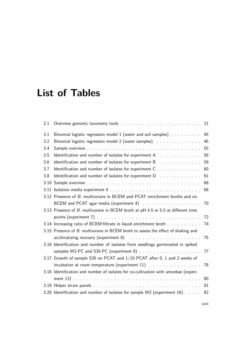
List of Tables
2.1 Overview genomic taxonomy tools . . . . . . . . . . . . . . . . . . . . . . . 21
3.1 Binomial logistic regression model 1 (water and soil samples) . . . . . . . . . 453.2 Binomial logistic regression model 2 (water samples) . . . . . . . . . . . . . 463.4 Sample overview . . . . . . . . . . . . . . . . . . . . . . . . . . . . . . . . . 553.5 Identification and number of isolates for experiment A . . . . . . . . . . . . 583.6 Identification and number of isolates for experiment B . . . . . . . . . . . . 593.7 Identification and number of isolates for experiment C . . . . . . . . . . . . . 603.8 Identification and number of isolates for experiment D . . . . . . . . . . . . 613.10 Sample overview . . . . . . . . . . . . . . . . . . . . . . . . . . . . . . . . . 683.11 Isolation media experiment 4 . . . . . . . . . . . . . . . . . . . . . . . . . . 683.12 Presence of B. multivorans in BCEM and PCAT enrichment broths and on
BCEM and PCAT agar media (experiment 4) . . . . . . . . . . . . . . . . . 703.13 Presence of B. multivorans in BCEM broth at pH 4.5 or 5.5 at different time
points (experiment 7) . . . . . . . . . . . . . . . . . . . . . . . . . . . . . . 723.14 Increasing ratio of BCEM:filtrate in liquid enrichment broth . . . . . . . . . . 743.15 Presence of B. multivorans in BCEM broth to assess the effect of shaking and
acclimatizing recovery (experiment 8) . . . . . . . . . . . . . . . . . . . . . 753.16 Identification and number of isolates from seedlings germinated in spiked
samples W2-PC and S35-PC (experiment 9) . . . . . . . . . . . . . . . . . . 773.17 Growth of sample S35 on PCAT and 1/10 PCAT after 0, 1 and 2 weeks of
incubation at room temperature (experiment 11) . . . . . . . . . . . . . . . 783.18 Identification and number of isolates for co-cultivation with amoebae (experi-
ment 13) . . . . . . . . . . . . . . . . . . . . . . . . . . . . . . . . . . . . . 803.19 Helper strain panels . . . . . . . . . . . . . . . . . . . . . . . . . . . . . . . 813.20 Identification and number of isolates for sample W2 (experiment 16) . . . . . 82
xxiii

List of tables
3.21 Identification and number of isolates for sample W152 (experiment 16) . . . 833.22 Identification and number of isolates for sample S35 (experiment 16) . . . . . 833.23 B. multivorans isolates included in the present study . . . . . . . . . . . . . 873.24 B. multivorans genome characteristics . . . . . . . . . . . . . . . . . . . . . 903.25 The frequency of orthologous versus non-orthologous CDS varies among the
COG categories . . . . . . . . . . . . . . . . . . . . . . . . . . . . . . . . . 943.26 Ortholog specificity varies among the COG categories . . . . . . . . . . . . . 983.27 The distribution of B. multivorans versus B. cenocepacia CDS varies among
COG categories . . . . . . . . . . . . . . . . . . . . . . . . . . . . . . . . . 993.28 B. multivorans-specific COGs . . . . . . . . . . . . . . . . . . . . . . . . . . 100
4.1 Studied isolates, showing their source, ST and allelic profile . . . . . . . . . . 1134.2 Phenotypic characteristics useful for differentiation of B. pseudomultivorans
from members of the B. cepacia complex and B. gladioli . . . . . . . . . . . 1214.3 Studied isolates, showing their source, ST and allelic profile . . . . . . . . . . 1254.4 Phenotypic characteristics useful for differentiation of B. stagnalis sp. nov.
and B. territorii sp. nov. from members of the Bcc . . . . . . . . . . . . . . 132
5.1 Phenotypic characteristics that distinguish B. cordobensis sp. nov. from itsnearest phylogenetic neighbors . . . . . . . . . . . . . . . . . . . . . . . . . 150
5.2 Mean fatty acid compositions of B. cordobensis sp. nov. and its nearestphylogenetic neighbors . . . . . . . . . . . . . . . . . . . . . . . . . . . . . 151
5.3 Strains included in the present study . . . . . . . . . . . . . . . . . . . . . . 1585.4 Genomes included in the present study . . . . . . . . . . . . . . . . . . . . . 1595.5 Mean fatty acid composition of all examined strains of BGC species . . . . . 1665.6 Differential biochemical characteristics of all examined strains of BGC species 1675.7 G+C content (mol%) of validly named BGC species . . . . . . . . . . . . . . 170
6.1 Environmental B. multivorans isolates . . . . . . . . . . . . . . . . . . . . . 190
7.1 Isolate overview NRC Bcc 2011-2015 . . . . . . . . . . . . . . . . . . . . . . 2027.2 B. multivorans strains present in multiple Belgian CF patients and centers
(NRC Bcc 2011-2015) . . . . . . . . . . . . . . . . . . . . . . . . . . . . . . 203
xxiv
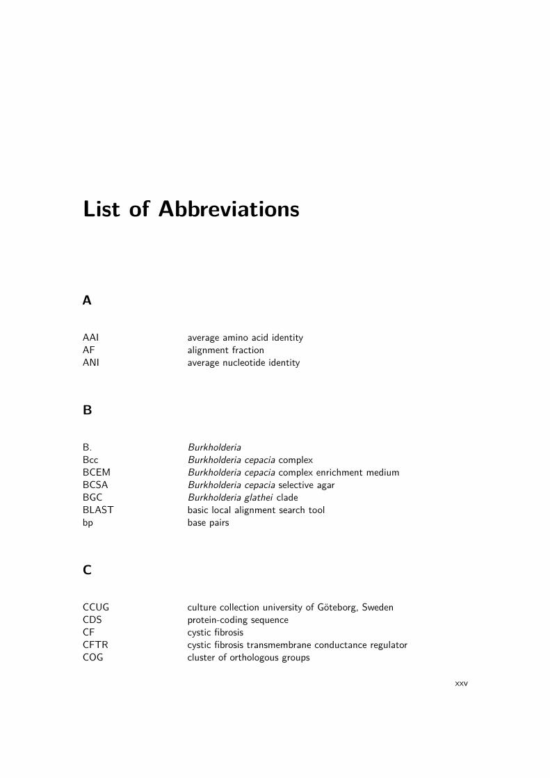
List of Abbreviations
A
AAI average amino acid identityAF alignment fractionANI average nucleotide identity
B
B. BurkholderiaBcc Burkholderia cepacia complexBCEM Burkholderia cepacia complex enrichment mediumBCSA Burkholderia cepacia selective agarBGC Burkholderia glathei cladeBLAST basic local alignment search toolbp base pairs
C
CCUG culture collection university of Göteborg, SwedenCDS protein-coding sequenceCF cystic fibrosisCFTR cystic fibrosis transmembrane conductance regulatorCOG cluster of orthologous groups
xxv

List of abbreviations
D
DDH DNA-DNA hybridizationdDDH digital DNA-DNA hybridizationDNA deoxyribonucleic aciddNTP deoxynucleotide triphosphate
E
ENV environmental
F
FAME fatty acid methyl ester
G
GBDP genome BLAST distance phylogenyGGDC genome-to-genome distance calculatorGNNF Gram-negative non-fermenting
L
LM-UGent laboratory of microbiology, Ghent universityLMG BCCM/LMG bacteria collection, laboratory of microbiology, Ghent
university, Belgium
M
xxvi
List of abbreviations
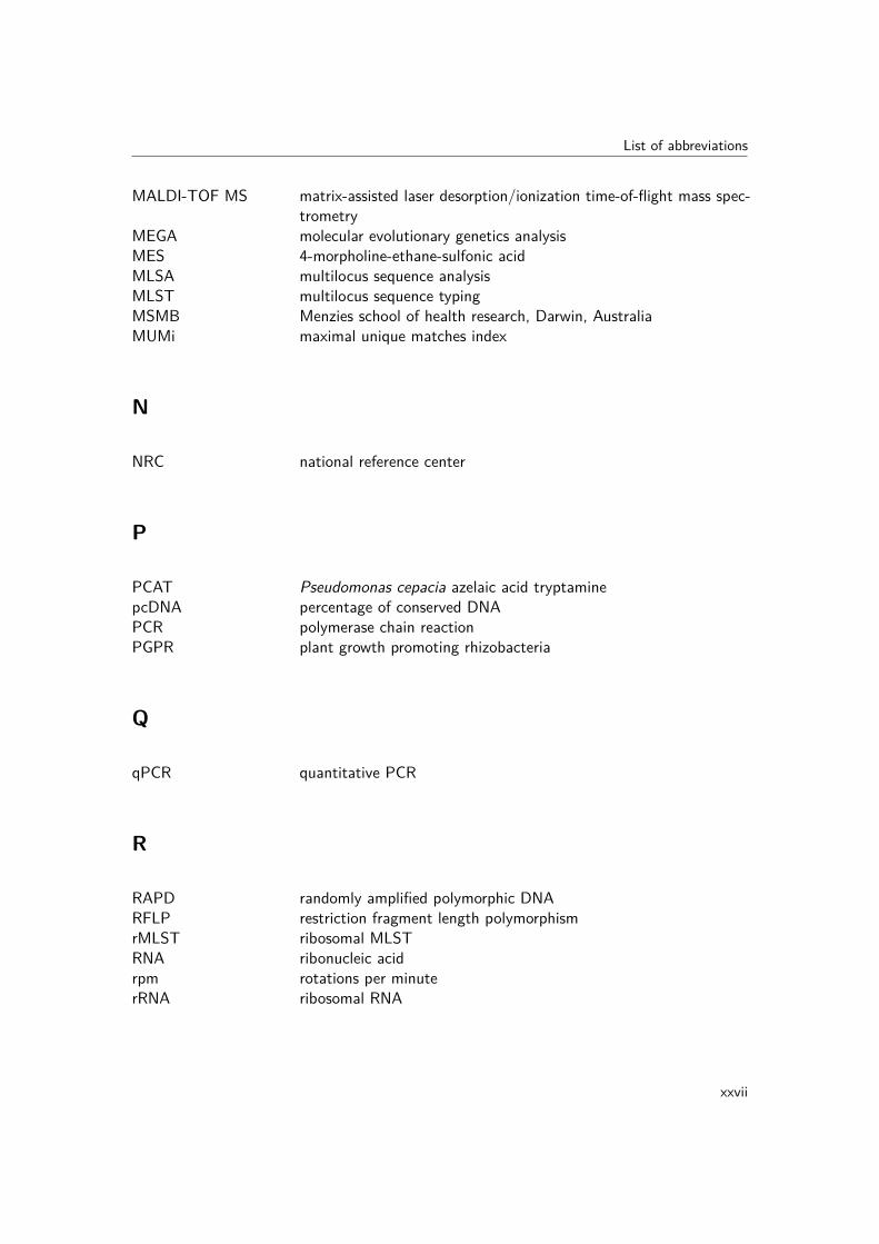
MALDI-TOF MS matrix-assisted laser desorption/ionization time-of-flight mass spec-trometry
MEGA molecular evolutionary genetics analysisMES 4-morpholine-ethane-sulfonic acidMLSA multilocus sequence analysisMLST multilocus sequence typingMSMB Menzies school of health research, Darwin, AustraliaMUMi maximal unique matches index
N
NRC national reference center
P
PCAT Pseudomonas cepacia azelaic acid tryptaminepcDNA percentage of conserved DNAPCR polymerase chain reactionPGPR plant growth promoting rhizobacteria
Q
qPCR quantitative PCR
R
RAPD randomly amplified polymorphic DNARFLP restriction fragment length polymorphismrMLST ribosomal MLSTRNA ribonucleic acidrpm rotations per minuterRNA ribosomal RNA
xxvii
List of abbreviations
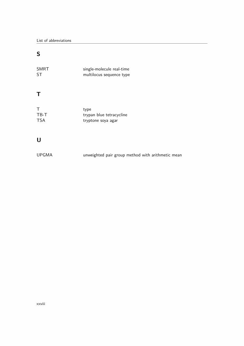
S
SMRT single-molecule real-timeST multilocus sequence type
T
T typeTB-T trypan blue tetracyclineTSA tryptone soya agar
U
UPGMA unweighted pair group method with arithmetic mean
xxviii

Part I
Introduction
1

1 | Burkholderia cepacia complexbacteria as opportunistic pathogensin cystic fibrosis patients
1.1 Cystic fibrosis lung pathology
Cystic fibrosis (CF) is an autosomal recessive genetic disorder that results from a mutation inthe CF transmembrane conductance regulator (CFTR) gene. This gene encodes a membraneprotein which is involved in transepithelial ion transport and its defect leads to the secretionof viscous secretions in all organs (Rowe et al., 2005). CF is most common in the northernEuropean population but the birth prevalence varies for different countries and ethnic back-grounds. The incidence of CF in Belgium is estimated at 1:2850 (Farrell, 2008). In total,more than 1500 mutations have been identified but the clinical significance is known only fora very small fraction of mutations. The best characterized mutation is F508del, representinga deletion of phenylalanine at position 508, which accounts for two-thirds of the mutations innorthern European and North American populations (O’Sullivan & Freedman, 2009).
The symptoms of CF are mainly evident in the gastrointestinal and respiratory tracts andchronic inflammation and respiratory infections represent the main causes of morbidityand mortality. Several hypotheses exist about how CFTR dysfunction leads to phenotypicsymptoms, and possibly all of them contribute to the outcome of recurrent respiratoryinfections and chronic inflammation. The constant cycle of inflammation and infection leadsto permanent lung damage and may eventually result in death by pulmonary insufficiency(O’Sullivan & Freedman, 2009).
The accumulation of mucus in the CF lung creates an environment suitable for colonization
3

I. Introduction
by various opportunistic pathogens, resulting in a complex community consisting of bacteria,fungi and viruses. The recurrent bacterial respiratory infections in CF are caused by a varietyof organisms and their prevalence typically varies with the age of the CF patient (Fig. 1.1)(Harrison, 2007). A recent study showed that the microbial community in the lower airwaysof children with CF is less rich and less even compared with that of non-CF children and thatthe CF lung microbial community is disrupted early in life (Renwick et al., 2014). Moreover,a longitudinal study demonstrated that the community diversity is decreased in patientswith more progressive lung disease and that this decrease in diversity in driven primarily byantibiotic therapy (Zhao et al., 2012). Similarly, two recent studies showed that a loss ofmicrobial diversity is associated with severe lung disease and the presence of Burkholderiabacteria (Flight et al., 2015; Stokell et al., 2015).
Downloaded from www.microbiologyresearch.org by
IP: 157.193.163.239
On: Wed, 09 Mar 2016 08:43:51
picture we have of the lung communities present in CFpatients is, therefore, limited and qualitative.
There are, however, three broad conclusions to be drawnfrom the published literature. First, coinfections involvingdifferent species of bacteria, or bacteria, fungi and viruses,are common and probably the norm. Second, coinfectingspecies interact, both syngergistically and antagonistically.Finally, pathogen populations within the lung evolve inresponse to selection pressures exerted by the within-hostenvironment and by other members of the community.These points will now be discussed.
Coinfections and communities
Several studies explicitly report coinfections of pairs ortriplets of bacterial species, while in other cases we can inferfrom the data given for individual species that a significantproportion of patients were infected by more than onespecies. Anzaudo et al. (2005) reported that 31 % of patientsharboured two or more bacterial species simultaneously,while Hoiby (1974) and Wahab et al. (2004) reportedcoinfections in ‘most patients’. Burns et al. (1998) calculatedthat they recovered an average of 2.9 species of pathogenicbacteria per sample (range: 0–10), and more recent DNAprofiling studies suggest that CF bacterial communities maybe even more diverse than suggested by culture-basedmethods (Rogers et al., 2004). Further, Petersen et al. (1981)found that 13 % of acute respiratory exacerbations amongtheir study population were associated with simultaneousbacterial and non-bacterial infections. The results of areview of reported coinfections are summarized in Fig. 2.
We have, then, evidence for high inter-species diversityin the CF lung. Signficant intra-species diversity is alsoobserved. Burns et al. (1998) recovered, on average, 2.38phenotypically distinct P. aeruginosa isolates per sample perpatient (range: 1–6). Further, selection pressures onpathogens are likely to be both temporally and spatially
heterogeneous during long-term infections, leading to geneticdiversification of founder clones. Smith et al. (2006) geno-typed P. aeruginosa isolates taken from a single CF patientover a period of 90 months and showed the existence ofmultiple, related lineages which coexisted for long periods.
Different microbial communities can be found within thesame patient. Several authors have noted a poor correlationbetween species and/or strains present in upper and lowerairway communities (Armstrong et al., 1996; Lyczak et al.,2002; Saiman, 2004) and in different lobes of the lung(Gutierrez et al., 2001; Smith et al., 1998). This is un-surprising given the compartmentalized nature of therespiratory system and high viscosity of CF mucus, andmeans that pooling samples from different lobes (as inNixon et al., 2001) is unlikely to give a reliable picture of themicrobial ecology within a patient.
The outcome of a mixed (multi-strain, multi-genotype ormulti-species) infection depends on exactly how coinfectingpopulations interact. Clinical and laboratory studies havestarted to elucidate the many ways in which CF pathogensmight interact in vivo.
100
Per
cent
age
of p
atie
nts
80
60
40
20
0_1 2_5 6_10 11_17 18_24 35_44 45+25_34Age group
P. aeruginosa
Staph. aureus
S. maltophilia
H. influenzaeMRSA
B. cepacia
Fig. 1. Prevalence of bacterial respiratory infections by agegroup in CF subjects. [Reproduced, with modifications, withpermission from the Cystic Fibsosis Foundation National PatientRegistry (Cystic Fibrosis Foundation, 2004).] MRSA, meticillin-resistant Staph. aureus.
Fig. 2. Venn diagram showing reported coinfections of the CFairways. (NB: coinfection does not necessarily imply directinteraction between species.) A, Aspergillus spp.; AV, adeno-virus; AX, A. xylosoxidans; BP, bacteriophage; C, Candida
spp.; Ent, enterobacteria; IPV, influenza and/or parainfluenzavirus; K, Klebsiella spp.; M, mycoplasma; MA, Mycobacterium
abscessus; N, Neisseria spp.; OF, oropharyngeal flora; RSV,respiratory syncytial virus; SM, S. maltophilia. Numbers refer toreferences: 1, Petersen et al. (1981); 2, Lambiase et al. (2006);3, Wahab et al. (2004); 4, Moore et al. (2005); 5, Burns et al.(1998); 6, Hoiby (1974); 7, Lording et al. (2006); 8, Santanaet al. (2003); 9, Alvarez et al. (2004); 10, Anzaudo et al.(2005); 11, Ojeniyi et al. (1991).
918 Microbiology 153
F. Harrison
Figure 1.1: Prevalence of bacterial respiratory infections in CF patients by age group. MRSA,meticillin-resistant Staphylococcus aureus. (Harrison, 2007)
Because the CF lung can harbor a wide diversity of bacteria, accurate identification iscrucial to assess the prevalence and clinical relevance of these generally rare opportunisticpathogens (Coenye et al., 2002a; LiPuma, 2010). One important group of opportunisticpathogens consists of the Gram-negative non-fermenting (GNNF) bacteria, including thegenera Pandoraea, Inquilinus, Achromobacter, Ralstonia, Cupriavidus, Pseudomonas andStenotrophomonas (Vandamme & Dawyndt, 2011; Alby et al., 2013). Although the clinicalsignificance of these B. cepacia-like organisms is mostly unclear, infection with these GNNFopportunistic pathogens may have a high impact on morbidity because of their multi-resistanceto antibiotics and their transmissibility (Coenye & Vandamme, 2003; Jorgensen et al., 2003;Schmoldt et al., 2006; Kalka-Moll et al., 2009).
4

1. Bcc bacteria as opportunistic pathogens in CF patients
1.2 Burkholderia cepacia complex epidemiology in cysticfibrosis patients
Burkholderia cepacia complex (Bcc) bacteria are significant pathogens in people with CFbecause of their high impact on morbidity, mortality and post-lung transplant survival (DeBoeck et al., 2004; Govan et al., 2007; LiPuma, 2010). Bcc infection in CF is characterizedby a highly variable clinical outcome, but generally results in a progressive decline of lungfunction. In extreme cases, Bcc infection can result in "cepacia syndrome", a necrotizingpneumonia and septicemia resulting in early death. Bcc infections are difficult to eradicatebecause Bcc bacteria show an innate multiresistance to antibiotics. Finally, the diagnosis ofBcc infection also has a social and psychological impact because the transmissibility of Bccstrains through social contact resulted in stringent infection control guidelines that necessitatepatient segregation (Mahenthiralingam et al., 2005).
Bcc bacteria rarely cause infection in healthy (i.e. non-immunocompromised) individuals,except when they are present as contaminants in pharmaceutical products (Mahenthiralingamet al., 2005). Several studies reported on disinfectants, intravenous solutions and contaminatedmedical devices as sources of nosocomial outbreaks (Weber et al., 2007; Heo et al., 2008;Torbeck et al., 2011; Souza Dias et al., 2013).
Of the 20 formally named species within the Bcc (Fig. 2.1 and 2.2), Burkholderia multivoransand Burkholderia cenocepacia are generally the most prevalent Bcc species in CF. During thelast decade, infection control measures have restricted patient-to-patient transmission, whichis mostly associated with B. cenocepacia (Mahenthiralingam et al., 2001; Speert et al., 2002;Turton et al., 2003; France et al., 2008). Consequently, B. multivorans emerged as the mostprevalent Bcc pathogen in many countries, including Belgium, France, Denmark, the UnitedKingdom, the United States (Fig. 1.2) and New Zealand (Brisse et al., 2004; De Boecket al., 2004; Govan et al., 2007; LiPuma, 2010; Norskov-Lauritsen et al., 2010; Pope et al.,2010). In Spain, Portugal and Argentina a high prevalence has recently been reported of B.contaminans in CF patients (Martina et al., 2013; Coutinho et al., 2015; Medina-Pascualet al., 2015).
Historically, B. cenocepacia strains have been responsible for large epidemics within the CFcommunity and are often extremely virulent (Drevinek & Mahenthiralingam, 2010). The ET12clone is responsible for infecting many CF patients in Canada and Europe (Mahenthiralingamet al., 2002), while the Midwest clone (Coenye & LiPuma, 2002) and the PHDC clone (Chen
5

I. Introduction
P. aeruginosa, significant differences in age-specific prevalencerates are found, with rates as high as 8% being reported foradults. Razvi and colleagues (249) reported that the incidenceof infection, based on CFF Patient Registry data, decreasedfrom 1.3% to 0.8% between 1995 and 2005. The overall prev-alence of infection also decreased from 3.6% to 3.1% duringthis period. An analysis of microbiological data from the RoyalBrompton Hospital indicates that while the prevalence of B.cepacia complex infection increased significantly in adult CFpatients between 1985 and 1990, it decreased from 9% to 4%between 1990 and 2005 (203).
(iii) Species distribution in CF. With the exception of Burk-holderia ubonensis, all species in the B. cepacia complex havebeen isolated from CF sputum cultures; however, some are
much more commonly encountered in CF than are others. Theapproximate distribution of B. gladioli and B. cepacia complexspecies among Burkholderia-infected CF patients in the UnitedStates is provided in Fig. 1A (184; my unpublished observa-tions). Burkholderia multivorans and Burkholderia cenocepaciaare most frequently recovered, together accounting for approx-imately 70% of infected patients. In Canada and some Euro-pean countries, the proportion of CF patients infected with B.cenocepacia is higher (3, 288). Of note, B. gladioli is much morefrequently recovered in CF than are most species of the B.cepacia complex, and B. cepacia is relatively infrequently en-countered. Furthermore, some CF patients are infected withbacteria that, based on genetic analyses, are most appropri-ately included in the B. cepacia complex but cannot be assignedto one of the currently named species. These strains most likelyrepresent additional novel species within the complex. Finally,the incidences of B. multivorans and B. cenocepacia infection inCF appear to have shifted during the last few years (115, 251).Whereas B. cenocepacia previously accounted for the majorityof Burkholderia infection in CF (251), currently, approximatelythree times as many CF patients in the United States becomeinfected with B. multivorans as with B. cenocepacia (Fig. 1B). Inthe United Kingdom, the recovery of B. multivorans from CFpatients now also exceeds that of B. cenocepacia (115).
(iv) Epidemic strains. A variety of methods have been usedto genotype Burkholderia to assess the epidemiology of infec-tion in CF (14, 59, 75, 179, 182, 189, 290). Genotyping studiesin the late 1980s identified strains that were common to mul-tiple patients receiving care in the same CF centers (182),suggesting interpatient spread. More compelling evidence ofpatient-to-patient transmission of Burkholderia soon followed(116, 181). Further studies identified strains common to CFpatients in wider geographic regions. Among such so-called“epidemic” strains, the ET12 (for electrophoretic type 12)
TABLE 1. The Burkholderia cepacia complex
SpeciesFormer
genomovardesignation
Yr identified and/or named Reference(s)
B. cepacia I 1950, 1997 34, 322B. multivorans II 1997 322B. cenocepacia III 1997, 2003 321, 322B. stabilis IV 1997, 2000 322, 323B. vietnamiensis V 1995, 1997 107, 322B. dolosa VI 2001, 2004 58, 335B. ambifaria VII 2001 63B. anthina VIII 2002 319B. pyrrocinia IX 2002 319B. ubonensis 2000, 2008 333, 355B. latens 2008 333B. diffusa 2008 333B. arboris 2008 333B. seminalis 2008 333B. metallica 2008 333B. contaminans 2009 332B. lata 2009 332
FIG. 1. (A) Distribution of Burkholderia species among U.S. CF patients. The proportions of CF patients infected with various Burkholderiaspecies are shown. The data are based on 2,024 CF patients who were infected with Burkholderia species and whose isolates were referred to theBurkholderia cepacia Research Laboratory and Repository (University of Michigan) between 1997 and 2007. “Other Bcc species” indicates patientsinfected with B. cepacia complex species other than those specified in the chart. “Indeterminate” refers to patients infected with strains thatphylogenetically are members of the B. cepacia complex species but that cannot be definitively placed into one of the 17 defined species in thisgroup. (B) Incidence of B. cenocepacia and B. multivorans infection in U.S. CF patients. The proportions of B. cepacia complex-infected CF patientswho were first infected with either B. cenocepacia (red line) or B. multivorans (blue line) in the years indicated are shown.
302 LIPUMA CLIN. MICROBIOL. REV.
Figure 1.2: Distribution and incidence of Burkholderia in U.S. CF patients. (A) Distribution ofBurkholderia species among U.S. CF patients. The proportions of CF patients infected with variousBurkholderia species are shown. The data are based on 2,024 CF patients who were infected withBurkholderia species and whose isolates were referred to the Burkholderia cepacia Research Laboratoryand Repository (University of Michigan) between 1997 and 2007. “Other Bcc species” indicatespatients infected with Bcc species other than those specified in the chart. “Indeterminate” refers topatients infected with strains that phylogenetically are members of the Bcc species but that cannot bedefinitively placed into one of the 17 defined species in this group. (B) Incidence of B. cenocepaciaand B. multivorans infection in U.S. CF patients. The proportions of Bcc-infected CF patients whowere first infected with either B. cenocepacia (red line) or B. multivorans (blue line) in the yearsindicated are shown. (LiPuma, 2010)
et al., 2001) are dominant strains infecting CF patients in the United States. Although B.multivorans is generally considered a lesser virulent Bcc pathogen compared to B. cenocepacia,rare cases of cepacia syndrome caused by B. multivorans have been reported (Blackburnet al., 2004; Jones et al., 2004). While preoperative Bcc infection is generally associated witha poor prognosis for post-lung transplant survival, mortality is primarily dependent on theBcc species and strain (Aris et al., 2001; De Soyza et al., 2001; Murray et al., 2008).
The ability to differentiate Bcc strains has been crucial in understanding their epidemiologyand improving infection control guidelines for the CF community. Methods for straindifferentiation include multilocus restriction typing (Coenye & LiPuma, 2002), pulsed fieldgel electrophoresis (Coenye et al., 2002b), randomly amplified polymorphic DNA (RAPD)analysis (Mahenthiralingam et al., 1996), BOX-PCR fingerprinting (Coenye et al., 2002b),multilocus sequence typing (MLST) (Baldwin et al., 2005) and single-nucleotide polymorphismanalysis of whole-genome sequences (Pallen et al., 2010; Lieberman et al., 2011). MLST is awell-established method for studying the population structure of Bcc organisms and takes
6

1. Bcc bacteria as opportunistic pathogens in CF patients
into account the allelic variation of seven housekeeping genes (atpD, gltB, gyrB, recA, lepA,phaC and trpB) (Baldwin et al., 2005). As a result, each strain is defined by its unique allelicprofile and multilocus sequence type (ST). Only a limited number of B. multivorans outbreakstrains was described and for all cases of implied patient-to-patient spread, unique STs werefound (Baldwin et al., 2008). In the United Kingdom, the strains ST-27 and ST-15 causedhospital outbreaks among CF patients in Glasgow and South Wales, respectively (Whitefordet al., 1995; Millar-Jones et al., 1998). Strains ST-25 and ST-179 were shared by CF patientsin the United States (Biddick et al., 2003). Finally, strain ST-419 caused an outbreak amongCF patients in France (Segonds et al., 1999).
The low number of outbreaks caused by B. multivorans and the fact that B. multivoransisolates from CF patients commonly represent unique strains suggest that there is only limitedperson-to-person transmission and that B. multivorans strains are acquired from non-humansources such as the natural environment (Baldwin et al., 2008).
1.3 The natural environment as a reservoir for cystic fibrosispathogens
Bcc bacteria are characterized by a versatile lifestyle and almost all Bcc species have beenisolated from both CF patients and environmental samples (Coenye & Vandamme, 2003;Baldwin et al., 2007). Exceptions are Burkholderia ubonensis and Burkholderia territoriiwhich have not been isolated from CF patients thus far (http://pubmlst.org/bcc/) (Jolley &Maiden, 2010). Baldwin et al. (2008) demonstrated a continued emergence of unique B.multivorans strains in the CF population, with several STs being globally distributed andassociated with human infection. The same study showed an overlap in strains between waterenvironments, industrial products and human infection, suggesting that the environment maybe an important reservoir for infection with B. multivorans (Baldwin et al., 2008).
Specific media have been developed for the isolation of Bcc from environmental samples suchas Pseudomonas cepacia azelaic acid tryptamine (PCAT) (Burbage & Sasser, 1982), trypanblue tetracycline (TB-T) (Hagedorn et al., 1987) and Bcc enrichment medium (BCEM)(Vermis et al., 2003a; Vanlaere et al., 2005). The selectivity of these media is achieved byspecific nitrogen and carbon sources, often combined with antimicrobial compounds to inhibitother bacteria and fungi (Vermis et al., 2003b). Although many studies described the isolationof B. cenocepacia from rhizosphere samples (Butler et al., 1995; Balandreau et al., 2001;
7

I. Introduction
Fiore et al., 2001; Bevivino et al., 2002; LiPuma et al., 2002; Pirone et al., 2005; Zhang &Xie, 2007; Bevivino et al., 2011; Hall et al., 2015), only few studies reported the isolation ofB. multivorans from maize rhizosphere (Ramette et al., 2005) and agricultural soil (Lin et al.,2011; Hsueh et al., 2015). Vermis et al. (2003b) developed a selective enrichment broth forthe isolation of Bcc bacteria from water samples and obtained eight B. multivorans isolatesfrom the Schelde river in Belgium. Using the same strategy Vanlaere et al. (2005) obtainedtwo B. multivorans isolates from soil in a veranda. Finally, a more recent study describedthe isolation of 48 B. multivorans isolates from West Lake, China (Fang et al., 2011). Yettogether, the number of environmental B. multivorans isolates is very small compared to thenumber of CF isolates and its true environmental niche is considered unknown.
Environmental pressure can select for traits that confer virulence and natural environmentscould therefore serve as potential reservoirs of opportunistic pathogens (Coenye & Vandamme,2003; Berg et al., 2005). Bcc bacteria adopt a wide range of lifestyles (Section 2.2) and thevery same mechanisms of adaptation that let these bacteria thrive in different ecological nichesmay enable them to colonize the CF lung and cause infection (Vial et al., 2011). Therefore,the natural environment may be considered a fitness school for emerging pathogens.
8

2 | Taxonomy and diversity of thegenus Burkholderia
2.1 Taxonomy of the genus Burkholderia
2.1.1 Historical background
The genus Burkholderia originated in the 1940s when plant physiologist Walter Burkholderisolated bacteria from onion bulbs that were suffering from "sour skin" disease and proposedto name these strains Pseudomonas cepacia (Burkholder, 1950). Based on rRNA-basedmethods, the genus Pseudomonas was shown to be phylogenetically diverse and to consist offive major clusters, the so called rRNA homology groups (Palleroni et al., 1973). In 1992,the new genus Burkholderia was proposed to accommodate RNA homology group II whichthen comprised P. cepacia and six other species (Yabuuchi et al., 1992). During the last twodecades, the genus Burkholderia expanded dramatically and it became apparent that thisgenus comprised versatile bacteria which occupy a wide range of ecological niches (Coenye &Vandamme, 2003). In March 2016, the genus comprised 90 validly named species (Euzeby,1997) and a large number of uncultivated Candidatus species (Van Oevelen et al., 2002, 2004;Lemaire et al., 2011a, 2012; Verstraete et al., 2011). However, literature data and analysisof publicly available 16S rRNA gene sequences suggest that many unclassified Burkholderiaisolates represent additional novel species, showing that the diversity of Burkholderia bacteriais still not fully uncovered.
Organisms now known as Burkholderia cepacia emerged as important CF pathogens in the1980s (Isles et al., 1984; LiPuma et al., 1990; Govan et al., 1993). Only after its formalreclassification in the genus Burkholderia it became apparent that presumed B. cepacia
9
I. Introduction
strains showed a marked heterogeneity (Butler et al., 1995; Gillis et al., 1995). This ledto a polyphasic taxonomic study which revealed that presumed B. cepacia strains from CFpatients in fact belonged to at least five distinct genomic species or genomovars, collectivelyreferred to as the B. cepacia complex (Vandamme et al., 1997). Bcc species are closelyrelated and typically show 30-50% DNA-DNA hybridization (DDH). Only two out of the fiveclosely related genomic species could also be distinguished phenotypically at that time andthese were Burkholderia vietnamiensis and B. multivorans. The term "genomovar" was usedto denote groups of strains that are delimited by DDH but that are phenotypically similarso that they cannot be differentiated (Ursing et al., 1995). The used polyphasic approachwas based on multiple techniques including whole-cell protein electrophoresis, whole-cell fattyacid methyl ester analysis, DNA-DNA and DNA-rRNA hybridizations and several biochemicaltests and set an example for a taxonomic consensus approach to bacterial systematics inwhich the results of several genotypical and phenotypical tests are integrated (Vandammeet al., 1996).
Driven by the devastating effect of Bcc infections in CF patients, a wide range of alternativeidentification methods were developed, including species-specific 16S rRNA (LiPuma et al.,1999; Whitby et al., 2000) and recA PCR assays (Mahenthiralingam et al., 2000a; Drevineket al., 2002; Vermis et al., 2002a), amplified fragment length polymorphism analysis ofgenomic DNA (Coenye et al., 1999b), ribotyping (Brisse et al., 2000), restriction fragmentlength polymorphism (RFLP) analysis of PCR amplified 16S rRNA and recA gene fragments(Mahenthiralingam et al., 2000a; McDowell et al., 2001; Vermis et al., 2002b) and tRNAprofiling (Storms et al., 2002). The sensitivity and specificity of these first-generationmolecular identification approaches needed re-evaluation each time novel Bcc species weredescribed and several misidentifications were reported (Coenye et al., 1999b; McMenaminet al., 2000; Moore et al., 2002; Cesarini et al., 2009; Drevinek et al., 2010). At present,sequence analysis of recA and other housekeeping genes have been established as powerful,objective and portable taxonomic tools that are useful for the accurate identification andclassification of Bcc organisms (Section 2.1.3.1) (Vandamme & Dawyndt, 2011). As more CFisolates were examined and techniques with a better resolution became available, additionalBcc species could be distinguished both genotypically and phenotypically and were formallyclassified. Today (March 2016) the Bcc comprises 20 validly named species (De Smet et al.,2015a).
10

2. Taxonomy and diversity of the genus Burkholderia
2.1.2 Taxonomic overview of the genus Burkholderia based on the 16SrRNA gene
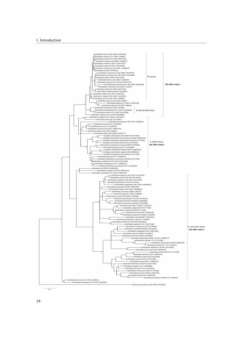
Based on 16S rRNA gene sequence analysis, the genus Burkholderia consists of severaldeep-branching lineages (Fig. 2.1). The type species B. cepacia is part of a first clade ofabout 30 species that comprises the Bcc, species closely related to the risk class 3 pathogensBurkholderia mallei and Burkholderia pseudomallei and a group of plant-pathogenic speciesthat includes Burkholderia gladioli, Burkholderia plantarii and Burkholderia glumae. Thisclade (from here on referred to as 16S rRNA clade 1) comprises the well-known humanpathogens in this genus (Section 2.2.3), but also includes strains that show potential forplant-growth promotion and biocontrol (Section 2.2.1).
A second deep-branching Burkholderia clade (from here on referred to as 16S rRNA clade 2)comprises Burkholderia glathei and 11 validly named Burkholderia species (Fig. 2.1). Mostspecies in this clade have been isolated as free-living organisms from soil (Zolg & Ottow, 1975;Vandamme et al., 2013a; Draghi et al., 2014; Baek et al., 2015) but also associations withfungi (Lim et al., 2003), plants (Tian et al., 2013) and insects (Kikuchi et al., 2011) have beendescribed for this clade. Additionally, many uncultivated species adapted a endosymbioticlifestyle in plant tissue and represent candidate species (Verstraete et al., 2013; Carlier et al.,2016). Exemplary for the versatile metabolism of Burkholderia bacteria, several B. glatheiclade species were isolated from contaminated soil (Vandamme et al., 2013a; Liu et al., 2014)or from a wastewater treatment system (Lu et al., 2012). The B. glathei clade thus farincludes only 12 formally classified species, yet many unclassified B. glathei-like bacteria havebeen reported (Nogales et al., 2001; Salles et al., 2006b; Pumphrey & Madsen, 2008; Draghiet al., 2014; Verstraete et al., 2014).
A third deep-branching Burkholderia clade (from here on referred to as 16S rRNA clade3) comprises more than 40 species which are primarily plant-associated beneficial and en-vironmental (Suarez-Moreno et al., 2012) (Fig. 2.1). However, Burkholderia fungorum,Burkholderia ginsengisoli, Burkholderia tropica and Burkholderia xenovorans represent impor-tant exceptions in this clade because they have been isolated from human clinical samplestoo (Section 2.2.3).
In addition to these three large clades, several Burkholderia species represent rather unique16S rRNA lineages and do not cluster closely with any other Burkholderia species (Fig. 2.1).Their 16S rRNA-based phylogenetic position is variable and dependent on the remainingsequences included in the phylogenetic analysis. Burkholderia rhizoxinica and Burkholderia
11

I. Introduction
endofungorum represent a first lineage that includes bacterial endosymbionts of a plant-pathogenic fungus (Partida-Martinez et al., 2007). A second lineage comprises Burkholderiacaryophylli, Burkholderia symbiotica and Burkholderia soli which are known as a plantpathogen, a root nodule endosymbiont of Mimosa species and a soil bacterium, respectively(Yabuuchi et al., 1992; Yoo et al., 2007; Sheu et al., 2012). Finally, Burkholderia andropogonisis a plant pathogen (Coenye et al., 2001a) that represents a deep-branching lineage within16S rRNA clade 2 in the present analysis (Fig. 2.1) but which occupies a distinct position inmost 16S rRNA-based phylogenetic trees (Gyaneshwar et al., 2011; Suarez-Moreno et al.,2012; Estrada-de los Santos et al., 2013, 2015)).
2.1.3 Burkholderia taxonomy in a changing landscape
2.1.3.1 Gene sequence analysis
While the 16S rRNA gene phylogeny gives a traditional representation of the phylogeny ofBurkholderia bacteria, this gene only has limited taxonomic resolution for species identificationwithin the genus Burkholderia. Within the Bcc, 16S rRNA sequences of different Bcc speciestypically show more than 98% similarity, while exhibiting at the same time up to 2%intraspecies divergence (Vandamme & Dawyndt, 2011). In contrast with the 16S rRNA gene,the recA gene is a powerful taxonomic tool for Bcc species identification (Mahenthiralingamet al., 2000a). RecA sequences typically show 94-95% similarity between Bcc speciesand, mostly, about 98-99% similarity within Bcc species (Vandamme & Dawyndt, 2011).Remarkably, B. cenocepacia (formerly known as genomovar III) comprises four recA lineagesreferred to as IIIA through IIID, with most clinical strains residing in clusters IIIA andIIIB (Vandamme et al., 2003). Another housekeeping gene that proved useful for speciesidentification within the genus Burkholderia is gyrB (Tayeb et al., 2008). This gene servesas a reliable taxonomic tool for species-level discrimination of both Bcc (Tabacchioni et al.,2008) and non-Bcc Burkholderia species (Vandamme et al., 2013a).
Multilocus sequence analysis (MLSA) is a technique that uses a set of genes (usually six orseven) that are universally present within the taxon, occur as a single copy in the genome, arenot closely linked and are not subject to high levels of recombination (Gevers et al., 2005).With the advent of new sequencing technologies, MLSA was proposed to replace DDH as atool for species delineation (Stackebrandt et al., 2002; Gevers et al., 2005). The Bcc MLSTscheme was developed for differentiation of Bcc isolates at both strain and species level sothat the same sequence data that was used for strain differentiation (Section 1.2) could
12

2. Taxonomy and diversity of the genus Burkholderia
also be employed for identification. Phylogenetic analysis of housekeeping gene fragments ofatpD, gltB, gyrB, recA, lepA, phaC and trpB has a superior taxonomic resolution for specieslevel identification within the Bcc (Baldwin et al., 2005) and PCR primers were subsequentlyimproved to reliably amplify the target loci from both Bcc and non-Bcc Burkholderia speciesand to enable the use of a single primer set for both amplification and sequencing (Spilkeret al., 2009). Vanlaere et al. (2009) compared the average concatenated allele sequencedivergence within and between established Bcc species as delineated by DDH studies anddemonstrated that a 3% concatenated allele sequence divergence level can be used as athreshold value for species delineation within the Bcc, thus replacing the need to performDDH experiments in this complex.
The ability to carry out both species identification and strain differentiation in a singleapproach represented a major advantage over previous methods (Baldwin et al., 2005). Anadditional advantage of these multilocus sequence based methods over previous typing andidentification methods was its reproducibility and portability (Jolley & Maiden, 2010). Thepublic database enabled researchers worldwide to analyze and deposit data and as a result theBcc PubMLST database currently (March 2016) holds information on 1,964 isolates and their987 associated STs. Fig. 2.2 shows a phylogenetic analysis of the concatenated sequencesof the seven loci of all STs in the database and reveals the presence of at least 12 putativenovel Bcc species awaiting formal classification.
13
I. Introduction
Burkholderia territorii LMG 28158T (LK023503)
Burkholderia cepacia ATCC 25416T (U96927)
Burkholderia seminalis R-24196T (AM747631)
Burkholderia anthina LMG 20980T (JX986972)
Burkholderia metallica R-16017T (AM747632)
Burkholderia arboris R-24201T (AM747630)
Burkholderia contaminans LMG 23361T (JX986975)
Burkholderia lata 383T (CP000151)
Burkholderia vietnamiensis LMG 10929T (AF097534)
Burkholderia cenocepacia IIIA LMG 16656T (AF148556)
Burkholderia latens R-5630T (AM747628)
Burkholderia dolosa LMG 18943T (JX986970)
Burkholderia ubonensis CIP 107078T (EU024179)
Burkholderia pseudomultivorans LMG 26883T (HE962386)
Burkholderia multivorans LMG 13010T (Y18703)
Burkholderia diffusa R-15930T (AM747629)
Burkholderia ambifaria AMMDT (AF043302)
Burkholderia stabilis LMG 14294T (AF097533)
Burkholderia stagnalis LMG 28156T (LK023502)
Burkholderia pyrrocinia LMG 14191T (U96930)
Bcc group
Burkholderia glumae LMG 2196T (U96931)
Burkholderia gladioli CIP 105410T (EU024168)
Burkholderia plantarii LMG 9035T (U96933)
Burkholderia thailandensis E264T (U91838)
Burkholderia pseudomallei ATCC 23343T (DQ108392)
Burkholderia mallei ATCC 23344T (AF110188)
B. pseudomallei group
Burkholderia oklahomensis C6786T (DQ108388)
Burkholderia sordidicola SNU 020123T (AF512827)
Burkholderia udeis Hg2T (AY154367)
Burkholderia andropogonis LMG 2129T (JX986957)
Burkholderia choica RA1-8T (AY949196)
Burkholderia humi RA1-5T (AY949193)
Burkholderia terrestris LMG 22937T (HE981726)
Burkholderia glathei LMG14190T (U96935)
Burkholderia telluris LMG 22936T (HE981727)
Candidatus Burkholderia kirkii 19536779 (AF475063)
Candidatus Burkholderia schumannianae SD1099 (HQ849124)
Candidatus Burkholderia nigropunctata 19750521 (AY277698)
Candidatus Burkholderia petitii SD1512 (JF916923)
Burkholderia megalochromosomata JC2949T (KF155693)
Burkholderia jiangsuensis MP-1T (KJ400396)
Candidatus Burkholderia hispidae SD3176 (HQ849122)
Candidatus Burkholderia rigidae OL694 (HQ849120)
Candidatus Burkholderia calva 19620512 (AY277697)
Burkholderia grimmiae R27T (JN256678)
Candidatus Burkholderia verschuerenii 19750204 (AY277699)
Burkholderia cordobensis LMG 27620T (HG324048)
Burkholderia zhejiangensis OP-1T (HM802212)
Candidatus Burkholderia andongensis BL271 (JF916918)
B. glathei group
Burkholderia soli GP25-8T (DQ465451)
Burkholderia symbiotica JPY345T (HM357233)
Burkholderia caryophylli ATCC 25418T (AB021423)
Burkholderia fungorum LMG 16225T (AF215705)
Burkholderia insulsa PNG-AprilT (KF733462)
Burkholderia caledonica LMG 19076T (AF215704)
Burkholderia phytofirmans PsJNT (AY497470)
Burkholderia megapolitana LMG 23650T (AM489502)
Burkholderia ginsengisoli KMY03T (AB201286)
Burkholderia bryophila LMG 23644T (AM489501)
Burkholderia xenovorans LB400T (U86373)
Burkholderia graminis C4D1MT (U96939)
Burkholderia terricola LMG 20594T (AY040362)
Burkholderia phenoliruptrix AC1100T (AY435213)
Burkholderia dilworthii WSM3556T (HQ698908)
Burkholderia rhynchosiae WSM3937T (EU219865)
Burkholderia phymatum STM815T (AJ302312)
Burkholderia sabiae Br3407T (AY773186)
Burkholderia caribensis MWAP64T (Y17009)
Burkholderia terrae KMY02T (AB201285)
Burkholderia hospita LMG 20598T (AY040365)
Burkholderia sartisoli RP007T (AF061872)
Burkholderia phenazinium LMG 2247T (U96936)
Burkholderia sediminicola HU2-65WT (EU035613)
Burkholderia aspalathi VG1CT (KC817488)
Burkholderia susongensis L226T (KJ746438)
Burkholderia sprentiae WSM5005T (HF549035)
Burkholderia acidipaludis SA33T (AB513180)
Burkholderia tuberum STM678T (AJ302311)
Burkholderia monticola JC2948T (KF155692)
Burkholderia diazotrophica NKMU-JPY461T (HM366717)
Burkholderia solisilvae Y-47T (FJ772068)
Burkholderia rhizosphaerae WR43T (AB365791)
Burkholderia humisilvae Y-12T (FJ796457)
Burkholderia caballeronis TNe-841T (EF139186)
Burkholderia kururiensis KP23T (AB024310)
Burkholderia nodosa Br3437T (AY773189)
Burkholderia bannensis E25T (AB561874)
Burkholderia tropica Ppe8T (AJ420332)
Burkholderia unamae MTl-641T (AY221956)
Burkholderia eburnea RR11T (JQ692176)
Burkholderia denitrificans KIS30-44T (GU171384)
Burkholderia oxyphila OX-01T (AB488693)
Burkholderia sacchari IPT101T (AF263278)
Burkholderia mimosarum PAS44T (AY752958)
Burkholderia ferrariae FeGI01T (DQ514537)
Burkholderia heleia SA41T (AB495123)
Burkholderia silvatlantica SRMrh-20T (AY965240)
B. xenovorans group
Burkholderia rhizoxinica HKI 454T (AJ938142)
Burkholderia endofungorum HKI 456T (AM420302)
Ralstonia solanacearum LMG 2299T (EF016361)
97
95
99
73
53
75
86
85
97
93
77
60
59
50
89
86
79
98
99
99
53
67
90
64
86
83
65
59
99
79
89
86
91
93
62
50
60
0.02
16S rRNA clade 3
16S rRNA clade 2
16S rRNA clade 1
14

2. Taxonomy and diversity of the genus Burkholderia
Figure 2.1: Phylogenetic tree based on partial 16S rRNA gene sequences of Burkholderia species.Sequences (1125-1610 bp) were aligned against the SILVA SSU reference database using SINA v1.2.11www.arb-silva.de/aligner/ (Pruesse et al., 2012). Phylogenetic analysis was conducted using MEGA6(Tamura et al., 2013). All positions containing gaps and missing data were eliminated, resulting in atotal of 1087 positions in the final dataset. The optimal tree (highest log likelihood) was constructedusing the maximum likelihood method and Tamura-Nei model (Tamura & Nei, 1993). A discretegamma distribution was used to model evolutionary rate differences among sites (5 categories (+G,parameter = 0.3498)) and allowed for some sites to be evolutionarily invariable ([+I], 68.6154% sites).The percentage of replicate trees in which the associated taxa clustered together in the bootstrap test(1000 replicates) are shown next to the branches if greater than 50%. The sequence of Ralstoniasolanacearum LMG 2299T was used as outgroup. The scale bar indicates the number of substitutionsper site. (Depoorter et al., 2016)
2.1.3.2 Genomic taxonomy
In this genomics era, the number of finished or draft bacterial genomes has risen exponentiallyand phylogenetic analysis has entered the new era of phylogenomics (Eisen & Fraser, 2003;Delsuc et al., 2005). Several tools are available for studying genomic taxonomy and these canbe subdivided based on how phylogeny is inferred and what part of the genome is being analyzed(Table 2.1). Methods for inferring evolutionary relationships can generally be classified aseither distance-based or character-based (Fig. 2.3). Distance-based methods convert alignedsequences into a distance matrix employing a model of evolution and subsequently use thisdistance matrix to infer a phylogenetic tree (Hall, 2011; Sleator, 2013). Although effective,these methods have the disadvantage of providing only one tree instead of a consensus treeand changing the order in which the constituent sequences are entered into the analysis mayresult in different trees (Sleator, 2013). On the other hand, character-based methods inferthe most probable tree(s) based on the characters at each position in a multiple sequencealignment (Hall, 2011; Sleator, 2013).The character-based methods in genomic taxonomy infer phylogenetic trees based on themultiple sequence alignment of conserved protein-coding genes in all genomes under studyand represent an extension of the MLSA principle (Table 2.1). This extended MLSA approachhas the advantage that it only takes into account the coding part of the genome and istherefore not influenced by non-coding sequences or pseudogenes which might have a differentevolutionary history than the rest of the genome. Important disadvantages are the needfor correctly annotated genomes and the fact that the set of single-copy orthologous genesmay become very small when studying more distantly related organisms or organisms thatunderwent reductive genome evolution (Vandamme & Dawyndt, 2011). Examples of thisapproach are the core gene identity (Vanlaere et al., 2009), supermatrix (Ciccarelli et al., 2006)
15

I. Introduction
Non-Bcc (117)
B. am
bifaria (54)Other B
cc H
(3)
B. dolosa (12)
Other Bcc E (15)
B. a
rbor
is (1
)
B. d
iffus
a (1
3)
B. s
emin
alis
(14
)
Other Bcc K (2)B
. arb
oris
(1)
Other Bcc N (2)
B. v
ietn
amie
nsis
(69
)
B. cenocepacia IIID (3)
B. lata (21)
B. pyrrocinia (20)
B. cenocepacia IIIC (3)
Other B
cc A (5)
Other B
cc I (18)
B. t
errit
orii
(3)
B. stabilis (2
2)
Other Bcc G (2)
B. multivorans (184)
Other Bcc M (5)
B. anthina (20)
B. stagnalis (5)
Oth
er B
cc (1
)
B. cenocepacia IIIA
(112)
B. meta
llica (
4)
Other Bcc C (5)
B. a
rbor
is (
8)
Other Bcc (1)B
. cenocepacia IIIB (3)
B. lata (2)
B. pseudomultivorans (11)B. ubonensis (9)
B. cep
acia (6
6)
B. arboris (4
)
B. arb
oris
(3)
Other B
cc J (2)
B. contaminans (29)
Other Bcc (1)
Other Bcc D (4)
B. l
aten
s (6
)
Other B
cc F
(2)
100
100
100
100
100
79
100
100
100
100
93
93
100
100
86
100
100
99
84
98
100
100
84
81
92
98
72
100
94
73
100
100
94
100
96
100
100
Tree scale: 0.1
Figure 2.2: Phylogenetic tree based on the concatenated sequences of seven housekeeping genefragments [atpD (443 bp), gltB (397-400 bp), gyrB (454 bp), recA (393 bp), lepA (397 bp), phaC(385 bp) and trpB (301 bp)] of all STs in the Bcc PubMLST database, showing the 20 establishedBcc species (in bold type) and putative novel Bcc species (labeled "Other Bcc"). The number of STsper species is given in between brackets. Concatenated allele sequences of all STs (n=987, March2016) were exported in frame from the Bcc PubMLST database (http://pubmlst.org/bcc) (Jolley &Maiden, 2010). Sequences (2757-2760 bp) were aligned based on their amino acid sequences usingMuscle (Edgar, 2004) in MEGA6 (Tamura et al., 2013). The phylogenetic tree was constructed basedon the nucleic acid alignment in RAxML version 7.4.2 (Stamatakis, 2014). Rapid bootstrappingand the maximum likelihood search were performed using the general time reversible model withCAT approximation (GTRCAT) and the best scoring maximum likelihood tree (optimized underGTRGAMMA) was annotated in iTOL (Letunic & Bork, 2011). The percentage of replicate trees inwhich the associated taxa clustered together in the bootstrap test (1000 replicates) are shown next tothe branches if greater than 70%.
16

2. Taxonomy and diversity of the genus Burkholderia
Herein, I provide a brief overview of the major analyt-
ical approaches to phylogenetic tree construction, the sta-
tistical tests available to assess the reliability of a tree and
the main difficulties associated with creating reliable
phylogenetic trees.
Discussion
Tree-building methods: the major analytical approaches
Although several methods exist for inferring evolutionary
relatedness, most can be classified as either distance- or
character-based methods (outlined in Fig. 1) (Whelan et al.
2001). For an overview of the relative strengths and
weaknesses of each method, see Table 1.
Distance methods (also referred to as algorithmic
methods) employ an algorithm incorporating a model of
evolution (e.g. amino acid substitution) to compute a dis-
tance matrix from which a phylogenetic tree is calculated
by means of progressive clustering (McCormack and
Clewley 2002). Specifically, distances in the matrix relate
to the number of differences between each pair of
sequences (either DNA or protein). The model of evolution
specifies how amino acid substitutions occurred in the
protein sequence since they last shared a common ancestor.
Finally, the tree is constructed from the numerical data in
the matrix, with the most closely related sequences occu-
pying a position on the tree, which is distant from the less
closely related sequences. Both the neighbour-joining (NJ)
and the unweighted pair group method using arithmetic
averages (UPGMA) approaches to tree building employ
distance-based methods (Brocchieri 2001). Although fast
and readily available in user-friendly software packages
such as MEGA4 (Tamura et al. 2007), distance-based
methods have a number of significant limitations. NJ, for
example, provides only a single tree as opposed to char-
acter-based methods, which compute a consensus tree from
several optimal or near optimal candidates (Soltis and
Soltis 2003a). Furthermore, NJ may compute different tress
depending on the order in which the constituent sequences
are added. Finally, given that differences are presented as
distance values, it is impossible to identify the specific
character changes that support a branch (Soltis and Soltis
2003a).
Character-based methods (also referred to as tree-
searching methods) search for the most probable tree for a
specific sequence set based on characters at each position
of the sequence alignment and a model of evolution. The
most common character-based approaches include maxi-
mum parsimony (MP), maximum likelihood (ML) and to a
lesser extent Bayesian methods (an approach which falls
outside the scope of the current review).
Maximum parsimony (MP) seeks to find the tree (or
trees) that is compatible with the minimum number of
substitutions among sequences, i.e. the fewest evolutionary
changes. An advantage of MP is that it provides diagnos-
able units (i.e. specific sets of characters) for each clade
and branch lengths in terms of the number of changes on
each branch of the tree. However, a significant limitation of
the MP approach is that it requires strict assumptions of
consistency across sites and among lineages. Thus, MP
performance is significantly affected when mutational rates
differ between conserved and hypervariable regions (Yang
1996) or if evolutionary rates are highly variable among
evolutionary lineages (DeBry 1992). Finally, parsimony
lacks an explicit model of evolution (Whelan et al. 2001).
Maximum likelihood (ML) methods are based on the
specific probabilistic models of evolution and search for
the tree with maximum likelihood under these models
(Brocchieri 2001). The model of evolution may be
empirical, derived from general assumptions about the
evolution of sequences, or parametric, based on the values
estimated from the data set (McCormack and Clewley
2002). The major advantage of likelihood approaches is
that they are based on the powerful statistical theory, which
facilitates the application of robust statistical hypothesis
testing and significant refinements to the resulting
Fig. 1 Schematic overview of
the major analytical approaches
to phylogenetic tree building
236 Arch Microbiol (2011) 193:235–239
123 Figure 2.3: Schematic overview of the major analytical approaches to phylogenetic tree building.(Sleator, 2011)
and supertree (Daubin et al., 2001) methods. The supermatrix method infers phylogeny basedon the concatenated alignments of all orthologous genes in the genome dataset, while thesupertree approach infers phylogeny for all loci separately and combines the trees afterwards(Klenk & Goker, 2010). The core gene identity method is similar to the supermatrix approachby the fact that it also restricts its calculation to the conserved genes in all genomes, butthe phylogenetic tree is in this case calculated on a randomly chosen set of positions in theamino acid alignment (Vanlaere et al., 2009).The ribosomal MLST (rMLST) scheme enables rapid species and strain identification fromwhole-genome sequences by analyzing the divergence of 53 universal ribosomal proteinencoding genes (Jolley et al., 2012). This approach was recently applied to all publiclyavailable Burkholderia genomes and provided a robust and high-resolution phylogenetic treeof the genus Burkholderia (Fig. 2.4) (Depoorter et al., 2016). The same main clades as inthe 16S rRNA phylogeny (Fig. 2.1) were revealed, except that 16S rRNA clade 2 clusteredamong species belonging to 16S rRNA clade 3 (Section 2.1.2). In the rMLST analysis too, B.rhizoxinica occupied a very distinct position.
17

I. Introduction
18

2. Taxonomy and diversity of the genus Burkholderia
Figure 2.4: Burkholderia phylogeny reconstructed from concatenated ribosomal protein gene se-quences. Aligned concatenated gene sequences from defined rMLST loci were downloaded from therMLST database (http://pubmlst.org/rmlst/) (Jolley et al., 2012). Low confidence regions of thealignment were removed with Gblocks (Talavera & Castresana, 2007), resulting in a total of 18,490positions in the final dataset. A phylogeny was reconstructed with FastTree (Price et al., 2010) usingthe general time reversible model of nucleotide evolution, and the resulting tree was visualized withFigTree (http://tree.bio.ed.ac.uk/software/figtree). Type strains are indicated in bold type. Nodeconfidence is shown if less than 80%. The sequence of Ralstonia solanacearum PSI07 was used asoutgroup. The scale bar indicates the number of substitutions per site. (Depoorter et al., 2016)
A first distance-based approach estimates intergenomic distances or similarities based onthe divergence of the conserved DNA or protein-coding genes (Table 2.1). Examples of thisapproach are average nucleotide identity (ANI) (Konstantinidis & Tiedje, 2005a; Vargheseet al., 2015) and average amino acid identity (AAI) (Konstantinidis & Tiedje, 2005b). TheANI and AAI methods estimate the genetic relatedness of a pair of genomes (expressedas a percentage similarity value) based on the average nucleotide and amino acid identity,respectively, of all the conserved genes between the two genomes and this may induce a biasin pairwise similarities when computed on draft genomes (Vandamme & Dawyndt, 2011).Goris et al. (2007) introduced a variation on the original ANI method (ANIg) (Konstantinidis& Tiedje, 2005a) in which the nucleotide identity is calculated as the mean identity ofall BLAST matches of 1020 nt long DNA fragments in a pair of genomes (ANIb). Thisartificial fragmentation of genomic sequences was implemented to mimic the genomic DNAfragmentation in DDH experiments, but was later implemented as such in the JSpeciessoftware (Richter & Rossello-Mora, 2009). Another variation on the ANI method that wasimplemented in the JSpecies software makes use of the MUMmer software (ANIm) instead ofBLAST to find the conserved DNA in a pair of genomes and does not require slicing of thegenome sequence (Richter & Rossello-Mora, 2009).A second distance-based approach estimates intergenomic distances or similarities based onthe proportion of DNA or orthologous genes that is shared among genomes (Table 2.1).Examples of this approach are the percentage of conserved DNA (pcDNA) (Goris et al., 2007),the maximal unique matches index (MUMi) (Deloger et al., 2009) and the alignment fraction(AF) (Varghese et al., 2015). While both the pcDNA and MUMi approaches are based onfinding stretches of nucleotides that are conserved in the genomic DNA under comparison,they differ by the fact that pcDNA calculations are based on inexact BLAST matches, whileMUMi is based on exact MUMmer matches between two genomes and was developed tobe most sensitive at the intraspecies level (Deloger et al., 2009). Both the pcDNA andMUMi approaches do not require any annotation of the genomes but need (nearly) complete
19

I. Introduction
genome sequences because large stretches of missing DNA in draft genome assemblies candramatically influence the result (Vandamme & Dawyndt, 2011). On the other hand, therecently introduced AF method considers the fraction of orthologous genes instead of DNAand is thus dependent of annotation (Varghese et al., 2015).The Genome BLAST Distance Phylogeny (GBDP) approach was introduced as an in silicoreplacement for DDH and calculates pairwise intergenomic distances based on the resultsof a genome-wide homology search (Henz et al., 2005). Several studies were performed inwhich different distance formulas, algorithms and statistical models were compared to find theoptimal method for in silico DDH (Auch et al., 2010; Meier-Kolthoff et al., 2013). As a result,the GBDP method provides a quick and reliable alternative to the wet-lab DDH technique andits improved DDH prediction capability produces classifications which correlate better withthe traditional DDH values than do any of the ANI implementations (Meier-Kolthoff et al.,2013). Besides its good mimicking of wet-lab DDH values, this method has the advantagesof being independent from genome annotation and being immune against problems causedby incompletely sequenced or low-quality draft genomes. Finally, the GBDP method providesconfidence intervals and support values for groupings in the resulting phylogenetic trees(Meier-Kolthoff et al., 2013). As two recent examples, the GBDP method was used tosuccessfully clarify the taxonomic affiliations of the Bacillus cereus group (Liu et al., 2014)and to propose a novel method to delineate subspecies, exemplified by the Escherichia coligroup (Meier-Kolthoff et al., 2014b).Genomic signatures reflecting di-, tri- or tetranucleotide relative abundances circumvent theneed of pairwise alignment of genomic DNA and represent a fast, alignment-free alternativeto the approaches described above (Burge et al., 1992; Karlin et al., 1998; Richter & Rossello-Mora, 2009). The dinucleotide relative abundance was shown to be constant within a genome(Karlin et al., 1997; Karlin, 1998) and correlated well with 16S rRNA similarity for closelyrelated species (Coenye & Vandamme, 2004).For each of these genomic taxonomy tools, cut-offs for species delineation have been proposedthat correspond to the recommended cut-off of 70% DDH (Table 2.1).
2.1.3.3 Routine identification of Bcc bacteria by MALDI-TOF MS
Biochemical tests alone are insufficient to reliably distinguish between Bcc species andcommonly cannot differentiate them from members of related genera such as Ralstonia,Pandoraea, Achromobacter or Cupriavidus (Henry et al., 2001; Vandamme & Dawyndt,2011). Therefore, it is important to use both phenotypic and molecular methods for theidentification of Bcc bacteria. Now that bioinformatics tools catch up with the ever-advancing
20
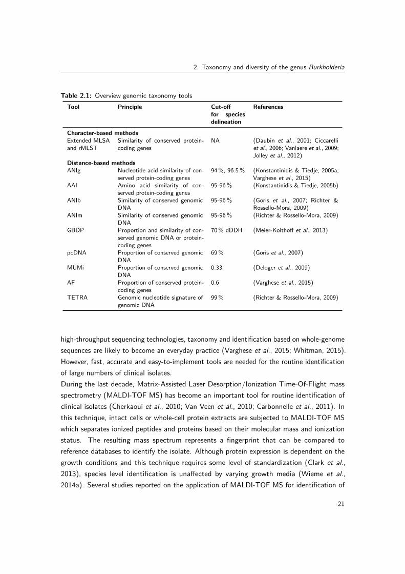
2. Taxonomy and diversity of the genus Burkholderia
Table 2.1: Overview genomic taxonomy toolsTool Principle Cut-off
for speciesdelineation
References
Character-based methodsExtended MLSAand rMLST
Similarity of conserved protein-coding genes
NA (Daubin et al., 2001; Ciccarelliet al., 2006; Vanlaere et al., 2009;Jolley et al., 2012)
Distance-based methodsANIg Nucleotide acid similarity of con-
served protein-coding genes94%, 96.5% (Konstantinidis & Tiedje, 2005a;
Varghese et al., 2015)AAI Amino acid similarity of con-
served protein-coding genes95-96% (Konstantinidis & Tiedje, 2005b)
ANIb Similarity of conserved genomicDNA
95-96% (Goris et al., 2007; Richter &Rossello-Mora, 2009)
ANIm Similarity of conserved genomicDNA
95-96% (Richter & Rossello-Mora, 2009)
GBDP Proportion and similarity of con-served genomic DNA or protein-coding genes
70% dDDH (Meier-Kolthoff et al., 2013)
pcDNA Proportion of conserved genomicDNA
69% (Goris et al., 2007)
MUMi Proportion of conserved genomicDNA
0.33 (Deloger et al., 2009)
AF Proportion of conserved protein-coding genes
0.6 (Varghese et al., 2015)
TETRA Genomic nucleotide signature ofgenomic DNA
99% (Richter & Rossello-Mora, 2009)
high-throughput sequencing technologies, taxonomy and identification based on whole-genomesequences are likely to become an everyday practice (Varghese et al., 2015; Whitman, 2015).However, fast, accurate and easy-to-implement tools are needed for the routine identificationof large numbers of clinical isolates.During the last decade, Matrix-Assisted Laser Desorption/Ionization Time-Of-Flight massspectrometry (MALDI-TOF MS) has become an important tool for routine identification ofclinical isolates (Cherkaoui et al., 2010; Van Veen et al., 2010; Carbonnelle et al., 2011). Inthis technique, intact cells or whole-cell protein extracts are subjected to MALDI-TOF MSwhich separates ionized peptides and proteins based on their molecular mass and ionizationstatus. The resulting mass spectrum represents a fingerprint that can be compared toreference databases to identify the isolate. Although protein expression is dependent on thegrowth conditions and this technique requires some level of standardization (Clark et al.,2013), species level identification is unaffected by varying growth media (Wieme et al.,2014a). Several studies reported on the application of MALDI-TOF MS for identification of
21

I. Introduction
GNNF bacteria from CF patients (Degand et al., 2008; Desai et al., 2012; Fernandez-Olmoset al., 2012; Alby et al., 2013; Lambiase et al., 2013). While its overall performance for theidentification of GNNF bacteria is generally good, identification of Bcc species requires anextended reference database (Fernandez-Olmos et al., 2012; Alby et al., 2013).
2.2 The good, the bad and the ugly: a tribute toadaptation
The genus Burkholderia currently (March 2016) comprises 90 validly named species (Euzeby,1997) which have a versatile metabolism and occupy very diverse niches (Coenye & Vandamme,2003). Burkholderia organisms have been isolated mainly as free-living organisms from water,soil and the rhizosphere of plants but these bacteria also developed different types ofinteractions with different hosts including plants, animals, fungi and amoebae (Marolda et al.,1999; Van Borm et al., 2002; Kikuchi et al., 2011; Verstraete et al., 2013; Stopnisek et al.,2016).
2.2.1 Plant growth promotion
Several Burkholderia species are plant growth promoting rhizobacteria (PGPR). PGPRare rhizosphere soil bacteria that influence plant growth through a range of interactions.These interactions can be direct through the production of phytohormones or through theenhancement of nutrient availability, or indirect through biological control of plant pathogens(Lugtenberg & Kamilova, 2009). Several diazotrophic (nitrogen-fixing) Burkholderia specieshave been described, including B. vietnamiensis, Burkholderia silvatlantica, Burkholderiadiazotrophica, Burkholderia unamae, B. xenovorans and B. tropica (Van et al., 2000; Caballero-Mellado et al., 2004; Perin et al., 2006; Caballero-Mellado et al., 2007; Sheu et al., 2013).Burkholderia phytofirmans, B. unamae and B. xenovorans show 1-aminocyclopropane-1-carboxylic acid deaminase activity and thereby lower the ethylene levels in stressed ordeveloping plants (Sessitsch et al., 2005; Caballero-Mellado et al., 2007). Plant hormonelevels are also influenced by the production of indol acetic acid by B. fungorum, Burkholderiagraminis, Burkholderia kururiensis, B. phytofirmans, B. unamae and B. vietnamiensis (Bevivinoet al., 1994; Suarez-Moreno et al., 2012; Castanheira et al., 2016). Finally, the production oforganic acids and siderophores increases the availability of phosphate and iron for the plantand thereby also promotes plant growth (Caballero-Mellado et al., 2007).While many Burkholderia species live in close interactions with plants in the rhizosphere,some rhizosphere bacteria also colonize roots, stems and leaves (Coenye & Vandamme, 2003;
22

2. Taxonomy and diversity of the genus Burkholderia
Compant et al., 2008). Many Burkholderia species including B. cepacia, B. cenocepacia, B.gladioli, B. phytofirmans, Burkholderia pyrrocinia, B. silvatlantica, B. tropica, B. unamaeand B. vietnamiensis have been isolated from plant tissue specimens (Compant et al., 2008).Burkholderia caledonica was detected between mesophyll cells in Fadogia and Vangueriaspecies (family Rubiaceae) known to cause gousiekte, a fatal disease of South Africanruminants (Verstraete et al., 2011). Recently, B. multivorans WS-FJ9 was shown to colonizethe rhizosphere and root tissues of poplar seedlings and promoted their growth (Li et al., 2013).Additionally, several Burkholderia species including Burkholderia caribensis, Burkholderiamimosarum, Burkholderia nodosa, Burkholderia phymatum and Burkholderia tuberum arealso known for nodulation of plant species of the Fabaceae family (Vandamme et al., 2002a;Chen et al., 2005, 2006, 2007). In addition to these free-living plant-beneficial species, severalCandidatus Burkholderia species have been described. They are obligate endosymbiontsin species of the Rubiaceae (genera Pavetta, Psychotria and Sericanthe) and Primulaceaefamilies (Van Oevelen et al., 2002, 2004; Lemaire et al., 2011a, 2012; Verstraete et al., 2011,2013). These endosymbionts occur in leaf nodules, are vertically transmitted and representan obligatory symbiosis which was estimated to originate millions of years ago (Lemaire et al.,2011b).Besides directly stimulating plant growth through the mechanisms described above,Burkholderia organisms can also produce antibacterial and antifungal metabolites whichprevent the proliferation of other rhizobacteria and fungi (Compant et al., 2008). By sup-pressing other organisms in the rhizosphere environment, they do not only compete withother soil bacteria, but also protect plants from soilborne pathogens. Examples of thesemetabolites are cepacin, quinolones, phenazine and pyrrolnitrin (Parke & Gurian-Sherman,2001). The best known example is Burkholderia ambifaria AMMDT, which protects peaplants from Aphanomyces root rot and Phytium damping-off disease (King & Parke, 1993). B.pyrrocinia produces at least two antifungal compounds including pyrrolnitrin and occidiofungin(Kwak & Shin, 2015; Wang et al., 2016) and an endophytic B. cenocepacia strain showed inplanta biocontrol of soilborne Fusarium wilt of banana plants (Ho et al., 2015). Next to theproduction of antimicrobial compounds, the biocontrol properties of Burkholderia bacteriacan also be attributed to their capacity to compete for limited resources (e.g. iron, vitamins,amino acids), to utilize root exudates and to activate an induced systemic resistance in plants(Compant et al., 2008; Vial et al., 2011).
23

I. Introduction
2.2.2 Plant pathogens
The best known plant-pathogenic species within the genus Burkholderia are B. andropogonis,B. caryophylli, B. cepacia, B. gladioli, B. glumae and B. plantarii (Compant et al., 2008). B.andropogonis is an important cause of stripe disease of sorghum and leaf spot of velvet beanand causes considerable loss of carnation (Coenye et al., 2001a). B. cepacia causes soft rot ofonion leaves and bulbs (Burkholder, 1950). B. caryophylli causes wilt in various plant species(Furuya et al., 2000). Finally, B. gladioli, B. glumae and B. plantarii form a phylogeneticallycoherent group (Fig. 2.1 and 2.4) of notorious rice pathogens (Seo et al., 2015). In addition,B. gladioli also induces soft rot in onions (Lee et al., 2005) and B. glumae causes wiltingsymptoms in several field crops (Jeong et al., 2003).
2.2.3 Human and animal pathogens
B. mallei and B. pseudomallei are primary pathogens causing glanders in horses, donkeys andmules and melioidosis in humans and animals, respectively. The Bcc contains opportunisticpathogens that pose a threat to patients with an underlying immunocompromising illnesssuch as CF (Chapter 1) and chronic granulomatous disease. Also the plant pathogen B.gladioli has been isolated from human clinical samples and is a well-known CF pathogen. B.fungorum is a most striking exception among the 16S rRNA clade 3 species (Fig. 2.1) sinceit has not only been isolated from the environment but also from a wide range of human andanimal samples such as cerebrospinal fluid, vaginal secretions, blood, respiratory secretions ofCF patients sputum and the nose of mice (Coenye et al., 2001b, 2002a; Gerrits et al., 2005).Other exceptions are B. tropica which was isolated from a neonatal patient who developedsepticemia (Deris et al., 2010), B. xenovorans which was isolated from human blood (Goriset al., 2004) and B. ginsengisoli which was isolated from a young man with Crohn’s diseasewho developed bacteraemia (Marks et al., 2016).
2.2.4 Fungal interactions
Interactions between Burkholderia bacteria and fungi exist in many forms (Scherlach et al.,2013). B. rhizoxinica and B. endofungorum represent bacterial endosymbionts of the plant-pathogenic fungus Rhizopus microsporus (Partida-Martinez et al., 2007). The phytotoxinused by the plant-pathogenic fungi to cause rice seedling blight is produced by the bacterialendosymbiotic cells (Partida-Martinez & Hertweck, 2005). B. fungorum and Burkholderiasordidicola have been isolated from the white-rot fungi Phanerochaete chrysosporium andPhanerochaete sordida, respectively (Coenye et al., 2001b; Lim et al., 2003). Associationsbetween Burkholderia and fungal cells have not only been described for plant-pathogenic
24
2. Taxonomy and diversity of the genus Burkholderia
fungi, but also for plant-beneficial mycorrhizal fungi. Burkholderia species have been foundin close association with both ectomycorrhizal (Izumi et al., 2007; Uroz et al., 2012) andarbuscular mycorrhizal (Andrade et al., 1997) fungi. Burkholderia are often associated withfungi in soils (Warmink et al., 2011; Nazir et al., 2012b; Uroz et al., 2012) and it was recentlydemonstrated that this co-occurrence of Burkholderia with a wide range of fungi mightrepresent a survival strategy of Burkholderia organisms in acidic soils (Stopnisek et al., 2014,2016).
2.2.5 Insect interactions
Burkholderia bacteria belonging to the B. glathei clade have been described as gut en-dosymbionts of the bean bug Riptortus pedestris (Kikuchi et al., 2011). The bacterialendosymbionts are not vertically transmitted but are acquired from soil by the nymphalinsect (Kikuchi et al., 2007). The insecticide resistance to fenitrothion in the pest insects isestablished by the endosymbiotic Burkholderia in the gut (Kikuchi et al., 2012) and emergesas a consequence of repeated insecticide use (Tago et al., 2015). Also in the chinch bugBlissus insularis Burkholderia bacteria were found in the gut (Boucias et al., 2012; Xu et al.,2016). Furthermore, a survey of Bartonella bacteria bed bugs in the United States detectedDNA sequences of Bcc bacteria (Saenz et al., 2013). Finally, also ants (Van Borm et al.,2002; Santos et al., 2004) and beetles (Lundgren et al., 2007; Kim et al., 2009) have beenreported to live in complex interactions with Burkholderia bacteria. In contrast with thesebeneficial insect interactions, Burkholderia strain A396 (named "Burkholderia rinojensis") wasisolated from soil in Japan and showed insecticidal and miticidal activities (Cordova-Kreyloset al., 2013).
2.2.6 Amoebal interactions
Several Bcc strains survive in Acanthamoeba cells and grow on by-products released by theamoebae (Marolda et al., 1999; Landers et al., 2000). Burkholderia bacteria were internalizedby phagocytosis and survived in acidic vacuoles instead of being digested (Lamothe et al.,2004). Since amoebae are also found in freshwater environments they have been suggestedas a natural reservoir for Bcc strains (Marolda et al., 1999). More recently, a cultivation-independent study showed that Burkholderia was one of the genera found in free-livingamoebae that were isolated from drinking water (Delafont et al., 2013). This ability to surviveintracellularly in eukaryotic cells was also demonstrated in human epithelial cells (Burns et al.,1996) and macrophages (Saini et al., 1999), suggesting that the same mechanisms thatlead to survival and persistence in the natural environment may also lead to survival and
25

I. Introduction
persistence as opportunistic pathogens in host cells (Valvano et al., 2005; Adiba et al., 2010).More recently, Burkholderia were found to colonize Dictyostelium discoideum amoebae andto induce the farming phenomenon in which amoebae stably associate with bacteria as afood source (DiSalvo et al., 2015).
2.2.7 Bioremediation
The metabolic versatility of Burkholderia allows them to utilize recalcitrant pollutants andmakes these bacteria useful for bioremediation applications (Coenye & Vandamme, 2003).Burkholderia bacteria are known to degrade antibiotics (Beckman & Lessie, 1979), herbicides(Jacobsen, 1997; Coenye et al., 2004), insecticides (Hong et al., 2007; Kikuchi et al., 2012),crude oil constituents (Castorena et al., 2006; Vanlaere et al., 2008b) and solvents suchas trichloroethylene (Mars et al., 1996; Zhang et al., 2000). B. xenovorans LB400 wasisolated from a polychlorinated biphenyl contaminated landfill in New York (Bopp, 1986)and its genome encodes at least 27 aromatic pathways (Chain et al., 2006). Burkholderiaphenoliruptrix AC1100 is well-known for its ability to degrade a variety of xenobiotics includingthe herbicide 2,4,5-trichlorophenoxyacetic acid (Coenye et al., 2004). As a last example,Burkholderia sp. strain NF100 was reported more than a decade ago as a degrader of theinsecticide fenitrothion (Hayatsu et al., 2000) and more strains with this bioremediationcapacity have been reported recently (Hong et al., 2007; Lim et al., 2012; Tago et al.,2015).
2.3 The genetic source code for versatility: Burkholderiagenomes
Burkholderia genomes vary in size from 2.4 Mb (Ca. Burkholderia schumannianae UZHbot8)(Pinto-Carbo et al., 2016) to 11.5 Mb (Burkholderia terrae BS001) (Nazir et al., 2012a),are characterized by a high G+C content (60-68%) and consist of multiple replicons (Lessieet al., 1996; Winsor et al., 2008; Ussery et al., 2009). In general, large prokaryotic genomesare disproportionately enriched in regulation and secondary metabolism, while being depletedin protein translation, DNA replication, cell division and nucleotide metabolism. This mayexplain why bacteria with large genomes dominate in environments were resources are scarcebut diverse, such as soil (Konstantinidis & Tiedje, 2004).These large genomes encode a multitude of metabolic functions and virulence factors (Fig.2.5). The first and largest chromosome of Burkholderia species harbors most of the genesessential for growth and basic metabolism while the other chromosome(s) encode mostly genes
26
2. Taxonomy and diversity of the genus Burkholderia
involved in adaptation and secondary metabolism (Mahenthiralingam et al., 2005; Ussery et al.,2009). Cooper et al. (2010) demonstrated that genes were less conserved and evolved morerapidly if they were located on the secondary chromosome and that secondary chromosomesmay serve as evolutionary test beds. The multireplicon structure of Burkholderia genomesthus allows for long-term segregation of genes by their expression rate and dispensability.Although the third chromosome of Bcc bacteria encodes at least one rRNA operon and wastherefore originally considered a chromosome, this replicon is not essential for survival andshould be considered a megaplasmid which is involved in virulence and stress response (Agnoliet al., 2012). Burkholderia bacteria can also carry one or more plasmids which further fosterthe diversification and adaptation to variable environments such as soil (Heuer & Smalla,2012).Finally, Burkholderia genomes contain numerous genomic islands and insertion sequences,representing as much as 9% of total DNA (Holden et al., 2009). Insertion sequences ortransposons are small mobile genetic elements (up to 2500 bp) that can jump from onelocation in the genome to another, thereby causing mutations or affecting gene expression.Genomic islands are larger regions that are identifiable by their deviating G+C content and areinvolved in the dissemination of variable genes, including antibiotic resistance and virulencegenes as well as catabolic genes (Juhas et al., 2009). Altogether, this genomic diversityand plasticity provides the genetic source code for the diversification and adaptation ofBurkholderia bacteria to a wide range of ecological niches.
2.4 What’s in the name Burkholderia?
Burkholderia bacteria interact in different ways with different hosts, making them ubiquitousand versatile bacteria. This versatility and adaptability is both a blessing and a curse. Withouttheir ability to thrive in varying environments, they would never have emerged as usefulstrains for plant growth promotion, bioremediation and biocontrol. However, this same abilityto thrive in varying environments most likely also contributes to their success as opportunisticpathogens. In fact, many properties important for rhizosphere colonization and survival inthe natural environment (versatile metabolism, antibiotic resistance and production, quorumsensing, siderophore production and adherence to host cells) may also be considered traitsthat could contribute to establishing infection in immunocompromised patients (Parke &Gurian-Sherman, 2001).Especially for the Bcc, their biotechnologically useful features are in marked contrast withthe severe and often fatal infections they cause as opportunistic human pathogens (Mahen-thiralingam et al., 2008). Increased concerns about the health risks for immunocompromised
27
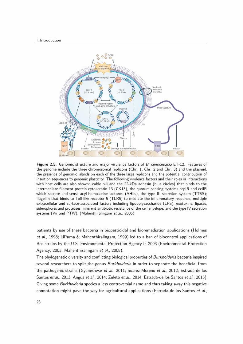
I. Introduction
© 2005 Nature Publishing Group
NATURE REVIEWS | MICROBIOLOGY VOLUME 3 | FEBRUARY 2005 | 149
R E V I E W S
is awaiting formal annotation. Other draft genomesequences include B. vietnamiensis strain G4 (ATCC53617), which is an important bioremediation strain4,and B. cepacia (genomovar I) strain 383 (R-18194 orATCC 17660), a soil strain described in the pioneeringstudy by Stanier et al .52 Venter and co-workers at theInstitute for Biological Energy Alternatives53 have iden-tified a near-complete genome that they designatedBurkholderia SAR-1 and which is a METAGENOMIC SEQUENCE
that was derived from a study of the Sargasso Sea.Analysis of the recA gene sequence of strain SAR-1indicates that it is closely related to strain 383 and that itis a member of B. cepacia genomovar I group K (REF. 54;E.M., unpublished observations).
The main features of the B. cenocepacia strain J2315genome are illustrated in FIG. 2. The total genome size is8,056 Mb, with an average GC content of 66.9% —which is similar to that of Pseudomonas species6. Threelarge chromosomal replicons and, unexpectedly, a small92-kb plasmid were identified. Each of the large replicons
GenomicsLessie and colleagues (reviewed in REF. 4) were the first toobserve that Bcc bacteria have large genomes made upof multiple large replicons — a relatively rare genomicarrangement in bacteria that was only discovered duringthe 1980s. In the past few years, a wealth of informationon the genomics of the Bcc bacteria has been obtained.All species that have been examined so far have morethan one chromosome and large genomes ranging from6 to 9 Mb — which are some of the largest to beobserved among Gram-negative species3 (see the Onlinelinks box for further information). Burkholderia xenovo-rans strain LB400, an environmental bacterium that candegrade the pollutant compounds polychlorobiphenyls,was the first Burkholderia genome to be completed indraft (see the Online links box). The complete genomesequences of Burkholderia pseudomallei andBurkholderia mallei have been recently published50,51,whereas the finished genome of B. cenocepacia strainJ2315 (originally documented as strain CF5610; REFS 5,24)
METAGENOMIC SEQUENCE
The entire genomic sequence ofan organism, which is assembledfrom nucleotide sequenceanalysis of random DNAfragments that are isolated fromthe environment.
Gl Gl
Gl
Gl
Gl
Gl
GlIS
IS
IS
IS
IS
TTSS
cblA
cepIRInterplay?
IS
Gl
Gl
Chr. 3(~0.87 Mb)
Plasmid
Recombinationand gene activation
Effector
Damage and inflammation Invasion
Polar flagellum
Antibioticresistanceand efflux
Cable pili
AHLs
Chr. 2(~3.2 Mb)
Chr. 1(~3.8 Mb )
PTW
IS
Virulencefactor expression
cciIR
Vir
(?)
Invasion
ExotoxinsLipases
SiderophoresProteases
Unknown
CK13 TLR5
LPS
Host cells
Figure 2 | Genomic structure and major virulence factors of Burkholderia cenocepacia ET-12. The schematic illustrationshows the genome structure and important virulence factors of B. cenocepacia. Features of the genome include: the threechromosomal replicons (Chr. 1, Chr. 2 and Chr. 3) and the plasmid; the presence of genomic islands (GI) on each of the three largereplicons; and the potential contribution of insertion sequences (IS) to genomic plasticity. The following virulence factors and theirroles or interactions with host cells are also shown: cable pili and the 22-kDa adhesin (blue circles) that binds to the intermediatefilament protein cytokeratin 13 (CK13); the quorum-sensing systems cepIR and cciIR, both of which secrete and sense acyl-homoserine lactones (AHLs); the type III secretion system (TTSS) and its possible role in invasion and survival in vivo; flagellin thatbinds to Toll-like receptor 5 (TLR5) to mediate the inflammatory response; multiple extracellular and surface-associated factors,including lipopolysaccharide (LPS), exotoxins, lipases, siderophores and proteases; inherent antibiotic resistance of the cellenvelope; and the recently described type IV secretion systems (Vir and PTW).
Figure 2.5: Genomic structure and major virulence factors of B. cenocepacia ET-12. Features ofthe genome include the three chromosomal replicons (Chr. 1, Chr. 2 and Chr. 3) and the plasmid,the presence of genomic islands on each of the three large replicons and the potential contribution ofinsertion sequences to genomic plasticity. The following virulence factors and their roles or interactionswith host cells are also shown: cable pili and the 22-kDa adhesin (blue circles) that binds to theintermediate filament protein cytokeratin 13 (CK13), the quorum-sensing systems cepIR and cciIRwhich secrete and sense acyl-homoserine lactones (AHLs), the type III secretion system (TTSS);flagellin that binds to Toll-like receptor 5 (TLR5) to mediate the inflammatory response, multipleextracellular and surface-associated factors including lipopolysaccharide (LPS), exotoxins, lipases,siderophores and proteases, inherent antibiotic resistance of the cell envelope, and the type IV secretionsystems (Vir and PTW). (Mahenthiralingam et al., 2005)
patients by use of these bacteria in biopesticidal and bioremediation applications (Holmeset al., 1998; LiPuma & Mahenthiralingam, 1999) led to a ban of biocontrol applications ofBcc strains by the U.S. Environmental Protection Agency in 2003 (Environmental ProtectionAgency, 2003; Mahenthiralingam et al., 2008).The phylogenetic diversity and conflicting biological properties of Burkholderia bacteria inspiredseveral researchers to split the genus Burkholderia in order to separate the beneficial fromthe pathogenic strains (Gyaneshwar et al., 2011; Suarez-Moreno et al., 2012; Estrada-de losSantos et al., 2013; Angus et al., 2014; Zuleta et al., 2014; Estrada-de los Santos et al., 2015).Giving some Burkholderia species a less controversial name and thus taking away this negativeconnotation might pave the way for agricultural applications (Estrada-de los Santos et al.,
28

2. Taxonomy and diversity of the genus Burkholderia
2015). However, there is no phylogenetic subdivision in this genus that distinguishes beneficialfrom pathogenic strains, as exemplified by the fact that B. fungorum, B. ginsengisoli, B.tropica and B. xenovorans, four species belonging to the so-called plant-associated beneficialand environmental group (Suarez-Moreno et al., 2012), have been isolated from humanclinical samples too (Coenye et al., 2001b, 2002a; Goris et al., 2004; Gerrits et al., 2005; Deriset al., 2010; Marks et al., 2016). Yet, Sawana et al. (2014) recently reclassified all species notbelonging to 16S rRNA clade 1 (Fig. 2.1) into a single novel genus, Paraburkholderia (Sawanaet al., 2014; Oren & Garrity, 2015). The rationale for splitting the genus Burkholderia wasbased on the identification of conserved sequence indels in whole-genome sequences of 45Burkholderia strains representing only 25 formally named species and several unclassifiedstrains. Species belonging to 16S rRNA clade 1 shared six conserved sequence indels. Theremaining Burkholderia strains representing a subset of species belonging to 16S rRNA clades2 and 3 shared only two conserved sequence indels. However, the phylogenetic diversityamong the clade 2 and 3 species and B. rhizoxinica as revealed by 16S rRNA-based divergenceand by differences in the distribution of 22 additional conserved sequence indels was ignoredand the authors proposed to restrict the name Burkholderia to 16S rRNA clade 1 whilereclassifying all other species into a single novel genus named Paraburkholderia (Sawana et al.,2014). More recently, a similar rationale led to the reclassification of all species of 16S rRNAclade 2 (Fig. 2.1) into the novel genus Caballeronia (Dobritsa & Samadpour, 2016).
29


Part II
Aims and Outline
31


Aims and Outline
As outlined in Part I, Bcc bacteria are rare but significant pathogens in people with CF.Epidemiological surveys reveal that B. multivorans is the most prevalent Bcc CF pathogen inmany countries, including Belgium, and the continued emergence of unique B. multivoransstrains in CF patients suggests acquisition from non-human sources, such as the naturalenvironment. Yet, environmental B. multivorans isolates are rare and its environmental nicheis therefore considered unknown.The first goal of the present thesis was to gain better insight into the epidemiology of B.multivorans by (i) examining its environmental niche and (ii) comparing the genomes ofclinical and environmental B. multivorans isolates. Chapter 3 describes how a large numberof environmental water and soil samples were examined using a cultivation-independent B.multivorans-specific PCR assay (Section 3.1). Next, cultivation strategies were evaluated andoptimized for the isolation of B. multivorans from PCR positive samples (Sections 3.2 and3.3). Finally, section 3.4 presents a comparative genomics study of clinical and environmentalB. multivorans isolates. Chapter 6 provides a general discussion of these results and somefuture perspectives.
The CF lung can harbor a wide range of bacteria and accurate identification of CF pathogens atthe species level is important to assess the clinical impact of these generally rare opportunisticpathogens. Furthermore, accurate typing of CF pathogens is necessary to identify outbreaks,gain insight into their epidemiology and improve infection control guidelines. Since January2011 a National Reference Center (NRC) is charged with the surveillance of respiratoryinfections caused by GNNF bacilli in Belgian CF patients (https://nrchm.wiv-isp.be/nl/ref_centra_labo/burkholderia_cepacia_complex) and LM-UGent is responsible for the molecularidentification and typing of these CF isolates.The second goal of the present thesis was to contribute to the general knowledge of theprevalence and epidemiology of GNNF bacteria in CF by (i) applying MLST to study theepidemiology of Bcc bacteria, (ii) using MLSA for the classification of novel species within
33

II. Aims and outline
the Bcc and (iii) performing polyphasic taxonomic studies for the classification of novelBurkholderia species. Chapter 4 describes how increased knowledge of the diversity of Bccbacteria and the application of MLSA led to the formal classification of three novel Bccspecies. Chapter 5 presents the application of both a traditional and modern polyphasicapproach for studying Burkholderia taxonomy and the formal classification of 14 novel speciesin the B. glathei clade. Chapter 7 provides a general discussion of these results, reflections onthe use of MLST for surveillance of Bcc infections in CF and some future perspectives.
34

Part III
Experimental Work
35


3 | The environmental nicheof Burkholderia multivorans:fitness school for a cystic fibrosispathogen?
The continued emergence of unique B. multivorans strains in CF patients suggests acquisitionfrom non-human sources such as the natural environment (Baldwin et al., 2008). Yet,environmental B. multivorans isolates are rare and its true environmental niche is consideredunknown. Section 3.1 describes the application of a cultivation-independent PCR assay toscreen a large number of environmental water and soil samples and to assess the occurrenceof B. multivorans in the natural environment. Sections 3.2 and 3.3 present the application ofmultiple cultivation strategies to B. multivorans PCR positive water and soil samples. Finally,section 3.4 provides the results of a comparative genomics study that examined to whichextent B. multivorans isolates with the same multilocus sequence type but from differentorigins (CF vs environment) differed in genetic potential.
37


3. The environmental niche of B. multivorans: fitness school for a CF pathogen?
3.1 PCR detection of Burkholderia multivorans in water andsoil samples
Redrafted from: Charlotte Peeters, Stijn Daenekindt and Peter Vandamme (2016). PCRdetection of Burkholderia multivorans in water and soil samples. BMC Microbiology, 16,1-7.
Author contributions: CP and PV conceived the study and wrote the manuscript. CPperformed the experiments. CP and SD performed the statistical data analysis. SD proofreadthe manuscript.
3.1.1 Abstract
Background: Although semi-selective growth media have been developed for the isolationof Bcc bacteria from the environment, thus far B. multivorans has rarely been isolated fromsuch samples. Because environmental B. multivorans isolates mainly originate from watersamples, we hypothesized that water rather than soil is its most likely environmental niche.The aim of the present study was to assess the occurrence of B. multivorans in water samplesfrom Flanders (Belgium) using a fast, culture-independent PCR assay.Results: A nested PCR approach was used to achieve high sensitivity, and specificity wasconfirmed by sequencing the resulting amplicons. B. multivorans was detected in 11% of thewater samples (n=112) and 92% of the soil samples (n=25) tested. The percentage of falsepositives was higher for water samples compared to soil samples, showing that the presentlyavailable B. multivorans recA primers lack specificity when applied to the analysis of watersamples.Conclusion: The results of the present study demonstrate that B. multivorans DNA iscommonly present in soil samples and to a lesser extent in water samples in Flanders(Belgium).
3.1.2 Background
The Bcc represents a group of closely related (Vandamme & Peeters, 2014; De Smet et al.,2015a) and extremely versatile bacteria that can be applied for a range of bioremediation,plant growth promotion and biocontrol purposes (Parke & Gurian-Sherman, 2001). Yet,these bacteria are simultaneously rare but important opportunistic pathogens in CF patients(De Boeck et al., 2004; LiPuma, 2010). The most prevalent Bcc CF pathogens are B.cenocepacia and B. multivorans. The low number of outbreaks caused by B. multivorans
39

III. Experimental work
(Whiteford et al., 1995; Segonds et al., 1999; Biddick et al., 2003), and the fact that B.multivorans isolates from CF patients commonly represent unique strains, suggest that thereis only limited person-to-person transmission, and that strains are acquired from non-humansources such as the natural environment (Baldwin et al., 2008). Although many studies havedescribed the isolation of B. cenocepacia from rhizosphere samples (Mahenthiralingam et al.,2008), only few reported on the occasional isolation of B. multivorans from environmentalsamples (Vermis et al., 2003a; Ramette et al., 2005; Vanlaere et al., 2005; Fang et al., 2011).Consequently, the true environmental niche of B. multivorans is considered unknown. Thefact that the few environmental B. multivorans isolates that have been reported mainlyoriginated from water samples (Vermis et al., 2003a; Fang et al., 2011) suggested that wateris the most likely environmental niche of B. multivorans.PCR-based diagnostic tests have been developed for Bcc species identification based onboth the 16S rRNA and recA genes, resulting in two sets of species-specific primers for B.multivorans (LiPuma et al., 1999; Mahenthiralingam et al., 2000a). Miller et al. (2002)used the 16S rRNA-based PCR assays for the culture-independent detection of Bcc in soilenvironments. In the latter study, none of the samples that were Bcc PCR positive yieldedBcc isolates, suggesting that cultivation-dependent methods for the detection of Bcc bacteriamay underestimate environmental populations.In the present study, we used a cultivation-independent recA-based PCR assay to assessthe presence of B. multivorans in water and soil samples in Flanders, Belgium. Our resultsshow that B. multivorans DNA could be detected only occasionally in water samples butto a greater extent in soil samples, and that the available B. multivorans recA primers lackspecificity, especially when applied to the analysis of water samples.
3.1.3 Materials and methods
3.1.3.1 Samples
Water (n=112) and soil (n=27) samples were taken from August to October 2013 in Flanders,Belgium (Supplementary Fig. 3.1). For the water samples, an autoclaved 1 L Duran bottlewas opened and filled 10 cm below the water surface. Per sample, three times 150 ml wasfiltered using a Nalgene vacuum filter funnel and cellulose nitrate membrane filters with 0.45µm pore size and 47 mm diameter (Thermo Scientific). For the soil samples, samples weretaken 2 cm below the soil surface using a sterile spoon, and collected in sterile falcon tubes.Per sample, 1 g of soil was homogenized in 9 ml phosphate-buffered resuspension buffer (0.15M NaCl, 10 mM EDTA, 0.1 M phosphate buffer, pH 8.0) using a Stomacher blender for 30sec at 230 rpm. Three times 1 ml of soil suspension was transferred to an Eppendorf tube
40

3. The environmental niche of B. multivorans: fitness school for a CF pathogen?
and centrifuged for 5 minutes at 13,000 rpm (17,949 g) before removing the supernatant.Filters and soil pellets were stored at −20 ◦C until DNA extraction.For each sample, sampling date, address, region (i.e. West-Vlaanderen [WV], Oost-Vlaanderen[OV], Limburg [L], Vlaams-Brabant [VB] or Antwerpen [A]), and class (i.e. swimming orrecreational water [SRW], canal-river-stream [CRS] or other [O]) were recorded (SupplementaryTable 3.1). Swimming and recreational waters were those under surveillance of the FlemishEnvironment Agency (www.kwaliteitzwemwater.be). For water samples, pH and temperaturewere measured on site. For soil samples, pH was measured after dissolving 10 g of soil in 50ml distilled water and magnetic stirring for 10 min.
3.1.3.2 DNA extraction from environmental samples and quality assessment
Prior to DNA extraction, filters with biological material from water samples were cut intosmaller pieces with sterilized scissors. Total DNA was extracted in triplicate from the filtersand soil pellets (three per sample) following the protocol for Gram-negative bacteria of Pitcheret al. (1989). DNA pellets were dissolved by adding 50 or 100 µl TE buffer depending on thesize of DNA pellet, and left to dissolve overnight at 4 ◦C. RNA was degraded by adding 2.5or 5 µl RNase (2 mg/ml) for pellets dissolved in 50 or 100 µl TE, respectively, and incubatingat 37 ◦C for 1 hour.The quality and quantity of the extracted DNA were examined by measuring optical densities(OD) at 234 nm, 260 nm, 280 nm and 320 nm (Miller, 2001; Arbeli & Fuentes, 2007) witha SpectraMax Plus 384 spectrophotometer. DNA was considered of acceptable quality ifthe OD260/280 ratio was higher than 1.7, the OD234/260 ratio was smaller than 1 and theOD320/260 ratio was smaller than 0.15. If both quality and quantity of the three DNA extractionreplicates per sample were similar, these replicates were pooled. DNA fragmentation and RNAcontamination was assessed by agarose (1%) gel electrophoresis and EtBr staining.To test for the presence of PCR inhibitors, DNA extracts were subjected to a 16S rRNAamplification PCR with universal primers ARI C/T (5’-CTG GCT CAG GAY GAA CGCTG-3’) and pH (5’-AAG GAG GTG ATC CAG CCG CA-3’). The PCR mix contained 1xCorelLoad PCR buffer (Qiagen), 0.2 mM dNTP (Applied Biosystems), 0.5 U AmpliTaq(Applied Biosystems) 0.1 µM of both primers and 200 ng/µl BSA (Roche). For each sample,2 µl DNA was added to 23 µl PCR master mix. B. multivorans R-20526 DNA and sterileMQ were used as positive and negative control, respectively. PCR was performed using a MJResearch PTC-100 thermal cycler. Initial denaturation for 5 min at 95 ◦C was followed by 3cycles of 1 min at 95 ◦C, 2 min 15 sec at 55 ◦C and 1 min 15 sec at 72 ◦C, another 30 cyclesof 35 sec at 95 ◦C, 1 min 15 sec at 55 ◦C and 1 min 15 sec at 72 ◦C, and a final elongation for
41

III. Experimental work
7 min at 72 ◦C. The presence of amplicons was verified via agarose (1%) gel electrophoresiswith SmartLadder (Eurogentec) as molecular size marker and EtBr staining.If the OD320/260 ratio was higher than 0.15 and/or no universal 16S rRNA amplicon couldbe obtained, an extra purification step using agarose plugs was performed to remove humicacids and other PCR inhibiting contaminants (Moreira, 1998). After purification using plugs,DNA extracts were diluted 5x in TE buffer. Only DNA extracts for which a universal 16SrRNA amplicon could be obtained, were subjected to the B. multivorans PCR assay.
3.1.3.3 Preliminary experiment
A preliminary experiment compared the specificity of the B. multivorans-specific primersthat were based on the recA and 16S rRNA gene and were available from previous studies(LiPuma et al., 1999; Mahenthiralingam et al., 2000a). Sampling of two water samples, DNAextraction and quality assessment of the DNA extracts was performed as described above.For the nested B. multivorans 16S rRNA PCR assay, PCR products of the first, universal 16SrRNA PCR were used as template in a second PCR in which B. multivorans-specific primersBC-GII (5’-AGG CGG TCT GTT AAG ACA-3’) and BC-R (5’-AGC ACT CCC GAA TCTCTT-3’) were used (LiPuma et al., 1999). The second PCR mix was identical to the first PCRmix, except for a lower BSA concentration (50 ng/µl). For each sample, 2 µl PCR product ofthe first PCR was added to 23 µl PCR master mix. The positive (B. multivorans R-20526)and negative (blank) control of the first PCR (5 µl) were also transferred as template into thesecond PCR. The thermal cycling program was identical to that of the first PCR. The presenceof amplicons was verified via agarose (1%) gel electrophoresis with SmartLadder (Eurogentec)as molecular size marker and EtBr staining. If an amplicon (445 bp) was visible in the secondPCR, it was sequenced using the BC-GII and BC-R primers as described previously (Peeterset al., 2013) to exclude false positive results. The recA-based B. multivorans PCR assay wasperformed as described below.
3.1.3.4 Nested recA PCR assay for B. multivorans
In a first PCR, Bcc-specific recA primers recA-01-F (5’-GAT AGC AAG AAG GGC TCC-3’)and recA-02-R (5’-CTC TTC TTC GTC CAT CGC CTC-3’) were used (Baldwin et al.,2005). The PCR mix contained 1x CorelLoad PCR buffer (Qiagen), 0.25 mM dNTP (AppliedBiosystems), 1 U Taq (Qiagen), 0.5 µM of both primers, 1x Q-solution (Qiagen) and 200ng/µl BSA (Roche). For each sample, 2 µl DNA was added to 23 µl PCR master mix.B. multivorans R-20526 DNA and sterile MQ were used as positive and negative control,respectively. This PCR reaction was setup in duplicate for each sample to enable pooling and
42

3. The environmental niche of B. multivorans: fitness school for a CF pathogen?
to increase sensitivity. PCR was performed using a MJ Research PTC-100 thermal cycler.Initial denaturation for 2 min at 94 ◦C was followed by 35 cycles of 30 sec at 94 ◦C, 45 secat 58 ◦C and 1 min at 72 ◦C, and a final elongation for 10 min at 72 ◦C. PCR product ofthe duplicate reactions for each sample were pooled using filter tips and used as templatein the second PCR, in which B. multivorans-specific primers BCRBM1 (5’-CGG CGT CAACGT GCC GGA T-3’) and BCRBM2 (5’-TCC ATC GCC TCG GCT TCG T-3’) were used(Mahenthiralingam et al., 2000a). The second PCR mix was identical to the first PCR mix,except for a lower BSA concentration (50 ng/µl) and a higher primer concentration (1 µM).For each sample, 5 µl of pooled PCR product from the first PCR was added to 20 µl PCRmaster mix. The positive (B. multivorans R-20526) and negative (blank) control of thefirst PCR (5 µl) were also transferred as template into the second PCR. Thermal cyclingwas identical to the first PCR, except that the annealing temperature was 64 ◦C instead of58 ◦C (Vermis et al., 2002a). The presence of amplicons was verified via agarose (1%) gelelectrophoresis with SmartLadder (Eurogentec) as molecular size marker and EtBr staining.The nested recA PCR assay was performed twice for each sample. If the results for the tworuns were not the same, the assay was performed a third time. If an amplicon (714 bp)was visible in the second B. multivorans-specific PCR, it was sequenced using the BCRBM1and BCRBM2 primers as described previously (Peeters et al., 2013) to exclude false positiveresults. Only if at least for one of the replicate runs an amplicon from the second PCR wassequenced that showed at least 97% similarity to the recA sequence of B. multivorans ATCC17616, the sample was considered a true positive for the detection of B. multivorans.To determine the detection limit of this PCR assay, it was applied on serial dilutions ofgenomic DNA from B. multivorans R-20526, a strain for which whole-genome sequencingdata is available (BioProject PRJNA234537). The mean weight for an AT and GC basepair is 615.3830 Da and 616.3711 Da, respectively (Dolezel et al., 2003). Given the GCcontent of this genome of about 68%, and ignoring the presence of modified nucleotides,the mean relative weight of one base pair of R-20526 is 616.0549 Da or 1.023 x 10⁻⁹ pg(1 Da = 1.660539 x 10⁻²⁴ g). Given the genome size of 6.5 Mb, one genome of R-20526contains 6.65 x 10⁻⁶ ng of DNA, or 1 ng of DNA contains 1.50 x 10⁵ genome equivalents.The undiluted genomic DNA stock of strain R-20526 contained 225 ng/µl DNA, as measuredwith the Promega QuantiFluor ONE dsDNA system, or 3.38 x 10⁷ genomic DNA equivalentsper µl. The PCR assay was first applied on a 50x dilution series (in TE buffer) to find theapproximate fading range. The highest 50x dilution for which an amplicon could be obtainedwas then used to make a twofold dilution series to find the limit of detection. The PCRassay was performed three times for each dilution and the detection limit was defined as thehighest twofold dilution that tested positive in all three runs. Considering the recA gene is
43

III. Experimental work
a single-copy gene for B. multivorans R-20526, the detection limit was calculated from themeasured DNA concentration and calculated DNA content.
3.1.3.5 Statistical data analysis
Statistical data analysis was performed using R version 3.1.2 in RStudio (version 0.98.1091),with the following packages: car, ggplot2, and MASS. Binomial logistic regression was used totest which variables (type, region, class, pH, temperature) were significant predictor variablesfor the outcome variable, i.e. B. multivorans detection (Bm). Type, class, region and Bmwere coded as factors, and the level with the most cases was chosen as reference category.Temperature and pH were coded as numeric variables, and centered around the mean toreduce standard error (SE). Backward stepwise model selection was applied to select the bestfitting models.
3.1.4 Results
3.1.4.1 Specificity of the 16S rRNA and recA PCR assays
Preliminary experiments were performed to compare the specificity of the B. multivorans-specific primers that were available from previous studies (De Boeck et al., 2004; LiPuma,2010). Therefore, PCR assays based on the recA and 16S rRNA genes were applied to DNAextracts of two water samples and the resulting amplicons were sequenced. The obtainedsequences were analyzed using the NCBI blastn suite (blast.ncbi.nlm.gov) to evaluate specificity.For amplicons of the recA PCR assay, hits showed 98% or more similarity with the recAsequence of B. multivorans ATCC 17616 (CP000868) (this degree of variability in recA genesequences corresponds with the sequence diversity commonly observed within Bcc species(Vandamme & Dawyndt, 2011)). For the 16S rRNA nested PCR, amplicons showed thehighest similarity (96%) with sequences of Comamonadaceae sp. (FM886892), Ideonellasp. (FM886860), Roseatales sp. (JQ917995), and Mitsuaria sp. (JQ659937), all belongingto the order Burkholderiales, yet demonstrating that the nested 16S rRNA PCR assay wasnot B. multivorans-specific. Therefore, only the B. multivorans nested recA PCR assay wasfurther optimized (i.e. PCR mix, BSA concentration, number of cycles, primer concentrationand quantity of pooled PCR product being transferred to the second round of PCR; data notshown) and used to analyze all environmental samples.
44

3. The environmental niche of B. multivorans: fitness school for a CF pathogen?
3.1.4.2 Detection of B. multivorans in environmental samples
All 112 water samples yielded PCR-grade DNA extracts without the need of an extrapurification step. B. multivorans DNA was detected in 12 water samples (11%), of which onesample (W132) already yielded a visible amplicon in the first round of PCR (showing 97%similarity to the recA sequence of B. multivorans ATCC 17616). For the soil samples (n=27),four yielded PCR-grade DNA without the need of an extra purification step, 21 samplesneeded extra purification using agarose plugs, and for two soil samples (S6 and S14) noPCR-grade DNA extract could be obtained. The latter soil samples were therefore excludedfrom the dataset. For soil DNA extracts containing 50 ng/µl or more DNA, an OD320/260ratio of 0.15 or higher was a good indicator for the necessity of an extra purification stepto obtain PCR-grade DNA. B. multivorans DNA was detected in 23 soil samples (92%), ofwhich none yielded a visible amplicon in the first round of PCR.
3.1.4.3 Statistical data analysis
The first model tested if type (water, soil) and class (SRW, CRS, O) were significant predictorsfor B. multivorans detection. Binomial logistic regression showed that the type of sample(water versus soil) was indeed a significant predictor for B. multivorans detection (Table 3.1).Adding class to the model did not significantly improve the model (data not shown). Theodds ratio from this model predicted that there was a 96 times (e4.56 = 95.58) bigger chanceto detect B. multivorans in soil than in water samples.
Table 3.1: Binomial logistic regression model 1 (water and soil samples)Beta (SE) Odds ratio (95% CI)
Intercept -2.12 *** (0.31) 0.12 (0.06; 0.21)TypeWater (ref) — —Soil 4.56 *** (0.80) 95.83 (24.48; 646.70)χ2 65.50 *** (df = 1)
Binomial logistic regression with Bm as outcome and Type as predictor. Signif. codes: 0 ’***’ 0.001 ’**’ 0.01 ’*’0.05 ’.’ . SE, standard error; CI, confidence interval.
To test which characteristics of the water environment affected the presence of B. multivorans,a second model was tested in which pH, temperature and class were assessed as predictorsfor B. multivorans detection. Binomial logistic regression showed that pH and class weresignificant predictors for B. multivorans detection in water samples (Table 3.2). Model 2aincludes the main effects of all the variables and shows that the probability of detectingB. multivorans was higher in water samples with high pH. The results also show that the
45

III. Experimental work
probability of detecting B. multivorans was higher in streams (CRS) compared to swimmingand recreational waters (SRW). Model 2b includes an additional interaction effect betweentemperature and pH to test whether the effect of pH on detection depends on the temperature.The model fit of model 2b improved and the interaction effect between temperature and pHindicated that the positive effect of pH was stronger when the temperature of the water washigher.
Table 3.2: Binomial logistic regression model 2 (water samples)Model 2a Model 2b
Beta (SE) Beta (SE) Odds ratio (95% CI)
Intercept -2.72 *** (0.59) -3.51 *** (0.82) 0.03 (0.00; 0.01)pH 0.97 . (0.52) 1.70 * (0.69) 5.48 (1.52; 23.84)Temperature -0.22 (0.16) -0.19 (0.16) 0.83 (0.59; 1.13)ClassSRW (ref) — — —O -0.18 (0.92) 0.16 (0.96) 1.17 (0.14; 7.29)CRS 1.47 . (0.80) 1.95 * (0.94) 7.06 (1.21; 52.19)
InteractionpH*Temperature — 0.39 * (0.20) 1.47 (1.01; 2.25)χ2 8.24 . (df = 4) 12.36 * (df = 5)
Binomial logistic regression with Bm as outcome and pH, Temperature and Class as predictor, without (model 2a) andwith (model 2b) interaction effect between pH and Temperature. Signif. codes: 0 ’***’ 0.001 ’**’ 0.01 ’*’ 0.05 ’.’ .SE, standard error; CI, confidence interval. SRW, swimming or recreational water; CRS, canal-river-stream; O, other.
3.1.4.4 Detection limit recA PCR assay
The recA PCR assay was applied to serial dilutions of genomic DNA from B. multivoransR-20526 to determine its detection limit. The highest 50x dilution testing positive was thefourth one, from which a twofold dilution series was made. The highest twofold dilutiontesting positive in all three replicate runs of the assay was the second one, containing 1.35genome equivalents/µl. Given that recA is a single-copy gene, and that for each dilution2x 2 µl was added to the first round of PCR (set up in duplicate), the detection limit wasdetermined to be 5.41 recA copies.
3.1.5 Discussion
Because the goal of the present study was to gain insight in the environmental niche of B.multivorans in a time and cost-effective way, we chose for a conventional PCR method withgel-based detection of the resulting amplicons, instead of quantitative PCR. B. multivorans-specific primers based on both 16S rRNA and recA sequences were available from previous
46
3. The environmental niche of B. multivorans: fitness school for a CF pathogen?
studies (LiPuma et al., 1999; Mahenthiralingam et al., 2000a). Although the recA gene has ahigher taxonomic resolution (Mahenthiralingam et al., 2000a), a 16S rRNA gene based assayis potentially more sensitive because the B. multivorans genome contains five copies of the16S rRNA gene (Stoddard et al., 2015). However, preliminary tests revealed that the 16SrRNA-based assay yielded only false positive results, as shown by sequence analysis of theresulting amplicons. A nested PCR design was used to increase the sensitivity of the recAPCR assay (Drevinek et al., 2002), in which the first PCR was carried out in duplicate andthe resulting PCR product was pooled before using it as a template in the second PCR. Thein vitro detection limit of this PCR assay was therefore determined to be 5.41 recA copies orgenome equivalents.The specificity of the recA primers (Mahenthiralingam et al., 2000a) used was previouslyevaluated (Vermis et al., 2002a). The latter study demonstrated that non-specific primerbinding could be eliminated by raising the annealing temperature for the B. multivorans assayfrom 58 ◦C to 64 ◦C, resulting in a specificity of 100% (Vermis et al., 2002a). Additionally, thecombination of two different primer pairs in our nested PCR design (specific for the Bcc andB. multivorans) further minimized false positive results. Finally, all resulting amplicons weresequenced to ensure that only true positive results were recorded. Whereas this final checkyielded satisfactory results for the soil samples (one false positive versus 23 true positives), itrevealed a strikingly high number of false positives for the water samples (20 false positivesversus 12 true positives). The false positives included samples for which the amplicon couldnot be sequenced (one soil sample and 15 water samples) or samples for which the resultingsequence did not yield any BLAST hits (five water samples). These findings demonstratethat this recA-based PCR assay in its current form is not suited for routine analysis of watersamples, and that one should re-evaluate PCR-based methods in terms of specificity whenapplying them to environmental samples (Bergmark et al., 2012).To eliminate false negative results caused by PCR inhibitors such as humic acids in theenvironmental DNA extracts, all samples were screened for the presence of PCR inhibitorsusing a universal 16S rRNA PCR. In case this universal PCR was inhibited, the DNA extractswere purified using low-melting point agarose plugs. Re-testing the DNA extracts afterthis purification step demonstrated that this purification procedure was very efficient inremoving the humic acids from the DNA extracts, as shown previously by Moreira (1998).Furthermore, optical density should not only be measured at 260 nm because this can lead toan overestimation of the DNA concentration if humic acids are present, but also at 320 nmto quantify these contaminations and to evaluate the quality of environmental DNA extractsproperly (Miller, 2001; Arbeli & Fuentes, 2007).Because the few environmental B. multivorans isolates that have been reported mainly
47

III. Experimental work
originated from water samples (Vermis et al., 2003a; Fang et al., 2011), we hypothesized thatwater rather than soil was the most likely environmental niche of B. multivorans. Our studytherefore focused primarily on water samples. However, only 12 out of 112 water samples(11%) examined were B. multivorans PCR positive, in contrast to 23 out of 25 soil samples(92%) examined. Despite the small number of soil samples, binomial logistic regressionshowed a highly significant effect (p < .001) of the type of sample (Model 1, Table 1) andpredicted that there was a 96 times bigger chance to detect B. multivorans in soil than inwater samples. This result also seemed to suggest that, if present, B. multivorans can beisolated more easily from water than from soil samples. Accordingly, Miller et al. (2002)showed that although a high percentage of soil samples was PCR positive for Bcc, none ofthese samples yielded Bcc isolates, demonstrating that cultivation-dependent recovery ofBcc bacteria likely underestimates their prevalence in environmental samples. However, thedetection of B. multivorans DNA does not necessarily imply that viable target organismswere present at the moment samples were taken (Josephson et al., 1993).Binomial logistic regression showed that the probability of detecting B. multivorans in watersamples was higher in streams (CRS) compared to swimming and recreational waters (SRW),and in water samples with a higher pH (Model 2, Table 3.2). Since pH is known to bean important predictor of bacterial diversity in soil (Fierer & Jackson, 2006; Rousk et al.,2010), and Burkholderia bacteria have been shown to be acid tolerant (Stopnisek et al.,2014), we expected to detect more B. multivorans in acidic versus alkaline waters. Theopposite findings of the present study again may suggest that water is not the natural reservoirof B. multivorans. However, the number of PCR positive water samples was rather smalland therefore one should be careful when extrapolating the results of our binomial logisticregression model 2.Our finding that B. multivorans is widely distributed in soil samples contrasts with theresults of previous isolation campaigns, but nevertheless agrees with the notion that the soilenvironment typically harbors large-genome sized organisms (Konstantinidis & Tiedje, 2004;Raes et al., 2007). As B. multivorans harbors a ~6.5 Mb genome, it is equipped with themetabolic versatility needed to thrive in a complex, variable environment such as soil. Futureresearch could focus on the genome biology of this organism, and try to infer the lifestyle ofthis organism based on genome data (Barberan et al., 2014; Livermore et al., 2014).
3.1.6 Conclusions
In summary, we applied a recA-based PCR assay that demonstrated that B. multivoransDNA is widely distributed in soil samples but only occasionally in water samples in Flanders,
48

3. The environmental niche of B. multivorans: fitness school for a CF pathogen?
Belgium. As for all Bcc bacteria it is unclear if and how this mere observation should beimplemented in infection control guidelines. Our study also demonstrated that the presentlyavailable B. multivorans recA primers lack specificity when applied to the analysis of watersamples.
3.1.7 Acknowledgments
C. P. is indebted to the Special Research Council of Ghent University.
3.1.8 Supplementary material
●
●
●
●●●
●
●
●
●
●
●
●●
●●●
50.0
50.5
51.0
51.5
52.0
3 4 5 6lon
lat
Bm
●
●
no
yes
Type
● soil
water
Supplementary Figure 3.1: Sampling map. Map showing all sampling locations as produced by theggmap and ggplot2 packages in R (own figure). Bm, B. multivorans.
49

III. Experimental work
Supplementary Table 3.1: Sample dataBcc, B. cepacia complex PCR positive; Bm, B. multivorans PCR positive. NA, not available. aWV, West-Vlaanderen;OV, Oost-Vlaanderen; L, Limburg; VB, Vlaams-Brabant; A, Antwerpen. bSRW, swimming or recreational water; CRS,canal-river-stream; O, other.
ID Date Type Regiona Classb Temperature pH Bcc Bm RemarksW40 13/08/2013 water OV CRS 19.0 7.69 no yesW41 13/08/2013 water OV SRW 20.0 8.41 no yesW42 13/08/2013 water OV SRW 21.0 7.86 no noW43 13/08/2013 water OV SRW 20.0 8.33 no noW44 14/08/2013 water WV O 16.5 7.32 no noW45 14/08/2013 water WV O 19.0 9.59 no noW46 14/08/2013 water WV O 16.0 5.49 no noW47 14/08/2013 water WV O 17.0 6.70 no noW48 14/08/2013 water WV O 20.0 8.80 no yesW49 14/08/2013 water WV CRS 20.0 7.82 no yesW50 14/08/2013 water WV SRW 22.0 8.87 no noW51 14/08/2013 water WV SRW 22.0 8.28 no noW52 14/08/2013 water WV CRS 22.0 7.80 no noW53 14/08/2013 water OV CRS 21.0 7.77 no noW54 20/08/2013 water A SRW 19.0 10.30 no noW55 20/08/2013 water A SRW 21.0 8.16 no noW56 20/08/2013 water A SRW 20.0 8.10 no yesW57 20/08/2013 water A SRW 22.0 8.92 no noW58 20/08/2013 water A SRW 22.0 8.92 no noW59 20/08/2013 water A O 21.0 7.61 no noW60 20/08/2013 water A SRW 23.0 8.63 no noW61 20/08/2013 water A SRW 23.0 8.77 no noS3 20/08/2013 soil A SRW NA 6.74 no yesW62 20/08/2013 water A SRW 22.0 4.53 no noW63 20/08/2013 water A SRW 23.0 4.97 no noW64 20/08/2013 water A SRW 23.5 3.79 no noW65 20/08/2013 water A SRW 23.0 9.13 no yesW66 20/08/2013 water A SRW 23.0 8.41 no yesW67 20/08/2013 water A O 22.0 8.38 no noW68 20/08/2013 water A CRS 22.0 8.04 no noW69 20/08/2013 water A SRW 22.0 7.12 no noW70 20/08/2013 water A SRW 22.0 7.35 no noW71 22/08/2013 water OV O 19.0 6.56 no noW72 22/08/2013 water OV O 18.0 6.65 no noS4 22/08/2013 soil OV O NA 4.11 no yesW73 22/08/2013 water OV SRW 21.0 8.87 no noW74 22/08/2013 water WV SRW 21.0 8.89 no noS5 22/08/2013 soil WV SRW NA 8.45 no yesW75 22/08/2013 water WV SRW 20.5 8.23 no noW76 22/08/2013 water WV SRW 20.0 9.01 no noW77 22/08/2013 water WV SRW 21.0 8.72 no noW78 22/08/2013 water WV CRS 20.0 7.69 no noW79 22/08/2013 water WV CRS 20.0 8.26 no no
Continued on next page
50

3. The environmental niche of B. multivorans: fitness school for a CF pathogen?
Continued from previous pageID Date Type Regiona Classb Temperature pH Bcc Bm RemarksW80 22/08/2013 water WV SRW 22.0 8.63 no noW81 27/08/2013 water OV O 19.0 7.67 no noS6 27/08/2013 soil OV O NA 3.92 NA NA PCR inhibitorsW82 27/08/2013 water OV O 21.5 7.59 no noS7 27/08/2013 soil OV O NA 8.63 no yesW83 27/08/2013 water OV O 20.0 7.37 no noW84 27/08/2013 water OV O 21.0 8.70 no yesW85 27/08/2013 water OV SRW 22.5 8.80 no noS8 27/08/2013 soil OV SRW NA 8.30 no yesW86 27/08/2013 water OV CRS 22.0 7.85 no noW87 27/08/2013 water OV O 23.0 8.11 no noS9 27/08/2013 soil OV O NA 3.53 no yesW88 27/08/2013 water OV SRW 22.0 8.47 no noW89 27/08/2013 water OV O 22.0 8.11 no noW90 27/08/2013 water OV O 22.0 7.28 no noS10 27/08/2013 soil OV O NA 8.01 no yesW91 27/08/2013 water OV O 24.0 8.60 no noW92 27/08/2013 water OV CRS 23.0 8.14 no noW93 27/08/2013 water OV CRS 22.0 7.83 no yesW94 29/08/2013 water OV CRS 20.0 7.53 no noW95 29/08/2013 water OV SRW 20.0 7.87 no noW96 29/08/2013 water OV SRW 20.0 7.66 no noW97 29/08/2013 water OV CRS 21.0 7.32 no noW98 29/08/2013 water WV SRW 21.0 7.96 no noW99 29/08/2013 water WV SRW 21.0 8.25 no noW100 29/08/2013 water WV SRW 21.0 7.53 no noW101 29/08/2013 water OV SRW 21.0 7.75 no noW102 29/08/2013 water OV SRW 22.0 8.20 no noW103 2/09/2013 water A SRW 20.5 8.80 no yesW104 2/09/2013 water A SRW 21.0 8.79 no noW105 2/09/2013 water A SRW 19.0 8.70 no noW106 2/09/2013 water A CRS 19.0 8.06 no noW107 2/09/2013 water A O 21.0 7.48 no noS11 2/09/2013 soil A O NA 3.69 no yesW108 2/09/2013 water A O 20.0 7.87 no noW109 2/09/2013 water A SRW 21.0 8.63 no noS12 2/09/2013 soil A SRW NA 8.54 no yesW110 2/09/2013 water VB SRW 21.0 7.94 no noW111 2/09/2013 water VB O 19.0 8.89 no noW112 2/09/2013 water A SRW 22.0 8.28 no noW113 2/09/2013 water A SRW 21.5 8.05 no noW114 2/09/2013 water A SRW 22.0 8.38 no noW115 2/09/2013 water A O 21.5 7.73 no noW116 2/09/2013 water A O 21.5 7.44 no noW117 2/09/2013 water A CRS 21.0 7.74 no noW118 2/09/2013 water A SRW 22.0 8.43 no noS13 2/09/2013 soil A SRW NA 6.26 no yesW119 2/09/2013 water OV SRW 20.0 8.25 no no
Continued on next page
51

III. Experimental work
Continued from previous pageID Date Type Regiona Classb Temperature pH Bcc Bm RemarksW120 5/09/2013 water VB O 17.5 7.25 no noS14 5/09/2013 soil VB O NA 8.44 NA NA PCR inhibitorsW121 5/09/2013 water VB CRS 18.0 7.73 no noW122 5/09/2013 water VB O 21.5 7.16 no noW123 5/09/2013 water VB O 18.0 7.54 no noW124 5/09/2013 water VB O 21.0 8.22 no noW125 5/09/2013 water VB O 24.0 7.83 no noW126 5/09/2013 water VB SRW 23.0 8.31 no noW127 5/09/2013 water VB SRW 24.0 8.03 no noW128 5/09/2013 water VB CRS 23.0 7.65 no noW129 5/09/2013 water VB CRS 23.0 7.94 no noW130 11/09/2013 water L SRW 17.0 7.24 no noW131 11/09/2013 water L SRW 17.5 7.16 no noW132 11/09/2013 water L CRS 17.0 7.40 yes yesW133 11/09/2013 water L SRW 18.0 7.81 no noW134 11/09/2013 water L SRW 17.0 7.98 no noW135 11/09/2013 water L SRW 17.0 6.02 no noW136 11/09/2013 water L SRW 18.5 6.49 no noW137 11/09/2013 water L O 19.0 6.37 no noW138 11/09/2013 water L O 16.0 6.40 no noW139 11/09/2013 water L CRS 13.5 6.31 no yesW140 11/09/2013 water L CRS 20.5 7.94 no noW141 11/09/2013 water L O 18.0 6.50 no noS15 11/09/2013 soil L O NA 7.05 no yesW142 11/09/2013 water L O 17.0 6.49 no noW143 11/09/2013 water L CRS 15.0 7.04 no noS16 11/09/2013 soil L CRS NA 4.06 no yesW144 11/09/2013 water L CRS 16.0 7.40 no noW145 11/09/2013 water L O 18.5 7.73 no noW146 11/09/2013 water L O 19.0 6.00 no noW147 11/09/2013 water L O 18.0 7.71 no noW148 11/09/2013 water L SRW 19.5 8.33 no noW149 11/09/2013 water L SRW 18.5 7.88 no noW150 11/09/2013 water L SRW 19.0 7.60 no noS17 17/09/2013 soil OV O NA 7.97 no yesS18 17/09/2013 soil OV O NA 7.94 no yesW151 19/09/2013 water OV O 27.0 8.36 no noS19 19/09/2013 soil OV O NA 9.61 no yesS20 19/09/2013 soil OV O NA 5.18 no yesS21 19/09/2013 soil OV O NA 8.90 no yesS22 21/09/2013 soil OV O NA 6.59 no noS23 21/09/2013 soil OV O NA 6.09 no yesS25 22/09/2013 soil WV O NA 6.68 no noS26 12/10/2013 soil WV O NA 7.53 no yesS27 12/10/2013 soil WV O NA 4.72 no yesS28 12/10/2013 soil WV O NA 7.28 no yesS29 12/10/2013 soil WV O NA 7.40 no yesS30 12/10/2013 soil WV O NA 7.77 no yes
52

3. The environmental niche of B. multivorans: fitness school for a CF pathogen?
3.2 Extensive cultivation of soil and water samples yieldsvarious pathogens in patients with cystic fibrosis but notBurkholderia multivorans
Redrafted from: Charlotte Peeters, Eliza Depoorter, Jessy Praet and Peter Vandamme(2016). Extensive cultivation of soil and water samples yields various pathogens in patientswith cystic fibrosis but not Burkholderia multivorans. Journal of Cystic Fibrosis, in press.
Author contributions: CP and PV conceived the study and wrote the manuscript. CP,ED and JP performed the experiments. CP analyzed the data. ED and JP proofread themanuscript.
3.2.1 Abstract
Background: While the epidemiology of Bcc bacteria in CF patients suggests that B.multivorans is acquired from environmental sources, this species has rarely been isolated fromwater and soil samples.Methods: Multiple isolation strategies were applied to water and soil samples that werepreviously shown to be B. multivorans PCR positive. These included direct plating andliquid enrichment procedures and the use of selective media, acclimatizing recovery andco-cultivation with CF sputum. MALDI-TOF MS and sequence analysis of 16S rRNA andhousekeeping genes were used to identify all isolates.Results: None of the approaches yielded B. multivorans isolates. Other Burkholderia species,several GNNF bacteria (including Cupriavidus, Inquilinus, Pandoraea, Pseudomonas andStenotrophomonas) and rapidly growing mycobacteria (including Mycobacterium chelonae)were all isolated from water and soil samples.Conclusions: The use of Bcc isolation media yielded a surprisingly wide array of rare butoften clinically relevant CF pathogens, confirming that water and soil are reservoirs of theseinfectious agents.
3.2.2 Introduction
Bcc bacteria are rare but important pathogens in CF patients which may have a high impacton morbidity, mortality and post-lung transplant survival (De Boeck et al., 2004; LiPuma,2010). The Bcc comprises 20 validly named species and at least ten additional species awaitformal description (Vandamme & Peeters, 2014; De Smet et al., 2015a). Bcc bacteria have
53
III. Experimental work
a versatile lifestyle and all Bcc species except Burkholderia metallica (only clinical) havebeen isolated from both CF patients and environmental samples (Coenye & Vandamme,2003; Baldwin et al., 2007) (http://pubmlst.org/bcc/). During the last decade infectioncontrol measures reduced patient-to-patient transmission which is mostly associated with B.cenocepacia. Subsequently, B. multivorans emerged as the most prevalent Bcc pathogen inmany countries (Brisse et al., 2004; De Boeck et al., 2004; Govan et al., 2007; LiPuma, 2010;Norskov-Lauritsen et al., 2010; Pope et al., 2010; Medina-Pascual et al., 2012).The low number of epidemic outbreaks caused by B. multivorans (Whiteford et al., 1995;Segonds et al., 1999; Biddick et al., 2003) and the fact that B. multivorans isolates fromCF patients commonly represent unique strains suggest that there is limited person-to-person transmission and that strains are acquired from non-human sources such as thenatural environment (Baldwin et al., 2008). In a previous study, we developed a cultivation-independent PCR assay for the detection of B. multivorans in environmental samples (Peeterset al., submitted), demonstrating that B. multivorans is indeed widespread in water and soilsamples.Although many studies have described the isolation of B. cenocepacia from rhizosphere samples(Balandreau et al., 2001; LiPuma et al., 2002; Pirone et al., 2005; Ramette et al., 2005;Zhang & Xie, 2007), only few reported on the isolation of B. multivorans from environmentalsamples such as maize rhizosphere soil (Ramette et al., 2005), greenhouse soil (Vanlaereet al., 2005) and river (Vermis et al., 2003a) and lake water (Fang et al., 2011). Since B.multivorans is the most commonly isolated Bcc species from CF samples in Belgium (DeBoeck et al., 2004) we aimed to compare clinical and environmental B. multivorans isolatesfrom Belgium in an epidemiological study. Therefore, we applied different isolation strategiesincluding selective and non-selective liquid enrichment (Vanlaere et al., 2005; Ahn et al.,2014), acclimatizing recovery (Hahn et al., 2004; Vartoukian et al., 2010) and co-cultivationwith CF sputum (Vartoukian et al., 2010; Epstein, 2013) on B. multivorans PCR positive(Peeters et al., submitted) water and soil samples.
3.2.3 Materials and methods
3.2.3.1 Samples
An autoclaved 1 L Duran bottle was opened and filled 10 cm below the water surface tocollect water samples. Soil samples were taken 2 cm below the soil surface using a sterilespoon and collected in sterile falcon tubes. Temperature and pH of the water samples weremeasured on site. The pH of the soil samples was measured after dissolving 10 g of soilin 50 ml distilled water and magnetic stirring for 10 min. Fresh samples were taken for all
54
3. The environmental niche of B. multivorans: fitness school for a CF pathogen?
experiments (Table 3.4).
Table 3.4: Sample overviewExperiment Sample Sampling date Sampling sourcea pH Temperatureb
A W2 03/05/2013 Freshwater greenhouse 7.83 23 ◦CA W33 03/05/2013 Pond greenhouse 7.73 19 ◦CB S32 10/01/2014 Soil 6.23 -B S35 10/01/2014 Soil 5.62 -C W2 19/08/2014 Freshwater greenhouse 8.36 24 ◦CD W2 07/10/2014 Freshwater greenhouse 8.50 25 ◦CD W152 07/10/2014 Pond water 7.33 15 ◦CD S35 07/10/2014 Soil 5.57 -
aAll samples were taken in the botanical garden of Ghent University, Ghent, Belgium. bFor soil samples notemperature was recorded.
3.2.3.2 Isolation media
Four different isolation media were used. BCEM (B. cepacia complex enrichment medium)(Vanlaere et al., 2005) contained 0.1% L-arabinose (Sigma), 0.1% L-threonine (Sigma),0.1% D-cellobiose (Sigma), 0.5% NaCl (Merck), 0.02% MgSO4 · 7H2O (Merck), 0.1%NH4H2PO4 (Sigma), 0.1% K2HPO4 (UCB) and 0.002% yeast extract (Oxoid). MES (4-morpholine-ethanesulfonic acid) buffer (Sigma) adjusted to pH 5.5 was added to a finalconcentration of 50 mM after autoclaving. Selective agents were added after autoclavingand included 600,000 U/L polymyxin B sulfate (Sigma), 10 mg/L gentamicin (Sigma) and 2mg/L vancomycin (Sigma). For cycloheximide (Sigma), the final concentration was 10 mg/Land 200 mg/L for enrichment broth and agar medium, respectively. For agar media, 12 g/L(experiment A) or 15 g/L (experiment B) agar was added before autoclaving.PCAT (Pseudomonas cepacia azelaic acid tryptamine) medium (Burbage & Sasser, 1982)contained 0.2% azelaic acid (Fluka), 0.02% MgSO4 ·7H2O (Merck), 0.4% KH2PO4 (Merck),0.4% K2HPO4 (UCB) and 0.002% yeast extract (Oxoid). The pH was adjusted to 5.7.Tryptamine (Sigma) was prepared as a 100x stock solution and added to a final concentrationof 200 mg/L after autoclaving. For cycloheximide (Sigma), the final concentration was 10mg/L and 200 mg/L for enrichment broth and agar medium, respectively. For agar media,12 g/L (experiment A) or 15 g/L (experiment B) agar was added before autoclaving.The 1/10 PCAT medium contained all ingredients in a tenfold lower concentration comparedto PCAT, except cycloheximide and agar.BCSA (B. cepacia selective agar) medium (Henry et al., 1997) contained 1% sucrose (Merck),1% lactose (Merck), 1% trypticase peptone (Oxoid), 0.5% NaCl (Merck) and 0.15% yeastextract (Oxoid). Phenol red and crystal violet were prepared as 100x stock solutions and were
55

III. Experimental work
added to a final concentration of 0.008% and 0.0002%, respectively. The pH was adjustedto 7.0. Selective agents were added after autoclaving and included 600,000 U/L polymyxin Bsulfate (Sigma), 10 mg/L gentamicin (Sigma), 2.5 mg/L vancomycin (Sigma) and 200 mg/Lcycloheximide (Sigma). For agar media, 14 g/L agar was added before autoclaving.
3.2.3.3 Isolation experiments
Experiment A. For water samples W2 and W33, three aliquots of 150 ml were filteredusing a Nalgene vacuum filter funnel and cellulose nitrate membrane filters with 0.45 µmpore size and 47 mm diameter (Thermo Scientific) to collect bacterial cells. One filter wasincubated for 5 days at 28 ◦C in 25 ml BCEM and PCAT broth each while shaking. Aliquots(50 µl) of tenfold serial dilutions (1E0 to 1E-9) in physiological saline of both enrichmentbroths were plated on the respective agar media. The third filter was washed with 1 mlphysiological saline of which 100 µl was plated directly on BCEM and PCAT agar media.After incubation at 28 ◦C for 5 and 14 days (direct plating) or 6 days (plating of enrichmentbroth), colonies were randomly picked and subcultivated on buffered nutrient agar (Oxoid,pH 6.8).
Experiment B. For soil samples S32 and S35, 25 g of soil was homogenized in 225 mlphysiological saline using a Stomacher blender for 30 s at 230 rpm. Aliquots (50 µl) of tenfoldserial dilutions (1E0 to 1E-9) in physiological saline were plated on BCEM and PCAT agarmedia. After incubation at 28 ◦C for 4, 6 and 41 days (S32) or 4 and 6 days (S35), colonieswere randomly picked and subcultivated on buffered nutrient agar (Oxoid, pH 6.8).
Experiment C. For water sample W2, three aliquots of 150 ml were filtered as describedfor experiment A and each filter was incubated in 25 ml enrichment broth: one in 0.1%peptone for non-selective recovery (NR), one in BCEM broth for selective recovery (SR) andone in a broth containing 50% BCEM broth and 50% W2 filtrate for acclimatizing recovery(AR). After 3 days, the AR broth was replaced by pelleting the cell material (10 min at 8000rpm or 8228 g), removing the supernatants and adding 25 ml of fresh BCEM broth whichwas further incubated at 28 ◦C for another 4 days while shaking. Aliquots (50 µl) of tenfoldserial dilutions in physiological saline were plated on 1/10 PCAT and BCSA agar media after2 h (D0) for NR, 1 day (D1) for NR and SR, 3 days (D3) for NR and SR, 7 days (D7) for SRand AR and 14 days (D14) for SR and AR. After incubation at 28 ◦C for 6 days (D0, D1,D3 and D7) or 8 days (D14), colonies were randomly picked and subcultivated on bufferednutrient agar (Oxoid, pH 6.8).
56

3. The environmental niche of B. multivorans: fitness school for a CF pathogen?
Experiment D. For water samples W2 and W152, 300 ml was filtered as described forexperiment A and each filter was shaken in 25 ml of its own sterile filtrate for 2 h at 28 ◦C.For soil sample S35, 2.5 g of soil was shaken in 22.5 ml physiological saline for 2 h at 28 ◦C.Four ml of the concentrated water samples and 2 ml of soil the suspension were added toa single well of a Greiner 6-well plate. Four ml of physiological saline was used as negativecontrol (NC). ThinCert membranes (Greiner 657 640, pore size 0.4 µm) were placed intoeach well and 2 ml CF sputum was added into the ThinCert compartment. Aliquots (50 µl)of tenfold serial dilutions in physiological saline were plated on 1/10 PCAT and BCSA agarmedia after 3 (D3), 7 (D7) and 14 (D14) days at 28 ◦C. After incubation at 28 ◦C for 6 days(D3 and D14) or 7 days (D7), colonies were randomly picked and subcultivated on bufferednutrient agar (Oxoid, pH 6.8).
3.2.3.4 Identification of isolates
A total of 964 isolates were subcultivated at least twice and checked for purity. MALDI-TOFMS spectra were generated using a 4800 Plus MALDI TOF/TOF Analyzer (AB Sciex) aspreviously described (Wieme et al., 2014b). Spectra were clustered through curve-based dataanalysis using the Pearson product-moment correlation coefficient and UPGMA clusteringalgorithm in BioNumerics 7.5 (Applied Maths). At least one representative isolate was selectedper cluster and tentatively identified through 16S rRNA gene sequence analysis. Accuratespecies level identification of Burkholderia, Stenotrophomonas and Mycobacterium isolateswas further obtained through recA, rpoD and rpoB gene sequence analysis, respectively(see supplementary material). Isolates for which only 16S rRNA gene sequences weregenerated were identified using the EzTaxon database (Kim et al., 2012). An isolate wasconsidered identified at the genus and species level if its 16S rRNA sequence showed atleast 97% and 98.7% similarity, respectively, to the sequence of the respective taxonomictype strain (Stackebrandt & Ebers, 2006) (Supplementary Table 3.2). The recA, rpoD andrpoB sequences were compared with those of type and reference strains of validly namedBurkholderia, Stenotrophomonas and Mycobacterium species, respectively.
3.2.4 Results
3.2.4.1 Experiment A: direct plating and liquid enrichment of water samples
Two water samples (W2 and W33) were plated both directly and after liquid enrichment onBCEM and PCAT medium. MALDI-TOF MS dereplication of 332 isolates resulted in a totalof 46 clusters, of which 20 clusters (243 isolates) were identified as various GNNF bacteria andone cluster (comprising spectra of a single isolate) as Mycobacterium peregrinum (Table 3.5).
57

III. Experimental work
The remaining 25 clusters (88 isolates) were identified as Gram-positive or Gram-negativefermenting bacteria (data not shown).
Table 3.5: Identification and number of isolates for experiment AW2 W33
DP EP DP EP
BCEM PCAT BCEM PCAT BCEM PCAT BCEM PCAT
Aminobacter aminovorans 1Bosea vestrisii 1*Burkholderia fungorum 6Burkholderia sp. 1 1Chromobacterium aquaticum 34 5Chryseobacterium sp. 15Cupriavidus metallidurans 4Flavobacterium arsenitoxidans 5Flavobacterium sp. 2Pandoraea sputorum 40Pseudomonas mosselii 3Pseudomonas nitroreducens 53 19Rhizobium lusitanum 2Sphingobacterium multivorum 11Sphingobacterium siyangense 32Stenotrophomonas maltophilia 4 4Total GNNF 3 2 61 58 55 4 5 55Mycobacterium peregrinum 1*Total isolates 10 4 72 66 63 19 33 65
Each cell represents the number of isolates per MALDI-TOF MS cluster. DP, direct plating; EP, liquid enrichmentprior to plating. W2 and W33, water samples W2 and W33 (Table 3.4). *, picked after 14 days of incubation.
3.2.4.2 Experiment B: direct plating of soil samples
Two soil samples (S32 and S35) were plated directly on BCEM and PCAT medium. MALDI-TOF MS dereplication of 294 isolates collected after 4 or 6 days of incubation resulted in atotal of 103 clusters, of which 41 clusters (185 isolates) were identified as GNNF bacteria andone cluster (2 isolates) as Mycobacterium septicum (Table 3.6). The remaining 61 clusters(107 isolates) were identified as Gram-positive or Gram-negative fermenting bacteria (datanot shown). For sample S32, 63 additional isolates were picked after 41 days of incubation,of which two were identified as M. septicum and one as Mycobacterium smegmatis.
3.2.4.3 Experiment C: liquid recovery of water samples
For water sample W2 SR, NR and AR liquid recovery strategies were applied and the resultingbroth was plated on BCSA and 1/10 PCAT medium. MALDI-TOF MS dereplication of 263isolates resulted in a total of 31 clusters, of which 21 clusters (243 isolates) were identified
58

3. The environmental niche of B. multivorans: fitness school for a CF pathogen?
Table 3.6: Identification and number of isolates for experiment BS32 S35
BCEM PCAT BCEM PCAT
Burkholderia caledonica 21(3)Burkholderia choica 47(6) 1Burkholderia cordobensis 2Burkholderia glathei 9 1Burkholderia graminis 15Burkholderia humi 3Burkholderia jiangsuensis 14(3) 11Burkholderia phytofirmans 8 2Burkholderia sp. 13(9) 1 26(5)Chryseobacterium lactis 1Pseudomonas jessenii 5 2Pseudomonas migulae 2Rhizobium metallidurans 1Total GNNF 1 91 9 84Mycobacterium smegmatis 1*Mycobacterium septicum 2* 2Total isolates 16 153 20 105
Each cell represents the number of isolates per MALDI-TOF MS cluster. The number of clusters is given in betweenbrackets if more than one. S32 and S35, soil samples S32 and S35 (Table 3.4). *, picked after 41 days of incubationand not examined by MALDI-TOF MS.
as GNNF bacteria and one cluster (2 isolates) as Mycobacterium chelonae (Table 3.7).The remaining 9 clusters (18 isolates) were identified as Gram-positive or Gram-negativefermenting bacteria (data not shown).
3.2.4.4 Experiment D: liquid enrichment in the presence of CF sputum
Two water samples (W2 and W152) and one soil sample (S35) were incubated for up to twoweeks with CF sputum in a transmembrane system prior to plating on BCSA and 1/10 PCAT.For S35, no growth was observed on BCSA. MALDI-TOF MS dereplication of 75 isolatesresulted in a total of 19 clusters, of which 10 clusters (58 isolates) were identified as GNNFbacteria and one cluster (3 isolates) as M. chelonae (Table 3.8). The remaining 8 clusters(14 isolates) were identified as Gram-positive or Gram-negative fermenting bacteria (data notshown).
3.2.5 Discussion
While the epidemiology of Bcc bacteria in CF patients suggests that B. multivorans is acquiredfrom non-human sources such as the natural environment (Baldwin et al., 2008) it has rarelybeen isolated from environmental samples. The comparison of clinical and environmental
59

III. Experimental work
Table 3.7: Identification and number of isolates for experiment CNR SR AR
1/10 PCAT BCSA 1/10 PCAT BCSA 1/10 PCAT BCSA
Acinetobacter calcoaceticus 17Chryseobacterium rhizoplanae 2 4 36 6 2 1Comamonas sp. 2Delftia lacustris 2 15Inquilinus limosus 2Pseudomonas sp. 1Rhizobium radiobacter 4Sphingobacterium detergens 68 2 51 2Sphingobacterium multivorum 2Stenotrophomonas maltophilia 21(2)Stenotrophomonas rhizophila 3Total GNNF 49 4 109 8 55 18Mycobacterium chelonae 2Total isolates 57 6 111 8 62 19
Each cell represents the number of isolates per MALDI-TOF MS cluster. The number of clusters is given in betweenbrackets if more than one. NR, non-selective recovery; SR, selective recovery; AR, acclimatizing recovery.
isolates may provide new insights in the epidemiology of this CF pathogen. We thereforeapplied in a stepwise manner multiple isolation strategies to water and soil samples that werepreviously shown to be PCR positive for B. multivorans (Peeters et al., submitted). Since thediversity of the obtained isolates is strongly dependent on the selective medium (Tabacchioniet al., 2000), two selective media were used in parallel in each experiment.Several isolation media have been developed for the recovery of Bcc bacteria from CF sputum(Gilligan et al., 1985; Welch et al., 1987; Henry et al., 1997). However, because of their highnutrient and antibiotic concentrations and/or low specificity these media are generally notsuited for the isolation of Bcc bacteria from environmental samples and specific media forsuch samples have been developed (Burbage & Sasser, 1982; Hagedorn et al., 1987). BCEMmedium was used in our first isolation experiments since its use yielded a few B. multivoransisolates from water and soil samples in earlier studies (Vermis et al., 2003a; Vanlaere et al.,2005); the pH was set to pH 5.5 (Burbage & Sasser, 1982; Hagedorn et al., 1987) andL-arabinose, D-cellobiose and L-threonine were simultaneously included as carbon sources(Vermis et al., 2003a).Direct plating of two soil samples yielded no B. multivorans isolates, neither on BCEMnor on PCAT medium which is in agreement with previous isolation studies in which soilsamples were plated on PCAT medium and many other Bcc species were isolated, but noB. multivorans (Balandreau et al., 2001; Pirone et al., 2005; Zhang & Xie, 2007). Directplating of water samples yielded no B. multivorans isolates either. Vermis et al. (2003a)
60

3. The environmental niche of B. multivorans: fitness school for a CF pathogen?
Table 3.8: Identification and number of isolates for experiment DW2 W152 S35
1/10 PCAT BCSA 1/10 PCAT BCSA 1/10 PCAT
Delftia lacustris 5Elizabethkingia miricola 6Pseudomonas kilonensis 1Pseudomonas protegens 11 5Pseudomonas soli 3Sphingobacterium faecium 3Stenotrophomonas maltophilia 6 6Stenotrophomonas rhizophila 12Total GNNF 9 0 17 14 18Mycobacterium chelonae 3Total isolates 9 4 19 14 29
Each cell represents the number of isolates per MALDI-TOF MS cluster. W2, W152 and S35, water sample W2,water sample W152 and soil sample S35 (Table 3.4).
reported that liquid enrichment prior to plating may enhance the isolation of Bcc from watersamples. Concordantly, Ahn et al. (2014) showed that broth media were more effectivethan solid media for the recovery of B. cenocepacia from distilled water. However, Fanget al. (2011) isolated several Bcc bacteria via direct plating of concentrated water sampleson PCAT, suggesting that liquid enrichment is not always necessary. In our study, neitherstrategy yielded B. multivorans isolates. Nevertheless, a higher number of other, non-BccBurkholderia isolates was obtained using liquid enrichment prior to plating compared to directplating (Table 3.5).Since conventional growth media are often considered too nutrient-rich (Vartoukian et al.,2010) to allow for optimal recovery of environmental isolates, a tenfold diluted formulation ofPCAT was subsequently compared with conventional PCAT for direct plating of soil samples.Preliminary experiments (data not shown) showed that a higher number of distinct colonymorphologies were present on the diluted formulation suggesting that 1/10 PCAT indeedyielded a higher diversity. Therefore, 1/10 PCAT instead of PCAT was used in the remainingexperiments.Because direct plating of soil and liquid enrichment of water prior to plating did not yieldB. multivorans isolates, we applied several alternative strategies to enable the cultivationof fastidious bacteria (Vartoukian et al., 2010; Epstein, 2013). Besides liquid enrichmentof a concentrated water sample in selective broth, we also applied non-selective recoveryin 0.1% peptone solution. This way, debilitated cells are given the chance to resuscitateprior to being exposed to the selective medium (Straka & Stokes, 1957; Ahn et al., 2014).Furthermore, acclimatizing recovery is based on the filtration-acclimatization method (Hahn
61

III. Experimental work
et al., 2004) and allows the inclusion of essential nutrients and/or signaling molecules from theoriginal environment, together with a gradual adaptation to increasing nutrient and antibioticconcentrations (Hahn et al., 2004; Vartoukian et al., 2010). Finally, we tried to mimic the CFlung environment by incubating a water sample in the presence of CF sputum, separated onlyby a semipermeable membrane (Epstein, 2013). Neither of these liquid enrichment strategiesyielded B. multivorans isolates.We cannot explain why we failed to isolate B. multivorans from these PCR positive water andsoil samples. Sequence analysis of B. multivorans-specific amplicons of each of these samplesearlier demonstrated that they were true positives (Peeters et al., submitted). The presenceof B. multivorans DNA implies that viable target organisms were present at or before thetime of sampling (Josephson et al., 1993). Even after exposure to CF sputum for up to twoweeks, no B. multivorans was isolated, while this species can be readily cultivated from CFsputum samples. Additionally, Lin et al. (2011) showed that B. multivorans can survive ina broader range of pH, temperatures and salt concentrations than B. pseudomallei, againdemonstrating that this species should be able to adapt well to environmental changes andvarying growth conditions.Although no B. multivorans was isolated, soil samples in particular yielded several non-BccBurkholderia species (Tables 3.5-3.6). These organisms were primarily isolated on PCATmedium. Additionally, although only two soil and three water samples were examined, a largenumber of other GNNF bacteria, including known CF pathogens such as Cupriavidus, Inquilinus,Pandoraea, Pseudomonas and Stenotrophomonas spp., were isolated. The prevalence ofStenotrophomonas maltophilia infection in CF patients ranges from 0 to 17.5% in Europeancountries (Zolin et al., 2014). Cupriavidus, Inquilinus and Pandoraea are rare causes ofsevere respiratory exacerbations in CF patients (Jorgensen et al., 2003; Schmoldt et al.,2006; Kalka-Moll et al., 2009) but their general prevalence in CF is unknown (LiPuma,2010). Our findings confirm that water and soil are indeed natural reservoirs of these rarebut potentially virulent pathogens (LiPuma, 2010). The same five samples also yieldedmycobacterial isolates. Rapidly growing mycobacteria have emerged as significant pathogensin CF and their prevalence is increasing (Bar-On et al., 2015; Preece et al., 2016), rangingfrom 0 to 4.4% in European countries (Zolin et al., 2014). Similar to Bcc bacteria, they canbe recovered from various environmental sources, both natural and engineered (van Ingenet al., 2009). In the present study, M. chelonae was isolated from water sample W2 usingBCSA as selective isolation medium. A second water sample (W33) yielded M. peregrinum.The latter strain was isolated after direct plating on BCEM and 14 days of incubation,confirming that an extended incubation time of BCSA may enhance the recovery of somemycobacteria (Esther et al., 2011). From soil sample S32, M. septicum was isolated on both
62

3. The environmental niche of B. multivorans: fitness school for a CF pathogen?
PCAT and BCEM, while M. smegmatis was isolated on BCEM only.When comparing the three isolation media used (BCEM, PCAT and BCSA), it was clear thateach medium selected for specific genera. Pandoraea and Chromobacterium isolates wererecovered exclusively from BCEM, while Pseudomonas was recovered exclusively from PCATmedium. The latter is in accordance with the presence of gentamicin and polymyxin B sulfatein BCEM and BCSA. Also the absence of S. maltophilia isolates from BCSA agrees withearlier reports (Henry et al., 1999).
3.2.6 Acknowledgments
C. P. is indebted to the Special Research Council of Ghent University.
3.2.7 Supplementary material
3.2.7.1 Methods gene sequence analysis
DNA was prepared by alkaline lysis. One colony was suspended in 20 µl lysis buffer (0.25%SDS, 0.05 M NaOH) and heated at 95 ◦C for 15 min. After cooling on ice, the lysate wasdiluted with 180 µl of sterile MQ and centrifuged for 5 min at max speed. Sequencing ofthe recA (Burkholderia species) and 16S rRNA gene were performed as described previously(Peeters et al., 2013) with the recA primers from Spilker et al. (2009).For Stenotrophomonas species, the rpoD gene was amplified and sequenced with forwardprimer 5’-YAT GMG NGA RAT GGG NAC NGT-3’ and reverse primer 5’-NGC YTC NACCAT YTC YTT YTT-3’ (Svensson-Stadler, L.A., personal communication). Each 25 µl PCRreaction consisted of 1x PCR buffer (Qiagen), 0.75 U of Taq polymerase (Qiagen), 200 µMof each dNTP (Applied Biosytems), 1x Q-solution (Qiagen), 1 µM of each primer and 2 µl ofDNA. PCR was performed using a MJ Research PTC-100 thermal cycler. Initial denaturationfor 2 min at 95 ◦C was followed by 35 cycles of 30 s at 95 ◦C, 1 min at 55 ◦C and 2 min at72 ◦C, and a final elongation for 10 min at 72 ◦C.For Mycobacterium species, the rpoB gene was amplified and sequenced using the primersMycoF and MycoR from Adekambi et al. (2003). Each 25 µl PCR reaction consisted of 1xPCR buffer (Qiagen), 0.625 U of Taq polymerase (Qiagen), 200 µM of each dNTP (AppliedBiosytems), 1x Q-solution (Qiagen), 0.5 µM of each primer and 2.5 µl of DNA. PCR wasperformed using a Verity thermal cycler following the PCR protocol of Adekambi et al.(2003).Sequence assembly and analysis were performed using BioNumerics 7.5 (Applied Maths).
63

III. Experimental work
3.2.7.2 Isolates from the present study
Supplementary Table 3.2: Isolates from the present studyIsolates from the present study with their identification and the accession numbers of the gene sequences used foridentification.
Experiment Sample Isolate MALDI cluster Identification Gene AccessionA W33 R-52011 W33-16 Aminobacter aminovorans 16S rRNA LN995690A W2 R-51039 W2-25 Bosea vestrisii 16S rRNA LN995688A W2 R-51028 W2-17 Burkholderia fungorum recA LN995631A W2 R-51032 W2-17 Burkholderia fungorum recA LN995632A W2 R-51037 W2-23 Burkholderia sp. recA LN995633A W33 R-51042 W33-20 Burkholderia sp. recA LN995634A W33 R-50671 W33-12 Chromobacterium aquaticum 16S rRNA LN995680A W33 R-50668 W33-13 Chryseobacterium sp. 16S rRNA LN995678A W2 R-51029 W2-15 Cupriavidus metallidurans 16S rRNA LN995683A W33 R-51041 W33-15 Flavobacterium arsenitoxidans 16S rRNA LN995689A W2 R-51025 W2-26 Flavobacterium sp. 16S rRNA LN995681A W33 R-52013 bad-5-M Mycobacterium peregrinum 16S rRNA LN995692A W33 R-52013 bad-5-M Mycobacterium peregrinum rpoB LN995670A W2 R-51031 W2-18 Pandoraea sputorum 16S rRNA LN995684A W2 R-51036 W2-18 Pandoraea sputorum 16S rRNA LN995687A W33 R-52012 W33-14 Pseudomonas mosselii 16S rRNA LN995691A W33 R-50665 W33-23 Pseudomonas nitroreducens 16S rRNA LN995677A W2 R-54719 W2-21 Pseudomonas nitroreducens 16S rRNA LN995712A W2 R-51027 W2-11 Rhizobium lusitanum 16S rRNA LN995682A W2 R-51033 W2-30 Sphingobacterium multivorum 16S rRNA LN995685A W2 R-51034 W2-30 Sphingobacterium multivorum 16S rRNA LN995686A W33 R-50669 W33-26 Sphingobacterium siyangense 16S rRNA LN995679A W2 R-51038 W2-29 Stenotrophomonas maltophilia rpoD LN995623A W33 R-51045 W33-25 Stenotrophomonas maltophilia rpoD LN995624B S35 R-52624 S35-2 Burkholderia caledonica recA LN995657B S35 R-52625 S35-4 Burkholderia caledonica recA LN995658B S35 R-52635 S35-5 Burkholderia caledonica recA LN995667B S32 R-52598 S32-10 Burkholderia choica recA LN995635B S32 R-52600 S32-9 Burkholderia choica recA LN995637B S32 R-52613 S32-12 Burkholderia choica recA LN995648B S32 R-52614 S32-8 Burkholderia choica recA LN995649B S32 R-52615 S32-11 Burkholderia choica recA LN995650B S32 R-52619 bad-6-B1 Burkholderia choica recA LN995653B S35 R-52638 bad-6-B3 Burkholderia choica recA LN995668B S32 R-52616 S32-13 Burkholderia cordobensis recA LN995651B S32 R-52610 S32-7 Burkholderia glathei recA LN995645B S35 R-52628 S35-14 Burkholderia glathei recA LN995660B S35 R-52632 S35-12 Burkholderia graminis recA LN995664B S35 R-52630 S35-15 Burkholderia humi recA LN995662B S32 R-52599 S32-6 Burkholderia jiangsuensis recA LN995636B S32 R-52608 S32-46 Burkholderia jiangsuensis recA LN995643
Continued on next page
64

3. The environmental niche of B. multivorans: fitness school for a CF pathogen?
Continued from previous pageExperiment Sample Isolate MALDI cluster Identification Gene AccessionB S32 R-52611 S32-2 Burkholderia jiangsuensis recA LN995646B S35 R-52623 S35-8 Burkholderia jiangsuensis recA LN995656B S35 R-52629 S35-1a Burkholderia phytofirmans recA LN995661B S32 R-52602 S32-53 Burkholderia sp. recA LN995638B S32 R-52603 S32-24 Burkholderia sp. recA LN995639B S32 R-52605 S32-51 Burkholderia sp. recA LN995640B S32 R-52606 S32-25 Burkholderia sp. recA LN995641B S32 R-52607 S32-15 Burkholderia sp. recA LN995642B S32 R-52609 S32-1 Burkholderia sp. recA LN995644B S32 R-52612 S32-3 Burkholderia sp. recA LN995647B S32 R-52617 S32-52 Burkholderia sp. recA LN995652B S32 R-52621 bad-6-B2 Burkholderia sp. recA LN995654B S32 R-52622 S35-10 Burkholderia sp. recA LN995655B S35 R-52627 S35-16 Burkholderia sp. recA LN995659B S35 R-52631 S35-7 Burkholderia sp. recA LN995663B S35 R-52633 S35-6 Burkholderia sp. recA LN995665B S35 R-52634 S35-13 Burkholderia sp. recA LN995666B S35 R-52639 S35-11 Burkholderia sp. recA LN995669B S32 R-52618 S32-33 Chryseobacterium lactis 16S rRNA LN995695B S32 R-52604 S32-30 Mycobacterium septicum 16S rRNA LN995694B S32 R-52750 NA Mycobacterium septicum 16S rRNA LN995699B S32 R-52752 NA Mycobacterium septicum 16S rRNA LN995701B S32 R-52604 S32-30 Mycobacterium septicum rpoB LN995671B S32 R-52750 NA Mycobacterium septicum rpoB LN995672B S32 R-52752 NA Mycobacterium septicum rpoB LN995674B S32 R-52751 NA Mycobacterium smegmatis 16S rRNA LN995700B S32 R-52751 NA Mycobacterium smegmatis rpoB LN995673B S32 R-52601 S32-41 Pseudomonas jessenii 16S rRNA LN995693B S35 R-52636 S35-32 Pseudomonas jessenii 16S rRNA LN995697B S35 R-52626 S35-21 Pseudomonas migulae 16S rRNA LN995696B S32 R-52643 S32-31 Rhizobium metallidurans 16S rRNA LN995698C W2 R-53748 W2-65 Acinetobacter calcoaceticus 16S rRNA LN995703C W2 R-54350 W2-46 Chryseobacterium rhizoplanae 16S rRNA LN995706C W2 R-54353 W2-53 Comamonas sp. 16S rRNA LN995708C W2 R-54356 W2-60 Delftia lacustris 16S rRNA LN995711C W2 R-53747 W2-47 Inquilinus limosus 16S rRNA LN995702C W2 R-54347 W2-52 Mycobacterium chelonae 16S rRNA LN995704C W2 R-54347 W2-52 Mycobacterium chelonae rpoB LN995675C W2 R-54348 W2-66 Pseudomonas sp. 16S rRNA LN995705C W2 R-54352 W2-54 Rhizobium radiobacter 16S rRNA LN995707C W2 R-54354 W2-57 Sphingobacterium detergens 16S rRNA LN995709C W2 R-54355 W2-59 Sphingobacterium multivorum 16S rRNA LN995710C W2 R-54349 W2-55 Stenotrophomonas maltophilia rpoD LN995626C W2 R-54351 W2-48 Stenotrophomonas maltophilia rpoD LN995627C W2 R-53749 W2-49 Stenotrophomonas rhizophila rpoD LN995625D W152 R-54734 W152-25 Delftia lacustris 16S rRNA LN995716D W152 R-54733 W152-22 Elizabethkingia miricola 16S rRNA LN995715D W2 R-54726 W2-71 Mycobacterium chelonae 16S rRNA LN995714
Continued on next page
65

III. Experimental work
Continued from previous pageExperiment Sample Isolate MALDI cluster Identification Gene AccessionD W2 R-54726 W2-71 Mycobacterium chelonae rpoB LN995676D S35 R-54757 S35-47 Pseudomonas kilonensis 16S rRNA LN995719D W152 R-54746 W152-23 Pseudomonas protegens 16S rRNA LN995718D S35 R-54758 S35-46 Pseudomonas protegens 16S rRNA LN995720D W2 R-54724 W2-70 Pseudomonas soli 16S rRNA LN995713D W152 R-54745 W152-21 Sphingobacterium faecium 16S rRNA LN995717D W2 R-54722 W2-73 Stenotrophomonas maltophilia rpoD LN995628D W152 R-54741 W152-32 Stenotrophomonas maltophilia rpoD LN995629D S35 R-54756 S35-40 Stenotrophomonas rhizophila rpoD LN995630
66
3. The environmental niche of B. multivorans: fitness school for a CF pathogen?
3.3 The quest for environmental Burkholderia multivoransisolates - unpublished isolation experiments
3.3.1 Introduction
This section presents the results of unpublished isolation experiments and serves as a com-plement to Section 3.2. Experiment 4 presents the optimization of BCEM with respect toC-source composition and pH. Because the traditional approaches, i.e direct plating and liquidenrichment of water samples and direct plating of soil samples (Section 3.2), did not yield B.multivorans isolates from PCR positive samples, we applied multiple cultivation strategies ina stepwise manner. Experiment 7 and 8 examine the effect of lowering the pH, extendingthe incubation time and shaking the enrichment broth. Additionally, we applied severalstrategies to enable the cultivation of fastidious bacteria (Vartoukian et al., 2010; Epstein,2013), including acclimatizing recovery in broth medium (experiment 8), resuscitation at roomtemperature (experiments 11 and 12), use of diluted nutrient media (experiments 11 and12) and co-cultivation with amoebae (experiment 13) and helper strain panels (experiment16). Finally, we used germinating seedlings to "fish" B. multivorans out of environmentalsamples. A discussion of the results in this section is presented in Section 6.2 of the generaldiscussion.
3.3.2 Samples
Fresh samples were taken for all experiments (Table 3.10) as described previously (Section3.2.3.1).
3.3.3 Experiment 4: comparison of different BCEM formulations with PCATfor liquid enrichment and plating of water samples
3.3.3.1 Setup and methods
The present experiment compared different formulations of BCEM (with respect to C-sources, pH and buffer, see Table 3.11) with PCAT and examined the effect of differentagar concentrations (1.2% versus 1.8%). Media C and D were based on the EB1 and EB2formulations of Vermis et al. (2003a) and contained only two C-sources.Two fresh water samples W2 and W33 (Table 3.10) that were PCR positive for B. multivoranswere filtered as described previously (Section 3.2.3.3, experiment A). One filter was incubatedin 25 ml of each broth (Table 3.11) while shaking. After 5 days of incubation at 28 ◦C,2 ml broth samples were taken for pH measurement, total DNA extraction and plating of
67
III. Experimental work
Table 3.10: Sample overviewExperiment Sample Sampling date Sampling sourcea pH Temperatureb
4 W2 20/03/2013 Freshwater greenhouse 7.83 23 ◦C4 W33 20/03/2013 Pond greenhouse 7.73 19 ◦C7 W2 28/02/2014 Freshwater greenhouse 8.12 22 ◦C7 W152 28/02/2014 Pond water 7.95 6 ◦C8 W2 16/04/2014 Freshwater greenhouse 8.19 22 ◦C9, 11, 12 W2 07/05/2014 Freshwater greenhouse 8.14 22 ◦C9, 11, 12 S35 07/05/2014 Soil 6.17 -13 W2 26/08/2014 Freshwater greenhouse 8.42 24 ◦C13 W152 26/08/2014 Pond water 7.75 18 ◦C16 W2 07/10/2014 Freshwater greenhouse 8.50 25 ◦C16 W152 07/10/2014 Pond water 7.33 15 ◦C16 S35 07/10/2014 Soil 5.57 -
aAll samples were taken in the botanical garden of Ghent University, Ghent, Belgium. bFor soil samples notemperature was recorded.
aliquots (50 µl) of tenfold serial dilutions in physiological saline (1E-0, 1E-3 and 1E-5) onthe respective agar media (Table 3.11) with 1.2% or 1.8% agar. After 3 days of incubationat 28 ◦C, total cell mass was harvested from each agar plate for DNA extraction followingthe Pitcher protocol (Pitcher et al., 1989). The presence of B. multivorans and the relativeabundance of Burkholderia bacteria in each sample were assessed using a B. multivorans-specific recA-based PCR assay (Section 3.1.3.4) and a Burkholderia qPCR, respectively. Incase the B. multivorans-specific PCR assay yielded visible amplicons, these were sequencedto exclude false positive results (Section 3.1.3.4).
Table 3.11: Isolation media experiment 4Medium C- and N-sources pH Reference
A L-arabinose, D-cellobiose and L-threonine 5.5 - MES buffer BCEM (Section 3.2.3.2)B L-arabinose, D-cellobiose and L-threonine Not adjusted (±6.5) -C L-arabinose and D-cellobiose Not adjusted (±6.5) EB1 (Vermis et al., 2003a)D L-arabinose and L-threonine Not adjusted (±6.5) EB2 (Vermis et al., 2003a)
(Vanlaere et al., 2005)E Azelaic acid and tryptamine Not adjusted (5.8) PCAT (Section 3.2.3.2)
Medium A, B, C and D contained the same selective agents as BCEM (Section 3.2.3.2).
Quantitative real-time PCR (qPCR) was performed on a LightCycler 480 II (Roche) usingFrameStar 480 plates (BioKe 4TI-0951). Each reaction (20 µl) was composed of 1x SensiMixSYBR No-ROX (Bioline), 0.15 µM (universal 16S rRNA assay) or 0.7 µM (Burkholderia 16SrRNA assay) of each primer, 2 µl of template DNA and MQ water. Per sample, reactionswere run in triplicate and fluorescence was detected at the end of each cycle. Specificityof the PCR products was evaluated by melting curve analysis. Universal primers Eub338
68

3. The environmental niche of B. multivorans: fitness school for a CF pathogen?
and Eub518 (Fierer et al., 2005) targeted a 200 bp fragment of the 16S rRNA gene andwere used to measure total bacterial abundance. PCR cycling conditions for this primer setwere 10 min at 95 ◦C, followed by 45 cycles of 10 s at 95 ◦C, 30 s at 60 ◦C and 1 s at 72 ◦C.Burkholderia-specific primers BKH812F and BKH1249R (Bergmark et al., 2012) targeteda 438 bp fragment of the 16S rRNA gene and were used to measure total Burkholderiaabundance. PCR cycling conditions for this primer set were based on the protocol of Stopniseket al. (2014) and were 10 min at 95 ◦C, followed by 45 cycles of 30 s at 95 ◦C, 30 s at 64 ◦Cand 45 s at 72 ◦C. External standard curves (R2>0.99) were prepared from tenfold serialdilutions of B. multivorans R-20526 with known concentrations. Data were presented as theratio of Burkholderia bacteria relative to the total number of bacteria.
3.3.3.2 Results
Growth was observed for both water samples on all agar media until dilution 1E-5. Overall,similar numbers of colonies were observed for agar media with 1.2% or 1.8% agar, exceptfor medium A for which more growth was observed on plates with 1.2% agar. Colonies weregenerally larger on agar media with 1.2% agar. The pH of the enrichment broth after 5 days ofincubation at 28 ◦C and the presence of B. multivorans in total cell mass from the enrichmentbroths and agar plates is presented in Table 3.12. The MES buffer in medium A (50 mM)was effective in keeping the pH of the enrichment broth at 5.5 for both samples. Only formedia A and E both the broth and agar medium were PCR positive for B. multivorans. Watersample W33 yielded a Bcc-specific amplicon on medium B (detected only in the broth) and D(detected both in the broth and on the agar plates). The relative abundance of Burkholderiabacteria in the total cell mass from broth and agar media is presented in Fig. 3.1. Only forwater sample W2 in medium A an increase in relative abundance of Burkholderia bacteriawas recorded.
3.3.3.3 Conclusions
Overall, similar numbers of colonies were observed for agar media with 1.2% and 1.8% agarafter liquid enrichment and plating of water samples. B. multivorans was detected in thetotal cell mass from both the enrichment broth and agar plates only when BCEM with threeC-sources at pH 5.5 or PCAT medium was used. Although inconsistent qPCR results wererecorded for the two water samples, BCEM with three C-sources at pH 5.5 was generallybetter than PCAT in increasing the relative abundance of Burkholderia bacteria. In conclusion,the optimized formulation of BCEM included three C-sources (i.e. L-arabinose, D-cellobioseand L-threonine) and MES buffer at pH 5.5.
69

III. Experimental work
Table 3.12: Presence of B. multivorans in BCEM and PCAT enrichment broths and on BCEM andPCAT agar media (experiment 4)
Medium W2 W33
broth 1.2% agar 1.8% agar broth 1.2% agar 1.8% agar
A pH 5.45 pH 5.45Bcc: + Bcc: + Bcc: − Bcc: + Bcc: + Bcc: +Bm: + Bm: + Bm: − Bm: + Bm: + Bm: +
B pH 5.70 pH 6.42Bcc: + Bcc: − Bcc: − Bcc: +a Bcc: + Bcc: +Bm: + Bm: − Bm: − Bm: − Bm: + Bm: +
C pH 5.66 pH 5.65Bcc: + Bcc: − Bcc: − Bcc: + Bcc: − Bcc: wBm: + Bm: − Bm: − Bm: + Bm: − Bm: −
D pH 5.95 pH 6.15Bcc: + Bcc: + Bcc: + Bcc: +b Bcc: +c Bcc: +c
Bm: + Bm: + Bm: + Bm: − Bm: − Bm: −E pH 5.86 pH 5.88
Bcc: + Bcc: + Bcc: + Bcc: + Bcc: + Bcc: +Bm: + Bm: + Bm: + Bm: + Bm: w Bm: +
W2 and W33, water samples. Bcc, Bcc-specific amplicon in the first round of the PCR assay; Bm, B.multivorans-specific amplicon in the second round of the PCR assay; +, PCR positive; −, PCR negative; w, weakamplicon. a96% similarity with B. arboris recA sequences. b99% similarity with B. arboris and B. lata recAsequences. c97% similarity with Bcc recA sequences.
3.3.4 Experiment 7: effect of pH and incubation time on liquid enrichmentof water samples
3.3.4.1 Setup and methods
The present experiment examined the effect of the pH and incubation time of the enrichmentbroth.Two fresh water samples W2 and W152 (Table 3.10) that were PCR positive for B. multivoranswere filtered as described previously (Section 3.2.3.3, experiment A). One liter of filtrate fromwater samples W2 and W152 was inoculated with a loopful (10 µl) of cells of B. multivoransR-20825 (grown for 48 h at 28 ◦C on buffered nutrient agar) and subsequently filtered asa positive control (PC). Filtrate from water samples W2 and W152 was used as negativecontrol (NC). Per condition, four filters were cut into smaller pieces and incubated in 100ml of liquid BCEM (as described in section 3.2.3.2) at pH 4.5 or pH 5.5 (with 50 mM MESbuffer). After 3, 5, 7 and 10 days of incubation at 28 ◦C, 10 ml broth samples were takenfor pH measurement and total DNA extraction following the Pitcher protocol (Pitcher et al.,1989). The presence of B. multivorans and the relative abundance of Burkholderia bacteriain each sample were assessed using a B. multivorans-specific recA-based PCR assay (Section
70

3. The environmental niche of B. multivorans: fitness school for a CF pathogen?
0,0%
10,0%
20,0%
30,0%
40,0%
50,0%
60,0%
70,0%
water broth agar
Burkholeria
/to
tal b
acte
ria
Medium A
W2-A-12
W2-A-18
W33-A-12
W33-A-18
(a) BCEM
0,0%
0,1%
0,2%
0,3%
0,4%
0,5%
0,6%
water broth agar
Burkholeria
/to
tal b
acte
ria
Medium E
W2-E-12
W2-E-18
W33-E-12
W33-E-18
(b) PCAT
Figure 3.1: Relative abundance of Burkholderia bacteria in enrichment broth and on agar media of(a) BCEM and (b) PCAT (experiment 4). W2 and W33, water samples. A, medium A; E, medium E.12, 1.2% agar; 18; 1.8% agar.
3.1.3.4) and a Burkholderia qPCR, respectively. In case the B. multivorans-specific PCRassay yielded visible amplicons, these were sequenced to exclude false positive results (Section3.1.3.4).
3.3.4.2 Results
The presence of B. multivorans and relative abundance of Burkholderia bacteria in totalcell mass from the BCEM broth with pH 4.5 or 5.5 at different time points is presented inTable 3.13 and Fig. 3.2, respectively. All PC samples were PCR positive for B. multivorans,while all NC samples that were taken after 10 days of incubation were PCR negative for B.multivorans. After 10 days of incubation at 28 ◦C, sample W2 in BCEM broth with pH 4.5showed an increase in relative abundance of Burkholderia bacteria. Contrastingly, sample
71

III. Experimental work
W152 incubated in BCEM broth with pH 4.5 showed a decrease in relative abundance ofBurkholderia bacteria at day 3. In BCEM broth with pH 5.5, both samples showed anenrichment after 3 days, again followed by a decline of the relative number of Burkholderiabacteria (Fig. 3.2). A similar trend was observed for the presence of B. multivorans, in whichall broths were initially PCR positive, turned negative at day 5, and turned positive again atdays 7 and 10 (only W2) (Table 3.13).
Table 3.13: Presence of B. multivorans in BCEM broth at pH 4.5 or 5.5 at different time points(experiment 7)
Sample pH BCEM Day 3 Day 5 Day 7 Day 10
W2 4.5 − − w +5.5 w − w w
W152 4.5 w − w −5.5 + − − −
W2 and W152, water samples. +, PCR positive for B. multivorans; −, PCR negative for B. multivorans; w, weakamplicon for B. multivorans.
3.3.4.3 Conclusions
Although an increase was observed in the relative abundance of Burkholderia bacteria inBCEM broth with pH 5.5 after 3 days of incubation, their abundance again dropped atthe later time points. A similar trend was observed for the presence of B. multivorans.Lowering the pH of the enrichment broth to 4.5 and increasing the incubation time yieldedvarying results for different samples but generally did not enhance the liquid enrichment ofBurkholderia bacteria from water samples.
3.3.5 Experiment 8: effect of shaking and acclimatizing recovery on liquidenrichment of water samples
3.3.5.1 Setup and methods
The present experiment examined the effect of the pH and shaking of the enrichment brothand tested acclimatizing recovery. The latter strategy allows a gradual increase in nutrientsand selective agents and the inclusion of nutrients and signals from the original environment(Vartoukian et al., 2010; Epstein, 2013).One fresh water sample W2 (Table 3.10) that was PCR positive for B. multivorans wasfiltered as described previously (Section 3.2.3.3, experiment A). Per condition, two filterswere cut into smaller pieces and incubated in 50 ml of liquid BCEM (as described in section3.2.3.2) at pH 4.5 or pH 5.5 (with 50 mM MES buffer). Per pH condition, one bottle was
72

3. The environmental niche of B. multivorans: fitness school for a CF pathogen?
0,0%
0,5%
1,0%
1,5%
2,0%
2,5%
3,0%
3,5%
water day 3 day 5 day 7 day 10
Burkholeria
/to
tal b
acte
ria
W2 - broth time course
pH 4.5
pH 5.5
(a) W2
0,0%
0,1%
0,2%
0,3%
0,4%
0,5%
0,6%
0,7%
water day 3 day 5 day 7 day 10
Burkholeria
/to
tal b
acte
ria
W152 - broth time course
pH 4.5
pH 5.5
(b) W152
0,0%
10,0%
20,0%
30,0%
40,0%
50,0%
60,0%
70,0%
80,0%
90,0%
100,0%
day 3 day 5 day 7 day 10
Burkholeria
/to
tal b
acte
ria
PC - broth time course
pH 4.5
pH 5.5
(c) B. multivorans R-20825
Figure 3.2: Relative abundance of Burkholderia bacteria in BCEM broth at pH 4.5 and 5.5 at differenttime points (experiment 7). W2 and W152, water samples; PC, positive control.
73

III. Experimental work
incubated while shaking, while the other bottle was incubated while not shaking. After 1, 2,3 and 4 weeks of incubation at 28 ◦C, 10 ml broth samples were taken for pH measurementand total DNA extraction following the Pitcher protocol (Pitcher et al., 1989).For the acclimatizing recovery experiment, two filters were incubated in 50 ml of broth whichcomprised a mixture of filtrate from the original water sample and BCEM at pH 4.5 or 5.5.The ratio of BCEM:filtrate was increased weekly by 20% of the total volume (Table 3.14).The broths were therefore divided into two aliquots of 25 ml that were centrifuged for 10 minat 8000 rpm. The supernatant was removed and used to measure the pH of the enrichmentbroth. One pellet was stored at −20 ◦C for total DNA extraction, while the other pellet wasused to inoculate the next batch of 50 ml BCEM/filtrate broth. After 1, 2, 3, 4, 5 and 6weeks of incubation at 28 ◦C, 10 ml broth samples were taken for pH measurement and totalDNA extraction following the Pitcher protocol (Pitcher et al., 1989). The presence of B.multivorans and the relative abundance of Burkholderia bacteria in each sample were assessedusing a B. multivorans-specific recA-based PCR assay (Section 3.1.3.4) and a BurkholderiaqPCR, respectively. In case the B. multivorans-specific PCR assay yielded visible amplicons,these were sequenced to exclude false positive results (Section 3.1.3.4).
Table 3.14: Increasing ratio of BCEM:filtrate in liquid enrichment brothRatio BCEM:filtrate BCEM (ml) Filtrate (ml)
Week 1 0:5 0 50Week 2 1:4 10 20Week 3 2:3 20 30Week 4 3:2 30 20Week 5 4:1 40 10Week 6 5:0 50 0
3.3.5.2 Results
The presence of B. multivorans and relative abundance of Burkholderia bacteria in total cellmass from the different BCEM broths at different time points is presented in Table 3.15 andFig. 3.3, respectively. Both for the shaking and acclimatizing recovery experiment a similartrend was found in which the broths were PCR positive for B. multivorans after 1 week ofincubation. The broths of week 2 and 3 are PCR negative, while after 4 weeks of incubationall samples were again PCR positive for B. multivorans. Because the overall trend for thepresence of B. multivorans was the same in the shaking experiment for pH 4.5 and 5.5, theqPCR was performed only for the acclimatizing recovery experiment on the broths with pH4.5. The qPCR results showed that shaking of the enrichment broth led to a faster increase in
74
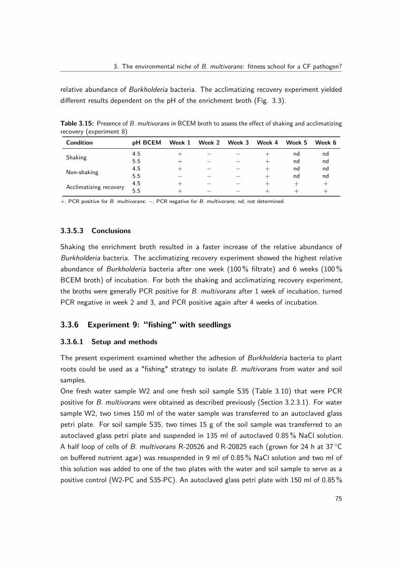
3. The environmental niche of B. multivorans: fitness school for a CF pathogen?
relative abundance of Burkholderia bacteria. The acclimatizing recovery experiment yieldeddifferent results dependent on the pH of the enrichment broth (Fig. 3.3).
Table 3.15: Presence of B. multivorans in BCEM broth to assess the effect of shaking and acclimatizingrecovery (experiment 8)Condition pH BCEM Week 1 Week 2 Week 3 Week 4 Week 5 Week 6
Shaking 4.5 + − − + nd nd5.5 + − − + nd nd
Non-shaking 4.5 + − − + nd nd5.5 − − − + nd nd
Acclimatizing recovery 4.5 + − − + + +5.5 + − − + + +
+, PCR positive for B. multivorans; −, PCR negative for B. multivorans; nd, not determined.
3.3.5.3 Conclusions
Shaking the enrichment broth resulted in a faster increase of the relative abundance ofBurkholderia bacteria. The acclimatizing recovery experiment showed the highest relativeabundance of Burkholderia bacteria after one week (100% filtrate) and 6 weeks (100%BCEM broth) of incubation. For both the shaking and acclimatizing recovery experiment,the broths were generally PCR positive for B. multivorans after 1 week of incubation, turnedPCR negative in week 2 and 3, and PCR positive again after 4 weeks of incubation.
3.3.6 Experiment 9: "fishing" with seedlings
3.3.6.1 Setup and methods
The present experiment examined whether the adhesion of Burkholderia bacteria to plantroots could be used as a "fishing" strategy to isolate B. multivorans from water and soilsamples.One fresh water sample W2 and one fresh soil sample S35 (Table 3.10) that were PCRpositive for B. multivorans were obtained as described previously (Section 3.2.3.1). For watersample W2, two times 150 ml of the water sample was transferred to an autoclaved glasspetri plate. For soil sample S35, two times 15 g of the soil sample was transferred to anautoclaved glass petri plate and suspended in 135 ml of autoclaved 0.85% NaCl solution.A half loop of cells of B. multivorans R-20526 and R-20825 each (grown for 24 h at 37 ◦Con buffered nutrient agar) was resuspended in 9 ml of 0.85% NaCl solution and two ml ofthis solution was added to one of the two plates with the water and soil sample to serve as apositive control (W2-PC and S35-PC). An autoclaved glass petri plate with 150 ml of 0.85%
75

III. Experimental work
0,0%
2,0%
4,0%
6,0%
8,0%
10,0%
12,0%
14,0%
water week 1 week 2 week 3 week 4
Burkholeria
/to
tal b
acte
ria
Shaking vs. non-shaking broth (pH 4.5)
shaking
non-shaking
(a) Shaking of the enrichment broth
0,0%
0,5%
1,0%
1,5%
2,0%
2,5%
3,0%
3,5%
4,0%
4,5%
5,0%
water week 1 week 2 week 3 week 4 week 5 week 6
Burkholeria
/to
tal b
acte
ria
Acclimatizing recovery
pH 4.5
pH 5.5
(b) Acclimatizing recovery
Figure 3.3: Relative abundance of Burkholderia bacteria in BCEM broth to assess the effect of (a)shaking and (b) acclimatizing recovery (experiment 8).
NaCl solution served as negative control (NC). Seeds of pea (Wonder of Kelvedon), maize(Tasty sweet F1 hybrid), lupine, barley and rice were surface sterilized (by soaking them for 2min in a 1% NaOCl solution, 2 min in 70% ethanol, 2 min in sterile MQ and 4 times 1 minin sterile MQ) and transferred to the glass petri plates containing either water or soil sample.After 9 days of incubation at room temperature on a windowsill, one type of seed from eachcondition was used for further analysis. The roots of the seedlings were aseptically removedand transferred into 500 µl 0.85% NaCl solution. Using a surface sterilized micropestle, theroots were crushed for 30 sec and another 500 µl 0.85% NaCl solution was added. Aliquots(50 µl) of tenfold serial dilutions (1E-0 until 1E-2) in physiological saline were plated onPCAT and BCSA agar media (Section 3.2.3.2). After 6 (W2) or 7 (S35) days of incubationat 28 ◦C for , 10-15 isolates per sample, seed and agar medium were randomly picked andsubcultivated on buffered nutrient agar (Oxoid, pH 6.8). Isolates were identified using recA
76

3. The environmental niche of B. multivorans: fitness school for a CF pathogen?
gene sequence analysis as described previously (Section 3.2.7.1).
3.3.6.2 Results
In W2 and W2-PC, seeds of pea and rice germinated, as well as one maize seed in W2-PC. InS35 and S35-PC, only pea seeds germinated. The other seeds in W2 and S35 were overgrownby fungi. In the NC sample none of the seeds germinated and all seeds were overgrownby fungi. For the pea seed in W2, no isolates were obtained from BCSA and 0/10 isolatesfrom PCAT were identified as Burkholderia. For the rice seed in W2, 0/10 isolates fromBCSA and 0/8 isolates from PCAT were identified as Burkholderia. For the pea seed in S35,0/2 isolates from BCSA and 4/23 isolates from PCAT were identified as Burkholderia. Twoisolates were identified as B. caledonica, one as B. phytofirmans and one as Burkholderiasp. The identification and number of isolates for the spiked samples W2-PC and S35-PC ispresented in Table 3.16.
Table 3.16: Identification and number of isolates from seedlings germinated in spiked samples W2-PCand S35-PC (experiment 9)
W2-PC S35-PC
Pea Rice Maize Pea
PCAT BCSA PCAT BCSA PCAT BCSA PCAT BCSA
B. multivorans PC 5 3 5 4 1B. phytofirmans 1Total isolates 9 4 8 2 20 9 10 5
PC, B. multivorans positive control strains R-20526 and R-20825.
3.3.6.3 Conclusions
B. multivorans was retrieved from the spiked PC samples with pea, rice and maize from W2,and with pea for S35. More isolates of the PC strains were retrieved from PCAT than fromBCSA. "Fishing" with seedlings did however not yield B. multivorans from the unspiked waterand soil samples.
3.3.7 Experiment 11 and 12: effect of resuscitation at room temperatureand comparison of PCAT to 1/10 PCAT medium
3.3.7.1 Setup and methods
The present experiment examined the effect of a reduced nutrient concentration of the PCATisolation medium and the resuscitation of environmental bacteria at room temperature.
77

III. Experimental work
One fresh soil sample S35 (Table 3.10) that was PCR positive for B. multivorans washomogenized as described previously (Section 3.2.3.3, experiment B). Aliquots (50 µl) oftenfold serial dilutions of the soil suspension in physiological saline (1E-0 until 1E-5) wereplated on PCAT and 1/10 PCAT (Table 3.2.3.2). The remainder of the soil sample was leftat room temperature for two more weeks (with loosened screw cap to allow oxygen flow)and the plating procedure was repeated after 1 and 2 weeks. Colonies were picked from bothmedia from the last plating after 16 days of incubation at 28 ◦C. One fresh water sampleW2 (Table 3.10) that was PCR positive for B. multivorans was left for 16 days at roomtemperature (with loosened screw cap to allow oxygen flow) prior to direct plating (50 µl) onPCAT and 1/10 PCAT. After 5 days of incubation at 28 ◦C growth on the agar plates wasevaluated.
3.3.7.2 Results
For sample S35, growth was observed until dilutions of 1E-4 or 1E-3 onto 1/10 PCAT medium,whereas PCAT medium only yielded growth up until 1E-2 (Table 3.17). A larger diversity ofcolony morphologies was observed on 1/10 PCAT medium after incubating the soil samplefor 1 or 2 weeks at 28 ◦C. Of the 35 isolates from 1/10 PCAT medium, 23 were identified asBurkholderia, but none of them was B. multivorans. Of the 23 isolates from PCAT medium,19 were identified as Burkholderia, but none of them was B. multivorans.Sample W2, which was left for 16 days at room temperature prior to plating, yielded nocolonies on PCAT but 25-35 colonies on 1/10 PCAT. None of the 32 colonies that werepicked from 1/10 PCAT medium was identified as Burkholderia.
Table 3.17: Growth of sample S35 on PCAT and 1/10 PCAT after 0, 1 and 2 weeks of incubation atroom temperature (experiment 11)
Week 0 Week 1 Week 2
Dilution PCAT 1/10 PCAT PCAT 1/10 PCAT PCAT 1/10 PCAT
1E-0 + + + + + +1E-1 + + + + + +1E-2 + + + + + +1E-3 NG + NG + NG +1E-4 NG + NG + NG NG1E-5 NG NG NG NG NG NG
NG, no growth observed.
78

3. The environmental niche of B. multivorans: fitness school for a CF pathogen?
3.3.7.3 Conclusions
The nutrient diluted (1/10) formulation of PCAT yielded more growth for both water and soilsamples, and generally yielded a larger number of distinct colony morphologies. Incubatingthe water and soil samples for up to two weeks at room temperature prior to plating did notyield B. multivorans isolates.
3.3.8 Experiment 13: "fishing" with amoebae
3.3.8.1 Setup and methods
The present experiment examined the effect of co-cultivation with amoebae to resuscitate B.multivorans in environmental samples (Steinert et al., 1997; Marolda et al., 1999; La Scolaet al., 2001; Lamothe et al., 2004; Pagnier et al., 2008). For fresh water samples W2 andW152 (Table 3.10), 150 ml aliquots were filtered as described previously (Section 3.2.3.3,experiment A) and two filters were shaken for 2 h at 28 ◦C in 25 ml PAS medium (Chavatteet al., 2014). One ml of the concentrated water sample in PAS medium was added to asingle well of a 12-well plate with a monolayer of Acanthamoeba castellanii in duplicate.PAS medium was used as negative control (NC). After 1 (D1), 3 (D3) and 7 (D7) daysof incubation at 25 ◦C aliquots (100 µl for D1 and 300 µl for D3 and D7) of tenfold serialdilutions were plated on 1/10 PCAT and BCSA agar media (Section 3.2.3.2). After 6 days ofincubation at 28 ◦C colonies were randomly picked and subcultivated on buffered nutrient agar(Oxoid, pH 6.8). Dereplication and identification of the isolates was performed as describedpreviously (Section 3.2.3.4).
3.3.8.2 Results
MALDI-TOF MS dereplication of 176 isolates resulted in a total of 26 clusters, of whichnone were identified as Burkholderia, 16 clusters (146 isolates) were identified as other GNNFbacteria and 3 clusters (4 isolates) as Mycobacterium species (Table 3.18). The remaining 7clusters (26 isolates) were identified as Gram-positive or Gram-negative fermenting bacteria(data not shown).
3.3.8.3 Conclusions
Co-cultivation of water samples with amoebae (A. castellanii) prior to plating did not yield B.multivorans isolates.
79
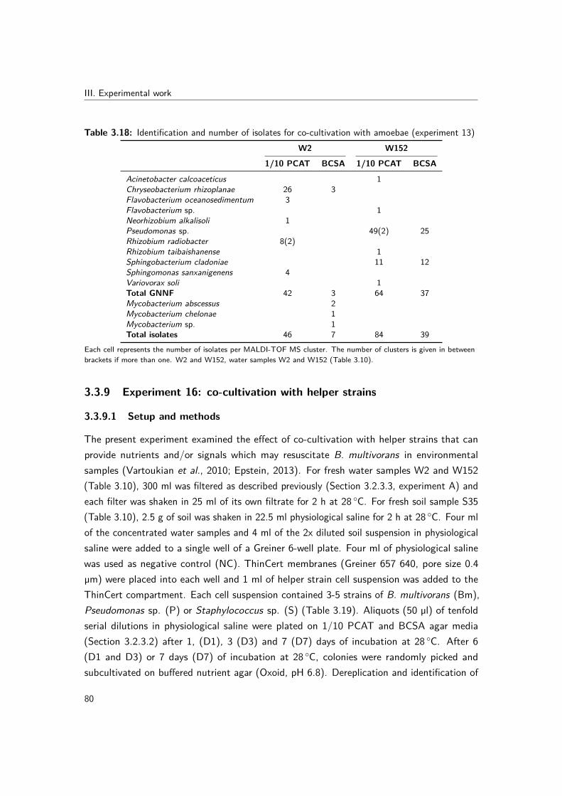
III. Experimental work
Table 3.18: Identification and number of isolates for co-cultivation with amoebae (experiment 13)W2 W152
1/10 PCAT BCSA 1/10 PCAT BCSA
Acinetobacter calcoaceticus 1Chryseobacterium rhizoplanae 26 3Flavobacterium oceanosedimentum 3Flavobacterium sp. 1Neorhizobium alkalisoli 1Pseudomonas sp. 49(2) 25Rhizobium radiobacter 8(2)Rhizobium taibaishanense 1Sphingobacterium cladoniae 11 12Sphingomonas sanxanigenens 4Variovorax soli 1Total GNNF 42 3 64 37Mycobacterium abscessus 2Mycobacterium chelonae 1Mycobacterium sp. 1Total isolates 46 7 84 39
Each cell represents the number of isolates per MALDI-TOF MS cluster. The number of clusters is given in betweenbrackets if more than one. W2 and W152, water samples W2 and W152 (Table 3.10).
3.3.9 Experiment 16: co-cultivation with helper strains
3.3.9.1 Setup and methods
The present experiment examined the effect of co-cultivation with helper strains that canprovide nutrients and/or signals which may resuscitate B. multivorans in environmentalsamples (Vartoukian et al., 2010; Epstein, 2013). For fresh water samples W2 and W152(Table 3.10), 300 ml was filtered as described previously (Section 3.2.3.3, experiment A) andeach filter was shaken in 25 ml of its own filtrate for 2 h at 28 ◦C. For fresh soil sample S35(Table 3.10), 2.5 g of soil was shaken in 22.5 ml physiological saline for 2 h at 28 ◦C. Four mlof the concentrated water samples and 4 ml of the 2x diluted soil suspension in physiologicalsaline were added to a single well of a Greiner 6-well plate. Four ml of physiological salinewas used as negative control (NC). ThinCert membranes (Greiner 657 640, pore size 0.4µm) were placed into each well and 1 ml of helper strain cell suspension was added to theThinCert compartment. Each cell suspension contained 3-5 strains of B. multivorans (Bm),Pseudomonas sp. (P) or Staphylococcus sp. (S) (Table 3.19). Aliquots (50 µl) of tenfoldserial dilutions in physiological saline were plated on 1/10 PCAT and BCSA agar media(Section 3.2.3.2) after 1, (D1), 3 (D3) and 7 (D7) days of incubation at 28 ◦C. After 6(D1 and D3) or 7 days (D7) of incubation at 28 ◦C, colonies were randomly picked andsubcultivated on buffered nutrient agar (Oxoid, pH 6.8). Dereplication and identification of
80

3. The environmental niche of B. multivorans: fitness school for a CF pathogen?
the isolates was performed as described previously (Section 3.2.3.4).
Table 3.19: Helper strain panelsStrain panel Strain Source (ST)
Bm
R-20526 ENV (ST-836)R-20825 ENV (ST-650)R-53445 CF (ST-25)R-53446 CF (ST-741)R-53627 CF (ST-16)
PR-31520 CFR-52232 CFR-52542 CFR-53162 CF
SR-19214 CFR-19219 CFR-49429 t1 CF
Bm, B. multivorans; P, Pseudomonas sp.; S, Staphylococcus sp.; CF, cystic fibrosis; ENV, environmental; ST,multilocus sequence type.
3.3.9.2 Results
For sample W2, MALDI-TOF MS dereplication of 279 isolates resulted in a total of 46 clusters,of which 35 clusters (259 isolates) were identified as GNNF bacteria and two clusters (3isolates) as M. chelonae (Table 3.20). The remaining 9 clusters (17 isolates) were identifiedas Gram-positive or Gram-negative fermenting bacteria (data not shown).For sample W152, MALDI-TOF MS dereplication of 236 isolates resulted in a total of 47clusters, of which 34 clusters (116 isolates) were identified as GNNF bacteria (Table 3.21).The remaining 13 clusters (120 isolates) were identified as Gram-positive or Gram-negativefermenting bacteria (data not shown).For sample S35, MALDI-TOF MS dereplication of 314 isolates resulted in a total of 30clusters, of which 24 clusters (291 isolates) were identified as GNNF bacteria (Table 3.22).The remaining 6 clusters (23 isolates) were identified as Gram-positive or Gram-negativefermenting bacteria (data not shown).For the NC, growth was observed at all time points on both the 1/10 PCAT and BCSA agarmedia, with all colonies showing the same colony morphology. Several colonies were pickedat each time point and from each agar medium (17 isolates in total) and all of them wereidentified as B. multivorans. MALDI-TOF MS analysis suggested that all B. multivoransisolates that were obtained in this experiment (from the wells with NC, W2, W152 and S35sample) belonged to the Bm strain panel (Table 3.19) and were thus contaminants. Thesefindings were confirmed by RAPD typing and gyrB sequence analysis (data not shown).
81

III. Experimental work
Table 3.20: Identification and number of isolates for sample W2 (experiment 16)Bm P S
1/10 PCAT BCSA 1/10 PCAT BCSA 1/10 PCAT BCSA
Aminobacter sp. 1 42 9 6Bosea lupini 1Bosea vestrisii 2Chromobacterium aquaticum 4 1 5 21Chryseobacterium sp. 2 1 3Novosphingobium resinovorum 1 1Pandoraea pnomenusa 1 3Pseudomonas extremaustralis 1Pseudomonas nitroreducens 28 6 6Pseudomonas soli 1 1 1Pseudomonas taiwanensis 2 4Rhizobium radiobacter 2 1Sphingobium rhizovicinum 29 22 20Stenotrophomonas maltophilia 3Bm contaminants 15Total GNNF 60 63 39 18 33 33Mycobacterium chelonae 1 2Total isolates 62 63 43 20 33 35
Each cell represents the number of isolates per MALDI-TOF MS cluster. The number of clusters is given in betweenbrackets if more than one. Bm, B. multivorans; P, Pseudomonas sp.; S, Staphylococcus sp. (Table 3.19).
3.3.9.3 Conclusions
Co-cultivation of water and soil samples with helper strains prior to plating did not yield B.multivorans isolates.
82

3. The environmental niche of B. multivorans: fitness school for a CF pathogen?
Table 3.21: Identification and number of isolates for sample W152 (experiment 16)Bm P S
1/10 PCAT BCSA 1/10 PCAT BCSA 1/10 PCAT BCSA
Brevundimonas sp. 1Chryseobacterium aquaticum 2 1 1Chryseobacterium scophthalmum 1 1Chryseobacterium sp. 1Chryseobacterium taeanense 1Cloacibacterium rupense 1Herbaspirillum frisingense 3Herbaspirillum huttiense 3 13Herbaspirillum sp. 1Novosphingobium barchaimii 1Novosphingobium resinovorum 7 5Pseudomonas protegens 4Pseudomonas sp. 1Rhizobium nepotum 1Rhizobium sp. 1Rhizobium tibeticum 1Sphingobacterium faecium 2(2)Sphingobacterium kitahiroshimense 2Sphingobacterium sp. 1 2 3Sphingobium sp. 2Stenotrophomonas maltophilia 1Stenotrophomonas rhizophila 1Variovorax guangxiensis 1Bm contaminants 20Total GNNF 9 26 11 10 7 23Total isolates 47 26 43 10 57 23
Each cell represents the number of isolates per MALDI-TOF MS cluster. The number of clusters is given in betweenbrackets if more than one. Bm, B. multivorans; P, Pseudomonas sp.; S, Staphylococcus sp. (Table 3.19).
Table 3.22: Identification and number of isolates for sample S35 (experiment 16)Bm P S
1/10 PCAT BCSA 1/10 PCAT BCSA 1/10 PCAT BCSA
Burkholderia choica 2Burkholderia jiangsuensis 2 26(5)Burkholderia phytofirmans 10 8 24(5)Burkholderia sp. 1 3(2)Pseudomonas prosekii 1Bm contaminants 43 34 45 32 6 8Total GNNF 53 34 56 32 62 8Total isolates 74 34 56 32 64 8
Each cell represents the number of isolates per MALDI-TOF MS cluster. The number of clusters is given in betweenbrackets if more than one. Bm, B. multivorans; P, Pseudomonas sp.; S, Staphylococcus sp. (Table 3.19).
83

3. The environmental niche of B. multivorans: fitness school for a CF pathogen?
3.4 Multilocus sequence types predict phylogeny and genecontent but not isolation source for Burkholderiamultivorans
Redrafted from: Charlotte Peeters, Vaughn S. Cooper, Philip Hatcher, Bart Verheyde andPeter Vandamme. Multilocus sequence types predict phylogeny and gene content but notisolation source for Burkholderia multivorans. In preparation.
Author contributions: CP, VC and PV conceived the study and wrote the manuscript. CPperformed the DNA preparations. VC directed the genomic sequencing methods. VC andPH identified the orthologous genes. CP and VC performed the genomic data analysis andstatistical analysis. CP and BV performed the whole-genome sequence-based phylogeneticanalysis.
3.4.1 Abstract
The natural environment may serve as a reservoir of opportunistic pathogens. A well-established method for studying the epidemiology of such opportunists is multilocus sequencetyping, which in many cases has defined strains predisposed to causing infection. Burkholderiamultivorans is an important pathogen in people with CF and its epidemiology suggeststhat strains are acquired from non-human sources such as the natural environment. Thisraises the central question of whether the isolation source (CF or environment) or the ST ofB. multivorans better predicts their genomic content and functionality. We identified fourpairs of B. multivorans isolates, representing distinct STs and consisting of one CF and oneenvironmental isolate. All genomes were sequenced using the PacBio SMRT sequencingtechnology, which resulted in eight high-quality B. multivorans genome assemblies. Thepresent study demonstrated that the genomic structure of B. multivorans is highly conservedand that the B. multivorans genomic lineages are defined by their ST. Orthologous proteinfamilies were not uniformly distributed among chromosomes, with core orthologs beingenriched on the primary chromosome and ST-specific orthologs being enriched on the secondand third chromosome. The ST-specific orthologs were enriched in genes involved in defensemechanisms and secondary metabolism, corroborating the strain-specificity of these virulencecharacteristics. Finally, the same B. multivorans genomic lineages were isolated from CFand environmental samples and on different continents many years apart, demonstrating theevolutionary persistence and ubiquity of these strains in different niches and on differentcontinents.
85

III. Experimental work
3.4.2 Introduction
Bcc bacteria are rare but potentially virulent pathogens in CF patients (LiPuma, 2010).Epidemiological surveys revealed that B. multivorans is the most prevalent Bcc CF pathogenin many countries (Brisse et al., 2004; De Boeck et al., 2004; Govan et al., 2007; LiPuma,2010; Norskov-Lauritsen et al., 2010; Pope et al., 2010; Medina-Pascual et al., 2012). Thecontinued emergence of unique B. multivorans strains in CF patients suggests acquisition fromnon-human sources, such as the natural environment (Baldwin et al., 2008). Environmentalconditions or non-human hosts in which virulence factors might be adaptive can select fortraits that confer virulence and natural environments could therefore serve as reservoirs ofopportunistic pathogens (Coenye & Vandamme, 2003; Berg et al., 2005).MLST is a well-established method for studying the epidemiology and population structure ofBcc organisms (Baldwin et al., 2005; Spilker et al., 2009). The Bcc MLST scheme takes intoaccount the allelic variation of seven housekeeping genes and each strain is defined by itsunique allelic profile and ST (Jolley & Maiden, 2010). Baldwin et al. (2007) demonstratedthat roughly one fifth of the clinical isolates in the Bcc PubMLST database had the same STas environmental isolates, suggesting these isolates represent the same strain. A follow-upstudy demonstrated that several B. multivorans STs were globally distributed and that thenatural environment (e.g. water and soil) may be an important reservoir for infection withthis species (Baldwin et al., 2008).Burkholderia genomes vary in size from 2.4 Mb (Ca. Burkholderia schumannianae UZHbot8)(Pinto-Carbo et al., 2016) to 11.5 Mb (Burkholderia terrae BS001) (Nazir et al., 2012a),are characterized by a high G+C content (62-68 mol%) and consist of multiple replicons(Mahenthiralingam et al., 2005; Ussery et al., 2009). To gain insight into the overall genomebiology of B. multivorans, we sequenced the genomes of eight isolates representing fourdistinct STs. For each ST, a CF and an environmental isolate were sequenced using the PacBioSingle-Molecule Real-Time (SMRT) sequencing technology. The present study provides thefirst comprehensive comparative genome analysis of B. multivorans and assesses to whichextent isolates with the same ST but from different origin (CF versus environmental) differ ingenetic potential.
3.4.3 Materials and methods
3.4.3.1 Studied isolates
We searched the Bcc PubMLST database (http://pubmlst.org/bcc/) (Jolley & Maiden, 2010)for B. multivorans STs that included both CF and environmental (ENV) isolates, and selected
86

3. The environmental niche of B. multivorans: fitness school for a CF pathogen?
eight isolates, representing four STs, for whole-genome sequencing (Table 3.23). Strains weregrown aerobically on Tryptone Soya Agar (Oxoid) and incubated at 28 ◦C. Cultures werepreserved in MicroBankTM vials at −80 ◦C.
Table 3.23: B. multivorans isolates included in the present studyIsolate Strain number ST Isolation source Depositor
ST180-ENV LMG 29305, J2943 180 Rhizosphere soil (United King-dom, 2000)
J. Govan
ST180-CF LMG 29313, 8335 180 CF sputum (Czech republic,2011)
P. Drevinek
ST189-ENV LMG 29309 189 Soil succulent (Belgium, 2003) Own isolateST189-CF LMG 29312, BCC0208 189 CF patient (Canada, 1999) E. MahenthiralingamST287-ENV LMG 29306, J2947 287 Rhizosphere soil (United King-
dom, 2000)J. Govan
ST287-CF LMG 29311, BCC0059 287 CF patient (Canada, 1995) E. MahenthiralingamST650-ENV LMG 29308 650 Pond water (Belgium, 2003) Own isolateST650-CF LMG 29310, Q113 650 CF patient (Germany, 2010) B. Kahl
LMG, BCCM/LMG Bacteria Collection, Laboratory of Microbiology, Ghent University, Ghent, Belgium. CF, cysticfibrosis; ENV, environmental; ST, multilocus sequence type.
3.4.3.2 Genome sequencing, assembly and annotation
High-quality DNA was prepared using Qiagen Genomic tips (20/G) and genomes weresequenced using the P5-C3 chemistry on the PacBio SMRT II platform of the Departmentof Genetics and Genomic Sciences of the Icahn School of Medicine at Mount Sinai (NewYork, USA). One SMRT cell per isolate was sequenced, except for isolates ST189-CF andST287-ENV for which a second SMRT cell was run to increase the quality of the raw data.PacBio reads were assembled in the sequencing center using the SMRT analysis software(including HGAP3 and Quiver) and contigs were ordered against the complete referencegenome of B. multivorans strain ATCC 17616 (PRJNA17407) using Mauve (Rissman et al.,2009). We further polished the assemblies in five steps. The first step consisted of removingspurious contigs that were small in size, had a low coverage and resulted in a highest BLASThit with the primary chromosome of its own genome (Liao et al., 2015). Reads were mappedusing pbalign and QC reports were created based on the resulting BAM files using Qualimap(Garcia-Alcalde et al., 2012). Contigs smaller than 20 kb and with less than 20x coverage ora high variation (SD) in coverage were discarded. In step two, read mappings were used tofurther polish the contigs using Pilon (Walker et al., 2014b) with default parameters. In stepthree, contigs with overlapping ends were merged using Gap5 (Bonfield & Whitwham, 2010)to exclude artificially duplicated regions, often including many frameshifts and fragmentedopen reading frames. In step four, the duplicated ends of circular contigs were trimmed using
87

III. Experimental work
Gepard (Krumsiek et al., 2007) and Gap5 as these duplications were a consequence of thecircular nature of the replicons in combination with the long-read sequencing technology(Scott & Ely, 2015). Importantly, this artificial duplication of contig ends not only resultedin a highly variable rRNA copy number, but also falsely excluded genes from the orthologdataset because they were artificially duplicated. Since the merging of overlapping ends byGap5 might be imperfect, we ran Quiver in a final polishing step. The PacBio sequencingreads of one SMRT cell resulted in a coverage ranging from 76x to 119x.Annotation was performed using Prokka v1.11 (Seemann, 2014) with a genus-specificdatabase based on reference genomes from the Burkholderia Genome database (http://beta.burkholderia.com/) (Winsor et al., 2008). The annotated genome assemblies weresubmitted to the European Nucleotide Archive and are publicly available through the Gen-Bank/EMBL/DDBJ accession numbers FKJT01000000, FKJS01000000, FKJU01000000,FKJP01000000, FKJV01000000, FKJW01000000, FKJX01000000 and FKJY01000000. Thegenome sequence of B. multivorans strain ATCC 17616 (PRJNA17407) was included as areference in all further analyses. A multiple genome alignment was performed using Mauve(Darling et al., 2010) to assess the basic genome structure.
3.4.3.3 Analysis of protein-coding genes and ortholog identification
We mapped for each protein-coding gene (CDS) on which chromosome it was located andto which cluster of orthologous groups (COG) it belonged (Galperin et al., 2015). COGswere assigned by a reversed position-specific BLAST (RPSBLAST v2.2.29+) with an e-valuecut-off of 1E-3 against the NCBI conserved domain database (CDD v3.14). Orthologousgenes were identified as described previously (Lerat et al., 2003; Cooper et al., 2010). Inshort, homologs were identified as reciprocal best BLAST hits with a normalized bit score,providing an empirically determined taxon-specific threshold. The putative panorthologs (i.e.single-copy orthologous genes conserved in all genomes) were computed while varying the bitscore threshold from 0.1 to 0.9 in 0.1 increments and the largest set of panorthologs wasselected. For each orthologous protein family, the consensus chromosome location and COGwere determined. Conflicts in COG mapping were resolved by the majority rule.
3.4.3.4 Phylogenomic analysis
The whole-genome phylogeny was calculated based on the sequences of the panorthologs asdescribed previously (Pinto-Carbo et al., 2016). In short, amino acid sequences were alignedusing MUSCLE (Edgar, 2004) and translated back to the respective nucleotide sequencesusing T-Coffee (Notredame et al., 2000). Nucleotide alignments were trimmed using trimAl
88

3. The environmental niche of B. multivorans: fitness school for a CF pathogen?
(Capella-Gutierrez et al., 2009) by removing positions with gaps in more than 50% of thesequences, and were subsequently concatenated to construct a maximum likelihood tree usingRaXML v7.4.2 (Stamatakis, 2014) with the GTRGAMMA substitution model and 1000 rapidbootstrap analyses.In a second approach, the presence/absence matrix of all orthologs was used in a discretecharacter-state parsimony analysis using pars from the PHYLIP package (Felsenstein, 2005)to assess the relatedness of the genomes in terms of gene content.
3.4.3.5 Comparison of B. multivorans and B. cenocepacia COG profiles
Complete genome sequences of B. cenocepacia strains J2315, H111, K56-2Valvano, AU1054,HI2424 and MC0-3 were downloaded from the Burkholderia Genome database (http://beta.burkholderia.com/) (Winsor et al., 2008). COG mapping of B. cenocepacia CDS wasperformed as described above for B. multivorans. The number of CDS per COG category foreach species (B. multivorans versus B. cenocepacia) was counted and the distributions werecompared using Pearson chi-square analysis.
3.4.3.6 Data visualization and statistical analyses
Data visualization and statistical analyses were performed using RStudio with R v3.2.3.Pearson’s chi-square analyses were used to test the association between different sets ofcategorical variables. When a significant relationship was found between two variables, wefurther examined the standardized Pearson residuals. Standardized Pearson residuals havinghigh absolute values indicate a lack of fit of the null hypothesis of independence in eachcell (Agresti, 2002) and thus indicate observed cell frequencies in the contingency tablethat are significantly higher or lower than one would expect based on coincidence. In casemultiple COG categories were registered for the same COG, each COG category was countedseparately for Pearson chi-square analysis on COG categories. For the 198 CDS that wereinvolved in the translocation within the ST650-CF isolate from the primary to the secondarychromosome, the consensus chromosome mapping was set to the primary chromosome forPearson chi-square analysis on chromosome distribution.
3.4.4 Results
3.4.4.1 The genomic structure of B. multivorans is highly conserved
The final assemblies produced closed genomes for five of the eight B. multivorans isolatessequenced in the present study. The genomes were 6.2-6.9 Mb in size with a G+C content of
89

III. Experimental work
~67 mol% and the number of predicted CDS ranged from 5,415 to 6,155 CDS per genome(Table 3.24). No clustered regularly interspaced short palindromic repeats (CRISPRs) wereidentified.The multiple genome alignment revealed a highly conserved genomic structure with threechromosomes (from here on referred to as C1, C2 and C3). C1, C2 and C3 were on average3.4 Mb, 2.4 Mb and 0.6 Mb in size. Both ST180 isolates harbored one contig that did notmap onto the reference genome of ATCC 17616. These contigs were 22,339 and 28,809 bp insize, had a G+C content of 58 mol% and were therefore considered plasmids. Both plasmidscontained genes for an initiator repB protein, an AsnC transcriptional regulator, a cobyrinicacid a,c-diamide synthase (parA homologue), multiple integrases and several hypotheticalproteins. The multiple genome alignment also revealed a fairly large translocation (207 genes,198 CDS) within the ST650-CF isolate from C1 to C2 that was delimited by rRNA operonsat both ends. All isolates except ST650-CF contained 3, 1 and 1 rRNA copies on C1, C2and C3, respectively. As a result of the translocation, isolate ST650-CF contained 2, 2 and 1rRNA copies on C1, C2 and C3, respectively.
Table 3.24: B. multivorans genome characteristicsIsolate Contigs Size (bp) %GC C1 C2 C3 Plasmid Total
CDSOrtho-logousCDS
Non-ortho-logousCDS
ST180-ENV 4 6,464,081 67.1 1 2 3 4 5,794 5,271 523ST180-CF 4 6,296,736 67.3 1 2 3 4 5,551 5,266 285ST189-ENV 3 6,223,431 67.3 1 2 3 - 5,467 5,144 323ST189-CF 13 6,157,395 67.3 1-5 6-9 10-13 - 5,415 5,132 283ST287-ENV 4 6,559,547 67.2 1-2 3 4 - 5,800 5,494 306ST287-CF 6 6,857,684 67.0 1-4 5 6 - 6,155 5,505 650ST650-ENV 3 6,322,929 67.2 1 2 3 - 5,594 5,275 319ST650-CF 3 6,308,820 67.2 1 2 3 - 5,599 5,250 349ATCC 17616 4 7,008,622 66.7 1 2 3 4 6,258 4,893 1,365
C1, C2 and C3: contigs mapping to chromosome 1, 2 and 3 of B. multivorans strain ATCC 17616, respectively; bp,base pairs.
3.4.4.2 ST predicts genomic lineage
Orthologous genes were identified to determine the conserved genome content of B.multivorans. The ortholog analysis identified 6,254 homologous protein families comprising47,230 CDS in total (Table 3.24). The largest set of panorthologs, i.e. orthologs conserved inall nine B. multivorans genomes and present as single copies, was found at a reciprocal bestbit score threshold of 0.7 (see Methods section) and comprised 4,503 ortholog families.
90

3. The environmental niche of B. multivorans: fitness school for a CF pathogen?
The frequency of orthologous versus non-orthologous CDS varied significantly per isolate(χ2(8)=1829.6, p<0.001) and ST (χ2(3)=67.3, p<0.001), but not isolation source (p>0.05).The genomes of isolates ST287-CF and ATCC 17616 were significantly enriched with non-orthologous CDS, while those of ST180-CF, ST287-ENV, ST650-ENV and both ST189isolates were significantly deprived in non-orthologous CDS (Table 3.24) (SupplementaryTable 3.3). Analysis of the relationship between orthologous versus non-orthologous CDSand ST showed that the ST287 genomes were significantly enriched with non-orthologousCDS, while the ST189 and ST650 genomes were significantly deprived in non-orthologousCDS (Supplementary Table 3.4).The ortholog dataset enabled two subsequent analyses of strain phylogeny. In the firstapproach, a whole-genome phylogeny was obtained based on nucleotide sequence divergenceof the panorthologs (Fig. 3.4). In the second approach, the presence/absence matrix of theortholog families was used to assess the relatedness of the genomes in terms of gene contentusing parsimony (Supplementary Fig. 3.2). These analyses both demonstrated that the ST,and not the isolation source, of the B. multivorans isolates predicted their phylogeny andgene content. This finding demonstrated that isolates with the same ST represent the samegenomic lineage, irrespective of their isolation source.
ST650-CF
ST287-CF
ST189-CF
ATCC 17616
ST180-CF
ST189-ENV
ST287-ENV
ST180-ENV
ST650-ENV
100
100
100
100
100
Tree scale: 0.001
Figure 3.4: Phylogenomic analysis showing the relatedness of the genomes in terms of sequencedivergence of the panorthologs. The maximum likelihood tree was inferred using the GTRGAMMAsubstitution model and is based on a concatenated nucleotide alignment of 4,503 CDS (4,457,847positions). The percentage of replicate trees in which the associated taxa clustered together in thebootstrap analyses (1,000 replicates) are shown next to the branches. Scale bar represents number ofsubstitutions per site. The tree was rooted on the branch with the largest branch length.
3.4.4.3 Orthologous genes are enriched on C2 and are involved in carbohydratemetabolism and transport
Because the fraction of genes that are involved in housekeeping functions varies among thechromosomes (Mahenthiralingam et al., 2005), we mapped the chromosome location ofeach CDS. Consistent with the average chromosome size, the total number of CDS was
91
III. Experimental work
highest on C1 (27,813 CDS), followed by C2 (18,565 CDS) and C3 (5,047 CDS). The plasmidof the ST180 isolates harbored 208 CDS, of which 206 were non-orthologous CDS. Thetranslocation within the ST650-CF isolate from C1 to C2 comprised 198 CDS, of which 13were non-orthologous CDS. The frequency of orthologous versus non-orthologous CDS variedsignificantly among the different chromosomes (χ2(2)=213.4, p<0.001) (Fig. 3.5a). C1 wassignificantly enriched with non-orthologous CDS, while C2 was significantly enriched withorthologous CDS (Fig. 3.5b).To identify biological functions that were over- or underrepresented, we assigned each CDSto a COG. Roughly 80% of the CDS (41,520 CDS in total) could be assigned to a COG andits associated COG functional category. The frequency of orthologous versus non-orthologousCDS varied significantly among the different COG categories (χ2(22)=5101.2, p<0.001)(Fig. 3.5c). The non-orthologous CDS were significantly enriched in the COG categoriescell wall/membrane/envelope biogenesis (M), intracellular trafficking, secretion and vesiculartransport (U), amino acid transport and metabolism (E) and mobilome (X), while theorthologous CDS were significantly enriched in the COG categories carbohydrate transportand metabolism (G) and general function prediction only (R) (Table 3.25).
3.4.4.4 Each ST harbors unique orthologs
For each ortholog family we examined whether it was present in all eight isolates (i.e. showedcore specificity), specific for isolates of one or more STs, specific for isolates of a specificsource or randomly present. None of the ortholog families was present in all four isolates of aspecific source (CF vs. ENV), but a small number of ortholog families were present in onlyone, two or three isolates from the same source, thus leaving five relevant specificity groups:core (n=4,684), ST (n=1,362), CF-only (n=38), ENV-only (n=51) and random (n=119).The Venn diagram (Fig. 3.6) visualizes the number of ortholog families in the core and STspecificity groups (n=6,046) and shows that each ST harbors 103-539 orthologs that werenot present in any other ST.
3.4.4.5 ST-specific orthologs are enriched on C2 and C3 and are involved in defensemechanisms and secondary metabolism
Based on the chromosome and COG mapping for the individual CDS, we mapped theconsensus chromosome location and COG category for each ortholog family. Consistent withthe average chromosome size, the number of orthologs was highest on C1 (3,242), followedby C2 (2,264) and C3 (710). For 37 ortholog families there was a conflict in chromosomemapping, and 1 ortholog was located on the plasmid of the ST180 isolates. COGs and
92

3. The environmental niche of B. multivorans: fitness school for a CF pathogen?
0
5000
10000
15000
20000
25000
C1 C2 C3Chromosome
Cou
nt C
DS
Non−orthologous CDS
Orthologous CDS
(a)
Sta
ndar
dize
dR
esid
uals
:<
−4
−4:
−2
−2:
00:
22:
4>
4
C1 C2 C3
Non−orthologous CDS
Orthologous CDS
(b)
0
1000
2000
3000
4000
J K L B D V T M N W U O C G E F H I P Q X R SCOG category
Cou
nt C
DS
Non−orthologous CDS
Orthologous CDS
(c)
Figure 3.5: The frequency of orthologous versus non-orthologous CDS varies among chromosomesand COG categories. Bar plots show the number of orthologous and non-orthologous CDS perchromosome (χ2(2)=213.4, p<0.001) (a) and COG category (χ2(22)=5101.2, p<0.001) (c). Mosaicplots show the standardized residuals of the Pearson chi-square analysis for the number of orthologousand non-orthologous CDS per chromosome (b). Solid and dashed boundaries represent positive andnegative residuals, respectively. Rectangles are colored only if the standardized residual is significantat p<0.05 (outside ±1.96). COG categories: J, translation, ribosomal structure and biogenesis; K,transcription; L, replication, recombination and repair; B, chromatin structure and dynamics; D, cellcycle control, cell division, chromosome partitioning; V, defense mechanisms; T, signal transductionmechanisms; M, cell wall/membrane/envelope biogenesis; N, cell motility; W, extracellular structures;U, intracellular trafficking, secretion, and vesicular transport; O, posttranslational modification, proteinturnover, chaperones; X, mobilome: prophages, transposons; C, energy production and conversion;G, carbohydrate transport and metabolism; E, amino acid transport and metabolism; F, nucleotidetransport and metabolism; H, coenzyme transport and metabolism; I, lipid transport and metabolism;P, inorganic ion transport and metabolism; Q, secondary metabolites biosynthesis, transport andcatabolism; R, general function prediction only; S, function unknown.
93

III. Experimental work
Table 3.25: The frequency of orthologous versus non-orthologous CDS varies among the COGcategories
Orthologous CDS Non-orthologous CDS
Information storage and processingJ Translation, ribosomal structure and biogenesis 1814 (0.789) − 65 (-3.359)K Transcription 4122 (0.766) − 176 (-3.258)L Replication, recombination and repair 977 (-0.032) 55 (0.137)B Chromatin structure and dynamics 24 (0.263) 0 (-1.121)Cellular processes and signalingD Cell cycle control, cell division, chromosome partitioning 319 (0.848) − 2 (-3.610)V Defense mechanisms 765 (1.199) − 8 (-5.101)T Signal transduction mechanisms 2108 (0.988) − 69 (-4.207)M Cell wall/membrane/envelope biogenesis 2441 (-1.161) + 196 (4.941)N Cell motility 898 (1.510) − 3 (-6.429)W Extracellular structures 225 (0.806) − 0 (-3.431)U Intracellular trafficking, secretion, and vesicular transport 838 (-1.241) + 85 (5.283)O Posttranslational modification, protein turnover, chaperones 1367 (0.711) − 48 (-3.025)MetabolismC Energy production and conversion 2641 (0.329) 128 (-1.401)G Carbohydrate transport and metabolism + 2710 (2.016) − 41 (-8.579)E Amino acid transport and metabolism − 3880 (-3.142) + 426 (13.373)F Nucleotide transport and metabolism 845 (0.916) − 19 (-3.897)H Coenzyme transport and metabolism 2058 (1.521) − 42 (-6.475)I Lipid transport and metabolism 2127 (0.091) 113 (-0.387)P Inorganic ion transport and metabolism 2148 (0.567) − 91 (-2.415)Q Secondary metabolites biosynthesis, transport and catabolism 1442 (0.543) − 58 (-2.311)Poorly characterizedR General function prediction only + 3954 (2.026) − 86 (-8.623)S Function unknown 1851 (0.358) 86 (-1.524)Mobile elementsX Mobilome: prophages, transposons − 207 (-15.300) + 398 (65.117)
Pearson chi-square analysis testing the independence of gene conservation (orthologous vs. non-orthologous CDS)and COG category (χ2(22)=5101.2, p<0.001). Each cell in the contingency represents the observed frequency andstandardized residual (in between brackets) and is preceded by + or − if the standardized residual is >1.96 or <-1.96,respectively, and significant at p<0.05.
their associated COG functional category could be assigned to 4,896 of the 6,254 orthologfamilies.The specificity of the ortholog families varied significantly among the chromosomes(χ2(8)=469.8, p<0.001) (Fig. 3.7a) and COG categories (χ2(88)=649.8, p<0.001) (Fig.3.7c). C2 and C3 were significantly enriched with ST-specific orthologs, while C1 wassignificantly enriched with orthologs belonging to the specificity groups core, random, CF-onlyand ENV-only (Fig. 3.7b). The ST-specific orthologs were significantly enriched in theCOG categories defense mechanisms (V), secondary metabolites biosynthesis, transport andcatabolism (Q), mobilome (X) and general function prediction only (R) (Table 3.26).
94

3. The environmental niche of B. multivorans: fitness school for a CF pathogen?
539
33
103
36
79
4684
13
43
168
21176 10
9
106
26
ST−180 ST−189
ST−287 ST−650
Figure 3.6: Venn diagram showing the number of core and ST-specific ortholog families
3.4.4.6 ST287 harbors extra orthologous and non-orthologous genes
Both ST287 genomes were considerably larger and contained a higher number of CDS (Table3.24), suggesting that this genomic lineage contains extra genes. ST287 was not only enrichedwith non-orthologous CDS (Supplementary Table 3.4) but also harbored 539 orthologs thatwere not present in the other three STs (Fig. 3.6), showing that the extra genes in ST287are both orthologous and non-orthologous CDS. Conversely, the other three STs (ST180,ST189 and ST650) shared 176 orthologs that were absent in ST287 (Fig. 3.6). We foundno significant association between the specificity profile and distribution of COG categories(p>0.05), suggesting that the 539 ST287-specific orthologs and 176 orthologs that werespecific for the other three STs (ST180, ST189 and ST650) are involved in similar biologicalprocesses.
3.4.4.7 C1 is enriched with orthologs showing CF and ENV specificity
C1 was enriched with orthologs that were present in only one, two or three isolates from thesame source (CF-only or ENV-only) (Fig. 3.7a and 3.7b). CF-only orthologs were significantlyenriched in the COG categories cell cycle control, cell division and chromosome partitioning(D), extracellular structures (W), intracellular trafficking, secretion and vesicular transport(U) and mobilome (X), while ENV-only orthologs were significantly enriched in replication,recombination and repair (L), intracellular trafficking, secretion and vesicular transport (U)and posttranslational modification, protein turnover and chaperones (O) (Fig. 3.7c and Table
95

III. Experimental work
0
1000
2000
3000
C1 C2 C3Chromosome
Cou
ntENV−only
CF−only
random
ST
core
(a)
Sta
ndar
dize
dR
esid
uals
:<
−4
−4:
−2
−2:
00:
22:
4>
4
C1 C2 C3
ENV−only
CF−only
random
ST
core
(b)
0
200
400
600
J K L B D V T M N W U O C G E F H I P Q X R SCOG category
Cou
nt C
DS
ENV−only
CF−only
random
ST
core
(c)
Figure 3.7: Ortholog specificity varies among chromosomes and COG categories. Bar plots show thenumber of orthologs per specificity group for different chromosomes (χ2(8)=469.8, p<0.001) (a) andCOG categories (χ2(88)=649.8, p<0.001) (c). Mosaic plots show the standardized residuals of thePearson chi-square analysis on the number of orthologs per specificity group per chromosome (b). Solidand dashed boundaries represent positive and negative residuals, respectively. Rectangles are coloredonly if the standardized residual is significant at p<0.05 (outside ±1.96). COG categories: J, translation,ribosomal structure and biogenesis; K, transcription; L, replication, recombination and repair; B,chromatin structure and dynamics; D, cell cycle control, cell division, chromosome partitioning; V,defense mechanisms; T, signal transduction mechanisms; M, cell wall/membrane/envelope biogenesis;N, cell motility; W, extracellular structures; U, intracellular trafficking, secretion, and vesiculartransport; O, posttranslational modification, protein turnover, chaperones; X, mobilome: prophages,transposons; C, energy production and conversion; G, carbohydrate transport and metabolism; E,amino acid transport and metabolism; F, nucleotide transport and metabolism; H, coenzyme transportand metabolism; I, lipid transport and metabolism; P, inorganic ion transport and metabolism; Q,secondary metabolites biosynthesis, transport and catabolism; R, general function prediction only; S,function unknown.
96
3. The environmental niche of B. multivorans: fitness school for a CF pathogen?
3.26).Additionally, C1 was also enriched with orthologs showing random specificity (Fig. 3.7aand 3.7b) and these orthologs with random specificity were significantly enriched in theCOG categories replication, recombination and repair (L), cell cycle control, cell division andchromosome partitioning (D), cell wall/membrane/envelope biogenesis (M) and mobilome(X) (Fig. 3.7c and Table 3.26).
3.4.4.8 Comparison of B. multivorans and B. cenocepacia average COGprofiles
During the past two decades, B. multivorans and B. cenocepacia have been the mostprevalent Bcc pathogens in CF. Historically, B. cenocepacia strains have been responsiblefor large epidemics within the CF community and are often extremely virulent (Drevinek &Mahenthiralingam, 2010). In contrast, only a limited number of B. multivorans outbreakstrains were described and B. multivorans is generally considered a less virulent Bcc pathogenas compared to B. cenocepacia (Mahenthiralingam et al., 2005). To examine the species-specific genome content of B. multivorans, we compared its average COG profile to that ofB. cenocepacia. B. cenocepacia genomes contained generally more genes (6,477-7,116 CDSper genome) (Supplementary Table 3.5) than B. multivorans genomes (5,415-6,155 CDSper genome) (Table 3.24) and more CDS per COG category (Supplementary Fig. 3.3). Wecompared the average COG profile of the two species by calculating the average number ofCDS per genome in each COG category and by comparing these distributions. The distributionof CDS among COG categories varied significantly between the two species (χ2(22)=102.9,p<0.001) (Supplementary Fig. 3.3). B. cenocepacia genomes harbor significantly moreCDS in the COG categories transcription (K), defense mechanisms (V) and general functionprediction only (R) and significantly less in translation, ribosomal structure and biogenesis(J) and replication, recombination and repair (L). Conversely, B. multivorans genomes harborsignificantly more CDS in the COG categories replication, recombination and repair (L) andless in transcription (K) (Table 3.27).Finally, we searched for COGs that were exclusively present in either B. multivorans orB. cenocepacia genomes. In total, 124 COGs were exclusively present in one or more B.multivorans genomes, but only 21 COGs were uniquely present in all nine of the B. multivoransgenomes (Table 3.28). Conversely, 204 COGs were exclusively present in one or more B.cenocepacia genomes, but only 72 COGs were uniquely present in all six of the B. cenocepaciagenomes (Supplementary Table 3.6).
97

III. Experimental work
Table3.26:Orth
olog
specificit
yvarie
samon
gtheCO
Gcategorie
s Core
STCF-on
lyEN
V-only
Rando
m
Inform
ationstorageandprocessin
gJ
Transla
tion,
ribosom
alstructureandbiog
enesis
+22
3(2.570
)−
10(-4.94
7)0(-0.86
5)1(0.044
)1(-1.30
6)K
Transcrip
tion
465(-0.18
7)10
9(0.375
)2(0.095
)5(1.688
)6(-0.86
4)L
Replica
tion,
recombinatio
nandrepair
112(-0.28
6)22
(-0.74
8)1(0.799
)+
4(4.458
)+
5(2.013
)B
Chromatin
structureanddynamics
3(0.389
)0(-0.73
3)0(-0.09
8)0(-0.11
1)0(-0.20
9)Cellularprocessesandsig
naling
DCe
llcycle
control,celldivis
ion,
chromosom
epartitio
ning
37(0.594
)−
1(-2.37
8)+
1(2.368)
0(-0.41
4)+
3(3.061
)V
Defense
mechanism
s79
(-1.87
2)+
41(4.095
)0(-0.62
3)0(-0.70
5)2(0.172
)T
Sign
altra
nsdu
ctionmechanism
s24
2(0.461
)46
(-0.919)
0(-0.96
8)0(-1.09
4)6(0.839
)M
Cellwa
ll/mem
brane/envelope
biog
enesis
266(-0.67
7)69
(0.867
)0(-1.05
2)0(-1.18
9)+
12(3.102
)N
Cellmotility
104(0.576
)16
(-1.28
6)1(0.971
)1(0.705
)1(-0.58
8)W
Extra
cellular
structures
25(-0.41
6)8(0.774
)+
1(2.709
)0(-0.37
2)0(-0.70
3)U
Intra
cellular
trafficking,
secretion,
andvesic
ular
transpo
rt90
(-1.36
2)27
(0.769
)+
7(10.23
3)+
2(2.022
)4(1.538
)O
Posttra
nslatio
nalm
odificatio
n,protein
turnover,c
haperones
162(1.028
)−
18(-2.67
8)0(-0.77
2)+
5(4.858
)2(-0.43
4)Metab
olism
CEn
ergy
prod
uctio
nandconversio
n30
2(0.463
)65
(-0.11
4)0(-1.08
3)0(-1.22
4)1(-1.87
9)G
Carboh
ydrate
transpo
rtandmetab
olism
307(0.192
)67
(-0.13
0)0(-1.10
0)0(-1.244)
6(0.206
)E
Aminoacid
transpo
rtandmetab
olism
443(0.710
)86
(-1.02
3)0(-1.30
7)0(-1.47
7)7(-0.28
0)F
Nucle
otidetra
nspo
rtandmetab
olism
104(1.717
)−
5(-3.31
3)0(-0.59
2)0(-0.66
9)1(-0.47
2)H
Coenzymetra
nspo
rtandmetab
olism
237(0.560
)46
(-0.73
1)0(-0.95
5)0(-1.07
9)3(-0.56
5)I
Lipidtra
nspo
rtandmetab
olism
240(-0.78
7)71
(1.913
)0(-1.00
3)1(-0.25
3)4(-0.27
4)P
Inorganiciontra
nspo
rtandmetab
olism
256(1.873
)−
28(-3.22
7)0(-0.95
3)1(-0.14
9)−
0(-2.03
4)Q
Second
arymetab
olite
sbiosynthesis,
transpo
rtandcatabo
lism
157(-0.95
3)+
55(2.762
)0(-0.82
2)0(-0.92
9)0(-1.75
4)Po
orly
characteriz
edR
Generalfun
ctionpredict
ionon
ly43
7(-1.33
7)+
141(3.577
)0(-1.36
3)0(-1.54
1)5(-1.19
0)S
Functio
nun
know
n21
4(-0.01
0)49
(0.143
)2(1.240
)2(0.870
)1(-1.46
5)Mob
ileelem
ents
XMob
ilome:
prop
hages,tra
nspo
sons
−9(-5.41
5)+
32(6.818
)+
3(6.613
)1(1.594
)+
12(12.28
3)
Pearsonchi-squ
areanalysistestingtheindepend
ence
ofortholog
specificit
yandCO
Gcategory
(χ2 (88)=
469.8,
p<0.001).Ea
chcellin
thecontingencyrepresents
the
observed
frequ
ency
andstandardize
dresid
ual(in
betweenbrackets)andispreceded
by+
or−
ifthestandardize
dresid
ualis>1.96
or<-1.96,
respectiv
ely,a
ndsig
nificantat
p<0.05.
98

3. The environmental niche of B. multivorans: fitness school for a CF pathogen?
Table 3.27: The distribution of B. multivorans versus B. cenocepacia CDS varies among COGcategories
B. cenocepacia B. multivorans
Information storage and processingJ Translation, ribosomal structure and biogenesis − 1528 (-2.166) 2118 (1.931)K Transcription + 4314 (3.883) − 4866 (-3.462)L Replication, recombination and repair − 809 (-2.797) + 1206 (2.494)B Chromatin structure and dynamics 21 (-0.057) 27 (0.051)Cellular processes and signalingD Cell cycle control, cell division, chromosome partitioning 261 (-1.082) 369 (0.964)V Defense mechanisms + 800 (2.046) 880 (-1.825)T Signal transduction mechanisms 2032 (0.726) 2482 (-0.647)M Cell wall/membrane/envelope biogenesis 2438 (0.658) 2993 (-0.587)N Cell motility 715 (-1.808) 1012 (1.612)W Extracellular structures 226 (0.949) 253 (-0.846)U Intracellular trafficking, secretion, and vesicular transport 750 (-1.798) 1058 (1.603)O Posttranslational modification, protein turnover, chaperones 1251 (-0.421) 1607 (0.376)MetabolismC Energy production and conversion 2357 (-1.677) 3151 (1.496)G Carbohydrate transport and metabolism 2519 (0.619) 3098 (-0.552)E Amino acid transport and metabolism 3694 (-1.403) 4840 (1.251)F Nucleotide transport and metabolism 747 (-0.603) 977 (0.538)H Coenzyme transport and metabolism 1854 (-0.344) 2364 (0.297)I Lipid transport and metabolism 2090 (1.130) 2513 (-1.008)P Inorganic ion transport and metabolism 1883 (-1.820) 2550 (1.623)Q Secondary metabolites biosynthesis, transport and catabolism 1367 (0.495) 1678 (-0.441)Poorly characterizedR General function prediction only + 3843 (1.965) 4562 (-1.752)S Function unknown 1685 (-0.887) 2202 (0.791)Mobile elementsX Mobilome: prophages, transposons 630 (0.830) 746 (-0.740)
Pearson chi-square analysis testing the independence of species and COG category (χ2(22)=102.9, p<0.001). Eachcell in the contingency represents the observed frequency and standardized residual (in between brackets) and ispreceded by + or − if the standardized residual is >1.96 or <-1.96, respectively, and significant at p<0.05.
3.4.5 Discussion
While infection control measures reduced patient-to-patient transmission and thereby theprevalence of B. cenocepacia, B. multivorans is characterized by a limited person-to-persontransmission and subsequently emerged as the most prevalent Bcc pathogen in many countries(Brisse et al., 2004; De Boeck et al., 2004; Govan et al., 2007; LiPuma, 2010; Norskov-Lauritsen et al., 2010; Pope et al., 2010; Medina-Pascual et al., 2012). The low numberof epidemic outbreaks caused by B. multivorans (Whiteford et al., 1995; Segonds et al.,1999; Biddick et al., 2003) and the fact that isolates from CF patients commonly representunique strains suggest that strains are acquired from non-human sources such as the naturalenvironment (Baldwin et al., 2008). To examine the extent to which the ST of B. multivorans
99

III. Experimental work
Table 3.28: B. multivorans-specific COGsCOG COG name COG category
COG0062 NAD(P)H-hydrate repair enzyme Nnr, NAD(P)H-hydrate epimerase domain FCOG0645 Predicted kinase RCOG1585 Membrane protein implicated in regulation of membrane protease activity OCOG2312 Erythromycin esterase homolog QCOG4121 tRNA U34 5-methylaminomethyl-2-thiouridine-forming methyltransferase
MnmCJ
COG5567 Predicted small periplasmic lipoprotein YifL (function unknown) SCOG5615 Uncharacterized membrane protein SCOG2519 tRNA A58 N-methylase Trm61 JCOG2905 Signal-transduction protein containing cAMP-binding, CBS, and nucleotidyl-
transferase domainsT
COG3059 Uncharacterized membrane protein YkgB SCOG3095 Chromosome condensin MukBEF, MukE localization factor DCOG3220 Uncharacterized conserved protein, UPF0276 family SCOG4823 Abortive infection bacteriophage resistance protein VCOG5453 Uncharacterized protein SCOG1107 Archaea-specific RecJ-like exonuclease, contains DnaJ-type Zn finger domain LCOG1140 Nitrate reductase beta subunit CPCOG2180 Nitrate reductase assembly protein NarJ, required for insertion of molybdenum
cofactorCPO
COG2181 Nitrate reductase gamma subunit CPCOG2202 PAS domain TCOG2427 Uncharacterized conserved protein YjgD, DUF1641 family SCOG5013 Nitrate reductase alpha subunit CP
COG categories: J, translation, ribosomal structure and biogenesis; K, transcription; L, replication, recombination andrepair; B, chromatin structure and dynamics; D, cell cycle control, cell division, chromosome partitioning; V, defensemechanisms; T, signal transduction mechanisms; M, cell wall/membrane/envelope biogenesis; N, cell motility; W,extracellular structures; U, intracellular trafficking, secretion, and vesicular transport; O, posttranslationalmodification, protein turnover, chaperones; X, mobilome: prophages, transposons; C, energy production andconversion; G, carbohydrate transport and metabolism; E, amino acid transport and metabolism; F, nucleotidetransport and metabolism; H, coenzyme transport and metabolism; I, lipid transport and metabolism; P, inorganic iontransport and metabolism; Q, secondary metabolites biosynthesis, transport and catabolism; R, general functionprediction only; S, function unknown.
isolates from CF versus environmental samples explains their genomic content and functionality,we selected four pairs of B. multivorans isolates for whole-genome sequencing, representingdistinct STs and consisting of one CF and one environmental isolate each.MLST is a well-established method for studying the population structure of Bcc organisms(Baldwin et al., 2005; Spilker et al., 2009) and Baldwin et al. (2008) previously reported theoccurrence of B. multivorans STs that were globally distributed. Recently, whole-genomesequencing of B. pseudomallei revealed that the unexpected occurrence of two B. pseudomalleiSTs on two continents was due to homoplasy (De Smet et al., 2015b). However, the presentstudy demonstrated that the ST predicted both phylogeny and gene content of B. multivoransisolates (Fig. 3.4 and Supplementary Fig. 3.2) and hence corroborated the use of MLST for
100

3. The environmental niche of B. multivorans: fitness school for a CF pathogen?
epidemiological surveillance of Bcc bacteria.The clinical isolates of ST189 and ST287 were obtained from samples of Canadian CFpatients, but the environmental isolates of these STs were soil isolates from Belgium andthe United Kingdom, respectively (Table 3.23). Yet, our analyses showed that, despite thistransatlantic barrier, each B. multivorans genomic lineage was defined by its ST, harboringa highly conserved set of genes (Supplementary Fig. 3.2). Moreover, isolates belongingto the same ST were isolated up to eleven years apart (Table 3.23). Finally, searching theBcc PubMLST database (http://pubmlst.org/bcc/) (Jolley & Maiden, 2010) for additionalisolates of the studied STs (Table 3.23) revealed yet another ST189 isolate that was isolatedin 2000 from an Australian CF patient. Altogether, these findings underscore the ubiquity ofB. multivorans strains in different niches and on different continents.To gain insight into the genome biology of B. multivorans, we analyzed all protein-codinggenes in terms of homology, specificity, chromosome location and predicted function (i.e. COGcategory). Firstly, we identified orthologous genes because the conservation of genes mayhold clues about which genes are essential for the species-specific lifestyle of B. multivorans.Secondly, we mapped the chromosome location of each CDS because the different chromo-somes are associated with different gene copy numbers, mutation rates and expression levelsand because the chromosomal location of a gene has an influence on its evolutionary course(Cooper et al., 2010; Morrow & Cooper, 2012). Finally, we assigned each CDS to a COG toassess which biological functions were over- or underrepresented (Galperin et al., 2015). Alarge fraction of the orthologs (72%) was present in all nine B. multivorans genomes, showingthat the B. multivorans isolates possessed a large set of genes regardless of their isolationsource. Accordingly, Wolfgang et al. (2003) compared clinical and environmental isolates ofPseudomonas aeruginosa, which is also a significant CF pathogen, and demonstrated thatmost strains, regardless of source, possess the basic pathogenic mechanisms necessary tocause a wide variety of human infections.The highly conserved multireplicon genomic structure found in the present study was inagreement with the general genome architecture of Bcc organisms (Agnoli et al., 2012). Sinceprimary chromosomes contain generally more core genes (Mahenthiralingam et al., 2005) itwas not surprising to find that C1 was enriched with core orthologs, while C2 and C3 wereenriched with ST-specific orthologs (Fig. 3.7b). These ST-specific orthologs were enriched ingenes involved in defense mechanisms and secondary metabolism (Table 3.26), two functionalcategories that are generally characterized by a large degree of strain-specificity. As shown byCooper et al. (2010), multiple replicons allow for long-term segregation of genes by expressionrates and dispensability. This way, secondary chromosomes might serve as evolutionary testbeds and the ST-specific orthologs located on C2 and C3 are expected to evolve faster.
101

III. Experimental work
C1 was not only enriched with core orthologs but also with orthologs showing random, CF-onlyand ENV-only specificity (Fig. 3.7b) and non-orthologous CDS (Fig. 3.5b). The enrichmentof C1 with random specificity orthologs may be explained by the fact that primarily C1suffered from unclosed assemblies (Table 3.24) and annotations could be missing at contigends. Nevertheless, these findings suggest that C1 harbors a rather large number of CDS thatare non-orthologous, or CDS that are found only in a smaller subset (CF-only, ENV-only) ofthe B. multivorans genomes in the present study.Whole-genome phylogenetic analysis (Fig. 3.4) showed that the ST287 genomic lineage wasmost closely related to ATCC 17616. The larger ST287 genomes (Table 3.24) harbored 539orthologs that were involved in similar biological processes as the 176 orthologs that werespecific for the other three STs (ST180, ST189 and ST650) (Fig. 3.6). These ST287-specificorthologous genes suggest an example of functional redundancy in which different homologousgenes encode protein families involved in the same biological process.Because the absence or presence of specific genes may hold clues about how B. multivoransdiffers in lifestyle and epidemiology from B. cenocepacia, we compared the average COGprofiles of these two Bcc species. In comparison with B. cenocepacia, the genome ofB. multivorans was enriched in COGs involved in translation (J) and replication (L) anddeprived in COGs involved in transcription (K), which may indicate a lower adaptability ofB. multivorans to varying environments. This different distribution in COGs involved ininformation storage and processing may also reflect the overall difference in genome sizebetween these two Bcc species (Table 3.24 and Supplementary Table 3.5). Indeed, severalstudies (Cases et al., 2003; Konstantinidis & Tiedje, 2004) previously demonstrated thatthe categories translation (J) and replication (L) showed a strong negative correlation withgenome size, while transcription (K) showed a strong positive correlation with genome size.Similarly, Carlier et al. (2012; 2016) showed that the genomes of the obligate leaf noduleendosymbionts Candidatus Burkholderia crenata and Candidatus Burkholderia kirkii weresmaller, enriched in COG categories J and L and deprived in COG category K when comparedto free-living, facultative endophytic Burkholderia species. Consequently, we may expect thatlarger genomes require greater regulatory capacity to control their versatile metabolic capacity,as reflected by the higher number of transcriptional regulators, but that this adaptability alsocomes with a cost in terms of growth rate.Next to the differences in average COG profile related to information storage and processing,our comparison also revealed that, as compared to B. cenocepacia, B. multivorans genomescontained less COGs involved in defense mechanisms (V). This finding correlated with B.multivorans generally being less virulent than B. cenocepacia (Aris et al., 2001; Jones et al.,2004). Similarly, Bartell et al. (2014) recently showed that B. cenocepacia produces a wider
102

3. The environmental niche of B. multivorans: fitness school for a CF pathogen?
array of virulence factors compared to B. multivorans. This difference in average COG profilewas also reflected by the fact that B. cenocepacia genomes harbored several COGs involvedin resistance to antimicrobial compounds (Supplementary Table 3.6). B. multivorans on theother hand harbored exclusive COGs that suggested an expanded functionality with regards tonitrogen metabolism (Table 3.28). Altogether, the present study did not reveal any differencein the average COG profile between B. multivorans and B. cenocepacia that could explaintheir difference in CF epidemiology.In this high-throughput sequencing era it is relatively straightforward to obtain draft genomesequences to study the molecular epidemiology of bacterial pathogens (McAdam et al.,2014). While short-read sequencing platforms yield draft genome assemblies at a low cost,Burkholderia genomes can only be fully resolved using long-read sequencing technologiessuch as PacBio SMRT sequencing (Shin et al., 2013; Koren & Phillippy, 2015). The presentstudy provides high-quality genome assemblies for eight B. multivorans isolates and the finalassemblies produced closed genomes for five of the eight isolates (Table 3.24). Although theSMRT analysis software already produced high-quality assemblies there was still a need tofurther polish the resulting assemblies manually (see Methods section). The circular natureof the replicons in combination with the long-read sequencing technology resulted in artificialduplications, as exemplified by the fact that the B. multivorans genomes initially harboredfive to seven rRNA operons, while they all harbored five copies after the manual curation.The rRNA copy number is generally quite stable within a species (Stoddard et al., 2015) andis thus an easy quality checkpoint when evaluating the status of PacBio assemblies.In conclusion, the present study demonstrates that the genomic structure of B. multivoransis highly conserved and that the ST predicts the genomic lineage. The high-quality genomeassemblies provided in the present study may serve as reference genomes for future studies usingtranscriptomics and proteomics to try to further elucidate the epidemiology and pathogenicityof this CF pathogen.
3.4.6 Acknowledgments
We thank all strain depositors listed in Table 3.23 to make this study possible. We thankRobert Sebra from the Department of Genetics and Genomic Sciences of the Icahn Schoolof Medicine at Mount Sinai for the PacBio sequencing service and Aurélien Carlier for hisadvice on the COG data analysis. CP is indebted to the Special Research Council of GhentUniversity.
103

III. Experimental work
3.4.7 Supplementary material
The B. multivorans genomes sequenced in this study are publicly available through theGenBank/EMBL/DDBJ accession numbers FKJT01000000, FKJS01000000, FKJU01000000,FKJP01000000, FKJV01000000, FKJW01000000, FKJX01000000 and FKJY01000000.
Supplementary Table 3.3: The frequency of orthologous versus non-orthologous CDS varies amongisolates
Isolate Orthologous CDS Non-orthologous CDS
ST180-ENV 5271 (-0.397) 523 (1.301)ST180-CF + 5266 (2.643) − 285 (-8.658)ST189-ENV + 5144 (2.025) − 323 (-6.632)ST189-CF + 5132 (2.540) − 283 (-8.319)ST287-ENV + 5494 (2.589) − 306 (-8.480)ST287-CF 5505 (-1.668) + 650 (5.462)ST650-ENV + 5275 (2.209) − 319 (-7.235)ST650-CF 5250 (1.795) − 349 (-5.879)ATCC 17616 − 4893 (-10.988) + 1365 (35.988)
Pearson chi-square analysis testing the independence of gene conservation (orthologous vs. non-orthologous CDS)and isolate (χ2(8)=1829.6, p<0.001). Each cell in the contingency represents the observed frequency andstandardized residual (in between brackets) and is preceded by + or − if the standardized residual is >1.96 or <-1.96,respectively, and significant at p<0.05.
Supplementary Table 3.4: The frequency of orthologous versus non-orthologous CDS varies amongSTs
ST Orthologous CDS Non-orthologous CDS
ST180 10537 (-0.471) 808 (1.757)ST189 10276 (1.217) − 606 (-4.541)ST287 10999 (-1.473) + 956 (5.499)ST650 10525 (0.797) − 668 (-2.974)
Pearson chi-square analysis testing the independence of gene conservation (orthologous vs. non-orthologous CDS)and ST (χ2(3)=67.3, p<0.001). Each cell in the contingency represents the observed frequency and standardizedresidual (in between brackets) and is preceded by + or − if the standardized residual is >1.96 or <-1.96, respectively,and significant at p<0.05.
104

3. The environmental niche of B. multivorans: fitness school for a CF pathogen?
ST189-ENV
ST650-CF
ST180-CF
ATCC 17616
ST287-ENV
ST180-ENV
ST650-ENV
ST287-CF
ST189-CF
Tree scale: 100
Supplementary Figure 3.2: Parsimony tree showing the relatedness of the genomes in terms of genecontent. Scale bar represents number of changes of state required in each character. The tree wasrooted on the branch with the largest branch length.
Supplementary Table 3.5: B. cenocepacia genomes included in the present studyIsolate Genomovar Project Isolation source Size (bp) CDS
J2315 IIIA PRJNA339 CF 8,055,782 7116H111 IIIA PRJNA69823 CF 7,714,893 6932K56-2Valvano IIIA PRJNA62783 CF 7,750,255 6793AU1054 IIIB PRJNA13919 CF 7,279,116 6477HI2424 IIIB PRJNA13918 ENV 7,702,840 6919MC0-3 IIIB PRJNA17929 ENV 7,971,389 7008
CF, cystic fibrosis; ENV, environmental.
105

III. Experimental work
0
200
400
600
J K L B D V T M N W U O C G E F H I P Q X R SCOG category
Cou
nt C
DS
(av
erag
e pe
r ge
nom
e)
B. cenocepacia
B. multivorans
Supplementary Figure 3.3: Average COG profiles of B. multivorans and B. cenocepacia. Bar plotshowing the average number of CDS per genome per COG category. COG categories: J, translation,ribosomal structure and biogenesis; K, transcription; L, replication, recombination and repair; B,chromatin structure and dynamics; D, cell cycle control, cell division, chromosome partitioning; V,defense mechanisms; T, signal transduction mechanisms; M, cell wall/membrane/envelope biogenesis;N, cell motility; W, extracellular structures; U, intracellular trafficking, secretion, and vesiculartransport; O, posttranslational modification, protein turnover, chaperones; X, mobilome: prophages,transposons; C, energy production and conversion; G, carbohydrate transport and metabolism; E,amino acid transport and metabolism; F, nucleotide transport and metabolism; H, coenzyme transportand metabolism; I, lipid transport and metabolism; P, inorganic ion transport and metabolism; Q,secondary metabolites biosynthesis, transport and catabolism; R, general function prediction only; S,function unknown.
106

3. The environmental niche of B. multivorans: fitness school for a CF pathogen?
Supplementary Table 3.6: B. cenocepacia-specific COGsCOG categories: J, translation, ribosomal structure and biogenesis; K, transcription; L, replication, recombination andrepair; B, chromatin structure and dynamics; D, cell cycle control, cell division, chromosome partitioning; V, defensemechanisms; T, signal transduction mechanisms; M, cell wall/membrane/envelope biogenesis; N, cell motility; W,extracellular structures; U, intracellular trafficking, secretion, and vesicular transport; O, posttranslationalmodification, protein turnover, chaperones; X, mobilome: prophages, transposons; C, energy production andconversion; G, carbohydrate transport and metabolism; E, amino acid transport and metabolism; F, nucleotidetransport and metabolism; H, coenzyme transport and metabolism; I, lipid transport and metabolism; P, inorganic iontransport and metabolism; Q, secondary metabolites biosynthesis, transport and catabolism; R, general functionprediction only; S, function unknown.
COG COG name COG categoryCOG0063 NAD(P)H-hydrate repair enzyme Nnr, NAD(P)H-hydrate dehydratase domain FCOG0213 Thymidine phosphorylase FCOG0274 Deoxyribose-phosphate aldolase FCOG0295 Cytidine deaminase FCOG0520 Selenocysteine lyase/Cysteine desulfurase ECOG0530 Ca2+/Na+ antiporter PCOG0569 Trk K+ transport system, NAD-binding component PCOG0639 Diadenosine tetraphosphatase ApaH/serine/threonine protein phosphatase, PP2A
familyT
COG0738 Fucose permease GCOG1027 Aspartate ammonia-lyase ECOG1056 Nicotinamide mononucleotide adenylyltransferase HCOG1090 NAD dependent epimerase/dehydratase family enzyme RCOG1169 Isochorismate synthase EntC HQCOG1368 Phosphoglycerol transferase MdoB or a related enzyme of AlkP superfamily MCOG1501 Alpha-glucosidase, glycosyl hydrolase family GH31 GCOG1510 DNA-binding transcriptional regulator GbsR, MarR family KCOG1513 Cyanate lyase PCOG1586 S-adenosylmethionine decarboxylase or arginine decarboxylase ECOG1621 Sucrose-6-phosphate hydrolase SacC, GH32 family GCOG1647 Esterase/lipase QCOG1708 Predicted nucleotidyltransferase RCOG1944 Ribosomal protein S12 methylthiotransferase accessory factor YcaO JCOG1957 Inosine-uridine nucleoside N-ribohydrolase FCOG1972 Nucleoside permease NupC FCOG2116 Formate/nitrite transporter FocA, FNT family PCOG2140 Oxalate decarboxylase/archaeal phosphoglucose isomerase, cupin superfamily GCOG2206 HD-GYP domain, c-di-GMP phosphodiesterase class II (or its inactivated variant) TCOG2234 Zn-dependent amino- or carboxypeptidase, M28 family OCOG2258 Uncharacterized conserved protein YiiM, contains MOSC domain SCOG2509 Uncharacterized FAD-dependent dehydrogenase RCOG2602 Beta-lactamase class D VCOG2936 Predicted acyl esterase RCOG2974 DNA recombination-dependent growth factor C LCOG3011 Predicted thiol-disulfide oxidoreductase YuxK, DCC family RCOG3147 Cell division protein DedD (periplasmic protein involved in septation) D
Continued on next page
107

III. Experimental work
Continued from previous pageCOG COG name COG categoryCOG3177 Fic family protein KCOG3208 Surfactin synthase thioesterase subunit QCOG3227 Zn-dependent metalloprotease OCOG3238 Uncharacterized membrane protein YdcZ, DUF606 family SCOG3300 MHYT domain, NO-binding membrane sensor TCOG3393 Predicted acetyltransferase, GNAT family RCOG3422 Uncharacterized conserved protein YegP, UPF0339 family SCOG3437 Response regulator c-di-GMP phosphodiesterase, RpfG family, contains REC and
HD-GYP domainsT
COG3467 Nitroimidazol reductase NimA or a related FMN-containing flavoprotein, pyridox-amine 5-phosphate oxidase superfamily
V
COG3510 Cephalosporin hydroxylase VCOG3525 N-acetyl-beta-hexosaminidase GCOG3530 Uncharacterized conserved protein, DUF3820 family SCOG3537 Putative alpha-1,2-mannosidase GCOG3602 Uncharacterized protein SCOG3637 Opacity protein and related surface antigens MCOG3670 Carotenoid cleavage dioxygenase or a related enzyme QCOG3766 Uncharacterized membrane protein YjfL, UPF0719 family SCOG3963 Phospholipid N-methyltransferase ICOG3979 Chitodextrinase GCOG4117 Thiosulfate reductase cytochrome b subunit PCOG4260 Membrane protease subunit, stomatin/prohibitin family, contains C-terminal Zn-
ribbon domainO
COG4262 Predicted spermidine synthase with an N-terminal membrane domain RCOG4285 Uncharacterized conserved protein , conains N-terminal glutamine amidotransferase
(GATase1)-like domainR
COG4448 L-asparaginase II ECOG4452 Inner membrane protein involved in colicin E2 resistance VCOG4551 Predicted protein tyrosine phosphatase RCOG4589 Predicted CDP-diglyceride synthetase/phosphatidate cytidylyltransferase RCOG4682 Uncharacterized membrane protein YiaA SCOG4693 Oxidoreductase (NAD-binding), involved in siderophore biosynthesis PCOG4773 Outer membrane receptor for ferric coprogen and ferric-rhodotorulic acid PCOG4922 Predicted SnoaL-like aldol condensation-catalyzing enzyme RCOG5002 Signal transduction histidine kinase TCOG5460 Uncharacterized conserved protein, DUF2164 family SCOG5472 Predicted small integral membrane protein SCOG5500 Uncharacterized membrane protein SCOG5528 Uncharacterized membrane protein SCOG5612 Uncharacterized membrane protein S
108

4 | New insights into the taxonomyof Burkholderia cepacia complexbacteria
During our ongoing survey of the diversity of Bcc bacteria, several isolates of clinical andenvironmental origin were found to represent unique lineages within the Bcc. These groups ofstrains could be distinguished both genotypically and phenotypically from the present formallynamed Bcc species and could therefore be formally classified. Section 4.1 presents a polyphasictaxonomic study that led to the formal classification of Burkholderia pseudomultivorans sp.nov. and a re-evaluation of the 3% concatenated allele sequence divergence threshold(Vanlaere et al., 2009) for species delineation within the Bcc. Section 4.2 presents apolyphasic taxonomic study of several Bcc isolates that were isolated during environmentalsurveys and the formal classification of Burkholderia stagnalis sp. nov. and Burkholderiaterritorii sp. nov.
109


4. New insights into the taxonomy of Bcc bacteria
4.1 Burkholderia pseudomultivorans sp. nov., a novelBurkholderia cepacia complex species from humanrespiratory samples and the rhizosphere
Redrafted from: Charlotte Peeters, James E.A. Zlosnik, Theodore Spilker, Trevor J. Hird,John J. LiPuma and Peter Vandamme (2013). Burkholderia pseudomultivorans sp. nov.,a novel Burkholderia cepacia complex species from human respiratory samples and therhizosphere. Systematic and Applied Microbiology, 36, 483–489.
Author contributions: CP and PV conceived the study and wrote the manuscript. CPperformed all experiments except the biochemical characterization and analyzed the data. JZand TH performed the biochemical characterization. TS and JL isolated the strains. JZ, TS,TH and JL proofread the manuscript.
4.1.1 Abstract
Eleven B. cepacia-like isolates of human clinical and environmental origin were examined bya polyphasic approach including recA and 16S rRNA sequence analysis, MLSA, DNA basecontent determination, fatty acid methyl ester analysis, and biochemical characterization.The results of this study demonstrate that these isolates represent a novel species withinthe Bcc for which we propose the name Burkholderia pseudomultivorans. The type strainis strain LMG 26883T (=CCUG 62895T). B. pseudomultivorans can be differentiated fromother Bcc species by recA gene sequence analysis, MLSA, and several biochemical testsincluding growth at 42 ◦C, acidification of sucrose and adonitol, lysine decarboxylase andβ-galactosidase activity, and esculin hydrolysis.
4.1.2 Introduction
The Bcc is a versatile group of closely related bacteria that are ubiquitous in nature (Coenye& Vandamme, 2003). Bcc species have versatile lifestyles and exhibit conflicting biologicalfeatures: some Bcc strains show biotechnological potential for biocontrol, bioremediation andplant growth promotion (Parke & Gurian-Sherman, 2001), whereas other strains are pathogensfor plants and immunocompromised individuals such as persons with CF (Mahenthiralingamet al., 2008). The Bcc currently comprises 17 species (Vanlaere et al., 2009) which exhibita high degree of 16S rRNA (98-100%) and recA (94-95%) gene sequence similarity, andmoderate levels of DDH (30-50%) (Coenye et al., 2001d). In contrast to the 16S rRNA
111

III. Experimental work
gene, which has only limited taxonomic resolution in the Bcc, the recA gene mostly providesthe discriminatory power needed for identification of Bcc species (Mahenthiralingam et al.,2000a). The use of multilocus sequence-based approaches as powerful taxonomic tools forboth identification and typing (Baldwin et al., 2005; Spilker et al., 2009) was shown validfor the differentiation of Bcc species as well (Vanlaere et al., 2009). Vanlaere et al. (2009)compared the average concatenated allele sequence divergence within and between establishedBcc species as delineated by DDH studies, and demonstrated that the 3% concatenatedallele sequence divergence level can be used as a threshold value for species delineation withinthis complex, thus replacing DDH experiments.In an ongoing survey of the biodiversity of B. cepacia-like organisms, 11 isolates from humanclinical and environmental origin represented a unique recA lineage within the Bcc. The aimof the present study was to examine the taxonomic position of these B. cepacia-like isolatesusing a polyphasic approach.
4.1.3 Materials and methods
4.1.3.1 Bacterial strains and growth conditions
The eleven isolates are listed in Table 4.1. Reference strains of other Bcc species weredescribed previously (Mahenthiralingam et al., 2000b; Coenye et al., 2003; Vanlaere et al.,2008a, 2009). Strains were grown aerobically on Tryptone Soya Agar (Oxoid) and incubatedat 28 ◦C or 37 ◦C. Cultures were preserved in MicroBankTM vials at −80 ◦C.
4.1.3.2 DNA preparation
For PCR experiments, DNA was prepared by alkaline lysis as described previously (Stormset al., 2004). For determination of DNA base composition, DNA was prepared as describedby Pitcher et al. (1989).
4.1.3.3 recA gene sequence analysis
The nearly complete sequence of the recA gene was amplified by PCR as described previously(Mahenthiralingam et al., 2000a) with forward primer 5’ GAT AGC AAG AAG GGC TCC3’ and reverse primer 5’ CTC TTC TTC GTC CAT CGC CTC 3’ (Baldwin et al., 2005).Sequence analysis was performed with an Applied Biosystems 3130xl Genetic Analyzer andprotocols of the manufacturer using the BigDye Terminator Cycle Sequencing Ready kit. Thesequencing primers were 5’ GAT AGC AAG AAG GGC TCC 3’, 5’ CTC TTC TTC GTC CATCGC CTC 3’, 5’ TGA CCG CCG AGA AGA GCA A 3’ and 5’ GAC CGA GTC GAT GAC
112

4. New insights into the taxonomy of Bcc bacteria
Table 4.1: Studied isolates, showing their source, ST and allelic profileStrain Other strain designations Source (country, year of isolation) Depositor
LMG 26883T AU3207T, CCUG 62895T CF sputum (USA, 2001) Own isolateR-48991 AU4855 CF sputum (USA, 2002) Own isolateR-48993 AU6557 CF sputum (USA, 2003) Own isolateR-48994 AU12399 CF sputum (USA, 2006) Own isolateR-48995 AU19682 CF sputum (USA, 2010) Own isolateR-48996 AU20760 Non-CF sputum (USA, 2010) Own isolateLMG 16669 J2541, BPC113 Rhizosphere (UK) J.R.W. GovanLMG 26914 270t1 CF (Brazil, 1997) S. CananiR-9705 41C029 CF (Netherlands) S. BrisseLMG 26915 273 CF (Germany) I. SteinmetzR-24200 AU6112 CF sputum (USA, 2003) Own isolate
Strain ST Allelic profile
atpD gltB gyrB recA lepA phaC trpB
LMG 26883T 536 150 204 205 250 171 163 304R-48991 - - - - - - - -R-48993 479 123 273 397 176 112 125 270R-48994 - - - - - - - -R-48995 732 310 356 536 210 377 182 363R-48996 - - - - - - - -LMG 16669 755 158 271 305 176 190 125 149LMG 26914 393 158 238 324 210 237 182 224R-9705 536 150 204 205 250 171 163 304LMG 26915 731 158 356 535 210 377 182 364R-24200 517 158 271 219 176 112 142 149
LMG, BCCM/LMG Bacteria Collection, Laboratory of Microbiology, Ghent University, Ghent, Belgium; CF, isolatedfrom cystic fibrosis patient; ENV, environmental isolate.
GAT 3’ (Baldwin et al., 2005). Sequence assembly was performed using BioNumerics v5.10(Applied Maths). Sequences (929-1050 bp) were aligned based on amino acid sequencesusing Muscle (Edgar, 2004) in MEGA5 (Tamura et al., 2011). Phylogenetic analysis wasconducted in MEGA5 (Tamura et al., 2011). All positions containing gaps and missing datawere eliminated, resulting in a total of 860 positions in the final dataset.
4.1.3.4 16S rRNA gene sequence analysis
The nearly complete sequences of the 16S rRNA gene of strains LMG 26883T and LMG26915 were amplified by PCR using the conserved primers 5’ AGA GTT TGA TCC TGG CTCAG 3’ (positions 8-27) and 5’ AAG GAG GTG ATC CAG CCG CA 3’ (positions 1541-1522).Sequence analysis and assembly were performed as described above for the recA gene. Thesequencing primers are those described by Coenye et al. (1999a), except that BKL1 (position536-516) was used instead of pD (positions 536-519). The mothur software package (Schloss
113

III. Experimental work
et al., 2009) was used to align the 16S rRNA gene sequences of strains LMG 26883T and LMG26915 (both 1485 bp) with those of type strains of established Bcc and other Burkholderiaspecies (1124-1600 bp) against the Silva reference database (www.arb-silva.de). Uncorrectedpairwise distances were calculated using MEGA5 (Tamura et al., 2011). All ambiguouspositions were removed for each sequence pair, resulting in a total of 1134 positions in thefinal dataset.
4.1.3.5 MLSA analysis
MLSA analysis was based on the method described by Spilker et al. (2009) with smallmodifications. The 25 µl PCR reaction was slightly modified and consisted of 1x PCR buffer(Qiagen), 1 U of Taq polymerase (Qiagen), 250 µM of each dNTP (Applied Biosystems),1x Q-solution (Qiagen) and 0.4 µM (gltB, trpB, atpD, gyrB), 1 µM (recA, lepA) or 2 µM(phaC) of each primer. Sequence analysis and assembly were performed as described abovefor the recA gene. A phylogenetic tree of the concatenated sequences (2773 bp) of sevenhousekeeping gene fragments [atpD (443 bp), gltB (400 bp), gyrB (454 bp), recA (393 bp),lepA (397 bp), phaC (385 bp) and trpB (301 bp)] was constructed using MEGA5 (Tamuraet al., 2011). The mean number of nucleotide substitutions per site (i.e. the percentageof divergence of concatenated allele sequences) between populations was calculated usingthe program DnaSP v5.10 (Librado & Rozas, 2009) based on the Jukes-Cantor method(Jukes & Cantor, 1969). Nucleotide sequences of each allele, allelic profiles and sequencetypes for all isolates from the present study are available on the Bcc PubMLST website(http://pubmlst.org/bcc/) (Jolley et al., 2004).
4.1.3.6 Determination of the DNA base composition
DNA was enzymatically degraded into nucleosides as described by Mesbah et al. (1989). Thenucleoside mixture obtained was then separated using a Waters Breeze HPLC system andXBridge Shield RP18 column thermostabilized at 37 ◦C. The solvent was 0.02 M NH4H2PO4(pH 4.0) with 1.5% (v/v) acetonitrile. Non-methylated lambda phage (Sigma) and Escherichiacoli LMG 2093 DNA were used as calibration reference and control, respectively.
4.1.3.7 Burkholderia multivorans-specific PCR
B. multivorans-specific recA and 16S rRNA PCR assays were performed on alkaline lysatesas described previously (LiPuma et al., 1999; Mahenthiralingam et al., 2000a).
114
4. New insights into the taxonomy of Bcc bacteria
4.1.3.8 Fatty acid methyl ester analysis
After a 24 h incubation period at 28 ◦C on Tryptone Soya Agar (BD), a loopful of well-growncells was harvested and fatty acid methyl esters were prepared, separated and identified usingthe Microbial Identification System (Microbial ID) as described previously (Vandamme et al.,1992).
4.1.3.9 Biochemical characterization
Biochemical characterization was performed as described previously (Henry et al., 2001).
4.1.4 Results
4.1.4.1 recA gene sequence analysis
The recA sequences of the 11 isolates showed greater than 94% similarity towards establishedBcc species (data not shown). Phylogenetic analysis demonstrated that they constitutedone cluster,consisting of two subclusters, designated recA-I and recA-II, and supported bybootstrap values of 69% and 99%, respectively (Fig. 4.1).
4.1.4.2 16S rRNA gene sequence analysis
The 16S rRNA sequences of representatives of both recA subclusters (LMG 26883T for recA-Iand LMG 26915 for recA-II) showed 99.2% similarity. Pairwise comparison of these sequenceswith those of type strains of other Bcc species revealed similarity levels between 98.3% and99.8% (data not shown). Similarity levels towards B. glumae and B. gladioli were also in therange of 98-99%, and similarity levels towards other Burkholderia species were below 96%(data not shown).
4.1.4.3 MLSA analysis
Eight isolates (LMG 16669, LMG 26883T, LMG 26914, LMG 26915, R-9705, R-24200,R-48993, R-48995) were analyzed by MLSA. Phylogenetic analysis of concatenated allelesequences demonstrated that these isolates constituted one cluster within the Bcc, supportedby a bootstrap value of 97% (Fig. 4.2); there was no subdivision in two distinct lineages asobserved by recA sequence analysis. MLSA data were also used to assign allele types, allelicprofiles and STs: the eight isolates were resolved into seven STs, with LMG 26883T andR-9705 (isolated from CF sputum samples in the USA and the Netherlands, respectively)showing the same ST (Table 4.1).
115

III. Experimental work
C. Peeters et al. / Systematic and Applied Microbiology 36 (2013) 483– 489 485
Fig. 1. Phylogenetic tree based on recA gene sequences of established Bcc species and B. pseudomultivorans isolates. The bootstrap consensus tree, inferred from 1000replicates, was constructed using the Maximum Likelihood Method based on the General Time Reversible model. Branches corresponding to partitions reproduced in lessthan 50% bootstrap replicates are collapsed. The percentage of replicate trees in which the associated taxa clustered together in the bootstrap test (1000 replicates) is shownnext to the branches if greater than 50%. A discrete Gamma distribution was used to model evolutionary rate differences among sites (5 categories (+G, parameter = 0.1657))and allowed for some sites to be evolutionarily invariable ([+I], 57.4297% sites). The recA sequence of B. glumae LMG 2196T was used as outgroup. The scale bar indicates thenumber of substitutions per site.
Biochemical characterization
Biochemical characterization was performed as described pre-viously [8].
Results
recA gene sequence analysis
The recA sequences of the 11 isolates showed greater than 94%similarity towards established Bcc species (data not shown). Phy-logenetic analysis demonstrated that they constituted one cluster,consisting of two subclusters, designated recA-I and recA-II, andsupported by bootstrap values of 69% and 99%, respectively (Fig. 1).
16S rRNA gene sequence analysis
The 16S rRNA sequences of representatives of both recA subclus-ters (LMG 26883T for recA-I and LMG 26915 for recA-II) showed99.2% similarity. Pairwise comparison of these sequences withthose of type strains of other Bcc species revealed similarity lev-els between 98.3% and 99.8% (data not shown). Similarity levelstowards Burkholderia glumae and Burkholderia gladioli were also inthe range of 98–99%, and similarity levels towards other Burkholde-ria species were below 96% (data not shown).
MLSA analysis
Eight isolates (LMG 16669, LMG 26883T, LMG 26914, LMG26915, R-9705, R-24200, R-48993, R-48995) were analyzed by
Figure 4.1: Phylogenetic tree based on recA gene sequences of established Bcc species and B.pseudomultivorans isolates. The bootstrap consensus tree, inferred from 1000 replicates, was con-structed using the maximum likelihood method based on the general time reversible model. Branchescorresponding to partitions reproduced in less than 50% bootstrap replicates are collapsed. Thepercentage of replicate trees in which the associated taxa clustered together in the bootstrap test(1000 replicates) is shown next to the branches if greater than 50%. A discrete gamma distributionwas used to model evolutionary rate differences among sites (5 categories (+G, parameter = 0.1657))and allowed for some sites to be evolutionarily invariable ([+I], 57.4297% sites). The recA sequenceof B. glumae LMG 2196T was used as outgroup. The scale bar indicates the number of substitutionsper site.
116

4. New insights into the taxonomy of Bcc bacteria
The intra- and inter-taxon divergence was calculated for the novel taxon and for the other Bccspecies (Supplementary Fig. 4.1). For each established Bcc species, all STs were exportedfrom the Bcc MLST website (August 10, 2012). Accuracy of species designations wasevaluated by phylogenetic analysis of concatenated MLST loci (data not shown) to verifythat the species designation of each ST corresponded with its phylogenetic position. Forthe novel taxon the concatenated allele sequence divergence was 1.3±0.2 %,and the averagedivergence towards its nearest neighbor, B. multivorans, was 4.6±0.2 %. For all establishedBcc species the average within-group divergence remained below the 3% cut-off value asreported by Vanlaere et al. (2009).
4.1.4.4 DNA base composition
The G+C content of both strains LMG 26883T (recA-I) and LMG 26915 (recA-II) was 67mol%.
4.1.4.5 B. multivorans-specific PCR
All 10 isolates tested (strain LMG 16669 was not examined) yielded an amplicon of theexpected size in both the recA and 16S rRNA-based B. multivorans-specific PCR assays (datanot shown), thus yielding false positive results.
4.1.4.6 Cellular fatty acid analysis
The following fatty acid components were present in all 11 isolates: C14:0 (4.5±0.5 %), C16:0
(20.9±2.8 %), C16:0 2-OH (1.1±0.3 %), C16:0 3-OH (6.7±1.1 %), C16:1 2-OH (1.6±0.7 %),C17:0 cyclo (8.0±4.4 %), C18:1 2-OH (1.8±0.9 %), C18:1 ω7c (25.2±6.2 %), C19:0 cyclo ω8c(4.2±2.2 %), summed feature 2 (comprising C14:0 3-OH, C16:1 ISO I, an unidentified fattyacid with equivalent chain length of 10.928 or C12:0 ALDE or any combination of these fattyacids) (8.6±1.8 %), summed feature 3 (comprising C16:1 ω7c or C15 ISO 2-OH or both)(14.1±4.1 %). Some fatty acid components were present in different quantities in recA-Iand recA-II isolates: C17:0 cyclo (9.2±4.6 % and 4.7±0.5 %, respectively), C19:0 cyclo ω8c(5.0±2.0 % and 2.1±0.8 %, respectively) and summed feature 3 (12.8±2.6 % and 17.6±5.9%, respectively). Summed features 3 and 7 most probably correspond to C16:1 ω7c and C18:1
ω7c, respectively, as these fatty acids have been reported in Burkholderia species (Stead,1992).
117

III. Experimental work
486 C. Peeters et al. / Systematic and Applied Microbiology 36 (2013) 483– 489
Fig. 2. Phylogenetic tree based on the concatenated sequences (2773 bp) of seven housekeeping gene fragments [atpD (443 bp), gltB (400 bp), gyrB (454 bp), recA (393 bp),lepA (397 bp), phaC (385 bp) and trpB (301 bp)] of established Bcc species and B. pseudomultivorans strains. The bootstrap consensus tree, inferred from 1000 replicates, wasconstructed using the Maximum Likelihood Method based on the General Time Reversible model. Branches corresponding to partitions reproduced in less than 50% bootstrapreplicates are collapsed. The percentage of replicate trees in which the associated taxa clustered together in the bootstrap test (1000 replicates) is shown next to the branchesif greater than 50%. A discrete Gamma distribution was used to model evolutionary rate differences among sites (5 categories (+G, parameter = 0.2232)) and allowed for somesites to be evolutionarily invariable ([+I], 52.6522% sites). The sequence of B. fungorum LMG 16225T was used as outgroup. The scale bar indicates the number of substitutionsper site.
MLSA. Phylogenetic analysis of concatenated allele sequencesdemonstrated that these isolates constituted one cluster within theBcc, supported by a bootstrap value of 97% (Fig. 2); there was nosubdivision in two distinct lineages as observed by recA sequenceanalysis. MLSA data were also used to assign allele types, allelic pro-files and sequence types (ST): the eight isolates were resolved intoseven STs, with LMG 26883T and R-9705 (isolated from CF sputumsamples in the USA and the Netherlands, respectively) showing thesame ST (Table 1).
The intra- and inter-taxon divergence was calculated for thenovel taxon and for the other Bcc species (Supplementary Fig. S1).For each established Bcc species, all STs were exported from theBcc MLST website (August 10, 2012). Accuracy of species desig-nations was evaluated by phylogenetic analysis of concatenatedMLST loci (data not shown) to verify that the species designation ofeach ST corresponded with its phylogenetic position. For the noveltaxon the concatenated allele sequence divergence was 1.3 ± 0.2%,and the average divergence towards its nearest neighbour,
Figure 4.2: Phylogenetic tree based on the concatenated sequences (2773 bp) of seven housekeepinggene fragments [atpD (443 bp), gltB (400 bp), gyrB (454 bp), recA (393 bp), lepA (397 bp), phaC(385 bp) and trpB (301 bp)] of established Bcc species and B. pseudomultivorans strains. Thebootstrap consensus tree, inferred from 1000 replicates, was constructed using the maximum likelihoodmethod based on the general time reversible model. Branches corresponding to partitions reproducedin less than 50% bootstrap replicates are collapsed. The percentage of replicate trees in which theassociated taxa clustered together in the bootstrap test (1000 replicates) is shown next to the branchesif greater than 50%. A discrete gamma distribution was used to model evolutionary rate differencesamong sites (5 categories (+G, parameter = 0.2232)) and allowed for some sites to be evolutionarilyinvariable ([+I], 52.6522% sites). The sequence of B. fungorum LMG 16225T was used as outgroup.The scale bar indicates the number of substitutions per site.
118
4. New insights into the taxonomy of Bcc bacteria
4.1.4.7 Biochemical characterization
Biochemical characteristics were determined for nine isolates representing 6 STs (LMG 16669and R-9705 were not examined) and are listed below. Characteristics that differed betweenrecA-I and recA-II isolates were acidification of adonitol and esculin hydrolysis (both present inrecA-I isolates and absent in recA-II isolates). In contrast to the other isolates, strain R-48995demonstrated only slow growth on BSA and was non-mucoid (data not shown).
4.1.5 Discussion
In an ongoing survey of the biodiversity of B. cepacia-like bacteria, 11 isolates representeda unique recA lineage (Fig. 4.1). The recA and 16S rRNA gene sequence similarity levelstowards other Bcc strains suggested that these isolates represented a distinct Bcc taxon.The DNA G+C content also corresponded with that of other Bcc species (66-69 mol%)(Vandamme & Dawyndt, 2011). We used MLSA analysis as a validated tool (Vanlaere et al.,2009) for replacing DDH experiments to confirm that these strains represent a novel specieswithin the Bcc. In this process, the 3% concatenated allele sequence divergence thresholdvalue for species delineation was re-evaluated based on a considerably larger set of STs (485versus 349 STs in the original study). The intra- and inter-taxon divergence of concatenatedallele sequences was calculated based on the expanded dataset and the 3% threshold valuefor species delineation was further substantiated (Supplementary Fig. 4.1). This analysis alsoconfirmed that the novel isolates constituted a single cluster within the Bcc (Fig. 4.2), ofwhich the intraspecies divergence was below 3%, while the average divergence to its nearestneighbor B. multivorans was above the 3% threshold value. We therefore conclude that theeleven isolates represent a single novel Bcc species.Biochemically, this novel species is similar to other Bcc bacteria and B. gladioli. However,particularly growth at 42 ◦C, acidification of sucrose and adonitol, lysine decarboxylase activity,β-galactosidase activity and esculin hydrolysis are helpful to enable its differentiation (Table4.2). It can also be differentiated from B. multivorans, its nearest phylogenetic neighbor (Fig.4.2 and Supplementary Fig. 4.1): whereas B. multivorans is typically sucrose negative andadonitol positive, most of these isolates are either sucrose positive and adonitol negative(n=3) or sucrose positive and adonitol positive (n=4). The most discriminating fatty acidcomponents are C16:0, C17:0 cyclo, C19:0 cyclo ω8c, summed feature 3 and summed feature 7(Table 4.2), but Bcc species or B. gladioli cannot unequivocally be distinguished using theirwhole-cell fatty acid profiles (Table 4.2).In conclusion, we demonstrated that eleven human clinical and environmental isolates representa novel species within the Bcc, with B. multivorans as its nearest phylogenetic neighbor. This
119

III. Experimental work
species can be identified unambiguously by recA gene sequencing and MLSA analysis butwould be misidentified as B. multivorans using B. multivorans-specific recA and 16S rRNAPCR assays (LiPuma et al., 1999; Mahenthiralingam et al., 2000a). This novel species consistsof two recA subgroups which are biochemically different but which cannot be distinguishedusing MLSA analysis. We therefore propose to formally classify these eleven isolates as thenovel species Burkholderia pseudomultivorans sp. nov. with strain LMG 26883T as the typestrain.
4.1.5.1 Description of Burkholderia pseudomultivorans sp. nov.
B. pseudomultivorans: pseu.do.mul.ti.vo’rans. Gr. adj. pseudes, false; N.L. part. adj.multivorans, a bacterial specific epithet; N.L. part. adj. pseudomultivorans, the false(Burkholderia) multivorans, referring to the fact that isolates of this species are very similarto B. multivorans isolates.Cells are Gram-negative, non-sporulating rods. All strains grow on BSA, BCSA and MacConkeyagar. Growth is observed at 42 ◦C. No pigment production. Oxidase, β-galactosidase andlysine decarboxylase activity are present. No ornithine decarboxylase or β-haemolysis activity,and no gelatin liquefaction. Strain-dependent reactions for nitrate reduction and esculinhydrolysis. Acidification of glucose, maltose, lactose and xylose, but strain-dependent reactionsfor sucrose and adonitol. The G+C content is 67 mol%. The following fatty acids are presentin all strains: C14:0, C16:0, C16:0 2-OH, C16:0 3-OH, C16:1 2-OH, C17:0 cyclo, C18:1 2OH, C18:1
ω7c, C19:0 cyclo ω8c, and summed features 2 and 3. Strains have been isolated from humanrespiratory specimens and from rhizosphere soil.The type strain is LMG 26883T (=CCUG 62895T). Phenotypic characteristics of the typestrain are the same as those described above for the species. In addition, the type strainacidifies adonitol but not sucrose, reduces nitrate and hydrolyzes esculin. Its G+C content is67%.
4.1.6 Acknowledgments
We thank all strain depositors who contributed to this study.
120
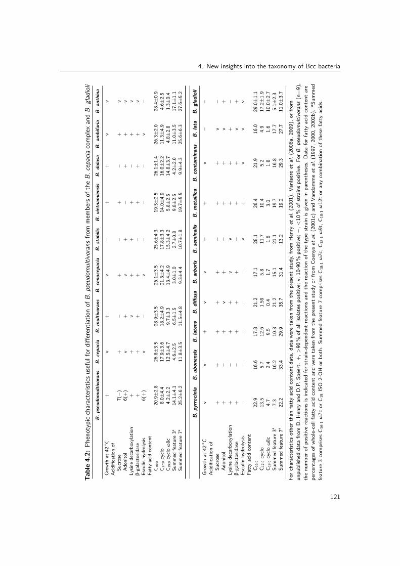
4. New insights into the taxonomy of Bcc bacteria
Table4.2:
Phenotypiccharacteristic
susefulfor
diffe
rentiat
ionof
B.pseudo
multiv
oransfro
mmem
bers
oftheB.
cepacia
complex
andB.
gladioli
B.p
seud
omultiv
orans
B.c
epacia
B.m
ultiv
orans
B.c
enocepacia
B.stabilis
B.v
ietnam
iensis
B.d
olosa
B.a
mbifaria
B.a
nthina
Grow
that
42◦ C
+v
+v
−+
+v
vAc
idificatio
nof
Sucrose
7(−)
+−
+−
+−
+v
Adon
itol
6(+)
v+
v+
−+
+v
Lysin
edecarboxyla
tion
++
v+
++
−+
vβ-galactosid
ase
++
++
−+
++
vEsculin
hydrolysis
6(+)
v−
v−
−−
v−
Fatty
acid
content
C 16:0
20.9
±2.8
26.8
±3.5
28.9
±3.5
26.1
±3.5
25.6
±4.3
19.5
±2.5
26.1
±1.4
26.3
±2.0
28.4
±0.9
C 17:0cyclo
8.0±
4.4
17.9
±3.6
18.2
±4.9
21.3
±4.2
17.8
±3.3
14.0
±4.9
16.0
±2.2
11.3
±4.9
4.6±
2.5
C 19:0cycloω8c
4.2±
2.2
12.5
±4.7
9.7±
3.3
13.4
±4.9
15.3
±4.2
5.8±
2.5
14.8
±3.7
4.8±
2.8
1.3±
0.4
Summed
feature3a
14.1
±4.1
4.6±
2.5
6.5±
1.5
3.0±
1.0
2.7±
0.8
9.8±
2.5
4.2±
2.0
11.0
±3.5
17.1
±1.1
Summed
feature7a
25.2
±6.2
11.8
±3.5
11.5
±4.8
9.3±
4.4
10.7
±1.8
19.7
±5.5
9.9±
4.3
25.6
±6.3
27.6
±5.2
B.p
yrrocinia
B.u
bonensis
B.latens
B.d
iffusa
B.a
rboris
B.sem
inalis
B.m
etallica
B.c
ontaminans
B.lata
B.g
ladioli
Grow
that
42◦ C
vv
+v
v+
+v
−−
Acidificatio
nof
Sucrose
++
++
++
++
v−
Adon
itol
+−
+v
++
++
v+
Lysin
edecarboxyla
tion
+−
++
vv
++
+−
β-galactosid
ase
+−
++
++
++
v+
Esculin
hydrolysis
−−
−−
−v
+v
vv
Fatty
acid
content
C 16:0
22.9
16.6
17.8
21.2
17.1
28.1
26.4
21.9
16.0
29.0
±1.1
C 17:0cyclo
13.5
5.7
12.6
1.59
5.8
11.7
10.4
5.2
4.9
17.2
±1.9
C 19:0cycloω8c
4.7
2.4
9.5
0.4
1.7
1.6
3.0
1.8
1.6
10.0
±2.7
Summed
feature3a
7.3
16.2
10.3
21.2
15.1
21.1
19.7
16.8
17.7
5.1±
2.3
Summed
feature7a
22.2
33.4
29.9
35.7
31.4
13.2
19.2
29.3
27.7
11.0
±3.7
Forc
haracteristics
othert
hanfattyacid
contentdata,d
atawe
retakenfro
mthepresentstud
y,fro
mHenry
etal.(
2001),Va
nlaere
etal.(
2008a,
2009),or
from
unpu
blish
eddata
from
D.H
enry
andD.P.S
peert.
+,>
90%
ofallisolatesp
ositive;v
,10-90
%po
sitive;−,<
10%
ofstrainsp
ositive.Fo
rB.p
seud
omultiv
orans(n=9),
thenu
mbero
fpositive
reactio
nsisindicatedfors
train-depend
entreactio
nsandthereactio
nof
thetype
strain
isgivenin
parentheses.
Dataforfatty
acid
contentare
percentageso
fwho
le-cellfattyacid
contenta
ndwe
retakenfro
mthepresents
tudy
orfro
mCo
enye
etal.(2001c)
andVa
ndam
meet
al.(1997,2
000,
2002b).a Sum
med
feature3comprise
sC 1
6:1ω7c
orC 1
5ISO
2-OH
orbo
th.Su
mmed
feature7comprise
sC 1
8:1ω7c,C
18:1ω9t,C
18:1ω12tor
anycombinatio
nof
thesefattyacids.
121

III. Experimental work
4.1.7 Supplementary material
The GenBank/EMBL/DDBJ accession numbers for the 16S rRNA gene sequences of strainsLMG 26883T and LMG 26915 are HE962386 and HE962387, respectively. The Gen-Bank/EMBL/DDBJ accession numbers for the recA gene sequences of strains LMG 26883T,LMG 26914, LMG 26915, R-48991, R-48993, R-48994, R-48995, R-48996, LMG 16669,R-24200, R-9705, LMG 13010T, LMG 24066T, LMG 22485T and LMG 24068T are HE963737,HE963738, HE963739, HE963740, HE963741, HE963742, HE963743, HE963744, HE963745,HE963746, HE963747, HE981730, HE981731, HE981732 and HE981733, respectively.
0% 1% 2% 3% 4% 5% 6% 7% 8% 9% 10%
B. cepacia (29)
B. multivorans (114)
B. cenocepacia (127)
B. stabilis (18)
B. vietnamiensis (39)
B. dolosa (11)
B. ambifaria (44)
B. anthina (16)
B. pyrrocinia (14)
B. ubonensis (2)
B. latens (2)
B. diffusa (8)
B. arboris (15)
B. seminalis (11)
B. metallica (3)
B. contaminans (13)
B. lata (12)
B. pseudomultivorans (7)
B. cenocepacia
B. pseudomultivorans
B. pseudomultivorans
B. seminalis
B. seminalis
B. diffusa
B. pyrrocinia
B. multivorans
B. multivorans
B. contaminans
B. metallica
B. metallica
B. seminalis
B. ambifaria
B. diffusa
B. diffusa
B. diffusa
B. stabilis
Supplementary Figure 4.1: Nucleotide divergence of concatenated MLST loci. Dark grey barsrepresent within group divergence, light grey bars represent the divergence between a Bcc species andits highest similarity neighbor (listed to the right). The number of STs included in the calculationsfor each species is given in parentheses. For each bar, the SD is indicated by error bars. The 3%divergence threshold value is marked (Vanlaere et al., 2009).
122
4. New insights into the taxonomy of Bcc bacteria
4.2 Burkholderia stagnalis sp. nov. and Burkholderia territoriisp. nov., two novel Burkholderia cepacia complex speciesfrom environmental and human sources
Redrafted from: Birgit De Smet, Mark Mayo, Charlotte Peeters, James E.A. Zlosnik,Theodore Spilker, Trevor J. Hird, John J. LiPuma, Timothy J. Kidd, Mirjam Kaestli, JenniferL. Ginther, David M. Wagner, Paul Keim, Scott C. Bell, Jan A. Jacobs, Bart J. Currie andPeter Vandamme (2015). Burkholderia stagnalis sp. nov. and Burkholderia territorii sp.nov., two novel Burkholderia cepacia complex species from environmental and human sources.International Journal of Systematic and Evolutionary Microbiology, 65, 2265-2271.
Author contributions: BDS, CP and PV conceived the study. BDS and PV wrote themanuscript. BDS and CP performed all experiments except the biochemical characterizationand analyzed the data. JZ and TH performed the biochemical characterization. TS, JL, TK,SB and BC isolated the strains. CP, JZ, TS, TH and JL proofread the manuscript.
4.2.1 Abstract
Nine Bcc bacteria were isolated during environmental surveys for the ecological niche of B.pseudomallei, the aetiological agent of melioidosis, in the Northern Territory of Australia. Theyrepresented two multilocus sequence analysis-based clusters, referred to as Bcc B and Bcc L.Three additional environmental and clinical Bcc B isolates were identified upon deposition ofthe sequences in the PubMLST database. Analysis of the concatenated nucleotide sequencedivergence levels within both groups (1.4 and 1.9%, respectively) and towards established Bccspecies (4.0 and 3.9%, respectively) demonstrated that the two taxa represented novel Bccspecies. All 12 isolates were further characterized using 16S rRNA and recA gene sequenceanalysis, RAPD analysis, DNA base content determination, fatty acid methyl ester analysisand biochemical profiling. Analysis of recA gene sequences revealed a remarkable diversitywithin each of these taxa, but, together, the results supported the affiliation of the twotaxa to the Bcc. Bcc B strains can be differentiated from most other Bcc members by theassimilation of maltose. Bcc L strains can be differentiated from other Bcc members bythe absence of assimilation of N-acetylglucosamine. The names Burkholderia stagnalis sp.nov. with type strain LMG 28156T (=CCUG 65686T) and Burkholderia territorii sp. nov.with type strain LMG 28158T (=CCUG 65687T) are proposed for Bcc B and Bcc L bacteria,respectively.
123

III. Experimental work
4.2.2 Background, methods, results and discussion
The Bcc is a versatile group of closely related bacteria that are ubiquitous in nature (Coenye& Vandamme, 2003; Vanlaere et al., 2009; Peeters et al., 2013; Vandamme & Peeters, 2014).Some Bcc strains show biotechnological potential for biocontrol, bioremediation and plantgrowth promotion, whereas other strains are pathogens for plants and immunocompromisedindividuals such as those with CF. Bcc strains are also common nosocomial pathogens(Vonberg & Gastmeier, 2007). The Bcc comprises, at the time of writing, 18 validly namedspecies (Peeters et al., 2013), which exhibit a high degree of 16S rRNA (98-100%) andrecA (94-95%) gene sequence similarity, moderate levels of DDH (30-50%) (Coenye et al.,2001d) and whole-genome ANI values of between 85.04 and 89.92% (Vanlaere et al., 2009).Protein encoding genes applied in a MLSA approach have a high discriminatory power for theidentification of Bcc isolates, and 3% concatenated sequence divergence has been validatedas a threshold level for species differentiation (Vanlaere et al., 2009; Peeters et al., 2013).The application of this threshold level to analyze publicly available MLST data revealed thepresence of at least another 16 novel Bcc species that await formal description (Vandamme& Peeters, 2014).Nine Bcc bacteria were isolated in ongoing environmental soil and water sampling surveysfor the ecological niches of B. pseudomallei, the aetiological agent of melioidosis, in thetropical Northern Territory of Australia. As part of the Darwin prospective melioidosis study(Currie et al., 2010) household yards, gardens and domestic water supplies and other locationsin Darwin city and its rural surrounds have been opportunistically sampled over the lasttwo decades, with recovered B. pseudomallei and other species of the genus Burkholderiacharacterized and stored. Environmental sampling and bacterial isolation methods have beendescribed previously (Mayo et al., 2011; Kaestli et al., 2012).These nine isolates represented two unique MLSA clusters within the Bcc, referred to earlieras clusters Bcc B and Bcc L (Vandamme & Peeters, 2014). Upon deposition of the sequencesin the Bcc PubMLST database (http://pubmlst.org/bcc/), three additional Bcc B isolateswere identified: one environmental soil isolate from North Carolina, USA, one clinical isolatefrom a respiratory specimen (tracheal aspirate) of a native American non-CF adult patientfrom North Carolina (we were unable to verify if infection could have been acquired abroad)and one isolate from a sputum sample of a patient with CF from Australia (Table 4.3).All isolates were preserved in MicroBank vials at −80 ◦C. They were grown aerobically ontryptone soya agar (Oxoid) and incubated at 30 ◦C for further characterization, except whenmentioned otherwise. For PCR experiments, DNA was prepared by alkaline lysis as describedby Storms et al. (2004).
124

4. New insights into the taxonomy of Bcc bacteria
Table 4.3: Studied isolates, showing their source, ST and allelic profileStrain Other strain designations Source (country, year of isolation)
B. stagnalis sp. nov.R-52235 MSMB049, FC1736 Soil (Australia, 2006)LMG 28156T CCUG 65686T, MSMB050T, FC1737T Soil (Australia, 2006)R-52237 MSMB085, FC1738 Soil (Australia, 2007)R-52238 MSMB086, FC1739 Soil (Australia, 2007)LMG 28157 MSMB097, FC1740 Soil (Australia, 2007)R-52095 HI3541, FC1742 Soil (USA, 2005)R-52096 AU7314, FC1741 Tracheal aspirate, non-CF (USA, 2004)R-52240 MSMB21995, QLD037, FC1812 CF sputum (Australia, 2008)
B. territorii sp. nov.LMG 28158T CCUG 65687T, MSMB110T, FC1743T Water (Australia, 2003)R-52242 MSMB117, FC1744 Water (Australia, 2003)LMG 28159 MSMB138, FC1745 Water (Australia, 2003)R-52244 MSMB139, FC1746 Water (Australia, 2003)
Strain ST Allelic profile
atpD gltB gyrB recA lepA phaC trpB
B. stagnalis sp. nov.R-52235 787 170 197 564 345 300 213 381LMG 28156T 787 170 197 564 345 300 213 381R-52237 789 328 379 399 347 300 213 196R-52238 789 328 379 399 347 300 213 196LMG 28157 789 328 379 399 347 300 213 196R-52095 865 205 406 420 366 245 191 247R-52096 519 170 197 399 190 300 213 196R-52240 690 300 345 511 319 300 274 351
B. territorii sp. nov.LMG 28158T 791 233 381 567 348 395 304 384R-52242 792 329 382 568 349 396 305 385LMG 28159 794 331 384 570 351 398 306 386R-52244 794 331 384 570 351 398 306 386
LMG, BCCM/LMG Bacteria Collection, Laboratory of Microbiology, Ghent University, Ghent, Belgium; CCUG,Culture collection University of Göteborg, Sweden; MSMB, Menzies school of Health Research, Darwin, Australia; CF,cystic fibrosis patient.
125

III. Experimental work
MLSA was performed using standard protocols (Spilker et al., 2009; Peeters et al., 2013).Nucleotide sequences of each allele, allelic profiles and sequence types (STs) for all isolatesrepresented five STs for cluster Bcc B isolates and three STs for cluster Bcc L isolates, and areavailable on the Bcc PubMLST website (http://pubmlst.org/bcc/) (Jolley & Maiden, 2010).The mean number of nucleotide substitutions per site (i.e. the percentage of divergence ofconcatenated allele sequences) between established (June 2014) and the newly proposed Bccspecies was calculated using the program DnaSP v5.10 (Librado & Rozas, 2009) based onthe Jukes-Cantor method (Jukes & Cantor, 1969). A phylogenetic tree of the concatenatedsequences (2773 bp) of seven housekeeping gene fragments, namely atpD (443 bp), gltB(400 bp), gyrB (454 bp), recA (393 bp), lepA (397 bp), phaC (385 bp) and trpB (301 bp),was constructed using MEGA5 (Tamura et al., 2011).Within cluster Bcc B (n=5) the concatenated allele sequence divergence was 1.4±0.6 %, andthe average divergence towards its nearest neighbor, B. ubonensis, was 4.0±0.8 %. Withincluster Bcc L (n=3) the concatenated allele sequence divergence was 1.9±0.7 %, and theaverage divergence towards its nearest neighbor, B. diffusa, was 3.9±0.9 % (data not shown).These concatenated sequence divergence values demonstrate that the two clusters representnovel species within the Bcc (Vanlaere et al., 2009; Peeters et al., 2013).When a non-Bcc outgroup species (B. fungorum) was included in the phylogenetic analysisof the concatenated sequences, cluster Bcc B and cluster Bcc L isolates constituted twoclusters supported by high bootstrap values of 93 and 99%, respectively (Fig. 4.3; see alsoVandamme & Peeters, 2014). Yet a bifurcation was apparent in each cluster and the removalof the outgroup taxon reinforced the differences between these subgroups in the resultingphylogenetic trees: Supplementary Fig. 4.3-4.9 show the results of phylogenetic analysesbased on single gene sequences. Among Bcc B isolates, R-52095 had gltB, lepA and trpBgene sequences that were very similar to those of the remaining cluster Bcc B strains, whereasits gyrB, phaC and, to an even greater extent, atpD and recA gene sequences were divergentfrom those of the remaining cluster Bcc B strains. Among the cluster Bcc L strains, isolatesLMG 28159 and R-52244 differed from LMG 28158T and R-52242 in particular for the recA,trpB and, to a lesser extent, lepA gene sequences, whereas the atpD, gltB, gyrB and phaCgene sequences were highly similar (Supplementary Fig. 4.3-4.9).RAPD analysis of all isolates was performed with primer 270 as described by Mahenthiralingamet al. (1996) and profiles were compared visually (data not shown). For the eight Bcc Bisolates, five different RAPD profiles where observed, corresponding to the five STs. For thefour Bcc L isolates, four different RAPD profiles were observed; isolates LMG 28159 andR-52244, which represented a single ST, had only slightly different RAPD profiles (data notshown).
126

4. New insights into the taxonomy of Bcc bacteria
Downloaded from www.sgmjournals.org byIP: 157.193.163.134
On: Fri, 21 Aug 2015 13:29:03
C16:0 (18.7¡0.9 and 19.0¡0.9%), C16:0 2-OH (1.5¡0.5and 1.2¡0.6%), C16:0 3-OH (8.4¡1.0 and 7.1¡0.9%),C16:1 2-OH (1.9¡0.6 and 1.0¡0.4 ;), C17:0 cyclo(10.2¡4.2 and 6.1¡3.2 %), C18 : 1v7c (24.0¡4.5 and30.1¡1.7%), C19 : 0 cyclo v8c (4.4¡2.4 and 2.1¡1.3%),C18 : 0 2-OH (2.8¡0.7 and 2.5¡1.0%), summed feature2 (comprising C14 : 0 3-OH, iso-C16 : 1 I, an unidentified
fatty acid with equivalent chain-length of 10.928 or C12 : 0
ALDE or any combination of these) (10.3¡1.7 and10.8¡3.3%) and summed feature 3 (comprisingC16 : 1v7c and/or iso-C15 : 0 2-OH) (12.2¡4.9 and15.2¡4.0%). These cellular fatty acid profiles are highlysimilar to those of other Bcc species and Burkholderiagladioli (Stead, 1992).
Burkholderia contaminans LMG 23361T (ST-102)
Burkholderia lata LMG 22485T (ST-101)
Burkholderia metallica LMG 24068T (ST-511)
Burkholderia cepacia LMG 1222T (ST-10)
Burkholderia cenocepacia IIIC LMG 19230T (ST-44)
Burkholderia arboris LMG 24066T (ST-492)
Burkholderia pyrrocinia LMG 14191T (ST-41)
Burkholderia stabilis LMG 14294T (ST-50)
Burkholderia seminalis LMG 24067T (ST-473)
Burkholderia cenocepacia IIID LMG 21462T (ST-102)
Burkholderia cenocepacia IIIB LMG 18830T (ST-39)
Burkholderia cenocepacia IIIA LMG 16656T (ST-31)
Burkholderia anthina LMG 20980T (ST-86)
Burkholderia ambifaria LMG 19182T (ST-77)
Burkholderia diffusa LMG 24065T (ST-164)
Burkholderia vietnamensis LMG 10929T (ST-65)
Burkholderia latens LMG 24064T (ST-238)
Burkholderia territorii LMG 28159; R-52244 (ST-794)
Burkholderia territorii LMG 28158T (ST-791)
Burkholderia territorii R-52242 (ST-792)
Burkholderia pseudomultivorans LMG 26883T (ST-536)
Burkholderia multivorans LMG 13010T (ST-397)
Burkholderia dolosa LMG 18943T (ST-72)
Burkholderia ubonensis LMG 20358T (ST-299)
Burkholderia stagnalis R-52095 (ST-865)
Burkholderia stagnalis R-52240 (ST-690)
Burkholderia stagnalis LMG 28157; R-52237; R-52238 (ST-789)
Burkholderia stagnalis LMG 28156T; R-52235 (ST-787)
Burkholderia stagnalis R-52096 (ST-519)
Burkholderia fungorum LMG 16225T (ST-499)
100
62
87
100
93
100
98
99
81
99
91
96
88
88
78
93
58
71
81
0.05
Fig. 1. Phylogenetic tree based on the concatenated sequences (2773 bp) of seven housekeeping gene fragments [atpD(443 bp), gltB (400 bp), gyrB (454 bp), recA (393 bp), lepA (397 bp), phaC (385 bp) and trpB (301 bp)] of establishedBcc species and Burkholderia stagnalis sp. nov. and Burkholderia territorii sp. nov. strains. The bootstrap consensus tree,inferred from 1000 replicates, was reconstructed using the maximum-likelihood method based on the general time reversiblemodel. Branches corresponding to partitions reproduced in less than 50% bootstrap replicates are collapsed. The percen-tage of replicate trees in which the associated taxa clustered together in the bootstrap test (1000 replicates) is shown nextto the branches if greater than 50%. A discrete gamma distribution was used to model evolutionary rate differences amongsites [five categories (+G, parameter50.3623)] and allowed for some sites to be evolutionarily invariable [(+I), 51.7996%sites]. The analysis involved 30 nt sequences. Evolutionary analyses were conducted in MEGA5.The sequence of B. fungorumLMG 16225T was used as an outgroup. Bar, 0.05 substitutions per site.
B. De Smet and others
2268 International Journal of Systematic and Evolutionary Microbiology 65
Figure 4.3: Phylogenetic tree based on the concatenated sequences (2773 bp) of seven housekeepinggene fragments [atpD (443 bp), gltB (400 bp), gyrB (454 bp), recA (393 bp), lepA (397 bp), phaC(385 bp) and trpB (301 bp)] of established Bcc species and Burkholderia stagnalis sp. nov. andBurkholderia territorii sp. nov. strains. The bootstrap consensus tree, inferred from 1000 replicates,was reconstructed using the maximum likelihood method based on the general time reversible model.Branches corresponding to partitions reproduced in less than 50% bootstrap replicates are collapsed.The percentage of replicate trees in which the associated taxa clustered together in the bootstrap test(1000 replicates) is shown next to the branches if greater than 50%. A discrete gamma distributionwas used to model evolutionary rate differences among sites [five categories (+G, parameter=0.3623)]and allowed for some sites to be evolutionarily invariable [(+I), 51.7996% sites]. The analysis involved30 nt sequences. Evolutionary analyses were conducted in MEGA5. The sequence of B. fungorumLMG 16225T was used as an outgroup. Bar, 0.05 substitutions per site.
127

III. Experimental work
The nearly complete sequences of the 16S rRNA gene of strains LMG 28156T (1485 bp)and LMG 28158T (1443 bp) were obtained as described previously (Peeters et al., 2013).Pairwise comparison of these sequences, as determined with the EzTaxon-e server (www.ezbiocloud.net/eztaxon (Kim et al., 2012), with those of type strains of other Bcc speciesrevealed similarity levels of between 98.43 and 99.66% for the former and between 98.89 and100% for the latter (data not shown). Similarity levels towards non-Bcc Burkholderia specieswere in the range 94.28-98.98%. For both strains the highest similarity values were obtainedtowards the type strain of B. glumae.Sequences of the recA gene were amplified using forward primer 5’ AGG ACG ATT CAT GGAAGA WAG C 3’ and reverse primer 5’ GAC GCA CYG AYG MRT AGA ACT T ’3 (Spilkeret al., 2009). Sequence assembly was performed using BioNumerics v5.10 (Applied Maths).Sequences (540-660 bp) were aligned based on amino acid sequences using Muscle (Edgar,2004) in MEGA5 (Tamura et al., 2011). Phylogenetic analysis was conducted in MEGA5(Tamura et al., 2011). All positions containing gaps and missing data were eliminated,resulting in a total of 473 positions in the final dataset. All Bcc B isolates except isolateR-52095, which occupied a distinct position in the recA gene tree, formed a coherent recAgene cluster with B. pseudomultivorans as its nearest neighbor (Supplementary Fig. 4.2).These results confirmed and extended the considerable differences observed in the 393 bprecA gene fragment of the MLSA scheme (Supplementary Fig. 4.6). Similarly, the bifurcationof the four Bcc L strains was confirmed in the 473 bp based analysis (Supplementary Fig.4.2).For determination of the DNA base composition, high-molecular-mass DNA was preparedas described by Pitcher et al. (1989). DNA was enzymatically degraded into nucleosides asdescribed by Mesbah & Whitman (1989). The nucleoside mixture obtained was separatedusing a Waters Breeze HPLC system and XBridge Shield RP18 column thermo-stabilizedat 37 ◦C. The solvent was 0.02 M NH4H2PO4 (pH 4.0) with 1.5% (v/v) acetonitrile.Non-methylated lambda phage (Sigma) and Escherichia coli LMG 2093 DNA were usedas calibration reference and control, respectively. The DNA G+C content of strains LMG28156T and LMG 28158T was both 67 mol%, which corresponded with that of other Bccspecies (66-69 mol%) (Vandamme & Dawyndt, 2011).For fatty acid methyl ester analysis, all strains were grown on tryptone soya agar (BD) at28 ◦C for 24 h. A loopful of well-grown cells was harvested and fatty acid methyl esters wereprepared, separated and identified using the Microbial Identification System (Microbial ID) asdescribed previously (Vandamme et al., 1992). The following fatty acid components weredetected in Bcc B and Bcc L isolates, respectively: C14:0 (4.08±1.3 and 4.3±0.3 %), C16:0
(18.7±0.9 and 19.0±0.9 %), C16:0 2-OH (1.5±0.5 and 1.2±0.6 %),C16:0 3-OH (8.4±1.0
128
4. New insights into the taxonomy of Bcc bacteria
and 7.1±0.9 %), C16:1 2-OH (1.9±0.6 and 1.0±0.4 ;), C17:0 cyclo (10.2±4.2 and 6.1±3.2%), C18:1 ω7c (24.0±4.5 and 30.1±1.7 %), C19:0 cyclo ω8c (4.4±2.4 and 2.1±1.3 %),C18:0 2-OH (2.8±0.7 and 2.5±1.0 %), summed feature 2 (comprising C14:0 3-OH, iso-C16:1
I, an unidentified fatty acid with equivalent chain-length of 10.928 or C12:0 ALDE or anycombination of these) (10.3±1.7 and 10.8±3.3 %) and summed feature 3 (comprising C16:1
ω7c and/or iso-C15:0 2-OH) (12.2±4.9 and 15.2±4.0 %). These cellular fatty acid profilesare highly similar to those of other Bcc species and B. gladioli (Stead, 1992).Biochemical characterization of all isolates was performed as described by Henry et al. (2001)and a summary of the results can be found in Table 4.4 and in the species descriptions below.Biochemically, all Bcc B and Bcc L strains are similar to other Bcc bacteria. Bcc B strainscan be differentiated from other Bcc species (except for B. ubonensis) by the assimilationof maltose. The only difference noted between isolate R-52095 and the remaining Bcc Bisolates is the presence of β-haemolysis. Bcc L strains can be differentiated from other Bccspecies by the absence of assimilation of N-acetylglucosamine. The two Bcc L recA geneclusters differed in nitrate reduction capacity.In conclusion, MLSA demonstrated that 12 Bcc isolates represented two novel species withinthe Bcc. DNA base content determination, 16S rRNA gene sequence analysis and fatty acidmethyl ester analysis confirmed that these isolates conformed to the general characteristics ofthe genus Burkholderia. The two novel species consisted of two recA gene lineages that werephenotypically different; yet assimilation of maltose and of N-acetylglucosamine could be usedto distinguish these species from other Bcc species. We therefore propose to formally classifyBcc B and Bcc L strains as representing the novel species Burkholderia stagnalis sp. nov.with strain LMG 28156T (=CCUG 65686T) as the type strain, and Burkholderia territorii sp.nov. with strain LMG 28158T (=CCUG 65687T) as the type strain, respectively.
4.2.2.1 Description of Burkholderia stagnalis sp. nov.
Burkholderia stagnalis (stag.na’lis. L. n. stagnum pond, lagoon, lake; -alis adjective formingsuffix; N.L. fem. adj. stagnalis pertaining to a lagoon).Cells are Gram-stain-negative, aerobic, motile, non-sporeforming rods, about 0.1-0.5 µm wideand 0.6-1.7 µm long. All isolates grow at 37 ◦C on BSA, BCSA and MacConkey agar anddo not produce any pigment. The majority (six of eight) of the isolates show medium-sizedgrey entire colonies after 3 days of incubation on BSA, and the remaining two isolates showdistinctive colony centers Only one isolate (R-52095) shows β-haemolysis on blood agar.The majority of isolates (five of eight) are able to grow at 42 ◦C (R-52235, LMG 28156T,R-52237, R-52238, R-52239). All isolates show slow oxidase-positive reactions and lysine
129
III. Experimental work
decarboxylase activity, but no nitrate reduction, ornithine decarboxylase or aesculin hydrolysisactivity. Furthermore, glucose, maltose, lactose and xylose are acidified, but not adonitol.Assimilation of maltose is observed for all strains. Strain-dependent reactions are observedfor acidification of sucrose (negative for R-52240), gelatinase (negative for R-52096) andβ-galactosidase (weakly positive for R-52096) activity. API 20NE profiles are 0057777 (six ofeight isolates), 0056577 (R-52240) and 0067777 (R-52096). The following fatty acids arepresent in all isolates: C14:0, C16:1 2-OH, C16:0 2-OH, C18:0, C19:0 cyclo ω8c and C18:1 2-OHall at <5% (mean value of all isolates); and C16:0, C17:0 cyclo, C16:0 3-OH, C18:1 ω7c andsummed features 2 and 3 all at between 5 and 25% (mean value of all isolates). Isolateshave been obtained from soil and human respiratory samples in Australia and the USA.The type strain is LMG 28156T (=CCUG 65686T, originally collected as MSMB050T).Phenotypic characteristics of the type strain are the same as those described above for allstrains of the species. In addition, the type strain grows at 42 ◦C, does not show β-haemolysison sheep blood agar, acidifies sucrose, assimilates arabinose and shows gelatinase activity.The DNA G+C content of the type strain is 67 mol%.
4.2.2.2 Description of Burkholderia territorii sp. nov.
Burkholderia territorii [ter.ri.to’ri.i. L. gen. n. territorii of the territory; referring to the(Northern) Territory of Australia, where this organism has been recovered from environmentalgroundwater samples].Cells are Gram-stain-negative, aerobic, motile, non-sporeforming rods, about 0.2-0.5 µm wideand 0.5-1.7 µm long. All isolates grow at 37 ◦C on BSA, BCSA and MacConkey agar. Theyshow medium-sized grey entire colonies on BSA without haemolysis, no pigment productionis observed and all strains grow at 42 ◦C. Two isolates (R-52243 and R-52244) display mixedcolonies types with small and large colonies. However, RAPD on both colony types showsidentical patterns. All isolates show slow oxidase-positive reactions, lysine decarboxylase,β-galactosidase and gelatinase activity but no ornithine decarboxylase or aesculin hydrolysisactivity. Acidification of glucose, maltose, lactose, xylose and adonitol is positive, withstrain-dependent reaction for sucrose acidification (negative for R-52242), as well as nitratereduction (negative for LMG 28158T and R-52242). No assimilation of N-acetylglucosamine.API 20NE profiles are 0077567 (LMG 28158T), 0077577 (R-52242) and 1077577 (R-52243and R-52244). The following fatty acids are present in all isolates: C14:0, C16:1 2-OH, C16:0
2-OH, C18:0, C19:0 cyclo ω8c and C18:1 2-OH all at <5%; and C16:0, C17:0 cyclo, C16:0 3-OH,C18:1 ω7c, and summed features 2 and 3 all at between 5 and 30%. Strains have beenisolated from environmental groundwater samples.
130

4. New insights into the taxonomy of Bcc bacteria
The type strain is LMG 28158T (=CCUG 65687T, originally collected as MSMB110T).Phenotypic characteristics of the type strain are the same as those described above for allstrains of the species. In addition, the type strain reduces nitrate and acidifies sucrose. TheDNA G+C content of the type strain is 67 mol%.
131

III. Experimental work
Table4.4:
Phenotypic
characteristic
susefulfor
diffe
rentiat
ionof
B.stagnalis
sp.n
ov.a
ndB.
territorii
sp.n
ov.f
rom
mem
bers
oftheBc
cPheno
typiccharacteristic
12
34
56
78
910
1112
1314
1516
1718
1920
Grow
thon
MacCo
nkey
agar
++
++
++
++
++
v+
v+
v+
++
++
Grow
that
42◦ C
5(+)
++
v−
+v
v+
+v
+v
−+
+v
vv
vPigm
ent
−−
−v
v−
−v
v+(y)
v−
v−
−−
v−
v−
Haem
olysis(sheep
bloo
d)1β(−
)−
−v
−−
−v
−−
−−
−−
v−
v−
v−
Assim
ilatio
nof:
L-Ar
abinose
5(+)
++
+v
++
++
++
++
−+
++
++
+D-M
annitol
++
++
++
++
++
++
++
++
++
v+
N-Ac
etylg
lucosamine
+−
++
++
++
++
++
++
++
++
v+
Maltose
+3(
−)
v−
v−
−v
−−
−−
−−
v−
−v
v+
Adipate
++
++
++
++
++
++
++
v+
++
++
Phenyla
cetate
++
+v
++
++
++
++
++
++
++
++
Acidificatio
nof:
Maltose
++
+v
++
++
++
v+
v+
++
++
++
Lactose
++
++
++
++
++
++
v+
++
++
++
D-X
ylose
++
++
++
+w
++
++
+v
v+
++
++
Sucrose
7(+)
3(+)
v+
v+
++
++
+−
+−
+−
+v
++
Adon
itol
−+
v+
v+
v+
++
v+
v+
−+
+v
+−
Nitra
teredu
ction
−2(
−)
vv
v−
+v
−−
−+
v−
v+
vv
vv
Activ
ityof:
Lysin
edecarboxyla
se+
++
++
++
vv
++
v+
++
−+
v+
−Ornith
inedecarboxyla
se−
−−
−v
−−
+v
−v
−v
+−
−−
−+
−Ae
sculin
hydrolysis
−−
vv
v−
−−
v+
v−
v−
−−
v−
−−
Arginine
dihydrolase
−−
−−
−−
−−
−−
−−
−−
−−
−−
−+
Gelatin
ase
7(+)
+−
+v
vv
++
+v
−v
v−
−+
−+
+β-galactosid
ase
1(−)
++
+v
++
++
++
++
−+
++
v+
−Sp
ecies
:1,
B.stagn
alis
sp.no
v.;2
,B.territoriisp.no
v.;3
,B.p
seud
omultiv
orans;
4,B.c
ontaminans;
5,B.lata;
6,B.latens;
7,B.d
iffusa;
8,B.a
rboris;9
,B.
seminalis;1
0,B.m
etallica;
11,B
.cepacia;1
2,B.m
ultiv
orans;
13,B
.cenocepacia;1
4,B.stabilis;1
5,B.v
ietnam
iensis;1
6,B.d
olosa;
17,B
.ambifaria
;18,
B.
anthina;
19,B
.pyrrocinia;
20,B
.ubo
nensis.
+,<
90%
ofallisolatespo
sitive;
v,10-90%
positive;−,<
10%
ofstrainspo
sitive;
w,we
akreactio
n;β,β
-haemolysis;
y,yellow.
ForB
.stagn
alis
(n=8)
andB.territorii(n=4),the
numbero
fpositive
reactio
nsisindicatedfors
train-depend
entreactio
nsandthereactio
nof
thetype
strain
isgivenin
parentheses.
Forc
haracteristics
ofestablish
edBc
cspecies
data
were
takenfro
mthepresentstud
yandPe
eterset
al.(
2013).
132

4. New insights into the taxonomy of Bcc bacteria
4.2.3 Acknowledgments
We thank all strain depositors who contributed to this study, Cindy Snauwaert (Laboratoryof Microbiology, Faculty of Sciences, Ghent University, Ghent, Belgium), Evie De Brandt(Laboratory of Microbiology, Faculty of Sciences, Ghent University, Ghent, Belgium), RebeccaHickman (University of British Columbia) and Kay Ramsay (Queensland Childrens MedicalResearch Institute, The University of Queensland, Brisbane, Queensland, Australia) fortechnical assistance, and Glenda Harrington (Menzies School of Health Research, Darwin,Northern Territory, Australia) for assistance with environmental sampling. Collection of theenvironmental samples was supported by grants from the Australian National Health andMedical Research Council (605820 and 1046812). J. E.A. Z. and T. J. H. are very gratefulfor the support of David P. Speert and financial support provided to D. P. S. by CysticFibrosis Canada through an operating grant (20R32976) and support for the Canadian Bccresearch and referral repository (20R40868). Initial MLSA was supported by grant fundingfrom the Childrens Health Foundation Queensland (50005). This publication made use of theBcc PubMLST website (http://pubmlst.org/bcc) developed by Keith Jolley and sited at theUniversity of Oxford (Jolley & Maiden, 2010). The development of this site has been fundedby the Wellcome Trust.
4.2.4 Supplementary material
The GenBank/EMBL/DDBJ accession numbers for the 16S rRNA gene sequences of strainsLMG 28156T and LMG 28158T are LK023502 and LK023503, respectively. Those for the recAgene sequences of strains R-52235, LMG 28156T, R-52237, R-52238, LMG 28157, R-52095, R-52096, R-52240, LMG 28158T, R-52242, LMG 28159 and R-52244 are LK023504-LK023515,respectively.
133
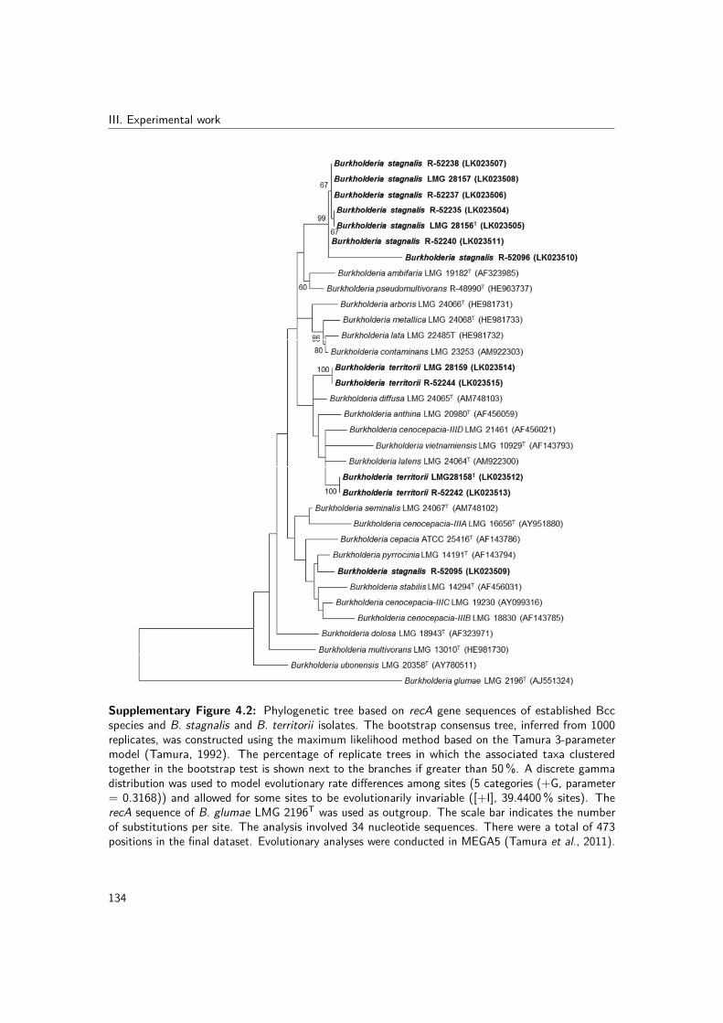
III. Experimental workSupplementary figure 1
Supplementary Figure 4.2: Phylogenetic tree based on recA gene sequences of established Bccspecies and B. stagnalis and B. territorii isolates. The bootstrap consensus tree, inferred from 1000replicates, was constructed using the maximum likelihood method based on the Tamura 3-parametermodel (Tamura, 1992). The percentage of replicate trees in which the associated taxa clusteredtogether in the bootstrap test is shown next to the branches if greater than 50%. A discrete gammadistribution was used to model evolutionary rate differences among sites (5 categories (+G, parameter= 0.3168)) and allowed for some sites to be evolutionarily invariable ([+I], 39.4400% sites). TherecA sequence of B. glumae LMG 2196T was used as outgroup. The scale bar indicates the numberof substitutions per site. The analysis involved 34 nucleotide sequences. There were a total of 473positions in the final dataset. Evolutionary analyses were conducted in MEGA5 (Tamura et al., 2011).
134

4. New insights into the taxonomy of Bcc bacteriaSupplementary figure 2
Supplementary Figure 4.3: Phylogenetic tree based on atpD locus sequences of established Bccspecies and B.stagnalis and B. territorii isolates. The bootstrap consensus tree, inferred from 1000replicates was constructed using the maximum likelihood method based on the Tamura 3-parametermodel (Tamura, 1992). The percentage of trees in which the associated taxa clustered together isshown next to the branches if greater than 50%. A discrete gamma distribution was used to modelevolutionary rate differences among sites (5 categories (+G, parameter = 0.2570)) and allowed forsome sites to be evolutionarily invariable ([+I], 74.3388% sites). The scale bar indicates the numberof substitutions per site. The analysis involved 28 nucleotide sequences. There were a total of 443positions in the final dataset. Evolutionary analyses were conducted in MEGA5 (Tamura et al., 2011).
135

III. Experimental workSupplementary figure 3
Supplementary Figure 4.4: Phylogenetic tree based on gltB locus sequences of established Bccspecies and B. stagnalis and B. territorii isolates. The bootstrap consensus tree, inferred from 1000replicates was constructed using the maximum likelihood method based on the Tamura 3-parametermodel (Tamura, 1992). The percentage of trees in which the associated taxa clustered together isshown next to the branches if greater than 50%. A discrete gamma distribution was used to modelevolutionary rate differences among sites (5 categories (+G, parameter = 0.4758)) and allowed forsome sites to be evolutionarily invariable ([+I], 57.9292% sites). The scale bar indicates the numberof substitutions per site. The analysis involved 28 nucleotide sequences. There were a total of 400positions in the final dataset. Evolutionary analyses were conducted in MEGA5 (Tamura et al., 2011).
136

4. New insights into the taxonomy of Bcc bacteriaSupplementary figure 4
Supplementary Figure 4.5: Phylogenetic tree based on gyrB locus sequences of established Bccspecies and B. stagnalis and B. territorii isolates. The bootstrap consensus tree, inferred from 1000replicates was constructed using the maximum likelihood method based on the general time reversiblemodel. The percentage of trees in which the associated taxa clustered together is shown next to thebranches if greater than 50%. A discrete gamma distribution was used to model evolutionary ratedifferences among sites (5 categories (+G, parameter = 0.4373)) and allowed for some sites to beevolutionarily invariable ([+I], 49.3662% sites). The scale bar indicates the number of substitutionsper site. The analysis involved 28 nucleotide sequences. There were a total of 454 positions in thefinal dataset. Evolutionary analyses were conducted in MEGA5 (Tamura et al., 2011).
137

III. Experimental workSupplementary figure 5
Supplementary Figure 4.6: Phylogenetic tree based on recA locus sequences of established Bccspecies and B. stagnalis and B. territorii isolates. The bootstrap consensus tree, inferred from 1000replicates was constructed using the maximum likelihood method based on the Tamura 3-parametermodel (Tamura, 1992). The percentage of trees in which the associated taxa clustered together isshown next to the branches if greater than 50%. A discrete gamma distribution was used to modelevolutionary rate differences among sites (5 categories (+G, parameter = 0.3316)) and allowed forsome sites to be evolutionarily invariable ([+I], 54.4859% sites). The scale bar indicates the numberof substitutions per site. The analysis involved 29 nucleotide sequences. There were a total of 393positions in the final dataset. Evolutionary analyses were conducted in MEGA5 (Tamura et al., 2011).138

4. New insights into the taxonomy of Bcc bacteria
Supplementary figure 6
Supplementary Figure 4.7: Phylogenetic tree based on lepA locus sequences of established Bccspecies and B. stagnalis and B. territorii isolates. The bootstrap consensus tree, inferred from 1000replicates was constructed using the maximum likelihood method based on the Tamura 3-parametermodel (Tamura, 1992). The percentage of trees in which the associated taxa clustered together isshown next to the branches if greater than 50%. A discrete gamma distribution was used to modelevolutionary rate differences among sites (5 categories (+G, parameter = 0.6858)) and allowed forsome sites to be evolutionarily invariable ([+I], 52.7765% sites). The scale bar indicates the numberof substitutions per site. The analysis involved 26 nucleotide sequences. There were a total of 397positions in the final dataset. Evolutionary analyses were conducted in MEGA5 (Tamura et al., 2011).
139

III. Experimental workSupplementary figure 7
Supplementary Figure 4.8: Phylogenetic tree based on phaC locus sequences of established Bccspecies and B. stagnalis and B. territorii isolates. The bootstrap consensus tree, inferred from 1000replicates was constructed using the maximum likelihood method based on the Tamura 3-parametermodel (Tamura, 1992). The percentage of trees in which the associated taxa clustered together isshown next to the branches if greater than 50%. A discrete gamma distribution was used to modelevolutionary rate differences among sites (5 categories (+G, parameter = 0.6710)) and allowed forsome sites to be evolutionarily invariable ([+I], 67.9788% sites). The scale bar indicates the numberof substitutions per site. The analysis involved 27 nucleotide sequences. There were a total of 385positions in the final dataset. Evolutionary analyses were conducted in MEGA5 (Tamura et al., 2011).
140

4. New insights into the taxonomy of Bcc bacteriaSupplementary figure 8
Supplementary Figure 4.9: Phylogenetic tree based on trpB locus sequences of established Bccspecies and B. stagnalis and B. territorii isolates. The bootstrap consensus tree, inferred from 1000replicates was constructed using the maximum likelihood method based on the Tamura 3-parametermodel (Tamura, 1992). The percentage of trees in which the associated taxa clustered together isshown next to the branches if greater than 50%. A discrete gamma distribution was used to modelevolutionary rate differences among sites (5 categories (+G, parameter = 0.3701)) and allowed forsome sites to be evolutionarily invariable ([+I], 51.1343% sites). The scale bar indicates the numberof substitutions per site. The analysis involved 28 nucleotide sequences. There were a total of 301positions in the final dataset. Evolutionary analyses were conducted in MEGA5 (Tamura et al., 2011).
141

III. Experimental work
142

5 | New insights into thetaxonomy of Burkholderia glathei-like bacteria
The B. glathei clade (BGC) represents a phylogenetically divergent clade in the genusBurkholderia (Sawana et al., 2014; Vandamme et al., 2014). Most of the 12 validly namedBGC species have been isolated from soil but publicly available sequence data indicate thatthe taxonomic diversity in this clade is severely underestimated (Nogales et al., 2001; Salleset al., 2006b; Pumphrey & Madsen, 2008; Xu et al., 2016). Section 5.1 presents a traditionalpolyphasic taxonomic study of isolates from agricultural soil in Argentina and the formalclassification of Burkholderia cordobensis sp. nov. Section 5.2 presents a modern polyphasictaxonomic approach in which whole-genome sequences are used for the assessment of thegenomic relatedness among strains and their G+C content. This genomic taxonomic studywas performed on 13 clusters of B. glathei-like isolates from human clinical and environmentalorigin and led to the formal taxonomic proposal of 13 novel BGC species.
143


5. New insights into the taxonomy of B. glathei-like bacteria
5.1 Burkholderia cordobensis sp. nov., from agriculturalsoils
Redrafted from: Walter O. Draghi, Charlotte Peeters, Margo Cnockaert, Cindy Snauwaert,Luis G. Wall, Angeles Zorreguieta and Peter Vandamme (2014). Burkholderia cordobensissp. nov., from agricultural soils. International Journal of Systematic and EvolutionaryMicrobiology, 64, 2003–2008.
Author contributions: WD, CP and PV conceived the study and wrote the manuscript. WDand CP performed all experiments and analyzed the sequence data. WD and MC performedthe biochemical characterization. WD and CS performed the fatty acid methyl ester analysis.WD, LW and AZ isolated the strains.
5.1.1 Abstract
Two Gram-negative, rod-shaped bacteria were isolated from agricultural soils in Cordobaprovince in central Argentina. Their 16S rRNA gene sequences demonstrated that theybelong to the genus Burkholderia, with Burkholderia zhejiangensis as most closely relatedformally named species; this relationship was confirmed through comparative gyrB sequenceanalysis. Whole-cell fatty acid analysis supported their assignment to the genus Burkholderia.Burkholderia sp. strain YI23, for which a whole-genome sequence is available, represents thesame taxon, as demonstrated by its highly similar 16S rRNA (100% similarity) and gyrB (99.1-99.7%) gene sequences. The results of DDH experiments and physiological and biochemicalcharacterization further substantiated the genotypic and phenotypic distinctiveness of theArgentinian soil isolates, for which the name Burkholderia cordobensis sp. nov. is proposed,with strain MMP81T (=LMG 27620T =CCUG 64368T) as the type strain.
5.1.2 Background, methods, results and discussion
Members of the genus Burkholderia are β-Proteobacteria and are aerobic, chemo-organotrophicand motile (except for B. mallei), Gram-negative rods that display a wide physiologicalversatility, allowing them to inhabit extremely different ecological niches (Coenye & Vandamme,2003; Compant et al., 2008). Several species of the genus Burkholderia are human or animalpathogens; these include members of the Bcc, a group of closely related species commonlyisolated from respiratory specimens of CF patients (Vandamme & Dawyndt, 2011), and speciesbelonging to the B. pseudomallei group, which includes B. pseudomallei, the aetiologicalagent of melioidosis in humans, and B. mallei, the causative agent of glanders in animals
145
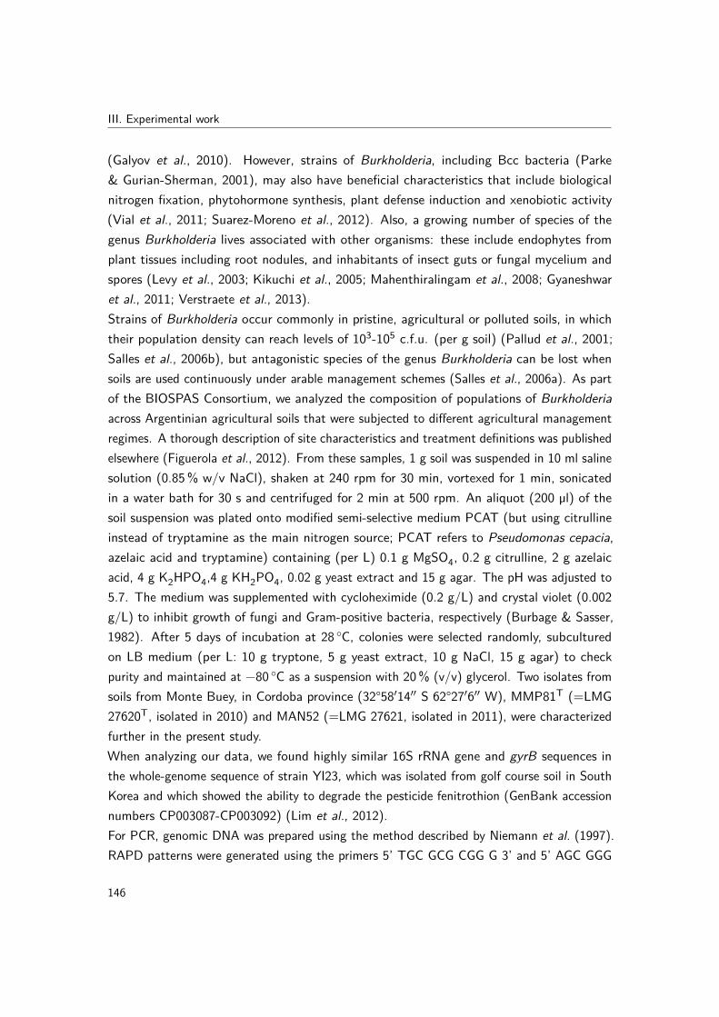
III. Experimental work
(Galyov et al., 2010). However, strains of Burkholderia, including Bcc bacteria (Parke& Gurian-Sherman, 2001), may also have beneficial characteristics that include biologicalnitrogen fixation, phytohormone synthesis, plant defense induction and xenobiotic activity(Vial et al., 2011; Suarez-Moreno et al., 2012). Also, a growing number of species of thegenus Burkholderia lives associated with other organisms: these include endophytes fromplant tissues including root nodules, and inhabitants of insect guts or fungal mycelium andspores (Levy et al., 2003; Kikuchi et al., 2005; Mahenthiralingam et al., 2008; Gyaneshwaret al., 2011; Verstraete et al., 2013).Strains of Burkholderia occur commonly in pristine, agricultural or polluted soils, in whichtheir population density can reach levels of 103-105 c.f.u. (per g soil) (Pallud et al., 2001;Salles et al., 2006b), but antagonistic species of the genus Burkholderia can be lost whensoils are used continuously under arable management schemes (Salles et al., 2006a). As partof the BIOSPAS Consortium, we analyzed the composition of populations of Burkholderiaacross Argentinian agricultural soils that were subjected to different agricultural managementregimes. A thorough description of site characteristics and treatment definitions was publishedelsewhere (Figuerola et al., 2012). From these samples, 1 g soil was suspended in 10 ml salinesolution (0.85% w/v NaCl), shaken at 240 rpm for 30 min, vortexed for 1 min, sonicatedin a water bath for 30 s and centrifuged for 2 min at 500 rpm. An aliquot (200 µl) of thesoil suspension was plated onto modified semi-selective medium PCAT (but using citrullineinstead of tryptamine as the main nitrogen source; PCAT refers to Pseudomonas cepacia,azelaic acid and tryptamine) containing (per L) 0.1 g MgSO4, 0.2 g citrulline, 2 g azelaicacid, 4 g K2HPO4,4 g KH2PO4, 0.02 g yeast extract and 15 g agar. The pH was adjusted to5.7. The medium was supplemented with cycloheximide (0.2 g/L) and crystal violet (0.002g/L) to inhibit growth of fungi and Gram-positive bacteria, respectively (Burbage & Sasser,1982). After 5 days of incubation at 28 ◦C, colonies were selected randomly, subculturedon LB medium (per L: 10 g tryptone, 5 g yeast extract, 10 g NaCl, 15 g agar) to checkpurity and maintained at −80 ◦C as a suspension with 20% (v/v) glycerol. Two isolates fromsoils from Monte Buey, in Cordoba province (32°58′14′′ S 62°27′6′′ W), MMP81T (=LMG27620T, isolated in 2010) and MAN52 (=LMG 27621, isolated in 2011), were characterizedfurther in the present study.When analyzing our data, we found highly similar 16S rRNA gene and gyrB sequences inthe whole-genome sequence of strain YI23, which was isolated from golf course soil in SouthKorea and which showed the ability to degrade the pesticide fenitrothion (GenBank accessionnumbers CP003087-CP003092) (Lim et al., 2012).For PCR, genomic DNA was prepared using the method described by Niemann et al. (1997).RAPD patterns were generated using the primers 5’ TGC GCG CGG G 3’ and 5’ AGC GGG
146
5. New insights into the taxonomy of B. glathei-like bacteria
CCA A 3’, as described previously (Williams et al., 1990), and demonstrated that isolates LMG27620T and LMG 27621 represent genetically distinct strains (Supplementary Fig. 5.1).Nearly complete sequences of the 16S rRNA genes of strains LMG 27620T and LMG 27621were obtained as described previously (Peeters et al., 2013). The MOTHUR software package(Schloss et al., 2009) was used to align the 16S rRNA gene sequences of strains LMG27620T, LMG 27621 and YI23 (1484 bp) with those of type strains of phylogenetically relatedspecies of the genus Burkholderia (1388-1525 bp) against the SILVA reference database(www.arb-silva.de). Phylogenetic analysis was conducted in MEGA5 (Tamura et al., 2011)(Fig. 5.1). Uncorrected pairwise distances were calculated using MEGA5, and all ambiguouspositions were removed for each sequence pair. Strains LMG 27620T, LMG 27621 and YI23had identical 16S rRNA gene sequences, which were highly similar to those of B. zhejiangensisOP-1T (99.7% similarity) and B. grimmiae R27T (99.3%) (Fig. 5.1).
10 ml saline solution (0.85 % w/v NaCl), shaken at240 r.p.m. for 30 min, vortexed for 1 min, sonicated in awater bath for 30 s and centrifuged for 2 min at 500 r.p.m.An aliquot (200 ml) of the soil suspension was plated ontomodified semi-selective medium PCAT (but using citrullineinstead of tryptamine as the main nitrogen source; PCATrefers to Pseudomonas cepacia, azelaic acid and tryptamine)containing (l21) 0.1 g MgSO4, 0.2 g citrulline, 2 g azelaicacid, 4 g K2HPO4, 4 g KH2PO4, 0.02 g yeast extract and15 g agar. The pH was adjusted to 5.7. The medium wassupplemented with cycloheximide (0.2 g l21) and crystalviolet (0.002 g l21) to inhibit growth of fungi and Gram-positive bacteria, respectively (Burbage et al., 1982). After5 days of incubation at 28 uC, colonies were selectedrandomly, subcultured on LB medium (l21: 10 g tryptone,5 g yeast extract, 10 g NaCl, 15 g agar) to check purityand maintained at 280 uC as a suspension with 20 % (v/v)glycerol. Two isolates from soils from Monte Buey, inCordoba province (32u 589 140 S 62u 279 060 W), MMP81T
(5LMG 27620T, isolated in 2010) and MAN52 (5LMG27621, isolated in 2011), were characterized further in thepresent study.
When analysing our data, we found highly similar 16SrRNA gene and gyrB sequences in the whole-genomesequence of strain YI23, which was isolated from a golfcourse soil in South Korea and which showed the ability todegrade the pesticide fenitrothion (GenBank accessionnumbers CP003087–CP003092; Lim et al., 2012).
For PCR, genomic DNA was prepared using the methoddescribed by Niemann et al. (1997). Randomly amplifiedpolymorphic DNA patterns were generated using theprimers 59-TGCGCGCGGG and 59-AGCGGGCCAA, asdescribed previously (Williams et al., 1990), and demon-strated that isolates LMG 27620T and LMG 27621 representgenetically distinct strains (Fig. S1, available in the onlineSupplementary Material).
Nearly complete sequences of the 16S rRNA genes of strainsLMG 27620T and LMG 27621 were obtained as describedpreviously (Peeters et al., 2013). The MOTHUR softwarepackage (Schloss et al., 2009) was used to align the 16S rRNAgene sequences of strains LMG 27620T, LMG 27621 andYI23 (1484 bp) with those of type strains of phylogeneticallyrelated species of the genus Burkholderia (1388–1525 bp)against the SILVA reference database (http://www.arb-silva.de). Phylogenetic analysis was conducted in MEGA5 (Tamuraet al., 2011) (Fig. 1). Uncorrected pairwise distances werecalculated using MEGA5, and all ambiguous positions wereremoved for each sequence pair. Strains LMG 27620T, LMG27621 and YI23 had identical 16S rRNA gene sequences,which were highly similar to those of Burkholderia zhejiangensisOP-1T (99.7 % similarity) and Burkholderia grimmiae R27T
(99.3 %) (Fig. 1).
Partial sequences of the gyrB genes of strains LMG 27620T
and LMG 27621 and their nearest neighbours as determinedby 16S rRNA gene sequence proximity were obtained usingthe method described by Spilker et al. (2009) as modified by
Burkholderia cordobensis LMG 27621 (HG324049)
Burkholderia cordobensis YI23 (CP003087)
Burkholderia cordobensis LMG 27620T (HG324048)
Burkholderia zhejiangensis OP-1T (HM802212)
Burkholderia grimmiae R-27T (JN256678)
Burkholderia glathei LMG 14190T (Y17052)
Burkholderia choica LMG 22940T (AY949196)
Burkholderia humi LMG 22934T (AY949193)
Burkholderia terrestris LMG 22937T (HE981726)
99
95
6163
98
80
99
0.01
Burkholderia telluris LMG 22936T (HE981727)
Burkholderia sordidicola SB-5T (AF512826)
Burkholderia udeis Hg 2T (AY154367)
Burkholderia cepacia ATCC 25416T (M22518)Burkholderia rhizoxinica HKI 0454T (AJ938142)
Burkholderia kururiensis KP23T (AB024310)
Fig. 1. Phylogenetic tree based on 16S rRNA gene sequences of strains LMG 27620T, LMG 27621 and YI23 andphylogenetically related members of the genus Burkholderia. The tree was reconstructed using the maximum-likelihood methodbased on the Hasegawa–Kishino–Yano model (Hasegawa et al., 1985). The tree with the highest log-likelihood (”3205.5147)is shown. The percentage of trees in which the associated taxa clustered together (from 1000 bootstrap replicates) is shownnext to branches if greater than 50 %. A discrete gamma distribution was used to model evolutionary rate differences amongsites [5 categories (+G, parameter50.0500)] and allowed for some sites to be evolutionarily invariable ([+I], 10.0430 % sites).All positions containing gaps and missing data were eliminated. There were a total of 1335 positions in the final dataset. The16S rRNA gene sequence of Burkholderia kururiensis KP23T was used as an outgroup. Bar, 0.01 substitutions per site.
W. O. Draghi and others
2004 International Journal of Systematic and Evolutionary Microbiology 64
Figure 5.1: Phylogenetic tree based on 16S rRNA gene sequences of strains LMG 27620T, LMG 27621and YI23 and phylogenetically related members of the genus Burkholderia. The tree was reconstructedusing the maximum likelihood method based on the Hasegawa–Kishino–Yano model (Hasegawa et al.,1985). The tree with the highest log-likelihood (-3205.5147) is shown. The percentage of treesin which the associated taxa clustered together (from 1000 bootstrap replicates) is shown next tobranches if greater than 50%. A discrete gamma distribution was used to model evolutionary ratedifferences among sites [5 categories (+G, parameter=50.0500)] and allowed for some sites to beevolutionarily invariable ([+I], 10.0430% sites). All positions containing gaps and missing data wereeliminated. There were a total of 1335 positions in the final dataset. The 16S rRNA gene sequence ofBurkholderia kururiensis KP23T was used as an outgroup. Bar, 0.01 substitutions per site.
Partial sequences of the gyrB genes of strains LMG 27620T and LMG 27621 and their nearestneighbors as determined by 16S rRNA gene sequence proximity were obtained using themethod described by Spilker et al. (2009) as modified by De Meyer et al. (2013). Sequence
147

III. Experimental work
assembly was performed using BioNumerics v5.10 (Applied Maths). Sequences (589-704bp) were aligned based on amino acid sequences using MUSCLE (Edgar, 2004) in MEGA5(Tamura et al., 2011). Phylogenetic analysis of nucleotide sequences was conducted inMEGA5 (Fig. 5.2) and uncorrected pairwise distances were calculated using MEGA5. StrainsLMG 27620T, LMG 27621 and YI23 formed a homogeneous cluster (sequences were >99%similar), which was supported by a bootstrap value of 75% (Fig. 5.2) and grouped with B.zhejiangensis LMG 27258T (95.3-95.9%) and B. grimmiae LMG 27580T (90.8-91.6%) asnearest neighbors.
De Meyer et al. (2013). Sequence assembly was performedusing BioNumerics version 5.10 (Applied Maths). Sequences(589–704 bp) were aligned based on amino acid sequencesusing MUSCLE (Edgar, 2004) in MEGA5 (Tamura et al., 2011).Phylogenetic analysis of nucleotide sequences was con-ducted in MEGA5 (Fig. 2) and uncorrected pairwise distanceswere calculated using MEGA5. Strains LMG 27620T, LMG27621 and YI23 formed a homogeneous cluster (sequenceswere .99 % similar), which was supported by a bootstrapvalue of 75 % (Fig. 2) and grouped with B. zhejiangensisLMG 27258T (95.3–95.9 %) and B. grimmiae LMG 27580T
(90.8–91.6 %) as nearest neighbours.
For DNA–DNA hybridization and the determination ofthe DNA G+C content, high-molecular-mass DNA wasprepared as described by Pitcher et al. (1989). DNA–DNAhybridizations were performed using a microplate methodand biotinylated probe DNA (Ezaki et al., 1989). Thehybridization temperature was 45±1 uC. Reciprocal reac-tions (A6B and B6A) were performed and the variationwas within the limits of this method (Goris et al., 1998).DNA–DNA hybridization experiments were performedbetween strain LMG 27620T and B. zhejiangensis LMG27258T, its nearest named phylogenetic neighbour (Figs 1and 2). DNA–DNA hybridization between the two strainswas 44 %. The DNA G+C content was determined byHPLC according to the method of Mesbah & Whitman(1989) using a Waters Breeze HPLC system and XBridgeShield RP18 column thermostabilized at 37 uC. The solventwas 0.02 M NH4H2PO4 (pH 4.0) with 1.5 % (v/v) acetoni-trile. Non-methylated lambda phage (Sigma) and Escherichiacoli DNA were used as calibration reference and control,
respectively. The DNA G+C content of strain LMG 27620T
was 63.6 mol%, which is within the range reported formembers of the genus Burkholderia (59.0–69.9 mol%; Gilliset al., 1995; Yabuuchi et al., 1992) and which correspondswell to the DNA G+C content of 63.5 mol% reported forthe whole-genome sequence of strain YI23 (Lim et al., 2012).
Phenotypic analysis of strains LMG 27620T and LMG27621 and B. zhejiangensis LMG 27258T was performed ontryptone soya agar (TSA) at 28 uC unless indicated otherwise(LMG medium 14; http://bccm.belspo.be/db/media_search_form.php). Cell morphology and motility were observed byphase-contrast microscopy. Oxidase activity was detectedby immersion of cells in 1 % N,N,N9,N9-tetramethyl p-phenylenediamine solution and catalase activity was deter-mined by bubble formation after flooding a colony with10 % H2O2. Lipase activity was determined according to themethod described by Sierra (1957). Growth on MacConkeymedium was observed after 48 h of incubation at 28 uC.Starch hydrolysis was observed after 48 h of incubation at28 uC in LMG 14 medium amended with 2 % starch. DNaseactivity was observed after 48 h of incubation at 28 uC onBD Difco DNase test agar, according to the method ofJeffries et al. (1957). Casein hydrolysis was observed after48 h of incubation at 28 uC on TSA plates amended with1.3 % skimmed milk, through the observation of clear haloesaround colonies. Other biochemical tests were performed byinoculating API 20NE and API ZYM strips (bioMerieux)according to the manufacturer’s instructions and incubatingfor 48 h at 28 uC or for 4 h at 28 uC, respectively. Growthwas tested at 28 uC in nutrient broth (NB; BD Difco) atpH 4–9 using appropriate biological buffers (acetate, citrate/
Burkholderia humi LMG 22934T (HE985168)9858
5362
87
99
67
75
99
0.02 Burkholderia terrestris LMG 22937T (HE985170)
Burkholderia choica LMG 22940T (HE985163)
Burkholderia telluris LMG 22936T (HE985161)
Burkholderia glathei LMG 14190T (HE985172)
Burkholderia sordidicola LMG 22029T (HE985176)
Burkholderia udeis LMG 27134T (HE985178)
Burkholderia grimmiae LMG 27580T (HG324054)
Burkholderia zhejiangensis LMG 27258T (HF547269)
Burkholderia kururiensis LMG 19447T (HQ849201)
Burkholderia cordobensis LMG 27260T (HG324055)
Burkholderia cordobensis YI23 (CP003087)
Burkholderia cordobensis LMG 27621 (HG324056)
Fig. 2. Phylogenetic tree based on gyrB gene sequences of strains LMG 27620T, LMG 27621 and YI23 and phylogeneticallyrelated members of the genus Burkholderia. The tree was reconstructed using the maximum-likelihood method based onTamura’s three-parameter model (Tamura, 1992). The tree with the highest log-likelihood (”2409.0868) is shown. Thepercentage of trees in which the associated taxa clustered together (from 1000 bootstrap replicates) is shown next to branches.A discrete gamma distribution was used to model evolutionary rate differences among sites [5 categories (+G,parameter50.2933)]. All positions containing gaps and missing data were eliminated. There were a total of 570 positions inthe final dataset. The gyrB sequence of Burkholderia kururiensis LMG 19447T was used as an outgroup. Bar, 0.02substitutions per site.
Burkholderia cordobensis sp. nov.
http://ijs.sgmjournals.org 2005
Figure 5.2: Phylogenetic tree based on gyrB gene sequences of strains LMG 27620T, LMG 27621and YI23 and phylogenetically related members of the genus Burkholderia. The tree was reconstructedusing the maximum likelihood method based on Tamura-three-parameter model (Tamura, 1992). Thetree with the highest log-likelihood (-2409.0868) is shown. The percentage of trees in which theassociated taxa clustered together (from 1000 bootstrap replicates) is shown next to branches. Adiscrete gamma distribution was used to model evolutionary rate differences among sites [5 categories(+G, parameter=50.2933)]. All positions containing gaps and missing data were eliminated. Therewere a total of 570 positions in the final dataset. The gyrB sequence of Burkholderia kururiensis LMG19447T was used as an outgroup. Bar, 0.02 substitutions per site.
For DDH and the determination of the DNA G+C content, high-molecular-mass DNA wasprepared as described by Pitcher et al. (1989). DDHs were performed using a microplatemethod and biotinylated probe DNA (Ezaki et al., 1989). The hybridization temperature was45±1 ◦C. Reciprocal reactions (A x B and B x A) were performed and the variation was withinthe limits of this method (Goris et al., 1998). DDH experiments were performed betweenstrain LMG 27620T and B. zhejiangensis LMG 27258T, its nearest named phylogeneticneighbor (Fig. 5.1 and 5.2). DDH between the two strains was 44%. The DNA G+C contentwas determined by HPLC according to the method of Mesbah & Whitman (1989) using aWaters Breeze HPLC system and XBridge Shield RP18 column thermostabilized at 37 ◦C.
148

5. New insights into the taxonomy of B. glathei-like bacteria
The solvent was 0.02 M NH4H2PO4 (pH 4.0) with 1.5% (v/v) acetonitrile. Non-methylatedlambda phage (Sigma) and Escherichia coli DNA were used as calibration reference andcontrol, respectively. The DNA G+C content of strain LMG 27620T was 63.6 mol%, which iswithin the range reported for members of the genus Burkholderia (59.0-69.9 mol%) (Yabuuchiet al., 1992; Gillis et al., 1995) and which corresponds well to the DNA G+C content of 63.5mol% reported for the whole-genome sequence of strain YI23 (Lim et al., 2012).Phenotypic analysis of strains LMG 27620T and LMG 27621 and B. zhejiangensis LMG27258T was performed on tryptone soya agar (TSA) at 28 ◦C unless indicated otherwise(BCCM/LMG medium 14). Cell morphology and motility were observed by phase-contrastmicroscopy. Oxidase activity was detected by immersion of cells in 1% N,N,N’,N’-tetramethylp-phenylenediamine solution and catalase activity was determined by bubble formation afterflooding a colony with 10% H2O2. Lipase activity was determined according to the methoddescribed by Sierra (1957). Growth on MacConkey medium was observed after 48 h ofincubation at 28 ◦C. Starch hydrolysis was observed after 48 h of incubation at 28 ◦C in LMG14 medium amended with 2% starch. DNase activity was observed after 48 h of incubation at28 ◦C on BD Difco DNase test agar, according to the method of Jeffries et al. (1957). Caseinhydrolysis was observed after 48 h of incubation at 28 ◦C on TSA plates amended with 1.3%skimmed milk, through the observation of clear haloes around colonies. Other biochemicaltests were performed by inoculating API 20NE and API ZYM strips (bioMerieux) accordingto the manufacturer’s instructions and incubating for 48 h at 28 ◦C or for 4 h at 28 ◦C,respectively. Growth was tested at 28 ◦C in nutrient broth (NB; BD Difco) at pH 4-9 usingappropriate biological buffers (acetate, citrate/Na2HPO4, phosphate buffer and Tris/HCl).Growth on LMG 14 medium was tested at 4, 15, 28, 30, 37, 40 and 45 ◦C. The results ofphenotypic and biochemical tests are given in the species description and in Table 5.1. Theyallow clear differentiation of the taxon represented by strains LMG 27620T and LMG 27621and its nearest phylogenetic neighbor, B. zhejiangensis: the former exhibits oxidase activitybut not arginine dihydrolase or urease activity, reduces nitrate, hydrolyzes Tween 80 anddoes not assimilate caprate; opposite test results were obtained for the latter. In addition,on the basis of phenotypic data published by Tian et al. (2013) using the same test gallery,strains LMG 27620T and LMG 27621 can be differentiated from B. grimmiae R27T: theformer exhibits no arginine dihydrolase or urease activity and assimilates phenylacetate. Inaddition, it does not hydrolyze starch and grows on MacConkey agar; opposite test resultswere obtained for B. grimmiae R27T. Differential characteristics towards other species of thegenus Burkholderia in this same lineage are presented in Table 5.1.Whole-cell fatty acid methyl esters were extracted according to the MIDI protocol (www.microbialid.com/PDF/TechNote_101.pdf). All characteristics such as temperature, medium
149

III. Experimental work
Table 5.1: Phenotypic characteristics that distinguish B. cordobensis sp. nov. from its nearestphylogenetic neighbor
Characteristic 1 2 3 4 5 6 7 8 9 10
Growth at:37 ◦C +− +++ + w + − − w − −40 ◦C −− +++ + − − − − − − −pH 8.0 ww +++ + − − + w − − +
Hydrolysis of Tween 60 ++ +++ ND + − − − + − −Nitrate reduction (API 20NE) w+ +++ + − − − + + − +Beta-galactosidase (API 20NE) −− −−− − − − − + − − wAssimilation of (API 20NE)Arabinose +− +++ + w w − + + − +Mannose ++ +++ + − + + + + + +Mannitol ++ −−− + w w + + + + −N-Acetylglucosamine ++ +++ + w + + + + + +Gluconate ++ +++ + w + + + + + +Caprate −− +++ − − + w − + w −Malate ++ +++ + w + + v + + +Citrate −− −−− − − + + − + + +Phenylacetate ++ ++w − − + + − + + −
Enzyme activity (API ZYM)C4 lipase −− −−− + w + + + + + +C8 lipase −− −−− + + w + + w w +Valine arylamidase −− −−− − w w − − − w +Cystine arylamidase −− −−− − − − − − − − −β-galactosidase −− −−− − − − − + − − +
Strains: 1. B. cordobensis sp. nov. LMG 26720T and LMG 27621; 2, B. zhejiangensis LMG 27258T, LMG 26180 andLMG 26181; 3, B. grimmiae R27T; 4, B. choica LMG 22940T; 5, B. glathei LMG 14190T; 6, B. humi LMG 22934T;7, B. sordidicola LMG 22029T; 8, B. telluris LMG 22936T; 9, B. terrestris LMG 22937T; 10, B. udeis LMG 27134T.Results for B. cordobensis sp. nov. and its nearest neighbor B. zhejiangensis are from the present study; test resultsof the type strains are given first, followed by the remaining strains in the order given above. Data for B. grimmiaeR27T were extracted from Tian et al. (2013); data for the remaining strains were taken from Vandamme et al.(2013a). +, present; −, absent; w, weak reaction; v, variable; ND, not determined.
and physiological age (overlap area of the second and third quadrant from a quadrantstreak for late-exponential-phase growing cells) were as in the MIDI protocol. The profileswere generated using an Agilent Technologies 6890N gas chromatograph and identified andclustered using the Microbial Identification System software and MIDI TSBA database version5.0. The most abundant fatty acids of strains LMG 27620T and LMG 27621 were C18:1 ω7c,summed feature 3 (probably C16:1 ω7c) and C16:0. Moderate to small amounts of summedfeature 2 (probably C14:0 3-OH), C17:0 cyclo, C16:0 3-OH, C14:0, C19:0 cyclo ω8c, C16:1 2-OHand C16:0 2-OH were also detected. The presence of C16:0 3-OH supports the placement ofthese strains in the genus Burkholderia (Yabuuchi et al., 1992), but the overall profile of thenovel taxon is very similar to those of its nearest neighbors (Table 5.2).The phenotypic, chemotaxonomic and genotypic data from the present study demonstrate that
150

5. New insights into the taxonomy of B. glathei-like bacteria
strains LMG 27620T, LMG 27621 and YI23 represent a novel species of the genus Burkholderiathat can be distinguished from its nearest phylogenetic neighbors both phenotypically as wellas genotypically. We therefore propose to classify these bacteria in a novel species, for whichthe name Burkholderia cordobensis sp. nov. is proposed.
Table 5.2: Mean fatty acid compositions of B. cordobensis sp. nov. and its nearest phylogeneticneighbors
Fatty acid 1 2 3 4 5
C12:0 ND ND TR ND NDC14:0 4.16±0.04 4.08±0.13 4.02 3.54±0.13 4.29±0.13C16:0 17.00±0.80 16.24±0.64 12.14 20.37±4.40 19.11±1.93C16:0 2-OH 2.49±0.79 3.37±0.82 4.41 3.26±1.54 2.06±0.86C16:0 3-OH 5.92±0.04 5.91±0.13 5.02 6.93±2.32 6.13±0.49C16:1 2-OH TR TR 4.37 ND TRC17:0 cyclo 5.31±1.86 4.96±0.65 TR 14.30±4.67 13.05±2.41C18:1 ω7c 36.82±1.2 36.88±1.05 38.78 30.97±1.41 31.10±1.83C19:0 cyclo ω8c 1.82±0.72 2.14±0.37 ND ND 2.64±1.29Summed features*2 7.21±0.15 6.82±0.35 5.32 7.80±2.28 7.08±0.533 17.55±2.11 17.43±0.60 23.16 12.84±4.21 12.79±2.79
Fatty acid 6 7 8 9 10
C12:0 ND 3.70 ND ND 4.63±0.07C14:0 3.71±0.27 0.79 3.00±0.01 3.80 TRC16:0 12.00±0.76 18.29 13.31±0.15 16.02 15.72±0.96C16:0 2-OH 2.43±0.55 3.32 1.61±0.14 2.29 2.05±0.10C16:0 3-OH 5.17±0.38 4.90 5.05±0.04 5.36 6.75±0.91C16:1 2-OH 1.71±0.32 4.06 TR 1.29 2.02±0.22C17:0 cyclo 7.64±2.05 13.94 4.54±0.13 6.58 6.57±0.99C18:1 ω7c 42.19±1.77 27.61 43.66±0.55 38.00 32.56±1.27C19:0 cyclo ω8c TR 7.01 TR ND 3.61±0.68Summed features*2 6.24±0.42 4.86 5.71±0.08 6.18 7.53±0.603 17.14±1.55 11.53 20.55±0.57 18.98 17.20±1.46
Species/strains: 1, B. cordobensis sp. nov. LMG 27620T and LMG 27621; 2, B. zhejiangensis LMG 27258T, LMG26180 and LMG 26181; 3, B. grimmiae LMG 27258T; 4, B. choica; 5, B. glathei ; 6, B. humi ; 7, B. sordidicola; 8, B.telluris; 9, B. terrestris; 10, B. udeis. Data for B. cordobensis sp. nov. and B. grimmiae LMG 27258T are from thepresent study. The remaining data were extracted from Vandamme et al. (2013a) and were generated under the samecultivation and analysis conditions. Values are mean±SD percentages of total fatty acids. Those fatty acids for whichthe mean amount for all taxa was <1% are not included; therefore, the percentages may not add up to 100%. TR,trace amount (<1%); ND, not detected. *Summed feature 2 comprised iso-C16:1 and/or C14:0 3-OH); summedfeature 3 comprised iso-C15:0 2-OH and/or C16:1 ω7c.
151
III. Experimental work
5.1.2.1 Description of Burkholderia cordobensis sp. nov.
Burkholderia cordobensis (cor.do.ben’sis. N.L. fem. adj. cordobensis pertaining to theArgentinian province of Cordoba, where the first strains were isolated).Gram-negative, aerobic, motile, non-sporeforming rods, about 0.4-0.7 µm wide and 1.2-1.8 µmlong. Colonies are round, with entire margins, a convex elevation, a white-creamy color and amoist appearance, and are 1-2 mm in diameter after 48 h of growth on TSA at 28 ◦C. Growson MacConkey agar. Growth occurs at 28-37 ◦C and at pH 6-8 in NB at 28 ◦C. Catalase andoxidase activities are present, but not arginine dihydrolase, urease, β-glucosidase, gelatinase orβ-galactosidase. When tested by using API ZYM strips, activities of the following enzymes arepresent: alkaline and acid phosphatases, leucyl arylamidase and phosphoamidase; activitiesare absent for C4 lipase, C8 lipase, C14 lipase, valine and cystine arylamidases, trypsin,chymotrypsin, α-galactosidase, β-galactosidase, β-glucuronidase, α-glucosidase, β-glucosidase,N-acetyl-β-glucosaminidase, α-mannosidase and α-fucosidase. Tweens 20, 40, 60 and 80 aredegraded, but casein and starch are not hydrolyzed. When tested by using API 20NE strips,nitrate is reduced and glucose, arabinose (variable; type strain positive), mannose, mannitol,N-acetylglucosamine, gluconate, malate and phenylacetate are assimilated, but not maltose,caprate, adipate or citrate. Negative for fermentation of glucose, activities of tryptophanase,arginine dihydrolase, urease and β-galactosidase (PNPG), hydrolysis of aesculin and gelatinliquefaction. The most abundant fatty acids are C18:1 ω7c, summed feature 3 (probably C16:1
ω7c) and C16:0.The type strain, MMP81T (=LMG 27620T =CCUG 64368T), was isolated from agriculturalsoils in Cordoba province, Argentina. The DNA G+C content of the type strain is 63.6 mol%.The whole-genome sequence of strain YI23 is 8.89 Mb and consists of three chromosomesand three plasmids (Lim et al., 2012).
5.1.3 Acknowledgments
This work was partially supported by the Agencia Nacional de Promocion Cientifica yTecnologica, Argentina (grants PAE 36976-PID 52 and PICT 2010/2691). W.O.D., A.Z. andL.G.W. are members of Research Career at CONICET, Argentina.
5.1.4 Supplementary material
The GenBank/EMBL/DDBJ accession numbers for the 16S rRNA and gyrB gene sequencesof strains LMG 27620T and LMG 27621 are HG324048 and HG324055 (LMG 27620T) andHG324049 and HG324056 (LMG 27621), respectively. The accession number for the gyrBgene sequence of B. grimmiae LMG 27580T is HG324054.
152

5. New insights into the taxonomy of B. glathei-like bacteria
21
22
1 2 3 456
Supplementary Figure 5.1: RAPD patterns of strains LMG 27620T (lanes 2 and 4) and LMG 27621(lanes 3 and 5) obtained using the primers TGC GCG CGG G (lanes 2 and 3) and AGC GGG CCA A(lanes 4 and 5). Lanes 1 and 6 are marker lanes.
153


5. New insights into the taxonomy of B. glathei-like bacteria
5.2 Phylogenomic study of Burkholderia glathei-like organisms,proposal of 13 novel Burkholderia species and emendeddescriptions of Burkholderia sordidicola, Burkholderiazhejiangensis and Burkholderia grimmiae
Redrafted from: Charlotte Peeters, Jan P. Meier-Kolthoff, Bart Verheyde, Evie De Brandt,Vaughn S. Cooper and Peter Vandamme (2016). Phylogenomic study of Burkholderiaglathei-like organisms, proposal of 13 novel Burkholderia species and emended descriptionsof Burkholderia sordidicola, Burkholderia zhejiangensis and Burkholderia grimmiae. Frontiersin Microbiology, 7, 1-19.
Author contributions: CP and PV conceived the study and wrote the manuscript. CPperformed all experiments except the phenotypical characterization and analyzed the data. CPand EDB performed the DNA extractions. EDB performed the phenotypical characterization.VC directed the genomic sequencing methods and initial analysis. JMK performed allGBDP-related analyses. CP and BV performed the ortholog analysis and whole-genomesequence-based phylogenetic analysis. JMK and VC proofread the manuscript.
5.2.1 Abstract
Partial gyrB gene sequence analysis of 17 isolates from human and environmental sourcesrevealed 13 clusters of strains and identified them as BGC bacteria. The taxonomic statusof these clusters was examined by whole-genome sequence analysis, determination of theG+C content, whole-cell fatty acid analysis and biochemical characterization. The whole-genome sequence-based phylogeny was assessed using the GBDP method and an extendedMLSA approach. The results demonstrated that these 17 BGC isolates represented 13novel Burkholderia species that could be distinguished by both genotypic and phenotypiccharacteristics. BGC strains exhibited a broad metabolic versatility and developed beneficial,symbiotic and pathogenic interactions with different hosts. Our data also confirmed that thereis no phylogenetic subdivision in the genus Burkholderia that distinguishes beneficial frompathogenic strains. We therefore propose to formally classify the 13 novel BGC Burkholderiaspecies as Burkholderia arvi sp. nov. (type strain LMG 29317T =CCUG 68412T), Burkholderiahypogeia sp. nov. (type strain LMG 29322T =CCUG 68407T), Burkholderia ptereochthonissp. nov. (type strain LMG 29326T =CCUG 68403T), Burkholderia glebae sp. nov. (typestrain LMG 29325T =CCUG 68404T), Burkholderia pedi sp. nov. (type strain LMG 29323T
155
III. Experimental work
=CCUG 68406T), Burkholderia arationis sp. nov. (type strain LMG 29324T =CCUG68405T), Burkholderia fortuita sp. nov. (type strain LMG 29320T =CCUG 68409T),Burkholderia temeraria sp. nov. (type strain LMG 29319T =CCUG 68410T), Burkholderiacalidae sp. nov. (type strain LMG 29321T =CCUG 68408T), Burkholderia concitans sp.nov. (type strain LMG 29315T =CCUG 68414T), Burkholderia turbans sp. nov. (typestrain LMG 29316T =CCUG 68413T), Burkholderia catudaia sp. nov. (type strain LMG29318T =CCUG 68411T) and Burkholderia peredens sp. nov. (type strain LMG 29314T
=CCUG 68415T). Furthermore, we present emended descriptions of the species Burkholderiasordidicola, Burkholderia zhejiangensis and Burkholderia grimmiae.
5.2.2 Introduction
The genus Burkholderia currently comprises 90 validly named species (Euzeby, 1997) andseveral uncultured Candidatus species (Van Oevelen et al., 2004; Verstraete et al., 2011;Lemaire et al., 2012) which occupy very diverse niches (Coenye & Vandamme, 2003). ManyBurkholderia species have thus far only been isolated as free-living organisms but a growingbody of literature reveals that they live in close interaction with numerous plant, animal,fungal or even amoebozoan hosts (Marolda et al., 1999; Van Borm et al., 2002; Kikuchiet al., 2011; Verstraete et al., 2013; Stopnisek et al., 2016; Xu et al., 2016). Burkholderiaspecies may be beneficial to their hosts because some strains can fix nitrogen, produce planthormones or siderophores, or lower pathogen-related ethylene levels; hence they have beenexploited for plant growth promotion and biocontrol of plant diseases (Compant et al., 2008;Vial et al., 2011). Yet, other Burkholderia species are notorious pathogens in plants, animalsand humans (Mahenthiralingam et al., 2008). This ecological diversity is likely attributedto their large, multireplicon genomes (typically between 6 and 9 Mb) which also confer ametabolic versatility allowing them to degrade a wide range of recalcitrant xenobiotics (Parke& Gurian-Sherman, 2001; Coenye & Vandamme, 2003).Phylogenetic analyses based on the 16S rRNA and protein-coding genes showed that BGCspecies are phylogenetically divergent from other Burkholderia species and form a separateclade (Sawana et al., 2014; Vandamme et al., 2014). Although this clade thus far includesonly 12 formally named species, its functional diversity is impressive. In this clade too, mostspecies have been isolated from bulk and rhizosphere soil (Zolg & Ottow, 1975; Viallardet al., 1998; Vandamme et al., 2013a; Draghi et al., 2014; Baek et al., 2015), but alsofrom contaminated soil and sludge from a wastewater treatment system (Lu et al., 2012;Vandamme et al., 2013a; Liu et al., 2014). Two BGC species were associated with lessstudied hosts like fungi (B. sordidicola) and mosses (B. grimmiae) (Lim et al., 2003; Tian
156

5. New insights into the taxonomy of B. glathei-like bacteria
et al., 2013) but numerous, mostly uncultivated BGC species adopted endosymbiotic lifestylesin insect guts (Kikuchi et al., 2011; Tago et al., 2015; Xu et al., 2016) or plant leaf tissue(Verstraete et al., 2013; Carlier et al., 2016) and many additional unclassified B. glathei-likebacteria have been reported (Nogales et al., 2001; Salles et al., 2006b; Pumphrey & Madsen,2008; Draghi et al., 2014; Verstraete et al., 2014; Peeters et al., 2016).The present study aimed to perform a phylogenomic study of established and novel speciesin the B. glathei clade, to formally name the latter and to make reference cultures andwhole-genome sequences of each of these versatile bacteria publicly available. The genomesequence-based phylogeny was assessed using the GBDP method (Meier-Kolthoff et al., 2013)and an extended MLSA approach. For phenotypic characterization, whole-cell fatty acidprofiling and biochemical analyses were performed.
5.2.3 Materials and methods
5.2.3.1 Bacterial strains and growth conditions
Table 5.3 lists the sources of the 17 studied isolates. Details of type strains of each of thepresent BGC species were described previously (Zolg & Ottow, 1975; Lim et al., 2003; Luet al., 2012; Tian et al., 2013; Vandamme et al., 2013a; Draghi et al., 2014; Liu et al., 2014;Baek et al., 2015). Strains were grown aerobically on buffered nutrient agar (Oxoid, pH 6.8)and incubated at 28 ◦C. Cultures were preserved in MicroBankTM vials at −80 ◦C.
5.2.3.2 16S rRNA gene sequence analysis
Nearly complete sequences were obtained as described previously (Peeters et al., 2013).
5.2.3.3 gyrB gene sequence analysis
Partial gyrB gene sequences were obtained as described previously (Spilker et al., 2009; Peeterset al., 2013). Sequence assembly was performed using BioNumerics v7.5 (Applied Maths).Sequences (589-1182 bp) were aligned based on amino acid sequences using Muscle (Edgar,2004) in MEGA6 (Tamura et al., 2013). All positions with less than 95% site coverage wereeliminated, resulting in a total of 570 positions in the final dataset. Phylogenetic analysiswas conducted in MEGA6 (Tamura et al., 2013).
5.2.3.4 Whole-genome sequencing
Genomic DNA of 20 strains (Table 5.4) was prepared as described by Pitcher et al. (1989).Genomic libraries were prepared using the Nextera kit following the methods introduced by
157

III. Experimental work
Table 5.3: Strains included in the present studyStrain Other strains designations Source Depositor Referencea
Burkholderia arvi sp. nov.LMG 29317T CCUG 68412T, MAN34T Soil (Argentina, 2010) Walter Draghi (1)Burkholderia hypogeia sp. nov.LMG 29322T CCUG 68407T Soil (Belgium, 2014) Own isolate (2)Burkholderia ptereochthonis sp. nov.LMG 29326T CCUG 68403T Soil (Belgium, 2014) Own isolate (2)Burkholderia glebae sp. nov.LMG 29325T CCUG 68404T Soil (Belgium, 2014) Own isolate (2)LMG 22938 RA57-7 Soil (Netherlands) Joana Salles (3)Burkholderia pedi sp. nov.LMG 29323T CCUG 68406T Soil (Belgium, 2014) Own isolate (2)R-52605 Soil (Belgium, 2014) Own isolate (2)Burkholderia arationis sp. nov.LMG 29324T CCUG 68405T Soil (Belgium, 2014) Own isolate (2)R-23361 RG47-6 Soil (Netherlands) Joana Salles (3)Burkholderia fortuita sp. nov.LMG 29320T CCUG 68409T Soil (South Africa, 2013) Brecht Verstraete (4)Burkholderia temeraria sp. nov.LMG 29319T CCUG 68410T Soil (South Africa, 2013) Brecht Verstraete (4)Burkholderia calidae sp. nov.LMG 29321T CCUG 68408T Water (Belgium, 2013) Own isolate (2)Burkholderia concitans sp. nov.LMG 29315T CCUG 68414T, AU12121T Lung tissue (USA, 2006) John J. LiPumaR-46586 AU21394 Blood (USA, 2010) John J. LiPumaBurkholderia turbans sp. nov.LMG 29316T CCUG 68413T, HI4065T Pleural fluid (USA, 2006) John J. LiPumaBurkholderia catudaia sp. nov.LMG 29318T CCUG 68411T Soil (South Africa, 2013) Brecht Verstraete (4)Burkholderia peredens sp. nov.LMG 29314T CCUG 68415T, NF100T Soil (Japan) M. Hayatsu (5)
LMG, BCCM/LMG Bacteria Collection, Laboratory of Microbiology, Ghent University, Ghent, Belgium. a(1) Draghiet al. (2014), (2) sections 3.2 and 3.3, Peeters et al. (2016), (3) Salles et al. (2006b), (4) Verstraete et al. (2014), (5)Hayatsu et al. (2000).
Baym et al. (2015) and the 151 bp paired-end libraries were sequenced on the Illumina HiSeqplatform of the University of New Hampshire Hubbard Center for Genomics Studies withan average insert size of 386 bp. Quality reports were created by FastQC. Adaptors andlow-quality reads were trimmed using Trimmomatic (Bolger et al., 2014) with the followingoptions: ILLUMINACLIP:NexteraPE-PE.fa:2:30:10 MAXINFO:60:0.4 MINLEN:60. Assemblywas performed using SPAdes (Bankevich et al., 2012) with default k-mer sizes (21, 33, 55, 77)and mismatch correction (option –careful). Contigs with length <500 bp and coverage <2were discarded from the resulting assemblies. Raw reads were mapped against the assembliesusing bwa-mem (Li, 2013) and contigs were polished using Pilon (Walker et al., 2014b) withdefault parameters. Quast (Gurevich et al., 2013) was used to create quality reports of theresulting assemblies. Annotation was performed using Prokka 1.11 (Seemann, 2014) with agenus-specific database based on reference genomes from the Burkholderia Genome Database(Winsor et al., 2008).
158
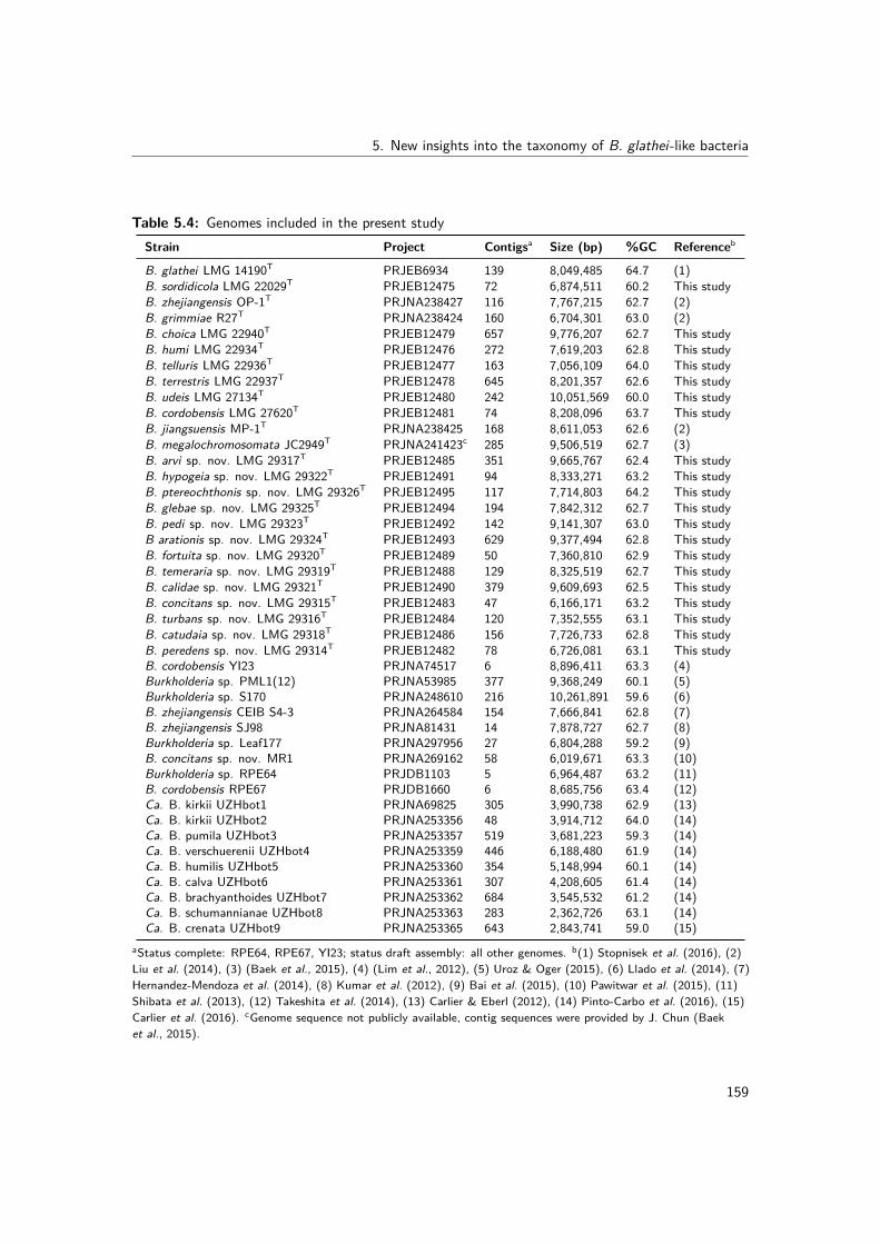
5. New insights into the taxonomy of B. glathei-like bacteria
Table 5.4: Genomes included in the present studyStrain Project Contigsa Size (bp) %GC Referenceb
B. glathei LMG 14190T PRJEB6934 139 8,049,485 64.7 (1)B. sordidicola LMG 22029T PRJEB12475 72 6,874,511 60.2 This studyB. zhejiangensis OP-1T PRJNA238427 116 7,767,215 62.7 (2)B. grimmiae R27T PRJNA238424 160 6,704,301 63.0 (2)B. choica LMG 22940T PRJEB12479 657 9,776,207 62.7 This studyB. humi LMG 22934T PRJEB12476 272 7,619,203 62.8 This studyB. telluris LMG 22936T PRJEB12477 163 7,056,109 64.0 This studyB. terrestris LMG 22937T PRJEB12478 645 8,201,357 62.6 This studyB. udeis LMG 27134T PRJEB12480 242 10,051,569 60.0 This studyB. cordobensis LMG 27620T PRJEB12481 74 8,208,096 63.7 This studyB. jiangsuensis MP-1T PRJNA238425 168 8,611,053 62.6 (2)B. megalochromosomata JC2949T PRJNA241423c 285 9,506,519 62.7 (3)B. arvi sp. nov. LMG 29317T PRJEB12485 351 9,665,767 62.4 This studyB. hypogeia sp. nov. LMG 29322T PRJEB12491 94 8,333,271 63.2 This studyB. ptereochthonis sp. nov. LMG 29326T PRJEB12495 117 7,714,803 64.2 This studyB. glebae sp. nov. LMG 29325T PRJEB12494 194 7,842,312 62.7 This studyB. pedi sp. nov. LMG 29323T PRJEB12492 142 9,141,307 63.0 This studyB arationis sp. nov. LMG 29324T PRJEB12493 629 9,377,494 62.8 This studyB. fortuita sp. nov. LMG 29320T PRJEB12489 50 7,360,810 62.9 This studyB. temeraria sp. nov. LMG 29319T PRJEB12488 129 8,325,519 62.7 This studyB. calidae sp. nov. LMG 29321T PRJEB12490 379 9,609,693 62.5 This studyB. concitans sp. nov. LMG 29315T PRJEB12483 47 6,166,171 63.2 This studyB. turbans sp. nov. LMG 29316T PRJEB12484 120 7,352,555 63.1 This studyB. catudaia sp. nov. LMG 29318T PRJEB12486 156 7,726,733 62.8 This studyB. peredens sp. nov. LMG 29314T PRJEB12482 78 6,726,081 63.1 This studyB. cordobensis YI23 PRJNA74517 6 8,896,411 63.3 (4)Burkholderia sp. PML1(12) PRJNA53985 377 9,368,249 60.1 (5)Burkholderia sp. S170 PRJNA248610 216 10,261,891 59.6 (6)B. zhejiangensis CEIB S4-3 PRJNA264584 154 7,666,841 62.8 (7)B. zhejiangensis SJ98 PRJNA81431 14 7,878,727 62.7 (8)Burkholderia sp. Leaf177 PRJNA297956 27 6,804,288 59.2 (9)B. concitans sp. nov. MR1 PRJNA269162 58 6,019,671 63.3 (10)Burkholderia sp. RPE64 PRJDB1103 5 6,964,487 63.2 (11)B. cordobensis RPE67 PRJDB1660 6 8,685,756 63.4 (12)Ca. B. kirkii UZHbot1 PRJNA69825 305 3,990,738 62.9 (13)Ca. B. kirkii UZHbot2 PRJNA253356 48 3,914,712 64.0 (14)Ca. B. pumila UZHbot3 PRJNA253357 519 3,681,223 59.3 (14)Ca. B. verschuerenii UZHbot4 PRJNA253359 446 6,188,480 61.9 (14)Ca. B. humilis UZHbot5 PRJNA253360 354 5,148,994 60.1 (14)Ca. B. calva UZHbot6 PRJNA253361 307 4,208,605 61.4 (14)Ca. B. brachyanthoides UZHbot7 PRJNA253362 684 3,545,532 61.2 (14)Ca. B. schumannianae UZHbot8 PRJNA253363 283 2,362,726 63.1 (14)Ca. B. crenata UZHbot9 PRJNA253365 643 2,843,741 59.0 (15)
aStatus complete: RPE64, RPE67, YI23; status draft assembly: all other genomes. b(1) Stopnisek et al. (2016), (2)Liu et al. (2014), (3) (Baek et al., 2015), (4) (Lim et al., 2012), (5) Uroz & Oger (2015), (6) Llado et al. (2014), (7)Hernandez-Mendoza et al. (2014), (8) Kumar et al. (2012), (9) Bai et al. (2015), (10) Pawitwar et al. (2015), (11)Shibata et al. (2013), (12) Takeshita et al. (2014), (13) Carlier & Eberl (2012), (14) Pinto-Carbo et al. (2016), (15)Carlier et al. (2016). cGenome sequence not publicly available, contig sequences were provided by J. Chun (Baeket al., 2015).
159

III. Experimental work
5.2.3.5 Publicly available genomes
Twenty three publicly available whole-genome sequences of BGC bacteria were downloadedfrom the NCBI database (Table 5.4). B. gladioli BSR3 (Seo et al., 2011) was used as anoutgroup in all phylogenomic analyses. For B. megalochromosomata JC2949T the whole-genome sequence was not publicly available (February 1st, 2016) and the contig sequenceswere provided by J. Chun (Baek et al., 2015). For B. sordidicola S170, B. zhejiangensis CEIBS4-3 and B. megalochromosomata JC2949T no annotation was available and annotation wasperformed using Prokka as described above.
5.2.3.6 Phylogenomic analysis
The latest version of the GBDP approach was applied (Meier-Kolthoff et al., 2013) to calculatethe intergenomic distance between each pair of genomes (based on the nucleotide data)and included the calculation of 100 replicate distances to assess pseudo-bootstrap support(Meier-Kolthoff et al., 2014a). Distance calculations were conducted under the recommendedsettings of the Genome-to-Genome Distance Calculator (GGDC 2.1; http://ggdc.dsmz.de), asdescribed earlier (Meier-Kolthoff et al., 2013). The GBDP trimming algorithm and formulad5 were chosen because of their advantages for phylogenetic inference (Meier-Kolthoff et al.,2014a) and according distance matrices were prepared (a single matrix for the originaldistances plus 100 matrices containing the replicates). A phylogenomic tree with branchsupport (Meier-Kolthoff et al., 2014a) was inferred using FastME v2.07 with tree bisectionand reconnection post-processing (Lefort et al., 2015). Moreover, pairwise digital DNA-DNAhybridization (dDDH) values and their confidence intervals were also determined using GGDC2.1 under recommended settings (Meier-Kolthoff et al., 2013). The potential affiliationof the novel strains to existing species was determined by clustering using a 70% dDDHradius around each of the 12 BGC type strains as previously applied (Liu et al., 2015).Visualization and annotation of the phylogenetic tree was performed using iTOL (Letunic &Bork, 2011).As an alternative for the GBDP method, an extended MLSA analysis was performed in which awhole-genome phylogeny was calculated based on single-copy orthologous genes as describedpreviously (Pinto-Carbo et al., 2016). In short, single-copy orthologs were identified usingblastp and OrthoMCL v2.0.9 (with e-value cut-off 1E-6 and 50% match cut-off) (Fischeret al., 2011) and aligned based on their amino acid sequences using MUSCLE. The alignmentswere trimmed using TrimAl (removing positions with gaps in more than 50% of the sequences)and concatenated to construct a maximum likelihood tree using RaXML v7.4.2 (Stamatakis,2014) with the WAG amino acid substitution model and 100 rapid bootstrap analyses.
160

5. New insights into the taxonomy of B. glathei-like bacteria
5.2.3.7 Phenotypic characterization
Phenotypic and cellular fatty acid analyses were performed as described previously (Draghiet al., 2014).
5.2.4 Results
5.2.4.1 16S rRNA gene sequence analysis
The 16S rRNA gene sequences determined in the present study are publicly available throughthe GenBank/EMBL/DDBJ accession numbers LT158612-LT158624.
5.2.4.2 gyrB gene sequence analysis
Partial gyrB gene sequences were compared to those of the type strains of the 12 validlynamed BGC species (Fig. 5.3). The 17 unclassified isolates represented 13 taxa which showed83.4-96.2% pairwise identity with the gyrB sequences of the type strains of other BGCspecies. The gyrB gene sequences determined in the present study are publicly availablethrough the GenBank/EMBL/DDBJ accession numbers LT158625-LT158641.
5.2.4.3 Whole-genome sequencing
To further characterize the taxonomic status of these 13 taxa, we determined the whole-genome sequence of one strain per gyrB cluster and of B. sordidicola LMG 22029T, B. choicaLMG 22940T, B. humi LMG 22934T, B. telluris LMG 22936T, B. terrestris LMG 22937T,B. udeis LMG 27134T and B. cordobensis LMG 27620T. The assembly of the IlluminaHiSeq 150 bp paired end reads resulted in assemblies with 47 to 657 contigs and a total of6,166,171 to 10,051,569 bp (Table 5.4). The annotated assemblies of these 20 genomeswere submitted to the European Nucleotide Archive and are publicly available through theGenBank/EMBL/DDBJ accession numbers listed in Table 5.4 and the species descriptions.The genome sequences of the remaining five BGC type strains and of 18 additional strainswere publicly available (Table 5.4).
5.2.4.4 DNA base composition
The G+C content of all type strains was calculated from their genome sequences and rangedfrom 62.4 to 64.2 mol% (Table 5.4).
161

III. Experimental work
Burkholderia glebae sp. nov. LMG 22938 (LT158638)
Burkholderia glebae sp. nov. LMG 29325T (LT158636)
Burkholderia fortuita sp. nov. LMG 29320T (LT158631)
Burkholderia calidae sp. nov. LMG 29321T (LT158632)
Burkholderia pedi sp. nov. R-52605 (LT158639)
Burkholderia pedi sp. nov. LMG 29323T (LT158634)
Burkholderia turbans sp. nov. LMG 29316T (LT158627)
Burkholderia jiangsuensis MP-1T (KJ535375)
Burkholderia megalochromosomata JC2949T (from WGS)
Burkholderia arvi sp. nov. LMG 29317T (LT158628)
Burkholderia catudaia sp. nov. LMG 29318T (LT158629)
Burkholderia cordobensis LMG 27620T (HG324055)
Burkholderia zhejiangensis LMG 27258T (HF547269)
Burkholderia temeraria sp. nov. LMG 29319T (LT158630)
Burkholderia hypogeia sp. nov. LMG 29322T (LT158633)
Burkholderia grimmiae LMG 27580T (HG324054)
Burkholderia concitans sp. nov. LMG 29315T (LT158626)
Burkholderia concitans sp. nov. R-46586 (LT158641)
Burkholderia peredens sp. nov. LMG 29314T (LT158625)
Burkholderia ptereochthonis sp. nov. LMG 29326T (LT158637)
Burkholderia sordidicola LMG 22029T (HE985176)
Burkholderia udeis LMG 27134T (HE985178)
Burkholderia arationis sp. nov. LMG 29324T (LT158635)
Burkholderia arationis sp. nov. R-23361 (LT158640)
Burkholderia glathei LMG 14190T (HE985172)
Burkholderia humi LMG 22934T (HE985164)
Burkholderia terrestris LMG 22937T (HE985170)
Burkholderia choica LMG 22940T (HE985163)
Burkholderia telluris LMG 22936T (HE985161)
Burkholderia kururiensis LMG 19447T (HQ849201)
100
100
100
95
78
81
74
70
99
85
65
88
99
51
0.05
Figure 5.3: Phylogenetic tree based on partial gyrB sequences of the 17 isolates in this study andtype strains of phylogenetically related Burkholderia species. The optimal tree (highest log likelihood)was constructed using the maximum likelihood method and general time reversible model in MEGA6(Tamura et al., 2013). A discrete gamma distribution was used to model evolutionary rate differencesamong sites (5 categories (+G, parameter = 0.5462)) and allowed for some sites to be evolutionarilyinvariable ([+I], 37.9331% sites). The percentage of replicate trees in which the associated taxaclustered together in the bootstrap test (1000 replicates) are shown next to the branches if greaterthan 50%. For B. megalochromosomata JC2949T the gyrB gene sequence was extracted from thegenome sequence. The gyrB sequence of B. kururiensis LMG 19447T was used as outgroup. Thescale bar indicates the number of substitutions per site.
162
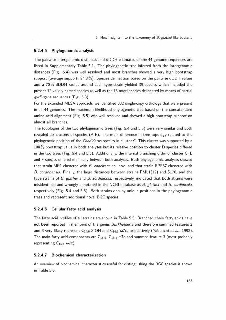
5. New insights into the taxonomy of B. glathei-like bacteria
5.2.4.5 Phylogenomic analysis
The pairwise intergenomic distances and dDDH estimates of the 44 genome sequences arelisted in Supplementary Table 5.1. The phylogenetic tree inferred from the intergenomicdistances (Fig. 5.4) was well resolved and most branches showed a very high bootstrapsupport (average support: 94.8%). Species delineation based on the pairwise dDDH valuesand a 70% dDDH radius around each type strain yielded 39 species which included thepresent 12 validly named species as well as the 13 novel species delineated by means of partialgyrB gene sequences (Fig. 5.3).For the extended MLSA approach, we identified 332 single-copy orthologs that were presentin all 44 genomes. The maximum likelihood phylogenetic tree based on the concatenatedamino acid alignment (Fig. 5.5) was well resolved and showed a high bootstrap support onalmost all branches.The topologies of the two phylogenomic trees (Fig. 5.4 and 5.5) were very similar and bothrevealed six clusters of species (A-F). The main difference in tree topology related to thephylogenetic position of the Candidatus species in cluster C. This cluster was supported by a100% bootstrap value in both analyses but its relative position to cluster D species differedin the two trees (Fig. 5.4 and 5.5). Additionally, the internal branching order of cluster C, Eand F species differed minimally between both analyses. Both phylogenomic analyses showedthat strain MR1 clustered with B. concitans sp. nov. and that strain RPE67 clustered withB. cordobensis. Finally, the large distances between strains PML1(12) and S170, and thetype strains of B. glathei and B. sordidicola, respectively, indicated that both strains weremisidentified and wrongly annotated in the NCBI database as B. glathei and B. sordidicola,respectively (Fig. 5.4 and 5.5). Both strains occupy unique positions in the phylogenomictrees and represent additional novel BGC species.
5.2.4.6 Cellular fatty acid analysis
The fatty acid profiles of all strains are shown in Table 5.5. Branched chain fatty acids havenot been reported in members of the genus Burkholderia and therefore summed features 2and 3 very likely represent C14:0 3-OH and C16:1 ω7c, respectively (Yabuuchi et al., 1992).The main fatty acid components are C16:0, C18:1 ω7c and summed feature 3 (most probablyrepresenting C16:1 ω7c).
5.2.4.7 Biochemical characterization
An overview of biochemical characteristics useful for distinguishing the BGC species is shownin Table 5.6.
163
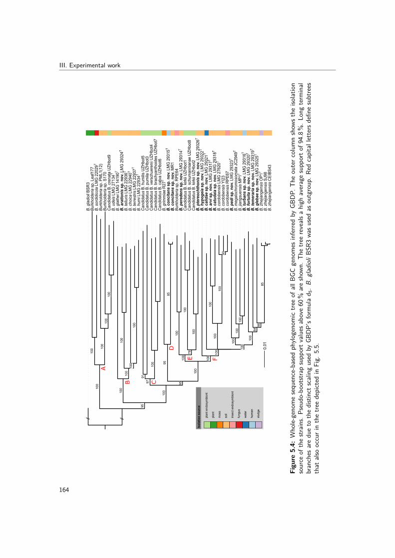
III. Experimental work
0.01
Iso
lati
on
so
urc
e
plan
t end
osym
bion
t
plan
t
mos
s
soil
inse
ct e
ndos
ymbi
ont
fung
us
wat
er
hum
an
slud
ge
B. g
ladi
oli B
SR
3
Bur
khol
deria
sp.
PM
L1(1
2)
Can
dida
tus
B. c
rena
ta U
ZH
bot9
B. u
deis
LM
G 2
7134
T
Bur
khol
deria
sp.
S17
0
Bur
khol
deria
sp.
Lea
f177
B. s
ordi
dico
la L
MG
220
29T
B. t
erre
stris
LM
G 2
2937
T
B. h
umi L
MG
229
34T
B. c
hoic
a LM
G 2
2940
T
B. a
rati
on
is s
p. n
ov.
LM
G 2
9324
T
B. t
ellu
ris L
MG
229
36T
B. g
lath
ei L
MG
141
90T
B. z
hejia
ngen
sis
SJ9
8B
. zhe
jiang
ensi
s C
EIB
S43
B. z
hejia
ngen
sis
OP
1TB
. gle
bae
sp
. no
v. L
MG
293
25T
B. t
emer
aria
sp
. no
v. L
MG
293
19T
B. f
ort
uit
a sp
. no
v. L
MG
293
20T
B. m
egal
ochr
omos
omat
a JC
2949
T
B. j
iang
suen
sis
MP
1T
B. t
urb
ans
sp. n
ov.
LM
G 2
9316
T
B. p
edi s
p. n
ov.
LM
G 2
9323
T
B. c
ordo
bens
is L
MG
276
20T
B. c
ordo
bens
is Y
I23
B. c
ordo
bens
is R
PE
67
B. a
rvi s
p. n
ov.
LM
G 2
9317
T
B. c
atu
dai
a sp
. no
v. L
MG
293
18T
B. c
alid
ae s
p. n
ov.
LM
G 2
9321
TB
. hyp
og
eia
sp. n
ov.
LM
G 2
9322
T
Can
dida
tus
B. k
irkii
UZ
Hbo
t1C
andi
datu
s B
. sch
uman
nian
ae U
ZH
bot8
Can
dida
tus
B. k
irkii
UZ
Hbo
t2B
. pte
reo
chth
on
is s
p. n
ov.
LM
G 2
9326
T
Bur
khol
deria
sp.
RP
E64
B. p
ered
ens
sp. n
ov.
LM
G 2
9314
T
B. c
on
cita
ns
sp. n
ov.
LM
G 2
9315
T
B. c
on
cita
ns
sp. n
ov.
MR
1
B. g
rimm
iae
R27
T
Can
dida
tus
B. h
umili
s U
ZH
bot5
Can
dida
tus
B. p
umila
UZ
Hbo
t3C
andi
datu
s B
. ver
schu
eren
ii U
ZH
bot4
Can
dida
tus
B. b
rach
yant
hoid
es U
ZH
bot7
Can
dida
tus
B. c
alva
UZ
Hbo
t6
100
100
100
100
100
95
100
100
100
100
100
100
95
100
100
100
100
100
8486
85
100
100
100
100
100
90
100
100
7810
0
100
100
9585
97
91
100
B C
D
E F
A
Figu
re5.4:
Who
le-geno
mesequ
ence-based
phylo
geno
mic
treeof
allBG
Cgeno
mes
inferre
dby
GBDP
.The
outerc
olum
nshow
sthe
isolat
ion
source
ofthestr
ains.
Pseudo
-boo
tstrapsupp
ortv
alues
above60
%areshow
n.Th
etre
ereveals
ahigh
averagesupp
orto
f94.8%
.Lon
gterm
inal
branches
aredu
eto
thedistincts
calingused
byGB
DP’sform
ulad 5.B
.gladioliB
SR3wa
susedas
outgroup
.Red
capitalletters
defin
esubtrees
that
alsooccurinthetre
edepicted
inFig.
5.5.
164

5. New insights into the taxonomy of B. glathei-like bacteria
B. calidae sp. nov. LMG 29321T
Candidatus B. crenata UZHbot9
B. peredens sp. nov. LMG 29314T
Burkholderia sp. PML1(12)
B. humi LMG 22934T
Burkholderia sp. S170
B. cordobensis RPE67
B. megalochromosomata JC2949T
B. glebae sp. nov. LMG 29325T
Candidatus B. kirkii UZHbot1
Candidatus B. pumila UZHbot3
B. arationis sp. nov. LMG 29324T
B. turbans sp. nov. LMG 29316T
Candidatus B. humilis UZHbot5
B. gladioli BSR3
B. terrestris LMG 22937T
B. arvi sp. nov. LMG 29317T
Candidatus B. calva UZHbot6
B. cordobensis YI23
B. choica LMG 22940T
B. sordidicola LMG 22029T
B. jiangsuensis MP1T
B. zhejiangensis SJ98
B. fortuita sp. nov. LMG 29320T
Candidatus B. verschuerenii UZHbot4
B. catudaia sp. nov. LMG 29318T
B. ptereochthonis sp. nov. LMG 29326T
B. grimmiae R27T
B. udeis LMG 27134T
B. temeraria sp. nov. LMG 29319T
B. zhejiangensis OP1T
B. cordobensis LMG 27620T
B. telluris LMG 22936T
Burkholderia sp. Leaf177
B. hypogeia sp. nov. LMG 29322T
B. concitans sp. nov. LMG 29315T
B. glathei LMG 14190T
Burkholderia sp. RPE64
B. pedi sp. nov. LMG 29323T
B. concitans sp. nov. MR1
Candidatus B. brachyanthoides UZHbot7
Candidatus B. schumannianae UZHbot8
B. zhejiangensis CEIBS43
Candidatus B. kirkii UZHbot2
60
100
100
79
100
100
100
100
100
100
100
100
100
100 100
100
89
100
100
100
98
100
100
100
87
100
100
100
100
100
100
100
100
100
100
79
100
100100
100
100
Tree scale: 0.01
A
B
D
C
E
F
Figure 5.5: Whole-genome phylogeny based on single-copy orthologs of all BGC genomes. Thephylogenetic tree was constructed using the WAG protein substitution model and RAxML and is basedon an amino acid alignment with 105,141 positions from 332 single-copy orthologous genes. Thepercentage of replicate trees in which the associated taxa clustered together in the bootstrap test(100 replicates) are shown next to the branches if greater than 60%. B. gladioli BSR3 was used asoutgroup. Red capital letters define subtrees that also occur in the tree depicted in Fig. 5.4.
165

III. Experimental workTa
ble5.5:
Meanfatty
acid
compo
sitionof
allexam
ined
strainso
fBGC
species
Fattyacid
12
34
56
78
910
1112
C 12:0
ND3.70
NDTR
NDND
NDND
4.63
±0.07
NDND
1.01
C 14:0
4.29
±0.13
0.79
4.08
±0.13
4.02
3.54
±0.13
3.71
±0.27
3.00
±0.01
3.80
TR4.16
±0.04
4.36
3.97
C 16:0
19.11±
1.93
18.29
16.24±
0.64
12.14
20.37±
4.40
12.00±
0.76
13.31±
0.15
16.02
15.72±
0.96
17.00±
0.80
15.58
15.23
C 16:02-OH
2.06
±0.86
3.32
3.37
±0.82
4.41
3.26
±1.54
2.43
±0.55
1.61
±0.14
2.29
2.05
±0.10
2.49
±0.79
4.10
1.53
C 16:03-OH
6.13
±0.49
4.90
5.91
±0.13
5.02
6.93
±2.32
5.17
±0.38
5.05
±0.04
5.36
6.75
±0.91
5.92
±0.04
6.44
4.89
C 16:12-OH
TR4.06
TR4.37
ND1.71
±0.32
TR1.29
2.02
±0.22
TR1.77
TRC 1
7:0cyclo
13.05±
2.41
13.94
4.96
±0.65
TR14.30±
4.67
7.64
±2.05
4.54
±0.13
6.58
6.57
±0.99
5.31
±1.86
3.96
TRC 1
8:1ω7c
31.10±
1.83
27.61
36.88±
1.05
38.78
30.97±
1.41
42.19±
1.77
43.66±
0.55
38.00
32.56±
1.27
36.62±
1.20
35.48
42.25
C 19:0cycloω8c
2.64
±1.29
7.01
2.14
±0.37
NDND
TRTR
ND3.61
±0.68
1.82
±0.72
1.73
TRSu
mmed
feature2
7.08
±0.53
4.86
6.82
±0.35
5.32
7.80
±2.28
6.24
±0.42
5.71
±0.08
6.18
7.53
±0.60
7.21
±0.15
8.13
7.04
Summed
feature3
12.79±
2.79
11.53
17.43±
0.60
23.16
12.84±
4.21
17.14±
1.55
20.55±
0.57
18.98
17.20±
1.46
17.55±
2.11
18.44
20.64
Fattyacid
1314
1516
1718
1920
2122
2324
25
C 12:0
NDND
NDTR
TRND
NDND
NDTR
NDND
NDC 1
4:0
4.13
4.40
4.18
3.91
±0.13
4.11
±0.28
4.08
±0.78
4.36
4.21
2.15
4.40
±0.05
4.86
4.43
4.31
C 16:0
15.87
18.25
16.71
15.38±
2.61
14.88±
0.02
17.09±
0.73
14.58
14.51
17.84
16.34±
1.82
26.27
16.97
15.11
C 16:02-OH
1.45
2.35
4.08
4.33
±0.53
3.31
±1.51
1.71
±1.71
3.45
3.68
2.48
7.31
±1.43
4.20
2.26
2.46
C 16:03-OH
5.28
5.64
6.94
5.63
±0.21
6.49
±0.16
10.29±
2.71
5.83
5.67
4.89
6.27
±1.50
4.68
5.16
6.27
C 16:12-OH
NDTR
1.22
1.91
±0.05
1.27
±0.39
TR1.22
TRND
1.99
±0.31
2.21
TR1.52
C 17:0cyclo
4.01
6.77
9.10
5.97
±3.01
7.56
±4.61
7.65
±7.65
5.06
7.85
4.56
13.95±
3.77
13.76
3.64
2.02
C 18:1ω7c
41.10
31.35
27.67
32.48±
4.27
32.00±
3.63
32.23±
4.96
34.89
35.05
41.75
23.16±
4.03
20.42
34.53
35.09
C 19:0cycloω8c
1.73
2.48
3.21
3.80
±1.45
1.96
±1.22
ND1.64
4.40
TR8.65
±4.56
3.60
1.20
NDSu
mmed
feature2
6.16
8.14
8.81
7.45
±0.86
8.73
±0.59
10.12±
2.55
7.13
6.84
6.12
6.45
±1.41
5.83
7.90
8.18
Summed
feature3
19.32
19.45
15.97
18.32±
0.18
18.44±
5.56
15.70±
5.44
20.31
15.52
18.83
8.82
±4.11
12.69
23.12
25.05
Species
:1,
B.g
lathei
(5strains);2
,B.sordidicola
(1);3,
B.z
hejiang
ensis
(3);4,
B.g
rimmiae(1);5,
B.c
hoica(2);6,
B.h
umi(6);7,
B.telluris
(2);8,
B.terrestris
(1);9,
B.u
deis
(3);10,B
.cordo
bensis
(2);11,B
.jiang
suensis
(1);12,B
.megalochrom
osom
ata(1);13,B
urkh
olderia
arvi
sp.no
v.(1);14,B
urkh
olderia
hypo
geia
sp.
nov.
(1);15,B
urkh
olderia
ptereochthon
issp.no
v.(1);16,B
urkh
olderia
glebae
sp.no
v.(2);
17,B
urkh
olderia
pedi
sp.no
v.(2);18,B
urkh
olderia
arationissp.no
v.(2);19,B
urkh
olderia
fortuita
sp.no
v.(1);
20,B
urkh
olderia
temeraria
sp.no
v.(1);
21,B
urkh
olderia
calidae
sp.no
v.(1);22,B
urkh
olderia
concita
nssp.no
v.(2);
23,B
urkh
olderia
turbanssp.no
v.(1);
24,B
urkh
olderia
catudaia
sp.no
v.(1);25,B
urkh
olderia
peredens
sp.no
v.(1).
DataforB
.glathei,B
.sordidicola,B
.zhejiang
ensis
,B.c
hoica,
B.h
umi,B.telluris,B
.terrestris
andB.u
deis
were
extractedfro
mVa
ndam
meet
al.(2013a).DataforB
.cordo
bensis
andB.g
rimmiaewe
reextractedfro
mDragh
ietal.(
2014).
Allo
ther
data
arefro
mthepresentstud
y.Va
lues
aremean±
SDpercentagesof
totalfatty
acids.
Thosefattyacidsforw
hich
the
meanam
ount
fora
lltaxa
was<1%
areno
tinclu
ded,
therefore,
thepercentagesmay
notaddup
to100%
.TR
,trace
amou
nt(<
1%);ND,n
otdetected.Su
mmed
feature2comprise
siso
-C16:1Ia
nd/orC
14:03-OH;sum
med
feature3comprise
siso
-C15:02-OH
and/
orC 1
6:1ω7c.
166

5. New insights into the taxonomy of B. glathei-like bacteriaTa
ble5.6:
Diffe
rential
biochemica
lcharacteristics
ofall
exam
ined
strainso
fBGC
species
Characteristic
12
34
65
78
910
1112
1314
1516
1718
1920
2122
2324
25
Grow
that
15◦ C
++
w+
++
++
+−
+w
++
++
++
++
++
++
++
++
+20
◦ C+
+ND
++
++
++
NDND
++
++
++
++
++
++
++
++
++
37◦ C
+−
++
w−
w−
−+
+−
ww
+−
−−
−−
−+
ww
+−
ww
wpH
5−
−−
+−
−−
−−
−−
−−
−−
−−
−−
−−
−−
−−
−−
−−
pH6
++
++
++
++
++
++
+w
−−
−+
+−
+w
ww
++
ww
−pH
7+
++
++
++
++
++
++
−+
w+
w+
−−
ww
w+
−+
w+
pH8
−w
++
−+
−−
+w
−+
−−
−−
+−
+−
−−
−−
−−
−−
−Hy
drolysisof
Tween60
+−
+ND
+−
+NG
NG+
++
++
++
++
++
++
−−
++
++
+Tw
een80
−−
++
−−
−−
−+
++
−−
−−
−−
−−
−−
−−
−−
−−
−AP
I20N
ENitra
teredu
ction
−+
++
−−
+−
+w
+−
++
−+
++
+−
−−
−+
−−
−+
−Ur
ease
−−
++
−−
−−
−−
−−
−−
−−
−−
−−
−−
−−
−−
−−
−β-Galactosid
ase
−+
−−
−−
−−
w−
−+
w−
−−
−w
+−
−−
−−
−−
−−
−As
similatio
nof
Arabinose
w+
++
w−
+−
++
w−
++
−+
w+
++
++
++
w+
++
wManno
se+
++
+−
++
++
++
++
++
++
++
++
++
++
++
++
Mannitol
w+
−+
w+
++
−+
++
++
++
++
++
++
++
++
++
+N-
Acetylg
lucosamine
++
++
w+
++
++
++
++
++
++
++
++
++
++
++
+Glucon
ate
++
++
w+
++
++
++
++
++
++
++
++
++
++
++
+Ca
prate
+−
+−
−w
+w
−−
−−
−−
−−
−−
ww
w−
−+
−−
+−
−Malate
+w
++
w+
++
++
++
++
++
w+
w+
++
++
++
++
+Citra
te+
−−
−−
++
++
−w
−w
−−
+w
−−
++
−w
ww
−−
−−
Phenyla
cetate
+−
+−
−+
++
−+
++
++
++
++
+w
++
++
ww
++
+En
zymeactiv
ity(A
PIZY
M)
C4lipase
++
−+
w+
w+
+−
+−
−+
+w
−−
++
+−
+−
++
w−
wC8
lipase
w+
−+
++
ww
+−
w−
−w
−−
−−
w−
w−
−w
ww
−−
wVa
linearyla
midase
w−
−−
w−
−w
−−
−−
−−
−−
−−
+−
w−
−−
++
−−
−Cy
stinearyla
midase
−−
−−
+−
−−
−−
−−
−−
−−
−−
w−
−−
−−
−w
−−
−β-Galactosid
ase
−+
−−
−−
−−
+−
−−
−−
−−
−−w
−−
−−
−−
−−
−−
Species
:1,
B.g
lathei
LMG14190T
;2,B
.sordidicola
LMG22029T
;3,B
.zhejiang
ensis
LMG27258T
;4,B
.grim
miaeR2
7T;5
,B.c
hoicaLM
G22940T
;6,B
.hum
iLMG22934T
;7,B
.telluris
LMG22936T
;8,B
.terrestris
LMG22937T
;9,B
.udeis
LMG27134T
;10,
B.c
ordo
bensis
LMG27620T
;11,
B.jiang
suensis
LMG27927T
;12,
B.m
egalochrom
osom
ataLM
G29263T
;13,
Burkh
olderia
arvi
sp.no
v.LM
G29317T
;14,
Burkh
olderia
hypo
geia
sp.no
v.LM
G29322T
;15,
Burkh
olderia
ptereochthon
issp.no
v.LM
G29326T
;16,
Burkh
olderia
glebae
sp.no
v.LM
G29325T
andLM
G22
938;
17,B
urkh
olderia
pedi
sp.no
v.LM
G29323T
andR-52605;
18,
Burkh
olderia
arationissp.no
v.LM
G29324T
andR-23361;
19,B
urkh
olderia
fortuita
sp.no
v.LM
G29320T
;20,
Burkh
olderia
temeraria
sp.no
v.LM
G29319T
;21,
Burkh
olderia
calidae
sp.no
v.LM
G29321T
;22,
Burkh
olderia
concita
nssp.no
v.LM
G29315T
andR-46586;
23,B
urkh
olderia
turbanssp.no
v.LM
G29316T
;24,
Burkh
olderia
catudaia
sp.no
v.LM
G29318T
;25,
Burkh
olderia
peredens
sp.no
v.LM
G29314T
.Datafor
B.g
lathei,B
.sordidicola,B
.cho
ica,
B.h
umi,B.telluris,B
.terrestris
andB.u
deis
were
extractedfro
mVa
ndam
meet
al.(2013a).DataforB
.grim
miaewe
reextractedfro
mTian
etal.(2013).
DataforB
.cordo
bensis
and
B.z
hejiang
ensis
were
extractedfro
mDragh
ietal.(2014).
Allo
ther
data
arefro
mthepresents
tudy.Te
stresults
ofthetype
strainsaregivenfirst,followe
dby
theremaining
strainsin
theorderg
iven
above.
+,p
resent;−
,absent;w
,weakreactio
n;v,
varia
ble;
ND,n
otdeterm
ined;N
G,no
grow
th.
167

III. Experimental work
5.2.5 Discussion
While soil is a well-known source of free-living Burkholderia species, these organisms oftenlive in close interaction with plants, animals, fungi or amoebae (Marolda et al., 1999; VanBorm et al., 2002; Kikuchi et al., 2011; Verstraete et al., 2013; Stopnisek et al., 2016;Xu et al., 2016). The BGC represents a poorly known line of descent within the genusBurkholderia and most of the 12 validly named BGC species have been isolated from soil. Yet,publicly available sequence data indicate that the taxonomic diversity in this clade is severelyunderestimated (Nogales et al., 2001; Salles et al., 2006b; Pumphrey & Madsen, 2008; Draghiet al., 2014; Verstraete et al., 2014; Peeters et al., 2016). In the present study, gyrB genesequence analysis was used to screen our strain collection and 17 isolates from human andenvironmental samples were identified as B. glathei-like bacteria. The gyrB sequence similaritylevels towards other BGC species suggested that the 17 isolates in this study represented13 novel species (Fig. 5.3). To further characterize the taxonomic status of these isolates,we analyzed the genome sequence of 13 isolates representative for the 13 gyrB sequenceclusters and of 7 BGC type strains and compared those to 23 whole-genome sequences ofBGC strains that were publicly available. Additionally, we also studied their chemotaxonomicand biochemical properties to comply with the polyphasic taxonomic consensus approach tobacterial systematics (Vandamme et al., 1996).In this genomics era, state-of-the-art sequencing technologies enable direct access to theinformation contained in whole-genome sequences and it is no longer adequate to deducegenome relatedness through traditional DDH experiments (Vandamme & Peeters, 2014;Whitman, 2015). Genomic taxonomy can be studied through various parameters includingANI, GBDP, MUMi and core gene identity (Konstantinidis & Tiedje, 2005a; Goris et al.,2007; Deloger et al., 2009; Vanlaere et al., 2009; Meier-Kolthoff et al., 2013). Although thereis a general consensus that genome sequencing could revolutionize prokaryotic systematics(Sutcliffe et al., 2013; Meier-Kolthoff et al., 2014c; Rossello-Mora & Amann, 2015; Thompsonet al., 2015), traditional DDH experiments are still being performed and new genome-basedmethods are evaluated in terms of their correspondence to the existing classifications whichare based on DDH data (Wayne et al., 1987; Stackebrandt et al., 2002). The GGDCimplementation of the GBDP method provides a quick and reliable alternative to the wet-labDDH technique and its dDDH prediction capability (including confidence intervals) producesclassifications which correlate better with the traditional DDH values than do any of theANI implementations (Meier-Kolthoff et al., 2013). Among several advantages, GBDP isindependent from genome annotation, is applicable to both nucleotide and amino acid dataand is immune against problems caused by incompletely sequenced or low-quality draft
168

5. New insights into the taxonomy of B. glathei-like bacteria
genomes. Finally, GBDP provides branch support values for the resulting phylogenetic trees(Meier-Kolthoff et al., 2013, 2014a).We complemented the results of the GBDP analysis with a whole-genome sequence-basedphylogeny based on the sequence analysis of 332 single-copy orthologous genes in all BGCgenomes. This extended MLSA approach takes only the coding part of the genomes intoaccount and is therefore not influenced by non-coding sequences or pseudogenes that mighthave a different evolutionary history than the rest of the genome. It depends however ongenome annotation, is unable to cope with problems caused by incompletely sequenced orlow-quality draft genomes, and its calculations are more compute-intensive and cannot becarried out incrementally. Although the GBDP and extended MLSA methods used differentalgorithms, the conclusions drawn from their phylogenies were consistent thus illustrating therobustness of whole-genome sequence-based taxonomic methods (Colston et al., 2014).The GGDC dDDH values and the application of the 70% dDDH cut-off for species delineation(Supplementary Table 5.1) demonstrated that the 13 clusters delineated through gyrBsequence analysis (Fig. 5.3) represented 13 novel BGC species and thus confirmed that gyrBgene sequence analysis is a reliable tool for the identification of Burkholderia species (Tayebet al., 2008; Vandamme et al., 2013a). Both phylogenomic analyses identified strain MR1,which was isolated from Florida golf course soil and which was shown to reduce the herbicidemethylarsenate, as B. concitans sp. nov. Next to strain YI23, which was previously identifiedas B. cordobensis by Draghi et al. (2014), the present study also identified strain RPE67,which was isolated from the gut of a stink bug, as B. cordobensis. Finally, both phylogenomicanalyses also showed that strain PML1(12), an ectomycorrhizosphere-inhabiting bacteriumwith mineral-weathering ability (Uroz & Oger, 2015), strain S170, a potential plant growthpromoter isolated from coniferous forest soil (Llado et al., 2014), strain RPE64, a bacterialsymbiont of the bean bug Riptortus pedestris (Shibata et al., 2013) and strain Leaf177, anArabidopsis leaf isolate (Bai et al., 2015) all represent novel BGC species.Burkholderia genomes vary in size from 3.75 Mb (B. rhizoxinica HKI 454) to 11.3 Mb (B.terrae BS001), are characterized by a high G+C content (60-68%) and consist of multiplereplicons (Winsor et al., 2008; Ussery et al., 2009). The DNA G+C content of the 13 novelspecies was calculated from their genome sequences and was in the range of that reportedfor other BGC species (60-65 mol%). For 10 of the 12 established BGC species, the G+Ccontent was previously calculated by traditional wet-lab methods and the reported valuesdiffered by 0.1 to 3.3 mol% from the values calculated from their genome sequences (Table5.7). As reported by Meier-Kolthoff et al. (2014c) the G+C content calculations based ongenome sequences show a higher precision than calculations based on traditional wet-labmethods because the latter methods do not count nucleotides but estimate the genomic
169

III. Experimental work
G+C content based on the physical properties of the extracted and/or digested genomicDNA. The difference between literature data (Lim et al., 2003; Lu et al., 2012; Tian et al.,2013) and the genome sequence-based G+C content values of B. sordidicola LMG 22029T,B. zhejiangensis OP-1T and B. grimmiae R27T is larger than 1% and we therefore presentemended descriptions of these species. The genome sizes of the type strains of the 13 novelspecies ranged from 6.2 Mb (B. concitans sp. nov. LMG 29315T) to 9.7 Mb (B. arvi sp.nov. LMG 29317T) and corresponded with the genome sizes of other free-living BGC species(Table 5.4). Consistent with reductive genome evolution in obligatory symbionts, the smallestBGC genomes belong to the obligatory leaf endosymbionts (2.4 to 6.2 Mb) (Carlier & Eberl,2012; Carlier et al., 2016; Pinto-Carbo et al., 2016).
Table 5.7: G+C content (mol%) of validly named BGC speciesStrain Wet-lab calculation Calculation from WGS
B. glathei LMG 14190T 64.8 (Zolg & Ottow, 1975) 64.7B. sordidicola LMG 22029T 61.3 (Lim et al., 2003) 60.2B. zhejiangensis OP-1T 59.4 (Lu et al., 2012) 62.7B. grimmiae R27T 64.6 (Tian et al., 2013) 63.0B. choica LMG 22940T 63 (Vandamme et al., 2013a) 62.7B. humi LMG 22934T 63 (Vandamme et al., 2013a) 62.8B. telluris LMG 22936T 64 (Vandamme et al., 2013a) 64.0B. terrestris LMG 22937T 62 (Vandamme et al., 2013a) 62.6B. udeis LMG 27134T 60 (Vandamme et al., 2013a) 60.0B. cordobensis LMG 27620T 63.6 (Draghi et al., 2014) 63.7B. jiangsuensis MP-1T - 62.6B. megalochromosomata JC2949T - 62.7
WGS, whole-genome sequence
Biochemically, these novel species are similar to their nearest neighbors. However, testsparticularly useful for distinguishing BGC species are growth at 37 ◦C and at pH 8, hydrolysisof tween 60 and 80, nitrate reduction, assimilation of arabinose, caprate and citrate, beta-galactosidase activity and C4 lipase (Table 5.6). The most discriminating fatty acids areC16:0 3-OH, C17:0 cyclo, C19:0 cyclo ω8c and summed features 2 and 3 (Table 5.5). Theoverall fatty acid profiles of the novel taxa are similar to those of their nearest neighbors andsupport their placement in the genus Burkholderia (Yabuuchi et al., 1992).The present study again underscores the multifaceted nature of Burkholderia bacteria (Coenye& Vandamme, 2003; Mahenthiralingam et al., 2005) and highlights that also BGC specieshave evolved a broad range of interactions with different hosts. B. cordobensis is a strikingexample of phenotypic and geographic breadth: it was recovered from agricultural soil inArgentina (strain LMG 27620T) (Draghi et al., 2014), from golf course soil in South Korea(strain YI23) (Lim et al., 2012) and from the gut of the bean bug Riptortus pedestris in
170

5. New insights into the taxonomy of B. glathei-like bacteria
Japan (strain RPE67) (Takeshita et al., 2014). The two latter strains (YI23 and RPE67)have fenitrothion degrading properties. The former two strains (LMG 27620T and YI23) werefree-living but the latter (RPE67) is an endosymbiont of stink bugs that is not verticallytransmitted but acquired from soil by the nymphal insect (Kikuchi et al., 2007). Theinsecticide resistance to fenitrothion in the pest insects was shown to be established by theendosymbiotic Burkholderia strain in the insect gut (Kikuchi et al., 2012) and was shownto emerge as a consequence of repeated insecticide use (Tago et al., 2015). The Riptortuspedestris-B. cordobensis association thus appears to be a rather young endosymbiosis andcontrasts with the symbiosis observed between plant species of the Rubiaceae and Primulaceaefamilies and several Candidatus Burkholderia species. The latter Candidatus designation isa provisional taxonomic status for organisms that have been characterized but that cannotbe cultivated at present (Schleifer, 2009). These obligate leaf endosymbionts are verticallytransmitted and represent an obligatory symbiosis which was estimated to originate millionsof years ago (Lemaire et al., 2011b).BGC species harbor both beneficial and pathogenic strains. Strains PML1(12) and S170 showbiotechnological potential for mineral-weathering and plant growth promotion, respectively,and are exemplary for the metabolic versatility of Burkholderia organisms (Llado et al., 2014;Uroz & Oger, 2015). Mineral-weathering bacteria dissolute key nutrients from minerals andthereby increase the bioavailability of chemical nutrients in the environment (Uroz et al.,2009). On the other hand, three strains analyzed in the present study were isolated fromhuman clinical samples, i.e. blood, pleural fluid and lung tissue (Table 5.3) and were classifiedas two novel species (Burkholderia concitans sp. nov. and Burkholderia turbans sp. nov.).They represent, to our knowledge, the first examples of human clinical isolates in the B.glathei clade. Strikingly, strain MR1, which was isolated from Florida golf course soil andshown to reduce the herbicide methylarsenate, was also identified as Burkholderia concitanssp. nov., and this species thus represents yet another human clinical Burkholderia species withinteresting biotechnological properties (Coenye et al., 2001b; Coenye & Vandamme, 2003;Goris et al., 2004; Mahenthiralingam et al., 2005). This study therefore further underscoresthat there is no phylogenetic subdivision in the genus Burkholderia that distinguishes beneficialfrom pathogenic strains (Angus et al., 2014; Sawana et al., 2014; Estrada-de los Santos et al.,2015; Dobritsa & Samadpour, 2016).In summary, the present study provides genotypic, chemotaxonomic and phenotypic data whichenable the differentiation of 13 novel species in the genus Burkholderia and we propose thenames Burkholderia arvi sp. nov., Burkholderia hypogeia sp. nov., Burkholderia ptereochtho-nis sp. nov., Burkholderia glebae sp. nov., Burkholderia pedi sp. nov., Burkholderia arationissp. nov., Burkholderia fortuita sp. nov., Burkholderia temeraria sp. nov., Burkholderia calidae
171

III. Experimental work
sp. nov., Burkholderia concitans sp. nov., Burkholderia turbans sp. nov., Burkholderiacatudaia sp. nov. and Burkholderia peredens sp. nov., with strains LMG 29317T, LMG29322T, LMG 29326T, LMG 29325T, LMG 29323T, LMG 29324T, LMG 29320T, LMG29319T, LMG 29321T, LMG 29315T, LMG 29316T, LMG 29318T and LMG 29314T as typestrains, respectively. By making reference cultures and whole-genome sequences of each ofthese versatile bacteria publicly available, we aim to contribute to future knowledge aboutthe metabolic versatility and pathogenicity of Burkholderia organisms.
5.2.5.1 Description of Burkholderia arvi sp. nov.
Burkholderia arvi (ar’vi. L. gen. n. arvi of a field).Cells are Gram-negative, non-motile rods (less than 1 µm wide and about 1 µm long) withrounded ends that occur as single units or in pairs. After 48 h of incubation on trypticasesoy agar at 28 ◦C, colonies are round (typically less than 1 mm in diameter), smooth, shiny,non-translucent, with entire margins and a white-creamy color. Grows on MacConkey agar.Growth occurs at 15-37 ◦C and at pH 6-7 in NB at 28 ◦C. Catalase and oxidase activities arepresent. Hydrolyzes tween 60, but not tween 80, starch and casein. When tested using API20NE strips, positive for nitrate reduction, beta-galactosidase (PNPG) (weak) and assimilationof glucose, arabinose, mannose, mannitol, N-acetyl-glucosamine, gluconate, malate, citrate(weak) and phenylacetate; negative for production of indol, fermentation of glucose, argininedihydrolase, urease, esculin hydrolysis, gelatin liquefaction and assimilation of maltose, caprateand adipate. When tested using API ZYM strips, positive for alkaline phosphatase, leucylarylamidase, acid phosphatase and phosphoamidase (weak); negative for C4 lipase, C8 lipase,C14 lipase, valine arylamidase, cystine arylamidase, trypsin, chymotrypsin, alpha-galactosidase,beta-galactosidase, beta-glucuronidase, alpha-glucosidase, beta-glucosidase, N-acetyl-beta-glucosaminidase, alpha-mannosidase and alpha-fucosidase. The following fatty acids arepresent: C16:0, C16:0 3-OH, C18:1 ω7c, summed feature 2 (most likely C14:0 3-OH) andsummed feature 3 (most likely C16:1 ω7c) in moderate amounts (>5%), and C14:0, C16:0
2-OH, C17:0 cyclo and C19:0 cyclo ω8c in minor amounts (1-5%).The type strain is LMG 29317T (=CCUG 68412T) and was isolated from agricultural soil inArgentina in 2010 (Draghi et al., 2014). Its G+C content is 62.4 mol% (calculated based onits genome sequence). The 16S rRNA, gyrB and whole-genome sequence of LMG 29317T arepublicly available through the accession numbers LT158615, LT158628 and FCOM01000000,respectively.
172

5. New insights into the taxonomy of B. glathei-like bacteria
5.2.5.2 Description of Burkholderia hypogeia sp. nov.
Burkholderia hypogeia (hy.po.ge’ia. Gr. adj. hypogeios subterraneous; N. L. fem. adj.hypogeia, subterraneous, earth-born).Cells are Gram-negative, non-motile rods (about 1 µm wide and 1-2 µm long) with roundedends that occur as single units or in pairs. After 48 h of incubation on trypticase soyagar at 28 ◦C, colonies are round (typically less than 1 mm in diameter), smooth, shiny,non-translucent, with entire margins and a white-creamy color. Grows on MacConkey agar.Growth occurs at 15-37 ◦C and at pH 6 in NB at 28 ◦C. Catalase and oxidase activities arepresent. Hydrolyzes tween 60, but not tween 80, starch and casein. When tested using API20NE strips, positive for nitrate reduction and assimilation of glucose, arabinose, mannose,mannitol, N-acetyl-glucosamine, gluconate, malate and phenylacetate; negative for productionof indol, fermentation of glucose, arginine dihydrolase, urease, esculin hydrolysis, gelatinliquefaction, beta-galactosidase (PNPG) and assimilation of maltose, caprate, adipate andcitrate. When tested using API ZYM strips, positive for alkaline phosphatase (weak), C4lipase, C8 lipase (weak), leucyl arylamidase, acid phosphatase and phosphoamidase (weak);negative for C14 lipase, valine arylamidase, cystine arylamidase, trypsin, chymotrypsin, alpha-galactosidase, beta-galactosidase, beta-glucuronidase, alpha-glucosidase, beta-glucosidase,N-acetyl-beta-glucosaminidase, alpha-mannosidase and alpha-fucosidase. The following fattyacids are present: C16:0, C16:0 3-OH, C17:0 cyclo, C18:1 ω7c, summed feature 2 (most likelyC14:0 3-OH) and summed feature 3 (most likely C16:1 ω7c) in moderate amounts (>5%),and C14:0, C2-OH and C19:0 cyclo ω8c in minor amounts (1-5%).The type strain is LMG 29322T (=CCUG 68407T) and was isolated from greenhouse soil inBelgium in 2014 (Peeters et al., 2016). Its G+C content is 63.2 mol% (calculated based onits genome sequence). The 16S rRNA, gyrB and whole-genome sequence of LMG 29322T arepublicly available through the accession numbers LT158620, LT158633 and FCOA01000000,respectively.
5.2.5.3 Description of Burkholderia ptereochthonis sp. nov.
Burkholderia ptereochthonis (pte.re.o.chtho’nis Gr. n. pteris fern; Gr. n. chthon soil; N. L.gen. n. ptereochthonis, from fern soil).Cells are Gram-negative, non-motile rods (less than 1 µm wide and about 1 µm long) withrounded ends that occur as single units or in pairs. After 48 h of incubation on trypticasesoy agar at 28 ◦C, colonies are round (typically less than 1 mm in diameter), smooth, shiny,non-translucent, with entire margins and a white-creamy color. Grows on MacConkey agar.Growth occurs at 15-37 ◦C and at pH 7 in NB at 28 ◦C. Catalase and oxidase activities are
173
III. Experimental work
present. Hydrolyzes tween 60, but not tween 80, starch and casein. When tested using API20NE strips, positive for the assimilation of glucose, mannose, mannitol, N-acetyl-glucosamine,gluconate, malate and phenylacetate; negative for nitrate reduction, production of indol,fermentation of glucose, arginine dihydrolase, urease, esculin hydrolysis, gelatin liquefaction,beta-galactosidase (PNPG) and assimilation of arabinose, maltose, caprate, adipate andcitrate. When tested using API ZYM strips, positive for alkaline phosphatase, C4 lipase,leucyl arylamidase, acid phosphatase and phosphoamidase (weak); negative for C8 lipase, C14lipase, valine arylamidase, cystine arylamidase, trypsin, chymotrypsin, alpha-galactosidase,beta-galactosidase, beta-glucuronidase, alpha-glucosidase, beta-glucosidase, N-acetyl-beta-glucosaminidase, alpha-mannosidase and alpha-fucosidase. The following fatty acids arepresent: C16:0, C16:0 3-OH, C17:0 cyclo, C18:1 ω7c, summed feature 2 (most likely C14:0 3-OH)and summed feature 3 (most likely C16:1 ω7c) in moderate amounts (>5%), and C14:0, C16:0
2-OH, C16:1 2-OH and C19:0 cyclo ω8c in minor amounts (1-5%).The type strain is LMG 29326T (=CCUG 68403T) and was isolated from botanical garden soilin Belgium in 2014 (Peeters et al., 2016). Its G+C content is 64.2 mol% (calculated based onits genome sequence). The 16S rRNA, gyrB and whole-genome sequence of LMG 29326T arepublicly available through the accession numbers LT158624, LT158637 and FCOB01000000,respectively.
5.2.5.4 Description of Burkholderia glebae sp. nov.
Burkholderia glebae (gle’bae. L. gen. n. glebae from a lump or clod of earth, soil).Cells are Gram-negative, non-motile rods (less than 1 µm wide and about 1 µm long) withrounded ends that occur as single units or in pairs. After 48 h of incubation on trypticasesoy agar at 28 ◦C, colonies are round, tiny (typically less than 0.5 mm in diameter), non-translucent, with a white-creamy color. Grows on MacConkey agar. Growth occurs at 15-28 ◦Cand at pH 7-8 in NB at 28 ◦C (for the type strain only at pH 7). Catalase and oxidaseactivities are present. Hydrolyzes tween 60, but not tween 80, starch and casein. When testedusing API 20NE strips, positive for nitrate reduction and assimilation of glucose, arabinose,mannose, mannitol, N-acetyl-glucosamine, gluconate, malate, citrate and phenylacetate;negative for production of indol, fermentation of glucose, arginine dihydrolase, urease, esculinhydrolysis, gelatin liquefaction, beta-galactosidase (PNPG) and assimilation of maltose,caprate and adipate. When tested using API ZYM strips, positive for leucyl arylamidase,acid phosphatase and phosphoamidase; negative for C8 lipase, C14 lipase, valine arylamidase,cystine arylamidase, trypsin, chymotrypsin, alpha-galactosidase, beta-galactosidase, beta-glucuronidase, alpha-glucosidase, beta-glucosidase, N-acetyl-beta-glucosaminidase, alpha-
174

5. New insights into the taxonomy of B. glathei-like bacteria
mannosidase and alpha-fucosidase; strain-dependent reactions for alkaline phosphatase (typestrain negative) and C4 lipase (type strain weak). The following fatty acids are present inall isolates: C16:0, C16:0 3-OH, C17:0 cyclo, C18:1 ω7c, summed feature 2 (most likely C14:0
3-OH) and summed feature 3 (most likely C16:1 ω7c) in moderate amounts (>5%), andC14:0, C16:0 2-OH, C16:1 2-OH and C19:0 cyclo ω8c in minor amounts (1-5%) (mean value ofall isolates).The type strain is LMG 29325T (=CCUG 68404T) and was isolated from botanical garden soilin Belgium in 2014 (Peeters et al., 2016). Its G+C content is 62.7 mol% (calculated based onits genome sequence). The 16S rRNA, gyrB and whole-genome sequence of LMG 29325T arepublicly available through the accession numbers LT158623, LT158636 and FCOJ01000000,respectively. An additional strain has been isolated from soil in the Netherlands (Table5.3).
5.2.5.5 Description of Burkholderia pedi sp. nov.
Burkholderia pedi (pe’di. Gr. n. pedon soil, earth; N. L. gen. n. pedi, from soil).Cells are Gram-negative, non-motile rods (less than 1 µm wide and 1-2 µm long) withrounded ends that occur as single units or in pairs. After 48 h of incubation on trypticasesoy agar at 28 ◦C, colonies are round (typically less than 1 mm in diameter), smooth, shiny,non-translucent, with entire margins and a beige color. Grows on MacConkey agar. Growthoccurs at 15-28 ◦C and at pH 6-8 in NB at 28 ◦C (type strain only in pH 6-7). Catalaseand oxidase activities are present. Hydrolyzes tween 60, but not tween 80, starch andcasein. When tested using API 20NE strips, positive for nitrate reduction, beta-galactosidase(PNPG) and assimilation of glucose, arabinose, mannose, mannitol, N-acetyl-glucosamine,gluconate, adipate, malate and phenylacetate; negative for production of indol, fermentationof glucose, urease, esculin hydrolysis, gelatin liquefaction and assimilation of maltose andcitrate; strain-dependent reactions for arginine dihydrolase (type strain negative) and theassimilation of caprate (type strain negative). When tested using API ZYM strips, positive foralkaline phosphatase, leucyl arylamidase, acid phosphatase and phosphoamidase; negative forC14 lipase, trypsin, chymotrypsin, alpha-galactosidase, beta-glucuronidase, alpha-glucosidase,beta-glucosidase, alpha-mannosidase and alpha-fucosidase; strain-dependent reactions forC4 lipase (type strain negative), C8 lipase (type strain negative), valine arylamidase (typestrain negative), cystine arylamidase (type strain negative), beta-galactosidase (type strainnegative) and N-acetyl-beta-glucosaminidase (type strain negative). The following fattyacids are present in all isolates: C16:0, C16:0 3-OH, C17:0 cyclo, C18:1 ω7c, summed feature 2(most likely C14:0 3-OH) and summed feature 3 (most likely C16:1 ω7c) in moderate amounts
175

III. Experimental work
(>5%), and C14:0, C16:0 2-OH, C16:1 2-OH and C19:0 cyclo ω8c in minor amounts (1-5%)(mean value of all isolates).The type strain is LMG 29323T (=CCUG 68406T) and was isolated from greenhouse soil inBelgium in 2014 (Peeters et al., 2016). Its G+C content is 63.0 mol% (calculated based onits genome sequence). The 16S rRNA, gyrB and whole-genome sequence of LMG 29323T arepublicly available through the accession numbers LT158621, LT158634 and FCOE01000000,respectively. An additional strain has been isolated from the same sample (Table 5.3).
5.2.5.6 Description of Burkholderia arationis sp. nov.
Burkholderia arationis (a.ra.ti.o’nis. L. gen. n. arationis from a field).Cells are Gram-negative, non-motile rods (less than 1 µm wide and about 1 µm long) withrounded ends that occur as single units or in pairs. After 48 h of incubation on trypticasesoy agar at 28 ◦C, colonies are round (typically less than 1 mm in diameter), smooth, shiny,translucent, with entire margins and a white-creamy color. Grows on MacConkey agar.Growth occurs at 15-28 ◦C and at pH 6 in NB at 28 ◦C (the type strain did not grow in liquidNB medium). Catalase and oxidase activities are present. Hydrolyzes tween 60, but nottween 80, starch and casein. When tested using API 20NE strips, positive for assimilationof glucose, arabinose, mannose, mannitol, N-acetyl-glucosamine, gluconate, caprate (weak),adipate (weak), malate, citrate and phenylacetate; negative for nitrate reduction, productionof indol, fermentation of glucose, arginine dihydrolase, urease, esculin hydrolysis, gelatinliquefaction, beta-galactosidase (PNPG) and assimilation of maltose. When tested using APIZYM strips, positive for alkaline phosphatase, C4 lipase, leucyl arylamidase, acid phosphataseand phosphoamidase; negative for C14 lipase, cystine arylamidase, trypsin, alpha-galactosidase,beta-galactosidase, beta-glucuronidase, alpha-glucosidase, beta-glucosidase, N-acetyl-beta-glucosaminidase, alpha-mannosidase and alpha-fucosidase; strain-dependent reactions forC8 lipase (type strain negative), valine arylamidase (type strain negative) and chymotrypsin(type strain negative). The following fatty acids are present in all isolates: C16:0, C16:0 3-OH,C18:1 ω7c, summed feature 2 (most likely C14:0 3-OH) and summed feature 3 (most likelyC16:1 ω7c) in moderate amounts (>5%), and C14:0 in minor amounts (1-5%) (mean valueof all isolates).The type strain is LMG 29324T (=CCUG 68405T) and was isolated from botanical garden soilin Belgium in 2014 (Peeters et al., 2016). Its G+C content is 62.8 mol% (calculated based onits genome sequence). The 16S rRNA, gyrB and whole-genome sequence of LMG 29324T arepublicly available through the accession numbers LT158622, LT158635 and FCOG01000000,respectively. An additional strain has been isolated from soil in the Netherlands (Table
176
5. New insights into the taxonomy of B. glathei-like bacteria
5.3).
5.2.5.7 Description of Burkholderia fortuita sp. nov.
Burkholderia fortuita (for.tu.i’ta. L. fem. adj. fortuita accidental, unpremeditated; referringto its fortuitous isolation when searching for Burkholderia caledonica endophytes).Cells are Gram-negative, non-motile rods (less than 1 µm wide and about 1 µm long) withrounded ends that occur as single units or in pairs. After 48 h of incubation on trypticasesoy agar at 28 ◦C, colonies are round (typically less than 1 mm in diameter), smooth, shiny,non-translucent, with entire margins and a beige color. Grows on MacConkey agar. Growthoccurs at 15-37 ◦C and at pH 6-7 in NB at 28 ◦C. Catalase and oxidase activities arepresent. Hydrolyzes tween 60, but not tween 80, starch and casein. When tested usingAPI 20NE strips, positive for the assimilation of glucose, arabinose, mannose, mannitol,N-acetyl-glucosamine, gluconate, malate and phenylacetate; negative for nitrate reduction,production of indol, fermentation of glucose, arginine dihydrolase, urease, esculin hydrolysis,gelatin liquefaction, beta-galactosidase (PNPG) and assimilation of maltose, caprate, adipateand citrate. When tested using API ZYM strips, positive for alkaline phosphatase (weak),leucyl arylamidase, acid phosphatase and phosphoamidase (weak); negative for C4 lipase,C8 lipase, C14 lipase, valine arylamidase, cystine arylamidase, trypsin, chymotrypsin, alpha-galactosidase, beta-galactosidase, beta-glucuronidase, alpha-glucosidase, beta-glucosidase,N-acetyl-beta-glucosaminidase, alpha-mannosidase and alpha-fucosidase. The following fattyacids are present: C16:0, C16:0 3-OH, C17:0 cyclo, C18:1 ω7c, summed feature 2 (most likelyC14:0 3-OH) and summed feature 3 (most likely C16:1 ω7c) in moderate amounts (>5%),and C14:0, C16:0 2-OH, C16:1 2-OH and C19:0 cyclo ω8c in minor amounts (1-5%).The type strain is LMG 29320T (=CCUG 68409T) and was isolated from Fadogia hombleirhizosphere soil in South Africa in 2013 (Verstraete et al., 2014). Its G+C content is 62.9mol% (calculated based on its genome sequence). The 16S rRNA, gyrB and whole-genomesequence of LMG 29320T are publicly available through the accession numbers LT158618,LT158631 and FCNX01000000, respectively.
5.2.5.8 Description of Burkholderia temeraria sp. nov.
Burkholderia temeraria (te.me.ra’ri.a. L. fem. adj. temeraria accidental, inconsiderate; refer-ring to its accidental isolation when searching for Burkholderia caledonica endophytes).Cells are Gram-negative, non-motile rods (less than 1 µm wide and about 1 µm long) withrounded ends that occur as single units or in pairs. After 48 h of incubation on trypticasesoy agar at 28 ◦C, colonies are round (typically less than 1 mm in diameter), smooth, shiny,
177

III. Experimental work
non-translucent, with entire margins and a white-creamy color. Grows on MacConkey agar.Growth occurs at 15-37 ◦C and at pH 6-7 in NB at 28 ◦C. Catalase and oxidase activitiesare present. Does not hydrolyze tween 60, tween 80, starch and casein. When tested usingAPI 20NE strips, positive for the assimilation of glucose, arabinose, mannose, mannitol,N-acetyl-glucosamine, gluconate, malate, citrate (weak) and phenylacetate; negative fornitrate reduction, production of indol, fermentation of glucose, arginine dihydrolase, urease,esculin hydrolysis, gelatin liquefaction, beta-galactosidase (PNPG) and assimilation of maltose,caprate and adipate. When tested using API ZYM strips, positive for alkaline phosphatase,C4 lipase, leucyl arylamidase, acid phosphatase and phosphoamidase (weak); negative forC8 lipase, C14 lipase, valine arylamidase, cystine arylamidase, trypsin, chymotrypsin, alpha-galactosidase, beta-galactosidase, beta-glucuronidase, alpha-glucosidase, beta-glucosidase,N-acetyl-beta-glucosaminidase, alpha-mannosidase and alpha-fucosidase. The following fattyacids are present: C16:0, C16:0 3-OH, C17:0 cyclo, C18:1 ω7c, summed feature 2 (most likelyC14:0 3-OH) and summed feature 3 (most likely C16:1 ω7c) in moderate amounts (>5%),and C14:0, C16:0 2-OH and C19:0 cyclo ω8c in minor amounts (1-55%).The type strain is LMG 29319T (=CCUG 68410T) and was isolated from Fadogia hombleirhizosphere soil in South Africa in 2013 (Verstraete et al., 2014). Its G+C content is 62.7mol% (calculated based on its genome sequence). The 16S rRNA, gyrB and whole-genomesequence of LMG 29319T are publicly available through the accession numbers LT158617,LT158630 and FCOI01000000, respectively.
5.2.5.9 Description of Burkholderia calidae sp. nov.
Burkholderia calidae (ca’li.dae. L. gen. n. calidae from warm water, because this strain wasisolated from pond water in a tropical garden).Cells are Gram-negative, non-motile rods (about 1 µm wide and 1 µm long) with roundedends that occur as single units or in pairs. After 48 h of incubation on trypticase soy agar at28 ◦C, colonies are round (typically about 1 mm in diameter), smooth, shiny, non-translucent,with entire margins and a white-creamy color. Grows on MacConkey agar. Growth occurs at15-37 ◦C and at pH 6-7 in NB at 28 ◦C. Catalase and oxidase activities are present. Doesnot hydrolyze tween 60, tween 80, starch and casein. When tested using API 20NE strips,positive for nitrate reduction and assimilation of glucose, arabinose, mannose, mannitol, N-acetyl-glucosamine, gluconate, caprate, malate, citrate (weak) and phenylacetate; negative forproduction of indol, fermentation of glucose, arginine dihydrolase, urease, esculin hydrolysis,gelatin liquefaction, beta-galactosidase (PNPG) and assimilation of maltose and adipate.When tested using API ZYM strips, positive for alkaline phosphatase (weak), C8 lipase
178

5. New insights into the taxonomy of B. glathei-like bacteria
(weak), leucyl arylamidase (weak), acid phosphatase and phosphoamidase (weak); negativefor C4 lipase, C14 lipase, valine arylamidase, cystine arylamidase, trypsin, chymotrypsin, alpha-galactosidase, beta-galactosidase, beta-glucuronidase, alpha-glucosidase, beta-glucosidase,N-acetyl-beta-glucosaminidase, alpha-mannosidase and alpha-fucosidase. The following fattyacids are present: C16:0, C18:1 ω7c, summed feature 2 (most likely C14:0 3-OH) and summedfeature 3 (most likely C16:1 ω7c) in moderate amounts (>5%), and C14:0, C16:0 2-OH, C16:0
3-OH and C17:0 cyclo in minor amounts (1-5%).The type strain is LMG 29321T (=CCUG 68408T) and was isolated from greenhouse pondwater in Belgium in 2013 (Peeters et al., 2016). Its G+C content is 62.5 mol% (calculatedbased on its genome sequence). The 16S rRNA, gyrB and whole-genome sequence of LMG29321T are publicly available through the accession numbers LT158619, LT158632 andFCOX01000000, respectively.
5.2.5.10 Description of Burkholderia concitans sp. nov.
Burkholderia concitans (con.ci’tans. L. fem. part. pres. concitans disturbing, upsetting;because the isolation of this bacterium from human sources, including blood, further disturbsthe image of this lineage of Burkholderia species as benign bacteria).Cells are Gram-negative, non-motile rods (less than 1 µm wide and about 1 µm long) withrounded ends that occur as single units or in pairs. After 48 h of incubation on trypticasesoy agar at 28 ◦C, colonies are round (typically less than 1 mm in diameter), smooth, shiny,non-translucent, with entire margins and a white-creamy color. Grows on MacConkey agar.Growth occurs at 15-28 ◦C (additionally, the type strains grows at 37 ◦C) and at pH 6-7in NB at 28 ◦C. Catalase and oxidase activities are present. Hydrolyzes tween 60, butnot tween 80, starch and casein. When tested using API 20NE strips, positive for theassimilation of glucose, arabinose, mannose, mannitol, N-acetyl-glucosamine, gluconate,malate and phenylacetate; negative for nitrate reduction, production of indol, fermentation ofglucose, arginine dihydrolase, urease, esculin hydrolysis, gelatin liquefaction, beta-galactosidase(PNPG) and assimilation of maltose, caprate and adipate; strain-dependent reactions forthe assimilation of citrate (type strain weak). When tested using API ZYM strips, positivefor alkaline phosphatase, C4 lipase, C8 lipase (weak), leucyl arylamidase, valine arylamidase,acid phosphatase and phosphoamidase; negative for C14 lipase, trypsin, chymotrypsin, alpha-galactosidase, beta-galactosidase, beta-glucuronidase, alpha-glucosidase, beta-glucosidase,N-acetyl-beta-glucosaminidase, alpha-mannosidase and alpha-fucosidase; strain-dependentreactions for cystine arylamidase (type strain negative). The following fatty acids are presentin all isolates: C16:0, C16:0 3-OH, C17:0 cyclo, C18:1 ω7c, C19:0 cyclo ω8c, summed feature 2
179

III. Experimental work
(most likely C14:0 3-OH) and summed feature 3 (most likely C16:1 ω7c) in moderate amounts(>5%), and C14:0, C16:0 2-OH and C16:1 2-OH in minor amounts (1-5%) (mean value of allisolates).The type strain is LMG 29315T (=CCUG 68414T) and was isolated from human lung tissuein the USA in 2006. Its G+C content is 63.2 mol%. The 16S rRNA, gyrB and whole-genomesequence of LMG 29315T are publicly available through the accession numbers LT158613,LT158626 and FCNV01000000, respectively. An additional strain has been isolated fromhuman blood in the USA in 2010 (Table 5.3).
5.2.5.11 Description of Burkholderia turbans sp. nov.
Burkholderia turbans (tur’bans. L. fem. part. pres. turbans disturbing, agitating, becausethe isolation of this bacterium from human pleural fluid further disturbs the image of thislineage of Burkholderia species as benign bacteria).Cells are Gram-negative, non-motile rods (about 1 µm wide and 1-1.5 µm long) with roundedends that occur as single units or in pairs. After 48 h of incubation on trypticase soyagar at 28 ◦C, colonies are round (typically less than 1 mm in diameter), smooth, shiny,non-translucent, with entire margins and a white-creamy color. Grows on MacConkey agar.Growth occurs at 15-37 ◦C and at pH 6-7 in NB at 28 ◦C. Catalase and oxidase activities arepresent. Hydrolyzes tween 60, but not tween 80, starch and casein. When tested using API20NE strips, positive for the assimilation of glucose, arabinose, mannose, mannitol, N-acetyl-glucosamine, gluconate, caprate, malate and phenylacetate; negative for nitrate reduction,production of indol, fermentation of glucose, arginine dihydrolase, urease, esculin hydrolysis,gelatin liquefaction, beta-galactosidase (PNPG) and assimilation of maltose, adipate andcitrate. When tested using API ZYM strips, positive for alkaline phosphatase, C4 lipase(weak), leucyl arylamidase, acid phosphatase and phosphoamidase (weak); negative for C8lipase, C14 lipase, valine arylamidase, cystine arylamidase, trypsin, chymotrypsin, alpha-galactosidase, beta-galactosidase, beta-glucuronidase, alpha-glucosidase, beta-glucosidase,N-acetyl-beta-glucosaminidase, alpha-mannosidase and alpha-fucosidase. The following fattyacids are present: C16:0, C17:0 cyclo, C18:1 ω7c, summed feature 2 (most likely C14:0 3-OH)and summed feature 3 (most likely C16:1 ω7c) in moderate amounts (>5%), and C14:0, C16:0
2-OH, C16:0 3-OH, C16:1 2-OH and C19:0 cyclo ω8c in minor amounts (1-5%).The type strain is LMG 29316T (=CCUG 68413T) and was isolated from human pleural fluidin the USA in 2006. Its G+C content is 63.1 mol% (calculated based on its genome sequence).The 16S rRNA, gyrB and whole-genome sequence of LMG 29316T are publicly availablethrough the accession numbers LT158614, LT158627 and FCOD01000000, respectively.
180

5. New insights into the taxonomy of B. glathei-like bacteria
5.2.5.12 Description of Burkholderia catudaia sp. nov.
Burkholderia catudaia (ca.tu.da’ia. Gr. adj. catudaios subterraneous; N. L. fem. adj.catudaia, earth-born).Cells are Gram-negative, non-motile rods (about 1 µm wide and 1-2 µm long) with roundedends that occur as single units or in pairs. After 48 h of incubation on trypticase soyagar at 28 ◦C, colonies are round (typically less than 1 mm in diameter), smooth, shiny,non-translucent, with entire margins and a white-creamy color. Grows on MacConkey agar.Growth occurs at 15-37 ◦C and at pH 6-7 in NB at 28 ◦C. Catalase and oxidase activitiesare present. Hydrolyzes tween 60, but not tween 80, starch and casein. When testedusing API 20NE strips, positive for nitrate reduction and assimilation of glucose, arabinose,mannose, mannitol, N-acetyl-glucosamine, gluconate, malate and phenylacetate; negative forproduction of indol, fermentation of glucose, arginine dihydrolase, urease, esculin hydrolysis,gelatin liquefaction, beta-galactosidase (PNPG) and assimilation of maltose, caprate, adipateand citrate. When tested using API ZYM strips, positive for alkaline phosphatase (weak),leucyl arylamidase, acid phosphatase and phosphoamidase (weak); negative for C4 lipase,C8 lipase, C14 lipase, valine arylamidase, cystine arylamidase, trypsin, chymotrypsin, alpha-galactosidase, beta-galactosidase, beta-glucuronidase, alpha-glucosidase, beta-glucosidase,N-acetyl-beta-glucosaminidase, alpha-mannosidase and alpha-fucosidase. The following fattyacids are present: C16:0, C16:0 3-OH, C18:1 ω7c, summed feature 2 (most likely C14:0 3-OH)and summed feature 3 (most likely C16:1 ω7c) in moderate amounts (>5%), and C14:0, C16:0
2-OH, C17:0 cyclo and C19:0 cyclo ω8c in minor amounts (1-5%).The type strain is LMG 29318T (=CCUG 68411T) and was isolated from Fadogia hombleirhizosphere soil in South Africa in 2013 (Verstraete et al., 2014). Its G+C content is 62.8mol% (calculated based on its genome sequence). The 16S rRNA, gyrB and whole-genomesequence of LMG 29318T are publicly available through the accession numbers LT158616,LT158629 and FCOF01000000, respectively.
5.2.5.13 Description of Burkholderia peredens sp. nov.
Burkholderia peredens (per.e’dens. L. fem. part. pres. peredens consuming, devouring;referring to the capacity of this bacterium to degrade fenitrothion).Cells are Gram-negative, non-motile rods (about 1 µm wide and 1-2 µm long) with roundedends that occur as single units or in pairs. After 48 h of incubation on trypticase soyagar at 28 ◦C, colonies are round (typically less than 1 mm in diameter), smooth, shiny,non-translucent, with entire margins and a white-creamy color. Grows on MacConkey agar.Growth occurs at 15-37 ◦C and at pH 7 in NB at 28 ◦C. Catalase and oxidase activities are
181

III. Experimental work
present. Hydrolyzes tween 60, but not tween 80, starch and casein. When tested using API20NE strips, positive for the assimilation of glucose, arabinose (weak), mannose, mannitol,N-acetyl-glucosamine, gluconate, malate and phenylacetate; negative for nitrate reduction,production of indol, fermentation of glucose, arginine dihydrolase, urease, esculin hydrolysis,gelatin liquefaction, beta-galactosidase (PNPG) and assimilation of maltose, caprate, adipateand citrate. When tested using API ZYM strips, positive for alkaline phosphatase, C4 lipase(weak), C8 lipase (weak), leucyl arylamidase, acid phosphatase and phosphoamidase (weak);negative for C14 lipase, valine arylamidase, cystine arylamidase, trypsin, chymotrypsin, alpha-galactosidase, beta-galactosidase, beta-glucuronidase, alpha-glucosidase, beta-glucosidase,N-acetyl-beta-glucosaminidase, alpha-mannosidase and alpha-fucosidase. The following fattyacids are present: C16:0, C16:0 3-OH, C18:1 ω7c, summed feature 2 (most likely C14:0 3-OH)and summed feature 3 (most likely C16:1 ω7c) in moderate amounts (>5%), and C14:0, C16:0
2-OH, C16:1 2-OH and C17:0 cyclo in minor amounts (1-5%).The type strain is LMG 29314T (=CCUG 68415T) and was isolated from soil in Japan (Hayatsuet al., 2000). Its G+C content is 63.1 mol% (calculated based on its genome sequence). The16S rRNA, gyrB and whole-genome sequence of LMG 29314T are publicly available throughthe accession numbers LT158612, LT158625 and FCOH01000000, respectively.
5.2.5.14 Emended description of the species Burkholderia sordidicola Lim et al.2003
The description of the species Burkholderia sordidicola is the one given by Lim et al. (2003)with the following modification. The G+C content of the type strain is 60.2%.
5.2.5.15 Emended description of the species Burkholderia zhejiangensis Lu et al.2012
The description of the species Burkholderia zhejiangensis is the one given by Lu et al. (2012)with the following modification. The G+C content of the type strain is 62.7%.
5.2.5.16 Emended description of the species Burkholderia grimmiae Tian et al.2013
The description of the species Burkholderia grimmiae is the one given by Tian et al. (2013)with the following modification. The G+C content of the type strain is 63.0%.
182

5. New insights into the taxonomy of B. glathei-like bacteria
5.2.6 Acknowledgments
CP is indebted to the Special Research Council of Ghent University. We thank Marcus Dillonfor constructing sequencing libraries, Aurélien Carlier for his advice on genome analysis andall strain depositors listed in Table 5.3 to make this study possible.
5.2.7 Supplementary material
The GenBank/EMBL/DDBJ accession numbers for the 16S rRNA and gyrB gene sequences de-termined in this study are LT158612-LT158624 and LT158625-LT158641, respectively.
Supplementary Table 5.1: Pairwise GGD and dDDH values
Supplementary Table 5.1 is available online at http://journal.frontiersin.org/article/10.3389/fmicb.2016.00877.
183


Part IV
General Discussion and FuturePerspectives
185


6 | Reflections on the epidemiologyof Burkholderia multivorans
The first goal of the present thesis was to gain better insight into the epidemiology of B.multivorans by examining its environmental niche and by comparing the genomes of clinicaland environmental B. multivorans isolates. Chapter 3 presented the experimental workwhich demonstrated (i) that B. multivorans DNA is present in water and – to a greaterextent – soil samples (Section 3.1), (ii) that B. multivorans could not be isolated from PCRpositive water and soil samples (Sections 3.2 and 3.3), and (iii) that the genomic lineagesof B. multivorans are defined by their multilocus sequence type (Section 3.4). The presentchapter provides a general discussion of these results and some future perspectives.
6.1 Cultivation-independent detection of Burkholderiamultivorans in environmental samples
Although semi-selective isolation media have been developed for the isolation of Bcc bacteriafrom environmental samples (Burbage & Sasser, 1982; Hagedorn et al., 1987; Vermis et al.,2003a; Vanlaere et al., 2005), the procedure is still very time-consuming and labor-intensiveand thus far only few environmental B. multivorans isolates have been obtained using thisstrategy. The aim of our first study (Section 3.1) was to gain insight into the environmentalniche of B. multivorans without the need for labor-intensive isolation studies. We thereforefollowed a cultivation-independent approach that was based on a total DNA extraction fromwater and soil samples and a B. multivorans-specific recA PCR assay.PCR-based diagnostic tests were developed in the 1990s to distinguish Bcc species and B.multivorans-specific primers based on both the 16S rRNA and recA gene were available fromprevious studies (LiPuma et al., 1999; Mahenthiralingam et al., 2000a). In later studies,these species-specific primers were used for the early detection and genomovar determination
187

IV. General discussion and future perspectives
of Bcc organisms in CF sputum samples (Drevinek et al., 2002). In addition, Miller et al.(2002) used the 16S rRNA-based PCR assays for the detection of Bcc bacteria in soil samples.Because our preliminary experiments showed that the 16S rRNA-based PCR assay yieldedmany false positive results when applied to water samples (Section 3.1.4.1), we used recA-based primers to further develop our PCR assay. The goal of this first study was to gaininsight into the environmental occurrence of B. multivorans in a time and cost-effective wayand we therefore preferred to apply a conventional PCR method with gel-based detectionof the resulting amplicons, rather than a qPCR. The used species-specific recA primers(Mahenthiralingam et al., 2000a) were previously evaluated for their sensitivity and specificityby Vermis et al. (2002a) and were found to be 100% specific (as tested on 508 Bcc isolates).Additionally, the combination of two different primer pairs in our nested PCR design (withprimers specific for Bcc and B. multivorans) helped to further decrease the number of falsepositives. However, preliminary experiments (data not shown) showed that false positiveamplicons were occasionally obtained when applying our PCR assay to water isolates belongingto the genera Ochrobactrum, Pseudochrobactrum, Sphingomonas and Chryseobacterium.Therefore, all amplicons were sequenced to ensure that only true positives were recorded.Whereas this final check excluded a considerable number of false positives from the watersamples, this was not the case for the soil samples (Section 3.1.5), demonstrating that thisrecA-based PCR assay in its current form is not suited for routine analysis of water samplesand that one should re-evaluate the specificity of PCR-based methods when applying themto environmental samples. Accordingly, Bergmark et al. (2012) showed that a high specificityas calculated from in silico analysis does not guarantee a high specificity of the primers whenapplied to environmental samples.Because the few environmental B. multivorans isolates that have been reported mainlyoriginated from water samples (Vermis et al., 2003a; Fang et al., 2011), we hypothesized thatwater rather than soil was the most likely environmental niche of B. multivorans and our studytherefore focused primarily on water samples. However, only 12 out of 112 water samples(11%) examined were B. multivorans PCR positive in contrast to 23 out of 25 soil samples(92%) examined (Section 3.1.4.2). Our finding that B. multivorans is widely distributed insoil samples contrasted with results of previous isolation campaigns, but nevertheless agreedwith the notion that the soil environment typically harbors large genome-sized organisms(Konstantinidis & Tiedje, 2004; Raes et al., 2007).Because Stopnisek et al. (2014) previously demonstrated that the biogeographical distributionof Burkholderia bacteria in soil could be attributed to their acid tolerance, we hypothesizedto detect B. multivorans more commonly in acidic versus alkaline waters. However, binarylogistic regression showed that the probability of detecting B. multivorans was higher in water
188

6. Reflections on the epidemiology of B. multivorans
samples with a higher pH and in streams compared to swimming and recreational waters(Section 3.1.4.3). This unexpected finding may again indicate that water is not the naturalreservoir of B. multivorans. However, the number of PCR positive water samples was rathersmall and one should therefore be cautious when extrapolating the results of our binarylogistic regression model. Staley et al. (2016) recently demonstrated that physico-chemicalparameters poorly explained the variation in relative abundances of bacterial families asdetermined through 16S rRNA amplicon sequencing. Furthermore, the same study showedthat sediments and soils may act as a reservoir for the biodiversity detected in Mississippi riverwater. With the ever-decreasing cost of high-throughput sequencing, a shotgun metagenomicanalysis of a large number of environmental samples may shed more light on the specificdeterminants of the environmental occurrence of B. multivorans.In conclusion, B. multivorans can be detected in water and - to a greater extent - soil samples,demonstrating that this Bcc species is widespread in the natural environment. Therefore, wesubsequently aimed to isolate B. multivorans from PCR positive environmental samples inorder to genotype the environmental isolates and to compare them with isolates from BelgianCF patients.
6.2 Cultivating the obvious: the failure to isolate Burkholderiamultivorans from environmental samples
While the epidemiology of Bcc bacteria in CF patients suggests that B. multivorans is acquiredfrom non-human sources such as the natural environment (Baldwin et al., 2008), it hasrarely been isolated from environmental samples (Table 6.1). The comparison of clinical andenvironmental isolates may provide new insights into the epidemiology of this CF pathogenand we therefore applied multiple isolation strategies in a stepwise manner to water and soilsamples that were previously shown to be PCR positive for B. multivorans (Section 3.1 and6.1).
189

IV. General discussion and future perspectivesTa
ble6.1:
Envir
onmentalB
.multiv
oransiso
lates
Source
Num
berof
isolates
/samples
Liqu
idenric
hment
prior
toplating
C-andN-sou
rces
Selectiveagents
pHST
References
ScheldeandLeie
river
(Jun
e-Octob
er20
02,
Belgium)
8/4
yes
L-arabinose
and
D-cellob
iose
(EB1
)or
L-arabinose
and
L-threon
ine(EB2
)
Polym
yxin
Bsulfa
te,
vancom
ycin,C
-390
Nota
djusted
374
418
782
783
784
836
(Vermis
etal.,
2003a)
Succulent
soil
inve-
rand
aof
CFpatie
nt(O
ctob
er-N
ovem
ber
2003,B
elgium)
2/1
yes
L-arabinose
and
L-threon
ine
Polym
yxin
Bsulfa
te,
vancom
ycin,
C-39
0,gentam
icin
Nota
djusted
189
781
(Vanlae
reet
al.,
2005)
Maize
rhizo
sphere
soil
(July2003,U
SA)
3/1
noGlucose
and
L-asparagine
(TB-
T)Trypan
blue,c
rystal
vi-olet,tetracycline
5.5
NA(R
amette
etal.,
2005)
West
Lake
(Octob
er2006,C
hina)
48/1
noAz
elaic
acid
and
tryptam
ine(P
CAT)
-5.8
274
(Fang
etal.,
2011)
190

6. Reflections on the epidemiology of B. multivorans
Since the 1980s, several semi-selective isolation media have been developed for the recoveryof Bcc bacteria from CF sputum. These include Pseudomonas cepacia agar (Gilligan et al.,1985), OFPBL (oxidation-fermentation base supplemented with agar, polymyxin B, bacitracinand lactose) (Welch et al., 1987) and BCSA (Henry et al., 1997). Several studies (Henryet al., 1999; Vermis et al., 2003b) compared the recovery of Bcc bacteria on different growthmedia and concluded that BCSA is best for Bcc recovery from CF samples. Because of theirhigh nutrient and antibiotic concentrations and/or low specificity these media are generallynot suited for the isolation of Bcc bacteria from environmental samples (Hagedorn et al.,1987; Vartoukian et al., 2010). Therefore, other media were developed for the isolation ofBcc bacteria from complex environmental samples such as PCAT (Burbage & Sasser, 1982),TB-T (Hagedorn et al., 1987) and BCEM (Vermis et al., 2003a; Vanlaere et al., 2005). Whencomparing PCAT with TB-T, the latter was shown to be more selective but a lot less sensitive,i.e. less Bcc isolates could be recovered on TB-T (Tabacchioni et al., 2000; Pallud et al.,2001; Vermis et al., 2003b). Since the diversity of the obtained isolates is strongly dependenton the selective medium (Tabacchioni et al., 2000; Vermis et al., 2003a), two selective mediawere used in each isolation experiment performed in the present study.BCEM was designed based on a re-assessment of C-source utilization and antibiotic suscepti-bility of Bcc bacteria (Vermis et al., 2003b) and its application yielded a few B. multivoransisolates from water and soil samples (Table 6.1). We therefore used this medium as a startingpoint for further optimization with respect to the C-source composition and pH (Experiment 4,section 3.3.3). Because an increased pH results in a loss of selectivity (Hagedorn et al., 1987)and because Burkholderia bacteria are known to be acid tolerant (Stopnisek et al., 2014) welowered the pH of BCEM to pH 5.5, analogous to that of PCAT and TB-T (Burbage & Sasser,1982; Hagedorn et al., 1987). Measuring the pH of BCEM enrichment broth after severaldays of incubation showed that MES buffer (Angle et al., 1991) was effective in maintainingthe pH of the enrichment broth at pH 5.5 (Experiment 4, section 3.3.3). Vermis et al.(2003a) used two combinations of C-sources (i.e. L-arabinose and D-cellobiose or L-arabinoseand L-threonine), while Vanlaere et al. (2005) previously used only the latter formulation(Table 6.1). Because experiment 4 (Section 3.3.3) showed that B. multivorans DNA wasonly detected in an enrichment broth in which the three carbon sources were combined, ourBCEM formulation subsequently contained all three C-sources, i.e. L-arabinose, D-cellobioseand L-threonine.Because B. multivorans DNA was detected through PCR experiments in the total cellmass that was harvested from BCEM agar plates (Experiment 4, section 3.3.3), we appliedpreviously described isolation strategies, i.e. direct plating and liquid enrichment of watersamples (Experiment A, section 3.2.4.1) and direct plating of soil samples (Experiment B,
191

IV. General discussion and future perspectives
section 3.2.4.2), to B. multivorans PCR positive samples. Direct plating of two soil samplesyielded no B. multivorans isolates, neither on BCEM nor on PCAT medium (Experiment B,section 3.2.4.2), which is in agreement with previous isolation studies in which soil sampleswere plated on PCAT medium, but no B. multivorans was isolated (Balandreau et al., 2001;Pirone et al., 2005; Zhang & Xie, 2007). Direct plating of water samples yielded no B.multivorans isolates either (Experiment A, section 3.2.4.1). Vermis et al. (2003a) reportedthat liquid enrichment prior to plating may enhance the isolation of Bcc from water samples.Concordantly, Ahn et al. (2014) showed that broth media were more effective than solid mediafor the recovery of B. cenocepacia from distilled water. However, Fang et al. (2011) isolatedseveral Bcc bacteria via direct plating of concentrated water samples on PCAT, suggestingthat liquid enrichment is not always necessary. In our study, neither strategy yielded B.multivorans isolates. Nevertheless, a higher number of other, non-Bcc, Burkholderia isolateswas obtained when using liquid enrichment prior to plating (Experiment A, section 3.2.4.1).A first difference with the study of Vanlaere et al. (2005) was that in the present study BCEMdid not contain the selective component C-390 because it was not available for purchase.However, Vanlaere et al. (2005) also reported the isolation other GNNF bacteria such asComamonas, Pandoraea, Chryseobacterium, Ralstonia, Herbaspirillum and Pseudomonas,suggesting that C-390 was not fully effective to inhibit the growth of these genera. A seconddifference with the study of Vanlaere et al. (2005) was our use of MALDI-TOF MS (Ghyselincket al., 2011) coupled to sequence analysis of various genes (Mahenthiralingam et al., 2000a)(rather than RAPD typing, growth in Stewart’s medium and recA RFLP) which allowed todereplicate and identify a much larger number of isolates (e.g. 626 isolates in experiments Aand B, sections 3.2.4.1 and 3.2.4.2).Because direct plating and liquid enrichment of water samples and direct plating of soilsamples did not yield B. multivorans isolates, we attempted to improve the liquid enrichmentof Burkholderia bacteria and examined the effect of lowering the pH, extending the incubationtime and shaking the enrichment broth (Experiments 7 and 8, sections 3.3.4 and 3.3.5). Incontrast with what was expected based on the genus-wide acid tolerance of Burkholderia(Stopnisek et al., 2014), lowering the pH of the BCEM enrichment broth to pH 4.5 did notresult in a general increase of the relative abundance of Burkholderia bacteria as determinedthrough qPCR. Although varying and inconsistent results were obtained with different watersamples, shaking the BCEM enrichment broth at pH 5.5 generally increased the relativeabundance of Burkholderia bacteria and the detection of B. multivorans (as determinedthrough qPCR and the B. multivorans-specific PCR assay, respectively), confirming the validityof the approach that was used in previous experiments (Experiment A, section 3.2.4.2).As an alternative approach, we applied several strategies which had been reported to enable
192

6. Reflections on the epidemiology of B. multivorans
the cultivation of fastidious bacteria (Vartoukian et al., 2010; Epstein, 2013). Besides liquidenrichment of a concentrated water sample in a semi-selective broth, a non-selective recoverystrategy in a 0.1% peptone solution was applied to give debilitated cells the opportunityto resuscitate prior to being exposed to the selective agar medium (Straka & Stokes, 1957;Ahn et al., 2014). This approach (Experiment C, section 3.2.4.3) yielded several GNNFopportunistic pathogens in CF patients including Acinetobacter calcoaceticus, Inquilinuslimosus and S. maltophilia, yet no B. multivorans. Acclimatizing recovery is an approachbased on the filtration-acclimatization method and allows a gradual transition from theoriginal environment, i.e. the water sample, to semi-selective conditions with high nutrientand antibiotic concentrations (Hahn et al., 2004). Also the use of this strategy (ExperimentC, section 3.2.4.3) did not yield B. multivorans isolates from water sample W2.The use of a transmembrane system allows the diffusion of essential nutrients and/or signalingmolecules between two compartments and previously led to the successful cultivation of severalfastidious bacteria (Vartoukian et al., 2010; Epstein, 2013). Because Bcc bacteria thrive inthe CF lung as opportunistic pathogens and can be readily isolated from CF sputum sampleswe tried to mimic the CF lung environment by incubating a water sample in the presenceof CF sputum with its accompanying microbiota in a transmembrane system (ExperimentD, section 3.2.4.4). Simultaneously, the same samples were co-incubated with helper strainpanels comprising other bacteria that also occur in the CF lung (i.e. B. multivorans, P.aeruginosa and S. aureus) (LiPuma, 2010) and that may influence the growth or cultivabilityof B. multivorans (Experiment 16, section 3.3.9). While these experiments again yieldedseveral opportunistic GNNF pathogens relevant in CF, none were B. multivorans.Both Vermis et al. (2003a) and Fang et al. (2011) reported an increased isolation of B.multivorans in (early) fall, suggesting a seasonal effect. Accordingly, Purdy-Gibson et al.(2015) found that P. aeruginosa could readily be isolated from drains in the home environment,but that the recovery was significantly higher in fall relative to other seasons. Because bacteriamay be dormant in cold environments we examined whether the temperature of the sampleshad an effect on the cultivability of B. multivorans. Experiments 11 and 12 (Section 3.3.7)tested the resuscitation of water and soil bacteria at room temperature and showed thatalthough many Burkholderia isolates were obtained from the soil sample, none of them wereB. multivorans. Moreover, several fresh, B. multivorans PCR positive samples were taken inAugust and October (Table 3.4 and 3.10) and water sample W2 originated from a pond in agreenhouse at constant temperature of about 25 ◦C, eliminating the seasonal temperatureeffect.Because the use of diluted nutrient media has led to the successful cultivation of previouslyuncultivable bacteria from various aquatic and terrestrial habitats (Vartoukian et al., 2010)
193

IV. General discussion and future perspectives
and because Bcc bacteria are known to survive in nutrient-poor conditions (Ahn et al., 2014),we also examined the effect of a reduced nutrient concentration in the isolation medium(Experiments 11 and 12, section 3.3.7). For both tested water and soil samples, a largernumber of colonies and a larger number of distinct colony morphologies were observedon 1/10 PCAT compared to standard PCAT, but none of these experiments yielded B.multivorans isolates. Nevertheless, 1/10 PCAT was used instead of PCAT in the remainingexperiments.Almost two decades ago Steinert et al. (1997) demonstrated that viable but non-cultivableLegionella pneumophila could be resuscitated by co-cultivation with Acanthamoeba. Amore recent study used Acanthamoeba co-cultivation to isolate amoeba-resisting bacteriafrom environmental water samples (Pagnier et al., 2008). Since amoebae can be found infreshwater environments and several Bcc strains were shown to survive in Acanthamoebacells, amoebae have been suggested to be a natural reservoir for Bcc strains (Marolda et al.,1999; Inglis & Sagripanti, 2006). More recently, a cultivation-independent study showedthat Burkholderia was one of the genera that was found in free-living amoebae isolated fromdrinking water (Delafont et al., 2013). We therefore examined the effect of co-cultivationof two water samples with Acanthamoeba castellanii, but no B. multivorans isolates wereobtained (Experiment 13, section 3.3.8).Finally, Burkholderia bacteria are frequently isolated from the plant rhizosphere and manyspecies form intimate interactions with the roots of plants such as maize, lupine, rice and pea(Compant et al., 2008). Vidal-Quist et al. (2014) recently showed that plant root colonizationis an intrinsic property of Bcc bacteria and reported an active migration of Bcc bacteriatoward plant roots. We therefore used germinating seedlings to "fish" Bcc bacteria out of anenvironmental sample (Experiment 9, section 3.3.6). Seeds of maize, pea, rice, barley andlupine were surface sterilized, germinated in either a water sample or a soil suspension and theroots were plated onto PCAT and BCSA medium. Pea seedling roots yielded B. caledonica,B. phytofirmans and one Burkholderia sp. isolate, but no B. multivorans. In line with thesefindings, both B. caledonica and B. phytofirmans were originally isolated from rhizospheresoil and were reported to form close associations with plants (Coenye et al., 2001b; Sessitschet al., 2005). We also spiked control experiments with two B. multivorans strains which wererecovered in high numbers from the pea, rice and maize seedling roots.The present series of experiments demonstrated that despite numerous enrichment andcultivation efforts and the analysis of more than 2,400 isolates, none of the B. multivorans PCRpositive environmental samples yielded B. multivorans isolates. Although different samplesyielded inconsistent results, at least some samples showed the presence of B. multivorans inenrichment broth or in the total cell mass that was harvested from agar plates when BCEM
194

6. Reflections on the epidemiology of B. multivorans
at pH 5.5 was used. The subsequent experiments yielded a wide diversity of Burkholderiaand other GNNF bacteria, but none of the picked isolated was B. multivorans.
6.3 The discrepancy between PCR and cultivation-baseddetection of Burkholderia multivorans in environmentalsamples
The notion that only a small fraction of a diverse bacterial population can by cultivated usingconventional cultivation-based techniques is known as "the great plate count anomaly" (Staley& Konopka, 1985) and led to a huge interest in cultivation-independent methods to study thebacterial diversity in complex environments such as the human gut and terrestrial and aquatichabitats (Venter et al., 2004; Rousk et al., 2010; Arumugam et al., 2011). Metagenomicapproaches have revolutionized microbiology by greatly improving our knowledge about therare biosphere and the huge diversity of microbial life in a wide range of environments.However, one of the major drawbacks of the current metagenomic approaches is known asthe "depth bias", referring to the fact that sequence-based techniques may miss clinicallyrelevant minority populations (Lagier et al., 2012). Another important drawback, in particularof the 16S rRNA amplicon-based approaches, is the limited discriminatory power of the short16S rRNA gene fragments generated. Although this approach is very powerful to gain insightinto the general diversity of a sample, a short fragment of the 16S rRNA gene does not allowto classify the obtained sequences at the species level. Finally, major discrepancies amongstudies reflect the biases of the employed techniques (Lagier et al., 2012; Clooney et al.,2016) and sequence-based microbiome analyses may be prone to major contaminations if theinclusion of proper control samples is ignored (Salter et al., 2014). Recently, these drawbacksof metagenomic approaches led to a renewed interest in cultivation-based methods to studybacterial diversity (Lagier et al., 2012; Shade et al., 2012; Walker et al., 2014a; Lagier et al.,2015b).In the present study, we wanted to gain insight into to environmental occurrence of B.multivorans by using a fast, cultivation-independent PCR assay, and we subsequently aimedto obtain environmental B. multivorans isolates from the PCR positive samples to be able tocompare them with clinical B. multivorans isolates in an epidemiological study. After all, purebacterial cultures remain essential for studying their virulence, their antibiotic susceptibilityand their genome sequence in order to facilitate the understanding and treatment of thecaused disease (Lagier et al., 2015a).Altogether, our series of experiments demonstrated a large discrepancy between PCR and
195

IV. General discussion and future perspectives
cultivation-based detection of B. multivorans in environmental samples. Although ourcultivation-independent approach demonstrated that B. multivorans DNA is present in thenatural environment, it should be noted that the detection of B. multivorans DNA does notnecessarily imply that viable target organisms were present at the time of sampling (Josephsonet al., 1993). Nevertheless, the fact that DNA of this organism was present at the time ofsampling and the fact that DNA degrades rather fast in soil (Bogas et al., 2009) togetherimply that the target organism was present alive in these environmental samples at somepoint (Miller et al., 2002). Additionally, the finding that at least some samples showedan enrichment (Experiments 7 and 8, sections 3.3.4 and 3.3.5) and/or the presence of B.multivorans among the colonies growing onto the selective agar media (Experiment 4, section3.3.3) indicates that live cells must have been present in the environmental samples at thetime of sampling.More than a decade ago, Miller et al. (2002) reported that although 93% of the examined(sub)urban soil samples were PCR positive for Bcc, only 15% of these samples yieldedBcc isolates. Because of this discrepancy the authors concluded that the cultivation-basedrecovery of Bcc may underestimate their environmental presence. Payne et al. (2006) useda recA-based PCR assay to examine the Burkholderia diversity associated with the maizerhizosphere and found recA phylotypes that matched previously isolated Bcc species. However,90% of the recA sequences in the latter study were suggestive of novel taxa belonging toyet uncultivated Burkholderia species. Also for CF sputum samples this discrepancy betweencultivation-based and DNA-based findings has been reported (Whitby et al., 1998; Flightet al., 2015).Our failure to isolate B. multivorans from environmental samples is in stark contrast withthe ever increasing number of unique B. multivorans strains that are isolated from CFpatients. This finding may be explained by the fact that B. multivorans may not have beenenriched sufficiently to be present on the agar plates with the highest dilutions from whichisolates were picked. Another explanation for these observations is that B. multivorans cells inenvironmental samples are alive but acquired a viable but non-cultivable state. Because cells ina viable but non-cultivable state were shown not to lose their virulence potential (McDougaldet al., 1998; Oliver, 2010), it is currently unclear if and how these mere observations shouldbe implemented in infection control guidelines. Future research will need to address why B.multivorans cannot readily be isolated from water and soil samples which are PCR positive.Live/dead staining in combination with FISH could be used to determine the number ofviable and dormant B. multivorans cells in environmental samples and enrichment broths(Savichtcheva et al., 2005). Alternatively, the survival and recovery of B. multivorans in asoil or water environment could be monitored via microcosm experiments (Delmont et al.,
196
6. Reflections on the epidemiology of B. multivorans
2015).The finding that B. multivorans DNA was detected far more often in soil than in watersamples contrasted with the relatively large number of water isolates that are currentlyavailable. This may suggest that, if present in the natural environment, B. multivorans canbe isolated more easily from water than from soil samples. B. multivorans can be readilycultivated from CF sputum samples and was shown to survive in a broader range of pH,temperatures and salt concentrations than B. pseudomallei (Lin et al., 2011), suggestingthat B. multivorans should be able to adapt well to environmental changes and varyinggrowth conditions. Future cultivation efforts could try to improve the liquid enrichmentconditions and capture the genetic diversity of B. multivorans in an enrichment broth using ashotgun metagenomic approach or single-cell genomics. An alternative enrichment strategycould exploit the capability of B. multivorans to form biofilms by providing solid surfaces foradhesion of B. multivorans in the enrichment broth (Gich et al., 2012). Finally, an alternative"fishing" strategy could exploit the close Burkholderia-fungi interactions and the capabilityof Burkholderia bacteria to adhere to fungi to "fish" B. multivorans out of environmentalsamples or enrichment broths (Stopnisek et al., 2016).
6.4 Genomic insights into the epidemiology of Burkholderiamultivorans
The comparative genomics study of four pairs of clinical and environmental isolates representingfour STs demonstrated that the genomic lineages of B. multivorans are well predicted bytheir ST (Section 3.4). The finding that the ST of the B. multivorans isolates predicted theirphylogeny and gene content corroborated the use of MLST for epidemiological surveillance ofBcc bacteria in CF (Baldwin et al., 2005). This confirms that the previous finding of the sameST in both clinical and environmental B. multivorans isolates thus indicates that the naturalenvironment is a potential reservoir for B. multivorans (Baldwin et al., 2008). Finally, thisalso demonstrated that the previous record of the same ST on different continents (Baldwinet al., 2008) is not due to homoplasy as was previously shown for two STs of B. pseudomallei(De Smet et al., 2015b).The advent of long-read sequencing technologies such as PacBio SMRT sequencing has madeit possible to resolve sequence repeats and assemble raw sequencing reads into high-quality,finished assemblies (Shin et al., 2013; Koren & Phillippy, 2015). The availability of completegenomes enables the study of genome structure and the evolutionary consequences of genesbeing localized on different chromosomes. The highly conserved multireplicon genomic
197

IV. General discussion and future perspectives
structure that we found for B. multivorans agrees with the general genome architecture ofBcc organisms (Agnoli et al., 2012). In line with the general genome biology of Burkholderia(Ussery et al., 2009) the first and largest B. multivorans chromosome was enriched in coreorthologs, while the other two chromosomes were enriched in ST-specific orthologs. As shownby Cooper et al. (2010), secondary chromosomes might serve as evolutionary test beds andthe ST-specific orthologs located on the second and third chromosome are therefore expectedto evolve faster. Although the three large replicons are generally considered chromosomesbecause they contain essential genes (Holden et al., 2009) and at least one rRNA operon(Bentley & Parkhill, 2004), Agnoli et al. (2012, 2014) showed that the third chromosome ofBcc organisms is rather a megaplasmid than a chromosome that encodes accessory functionssuch as virulence and secondary metabolism and is involved in stress tolerance.The comparative genomics study showed that a large set of orthologous genes was presentin strains of the same ST regardless of their isolation source. However, differences betweenclinical and environmental isolates may occur at the level of gene regulation, rather thanpresence or absence of genes (Parke & Gurian-Sherman, 2001). Zhu et al. (2016) recentlycompared whole-genome sequences of four Burkholderia seminalis strains and concluded thatthe examined B. seminalis strains differed in metabolic capacities and in survival capacityin different niches. The strains showed an increased fitness in medium that simulated theiroriginal niche and this strain-specific niche survival was linked with the differential expressionof genes. The availability of eight high-quality genome assemblies for B. multivorans isolatesfrom both CF patients and the natural environment enables future research focusing on geneexpression by means of transcriptome, proteome or phenome analysis.Bacterial respiratory infections in CF patients present an important model for studyingmicrobial adaptation (Silva et al., 2016). Future research could examine the adaptation of B.multivorans to the CF lung using the four pairs of clinical and environmental isolates (Section3.4) in competition assays or animal infection models. Examples of animal infections modelsthat are frequently used to assess virulence are Galleria mellonella and Caenorhabditis elegans(Valvano et al., 2005; Loutet & Valvano, 2010). However, the relevance of these infectionmodels to chronic CF lung infection is unclear because virulence in these infection models isgenerally not a reliable predictor for the clinical outcome of Bcc infection in CF patients (Eberl& Vandamme, 2016). For B. multivorans there is a complete lack of a suitable infectionmodel, as exemplified by the fact that B. multivorans is mostly avirulent in G. mellonella andC. elegans infection models, while most putative virulence factors that were suggested tobe indicative for pathogenic Burkholderia species were identified in this Bcc species (Anguset al., 2014; Eberl & Vandamme, 2016).As B. multivorans harbors a ~6.5 Mb genome, it is equipped with the metabolic versatility
198

6. Reflections on the epidemiology of B. multivorans
needed to thrive in a complex, variable environment such as soil. The fact that the soilenvironment typically harbors large genome-sized organisms (Konstantinidis & Tiedje, 2004;Raes et al., 2007) corroborates the finding of our cultivation-independent study that B.multivorans DNA is widespread in soil (Section 3.1). This metabolic versatility found in B.multivorans genomes nevertheless contrasts with the failure to isolate B. multivorans fromenvironmental samples (Sections 3.2 and 3.3). Future research could use the B. multivoransgenomes to try and explain why B. multivorans cannot readily be isolated from water andsoil samples and subsequently use this genomic information to design new isolation mediaas previously done for Tropheryma whipplei (Renesto et al., 2003). Alternatively, one couldsearch for unique catabolic activities which may help in designing novel semi-selective isolationmedia. Finally, future studies could focus on the genome biology of this organism and try toinfer the lifestyle of this organism based on genome data (Barberan et al., 2014; Livermoreet al., 2014).
199

IV. General discussion and future perspectives
200

7 | Epidemiology and taxonomy ofcystic fibrosis pathogens
The second goal of the present thesis was to contribute to the general knowledge of the preva-lence and epidemiology of GNNF bacteria in CF. Chapter 4 presented (i) the re-evaluationand application of the 3% threshold value of concatenated allele sequence divergence forspecies delineation within the Bcc (Section 4.1) and (ii) the use of MLSA in two polyphasicstudies that resulted in the formal classification of three novel Bcc species (Sections 4.1 and4.2). Chapter 5 presented (i) a traditional polyphasic taxonomic study in which speciesdelineation was based on DDH (Section 5.1) and (ii) a modern polyphasic taxonomic studyin which species delineation was based on whole-genome sequence-based parameters (Section5.2). The present chapter provides a general discussion of these results, reflections on theNRC Bcc and the use of MLST for surveillance of Bcc infections in CF and some futureperspectives.
7.1 Epidemiology of Gram-negative non-fermenting cysticfibrosis pathogens
The LM-UGent research group has been studying the diversity of Burkholderia bacteria andother GNNF bacteria since the early 1990s. For two decades, strains of CF patients wereidentified sporadically when requested by CF care and there was no systematic surveillance ofBcc infections. Since 2011, NRCs for human microbiology are being financed by the HealthInsurance System of the Belgian Ministry of Social Affairs and the NRC Bcc is charged withthe surveillance of respiratory infections caused by GNNF bacilli (except P. aeruginosa andAcinetobacter spp.) in Belgian CF patients. While this NRC primarily aims to identify andtype CF isolates, also non-CF human clinical and non-clinical isolates can be analyzed if thisprovides clinically relevant information related to the epidemiology of GNNF pathogens in CF.
201

IV. General discussion and future perspectives
Altogether, these long-term studies contributed to the general knowledge of the diversity ofGNNF bacteria in CF patients and in the environment.
7.1.1 The NRC Bcc and its role in the surveillance of GNNF pathogens inBelgian CF patients
The NRC Bcc exists as a cooperation between UZ Brussel and LM-UGent (https://nrchm.wiv-isp.be/nl/ref_centra_labo/burkholderia_cepacia_complex). UZ Brussel is responsiblefor the preliminary identification and characterization of isolates using MALDI-TOF MS,RAPD typing and antibiotic sensitivity testing, and aims to analyze two isolates per speciesper patient per year. LM-UGent is responsible for the molecular identification and typingof the isolates by gene sequence analysis and MLST, respectively, and aims to analyze oneisolate per species per patient per year.In the five-year period 2011-2015, 565 isolates were identified, including 72 non-CF isolates.Of the 493 CF isolates, 170 were identified as Burkholderia (Table 7.1). The 169 Bcc CFisolates (originating from 77 patients) were identified using recA gene sequence analysis and60% of the isolates belonged to B. multivorans (Fig. 7.1), confirming that B. multivorans isstill the most prevalent Bcc species in Belgian CF patients (De Boeck et al., 2004). MLSTanalysis of these Bcc CF isolates revealed many unique B. multivorans strains but also threeB. multivorans strains that were present in multiple Belgian CF patients and centers (Table7.2). Future research is needed to examine these cases of implied patient-to-patient spread inorder to improve infection control guidelines.
Table 7.1: Isolate overview NRC Bcc 2011-2015CF Non-CF
Total Burkholderia Achromobacter Stenotrophomonas Other GNNF Total
2011 38 30 3 0 5 72012 67 40 5 17 5 142013 98 35 26 23 14 222014 152 32 56 23 41 152015 138 33 47 24 34 14Total 493 170 137 87 99 72
The usefulness of analyzing non-CF Bcc isolates is illustrated by the epidemiological investi-gation of a B. cepacia outbreak among hospitalized non-CF patients at the end of 2013. B.cepacia was found in one batch of liquid soap and the same strain (ST-848) was retrievedfrom respiratory, blood and urine samples of hospitalized patients (Jans et al., 2014). Bccorganisms have been reported as contaminating agents of many pharmaceutical products
202

7. Epidemiology and taxonomy of CF pathogens
B. cenocepacia IIIA
B. cenocepacia IIIB
B. cepacia
B. contaminans
B. multivorans
B. stabilis
B. vietnamiensis
Other Bcc
0 25 50 75 100CF isolates (n=169)
NRC results 2011−2015
Figure 7.1: Bcc species distribution in Belgian CF patients (NRC Bcc 2011-2015)
Table 7.2: B. multivorans strains present in multiple Belgian CF patients and centers (NRC Bcc2011-2015)
Strain Isolates Patients Centers
ST-739 8 6 4ST-741 19 10 6ST-742 13 3 3
including cosmetics and disinfectants, posing a significant health risk for CF and otherimmunocompromised patients (Weber et al., 2007; Torbeck et al., 2011; De Smet et al.,2013).
7.1.2 The Bcc PubMLST database and its role in epidemiological studiesworldwide
One of the major advantages of the MLST approach over previous typing and identificationmethods is its reproducibility and portability (Jolley & Maiden, 2010). The public database(http://pubmlst.org/bcc/) allows researchers worldwide to analyze and deposit data, enablingthe study of the prevalence and epidemiology of Bcc bacteria. As curator of the Bcc PubMLSTdatabase, we experienced a geographical expansion of interest in Bcc and Burkholderiaorganisms. While the first few hundred STs were primarily originating from European,American and Canadian isolates, there has been an increase in submissions from Asian andSouth-American countries. The large number of recent publications that used MLST and theaccompanying PubMLST database as a tool for epidemiological studies shows that MLST
203
IV. General discussion and future perspectives
has become a well-established method for studying the population structure of Bcc organisms(Zurita et al., 2014; Segonds et al., 2015; Wang et al., 2015; Vasiljevic et al., 2016).Although whole-genome sequencing is revolutionizing (clinical) microbiology practices (Didelotet al., 2012; McAdam et al., 2014), a systematic and standardized description of genotypicvariation as provided by MLST will remain important (Maiden et al., 2013; Jolley & Maiden,2014). Whole-genome sequencing will therefore likely serve as a complement for existingMLST schemes rather than a replacement. Depending on the taxonomic resolution required,different sets of genes can be extracted from genomic sequences and the application ofmultiple MLST schemes can provide resolutions from the domain to the strain level (Jolley &Maiden, 2010; Jolley et al., 2012).
7.1.3 The natural environment as a niche for opportunistic CFpathogens
A multitude of cultivation strategies were applied to environmental water and soil samplesduring our search for environmental B. multivorans isolates (Sections 3.2 and 3.3). UsingBcc isolation media for the study of only a few environmental samples, a wide diversity ofBurkholderia bacteria and many other GNNF bacteria relevant in CF were isolated from bothwater and soil samples. Among the GNNF bacteria isolated were Cupriavidus, Pandoraea,Inquilinus, Pseudomonas and Stenotrophomonas species, confirming that water and soil areindeed potential reservoirs of these rare but potentially virulent pathogens (LiPuma, 2010).Rather unexpectedly, also several Mycobacterium species were isolated (Sections 3.2 and 3.3).Rapidly growing mycobacteria have emerged as significant pathogens in CF (Bar-On et al.,2015) and, similar to Bcc bacteria, they can be recovered from environmental sources (vanIngen et al., 2009).As argued by Berg et al. (2005, 2014), several mechanisms that are important for colonizationof the rhizosphere and antagonistic activity against plant pathogens (i.e. survival in thesoil environment) are similar to those responsible for colonization of human tissues andpathogenicity. Similar to the natural environment, the human body imposes harsh conditionsupon any organism attempting to colonize it. Opportunistic bacteria may overcome these bya broad metabolic versatility, production of siderophores to scavenge iron and the formationof biofilms which protect them from the host immune system.
204

7. Epidemiology and taxonomy of CF pathogens
7.2 Taxonomy of Gram-negative non-fermenting cystic fibrosispathogens
7.2.1 Novel insights into the taxonomy of Gram-negative non-fermentingCF pathogens
The devastating effect of pulmonary infections in CF patients triggered numerous polyphasictaxonomic studies of clinical and environmental B. cepacia-like GNNF bacteria that contributedto the general knowledge of the prevalence and epidemiology of GNNF bacteria in CF. Thetaxonomic work performed in the context of the present thesis led to the formal classificationof several novel species in the genera Burkholderia. Concomitantly with the analyses of novelBurkholderia isolates, we also contributed to the description of several novel Achromobacterand Bordetella species.A first taxonomic effort focused on the diversity of Bcc bacteria and resulted in the formalclassification of three novel Bcc species (Chapter 4), extending the list of validly named Bccspecies from 17 to 20.A second effort focused on the diversity of B. glathei-like bacteria and resulted in the formalclassification of 14 novel B. glathei-like species from both human clinical and environmentalsources (Chapter 5). Most of the novel species in this study originated from soil (Section 5.2)(Draghi et al., 2014; Verstraete et al., 2014), illustrating the huge diversity of Burkholderiaorganisms in the natural environment. However, B. glathei-like bacteria developed a widerange of interactions with different types of hosts including plants, insects and fungi (Section5.2).Finally, although not part of the present PhD thesis, a third effort focused on the diversity ofAchromobacter and Bordetella bacteria in CF and resulted in the formal classification of fournovel Achromobacter species (Vandamme et al., 2013b) and three novel Bordetella species(Vandamme et al., 2015).
7.2.2 Taxonomic methodology in a changing landscape
The CF lung can harbor a wide range of bacteria (LiPuma, 2010) and accurate identificationof CF pathogens at the species level is important to assess the clinical impact of thesegenerally rare opportunistic pathogens. The polyphasic taxonomic approach originated morethan two decades ago by the complementation of early classifications based on morphologyand biochemical data with chemotaxonomic and genotypic data (Vandamme et al., 1996).This consensus approach integrated genotypic, phenotypic and phylogenetic information to
205

IV. General discussion and future perspectives
provide a multifaceted view on the biology of a bacterium. Using this consensus approachto prokaryotic systematics, a bacterial species is defined as a group of strains that arecharacterized by a certain degree of phenotypic consistency, more than 70% DDH and morethan 97% of 16S rRNA gene sequence similarity (Stackebrandt & Goebel, 1994; Vandammeet al., 1996). The ever-decreasing cost of high-throughput sequencing technologies has beenrevolutionizing taxonomic methodologies however (Klenk & Goker, 2010; Sutcliffe et al.,2013; Whitman, 2015) and throughout the present thesis, an evolution in taxonomic methodscan be observed with regards to phylogeny and species delineation.
7.2.2.1 Evolution in methods to infer phylogeny
Long before the advent of polyphasic taxonomy, there was a general agreement amongtaxonomists that the genomic DNA sequence should be the reference standard to determinephylogeny and that phylogeny should determine taxonomy (Wayne et al., 1987). Sequenceanalysis of the universal 16S rRNA gene was recognized as a good tool to determinethe phylogenetic position of any prokaryotic organism, but lacked the resolving power todiscriminate closely related species (Fox et al., 1992; Stackebrandt & Goebel, 1994). Alsowithin the genera Burkholderia, Achromobacter and Bordetella, the 16S rRNA gene onlyhas limited taxonomic resolution for species identification. As shown in chapters 4 and 5,sequence analysis of the recA and gyrB housekeeping genes is a powerful taxonomic toolfor Burkholderia species identification (Mahenthiralingam et al., 2000a; Tabacchioni et al.,2008; Tayeb et al., 2008; Vandamme et al., 2013a). Similarly, sequence analysis of an internalfragment of nrdA gene was successfully applied for species differentiation within the generaAchromobacter and Bordetella (Vandamme et al., 2013b, 2015).Because the analysis of multiple genes provides a buffer against the distorting effects ofrecombination at a single locus, MLSA emerged as a most useful taxonomic tool for groupingisolates into robust, major genetic lineages (Gevers et al., 2005). Especially for depict-ing relationships within and between closely related species such as Bcc (Chapter 4) andAchromobacter species (Vandamme et al., 2013b, 2016), MLSA has a taxonomic resolutionsuperior to that of the traditional 16S rRNA gene sequence analysis.Finally, the number of finished or draft bacterial genome sequences has risen exponentiallyand phylogenetic analysis is entering the new era of phylogenomics (Eisen & Fraser, 2003;Delsuc et al., 2005). As an extension of the MLSA principle, phylogenetic trees with superiorresolution can be inferred based on the multiple sequence alignment of all conserved protein-coding genes in the genomes under study (see section 2.1.3.2 for a detailed overview oftools for studying genomic taxonomy). Section 5.2 presented a phylogenomic study of 13
206
7. Epidemiology and taxonomy of CF pathogens
clusters of B. glathei-like bacteria and the resulting phylogenetic tree (Fig. 5.5) showed asuperior resolution to the gyrB phylogenetic tree, together with very high bootstrap supportvalues.
7.2.2.2 Evolution in methods for species delineation
DDH was introduced as a tool to approach whole-genome sequence derived information asclose as possible in an era in which microbiologists had no access to DNA sequencing (Wayneet al., 1987). Following the advent of the first sequencing technologies, MLSA was proposedto replace DDH as a tool for species delineation (Stackebrandt et al., 2002; Gevers et al.,2005). Vanlaere et al. (2009) compared the average concatenated allele sequence divergencewithin and between established Bcc species as delineated by DDH and demonstrated thata 3% concatenated allele sequence divergence level could be used as a threshold value forspecies delineation within the Bcc, thus replacing the need to perform DDH experiments inthis complex. This 3% threshold value was evaluated and further substantiated in section4.1 and this method for Bcc species delineation was subsequently applied for the formalclassification of three novel Bcc species (Chapter 4). Similarly, a 2.1% threshold value ofconcatenated allele sequence divergence was applied for species delineation within the genusAchromobacter (Vandamme et al., 2013b, 2016). Although DDH was replaced successfullyby MLSA approaches in specific genera, the latter method has the disadvantage that thecut-off for concatenated allele sequence divergence must be validated for each MLSA schemeseparately and is thus not universally applicable.In the present genomics era, several universal whole-genome sequence-based parameters havebeen proposed as a replacement for DDH and a detailed overview was presented in section2.1.3.2 and Table 2.1. The GBDP method was introduced as an in silico replacement forDDH (dDDH) and calculates pairwise inter-genomic distances based on the results of agenome-wide homology search (Henz et al., 2005; Meier-Kolthoff et al., 2013). Importantly,GBDP is the only method that provides confidence intervals for species delineation andsupport values for the groupings in the resulting phylogenetic trees (Meier-Kolthoff et al.,2013). While B. cordobensis sp. nov. (Section 5.1) was still delineated based on traditionalDDH studies, thirteen novel B. glathei-like species (Section 5.2) were delineated based onthe genomic relatedness among strains and the 70% dDDH threshold value.
7.2.3 Time to revisit polyphasic taxonomy
In the present thesis we described three novel Bcc species and more than a dozen novel B.glathei-like species, but there is still an overwhelming number of putative novel Burkholderia
207

IV. General discussion and future perspectives
species awaiting formal classification. A phylogenetic analysis of the concatenated sequencesof the seven loci of all STs in the PubMLST database (Fig. 2.2) revealed the presence of atleast 12 putative novel species within the Bcc alone. Furthermore, when the huge taxonomicdiversity of Burkholderia bacteria in the natural environment (Sections 3.2 and 3.3) will becomplemented with culturomics approaches (Editorial Nature Reviews Microbiology, 2012;Lagier et al., 2015b), the rate at which novel species may be discovered can only be expectedto increase. Therefore, the genus Burkholderia represents a good example of the currentfailure of polyphasic taxonomy to truly report and name bacterial diversity (Vandamme &Peeters, 2014).Although an evolution in taxonomic methods can be observed in which whole-genomesequence-based tools replace traditional tools for both species delineation and inferringphylogeny (Section 7.2.2), the current implementation of whole-genome sequence-basedparameters into a "modern" polyphasic approach remains problematic for two reasons.The first issue relates to the fact that although DDH is gradually being replaced by whole-genome sequence-based parameters, these new tools are being validated and calibrated bycomparing them to the classifications that were built on the 70% DDH cut-off for speciesdelineation. However, this 70% DDH cut-off was on its turn calibrated empirically to yieldmany of the phenotype-based species already recognized at the time of its inception (Cohan,2002; Gevers et al., 2005). As argued by Cohan (2002), the practice of calibrating eachnew molecular technique to yield the clusters that were previously determined by phenotypiccriteria is caused by the lack of a theory-based species concept. However, in an era in whichmicrobiologists have direct access to whole-genome sequence information, it makes no senseto try and mimic the results that were obtained through DDH, a method that was introducedin a pre-genomics era to approach whole-genome sequence derived information as close aspossible (Wayne et al., 1987; Vandamme & Peeters, 2014).The second issue relates to the fact that taxonomists are being forced to perform an increasingnumber of, in particular biochemical and phenotypical, characterizations (Tindall et al., 2010;Ramasamy et al., 2014) that are of questionable taxonomic and practical relevance (Sutcliffeet al., 2012). The polyphasic taxonomic approach was introduced as a consensus approach toprovide a multifaceted view on the biology of a bacterium in an era in which microbiologistshad no direct access to the genetic source code responsible for these characteristics. Thissearch for differentiating biochemical reactions is a practice that originated from the era oftraditional diagnostic microbiology. However, the biochemical diagnostic methods that werepreviously used in routine clinical microbiology have nowadays been replaced by MALDI-TOFMS (Cherkaoui et al., 2010; Van Veen et al., 2010; Carbonnelle et al., 2011). The extendedbiochemical and phenotypical characterizations that were performed as part of the polyphasic
208

7. Epidemiology and taxonomy of CF pathogens
taxonomic studies in chapters 4 and 5 were thus performed to comply with the traditionalrequirements for the formal classification of bacteria that are imposed by the leading journalsin the field of prokaryotic taxonomy. This cost- and time-inefficient practice of performingextended phenotypical characterizations presents a significant barrier to progress in the fieldof taxonomy (Sutcliffe et al., 2012).In conclusion, it is time to revisit the currently applied polyphasic taxonomic approach andrevitalize the practice of prokaryotic systematics (Sutcliffe et al., 2012, 2013; Vandamme &Peeters, 2014). Although it can be argued that this compliance of whole-genome sequence-based parameters with the existing classifications is a convenient way to replace DDHwithout the need to revise the classification schemes, we should not hold on to existingclassification schemes if this hampers the introduction of new, more accurate genome-basedmethods in prokaryotic taxonomy. In this genomics era, a whole-genome sequence and aminimal description of phenotypic characteristics would provide a basic biological identity cardthat could be considered sufficient, cost-effective and appropriate for a species description(Vandamme & Peeters, 2014). In the future, a robust, whole-genome sequence-based speciesdefinition may help to classify the backlog of diversity awaiting formal description and thehuge diversity that is yet to be discovered.
209


Bibliography
211


Bibliography
Adekambi, T., Colson, P., & Drancourt, M. (2003). rpoB-based identification of nonpigmented and late-pigmenting rapidly growing mycobacteria. Journal of Clinical Microbiology , 41 , 5699–5708. doi:10.1128/JCM.41.12.5699-5708.2003.
Adiba, S., Nizak, C., van Baalen, M., Denamur, E., & Depaulis, F. (2010). From grazing resistance topathogenesis: the coincidental evolution of virulence factors. PLoS ONE , 5 , e11882. doi:10.1371/journal.pone.0011882.
Agnoli, K., Frauenknecht, C., Freitag, R., Schwager, S., Jenul, C., Vergunst, A., Carlier, A., & Eberl, L. (2014).The third replicon of members of the Burkholderia cepacia complex, plasmid pC3, plays a role in stresstolerance. Applied and Environmental Microbiology , 80 , 1340–1348. doi:10.1128/AEM.03330-13.
Agnoli, K., Schwager, S., Uehlinger, S., Vergunst, A., Viteri, D. F., Nguyen, D. T., Sokol, P. A., Carlier, A., &Eberl, L. (2012). Exposing the third chromosome of Burkholderia cepacia complex strains as a virulenceplasmid. Molecular Microbiology , 83 , 362–378. doi:10.1111/j.1365-2958.2011.07937.x.
Agresti, A. (2002). Inference for contingency tables. In Categorical Data Analysis chapter 3. (pp. 70–114).John Wiley & Sons, Inc. (2nd ed.). doi:10.1002/0471249688.ch3.
Ahn, Y., Kim, J. M., Ahn, H., Lee, Y.-J., LiPuma, J. J., Hussong, D., & Cerniglia, C. E. (2014). Evaluationof liquid and solid culture media for the recovery and enrichment of Burkholderia cenocepacia fromdistilled water. Journal of Industrial Microbiology and Biotechnology , 41 , 1109–1118. doi:10.1007/s10295-014-1442-3.
Alby, K., Gilligan, P. H., & Miller, M. B. (2013). Comparison of matrix-assisted laser desorption ionization–timeof flight (MALDI-TOF) mass spectrometry platforms for the identification of Gram-negative rods frompatients with cystic fibrosis. Journal of Clinical Microbiology , 51 , 3852–3854. doi:10.1128/JCM.01618-13.
Andrade, G., Mihara, K. L., Linderman, R. G., & Bethlenfalvay, G. J. (1997). Bacteria from rhizosphere andhyphosphere soils of different arbuscular-mycorrhizal fungi. Plant and Soil , 192 , 71–79. doi:10.1023/A:1004249629643.
Angle, J. S., McGrath, S. P., & Chaney, R. L. (1991). New culture medium containing ionic concentrations ofnutrients similar to concentrations found in the soil solution. Applied and Environmental Microbiology , 57 ,3674–3676.
Angus, A. A., Agapakis, C. M., Fong, S., Yerrapragada, S., Estrada-de los Santos, P., Yang, P., Song,N., Kano, S., Caballero-Mellado, J., de Faria, S. M., Dakora, F. D., Weinstock, G., & Hirsch, A. M.(2014). Plant-associated symbiotic Burkholderia species lack hallmark strategies required in mammalianpathogenesis. PLoS ONE , 9 , e83779. doi:10.1371/journal.pone.0083779.
Arbeli, Z., & Fuentes, C. L. (2007). Improved purification and PCR amplification of DNA from environmentalsamples. FEMS Microbiology Letters, 272 , 269–275. doi:10.1111/j.1574-6968.2007.00764.x.
Aris, R. M., Routh, J. C., LiPuma, J. J., Heath, D. G., & Gilligan, P. H. (2001). Lung transplantation forcystic fibrosis patients with Burkholderia cepacia complex. American Journal of Respiratory and CriticalCare Medicine, 164 , 2102–2106. doi:10.1164/ajrccm.164.11.2107022.
Arumugam, M., Raes, J., Pelletier, E., Le Paslier, D., Yamada, T., Mende, D. R., Fernandes, G. R., Tap, J.,Bruls, T., Batto, J.-M., Bertalan, M., Borruel, N., Casellas, F., Fernandez, L., Gautier, L., Hansen, T.,Hattori, M., Hayashi, T., Kleerebezem, M., Kurokawa, K., Leclerc, M., Levenez, F., Manichanh, C., Nielsen,H. B., Nielsen, T., Pons, N., Poulain, J., Qin, J., Sicheritz-Ponten, T., Tims, S., Torrents, D., Ugarte,E., Zoetendal, E. G., Wang, J., Guarner, F., Pedersen, O., de Vos, W. M., Brunak, S., Doré, J., Antolín,M., Artiguenave, F., Blottiere, H. M., Almeida, M., Brechot, C., Cara, C., Chervaux, C., Cultrone, A.,Delorme, C., Denariaz, G., Dervyn, R., Foerstner, K. U., Friss, C., van de Guchte, M., Guedon, E., Haimet,F., Huber, W., van Hylckama-Vlieg, J., Jamet, A., Juste, C., Kaci, G., Knol, J., Lakhdari, O., Layec, S.,
213

Bibliography
Le Roux, K., Maguin, E., Mérieux, A., Melo Minardi, R., M’rini, C., Muller, J., Oozeer, R., Parkhill, J.,Renault, P., Rescigno, M., Sanchez, N., Sunagawa, S., Torrejon, A., Turner, K., Vandemeulebrouck, G.,Varela, E., Winogradsky, Y., Zeller, G., Weissenbach, J., Ehrlich, S. D., & Bork, P. (2011). Enterotypes ofthe human gut microbiome. Nature, 473 , 174–180. doi:10.1038/nature09944.
Auch, A. F., von Jan, M., Klenk, H.-P., & Goker, M. (2010). Digital DNA-DNA hybridization for microbialspecies delineation by means of genome-to-genome sequence comparison. Standards in Genomic Sciences,2 , 117–134. doi:10.4056/sigs.531120.
Baek, I., Seo, B., Lee, I., Lee, K., Park, S.-C., Yi, H., & Chun, J. (2015). Burkholderia megalochromosomatasp. nov., isolated from grassland soil. International Journal of Systematic and Evolutionary Microbiology ,65 , 959–964. doi:10.1099/ijs.0.000046.
Bai, Y., Muller, D. B., Srinivas, G., Garrido-Oter, R., Potthoff, E., Rott, M., Dombrowski, N., Munch,P. C., Spaepen, S., Remus-Emsermann, M., Huttel, B., McHardy, A. C., Vorholt, J. A., & Schulze-Lefert, P. (2015). Functional overlap of the Arabidopsis leaf and root microbiota. Nature, 528 , 364–369.doi:10.1038/nature16192.
Balandreau, J., Viallard, V., Cournoyer, B., Coenye, T., Laevens, S., & Vandamme, P. (2001). Burkholderiacepacia genomovar III is a common plant-associated bacterium. Applied and Environmental Microbiology ,67 , 982–985. doi:10.1128/AEM.67.2.982-985.2001.
Baldwin, A., Mahenthiralingam, E., Drevinek, P., Pope, C., Waine, D. J., Henry, D. A., Speert, D. P., Carter,P., Vandamme, P., LiPuma, J. J., & Dowson, C. G. (2008). Elucidating global epidemiology of Burkholderiamultivorans in cases of cystic fibrosis by multilocus sequence typing. Journal of Clinical Microbiology , 46 ,290–295. doi:10.1128/JCM.01818-07.
Baldwin, A., Mahenthiralingam, E., Drevinek, P., Vandamme, P., Govan, J. R., Waine, D. J., Chiarini, L.,Dalmastri, C., Henry, D. A., Speert, D. P., Honeybourne, D., Maiden, M. C. J., & Dowson, C. G. (2007).Environmental Burkholderia cepacia complex isolates in human infections. Emerging Infectious Diseases,13 , 458–461. doi:10.3201/eid1303.060403.
Baldwin, A., Mahenthiralingam, E., Thickett, K. M., Honeybourne, D., Maiden, M. C. J., Govan, J. R., Speert,D. P., LiPuma, J. J., Vandamme, P., & Dowson, C. G. (2005). Multilocus sequence typing scheme thatprovides both species and strain differentiation for the Burkholderia cepacia complex. Journal of ClinicalMicrobiology , 43 , 4665–4673. doi:10.1128/JCM.43.9.4665-4673.2005.
Bankevich, A., Nurk, S., Antipov, D., Gurevich, A. A., Dvorkin, M., Kulikov, A. S., Lesin, V. M., Nikolenko,S. I., Pham, S., Prjibelski, A. D., Pyshkin, A. V., Sirotkin, A. V., Vyahhi, N., Tesler, G., Alekseyev, M. A.,& Pevzner, P. A. (2012). SPAdes: a new genome assembly algorithm and its applications to single-cellsequencing. Journal of Computational Biology , 19 , 455–477. doi:10.1089/cmb.2012.0021.
Bar-On, O., Mussaffi, H., Mei-Zahav, M., Prais, D., Steuer, G., Stafler, P., Hananya, S., & Blau, H. (2015).Increasing nontuberculous mycobacteria infection in cystic fibrosis. Journal of Cystic Fibrosis, 14 , 53–62.doi:10.1016/j.jcf.2014.05.008.
Barberan, A., Ramirez, K. S., Leff, J. W., Bradford, M. A., Wall, D. H., & Fierer, N. (2014). Why are somemicrobes more ubiquitous than others? Predicting the habitat breadth of soil bacteria. Ecology Letters, 17 ,794–802. doi:10.1111/ele.12282.
Bartell, J. A., Yen, P., Varga, J. J., Goldberg, J. B., & Papin, J. A. (2014). Comparative metabolic systemsanalysis of pathogenic Burkholderia. Journal of Bacteriology , 196 , 210–226. doi:10.1128/JB.00997-13.
Baym, M., Kryazhimskiy, S., Lieberman, T. D., Chung, H., Desai, M. M., & Kishony, R. (2015). Inexpensivemultiplexed library preparation for megabase-sized genomes. PLoS ONE , 10 . doi:10.1371/journal.pone.0128036.
214
Bibliography
Beckman, W., & Lessie, T. G. (1979). Response of Pseudomonas cepacia to beta-lactam antibiotics: utilizationof penicillin G as the carbon source. Journal of Bacteriology , 140 , 1126–1128.
Bentley, S. D., & Parkhill, J. (2004). Comparative genomic structure of prokaryotes. Annual Review ofGenetics, 38 , 771–791. doi:10.1146/annurev.genet.38.072902.094318.
Berg, G., Eberl, L., & Hartmann, A. (2005). The rhizosphere as a reservoir for opportunistic human pathogenicbacteria. Environmental Microbiology , 7 , 1673–1685. doi:10.1111/j.1462-2920.2005.00891.x.
Berg, G., Erlacher, A., Smalla, K., & Krause, R. (2014). Vegetable microbiomes: is there a connectionamong opportunistic infections, human health and our ‘gut feeling’? Microbial Biotechnology , 7 , 487–495.doi:10.1111/1751-7915.12159.
Bergmark, L., Poulsen, P. H. B., Al-Soud, W. A., Norman, A., Hansen, L. H., & Sorensen, S. J. (2012).Assessment of the specificity of Burkholderia and Pseudomonas qPCR assays for detection of these generain soil using 454 pyrosequencing. FEMS Microbiology Letters, 333 , 77–84. doi:10.1111/j.1574-6968.2012.02601.x.
Bevivino, A., Costa, B., Cantale, C., Cesarini, S., Chiarini, L., Tabacchioni, S., Caballero-Mellado, J., &Dalmastri, C. (2011). Genetic relationships among Italian and Mexican maize-rhizosphere Burkholderiacepacia complex (BCC) populations belonging to Burkholderia cenocepacia IIIB and BCC6 group. BMCMicrobiology , 11 . doi:10.1186/1471-2180-11-228.
Bevivino, A., Dalmastri, C., Tabacchioni, S., Chiarini, L., Belli, M. L., Piana, S., Materazzo, A., Vandamme,P., & Manno, G. (2002). Burkholderia cepacia complex bacteria from clinical and environmental sourcesin Italy: genomovar status and distribution of traits related to virulence and transmissibility. Journal ofClinical Microbiology , 40 , 846–851. doi:10.1128/JCM.40.3.846-851.2002.
Bevivino, A., Tabacchioni, S., Chiarini, L., Carusi, M. V., Del Gallo, M., & Visca, P. (1994). Phenotypiccomparison between rhizosphere and clinical isolates of Burkholderia cepacia. Microbiology , 140 , 1069–1077.
Biddick, R., Spilker, T., Martin, A., & LiPuma, J. J. (2003). Evidence of transmission of Burkholderia cepacia,Burkholderia multivorans and Burkholderia dolosa among persons with cystic fibrosis. FEMS MicrobiologyLetters, 228 , 57–62. doi:10.1016/S0378-1097(03)00724-9.
Blackburn, L., Brownlee, K., Conway, S., & Denton, M. (2004). ’Cepacia syndrome’ with Burkholderiamultivorans, 9 years after initial colonization. Journal of Cystic Fibrosis, 3 , 133–134. doi:10.1016/j.jcf.2004.03.007.
Bogas, V., Carvalho, M., Anjos, M., Pinheiro, M., & Corte-Real, F. (2009). Genetic identification of degradedDNA samples buried in different types of soil. Forensic Science International: Genetics Supplement Series2 , 2 , 169–171. doi:10.1016/j.fsigss.2009.08.031.
Bolger, A. M., Lohse, M., & Usadel, B. (2014). Trimmomatic: a flexible trimmer for Illumina sequence data.Bioinformatics, 30 , 2114–2120. doi:10.1093/bioinformatics/btu170.
Bonfield, J. K., & Whitwham, A. (2010). Gap5 - editing the billion fragment sequence assembly. Bioinformatics,26 , 1699–1703. doi:10.1093/bioinformatics/btq268.
Bopp, L. H. (1986). Degradation of highly chlorinated PCBs by Pseudomonas strain LB400. Journal ofIndustrial Microbiology , 1 , 23–29. doi:10.1007/BF01569413.
Boucias, D. G., Garcia-Maruniak, A., Cherry, R., Lu, H., Maruniak, J. E., & Lietze, V.-U. (2012). Detectionand characterization of bacterial symbionts in the Heteropteran, Blissus insularis. FEMS MicrobiologyEcology , 82 , 629–641. doi:10.1111/j.1574-6941.2012.01433.x.
Brisse, S., Cordevant, C., Vandamme, P., Bidet, P., Loukil, C., Chabanon, G., Lange, M., & Bingen, E. (2004).Species distribution and ribotype diversity of Burkholderia cepacia complex isolates from French patients withcystic fibrosis. Journal of Clinical Microbiology , 42 , 4824–4827. doi:10.1128/JCM.42.10.4824-4827.2004.
215
Bibliography
Brisse, S., Verduin, C. M., Milatovic, D., Fluit, A., Verhoef, J., Laevens, S., Vandamme, P., Tummler, B.,Verbrugh, H. A., & van Belkum, A. (2000). Distinguishing species of the Burkholderia cepacia complexand Burkholderia gladioli by automated ribotyping. Journal of Clinical Microbiology , 38 , 1876–1884.
Burbage, D. A., & Sasser, M. (1982). A medium selective for Pseudomonas cepacia. Phytopathology , 76 ,706.
Burge, C., Campbell, A. M., & Karlin, S. (1992). Over- and under-representation of short oligonucleotides inDNA sequences. Proceedings of the National Academy of Sciences, 89 , 1358–1362. doi:10.1073/pnas.89.4.1358.
Burkholder, W. (1950). Sour skin, a bacterial rot of onion bulbs. Phytopathology , 40 , 115–117.Burns, J. L., Jonas, M., Chi, E. Y., Clark, D. K., Berger, A., & Griffith, A. (1996). Invasion of respiratory
epithelial cells by (Pseudomonas) cepacia. Microbiology , 64 , 4054–4059.Butler, S. L., Doherty, C. J., Hughes, J. E., Nelson, J. W., & Govan, J. R. W. (1995). Burkholderia cepacia
and cystic fibrosis - do natural environments present a potential hazard? Journal of Clinical Microbiology ,33 , 1001–1004.
Caballero-Mellado, J., Martinez-Aguilar, L., Paredes-Valdez, G., & Estrada-de los Santos, P. (2004).Burkholderia unamae sp. nov., an N2-fixing rhizospheric and endophytic species. International Jour-nal of Systematic and Evolutionary Microbiology , 54 , 1165–1172. doi:10.1099/ijs.0.02951-0.
Caballero-Mellado, J., Onofre-Lemus, J., Estrada-de los Santos, P., & Martinez-Aguilar, L. (2007). Thetomato rhizosphere, an environment rich in nitrogen-fixing Burkholderia species with capabilities ofinterest for agriculture and bioremediation. Applied and Environmental Microbiology , 73 , 5308–5319.doi:10.1128/AEM.00324-07.
Capella-Gutierrez, S., Silla-Martinez, J. M., & Gabaldon, T. (2009). trimAl: a tool for automated alignment trim-ming in large-scale phylogenetic analyses. Bioinformatics, 25 , 1972–1973. doi:10.1093/bioinformatics/btp348.
Carbonnelle, E., Mesquita, C., Bille, E., Day, N., Dauphin, B., Beretti, J.-L., Ferroni, A., Gutmann, L., &Nassif, X. (2011). MALDI-TOF mass spectrometry tools for bacterial identification in clinical microbiologylaboratory. Clinical Biochemistry , 44 , 104–109. doi:10.1016/j.clinbiochem.2010.06.017.
Carlier, A., Fehr, L., Pinto-Carbo, M., Schaberle, T., Reher, R., Dessein, S., Konig, G., & Eberl, L. (2016).The genome analysis of Candidatus Burkholderia crenata reveals that secondary metabolism may be akey function of the Ardisia crenata leaf nodule symbiosis. Environmental Microbiology , 18 , 2507–2522.doi:10.1111/1462-2920.13184.
Carlier, A. L., & Eberl, L. (2012). The eroded genome of a Psychotria leaf symbiont: hypotheses aboutlifestyle and interactions with its plant host. Environmental Microbiology , 14 , 2757–2769. doi:10.1111/j.1462-2920.2012.02763.x.
Cases, I., de Lorenzo, V., & Ouzounis, C. A. (2003). Transcription regulation and environmental adaptation inbacteria. Trends in Microbiology , 11 , 248–253. doi:10.1016/S0966-842X(03)00103-3.
Castanheira, N., Dourado, A. C., Kruz, S., Alves, P. I. L., Delgado-Rodriguez, A. I., Pais, I., Semedo, J.,Scotti-Campos, P., Sanchez, C., Borges, N., Carvalho, G., Barreto Crespo, M. T., & Fareleira, P. (2016).Plant growth-promoting Burkholderia species isolated from annual ryegrass in Portuguese soils. Journal ofApplied Microbiology , 120 , 724–739. doi:10.1111/jam.13025.
Castorena, G., Mugica, V., Le Borgne, S., Acuna, M. E., Bustos-Jaimes, I., & Aburto, J. (2006). Carbazolebiodegradation in gas oil/water biphasic media by a new isolated bacterium Burkholderia sp. strain IMP5GC.Journal of Applied Microbiology , 100 , 739–745. doi:10.1111/j.1365-2672.2005.02799.x.
Cesarini, S., Bevivino, A., Tabacchioni, S., Chiarini, L., & Dalmastri, C. (2009). RecA gene sequence and
216

Bibliography
multilocus sequence typing for species-level resolution of Burkholderia cepacia complex isolates. Letters inApplied Microbiology , 49 , 580–588. doi:10.1111/j.1472-765X.2009.02709.x.
Chain, P. S. G., Denef, V. J., Konstantinidis, K. T., Vergez, L. M., Agullo, L., Reyes, V. L., Hauser, L.,Cordova, M., Gomez, L., Gonzalez, M., Land, M., Lao, V., Larimer, F., LiPuma, J. J., Mahenthiralingam,E., Malfatti, S. A., Marx, C. J., Parnell, J. J., Ramette, A., Richardson, P., Seeger, M., Smith, D., Spilker,T., Sul, W. J., Tsoi, T. V., Ulrich, L. E., Zhulin, I. B., & Tiedje, J. M. (2006). Burkholderia xenovoransLB400 harbors a multi-replicon, 9.73-Mbp genome shaped for versatility. Proceedings of the NationalAcademy of Sciences, 103 , 15280–15287. doi:10.1073/pnas.0606924103.
Chavatte, N., Bare, J., Lambrecht, E., Van Damme, I., Vaerewijck, M., Sabbe, K., & Houf, K. (2014).Co-occurrence of free-living protozoa and foodborne pathogens on dishcloths: implications for food safety.International Journal of Food Microbiology , 191 , 89–96. doi:10.1016/j.ijfoodmicro.2014.08.030.
Chen, J. S., Witzmann, K. A., Spilker, T., Fink, R. J., & LiPuma, J. J. (2001). Endemicity and inter-cityspread of Burkholderia cepacia genomovar III in cystic fibrosis. The Journal of Pediatrics, 139 , 643–649.doi:10.1067/mpd.2001.118430.
Chen, W.-M., de Faria, S. M., James, E. K., Elliott, G. N., Lin, K.-Y., Chou, J.-H., Sheu, S.-Y., Cnockaert,M., Sprent, J. I., & Vandamme, P. (2007). Burkholderia nodosa sp. nov., isolated from root nodules of thewoody Brazilian legumes Mimosa bimucronata and Mimosa scabrella. International Journal of Systematicand Evolutionary Microbiology , 57 , 1055–1059. doi:10.1099/ijs.0.64873-0.
Chen, W.-M., de Faria, S. M., Straliotto, R., Pitard, R. M., Simoes-Araujo, J. L., Chou, J.-H., Chou, Y.-J.,Barrios, E., Prescott, A. R., Elliott, G. N., Sprent, J. I., Young, J. P. W., & James, E. K. (2005). Proof thatBurkholderia strains form effective symbioses with legumes: a study of novel Mimosa-nodulating strainsfrom South America. Applied and Environmental Microbiology , 71 , 7461–7471. doi:10.1128/AEM.71.11.7461-7471.2005.
Chen, W.-M., James, E. K., Coenye, T., Chou, J.-H., Barrios, E., de Faria, S. M., Elliott, G. N., Sheu, S.-Y.,Sprent, J. I., & Vandamme, P. (2006). Burkholderia mimosarum sp. nov., isolated from root nodulesof Mimosa spp. from Taiwan and South America. International Journal of Systematic and EvolutionaryMicrobiology , 56 , 1847–1851. doi:10.1099/ijs.0.64325-0.
Cherkaoui, A., Hibbs, J., Emonet, S., Tangomo, M., Girard, M., Francois, P., & Schrenzel, J. (2010).Comparison of two matrix-assisted laser desorption ionization-time of flight mass spectrometry methodswith conventional phenotypic identification for routine identification of bacteria to the species level. Journalof Clinical Microbiology , 48 , 1169–1175. doi:10.1128/JCM.01881-09.
Ciccarelli, F. D., Doerks, T., von Mering, C., Creevey, C. J., Snel, B., & Bork, P. (2006). Toward automaticreconstruction of a highly resolved tree of life. Science, 311 , 1283–1287. doi:10.1126/science.1123061.
Clark, A. E., Kaleta, E. J., Arora, A., & Wolk, D. M. (2013). Matrix-assisted laser desorption ionization-timeof flight mass spectrometry: a fundamental shift in the routine practice of clinical microbiology. ClinicalMicrobiology Reviews, 26 , 547–603. doi:10.1128/CMR.00072-12.
Clooney, A. G., Fouhy, F., Sleator, R. D., O’ Driscoll, A., Stanton, C., Cotter, P. D., & Claesson, M. J. (2016).Comparing apples and oranges?: next generation sequencing and its impact on microbiome analysis. PLoSONE , 11 , e0148028. doi:10.1371/journal.pone.0148028.
Coenye, T., Falsen, E., Vancanneyt, M., Hoste, B., Govan, J. R. W., Kersters, K., & Vandamme, P. (1999a).Classification of Alcaligenes faecalis-like isolates from the environment and human clinical samples asRalstonia gilardii sp. nov. International Journal of Systematic Bacteriology , 49 , 405–413.
Coenye, T., Goris, J., Spilker, T., Vandamme, P., & LiPuma, J. J. (2002a). Characterization of unusual bacteriaisolated from respiratory secretions of cystic fibrosis patients and description of Inquilinus limosus gen. nov.,
217

Bibliography
sp. nov. Journal of Clinical Microbiology , 40 , 2062–2069. doi:10.1128/JCM.40.6.2062-2069.2002.Coenye, T., Henry, D., Speert, D. P., & Vandamme, P. (2004). Burkholderia phenoliruptrix sp. nov., to
accommodate the 2,4,5-trichlorophenoxyacetic acid and halophenol-degrading strain AC1100. Systematicand Applied Microbiology , 27 , 623–627.
Coenye, T., Laevens, S., Gillis, M., & Vandamme, P. (2001a). Genotypic and chemotaxonomic evidence for thereclassification of Pseudomonas woodsii (Smith 1911) Stevens 1925 as Burkholderia andropogonis (Smith1911) Gillis et al. 1995. International Journal of Systematic and Evolutionary Microbiology , 51 , 183–185.
Coenye, T., Laevens, S., Willems, A., Ohlen, M., Hannant, W., Govan, J. R. W., Gillis, M., Falsen, E.,& Vandamme, P. (2001b). Burkholderia fungorum sp. nov. and Burkholderia caledonica sp. nov., twonew species isolated from the environment, animals and human clinical samples. International Journal ofSystematic and Evolutionary Microbiology , 51 , 1099–1107.
Coenye, T., & LiPuma, J. J. (2002). Multilocus restriction typing: a novel tool for studying global epidemiologyof Burkholderia cepacia complex infection in cystic fibrosis. Journal of Infectious Diseases, 185 , 1454–1462.doi:10.1086/340279.
Coenye, T., LiPuma, J. J., Henry, D., Hoste, B., Vandemeulebroecke, K., Gillis, M., Speert, D. P., & Vandamme,P. (2001c). Burkholderia cepacia genomovar VI, a new member of the Burkholderia cepacia complexisolated from cystic fibrosis patients. International Journal of Systematic and Evolutionary Microbiology ,51 , 271–279.
Coenye, T., Schouls, L. M., Govan, J. R. W., Kersters, K., & Vandamme, P. (1999b). Identification ofBurkholderia species and genomovars from cystic fibrosis patients by AFLP fingerprinting. InternationalJournal of Systematic Bacteriology , 49 , 1657–1666.
Coenye, T., Spilker, T., Martin, A., & LiPuma, J. J. (2002b). Comparative assessment of genotyping methodsfor epidemiologic study of Burkholderia cepacia genomovar III. Journal of Clinical Microbiology , 40 ,3300–3307. doi:10.1128/JCM.40.9.3300-3307.2002.
Coenye, T., & Vandamme, P. (2003). Diversity and significance of Burkholderia species occupying diverseecological niches. Environmental Microbiology , 5 , 719–729. doi:10.1046/j.1462-2920.2003.00471.x.
Coenye, T., & Vandamme, P. (2004). Use of the genomic signature in bacterial classification and identification.Systematic and Applied Microbiology , 27 , 175–185. doi:10.1078/072320204322881790.
Coenye, T., Vandamme, P., Govan, J. R. W., & LiPuma, J. J. (2001d). Taxonomy and identification of theBurkholderia cepacia complex. Journal of Clinical Microbiology , 39 , 3427–3436. doi:10.1128/JCM.39.10.3427-3436.2001.
Coenye, T., Vandamme, P., LiPuma, J. J., Govan, J. R. W., & Mahenthiralingam, E. (2003). Updatedversion of the Burkholderia cepacia complex experimental strain panel. Journal of Clinical Microbiology ,41 , 2797–2798. doi:10.1128/JCM.41.6.2797-2798.2003.
Cohan, F. M. (2002). What are bacterial species? Annual Review of Microbiology , 56 , 457–487. doi:10.1146/annurev.micro.56.012302.160634.
Colston, S. M., Fullmer, M. S., Beka, L., Lamy, B., Gogarten, J. P., & Graf, J. (2014). Bioinformaticgenome comparisons for taxonomic and phylogenetic assignments using Aeromonas as a test case. mBio, 5 ,e02136–14. doi:10.1128/mBio.02136-14.
Compant, S., Nowak, J., Coenye, T., Clement, C., & Ait Barka, E. (2008). Diversity and occurrence ofBurkholderia spp. in the natural environment. FEMS Microbiology Reviews, 32 , 607–626. doi:10.1111/j.1574-6976.2008.00113.x.
Cooper, V. S., Vohr, S. H., Wrocklage, S. C., & Hatcher, P. J. (2010). Why genes evolve faster onsecondary chromosomes in bacteria. PLoS Computational Biology , 6 , e1000732. doi:10.1371/journal.
218

Bibliography
pcbi.1000732.Cordova-Kreylos, A. L., Fernandez, L. E., Koivunen, M., Yang, A., Flor-Weiler, L., & Marrone, P. G. (2013).
Isolation and characterization of Burkholderia rinojensis sp. nov., a non-Burkholderia cepacia complexsoil bacterium with insecticidal and miticidal activities. Applied and Environmental Microbiology , 79 ,7669–7678. doi:10.1128/AEM.02365-13.
Coutinho, C. P., Barreto, C., Pereira, L., Lito, L., Cristino, J. M., & Sa-Correia, I. (2015). Incidence ofBurkholderia contaminans at a cystic fibrosis centre with an unusually high representation of Burkholderiacepacia during 15 years of epidemiological surveillance. Journal of Medical Microbiology , 64 , 927–935.doi:10.1099/jmm.0.000094.
Currie, B. J., Ward, L., & Cheng, A. C. (2010). The epidemiology and clinical spectrum of melioidosis: 540cases from the 20 year Darwin prospective study. PLoS Neglected Tropical Diseases, 4 . doi:10.1371/journal.pntd.0000900.
Darling, A. E., Mau, B., & Perna, N. T. (2010). progressiveMauve: multiple genome alignment with genegain, loss and rearrangement. PLoS ONE , 5 , e11147. doi:10.1371/journal.pone.0011147.
Daubin, V., Gouy, M., & Perriere, G. (2001). Bacterial molecular phylogeny using supertree approach. GenomeInformatics, 164 , 155–164.
De Boeck, K., Malfroot, A., Van Schil, L., Lebecque, P., Knoop, C., Govan, J. R. W., Doherty, C., Laevens,S., & Vandamme, P. (2004). Epidemiology of Burkholderia cepacia complex colonisation in cystic fibrosispatients. European Respiratory Journal , 23 , 851–856. doi:10.1183/09031936.04.00118804.
De Meyer, S. E., Cnockaert, M., Ardley, J. K., Trengove, R. D., Garau, G., Howieson, J. G., & Vandamme, P.(2013). Burkholderia rhynchosiae sp. nov., isolated from Rhynchosia ferulifolia root nodules. InternationalJournal of Systematic and Evolutionary Microbiology , 63 , 3944–3949. doi:10.1099/ijs.0.048751-0.
De Smet, B., Mayo, M., Peeters, C., Zlosnik, J. E. A., Spilker, T., Hird, T. J., LiPuma, J. J., Kidd, T. J.,Kaestli, M., Ginther, J. L., Wagner, D. M., Keim, P., Bell, S. C., Jacobs, J. A., Currie, B. J., & Vandamme,P. (2015a). Burkholderia stagnalis sp. nov. and Burkholderia territorii sp. nov., two novel Burkholderiacepacia complex species from environmental and human sources. International Journal of Systematic andEvolutionary Microbiology , 65 , 2265–2271. doi:10.1099/ijs.0.000251.
De Smet, B., Sarovich, D. S., Price, E. P., Mayo, M., Theobald, V., Kham, C., Heng, S., Thong, P., Holden,M. T. G., Parkhill, J., Peacock, S. J., Spratt, B. G., Jacobs, J. A., Vandamme, P., & Currie, B. J. (2015b).Whole-genome sequencing confirms that Burkholderia pseudomallei multilocus sequence types commonto both Cambodia and Australia are due to homoplasy. Journal of Clinical Microbiology , 53 , 323–326.doi:10.1128/JCM.02574-14.
De Smet, B., Veng, C., Kruy, L., Kham, C., van Griensven, J., Peeters, C., Ieng, S., Phe, T., Vlieghe, E.,Vandamme, P., & Jacobs, J. (2013). Outbreak of Burkholderia cepacia bloodstream infections traced to theuse of Ringer lactate solution as multiple-dose vial for catheter flushing, Phnom Penh, Cambodia. ClinicalMicrobiology and Infection, 19 , 832–837. doi:10.1111/1469-0691.12047.
De Soyza, A., McDowell, A., Archer, L., Dark, J. H., Elborn, S. J., Mahenthiralingam, E., Gould, K., &Corris, P. A. (2001). Burkholderia cepacia complex genomovars and pulmonary transplantation outcomesin patients with cystic fibrosis. The Lancet, 358 , 1780–1781.
Degand, N., Carbonnelle, E., Dauphin, B., Beretti, J.-L., Le Bourgeois, M., Sermet-Gaudelus, I., Segonds, C.,Berche, P., Nassif, X., & Ferroni, A. (2008). Matrix-assisted laser desorption ionization-time of flight massspectrometry for identification of nonfermenting Gram-negative bacilli isolated from cystic fibrosis patients.Journal of Clinical Microbiology , 46 , 3361–3367. doi:10.1128/JCM.00569-08.
Delafont, V., Brouke, A., Bouchon, D., Moulin, L., & Hechard, Y. (2013). Microbiome of free-living amoebae
219

Bibliography
isolated from drinking water. Water Research, 47 , 6958–6965. doi:10.1016/j.watres.2013.07.047.Delmont, T. O., Eren, A. M., Maccario, L., Prestat, E., Esen, z. C., Pelletier, E., Le Paslier, D., Simonet,
P., & Vogel, T. M. (2015). Reconstructing rare soil microbial genomes using in situ enrichments andmetagenomics. Frontiers in Microbiology , 6 . doi:10.3389/fmicb.2015.00358.
Deloger, M., El Karoui, M., & Petit, M.-A. (2009). A genomic distance based on MUM indicates discontinuitybetween most bacterial species and genera. Journal of Bacteriology , 191 , 91–99. doi:10.1128/JB.01202-08.
Delsuc, F., Brinkmann, H., & Philippe, H. (2005). Phylogenomics and the reconstruction of the tree of life.Nature Reviews Genetics, 6 , 361–375. doi:10.1038/nrg1603.
Depoorter, E., Bull, M. J., Peeters, C., Coenye, T., Vandamme, P., & Mahenthiralingam, E. (2016).Burkholderia: an update on taxonomy and biotechnological potential as antibiotic producers. AppliedMicrobiology and Biotechnology , 100 , 5215–5229. doi:10.1007/s00253-016-7520-x.
Deris, Z. Z., Van Rostenberghe, H., Habsah, H., Noraida, R., Tan, G. C., Chan, Y. Y., Rosliza, A. R., &Ravichandran, M. (2010). First isolation of Burkholderia tropica from a neonatal patient successfully treatedwith imipenem. International Journal of Infectious Diseases, 14 , e73–e74. doi:10.1016/j.ijid.2009.03.005.
Desai, A. P., Stanley, T., Atuan, M., McKey, J., Lipuma, J. J., Rogers, B., & Jerris, R. (2012). Use of matrixassisted laser desorption ionisation-time of flight mass spectrometry in a paediatric clinical laboratory foridentification of bacteria commonly isolated from cystic fibrosis patients. Journal of Clinical Pathology , 65 ,835–838. doi:10.1136/jclinpath-2012-200772.
Didelot, X., Bowden, R., Wilson, D. J., Peto, T. E. a., & Crook, D. W. (2012). Transforming clinicalmicrobiology with bacterial genome sequencing. Nature Reviews Genetics, 13 , 601–612. doi:10.1038/nrg3226.
DiSalvo, S., Haselkorn, T. S., Bashir, U., Jimenez, D., Brock, D. A., Queller, D. C., & Strassmann,J. E. (2015). Burkholderia bacteria infectiously induce the proto-farming symbiosis of Dictyosteliumamoebae and food bacteria. Proceedings of the National Academy of Sciences, 112 , E5029–E5037.doi:10.1073/pnas.1511878112.
Dobritsa, A. P., & Samadpour, M. (2016). Transfer of eleven Burkholderia species to the genus Paraburkholderiaand proposal of Caballeronia gen. nov., a new genus to accommodate twelve species of Burkholderiaand Paraburkholderia. International Journal of Systematic and Evolutionary Microbiology , in press.doi:10.1099/ijsem.0.001065.
Dolezel, J., Bartos, J., Voglmayr, H., & Greilhuber, J. (2003). Nuclear DNA content and genome size of troutand human. Cytometry , 51A, 127–128. doi:10.1002/cyto.a.10013.
Draghi, W. O., Peeters, C., Cnockaert, M., Snauwaert, C., Wall, L. G., Zorreguieta, A., & Vandamme, P.(2014). Burkholderia cordobensis sp. nov., from agricultural soils. International Journal of Systematic andEvolutionary Microbiology , 64 , 2003–2008. doi:10.1099/ijs.0.059667-0.
Drevinek, P., Hrbackova, H., Cinek, O., Bartosova, J., Nyc, O., Nemec, A., & Pohunek, P. (2002). Direct PCRdetection of Burkholderia cepacia complex and identification of its genomovars by using sputum as sourceof DNA. Journal of Clinical Microbiology , 40 , 3485–3488. doi:10.1128/JCM.40.9.3485-3488.2002.
Drevinek, P., & Mahenthiralingam, E. (2010). Burkholderia cenocepacia in cystic fibrosis: epidemiologyand molecular mechanisms of virulence. Clinical Microbiology and Infection, 16 , 821–830. doi:10.1111/j.1469-0691.2010.03237.x.
Drevinek, P., Vosahlikova, S., Dedeckova, K., Cinek, O., & Mahenthiralingam, E. (2010). Direct culture-independent strain typing of Burkholderia cepacia complex in sputum samples from patients with cysticfibrosis. Journal of Clinical Microbiology , 48 , 1888–1891. doi:10.1128/JCM.02359-09.
220

Bibliography
Eberl, L., & Vandamme, P. (2016). Members of the genus Burkholderia: good and bad guys. F1000Research,5 , 1007. doi:10.12688/f1000research.8221.1.
Edgar, R. C. (2004). MUSCLE: a multiple sequence alignment method with reduced time and space complexity.BMC Bioinformatics, 5 , 113. doi:10.1186/1471-2105-5-113.
Editorial Nature Reviews Microbiology (2012). The cultural revolution. Nature Reviews Microbiology , 11 ,1–1. doi:10.1038/nrmicro2948.
Eisen, J. A., & Fraser, C. M. (2003). Phylogenomics: intersection of evolution and genomics. Science, 300 ,1706–1707. doi:10.1126/science.1086292.
Environmental Protection Agency (2003). Burkholderia cepacia complex: significant new use rule. FederalRegister , 68 .
Epstein, S. (2013). The phenomenon of microbial uncultivability. Current Opinion in Microbiology , 16 ,636–642. doi:10.1016/j.mib.2013.08.003.
Esther, C. R., Hoberman, S., Fine, J., Allen, S., Culbreath, K., Rodino, K., Kerr, A., & Gilligan, P. (2011).Detection of rapidly growing mycobacteria in routine cultures of samples from patients with cystic fibrosis.Journal of Clinical Microbiology , 49 , 1421–1425. doi:10.1128/JCM.02379-10.
Estrada-de los Santos, P., Rojas-Rojas, F. U., Tapia-Garcia, E. Y., Vasquez-Murrieta, M. S., & Hirsch, A. M.(2015). To split or not to split: an opinion on dividing the genus Burkholderia. Annals of Microbiology ,(pp. 1–12). doi:10.1007/s13213-015-1183-1.
Estrada-de los Santos, P., Vinuesa, P., Martinez-Aguilar, L., Hirsch, A. M., & Caballero-Mellado, J. (2013).Phylogenetic analysis of Burkholderia species by multilocus sequence analysis. Current Microbiology , 67 ,51–60. doi:10.1007/s00284-013-0330-9.
Euzeby, J. P. (1997). List of bacterial names with standing in nomenclature: a folder available on theinternet. International Journal of Systematic and Evolutionary Microbiology , 47 , 590–592. doi:10.1099/00207713-47-2-590.
Ezaki, T., Hashimoto, Y., & Yabuuchi, E. (1989). Fluorometric deoxyribonucleic acid-deoxyribonucleic acidhybridization in microdilution wells as an alternative to membrane filter hybridization in which radioisotopesare used to determine genetic relatedness among bacterial strains. International Journal of SystematicBacteriology , 39 , 224–229. doi:10.1099/00207713-39-3-224.
Fang, Y., Xie, G.-L., Lou, M.-M., Li, B., & Muhammad, I. (2011). Diversity analysis of Burkholderia cepaciacomplex in the water bodies of West Lake, Hangzhou, China. Journal of Microbiology , 49 , 309–314.doi:10.1007/s12275-011-0267-2.
Farrell, P. M. (2008). The prevalence of cystic fibrosis in the European Union. Journal of Cystic Fibrosis, 7 ,450–453. doi:10.1016/j.jcf.2008.03.007.
Felsenstein, J. (2005). PHYLIP (Phylogeny Inference Package) version 3.6.Fernandez-Olmos, A., Garcia-Castillo, M., Morosini, M.-I., Lamas, A., Maiz, L., & Canton, R. (2012). MALDI-
TOF MS improves routine identification of non-fermenting Gram negative isolates from cystic fibrosispatients. Journal of Cystic Fibrosis, 11 , 59–62. doi:10.1016/j.jcf.2011.09.001.
Fierer, N., Jackson, J. A., Vilgalys, R., & Jackson, R. B. (2005). Assessment of soil microbial communitystructure by use of taxon-specific quantitative PCR assays. Applied and Environmental Microbiology , 71 ,4117–4120. doi:10.1128/AEM.71.7.4117-4120.2005.
Fierer, N., & Jackson, R. B. (2006). The diversity and biogeography of soil bacterial communities. Proceedingsof the National Academy of Sciences, 103 , 626–631. doi:10.1073/pnas.0507535103.
Figuerola, E. L. M., Guerrero, L. D., Rosa, S. M., Simonetti, L., Duval, M. E., Galantini, J. A., Bedano, J. C.,Wall, L. G., & Erijman, L. (2012). Bacterial indicator of agricultural management for soil under no-till crop
221

Bibliography
production. PLoS ONE , 7 . doi:10.1371/journal.pone.0051075.Fiore, A., Laevens, S., Bevivino, A., Dalmastri, C., Tabacchioni, S., Vandamme, P., & Chiarini, L. (2001).
Burkholderia cepacia complex: distribution of genomovars among isolates from the maize rhizosphere inItaly. Environmental Microbiology , 3 , 137–143. doi:10.1046/j.1462-2920.2001.00175.x.
Fischer, S., Brunk, B. P., Chen, F., Gao, X., Harb, O. S., Iodice, J. B., Shanmugam, D., Roos, D. S.,& Stoeckert, C. J. (2011). Using OrthoMCL to assign proteins to OrthoMCL-DB groups or to clusterproteomes into new ortholog groups. In Current Protocols in Bioinformatics (pp. 6.12.1–6.12.19). Hoboken,NJ, USA: John Wiley & Sons, Inc. volume 35. doi:10.1002/0471250953.bi0612s35.
Flight, W. G., Smith, A., Paisey, C., Marchesi, J. R., Bull, M. J., Norville, P. J., Mutton, K. J., Webb,A. K., Bright-Thomas, R. J., Jones, A. M., & Mahenthiralingam, E. (2015). Rapid detection of emergingpathogens and loss of microbial diversity associated with severe lung disease in cystic fibrosis. Journal ofClinical Microbiology , 53 , 2022–2029. doi:10.1128/JCM.00432-15.
Fox, G. E., Wisotzkey, J. D., & Jurtshuk, P. (1992). How close is close - 16S rRNA sequence identity may notbe sufficient to guarantee species identity. International Journal of Systematic Bacteriology , 42 , 166–170.
France, M. W., Dodd, M. E., Govan, J. R., Doherty, C. J., Webb, A., & Jones, A. M. (2008). The changingepidemiology of Burkholderia species infection at an adult cystic fibrosis centre. Journal of Cystic Fibrosis,7 , 368–372. doi:10.1016/j.jcf.2008.01.002.
Furuya, N., Masunaga, T., Khan, A. A., Iiyama, K., Matsumoto, M., & Matsuyama, N. (2000). Bacterialwilt of Russell prairie gentian caused by Burkholderia caryophylli. Journal of General Plant Pathology , 66 ,316–322.
Galperin, M. Y., Makarova, K. S., Wolf, Y. I., & Koonin, E. V. (2015). Expanded microbial genome coverageand improved protein family annotation in the COG database. Nucleic Acids Research, 43 , D261–D269.doi:10.1093/nar/gku1223.
Galyov, E. E., Brett, P. J., & DeShazer, D. (2010). Molecular insights into Burkholderia pseudomallei andBurkholderia mallei pathogenesis. Annual Review of Microbiology , 64 , 495–517. doi:10.1146/annurev.micro.112408.134030.
Garcia-Alcalde, F., Okonechnikov, K., Carbonell, J., Cruz, L. M., Gotz, S., Tarazona, S., Dopazo, J.,Meyer, T. F., & Conesa, A. (2012). Qualimap: evaluating next-generation sequencing alignment data.Bioinformatics, 28 , 2678–2679. doi:10.1093/bioinformatics/bts503.
Gerrits, G. P., Klaassen, C., Coenye, T., Vandamme, P., & Meis, J. F. (2005). Burkholderia fungorumsepticemia. Emerging Infectious Diseases, 11 , 1115–1117.
Gevers, D., Cohan, F. M., Lawrence, J. G., Spratt, B. G., Coenye, T., Feil, E. J., Stackebrandt, E., Van dePeer, Y., Vandamme, P., Thompson, F. L., & Swings, J. (2005). Re-evaluating prokaryotic species. NatureReviews Microbiology , 3 , 733–739. doi:10.1038/nrmicro1236.
Ghyselinck, J., Van Hoorde, K., Hoste, B., Heylen, K., & De Vos, P. (2011). Evaluation of MALDI-TOF MS as a tool for high-throughput dereplication. Journal of Microbiological Methods, 86 , 327–336.doi:10.1016/j.mimet.2011.06.004.
Gich, F., Janys, M. A., Konig, M., & Overmann, J. (2012). Enrichment of previously uncultured bacteria fromnatural complex communities by adhesion to solid surfaces. Environmental Microbiology , 14 , 2984–2997.doi:10.1111/j.1462-2920.2012.02868.x.
Gilligan, P. H., Gage, P. A., Bradshaw, L. M., Schidlow, D. V., & DeCicco, B. T. (1985). Isolation mediumfor the recovery of Pseudomonas cepacia from respiratory secretions of patients with cystic fibrosis. Journalof Clinical Microbiology , 22 , 5–8.
Gillis, M., Van Van, T., Bardin, R., Goor, M., Hebbar, P., Willems, A., Segers, P., Kersters, K., Heulin, T., &
222

Bibliography
Fernandez, M. P. (1995). Polyphasic taxonomy in the genus Burkholderia leading to an emended descriptionof the genus and proposition of Burkholderia vietnamiensis sp. nov. for N2-fixing isolates from rice in Vietnam.International Journal of Systematic Bacteriology , 45 , 274–289. doi:10.1099/00207713-45-2-274.
Goris, J., De Vos, P., Caballero-Mellado, J., Park, J., Falsen, E., Quensen, J. F., Tiedje, J. M., & Vandamme, P.(2004). Classification of the biphenyl- and polychlorinated biphenyl-degrading strain LB400T and relativesas Burkholderia xenovorans sp. nov. International Journal of Systematic and Evolutionary Microbiology ,54 , 1677–1681. doi:10.1099/ijs.0.63101-0.
Goris, J., Konstantinidis, K. T., Klappenbach, J. A., Coenye, T., Vandamme, P., & Tiedje, J. M. (2007).DNA–DNA hybridization values and their relationship to whole-genome sequence similarities. InternationalJournal of Systematic and Evolutionary Microbiology , 57 , 81–91. doi:10.1099/ijs.0.64483-0.
Goris, J., Suzuki, K.-i., De Vos, P., Nakase, T., & Kersters, K. (1998). Evaluation of a microplate DNA-DNAhybridization method compared with the initial renaturation method. Canadian Journal of Microbiology ,44 , 1148–1153.
Govan, J. R. W., Brown, A. R., & Jones, A. M. (2007). Evolving epidemiology of Pseudomonas aeruginosaand the Burkholderia cepacia complex in cystic fibrosis lung infection. Future Microbiology , 2 , 153–164.doi:10.2217/17460913.2.2.153.
Govan, J. R. W., Brown, P. H., Maddison, J., Doherty, C. J., Nelson, J. W., Dodd, M., Greening, A. P., &Webb, A. K. (1993). Evidence for transmission of Pseudomonas cepacia by social contact in cystic fibrosis.The Lancet, 342 , 15–19.
Gurevich, A., Saveliev, V., Vyahhi, N., & Tesler, G. (2013). QUAST: quality assessment tool for genomeassemblies. Bioinformatics, 29 , 1072–1075. doi:10.1093/bioinformatics/btt086.
Gyaneshwar, P., Hirsch, A. M., Moulin, L., Chen, W.-M., Elliott, G. N., Bontemps, C., Estrada-de los Santos,P., Gross, E., dos Reis, F. B., Sprent, J. I., Young, J. P. W., & James, E. K. (2011). Legume-nodulatingBetaproteobacteria: diversity, host range, and future prospects. Molecular Plant-Microbe Interactions, 24 ,1276–1288. doi:10.1094/MPMI-06-11-0172.
Hagedorn, C., Gould, W. D., Bardinelli, T. R., & Gustavson, D. R. (1987). A selective medium for enumerationand recovery of Pseudomonas cepacia biotypes from soil. Applied and Environmental Microbiology , 53 ,2265–2268.
Hahn, M. W., Stadler, P., Wu, Q. L., & Pockl, M. (2004). The filtration-acclimatization method for isolationof an important fraction of the not readily cultivable bacteria. Journal of Microbiological Methods, 57 ,379–390. doi:10.1016/j.mimet.2004.02.004.
Hall, B. G. (2011). Phylogenetic trees made easy: a how-to manual . (4th ed.). Sinauer Associates.Hall, C. M., Busch, J. D., Shippy, K., Allender, C. J., Kaestli, M., Mayo, M., Sahl, J. W., Schupp, J. M.,
Colman, R. E., Keim, P., Currie, B. J., & Wagner, D. M. (2015). Diverse Burkholderia species isolatedfrom soils in the southern United States with no evidence of B. pseudomallei. PLOS ONE , 10 , e0143254.doi:10.1371/journal.pone.0143254.
Harrison, F. (2007). Microbial ecology of the cystic fibrosis lung. Microbiology , 153 , 917–923. doi:10.1099/mic.0.2006/004077-0.
Hasegawa, M., Kishino, H., & Yano, T.-a. (1985). Dating of the human-ape splitting by a molecular clock ofmitochondrial DNA. Journal of Molecular Evolution, 22 , 160–174.
Hayatsu, M., Hirano, M., & Tokuda, S. (2000). Involvement of two plasmids in fenitrothion degradation byBurkholderia sp. strain NF100. Applied and Environmental Microbiology , 66 , 1737–1740. doi:10.1128/AEM.66.4.1737-1740.2000.
Henry, D., Campbell, M., McGimpsey, C., Clarke, A., Louden, L., Burns, J. L., Roe, M. H., Vandamme, P.,
223
Bibliography
& Speert, D. (1999). Comparison of isolation media for recovery of Burkholderia cepacia complex fromrespiratory secretions of patients with cystic fibrosis. Journal of Clinical Microbiology , 37 , 1004–1007.
Henry, D. A., Campbell, M. E., LiPuma, J. J., & Speert, D. P. (1997). Identification of Burkholderia cepaciaisolates from patients with cystic fibrosis and use of a simple new selective medium. Journal of ClinicalMicrobiology , 35 , 614–619.
Henry, D. A., Mahenthiralingam, E., Vandamme, P., Coenye, T., & Speert, D. P. (2001). Phenotypic methodsfor determining genomovar status of the Burkholderia cepacia complex. Journal of Clinical Microbiology ,39 , 1073–1078. doi:10.1128/JCM.39.3.1073-1078.2001.
Henz, S. R., Huson, D. H., Auch, A. F., Nieselt-Struwe, K., & Schuster, S. C. (2005). Whole-genomeprokaryotic phylogeny. Bioinformatics, 21 , 2329–2335. doi:10.1093/bioinformatics/bth324.
Heo, S. T., Kim, S. J., Jeong, Y. G., Bae, I. G., Jin, J. S., & Lee, J. C. (2008). Hospital outbreak of Burkholderiastabilis bacteraemia related to contaminated chlorhexidine in haematological malignancy patients withindwelling catheters. Journal of Hospital Infection, 70 , 241–245. doi:10.1016/j.jhin.2008.07.019.
Hernandez-Mendoza, A., Martinez-Ocampo, F., Lozano-Aguirre Beltran, L. F., Popoca-Ursino, E. C., Ortiz-Hernandez, L., Sanchez-Salinas, E., & Dantan-Gonzalez, E. (2014). Draft genome sequence of theorganophosphorus compound-degrading Burkholderia zhejiangensis strain CEIB S4-3. Genome Announce-ments, 2 , e01323–14–e01323–14. doi:10.1128/genomeA.01323-14.
Heuer, H., & Smalla, K. (2012). Plasmids foster diversification and adaptation of bacterial populations in soil.FEMS Microbiology Reviews, 36 , 1083–1104. doi:10.1111/j.1574-6976.2012.00337.x.
Ho, Y.-N., Chiang, H.-M., Chao, C.-P., Su, C.-C., Hsu, H.-F., Guo, C.-t., Hsieh, J.-L., & Huang, C.-C.(2015). In planta biocontrol of soilborne Fusarium wilt of banana through a plant endophytic bacterium,Burkholderia cenocepacia 869T2. Plant and Soil , 387 , 295–306. doi:10.1007/s11104-014-2297-0.
Holden, M. T. G., Seth-Smith, H. M. B., Crossman, L. C., Sebaihia, M., Bentley, S. D., Cerdeno-Tarraga, A. M.,Thomson, N. R., Bason, N., Quail, M. A., Sharp, S., Cherevach, I., Churcher, C., Goodhead, I., Hauser, H.,Holroyd, N., Mungall, K., Scott, P., Walker, D., White, B., Rose, H., Iversen, P., Mil-Homens, D., Rocha,E. P. C., Fialho, A. M., Baldwin, A., Dowson, C., Barrell, B. G., Govan, J. R., Vandamme, P., Hart, C. A.,Mahenthiralingam, E., & Parkhill, J. (2009). The genome of Burkholderia cenocepacia J2315, an epidemicpathogen of cystic fibrosis patients. Journal of Bacteriology , 191 , 261–277. doi:10.1128/JB.01230-08.
Holmes, A., Govan, J., & Goldstein, R. (1998). Agricultural use of Burkholderia (Pseudomonas) cepacia: athreat to human health? Emerging Infectious Diseases, 4 , 221–227.
Hong, Q., Zhang, Z., Hong, Y., & Li, S. (2007). A microcosm study on bioremediation of fenitrothion-contaminated soil using Burkholderia sp. FDS-1. International Biodeterioration & Biodegradation, 59 ,55–61. doi:10.1016/j.ibiod.2006.07.013.
Hsueh, P.-T., Liu, J.-K., Chen, Y.-L., Liu, P.-J., Ni, W.-F., Chen, Y.-S., Wu, K.-M., & Lin, H.-H. (2015).Genomic sequence of Burkholderia multivorans NKI379, a soil bacterium that inhibits the growth ofBurkholderia pseudomallei. Genome Announcements, 3 , e01294–15. doi:10.1128/genomeA.01294-15.
van Ingen, J., Boeree, M. J., Dekhuijzen, P. N. R., & van Soolingen, D. (2009). Environmental sources ofrapid growing nontuberculous mycobacteria causing disease in humans. Clinical Microbiology and Infection,15 , 888–893. doi:10.1111/j.1469-0691.2009.03013.x.
Inglis, T. J. J., & Sagripanti, J.-L. (2006). Environmental factors that affect the survival and persistence ofBurkholderia pseudomallei. Applied and Environmental Microbiology , 72 , 6865–6875. doi:10.1128/AEM.01036-06.
Isles, A., Maclusky, I., Corey, M., Gold, R., Prober, C., Fleming, P., & Levison, H. (1984). Pseudomonascepacia infection in cystic fibrosis: an emerging problem. Journal of Pediatrics, 104 , 206–210.
224
Bibliography
Izumi, H., Moore, E. R. B., Killham, K., Alexander, I. J., & Anderson, I. C. (2007). Characterisation ofendobacterial communities in ectomycorrhizas by DNA- and RNA-based molecular methods. Soil Biologyand Biochemistry , 39 , 891–899. doi:10.1016/j.soilbio.2006.10.011.
Jacobsen, C. S. (1997). Plant protection and rhizosphere colonization of barley by seed inoculated herbicidedegrading Burkholderia (Pseudomonas) cepacia DBO1(pRO101) in 2,4-D contaminated soil. Plant andSoil , 189 , 139–144. doi:10.1023/A:1004296615446.
Jans, B., Pierard, D., Vrindts, E., & Vandamme, P. (2014). Impact van het gebruik van een vloeibare zeepbesmet met Burkholderia cepacia in Belgische ziekenhuizen. Technical Report WIV-ISP.
Jeffries, C. D., Holtman, D. F., & Guse, D. G. (1957). Rapid method for determining the activity ofmicroorganisms on nucleic acids. Journal Of Bacteriology , 73 , 590–591.
Jeong, Y., Kim, J., Kim, S., Kang, Y., Nagamatsu, T., & Hwang, I. (2003). Toxoflavin produced byBurkholderia glumae causing rice grain rot is responsible for inducing bacterial wilt in many field crops.Plant Disease, 87 , 890–895. doi:10.1094/PDIS.2003.87.8.890.
Jolley, K. A., Bliss, C. M., Bennett, J. S., Bratcher, H. B., Brehony, C., Colles, F. M., Wimalarathna, H.,Harrison, O. B., Sheppard, S. K., Cody, A. J., & Maiden, M. C. J. (2012). Ribosomal multilocus sequencetyping: universal characterization of bacteria from domain to strain. Microbiology , 158 , 1005–1015.doi:10.1099/mic.0.055459-0.
Jolley, K. A., Chan, M.-S., & Maiden, M. C. J. (2004). mlstdbNet – distributed multi-locus sequence typing(MLST) databases. BMC Bioinformatics, 5 , 86. doi:10.1186/1471-2105-5-86.
Jolley, K. A., & Maiden, M. C. (2010). BIGSdb: Scalable analysis of bacterial genome variation at thepopulation level. BMC Bioinformatics, 11 . doi:10.1186/1471-2105-11-595.
Jolley, K. A., & Maiden, M. C. (2014). Using MLST to study bacterial variation: prospects in the genomicera. Future Microbiology , 9 , 623–630. doi:10.2217/fmb.14.24.
Jones, A. M., Dodd, M. E., Govan, J. R. W., Barcus, V., Doherty, C. J., Morris, J., & Webb, A. K. (2004).Burkholderia cenocepacia and Burkholderia multivorans: influence on survival in cystic fibrosis. Thorax , 59 ,948–951. doi:10.1136/thx.2003.017210.
Jorgensen, I. M., Johansen, H. K., Frederiksen, B., Pressler, T., Hansen, A., Vandamme, P., Hoiby, N., &Koch, C. (2003). Epidemic spread of Pandoraea apista, a new pathogen causing severe lung disease incystic fibrosis patients. Pediatric Pulmonology , 36 , 439–446. doi:10.1002/ppul.10383.
Josephson, K. L., Gerba, C. P., & Pepper, I. L. (1993). Polmerase chain detection of nonviable bacterialpathogens. Applied and Environmental Microbiology , 59 , 3513–3515.
Juhas, M., van der Meer, J. R., Gaillard, M., Harding, R. M., Hood, D. W., & Crook, D. W. (2009). Genomicislands: tools of bacterial horizontal gene transfer and evolution. FEMS Microbiology Reviews, 33 , 376–393.doi:10.1111/j.1574-6976.2008.00136.x.
Jukes, T. H., & Cantor, C. R. (1969). Evolution of protein molecules. Mammalian Protein Metabolism, 3 ,21–132.
Kaestli, M., Schmid, M., Mayo, M., Rothballer, M., Harrington, G., Richardson, L., Hill, A., Hill, J., Tuanyok,A., Keim, P., Hartmann, A., & Currie, B. J. (2012). Out of the ground: aerial and exotic habitats of themelioidosis bacterium Burkholderia pseudomallei in grasses in Australia. Environmental Microbiology , 14 ,2058–2070. doi:10.1111/j.1462-2920.2011.02671.x.
Kalka-Moll, W. M., LiPuma, J. J., Accurso, F. J., Plum, G., Van Koningsbruggen, S., & Vandamme, P.(2009). Airway infection with a novel Cupriavidus species in persons with cystic fibrosis. Journal of ClinicalMicrobiology , 47 , 3026–3028. doi:10.1128/JCM.00846-09.
Karlin, S. (1998). Global dinucleotide signatures and analysis of genomic heterogeneity. Current Opinion in
225

Bibliography
Microbiology , 1 , 598–610. doi:10.1016/S1369-5274(98)80095-7.Karlin, S., Campbell, A. M., & Mrazek, J. (1998). Comparative DNA analysis across diverse genomes. Annual
Review of Genetics, 32 , 185–225. doi:10.1146/annurev.genet.32.1.185.Karlin, S., Mrazek, J., & Campbell, A. M. (1997). Compositional biases of bacterial genomes and evolutionary
implications. Journal of Bacteriology , 179 , 3899–3913.Kikuchi, Y., Hayatsu, M., Hosokawa, T., Nagayama, A., Tago, K., & Fukatsu, T. (2012). Symbiont-
mediated insecticide resistance. Proceedings of the National Academy of Sciences, 109 , 8618–8622.doi:10.1073/pnas.1200231109.
Kikuchi, Y., Hosokawa, T., & Fukatsu, T. (2007). Insect-microbe mutualism without vertical transmission:a stinkbug acquires a beneficial gut symbiont from the environment every generation. Applied andEnvironmental Microbiology , 73 , 4308–4316. doi:10.1128/AEM.00067-07.
Kikuchi, Y., Hosokawa, T., & Fukatsu, T. (2011). An ancient but promiscuous host–symbiont associationbetween Burkholderia gut symbionts and their heteropteran hosts. The ISME Journal , 5 , 446–460.doi:10.1038/ismej.2010.150.
Kikuchi, Y., Meng, X.-Y., & Fukatsu, T. (2005). Gut symbiotic bacteria of the genus Burkholderia in thebroad-headed bugs Riptortus clavatus and Leptocorisa chinensis (Heteroptera: Alydidae). Applied andEnvironmental Microbiology , 71 , 4035–4043. doi:10.1128/AEM.71.7.4035-4043.2005.
Kim, D. Y., Park, D. S., Kwon, S. B., Chung, M. G., Bae, K. S., Park, H. Y., & Rhee, Y. H. (2009).Biosynthesis of poly(3-hydroxybutyrate-co-3-hydroxyvalerate) copolyesters with a high molar fraction of3-hydroxyvalerate by an insect-symbiotic burkholderia sp. IS-01. Journal of Microbiology , 47 , 651–656.doi:10.1007/s12275-009-0109-7.
Kim, O.-S., Cho, Y.-J., Lee, K., Yoon, S.-H., Kim, M., Na, H., Park, S.-C., Jeon, Y. S., Lee, J.-H., Yi, H.,Won, S., & Chun, J. (2012). Introducing EzTaxon-e: a prokaryotic 16S rRNA gene sequence databasewith phylotypes that represent uncultured species. International Journal of Systematic and EvolutionaryMicrobiology , 62 , 716–721. doi:10.1099/ijs.0.038075-0.
King, E. B., & Parke, J. L. (1993). Biocontrol of Aphanomyces root rot and Pythium damping-off byPseudomonas cepacia AMMD on four pea cultivars. Plant Disease, 77 , 1185–1188.
Klenk, H. P., & Goker, M. (2010). En route to a genome-based classification of Archaea and Bacteria?Systematic and Applied Microbiology , 33 , 175–182. doi:10.1016/j.syapm.2010.03.003.
Konstantinidis, K. T., & Tiedje, J. M. (2004). Trends between gene content and genome size in prokaryoticspecies with larger genomes. Proceedings of the National Academy of Sciences, 101 , 3160–3165. doi:10.1073/pnas.0308653100.
Konstantinidis, K. T., & Tiedje, J. M. (2005a). Genomic insights that advance the species definition forprokaryotes. Proceedings of the National Academy of Sciences, 102 , 2567–2572. doi:10.1073/pnas.0409727102.
Konstantinidis, K. T., & Tiedje, J. M. (2005b). Towards a genome-based taxonomy for prokaryotes. Journalof Bacteriology , 187 , 6258–6264. doi:10.1128/JB.187.18.6258-6264.2005.
Koren, S., & Phillippy, A. M. (2015). One chromosome, one contig: complete microbial genomes fromlong-read sequencing and assembly. Current Opinion in Microbiology , 23 , 110–120. doi:10.1016/j.mib.2014.11.014.
Krumsiek, J., Arnold, R., & Rattei, T. (2007). Gepard: a rapid and sensitive tool for creating dotplots ongenome scale. Bioinformatics, 23 , 1026–1028. doi:10.1093/bioinformatics/btm039.
Kumar, S., Vikram, S., & Raghava, G. P. S. (2012). Genome sequence of the nitroaromatic compound-degrading bacterium Burkholderia sp. strain SJ98. Journal of Bacteriology , 194 , 3286–3286. doi:10.1128/
226

Bibliography
JB.00497-12.Kwak, Y., & Shin, J.-H. (2015). Complete genome sequence of Burkholderia pyrrocinia 2327T, the first
industrial bacterium which produced antifungal antibiotic pyrrolnitrin. Journal of Biotechnology , 211 , 3–4.doi:10.1016/j.jbiotec.2015.06.420.
La Scola, B., Mezi, L., Weiller, P. J., & Raoult, D. (2001). Isolation of Legionella anisa using an amoebiccoculture procedure. Journal of Clinical Microbiology , 39 , 365–366. doi:10.1128/JCM.39.1.365-366.2001.
Lagier, J.-C., Armougom, F., Million, M., Hugon, P., Pagnier, I., Robert, C., Bittar, F., Fournous, G., Gimenez,G., Maraninchi, M., Trape, J.-F., Koonin, E., La Scola, B., & Raoult, D. (2012). Microbial culturomics:paradigm shift in the human gut microbiome study. Clinical Microbiology and Infection, 18 , 1185–1193.doi:10.1111/1469-0691.12023.
Lagier, J.-C., Edouard, S., Pagnier, I., Mediannikov, O., Drancourt, M., & Raoult, D. (2015a). Current andpast strategies for bacterial culture in clinical microbiology. Clinical Microbiology Reviews, 28 , 208–236.doi:10.1128/CMR.00110-14.
Lagier, J.-C., Hugon, P., Khelaifia, S., Fournier, P.-E., La Scola, B., & Raoult, D. (2015b). The rebirthof culture in microbiology through the example of culturomics to study human gut microbiota. ClinicalMicrobiology Reviews, 28 , 237–264. doi:10.1128/CMR.00014-14.
Lambiase, A., Del Pezzo, M., Cerbone, D., Raia, V., Rossano, F., & Catania, M. R. (2013). Rapid identificationof Burkholderia cepacia complex species recovered from cystic fibrosis patients using matrix-assisted laserdesorption ionization time-of-flight mass spectrometry. Journal of Microbiological Methods, 92 , 145–149.doi:10.1016/j.mimet.2012.11.010.
Lamothe, J., Thyssen, S., & Valvano, M. A. (2004). Burkholderia cepacia complex isolates survive intracellularlywithout replication within acidic vacuoles of Acanthamoeba polyphaga. Cellular Microbiology , 6 , 1127–1138.doi:10.1111/j.1462-5822.2004.00424.x.
Landers, P., Kerr, K. G., Rowbotham, T. J., Tipper, J. L., Keig, P. M., Ingham, E., & Denton, M. (2000). Sur-vival and growth of Burkholderia cepacia within the free-living amoeba Acanthamoeba polyphaga. EuropeanJournal of Clinical Microbiology and Infectious Diseases, 19 , 121–123. doi:10.1007/s100960050442.
Lee, C. J., Lee, J. T., Kwon, J. H., Kim, B. C., & Park, W. (2005). Occurrence of bacterial soft rot ofonion plants caused by Burkholderia gladioli pv. alliicola in Korea. Australasian Plant Pathology , 34 , 287.doi:10.1071/AP05024.
Lefort, V., Desper, R., & Gascuel, O. (2015). FastME 2.0: a comprehensive, accurate, and fast distance-basedphylogeny inference program. Molecular Biology and Evolution, 32 , 2798–2800. doi:10.1093/molbev/msv150.
Lemaire, B., Robbrecht, E., van Wyk, B., Van Oevelen, S., Verstraete, B., Prinsen, E., Smets, E., & Dessein,S. (2011a). Identification, origin, and evolution of leaf nodulating symbionts of Sericanthe (Rubiaceae).The Journal of Microbiology , 49 , 935–941. doi:10.1007/s12275-011-1163-5.
Lemaire, B., Van Oevelen, S., De Block, P., Verstraete, B., Smets, E., Prinsen, E., & Dessein, S. (2012).Identification of the bacterial endosymbionts in leaf nodules of Pavetta (Rubiaceae). International Journalof Systematic and Evolutionary Microbiology , 62 , 202–209. doi:10.1099/ijs.0.028019-0.
Lemaire, B., Vandamme, P., Merckx, V., Smets, E., & Dessein, S. (2011b). Bacterial leaf symbiosis inangiosperms: host specificity without co-speciation. PLoS ONE , 6 , e24430. doi:10.1371/journal.pone.0024430.
Lerat, E., Daubin, V., & Moran, N. A. (2003). From gene trees to organismal phylogeny in prokaryotes: thecase of the γ-Proteobacteria. PLoS Biology , 1 , e19. doi:10.1371/journal.pbio.0000019.
Lessie, T. G., Hendrickson, W., Manning, B., & Devereux, R. (1996). Genomic complexity and plasticity of
227

Bibliography
Burkholderia cepacia. FEMS Microbiology Letters, 144 , 117–128.Letunic, I., & Bork, P. (2011). Interactive Tree Of Life v2: online annotation and display of phylogenetic trees
made easy. Nucleic Acids Research, 39 , W475–W478. doi:10.1093/nar/gkr201.Levy, A., Chang, B. J., Abbott, L. K., Kuo, J., Harnett, G., & Inglis, T. J. J. (2003). Invasion of spores of
the arbuscular mycorrhizal fungus Gigaspora decipiens by Burkholderia spp. Applied and EnvironmentalMicrobiology , 69 , 6250–6256. doi:10.1128/AEM.69.10.6250-6256.2003.
Li, G.-X., Wu, X.-Q., & Ye, J.-R. (2013). Biosafety and colonization of Burkholderia multivorans WS-FJ9and its growth-promoting effects on poplars. Applied Microbiology and Biotechnology , 97 , 10489–10498.doi:10.1007/s00253-013-5276-0.
Li, H. (2013). Aligning sequence reads, clone sequences and assembly contigs with BWA-MEM. arXiv , .Liao, Y.-C., Lin, S.-H., & Lin, H.-H. (2015). Completing bacterial genome assemblies: strategy and performance
comparisons. Scientific Reports, 5 , 8747. doi:10.1038/srep08747.Librado, P., & Rozas, J. (2009). DnaSP v5: a software for comprehensive analysis of DNA polymorphism
data. Bioinformatics, 25 , 1451–1452. doi:10.1093/bioinformatics/btp187.Lieberman, T. D., Michel, J.-B., Aingaran, M., Potter-Bynoe, G., Roux, D., Davis, M. R., Skurnik, D., Leiby,
N., LiPuma, J. J., Goldberg, J. B., McAdam, A. J., Priebe, G. P., & Kishony, R. (2011). Parallel bacterialevolution within multiple patients identifies candidate pathogenicity genes. Nature Genetics, 43 , 1275–1280.doi:10.1038/ng.997.
Lim, J. S., Choi, B. S., Choi, A. Y., Kim, K. D., Kim, D. I., Choi, I. Y., & Ka, J.-O. (2012). Complete genomesequence of the fenitrothion-degrading Burkholderia sp. strain YI23. Journal of Bacteriology , 194 , 896–896.doi:10.1128/JB.06479-11.
Lim, Y. W., Baik, K. S., Han, S. K., Kim, S. B., & Bae, K. S. (2003). Burkholderia sordidicola sp. nov., isolatedfrom the white-rot fungus Phanerochaete sordida. International Journal of Systematic and EvolutionaryMicrobiology , 53 , 1631–1636. doi:10.1099/ijs.0.02456-0.
Lin, H.-H., Chen, Y.-S., Li, Y.-C., Tseng, I.-L., Hsieh, T.-H., Buu, L.-M., & Chen, Y.-L. (2011). Burkholderiamultivorans acts as an antagonist against the growth of Burkholderia pseudomallei in soil. Microbiologyand Immunology , 55 , 616–624. doi:10.1111/j.1348-0421.2011.00365.x.
LiPuma, J., & Mahenthiralingam, E. (1999). Commercial use of Burkholderia cepacia. Emerging InfectiousDiseases, 5 , 305–306.
LiPuma, J. J. (2010). The changing microbial epidemiology in cystic fibrosis. Clinical Microbiology Reviews,23 , 299–323. doi:10.1128/cmr.00068-09.
LiPuma, J. J., Dasen, S. E., Nielson, D., Stern, R. C., & Stull, T. L. (1990). Person-to-person transmission ofPseudomonas cepacia between patients with cystic fibrosis. The Lancet, 336 , 1094–1096. doi:10.1016/0140-6736(90)92571-X.
LiPuma, J. J., Dulaney, B. J., McMenamin, J. D., Whitby, P. W., Stull, T. L., Coenye, T., & Vandamme, P.(1999). Development of rRNA-based PCR assays for identification of Burkholderia cepacia complex isolatesrecovered from cystic fibrosis patients. Journal of Clinical Microbiology , 37 , 3167–3170.
LiPuma, J. J., Spilker, T., Coenye, T., & Gonzalez, C. F. (2002). An epidemic Burkholderia cepacia complexstrain identified in soil. The Lancet, 359 , 2002–2003. doi:10.1016/S0140-6736(02)08836-0.
Liu, X.-Y., Li, C. L., Luo, X.-J., Lai, Q.-L., & Xu, J.-H. (2014). Burkholderia jiangsuensis sp. nov., a methylparathion degrading bacterium, isolated from methyl parathion contaminated soil. International Journal ofSystematic and Evolutionary Microbiology , 64 , 3247–3253. doi:10.1099/ijs.0.064444-0.
Liu, Y., Lai, Q., Goker, M., Meier-Kolthoff, J. P., Wang, M., Sun, Y., Wang, L., & Shao, Z. (2015).Genomic insights into the taxonomic status of the Bacillus cereus group. Scientific Reports, 5 , 14082.
228

Bibliography
doi:10.1038/srep14082.Livermore, J. A., Emrich, S. J., Tan, J., & Jones, S. E. (2014). Freshwater bacterial lifestyles inferred from
comparative genomics. Environmental Microbiology , 16 , 746–758. doi:10.1111/1462-2920.12199.Llado, S., Xu, Z., Sorensen, S. J., & Baldrian, P. (2014). Draft genome sequence of Burkholderia sordidicola
S170, a potential plant growth promoter isolated from coniferous forest soil in the Czech Republic. GenomeAnnouncements, 2 , e00810–14–e00810–14. doi:10.1128/genomeA.00810-14.
Loutet, S. A., & Valvano, M. A. (2010). A decade of Burkholderia cenocepacia virulence determinant research.Infection and Immunity , 78 , 4088–4100. doi:10.1128/IAI.00212-10.
Lu, P., Zheng, L.-Q., Sun, J.-J., Liu, H.-M., Li, S.-P., Hong, Q., & Li, W.-J. (2012). Burkholderia zhejiangensissp. nov., a methyl-parathion-degrading bacterium isolated from a wastewater-treatment system. InternationalJournal of Systematic and Evolutionary Microbiology , 62 , 1337–1341. doi:10.1099/ijs.0.035428-0.
Lugtenberg, B., & Kamilova, F. (2009). Plant-growth-promoting rhizobacteria. Annual Review of Microbiology ,63 , 541–556. doi:10.1146/annurev.micro.62.081307.162918.
Lundgren, J. G., Lehman, R. M., & Chee-sanford, J. (2007). Bacterial communities within digestive tracts ofground beetles (Coleoptera: Carabidae). Annals of the Entomological Society of America, 100 , 275–282.
Mahenthiralingam, E., Baldwin, A., & Dowson, C. G. (2008). Burkholderia cepacia complex bacteria:opportunistic pathogens with important natural biology. Journal of Applied Microbiology , 104 , 1539–1551.doi:10.1111/j.1365-2672.2007.03706.x.
Mahenthiralingam, E., Baldwin, A., & Vandamme, P. (2002). Burkholderia cepacia complex infection inpatients with cystic fibrosis. Journal of Medical Microbiology , 51 , 533–538.
Mahenthiralingam, E., Bischof, J., Byrne, S. K., Radomski, C., Davies, J. E., Av-gay, Y., & Vandamme, P.(2000a). DNA-based diagnostic approaches for identification of Burkholderia cepacia complex, Burkholderiavietnamiensis, Burkholderia multivorans, Burkholderia stabilis, and Burkholderia cepacia genomovars I andIII. Journal of Clinical Microbiology , 38 , 3165–3173.
Mahenthiralingam, E., Campbell, M. E., Henry, D. A., & Speert, D. P. (1996). Epidemiology of Burkholderiacepacia infection in patients with cystic fibrosis: analysis by randomly amplified polymorphic DNA finger-printing. Journal of Clinical Microbiology , 34 , 2914–2920.
Mahenthiralingam, E., Coenye, T., Chung, J. W., Speert, D. P., Govan, J. R. W., Taylor, P., & Vandamme, P.(2000b). Diagnostically and experimentally useful panel of strains from the Burkholderia cepacia complex.Journal of Clinical Microbiology , 38 , 910–913.
Mahenthiralingam, E., Urban, T. A., & Goldberg, J. B. (2005). The multifarious, multireplicon Burkholderiacepacia complex. Nature Reviews Microbiology , 3 , 144–156. doi:10.1038/nrmicro1085.
Mahenthiralingam, E., Vandamme, P., Campbell, M. E., Henry, D. A., Gravelle, A. M., Wong, L. T. K.,Davidson, A. G. F., Wilcox, P. G., Nakielna, B., & Speert, D. P. (2001). Infection with Burkholderia cepaciacomplex genomovars in patients with cystic fibrosis: virulent transmissible strains of genomovar III canreplace Burkholderia multivorans. Clinical Infectious Diseases, 33 , 1469–1475.
Maiden, M. C. J., van Rensburg, M. J. J., Bray, J. E., Earle, S. G., Ford, S. A., Jolley, K. A., & McCarthy, N. D.(2013). MLST revisited: the gene-by-gene approach to bacterial genomics. Nature Reviews Microbiology ,11 , 728–736. doi:10.1038/nrmicro3093.
Marks, L. R., Dodd, H., Russo, T. A., & Berenson, C. S. (2016). Burkholderia ginsengisoli bacteraemia:emergence of a novel pathogen. BMJ case reports, online. doi:10.1136/bcr-2015-213584.
Marolda, C. L., Hauroder, B., John, M. A., Michel, R., & Valvano, M. A. (1999). Intracellular survival andsaprophytic growth of isolates from the Burkholderia cepacia complex in free-living amoebae. Microbiology ,145 , 1509–1517.
229

Bibliography
Mars, A. E., Houwing, J., Dolfing, J., & Janssen, D. B. (1996). Degradation of toluene and trichloroethyleneby Burkholderia cepacia G4 in growth-limited fed-batch culture. Applied and Environmental Microbiology ,62 , 886–91.
Martina, P., Bettiol, M., Vescina, C., Montanaro, P., Mannino, M. C., Prieto, C. I., Vay, C., Naumann,D., Schmitt, J., Yantorno, O., Lagares, A., & Bosch, A. (2013). Genetic diversity of Burkholderiacontaminans isolates from cystic fibrosis patients in Argentina. Journal of Clinical Microbiology , 51 ,339–344. doi:10.1128/JCM.02500-12.
Mayo, M., Kaestli, M., Harrington, G., Cheng, A. C., Ward, L., Karp, D., Jolly, P., Godoy, D., Spratt, B. G.,& Currie, B. J. (2011). Burkholderia pseudomallei in unchlorinated domestic bore water, tropical northernAustralia. Emerging Infectious Diseases, 17 , 1283–1285. doi:10.3201/eid1707.100614.
McAdam, P. R., Richardson, E. J., & Fitzgerald, J. R. (2014). High-throughput sequencing for the study ofbacterial pathogen biology. Current Opinion in Microbiology , 19 , 106–113. doi:10.1016/j.mib.2014.06.002.
McDougald, D., Rice, S. A., Weichart, D., & Kjelleberg, S. (1998). Nonculturability: adaption or debilitation?FEMS Microbiology Ecology , 25 .
McDowell, A., Mahenthiralingam, E., Moore, J. E., Dunbar, K. E. A., Webb, A. K., Dodd, M. E., Martin,S. L., Millar, B. C., Scott, C. J., Crowe, M., & Elborn, J. S. (2001). PCR-based detection and identificationof Burkholderia cepacia complex pathogens in sputum from cystic fibrosis patients. Journal of ClinicalMicrobiology , 39 , 4247–4255. doi:10.1128/JCM.39.12.4247-4255.2001.
McMenamin, J. D., Zaccone, T. M., Coenye, T., Vandamme, P., & LiPuma, J. J. (2000). Misidentification ofBurkholderia cepacia in US cystic fibrosis treatment centers. Chest, 117 , 1661–1665. doi:10.1378/chest.117.6.1661.
Medina-Pascual, M. J., Valdezate, S., Carrasco, G., Villalon, P., Garrido, N., & Saez-Nieto, J. A. (2015).Increase in isolation of Burkholderia contaminans from Spanish patients with cystic fibrosis. ClinicalMicrobiology and Infection, 21 , 150–156. doi:10.1016/j.cmi.2014.07.014.
Medina-Pascual, M. J., Valdezate, S., Villalon, P., Garrido, N., Rubio, V., & Saez-Nieto, J. A. (2012).Identification, molecular characterisation and antimicrobial susceptibility of genomovars of the Burkholderiacepacia complex in Spain. European Journal of Clinical Microbiology and Infectious Diseases, 31 , 3385–3396.doi:10.1007/s10096-012-1707-6.
Meier-Kolthoff, J. P., Auch, A. F., Klenk, H.-P., & Goker, M. (2013). Genome sequence-based speciesdelimitation with confidence intervals and improved distance functions. BMC Bioinformatics, 14 , 60.doi:10.1186/1471-2105-14-60.
Meier-Kolthoff, J. P., Auch, A. F., Klenk, H.-P., & Goker, M. (2014a). Highly parallelized inference of largegenome-based phylogenies. Concurrency and Computation: Practice and Experience, 26 , 1715–1729.doi:10.1002/cpe.3112.
Meier-Kolthoff, J. P., Hahnke, R. L., Petersen, J., Scheuner, C., Michael, V., Fiebig, A., Rohde, C., Rohde, M.,Fartmann, B., Goodwin, L. A., Chertkov, O., Reddy, T., Pati, A., Ivanova, N. N., Markowitz, V., Kyrpides,N. C., Woyke, T., Goker, M., & Klenk, H.-P. (2014b). Complete genome sequence of DSM 30083T, thetype strain (U5/41T) of Escherichia coli, and a proposal for delineating subspecies in microbial taxonomy.Standards in Genomic Sciences, 9 , 2. doi:10.1186/1944-3277-9-2.
Meier-Kolthoff, J. P., Klenk, H.-P., & Goker, M. (2014c). Taxonomic use of DNA G+C content and DNA–DNAhybridization in the genomic age. International Journal of Systematic and Evolutionary Microbiology , 64 ,352–356. doi:10.1099/ijs.0.056994-0.
Mesbah, M., Premachandran, U., & Whitman, W. B. (1989). Precise measurement of the G+C content of
230

Bibliography
deoxyribonucleic acid by high-performance liquid-chromatography. International Journal of SystematicBacteriology , 39 , 159–167.
Mesbah, M., & Whitman, W. B. (1989). Measurement of deoxyguanosine/thymidine ratios in complexmixtures by high-performance liquid chromatography for determination of the mole percentage guanine +cytosine of DNA. Journal of Chromatography , 479 , 297–306.
Millar-Jones, L., Ryley, H. C., Paull, A., & Goodchild, M. C. (1998). Transmission and prevalence ofBurkholderia cepacia in Welsh cystic fibrosis patients. Respiratory Medicine, 92 , 178–183.
Miller, D. N. (2001). Evaluation of gel filtration resins for the removal of PCR-inhibitory substances from soilsand sediments. Journal of Microbiological Methods, 44 , 49–58. doi:10.1016/S0167-7012(00)00228-1.
Miller, S. C. M., LiPuma, J. J., & Parke, J. L. (2002). Culture-based and non-growth-dependent detectionof the Burkholderia cepacia complex in soil environments. Applied and Environmental Microbiology , 68 ,3750–3758. doi:10.1128/aem.68.8.3750-3758.2002.
Moore, J. E., Millar, B. C., Xu, J., Crowe, M., Redmond, A. O. B., & Elborn, J. S. (2002). Misidentificationof a genomovar of Burkholderia cepacia by recA restriction fragment length polymorphism. Journal ofClinical Pathology , 55 , 309–311.
Moreira, D. (1998). Efficient removal of PCR inhibitors using agarose-embedded DNA preparations. NucleicAcids Research, 26 , 3309–3310.
Morrow, J. D., & Cooper, V. S. (2012). Evolutionary effects of translocations in bacterial genomes. GenomeBiology and Evolution, 4 , 1256–1262. doi:10.1093/gbe/evs099.
Murray, S., Charbeneau, J., Marshall, B. C., & LiPuma, J. J. (2008). Impact of Burkholderia infection onlung transplantation in cystic fibrosis. American Journal of Respiratory and Critical Care Medicine, 178 ,363–371. doi:10.1164/rccm.200712-1834OC.
Nazir, R., Hansen, M. A., Sorensen, S., & van Elsas, J. D. (2012a). Draft genome sequence of the soilbacterium Burkholderia terrae strain BS001, which interacts with fungal surface structures. Journal ofBacteriology , 194 , 4480–4481. doi:10.1128/JB.00725-12.
Nazir, R., Zhang, M., de Boer, W., & van Elsas, J. D. (2012b). The capacity to comigrate with Lyophyllumsp. strain Karsten through different soils is spread among several phylogenetic groups within the genusBurkholderia. Soil Biology and Biochemistry , 50 , 221–233. doi:10.1016/j.soilbio.2012.03.015.
Niemann, S., Puhler, A., Tichy, H. V., Simon, R., & Selbitschka, W. (1997). Evaluation of the resolvingpower of three different DNA fingerprinting methods to discriminate among isolates of a natural Rhizobiummeliloti population. Journal of Applied Microbiology , 82 , 477–484.
Nogales, B., Moore, E. R. B., Llobet-Brossa, E., Rossello-Mora, R., Amann, R., & Timmis, K. N. (2001).Combined use of 16S ribosomal DNA and 16S rRNA to study the bacterial community of polychlorinatedbiphenyl-polluted soil. Applied and Environmental Microbiology , 67 , 1874–1884. doi:10.1128/AEM.67.4.1874-1884.2001.
Norskov-Lauritsen, N., Johansen, H. K., Fenger, M. G., Nielsen, X. C., Pressler, T., Olesen, H. V., & Hoiby,N. (2010). Unusual distribution of Burkholderia cepacia complex species in Danish cystic fibrosis clinicsmay stem from restricted transmission between patients. Journal of Clinical Microbiology , 48 , 2981–2983.doi:10.1128/JCM.00383-10.
Notredame, C., Higgins, D. G., & Heringa, J. (2000). T-coffee: a novel method for fast and accurate multiplesequence alignment. Journal of Molecular Biology , 302 , 205–217. doi:10.1006/jmbi.2000.4042.
Oliver, J. D. (2010). Recent findings on the viable but nonculturable state in pathogenic bacteria. FEMSMicrobiology Reviews, 34 , 415–425. doi:10.1111/j.1574-6976.2009.00200.x.
Oren, A., & Garrity, G. M. (2015). List of new names and new combinations previously effectively, but not
231

Bibliography
validly, published. International Journal of Systematic and Evolutionary Microbiology , 65 , 2017–2025.doi:10.1099/ijs.0.000317.
O’Sullivan, B. P., & Freedman, S. D. (2009). Cystic fibrosis. The Lancet, 373 , 1891–1904. doi:10.1016/S0140-6736(09)60327-5.
Pagnier, I., Raoult, D., & La Scola, B. (2008). Isolation and identification of amoeba-resisting bacteria fromwater in human environment by using an Acanthamoeba polyphaga co-culture procedure. EnvironmentalMicrobiology , 10 , 1135–1144. doi:10.1111/j.1462-2920.2007.01530.x.
Pallen, M. J., Loman, N. J., & Penn, C. W. (2010). High-throughput sequencing and clinical microbiology:progress, opportunities and challenges. Current Opinion in Microbiology , 13 , 625–631. doi:10.1016/j.mib.2010.08.003.
Palleroni, N. J., Kunisawa, R., Contopoulou, R., & Doudoroff, M. (1973). Nucleic acid homologies in thegenus Pseudomonas. International Journal of Systematic Bacteriology , 23 , 333 – 339. doi:10.1099/00207713-23-4-333.
Pallud, C., Viallard, V., Balandreau, J., Normand, P., & Grundmann, G. (2001). Combined use of a specificprobe and PCAT medium to study Burkholderia in soil. Journal of Microbiological Methods, 47 , 25–34.doi:10.1016/S0167-7012(01)00287-1.
Parke, J. L., & Gurian-Sherman, D. (2001). Diversity of the Burkholderia cepacia complex and implicationsfor risk assessment of biological control strains. Annual Review of Phytopathology , 39 , 225–258. doi:10.1146/annurev.phyto.39.1.225.
Partida-Martinez, L. P., Groth, I., Schmitt, I., Richter, W., Roth, M., & Hertweck, C. (2007). Burkholderiarhizoxinica sp. nov. and Burkholderia endofungorum sp. nov., bacterial endosymbionts of the plant-pathogenicfungus Rhizopus microsporus. International Journal of Systematic and Evolutionary Microbiology , 57 ,2583–2590. doi:10.1099/ijs.0.64660-0.
Partida-Martinez, L. P., & Hertweck, C. (2005). Pathogenic fungus harbours endosymbiotic bacteria for toxinproduction. Nature, 437 , 884–888. doi:10.1038/nature03997.
Pawitwar, S. S., Utturkar, S. M., Brown, S. D., Yoshinaga, M., & Rosen, B. P. (2015). Draft genome sequenceof Burkholderia sp. MR1, a methylarsenate-reducing bacterial isolate from Florida golf course soil. GenomeAnnouncements, 3 , e00608–15. doi:10.1128/genomeA.00608-15.
Payne, G. W., Ramette, A., Rose, H. L., Weightman, A. J., Jones, T. H., Tiedje, J. M., & Mahenthiralingam,E. (2006). Application of a recA gene-based identification approach to the maize rhizosphere reveals noveldiversity in Burkholderia species. FEMS Microbiology Letters, 259 , 126–132. doi:10.1111/j.1574-6968.2006.00257.x.
Peeters, C., Depoorter, E., Praet, J., & Vandamme, P. (2016). Extensive cultivation of soil and water samplesyields various pathogens in patients with cystic fibrosis but not Burkholderia multivorans. Journal of CysticFibrosis, in press. doi:10.1016/j.jcf.2016.02.014.
Peeters, C., Zlosnik, J. E. A., Spilker, T., Hird, T. J., LiPuma, J. J., & Vandamme, P. (2013). Burkholderiapseudomultivorans sp. nov., a novel Burkholderia cepacia complex species from human respiratory samplesand the rhizosphere. Systematic and Applied Microbiology , 36 , 483–489. doi:10.1016/j.syapm.2013.06.003.
Perin, L., Martinez-Aguilar, L., Paredes-Valdez, G., Baldani, J. I., Estrada-de los Santos, P., Reis, V. M., &Caballero-Mellado, J. (2006). Burkholderia silvatlantica sp. nov., a diazotrophic bacterium associated withsugar cane and maize. International Journal of Systematic and Evolutionary Microbiology , 56 , 1931–1937.doi:10.1099/ijs.0.64362-0.
Pinto-Carbo, M., Sieber, S., Dessein, S., Wicker, T., Verstraete, B., Gademann, K., Eberl, L., & Carlier, A.
232

Bibliography
(2016). Evidence of horizontal gene transfer between obligate leaf nodule symbionts. The ISME Journal ,10 , 2092–2105. doi:10.1038/ismej.2016.27.
Pirone, L., Chiarini, L., Dalmastri, C., Bevivino, A., & Tabacchioni, S. (2005). Detection of cultured and un-cultured Burkholderia cepacia complex bacteria naturally occurring in the maize rhizosphere. EnvironmentalMicrobiology , 7 , 1734–1742. doi:10.1111/j.1462-2920.2005.00897.x.
Pitcher, D. G., Saunders, N. A., & Owen, R. J. (1989). Rapid extraction of bacterial genomic DNA withguanidium thiocyanate. Letters in Applied Microbiology , 8 , 151–156.
Pope, C. E., Short, P., & Carter, P. E. (2010). Species distribution of Burkholderia cepacia complex isolatesin cystic fibrosis and non-cystic fibrosis patients in New Zealand. Journal of Cystic Fibrosis, 9 , 442–446.doi:10.1016/j.jcf.2010.08.011.
Preece, C. L., Perry, A., Gray, B., Kenna, D. T., Jones, A. L., Cummings, S. P., Robb, A., Thomas, M. F.,Brodlie, M., O’Brien, C. J., Bourke, S. J., & Perry, J. D. (2016). A novel culture medium for isolation ofrapidly-growing mycobacteria from the sputum of patients with cystic fibrosis. Journal of Cystic Fibrosis,15 , 186–191. doi:10.1016/j.jcf.2015.05.002.
Price, M. N., Dehal, P. S., & Arkin, A. P. (2010). FastTree 2 – approximately maximum-likelihood trees forlarge alignments. PLoS ONE , 5 , e9490. doi:10.1371/journal.pone.0009490.
Pruesse, E., Peplies, J., & Glockner, F. O. (2012). SINA: accurate high-throughput multiple sequence alignmentof ribosomal RNA genes. Bioinformatics, 28 , 1823–1829. doi:10.1093/bioinformatics/bts252.
Pumphrey, G. M., & Madsen, E. L. (2008). Field-based stable isotope probing reveals the identities of benzoicacid-metabolizing microorganisms and their in situ growth in agricultural soil. Applied and EnvironmentalMicrobiology , 74 , 4111–4118. doi:10.1128/AEM.00464-08.
Purdy-Gibson, M. E., France, M., Hundley, T. C., Eid, N., & Remold, S. K. (2015). Pseudomonas aeruginosain CF and non-CF homes is found predominantly in drains. Journal of Cystic Fibrosis, 14 , 341–346.doi:10.1016/j.jcf.2014.10.008.
Raes, J., Korbel, J. O., Lercher, M. J., von Mering, C., & Bork, P. (2007). Prediction of effective genome sizein metagenomic samples. Genome Biology , 8 , R10. doi:10.1186/gb-2007-8-1-r10.
Ramasamy, D., Mishra, A. K., Lagier, J.-C., Padhmanabhan, R., Rossi, M., Sentausa, E., Raoult, D., &Fournier, P.-E. (2014). A polyphasic strategy incorporating genomic data for the taxonomic description ofnovel bacterial species. International Journal of Systematic and Evolutionary Microbiology , 64 , 384–391.doi:10.1099/ijs.0.057091-0.
Ramette, A., LiPuma, J. J., & Tiedje, J. M. (2005). Species abundance and diversity of Burkholderia cepaciacomplex in the environment. Applied and Environmental Microbiology , 71 , 1193–1201. doi:10.1128/AEM.71.3.1193-1201.2005.
Renesto, P., Crapoulet, N., Ogata, H., La Scola, B., Vestris, G., Claverie, J.-M., & Raoult, D. (2003).Genome-based design of a cell-free culture medium for Tropheryma whipplei. The Lancet, 362 , 447–449.doi:10.1016/S0140-6736(03)14071-8.
Renwick, J., McNally, P., John, B., DeSantis, T., Linnane, B., & Murphy, P. (2014). The microbial communityof the cystic fibrosis airway is disrupted in early life. PLoS ONE , 9 , e109798. doi:10.1371/journal.pone.0109798.
Richter, M., & Rossello-Mora, R. (2009). Shifting the genomic gold standard for the prokaryotic speciesdefinition. Proceedings of the National Academy of Sciences, 106 , 19126–19131. doi:10.1073/pnas.0906412106.
Rissman, A. I., Mau, B., Biehl, B. S., Darling, A. E., Glasner, J. D., & Perna, N. T. (2009). Reordering contigsof draft genomes using the Mauve Aligner. Bioinformatics, 25 , 2071–2073. doi:10.1093/bioinformatics/
233

Bibliography
btp356.Rossello-Mora, R., & Amann, R. (2015). Past and future species definitions for Bacteria and Archaea.
Systematic and Applied Microbiology , 38 , 209–216. doi:10.1016/j.syapm.2015.02.001.Rousk, J., Baath, E., Brookes, P. C., Lauber, C. L., Lozupone, C., Caporaso, J. G., Knight, R., & Fierer, N.
(2010). Soil bacterial and fungal communities across a pH gradient in an arable soil. The ISME Journal , 4 ,1340–1351. doi:10.1038/ismej.2010.58.
Rowe, S. M., Miller, S., & Sorscher, E. J. (2005). Cystic fibrosis. The New England Journal of Medicine, 352 ,1992–2001.
Saenz, V. L., Maggi, R. G., Breitschwerdt, E. B., Kim, J., Vargo, E. L., & Schal, C. (2013). Survey ofBartonella spp. in U.S. bed bugs detects Burkholderia multivorans but not Bartonella. PLoS ONE , 8 ,e73661. doi:10.1371/journal.pone.0073661.
Saini, L. S., Galsworthy, S. B., John, M. A., & Valvano, M. A. (1999). Intracellular survival of Burkholderiacepacia complex isolates in the presence of macrophage cell activation. Microbiology , 145 , 3465–3475.doi:10.1099/00221287-145-12-3465.
Salles, J. F., van Elsas, J. D., & van Veen, J. A. (2006a). Effect of agricultural managementregime on Burkholderia community structure in soil. Microbial Ecology , 52 , 267–279. doi:10.1007/s00248-006-9048-6.
Salles, J. F., Samyn, E., Vandamme, P., van Veen, J. A., & van Elsas, J. D. (2006b). Changes in agriculturalmanagement drive the diversity of Burkholderia species isolated from soil on PCAT medium. Soil Biologyand Biochemistry , 38 , 661–673. doi:10.1016/j.soilbio.2005.06.018.
Salter, S. J., Cox, M. J., Turek, E. M., Calus, S. T., Cookson, W. O., Moffatt, M. F., Turner, P., Parkhill,J., Loman, N. J., & Walker, A. W. (2014). Reagent and laboratory contamination can critically impactsequence-based microbiome analyses. BMC Biology , 12 , 87. doi:10.1186/s12915-014-0087-z.
Santos, A. V., Dillon, R. J., Dillon, V. M., Reynolds, S. E., & Samuels, R. I. (2004). Ocurrence of theantibiotic producing bacterium Burkholderia sp. in colonies of the leaf-cutting ant Atta sexdens rubropilosa.FEMS Microbiology Letters, 239 , 319–323. doi:10.1016/j.femsle.2004.09.005.
Savichtcheva, O., Okayama, N., Ito, T., & Okabe, S. (2005). Application of a direct fluorescence-based live/deadstaining combined with fluorescence in situ hybridization for assessment of survival rate of Bacteroides spp.in drinking water. Biotechnology and Bioengineering , 92 , 356–363. doi:10.1002/bit.20608.
Sawana, A., Adeolu, M., & Gupta, R. S. (2014). Molecular signatures and phylogenomic analysis of the genusBurkholderia: proposal for division of this genus into the emended genus Burkholderia containing pathogenicorganisms and a new genus Paraburkholderia gen. nov. harboring environmental species. Frontiers inGenetics, 5 . doi:10.3389/fgene.2014.00429.
Scherlach, K., Graupner, K., & Hertweck, C. (2013). Molecular bacteria-fungi interactions: effectson environment, food, and medicine. Annual Review of Microbiology , 67 , 375–397. doi:10.1146/annurev-micro-092412-155702.
Schleifer, K. H. (2009). Classification of Bacteria and Archaea: past, present and future. Systematic andApplied Microbiology , 32 , 533–542. doi:10.1016/j.syapm.2009.09.002.
Schloss, P. D., Westcott, S. L., Ryabin, T., Hall, J. R., Hartmann, M., Hollister, E. B., Lesniewski, R. A.,Oakley, B. B., Parks, D. H., Robinson, C. J., Sahl, J. W., Stres, B., Thallinger, G. G., Van Horn, D. J.,& Weber, C. F. (2009). Introducing mothur: open-source, platform-independent, community-supportedsoftware for describing and comparing microbial communities. Applied and Environmental Microbiology ,75 , 7537–7541. doi:10.1128/AEM.01541-09.
Schmoldt, S., Latzin, P., Heesemann, J., Griese, M., Imhof, A., & Hogardt, M. (2006). Clonal analysis of
234

Bibliography
Inquilinus limosus isolates from six cystic fibrosis patients and specific serum antibody response. Journal ofMedical Microbiology , 55 , 1425–1433. doi:10.1099/jmm.0.46466-0.
Scott, D., & Ely, B. (2015). Comparison of genome sequencing technology and assembly methods for the analysisof a GC-rich bacterial genome. Current Microbiology , 70 , 338–344. doi:10.1007/s00284-014-0721-6.
Seemann, T. (2014). Prokka: rapid prokaryotic genome annotation. Bioinformatics, 30 , 2068–2069.doi:10.1093/bioinformatics/btu153.
Segonds, C., Heulin, T., Marty, N., & Chabanon, G. (1999). Differentiation of Burkholderia species byPCR-restriction fragment length polymorphism analysis of the 16S rRNA gene and application to cysticfibrosis isolates. Journal of Clinical Microbiology , 37 , 2201–2208.
Segonds, C., Thouverez, M., Barthe, A., Bossuet-Greif, N., Tisseyre, L., Plesiat, P., Vergnaud, G., Chabanon,G., & Pourcel, C. (2015). Development of a multiple-locus variable-number tandem-repeat typing schemefor genetic fingerprinting of Burkholderia cenocepacia and application to nationwide epidemiological analysis.Journal of Clinical Microbiology , 53 , 398–409. doi:10.1128/JCM.02473-14.
Seo, Y.-S., Lim, J., Choi, B.-S., Kim, H., Goo, E., Lee, B., Lim, J.-S., Choi, I.-Y., Moon, J. S., Kim, J., &Hwang, I. (2011). Complete genome sequence of Burkholderia gladioli BSR3. Journal of Bacteriology , 193 ,3149–3149. doi:10.1128/JB.00420-11.
Seo, Y.-S., Lim, J. Y., Park, J., Kim, S., Lee, H.-H., Cheong, H., Kim, S.-M., Moon, J. S., & Hwang, I.(2015). Comparative genome analysis of rice-pathogenic Burkholderia provides insight into capacity toadapt to different environments and hosts. BMC Genomics, 16 , 349. doi:10.1186/s12864-015-1558-5.
Sessitsch, A., Coenye, T., Sturz, A. V., Vandamme, P., Ait Barka, E., Salles, J. F., Van Elsas, J. D., Faure,D., Reiter, B., Glick, B. R., Wang-Pruski, G., & Nowak, J. (2005). Burkholderia phytofirmans sp. nov., anovel plant-associated bacterium with plant-beneficial properties. International Journal of Systematic andEvolutionary Microbiology , 55 , 1187–1192. doi:10.1099/ijs.0.63149-0.
Shade, A., Hogan, C. S., Klimowicz, A. K., Linske, M., McManus, P. S., & Handelsman, J. (2012).Culturing captures members of the soil rare biosphere. Environmental Microbiology , 14 , 2247–2252.doi:10.1111/j.1462-2920.2012.02817.x.
Sheu, S.-Y., Chou, J.-H., Bontemps, C., Elliott, G. N., Gross, E., James, E. K., Sprent, J. I., Young, J. P. W.,& Chen, W.-M. (2012). Burkholderia symbiotica sp. nov., isolated from root nodules of Mimosa spp.native to North East Brazil. International Journal of Systematic and Evolutionary Microbiology , 62 , 1–35.doi:10.1099/ijs.0.037408-0.
Sheu, S.-Y., Chou, J.-H., Bontemps, C., Elliott, G. N., Gross, E., dos Reis, F. B., Melkonian, R., Moulin,L., James, E. K., Sprent, J. I., Young, J. P. W., & Chen, W.-M. (2013). Burkholderia diazotrophica sp.nov., isolated from root nodules of Mimosa spp. International Journal of Systematic and EvolutionaryMicrobiology , 63 , 435–441. doi:10.1099/ijs.0.039859-0.
Shibata, T. F., Maeda, T., Nikoh, N., Yamaguchi, K., Oshima, K., Hattori, M., Nishiyama, T., Hasebe,M., Fukatsu, T., Kikuchi, Y., & Shigenobu, S. (2013). Complete genome sequence of Burkholderiasp. strain RPE64, bacterial symbiont of the bean bug Riptortus pedestris. Genome Announcements, 1 ,e00441–13–e00441–13. doi:10.1128/genomeA.00441-13.
Shin, S. C., Ahn, D. H., Kim, S. J., Lee, H., Oh, T.-J., Lee, J. E., & Park, H. (2013). Advantages ofsingle-molecule real-time sequencing in high-GC content genomes. PLoS ONE , 8 , e68824. doi:10.1371/journal.pone.0068824.
Sierra, G. (1957). A simple method for the detection of lipolytic activity of micro-organisms and someobservations on the influence of the contact between cells and fatty substrates. Antonie van Leeuwenhoek,23 , 15–22.
235

Bibliography
Silva, I. N., Santos, P. M., Santos, M. R., Zlosnik, J. E. A., Speert, D. P., Buskirk, S. W., Bruger, E. L.,Waters, C. M., Cooper, V. S., & Moreira, L. M. (2016). Long-term evolution of Burkholderia multivoransduring a chronic cystic fibrosis infection reveals shifting forces of selection. mSystems, 1 , e00029–16.doi:10.1128/mSystems.00029-16.
Sleator, R. D. (2011). Phylogenetics. Archives of Microbiology , 193 , 235–239. doi:10.1007/s00203-011-0677-x.
Sleator, R. D. (2013). A beginner’s guide to phylogenetics. Microbial Ecology , 66 , 1–4. doi:10.1007/s00248-013-0236-x.
Souza Dias, M. B., Cavassin, L. G., Stempliuk, V., Xavier, L. S., Lobo, R. D., Sampaio, J. L. M., Pignatari,A. C., Borrasca, V. L., Bierrenbach, A. L., & Toscano, C. M. (2013). Multi-institutional outbreak ofBurkholderia cepacia complex associated with contaminated mannitol solution prepared in compoundingpharmacy. American Journal of Infection Control , 41 , 1038–1042. doi:10.1016/j.ajic.2013.01.033.
Speert, D. P., Henry, D., Vandamme, P., Corey, M., & Mahenthiralingam, E. (2002). Epidemiology ofBurkholderia cepacia complex in patients with cystic fibrosis, Canada. Emerging Infectious Diseases, 8 ,181–187.
Spilker, T., Baldwin, A., Bumford, A., Dowson, C. G., Mahenthiralingam, E., & LiPuma, J. J. (2009). Expandedmultilocus sequence typing for Burkholderia species. Journal of Clinical Microbiology , 47 , 2607–2610.doi:10.1128/JCM.00770-09.
Stackebrandt, E., & Ebers, J. (2006). Taxonomic parameters revisited: tarnished gold standards. MicrobiologyToday , 33 , 152–155.
Stackebrandt, E., Frederiksen, W., Garrity, G. M., Grimont, P. A. D., Kampfer, P., Maiden, M. C. J., Nesme,X., Rossello-Mora, R., Swings, J., Truper, H. G., Vauterin, L., Ward, A. C., & Whitman, W. B. (2002).Report of the ad hoc committee for the re-evaluation of the species definition in bacteriology. InternationalJournal of Systematic and Evolutionary Microbiology , 52 , 1043–1047. doi:10.1099/00207713-52-3-1043.
Stackebrandt, E., & Goebel, B. M. (1994). A place for DNA-DNA reassociation and 16S rRNA sequenceanalysis in the present species definition in bacteriology. International Journal of Systematic Bacteriology ,44 , 846–849.
Staley, C., Gould, T. J., Wang, P., Phillips, J., Cotner, J. B., & Sadowsky, M. J. (2016). Sediments and soilsact as reservoirs for taxonomic and functional bacterial diversity in the upper Mississippi River. MicrobialEcology , 71 , 814–824. doi:10.1007/s00248-016-0729-5.
Staley, J. T., & Konopka, A. (1985). Measurement of in situ activities of nonphotosynthetic microorganismsin aquatic and terrestrial habitats. Annual Review of Microbiology , 39 , 321–346. doi:10.1146/annurev.mi.39.100185.001541.
Stamatakis, A. (2014). RAxML version 8: a tool for phylogenetic analysis and post-analysis of large phylogenies.Bioinformatics, 30 , 1312–1313. doi:10.1093/bioinformatics/btu033.
Stead, D. E. (1992). Grouping of plant-pathogenic and some other Pseudomonas spp by using cellularfatty-acid profiles. International Journal of Systematic Bacteriology , 42 , 281–295.
Steinert, M., Emody, L., Amann, R., & Hacker, J. (1997). Resuscitation of viable but nonculturable Legionellapneumophila Philadelphia JR32 by Acanthamoeba castellanii. Applied and Environmental Microbiology , 63 ,2047–2053.
Stoddard, S. F., Smith, B. J., Hein, R., Roller, B. R. K., & Schmidt, T. M. (2015). rrnDB: improved tools forinterpreting rRNA gene abundance in bacteria and archaea and a new foundation for future development.Nucleic Acids Research, 43 , D593–D598. doi:10.1093/nar/gku1201.
Stokell, J. R., Gharaibeh, R. Z., Hamp, T. J., Zapata, M. J., Fodor, A. A., & Steck, T. R. (2015). Analysis of
236

Bibliography
changes in diversity and abundance of the microbial community in a cystic fibrosis patient over a multiyearperiod. Journal of Clinical Microbiology , 53 , 237–247. doi:10.1128/JCM.02555-14.
Stopnisek, N., Bodenhausen, N., Frey, B., Fierer, N., Eberl, L., & Weisskopf, L. (2014). Genus-wide acidtolerance accounts for the biogeographical distribution of soil Burkholderia populations. EnvironmentalMicrobiology , 16 , 1503–1512. doi:10.1111/1462-2920.12211.
Stopnisek, N., Zuhlke, D., Carlier, A., Barberan, A., Fierer, N., Becher, D., Riedel, K., Eberl, L., & Weisskopf,L. (2016). Molecular mechanisms underlying the close association between soil Burkholderia and fungi.The ISME Journal , 10 , 253–264. doi:10.1038/ismej.2015.73.
Storms, V., Van Den Vreken, N., Coenye, T., Mahenthiralingam, E., LiPuma, J. J., Gillis, M., & Vandamme, P.(2004). Polyphasic characterisation of Burkholderia cepacia-like isolates leading to the emended descriptionof Burkholderia pyrrocinia. Systematic and Applied Microbiology , 27 , 517–526.
Storms, V., Van Den Vreken, N., Gillis, M., & Vandamme, P. (2002). Evaluation of tRNA intergenic lengthpolymorphism (tDNA-PCR) for the differentiation of the members of the Burkholderia cepacia complex.Systematic and Applied Microbiology , 25 , 376–385. doi:10.1078/0723-2020-00126.
Straka, R. P., & Stokes, J. L. (1957). Rapid destruction of bacteria in commonly used diluents and itselimination. Applied and Environmental Microbiology , 5 , 21–24.
Suarez-Moreno, Z. R., Caballero-Mellado, J., Coutinho, B. G., Mendonca-Previato, L., James, E. K., & Venturi,V. (2012). Common features of environmental and potentially beneficial plant-associated Burkholderia.Microbial Ecology , 63 , 249–266. doi:10.1007/s00248-011-9929-1.
Sutcliffe, I. C., Trujillo, M. E., & Goodfellow, M. (2012). A call to arms for systematists: revitalising thepurpose and practises underpinning the description of novel microbial taxa. Antonie van Leeuwenhoek,101 , 13–20. doi:10.1007/s10482-011-9664-0.
Sutcliffe, I. C., Trujillo, M. E., Whitman, W. B., & Goodfellow, M. (2013). A call to action for the InternationalCommittee on Systematics of Prokaryotes. Trends in Microbiology , 21 , 51–52. doi:10.1016/j.tim.2012.11.004.
Tabacchioni, S., Chiarini, L., Bevivino, A., Cantale, C., & Dalmastri, C. (2000). Bias caused by using differentisolation media for assessing the genetic diversity of a natural microbial population. Microbial Ecology , 40 ,169–176.
Tabacchioni, S., Ferri, L., Manno, G., Mentasti, M., Cocchi, P., Campana, S., Ravenni, N., Taccetti, G.,Dalmastri, C., Chiarini, L., Bevivino, A., & Fani, R. (2008). Use of the gyrB gene to discriminate amongspecies of the Burkholderia cepacia complex. FEMS Microbiology Letters, 281 , 175–182. doi:10.1111/j.1574-6968.2008.01105.x.
Tago, K., Kikuchi, Y., Nakaoka, S., Katsuyama, C., & Hayatsu, M. (2015). Insecticide applications to soilcontribute to the development of Burkholderia mediating insecticide resistance in stinkbugs. MolecularEcology , 24 , 3766–3778. doi:10.1111/mec.13265.
Takeshita, K., Shibata, T. F., Nikoh, N., Nishiyama, T., Hasebe, M., Fukatsu, T., Shigenobu, S., & Kikuchi, Y.(2014). Whole-genome sequence of Burkholderia sp. strain RPE67, a bacterial gut symbiont of the bean bugRiptortus pedestris. Genome Announcements, 2 , e00556–14–e00556–14. doi:10.1128/genomeA.00556-14.
Talavera, G., & Castresana, J. (2007). Improvement of phylogenies after removing divergent andambiguously aligned blocks from protein sequence alignments. Systematic Biology , 56 , 564–577.doi:10.1080/10635150701472164.
Tamura, K. (1992). Estimation of the number of nucleotide substitutions when there are strong transition-transversion and G + C-content biases. Molecular Biology and Evolution, 9 , 678–687.
Tamura, K., & Nei, M. (1993). Estimation of the number of nucleotide substitutions in the control region of
237
Bibliography
mitochondrial DNA in humans and chimpanzees. Molecular Biology and Evolution, 10 , 512–526.Tamura, K., Peterson, D., Peterson, N., Stecher, G., Nei, M., & Kumar, S. (2011). MEGA5: molecular
evolutionary genetics analysis using maximum likelihood, evolutionary distance, and maximum parsimonymethods. Molecular Biology and Evolution, 28 , 2731–2739. doi:10.1093/molbev/msr121.
Tamura, K., Stecher, G., Peterson, D., Filipski, A., & Kumar, S. (2013). MEGA6: molecular evolutionarygenetics analysis version 6.0. Molecular Biology and Evolution, 30 , 2725–2729. doi:10.1093/molbev/mst197.
Tayeb, L. A., Lefevre, M., Passet, V., Diancourt, L., Brisse, S., & Grimont, P. A. D. (2008). Comparativephylogenies of Burkholderia, Ralstonia, Comamonas, Brevundimonas and related organisms derived fromrpoB, gyrB and rrs gene sequences. Research in Microbiology , 159 , 169–177. doi:10.1016/j.resmic.2007.12.005.
Thompson, C. C., Amaral, G. R., Campeao, M., Edwards, R. A., Polz, M. F., Dutilh, B. E., Ussery, D. W.,Sawabe, T., Swings, J., & Thompson, F. L. (2015). Microbial taxonomy in the post-genomic era: rebuildingfrom scratch? Archives of Microbiology , 197 , 359–370. doi:10.1007/s00203-014-1071-2.
Tian, Y., Kong, B. H., Liu, S. L., Li, C. L., Yu, R., Liu, L., & Li, Y. H. (2013). Burkholderia grimmiae sp. nov.,isolated from a xerophilous moss (Grimmia montana). International Journal of Systematic and EvolutionaryMicrobiology , 63 , 2108–2113. doi:10.1099/ijs.0.045492-0.
Tindall, B. J., Rossello-Mora, R., Busse, H.-J., Ludwig, W., & Kampfer, P. (2010). Notes on the characterizationof prokaryote strains for taxonomic purposes. International Journal of Systematic and EvolutionaryMicrobiology , 60 , 249–266. doi:10.1099/ijs.0.016949-0.
Torbeck, L., Raccasi, D., Guilfoyle, D. E., Friedman, R. L., & Hussong, D. (2011). Burkholderia cepacia: thisdecision is overdue. PDA Journal of Pharmaceutical Science and Technology , 65 , 535–543. doi:10.5731/pdajpst.2011.00793.
Turton, J. F., Kaufmann, M. E., Mustafa, N., Kawa, S., Clode, F. E., & Pitt, T. L. (2003). Molecularcomparison of isolates of Burkholderia multivorans from patients with cystic fibrosis in the United Kingdom.Journal of Clinical Microbiology , 41 , 5750–5754. doi:10.1128/JCM.41.12.5750-5754.2003.
Uroz, S., Calvaruso, C., Turpault, M.-P., & Frey-Klett, P. (2009). Mineral weathering by bacteria: ecology,actors and mechanisms. Trends in Microbiology , 17 , 378–387. doi:10.1016/j.tim.2009.05.004.
Uroz, S., & Oger, P. (2015). Draft genome sequence of Burkholderia sp. strain PML1(12), anectomycorrhizosphere-inhabiting bacterium with effective mineral-weathering ability. Genome Announce-ments, 3 , e00798–15–e00798–15. doi:10.1128/genomeA.00798-15.
Uroz, S., Oger, P., Morin, E., & Frey-Klett, P. (2012). Distinct ectomycorrhizospheres share similar bacterialcommunities as revealed by pyrosequencing-based analysis of 16S rRNA Genes. Applied and EnvironmentalMicrobiology , 78 , 3020–3024. doi:10.1128/AEM.06742-11.
Ursing, J. B., Rossello-Mora, R. A., Garcia-Valdes, E., & Lalucat, J. (1995). Taxonomic note - a pragmaticapproach to the nomenclature of phenotypically similar genomic groups. International Journal of SystematicBacteriology , 45 , 604.
Ussery, D. W., Kiil, K., Lagesen, K., Sicheritz-Ponten, T., Bohlin, J., & Wassenaar, T. M. (2009). The genusBurkholderia: analysis of 56 genomic sequences. Microbial Pathogenomics, 6 , 140–157.
Valvano, M. A., Keith, K. E., & Cardona, S. T. (2005). Survival and persistence of opportunistic Burkholderiaspecies in host cells. Current Opinion in Microbiology , 8 , 99–105. doi:10.1016/j.mib.2004.12.002.
Van, V. T., Berge, O., Ngo Ke, S., Balandreau, J., & Heulin, T. (2000). Repeated beneficial effects of riceinoculation with a strain of Burkholderia vietnamiensis on early and late yield components in low fertilitysulphate acid soils of Vietnam. Plant and Soil , 218/2 , 273–284. doi:10.1023/A:1014986916913.
238

Bibliography
Van Borm, S., Buschinger, A., Boomsma, J. J., & Billen, J. (2002). Tetraponera ants have gut symbiontsrelated to nitrogen-fixing root-nodule bacteria. Proceedings of the Royal Society B, 269 , 2023–2027.doi:10.1098/rspb.2002.2101.
Van Oevelen, S., De Wachter, R., Vandamme, P., Robbrecht, E., & Prinsen, E. (2002). Identification of thebacterial endosymbionts in leaf galls of Psychotria (Rubiaceae, angiosperms) and proposal of ’CandidatusBurkholderia kirkii’ sp. nov. International Journal of Systematic and Evolutionary Microbiology , 52 ,2023–2027. doi:10.1099/00207713-52-6-2023.
Van Oevelen, S., De Wachter, R., Vandamme, P., Robbrecht, E., & Prinsen, E. (2004). ’CandidatusBurkholderia calva’ and ’Candidatus Burkholderia nigropunctata’ as leaf gall endosymbionts of AfricanPsychotria. International Journal of Systematic and Evolutionary Microbiology , 54 , 2237–2239. doi:10.1099/ijs.0.63188-0.
Van Veen, S. Q., Claas, E. C. J., & Kuijper, E. J. (2010). High-throughput identification of bacteria andyeast by matrix-assisted laser desorption ionization-time of flight mass spectrometry in conventional medicalmicrobiology laboratories. Journal of Clinical Microbiology , 48 , 900–907. doi:10.1128/JCM.02071-09.
Vandamme, P., & Dawyndt, P. (2011). Classification and identification of the Burkholderia cepacia complex:past, present and future. Systematic and Applied Microbiology , 34 , 87–95. doi:10.1016/j.syapm.2010.10.002.
Vandamme, P., De Brandt, E., Houf, K., Salles, J. F., van Elsas, J. D., Spilker, T., & LiPuma, J. J. (2013a).Burkholderia humi sp. nov., Burkholderia choica sp. nov., Burkholderia telluris sp. nov., Burkholderiaterrestris sp. nov. and Burkholderia udeis sp. nov.: Burkholderia glathei-like bacteria from soil andrhizosphere soil. International Journal of Systematic and Evolutionary Microbiology , 63 , 4707–4718.doi:10.1099/ijs.0.048900-0.
Vandamme, P., Goris, J., Chen, W.-M., de Vos, P., & Willems, A. (2002a). Burkholderia tuberum sp. nov.and Burkholderia phymatum sp. nov., nodulate the roots of tropical legumes. Systematic and AppliedMicrobiology , 25 , 507–12. doi:10.1078/07232020260517634.
Vandamme, P., Henry, D., Coenye, T., Nzula, S., Vancanneyt, M., LiPuma, J. J., Speert, D. P., Govan, J. R.,& Mahenthiralingam, E. (2002b). Burkholderia anthina sp. nov. and Burkholderia pyrrocinia , two additionalBurkholderia cepacia complex bacteria, may confound results of new molecular diagnostic tools. FEMSImmunology and Medical Microbiology , 33 , 143–149. doi:10.1111/j.1574-695X.2002.tb00584.x.
Vandamme, P., Holmes, B., Coenye, T., Goris, J., Mahenthiralingam, E., LiPuma, J. J., & Govan, J. R. W.(2003). Burkholderia cenocepacia sp. nov.—a new twist to an old story. Research in Microbiology , 154 ,91–96. doi:10.1016/S0923-2508(03)00026-3.
Vandamme, P., Holmes, B., Vancanneyt, M., Coenye, T., Hoste, B., Coopman, R., Revets, H., Lauwers, S.,Gillis, M., Kersters, K., & Govan, J. R. W. (1997). Occurrence of multiple genomovars of Burkholderiacepacia in cystic fibrosis patients and proposal of Burkholderia multivorans sp. nov. International Journal ofSystematic Bacteriology , 47 , 1188–1200.
Vandamme, P., Mahenthiralingam, E., Holmes, B., Coenye, T., Hoste, B., De Vos, P., Henry, D., & Speert,D. P. (2000). Identification and population structure of Burkholderia stabilis sp. nov. (formerly Burkholderiacepacia genomovar IV). Journal of Clinical Microbiology , 38 , 1042–1047.
Vandamme, P., Moore, E. R. B., Cnockaert, M., Peeters, C., Svensson-Stadler, L., Houf, K., Spilker, T.,& LiPuma, J. J. (2013b). Classification of Achromobacter genogroups 2, 5, 7 and 14 as Achromobacterinsuavis sp. nov., Achromobacter aegrifaciens sp. nov., Achromobacter anxifer sp. nov. and Achromobacterdolens sp. nov., respectively. Systematic and Applied Microbiology , 36 , 474–482. doi:10.1016/j.syapm.2013.06.005.
239

Bibliography
Vandamme, P., & Peeters, C. (2014). Time to revisit polyphasic taxonomy. Antonie van Leeuwenhoek, 106 ,57–65. doi:10.1007/s10482-014-0148-x.
Vandamme, P., Pot, B., Gillis, M., De Vos, P., Kersters, K., & Swings, J. (1996). Polyphasic taxonomy, aconsensus approach to bacterial systematics. Microbiological Reviews, 60 , 407–438.
Vandamme, P., Vancanneyt, M., Pot, B., Mels, L., Hoste, B., Dewettinck, D., Vlaes, L., Van Den Borre,C., Higgins, R., Hommez, J., Kersters, K., Butzler, J. P., & Goossens, H. (1992). Polyphasic taxonomicstudy of the emended genus Arcobacter with Arcobacter butzleri comb. nov. and Arcobacter skirrowii sp.nov., an aerotolerant bacterium isolated from veterinary specimens. International Journal of SystematicBacteriology , 42 , 344–356. doi:10.1099/00207713-42-3-344.
Vandamme, P., Verheyde, B., Peeters, C., & Dawyndt, P. (2014). Genomic taxonomy and biodiversity of theBurkholderia cepacia complex. In T. Coenye, & E. Mahenthiralingam (Eds.), Burkholderia: from genomesto function chapter 1. (pp. 15–29). Norfolk, UK: Caister Academic Press. (1st ed.).
Vandamme, P. A., Peeters, C., Cnockaert, M., Gomila, M., Moore, E. R. B., Spilker, T., & LiPuma, J. J.(2016). Reclassification of Achromobacter spiritinus Vandamme et al. 2013 as a later heterotypic synonymof Achromobacter marplatensis Gomila et al. 2011. International Journal of Systematic and EvolutionaryMicrobiology , 66 , 1641–1644. doi:10.1099/ijsem.0.000872.
Vandamme, P. A., Peeters, C., Cnockaert, M., Inganas, E., Falsen, E., Moore, E. R. B., Nunes, O. C.,Manaia, C. M., Spilker, T., & LiPuma, J. J. (2015). Bordetella bronchialis sp. nov., Bordetella flabilis sp.nov. and Bordetella sputigena sp. nov., isolated from human respiratory specimens, and reclassification ofAchromobacter sediminum Zhang et al. 2014 as Verticia sediminum gen. nov., comb. nov. InternationalJournal of Systematic and Evolutionary Microbiology , 65 , 3674–3682. doi:10.1099/ijsem.0.000473.
Vanlaere, E., Baldwin, A., Gevers, D., Henry, D., De Brandt, E., LiPuma, J. J., Mahenthiralingam, E., Speert,D. P., Dowson, C., & Vandamme, P. (2009). Taxon K, a complex within the Burkholderia cepacia complex,comprises at least two novel species, Burkholderia contaminans sp. nov. and Burkholderia lata sp. nov.International Journal of Systematic and Evolutionary Microbiology , 59 , 102–111. doi:10.1099/ijs.0.001123-0.
Vanlaere, E., Coenye, T., Samyn, E., Van den Plas, C., Govan, J., De Baets, F., De Boeck, K., Knoop, C., &Vandamme, P. (2005). A novel strategy for the isolation and identification of environmental Burkholderiacepacia complex bacteria. FEMS Microbiology Letters, 249 , 303–307. doi:10.1016/j.femsle.2005.06.026.
Vanlaere, E., LiPuma, J. J., Baldwin, A., Henry, D., De Brandt, E., Mahenthiralingam, E., Speert, D., Dowson,C., & Vandamme, P. (2008a). Burkholderia latens sp. nov., Burkholderia diffusa sp. nov., Burkholderiaarboris sp. nov., Burkholderia seminalis sp. nov. and Burkholderia metallica sp. nov., novel species withinthe Burkholderia cepacia complex. International Journal of Systematic and Evolutionary Microbiology , 58 ,1580–1590. doi:10.1099/ijs.0.65634-0.
Vanlaere, E., van der Meer, J. R., Falsen, E., Salles, J. F., de Brandt, E., & Vandamme, P. (2008b). Burkholderiasartisoli sp. nov., isolated from a polycyclic aromatic hydrocarbon-contaminated soil. International Journalof Systematic and Evolutionary Microbiology , 58 , 420–423. doi:10.1099/ijs.0.65451-0.
Varghese, N. J., Mukherjee, S., Ivanova, N., Konstantinidis, K. T., Mavrommatis, K., Kyrpides, N. C., &Pati, A. (2015). Microbial species delineation using whole genome sequences. Nucleic Acids Research, 43 ,6761–6771. doi:10.1093/nar/gkv657.
Vartoukian, S. R., Palmer, R. M., & Wade, W. G. (2010). Strategies for culture of ’unculturable’ bacteria.FEMS Microbiology Letters, 309 . doi:10.1111/j.1574-6968.2010.02000.x.
Vasiljevic, Z. V., Novovic, K., Kojic, M., Minic, P., Sovtic, A., Djukic, S., & Jovcic, B. (2016). Burkholderia
240

Bibliography
cepacia complex in Serbian patients with cystic fibrosis: prevalence and molecular epidemiology. EuropeanJournal of Clinical Microbiology and Infectious Diseases, 35 , 1277–1284. doi:10.1007/s10096-016-2662-4.
Venter, J. C., Remington, K., Heidelberg, J. F., Halpern, A. L., Rusch, D., Eisen, J. A., Wu, D., Paulsen,I., Nelson, K. E., Nelson, W., Fouts, D. E., Levy, S., Knap, A. H., Lomas, M. W., Nealson, K., White,O., Peterson, J., Hoffman, J., Parsons, R., Baden-Tillson, H., Pfannkoch, C., Rogers, Y.-H., & Smith,H. O. (2004). Environmental genome shotgun sequencing of the Sargasso Sea. Science, 304 , 66–74.doi:10.1126/science.1093857.
Vermis, K., Brachkova, M., Vandamme, P., & Nelis, H. (2003a). Isolation of Burkholderia cepaciacomplex genomovars from waters. Systematic and Applied Microbiology , 26 , 595–600. doi:10.1078/072320203770865909.
Vermis, K., Coenye, T., Mahenthiralingam, E., Nelis, H. J., & Vandamme, P. (2002a). Evaluation ofspecies-specific recA-based PCR tests for genomovar level identification within the Burkholderia cepaciacomplex. Journal of Medical Microbiology , 51 , 937–940.
Vermis, K., Vandamme, P. A. R., & Nelis, H. J. (2003b). Burkholderia cepacia complex genomovars: utilizationof carbon sources, susceptibility to antimicrobial agents and growth on selective media. Journal of AppliedMicrobiology , 95 , 1191–1199. doi:10.1046/j.1365-2672.2003.02054.x.
Vermis, K., Vandekerckhove, C., Nelis, H. J., & Vandamme, P. A. R. (2002b). Evaluation of restriction fragmentlength polymorphism analysis of 16S rDNA as a tool for genomovar characterisation within the Burkholderiacepacia complex. FEMS Microbiology Letters, 214 , 1–5. doi:10.1111/j.1574-6968.2002.tb11316.x.
Verstraete, B., Janssens, S., Smets, E., & Dessein, S. (2013). Symbiotic ß-Proteobacteria beyond Legumes:Burkholderia in Rubiaceae. PLoS ONE , 8 . doi:10.1371/journal.pone.0055260.
Verstraete, B., Peeters, C., van Wyk, B., Smets, E., Dessein, S., & Vandamme, P. (2014). Intraspecificvariation in Burkholderia caledonica: Europe vs. Africa and soil vs. endophytic isolates. Systematic andApplied Microbiology , 37 , 194–199. doi:10.1016/j.syapm.2013.12.001.
Verstraete, B., Van Elst, D., Steyn, H., Van Wyk, B., Lemaire, B., Smets, E., & Dessein, S. (2011). Endophyticbacteria in toxic South African plants: identification, phylogeny and possible involvement in Gousiekte.PLoS ONE , 6 , e19265. doi:10.1371/journal.pone.0019265.g001.
Vial, L., Chapalain, A., Groleau, M.-C., & Deziel, E. (2011). The various lifestyles of the Burkholderiacepacia complex species: a tribute to adaptation. Environmental Microbiology , 13 , 1–12. doi:10.1111/j.1462-2920.2010.02343.x.
Viallard, V., Poirier, I., Cournoyer, B., Haurat, J., Wiebkin, S., Ophel-Keller, K., & Balandreau, J. (1998).Burkholderia graminis sp. nov., a rhizospheric Burkholderia species, and reassessment of [Pseudomonas]phenazinium, [Pseudomonas] pyrrocinia and [Pseudomonas] glathei as Burkholderia. International Journalof Systematic Bacteriology , 48 , 549–563.
Vidal-Quist, J. C., O’Sullivan, L. A., Desert, A., Fivian-Hughes, A. S., Millet, C., Jones, T. H., Weightman,A. J., Rogers, H. J., Berry, C., & Mahenthiralingam, E. (2014). Arabidopsis thaliana and Pisum sativummodels demonstrate that root colonization is an intrinsic trait of Burkholderia cepacia complex bacteria.Microbiology , 160 , 373–384. doi:10.1099/mic.0.074351-0.
Vonberg, R. P., & Gastmeier, P. (2007). Hospital-acquired infections related to contaminated substances.Journal of Hospital Infection, 65 , 15–23. doi:10.1016/j.jhin.2006.09.018.
Walker, A. W., Duncan, S. H., Louis, P., & Flint, H. J. (2014a). Phylogeny, culturing, and metagenomics ofthe human gut microbiota. Trends in Microbiology , 22 , 267–274. doi:10.1016/j.tim.2014.03.001.
Walker, B. J., Abeel, T., Shea, T., Priest, M., Abouelliel, A., Sakthikumar, S., Cuomo, C. a., Zeng, Q.,Wortman, J., Young, S. K., & Earl, A. M. (2014b). Pilon: an integrated tool for comprehensive microbial
241
Bibliography
variant detection and genome assembly improvement. PLoS ONE , 9 , e112963. doi:10.1371/journal.pone.0112963.
Wang, L., Wang, M., Zhang, J., Wu, W., Lu, Y., & Fan, Y. (2015). An outbreak of Burkholderia stabiliscolonization in a nasal ward. International Journal of Infectious Diseases, 33 , 71–74. doi:10.1016/j.ijid.2014.12.046.
Wang, X. Q., Liu, A. X., Guerrero, A., Liu, J., Yu, X. Q., Deng, P., Ma, L., Baird, S. M., Smith, L., Li, X. D.,& Lu, S. E. (2016). Occidiofungin is an important component responsible for the antifungal activity ofBurkholderia pyrrocinia strain Lyc2. Journal of Applied Microbiology , 120 , 607–618. doi:10.1111/jam.13036.
Warmink, J. A., Nazir, R., Corten, B., & van Elsas, J. D. (2011). Hitchhikers on the fungal highway: Thehelper effect for bacterial migration via fungal hyphae. Soil Biology and Biochemistry , 43 , 760–765.doi:10.1016/j.soilbio.2010.12.009.
Wayne, L. G., Brenner, D. J., Colwell, R. R., Grimont, P. A. D., Kandler, O., Krichevsky, M. I., Moore, L. H.,Moore, W. E. C., Murray, R. G. E., Stackebrandt, E., Starr, M. P., & Truper, H. G. (1987). Report ofthe ad hoc committee on reconciliation of approaches to bacterial systematics. International Journal ofSystematic Bacteriology , 37 , 463–464.
Weber, D. J., Rutala, W. A., & Sickbert-Bennett, E. E. (2007). Outbreaks associated with contaminatedantiseptics and disinfectants. Antimicrobial Agents and Chemotherapy , 51 , 4217–4224. doi:10.1128/AAC.00138-07.
Welch, D. F., Muszynski, M. J., Pai, C. H., Marcon, M. J., Hribar, M. M., Gilligan, P. H., Matsen, J. M.,Ahlin, P. A., Hilman, B. C., & Chartrand, S. A. (1987). Selective and differential medium for recoveryof Pseudomonas cepacia from the respiratory tracts of patients with cystic fibrosis. Journal of ClinicalMicrobiology , 25 , 1730–1734.
Whitby, P. W., Carter, K. B., Hatter, K. L., LiPuma, J. J., & Stull, T. L. (2000). Identification of members ofthe Burkholderia cepacia complex by species-specific PCR. Journal of Clinical Microbiology , 38 , 2962–2965.
Whitby, P. W., Dick, H. L. N., Campbell, P. W., Tullis, D. E., Matlow, A., & Stull, T. L. (1998). Comparisonof culture and PCR for detection of Burkholderia cepacia in sputum samples of patients with cystic fibrosis.Journal of Clinical Microbiology , 36 , 1642–1645.
Whiteford, M. L., Wilkinson, J. D., McColl, J. H., Conlon, F. M., Michie, J. R., Evans, T. J., & Paton,J. Y. (1995). Outcome of Burkholderia (Pseudomonas) cepacia colonisation in children with cystic fibrosisfollowing a hospital outbreak. Thorax , 50 , 1194–1198. doi:10.1136/thx.50.11.1194.
Whitman, W. B. (2015). Genome sequences as the type material for taxonomic descriptions of prokaryotes.Systematic and Applied Microbiology , 38 , 217–222. doi:10.1016/j.syapm.2015.02.003.
Wieme, A. D., Spitaels, F., Aerts, M., De Bruyne, K., Van Landschoot, A., & Vandamme, P. (2014a). Effectsof growth medium on matrix-assisted laser desorption-ionization time of flight mass spectra: a case study ofacetic acid bacteria. Applied and Environmental Microbiology , 80 , 1528–1538. doi:10.1128/AEM.03708-13.
Wieme, A. D., Spitaels, F., Aerts, M., De Bruyne, K., Van Landschoot, A., & Vandamme, P. (2014b).Identification of beer-spoilage bacteria using matrix-assisted laser desorption/ionization time-of-flight massspectrometry. International Journal of Food Microbiology , 185 , 41–50. doi:10.1016/j.ijfoodmicro.2014.05.003.
Williams, J. G. K., Kubelik, A. R., Livak, K. J., Rafalski, J. A., & Tingey, S. V. (1990). DNA polymorphismsamplified by arbitrary primers are useful as genetic markers. Nucleic Acids Research, 18 , 6531–6535.
Winsor, G. L., Khaira, B., Van Rossum, T., Lo, R., Whiteside, M. D., & Brinkman, F. S. L. (2008). TheBurkholderia Genome Database: facilitating flexible queries and comparative analyses. Bioinformatics, 24 ,
242

Bibliography
2803–2804. doi:10.1093/bioinformatics/btn524.Wolfgang, M. C., Kulasekara, B. R., Liang, X., Boyd, D., Wu, K., Yang, Q., Miyada, C. G., & Lory, S.
(2003). Conservation of genome content and virulence determinants among clinical and environmentalisolates of Pseudomonas aeruginosa. Proceedings of the National Academy of Sciences, 100 , 8484–8489.doi:10.1073/pnas.0832438100.
Xu, Y., Buss, E. A., & Boucias, D. G. (2016). Culturing and characterization of gut symbiont Burkholderiaspp. from the Southern chinch bug, Blissus insularis (Hemiptera: Blissidae). Applied and EnvironmentalMicrobiology , 82 , 3319–3330. doi:10.1128/AEM.00367-16.
Yabuuchi, E., Kosako, Y., Oyaizu, H., Yano, I., Hotta, H., Hashimoto, Y., Ezaki, T., & Arakawa, M. (1992).Proposal of Burkholderia gen. nov. and transfer of seven species of the genus Pseudomonas homologygroup II to the new genus, with the type species Burkholderia cepacia (Palleroni and Holmes 1981) comb.nov. Microbiology and Immunology , 36 , 1251–1275.
Yoo, S.-H., Kim, B.-Y., Weon, H.-Y., Kwon, S.-W., Go, S.-J., & Stackebrandt, E. (2007). Burkholderiasoli sp. nov., isolated from soil cultivated with Korean ginseng. International Journal of Systematic andEvolutionary Microbiology , 57 , 122–125. doi:10.1099/ijs.0.64471-0.
Zhang, H., Hanada, S., Shigematsu, T., Shibuya, K., Kamagata, Y., Kanagawa, T., & Kurane, R. (2000).Burkholderia kururiensis sp. nov., a trichloroethylene (TCE)-degrading bacterium isolated from an aquiferpolluted with TCE. International Journal of Systematic and Evolutionary Microbiology , 50 , 743–749.doi:10.1099/00207713-50-2-743.
Zhang, L., & Xie, G. (2007). Diversity and distribution of Burkholderia cepacia complex in the rhizosphere ofrice and maize. FEMS Microbiology Letters, 266 , 231–235. doi:10.1111/j.1574-6968.2006.00530.x.
Zhao, J., Schloss, P. D., Kalikin, L. M., Carmody, L. A., Foster, B. K., Petrosino, J. F., Cavalcoli, J. D.,VanDevanter, D. R., Murray, S., Li, J. Z., Young, V. B., & LiPuma, J. J. (2012). Decade-long bacterialcommunity dynamics in cystic fibrosis airways. Proceedings of the National Academy of Sciences, 109 ,5809–5814. doi:10.1073/pnas.1120577109.
Zhu, B., Ibrahim, M., Cui, Z., Xie, G., Jin, G., Kube, M., Li, B., & Zhou, X. (2016). Multi-omics analysis ofniche specificity provides new insights into ecological adaptation in bacteria. The ISME Journal , (pp. 1–4).doi:10.1038/ismej.2015.251.
Zolg, W., & Ottow, J. C. G. (1975). Pseudomonas glathei sp. nov., a new nitrogen-scavenging rod isolatedfrom acid lateritic relicts in Germany. Zeitschrift für allgemeine Mikrobiologie, 15 , 287–299. doi:10.1002/jobm.19750150410.
Zolin, A., McKone, E., & van Rens, J. (2014). ECFSPR Annual Report 2010 . Technical Report ECFS.Zuleta, L. F. G., Cunha, C. d. O., de Carvalho, F. M., Ciapina, L. P., Souza, R. C., Mercante, F. M., de Faria,
S. M., Baldani, J. I., Straliotto, R., Hungria, M., & de Vasconcelos, A. T. R. (2014). The completegenome of Burkholderia phenoliruptrix strain BR3459a, a symbiont of Mimosa flocculosa: highlighting thecoexistence of symbiotic and pathogenic genes. BMC Genomics, 15 , 535. doi:10.1186/1471-2164-15-535.
Zurita, J., Mejia, L., Zapata, S., Trueba, G., Vargas, A. C., Aguirre, S., & Falconi, G. (2014). Healthcare-associated respiratory tract infection and colonization in an intensive care unit caused by Burkholderiacepacia isolated in mouthwash. International Journal of Infectious Diseases, 29 , 96–99. doi:10.1016/j.ijid.2014.07.016.
243


Curriculum Vitae
245


Charlotte Peeters Curriculum Vitae
+32(0)473911657 [email protected] Page 1 | 4
PERSONAL INFORMATION
Name Charlotte Peeters
Address Spinmolenplein 53, 9000 Gent
Phone +32 (0) 473 91 16 57
Email [email protected]
Nationality Belgian
Date of birth February 25, 1988
WORK EXPERIENCE
2011-now PhD student Biochemistry and Biotechnology, LM-UGent, Ghent University
Dissertation: ‘Burkholderia multivorans and beyond: epidemiology, genomics and taxonomy of opportunistic pathogens in cystic fibrosis patients’ Promoter: Prof. Dr. Peter Vandamme
EDUCATION
2009-2011 Master of Science in Biochemistry and Biotechnology Specialization: Microbial Biotechnology Ghent University - Summa cum laude
Dissertation: ‘Polyphasic identification of Staphylococcus species isolated from raw milk cheeses: MALDI-TOF MS as an alternative identification tool’ Promoters: Prof. Dr. Peter Vandamme and Dr. Koenraad Van Hoorde
2006-2009 Bachelor in Biochemistry and Biotechnology Ghent University - Magna cum laude
TRAINING
2015 Visit Cooper Lab, University of New Hampshire, NH, USA for comparative
genome analysis (January-February 2015)
2014 Crash course in R for microbial ecologists, Patrick Schloss, Detroit, USA (July 8-10, 2014)
COST training school ES1103: Linking microbial ecology & evolution, Berlin, Germany (June 24-27, 2014)
2013 COST training school ES1103: Bioinformatics for Microbial Community Analysis, Liverpool, United Kingdom (December 11-14, 2013)
Getting Started with High-performance Computing part I: Unix/Linux, Ghent University, Belgium (May 29-31, 2013)
2012 Basic training workshop on BioNumerics and GelCompar II, Applied Maths, Sint-Martens-Latem, Belgium (May 21-22, 2012)
2011-2016 Ghent University Doctoral Training Program, organized by Doctoral Schools of Life Sciences and Medicine: Advanced academic English writing skills, Personal effectiveness

Charlotte Peeters Curriculum Vitae
+32(0)473911657 [email protected] Page 2 | 4
SKILLS & EXPERIENCE
Social and organizational skills
Active participation in outreach activities and science popularization (‘Dag van de Wetenschap’ 2012, 2013 and 2015)
Coordination of practical course Microbiology of 3rd Bachelor in Biochemistry and Biotechnology
Computer skills
Microsoft Windows and Office (Word, Excel, Powerpoint, Access)
Linux and command-line interface
R software for data analysis and visualization
Basic programming skills in Microsoft Office Visual Basic
Educational skills
Supervision of master thesis students Biochemistry and Biotechnology (Jessy Praet and Eliza Depoorter) and Pharmaceutical sciences (Charlotte Schelfhout)
Supervision of students during practical courses in Microbiology of 2nd and 3rd Bachelor in Biochemistry and Biotechnology
Supervision of lab technicians in the context of the National Reference Center for Burkholderia cepacia complex bacteria from Belgian cystic fibrosis patients
Guidance and participation in isolation study of Burkholderia bacteria from South-African soil samples (Brecht Verstraete, KU Leuven)
Guidance and participation in polyphasic taxonomic study of Burkholderia bacteria isolated from Argentinian soil samples (Walter Draghi, University of La Plata, Argentina)
Guidance and participation in study of clinical Burkholderia isolates from Cambodia (Birgit De Smet, ITG Antwerpen)
Supervision of study of Burkholderia and Achromobacter isolates from Brazilian cystic fibrosis patients (Carolina Capizzani, University of São Paulo, Brazil)
Services
Identification and typing of clinical isolates for the National Reference Center for Burkholderia cepacia complex bacteria from Belgian cystic fibrosis patients
Curator of the Bcc PubMLST database (pubmlst.org/bcc)
Design and implementation of a central database for all data related to isolates, DNA extracts, whole-genome sequences, etc. in LM-UGent
Implementation of a pipeline for whole-genome sequencing for the National Reference Center for Burkholderia cepacia complex bacteria from Belgian cystic fibrosis patients

Charlotte Peeters Curriculum Vitae
+32(0)473911657 [email protected] Page 3 | 4
SCIENTIFIC OUTPUT
2016 Peeters, C., Daenekindt, S., & Vandamme, P. (2016a). PCR detection of
Burkholderia multivorans in water and soil samples. BMC Microbiology, 16, 1–7.
Peeters, C., Meier-Kolthoff, J. P., Verheyde, B., De Brandt, E., Cooper, V. S. & Vandamme, P. (2016b). Phylogenomic study of Burkholderia glathei-like organisms, proposal of 13 novel Burkholderia species and emended descriptions of Burkholderia sordidicola, Burkholderia zhejiangensis and Burkholderia grimmiae. Frontiers in Microbiology, 7, 1-19.
Peeters, C., Depoorter, E., Praet, J. & Vandamme, P. (2016). Extensive cultivation of soil and water samples yields various pathogens in patients with cystic fibrosis but not Burkholderia multivorans. Journal of Cystic Fibrosis, in press.
Vandamme, P. A., Peeters, C., Inganas, E., Cnockaert, M., Houf, K., Spilker, T., Moore, E. R. B. & LiPuma, J. J. (2016b). Taxonomic dissection of Achromobacter denitrificans Coenye et al. 2003 and proposal of Achromobacter agilis sp. nov., nom. rev., Achromobacter pestifer sp. nov., nom. rev., Achromobacter kerstersii sp. nov. and Achromobacter deleyi sp. nov. International Journal of Systematic and Evolutionary Microbiology, in press.
Depoorter, E., Bull, M. J., Peeters, C., Coenye, T., Vandamme, P. & Mahenthiralingam, E. (2016). Burkholderia: an update on taxonomy and biotechnological potential as antibiotic producers. Applied Microbiology and Biotechnology, 100, 5215–5229.
Vandamme, P. A., Peeters, C., Cnockaert, M., Gomila, M., Moore, E. R. B., Spilker, T. & LiPuma, J. J. (2016). Reclassification of Achromobacter spiritinus Vandamme et al. 2013 as a later heterotypic synonym of Achromobacter marplatensis Gomila et al. 2011. International Journal of Systematic and Evolutionary Microbiology, 66, 1641–1644.
2015 Vandamme, P. A., Peeters, C., Cnockaert, M., Inganas, E., Falsen, E., Moore, E. R. B., Nunes, O. C., Manaia, C. M., Spilker, T. & LiPuma, J. J. (2015). Bordetella bronchialis sp. nov., Bordetella flabilis sp. nov. and Bordetella sputigena sp. nov., isolated from human respiratory specimens, and reclassification of Achromobacter sediminum Zhang et al. 2014 as Verticia sediminum gen. nov., comb. nov. International Journal of Systematic and Evolutionary Microbiology, 65, 3674–3682.
De Smet, B., Mayo, M., Peeters, C., Zlosnik, J. E. A., Spilker, T., Hird, T. J., LiPuma, J. J., Kidd, T. J., Kaestli, M. & other authors. (2015). Burkholderia stagnalis sp. nov. and Burkholderia territorii sp. nov., two novel Burkholderia cepacia complex species from environmental and human sources. International Journal of Systematic and Evolutionary Microbiology, 65, 2265–2271.
2014 Vandamme, P. & Peeters, C. (2014). Time to revisit polyphasic taxonomy. Antonie van Leeuwenhoek, 106, 57–65.
Verstraete, B., Peeters, C., van Wyk, B., Smets, E., Dessein, S. & Vandamme, P. (2014). Intraspecific variation in Burkholderia caledonica: Europe vs. Africa and soil vs. endophytic isolates. Systematic and Applied Microbiology, 37, 194–199.
Draghi, W. O., Peeters, C., Cnockaert, M., Snauwaert, C., Wall, L. G., Zorreguieta, A. & Vandamme, P. (2014). Burkholderia cordobensis sp. nov., from agricultural soils. International Journal of Systematic and Evolutionary Microbiology, 64, 2003–2008.
Vandamme, P., Verheyde, B., Peeters, C. & Dawyndt, P. (2014). Genomic taxonomy and biodiversity of the Burkholderia cepacia complex. In Burkholderia: from
Charlotte Peeters Curriculum Vitae
+32(0)473911657 [email protected] Page 4 | 4
genomes to function, pp. 15–29. Edited by T. Coenye & E. Mahenthiralingam. Caister Academic Press.
2013 Peeters, C., Zlosnik, J. E. A., Spilker, T., Hird, T. J., LiPuma, J. J. & Vandamme, P. (2013). Burkholderia pseudomultivorans sp. nov., a novel Burkholderia cepacia complex species from human respiratory samples and the rhizosphere. Systematic and Applied Microbiology, 36, 483–489.
De Smet, B., Veng, C., Kruy, L., Kham, C., van Griensven, J., Peeters, C., Ieng, S., Phe, T., Vlieghe, E. & other authors. (2013). Outbreak of Burkholderia cepacia bloodstream infections traced to the use of Ringer lactate solution as multiple-dose vial for catheter flushing, Phnom Penh, Cambodia. Clinical Microbiology and Infection, 19, 832–837.
Vandamme, P., Moore, E. R. B., Cnockaert, M., Peeters, C., Svensson-Stadler, L., Houf, K., Spilker, T. & LiPuma, J. J. (2013). Classification of Achromobacter genogroups 2, 5, 7 and 14 as Achromobacter insuavis sp. nov., Achromobacter aegrifaciens sp. nov., Achromobacter anxifer sp. nov. and Achromobacter dolens sp. nov., respectively. Systematic and Applied Microbiology, 36, 474–482.
CONFERENCE PRESENTATIONS
2015 Oral presentation at the International Burkholderia cepacia Working Group Meeting,
Vancouver, Canada (April 2015).
2014 Oral presentation at the International Burkholderia cepacia Working Group Meeting, Nimes, France (April 2014)
Poster and oral presentation at the 8th European Young Investigator Meeting, Paris, France (February 2014)
2013 Oral presentation at the International Burkholderia cepacia Working Group Meeting, Ann Arbor, USA (April 2013)
ADDITIONAL INFORMATION
Personality: eager to learn, motivated team-player, good social skills
Languages
Dutch: mother tongue
English: proficient user
French: basic user
Other: Driver’s license B

Acknowledgments
251


Dankwoord
Het is goed geweest. Het waren vijf leuke, mooie, intense jaren.
Dit hele doctoraatstraject had voor mij iets weg van een 400m op de piste. Eén vollebaanronde de benen vanonder je lijf spurten, verzuren maar blijven gaan om toch te probereneervol te finishen. De eerste bocht (lezen en experimenteren): zo snel mogelijk op snelheidkomen zonder onnodig krachten te verspillen. De eerste rechte lijn (op volle toeren): in eengoed ritme, net niet voluit toch zo snel mogelijk vooruit. De laatste bocht (de mid-PhDcrisis): je begint je af te vragen waarom je hieraan begonnen bent want het begint pijn tedoen en de meet is nog ver. De laatste rechte lijn (de laatste loodjes): je lichaam schreeuwtstop maar je draait de gas volledig open want dit is de snelste weg naar de meet. Eens aande meet, de voldoening dat je doorgezet hebt.Net zoals vroeger tijdens een loopwedstrijd waren ook tijdens dit doctoraat de supportersonmisbaar. Bedankt aan iedereen die ik tegenkwam! Ik wil hierbij graag enkele mensen in hetbijzonder bedanken. En omdat woorden hierbij veelal te kort schieten heb ik ze aangevuldmet wat muziek.Peter, een welgemeende dankuwel! Je hebt me als promotor begeleid maar ook losgelaten.Op eigen benen kon ik zelf dingen uitzoeken, op poten zetten, met mijn kop tegen de muurlopen en terug rechtkrabbelen. Bedankt voor de open discussies waardoor ik bij alles konmeedenken. Het was een mooi, afwisselend parcours en het heeft van mij niet alleen eenbetere wetenschapper maar ook een wijzere mens gemaakt. De IBCWG congressen warenstuk voor stuk fantastische belevenissen en een grote inspiratie om door te gaan, bedankt datik je hiernaartoe mocht vergezellen!
No Ceiling – Eddie Vedder
Tom, Aurélien, Kurt and Leo, thank you for being part of my examination committee.Your critical reading and inspiring comments made it possible to finish with satisfaction.
Divenire – Ludovico Einaudi
253

Dankwoord
Isabel, je was lang mijn partner in crime voor zowat alles en je was o zo veel een schitterendvoorbeeld van hoe je bovenal mens kan blijven onder de nooit aflatende druk om dit doctoraattot een goed einde te brengen. Onze avonturen in Liverpool ("do you know picnic?"), Berlijn(na look moet je appels eten) en Detroit (literatuurstudie aan het zwembad en fitness om4u ‘s ochtends) zullen me altijd bijblijven! Helen, we zijn samen begonnen en we deeldende gewoonte om zo efficiënt mogelijk (onmogelijke?) to do lijstjes af te werken. Onzevriendschap is er het zuiverste bewijs van dat samen in de shit zitten een sterke band schept!Anneleen, ik heb je pas echt goed leren kennen toen we samen al eens laat achterblevenop onze gang en het was de overuren meer dan waard! Samen mijten meppen en boekeninvriezen, leve de chaos! Anne, je was een fantastisch voorbeeld van hoe je ook een tomelozeinzet en energie aan de dag kunt leggen voor niet-doctoraat dingen en dit was voor mij eenbelangrijke inspiratie! Jullie zijn karaktervrouwtjes!
Human – The Killers
Up To You Now – Ben Harper & Relentless7
Pompeii - Bastille
Jessy en Eliza, het doet me deugd te zien dat jullie elk jullie eigen weg gevonden hebbenin dit wereldje. Jullie waren schatten van thesisstudenten! De laatste maanden waren jullietelkens weer een rots in de branding en ik hoop dat die spontane brainstormmomenten altijdzullen blijven komen!
It’s Oh So Quiet - Björk
Bad Day – REM
Bart, ik vind het nog steeds fantastisch wat er op het raakvlak tussen IT en biologie allemaalis ontstaan. Tijdens de vele uren miserie oplossen met BioNumerics en het op poten zettenvan de masterDB is een vriendschap ontstaan die me de laatste maanden door veel miseriegeholpen heeft. Ik voel me vereerd om meter te mogen zijn van Katinka!
Rigoureus – The Scene
Losing My Religion – REM
Maarten, je eindeloze mopjes, droge humor en relativeringsvermogen ("chill!") waren goudwaard! Het is me een eer en genoegen jou te morgen opvolgen ik zal mijn uiterste best doenom je wijsheid door te geven! (Ik beloof hierbij ook plechtig om de kasten in de practicumzaalop orde te houden.)
Giorgio by Moroder - Daft Punk
254

Dankwoord
Bram, ook jij merci voor de eeuwig aanhoudende stroom mopjes en onnozeliteiten. Hetwas heerlijk om tijdens een zotte brainstorm de zoveelste theorie te verzinnen omtrent deniet-cultivatie van onze beestjes. Guillaume, merci voor je oneindig enthousiasme tijdens depractica! Charles en alle anderen die een bijgedragen hebben aan de fantastische collegialesfeer, merci! Dat er nog veel Friday After Works mogen plaatsvinden!
Another Brick In The Wall – Pink Floyd
Evie, we hebben samen de kar van het NRC getrokken en onze heerlijk directe no-nonsensecommunicatie heeft ervoor gezorgd dat we tussendoor ook nog eens goed konden lachen ofeen menselijk gesprek voeren. Je was vaak een reddingsboei als er weer eens chaos heerste...Evelien, ook jij bent nu deel van een bijster goeie tandem om de nooit aflatende stroom vanNRC stammen het hoofd te bieden. Liesbeth, dat er nog veel smoelen mogen getrokkenworden! Margo, Cindy, Bart (H.), Jeannine, Mario en alle andere collega’s, ookjullie waren onmisbaar in dit geheel.
Iedereen is van de wereld – The Scene
Elien & Sarah, ik heb jullie als student leren kennen maar het is ondertussen duidelijkgeworden dat onze vriendschap veel verder draagt dan onze studententijd. Ik kan me geenbetere vriendinnen bedenken! Het was heerlijk om met jullie af en toe eens even dat doctoraatvolledig te kunnen vergeten en te genieten van muziek, drank, shoppen en eten.
You’ve Got The Love – Florence + The Machine
Nothing Really Ends - dEUS
Beat It – Michael Jackson
Freed from Desire - Gala
Mama, papa, Nico en Kaatje. Ik kan jullie niet genoeg bedanken voor jullie onvoorwaarde-lijke steun en jullie onvermurwbare geloof in een goeie afloop. Jullie hebben me altijd geleerdmijn best te doen, los van punten of beloningen, en dit was dé sleutel om dit doctoraat toteen goed einde te brengen! Oma, jij was er het levende bewijs van dat je niet groot hoeftte zijn om sterk te zijn. Jullie hebben ons geleerd mooi en goed te leven, ook bij tegenslag.Laat die sproeten maar komen, ik ben er trots op! Maarten, het is ondertussen tien jaargeleden dat ik je terugvond in Gent en het is fantastisch om een grote broer te hebben. EnIsabelle, ik had nooit gedacht er zo’n toffe schoonzus bij te krijgen, laat staan schattenvan een neefje en nichtje! Robin, Thomas, Sofie en Aline, merci om deel uit te makenvan onze complexe gezinnen, ik zou het voor geen geld hebben willen missen.
Rattle That Lock – David Gilmour
255

Dankwoord
Godspeed – Ozark Henry
Aan de meet – Raymond van het Groenewoud
Corner Of The Earth – Jamiroquai
Maanlicht – Raymond van het Groenewoud
Tot slot, Stijn. Talloze keren heb je me aangemoedigd om te stoppen met werken, naar huiste komen, te slapen, op te staan, even niets te doen, te blijven liggen, even dat doctoraat tevergeten, of toch maar nog een beetje te proberen schrijven... Het was heerlijk om thuis tekomen bij jou, en op slag alles te kunnen relativeren. Woorden schieten tekort om je vooralles te bedanken!
Personal Jesus – Depeche Mode
Private Investigations – Dire Straits
High Hopes – Pink Floyd
Better Together – Jack Johnson
Charlotte, Lot, Lottie, Lotte, Lotje, CharlieGent, 26 augustus 2016
256