8534553.pdf - UGent Biblio
33
Systematic and Applied Microbiology 40 (2017) 357–369 Contents lists available at ScienceDirect Systematic and Applied Microbiology j ourna l h omepage: www.elsevier.de/syapm Isolation and characterization of aerobic anoxygenic phototrophs from exposed soils from the Sør Rondane Mountains, East Antarctica Guillaume Tahon, Anne Willems ∗ Laboratory of Microbiology, Department of Biochemistry and Microbiology, Ghent University, K.L. Ledeganckstraat 35, 9000 Ghent, Belgium a r t i c l e i n f o Article history: Received 16 March 2017 Received in revised form 15 May 2017 Accepted 16 May 2017 Keywords: pufLM AAP Proteorhodopsin Actinorhodopsin Cultivation a b s t r a c t This study investigated the culturable aerobic phototrophic bacteria present in soil samples collected in the proximity of the Belgian Princess Elisabeth Station in the Sør Rondane Mountains, East Antarctica. Until recently, only oxygenic phototrophic bacteria (Cyanobacteria) were well known from Antarctic soils. However, more recent non-cultivation-based studies have demonstrated the presence of anoxygenic phototrophs and, particularly, aerobic anoxygenic phototrophic bacteria in these areas. Approximately 1000 isolates obtained after prolonged incubation under different growth conditions were studied and characterized by matrix-assisted laser desorption/ionization time-of-flight mass spectrometry. Repre- sentative strains were identified by sequence analysis of 16S rRNA genes. More than half of the isolates grouped among known aerobic anoxygenic phototrophic taxa, particularly with Sphingomonadaceae, Methylobacterium and Brevundimonas. In addition, a total of 330 isolates were tested for the presence of key phototrophy genes. While rhodopsin genes were not detected, multiple isolates possessed key genes of the bacteriochlorophyll synthesis pathway. The majority of these potential aerobic anoxygenic phototrophic strains grouped with Alphaproteobacteria (Sphingomonas, Methylobacterium, Brevundimonas and Polymorphobacter). © 2017 The Authors. Published by Elsevier GmbH. This is an open access article under the CC BY-NC-ND license (http://creativecommons.org/licenses/by-nc-nd/4.0/). Introduction The permanently ice-free regions of Antarctica constitute only 0.32–0.4% of the continent’s surface area [2,28]. The largest exposed regions are situated mainly along the coastal lowlands of the Penin- sula and continental Antarctica, as well as in the Transantarctic Mountains. In the higher altitude inland areas, ice-free regions are very scarce [14]. However, in Dronning Maud Land (East Antarctica), the Sør Rondane Mountains (SRM) – located ∼200 km inland from the King Haakon VII Sea – contain ∼900 km 2 of exposed surface area. This 220 km long wedge-shaped mountain chain (71 ◦ 30 – 72 ◦ 40 S, 22–28 ◦ E) mainly consists of groups of mountains and individual nunataks (i.e. isolated mountain tops projecting above the surrounding ice layer) [76,120]. Similar to many other exposed inland continental Antarctic areas, terrestrial regions in the SRM are characterized by very low levels of organic matter, low Abbreviations: AAP, aerobic anoxygenic phototrophs; APB, anoxygenic pho- totrophic bacteria; BchL, bacteriochlorophyll; MALDI-TOF MS, matrix-assisted laser desorption/ionization time-of-flight mass spectrometry; SRM, Sør Rondane Moun- tains. ∗ Corresponding author. E-mail addresses: [email protected] (G. Tahon), [email protected] (A. Willems). soil moisture and extremely low soil surface temperatures, which provide the conditions for selection of a specialized, highly adapted microbial community [24,43,107,120]. In this oligotrophic environ- ment, sunlight, abundantly present during the austral summer, may be an important energy resource for phototrophic bacterial groups that can harvest sunlight and convert it into chemical energy in order to support life. Phototrophy represents one of the oldest and most impor- tant bacterial processes on Earth for which two mechanisms have been described. The simplest mechanism involves ion-pumping rhodopsin proteins [10,106], and environmental studies in the last decade have revealed the enormous diversity of microbial rhodopsins. Although they comprise a diverse group of photoactive transmembrane proteins, the proteo- and actinorhodopsin proton pumping families, predominantly found in aquatic environments all over the planet, are by far the most abundant and widespread [5,6,15,20,59,86,87,89,93,94]. The second bacterial phototrophy mechanism, which is less widespread but more efficient, relies on (bacterio)chlorophyll- containing photochemical reaction centers, and chlorophyll- dependent species are found solely in the Cyanobacteria. Anoxygenic phototrophic bacteria (APB) (i.e. those relying on bacte- riochlorophyll (Bchl)) are found in the Acidobacteria, Chlorobi, Chlo- roflexi, Firmicutes, Gemmatimonadetes and Proteobacteria [17,121]. Most known APB are aerobic anoxygenic phototrophs (AAP). http://dx.doi.org/10.1016/j.syapm.2017.05.007 0723-2020/© 2017 The Authors. Published by Elsevier GmbH. This is an open access article under the CC BY-NC-ND license (http://creativecommons.org/licenses/by-nc-nd/ 4.0/).
-
Upload
khangminh22 -
Category
Documents
-
view
0 -
download
0
Transcript of 8534553.pdf - UGent Biblio

If
GL
a
ARRA
KpAPAC
I
0rsMaAis3aaet
tdt
A
h04
Systematic and Applied Microbiology 40 (2017) 357–369
Contents lists available at ScienceDirect
Systematic and Applied Microbiology
j ourna l h omepage: www.elsev ier .de /syapm
solation and characterization of aerobic anoxygenic phototrophsrom exposed soils from the Sør Rondane Mountains, East Antarctica
uillaume Tahon, Anne Willems ∗
aboratory of Microbiology, Department of Biochemistry and Microbiology, Ghent University, K.L. Ledeganckstraat 35, 9000 Ghent, Belgium
r t i c l e i n f o
rticle history:eceived 16 March 2017eceived in revised form 15 May 2017ccepted 16 May 2017
eywords:ufLMAProteorhodopsinctinorhodopsin
a b s t r a c t
This study investigated the culturable aerobic phototrophic bacteria present in soil samples collected inthe proximity of the Belgian Princess Elisabeth Station in the Sør Rondane Mountains, East Antarctica.Until recently, only oxygenic phototrophic bacteria (Cyanobacteria) were well known from Antarctic soils.However, more recent non-cultivation-based studies have demonstrated the presence of anoxygenicphototrophs and, particularly, aerobic anoxygenic phototrophic bacteria in these areas. Approximately1000 isolates obtained after prolonged incubation under different growth conditions were studied andcharacterized by matrix-assisted laser desorption/ionization time-of-flight mass spectrometry. Repre-sentative strains were identified by sequence analysis of 16S rRNA genes. More than half of the isolatesgrouped among known aerobic anoxygenic phototrophic taxa, particularly with Sphingomonadaceae,
ultivation Methylobacterium and Brevundimonas. In addition, a total of 330 isolates were tested for the presenceof key phototrophy genes. While rhodopsin genes were not detected, multiple isolates possessed keygenes of the bacteriochlorophyll synthesis pathway. The majority of these potential aerobic anoxygenicphototrophic strains grouped with Alphaproteobacteria (Sphingomonas, Methylobacterium, Brevundimonas
thor
and Polymorphobacter).© 2017 The Au
ntroduction
The permanently ice-free regions of Antarctica constitute only.32–0.4% of the continent’s surface area [2,28]. The largest exposedegions are situated mainly along the coastal lowlands of the Penin-ula and continental Antarctica, as well as in the Transantarcticountains. In the higher altitude inland areas, ice-free regions
re very scarce [14]. However, in Dronning Maud Land (Eastntarctica), the Sør Rondane Mountains (SRM) – located ∼200 km
nland from the King Haakon VII Sea – contain ∼900 km2 of exposedurface area. This 220 km long wedge-shaped mountain chain (71◦
0′ – 72◦ 40′ S, 22–28◦ E) mainly consists of groups of mountainsnd individual nunataks (i.e. isolated mountain tops projecting
bove the surrounding ice layer) [76,120]. Similar to many otherxposed inland continental Antarctic areas, terrestrial regions inhe SRM are characterized by very low levels of organic matter, lowAbbreviations: AAP, aerobic anoxygenic phototrophs; APB, anoxygenic pho-otrophic bacteria; BchL, bacteriochlorophyll; MALDI-TOF MS, matrix-assisted laseresorption/ionization time-of-flight mass spectrometry; SRM, Sør Rondane Moun-ains.∗ Corresponding author.
E-mail addresses: [email protected] (G. Tahon),[email protected] (A. Willems).
ttp://dx.doi.org/10.1016/j.syapm.2017.05.007723-2020/© 2017 The Authors. Published by Elsevier GmbH. This is an open access artic.0/).
s. Published by Elsevier GmbH. This is an open access article under the CCBY-NC-ND license (http://creativecommons.org/licenses/by-nc-nd/4.0/).
soil moisture and extremely low soil surface temperatures, whichprovide the conditions for selection of a specialized, highly adaptedmicrobial community [24,43,107,120]. In this oligotrophic environ-ment, sunlight, abundantly present during the austral summer, maybe an important energy resource for phototrophic bacterial groupsthat can harvest sunlight and convert it into chemical energy inorder to support life.
Phototrophy represents one of the oldest and most impor-tant bacterial processes on Earth for which two mechanisms havebeen described. The simplest mechanism involves ion-pumpingrhodopsin proteins [10,106], and environmental studies in thelast decade have revealed the enormous diversity of microbialrhodopsins. Although they comprise a diverse group of photoactivetransmembrane proteins, the proteo- and actinorhodopsin protonpumping families, predominantly found in aquatic environmentsall over the planet, are by far the most abundant and widespread[5,6,15,20,59,86,87,89,93,94].
The second bacterial phototrophy mechanism, which is lesswidespread but more efficient, relies on (bacterio)chlorophyll-containing photochemical reaction centers, and chlorophyll-dependent species are found solely in the Cyanobacteria.
Anoxygenic phototrophic bacteria (APB) (i.e. those relying on bacte-riochlorophyll (Bchl)) are found in the Acidobacteria, Chlorobi, Chlo-roflexi, Firmicutes, Gemmatimonadetes and Proteobacteria [17,121].Most known APB are aerobic anoxygenic phototrophs (AAP).le under the CC BY-NC-ND license (http://creativecommons.org/licenses/by-nc-nd/

3 Appl
Tlm
tpdbtteplrttd
gpbAqptytgocdiftodigetfsi
rt[etTmcclTsv
M
S
cB
58 G. Tahon, A. Willems / Systematic and
hese AAP do not contain carbon fixation enzymes [117] and useight as an auxiliary energy source for their mostly heterotrophic
etabolism [37,58].Several genes encoding subunits of key enzymes in the (bac-
erio)chlorophyll synthesis pathway are well conserved amonghototrophic bacteria. The dark-operative protochlorophyllide oxi-oreductase enzyme complex is present in all known phototrophicacteria using photochemical reaction centers. In Cyanobacteria,he complex is encoded by chlLNB genes, whereas APB rely onhe homologous bchLNB genes. Additionally, APB contain a secondnzyme complex involved in the Bchl synthesis pathway: chloro-hyllide oxidoreductase, encoded by bchXYZ genes [22,41,47]. For
ight-harvesting, the majority of APB rely on a type 2 photochemicaleaction center. These reaction centers have a heterodimeric struc-ure, with pufL and pufM encoding the conserved proteins. Hence,hese genes have proven to be excellent markers for studying APBiversity [58,60,84].
Previously, we reported the diversity of key protein encodingenes involved in (bacterio)chlorophyll- and rhodopsin-dependenthototrophy in exposed areas of the SRM, which appeared toe suitable habitats for phototrophic microorganisms, especiallyAP, due to the availability of sunlight, oxygen and the minimumuantity of organic nutrients [101,102]. The results suggested theresence of a diverse AAP community, including novel representa-ive bacteria. However, since most of these bacteria still have notet been cultivated, their characteristics and biochemical poten-ial remain unknown. Although amplicon sequencing of 16S rRNAenes only provides insights into what is present, such inventoriesf protein-encoding genes, in general, cannot be linked to spe-ific bacteria because of possible horizontal gene transfer and geneuplication events [30,31,69]. Even though metagenome sequenc-
ng may reveal functional potential, recreating and closing genomesrom such data is difficult due to genomic microheterogeneity. Fur-hermore, a function cannot be assigned to a considerable numberf genes [72,82]. Thus, while culture-independent methods canescribe the functional capacities of whole microbial communities,
solation and characterization of Antarctic bacteria, and microor-anisms in general, is of great scientific relevance for investigatingcophysiology and adaptive strategies and linking function to iden-ity. Cultured microorganisms not only permit testing for certainunctions (e.g. phototrophy) or expression of genes, but also theequencing of their genomes. In addition, they may help extenddentifications obtained from metagenome data [38,72,103,108].
Previous research using deep sequencing of 16S rRNA genesevealed that oxygenic phototrophs (Cyanobacteria) are some-imes only present in relatively small numbers in soils of the SRM101,Tahon et al. Submitted for publication]. In contrast, thesenvironments seem to contain a broad diversity of anoxygenic pho-otrophs, although rhodopsins have not been detected [101,102].herefore, the present study focused on the isolation of APB, andore specifically AAP relying on a type 2 photochemical reaction
enter, from the same exposed samples used previously for non-ultivation based studies. Isolates were identified at the molecularevel and screened for the presence of different phototrophy genes.his data, contributing to the bacterial characterization of exposedurface soils of the SRM, represents one of the first reports on culti-ation of aerobic anoxygenic bacteria from continental Antarctica.
aterials and methods
ampling site and sampling method
As previously described [101,102], top surface soil samples wereollected aseptically from four exposed sites in the proximity of theelgian Princess Elisabeth Station, Utsteinen, East Antarctica (71◦
ied Microbiology 40 (2017) 357–369
57′ S, 23◦ 20′ E, elevation 1382 m). Three samples (KP15, KP43 andKP53) were taken on the eastern part of the Utsteinen nunatak,∼500 m north of the research station, whereas sample KP2 wastaken ∼1.3 km south of the Belgian base, on the eastern part ofthe Utsteinen ridge. Samples were stored in sterile polypropylenecontainers at −20 ◦C on collection until they were returned to theLaboratory for Microbiology (Ghent University, Belgium), wherethey were stored in a cold room facility (−20 ◦C).
Media and isolation of bacterial strains
For isolation of aerobic phototrophic microorganisms, twodefined low nutrient media were prepared, one selective for aer-obic photoautotrophs (PA) and one for aerobic photoheterotrophs(PH). Media compositions were based on media previously used forthe isolation of phototrophic bacteria [23,49,54,99,104,110,118].Both media contained 3.50 mM K2HPO4·3H2O, 1.47 mM KH2PO4,0.81 mM MgSO4·7H2O, 3.42 mM NaCl, 0.58 mM CaSO4·2H2O,25 �M Fe2(SO4)3, 69.6 nM ZnSO4·7H2O, 0.252 �M MnCl2·4H2O,25.2 nM CoCl2·6H2O, 10 nM CuCl2·2H2O and 25 nM NiCl2·H2O. Nocarbon sources were added to the PA medium but the PH mediumwas enriched with a mix of six different carbon sources (glucose,sucrose, sodium succinate, sodium pyruvate, sodium acetate andmalate), which are frequently used for isolating phototrophic bac-teria. Concentrations of carbon sources were set at 0.5 mM eachin order to mimic the oligotrophic Antarctic environment. Nitro-gen traces in the aforementioned components mimicked the lowin situ Antarctic nitrogen conditions [19,39], and no additionalnitrogen source was added. To support growth of photodia-zotrophs, 24.32 �M MoO3 and 1 �M V2O5 were added to the media[12,13,55,83]. For solid media, 15 g L−1 Bacto agar (BD) was added.The final pH of both media was set to 7.0.
The isolation of phototrophic bacteria was performed as fol-lows: a ten-fold dilution series (10−1–10−5) was prepared for eachsample, starting from one gram of aseptically weighed soil homog-enized in 9 mL sterile liquid growth medium using a vortex. Finally,100 �L of each dilution was plated out. For liquid enrichments, ster-ile 120 mL glass vials containing 20 mL of dilutions 10−2–10−5 wereset up and sealed with a gas permeable seal (4titude
®).
Culture plates and liquid enrichments were set up with fourreplicates incubated under an aerobic atmosphere at 4 and 15 ◦C(two replicates each). For bacteria in Antarctic surface samples,residing in a dark freezer for several years was considered simi-lar to a long austral winter. To aid recovery of dormant bacteria,an isolation procedure was devised that would mimic the graduallight transition from winter to summer. Incubation was performedin illuminated incubators, starting at 0 h of light per day for oneweek, and increasing by two hours day−1 every week until a max-imum of 18 h day−1 by week 10, which was designed to simulatethe increasing day length during the transition from winter to sum-mer at the sampling locations. Distinct colonies were purified fromsolid media from week 10 onwards. For liquid enrichments, onevial of each condition was non-continuously shaken (aeration onceper day by gentle manual shaking for 10 s) and the other vial wasnot aerated. After 21 weeks, 1/100 dilutions were plated and incu-bated for up to seven months. For each condition, all colonies witha distinct morphology were purified by seeding single colonies onnew plates filled with the same medium.
MALDI-TOF mass spectrometry
Due to the very slow growth of the isolates and limited amount
of available biomass, a modified version of the matrix-assisted laserdesorption/ionization time-of-flight mass spectrometry (MALDI-TOF MS) sample preparation protocol was used, as previouslydescribed by Wieme et al. [112], but with cell suspensions rather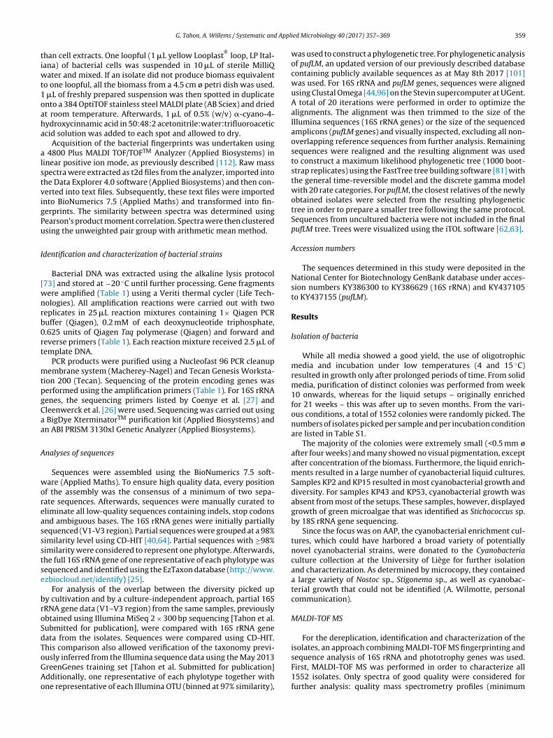
Appli
tiwt1oaha
alstvigPu
I
[wnrb0rt
mtpgCaa
A
woreassstse
broSdToGAo
G. Tahon, A. Willems / Systematic and
han cell extracts. One loopful (1 �L yellow Looplast®
loop, LP Ital-ana) of bacterial cells was suspended in 10 �L of sterile MilliQ
ater and mixed. If an isolate did not produce biomass equivalento one loopful, all the biomass from a 4.5 cm ø petri dish was used.
�L of freshly prepared suspension was then spotted in duplicatento a 384 OptiTOF stainless steel MALDI plate (AB Sciex) and driedt room temperature. Afterwards, 1 �L of 0.5% (w/v) �-cyano-4-ydroxycinnamic acid in 50:48:2 acetonitrile:water:trifluoroaceticcid solution was added to each spot and allowed to dry.
Acquisition of the bacterial fingerprints was undertaken using 4800 Plus MALDI TOF/TOFTM Analyzer (Applied Biosystems) ininear positive ion mode, as previously described [112]. Raw masspectra were extracted as t2d files from the analyzer, imported intohe Data Explorer 4.0 software (Applied Biosystems) and then con-erted into text files. Subsequently, these text files were importednto BioNumerics 7.5 (Applied Maths) and transformed into fin-erprints. The similarity between spectra was determined usingearson’s product moment correlation. Spectra were then clusteredsing the unweighted pair group with arithmetic mean method.
dentification and characterization of bacterial strains
Bacterial DNA was extracted using the alkaline lysis protocol73] and stored at −20 ◦C until further processing. Gene fragmentsere amplified (Table 1) using a Veriti thermal cycler (Life Tech-ologies). All amplification reactions were carried out with twoeplicates in 25 �L reaction mixtures containing 1× Qiagen PCRuffer (Qiagen), 0.2 mM of each deoxynucleotide triphosphate,.625 units of Qiagen Taq polymerase (Qiagen) and forward andeverse primers (Table 1). Each reaction mixture received 2.5 �L ofemplate DNA.
PCR products were purified using a Nucleofast 96 PCR cleanupembrane system (Macherey-Nagel) and Tecan Genesis Worksta-
ion 200 (Tecan). Sequencing of the protein encoding genes waserformed using the amplification primers (Table 1). For 16S rRNAenes, the sequencing primers listed by Coenye et al. [27] andleenwerck et al. [26] were used. Sequencing was carried out using
BigDye XterminatorTM purification kit (Applied Biosystems) andn ABI PRISM 3130xl Genetic Analyzer (Applied Biosystems).
nalyses of sequences
Sequences were assembled using the BioNumerics 7.5 soft-are (Applied Maths). To ensure high quality data, every position
f the assembly was the consensus of a minimum of two sepa-ate sequences. Afterwards, sequences were manually curated toliminate all low-quality sequences containing indels, stop codonsnd ambiguous bases. The 16S rRNA genes were initially partiallyequenced (V1-V3 region). Partial sequences were grouped at a 98%imilarity level using CD-HIT [40,64]. Partial sequences with ≥98%imilarity were considered to represent one phylotype. Afterwards,he full 16S rRNA gene of one representative of each phylotype wasequenced and identified using the EzTaxon database (http://www.zbiocloud.net/identify) [25].
For analysis of the overlap between the diversity picked upy cultivation and by a culture-independent approach, partial 16SRNA gene data (V1–V3 region) from the same samples, previouslybtained using Illumina MiSeq 2 × 300 bp sequencing [Tahon et al.ubmitted for publication], were compared with 16S rRNA geneata from the isolates. Sequences were compared using CD-HIT.his comparison also allowed verification of the taxonomy previ-
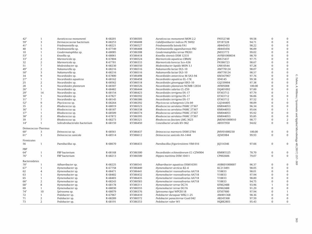
usly inferred from the Illumina sequence data using the May 2013reenGenes training set [Tahon et al. Submitted for publication]dditionally, one representative of each phylotype together withne representative of each Illumina OTU (binned at 97% similarity),ed Microbiology 40 (2017) 357–369 359
was used to construct a phylogenetic tree. For phylogenetic analysisof pufLM, an updated version of our previously described databasecontaining publicly available sequences as at May 8th 2017 [101]was used. For 16S rRNA and pufLM genes, sequences were alignedusing Clustal Omega [44,96] on the Stevin supercomputer at UGent.A total of 20 iterations were performed in order to optimize thealignments. The alignment was then trimmed to the size of theIllumina sequences (16S rRNA genes) or the size of the sequencedamplicons (pufLM genes) and visually inspected, excluding all non-overlapping reference sequences from further analysis. Remainingsequences were realigned and the resulting alignment was usedto construct a maximum likelihood phylogenetic tree (1000 boot-strap replicates) using the FastTree tree building software [81] withthe general time-reversible model and the discrete gamma modelwith 20 rate categories. For pufLM, the closest relatives of the newlyobtained isolates were selected from the resulting phylogenetictree in order to prepare a smaller tree following the same protocol.Sequences from uncultured bacteria were not included in the finalpufLM tree. Trees were visualized using the iTOL software [62,63].
Accession numbers
The sequences determined in this study were deposited in theNational Center for Biotechnology GenBank database under acces-sion numbers KY386300 to KY386629 (16S rRNA) and KY437105to KY437155 (pufLM).
Results
Isolation of bacteria
While all media showed a good yield, the use of oligotrophicmedia and incubation under low temperatures (4 and 15 ◦C)resulted in growth only after prolonged periods of time. From solidmedia, purification of distinct colonies was performed from week10 onwards, whereas for the liquid setups – originally enrichedfor 21 weeks – this was after up to seven months. From the vari-ous conditions, a total of 1552 colonies were randomly picked. Thenumbers of isolates picked per sample and per incubation conditionare listed in Table S1.
The majority of the colonies were extremely small (<0.5 mm øafter four weeks) and many showed no visual pigmentation, exceptafter concentration of the biomass. Furthermore, the liquid enrich-ments resulted in a large number of cyanobacterial liquid cultures.Samples KP2 and KP15 resulted in most cyanobacterial growth anddiversity. For samples KP43 and KP53, cyanobacterial growth wasabsent from most of the setups. These samples, however, displayedgrowth of green microalgae that was identified as Stichococcus sp.by 18S rRNA gene sequencing.
Since the focus was on AAP, the cyanobacterial enrichment cul-tures, which could have harbored a broad variety of potentiallynovel cyanobacterial strains, were donated to the Cyanobacteriaculture collection at the University of Liège for further isolationand characterization. As determined by microcopy, they containeda large variety of Nostoc sp., Stigonema sp., as well as cyanobac-terial growth that could not be identified (A. Wilmotte, personalcommunication).
MALDI-TOF MS
For the dereplication, identification and characterization of theisolates, an approach combining MALDI-TOF MS fingerprinting and
sequence analysis of 16S rRNA and phototrophy genes was used.First, MALDI-TOF MS was performed in order to characterize all1552 isolates. Only spectra of good quality were considered forfurther analysis: quality mass spectrometry profiles (minimum
360
G.
Tahon, A
. W
illems
/ System
atic and
Applied
Microbiology
40 (2017)
357–369
Table 1PCR primers and conditions used for screening different genes.
Gene Target Primer Sequence 5′-3′ Final concentration Region Amplicon size Program
16S rRNA Universal pAa AGA GTT TGA TCC TGG CTC AG 0.1 �M 8–1541l ±1500 bp 95 ◦C (5 min); 3 × 95 ◦C (1 min), 55 ◦C (2 min15 s), 72 ◦C (2 min 15 s); 30 × 95 ◦C (35 s), 55 ◦C(1 min 15 s), 72 ◦C (1 min 15 s); 72 ◦C (10 min)
pHa AAG GAG GTG ATC CAG CCG CA 0.1 �M
pufLM AAP pufLFb CTK TTC GAC TTC TGG GTS GG 0.2 �M 64–1612m ±1500 bp 94 ◦C (3 min); 30 × 94 ◦C (1 min), 60 ◦C (1 min),72 ◦C (2 min); 72 ◦C (10 min) [3]pufMRc CCA TSG TCC AGC GCC AGA A 0.2 �M
pufM Universal pufM uniFd GGN AAY YTN TWY TAY AAY CCN TTY CA 1.0 �M 584–825n ±240 bp 94 ◦C (4 min); 35 × 94 ◦C (40 s), 49 ◦C (30 s),72 ◦C (30 s); 72 ◦C (7 min)pufM WAWd AYN GCR AAC CAC CAN GCC CA 0.5 �M
proteorhodopsin Universal PR-1aFe GAT CGA GCG NTA YRT HGA RTG G 1.87 �M 340–665o ±335 bp 94 ◦C (2 min), 30 × 94 ◦C (30 s), 52 ◦C (30 s),72 ◦C (30 s); 72 ◦C (7 min) [20]PR-1aRe GAT CGA GCR TAD ATN GCC CAN CC 1.87 �M
actinorhodopsin Clade LG1 & LG2 LG-forf TAY MGN TAY GTN GAY TGG 0.4 �M 283–614p ±330 bp 95 ◦C (7 min), 45 × 94 ◦C (30 s), 51.5 ◦C (1 min30 s), 72 ◦C (30 s); 72 ◦C (10 min)LG1A-forf MGN TAY ATH GAY TGG YT 0.4 �M
LG2-forf TAY MGN TAY GCN GAY TGG 0.4 �MLG-revf ATN GGR TAN CAN CCC CA 0.8 �M
nifH, bchL, bchX Universal IGK3g GCI WTH TAY GGI AAR GGI GGI ATH GGI AA 1.0 �M 19–413qp 395 bp 95 ◦C (10 min); 40 × 95 ◦C (45 s), 52 ◦C (30 s),72 ◦C (40 s); 72 ◦C (10 min)DVVg ATI GCR AAI CCI CCR CAI ACI ACR TC 1.0 �M
nifH Universal F2h TGY GAY CCI AAI GCI GA 1.0 �M 115–473q 359 bp 95 ◦C (5 min); 35 × 95 ◦C (45 s), 51 ◦C (45 s),72 ◦C (45 s); 72 ◦C (7 min)R6h GCC ATC ATY TCI CCI GA 1.0 �M
cbbL RuBisCO IA & IB RubIgFi GAY TTC ACC AAR GAY GAY GA 0.4 �M 571–1382r ±800 bp 95 ◦C (3 min); 3 × 95 ◦C (1 min), 49 ◦C (2 min15 s), 72 ◦C (2 min 15 s); 30 × 95 ◦C (35 s), 49 ◦C(1 min 15 s), 72 ◦C (1 min 15 s); 72 ◦C (7 min)
RubIgRi TCR AAC TTG ATY TCY TTC CA 0.4 �M
cbbL RuBisCO IA & IC K2fj ACC AYC AAG CCS AAG CTS GG 0.2 �M 496–990r 492–495 bp 95 ◦C (3 min); 35 × 95 ◦C (1 min), 62 ◦C (1 min),72 ◦C (1 min 30 s); 72 ◦C (10 min) [116]V2rj GCC TTC SAG CTT GCC SAC CRC 0.2 �M
cbbM RuBisCO II cbbM343Fk GGY AAY AAC CAR GGY ATG GG 0.1 �M 343–1126k 700–800 bp 95 ◦C (3 min); 30 × 95 ◦C (1 min), 50 ◦C (2 min),72 ◦C (3 min); 72 ◦C (7 min) [56]cbbM1126Rk CGY ARB GCR TTC ATR CCR CC 0.1 �M
a From Ref. [33].b From Ref. [70].c From Ref. [11].d From Ref. [119].e From Ref. [20].f From Ref. [93].g From Ref. [4].h From Ref. [66].i From Ref. [97].j From Ref. [71].k From Ref. [56].l Based on the 16S rRNA gene sequence of Escherichia coli (A14565).
m Based on the pufLM sequence of Sphingomonas sanxanigenens DSM 19645 (CP006644).n Based on the pufM sequence of Sphingomonas sanxanigenens DSM 19645 (CP006644).o Based on the proteorhodopsin sequence of Vibrio campbellii BAA-1116 (FJ985782).p Based on the actinorhodopsin sequence of Leifsonia rubra CMS 76R (ATIA01000023).q Based on the nifH sequence of Azotobacter vinelandii (M20568).r Based on the cbbL IA sequence of Bradyrhizobium sp. ORS278 (CU234118).

Appli
hilmttpscsbMi
I
loEtP(91rtdra(r(iwcgdrw
MutirtdwflulstciworTdasca
G. Tahon, A. Willems / Systematic and
ighest peak intensity of 200 and <35% slope for the maximum peakntensity) could be generated for 1038 isolates. For the other iso-ates, even on repetition, no good quality profile could be obtained,
ostly due to the very low quantity of biomass available. As a result,hese isolates were not used in further analyses. Subsequently, clus-er analysis (Pearson correlation) of MALDI-TOF MS profiles waserformed, and visual inspection resulted in 141 clusters (data nothown). Approximately 15% of the isolates did not group in theselusters and they were characterized by unique profiles often of aomewhat lower quality. Prior to further analyses, the initial num-er of isolates was reduced to a subset of isolates representing allALDI-TOF MS clusters, as well as good quality unique profiles (330
solates total) (Table S2).
dentification based on 16S rRNA sequences
After partial 16S rRNA sequencing, the 330 representative iso-ates were binned in 77 phylotypes (98% sequence similarity). Basedn the full 16S rRNA gene sequence, taxon assignment with thezTaxon cultured database allocated 63 phylotypes (295 isolates)o 29 genera (95% cutoff [92]) belonging to the Actinobacteria,roteobacteria, Firmicutes, Bacteroidetes and Deinococcus-ThermusTable 2). Of the remaining phylotypes, the 16S rRNA was less than5% similar to that of their closest cultured neighbor. Of these,2 grouped with the aforementioned phyla and potentially rep-esented new genera or families (90–95% 16S rRNA similarity tohe closest cultured neighbor) (Table 2). Interestingly, two isolatesisplayed less than 80% similarity to named species and thus mayepresent the first cultured isolates of novel or uncultured taxa at
less detailed phylogenetic level. The sequence of isolate R-68168phylotype 64) was most similar (78.70%) to that of Streptacidiphilusugosus AM-16 (Actinobacteria), whereas the sequence of R-68213phylotype 76) was most similar (79.07%) to that of Hippea mar-tima DSM 10411 (Deltaproteobacteria). Therefore, these sequences,
hich were 89.99% similar to each other, were additionallyompared with the GreenGenes database [32,67,91]. The resultsrouped the isolates with environmental sequences of the candi-ate phylum FBP (96.26 and 97.83% highest sequence similarity,espectively) [61]. They are currently being studied in detail andill be reported on separately.
The identifications obtained were compared to the MALDI-TOFS dendrogram. A total of 126 of the 141 clusters and several
nique profiles, accounting for 892 of the 1038 isolates enclosed inhe MALDI-TOF MS dendrogram, were well defined and containedsolates belonging to the same genus or species, based on the 16SDNA identification of the closest EzTaxon hit. Other clusters wereaxonomically heterogeneous and contained isolates belonging toifferent genera. This may be explained by the fact that the profilesere a somewhat lower quality due to the low biomass obtained
or these strains. The comparison also revealed that several phy-otypes were represented by multiple MALDI-TOF MS clusters andnique spectra, indicating that several distinct strains were iso-
ated within these groups. Most diversity was retrieved from theolid media and least from the liquid enrichments. The distribu-ion of the recovered taxa for the different terrestrial samples andultivation setups is shown in Table S3. Several genera, includ-ng Sphingomonas, Nocardioides, Rhodococcus and Hymenobacter,
ere retrieved from all samples and nearly all setups, whereasthers (e.g. Rhodopseudomonas, Polymorphobacter, Knoellia) wereetrieved only from one sample and cultivation setup (Table S3).he most abundantly retrieved genera considering all isolation con-itions were Sphingomonas (397 isolates), Nocardioides (83 isolates)
nd Arthrobacter (83 isolates) (Table S3). A subset of strains repre-enting the total diversity recovered was stored in the researchollection of the Laboratory of Microbiology at Ghent Universitynd is available for further research.ed Microbiology 40 (2017) 357–369 361
In a previous study [Tahon et al. Submitted for publication], thebacterial communities present in the samples had been investi-gated by sequencing partial 16S rRNA genes (V1–V3 region) usingIllumina MiSeq 2 × 300 bp sequencing. Grouping at a 97% similar-ity resulted in a total of 703 OTUs. Comparison of these sequenceswith those of the isolates allowed more insight into the diversityoverlap retrieved between cultivation and the culture-independentapproach. Many of the isolates’ 16S rRNA genes grouped togetherat high similarities (≥97%) with the environmental sequences(Table 2, Figs. S1–S9). However, for 30 of the 77 cultured phylo-types, no sequence sharing more than 97% similarity was presentin the culture-independent dataset.
Although deep sequencing has revolutionized the currentknowledge of the bacterial world, these techniques also have weak-nesses. Currently, for the widely used Illumina MiSeq platform,the maximum amplicon length after merging reads is restrictedto ∼550 bp. For 16S rRNA genes, these partial sequences encom-pass only one third of the complete gene and, as a result, theiridentification at more detailed taxonomic levels (i.e. genus andspecies) may prove difficult. Therefore, the identity of the isolatesbased on full 16S rRNA gene sequences allowed tentative confirma-tion or improvement of the previous identification of the Illuminasequences obtained from the same samples. Maximum likelihoodanalysis clearly showed that the neighboring Illumina sequences ofthe 327 representative isolates (74 phylotypes) grouping with Acti-nobacteria, Proteobacteria, Bacteroidetes and Deinococcus-Thermushad previously all been assigned a correct taxonomy at the phy-lum, class and order level (Figs. S1 and S3–S9). For 63 phylotypes,the neighboring sequences had also been assigned a correct family.For the others, related Illumina sequences had been unclassified atthe family level. For phylotypes 29, 49 and 77, the identity of theisolates made it possible to assign a tentative genus and speciesidentity to the highly similar Illumina sequences that were pre-viously identified only to the family level. In a few other cases,there were discrepancies in the identification of isolates and theOTUs they were grouped with. For example, the V1–V3 region ofisolates grouping in phylotypes 4 and 20 (identified as Brevundi-monas variabilis and Noviherbaspirillum sp.) was almost identicalto the sequences recovered using Illumina (Table 2, Figs. S3 andS7) that had been identified as Mycoplana and Collimonas using theMay 2013 GreenGenes training set [32,67,91]. Repeating the iden-tification of these sequences, but with the current EZTaxon andGreenGenes databases, led to the same identification as B. variabilisand Noviherbaspirillum sp. Thus, although any differences couldsometimes be due to using a short sequence for identification pur-poses, they might also be explained by differences in the sequencedatabases used, but the ongoing addition of new sequence data andnovel taxa improves the identifications.
Protein coding gene analyses
For all 330 representative isolates, phototrophy potential wastested by amplification of key genes involved in rhodopsin- and(bacterio)chlorophyll-dependent light-harvesting. However, pro-teorhodopsin and actinorhodopsin genes could not be amplifiedfrom any of the isolates, although amplification of pufLM resultedin 50 positive reactions. The more universal pufM primers of Yutinet al. [119] did not provide any additional isolates. Positive iso-lates belonged to Sphingomonas (30 isolates), Methylobacterium (9isolates), Brevundimonas (9 isolates), Sphingomonadaceae (1 iso-late) and Hymenobacter (1 isolate) (Tables 2 and S2). Comparisonof these pufLM sequences with pufLM cloned sequences from a
previous study [101] revealed that only one sequence was recov-ered using both approaches. The pufLM sequence of isolate R-68361(Hymenobacter sp.) was identical to two cloned sequences (acces-sion no. KT154478) obtained from the same terrestrial sample (i.e.
362
G.
Tahon, A
. W
illems
/ System
atic and
Applied
Microbiology
40 (2017)
357–369
Table 2Overview of phylotypes recovered from the samples. For each phylotype, the number of representative strains (330 total) enclosed in the group are listed as well as the nearest phylogenetic neighbor and the number of isolatestesting positive for different phototrophy genes. Types previously retrieved [Tahon et al. Submitted for publication] using Illumina MiSeq sequencing (≥97% similarity) are indicated with an *.
Phylotype No. of strains Identification Representativestrain
Accession no.strain
Nearest phylogenetic neighbor pufLM bchL/bchX
Type strain Accession no. Sequence similarity (%)
ProteobacteriaAlphaproteobacteria3 1 Aureimonas sp. R-68373 KY386318 Aureimonas ferruginae CC-CFT023 JQ864240 96.64 0 14* 10 Brevundimonas variabilis R-68295 KY386469 Brevundimonas variabilis ATCC15255 AJ227783 99.79 9 61* 10 Methylobacterium sp. R-68173 KY386380 Methylobacterium iners 5317S-33 EF174497 98.52 9 26 1 Polymorphobacter sp. R-68699 KY386562 Polymorphobacter multimanifer 262-7 AB649056 95.99 0 02* 1 Rhodopseudomonas sp. R-67878 KY386506 Rhodopseudomonas pseudopalustris DSM123 AB498818 98.67 0 018* 1 Roseomonas aquatica R-68475 KY386403 Roseomonas aquatica TR53 AM231587 99.93 1 019* 1 Roseomonas sp. R-68165 KY386462 Roseomonas tokyonensis K-20 AB297501 98.60 0 05 2 Sphingomonadaceae bacterium R-67883 KY386496 Polymorphobacter multimanifer 262-7 AB649056 94.94 1 19* 53 Sphingomonas sp. R-68274 KY386532 Sphingomonas aerolata NW12 AJ429240 99.09 3 712 12 Sphingomonas sp. R-68304 KY386332 Sphingomonas cynarae SPC-1 HQ439186 98.24 6 37 18 Sphingomonas sp. R-67984 KY386379 Sphingomonas faeni MA-olki AJ429239 99.62 4 416 12 Sphingomonas sp. R-68260 KY386460 Sphingomonas hunanensis JSM 083058 FJ527417 98.52 10 413 3 Sphingomonas sp. R-68524 KY386357 Sphingomonas mucosissima CP173-2 AM229669 98.27 0 08 1 Sphingomonas sp. R-68700 KY386453 Sphingomonas oligophenolica JCM 12082 AB018439 98.24 0 017 1 Sphingomonas sp. R-67954 KY386585 Sphingomonas pituitosa EDIV AJ243751 95.58 0 015 7 Sphingomonas sp. R-68270 KY386304 Sphingomonas yantingensis 1007 JX566547 96.79 7 511* 1 Sphingomonas sp. R-68222 KY386535 Sphingomonas yunnanensis YIM 003 AY894691 97.52 0 0
Betaproteobacteria20* 2 Noviherbaspirillum sp. R-67997 KY386345 Noviherbaspirillum psychrotolerans PB1 JN390675 98.62 0 021* 1 Noviherbaspirillum suwonensis R-68579 KY386599 Noviherbaspirillum suwonensis 54105-65 JX275858 98.95 0 022* 2 Massilia sp. R-67978 KY386448 Massilia eurypsychrophila B528-3 KJ361504 97.70 0 023 5 Variovorax sp. R-67932 KY386486 Variovorax boronicumulans BAM-48 AB300597 97.55 0 024 2 Variovorax sp. R-67871 KY386393 Variovorax ginsengisoli Gsoil 3165 AB245358 99.26 0 0
Actinobacteria43* 2 Actinomycetales bacterium R-67786 KY386350 Jatrophihabitans endophyticus S9650 JQ346802 93.6 0 044* 1 Actinomycetales bacterium R-68701 KY386494 Jatrophihabitans soli KIS75-12 KP017569 94.37 0 045* 7 Actinomycetales bacterium R-68183 KY386330 Frankia alni ACN14A CT573213 94.11 0 046* 1 Actinomycetales bacterium R-67810 KY386542 Cryptosporangium minutisporangium IFO15962 AB037007 94.41 0 047* 3 Actinomycetales bacterium R-67836 KY386609 Modestobacter versicolor CP153-2 AJ871304 94.28 0 048* 2 Actinomycetales bacterium R-68223 KY386537 Sporichthya brevicatena IFO 16195 AB006164 93.78 0 077* 1 Angustibacter sp. R-68259 KY386618 Angustibacter aerolatus 7402J-48 JQ639056 96.64 0 054* 4 Aquipuribacter sp. R-67807 KY386530 Aquipuribacter hungaricus IV-75 FM179321 97.88 0 157* 22 Arthrobacter sp. R-67818 KY386623 Arthrobacter agilis DSM 20550 X80748 99.46 0 058* 2 Arthrobacter flavus R-67793 KY386372 Arthrobacter flavus TB 23 ALPM01000083 99.25 0 075 1 Arthrobacter sp. R-68384 KY386384 Arthrobacter oxydans DSM 20119 X83408 99.25 0 055* 5 Arthrobacter pityocampae R-68518 KY386301 Arthrobacter pityocampae Tp2 EU885749 99.34 0 0
G.
Tahon, A
. W
illems
/ System
atic and
Applied
Microbiology
40 (2017)
357–369
363
42* 1 Auraticoccus monumenti R-68201 KY386505 Auraticoccus monumenti MON 2.2 FN552748 99.58 0 059* 3 Dermacoccaceae bacterium R-68253 KY386608 Calidifontibacter indicus PC IW02 EF187228 94.71 0 041* 1 Friedmanniella sp. R-68221 KY386527 Friedmanniella luteola FA1 AB445453 98.22 0 040 5 Friedmanniella sp. R-67749 KY386408 Friedmanniella sagamiharensis FB2 AB445456 96.09 0 010 1 Geodermatophilus sp. R-68085 KY386308 Geodermatophilus terrae PB261 JN033773 99.02 0 053* 1 Knoellia sp. R-68061 KY386418 Knoellia sinensis DSM 12331 AVPJ01000034 98.70 0 033* 2 Marmoricola sp. R-67804 KY386524 Marmoricola aquaticus CBMAI JN615437 97.75 0 032 2 Marmoricola sp. R-67781 KY386333 Marmoricola korecus Sco-A36 FN386723 98.67 0 051 7 Modestobacter sp. R-68230 KY386550 Modestobacter lapidis MON 3.1 LN810544 97.28 0 049* 2 Nakamurella sp. R-68216 KY386516 Nakamurella lactae DLS-10 AM778124 98.07 0 050* 3 Nakamurella sp. R-68216 KY386516 Nakamurella lactae DLS-10 AM778124 98.57 0 034 9 Nocardioides sp. R-67800 KY386498 Nocardioides antarcticus M-SA3-94 KM347967 97.76 0 027* 1 Nocardioides aquaticus R-68162 KY386458 Nocardioides aquaticus EL-17K X94145 99.38 0 025 2 Nocardioides sp. R-68562 KY386518 Nocardioides ginsengagri BX5-10 GQ339904 97.38 0 028 3 Nocardioides plantarum R-68307 KY386526 Nocardioides plantarum NCIMB 12834 AF005008 100.00 0 026* 1 Nocardioides sp. R-68482 KY386444 Nocardioides salarius CL-Z59 DQ401092 97.00 0 029* 14 Nocardioides sp. R-68154 KY386423 Nocardioides terrigena DS-17 EF363712 97.79 0 130* 1 Nocardioides sp. R-67827 KY386592 Nocardioides terrigena DS-17 EF363712 96.06 0 031* 7 Nocardioides sp. R-68145 KY386386 Nocardioides terrigena DS-17 EF363712 97.52 0 152* 9 Phycicoccus sp. R-68264 KY386392 Phycicoccus ochangensis L1b-b9 GQ344405 98.09 0 035 2 Rhodococcus sp. R-68019 KY386523 Rhodococcus aerolatus PAMC 27367 KM044053 96.34 0 036* 1 Rhodococcus sp. R-68187 KY386338 Rhodococcus aerolatus PAMC 27367 KM044053 95.64 0 137* 1 Rhodococcus sp. R-68509 KY386574 Rhodococcus aerolatus PAMC 27367 KM044053 95.92 0 038* 2 Rhodococcus sp. R-67872 KY386395 Rhodococcus aerolatus PAMC 27367 KM044053 95.85 0 039 7 Rhodococcus sp. R-68273 KY386321 Rhodococcus fascians LMG 3623 JMEN01000010 98.77 0 214* 1 Solirubrobacterales bacterium R-68159 KY386450 Conexibacter arvalis KV-962 AB597950 94.02 0 0
Deinococcus-Thermus60* 3 Deinococcus sp. R-68561 KY386437 Deinococcus marmoris DSM12784 JNIV01000230 100.00 0 061* 3 Deinococcus saxicola R-68514 KY386612 Deinococcus saxicola AA-1444 AJ585984 99.93 0 0
Firmicutes56 1 Paenibacillus sp. R-68670 KY386433 Paenibacillus frigoriresistens YIM 016 JQ314346 97.66 0 0
FBP64* 1 FBP bacterium R-68168 KY386300 Nocardioides echinoideorum CC-CZW004 KM085325 78.70 0 076* 1 FBP bacterium R-68213 KY386500 Hippea maritima DSM 10411 CP002606 79.07 0 0
Bacteroidetes70* 1 Adhaeribacter sp. R-68225 KY386541 Adhaeribacter aquaticus DSM16391 AXBK01000007 96.37 0 067* 1 Hymenobacter sp. R-67758 KY386449 Hymenobacter arcticus R2-4 KC213491 98.05 0 062 4 Hymenobacter sp. R-68471 KY386441 Hymenobacter roseosalivarius AA718 Y18833 98.01 0 063 1 Hymenobacter sp. R-68402 KY386432 Hymenobacter roseosalivarius AA718 Y18833 97.94 0 065 1 Hymenobacter sp. R-68403 KY386435 Hymenobacter roseosalivarius AA718 Y18833 98.89 0 066* 2 Hymenobacter sp. R-68243 KY386583 Hymenobacter roseosalivarius AA718 Y18833 94.75 0 068* 8 Hymenobacter sp. R-68178 KY386311 Hymenobacter terrae DG7A KF862488 93.96 1 069 1 Hymenobacter sp. R-68030 KY386555 Hymenobacter terrae DG7A KF862488 91.29 0 074* 10 Spirosoma sp. R-68079 KY386376 Spirosoma rigui WPCB118 EF507900 97.94 0 172 1 Pedobacter sp. R-67967 KY386410 Pedobacter duraquae WB2.1-25 AM491368 98.36 0 071 2 Pedobacter sp. R-68289 KY386572 Pedobacter panaciterrae Gsoil 042 AB245368 97.59 0 073 2 Pedobacter sp. R-68191 KY386353 Pedobacter ruber W1 HQ882803 95.43 0 0

364 G. Tahon, A. Willems / Systematic and Applied Microbiology 40 (2017) 357–369
F M seqn isted.
o . Ident
Kic
ig. 1. Maximum likelihood phylogenetic tree (1000 bootstrap replicates) of pufLucleotide positions. For reference data, the accession number and taxon name are lf at least 50% are provided. Chloroflexi pufLM sequences were used as an outgroup
P15). All other pufLM isolate sequences were less than 86% sim-lar to cloned sequences. The maximum likelihood tree in Fig. 1learly shows newly obtained pufLM sequences grouping among
uences (1554 nucleotide positions). Scale bar indicates 10 substitutions per 100PufLM sequences from newly obtained strains are labelled in bold. Bootstrap valuesifications of the strains obtained can be found in Table S2.
the reference data of closely related taxa with a known photo-heterotrophic phenotype. Nevertheless, the newly obtained pufLMsequences were not highly similar to the sequences of their clos-

Appli
enghtab
tKwPDnpfiwa
ei[lwgroa
D
ertssvsacsststwmrs
das[asotagwt(i
G. Tahon, A. Willems / Systematic and
st neighbors (Fig. 1). Perhaps more closely related neighbors haveot yet been cultured or, since many anoxygenic phototrophs canrow heterotrophically, their phototrophic potential may not yetave been recorded [58]. Interestingly, the pufLM sequence ofhe Hymenobacter isolate (R-68361) grouped in a separate branchmong the reference data from Sphingomonas spp., with a highootstrap value supporting the branch (Fig. 1).
BchL/bchX could be amplified from 41 of the 330 representa-ive isolates, 17 of which also tested positive for pufLM (Table S2).nown anoxygenic phototrophs contain both bchL and bchX [41],hich co-amplified with the primers used here [4]. Therefore, these
CR products could not be sequenced directly. Since the IGK3 andVV primers used for amplification of bchL and bchX also amplifyifH [4], an additional PCR was carried out with broad range nifHrimers (F2 and R6) in order to verify that no nifH were ampli-ed (Table 2). This primer set, designed by Marusina et al. [66]as reported to amplify strictly nifH and resulted in no positive
mplifications [42].In previous research, the diversity of key bacterial protein
ncoding genes in the Calvin-Benson-Bassham cycle (RuBisCO) wasnvestigated using clone libraries and Illumina MiSeq sequencing101,Tahon et al. Submitted for publication]. The results revealed aarge diversity of RuBisCO types IA, IB and IC (cbbL gene) grouping
ith Proteobacteria and Actinobacteria. The type II RuBisCO (cbbMene) could not be amplified from the samples. Therefore, the 330epresentative isolates were additionally checked for the presencef cbbL and cbbM genes (Table 1) but this resulted in no positivemplifications.
iscussion
Bacteria in Antarctic soils are subjected to a range of extremenvironmental conditions, including sub-zero temperatures withepeated cycles of freezing and thawing, low transient precipita-ion [114], very low availability of organic matter [107] and strongolar radiation at exposed sites. In these environmental conditions,ome bacteria may have adopted a phototrophic lifestyle, con-erting sunlight into chemical energy. The analysis in the presenttudy is the first to report on the diversity of culturable aerobicnoxygenic phototrophic bacteria relying on a type 2 photochemi-al reaction center in soils from the SRM (East Antarctica). Previoustudies of environmental DNA have revealed that a broad diver-ity of (aerobic) anoxygenic phototrophs, particularly those usinghe aforementioned reaction center, was present in the exposedoils in the proximity of the Princess Elisabeth Station, whereashe relative abundance of oxygenic photosynthetic microorganismsas found to be low in many samples [101,102,107]. Therefore, theain objective was to isolate and characterize AAP with a type 2
eaction center, which is a group that has, to date, almost exclu-ively been studied in aquatic environments [58].
AAP relying on a type 2 photochemical reaction center have pre-ominantly been found in the Alpha- and Gammaproteobacteria,nd to a lesser extent in the Betaproteobacteria. A single repre-entative is known in the Gemmatimonadetes and in the Firmicutes58,79,121]. The isolation strategy using oligotrophic media and
light regime simulating the increasing day length during tran-ition from Antarctic winter to summer gave access to a rangef bacteria known to be common inhabitants of soils, includinghose from Antarctica [2,77,80,105,115]. Following MALDI-TOF MSnd 16S rRNA gene analysis, ∼52% of the 892 isolates identifiedrouped among known alphaproteobacterial AAP taxa, particularly
ith Sphingomonas (∼45%). The majority of these alphaproteobac-erial AAP were retrieved from liquid enrichments incubated at 4 ◦CTable S3). A total of 20 isolates, many obtained from PA mediumncubated at 4 ◦C, grouped with Betaproteobacteria, whereas only
ed Microbiology 40 (2017) 357–369 365
a single Firmicutes isolate was picked up. However, none of theseisolates grouped among known AAP taxa and no Gammaproteobac-teria or Gemmatimonadetes were isolated. This observation wasin accordance with previous observations made on these sam-ples with culture-independent approaches. Clone libraries andIllumina MiSeq sequencing of the puf(L)M and bchL/bchX genesrevealed a dominance of alphaproteobacterial AAP, including manyof the groups isolated here [101,102]. Only 0.65% of the ∼680,000pufM Illumina reads grouped with beta- and gammaproteobacterialpufM, while 16S rRNA gene sequencing also revealed a high rela-tive abundance of Alphaproteobacteria and, in particular, AAP taxa.On the other hand, Beta- and Gammaproteobacteria, Gemmatimon-adetes and Firmicutes were (almost) absent in these samples andthe SRM in general [101,Tahon et al. Submitted for publication].Although similar patterns could be observed in both approaches(i.e. dominance of alphaproteobacterial AAP taxa), the compari-son of culture-dependent and culture-independent 16S rRNA andpuf(L)M gene datasets only revealed a limited overlap, indicatingthat both approaches provided complementary information con-cerning a community’s diversity that might be missed when usinga single approach. However, the low overlap may be explained bythe limitations of these methods. Amplicon sequencing is depen-dent on DNA extraction and the use of primers, and these steps donot retrieve all diversity. Indeed, recent metagenome analysis hasled to the discovery of a novel bacterial phylum that had alwaysremained hidden because of mismatches in the most commonlyused primers for 16S rRNA gene sequencing [34]. While cultiva-tion might overcome this problem, many isolates are still resistantto the commonly used cultivation techniques. The low number oftypes shared will have also been biased by the cultivation setup.Firstly, although specific cultivation conditions for the targetedgrowth of aerobic phototrophic bacteria were used, in addition tooxygenic and anoxygenic phototrophic bacteria, a high number ofnon-phototrophs lacking pufLM and bchL/bchX were also isolated.A portion of these isolates may have phototrophic capacities thatdepend on rhodopsins or BchL; however, these features may havebeen missed as a result of primer mismatch, or the presence of adifferent rhodopsin type or a type 1 photochemical reaction center.For example, very little rhodopsin data has been retrieved from cur-rently available terrestrial Antarctic metagenomes with MG-RAST[113] and IMG [65]. Most rhodopsin data, however, originates fromaquatic environments [15]. Since it is well known that this proteinfamily contains an enormous diversity [8,35], currently avail-able primers may be unsuitable for capturing terrestrial Antarcticrhodopsin variants, whereas annotation pipelines may be unableto detect these variants in terrestrial metagenomes. Neverthe-less, future advances in physiological characterization and genomeanalyses may resolve this question. Secondly, the unexpected andabundant growth of Cyanobacteria in the liquid enrichments mayhave restricted the growth of other phototrophic bacteria in thesesetups. Thirdly, when applying a culture-independent approach,the presence of DNA from dead cells, which is capable of persist-ing in cold soil for long periods of time, may inflate the bacterialdiversity observed, hence reducing the overlap observed witha culture-dependent approach [21]. Finally, the high number ofcolonies, their very slow growth and miniscule colony size, in com-bination with manual picking, introduced an additional bias, sinceit was not possible to isolate every single colony. Additionally, thelimited biomass production impeded many of the analyses andcalled for a modification of the standard operating procedures usedfor fast growing organisms producing high biomass. Developmentof new innovative and high throughput strategies will be necessary
to cultivate and characterize a larger proportion of the Antarcticbiodiversity.Of the 75 isolates showing phototrophy potential (i.e. positivePCR for a phototrophy gene), 67 grouped with alphaproteobacterial

3 Appl
AtodigemowyeetAolkw[psoBr[Rdhpft
TBTolaapttT9d[sldngbtStratbr
idip
66 G. Tahon, A. Willems / Systematic and
AP taxa (Table S2). Although the abundance of alphaproteobac-erial AAP is well recognized in various aquatic environments allver the planet [16,29,58,84], to date, little is known about theiriversity, distribution and role in terrestrial ecosystems. The major-
ty of potential phototrophs, and ∼45% of the isolates in general,rouped with Sphingomonas (Table 2, Figs. 1 and S5). The pres-nce of Sphingomonas in Antarctica is not unusual, since severalembers of this group have previously been isolated from a range
f cold ecosystems, including Antarctica [18,46,77], and they areell known for their phototrophic capacities [48,88]. In recent
ears, analyses have also revealed the metabolic diversity of sev-ral Sphingomonas strains, as well as their capacity to adapt toxtreme cold, high UV-B radiation and arid conditions, indicatinghat they are ideal candidates for survival in extreme oligotrophicntarctic systems [9,46,51,52]. The second most recovered groupf alphaproteobacterial phototrophs was highly related to Methy-
obacterium (Fig. 1, Table 2). Some Methylobacterium strains are wellnown for their tolerance to high UV radiation and dehydration,hich are common environmental conditions in Antarctic soils
75]. Indeed, Romanovskaya et al. [85] reported this genus, and inarticular Methylobacterium extorquens, a bacterium known to pos-ess a type 2 photochemical reaction center, in terrestrial biotopesn several Antarctic islands [85,109], whereas PufM, BchL andchX sequences from members of this taxon have previously beeneported from terrestrial locations in the Arctic [36] and Antarctic102]. Interestingly, no pufLM genes could be amplified from strain-67878 (Rhodopseudomonas sp.). Given that all known Rhodopseu-omonas species are phototrophic [100], the negative result mayave been caused by primer mismatch. Indeed, we have previouslyerformed an in silico comparison of multiple primers targeting dif-erent regions of the pufM gene [102], and the results in fact showedhat none of the primers investigated targeted all known diversity.
Based on 16S rRNA sequence data combined with the MALDI-OF MS dendrogram, 87 of the isolates grouped with theacteroidetes, and especially with Hymenobacter (Table S3, Fig. S4).he type species of this genus, Hymenobacter roseosalivarius, wasriginally isolated from exposed areas in the McMurdo Dry Val-eys [50], and has been commonly reported from several terrestrialnd aquatic Antarctic locations [2,57,77]. To our knowledge, nonoxygenic phototrophic members have ever been reported in thehylum Bacteroidetes [53]. Remarkably, the pufLM genes encodinghe conserved proteins of the type 2 phototrophic reaction cen-er could be amplified from Hymenobacter isolate R-68361 (Fig. 1).he 16S rRNA gene sequence of strain R-68361 was found to be3.96% identical to that of Hymenobacter terrae DG7A, a recentlyescribed strain isolated from soil samples in Seoul (South Korea)98]. The pufLM sequence of R-68361 was identical to clonedequences previously obtained from the same sample [101]. Phy-ogenetic analysis of this sequence grouped it among referenceata for Sphingomonas spp. with a known photoheterotrophic phe-otype (Fig. 1). This grouping may be the result of a horizontalene transfer event, since similar observations have previouslyeen made for phototrophic Gemmatimonadetes [121]. In addi-ion, a positive result for bchL/bchX was obtained from one of thepirosoma isolates (R-67957) (Table S2). It should also be notedhat both rhodopsin-dependent and Bchl-dependent phototrophyequire the transcription of unique multiple genes [17]. Thus, tossess whether these isolates are the first phototrophic represen-atives of the Bacteroidetes, genome and transcriptome analysis wille needed in order to verify the presence and expression of all theequired phototrophic genes.
In addition to taxa known to contain AAP, ∼31% of the 892
solates were identified as Actinobacteria, and they were pre-ominantly isolated from solid media (Table S3). Six of thesesolates tested positive in the PCR for bchL/bchX, although theseroducts were co-amplified and therefore were not sequenced.
ied Microbiology 40 (2017) 357–369
None of the Actinobacteria tested positive for rhodopsins or pufLM(Table 2). This might be explained by primer mismatch, since cur-rently available primers may be unable to target all the rhodopsinand photoreaction center diversity. However, future physiologicalcharacterization and genome analyses may resolve this questionbut, until then, the phototrophic potential of these Actinobacte-ria remains unknown. All actinobacterial isolates grouped withthe Actinobacteridae, which is similar to observations made inother Antarctic soils [1,2,77]. Most of the Actinobacteridae isolateswere identified as Arthrobacter, Nocardioides, Modestobacter andRhodococcus. Members of these genera have previously been iso-lated from several cold regions, such as Greenland, the Arctic andAntarctica [7,68,78,90,95]. The presence of Modestobacter, someof which are known to be cold tolerant and radiation resistant,may be linked to its possible involvement in the weathering ofrocks [45,74]. On the other hand, Arthrobacter is a genus gener-ally associated with the soil compartment and is recognized forits physiological versatility (e.g. altering of its cell wall fatty acidcomposition in response to lowered growth temperatures) and itsability to use a wide range of substrates [2,111].
Conclusions
This study provided the first data on the culturable aerobicanoxygenic phototrophic bacterial diversity in the Sør RondaneMountains, East Antarctica. In general, the conditions used resultedin slow growing isolates producing extremely small colonies andminimal amounts of biomass. The isolation strategy resulted inapproximately 52% of isolates belonging to known alphaproteobac-terial AAP taxa, while other isolates were distributed over six phyla,including a candidate phylum. In addition, enrichment culturesrevealed the presence of Cyanobacteria and even green algae. It wasalso demonstrated for the first time that a single Bacteroidetes iso-late may have phototrophic potential. Overall, the results suggestedthat the ability to adopt a photoheterotrophic lifestyle may pro-vide an advantage in the oligotrophic Antarctic soils surroundingthe Princess Elisabeth Station.
Funding
This work was supported by the Fund for Scientific Research– Flanders (project G.0146.12). Additional support was obtainedfrom the Belgian Science Policy Office (project CCAMBIO). The com-putational resources (Stevin Supercomputer Infrastructure) andservices used in this work were provided by the Flemish Super-computer Center (VSC) funded by Ghent University, the HerculesFoundation and the Flemish Government – Department EWI. Thisstudy was a contribution to the State of the Antarctic Ecosystem(AntEco) research program of the Scientific Committee on AntarcticResearch (SCAR).
Author contributions
Conceived and designed the experiments: GT, AW. Performedthe experiments: GT. Analyzed the data: GT. Wrote the paper: GT,AW. Both authors approved the final manuscript.
Appendix A. Supplementary data
Supplementary data associated with this article can be found,in the online version, at http://dx.doi.org/10.1016/j.syapm.2017.05.007.
References
[1] Aislabie, J.M., Chhour, K.-L., Saul, D.J., Miyauchi, S., Ayton, J., Paetzold,R.F., Balks, M.R. (2006) Dominant bacteria in soils of Marble Point

Appli
G. Tahon, A. Willems / Systematic andand Wright Valley, Victoria Land, Antarctica. Soil Biol. Biochem. 38,3041–3056.
[2] Aislabie, J.M., Lau, A., Dsouza, M., Shepherd, C., Rhodes, P., Turner, S.J. (2013)Bacterial composition of soils of the Lake Wellman area, Darwin Mountains,Antarctica. Extremophiles 17, 775–786.
[3] Allgaier, M., Uphoff, H., Felske, A., Wagner-Döbler, I. (2003) Aerobic anoxy-genic photosynthesis in Roseobacter clade bacteria from diverse marinehabitats. Appl. Environ. Microbiol. 69, 5051–5059.
[4] Ando, S., Goto, M., Meunchang, S., Thongra-ar, P., Fujiwara, T., Hayashi, H.,Yoneyama, T. (2005) Detection of nifH sequences in sugarcane (Saccharumofficinarum L.) and pineapple (Ananas comosus [L.] Merr.). Soil Sci. Plant Nutr.51, 303–308.
[5] Atamna-Ismaeel, N., Finkel, O.M., Glaser, F., Sharon, I., Schneider, R., Post, A.F.,Spudich, J.L., von Mering, C., Vorholt, J.A., Iluz, D., Béjà, O., Belkin, S. (2012)Microbial rhodopsins on leaf surfaces of terrestrial plants. Environ. Microbiol.14, 140–146.
[6] Atamna-Ismaeel, N., Sabehi, G., Sharon, I., Witzel, K.-P., Labrenz, M., Jurgens,K., Barkay, T., Stomp, M., Huisman, J., Béjà, O. (2008) Widespread distribu-tion of proteorhodopsins in freshwater and brackish ecosystems. ISME J. 2,656–662.
[7] Babalola, O.O., Kirby, B.M., Le Roes-Hill, M., Cook, A.E., Cary, S.C., Burton,S.G., Cowan, D.A. (2009) Phylogenetic analysis of actinobacterial populationsassociated with Antarctic Dry Valley mineral soils. Environ. Microbiol. 11,566–576.
[8] Bamann, C., Bamberg, E., Wachtveitl, J., Glaubitz, C. (2014) Proteorhodopsin.Biochim. Biophys. Acta: Bioenerg. 1837, 614–625.
[9] Baraniecki, C.A., Aislabie, J., Foght, J.M. (2002) Characterization of Sphin-gomonas sp. Ant 17, an aromatic hydrocarbon-degrading bacterium isolatedfrom Antarctic soil. Microb. Ecol. 43, 44–54.
[10] Béjà, O., Lanyi, J.K. (2014) Nature’s toolkit for microbial rhodopsin ion pumps.Proc. Natl. Acad. Sci. U. S. A. 111, 6538–6539.
[11] Béjà, O., Suzuki, M.T., Heidelberg, J.F., Nelson, W.C., Preston, C.M., Hamada,T., Eisen, J.A., Fraser, C.M., DeLong, E.F. (2002) Unsuspected diversity amongmarine aerobic anoxygenic phototrophs. Nature 415, 630–633.
[12] Bellenger, J.P., Wichard, T., Kraepiel, A.M.L. (2008) Vanadium requirementsand uptake kinetics in the dinitrogen-fixing bacterium Azotobacter vinelandii.Appl. Environ. Microbiol. 74, 1478–1484.
[13] Benemann, J.R., McKenna, C.E., Lie, R.F., Traylor, T.G., Kamen, M.D. (1972) Thevanadium effect in nitrogen fixation by azotobacter. Biochim. Biophys. Acta:Gen. Subj. 264, 25–38.
[14] Bockheim, J.G. (2015) Soil-forming factors in Antarctica. In: Bockheim, G.J.(Ed.), The Soils of Antarctica, Springer International Publishing, Cham, pp.5–20.
[15] Boeuf, D., Audic, S., Brillet-Guéguen, L., Caron, C., Jeanthon, C. (2015)MicRhoDE: a curated database for the analysis of microbial rhodopsin diver-sity and evolution. Database 2015, bav080.
[16] Boeuf, D., Cottrell, M.T., Kirchman, D.L., Lebaron, P., Jeanthon, C. (2013) Sum-mer community structure of aerobic anoxygenic phototrophic bacteria in thewestern Arctic Ocean. FEMS Microbiol. Ecol. 85, 417–432.
[17] Bryant, D.A., Frigaard, N.-U. (2006) Prokaryotic photosynthesis and phototro-phy illuminated. Trends Microbiol. 14, 488–496.
[18] Busse, H.-J., Denner, E.B.M., Buczolits, S., Salkinoja-Salonen, M., Bennasar, A.,Kämpfer, P. (2003) Sphingomonas aurantiaca sp. nov., Sphingomonas aerolatasp. nov. and Sphingomonas faeni sp. nov., air- and dustborne and Antarctic,orange-pigmented, psychrotolerant bacteria, and emended description of thegenus Sphingomonas. Int. J. Syst. Evol. Microbiol. 53, 1253–1260.
[19] Cameron, R.E. (1969) Abundance of microflora in soils of desert regions. NASATech. Rep. 32, 1–16.
[20] Campbell, B.J., Waidner, L.A., Cottrell, M.T., Kirchman, D.L. (2008) Abundantproteorhodopsin genes in the North Atlantic Ocean. Environ. Microbiol. 10,99–109.
[21] Carini, P., Marsden, P.J., Leff, J.W., Morgan, E.E., Strickland, M.S., Fierer, N.(2016) Relic DNA is abundant in soil and obscures estimates of soil microbialdiversity. Nat. Microbiol. 2, 16242.
[22] Chew, A.G.M., Bryant, D.A. (2007) Chlorophyll biosynthesis in bacteria:the origins of structural and functional diversity. Ann. Rev. Microbiol. 61,113–129.
[23] Cho, J.-C., Giovannoni, S.J. (2004) Cultivation and growth characteristics ofa diverse group of oligotrophic marine Gammaproteobacteria. Appl. Environ.Microbiol. 70, 432–440.
[24] Chong, C.-W., Pearce, D.A., Convey, P. (2015) Emerging spatial patterns inAntarctic prokaryotes. Front. Microbiol. 6, 1058.
[25] Chun, J., Lee, J.-H., Jung, Y., Kim, M., Kim, S., Kim, B.K., Lim, Y.-W. (2007)EzTaxon: a web-based tool for the identification of prokaryotes based on 16Sribosomal RNA gene sequences. Int. J. Syst. Evol. Microbiol. 57, 2259–2261.
[26] Cleenwerck, I., Camu, N., Engelbeen, K., De Winter, T., Vandemeulebroecke, K.,De Vos, P., De Vuyst, L. (2007) Acetobacter ghanensis sp. nov., a novel acetic acidbacterium isolated from traditional heap fermentations of Ghanaian cocoabeans. Int. J. Syst. Evol. Microbiol. 57, 1647–1652.
[27] Coenye, T., Falsen, E., Vancanneyt, M., Hoste, B., Govan, J.R.W., Kersters, K.,Vandamme, P. (1999) Classification of Alcaligenes faecalis-like isolates from
the environment and human clinical samples as Ralstonia gilardii sp. nov. Int.J. Syst. Bacteriol. 49, 405–413.[28] Convey, P., Gibson, J.A.E., Hillenbrand, C.-D., Hodgson, D.A., Pugh, P.J.A.,Smellie, J.L., Stevens, M.I. (2008) Antarctic terrestrial life – challenging thehistory of the frozen continent? Biol. Rev. 83, 103–117.
ed Microbiology 40 (2017) 357–369 367
[29] Cottrell, M.T., Mannino, A., Kirchman, D.L. (2006) Aerobic anoxygenic pho-totrophic bacteria in the Mid-Atlantic Bight and the North Pacific Gyre. Appl.Environ. Microbiol. 72, 557–564.
[30] Daubin, V., Szollosi, G.J. (2016) Horizontal gene transfer and the history of life.Cold Spring Harb. Perspect. Biol. 8.
[31] Delwiche, C.F., Palmer, J.D. (1996) Rampant horizontal transfer and duplica-tion of rubisco genes in eubacteria and plastids. Mol. Biol. Evol. 13, 873–882.
[32] DeSantis, T.Z., Hugenholtz, P., Larsen, N., Rojas, M., Brodie, E.L., Keller, K.,Huber, T., Dalevi, D., Hu, P., Andersen, G.L. (2006) Greengenes, a chimera-checked 16S rRNA gene database and workbench compatible with ARB. Appl.Environ. Microbiol. 72, 5069–5072.
[33] Edwards, U., Rogall, T., Blöcker, H., Emde, M., Böttger, E.C. (1989) Isolation anddirect complete nucleotide determination of entire genes. Characterization ofa gene coding for 16S ribosomal RNA. Nucleic Acids Res. 17, 7843–7853.
[34] Eloe-Fadrosh, E.A., Paez-Espino, D., Jarett, J., Dunfield, P.F., Hedlund, B.P.,Dekas, A.E., Grasby, S.E., Brady, A.L., Dong, H.L., Briggs, B.R., Li, W.J., Goudeau,D., Malmstrom, R., Pati, A., Pett-Ridge, J., Rubin, E.M., Woyke, T., Kyrpides,N.C., Ivanova, N.N. (2016) Global metagenomic survey reveals a new bacterialcandidate phylum in geothermal springs. Nat. Commun. 7.
[35] Ernst, O.P., Lodowski, D.T., Elstner, M., Hegemann, P., Brown, L.S., Kandori, H.(2014) Microbial and animal rhodopsins: structures, functions, and molecularmechanisms. Chem. Rev. 114, 126–163.
[36] Feng, Y., Grogan, P., Caporaso, J.G., Zhang, H., Lin, X., Knight, R., Chu, H. (2014)pH is a good predictor of the distribution of anoxygenic purple phototrophicbacteria in Arctic soils. Soil Biol. Biochem. 74, 193–200.
[37] Feng, Y., Lin, X., Mao, T., Zhu, J. (2011) Diversity of aerobic anoxygenic pho-totrophic bacteria in paddy soil and their response to elevated atmosphericCO2. Microb. Biotechnol. 4, 74–81.
[38] Ferrer, M., Golyshina, O., Beloqui, A., Golyshin, P.N. (2007) Mining enzymesfrom extreme environments. Curr. Opin. Microbiol. 10, 207–214.
[39] Fritsen, C.H., Grue, A.M., Priscu, J.C. (2000) Distribution of organic carbon andnitrogen in surface soils in the McMurdo Dry Valleys, Antarctica. Polar Biol.23, 121–128.
[40] Fu, L., Niu, B., Zhu, Z., Wu, S., Li, W. (2012) CD-HIT: accelerated for clusteringthe next-generation sequencing data. Bioinformatics 28, 3150–3152.
[41] Fujita, Y., Bauer, C.E. (2003) The light-independent protochlorophyllidereductase: a nitrogenase-like enzyme catalyzing a key reaction for green-ing in the dark. In: Guilard, R., Kadish, K., Smith, K.M. (Eds.), The PorphyrinHandbook, Academic Press, Amsterdam, pp. 109–156.
[42] Gaby, J.C., Buckley, D.H. (2012) A comprehensive evaluation of PCR primersto amplify the nifH gene of nitrogenase. PLoS One 7, e42149.
[43] Gorodetskaya, I.V., Van Lipzig, N.P.M., Van den Broeke, M.R., Mangold, A., Boot,W., Reijmer, C.H. (2013) Meteorological regimes and accumulation patterns atUtsteinen, Dronning Maud Land, East Antarctica: analysis of two contrastingyears. J. Geophys. Res. Atmos. 118, 1700–1715.
[44] Goujon, M., McWilliam, H., Li, W., Valentin, F., Squizzato, S., Paern, J., Lopez, R.(2010) A new bioinformatics analysis tools framework at EMBL-EBI. NucleicAcids Res. 38, W695–W699.
[45] Gtari, M., Essoussi, I., Maaoui, R., Sghaier, H., Boujmil, R., Gury, J., Pujic, P.,Brusetti, L., Chouaia, B., Crotti, E., Daffonchio, D., Boudabous, A., Normand, P.(2012) Contrasted resistance of stone-dwelling Geodermatophilaceae speciesto stresses known to give rise to reactive oxygen species. FEMS Microbiol. Ecol.80, 566–577.
[46] Gunnigle, E., Ramond, J.-B., Guerrero, L.D., Makhalanyane, T.P., Cowan, D.A.(2015) Draft genomic DNA sequence of the multi-resistant Sphingomonas sp.strain AntH11 isolated from an Antarctic hypolith. FEMS Microbiol. Lett. 362.
[47] Gupta, R.S. (2012) Origin and spread of photosynthesis based upon conservedsequence features in key bacteriochlorophyll biosynthesis proteins. Mol. Biol.Evol. 29, 3397–3412.
[48] Hiraishi, A., Kuraishi, H., Kawahara, K. (2000) Emendation of the descriptionof Blastomonas natatoria (Sly 1985) Sly and Cahill 1997 as an aerobic pho-tosynthetic bacterium and reclassification of Erythromonas ursincola Yurkovet al. 1997 as Blastomonas ursincola comb. nov. Int. J. Syst. Evol. Microbiol. 50,1113–1118.
[49] Hirose, S., Nagashima, K.V.P., Matsuura, K., Haruta, S. (2012) Diversity of pur-ple phototrophic bacteria, inferred from pufM gene, within epilithic biofilmin Tama River, Japan. Microbes Environ. 27, 327–329.
[50] Hirsch, P., Ludwig, W., Hethke, C., Sittig, M., Hoffmann, B., Gal-likowski, C.A. (1998) Hymenobacter roseosalivarius gen. nov., sp.nov. from continental Antarctic soils and sandstone: bacteria of theCytophaga/Flavobacterium/Bacteroides line of phylogenetic descent. Syst.Appl. Microbiol. 21, 374–383.
[51] Hortnagl, P., Perez, M.T., Sommaruga, R. (2010) Living at the border: a com-munity and single-cell assessment of lake bacterioneuston activity. Limnol.Oceanogr. 55, 1134–1144.
[52] Huang, J., Huang, Z., Zhang, Z.-D., He, L.-Y., Sheng, X.-F. (2014) Sphingomonasyantingensis sp. nov., a mineral-weathering bacterium isolated from purplishpaddy soil. Int. J. Syst. Evol. Microbiol. 64, 1030–1034.
[53] Huang, Y., Zeng, Y., Lu, H., Feng, H., Zeng, Y., Koblízek, M. (2016) NovelacsF gene primers revealed a diverse phototrophic bacterial populationincluding Gemmatimonadetes in the Lake Taihu. Appl. Environ. Microbiol. 82,
5587–5594.[54] Jeanthon, C., Boeuf, D., Dahan, O., Le Gall, F., Garczarek, L., Bendif, E.M.,Lehours, A.-C. (2011) Diversity of cultivated and metabolically active aero-bic anoxygenic phototrophic bacteria along an oligotrophic gradient in theMediterranean Sea. Biogeosciences 8, 1955–1970.

3 Appl
68 G. Tahon, A. Willems / Systematic and[55] Jensen, H.L. (1942) Nitrogen fixation in leguminous plants. I. General charac-ters of root-nodule bacteria isolated from species of Medicago and Trifoliumin Australia. Proc. Linn. Soc. N. S. W. 67, 98–108.
[56] Kato, S., Nakawake, M., Ohkuma, M., Yamagishi, A. (2012) Distribution andphylogenetic diversity of cbbM genes encoding RubisCO form II in a deep-seahydrothermal field revealed by newly designed PCR primers. Extremophiles16, 277–283.
[57] Klassen, J.L., Foght, J.M. (2011) Characterization of Hymenobacter isolates fromVictoria Upper Glacier, Antarctica reveals five new species and substantialnon-vertical evolution within this genus. Extremophiles 15, 45–57.
[58] Koblízek, M. (2015) Ecology of aerobic anoxygenic phototrophs in aquaticenvironments. FEMS Microbiol. Rev. 39, 854–870.
[59] Koh, E.Y., Atamna-Ismaeel, N., Martin, A., Cowie, R.O.M., Béjà, O., Davy, S.K.,Maas, E.W., Ryan, K.G. (2010) Proteorhodopsin-bearing bacteria in Antarcticsea ice. Appl. Environ. Microbiol. 76, 5918–5925.
[60] Koh, E.Y., Phua, W., Ryan, K.G. (2011) Aerobic anoxygenic phototrophic bac-teria in Antarctic sea ice and seawater. Environ. Microbiol. Rep. 3, 710–716.
[61] Lee, K.C.Y., Herbold, C.W., Dunfield, P.F., Morgan, X.C., McDonald, I.R., Stott,M.B. (2013) Phylogenetic delineation of the novel phylum Armatimonadetes(former candidate division OP10) and definition of two novel candidate divi-sions. Appl. Environ. Microbiol. 79, 2484–2487.
[62] Letunic, I., Bork, P. (2007) Interactive Tree Of Life (iTOL): an online tool forphylogenetic tree display and annotation. Bioinformatics 23, 127–128.
[63] Letunic, I., Bork, P. (2011) Interactive Tree Of Life v2: online annotation anddisplay of phylogenetic trees made easy. Nucleic Acids Res. 39, W475–W478.
[64] Li, W., Godzik, A. (2006) Cd-hit: a fast program for clustering and comparinglarge sets of protein or nucleotide sequences. Bioinformatics 22, 1658–1659.
[65] Markowitz, V.M., Chen, I.-M.A., Chu, K., Szeto, E., Palaniappan, K., Pillay, M.,Ratner, A., Huang, J., Pagani, I., Tringe, S., Huntemann, M., Billis, K., Varghese,N., Tennessen, K., Mavromatis, K., Pati, A., Ivanova, N.N., Kyrpides, N.C. (2014)IMG/M 4 version of the integrated metagenome comparative analysis system.Nucleic Acids Res. 42, D568–D573.
[66] Marusina, A.I., Boulygina, E.S., Kuznetsov, B.B., Tourova, T.P., Kravchenko, I.K.,Gal’chenko, V.F. (2001) A system of oligonucleotide primers for the amplifica-tion of nifH genes of different taxonomic groups of prokaryotes. Microbiology70, 73–78.
[67] McDonald, D., Price, M.N., Goodrich, J., Nawrocki, E.P., DeSantis, T.Z., Probst, A.,Andersen, G.L., Knight, R., Hugenholtz, P. (2012) An improved Greengenes tax-onomy with explicit ranks for ecological and evolutionary analyses of bacteriaand archaea. ISME J. 6, 610–618.
[68] Mevs, U., Stackebrandt, E., Schumann, P., Gallikowski, C.A., Hirsch, P. (2000)Modestobacter multiseptatus gen nov., sp nov., a budding actinomycete fromsoils of the Asgard Range (Transantarctic Mountains). Int. J. Syst. Evol. Micro-biol. 50, 337–346.
[69] Mizrahi-Man, O., Davenport, E.R., Gilad, Y. (2013) Taxonomic classification ofbacterial 16S rRNA genes using short sequencing reads: evaluation of effectivestudy designs. PLoS One 8, e53608.
[70] Nagashima, K., Hiraishi, A., Shimada, K., Matsuura, K. (1997) Horizontal trans-fer of genes coding for the photosynthetic reaction centers of purple bacteria.J. Mol. Evol. 45, 131–136.
[71] Nanba, K., King, G.M., Dunfield, K. (2004) Analysis of facultative lithotrophdistribution and diversity on volcanic deposits by use of the large subunit ofribulose 1,5-bisphosphate carboxylase/oxygenase. Appl. Environ. Microbiol.70, 2245–2253.
[72] Nichols, D. (2007) Cultivation gives context to the microbial ecologist. FEMSMicrobiol. Ecol. 60, 351–357.
[73] Niemann, S., Puhler, A., Tichy, H.V., Simon, R., Selbitschka, W. (1997) Evalu-ation of the resolving power of three different DNA fingerprinting methodsto discriminate among isolates of a natural Rhizobium meliloti population. J.Appl. Microbiol. 82, 477–484.
[74] Normand, P., Gury, J., Pujic, P., Chouaia, B., Crotti, E., Brusetti, L., Daffon-chio, D., Vacherie, B., Barbe, V., Medigue, C., Calteau, A., Ghodhbane-Gtari, F.,Essoussi, I., Nouioui, I., Abbassi-Ghozzi, I., Gtari, M. (2012) Genome sequenceof radiation-resistant Modestobacter marinus strain BC501, a representativeactinobacterium that thrives on calcareous stone surfaces. J. Bacteriol. 194,4773–4774.
[75] Onofri, S., Selbmann, L., Zucconi, L., Pagano, S. (2004) Antarctic microfungi asmodels for exobiology. Planet. Space Sci. 52, 229–237.
[76] Osanai, Y., Nogi, Y., Baba, S., Nakano, N., Adachi, T., Hokada, T., Toyoshima, T.,Owada, M., Satish-Kumar, M., Kamei, A., Kitano, I. (2013) Geologic evolutionof the Sør Rondane Mountains, East Antarctica: collision tectonics proposedbased on metamorphic processes and magnetic anomalies. Precambrian Res.234, 8–29.
[77] Peeters, K., Ertz, D., Willems, A. (2011) Culturable bacterial diversity atthe Princess Elisabeth Station (Utsteinen, Sør Rondane Mountains, EastAntarctica) harbours many new taxa. Syst. Appl. Microbiol. 34, 360–367.
[78] Peeters, K., Verleyen, E., Hodgson, D., Convey, P., Ertz, D., Vyverman, W.,Willems, A. (2012) Heterotrophic bacterial diversity in aquatic microbial matcommunities from Antarctica. Polar Biol. 35, 543–554.
[79] Perreault, N.N., Greer, C.W., Andersen, D.T., Tille, S., Lacrampe-Couloume, G.,Lollar, B.S., Whyte, L.G. (2008) Heterotrophic and autotrophic microbial pop-
ulations in cold perennial springs of the high Arctic. Appl. Environ. Microbiol.74, 6898–6907.[80] Pointing, S.B., Chan, Y., Lacap, D.C., Lau, M.C.Y., Jurgens, J.A., Farrell, R.L. (2009)Highly specialized microbial diversity in hyper-arid polar desert. Proc. Natl.Acad. Sci. 106, 19964–19969.
ied Microbiology 40 (2017) 357–369
[81] Price, P.S. (2010) FastTree 2 – approximately maximum-likelihood trees forlarge alignments. PLoS One 5, e9490.
[82] Prosser, J.I. (2015) Dispersing misconceptions and identifying opportuni-ties for the use of ‘omics’ in soil microbial ecology. Nat. Rev. Microbiol. 13,439–446.
[83] Ranganayaki, S., Mohan, C. (1981) Effect of sodium molybdate on microbialfixation of nitrogen. Z. Allg. Mikrobiol. 21, 607–610.
[84] Ritchie, A.E., Johnson, Z.I. (2012) Abundance and genetic diversity of aerobicanoxygenic phototrophic bacteria of coastal regions of the Pacific Ocean. Appl.Environ. Microbiol. 78, 2858–2866.
[85] Romanovskaya, V., Tashyrev, O., Rokitko, P., Shilin, S., Chernaya, N., Tashyreva,A. (2009) Microbial diversity in terrestrial Antarctic biotopes. Ukr. Antarct. J.8, 364–369.
[86] Sabehi, G., Béjà, O., Suzuki, M.T., Preston, C.M., DeLong, E.F. (2004) DifferentSAR86 subgroups harbour divergent proteorhodopsins. Environ. Microbiol. 6,903–910.
[87] Sabehi, G., Massana, R., Bielawski, J.P., Rosenberg, M., Delong, E.F., Béjà, O.(2003) Novel proteorhodopsin variants from the Mediterranean and Red seas.Environ. Microbiol. 5, 842–849.
[88] Salka, I., Srivastava, A., Allgaier, M., Grossart, H.-P. (2014) The draft genomesequence of Sphingomonas sp. strain FukuSWIS1, obtained from acidic LakeGrosse Fuchskuhle, indicates photoheterotrophy and a potential for humicmatter degradation. Genome Announc. 2, e01183–01114.
[89] Salka, I., Wurzbacher, C., Garcia, S.L., Labrenz, M., Juergens, K., Grossart, H.-P.(2014) Distribution of acl-Actinorhodopsin genes in Baltic Sea salinity gra-dients indicates adaptation of facultative freshwater photoheterotrophs tobrackish waters. Environ. Microbiol. 16, 586–597.
[90] Saul, D.J., Aislabie, J.M., Brown, C.E., Harris, L., Foght, J.M. (2005) Hydrocarboncontamination changes the bacterial diversity of soil from around Scott Base,Antarctica. FEMS Microbiol. Ecol. 53, 141–155.
[91] Schloss, P.D. (2010) The effects of alignment quality, distance calculationmethod, sequence filtering, and region on the analysis of 16S rRNA gene-basedstudies. PLoS Comput. Biol. 6, 1–16.
[92] Schloss, P.D., Handelsman, J. (2005) Introducing DOTUR, a computer programfor defining operational taxonomic units and estimating species richness.Appl. Environ. Microbiol. 71, 1501–1506.
[93] Sharma, A.K., Sommerfeld, K., Bullerjahn, G.S., Matteson, A.R., Wilhelm, S.W.,Jezbera, J., Brandt, U., Doolittle, W.F., Hahn, M.W. (2009) Actinorhodopsingenes discovered in diverse freshwater habitats and among cultivated fresh-water Actinobacteria. ISME J. 3, 726–737.
[94] Sharma, A.K., Zhaxybayeva, O., Papke, R.T., Doolittle, W.F. (2008) Acti-norhodopsins: proteorhodopsin-like gene sequences found predominantlyin non-marine environments. Environ. Microbiol. 10, 1039–1056.
[95] Shivaji, S., Begum, Z., Shiva Nageswara Rao, S.S., Vishnu Vardhan Reddy, P.V.,Manasa, P., Sailaja, B., Manasa, M.S., Prathiba, M.S., Thamban, M., Krishnan,K.P., Singh, S.M., Srinivas, T.N.R. (2013) Antarctic ice core samples: culturablebacterial diversity. Res. Microbiol. 164, 70–82.
[96] Sievers, F., Wilm, A., Dineen, D., Gibson, T.J., Karplus, K., Li, W., Lopez, R.,McWilliam, H., Remmert, M., Soeding, J., Thompson, J.D., Higgins, D.G. (2011)Fast, scalable generation of high-quality protein multiple sequence align-ments using Clustal Omega. Mol. Syst. Biol. 7, 539.
[97] Spiridonova, E.M., Berg, I.A., Kolganova, T.V., Ivanovsky, R.N., Kuznetsov, B.B.,Tourova, T.P. (2004) An oligonucleotide primer system for amplification ofthe ribulose-1,5-bisphosphate carboxylase/oxygenase genes of bacteria ofvarious taxonomic groups. Microbiology 73, 316–325.
[98] Srinivasan, S., Lee, J.-J., Park, K.R., Park, S.-H., Jung, H.-Y., Kim, M.K. (2015)Hymenobacter terrae sp. nov., a bacterium isolated from soil. Curr. Microbiol.70, 643–650.
[99] Storelli, N., Peduzzi, S., Saad, M.M., Frigaard, N.U., Perret, X., Tonolla, M.(2013) CO2 assimilation in the chemocline of Lake Cadagno is dominated bya few types of phototrophic purple sulfur bacteria. FEMS Microbiol. Ecol. 84,421–432.
[100] Swingley, W.D. (2009) Evolutionary relationships among purple photosyn-thetic bacteria and the origin of proteobacterial photosynthetic systems. In:Hunter, C.N., Daldal, F., Thurnauer, M.C., Beatty, J.T. (Eds.), The Purple Pho-totrophic Bacteria, Springer Netherlands, Netherlands.
[101] Tahon, G., Tytgat, B., Stragier, P., Willems, A. (2016) Analysis of cbbL, nifH, andpufLM in soils from the Sør Rondane Mountains, Antarctica, reveals a largediversity of autotrophic and phototrophic bacteria. Microb. Ecol. 71, 131–149.
[102] Tahon, G., Tytgat, B., Willems, A. (2016) Diversity of phototrophic genes sug-gests multiple bacteria may be able to exploit sunlight in exposed soils fromthe Sør Rondane Mountains, East Antarctica. Front. Microbiol. 7, 2026.
[103] Tamames, J., Abellán, J.J., Pignatelli, M., Camacho, A., Moya, A. (2010) Environ-mental distribution of prokaryotic taxa. BMC Microbiol. 10, 85.
[104] Tayeh, M.A., Madigan, M.T. (1987) Malate-dehydrogenase in phototrophicpurple bacteria: purification, molecular weight, and quaternary structure. J.Bacteriol. 169, 4196–4202.
[105] Teixeira, L.C.R.S., Peixoto, R.S., Cury, J.C., Sul, W.J., Pellizari, V.H., Tiedje, J.,Rosado, A.S. (2010) Bacterial diversity in rhizosphere soil from Antarctic vas-cular plants of Admiralty Bay, maritime Antarctica. ISME J. 4, 989–1001.
[106] Tsukamoto, T., Demura, M., Sudo, Y. (2014) Irreversible trimer to monomer
transition of thermophilic rhodopsin upon thermal stimulation. J. Phys. Chem.B 118, 12383–12394.[107] Tytgat, B., Verleyen, E., Sweetlove, M., D’hondt, S., Clercx, P., Van Ranst, E.,Peeters, K., Roberts, S., Namsaraev, Z., Wilmotte, A., Vyverman, W., Willems,A. (2016) Bacterial community composition in relation to bedrock type and

Appli
[
[
[
[
[
G. Tahon, A. Willems / Systematic and
macrobiota in soils from the Sør Rondane Mountains, East Antarctica. FEMSMicrobiol. Ecol. 92, fiw126.
108] Vasileva-Tonkova, E., Romanovskaya, V., Gladka, G., Gouliamova, D., Tomova,I., Stoilova-Disheva, M., Tashyrev, O. (2014) Ecophysiological properties ofcultivable heterotrophic bacteria and yeasts dominating in phytocenosesof Galindez Island, maritime Antarctica. World J. Microbiol. Biotechnol. 30,1387–1398.
109] Vuilleumier, S., Chistoserdova, L., Lee, M.-C., Bringel, F., Lajus, A., Zhou, Y.,Gourion, B., Barbe, V., Chang, J., Cruveiller, S., Dossat, C., Gillett, W., Gruffaz,C., Haugen, E., Hourcade, E., Levy, R., Mangenot, S., Muller, E., Nadalig, T., Pagni,M., Penny, C., Peyraud, R., Robinson, D.G., Roche, D., Rouy, Z., Saenampechek,C., Salvignol, G., Vallenet, D., Wu, Z., Marx, C.J., Vorholt, J.A., Olson, M.V.,Kaul, R., Weissenbach, J., Médigue, C., Lidstrom, M.E. (2009) Methylobacteriumgenome sequences: a reference blueprint to investigate microbial metabolismof C1 compounds from natural and industrial sources. PLoS One 4, e5584.
110] Wahlund, T.M., Woese, C.R., Castenholz, R.W., Madigan, M.T. (1991) A ther-mophilic green sulfur bacterium from new-zealand hot-springs, Chlorobiumtepidum sp. nov. Arch. Microbiol. 156, 81–90.
111] White, P.L., Wynn-Williams, D.D., Russell, N.J. (2000) Diversity of thermalresponses of lipid composition in the membranes of the dominant culturablemembers of an Antarctic fellfield soil bacterial community. Antarct. Sci. 12,
386–393.112] Wieme, F., Spitaels, M., De Bruyne, K., Van Landschoot, A., Van-damme, P. (2014) Effects of growth medium on matrix-assisted laserdesorption–ionization time of flight mass spectra: a case study of acetic acidbacteria. Appl. Environ. Microbiol. 80, 1528–1538.
ed Microbiology 40 (2017) 357–369 369
[113] Wilke, A., Bischof, J., Gerlach, W., Glass, E., Harrison, T., Keegan, K.P., Paczian,T., Trimble, W.L., Bagchi, S., Grama, A., Chaterji, S., Meyer, F. (2016) TheMG-RAST metagenomics database and portal in 2015. Nucleic Acids Res. 44,D590–D594.
[114] Wynn-Williams, D.D. (1990) Microbial colonization processes in Antarcticfellfield soils: an experimental overview (Eleventh Symposium on Polar Biol-ogy). Proc. NIPR Symp. Polar Biol. 3, 164–178.
[115] Yergeau, E., Newsham, K.K., Pearce, D.A., Kowalchuk, G.A. (2007) Patterns ofbacterial diversity across a range of Antarctic terrestrial habitats. Environ.Microbiol. 9, 2670–2682.
[116] Yuan, H., Ge, T., Chen, C., O’Donnell, A.G., Wu, J. (2012) Significant role formicrobial autotrophy in the sequestration of soil carbon. Appl. Environ. Micro-biol. 78, 2328–2336.
[117] Yurkov, V., Csotonyi, J.T. (2009) New light on aerobic anoxygenic phototrophs.In: Hunter, C.N., Daldal, F., Thurnauer, M.C., Beatty, J.T. (Eds.), The PurplePhototrophic Bacteria, Springer Netherlands, Dordrecht, pp. 31–55.
[118] Yurkov, V.V., Beatty, J.T. (1998) Aerobic anoxygenic phototrophic bacteria.Microbiol. Mol. Biol. Rev. 62, 695–724.
[119] Yutin, N., Suzuki, M.T., Béjà, O. (2005) Novel primers reveal wider diversityamong marine Aerobic anoxygenic phototrophs. Appl. Environ. Microbiol. 71,8958–8962.
[120] Zazovskaya, E., Fedorov-Davydov, D., Alekseeva, T., Dergacheva, M. (2015)
Soils of Queen Maud Land. In: Bockheim, J.G. (Ed.), The Soils of Antarctica,Springer International Publishing, Basel, pp. 21–44.[121] Zeng, Y., Feng, F., Medová, H., Dean, J., Koblízek, M. (2014) Functional type 2photosynthetic reaction centers found in the rare bacterial phylum Gemma-timonadetes. Proc. Natl. Acad. Sci. 111, 7795–7800.

1
Supplementary Material
Isolation and characterization of aerobic anoxygenic phototrophs
from exposed soils from the Sør Rondane Mountains, East
Antarctica
Guillaume Tahon, Anne Willems*
* Correspondence: Anne Willems: [email protected]

2
1. Supplementary Figures and Tables
1.1. Supplementary Figures
Figure S1. Detailed view of the Deinococcus-Thermus cluster of the maximum likelihood
phylogenetic tree (1000 bootstraps) combining partial 16S rRNA gene sequences obtained using a
culture-independent and –dependent approach. Representatives of the isolate phylotypes are labelled
in bold. For Illumina OTUs [Tahon et al. Submitted for publication], identifications at the most
detailed taxonomic level are given. Scale bar indicates 0.1 substitutions per position. Bootstrap values
are displayed as circles with a diameter reflecting the height of the bootstrap value. Smallest circles
represent the lower cutoff of 50%.

3
Figure S2. Detailed view of the FBP cluster of the maximum likelihood phylogenetic tree (1000
bootstraps) combining partial 16S rRNA gene sequences obtained using a culture-independent and –
dependent approach. Representatives of the isolate phylotypes are labelled in bold. For Illumina OTUs
[Tahon et al. Submitted for publication], identifications at the most detailed taxonomic level are given.
Scale bar indicates 0.1 substitutions per position. Bootstrap values are displayed as circles with a
diameter reflecting the height of the bootstrap value. Smallest circles represent the lower cutoff of
50%.

4
Figure S3. Detailed view of the Beta- and Gammaproteobacteria cluster of the maximum likelihood
phylogenetic tree (1000 bootstraps) combining partial 16S rRNA gene sequences obtained using a
culture-independent and –dependent approach. Representatives of the isolate phylotypes are labelled
in bold. For Illumina OTUs [Tahon et al. Submitted for publication], identifications at the most
detailed taxonomic level are given. Scale bar indicates 0.1 substitutions per position. Bootstrap values
are displayed as circles with a diameter reflecting the height of the bootstrap value. Smallest circles
represent the lower cutoff of 50%.

5
Figure S4. Detailed view of the Bacteroidetes cluster of the maximum likelihood phylogenetic tree
(1000 bootstraps) combining partial 16S rRNA gene sequences obtained using a culture-independent
and –dependent approach. Illumina sequences grouping with the orders Saprospirales and
Rhodothermales are not included in the view. Representatives of the isolate phylotypes are labelled in
bold. For Illumina OTUs [Tahon et al. Submitted for publication], identifications at the most detailed
taxonomic level are given. Scale bar indicates 0.1 substitutions per position. Bootstrap values are
displayed as circles with a diameter reflecting the height of the bootstrap value. Smallest circles
represent the lower cutoff of 50%.

6
Figure S5. Detailed view of the Sphingomonadales cluster (Alphaproteobacteria) of the maximum
likelihood phylogenetic tree (1000 bootstraps) combining partial 16S rRNA gene sequences obtained
using a culture-independent and –dependent approach. Representatives of the isolate phylotypes are
labelled in bold. For Illumina OTUs [Tahon et al. Submitted for publication], identifications at the
most detailed taxonomic level are given. Scale bar indicates 0.1 substitutions per position. Bootstrap
values are displayed as circles with a diameter reflecting the height of the bootstrap value. Smallest
circles represent the lower cutoff of 50%.

7
Figure S6. Detailed view of the Rhodospirillales cluster (Alphaproteobacteria) of the maximum
likelihood phylogenetic tree (1000 bootstraps) combining partial 16S rRNA gene sequences obtained
using a culture-independent and –dependent approach. Representatives of the isolate phylotypes are
labelled in bold. For Illumina OTUs [Tahon et al. Submitted for publication], identifications at the
most detailed taxonomic level are given. Scale bar indicates 0.1 substitutions per position. Bootstrap
values are displayed as circles with a diameter reflecting the height of the bootstrap value. Smallest
circles represent the lower cutoff of 50%.
8
Figure S7. Detailed view of the Caulobacterales, Rhizobiales and Rhodobacterales clusters
(Alphaproteobacteria) of the maximum likelihood phylogenetic tree (1000 bootstraps) combining
partial 16S rRNA gene sequences obtained using a culture-independent and –dependent approach.
Representatives of the isolate phylotypes are labelled in bold. For Illumina OTUs [Tahon et al.
Submitted for publication], identifications at the most detailed taxonomic level are given. Scale bar
indicates 0.1 substitutions per position. Bootstrap values are displayed as circles with a diameter
reflecting the height of the bootstrap value. Smallest circles represent the lower cutoff of 50%.

9
Figure S8. Detailed view of the Solirubrobacterales cluster (Actinobacteria) of the maximum
likelihood phylogenetic tree (1000 bootstraps) combining partial 16S rRNA gene sequences obtained
using a culture-independent and –dependent approach. Representatives of the isolate phylotypes are
labelled in bold. For Illumina OTUs [Tahon et al. Submitted for publication], identifications at the
most detailed taxonomic level are given. Scale bar indicates 0.1 substitutions per position. Bootstrap
values are displayed as circles with a diameter reflecting the height of the bootstrap value. Smallest
circles represent the lower cutoff of 50%.

10

11
Figure S9. Detailed view of the Actinomycetales cluster (Actinobacteria) of the maximum likelihood phylogenetic tree (1000 bootstraps) combining partial
16S rRNA gene sequences obtained using a culture-independent and –dependent approach. Representatives of the isolate phylotypes are labelled in bold. For
Illumina OTUs [Tahon et al. Submitted for publication], identifications at the most detailed taxonomic level are given. Scale bar indicates 0.1 substitutions per
position. Bootstrap values are displayed as circles with a diameter reflecting the height of the bootstrap value. Smallest circles represent the lower cutoff of
50%.

12
1.2. Supplementary Tables
Table S1. Number of isolates per condition.
4 °C 15 °C
PH medium Solid Liquid shaken Liquid not-shaken Solid Liquid shaken Liquid not-shaken
KP2 45 30 30 45 20 30
KP15 30 30 30 45 20 20
KP43 45 32 30 45 30 30
KP53 30 32 30 45 30 25
4 °C 15 °C
PA medium Solid Liquid shaken Liquid not-shaken Solid Liquid shaken Liquid not-shaken
KP2 35 38 38 45 20 20
KP15 41 44 34 45 20 30
KP43 45 30 30 45 20 20
KP53 30 30 28 45 20 20
Table S2. Detailed overview of all 330 reference isolates enclosed in the 77 phylotypes listed in
Table 2. For each representative strain, strain number, 16S rRNA phylotype, classification, results of
protein encoding gene PCRs (+: positive, -: negative) and information on the nearest phylogenetic
neighbor are given. For the representative strain of each phylotype, the percentage of 16S rRNA gene
sequence similarity with the closest phylogenetic neighbor is given as well as the accession number of
the 16S rRNA gene of the neighbor.
Table S2 is provided in a separate file (Table S2.xlsx).
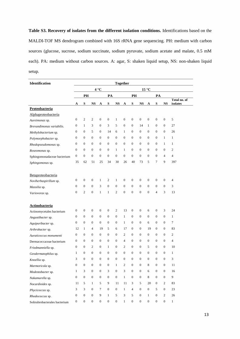
13
Table S3. Recovery of isolates from the different isolation conditions. Identifications based on the
MALDI-TOF MS dendrogram combined with 16S rRNA gene sequencing. PH: medium with carbon
sources (glucose, sucrose, sodium succinate, sodium pyruvate, sodium acetate and malate, 0.5 mM
each). PA: medium without carbon sources. A: agar, S: shaken liquid setup, NS: non-shaken liquid
setup.
Identification Together
4 °C 15 °C
PH PA PH PA
A S NS A S NS A S NS A S NS
Total no. of
isolates
Proteobacteria
Alphaproteobacteria
Aureimonas sp. 0 2 2 0 0 1 0 0 0 0 0 0 5
Brevundimonas variabilis. 0 1 3 0 3 5 0 0 14 1 0 0 27
Methylobacterium sp. 0 0 5 0 14 6 1 0 0 0 0 0 26
Polymorphobacter sp. 0 0 0 0 0 0 0 0 0 0 0 1 1
Rhodopseudomonas sp. 0 0 0 0 0 0 0 0 0 0 0 1 1
Roseomonas sp. 0 0 0 0 0 1 1 0 0 0 0 0 2
Sphingomonadaceae bacterium 0 0 0 0 0 0 0 0 0 0 0 4 4
Sphingomonas sp. 35 62 51 25 34 30 26 40 73 5 7 9 397
Betaproteobacteria
Noviherbaspirillum sp. 0 0 0 1 2 1 0 0 0 0 0 0 4
Massilia sp. 0 0 0 3 0 0 0 0 0 0 0 0 3
Variovorax sp. 0 2 0 1 1 2 0 0 0 0 4 3 13
Actinobacteria
Actinomycetales bacterium 0 0 0 0 0 2 13 0 0 6 0 3 24
Angustibacter sp. 0 0 0 0 0 0 1 0 0 0 0 0 1
Aquipuribacter sp. 0 0 0 0 0 0 1 0 0 6 0 0 7
Arthrobacter sp. 12 1 4 19 5 6 17 0 0 19 0 0 83
Auraticoccus monumenti 0 0 0 0 0 0 2 0 0 0 0 0 2
Dermacoccaceae bacterium 0 0 0 0 0 0 4 0 0 0 0 0 4
Friedmanniella sp. 0 0 2 0 1 0 2 0 0 5 0 0 10
Geodermatophilus sp. 1 0 0 0 0 0 0 0 0 0 0 0 1
Knoellia sp. 3 0 0 0 0 0 0 0 0 0 0 0 3
Marmoricola sp. 0 0 0 0 0 1 2 0 0 8 0 0 11
Modestobacter sp. 1 3 0 0 3 0 3 0 0 6 0 0 16
Nakamurella sp. 0 0 0 0 0 0 1 0 0 8 0 0 9
Nocardioides sp. 11 5 1 5 9 11 11 3 5 20 0 2 83
Phycicoccus sp. 3 3 0 7 0 0 1 4 0 0 5 0 23
Rhodococcus sp. 0 0 0 9 1 5 3 5 0 1 0 2 26
Solirubrobacterales bacterium 0 0 0 0 0 0 1 0 0 0 0 0 1

14
Identification Together
4 °C 15 °C
PH PA PH PA
A S NS A S NS A S NS A S NS
Total no. of
isolates
Deinococcus-Thermus
Deinococcus sp. 0 0 0 0 9 4 2 0 0 0 0 0 15
Firmicutes
Paenibacillus sp. 0 0 0 0 0 0 1 0 0 0 0 0 1
FBP
FBP bacterium 0 0 0 0 0 0 2 0 0 0 0 0 2
Bacteroidetes
Adhaeribacter sp. 0 0 0 0 0 0 1 0 0 0 0 0 1
Hymenobacter sp. 4 5 3 3 9 11 3 0 0 1 1 0 40
Pedobacter sp. 1 0 0 3 0 0 4 1 0 0 1 0 10
Spirosoma sp. 2 2 4 5 3 3 0 0 0 1 10 6 36

Phylotype Strain Accession no. ID Sequence similarity (%) Accession number nifH /bchL /bchX pufLM Phylum Class Order Family1 R‐68457 KY386309 Methylobacterium iners 5317S‐33 ‐ + Proteobacteria Alphaproteobacteria Rhizobiales Methylobacteraceae1 KP15.18.4.PA.S KY386323 Methylobacterium iners 5317S‐33 ‐ + Proteobacteria Alphaproteobacteria Rhizobiales Methylobacteraceae1 R‐68525 KY386327 Methylobacterium iners 5317S‐33 ‐ + Proteobacteria Alphaproteobacteria Rhizobiales Methylobacteraceae1 KP15.21.4.PA.S KY386331 Methylobacterium iners 5317S‐33 ‐ + Proteobacteria Alphaproteobacteria Rhizobiales Methylobacteraceae1 R‐68173 KY386380 Methylobacterium iners 5317S‐33 98,52 EF174497 + ‐ Proteobacteria Alphaproteobacteria Rhizobiales Methylobacteraceae1 R‐68388 KY386396 Methylobacterium iners 5317S‐33 ‐ + Proteobacteria Alphaproteobacteria Rhizobiales Methylobacteraceae1 R‐68391 KY386399 Methylobacterium iners 5317S‐33 ‐ + Proteobacteria Alphaproteobacteria Rhizobiales Methylobacteraceae1 R‐68554 KY386402 Methylobacterium iners 5317S‐33 ‐ + Proteobacteria Alphaproteobacteria Rhizobiales Methylobacteraceae1 R‐68669 KY386414 Methylobacterium iners 5317S‐33 + + Proteobacteria Alphaproteobacteria Rhizobiales Methylobacteraceae1 KP2.9b.4.PA.S KY386471 Methylobacterium iners 5317S‐33 ‐ + Proteobacteria Alphaproteobacteria Rhizobiales Methylobacteraceae2 R‐67878 KY386506 Rhodopseudomonas pseudopalustris DSM123 98,67 AB498818 ‐ ‐ Proteobacteria Alphaproteobacteria Rhizobiales Bradyrhizobiaceae3 R‐68373 KY386318 Aureimonas ferruginae CC‐CFT023 96,64 JQ864240 + ‐ Proteobacteria Alphaproteobacteria Rhizobiales Aurantimonadaceae4 R‐68476 KY386398 Brevundimonas variabilis ATCC15255 ‐ + Proteobacteria Alphaproteobacteria Caulobacterales Caulobacteraceae4 R‐68394 KY386404 Brevundimonas variabilis ATCC15255 ‐ + Proteobacteria Alphaproteobacteria Caulobacterales Caulobacteraceae4 KP2.19.15.PH.NS KY386407 Brevundimonas variabilis ATCC15255 ‐ + Proteobacteria Alphaproteobacteria Caulobacterales Caulobacteraceae4 R‐68473 KY386409 Brevundimonas variabilis ATCC15255 + + Proteobacteria Alphaproteobacteria Caulobacterales Caulobacteraceae4 R‐68297 KY386412 Brevundimonas variabilis ATCC15255 ‐ + Proteobacteria Alphaproteobacteria Caulobacterales Caulobacteraceae4 R‐68298 KY386416 Brevundimonas variabilis ATCC15255 + + Proteobacteria Alphaproteobacteria Caulobacterales Caulobacteraceae4 R‐68299 KY386422 Brevundimonas variabilis ATCC15255 + + Proteobacteria Alphaproteobacteria Caulobacterales Caulobacteraceae4 R‐68483 KY386436 Brevundimonas variabilis ATCC15255 + + Proteobacteria Alphaproteobacteria Caulobacterales Caulobacteraceae4 R‐67742 KY386439 Brevundimonas variabilis ATCC15255 + ‐ Proteobacteria Alphaproteobacteria Caulobacterales Caulobacteraceae4 R‐68295 KY386469 Brevundimonas variabilis ATCC15255 99,79 AJ227783 + + Proteobacteria Alphaproteobacteria Caulobacterales Caulobacteraceae5 R‐67883 KY386496 Polymorphobacter multimanifer 262‐7 94,94 AB649056 + ‐ Proteobacteria Alphaproteobacteria Sphingomonadales Sphingomonadaceae5 KP53.16.15.PA.NS KY386580 Polymorphobacter multimanifer 262‐7 ‐ + Proteobacteria Alphaproteobacteria Sphingomonadales Sphingomonadaceae6 R‐68699 KY386562 Polymorphobacter multimanifer 262‐7 95,99 AB649056 ‐ ‐ Proteobacteria Alphaproteobacteria Sphingomonadales Sphingomonadaceae7 R‐68084 KY386306 Sphingomonas faeni MA‐olki ‐ ‐ Proteobacteria Alphaproteobacteria Sphingomonadales Sphingomonadaceae7 KP15.16.4.PA.V KY386315 Sphingomonas faeni MA‐olki ‐ ‐ Proteobacteria Alphaproteobacteria Sphingomonadales Sphingomonadaceae7 R‐67991 KY386320 Sphingomonas faeni MA‐olki ‐ + Proteobacteria Alphaproteobacteria Sphingomonadales Sphingomonadaceae7 R‐68089 KY386319 Sphingomonas faeni MA‐olki ‐ ‐ Proteobacteria Alphaproteobacteria Sphingomonadales Sphingomonadaceae7 R‐68414 KY386322 Sphingomonas faeni MA‐olki ‐ ‐ Proteobacteria Alphaproteobacteria Sphingomonadales Sphingomonadaceae7 R‐68418 KY386339 Sphingomonas faeni MA‐olki ‐ ‐ Proteobacteria Alphaproteobacteria Sphingomonadales Sphingomonadaceae7 R‐68527 KY386348 Sphingomonas faeni MA‐olki ‐ ‐ Proteobacteria Alphaproteobacteria Sphingomonadales Sphingomonadaceae7 R‐68539 KY386359 Sphingomonas faeni MA‐olki ‐ ‐ Proteobacteria Alphaproteobacteria Sphingomonadales Sphingomonadaceae7 R‐67789 KY386360 Sphingomonas faeni MA‐olki ‐ ‐ Proteobacteria Alphaproteobacteria Sphingomonadales Sphingomonadaceae7 R‐68448 KY386370 Sphingomonas faeni MA‐olki + ‐ Proteobacteria Alphaproteobacteria Sphingomonadales Sphingomonadaceae7 R‐68170 KY386374 Sphingomonas faeni MA‐olki ‐ ‐ Proteobacteria Alphaproteobacteria Sphingomonadales Sphingomonadaceae7 R‐67846 KY386377 Sphingomonas faeni MA‐olki + ‐ Proteobacteria Alphaproteobacteria Sphingomonadales Sphingomonadaceae7 R‐67984 KY386379 Sphingomonas faeni MA‐olki 99,62 AJ429239 + ‐ Proteobacteria Alphaproteobacteria Sphingomonadales Sphingomonadaceae7 KP2.34b.15.PH.V KY386451 Sphingomonas faeni MA‐olki ‐ + Proteobacteria Alphaproteobacteria Sphingomonadales Sphingomonadaceae7 R‐67744 KY386468 Sphingomonas faeni MA‐olki ‐ + Proteobacteria Alphaproteobacteria Sphingomonadales Sphingomonadaceae7 KP43.10b.4.PH.V KY386477 Sphingomonas faeni MA‐olki ‐ + Proteobacteria Alphaproteobacteria Sphingomonadales Sphingomonadaceae7 R‐68208 KY386485 Sphingomonas faeni MA‐olki + ‐ Proteobacteria Alphaproteobacteria Sphingomonadales Sphingomonadaceae7 R‐68340 KY386497 Sphingomonas faeni MA‐olki ‐ ‐ Proteobacteria Alphaproteobacteria Sphingomonadales Sphingomonadaceae8 R‐68700 KY386453 Sphingomonas oligophenolica JCM 12082 98,24 AB018439 ‐ ‐ Proteobacteria Alphaproteobacteria Sphingomonadales Sphingomonadaceae9 R‐68087 KY386314 Sphingomonas aerolata NW12 ‐ ‐ Proteobacteria Alphaproteobacteria Sphingomonadales Sphingomonadaceae9 R‐68461 KY386351 Sphingomonas aerolata NW12 ‐ ‐ Proteobacteria Alphaproteobacteria Sphingomonadales Sphingomonadaceae9 KP2.23.15.PH.NS KY386419 Sphingomonas aerolata NW12 ‐ ‐ Proteobacteria Alphaproteobacteria Sphingomonadales Sphingomonadaceae9 R‐68261 KY386466 Sphingomonas aerolata NW12 ‐ ‐ Proteobacteria Alphaproteobacteria Sphingomonadales Sphingomonadaceae9 R‐68499 KY386473 Sphingomonas aerolata NW12 ‐ ‐ Proteobacteria Alphaproteobacteria Sphingomonadales Sphingomonadaceae9 R‐68344 KY386472 Sphingomonas aerolata NW12 ‐ ‐ Proteobacteria Alphaproteobacteria Sphingomonadales Sphingomonadaceae9 R‐68493 KY386479 Sphingomonas aerolata NW12 ‐ ‐ Proteobacteria Alphaproteobacteria Sphingomonadales Sphingomonadaceae9 R‐68425 KY386482 Sphingomonas aerolata NW12 ‐ ‐ Proteobacteria Alphaproteobacteria Sphingomonadales Sphingomonadaceae9 R‐68427 KY386487 Sphingomonas aerolata NW12 ‐ ‐ Proteobacteria Alphaproteobacteria Sphingomonadales Sphingomonadaceae9 R‐68341 KY386490 Sphingomonas aerolata NW12 ‐ ‐ Proteobacteria Alphaproteobacteria Sphingomonadales Sphingomonadaceae9 R‐68103 KY386491 Sphingomonas aerolata NW12 ‐ ‐ Proteobacteria Alphaproteobacteria Sphingomonadales Sphingomonadaceae9 R‐68279 KY386493 Sphingomonas aerolata NW12 ‐ ‐ Proteobacteria Alphaproteobacteria Sphingomonadales Sphingomonadaceae
Nearest phylogenetic neighbor Taxonomy

9 R‐68104 KY386499 Sphingomonas aerolata NW12 + + Proteobacteria Alphaproteobacteria Sphingomonadales Sphingomonadaceae9 R‐68435 KY386501 Sphingomonas aerolata NW12 ‐ + Proteobacteria Alphaproteobacteria Sphingomonadales Sphingomonadaceae9 R‐68105 KY386502 Sphingomonas aerolata NW12 ‐ ‐ Proteobacteria Alphaproteobacteria Sphingomonadales Sphingomonadaceae9 R‐68335 KY386511 Sphingomonas aerolata NW12 + ‐ Proteobacteria Alphaproteobacteria Sphingomonadales Sphingomonadaceae9 R‐68336 KY386515 Sphingomonas aerolata NW12 ‐ ‐ Proteobacteria Alphaproteobacteria Sphingomonadales Sphingomonadaceae9 KP43.28.4.PH.NS KY386528 Sphingomonas aerolata NW12 ‐ ‐ Proteobacteria Alphaproteobacteria Sphingomonadales Sphingomonadaceae9 R‐68566 KY386531 Sphingomonas aerolata NW12 ‐ ‐ Proteobacteria Alphaproteobacteria Sphingomonadales Sphingomonadaceae9 R‐68274 KY386532 Sphingomonas aerolata NW12 99,09 AJ429240 ‐ ‐ Proteobacteria Alphaproteobacteria Sphingomonadales Sphingomonadaceae9 R‐68308 KY386534 Sphingomonas aerolata NW12 + ‐ Proteobacteria Alphaproteobacteria Sphingomonadales Sphingomonadaceae9 R‐68023 KY386540 Sphingomonas aerolata NW12 ‐ ‐ Proteobacteria Alphaproteobacteria Sphingomonadales Sphingomonadaceae9 R‐68025 KY386544 Sphingomonas aerolata NW12 ‐ ‐ Proteobacteria Alphaproteobacteria Sphingomonadales Sphingomonadaceae9 R‐68275 KY386549 Sphingomonas aerolata NW12 ‐ ‐ Proteobacteria Alphaproteobacteria Sphingomonadales Sphingomonadaceae9 R‐68232 KY386553 Sphingomonas aerolata NW12 ‐ ‐ Proteobacteria Alphaproteobacteria Sphingomonadales Sphingomonadaceae9 KP43.5.15.PH.S KY386557 Sphingomonas aerolata NW12 ‐ ‐ Proteobacteria Alphaproteobacteria Sphingomonadales Sphingomonadaceae9 R‐68305 KY386561 Sphingomonas aerolata NW12 ‐ + Proteobacteria Alphaproteobacteria Sphingomonadales Sphingomonadaceae9 R‐68495 KY386563 Sphingomonas aerolata NW12 + ‐ Proteobacteria Alphaproteobacteria Sphingomonadales Sphingomonadaceae9 R‐68343 KY386567 Sphingomonas aerolata NW12 ‐ ‐ Proteobacteria Alphaproteobacteria Sphingomonadales Sphingomonadaceae9 R‐68309 KY386568 Sphingomonas aerolata NW12 ‐ ‐ Proteobacteria Alphaproteobacteria Sphingomonadales Sphingomonadaceae9 R‐67894 KY386575 Sphingomonas aerolata NW12 ‐ ‐ Proteobacteria Alphaproteobacteria Sphingomonadales Sphingomonadaceae9 KP53.14.4.PH.S KY386576 Sphingomonas aerolata NW12 ‐ ‐ Proteobacteria Alphaproteobacteria Sphingomonadales Sphingomonadaceae9 R‐68345 KY386579 Sphingomonas aerolata NW12 ‐ ‐ Proteobacteria Alphaproteobacteria Sphingomonadales Sphingomonadaceae9 R‐68383 KY386582 Sphingomonas aerolata NW12 + ‐ Proteobacteria Alphaproteobacteria Sphingomonadales Sphingomonadaceae9 R‐68291 KY386584 Sphingomonas aerolata NW12 + ‐ Proteobacteria Alphaproteobacteria Sphingomonadales Sphingomonadaceae9 R‐68245 KY386586 Sphingomonas aerolata NW12 ‐ ‐ Proteobacteria Alphaproteobacteria Sphingomonadales Sphingomonadaceae9 R‐68129 KY386587 Sphingomonas aerolata NW12 ‐ ‐ Proteobacteria Alphaproteobacteria Sphingomonadales Sphingomonadaceae9 R‐68246 KY386589 Sphingomonas aerolata NW12 ‐ ‐ Proteobacteria Alphaproteobacteria Sphingomonadales Sphingomonadaceae9 R‐68445 KY386594 Sphingomonas aerolata NW12 ‐ ‐ Proteobacteria Alphaproteobacteria Sphingomonadales Sphingomonadaceae9 R‐68042 KY386595 Sphingomonas aerolata NW12 ‐ ‐ Proteobacteria Alphaproteobacteria Sphingomonadales Sphingomonadaceae9 R‐68381 KY386598 Sphingomonas aerolata NW12 ‐ ‐ Proteobacteria Alphaproteobacteria Sphingomonadales Sphingomonadaceae9 R‐68380 KY386601 Sphingomonas aerolata NW12 ‐ ‐ Proteobacteria Alphaproteobacteria Sphingomonadales Sphingomonadaceae9 R‐68046 KY386603 Sphingomonas aerolata NW12 ‐ ‐ Proteobacteria Alphaproteobacteria Sphingomonadales Sphingomonadaceae9 R‐68578 KY386606 Sphingomonas aerolata NW12 ‐ ‐ Proteobacteria Alphaproteobacteria Sphingomonadales Sphingomonadaceae9 R‐68351 KY386607 Sphingomonas aerolata NW12 ‐ ‐ Proteobacteria Alphaproteobacteria Sphingomonadales Sphingomonadaceae9 R‐68574 KY386613 Sphingomonas aerolata NW12 ‐ ‐ Proteobacteria Alphaproteobacteria Sphingomonadales Sphingomonadaceae9 R‐68036 KY386622 Sphingomonas aerolata NW12 ‐ ‐ Proteobacteria Alphaproteobacteria Sphingomonadales Sphingomonadaceae9 R‐68349 KY386621 Sphingomonas aerolata NW12 ‐ ‐ Proteobacteria Alphaproteobacteria Sphingomonadales Sphingomonadaceae9 R‐68512 KY386624 Sphingomonas aerolata NW12 ‐ ‐ Proteobacteria Alphaproteobacteria Sphingomonadales Sphingomonadaceae9 R‐68037 KY386625 Sphingomonas aerolata NW12 + ‐ Proteobacteria Alphaproteobacteria Sphingomonadales Sphingomonadaceae9 KP53.9.15.PH.S KY386626 Sphingomonas aerolata NW12 ‐ ‐ Proteobacteria Alphaproteobacteria Sphingomonadales Sphingomonadaceae9 R‐68348 KY386628 Sphingomonas aerolata NW12 ‐ ‐ Proteobacteria Alphaproteobacteria Sphingomonadales Sphingomonadaceae9 KP53.9a.4.PH.NS KY386629 Sphingomonas aerolata NW12 ‐ ‐ Proteobacteria Alphaproteobacteria Sphingomonadales Sphingomonadaceae11 R‐68222 KY386535 Sphingomonas yunnanensis YIM 003 97,52 AY894691 ‐ ‐ Proteobacteria Alphaproteobacteria Sphingomonadales Sphingomonadaceae12 R‐68269 KY386302 Sphingomonas cynarae SPC‐1 ‐ ‐ Proteobacteria Alphaproteobacteria Sphingomonadales Sphingomonadaceae12 R‐68271 KY386310 Sphingomonas cynarae SPC‐1 ‐ + Proteobacteria Alphaproteobacteria Sphingomonadales Sphingomonadaceae12 R‐68375 KY386329 Sphingomonas cynarae SPC‐1 + ‐ Proteobacteria Alphaproteobacteria Sphingomonadales Sphingomonadaceae12 R‐68304 KY386332 Sphingomonas cynarae SPC‐1 98,24 HQ439186 ‐ + Proteobacteria Alphaproteobacteria Sphingomonadales Sphingomonadaceae12 R‐67996 KY386342 Sphingomonas cynarae SPC‐1 ‐ ‐ Proteobacteria Alphaproteobacteria Sphingomonadales Sphingomonadaceae12 R‐68199 KY386373 Sphingomonas cynarae SPC‐1 + ‐ Proteobacteria Alphaproteobacteria Sphingomonadales Sphingomonadaceae12 R‐68171 KY386378 Sphingomonas cynarae SPC‐1 ‐ ‐ Proteobacteria Alphaproteobacteria Sphingomonadales Sphingomonadaceae12 R‐68355 KY386381 Sphingomonas cynarae SPC‐1 ‐ ‐ Proteobacteria Alphaproteobacteria Sphingomonadales Sphingomonadaceae12 R‐68392 KY386400 Sphingomonas cynarae SPC‐1 ‐ + Proteobacteria Alphaproteobacteria Sphingomonadales Sphingomonadaceae12 R‐68317 KY386417 Sphingomonas cynarae SPC‐1 ‐ + Proteobacteria Alphaproteobacteria Sphingomonadales Sphingomonadaceae12 KP2.25b.4.PA.NS KY386428 Sphingomonas cynarae SPC‐1 + + Proteobacteria Alphaproteobacteria Sphingomonadales Sphingomonadaceae12 KP2.32b.4.PA.NS KY386447 Sphingomonas cynarae SPC‐1 ‐ + Proteobacteria Alphaproteobacteria Sphingomonadales Sphingomonadaceae13 R‐68091 KY386335 Sphingomonas mucosissima CP173‐2 ‐ ‐ Proteobacteria Alphaproteobacteria Sphingomonadales Sphingomonadaceae13 R‐67999 KY386352 Sphingomonas mucosissima CP173‐2 ‐ ‐ Proteobacteria Alphaproteobacteria Sphingomonadales Sphingomonadaceae13 R‐68524 KY386357 Sphingomonas mucosissima CP173‐2 98,27 AM229669 ‐ ‐ Proteobacteria Alphaproteobacteria Sphingomonadales Sphingomonadaceae15 R‐67917 KY386305 Sphingomonas yantingensis 1007 + + Proteobacteria Alphaproteobacteria Sphingomonadales Sphingomonadaceae

15 R‐68270 KY386304 Sphingomonas yantingensis 1007 96,79 JX566547 + + Proteobacteria Alphaproteobacteria Sphingomonadales Sphingomonadaceae15 KP15.19.15.PH.S KY386324 Sphingomonas yantingensis 1007 + + Proteobacteria Alphaproteobacteria Sphingomonadales Sphingomonadaceae15 KP15.20.15.PH.V KY386328 Sphingomonas yantingensis 1007 ‐ + Proteobacteria Alphaproteobacteria Sphingomonadales Sphingomonadaceae15 R‐67859 KY386337 Sphingomonas yantingensis 1007 + + Proteobacteria Alphaproteobacteria Sphingomonadales Sphingomonadaceae15 R‐68007 KY386366 Sphingomonas yantingensis 1007 + + Proteobacteria Alphaproteobacteria Sphingomonadales Sphingomonadaceae15 R‐68267 KY386368 Sphingomonas yantingensis 1007 ‐ + Proteobacteria Alphaproteobacteria Sphingomonadales Sphingomonadaceae16 R‐68405 KY386312 Sphingomonas hunanensis JSM 083058 + + Proteobacteria Alphaproteobacteria Sphingomonadales Sphingomonadaceae16 R‐67790 KY386362 Sphingomonas hunanensis JSM 083058 ‐ ‐ Proteobacteria Alphaproteobacteria Sphingomonadales Sphingomonadaceae16 KP15.4.15.PH.NS KY386367 Sphingomonas hunanensis JSM 083058 ‐ + Proteobacteria Alphaproteobacteria Sphingomonadales Sphingomonadaceae16 R‐68410 KY386375 Sphingomonas hunanensis JSM 083058 + + Proteobacteria Alphaproteobacteria Sphingomonadales Sphingomonadaceae16 R‐68292 KY386383 Sphingomonas hunanensis JSM 083058 ‐ + Proteobacteria Alphaproteobacteria Sphingomonadales Sphingomonadaceae16 R‐68553 KY386406 Sphingomonas hunanensis JSM 083058 ‐ + Proteobacteria Alphaproteobacteria Sphingomonadales Sphingomonadaceae16 R‐68552 KY386415 Sphingomonas hunanensis JSM 083058 ‐ + Proteobacteria Alphaproteobacteria Sphingomonadales Sphingomonadaceae16 R‐68400 KY386424 Sphingomonas hunanensis JSM 083058 ‐ + Proteobacteria Alphaproteobacteria Sphingomonadales Sphingomonadaceae16 R‐68370 KY386440 Sphingomonas hunanensis JSM 083058 ‐ + Proteobacteria Alphaproteobacteria Sphingomonadales Sphingomonadaceae16 R‐68161 KY386455 Sphingomonas hunanensis JSM 083058 + + Proteobacteria Alphaproteobacteria Sphingomonadales Sphingomonadaceae16 R‐68260 KY386460 Sphingomonas hunanensis JSM 083058 98,52 FJ527417 + + Proteobacteria Alphaproteobacteria Sphingomonadales Sphingomonadaceae16 R‐68242 KY386578 Sphingomonas hunanensis JSM 083058 ‐ ‐ Proteobacteria Alphaproteobacteria Sphingomonadales Sphingomonadaceae17 R‐67954 KY386585 Sphingomonas pituitosa EDIV 95,58 AJ243751 ‐ ‐ Proteobacteria Alphaproteobacteria Sphingomonadales Sphingomonadaceae18 R‐68475 KY386403 Roseomonas aquatica TR53 99,93 AM231587 ‐ + Proteobacteria Alphaproteobacteria Rhodospirillales Acetobacteraceae19 R‐68165 KY386462 Roseomonas tokyonensis K‐20 98,6 AB297501 + ‐ Proteobacteria Alphaproteobacteria Rhodospirillales Acetobacteraceae20 R‐67997 KY386345 Noviherbaspirillum psychrotolerans PB1 98,62 JN390675 ‐ ‐ Proteobacteria Betaproteobacteria Burkholderiales Oxalobacteraceae20 R‐68490 KY386488 Noviherbaspirillum psychrotolerans PB1 ‐ ‐ Proteobacteria Betaproteobacteria Burkholderiales Oxalobacteraceae21 R‐68579 KY386599 Noviherbaspirillum suwonense 54105‐65 98,95 JX275858 ‐ ‐ Proteobacteria Betaproteobacteria Burkholderiales Oxalobacteraceae22 R‐67978 KY386448 Massilia eurypsychrophila B528‐3 97,7 KJ361504 ‐ ‐ Proteobacteria Betaproteobacteria Burkholderiales Oxalobacteraceae22 R‐67982 KY386454 Massilia eurypsychrophila B528‐3 ‐ ‐ Proteobacteria Betaproteobacteria Burkholderiales Oxalobacteraceae23 R‐67959 KY386465 Variovorax boronicumulans BAM‐48 ‐ ‐ Proteobacteria Betaproteobacteria Burkholderiales Comamonadaceae23 R‐67932 KY386486 Variovorax boronicumulans BAM‐48 97,55 AB300597 ‐ ‐ Proteobacteria Betaproteobacteria Burkholderiales Comamonadaceae23 KP43.23.4.PA.S KY386522 Variovorax boronicumulans BAM‐48 ‐ ‐ Proteobacteria Betaproteobacteria Burkholderiales Comamonadaceae23 R‐68338 KY386543 Variovorax boronicumulans BAM‐48 ‐ ‐ Proteobacteria Betaproteobacteria Burkholderiales Comamonadaceae23 R‐67927 KY386558 Variovorax boronicumulans BAM‐48 ‐ ‐ Proteobacteria Betaproteobacteria Burkholderiales Comamonadaceae24 R‐67871 KY386393 Variovorax ginsengisoli Gsoil 3165 99,26 AB245358 ‐ ‐ Proteobacteria Betaproteobacteria Burkholderiales Comamonadaceae24 R‐67891 KY386627 Variovorax ginsengisoli Gsoil 3165 ‐ ‐ Proteobacteria Betaproteobacteria Burkholderiales Comamonadaceae25 KP15.17.15.PA.V KY386317 Nocardioides ginsengagri BX5‐10 97,38 GQ339904 ‐ ‐ Actinobacteria Actinobacteria Actinomycetales Nocerdioidaceae25 R‐68562 KY386518 Nocardioides ginsengagri BX5‐10 ‐ ‐ Actinobacteria Actinobacteria Actinomycetales Nocerdioidaceae26 R‐68482 KY386444 Nocardioides salarius CL‐Z59 97 DQ401092 ‐ ‐ Actinobacteria Actinobacteria Actinomycetales Nocerdioidaceae27 R‐68162 KY386458 Nocardioides aquaticus EL‐17K 99,38 X94145 ‐ ‐ Actinobacteria Actinobacteria Actinomycetales Nocerdioidaceae28 KP43.13.15.PH.NS KY386484 Nocardioides plantarum NCIMB 12834 100 AF005008 ‐ ‐ Actinobacteria Actinobacteria Actinomycetales Nocerdioidaceae28 R‐68307 KY386526 Nocardioides plantarum NCIMB 12834 ‐ ‐ Actinobacteria Actinobacteria Actinomycetales Nocerdioidaceae28 KP43.9.15.PH.NS KY386564 Nocardioides plantarum NCIMB 12834 ‐ ‐ Actinobacteria Actinobacteria Actinomycetales Nocerdioidaceae29 KP2.10.4.PA.S KY386388 Nocardioides terrigena DS‐17 ‐ ‐ Actinobacteria Actinobacteria Actinomycetales Nocerdioidaceae29 R‐68053 KY386389 Nocardioides terrigena DS‐17 ‐ ‐ Actinobacteria Actinobacteria Actinomycetales Nocerdioidaceae29 R‐67906 KY386390 Nocardioides terrigena DS‐17 ‐ ‐ Actinobacteria Actinobacteria Actinomycetales Nocerdioidaceae29 KP2.14.4.PH.V KY386397 Nocardioides terrigena DS‐17 ‐ ‐ Actinobacteria Actinobacteria Actinomycetales Nocerdioidaceae29 R‐68364 KY386401 Nocardioides terrigena DS‐17 ‐ ‐ Actinobacteria Actinobacteria Actinomycetales Nocerdioidaceae29 R‐68474 KY386405 Nocardioides terrigena DS‐17 ‐ ‐ Actinobacteria Actinobacteria Actinomycetales Nocerdioidaceae29 R‐68550 KY386421 Nocardioides terrigena DS‐17 ‐ ‐ Actinobacteria Actinobacteria Actinomycetales Nocerdioidaceae29 R‐68154 KY386423 Nocardioides terrigena DS‐17 97,79 EF363712 ‐ ‐ Actinobacteria Actinobacteria Actinomycetales Nocerdioidaceae29 R‐67971 KY386425 Nocardioides terrigena DS‐17 ‐ ‐ Actinobacteria Actinobacteria Actinomycetales Nocerdioidaceae29 R‐68065 KY386430 Nocardioides terrigena DS‐17 ‐ ‐ Actinobacteria Actinobacteria Actinomycetales Nocerdioidaceae29 R‐67865 KY386438 Nocardioides terrigena DS‐17 ‐ ‐ Actinobacteria Actinobacteria Actinomycetales Nocerdioidaceae29 R‐67756 KY386443 Nocardioides terrigena DS‐17 ‐ ‐ Actinobacteria Actinobacteria Actinomycetales Nocerdioidaceae29 R‐68140 KY386461 Nocardioides terrigena DS‐17 + ‐ Actinobacteria Actinobacteria Actinomycetales Nocerdioidaceae29 R‐68014 KY386492 Nocardioides terrigena DS‐17 ‐ ‐ Actinobacteria Actinobacteria Actinomycetales Nocerdioidaceae30 R‐67827 KY386592 Nocardioides terrigena DS‐17 96,06 EF363712 ‐ ‐ Actinobacteria Actinobacteria Actinomycetales Nocerdioidaceae31 R‐68372 KY386385 Nocardioides terrigena DS‐17 ‐ ‐ Actinobacteria Actinobacteria Actinomycetales Nocerdioidaceae31 R‐68145 KY386386 Nocardioides terrigena DS‐17 97,52 EF363712 ‐ ‐ Actinobacteria Actinobacteria Actinomycetales Nocerdioidaceae31 R‐67747 KY386391 Nocardioides terrigena DS‐17 + ‐ Actinobacteria Actinobacteria Actinomycetales Nocerdioidaceae

31 KP2.23.15.PH.V KY386420 Nocardioides terrigena DS‐17 ‐ ‐ Actinobacteria Actinobacteria Actinomycetales Nocerdioidaceae31 R‐68158 KY386446 Nocardioides terrigena DS‐17 ‐ ‐ Actinobacteria Actinobacteria Actinomycetales Nocerdioidaceae31 R‐68160 KY386452 Nocardioides terrigena DS‐17 ‐ ‐ Actinobacteria Actinobacteria Actinomycetales Nocerdioidaceae31 R‐68167 KY386463 Nocardioides terrigena DS‐17 ‐ ‐ Actinobacteria Actinobacteria Actinomycetales Nocerdioidaceae32 R‐67776 KY386307 Marmoricola korecus Sco‐A36 ‐ ‐ Actinobacteria Actinobacteria Actinomycetales Nocerdioidaceae32 R‐67781 KY386333 Marmoricola korecus Sco‐A36 98,67 FN386723 ‐ ‐ Actinobacteria Actinobacteria Actinomycetales Nocerdioidaceae33 R‐68467 KY386387 Marmoricola aquaticus CBMAI 1089 ‐ ‐ Actinobacteria Actinobacteria Actinomycetales Nocerdioidaceae33 R‐67804 KY386524 Marmoricola aquaticus CBMAI 1089 97,75 JN615437 ‐ ‐ Actinobacteria Actinobacteria Actinomycetales Nocerdioidaceae34 R‐67800 KY386498 Nocardioides antarcticus M‐SA3‐94 97,76 KM347967 ‐ ‐ Actinobacteria Actinobacteria Actinomycetales Nocerdioidaceae34 R‐68564 KY386504 Nocardioides antarcticus M‐SA3‐94 ‐ ‐ Actinobacteria Actinobacteria Actinomycetales Nocerdioidaceae34 R‐68503 KY386512 Nocardioides antarcticus M‐SA3‐94 ‐ ‐ Actinobacteria Actinobacteria Actinomycetales Nocerdioidaceae34 R‐68432 KY386521 Nocardioides antarcticus M‐SA3‐94 ‐ ‐ Actinobacteria Actinobacteria Actinomycetales Nocerdioidaceae34 R‐68485 KY386533 Nocardioides antarcticus M‐SA3‐94 ‐ ‐ Actinobacteria Actinobacteria Actinomycetales Nocerdioidaceae34 R‐68233 KY386554 Nocardioides antarcticus M‐SA3‐94 ‐ ‐ Actinobacteria Actinobacteria Actinomycetales Nocerdioidaceae34 R‐68486 KY386559 Nocardioides antarcticus M‐SA3‐94 ‐ ‐ Actinobacteria Actinobacteria Actinomycetales Nocerdioidaceae34 R‐68497 KY386560 Nocardioides antarcticus M‐SA3‐94 ‐ ‐ Actinobacteria Actinobacteria Actinomycetales Nocerdioidaceae34 R‐68352 KY386605 Nocardioides antarcticus M‐SA3‐94 ‐ ‐ Actinobacteria Actinobacteria Actinomycetales Nocerdioidaceae35 R‐68019 KY386523 Rhodococcus aerolatus PAMC 27367 96,34 KM044053 ‐ ‐ Actinobacteria Actinobacteria Actinomycetales Nocardiaceae35 R‐68235 KY386556 Rhodococcus aerolatus PAMC 27367 ‐ ‐ Actinobacteria Actinobacteria Actinomycetales Nocardiaceae36 R‐68187 KY386338 Rhodococcus aerolatus PAMC 27367 95,64 KM044053 + ‐ Actinobacteria Actinobacteria Actinomycetales Nocardiaceae37 R‐68509 KY386574 Rhodococcus aerolatus PAMC 27367 95,92 KM044053 ‐ ‐ Actinobacteria Actinobacteria Actinomycetales Nocardiaceae38 R‐67872 KY386395 Rhodococcus aerolatus PAMC 27367 95,85 KM044053 ‐ ‐ Actinobacteria Actinobacteria Actinomycetales Nocardiaceae38 R‐68258 KY386617 Rhodococcus aerolatus PAMC 27367 ‐ ‐ Actinobacteria Actinobacteria Actinomycetales Nocardiaceae39 R‐68460 KY386303 Rhodococcus fascians LMG 3623 + ‐ Actinobacteria Actinobacteria Actinomycetales Nocardiaceae39 R‐68273 KY386321 Rhodococcus fascians LMG 3623 98,77 JMEN01000010 ‐ ‐ Actinobacteria Actinobacteria Actinomycetales Nocardiaceae39 R‐68530 KY386341 Rhodococcus fascians LMG 3623 ‐ ‐ Actinobacteria Actinobacteria Actinomycetales Nocardiaceae39 R‐68463 KY386344 Rhodococcus fascians LMG 3623 ‐ ‐ Actinobacteria Actinobacteria Actinomycetales Nocardiaceae39 KP15.30.4.PA.NS KY386358 Rhodococcus fascians LMG 3623 + ‐ Actinobacteria Actinobacteria Actinomycetales Nocardiaceae39 R‐68196 KY386364 Rhodococcus fascians LMG 3623 ‐ ‐ Actinobacteria Actinobacteria Actinomycetales Nocardiaceae39 R‐68515 KY386604 Rhodococcus fascians LMG 3623 ‐ ‐ Actinobacteria Actinobacteria Actinomycetales Nocardiaceae40 R‐68537 KY386361 Friedmanniella sagamiharensis FB2 ‐ ‐ Actinobacteria Actinobacteria Actinomycetales Propionibacteriaceae40 R‐67749 KY386408 Friedmanniella sagamiharensis FB2 96,09 AB445456 ‐ ‐ Actinobacteria Actinobacteria Actinomycetales Propionibacteriaceae40 R‐67754 KY386434 Friedmanniella sagamiharensis FB2 ‐ ‐ Actinobacteria Actinobacteria Actinomycetales Propionibacteriaceae40 R‐67745 KY386470 Friedmanniella sagamiharensis FB2 ‐ ‐ Actinobacteria Actinobacteria Actinomycetales Propionibacteriaceae40 R‐68220 KY386525 Friedmanniella sagamiharensis FB2 ‐ ‐ Actinobacteria Actinobacteria Actinomycetales Propionibacteriaceae41 R‐68221 KY386527 Friedmanniella luteola FA1 98,22 AB445453 ‐ ‐ Actinobacteria Actinobacteria Actinomycetales Propionibacteriaceae42 R‐68201 KY386505 Auraticoccus monumenti MON 2.2 99,58 FN552748 ‐ ‐ Actinobacteria Actinobacteria Actinomycetales Propionibacteriaceae43 R‐67786 KY386350 Jatrophihabitans endophyticus S9650 93,6 JQ346802 ‐ ‐ Actinobacteria Actinobacteria Actinomycetales43 R‐68511 KY386570 Jatrophihabitans endophyticus S9650 ‐ ‐ Actinobacteria Actinobacteria Actinomycetales44 R‐68701 KY386494 Jatrophihabitans soli KIS75‐12 94,37 KP017569 ‐ ‐ Actinobacteria Actinobacteria Actinomycetales45 R‐68182 KY386325 Frankia alni ACN14A ‐ ‐ Actinobacteria Actinobacteria Actinomycetales45 R‐68183 KY386330 Frankia alni ACN14A 94,11 CT573213 ‐ ‐ Actinobacteria Actinobacteria Actinomycetales45 R‐67752 KY386426 Frankia alni ACN14A ‐ ‐ Actinobacteria Actinobacteria Actinomycetales45 R‐67882 KY386481 Frankia alni ACN14A ‐ ‐ Actinobacteria Actinobacteria Actinomycetales45 R‐68215 KY386514 Frankia alni ACN14A ‐ ‐ Actinobacteria Actinobacteria Actinomycetales45 R‐67881 KY386566 Frankia alni ACN14A ‐ ‐ Actinobacteria Actinobacteria Actinomycetales45 R‐67888 KY386611 Frankia alni ACN14A ‐ ‐ Actinobacteria Actinobacteria Actinomycetales46 R‐67810 KY386542 Cryptosporangium minutisporangium IFO15962 94,41 AB037007 ‐ ‐ Actinobacteria Actinobacteria Actinomycetales47 R‐68148 KY386394 Modestobacter versicolor CP153‐2 ‐ ‐ Actinobacteria Actinobacteria Actinomycetales47 R‐67836 KY386609 Modestobacter versicolor CP153‐2 94,28 AJ871304 ‐ ‐ Actinobacteria Actinobacteria Actinomycetales47 R‐68237 KY386619 Modestobacter versicolor CP153‐2 ‐ ‐ Actinobacteria Actinobacteria Actinomycetales48 R‐68223 KY386537 Sporichthya brevicatena IFO 16195 93,78 AB006164 ‐ ‐ Actinobacteria Actinobacteria Actinomycetales48 R‐68238 KY386569 Sporichthya brevicatena IFO 16195 ‐ ‐ Actinobacteria Actinobacteria Actinomycetales49 KP15.26.15.PA.V KY386343 Nakamurella lactae DLS‐10 ‐ ‐ Actinobacteria Actinobacteria Actinomycetales Nakamurellaceae49 R‐68216 KY386516 Nakamurella lactae DLS‐10 98,07 AM778124 ‐ ‐ Actinobacteria Actinobacteria Actinomycetales Nakamurellaceae50 R‐67826 KY386590 Nakamurella lactae DLS‐10 ‐ ‐ Actinobacteria Actinobacteria Actinomycetales Nakamurellaceae50 R‐67832 KY386596 Nakamurella lactae DLS‐10 ‐ ‐ Actinobacteria Actinobacteria Actinomycetales Nakamurellaceae50 R‐67838 KY386610 Nakamurella lactae DLS‐10 98,57 AM778124 ‐ ‐ Actinobacteria Actinobacteria Actinomycetales Nakamurellaceae

51 R‐68190 KY386347 Modestobacter lapidis MON 3.1 ‐ ‐ Actinobacteria Actinobacteria Actinomycetales Geodermatophilaceae51 R‐68535 KY386365 Modestobacter lapidis MON 3.1 ‐ ‐ Actinobacteria Actinobacteria Actinomycetales Geodermatophilaceae51 R‐68354 KY386382 Modestobacter lapidis MON 3.1 ‐ ‐ Actinobacteria Actinobacteria Actinomycetales Geodermatophilaceae51 R‐67763 KY386459 Modestobacter lapidis MON 3.1 ‐ ‐ Actinobacteria Actinobacteria Actinomycetales Geodermatophilaceae51 R‐68492 KY386483 Modestobacter lapidis MON 3.1 ‐ ‐ Actinobacteria Actinobacteria Actinomycetales Geodermatophilaceae51 R‐68563 KY386513 Modestobacter lapidis MON 3.1 ‐ ‐ Actinobacteria Actinobacteria Actinomycetales Geodermatophilaceae51 R‐68230 KY386550 Modestobacter lapidis MON 3.1 97,28 LN810544 ‐ ‐ Actinobacteria Actinobacteria Actinomycetales Geodermatophilaceae52 R‐68264 KY386392 Phycicoccus ochangensis L1b‐b9 98,09 GQ344405 ‐ ‐ Actinobacteria Actinobacteria Actinomycetales Intrasporangiaceae52 R‐68327 KY386427 Phycicoccus ochangensis L1b‐b9 ‐ ‐ Actinobacteria Actinobacteria Actinomycetales Intrasporangiaceae52 R‐68064 KY386429 Phycicoccus ochangensis L1b‐b9 ‐ ‐ Actinobacteria Actinobacteria Actinomycetales Intrasporangiaceae52 R‐67977 KY386445 Phycicoccus ochangensis L1b‐b9 ‐ ‐ Actinobacteria Actinobacteria Actinomycetales Intrasporangiaceae52 KP2.38.4.PH.V KY386456 Phycicoccus ochangensis L1b‐b9 ‐ ‐ Actinobacteria Actinobacteria Actinomycetales Intrasporangiaceae52 R‐67902 KY386464 Phycicoccus ochangensis L1b‐b9 ‐ ‐ Actinobacteria Actinobacteria Actinomycetales Intrasporangiaceae52 R‐68010 KY386474 Phycicoccus ochangensis L1b‐b9 ‐ ‐ Actinobacteria Actinobacteria Actinomycetales Intrasporangiaceae52 R‐68028 KY386548 Phycicoccus ochangensis L1b‐b9 ‐ ‐ Actinobacteria Actinobacteria Actinomycetales Intrasporangiaceae52 KP43.40.4.PA.V KY386551 Phycicoccus ochangensis L1b‐b9 ‐ ‐ Actinobacteria Actinobacteria Actinomycetales Intrasporangiaceae53 R‐68061 KY386418 Knoellia sinensis DSM12331 98,7 AVPJ01000034 ‐ ‐ Actinobacteria Actinobacteria Actinomycetales Intrasporangiaceae54 R‐67798 KY386475 Aquipuribacter hungaricus IV‐75 + ‐ Actinobacteria Actinobacteria Actinomycetales Intrasporangiaceae54 R‐67807 KY386530 Aquipuribacter hungaricus IV‐75 ‐ ‐ Actinobacteria Actinobacteria Actinomycetales Intrasporangiaceae54 KP43.37.15.PA.V KY386545 Aquipuribacter hungaricus IV‐75 97,88 FM179321 ‐ ‐ Actinobacteria Actinobacteria Actinomycetales Intrasporangiaceae54 R‐68204 KY386565 Aquipuribacter hungaricus IV‐75 ‐ ‐ Actinobacteria Actinobacteria Actinomycetales Intrasporangiaceae55 R‐68518 KY386301 Arthrobacter pityocampae Tp2 99,34 EU885749 ‐ ‐ Actinobacteria Actinobacteria Actinomycetales Micrococcaceae55 R‐68186 KY386336 Arthrobacter pityocampae Tp2 ‐ ‐ Actinobacteria Actinobacteria Actinomycetales Micrococcaceae55 R‐67785 KY386346 Arthrobacter pityocampae Tp2 ‐ ‐ Actinobacteria Actinobacteria Actinomycetales Micrococcaceae55 R‐67998 KY386349 Arthrobacter pityocampae Tp2 ‐ ‐ Actinobacteria Actinobacteria Actinomycetales Micrococcaceae55 R‐68224 KY386538 Arthrobacter pityocampae Tp2 ‐ ‐ Actinobacteria Actinobacteria Actinomycetales Micrococcaceae57 R‐68156 KY386431 Arthrobacter agilis DSM20550 ‐ ‐ Actinobacteria Actinobacteria Actinomycetales Micrococcaceae57 R‐68157 KY386442 Arthrobacter agilis DSM20550 ‐ ‐ Actinobacteria Actinobacteria Actinomycetales Micrococcaceae57 R‐68477 KY386457 Arthrobacter agilis DSM20550 ‐ ‐ Actinobacteria Actinobacteria Actinomycetales Micrococcaceae57 R‐68144 KY386467 Arthrobacter agilis DSM20550 ‐ ‐ Actinobacteria Actinobacteria Actinomycetales Micrococcaceae57 R‐68102 KY386476 Arthrobacter agilis DSM20550 ‐ ‐ Actinobacteria Actinobacteria Actinomycetales Micrococcaceae57 KP43.11.4.PH.V KY386478 Arthrobacter agilis DSM20550 ‐ ‐ Actinobacteria Actinobacteria Actinomycetales Micrococcaceae57 R‐67801 KY386510 Arthrobacter agilis DSM20550 ‐ ‐ Actinobacteria Actinobacteria Actinomycetales Micrococcaceae57 R‐68017 KY386519 Arthrobacter agilis DSM20550 ‐ ‐ Actinobacteria Actinobacteria Actinomycetales Micrococcaceae57 R‐67803 KY386520 Arthrobacter agilis DSM20550 ‐ ‐ Actinobacteria Actinobacteria Actinomycetales Micrococcaceae57 R‐67808 KY386536 Arthrobacter agilis DSM20550 ‐ ‐ Actinobacteria Actinobacteria Actinomycetales Micrococcaceae57 R‐68027 KY386546 Arthrobacter agilis DSM20550 ‐ ‐ Actinobacteria Actinobacteria Actinomycetales Micrococcaceae57 R‐68229 KY386547 Arthrobacter agilis DSM20550 ‐ ‐ Actinobacteria Actinobacteria Actinomycetales Micrococcaceae57 R‐68231 KY386552 Arthrobacter agilis DSM20550 ‐ ‐ Actinobacteria Actinobacteria Actinomycetales Micrococcaceae57 R‐68508 KY386577 Arthrobacter agilis DSM20550 ‐ ‐ Actinobacteria Actinobacteria Actinomycetales Micrococcaceae57 R‐68247 KY386593 Arthrobacter agilis DSM20550 ‐ ‐ Actinobacteria Actinobacteria Actinomycetales Micrococcaceae57 R‐68043 KY386597 Arthrobacter agilis DSM20550 ‐ ‐ Actinobacteria Actinobacteria Actinomycetales Micrococcaceae57 R‐68045 KY386600 Arthrobacter agilis DSM20550 ‐ ‐ Actinobacteria Actinobacteria Actinomycetales Micrococcaceae57 KP53.28.4.PA.V KY386602 Arthrobacter agilis DSM20550 ‐ ‐ Actinobacteria Actinobacteria Actinomycetales Micrococcaceae57 R‐67839 KY386614 Arthrobacter agilis DSM20550 ‐ ‐ Actinobacteria Actinobacteria Actinomycetales Micrococcaceae57 R‐68257 KY386616 Arthrobacter agilis DSM20550 ‐ ‐ Actinobacteria Actinobacteria Actinomycetales Micrococcaceae57 R‐67817 KY386620 Arthrobacter agilis DSM20550 ‐ ‐ Actinobacteria Actinobacteria Actinomycetales Micrococcaceae57 R‐67818 KY386623 Arthrobacter agilis DSM20550 99,46 X80748 ‐ ‐ Actinobacteria Actinobacteria Actinomycetales Micrococcaceae58 R‐67793 KY386372 Arthrobacter flavus TB 23 99,25 ALPM01000083 ‐ ‐ Actinobacteria Actinobacteria Actinomycetales Micrococcaceae58 R‐68397 KY386413 Arthrobacter flavus TB 23 ‐ ‐ Actinobacteria Actinobacteria Actinomycetales Micrococcaceae59 R‐68209 KY386489 Calidifontibacter indicus PC IW02 ‐ ‐ Actinobacteria Actinobacteria Actinomycetales Dermacoccaceae59 R‐68253 KY386608 Calidifontibacter indicus PC IW02 94,71 EF187228 ‐ ‐ Actinobacteria Actinobacteria Actinomycetales Dermacoccaceae59 KP53.41.15.PH.V KY386615 Calidifontibacter indicus PC IW02 ‐ ‐ Actinobacteria Actinobacteria Actinomycetales Dermacoccaceae60 R‐68180 KY386316 Deinococcus marmoris DSM12784 ‐ ‐ Deinococcus‐Thermus Deinococci Deinococcales Deinococcaceae60 R‐68561 KY386437 Deinococcus marmoris DSM12784 100 JNIV01000230 ‐ ‐ Deinococcus‐Thermus Deinococci Deinococcales Deinococcaceae60 R‐68498 KY386507 Deinococcus marmoris DSM12784 ‐ ‐ Deinococcus‐Thermus Deinococci Deinococcales Deinococcaceae61 R‐68571 KY386571 Deinococcus saxicola AA‐1444 99,93 AJ585984 ‐ ‐ Deinococcus‐Thermus Deinococci Deinococcales Deinococcaceae61 KP53.17.15.PH.V KY386581 Deinococcus saxicola AA‐1444 ‐ ‐ Deinococcus‐Thermus Deinococci Deinococcales Deinococcaceae

61 R‐68514 KY386612 Deinococcus saxicola AA‐1444 ‐ ‐ Deinococcus‐Thermus Deinococci Deinococcales Deinococcaceae62 R‐68702 KY386340 Hymenobacter roseosalivarius AA718 ‐ ‐ Bacteroidetes Cytophagia Cytophagales Cytophagaceae62 R‐68471 KY386441 Hymenobacter roseosalivarius AA718 98,01 Y18833 ‐ ‐ Bacteroidetes Cytophagia Cytophagales Cytophagaceae62 R‐68240 KY386573 Hymenobacter roseosalivarius AA718 ‐ ‐ Bacteroidetes Cytophagia Cytophagales Cytophagaceae62 R‐68582 KY386591 Hymenobacter roseosalivarius AA718 ‐ ‐ Bacteroidetes Cytophagia Cytophagales Cytophagaceae63 R‐68402 KY386432 Hymenobacter roseosalivarius AA718 97,94 Y18833 ‐ ‐ Bacteroidetes Cytophagia Cytophagales Cytophagaceae65 R‐68403 KY386435 Hymenobacter roseosalivarius AA718 98,89 Y18833 ‐ ‐ Bacteroidetes Cytophagia Cytophagales Cytophagaceae66 R‐68243 KY386583 Hymenobacter roseosalivarius AA718 94,75 Y18833 ‐ ‐ Bacteroidetes Cytophagia Cytophagales Cytophagaceae66 R‐68122 KY386588 Hymenobacter roseosalivarius AA718 ‐ ‐ Bacteroidetes Cytophagia Cytophagales Cytophagaceae67 R‐67758 KY386449 Hymenobacter arcticus R2‐4 98,05 KC213491 ‐ ‐ Bacteroidetes Cytophagia Cytophagales Cytophagaceae68 R‐68178 KY386311 Hymenobacter terrae DG7A 93,96 KF862488 ‐ ‐ Bacteroidetes Cytophagia Cytophagales Cytophagaceae68 R‐68353 KY386313 Hymenobacter terrae DG7A ‐ ‐ Bacteroidetes Cytophagia Cytophagales Cytophagaceae68 R‐68361 KY386326 Hymenobacter terrae DG7A ‐ + Bacteroidetes Cytophagia Cytophagales Cytophagaceae68 KP15.29.4.PA.NS KY386354 Hymenobacter terrae DG7A ‐ ‐ Bacteroidetes Cytophagia Cytophagales Cytophagaceae68 KP15.29b.4.PA.V KY386355 Hymenobacter terrae DG7A ‐ ‐ Bacteroidetes Cytophagia Cytophagales Cytophagaceae68 R‐68447 KY386356 Hymenobacter terrae DG7A ‐ ‐ Bacteroidetes Cytophagia Cytophagales Cytophagaceae68 R‐68536 KY386363 Hymenobacter terrae DG7A ‐ ‐ Bacteroidetes Cytophagia Cytophagales Cytophagaceae68 R‐68359 KY386369 Hymenobacter terrae DG7A ‐ ‐ Bacteroidetes Cytophagia Cytophagales Cytophagaceae69 R‐68030 KY386555 Hymenobacter terrae DG7A 91,29 KF862488 ‐ ‐ Bacteroidetes Cytophagia Cytophagales Cytophagaceae70 R‐68225 KY386541 Adhaeribacter aquaticus DSM16391 96,37 AXBK01000007 ‐ ‐ Bacteroidetes Cytophagia Cytophagales Cytophagaceae71 R‐68113 KY386539 Pedobacter panaciterrae Gsoil 042 ‐ ‐ Bacteroidetes Sphingobacteriia Sphingobacteriales Sphingobacteriaceae71 R‐68289 KY386572 Pedobacter panaciterrae Gsoil 042 97,59 AB245368 ‐ ‐ Bacteroidetes Sphingobacteriia Sphingobacteriales Sphingobacteriaceae72 R‐67967 KY386410 Pedobacter duraquae WB2.1‐25 98,36 AM491368 ‐ ‐ Bacteroidetes Sphingobacteriia Sphingobacteriales Sphingobacteriaceae73 R‐68191 KY386353 Pedobacter ruber W1 95,43 HQ882803 ‐ ‐ Bacteroidetes Sphingobacteriia Sphingobacteriales Sphingobacteriaceae73 R‐68207 KY386480 Pedobacter ruber W1 ‐ ‐ Bacteroidetes Sphingobacteriia Sphingobacteriales Sphingobacteriaceae74 R‐68376 KY386334 Spirosoma rigui WPCB118 ‐ ‐ Bacteroidetes Cytophagia Cytophagales Cytophagaceae74 R‐68523 KY386371 Spirosoma rigui WPCB118 ‐ ‐ Bacteroidetes Cytophagia Cytophagales Cytophagaceae74 R‐68079 KY386376 Spirosoma rigui WPCB118 97,94 EF507900 ‐ ‐ Bacteroidetes Cytophagia Cytophagales Cytophagaceae74 R‐67957 KY386411 Spirosoma rigui WPCB118 + ‐ Bacteroidetes Cytophagia Cytophagales Cytophagaceae74 R‐68424 KY386495 Spirosoma rigui WPCB118 ‐ ‐ Bacteroidetes Cytophagia Cytophagales Cytophagaceae74 R‐68502 KY386503 Spirosoma rigui WPCB118 ‐ ‐ Bacteroidetes Cytophagia Cytophagales Cytophagaceae74 R‐67885 KY386508 Spirosoma rigui WPCB118 ‐ ‐ Bacteroidetes Cytophagia Cytophagales Cytophagaceae74 R‐67936 KY386509 Spirosoma rigui WPCB118 ‐ ‐ Bacteroidetes Cytophagia Cytophagales Cytophagaceae74 R‐68108 KY386517 Spirosoma rigui WPCB118 ‐ ‐ Bacteroidetes Cytophagia Cytophagales Cytophagaceae74 R‐68505 KY386529 Spirosoma rigui WPCB118 ‐ ‐ Bacteroidetes Cytophagia Cytophagales Cytophagaceae75 R‐68384 KY386384 Arthrobacter oxydans DSM 20119 99,25 X83408 ‐ ‐ Actinobacteria Actinobacteria Actinomycetales Micrococcaceae77 R‐68259 KY386618 Angustibacter aerolatus 7402J‐48 96,64 JQ639056 ‐ ‐ Actinobacteria Actinobacteria Actinomycetales Kineosporiaceae10 R‐68085 KY386308 Geodermatophilus terrae PB261 99,02 JN033773 ‐ ‐ Actinobacteria Actinobacteria Actinomycetales Geodermatophilaceae14 R‐68159 KY386450 Conexibacter arvalis KV‐962 94,02 AB597950 ‐ ‐ Actinobacteria Thermoleophilia Solirubrobacterales56 R‐68670 KY386433 Paenibacillus frigoriresistens YIM 016 97,66 JQ314346 ‐ ‐ Firmicutes Bacilli Bacillales Paenibacillaceae64 R‐68168 KY386300 Nocardioides echinoideorum CC‐CZW004 78,7 KM085325 ‐ ‐ FBP76 R‐68213 KY386500 Hippea Maritima DSM 10411 79,07 CP002606 ‐ ‐ FBP