Cover & table of contents-new - UGent Biblio
161
Ghent University Faculty of Sciences Department of Plant Biotechnology and Bioinformatics The Arabidopsis root cap contributes to root branching by setting the root clock this thesis is submitted as fulfilment of the requirements for the degree of Ph.D. in Sciences 2014 by Wei Xuan Promoter: Prof. dr. Tom Beeckman VIB - Plant Systems Biology Root Development Group Technologiepark 927, B-9000 Ghent, Belgium
-
Upload
khangminh22 -
Category
Documents
-
view
0 -
download
0
Transcript of Cover & table of contents-new - UGent Biblio

Ghent University
Faculty of Sciences
Department of Plant Biotechnology and Bioinformatics
The Arabidopsis root cap contributes to root
branching by setting the root clock
this thesis is submitted as fulfilment of the requirements for
the degree of Ph.D. in Sciences 2014 by
Wei Xuan
Promoter:
Prof. dr. Tom Beeckman
VIB - Plant Systems Biology
Root Development Group
Technologiepark 927, B-9000 Ghent, Belgium

Xuan, W (2014) “The Arabidopsis root cap contributes to root branching by setting the
root clock”. PhD Thesis, Ghent University, Ghent, Belgium
The authors and promoters give the authorization to consult and copy parts of this work for
personal use only. Every other use is subject to the copyright laws. Permission to reproduce
any material contained in this work should be obtained from the author.
Wei Xuan was supported by a CSC PhD fellowship grant of China and by the Special
Research Funds of Ghent University (BOF, B/06640/01)

JURY MEMBERS
Promotors
Prof. Dr. Tom Beeckman
Department of Plant Biotechnology and Genetics
Ghent University, Belgium
Promotion commission
Prof. dr. Wout Boerjan (chair)
Department of Plant Biotechnology and Genetics
Ghent University, Belgium
Prof. dr. Lieven De Veylder
Department of Plant Systems Biology,
Ghent University, Belgium
Prof. dr. Ive de Smet
Department of Plant Biotechnology and Genetics
Ghent University, Belgium
Prof. dr. Danny Geelen
Department of Plant Production,
Faculty of Bioscience Engineering,
Ghent University, Belgium
Dr. Laurent Laplaze
Institut de Recherche pour le Développement (IRD),
UMR DIADE (IRD/UM2), France
Dr. Dominique Audenaert
Compound Screening Facility,
VIB, Belgium
Dr. Bert de Rybel
Laboratory of Biochemistry,
Wageningen University, the Netherlands

TABLE OF CONTENTS
Scope
Frequently used abbreviations
Chapter 1 Introduction 1
Chapter 2 A local auxin source modulates root pre-patterning 51
Chapter 3 The growth dynamics of the root cap cells set the root clock 81
Chapter 4 A novel small molecule reveals the role of auxin receptor TIR1
on root branching 105
Chapter 5 Concluding remarks 121
Chapter 6 Summary 133
Chapter 7 Acknowledgements 135
Curriculum vitae

SCOPE
Spatiotemporal coordination of organ formation is a crucial research topic in both
plant and animal biology. In the model plant Arabidopsis thaliana, the “root clock”
model states that a periodic induction of gene expression occurring in the oscillation
zone of the root apex constitutes a temporal signal. This temporal signal can be
translated into a spatial message leading to sequential formation of the prebranch
sites, patches of cell competent to form lateral roots. The plant hormone auxin
controls many aspects of organ growth and development in plants. Particularly for
lateral root development, auxin signalling is quintessential for lateral root (LR)
initiation, patterning of LR primordia and its emergence. A root cap-specific indole-
3-butyric acid (IBA) to indole-3-acetic acid (IAA) conversion was found to
contribute to the root branching process. This auxin source modulates the amplitude
of the oscillations and subsequently determines whether a prebranch site is created or
not.
The aim of this project was to reveal the mechanism how this root cap-source auxin
affect the root clock and the nature of this process. To access it, we applied live-
imaging approaches to visualize auxin signalling dynamics during the oscillations and
the prebranch sites formation. A novel imaging system with a vertically adapted
fluorescence microscope was optimized to visualize the dynamics of the root cap
auxin response. To identity novel genes controlling the root clock in Arabidopsis, we
followed two strategies; firstly, an IBA-trascriptome analysis was applied to explore
the signalling components downstream of the root cap-source auxin, which led to the
identification of MEMBRANE-ASSOCIATED KINASE REGULATOR4 (MAKR4).
Secondly, we use Tirlin as a chemical tool to identify the potential signalling
components downstream of TIR1/AFB-dependent signalling pathways for lateral root
formation.

FREQUENTLY USED ABBREVIATIONS
ACR4: ARABIDOPSIS CRINKLY 4 AFB: AUXIN-RELATED F-Box protein ARF: AUXIN RESPONSE FACTOR Aux/IAA: AUXIN/INDOLE-3-ACETIC ACID AXR: AUXIN RESISTANT Dex: dexamethasone DMSO: dimethylsulfoxide DTA: Diphtheria toxin A FC: founder cell GFP: GREEN FLUORESCENT PROTEIN GR: glucocorticoid receptor IAA: indole-3-acetic acid IAM: indole-3-acetamide IBA: indole-3-butyric acid LR: lateral root LRC: lateral root cap LRP: lateral root primordium LRIS: lateral root inducible system MAKR4: MEMBRANE-ASSOCIATED KINASE REGULATOR 4 NAA: naphthalene-1-acetic acid Naxillin: non-auxin like lateral root inducer NLS: nuclear localisation signal NPA: 1-naphthylphthalamic acid OZ: oscillation zone PB: prebranch site PC: periclinal cell division PCD: programmed cell death PI: propidium iodide PPP: phloem pole pericycle Q-RT-PCR: quantitative real-time PCR amiRNA: artificial micro RNA SLR-1: SOLITARY ROOT-1 SMB: SOMBRERO T-DNA: transfer DNA TIR1: transport inhibitor response 1 Tirlin: TIR1-depedent lateral root inducer TZ: transition zone tdTOMATO: tandem dimer Tomato red fluorescent protein UAS: upstream activating sequence WT: wild type XPP: xylem pole pericycle


Learn extensively, inquire carefully,
think deeply, differentiate clearly,
and practice faithfully.
博学之、审问之、慎思之、明辨
之、笃行之。”
Doctrine of the Mean 《礼记·中庸》

Adapted from:
Van Norman, J.M., Xuan, W., Beeckman, T., and Benfey, P.N. (Chatfield et al.). To branch
or not to branch: the role of pre-patterning in lateral root formation. Development 140, 4301-
4310.
Xuan W., Murphy E., Beeckman T., Audenaert D., De Smet I. (Chatfield et al.). Synthetic
molecules: helping to unravel plant signal transduction. J. Chem. Biol. 6, 43–50
Chapter 1
Introduction

1 Introduction
TABLE OF CONTENTS
An introduction to the root cap and the root clock 2
Root clock pre-patterns the root system 2
Segmentation clock in animals 3
The role of indole-3-butyric acid (IBA) in Arabidopsis 4
To branch or not to branch: the role of pre-patterning in lateral root formation 9
Abstract 9
Introduction 9
Is there a mechanical mechanism involved in establishing the pattern of lateral roots? 13
Evidence for an endogenous mechanism in lateral root pre-patterning 16
Lateral root founder cells and prebranch sites 19
A developmental window for founder cell identity and the first formative division
to produce LRP 21
Conclusion 23
Synthetic molecules: a helping hand in unravelling plant signal transduction 32
Abstract 32
Introduction 33
Why do we need to screen in plants? 34
Screening procedures 35
Chemical genetics in plant growth 36
Small molecules in translational plant sciences 41
Conclusions 42

Introduction 2
An introduction to the root clock
The root clock pre-patterns the root system
The plant root system is responsible for the uptake of water and nutrients from the soil,
and thus crucial for the plant survival and growth. In response to various growth conditions,
plants can optimize their root system by altering root patterning through the formation of
lateral roots. Understanding the mechanism underlying root patterning is a major topic both in
fundamental and applied research.
In the plant model Arabidopsis, root pre-patterning has been linked to the root clock,
which manifest itself by a periodic formation of prebranch sites along the axis of primary root
Arabidopsis (Van Norman et al., 2013). These prebranch sites are prepared to develop as
lateral roots when they receive signals to grow further and emerge from the primary root.
Molecular evidence showed that the root clock is characterized by a large scale of gene
expression oscillations that are in phase with the expression of the auxin response reporter
DR5 in a defined zone of the root, the oscillation zone (Moreno-Risueno et al., 2010).
Subsequently, this temporal oscillating pattern of gene expression in the oscillation zone is
translated into a repetitive spatial pattern of prebranch sites (Moreno-Risueno et al., 2010).
The root clock can be visualized by the use of DR5:Lucifease in Arabidopsis (Moreno-
Risueno et al., 2010). DR5 is a highly active synthetic auxin response element (AuxRE), and
it contains tandem direct repeats of 11 base pairs that included the auxin-responsive TGTCTC
element found in the soybean GH3 promoter (Ulmasov et al., 1997). The DR5 AuxRE
contains 3-bp mutants with thymidine substitutions next to the TGTCTC elements
(CCTCGTGTCTC→CCTttTGTCTC), and displays more sensitivity to auxin than the natural
composite AuxRE’s, and thus provides a useful reporter gene for studying auxin-responsive
transcription in Arabidopsis and other species. The activity of DR5 is tightly controlled by
local auxin signaling capacities and rates of transcription and translation of ARFs. In
Arabidopsis, DR5 activity can be quantified in transgenic DR5rev:GFP, DR5rev:3xVENUS-
N7 and DR5:Lucifesrase lines by the analysis of digital images based upon which
fluorescence and luciferase signals can be quantified by measuring the analog-digital units
(ADU) per pixel using image analysis software (Brunoud et al., 2012; Moreno-Risueno et al.,
2010). However, the DR5 reporter does not reflect endogenous auxin concentration in tissue
profiles in plants and so far no maker line has been created to evaluate the endogenous IAA
status.

3 Introduction
The segmentation clock in animals
In segmented animals, such as vertebrates, annelids, and arthropods, body segments are
generated sequentially from the presomitic mesoderm (PSM) during somitogenesis (Chipman
et al., 2004; Dray et al., 2010; Pueyo et al., 2008; Stollewerk et al., 2003). The segmentation
clock and the root clock share the identical mechanism involving a biological clock that
periodically convert a temporal signal into a repetitive spatial pattern during sequential organ
formation. At the transcriptional level, this process is both controlled by two sets of
oscillating genes, in-phase and anti-phase genes, which behave in an opposite way and are
required for root clock in plants and segmentation clock in animals.
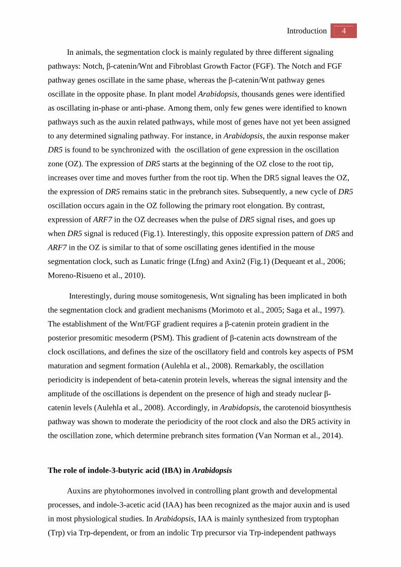
Figure 1. Comparison of the expression patterns of the oscillating genes in the vertebrate
segmentation clock in mouse embryo (A) and in the root clock in Arabidopsis (B). Both
the presomitic mesoderm and the primary root elongate from top to bottom in this
schematic, as indicated by the arrow, while gene expression propagates in the opposite
direction over time (as depicted from left to right). Gene expression oscillations in two
opposite phases occur at the peak of the respective oscillations in the oscillation zones
(green frames) as represented by Lunatic fringe (yellow) and Axin2 (blue) in the
segmentation clock (A) and by the marker gene DR5 (yellow) and Auxin Response
Factor 7 (ARF 7) (Blue) in the root clock (B). (Adapted from Moreno-Risueno and
Benfey, 2011)
Introduction 4
In animals, the segmentation clock is mainly regulated by three different signaling
pathways: Notch, β-catenin/Wnt and Fibroblast Growth Factor (FGF). The Notch and FGF
pathway genes oscillate in the same phase, whereas the β-catenin/Wnt pathway genes
oscillate in the opposite phase. In plant model Arabidopsis, thousands genes were identified
as oscillating in-phase or anti-phase. Among them, only few genes were identified to known
pathways such as the auxin related pathways, while most of genes have not yet been assigned
to any determined signaling pathway. For instance, in Arabidopsis, the auxin response maker
DR5 is found to be synchronized with the oscillation of gene expression in the oscillation
zone (OZ). The expression of DR5 starts at the beginning of the OZ close to the root tip,
increases over time and moves further from the root tip. When the DR5 signal leaves the OZ,
the expression of DR5 remains static in the prebranch sites. Subsequently, a new cycle of DR5
oscillation occurs again in the OZ following the primary root elongation. By contrast,
expression of ARF7 in the OZ decreases when the pulse of DR5 signal rises, and goes up
when DR5 signal is reduced (Fig.1). Interestingly, this opposite expression pattern of DR5 and
ARF7 in the OZ is similar to that of some oscillating genes identified in the mouse
segmentation clock, such as Lunatic fringe (Lfng) and Axin2 (Fig.1) (Dequeant et al., 2006;
Moreno-Risueno et al., 2010).
Interestingly, during mouse somitogenesis, Wnt signaling has been implicated in both
the segmentation clock and gradient mechanisms (Morimoto et al., 2005; Saga et al., 1997).
The establishment of the Wnt/FGF gradient requires a β-catenin protein gradient in the
posterior presomitic mesoderm (PSM). This gradient of β-catenin acts downstream of the
clock oscillations, and defines the size of the oscillatory field and controls key aspects of PSM
maturation and segment formation (Aulehla et al., 2008). Remarkably, the oscillation
periodicity is independent of beta-catenin protein levels, whereas the signal intensity and the
amplitude of the oscillations is dependent on the presence of high and steady nuclear β-
catenin levels (Aulehla et al., 2008). Accordingly, in Arabidopsis, the carotenoid biosynthesis
pathway was shown to moderate the periodicity of the root clock and also the DR5 activity in
the oscillation zone, which determine prebranch sites formation (Van Norman et al., 2014).
The role of indole-3-butyric acid (IBA) in Arabidopsis
Auxins are phytohormones involved in controlling plant growth and developmental
processes, and indole-3-acetic acid (IAA) has been recognized as the major auxin and is used
in most physiological studies. In Arabidopsis, IAA is mainly synthesized from tryptophan
(Trp) via Trp-dependent, or from an indolic Trp precursor via Trp-independent pathways

5 Introduction
(Mashiguchi et al., 2011). However, next to IAA, other abundant auxins in plants have been
reported. Indole-3-butyric acid (IBA) has long been used as a synthetic compound that
induced root initiation, and several lines of evidence prove the existence of native IBA in
plants (Blommaert 1954; Epstein et al, 1993; Ludwig-Muller et al, 1993; Schneider et al.,
1985; Sutter and Cohen, 1992). For instance, IBA has been shown to be synthesized in vivo
by using IAA and other compounds as precursors in maize (put reference here), and IBA
could be extracted from all species belonging to the Salix genus (Ludwig-Müller, 2000;
William, 1999). In Arabidopsis, IBA comprises approximately 25% to 30% of the total free
auxin pool in seedlings (Ludwig-Muller et al., 1993). Unexpectedly, more recently,
researchers failed to detect the endogenous IBA in Arabidopsis (Novak et al., 2012), which
might be due to the very low level of free IBA below detection limit, or still uncharacterized
metabolism pathways for IBA in Arabidopsis.
Genetic evidence showed that IBA is converted to active indole-3-acetic acid (IAA) in
peroxisomes by a process similar to fatty acid β-oxidation (Strader et al., 2010; Zolman et al.,
2000). In contrast, IBA transport in vivo is independent of IAA, and facilitated by
PLEIOTROPIC DRUG RESISTANCE8 (PDR8)/PENETRATION3/ABCG36 and
PDR9/ABCG37 (Liu et al., 2012; Rashotte et al., 2003; Ruzicka et al., 2010; Strader and
Bartel, 2009, 2011; Tognetti et al., 2010). As IBA serves as an auxin precursor, it is shares
functionality with IAA during plant development. It has been demonstrated that the
endogenous IBA-to-IAA conversion is required for proper root growth, such as the root hair
elongation and lateral root formation (De Rybel et al., 2012; Strader et al., 2010). Most of the
fatty acid β-oxidation enzymes and IBA efflux carriers are located in the root cap cells in the
root tip, suggesting the important role of IBA-response on root development.
Several questions remain unanswered, including how, when, and where IBA is
synthesized, whether IBA can serve as a signaling molecule on its own, what components
regulate IBA distribution in roots, and how IAA derived from IBA in the root cap contributes
to the patterning of the root system. For the latter, we hope that the present thesis represents a
step forwards towards a better understanding.

Introduction 6
References
Aulehla A., Wiegraebe W., Baubet V., Wahl M.B., Deng C., Taketo M., Lewandoski M.,
Pourquie O. (2008) A beta-catenin gradient links the clock and wavefront systems in mouse
embryo segmentation. Nature Cell Biology 10, 186-193.
Blommaert K. (1954) Growth- and inhibiting-substances in relation to the rest period of the
potato tuber. Nature 174, 970-972.
Brunoud G., Wells D.M., Oliva M., Larrieu A., Mirabet V., Burrow A.H., Beeckman T.,
Kepinski S., Traas J., Bennett M.J., Vernoux T. (2012) A novel Aux/IAA-based sensor
provides a high-resolution spatio-temporal map of auxin response and distribution during
plant development. Nature 482, 103–106.
Chipman, A.D., Arthur, W., and Akam, M. (2004). A double segment periodicity underlies
segment generation in centipede development. Current biology : CB 14, 1250-1255.
De Rybel, B., Audenaert, D., Xuan, W., Overvoorde, P., Strader, L.C., Kepinski, S., Hoye, R.,
Brisbois, R., Parizot, B., Vanneste, S., et al. (2012). A role for the root cap in root branching
revealed by the non-auxin probe naxillin. Nature chemical biology 8, 798-805.
Dequeant, M.L., Glynn, E., Gaudenz, K., Wahl, M., Chen, J., Mushegian, A., and Pourquie,
O. (2006). A complex oscillating network of signaling genes underlies the mouse
segmentation clock. Science 314, 1595-1598.
Dray, N., Tessmar-Raible, K., Le Gouar, M., Vibert, L., Christodoulou, F., Schipany, K.,
Guillou, A., Zantke, J., Snyman, H., Behague, J., et al. (2010). Hedgehog signaling regulates
segment formation in the annelid Platynereis. Science 329, 339-342.
Epstein E, Ludwig-Müller J. (1993). Indole-3-butyric acid in plants: occurrence, synthesis,
metabolism, and transport. Physiol Plant 88, 382-389
Liu, X., Barkawi, L., Gardner, G., and Cohen, J.D. (2012). Transport of indole-3-butyric acid
and indole-3-acetic acid in Arabidopsis hypocotyls using stable isotope labeling. Plant
physiology 158, 1988-2000.
Ludwig-Müller J. (2000) Indole-3-butyric acid in plant growth and development. Plant
Growth Regulation. 32, 2-3.
Ludwig-Muller J, Sass S, Sutter E, Wodner M, Epstein E (1993) Indole-3-butyric acid in
Arabidopsis thaliana. J Plant Growth Regul 13, 179–187
Mashiguchi, K., Tanaka, K., Sakai, T., Sugawara, S., Kawaide, H., Natsume, M., Hanada, A.,
Yaeno, T., Shirasu, K., Yao, H., et al. (2011). The main auxin biosynthesis pathway in
Arabidopsis. Proceedings of the National Academy of Sciences of the United States of
America 108, 18512-18517.
7 Introduction
Moreno-Risueno, M.A., and Benfey, P.N. (2011). Time-based patterning in development: The
role of oscillating gene expression. Transcription 2, 124-129.
Moreno-Risueno, M.A., Van Norman, J.M., Moreno, A., Zhang, J., Ahnert, S.E., and Benfey,
P.N. (2010). Oscillating gene expression determines competence for periodic Arabidopsis
root branching. Science 329, 1306-1311.
Morimoto, M., Takahashi, Y., Endo, M. & Saga, Y. (2005). The Mesp2 transcription factor
establishes segmental borders by suppressing Notch activity. Nature 435, 354-359
Novak, O., Henykova, E., Sairanen, I., Kowalczyk, M., Pospisil, T., and Ljung, K. (2012).
Tissue-specific profiling of the Arabidopsis thaliana auxin metabolome. The Plant journal :
for cell and molecular biology 72, 523-536.
Pueyo, J.I., Lanfear, R., and Couso, J.P. (2008). Ancestral Notch-mediated segmentation
revealed in the cockroach Periplaneta americana. Proceedings of the National Academy of
Sciences of the United States of America 105, 16614-16619.
Rashotte, A.M., Poupart, J., Waddell, C.S., and Muday, G.K. (2003). Transport of the two
natural auxins, indole-3-butyric acid and indole-3-acetic acid, in Arabidopsis. Plant
physiology 133, 761-772.
Ruzicka, K., Strader, L.C., Bailly, A., Yang, H., Blakeslee, J., Langowski, L., Nejedla, E.,
Fujita, H., Itoh, H., Syono, K., et al. (2010). Arabidopsis PIS1 encodes the ABCG37
transporter of auxinic compounds including the auxin precursor indole-3-butyric acid.
Proceedings of the National Academy of Sciences of the United States of America 107,
10749-10753.
Saga, Y., Hata, N., Koseki, H. & Taketo, M. M. Mesp2: a novel mouse gene expressed in the
presegmented mesoderm and essential for segmentation initiation. Genes Dev. 11, 1827–1839
Scheres, B., Benfey, P., and Dolan, L. (2002). Root development. The Arabidopsis book /
American Society of Plant Biologists 1, e0101.
Schneider, E.A., Kazakoff, C.W., and Wightman, F. (1985). Gas chromatography-mass
spectrometry evidence for several endogenous auxins in pea seedling organs. Planta 165, 232-
241.
Stollewerk, A., Schoppmeier, M., and Damen, W.G. (2003). Involvement of Notch and Delta
genes in spider segmentation. Nature 423, 863-865
Strader, L.C., and Bartel, B. (2009). The Arabidopsis PLEIOTROPIC DRUG
RESISTANCE8/ABCG36 ATP binding cassette transporter modulates sensitivity to the auxin
precursor indole-3-butyric acid. The Plant cell 21, 1992-2007.
Strader, L.C., and Bartel, B. (2011). Transport and metabolism of the endogenous auxin
precursor indole-3-butyric acid. Molecular plant 4, 477-486.

Introduction 8
Strader, L.C., Culler, A.H., Cohen, J.D., and Bartel, B. (2010). Conversion of endogenous
indole-3-butyric acid to indole-3-acetic acid drives cell expansion in Arabidopsis seedlings.
Plant physiology 153, 1577-1586.
Sutter, E.G., and Cohen, J.D. (1992). Measurement of indolebutyric Acid in plant tissues by
isotope dilution gas chromatography-mass spectrometry analysis. Plant physiology 99, 1719-
1722.
Tognetti, V.B., Van Aken, O., Morreel, K., Vandenbroucke, K., van de Cotte, B., De Clercq,
I., Chiwocha, S., Fenske, R., Prinsen, E., Boerjan, W., et al. (2010). Perturbation of indole-3-
butyric acid homeostasis by the UDP-glucosyltransferase UGT74E2 modulates Arabidopsis
architecture and water stress tolerance. The Plant cell 22, 2660-2679.
Ulmasov, T., Murfett, J., Hagen, G., and Guilfoyle, T.J. (1997). Aux/IAA proteins repress
expression of reporter genes containing natural and highly active synthetic auxin response
elements. The Plant cell 9, 1963-1971.
Van Norman, J.M., Xuan, W., Beeckman, T., and Benfey, P.N. (2013). To branch or not to
branch: the role of pre-patterning in lateral root formation. Development 140, 4301-4310.
Van Norman J. M., Zhang J., Cazzonelli C. I., Pogson B. J., Harrison P. J., Bugg T. D., Chan
K.X , Thompson A.J., Benfey P.N. (2014). Periodic root branching in Arabidopsis requires
synthesis of an uncharacterized carotenoid derivative. Proceedings of the National Academy
of Sciences of the United States of America 111, 1300-1309
William G.H. (1999) Introduction to plant physiology. John Wiley & Sons, Inc., New York
City, New York
Zolman, B.K., Yoder, A., and Bartel, B. (2000). Genetic analysis of indole-3-butyric acid
responses in Arabidopsis thaliana reveals four mutant classes. Genetics 156, 1323-1337

9 Introduction
To branch or not to branch: the role of pre-patterning in lateral root formation
Adapted from:
Van Norman, J.M., Xuan, W., Beeckman, T., and Benfey, P.N. (2013). To branch or not to
branch: the role of pre-patterning in lateral root formation. Development 140, 4301-4310.
Abstract
The establishment of a pre-pattern or competence to form new organs is a key feature of the
post-embryonic plasticity of plant development. The elaboration of pre-patterns leads to
remarkable heterogeneity in plant form. In root systems, many of the differences in
architecture can be directly attributed to the outgrowth of lateral roots. In recent years, efforts
have focused on understanding how the pattern of lateral roots is established. Here, we review
recent findings that point to a periodic mechanism for establishing this pattern, as well as
roles for plant hormones, particularly auxin, in the earliest steps leading up to primordium
development. In addition, we compare the development of lateral root primordia with in vitro
plant regeneration and discuss possible common molecular mechanisms.
Introduction
The post-embryonic formation of lateral organs in plants occurs when cells acquire a
new fate, generally based on positional cues, and then undergo a coordinated program of cell
division and differentiation to produce an organ primordium. In the root, lateral branches are
formed primarily from cells of the pericycle (see Glossary, Box 1), which is an internal tissue
surrounding the central vascular cylinder (Fig. 1). On a regular basis, subsets of pericycle
cells become competent to form lateral roots (LRs, see Glossary, Box 1) and, depending on
the species, this occurs in proximity of phloem (e.g. in maize) or protoxylem strands (e.g. in
Arabidopsis thaliana) (Casero et al., 1995; Dubrovsky et al., 2000; Hochholdinger and
Zimmermann, 2008). The frequency of these events establishes the number of sites competent
to form LRs over time and is, therefore, crucial in shaping the final root system architecture,
which is a major determinant of agronomic productivity. After competence is established, the
development of a lateral root primordium (LRP, see Glossary, Box 1) occurs either strictly
through division of cells derived from the pericycle (e.g. in Arabidopsis), or through division
of pericycle-derived cells and recruitment of cells in the adjacent endodermis (e.g. in maize)
(Bell, 1970; Hochholdinger and Zimmermann, 2008).

Introduction 10
The development of LRP can be induced or repressed in response to environmental
conditions and thus provides a mechanism for the plant to cope with changing edaphic
conditions (Malamy, 2005). A great number of environmental variables have been shown to
influence LRP development. For example, osmotic (drought) stress inhibits developmental
progression of early stage LRP (Deak and Malamy, 2005) and activation of the meristem in
emerged LRP is blocked by exogenous abscisic acid, a plant hormone involved in stress
responses (De Smet et al., 2003). LRP development is also sensitive to the availability of
nutrients including growth limiting nutrients such as nitrogen and phosphorous (recently
reviewed in (Jones and Ljung, 2012; Lavenus et al., 2013; Peret et al., 2011). While some
environmental stimuli have clear involvement in late stage LRP, nitrogen and phosphorous
can also act earlier in LRP development (Lima et al., 2010). It is unclear whether
environmental stimuli can only influence the developmental progression of sites already
established as competent to form an LRP or if lateral root pre-patterning, which has, to date,
been shown to be primarily dependent on time (Moreno-Risueno et al., 2010) can also be
impacted by environmental cues. Although the final outcome would be similar, more or fewer
LRs, the distinction would reflect a difference in the plant’s strategy to achieve developmental
plasticity under variable conditions. Therefore, understanding the regulation of LR pre-
patterning and subsequent primordia development has captured the interest of many plant
biologists.
The molecular and cellular mechanisms of LR formation have been most extensively
studied in the model plant Arabidopsis thaliana. In this species, relatively regular spacing of
LRs was reported, with LR placement coinciding with the outside edge of curves along the
primary root, particularly when roots show a bending or wavy growth pattern. To understand
the basis for this regular branching pattern, it is crucial to understand the earliest
developmental events occurring during LR formation. The Arabidopsis primary root tip is
classically divided into 3 main developmental zones (Fig. 2A) (Dolan et al., 1993). The
rootward-most portion of the root tip, the meristematic zone, contains the stem cell niche and
cells that are undergoing active proliferation with relatively little expansion. The meristematic
zone is occasionally described as having two parts: the basal and apical meristem. The basal
meristem is the shootward-most region of the meristem and is also referred to as the transition
zone, as cell division rates slow and cells begin to increase in size (Figure 2A). This is
followed by the elongation zone: a region where proliferative cell divisions cease and cells
undergo rapid and extensive cell elongation, increasing in length by 300% within three hours
(Verbelen et al., 2006). Finally cells enter the differentiation zone where they cease growth
and the vast majority attain their final size, begin to differentiate, acquiring their specialized

11 Introduction
Figure 2. Structure and development of the Arabidopsis root. (A) Median longitudinal section depicting developmental time (black arrow) in the longitudinal axis. A prebranch site (magenta) forms after an oscillation of gene expression within the oscillation zone (dotted line). Prebranch sites indicate competence to form a lateral root primordium (LRP) in the future. After competence is established, it is predicted that xylem pole pericycle (XPP) cells within a prebranch site can be specified as lateral root founder cells (LRFCs, green hatching). LRP initiate in the differentiation zone through asymmetric cell division of LRFCs, which gives rise to smaller cells (blue). (B) Transverse section. Periodic expression of DR5:GUS occurs in the protoxylem; however, because lateral root (Choat et al.) initiation occurs in the adjacent XPP cells, signaling between these cell types might be required for LRFC specification. Note that the ground tissue comprises two cell layers: the outermost cortex and the endodermis, which is immediately exterior to the pericycle. (C) Cut-away portion of the median longitudinal section focused on a region where an LR will form. XPP cells are predicted to be sequentially specified as LRFCs (green hatching), then activated to undergo cell division (green/white hatching). LRFC activation results in the coordinated migration of nuclei (white circles) towards the common cell wall in a pair of longitudinally abutted cells. These cells then undergo asymmetric division, giving rise to smaller cells (blue), to generate a stage I LRP. The primordium grows through the outer cell layers of the primary root until it emerges from the epidermis. Drawing is not to scale.

Introduction 12
cellular features and functions (Figure 2A). Additionally, development of LRP begins in the
differentiation zone.
A developing LRP becomes microscopically detectable when a primordium consisting of
a single cell layer is generated through asymmetric cell division in the differentiation zone of
the root (Fig. 2C) (Malamy and Benfey, 1997). The adjacent pairs of xylem pole pericycle
(XPP, see Glossary, Box 1) cells that undergo this cell division, also called LR initiation, are
designated as lateral root founder cells (LRFCs, see Glossary, Box 1). Prior to cell division,
LRFCs cannot be microscopically distinguished from the other pericycle cells without the use
of specific reporter lines. These founder cells first undergo anticlinal cell divisions to generate
a single cell-layered primordium containing up to ten small cells (stage I primordium, see
Glossary, Box 1). This is followed by periclinal cell divisions in the center-most cells, giving
rise to a two cell-layered primordium (stage II primordium, see Glossary, Box 1). Several
rounds of division in the central cells lead to an ellipsoid-shaped primordium that eventually
grows through the outer cell layers of the parent root and finally emerges from the root
surface (Fig. 2C) (Lucas et al., 2013).
Molecular evidence suggests that early events establishing the regular pattern of LRs,
prior to LRFC identity and LR initiation, occur at a more root-ward position in the root tip
where recurrent expression of reporter constructs driven by the synthetic promoter element
DR5 (DIRECT REPEAT5) are observed (De Smet et al., 2007; Moreno-Risueno et al., 2010).
DR5 promoter activity, which is used to assay the transcriptional response to auxin, is
correlated with subsequent LR initiation, suggesting that an oscillating transcriptional
mechanism operates as an upstream driving force for the regular pattern of LRs. Indeed, a
large number of genes were identified that oscillate both in phase and in antiphase with the
DR5 reporter, although the oscillatory system appears to function independently of local
auxin levels (Moreno-Risueno et al., 2010). Furthermore, the 6-hour period of the
transcriptional oscillation appears to be shorter than the frequency at which LRs initiate,
suggesting that establishment of competence to form a LR and initiation of an LRP are
distinct developmental events.
The oscillation in gene expression occurs over a region of the root termed the oscillation
zone (OZ, see Glossary, Box 1) (Fig. 2A) (Moreno-Risueno et al., 2010). During the period of
the oscillation as many as 12 pericycle cells may exit the OZ (Verbelen et al., 2006),
suggesting that several cells may experience the oscillation in gene expression. Yet, generally
only pairs of abutted pericycle are specified as LRFCs, suggesting a mechanism exists to
refine or restrict the number of pericycle cells that will adopt this fate. At the tissue-specific
level, DR5 reporter expression suggested that the oscillatory maximum occurs in the

13 Introduction
protoxylem cells adjacent to the pericycle (Fig. 2B). It may, therefore, be that XPP cells
receive signals during the oscillation to prepare them for LR initiation, a process that has been
termed, priming (see Glossary, Box 1). After the oscillation, a static point of DR5 expression
marks pre-branch sites, which are defined as positions competent to produce LRs in the future.
Subsequently, auxin signaling-dependent nuclear migration in LRFCs precedes the
asymmetric cell divisions that generate stage I primordia.
Hence, the events leading up to and including the specification of LRFCs and LR
initiation are crucial for lateral root organogenesis, but many questions surrounding the
molecular mechanisms that underlie the earliest stages of lateral root formation remain
unanswered. In this review, we focus on these early developmental steps and reflect on the
potential mechanisms that contribute to the establishment of the LR distribution pattern,
which forms the basis of root system architecture.
Is there a mechanical mechanism involved in establishing the pattern of lateral roots?
Under experimental conditions, Arabidopsis roots grow in a serpentine manner, bending
from side-to-side as they traverse the culture medium. Root waving has been described as the
consequence of differential growth due to re-orientation of growth in the direction of the
gravity vector combined with thigmotropic growth (re-orientation based on the touch
response, reviewed in (Oliva and Dunand, 2007)). These root growth behaviors are
hypothesized to be an evolutionary strategy to facilitate obstacle avoidance under rhizospheric
conditions. Accompanying root waving, the development of LRP and the emergence of LRs
coincides with the outside edge of these curves (Fortin et al., 1989), suggesting a relationship
between the pattern of LRs and root waving.
As root waving results from alternating left- and right-turns by the root tip, the number
of outside edges facing towards the left and right is roughly equal. Coincident with the
sidedness of the curves, the presence of LRs and LRP is also equal on each side of the root
(Fig.3). Furthermore, an agravitropic, auxin transport mutant, aux1, which turns in only one
direction, shows a shift in LR distribution with more LRs emerging on the outside edge of the
coiled root (De Smet et al., 2007). These results suggest that the distribution pattern of LRs is
linked with root waving and the gravity response via auxin transport. The co-occurrence of
these processes was further investigated by inducing root bending by gravi-stimulation and
mechanical methods (Ditengou et al., 2008; Laskowski et al., 2008; Lucas et al., 2008;
Richter et al., 2009). Gravi-stimulated bends occur when plants are re-oriented with respect to
the gravity vector resulting in a sharp bend as the root tip reorients growth to realign with

Introduction 14
gravity. Mechanical bending can be induced through manual manipulation of root or seedling
position, growth of the root into a barrier, or through gel sliding assays (Figure 3B-E). Similar
to root waving, induction of sharper bends in the root by any method resulted in emergence of
LRs at the outside edge of the bends. Intriguingly, LRP develop at the outside edge of a bend
even when a root is only transiently bent, however LRP and mechanically-induced bends only
coincide when bending occurs a short distance from the root tip (Ditengou et al., 2008;
Laskowski et al., 2008; Lucas et al., 2008; Richter et al., 2009).
The molecular link between gravitropism/root waving and LRP development is predicted
to be auxin. It was proposed that altered auxin distribution upon root re-orientation is
sufficient to establish the pattern of LRs along the root. However, roots that are agravitropic
due to defects in auxin signaling or transport or to removal of gravity-sensing tissues still
form LRs on the outside of curves, suggesting that gravity response isn’t specifically required
(Ditengou et al., 2008; Lucas et al., 2008; Richter et al., 2009). Recent observations of roots
grown during spaceflight further indicate that the pattern of LRs and gravitropic responses of
the primary root are separable; in the micro-g environment, roots grow more slowly than
those of control plants on Earth (at 1-g) but root waving persists and LRs are observed on the
outside of curves (Paul et al., 2012). Thus, root waving and the coincidence of LRP with
curves occur independent of gravity. These results don’t preclude the hypothesis that
asymmetric auxin distribution at curves in the root, regardless of its cause, is linked to the
development of an LRP.
Indeed, the expression and/or localization of reporters for auxin signaling and transport
show rapid changes (observed within 3-7 hours) after the induction of bends, suggesting that
mechanical strain on the cells induces changes in auxin distribution and signaling (Ditengou
et al., 2008; Laskowski et al., 2008). A computational model was developed whereby the
physical deformation of cells upon bending leads to auxin accumulation on the outside of
curves, which was suggested to trigger local competence of XPP cells, and then promote the
development and emergence of LRP (Laskowski et al., 2008). However, mutants with defects
in auxin signaling and/or transport and reduced LR production consistently form LRP or LRs
when roots are manually bent (Ditengou et al., 2008; Richter et al., 2009). These results
suggest that while the development of LRP may be defective in these mutants, sites
competent to form LRP are present. Furthermore, bends induced for very short durations (on
the order of 20 seconds) are sufficient to increase the number of LRs observed at the outside
of these transient bends. Following these bends, similarly rapid changes in cytosolic Ca2+
levels are observed, and treatment with calcium channel blockers inhibited both changes in

15 Introduction
Figure 3. LRs emerge from the outside of curves in the primary root. Schematics of root bends formed under various experimental conditions. (A) Root waving occurs as roots grow along the surface of agar plates. LRP develop and eventually emerge from the outside of the curves. The arrowheads indicate positions of incipient LRP. (B) Bends can be induced to form in the root through manual manipulation of the seedling either by pulling the shoot downward (left) or by pushing the root tip upward (right). (C) Gravistimulation-induced bends. If seedlings are reoriented with respect to the gravity vector, a bend will form as the root tip responds to realign the tip to gravity through differential growth. (D) In the absence of gravitropic response in either the root or shoot, a bend can be induced by root growth into a barrier (purple bar). (E) Bends can also be induced by cutting the agar on either side of a growing root (gray dotted line) and sliding the agar to one side, thereby creating two bends in the root. In these gel-sliding assays, neither the root tip nor shoot is exposed to manual contact or reorientation. Arrowheads (B-E) indicate the position of LRP emergence in response to induced bends. cytosolic Ca2+ and production of LRP after bending, indicating that Ca2+ signaling is required
for bend-induced LRP development (Richter et al., 2009). These results suggest that rapid
cellular signaling upon bending triggers events that lead to LRP development, prior to
changes in cell shape and differential auxin distribution. This implies that events upstream of

Introduction 16
signaling can promote LRP development and may indicate that competence to form an LRP is
already present at positions of mechanical bending. Alternatively, another interpretation of
these results may be that the pattern of LRs is less dependent on developmental pre-patterning
and, instead, is a consequence of root growth behaviors.
Nevertheless, evidence for an endogenous pre-patterning mechanism is observed in
studies of bend-induced LRP development. Roots subjected to gravistimuli at regular intervals
showed a maximum number of LRs when gravistimulation occured at 6-hour intervals.
However, LRP formed between the gravity-induced bends when the intervals between
gravistimulation were extended to 12- and 24-hours (Lucas et al., 2008). Additionally,
removal of the root tip prior to manual bending results in the formation of more LRs between
the cut edge and the bent region in both wild type and auxin signaling mutants (Ditengou et
al., 2008). These results suggest that the pattern of LRP is established independent of induced
bends and indicates that, although a single LR typically emerges at an induced bend,
additional nearby sites are competent to develop into LRP. These competent sites may be
developmentally stalled by signals from the root tip, by the emerging primordia, or both.
Evidence for an endogenous mechanism in lateral root pre-patterning
An endogenous mechanism for establishing the pattern of LRs was proposed based on
a temporal fluctuation in expression of the DR5 reporter. At 15 hour intervals, expression of
the DR5 promoter fused to the β-glucuronidase (GUS) reporter gene was observed in the
shootward-most portion of the meristematic zone, specifically in the two protoxylem cell files
but not in the adjacent XPP cells (Fig. 2B). The longitudinal position of the sites of DR5:GUS
expression in the meristem could be correlated with the later development of an LRP (De
Smet et al., 2007). Thus, it was suggested that DR5-expressing protoxylem cells signal to
adjacent XPP cells to condition them for LRFC identity, a process called priming (see
Glossary, Box 1). If the temporal changes in DR5 expression are hypothesized to direct the
later formation of an LRP, this recurrent process could explain the regular spacing between
LRs under controlled growth conditions. However as DR5 expression occurs in both sets of
protoxylem cells, the alternating distribution of LRs on the sides of the root cannot be
explained, suggesting that a subsequent mechanism determines LR sidedness (De Smet et al.,
2007). For example, the mechanical strain and asymmetric distribution of Ca2+ and auxin that
is described in cells upon bending occurs in more differentiated regions of the root, therefore
it is possible that the sidedness of LR initiation is determined later in response to signals
produced as a consequence of changes in cell shape. Expression conferred by the DR5

17 Introduction
Figure 4. Prebranch sites mark the positions at which LRP will subsequently develop and emerge. (A) An oscillation in DR5:LUC expression (chemiluminescence signal imaged at 5-6 minute exposure times) in the oscillation zone (OZ) leads to the formation of a prebranch site (asterisk). (B) Quantification of the oscillation of DR5:LUC expression in two individual roots. The oscillation has a period of ∼6 hours and appears to precede the changes in growth direction of the root tip during root waving. Blue/dark blue arrows indicate the time points at which bends were formed in each of the primary roots. ADU, analog-digital units. (C) Overlay of a luciferase and brightfield image (taken 5 days after the luciferase image) to show emerged lateral roots. Arrowheads indicate positions at which LRP have yet to emerge. (B,C) Adapted with permission (Moreno-Risueno et al., 2010).
promoter was further examined by fusing it to the Luciferase gene, allowing visualization of
its behavior in vivo (Moreno-Risueno et al., 2010). Expression of DR5:Luciferase (LUC) in
the root tip revealed oscillatory activity with a period of 6 hours. This dynamic expression
pattern occurred over a larger region of the root tip than previously described and this region
was, therefore, termed the oscillation zone (OZ) (Fig. 4A, B). Following each peak of the
DR5 oscillation, a static point of expression was observed, which exhibited a similar
longitudinal distribution as LRP and LRs. Indeed, later examination of these points revealed
them as the future sites of LRP and LRs, and they were, therefore designated as prebranch
sites (see Glossary, Box 1, Fig. 4C) (Moreno-Risueno et al., 2010).
DR5 expression is frequently utilized as a proxy for the distribution of auxin, however an
exogenously stimulated peak in auxin levels in the OZ was not able to trigger formation of a
prebranch site. Additionally, a reporter gene with similar response dynamics to exogenous
auxin as DR5:LUC and expressed in the OZ did not exhibit periodic expression (Moreno-
Risueno et al., 2010). These results suggested that oscillatory peaks in auxin itself are not
sufficient to account for the dynamic behavior of DR5 and the subsequent formation of
prebranch sites. In an effort to determine the underlying cause of the oscillation, microarray
analysis of gene expression identified >3400 genes whose expression oscillates either in phase
or in antiphase with the DR5 reporter. Several candidate transcriptional regulators were found

Introduction 18
to both exhibit oscillatory expression and be functionally important for LR formation
(Moreno-Risueno et al., 2010). Although auxin responsive genes do not necessarily show
oscillatory expression in the OZ, some oscillating genes have established roles in LR
formation and are involved in or downstream of auxin signaling, such as LATERAL ORGAN
BOUNDARIES DOMAIN 16 (LBD16) and AUXIN RESPONSIVE FACTOR 7 (ARF7)
(Okushima et al., 2007; Okushima et al., 2005). Unexpectedly, ARF7 was found to oscillate in
antiphase to DR5:LUC and in arf7 mutants the oscillatory expression of DR5:LUC is
abnormal and prebranch sites form at irregular intervals, suggesting ARF7 function is
important for periodic gene expression in the OZ (Moreno-Risueno et al., 2010). Together
these results led to a model describing a lateral root clock, in which a complex periodic
transcriptional mechanism specifies sites that are competent to form LRs, thus establishing a
LR pre-pattern along the root’s axis.
Like the LRs that follow them, prebranch sites are found at curves that are produced
during root waving. Although root waving shows a similar periodicity as prebranch site
formation, the oscillation of DR5 expression is observed prior to the re-orientation of root
growth direction (Moreno-Risueno et al., 2010). This suggests that, despite their occurrence at
a similar position along the root, these events are separated by time. The link between
bending and prebranch site formation was examined by exposing roots to gravistimuli and
manual bending. Roots responded to gravistimulation asynchronously, with individual roots
completing the last bend due to root waving prior to re-orienting growth in the direction of the
gravity vector, which is consistent with these being distinct growth behaviors. In manual
bending assays, prebranch sites were observed at the bend and nearer to the root tip than
bends could be made without disrupting the position of the root tip. Manual bending did not
result in de novo prebranch sites and no LRs emerged from sites not previously marked by a
prebranch site; yet, as observed previously, LRP emerged at the outside edge of the bends
(Moreno-Risueno et al., 2010). These results are consistent with a hypothesis in which an
endogenous patterning mechanism establishes sites competent to form a LR, but LRP
development and perhaps sidedness of LRFC specification are subsequent developmental
decisions, which integrate multiple cues.
The priming of XPP cells during the oscillation of gene expression in the OZ
conceptually links DR5 expression in the protoxylem with later LRP development in the
adjacent pericycle. Although priming is thought to be XPP specific, prebranch sites cannot yet
be examined at a cellular level for technical reasons (see below). Priming of XPP cells would
not be predicted to occur only on one side of the root as DR5 expression is observed at both
xylem poles. Additionally, the molecular character of primed XPP cells and the priming

19 Introduction
signal remain elusive. An alternative, and not mutually exclusive, hypothesis is that genes
oscillating in the pericycle itself may have important roles establishing the LR prepattern. For
example, LBD16 is observed to oscillate and was recently reported to have XPP-specific
expression and a key role in LR initiation (Goh et al., 2012; Moreno-Risueno et al., 2010).
Because the root tissue examined for oscillating transcriptional profiles was specific to
longitudinal regions but encompassed all root tissues, the tissue-specific nature of any
oscillating transcripts was not captured (Moreno-Risueno et al., 2010). The necessity for
vascular continuity between primary and lateral roots may be a crucial reason for coordination
between vascular patterning and LR pre-patterning, and this is supported by additional
connections between vascular patterning and the development of LRP (Bonke et al., 2003;
Ohashi-Ito and Bergmann, 2007; Parizot et al., 2008). However, the role of cell-to-cell
signaling between protoxylem and XPP cells is an intriguing question requiring further
investigation.
Lateral root founder cells and prebranch sites
Organogenesis is generally thought to begin with the specification of founder cells
(FCs). This specification could involve cells acquiring competence to respond to an activation
signal. Activation of FCs typically leads to cell division, which is the first morphological
indication that a change in cell fate has occurred. However, prior to activation of cell division,
the identification of FCs is difficult as they are histologically indistinguishable from the
surrounding cells. Another difficulty is that there are few molecular reporters for FCs, and for
those markers that are available, the function of the associated molecules in FC specification,
activation or cell division is not entirely clear (Beveridge et al., 2007; Chandler, 2011). These
general FC features are also true for lateral root founder cells (LRFCs).
LRFCs are the specific XPP cells that will undergo asymmetric cell division (LR
initiation) to produce a stage I LRP. The specification and activation of LRFCs is thought to
occur within the differentiation zone of the root, where other cells have ceased division and
growth and have become differentiated. However, it is unclear if XPP cells dedifferentiate
then re-differentiate into LRFCs, or if they are maintained in an undifferentiated state
(Dubrovsky et al., 2000; Laskowski et al., 1995; Malamy and Benfey, 1997). Expression of
the DR5:GFP reporter is observed in select XPP cells and precedes LR initiation. Therefore,
activation of DR5 expression is considered the first indication that specific XPP cells have
acquired LRFC identity (Dubrovsky et al., 2008). Additionally, aberrant lateral root
formation 4 (alf4) mutants, show DR5:GFP expression in select XPP cells, yet LRP are not

Introduction 20
produced as a result of defects in cell division (DiDonato et al., 2004; Dubrovsky et al.,
2008). This suggests that alf4 LRFC are either specified but not activated or are both specified
and activated, but cannot undergo cell division to produce a stage 1 LRP. Because DR5:GFP
expression precedes LRFC cell division, and pericycle cells appear to be uniformly sensitive
to exogenous auxin, it was proposed that local auxin accumulation, rather than increased
auxin sensitivity, triggers LRFC specification (Dubrovsky et al., 2008). In addition, one of the
first anatomical signs that XPP cells have taken on LRFC fate is the coordinated migration of
the nuclei towards the common wall in a pair of cells, however by this point LRFC
specification and activation have already occurred, as cell division is imminent (De Rybel et
al., 2010; Dubrovsky et al., 2011).
Recent evidence shows that the developmental progression of LRFCs to stage I LRP
requires activity of the auxin transporter PIN3 in endodermal cells, which are adjacent to the
pericycle cells (Fig 2B). However, LRFCs exhibit DR5:GFP expression prior to PIN3
accumulation in endodermal cells, suggesting that LRFC fate has already been specified
(Marhavy et al., 2013). Accumulation of auxin in specific cells requires either directed
transport or intracellular biosynthesis, with cellular retention of auxin. Either scenario
requires that these select XPP cells attain higher auxin levels, suggesting they may already be
distinct from other XPP cells prior to detection of DR5:GFP reporter expression. Thus, in
contrast to the proposed role for auxin as a signal in LRFC specification, it may be that auxin
acts as an activation signal of LRFC cell division. Based on this hypothesis, it is possible that
in alf4 mutants, LRFCs are specified and receive the activation signal (as visualized by
DR5:GFP expression) but, due to mitotic defects, are unable to undergo coordinated cell
division. Additionally, ALF4 expression and protein localization appear to be independent of
auxin signaling (DiDonato et al., 2004), suggesting that additional activation signals may
exist.
Prebranch sites are the static points of DR5:LUC expression that form at the position of
the peak in the periodic oscillation of DR5 after the oscillation is complete (Moreno-Risueno
et al., 2010). Expression of DR5, as reported by GFP, is observed in XPP cells at one side of
the xylem pole prior to the asymmetric division that gives rise to an LRP, identifying these
cells as LRFCs (Dubrovsky et al., 2008). Because the expression of DR5 is used to define
both of these terms, they might be considered synonymous. However, it is important to keep
in mind the difference between the reporter genes, LUC and GFP. The LUC enzyme cleaves
its substrate (luciferin), thereby producing light, and it then becomes inactive. Thus, while
monitoring LUC activity is a highly dynamic and sensitive method to assay the in vivo
activity of a promoter (de Ruijter N.C.A., 2003), it is difficult to obtain cell type-specific

21 Introduction
resolution, as light spreads outward in all directions from the source. GFP expression,
however, can be localized in a cell type-specific manner using confocal microscopy, although
the drawbacks of GFP are long maturation and stability times, higher thresholds for
detectability, and a relatively high background fluorescence in plants (de Ruijter N.C.A.,
2003). Because the static points of DR5:LUC expression are visible earlier than expected for
LRFCs, and because it is not yet possible to determine which cell type the LUC activity
originates from or if it is localized to one side of xylem pole, it is not appropriate to describe
these points of DR5:LUC expression as LRFCs (Moreno-Risueno et al., 2010). Prebranch
sites may indeed be LRFCs that are visible at an earlier time due to the higher sensitivity of
LUC. Alternatively, they may indicate a broader, competent site from which specification of a
restricted number of XPP cells into LRFCs will subsequently occur specifically at one side of
the root.
A developmental window for founder cell identity and the first formative division to
produce LRP
LRFC identity has been associated with an increase in the transcriptional response to
auxin in select XPP cells briefly before they undergo asymmetric cell division (Benkova et al.,
2003; Dubrovsky et al., 2008). The time lag between the DR5:GFP expression in LRFCs and
LR initiation is extremely short and, consequently, both events are observed in the same
region of the root, namely the early differentiation zone (Fig. 2A) (Dubrovsky et al., 2011).
Monitoring auxin response and distribution along the entire Arabidopsis primary root
revealed a region with low auxin response and levels that was positioned between two distinct
auxin maxima: one at the very tip of the root, including the QC and meristematic zone, and a
second in the vascular bundle of mature tissue in the shootward-most regions of the root. The
region of “auxin minimum” was somewhat paradoxically found to overlap with that in which
increased auxin response (as assayed by induction of DR5:GFP expression) in LRFCs and LR
initiation occur. Therefore, this region was proposed as the developmental window for LR
initiation.
The developmental window is somewhat dynamic, shifting in the direction of the root
apex as the root grows thereby guaranteeing a rootward sequence of LR production under
controlled growth conditions (Dubrovsky et al., 2006). In this region of lower auxin levels
and response, cell- and tissue-specific auxin distribution and TIR1/AFB-dependent auxin
signaling modules result in the induction of auxin-responsive genes, such as GATA23 and
LBD16, and the subsequent activation of LRFCs to undergo nuclear migration and

Introduction 22
asymmetric cell division (De Rybel et al., 2010; Goh et al., 2012). Downstream of the
TIR1/AFB auxin receptor proteins, a family of transcriptional repressors AUXIN/INDOLE-3-
ACETIC ACID INDUCIBLE (AUX/IAA) proteins are degraded upon auxin perception
leading to auxin-induced gene expression (reviewed in (Chapman and Estelle, 2009). In
iaa28, a gain-of-function mutant, in which the IAA28 protein is stabilized thus suppressing
auxin response, nuclear migration is interrupted, leading to inhibition of LRFC activation and
a substantial decrease in LR formation (De Rybel et al., 2010). Similarly when LBD16, a
downstream target of auxin signaling but whose specific function in LR formation remains
unknown, is repressed nuclear migration in LRFCs is disrupted, thereby blocking the
subsequent initiation of LRs (Goh et al., 2012). Likewise, disrupting polar auxin transport
genetically or through chemicals alters auxin distribution in this region and inhibits lateral
root initiation (Dubrovsky et al., 2011; Marhavy et al., 2013). The occurrence of these auxin
response-maximum driven processes within a region of generally low auxin levels is
intriguing and suggests that cells in this region may have enhanced responses to minor
fluctuations in endogenous auxin availability. In such an environment, a subset of XPP cells
could register local changes in auxin levels providing a signal for developmental progression
towards LR initiation, a situation that may not be possible in conditions of high auxin levels.
As opposed to auxin, cytokinins were identified as endogenous suppressors of LR
formation. Their inhibitory mode of action was attributed to hindrance of polar auxin
transport, which could disturb local auxin distribution patterns and auxin signaling pathways
(Benkova et al., 2003; Laplaze et al., 2007; Li et al., 2006). More recently, however, cytokinin
response, as monitored by a cytokinin-sensitive sensor (the TCS reporter), in the
developmental window was shown to be minimal, although no decrease in active cytokinin
levels could be measured within this region of the root (Bielach et al., 2012). Furthermore,
exogenous cytokinin failed to induce expression of the TCS reporter, indicating that strong
repression of cytokinin signaling is at play in the developmental window and might be an
important component for LR initiation. Categorizing the effects of increased cytokinin levels
on LR formation either by endogenous expression of cytokinin biosynthesis genes, or by
exogenous cytokinin treatment, demonstrated that the early phases of LR formation including
the pre-mitotic stages are more sensitive to cytokinin than are the later stages of LRP
development. It was suggested that in the developmental window where auxin levels are low,
ectopic cytokinin levels are more disruptive to early stage LRP, whereas in more developed
primordia, auxin levels are more robust, thus diminishing the impact of cytokinins (Bielach et
al., 2012).

23 Introduction
Conclusion
During recent years and thanks to the development of novel reporter lines in Arabidopsis,
insight has been gained into the “invisible phase” of LR formation, namely the events that
precede the first asymmetric cell divisions in LRFCs. The uncovering of previously unknown
developmental steps has pushed researchers to formulate new concepts so that results
obtained by different research groups working on lateral roots can be compared. In this
Review, we aim to provide a solid foundation for the coming years during which exciting new
insights are expected to surface. We have summarized recent published work on pre-
patterning mechanisms in the root, which consist of two important developmental steps: 1) a
periodic oscillation of gene expression that triggers competence for LR formation; and 2) the
perception of an auxin signal in founder cells to set up LR initiation in the developmental
window, a region of the root in which the integration of auxin and cytokinin signaling occurs.
However, many questions remain unanswered.
We still lack cellular resolution of the oscillatory gene expression process. The current
cellular information from the DR5:GUS reporter implies that signaling from the adjacent
vasculature to the XPP cells is important for LRFC specification. However, it is unclear what
the identity of such a signal might be and when (in the OZ or later) this signal would be
transmitted to the XPP. Because there doesn’t appear to be a sidedness to the oscillation in
DR5 or endogenous genes, how LR sidedness occurs remains to be determined but signals
from the cells exterior to the pericycle upon cellular deformation may be involved. Finally,
whether so-called priming signals and the cues that determine sidedness are distinct and
sequential remains to be established. Once LRFCs become observable by reporter expression
or nuclear migration, asymmetric cell division quickly follows. However, as the positional
information transmitted by the oscillation of gene expression occurs earlier, XPP cells may
undergo a change in state that we are, as yet, unable to detect. A delay between competence
and LRFC specification and activation would further increase the developmental plasticity of
the root system by providing another “check-point” for the developmental progression of
organogenesis in the root.
Pre-patterning for LR formation is likely to be an example of the trade-off between
resource investment and response time during plant development. Unlike animals, plants
continually produce new organs in response to environmental cues. One option for a plant
would be to wait for the cue and then begin the process of organ formation de novo. The
obvious downside to this strategy is that the conditions that triggered the response might be
short-lived. To reduce response time, plants have instead adopted a strategy of commencing

Introduction 24
organ formation, then arresting it at various stages of development. An example is apical
branch formation, in which branch points are positioned through phyllotaxis, and primordia
are initiated then arrested until the appropriate signal is received. The oscillatory gene
expression process that establishes a LR pre-pattern of prebranch sites can be thought of as
the equivalent of phyllotaxis, leading to priming of select XPP cells, which then await a signal
to form a lateral root primordium.
The presence of pre-patterning mechanisms implies the continuous production of
organogenesis-competent cells during root growth. In contrast to this idea, organogenesis
during plant regeneration from callus was thought to rely on de novo dedifferentiation of
mature cells. However, recent comparative analyses of LR and callus formation have revealed
clear and striking similarities. One important similarity is the requirement of high hormone
levels for induction. In the LRIS (Box 2), the transportable synthetic auxin analogue NAA is
applied to seedlings at ~4x the concentration at which the non-transportable analogue 2,4-D is
applied to explants in the CIM (Atta et al., 2009; Himanen et al., 2002; Valvekens et al.,
1988). However, treatment of root explants with the amount of 2,4-D in CIM or of NAA in
the LRIS results in comparable gene expression patterns, indicating that these two treatments
induce a similar response (Sugimoto et al., 2010). Unexpectedly, root explants treated with
high cytokinin levels or whole seedlings sequentially treated with NAA and cytokinin-
enriched media are able to form shoot tissue at early stage LRP, suggesting flexibility in the
developmental potential of LRP (Atta et al., 2009; Chatfield et al., 2013). These results
suggest that, while the program for callus formation and its initial steps are similarly executed
under various hormonal conditions, the formation of root or shoot tissue from callus or early
LRP depends on hormonal context.
The comparison between the induction of LR and callus development also revealed that
the XPP cells in the root and XPP-like cells in the shoot are unique among cell types in their
ability to divide and from new structures/organs in differentiated tissues. Root pericycle cells
at poles of either the xylem or phloem are further delineated in that they have distinct cellular
morphology, transcriptional profiles, and are the cells of origin for LRP (Brady et al., 2007;
Jansen et al., 2012; Laskowski et al., 1995; Malamy and Benfey, 1997). In the Arabidopsis
shoot, the XPP-like, callus-forming cells are similar to root XPP cells in that they share
marker gene expression and are associated with the vasculature, although up to now this shoot
tissue has not been specifically defined as XPP (Sugimoto et al., 2010). Given that they are
the cells of origin for callus formation from both root and shoot tissues, the meristematic
potential and properties of XPP and XPP-like cells has been greatly expanded. Perhaps the
structural and molecular similarities, and the notion of a common cell of origin, between LRP

25 Introduction
and callus development indicate a common evolutionary origin. Given that hormonal context
is a key aspect of determining which type of organ is formed by callus or early LRP, the
possibility that LR development is an evolutionary offshoot of regeneration may be a viable
hypothesis. In this context, the establishment of a LR pre-pattern may function to confine the
meristematic potential of the XPP to specific sites.
What was once considered a largely random event primarily refined by lateral inhibition,
lateral root formation is now revealed as a complex developmental process underpinned by a
dynamic spatiotemporal pre-patterning mechanism. Advances in methods to interrogate
cellular gene expression at finer resolution and the development of dynamic, cell-type specific
reporter proteins will be key tools in future studies.
Box 1: Glossary
Oscillation zone (OZ) – The region in which periodic oscillation of the DR5:Luciferase
reporter and expression of certain endogenous genes occurs. This region encompasses the
shootward-most portion of the meristematic zone, as well as the elongation zone (Figure 2A)
(Moreno-Risueno et al., 2010).
Pericycle – A cell layer located between the vascular cylinder and the ground tissue (Figure
2B). Like the vascular tissues, the pericycle has a bilaterally symmetric organization.
Xylem pole pericycle (XPP) – Cells of the pericycle that flank the protoxylem cells (Figure
2B). Xylem pole pericycle cells have distinct cellular morphology, gene expression profiles
and the unique capacity within the differentiation zone to re-enter the cell cycle and undergo
cell division. Xylem pole pericycle cell division is required for lateral root initiation, as well
as for regeneration via callus.
Priming – a process that occurs in select xylem pole pericycle cells, which is proposed to
coincide with the oscillation of gene expression. Priming is predicted to condition these cells
for subsequent prebranch site and lateral root founder cell specification.
Prebranch site – Static points of DR5:Luciferase expression that occur following the
oscillation of DR5:Luciferase in the oscillation zone. Prebranch sites are competent to form
lateral roots in the future. Because these sites occur earlier than expected for lateral root
founder cells and it hasn’t been determined if expression is cell type specific, the relationship
between prebranch sites and lateral root founder cells is unclear.
Founder cells – Founder cells are the initial cells specified to become a new organ or tissue.
Founder cells are typically histologically similar to related/nearby cells and can only be
Introduction 26
identified following other developmental events, such as the activation of cell
division(Beveridge et al., 2007; Chandler, 2011).
Lateral root founder cells (LRFC) – A set of two longitudinally abutted cells in each of the
2-3 cell files of the xylem pole pericycle at one side of the root. These cells will undergo
asymmetric cell divisions (also called formative divisions) to initiate a lateral root
primordium. The first morphological indicator that these cells have a distinct fate is the
migration of their nuclei towards the common cell wall. Additionally, expression of DR5:GFP
and gLBD16:GFP is induced in these cells prior to asymmetric cell division(Dubrovsky et al.,
2008; Goh et al., 2012).
Lateral root primordia (LRP) – A group of cells originating from asymmetric division of
lateral root founder cells that progress through a stereotypical set of developmental stages to
produce a root de novo.
Stage I lateral root primordium – A lateral root primordium comprised of a single cell layer
and the first stage of lateral root primordia development. Initially this structure is comprised
of two small cells resulting from asymmetric division of the lateral root founder cells,
however successive divisions result in a group of 4-10 small longitudinally abutted cells.
Stage II lateral root primordium – Following radial expansion, the cells of the Stage I
primordium reorient their division plane, dividing periclinally to the root’s longitudinal axis,
resulting in a primordium comprised of two cell layers.
Lateral root (Choat et al.) – A root that is branching from a parent root and has activated its
apical meristem. In most plants, these organs are formed postembryonically.
Lateral root prepattern – The specification of a spatio-temporal region of the root that is
competent to give rise to a lateral root primordium. The lateral root prepattern is predicted to
be established by periodic gene expression in the oscillation zone and the formation of
prebranch sites. Establishment of the prepattern is stable under various environmental
conditions.
Lateral root formation – A term encompassing all of the events leading to the production of
an actively growing lateral root.
Lateral root development – A term without a clear and accepted definition. This term can be
used to encompass all the developmental stages of a lateral root primordium (from stage I-
VII) and is more clearly stated as “lateral root primordium development”. The progression of
any one lateral root primordium through the developmental stages is impinged upon by
environmental cues.
27 Introduction
Box 2: The lateral root inducible system
As initiation of an LRP involves few cells and is not coordinated in space or time between
seedlings, the use of genome-wide approaches has been challenging. To address this, a
method termed the Lateral Root Inducible System (LRIS) was developed, which involves
sequential treatment of seedlings with an auxin transport inhibitor and then a synthetic auxin
analog, 1-naphthalene acetic acid (NAA) (Himanen et al., 2002). This treatment rapidly
induces synchronous cell divisions throughout the XPP. The resulting small cells, which are
similar to a stage I LRP, then divide parallel to the root axis similar to a stage II LRP. Finally,
extended NAA treatment results in proliferative LRP development along the length of the root
at both XPP axes (Himanen et al., 2002).
The LRIS was proposed to override the endogenous prepatterning mechanism and stimulate
LRP initiation en masse. This allowed application of transcriptional profiling techniques to
begin to address the underlying molecular mechanisms. These analyses led to the
characterization of novel proteins involved in the early steps of LR formation, including
ARABIDOPSIS CRINKLY 4 (ACR4) and GATA23 (De Rybel et al., 2010; De Smet et al.,
2008) and indicated sequential links between auxin signaling and cell cycle regulation
(Himanen et al., 2002; Himanen et al., 2004). In brief, auxin signaling via SOLITARY-ROOT
(SLR/IAA14) is required for LR initiation under both standard conditions and in the LRIS
(Fukaki et al., 2002; Vanneste et al., 2005). Although, ectopic induction of XPP cell division
in slr/iaa14 mutants did not promote LR formation (Vanneste et al., 2005), LRs formed
proliferatively when induction of XPP cell division was combined with NAA treatment (De
Smet et al., 2010). Although endogenous and LRIS-induced LRs have common features, such
as tissue of origin and links between auxin signaling and the cell cycle, differences in the
pattern/distribution of lateral organs suggest it is less clear how the LRIS informs endogenous
LR pre-patterning. While the LRIS may simply shift the endogenous LR patterning program
into overdrive, these fundamental patterning differences may indicate that hormonal
manipulation elicits a distinct response program in the XPP.
Acknowledgements
In the lab of PNB, this work was funded by grants from DARPA (#D12AP0000), NSF and
GBMF. Work in the Lab of TB was in part financed by grants of the Interuniversity
Attraction Poles Programme (IAP VI/33 and IUAP P7/29 "MARS") from the Belgian Federal
Science Policy Office and WX is supported by a CSC and an Ugent – BOF – cofounding
grant.

Introduction 28
References Atta, R., Laurens, L., Boucheron-Dubuisson, E., Guivarc'h, A., Carnero, E., Giraudat-Pautot, V., Rech, P. and Chriqui, D. (2009) Pluripotency of Arabidopsis xylem pericycle underlies shoot regeneration from root and hypocotyl explants grown in vitro. Plant J, 57, 626-644. Bell, J. K. a. M., M.E. (1970) A histological study of lateral root initiation and development in Zea mays. Protoplasma, 70, 179-205. Benkova, E., Michniewicz, M., Sauer, M., Teichmann, T., Seifertova, D., Jurgens, G. and Friml, J. (2003) Local, efflux-dependent auxin gradients as a common module for plant organ formation. Cell, 115, 591-602. Beveridge, C. A., Mathesius, U., Rose, R. J. and Gresshoff, P. M. (2007) Common regulatory themes in meristem development and whole-plant homeostasis. Curr Opin Plant Biol, 10, 44-51. Bielach, A., Podlesakova, K., Marhavy, P., Duclercq, J., Cuesta, C., Muller, B., Grunewald, W., Tarkowski, P. and Benkova, E. (2012) Spatiotemporal regulation of lateral root organogenesis in Arabidopsis by cytokinin. Plant Cell, 24, 3967-3981. Bonke, M., Thitamadee, S., Mahonen, A. P., Hauser, M. T. and Helariutta, Y. (2003) APL regulates vascular tissue identity in Arabidopsis. Nature, 426, 181-186. Brady, S. M., Orlando, D. A., Lee, J. Y., Wang, J. Y., Koch, J., Dinneny, J. R., Mace, D., Ohler, U. and Benfey, P. N. (2007) A high-resolution root spatiotemporal map reveals dominant expression patterns. Science, 318, 801-806. Casero, P. J., Casimiro, I. and Lloret, P. G. (1995) Lateral root initiation by asymmetrical transverse divisions of pericycle cells in four plant species: Raphanus sativus, Helianthus annuus, Zea mays, and Daucus carota. Protoplasma, 188, 49-58. Celenza, J. L., Jr., Grisafi, P. L. and Fink, G. R. (1995) A pathway for lateral root formation in Arabidopsis thaliana. Genes Dev, 9, 2131-2142. Chandler, J. W. (2011) Founder cell specification. Trends Plant Sci, 16, 607-613. Chapman, E. J. and Estelle, M. (2009) Mechanism of auxin-regulated gene expression in plants. Annu Rev Genet, 43, 265-285. Chatfield, S. P., Capron, R., Severino, A., Penttila, P. A., Alfred, S., Nahal, H. and Provart, N. J. (2013) Incipient stem cell niche conversion in tissue culture: using a systems approach to probe early events in WUSCHEL-dependent conversion of lateral root primordia into shoot meristems. Plant J, 73, 798-813. Che, P., Lall, S. and Howell, S. H. (2007) Developmental steps in acquiring competence for shoot development in Arabidopsis tissue culture. Planta, 226, 1183-1194. de Ruijter N.C.A., V. J., van Leeuwen W., and van der Krol A.R. (2003) Evaluation and comparison of the GUS, LUC, and GFP reporter system for gene expression studies in plants. Plant Biol., 5, 103-115. De Rybel, B., Vassileva, V., Parizot, B., Demeulenaere, M., Grunewald, W., Audenaert, D., Van Campenhout, J., Overvoorde, P., Jansen, L., Vanneste, S., et al. (2010) A novel aux/IAA28 signaling cascade activates GATA23-dependent specification of lateral root founder cell identity. Curr Biol, 20, 1697-1706. De Smet, I., Lau, S., Voss, U., Vanneste, S., Benjamins, R., Rademacher, E. H., Schlereth, A., De Rybel, B., Vassileva, V., Grunewald, W., et al. (2010) Bimodular auxin response controls organogenesis in Arabidopsis. Proc Natl Acad Sci U S A, 107, 2705-2710.

29 Introduction
De Smet, I., Signora, L., Beeckman, T., Inze, D., Foyer, C. H. and Zhang, H. (2003) An abscisic acid-sensitive checkpoint in lateral root development of Arabidopsis. Plant J, 33, 543-555. De Smet, I., Tetsumura, T., De Rybel, B., Frey, N. F., Laplaze, L., Casimiro, I., Swarup, R., Naudts, M., Vanneste, S., Audenaert, D., et al. (2007) Auxin-dependent regulation of lateral root positioning in the basal meristem of Arabidopsis. Development, 134, 681-690. De Smet, I., Vassileva, V., De Rybel, B., Levesque, M. P., Grunewald, W., Van Damme, D., Van Noorden, G., Naudts, M., Van Isterdael, G., De Clercq, R., et al. (2008) Receptor-like kinase ACR4 restricts formative cell divisions in the Arabidopsis root. Science, 322, 594-597. Deak, K. I. and Malamy, J. (2005) Osmotic regulation of root system architecture. Plant J, 43, 17-28. DiDonato, R. J., Arbuckle, E., Buker, S., Sheets, J., Tobar, J., Totong, R., Grisafi, P., Fink, G. R. and Celenza, J. L. (2004) Arabidopsis ALF4 encodes a nuclear-localized protein required for lateral root formation. Plant J, 37, 340-353. Ditengou, F. A., Teale, W. D., Kochersperger, P., Flittner, K. A., Kneuper, I., van der Graaff, E., Nziengui, H., Pinosa, F., Li, X., Nitschke, R., et al. (2008) Mechanical induction of lateral root initiation in Arabidopsis thaliana. Proc Natl Acad Sci U S A, 105, 18818-18823. Dolan, L., Janmaat, K., Willemsen, V., Linstead, P., Poethig, S., Roberts, K. and Scheres, B. (1993) Cellular organisation of the Arabidopsis thaliana root. Development, 119, 71-84. Dubrovsky, J. G., Doerner, P. W., Colon-Carmona, A. and Rost, T. L. (2000) Pericycle cell proliferation and lateral root initiation in Arabidopsis. Plant Physiol, 124, 1648-1657. Dubrovsky, J. G., Gambetta, G. A., Hernandez-Barrera, A., Shishkova, S. and Gonzalez, I. (2006) Lateral root initiation in Arabidopsis: developmental window, spatial patterning, density and predictability. Ann Bot, 97, 903-915. Dubrovsky, J. G., Napsucialy-Mendivil, S., Duclercq, J., Cheng, Y., Shishkova, S., Ivanchenko, M. G., Friml, J., Murphy, A. S. and Benkova, E. (2011) Auxin minimum defines a developmental window for lateral root initiation. New Phytol, 191, 970-983. Dubrovsky, J. G., Sauer, M., Napsucialy-Mendivil, S., Ivanchenko, M. G., Friml, J., Shishkova, S., Celenza, J. and Benkova, E. (2008) Auxin acts as a local morphogenetic trigger to specify lateral root founder cells. Proc Natl Acad Sci U S A, 105, 8790-8794. Fan, M., Xu, C., Xu, K. and Hu, Y. (2012) LATERAL ORGAN BOUNDARIES DOMAIN transcription factors direct callus formation in Arabidopsis regeneration. Cell Res, 22, 1169-1180. Feng, Z., Zhu, J., Du, X. and Cui, X. (2012) Effects of three auxin-inducible LBD members on lateral root formation in Arabidopsis thaliana. Planta, 236, 1227-1237. Fortin, M. C., Pierce, F. J. and Poff, K. L. (1989) The pattern of secondary root formation in curving roots of Arabidopsis thaliana (L.) Heynh. Plant Cell Environ, 12, 337-339. Fukaki, H., Tameda, S., Masuda, H. and Tasaka, M. (2002) Lateral root formation is blocked by a gain-of-function mutation in the SOLITARY-ROOT/IAA14 gene of Arabidopsis. Plant J, 29, 153-168. Goh, T., Joi, S., Mimura, T. and Fukaki, H. (2012) The establishment of asymmetry in Arabidopsis lateral root founder cells is regulated by LBD16/ASL18 and related LBD/ASL proteins. Development, 139, 883-893. Himanen, K., Boucheron, E., Vanneste, S., de Almeida Engler, J., Inze, D. and Beeckman, T. (2002) Auxin-mediated cell cycle activation during early lateral root initiation. Plant Cell, 14, 2339-2351.

Introduction 30
Himanen, K., Vuylsteke, M., Vanneste, S., Vercruysse, S., Boucheron, E., Alard, P., Chriqui, D., Van Montagu, M., Inze, D. and Beeckman, T. (2004) Transcript profiling of early lateral root initiation. Proc Natl Acad Sci U S A, 101, 5146-5151. Hochholdinger, F. and Zimmermann, R. (2008) Conserved and diverse mechanisms in root development. Curr Opin Plant Biol, 11, 70-74. Jansen, L., Roberts, I., De Rycke, R. and Beeckman, T. (2012) Phloem-associated auxin response maxima determine radial positioning of lateral roots in maize. Philos Trans R Soc Lond B Biol Sci, 367, 1525-1533. Jones, B. and Ljung, K. (2012) Subterranean space exploration: the development of root system architecture. Curr Opin Plant Biol, 15, 97-102. Laplaze, L., Benkova, E., Casimiro, I., Maes, L., Vanneste, S., Swarup, R., Weijers, D., Calvo, V., Parizot, B., Herrera-Rodriguez, M. B., et al. (2007) Cytokinins act directly on lateral root founder cells to inhibit root initiation. Plant Cell, 19, 3889-3900. Laskowski, M., Grieneisen, V. A., Hofhuis, H., Hove, C. A., Hogeweg, P., Maree, A. F. and Scheres, B. (2008) Root system architecture from coupling cell shape to auxin transport. PLoS Biol, 6, e307. Laskowski, M. J., Williams, M. E., Nusbaum, H. C. and Sussex, I. M. (1995) Formation of lateral root meristems is a two-stage process. Development, 121, 3303-3310. Lavenus, J., Goh, T., Roberts, I., Guyomarc'h, S., Lucas, M., De Smet, I., Fukaki, H., Beeckman, T., Bennett, M. and Laplaze, L. (2013) Lateral root development in Arabidopsis: fifty shades of auxin. Trends Plant Sci. Li, X., Mo, X., Shou, H. and Wu, P. (2006) Cytokinin-mediated cell cycling arrest of pericycle founder cells in lateral root initiation of Arabidopsis. Plant Cell Physiol, 47, 1112-1123. Lima, J. E., Kojima, S., Takahashi, H. and von Wiren, N. (2010) Ammonium triggers lateral root branching in Arabidopsis in an AMMONIUM TRANSPORTER1;3-dependent manner. Plant Cell, 22, 3621-3633. Lucas, M., Godin, C., Jay-Allemand, C. and Laplaze, L. (2008) Auxin fluxes in the root apex co-regulate gravitropism and lateral root initiation. J Exp Bot, 59, 55-66. Lucas, M., Kenobi, K., von Wangenheim, D., Vobeta, U., Swarup, K., De Smet, I., Van Damme, D., Lawrence, T., Peret, B., Moscardi, E., et al. (2013) Lateral root morphogenesis is dependent on the mechanical properties of the overlaying tissues. Proc Natl Acad Sci U S A, 110, 5229-5234. Malamy, J. E. (2005) Intrinsic and environmental response pathways that regulate root system architecture. Plant Cell Environ, 28, 67-77. Malamy, J. E. and Benfey, P. N. (1997) Organization and cell differentiation in lateral roots of Arabidopsis thaliana. Development, 124, 33-44. Marhavy, P., Vanstraelen, M., De Rybel, B., Zhaojun, D., Bennett, M. J., Beeckman, T. and Benkova, E. (2013) Auxin reflux between the endodermis and pericycle promotes lateral root initiation. EMBO J, 32, 149-158. Moreno-Risueno, M. A., Van Norman, J. M., Moreno, A., Zhang, J., Ahnert, S. E. and Benfey, P. N. (2010) Oscillating gene expression determines competence for periodic Arabidopsis root branching. Science, 329, 1306-1311. Ohashi-Ito, K. and Bergmann, D. C. (2007) Regulation of the Arabidopsis root vascular initial population by LONESOME HIGHWAY. Development, 134, 2959-2968.
31 Introduction
Okushima, Y., Fukaki, H., Onoda, M., Theologis, A. and Tasaka, M. (2007) ARF7 and ARF19 regulate lateral root formation via direct activation of LBD/ASL genes in Arabidopsis. Plant Cell, 19, 118-130. Okushima, Y., Overvoorde, P. J., Arima, K., Alonso, J. M., Chan, A., Chang, C., Ecker, J. R., Hughes, B., Lui, A., Nguyen, D., et al. (2005) Functional genomic analysis of the AUXIN RESPONSE FACTOR gene family members in Arabidopsis thaliana: unique and overlapping functions of ARF7 and ARF19. Plant Cell, 17, 444-463. Oliva, M. and Dunand, C. (2007) Waving and skewing: how gravity and the surface of growth media affect root development in Arabidopsis. New Phytol, 176, 37-43. Parizot, B., Laplaze, L., Ricaud, L., Boucheron-Dubuisson, E., Bayle, V., Bonke, M., De Smet, I., Poethig, S. R., Helariutta, Y., Haseloff, J., et al. (2008) Diarch symmetry of the vascular bundle in Arabidopsis root encompasses the pericycle and is reflected in distich lateral root initiation. Plant Physiol, 146, 140-148. Paul, A. L., Amalfitano, C. E. and Ferl, R. J. (2012) Plant growth strategies are remodeled by spaceflight. BMC Plant Biol, 12, 232. Peret, B., Clement, M., Nussaume, L. and Desnos, T. (2011) Root developmental adaptation to phosphate starvation: better safe than sorry. Trends Plant Sci, 16, 442-450. Richter, G. L., Monshausen, G. B., Krol, A. and Gilroy, S. (2009) Mechanical stimuli modulate lateral root organogenesis. Plant Physiol, 151, 1855-1866. Skoog, F. and Miller, C. O. (1957) Chemical regulation of growth and organ formation in plant tissues cultured in vitro. Symp Soc Exp Biol, 11, 118-130. Sugimoto, K., Jiao, Y. and Meyerowitz, E. M. (2010) Arabidopsis regeneration from multiple tissues occurs via a root development pathway. Dev Cell, 18, 463-471. Valvekens, D., Montagu, M. V. and Van Lijsebettens, M. (1988) Agrobacterium tumefaciens-mediated transformation of Arabidopsis thaliana root explants by using kanamycin selection. Proc Natl Acad Sci U S A, 85, 5536-5540. Vanneste, S., De Rybel, B., Beemster, G. T., Ljung, K., De Smet, I., Van Isterdael, G., Naudts, M., Iida, R., Gruissem, W., Tasaka, M., et al. (2005) Cell cycle progression in the pericycle is not sufficient for SOLITARY-ROOT/IAA14-mediated lateral root initiation in Arabidopsis thaliana. Plant Cell, 17, 3035-3050. Verbelen, J. P., De Cnodder, T., Le, J., Vissenberg, K. and Baluska, F. (2006) The Root Apex of Arabidopsis thaliana Consists of Four Distinct Zones of Growth Activities: Meristematic Zone, Transition Zone, Fast Elongation Zone and Growth Terminating Zone. Plant Signal Behav, 1, 296-304.
Introduction 32
Synthetic molecules: a helping hand in unravelling plant signal transduction
Adapted from:
Xuan W., Murphy E., Beeckman T., Audenaert D., De Smet I. (2013). Synthetic molecules:
helping to unravel plant signal transduction. J. Chem. Biol. 6, 43–50
ABSTRACT
The application of small molecules has played a crucial role in identifying novel components
involved in plant signalling. Compared to classic genetic approaches, small molecule screens
offer notable advantages in dissecting plant biological processes, such as technical simplicity,
low start-up costs, and most importantly, bypassing the problems of lethality and redundancy.
To identify small molecules that target a biological process or protein of interest, robust and
well-reasoned high-throughput screening approaches are essential. In this review we present a
series of principles and valuable approaches in small molecule screening in the plant model
system Arabidopsis thaliana. We also provide an overview of small molecules that led to
breakthroughs in uncovering phytohormone signalling pathways, endomembrane signalling
cascades, novel growth regulators, and plant defence mechanisms. Meanwhile, the strategies
to deciphering the mechanisms of these small molecules on Arabidopsis are highlighted.
Moreover, the opportunities and challenges of small molecule applications in translational
biology are discussed.
KEYWORDS
Chemical biology, plant growth and development, small molecules, screening, translational
biology

33 Introduction
INTRODUCTION
There is a long tradition of small molecule screenings to generate starting points (hit
compounds) for drug discovery in animal and microbial systems. This requires a screening
collection with a large number of compounds that can be analysed for the desired effect.
Pharmaceutical companies have access to collections that often amount to a total of several
millions of compounds. In addition, the agro-industry has used similar approaches to identify
useful agrochemicals. In recent years, diverse compound collections have become available to
academic researchers through commercial suppliers. The availability of these commercial
chemical libraries allows exploration of their effect in specific pathways and cellular
processes in an academic setting (Iorio et al. 2010).
The effect of these compounds can be tested via two types of screening approaches. In
pharmaceutical companies, drug discovery often utilizes a target-based approach, by looking
at a protein that plays a role in a specific disease process and subsequently identifying
compounds that interfere with the function of that protein (Pandey and Nichols, 2011). But in
addition to this, drug discovery can also be approached in a phenotypic way to identify
compounds that produce a certain phenotype-of-interest, either in a model organism or in a
cell-based system. For this purpose, highly advanced and innovative ways for screening and
evaluating compounds have been developed. One such tool that has been extensively used in
phenotypic screening is high content imaging. By utilising automated microscopy, scientists
can design in-depth qualitative and quantitative paradigms into specific cellular and
subcellular processes to discover how these processes respond to certain chemical stimuli
(Trask, 2004). This type of screening has led to significant breakthroughs in the field of
neurobiology, for example by the discovery of compound FK506 and its respective
immunophilin receptors (Liu et al., 2007). Other areas of research that have been significantly
advanced include oncology, toxicology, cell cycle research, and protein ligand and receptor
identification (Agler et al., 2007; Barabasz et al., 2006; Chuma et al., 2004; Kobayashi et al.,
2012).
In agricultural research, synthetic molecules have a longstanding tradition to be applied
as fungicides, insecticides and herbicides. Only recently, the application of chemicals to study
biological processes (‘chemical biology’ or ‘chemical genetics’) has found its way into the
field of plant sciences (Fig. 1). Many of the general methods and principles of chemical
biology can also be utilised in the plant field. Here, we will review the screening approaches
that were used to identify novel chemical tools and the strategies to identify their mode-of-
action.
Introduction 34
WHY DO WE NEED TO SCREEN IN PLANTS?
The significance of using synthetic molecules to disrupt highly specific biological
processes in plants is evident when looking at the advantages of this technique compared to
classical genetics. In plant and animal systems, the highly conserved nature of, for instance,
protein kinases or phosphatases, which constitute a large family of signal transduction
enzymes, presents a challenging task for the development of chemical inhibitors that target
only a subset of these enzymes. RNA interference against non-conserved sequences can be
used as a genetic approach to analyse a subset of a large gene family during plant growth and
development. However, this approach can become a significant problem when these genes
play an essential role in development at the embryonic stage. Mutations in essential genes
often lead to embryonic lethality, and thus, prevent the discovery of other roles for that gene
later in development. For example, PROTEIN PHOSPHATASE 2A (PP2A) and AURORA
(AUR) kinases are comprised of multiple subunits or classes, respectively. Single aur kinase
mutants show no obvious macroscopic phenotype, whereas double mutants with strong alleles
lead to gametophytic lethality and no plants can be recovered (Van Damme et al., 2011). This
makes it difficult to determine the potential roles of these proteins at later stages of
development. Unlike genetic approaches, in which mutations at the DNA level perturb gene
function, synthetic molecules exert their effect directly at the protein level in a manner which
is tunable, reversible, and conditional. Therefore, embryonic lethality can be circumvented
and the effect of the molecules can be assessed in later developmental stages under variable
conditions.
Although inhibitors against animal PP2As, such as cantharidin and okadaic acid, and
AUR kinases, such as aurora inhibitor II, are available for the research community, they are
ineffective in plants because they abolish overall activity, are not very specific, and/or result
in pleiotropic effects (Bajsa et al., 2011; Baskin and Wilson, 1997; Deruere et al., 1999;
Mortlock et al., 2005). For instance, the AUR family consists of two classes (Demidov et al.,
2005) of which only α AUR kinases (AUR1/2) are involved in formative division plane
orientation (Van Damme et al., 2011). Therefore, general inhibitors affecting the activity of
all three Arabidopsis AUR kinases would not be useful when examining the specific process
of plane orientation and cell division. Thus, to modulate the activity of individual proteins
within a biological process, novel, (plant-)specific molecules are required.
Small molecules are also very useful as they can address the issue of genetic redundancy,
a problem often associated with reverse genetic approaches in plants. If interfering with

35 Introduction
multiple pathways simultaneously is required to influence plant growth and development,
multiple molecules can be added, which is analogous to multiple gene modifications.
Alternatively, synthetic molecules can target several members of the same protein family (i.e.
by interacting at conserved sites) and can consequently overcome genetic redundancy.
Additionally, due to the highly conserved nature of major plant protein families, such as
receptor-like kinases (RLKs), chemical genetics in model systems (like Arabidopsis thaliana)
allows for techniques to be transferred from one species to another, greatly enhancing the
significance of a single chemical screen.
SCREENING PROCEDURES
A prerequisite to find new chemicals that interfere with a certain phenotypic response or
biological pathway is the availability of a ‘compound screening toolbox’. First, a large
collection of compounds needs to be available that, as a whole, is capable of altering the
function of a broad range of proteins, including those involved in the biological process of
interest. The screening collection can consist of synthetic molecules, natural products, or
small signalling peptides (collectively referred to as compounds) (Huggins et al. 2011). There
are several compound collections commercially available that can be used for small molecule
screening in Arabidopsis (Robert et al, 2009). For example, the ChemBridge DIVERSet
library contains in total about 100,000 drug-like low molecular mass molecules designed to
maximize structural diversity (http://www.chembridge.com/screening_libraries/). Subsets of
this collection have been used previously in Arabidopsis screenings and have yielded
interesting hits and tool compounds (Kim et al., 2011). Similar diverse collections are also
available from other suppliers such as Life Chemicals (http://www.lifechemicals.com/),
Asinex (http://www.asinex.com/Libraries.html) and TimTec
(http://www.timtec.net/Screening-Compound-Libraries.html). During the assembly process of
these collections, compounds are selected via in silico filtering algorithms based upon
physico-chemical properties to enhance bio-availability. In addition, substructure analyses are
applied to remove unstable and/or toxic compounds (Vert and Jacob, 2008). Diversity of the
compound collection is essential if no prior knowledge of the protein target is known and the
screening aims for the identification of compounds that interfere with a phenotypic response
rather than a specific protein. On the other hand, if structural information is known about the
protein site(s) to target, a more focused library can be designed in which screening
compounds are assembled or synthesized based upon one or several structural scaffolds. Most
suppliers allow cherry-picking from their collection to assemble custom and/or focused
Introduction 36
libraries. In some cases, commercial focused libraries are already available such as collections
of kinase inhibitors and ion channel inhibitors.
To assess the potential effect of a compound collection on a particular biological process
or protein-of-interest, a robust screening assay has to be developed in cell-free systems,
cellular systems, or even small model organisms. In the animal field, these are, for example,
Danio rerio or Xenopus laevis embryos (Kalin et al. 2009; Sun et al. 2012}. In plants, these
are mainly Arabidopsis thaliana seedlings, but also suspension cells (Noutoshi et al. 2012). In
yeast, Saccharomyces cerevisiae is used as a tool in the yeast-3-hybrid system, allowing for
molecule-protein interactions in vivo, which can be used to refute or confirm interactions
shown in other model systems (Licitra and Liu, 1996). An important aspect during assay
development is miniaturization of the assay to 96- or 384-well plates. This significantly
reduces reagent costs during screening campaigns and makes the assay compatible with
automation and liquid handling systems, which allows the distribution of compounds,
reagents, and model systems in a high-throughput fashion. Because in many screening
collections compounds are dissolved in DMSO, determining the sensitivity of the model
system to DMSO is essential to avoid toxicity due to too high solvent concentrations. In
addition, analysis of positive and negative controls during assay development allows to
determine the assay window and to calculate a Z’ value, a measure to assess robustness of the
screening assay (Zhang et al., 1999). After assay development and acquisition or synthesis of
the screening compounds, the compound collection is applied to the assay system with
automated liquid handling platforms and the assay output is detected by means of automated
plate readers or microscopes. Informatics and databases are required to track, analyse, and
retrieve screening data. After hit identification, hits are validated with secondary screening
assays and chemical characterization including evaluation of chemical structure and initial
structure-activity analysis.
CHEMICAL GENETICS IN PLANT GROWTH
Chemical genetic approaches have been successfully applied to study plant signalling
pathways and to modulate plant growth (Armstrong et al. 2004; Dai et al. 2005; (De Rybel et
al., 2009b; Hayashi et al., 2008; Kim et al., 2011; Savaldi-Goldstein et al., 2008; Tsuchiya et
al., 2010). Initially, chemical screens were mainly applied to gain insight into auxin signal
transduction. For example, the small molecule sirtinol was identified because it activated the
auxin signal transduction pathway and mimicked auxin-related developmental phenotypes. It
led to the identification of SIRTINOL RESISTANT 1 (SIR1), an upstream regulator of auxin

37 Introduction
signalling pathways (Zhao et al. 2003). Further studies revealed that the activation of sirtinol
required a functional aldehyde oxidase (Dai et al. 2005). In addition, inhibitory small
molecules of auxin signalling pathways have also been identified by chemical screens
(Armstrong et al. 2004). Only recently, phenotype-based small molecule screens in
Arabidopsis gave rise to the discovery of various novel signalling pathways in abiotic stress
and plant growth development (Robert et al. 2008; Park et al. 2009; De Rybel et al. 2009;
Kim et al. 2011). In this section, well-characterized small molecules which were identified
from phenotypic screens will be introduced. Furthermore, the screening methods and the
mechanism of these chemicals will be briefly discussed.
Pyrabactin
The identification of the synthetic molecule pyrabactin (4-bromo-N-[pyridin-2-yl
methyl]naphthalene-1-sulfonamide) as a selective abscisic acid (ABA) agonist has led to
major breakthroughs in understanding ABA perception mechanisms (Park et al. 2009).
Although many intermediate signalling components had been described before (Finkelstein et
al., 2002), knowledge at the level of ABA perception was only marginal. This was mainly due
to the high genetic redundancy of the ABA receptor gene family. During a screen of a 10,000-
membered chemical library, pyrabactin was identified as a synthetic seed germination
inhibitor in an Arabidopsis thaliana seed germination assay (Zhao et al. 2007). An ABA-
hypersensitive Arabidopsis accession was observed to also show hypersensitivity to
pyrabactin. Subsequently, pyrabactin-insensitive mutants were identified containing
insensitive alleles of pyrabactin resistance1 (PYR1) genes. PYR1 was shown to interact with
hypersensitive to aba1 (HAB1) (homolog of ABI1 and ABI2), a protein phosphatase which is
a negative regulator of ABA signalling. Thus, the selectivity of pyrabactin for a subset of
ABA receptors allowed to bypass this redundancy, and led to the identification of
pyr/regulatory component of aba receptor (RCAR) proteins as ABA receptors (Park et al.,
2009). The PYR/RCAR proteins act together with PP2Cs and SNF1-related protein kinase2
(SnRK2s) (Fujii et al., 2007; Yoshida et al., 2002) as negative and positive regulators,
respectively, of downstream ABA signalling (Ma et al., 2009; Park et al., 2009). This
breakthrough, together with further detailed structural and mutational approaches, provided
new insights into ABA perception and signalling, and exemplified the need for and use of
target-specific agonists in chemical genetics (Melcher et al., 2010; Mosquna et al., 2011).
Bikinin

Introduction 38
In addition to specific agonists, such as pyrabactin, general antagonists can also be
powerful chemical tools. For example, bikinin, (4-[(5-bromo-2-pyridinyl)amino]-4-
oxobutanoic acid), was identified as an activator of brassinosteroid (BR) signalling in a screen
for small molecules that induce a constitutive BR response (De Rybel et al., 2009b). A
commercial 10,000 compound library (DIVERSet, ChemBridge Corporation) was used for
this screen. The structure-activity analysis identified bikinin as a non-steroidal molecule
modulating the BR signalling cascade downstream of the brassinosteroid-insensitive1 (BRI1)
receptor. A combination of BES1 phosphorylation analysis, kinase assays, surface plasmon
resonance binding studies, and microarray analysis showed that bikinin directly targets
brassinosteroid-insensitive2 (BIN2) protein, which belongs to the group II glycogen synthase
kinase 3 family (GSK3s). To assess the binding mode of bikinin, an ATP-competition assay
with BIN2 and modelling of the compound into the crystal structure of the human BIN2
homolog, GSK3β, revealed that bikinin acts as an ATP-competitive kinase inhibitor. In A.
thaliana, a set of ten GSK3s is present (Jonak and Hirt, 2002). Interestingly, because bikinin
targets several subsets of GSK3s, including a subset of three GSK3s shown to be involved in
the negative regulation of BR signalling, the compound could act as a conditional and
multiple knock-out tool for this subset of GSK3s and therefore induce a BR response (De
Rybel et al., 2009b). This type of response would never have been observed by single loss-of-
function mutants in genes encoding GSK3s or by a selective GSK3 inhibitor. Thus, the
specificity of bikinin for a subset of GSK3s offers the opportunity to study other effects of
specifically inhibiting GSK3s in A. thaliana.
DFPM
The small molecule [5-(3, 4-dichlorophenyl) furan-2-yl]-piperidine-1-ylmethanethione
(DFPM) has been used to determine the coordination and interaction between abiotic stress
and plant immunity (Kim et al., 2011). DFPM was first selected from a chemical library of
ChemBridge’s DIVERSet E library of 9600 compounds (ChemBridge, San Diego) as a
negative regulator of the ABA signalling pathway by using a WT-RAB18 reporter line.
Microarray-based whole genome transcriptomic analysis revealed that DFPM down-regulated
ABA-induced gene expression, but also stimulates the expression of pathogen-resistance
genes, including pathogenesis-related5 (PR5) and enhanced disease susceptibility1 (EDS1).
Interestingly, the inhibitory effects of DFPM on ABA-responsive genes and ABA-induced
stomatal closure were impaired in mutants of plant disease resistance pathways, such as eds1,

39 Introduction
pad4, sgt1b, and rar1, but not in npr1, which is the crucial salicylic acid (SA) response
regulator (Cao et al., 1997). This indicated that DFPM-dependent ABA signal transduction
required early pathogen resistance response regulators rather than SA signalling. Notably,
transcriptional activation of defence-related gene expression or Pseudomonas syringae
infection can mimic the effect of DFPM on ABA responses, suggesting a negative regulation
of ABA signal transduction by activation of plant immunity pathways. Further investigation
on the mechanism of DFPM-interfered ABA signal transduction revealed that ABA
perception by PYR/RCAR receptors (Park et al., 2009) and subsequent activation of the major
ABA signalling kinases, SnRK2s, were not affected by DFPM. However, DFPM blocked
ABA-induced Ca2+ activated S-type anion channel currents in the wild-type guard cells, but
not in pad4-1background. This indicated a DES1/PAD4-dependent plant immunity pathway
which plays a key role in interrupting early ABA responses by modulation of Ca2+ signalling
(Kim et al., 2011). Taken together, the synthetic molecule DFPM has provided a
comprehensive understanding of cross talk between biotic and ABA signalling networks.
DFPM also presents the characteristics of an effective instigator of plant immunity, and could
thus be widely applied in abiotic-biotic interaction research.
Naxillin
The non-auxin like probe naxillin was identified as a specific modulator of lateral root
development from a marker/phenotype-based small-molecule screen of a commercial 10,000-
compound library (DIVERSet, ChemBridge Corporation) in A. thaliana (De Rybel et al.,
2012). The plant hormone auxin is known as a regulator of many plant developmental
processes, including lateral root development (De Rybel et al., 2009a). By contrast, naxillin
specifically induces root branching with minimal side effects typical of auxin treatment, such
as inhibition of primary root growth. At the transcriptome level, naxillin treatment induced
401 genes, whereas treatment with the synthetic auxin analog naphthalene acetic acid (NAA)
induced 2,581 genes, suggesting a much narrower mechanism of action. As such, naxillin
represents a valuable tool to decipher the molecular networks involved in lateral root
development. To gain insight into the mode-of-action of naxillin, an ethyl methane sulfonate
(EMS)-mutagenized population was screened, and a naxillin-resistant mutant allele was
selected for further analysis. A positional cloning approach identified a missense mutation in
indole-3-butyric acid response3 (IBR3), which acts on conversion of indole-3-butyric acid
(IBA) to indole-3-acetic acid (IAA) (Zolman et al., 2007). IBA-to-IAA conversion pathway
mutants were further checked upon naxillin treatment and demonstrated that naxillin acts at

Introduction 40
the level of the enoyl-CoA hydratase step of the pathway. Expression pattern analysis of IBA-
to-IAA conversion genes indole-3-butyric acid response1 (IBR1), IBR3, IBR10, and abnormal
inflorescence meristem 1 (AIM1) revealed that expression domains of all these genes
overlapped in the root tip of the primary root, specifically in root cap cells. This indicated that
root cap-specific auxin production might be involved in root branching. The existence of
tissue-specific sources of auxin as a mechanism to fine-tune developmental processes, such as
root branching, has never been observed by applying auxins or its analogs, which produce the
global effects on plant root developmental processes. This breakthrough provides new
insights into the function of auxin homeostasis on root development and nicely illustrates how
novel chemical tools can be applied to discover biological mechanisms that are involved in
specific plant developmental processes.
Endosidins
The synthetic molecule endosidin1 (ES1) was selected from an automated image-based
screen from a chemical library (Microsource Spectrum) contained 2,016 chemicals with
known biological activity for inhibitors of pollen germination or effectors of polar growth,
and the screen was conducted by using GFP-RIP1, a maker line of apical plasma membrane in
Arabidopsis and tobacco pollen tubes (Robert et al., 2008). The application of ES1 selectively
disrupted the trafficking of pin-formed (PIN) auxin efflux carrier PIN2, auxin insensitive1
(AUX1), and BRI1, and formed intracellular agglomerations termed “endosidin bodies”.
Endosidin bodies were further defined as trans-golgi network (TGN)/endosomal proteins
SYP61 and the V-ATPase subunit VHA-a1. This suggested that SYP61/VHA-a1 act as
components of an early endosome compartment in PIN2 and AUX1 mediated-endomembrane
trafficking processes (Robert et al., 2008). To explore more components involved in this
pathway, a modified laser scanning confocal microscopy-based high-content intracellular
screen was established, which allowed the identification of small-molecules that phenocopy
ES1 treatment (Drakakaki et al., 2011). Meanwhile, more chemical libraries, including
Chembridge Diverset library, Chembridge, Novacore library and Sigma TimTec Myria
library, containing 46,418 compounds in total were screened. After two rounds of screening,
123 small molecules were selected as both inhibitors of pollen germination and effectors of
plasma membrane markers. The image database was then transformed by a flexible algorithm
into a marker-by-phenotype-by-treatment time matrix and molecules were clustered into
groups of endosidins (ESs) depending on the specific profiles of subcellular phenotypes.
Although these molecules may induce a similar endomembrane trafficking phenotype,

41 Introduction
detailed analysis of different PM makers revealed diverse modes-of-action of these ESs on
early events of endosome trafficking. For example, endosidin3 (ES3) was found to target Rho
GTPases (ROP) trafficking and exhibited cell polarity defection, whereas endosidin5 (ES5)
was linked to PIN cycling and gravitropism. Thus, the direct discovery of endomembrane-
defective phenotypes could then easily be linked to developmental phenotypes, which still
poses a challenge for exclusively forward genetic screens. This breakthrough is the first time
that an automated microscopy-driven phenotypic molecule screen has been used in plants,
suggesting that a high-content small molecule screen could serve as an effective tool to
illustrate intracellular signalling pathways in vivo, and also help to set up a comprehensive
systems biology view.
SMALL MOLECULES IN TRANSLATIONAL PLANT SCIENCES
The above examples illustrate the power of chemical genetics to identify chemical
‘probes’ that can be applied to study biology. From a translational point-of-view, small-
molecules could be of great value by forming the starting point in the discovery of new
agrochemicals. Evidently, this requires that the compound’s target protein(s) and/or the
mechanism-of-action be conserved between the species in which the activity of the compound
was observed (e.g. A. thaliana) and the target crop species.
Based upon analysis of currently available pesticides and herbicides, agrochemicals obey
certain structural and physico-chemical rules (Tice, 2001). This is similar to drug-like
properties as illustrated by Lipinski’s Rule-of-Five, which states that poor bioavailability
(poor absorption and permeability) is more likely when more than 5 H-bond donors are
present, more than 10 H-bond acceptors are present, the molecular weight (MW) is greater
than 500 Da, and the calculated octanol/water coefficient (CLogP) is greater than 5 (Lipinski
et al. 2001). The ranges of these parameters for agrochemicals are similar, except for the
lower acceptable number of H-bond donors. However, some important differences exist
between agrochemicals and pharmaceuticals regarding the types of functional groups (Tice,
2001). For example, to be able to protect a crop, a chemical must persist in the field for
several weeks to be of practical value. Therefore, alcohols and amines are much less common
in agrochemicals than in pharmaceuticals as these groups are less stable in field environments
(due to ease of oxidation). Aromatic rings are also more prevalent among agrochemicals
because aromatic rings are more likely to be stable in the environment than alicyclic rings.
Finally, acidic groups such as carboxylic acids and acylsulfonamides are prevalent among
post-emergence agrochemicals. This is because weakly acidic groups promote phloem

Introduction 42
mobility, which is required to transport the chemical to the growing points of the plant. These
structural, functional, and physico-chemical constraints should be considered during the
assembly of a compound screening collection with the aim to identify new types of
agrochemicals. In view of non-GMO applications, synthetic molecules are required that
specifically mimic, disturb and/or enhance protein activities, and that can easily and cost-
effectively (potentially as a modified variant) be applied to crops (for instance through
addition to fertiliser or water). This will generate tools (synthetic molecules) that can be
widely applied to non-related species, without requiring genetic modifications. This
translational approach is relevant considering the fact that several key signalling pathways are
conserved between species.
CONCLUSION
The application of small molecules in plant research has expanded rapidly in the past
decade and has made genuine contributions to our comprehensive knowledge of the molecular
mechanisms of plant development. However, plant chemical genetics is now at the stage
where faster and more efficient ways of screening have to be developed to permit wider
accessibility in the plant research field. The establishment of a compound screening platform
is of prime importance (Fig. 5), as small molecule use in plant systems has been shown to
significantly accelerate and enhance developmental research. This requires development of
robust screening assays in plant-based systems and compound collections that a more
dedicated for applications in the field of plant sciences. In addition, the application of high-
throughput imaging technologies in plant screenings would certainly technically allow us to
delve more deeply into complex intracellular networks than previous approaches permitted. In
addition, development of small molecules that can modulate protein-protein interactions
remains a challenge even in human drug development, and heavily relies on biochemical and
biophysical knowledge of the respective target interactions (Arkin and Wells, 2004) and such
knowledge unfortunately remains scarce in plant biology. Thus, further investigation will not
only be emphasized on searching for protein targets, but also on the mechanistic level where
small molecules act as regulators of, for instance, plant receptor-like kinase (RLK) signalling
(Marshall et al., 2012). Importantly, one of the greatest challenges remaining is the generation
of useful, applicable small molecules in agricultural production. This requires exploration of
small molecules that affect specific protein activities, and that can easily and cost-effectively
be applied to crops. This in turn could be a potential solution for the non-GMO, and could
ultimately lead to a new green revolution.

43 Introduction
Figure 5. Small molecule screen strategies identify proteins and RLK signalling pathways. The phenotype-based approach (left) is analogous to forward genetics and comprises three different steps. The first step is the assembly of a set of mutation equivalents, i.e. a chemical library with 10,000 or more compounds capable of altering protein function. Subsequently, a high-throughput screen is performed to identify compounds that affect a biological process of interest. A high-content screen can be processed by using advanced technologies. Target-based chemical genetics (right) is comparable to reverse genetics and entails over-expressing a protein of interest, screening for compounds that interact with the protein and finally using this compound to determine the phenotypic consequences of altering the function of this protein in a cellular context. As a final step, the protein targets of these compounds or the potential mechanism are identified. Furthermore, bioactive small molecular would be modified and applied into translation platform. Naxillin was used as example for the structure-activity analysis.

Introduction 44
ACKNOWLEDGEMENTS
This work was supported by a Biotechnology and Biological Science Research Council David
Phillips Fellowship (BB_BB/H022457/1), a Marie Curie European Reintegration grant
(PERG06-GA-2009-256354), the Interuniversity Attraction Poles Programme (IUAP VI/33)
initiated by the Belgian State Science Policy Office, VIB, and the Special Research Fund of
Ghent University. We thank the School of Biosciences and Malcolm. J. Bennett for
studentship funding and acknowledge the University of Nottingham research committee.
REFERENCES
Agler, M., Prack, M., Zhu, Y., Kolb, J., Nowak, K., Ryseck, R., Shen, D., Cvijic, M.E., Somerville, J.,
Nadler, S., et al. (2007). A high-content glucocorticoid receptor translocation assay for compound mechanism-of-action evaluation. J Biomol Screen 12, 1029-1041. Arkin, M.R., and Wells, J.A. (2004). Small-molecule inhibitors of protein-protein interactions: progressing towards the dream. Nat Rev Drug Discov 3, 301-317. Atta, R., Laurens, L., Boucheron-Dubuisson, E., Guivarc'h, A., Carnero, E., Giraudat-Pautot, V., Rech, P., and Chriqui, D. (2009). Pluripotency of Arabidopsis xylem pericycle underlies shoot regeneration from root and hypocotyl explants grown in vitro. The Plant journal : for cell and molecular biology 57, 626-644. Bajsa, J., Pan, Z., and Duke, S.O. (2011). Transcriptional responses to cantharidin, a protein phosphatase inhibitor, in Arabidopsis thaliana reveal the involvement of multiple signal transduction pathways. Physiol Plant 143, 188-205. Barabasz, A., Foley, B., Otto, J.C., Scott, A., and Rice, J. (2006). The use of high-content screening for the discovery and characterization of compounds that modulate mitotic index and cell cycle progression by differing mechanisms of action. Assay Drug Dev Technol 4, 153-163. Baskin, T.I., and Wilson, J.E. (1997). Inhibitors of protein kinases and phosphatases alter root morphology and disorganize cortical microtubules. Plant physiology 113, 493-502. Bell, J.K.a.M., M.E. (1970). A histological study of lateral root initiation and development in Zea mays. Protoplasma 70, 179-205. Benkova, E., Michniewicz, M., Sauer, M., Teichmann, T., Seifertova, D., Jurgens, G., and Friml, J. (2003). Local, efflux-dependent auxin gradients as a common module for plant organ formation. Cell 115, 591-602. Beveridge, C.A., Mathesius, U., Rose, R.J., and Gresshoff, P.M. (2007). Common regulatory themes in meristem development and whole-plant homeostasis. Current opinion in plant biology 10, 44-51. Bielach, A., Podlesakova, K., Marhavy, P., Duclercq, J., Cuesta, C., Muller, B., Grunewald, W., Tarkowski, P., and Benkova, E. (2012). Spatiotemporal regulation of lateral root organogenesis in Arabidopsis by cytokinin. The Plant cell 24, 3967-3981.

45 Introduction
Bonke, M., Thitamadee, S., Mahonen, A.P., Hauser, M.T., and Helariutta, Y. (2003). APL regulates vascular tissue identity in Arabidopsis. Nature 426, 181-186. Brady, S.M., Orlando, D.A., Lee, J.Y., Wang, J.Y., Koch, J., Dinneny, J.R., Mace, D., Ohler, U., and Benfey, P.N. (2007). A high-resolution root spatiotemporal map reveals dominant expression patterns. Science 318, 801-806. Cao, H., Glazebrook, J., Clarke, J.D., Volko, S., and Dong, X. (1997). The Arabidopsis NPR1 gene that controls systemic acquired resistance encodes a novel protein containing ankyrin repeats. Cell 88, 57-63. Casero, P.J., Casimiro, I., and Lloret, P.G. (1995). Lateral root initiation by asymmetrical transverse divisions of pericycle cells in four plant species: Raphanus sativus, Helianthus annuus, Zea mays, and Daucus carota. Protoplasma 188, 49-58. Chandler, J.W. (2011). Founder cell specification. Trends in plant science 16, 607-613. Chapman, E.J., and Estelle, M. (2009). Mechanism of auxin-regulated gene expression in plants. Annual review of genetics 43, 265-285. Chatfield, S.P., Capron, R., Severino, A., Penttila, P.A., Alfred, S., Nahal, H., and Provart, N.J. (2013). Incipient stem cell niche conversion in tissue culture: using a systems approach to probe early events in WUSCHEL-dependent conversion of lateral root primordia into shoot meristems. Plant J 73, 798-813. Chipman, A.D., Arthur, W., and Akam, M. (2004). A double segment periodicity underlies segment generation in centipede development. Current biology : CB 14, 1250-1255. Choat, B., Drayton, W.M., Brodersen, C., Matthews, M.A., Shackel, K.A., Wada, H., and McElrone, A.J. (2010). Measurement of vulnerability to water stress-induced cavitation in grapevine: a comparison of four techniques applied to a long-vesseled species. Plant, cell & environment 33, 1502-1512. Chuma, M., Sakamoto, M., Yasuda, J., Fujii, G., Nakanishi, K., Tsuchiya, A., Ohta, T., Asaka, M., and Hirohashi, S. (2004). Overexpression of cortactin is involved in motility and metastasis of hepatocellular carcinoma. J Hepatol 41, 629-636. de Ruijter N.C.A., V.J., van Leeuwen W., and van der Krol A.R. (2003). Evaluation and comparison of the GUS, LUC, and GFP reporter system for gene expression studies in plants. Plant Biol 5, 103-115. De Rybel, B., Audenaert, D., Beeckman, T., and Kepinski, S. (2009a). The past, present, and future of chemical biology in auxin research. ACS chemical biology 4, 987-998. De Rybel, B., Audenaert, D., Vert, G., Rozhon, W., Mayerhofer, J., Peelman, F., Coutuer, S., Denayer, T., Jansen, L., Nguyen, L., et al. (2009b). Chemical inhibition of a subset of Arabidopsis thaliana GSK3-like kinases activates brassinosteroid signaling. Chem Biol 16, 594-604. De Rybel, B., Audenaert, D., Xuan, W., Overvoorde, P., Strader, L.C., Kepinski, S., Hoye, R., Brisbois, R., Parizot, B., Vanneste, S., et al. (2012). A role for the root cap in root branching revealed by the non-auxin probe naxillin. Nat Chem Biol 8, 798-805. De Rybel, B., Vassileva, V., Parizot, B., Demeulenaere, M., Grunewald, W., Audenaert, D., Van Campenhout, J., Overvoorde, P., Jansen, L., Vanneste, S., et al. (2010). A novel aux/IAA28 signaling cascade activates GATA23-dependent specification of lateral root founder cell identity. Current biology : CB 20, 1697-1706. De Smet, I., Lau, S., Voss, U., Vanneste, S., Benjamins, R., Rademacher, E.H., Schlereth, A., De Rybel, B., Vassileva, V., Grunewald, W., et al. (2010). Bimodular auxin response controls

Introduction 46
organogenesis in Arabidopsis. Proceedings of the National Academy of Sciences of the United States of America 107, 2705-2710. De Smet, I., Signora, L., Beeckman, T., Inze, D., Foyer, C.H., and Zhang, H. (2003). An abscisic acid-sensitive checkpoint in lateral root development of Arabidopsis. The Plant journal : for cell and molecular biology 33, 543-555. De Smet, I., Tetsumura, T., De Rybel, B., Frey, N.F., Laplaze, L., Casimiro, I., Swarup, R., Naudts, M., Vanneste, S., Audenaert, D., et al. (2007). Auxin-dependent regulation of lateral root positioning in the basal meristem of Arabidopsis. Development 134, 681-690. De Smet, I., Vassileva, V., De Rybel, B., Levesque, M.P., Grunewald, W., Van Damme, D., Van Noorden, G., Naudts, M., Van Isterdael, G., De Clercq, R., et al. (2008). Receptor-like kinase ACR4 restricts formative cell divisions in the Arabidopsis root. Science 322, 594-597. Deak, K.I., and Malamy, J. (2005). Osmotic regulation of root system architecture. The Plant journal : for cell and molecular biology 43, 17-28. Demidov, D., Van Damme, D., Geelen, D., Blattner, F.R., and Houben, A. (2005). Identification and dynamics of two classes of aurora-like kinases in Arabidopsis and other plants. Plant Cell 17, 836-848. Dequeant, M.L., Glynn, E., Gaudenz, K., Wahl, M., Chen, J., Mushegian, A., and Pourquie, O. (2006). A complex oscillating network of signaling genes underlies the mouse segmentation clock. Science 314, 1595-1598. Deruere, J., Jackson, K., Garbers, C., Soll, D., and Delong, A. (1999). The RCN1-encoded A subunit of protein phosphatase 2A increases phosphatase activity in vivo. Plant J 20, 389-399. DiDonato, R.J., Arbuckle, E., Buker, S., Sheets, J., Tobar, J., Totong, R., Grisafi, P., Fink, G.R., and Celenza, J.L. (2004). Arabidopsis ALF4 encodes a nuclear-localized protein required for lateral root formation. The Plant journal : for cell and molecular biology 37, 340-353. Ditengou, F.A., Teale, W.D., Kochersperger, P., Flittner, K.A., Kneuper, I., van der Graaff, E., Nziengui, H., Pinosa, F., Li, X., Nitschke, R., et al. (2008). Mechanical induction of lateral root initiation in Arabidopsis thaliana. Proceedings of the National Academy of Sciences of the United States of America 105, 18818-18823. Dolan, L., Janmaat, K., Willemsen, V., Linstead, P., Poethig, S., Roberts, K., and Scheres, B. (1993). Cellular organisation of the Arabidopsis thaliana root. Development 119, 71-84. Drakakaki, G., Robert, S., Szatmari, A.M., Brown, M.Q., Nagawa, S., Van Damme, D., Leonard, M., Yang, Z., Girke, T., Schmid, S.L., et al. (2011). Clusters of bioactive compounds target dynamic endomembrane networks in vivo. Proc Natl Acad Sci U S A 108, 17850-17855. Dray, N., Tessmar-Raible, K., Le Gouar, M., Vibert, L., Christodoulou, F., Schipany, K., Guillou, A., Zantke, J., Snyman, H., Behague, J., et al. (2010). Hedgehog signaling regulates segment formation in the annelid Platynereis. Science 329, 339-342. Dubrovsky, J.G., Doerner, P.W., Colon-Carmona, A., and Rost, T.L. (2000). Pericycle cell proliferation and lateral root initiation in Arabidopsis. Plant physiology 124, 1648-1657. Dubrovsky, J.G., Gambetta, G.A., Hernandez-Barrera, A., Shishkova, S., and Gonzalez, I. (2006). Lateral root initiation in Arabidopsis: developmental window, spatial patterning, density and predictability. Annals of botany 97, 903-915. Dubrovsky, J.G., Napsucialy-Mendivil, S., Duclercq, J., Cheng, Y., Shishkova, S., Ivanchenko, M.G., Friml, J., Murphy, A.S., and Benkova, E. (2011). Auxin minimum defines a developmental window for lateral root initiation. The New phytologist 191, 970-983.
47 Introduction
Dubrovsky, J.G., Sauer, M., Napsucialy-Mendivil, S., Ivanchenko, M.G., Friml, J., Shishkova, S., Celenza, J., and Benkova, E. (2008). Auxin acts as a local morphogenetic trigger to specify lateral root founder cells. Proceedings of the National Academy of Sciences of the United States of America 105, 8790-8794. Finkelstein, R.R., Gampala, S.S., and Rock, C.D. (2002). Abscisic acid signaling in seeds and seedlings. Plant Cell 14 Suppl, S15-45. Fortin, M.C., Pierce, F.J., and Poff, K.L. (1989). The pattern of secondary root formation in curving roots of Arabidopsis thaliana (L.) Heynh. Plant, cell & environment 12, 337-339. Fujii, H., Verslues, P.E., and Zhu, J.K. (2007). Identification of two protein kinases required for abscisic acid regulation of seed germination, root growth, and gene expression in Arabidopsis. Plant Cell 19, 485-494. Fukaki, H., Tameda, S., Masuda, H., and Tasaka, M. (2002). Lateral root formation is blocked by a gain-of-function mutation in the SOLITARY-ROOT/IAA14 gene of Arabidopsis. The Plant journal : for cell and molecular biology 29, 153-168. Goh, T., Joi, S., Mimura, T., and Fukaki, H. (2012). The establishment of asymmetry in Arabidopsis lateral root founder cells is regulated by LBD16/ASL18 and related LBD/ASL proteins. Development 139, 883-893. Hayashi, K., Tan, X., Zheng, N., Hatate, T., Kimura, Y., Kepinski, S., and Nozaki, H. (2008). Small-molecule agonists and antagonists of F-box protein-substrate interactions in auxin perception and signaling. Proc Natl Acad Sci U S A 105, 5632-5637. Himanen, K., Boucheron, E., Vanneste, S., de Almeida Engler, J., Inze, D., and Beeckman, T. (2002). Auxin-mediated cell cycle activation during early lateral root initiation. The Plant cell 14, 2339-2351. Himanen, K., Vuylsteke, M., Vanneste, S., Vercruysse, S., Boucheron, E., Alard, P., Chriqui, D., Van Montagu, M., Inze, D., and Beeckman, T. (2004). Transcript profiling of early lateral root initiation. Proceedings of the National Academy of Sciences of the United States of America 101, 5146-5151. Hochholdinger, F., and Zimmermann, R. (2008). Conserved and diverse mechanisms in root development. Current opinion in plant biology 11, 70-74. Jansen, L., Roberts, I., De Rycke, R., and Beeckman, T. (2012). Phloem-associated auxin response maxima determine radial positioning of lateral roots in maize. Philosophical transactions of the Royal Society of London Series B, Biological sciences 367, 1525-1533. Jonak, C., and Hirt, H. (2002). Glycogen synthase kinase 3/SHAGGY-like kinases in plants: an emerging family with novel functions. Trends Plant Sci 7, 457-461. Jones, B., and Ljung, K. (2012). Subterranean space exploration: the development of root system architecture. Current opinion in plant biology 15, 97-102. Kim, T.H., Hauser, F., Ha, T., Xue, S., Bohmer, M., Nishimura, N., Munemasa, S., Hubbard, K., Peine, N., Lee, B.H., et al. (2011). Chemical genetics reveals negative regulation of abscisic acid signaling by a plant immune response pathway. Curr Biol 21, 990-997. Kobayashi, H., Harada, H., Nakamura, M., Futamura, Y., Ito, A., Yoshida, M., Iemura, S., Shin-Ya, K., Doi, T., Takahashi, T., et al. (2012). Comprehensive predictions of target proteins based on protein-chemical interaction using virtual screening and experimental verifications. BMC Chem Biol 12, 2. Laplaze, L., Benkova, E., Casimiro, I., Maes, L., Vanneste, S., Swarup, R., Weijers, D., Calvo, V., Parizot, B., Herrera-Rodriguez, M.B., et al. (2007). Cytokinins act directly on lateral root founder cells to inhibit root initiation. The Plant cell 19, 3889-3900.

Introduction 48
Laskowski, M., Grieneisen, V.A., Hofhuis, H., Hove, C.A., Hogeweg, P., Maree, A.F., and Scheres, B. (2008). Root system architecture from coupling cell shape to auxin transport. PLoS biology 6, e307. Laskowski, M.J., Williams, M.E., Nusbaum, H.C., and Sussex, I.M. (1995). Formation of lateral root meristems is a two-stage process. Development 121, 3303-3310. Lavenus, J., Goh, T., Roberts, I., Guyomarc'h, S., Lucas, M., De Smet, I., Fukaki, H., Beeckman, T., Bennett, M., and Laplaze, L. (2013). Lateral root development in Arabidopsis: fifty shades of auxin. Trends in plant science. Li, X., Mo, X., Shou, H., and Wu, P. (2006). Cytokinin-mediated cell cycling arrest of pericycle founder cells in lateral root initiation of Arabidopsis. Plant & cell physiology 47, 1112-1123. Licitra, E.J., and Liu, J.O. (1996). A three-hybrid system for detecting small ligand-protein receptor interactions. Proc Natl Acad Sci U S A 93, 12817-12821. Lima, J.E., Kojima, S., Takahashi, H., and von Wiren, N. (2010). Ammonium triggers lateral root branching in Arabidopsis in an AMMONIUM TRANSPORTER1;3-dependent manner. The Plant cell 22, 3621-3633. Liu, D., McIlvain, H.B., Fennell, M., Dunlop, J., Wood, A., Zaleska, M.M., Graziani, E.I., and Pong, K. (2007). Screening of immunophilin ligands by quantitative analysis of neurofilament expression and neurite outgrowth in cultured neurons and cells. J Neurosci Methods 163, 310-320. Lucas, M., Godin, C., Jay-Allemand, C., and Laplaze, L. (2008). Auxin fluxes in the root apex co-regulate gravitropism and lateral root initiation. Journal of experimental botany 59, 55-66. Lucas, M., Kenobi, K., von Wangenheim, D., Vobeta, U., Swarup, K., De Smet, I., Van Damme, D., Lawrence, T., Peret, B., Moscardi, E., et al. (2013). Lateral root morphogenesis is dependent on the mechanical properties of the overlaying tissues. Proceedings of the National Academy of Sciences of the United States of America 110, 5229-5234. Ma, Y., Szostkiewicz, I., Korte, A., Moes, D., Yang, Y., Christmann, A., and Grill, E. (2009). Regulators of PP2C phosphatase activity function as abscisic acid sensors. Science 324, 1064-1068. Malamy, J.E. (2005). Intrinsic and environmental response pathways that regulate root system architecture. Plant, cell & environment 28, 67-77. Malamy, J.E., and Benfey, P.N. (1997). Organization and cell differentiation in lateral roots of Arabidopsis thaliana. Development 124, 33-44. Marhavy, P., Vanstraelen, M., De Rybel, B., Zhaojun, D., Bennett, M.J., Beeckman, T., and Benkova, E. (2013). Auxin reflux between the endodermis and pericycle promotes lateral root initiation. The EMBO journal 32, 149-158. Marshall, A., Aalen, R.B., Audenaert, D., Beeckman, T., Broadley, M.R., Butenko, M.A., Cano-Delgado, A.I., de Vries, S., Dresselhaus, T., Felix, G., et al. (2012). Tackling Drought Stress: RECEPTOR-LIKE KINASES Present New Approaches. Plant Cell 24, 2262. Melcher, K., Xu, Y., Ng, L.M., Zhou, X.E., Soon, F.F., Chinnusamy, V., Suino-Powell, K.M., Kovach, A., Tham, F.S., Cutler, S.R., et al. (2010). Identification and mechanism of ABA receptor antagonism. Nat Struct Mol Biol 17, 1102-1108. Moreno-Risueno, M.A., and Benfey, P.N. (2011). Time-based patterning in development: The role of oscillating gene expression. Transcription 2, 124-129.
49 Introduction
Moreno-Risueno, M.A., Van Norman, J.M., Moreno, A., Zhang, J., Ahnert, S.E., and Benfey, P.N. (2010). Oscillating gene expression determines competence for periodic Arabidopsis root branching. Science 329, 1306-1311. Mortlock, A.A., Keen, N.J., Jung, F.H., Heron, N.M., Foote, K.M., Wilkinson, R.W., and Green, S. (2005). Progress in the development of selective inhibitors of aurora kinases. Curr Top Med Chem 5, 807-821. Mosquna, A., Peterson, F.C., Park, S.Y., Lozano-Juste, J., Volkman, B.F., and Cutler, S.R. (2011). Potent and selective activation of abscisic acid receptors in vivo by mutational stabilization of their agonist-bound conformation. Proc Natl Acad Sci U S A 108, 20838-20843. Ohashi-Ito, K., and Bergmann, D.C. (2007). Regulation of the Arabidopsis root vascular initial population by LONESOME HIGHWAY. Development 134, 2959-2968. Okushima, Y., Fukaki, H., Onoda, M., Theologis, A., and Tasaka, M. (2007). ARF7 and ARF19 regulate lateral root formation via direct activation of LBD/ASL genes in Arabidopsis. The Plant cell 19, 118-130. Okushima, Y., Overvoorde, P.J., Arima, K., Alonso, J.M., Chan, A., Chang, C., Ecker, J.R., Hughes, B., Lui, A., Nguyen, D., et al. (2005). Functional genomic analysis of the AUXIN RESPONSE FACTOR gene family members in Arabidopsis thaliana: unique and overlapping functions of ARF7 and ARF19. The Plant cell 17, 444-463. Oliva, M., and Dunand, C. (2007). Waving and skewing: how gravity and the surface of growth media affect root development in Arabidopsis. The New phytologist 176, 37-43. Pandey, U.B., and Nichols, C.D. (2011). Human disease models in Drosophila melanogaster and the role of the fly in therapeutic drug discovery. Pharmacol Rev 63, 411-436. Parizot, B., Laplaze, L., Ricaud, L., Boucheron-Dubuisson, E., Bayle, V., Bonke, M., De Smet, I., Poethig, S.R., Helariutta, Y., Haseloff, J., et al. (2008). Diarch symmetry of the vascular bundle in Arabidopsis root encompasses the pericycle and is reflected in distich lateral root initiation. Plant physiology 146, 140-148. Park, S.Y., Fung, P., Nishimura, N., Jensen, D.R., Fujii, H., Zhao, Y., Lumba, S., Santiago, J., Rodrigues, A., Chow, T.F., et al. (2009). Abscisic acid inhibits type 2C protein phosphatases via the PYR/PYL family of START proteins. Science 324, 1068-1071. Paul, A.L., Amalfitano, C.E., and Ferl, R.J. (2012). Plant growth strategies are remodeled by spaceflight. BMC plant biology 12, 232. Peret, B., Clement, M., Nussaume, L., and Desnos, T. (2011). Root developmental adaptation to phosphate starvation: better safe than sorry. Trends in plant science 16, 442-450. Pueyo, J.I., Lanfear, R., and Couso, J.P. (2008). Ancestral Notch-mediated segmentation revealed in the cockroach Periplaneta americana. Proceedings of the National Academy of Sciences of the United States of America 105, 16614-16619. Richter, G.L., Monshausen, G.B., Krol, A., and Gilroy, S. (2009). Mechanical stimuli modulate lateral root organogenesis. Plant physiology 151, 1855-1866. Robert, S., Chary, S.N., Drakakaki, G., Li, S., Yang, Z., Raikhel, N.V., and Hicks, G.R. (2008). Endosidin1 defines a compartment involved in endocytosis of the brassinosteroid receptor BRI1 and the auxin transporters PIN2 and AUX1. Proc Natl Acad Sci U S A 105, 8464-8469. Savaldi-Goldstein, S., Baiga, T.J., Pojer, F., Dabi, T., Butterfield, C., Parry, G., Santner, A., Dharmasiri, N., Tao, Y., Estelle, M., et al. (2008). New auxin analogs with growth-promoting

Introduction 50
effects in intact plants reveal a chemical strategy to improve hormone delivery. Proc Natl Acad Sci U S A 105, 15190-15195. Stollewerk, A., Schoppmeier, M., and Damen, W.G. (2003). Involvement of Notch and Delta genes in spider segmentation. Nature 423, 863-865. Sugimoto, K., Jiao, Y., and Meyerowitz, E.M. (2010). Arabidopsis regeneration from multiple tissues occurs via a root development pathway. Developmental cell 18, 463-471. Tice, C.M. (2001). Selecting the right compounds for screening: does Lipinski's Rule of 5 for pharmaceuticals apply to agrochemicals? Pest Manag Sci 57, 3-16. Trask, O.J. (2004). Nuclear Factor Kappa B (NF-kappaB) Translocation Assay Development and Validation for High Content Screening. In Assay Guidance Manual, G.S. Sittampalam, N. Gal-Edd, M. Arkin, D. Auld, C. Austin, B. Bejcek, M. Glicksman, J. Inglese, V. Lemmon, Z. Li , et al., eds. (Bethesda (MD)). Tsuchiya, Y., Vidaurre, D., Toh, S., Hanada, A., Nambara, E., Kamiya, Y., Yamaguchi, S., and McCourt, P. (2010). A small-molecule screen identifies new functions for the plant hormone strigolactone. Nat Chem Biol 6, 741-749. Valvekens, D., Montagu, M.V., and Van Lijsebettens, M. (1988). Agrobacterium tumefaciens-mediated transformation of Arabidopsis thaliana root explants by using kanamycin selection. Proceedings of the National Academy of Sciences of the United States of America 85, 5536-5540. Van Damme, D., De Rybel, B., Gudesblat, G., Demidov, D., Grunewald, W., De Smet, I., Houben, A., Beeckman, T., and Russinova, E. (2011). Arabidopsis alpha Aurora kinases function in formative cell division plane orientation. Plant Cell 23, 4013-4024. Van Norman, J.M., Xuan, W., Beeckman, T., and Benfey, P.N. (2013). To branch or not to branch: the role of pre-patterning in lateral root formation. Development 140, 4301-4310. Vanneste, S., De Rybel, B., Beemster, G.T., Ljung, K., De Smet, I., Van Isterdael, G., Naudts, M., Iida, R., Gruissem, W., Tasaka, M., et al. (2005). Cell cycle progression in the pericycle is not sufficient for SOLITARY-ROOT/IAA14-mediated lateral root initiation in Arabidopsis thaliana. The Plant cell 17, 3035-3050. Verbelen, J.P., De Cnodder, T., Le, J., Vissenberg, K., and Baluska, F. (2006). The Root Apex of Arabidopsis thaliana Consists of Four Distinct Zones of Growth Activities: Meristematic Zone, Transition Zone, Fast Elongation Zone and Growth Terminating Zone. Plant signaling & behavior 1, 296-304. Yoshida, R., Hobo, T., Ichimura, K., Mizoguchi, T., Takahashi, F., Aronso, J., Ecker, J.R., and Shinozaki, K. (2002). ABA-activated SnRK2 protein kinase is required for dehydration stress signaling in Arabidopsis. Plant & cell physiology 43, 1473-1483. Zhang, J.H., Chung, T.D., and Oldenburg, K.R. (1999). A Simple Statistical Parameter for Use in Evaluation and Validation of High Throughput Screening Assays. J Biomol Screen 4, 67-73. Zolman, B.K., Nyberg, M., and Bartel, B. (2007). IBR3, a novel peroxisomal acyl-CoA dehydrogenase-like protein required for indole-3-butyric acid response. Plant molecular biology 64, 59-72.

“Imagination is more important
than knowledge. For knowledge is
limited to all we now know and
understand, while imagination
embraces the entire world, and all
there ever will be to know and
understand.”
Albert Einstein

Adapted From:
Wei Xuan, Dominique Audenaert, Boris Parizot, Barbara Möller, Maria F. Njo, Marlies
Demeulenaere, Bert De Rybel, Gert Van Isterdael, Ari Pekka Mähönen, Steffen Vanneste,
Tom Beeckman. Root cap derived auxin patterns the root system. Manuscript in preparing.
Chapter 2 Root cap derived auxin patterns the root system

51 Chapter 2
Root cap derived auxin prepatterns the root system
Authors: Wei Xuan1,2*, Dominique Audenaert1,2*#, Boris Parizot1,2, Barbara Möller1,2, Maria
F. Njo1,2, Marlies Demeulenaere1,2, Bert De Rybel1,2†, Gert Van Isterdael1,2, Ari Pekka
Mähönen3,4, Steffen Vanneste1,2, Tom Beeckman1,2§
Affiliations: 1Department of Plant Systems Biology, VIB, Gent, Belgium. 2Department of Plant Biotechnology and Bioinformatics, Gent University, Gent, Belgium. 3Institute of Biotechnology, University of Helsinki, Helsinki 00014, Finland. 4Department of Biosciences, University of Helsinki, Helsinki 00014, Finland.
*These authors contributed equally to this work. #Present address: Compound Screening Facility, VIB, Ghent University, Gent, Belgium †Present address: Laboratory of Biochemistry, Wageningen University, Dreijenlaan 3,
6703HA Wageningen, The Netherlands. § Corresponding author. E-mail: [email protected]
Abstract:
Spatiotemporal coordination of organ formation is a central question in plant and animal
development. In Arabidopsis thaliana, root branching begins with oscillatory gene activity in
the primary root to create prebranch sites, patches of cells competent to form a lateral root.
Thus far, the molecular components that regulate the oscillations were still unknown. Here,
we show that auxin perception is required for the oscillations. Furthermore, we reveal a local
auxin source in the root cap, derived from the auxin precursor indole-3-butyric acid (IBA),
that modulates the oscillation amplitude which in turn determines whether a prebranch site is
created or not. Moreover, transcriptome profiling identified novel and IBA-regulated
components of root patterning, such as the MEMBRANE-ASSOCIATED KINASE
REGULATOR4 (MAKR4) that translates the prebranch sites into a regular spacing of lateral
organs. Thus, the spatiotemporal patterning of roots is fine-tuned by the root cap-specific
conversion pathway of IBA to auxin.

Chapter 2 52
Introduction
Prepatterning is the spatiotemporal specification of subsets of cells to become competent
for organogenesis. The characteristics of this process are shared by plants and animals and
have been linked to a biological clock that converts temporal information into a periodic
spatial pattern (1-3). Prepatterning, in which equivalent organs need to be positioned
repeatedly along an elongating axis, occurs during somitogenesis in the vertebrate embryo
and lateral root (LR) formation in plants (1-4). In case of the root clock in Arabidopsis,
periodic induction of gene expression in the transition zone creates oscillations in the growing
primary root that are proposed to prepare cells to produce a LR (1). These oscillations are
recorded in the transition zone of the root apex, also referred to as oscillation zone (OZ), a
region close to the tip where meristematic cells stop dividing and rapidly elongate. The
oscillations can be visualized by the synthetic auxin signaling output reporter DR5 (1, 5).
When cells with high DR5 expression levels leave the OZ, the expression is maintained and
becomes fixed in regularly spaced prebranch sites along the primary root capable to form
LRs.
Thus far, the endogenous molecular components that regulate the oscillations are
unknown. In addition, whether auxin plays a role in LR prepatterning is still an open question,
despite several reported observations. Gravitropic stimulation activates dynamic redistribution
of auxin to the lower side of the root in lateral root cap and epidermal cells (6). This not only
induces root bending but repeated gravistimulation also accelerates the periodicity of DR5
oscillations (1), suggesting that differential auxin distribution might play a role in LR
prepatterning. Furthermore, the canonical auxin signaling transcription factor AUXIN
RESPONSE FACTOR7 (ARF7) is required for regular oscillations (1). However, exogenous
application of the most abundant endogenous auxin indole-3-acetic acid (IAA) results in
severe and pleiotropic effects on plant growth (7), masking its effect on prebranch site
formation. Recently, we found that the auxin precursor indole-3-butyric acid (IBA) more
specifically induces LR formation by using a chemical biology approach (8). IBA-to-IAA
conversion depends on several peroxisomal enzymes, such as INDOLE-3-BUTYRIC ACID
RESPONSE1 (IBR1), IBR3, and IBR10 (9, 10), of which some are specifically produced in
the root cap (8). The root cap is the first organ that senses the soil during growth; it protects
the meristem and directs root growth in response to gravity and other environmental signals.
In addition to these functions, we reveal that the root cap represents an auxin resource that
controls the regular distribution of lateral organs to optimize the uptake of water and nutrients
from the soil.

53 Chapter 2
Results
Auxin perception is required to translate the oscillation signal.
Previously, the DR5 oscillation in the OZ had been shown to have a mean period of
~6 hours in 2-day-old seedlings (1). We obtained a similar time interval of DR5 pulses in
these very young seedlings, but also observed that the oscillation frequency decreased with
seedling age, and reached almost a steady-state situation in older seedlings (Fig. 1A). In all
experiments described hereafter, we measured DR5 oscillations in 3-day-old seedlings.
Although local auxin application had been reported to be insufficient to alter the root
clock periodicity, several observations suggest that auxin is involved in the establishment of
the LR prepattern (1, 6). To clarify the role of auxin in this process, we examined whether
auxin perception is required to control the root clock. In the plant model system Arabidopsis
thaliana, auxin is perceived by the TRANSPORT INHIBITOR RESPONSE1
(TIR1)/AUXIN-RELATED F-BOX (AFB) members of the F-box protein family that act in
concert with Aux/IAA transcriptional repressors to control auxin response. Previously, TIR1
and AFB2 have been found to be the predominantly expressed auxin receptors in the root
(11), and we found TIR1 and AFB2 to be expressed in the OZ (fig. S1). Consistently with the
role of auxin in LR formation, in the tir1afb2 double mutant the number of lateral root
primordia (LRPs) and emerged LRs was strongly reduced in 8-day-old seedlings (Fig. 1, B
and C and fig. S1). We also detected a severely decreased number of prebranch sites in the
tir1afb2 double mutant (Fig. 1, B and C), consistent with a role in LR prepatterning. This
reduction in prebranch site number cannot be attributed to an altered gravitropic response (fig.
S1). In addition, the DR5 oscillation frequency was unaltered (Fig. 1E), indicating that the
DR5 oscillation periodicity is not the only factor that controls prebranch site formation. In
contrast, the levels of DR5:Luciferase expression had decreased strongly in the OZ of tir1afb2
double mutants (Fig. 1D, Movie S1). Taken together, these results show that auxin perception
is required to prepattern the root branching by modulating the oscillation intensity.

Chapter 2 54
Fig. 1. Auxin receptor-dependent auxin signaling is required for prebranch site formation. (A)
Analysis of prebranch site number and predicted periodicity. To count the prebranch site numbers
formed daily in Col-0 seedlings of different age, the position of the primary root tip was labeled each
day. Twenty-four hours later, static DR5:Luciferase expression patches along the newly formed part of
the primary root outside the OZ were counted as prebranch sites. Predicted periodic time was
calculated by dividing each 24 hour period by the number of prebranch sites established in this period
(n > 30 per day). (B) Root phenotype and DR5:Luciferase expression in roots of Col-0 and tir1afb2
double mutant seedlings. Bright-field images were taken from 8-day-old seedlings (Scale bar, 1 cm)
and DR5:Luciferase was expressed in 3-day-old seedlings (Scale bar, 0.1 cm). (C) Quantification of
the number of LR, prebranch sites, and LRP in 8-day-old Col-0 and tir1afb2 seedlings. Error-bars are
means ± standard deviation (n = 10). (D) Distribution of periodic time of DR5:Luciferase oscillations
in 3-day-old Col-0 and tir1afb2 seedlings (n > 15).

55 Chapter 2
The IBA-to-IAA conversion controls the oscillation amplitude.
Based on the expression pattern of IBA-to-IAA conversion genes, this conversion
pathway might act as a local auxin source that contributes to a spatially restricted auxin
response in the OZ (8). We investigated whether genetic perturbations of the IBA-to-IAA
conversion affect the DR5 oscillations. The ibr1ibr3ibr10 triple mutant is defective in
enzymes of the IBA-to-IAA conversion pathway and had a reduced number of LRs and early
stage LRP (11) (Fig. 2A). This was also reflected in failure to induce LRs in ~19.6% of the
root curves in gravistimulated seedlings, while the gravitropic response was not altered in the
ibr1ibr3ibr10 triple mutant (fig. S2). Moreover, the number of prebranch sites was reduced in
8-day-old ibr1ibr3ibr10 triple mutant seedlings (Fig. 2, A and D). ENOYL-CoA
HYDRATASE2 (ECH2) is another peroxisomal enzyme required for IBA response (12).
Consistentl with a stronger defect in IBA-to-IAA conversion (12) the number of prebranch
sites was lower in the ech2ibr1ibr3ibr10 quadruple mutant than in the triple mutant (Fig. 2, A
and D), revealing a correlation between the IBA-to-IAA conversion and prebranch site
formation. Similarly to the tir1 afb2 mutant, the oscillation frequency in the ibr1ibr3ibr10
triple mutant was only slightly reduced (~9% compared to the wild type; fig. S2), while the
DR5 expression levels in the OZ of the ibr1ibr3ibr10 triple mutant seemed to be decreased
considerably (Fig. 2D), again implying that the amplitude of the oscillations may be crucial
for prebranch site formation.
Altogether, our results suggest that the amplitude of the oscillation modulates the LR
prepattern. Therefore, we investigated whether there is any correlation between the amplitude
of DR5 oscillations in the OZ and the subsequent establishment of prebranch sites. To this
end, the DR5 expression levels in the OZ of several subsequent oscillations were measured
for 24 hours (Fig. 2B, and Movies S1-S4) and were compared with the number of prebranch
sites that were established as a result of these oscillations (Fig. 2C). At least 15 seedlings per
genotype (Col-0, tir1afb2 double mutant, ibr1ibr3ibr10 triple mutant, ech2ibr1ibr3ibr10
quadruple mutant and IBA-treated Col-0 seedlings) were analyzed to obtain a large number
of measurements (n > 30 per genotype) (Fig. 2, B and C and fig. S2). Exogenous IBA
substantially increased the number of LRs in a dose-dependent manner (fig. S2). In seedlings
treated with 1 µM IBA, both DR5 oscillation amplitude (136.3% of wild-type level) and
frequency (135.9% of wild-type level) were higher than in mock-treated seedlings, resulting
in a strongly increased number of prebranch sites (167.7% of wild-type) (Fig. 2, B and C and
fig. S2). In all tested mutant combinations, the decrease in DR5 oscillation amplitude was
followed by a proportional reduction in the prebranch site numbers. In the ibr1ibr3ibr10 triple

Chapter 2 56
Fig. 2. IBA-to-IAA conversion regulates the oscillation amplitude and the prebranch site
numbers. (A) The number of LRs, prebranch sites, and LRPs in 8-day-old Col-0, ibr1ibr3ibr10 triple
mutant, and ech2ibr1ibr3ibr10 quadruple mutant seedlings (n = 10). (B) Box plot of peak intensity
values of DR5:Luciferase oscillations in 3-day-old Col-0, ibr1ibr3ibr10 triple mutant,
ech2ibr1ibr3ibr10 quadruple mutant, tir1afb2 double mutant and IBA-treated Col-0 seedlings. Each
box plot was produced from at least 34 measurements from seedlings grown during 24 hours on half-
strength MS medium. ADU, analog-digital units. (C) Quantification of the number of prebranch sites
formed over 24 hours as a result of the oscillations shown in (B) for lines with or without IBA
treatment. (D) Kymograph of DR5:Luciferase intensity along the primary root of transgenic 3-day-old
lines with or without IBA treatment during 24 hours. Scale bar, 0.1 cm.
mutant, the average amplitude of DR5 oscillations was 73.8% of the wild-type level (Fig. 2B),
whereas 65.5% of the wild-type prebranch site numbers were established as a result of these
oscillations (Fig. 2C). In the ech2ibr1ibr3ibr10 quadruple mutant, the oscillation amplitude
was more reduced (54.5% of the wild-type level) and the number of prebranch sites lower
(44.2% of the wild-type level) (Fig. 2, B and C). Finally, in the tir1afb2 double mutant, the

57 Chapter 2
Fig. 3. An auxin source derived from the root cap-specific IBA-to-IAA conversion regulates root
patterning. a, DR5:GUS expression in the root tips of three-day-old Col-0 and ibr3 seedlings after 24
hours treatment with or without 3 µM IBA or 0.1 µM IAA. PX, protoxylem pole; Ep, epidermis; OZ,
oscillation zone. The percentage indicated the proportion of seedlings showing the same expression
pattern in a population of seedlings. b, Schematic longitudinal view of root cap cells in the primary
root. Enhancer trap lines J3411, J0951, and J1092 were expressed in different LR cap tissue domains;
J0121 was expressed in the pericycle associated with the xylem poles; and the IBR3 and GLV5
promoter were lateral root cap and columella specific respectively. c, d, Root phenotype and LR

Chapter 2 58
number of enhancer trap lines trans-activating the IBR3 gene expression in the ibr3 mutant
background. Three-day-old seedlings treated with or without 1 µM IBA or 5 µM Naxillin for 5 days
(c) and LR number of these seedlings (d). Scale bar, 1 cm. Different letters indicate significant
differences among means (P < 0.05 by one way ANOVA and Tukey’s test as post hoc analysis
compared to Col-0 under different treatments respectively, n > 10).
lowest oscillation amplitude (46.8% of the wild-type level) correlated with the lowest number
of prebranch sites (22.7% of wild-type level) (Fig. 2, B and C). In conclusion, the amplitude
of the DR5 oscillation in the OZ determines whether this temporary signal is transmitted to
produce a prebranch site capable to form a LR. We showed that the levels of IBA and IBA-to-
IAA conversion regulate the amplitude of the DR5 oscillation. As such, the level of the IBA-
to-IAA conversion represents an important mechanism to regulate the LR prepattern.
An auxin source derived from root cap cells feeds into the root clock.
Based on the expression pattern of IBA-to-IAA conversion genes, we previously
suggested that the IBA-to-IAA conversion creates an auxin source in the root cap (8).
Analysis of the DR5::GUS reporter line expression showed that unlike IAA, exogenous IBA
specifically induced an auxin response maximum in the root cap and OZ that depends on
IBA-to-IAA conversion enzymes (Fig. 3A). Therefore, we investigated the contribution of the
root cap and of other tissues for the effect of IBA response on root prepatterning. A GAL4-
based transactivation approach was applied to target the expression of IBR3 in different root
cap tissues in the ibr3 mutant background. The ibr3 mutant is not responding to treatments
with IBA or with naxillin, a synthetic compound enhancing IBA-to-IAA pathways (8) (Fig. 3,
C and D). The IBA- or naxillin-insensitivity of the ibr3 mutant towards LR development
could be completely rescued by expressing IBR3 under the control of the native promoter
(lateral root cap), or transactivating IBR3 in the expression domain of J3411 (lateral root cap)
and J0951 (outer lateral root cap cells, epidermis and pericycle in differentiation zone) (14),
but not of J1092 (lateral root cap initials), J0121 (pericycle cells) or GOLVEN5 promoter
(columella) (Fig. 3, B, C and D). These results imply that the IBA-to-IAA conversion
pathway is active specifically in the outer lateral root cap cells to promote LR formation.
Factors downstream of the IBA-to-IAA conversion regulate root patterning.
The high auxin production in outer lateral root cap cells induced an auxin response
maximum specifically in the OZ (Fig. 3H). Likewise, exogenous IBA induced an auxin

59 Chapter 2
response maximum in the OZ that depended on IBA-to-IAA conversion enzymes (fig. S3).
These results suggest that auxin production in root cap cells regulates auxin signaling in the
OZ. To explore downstream components of this signaling process, we performed a
transcriptome profiling to identify early transcriptional changes downstream of the IBA-to-
IAA conversion in Col-0 and ibr1ibr3ibr10 triple mutant roots 6 hours after IBA treatment.
According to the effect of the root cap-specific IBA-to-IAA conversion on auxin signaling in
the OZ, we used a root segment that included root cap, meristem, and OZ (Fig. 4A). We
found 66 genes that were induced by IBA in an IBR1 IBR3 IBR10 dependent manner (two-
way analysis of variance [ANOVA], fold change [FC] ≥ 3; P-value ≤ 0.01; Table S1, see
Materials and Methods and eFP browser http://bar.utoronto.ca/~asher/efp_arabidopsis/cgi-
bin/efpWeb.cgi?dataSource=Lateral_Root_Initiation) (16). Among these genes, gene ontology analysis
revealed that genes involved in the response to auxin stimulus were significantly enriched,
confirming the validity of our approach (Table S1).
To detect candidate genes downstream of the endogenous IBA-to-IAA conversion
pathway, we selected genes that were also significantly upregulated by naxillin, a synthetic
IBA-to-IAA pathway-enhancing compound (8). We identified two genes involved in auxin
homeostasis. Transcript profiling showed that the auxin conjugation enzyme GH3.3 was
highly upregulated after IBA treatment (FC~40), whereas GH3.6 was upregulated 3.7-fold;
quantitative polymerase chain reaction (qPCR) experiments confirmed that the upregulation
depended on IBR1 IBR3 IBR10 (fig. S3) (17, 18). By means of transcriptional reporters,
GH3.6 was found to be specifically expressed in the root cap and GH3.3 in the columella, the
meristematic protoxylem pole, and early LRP stages (Fig. 4B and fig. S3). Although the
number of LRs in gh3.3 and gh3.6 single and double mutants was not altered (fig. S3),
estradiol-inducible overexpression of GH3.3 and GH3.6 significantly reduced the number of
LRs, LRP, and prebranch sites (Fig. 4, C and D and fig. S3). These results suggest that, after
IBA application, auxin conjugation enzymes are upregulated to moderate the excess amount
of free IAA produced as a result of enhanced IBA-to-IAA conversion. Hence, decreased IAA
levels in the root cap reduce the number of prebranch sites, demonstrating the importance of a
local auxin source for LR prepatterning.
To select candidate genes that might function downstream of IBA-to-IAA conversion in
LR prepatterning, we searched for genes that were expressed in phase or anti-phase with DR5
oscillations in the OZ (1). Among these genes, MEMBRANE-ASSOCIATED KINASE
REGULATOR4 (MAKR4) was also induced by auxin in the pericycle layer where LR
initiation occurs (19), and had recently been proposed to be involved in hormone signaling
based on homology with another member of this family of seven putative MAKRs (20).

Chapter 2 60
qPCR experiments validated that MAKR4 was ~5-fold upregulated upon IBA treatment in an
IBR1 IBR3 IBR10-dependent manner (fig. S4). We generated a transcriptional reporter with
~1.8 kb of the MAKR4 promoter, driving Click Beetle luciferase CBGr99 that has a stronger
photon yield than the firefly luciferase (21). MAKR4 was expressed in the protoxylem pole of
the meristem and was specifically induced in newly formed prebranch sites following
oscillations (Fig. 4, E-G and Movie S5). The MAKR4 protein also accumulated in protoxylem
cells in the meristem and in prebranch sites in pericycle cells before nuclear migration, which
marks the start of LR initiation (Fig. 4J and K and Movie S6). Subsequently, MAKR4 protein
was present in outer root layers of early stage LRP, such as the endodermis and cortex that
need to be penetrated by the growing LRP (Fig. 4K). In accordance with the reported
localization of the homologous BRI1 KINASE INHIBITOR1 (BKI1) protein (20), the
MAKR4 protein was localized in the plasma membrane and cytoplasm, and appeared to
accumulate relatively densely around the nucleus (Fig. 5J). These results suggest the
involvement of MAKR4 in root patterning.
Indeed, the makr4 mutant and the amiRNAi MAKR4 lines produced significantly fewer
LRs and LRP than the wild type, whereas MAKR4 overexpression promoted LR formation
(Fig. 4, H, I and fig. S4). Expression of the MAKR4 protein under its endogenous promoter
complemented the makr4 mutant, confirming that the loss of MAKR4 was responsible for the
makr4 LR phenotype (fig. S4). However, the estradiol-inducible artificial microRNA
interference (amiRNAi) line directed to MAKR4 had unaltered prebranch site numbers (fig.
S4). In conclusion, MAKR4 functions downstream of the IBA-to-IAA conversion and is
probably involved in a still unknown signaling process that is required to successfully
translate a prebranch site into a LR.

61 Chapter 2
Fig. 4. Auxin homeostasis genes act downstream of the IBA-to-IAA conversion to regulate LR
development. (A) Scheme of the root zone (red box) of Col-0 and ibr1ibr3ibr10 triple mutant
seedlings used for transcript profiling after IBA treatment. Scale bar, 0.5 mm. (B) Expression pattern
of propidium iodide-stained 3-day-old GH3.6:nGFP and GH3.3:nGFP transcriptional reporter lines in
primary root tip. Scale bar, 100 µM. (C and D) DR5:Luciferase expression and LR and prebranch site
numbers of estradiol-inducible GH3.3 and GH3.6 overexpression lines. Two-day-old seedlings were
treated with or without 3 µM estradiol for 5 more days before LR and prebranch site numbers from the
newly grown part of the primary root were quantified. (E) pMAKR4:CBG99 expression in three-day-
old seedlings. Luciferase image was overlayed with bright field (BF) image, Scale bar, 0.2 cm. (F)

Chapter 2 62
Kymograph based on quantification of pMAKR4:CBG99 intensity along the primary root during
twenty four hours (G) Expression pattern of 3-day-old pMAKR4:GUS seedlings. The dotted line gives
the position of the transversal section shown in the inset. PX, protoxylem. Scale bar, 20 µm. (H and I)
LR phenotype and LR number of estradiol-inducible amiMAKR4 lines. Three-day-old seedlings were
treated with or without estradiol for 5 more days before images were taken (Scale bar, 1 cm in H) and
LR number from the newly grown part of the primary root (red arrow in H) were quantified. (J)
Localization of pMAKR4:GFP-MAKR4 protein in propidium iodide-stained primary root and
prebranch site of 3-day-old seedlings. Scale bar, 20 µM. (K) Localization of pMAKR4:GFP-MAKR4
protein during LR initiation in pGATA23:nGFP seedlings. Red arrows indicate localization of
MAKR4 in the plasma membranes near the anticlinal cell walls of 2 adjacent pericycle founder cells
before nuclear migration. White arrows indicate localization of MAKR4 in the plasma membranes
adjacent to newly formed anticlinal cell walls of the small daughter cells after asymmetric cell
division. Scale bar, 50 µM. Error-bars are means ± standard deviation. *P < 0.01 by two-sided
Student's t test indicated statistically significant differences from Col-0 (n > 10).
Discussion.
The role played by the plant hormone auxin in positioning new organs is a central
question in plant biology. During LR organogenesis, an auxin-independent mechanism that
involves oscillatory gene activity has been proposed to specify subsets of cells competent to
form a new organ, a process that is considered as prepatterning of root branching (1). Our
findings suggest that endogenous auxin levels are both sufficient and necessary to modulate
this prepatterning and that auxin perception is essential for this process. We present evidence
that root cap-specific IBA-to-IAA conversion creates a local auxin source that modulates both
the oscillation amplitude and periodicity. Furthermore, we identified the oscillation amplitude
as an important factor determining whether an oscillation is translated into a prebranch site
capable of forming a LR. The oscillation amplitude might reflect dynamic fluctuations of the
auxin concentration in the OZ. Local auxin accumulation is a shared mechanism to position
various organs and tissues in Arabidopsis (22-25).
Transcriptome profiling has revealed MAKR4 downstream of the IBA-to-IAA conversion
pathway as the first gene reported to be specifically induced in newly formed prebranch sites
following oscillations. MAKR4 is a novel membrane-associated kinase regulator, and its
membrane localization could hint at cell-cell communication during LR patterning. The
cellular localization and the early appearance of MAKR4 during root patterning indicate that
MAKR4 is a signaling component that translates the prebranch sites into a regular distribution
of lateral organs along the primary root. In vertebrate somitogenesis, the translation of the

63 Chapter 2
prepattern into somites also requires cell-cell communication that depends on Delta-Notch
transmembrane signaling (26).
In conclusion, our study revealed a crucial role for the root cap in the spatiotemporal
patterning of root branching. Already in 1880, Darwin proposed that the very tip of the root is
highly suitable to sense external stimuli and to convey them to the upper part of the root to
optimize root growth under changing soil conditions (27). Our data support this visionary
statement by extending the role of the tip to the branching of roots that is required for the
increase in surface area of plant root systems.
Acknowledgements
We thank Mark Estelle (University of California, San Diego, CA, USA), Malcolm Bennett
(University of Nottingham, UK), Catherine Bellini (Umeå University, Umeå, Sweden/Institut
Jean-Pierre Bourgin, INRA-AgroParis Tech, Versailles, France), Lucia C. Strader
(Washington University, St. Louis, MO, USA) for the kind gifts of lines and constructs;
Matyáš Fendrych for advice on Kymograph; Lorena López-Galvis for help in cloning; and
Nicholas Provart and Asher Pasha for help with the eFP browser. This work was funded by
grants of the Interuniversity Attraction Poles Programme (IAP VI/33 and IUAP P7/29
‘MARS’) initiated by the Belgian Science Policy Office, the Research Foundation-Flanders
and Ghent University Special Research Fund. W.X. is supported by a grant from the Chinese
Scholarship Council (CSC) with cofunding from Ghent University Special Research Fund.
B.M. is the recipient of an Omics@vib Marie Curie COFUND fellowship, S.V. is a
Postdoctoral Fellow of the Research Foundation-Flanders, and A.P.M is funded by the
Academy of Finland.
References
1. M. A. Moreno-Risueno et al., Science 329, 1306-1311 (2010).
2. M. A. Moreno-Risueno, P. N. Benfey, Transcription 2, 124-129 (2011).
3. Y. Harima, R. Kageyama, Curr. Opin. Genet. Dev. 23, 484-490 (2013).
4. J. M. Van Norman, W. Xuan, T. Beeckman, P. N. Benfey, Development 140, 4301-
4310 (2013).
5. I. De Smet et al., Development 134, 681-690 (2007).
6. L. R. Band et al., Proc. Natl. Acad. Sci. USA 109, 4668-4673 (2012).

Chapter 2 64
7. M. G. Ivanchenko, S. Napsucialy-Mendivil, J. G. Dubrovsky, Plant J. 64, 740-752
(2010).
8. B. De Rybel et al., Nat. Chem. Biol. 8, 798-805 (2012).
9. B. K. Zolman, M. Nyberg, B. Bartel, Plant. Mol. Biol. 64, 59-72 (2007).
10. B. K. Zolman et al., Genetics 180, 237-251 (2008).
11. G. Parry et al., Proc. Natl. Acad. Sci. USA 106, 22540-22545 (2009).
12. L. C. Strader et al., Plant Cell 23, 984-999 (2011).
13. J. Haseloff, Methods Cell Biol. 58, 139-151 (1999).
14. M. Fendrych et al., Curr. Biol. 24, 931-940 (2014).
15. C. Kares, E. Prinsen, H. Van Onckelen, L. Otten, Plant Mol. Biol. 15, 225-236 (1990).
16. D. Winter et al., PloS One 2, e718 (2007).
17. M. Nakazawa et al., Plant J. 25, 213-221 (2001).
18. P. E. Staswick et al., Plant Cell 17, 616-627 (2005).
19. I. De Smet et al., Science 322, 594-597 (2008).
20. Y. Jaillais et al., Genes Dev. 25, 232-237 (2014).
21. T. Miloud, C. Henrich, G. J. Hämmerling, J. Biomed. Opt. 12, 054018 (2007).
22. Y. Ikeda et al., Nat. Cell Biol. 11, 731-738 (2009).
23. D. Reinhardt et al., Nature 426, 255-260 (2003).
24. M. Ibañes, N. Fàbregas, J. Chory, A. I. Caño-Delgado, Proc. Natl. Acad. Sci. USA 106,
13630-13635 (2009).
25. E. Benková et al., Cell 115, 591-602 (2003).
26. A. C. Oates, L. G. Morelli, S. Ares, Development 139, 625-639 (2012).
27. C. R. Darwin. The Power of Movement in Plants (Murray, London, 1880).
65 Chapter 2
SUPPORTING ONLINE MATERIAL
Materials and Methods
Plant growth conditions
Arabidopsis thaliana seeds were surface sterilized with 95% (v/v) ethanol for 5 minutes and
20% (v/v) bleach for 12 minutes. After the seeds had been rinsed 5 times with sterile water,
they were imbibed, stratified at 4°C for 3 days, and sown on Petri dishes containing sterile
half-strength Murashige and Skoog (1/2MS) medium (0.5 x MS salts, 0.8% sucrose, 0.5 g/L
2-(N-morpholino)ethanesulfonic acid MES, pH 5.7, and 1% w/v agar).
Seeds were germinated on vertically positioned Petri dishes in a growth chamber at
21°C under continuous light (100 µmol m-2 s-1 photosynthetically active radiation), unless
otherwise noted. Plants were examined for lateral root phenotypes 8 days after germination
(10 days postimbibition [dpi]), unless otherwise noted. For crosses and seed collection,
seedlings were transplanted to soil and grown at 22°C with a 16-hour daily illumination
(100 µmol m-2 s-1).
For hormone and compound treatments, filter-sterilized substances were added to
cooled (50°C) molten nutrient medium and mixed in 50-mL Falcon tubes before being poured
into Petri dishes. Three-day-old seedlings were transferred to fresh ½MS media with different
compounds for an extra 5 days, unless otherwise indicated.
Plant lines used
The Arabidopsis accessions Columbia (Col-0) and C24 were used for this study. The auxin-
responsive reporter lines DR5:β-glucuronidase (DR5:GUS) (S1) and DR5:Luciferase (S2)
have been described previously. pGATA23:nGFP was used as marker line for lateral root
initiation in pericycle cells (S3). pTIR1:GUS and pAFB2:GUS lines were kind gifts from
Mark Estelle (University of California, San Diego, CA, USA). The GAL4-GFP enhancer trap
lines J0121, J0951, J3411, and J1092 were obtained from the Nottingham Arabidopsis Stock
Centre (http://nasc.nott.ac.uk/) and the UAS:iaaH line was a gift from Malcolm Bennett
(University of Nottingham, UK). The GAL4 enhancer trap lines were crossed with UAS:iaaH
and the lateral root phenotype of the F1 generation was analyzed.
The origin of the mutant lines used is as follows: the makr4-1 (Salk_084039) mutant
was obtained from the Nottingham Arabidopsis Stock Centre; the auxin receptor mutants
were kindly supplied by Mark Estelle (University of California, San Diego, CA, USA); the
auxin conjugation mutants gh3.3-1, gh3.3-2, gh3.6-1, gh3.6-2, and gh3.3gh3.6 were kind gifts

Chapter 2 66
from Catherine Bellini (Umeå University, Umeå, Sweden/Institut Jean-Pierre Bourgin, INRA-
AgroParis Tech, Versailles, France); and dlf1-D was a kind gift from Minami Matsui (RIKEN
Plant Science Center, Kanagawa, Japan). The indole-3-buytric acid (IBA) conversion
pathway mutants ibr1-2, ibr3-1, ibr10-1, and ech2-1 have been described previously (S4) and
ech2-1ibr1-2ibr3-1ibr10-1 was a kind gift from Lucia C. Strader (Washington University, St.
Louis, MO, USA). Double and higher-order mutants harboring various marker lines were
generated by crossing. F3 homozygous seedlings were analyzed in all experiments. For the
ibr3 complementation study, F1 seedlings were used to quantify the lateral root phenotype.
The primers used to verify that each mutant line was homozygous at the locus of interest are
listed in Supplementary Table 2.
Nucleic-acid manipulations and constructs
The Gateway system® (Invitrogen, Carlsbad, CA, USA) was applied to generate most
constructs. For transcriptional fusions, ~2-kb promoter fragments upstream of the coding
sequence amplified from genomic DNA were cloned into pDONR221 or pDONRP4P1R and
subsequently introduced into different expression vectors (S5). To generate the
pMAKR4:CBG99 construct, the green luciferase 99-coding sequence of click beetle
(Pyrophorus plagiophthalamus) was amplified from the pCBG99-Basic Vector (Promega) by
polymerase chain reaction (PCR) and introduced into pDONR221; the CBG99-coding
sequence was fused to the MAKR4 promoter by Gateway LR reaction. For the IBR3
transactivation experiment and for the construction of an estradiol-inducible overexpression
construct of a translational fusion of the Green Fluorescent Protein (GFP) with MAKR4, the
MultiSite Gateway cloning strategy was applied. The promoter fragment and the coding
sequence of the target gene were introduced into Entry clones and subsequently cloned into
destination vectors. The entry clone carrying the GLV5 promoter was a gift from Ana
Fernandez (VIB-Ghent University, Gent, Belgium). To generate estradiol-inducible
amiMAKR4 vectors, three unique gene-specific tag sequences designed in Web MicroRNA
Designer (http://wmd3.weigelworld.org/) were used to construct the RNAi lines. According to
BLAST searches of Arabidopsis cDNA, these sequences target MAKR4 only. The plasmids
were transformed into Col-0 plants by the standard floral dip method (S6), except for the
plasmids containing the UAS:IBR3 and pMAKR4:GFP-MAKR4 constructs that were
transformed into the ibr3-1 and makr4-1 mutants, respectively. Primers used are listed in
Supplementary Table 2.
Root phenotype analyses

67 Chapter 2
To quantify the lateral root phenotype in wild-type plants and mutants, emerged lateral roots
of the whole seedlings were counted under a dissecting microscope 8 days after germination.
Subsequently, whole seedlings were scanned for further analysis of the primary root length.
For indoleacetamide (IAM) and estradiol treatments, the length of the primary root grown
after the treatment was measured and emerged lateral roots in this root region were counted.
The gravitropic index was obtained by calculating the ratio of vertical length (VL) and
primary root length (RL) (S7). To quantify the lateral root phenotype of the IBR3
transactivation lines, different GAL4-GFP enhancer trap lines were first introgressed into the
ibr3 mutant background, and subsequently crossed with the ibr3 UAS:IBR3 transgenic line.
Primers to verify that each mutant line was homozygous at the locus of interest are listed in
Supplementary Table 2.
Histochemical analysis and microscopy
GUS assays were done as described previously (S8). For microscopic analysis of primordium
stages, root samples were cleared (S9). All samples were analyzed by differential interference
contrast microscopy (Olympus BX53). For anatomical sections, GUS-stained samples were
fixed overnight and embedded as described (S10). An Olympus FV10-ASW or Zeiss 710
confocal laser scanning microscope was used for fluorescence imaging of roots. For the
propidium iodide (PI)-treated root images, seedlings were stained with 2 µg/mL PI for
3 minutes, washed with water, and used for confocal imaging.
Luciferase imaging and expression analysis
The Luciferase images were taken by a Lumazone machine carrying a charge-coupled device
(CCD) camera (Princeton Instruments, Trenton, NJ, USA). The CCD camera that is
controlled by a WinView/32 software took movies of the DR5:Luciferase expression
automatically every 10 minutes (exposure time, 10 minutes) for ~24 hours. Before imaging,
plates containing ½MS medium were sprayed with 1 mM D-luciferin solution (Duchefa
Biochemie). The picture series were saved as TIFF format for further analysis. To quantify
the DR5:Luciferase amplitude in the oscillation zone of wild-type plants and mutants, a movie
was viewed first; then, the root region from wild-type plants or mutants in which a DR5

Chapter 2 68
oscillation had been observed was selected for luciferase signal measurement for 8 hours.
Meanwhile, luciferase signals from regions outside the root were measured as background
and subtracted. The luciferase signals were quantified by the measure of the analog-digital
units (ADU) per pixel by means of ImagJ (http://imagej.nih.gov/ij/). When movies of mutants
were recorded, the wild-type seedlings were always placed next to the mutant seedlings and
imaged together. The periodicity of the DR5 oscillations during the primary root growth was
calculated by recording the time interval between consecutive DR5 oscillations. More than 70
time points from at least 15 individual seedlings were collected to make a histogram. To
visualize the spatiotemporal DR5:Luciferase signal changes during primary root elongation, a
Kymograph (http://www.embl.de/eamnet/html/body_kymograph.html) was generated with
ImageJ. For this purpose, a real-time movie that lasted at least 20 hours was viewed in ImageJ
and the DR5:Luciferase signal from a newly-grown root region was presented as Kymograph.
To monitor the prebranch site numbers of 8-day-old seedling, Col-0 or transgenic
DR5:Luciferase-harboring seedlings were sprayed with D-luciferin and immediately imaged
by Lumazone with a 15-minute exposure time. Static DR5 expression sites that were visible
along the primary root outside the oscillation zone were counted as prebranch sites.
IBA microarray set-up
Seeds were germinated on vertically positioned Petri dishes in a growth chamber at 21°C
under continuous light (100 µmol m-2 s-1 photosynthetically active radiation). Three-day-old
Col-0 and ibr1ibr3ibr10 seedlings (5 days postimbibition) were transferred to fresh ½MS
media with or without 10 µM IBA for 6 hours. Filter-sterilized IBA was added to cooled
(50°C) molten nutrient medium and mixed in 50-mL Falcon tubes before being poured into
Petri dishes. The root tip segments (~4 mm) were dissected from the primary root and
harvested for further microarray analysis. For each treatment, at least 120 individual Col-0 or
ibr1ibr3ibr10 mutant root tip segments were sampled and three independent biological
replicates were performed.
Microarray analysis
The expression values were normalized with the robust Multi-Array average method (S11).
Differential analysis was done with linear models and empirical Bayes methods within affy
and limma R packages (www.r-project.org) (S12-S14). Raw P-values were adjusted to q-
values with the Benjamini-Hochberg method to control the false discovery rate (S15). The
Arabidopsis Genome Initiative (AGI) locus identification numbers of the Affymetrix probe
sets were assigned with the “affy_ATH1_array_elements-2010-12-20.txt” file from The

69 Chapter 2
Arabidopsis Information Resource (TAIR) (www.arabidopsis.org). Genes that were either
ambiguous (multiple gene identifier for one probe set) or microarray controls were discarded.
Two-way analysis of variance (ANOVA) P-values were computed with the MultiExperiment
Viewer (http://www.tm4.org/mev/). Raw and processed microarray data have been deposited
in the Gene Expression Omnibus
(http://www.ncbi.nlm.nih.gov/geo/query/acc.cgi?token=onutyimizdudzk d&acc=GSE59426)
under the accession number: GSE59426. Genes were selected when the following criteria
were fulfilled: significant regulation upon 6 hours of IBA treatment in Col seedlings
independently of IBR1, IBR3, and IBR10 (fold change [FC] ≥ 3, q-value ≤ 0.01, two-way
ANOVA P-value ≤ 0.01 for the interaction of the treatment and the ibr1 ibr3 ibr10 genotype),
and genes were rejected when FC ≥ 1.5, q-value ≤ 0.01 in the ibr1 ibr3 ibr10 genotypes.
Compendium analysis
Datasets corresponding to the published experiments (S4, S16) were retrieved from the Gene
Expression Omnibus (http://www.ncbi.nlm.nih.gov/geo/) accessions GSE42896 and
GSE6349 and were analyzed independently with the same procedure as for the IBA dataset.
Genes were considered to be significantly regulated in each independent experiment when
they fulfilled either the conditions absolute FC ≥ 2, q-value ≤ 0.05 between 0 and 2 hours
upon treatment with both compounds (1-naphthaleneacetic acid and naxillin) during the time
course (S4) or absolute FC ≥ 2, q-value ≤ 0.05 between 0 and 2 hours of the lateral root-
inducible system in the sorted pericycle cells (S16). Oscillation cluster data were extracted
from Supplementary Table 1 of the corresponding publication (S2). A gene was considered a
hit when it was expressed in phase or antiphase with the DR5 oscillations with an absolute FC
≥ 2 and an adjusted P-value ≤ 0.05. Gene lists were generated and intersected with the
Microsoft Excel® software by means of the described methodology (S17).
Gene Ontology Enrichment
The Gene Ontology Enrichment of the biological processes was studied with the singular
enrichment analysis on the agriGO platform (http://bioinfo.cau.edu.cn/agriGO/) with the
ATH1 genome Array (GPL198) as reference background and other parameters set to default
(S18).
qRT-PCR analysis
Root tips from 3-day-old seedlings were harvested for RNA extraction unless otherwise
noted. cDNA was synthesized from 1 µg of RNA with the iScript cDNA Synthesis Kit (Bio-

Chapter 2 70
Rad) according to the manufacturer’s instructions and was diluted 20 times for subsequent
quantitative (q)PCR. Quantitative reverse-transcription (qRT)-PCR was done on a
LightCycler 480 (Roche Diagnostics) in 384-well plates with LightCycler 480 SYBR Green I
Master (Roche) according to the manufacturer's instructions. Melting curves were analyzed to
check primer specificity. Normalization was done against the average of the housekeeping
genes AT5G60390 and AT2G32170 with the formula ∆Ct = Ct (gene) – Ct (mean
[housekeeping genes]) and ∆∆Ct = ∆Ct (control line) – ∆Ct (line of interest). Ct refers to the
number of cycles at which SYBR Green fluorescence reaches an arbitrary value during the
exponential amplification phase. Primers used in this study are listed in Supplementary Table
2.
Supporting References
S1. T. Ulmasov, J. Murfett, G. Hagen, T. J. Guilfoyle, Plant Cell 9, 1963-1971 (1997).
S2. M. A. Moreno-Risueno, et al.,, Science 329, 1306-1311 (2010).
S3. B. De Rybel, et al., Curr. Biol. 20, 1697-1706 (2010).
S4. B. De Rybel, et al., Nat. Chem. Biol. 8, 798-805 (2012).
S5. M. Karimi, A. Depicker, P. Hilson, Plant Physiol. 145, 1144-1154 (2007).
S6. S.J. Clough, A. F. Bent, A.F. Plant J. 16, 735-743 (1998).
S7. A. Grabov, et al.,, New Phytol. 165, 641-651 (2005).
S8. J. D. Ward, et al., PLoS Genet. 9, e1003992 (2013).
S9. J. E. Malamy, P. N. Benfey, Development 124, 33-44 (1997).
S10. I. De Smet, et al., J. Microsc. 213, 76-80 (2004).
S11. R. A. Irizarry, et al., Biostatistics 4, 249-264 (2003).
S12. L. Gautier, L. Cope, B. M. Bolstad, R. A. Irizarry, Bioinformatics 20, 307-315 (2004).
S13. G. K. Smyth, Statistical applications in genetics and molecular biology 3, Article3
(2004).
S14. G. K. Smyth, J. Michaud, H. S. Scott, Bioinformatics 21, 2067-2075 (2005).
S15. Y. Benjamini, Y. Hochberg, J. R. Stat. Soc.: Series B (Methodological) 57: 289-300
(1995).
S16. I. De Smet, et al., Science 322, 594-597 (2008).
S17. B. Parizot, B. De Rybel, T. Beeckman, Plant Physiol. 153, 34-40 (2010).
S18. Z. Du, X. Zhou, Y. Ling, Z. Zhang, Z. Su, Nucleic Acids Res. 38, W64-W70 (2010).

71 Chapter 2
Supplemental Figure S1. Expression patterns of auxin receptor genes and phenotypic
characterization of roots in auxin receptor mutants. (A) Lateral root density of single and
double auxin receptor mutants quantified from 8-day-old seedlings. *P < 0.01 by two-sided
Student's t test indicated statistically significant differences from transport inhibitor response
1-1 (tir1-1) mutant; n > = 10. (B) Expression patterns of 3-day-old transcriptional reporter
lines expressing the β-glucuronidase (GUS) gene under the control of the promoter of TIR1
and AUXIN-RELATED F-BOX2 (AFB2). Scale bar, 0.2 mm. (C) Quantification of gravitropic
index and primary root (PR) length in Col-0 and tir1afb2 8-day-old seedlings. Error-bars are
means ± standard deviation.

Chapter 2 72
Supplemental Figure S2. Independence on gravity and root elongation of IBA-controlled root branching. (A) DR5:Luciferase expression in 8-day-old Col-0 and ibr1ibr3ibr10 triple mutant seedlings. Images were overlayed with bright-field images. Arrows indicate the convex side of root curves without LR. Scale bars, 0.5 cm (rooted region) and 0.2 cm (naked region). (B) Quantification of gravitropic index, percentage of root curves without LR and primary root (PR) length in 8-day-old Col-0, ibr1ibr3ibr10 triple mutant, and ech2ibr1ibr3ibr10 quadruple mutant seedlings. Only root curves within the rooted region of the PR were taken into account. *P < 0.001 by two-sided Student's t test; n > 10. (C and D) Three-day-old Col-0 and ibr1ibr3ibr10 triple mutant seedlings transferred to medium with or without 5 µM IBA and immediately gravistimulated by a 90 degree rotation. After 5 additional days, pictures were taken and the percentage of the bending sites without emerged LRs was calculated (n > 30). Scale bar, 2 mm. (E) Average periodic time of DR5:Luciferase oscillations in 3-day-old ibr1ibr3ibr10 triple mutant seedlings and Col-0 seedlings treated with or without 1 µM IBA (n >15). (F and G) DR5:Luciferase expression and number of prebranch sites in 5-day-old Col-0 seedlings treated with 3 µM IBA for 48 hours before the start of the measurement. White arrows indicate transfer of seedlings to ½MS medium containing 3 µM IBA and red arrows mark prebranch sites. Scale bar, 0.6 cm. (H and I) LR number and PR elongation measured after 5 days of treatment with different concentrations of IBA in the root region formed after 24 hours of IBA treatment in 3-day-old Col-0 and ibr1ibr3ibr10 triple mutant seedlings. (J) Real-time quantification of the DR5:Luciferase signal in the oscillation zone of 3-day-old seedlings of indicated lines with or without IBA treatment (n > 30). Time-lapse imaging of DR5:Luciferase signal was taken every 10 minutes for 24 hours. Error-bars are means ± standard deviation.

73 Chapter 2

Chapter 2 74
Supplemental Figure S3. Requirement for LR development of IBA-induced genes
involved in auxin conjugation. (A) DR5:GUS expression in root tips of three-day-old Col-0 and
ibr1ibr3ibr10 seedlings after extra two days treatment with or without 3 µM IBA. (B) qPCR
quantification of transcript levels of GH3.6 and GH3.3 in 3-day-old wild-type and transgenic
seedlings treated with or without 10 µM IBA for 6 hours. (C) Root phenotype of 8-day-old
gh3.3-1, gh3.3-2, gh3.6-1, and gh3.6-2 single mutants, gh3.3-1gh3.6-1 double mutant, dlf1-D,
and 35S:GH3.3 overexpression lines. (D) LRP number in the newly formed part of the
primary root of 2-day-old Col-0 and estradiol inducible GH3.3 and GH3.6 overexpression
lines treated with 3 µM estradiol for 5 more days. *P < 0.001 by two-sided Student's t test; n
> 8. (E and F) Expression pattern of five-day-old transcriptional reporter lines for GH3.3 and
GH3.6 during primordium development. pGH3.6:NLS-GFP seedlings were stained with
propidium iodide and the pGH3.3:NLS-GFP line was crossed with the plasma membrane
marker line 35S:FH6-GFP. Co, Cortex; En, Endodermis; Ep, Epidermis; Pe, Pericycle. Scale
bar, 100 µM. Error-bars are means ± standard deviation.

75 Chapter 2

Chapter 2 76
Supplemental Figure S4. Root phenotype analysis of transgenic MAKR4 lines. (A) qPCR
quantification of MAKR4 transcript level in 3-day-old Col-0 and ibr1ibr3ibr10 seedlings
mock-treated or treated for 6 hours with 10 µM IBA. (B) Expression pattern of 3-day-old
pMAKR4-GUS seedlings treated with 5 µM IBA or 0.3 µM indole-3-acetic acid (IAA) for 6
or 12 hours. Scale bar, 0.1 cm. (C and D) DR5:Luciferase expression, prebranch site number
and LRP number in estradiol-inducible amiMAKR4-3 line. 3-day-old seedlings were treated
with or without 3 µM estradiol for 5 more days before prebranch sites and LRP were counted
in the newly formed part of the primary root. Scale bar, 0.5 cm. (E) qPCR quantification of
MAKR4 transcript level in root tips of three different amiMAKR4 lines in 3-day-old seedlings
treated with 3 µM estradiol for 24 hours. (F to H) A 3-day-old estradiol-inducible MAKR4
overexpression line treated with different concentrations of estradiol for 7 more days before
the LR number was counted in the newly formed part of the primary root. White arrows
indicate point of transfer of seedlings (n > 10). Scale bar, 1 cm. (I) qPCR quantification of
MAKR4 transcript level in root tips of 3-day-old inducible MAKR4 overexpression lines
treated with or without 1 µM estradiol for 24 hours. (J-L) LR number, primordium number,
and primary root length of 8-day-old makr4 mutant complemented with or without MAKR4
translational fusion construct. To validate the MAKR4 mRNA level, primary root tips from 3-
day-old transgenic seedlings were harvested and used for real-time PCR quantification (n >
10). Scale bar, 1 cm. Error-bars are means ± standard deviation. *P < 0.005 by two-sided
Student's t test indicated statistically significant differences from Col-0 (n ≥10).

77 Chapter 2
Supporting Tables
Locus AGI Identifier Gene Symbol Gene Name
AT2G39370 MAKR4 MEMBRANE-ASSOCIATED KINASE REGULATOR 4
AT1G30840 PUP4 purine permease 4
AT1G64405 unknown protein AT5G54510 DFL1 DWARF IN LIGHT 1 AT2G36220 AT2G42430 LBD16 ASYMMETRIC LEAVES2-LIKE 18 AT2G23170 GH3.3
AT2G41100 TCH3 ARABIDOPSIS THALIANA CALMODULIN LIKE 4 AT2G33310 IAA13 auxin-induced protein 13 AT3G58190 LBD29 ASYMMETRIC LEAVES 2-LIKE 16
AT5G18470 AT5G65640 bHLH093 beta HLH protein 93 no_match embryo sac development arrest 21
AT3G59900 ARGOS AUXIN-REGULATED GENE INVOLVED IN ORGAN SIZE AT4G01430
AT4G15550 IAGLU indole-3-acetate beta-D-glucosyltransferase AT5G59780 MYB59 MYB DOMAIN PROTEIN 59 AT1G15740 AT4G04840 MSRB6 methionine sulfoxide reductase B6
AT2G42440
AT1G48300 AT4G39950 CYP79B2 cytochrome P450, family 79, subfamily B, polypeptide 2 AT5G54490 PBP1 pinoid-binding protein 1
Chapter 2 78
Supplemental Table S1. Overlap of genes regulated by IBA in ibr1ibr3ibr10 dependant
manner with auxin pathways and/or tissue specificity. a) Overlap of the 458 IBA regulated
genes with SLR dependent NAA induction pathways and naxillin induction pathways. a’) list
of the 17 genes at the intersection. b) overlap of the 458 IBA regulated genes with NAA
induction pathways in the pericycle, specificity of expression in the pericycle or in the
primordium tissues, specificity of expression in the columella or in the root cap, expression
pattern oscillating in phase or anti-phase with DR5 auxin response marker. b’) gene at the
intersection of at least 3 of the 4 datasets. The dark lines delimit the regions of the Venn
diagram displayed in the tables. Genes are called after their symbol or AGI number and Fold-
Change between 0 and 6 hours of IBA treatment is indicated and highlighted with a gradient
of blue or yellow color for the genes respectively up- or down-regulated. An extra table shows
that twenty-three genes were considered as HITs because they fulfilled the following criteria:
significant regulation upon 6 hours of IBA treatment in Col-0 and dependence on IBR1,
IBR3, and IBR10. TRUE and FALSE stand for genes that passed or not the selection criteria,
respectively (see Materials and Methods).

79 Chapter 2
Supplemental Table S2. Primer sequences for Gateway cloning, T-DNA insertion
verification, and qPR-PCR analysis.
Oligo Name SEQUENCE
IBR3_CDS_attb1F GGGGACAAGTTTGTACAAAAAAGCAGGCTATATGGGAAGCAGCACGGGCGATC
IBR3_CDS_attb2R GGGGACCACTTTGTACAAGAAAGCTGGGTGTTAAAGCTTTGAAGCTCTTTGC
GH3.6_Promoter_attb4F GGGGACAACTTTGTATAGAAAAGTTGGCCGTTATCTTTATGTATAGCGTC
GH3.6_promoter_attb1R GGGGACTGCTTTTTTGTACAAACTTGCCGTTTAGGTTTTGTGTTTAA
GH3.6_CDS_attb1F GGGGACAAGTTTGTACAAAAAAGCAGGCTATATGCCTGAGGCACCAAAGAT
GH3.6_CDS_attb2R GGGGACCACTTTGTACAAGAAAGCTGGGTATTAGTTACTCCCCCATTGCT
GH3.3_Promoter_attb4F GGGGACAACTTTGTATAGAAAAGTTGGCTCTTACCAAGATACCACCGTA
GH3.3_Promoter_attb1R GGGGACTGCTTTTTTGTACAAACTTGCGATTAAAATGGTATTTGTAAGTG
GH3.3_CDS_attb1F GGGGACAAGTTTGTACAAAAAAGCAGGCTGCATGACCGTTGATTCAGCTCT
GH3_3_CDS_attb2R GGGGACCACTTTGTACAAGAAAGCTGGGTATCAACGACGACGTTCTGGTGA
MAKR4_Promoter_attb4F GGGGACAACTTTGTATAGAAAAGTTGGCAGTTCACAGTTAGAACATTTGC
MAKR4_Promoter_attb1R GGGGACTGCTTTTTTGTACAAACTTGCCTTTTTTTTTTTATGTTTCTTC
MAKR4_CDS_attb1F GGGGACAAGTTTGTACAAAAAAGCAGGCTGCATGGCGGCTTATCTAGAGCGA
MAKR4_CDS_no stop_attb2R GGGGACCACTTTGTACAAGAAAGCTGGGTAGCCCCTAAACATCTGAGCCCAT
MARK4_CDS_attb2R GGGGACCACTTTGTACAAGAAAGCTGGGTATTAGCCCCTAAACATCTGAGC
MAKR4_CDS_attb2F GGGGACAGCTTTCTTGTACAAAGTGGATATGGCGGCTTATCTAGAGCGA
MAKR4_CDS_attb3R GGGGACAACTTTGTATAATAAAGTTGGTTAGCCCCTAAACATCTGAGC
MAKR4_1_I_miR_s GATGATATCTTTAGTTAGCGCCTTCTCTCTTTTGTATTCC
MAKR4_1_II_miR_a GAAGGCGCTAACTAAAGATATCATCAAAGAGAATCAATGA
MAKR4_1_III_miR*s GAAGACGCTAACTAATGATATCTTCACAGGTCGTGATATG
MAKR4_1_IV_miR*a GAAGATATCATTAGTTAGCGTCTTCTACATATATATTCCT
MAKR4_2_I_miR_s GATTTTACTCGCGAATACGTCAATCTCTCTTTTGTATTCC
MAKR4_2_II_miR_a GATTGACGTATTCGCGAGTAAAATCAAAGAGAATCAATGA
MAKR4_2_III_miR*s GATTAACGTATTCGCCAGTAAATTCACAGGTCGTGATATG
MAKR4_2_IV_miR*a GAATTTACTGGCGAATACGTTAATCTACATATATATTCCT
MAKR4_3_I_miR_s GATTACACTGTCGCATCGCGCTATCTCTCTTTTGTATTCC
MAKR4_3_II_miR_a GATAGCGCGATGCGACAGTGTAATCAAAGAGAATCAATGA
MAKR4_3_III_miR*s GATAACGCGATGCGAGAGTGTATTCACAGGTCGTGATATG
MAKR4_3_IV_miR*a GAATACACTCTCGCATCGCGTTATCTACATATATATTCCT
CBG99_CDS_attb1F GGGGACAAGTTTGTACAAAAAAGCAGGCTTAATGGTGAAGCGTGAGAAA
CBG99_CDS_attb2R GGGGACCACTTTGTACAAGAAAGCTGGGTACTAACCGCCGGCCTTCTCCAA
Salk_084039_LP GGCACCCTTAATCATATTTGG
Salk_084039_RP GGAGTGCTGTAGAATTCGTCG
GH3.6_qPCR_for TGGACCATTGGAGATCAAGATG
GH3.6_qPCR_rev GGCTGAAGTAACTATCAACAACC
GH3.3_qPCR_for CTCTGCGATCTCCGATGATG
GH3.3_qPCR_rev CGGTCAGTGAATCCCTTGAG
MAKR4_qPCR1_for GAAGAGAAGTACGAGTTCGAGTTC
MAKR4_qPCR1_rev CCCTAAACATCTGAGCCCATTC
MAKR4_qPCR2_for CGTCTTCCGCTGCGAGAG
MAKR4_qPCR2_rev GCTTGCCTCCTCATAGAAACTG

Chapter 2 80
Supporting Movies
Video files S1-S6
Movie S1. Movie of DR5:Luciferase expression in 3-day-old Col-0 and tir1afb2 seedlings for
20 hours. The Col-0 root and the tir1afb2 mutant root were located on the left and right sides,
respectively. The root region where DR5 oscillations occurred is indicated by a white arrow.
Scale bar, 0.1 cm.
Movie S2. Movie of DR5:Luciferase expression in 3-day-old Col-0 seedlings under mock
treatment for 20 hours. White arrow indicates the root region where DR5 oscillations
occurred. Scale bar, 0.1 cm.
Movie S3. Movie of DR5:Luciferase expression in 3-day-old Col-0 seedlings for 20 hours.
Seedlings were grown in the presence of 1 µM IBA. White arrow indicates the root region
where DR5 oscillations occurred. Scale bar, 0.1 cm.
Movie S4. Movie of DR5:Luciferase expression in 3-day-old Col-0 and ech2ibr1ibr3ibr10
quadruple mutant seedlings for 24 hours. Two roots from the Col-0 seedlings (on the left) and
four roots from the ech2ibr1ibr3ibr10 seedlings were imaged over time. Scale bar, 0.1 cm.
Movie S5. Movie of pMAKR4:CBG99 expression in 3-day-old Col-0 seedlings for 24 hours.
Scale bar, 0.1 cm.
Movie S6. Movie of MAKR4 protein localization during nuclear migration and asymmetric
cell division in pericycle cells that are marked by nuclear GATA23 expression. A 5-day-old
pMAKR4:GFP-MAKR4 x GATA23:nGFP seedling was used for confocal imaging for
18 hours. Yellow and red arrows indicate GFP signal from the MAKR4 protein and the
nuclear GATA23 signal during migration. Scale bar, 200 µm.

“We believe that there is no
structure in plants more wonderful,
as far as its functions are concerned,
than the tip of the radicle.”
Charles Darwin

Adapted From:
Wei Xuan, Leah Band, Barbara Möller, Daniel Van Damme, Gieljan De Rop, Davy
Opdenacker, Moritz Nowack, Dominique Audenaert, Steffen Vanneste, Tom Beeckman. The
growth dynamics of root cap cells set the root clock. Manuscript in preparing.
Chapter 3 Growth dynamics of root cap cells set the root clock

81 Chapter 3
The growth dynamics of root cap cells set the root clock
Wei Xuan1,2, Leah Band3, Barbara Möller1,2, Daniel Van Damme1,2, Gieljan De Rop1,2, Davy
Opdenacker1,2, Moritz Nowack1,2, Dominique Audenaert1,2, Steffen Vanneste1,2, Tom
Beeckman1,2†
Affiliations: 1Department of Plant Systems Biology, VIB, Gent, Belgium. 2Department of Plant Biotechnology and Bioinformatics, Gent University, Gent, Belgium. 3Centre for Plant Integrative Biology, University of Nottingham, Nottingham LE12 5RD,
United Kingdom † Correspondence to: [email protected]
Summary:
During growth of the plant root system, the root cap is the first organ that interacts with the
rhizosphere and senses environmental signals to direct root growth. In Arabidopsis thaliana, a
root cap-specific auxin source modulates the patterning of lateral organs along the primary
root axis. However, thus far the mechanism remained elusive. Here, we reveal that
programmed cell death in the root cap is a periodic process that determines the spatiotemporal
patterning of root branching. Genetic evidence demonstrated that auxin signaling in the root
cap is not required for maintaining the root clock behavior, but depends on the coordination
of local auxin biosynthesis and auxin transport. This work shows that the growth dynamics of
the root cap are responsible to generate the positional information for periodic root branching
to optimize the uptake of water and nutrients from the soil.

Chapter 3 82
Introduction:
During plant growth, the root system contributes to the uptake of water and nutrients by
the sequential prodction of lateral roots. In plant model Arabidopsis thaliana, the position of
lateral organs along the primary root is specified by a prepatterning process. This occurs in
the transition zone of the root apex, also referred to as oscillation zone, a region close to the
tip where meristematic cells stop dividing and rapidly elongate. During prepatterning, subsets
of cells in the OZ experience high levels of gene expression that create oscillations in the
growing primary root and that are proposed to prepare cells for the production of a lateral root
(De Smet et al., 2007; Moreno-Risueno et al., 2010; Van Norman et al., 2013). The
oscillations can be visualized by the synthetic auxin signaling output reporter DR5 (De Smet
et al., 2007; Moreno-Risueno et al., 2010). When cells with high DR5 expression levels leave
the OZ, the expression is maintained and becomes fixed in regularly spaced prebranch sites
along the primary root capable to form LRs.
We recently revealed a crucial role for the root cap in the spatiotemporal patterning of
root branching. Root cap-specific conversion of the auxin precursor indole-3-butyric acid
(IBA) into the most abundant endogenous auxin indole-3-acetic acid (IAA) creates a local
auxin source that modulates the oscillations and thereby controls prebranch site formation
(Xuan et al., chapter 2). However, thus far it is unknown how the auxin source in the root cap
controls the oscillations. The root cap is the first organ that interacts with the rhizosphere
when a root grows through the soil. It is a sensory organ that perceives environmental signals
such as gravity, water and nutrients to direct root growth towards nutrient- and water-rich soil
patches (Arnaud et al., 2010). The root cap ensheaths and protects the root meristem that
continuously produces new root cap cells. In Arabidopsis, the root cap consists of centrally
located columella cells and peripherally located lateral root cap cells (Dolan et al., 1993).
Recently, it was shown that lateral root cap cells undergo programmed cell death (PCD) when
they approach the distal boundary of the root cap in the transition zone of the primary root
(Fendrych et al., 2014). Moreover, the root cap-specific NAC transcription factor
SOMBRERO transcriptionally controls root cap maturation and PCD in the lateral root cap
and is involved in LR patterning (Bennett et al., 2010; Moreno-Risueno et al., 2010;
Willemsen et al., 2008). Here, we reveal that the growth dynamics of the root cap determine
the regular distribution of lateral roots to optimize the uptake of water and nutrients from the
soil.

83 Chapter 3
Results
DR5 expression dynamics in root cap and OZ exhibit equal periodicity.
As a first step to investigate how the root cap modulates LR patterning, we determined
whether auxin signaling occurs in the root cap. For this purpose, we used a highly sensitive
stereo-microscope for fluorescence to visualize a nuclear localized fluorescent DR5 reporter
(DR5rev-3xVENUS-N7) (Heisler et al., 2005) at cellular resolution in a vertically growing
root. This imaging system uniquely combines the ability to image seedlings while they are
growing vertically on solid plant medium with visualization at cellular resolution of a large
root portion including the meristem and oscillation zone. We detected a strong DR5 signal in
the entire root cap, indicative of a general auxin response (Fig. 1a, b). Long-term imaging of
growing seedlings with 10 min. intervals showed that the DR5 signal disappeared every ~4
hours in the most-distal concentric file of root cap cells (Figure 1c, d, Extened Data Fig. 1,
and Supplementary Video 1). When we followed the position of the root where DR5
expression had disappeared in the growing seedling, we consistently detected a new LRP at
this position in ~89% of events (Supplementary Video 1). Inversely, when we traced back the
origin of LRP during root growth, we found that 100% of LRP formed at the position where
DR5 expression had disappeared in the root cap. Moreover, we observed that the recurrent
disappearance of DR5 expression exhibits the same periodicity as the DR5-Luciferase
oscillations in the OZ that were previously shown to mark the position of future LRs (Fig. 1e,
f; Moreno-Risueno et al., 2010; Xuan et al., chapter 2). Gravitropic stimulation decreased
both the periodicity of disappearance of DR5 expression in the root cap and DR5 oscillations
in the OZ to ~2 hours (Fig. 1g). After gravitropic stimulation, the root bended at the position
where DR5 disappeared in the root cap and formed a LRP at the bending site (Supplementary
Video 2). To study the spatial connection between the root cap and OZ in more detail, we
compared the average distance from the QC of the Luciferase signal in the OZ of DR5-
Luciferase seedlings and the fluorescent signal in the root cap of DR5rev-3xVENUS-N7
seedlings. We determined that the DR5 signal in the root cap disappeared at the start of the
OZ (Fig. 1h, i). All together, these results show that the disappearance of the DR5 signal in
the root cap, the oscillations in the OZ and the formation of LRP are temporally and spatially
connected.

Chapter 3 84
Figure 1: Periodic disappearance of root cap-DR5 expression precedes LRP formation. a-c, Confocal fluorescence microscopy images (a, b) and stereo microscope image (c) of root tip of DR5rev:VENUS-N7 seedlings. Image in a was obtained by Z-stack scanning, and arrow in c points at a concentric file of root cap cells with DR5 expression. Scale bar, 100 µm. d, Quantification of DR5 expression in the two most distal adjacent concentric files of root cap cells over time. Black arrows mark the time-point when the DR5 expression level in the root cap starts decreasing, and red arrows mark the time-point when the DR5 signal completely disappears in the root cap. e, f, Histograms showing the distribution of the time interval between the disappearance of DR5 expression in two adjacent files of root cap cells (e; n = 85 measurements obtained from 25 individual seedlings) and two consecutive DR5 oscillations in OZ (f; n = 70 from 18 individual seedlings) over time. g, Average time interval between the consecutive disappearance of DR5 expression in two adjacent files of root cap cells and two consecutive DR5 oscillations in OZ over time under normal conditions and during gravity-induced bending (n > 20). h, DR5:Luciferase signal in seedling root, Scale bar, 0.5 mm. i, Distance from QC of DR5rev:VENUS-N7 signal in the root cap, DR5:Luciferase oscillation signal in OZ, and static DR5-Luciferase signal in prebranch sites (n > 40). Scale bar, 0.5 mm. In all experiments, 3-day-old seedlings were used for imaging and analysis.

85 Chapter 3
PCD triggers the oscillations in OZ and subsequent LRP formation.
We assesed whether the recurrent disappearance of DR5rev-3xVENUS-N7 expression in
the root cap could be controlled by active degradation of fluorescent protein as a result of
PCD (Fendrych et al., 2014). Lateral root cap cells show increasing expression of the aspartic
protease PASPA3 while they approach the distal end of the root cap and finally die (Fendrych
et al., 2014). We created a line expressing the DR5rev-3xVENUS-N7 reporter and the
pPASPA3-NLS-tdTomato cell death marker, and detected overlapping expression in the
nuclei of the most distal lateral root cap cells (Extended Data Fig. 2a-d). Moreover, real time
imaging showed that the DR5 and PASPA3 signal disappeared synchronously in distal lateral
root cap cells (Fig. 2a ). This indicates that the recurrent disappearance of DR5 signal marks
PCD in the lateral root cap and suggests that this PCD is a periodic process. Indeed, we found
that expression of the PASPA3 cell death marker disappeared every ~4 hours in the most-
distal concentric files of root cap cells (Fig. 2b-d). Gravitropic stimulation decreased the
periodicity of disappearance of PASPA3 expression in the root cap to ~2 hours, as was shown
for the periodicity of DR5 oscillations in the OZ (Supplementary Video 3 and Extended Data
Fig. 2f). When we followed the position where PASPA3 expression disappeared during
seedling growth (Supplementary Video 4), we consistently observed the formation of a new
LRP at this position (Extended Data Fig. 2e). In addition, the NAC domain transcription
factor SMB is specifically expressed in root cap cells and has been shown to transcriptionally
control PCD in the lateral root cap (Fig. 2f; Willemsen et al., 2008; Fendrych et al., 2014).
Previous transcriptome analysis identified SMB as a putatively oscillating gene in the OZ
(Moreno-Risueno et al., 2010), but our analysis of SMB-Luciferase signal showed that SMB
expression does not oscillate in root cap cells (Extended Data Fig. 2g). Instead, similar to
PASPA3 expression in the root cap, the nuclear pSMB-NLS-GFP signal disappeared every ~4
hours in the most-distal concentric files of root cap cells (Fig. 2e-g and Supplementary Video
5). Thus, the periodicity of PCD in the lateral root cap and the oscillations in the OZ is
identical and shifts synchronously in response to gravity. Together, these results reveal that
PCD in the lateral root cap is a recurrent process that is spatiotemporally interconnected with
LR patterning.
We next disturbed PCD in the lateral root cap to investigate if this will affect LR
patterning. Previously, PCD in the lateral root cap had been shown to be disturbed in the smb-
3 mutant (Fendrych et al., 2014). Root cap cells in the smb-3 mutant continued to divide and
failed to detach from the root, resulting in an increased number of root cap cells that
ectopically extended into the oscillation zone (Extended Data Fig. 3e-g; Bennett et al., 2010).

Chapter 3 86
Figure 2: Disappearance of DR5 expression and cell death in root cap cells exhibit equal periodicity. a, stereo microscope images of DR5rev:VENUS-N7 and pPASPA:NLS-tdTOMATO expressing transgenic seedlings that were imaged over a 90-min period. Images were taken every 15 min. Small numbers mark cells with DR5 and PASPA signal. b, c, e, f, stereo microscope (b, e) and confocal microscopy (c, f) images of nuclear-tagged PASPA3 red fluorescent signal and nuclear-tagged SMB green fluorescent signal in root cap cells. White arrows in b and e indicate PASPA3 and SMB nuclear signals in concentric files of root cap cells. Bar = 100 µm. PI, propidium iodide d, g, Histograms showing the distribution of the time interval between the consecutive disappearance of PASPA3 and SMB signals in two adjacent concentric files of distal root cap cells (n = 85 measurements obtained from 18 individual pPASPA:NLS-tdTOMATO seedlings; n = 70 measurements obtained from 18 individual pSMB:NLS-GFP seedlings). In all experiments, 3-day-old seedlings were used for imaging and analysis.

87 Chapter 3
The DR5 signal in the root cap was disorganized and significantly reduced in the smb-3
mutant and extended into the oscillation zone (Extended Data Fig. 3e, f, g). Moreover, the
oscillations in the OZ were strongly irregular in the smb-3 mutant, and we observed less LRPs
and LRs and a reduced primary root elongation in the smb-3 mutant (Extended Data Fig. 3a,
b, g). In contrast to the smb-3 mutant, dexamethasone (Dex) inducible SMB overexpression
triggers a strong release of root cap cells (Fig. 3a, d, g). This is accompanied with a strong
reduction in DR5 signal intensity in the root cap and loss of the periodic disappearance of
DR5 expression in SMB overexpressing seedlings (Fig. 3c, f, g and Extended Data Fig. 3h).
Consequently, the DR5 oscillations in the OZ are completely absent and the number of
prebranch sites and LRs is severely diminished upon SMB overexpression (Fig. 2h, i and
Supplementary Video 6). When SMB overexpressing seedlings were allowed to grow further
on medium without Dex, the newly formed root part produced a normal root cap and LRs.
However, the part of the root that had grown on Dex did not produce any LR, confirming the
requirement of the root cap for LR patterning (Extended Data Fig. 3i-k). In addition, a strong
reduction in the number of lateral root cap cells by inducible transactivation of the toxic
diphteria toxin A chain gene in the lateral root cap (J3411) (Birnbaum et al., 2003; Weijers et
al., 2003) also significantly suppressed LRP formation (Extended Data Fig. 3l-n). All
together, these results suggest that the controlled and recurrent death of root cap cells
regulates the oscillations in the OZ and the subsequent formation of LRP.
The root cap modulates the oscillations in OZ via auxin transport.
We next investigated how the recurrent PCD of lateral root cap cells controls the
oscillations in the OZ. Previously, we showed that the oscillations are modulated by a local
auxin source in the root cap, derived from the auxin precursor IBA (Xuan et al., chapter 2).
Therefore, we assessed whether the auxin response that we observed in the root cap (Fig. 1a,
b) could be required for this process. Auxin response is inhibited by Aux/IAA transcriptional
repressors that are degraded when auxin levels rise (Gray et al., 2001). We expressed a mutant
form of the Aux/IAA17 protein, axr3-1, which cannot be degraded by auxin and thus
constantly represses auxin response (Rouse et al., 1998; Swarup et al., 2005), under the
control of the root cap specific SMB promoter in a Dex inducible manner. Induction of axr3-1
resulted in agravitropic root growth and loss of DR5 expression in the root cap, but did not
alter LR number (Fig. 4a-c and Extended Data Fig. 4a, b). This indicates that auxin response
is not required in the root cap to control LR patterning.

Chapter 3 88
Figure 3: The root cap is required for LR patterning. a, d, c, f, Z-stack confocal
microscopy images of transition zone of PI-stained roots (Aida et al., 2004) and stereo
microscope images of DR5rev:VENUS-N7 expressing root tips (c, f). 5-day-old 35:SMB-GR
transgenic seedlings treated without (a, c) or with (d, f) 1 µM Dexamethasone (Dex) from day
3 on. Seedlings in a and d were stained with propidium iodide (PI) before confocal imaging.
Pinhole at 1 µm was used for Z-stack scanning; scale bar, 100 µm. b, e, Root phenotype of 8-
day-old 35:SMB-GR transgenic seedlings treated without (c) or with (f) 1 µM Dexamethasone
(Dex) from day 3 on. g, Quantification of the DR5 signal intensity in the root cap and number
of root cap cells in 5-day-old DR5rev:VENUS-N7 expressing 35S:SMB-GR transgenic
seedlings that were grown on 1 µM Dex from day 3 on (n > 30). h, Kymograph representing
DR5:Luciferase expression in 3-day-old Col-0 and 35S:SMB-GR transgenic seedlings after 1
µM Dex application over twenty hours. Scale bar, 1 cm. i, Quantification of prebranch sites
and LRs number in 35S:SMB-GR transgenic seedlings that were grown without Dex for 3
days and then grown on indicated Dex concentrations for another 2 days to count the
prebranch sites (n =10) or another 5 days to quantify LR number (n = 12). Error-bars are
means ± standard deviation.

89 Chapter 3

Chapter 3 90
Figure 4: The auxin reflux loop is required for LR patterning. a, Stereo microscope (left)
and confocal microscope (right) images of DR5rev:VENUS-N7 expression in 3-day-old Col-0
and pSMB:axr3-GR seedlings grown on 10 µM Dex. Scale bar, 50 µm. b, Quantification of
LR number and gravitropic index of Dex-grown primary root part of 8-day-old pSMB:axr3-
GR seedlings grown on indicated Dex concentrations from day 3 on. c, Confocal microscopy
images of DR5rev:VENUS-N7 expression in 3-day-old seedlings after 24 hours treatment by
DMSO or 10 µM NPA. Scale bar, 200 µm. d, Quantification of DR5rev:VENUS-N7 signal
intensity in indicated tissues in c (n > 20). e, DR5:Luciferase expression in 3-day-old
seedlings after 2 hours and 12 hours treatment by 10 µM NPA. Blue arrows indicate the
position of OZ. Scale bar, 2 mm. f, Quantification of DR5:Luciferase signal in the root tips of
3-day-old seedlings treated by DMSO or 10 µM NPA for 12 hours (n > 30).
We next investigated if the IBA-derived auxin source is transported from the root cap to
the OZ to modulate the oscillations. We used the polar auxin transport inhibitor N-1-
naphthylphthalamic acid (NPA) to determine whether auxin efflux is involved to establish the
oscillations in the OZ. Analysis of the PASPA3 cell death marker showed that exogenous
application of NPA does not stop the recurrent PCD in lateral root cap cells (Supplementary
Video 7 and Extended Data Fig. 4c, d). However, NPA application resulted in a strongly
increased DR5rev-3xVENUS-N7 signal in the lateral root cap and epidermis and a decreased
DR5 signal in the vascular tissue (Fig. 4f-h and Supplementary Video 8). This suggests that
auxin accumulates in the lateral root cap and epidermis and cannot be transported to the
vascular tissue in the OZ. When NPA-grown seedlings were allowed to grow further in the
absence of NPA, the recurrent degradation of DR5 expression in the lateral root cap is
followed by the formation of a LRP at the position where the DR5 expresssion disappeared
(Supplementary Video 9). Moreover, NPA addition resulted in loss of DR5-Luciferase
oscillations in OZ. (Fig. 4d, e), indicating that auxin efflux carriers are required for LR
patterning.
Polar auxin efflux is facilitated by PIN-FORMED (PIN) efflux carriers. Of all tested
PIN proteins, only PIN2 is is polarly localized in the apical cell membrane of lateral root cap
and epidermis (Extended Data Fig. 4h). In the distal lateral root cap cells, PIN2 is laterally
localized on cell membranes of that face the epidermis (Extended Data Fig. 4h), facilitating
auxin flux into the epidermis towards the OZ. The protein kinases PINOID (PID), WAG1 and
WAG2 redundantly recruit PINs to the apical plasma membrane and are expressed in the
epidermis and lateral root cap (Extended Data Fig. 4e) (Dhonukshe et al., 2010). In

91 Chapter 3
pidwag1wag2 triple mutant seedlings, the apical PIN2 polarity in lateral root cap cells is lost
(Dhonukshe et al., 2010), and the number of LRPs and LRs was dramatically reduced and
cannot be rescued by increased conversion of IBA-into-auxin in root cap cells (Extended Data
Fig. 4f and g). This suggests that apical and lateral polar auxin transport in lateral root cap
cells towards the OZ is required for LR patterning.
We next investigated whether auxin influx into the lateral root cap is required for LR
patterning. The auxin influx carrier AUX1 is specifically expressed in root cap cells and
epidermis cells starting from the most distal part of the lateral root cap (Extended Data Fig.
3h). In the aux1 mutant, we observed reduced DR5 expression in the root cap and less
prebranch sites and LRs (Extended Data Fig. 4i and Extended Data Fig. 5a-c). A GAL4-based
transactivation approach showed that the reduced number of LRs in the aux1 mutant could be
rescued by targeted expression of AUX1 in the domain of J3411 (lateral root cap) but not
J0121 (pericycle cells) (Extended Data Fig. 4j). In addition, NPA induced accumulation of
DR5 in lateral root cap and epidermis was repressed in aux1 and ibr1ibr3ibr10 mutants
(Extended Data Fig. 5a-c). This indicates that auxin flux into lateral root cap cells is required
for LR patterning, possibly to ensure that the IBA-derived auxin pool can be transported
towards the OZ. All together, these results suggest that auxin is transported from the lateral
root cap to the OZ to trigger the oscillations.
Discussion:
The molecular mechanism behind the paterning of new organs is a major research topic
both in plant and animal biology. In plant roots, lateral roots form periodcically, driven by a
molecular oscillator referred to as the root clock, along the primary root axis. The root clock
is controlled by the combination of a temporal signal (oscillating of gene expression) to
regulate the oscillation periodicity (Moreno-Risueno et al., 2010), and a spatial auxin signal
from the root cap to moderate the oscillation strength in the oscillation zone (OZ) (Xuan et al.,
chapter 2). In this study, we revealed that this root clock is facilitated by a recurrent root cap
cell death, which triggers the transition of a considerable amount of auxin from root cap to
OZ to set the root clock.
So far, the root cap has been demonstrated as a crucial tissue that mediates the interaction
between plant roots and their growth substrate (Filleur et al., 2005; Svistoonoff et al., 2007).
Its central role on the patterning of lateral roots along the primary root, that is the central
theme of our study, might provide the plants with the possibility to adapt their root
architecture to the ever changing soil conditions. During the exploration of the soil, root tips

Chapter 3 92
might sense, via the root cap, the nutrient status when they enter nutrient-poor versus nutrient-
rich spots and translate this environmental information to an alteration in the rate of lateral
root production. This mechanism might help the plants to take profit from favorable soil
conditions and to produce locally a higer number of lateral roots by increasing the lateral root
density as is the case for root foraging.
On the other hand, irrespective of environmental conditions, the default settings of the
root clock show parallels with the molecular mechanism which controls somitogenesis in
vertebrates. Both root cap and tail bud are located at the distal end of the growing structure
and are responsible to prepare proliferating cells for the periodic formation of segmentation
(segment clock). It is therefore suggested that the apical growing cells of an organism might
direct the movement of the organism and pattern the new organs along body axis both in
vertebrates and plants.
Darwin once mentioned about the root cap that “We believed there is no structure in
plants more wonderful, as far as its functions are concerned, that the tip of the radical”
(Darwin and Darwin, 1880). Our results support this vision on the central role of the root cap,
the organ at the most tip of root, controling root patterning in Arabidopsis.
Materials and Methods
Plant growth conditions
Arabidopsis thaliana seeds were grown on Petri dishes (12 cm X 12 cm) containing
sterile half-strength Murashige and Skoog (1/2MS) medium under continuous light as
described before (Xuan et al., chapter 2). For crosses and seed collection, seedlings were
transplanted to soil and grown at 22°C with a 16-hour daily illumination (100 µmol m-2 s-1).
For compound treatments, filter-sterilized substances were added to cooled (50°C)
molten MS medium and mixed in 50-mL Falcon tubes before being poured into Petri dishes.
Three-day-old seedlings were transferred to fresh ½MS media with different compounds for
extra 5 days, unless otherwise indicated.
Plant lines used
The Arabidopsis accessions Columbia (Col-0) and C24 were used for this study. The
auxin-responsive reporter lines DR5rev:VENUS-N7 , DII-VENUS and DR5:Luciferase have
been described previously (Brunoud et al., 2012; Heisler et al., 2005; Moreno-Risueno et al.,
2010). DR5rev:VENUS-N7 has been crossed with Col-0 for three times before being applied
in all the experiments. The GAL4-GFP enhancer trap lines J3411, J0951 and J0121 were

93 Chapter 3
obtained from the Nottingham Arabidopsis Stock Centre (http://nasc.nott.ac.uk/) and the
UAS:DTA line was a gift from Remko Offringa (Leiden University, The Netherlands). The
GAL4 enhancer trap lines were crossed with UAS:DTA and the lateral root phenotype of the
F1 generation was analyzed.
The origin of the mutant lines used is as follows: the smb-3 (SALK_143526) mutant was
obtained from the Nottingham Arabidopsis Stock Centre; the 35S:SMB-GR transgenic line
was kindly supplied by Lieven De Veylder (Ghent University, Belgium); qua1-3 and qua2-1
mutants were the gifts from Grégory Mouille (Institut Jean-Pierre Bourgin, INRA, France);
the indole-3-buytric acid (IBA) conversion pathway mutant ibr1-2ibr3-1ibr10-1, auxin
transport mutants aux1-21 and pidwag1(Dhonukshe et al., 2010) wag2 have been described
previously (Strader et al., 2011; Swarup et al., 2004). Double and higher-order mutants
harboring various marker lines were generated by crossing. F3 homozygous seedlings were
analyzed in all experiments. For the aux1 complementation study, the enhancer trap lines
J3411 and J0121 were first induced into aux1-21 mutant. Homozygous J3411 aux1-21 and
J0121 aux1-21 seedlings were subsequently crossed with UAS:AUX1 aux1-22 seedlings. F1
seedlings were used to quantify the lateral root phenotype.
Plant Constructs and Transformations
The Gateway system® (Invitrogen, Carlsbad, CA, USA) was applied to generate most
constructs, and the primers used in this study are listed in Supplemental Table 1. For
transcriptional fusions, the promoter fragments upstream of the coding sequence amplified
from genomic DNA were cloned into pDONR221 or pDONRP4P1R and subsequently
introduced into different expression vectors (Karimi et al., 2007). To generate pSMB:axr3-GR
construct, the gain-of-function axr3-1 cDNA fragment were amplified from UAS:axr3-1
seedling cDNA, and then fused between the SMB promoter and the GR tag in a destination
vector. For estradiol-inducible Diphtheria toxin a (DTA) translation fusions, the DTA cDNAs
was amplified from UAS:DTA transgenic seedling cDNA and cloned into pDNOR221. A
modified pER8 vector was cloned into the pDONRP4P1R downstream of UAS promoter to
enable the compiling of the inducible construct. Transgenic plants were created by
Agrobacterium tumefaciens floral dipping with the construct described above into the
appropriate genetic background (Clough and Bent, 1998).
Root phenotype analyses
To quantify the lateral root phenotype in wild-type plants and mutants, emerged lateral
roots of the whole seedlings were counted under a dissecting microscope 8 days after
Chapter 3 94
germination. Subsequently, whole seedlings were scanned for further analysis of the primary
root length. For dexamethasone (Dex) and estradiol treatments, the length of the primary root
grown after the treatment was measured and emerged lateral roots in this root region were
counted. The gravitropic index was obtained by calculating the ratio of vertical length and
primary root length (RL) (Grabov et al., 2005).
Histochemical analysis and confocal microscopy
GUS assays were done as described previously (Vanneste et al., 2005). For microscopic
analysis of primordium stages, root samples were cleared (Malamy and Benfey, 1997). All
samples were analyzed by differential interference contrast microscopy (Olympus BX53). An
Zeiss 710 confocal laser scanning microscope was used for fluorescence imaging of roots. For
the propidium iodide (PI)-treated root images, seedlings were stained with 2 µg/mL PI for
3 minutes, washed with water, and used for confocal imaging. To generate 3D projection of z-
stacks of root tip sections, stacks of ~ 70 optical z sections (1 µm step-size) were collected
from root axes at the meristem zone.
Macroview stereo microscope setting up and imaging
Olympus MXV10 macroview stereo microscope
(http://www.olympusamerica.com/seg_section/product.asp?product=1013) was 90 degree
turned and adapted to a holder, which enable to image the fluorescence signal from vertical
growing Arabidospsis root in the square plate. A mobile microscope stage was installed to fix
the plate close-up to the lens. For time lapse imaging, the filters were under control of an
automated shutter manipulated by the software, and images were taken every two minutes to
generate the video files.
Luciferase imaging and expression analysis
The Luciferase images were taken by a Lumazone machine carrying a charge-coupled
device (CCD) camera (Princeton Instruments, Trenton, NJ, USA) as described previously
(Xuan et al., chapter 2).
Author contributions:
W.X., D.A. and T.B. conceived the project. W.X. and D.V.D. set up the imaging system.
W.X., G.D.R. and D.O. performed imaging and analysis. L.B. developed the mathematical

95 Chapter 3
modelling. X.W. and S.V. performed analysis on auxin transport reporters and mutants. M.N.
cloned cell death genes. W.X., B.M. and T.B. wrote the manuscript. All authors discussed the
results and commented on the manuscript.
Supplementary videos files S1-S9
Video S1. Movie of DR5rev:VENUS-N7 expression in a 3-day-old seedling for 14 hours. Red arrows indicate
the disappearance of DR5 expression in the root cap; yellow arrows indicate a LRP. Scale bar, 0.2 cm.
Video S2. Movie of DR5rev:VENUS-N7 expression in a 3-day-old seedling after a gravitropic stimulus (145
degree turn) for 20 hours. Red arrows indicate the disappearance of DR5 expression in the root cap during root
bending; yellow arrows indicate a LRP. Scale bar, 0.5 mm.
Video S3. Movie of pPASPA3:NLS-tdTOMATO expression in a 3-day-old seedling after a gravitropic stimulus
(145 degree turn) for 11 hours. Red arrows indicate the disappearance of PASPA3 signal in the root cap during
root bending. Scale bar, 0.5 mm.
Video S4. Movie of pPASPA3:NLS-tdTOMATO expression in a 3-day-old F1 seedling of a cross between
pPASPA3:NLS-tdTOMATO and DR5rev:VENUS-N7 over 18 hours. White arrows indicate the disappearance of
PASPA3 signal in the root cap. Scale bar, 0.1 mm.
Video S5. Movie of pSMB:NLS-GFP expression in a 3-day-old seedling over 10 hours. Red arrows indicate the
disappearance of SMB signal in the root cap. Scale bar, 0.5 mm.
Video S6. Movie of DR5:Luciferase expression in 3-day-old seedlings Col-0 and 35S:SMB-GR Dex-treated
seedling over 24 hours. One root from the Col-0 seedlings (on the left) and five roots from the 35S:SMB-GR
seedlings were imaged over time. Scale bar, 0.5 mm.
Video S7. Movie of pPASPA3:NLS-tdTOMATO expression in a 3-day-old NPA-treated Col-0 seedling over
22 hours. White arrows indicate the disappearance of PASPA3 signal in the root cap. NPA was used at 10 µM.
Scale bar, 0.2 mm.
Video S8. Movie of DR5rev:VENUS-N7 expression in a 3-day-old Col-0 seedling treated with NPA for
16 hours. NPA was used at 10 µM. Scale bar, 0.2 mm.
Video S9. Movie of DR5rev:VENUS-N7 expression in a 3-day-old NPA-grown Col-0 seedling that was
transferred to medium without NPA. Red arrow indicates the disappearance of DR5 signal in the root cap;
yellow arrow indicates a LRP. Scale bar, 0.2 mm.

Chapter 3 96
Extended Data Figure 1 | Schematic of the ring-like expression pattern of nuclear
localized florescence signal in root cap cells under stereo microscope.
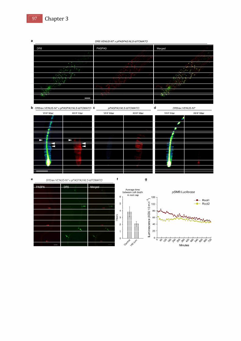
97 Chapter 3
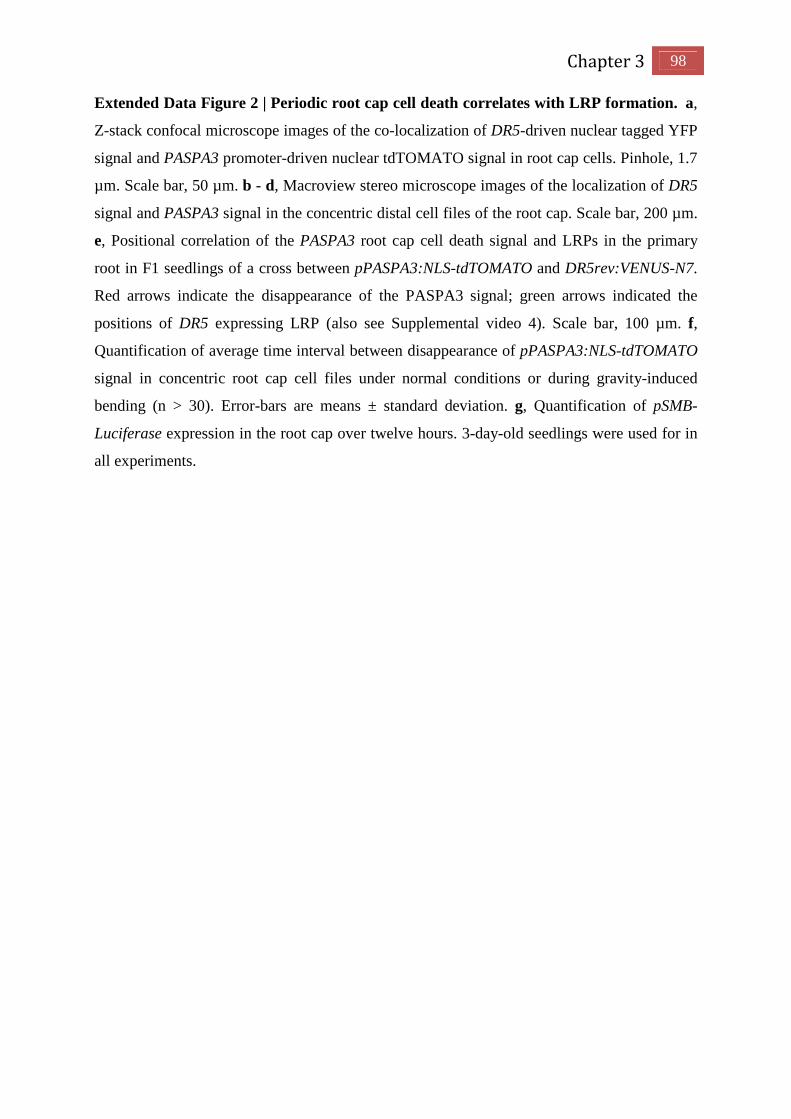
Chapter 3 98
Extended Data Figure 2 | Periodic root cap cell death correlates with LRP formation. a,
Z-stack confocal microscope images of the co-localization of DR5-driven nuclear tagged YFP
signal and PASPA3 promoter-driven nuclear tdTOMATO signal in root cap cells. Pinhole, 1.7
µm. Scale bar, 50 µm. b - d, Macroview stereo microscope images of the localization of DR5
signal and PASPA3 signal in the concentric distal cell files of the root cap. Scale bar, 200 µm.
e, Positional correlation of the PASPA3 root cap cell death signal and LRPs in the primary
root in F1 seedlings of a cross between pPASPA3:NLS-tdTOMATO and DR5rev:VENUS-N7.
Red arrows indicate the disappearance of the PASPA3 signal; green arrows indicated the
positions of DR5 expressing LRP (also see Supplemental video 4). Scale bar, 100 µm. f,
Quantification of average time interval between disappearance of pPASPA3:NLS-tdTOMATO
signal in concentric root cap cell files under normal conditions or during gravity-induced
bending (n > 30). Error-bars are means ± standard deviation. g, Quantification of pSMB-
Luciferase expression in the root cap over twelve hours. 3-day-old seedlings were used for in
all experiments.

99 Chapter 3

Chapter 3 100
Extended Data Figure 3 | Root clock requires the root cap. a, Root phenotype of 8-day-old
Col-0 and smb-3 seedlings. c-f, Macroview microscope images (c, d) and confocal
microscope images (e, f) of DR5rev:VENUS-N7 expression in root tips of 3-day-old Col-0
and smb-3 seedlings. PI, propidium iodide. g, Quantification of the indicated parameters in
Col-0 and smb-3 seedlings. 3-day-old seedlings were used to measure the number of lateral
root cap cells (n > 30), the DR5 signal intensity in the root cap (n > 30) and the DR5
oscillation frequency in OZ (n > 70 obtained from individual 15 seedlings); numbers of
prebranch sites (PBs), LRPs, and LRs were obtained from 8-day-old seedlings (n > 12).
Primary root elongation was measured in 6-day-old seedlings (n > 14). h, DR5rev:VENUS-N7
expression in 5-day-old 35S:SMB-GR transgenic seedlings that were Mock or Dex treated
from day 3 on. i-k, Quantification of LR number in 8-day-old Col-0 and 35S:SMB-GR
seedlings treated with Dex from day 2 (red arrows), then transferred to medium without Dex
on day 4 (black arrows). LR numbers from the root regions that only formed on indicated
medium (with or without Dex), and images of root phenotype (i) and root tip (k) were taken.
Arrows indicated the time of the transfer. n = 10. l, m, Root phenotype and expression pattern
of J3411 of 8-day-old F1 progeny of indicated lines. n, Quantification of LR number in 8-
day-old indicated transgenic lines treated with various concentrations of estradiol from day 3
on (n > 10). Black scale bars, 1 cm; white scale bars, 100 µm.

101 Chapter 3
Extended Data Figure 4 | Auxin transport regulates LR patterning. a, Root phenotype of
8-day-old pSMB:axr3-GR transgenic seedlings transferred to ½ MS medium with or without
Dex from day 3 on. Bar = 1 cm. b, Quantification of DR5 signal intensity in lateral root cap
and epidermis in 3-day-old indicated lines treated with or without 10 µM Dex since
germination. c, pPASPA3:NLS-tdTOMATO expression in 3-day-old Mock- or NPA-treated
seedlings. d, Quantification of average time interval between the disappearance of
pPASPA3:NLS-tdTOMATO signal in concentric root cap cell files under indicated treatments
(n > 30, *P < 10-5). e, Quantification of root phenotype in 8-day-old Col-0 and pidwag1wag2
seedlings transferred to medium containing different concentrations of IBA from day 3 on (n
> 10). f, pAUX1:AUX1-YFP and pPIN2:PIN2-GFP protein accumulation in root cap cells. g,
h, Quantification of the root phenotype in 8-day-old Col-0 and aux1 single mutant seedlings
and F1 seedlings of J3411>>AUX1 aux1-22 and J0121>>AUX1 aux1-22. Error-bars are
means ± standard deviation. Scale bar, 50 µm.

Chapter 3 102
Extended Data Figure 5 | Inhibition of auxin transport changes DR5 expression pattern.
a, c, Macroview stereo microscope images (a) and confocal microscope images (c) of
DR5rev:VENUS-N7 expression in 3-day-old Col-0, ibr1ibr3ibr10 triple mutant, and aux1
single mutant seedlings grown with or without 10 µM NPA. Scale bar, 50 µm. b,
Quantification of DR5rev:VENUS-N7 signal intensity in lateral root cap and epidermis tissues
in 3-day-old indicated seedlings under Mock- or 10 µM NPA treatment (n = 24, *P < 10-6).
Error-bars are means ± standard deviation.

103 Chapter 3
Reference
Aida, M., Beis, D., Heidstra, R., Willemsen, V., Blilou, I., Galinha, C., Nussaume, L., Noh, Y.S., Amasino, R., and Scheres, B. (2004). The PLETHORA genes mediate patterning of the Arabidopsis root stem cell niche. Cell 119, 109-120. Arnaud, C., Bonnot, C., Desnos, T., and Nussaume, L. (2010). The root cap at the forefront. Comptes rendus biologies 333, 335-343. Bennett, T., van den Toorn, A., Sanchez-Perez, G.F., Campilho, A., Willemsen, V., Snel, B., and Scheres, B. (2010). SOMBRERO, BEARSKIN1, and BEARSKIN2 regulate root cap maturation in Arabidopsis. The Plant cell 22, 640-654. Birnbaum, K., Shasha, D.E., Wang, J.Y., Jung, J.W., Lambert, G.M., Galbraith, D.W., and Benfey, P.N. (2003). A gene expression map of the Arabidopsis root. Science 302, 1956-1960. Brunoud, G., Wells, D.M., Oliva, M., Larrieu, A., Mirabet, V., Burrow, A.H., Beeckman, T., Kepinski, S., Traas, J., Bennett, M.J., et al. (2012). A novel sensor to map auxin response and distribution at high spatio-temporal resolution. Nature 482, 103-106. Clough, S.J., and Bent, A.F. (1998). Floral dip: a simplified method for Agrobacterium-mediated transformation of Arabidopsis thaliana. The Plant journal : for cell and molecular biology 16, 735-743. De Smet, I., Tetsumura, T., De Rybel, B., Frey, N.F., Laplaze, L., Casimiro, I., Swarup, R., Naudts, M., Vanneste, S., Audenaert, D., et al. (2007). Auxin-dependent regulation of lateral root positioning in the basal meristem of Arabidopsis. Development 134, 681-690. Dhonukshe, P., Huang, F., Galvan-Ampudia, C.S., Mahonen, A.P., Kleine-Vehn, J., Xu, J., Quint, A., Prasad, K., Friml, J., Scheres, B., et al. (2010). Plasma membrane-bound AGC3 kinases phosphorylate PIN auxin carriers at TPRXS(N/S) motifs to direct apical PIN recycling. Development 137, 3245-3255. Dolan, L., Janmaat, K., Willemsen, V., Linstead, P., Poethig, S., Roberts, K., and Scheres, B. (1993). Cellular organisation of the Arabidopsis thaliana root. Development 119, 71-84. Fendrych, M., Van Hautegem, T., Van Durme, M., Olvera-Carrillo, Y., Huysmans, M., Karimi, M., Lippens, S., Guerin, C.J., Krebs, M., Schumacher, K., et al. (2014). Programmed Cell Death Controlled by ANAC033/SOMBRERO Determines Root Cap Organ Size in Arabidopsis. Current biology : CB. Filleur, S., Walch-Liu, P., Gan, Y., and Forde, B.G. (2005). Nitrate and glutamate sensing by plant roots. Biochemical Society transactions 33, 283-286. Grabov, A., Ashley, M.K., Rigas, S., Hatzopoulos, P., Dolan, L., and Vicente-Agullo, F. (2005). Morphometric analysis of root shape. The New phytologist 165, 641-651. Gray, W.M., Kepinski, S., Rouse, D., Leyser, O., and Estelle, M. (2001). Auxin regulates SCF(TIR1)-dependent degradation of AUX/IAA proteins. Nature 414, 271-276. Heisler, M.G., Ohno, C., Das, P., Sieber, P., Reddy, G.V., Long, J.A., and Meyerowitz, E.M. (2005). Patterns of auxin transport and gene expression during primordium development revealed by live imaging of the Arabidopsis inflorescence meristem. Current biology : CB 15, 1899-1911. Karimi, M., Depicker, A., and Hilson, P. (2007). Recombinational cloning with plant gateway vectors. Plant physiology 145, 1144-1154. Lucas, M., Godin, C., Jay-Allemand, C., and Laplaze, L. (2008). Auxin fluxes in the root apex co-regulate gravitropism and lateral root initiation. Journal of experimental botany 59, 55-66. Malamy, J.E., and Benfey, P.N. (1997). Organization and cell differentiation in lateral roots of Arabidopsis thaliana. Development 124, 33-44.

Chapter 3 104
Moreno-Risueno, M.A., Van Norman, J.M., Moreno, A., Zhang, J., Ahnert, S.E., and Benfey, P.N. (2010). Oscillating gene expression determines competence for periodic Arabidopsis root branching. Science 329, 1306-1311. Rouse, D., Mackay, P., Stirnberg, P., Estelle, M., and Leyser, O. (1998). Changes in auxin response from mutations in an AUX/IAA gene. Science 279, 1371-1373. Strader, L.C., Wheeler, D.L., Christensen, S.E., Berens, J.C., Cohen, J.D., Rampey, R.A., and Bartel, B. (2011). Multiple facets of Arabidopsis seedling development require indole-3-butyric acid-derived auxin. The Plant cell 23, 984-999. Svistoonoff, S., Creff, A., Reymond, M., Sigoillot-Claude, C., Ricaud, L., Blanchet, A., Nussaume, L., and Desnos, T. (2007). Root tip contact with low-phosphate media reprograms plant root architecture. Nature genetics 39, 792-796. Swarup, R., Kargul, J., Marchant, A., Zadik, D., Rahman, A., Mills, R., Yemm, A., May, S., Williams, L., Millner, P., et al. (2004). Structure-function analysis of the presumptive Arabidopsis auxin permease AUX1. The Plant cell 16, 3069-3083. Swarup, R., Kramer, E.M., Perry, P., Knox, K., Leyser, H.M., Haseloff, J., Beemster, G.T., Bhalerao, R., and Bennett, M.J. (2005). Root gravitropism requires lateral root cap and epidermal cells for transport and response to a mobile auxin signal. Nature cell biology 7, 1057-1065. Van Norman, J.M., Xuan, W., Beeckman, T., and Benfey, P.N. (2013). To branch or not to branch: the role of pre-patterning in lateral root formation. Development 140, 4301-4310. Vanneste, S., De Rybel, B., Beemster, G.T., Ljung, K., De Smet, I., Van Isterdael, G., Naudts, M., Iida, R., Gruissem, W., Tasaka, M., et al. (2005). Cell cycle progression in the pericycle is not sufficient for SOLITARY ROOT/IAA14-mediated lateral root initiation in Arabidopsis thaliana. The Plant cell 17, 3035-3050. Weijers, D., Van Hamburg, J.P., Van Rijn, E., Hooykaas, P.J., and Offringa, R. (2003). Diphtheria toxin-mediated cell ablation reveals interregional communication during Arabidopsis seed development. Plant physiology 133, 1882-1892. Willemsen, V., Bauch, M., Bennett, T., Campilho, A., Wolkenfelt, H., Xu, J., Haseloff, J., and Scheres, B. (2008). The NAC domain transcription factors FEZ and SOMBRERO control the orientation of cell division plane in Arabidopsis root stem cells. Developmental cell 15, 913-922.

GRASSES
Boundless grasses over the plain,
Come and go with every season;
No prairie fire can destroy the grass,
It shoots up again with the spring breeze blows;
Sweet they press on the old high-road,
And reach the crumbling city-gate;
Oh, Prince of friends, you are gone again...
I hear them sighing after you.
Bai Juyi (A poet from Tang Dynasty)

Chapter 4
A novel small molecule reveals the role of auxin receptor TIR1
on root branching

105 Chapter 4
A novel small molecule reveals the role of auxin receptor TIR1 on root branching Wei Xuan1,2, Dominique Audenaert1,2#, Bert De Rybel1,2¥, Paul Overvoorde3, Stefan
Kepinski4, Mark Estelle5 and Tom Beeckman1,2*
Affiliations: 1Department of Plant Systems Biology, VIB, Ghent, Belgium. 2Department of Plant Biotechnology and Bioinformatics, Ghent University, Gent, Belgium. 3Department of Biology, Macalester College, St. Paul, Minnesota, USA. 4Centre for Plant Sciences, University of Leeds, Leeds, UK. 5Department of Biology, Indiana University, Bloomington, Indiana 47405, USA #Present address: Compound Screening Facility, VIB, Ghent University, Gent, Belgium ¥Present address: Laboratory of Biochemistry, Wageningen University, Dreijenlaan 3,
6703HA Wageningen, The Netherlands. † Correspondence to: [email protected]
Abstract Auxin has been demonstrated to control root branching in plants. In the plant model
Arabidopsis thaliana, the auxin signal is mediated by the auxin receptor transport inhibitor
response 1 (TIR1). However, the regulation of root branching by TIR1 remains elusive. Here,
we identified a novel small molecule, tirlin, TIR1-dependent lateral root inducer, as a
chemical tool to unravel the molecular mechanism of Tir1 on lateral root development. We
found that tirlin strongly induces lateral root formation without moderating auxin perception
by TIR1. Genetic evidence shows that tirlin might act downstream of TIR1 and ARF7-ARF19
to regulate lateral root formation. By screening a fast-neutron mutagenesis population, we
identified LBD proteins as potential targets of tirlin. Our work suggests that LBD proteins
may act as the core downstream components of TIR1-dependent signaling on the regulation
of lateral root formation.

Chapter 4 106
Introduction
The complexity and architecture of the plant root system is mainly controlled by root
branching. It plays a crucial role in the adaptation of the plant to environment stimuli. In the
plant model Arabidopsis thaliana, the process of root branching is under the temporal control
of oscillating gene expression in the oscillation zone (OZ) close to the root tip, which has
been designated as the root clock (Moreno-Risueno et al., 2010; Van Norman et al., 2013).
This periodic gene oscillation in the OZ leads to the spatial formation of prebranch sites,
patches of cells in the OZ that subsequently will develop as lateral root (LR) primordia.
Auxin has been demonstrated as a key regulator of repeatable organogenesis in
Arabidopsis. The expression of the transcriptional auxin response reporter DR5 was found to
oscillate in OZ and forms static expression pattern in the prebranch sites, which implies a role
for auxin on regulating the rook clock. Recently, a root cap-specific auxin source driven by
indole-3-butyric acid (IBA) and TIR1/AFB-dependent auxin signaling were also
demonstrated to moderate the strength of the DR5 oscillation, and thus regulate prebrach sites
formation (Xuan et al., chapter 2). In Arabidopsis, auxin signaling is perceived by the
TIR1/AFB family of F-box proteins acting in concert with the Aux/IAA transcriptional
repressors (Dharmasiri et al., 2005; Gray et al., 2001). Gain-of-function mutations in
AUX/IAA proteins, including IAA28, SLR/IAA14, CRANE/IAA18 and SHY2/IAA3,
decrease the number of LRs, indicating that AUX/IAA-dependent auxin signaling is
necessary for LR formation (De Rybel et al., 2010; Rogg et al., 2001; Uehara et al., 2008;
Vanneste et al., 2005; Vermeer et al., 2014).
However, several lines of evidence suggest that different combination of TIR1/AFB
auxin receptors and AUX/IAA proteins displayed a wide range of auxin binding affinities,
and as much contributes to the complexity of auxin responses and diverse root phenotypes
(Calderon Villalobos et al., 2012; Parry et al., 2009). In addition, the exact role of each auxin
receptor type and their downstream signaling components during the process of LR
development remains unclear. By using a chemical genetic approach we established
previously, we identified several small molecules, as non-auxin-like lateral root inducers in
Arabidopsis (De Rybel et al., 2012).
One small molecule, which we named tirlin for TIR1-dependent lateral root inducer,
enhanced LR development in a TIR1-mediated fashion. In this way, tirlin could act as a
unique chemical tool for understanding the signaling pathways involved in TIR1-mediated
lateral root formation, and also to get insight into the redundant and non-redundant functions
of different auxin receptors.
107 Chapter 4
Results
Identification and characterization of non-auxin like lateral root inducers
To identify new synthetic molecules that stimulate the process of lateral root
development, we screened a diverse 10,000-compound library for activators of expression of
CYCB1;1, a cell cycle gene that marks cellular divisions (Supplementary Fig. 1). In xylem
pole pericycle cells, the induction of CYCB1;1 promoter expression coincides with cell
division of pericycle cells and thus reports the formation of new lateral root primordia
(Himanen et al., 2002; Himanen et al., 2004; Vanneste et al., 2005). To report cell division in
pericycle cells, we used transgenic seedlings containing a construct comprising the CYCB1;1
promoter fused to β-glucuronidase (GUS) (pCYCB1;1::GUS) in a high-throughput adaptation
of a previously described ‘lateral root inducible system’ (Himanen et al., 2004). Eighty-eight
molecules were identified to induce expression of pCYCB1;1::GUS in the xylem pole
pericycle cells after 24 h (Fig. 1a), suggesting they were potent activators of the early stages
of lateral root development. To avoid the selection of auxin-like compounds that would also
affect other auxin-related processes, we excluded all molecules with a chemical structure
similar to that of known auxins, such as IAA, NAA, 2,4-D or sirtinol, and retained nine hit
molecules for further analysis (Fig. 1a and Supplementary Fig. 1; De Rybel et al., 2012).
Phenotypic characterization shows that these hit compounds increased lateral root densities in
a dose-dependent manner compared to the mock-treated control seedlings (Fig. 1b). Two of
these molecules, A11 and A12, which we named naxillin for non-auxin-like lateral root
inducer, shared a core structure and act on IBA-to-IAA conversion pathway (De Rybel et al.,
2012). Another molecule, A14, displayed the strongest induction on lateral root formation
without inducing a transcriptional auxin response in the basal meristem compared to NAA
(Fig. 1b and Supplementary Fig. 1). In addition, similar to naxillin, A14 had less effect on
primary root elongation and shoot development compared to the synthetic auxin NAA (Fig.
1c). In A14-treated Col-0 seedlings, we also observed lateral roots that were formed adjacent
to one another or that fused at the base, indicating that lateral inhibition of organ formation is
interrupted by A14 (Fig. 1e). Structure-activity analysis showed that removal of any
substructure of this molecule led to the loss of A14 function on lateral root induction. Taken
together, these data suggested that A14 and NAA might activate different modes of action to
regulate lateral root formation.

Chapter 4 108
Figure 1. Identification of non-auxin like lateral root inducers. (a) Overview of the
procedure to screen for activators of lateral root development with the pCYCB1;1::GUS
marker (also see Supplemental Figure 1). (b) Dose-response analysis of lateral root (LR)
density of seedlings grown on control medium until 3 d after germination and transferred to
medium supplemented with the indicated hit molecule at the indicated concentration for five
additional days. (c) Phenotype of plants grown on control medium for 3 d and then transferred
to mock medium (DMSO) or medium supplemented with 10 µM NAA, 10 µM A12-naxillin,
or 10 µM A14 for five additional days. (d) Chemical structure of auxin analogues, A11,
naxillin and A14. (e) Lateral root primordia phenotype of three-day-old Col-0 seedling under
A14 treatment for five additional days. (Updated from De Rybel et al., 2012)

109 Chapter 4
A14 induces auxin response in the transition zone
It has been suggested that the auxin response in the OZ oscillates periodically and leads
to the formation of prebranch sites, which is regulated by the amplitude of auxin response in
the OZ (De Rybel et al., 2012; Xuan et al., chapter 2). To assess the effect of A14 on auxin
response in the OZ, a transgenic line expressing DII-VENUS, an Aux/IAA-based auxin
signaling sensor (Brunoud et al., 2012), was treated with A14. Interestingly, similar to NAA,
A14 treatment induced a transient degradation of DII-VENUS in the OZ after 2 hours
treatment (Fig. 2a). However, the reduction of DII expression level by A14 treatment was less
pronounced compared to the global reduction of DII by NAA treatment, indicating that A14
affects auxin signaling in a more subtle way.
In Arabidopsis, auxin signaling is monitored by the binding of auxin and its main
receptors TIR1/AFBs, which trigger the degradation of AUX/IAA proteins to activate
downstream transcription. We further performed pull-down assays to determine whether A14
induces DII-VENUS degradation by affecting the binding of TIR1 to AUX/IAA proteins.
Unlike NAA, A14 did not affect the interaction between TIR1-myc and AUX/IAA proteins in
the presence or absence of NAA application, which is similar to the behavior of naxillin (Fig.
2b). This suggests that A14 does not act as a typical auxin.
The establishment of local auxin maxima in lateral root primordia is an important
determinative factor in the development of lateral roots. In Arabidopsis, these auxin maxima
are established by the constitutive cycling of the PIN-FORMED (PIN) family of auxin efflux
carriers between the plasma membrane and endosomes (Benkova et al., 2003; De Smet et al.,
2007; Kitakura et al., 2011). Therefore, we further tested the possibility of A14 on PIN
endocytosis. As shown in Fig 3c, BFA treatment inhibits PINs trafficking from endosomes to
the plasma membrane and causes the accumulation of PIN1/PIN2 in endosomes, which could
be reversed by the application of NAA. By contrast, A14 treatments could not suppress the
BFA-induced PIN accumulations, further demonstrating that A14 and NAA have different
modes of action.

Chapter 4 110
Figure 2. A14 does not interfere with TIR1-AUX/IAA interaction. (a) Time-course
analysis of DII-VENUS expression on Col-0 seedlings germinated for five days and transfferd
to mock medium (DMSO) or medium supplemented with indicated compounds for 1, 3 and 6
hours. Seedling harvested at indicated time points were used for confocal imaging. (b) The
effect of different compounds on the binding assay of TIR1 and AUX/IAA proteins. c-myc-
tagged TIR1 was pulled-down using biotinylated Aux/IAA domain II peptides in the presence
of 1 µM NAA, 10 µM IBA, 100 µM A12, 100 µM A14, or the combination of different
compounds as indicated, and compared to DMSO treatment. Each lane is from identical
aliquots of the same batch of TIR1-myc extract and the Aux/IAA peptide is pipetted in and
then captured on beads. (c) PIN1/PIN2 immunolocalization in wild-type seedlings treated
with BFA alone, or BFA together with NAA or A14 at indicated concentrations for 90 min.
(Updated from De Rybel et al., 2012)
111 Chapter 4
A14 function requires the auxin receptor TIR1
To identify the signaling pathways that are essential for A14 activity on lateral root
formation, we analyzed the effect of A14 on various mutants from different signaling
pathways, which have been demonstrated to be involved in lateral root formation. First, the
IBA-to-IAA conversion triple loss-of-function mutant ibr1ibr3ibr10 maintained sensitivity to
A14 treatment (Supplemental Fig. 1) (De Rybel et al., 2012). Meanwhile, A14 could also
induce lateral root formation in smb-3, brn1brn2 and arf7 (Supplemental Fig. 1), mutants that
were shown to control the periodicity of lateral root production (Moreno-Risueno et al.,
2010). Furthermore, the acr4 mutation, which interferes with the asymmetric cell division
during lateral root initiation (De Smet et al., 2008), also did not suppress the effect of A14
induction on lateral root formation (Supplemental Fig. 1). However, A14 failed to induced
lateral root formation in the arf7 arf19 double loss-of-function mutant (Supplemental Fig. 1),
suggesting that A14’s function is dependent on the ARF7-ARF19 pathway.
To assess whether A14 affects the early events of auxin signaling, we first determine the
effect of A14 on the tir1-1, auxin receptor TIR1mutant (Dharmasiri et al., 2005).
Interestingly, the A14-response on lateral root formation and primary root elongation was
suppressed in the tir1mutant background (Fig. 3a and b). To further test the effect of A14
effect on other auxin receptors, we also analyzed the lateral root phenotype of afb1, afb2 and
afb3 auxin receptor mutants under A14 treatment. Unlike tir1, afb mutants still show
sensitivity to A14, whereas the combination of tir1 and afb mutants were resistant to A14
treatment (Fig. 3a and b). These data suggests A14 might specifically act through a TIR1-
dependent signaling pathway. Therefore we named A14 tirlin for “TIR1-dependent lateral
root inducer”.
At the transcript level, analysis of the auxin response showed that A14-induced
DR5:GUS expression level in the OZ and pericycle cells was also reduced in the tir1 mutant
background, further confirming tirlin-induced auxin response during early lateral root
development was dependent on TIR1-mediated signaling.

Chapter 4 112
Figure 3. A14 function on lateral root events requires TIR1. (a) Phenotype of Col-0 and
tir1-1 seedlings grown on control medium for 3 d and then transferred to mock medium
(DMSO) or medium supplemented with 20 µM A14 for five additional days. (b)
Quantification of lateral root number of indicated transgenic seedlings grown on control
medium until 3d after germination and transferred to medium supplemented with the
indicated hit molecule at the indicated concentration for five additional days. (c) Time-course
analysis of DR5::GUS expression on Col-0 and tir1-1 seedling grown on control medium for
3d and then transferred to mock medium (DMSO) or medium supplemented with indicated
compounds for five additional days. Seedling harvested from indicated time points were used
for staining to analyze the β-glucuronidase activity.

113 Chapter 4
Identification of potential A14 targets in Arabidopsis
Interestingly, the tir1 mutant also display ~ 50% reduction on lateral root number
compared to WT (Fig. 3b), implying an important role of TIR1 on the LR development. To
explore the potential targets of tirlin and TIR1-dependent downstream signaling components,
we performed a suppressor screen in Arabidopsis to identify mutants that were resistant or
hyper-sensitive to tirlin-dependent induction of lateral root formation. For this purpose, a fast-
neutron mutagenized Col-0 population was screened upon tirlin treatment and the 31-2R, 23-
5R, 17-5R, and 61-108R mutants were identified as tirlin-resistant mutants, whereas the 9-15S
mutant was selected as a tirlin-hypersensitive mutant (Fig. 4a and b). Among the resistant
mutant alleles, the 31-2R allele showed complete resistance to tirlin-induced lateral root
formation and primary root elongation (Fig. 4a and b). Subsequent positional cloning
identified a deletion on chromosome 2 close to two genetic markers T20P8 and T16B24 (Fig.
4c). Interestingly, several LATERAL ORGAN BOUNDARIES DOMAIN/ASYMMETRIC
LEAVES2-LIKE (LBD/ASL) genes, which encode proteins containing the LOB (for lateral
organ boundaries) domain, were found to be located in this region (Fig. 4d) (Matsumura et al.,
2009). Among them, LBD16 and LBD18 have been shown to regulate lateral root formation
in Arabidopsis (Goh et al., 2012). lbd16 and lbd18 loss-of-function mutants were less
sensitive to tirlin-induced lateral root formation compared to WT, however, an slightly
increased lateral root number could still be detected in tirlin-treated lbd16 and lbd18 single
mutants when compared to Mock-treated seedlings (Fig. 4e). By contrast, lbd33, lbd16lbd33,
lbd16lbd18lbd33 mutants completely inhibited the tirlin response (Fig. 4d and e).Meanwhile,
we noticed that LBD33 gene is not located in the predicted deletion region, indicating tirlin
might acts on other LBD proteins to regulate LR formation. In addition, lateral root phenotype
in lbd16lbd18lbd33 triple mutant is similar to it in tir1afb2 mutant, indicating a possible link
between TIR1 and LBD proteins. Therefore, we propose that tirlin might be dependent on
LBD proteins downstream of TIR1 to regulate lateral root formation.

Chapter 4 114
Figure 4. Identification of LBD proteins as the potential targets of Tirlin. (a) Root
phenotype of 3-d-old seedlings from different mutant alleles transferred to medium containing
10 µM Tirlin for five more days. (b) Quantification of lateral root phenotype of indicated
mutant alleles grown on control medium until 3 d after germination and transferred to
medium supplemented with or without 10 µM Tirlin for five additional days. (c) Localization
of the potential deletion region and LBD genes in chromosome 2 in Arbidopsis genome. (d)
Root phenotype of 3-d-old Col-0 and lbd16lbd18lbd33 mutant seedlings transferred to
medium containing 10 µM Tirlin for five more days. (e) Quantification of lateral root
phenotype of seedlings from indicated mutants grown on control medium until 3 d after
germination and transferred to medium supplemented with or without 10 µM Tirlin for five
additional days (n > 12, *P < 0.01, ***P < 10-4, and **** P < 10-5 by two-sided Student's t test
indicated statistically significant differences).

115 Chapter 4
Discussion
For decades, auxin has been demonstrated to control new organ formation in plants,
especially in the case of lateral and adventitious root formation. In plant model Arabidopsis,
auxin perception is mediated by the F-Box proteins TIR1/AFBs (Dharmasiri et al., 2005;
Gray et al., 2001). A mutation the main auxin receptor TIR1 results in a reduced number of
lateral roots, implying an important role of TIR1 in lateral root formation (Fig. 3). However,
the molecular mechanism and signaling components downstream of the auxin receptors for
lateral root formation are not yet identified. The observation that auxins such as NAA can still
increase LRs in tir1 or tir1afb2afb3 triple mutants also indicates functional redundancy of the
different auxin receptors for the process of lateral root formation (Fig. 3). In our study, we
identified a novel small molecule, tirlin, a strong lateral root inducing molecule that acts in a
TIR1-dependent manner and similar to the artificial auxin analogue NAA. However, the
effect of tirlin on lateral root formation is not based on alteration in auxin transport but is
exclusively dependent on the auxin receptor TIR1, and not on other auxin receptors,
indicating the distinguished roles of auxin receptors in different plant developmental
processes. The identification of a strong lateral root inducing molecule specifically acting
through TIR1therefore underlines the importance of this receptor for lateral root formation as
compared to the other auxin receptors. This is coinciding with our previous findings in which
we demonstrated that the TIR1-AFB2 pathway controlled the periodic prebranch sites
formation by regulating the DR5 oscillation strength in OZ.
Biochemistry data showed that tirlin did not affect the binding affinities of TIR1 and
AUX/IAA proteins nor PIN-mediated endocytosis (Fig. 2). These data indicate that tirlin does
not directly bind the TIR1 protein; instead, it might target the signaling components
downstream of TIR1. By a forward genetic approach, LBD proteins were suggested as
potential targets of Tirlin. It has been demonstrated that the expression level of LBD proteins,
i.e. LBD16 and LBD18, is regulated by auxin, and they were shown to act downstream of the
auxin response factors ARF7- and ARF19-dependent auxin signaling pathway in Arabidopsis
roots (Okushima et al., 2007). Meanwhile, tirlin function on lateral root development is also
dependent on ARF7-ARF19 pathway (Supplemental Fig. 1). In addition, lbd triple mutants
phenotypically mimic the tir1afb2 mutants at the level of lateral root formation. Our data
therefore suggest the involvement of LBDs in TIR1-dependent signaling pathway for lateral
root formation. However, we cannot exclude the possibilities that tirlin might also acts on
unknown signaling pathways that are downstream of TIR1, which will be further addressed.

Chapter 4 116
Altogether, identification of tirlin by using a chemical genetics approach, led to the
clarification of the role of the auxin receptor TIR1 on lateral root developmental process, and
provides candidate genes potentially involved in the TIR1-downstream signaling cascades.
Acknowledgements
We thank Malcolm Bennett (University of Nottingham, UK), Mark Estelle (University of
California, San Diego, CA, USA), Hidehiro Fukaki (Kobe University, Kobe, Japan) for
sharing seed lines.
Materials and Methods
Compound screening and growth conditions
A commercial 10,000 compound library (DIVERSet™, ChemBridge Corporation) was
screened for induction of pCYCB1;1::GUS expression in xylem pole pericycle cells. About
three seeds of this marker line in Arabidopsis thaliana. Col-0 background were sown in 96-
well filter plates (Multiscreen HTS MSBVS1210; Millipore) in liquid medium derived from
standard Murashige and Skoog (MS) medium, supplemented with 10 µM of the auxin
transport inhibitor naphtylphtalamic acid (NPA), resulting in a primary root devoid of lateral
roots and allowing synchronization of lateral root development. Subsequently, seeds were
incubated in a growth chamber under continuous light (110 µE.m-2.s-1 photosynthetically
active radiation) at 21°C. Three days after germination, the liquid NPA medium was removed
and replaced with fresh liquid medium. Compounds were added to the 96-well plates to a
final concentration of 50 µM for 24 hours. Plants incubated in 2% DMSO or 10 µM NAA
were used as negative and positive control, respectively. Next, all plants were incubated in
GUS buffer as described (Vanneste et al., 2005) and analysed for GUS staining in xylem pole
pericycle cells. Only compounds that showed similar staining profiles in all seedlings were
considered. For all subsequent phenotypic analyses, plants were grown on square plates
(Greiner Labortechnik) with solid medium derived from standard MS medium under the same
conditions supplemented with compounds dissolved in DMSO when indicated as described
previously (De Rybel et al., 2009).
Plant lines used
The Arabidopsis accessions Columbia (Col-0) were used for this study. lbd mutant lines
were kind gifts from Hidehiro Fukaki (Kobe University, Kobe, Japan); the auxin receptor

117 Chapter 4
mutants were kindly supplied by Mark Estelle (University of California, San Diego, CA,
USA); DII-VENUS transgenic line was a gift from Malcolm Bennett (University of
Nottingham, UK) Double and higher-order mutants harboring various marker lines were
generated by crossing. F3 homozygous seedlings were analyzed in all experiments.
Root phenotype analyses
To quantify the lateral root phenotype in wild-type plants and mutants, emerged lateral
roots of the whole seedlings were counted under a dissecting microscope 8 days after
germination. Subsequently, whole seedlings were scanned for further analysis of the primary
root length. For compound treatments, three-day-old seedlings from the indicated lines were
transferred to ½ MS medium containing compounds at the indicated concentrations for extra
five days. Subsequently, the emerged lateral roots and primary root length were quantified.
Pull-down experiments
Pull-down experiments were done as described previously (Kepinski and Leyser, 2005).
A fast neutron mutagenesis screening and positional cloning
About 50,000 seedlings from 100 fast neutron-mutagenised pools (kindly supplied by
Malcolm Bennett lab in University of Nottingham, UK) were germinated on standard MS
medium. To exclude effects on germination, these plants were subsequently transferred to 10
µM tirlin three days after germination. Plants resistant to the lateral root inducing effect of
tirlin were selected after five more days. Before positional cloning, mutants were back-
crossed to Col-0 and selected again for the resistant phenotype. For PCR-based positional
cloning using SSLP markers, the mutant was crossed with Ler and subsequently selfed. 40
resistant F2 seedlings were used to map the mutation to chromosome 3 between T20P8 (11.6
Mb) and T16B24 (16.4 Mb).
Histochemical and histological analysis and microscopy
The GUS assays were performed as described previously (Vanneste et al., 2005). For
microscopic analysis, samples were cleared by mounting in 90% lactic acid (Acros Organics)
or by clearing as described previously (Malamy and Benfey, 1997). All samples were
analyzed by differential interference contrast microscopy (Olympus BX51). For anatomical
sections, GUS-stained samples were fixed overnight and embedded as described previously
(De Smet et al., 2004). Fluorescence imaging of roots was performed with an Olympus FV10-
ASW or Zeiss 710 confocal laser scanning microscope. For the propidium iodide (PI)-treated
Chapter 4 118
root images, seedlings were stained with 2 µg/mL PI for 3 minutes, washed with water, and
used for confocal imaging.
Reference
Benkova, E., Michniewicz, M., Sauer, M., Teichmann, T., Seifertova, D., Jurgens, G., and Friml, J. (2003). Local, efflux-dependent auxin gradients as a common module for plant organ formation. Cell 115, 591-602. Brunoud, G., Wells, D.M., Oliva, M., Larrieu, A., Mirabet, V., Burrow, A.H., Beeckman, T., Kepinski, S., Traas, J., Bennett, M.J., et al. (2012). A novel sensor to map auxin response and distribution at high spatio-temporal resolution. Nature 482, 103-106. Calderon Villalobos, L.I., Lee, S., De Oliveira, C., Ivetac, A., Brandt, W., Armitage, L., Sheard, L.B., Tan, X., Parry, G., Mao, H., et al. (2012). A combinatorial TIR1/AFB-Aux/IAA co-receptor system for differential sensing of auxin. Nature chemical biology 8, 477-485. De Rybel, B., Audenaert, D., Vert, G., Rozhon, W., Mayerhofer, J., Peelman, F., Coutuer, S., Denayer, T., Jansen, L., Nguyen, L., et al. (2009). Chemical inhibition of a subset of Arabidopsis thaliana GSK3-like kinases activates brassinosteroid signaling. Chemistry & Biology 16, 594-604. De Rybel, B., Audenaert, D., Xuan, W., Overvoorde, P., Strader, L.C., Kepinski, S., Hoye, R., Brisbois, R., Parizot, B., Vanneste, S., et al. (2012). A role for the root cap in root branching revealed by the non-auxin probe naxillin. Nat Chem Biol 8, 798-805. De Rybel, B., Vassileva, V., Parizot, B., Demeulenaere, M., Grunewald, W., Audenaert, D., Van Campenhout, J., Overvoorde, P., Jansen, L., Vanneste, S., et al. (2010). A novel aux/IAA28 signaling cascade activates GATA23-dependent specification of lateral root founder cell identity. Current biology : CB 20, 1697-1706. De Smet, I., Chaerle, P., Vanneste, S., De Rycke, R., Inze, D., and Beeckman, T. (2004). An easy and versatile embedding method for transverse sections. J Microsc 213, 76-80. De Smet, I., Tetsumura, T., De Rybel, B., Frei dit Frey, N., Laplaze, L., Casimiro, I., Swarup, R., Naudts, M., Vanneste, S., Audenaert, D., et al. (2007). Auxin-dependent regulation of lateral root positioning in the basal meristem of Arabidopsis. Development 134, 681-690. De Smet, I., Vassileva, V., De Rybel, B., Levesque, M.P., Grunewald, W., Van Damme, D., Van Noorden, G., Naudts, M., Van Isterdael, G., De Clercq, R., et al. (2008). Receptor-like kinase ACR4 restricts formative cell divisions in the Arabidopsis root. Science 322, 594-597. Dharmasiri, N., Dharmasiri, S., and Estelle, M. (2005). The F-box protein TIR1 is an auxin receptor. Nature 435, 441-445. Goh, T., Joi, S., Mimura, T., and Fukaki, H. (2012). The establishment of asymmetry in Arabidopsis lateral root founder cells is regulated by LBD16/ASL18 and related LBD/ASL proteins. Development 139, 883-893. Gray, W.M., Kepinski, S., Rouse, D., Leyser, O., and Estelle, M. (2001). Auxin regulates SCF(TIR1)-dependent degradation of AUX/IAA proteins. Nature 414, 271-276. Hilson, P., Allemeersch, J., Altmann, T., Aubourg, S., Avon, A., Beynon, J., Bhalerao, R.P., Bitton, F., Caboche, M., Cannoot, B., et al. (2004). Versatile gene-specific sequence tags for Arabidopsis functional genomics: transcript profiling and reverse genetics applications. Genome Res 14, 2176-2189.

119 Chapter 4
Himanen, K., Boucheron, E., Vanneste, S., de Almeida Engler, J., Inze, D., and Beeckman, T. (2002). Auxin-mediated cell cycle activation during early lateral root initiation. The Plant cell 14, 2339-2351. Himanen, K., Vuylsteke, M., Vanneste, S., Vercruysse, S., Boucheron, E., Alard, P., Chriqui, D., Van Montagu, M., Inze, D., and Beeckman, T. (2004). Transcript profiling of early lateral root initiation. Proceedings of the National Academy of Sciences of the United States of America 101, 5146-5151. Karimi, M., Depicker, A., and Hilson, P. (2007). Recombinational cloning with plant gateway vectors. Plant Physiol 145, 1144-1154. Kepinski, S., and Leyser, O. (2005). The Arabidopsis F-box protein TIR1 is an auxin receptor. Nature 435, 446-451. Kitakura, S., Vanneste, S., Robert, S., Lofke, C., Teichmann, T., Tanaka, H., and Friml, J. (2011). Clathrin mediates endocytosis and polar distribution of PIN auxin transporters in Arabidopsis. The Plant cell 23, 1920-1931. Liu, X., Cohen, J.D., and Gardner, G. (2011). Low-fluence red light increases the transport and biosynthesis of auxin. Plant Physiology 157, 891-904. Malamy, J.E., and Benfey, P.N. (1997). Organization and cell differentiation in lateral roots of Arabidopsis thaliana. Development 124, 33-44. Matsumura, Y., Iwakawa, H., Machida, Y., and Machida, C. (2009). Characterization of genes in the ASYMMETRIC LEAVES2/LATERAL ORGAN BOUNDARIES (AS2/LOB) family in Arabidopsis thaliana, and functional and molecular comparisons between AS2 and other family members. The Plant journal : for cell and molecular biology 58, 525-537. Moreno-Risueno, M.A., Van Norman, J.M., Moreno, A., Zhang, J., Ahnert, S.E., and Benfey, P.N. (2010). Oscillating gene expression determines competence for periodic Arabidopsis root branching. Science 329, 1306-1311. Okushima, Y., Fukaki, H., Onoda, M., Theologis, A., and Tasaka, M. (2007). ARF7 and ARF19 regulate lateral root formation via direct activation of LBD/ASL genes in Arabidopsis. The Plant cell 19, 118-130. Parry, G., Calderon-Villalobos, L.I., Prigge, M., Peret, B., Dharmasiri, S., Itoh, H., Lechner, E., Gray, W.M., Bennett, M., and Estelle, M. (2009). Complex regulation of the TIR1/AFB family of auxin receptors. Proceedings of the National Academy of Sciences of the United States of America 106, 22540-22545. Rogg, L.E., Lasswell, J., and Bartel, B. (2001). A gain-of-function mutation in IAA28 suppresses lateral root development. The Plant cell 13, 465-480. Uehara, T., Okushima, Y., Mimura, T., Tasaka, M., and Fukaki, H. (2008). Domain II mutations in CRANE/IAA18 suppress lateral root formation and affect shoot development in Arabidopsis thaliana. Plant & cell physiology 49, 1025-1038. Van Norman, J.M., Xuan, W., Beeckman, T., and Benfey, P.N. (2013). To branch or not to branch: the role of pre-patterning in lateral root formation. Development 140, 4301-4310. Vanneste, S., De Rybel, B., Beemster, G.T., Ljung, K., De Smet, I., Van Isterdael, G., Naudts, M., Iida, R., Gruissem, W., Tasaka, M., et al. (2005). Cell cycle progression in the pericycle is not sufficient for SOLITARY ROOT/IAA14-mediated lateral root initiation in Arabidopsis thaliana. The Plant cell 17, 3035-3050. Vermeer, J.E., von Wangenheim, D., Barberon, M., Lee, Y., Stelzer, E.H., Maizel, A., and Geldner, N. (2014). A spatial accommodation by neighboring cells is required for organ initiation in Arabidopsis. Science 343, 178-183.
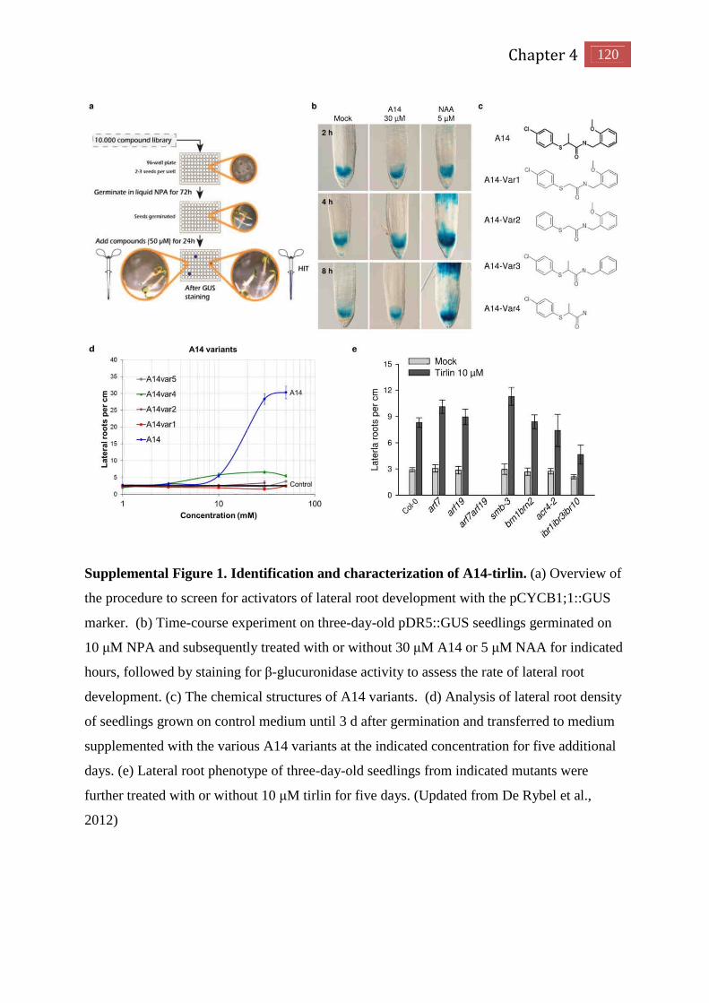
Chapter 4 120
Supplemental Figure 1. Identification and characterization of A14-tirlin. (a) Overview of
the procedure to screen for activators of lateral root development with the pCYCB1;1::GUS
marker. (b) Time-course experiment on three-day-old pDR5::GUS seedlings germinated on
10 µM NPA and subsequently treated with or without 30 µM A14 or 5 µM NAA for indicated
hours, followed by staining for β-glucuronidase activity to assess the rate of lateral root
development. (c) The chemical structures of A14 variants. (d) Analysis of lateral root density
of seedlings grown on control medium until 3 d after germination and transferred to medium
supplemented with the various A14 variants at the indicated concentration for five additional
days. (e) Lateral root phenotype of three-day-old seedlings from indicated mutants were
further treated with or without 10 µM tirlin for five days. (Updated from De Rybel et al.,
2012)

Knowledge is a matter of science, and no dishonesty or
conceit whatsoever is permissible.
Mao Tse-tung

Chapter 5 Concluding remarks

121 Chapter 5
Concluding remarks
Root cap contributes to the root patterning
Plant roots grow in the soil in order to support plant growth by absorbing water and
nutrients. Root growth rate is controlled by cell elongation in the elongation zone and cell
divisions in the apical meristem, the latter being covered by the root cap. As helmets are
required for protecting peoples head during hazardous activities, root cap cells serve a similar
purpose in plants. Besides protecting the root apical meristem, the root cap further contributes
in the perception of environmental signals, in mediating interactions between the soil and the
plant and in controlling the direction of root growth (Filleur et al., 2005). More recently, the
root cap was found to release a broad variety of chemical compounds into the soil to mediate
rhizospheric interactions both at the plant–microbiome levels (Driouich et al., 2013; Turner et
al., 2013b).
Our studies revealed a novel role of the root cap in patterning of root branching in
Arabidopsis. First, local auxin biosynthesis in the root cap controls the prebranch site
formation by regulating the oscillation amplitude in the OZ (Chapter 2). Secondly, auxin
transport through the lateral root cap and epidermis is involved in the transduction of the root
cap signal to the prebranch site and finally the earlier described periodic root cap cell death is
correlated with and seems to be crucial for the oscillatory nature of the process (Chapter 3).
Moreover, the NAC domain transcriptional factors SMB and FEZ, known to regulate root cap
formation, are also required for setting the root clock (Moreno-Risueno et al., 2010).
Based on our findings it is tempting to speculate that the root cap might act as a
transmission medium linking the environmental stimuli with root pre-patterning. When the
root cap perceives external signals, it might moderate its growth dynamics by altering the cell
division rate and by delaying or accelerating its programmed cell death. Through the
mechanism that we have proposed in this thesis, such an alteration in the growth dynamics of
the root cap might affect the patterning of lateral organs in the primary root. In other words,
the growth dynamics in the root cap might help the plant to produce more or less lateral roots
along the primary root axis dependent on the environmental conditions. Interestingly, Low
Phosphate Root1 (LPR1) and Nitrate Transporter (NRT1) genes are expressed in the root cap
cells (Krouk et al., 2010; Svistoonoff et al., 2007), indicating the altered primary root and
lateral root phenotype under varying nutrient conditions might be also determined by the
signal perception and transduction in the root cap. Therefore, further research is required to

Chapter 5 122
focus on the mechanism by which the root cap is capable in sensing the environment signal,
and to determine whether this signal could be converted into the positional information for
root patterning.
Furthermore, in chapter 3, we also show that the formation and programmed cell death of
root cap cells is crucial for the root clock. In Arabidopsis, the root cap develops from two sets
of meristematic cells, a central group of initials that gives rise to the columella and a
surrounding ring of cells that gives rise to both the lateral root cap and the epidermis through
periclinal cell divisions of common stem cells (Fig.1) (Dolan et al., 1993; Scheres et al., 2002).
At the distal end of the lateral root cap, cells are released from the root triggered by
programmed cell death (Fendrych et al., 2014). This process is restricted by SMB and FEZ,
the NAC domain transcriptional factors in Arabidopsis (Bennett et al., 2010; Willemsen et al.,
2008). Meanwhile, the mutants of QUASIMODO 1 (QUA1) and QUASIMODO 2 (QUA2)
genes, which encode putative glycosyltransferases in Arabidopsis, have root cap cells that
separate from each other when they are released (Durand et al., 2009). Normally, the wild
type Arabidopsis root tip does not produce isolated border cells per se, but it does produce and
release cells that remain attached to each other, forming a block of several cell layers called
border-like cells (Vicre et al., 2005). Interestingly, the quasimodo mutants display an altered
lateral root phenotype compared to WT, suggesting that a formative build-up of root cap
Figure 1. The root cap models in plants. (A) Schematic of the Arabidopsis root cap.(B)
Schematic showing PC division in the Epi/LRC stem cell (dark pink) division that
generates the LRC (purple) and anticlinal cell division that generates the epidermis (light
pink).(C) Schematic of anticlinal COL stem cell division, with the stem cell in red and
the differentiated COL cell in pink. (D) Schematic drawing of the root cap in rice. CSC:
columella stem cell. CSCD: columella stem cell daughter. C: columella. LRCSC: lateral
root cap stem cell. LRCSCD: lateral root cap stem cell daughter. LRC: lateral root cap.
D
123 Chapter 5
tissue is required for root prepatterning in Arabidopsis. More recently, signaling components
were identified to be involved in the root cap formation. The QC-expressed transcription
factor WUSCHEL-RELATED HOMEOBOX5 (WOX5) was found to negatively regulate the
SMB activity during columella development. Besides WOX5, RETINOBLASTOMA-
RELATED protein, and the ARF10- and ARF16-mediated auxin response factors also play a
role in the determination of columella stem cells activity, which further affects the root cap
formation (Bennett et al., 2014). However, up to present, it is not clear if all these factors also
regulate the root cap cell death and root prepatterning.
In order to identify more and specific signaling pathways that might have a function in
root cap differentiation, secretory activity and PCD, a comprehensive transcriptomic fate map
of the LRC would be preferable. Actually, the exciting transcriptomic data sets of the
Arabidopsis root generated by the Benfey’s lab (Brady et al., 2007) do not allow to distillate
transcriptional data on the separated root cap cell types, such as columella, LRC initials,
differentiating cells, differentiated cells, and cells undergoing PCD). To excess the different
developmental stages of the root cap, we are recently involved in a project that will make use
of cell-type and developmental specific reporter lines, such as J3411(Lateral root cap), J0951
(out layer of LRC), PET111 (Columella), J1092 (LRC initials), pPASPA1:GFP, and
SMB:GFP to perform cell sorting and further RNA sequencing. This novel dataset might
enhance our insight in the various functions of the root cap including its role in root
prepatterning.
Unlike Arabidopsis, in the monocot plant Oryza sativa, columella and lateral root cap
arise from a set of root cap stem cells that do not contribute to the generation of the epidermis
(Fig.2) (Wang et al., 2014). In rice, the root cap stem cells are located below the cap junction,
a distinct cell layer composed with approximately 13 cells. The central root cap stem cells
divide anticlinally to develop a columella, whereas the outer stem cells form the lateral root
cap by several rounds of periclinal and anticlinal cell divisions. This process is regulated by
OsIAA23-mediated auxin signaling and the glutamate receptor-like gene, GLR3;1 (Jun et al.,
2011; Li et al., 2006; Ni et al., 2014). However, the role of these genes on root prepatterning
in is not characterized yet.
Taken together, despite the dissimilar pattern of root cap formation in Arabidopsis and
Oryza sativa, it is still unclear whether the control on the pattern of root branching occurs in a
similar way in these two different species. Moreover, it is not clear yet whether the root cap is

Chapter 5 124
also controlling root branching in other species. As mentioned higher, the lateral root
phenotype of the quasimodo mutants indicate that there might be a correlation with the
presence of border-like cells and normal lateral root patterning. Furthermore, the occurrence
of border-like cells seems to be specific for the Brassicaceae family as it also occurs in
rapeseed (Brassica napus), mustard (Brassica juncea), and Brussels sprout (Brassica oleracea
gemmifera) (Driouich et al., 2007) and is absent in many other plant species analyzed so far. It
is therefore still possible that root prepatterning occurs differently in other plant species.
Root pre-patterning in plants
In Arabidopsis, root prepatterning has been described as a biological clock process that
translates a temporal signal into spatial information for lateral organ formation along the
primary root axis. A large scale of experiments has been applied to study the molecular
mechanism of this process in Arabidopsis, and several signaling components have been
discovered to control this process. However, it is not certain whether the occurrence of a root
clock is a shared mechanism for root branching in other plant species. In addition, the
existence and the function of the identified signaling pathways for root branching still need to
be investigated in in other species and is mainly hampered by the lack of suitable research
tools such as in vivo markers to monitor auxin reponses.
In Zea mays and Oryza sativa, two important commercial crop plants, several lines of
research have been performed to reveal the pattern of lateral root primordium development. In
contrast to Arabidopsis, in which lateral roots are specifically initiated and developed from a
patch of protoxylem pole pericycle cells (De Smet et al., 2008; Malamy and Benfey, 1997),
LR development in Zea mays and Oryza sativa is more painful to analyze because monocot
roots are composed of several cortex layers and a varying number of cells per layer. In Zea
mays, LR initiation occurs in the pericycle cells opposite the phloem poles, and xylem pole
pericycle cells are not competent for LRI (Jansen et al., 2012). In Oryza sativa, the LR
primordium is initiated from pericycle cells at the phloem pole and endodermis (Kawata and
Shibayama, 1965). Similar to Arabidopsis, DR5 was also detected to be expressed in the
meristem in Zea mays and Oryza sativa. Longitudinal sections of the Zea mays root tip
showed the expression of DR5:RFP mainly in the QC, root cap, epidermis, and vascular tissue,
whereas transversal sections reveals that the DR5 signal starts in the meta-xylem precursor
cells and the proto-xylem poles close to the root tip, and subsequently also appears in the
phloem pole. In the upper root, the DR5 signal only remains in the phloem poles, which might
125 Chapter 5
be linked to the activation of phloem pole pericycle cells for lateral root initiation (Jansen et
al., 2012). While in the Oryza sativa root tip, DR5:GUS expression was observed specifically
in the root cap, quiescent center, xylem cells in the root apical meristem and lateral roots
(Zhou et al., 2014), which resembles the expression pattern of DR5 obtained in Arabidopsis.
Moreover, similar expression patterns of DR5 were also found in Medicago truncatula,
soybean and tomato during the lateral root developmental process (Dubrovsky et al., 2008;
Herrbach et al., 2014; Turner et al., 2013a). Thus, to reveal the root prepatterning or lateral
root initiation events in other species, auxin responsive elements base marker lines such as
DR5 could serve as a general and useful maker for the further research.
Spatial control of the root clock by auxin
In Arabidopsis, local auxin sources have been found to play a central role in the
regulation of organ formation. In chapter 2, we have identified that a root-cap specific auxin
source, derived from IBA, could moderate the amplitude of DR5 oscillation, and thus
presumably the auxin response levels, in the OZ to regulate the prebanch sites formation. In
addition, we also found that the DR5 signal intensity and the amplitude of the oscillations
were reduced in the auxin receptor mutant tir1afb2 (Chapter 2). Interestingly, the periodicity
of DR5 expression oscillating in OZ is not disturbed in auxin biosynthesis or signaling
mutants, indicating that oscillations of the root clock could occur even in the absence of local
auxin signaling. It also shows that auxin is required to maintain clock oscillations and
suggests that the arrest of transition from oscillations to prebranch sites is linked to the level
of auxin and its signaling in the OZ. Thus, we propose a two-tier mechanism for the root
clock; while oscillating genes might regulate the temporal signals, auxin may act as a local
gradient facilitating the spatial formation of prebranch sites in OZ.
IBA was reported to supply 30% of total auxin biosynthesis in Arabidopsis, and in our
study, we found that IBA-to-IAA conversion contributed to 50% of total lateral root
production. This indicates that other auxin sources might also be involved in root
prepatterning. It has been reported that overexpressing TAA1 and YUC genes significantly
promotes lateral root formation in Arabidopsis (Mashiguchi et al., 2011), indicating a possible
role for the tryptophan (Trp)-dependent auxin biosynthesis pathway on root patterning,
however, the mechanism and the contribution of this auxin biosynthesis for root branching is
not fully characterized yet.

Chapter 5 126
Beside the local auxin biosynthesis, several lines of evidence showed that auxin signaling
is also important for lateral root development. Gain-of-function mutants of AUX/IAA genes,
such as IAA28, SLR/IAA14, CRANE/IAA18 and SHY2/IAA3, severely reduce the lateral root
formation. Moreover, tissue-specific auxin signaling also affects lateral root development at
different developmental stages. For instance, endodermis auxin signaling is required for the
swelling of the LRFC and the execution of the asymmetric cell division of pericycle cells
(Vermeer et al., 2014), and auxin signaling in xylem pole pericycle is essential for lateral root
initiation (De Smet et al., 2007). Cortex auxin signaling is also found to be involved in lateral
root emergence and lateral root primordium shape (Lucas et al., 2013). Interestingly, IAA2
and IAA14 are found to be expressed in the lateral root cap cells (Swarup et al., 2005;
Vanneste et al., 2005), indicating a possible role of auxin signaling in the root cap. However,
our data argue for a scenario in which local auxin signaling is not required for this process;
instead, auxin transport might mediate auxin movement from the root cap into the OZ
(Chapter 3). In addition, although AUX/IAA genes were expressed in the endodermis and
pericycle cells, there is no clear evidence of the existence of a local auxin source in these
tissues, whereas several auxin flux carriers were found to be localized in the cell layers
surrounding LRP. The auxin signaling in these tissues might therefore be activated by auxin
transported from other tissues. It has been demonstrated that in Arabidopsis, the root tip has a
high activity on auxin biosynthesis (Petersson et al., 2009), and this auxin is further taken by
auxin transport to generate an auxin maximum in other tissues through “auxin reflux loop”
model (Grieneisen et al., 2007; Laskowski et al., 2008). So it will be also interesting to
investigate whether root cap-derived auxin source could also contribute to the LR initiation
and LRP development. Moreover, we cannot exclude the possibility that the auxin signaling
in these tissues might be activated by other signaling components rather than auxin itself,
which also has to be further determined.
Other hormones or signaling molecules, such as cytokinins and carotenoids, are known to
display negative effects on root patterning and a change on DR5 activity in their pathway
mutants or under exogenous compound treatment have been observed (Bielach et al., 2012;
Van Norman et al., 2014), suggesting a link between these hormones and auxin on regulating
root patterning. Interestingly, recent studies also show that cytokinin could act through auxin
efflux to regulate the auxin response in the root meristem and lateral root development
(Bishopp et al., 2011; Marhavy et al., 2014). Because our results suggested that an auxin flux,
mediated by auxin transport carriers AUX1 and PINS, is essential to establish the DR5

127 Chapter 5
oscillations in the OZ, it will be interesting to evaluate whether cytokinins act on this early
step of lateral root formation through the interference with PIN function (Chapter 3). The
impact of cytokinin on auxin transport could thus represent a potential role of cytokinin on
root prepatterning. According to the opposite function on root branching by IBA and
cytokinin, the cross-talk between IBA and cytokinin would be an interesting topic for further
research. Additionally, the function of other hormones on root patterning could also be
mediated by auxin-independent signaling pathways and still have to be analyzed.
Gene oscillation in the root cells: a mystery unraveled
In Arabidopsis, gene oscillatory patterns of expression were detected by capturing the
bioluminescence signal from firefly luciferase driven by the promoters of oscillating genes.
Because of the low resolution images captured by CCD cameras compared to laser scanning
confocal microscopes, the oscillating model could only be observed at the organ level. By
contrast, in animals, gene oscillation could be detected at the cell level by visualizing signal
Figure 2. Spatial distribution of DR5 oscillation signal and GATA23 signal in OZ and
the prebranch sites. pGATA23:NLS-GFP reporter and DR5:luciferase reporter were used
to quantifying DR5 signal and GATA23signal in OZ and the prebranch sites respectively
(n > 40). Scale bar = 0.5 mm.

Chapter 5 128
from fluorescence proteins (i.e. YFP and GFP) during somitogenesis. In this respect, it will be
crucial for future research in plants to reveal expression patterns of oscillating genes in the OZ
at the cellular level.
To this end, we developed an experimental set-up using a macro-view microscope that
enables us to detect fluorescence signals with higher resolution, thus serving as a powerful
technique to access the gene oscillating pattern at the tissue level. Expression pattern of
DR5:Luciferase throughout the root was observed to be synchronized with the dynamic of
oscillating genes, thus we performed live imaging on auxin response reporter line carrying a
DR5-promoter-driven nuclear yellow fluorescent proteins over a longer period. The DR5
signal appeared to be homogenously expressed in two strains of protoxylem at the start of the
OZ and appears in xylem pole pericycle cells later on. When the root cells enter the different
zone, the DR5 signal disappears from the xylem pole cells and becomes specifically
expressed in lateral root primordia. Under our experimental set-up, we could not detect the
changes of DR5 expression level at the cellular level inside the OZ, so it remains unclear
which cells are targeted during the oscillation.
Interestingly, by using another maker line, namely pGATA23:NLS-GFP, which marks
the founder cell specification and lateral root initiation, we found that the expression of
GATA23 already started in the OZ, at the position where the DR5 signal reached the peak
value in the OZ (Fig.2). Therefore, we hypothesize that in the OZ the auxin response
maximum shifts from protoxylem pole cells to the xylem pole pericycle cells thereby
triggering the lateral root initiation events. In this respect a new “DR5” marker (unpublished
data, personal communication, Bert De Rybel, University of Wageningen, The Netherlands)
was recently generated that shows expression both in protoxylem cells as well as in
protoxylem pole pericycle cells. These data strongly indicate that the DR5 oscillation in the
OZ is dependent on the signal transition from xylem pole to xylem pole pericycle cells when
the root tip receive the development signal, such as root bending, programmed cell death, cell
elongation and differentiation.
Although the root clock in plants might be comparable to the segmentation clock in
animals to a certain level, our observations argue for a novel oscillating model in plants which
requires signal transduction between neighboring cells or tissues, rather than a cell-
autonomous mode of action. However, we cannot exclude the possibility that gene oscillating
occurs cell autonomously because oscillation might be hard to be traced due to the rapid

129 Chapter 5
process and minute change of expression levels which requires more sensitive fluorescence
proteins and more advanced imaging techniques.
Reference
Bennett, T., van den Toorn, A., Sanchez-Perez, G.F., Campilho, A., Willemsen, V., Snel, B.,
and Scheres, B. (2010). SOMBRERO, BEARSKIN1, and BEARSKIN2 regulate root cap
maturation in Arabidopsis. The Plant cell 22, 640-654.
Bennett, T., van den Toorn, A., Willemsen, V., and Scheres, B. (2014). Precise control of
plant stem cell activity through parallel regulatory inputs. Development 141, 4055-4064.
Bielach, A., Podlesakova, K., Marhavy, P., Duclercq, J., Cuesta, C., Muller, B., Grunewald,
W., Tarkowski, P., and Benkova, E. (2012). Spatiotemporal regulation of lateral root
organogenesis in Arabidopsis by cytokinin. The Plant cell 24, 3967-3981.
Bishopp, A., Help, H., El-Showk, S., Weijers, D., Scheres, B., Friml, J., Benkova, E.,
Mahonen, A.P., and Helariutta, Y. (2011). A mutually inhibitory interaction between auxin
and cytokinin specifies vascular pattern in roots. Current biology : CB 21, 917-926.
Brady, S.M., Orlando, D.A., Lee, J.Y., Wang, J.Y., Koch, J., Dinneny, J.R., Mace, D., Ohler,
U., and Benfey, P.N. (2007). A high-resolution root spatiotemporal map reveals dominant
expression patterns. Science 318, 801-806.
De Smet, I., Tetsumura, T., De Rybel, B., Frei dit Frey, N., Laplaze, L., Casimiro, I., Swarup,
R., Naudts, M., Vanneste, S., Audenaert, D., et al. (2007). Auxin-dependent regulation of
lateral root positioning in the basal meristem of Arabidopsis. Development 134, 681-690.
De Smet, I., Vassileva, V., De Rybel, B., Levesque, M.P., Grunewald, W., Van Damme, D.,
Van Noorden, G., Naudts, M., Van Isterdael, G., De Clercq, R., et al. (2008). Receptor-like
kinase ACR4 restricts formative cell divisions in the Arabidopsis root. Science 322, 594-597.
Dolan, L., Janmaat, K., Willemsen, V., Linstead, P., Poethig, S., Roberts, K., and Scheres, B.
(1993). Cellular organisation of the Arabidopsis thaliana root. Development 119, 71-84.
Driouich, A., Follet-Gueye, M.L., Vicre-Gibouin, M., and Hawes, M. (2013). Root border
cells and secretions as critical elements in plant host defense. Current opinion in plant biology
16, 489-495.
Dubrovsky, J.G., Sauer, M., Napsucialy-Mendivil, S., Ivanchenko, M.G., Friml, J., Shishkova,
S., Celenza, J., and Benkova, E. (2008). Auxin acts as a local morphogenetic trigger to specify

Chapter 5 130
lateral root founder cells. Proceedings of the National Academy of Sciences of the United
States of America 105, 8790-8794.
Fendrych, M., Van Hautegem, T., Van Durme, M., Olvera-Carrillo, Y., Huysmans, M.,
Karimi, M., Lippens, S., Guerin, C.J., Krebs, M., Schumacher, K., et al. (2014). Programmed
Cell Death Controlled by ANAC033/SOMBRERO Determines Root Cap Organ Size in
Arabidopsis. Current biology : CB.
Filleur, S., Walch-Liu, P., Gan, Y., and Forde, B.G. (2005). Nitrate and glutamate sensing by
plant roots. Biochemical Society transactions 33, 283-286.
Grieneisen, V.A., Xu, J., Maree, A.F., Hogeweg, P., and Scheres, B. (2007). Auxin transport
is sufficient to generate a maximum and gradient guiding root growth. Nature 449, 1008-1013.
Herrbach, V., Rembliere, C., Gough, C., and Bensmihen, S. (2014). Lateral root formation
and patterning in Medicago truncatula. Journal of plant physiology 171, 301-310.
Jansen, L., Roberts, I., De Rycke, R., and Beeckman, T. (2012). Phloem-associated auxin
response maxima determine radial positioning of lateral roots in maize. Philosophical
transactions of the Royal Society of London Series B, Biological sciences 367, 1525-1533.
Jun, N., Gaohang, W., Zhenxing, Z., Huanhuan, Z., Yunrong, W., and Ping, W. (2011).
OsIAA23-mediated auxin signaling defines postembryonic maintenance of QC in rice. The
Plant journal : for cell and molecular biology 68, 433-442.
Kawata, S., Shibayama, H. (1965) On the lateral root primordia formation in the crown roots
of rice plants. Proc Crop Sci Soc Jpn 33, 423-431
Krouk, G., Lacombe, B., Bielach, A., Perrine-Walker, F., Malinska, K., Mounier, E.,
Hoyerova, K., Tillard, P., Leon, S., Ljung, K., et al. (2010). Nitrate-regulated auxin transport
by NRT1.1 defines a mechanism for nutrient sensing in plants. Developmental cell 18, 927-
937.
Laskowski, M., Grieneisen, V.A., Hofhuis, H., Hove, C.A., Hogeweg, P., Maree, A.F., and
Scheres, B. (2008). Root system architecture from coupling cell shape to auxin transport.
PLoS biology 6, e307.
Li, J., Zhu, S., Song, X., Shen, Y., Chen, H., Yu, J., Yi, K., Liu, Y., Karplus, V.J., Wu, P., et
al. (2006). A rice glutamate receptor-like gene is critical for the division and survival of
individual cells in the root apical meristem. The Plant cell 18, 340-349.
Lucas, M., Kenobi, K., von Wangenheim, D., Vobeta, U., Swarup, K., De Smet, I., Van
Damme, D., Lawrence, T., Peret, B., Moscardi, E., et al. (2013). Lateral root morphogenesis
is dependent on the mechanical properties of the overlaying tissues. Proceedings of the
National Academy of Sciences of the United States of America 110, 5229-5234.

131 Chapter 5
Malamy, J.E., and Benfey, P.N. (1997). Organization and cell differentiation in lateral roots of
Arabidopsis thaliana. Development 124, 33-44.
Marhavy, P., Duclercq, J., Weller, B., Feraru, E., Bielach, A., Offringa, R., Friml, J.,
Schwechheimer, C., Murphy, A., and Benkova, E. (2014). Cytokinin controls polarity of
PIN1-dependent auxin transport during lateral root organogenesis. Current biology : CB 24,
1031-1037.
Mashiguchi, K., Tanaka, K., Sakai, T., Sugawara, S., Kawaide, H., Natsume, M., Hanada, A.,
Yaeno, T., Shirasu, K., Yao, H., et al. (2011). The main auxin biosynthesis pathway in
Arabidopsis. Proceedings of the National Academy of Sciences of the United States of
America 108, 18512-18517.
Moreno-Risueno, M.A., Van Norman, J.M., Moreno, A., Zhang, J., Ahnert, S.E., and Benfey,
P.N. (2010). Oscillating gene expression determines competence for periodic Arabidopsis root
branching. Science 329, 1306-1311.
Ni, J., Zhu, Z., Wang, G., Shen, Y., Zhang, Y., and Wu, P. (2014). Intragenic suppressor of
Osiaa23 revealed a conserved tryptophan residue crucial for protein-protein interactions. PloS
one 9, e85358.
Petersson, S.V., Johansson, A.I., Kowalczyk, M., Makoveychuk, A., Wang, J.Y., Moritz, T.,
Grebe, M., Benfey, P.N., Sandberg, G., and Ljung, K. (2009). An auxin gradient and
maximum in the Arabidopsis root apex shown by high-resolution cell-specific analysis of
IAA distribution and synthesis. The Plant cell 21, 1659-1668.
Scheres, B., Benfey, P., and Dolan, L. (2002). Root development. The Arabidopsis book /
American Society of Plant Biologists 1, e0101.
Svistoonoff, S., Creff, A., Reymond, M., Sigoillot-Claude, C., Ricaud, L., Blanchet, A.,
Nussaume, L., and Desnos, T. (2007). Root tip contact with low-phosphate media reprograms
plant root architecture. Nature genetics 39, 792-796.
Swarup, R., Kramer, E.M., Perry, P., Knox, K., Leyser, H.M., Haseloff, J., Beemster, G.T.,
Bhalerao, R., and Bennett, M.J. (2005). Root gravitropism requires lateral root cap and
epidermal cells for transport and response to a mobile auxin signal. Nature cell biology 7,
1057-1065.
Turner, M., Nizampatnam, N.R., Baron, M., Coppin, S., Damodaran, S., Adhikari, S.,
Arunachalam, S.P., Yu, O., and Subramanian, S. (2013a). Ectopic expression of miR160
results in auxin hypersensitivity, cytokinin hyposensitivity, and inhibition of symbiotic nodule
development in soybean. Plant physiology 162, 2042-2055.

Chapter 5 132
Turner, T.R., James, E.K., and Poole, P.S. (2013b). The plant microbiome. Genome biology
14, 209.
Van Norman, J.M., Zhang, J., Cazzonelli, C.I., Pogson, B.J., Harrison, P.J., Bugg, T.D., Chan,
K.X., Thompson, A.J., and Benfey, P.N. (2014). Periodic root branching in Arabidopsis
requires synthesis of an uncharacterized carotenoid derivative. Proceedings of the National
Academy of Sciences of the United States of America 111, E1300-1309.
Vanneste, S., De Rybel, B., Beemster, G.T., Ljung, K., De Smet, I., Van Isterdael, G., Naudts,
M., Iida, R., Gruissem, W., Tasaka, M., et al. (2005). Cell cycle progression in the pericycle is
not sufficient for SOLITARY ROOT/IAA14-mediated lateral root initiation in Arabidopsis
thaliana. The Plant cell 17, 3035-3050.
Vermeer, J.E., von Wangenheim, D., Barberon, M., Lee, Y., Stelzer, E.H., Maizel, A., and
Geldner, N. (2014). A spatial accommodation by neighboring cells is required for organ
initiation in Arabidopsis. Science 343, 178-183.
Vicre, M., Santaella, C., Blanchet, S., Gateau, A., and Driouich, A. (2005). Root border-like
cells of Arabidopsis. Microscopical characterization and role in the interaction with
rhizobacteria. Plant physiology 138, 998-1008.
Wang, L., Chu, H., Li, Z., Wang, J., Li, J., Qiao, Y., Fu, Y., Mou, T., Chen, C., and Xu, J.
(2014). Origin and development of the root cap in rice. Plant physiology.
Willemsen, V., Bauch, M., Bennett, T., Campilho, A., Wolkenfelt, H., Xu, J., Haseloff, J., and
Scheres, B. (2008). The NAC domain transcription factors FEZ and SOMBRERO control the
orientation of cell division plane in Arabidopsis root stem cells. Developmental cell 15, 913-
922.
Zhou, J., Yu, F., Wang, X., Yang, Y., Yu, C., Liu, H., Cheng, Y., Yan, C., and Chen, J.
(2014). Specific expression of DR5 promoter in rice roots using a tCUP derived promoter-
reporter system. PloS one 9, e87008.


Summary
Summary

133 Summary
Summary
The root system is essential for plants to uptake water and nutrients from the soil and to
adapt the growth pattern in response to changing environment conditions. The root cap is
located in the distal end of the root and covers the meristem reaching the transition zone. It
acts as principal sensor mediating the interactions between plant roots and the soil, thus
representing an interface capable in transmitting external signals to the root thereby
determining the growth pattern of the root systems.
The root system is composed of a primary root and lateral roots sequentially forming
along the primary root. In plant model Arabidopsis thaliana, lateral root formation is linked to
a root clock that reflects a temporal oscillating pattern of gene expression in the oscillation
zone in the root tip. This recurrent gene expression pattern is translated into a repetitive
spatial pattern of prebranch sites, which eventually can further develop as lateral roots (Van
Norman et al., 2013). Thus, the spatiotemporal pattern of lateral root formation during
primary root growth becomes a curial topic in the present root development research.
However, the molecular components that regulate the oscillations remain unknown. In
addition, auxin has been demonstrated to control most aspects of lateral rooting events;
however, the potential role of auxin in controlling the root clock has not yet been determined.
In this Ph.D thesis, we attempt to address this question. First of all, by using a chemical
genetic approach, we identified that IBA-to-IAA conversion contributes to root branching.
Following the real-time analysis of DR5:Lucifease expression in IBA-to-IAA conversion
mutants, we revealed that the IBA-derived auxin in the root cap could moderate the strength
of the oscillation, and thus regulate prebranch sites formation. Meanwhile, we showed that the
amplitude of oscillation signal is dependent on TIR1/AFB-mediated auxin signaling in OZ.
These data led to the understanding of the role of auxin in the root clock. More specifically
we showed that auxin might act as a local gradient in OZ to regulate the oscillation strength
which in turn determines the establishment of prebranch sites. Our results suggest that the root
clock is controlled by a combination of temporal signals (oscillating of gene expression) on
the oscillation periodicity and by a spatial signal of TIR1/AFB-dependent auxin signaling that
is required for the amplitude in the oscillation.

Summary 134
To explore the downstream signaling components of IBA-derived auxin, we performed
an IBA transcriptome analysis and identified novel and IBA-regulated components of root
patterning, such as the MEMBRANE-ASSOCIATED KINASE REGULATOR4 (MAKR4).
Transcriptional analysis revealed that MAKR4 expression specifically locates in primordia and
moves towards the consecutive prebranch sites. Its plasma membrane localization strongly
suggested cell-to-cell communication might be required during the prebanch sites
establishment. Moreover, genetic evidence showed that the makr4 mutant and amiRNA lines
have a decreased number of lateral roots and lateral root primordia without affecting the
prebranch site formation. Taken together, our data indicated that MAKR4 perceives the
oscillation signal and translates it to the prebranch sites resulting into a regular spacing of
lateral organs.
Based on these results, we set up a new imaging system using a vertical oriented
macroview stereo microscope. It enables us to trace fluorescence signal movement through
the root in the normal experimental condition during a long period. By using this novel
imaging system, we observed that a periodic degradation of DR5 signal in the root cap
triggers the local formation of a lateral root primordium. This process is triggered by periodic
programmed cell death of root cap cells. We also found that auxin signaling in root cap is not
required for maintaining the root clock behavior. Instead, it requires the coordination of local
auxin biosynthesis and auxin transport in the root cap. Our findings demonstrated that the
auxin in the root cap is as a crucial for regulating root patterning in Arabidopsis.
In addition, we have shown that TIR1-dependent auxin perception is required for
maintaining DR5 oscillation level in OZ, indicating a central role of auxin on root
prepatterning. In chapter 4, we further explore the specific signaling pathway downstream
TIR1 for lateral root development. We identified a novel small molecule tirlin as a TIR1-
dependent lateral root inducer representing a chemical tool to access it. Genetic evidences
show that tirlin might act downstream of TIR1 and ARF7-ARF19 pathway to regulate lateral
root formation. By screening a fast-neutron mutagenesis population, we propose LBD
proteins as the potential target of tirlin. Our work further suggests that LBD proteins may act
as downstream components of TIR1-dependent signaling on regulating lateral root formation.

Bidding deputy magistrate Du farewell
《送杜少府之任蜀州》
The capital and palace are guarded by the land of three Qin kingdoms,
In the distance the five ferries are screened by wind and mist.
城阙辅三秦,风烟望五津。
Now comes the time for us to bid farewell to each other,
And we still be officials away from home on duty.
与君离别意,同是宦游人。
As long as we remain bosom friends in our heart of hearts,
We'll still feel like neighbours despite the distance apart.
海内存知己,天涯若比邻。
So don't let us shed tears like youngsters,
At that last moment when we both wave goodbye.
无为在歧路,儿女共沾巾!
Wang Bo 王勃

Acknowledgements

135 Acknowledgements
In China, we often use “Fate (分缘 )” to define friendship. People always say that the fate
brings people together no matter how far apart they may be, otherwise they can't know each
other even face to face.
In this respect, before I finished the writing of my Ph.D thesis, I would like to thank all the
people who have ever helped me not only during my Ph.D study, but also in my life in the
past five years, which is really a fantastic experience for me. I am very pleased with my
research here in the past years, and I always feel free and never had the feeling of stress. I also
like the academic atmosphere in PSB, people here are motivated and prefer to share the
experience, which enable me to learn as much as possible.
First of all, I would like to thank all the root members, especially Tom, who gave me the
opportunity to study at PSB, an international institute with excellent researches, in Gent, a
city with so many friendly Flemish people, in Belgium, a country with the best BEER and
CHOCOLATE! Actually, when there is a shortage of Orval in the market, I always got the
feeling of victory because I could drink Orval weekly. Ha~Ha~
As my promoter, Tom spent a lot time on my project, a lot discussions and sending emails all
over world to get the materials to keep my research in the highway. I really appreciate these
and I sincerely believe Tom is an excellent promoter and a gentleman. I hope Tom could find
Orval in the shop again after my leaving….
Next to Tom, I would like to thank my supervisor, Dominique, for guiding me a lot on
designing the experiments properly and analyzing the data. Most of my research is performed
on Arabidopsis thaliana, a tiny and uneatable plant, which is complete new for me at the
beginning of my Ph.D study. With the help from Dominique and Bert, I became familiar with
doing experiments Arabidopsis thaliana. And also thanks for including me in the chemical
genetic projects, from which I could work out the title for my Ph.D thesis.
I will also remember Maria for help me making sections of roots – really very hard but crucial
work, and also the delicious Asian&Belgium food. Food….sometimes can be very special, for
instant, chick eggs from Gieljon; if the size of eggs in supermarket is scaled as lateral root
primordium, Gieljon’s chick is producing founder cells. But many thanks to Xiyangyang for
these eggs, several times help on repairing my bike, and also a lot fun during the work.
Lorena, Leen, Giel and Gert, although you have left the lab, I still appreciate the help from
you for guiding me basic techniques at the beginning of my Ph.D with great patience. Lorena,

Acknowledgements 136
I believe you can become a good teacher; Gert, I still remember the word from you: “Be nice
to the people!”
Davy and Hanne (哈娜), thanks a lot for help me with cloning and imagings; I wish you be in
love forever and have happiness every moment.
Barbara, thanks for helping me with writing; Boris, thanks for the help on IBA microarray
analysis; Marlies, Ianto and Ana, thanks for sharing experience on counting lateral root
primordia; Long, Andry (Monkey King) and Hans, I really enjoyed the time sitting in the
corner with you, always fun and innovations there.
Kun, Qian and Tao, I wish you success in your research and find a good job in the future.=
In the department, I want to thank Steffen for many useful advices and ideas on my research,
especially on auxin transport; although every time I know I will get difficult questions from
you and more work to do, but I still like to discuss with you. I also appreciate the help from
Daniel for setting up the Olympus imaging systems; it is really fantastic and powerful
machine for root development research.
Moreover, I want to thank all the jury members in the committee of my Ph.D defense,
dr. Wout Boerjan (chair), dr. Laurent Laplaze, dr. Lieven De Veylder, dr. Ive de Smet, and dr.
Danny Geelen for the critical comments and very kind advices on my future research.
all the PIs in the department:
dr. Dirk Inzé, dr. Moritz Nowack, dr. Geert De Jaeger, dr. Sofie Goormachtig, dr. Ann
Depicker, and dr. Jiri Friml for nice discussions on my projects and kindly sharing the
materials.
I am also very appreciated all the support from people all over the world:
dr. Philip N. Benfey (Duke University, USA), dr. Malcolm Bennett (University of
Nottingham, UK), dr. Bonnie Bartel (Rice University, USA), dr. Mark Estelle (University of
California, USA), dr. Leah Band (University of Nottingham, UK ), dr. Lucia C. Strader
(Washington University, USA), dr. Catherine Bellini (Umeå University, Umeå, Sweden) for
the kind gifts of lines and constructs.

137 Acknowledgements
For my dear Chinese friends, I really miss the time with you guys in Belgium, and am grateful
for all your great help and encouragement.
First of all, I want to thank Dalong, my comrade, who had this adventure together with me,
and we helped each other for our Ph.D study. I also want to thank Hanyi, and Weicong, who
introduce PSB to me with a lot nice words, which help me make the decision to go here. My
wife and I also enjoyed the trip we made together in France very much.
I could not forget the help from Zhang Jing, Zhaojun and He Ying on my research and take
care of me at the early period of my study and living in Belgium.
Li Zhen and Deng Lei, I think I will miss the taste of home-made and Belgium-style hafing
dish very much after I return, thanks a lot for sharing nice food with me and many nice
discussions.
Zhubing and Chunmei, thanks for feeding me with delicious cookies every time when I was
hungry. It’s really special way for supporting my work.
I am also deeply indebted to all the other Chinese: Huang Jingjing, Chen Jian, Yao Yao, Du
yulong, Xiao Xiaojian, Wang Feng, Chen Xu, Zhu Qiang, Gao Zhen, Luo Yu, Zhang Zaichao,
Sun Xiaohuan, Han Chao, and Jiang Lingxiang, for your direct and indirect help to me and
my work!
In the end, I also want to thank the China scholarship council (CSC), Ghent University, and
VIB, who support my Ph.D study for the past five years.
Finally, I am indebted to my families for their continuous support and
To everybody: I hope all of you have enjoyed reading this. Without all your help, I could not
finish my Ph.D. Thanks for all the great moments during these last five years. I wish our
friendship remain and never can end.
Now it is time for the new adventure…; the future is warm yellow!
All the best,
Wei Xuan (宣伟), on the behavior of my family

Acknowledgements 138

Curriculum Vitae-Wei Xuan
1
Curriculum Vitae
Wei Xuan
Personal details
Education
2002.9-2006.6 Bachelor of Science, major in Biological Science, College of Life Sciences, Nanjing Agricultural University
2006.9-2009.6 Master of Science, major in Biochemistry & Molecular Biology, College of Life Sciences, Nanjing Agricultural University
2009.11- Present Predoctoral follow, VIB, Department of Plant Systems Biology,
Ghent University, Technologiepark 927, 9052 Gent
Birth date: 23/03/1984, Male
Birth place: Xinghua city, Jiangsu Provence, P.R.China
Nationality: Chinese
Residence address (Belgium): Ijzerlaan 7, Gent, B-9000
Contact email: [email protected]

Curriculum Vitae-Wei Xuan
2
PERFORMANCE AWARDS
2006 Nanjing Agricultural University scientific competition in Challenge Cup Awards
2008 China Youth Science and Technology Innovation Awards
2009 Jiangsu Province “triple-A” outstanding student
2009 China Scholarship Council (CSC) scholarship
2010 Special Research Fund - Cofunding for Chinese Ph.D students
Selected Publications (SCI/EI)
1. Van Norman, J.M., Xuan, W., Beeckman, T., and Benfey, P.N. (2013). To branch or not to
branch: the role of pre-patterning in lateral root formation. Development 140, 4301-4310.
2. Xuan, W., Murphy, E., Beeckman, T., Audenaert, D., and De Smet, I. (2013). Synthetic
molecules: helping to unravel plant signal transduction. Journal of chemical biology 6, 43-50.
3. De Rybel, B., Audenaert, D., Xuan, W., Overvoorde, P., Strader, L.C., Kepinski, S., Hoye, R.,
Brisbois, R., Parizot, B., Vanneste, S., et al. (2012). A role for the root cap in root branching
revealed by the non-auxin probe naxillin. Nature chemical biology 8, 798-805.
4. Xuan, W., Xu, S., Li, M., Han, B., Zhang, B., Zhang, J., Lin, Y., Huang, J., Shen, W., and Cui,
J. (2012). Nitric oxide is involved in hemin-induced cucumber adventitious rooting process.
Journal of plant physiology 169, 1032-1039.
5. Cao, Z., Geng, B., Xu, S., Xuan, W., Nie, L., Shen, W., Liang, Y., and Guan, R. (2011).
BnHO1, a haem oxygenase-1 gene from Brassica napus, is required for salinity and osmotic
stress-induced lateral root formation. Journal of experimental botany 62, 4675-4689.
6. Han, Y., Zhang, J., Chen, X., Gao, Z., Xuan, W., Xu, S., Ding, X., and Shen, W. (2008).
Carbon monoxide alleviates cadmium-induced oxidative damage by modulating glutathione
metabolism in the roots of Medicago sativa. The New phytologist 177, 155-166.
7. Xuan, W., Xu, S., Yuan, X., and Shen, W. (2008a). Carbon monoxide: A novel and pivotal
signal molecule in plants? Plant signaling & behavior 3, 381-382.
8. Xuan, W., Zhu, F.Y., Xu, S., Huang, B.K., Ling, T.F., Qi, J.Y., Ye, M.B., and Shen, W.B.
(2008b). The heme oxygenase/carbon monoxide system is involved in the auxin-induced
cucumber adventitious rooting process. Plant physiology 148, 881-893.
9. Xuan W, Huang LQ, Li M, Huang BK, Xu S, Liu H, Gao Y, Shen WB (2007) Induction of
growth elongation in wheat root segments by heme molecules: a regulatory role of carbon
monoxide in plants? Plant Growth Regulation 52: 41–51