Isolation of Salmonella Spp. in different meat samples of ...
100
1 Isolation of Salmonella Spp. in different meat samples of Kathmandu valley A Dissertation Submitted to the Department of Micro-Biology Kathmandu College of Science and Technology (Affiliated to Tribhuvan University) In Partial Fulfillment to award the Degree of Master of Science in Microbiology (Medical) By SRIJAN POKHREL MAJAGAIYA Department of Microbiology Kathmandu College of Science and Technology (Affiliated to Tribhuvan University) Kathmandu, Nepal 2009
-
Upload
khangminh22 -
Category
Documents
-
view
4 -
download
0
Transcript of Isolation of Salmonella Spp. in different meat samples of ...
1
Isolation of Salmonella Spp. in different meat samples of Kathmandu valley
A
Dissertation
Submitted to the Department of Micro-Biology
Kathmandu College of Science and Technology
(Affiliated to Tribhuvan University)
In Partial Fulfillment to award the Degree of Master of Science in Microbiology
(Medical)
By
SRIJAN POKHREL MAJAGAIYA
Department of Microbiology
Kathmandu College of Science and Technology
(Affiliated to Tribhuvan University)
Kathmandu, Nepal
2009
2
RECOMMENDATION
This is to certify that Mr. Srijan Pokhrel Majagaiya has completed this dissertation
work entitled “Isolation of Salmonella Spp. in different meat samples of
Kathmandu valley’ as a partial fulfillment of M.Sc. Degree in Microbiology under
our supervision. To our knowledge, this work has not submitted for any other Degree.
………………………………. ………………………………..
Mr. Pradeep Kumar Shah Dr. Poornima Manandhar
Visiting faculty and member of Senior Veterinary Officer
Research Committee Central Veterinary Laboratory
Department of Microbiology Tripureshwor, Kathmandu
Kathmandu College of Science Nepal
and Technology
(Affiliated to Tribhuvan University)
Kathmandu, Nepal
Date …13-10-2065…………….
3
CERTIFICATE OF APPROVAL
On the recommendation of Mr. Pradeep Kumar Shah and Dr. Poornima
Manandhar, this dissertation work of Mr. Srijan Pokhrel Majagaiya, entitled
“Isolation of Salmonella Spp. in different meat samples of Kathmandu Valley” is
approved for the examination and is submitted to the Tribhuvan University in the
partial fulfillment of the requirements for M.Sc. Degree of Microbiology.
.....................................
Ms. Archana Katuwal
Head of Department Microbiology
Kathmandu College of Science and Technology
(Affiliated to Tribhuvan University)
Kathmandu, Nepal
Date;…15-10-2065………………..
4
BOARD OF EXAMINERS
Recommended by: ………………………………
Mr. Pradeep Kumar Shah
Supervisor
………………………………
Dr. Poornima Manandhar
Supervisor
Approved by: …………………………………….
Ms. Archana Katuwal
Head of Department Microbilogy
Kathmandu College of science and Technology
(Affiliated to Tribhuvan University)
Kathmandu, Nepal
Examined by: ………………………………
Ms. Pearl Banmali
External Examiner
Senior Deputy Research Officer
Nepal Health Research Council
Ramshah Path, Kathmandu, Nepal
……………………………………..
Ms. Jyoti Amatya
Internal Examiner
Lecturer
Kathmandu College of science and Technology
Kathmandu, Nepal
Date:…11-11-2065………………

5
ACKNOWLEDGEMENTS
First and foremost, I would like to express my sincere thanks and heartfelt
appreciation to my supervisor Dr. Poornima Manandhar, a Senior Veterinary
Officer, Central Veterinary Laboratory for her constant guidance, inspiration,
invaluable suggestions and support to complete this dissertation work.
I am equally indebted to my internal supervisor, Mr. Pradeep Kumar Shah, Lecturer
of Kathmandu College of Science & Technology, for his valuable suggestions and
tremendous support in completition of this thesis work.
I am indebted to Mr. Tek Bahadur Air, Senior Laboratory Technician at CVL for his
invaluable assistant and encouragement to carry out this thesis work. I am also
thankful to Mr. Bal Bahadur Kunwar and Mr. Bhimsen Adhikari, Laboratory
Technician and all the staffs of CVL for their kind help and cooperative throughout
my research work at CVL. I would also like to thank the Department of Microbiology
(KCST) for suggestions and support and also the institute Armed Force Research
Institute and Medical Sciences (AFRIMS) for providing the facilities for
Salmonella serotyping.
I must acknowledge the indispensable cooperative, comments and suggestions of my
friends Mr. Ananda Babu Sharma and Mr.Sudip Regmi for this thesis work.
I am very much indebted to Mr. Jeewan Paudel, Mr. Yub Raj Sedhai & Mr. Bimal
Ghimire, Mr.Krishna Shrestha & Keshab Pathak for their cooperation & support.
I wish to express my gratefulness to Mr. Laxman Belbase dai for his help during the
data analysis.
Finally, I like to mention the tired less outstanding and everlasting support and
inspiration provided by my family.
Srijan Pokhrel Majagaiya
6
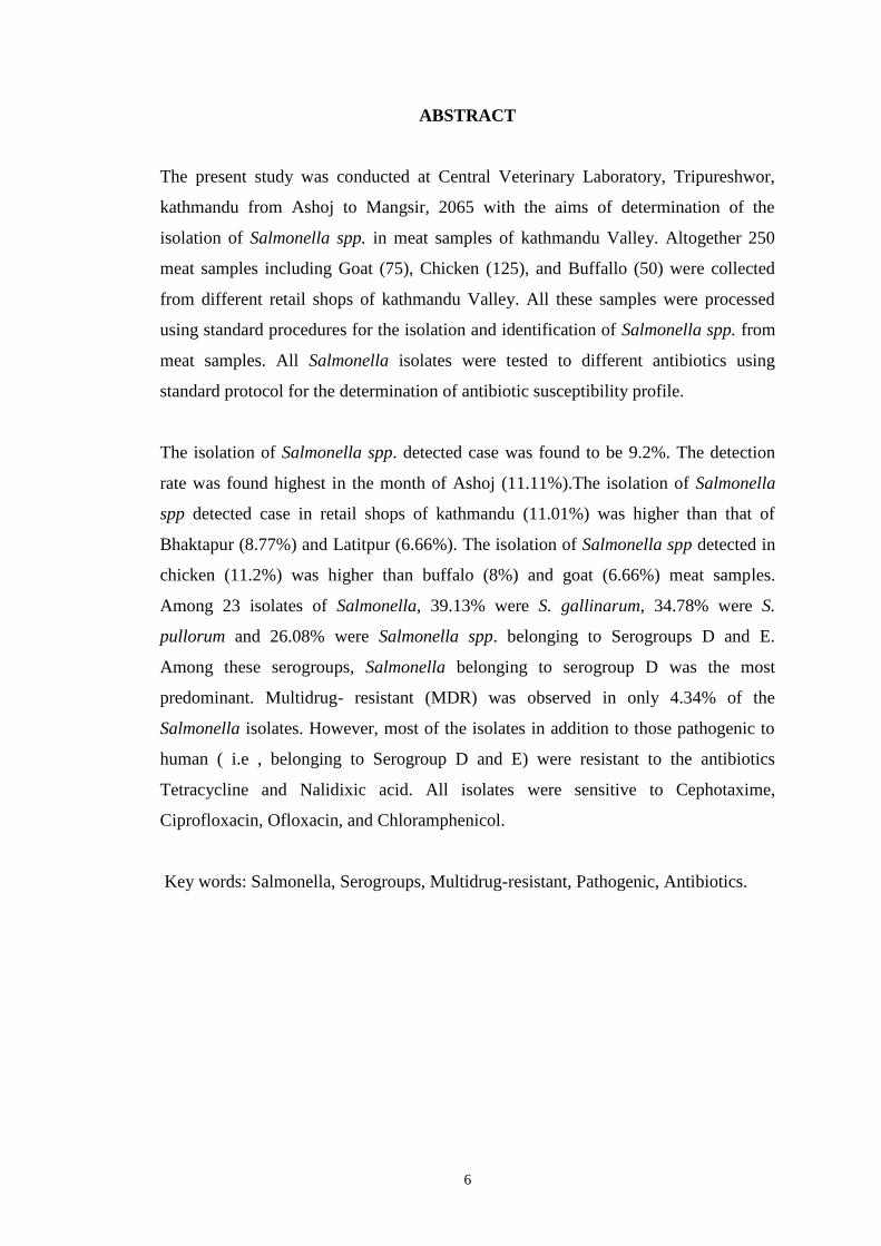
ABSTRACT
The present study was conducted at Central Veterinary Laboratory, Tripureshwor,
kathmandu from Ashoj to Mangsir, 2065 with the aims of determination of the
isolation of Salmonella spp. in meat samples of kathmandu Valley. Altogether 250
meat samples including Goat (75), Chicken (125), and Buffallo (50) were collected
from different retail shops of kathmandu Valley. All these samples were processed
using standard procedures for the isolation and identification of Salmonella spp. from
meat samples. All Salmonella isolates were tested to different antibiotics using
standard protocol for the determination of antibiotic susceptibility profile.
The isolation of Salmonella spp. detected case was found to be 9.2%. The detection
rate was found highest in the month of Ashoj (11.11%).The isolation of Salmonella
spp detected case in retail shops of kathmandu (11.01%) was higher than that of
Bhaktapur (8.77%) and Latitpur (6.66%). The isolation of Salmonella spp detected in
chicken (11.2%) was higher than buffalo (8%) and goat (6.66%) meat samples.
Among 23 isolates of Salmonella, 39.13% were S. gallinarum, 34.78% were S.
pullorum and 26.08% were Salmonella spp. belonging to Serogroups D and E.
Among these serogroups, Salmonella belonging to serogroup D was the most
predominant. Multidrug- resistant (MDR) was observed in only 4.34% of the
Salmonella isolates. However, most of the isolates in addition to those pathogenic to
human ( i.e , belonging to Serogroup D and E) were resistant to the antibiotics
Tetracycline and Nalidixic acid. All isolates were sensitive to Cephotaxime,
Ciprofloxacin, Ofloxacin, and Chloramphenicol.
Key words: Salmonella, Serogroups, Multidrug-resistant, Pathogenic, Antibiotics.
7
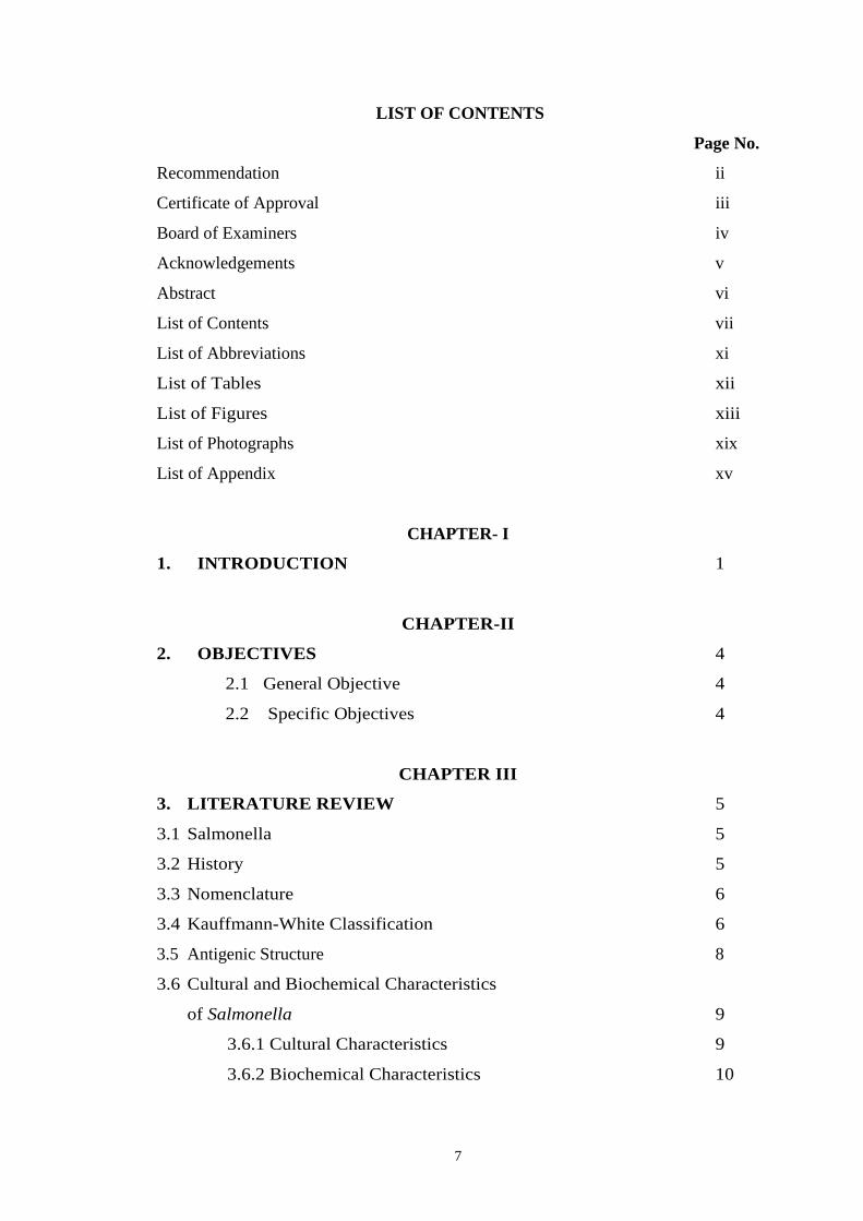
LIST OF CONTENTS
Page No.
Recommendation ii
Certificate of Approval iii
Board of Examiners iv
Acknowledgements v
Abstract vi
List of Contents vii
List of Abbreviations xi
List of Tables xii
List of Figures xiii
List of Photographs xix
List of Appendix xv
CHAPTER- I
1. INTRODUCTION 1
CHAPTER-II
2. OBJECTIVES 4
2.1 General Objective 4
2.2 Specific Objectives 4
CHAPTER III
3. LITERATURE REVIEW 5
3.1 Salmonella 5
3.2 History 5
3.3 Nomenclature 6
3.4 Kauffmann-White Classification 6
3.5 Antigenic Structure 8
3.6 Cultural and Biochemical Characteristics
of Salmonella 9
3.6.1 Cultural Characteristics 9
3.6.2 Biochemical Characteristics 10
8
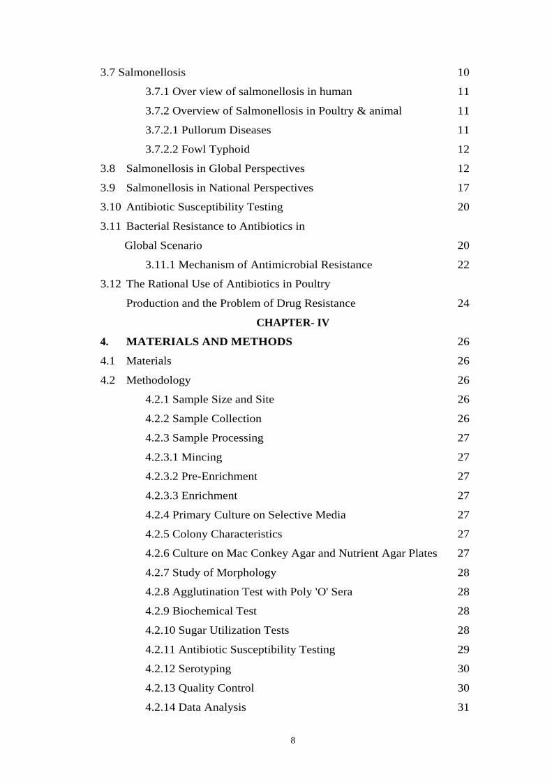
3.7 Salmonellosis 10
3.7.1 Over view of salmonellosis in human 11
3.7.2 Overview of Salmonellosis in Poultry & animal 11
3.7.2.1 Pullorum Diseases 11
3.7.2.2 Fowl Typhoid 12
3.8 Salmonellosis in Global Perspectives 12
3.9 Salmonellosis in National Perspectives 17
3.10 Antibiotic Susceptibility Testing 20
3.11 Bacterial Resistance to Antibiotics in
Global Scenario 20
3.11.1 Mechanism of Antimicrobial Resistance 22
3.12 The Rational Use of Antibiotics in Poultry
Production and the Problem of Drug Resistance 24
CHAPTER- IV
4. MATERIALS AND METHODS 26
4.1 Materials 26
4.2 Methodology 26
4.2.1 Sample Size and Site 26
4.2.2 Sample Collection 26
4.2.3 Sample Processing 27
4.2.3.1 Mincing 27
4.2.3.2 Pre-Enrichment 27
4.2.3.3 Enrichment 27
4.2.4 Primary Culture on Selective Media 27
4.2.5 Colony Characteristics 27
4.2.6 Culture on Mac Conkey Agar and Nutrient Agar Plates 27
4.2.7 Study of Morphology 28
4.2.8 Agglutination Test with Poly 'O' Sera 28
4.2.9 Biochemical Test 28
4.2.10 Sugar Utilization Tests 28
4.2.11 Antibiotic Susceptibility Testing 29
4.2.12 Serotyping 30
4.2.13 Quality Control 30
4.2.14 Data Analysis 31
9
CHAPTER – V
5. RESULTS 32
5.1 Colony Characteristics of Isolates on XLD 32
5.2 Colony Characteristics of Isolates on NA 32
5.3 Morphology 32
5.4 Motility (Hanging Drop Method) 32
5.5 Agglutination Tests with Poly 'O' Sera 33
5.6 Biochemical Tests 33
5.6.1 Catalase and Oxidase Tests 33
5.6.2 Other Biochemical Tests 33
5.6.3 Sugar Fermentation Tests 33
5.7 Monthly Isolation of Salmonella Species 34
5.8 Animal-wise Isolation of Salmonella Species 35
5.9: Location Wise Distribution of Salmonella Spp in
Meat Samples 36
5.10 Species Wise Distribution of Salmonella 38
5.11 Distribution of Different Species of Salmonella IN
Different Animals 39
5.12 Antibiotic Susceptibility Pattern on Salmonella
Isolated From Meat 40
5.13 Antibiotic Susceptibility Pattern of Salmonella
Gallinarum 41
5.14 Antibiotic Susceptibility Pattern of Salmonella
Pullorum 42
5.15 Antibiotic Susceptibility Pattern of Other
Salmonella Species 43
5.16 Serotyping of Salmonella SPP. 43
5.17 Analysis of MDR Isolates 44
10
CHAPTER-VI
6.1 Discussion 45
6.2 Conclusion 50
CHAPTER-VII
SUMMARY AND RECOMMENDATIONS 51
7.1 Summary 51
7.2 Recommendations 52
REFERENCES 53
APPENDICES i-xx
11
LIST OF TABLES
Page No.
Table 1: Antigenic Formulae of some representative serotype of Salmonella
(Kauffmann-White classification) 7
Table 2: Sugar Utilization Tests of some species of Salmonella 29
Table 3: Colony characteristics of Salmonella spp on XLD media. 32
Table 4: Colony characteristics of Salmonella spp. on NA media 32
Table 5: Biochemical tests performed for the identification of isolates 33
Table 6: Sugar fermentation tests performed for the identification of
Salmonella isolates 33
Table 7: Monthly isolation of Salmonella spp in meat samples of
Kathmandu Valley 34
Table 8: Animal wise isolation of Salmonella spp in meat samples of
Kathmandu Valley 35
Table 9: Distribution of Salmonella in meat samples of different localities
of Kathmandu Valley 37
Table10: Isolation of different species of Salmonella in meat samples of
Kathmandu Valley 38
Table 11: Species & animal wise isolation of Salmonella in meat samples
of Kathmandu Valley 40
Table 12: Antibiotic susceptibility pattern of Salmonella 41
Table 13: Antibiotic susceptibility pattern of Salmonella gallinarum 42
Table 14: Antibiotic susceptibility pattern of Salmonella pullorum 42
Table 15: Antibiotic susceptibility pattern of other Salmonella spp. 43
Table 16: Sero-typing of Salmonella spp. 44
Table 17: Resistance pattern and distribution of MDR strains of
Salmonella spp. 44
12
LIST OF FIGURES
Page No.
Figure 1: Total meat sample collected in different months of
Kathmandu Valley 34
Figure 2: Monthly isolation of Salmonella spp in meat samples of
Kathmandu Valley 35
Figure 3: Total meat samples collected from different animals in
Kathmandu Valley 36
Figure 4: Animal wise isolation of Salmonella spp in meat sample of
Kathmandu Valley 36
Figure 5: Total meat sample collected from different cities of
Kathmandu Valley 37
Figure 6: Location wise distribution of Salmonella spp in meat samples
collected in Kathmandu Valley 38
Figure 7: Total positive cases of Salmonella in meat sample of
Kathmandu Valley 39
Figure 8: Isolation of different Salmonella spp in meat samples of
Kathmandu Valley 39
Figure 9: Species & animal wise isolation of different Salmonella
spp in different meat samples of Kathmandu Valley 40
13
LIST OF PHOTOGRAPHS
Photograph 1: Culture of Salmonella (Serogroup D or E) on XLD agar plate
Photograph 2: Pure culture of Salmonella gallinarum on XLD agar plate.
Photograph 3: Agglutination of Salmonella spp. with poly 'O' sera.
Photograph 4: Biochemical test of Salmonella spp. (Serogroup D or E)
Photograph 5: Biochemical tests of S. gallinarum
Photograph 6: Antibiotic susceptibility test of S. gallinarum (MDR strain)
Photograph 7: Investigator processing the sample.
14
LIST OF APPENDICES
Page No.
APPENDIX-I: List of equipments and materials used during the study i
APPENDIX-II: iii
A. Composition and preparation of different culture media iii
B Composition and preparation of different biochemical tests media vi
C. Composition and preparation of different staining and tests reagents viii
APPENDIX-III:
Gram-stain procedure xii
APPENDIX-IV: Methodology of biochemical tests used for identification
of bacteria xiii
APPENDIX -V: Zone size interpretative chart xviii
APPENDIX - VI: Data analysis (chi-square test) xix
15
LIST OF ABBREVIATIONS
A/A : Acid / Acid
Ado : Adonitol
Alk/A : Alkali / Acid
Ara : Arabinose
Buff : Buffalo
CVL : Central Veterinary Laboratory
Dul : Dulcitol
d : 16-84% Strains Positive
Hrs : Hours
Ino : Inositol
Lac : lactose
MA : Mac Conkey Agar
Man : Mannitol
MDR : Multi drug Resistant
Min : Minutes
ml : Milliliters
MR : Methyl Red
NA : Nutrient Agar
No : Number
SIM : Sulphide Indole Motility
Tre : Trehalose
TSI : Triple Sugar Iron
UK : United Kingdom
US : United States
Vp : Voges Proskauer
WHO : World Health Organization
(w) : Relatively Delayed or Work
XLD : Xylose Lysine Deoxycholate Agar
Xyl : Xylose
16
CHAPTER- I
1. INTRODUCTION
The term Meat is widely used to define flesh and offal including their natural
associates skin and gristle, derived from carcass of any animal and bird
normally used for human consumption. A meat product is defined as any food,
which consists of meat or of which meat is an ingredient (Robinson, 2001).
Meat is a good source of protein, vitamins essential fatty acids and minerals
like Zinc, Iron, Calcium, Phosphorous, etc. (Frazier and Westhoff, 1978).
Meat is an ideal culture medium for the growth of various microorganisms as it
is rich in nitrogenous foods of various degrees of complexity, plentifully
supplied with minerals and accessory growth factors, high in moisture (76.78%
to 77.94%), protein (17.49% to 18.42%), lipid (0.47% to 0.82%), phospholipids
(147.34 mg% to 206.1 mg%), total cholesterol (28.66 mg% to 34.32 mg%) and
abundantly supplied with minerals and accessory growth factors, usually has
some fermentable carbohydrates (glycogen). Lastly, it has a favorable pH (5.7 to
7.2) for most microorganisms and is classified as low-acid foods, when
classified on the basis of acidity (Frazier and Westhoff, 1978).
According to William C. Frazier and Dennis C. Westhoff 1993, the healthy
inner flesh of meats has been reported to contain few or no organisms, though
they have been found in Lymph nodes, bone marrow, and even flesh. The
carcass of healthy animal slaughtered for meat and held in a refrigerated room
is likely to have only nominal surface contamination while the inner tissues are
sterile. Fresh meat cut from the chilled carcass has its surface contaminated
with micro-organisms characteristic of the environment and the implements
(saws or knives) used to cut meat. Each new surface of meat, resulting from a
new cut, adds more microorganisms to the exposed tissue. Staphylococcus spp,
Streptococcus spp, Clostridium spp, and Salmonella spp have been isolated
from the lymph node of red meat animals. Normal slaughtering practices would
remove the lymph node from the edible parts. The important contamination,
17
however, comes from external sources during bleeding, handling and
processing.
In Nepal, lack of appropriate slaughtering facilities and unsatisfactory
slaughtering techniques are causing unnecessary losses in meat as well as its
invaluable by products. Animals particularly buffaloes are slaughtered in
Kathmandu, in slaughtering places which are frequently polluted with street
dust, garbage, human excreta, animal blood, intestinal contents and dirty
effluents and which are not protected against dogs, rodents and insects. Meat
products under such conditions are generally deteriorated due to the bacterial
infections and which causes food poisoning time to time. Due to lack of meat
inspection act and in absence of meat inspection, meat from unhealthy or
parasitic infected animals may be source for infection and spreading diseases to
human as well as animals. Besides meat quality is adversely affected by
careless handling conditions in the slaughtering places as well as in the meat
markets or shops (Joshi D. D. 1991).
Food borne diseases caused by non-typhoid Salmonella represents an important
public health problem worldwide. In most part of the world, S. typhimurium is
the most common species. Some others are S. enteritidis, S. haldar, S.
Heidelberg, S. agona, S. Virchow, S. newport and S. anatum (Ananthanarayan
and Paniker, 2000). Salmonellosis in humans is generally contracted through
the consumption of contaminated food of animal origin mainly meat, poultry
eggs & milk, although many other foods including green vegetables
contaminated from manure have been implicated in its transmission (WHO,
2004).
Multi-drug resistant (MDR) strains of Salmonella are now encountered
frequently and the cases of MDR have increased considerably in recent years. It
is reported that some strains of Salmonella have developed MDR (WHO,
2004). Salmonella gallinarum, the causative agent of fowl typhoid are resistant
to antibiotics- penicillin, ampicillin, tetracycline and Cloxacillin (NARC,
2053/54).

18
Several reports have been published on microbiology of meat from different
parts of the world with different organism pattern. However we do not have
such type of enough information regarding meat microbiology of Nepal. So, it
is hoped that, this research work would be informative and helpful for both
planners, policy-makers and also those who are interested to know about
microbiology quality of meat.
19
CHAPTER-II
2. OBJECTIVES
2.1 GENERAL OBJECTIVE
To isolate Salmonella species from different meat samples of
Kathmandu Valley
2.2 SPECIFIC OBJECTIVES
To isolate and identify Salmonella species from different meat samples
of different retail shops of Kathmandu Valley.
To isolate different sero groups of Salmonella.
Antibiotic Sensitivity testing of the isolates of Salmonella by disc
diffusion method.
20
CHAPTER III
3. LITERATURE REVIEW
3.1 SALMONELLA
Salmonella is one of the important pathogenic members of family
“Enterobacteriaceae” responsible for causing various infections and food
poisoning in human and animals. Salmonella species are gram negative rods
with size of 2-4x6µm. they are non-acid fast, non capsulated and non spore
forming. Most serotypes are motile with peritrichous flagella. These non-motile
bacteria are susceptible to cause infection in poultry birds with high mortality
rate. Salmonella species are aerogenic, non lactose fermenter, urease negative,
indole negative, methyl red positive, Voges-Proskauer negative and citrate
variable (Mackie and Mc Cartney 1989).
Several species of Salmonella are pathogenic, some producing mild
gastroenteritis, other producing a severe and often fatal food poisoning, which
is called Salmonellosis. Salmonella grow between 15-45oC, and at pH range of
4 to 8. with the exception of limited number of human host adapted serotypes
(also referred to as the typhoidal Salmonellae), the members of the genus
Salmonella are regarded as zoonotic or potentially zoonotic (Acha and Szyfres,
2001).
3.2 HISTORY
The Salmonella group previously was also called the TPE group, the so called
typhus-paratyphus enteritis group. It comprises the typhus bacillus, Salmonella
typhi, previously called Eberth- Gaffkey bacillus or Eberthela typhi after the
name of two scientists Eberth (1880) observed into the mesenteric lymph node
and spleen of typhoid patient, and Gaffkey (1884) isolated the organism. In
1896, it was demonstrated that the serum from an animal immunized with the
typhoid bacillus agglutinated (clumped) the typhoid bacterial cells, and it was
shown that the serum of patients afflicted with typhoid likewise agglutinated
the typhoid bacillus. Sero diagnosis of typhoid was thus made possible by 1896
(Kenneth,2006).
21
3.3 NOMENCLATURE
The classification of Salmonella has been controversial for many years.
According to the latest nomenclature, which reflects recent advances in
taxonomy, the genus Salmonella consists of only two species. S. enterica and S.
bongori (Le minor and Popoff, 1987). Salmonella enterica is divided into six
subspecies (OIE, 2004). These subspecies are:
Previous nomenclature current nomenclature
Subspecies I Subspecies enterica
Subspecies II Subspecies salamae
Subspecies IIIa Subspecies arizonae
Subspecies IIIb Subspecies diarizonae
Subspecies IV Subspecies houtenae
Subspecies V Subspecies indica
For the serovars of S. bongori, the symbol V was retained to avoid confusion
with the serovars name of S. enterica subspecies enterica. Strains of Salmonella
are classified into serovars on the basis of extensive diversity of
lipopolysaccharides (LPS) antigen (O) flagellar protein antigen (H) in
accordance with the Kauffmann-White scheme, currently approximately 2500
serovars are recognized. The most common serovars that cause infection in
humans and food animals belong to subspecies enterica (OIE, 2004).
3.4 KAUFFMANN-WHITE CLASSIFICATION
This scheme, first developed in 1934, classifies the Salmonella into different O
groups or O sero groups, each of which contains a number of serotypes
possessing a common O antigen not found in other O groups. The O groups
first defined were designated by capital letters A to Z and those discovered later
by number (51-67) of the characteristic O antigen. It is now considered more
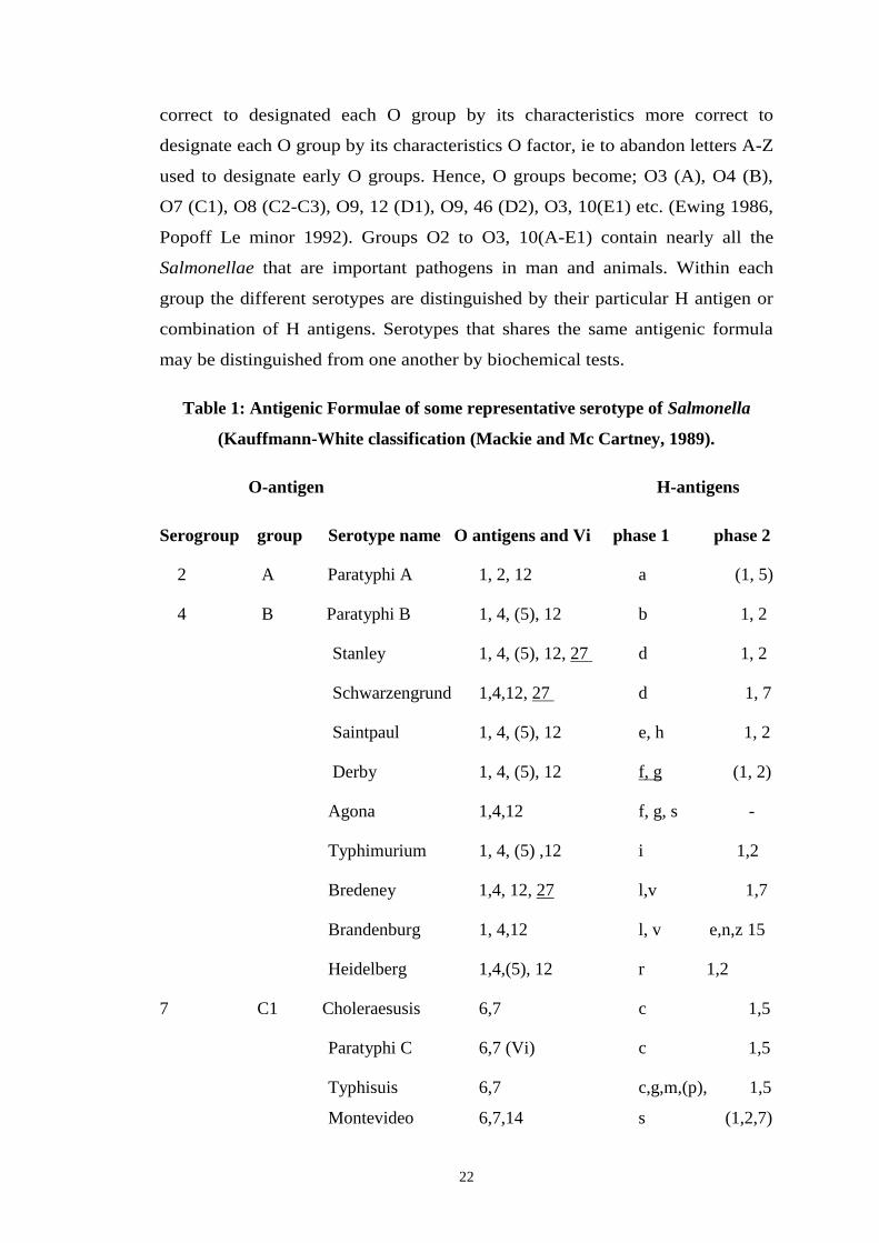
22
correct to designated each O group by its characteristics more correct to
designate each O group by its characteristics O factor, ie to abandon letters A-Z
used to designate early O groups. Hence, O groups become; O3 (A), O4 (B),
O7 (C1), O8 (C2-C3), O9, 12 (D1), O9, 46 (D2), O3, 10(E1) etc. (Ewing 1986,
Popoff Le minor 1992). Groups O2 to O3, 10(A-E1) contain nearly all the
Salmonellae that are important pathogens in man and animals. Within each
group the different serotypes are distinguished by their particular H antigen or
combination of H antigens. Serotypes that shares the same antigenic formula
may be distinguished from one another by biochemical tests.
Table 1: Antigenic Formulae of some representative serotype of Salmonella
(Kauffmann-White classification (Mackie and Mc Cartney, 1989).
O-antigen H-antigens
Serogroup group Serotype name O antigens and Vi phase 1 phase 2
2 A Paratyphi A 1, 2, 12 a (1, 5)
4 B Paratyphi B 1, 4, (5), 12 b 1, 2
Stanley 1, 4, (5), 12, 27 d 1, 2
Schwarzengrund 1,4,12, 27 d 1, 7
Saintpaul 1, 4, (5), 12 e, h 1, 2
Derby 1, 4, (5), 12 f, g (1, 2)
Agona 1,4,12 f, g, s -
Typhimurium 1, 4, (5) ,12 i 1,2
Bredeney 1,4, 12, 27 l,v 1,7
Brandenburg 1, 4,12 l, v e,n,z 15
Heidelberg 1,4,(5), 12 r 1,2
7 C1 Choleraesusis 6,7 c 1,5
Paratyphi C 6,7 (Vi) c 1,5
Typhisuis 6,7 c,g,m,(p), 1,5
Montevideo 6,7,14 s (1,2,7)
23
Thompson 6,7,14 k 1,5
Virchow 6,7 r 1,2
Infantis 6,7,14 r 1,5
Mbandaka 6,7,14 z10 e,n,z15
8 C2-C3 Muenchen 6,8 d 1,2
Newport 6,8,20 e,h 1,2
Hadar 6,8 z10 e,n,x
Miami 1,9,12 a 1,5
Sendai 1,9,12 a 1,5
9 D1 Typhi 9,12(Vi) d -
Enteritidis 1,9,12 g,m (1,7)
Dublin 1,9,12(Vi) g,p -
Panama 1,9,12 l, v 1,5
Gallinarum 1,9,12 - -
3, 10 E1 Anatum 3,10,(15),(15,34) e,h 1,6
Weltevreden 3,10,(15) r z6
1,3,19 E4 Sentfenberg 1,3,19 g,(s),t -
11 F Rubislaw 11 r e,n,x
13 G Kedougou 1,13,23 i l,w
Note: Somatice factors associated with phage conversion are underlined.
Antigens in brackets [X] are not always present.
3.5 ANTIGENIC STRUCTURE
In the Kauffmann-White classification the genus Salmonella is subdivided into more
than 2300 serotypes containing different combinations of antigen (Mackie and Mc
Cartney, 1996)
24
Somatic (O) or Cell Wall Antigen
These somatic antigen represent the side chains of repeating sugar projecting outward
from the lipopolysaccharide layer on the surface of bacterial cell wall. Over
60different antigen have been recognized and they are designated by Arabic
numerals. O antigens are heat stable being unaffected by heating for 2.5 hours
at 100oC and alcohol stable (Mackie and Mc Cartney, 1996).
H-antigen/ Flagellar antigen
H-antigen is flagellar antigen. Many Salmonella are diphasic that is they can
occur in two antigenic forms referred as Phase I and Phase II. Phase I antigens
are either numbered or given a letter if known to occur in both phases. The
definitive identification of a diphasic Salmonella always requires the
identification of the H-antigens of both phases.
Surface (K or Vi) antigen
Vi antigen is surface antigen and helpful in phase typing of S. typhi, S.
paratyphi C and S. dublins as these serotype possess this antigen (Bergey’s
manual 1990).
“O” and “Vi” agglutinins produce fine granular agglutination while H
agglutinin bring about a large flocculent agglutination.
3.6 CULTURAL AND BIOCHEMICAL CHARACTERISTICS OF
SALMONELLA
3.6.1 Cultural Characteristics
Salmonella is member of Enterobacteriaceae group, therefore it follows all the
characters common to Enterobacteria ie rod shape, aerobic, facultative
anaerobic, catalase +ve, oxidase –ve, production of gas by carbohydrate break
down etc. in nutrient and blood agar, colonies of most strains are moderately
large (e.g 2-3 mm in diameter), grey-white, moist, circular discs with a smooth
convex surface and entire edge. Their size and degree of opacity varies with the
serotype (Collee et al, 1996).
25
The organism are aerobes, the optimum temperature for growth is 37oC. They
grow readily on ordinary nutrient media. Grow as non-lactose fermentative
colonies on Mac Conkey Agar. In selective media like Bismuth Sulfite Agar
colonies of Salmonella are metallic black in color with 1-2 mm diameter size
while in XLD, colonies are black centered with 1-2mm diameter in size
(Banwart JG 1987).
3.6.2 Biochemical Characteristics
In general, Salmonella is catalase +ve, oxidase –ve, produce gas from glucose
at 37oC. produce acid from carbohydrate breakdown ie sugars like Dulcitol,
Arabinose, Maltose, Trehalose, Xylose, etc. with variation in reaction according
to different types, use citrate as carbon sources, MR +ve, Vp –ve. Protein
reactions shown by Salmonella are –ve gelatin hydrolysis, -ve indole reaction, -
ve urea hydrolyzation, production of H2S in TSI etc. (Joshi V, 2003).
Few biochemical characteristics are different between S. pullorum and S.
gallinarum (Christensen et al, 1992). Both of them ferment arabinose, dextrose,
galactose, mannitol, mannose, rhamnose and xylose to produce acid with or
without gas. S. gallinarum decarboxylates ornithine, whereas S. gallinarum
does not (Shiva Prasad, 1997). S. pullorum and S. gallinarum produce a red
slant with a yellow butt and shows delayed blackening from H2S production. S.
gallinarum does not form gas in triple sugar iron agar, but S. pullorum may
show weak gas production (Doughlas et al, 1998).
3.7 SALMONELLOSIS
Salmonellosis is a diseases caused by the ingestion of Salmonella spp by human
or animals.
Following ingestion, symptoms may or may not develop depending upon the
dose, level, serotype and Virulence of the strain and host resistance (Bryan,
1968). Salmonella species is the potent organism that causes several losses in
poultry by infecting with several disease .S. pullorum diseases and fowl typhoid
is the two specific manifestations of Salmonellosis caused by non-motile
bacteria, Salmonella pullorum and Salmonella gallinarum.
26
A broad group of diseases called paratyphoid infections are also caused by
Salmonella organism that are motile. The micro organisms are highly prevalent
in human excretions, poultry meat, pork and beef origin meat. Probably these
are the major source of Salmonellosis problem in human and poultry.
3.7.1 Over view of salmonellosis in human
Salmonellosis in humans is generally contracted through the consumption of
contaminated food of animal origin (mainly meat, poultry, egg & milk),
although many other foods, including green vegetables contaminated from
manure, have been implicated in its transmission. The causative organisms pass
through the food chain from primary production to households or food-service
establishments and institutions. In addition to acquiring infection from
contaminated food, human cases have also occurred where individuals have had
contact with infected animals, including domestic animals such as cats & dogs.
Domestic animals probably acquire the infection in the same ways as humans,
i.e through consumption of contaminated raw meat, poultry or poultry-derived
products. The evolution of specific Salmonella serotypes in intensive animal
husbandry and subsequently in humans has been observed over the past three
decades. S. enteritidis caused the most recent epidemic, which peaked in
humans in 1992 in many European countries. Its current slight decline sets the
scene for re-emergence of S. typhimurim as the most important serotype in
human Salmonellosis. Another possible scenario is that these two particular
strains with epidemic potential will dominate in many countries in the
Foreseeable Future (WHO, 2004).
3.7.2 Overview of Salmonellosis in Poultry & animal
3.7.2.1 Pullorum Diseases
It is an acute or chronic infection bacterial disease affecting primarily chickens
and turkeys but most domestic and wild fowl can be infected as well. The
disease is caused by the bacteria Salmonella pullorum, non-motile bacteria.
Most outbreaks of acute pullorum diseases in chicken result from infection
while they are in the hatchery. Pullorum disease is highly fatal to young chicks
27
or adults but may die soon after hatching without exhibiting any observable
signs.
Mortality rate in such outbreaks may approach 90% if untreated. Survivors are
usually stunted and unthrifty. Infection in young birds may be indicated by
droopiness, ruffled feathers, a child appearance with birds huddling near a
source of heat, labored, breathing difficulties and presence of a white diarrhoea
with a “pested-down” appearance around the vent white diarrhoea symptoms
instigated the term “Bacillary White Diarrhoea” (Pandey, 2003).
3.7.2.2 Fowl Typhoid
The disease is caused by the non-motile bacterium Salmonella gallinarum.
Hence, Salmonella gallinarum is attributed to septicemia of acute or chronic in
chicken (Pandey et al 1991). The poultry bird infected from the fowl typhoid
shows the similar symptoms to the pullorum disease. Post-mortem finding of
the liver, kidneys and spleen may be enlarged and discolored.
Fowl typhoid has increased in most part of the world. In areas where the disease
is common, it has become one of the most important diseases in poultry (Silva
et al, 1981). Fowl typhoid is endemic in Nepal. The mortality rate due to the
disease may vary from moderate to very high. Although, Salmonella
gallinarum is pathogenic to both chicks and adults under natural condition,
virulence of the isolates may vary depending upon strain variation environment
situation, individual susceptibility and cultural condition of the isolates.
3.8 SALMONELLOSIS IN GLOBAL PERSPECTIVES
From the data obtained from US department of agricultural, food safety and
inspection service (FSIS) S. enterica serotype enteritidis in broiler chicken
carcass rinses collected from 2000 to 2005 showed that the annual number of
isolates increased > 4 fold and the proportion of chicken slaughter
establishment with S. enteritidis positive rinses increased nearly 3 fold. During
the six year study period, 280 (0.5%) Salmonella entritidis were recovered from
51, 327 broiler rinses. The number of establishment testing positive increased
28
from 17 (9%) of 197 in 2000 to 47 (25% of 187 in 2005. the most predominant
phage type in broiler chickens and in the chicken slaughter establishment with
S. enteritidis positive rinses were PT8 and PT13 (Sean et al, 2006).
A case study on Salmonella outbreaks in a middle town of USA reported that
125 cases of food borne Salmonellosis resulted from cross contaminated food
items served in a picnic. 125 people out of 175 people who attended the picnic
developed diarrhoea after having smorgasbord prepared by a bar restaurant. The
stool culture report showed the presence of S. infantis, S. agona, S.
schwarzengruna. The incidence took place in Minnisota USA in September
1973 (Levy et al, 1975).
Mohamed Osama N. et al, (2001) conducted an experimental and surveillance
study for the diagnosis of Salmonellosis in meat of slaughtered animal. The
study evaluated meat juice as a sample from infected mice with S. typhimurium
for detection of anti Salmonella antibody by ELISA techniques. Studied
showed that meat juice as efficient as blood in diagnosis of Salmonellosis. The
method can be applied efficiently for surveillance study for screening programs.
A study was carried out regarding the Salmonella incidence in minced meat
produced in a European Union approved slaughtering and cutting plant.
Throughout 21 mounts, 297 pool samples (1,485 individual samples) of mixed
minced meat (beef and pork) were examined according to council directive
94/65/EC and to ISO 6579. Salmonella were detected in 47 (15.8%) of the pool
samples. After separation of the positive pools, 93 individual samples were
determined to be Salmonella positive, representing 6.3% of the total 1,485
samples. Serotype resulted in most isolates (69.9%) being identified as
Salmonella typhimurium (Stock and Andreas, 2001).
Victoria Atanassova (2002) recorded the isolation of Salmonella spp. in foreign
chicken meat from different European countries. During the study period of Feb
2000 to March 2001, a total of 2006 samples of frozen chicken meat were
examined for isolation of Salmonella spp using classical cultural detection as
well as RFLP_PCR techniques. 15.7% ie 453 samples were found to
contaminate with Salmonella spp of all isolates, 199 were characteristics as
29
S. enteritidis (43.9%) 112 isolates as Salmonella hador (24.7%) 78 isolates as
S. typhimurium (17.2%) and 64 isolates belonged to other Salmonella spp.
Berchier et al (1987) reported that the presence of Salmonella at slaughter time
in a poultry processing plant, industries water, scaled tank water, pre-chilled
water, defeathered carcass, finished carcass, poultry meal and feces of living
birds were investigated. Salmonella spp. were not isolated from industrial water
but they were isolated from all other material of 9 serotypes detected S.
typhimurium was predominant (Prasai, 2000).
Szazades et al 1996 investigated the evaluation of Salmonella spp. in a
slaughter house laboratory during 17 years. During the last 17 years (1978 to
1994), 3261 Salmonella strains were isolated by an enrichment method from
84,349 samples originated from the slaughter house and from the territory of
the country. 3.87% of the samples proved to be positive. The isolated
Salmonella spp. belonged into 52 serotypes. Positive Salmonella samples were
classified into the following 8 sample groups (1): Salmonella spp. were found
in 0.91% in the samples of the complementary bacteriological examination. In
spite of the lower incidence, the food safety importance of this sample group is
constant. (2): phase investigation of swab samples was positive in 9.83% of the
cases. Since, 1989 the sampling has been carried out according to the HACCP
(Hazard Analysis and Critical Control Point) system. Higher number of positive
samples could be traced back to the deliberate exploration of the possible places
of Salmonella incidence, (3): important role has been attached to the
examination of frying savage in case of clearing up the hygienic situation in a
production unit, similar to the phase investigation. (4): results of the mass of
products preserved by maturation, drying and party by starter cultures
(sausages, salami, and other “dry products”) and (5): finished products indicate
the changes in the incidence of Salmonella spp. during processing of products.
Inspite of the 11.92% incidences of Salmonella spp. in the mass samples, that
of finished products was 2.19%. (6): of the 19,63. heat treated meat products
only two samples (0.01%) contained Salmonella spp. which indicates a high
level product safety because an appropriate heat treatment means practically an
insuperable obstacle for the Salmonella. (7): 15.93% Salmonella spp.
30
contamination of poultry slaughter house samples indicates an important food
hygienic problem. (8): 4.45% of the regional samples (territory of the country)
were Salmonella spp positive of them. “Flamed sausage” examined in the
previous years, was an important group, its Salmonella spp. contamination was
8.78% indicating the important food safety problems of this product. In the
samples, most frequently the incidence of Salmonella infantis (17.30%),
Salmonella derby (13.46%), Salmonella typhimurium (9.69%), as well as
Salmonella anatum (6.16%), Salmonella Heidelberg (5.61%), Salmonella
agona (4.88%), Salmonella cholerasesuis (4.63%), Salmonella enteritidis
(3.74%), Salmonella virchow (3.71%), Salmonella hadar (3.34%), and
Salmonella bredeney (3.13%) were observed.
Bok et al, (1986) studied on the incidence of food borne pathogens on retail
broilers. They found that out of total 718 isolates 158 isolates were identified as
potential human pathogens. Salmonella was the major pathogenic isolate (86%)
of total pathogens, 136 isolates.
Cross contamination of food is one of the 10 main factors that contribute to
food borne illness. People handling both cooked and raw foods can transfer
microorganisms from the raw to cooked product. With no further treatment, this
is a potential health hazard. The housewife who cuts up raw vegetables for
salad may transfer Salmonella spp. from the raw chicken to the raw vegetables.
The meat department in a retail store may use the same knife and block to cut
fish, cold meat, chicken, beef and other types of food. It is evident that a
potential health hazard can result (Banwart, 1987).
Tellez et al, (2001) reported about Salmonella enteritidis colonization in the
intestinal tract of chicken causing food borne illness in humans and reduction of
S. enteritidis colonization in the intestinal tract of chicken can reduce potential
carcass contamination during slaughter.
Sansoni L. et al (2001) isolated Salmonella spp. from human and environment
sources in province of Verona in a 2-year study i.e. between 1996-1997. The
data collected revealed the widespread presence of Salmonella spp. in water
sources. They also found the putative role of water sources in human infection
31
in the province of Verona confirmed by the fact that main serotype in
circulation both in river and in man are same. Findings also showed high
isolation in coinciding in both water and human cases within summer autumn
period.
Morgan-Jones (1980) reported the occurrence of Salmonella during the rearing
of broiler birds. They found that Salmonella were not isolated from empty
cleaned and fumigated houses and only on one occasion from feed. Salmonella
were isolated from the environment of the chicks and spasmodically from litter
water troughs and dust. They concluded that water in water troughs rather the
food, appeared to be the major oral route of infection or re-infection of birds
during rearing (Karki, 1995).
Salmonellosis infections were 55.1% of the reported food borne disease cases
reported from 1993 to 1996 in Korea (Bajk and Roh, 1998).
Of the reported food borne outbreaks in Europe caused by an identical agent,
more than one third were confirmed to be caused by S. enteritidis. Food
associated with S. enteritidis outbreaks include egg and egg products (68.2%),
cake and ice creams (8%) and poultry and poultry products (3%). Other
vehicles include meat and meat products (4%), mixed foods (4%), fish and shell
fish (2%), and milk and milk products (3%). In S. typhimurium outbreaks, eggs
products (39%) meat and meat products (10%) were reported as the vehicles of
infection. A large no of other Salmonella serotypes were also involved in an
outbreak in Europe, but specific serotypes were not reported (WHO, 2001).
A total of 825 samples of retail raw meats (chicken, turkey, pork and beef) were
examined for the presence of E. coli and Salmonella serovars. The samples
were randomly obtained from 59 stores of four supermarkets in the Washington
D.C. from June 1999 to July 2000. Of the 212 chicken samples, 82 (38.7%)
yielded E. coli, while 19% of the beef samples, 16.3% of the pork samples and
11.9% of the turkey samples were positive for E. coli. Only 25 (3%) of the
retail meat samples tested were positive for Salmonella (Zhao et al, 2001).
32
3.9 SALMONELLOSIS IN NATIONAL PERSPECTIVES
In Nepal, Salmonellosis has caused a burning problem for poultry raiser as well
as for consumers. More than 2,200 serovar of Salmonella has been identified so
far all over the world and few of them cause disease in poultry e.g. S. pullorum,
S. gallinarum, S. enteritidis, S. typhimurium, S. typhi, S. arizone, S. anatis, & S.
Virchow, and some cause food poisoning in human eg. S.enteritidis, S.
typhimurium, S. Virchow . Salmonellosis, a zoonotic and egg born disease has
great importance as most of the chickens are distributed from recent established
hatcheries or brought from neighbouring countries which play significant role
in disease transmission in the country. (Prasai 2000).
Joshi (1991) studied the current practices of livestock slaughtering and meat
marketing in Kathmandu, Lalitpur and Bhaktapur. He reported that total buffalo
slaughtering places of kathmandu, Lalitpur and Bhaktapur are 81, 22 and 20
respectively and total poultry slaughtering places of kathmandu, Lalitpur and
Bhaktapur are 107, 30 and 5 respectively. He also reported that total number of
buffalo meat shops in Kathmandu, Lalitpur and Bhaktapur are 181, 39 and 48
respectively and total poultry meat shops in Kathmandu, Lalitpur and
Bhaktapur are 115, 30 and 13 respectively. In this study, he also reported that
the average number of buffaloes slaughtered per day in Kathmandu, Lalitpur
and Bhaktapur are 324, 88 and 80 respectively and average number of poultry
slaughtered per day in Kathmandu, Lalitpur and Bhaktapur are 1070, 300 and
150 respectively. In this study, total 111 butchers were interviewed about their
knowledge of meat borne diseases. On an average, 35.1% had the knowledge of
meat borne and 64.9% had no knowledge about it.
Different samples of the poultry were studied and bacterial analysis was done.
Salmonella spp. was found to be the most predominant organism followed by
E. coli, Staphylococcus aureus and Streptococcus spp (annual report of NARC
in the fiscal year 2042/2043).
According to the annual report of NARC in the fiscal year 2044/2045, E. coli
was found to be the most predominant organisms followed by enterobacter,
33
Pasteurella spp and Citrobacter spp from the different samples of the goats.
Besides Pasteurella and enterobacter spp were reported to be the predominated
from the cow and buffalo samples. Klebsiella, Streptococcus and Enterobacter
spp were found from the samples of 200 lab animals. In the same year 652
poultry samples were analyzed for the bacterial analysis. From the study it was
reported that Salmonella (31%) was found to be the most predominant
organisms followed by E. coli (30%), Staphylococcus (5%), and enterobacter
(0.6%), serological test for Salmonella spp confirmed that 0.68% were positive
for the pullorum spp.
Two hundred samples were collected from the apparently healthy and sick
birds. Out of hundreds healthy birds, only two were sero positive for
Salmonella infection. Both sero positive birds were slaughtered for the
collection of samples for Salmonella spp culture. Two isolates of S. pullorum
were found in the culture of livers collected from both sero positive cases. On
the other hand, out of one hundred sick birds ten were positive for Salmonella
gallinarum. (Annual report of Nepal Agricultural Research Council, NARC,
fiscal Year 2053/2054).
The problem of Salmonellosis is important not only for chicken raises but also
for public health as far as human cases of enteric fever are concerned. Enteric
fever is an endemic disease in Kathmandu Valley. The disease flares upto an
epidemic proportional from time to time (Malla, FB and Shakya , GM, 1984).
A survey was carried out between June and November 1990 to find out the
isolation of Salmonella in dairy product sold in street of Kathmandu Valley. A
total of 200 samples of various dairy products were collected Salmonella was
isolated from 3% samples. Variety wise 12.5% in ice-cream and 2.1% in sweet
item (Joshi DD et al, 1990).
A survey work carried out to assess the occurrence of micro organism in cheese
collected from retail shop in Kathmandu city market. Among other bacteria
Salmonella typhi was found to be present in 11% of total samples (Tuladhar, E
and Sharma, A.P.,1997).
In annual bulletin of CFRL 1997/98 Salmonella was reported from the raw
34
frozen pork samples in Prasuma Factory during the processing of momo (momo
is an item of mined meat). Salmonella spp was also detected in raw pieces, on
surface meat and in mixing dough. In the same report Salmonella was also
reported from 3 samples of raw momo out of 29 samples.
A general survey of hygienic quality of ethnic Newari meat varieties was
conducted out. Samples were collected from different restaurant of Kathmandu
valley. Coliforms were present in 55% samples. In a variety homed ‘sekuwa’ (
an under cooked meat variety) Salmonella spp was detected in 3% samples, in
‘kachila( raw meat variety) this percentage was 5% while in momo it was 7% (
Shrestha,H.et .al 1999)
Manandhar (2000) recorded the sero-isolation of Salmonella spp, using
ELISA(enzyme linked immunosorbent assay) in human’s and chicken in related
areas of Philippines, total 128 human and 128 chicken serum samples were
collected. Sero isolation ratio was found significantly higher (p<_0.05) for S.
enteritidis in> 40 year age group and for S. abortusequi in early age groups.
There was no difference in sero isolation or exposed and unexposed groups for
S. enteritidis.
According to annual report of NARC (2000/2001) Pradhan A conducted a study
about calf diarrhea with particular reference to E-coli infection 30fecal samples
were collected form calves affected with diarrhea in Kandaghari Gothatar .E-
coli in 66.60% cases and Streptococcus spp, in 10% cases .bacillus spp. In 10%
cases and Salmonella in 6.6% cases were isolated.
In a study conducted at CVL and ADCC(central veterinary laboratory and
animal disease control section) out of 268 post-mortem chicken samples
cultured 259 samples were found to be infected E-coli was made predominant
isolates (73.13%), followed by Salmonella spp, Streptococcus spp (7.08%) and
S, aureus (2.61%). Among Salmonella spp, S. gallinarum and S. pullorlun were
identified, 95.34% of Salmonella spp were sensitive to chloramphenicol while
Ampicillin and cloxacillin were found to be least effective drugs (Pandey,
2003).
35
3.10 ANTIBIOTIC SUSCEPTIBILITY TESTING
Antimicrobial susceptibility testing is an in vitro method for estimating the
activity of drugs which will assist clinician in selecting an antimicrobial drug
effective in inhibiting the growth of an infecting microorganisms in vivo. The
primary goal of antimicrobial susceptibility testing is to determine whether the
bacterial etiology of concern is capable of expressing resistance to the
antimicrobial agents that are posterities choices as therapeutic agents for
managing the infection.
World Health organization (WHO) recommended modified Kirby-Bauer disk
diffusion technique is used by most laboratories to lest routinely for
antimicrobial susceptibility. Using this lest, antimicrobial resistance is detected
by allowing the antibiotics to diffuse from a point source, commonly in the
form of an impregnated filter paper discs, into an agar medium that has been
seeded with the test organism, visible growth of bacteria occurs on the surface
of the agar where the concentration of antibiotic has fallen below its inhibitory
level for the test strain. Following incubation the diameter of the zone of
inhibition around each disc measured in millimeters. (Collee et al,1996).
3.11 BACTERIAL RESISTANCE TO ANTIBIOTICS IN GLOBAL
SCENARIO:
The study on the antibiotic resistance of 3600 strains of Salmonella spp of
animal origin . 98 sero types were identified and the most common 3 sero
types were S. saintpaul, Salmonella typhimurium and S.dublin. The resistance
patterns to various antibiotics Ampicillin, Streptomycin, kanamycin, Neomycin,
Framycetin, Gentamycin Chloramphenicol, Tetracycline and Colistin were
studied. Out of 3624strains, 1025 were found resistant to one or more
antibiotics 509 were resistant to one anitibiotic,218 to two, 117 to three, 92
four, 59 to five, 23 to six, 6 to seven and 1 to eight. Resistant to Tetracycline
was the most common (16% strains were resistant). The incidence of multiple
resistances appeared to be increasing. (Gledel et al 1977).
The study was conducted to estimate the anti microbial resistance of Salmonella
isolated from raw chicken between November 2003 to April 2004. A total of
36
120 chicken carcasses were collected from 36 different sale points and
examined for the presence of Salmonella .Salmonella was isolated from 75
(62.5%) of the examined samples. Out of 90 isolates obtained, 21 seotypes were
identified, the most prevalent being S. kentuchy (13%) S. muenster (133%), S.
bvancaster (8.8%), S. enteritidis (6.6%) and S. hadar (6.6%) . All Salmonella
were tested against 16 selected antimicrobial agents. Out of 71 resistant
isolates, 33 (46.5%) showed multiple resistance to five or more antibiotics.
Reistance to Ampicilin, Trimethoprim, Trimethoprim –sulphamethoxaZole,
Tetracycline and sulphonamides were most frequent. (Rianatou et al, 2006).
Of 200 ground meat samples purchased in the Washington ,D.C area, 41(20%)
contained Salmonella , with a total of 13 serotypes .84% of the isolated were
resistant to at least one antibiotic, and 53% were resistant to at least three
antibiotics.16% of the isolated were resistant to ceftriaxone, the drug of choice
for treating Salmonellosis in children. Bacteriophage typing identified four
isolate of Salmonella enterica serotype typhimurium definitive type
104(DT104),one of DT104b, and two of DT208.Five isolates of S.enterica
serotype agona had resistance to 9 antibiotics, and the two isolate of serotype
typhimurium DT208 were resistant to 12 antibiotics .(White et al,2001)
In a study conducted between 1994-1996, the relative frequency of Salmonella
strains from hospitalized patients in Southern Israel was found to be changed.
The most prevalent isolate was found to be S. typhimurium (DT104) and S.
agona followed by S. enteritidis. In addition to the R-type ACT (i.e Resistance
to Ampicillin ,Chloramphenicol, and Tetracycline) the S. typhimurium (DT104)
possesses a chromosomally encoded resistance to Quinolone drug, Nalidixic
acid .In 1996,27% of the S.typhimurium, DT104 were of R-type ACTN. S.
enteritidis exhibited to over 95% susceptibility to at least 8 of the most
commonly used antibiotic but non of the isolates were resistant to Quinolone
and Fluroquinolone .(Metzer. et al,1998)
Arora etal (1987) Studied on detection of Upper respiratory tract bacterial
carriers in poultry. On bacteriological examination they isolated E.coli,
Klebsiella pneumoniae, Citrobacter intermedius, Citrobacter freundii,
Enterobacter cloacae ,Proteus mirabilis, Pseudomonas aeruginosa and
37
Acenetobacter spp. They found that the isolates were sensitive to gentamycin,
kanamycin ,chloramphenical, furadantin and ampicillin on in vitro sensitivity
test. However, Pseudomonas aeruginosa was resistant to the antibiotics except
for gentamycin and chemotherapeutic agents used .
3.11.1 Mechanism of Antimicrobial Resistance
It is important to note that resistance genes and mechanism existed long before
antibiotics were used. For example, antibiotic resistance bacteria have been
isolated from deep within glaciers in Canada’s high Aretic regions, estimated at
2000 years old. The original source of resistant genes was probably soil
microorganisms, some of which can produce antibiotics in active form and thus
environmental bacteria are exposed.
There are many different mechanism by which microorganism might exhibit
resistance to drugs (Brooks et al, 2004).
1. Microorganism produce enzymes that destroy the active drugs. Examples:
Staphylococci resistant to penicillin G produce a beta- lactamase that
destroys the active drug .Other beta- lactamares are produced by gram
negative rods.
2. Microorganism develop an altered structural target for the drug examples:
Erythromycin-_resistant organism have an altered receptor on the 50’s
subunit of the ribosome, resulting from methylation of a 23’s ribosomal
RNA. Resistance to some penicillins and Cephalosporins may be a function
of the loss or alteration of penicillin binding proteins ( PBPs) .
3. Microorganisms developed and altered metabolic pathway that by passes the
reaction inhibited by the drug. Example: Some Sulphonamide – resistant
bacteria do not require extra cellular para – amino banzoic acid (PABA) but,
like mammalian, cells can utilize performed folic acid.
4. Micro Organism change their permeability to the drug. example: Tetracycline
accumulate in susceptible bacteria but not in resistant bacteria. Streptococci
have a natural permeability barrier to aminoglycosides.
38
5. Micro-Organisms developed and altered enzyme that can still perform it’s
metabolic function but is much less affected by the drug. Example: in
Trimethoprim – resistant bacteria, the dihydrofolic acid reductase is inhibited
for less efficiently than in Trimethoprim – susceptible bacteria.
Multiple drug resistant :
Multi drug resistant Salmonella gallinarum isolated from the broilers were
studied and was considered as a major problem in the poultry birds in Buenos
Aires (Mairorini et al, 1993). White et al. (1997) reported antimicrobial
resistance of Salmonella gallinarun isolates in France. During the multi drug
profile analysis and antibiotics susceptibility testing of endemic strains of multi
resistant Salmonella, it was confirmed that plasmid was resistance of the
isolates in Great Britain ( Evans and Davies 1996)
The multi Drug resistant Salmonella was a potential case of infection in poultry
birds in India (Chaudhary 1996) The isolates of multi drug resistant Salmonella
strains were more prevalent in Tanzania to commercial chicken, than local
chicken (Gyles,1989) Conjugative plasmid mediated multiple drug resistant
Salmonella infections is an emerging problem in Bangladesh (Rahman and
Albert 1998)
R. Factors:
One of the earliest examples was in Japan in 1959. Previously sensitive E. Coli
become resistant to multiple antibiotics through acquisition of a conjugative
plasmid (R-factor) from resistant Salmonella and Shigella isolates. A number of
R-factors have now been characterized including RP4, encoding resistant to
ampicillin, kanamycin, tetracycline and neomycin, found in P. aeruginosa and
other gram negative bacteria, R1, encoding resistance to ampicillin, kanamycin,
sulphonamides, chloramphenicol and streptomycin, found in gram negative
bacteria and PSH6, encoding resistance to gentamicin , trimethoprim and
kanamycin, found in S. aureus .
39
Mobile Gene Cassettes and Integrons:
Many gram negative resistance genes are located in gene cassettes one or more
of these cassettes can be integrated into a specific position on the chromosome
termed as integron. Thus, integrons are genetic elements that recognize and
capture multiple mobile gene cassettes (Smith, 2004). Although integrons by
themselves are not motile, due to their presence in plasmids and transposons,
they can be transferred horizontally.
Chromosomal Multiple Antibiotic Resistance (Mar) Locus:
The multiple-antibiotic resistance (mar) locus was first described in Escherichia
coil by Stuart levy and collogues at TUFts University and has since been
recognized in other enteric bacteria. The locus consists of two divergently
transcribed units, marc, and mar RAB. (Smith, 2004)
3.12 THE RATIONAL USE OF ANTIBIOTICS IN POULTRY
PRODUCTION AND THE PROBLEM OF DRUG RESISTANCE
The increasing use of antibiotics in few decades in the poultry production for
the prevention and treatment of infectious diseases and for growth promotion
has provided an intense selective pressure in favor of genus coding for drug
resistance and consequently resistance is now common in many bacterial
species (Dutta, 1984).
A large amount of drugs are being used globally annually to secure sufficient
quantities of food to feed fast growing population. The use of antibiotics helped
for the fast growth of food animal industry worldwide. Thus, the use of
anitbiotics is considered essential for the efficient production of foods of animal
origin (WHO, 1985).
It is generally accepted that certain antibiotics such as penicillin, tetracycline,
virginiamycin, bambermycin, avoparcin, bacitracin, etc. are capable of
stimulating the growth rate or keep efficiency or both. The indiscriminate use,
however, may lead to the emergence of resistance and residues (Huber, 1971)
40
and alteration of the intestinal micro-flora of exposed individuals (WHO,
1963).
In the United States, many antibiotics are incorporated in food at low levels to
promote growth and protect against diseases. (Tanner, 1993)
In a study Speele (1984) stated that chicken increased up to 12.2% in weight
gain and 7.1% feed efficiency upto 4 weeks of age.
In the United States, more than 40% of all antibiotics produced are used for
animals (Stalheim, 1987). Correspondingly, more than 80% of the meat and
eggs consumed by the public have been produced with the aid of modicated
feeds (Marteniuk, et al. 1988)
Mixing of 20 ppm antibiotics with feed does not give rise to drug residues in
food. However, fooding 100-200 ppm antibiotics may result in reside in food
and development of antibiotic resistant organism (WHO, 1969).
41
CHAPTER- IV
4. MATERIALS AND METHODS
The present study was conducted at Central Veterinary Laboratory (CVL),
Tripureshwor, Kathmandu. The study was carried out from Ashoj to Mangshir,
2065 (September 17 to December 15, 2008). During this period of three
months, altogether 250 meat samples including chicken (125), goat (75) and
buffallo (50) were collected from different localities of Kathmandu valley.
These samples were collected and processed according to the standard
laboratory methods (OIE, 2004).
4.1 MATERIALS
All the materials required for present work are listed in the appendix I.
4.2 METHODOLOGY
4.2.1 Sample Size and Site
The sample size (n) of the study was 250, which included chicken (125), goat
(75) and buffalo (50) randomly. From Kathmandu city, a total of 118 meat
samples were processed including 63 chickens, 30 goat and 25 buffalo meat.
Similarly from Lalitpur local meat shops, 75 meat samples were processed
including 37 chicken, 25 goat and 13 buffalo meat and from Bhaktapur local
meat shops, 57 samples were processed including 25 chicken, 20 goat and 12
buffalo meat.
4.2.2 Sample Collection
The different meat samples were collected in sterile plastic bags and transported
to the lab. in an ice-cold box within 2 hours of collection. About 25 grams of
each samples were collected from different meat shops.
42
4.2.3 Sample Processing
4.2.3.1 Mincing
Before proceeding twenty five grams of each meat samples (chicken, goat and
buffalo) were minced separately. It was done using a sterile sharp scissor and
sterile blade. Bacteriological culture was performed first followed by the
routine microscopic observation.
4.2.3.2 Pre-Enrichment
5 gm of the minced meat sample was placed in MC-Cartney bottle containing
25 ml of buffered peptone water for pre-enrichment and those bottles
containing samples were incubated at 37ºc for 18 hours.
4.2.3.3 Enrichment
One milliter of each sample from pre-enriched vial was added to the test tube
containing 10 ml of selenite F Broth and the tubes were incubated at 42ºC for
18 hours.
4.2.4 Primary Culture on Selective Media
One loopful of sample from Selenite F. Broth was taken and streaked on XLD
agar media and the cultured plates were incubated at 42ºc for 24 hours.
4.2.5 Colony Characteristics
After an overnight incubation, XLD plates were examined for the presence of
Salmonella spp like colonies, suspected colonies were marked and sub-cultured
in Mac Conkey agar plates.
4.2.6 Culture on Mac Conkey Agar and Nutrient Agar Plates
From XLD media plates, black colonies or red colonies with black center or
colonies with pink color were selected without touching the nearby colonies
and streaked on MaC conkey agar plates to obtain the isolated colony. The
isolated colonies were sub-cultured in nutrient agar plates for performing
various bio-chemical tests and antibiotic sensitivity tests.
43
4.2.7 Study of Morphology
Gram's staining of the pure isolate from NA was done. The protocol of Gram's
staining is included in the Appendix III.
4.2.8 Agglutination Test with Poly 'O' Sera
Pure colonies from the NA plates were transferred to a glass slide containing 1-
2 drops of poly 'O' sera and the colony was emulsified properly and observed
for agglutination. If agglutination occurred, the bacteria were confirmed to be
Salmonella spp.
4.2.9 Biochemical Test
The biochemical test such as catalase test, oxidase test, indole test, methyl red
test, Voges proskauer test, citrate utilization test, triple sugar iron (TSI) test,
urease test, motility test (hanging drop method), sulphide production test and
gas production test were carried out by standard method.(Cheesbrough, 2000).
4.2.10 Sugar Utilization Tests
After performing various biochemical tests, those isolates which were identified
as Salmonella spp. were subjected to sugar utilization tests. For this, the
different species of Salmonella were identified by inoculating the pure culture
from NA into 1% sugar solutions prepared in peptone water and incubated at
37ºC for 24 hours. The utilization of sugar was indicated by the change in
colour of sugar solution into pink after the addition of Andrade's indicator.
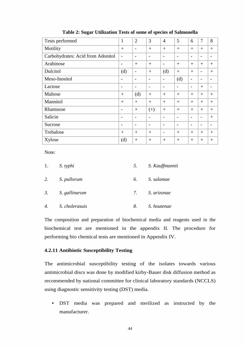
44
Table 2: Sugar Utilization Tests of some of species of Salmonella
Tests performed 1 2 3 4 5 6 7 8
Motility + - + + + + + +
Carbohydrates: Acid from Adonitol - - - - - - - -
Arabinose - + + - + + + +
Dulcitol (d) - + (d) + + - +
Meso-Inositol - - - - (d) - - -
Lactose - - - - - - + -
Maltose + (d) + + + + + +
Mannitol + + + + + + + +
Rhamnose - + (+) + + + + +
Salicin - - - - - - - +
Sucrose - - - - - - - -
Trehalose + + + - + + + +
Xylose (d) + + + + + + +
Note:
1. S. typhi 5. S. Kauffmannii
2. S. pullorum 6. S. salamae
3. S. gallinarum 7. S. arizonae
4. S. cholerasuis 8. S. houtenae
The composition and preparation of biochemical media and reagents used in the
biochemical test are mentioned in the appendix II. The procedure for
performing bio chemical tests are mentioned in Appendix IV.
4.2.11 Antibiotic Susceptibility Testing
The antimicrobial susceptibility testing of the isolates towards various
antimicrobial discs was done by modified kirby-Bauer disk diffusion method as
recommended by national committee for clinical laboratory standards (NCCLS)
using diagnostic sensitivity testing (DST) media.
DST media was prepared and sterilized as instructed by the
manufacturer.
45
The pH of the medium 7.2- 7.4 and the depth of the medium at 4 mm
(about 25 ml per plate) were maintained in petri dish.
Using a sterile inoculating wire loop, a single isolated colony of which
the sensitivity pattern is to be determined was touched and inoculated
into nutrient broth tube and was incubated at 37ºc for 24 hrs.
After incubation, the turbidity of the suspension was matched with the turbidity
standard of the MC-farland tube number 0.5
Using a sterile swab, a plate of DST was inoculated with the bacterial
suspension using carpet culture technique. The plate was left for about 5
minutes to let the agar surface dry.
Using sterile forceps, appropriate antimicrobial discs (6 mm diameter) was
placed, evenly distributed on the inoculated plates, not more than 6 discs were
placed on a 90 mm diameter petri dish.
After overnight incubation, the plates were examined to ensure confluent
growth and the diameter of each zone of inhibition in mm was measured and
compared with standardized zone interpretative chart provided by the company.
The preparation and composition of diagnostic sensitivity testing (DST) media
are shown in appendix I and II. The detailed about antibiotic discs used and its
interpretive chart are mentioned in the appendix I and V.
4.2.12 Serotyping
The isolates of Salmonella that were motile and which produced black colony
on XLD media were sereotype at WARUN institute for the identification of the
'O' antigen group.
4.2.13 Quality Control
To obtain reliable microbiological result, it is necessary to maintain quality
control. Quality of each test was maintained by using standard procedures
through out the study period from sample collection to sample transportation,
handling, media preparation. The quality of each agar plates prepared was
46
tested by incubating one plate of each lot on the incubator. Control strains of S.
pullorum and S. gallinarum available at CVL were used as positive control for
the standardization of the kirby-Bauer test. Quality of sensitivity tests was
maintained by maintaining the thickness of DST at 4 mm and the pH at 7.2-7.4.
Similarly, antibiotics discs containing the correct amount as indicated were
used. Strict aseptic conditions were maintained while carrying out all the
procedures.
4.2.14 Data Analysis
Chi-square test was used to determine significant of isolation of Salmonella
from meat sample and presence of different types of Salmonella species in the
meat sample.(Appendix VI) .
Data Analysis Software: Statistical Programme for Social Sciences (SPSS);
Microsoft Excel
47
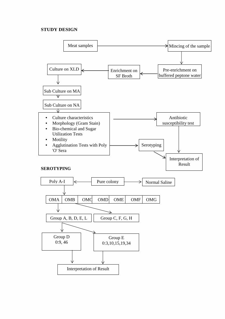
STUDY DESIGN
SEROTYPING
Culture characteristics Morphology (Gram Stain) Bio-chemical and Sugar
Utilization Tests Motility Agglutination Tests with Poly
'O' SeraSerotyping
Meat samples Mincing of the sample
Culture on XLD Pre-enrichment onbuffered peptone water
Enrichment onSF Broth
Sub Culture on NA
Antibioticsusceptibility test
Interpretation ofResult
Poly A-I Pure colony
Group A, B, D, E, L Group C, F, G, H
Group D0:9, 46
Group E0:3,10,15,19,34
Interpretation of Result
OMA OMB OMC OMD OME OMF OMGONag
Normal Saline
Sub Culture on MA
32
CHAPTER – V
5. RESULTS
5.1 COLONY CHARACTERISTICS OF ISOLATES ON XLD
Salmonella spp isolated from 23 meat samples showed the following
characteristics colony on XLD media.
Table 3: Colony characteristics of Salmonella spp on XLD media.
No. of isolates Size (mm) Color Margin Elevation Consistency
17 1-2 Red Entire Raised Soft
6 2-3 Black Entire Raised Soft
5.2 COLONY CHARACTERISTICS OF ISOLATES ON NA
The black and red colony of Salmonella spp formed on XLD media when sub
cultured on NA showed the following colony characteristics:
Table 4: Colony characteristics of Salmonella spp. on NA media
No. of
isolates
Size
(mm)
Color Margin Elevation Consistency
17 1-2 Creamy
white
Entire Raised Soft
6 2-3 Creamy
white
Entire Raised Soft
5.3 MORPHOLOGY
All isolates from NA agar plate were gram negative short rods
5.4 MOTILITY (HANGING DROP METHOD)
Seventeen isolates from NA agar plate having colony size 1-2 mm in diameter
were non-motile and rests were motile.
33
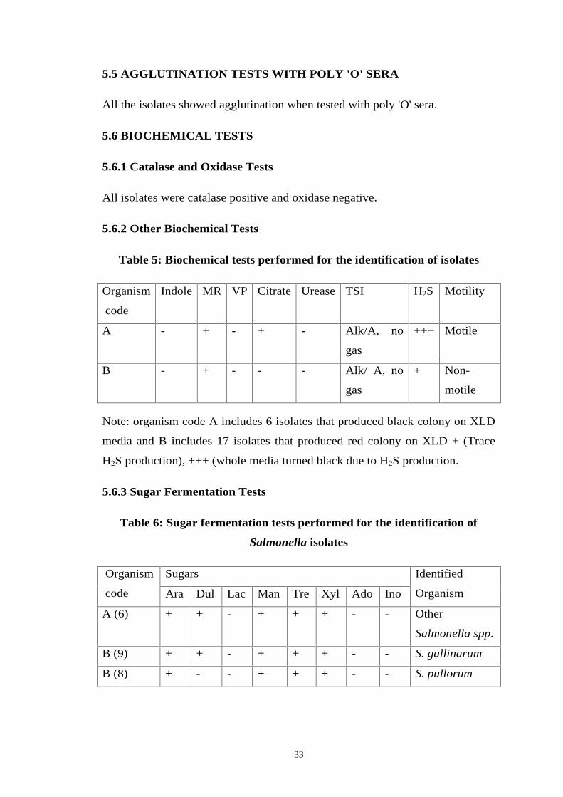
5.5 AGGLUTINATION TESTS WITH POLY 'O' SERA
All the isolates showed agglutination when tested with poly 'O' sera.
5.6 BIOCHEMICAL TESTS
5.6.1 Catalase and Oxidase Tests
All isolates were catalase positive and oxidase negative.
5.6.2 Other Biochemical Tests
Table 5: Biochemical tests performed for the identification of isolates
Organism
code
Indole MR VP Citrate Urease TSI H2S Motility
A - + - + - Alk/A, no
gas
+++ Motile
B - + - - - Alk/ A, no
gas
+ Non-
motile
Note: organism code A includes 6 isolates that produced black colony on XLD
media and B includes 17 isolates that produced red colony on XLD + (Trace
H2S production), +++ (whole media turned black due to H2S production.
5.6.3 Sugar Fermentation Tests
Table 6: Sugar fermentation tests performed for the identification of
Salmonella isolates
Organism
code
Sugars Identified
OrganismAra Dul Lac Man Tre Xyl Ado Ino
A (6) + + - + + + - - Other
Salmonella spp.
B (9) + + - + + + - - S. gallinarum
B (8) + - - + + + - - S. pullorum
34
Note: 6 isolates included in A differs from 9 isolates included in B (9) and 8
isolates included in B (8) due to formation of black colony on XLD. 9 isolates
included in B (9) differs from 8 isolate included in B (8) due to dulcitol
positive.
5.7 MONTHLY ISOLATION OF SALMONELLA SPECIES
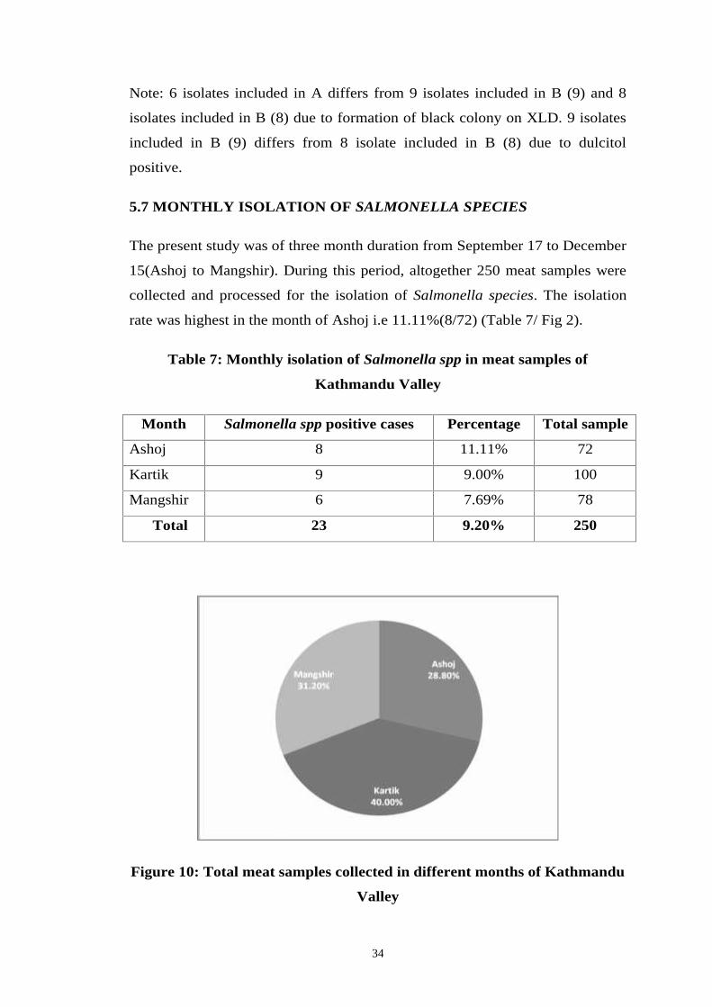
The present study was of three month duration from September 17 to December
15(Ashoj to Mangshir). During this period, altogether 250 meat samples were
collected and processed for the isolation of Salmonella species. The isolation
rate was highest in the month of Ashoj i.e 11.11%(8/72) (Table 7/ Fig 2).
Table 7: Monthly isolation of Salmonella spp in meat samples of
Kathmandu Valley
Month Salmonella spp positive cases Percentage Total sample
Ashoj 8 11.11% 72
Kartik 9 9.00% 100
Mangshir 6 7.69% 78
Total 23 9.20% 250
Figure 10: Total meat samples collected in different months of Kathmandu
Valley
35
Figure 11: Monthly isolation of Salmonella spp in meat different samples of
Kathmandu Valley
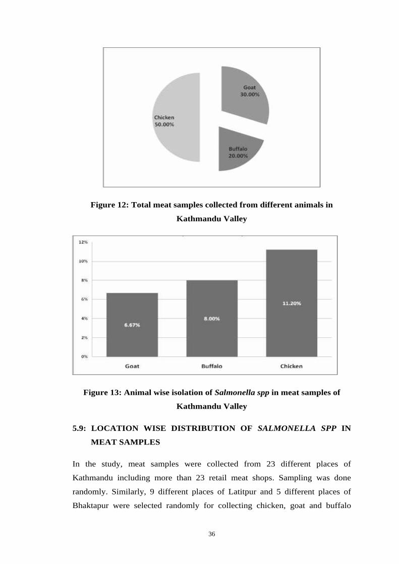
5.8 ANIMAL-WISE ISOLATION OF SALMONELLA SPECIES
In the study, different animal meat samples were included. Altogether 125
chicken, 75 goat and 50 buffalo meat samples were analyzed for Salmonella
spp contamination. The result revealed that isolation of Salmonella spp
contamination was seen highest in chicken (11.20%) followed by buffalo
(8.00%) and goat (6.67%) (Table 8/fig 4)
Table 8: Animal wise isolation of Salmonella spp in meat sample of
Kathmandu Valley
Animals Salmonella spp positive cases Percentage Total sample
Goat 5 6.67% 75
Buffalo 4 8.00% 50
Chicken 14 11.20% 125
Total 23 9.20% 250
36
Figure 12: Total meat samples collected from different animals in
Kathmandu Valley
Figure 13: Animal wise isolation of Salmonella spp in meat samples of
Kathmandu Valley
5.9: LOCATION WISE DISTRIBUTION OF SALMONELLA SPP IN
MEAT SAMPLES
In the study, meat samples were collected from 23 different places of
Kathmandu including more than 23 retail meat shops. Sampling was done
randomly. Similarly, 9 different places of Latitpur and 5 different places of
Bhaktapur were selected randomly for collecting chicken, goat and buffalo
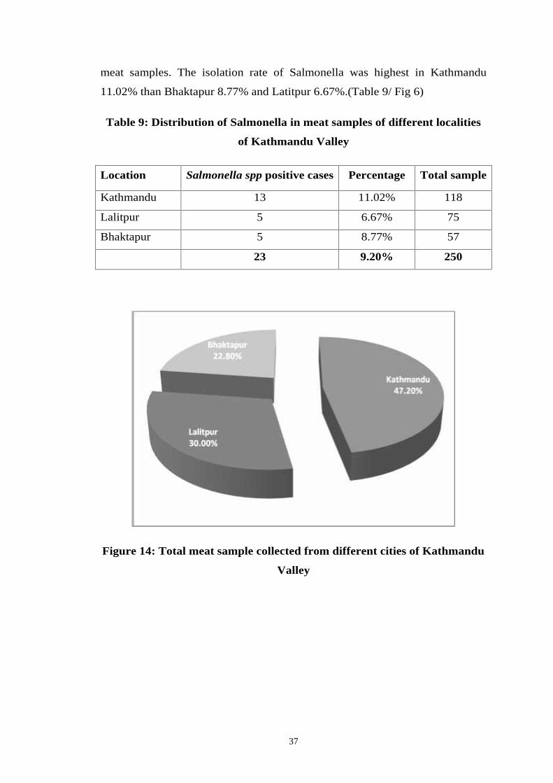
37
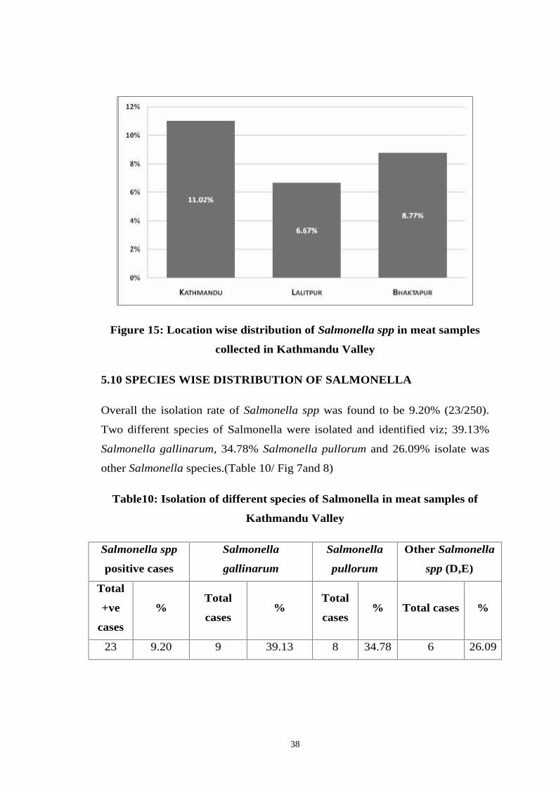
meat samples. The isolation rate of Salmonella was highest in Kathmandu
11.02% than Bhaktapur 8.77% and Latitpur 6.67%.(Table 9/ Fig 6)
Table 9: Distribution of Salmonella in meat samples of different localities
of Kathmandu Valley
Location Salmonella spp positive cases Percentage Total sample
Kathmandu 13 11.02% 118
Lalitpur 5 6.67% 75
Bhaktapur 5 8.77% 57
23 9.20% 250
Figure 14: Total meat sample collected from different cities of Kathmandu
Valley
38
Figure 15: Location wise distribution of Salmonella spp in meat samples
collected in Kathmandu Valley
5.10 SPECIES WISE DISTRIBUTION OF SALMONELLA
Overall the isolation rate of Salmonella spp was found to be 9.20% (23/250).
Two different species of Salmonella were isolated and identified viz; 39.13%
Salmonella gallinarum, 34.78% Salmonella pullorum and 26.09% isolate was
other Salmonella species.(Table 10/ Fig 7and 8)
Table10: Isolation of different species of Salmonella in meat samples of
Kathmandu Valley
Salmonella spp
positive cases
Salmonella
gallinarum
Salmonella
pullorum
Other Salmonella
spp (D,E)
Total
+ve
cases
%Total
cases%
Total
cases% Total cases %
23 9.20 9 39.13 8 34.78 6 26.09
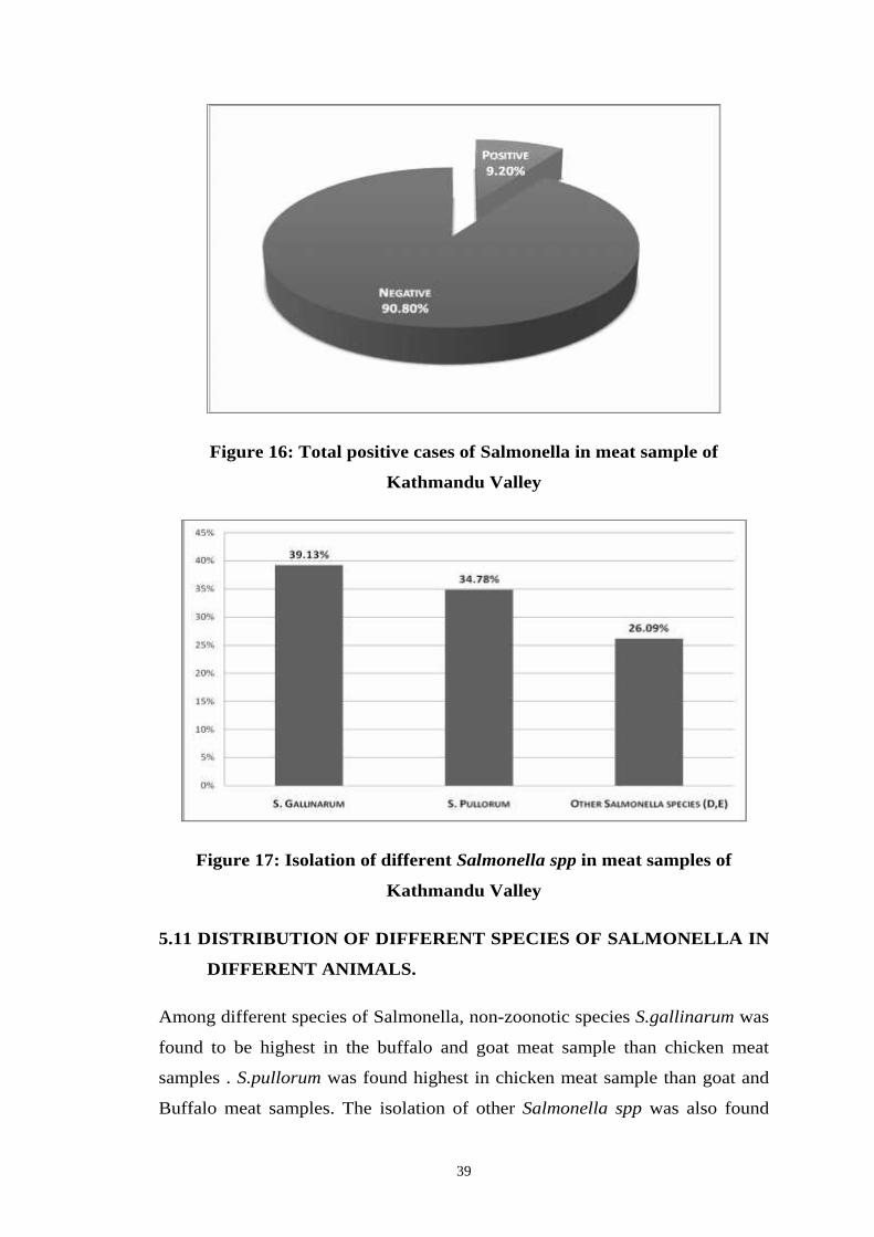
39
Figure 16: Total positive cases of Salmonella in meat sample of
Kathmandu Valley
Figure 17: Isolation of different Salmonella spp in meat samples of
Kathmandu Valley
5.11 DISTRIBUTION OF DIFFERENT SPECIES OF SALMONELLA IN
DIFFERENT ANIMALS.
Among different species of Salmonella, non-zoonotic species S.gallinarum was
found to be highest in the buffalo and goat meat sample than chicken meat
samples . S.pullorum was found highest in chicken meat sample than goat and
Buffalo meat samples. The isolation of other Salmonella spp was also found
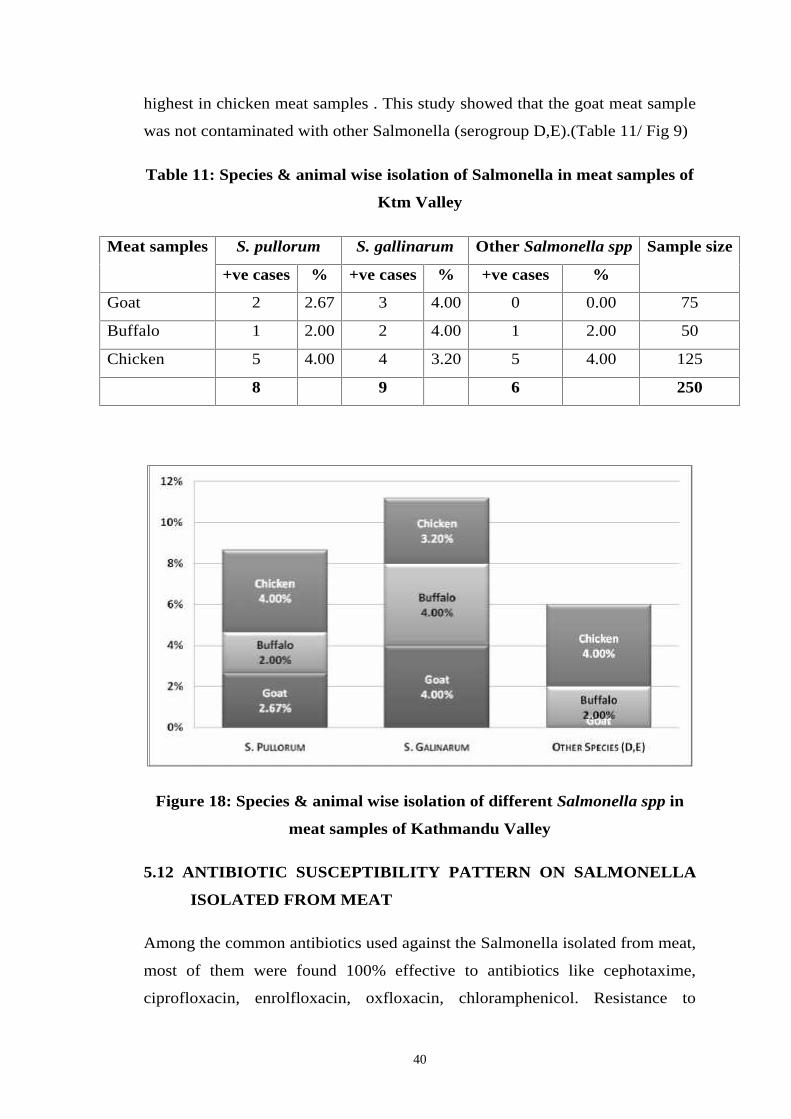
40
highest in chicken meat samples . This study showed that the goat meat sample
was not contaminated with other Salmonella (serogroup D,E).(Table 11/ Fig 9)
Table 11: Species & animal wise isolation of Salmonella in meat samples of
Ktm Valley
Meat samples S. pullorum S. gallinarum Other Salmonella spp Sample size
+ve cases % +ve cases % +ve cases %
Goat 2 2.67 3 4.00 0 0.00 75
Buffalo 1 2.00 2 4.00 1 2.00 50
Chicken 5 4.00 4 3.20 5 4.00 125
8 9 6 250
Figure 18: Species & animal wise isolation of different Salmonella spp in
meat samples of Kathmandu Valley
5.12 ANTIBIOTIC SUSCEPTIBILITY PATTERN ON SALMONELLA
ISOLATED FROM MEAT
Among the common antibiotics used against the Salmonella isolated from meat,
most of them were found 100% effective to antibiotics like cephotaxime,
ciprofloxacin, enrolfloxacin, oxfloxacin, chloramphenicol. Resistance to
41
cotrimoxazole, amoxicillin and gentamycin was seen in only one isolate. Most
of the isolates were found to be resistant to nalidixic acid (19/23) and
tetracycline (12/23) (Table 12).
Table 12: Antibiotic susceptibility pattern of Salmonella
S.N. Antibiotics used Drug
potency
(mcg/discs)
Total
no. of
isolates
Resistant Intermediate
susceptible
Susceptible
1. Cephotaxime 30 23 0 0 23
2. Cotrimoxazole 25 23 1 0 22
3. Chloramphenical 30 23 0 0 23
4. Ciprofloxacin 5 23 0 0 23
5. Ofloxacin 5 23 0 0 23
6. Enrolfloxacin 10 23 0 0 23
7. Tetracycline 30 23 12 6 5
8. Amoxycillin 10 23 1 3 19
9. Nalidixic acid 30 23 19 1 3
10. Gentamycin 10 23 1 0 22
5.13 ANTIBIOTIC SUSCEPTIBILITY PATTERN OF SALMONELLA
GALLINARUM
S. gallinarum was found to be more resistant towards nalidixic acid. Among the
10 antimicrobials tested, most of the antibiotics were 100% effective and only
one isolate was resistant to cotrimoxazole, amoxicillin and gentamycin and out
of 9, 5 isolates were resistant to tetracycline (Table 13)
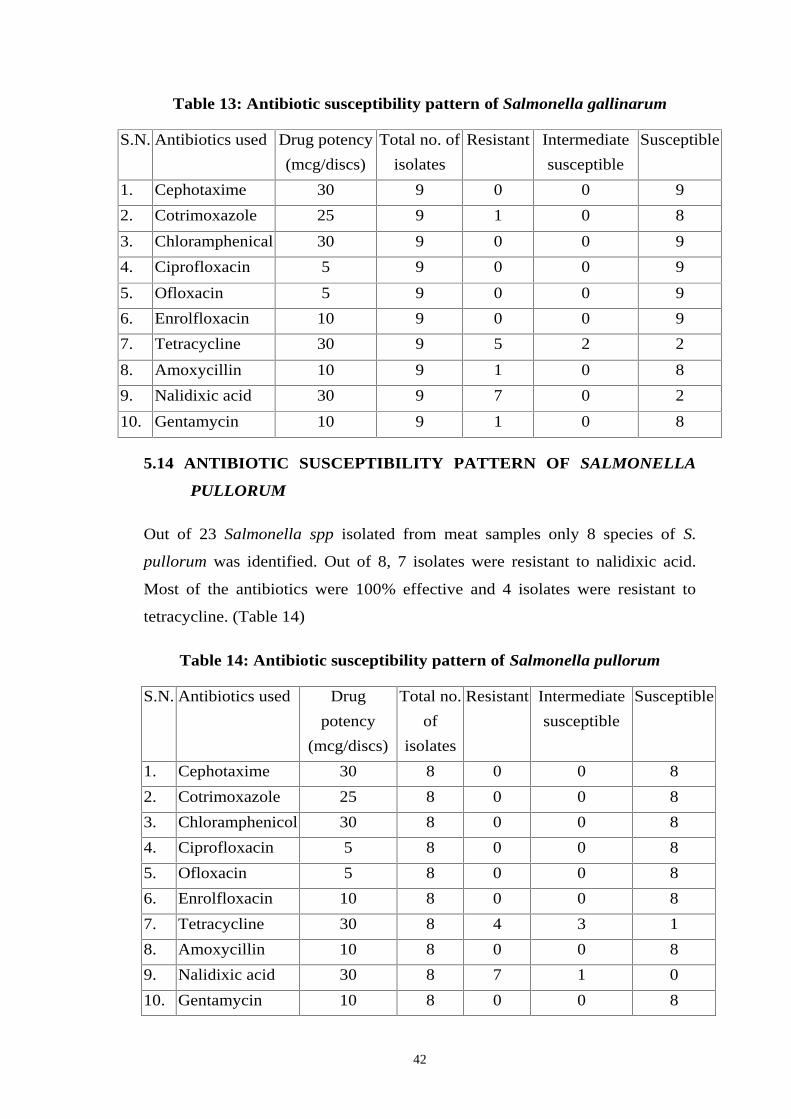
42
Table 13: Antibiotic susceptibility pattern of Salmonella gallinarum
S.N. Antibiotics used Drug potency
(mcg/discs)
Total no. of
isolates
Resistant Intermediate
susceptible
Susceptible
1. Cephotaxime 30 9 0 0 9
2. Cotrimoxazole 25 9 1 0 8
3. Chloramphenical 30 9 0 0 9
4. Ciprofloxacin 5 9 0 0 9
5. Ofloxacin 5 9 0 0 9
6. Enrolfloxacin 10 9 0 0 9
7. Tetracycline 30 9 5 2 2
8. Amoxycillin 10 9 1 0 8
9. Nalidixic acid 30 9 7 0 2
10. Gentamycin 10 9 1 0 8
5.14 ANTIBIOTIC SUSCEPTIBILITY PATTERN OF SALMONELLA
PULLORUM
Out of 23 Salmonella spp isolated from meat samples only 8 species of S.
pullorum was identified. Out of 8, 7 isolates were resistant to nalidixic acid.
Most of the antibiotics were 100% effective and 4 isolates were resistant to
tetracycline. (Table 14)
Table 14: Antibiotic susceptibility pattern of Salmonella pullorum
S.N. Antibiotics used Drug
potency
(mcg/discs)
Total no.
of
isolates
Resistant Intermediate
susceptible
Susceptible
1. Cephotaxime 30 8 0 0 8
2. Cotrimoxazole 25 8 0 0 8
3. Chloramphenicol 30 8 0 0 8
4. Ciprofloxacin 5 8 0 0 8
5. Ofloxacin 5 8 0 0 8
6. Enrolfloxacin 10 8 0 0 8
7. Tetracycline 30 8 4 3 1
8. Amoxycillin 10 8 0 0 8
9. Nalidixic acid 30 8 7 1 0
10. Gentamycin 10 8 0 0 8
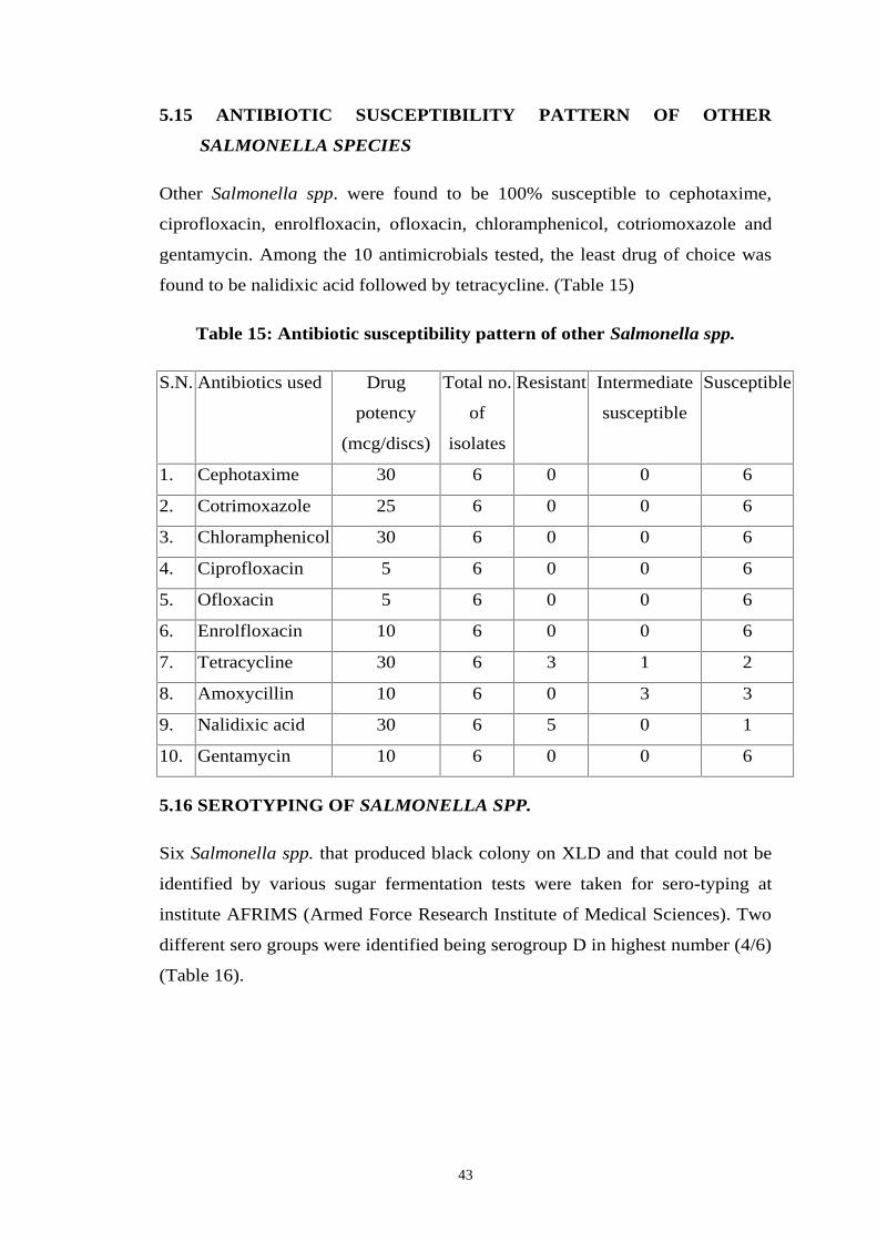
43
5.15 ANTIBIOTIC SUSCEPTIBILITY PATTERN OF OTHER
SALMONELLA SPECIES
Other Salmonella spp. were found to be 100% susceptible to cephotaxime,
ciprofloxacin, enrolfloxacin, ofloxacin, chloramphenicol, cotriomoxazole and
gentamycin. Among the 10 antimicrobials tested, the least drug of choice was
found to be nalidixic acid followed by tetracycline. (Table 15)
Table 15: Antibiotic susceptibility pattern of other Salmonella spp.
S.N. Antibiotics used Drug
potency
(mcg/discs)
Total no.
of
isolates
Resistant Intermediate
susceptible
Susceptible
1. Cephotaxime 30 6 0 0 6
2. Cotrimoxazole 25 6 0 0 6
3. Chloramphenicol 30 6 0 0 6
4. Ciprofloxacin 5 6 0 0 6
5. Ofloxacin 5 6 0 0 6
6. Enrolfloxacin 10 6 0 0 6
7. Tetracycline 30 6 3 1 2
8. Amoxycillin 10 6 0 3 3
9. Nalidixic acid 30 6 5 0 1
10. Gentamycin 10 6 0 0 6
5.16 SEROTYPING OF SALMONELLA SPP.
Six Salmonella spp. that produced black colony on XLD and that could not be
identified by various sugar fermentation tests were taken for sero-typing at
institute AFRIMS (Armed Force Research Institute of Medical Sciences). Two
different sero groups were identified being serogroup D in highest number (4/6)
(Table 16).
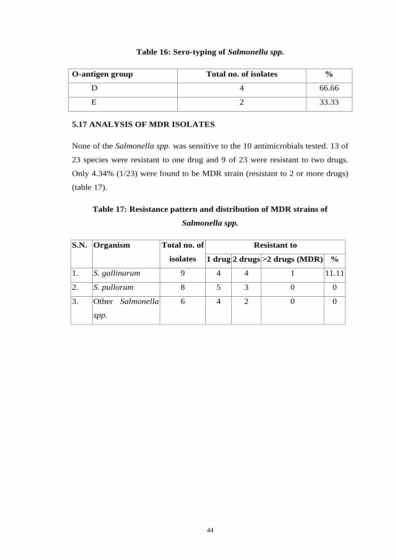
44
Table 16: Sero-typing of Salmonella spp.
O-antigen group Total no. of isolates %
D 4 66.66
E 2 33.33
5.17 ANALYSIS OF MDR ISOLATES
None of the Salmonella spp. was sensitive to the 10 antimicrobials tested. 13 of
23 species were resistant to one drug and 9 of 23 were resistant to two drugs.
Only 4.34% (1/23) were found to be MDR strain (resistant to 2 or more drugs)
(table 17).
Table 17: Resistance pattern and distribution of MDR strains of
Salmonella spp.
S.N. Organism Total no. of
isolates
Resistant to
1 drug 2 drugs >2 drugs (MDR) %
1. S. gallinarum 9 4 4 1 11.11
2. S. pullorum 8 5 3 0 0
3. Other Salmonella
spp.
6 4 2 0 0
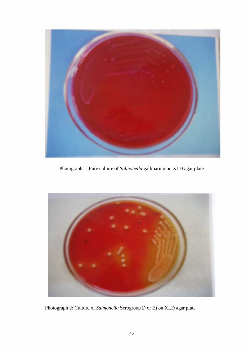
45
Photograph 1: Pure culture of Salmonella gallinarum on XLD agar plate
Photograph 2: Culture of Salmonella Serogroup D or E) on XLD agar plate
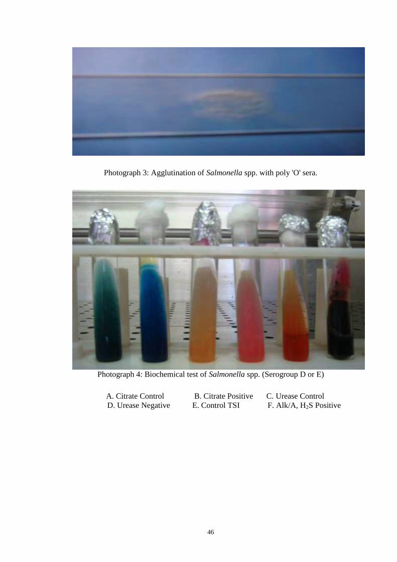
46
Photograph 3: Agglutination of Salmonella spp. with poly 'O' sera.
Photograph 4: Biochemical test of Salmonella spp. (Serogroup D or E)
A. Citrate Control B. Citrate Positive C. Urease ControlD. Urease Negative E. Control TSI F. Alk/A, H2S Positive
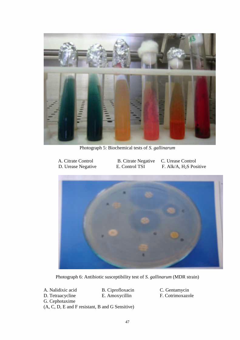
47
Photograph 5: Biochemical tests of S. gallinarum
A. Citrate Control B. Citrate Negative C. Urease ControlD. Urease Negative E. Control TSI F. Alk/A, H2S Positive
Photograph 6: Antibiotic susceptibility test of S. gallinarum (MDR strain)
A. Nalidixic acid B. Ciprofloxacin C. GentamycinD. Tetraacycline E. Amoxycillin F. CotrimoxazoleG. Cephotaxime(A, C, D, E and F resistant, B and G Sensitive)
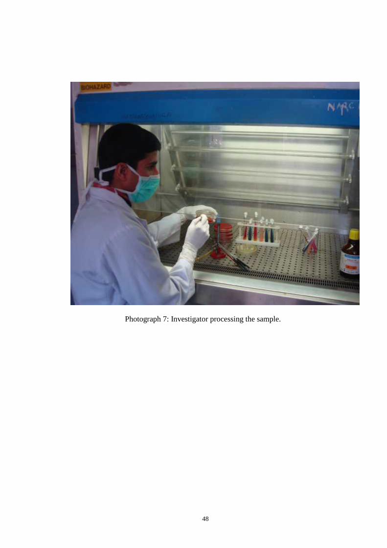
48
Photograph 7: Investigator processing the sample.
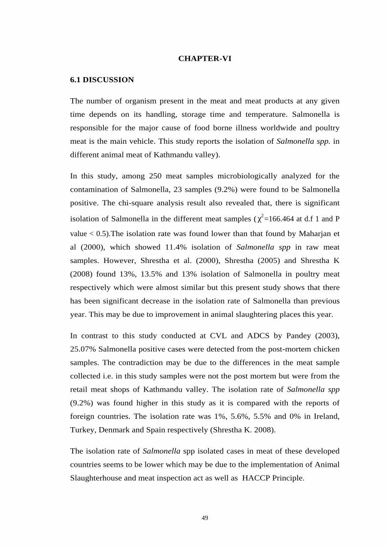
49
CHAPTER-VI
6.1 DISCUSSION
The number of organism present in the meat and meat products at any given
time depends on its handling, storage time and temperature. Salmonella is
responsible for the major cause of food borne illness worldwide and poultry
meat is the main vehicle. This study reports the isolation of Salmonella spp. in
different animal meat of Kathmandu valley).
In this study, among 250 meat samples microbiologically analyzed for the
contamination of Salmonella, 23 samples (9.2%) were found to be Salmonella
positive. The chi-square analysis result also revealed that, there is significant
isolation of Salmonella in the different meat samples ( 2χ =166.464 at d.f 1 and P
value < 0.5).The isolation rate was found lower than that found by Maharjan et
al (2000), which showed 11.4% isolation of Salmonella spp in raw meat
samples. However, Shrestha et al. (2000), Shrestha (2005) and Shrestha K
(2008) found 13%, 13.5% and 13% isolation of Salmonella in poultry meat
respectively which were almost similar but this present study shows that there
has been significant decrease in the isolation rate of Salmonella than previous
year. This may be due to improvement in animal slaughtering places this year.
In contrast to this study conducted at CVL and ADCS by Pandey (2003),
25.07% Salmonella positive cases were detected from the post-mortem chicken
samples. The contradiction may be due to the differences in the meat sample
collected i.e. in this study samples were not the post mortem but were from the
retail meat shops of Kathmandu valley. The isolation rate of Salmonella spp
(9.2%) was found higher in this study as it is compared with the reports of
foreign countries. The isolation rate was 1%, 5.6%, 5.5% and 0% in Ireland,
Turkey, Denmark and Spain respectively (Shrestha K. 2008).
The isolation rate of Salmonella spp isolated cases in meat of these developed
countries seems to be lower which may be due to the implementation of Animal
Slaughterhouse and meat inspection act as well as HACCP Principle.
50
Shrestha (2005) had conducted similar types of study on poultry meat and
found high isolation rate (24.48%) in the month of Bhadra. But this study has
conducted from Ashoj, and found higher isolation rate in the month of Ashoj
(11.11%). The reason behind this may be due to the warm and humid storage
temperature which is suitable for the growth of micro-organisms. Similar type
of result was also found by Shrestha K (2008) i.e. 11.63% isolation rate in the
month of Ashoj, Banatvala et al. (1999); Delarocque et al. (1998), Joshi (2003),
Maharjan et al. (2006) also found similar type of result.
In the present study, the isolation of Salmonella spp. isolated cases were found
higher in Kathmandu (11.01%) than Bhaktapur (8.77%) and Lalitpur (6.66%)
as shown in table 9. This result is similar to the result obtained by Karki (1995);
and Shrestha K (2008). In a study carried out by Joshi (2001), he reported that
the environmental sanitary conditions of the meat shops were not satisfactory
and standard. Out of 70 slaughtering places, the environment is good while in
70% and 15.7% were average and poor respectively. Out of 70 butchers, only
14.3% had the knowledge of environmental pollution due to the poor sanitary
conditions in slaughtering places. Lack of water and poor sanitary condition in
slaughtering places may be the reason for high isolation rate of Salmonella
contamination in poultry meat of Kathmandu and Bhaktapur area which is
similar to the conducted present study.
In the study conducted by Joshi D.D., Joshi V. and Mishra P.N. (2005) the
isolation rate of Salmonella was found to be 11.3% including 3.2% S. pullorum,
0.81% S. gallinarum, 1.62% S. typhi, 0.81% S. choleraesuis and 4.8%
Salmonella of subgenus I or II type. The sample collected were buff, chicken
and chevon meat samples. The isolation rate of Salmonella found on buff,
chicken and cheovan were 13.5%, 14.5% and 3.2% respectively. The highest
isolation rate was seen in chicken meat sample which is similar to the present
study. The reason behind getting highest frequency of Salmonella in chicken
meat samples than buff and goat may be due to the poor sanitary conditions in
most of the chicken meat shops and slaughtering places.
All the isolates were analyzed and categorized based on their cultural,
morphological as well as biochemical characteristics. From 23 meat samples,
51
Salmonella were isolated and identified. S. gallinarum was isolated from 9 meat
samples where as S. pullorum was isolated from 8 meat samples and sero-group
(D and E) was isolated in 6 meat samples The chi-square analysis result also
revealed that, there is significant isolation of different species of Salmonella in
the different meat samples ( 2χ =577.360 at d.f 3,P value < 0.5). In a similar study,
there are reports of isolation of S. gallinarum and S. pullorum from poultry
meat samples of Kathmandu valley as found by Shrestha (2005), Manandhar et
al. (2004) and Shrestha K (2008). When the percentage of isolation is compared
with the present study, it can be said that the process of slaughtering diseased
has decreased this year. The reason behind getting almost similar frequency of
S. pullorum and S. gallinarum in poultry meat sample might be due to pullorum
disease which is usually confined to the chicks at first 2-3 weeks of age and
occasionally occurs in adults (Shivprasad, 1997) and most of the samples
processed in this study might be of age group less than 2-3 weeks. The reason
behind getting almost similar result of isolation S. gallinarum and pullorum in
goat meat sample with chicken meat sample may be due to contamination from
the same chopping board and knief used in the same meat shop. Although S.
gallinarum and S. pullorum were isolated in meat sample, these are considered
to be non-pathogenic for humans. However, they may cause significant loss to
the poultry farmers.
The 6 isolates were found neither S. gallinarum nor S. pullorum based on their
cultural tests mainly citrate utilization tests and H2S production in SIM media.
(Both S. gallinarum and S. pullorum are) citrate negative but other 6 showed
the utilization of citrate). These 6 isolates were further categorized by
serotyping. Serotyping result revealed that they belonged to two groups D and
E as shown in table 16. It was found that Salmonella spp. belonging to
serogroup D had highest frequency.
S. enteritidis, a member of sero group D is the common isolates of poultry meat
as reported by Shrestha (2005); Manandhar et al. (2004) and Joshi (2003).
Isolation of S. enteritidis in foreign countries is also maximum in poultry meat
being 51.7% in Albania, 29.3% in Korea and 30% in Brazil (Beli et al. 2000),
Chung et al. 2003: Fuzihara et al. 2000). The Salmonella isolates belonging to
52
sero-group D in the present study is S. enteritidis which is being more prevalent
in chicken meat of Nepal as well as of the foreign countries. However, for more
confirmed identification, complete serotyping should be done.
So far literature concern, Salmonella belonging to serogroup E was only
reported by Shrestha K (2008) to be present in poultry meat of Nepal. The
present study also found Salmonella serogroup E positive cases and to my
knowledge it is the second report in Nepal and it has opened the pathway for
the further research. However, there are reports of isolation of Salmonella
belonging to sero groups E in the poultry meat of foreign countries.
Although with low isolation rate Maharjan et al (2006) and Manandhar et al.
(2004) had isolated S. typhi also from meat samples of Kathmandu valley. In
the present study S. typhi, S. paratyphi A and B and S. cholerasuis were not
isolated from any of the meat samples. It is good as a point for consumer’s
health.
In this study, antibiotic susceptibility test was performed for all 23 isolates of
Salmonella. All isolates were found to be sensitive to the antibiotics like
cephotaxime were found sensitive to the antibiotics like cephotaxime (third
generation cephalosporins), fluoroquinolones such as ciprofloxacin, ofloxacin
and enrol floxacin and broad spectrum antibiotics like chloramphenicol. Out of
23 isolates only 1 (S. gallinarum) showed resistance to cotrimoxazole,
gentamycin and amoxycillin. In the study, nalidixic acid was found to be least
effective drug followed by tetracycline. This finding is also similar to the result
of NARC (053/054) also which showed that tetracycline resistant is more
among S. gallinarum and S. pullorum.
Aarestrup et al. (2006) in his study isolated Salmonella enterica serotype
schwarzengrund from food and food animals in Denmark and found to be
resistance to nalidixic acid. The study conducted by abouzeed et al (2000);
Capita et al. (2003); Metzer et al (1998), Winokur et al. (2000) also showed that
tetracycline and nalidixic acid were the least effective drug to treat Salmonella
spp. These findings also supports the findings of the present study.
53
According to Kurutepe et al. (2005) multi-drug resistant organisms (MDR) are
defined as the organisms which are resistance to 2 or more of the antimicrobial
agents. Based on this definition of MDR only 1 isolate (S. gallinarum) was
found to be MDR. Resistance to tetracycline and nalidixic acid was most
common in the case of Salmonella belonging to sero groups D and E. However,
the antibiotic susceptibility pattern shows that amoxicillin (10 mcg) is also
going to be resistant. Many studies had showed that isolation of MDR
Salmonella spp in poultry meat of foreign countries is significantly higher
(Aboozeed et al. 2000: Capita et al. 2003, Jorgensen et al. 2002; White et al.
2003) till date. But it is in increasing tendency which alarms the concerned
authorities.
54
6.2 CONCLUSION
In this study, 250 meat samples including 125 chicken, 75 goat and 50 buffalo
meat samples were processed and Salmonella was isolated from 23 (9.2%) meat
samples. The chi-square analysis result also revealed that, there is significant
isolation of different species of Salmonella in the different meat samples
( 2χ =577.360 at d.f 3).
Among the isolates, 39.13% S. gallinarum, 34.78% S. pullorum and 26.08%
were other Salmonella spp. belonging to sero group D and E. Various studies
show that MDR Salmonella is increasing globally in recent years. In the present
study 4.34% MDR Salmonella spp. was observed. The present study was
carried out with the aim of isolation of MDR Salmonella in meat samples of
Kathmandu valley. For the second time in Nepal, isolation of Salmonella
belonging to sero group E in addition to those belonging to serogroup D in meat
sample is another interesting finding of this study.
Based on the evidence of this study, it can be concluded that from the health
and hygiene point of view, the meat sold in retail shops of Kathmandu valley
should be improved in the meat quality.

55
CHAPTER-VII
7. SUMMARY AND RECOMMENDATIONS
7.1 SUMMARY
Altogether, 250 meat samples (chicken 125, goat 75 and buffalo 50) collected
from different localities of retail shops of Kathmandu valley were
microbiologically analyzed for the presence of Salmonella. Out of 250 samples,
23 (9.2%) samples were found to be Salmonella positive. Statistically, the
isolation rate of Salmonella in different meat samples is also significant
( 2χ =166.464 at d.f 1), this also supports the result.
The study was of three month duration from Ashoj to Mangshir. The isolation
rate was higher in the month of Ashoj. When the isolation rate is compared with
the different animal meat samples, chicken meat samples were found to be
more contaminated with Salmonella spp than that of goat and buffalo.
Out of 118, (11.01%) samples were found to be positive for Salmonella spp. in
meat samples of Kathmandu which is higher than Bhaktapur (8.77%) and
Lalitpur (6.66%) meat samples.
Among the 23 isolates of Salmonella, 9 (39.13%) isolates were identified as S.
gallinarum, and 8 (34.78%) S. pullorum. Sero typing was done for remaining 6
isolates and it was found to be within two different 'o' antigen groups- 4
belonging to sero group D (66.66%), and 2 to serogroup E (33.39%). The
Salmonella spp. belonging to sero group D was most predominant.
These 23 isolates were tested against 10 antimicrobials and it was found that
among these antibiotics, nalidixic acid, tetracycline and amoxycillin was least
effective to Salmonella spp. All these isolates were highly sensitive (100%) to
cephotaxime, fluoroquinolones, chloramphenicol. MDR pattern was observed
in 1 (4.34%) out of 23 isolates.
56
7.2 RECOMMENDATIONS
Findings of this study showed that there is as light variation in the month
wise isolation of Salmonella isolated positive cases, so the study should
be carried out including all seasons.
The study was conducted on only central veterinary laboratory and the
samples collected were from only Kathmandu valley, so the study should
also be carried out in the whole country Nepal. For the Nepalese
awareness.
Due to lack of laboratory facilities, complete serotyping of all isolates
could not be determined so it is recommended for complete serotyping.
As the multi-drug resistant Salmonella is found to be an emerging
problem in meat of Kathmandu valley. It would be better if the
concerned authorities check for the standard dose of antimicrobial to be
used in animal feed.
Implementation of animal slaughtering house and meat inspection act is
necessary.
Consumer's awareness programme should be conducted by distributing
pamphlets or through radio or through other means.
57
REFERENCES
Acha PN and Szyfres B (2001), Salmonellosis: Zoonoses and communicable
diseases common to man and animals: Pan American Health
Organization, Washington D.C. Scientific and Technical Publication:
223-246.
Ananthanarayan R and Panikar CJK (2000); Test Book of Microbiology, 6th
edition, Orient Longman Ltd: 267-272, 570.
Annual Bulletin (1997/98).; Food hygiene, Central Food Research Laboratory,
Kathmandu: 22-26.
Arora, A.K., Gupta S.C. and Kushik R.K. (1987); Determination of Upper
respiratory tract bacterial carriers in poultry. Indian vet. Med. Journal
10 (2): 63-67.
Bailey and Scotts (1990); Diagnostic Microbiology, Elln JoBaron, Sydney M.
Finegold. 8th edition, Baltimore.
Banwart G.J. (1987); Basic food Microbiology, CBS publishers and
distributors.
Bergey's Manual of determinative bacteriology (1990); 8th edition, the williams
and wilkins company Baltimore.
Brock T.D; Smith, D.W. Madigan M.T. (1984); Biology of Microorganisms.
Fourth Edition.
Bryan F.L. (1968); What a saniterian should know about Salmonellae in non-
diary products. J milk food techno 31: 110.
Bryan FL (1968); Salmonellae problems in non-diary products 11. Salmonella,
J milk food techno. 31: 131.
Chaudhari Shiv Prasad (1996); Pullorum disease and fowl typhoid calnek BW
(ed) Disease of poultry. Tenth edition pp 82-96. Iowa State University.
58
Cheesbrough M (2000); District laboratory practice in tropical countries,
Cambridge University Press: 257-261.
Collee JG, Fraser AG, Marmion BP and Simmons A (1996); Mackie and
Mccartney practical medical Microbiology, 14th edition, Churchill
Livingstone, New York: 358-392.
Doughlas WW, Gast RK and Mallinson ET (1998); A laboratory mannual for
the isolation and identification of avian pathogen, 4th edition. American
association of allian pathologist: 4-13.
Dutta N. (1984); British Medical Bulletin 40:1.
Euring WH (1986); Edwards and Euring's identification of enterobacteriaceae,
4th edition, Elsevier Science Publishing, New York: 247-314.
Evans S., Davies R. (1996); Plasmid profiles analysis and antibiotics
susceptibility testing of multi resistant Salmonella veterinary record
139:557-558.
Frazier WC and Westhoff DC (1985); Food Microbiology, 3rd edition, Tata
McGraw Hill Publishing Company Ltd. New Delhi: 218-238.
Frazier, W.C. and D.C. Westhoff (1993); Food Microbiology, fourth edition,
Tata McGraw Hill Publication.
Fries, R. (1987); Bacteria on Broiler skin during poultry meat production
processes. Fleishwirtschaft 67 (1): 106-108 111-113 Germany.
Gledel J. Pantaleon, J. Carbion B, (1977); Antibiotic resistance of 3600 strains
of Salmonella of animal origin (1974-1975): Reweil de medicine
veterinare 153 (2): 109-118, France.
Gyles N.R. (1989); National sample census of agriculture. Tanzania Manland
ministry of agriculture report vol II.
59
Huber, W.G. (1971); The impact of antibiotic drugs and their residues in:
Broadley, C.A. and conrail's C.E. (eds.) Advances in veterinary science
and comparative medicine, Academic Press, New York 102.
Joshi D.D. (1991); Current practices of livestock slaughtering and meat
marketing in Kathmandu, Lalitpur and Bhaktapur: National zoonesis
and food hygiene, consulting centre (Pvt) Ltd. Tahachal, Kathmandu.
Joshi V (2003); Study of raw meat sample for isolation and identification of
Salmonella spp. in ward no. 13 of Kathmandu Metropolitan City. A
dissertation submitted to the Central Department of Zoology, T.U.,
Kathmandu.
Joshi, D.D. Maharjan, M.Johnsen, M.V. Willinghan A.R.V. and Sharma M.
(2003); Improving meat inspection and control in resources poor
communities. "The Nepal example". Acta tropica vol 87 (1): 119-127.
Joshi, D.D., Joshi, R. and Bista, P.R. (1990); Isolation of Salmonella in street
dairy products in Kathmandu. J. Nep. Med. Assoc. 28: 148-151.
Karki, GB (1995); Bacteriological study of chicken and buff meat of
Kathmandu valley. A dissertation submitted to the Central Department
of Microbiology, T.U., Kathmandu.
Kenneth T (2006);Salmonella and Salmonellosis Todar's online textbook of
bacteriology. Todar University of Wisconsin Madison Department of
Bacteriology, http://www.textbook of bacteriology.net
Le Minor and Popoff My (1987); Request for an opinion: Designation of
Salmonella enterica sp. Nov. nom rev, as the type and only species of
the genus Salmonella. International journal of systematic bacteriology
37: 465-468.
Levy, Barry, Mc Intire, W., Damsky, L. et al. (1975); The middle town
outbreak: 125 cases of foodborne resulting from cross contaminated
items served at a picnic and smorgasbord. Minn. Dep. Health, 717
Delaware St. S.E. Minnea Polis Minn USA: 54-55.
60
Mackie and Mc Cartney (1996); Practical medical microbiology. 14th edition
churchill livingstone.
Maharjan, M. Sharma, A.P. (2000); Bacteriological quality of groundwater in
urban patan and antibiotic sensitivity against isolated enteric bacteria. J.
Nep. Med. Assoc. 39: 269-274.
Maharjan, M., Joshi V., Joshi DD and Manandhar P. (2006); Isolation of
Salmonella species in various raw meat samples of a local market in
Kathmandu. Annuals of the New York Academy of Sciences 108: 249-
256.
Maiorini, E, Lopes EL, Morrow AL, Ramirez F, Prochipio A, Purmanshi S
(1993); Multiply resistant Salmonella gallinarum, Surveillance, 24 (1):
31.
Majk GK and Roh WS (1998); Estimates of casses and social economic costs of
food borne salmonellosis in Korea. Journal of food hygiene and safety
13: 299-304.
Malla, F.B. and Sakya, G.M. (1984); Enteric Fever- a review of clinical
records. Journal of Nepal medical association vol. 22 (2): 9-15.
Manandhar P., Air TB and Shrestha P. (2004); Isolation of Salmonella in
poultry meat of Kathmandu valley. Report of animal research
technician's interaction: 1-3.
Manandhar, P. (2000); Seroisolation of campylobacter spp and Salmonella spp.
using enzyme linked immunosorbent assay (ELISA) in human and
chicken (Gallus gallus domestic us. linn) in selected areas of the
Philippines. A dissertation of M.V. Sc. presented to University of
Philippines Los Banes.
Manandhar, S (2057/058 B.S.); Isolation of Salmonellosis in poultry in Nepal
CVL annual technical report: 61-63.
61
Martenuik R.H., AW A.S., and Bartlett P.C. (1988); Compliance with
recommended drug withdrawal requirements for diary cows sent to
market in Michigan. Journal of the American veterinary medical
association 193 (4): 406-407.
Metzer E, Agmon V, Andoren N and Cohen D (1998); Emergence of Multidrug
resistant Salmonella entericaserotype typhimurium phase- type DTL04
among Salmonella causing enterititis Israel. Cambridge Journals 121
(3): 541-546.
NARC (Fy 53/54); Annual report Nepal agricultural research council, animal
health research division, Kathmandu.
Office international des epizoonostics (OIE, 2004); Mannual of diagnostic tests
and vaccines for terrestials animals (mammals, birds and bees) 5th
edition: 1018-1020.
Pandey M (2003); Study on the isolation of multiple drug resistant Salmonella
in poultry birds. A dissertation submitted to the Central Department of
Microbiology, T.U., Kathmandu.
Pomeroy, B.S. and Bagoriza K.V. (1991); Fowl typhoid disease of poultry, 9th
edition, page 82-99.
Poudyal A (2002); Salmonella serotyping and drug susceptibility pattern from
environment and clinical samples of urban Nepal. A dissertation
submitted to the Central Department of Micrbiology, T.U., Kathmandu.
Prasai P (2000); Microbiological study of raw meat of Kathmandu valley with
public health and veterinary importance and seriological study of the
isolated Salmonella sps. A dissertation submitted to the Central
Department of Microbiology, T.U., Kathmandu.
Rahman M and Albert MJ (1998); Multi drug resistant Salmonella infections an
emerging problem in poultry in Bangladesh. Poultry development
Bangladesh.
62
Robinson, F. (2001); The nutritional contribution of meat to British diet: recent
trend and analysis. Nutrition bulletin, 26: 283-293.
Sansoni, L. consolaro, S. Masconi, C. and conepari, P. (2001); Human and
Environmental Isolates of Salmonella in province of verona: A two
year study (1996-1997) Igiene moderna 114 (5) November: 351-371.
Shivprasad HL (1997); Pullorum disease and fowltyphoid in: calnek BW,
Barnes HJ, Beard CW Medoughhald LR and Saif YM. Disease of
Poultry, 10th edition. Iowa State University Press: 82-96.
Shrestha Kanchan (2008); Isolation of Multi-drug resistant Salmonella in
poultry meat of Kathmandu valley. A dissertation submitted to the
Central Department of Microbiology, T.U., Kathmandu.
Shrestha P (2005); A study on contamination of Salmonella on poultry meat. A
dissertation submitted to the Central Department of Zoology, T.U.,
Kathmandu.
Shrestha RD (2000); A study on bacterial contamination on raw meats (buff,
goat, chicken, pork) available in Kathmandu valley. A dissertation
submitted to the Institute of Agriculture and Animal Science, T.U.,
Rampur, Chitwan.
Shrestha, H. Joshi, S. Joshi, R. and Karki, J. (1999); A general survey of
hygienic quality of ethnic newari meat varities, proceedings of III
national conference on science and technology, March, vol (1): 8-11.
Silva E.N., Snoeyenbos, G.H. Olga, M. Weinack and Smyser, C.F.(1981);
Studies on the use of 9R strain Salmonella gallinarum as a vaccine in
chickens. Avian disease 25: 38-52.
Smith A (2004); Bacterial resistance to antibiotics. In: denyer SP, Hodges NA
and Gorman SP Hugo and Russell's Pharmaceutical Microbiology, 7th
ed. Blackwell Science Ltd: 220-236.
63
Speele J.M. and Beran G.W. (1984); Perspectives in the uses of antibiotics and
sulfonamide In: Steele J.M., and Beran G.W. (eds) CRC Handbook
series in Zoonozes: antibiotics sulfonamide and public health section D.
CRC Press, Ic. Florida 1: 3-31.
Speele JM and Beran GW (1984); Perspectives in the use of antibiotics and
sulfonamide In: Steele JM and Beran GW, CRC handbook series in
zoonoses 1: 3-31.
Stock K and Andreas S (2001); Salmonella serotype in meat. Journal of food
protection 64 (9): 1435-1438.
Szazados, Imre (1996); Brief evaluation of Salmonella investigation in a
slaughterhouse laboratory during 17 years Magyar allatervosok 51 (10):
607-612.
Tanner A.C. (1993); Antimicrobial drug use in poultry In: Prescott, J.F. and
Baggot, J.D. antimicrobial therapy in veterinary medicine. 2nd ed. Iowa
State University Press, Ames, Iowa, 507-508.
Tellez, G. petrone, N.M. Escorcia, M. Morishita, J.Y., Cobb, C.W. and
villansener, L. (2001); Evaluation of avian specific probiotic and
Salmonella enteritidis, S. typhimurium, S. heldelberg specific
antibiotics on caecal colonization and crgan invasion of Salmonella
enteritidis in broiler. Journal of food protection 64 (3): 287-291.
Tuladhar, E. Sharma, A.P. (1997); Bacteriological study of cheese of
Kathmandu city. J. Nep. Med. Assoc. (35): 26-29.
Viktoria Antanassova and Ring, C. (2002); Isolation of Salmonella spp. in
poultry meat. 48th ICOMST-Rome Vol. I: 904-905.
White DG, Zhao S, Sudler, R, Aters, S. Friedman S, chen S, McDermott PF,
The Dermott S, Wagner DD and Meng J (2001); The isolation of
antibiotic resistant Salmonella from retail ground meats. New England
Journal of Medicine 347: 1147-1154.
64
WHO (1963); The public health aspects of the use of the antibiotic. Report of
joint FAO/WHO expert committee on food activities. Technical report
series no. 260. Geneva, World Health Organization, 6-25.
WHO (1969); Specifications for the identity and purity of food additives and
their toxicological evaluation: some antibiotics. Twelfth report of the
join FAO/ WHO expert committee on food additives: Technical reports
series no. 430. Geneva World Health Organization 6-11.
WHO (1985); Reports of a Joint FAO/ WHO expert consultation on residues of
veterinary drugs in foods. Food and nutrition paper no. 32. Geneva,
World Health Organization 1-16.
WHO (2001); World Health Organization Surveillance Programme for control
of food borne infections and intoxications in Europe 7th report: 415,
422-423.
WHO (2004); Reports of a joint FAO/ WHO expert on drug resistant
Salmonella. Factsheet 139: 1-4.
Zhao C, Ge B, Villena JD, Sudler R, Yeh E, Zhao S, White DG, Wagner D and
Meng J (2001); Isolation of camphylobacteri spp. Eschorichia coli, and
Salmonella serovars in retail chicken, Turkey, pork and beef from the
Greater Washington DC, Area. Journal of applied environmental
Microbiology 67 (12): 5431-5436.
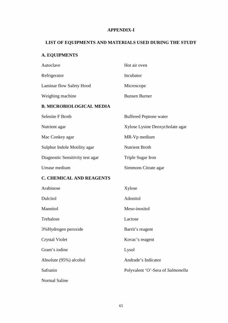
65
APPENDIX-I
LIST OF EQUIPMENTS AND MATERIALS USED DURING THE STUDY
A. EQUIPMENTS
Autoclave Hot air oven
Refrigerator Incubator
Laminar flow Safety Hood Microscope
Weighing machine Bunsen Burner
B. MICROBIOLOGICAL MEDIA
Selenite F Broth Buffered Peptone water
Nutrient agar Xylose Lysine Deoxycholate agar
Mac Conkey agar MR-Vp medium
Sulphur Indole Motility agar Nutrient Broth
Diagnostic Sensitivity test agar Triple Sugar Iron
Urease medium Simmons Citrate agar
C. CHEMICAL AND REAGENTS
Arabinose Xylose
Dulcitol Adonitol
Mannitol Meso-inositol
Trehalose Lactose
3%Hydrogen peroxide Barrit’s reagent
Crystal Violet Kovac’s reagent
Gram’s iodine Lysol
Absolute (95%) alcohol Andrade’s Indicator
Safranin Polyvalent ‘O’-Sera of Salmonella
Normal Saline
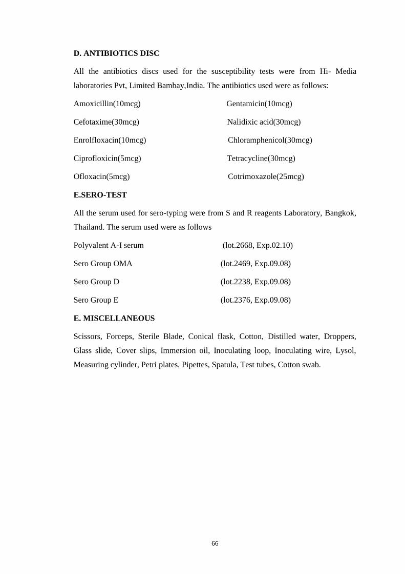
66
D. ANTIBIOTICS DISC
All the antibiotics discs used for the susceptibility tests were from Hi- Media
laboratories Pvt, Limited Bambay,India. The antibiotics used were as follows:
Amoxicillin(10mcg) Gentamicin(10mcg)
Cefotaxime(30mcg) Nalidixic acid(30mcg)
Enrolfloxacin(10mcg) Chloramphenicol(30mcg)
Ciprofloxicin(5mcg) Tetracycline(30mcg)
Ofloxacin(5mcg) Cotrimoxazole(25mcg)
E.SERO-TEST
All the serum used for sero-typing were from S and R reagents Laboratory, Bangkok,
Thailand. The serum used were as follows
Polyvalent A-I serum (lot.2668, Exp.02.10)
Sero Group OMA (lot.2469, Exp.09.08)
Sero Group D (lot.2238, Exp.09.08)
Sero Group E (lot.2376, Exp.09.08)
E. MISCELLANEOUS
Scissors, Forceps, Sterile Blade, Conical flask, Cotton, Distilled water, Droppers,
Glass slide, Cover slips, Immersion oil, Inoculating loop, Inoculating wire, Lysol,
Measuring cylinder, Petri plates, Pipettes, Spatula, Test tubes, Cotton swab.
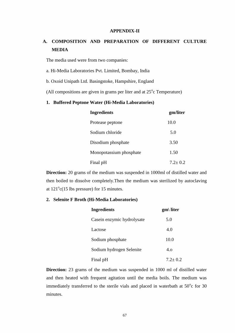
67
APPENDIX-II
A. COMPOSITION AND PREPARATION OF DIFFERENT CULTURE
MEDIA
The media used were from two companies:
a. Hi-Media Laboratories Pvt. Limited, Bombay, India
b. Oxoid Unipath Ltd. Basingstoke, Hampshire, England
(All compositions are given in grams per liter and at 25oc Temperature)
1. Buffered Peptone Water (Hi-Media Laboratories)
Ingredients gm/liter
Protease peptone 10.0
Sodium chloride 5.0
Disodium phosphate 3.50
Monopotassium phosphate 1.50
Final pH 7.2± 0.2
Direction: 20 grams of the medium was suspended in 1000ml of distilled water and
then boiled to dissolve completely.Then the medium was sterilized by autoclaving
at 121oc(15 lbs pressure) for 15 minutes.
2. Selenite F Broth (Hi-Media Laboratories)
Ingredients gm\ liter
Casein enzymic hydrolysate 5.0
Lactose 4.0
Sodium phosphate 10.0
Sodium hydrogen Selenite 4.o
Final pH 7.2± 0.2
Direction: 23 grams of the medium was suspended in 1000 ml of distilled water
and then heated with frequent agitation until the media boils. The medium was
immediately transferred to the sterile vials and placed in waterbath at 50oc for 30
minutes.
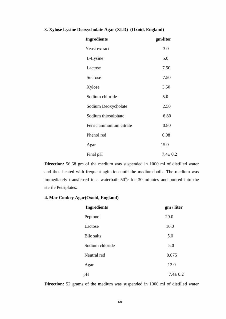
68
3. Xylose Lysine Deoxycholate Agar (XLD) (Oxoid, England)
Ingredients gm\liter
Yeast extract 3.0
L-Lysine 5.0
Lactose 7.50
Sucrose 7.50
Xylose 3.50
Sodium chloride 5.0
Sodium Deoxycholate 2.50
Sodium thiosulphate 6.80
Ferric ammonium citrate 0.80
Phenol red 0.08
Agar 15.0
Final pH 7.4± 0.2
Direction: 56.68 gm of the medium was suspended in 1000 ml of distilled water
and then heated with frequent agitation until the medium boils. The medium was
immediately transferred to a waterbath 50oc for 30 minutes and poured into the
sterile Petriplates.
4. Mac Conkey Agar(Oxoid, England)
Ingredients gm / liter
Peptone 20.0
Lactose 10.0
Bile salts 5.0
Sodium chloride 5.0
Neutral red 0.075
Agar 12.0
pH 7.4± 0.2
Direction: 52 grams of the medium was suspended in 1000 ml of distilled water
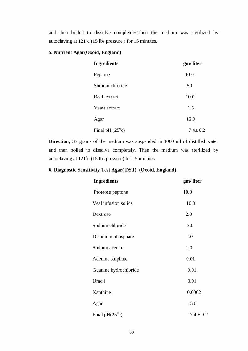
69
and then boiled to dissolve completely.Then the medium was sterilized by
autoclaving at 121oc (15 lbs pressure ) for 15 minutes.
5. Nutrient Agar(Oxoid, England)
Ingredients gm/ liter
Peptone 10.0
Sodium chloride 5.0
Beef extract 10.0
Yeast extract 1.5
Agar 12.0
Final pH (25oc) 7.4± 0.2
Direction; 37 grams of the medium was suspended in 1000 ml of distilled water
and then boiled to dissolve completely. Then the medium was sterilized by
autoclaving at 121oc (15 lbs pressure) for 15 minutes.
6. Diagnostic Sensitivity Test Agar( DST) (Oxoid, England)
Ingredients gm/ liter
Proteose peptone 10.0
Veal infusion solids 10.0
Dextrose 2.0
Sodium chloride 3.0
Disodium phosphate 2.0
Sodium acetate 1.0
Adenine sulphate 0.01
Guanine hydrochloride 0.01
Uracil 0.01
Xanthine 0.0002
Agar 15.0
Final pH(25oc) 7.4 ± 0.2
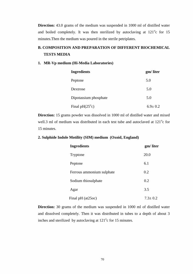
70
Direction: 43.0 grams of the medium was suspended in 1000 ml of distilled water
and boiled completely. It was then sterilized by autoclaving at 121oc for 15
minutes.Then the medium was poured in the sterile petriplates.
B. COMPOSITION AND PREPARATION OF DIFFERENT BIOCHEMICAL
TESTS MEDIA
1. MR-Vp medium (Hi-Media Laboratories)
Ingredients gm/ liter
Peptone 5.0
Dextrose 5.0
Dipotassium phosphate 5.0
Final pH(25oc) 6.9± 0.2
Direction: 15 grams powder was dissolved in 1000 ml of distilled water and mixed
well.3 ml of medium was distributed in each test tube and autoclaved at 121oc for
15 minutes.
2. Sulphide Indole Motility (SIM) medium (Oxoid, England)
Ingredients gm/ liter
Tryptone 20.0
Peptone 6.1
Ferrous ammonium sulphate 0.2
Sodium thiosulphate 0.2
Agar 3.5
Final pH (at25oc) 7.3± 0.2
Direction: 30 grams of the medium was suspended in 1000 ml of distilled water
and dissolved completely. Then it was distributed in tubes to a depth of about 3
inches and sterilized by autoclaving at 121oc for 15 minutes.
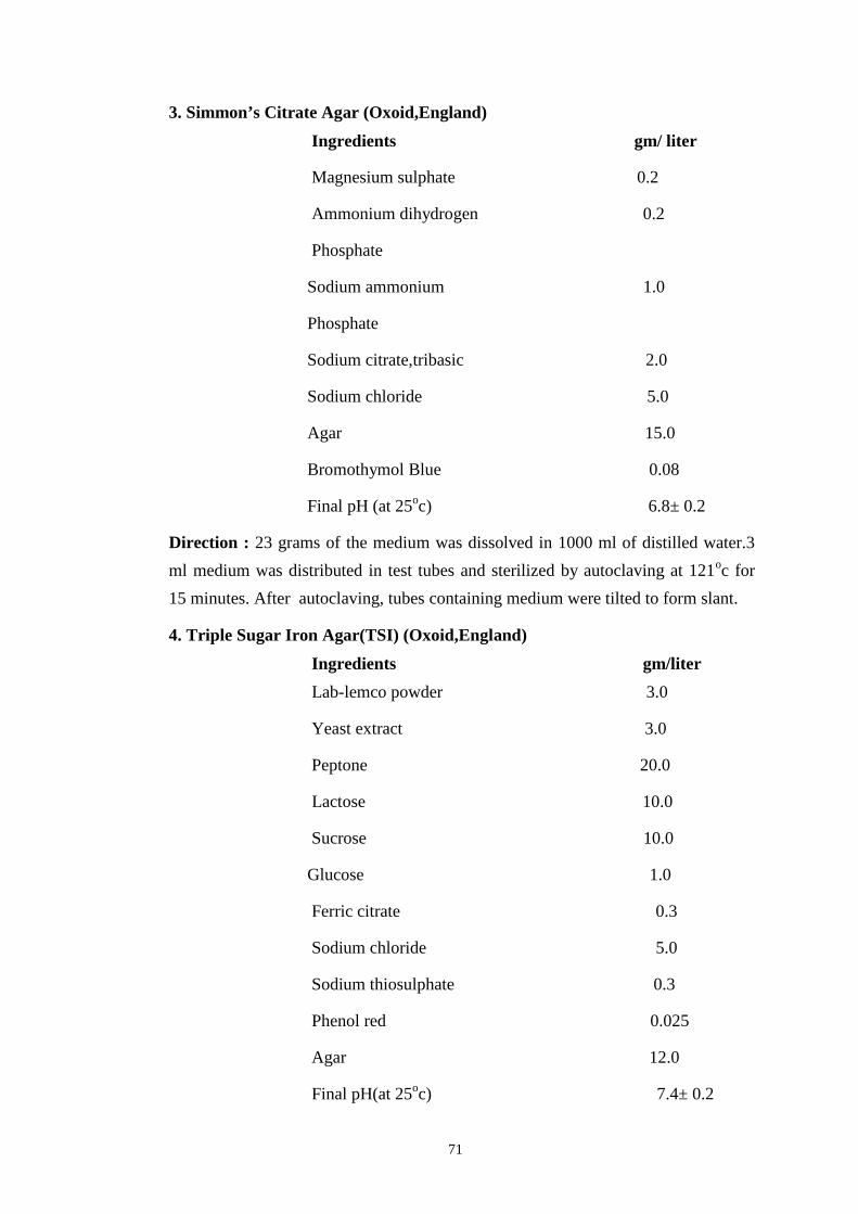
71
3. Simmon’s Citrate Agar (Oxoid,England)Ingredients gm/ liter
Magnesium sulphate 0.2
Ammonium dihydrogen 0.2
Phosphate
Sodium ammonium 1.0
Phosphate
Sodium citrate,tribasic 2.0
Sodium chloride 5.0
Agar 15.0
Bromothymol Blue 0.08
Final pH (at 25oc) 6.8± 0.2
Direction : 23 grams of the medium was dissolved in 1000 ml of distilled water.3
ml medium was distributed in test tubes and sterilized by autoclaving at 121oc for
15 minutes. After autoclaving, tubes containing medium were tilted to form slant.
4. Triple Sugar Iron Agar(TSI) (Oxoid,England)
Ingredients gm/liter
Lab-lemco powder 3.0
Yeast extract 3.0
Peptone 20.0
Lactose 10.0
Sucrose 10.0
Glucose 1.0
Ferric citrate 0.3
Sodium chloride 5.0
Sodium thiosulphate 0.3
Phenol red 0.025
Agar 12.0
Final pH(at 25oc) 7.4± 0.2
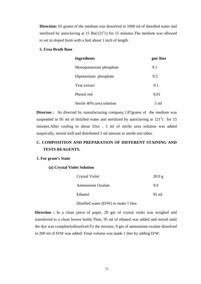
72
Direction: 65 grams of the medium was dissolved in 1000 ml of distolled water and
sterilized by autoclaving at 15 lbs(121oc) for 15 minutes.The medium was allowed
to set in sloped form with a butt about 1 inch of length.
5. Urea Broth Base
Ingredients gm/ liter
Monopotassium phosphate 9.1
Dipotassium phosphate 9.5
Yeat extract 0.1
Phenol red 0.01
Sterile 40% urea solution 5 ml
Directon : As directed by manufacturing company,1.87grams of the medium was
suspended in 95 ml of distilled water and sterilized by autoclaving at 121oc for 15
minutes.After cooling to about 55oc , 5 ml of sterile urea solution was added
aseptically, mixed well and distributed 5 ml amount in sterile test tubes.
C. COMPOSITION AND PREPARATION OF DIFFERENT STAINING AND
TESTS REAGENTS.
1. For gram’s Stain
(a) Crystal Violet Solution
Crystal Violet 20.0 g
Ammonium Oxalate 9.0
Ethanol 95 ml
Distilled water (D/W) to make 1 liter.
Direction : In a clean piece of paper, 20 gm of crystal violet was weighed and
transferred to a clean brown bottle.Then, 95 ml of ethanol was added and mixed until
the dye was completelydissolved.To the mixture, 9 gm of ammonium oxalate dissolved
in 200 ml of D/W was added. Final volume was made 1 liter by adding D/W.
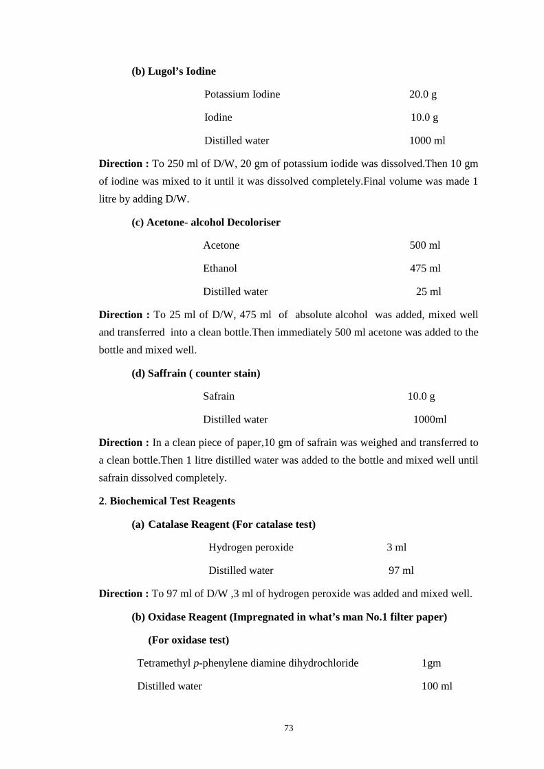
73
(b) Lugol’s Iodine
Potassium Iodine 20.0 g
Iodine 10.0 g
Distilled water 1000 ml
Direction : To 250 ml of D/W, 20 gm of potassium iodide was dissolved.Then 10 gm
of iodine was mixed to it until it was dissolved completely.Final volume was made 1
litre by adding D/W.
(c) Acetone- alcohol Decoloriser
Acetone 500 ml
Ethanol 475 ml
Distilled water 25 ml
Direction : To 25 ml of D/W, 475 ml of absolute alcohol was added, mixed well
and transferred into a clean bottle.Then immediately 500 ml acetone was added to the
bottle and mixed well.
(d) Saffrain ( counter stain)
Safrain 10.0 g
Distilled water 1000ml
Direction : In a clean piece of paper,10 gm of safrain was weighed and transferred to
a clean bottle.Then 1 litre distilled water was added to the bottle and mixed well until
safrain dissolved completely.
2. Biochemical Test Reagents
(a) Catalase Reagent (For catalase test)
Hydrogen peroxide 3 ml
Distilled water 97 ml
Direction : To 97 ml of D/W ,3 ml of hydrogen peroxide was added and mixed well.
(b) Oxidase Reagent (Impregnated in what’s man No.1 filter paper)
(For oxidase test)
Tetramethyl p-phenylene diamine dihydrochloride 1gm
Distilled water 100 ml
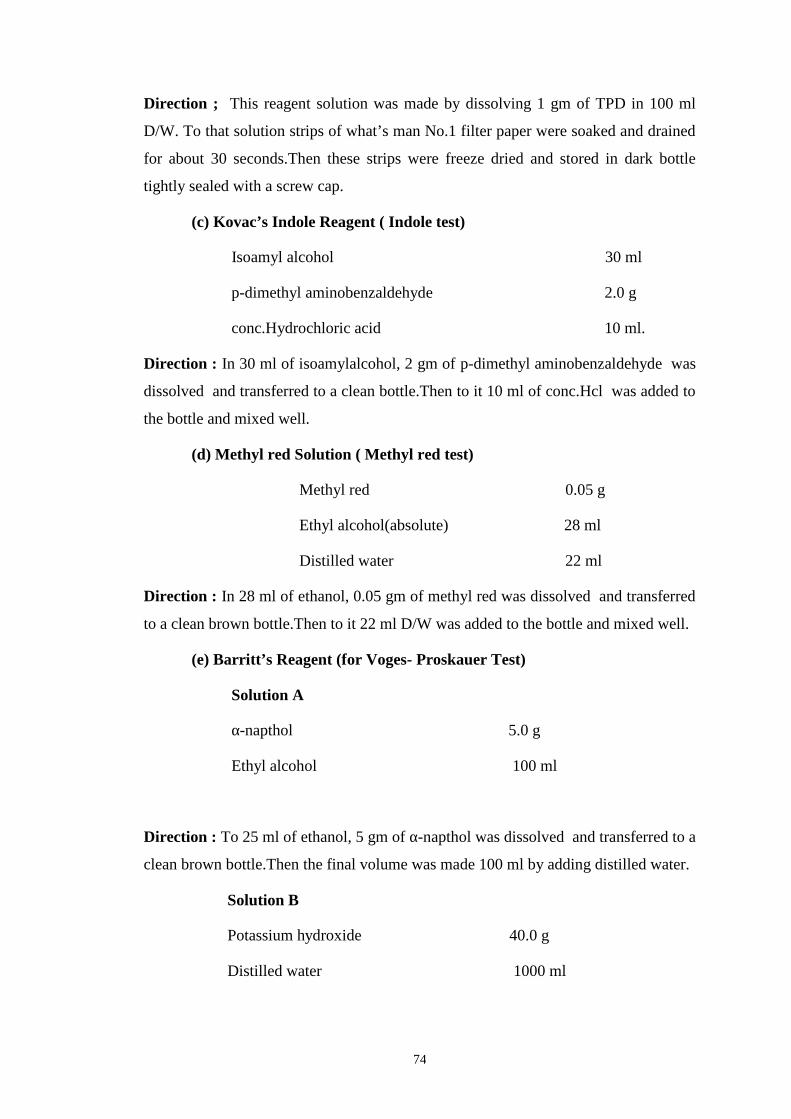
74
Direction ; This reagent solution was made by dissolving 1 gm of TPD in 100 ml
D/W. To that solution strips of what’s man No.1 filter paper were soaked and drained
for about 30 seconds.Then these strips were freeze dried and stored in dark bottle
tightly sealed with a screw cap.
(c) Kovac’s Indole Reagent ( Indole test)
Isoamyl alcohol 30 ml
p-dimethyl aminobenzaldehyde 2.0 g
conc.Hydrochloric acid 10 ml.
Direction : In 30 ml of isoamylalcohol, 2 gm of p-dimethyl aminobenzaldehyde was
dissolved and transferred to a clean bottle.Then to it 10 ml of conc.Hcl was added to
the bottle and mixed well.
(d) Methyl red Solution ( Methyl red test)
Methyl red 0.05 g
Ethyl alcohol(absolute) 28 ml
Distilled water 22 ml
Direction : In 28 ml of ethanol, 0.05 gm of methyl red was dissolved and transferred
to a clean brown bottle.Then to it 22 ml D/W was added to the bottle and mixed well.
(e) Barritt’s Reagent (for Voges- Proskauer Test)
Solution A
α-napthol 5.0 g
Ethyl alcohol 100 ml
Direction : To 25 ml of ethanol, 5 gm of α-napthol was dissolved and transferred to a
clean brown bottle.Then the final volume was made 100 ml by adding distilled water.
Solution B
Potassium hydroxide 40.0 g
Distilled water 1000 ml
75
Direction : To 25 ml of D/W, 40gm of KOH was dissolved and transferred to a clean
brown bottle.Then the final volume was made 100 ml by adding distilled water.
(f ) Andrade’s Indicator
Acid Fuschin 5.0 g
Distilled water 1000 ml
1 N NaoH 150-180 ml
Direction : Acid Fuschin was dissolved in didtilled water and then 150 ml of alkali
solution was added .It was mixed properly and allowed to stand at room temperature
for 24 hours in rotor .If the dye has not been decolorized then 10 ml alkali was added
further, mixed well and left for next 24 hours. The ultimate desired color is straw
yellow.
76
APPENDIX-III
GRAM-STAIN PROCRDURE
First devised by Hans Christian Gram during the late 19th century, the Gram stain
can be used effectively to divide all bacterial species into two large groups :those that
take up the basic dye ,crystal violet (Gram positive) and those that allow the crystal
dye to wash out easily with the decolorizer alcohol or acetone ( Gram – negative).
The following steps are involved in Gram-stain;
1. A thin film of the material to be examined was prepared and dried on a glass slide.
2. The material on the slide was heat fixed and allowed to cool before staining.
3. The slide was flooded with crystal violet stain and allowed to remain with out
drying for 1 minute.
4. The slide was rinsed with tap water, shaking off excess.
5. The slide was flooded with iodine solution and allowed to remain on the surface
with out drying for twice as long as the crystal violet was in contact with the slide
surface.
6. The slide was rinsed with tap water, shaking off excess.
7. The slide was flooded with alcohol acetone decolorizer for 10 seconds and rinsed
immediately with tap water until no further color flows from the slide with the
decolorizer. Thicker smear requires more aggressive decolorizing.
8. The slide was flooded with counter stain (safranin) for 1minute and washed off
with tap water.
9. The slide was blotted between two clean sheets of bibulous paper and examined
microscopically under oil immersion at 100 X.
77
APPENDIX-IV
METHODOLOGY OF BIOCHEMICAL TESTS USED FOR
IDENTIFICATION OF BACTERIA
A. Catalase Test
This test is performed to demonstrate the presence of catalase, an enzyme that
catalyses the release of oxygen from hydrogen peroxide. During aerobic respiration,
in the presence of oxygen, micro organisms produce hydrogen peroxide, which is
lethal to the cell itself. The enzyme catalase is present in most cytochrome containing
aerobic and facultative anaerobic bacteria, the main exception being Streptococcus
Spp.
Procedure: A small amount of culture from nutrient agar plate was taken in a clean
glass slide and about 2-3 drops of 3% H2O2 was put on the surface of the slide. The
positive test is indicated by the formation of active bubbling of the oxygen gas. A
false positive reaction may be obtained if the culture medium contains catalase (eg.
Blood agar) or if an iron wire loop is used.
B. Oxidase Test
This test is performed for the detection of cytochrome Oxidase in bacteria which
catalyzes the transport of electrons between electron donors. In the presence of redox
dye Tetramethyl p-phenylene diamine dihydrochloride, the cytochrome Oxidase
oxidizes it in to a deep purple colored end product indophenol which is detected in the
test. The test is used for screening species of Neisseria, Alcaligenes, Aeromonas,
Vibrio, Campylobacter and pseudomonas which give positive reactions and for
excluding the enterobacteriaceae, all species of which give negative reaction.
Procedure: A piece of filter paper was soaked with few drops of Oxidase reagent
(whatman’s No. 1 filter paper impregnated with 1% Tetramethyl p-phenylene diamine
dihydrochloride.) then the colony of the test organism was smeared on the filter
paper. The positive test is indicated by the appearance of blue purple color with 10
seconds.
78
C. Indole Production Test
This test detects the ability of the organism to produce an enzyme ‘tryptophanase’
which oxidizes tryptophan to form indolic metabolites: indole, skatola (methyl indole)
and indole acetic acid. The enzyme tryptophanase catalyses the deamination reaction
attacking the tryptophan molecule in its side chain and leaving the aromatic ring intact
in the form of indole.
Procedure: A smooth bacterial colony was stabbed on SIM (Sulphide Indole
Motility) medium by a sterile stab-wire and the inoculated media was incubated at 37o
c for 24 hours. After 24 hours incubation, 2-3 drops of kovac‘s reagent was added.
Appearance of red color on the top of media indicates indole positive. Indole if
present combines with the aldehyde present in the reagent to give a red color in the
alcohol layer. The color reaction is based on the presence of the pyrrole structure
present in indole.
D. Methyl Red Test
This test is performed to test the ability of an organism to produce and maintain stable
acid end product from the fermentation of glucose to give a red color with the
indicator methyl red and to over come the buffering capacity of the system. Medium
used in the study was Clark and Lubs medium (MR/Vp broth, pH 6.9). Methyl red is
an indicator which is already acid and will denote changes in degree of acidity by
color reactions of over a pH range of 4.4 to 6.0.
Procedure: A pure colony of the test organism was inoculated in to 2 ml of MRVp
medium and was incubated at 30o c for 24 hours. After incubation, about 5 drops of
methyl red reagent was added and mixed well. The positive test was indicated by the
development of bright red color, indicating acidity and negative with yellow color.
E. Voges-Proskauer (VP) Test
The principle of this test is to determine the ability of some organisms to produce a
acetyl methyl carbinol, a neutral end product (acetoin) or its reduction product 2,3-
butanidiol during fermentation of carbohydrates. An organism of the
79
entrobacteriaceae group is usually either methyl red positive and Voges – proskauer –
negative or methyl red negative and Voges – proskauer positive.
Procedure: A pure colony of the test organism was inoculated in to 2 ml of MRVP
medium and was incubated at 37oc for 24 hours. After incubation, about 5 drops of
barritt’s reagent was added and shaken well for maximum aeration and kept for 15
minutes, positive test is indicated by the development of pink red color.
F. Citrate Utilization Test
This test is performed t detect whether an organism utilizes citrate as a sole source of
carbon for metabolism with resulting alkalinity. The medium used for citrate
fermentation (Simmons Citrate Medium) also contains inorganic ammonium salts.
Organisms capable of utilizing citrate as its sole carbon source also utilizes the
ammonium salts present in the medium as its sole Nitrogen source, the ammonium
salts are broken down to ammonia with resulting alkalinity.
Procedure: A loopful of test organism was streaked on the slant area of Simmon’s
Citrate agar medium and incubated at 37oc for 24 hours. A positive test was indicated
by the growth of organism and change of media by green to blue, due to alkaline
reaction. The pH indicator bromothymol blue has pH a range of 6.0 to 7.6, that is
above pH 7.6, a blue color develops due to alkalinity of the medium.
G. Motility Test
This test is done to determine if an organism was motile or non-motile. Bacteria are
motile by means of flagella. Flagella occur primarily among the bacilli, how ever a
few cocci forms are motile. Motile bacteria may contain single flagella. The motility
media used for motility test are semisolid, making motility interpretations
macroscopic.
Procedure: Motility of organism was tested by hanging drop and cultural method. In
cultural method, the test organism was stabbed in the SIM medium and incubated at
37oc for 48 hours. Motile organisms migrate from the stabline and deffuse in to the
medium causing turbidity. Where as non-motile bacteria show the growth along the
stabline, and the surrounding media remains colorless and clear color.
80
In hanging drop method, 1-2 drops of bacterial suspension was added to the centre of
concavity slid. Grease was applied to four corners of coverslips and placed over the
slid. The slid was then observed under 10X to observe motility.]
H. Triple Sugar Iron (TSI) Agar Test
The TSI agar is used to determine the ability of an organism to utilize specific
carbohydrate incorporated in the medium (Glucose, Sucrose and Lactose in
concentrations of 0.1%, 1%, 1.0% respectively), with or with out the production of
gas (Indicated by cracks in the medium as well as an air gap at the bottom of the tube)
along with determination of possible hydrogen sulfide production (detected by
production of black color in the medium). A pH indicator (phenol red) included in the
medium can detect acid production from fermentation of these carbohydrates and it
gives yellow reaction at acidic pH, and red reaction to indicate an alkaline
surrounding.
Procedure: The test organism was streaked and stabbed on the surface of TSI and
incubated at 37oc for 24 hours. Acid production limited only to the butt region of the
tube is indicative of glucose utilization, while acid production in slat and butt
indicates sucrose or lactose fermentation. The results are interpreted as follows:
a. Yellow (Acid)/Yellow (Acid), Gas, H2S Lactose/Sucrose Fermenter,
H2S producer.
b. Red (Alkanine)/Yellow (Acid), No Gas, No H2S Only Glucose, Not
Lactose/Sucrose Fermenter, Not Aerogenic, No H2S production.
c. Red (Alkanine)/No Change Glucose, Lactose and Sucrose Non –
Fermenter.
d. Yellow (Acid)/No Change Glucose-Oxidiser.
e. No Change / No Change Non Fermenter.
81
I. Urea Hydrolysis Test
This test demonstrates the urease activity present in certain bacteria which
decomposes urea releasing ammonia and carbon dioxide. Ammonia thus produced
changes the color of indicator (Phenol Red) incorporated in the medium.
Procedure: The test organism was inoculated in a medium containing urea and the
indicator phenol red. The inoculated medium was incubated at 37oc over night.
Positive organism shows pink red color due to the break down of the urea to
ammonia. With the release of ammonia the medium becomes alkaline as shown by a
change in color of the indicator to pink.
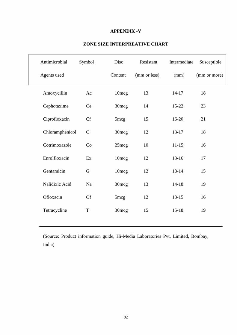
82
APPENDIX -V
ZONE SIZE INTERPREATIVE CHART
Antimicrobial Symbol Disc Resistant Intermediate Susceptible
Agents used Content (mm or less) (mm) (mm or more)
Amoxycillin Ac 10mcg 13 14-17 18
Cephotaxime Ce 30mcg 14 15-22 23
Ciprofloxacin Cf 5mcg 15 16-20 21
Chloramphenicol C 30mcg 12 13-17 18
Cotrimoxazole Co 25mcg 10 11-15 16
Enrolfloxacin Ex 10mcg 12 13-16 17
Gentamicin G 10mcg 12 13-14 15
Nalidixic Acid Na 30mcg 13 14-18 19
Ofloxacin Of 5mcg 12 13-15 16
Tetracycline T 30mcg 15 15-18 19
(Source: Product information guide, Hi-Media Laboratories Pvt. Limited, Bombay,
India)
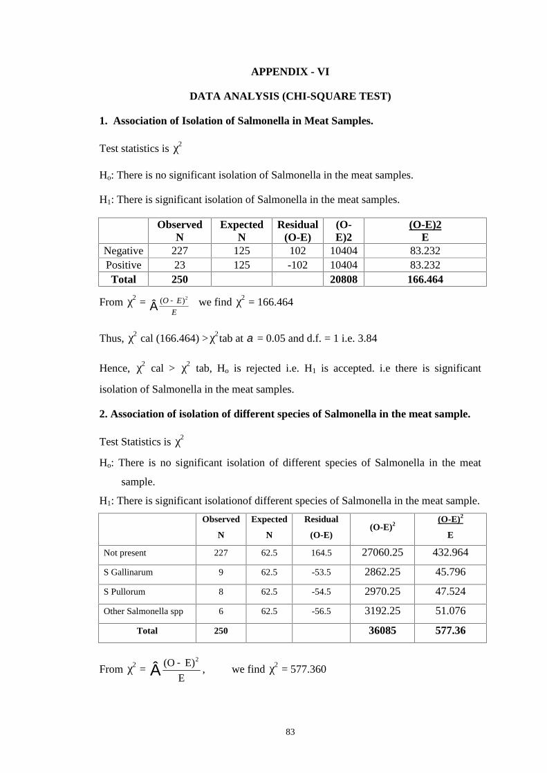
83
APPENDIX - VI
DATA ANALYSIS (CHI-SQUARE TEST)
1. Association of Isolation of Salmonella in Meat Samples.
Test statistics is 2χ
Ho: There is no significant isolation of Salmonella in the meat samples.
H1: There is significant isolation of Salmonella in the meat samples.
ObservedN
ExpectedN
Residual(O-E)
(O-E)2
(O-E)2E
Negative 227 125 102 10404 83.232Positive 23 125 -102 10404 83.232Total 250 20808 166.464
From 2χ = E
EO 2)( we find 2χ = 166.464
Thus, 2χ cal (166.464) > 2χ tab at = 0.05 and d.f. = 1 i.e. 3.84
Hence, 2χ cal > 2χ tab, Ho is rejected i.e. H1 is accepted. i.e there is significant
isolation of Salmonella in the meat samples.
2. Association of isolation of different species of Salmonella in the meat sample.
Test Statistics is 2χ
Ho: There is no significant isolation of different species of Salmonella in the meat
sample.
H1: There is significant isolationof different species of Salmonella in the meat sample.
Observed
N
Expected
N
Residual
(O-E)(O-E)2
(O-E)2
E
Not present 227 62.5 164.5 27060.25 432.964
S Gallinarum 9 62.5 -53.5 2862.25 45.796
S Pullorum 8 62.5 -54.5 2970.25 47.524
Other Salmonella spp 6 62.5 -56.5 3192.25 51.076
Total 250 36085 577.36
From 2χ = E
E)(O 2
, we find 2χ = 577.360

84
Thus, 2χ cal (577.360) > 2χ tab at α = 0.05 and d.f. = 3 i.e. 7.815
Hence, 2χ cal > 2χ tab, Ho is rejected i.e. H1 is accepted i.e. there is significant
isolation of different species of Salmonella in the meat sample.
Data Analysis Software: Statistical Programme for Social Sciences (SPSS);Microsoft Excel