Isolation, molecular characterization and antimicrobial resistance patterns of Salmonella and...
12
Isolation, molecular characterization and antimicrobial resistance patterns of Salmonella and Escherichia coli isolates from meat-based fast food in Lebanon Steve Harakeh a, * , Hadi Yassine a , Maya Gharios b , Elie Barbour c , Shadi Hajjar a , Mutasem El-Fadel d , Imad Toufeili b , Raja Tannous b, * a Department of Biology, American University of Beirut, PO Box 11-0236, Beirut, Lebanon b Department of Nutrition and Food Science, American University of Beirut, PO Box 11-0236, Beirut, Lebanon c Department of Animal Sciences, American University of Beirut, PO Box 11-0236, Beirut, Lebanon d Department of Civil and Environmental Engineering, American University of Beirut, PO Box 11-0236, Beirut, Lebanon Received 15 April 2004; accepted 10 September 2004 Abstract The aim of this study was to characterize at the molecular level the different stains of Salmonella spp. and Escherichia coli that were isolated from meat-based fast food in Lebanon. In addition, this study evaluated the resistance of those strains to different antimicrobials that are commonly used. The foods included were Lahm-bi-Ajeen (LBA, meat pies) and Shawarma (Lebanese meat sandwiches similar to Gyros and Donairs, containing meat, vegetables, and sesame seeds-oil-based sauce). Polymerase chain reaction (PCR) was used to characterize and identify the strains of both bacteria. Salmonella species characterization was performed using rfb genes cluster genetic marker, while that of E. coli strains were carried out based on stx1, stx2, eaeA, fliC, and ehlyA virulence markers. The characterized strains were then tested for their response to various antimicrobials. The results showed that the tested foods were contaminated with Salmonella paratyphi (serogroup A) and Shiga Toxin (Stx)-producing E. coli (STX-EC). The PCR showed that 75% of E. coli tested strains was positive in PCR performed with stx1 primers, one of which was eaeA positive. Two of the tested strains were positive using PCR with fliC primers. The resistances of the various strains were evaluated using the following antimicrobials: Oxacillin, Teicoplanin, Trimethoprim/sulfamethoxazole, Gentamicin, Clindamycin, Cefotaxime, Cefuroxime, Erythromycin, and Vancomycin. Bacteria were highly resistant to one or more of the tested antimicrobials. Approximately 69% of E. coli and 77.8% of Salmonella spp. exhibited resistance. Salmonella spp. were shown to be 100% resistant to four antimicrobials: Oxacillin, Teicoplanin, Clindamycin, Vancomycin, and Erythromycin, while E. coli was 100% resistant to Teicoplanin and Trimethoprim/ sulfamethoxazole. The most interesting findings were the high susceptibility of the E. coli to Gentamicin (100%). Highest 0048-9697/$ - see front matter D 2004 Elsevier B.V. All rights reserved. doi:10.1016/j.scitotenv.2004.09.025 * Corresponding authors. S. Hakareh is to be contacted at Department of Biology, American University of Beirut, PO Box 11-0236, Beirut, Lebanon. Tel.: +961 3 570119; fax: +961 1 744461. R. Tannous, Department of Nutrition and Food Science, American University of Beirut, PO Box 11-0236, Beirut, Lebanon. E-mail addresses: [email protected] (S. Harakeh)8 [email protected] (R. Tannous). Science of the Total Environment 341 (2005) 33– 44 www.elsevier.com/locate/scitotenv
-
Upload
independent -
Category
Documents
-
view
3 -
download
0
Transcript of Isolation, molecular characterization and antimicrobial resistance patterns of Salmonella and...

www.elsevier.com/locate/scitotenv
Science of the Total Environ
Isolation, molecular characterization and antimicrobial
resistance patterns of Salmonella and Escherichia coli
isolates from meat-based fast food in Lebanon
Steve Harakeha,*, Hadi Yassinea, Maya Ghariosb, Elie Barbourc, Shadi Hajjara,
Mutasem El-Fadeld, Imad Toufeilib, Raja Tannousb,*
aDepartment of Biology, American University of Beirut, PO Box 11-0236, Beirut, LebanonbDepartment of Nutrition and Food Science, American University of Beirut, PO Box 11-0236, Beirut, Lebanon
cDepartment of Animal Sciences, American University of Beirut, PO Box 11-0236, Beirut, LebanondDepartment of Civil and Environmental Engineering, American University of Beirut, PO Box 11-0236, Beirut, Lebanon
Received 15 April 2004; accepted 10 September 2004
Abstract
The aim of this study was to characterize at the molecular level the different stains of Salmonella spp. and Escherichia coli
that were isolated from meat-based fast food in Lebanon. In addition, this study evaluated the resistance of those strains to
different antimicrobials that are commonly used. The foods included were Lahm-bi-Ajeen (LBA, meat pies) and Shawarma
(Lebanese meat sandwiches similar to Gyros and Donairs, containing meat, vegetables, and sesame seeds-oil-based sauce).
Polymerase chain reaction (PCR) was used to characterize and identify the strains of both bacteria. Salmonella species
characterization was performed using rfb genes cluster genetic marker, while that of E. coli strains were carried out based on
stx1, stx2, eaeA, fliC, and ehlyA virulence markers. The characterized strains were then tested for their response to various
antimicrobials. The results showed that the tested foods were contaminated with Salmonella paratyphi (serogroup A) and Shiga
Toxin (Stx)-producing E. coli (STX-EC). The PCR showed that 75% of E. coli tested strains was positive in PCR performed
with stx1 primers, one of which was eaeA positive. Two of the tested strains were positive using PCR with fliC primers.
The resistances of the various strains were evaluated using the following antimicrobials: Oxacillin, Teicoplanin,
Trimethoprim/sulfamethoxazole, Gentamicin, Clindamycin, Cefotaxime, Cefuroxime, Erythromycin, and Vancomycin. Bacteria
were highly resistant to one or more of the tested antimicrobials. Approximately 69% of E. coli and 77.8% of Salmonella spp.
exhibited resistance. Salmonella spp. were shown to be 100% resistant to four antimicrobials: Oxacillin, Teicoplanin,
Clindamycin, Vancomycin, and Erythromycin, while E. coli was 100% resistant to Teicoplanin and Trimethoprim/
sulfamethoxazole. The most interesting findings were the high susceptibility of the E. coli to Gentamicin (100%). Highest
0048-9697/$ - s
doi:10.1016/j.sc
* Correspondi
Lebanon. Tel.: +
Box 11-0236, B
E-mail addr
ment 341 (2005) 33–44
ee front matter D 2004 Elsevier B.V. All rights reserved.
itotenv.2004.09.025
ng authors. S. Hakareh is to be contacted at Department of Biology, American University of Beirut, PO Box 11-0236, Beirut,
961 3 570119; fax: +961 1 744461. R. Tannous, Department of Nutrition and Food Science, American University of Beirut, PO
eirut, Lebanon.
esses: [email protected] (S. Harakeh)8 [email protected] (R. Tannous).
S. Harakeh et al. / Science of the Total Environment 341 (2005) 33–4434
resistance in the case of Salmonella spp. was seen against Cefotaxime (74%). Those two antimicrobials are commonly used for
the treatment of enteric infections caused by gram-negative bacteria. The results showed that meat-based fast foods in Lebanon
could be a public health hazard, especially Shawarma, as they may act as a potential vehicle for many antimicrobial-resistant
pathogenic organisms. Improper hygienic standards and indiscriminate use of antimicrobials are two of the main causes for the
prevalence of these pathogenic resistance strains in Lebanon. These results will emphasize the need to implement protective
measures and more emphasis will be placed on the application of hygienic practices to reduce the levels of food contamination.
D 2004 Elsevier B.V. All rights reserved.
Keywords: Lebanese fast foods; Antimicrobial resistance; Escherichia coli (STX-EC); Salmonella paratyphi
1. Introduction
The importance of food as a vehicle for the
transmission of many diseases has been documented
for a long time especially in the developing countries
where hygienic standards are not strictly followed and
enforced. The presence of these microorganisms can
lead to many food-borne outbreaks. Furthermore, the
wide application of antimicrobials has led to large-
scale dissemination of bacteria resistant to antimicro-
bials in the environment. Infection caused by those
resistant strains usually lead to a high fatality rate than
especially among immuno-comprised individuals
(Holmberg et al., 1984). The identification of patho-
genic organisms is highly crucial for surveillance,
prevention, and control of food-borne diseases. In
addition, studying antimicrobial resistance in humans
and animals is important in order to (a) detect changes
patterns in resistance, (b) implement control measures
on the use of antimicrobial agents, and (c) prevent the
spread of multidrug-resistant strains of bacteria
(Duijkeren et al., 2003).
Considering the marked importance of Salmonella
spp. and Escherichia coli (E. coli) organisms as food-
borne pathogens, we aimed in this study at character-
izing the different strains of Salmonella spp. and E.
coli that are present in meat-based fast food in
Lebanon and to evaluate their antimicrobial resistance
patterns.
Salmonellosis has become one of main causative
agents of enteric infections in humans and animals
(Tauxe, 1996). Several Salmonellosis outbreaks have
been documented worldwide due to the consumption
of contaminated meat in the last decade (Liewellyn et
al., 1998).
In Salmonella spp., the rfb gene cluster encodes
for the O antigens of Salmonella spp. polysaccharides
(Lee et al., 1992). Variations among different O
antigen structures are manifested in the types of sugar
present or the arrangement of sugars. This variability
provides the basis for serotyping Salmonella spp. into
serogroups (Lee et al., 1992). In our study, this highly
polymorphic rfb gene cluster has been targeted as a
molecular marker to detect Salmonella spp. serovars
in contaminated meat-based fast food in Lebanon.
Polymerase chain reaction (PCR) procedure was
performed using four sets of primers that amplify
the rfb(B), rfb(C1), rfb(C2) and rfb(D) genes of the
rfb cluster, respectively (Luk et al. 1993; Lee et al.,
1992).
Escherichia coli on the other hand is common,
usually harmless, bacteria of the human intestinal
flora. However, five groups of E. coli-causing
diarrhea in humans and other warm-blooded animals
have been identified (Brook et al., 1994; Wasteson,
2001). These include enterotoxinogenic E. coli
(ETEC), enteropathogenic E. coli (EPEC), enteroag-
gregative E. coli (EAEC), enteroinvasive E. coli
(EIEC), and enterohaemorrhagic E. coli (EHEC).
The later includes Shiga Toxin (Stx)-Producing E.
coli (STX-EC).
Shiga toxin (Stx)-producing E. coli (STX-EC), also
known as Verotoxin-producing E. coli, is associated
with infant diarrhea, haemorrhagic colitis, thrombiotic
thrombocytic purpura, and hemolytic uremic syn-
drome in humans (Griffin and Tauxe, 1991). E. coli
O157:H7, which belongs to STX-EC group, is the
most common serotype isolated from individuals with
haemorrhagic colitis (E. coli O157:H7). Outbreaks of
infection with those bacteria have emerged due to the
consumption of contaminated animal-derived food
products (Belongia et al., 1991; Borezyk et al., 1987).
Research reveals that E. coli O157:17 infections are
endemic in cattle, goat, sheep, and other farmed

S. Harakeh et al. / Science of the Total Environment 341 (2005) 33–44 35
animals, with the cattle being the primary source of
this pathogen (Langreid et al., 1999).
Different molecular markers throughout STX-EC
genomic DNA are now being used for the detection of
STX-EC, using mainly Polymerase chain reaction
(PCR) for this purpose (Jackson, 1991; Karch and
Meyer, 1989). In this study, five genes were targeted
for amplification (stx1, stx2, eaeA, ehlyA, and f liC),
using eight different sets of oligonucleotide primers.
Sometimes, two different sets of primers were used to
amplify the same gene, as described in the literature.
To achieve our aim, different bacterial isolates
were tested for their susceptibility to nine different
antimicrobials. Moreover, the role of plasmids in
conferring antimicrobial resistance was studied by
transforming plasmid preparations from different
isolates into E. coli wild type bacteria, MG1655.
2. Materials and methods
2.1. Sample collection
Lahm-bi-Ajeen (LBA, meat pies) and Shawarma
(Lebanese meat sandwiches similar to Gyros and
Donairs, containing meat, vegetables, sesame seeds
oil, and other ingredients) samples were randomly
collected from five areas in Lebanon. These areas
included Beirut, Mount Lebanon, north (Tripoli
mainly), south (Sidon mainly), and east (the Bekaa
Valley). The distributions of samples were as follows:
40% of the samples obtained from Beirut, 20% from
Mount Lebanon, and 13% from each of the north, the
south, and the east of Lebanon. Samples were
collected over an 11-month period from 37 food
establishments. These food establishments ranged
from restaurants to fast food outlets to small kiosks
and street vendors. Each establishment was visited on
at least two occasions and during the summer and
winter seasons. A total of 95 samples were aseptically
collected in the same way delivered to the consumer.
Samples were then packaged in sterile Whirl-Pak bags
and brought to the laboratory on ice in a cooler (b8 8C)within 2 h from the time of purchase. Duplicate
samples were obtained whenever possible. In the
Shawarma sampling, a sharp knife was used to cut
meat from the surface. All samples were collected
during lunchtime; no samples were obtained after 3:00
p.m. All samples were analyzed within 2 h after their
arrival to the laboratory.
2.2. Bacterial isolation and bacteriological analysis
Samples of Lahm-bi-Ajeen (LBA) and Shawarma
were subjected to various biological tests. To isolate
bacteria, a 25-g portion was weighed aseptically in a
sterile stomacher bags (Seward Medical StomacherRBags), diluted with 225 ml of sterilized 0.1% w/v
peptone water (Oxoid Media) and macerated in a
stomacher (Mix 1 AES Laboratories, ref :
AESAP1040) for 3 min. For isolation and counting,
serial dilutions ranging from 10�1 to 10�6 were
performed by adding 1 ml of homogenate to 9 ml
sterilized peptone water (1:10 dilution factor). Bacter-
iological analyses were performed by plating 0.1 ml of
each dilution on agar plates. Plates were then
incubated at 37 8C for 48 h, and colonies were
counted. The following bacteria were isolated: indica-
tor bacteria (Aerobic plate count and E. coli) and
pathogenic organisms (Salmonella spp. and STX-EC).
Bacteriological analyses were done according to
American Public Health Association (APHA, 2001).
All bacteriological media used were of Oxoid. Plate
Count Agar (PCA) was used for aerobic plate count
(APC), MacConkey Agar (MCA) for E. coli detection,
Sorbitol MacCankoky Agar (SMAC) for STX-EC, and
Bismuth Sulfate Agar (BSA) for Salmonella spp.
detection. Purple colonies on MCA were identified as
E. coli, whereas white colonies on SMAC were
presumptive STX-EC and were therefore stereotyped
by PCR for confirmation. On the other hand, brown to
black colonies on BSAwere identified as presumptive
Salmonella spp. and were then subjected to PCR tests
for further confirmation.
2.3. DNA extraction
Total bacterial DNA was extracted as described by
Ausubel et al. (1987). Briefly, bacteria were grown on
Brain Heart Infusion (BHI; Oxoid media) broth over
night at 37 8C, and then bacterial cells were harvested
by centrifugation and lysed with sodium lauryl
sulfate. DNA was extracted from the lysate with
phenol-chloroform and precipitated with isopropanol.
A 100–200-ng DNA template was used per 25 AlPolymerase chain reaction (PCR) reaction.

Table 1
Primers used in identifying Salmonella serogroups
Target gene Primers Sequence Resource Predicted size of the
amplified product (bp)
rfb(B) rfb(B) F: GAGAATATGTAATTGTCAG Luk et al. (1993, 1997) 851
R: GTAACCGTTTCAGTAGTTC
rfb(C1) rfb(C1) F: AAGTGTGTTTGATTGTTGG Lee et al. (1992) 781 and 410
R: GTAACCGTTTCAGTAGTTC
rfb(C2) rfb(C2) F: ATGCTTGATGTGAATAAG Luk et al. (1993, 1997) 795
R: CTAATCGAGTCAAGAAAG
rfb(D) rfb(D) F: AGTCACGACTTACATCCTAC Luk et al. (1993, 1997) 703
R: ACCTGCTATATCAGCACAAC
S. Harakeh et al. / Science of the Total Environment 341 (2005) 33–4436
2.4. PCR assays
For Salmonella spp. detection, primers for PCR
were selected based on the rfb gene clusters specific
for Salmonella spp. serogroups B, C2, and D, as
reported by Luk et al. (1993, 1997). The primers for
Salmonella spp. serovar C1 were designed based on
the sequence of Salmonella enterica group C1 rfb
gene cluster, as described by Lee et al. (1992). Primer
sequences used in the PCR are listed in Table 1. DNA
amplification was performed following the protocol of
Luk et al. (1993, 1997) with some modifications:
detection of PCR products by gel electrophoresis and
photography of agarose gels were carried out as
indicated by Gillespie et al. (1997). Briefly concern-
ing the detection for STX-EC, primers were selected
based on five genes specific for STX-EC strains.
Table 2
Primers used in identifying STX-EC strains
Target gene Primers Sequence
stx1 stx1 F: CAGTTAATGTCGTGGCGAAGG
R: CACCAGACAATGTAACCGCTG
stx2 stx2 F: ATCCTATTCCCGGGAGTTTACG
R: GCGTCATCGTATACACAGGAGC
slt1 slt1 F: ACACTGGATGATCTCAGTGG
R: CTGAATCCCCCTCCATTATG
slt2 slt2 F: CCATGACAACGGACAGCAGTT
R: CCTCTCAACTGAGCACTTTG
stx1+stx2 STEC F: gA(Ag) C(Ag)A AAT AAT TTA TAT
R: TgA TgA Tg(Ag) CAA TTC AgT AT
eaeA eaeA F: gAC CCg gCA CAA gCA TAA gC
R: CCA CCT gCA gCA ACA AgA gg
fliC flicH7 F: GCTGCAACGGTAAGTGAT
R: GGCAGCAAGCGGGTTGGT
ehlyA ehlyA F: gCA TCA TCA AgC gTA CgT TCC
R: AAT gAg CCA AgC Tgg TTA AgC
Primers to amplify Shiga toxins 1 and 2 (stx1 and
stx2) genes, also named as slt1 and slt2, were selected
as shown by Osek and Gallein (2002) and Victor et al.
(1992) (Table 2). Due to mutations that may occur
spontaneously in nature, many gene sequences might
be affected. For this reason, two sets of primers were
used to amplify each of stx1 and stx2 genes (Table 2).
This is in addition to the fact that different sets of
primers were reportedly used to amplify the above-
mentioned genes. STEC primers were used to amplify
both stx1 and stx2 in combination, as described by
Reischl et al. (2002). The three other genes that were
targeted for amplification were eacA gene, encodes
for intimin gamma, ehlyA, encodes for enterohemo-
lysin, and f liC, encodes for H7, as described by
Reischl et al. (2002) and Osek and Gallein (2002),
respectively (Table 2). Amplification of bacterial
Resource Predicted size of the
amplified product (bp)
Osek and Gallein (2002) 348
Osek and Gallein (2002) 584
Gannon et al. (1992) 614
Gannon et al. (1992) 779
gTg Reischl et al. (2002) 520
Reischl et al. (2002) 383
Reischl et al. (2002) 984
Reischl et al. (2002) 532
T
S. Harakeh et al. / Science of the Total Environment 341 (2005) 33–44 37
DNA was performed in 50-Al volumes containing
150–200-ng DNA templates; 2 mMMgC12 (AB-gene
products); 1X reaction buffer (AB-gene products); 0.2
mM each of dATP, dGTP, dCTP, and dTTP (AB-gene
products); 1.25 AM of primer; and 1U of Thermus
aquaticus (Taq) DNA polymerase (AB-gene prod-
ucts). Amplification was performed using a DNA
thermal cycler (Bio-Rad) for 35 cycles for 3 min at 93
8C, 30 s at 55 8C, and 2 min at 72 8C, and a final
extension at 75 8C for 10 min. Negative controls (no
DNA template) were included with every PCR assays.
After the PCR, 10-Al aliquots were analyzed through
submarine electrophoresis with 0.1% agarose gel
containing 0.25 Ag ethidium bromide per ml. The
DNA samples were analyzed through agarose gel
electrophoresis, visualized through UV transmission,
and photographed.
2.5. Antimicrobial susceptibility testing
Characterized strains were tested for their suscept-
ibility to nine antimicrobials, using the disk diffusion
method as set by the National Committee for Clinical
Laboratory standards (1997) (NCCLS). Briefly,
organisms were grown in a shaking water bath at 37
8C until a 0.5 McFarland turbidity standard was
obtained. A volume of 0.1 ml of the culture was then
spread over Brain Heart Infusion agar (BHIA, Oxoid)
plates. Antimicrobial disks impregnated with either of
the following antimicrobials; Oxacillin (1 Ag), Teico-planin (30 Ag), Trimethoprim/sulfamethoxazole
(1.25+23.75 Ag), Gentamicin (10 Ag), Clindamycin
(2 Ag), Cefotaxime (30 Ag), Cefuroxime (30 Ag),Erythromycin (15 Ag) and Vancomycin (30 Ag) wereplaced on the surface of inoculated agar plates. Zones
of inhibition around each antimicrobial disk were
measured after an incubation period of 24 h at 37 8C.Using NCCLS guidelines, each organism was classi-
fied either resistant or susceptible to the antimicro-
bials. Intermediate-resistant and resistant strains were
grouped together. Antimicrobial disks were purchased
from BioMerieux, France.
2.6. Plasmid extraction and transformation
Plasmid DNA was extracted using Qiagen Plasmid
Mini Kit according to the manufacture’s guideline
(Qiagen, 40724 Hilden, Germany), and DNA was run
using agarose gel electrophoresis, visualized through
UV transmission, and photographed. To decipher the
role of plasmids in conferring antimicrobial resistance,
a number of 21 different plasmid preparations from E.
coli and Salmonella spp. were transformed into wild
type E. coli MG1655 strain (Provided by Dr. Khattar,
Biology Department, AUB), and transformants were
selected on BHI agar plates supplemented with
different antimicrobials of specific concentrations:
Ampicillin (50 Ag/ml), Chloramphenicol (20 Ag/ml),
Naladixic acid (15 Ag/ml), Neomycin (50 Ag/ml), and
Tetracycline (15 Ag/ml). Transformation procedure
was carried out as described by Sambrook (1989). In
brief, 200-Al competent cells, prepared in the presence
CaCl2, were mixed with 5–10 ng plasmids, incubated
on ice for 30 min, and then heat-shocked at 42 8C for
90 s. Then, 1 ml of Lauria Broth (LB) was added to
the mixture and incubated at 37 8C to enable bacterial
growth. A volume of 100-Al aliquot was then spread
on BHI agar plates supplemented with antimicrobials
of specific concentrations. Plates were then incubated
at 37 8C for 24 h, and transformants were detected by
colony formations.
3. Results
3.1. Bacteriological counts and molecular character-
ization of Salmonella paratyphi and STX-EC using
PCR
Regarding the distribution of microbial population
in LBA, E. coli and Salmonella spp. were not
detected at all. On the other hand, 55% of the samples
was contaminated with E. coli, whereas contamina-
tion with suspected Salmonella spp. was detected in
47.5% of the samples.
Random isolates from the different samples were
subjected to biochemical tests. A number of those
tested positive for Salmonella spp. Ten of those
Salmonella spp. isolates with different morphologies
were then analyzed by PCR for the rfb gene cluster
molecular marker. Isolates were classified according
to the size of band visualized on agarose gel electro-
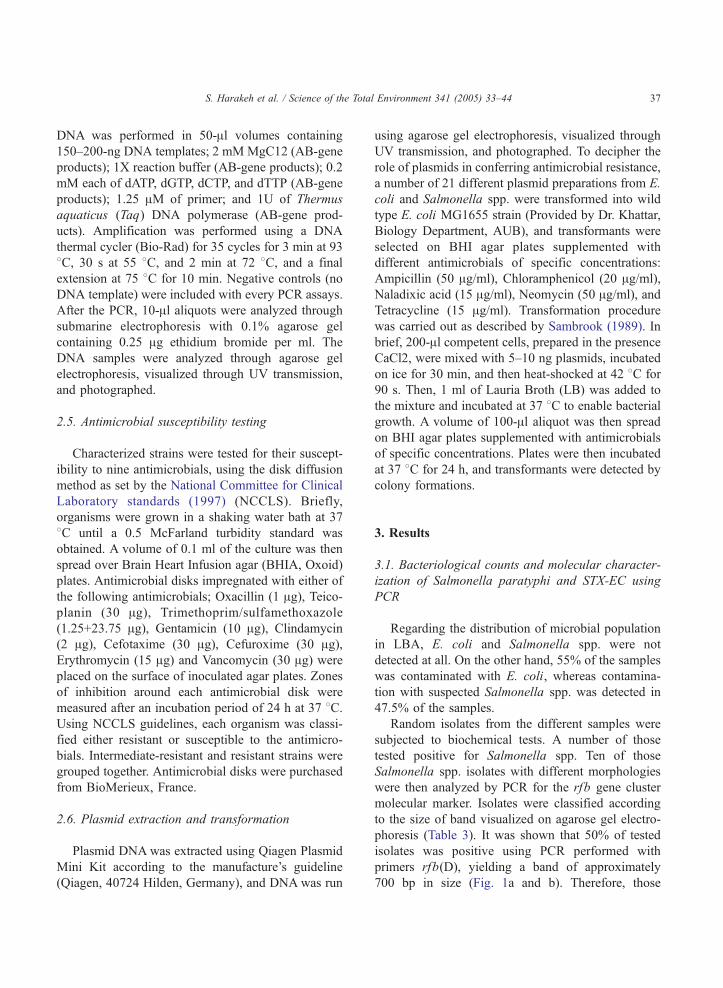
phoresis (Table 3). It was shown that 50% of tested
isolates was positive using PCR performed with
primers rfb(D), yielding a band of approximately
700 bp in size (Fig. 1a and b). Therefore, those
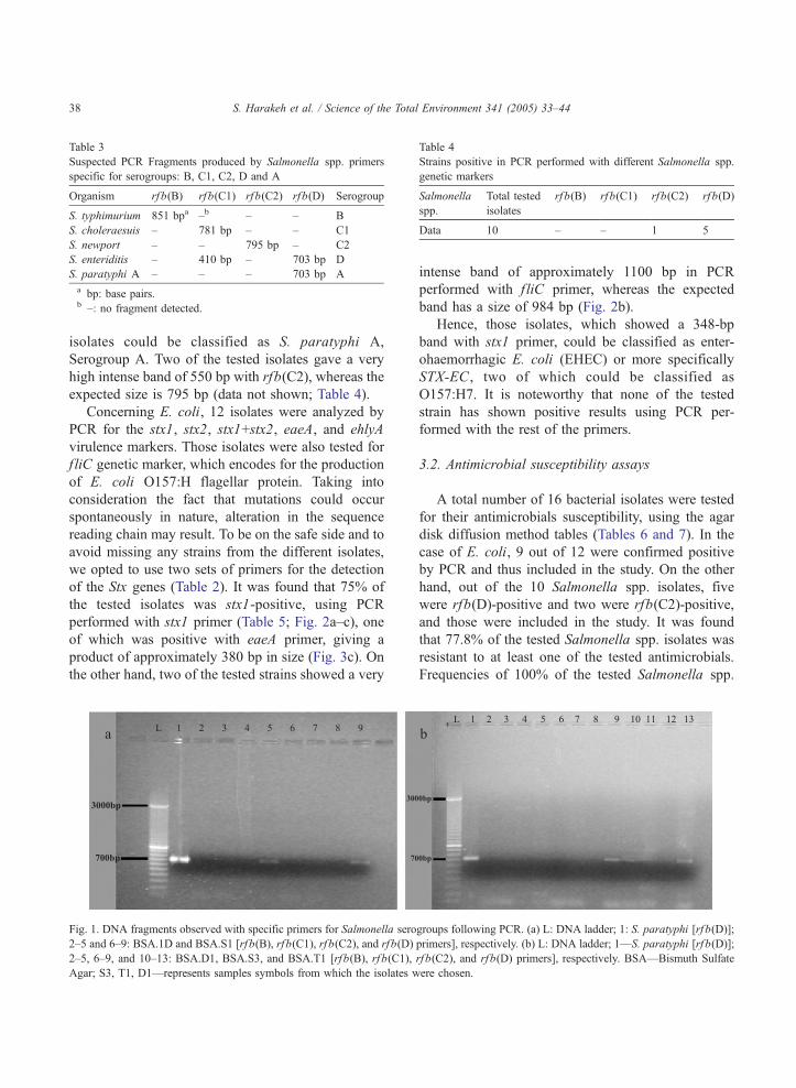
Table 3
Suspected PCR Fragments produced by Salmonella spp. primers
specific for serogroups: B, C1, C2, D and A
Organism rf b(B) rf b(C1) rfb(C2) rfb(D) Serogroup
S. typhimurium 851 bpa –b – – B
S. choleraesuis – 781 bp – – C1
S. newport – – 795 bp – C2
S. enteriditis – 410 bp – 703 bp D
S. paratyphi A – – – 703 bp A
a bp: base pairs.b –: no fragment detected.
Table 4
Strains positive in PCR performed with different Salmonella spp
genetic markers
Salmonella
spp.
Total tested
isolates
rf b(B) rf b(C1) rfb(C2) rf b(D)
Data 10 – – 1 5
S. Harakeh et al. / Science of the Total Environment 341 (2005) 33–4438
isolates could be classified as S. paratyphi A,
Serogroup A. Two of the tested isolates gave a very
high intense band of 550 bp with rfb(C2), whereas the
expected size is 795 bp (data not shown; Table 4).
Concerning E. coli, 12 isolates were analyzed by
PCR for the stx1, stx2, stx1+stx2, eaeA, and ehlyA
virulence markers. Those isolates were also tested for
f liC genetic marker, which encodes for the production
of E. coli O157:H flagellar protein. Taking into
consideration the fact that mutations could occur
spontaneously in nature, alteration in the sequence
reading chain may result. To be on the safe side and to
avoid missing any strains from the different isolates,
we opted to use two sets of primers for the detection
of the Stx genes (Table 2). It was found that 75% of
the tested isolates was stx1-positive, using PCR
performed with stx1 primer (Table 5; Fig. 2a–c), one
of which was positive with eaeA primer, giving a
product of approximately 380 bp in size (Fig. 3c). On
the other hand, two of the tested strains showed a very
Fig. 1. DNA fragments observed with specific primers for Salmonella serogroups following PCR. (a) L: DNA ladder; 1: S. paratyphi [rf b(D)]
2–5 and 6–9: BSA.1D and BSA.S1 [rf b(B), rfb(C1), rfb(C2), and rf b(D) primers], respectively. (b) L: DNA ladder; 1—S. paratyphi [rf b(D)]
2–5, 6–9, and 10–13: BSA.D1, BSA.S3, and BSA.T1 [rfb(B), rf b(C1), rf b(C2), and rf b(D) primers], respectively. BSA—Bismuth Sulfate
Agar; S3, T1, D1—represents samples symbols from which the isolates were chosen.
.
intense band of approximately 1100 bp in PCR
performed with f liC primer, whereas the expected
band has a size of 984 bp (Fig. 2b).
Hence, those isolates, which showed a 348-bp
band with stx1 primer, could be classified as enter-
ohaemorrhagic E. coli (EHEC) or more specifically
STX-EC , two of which could be classified as
O157:H7. It is noteworthy that none of the tested
strain has shown positive results using PCR per-
formed with the rest of the primers.
3.2. Antimicrobial susceptibility assays
A total number of 16 bacterial isolates were tested
for their antimicrobials susceptibility, using the agar
disk diffusion method tables (Tables 6 and 7). In the
case of E. coli, 9 out of 12 were confirmed positive
by PCR and thus included in the study. On the other
hand, out of the 10 Salmonella spp. isolates, five
were rfb(D)-positive and two were rfb(C2)-positive,
and those were included in the study. It was found
that 77.8% of the tested Salmonella spp. isolates was
resistant to at least one of the tested antimicrobials.
Frequencies of 100% of the tested Salmonella spp.
;
;

Table 5
Strains positive in PCR performed with different STX-EC genetic markers
E. coli Total tested isolates stx1 positives stx2 positives slt1 positives slt2 positives STEC Positives eaeA positives ehlyA positives
Data 12 9 – – – – 1 2
S. Harakeh et al. / Science of the Total Environment 341 (2005) 33–44 39
strains were resistant to Oxacillin, Teicoplanin,
Clindamycin, Erythromycin, and Vancomycin, and
86% was resistant to Trimethoprim/sulfamethoxa-
zole. Salmonella spp. tested strains were least
resistant to Cefotaxime (25.9%) and with moderate
susceptibility of 57.1% against both Cefuroxime and
Gentamicin.
Regarding E. coli, 69.1% of all the tested
isolates was resistant to at least one of the tested
antimicrobials. One hundred percent resistance was
noted against Teicoplanin, while 88.9% resistance
was seen in response to those four antimicrobials:
Oxacillin, Clindamycin, Erythromycin, and Vanco-
Fig. 2. DNA fragments observed with specific primers for E. coli serogrou
and SMAC.T1 [stx1, stx2, slt1, slt2, STEC, eaeA, fliC, and ehlyA], respec
eaeA, fliC, and ehlyA], respectively. (c) MCA.Tn, MCA.Sa, MCA.T1, an
respectively. SMAC—Sorbitol MacCankoky Agar.
mycin. All tested E. coli strains were susceptible to
Gentamicin.
3.3. Transformable plasmid transfer of antimicrobial
resistance
Our studies revealed that the Salmonella spp. and
E. coli isolates were highly resistant to various
antimicrobials, as mentioned above. In order to
determine whether some antimicrobial resistance is
plasmid-encoded or not, plasmids were extracted
from different isolates (nine preparations were
extracted from E. coli and six from Salmonella
ps following PCR. (a) L: DNA ladder; 1–8 and 9–16: SMAC.MT2
tively. (b) SMAC.Sa and SMAC.MT1 [stx1, stx2, slt1, slt2, STEC,
d MCA.MT1 [stx1, stx2, slt1, slt2, STEC, eaeA, fliC, and ehlyA],
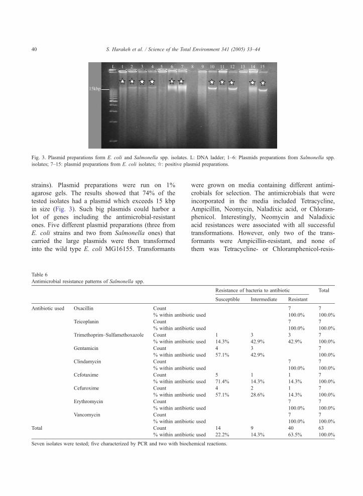
Fig. 3. Plasmid preparations form E. coli and Salmonella spp. isolates. L: DNA ladder; 1–6: Plasmids preparations from Salmonella spp.
isolates; 7–15: plasmid preparations from E. coli isolates; B: positive plasmid preparations.
S. Harakeh et al. / Science of the Total Environment 341 (2005) 33–4440
strains). Plasmid preparations were run on 1%
agarose gels. The results showed that 74% of the
tested isolates had a plasmid which exceeds 15 kbp
in size (Fig. 3). Such big plasmids could harbor a
lot of genes including the antimicrobial-resistant
ones. Five different plasmid preparations (three from
E. coli strains and two from Salmonella ones) that
carried the large plasmids were then transformed
into the wild type E. coli MG16155. Transformants
Table 6
Antimicrobial resistance patterns of Salmonella spp.
Antibiotic used Oxacillin Count
% within antibio
Teicoplanin Count
% within antibio
Trimethoprim–Sulfamethoxazole Count
% within antibio
Gentamicin Count
% within antibio
Clindamycin Count
% within antibio
Cefotaxime Count
% within antibio
Cefuroxime Count
% within antibio
Erythromycin Count
% within antibio
Vancomycin Count
% within antibio
Total Count
% within antibio
Seven isolates were tested; five characterized by PCR and two with bioch
were grown on media containing different antimi-
crobials for selection. The antimicrobials that were
incorporated in the media included Tetracycline,
Ampicillin, Neomycin, Naladixic acid, or Chloram-
phenicol. Interestingly, Neomycin and Naladixic
acid resistances were associated with all successful
transformations. However, only two of the trans-
formants were Ampicillin-resistant, and none of
them was Tetracycline- or Chloramphenicol-resis-
Resistance of bacteria to antibiotic Total
Susceptible Intermediate Resistant
7 7
tic used 100.0% 100.0%
7 7
tic used 100.0% 100.0%
1 3 3 7
tic used 14.3% 42.9% 42.9% 100.0%
4 3 7
tic used 57.1% 42.9% 100.0%
7 7
tic used 100.0% 100.0%
5 1 1 7
tic used 71.4% 14.3% 14.3% 100.0%
4 2 1 7
tic used 57.1% 28.6% 14.3% 100.0%
7 7
tic used 100.0% 100.0%
7 7
tic used 100.0% 100.0%
14 9 40 63
tic used 22.2% 14.3% 63.5% 100.0%
emical reactions.

Table 7
Antimicrobial resistance patterns of nine characterized E. coli strains
Resistance of bacteria to antibiotic Total
Susceptible Intermediate Resistant
Antibiotic used Oxacillin Count 1 8 9
% within antibiotic used 11.1% 88.9% 100.0%
Teicoplanin Count 9 9
% within antibiotic used 100.0% 100.0%
Trimethoprim–Sulfamethoxazole Count 2 7 9
% within antibiotic used 22.2% 77.8% 100.0%
Gentamicin Count 9 9
% within antibiotic used 100.0% 100.0%
Clindamycin Count 1 8 9
% within antibiotic used 11.1% 88.9% 100.0%
Cefotaxime Count 7 2 9
% within antibiotic used 77.8% 22.2% 100.0%
Cefuroxime Count 5 3 1 9
% within antibiotic used 55.6% 33.3% 11.1% 100.0%
Erythromycin Count 1 8 9
% within antibiotic used 11.1% 88.9% 100.0%
Vancomycin Count 1 8 9
% within antibiotic used 11.1% 88.9% 100.0%
Total Count 25 7 49 81
% within antibiotic used 30.9% 8.6% 60.5% 100.0%
S. Harakeh et al. / Science of the Total Environment 341 (2005) 33–44 41
tant. Interestingly, the plasmids that conferred
Ampicillin resistance were extracted from Salmo-
nella strains.
4. Discussion
In 2001, the Lebanese Ministry of Health reported
3497 cases attributed to food/water-borne diseases
(http://www.public-health.gov.lb, 2003–2004). How-
ever, there is little information concerning the
molecular characterization of pathogenic bacterial
strains in Lebanon. Considering the marked impor-
tance of E. coli and Salmonella infection organisms as
food-borne pathogens, we aimed in this study to
evaluate the levels of contamination by those organ-
isms in meat-based fast food in Lebanon. The isolated
strains were characterized at the molecular level using
PCR and evaluated their antimicrobial resistance
patterns to different antimicrobials. Lastly, the
involvement of plasmids in antimicrobial resistance
exhibited by the isolated bacteria was investigated.
Bacteriological analysis performed showed a dif-
ferential distribution of bacteria between Shawarma
and LBA. Shawarma showed a higher level of
contamination than Lahm-bi-Ajeen (LBA). The fact
that Shawarma had a higher level of contamination
than Lahm-bi-Ajeen can be attributed to many
reasons. LBA is usually cooked in the oven at 500
8C for around 5 min, which possibly eliminates all
kinds of bacteria present. LBA is often served with
pressed lemon that increases its acidity and likely
reduces bacterial growth. Moreover, there is less
handling in the case of LBA as compared to that of
Shawarma. In the case of Shawarma, after cutting the
meat, most of the food handlers adds vegetables and
sauce with their bare hands.
Some factors that may attribute to bacterial
contamination of Shawarma could be due to the way
Shawarma is cooked. It is usually sliced meat on a
rotating stick, which leads to cooking of the outer
meat while the inside remains rare. Also, the addition
of accessory ingredients to Shawarma, such as
vegetables and Tahina sauce (made from sesame
seeds oil), could be another source of contamination.
Vegetables could be contaminated with soil (Okafo et
al., 2003), and they reduce the temperature of the meat
thus providing a conducive environment for bacterial
contamination (Raiden et al., 2003). Another source
of contamination may be attributed to the Tahina

S. Harakeh et al. / Science of the Total Environment 341 (2005) 33–4442
sauce used, which was recently found to be contami-
nated with Salmonella spp.
After the microbiological examination, rapid and
sensitive methods were used to characterize bacterial
strains in meat-based fast food. DNA-based assays
were followed for the identification of pathogens
because these methods rely on nucleic acid composi-
tion of bacterium instead of the phenotypic expression
that might vary under culture conditions.
For Salmonella species identification, four sets of
primers were used to detect the rfb(B), rfb(C1),
rfb(C2), and rfb(D) genetic markers. A ratio of 50%
of the tested samples was positive in PCR performed
with rfb(D) primer, indicating the presence of S.
paratyphi A, serogroup A, in the contaminated food.
The negative results showed by the rest of the tested
strains could be attributed to the high number of
Salmonella species that we did not test for or due to
the existence of unknown mutations in the rfb genes
that might interfere with the detection method.
As for E. coli serotyping, the results showed
different gene profiles of the isolated strains. Sev-
enty-five percent of the tested strains were stx1-
positive. However, most of those were lacking the H7
flagellar protein gene as tested by PCR amplification
of the f liC operon. Such H7-negative strains have
recently been identified in Germany, Czech Republic,
and Poland, and it was demonstrated that they
represent a distinct clone within the E. coli O157
serogroup. However, it shares several virulence
characteristics with other STX-EC of the O157:H7
serotype (Bielaszewska et al., 1998, 2000; Ammon et
al., 1999; Osek and Gallein, 2002). Moreover, only
one strain was positive in PCR performed with eaeA
primers, and none of the tested strains were ehlyA
positive.
Considering the significant rise in the annual
consumption of antimicrobials as a medication or its
use in various products, it is extremely important to
document the level of antimicrobial resistance of
bacteria isolated from different foods for the public
health safety. Our findings clearly indicated that two-
third of the tested bacteria, both for E. coli and
Salmonella spp., were resistant to more than five
antimicrobials. In contrast to previously reported
results (Araj and Zaatari, 2002 medical report by the
American University of Beirut Medical Center) that
showed 100% susceptibility of clinical Salmonella
spp. isolates to Trimethoprim/sulfamethoxazole, only
14.3% was found to be susceptible in our study.
A 30% reduction frequency was observed in
Salmonella spp. susceptibility patterns to Cefotaxime,
as compared to the previously mentioned report. E.
coli on the other hand showed a reduction of more
than 40% in susceptibility to Cefuroxime, where it
showed 100% susceptibility in the clinical isolates
(Araj and Zaatari, 2002) compared to 55.6% in our
isolates. The three previously mentioned antimicro-
bials are commonly used for the treatment of bacterial
infections in Lebanon (Araj and Zaatari, 2002), which
may explain the difference in the susceptibility
patterns between the clinical and food isolates. The
high percentage of Erythromycin and Oxacillin
resistances also refers to the frequent use of these
antimicrobials for treatment of infectious diseases in
Lebanon. Such resistance may not only be a direct
concern to human health, but also, it is important
because it could be transferred to other important
pathogenic serotypes (Dzidic and Bedekovic, 2003).
Interestingly, both organisms showed a similar
resistance profile where the only difference was
observed in the resistance patterns to Gentamicin.
This result indicates a high possibility of horizontal
gene transfer between bacteria, via plasmids or
transposons, and therefore contributing in the increase
of the resistant genes in the environment (Kruse and
Sbrum, 1994).
Transfer of plasmid-encoded resistance to anti-
microbial agents is a significant public health
concern. The possibility of transfer of resistance
genes between bacteria in natural habitats has
recently attracted a lot of attention. Within this
framework, we evaluated the association of plasmid
existence with antimicrobial resistance in the isolated
strains. Both E. coli and Salmonella spp. harbored
the same size of plasmid that exceeds 15 kbp in size.
Plasmids with Neomycin and Naladixic acid were
most frequently transformed; however, few were
with ampicillin, and none were transformed with
Tetracycline or Chloramphenicol. Interestingly, plas-
mids that conferred ampicillin resistance were all
isolated from Salmonella species. Such results
cannot give a clear-cut picture on whether the two
bacteria are carrying the same kind of plasmid or not
due to the differential expression of antimicrobial
resistance by plasmids isolated from both bacteria.
S. Harakeh et al. / Science of the Total Environment 341 (2005) 33–44 43
Several antimicrobials earlier used as growth
promoters have been banned in several countries. In
Lebanon, the usage of antimicrobials is not well
controlled. In our study, we presented some data for
antimicrobial resistance patterns of E. coli and
Salmonella spp. in two kinds of fast food in Lebanon.
Obviously, the controlled use of antimicrobial agents
is a prerequisite to limit the emergence of drug-
resistant bacteria, but such prudence in itself is not
enough to control the emerging public health concern.
Additional research is certainly needed to better
understand the mechanisms behind bacterial resist-
ance to antimicrobials and the underlying mechanisms
such as the transfer of genetic material between
bacteria in the environment.
Finally, the data obtained indicated that meat-based
fast foods in Lebanon, especially Shawarma, are
potential reservoir for many pathogenic organisms,
which were shown to be resistant to many antimicro-
bials, suggesting a potential public health hazard.
Improper hygienic standards and the indiscriminate
use of antimicrobials are the main reasons behind the
emergence of antimicrobial-resistant strains in Leb-
anon. These results will emphasize the need to
implement proactive measures, and more emphasis
will be placed on the application of hygienic practices
and the use of Hazard Analysis and Critical Control
Point (HACCP) in the preparation and processing of
foods to reduce the risk of infection.
Acknowledgments
The authors are grateful to the American Univer-
sity Research Board and the USAID for financing this
work.
References
Ammon A, Peterson LR, Karch H. A large outbreak of hemolytic
uremic syndrome caused by an unusual sorbitol-fermenting
strain E coli O157:H7. J Infect Dis 1999;179:1274–7.
APHA. Compendium of methods for the microbiological examina-
tion of foods. Fourth edition. Washington, DC7 American Public
Health Association; 2001.
Araj, G.F., Zaatari, G., 2002. Antimicrobial susceptibility patterns
of bacterial isolates at the American University of Beirut
Medical Center (REPORT). Department of Pathology and
Laboratory Medicine.
Ausubel FM, Brent RE, Kingston DD, Moore JG, Seidman JA,
Smith JA, et al, 1987. Current protocols in molecular biology.
New York7 John Wiley & Sons; 1987. p. 241–2.
Belongia EA, MacDonald KL, Parham GL, White KE, Korlath JA,
Lobato MN, et al. An outbreak of Escherichia coli O157:H7
colitis associated with consumption of pre-cooked meat patties.
J Infect Dis 1991;164:338–43.
Bielaszewska M, Schmidt H, Karmali MA, Khakhria R, Janda J,
Blahova K, et al. Isolation and characterization of sorbitol-
fermenting Shiga toxin (Verocytotoxin)-producing Escherichia
coli O157: H� strains in the Czech Republic. J Clin Microbiol
1998;36:2135–7.
Bielaszewska M, Schmidt H, Liesegang A, Prager R, Rabsch W,
Tschape H, et al. Cattle can be a reservoir of sorbitol-fermenting
Shigatoxin-producing Escherichia coli O157: H� strains and a
source of human diseases. J Clin Microbiol 2000;38:3470–3.
Borezyk AA, Karmali MA, Lior H, Duncan LMC. Bovine reservoir
for verotoxin producing Escherichia coli. Lancet 1987;i:.
Brook M0, Smith RR, Bannister BA, McConnel M, Chart H,
Scotland SM, et al. Prospective study of verocytotoxin-
producing, enteroaggregative and diffusely adherent Escher-
ichia coli in different diarrhoeal states. Epidemiol Infect
1994;112:63–7.
Duijkeren EV, Wannet WJB, Houwers DJ, Pelt WV. Antimicrobial
susceptibility of Salmonella strains isolated from humans, cattle,
pigs, and chickens in the Netherlands from 1984 to 2001. J Clin
Microbiol 2003;41(8):3574–8.
Dzidic S, Bedekovic V. Horizontal gene transfer-emerging multi-
drug resistance in hospital bacteria. Acta Pharmacol Sin
2003;24(6):519–26 [Review].
Gannon VP, King RK, Kim JY, Thomas EJ. Rapid and sensitive
method for detection of Shiga-like toxin-producing Escherichia
coli in ground beef using the polymerase chain reaction. App
Environ Microbiol Eco 1992;58(12):3809–15.
Gillespie BE, Jayarao BM, Oliver SP. Identification of Streptococ-
cus species by randomly amplified polymorphic deoxyribonu-
cleic acid fingerprinting. J Dairy Sci 1997;80:471–6.
Griffin PM, Tauxe RV. The epidemiology of infections caused by
Escherichia coli O157:H7, other enterohemorrhagic E coli, and
the associated hemolytic uremic syndrome. Epidemiol Rev
1991;13:60–98.
Holmberg SD, Wells JG, Cohn ML. Animal to-man transmission of
antimicrobial-resistant Salmonella: investigation of US out
breaks, 1971–1983. Science 1984;225:833–5.
Jackson MP. Detection of Shiga toxin-producing Shigella dysenter-
iae type 1 and Escherichia coli using polymerase chain reaction
with incorporation of digoxigenin-11-UTP. J Clin Microbiol
1991;29:1910–4.
Karch H, Meyer T. Single primer pair for amplifying segments of
distinct Shiga-like toxin genes by polymerase chain reaction. J
Clin Microbiol 1989;27:2751–7.
Kruse H, Sbrum H. Transfer of multi-drug resistance plasmids
between bacteria of diverse origins in the natural environment.
Appl Environ Microbiol 1994;60(11):4015–21.
Langreid WW, Elder RO, Keen JE. Prevalence of Escherichia coli
O157:H7 in range beef calves at weaning. Epidemiol Infect
1999;123:291–8.
S. Harakeh et al. / Science of the Total Environment 341 (2005) 33–4444
Lee SJ, Romana LK, Reeves PR. Sequence and structural analysis
of the rfb (O antigen) gene cluster from a group C1 Salmonella
enterica strain. J Gen Microbiol 1992;138:1843–55.
Liewellyn LJ, Evans MR, Paimer Sr. Use of sequential case control
studies to investigate a community Salmonella outbreak in
Wales. J Epidemiol Community Health 1998;52:272–6.
Luk JM, Kongmauang U, Reeves PR, Lindberg AA. Selective
amplification of abequose and paratose synthase genes (rfb) by
polymerase chain reaction for identification of Salmonella
major serovars (A, B, C2, and D). J Clin Microbiol 1993;31:
2118–2123.
Luk JM, Kongmauang U, Tsang RSW, Lindberg AA. An enzyme-
linkedimmunosorbent assay to detect PCR products of the rfbS
gene from serovar. J Clin Microbiol 1997;35:714–8.
National Committee for Clinical Laboratory standards. Methods
for dilution antimicrobial susceptibility tests for bacteria
that grow aerobically. Approved Standared M7-A4. Wayne
(PA)7 National Committee for Clinical Laboratory standards;
1997.
Okafo CN, Umoh VJ, Galadima M. Occurrence of pathogens on
vegetables harvested from soils irrigated with contaminated
streams. Sci Total Environ 2003;311(1–3):49–56.
Osek J, Gallein P. Molecular analysis of Escherichia coli O157
strains isolated from cattle and pigs by the use of PCR and
pulsed-field gel electrophoresis methods. Vet Med-Czech
2002;47(6):149–58.
Raiden RM, Quicho JM, Maxfield CJ, Sumner SS, Eifert JD,
Pierson MD. Survivability of Salmonella and Shigella spp in
sodium lauryl sulfate and tween 80 at 22 and 40 degrees C. J
Food Prot 2003;66(8):1462–4.
Reischl U, Youssef MT, Kilwinski J, Lehn N, Zhang WL, Karch H,
et al. Real-time fluorescence PCR assays for detection and
characterization of Shiga toxin, intimin, and enterohemolysin
genes from Shiga toxin-producing Escherichia coli. J Clin
Microbiol 2002;40(7):2555–65.
Sambrook J, Fritsch EF, Maniatis T. Molecular cloning A laboratory
manual. Second edition. Cold Spring Harbor Laboratory Press.
Tauxe RV. An update on Salmonella . Health Environ Dig
1996;10:1–4.
Wasteson Y. Zoonotic Escherichia coli. Acta Vet Scand Suppl
2001;95:79–84.