Application of solid phase peptide synthesis to engineering PEO–peptide block copolymers for drug...
12
Application of solid phase peptide synthesis to engineering PEO /peptide block copolymers for drug delivery Gary H. Van Domeselaar a , Glen S. Kwon b , Lena C. Andrew a , David S. Wishart a, * a Faculty of Pharmacy and Pharmaceutical Sciences, University of Alberta, Edmonton, Alberta, Canada T6G 2N8 b School of Pharmacy, University of Wisconsin-Madison, 425 Charter St., Madison, WI 53706, USA Received 22 January 2003; accepted 27 April 2003 Abstract This work describes a simple, versatile solid-phase peptide-synthesis (SPPS) method for preparing micelle-forming poly(ethylene oxide)-block -peptide block copolymers for drug delivery. To demonstrate its utility, this SPPS method was used to construct two series of micelle-forming block copolymers (one of constant core-composition and variable length; the other of constant core length and variable composition). The block copolymers were then used to study in detail the effect of size and composition on micellization. The various block copolymers were prepared by a combination of SPPS for the peptide block, followed by solution /phase conjugation of the peptide block with a proprionic acid derivative of poly(ethylene oxide) (PEO) to form the PEO-b -peptide block copolymer. The composition of each block component was characterized by mass spectrometry (MALDI and ES-MS). Block copolymer compositions were characterized by 1 H NMR. All the block copolymers were found to form micelles as judged by transmission electron microscopy (TEM) and light scattering analysis. To demonstrate their potential as drug delivery systems, micelles prepared from one member of the PEO-b -peptide block copolymer series were physically loaded with the anticancer drug doxorubicin (DOX). Micelle static and dynamic stability were found to correlate strongly with micelle core length. In contrast, these same micellization properties appear to be a complex function of core composition, and no clear trends could be identified from among the set of compositionally varying, fixed length block copolymer micelles. We conclude that SPPS can be used to construct biocompatible block copolymers with well-defined core lengths and compositions, which in turn can be used to study and to tailor the behavior of block copolymer micelles. # 2003 Elsevier Science B.V. All rights reserved. Keywords: Micelle; Block copolymer; Peptide synthesis; Drug delivery; Nano-engineering 1. Introduction Amphipathic block copolymers consist of a linear arrangement of alternating hydrophilic and hydrophobic segments. When exposed to polar * Corresponding author. E-mail address: [email protected] (D.S. Wishart). Colloids and Surfaces B: Biointerfaces 30 (2003) 323 /334 www.elsevier.com/locate/colsurfb 0927-7765/03/$ - see front matter # 2003 Elsevier Science B.V. All rights reserved. doi:10.1016/S0927-7765(03)00125-5
-
Upload
independent -
Category
Documents
-
view
1 -
download
0
Transcript of Application of solid phase peptide synthesis to engineering PEO–peptide block copolymers for drug...
Application of solid phase peptide synthesis to engineeringPEO�/peptide block copolymers for drug delivery
Gary H. Van Domeselaar a, Glen S. Kwon b, Lena C. Andrew a,David S. Wishart a,*
a Faculty of Pharmacy and Pharmaceutical Sciences, University of Alberta, Edmonton, Alberta, Canada T6G 2N8b School of Pharmacy, University of Wisconsin-Madison, 425 Charter St., Madison, WI 53706, USA
Received 22 January 2003; accepted 27 April 2003
Abstract
This work describes a simple, versatile solid-phase peptide-synthesis (SPPS) method for preparing micelle-forming
poly(ethylene oxide)-block -peptide block copolymers for drug delivery. To demonstrate its utility, this SPPS method
was used to construct two series of micelle-forming block copolymers (one of constant core-composition and variable
length; the other of constant core length and variable composition). The block copolymers were then used to study in
detail the effect of size and composition on micellization. The various block copolymers were prepared by a
combination of SPPS for the peptide block, followed by solution�/phase conjugation of the peptide block with a
proprionic acid derivative of poly(ethylene oxide) (PEO) to form the PEO-b -peptide block copolymer. The composition
of each block component was characterized by mass spectrometry (MALDI and ES-MS). Block copolymer
compositions were characterized by 1H NMR. All the block copolymers were found to form micelles as judged by
transmission electron microscopy (TEM) and light scattering analysis. To demonstrate their potential as drug delivery
systems, micelles prepared from one member of the PEO-b -peptide block copolymer series were physically loaded with
the anticancer drug doxorubicin (DOX). Micelle static and dynamic stability were found to correlate strongly with
micelle core length. In contrast, these same micellization properties appear to be a complex function of core
composition, and no clear trends could be identified from among the set of compositionally varying, fixed length block
copolymer micelles. We conclude that SPPS can be used to construct biocompatible block copolymers with well-defined
core lengths and compositions, which in turn can be used to study and to tailor the behavior of block copolymer
micelles.
# 2003 Elsevier Science B.V. All rights reserved.
Keywords: Micelle; Block copolymer; Peptide synthesis; Drug delivery; Nano-engineering
1. Introduction
Amphipathic block copolymers consist of a
linear arrangement of alternating hydrophilic and
hydrophobic segments. When exposed to polar
* Corresponding author.
E-mail address: [email protected] (D.S.
Wishart).
Colloids and Surfaces B: Biointerfaces 30 (2003) 323�/334
www.elsevier.com/locate/colsurfb
0927-7765/03/$ - see front matter # 2003 Elsevier Science B.V. All rights reserved.
doi:10.1016/S0927-7765(03)00125-5
solvents these block copolymers can self-assemble
to form extraordinarily stable micellar suspensions
consisting of a dense, hydrophobic core and a
solvated outer shell [1]. Hydrophobic small
molecule drugs can be incorporated into the
hydrophobic core during micellization. The am-
phipathic block copolymer micelle, with its core/
shell architecture, and its ability to transport
lipophilic substances in aqueous media, is structu-
rally and functionally similar to plasma lipopro-
teins [2,3]. Thus, researchers are actively
investigating the use of biocompatible block co-
polymers as long-circulating delivery systems for
hydrophobic small molecule drugs [2,4,5]. Micellar
drug microcontainers can, in principle, impart
numerous desirable therapeutic advantages to
existing drugs in that they: (a) protect the drug
from enzymatic or other degradative mechanisms;
(b) increase the solubility of poorly soluble
hydrophobic drugs; (c) modulate the drug’s phar-
macokinetics; and (d) can accumulate at the target
site. However, in developing a pharmaceutically
useful polymeric micelle for drug delivery, many
performance-related issues must be addressed,
including drug-loading capacity, release kinetics,
circulation time, biodistribution, static and dy-
namic stability, morphology, size, and size dis-
tribution [4]. To a large extent these properties are
determined by block copolymer composition. The
hydrophilic shell-forming block determines the
micelle’s pharmacokinetic parameters, biodistribu-
tion, and contributes significantly to the physical
and biological stability of the micelle. On the other
hand, the composition of the core-forming block
determines the micelle’s drug loading capacity,
drug specificity, and contributes largely to the
micelle’s physico-chemical properties. With a few
notable exceptions [6�/8], most research on block
copolymer micelles for drug delivery has focused
on the use of poly(ethylene oxide) (PEO) for the
hydrophilic shell. PEO’s high degree of hydration
and large excluded volume induce repulsive forces
that impart steric stability to the micelle, and
extend circulation times by preventing
opsonization, thereby avoiding clearance by the
reticuloendothelial system [9]. In contrast to the
near universal application of PEO for the hydro-
philic shell-forming block, a somewhat larger
variety of hydrophobic core-forming blocks have
been investigated. Examples of core-forming block
compositions include polycaprolactone [6],
poly(D,L-lactide) [5,10], poly(propylene oxide)
[11], and poly(L-amino acids) and their derivatives
[12�/15].
Although biocompatible PEO-b-peptide block
copolymer micelles show promise as drug carriers,
their development and study has been slowed by
the difficulty associated with their synthesis. Tra-
ditionally, block copolymer synthesis is performed
by derivitizing one or both ends of the PEO block
with an initiator, and then adding the PEO to a
solution containing the monomeric L-amino acid
N -carboxyanhydride which, upon polymerization,
yields the block copolymer [2]. This synthetic
route, although reproducible and well established,
requires strict control of reagent purity and
stoichiometry in order to minimize polydispersity
and by-product formation. Moreover, this strategy
is restricted to the formation of compositionally
homogeneous copolymer hydrophobic blocks.
Hence, for a given hydrophobic block composi-
tion, micelle nano-engineering is limited to ‘ap-
proximately’ adjusting the relative and absolute
lengths of the block copolymer segments.
We have hypothesized that the techniques of
solid phase peptide synthesis (SPPS) may be
adapted to the construction PEO-b -peptide block
copolymers with precisely defined core lengths and
core compositions, thus allowing the properties of
the resulting micelles to be engineered in fine
detail. To test this hypothesis, we used SPPS,
followed by solution phase condensation (SPC), to
prepare two series of block copolymers: one of
fixed composition and varying length; the other of
fixed length and varying composition. We then
performed experiments to systematically study the
relationships of core length and core composition
to micelle size and thermodynamic stability in
terms of free energy of micellization (CMC),
micelle-unimer equilibrium, and micelle dissocia-
tion rates.
G.H. Van Domeselaar et al. / Colloids and Surfaces B: Biointerfaces 30 (2003) 323�/334324
2. Materials and methods
2.1. Materials and chemicals
Five thousand molecular weight a-methyl-v-
proprionic acid�/PEO (PEO�/O�/CH2�/CH2�/
COOH) was obtained from Shearwater Polymers
Inc. (Huntsville, AL). Rink amide methylbenzhy-
drylamine resin (Rink Amide MBHA), O -benzo-triazole-N ,N ,N ?,Nƒ-tetramethyluronium hexa-
fluorophosphate (HBTU), piperidine, and Fmoc-
amino acids were purchased from Chem Impex
International Inc. (Woodale, IL) and used as
received. HATU ((7-azabenzotriazol-1yl)-1,1,3,3,-
tetramethyluronium hexafluorophosphate) was
purchased from PerSeptive Biosystems (Framing-
ham, MA). Sequencing grade dimethylformamide(DMF) and dichloromethane (DCM) were ob-
tained from Fisher Scientific Canada. Trifluoroa-
cetic acid (TFA) was purchased from Halocarbon
Products Corporation (River Edge, NJ). N -Meth-
yl morpholine (NMM) was purchased from Al-
drich Chemical Co. (Milwaukee, IL). Doxorubicin
hydrochloride (DOX) was purchased from Sigma
Chemical Co. (St. Louis, MO).
2.2. PEO�/proprionic acid characterization
PEO�/proprionic acid was characterized for
weight-average molecular weight (Mw), number
average molecular weight (Mn,) and polydispersity
index (I) by MALDI-TOF mass spectrometry. A
200 mM sample of PEO�/proprionic acid in ddH2Owas prepared and mixed 1:1 with a matrix solution
consisting of 0.1 M dihydroxybenzoic acid dis-
solved in 1:1 MeOH and doped with 2 mM NaCl
to promote ionization. Spectra were acquired on a
PerSeptive Biosystems Voyager Elite MALDI-
TOF mass spectrometer. Mw: 5223, Mn: 5180, I :
1.01.
2.3. Solid-phase peptide synthesis, cleavage, and
purification
A series of poly L-amino acid (PLAA) core-
forming blocks with the composition Gly-Tyr(n)
(where n�/7, 9, 12, and 15 residues), and a series
of constant length blocks with the compositions
Gly-Phe15 (polyphenylalanine), Gly-Leu15 (poly-leucine), and Gly-Phe-Leu-Tyr-Trp-Phe-Leu-Tyr-
Trp-Phe-Leu-Tyr-Trp-Phe-Leu-Tyr (polyFLYW),
were assembled using an ABI 430A automated
peptide synthesizer on Rink amide MBHA resin
using standard Fmoc/t-Butyl protected amino
acids and HATU active-ester based coupling [16].
Upon completion of each peptide block synthesis
the protected peptide�/resin was transferred to a 20ml disposable polypropylene cartridge fitted with a
polyethylene frit. A 10 ml solution of TFA�/H2O
(95:5) cleavage cocktail was added and the mixture
shaken gently for 2 h. The cleaved and deprotected
peptide block was then filtered from the resin and
the resin washed with 3�/2 ml of TFA. The
combined eluate was evaporated in vacuo, pre-
cipitated and washed with cold ether (3�/20 ml),then dried in vacuo overnight to prepare for
purification.
Each crude peptide was purified by RP-HPLC
(Waters-System 501) on a reversed-phase C8 21�/
250 mm Zorbax 300 S.B. column with a binary
gradient at a flow rate of 8.0 ml/min using aqueous
0.1% TFA and 0.1% TFA in acetonitrile as the
mobile phases. The eluent was monitored at 220nm and 1-min fractions were collected. UV
absorbing fractions were analyzed by mass spec-
trometry using a Fisons VG Trio 2000 electrospray
mass spectrometer (ES-MS). Fractions containing
the correct mass and of sufficient purity were
combined and lyophilized to yield the desired
peptide block in excess of 90% purity, as judged
by ES-MS analysis.
2.4. Block copolymer synthesis
PEO-b -peptide block copolymers were prepared
using solution phase condensation (SPC). PEO�/
proprionic acid was coupled to each peptide block
in solution at a ca. 0.05 mmol scale. The PEO�/
proprionic acid carboxyl group was activated
using a 0.9 molar equivalent of HATU and twomolar equivalents of NMM in DMF. The PEO�/
proprionic acid was allowed to activate for 1 h at
room temperature on a shaker. The activated PEO
(2 molar equivalents with respect to the hydro-
phobic block) was then coupled to the glycine N-
terminal amine of the peptide block in DMF until
G.H. Van Domeselaar et al. / Colloids and Surfaces B: Biointerfaces 30 (2003) 323�/334 325
a negative Kaiser test [17] was obtained (overnightat room temperature on a shaker). The crude
block copolymer was dialyzed with three changes
of DMF (overnight at room temperature with
stirring using Spectra/Por 3500 MWCO dialysis
membrane) to remove any unreacted peptide and
small molecular weight contaminants. DMF was
then removed from the polymer by evaporation in
vacuo. Unreacted PEO was removed from theblock copolymer by preparative HPLC. The UV
absorbing fractions were collected, lyophilized,
desiccated, and stored at 4 8C. The segment
condensation coupling efficiency was determined
by 1H NMR analysis (relative integrated peak
area) of the block copolymer in DMSO-d6.
2.5. Determination of CMC
The critical micelle concentration (CMC) was
determined by light scattering according to a
previously published procedure [18]. Block copo-
lymer micelles of each of the seven different PEO-
b -peptide block copolymers were prepared by a
gradient dialysis approach [19]. The block copoly-
mer (20 ml) was dissolved in DMF (5 ml) and 10ml of ddH2O was added dropwise over 2 h. The
solution was dialyzed into three changes of 1 l
ddH2O over 24 h. The micelle solution was filtered
through a 0.22 mm nylon membrane (Fisher,
Pittsburgh, PA) and lyophilized. Lyophilized mi-
celles (12 mg) were accurately weighed and dis-
solved into 12 ml 0.1 M PO�4 ; pH 7.2. A series of
doubling dilutions, from 1 to 1�/10�4 mg/ml,were prepared in triplicate from this stock solu-
tion. Light scattering was measured with a SPEX
Fluoromax spectrofluorometer using excitation
and emission wavelengths of 600 nm, a bandpass
width of 1 nm, a step increment of 0.5 nm, and an
integration time of 1.0 s. The intensity of scattered
light was plotted against polymer concentration to
determine the concentration at which the intensitysharply increases, indicating the formation of
micelles.
2.6. Electron microscopy examination
Lyophilized micelles were reconstituted in
ddH2O (ca. 1 mg/ml). Samples of the aqueous
micelle suspensions were examined by negative-stain transmission electron microscopy (TEM).
One drop of the micelle sample was placed on a
300-mesh copper membrane coated disk, followed
by one drop of 1% phosphotungstic acid in water
(pH 7.0) (the negative stain). After 30 s excess
liquid was blotted from the disk with filter paper
and the sample loaded onto a sample holder. The
sample was then examined using a Hitachi Trans-mission Electron Microscope H-7000 at an accel-
erating voltage of 75 keV. Micelle size and size
distribution was determined directly from the
TEM images.
2.7. Drug loading of micelles
Doxorubicin hydrochloride was loaded intoPEO-b-polytyrosine7 micelles according to a pre-
viously published procedure [20]. DOX (10 mg)
and PEO-b -polytyrosine7 block copolymer (20
mg) were added to DMF and mixed for 1 h. The
solution was first dialyzed against 1:15 v/v
DMF:ddH2O over 18 h, then dialyzed into three
changes of ddH2O over 24 h. The mixture was
microcentrifuged for 10 min at 13,000 rpm toremove precipitated doxorubicin, and the super-
natant diluted to 10 ml with ddH2O. Drug loading
was determined by gel permeation chromato-
graphic analysis of an aliquot of the micelle
solution and by measuring UV absorbance of the
eluent at 485 nm using a Pharmacia Biotech
Ultraspec 3000 UV�/Vis spectrophotometer.
2.8. Micelle dissociation rates
Accurately weighed samples of lyophilized block
copolymer micelles were reconstituted in 0.1 M
phosphate buffer (pH 7.2) at concentrations above
their critical micelle concentration (ca. 1.0�/2.0 mg/
ml). An accurately measured aliquot was removed
and diluted to a concentration below the CMC
(from ca. 0.02 to 0.001 mg/ml) and incubated at37 8C in a dry bath. One hundred microlitres
samples were withdrawn periodically and sub-
jected to chromatographic analysis using a Rainin
HPLC system consisting of a Rainin HPXL
solvent delivery system, a Rainin Dynamax UV�/
Visible absorption detector. Samples were applied
G.H. Van Domeselaar et al. / Colloids and Surfaces B: Biointerfaces 30 (2003) 323�/334326
to an Ultrahydrogel 2000 column and matchingguard column at a flow rate of 0.8 ml min�1 and
0.1 M phosphate buffer (pH 7.2) as the mobile
phase. The column was calibrated with size exclu-
sion standards (blue dextran, 2,000,000 Da; pro-
teins, 600,000�/17,000 Da; and sodium azide, 65
Da). The eluent was monitored at an appropriate
wavelength for detection of the hydrophobic
block.
3. Results and discussion
Although recent reports have emerged describ-
ing the construction of PEO-b-peptide block
copolymers [21], to our knowledge, this is the first
reported application of solid phase peptide synth-
esis specifically for preparing PEO-b -peptide blockcopolymer micelles. Given the ease with which
custom peptides can be prepared or purchased
(through commercial and non-commercial peptide
synthesis facilities), we believe this hybrid SPPS�/
SPC approach provides a convenient and cost-
effective alternative to the conventional NCA-
based ring-opening polymerization strategy. In-
deed, a person only needs to order peptides and tohave access to an HPLC and a few commercial
reagents to prepare these block copolymers*/they
do not have to be peptide chemists.
The key advantages of SPPS over classical
polymerization techniques are the ability to apply
combinatorial chemistry to the polymer’s con-
struction, and the ability to precisely control core
length. Hence, the flexibility of nano-engineeringmicelle-based drug delivery systems can be ex-
tended considerably. In the present study, these
properties are exploited to study micellization
behavior. Other examples of the possible applica-
tions of this technology include: incorporation of
crosslinking residues (such as cysteine) into the
core-forming block to produce rigid cores; con-
struction of multipartite systems with separateblocks for core formation, drug binding, and shell
formation; and incorporation of cleavage sub-
strates into the polymer chain for proteolytically
enhanced micelle degradation and drug release.
This SPPS technique was used to construct two
sets of PEO-b-peptide block copolymers: one with
varied compositions but fixed core length, and theother of fixed composition (polytyrosine) but of
varying core lengths. The variable composition
series includes the following block copolymer
constructs: PEO-b-polyleucine (hydrophobic, ali-
phatic), PEO-b -polytyrosine (hydrophobic, aro-
matic), PEO-b -polyphenylalanine (hydrophobic,
aromatic), and PEO-polyFLYW, which is unique
among this series, in that the core-forming blockcontains a compositionally diverse distribution of
aliphatic and aromatic residues. It was designed to
study the effect of sequence heterogeneity on block
copolymer micellization in analogy with globular
proteins, which contain a heterogeneous structure
and form core/shell type particles with glassy cores
under physiological conditions [22]. The variable
length series is composed of polytyrosine peptideblocks of 7, 9, 12, and 15 residues. This series was
used to examine the effect of core-block size on
micellization.
3.1. Synthesis and characterization
A diagram describing structures of the various
block copolymer constructs is provided in Fig. 1.
Peptide block compositions and lengths werecontrolled by altering the number of SPPS cycles
and the order of addition of protected L-amino
acids. All seven peptide blocks were appended at
the N-terminus with a single glycine residue to aid
in the conjugation efficiency of PEO to the peptide
block. Glycine is a relatively hydrophilic, unhin-
dered, amino acid and is known to disrupt
secondary structure formation. These propertiescombine to help expose the N-terminal amino
group and therefore increase coupling efficiency
[23].
The crude peptide blocks were purified by
conventional preparative RP-HPLC. Purity ana-
lysis by analytical RP-HPLC was not possible to
obtain purity levels in excess of 90%, as judged by
ES-MS analysis, for all the constructs prepared inthis study (data not shown). The remaining frac-
tions consisted of (typically 1-residue) deletion
products; hence, the core-forming blocks prepared
by SPPS can be considered for all intents and
purposes to be monodisperse. This ability to
precisely control peptide block length can in turn
G.H. Van Domeselaar et al. / Colloids and Surfaces B: Biointerfaces 30 (2003) 323�/334 327
be exploited to tailor micellization properties such
as CMC and dissociation rates, as we demonstrate
here.
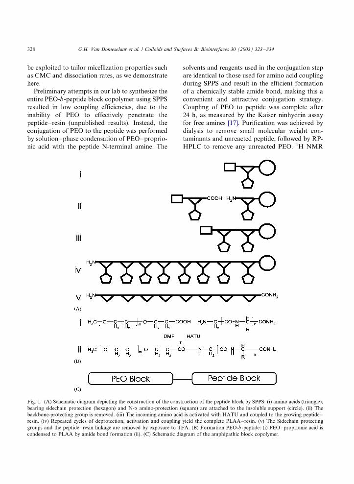
Preliminary attempts in our lab to synthesize the
entire PEO-b-peptide block copolymer using SPPS
resulted in low coupling efficiencies, due to the
inability of PEO to effectively penetrate the
peptide�/resin (unpublished results). Instead, the
conjugation of PEO to the peptide was performed
by solution�/phase condensation of PEO�/proprio-
nic acid with the peptide N-terminal amine. The
solvents and reagents used in the conjugation step
are identical to those used for amino acid coupling
during SPPS and result in the efficient formation
of a chemically stable amide bond, making this a
convenient and attractive conjugation strategy.
Coupling of PEO to peptide was complete after
24 h, as measured by the Kaiser ninhydrin assay
for free amines [17]. Purification was achieved by
dialysis to remove small molecular weight con-
taminants and unreacted peptide, followed by RP-
HPLC to remove any unreacted PEO. 1H NMR
Fig. 1. (A) Schematic diagram depicting the construction of the construction of the peptide block by SPPS: (i) amino acids (triangle),
bearing sidechain protection (hexagon) and N-a amino-protection (square) are attached to the insoluble support (circle). (ii) The
backbone-protecting group is removed. (iii) The incoming amino acid is activated with HATU and coupled to the growing peptide�/
resin. (iv) Repeated cycles of deprotection, activation and coupling yield the complete PLAA�/resin. (v) The Sidechain protecting
groups and the peptide�/resin linkage are removed by exposure to TFA. (B) Formation PEO-b -peptide: (i) PEO�/proprionic acid is
condensed to PLAA by amide bond formation (ii). (C) Schematic diagram of the amphipathic block copolymer.
G.H. Van Domeselaar et al. / Colloids and Surfaces B: Biointerfaces 30 (2003) 323�/334328
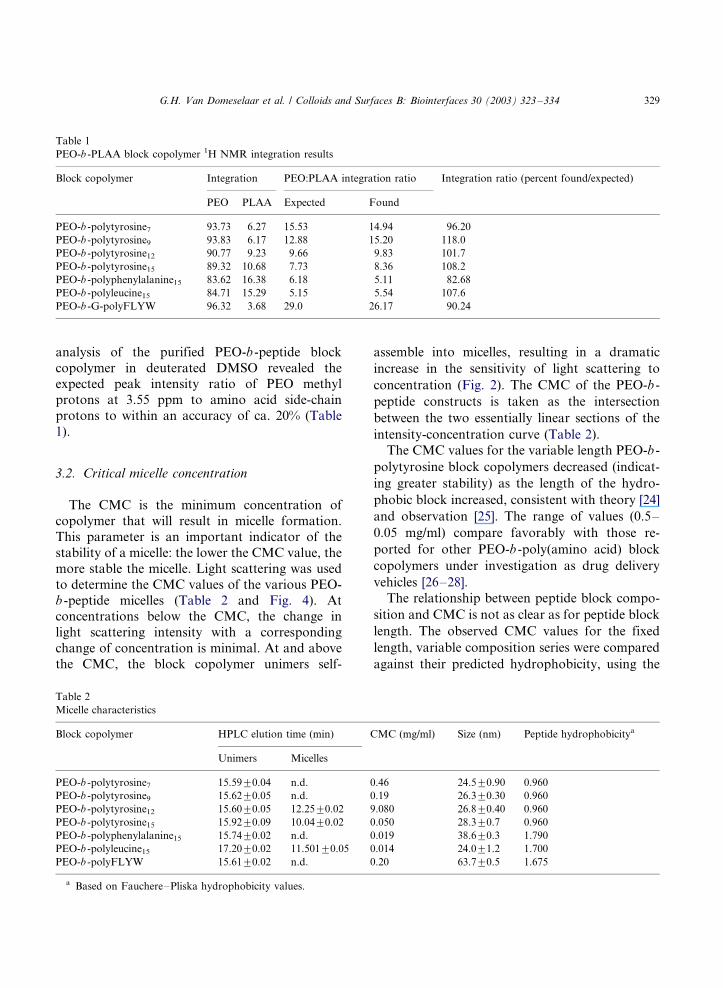
analysis of the purified PEO-b -peptide block
copolymer in deuterated DMSO revealed theexpected peak intensity ratio of PEO methyl
protons at 3.55 ppm to amino acid side-chain
protons to within an accuracy of ca. 20% (Table
1).
3.2. Critical micelle concentration
The CMC is the minimum concentration of
copolymer that will result in micelle formation.
This parameter is an important indicator of the
stability of a micelle: the lower the CMC value, the
more stable the micelle. Light scattering was used
to determine the CMC values of the various PEO-
b -peptide micelles (Table 2 and Fig. 4). Atconcentrations below the CMC, the change in
light scattering intensity with a corresponding
change of concentration is minimal. At and above
the CMC, the block copolymer unimers self-
assemble into micelles, resulting in a dramatic
increase in the sensitivity of light scattering to
concentration (Fig. 2). The CMC of the PEO-b -
peptide constructs is taken as the intersection
between the two essentially linear sections of the
intensity-concentration curve (Table 2).
The CMC values for the variable length PEO-b -
polytyrosine block copolymers decreased (indicat-
ing greater stability) as the length of the hydro-
phobic block increased, consistent with theory [24]
and observation [25]. The range of values (0.5�/
0.05 mg/ml) compare favorably with those re-
ported for other PEO-b -poly(amino acid) block
copolymers under investigation as drug delivery
vehicles [26�/28].
The relationship between peptide block compo-
sition and CMC is not as clear as for peptide block
length. The observed CMC values for the fixed
length, variable composition series were compared
against their predicted hydrophobicity, using the
Table 1
PEO-b -PLAA block copolymer 1H NMR integration results
Block copolymer Integration PEO:PLAA integration ratio Integration ratio (percent found/expected)
PEO PLAA Expected Found
PEO-b -polytyrosine7 93.73 6.27 15.53 14.94 96.20
PEO-b -polytyrosine9 93.83 6.17 12.88 15.20 118.0
PEO-b -polytyrosine12 90.77 9.23 9.66 9.83 101.7
PEO-b -polytyrosine15 89.32 10.68 7.73 8.36 108.2
PEO-b -polyphenylalanine15 83.62 16.38 6.18 5.11 82.68
PEO-b -polyleucine15 84.71 15.29 5.15 5.54 107.6
PEO-b -G-polyFLYW 96.32 3.68 29.0 26.17 90.24
Table 2
Micelle characteristics
Block copolymer HPLC elution time (min) CMC (mg/ml) Size (nm) Peptide hydrophobicitya
Unimers Micelles
PEO-b -polytyrosine7 15.599/0.04 n.d. 0.46 24.59/0.90 0.960
PEO-b -polytyrosine9 15.629/0.05 n.d. 0.19 26.39/0.30 0.960
PEO-b -polytyrosine12 15.609/0.05 12.259/0.02 9.080 26.89/0.40 0.960
PEO-b -polytyrosine15 15.929/0.09 10.049/0.02 0.050 28.39/0.7 0.960
PEO-b -polyphenylalanine15 15.749/0.02 n.d. 0.019 38.69/0.3 1.790
PEO-b -polyleucine15 17.209/0.02 11.5019/0.05 0.014 24.09/1.2 1.700
PEO-b -polyFLYW 15.619/0.02 n.d. 0.20 63.79/0.5 1.675
a Based on Fauchere�/Pliska hydrophobicity values.
G.H. Van Domeselaar et al. / Colloids and Surfaces B: Biointerfaces 30 (2003) 323�/334 329
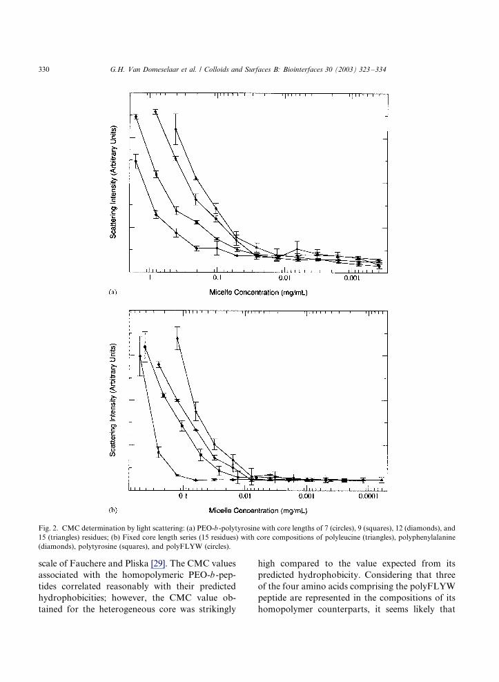
scale of Fauchere and Pliska [29]. The CMC values
associated with the homopolymeric PEO-b -pep-
tides correlated reasonably with their predicted
hydrophobicities; however, the CMC value ob-
tained for the heterogeneous core was strikingly
high compared to the value expected from its
predicted hydrophobicity. Considering that three
of the four amino acids comprising the polyFLYW
peptide are represented in the compositions of its
homopolymer counterparts, it seems likely that
Fig. 2. CMC determination by light scattering: (a) PEO-b -polytyrosine with core lengths of 7 (circles), 9 (squares), 12 (diamonds), and
15 (triangles) residues; (b) Fixed core length series (15 residues) with core compositions of polyleucine (triangles), polyphenylalanine
(diamonds), polytyrosine (squares), and polyFLYW (circles).
G.H. Van Domeselaar et al. / Colloids and Surfaces B: Biointerfaces 30 (2003) 323�/334330
this discrepancy is due to sequence heterogeneity.Chothia showed that the ‘effective’ hydrophobicity
of peptides and proteins are influenced by their
secondary structure, which in turn are influenced
by their primary sequence and the polarity of the
surrounding medium [30]. Cammas et al. [31,32]
studied the conformation of PEO-b-poly(b-benzyl
L-aspartate) in organic solvents, but did not relate
the effect of conformation on micellization ther-modynamics. We speculate that the sequence
variability within the polyFLYW core translates
into a more disordered secondary structure than
for the homopolymeric series, and that this
disorder results in a greater exposure of the
peptide’s relatively polar main chain to the sur-
rounding medium, thus decreasing hydrophobicity
and increasing the observed CMC.
3.3. Micelle drug encapsulation
The ability of PEO-b -PLAA block copolymer
micelles to encapsulate model hydrophobic drugs
is also an indication of their drug delivery utility.
Physical entrapment by dialysis and oil-in-water
emulsion methods have both been shown to
successfully place hydrophobic drugs, such as theanticancer drug doxorubicin, at high levels within
PEO-b -PBLA micelle cores without chemical de-
gradation [20] [[33]]. Dialysis was used in a
preliminary experiment to encapsulate doxorubi-
cin within PEO-b -polytyrosine7 block copolymer
micelles. Using UV absorbance and gel permea-
tion chromatography to quantify and determine
the proportion of encapsulated versus solvateddoxorubicin, a significant drug loading level of
2.8%w/w was found, compared to 14%w/w deter-
mined for PEO-b-poly(b-benzyl-L-aspartate)12
(unpublished results). It is plausible that higher
‘solubilities’ for this drug could be obtained in the
drug loading of PEO-b-polyleucine15, whose core
has a higher predicted peptide hydrophobicity
(Table 2). As well, it can be hypothesized thatlonger core lengths may also aid in increasing drug
loading efficiency. The CMC values for the vari-
able length PEO-b-polytyrosine block copolymers
decreased (indicating experimentally greater stabi-
lity and hydrophobicity) as core length increased
(Table 2), suggesting that PEO-b-polytyrosine15
would display higher doxorubicin loading thanobserved for PEO-b -polytyrosine7.
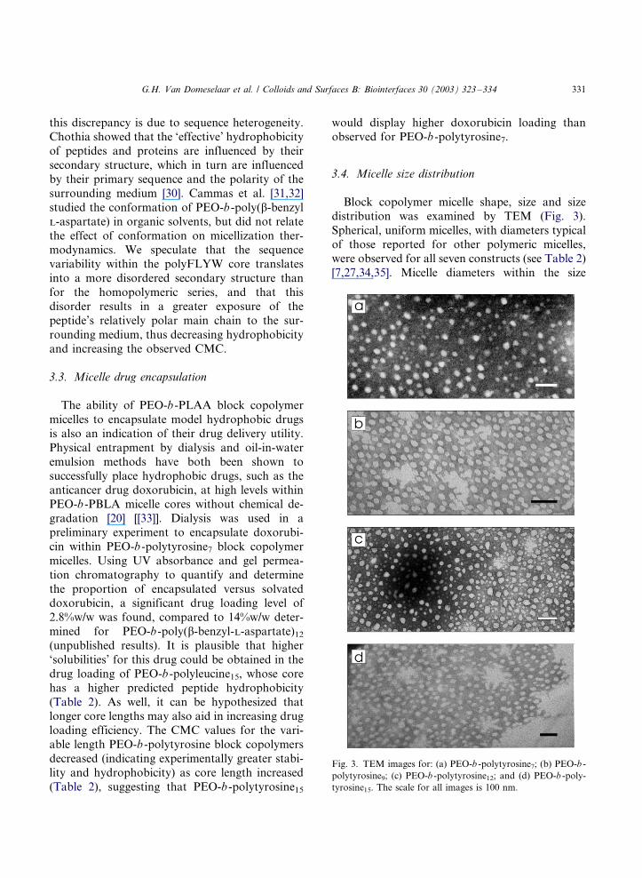
3.4. Micelle size distribution
Block copolymer micelle shape, size and size
distribution was examined by TEM (Fig. 3).
Spherical, uniform micelles, with diameters typicalof those reported for other polymeric micelles,
were observed for all seven constructs (see Table 2)
[7,27,34,35]. Micelle diameters within the size
Fig. 3. TEM images for: (a) PEO-b -polytyrosine7; (b) PEO-b -
polytyrosine9; (c) PEO-b -polytyrosine12; and (d) PEO-b -poly-
tyrosine15. The scale for all images is 100 nm.
G.H. Van Domeselaar et al. / Colloids and Surfaces B: Biointerfaces 30 (2003) 323�/334 331
range are considered desirable for drug-delivery;that is, they are large enough to escape renal
excretion, yet small enough to avoid hepatic
elimination. As seen for other PEO-b -peptide
block copolymer micelles, this size range, coupled
with the non-immunogenic PEO shell, imparts a
‘stealthy’ quality to the micelle allowing it to
circulate in vivo for extended periods of time [13].
Micelle diameters were observed to increaseslightly with increasing peptide block length. In
contrast, the micelle diameters for the fixed size
series were much more varied. The PEO-b-poly-
FLYW construct showed the greatest deviation,
with a diameter over twice the average diameter of
the other homopolymeric peptide cores. The
reason for this deviation is not clear; however, it
seems likely that, as with the CMC, the hetero-geneous core composition is a major contributing
factor.
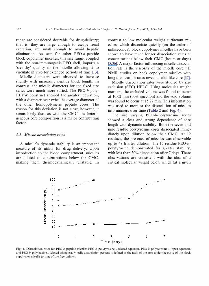
3.5. Micelle dissociation rates
A micelle’s dynamic stability is an important
measure of its utility for drug delivery. Uponintroduction to the blood compartment, micelles
are diluted to concentrations below the CMC,
making them thermodynamically unstable. In
contrast to low molecular weight surfactant mi-
celles, which dissociate quickly (on the order of
milliseconds), block copolymer micelles have been
shown to have much longer dissociation rates at
concentrations below their CMC (hours or days)
[5,36]. A major factor influencing micelle dissocia-
tion rate is the viscosity of the micelle core. 1H
NMR studies on bock copolymer micelles with
long dissociation rates reveal a solid-like core [37].
Micelle dissociation rates were studied by size
exclusion (SEC) HPLC. Using molecular weight
markers, the excluded volume was found to occur
at 10.02 min (post injection) and the void volume
was found to occur at 15.27 min. This information
was used to monitor the dissociation of micelles
into unimers over time (Table 2 and Fig. 4).
The size varying PEO-b -polytyrosine series
showed a clear and strong dependence of core
length with dynamic stability. Both the seven and
nine residue polytyrosine cores dissociated imme-
diately upon dilution below their CMC. At 12
residues, the presence of micelles was observable
up to 48 h after dilution. The 15 residue PEO-b -
polytyrosine demonstrated far greater stability,
with less than 30% dissociation after 7 days. These
observations are consistent with the idea of a
critical molecular weight below which (at a given
Fig. 4. Dissociation rates for PEO-b -peptide micelles PEO-b -polytyrosine12 (closed squares), PEO-b -polytyrosine15 (open squares),
and PEO-b -polyleucine15 (closed triangles). Micelle dissociation percent is defined as the ratio of the area under the curve of the block
copolymer micelle to that of the free unimer.
G.H. Van Domeselaar et al. / Colloids and Surfaces B: Biointerfaces 30 (2003) 323�/334332
temperature) the polymer exists as a melt, and
above which the polymer exists in the solid, glassy
state. For PEO-b -polytyrosine, it appears that a
minimum chain-length of 12 residues is necessary
to form micelles with a solid-like core.
Of the various fixed-size constructs investigated,
only PEO-b-polytyrosine and PEO-b-polyleucine
showed long-term stability below their CMC. Both
PEO-b -polyFLYW and PEO-b -polyphenylalanine
were not stable below the CMC. This apparent
instability of PEO-b-polyphenylalanine is unex-
pected, considering that the chemical structure of
phenylalanine is very similar to that of tyrosine
(tyrosine has a para hydroxyl group whereas
phenylalanine does not), yet the 15-residue PEO-
b -polytyrosine micelles are the most stable of all
the micelles examined in this study. The reason for
this discrepancy is not entirely clear, however, it is
possible that the increased size of the tyrosine side-
chain over phenylalanine coupled with its ability
to participate in intermolecular hydrogen bonding
may be sufficient to account for the difference in
observed dissociation rates. Regardless, these
results show that micelle stability is a complex
function of core composition, and cannot be
predicted on the basis of hydrophobicity alone.
4. Conclusions
Block copolymer micelles hold much potential
as non-toxic, non-immunogenic, controlled-release
systems for hydrophobic drugs. We have devel-
oped a simple and versatile SPPS�/SPC method for
the construction of PEO-b -peptide block copoly-
mers that greatly expands the preparative capabil-
ities of these polymers. To demonstrate its utility
and versatility we employed this technique in the
construction of a number of PEO-b -peptide block
copolymers with precisely defined sequence com-
positions and sizes. This allowed us to investigate
the relationship of core size and composition to
micelle formation and stability. This work should
aid in the nano-engineering and preparation of
novel block copolymer constructs for drug deliv-
ery.
Acknowledgements
Funding for this work was provided the Alberta
Heritage Foundation for Medical Research
(AHFMR), the Protein Engineering Network of
Centers of Excellence (PENCE) and BioTools Inc.
GVD is supported by a PMAC-HRF Graduate
Research Scholarship in Pharmacy. The authors
thank Diane Jette, Afsaneh Lavasanifar, and DrMing Chen for their technical assistance.
References
[1] Z. Tuzar, P. Kratochvil, Block and graft copolymer
micelles in solution, Adv. Colloid Interface Sci. 6 (1976)
201�/232.
[2] G.S. Kwon, K. Kataoka, Block-copolymer micelles as
long-circulating drug vehicles, Adv. Drug Deliv. Rev. 16
(1995) 295�/309.
[3] K. Kataoka, G.S. Kwon, M. Yokoyama, T. Okano, Y.
Sakurai, Block copolymer micelles as vehicles for drug
delivery, J. Controlled Release 24 (1993) 119�/132.
[4] C. Allen, D. Maysinger, A. Eisenberg, Nano-engineering
block copolymer aggregates for drug delivery, Colloids
Surf. B 16 (1999) 3�/27.
[5] Y. Yamamoto, Y. Nagasaki, Y. Kato, Y. Sugiyama, K.
Kataoka, Long-circulating poly(ethylene glycol)-poly(D,L-
lactide) block copolymer micelles with modulated surface
charge, J. Controlled Release 77 (2001) 27�/38.
[6] A.V. Kabanov, V.P. Chekhonin, V.Y. Alakhov, E.V.
Betrakova, A.S. Lebedev, N.S. Melik-Nubarov, S. Arzha-
kov, A.V. Levashov, G.V. Morozov, E.S. Severin, V.A.
Kabanov, The neuroleptic activity of haloperidol increases
after its solubilization in surfactant micelles�/micelles as
microcontainers for drug targeting, FEBS Lett. 258 (1989)
343�/345.
[7] Ha S.A. Hagan, A.G. Coombes, M.C. Garnett, S.E. Dunn,
M.C. Davies, L. Illum, S.S. Davis, S.E. Harding, S.
Purkiss, P.R. Gellert, Polylactide-poly(ethylene glycol)
copolymers as drug delivery systems. I. Characterization
of water dispersible micelle-forming systems, Langmuir 12
(1996) 2153�/2161.
[8] T. Inoue, G. Chen, K. Nakamae, S.A. Hoffman, An AB
block copolymer of oligo (methylmethacrylate) and
poly(acrylic acid) for micellar delivery of hydrophobic
drugs, J. Controlled Release 51 (1998) 221�/229.
[9] A. Abuchowski, T. van Es, N.C. Palczuk, J.R. McCoy,
F.F. Davis, Effect of covalent attachment of polyethylene
glycol on immunogenicity and circulating life of bovine
liver catalase, J. Biol. Chem. 252 (1977) 3582�/3586.
[10] I.G. Shin, S.Y. Kim, Y.M. Lee, C.S. Cho, Y.K. Sung,
Methoxy poly(ethylene glycol) o-caprolactone amphiphilic
block copolymeric micelle containind indomethacin. I.
G.H. Van Domeselaar et al. / Colloids and Surfaces B: Biointerfaces 30 (2003) 323�/334 333
Preparation and characterization, J. Controlled Rel. 51
(1977) 1�/11.
[11] D.W. Miller, E.V. Batrakova, T.O. Waltner, V.Y. Ala-
khov, A.V. Kabanov, Interactions of pluronic block
copolymers with brain microvessel endothelial cells: evi-
dence of two potential pathways for drug absorption,
Bioconjugate Chem. 8 (1997) 649�/657.
[12] M. Yokoyama, M. Miyauchi, N. Yamada, T. Okano, Y.
Sakurai, K. Kataoka, S. Inoue, Characterization and
anticancer activity of the micelle-forming polymeric antic-
ancer drug adriamycin-conjugated poly(ethylene glycol)�/
poly(aspartic acid) block copolymer, Cancer Res. 50 (1990)
1693�/1700.
[13] K. Kataoka, T. Matsumoto, M. Yokoyama, T. Okano, Y.
Sakurai, S. Fukushima, K. Okamoto, G.S. Kwon, Dox-
orubicin-loaded poly(ethylene glycol)�/poly(b-benzyl-L-as-
partate) copolymer micelles: their pharmaceutical
characterization and biological significance, Control. Re-
lease 64 (2000) 143�/153.
[14] Y.I. Jeong, J.B. Cheon, S.H. Kim, J.W. Nah, Y.M. Lee,
Y.K. Sung, T. Akaike, C.S. Cho, Clonazepam release from
core-shell type nanoparticles in vitro , J. Controlled Release
51 (1998) 169�/178.
[15] A. Harada, H. Togawa, K. Kataoka, Physicochemical
properties and nuclease resistance of antisense-oligodeox-
ynucleotides entrapped in the core of polyion complex
micelles composed of poly(ethylene glycol)�/poly(L-lysine)
block copolymers, Eur. J. Pharm. Sci. 13 (2001) 35�/42.
[16] C.G. Fields, D.H. Lloyd, R.L. Macdonald, K.M. Otteson,
R.L. Noble, HBTU activation for automated Fmoc solid-
phase peptide synthesis, Peptide Res. 4 (1991) 95�/101.
[17] E. Kaiser, R.L. Colescott, C.D. Bossinger, P.I. Cook,
Color test for detection of free terminal amino groups in
the solid phase synthesis of peptides, Anal. Biochem. 34
(1970) 595�/598.
[18] P. Tancrede, J. Barwicz, S. Jutras, I.B. Gruda, The effect
of surfactants on the aggregation state of amphotericin B,
Biochim. Biophys. Acta 1030 (1990) 289�/295.
[19] Y. Li, G.S. Kwon, Micelle-like structures of poly(ethylene
oxide)-block-poly(2-hydroxyethylaspartamide)-methotrex-
ate conjugates, Colloids Surf. B 16 (1999) 217�/226.
[20] G.S. Kwon, M. Naito, M. Yokoyama, T. Okano, Y.
Sakurai, K. Kataoka, Entrapment of adriamycin in ab
block copolymer micelles, Pharm. Res. 12 (1995) 200�/203.
[21] M. Pechar, K. Ulbrich, V. Subr, L.W. Seymour, E.H.
Schacht, Poly(ethylene glycol) multiblock copolymer as a
carrier of the anticancer drug doxorubicin, Bioconjugate
Chem. 11 (2000) 131�/139.
[22] J. Onuchic, Z. Luthey-Schulten, P. Wolynes, Theory of
protein folding: the energy landscape perspective, Ann.
Rev. Phys. Chem. 48 (1997) 545�/600.
[23] Y. Lu, A. Felix, Pegylated peptides*/solid phase synthesis
of Na-pegylated peptides using Fmoc strategy, Peptide
Res. 6 (1993) 140�/146.
[24] M. Munch, A. Gast, Block copolymers at interfaces. I.
Micelle formation, Macromolecules 21 (1988) 1360�/1366.
[25] P. Alexandridis, T.A. Hatton, Poly(ethylene oxide)�/
poly(propylene oxide)�/poly(ethylene oxide) block-copoly-
mer surfactants in aqueous solution and at interfaces*/
thermodynamics, structure, dynamics and modeling, Col-
loids Surf. A 96 (1995) 1�/46.
[26] G.S. Kwon, M. Naito, M. Yokoyama, T. Okano, Y.
Sakurai, K. Kataoka, Micelles based on AB block
copolymers of poly(ethylene oxide) and poly(b-benzyl-L-
aspartate), Langmuir 9 (1993) 945�/949.
[27] S. La, T. Okano, K. Kataoka, Preparation and character-
ization of the micelle-forming polymeric drug indometha-
cin-incorporated poly(ethylene oxide)�/poly(b-benzyl-L-
aspartate), J. Pharm. Sci. 85 (1996) 85�/90.
[28] A. Lavasanifar, J. Samuel, G.S. Kwon, The effect of alkyl
core structure on micellar properties of poly(ethylene
oxide)-block-poly(L-aspartamide) derivatives, Colloids
Surf. B 22 (2001) 115�/126.
[29] J.L. Fauchere, V. Pliska, Hydrophobic parameters-p of
amino acid side-chains from the partitioning of N-acetyl
aminoacid amides, Eur. J. Med. Chem. 18 (1983) 369�/375.
[30] C. Chothia, Nature of accessible and buried surfaces in
proteins, J. Mol. Biol. 105 (1976) 1�/12.
[31] S. Cammas, A. Harada, Y. Nagasaki, K. Kataoka,
Poly(ethylene oxide-co-b-benzyl-L-aspartate) block copo-
lymers: influence of the poly(ethylene oxide) block on the
conformation of the poly(b-benzyl-L-aspartate) segment in
organic solvents, Macromolecules 23 (1996) 3227�/3231.
[32] A. Harada, S. Cammas, K. Kataoka, Stabilized a-helix
structure of poly(L-lysine)-block-poly(ethylene glycol) in
aqueous medium through supramolecular assembly,
Macromolecules 29 (1996) 6183�/6188.
[33] G.S. Kwon, M. Naito, M. Yokoyama, T. Okano, Y.
Sakrai, K. Kataoka, Black copolymer micelles for drug
delivery: loading and release of doxorubicin, J. Controlled
Release 48 (1997) 195�/201.
[34] T. Cao, P. Munk, C. Ramireddy, Z. Tuzar, S.E. Webber,
Fluorescence studies of amphiphilic poly(methacrylic
acid)-block-polystyrene-block-poly(methacrylic acid) mi-
celles, Macromolecules 24 (1991) 6300�/6305.
[35] H.M. Zareie, X. Kaitian, E. Piskin, STM images of
PDLLA�/PEG copolymer micelles, Colloids Surf. A 112
(1996) 19�/24.
[36] A. Halperin, S. Alexander, Polymeric micelles*/their
relaxation kinetics, Macromolecules 22 (1989) 2403�/2412.
[37] K. Nakamura, R. Endo, M. Takeda, Study of molecular-
motion of block copolymers in solution by high-resolution
proton magnetic resonance, J. Polym. Sci. Part B: Polym.
Phys. 15 (1977) 2095�/2101.
G.H. Van Domeselaar et al. / Colloids and Surfaces B: Biointerfaces 30 (2003) 323�/334334