Structure and function evolution in the superfamily of globins
10
C. R. Biologies 332 (2009) 273–282 http://france.elsevier.com/direct/CRASS3/ Evolution / Évolution Structure and function evolution in the superfamily of globins Henri Wajcman a,∗ , Laurent Kiger b , Michael C. Marden b a INSERM U841, Équipe 11, CHU Henri-Mondor, 94010 Créteil, France b INSERM U779, Hôpital de Bicêtre, 94275 Le Kremlin-Bicêtre cedex, France Accepted after revision 30 July 2008 Available online 28 November 2008 Presented by Claude Combes Abstract The superfamily of globins has emerged some 4000 Myr from a common ancestor, which was among the basic protein com- ponents required for life. Globins are present in the three kingdoms of life. From a structure point of view, these molecules are defined by the presence of a characteristic protein fold, rich in α-helix, surrounding a heme group. Depending on the species or organs, they may be physiologically active as monomers, tetramers or large size polymers. Their function varies from the classical reversible binding of oxygen for transport and storage to cytoprotection against reactive oxygen species, NO scavenging, signal- ing in oxygen dependent metabolic pathways, or possibly other specific properties involving ligand or electron transfer. All these aspects are discussed in this review. To cite this article: H. Wajcman et al., C. R. Biologies 332 (2009). © 2008 Académie des sciences. Published by Elsevier Masson SAS. All rights reserved. Résumé Évolution de la structure et de la fonction dans la superfamille des globines. La superfamille des globines est apparue il y a quelques 4 milliards d’années à partir d’un ancêtre commun qui se trouvait parmi les protéines indispensables à la vie. Les globines sont présentes dans les trois règnes du monde vivant. Ces molécules se définissent par une structure commune constituée par un repliement caractéristique de six à huit hélices α autour d’une molécule d’hème. Selon les espèces ou les organes, ces molécules peuvent être physiologiquement actives sous forme de monomères, de tétramères voire de polymères de grande taille. Leur fonction varie d’une liaison réversible à l’oxygène permettant le classique transport ou stockage de ce gaz à celui d’une protection de la cellule contre les agressions oxydantes. Il peut également s’agir d’un piégeage du monoxyde d’azote, ou encore d’un rôle de signalisation dans le contrôle de métabolisme ou de fonctions oxygène dépendantes. Des rôles nouveaux dans des processus de transports d’électrons ou de fixation de ligands sont régulièrement décrits. Tous ces aspects sont discutés dans cette revue. Pour citer cet article : H. Wajcman et al., C. R. Biologies 332 (2009). © 2008 Académie des sciences. Published by Elsevier Masson SAS. All rights reserved. Keywords: Hemoglobin; Myoglobin; Flavohemoglobin; Leghemoglobin Mots-clés : Hémoglobine ; myoglobine ; Flavohémoglobine ; Leghémoglobine * Corresponding author. E-mail address: [email protected] (H. Wajcman). In this review we will discuss the structure and func- tion of proteins belonging to the globin superfamily, and their diversity considering the various species. We will first recall the common structural features shared by all 1631-0691/$ – see front matter © 2008 Académie des sciences. Published by Elsevier Masson SAS. All rights reserved. doi:10.1016/j.crvi.2008.07.026
-
Upload
independent -
Category
Documents
-
view
2 -
download
0
Transcript of Structure and function evolution in the superfamily of globins

C. R. Biologies 332 (2009) 273–282
http://france.elsevier.com/direct/CRASS3/
Evolution / Évolution
Structure and function evolution in the superfamily of globins
Henri Wajcman a,∗, Laurent Kiger b, Michael C. Marden b
a INSERM U841, Équipe 11, CHU Henri-Mondor, 94010 Créteil, Franceb INSERM U779, Hôpital de Bicêtre, 94275 Le Kremlin-Bicêtre cedex, France
Accepted after revision 30 July 2008
Available online 28 November 2008
Presented by Claude Combes
Abstract
The superfamily of globins has emerged some 4000 Myr from a common ancestor, which was among the basic protein com-ponents required for life. Globins are present in the three kingdoms of life. From a structure point of view, these molecules aredefined by the presence of a characteristic protein fold, rich in α-helix, surrounding a heme group. Depending on the species ororgans, they may be physiologically active as monomers, tetramers or large size polymers. Their function varies from the classicalreversible binding of oxygen for transport and storage to cytoprotection against reactive oxygen species, NO scavenging, signal-ing in oxygen dependent metabolic pathways, or possibly other specific properties involving ligand or electron transfer. All theseaspects are discussed in this review. To cite this article: H. Wajcman et al., C. R. Biologies 332 (2009).© 2008 Académie des sciences. Published by Elsevier Masson SAS. All rights reserved.
Résumé
Évolution de la structure et de la fonction dans la superfamille des globines. La superfamille des globines est apparue ily a quelques 4 milliards d’années à partir d’un ancêtre commun qui se trouvait parmi les protéines indispensables à la vie. Lesglobines sont présentes dans les trois règnes du monde vivant. Ces molécules se définissent par une structure commune constituéepar un repliement caractéristique de six à huit hélices α autour d’une molécule d’hème. Selon les espèces ou les organes, cesmolécules peuvent être physiologiquement actives sous forme de monomères, de tétramères voire de polymères de grande taille.Leur fonction varie d’une liaison réversible à l’oxygène permettant le classique transport ou stockage de ce gaz à celui d’uneprotection de la cellule contre les agressions oxydantes. Il peut également s’agir d’un piégeage du monoxyde d’azote, ou encored’un rôle de signalisation dans le contrôle de métabolisme ou de fonctions oxygène dépendantes. Des rôles nouveaux dans desprocessus de transports d’électrons ou de fixation de ligands sont régulièrement décrits. Tous ces aspects sont discutés dans cetterevue. Pour citer cet article : H. Wajcman et al., C. R. Biologies 332 (2009).© 2008 Académie des sciences. Published by Elsevier Masson SAS. All rights reserved.
Keywords: Hemoglobin; Myoglobin; Flavohemoglobin; Leghemoglobin
Mots-clés : Hémoglobine ; myoglobine ; Flavohémoglobine ; Leghémoglobine
* Corresponding author.E-mail address: [email protected] (H. Wajcman).
1631-0691/$ – see front matter © 2008 Académie des sciences. Published bdoi:10.1016/j.crvi.2008.07.026
In this review we will discuss the structure and func-tion of proteins belonging to the globin superfamily, andtheir diversity considering the various species. We willfirst recall the common structural features shared by all
y Elsevier Masson SAS. All rights reserved.

274 H. Wajcman et al. / C. R. Biologies 332 (2009) 273–282
these molecules, which is a typical three-dimensionalfolding of a small number of α helices – named theglobin fold – protecting a non-covalently bound hemegroup and allowing a reversible oxygen binding. Thesemolecules appeared about 4000 Myr and, dependingon the species, developed functional properties vary-ing from scavenging against oxidant species to oxygentransport and storage, and in some cases, some originalunexpected properties.
1. Definition and variety of globins
A few years ago, the definition of a globin was sim-ply a protein resembling myoglobin (Mb) or the alphaand beta chains of hemoglobin (Hb). Based on DNAanalysis of human and many other organisms, the globinfamily has expanded. As more proteins are discoveredand the details of their function emerge, it is more dif-ficult to make a simple test of a globin. The exact se-quence is highly variable and therefore not a clear test.We do not intend to include the known cytochromesas they are clearly separate in structure and function.Our globins bind oxygen reversibly and have structuresnearly superimposable, but, as we discuss below, thisstill leaves room for some new molecular mechanisms.
Heme is a chemically highly active group that hasbeen involved in biological processes as soon as lifeappeared. Heme participates in many biochemical func-tions among which are the binding and transport ofgaseous ligands (O2, NO, CO), scavenging of free ox-idant species, oxido-reduction, or oxygen-sensing. Thespecificity of these functions is directed by the struc-ture of the protein to which it is associated. To remainsoluble and active it needs to be surrounded by a hy-drophobic environment that is obtained by a few struc-tural 3D protein arrangements, the most common beingthe globin fold.
The classical globin molecule is characterized bythe canonical 3/3 α-helical myoglobin fold, which hasbeen described fifty years ago by Kendrew [1]. Thismolecule, ca 150 residues long, is characterized bya heme group surrounded by 8 helices designated Athrough H from the N to the C terminal. Helices A,B, C and E are on distal side of the heme and helicesF, G and H on the proximal side (Fig. 1A). To thisstructure, a pattern of 37 hydrophobic residues at con-served, solvent-inaccessible positions has to be added[2]. These residues are summarized in Table 1. In humanhemoglobin, the replacement of any of these residuesleads to a hemoglobin molecule which is unstable or hasaltered oxygen binding properties. The main function ofvertebrate hemoglobins and myoglobin is to bind oxy-
Table 1Conserved residues of the globin structure.
Hydrophobic residues at contactsbetween helices
A8, A11, A12, A15, B6, B9, B10,B13, B14, C4, E4, E7, E8, E11,E12, E15, E18, E19, F1, F4, G5,G8, G11, G12, G13, G15, G16,H7, H8, H11, H12, H15, and H19,
Inter-helical residues CD1, CD4 and FG4
Binding to heme iron His at F8
gen reversibly for transport and storage, but additionalroles, such as cytoprotection against reactive oxygenspecies and NO scavenging, are now well documented.
Initially, the description of globins was limited tomyoglobins and to α- and β-subunits of hemoglobinsin vertebrates. Hemoglobins were further described insome insects and as symbiotic hemoglobins (leghe-moglobins) in the nodules of legume plants. Non-symbiotic hemoglobins were further found in a largevariety of plants other than legumes [3]. The emergenceof high throughput genome sequencing facilities andpowerful high performance bioinformatic tools revealedan unexpected wide occurrence of putative globin se-quences in the three kingdoms of life leading to a rapidgrowth of a “globin superfamily”. In all these moleculesa similar 3D structure, characterized by the typicalglobin fold, has been found despite a large variety offunctions and linear amino-acid sequences.
Within this family, a large group is made fromglobins, with sequences shorter than normal by 20 to 40residues and named “truncated globins” (Fig. 1B). Theywere found in unicellular eukaryotes, [4], cyanobacte-ria [5], a nemertean [6], and a large number of bacteria[7,8]. The multiple amino acid deletions, insertions, andreplacements resulted in these molecules in a distinctivealteration of the globin fold. These globins are singledomain molecules whose crystal structures exhibit anabbreviated 2/2 α-helical fold characterized by a veryshort or absent A helix, a decreased CE inter-helical re-gion and most of the F helix occurring as a loop, withonly helices BC, E, G and H left to surround the hemegroup. Globins longer than normal (>160 aa) were ob-served in a green alga [9] and in plants [10] with similarstructural features. Since these molecules result frommany short deletions and insertions spread in differentregions all along the molecule, the term “2/2 Hb” hasbeen coined to refer to this group. Amino-acid sequencesimilarity between a mammalian globin and a bacteriaglobin may be less than 10% but the three-dimensionalpattern defining the globin fold is well conserved.
A question frequently debated is whether the 3/3 and2/2 globins evolved independently from a cytochrome
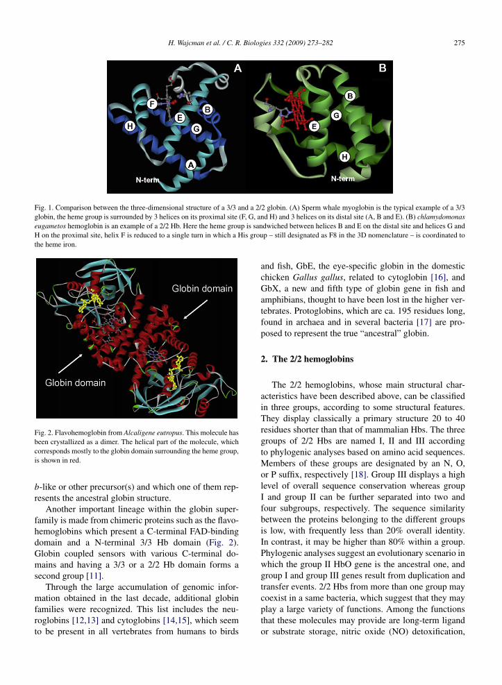
H. Wajcman et al. / C. R. Biologies 332 (2009) 273–282 275
Fig. 1. Comparison between the three-dimensional structure of a 3/3 and a 2/2 globin. (A) Sperm whale myoglobin is the typical example of a 3/3globin, the heme group is surrounded by 3 helices on its proximal site (F, G, and H) and 3 helices on its distal site (A, B and E). (B) chlamydomonaseugametos hemoglobin is an example of a 2/2 Hb. Here the heme group is sandwiched between helices B and E on the distal site and helices G andH on the proximal site, helix F is reduced to a single turn in which a His group – still designated as F8 in the 3D nomenclature – is coordinated tothe heme iron.
Fig. 2. Flavohemoglobin from Alcaligene eutropus. This molecule hasbeen crystallized as a dimer. The helical part of the molecule, whichcorresponds mostly to the globin domain surrounding the heme group,is shown in red.
b-like or other precursor(s) and which one of them rep-resents the ancestral globin structure.
Another important lineage within the globin super-family is made from chimeric proteins such as the flavo-hemoglobins which present a C-terminal FAD-bindingdomain and a N-terminal 3/3 Hb domain (Fig. 2).Globin coupled sensors with various C-terminal do-mains and having a 3/3 or a 2/2 Hb domain forms asecond group [11].
Through the large accumulation of genomic infor-mation obtained in the last decade, additional globinfamilies were recognized. This list includes the neu-roglobins [12,13] and cytoglobins [14,15], which seemto be present in all vertebrates from humans to birds
and fish, GbE, the eye-specific globin in the domesticchicken Gallus gallus, related to cytoglobin [16], andGbX, a new and fifth type of globin gene in fish andamphibians, thought to have been lost in the higher ver-tebrates. Protoglobins, which are ca. 195 residues long,found in archaea and in several bacteria [17] are pro-posed to represent the true “ancestral” globin.
2. The 2/2 hemoglobins
The 2/2 hemoglobins, whose main structural char-acteristics have been described above, can be classifiedin three groups, according to some structural features.They display classically a primary structure 20 to 40residues shorter than that of mammalian Hbs. The threegroups of 2/2 Hbs are named I, II and III accordingto phylogenic analyses based on amino acid sequences.Members of these groups are designated by an N, O,or P suffix, respectively [18]. Group III displays a highlevel of overall sequence conservation whereas groupI and group II can be further separated into two andfour subgroups, respectively. The sequence similaritybetween the proteins belonging to the different groupsis low, with frequently less than 20% overall identity.In contrast, it may be higher than 80% within a group.Phylogenic analyses suggest an evolutionary scenario inwhich the group II HbO gene is the ancestral one, andgroup I and group III genes result from duplication andtransfer events. 2/2 Hbs from more than one group maycoexist in a same bacteria, which suggest that they mayplay a large variety of functions. Among the functionsthat these molecules may provide are long-term ligandor substrate storage, nitric oxide (NO) detoxification,

276 H. Wajcman et al. / C. R. Biologies 332 (2009) 273–282
O2/NO sensing, redox reactions, and O2 delivery underhypoxic conditions [19,20].
3. Chimeric proteins
3.1. Flavohemoglobins
Prevalent in many bacterial and fungal pathogens,flavohemoglobins [21] are also known as a “fungal de-fense” enzymes (Fig. 2). They have been shown tobe closely tied to virulence. Upon infectious invasion,macrophages engulf pathogens and produce oxidantslike nitric oxide to protect the organism. De Jesus-Berrios et al. [22] have shown that inactivation of flavo-hemoglobin led to a significant loss of virulence bothin vitro and in vivo. Amino acid sequence analyses in-dicate that bacterial flavohemoglobins have 3/3 Hb foldand are closer to metazoan globins than most of the bac-teria hemoglobins, which display a 2/2 Hb fold.
3.2. Globin coupled sensors
Globin coupled sensors (GCSs) form a large groupof chimeric proteins having a N-terminal globin domainacting as a sensor to stimulate a signal controlled path-way [23]. They are one of the four known groups ofheme sensors protein, among which one of the moststudied was the kinase FixL, a PAS group protein [24].In these molecules, the heme group, through a confor-mation change, induced by ligand binding, switch on oroff an oxygen dependent metabolic pathway activatingsome genes. The first GCSs described were regulatorsof the aerotactic responses in Bacillus subtilis (HemAT-Bs) and Halobacterium salinarum (HemAT-Hs) [25].Today, more than 30 GCSs are known and the evolu-tion tree of their globin part suggests that they derivedfrom a common ancestor, the “protoglobin”. Such pro-toglobins have indeed been discovered in two Archaeaand two Bacteria [17]. Protoglobin from the strictlyanaerobic methanogenic Archaea (Methanosarcina ace-tivorans) has been recently crystallized and revealed analmost completely buried heme group being only acces-sible to ligand through apolar tunnels [26]. GCSs couldbe classified in several categories: the first is made byaerotactics and contains the HemATs. Another group isformed by “second messengers” coupled, for example,to a guanylyl cyclase catalytic domain [23]. Two GCSswere found to be involved in protein-protein interac-tions through a STAS domain (sulfate transporter andantisigma factor antagonist). Finally, some GCSs werefound to be tightly bound to the membrane through adomain which spans the membrane several times.
The evolutionary pathways of GCS and protoglobinare not yet clear. The initial ancestor could be a multido-main protein which split into components that furtherevolved on their own. Conversely, another hypothesisis that evolution may have started from individual do-mains forming a multidomain protein in which the twopartners evolved together.
4. Phylogenic origin of globins
Globin, under the form of protoglobin, is likely tohave been present in the first organisms living on Earth.It was perhaps included in the basic protein equipmentof the Last Universal Common Ancestor (LUCA), thesource of all life on Earth some 4000 Myr. The questiondebated now is whether the 3/3 and 2/2 globins evolvedindependently from a cytochrome b-like or other pre-cursor(s) and which one of them represents the ancestralglobin structure. During evolution, the problem of se-questering the water-insoluble heme in a hydrophobicprotein cavity was solved in several ways such as thecytochrome fold or the catalase, and PAS domains, butthe similarity of the globin fold in the 2/2 and 3/3 Hbs isa strong argument in favor of a unique globin origin. Al-though atmospheric O2 levels were very low in the earlyhistory of the Earth [<0.0008 atm], local concentrationscould have been high enough to promote the evolutionof the globin ancestor [27].
Up to now there is no answer as to whether the 3/3or the 2/2 fold is ancestral; both structures are present inthe three kingdoms of life. In addition, this debate couldnot be clarified by the presence or the absence of in-trons in the genes encoding globins. Introns are presentin all the 3/3 Hb genes and in many 2/2 Hbs, and whenpresent they are always localized at the same position. Itis therefore not yet possible to decide between the twoscenarios.
The evolutionary tree of globins is also complicatedby evidence for horizontal gene transfer (HGT) in some3/3 Hbs from bacteria and in some flavohemoglobins[28].
5. Evolution and adaptation of globin function
5.1. General considerations
The presence in all three kingdoms of life of a largevariety of globin molecules belonging to the differentlineages raises the question of their functions. It is as-sumed that the globin ancestor was able to react withdiatomic gaseous ligands inducing a change configura-tion that could be used in many tasks. Hemoglobin is

H. Wajcman et al. / C. R. Biologies 332 (2009) 273–282 277
therefore able to detect, scavenge, and detoxify O2 andO2-derived species (e.g., NO and CO). The principalrole of existent 2/2 Hbs is considered to be NO detox-ification [29]. It is also the case for flavohemoglobinsacting aerobically as NO oxygenases and anaerobicallyas NO reductases [30]. This NO detoxification is linkedto bacterial resistance: it has been shown in pathogenicmicroorganisms that flavohemoglobins protect the bac-teria from human macrophage NO-mediated killing andpromotes the virulence of bacteria.
In plants three types of Hbs have been identified:symbiotic, non-symbiotic and 2/2 Hbs. Two classes ofHbs evolved prior to the divergence of monocots anddicots. Leghemoglobins, which are pentacoordinate O2transporters, emerged from the first class, whereas hex-acoordinate scavenger/sensor Hbs emerged from bothclasses [31]. The function of the symbiotic hemoglobinsis to establish a low oxygen concentration for the ef-fective functioning of the bacterial nitrogenase neces-sary for nitrogen fixation. Although the role of 2/2 Hbsin plants is completely unknown, the non-symbiotichemoglobins, which biosynthesis is induced in plantcells upon exposure to low oxygen concentrations, arethought to play a role in a metabolic pathway which alsoinvolves nitric oxide and which provides an alternativetype of respiration to the mitochondrial electron trans-port under hypoxic conditions [32,33].
In the animal kingdom, the 3/3 Hbs and 2/2 Hbs areusually involved in oxygen transport and storage andNO detoxification, respectively. Nevertheless, in somespecies, hemoglobin molecules developed special evo-lution pattern allowing their carriers to occupy new eco-logical niches [29].
In some invertebrates, hemoglobins are found withinthe circulating cells of the coelomic fluid and/or withinmuscular type cells or cells of organ that need to be pro-tected from anoxia or oxidant stress. These moleculesare usually monomers or made from a small number ofsubunits.
In other species, such as insects, worms or mol-lusks, Hbs are freely dissolved in vascular, coelomic,or perienteric body fluids. They are circulating as gi-ant polymers [34,35]. This is observed in the ery-throcruorins from several oligochaetes (earthworm),achaetes (leech), vestimentiferans (hydrothermal venttube worm) and polychaetes. Lumbricus erythrocruorin,shown here as an example, assembles from 144 oxygen-binding hemoglobin subunits and 36 structural “link-er” subunits. Four distinct hemoglobin subunits arearranged into 12 dodecamers, which assemble onto acentral core formed from linker subunits (Fig. 3) [36].This supramolecular assembly involving several disul-
fide bonds raises many questions about the synchro-nization of the gene expression for the different struc-tural elements and the subsequent folding and assemblymechanism. Such large complexes offer a number ofimportant advantages as oxygen transport vehicles: ery-throcruorins are retained in the vascular system, eachcomplex has large oxygen binding capacity and subunitscan be arranged to permit cooperative oxygen bindingand regulatory features. Lumbricus terrestris erythro-cruorin is probably the best studied giant Hb; it hasbeen proposed as a potential model for a blood sub-stitute [37].
5.2. High oxygen affinity: Structure functionrelationships
The globins have often served as a model for study-ing structure-function relations in proteins. However,they are not necessarily an easy system to describe forligand binding, since the crystal structure did not reveala ligand binding pathway. Rather than a lock and keytype system involving a large surface area, ligand bind-ing apparently requires protein fluctuations. The staticstructure therefore is not sufficient to provide informa-tion such as trajectories and affinities [1].
The globins of very high oxygen affinity led to anew description of the critical structural elements. Itwas originally thought that it was essentially an affairof iron-oxygen binding, in which the heme pocket pro-vides a minor modulation, especially by the distal (E7)residue. It has now been shown that both the E7 andB10 position can participate in H-bonding directly tothe oxygen molecule. This places the center of intereston the oxygen ligand itself, capable of forming up tothree liaisons. The E7 and B10 positions will providethe bonds for major changes in affinity, which now ap-proach a variation of nearly a factor of a million (seeFig. 4) [38,39].
Globins displaying these extremely high affinitieswill necessarily have specific residues in these positionscapable of forming the H-bonds. Note that the specificresidue in the B10 position may be a Tyr or a Phe,depending on the distance requirements. In the E7 posi-tion, the classical His residue may form a bond, but thevery high affinities seem to employ a Gln residue.
5.3. Hexacoordination
The discovery of neuroglobin has lead to the for-mulation of an alternate mechanism of oxygen binding.The external ligands (such as oxygen, CO, or NO) maystill bind to the iron atom, but in competition with the

278 H. Wajcman et al. / C. R. Biologies 332 (2009) 273–282
Fig. 3. General structure of Lumbricus terrestris erythrocruorin showing the assembly of 12 dodecamers (A to L) in two parallel layers (bottomleft). The three-dimensional structure of a single dodecamer is shown with a resolution of 3.5 Å, 12 such units bind through their linker subunits,which form a central core. In total a complete erythrocruorin molecule carries 144 heme groups, each surrounded by a globin fold.
distal E7 His residue [40,41]. In the absence of exter-nal ligands, the ferrous iron may not be in a penta-coordinated state, since the distal His will bind to forma hexa-coordinated system [40]. Other globins such ascytoglobin, and new species of globin also display thismechanism of competitive binding; they are now collec-tively classed as hexa-coordinated globins to stress thefact that the penta-coordinated state is never more thana transient state.
Note that this mechanism has some interesting phys-iological consequences. The overall (observed) oxygenaffinity is no longer the simple ratio of oxygen bindingrates, but rather will be a ratio of the intrinsic oxygenand His binding affinities.
KO2,obs = KO2,penta/(1 + KHis) (1)
In the case of neuroglobin, the His affinity (KHis =kon,His/koff,His) is over 100; in order to obtain a typi-cal physiological (Mb-like) oxygen affinity, the intrinsicoxygen binding parameters must provide this extra fac-
tor of 100; this is accomplished by the extra H-bonds(via the E7 and B10 positions) as described in the sec-tion above [38–40]. This results in very slow oxygendissociation, requiring, nearly 1 second. The compen-sating His affinity leads to the “normal” oxygen affinityof about 1 mm Hg, but results in a rather sluggish globinin terms of response to external ligand levels, since re-equilibration will require several seconds.
Another direct consequence of the competition is areduced effect of external factors, such as temperatureand pH. While the individual rate coefficients may besensitive to temperature, the overall affinity is the ra-tio of affinities. If both the internal (His) and external(O2) ligands have a similar response to temperature, theoverall observed oxygen affinity may be nearly indepen-dent of temperature. This has been observed for oxygenbinding to various Hex-globins, and even a reverse tem-perature effect for CO binding [41].
In species where Hbs play only a local role or a func-tion in a signal pathway, oxygen transport is usually

H. Wajcman et al. / C. R. Biologies 332 (2009) 273–282 279
Fig. 4. The range of observed oxygen affinities covers nearly a factor of a million. The oxygen affinity (left panel) is the partial pressure of oxygenfor 50% saturation of the hemes, presented here on a logarithmic scale to cover the large variation. Typical oxygen affinities are in the range of 0.1to 10 mm Hg. The reference R and T states of tetrameric human Hb differ by about a factor of 100; the observed affinity for HbA can be modulatedby the effector DPG; this effect can be mimicked by the introduction of mutations, such as replacing the phenylalanine at position 41 of the betachain by a tyrosine. At the lower limit of oxygen affinity (high P50) is FixL, which is not a globin but a heme based oxygen sensor from rhizobium.Very high oxygen affinities have been observed for the globins of certain parasites, such as Ascaris or the trematodes (Gastrothylax crumenifershown here). The major contributions to the very high oxygen affinities are the formation of H-bonds directly to the Fe-bound oxygen molecule(yellow on the molecular image on the right hand panel) via critical protein residues at positions E7 and B10 (blue and green respectively) belowthe heme group (red). Modifications at these sites can provoke changes of over an order of magnitude in the oxygen affinity. A change of the B10position residue of Hb Ascaris leads to a shift to towards the normal range, while introduction of mutations at this site may shift a normal globintowards the very high affinity, as shown here for the globin from the clam Lucina pectinata.
assumed by other respiratory pigments such as hemo-cyanin.
5.4. A few examples
Hemoglobin from Ascaris works probably as an oxy-gen scavenger. Two Hbs, that have been thoroughlystudied, are present in this species. The first one is anextracellular one that circulates as a 328 kDa octamer.Each of the 8 subunits is made from a 39.5 kDa polypep-tide chain formed by two covalently bound globin do-mains (D1 and D2). In the C-terminal part of D2, thesequence “His-Lys-Glu-Glu” is repeated 4 times andis likely to play the role of a zipper for the associ-ation into octamer [42]. Electron microscopy picturesshow that the octamers are made by two layers formedeach by 4 subunits. This Hb displays one of the highestknown oxygen affinities: its P50 is of about 0.004 mmHg, which is 25 000 times higher than that of mam-malian Hbs [39,43,44]. Since association rates are lim-ited by diffusion, these very high affinities necessarily
display a considerably low value of the dissociationrate, making difficult the release of the bound oxygen.This property will hardly make this molecule suitablefor oxygen transport requiring a prompt uptake and re-lease. At the limit it could be a way to store oxygen inorder to assume a minimal oxygen supply in case of ex-treme anaerobic conditions. Conversely, it has also beenproposed that this Hb could be an oxygen scavenger al-lowing its destruction through a pathway involving NO[45]. In fact, nothing precise is known about the physi-ological role of this Hb, which remains a challenge. Inthe case of this Hb the only strong data are the molecularmechanism leading to this considerable oxygen affinitywhich consists in a network of hydrogen bonds extend-ing in the heme pocket between a Gln at position E7and the Tyr B10 stabilizing the Fe(δ+)–O2(δ−) dipolebinding [39].
In the same species another type of Hb is present inthe wall cells of the animal central cavity. It is a homod-imer of 40.6 kDa stabilized by SH bridges. Its sequenceis 38% similar to that of domain D1 and its P50 is of

280 H. Wajcman et al. / C. R. Biologies 332 (2009) 273–282
about 0.1 mm Hg, which could allow a role for oxy-gen storage at the proximity of muscular cells. In theanimal kingdoms, Hbs of nematodes, like neuroglobinsand cytoglobins, are the only globins encoded by a genecontaining 3 intervening sequences.
In some marine invertebrates, Hb is involved insulfur transport. For example, the giant extracellularhexagonal bilayer hemoglobin (HBL-Hb) of the deep-sea hydrothermal vent tube worm Riftia pachyptilatransports simultaneously O2 and H2S in the bloodfrom the gills to a specific organ, the trophosome, thatharbors sulfide-oxidizing endosymbionts. Within thisorgan, symbiotic bacteria use the sulfur as final elec-tron acceptor in a metabolic pathway transforming thedissolved CO2 into sugars [46]. Three Hbs are presentin these animals: the vascular blood contains Hbs V1and V2, and the coelomic fluid comprises Hb C1. TheseHbs are able to bind oxygen and sulfide simultaneouslyand reversibly at two different sites. The amount of H2Sthat they transport is high, varying from 1–10 mmol/L,depending upon its concentration in the surroundingmedium. Hb of Riftia pachyptila is a giant polymermade from a basic unit formed by 4 chains with 1free thiol group, connected by linker polypeptides. TheMW of HbV1 is of about 3500 kDa, contains about 150globin chains and some 40 linkers [47]. Hb V2 and C1are smaller, with a mw of about 400 kDa, and contains24 globin chains, many free SH groups are present.
Another example of Hb with high affinity for thiolgroup is observed in Lucina pectinata. This tropicalclam contains two kinds of Hbs: the first one (HbI) is amonomer, 143 residues long, which serves to transportH2S to autotrophic bacteria whereas two other globins(HbII and Hb III), respectively dimeric and tetramericin the µM range of [heme], are used for oxygen trans-port [48,49]. Site directed mutagenesis introducing aGln residue at position E7 and a Phe at B10 and E11in sperm whale myoglobin led to a Hb molecule with ahigh affinity for H2S but nevertheless with one order ofmagnitude less than in Lucina pectinata. Even thoughfurther modifications of the heme pocket are thus re-quired for the fine tuning mechanism of ligand bindingthis underlines the presence of key residues located atthe distal side of the heme pocket for switching fromO2 to H2S transport. Same conclusion holds inside thedifferent Lucina pectinata globins. Indeed for Hb II andHb III while the E7 and E11 residues do not changecompared to Hb I the B10 Phe is replaced by a Tyr lead-ing almost to three orders of magnitude decrease of theO2 dissociation rate. Thus the addition of a single hy-droxyl group is able to stabilize the dipolar Fe–O2 bondthrough a favorable hydrogen bond, perhaps in associ-
ation with a network of electrostatic interactions suchas those in the Ascaris globin explains the difference ofO2 off-rates between these globins. Note that the B10residue is also involved in the large decrease of the O2off-rates for Hb II and Hb III compared to Hb I and moregenerally in the ligand accessibility from the solvent tothe heme.
5.5. Neuroglobin: an allosteric protein?
As can be expected from the competitive ligand bind-ing, a modification of the His affinity will directly in-fluence the observed oxygen affinity. The effect is ex-ploited by neuroglobin via an internal disulfide bond:when the S–S bond is broken, the His affinity increasesby an order of magnitude, thereby decreasing the oxy-gen affinity [40]. One could therefore imagine a partnermolecule which reduces the cysteines of neuroglobinand thereby provokes the release of oxygen. This wouldlead to a model of Ngb as an allosteric protein providingoxygen delivery on demand of a specific partner.
Neuroglobin and cytoglobin have shed light on analternate globin mechanism for modulating the ligandbinding. These globins belong to the class of hexacoor-dinated globins: in the absence of external ligands, thedistal E7 His forms a stable bond with the heme iron.This bond is reversible so that the external ligand bind-ing requires the replacement of the constitutive residue.The globins are then able to use a mechanism compet-itive ligand binding to the heme iron. The fine tuningmechanism appears more in the stress induced at a dis-tance by an intra-disulfide bond; note that the locationof this disulfide bond is different for Ngb (CD loop) vsCygb (spanning the E helix with AB turn). Breaking thedisulfide bond allows a shift in the orientation of the E-helix, resulting in an enhanced affinity of the distal E7His for the heme and thereby a release of O2. The redoxstate of the cell could act on the state of the cysteines ofthe neuroglobin and further influence other biochemicalreactions involving an electron transfer to the molecularoxygen or another electron acceptor molecule and theNO catabolism.
5.6. An optical filter function
Hb from the nematode Mermis nigrescens (Mn-GLB-E), a parasite of grasshoppers and other Or-thopteran insects, has an optical, light shadowing func-tion [50]. The protein accumulates to high concentrationas intracellular crystals in the ocellus of mature photo-tactic adult females while also being expressed at lowconcentration in other tissues. From eggs ingested by

H. Wajcman et al. / C. R. Biologies 332 (2009) 273–282 281
the host, larvae grow to the adult length inside the bodycavity. The last larvae stage break out of the host andburrow into the soil where the final molt takes place andthe adult nematode matures. The gravid adult femalesemerge 1–2 years later from the soil and exhibit a posi-tive phototaxis during the search for suitable egg-layingsites in grass. This positive phototaxis results from apigmentation of the ocellus by formation of a thick Hbcrystal. A sensory nerve ending lies anterior to this areaand will point the head away from a light source as longas the ocellus would not be shadowed.
5.7. The smallest hemoglobin
The neural and body wall tissue of a nemerteanworm, Cerebratulus lacteus, express a major Hb com-ponents for which the amino acid sequences have beendeduced from cDNA and genomic DNA [51]. This Hbis 109 residues long and forms the smallest stable Hbknown. The globin genes have three exons and two in-trons with splice sites in the highly conserved positionsof globin genes. Alignment of the sequences with thoseof other globins indicates that helix A is almost missing,helix H is very short and GH corner elongated. Never-theless the general 3D structure is that of a globin fold.It has been hypothesized that this Hb could be an ances-tor of neuroHbs.
6. Conclusions
A large variety of Hbs with different structures andoxygen binding properties have been reported amonginvertebrates. The sequence alone is not sufficient toclassify the protein as a globin; the large heterogeneityin the expanding database includes globins with differ-ences above 90%. The protein structure and functionneed to be included in the overall consideration to fur-ther develop this superfamily. It is out of the scope ofthis mini-review to discuss all of them and we refer thereader to a few well documented reviews on this topic[52,53].
References
[1] J.C. Kendrew, G. Bodo, H.M. Dintzis, R.G. Parrish, H. Wyck-off, D.C. Phillips, A three-dimensional model of the myoglobinmolecule obtained by X-ray analysis, Nature 181 (1958) 662–666.
[2] S.N. Vinogradov, D. Hoogewijs, X. Bailly, K. Mizuguchi, S.Dewilde, L. Moens, J.R. Vanfleteren, A model of globin evo-lution, Gene 398 (2007) 132–142.
[3] S.N. Vinogradov, D. Hoogewijs, X. Bailly, R. Arredondo-Peter,J. Gough, S. Dewilde, L. Moens, J.R. Vanfleteren, A phyloge-nomic profile of globins, BMC Evol. Biol. 7 (2006) 31.
[4] T. Takagi, Hemoglobins from single-celled organisms, Curr.Opinion Struct. Biol. 3 (1993) 413–418.
[5] M. Potts, S. Angeloni, R. Ebel, D. Bassam, Myoglobin in acyanobacterium, Science 256 (1992) 1690–1692.
[6] T. Vandergon, C. Riggs, T. Gorr, J. Colacino, A. Riggs, The mini-hemoglobins in neural and body wall tissue of the nemerteanworm Cerebratulus lacteus, J. Biol. Chem. 273 (1998) 16998–17011.
[7] M. Couture, S. Yeh, B.A. Wittenberg, J.B. Wittenberg, Y. Ouel-let, D. Rousseau, M. Guertin, A cooperative oxygen-bindinghemoglobin from Mycobacterium tuberculosis, Proc. Natl. Acad.Sci. USA 96 (1999) 11223–11228.
[8] J.B. Wittenberg, M. Bolognesi, B.A. Wittenberg, M. Guertin,Truncated hemoglobins: a new family of hemoglobins widelydistributed in bacteria, unicellular eukaryotes, and plants, J. Biol.Chem. 277 (2002) 871–874.
[9] M. Couture, H. Chamberland, B. St-Pierre, J. Lafontaine, M.Guertin, Nuclear genes encoding chloroplast hemoglobins in theunicellular green alga Chlamydomonas eugametos, Mol. Gen.Genet. 243 (1994) 185–197.
[10] R. Watts, P. Hunt, A. Hvitved, M. Hargrove, W. Peacock, E.Dennis, A hemoglobin from plants homologous to truncatedhemoglobins of microorganisms, Proc. Natl. Acad. Sci. USA 98(2001) 10119–101124.
[11] S.N. Vinogradov, D. Hoogewijs, X. Bailly, R. Arredondo-Peter,M. Guertin, J. Gough, S. Dewilde, L. Moens, J.R. Vanfleteren,Three globin lineages belonging to two structural classes ingenomes from the three kingdoms of life, Proc. Natl. Acad. Sci.USA 102 (2005) 11385–11389.
[12] T. Burmester, B. Weich, S. Reinhardt, T. Hankeln, A vertebrateglobin expressed in the brain, Nature 407 (2000) 520–523.
[13] J. Trent, R. Watts, M. Hargrove, Human neuroglobin, a hex-acoordinate hemoglobin the reversibly binds oxygen, J. Biol.Chem. 276 (2001) 30106–30110.
[14] T. Burmester, B. Ebner, B. Weich, T. Hankeln, Cytoglobin:a novel globin type ubiquitously expressed in vertebrate tissues,Mol. Biol. Evol. 19 (2002) 416–421.
[15] T. Hankeln, B. Ebner, C. Fuchs, F. Gerlach, M. Haberkamp, T.L.Laufs, A. Roesner, M. Schmidt, B. Weich, S. Wystub, S. Saaler-Reinhardt, S. Reuss, M. Bolognesi, D. De Sanctis, M.C. Marden,L. Kiger, L. Moens, S. Dewilde, E. Nevo, A. Avivi, R.E. Weber,A. Fago, T. Burmester, Neuroglobin and cytoglobin in search oftheir role in the vertebrate globin family, J. Inorg. Biochem. 99(2005) 110–119.
[16] D. Kugelstadt, M. Haberkamp, T. Hankeln, T. Burmester, Neu-roglobin, cytoglobin, and a novel, eye-specific globin fromchicken, Biochem. Biophys. Res. Commun. 325 (2004) 719–725.
[17] T. Freitas, S. Hou, E. Dioum, J. Saito, J. Newhouse, G. Gonza-lez, M.A. Gilles-Gonzalez, M. Alam, Ancestral hemoglobins inArchaea, Proc. Natl. Acad. Sci. USA 101 (2004) 6675–6680.
[18] M. Nardini, A. Pesce, M. Milani, M. Bolognesi, Protein fold andstructure in the truncated (2/2) globin family, Gene 398 (2007)2–11.
[19] J.B. Wittenberg, M. Bolognesi, B.A. Wittenberg, M. Guertin,Truncated hemoglobins: a new family of hemoglobins widelydistributed in bacteria, unicellular eukaryotes, and plants, J. Biol.Chem. 277 (2002) 871–874.
[20] D.A. Vuletich, J.T. Lecomte, A phylogenetic and structural anal-ysis of truncated hemoglobins, J. Mol. Evol. 62 (2006) 196–210.
[21] H. Zhu, A. Riggs, Yeast flavohemoglobin is an ancient proteinrelated to globins and a reductase family, Proc. Natl. Acad. Sci.USA 89 (1992) 5015–5019.

282 H. Wajcman et al. / C. R. Biologies 332 (2009) 273–282
[22] M. de Jesús-Berríos, L. Liu, J.C. Nussbaum, G.M. Cox, J.S.Stamler, J. Heitman, Enzymes that counteract nitrosative stresspromote fungal virulence, Curr. Biol. 13 (2003) 1963–1968.
[23] M.A. Gilles-Gonzalez, G. Gonzalez, Heme-based sensors: defin-ing characteristics, recent developments, and regulatory hypothe-ses, J. Inorg. Biochem. 99 (2005) 1–22.
[24] M.A. Gilles-Gonzalez, G. Gonzalez, M.F. Perutz, L. Kiger, M.C.Marden, C. Poyart, Heme-based sensors, exemplified by the ki-nase FixL, are a new class of heme protein with distinctive ligandbinding and autoxidation, Biochemistry 33 (1994) 8067–8073.
[25] S. Hou, R.W. Larsen, D. Boudko, C.W. Riley, E. Karatan, M.Zimmer, G.W. Ordal, M. Alam, Myoglobin-like aerotaxis trans-ducers in Archaea and Bacteria, Nature 403 (2000) 540–544.
[26] M. Nardini, A. Pesce, L. Thijs, J.A. Saito, S. Dewilde, M. Alam,P. Ascenzi, M. Coletta, C. Ciaccio, L. Moens, M. Bolognesi, Ar-chaeal protoglobin structure indicates new ligand diffusion pathsand modulation of haem-reactivity, EMBO Rep. 9 (2008) 157–163.
[27] T.A. Freitas, J.A. Saito, S. Hou, M. Alam, Globin-coupled sen-sors, protoglobins, and the last universal common ancestor, J. In-org. Biochem. 99 (2005) 23–33.
[28] L. Moens, J. Vanfleteren, Y. Van de Peer, K. Peeters, O. Kapp,J. Czeluzniak, M. Goodman, M. Blaxter, S.N. Vinogradov,Globins in nonvertebrate species: dispersal by horizontal genetransfer and evolution of the structure-function relationships,Mol. Biol. Evol. 13 (1996) 324–333.
[29] H. Wajcman, L. Kiger, Hemoglobin, from microorganisms toman: a single structural motif, multiple functions, C. R. Biol. 325(2002) 1159–1174.
[30] A.M. Gardner, P.R. Gardner, Flavohemoglobin detoxifies nitricoxide in aerobic, but not anaerobic, Escherichia coli. Evidencefor a novel inducible anaerobic nitric oxide-scavenging activity,J. Biol. Chem. 277 (2002) 8166–8171.
[31] J.A. Hoy, H. Robinson, J.T. Trent, S. Kakar, B.J. Smagghe, M.S.Hargrove, Plant hemoglobins: a molecular fossil record for theevolution of oxygen transport, J. Mol. Biol. 371 (2007) 168–179.
[32] V. Garrocho-Villegas, S.K. Gopalasubramaniam, R. Arredondo-Peter, Plant hemoglobins: what we know six decades after theirdiscovery, Gene 398 (2007) 78–85.
[33] K.H. Hebelstrup, A.U. Igamberdiev, R.D. Hill, Metabolic effectsof hemoglobin gene expression in plants, Gene 398 (2007) 86–93.
[34] B.N. Green, T. Gotoh, T. Suzuki, F. Zal, F.H. Lallier, A. Toul-mond, S.N. Vinogradov, Observation of large, non-covalentglobin subassemblies in the approximately 3600 kDa hexago-nal bilayer hemoglobins by electrospray ionization time-of-flightmass spectrometry, J. Mol. Biol. 309 (2001) 553–560.
[35] T. Suzuki, S.N. Vinogradov, Globin and linker sequences of thegiant extracellular hemoglobin from the leech Macrobdella dec-ora, J. Protein. Chem. 22 (2003) 231–242.
[36] W.E. Royer, H. Sharma, K. Strand, J.E. Knapp, B. Bhyravb-hatla, Lumbricus erythrocruorin at 3.5 A resolution: architectureof a megadalton respiratory complex, Structure 14 (2006) 1167–1177.
[37] R.E. Hirsch, L.A. Jelicks, B.A. Wittenberg, D.K. Kaul, H.L.Shear, J.P. Harrington, A first evaluation of the natural highmolecular weight polymeric Lumbricus terrestris hemoglobin asan oxygen carrier, Artif. Cells Blood Substit. Immobil. Biotech-nol. 25 (1997) 429–444.
[38] T.E. Carver, R.E. Brantley, E.W. Singleton, R.M. Arduini, M.L.Quillin, G.N. Phillips, J.S. Olson, A novel site-directed mutant ofmyoglobin with an unusually high O2 affinity and low autooxi-dation rate, J. Biol. Chem. 267 (1992) 14443–14450.
[39] I. De Baere, M.F. Perutz, L. Kiger, M.C. Marden, C. Poyart,Formation of two hydrogen bonds from the globin to the heme-linked oxygen molecule in Ascaris hemoglobin, Proc. Natl.Acad. Sci. USA 91 (1994) 1594–1597.
[40] D. Hamdane, L. Kiger, S. Dewilde, B.N. Green, A. Pesce, J.Uzan, T. Burmester, T. Hankeln, M. Bolognesi, L. Moens, M.C.Marden, The redox state of the cell regulates the ligand bindingaffinity of human neuroglobin and cytoglobin, J. Biol. Chem. 278(2003) 51713–51721.
[41] J. Uzan, S. Dewilde, T. Burmester, T. Hankeln, L. Moens, D.Hamdane, M.C. Marden, L. Kiger, Neuroglobin and other hexa-coordinated hemoglobins show a weak temperature dependenceof oxygen binding, Biophys. J. 87 (2004) 1196–1204.
[42] I. De Baere, L. Liu, L. Moens, J. Van Beeumen, C. Gielens,J. Richelle, C. Trotman, J. Finch, M. Gerstein, M. Perutz, Po-lar zipper sequence in the high-affinity hemoglobin of Ascarissuum: amino acid sequence and structural interpretation, Proc.Natl. Acad. Sci. USA 89 (1992) 4638–4642.
[43] J. Yang, A.P. Kloek, D.E. Goldberg, F.S. Mathews, The structureof Ascaris hemoglobin domain I at 2.2 A resolution: molecularfeatures of oxygen avidity, Proc. Natl. Acad. Sci. USA 92 (1995)4224–4228.
[44] L. Kiger, A.K. Rashid, N. Griffon, M. Haque, L. Moens, Q.H.Gibson, C. Poyart, M.C. Marden, Trematode hemoglobins showexceptionally high oxygen affinity, Biophys. J. 75 (1998) 990–998.
[45] D.M. Minning, A.J. Gow, J. Bonaventura, R. Braun, M. De-whirst, D.E. Goldberg, J.S. Stamler, Ascaris haemoglobin is anitric oxide-activated ‘deoxygenase’, Nature 401 (1999) 497–502.
[46] X. Bailly, D. Jollivet, S. Vanin, J. Deutsch, F. Zal, F. Lallier,A. Toulmond, Evolution of the sulfide-binding function withinthe globin multigenic family of the deep-sea hydrothermal venttubeworm Riftia pachyptila, Mol. Biol. Evol. 19 (2002) 1421–1433.
[47] C. Chabasse, X. Bailly, S. Sanchez, M. Rousselot, F. Zal, Genestructure and molecular phylogeny of the linker chains from thegiant annelid hexagonal bilayer hemoglobins, J. Mol. Evol. 63(2006) 365–374.
[48] D.W. Kraus, J.B. Wittenberg, Hemoglobins of the Lucina pecti-nata/bacteria symbiosis. I. Molecular properties, kinetics andequilibria of reactions with ligands, J. Biol. Chem. 265 (1990)16043–16053.
[49] M. Rizzi, J.B. Wittenberg, A. Coda, M. Fasano, P. Ascenzi,M. Bolognesi, Structure of the sulfide-reactive hemoglobin fromthe clam Lucina pectinata. Crystallographic analysis at 1.5 A res-olution, J. Mol. Biol. 244 (1994) 86–99.
[50] A.H.J. Burr, P. Hunt, D.R. Wagar, S. Dewilde, M.L. Blaxter, J.R.Vanfleteren, L. Moens, A hemoglobin with an optical function,J. Biol. Chem. 275 (2000) 4810–4815.
[51] A. Pesce, M. Nardini, S. Dewilde, E. Geuens, K. Yamauchi,P. Ascenzi, A.F. Riggs, L. Moens, M. Bolognesi, The 109 residuenerve tissue minihemoglobin from Cerebratulus lacteus high-lights striking structural plasticity of the alpha-helical globinfold, Structure 10 (2002) 725–735.
[52] M. Bolognesi, D. Bordo, M. Rizzi, C. Tarricone, P. Ascenzi,Nonvertebrate hemoglobins: structural bases for reactivity, Prog.Biophys. Mol. Biol. 68 (1997) 29–68.
[53] R.E. Weber, S.N. Vinogradov, Nonvertebrate hemoglobins: func-tions and molecular adaptations, Physiol. Rev. 81 (2001) 569–628.