Relating biochemistry and function in the myosin superfamily
The death-fold superfamily ofhomotypic interaction motifsKristof Kersse1,2, Jelle Verspurten1,2, Tom Vanden Berghe1,2* andPeter Vandenabeele1,2*
1 Department for Molecular Biomedical Research, VIB, B-9052 Ghent (Zwijnaarde), Belgium2 Department of Biomedical Molecular Biology, Ghent University, B-9052 Ghent (Zwijnaarde), Belgium
Review
The death-fold superfamily encompasses four structur-ally homologous subfamilies that engage in homotypic,subfamily-restricted interactions. The Death Domains(DDs), the Death Effector Domains (DEDs), the CAspaseRecruitment Domains (CARDs) and the PYrin Domains(PYDs) constitute key building blocks involved in theassembly of multimeric complexes implicated in signal-ing cascades leading to inflammation and cell death. Wereview the molecular basis of these homotypic domain–
domain interactions in light of their structure, functionand evolution. In addition, we elaborate on three distincttypes of asymmetric interactions that were recentlyidentified from the crystal structures of three multimeric,death-fold complexes: the MyDDosome, the PIDDosomeand the Fas/FADD-DISC. Insights into the mechanisms ofinteraction of death-fold domains will be useful to de-sign strategies for specific modulation of complex for-mation and might lead to novel therapeutic applications.
Structure and function of a death-fold domainSignal transduction pathways controlling immunity, in-flammation and apoptotic or necroptotic cell death dependto a large extent on proteins containing homotypic interac-tion motifs belonging to the death-fold superfamily. The 102members discovered in humans so far can be categorized infour subfamilies: 39 Death Domains (DDs), 8 Death EffectorDomains (DEDs), 33 CAspase Recruitment Domains(CARDs) and 22 PYrin Domains (PYDs) [1]. Despite theconsiderable overall divergence in sequence of its members(up to 90%), this superfamily is hallmarked by its structure,the so-called death-fold, which consists of a globular struc-ture wherein six amphipathic a-helices are arranged in anantiparallel a-helical bundle with Greek key topology [2–5](Figure 1). This topology determines the folding process,which starts with the formation of a hydrophobic core con-taining nearly all the conserved residues of the death-foldsuperfamily [6,7]. Structural differences between individualdomains, which are smaller within a subfamily than be-tween subfamilies, are the result of alterations in the lengthand orientation of the a-helices, and the distribution ofcharged and hydrophobic residues along the surface. Con-sequently, each member obtains a specific array of homo-typic interaction partners that, in general, do not cross theboundaries of the subfamily. This homotypic trait is proba-
Corresponding author: Vandenabeele, P. ([email protected])* Equal senior contribution.
0968-0004/$ – see front matter � 2011 Elsevier Ltd. All rights reserved. doi:10.1016/j.tibs.2011.
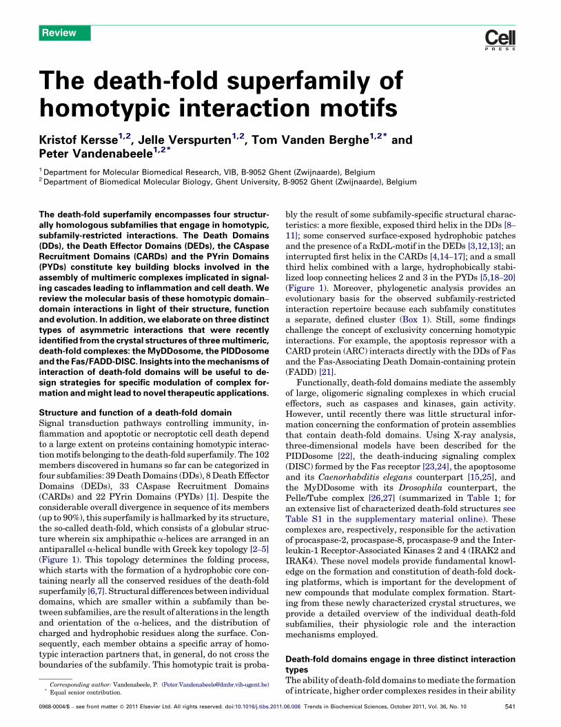
bly the result of some subfamily-specific structural charac-teristics: a more flexible, exposed third helix in the DDs [8–
11]; some conserved surface-exposed hydrophobic patchesand the presence of a RxDL-motif in the DEDs [3,12,13]; aninterrupted first helix in the CARDs [4,14–17]; and a smallthird helix combined with a large, hydrophobically stabi-lized loop connecting helices 2 and 3 in the PYDs [5,18–20](Figure 1). Moreover, phylogenetic analysis provides anevolutionary basis for the observed subfamily-restrictedinteraction repertoire because each subfamily constitutesa separate, defined cluster (Box 1). Still, some findingschallenge the concept of exclusivity concerning homotypicinteractions. For example, the apoptosis repressor with aCARD protein (ARC) interacts directly with the DDs of Fasand the Fas-Associating Death Domain-containing protein(FADD) [21].
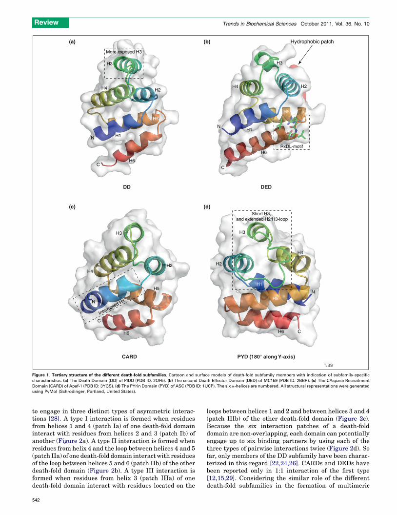
Functionally, death-fold domains mediate the assemblyof large, oligomeric signaling complexes in which crucialeffectors, such as caspases and kinases, gain activity.However, until recently there was little structural infor-mation concerning the conformation of protein assembliesthat contain death-fold domains. Using X-ray analysis,three-dimensional models have been described for thePIDDosome [22], the death-inducing signaling complex(DISC) formed by the Fas receptor [23,24], the apoptosomeand its Caenorhabditis elegans counterpart [15,25], andthe MyDDosome with its Drosophila counterpart, thePelle/Tube complex [26,27] (summarized in Table 1; foran extensive list of characterized death-fold structures seeTable S1 in the supplementary material online). Thesecomplexes are, respectively, responsible for the activationof procaspase-2, procaspase-8, procaspase-9 and the Inter-leukin-1 Receptor-Associated Kinases 2 and 4 (IRAK2 andIRAK4). These novel models provide fundamental knowl-edge on the formation and constitution of death-fold dock-ing platforms, which is important for the development ofnew compounds that modulate complex formation. Start-ing from these newly characterized crystal structures, weprovide a detailed overview of the individual death-foldsubfamilies, their physiologic role and the interactionmechanisms employed.
Death-fold domains engage in three distinct interactiontypesThe ability of death-fold domains to mediate the formationof intricate, higher order complexes resides in their ability
06.006 Trends in Biochemical Sciences, October 2011, Vol. 36, No. 10 541
H1
H2
H3
H4
H5
H6
N
C
Short H3, and extended H2/H3-loop
PYD (180° along Y-axis)
H1
H2
H3
H4
H5
H6
N
C
DED
RxDL-motif
Hydrophobic patch
H2
H3
H4
H5
H6
C
Interrupted H1N
CARD
H1
H2
H3
H4
H5
H6
N
C
More exposed H3
DD
(a) (b)
(c) (d)
TiBS
Figure 1. Tertiary structure of the different death-fold subfamilies. Cartoon and surface models of death-fold subfamily members with indication of subfamily-specific
characteristics. (a) The Death Domain (DD) of PIDD (PDB ID: 2OF5). (b) The second Death Effector Domain (DED) of MC159 (PDB ID: 2BBR). (c) The CAspase Recruitment
Domain (CARD) of Apaf-1 (PDB ID: 3YGS). (d) The PYrin Domain (PYD) of ASC (PDB ID: 1UCP). The six a-helices are numbered. All structural representations were generated
using PyMol (Schrodinger, Portland, United States).
Review Trends in Biochemical Sciences October 2011, Vol. 36, No. 10
to engage in three distinct types of asymmetric interac-tions [28]. A type I interaction is formed when residuesfrom helices 1 and 4 (patch Ia) of one death-fold domaininteract with residues from helices 2 and 3 (patch Ib) ofanother (Figure 2a). A type II interaction is formed whenresidues from helix 4 and the loop between helices 4 and 5(patch IIa) of one death-fold domain interact with residuesof the loop between helices 5 and 6 (patch IIb) of the otherdeath-fold domain (Figure 2b). A type III interaction isformed when residues from helix 3 (patch IIIa) of onedeath-fold domain interact with residues located on the
542
loops between helices 1 and 2 and between helices 3 and 4(patch IIIb) of the other death-fold domain (Figure 2c).Because the six interaction patches of a death-folddomain are non-overlapping, each domain can potentiallyengage up to six binding partners by using each of thethree types of pairwise interactions twice (Figure 2d). Sofar, only members of the DD subfamily have been charac-terized in this regard [22,24,26]. CARDs and DEDs havebeen reported only in 1:1 interaction of the first type[12,15,29]. Considering the similar role of the differentdeath-fold subfamilies in the formation of multimeric

0.1
PIDD DDRIP1 DD
MyD88 DD
IRAK1 DDIRAK2 DD
IRAK4 DD
IRAK3 DDMALT1 DD
TNFR1 DDTRADD DDFADD DDRAIDD DD
FAS DDTRAILR1 DD
FADD DEDCaspase−10 DED1
Caspase−8 DED1Caspase−8 DED2Caspase−10 DED2
ASC PYDNLRP6 PYD
NLRP10 PYD
NLRP12 PYDNLRP3 PYD
MEFV PYDNLRP1 PYD
NLRP4 PYDNLRP9 PYD
NLRP14 PYD NLRP5 PYDNLRP11 PYD
NLRP13 PYDNLRP8 PYD
NLRP2 PYDNLRP7 PYD
AIM2 PYD
RIG-I CARD2
ASC CARDNLRP1 CARD
CARMA1 CARDCARMA3 CARD
CARMA2 CARDCARD9 CARDRAIDD CARD
BCL10 CARDCaspase−12 CARD
Caspase−2 CARDCaspase−9 CARD
MDA5 CARD2MAVS CARDMDA5 CARD1
APAF1 CARDRIG-I CARD1
Caspase−4 CARDCaspase−5 CARD
Caspase−1 CARD
NOD1 CARDRIP2 CARD
NLRC4 CARD
NOD2 CARD1NOD2 CARD2
CA
RD
su
bfa
mily
DD
su
bfa
mily
D
ED
su
bfa
mily
PY
D s
ub
fam
ily
TiBS
Figure I. Phylogenetic analysis of the death-fold superfamily. The sequences of the death-fold domains were aligned using Clustal X (Gonnet matrix, gap penalty: 10,
gap extension penalty: 0.1). The Neighbor Joining bootstrap tree was constructed using Clustal X (http://www.clustal.org) and visualized with iTol (http://itol.embl.de).
Box 1. Evolution of the death-fold superfamily
Given the high homology in structure and the considerable diver-
gence in sequence of the death-fold superfamily members, we
examined whether a sequence-based phylogenetic analysis would
provide an evolutionary explanation for the emergence of four
conserved death-fold subfamilies, the DDs, the DEDs, the CARDs
and the PYDs. CARD- and DD-containing proteins have been identified
in organisms such as Caenorhabditis elegans and Drosophila
melanogaster, whereas zebrafish and rodents are the most distant
organisms wherein PYD- and DED-containing proteins have thus far
been found.
This phylogenetic analysis was performed with the death-fold
domains of human descent that form an integral part of well-
characterized signaling complexes, including the apoptosome [52],
the PIDDosome [73], the death receptor complexes [65], the
inflammasomes [30], the NOD complexes [74], the RIG-like receptor
complexes [67], the TLR/IL-1 receptor complexes [75] and the B cell
and T cell receptor proximal complexes [68]. Apart from one
exception, the second CARD of RIG-I, each of the subfamilies clusters
in a separate branch, indicating that each subfamily had its own
ancestor. This analysis suggests that the founding father of the death-
fold superfamily gave rise to a CARD and a DD–DED–PYD ancestor
(Figure I). The latter diverged in a DD and a DED–PYD ancestor, which
in turn resulted in the emergence of the DED and PYD ancestor (Figure
I). This clear subfamily separation probably explains why these
domains were almost exclusively found to engage in homotypic,
subfamily-confined interactions. Considering the structural similarity
and the low sequence similarity of the death-fold superfamily, these
ancestor death-folds might be the result of a convergent evolution
toward the same function [76]. Additional death-fold domains most
probably originated through divergent evolution involving gene
duplication events within each subfamily [63]. A striking example is
given by the caspase-1-related CARD-only proteins, COP, INCA and
ICEBERG, which localize at the caspase-1 locus, share a high degree
of sequence identity and acquired premature nonsense mutations
resulting in a CARD-only protein [77,78]. In addition, several members
of the Herpesviridae and Poxviridae, two families of dsDNA viruses,
acquired death-fold domain-only proteins (DED-containing vFLIPs
and PYD-containing vPOPs) through horizontal gene transfer that
enable them to evade the host defense system [49,64]. Taken
together, the parallel evolution of the four death-fold subfamilies
forms the basis for their ability to engage in homotypic interactions
within the boundaries of the subfamily.
Review Trends in Biochemical Sciences October 2011, Vol. 36, No. 10
543
Table 1. Overview of all described crystal structures of multimeric death-fold complexesa
Protein/complex Species Residues Purification and crystallization
conditions
PDB ID Resolution Ref.
DD PIDDosome human PIDD: 778–883
RAIDD: 94–199
Domains individually expressed
in Escherichia coli and mixed.
Crystallized at pH 6.5.
2OF5 3.2 A [22]
Fas/FADD-DISC human Fas: 223–335
FADD: 93–208
Domains individually
expressed in E. coli and mixed.
Crystallized at pH 4.0.
3EZQ 2.7 A [23]
mouse, human Fas: 210–310
FADD: 93–184
Domains coexpressed in
E. coli and copurified.
Crystallized at pH 8.5.
3OQ9 6.8 A [24]
MyDDosome human MyD88: 20–117
IRAK4: 4–106
IRAK2: 1–112
Domains coexpressed in
E. coli and copurified.
Crystallized at pH 8.0.
IRAK2 contains a
mutation (R50W)
3MOP 3.4 A [26]
Pelle–Tube
complex
Drosophila Pelle: 26–129
Tube: 1–184
Domains individually
expressed in E. coli and mixed.
Crystallized at pH 7.2–8.4.
1D2Z 2.0 A [27]
DED MC159 (v-FLIP) Molluscum
contagiosum
1–187 Expressed in E. coli.
Crystallized at pH 6.5.
2BBR 1.2 A [12]
1–183 Expressed in E. coli.
Crystallized at pH 8.5.
2F1S 1.4 A [29]
CARD Apoptosome human Apaf-1: 1–97
procaspase-9: 1–112
Domains individually
expressed in E. coli and mixed.
Crystallized at pH 4.6.
3YGS 2.5 A [15]
C. elegans Full-length CED4
Full-length CED3
Domains individually expressed
in E. coli and mixed.
Crystallization pH was not
indicated. No apparent electron
density for CED3 in the crystals.
3LQQ 3LQR 3.5 A
3.9 A
[25]
aThe species, the residues included to purify the death-fold domain, some purification and crystallization particulars, the PDB ID and the resolution of the obtained crystals
are indicated for each complex.
Review Trends in Biochemical Sciences October 2011, Vol. 36, No. 10
protein assemblies, one can assume that all death-fold domains might use the three types of interaction.Determining the crystal structure of the CARD- andPYD-containing inflammasomes, which activate procas-pase-1 to enable Interleukin (IL)-1b production andpyroptotic cell death [30], could be of particular interestto support this assumption. Moreover, the insight gainedon the interaction mechanism of the inflammasomes couldprovide novel avenues for the development of compoundsthat interfere with excessive inflammasome formation,an important factor in the pathogenesis of multipleinflammatory disorders (e.g. gout, type II diabetes andcryopyrin-associated periodic syndromes) [30]. Indeed, bytargeting upstream complexes, such inflammasome inter-fering compounds could be valuable, more specific alter-natives for the currently used IL-1b-inhibitors anakinra,rilonacept and canakinumab, which target the effectormolecules [31].
Although the hitherto reported structures suggest thatthe three interaction types are conserved throughout thedeath-fold superfamily, there are marked differences be-tween individual pairwise interactions of the same type.The type I interaction between the CARDs of Apaf-1 andprocaspase-9 depends on a network of intermolecular hy-drogen bonds and van der Waals interactions [15], whereasthe type I interaction between FADD–DED and procas-pase-8–DED2 is mediated by a hydrophobic interface[12,13]. The difference in utilization of non-covalent bind-ing modes combined with the complementarity in surface
544
are two important factors that determine the interactionpartners within the death-fold superfamily.
Homotypic DD interactionsMost knowledge concerning interactions of death-folddomains in the context of oligomeric protein assemblieswas obtained from crystal structures containing DDs. Inthis section of the review, the functional context and thestructural organization of the PIDDosome, the MyDDo-some and the Fas/FADD-DISC are highlighted.
The PIDDosome
The crystal structure of the DD assembly containing thep53-Inducible protein with a Death Domain (PIDD) andRIP-Associated protein with a Death Domain (RAIDD) wasthe first description of an oligomeric complex involvingmembers of the death-fold superfamily [22]. Interactionsmediated by these DDs form the core of the PIDDosome, acomplex acting as a molecular switch that determines lifeor death following genotoxic stress [32]. Depending on itsproteolytic processing status, PIDD mediates the assemblyof two separate PIDDosomes [33]. A 51-kDa C-terminalfragment obtained by single autoprocessing enables for-mation of the NF-kB-activating, pro-survival PIDDosome.In this complex, the PIDD-DD engages the DD-containingReceptor Interacting serine/threonine-Protein kinase 1(RIP1) to assemble a platform for the recruitment of theNF-kB Essential MOdulator (NEMO), an essential adaptorprotein for the activation of NF-kB [32,33]. The formation

H1
H2
H3H4
Type I interaction
Patch Ia Patch Ibhelix 1helix 4
helix 2helix 3
H4
H5
H5
H6
Type II interaction
Patch IIahelix 4
loop H4-H5
Patch IIbloop H5-H6
H3H1
H2
H3
H4
Type III interaction
Patch IIIa Patch IIIbhelix 3 loop H1-H2
loop H3-H4
(a)
(d)
(b) (c)
90° Y-axis 90° Y-axis 90° Y-axis
Patch IaKey:
Patch Ib
Patch IIa
Patch IIb
Patch IIIa
Patch IIIb
TiBS
Figure 2. A Death Domain (DD) mediates three interaction types through six interaction patches. The three asymmetric interaction types: type I (a), type II (b) and type III (c)
derived from the PIDDosome (PDB ID: 2OF5). The DDs of RIP-Associated protein with a Death Domain (RAIDD) (on the left, light gray) and p53-Inducible protein with a Death
Domain (PIDD) (on the right, dark gray) are shown as cartoon drawings for each interaction, whereas the interaction patches are displayed as surface (type I: green; type II:
red; type III: blue). Helices and loops contributing to the interactions are indicated. (d) The six possible interaction patches on the DD of RAIDD. The composite model is
rotated in four stages of 908 around the Y-axis.
Review Trends in Biochemical Sciences October 2011, Vol. 36, No. 10
of the pro-death PIDDosome relies on a subsequent cleav-age that generates a 37-kDa, DD-only PIDD fragment [33].Under these circumstances, the DD of PIDD interacts withthe DD of the CARD- and DD-containing adaptor proteinRAIDD, which subsequently recruits and activates theCARD-containing procaspase-2 through a CARD/CARDinteraction [33,34]. This pro-death PIDDosome representsthe functional context for the crystal structure of the PIDD-DD/RAIDD-DD assembly. An asymmetric, three-layered,globular complex was reported in which the bottom, middleand top layers contain five PIDD-DDs, five RAIDD-DDsand another two RAIDD-DDs, respectively (Figure 3a) [22].Based on the stoichiometry of this complex, the procas-pase-2 activating PIDDosome would have a molecularmass of 696.8 kDa, which is in good agreement with theobserved molecular mass of 670 kDa when the molecularcomposition of the PIDDosome was determined throughgel filtration analysis [34,35]. Despite the apparent asym-metry within the complex as a whole, each DD appears tobe in a quasi-equivalent environment and employs thethree types of interactions (Figure 3b) [22]. More thana decade ago, the interaction between the CARDs ofRAIDD and procaspase-2 was predicted based on muta-genic analysis and appeared to be mediated by electrostatic
interaction [4]. Still, a crystal structure elucidating thisCARD/CARD interaction is needed to unambiguously de-termine the exact interaction type.
The MyDDosome
Recently, another ternary complex containing DDs ofMyD88, IRAK4 and IRAK2 was characterized by X-raycrystallography and was termed the MyDDosome [26].This complex encompasses an essential component ofthe signal transduction cascade that links the Toll-likereceptors (TLRs) and the receptors for the proinflamma-tory cytokines IL-1b and IL-18 to the activation of tran-scription factors important for effective immune responses[36]. Interestingly, the overall structural appearance of theMyDDosome differs rather substantially from the PIDDo-some. In contrast to the globular PIDDosome, the MyDDo-some seems to be a left-handed, helical tower-shapedstructure in which six MyD88-DDs constitute the bottomtwo layers, four IRAK4-DDs make up the third layer andanother four IRAK2-DDs make up the fourth layer(Figure 3d) [26]. Still, similar to the PIDDosome, all theDDs of the MyDDosome were found to be in a quasi-equivalent environment and use the three types of inter-action. Type III interactions connect adjacent DDs within a
545

IRAK21IRAK22IRAK23IRAK24
IRAK41
IRAK44
IRAK42IRAK43IRAK44
MyD883MyD884MyD885MyD886
MyD882
MyD881
MyD886
IRAK24
(b)
2 RAIDD-DD
5 RAIDD-DD
5 PIDD-DD
90° along X-axis
PIDD 1
PIDD 1
PIDD 2
PIDD 3
PIDD 4
PIDD 5
RAIDD 2
RAIDD 1
RAIDD 7
RAIDD 3
RAIDD 4
RAIDD 5
RAIDD 6
RAIDD 1
Type II interaction Type III interactionType I interactionKey:
(a) (c)
(d)
4 IRAK2-DD
4 IRAK4-DD
6 MyD88-DD
90° along X-axis
TiBS
Figure 3. The PIDDosome and MyDDosome complex. (a) Side and top views of the PIDDosome complex (PDB ID: 2OF5) represented as cartoon drawings. The bottom layer
contains five PIDD (p53-Inducible protein with a Death Domain) Death Domains (DDs) (gray), the middle layer contains five RAIDD (RIP-Associated protein with a Death
Domain) DDs (orange) and the top layer contains two RAIDD DDs (dark yellow). The top view is a 908 rotation around the X-axis of the side view. (b) Schematic, planar
illustration of the three interaction types used to assemble the PIDDosome. (c) Side and top views of the MyDDosome complex (PDB ID: 3MOP) represented as cartoon
drawings. The bottom layer contains six MyD88-DDs (black), the middle layer contains four IRAK4-DDs (magenta) and the top layer contains four IRAK2-DDs (yellow). The
top view is a 908 rotation around the X-axis of the side view. (d) Schematic, planar illustration of the three interaction types used to assemble the MyDDosome. The different
interaction patches are colored as in Figure 2.
Review Trends in Biochemical Sciences October 2011, Vol. 36, No. 10
546
Review Trends in Biochemical Sciences October 2011, Vol. 36, No. 10
layer, whereas type I and II interactions connect the layers(Figure 3d).
Based on the MyDDosome structure a model can be putforward wherein complex assembly encompasses a hierar-chical and sequential process that is tightly controlled bymultiple mechanisms. Complex assembly can be consid-ered cooperative because the sequential recruitment ofeach DD increases stability. Complementarity in chargeand shape between the bottom and top surfaces of the DDs,involved in the formation of type II interactions, is one ofthe key determinants of hierarchy. For instance, IRAK2 isrecruited to the complex only when IRAK4 is presentbecause the bottom surface of IRAK2 finds its best matchin the top surface of IRAK4; the top surfaces of MyD88 andIRAK2 are not sufficiently complementary (Figure 3d) [26].Furthermore, the observation that the top and bottomsurfaces of IRAK2 do not fit properly to each other limitsthe recruitment of IRAK2-DDs to four.
The functional significance of the MyDDosome lies inthe activation of the kinase activity of both IRAKs: IRAK4undergoes autophosphorylation and cross-phosphorylatesIRAK2. Subsequently, active IRAK2 leaves the complex tointeract with TRAF6 and to propagate an intricate signal-ing cascade involving phosphorylation and ubiquitinationevents that lead to the activation of transcription factorssuch as NF-kB, AP-1 and c-Jun [36]. Interestingly, theconserved, homologous signaling pathway in Drosophilauses similar players, dMyD88, Tube and Pelle, but in thiscase it does not lead to the assembly of an oligomeric,MyDDosome-like complex [37,38]. Rather, the dMyD88–
Tube–Pelle complex was found to be a 1:1:1 kidney-shapedternary complex in which two type II interactions connectthe DDs [26,27,38].
The Fas/FADD-DISC
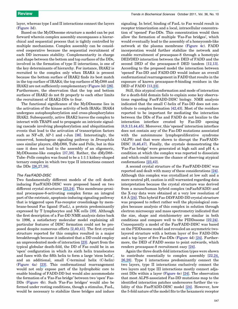
Two fundamentally different models of the cell death-inducing Fas/FADD-DISC were proposed based on twodifferent crystal structures [23,24]. This membrane-proxi-mal procaspase-8-activating complex forms an integralpart of the extrinsic, apoptosis-inducing signaling pathwaythat is triggered upon Fas-receptor crosslinkage by mem-brane-bound Fas ligand (FasL), a protein predominantlyexpressed by T lymphocytes and NK cells [39]. Althoughthe first description of a Fas-DD NMR analysis dates backto 1996, a satisfactory molecular model explaining allparticular features of DISC formation could not be pro-posed despite numerous efforts [2,40,41]. The first crystalstructure reported for this complex resulted in a majorbreakthrough because it indicated that a DD could employan unprecedented mode of interaction [23]. Apart from thetypical globular death-fold, the DD of Fas could be in an‘open’ configuration in which its sixth helix translocatesand fuses with the fifth helix to form a large ‘stem helix’,and an additional, small C-terminal helix (C-helix)(Figure 4a) [23]. This conformational rearrangementwould not only expose part of the hydrophobic core toenable binding of FADD-DD but would also accommodatethe formation of a ‘Fas-Fas bridge’ between two ‘open’ Fas-DDs (Figure 4b). Such ‘Fas-Fas bridges’ would also beformed under resting conditions, though a stimulus, FasL,would be necessary to stabilize these bridges and induce
signaling. In brief, binding of FasL to Fas would result inreceptor trimerization and a local, intracellular concentra-tion of ‘opened’ Fas-DDs. This concentration would thenallow the formation of multiple ‘Fas-Fas bridges’, whichwould eventually lead to the assembly of a honeycomb-likenetwork at the plasma membrane (Figure 4c). FADDincorporation would further stabilize the network andenable recruitment of procaspase-8 through a homotypicDED/DED interaction between the DED of FADD and thesecond DED of the procaspase-8 DED tandem [12,13].According to the proposed model the interaction between‘opened’ Fas-DD and FADD-DD would induce an overallconformational rearrangement in FADD that results in theexposure of known procaspase-8-binding residues in theDED of FADD [13,23].
Still, this atypical conformation and mode of interactionfor a death-fold domain fails to explain some key observa-tions regarding Fas/FADD-DISC biology. NMR analysisindicated that the small C-helix of Fas-DD does not con-tribute to complex formation [42,43]. Most of the residuesreported to be important for mediating the interactionbetween the DDs of Fas and FADD do not localize to theinteraction interface created by Fas-DD opening[8,9,13,44,45]. Moreover, this ‘opened’ interaction interfacedoes not contain any of the Fas-DD mutations associatedwith the autoimmune lymphoproliferative syndrome(ALPS) and that were shown to disrupt the Fas/FADD-DISC [8,46,47]. Finally, the crystals demonstrating the‘Fas-Fas bridge’ were generated at high salt and pH 4, acondition at which the complex was reported to dissociateand which could increase the chance of observing atypicalconformations [23,42].
A second crystal structure of the Fas/FADD-DISC wasreported and dealt with many of these considerations [24].Although this complex was crystallized at low salt and amore neutral pH, caution is still warranted regarding datainterpretation because the crystal structure was derivedfrom a mouse/human hybrid complex (mFas/hFADD) andthe X-ray data were obtained at a very low resolution of6.8 A [24]. This hybrid Fas-DD/FADD-DD crystal structurewas proposed to reflect rather well the physiological com-plex because analysis of this complex in solution throughelectron microscopy and mass spectrometry indicated thatthe size, shape and stoichiometry are similar in bothconditions and compare well to the PIDDosome [22,24].Consequently a model of the Fas/FADD-DISC was basedon the PIDDosome model and revealed an asymmetric two-layered structure with a bottom layer of five FADD-DDsand a top layer of five Fas-DDs (Figure 4d) [24]. Further-more, the DED of FADD seems to point outwards, whichrenders procaspase-8 recruitment easy [24].
Again the three death-fold interaction types were shownto contribute essentially to complex assembly [22,24,26,28]. Type I interactions predominantly connect thetwo layers, type II interactions exclusively connect thetwo layers and type III interactions mostly connect adja-cent DDs within a layer (Figure 4e) [24]. The observationthat most ALPS-associated Fas-DD mutations map to theidentified interaction patches underscores further the va-lidity of this Fas/FADD-DISC model [24]. However, howthe 5-fold nature of the model fits to the hexameric receptor
547
Superposition of ‘closed’ and ‘open’ Fas DD
N
C
C
H1
H2
H3
H4
H5
H6
Stem-helix
C-helix
FADD DD FADD DD
‘open’ Fas DD ‘open’ FasDD
(a)
(c)
(d)
(b)
(e)
FADD clustering
5 Fas-DD
5 FADD-DD
FADD 1
FADD 1
FADD 2
FADD 3
FADD 4
FADD 5
Fas 2
Fas 1 Fas 3
Fas 4
Fas 5
Fas 1
Type II interaction Type III interactionType I interactionKey:
TiBS
Figure 4. The Fas/FADD-DISC. (a) Structural alignment of the ‘closed’ (red) and ‘opened’ (green) conformation of Fas-DD (PDB ID: 3EZQ). The six a-helices of the ‘closed’
form are numbered and the newly formed ‘stem-helix’ and C-helix of the ‘opened’ form are indicated. (b) Diagrammatic representation of a Fas-Fas bridge (green) to which
two FADD-DDs (blue) are recruited. (c) Model illustrating Fas/FADD-DISC formation. Fas-ligand stimulation triggers trimerization of Fas, which brings together many Fas
molecules (green) because they cluster in lipid rafts. The larger number of ‘opened’ Fas-DDs ensures stabilization of the Fas-Fas bridge, which leads to the emergence of
hexagonal Fas clusters. Subsequently, FADD is recruited to the growing complex to stabilize the bridges and the complex as a whole. (d) Side view of the second Fas/FADD-
DISC crystal structure (PDB ID: 3OQ9). The bottom layer contains five FADD-DDs (blue) and the top layer contains five Fas-DDs (green). (e) Schematic, planar illustration of
the three interaction types used to assemble the Fas/FADD-DISC. The different interaction patches are colored as in Figure 2.
Review Trends in Biochemical Sciences October 2011, Vol. 36, No. 10
548
Review Trends in Biochemical Sciences October 2011, Vol. 36, No. 10
complexes that get formed by a dimer of FasL trimers, theminimal signaling component of FasL, is not clear [24,48].
Homotypic DED interactionsIn contrast to DD subfamily members, which form higherorder, multivalent DD complexes, no multivalent com-plexes involving DEDs have been identified so far. Twostudies reported a DED/DED interaction based on thecrystal structure of MC159, a viral FLICE/caspase-8 In-hibitory Protein (vFLIP) from the Molluscum contagiosumvirus, which in analogy to procaspase-8 and procaspase-10contains a DED tandem [12,29,49]. vFLIPs, mainly foundin Poxviridae and g-herpesviridae, bind to the DED ofFADD and prevent apoptotic cell death of infected cellsby interfering with the assembly of the apoptosis-inducingDISC [49]. Thereby, these viral proteins contribute oneimportant mechanism through which the virus might es-cape the immune surveillance of the host. In analogy, twocellular proteins, cFLIPL and cFLIPS, regulate death re-ceptor signaling within the cell [50].
The crystal structure of the MC159 DED tandem showsthat the DEDs associate tightly through a hydrophobicinterface and form a dumbbell-shaped structure [12,29].Each MC159 DED uses one of its two DED-characterizinghydrophobic patches to form the interaction interface inwhich helices 2 and 5 of DED1 line up against helices 1 and4 of DED2 [12,29]. Remarkably, this (helix–helix)/(helix–
helix) interaction pattern is consistent with the hallmarktype I interaction first described between the CARDs ofApaf-1 and procaspase-9 [15]. In this case, DED1 andDED2 of MC159 contribute patches Ib and Ia, respectively[12,29]. Despite the considerable structural deviation ofMC159 DED1 – the third a-helix is reduced to a rigid loopbecause a few conserved residues in that region are lacking– all DED tandems will probably have a similar compactstructure because the residues at the interaction interfaceare well conserved within the DED subfamily [3,12,29].
Crystal structures describing DED/DED interactionsbetween two proteins have not been reported yet. Never-theless, combining the information from three studies (theNMR structure of complete FADD, a mutagenesis-basedstudy and a docking study) indicates that the DEDs ofFADD and procaspase-8 could be arranged perpendicular-ly to each other [12,13,51]. In this arrangement, the DED ofFADD would use a patch formed by helices 1 and 4 tointeract with a patch formed by helices 2 and 5 of thesecond DED of procaspase-8, a typical type I interaction.The first DED of procaspase-8 is not involved in theinteraction with FADD–DED because helices 2 and 5 ofprocaspase-8–DED1 are necessarily involved in the type Iinteraction within the procaspase-8 DED tandem.
Homotypic CARD interactionsKnowledge of CARD/CARD interactions is predominantlybased on the crystal structure describing the 1:1 interac-tion between Apaf-1–CARD and procaspase-9–CARD, thefirst death-fold interaction to be characterized and theprototype of the type I interaction [15]. Functionally, thisinteraction forms one of the core elements of the apopto-some, a multimeric, procaspase-9-activating complex cru-cially involved in the intrinsic apoptotic pathway, and
triggered by cellular stress or damage-provoking stimuli[52]. In this CARD/CARD interaction, each CARD uses asurface patch complementary in shape and charge to theother one. The convex, acidic patch Ib of Apaf-1–CARD,composed of helices 2 and 3, interacts with the concave,basic patch Ia of the procaspase-9–CARD, composed of thekinked helix 1 and helix 4 [15]. Crystallography, NMRspectroscopy and mutagenic analysis corroborated the lackof tendency for the basic patch of Apaf-1–CARD to interactwith the acidic patch of procaspase-9–CARD [15,53,54].Because conditions of high ionic strength do not disruptthis interaction, electrostatic interactions appear to beinvolved only in the recognition and correct positioningof the domains [15]. Extensive hydrogen bonding and vander Waals interactions seem to be the driving force for thisCARD/CARD interaction [15].
The CARD subfamily as a whole might use this interac-tion mechanism because most CARDs contain at least onedefined charged patch [4,14,16,55]. Indeed, an NMR struc-ture of Nucleotide-binding Oligomerization Domain-con-taining protein (NOD)1–CARD combined with a homologymodel of RIP2–CARD suggests that the acidic patch ofNOD1–CARD interacts with the basic patch of RIP2–
CARD [55]. These results collectively indicate that CARDswould mainly engage in 1:1 interactions in which thecorrect positioning of the domains relies to a large extenton charge-mediated interactions.
However, crystallographic analysis of the C. elegansapoptosome recently indicated that the CARD of CED-4,the C. elegans homolog of Apaf-1, can simultaneouslyinteract with at least two other CARDs [25]. Whether theseinteractions can be categorized as any of the three death-fold interaction types is still unclear. In this complex, theCARDs of CED-4 appear to be organized in two staggered,tetrameric rings wherein the CARDs of one ring are inter-spersed by the CARDs of the other ring [25]. How theCARD of CED-3, the C. elegans homolog of procaspsae-9,integrates in this model is less clear because a 1:1 interac-tion between CED-4–CARD and CED-3–CARD wouldcause steric hindrance [25]. Similarly, the 1:1 interactionbetween Apaf-1–CARD and procaspase-9–CARD withinthe human apoptosome was questioned [56]. In particular,the disk-like module containing the CARDs located on topof the 7-fold shaped apoptosome cannot fit seven Apaf-1–
CARD/procaspase-9–CARD dimers [56]. Taken togetherthese observations question the generalization thatCARD/CARD interactions are 1:1. As exemplified by theCED-4 apoptosome more intricate interaction mechanismscan be envisioned for CARDs, although this would requirethe elucidation of more CARD-containing complexes.
Homotypic PYD interactionsThe PYDs are the most recent subfamily to be recognized aspart of the death-fold superfamily [57–60]. Many PYD-con-taining proteins, including ASC, NLRP1, NLRP3 and AIM2,fulfill a crucial role in inflammasome biology. The assemblyof inflammasome complexes induced by Pathogen/Danger-Associated Molecular Patterns (PAMP and DAMP) forms anintegral part of the innate immune system because itinduces an inflammatory condition through procaspase-1activation, which leads to the proteolytic activation of the
549
Review Trends in Biochemical Sciences October 2011, Vol. 36, No. 10
pleiotropic inflammatory cytokines IL-1b and IL-18 [30].Although these PYD-containing proteins have been exten-sively studied at the functional level [30], little is knownabout their tertiary structures or the mechanisms of theirmolecular interactions. So far, no crystal structures havebeen reported for this subfamily (Table S1 in the supple-mentary material online). Structures obtained throughNMR analysis confirmed the globular death-fold and gaveindications of possible interaction mechanisms [5,18–
20,61,62]. However, because the proposed interaction mod-els are still too speculative they are not discussed in thisreview.
Concluding remarksDomains of the death-fold superfamily play key roles in theassembly and regulation of complexes important for apo-ptotic and inflammatory signaling because they enablerecruitment of proteins through homotypic interactions.Despite the strong structural homology, an organized bun-dle of six a-helices being the hallmark of the superfamily,each domain has a well-defined set of interaction partnersthat are in general confined to members of its own subfam-ily. An important factor determining this confinement ofinteraction partners comes from the subfamily-specificstructural features. These probably find their origin indivergent evolution by gene duplication, which resultedin the emergence of four death-fold subfamilies that havebeen well-conserved during evolution (Box 1) [63]. Inter-estingly, double-stranded DNA (dsDNA) viruses belongingto the Herpesviridae and Poxviridae acquired throughhorizontal gene transfer genetic material encodingdeath-fold domain containing proteins that aid their dis-semination within susceptible hosts. To date, two death-fold subfamilies, DEDs and PYDs, have been identified inthis context. Tandem DEDs found in the poxvirus Mollus-cum contagiosum virus and several Herpesviridae preventapoptosis by targeting death receptor complexes [49].Pyrin-Only Proteins (vPOPs) encoded by certain Poxviri-dae, by contrast, were shown to interfere with the inflam-matory response of the host by preventing assembly ofinflammasome complexes [64]. This adaptive evolutionillustrates the importance of death-fold based complexes.
Crystal structures of three higher order, multimeric DDcomplexes, the PIDDosome, the MyDDosome and the Fas/FADD-DISC, demonstrate that a single DD can engage inup to six interactions through three distinct, well-defined,conserved interaction types involving different helix/loopcombinations in the interacting DDs. Whether the utiliza-tion of these interaction types is a common feature of thedeath-fold superfamily as a whole remains speculativebecause crystal structures of higher order non-DD com-plexes have not been reported.
Comparing the models of the PIDDosome, the MyDDo-some and the second model of the Fas/FADD-DISC indi-cates that the overall organization of these DD complexescan be regarded as truncated helical oligomers. However,at this point one has to remain cautious in generalizing thismodel for DD complexes because the first reported crystalstructure of the Fas/FADD-DISC illustrated that Fas-DDsmight interact with each other through the formation ofa ‘Fas-Fas bridge’ [23], an interaction mechanism not
550
witnessed by others. One possible explanation for thisinconsistency could be that the ratio between the differentproteins of the complex determines the overall structuralorganization. Whether this is the case and whether death-folds belonging to other subfamilies can also form helicaloligomers remain unanswered questions. Reports on othercomplexes will shed more light on this issue and are greatlyanticipated. Complexes of particular interest include theDD- and DED-containing death receptor and necrosomecomplexes [65,66], the CARD- and PYD-containing inflam-masomes [30], the CARD-containing RIG-I-like receptorcomplexes [67], and the CARD-containing B cell and T cellreceptor proximal complexes [68].
In addition to providing fundamental knowledge, modelsof death-fold complexes could be helpful for the developmentof novel therapeutics that could prevent or trigger complexformation. The apoptosome and RIP1 containing complexesare attractive targets for interference because they play keyroles in the initiation of apoptosis and necroptosis, respec-tively. Excessive cell death is associated with multiplepathological conditions, including stroke and neurodegen-erative disorders such as Alzheimer’s, Parkinson’s andHuntington’s disease [66,69]. The inflammasome complexesare valid candidates for therapeutic intervention and alsobecause excessive inflammasome formation is an importantetiological factor in the development of multiple (auto-)in-flammatory disorders such as gout, type 2 diabetes, silicosis,asbestosis, atherosclerosis and the cryopyrin-associated pe-riodic syndromes [70]. The latter groups three inheritablediseases that are characterized by recurrent episodes offever and is associated with mutations in the cias1 gene,which encodes the inflammasome assembling protein cryo-pyrin/NLRP3 [71]. ALPS is one disease that might be re-sponsive to complex-inducing therapeutics because ALPS-associated mutations in the DD of Fas preclude formation ofthe Fas/FADD-DISC, which results in the accumulation ofautoimmune T cells [72]. Together, these diseases exemplifythe enormous potential of therapeutics targeting death-foldcontaining complexes.
AcknowledgmentsResearch in the Vandenabeele group has been supported by Europeangrants (FP6 ApopTrain, MRTN-CT-035624; FP7 EC RTD IntegratedProject, Apo-Sys, FP7-200767; Euregional PACT II), Belgian grants(Interuniversity Attraction Poles, IAP 6/18), Flemish grants (FondsWetenschappelijk Onderzoek Vlaanderen, FWO G.0875.11 and FWOG.0973.11), Ghent University grants (MRP, GROUP-ID) and grants fromFlanders Institute for Biotechnology (VIB). K.K. has been supported bygrants from the Ghent University (BOF-GOA – 12.0505.02, BOF-GOA-01.GC02.05), ‘Stichting Emmanuel van der Schueren’ and the ‘Stichtingtegen Kanker’. T.V.B. currently holds a grant from the ‘Fonds voorWetenschappelijk Onderzoek’. P.V. holds a Methusalem grant (BOF09/01M00709) from the Flemish Government. We thank Dr. Amin Bredan(DMBR-VIB, Ghent) for editing the manuscript.
Appendix A. Supplementary dataSupplementary data associated with this article can befound, in the online version, at doi:10.1016/j.tibs.2011.06.006.
References1 Park, H.H. et al. (2007) The death domain superfamily in intracellular
signaling of apoptosis and inflammation. Annu. Rev. Immunol. 25,561–586
Review Trends in Biochemical Sciences October 2011, Vol. 36, No. 10
2 Huang, B. et al. (1996) NMR structure and mutagenesis of the Fas(APO-1/CD95) death domain. Nature 384, 638–641
3 Eberstadt, M. et al. (1998) NMR structure and mutagenesis of theFADD (Mort1) death-effector domain. Nature 392, 941–945
4 Chou, J.J. et al. (1998) Solution structure of the RAIDD CARD andmodel for CARD/CARD interaction in caspase-2 and caspase-9recruitment. Cell 94, 171–180
5 Hiller, S. et al. (2003) NMR structure of the apoptosis- andinflammation-related NALP1 pyrin domain. Structure 11, 1199–1205
6 Li, H. et al. (2009) Analysis of conservation in the Fas-associated deathdomain protein and the importance of conserved tryptophans instructure, stability and folding. Biochim. Biophys. Acta ProteinsProteomics 1794, 583–593
7 Steward, A. et al. (2009) Topology is the principal determinant in thefolding of a complex all-alpha Greek key death domain from humanFADD. J. Mol. Biol. 389, 425–437
8 Berglund, H. et al. (2000) The three-dimensional solution structure anddynamic properties of the human FADD death domain. J. Mol. Biol.302, 171–188
9 Jeong, E.J. et al. (1999) The solution structure of FADD death domain.Structural basis of death domain interactions of Fas and FADD. J. Biol.Chem. 274, 16337–16342
10 Sukits, S.F. et al. (2001) Solution structure of the tumor necrosis factorreceptor-1 death domain. J. Mol. Biol. 310, 895–906
11 Telliez, J.B. et al. (2000) Mutational analysis and NMR studies of thedeath domain of the tumor necrosis factor receptor-1. J. Mol. Biol. 300,1323–1333
12 Yang, J.K. et al. (2005) Crystal structure of MC159 reveals molecularmechanism of DISC assembly and FLIP inhibition. Mol. Cell 20,939–949
13 Carrington, P.E. et al. (2006) The structure of FADD and its mode ofinteraction with procaspase-8. Mol. Cell 22, 599–610
14 Humke, E.W. et al. (2000) ICEBERG: a novel inhibitor of interleukin-1beta generation. Cell 103, 99–111
15 Qin, H. et al. (1999) Structural basis of procaspase-9 recruitment by theapoptotic protease-activating factor 1. Nature 399, 549–557
16 Potter, J. et al. (2008) Crystal structure of human IPS-1/MAVS/VISA/Cardif caspase activation recruitment domain. BMC Struct. Biol. 8, 11
17 Srimathi, T. et al. (2008) Monomer/dimer transition of the caspase-recruitment domain of human Nod1. Biochemistry 47, 1319–1325
18 Pinheiro, A.S. et al. (2010) The 3-dimensional structure of the NLRP7pyrin domain – insight into pyrin:pyrin mediated effector domainsignaling in innate immunity. J. Biol. Chem. 285, 27402–27410
19 Liepinsh, E. et al. (2003) The death-domain fold of the ASC PYRINdomain, presenting a basis for PYRIN/PYRIN recognition. J. Mol. Biol.332, 1155–1163
20 Natarajan, A. et al. (2006) Structure and dynamics of ASC2, a pyrindomain-only protein that regulates inflammatory signaling. J. Biol.Chem. 281, 31863–31875
21 Nam, Y-J. et al. (2004) Inhibition of both the extrinsic and intrinsicdeath pathways through nonhomotypic death-fold interactions. Mol.Cell 15, 901–912
22 Park, H.H. et al. (2007) Death domain assembly mechanism revealedby crystal structure of the oligomeric PIDDosome core complex. Cell128, 533–546
23 Scott, F.L. et al. (2009) The Fas-FADD death domain complex structureunravels signalling by receptor clustering. Nature 457, 1019–1022
24 Wang, L. et al. (2010) The Fas-FADD death domain complex structurereveals the basis of DISC assembly and disease mutations. Nat. Struct.Mol. Biol. 17, 1324–1329
25 Qi, S. et al. (2010) Crystal structure of the Caenorhabditis elegansapoptosome reveals an octameric assembly of CED-4. Cell 141, 446–457
26 Lin, S-C. et al. (2010) Helical assembly in the MyD88-IRAK4-IRAK2complex in TLR/IL-1R signalling. Nature 465, 885–890
27 Xiao, T. et al. (1999) Three-dimensional structure of a complex betweenthe death domains of Pelle and Tube. Cell 99, 545–555
28 Weber, C.H. and Vincenz, C. (2001) The death domain superfamily: atale of two interfaces? Trends Biochem. Sci. 26, 475–481
29 Li, F.Y. et al. (2006) Crystal structure of a viral FLIP: insights intoFLIP-mediated inhibition of death receptor signaling. J. Biol. Chem.281, 2960–2968
30 Schroder, K. and Tschopp, J. (2010) The inflammasomes. Cell 140,821–832
31 Mitroulis, I. et al. (2010) Targeting IL-1b in disease; the expanding roleof NLRP3 inflammasome. Eur. J. Intern. Med. 21, 157–163
32 Janssens, S. et al. (2005) PIDD mediates NF-kB activation in responseto DNA damage. Cell 123, 1079–1092
33 Tinel, A. et al. (2007) Autoproteolysis of PIDD marks the bifurcationbetween pro-death caspase-2 and pro-survival NF-kB pathway. EMBOJ. 26, 197–208
34 Tinel, A. and Tschopp, J. (2004) The PIDDosome, a protein compleximplicated in activation of caspase-2 in response to genotoxic stress.Science 304, 843–846
35 Read, S.H. et al. (2002) A novel Apaf-1-independent putative caspase-2activation complex. J. Cell Biol. 159, 739–745
36 Kawai, T. and Akira, S. (2008) Toll-like receptor and RIG-1-likereceptor signaling. Ann. N. Y. Acad. Sci. 1143, 1–20
37 Sun, H. et al. (2002) A heterotrimeric death domain complex in Tollsignaling. Proc. Natl. Acad. Sci. U.S.A. 99, 12871–12876
38 Moncrieffe, M.C. et al. (2008) Assembly of oligomeric death domaincomplexes during Toll receptor signaling. J. Biol. Chem. 283, 33447–
3345439 Strasser, A. et al. (2009) The many roles of FAS receptor signaling in
the immune system. Immunity 30, 180–19240 Algeciras-Schimnich, A. et al. (2002) Molecular ordering of the initial
signaling events of CD95. Mol. Cell. Biol. 22, 207–22041 Werner, M.H. et al. (2006) Emerging roles for the death adaptor FADD
in death receptor avidity and cell cycle regulation. Cell Cycle 5, 2332–
233842 Esposito, D. et al. (2010) Solution NMR investigation of the CD95/
FADD homotypic death domain complex suggests lack of engagementof the CD95C terminus. Structure 18, 1378–1390
43 Ferguson, B.J. et al. (2007) Biophysical and cell-based evidence fordifferential interactions between the death domains of CD95/Fas andFADD. Cell Death Differ. 14, 1717–1719
44 Hill, J.M. et al. (2004) Identification of an expanded binding surface onthe FADD death domain responsible for interaction with CD95/Fas. J.Biol. Chem. 279, 1474–1481
45 Sandu, C. et al. (2006) FADD self-association is required for stableinteraction with an activated death receptor. Cell Death Differ. 13,2052–2061
46 Martin, D.A. et al. (1999) Defective CD95/APO-1/Fas signal complexformation in the human autoimmune lymphoproliferative syndrome,type Ia. Proc. Natl. Acad. Sci. U.S.A. 96, 4552–4557
47 Oliveira, J. and Gupta, S. (2008) Disorders of apoptosis: mechanismsfor autoimmunity in primary immunodeficiency diseases. J. Clin.Immunol. 28, 20–28
48 Holler, N. et al. (2003) Two adjacent trimeric Fas ligands are requiredfor Fas signaling and formation of a death-inducing signaling complex.Mol. Cell. Biol. 23, 1428–1440
49 Valmiki, M.G. and Ramos, J.W. (2009) Death effector domain-containing proteins. Cell. Mol. Life Sci. 66, 814–830
50 Budd, R.C. et al. (2006) cFLIP regulation of lymphocyte activation anddevelopment. Nat. Rev. Immunol. 6, 196–204
51 Yu, J.W. and Shi, Y. (2008) FLIP and the death effector domain family.Oncogene 27, 6216–6227
52 Adrain, C. and Martin, S.J. (2001) The mitochondrial apoptosome: akiller unleashed by the cytochrome seas. Trends Biochem. Sci. 26, 390–
39753 Day, C.L. et al. (1999) Solution structure and mutagenesis of the
caspase recruitment domain (CARD) from Apaf-1. Cell Death Differ.6, 1125–1132
54 Zhou, P. et al. (1999) Solution structure of Apaf-1 CARD and its interactionwith caspase-9 CARD: a structural basis for specific adaptor/caspaseinteraction. Proc. Natl. Acad. Sci. U.S.A. 96, 11265–11270
55 Manon, F. et al. (2007) Solution structure of NOD1 CARD andmutational analysis of its interaction with the CARD of downstreamkinase RICK. J. Mol. Biol. 365, 160–174
56 Yuan, S. et al. (2010) Structure of an apoptosome-procaspase-9 CARDcomplex. Structure 18, 571–583
57 Bertin, J. and DiStefano, P.S. (2000) The PYRIN domain: a novel motiffound in apoptosis and inflammation proteins. Cell Death Differ. 7,1273–1274
58 Martinon, F. et al. (2001) The pyrin domain: a possible member of thedeath domain-fold family implicated in apoptosis and inflammation.Curr. Biol. 11, R118–R120
551
Review Trends in Biochemical Sciences October 2011, Vol. 36, No. 10
59 Pawlowski, K. et al. (2001) PAAD – a new protein domain associatedwith apoptosis, cancer and autoimmune diseases. Trends Biochem. Sci.26, 85–87
60 Staub, E. et al. (2001) The DAPIN family: a novel domain linksapoptotic and interferon response proteins. Trends Biochem. Sci. 26,83–85
61 Srimathi, T. et al. (2008) Mapping of POP1-binding site on pyrindomain of ASC. J. Biol. Chem. 283, 15390–15398
62 de Alba, E. (2009) Structure and interdomain dynamics of apoptosis-associated speck-like protein containing a CARD (ASC). J. Biol. Chem.284, 32932–32941
63 Zhou, Q. and Wang, W. (2008) On the origin and evolution of new genes –
a genomic and experimental perspective. J. Genet. Genomics 35, 639–64864 Taxman, D.J. et al. (2010) Inflammasome inhibition as a pathogenic
stealth mechanism. Cell Host Microbe 8, 7–1165 Wilson, N.S. et al. (2009) Death receptor signal transducers: nodes of
coordination in immune signaling networks. Nat. Immunol. 10, 348–
35566 Vandenabeele, P. et al. (2010) Molecular mechanisms of necroptosis: an
ordered cellular explosion. Nat. Rev. Mol. Cell Biol. 11, 700–71467 O’Neill, L.A.J. and Bowie, A.G. (2010) Sensing and signaling in
antiviral innate immunity. Curr. Biol. 20, R328–R33368 Hara, H. and Saito, T. (2009) CARD9 versus CARMA1 in innate and
adaptive immunity. Trends Immunol. 30, 234–242
552
69 Gupta, S. et al. (2009) The mitochondrial death pathway: a promisingtherapeutic target in diseases. J. Cell Mol. Med. 13, 1004–1033
70 Davis, B.K. et al. (2011) The inflammasome NLRs in immunity,inflammation, and associated diseases. Annu. Rev. Immunol. 29,707–735
71 Kubota, T. and Koike, R. (2010) Cryopyrin-associated periodicsyndromes: background and therapeutics. Mod. Rheumatol. 20, 213–
22172 Turbyville, J.C. and Rao, V.K. (2010) The autoimmune
lymphoproliferative syndrome: a rare disorder providing clues aboutnormal tolerance. Autoimmun. Rev. 9, 488–493
73 Vakifahmetoglu-Norberg, H. and Zhivotovsky, B. (2010) Theunpredictable caspase-2: what can it do? Trends Cell Biol. 20, 150–159
74 Ting, J.P.Y. et al. (2010) How the noninflammasome NLRs function inthe innate immune system. Science 327, 286–290
75 O’Neill, L.A. (2008) The interleukin-1 receptor/Toll-like receptorsuperfamily: 10 years of progress. Immunol. Rev. 226, 10–18
76 Ponting, C.P. and Russell, R.R. (2002) The natural history of proteindomains. Annu. Rev. Biophys. Biomol. Struct. 31, 45–71
77 da Cunha, J. et al. (2008) Different evolutionary strategies for theorigin of caspase-1 inhibitors. J. Mol. Evol. 66, 591–597
78 Kersse, K. et al. (2007) A phylogenetic and functional overview ofinflammatory caspases and caspase-1-related CARD-only proteins.Biochem. Soc. Trans. 35, 1508–1511
Copyright © 2022 FDOKUMEN