The mechanism of cell-damaging reactive oxygen generation by colloidal fullerenes
Upload
independentCategory
view
1download
0
Free Radical Biology & Medicine 48 (2010) 749–762
Contents lists available at ScienceDirect
Free Radical Biology & Medicine
j ourna l homepage: www.e lsev ie r.com/ locate / f reeradb iomed
Review Article
Reactive oxygen species, cellular redox systems, and apoptosis
Magdalena L. Circu, Tak Yee Aw ⁎
Department of Molecular & Cellular Physiology, Louisiana University Health Sciences Center, Shreveport, LA 71130, USA
Abbreviations: AIF, apoptosis-inducing factor; ANT, acyclophilin D; DD, death domain; DISC, death-inducingprotein; FOXO, Forkhead box O; G6PDH, glucose-6-phosICDH, isocitrate dehydrogenase; JNK, c-Jun N-terminalNNT, nicotine amide nucleotide transhydrogenase; NADRIP1, receptor-interacting protein 1; ROS, reactive oxygewith low pI; tBid, truncated form of Bid; TRAF, TNFαthioredoxin; VDAC, voltage-dependent anion channel; X⁎ Corresponding author. Fax: +1 318 675 4217.
E-mail address: [email protected] (T.Y. Aw).
0891-5849/$ – see front matter © 2010 Elsevier Inc. Adoi:10.1016/j.freeradbiomed.2009.12.022
a b s t r a c t
a r t i c l e i n f oArticle history:Received 2 March 2009Revised 21 December 2009Accepted 27 December 2009Available online 4 January 2010
Keywords:ROS and apoptosisGSH and thioredoxin redox systemsGSH redox signaling and apoptosisPyridine nucleotide redox couplesand apoptosisMitochondria and apoptosisRedox control of caspases
Reactive oxygen species (ROS) are products of normal metabolism and xenobiotic exposure, and dependingon their concentration, ROS can be beneficial or harmful to cells and tissues. At physiological low levels, ROSfunction as “redox messengers” in intracellular signaling and regulation, whereas excess ROS induceoxidative modification of cellular macromolecules, inhibit protein function, and promote cell death.Additionally, various redox systems, such as the glutathione, thioredoxin, and pyridine nucleotide redoxcouples, participate in cell signaling and modulation of cell function, including apoptotic cell death. Cellapoptosis is initiated by extracellular and intracellular signals via two main pathways, the death receptor-and the mitochondria-mediated pathways. Various pathologies can result from oxidative stress-inducedapoptotic signaling that is consequent to ROS increases and/or antioxidant decreases, disruption ofintracellular redox homeostasis, and irreversible oxidative modifications of lipid, protein, or DNA. In thisreview, we focus on several key aspects of ROS and redox mechanisms in apoptotic signaling and highlightthe gaps in knowledge and potential avenues for further investigation. A full understanding of the redoxcontrol of apoptotic initiation and execution could underpin the development of therapeutic interventionstargeted at oxidative stress-associated disorders.
© 2010 Elsevier Inc. All rights reserved.
Contents
Overview of reactive oxygen species and intracellular sources . . . . . . . . . . . . . . . . . . . . . . . . . . . . . . . . . . . . . . . . 750Cellular redox systems . . . . . . . . . . . . . . . . . . . . . . . . . . . . . . . . . . . . . . . . . . . . . . . . . . . . . . . . . . . 750
The glutathione redox system and its cellular compartmentation . . . . . . . . . . . . . . . . . . . . . . . . . . . . . . . . . . . . . 750The thioredoxin redox system . . . . . . . . . . . . . . . . . . . . . . . . . . . . . . . . . . . . . . . . . . . . . . . . . . . . . . 751The pyridine nucleotide redox system . . . . . . . . . . . . . . . . . . . . . . . . . . . . . . . . . . . . . . . . . . . . . . . . . . 751
NADPH and antioxidant defense . . . . . . . . . . . . . . . . . . . . . . . . . . . . . . . . . . . . . . . . . . . . . . . . . . . 751NAD+ and the function of sirtuin proteins . . . . . . . . . . . . . . . . . . . . . . . . . . . . . . . . . . . . . . . . . . . . . . 752
ROS and redox involvement in apoptosis . . . . . . . . . . . . . . . . . . . . . . . . . . . . . . . . . . . . . . . . . . . . . . . . . . 753Overview of the death receptor and mitochondrial apoptotic pathways . . . . . . . . . . . . . . . . . . . . . . . . . . . . . . . . . . 753
Death receptor pathway . . . . . . . . . . . . . . . . . . . . . . . . . . . . . . . . . . . . . . . . . . . . . . . . . . . . . . . 753Mitochondrial pathway . . . . . . . . . . . . . . . . . . . . . . . . . . . . . . . . . . . . . . . . . . . . . . . . . . . . . . . 754
ROS and JNK-mediated apoptotic signaling . . . . . . . . . . . . . . . . . . . . . . . . . . . . . . . . . . . . . . . . . . . . . . . . 754GSH redox status and apoptotic signaling . . . . . . . . . . . . . . . . . . . . . . . . . . . . . . . . . . . . . . . . . . . . . . . . 755
denine nucleotide translocase; Apaf-1, apoptotic protease activation factor-1; ASK, apoptosis signal-regulating kinase; cypD,signaling complex; ER, endoplasmic reticulum; FADD, Fas-associated death domain; FasL, Fas ligand; FLIPL, FLICE inhibitoryphate dehydrogenase; GSH, glutathione; GSSG, glutathione disulfide; Grx, glutaredoxin; IAP, inhibitor of apoptosis protein;kinase; ME, malic enzyme; mtDNA, mitochondrial DNA; mtGSH/GSSG, mitochondrial GSH/GSSG; NAC, N-acetylcysteine;K, NAD kinase; Nox, NADPH oxidase; OGG1, 8-oxodG glycosylase; Prx, peroxiredoxin; PTP, permeability transition pore;n species; Sirt, sirtuin protein; Smac/Diablo, second mitochondria-derived activator of caspases/direct IAP binding proteinreceptor-associated factor; TRADD, TNFR-associated death domain; TRAIL, TNF-related apoptosis-inducing ligand; Trx,IAP, X-linked inhibitor of apoptosis protein.
ll rights reserved.

750 M.L. Circu, T.Y. Aw / Free Radical Biology & Medicine 48 (2010) 749–762
Modulators of initiation and execution of apoptosis. . . . . . . . . . . . . . . . . . . . . . . . . . . . . . . . . . . . . . . . . . . . . . 756Mitochondrial modulators of apoptotic initiation . . . . . . . . . . . . . . . . . . . . . . . . . . . . . . . . . . . . . . . . . . . . . 756
ROS and the mitochondrial permeability transition . . . . . . . . . . . . . . . . . . . . . . . . . . . . . . . . . . . . . . . . . . 756Oxidative mitochondrial DNA damage . . . . . . . . . . . . . . . . . . . . . . . . . . . . . . . . . . . . . . . . . . . . . . . . 756Cytochrome c and cardiolipin interaction . . . . . . . . . . . . . . . . . . . . . . . . . . . . . . . . . . . . . . . . . . . . . . . 756
Redox modulation of apoptotic execution: control of caspase activity. . . . . . . . . . . . . . . . . . . . . . . . . . . . . . . . . . . . 757Concluding remarks. . . . . . . . . . . . . . . . . . . . . . . . . . . . . . . . . . . . . . . . . . . . . . . . . . . . . . . . . . . . . 758Acknowledgments . . . . . . . . . . . . . . . . . . . . . . . . . . . . . . . . . . . . . . . . . . . . . . . . . . . . . . . . . . . . . 758References . . . . . . . . . . . . . . . . . . . . . . . . . . . . . . . . . . . . . . . . . . . . . . . . . . . . . . . . . . . . . . . . . 758
Overview of reactive oxygen species and intracellular sources
Reactive oxygen species (ROS) is a collective term that broadlydescribes O2-derived free radicals such as superoxide anion (O2
•−),hydroxyl (HO•), peroxyl (RO2
•), and alkoxyl (RO•) radicals, as well asO2-derived nonradical species such as hydrogen peroxide (H2O2) [1].The mitochondrion is a major intracellular source of ROS. Of totalmitochondrial O2 consumed, 1–2% is diverted to the formation of ROS,mainly at the level of complex I and complex III of the respiratorychain, and this diversion is believed to be tissue and speciesdependent [2,3]. Mitochondria-derived O2
•− is dismutated to H2O2
by manganese superoxide dismutase, and, in the presence of metalions, highly reactive HO• is generated via Fenton and/or Haber–Weissreactions, inflicting significant damage on cellular proteins, lipids, andDNA. To date ∼10 potential mitochondrial ROS-generating systemshave been identified [4]. Among these, Krebs cycle enzyme com-plexes, such as α-ketoglutarate dehydrogenase (α-KGDH) andpyruvate dehydrogenase, have been implicated as significant mito-chondrial O2
•− and H2O2 sources [5]. Notably, increased nicotinamideadenine dinucleotide is linked to elevated H2O2 production bymitochondrial α-KGDH, and this elevated oxidant burden elicitsfurther ROS production frommitochondrial complex I and acceleratescell death [6]. Other interesting mitochondrial ROS sources includep66Shc, an intermembrane space enzyme [7]; and monoamineoxidase, an outer membrane enzyme [4]; and altered mitochondrialmembrane potential [8] or matrix pH [9]. As major ROS generators,mitochondria are often targets of high ROS exposure with deleteriousconsequences, such as oxidative damage to mitochondrial DNA[10,11]. Although elevated O2
•− and HO• associated with mtDNAdamage have been implicated in cell apoptosis [12], the precisemechanism whereby mtDNA damage mediates apoptotic signaling isincompletely understood and should provide a fruitful avenue forfuture investigation.
Peroxisomes are sources of cytosolic H2O2 under physiologicalconditions, and peroxisomal H2O2 elimination is compartmentalizedgiven its structural organization. Matrix H2O2 is removed by catalase[13], whereas urate oxidase-catalyzed H2O2 generation within thecore is directly released into the cytosol via cristaloid core tubules[13]. Endoplasmic reticular (ER) monooxygenases (e.g., cytochromeP450) contribute to increased cellular H2O2 and O2
•− that promotelipid peroxidation, altered calcium homeostasis, mitochondrial dys-function, and cell apoptosis [14,15], whereas NADPH oxidase (Nox)-derived ROS at the plasma membrane function in cellular signaling[16]. In the extrinsic apoptotic pathway, ligand–death receptorengagement (Fas ligand–Fas receptor, TNFα–TNF receptor1) induceslipid raft formation and Nox recruitment/activation and ROSgeneration that signal acid sphingomyelinase activation, ceramideproduction, and receptor clustering. These combined processesconstitute lipid raft-derived signaling platforms that mediate deathreceptor activation and induction of apoptosis [17,18]. The physio-logical relevance and quantitative significance of ROS-dependentreceptor-mediated apoptosis compared to the classical receptor/ligand-induced apoptotic signaling are, at present, incompletelyunderstood and warrant further investigation.
Cellular redox systems
The glutathione redox system and its cellular compartmentation
The centrality of glutathione (L-γ-glutamyl-L-cysteinylglycine;GSH) in cellular redox homeostasis is well recognized, and collectivestudies from our laboratory have established the importance of GSHredox in cell apoptosis in a variety of cell types [19–22]. GSH, as themost abundant free thiol in eukaryotic cells, maintains an optimalintracellular redox environment for proper function of cellularproteins. Reduced GSH is the biological active form that is oxidizedto glutathione disulfide (GSSG) during oxidative stress; the ratio ofGSH to GSSG thus offers a simple and convenient expression ofcellular oxidative stress [23]. Typically, cells exhibit a highly reducedGSH-to-GSSG ratio, and greater than 90% of total GSH is maintained inthe reduced form through cytosolic de novo GSH synthesis, enzymaticreduction of GSSG, and exogenous GSH uptake [24]. Because GSHparticipates in numerous redox reactions, an oft-asked question thatremains unresolved is that of specificity of GSH redox in the control ofcell signaling. Current evidence suggests that in part, specificity andtargeted redox control are achieved through the existence of distinctintracellular redox compartments that exhibit a unique distribution ofGSH and other redox couples [25].
Intracellular GSH is compartmentalized as distinct redox poolswithin the cellular compartments of cytosol, mitochondria, endo-plasmic reticulum, and nucleus. Cytosolic GSH is highly reduced, withGSH concentrations ranging between 2 and 10 mM in most cell types.The cellular ratio of GSH to GSSG under physiological conditionshighly favors reduced GSH (around 100 to 1 in liver) and is decreasedduring oxidative stress and apoptosis [26]. GSH within the ER isbetween 2 and 10 mM, and it maintains catalytic site thiols of proteindisulfide isomerase (PDI) for protein folding [27,28] and buffersagainst ER-generated ROS [27,28]. Altered GSH redox state triggersthe unfolding protein response and apoptosis [29]. Recent evidencesuggests that the distribution of GSH and GSSG was closer to 5 to 1[30], that is, more reduced than previously thought (ratio of ∼1 to 1).The full implication of a more reducing redox environment for PDI–catalyzed protein folding is unclear at present and warrantsreevaluation in the light of this new observation. An independentnuclear GSH pool functions to preserve nuclear proteins in a reducingenvironment and protects against oxidative and ionizing radiation-induced DNA damage [31]. Cytosol-to-nuclear GSH distribution isreportedly a dynamic process that correlates with cell cycleprogression [32], with nuclear GSH being fourfold greater thancytosolic GSH during cell proliferation and equally distributedbetween the two compartments when cells reach confluency[31,32]. Cytosol-to-nuclear GSH import was suggested to occur bypassive diffusion via nuclear pores [33] that is facilitated by theantiapoptotic protein Bcl-2 [34]. A distinct mitochondrial GSH(mtGSH) pool preserves the integrity of mitochondrial proteins andlipids and controls mitochondrial ROS generation. The pool size ofmtGSH is cell-type specific, varying from 10–15% of the total GSH inthe liver [35] to 15–30% of total GSH in the renal proximal tubule [36].Matrix GSH is maintained through active transport from the cytosolic

751M.L. Circu, T.Y. Aw / Free Radical Biology & Medicine 48 (2010) 749–762
compartment via inner membrane dicarboxylate and 2-oxoglutarateGSH carriers [37]. Bcl-2 reportedly functions in the preservation ofGSH in the intermembrane space through interactions with GSH viaits BH2 groove [38], thus contributing to a localized source of mtGSHat this site. Disruption of this interaction by apoptotic stimuli inhibitscytosol-to-mitochondria GSH transport, inducing mtGSH efflux andthe apoptotic cascade [38].
The thioredoxin redox system
The function of the GSH/GSSG redox couple in cellular redoxhomeostasis occurs in conjunction with redox proteins. Thioredoxins(Trx's), which represent a pivotal partner with GSH/GSSG in redoxregulation, are small ubiquitous proteins that possess two catalytic-site redox-active cysteines (Cys-XX-Cys) [39]. Trx's catalyze thereversible reduction of protein disulfide bonds, and Trx active-sitecysteines are regenerated by Trx reductase and NADPH [39]. The Trxsystem collaborates with the glutaredoxin (Grx) system in thereduction of protein mixed disulfides. Like GSH, mammalian formsof Trx are compartmentalized within cells. Trx1 localizes within thecytosol and during oxidative stress translocates to the nucleus.However, cytosolic and nuclear Trx1 are regulated independent ofeach other and exhibit functional differences [40,41]. Trx2 residesexclusively in the mitochondria and functions in mitochondrial redoxhomeostasis [40,41]. The regulation of mitochondrial Trx2 is distinctfrom that of cytosolic Trx1 and mitochondrial GSH/GSSG [40,42];thus, reduced and oxidized Trx (Trx-SH/Trx-SS) as well as GSH andGSSG are distinct and independently controlled redox systems.
Described as unique “redox control nodes or circuitry,” the GSHand Trx redox systems reportedly function as rheostat on/offswitches in the redox regulation of cellular proteins [43]. Based ona simple concept that the GSH/GSSG, Trx/Trx-SS, and cysteine/cystine redox couples are not at equilibrium in biologic systems,which is in part supported by experimental data, Jones and co-workers have presented a compelling argument that the existence ofsuch unique compartments of redox pools could, in fact, afford anelegant mechanism for redox control of specific protein sets [41,44].Thus, the mode of independent regulation of the redox status ofspecific protein sets could represent a crucial and generalized redoxsignaling mechanism in the control of redox-sensitive biologicalprocesses within mammalian cells. However, the full extent of thebiological importance of metabolically distinct redox pools in redoxregulation and the universality of such a regulatory redox mecha-nism in all tissue types remain to be established. In addition, muchremains unknown about precise interactions of compartmentalregulatory redox mechanisms, the extent of GSH/Trx cross talk andcommunication among the various redox compartments, and thequantitative impact of altered GSH/GSSG and Trx/Trx-SS statuswithin one compartment, such as the mitochondria, on the thresholdfor redox signaling and gene expression in another compartment,such as the nucleus. A full discussion of the integration of redoxcompartments and communications and implications for redoxbiological processes is beyond the scope of this review. The readeris referred to several excellent reviews by Jones and co-workers [43,44] and our recent review [25] for a more in-depth coverage of theconcept of redox compartmentation in mammalian cells, nonequi-librium thiol/disulfide redox systems, and implications for redoxsignaling and regulation. Given the conceptual novelty that distinctredox nodes/circuitry could serve as a general paradigm of redoxcontrol of cell fate, vis-à-vis proliferation, differentiation, andapoptosis, it is anticipated that future research into this area ofredox regulation will continue to grow.
Peroxiredoxins (Prx's) are a group of non-selenothiol-specificperoxidases that also contribute to cellular redox control via theirability to eliminate organic hydroperoxides and H2O2 [45]. All Prx'spossess, within their catalytic site, peroxidatic cysteines (fast reactive
cysteines) that are oxidizable to sulfenic acids (Cys-SOH) that rapidlyform disulfide bonds with another cysteine at the C-terminal subunit.Cysteine regeneration is catalyzed by the Trx/TrxR system [46]. Thesix isoforms of Prx (PrxI to PrxVI) in mammalian cells are classifiedinto typical 2-Cys Prx's, atypical 2-Cys Prx's, and 1-Cys Prx's. Amongthese, PrxI–II and PrxVI are cytosolic, PrxII is mitochondrial, and PrxIVis extracellular. PrxV is localized in the mitochondria and peroxi-somes. The functional significance of redundancies of multiple Prxisoforms in the control of cellular oxidative stress, whether Prx'sintegrate with the GSH and Trx redox nodes, and whether Prx's,directly or indirectly, participate in redox regulation of apoptoticsignaling are important unresolved questions that need to beaddressed.
The pyridine nucleotide redox system
The redox state of pyridine nucleotides is intricately tied to that ofGSH and Trx in redox-dependent cellular processes. Indeed, NADPH,with a redox potential of ∼400 mV, is the electron donor in therejuvenation of GSH and Trx redox status. Pyridine nucleotidescollectively comprise reduced and oxidized nicotinamide adeninedinucleotide (NADH/NAD+) and reduced and oxidized nicotinamideadenine dinucleotide phosphate (NADPH/NADP+), which are classi-cally associated with ATP production and reductive biosynthesis,respectively. NADPH/NADP+ are also linked to oxidative stressdefense and redox regulation. However, more versatile biologicalfunctions have been attributed to these molecules. For instance,NADH/NAD+ were shown to modulate gene transcription via thecarboxy-terminal binding protein [47], Ca2+ signaling via cyclic ADP-ribose [48], cell death via poly(ADP-ribose) polymerase-1 [49], andsirtuin protein function (see NAD+ and the function of sirtuinproteins). NADPH/NADP+ were found to influence cellular signalingvia nicotinic acid adenine dinucleotide phosphate [50] and cellularROS production by electron transport chain or NADPH oxidases [51].The specific contribution of pyridine nucleotides to redox regulationof cell apoptosis and their interactions with the GSH and Trx redoxcircuitry in cell signaling are poorly understood and should provideexciting and novel avenues for future research. Of relevance, NADPH/NADP+ participate in antioxidant defense in the control of cellularoxidative stress and GSH/GSSG redox balance. In addition to theirclassical role in mitochondrial energy production, novel roles forNADH/NAD+ in oxidative stress and apoptosis are underscored bytheir function in modulating activities of sirtuin proteins, a class ofNAD+-dependent deacetylases and mono-ADP-ribosyl transferases.Aspects of NADPH/NADP+ and NADH/NAD+ redox functions perti-nent to detoxification reactions and sirtuin activity are discussed inthe following sections.
NADPH and antioxidant defenseNADPH is pivotal in GSSG and Trx-SS reduction. NADPH generation
during oxidative stress is accomplished by mechanisms that involveeither NADP+ reduction or NAD(H)-to-NADP(H) conversion. Consis-tent with a role for the pentose phosphate shunt in NADPHproduction, ROS-mediated increase in glucose-6-phosphate dehydro-genase (G6PDH) activity was associated with increased protectionagainst oxidative stress-induced apoptosis [52,53]. Moreover, al-though dispensable for pentose synthesis, G6PDH was essential forNADPH-mediated antioxidant protection in mouse embryonic stemcells [54]. NADP+-linked dehydrogenases such as isocitrate dehydro-genase (ICDH) are major contributors to NADPH maintenance, andICDH-mediated production of NADPH was correlated with increasedprotection against oxidative stress-induced cellular damage andapoptosis [55]. The two mammalian ICDH isoforms are localized tothe mitochondria (mtICDH) and cytosol; the latter is capable oftranslocating to the peroxisomes [56]. Overexpression of mtICDHimprovedmitochondrial NADPH and GSH levels and protected human

752 M.L. Circu, T.Y. Aw / Free Radical Biology & Medicine 48 (2010) 749–762
neuroblastoma cells (SH-SY5Y) against cytochrome c release andROS-mediated mitochondrial apoptotic signaling [57], whereas siRNAsilencing of mtICDH compromised mitochondrial redox status andenhanced HeLa cell apoptotic susceptibility to TNFα, anticancerdrugs, or heat shock [58,59]. Interestingly, ICDH-mediated NADPHproduction did not seem to compensate for loss of NADPH supply byG6PDH deficiency; G6PDH-deficient mouse cells remained highlysensitive to oxidative stress [60]. Malic enzyme (ME) is anotherimportant enzymatic source of NADPH; of the three knownmammalian MEs, NADP+- and NAD+-specific ME are mitochondrial,and NADP+-dependent ME is cytosolic [61]. In addition to NADPHgeneration, mitochondrial NADP+-specific ME reportedly participatesin pyruvate recycling and maintenance of mtGSH [62]. The existenceof multienzyme systems for NADPH production is consistent with theimportance of its dual functions in reductive biosynthesis as well asdetoxification and redox reactions. However, the biological signifi-cance of site-specific NADPH generation within the cytosolic andmitochondrial compartments in redox maintenance and drugdetoxification remains to be better defined. Indeed, compared tothe GSH and Trx redox systems, the questions of integration ofcompartmental pyridine nucleotide pools, communication amongcellular compartments, and implications for redox biological process-es are relatively unexplored.
An important aspect of antioxidant defense by pyridine nucleo-tides is underscored by the enzymatic NADH-to-NADPH conversion.Located in the inner membrane, the mitochondrial nicotinamidenucleotide transhydrogenase (NNT) coordinates proton translocationacross the mitochondrial membrane and transfer of reducingequivalents between NADH and NADPH, representing a key mecha-nism for mitochondrial NADH-to-NADPH conversion. Under physio-logical conditions, NNT favors NADPH production that maintainsintramitochondrial homeostatic redox state, but during oxidativestress, NNT-derived NADPH mediates the regeneration of GSH andprotein thiols [63]. Within the cytosol, NAD(H)-to-NADP(H) conver-sion is mediated by cytosolic NAD kinases (NADK), a class ofubiquitous enzymes that catalyze the phosphorylation of NAD+. Todate only a cytosolic mammalian NADK has been identified [64], andits importance in maintaining cellular NADPH for reductive biosyn-thesis in organogenesis is documented [64,65]. The relevance of NADKto NADPH-dependent protection against oxidative challenge is,however, less well understood. Pollak et al. found that NADKoverexpression conferred only moderate protection against oxidativestress and that NADK-deficient cells exhibited a sensitivity tooxidative stress similar to that of NADK-sufficient cells [64], implyinga relatively minor role for NADK in NADPH biogenesis. Moreover,whereas NADP+-linked dehydrogenases were activated by oxidativestress, NADK activity and expression remained unchanged [64],suggesting a lack of sensitivity to oxidative challenge. This notwithstanding, the importance of enzymatic conversion of NADH toNADPH and its relevance in antioxidant defense are underscored byrecent studies demonstrating that during oxidative challenge,gluconeogenic and tricarboxylic acid cycle enzymes in NADHproduction were downregulated in favor of upregulation of NADPH-producing enzymes of the glyoxalate and glycolytic cycles [66,67].Consequently, substrate flow was channeled away from NADHproduction toward increased NADPH generation. Significant diversionof reducing equivalents from mitochondrial energy production toNADPH generation for detoxification reactions is likely to have amarked impact on mitochondrial respiratory integrity. Furthermore,sustained compromise in mitochondrial respiration during oxidativechallenge, particularly under conditions of decreased glucose, wouldhave important biological implications for cell survival.
NAD+ and the function of sirtuin proteinsIn recent years, much interest has focused on the function of
sirtuin proteins, a conserved family of NAD+-dependent deacetylases
and mono-ADP-ribosyl transferases involved in cellular processessuch as gene silencing, DNA repair, life-span extension, and cellapoptosis [68–71]. Sirtuins (Sirts) catalyze protein deacetylation,functioning as cellular rheostats that sense changes in the energy andredox status. For instance, an increase in the NAD+/NADH ratioenhanced protein deacetylation, whereas elevated NADH or nicotin-amide inhibited Sirt activity [72], suggesting a close associationbetween the cellular redox status of pyridine nucleotides and Sirtfunction. To date, seven mammalian Sirts (Sirt1–7) have beenidentified, each exhibiting a distinct subcellular localization. AlthoughSirt members reportedly control the functions of specific protein sets,the biological roles of most Sirt proteins have not been fully explored.
Sirt1 is among the best studied members, an NAD+ deacetylasewith a predominant nuclear localization that is capable of nuclear-to-cytosol shuttling [73]. Sirt1 acts on numerous substrates that controlcell senescence, proliferation, and apoptosis [74,75]. The associationof Sirt1 with the tumor suppressor p53 and the transcription factorForkhead box O (FOXO) is of relevance to oxidative stress-inducedapoptosis. At low H2O2, Sirt1-mediated p53 deacetylation promotedp53 destruction via Mdm2-dependent ubiquitination [76,77], andSirt1 overexpression inhibited p53-mediated nuclear transactivationand blocked oxidant-induced apoptosis [78]. However, Sirt1 couldinduce proapoptotic effects in certain cell types under oxidativeconditions; at high ROS, hyperacetylated nuclear p53 promotedapoptosis through transactivation of the proapoptotic PUMA andBax genes [79,80]. Interestingly, physiological levels of ROS canpromote mitochondria-induced apoptosis in mouse embryonic cellsvia Sirt1-dependent mitochondrial translocation of p53 [81]. Sirt1interacts with FOXO-1, -3a, and -4, wherein FOXO deacetylationconfers cell resistance to oxidative stress and apoptosis [82]. Duringoxidative stress, Sirt1–FOXO3a interaction increased the transcriptionof stress-resistant genes and decreased the expression of FOXO3a-dependent proapoptotic genes [82]. In renal tubular cells, Sirt1induced catalase downregulation and maintained steady-state ROSlevels appropriate for cell signaling in the absence of oxidative stress[83], whereas increased H2O2 promoted Sirt1/FOXO3a-dependentcatalase expression that protected against apoptosis [83]. Thus, itseems that Sirt1 controls renal tubular cell apoptosis by modulatingintracellular ROS levels through bidirectional regulation of nuclearcatalase expression and thus affords cytoprotection under physiolog-ical as well as oxidative conditions. In other examples, increasedhuman cardiac fibroblast resistance to oxidative stress was associatedwith elevated FOXO3a and mitochondrial PrxIII expression [84], andblockade of caspase-3 and -7 that induced apoptosis in cancerepithelial cells was linked to Sirt1-mediated FOXO4 deacetylation[85]. The notion that Sirt1 may potentially be a key cellular target forprostate cancer therapy was suggested by the observation that Sirt1-dependent deacetylation of FOXO1 was associated with uncontrolledgrowth and proliferation of prostate cancer cells [86]. Much of thecurrent Sirt1 research is in the area of cell proliferation anddifferentiation relevant to cancer, but a link to redox control isunknown. The findings that Sirt1 is sensitive to H2O2 and responsiveto NAD+/NADH suggest a role for redox in the regulation of Sirt1function, an exciting possibility that hitherto has not been rigorouslyexplored, and one that warrants detailed study.
The functions of other Sirt members (Sirt2 to 6) are less studied. Todate, Sirt3, 4, and 5 have been identified as mitochondrial proteinspossessing NAD+ deacetylase (Sirt4), ADP-ribosyl transferase (Sirt5),or both enzyme activities (Sirt3) [87–89], suggesting that mitochon-drial Sirts may control a broad spectrum ofmitochondrial functions. Inseveral studies, mitochondrial Sirt proteins were implicated in energymetabolism such as ATP generation, membrane potential regulation,and ROS production. For instance, Sirt3-mediated deacetylation ofmitochondrial complex I engaged ATP production under normalhomeostatic conditions, whereas decreased ATP generation duringoxidant exposure resulted from Sirt3 dissociation and complex I

753M.L. Circu, T.Y. Aw / Free Radical Biology & Medicine 48 (2010) 749–762
acetylation [90]. Sustained expression of Sirt3 induced thermogenesisin brown adipocytes through a decrease in mitochondrial membranepotential and enhanced ROS production [91]. Deacetylation by Sirt3under caloric restriction activated glutamate dehydrogenase (GDH)[92], whereas ADP-ribosylation by Sirt4 repressed GDH activity anddecreased ATP production due to reduced trafficking throughtricarboxylic acid cycle intermediates [88]. Evidence of the contribu-tion of mitochondrial Sirt proteins to redox modulation and cellapoptosis is sketchy. However, the notion that Sirts can contribute tomitochondrial redox control and apoptosis is supported by evidencethat Sirt3 and 5 have roles in mitochondrial antioxidant defense andapoptotic signaling. NADP+-dependent ICDH (ICDH2) is a mitochon-drial target of Sirt3, and protein deacetylation stimulated enzymeactivity and NADPH regeneration [89]. Moreover, Sirt5 reportedly cantranslocate to the mitochondrial intermembrane space and deacety-late cytochrome c [89], but the precise impact of cytochrome cdeacetylation on mitochondrial respiration and/or apoptosis isunknown. The current paradigm proposes an oxidant-induceddisruption of the cardiolipin-cytochrome c complex in mitochon-dria-to-cytosol translocation of cytochrome c during apoptosisinitiation (see Mitochondrial modulators of apoptotic initiation).The demonstration that cytochrome c deacetylation is mechanisticallyassociated with mitochondrial cytochrome c release could constitutea novel paradigm shift.
Current research on Sirt2, 6, and 7 is scanty, and avenues for futureinvestigations into the functions and redox control of these Sirtmembers arewide open. Evidence to date identifies Sirt2 as a cytosolicenzyme that exhibits deacetylase and ADP-ribosyl transferaseactivities and colocalizes with the microtubule network within thecytosol, causing the deacetylation of α-tubulin. Nuclear translocationof Sirt2 during the G2/M phase of the cell cycle suggests a role in thecontrol of the cell cycle checkpoint [93], but the mechanism of Sirt2-mediated cell cycle regulation is unclear. Beyond a reportedinvolvement in maintaining genome integrity through activatingbase excision repair of oxidative damaged DNA or single-strand DNAbreaks [94], the biological importance of Sirt6, a nucleolus-locateddeacetylase [95,96], is relatively unexplored. Similarly, despitedocumentation that Sirt7 can bind to heterochromatin regions withinthe nucleolus [95,96], a possible role for Sirt7 in promoting RNApolymerase 1-catalyzed gene transcription [96] remains to be defined.At present, the involvement of sirtuin proteins in the apoptoticprocess is unknown, and validation of an epigenetic control ofapoptosis by sirtuin proteins should represent a fertile area for futureresearch. One intriguing hypothesis is that acetylation/deacetylationfunctions in posttranslational regulation of cell apoptosis in a mannerakin to glutathiolation/deglutathiolation and/or nitrosation/denitrosation.
ROS and redox involvement in apoptosis
Overview of the death receptor and mitochondrial apoptotic pathways
Death receptor pathwayThe extrinsic pathway of apoptosis is mediated by death receptors
in that ligand–receptor binding initiates protein–protein interactionsat cell membranes that activate initiator caspases. Major knownreceptors include Fas (also called CD95 or APO-1), TNF receptor 1(TNFR1), and TNF-related apoptosis-inducing ligand (TRAIL) receptor1 (TRAIL-R1; also called DR4) and TRAIL receptor 2 (TRAIL-R2; alsocalled DR5) [97]. TRAIL-R3, TRAIL-R4, and the soluble receptorosteoprotegerin lack functional cytosolic domains and are decoyreceptors by which ligand binding does not transmit an apoptoticsignal [98]. The death receptor comprises three functional extracel-lular ligand-binding, transmembrane, and intracellular domains.Ligands that activate death receptors belong to the TNF superfamilyof cytokines; these include TNFα, Fas ligand (FasL), and TRAIL. Ligand
binding induces receptor trimerization and cross-linking via disulfidebond formation, a step that is necessary for receptor stabilization andactivity [99]. Typically, apoptotic signaling is initiated by theassociation of death-domain-containing adaptor proteins within thedeath domain located at the C-terminal domain of the receptor. Asdiscussed in the overview, newer evidence suggests possible directroles for ROS in mediating death receptor activation and apoptoticinduction through ROS-induced receptor clustering and formation oflipid-raft-derived signaling platforms. The pathophysiological scenar-ios whereby this would be a major mechanism of apoptotic signalingremain to be defined.
Fig. 1 summarizes the key players and mechanistic differences inthe three major classical death receptor signaling pathways in cellapoptosis. The Fas/FasL system is one of the best studied systems indeath-receptor-mediated apoptosis. Within minutes of Fas/FasLbinding, Fas-associated DD (FADD) and procaspase-8 are recruited,and the resultant death-inducing signaling complex (DISC) isendocytosed [100]. The release of endosomal DISC from the receptoraccumulates as cytosolic DISC to which additional FADD andprocaspase-8 are recruited, resulting in activation of the initiatorcaspase-8 [101,102]. The extent of activated caspase-8 at the DISCdetermines Type 1 or Type 2 mechanisms; significant caspase-8 activation directly activates caspase-3 (Type 1), whereas lowcaspase-8 activation mediates caspase-3 activation through anamplification loop involving the mitochondria (Type 2) [103]. InType 2 apoptosis, activated caspase-8 cleaves proapoptotic Bid, whichinduces outer mitochondrial membrane permeabilization through theinteractions of tBid with Bax/Bak, resulting in the mitochondrialrelease of apoptogenic cytochrome c, second mitochondria-derivedactivator of caspases/direct IAP binding protein with low pI (Smac/Diablo), or apoptosis-inducing factor (AIF). Additionally, Fas-inducedNADPH oxidase-dependent H2O2 and O2
•− generation further down-regulates the antiapoptotic FLIPL through FLIPL ubiquitination/proteasomal degradation or through nitric oxide (NO) scavengingthat prevents FLIPL S-nitrosation and cytoprotection [104]. This newlydescribed ROS–NO interaction in controlling FLIPL downregulationwas considered a key regulatory mechanism of Fas-induced apoptosis[104], which is likely to be significantly impacted under pathophys-iological conditions of altered ROS and NO availability, such as occurduring ischemia–reperfusion or chronic inflammation.
TNFR1 is a death receptor that mediates the major biologicalfunctions of TNFα. TNFα–TNFR1 binding elicits receptor trimerization,release of inhibitory silencer of death domain [105], and recruitment ofTNFR1-associated death domain (TRADD) that results in complex I andII formation that activates distinct downstream survival or apoptoticsignaling pathways [106]. At complex I, TRADD serves as a scaffold forthe receptor-interacting protein 1 (RIP1) and TNF-receptor-associatedfactor 2 (TRAF2) in the recruitment of TGF-β-activated kinase 1 andactivation of NF-κB, p38, and Jun N-terminal protein kinase (JNK)[107]. NF-κB activation is associated with induction of antiapoptoticproteins, FLIPL, Bcl-xL, A1/Bfl-1, X-linked inhibitor of apoptosis (XIAP),and cellular inhibitors of apoptosis (c-IAP) 1 and 2 [108–110], and JNKactivation is associated with ROS-induced activation of apoptosissignal-regulating kinase 1 (ASK1) and proteasomal degradation ofFLIPL [111,112]. Complex II comprises TRADD, FADD, and caspase-8 and is formed within the cytosol after TNFR1 receptosomeendocytosis [113]. The cellular status of FLIPL and RIP1 seems to bean important checkpoint in determining whether TNFR1 inducesapoptotic or survival signaling. High concentrations of FLIPL compet-itively inhibit caspase-8 binding at complex II and prevent DISCformation [114], and complex I dissolution mediated by caspase-8 cleavage of RIP1 promotes complex II formation [115]. Given thesimilarity between the Fas and the TNFR1 systems, the activation ofTNFR1-mediated apoptosis could similarly subscribe tomodulation byROS through clustering of receptors, signaling via lipid raft platforms,and interaction with NO, a suggestion that remains to be tested.
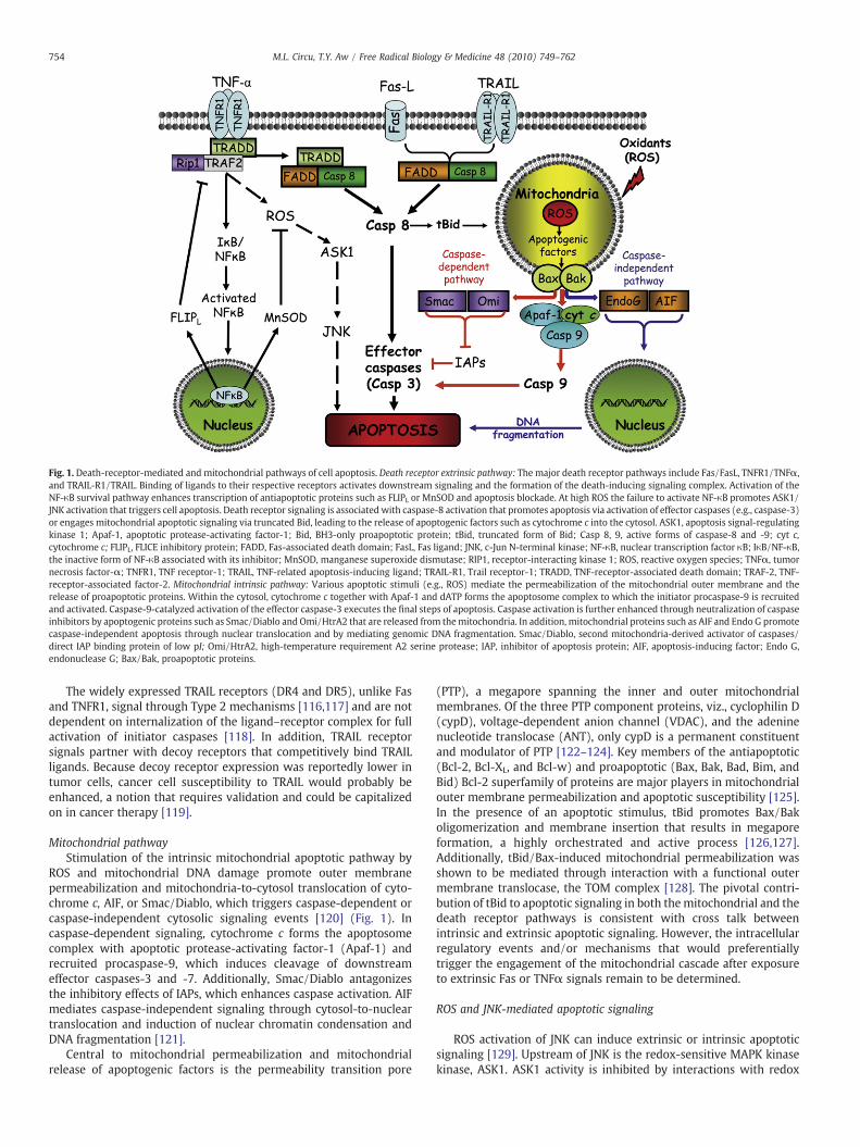
Fig. 1. Death-receptor-mediated and mitochondrial pathways of cell apoptosis. Death receptor extrinsic pathway: The major death receptor pathways include Fas/FasL, TNFR1/TNFα,and TRAIL-R1/TRAIL. Binding of ligands to their respective receptors activates downstream signaling and the formation of the death-inducing signaling complex. Activation of theNF-κB survival pathway enhances transcription of antiapoptotic proteins such as FLIPL or MnSOD and apoptosis blockade. At high ROS the failure to activate NF-κB promotes ASK1/JNK activation that triggers cell apoptosis. Death receptor signaling is associated with caspase-8 activation that promotes apoptosis via activation of effector caspases (e.g., caspase-3)or engages mitochondrial apoptotic signaling via truncated Bid, leading to the release of apoptogenic factors such as cytochrome c into the cytosol. ASK1, apoptosis signal-regulatingkinase 1; Apaf-1, apoptotic protease-activating factor-1; Bid, BH3-only proapoptotic protein; tBid, truncated form of Bid; Casp 8, 9, active forms of caspase-8 and -9; cyt c,cytochrome c; FLIPL, FLICE inhibitory protein; FADD, Fas-associated death domain; FasL, Fas ligand; JNK, c-Jun N-terminal kinase; NF-κB, nuclear transcription factor κB; IκB/NF-κB,the inactive form of NF-κB associated with its inhibitor; MnSOD, manganese superoxide dismutase; RIP1, receptor-interacting kinase 1; ROS, reactive oxygen species; TNFα, tumornecrosis factor-α; TNFR1, TNF receptor-1; TRAIL, TNF-related apoptosis-inducing ligand; TRAIL-R1, Trail receptor-1; TRADD, TNF-receptor-associated death domain; TRAF-2, TNF-receptor-associated factor-2. Mitochondrial intrinsic pathway: Various apoptotic stimuli (e.g., ROS) mediate the permeabilization of the mitochondrial outer membrane and therelease of proapoptotic proteins. Within the cytosol, cytochrome c together with Apaf-1 and dATP forms the apoptosome complex to which the initiator procaspase-9 is recruitedand activated. Caspase-9-catalyzed activation of the effector caspase-3 executes the final steps of apoptosis. Caspase activation is further enhanced through neutralization of caspaseinhibitors by apoptogenic proteins such as Smac/Diablo and Omi/HtrA2 that are released from themitochondria. In addition, mitochondrial proteins such as AIF and Endo G promotecaspase-independent apoptosis through nuclear translocation and by mediating genomic DNA fragmentation. Smac/Diablo, second mitochondria-derived activator of caspases/direct IAP binding protein of low pI; Omi/HtrA2, high-temperature requirement A2 serine protease; IAP, inhibitor of apoptosis protein; AIF, apoptosis-inducing factor; Endo G,endonuclease G; Bax/Bak, proapoptotic proteins.
754 M.L. Circu, T.Y. Aw / Free Radical Biology & Medicine 48 (2010) 749–762
The widely expressed TRAIL receptors (DR4 and DR5), unlike Fasand TNFR1, signal through Type 2 mechanisms [116,117] and are notdependent on internalization of the ligand–receptor complex for fullactivation of initiator caspases [118]. In addition, TRAIL receptorsignals partner with decoy receptors that competitively bind TRAILligands. Because decoy receptor expression was reportedly lower intumor cells, cancer cell susceptibility to TRAIL would probably beenhanced, a notion that requires validation and could be capitalizedon in cancer therapy [119].
Mitochondrial pathwayStimulation of the intrinsic mitochondrial apoptotic pathway by
ROS and mitochondrial DNA damage promote outer membranepermeabilization and mitochondria-to-cytosol translocation of cyto-chrome c, AIF, or Smac/Diablo, which triggers caspase-dependent orcaspase-independent cytosolic signaling events [120] (Fig. 1). Incaspase-dependent signaling, cytochrome c forms the apoptosomecomplex with apoptotic protease-activating factor-1 (Apaf-1) andrecruited procaspase-9, which induces cleavage of downstreameffector caspases-3 and -7. Additionally, Smac/Diablo antagonizesthe inhibitory effects of IAPs, which enhances caspase activation. AIFmediates caspase-independent signaling through cytosol-to-nucleartranslocation and induction of nuclear chromatin condensation andDNA fragmentation [121].
Central to mitochondrial permeabilization and mitochondrialrelease of apoptogenic factors is the permeability transition pore
(PTP), a megapore spanning the inner and outer mitochondrialmembranes. Of the three PTP component proteins, viz., cyclophilin D(cypD), voltage-dependent anion channel (VDAC), and the adeninenucleotide translocase (ANT), only cypD is a permanent constituentand modulator of PTP [122–124]. Key members of the antiapoptotic(Bcl-2, Bcl-XL, and Bcl-w) and proapoptotic (Bax, Bak, Bad, Bim, andBid) Bcl-2 superfamily of proteins are major players in mitochondrialouter membrane permeabilization and apoptotic susceptibility [125].In the presence of an apoptotic stimulus, tBid promotes Bax/Bakoligomerization and membrane insertion that results in megaporeformation, a highly orchestrated and active process [126,127].Additionally, tBid/Bax-induced mitochondrial permeabilization wasshown to be mediated through interaction with a functional outermembrane translocase, the TOM complex [128]. The pivotal contri-bution of tBid to apoptotic signaling in both themitochondrial and thedeath receptor pathways is consistent with cross talk betweenintrinsic and extrinsic apoptotic signaling. However, the intracellularregulatory events and/or mechanisms that would preferentiallytrigger the engagement of the mitochondrial cascade after exposureto extrinsic Fas or TNFα signals remain to be determined.
ROS and JNK-mediated apoptotic signaling
ROS activation of JNK can induce extrinsic or intrinsic apoptoticsignaling [129]. Upstream of JNK is the redox-sensitive MAPK kinasekinase, ASK1. ASK1 activity is inhibited by interactions with redox

755M.L. Circu, T.Y. Aw / Free Radical Biology & Medicine 48 (2010) 749–762
proteins (Grx, Trx1), heat shock proteins (Hsp90, Hsp72), and 14-3-3[130–132] and is stimulated by TRAF proteins, ASK1-interactingprotein 1, Daxx, and JASP/JIP3 [133–135]. TNFα is a potent activator ofthe MAPK cascade, and the ASK1–JNK pathway plays an importantrole in TNFR1-mediated apoptotic signaling in various cell types [136].Whether TNFα induces anti- or proapoptotic effects depends on thelevel and duration of JNK activation by ROS [137]. A transient andmodest JNK activation mediates cell survival via NF-κB-inducedantiapoptotic gene expression, whereas a prolonged and robust JNKactivation is associated with cell apoptosis via ASK1 signaling[138,139]. Thus, development of a strategy to manipulate the degreeand duration of cellular JNK activation could provide a reasonableapproach to specifically targeting cell survival or cell death such as incancer therapy.
Fig. 2 summarizes our current understanding of the mechanism ofROS and redox modulation of ASK1/JNK signaling in cell apoptosis.Thismodel proposes that ROSmediates the interaction of Trx1 and theN-terminal domain of ASK1, preventing ASK1 activation and down-stream propagation of an apoptotic signal [130]. Only reduced Trx1binds ASK1; the resultant Trx1/ASK1 complex, termed “ASK1signalosome,” functions as a redox switch that senses cellular ROSand is activated under oxidizing conditions [140]. Elevated cellularROS induce the dissociation of oxidized Trx1 from the complex and
Fig. 2. ROS-induced ASK1/JNK signaling and apoptosis. Reactive oxygen species initiateTrx dissociation from the ASK1–Trx complex, the ASK1 signalosome, through oxidationof the Trx redox-active site. ASK1 undergoes autophosphorylation and covalentbinding between its subunits, leading to the formation of an “activated signalosome”and recruitment of tumor necrosis factor receptor-associated factors 2 and 6 to thecomplex. Activated ASK1 signals downstream JNK activation and induces apoptosiseither via mitochondrial signaling or via transcription of AP-1-dependent proapoptoticgenes. Additionally, ROS-mediated disruption of the mitochondrial ASK1/ASK2/Trx2complex induces cytochrome c release. ROS, reactive oxygen species; TRAF2/6, TNFαreceptor-associated factor 2, 6; ASK1, 2, apoptosis signal-regulating kinase 1 and 2;JNK, c-Jun N-terminal kinase; Trx1, thioredoxin 1, reduced form; TrxSS, thioredoxin,oxidized form; Trx2, thioredoxin 2, mitochondrial enzyme; Bak, proapoptotic protein;Cyt c, cytochrome c; TNFα, tumor necrosis factor α; FasL, Fas ligand.
permit ASK1 oligomerization through N-terminal coiled-coil domainsand a gain of full ASK1 kinase activity [141]. Formation of a functionalASK1 signalosome complex is linked to the recruitment of TRAF2/6,which promotes ASK1 autophosphorylation and JNK activation[130,141]. ASK2, another member of the ASK family, binds andstabilizes ASK1 in the cytosol, nucleus, and mitochondria [142] andthus plays an important role in the regulation of ASK1/JNK signalingand cell apoptosis [143]. Current evidence shows that heterodimericcomplexes of ASK1 and 2 stimulate JNK and p38 activity, whereas theabsence of ASK2 diminishes apoptotic signaling [143]. ROS wereshown to promote ASK1 activation by inducing the dissociation of thedocking protein 14-3-3 [144] or by blocking the inhibitory effects ofthe protein phosphatases PP5 and PP2A [145]. Interestingly, themitochondrial ASK1-dependent apoptotic signaling pathway report-edly activated both JNK-dependent and JNK-independent apoptosis[146]. Nuclear translocation of activated JNK promoted activatorprotein-1 (AP-1)-mediated expression of proapoptotic TNFα, FasL,and Bak [147], whereas mitochondrial JNK translocation promotedcytochrome c release [148]. Although the latter observations suggestcross talk among ASK1/JNK signaling and the classical mitochondrialand death receptor pathways in cell apoptosis, the extent ofinteraction and integration among these various apoptotic pathwaysis unclear, as is the universality and quantitative importance of ASK1/ASK2–JNK signaling in apoptosis of various cell types induced byintrinsic versus extrinsic signals.
GSH redox status and apoptotic signaling
ROS-mediated apoptotic signaling is associated with decreasedcellular GSH levels and the loss of cellular redox balance [149,150].Decreased cell GSH can occur through ROS-induced GSH oxidation orGSH export from cells; the resultant GSH reduction would enhancefurther ROS production during oxidative challenge [151]. It wasdemonstrated that GSH loss due to decreased de novo GSH synthesistriggered redox activation of protein kinase C (PKCδ) [152] and,through GSH efflux, induced JNK-dependent apoptosis [153]. Theinitiation of apoptosis through GSH efflux was a ROS-independentmechanism because apoptosis was attenuated by blockade of GSHexport but not by antioxidants [154]. This suggestion is supported byour recent studies demonstrating that intestinal cell apoptosisinduced by staurosporine (STP) was linked to GSH efflux withoutaccompanying GSH/GSSG redox changes. This STP-induced export ofcellular GSH was driven by γ-glutamyltransferase-catalyzed extra-cellular GSH hydrolysis [155] and was associated with caspase-3activation independent of caspase-8 or -9 function. In other studies,FasL-induced GSH export was shown to be essential for thedevelopment of apoptosis in lymphoid cells; ROS production wasmerely a bystander phenomenon [154]. Collectively, the evidence todate has ruled out a major role for ROS signaling in GSH efflux-mediated cell apoptosis, and key challenges for future research will bedelineating the mechanism(s) that couples the export of GSH to thetriggering of apoptotic signals at cell membranes and the nature ofthese signals.
GSH oxidation is a major contributor to cell apoptosis mediated byoxidants. Accumulated evidence from our laboratory has consistentlyshown that an early spike in GSSG formation, typically withinminutesof oxidant exposure, preceded oxidant-induced activation of mito-chondrial apoptotic signaling and cell apoptosis hours later[20,21,156,157]. Importantly, postoxidant recovery of cellular GSH/GSSG redox status did not influence the apoptotic outcome, indicatingthat oxidant-induced apoptotic initiation occurred within an earlyand narrow window of GSH/GSSG redox shift. Indeed, whereas cellapoptosis was effectively blocked by NAC pretreatment, apoptosiswas not prevented when the thiol antioxidant was administered afteroxidant-induced GSH oxidation [21]. Collectively, our studies estab-lish that this mode of early induction of cellular GSH redox imbalance
756 M.L. Circu, T.Y. Aw / Free Radical Biology & Medicine 48 (2010) 749–762
in apoptotic redox signaling is common among diverse oxidants suchas hydroperoxides (tert-butyl, lipid hydroperoxides), redox cyclingmenadione quinones (MQ), andmethylglyoxal and in various intestinal(CaCo2, HT-29, NCM460) and neuronal (naïve and differentiated PC12)cell types [19,21,156,158–160].We further show that oxidant-mediatedapoptotic susceptibility is related to a cell's phenotype. The induction ofPC12 cell differentiation generated a phenotype that wasmore resistantto tert-butylhydroperoxide; this oxidative resistance was associatedwith a reduced intracellular GSH redox environment and decreasedApaf-1 expression [20]. Attenuated cellular susceptibility of thedifferentiated PC12 phenotype was also observed for carbonyl stress[160] and hyperglycemic stress [22,159]. If validated, the paradigm ofenhanced cellular vulnerability associated with a proliferative stateshould provide a strong cellular basis for targeting actively growingtumor cells for oxidative stress-induced cell killing.
Themitochondrial GSH/GSSG redox status is critical for preservingmitochondrial function during oxidative stress. Studies from ourlaboratory and others have documented a relationship betweenmitochondrial GSH (mtGSH) loss and cell apoptosis. Earlier studiesdemonstrated that decreased mtGSH correlated with apoptosisinduced by exposure to aromatic hydrocarbons [161], hypoxia [162],tert-butylhydroperoxide [163], and ethanol [164–166]. Associatedwith decreased mtGSH were mitochondrial ROS production, loss ofmitochondrial membrane potential, and mitochondria-to-cytosolrelease of cytochrome c [167]. Interestingly, moderate mtGSHdecrease was insufficient to elicit apoptosis in hepatocytes duringhypoxia, suggesting that achievement of a critical threshold of mtGSHloss is necessary to trigger mitochondrial apoptotic signaling [162].Moreover, Ghosh et al. demonstrated that oxidation of mtGSH wasrequisite for mitochondrial ROS generation, membrane potentialcollapse, and caspase-9 and -3 activation in cardiomyocyte apoptosiscaused by short-term diabetes [168]. We similarly showed that earlyROS production and mtGSSG formation preceded mitochondrialdysfunction and apoptosis in intestinal cells exposed to menadione[19]. Given the oxidative vulnerability of the mtGSH pool and thedynamics of its maintenance, manipulation of the mitochondrialredox compartment can be capitalized to selectively sensitize cells tooxidative damage.
Modulators of initiation and execution of apoptosis
Mitochondrial modulators of apoptotic initiation
ROS and the mitochondrial permeability transitionROS are known triggers of the intrinsic apoptotic cascade via
interactions with proteins of the mitochondrial permeability transi-tion complex [169]. Components of the PTP, viz., VDAC [170], ANT[171], and cypD [172], are targets of ROS, and oxidative modificationsof PTP proteins will significantly impact mitochondrial anion fluxes.Indeed, a mere transient increase in mitochondrial membranehyperpolarization after exposure to H2O2 initiated the collapse ofthe mitochondrial membrane potential (Δψm) [173], mitochondrialtranslocation of Bax and Bad, and cytochrome c release [174]. Evennonoxidants such as cadmium and staurosporine can trigger intrinsicapoptotic signaling through induction of ROS production andassociated mitochondrial permeability transition and cytochrome ctranslocation [175,176]. Significant mitochondrial loss of cytochromec will lead to further ROS increase due to a disrupted electrontransport chain [177].
Oxidative mitochondrial DNA damageMitochondrial DNA is a circular double-stranded DNA organized in
nucleoids in proximity to the electron transport chain. mtDNA lacksintrons and, being close to a ROS source, is prone to oxidative damage.Because mtDNA encodes 13 polypeptides of the respiratory chain,impaired mtRNA transcription would compromise mitochondrial ATP
production [178]. mtDNA damage-induced decreased respiratoryfunction enhances ROS generation, thus eliciting a vicious cycle ofROS–mtDNA damage that ultimately triggers apoptosis [179,180]. Alimited nucleotide excision DNA repair capacity coupled to a highmitochondrial ROS load further contributes to oxidative damage tomtDNA. Single-strand breaks and abasic sites formed during en-hanced ROS generation can induce apoptotic signaling [181].Interestingly, of the two sources of O2
•− production elicited byangiotensin II exposure, viz., NADPH oxidase and mitochondria, onlythe latter resulted frommtDNA damage-induced impaired mitochon-drial complex I activity. The subsequent collapse of theΔψm, release ofcytochrome c, and cell apoptosis were all prevented by inhibitingmtDNA damage [12]. Thus, the protection of mtDNA integrity iscritical not only to bioenergetic homeostasis, but to cell survival aswell. Recent findings by Rachek et al. [11] revealed that mtDNAdamage was an initiating event in mitochondrial dysfunction andhepatotoxicity induced by pharmacologic levels of the diabetic drugtroglitazone, thus ascribing potential clinical importance to mtDNAdamage in drug toxicity.
Our recent studies demonstrated that mtGSH is a determinant ofthe extent of oxidant-induced DNA damage [10]. MQ-inducedoxidative mtDNA damage paralleled the formation of mitochondrialGSSG and protein disulfide, which was blunted by NAC andexacerbated by inhibition of GSH synthesis in accordance withincreased and decreased cellular GSH, respectively [182]. Significantly,mtDNA damage was potentiated by blockade of mtGSH transport andprevented by overexpression of the oxoglutarate mtGSH carrier,validating the link between mtGSH and mtDNA integrity. Moreover,post-MQ recovery of mtDNA was preceded by restored cellular GSH,suggesting that DNA repair may also be GSH dependent [182]. Inprevious studies, an inverse relationship between GSH and basaloxidativeDNAdamage [183] and an association between ROS-inducedDNA deletions and genomic rearrangements with GSH depletion/oxidation have been documented [184–187]. Moreover, age-derivedROS-induced mtDNA damage was linked to mtGSH oxidation [188]. Itremains to be established as to whether GSH functions in attenuatingmtDNA damage or in stimulating mtDNA repair. Posttranslationalphosphorylation of OGG1 [189] and acetylation/deacetylation of Ape1[190] have been implicated in the control of DNA repair; the possibilitythat glutathiolation is another key posttranslational mechanism in theregulation of base-excision repair enzymes is an exciting notion thatwarrants investigation.
Cytochrome c and cardiolipin interactionCytochrome c is awater-soluble heme-containing protein bound to
the outer leaflet of the mitochondrial inner membrane throughinteractions with the anionic phospholipid cardiolipin. Normally,cytochrome c participates in shuttling electrons between complex IIIand complex IV of the mitochondrial electron transport chain; itsrelease from the mitochondria initiates the apoptotic cascade (Fig. 3).Mitochondria-to-cytosol release of cytochrome c sequentially occursvia detachment from cardiolipin and translocation through themitochondrial outer membrane [191]. At low mitochondrial ROS,tightly bound cytochrome c exhibits increased peroxidase activity[192] that oxidizes cardiolipin and facilitates its detachment [193,194].Oxidized cardiolipin is distributed to the outer leaflet of themitochondrial membrane where it functions as a docking platformfor tBid, enabling mitochondrial membrane permeabilization andcytochrome c movement across the outer membrane into the cytosol[195]. Because oxidative modification of cardiolipin is pivotal inmitochondrial cytochrome c loss and cell commitment to apoptosis,cardiolipin-bound cytochrome c could be viewed as a mitochondrialoxidative stress sensor and redox regulator of apoptosis. Therefore, theextent of cardiolipin peroxidation would probably be an importantdeterminant of apoptotic susceptibility of different cell types posses-sing various cardiolipin species and fatty acyl side chains. A possible

Fig. 3. Redox mediation of mitochondria-to-cytosol release of cytochrome c andactivation/inactivation of caspase-9. Interaction with mitochondria-specific cardiolipinsequesters cytochrome c in the mitochondrial intermembrane space. Enhancedmitochondrial generation of H2O2 activates cytochrome c peroxidase activity andinduces cardiolipin peroxidation and cytochrome c detachment. Oxidized cardiolipin istranslocated to the mitochondrial outer membrane and, together with the proapopto-tic proteins, Bid and Bax, forms a megapore channel that enables the mitochondria-to-cytosol transit of cytochrome c. Through a nitrosation reaction, the peroxidase activityof cytochrome c is inhibited by NO. Within the cytosol, cytochrome c interacts withApaf-1 and dATP, forming the apoptosome complex to which procaspase-9 is recruited.Proteolytic activation of procaspase-9 is mediated by posttranslational modification ofthe catalytic site cysteines through thiol oxidation, nitrosation, or glutathiolation.H2O2- and GSSG-mediated cysteine oxidation or S-glutathiolation, respectively, resultsin procaspase inactivation. NO-mediated S-nitrosation similarly inhibits caspase-9activation, whereas denitrosation promotes proenzyme proteolysis and activation.Additionally, GSSG-dependent glutathiolation of active caspase-9 results in directinhibition of enzyme activity. Apaf-1, apoptotic protease activation factor-1; CL,cardiolipin; CL-OOH, peroxidized cardiolipin; Cyt c, cytochrome c; Bid/Bax, proapop-totic proteins.
757M.L. Circu, T.Y. Aw / Free Radical Biology & Medicine 48 (2010) 749–762
quantitative relationship between cardiolipin oxidation products andpropensity for cell apoptosis has yet to be rigorously tested.
Nitric oxide reportedly modulates cytochrome c peroxidasefunction at the early stage of apoptotic initiation [196]. PhysiologicalNO levels inhibit peroxidase activity and prevent cardiolipin oxida-tion, whereas elevated NO induces peroxynitrite-mediated nitrationof Tyr-67 and enhances cytochrome c peroxidase activity [197,198].However, the nitration of Tyr-74 after continuous peroxynitriteexposure prevented cytochrome c/apoptosome complex formation[199]. Interestingly, the oxidation/reduction state of cytochrome cper se has been implicated in mitochondrial apoptotic signaling [200],and cytochrome c-containing oxidized, but not reduced, heme wascapable of caspase activation [201]. The redox state of cytochrome cwithin cells seems to be dependent on a nonapoptotic or apoptoticphenotype; generally, nonapoptotic cells favor cytochrome c reduc-tion and cell survival, whereas apoptotic cells favor cytochrome coxidation and apoptosis [202]. Precisely how the redox state of
cytochrome c contributes to the initiation of apoptosis is unknown,but it does not seem to involve direct effects on apoptosomeformation or caspase-9 activation [200].
Redox modulation of apoptotic execution: control of caspase activity
Cellular caspases belong to a highly conserved family of cysteineproteases that cleave aspartate residues of caspase substrates and arethe main players in the execution phase of apoptosis. The mammaliancaspase family contains at least 14 members that are divided intoinitiator and executioner caspases [203]. Initiator or apical caspasesare recruited at the death receptor via the death effector domain(caspase-8 and -10) or in the cytosol via the recruiting domain(caspase-2 and -9). Activated initiator caspases cleave executionercaspases such as caspase-3 and -7, which execute apoptosis throughcleavage of protein substrates that include mediators and regulatorsof apoptosis, structural proteins, and DNA repair- and cell-cycle-related proteins [204]. Additionally, activated caspase-3 promotescaspase-2 and -6 activation in an amplification loop that enhancescaspase-9 processing [205]. Caspases are constitutively expressed inthe cytosol as inactive zymogen monomers and are activated byapoptotic signals such as ROS via proteolysis at internal sites [206].Proteolytic cleavage of caspases at the N-terminal prodomain resultsin the generation of small p10 and large p20 active subunits, formingactive p10/p20 tetramers. The activation of caspases is prevented byspecific inhibitors belonging to the IAP family; members such as XIAP,c-IAP1, c-IAP2, and survivin bind and suppress enzyme catalyticactivity [207]. During apoptotic signaling IAPs are antagonized bymitochondria-derived Smac/Diablo and Omi/HtrA2 proteins, allow-ing caspase-mediated execution [208,209].
Our present understanding of the redox control of apoptoticexecution is sketchy, but it is a growing area of research. Posttrans-lational modification of catalytic site cysteine residues has gainedrecognition as a potentially important redox mechanism in thecontrol of caspase activity. Redox-active catalytic site cysteines ofcaspases are prone to oxidation, nitrosation, or glutathiolation (Fig. 3)[203]. Direct ROS effects on caspase activation have been documen-ted; for instance, H2O2 derived from endogenous and exogenoussources was shown to induce reversible inactivation of caspase 3 andcaspase 8 through oxidation of their catalytic site cysteines [210]. Forcaspase-9, H2O2-induced enzyme inactivation was specifically medi-ated through iron-catalyzed oxidation of the procaspase-9 catalyticsite cysteine [211]. Additionally, H2O2-mediated redox-dependentintramitochondrial autoactivation of caspase-9 has been demonstrat-ed in U937 cells in which procaspase-9 dimerization was induced bythiol–disulfide bond formation, a process that was inhibited by Trx[212]. Because mitochondrial procaspase-9 activation occurredduring the preapoptotic phase before cytochrome c release, it wassuggested that this mechanism could amplify the proapoptotic effectof cytochrome c [212].
Low levels of NO have been shown to exert antiapoptotic effectsvia S-nitrosation of a single cysteine residue at the catalytic site ofcaspases [213,214]. To date, the enzyme activity of seven members ofthe caspase family have been shown to be inhibited by redoxmodulation through this mechanism [215]. Evidence that caspase-3 isnitrosated with resultant inhibition of enzyme activity comes fromstudies in human umbilical vein endothelial cells. Using electron spinresonance spectroscopy of Myc-tagged p17 (a subunit of caspase 3),Rossig et al. found that S-nitrosation of Cys-163 prevented thecaspase-3-mediated apoptotic cascade [216]. In hepatocytes, NOblocked Bid activation through S-nitrosation of caspase-8 and pre-vented TNFα-induced mitochondrial apoptotic signaling [217–219].Additionally, NO donors were found to inhibit proper assembly of theApaf-1/caspase-9 apoptosome complex and caspase-9 activation[217–219]. Denitrosation is reportedly proapoptotic. For instance,denitrosation of procaspase-3 and procaspase-9 has been shown to
758 M.L. Circu, T.Y. Aw / Free Radical Biology & Medicine 48 (2010) 749–762
be associated with proteolytic enzyme activation in Fas-mediated[220] and cytokine-induced apoptosis [221]. A role for Trx1 inprocaspase-3 S-nitrosation has been documented, an interaction thatinvolves a transnitrosation reaction between procaspase-3 and Trx1[222]. The cellular sites of caspase nitrosation/denitrosation have notbeen fully investigated, but mitochondria are reportedly key locationsfor S-nitrosation reactions, judging by the preferred mitochondrialdistribution of nitrosated caspases [223]. If this is the case, theintriguing question of how nitrosation/activation of matrix caspasesmediates downstream cytosolic events in apoptotic executionwarrants further study.
Apart from ROS and NO, other recent evidence implicates a directrole for cellular GSH in redox regulation of caspase activity, mediatedby S-glutathiolation. Reportedly, S-glutathiolation of cysteine con-tributes to caspase stability and decreased accessibility for proteolyticcleavage, consistent with apoptotic resistance. Conversely, recentwork by Pan and Berk demonstrated that deglutathiolation ofcaspase-3 increased caspase-3 activity and TNFα-induced endothelialcell apoptosis [224]. Grx-catalyzed reversible glutathiolation ofcaspase-3 has been suggested to represent a novel redox signalingmechanism in TNFα-mediated cell apoptosis; specifically, TNFα-induced Grx assisted in thiol transfer in caspase-3 deglutathiolation[225]. Recent findings in HL60 cells show that GSSG at physiologicallevels mediated cysteine glutathiolation of both caspase-3 subunitsand inhibition of enzyme activity [226], linking S-glutathiolation withdirect inhibition of caspase activity. Additional findings that procas-pase-9 and -3 were targets of glutathiolation further suggests thatproteolytic activation of caspases may also be under GSH redoxcontrol [226]. Although caspases are subject to individual modifica-tion by nitrosation and glutathiolation, unanswered questions remainas to whether or how individual posttranslational mechanismsinteract and integrate with one another collectively to optimizecaspase activities during cell apoptosis.
Concluding remarks
Apoptosis has long been appreciated as an important form of celldeath in biological processes and various pathologies. Our currentunderstanding of the regulation of apoptosis is incomplete despitedecades of research. The recognition that ROS play a central role in cellsignaling has spurred much recent interest in the role of redoxmechanisms in apoptotic signaling and control. Evidence to date hasimplicated redox-dependent mechanisms in the mitochondria-to-cytosol release of cytochrome c, a central event in apoptotic initiation.Additionally, S-nitrosation and S-glutathiolation of catalytic-sitecysteines were reportedly important posttranslational redoxmechan-isms in the reversible activation/inactivation of caspases in thecontrol of apoptotic execution. Cellular redox systems, most notablythe GSH/GSSG redox couple, often functioning in conjunction withthe thioredoxin system, are central in redox regulation and cellapoptosis. Less well understood is the contribution of the pyridinenucleotide couples of NAD+/NADH and NADP+/NADPH. Apart fromtheir classical roles in bioenergetic homeostasis and reductivebiosynthesis, respectively, recent evidence suggests that pyridinenucleotides have broader biological functions, including controllingcell death. At present, the precise contribution of pyridine nucleotidesand the extent to which they interact with the thiol redox systems ofGSH/GSSG and thioredoxin in redox regulation of apoptosis areunclear and should provide fruitful avenues for future research.
Acknowledgments
Research in the authors' laboratory was supported by a grant fromthe National Institutes of Health, DK44510.
References
[1] Halliwell, B.; Cross, C. E. Oxygen-derived species: their relation to human diseaseand environmental stress. Environ. Health Perspect. 102 (Suppl. 10):5–12; 1994.
[2] Ames, B. N.; Shigenaga, M. K.; Hagen, T. M. Oxidants, antioxidants, and thedegenerative diseases of aging. Proc. Natl. Acad. Sci. USA 90:7915–7922; 1993.
[3] Turrens, J. F. Mitochondrial formation of reactive oxygen species. J. Physiol. 552:335–344; 2003.
[4] Andreyev, A. Y.; Kushnareva, Y. E.; Starkov, A. A. Mitochondrial metabolism ofreactive oxygen species. Biochemistry (Moscow) 70:200–214; 2005.
[5] Starkov, A. A.; Fiskum, G.; Chinopoulos, C.; Lorenzo, B. J.; Browne, S. E.; Patel,M. S.; Beal, M. F. Mitochondrial alpha-ketoglutarate dehydrogenase complexgenerates reactive oxygen species. J. Neurosci. 24:7779–7788; 2004.
[6] Tretter, L.; Adam-Vizi, V. Generation of reactive oxygen species in the reactioncatalyzed by alpha-ketoglutarate dehydrogenase. J. Neurosci. 24:7771–7778;2004.
[7] Migliaccio, E.; Giorgio, M.; Pelicci, P. G. Apoptosis and aging: role of p66Shc redoxprotein. Antioxid. Redox Signaling 8:600–608; 2006.
[8] Korshunov, S. S.; Skulachev, V. P.; Starkov, A. A. High protonic potential actuatesa mechanism of production of reactive oxygen species in mitochondria. FEBSLett. 416:15–18; 1997.
[9] Lambert, A. J.; Brand, M. D. Superoxide production by NADH:ubiquinoneoxidoreductase (complex I) depends on the pH gradient across the mitochon-drial inner membrane. Biochem. J. 382:511–517; 2004.
[10] Circu, M. L.; Moyer, M. P.; Harrison, L.; Aw, T. Y. Contribution of glutathionestatus to oxidant-induced mitochondrial DNA damage in colonic epithelial cells.Free Radic. Biol. Med. 47:1190–1198; 2009.
[11] Rachek, L. I.; Yuzefovych, L. V.; Ledoux, S. P.; Julie, N. L.; Wilson, G. L. Troglitazone,but not rosiglitazone, damages mitochondrial DNA and induces mitochondrialdysfunction and cell death in human hepatocytes. Toxicol. Appl. Pharmacol. 240:348–354; 2009.
[12] Ricci, C.; Pastukh, V.; Leonard, J.; Turrens, J.; Wilson, G.; Schaffer, D.; Schaffer,S. W. Mitochondrial DNA damage triggers mitochondrial-superoxide genera-tion and apoptosis. Am. J. Physiol. Cell Physiol. 294:C413–422; 2008.
[13] Fritz, R.; Bol, J.; Hebling, U.; Angermuller, S.; Volkl, A.; Fahimi, H. D.; Mueller, S.Compartment-dependent management of H2O2 by peroxisomes. Free Radic. Biol.Med. 42:1119–1129; 2007.
[14] Zangar, R. C.; Davydov, D. R.; Verma, S. Mechanisms that regulate production ofreactive oxygen species by cytochrome P450. Toxicol. Appl. Pharmacol. 199:316–331; 2004.
[15] Caro, A. A.; Cederbaum, A. I. Role of cytochrome P450 in phospholipase A2- andarachidonic acid-mediated cytotoxicity. Free Radic. Biol. Med. 40:364–375; 2006.
[16] Dumitru, C. A.; Zhang, Y.; Li, X.; Gulbins, E. Ceramide: a novel player in reactiveoxygen species-induced signaling? Antioxid. Redox Signaling 9:1535–1540; 2007.
[17] Zhang, A. Y.; Yi, F.; Zhang, G.; Gulbins, E.; Li, P. L. Lipid raft clustering and redoxsignaling platform formation in coronary arterial endothelial cells. Hypertension47:74–80; 2006.
[18] Zhang, A. Y.; Yi, F.; Jin, S.; Xia, M.; Chen, Q. Z.; Gulbins, E.; Li, P. L. Acidsphingomyelinase and its redox amplification in formation of lipid raft redoxsignaling platforms in endothelial cells. Antioxid. Redox Signaling 9:817–828;2007.
[19] Circu, M. L.; Rodriguez, C.; Maloney, R.; Moyer, M. P.; Aw, T. Y. Contribution ofmitochondrial GSH transport to matrix GSH status and colonic epithelial cellapoptosis. Free Radic. Biol. Med. 44:768–778; 2008.
[20] Ekshyyan, O.; Aw, T. Y. Decreased susceptibility of differentiated PC12 cells tooxidative challenge: relationship to cellular redox and expression of apoptoticprotease activator factor-1. Cell Death Differ. 12:1066–1077; 2005.
[21] Pias, E. K.; Ekshyyan, O. Y.; Rhoads, C. A.; Fuseler, J.; Harrison, L.; Aw, T. Y.Differential effects of superoxide dismutase isoform expression on hydroperoxide-induced apoptosis in PC-12 cells. J. Biol. Chem. 278:13294–13301; 2003.
[22] Okouchi, M.; Okayama, N.; Alexander, J. S.; Aw, T. Y. NRF2-dependent glutamate-L-cysteine ligase catalytic subunit expressionmediates insulin protection againsthyperglycemia-induced brain endothelial cell apoptosis. Curr. Neurovasc. Res. 3:249–261; 2006.
[23] Schafer, F. Q.; Buettner, G. R. Redox environment of the cell as viewed throughthe redox state of the glutathione disulfide/glutathione couple. Free Radic. Biol.Med. 30:1191–1212; 2001.
[24] Meister, A.; Tate, S. S. Glutathione and related gamma-glutamyl compounds:biosynthesis and utilization. Annu. Rev. Biochem. 45:559–604; 1976.
[25] Circu, M. L.; Aw, T. Y. Glutathione and apoptosis. Free Radic. Res. 42:689–706;2008.
[26] Meister, A.; Anderson, M. E. Glutathione. Annu. Rev. Biochem. 52:711–760; 1983.[27] Chakravarthi, S.; Jessop, C. E.; Bulleid, N. J. The role of glutathione in disulphide
bond formation and endoplasmic-reticulum-generated oxidative stress. EMBORep. 7:271–275; 2006.
[28] Jessop, C. E.; Bulleid, N. J. Glutathione directly reduces an oxidoreductase in theendoplasmic reticulum of mammalian cells. J. Biol. Chem. 279:55341–55347;2004.
[29] Frand, A. R.; Kaiser, C. A. Two pairs of conserved cysteines are required for theoxidative activity of Ero1p in protein disulfide bond formation in theendoplasmic reticulum. Mol. Biol. Cell 11:2833–2843; 2000.
[30] Dixon, B. M.; Heath, S. H.; Kim, R.; Suh, J. H.; Hagen, T. M. Assessment ofendoplasmic reticulum glutathione redox status is confounded by extensive exvivo oxidation. Antioxid. Redox Signaling 10:963–972; 2008.
[31] Chen, J.; Delannoy, M.; Odwin, S.; He, P.; Trush, M. A.; Yager, J. D. Enhancedmitochondrial gene transcript, ATP, bcl-2 protein levels, and altered glutathione

759M.L. Circu, T.Y. Aw / Free Radical Biology & Medicine 48 (2010) 749–762
distribution in ethinyl estradiol-treated cultured female rat hepatocytes. Toxicol.Sci. 75:271–278; 2003.
[32] Markovic, J.; Borras, C.; Ortega, A.; Sastre, J.; Vina, J.; Pallardo, F. V. Glutathione isrecruited into the nucleus in early phases of cell proliferation. J. Biol. Chem. 282:20416–20424; 2007.
[33] Ho, Y. F.; Guenthner, T. M. Isolation of liver nuclei that retain functional trans-membrane transport. J. Pharmacol. Toxicol. Methods 38:163–168; 1997.
[34] Voehringer, D. W.; McConkey, D. J.; McDonnell, T. J.; Brisbay, S.; Meyn, R. E. Bcl-2expression causes redistribution of glutathione to the nucleus. Proc. Natl. Acad.Sci. USA 95:2956–2960; 1998.
[35] Jocelyn, P. C.; Kamminga, A. The non-protein thiol of rat liver mitochondria.Biochim. Biophys. Acta 343:356–362; 1974.
[36] Schnellmann, R. G. Renal mitochondrial glutathione transport. Life Sci. 49:393–398; 1991.
[37] Soderdahl, T.; Enoksson, M.; Lundberg, M.; Holmgren, A.; Ottersen, O. P.;Orrenius, S.; Bolcsfoldi, G.; Cotgreave, I. A. Visualization of the compartmental-ization of glutathione and protein–glutathione mixed disulfides in cultured cells.FASEB J. 17:124–126; 2003.
[38] Zimmermann, A. K.; Loucks, F. A.; Schroeder, E. K.; Bouchard, R. J.; Tyler, K. L.;Linseman, D. A. Glutathione binding to the Bcl-2 homology-3 domain groove: amolecular basis for Bcl-2 antioxidant function at mitochondria. J. Biol. Chem. 282:29296–29304; 2007.
[39] Nakamura, H.; Nakamura, K.; Yodoi, J. Redox regulation of cellular activation.Annu. Rev. Immunol. 15:351–369; 1997.
[40] Watson, W. H.; Jones, D. P. Oxidation of nuclear thioredoxin during oxidativestress. FEBS Lett. 543:144–147; 2003.
[41] Hansen, J. M.; Go, Y. M.; Jones, D. P. Nuclear and mitochondrial compartmen-tation of oxidative stress and redox signaling. Annu. Rev. Pharmacol. Toxicol. 46:215–234; 2006.
[42] Hansen, J. M.; Zhang, H.; Jones, D. P. Differential oxidation of thioredoxin-1,thioredoxin-2, and glutathione by metal ions. Free Radic. Biol. Med. 40:138–145;2006.
[43] Kemp, M.; Go, Y. M.; Jones, D. P. Nonequilibrium thermodynamics of thiol/disulfide redox systems: a perspective on redox systems biology. Free Radic. Biol.Med. 44:921–937; 2008.
[44] Go, Y. M.; Jones, D. P. Redox compartmentalization in eukaryotic cells. Biochim.Biophys. Acta 1780:1273–1290; 2008.
[45] Rhee, S. G.; Chae, H. Z.; Kim, K. Peroxiredoxins: a historical overview andspeculative preview of novel mechanisms and emerging concepts in cellsignaling. Free Radic. Biol. Med. 38:1543–1552; 2005.
[46] Wood, Z. A.; Schroder, E.; Robin Harris, J.; Poole, L. B. Structure, mechanism andregulation of peroxiredoxins. Trends Biochem. Sci. 28:32–40; 2003.
[47] Zhang, Q.; Piston, D. W.; Goodman, R. H. Regulation of corepressor function bynuclear NADH. Science 295:1895–1897; 2002.
[48] Guse, A. H. Second messenger function and the structure–activity relationship ofcyclic adenosine diphosphoribose (cADPR). FEBS J. 272:4590–4597; 2005.
[49] Virag, L.; Szabo, C. The therapeutic potential of poly(ADP-ribose) polymeraseinhibitors. Pharmacol. Rev. 54:375–429; 2002.
[50] Lee, H. C. Physiological functions of cyclic ADP-ribose and NAADP as calciummessengers. Annu. Rev. Pharmacol. Toxicol. 41:317–345; 2001.
[51] Bedard, K.; Krause, K. H. The NOX family of ROS-generating NADPH oxidases:physiology and pathophysiology. Physiol. Rev. 87:245–313; 2007.
[52] Bolanos, J. P.; Delgado-Esteban, M.; Herrero-Mendez, A.; Fernandez-Fernandez, S.;Almeida, A. Regulation of glycolysis and pentose–phosphate pathway by nitricoxide: impact on neuronal survival. Biochim. Biophys. Acta 1777:789–793; 2008.
[53] Scott, M. D.; Zuo, L.; Lubin, B. H.; Chiu, D. T. NADPH, not glutathione, statusmodulates oxidant sensitivity in normal and glucose-6-phosphate dehydrogenase-deficient erythrocytes. Blood 77:2059–2064; 1991.
[54] Pandolfi, P. P.; Sonati, F.; Rivi, R.; Mason, P.; Grosveld, F.; Luzzatto, L. Targeteddisruption of the housekeeping gene encoding glucose 6-phosphate dehydro-genase (G6PD): G6PD is dispensable for pentose synthesis but essential fordefense against oxidative stress. EMBO J. 14:5209–5215; 1995.
[55] Kim, H. J.; Kang, B. S.; Park, J. W. Cellular defense against heat shock-inducedoxidative damage by mitochondrial NADP+-dependent isocitrate dehydrogenase.Free Radic. Res. 39:441–448; 2005.
[56] Yoshihara, T.; Hamamoto, T.; Munakata, R.; Tajiri, R.; Ohsumi, M.; Yokota, S.Localization of cytosolic NADP-dependent isocitrate dehydrogenase in theperoxisomes of rat liver cells: biochemical and immunocytochemical studies.J. Histochem. Cytochem. 49:1123–1131; 2001.
[57] Kim, S. J.; Yune, T. Y.; Han, C. T.; Kim, Y. C.; Oh, Y. J.; Markelonis, G. J.; Oh, T. H.Mitochondrial isocitrate dehydrogenase protects human neuroblastoma SH-SY5Y cells against oxidative stress. J. Neurosci. Res. 85:139–152; 2007.
[58] Shin, S. W.; Kil, I. S.; Park, J. W. Silencing of mitochondrial NADP+-dependentisocitrate dehydrogenase by small interfering RNA enhances heat shock-inducedapoptosis. Biochem. Biophys. Res. Commun. 366:1012–1018; 2008.
[59] Kil, I. S.; Kim, S. Y.; Lee, S. J.; Park, J. W. Small interfering RNA-mediated silencingof mitochondrial NADP+-dependent isocitrate dehydrogenase enhances thesensitivity of HeLa cells toward tumor necrosis factor-α and anticancer drugs.Free Radic. Biol. Med. 43:1197–1207; 2007.
[60] Filosa, S.; Fico, A.; Paglialunga, F.; Balestrieri, M.; Crooke, A.; Verde, P.; Abrescia,P.; Bautista, J. M.; Martini, G. Failure to increase glucose consumption throughthe pentose-phosphate pathway results in the death of glucose-6-phosphatedehydrogenase gene-deletedmouse embryonic stem cells subjected to oxidativestress. Biochem. J. 370:935–943; 2003.
[61] MacDonald, M. J. Evidence for the malate aspartate shuttle in pancreatic islets.Arch. Biochem. Biophys. 213:643–649; 1982.
[62] McKenna, M. C.; Stevenson, J. H.; Huang, X.; Tildon, J. T.; Zielke, C. L.; Hopkins, I. B.Mitochondrial malic enzyme activity is much higher in mitochondria fromcortical synaptic terminals compared with mitochondria from primary culturesof cortical neurons or cerebellar granule cells. Neurochem. Int. 36:451–459; 2000.
[63] Vogel, R.; Wiesinger, H.; Hamprecht, B.; Dringen, R. The regeneration of reducedglutathione in rat forebrain mitochondria identifies metabolic pathwaysproviding the NADPH required. Neurosci. Lett. 275:97–100; 1999.
[64] Pollak, N.; Niere, M.; Ziegler, M. NAD kinase levels control the NADPHconcentration in human cells. J. Biol. Chem. 282:33562–33571; 2007.
[65] Akella, S. S.; Harris, C. Developmental ontogeny of NAD+ kinase in the ratconceptus. Toxicol. Appl. Pharmacol. 170:124–129; 2001.
[66] Singh, R.; Lemire, J.; Mailloux, R. J.; Appanna, V. D. A novel strategy involved anti-oxidative defense: the conversion of NADH into NADPH by a metabolic network.PLoS ONE 3:e2682; 2008.
[67] Singh, R.; Mailloux, R. J.; Puiseux-Dao, S.; Appanna, V. D. Oxidative stress evokesa metabolic adaptation that favors increased NADPH synthesis and decreasedNADH production in Pseudomonas fluorescens. J. Bacteriol. 189:6665–6675;2007.
[68] Murayama, A.; Ohmori, K.; Fujimura, A.; Minami, H.; Yasuzawa-Tanaka, K.;Kuroda, T.; Oie, S.; Daitoku, H.; Okuwaki, M.; Nagata, K.; Fukamizu, A.; Kimura,K.; Shimizu, T.; Yanagisawa, J. Epigenetic control of rDNA loci in response tointracellular energy status. Cell 133:627–639; 2008.
[69] Sundaresan, N. R.; Samant, S. A.; Pillai, V. B.; Rajamohan, S. B.; Gupta, M. P. SIRT3is a stress-responsive deacetylase in cardiomyocytes that protects cells fromstress-mediated cell death by deacetylation of Ku70. Mol. Cell. Biol. 28:6384–6401; 2008.
[70] Rose, G.; Dato, S.; Altomare, K.; Bellizzi, D.; Garasto, S.; Greco, V.; Passarino, G.;Feraco, E.; Mari, V.; Barbi, C.; BonaFe, M.; Franceschi, C.; Tan, Q.; Boiko, S.; Yashin,A. I.; De Benedictis, G. Variability of the SIRT3 gene, human silent informationregulator Sir2 homologue, and survivorship in the elderly. Exp. Gerontol. 38:1065–1070; 2003.
[71] Oberdoerffer, P.; Michan, S.; McVay, M.; Mostoslavsky, R.; Vann, J.; Park, S. K.;Hartlerode, A.; Stegmuller, J.; Hafner, A.; Loerch, P.; Wright, S. M.; Mills, K. D.;Bonni, A.; Yankner, B. A.; Scully, R.; Prolla, T. A.; Alt, F. W.; Sinclair, D. A. SIRT1redistribution on chromatin promotes genomic stability but alters geneexpression during aging. Cell 135:907–918; 2008.
[72] Blander, G.; Guarente, L. The Sir2 family of protein deacetylases. Annu. Rev.Biochem. 73:417–435; 2004.
[73] Tanno, M.; Sakamoto, J.; Miura, T.; Shimamoto, K.; Horio, Y. Nucleocytoplasmicshuttling of the NAD+-dependent histone deacetylase SIRT1. J. Biol. Chem. 282:6823–6832; 2007.
[74] Yamamoto, H.; Schoonjans, K.; Auwerx, J. Sirtuin functions in health and disease.Mol. Endocrinol. 21:1745–1755; 2007.
[75] Smith, B. C.; Hallows, W. C.; Denu, J. M. Mechanisms and molecular probes ofsirtuins. Chem. Biol. 15:1002–1013; 2008.
[76] Kume, S.; Haneda, M.; Kanasaki, K.; Sugimoto, T.; Araki, S.; Isono, M.; Isshiki, K.;Uzu, T.; Kashiwagi, A.; Koya, D. Silent information regulator 2 (SIRT1) attenuatesoxidative stress-induced mesangial cell apoptosis via p53 deacetylation. FreeRadic. Biol. Med. 40:2175–2182; 2006.
[77] Li, M.; Luo, J.; Brooks, C. L.; Gu,W. Acetylation of p53 inhibits its ubiquitination byMdm2. J. Biol. Chem. 277:50607–50611; 2002.
[78] Luo, J.; Nikolaev, A. Y.; Imai, S.; Chen, D.; Su, F.; Shiloh, A.; Guarente, L.; Gu, W.Negative control of p53 by Sir2alpha promotes cell survival under stress. Cell107:137–148; 2001.
[79] Nakano, K.; Vousden, K. H. PUMA, a novel proapoptotic gene, is induced by p53.Mol. Cell 7:683–694; 2001.
[80] Macip, S.; Igarashi, M.; Berggren, P.; Yu, J.; Lee, S. W.; Aaronson, S. A. Influence ofinduced reactive oxygen species in p53-mediated cell fate decisions. Mol. Cell.Biol. 23:8576–8585; 2003.
[81] Han, M. K.; Song, E. K.; Guo, Y.; Ou, X.; Mantel, C.; Broxmeyer, H. E. SIRT1regulates apoptosis and Nanog expression in mouse embryonic stem cells bycontrolling p53 subcellular localization. Cell Stem Cell 2:241–251; 2008.
[82] Brunet, A.; Sweeney, L. B.; Sturgill, J. F.; Chua, K. F.; Greer, P. L.; Lin, Y.; Tran, H.; Ross,S. E.;Mostoslavsky, R.; Cohen, H. Y.; Hu, L. S.; Cheng, H. L.; Jedrychowski,M. P.; Gygi,S. P.; Sinclair, D. A.; Alt, F. W.; Greenberg, M. E. Stress-dependent regulation ofFOXO transcription factors by the SIRT1 deacetylase. Science 303:2011–2015; 2004.
[83] Hasegawa, K.; Wakino, S.; Yoshioka, K.; Tatematsu, S.; Hara, Y.; Minakuchi, H.;Washida, N.; Tokuyama, H.; Hayashi, K.; Itoh, H. Sirt1 protects against oxidativestress-induced renal tubular cell apoptosis by the bidirectional regulation ofcatalase expression. Biochem. Biophys. Res. Commun. 372:51–56; 2008.
[84] Chiribau, C. B.; Cheng, L.; Cucoranu, I. C.; Yu, Y. S.; Clempus, R. E.; Sorescu, D.FOXO3A regulates peroxiredoxin III expression in human cardiac fibroblasts.J. Biol. Chem. 283:8211–8217; 2008.
[85] Ford, J.; Jiang, M.; Milner, J. Cancer-specific functions of SIRT1 enable humanepithelial cancer cell growth and survival. Cancer Res. 65:10457–10463; 2005.
[86] Jung-Hynes, B.; Nihal, M.; Zhong, W.; Ahmad, N. Role of sirtuin histonedeacetylase Sirt1 in prostate cancer: a target for prostate cancer managementvia its inhibition? J. Biol. Chem. 284:3823–3832; 2008.
[87] Lombard, D. B.; Alt, F. W.; Cheng, H. L.; Bunkenborg, J.; Streeper, R. S.;Mostoslavsky, R.; Kim, J.; Yancopoulos, G.; Valenzuela, D.; Murphy, A.; Yang,Y.; Chen, Y.; Hirschey, M. D.; Bronson, R. T.; Haigis, M.; Guarente, L. P.; Farese Jr.,R. V.; Weissman, S.; Verdin, E.; Schwer, B. Mammalian Sir2 homolog SIRT3regulates global mitochondrial lysine acetylation. Mol. Cell. Biol. 27:8807–8814;2007.
[88] Haigis, M. C.; Mostoslavsky, R.; Haigis, K. M.; Fahie, K.; Christodoulou, D. C.;Murphy, A. J.; Valenzuela, D. M.; Yancopoulos, G. D.; Karow, M.; Blander, G.;

760 M.L. Circu, T.Y. Aw / Free Radical Biology & Medicine 48 (2010) 749–762
Wolberger, C.; Prolla, T. A.; Weindruch, R.; Alt, F. W.; Guarente, L. SIRT4 inhibitsglutamate dehydrogenase and opposes the effects of calorie restriction inpancreatic beta cells. Cell 126:941–954; 2006.
[89] Schlicker, C.; Gertz, M.; Papatheodorou, P.; Kachholz, B.; Becker, C. F.; Steegborn,C. Substrates and regulation mechanisms for the human mitochondrial sirtuinsSirt3 and Sirt5. J. Mol. Biol. 382:790–801; 2008.
[90] Ahn, B. H.; Kim, H. S.; Song, S.; Lee, I. H.; Liu, J.; Vassilopoulos, A.; Deng, C. X.;Finkel, T. A role for the mitochondrial deacetylase Sirt3 in regulating energyhomeostasis. Proc. Natl. Acad. Sci. USA 105:14447–14452; 2008.
[91] Shi, T.; Wang, F.; Stieren, E.; Tong, Q. SIRT3, a mitochondrial sirtuin deacetylase,regulates mitochondrial function and thermogenesis in brown adipocytes. J. Biol.Chem. 280:13560–13567; 2005.
[92] Guarente, L.; Picard, F. Calorie restriction—the SIR2 connection. Cell 120:473–482; 2005.
[93] North, B. J.; Marshall, B. L.; Borra, M. T.; Denu, J. M.; Verdin, E. The human Sir2ortholog, SIRT2, is an NAD+-dependent tubulin deacetylase. Mol. Cell 11:437–444; 2003.
[94] Lombard, D. B.; Schwer, B.; Alt, F. W.; Mostoslavsky, R. SIRT6 in DNA repair,metabolism and ageing. J. Intern. Med. 263:128–141; 2008.
[95] Liszt, G.; Ford, E.; Kurtev, M.; Guarente, L. Mouse Sir2 homolog SIRT6 is a nuclearADP-ribosyltransferase. J. Biol. Chem. 280:21313–21320; 2005.
[96] Ford, E.; Voit, R.; Liszt, G.; Magin, C.; Grummt, I.; Guarente, L. Mammalian Sir2homolog SIRT7 is an activator of RNA polymerase I transcription. Genes Dev. 20:1075–1080; 2006.
[97] Ashkenazi, A.; Dixit, V. M. Apoptosis control by death and decoy receptors. Curr.Opin. Cell Biol. 11:255–260; 1999.
[98] LeBlanc, H. N.; Ashkenazi, A. Apo2L/TRAIL and its death and decoy receptors. CellDeath Differ. 10:66–75; 2003.
[99] Berg, D.; Lehne, M.; Muller, N.; Siegmund, D.; Munkel, S.; Sebald, W.;Pfizenmaier, K.; Wajant, H. Enforced covalent trimerization increases theactivity of the TNF ligand family members TRAIL and CD95L. Cell Death Differ.14:2021–2034; 2007.
[100] Watanabe, N.; Kuriyama, H.; Sone, H.; Neda, H.; Yamauchi, N.; Maeda, M.; Niitsu,Y. Continuous internalization of tumor necrosis factor receptors in a humanmyosarcoma cell line. J. Biol. Chem. 263:10262–10266; 1988.
[101] Lee, K. H.; Feig, C.; Tchikov, V.; Schickel, R.; Hallas, C.; Schutze, S.; Peter, M. E.;Chan, A. C. The role of receptor internalization in CD95 signaling. EMBO J. 25:1009–1023; 2006.
[102] Huang, X.; Masselli, A.; Frisch, S. M.; Hunton, I. C.; Jiang, Y.; Wang, J. Y. Blockadeof tumor necrosis factor-induced Bid cleavage by caspase-resistant Rb. J. Biol.Chem. 282:29401–29413; 2007.
[103] Barnhart, B. C.; Alappat, E. C.; Peter, M. E. The CD95 type I/type II model. Semin.Immunol. 15:185–193; 2003.
[104] Wang, L.; Azad, N.; Kongkaneramit, L.; Chen, F.; Lu, Y.; Jiang, B. H.; Rojanasakul, Y.The Fas death signaling pathway connecting reactive oxygen species generationand FLICE inhibitory protein down-regulation. J. Immunol. 180:3072–3080; 2008.
[105] Jiang, Y.; Woronicz, J. D.; Liu, W.; Goeddel, D. V. Prevention of constitutive TNFreceptor 1 signaling by silencer of death domains. Science 283:543–546; 1999.
[106] Hsu, H.; Xiong, J.; Goeddel, D. V. The TNF receptor 1-associated protein TRADDsignals cell death and NF-kappa B activation. Cell 81:495–504; 1995.
[107] Shim, J. H.; Xiao, C.; Paschal, A. E.; Bailey, S. T.; Rao, P.; Hayden, M. S.; Lee, K. Y.;Bussey, C.; Steckel, M.; Tanaka, N.; Yamada, G.; Akira, S.; Matsumoto, K.; Ghosh,S. TAK1, but not TAB1 or TAB2, plays an essential role in multiple signalingpathways in vivo. Genes Dev. 19:2668–2681; 2005.
[108] Zong, W. X.; Edelstein, L. C.; Chen, C.; Bash, J.; Gelinas, C. The prosurvival Bcl-2homolog Bfl-1/A1 is a direct transcriptional target of NF-kappaB that blocksTNFalpha-induced apoptosis. Genes Dev. 13:382–387; 1999.
[109] Stehlik, C.; de Martin, R.; Kumabashiri, I.; Schmid, J. A.; Binder, B. R.; Lipp, J.Nuclear factor (NF)-kappaB-regulated X-chromosome-linked iap gene expres-sion protects endothelial cells from tumor necrosis factor alpha-inducedapoptosis. J. Exp. Med. 188:211–216; 1998.
[110] Micheau, O.; Lens, S.; Gaide, O.; Alevizopoulos, K.; Tschopp, J. NF-kappaB signalsinduce the expression of c-FLIP. Mol. Cell. Biol. 21:5299–5305; 2001.
[111] Chang, L.; Kamata, H.; Solinas, G.; Luo, J. L.; Maeda, S.; Venuprasad, K.; Liu, Y. C.;Karin, M. The E3 ubiquitin ligase itch couples JNK activation to TNFalpha-induced cell death by inducing c-FLIP(L) turnover. Cell 124:601–613; 2006.
[112] Pantano, C.; Shrivastava, P.; McElhinney, B.; Janssen-Heininger, Y. Hydrogenperoxide signaling through tumor necrosis factor receptor 1 leads to selectiveactivation of c-Jun N-terminal kinase. J. Biol. Chem. 278:44091–44096; 2003.
[113] Schneider-Brachert, W.; Tchikov, V.; Neumeyer, J.; Jakob, M.; Winoto-Morbach,S.; Held-Feindt, J.; Heinrich, M.; Merkel, O.; Ehrenschwender, M.; Adam, D.;Mentlein, R.; Kabelitz, D.; Schutze, S. Compartmentalization of TNF receptor 1signaling: internalized TNF receptosomes as death signaling vesicles. Immunity21:415–428; 2004.
[114] Micheau, O.; Tschopp, J. Induction of TNF receptor I-mediated apoptosis via twosequential signaling complexes. Cell 114:181–190; 2003.
[115] Lin, Y.; Devin, A.; Rodriguez, Y.; Liu, Z. G. Cleavage of the death domain kinase RIPby caspase-8 prompts TNF-induced apoptosis. Genes Dev. 13:2514–2526; 1999.
[116] LeBlanc, H.; Lawrence, D.; Varfolomeev, E.; Totpal, K.; Morlan, J.; Schow, P.; Fong,S.; Schwall, R.; Sinicropi, D.; Ashkenazi, A. Tumor-cell resistance to deathreceptor-induced apoptosis through mutational inactivation of the proapoptoticBcl-2 homolog Bax. Nat. Med. 8:274–281; 2002.
[117] Deng, Y.; Lin, Y.; Wu, X. TRAIL-induced apoptosis requires Bax-dependentmitochondrial release of Smac/DIABLO. Genes Dev. 16:33–45; 2002.
[118] Jin, Z.; El-Deiry, W. S. Distinct signaling pathways in TRAIL- versus tumornecrosis factor-induced apoptosis. Mol. Cell. Biol. 26:8136–8148; 2006.
[119] Falschlehner, C.; Emmerich, C. H.; Gerlach, B.; Walczak, H. TRAIL signalling:decisions between life and death. Int. J. Biochem. Cell Biol. 39:1462–1475; 2007.
[120] Ryter, S. W.; Kim, H. P.; Hoetzel, A.; Park, J. W.; Nakahira, K.; Wang, X.; Choi, A. M.Mechanisms of cell death in oxidative stress. Antioxid. Redox Signaling 9:49–89;2007.
[121] Susin, S. A.; Lorenzo, H. K.; Zamzami, N.; Marzo, I.; Snow, B. E.; Brothers, G. M.;Mangion, J.; Jacotot, E.; Costantini, P.; Loeffler, M.; Larochette, N.; Goodlett, D. R.;Aebersold, R.; Siderovski, D. P.; Penninger, J. M.; Kroemer, G. Molecularcharacterization of mitochondrial apoptosis-inducing factor. Nature 397:441–446; 1999.
[122] Baines, C. P.; Kaiser, R. A.; Sheiko, T.; Craigen, W. J.; Molkentin, J. D. Voltage-dependent anion channels are dispensable for mitochondrial-dependent celldeath. Nat. Cell Biol. 9:550–555; 2007.
[123] Kokoszka, J. E.; Waymire, K. G.; Levy, S. E.; Sligh, J. E.; Cai, J.; Jones, D. P.;MacGregor, G. R.; Wallace, D. C. The ADP/ATP translocator is not essential for themitochondrial permeability transition pore. Nature 427:461–465; 2004.
[124] Schinzel, A. C.; Takeuchi, O.; Huang, Z.; Fisher, J. K.; Zhou, Z.; Rubens, J.; Hetz, C.;Danial, N. N.; Moskowitz, M. A.; Korsmeyer, S. J. Cyclophilin D is a component ofmitochondrial permeability transition and mediates neuronal cell death afterfocal cerebral ischemia. Proc. Natl. Acad. Sci. USA 102:12005–12010; 2005.
[125] Kelekar, A.; Thompson, C. B. Bcl-2-family proteins: the role of the BH3 domain inapoptosis. Trends Cell Biol. 8:324–330; 1998.
[126] Eskes, R.; Desagher, S.; Antonsson, B.; Martinou, J. C. Bid induces theoligomerization and insertion of Bax into the outer mitochondrial membrane.Mol. Cell. Biol. 20:929–935; 2000.
[127] Desagher, S.; Osen-Sand, A.; Nichols, A.; Eskes, R.; Montessuit, S.; Lauper, S.;Maundrell, K.; Antonsson, B.; Martinou, J. C. Bid-induced conformational changeof Bax is responsible for mitochondrial cytochrome c release during apoptosis.J. Cell Biol. 144:891–901; 1999.
[128] Ott, M.; Norberg, E.; Walter, K. M.; Schreiner, P.; Kemper, C.; Rapaport, D.;Zhivotovsky, B.; Orrenius, S. The mitochondrial TOM complex is required fortBid/Bax-induced cytochrome c release. J. Biol. Chem. 282:27633–27639; 2007.
[129] Dhanasekaran, D. N.; Reddy, E. P. JNK signaling in apoptosis. Oncogene 27:6245–6251; 2008.
[130] Saitoh, M.; Nishitoh, H.; Fujii, M.; Takeda, K.; Tobiume, K.; Sawada, Y.; Kawabata,M.; Miyazono, K.; Ichijo, H. Mammalian thioredoxin is a direct inhibitor ofapoptosis signal-regulating kinase (ASK) 1. EMBO J. 17:2596–2606; 1998.
[131] Song, J. J.; Rhee, J. G.; Suntharalingam, M.; Walsh, S. A.; Spitz, D. R.; Lee, Y. J. Roleof glutaredoxin in metabolic oxidative stress: glutaredoxin as a sensor ofoxidative stress mediated by H2O2. J. Biol. Chem. 277:46566–46575; 2002.
[132] Zhang, L.; Chen, J.; Fu, H. Suppression of apoptosis signal-regulating kinase 1-induced cell death by 14-3-3 proteins. Proc. Natl. Acad. Sci. USA 96:8511–8515;1999.
[133] Zhang, H.; Zhang, R.; Luo, Y.; D'Alessio, A.; Pober, J. S.; Min, W. AIP1/DAB2IP, anovel member of the Ras–GAP family, transduces TRAF2-induced ASK1–JNKactivation. J. Biol. Chem. 279:44955–44965; 2004.
[134] Chang, H. Y.; Nishitoh, H.; Yang, X.; Ichijo, H.; Baltimore, D. Activation ofapoptosis signal-regulating kinase 1 (ASK1) by the adapter protein Daxx. Science281:1860–1863; 1998.
[135] Matsuura, H.; Nishitoh, H.; Takeda, K.; Matsuzawa, A.; Amagasa, T.; Ito, M.;Yoshioka, K.; Ichijo, H. Phosphorylation-dependent scaffolding role of JSAP1/JIP3in the ASK1–JNK signaling pathway: a newmode of regulation of the MAP kinasecascade. J. Biol. Chem. 277:40703–40709; 2002.
[136] Matsuzawa, A.; Ichijo, H. Redox control of cell fate by MAP kinase: physiologicalroles of ASK1–MAP kinase pathway in stress signaling. Biochim. Biophys.Acta1708; 2008.
[137] Kamata, H.; Honda, S.; Maeda, S.; Chang, L.; Hirata, H.; Karin, M. Reactive oxygenspecies promote TNFalpha-induced death and sustained JNK activation byinhibiting MAP kinase phosphatases. Cell 120:649–661; 2005.
[138] Liu, H.; Lo, C. R.; Czaja, M. J. NF-kappaB inhibition sensitizes hepatocytes to TNF-induced apoptosis through a sustained activation of JNK and c-Jun. Hepatology35:772–778; 2002.
[139] Deng, Y.; Ren, X.; Yang, L.; Lin, Y.; Wu, X. A JNK-dependent pathway is requiredfor TNFalpha-induced apoptosis. Cell 115:61–70; 2003.
[140] Fujino, G.; Noguchi, T.; Takeda, K.; Ichijo, H. Thioredoxin and protein kinases inredox signaling. Semin. Cancer Biol. 16:427–435; 2006.
[141] Fujino, G.; Noguchi, T.; Matsuzawa, A.; Yamauchi, S.; Saitoh, M.; Takeda, K.;Ichijo, H. Thioredoxin and TRAF family proteins regulate reactive oxygenspecies-dependent activation of ASK1 through reciprocal modulation of theN-terminal homophilic interaction of ASK1. Mol. Cell. Biol. 27:8152–8163;2007.
[142] Wang, X. S.; Diener, K.; Tan, T. H.; Yao, Z. MAPKKK6, a novel mitogen-activatedprotein kinase kinase kinase, that associates with MAPKKK5. Biochem. Biophys.Res. Commun. 253:33–37; 1998.
[143] Ortner, E.; Moelling, K. Heteromeric complex formation of ASK2 and ASK1regulates stress-induced signaling. Biochem. Biophys. Res. Commun. 362:454–459; 2007.
[144] Goldman, E. H.; Chen, L.; Fu, H. Activation of apoptosis signal-regulating kinase 1by reactive oxygen species through dephosphorylation at serine 967 and 14-3-3dissociation. J. Biol. Chem. 279:10442–10449; 2004.
[145] Chen, L.; Liu, L.; Huang, S. Cadmium activates the mitogen-activated proteinkinase (MAPK) pathway via induction of reactive oxygen species and inhibitionof protein phosphatases 2A and 5. Free Radic. Biol. Med. 45:1035–1044; 2008.
[146] Zhang, R.; Al-Lamki, R.; Bai, L.; Streb, J. W.; Miano, J. M.; Bradley, J.; Min, W.Thioredoxin-2 inhibits mitochondria-located ASK1-mediated apoptosis in a JNK-independent manner. Circ. Res. 94:1483–1491; 2004.

761M.L. Circu, T.Y. Aw / Free Radical Biology & Medicine 48 (2010) 749–762
[147] Fan, M.; Goodwin, M. E.; Birrer, M. J.; Chambers, T. C. The c-Jun NH(2)-terminalprotein kinase/AP-1 pathway is required for efficient apoptosis induced byvinblastine. Cancer Res. 61:4450–4458; 2001.
[148] Kharbanda, S.; Saxena, S.; Yoshida, K.; Pandey, P.; Kaneki, M.; Wang, Q.; Cheng,K.; Chen, Y. N.; Campbell, A.; Sudha, T.; Yuan, Z. M.; Narula, J.; Weichselbaum, R.;Nalin, C.; Kufe, D. Translocation of SAPK/JNK to mitochondria and interactionwith Bcl-x(L) in response to DNA damage. J. Biol. Chem. 275:322–327; 2000.
[149] Merad-Boudia, M.; Nicole, A.; Santiard-Baron, D.; Saille, C.; Ceballos-Picot, I.Mitochondrial impairment as an early event in the process of apoptosis inducedby glutathione depletion in neuronal cells: relevance to Parkinson's disease.Biochem. Pharmacol. 56:645–655; 1998.
[150] Lu, C.; Armstrong, J. S. Role of calcium and cyclophilin D in the regulation ofmitochondrial permeabilization induced by glutathione depletion. Biochem.Biophys. Res. Commun. 363:572–577; 2007.
[151] D'Alessio, M.; Cerella, C.; De Nicola, M.; Bergamaschi, A.; Magrini, A.; Gualandi,G.; Alfonsi, A. M.; Ghibelli, L. Apoptotic GSH extrusion is associated with freeradical generation. Ann. N. Y. Acad. Sci. 1010:449–452; 2003.
[152] Marengo, B.; De Ciucis, C.; Verzola, D.; Pistoia, V.; Raffaghello, L.; Patriarca, S.;Balbis, E.; Traverso, N.; Cottalasso, D.; Pronzato, M. A.; Marinari, U. M.;Domenicotti, C. Mechanisms of BSO (L-buthionine-S,R-sulfoximine)-inducedcytotoxic effects in neuroblastoma. Free Radic. Biol. Med. 44:474–482; 2008.
[153] Meurette, O.; Lefeuvre-Orfila, L.; Rebillard, A.; Lagadic-Gossmann, D.; Dimanche-Boitrel, M. T. Role of intracellular glutathione in cell sensitivity to the apoptosisinduced by tumor necrosis factor α-related apoptosis-inducing ligand/anticancerdrug combinations. Clin. Cancer Res. 11:3075–3083; 2005.
[154] Franco, R.; Panayiotidis, M. I.; Cidlowski, J. A. Glutathione depletion is necessaryfor apoptosis in lymphoid cells independent of reactive oxygen speciesformation. J. Biol. Chem. 282:30452–30465; 2007.
[155] Circu, M. L.; Stringer, S.; Rhoads, C. A.; Moyer, M. P.; Aw, T. Y. The role of GSHefflux in staurosporine-induced apoptosis in colonic epithelial cells. Biochem.Pharmacol. 77:76–85; 2009.
[156] Wang, T. G.; Gotoh, Y.; Jennings, M. H.; Rhoads, C. A.; Aw, T. Y. Lipidhydroperoxide-induced apoptosis in human colonic CaCo-2 cells is associatedwith an early loss of cellular redox balance. FASEB J. 14:1567–1576; 2000.
[157] Pias, E. K.; Aw, T. Y. Early redox imbalance mediates hydroperoxide-inducedapoptosis in mitotic competent undifferentiated PC-12 cells. Cell Death Differ. 9:1007–1016; 2002.
[158] Pias, E. K.; Aw, T. Y. Apoptosis in mitotic competent undifferentiated cells isinduced by cellular redox imbalance independent of reactive oxygen speciesproduction. FASEB J. 16:781–790; 2002.
[159] Okouchi, M.; Okayama, N.; Aw, T. Y. Hyperglycemia potentiates carbonyl stress-induced apoptosis in naive PC-12 cells: relationship to cellular redox andactivator protease factor-1 expression. Curr. Neurovasc. Res. 2:375–386; 2005.
[160] Okouchi, M.; Okayama, N.; Aw, T. Y. Differential susceptibility of naive anddifferentiated PC-12 cells to methylglyoxal-induced apoptosis: influence ofcellular redox. Curr. Neurovasc. Res. 2:13–22; 2005.
[161] Hallberg, E.; Rydstrom, J. Selective oxidation of mitochondrial glutathione incultured rat adrenal cells and its relation to polycyclic aromatic hydrocarbon-induced cytotoxicity. Arch. Biochem. Biophys. 270:662–671; 1989.
[162] Lluis, J. M.; Morales, A.; Blasco, C.; Colell, A.; Mari, M.; Garcia-Ruiz, C.; Fernandez-Checa, J. C. Critical role of mitochondrial glutathione in the survival ofhepatocytes during hypoxia. J. Biol. Chem. 280:3224–3232; 2005.
[163] Olafsdottir, K.; Reed, D. J. Retention of oxidized glutathione by isolated rat livermitochondria during hydroperoxide treatment. Biochim. Biophys. Acta 964:377–382; 1988.
[164] Fernandez-Checa, J. C.; Garcia-Ruiz, C.; Ookhtens, M.; Kaplowitz, N. Impaireduptake of glutathione by hepatic mitochondria from chronic ethanol-fed rats:tracer kinetic studies in vitro and in vivo and susceptibility to oxidant stress. J.Clin. Invest. 87:397–405; 1991.
[165] Garcia-Ruiz, C.; Fernandez-Checa, J. C. Mitochondrial glutathione: hepatocellularsurvival–death switch. J. Gastroenterol. Hepatol. 21 (Suppl. 3):S3–S6; 2006.
[166] Lluis, J. M.; Colell, A.; Garcia-Ruiz, C.; Kaplowitz, N.; Fernandez-Checa, J. C.Acetaldehyde impairs mitochondrial glutathione transport in HepG2 cellsthrough endoplasmic reticulum stress. Gastroenterology 124:708–724; 2003.
[167] Lluis, J. M.; Buricchi, F.; Chiarugi, P.; Morales, A.; Fernandez-Checa, J. C. Dual roleof mitochondrial reactive oxygen species in hypoxia signaling: activation ofnuclear factor-κB via c-SRC and oxidant-dependent cell death. Cancer Res. 67:7368–7377; 2007.
[168] Ghosh, S.; Pulinilkunnil, T.; Yuen, G.; Kewalramani, G.; An, D.; Qi, D.; Abrahani,A.; Rodrigues, B. Cardiomyocyte apoptosis induced by short-term diabetesrequires mitochondrial GSH depletion. Am. J. Physiol. Heart Circ. Physiol. 289:H768–H776; 2005.
[169] Tsujimoto, Y.; Shimizu, S. Role of the mitochondrial membrane permeabilitytransition in cell death. Apoptosis 12:835–840; 2007.
[170] Madesh, M.; Hajnoczky, G. VDAC-dependent permeabilization of the outermitochondrial membrane by superoxide induces rapid and massive cytochromec release. J. Cell Biol. 155:1003–1015; 2001.
[171] Giron-Calle, J.; Zwizinski, C. W.; Schmid, H. H. Peroxidative damage to cardiacmitochondria. II. Immunological analysis of modified adenine nucleotidetranslocase. Arch. Biochem. Biophys. 315:1–7; 1994.
[172] Baines, C. P.; Kaiser, R. A.; Purcell, N. H.; Blair, N. S.; Osinska, H.; Hambleton, M. A.;Brunskill, E.W.; Sayen,M. R.; Gottlieb, R. A.; Dorn, G.W.; Robbins, J.; Molkentin, J. D.Loss of cyclophilin D reveals a critical role formitochondrial permeability transitionin cell death. Nature 434:658–662; 2005.
[173] Skulachev, V. P. Bioenergetic aspects of apoptosis, necrosis and mitoptosis.Apoptosis 11:473–485; 2006.
[174] Cook, S. A.; Sugden, P. H.; Clerk, A. Regulation of bcl-2 family proteins duringdevelopment and in response to oxidative stress in cardiac myocytes:association with changes in mitochondrial membrane potential. Circ. Res. 85:940–949; 1999.
[175] Santamaria, G.; Martinez-Diez, M.; Fabregat, I.; Cuezva, J. M. Efficient executionof cell death in non-glycolytic cells requires the generation of ROS controlled bythe activity of mitochondrial H+-ATP synthase. Carcinogenesis 27:925–935;2006.
[176] Oh, S. H.; Lim, S. C. A rapid and transient ROS generation by cadmium triggersapoptosis via caspase-dependent pathway in HepG2 cells and this is inhibitedthrough N-acetylcysteine-mediated catalase upregulation. Toxicol. Appl. Pharmacol.212:212–223; 2006.
[177] Chen, Q.; Lesnefsky, E. J. Depletion of cardiolipin and cytochrome c duringischemia increases hydrogen peroxide production from the electron transportchain. Free Radic. Biol. Med. 40:976–982; 2006.
[178] Clayton, D. A. Transcription of the mammalian mitochondrial genome. Annu. Rev.Biochem. 53:573–594; 1984.
[179] Lenaz, G.; Bovina, C.; D'Aurelio, M.; Fato, R.; Formiggini, G.; Genova, M. L.;Giuliano, G.; Merlo Pich, M.; Paolucci, U.; Parenti Castelli, G.; Ventura, B. Role ofmitochondria in oxidative stress and aging. Ann. N. Y. Acad. Sci. 959:199–213;2002.
[180] Van Houten, B.; Woshner, V.; Santos, J. H. Role of mitochondrial DNA in toxicresponses to oxidative stress. DNA Repair (Amsterdam) 5:145–152; 2006.
[181] Grishko, V.; Pastukh, V.; Solodushko, V.; Gillespie, M.; Azuma, J.; Schaffer, S.Apoptotic cascade initiated by angiotensin II in neonatal cardiomyocytes: role ofDNA damage. Am. J. Physiol. Heart Circ. Physiol. 285:H2364–H2372; 2003.
[182] Circu, M. L.; Aw, T. Y. Menadione-induced mitochondrial DNA damage in coloniccells: influence of cellular GSH redox status and mitochondrial base excisionrepair (BER) capacity. Society for Free Radical Biology andMedicine 14th AnnualMeeting. Washington, DC,p. S148; 2007.
[183] Will, O.; Mahler, H. C.; Arrigo, A. P.; Epe, B. Influence of glutathione levels andheat-shock on the steady-state levels of oxidative DNA base modifications inmammalian cells. Carcinogenesis 20:333–337; 1999.
[184] Reliene, R.; Schiestl, R. H. Glutathione depletion by buthionine sulfoximineinduces DNA deletions in mice. Carcinogenesis 27:240–244; 2006.
[185] Suliman, H. B.; Carraway, M. S.; Velsor, L. W.; Day, B. J.; Ghio, A. J.; Piantadosi, C. A.Rapid mtDNA deletion by oxidants in rat liver mitochondria after heminexposure. Free Radic. Biol. Med. 32:246–256; 2002.
[186] Esteve, J. M.; Mompo, J.; Garcia de la Asuncion, J.; Sastre, J.; Asensi, M.; Boix, J.;Vina, J. R.; Vina, J.; Pallardo, F. V. Oxidative damage to mitochondrial DNA andglutathione oxidation in apoptosis: studies in vivo and in vitro. FASEB J. 13:1055–1064; 1999.
[187] Hollins, D. L.; Suliman, H. B.; Piantadosi, C. A.; Carraway, M. S. Glutathioneregulates susceptibility to oxidant-induced mitochondrial DNA damage inhuman lymphocytes. Free Radic. Biol. Med. 40:1220–1226; 2006.
[188] de la Asuncion, J. G.; Millan, A.; Pla, R.; Bruseghini, L.; Esteras, A.; Pallardo, F. V.;Sastre, J.; Vina, J. Mitochondrial glutathione oxidation correlates with age-associated oxidative damage to mitochondrial DNA. FASEB J. 10:333–338; 1996.
[189] Dantzer, F.; Luna, L.; Bjoras, M.; Seeberg, E. Human OGG1 undergoes serinephosphorylation and associates with the nuclear matrix and mitotic chromatinin vivo. Nucleic Acids Res. 30:2349–2357; 2002.
[190] Yamamori, T.; Dericco, J.; Naqvi, A.; Hoffman, T.A.; Mattagajasingh, I.; Kasuno, K.;Jung, S.B.; Kim, C.S.; Irani, K. SIRT1 deacetylates APE1 and regulates cellular baseexcision repair. Nucleic Acids Res. Electronic publication ahead of print.
[191] Ott, M.; Robertson, J. D.; Gogvadze, V.; Zhivotovsky, B.; Orrenius, S. Cytochrome crelease from mitochondria proceeds by a two-step process. Proc. Natl. Acad. Sci.USA 99:1259–1263; 2002.
[192] Kagan, V. E.; Tyurin, V. A.; Jiang, J.; Tyurina, Y. Y.; Ritov, V. B.; Amoscato, A. A.;Osipov, A. N.; Belikova, N. A.; Kapralov, A. A.; Kini, V.; Vlasova, I. I.; Zhao, Q.; Zou,M.; Di, P.; Svistunenko, D. A.; Kurnikov, I. V.; Borisenko, G. G. Cytochrome c actsas a cardiolipin oxygenase required for release of proapoptotic factors. Nat.Chem. Biol. 1:223–232; 2005.
[193] Kagan, V. E.; Borisenko, G. G.; Tyurina, Y. Y.; Tyurin, V. A.; Jiang, J.; Potapovich, A.I.; Kini, V.; Amoscato, A. A.; Fujii, Y. Oxidative lipidomics of apoptosis: redoxcatalytic interactions of cytochrome c with cardiolipin and phosphatidylserine.Free Radic. Biol. Med. 37:1963–1985; 2004.
[194] Dejean, L. M.; Martinez-Caballero, S.; Kinnally, K. W. Is MAC the knife that cutscytochrome c from mitochondria during apoptosis? Cell Death Differ. 13:1387–1395; 2006.
[195] Gonzalvez, F.; Pariselli, F.; Dupaigne, P.; Budihardjo, I.; Lutter, M.; Antonsson,B.; Diolez, P.; Manon, S.; Martinou, J. C.; Goubern, M.; Wang, X.; Bernard, S.;Petit, P. X. tBid interaction with cardiolipin primarily orchestrates mitochon-drial dysfunctions and subsequently activates Bax and Bak. Cell Death Differ.12:614–626; 2005.
[196] Vlasova, I. I.; Tyurin, V. A.; Kapralov, A. A.; Kurnikov, I. V.; Osipov, A. N.;Potapovich, M. V.; Stoyanovsky, D. A.; Kagan, V. E. Nitric oxide inhibitsperoxidase activity of cytochrome c•cardiolipin complex and blocks cardiolipinoxidation. J. Biol. Chem. 281:14554–14562; 2006.
[197] Jang, B.; Han, S. Biochemical properties of cytochrome c nitrated by peroxyni-trite. Biochimie 88:53–58; 2006.
[198] Cassina, A. M.; Hodara, R.; Souza, J. M.; Thomson, L.; Castro, L.; Ischiropoulos, H.;Freeman, B. A.; Radi, R. Cytochrome c nitration by peroxynitrite. J. Biol. Chem.275:21409–21415; 2000.
[199] Nakagawa, H.; Komai, N.; Takusagawa, M.; Miura, Y.; Toda, T.; Miyata, N.; Ozawa,T.; Ikota, N. Nitration of specific tyrosine residues of cytochrome C is associatedwith caspase-cascade inactivation. Biol. Pharm. Bull. 30:15–20; 2007.

762 M.L. Circu, T.Y. Aw / Free Radical Biology & Medicine 48 (2010) 749–762
[200] Brown, G. C.; Borutaite, V. Regulation of apoptosis by the redox state ofcytochrome c. Biochim. Biophys. Acta 1777:877–881; 2008.
[201] Suto, D.; Sato, K.; Ohba, Y.; Yoshimura, T.; Fujii, J. Suppression of the pro-apoptotic function of cytochrome c by singlet oxygen via a haem redox state-independent mechanism. Biochem. J. 392:399–406; 2005.
[202] Borutaite, V.; Brown, G. C. Mitochondrial regulation of caspase activation bycytochrome oxidase and tetramethylphenylenediamine via cytosolic cyto-chrome c redox state. J. Biol. Chem. 282:31124–31130; 2007.
[203] Nicholson, D. W. Caspase structure, proteolytic substrates, and function duringapoptotic cell death. Cell Death Differ. 6:1028–1042; 1999.
[204] Degterev, A.; Boyce, M.; Yuan, J. A decade of caspases. Oncogene 22:8543–8567;2003.
[205] Slee, E. A.; Harte, M. T.; Kluck, R. M.; Wolf, B. B.; Casiano, C. A.; Newmeyer, D. D.;Wang, H. G.; Reed, J. C.; Nicholson, D. W.; Alnemri, E. S.; Green, D. R.; Martin, S. J.Ordering the cytochrome c-initiated caspase cascade: hierarchical activation ofcaspases-2, -3, -6, -7, -8, and -10 in a caspase-9-dependent manner. J. Cell Biol.144:281–292; 1999.
[206] Salvesen, G. S.; Abrams, J. M. Caspase activation—stepping on the gas or releasingthe brakes? Lessons from humans and flies. Oncogene 23:2774–2784; 2004.
[207] Verhagen, A. M.; Coulson, E. J.; Vaux, D. L. Inhibitor of apoptosis proteins andtheir relatives: IAPs and other BIRPs. Genome Biol. 2 REVIEWS3009; 2001.
[208] Du, C.; Fang, M.; Li, Y.; Li, L.; Wang, X. Smac, a mitochondrial protein thatpromotes cytochrome c-dependent caspase activation by eliminating IAPinhibition. Cell 102:33–42; 2000.
[209] Suzuki, Y.; Takahashi-Niki, K.; Akagi, T.; Hashikawa, T.; Takahashi, R. Mitochon-drial protease Omi/HtrA2 enhances caspase activation through multiple path-ways. Cell Death Differ. 11:208–216; 2004.
[210] Borutaite, V.; Brown, G. C. Caspases are reversibly inactivated by hydrogenperoxide. FEBS Lett. 500:114–118; 2001.
[211] Barbouti, A.; Amorgianiotis, C.; Kolettas, E.; Kanavaros, P.; Galaris, D. Hydrogenperoxide inhibits caspase-dependent apoptosis by inactivating procaspase-9 inan iron-dependent manner. Free Radic. Biol. Med. 43:1377–1387; 2007.
[212] Katoh, I.; Tomimori, Y.; Ikawa, Y.; Kurata, S. Dimerization and processing ofprocaspase-9 by redox stress in mitochondria. J. Biol. Chem. 279:15515–15523;2004.
[213] Chung, H. T.; Pae, H. O.; Choi, B. M.; Billiar, T. R.; Kim, Y. M. Nitric oxide as abioregulator of apoptosis. Biochem. Biophys. Res. Commun. 282:1075–1079; 2001.
[214] Kim, K. M.; Kim, P. K.; Kwon, Y. G.; Bai, S. K.; Nam, W. D.; Kim, Y. M. Regulation ofapoptosis by nitrosative stress. J. Biochem. Mol. Biol 35:127–133; 2002.
[215] Li, J.; Billiar, T. R.; Talanian, R. V.; Kim, Y. M. Nitric oxide reversibly inhibits sevenmembers of the caspase family via S-nitrosylation. Biochem. Biophys. Res.Commun. 240:419–424; 1997.
[216] Rossig, L.; Fichtlscherer, B.; Breitschopf, K.; Haendeler, J.; Zeiher, A. M.; Mulsch,A.; Dimmeler, S. Nitric oxide inhibits caspase-3 by S-nitrosation in vivo. J. Biol.Chem. 274:6823–6826; 1999.
[217] Kim, Y. M.; Kim, T. H.; Chung, H. T.; Talanian, R. V.; Yin, X. M.; Billiar, T. R. Nitricoxide prevents tumor necrosis factor alpha-induced rat hepatocyte apoptosis bythe interruption of mitochondrial apoptotic signaling through S-nitrosylation ofcaspase-8. Hepatology 32:770–778; 2000.
[218] Tzeng, E.; Kim, Y. M.; Pitt, B. R.; Lizonova, A.; Kovesdi, I.; Billiar, T. R. Adenoviraltransfer of the inducible nitric oxide synthase gene blocks endothelial cellapoptosis. Surgery 122:255–263; 1997.
[219] Zech, B.; Kohl, R.; von Knethen, A.; Brune, B. Nitric oxide donors inhibit formationof the Apaf-1/caspase-9 apoptosome and activation of caspases. Biochem. J. 371:1055–1064; 2003.
[220] Mannick, J. B.; Hausladen, A.; Liu, L.; Hess, D. T.; Zeng, M.; Miao, Q. X.; Kane, L. S.;Gow, A. J.; Stamler, J. S. Fas-induced caspase denitrosylation. Science 284:651–654; 1999.
[221] Kim, J. E.; Tannenbaum, S. R. S-Nitrosation regulates the activation ofendogenous procaspase-9 in HT-29 human colon carcinoma cells. J. Biol. Chem.279:9758–9764; 2004.
[222] Mitchell, D. A.; Morton, S. U.; Fernhoff, N. B.; Marletta, M. A. Thioredoxin isrequired for S-nitrosation of procaspase-3 and the inhibition of apoptosis inJurkat cells. Proc. Natl. Acad. Sci. USA 104:11609–11614; 2007.
[223] Mannick, J. B.; Schonhoff, C.; Papeta, N.; Ghafourifar, P.; Szibor, M.; Fang, K.; Gaston,B. S-Nitrosylation of mitochondrial caspases. J. Cell Biol. 154:1111–1116; 2001.
[224] Pan, S.; Berk, B. C. Glutathiolation regulates tumor necrosis factor-alpha-inducedcaspase-3 cleavage and apoptosis: key role for glutaredoxin in the deathpathway. Circ. Res. 100:213–219; 2007.
[225] Sykes, M. C.; Mowbray, A. L.; Jo, H. Reversible glutathiolation of caspase-3 byglutaredoxin as a novel redox signaling mechanism in tumor necrosis factor-alpha-induced cell death. Circ. Res. 100:152–154; 2007.
[226] Huang, Z.; Pinto, J. T.; Deng, H.; Richie Jr., J. P. Inhibition of caspase-3 activity andactivation by protein glutathionylation. Biochem. Pharmacol. 75:2234–2244; 2008.
Copyright © 2022 FDOKUMEN