Review of Zero Valent Iron and Apatite as reactive materials for Permeable Reactive Barrier
Upload
independentCategory
view
0download
0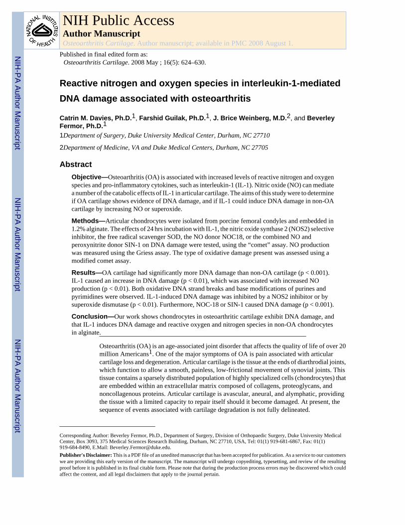
Reactive nitrogen and oxygen species in interleukin-1-mediatedDNA damage associated with osteoarthritis
Catrin M. Davies, Ph.D.1, Farshid Guilak, Ph.D.1, J. Brice Weinberg, M.D.2, and BeverleyFermor, Ph.D.11Department of Surgery, Duke University Medical Center, Durham, NC 27710
2Department of Medicine, VA and Duke Medical Centers, Durham, NC 27705
AbstractObjective—Osteoarthritis (OA) is associated with increased levels of reactive nitrogen and oxygenspecies and pro-inflammatory cytokines, such as interleukin-1 (IL-1). Nitric oxide (NO) can mediatea number of the catabolic effects of IL-1 in articular cartilage. The aims of this study were to determineif OA cartilage shows evidence of DNA damage, and if IL-1 could induce DNA damage in non-OAcartilage by increasing NO or superoxide.
Methods—Articular chondrocytes were isolated from porcine femoral condyles and embedded in1.2% alginate. The effects of 24 hrs incubation with IL-1, the nitric oxide synthase 2 (NOS2) selectiveinhibitor, the free radical scavenger SOD, the NO donor NOC18, or the combined NO andperoxynitrite donor SIN-1 on DNA damage were tested, using the “comet” assay. NO productionwas measured using the Griess assay. The type of oxidative damage present was assessed using amodified comet assay.
Results—OA cartilage had significantly more DNA damage than non-OA cartilage (p < 0.001).IL-1 caused an increase in DNA damage (p < 0.01), which was associated with increased NOproduction (p < 0.01). Both oxidative DNA strand breaks and base modifications of purines andpyrimidines were observed. IL-1-induced DNA damage was inhibited by a NOS2 inhibitor or bysuperoxide dismutase (p < 0.01). Furthermore, NOC-18 or SIN-1 caused DNA damage (p < 0.001).
Conclusion—Our work shows chondrocytes in osteoarthritic cartilage exhibit DNA damage, andthat IL-1 induces DNA damage and reactive oxygen and nitrogen species in non-OA chondrocytesin alginate.
Osteoarthritis (OA) is an age-associated joint disorder that affects the quality of life of over 20million Americans1. One of the major symptoms of OA is pain associated with articularcartilage loss and degeneration. Articular cartilage is the tissue at the ends of diarthrodial joints,which function to allow a smooth, painless, low-frictional movement of synovial joints. Thistissue contains a sparsely distributed population of highly specialized cells (chondrocytes) thatare embedded within an extracellular matrix composed of collagens, proteoglycans, andnoncollagenous proteins. Articular cartilage is avascular, aneural, and alymphatic, providingthe tissue with a limited capacity to repair itself should it become damaged. At present, thesequence of events associated with cartilage degradation is not fully delineated.
Corresponding Author: Beverley Fermor, Ph.D., Department of Surgery, Division of Orthopaedic Surgery, Duke University MedicalCenter, Box 3093, 375 Medical Sciences Research Building, Durham, NC 27710, USA, Tel: 01(1) 919-681-6867, Fax: 01(1)919-684-8490, E.Mail: [email protected]'s Disclaimer: This is a PDF file of an unedited manuscript that has been accepted for publication. As a service to our customerswe are providing this early version of the manuscript. The manuscript will undergo copyediting, typesetting, and review of the resultingproof before it is published in its final citable form. Please note that during the production process errors may be discovered which couldaffect the content, and all legal disclaimers that apply to the journal pertain.
NIH Public AccessAuthor ManuscriptOsteoarthritis Cartilage. Author manuscript; available in PMC 2008 August 1.
Published in final edited form as:Osteoarthritis Cartilage. 2008 May ; 16(5): 624–630.
NIH
-PA Author Manuscript
NIH
-PA Author Manuscript
NIH
-PA Author Manuscript
Evidence suggests that abnormal loading of the joint and increased pro-inflammatory cytokinesare risk factors for OA2,3. Pro-inflammatory mediators such as nitric oxide (NO) and otherreactive nitrogen and oxygen species (RNS and ROS)4–6 are increased in OA. The increasedlevels of these reactive species have been correlated to increased levels of inflammatorycytokines, such as interleukin-1 (IL-1). IL-1 is implicated in the degeneration of cartilage dueto its induction of proteoglycan loss and matrix degradation7. Elevated levels of IL-1 occur inthe synovial fluid and cartilage tissue of patients with OA compared to healthy individuals8,implying a role in disease pathogenesis. IL-1 receptor antagonist, a natural competitor of IL-1,suppresses cartilage loss, further supporting the role of IL-1 in cartilage breakdown9.
Both IL-1 and mechanical loading of cartilage increase NO production10–12 by up-regulatingnitric oxide synthase 2 (NOS2), which catalyzes the formation of NO and citrulline fromarginine in the presence of molecular oxygen and NADPH. NOS2 inhibitors can inhibit theprogression of OA in experimental animal models13, and osteoarthritic joint pathology issignificantly inhibited in the collagen–induced arthritis model in NOS2 deficient mice14.
Some of the destructive effects of NO in articular cartilage are linked to the ability of NO tocombine with superoxide anions (O2
−) to generate peroxynitrite15,16. Endogenousantioxidants such as superoxide dismutase (SOD) can serve to protect against oxidative stress.Extracellular SOD (EC-SOD) is decreased in osteoarthritic cartilage17, indicating the potentialimportance of EC-SOD to articular cartilage. Gene expression profiling has identifieddecreased expression of antioxidant defense genes in human OA cartilage, a situation that maycontribute to increased oxidative stress18. The effects of pro-inflammatory cytokines, ROSand RNS, on DNA integrity in articular cartilage have not previously been investigated.Oxidative stress can induce premature chondrocyte senescence via DNA damage in seriallypassaged chondrocytes19. DNA damage is noted in the synovial tissue of rheumatoid arthritisand OA patients. Microsatellite instability, a form of DNA mutation, is increased in the RAsynovium compared with OA synovium20, correlating with increased DNA mismatch repairenzymes in synovial tissue of RA and OA patients compared with non-arthritic tissue21. Priorstudies have not investigated the occurrence of microsatellite instability and DNA repairprotein within articular cartilage.
Since articular cartilage in diseased states such as OA exhibit elevated levels of NO and IL-1,and IL-1 can induce chondrocytes to produce NO in vitro, we investigated the susceptibilityof porcine articular chondrocytes to oxidative DNA damage. We sought (1) to determine ifOA cartilage contains more DNA damage than non-OA cartilage, and (2) to determine the roleof NO in IL-1-induced DNA damage in non-osteoarthritic chondrocytes embedded in alginate.
Materials and MethodsMacroscopic grading of articular cartilage
The femoral condyles of 2–3 year-old, female, ex-breeder pigs were obtained from the localslaughterhouse. Pigs can develop a spontaneous OA, and the incidence increases with age22,23. The articular cartilage on the medial condyle was graded macroscopically according to theCollins scale24. In summary, the principal distinguishing features of each grade in the Collinsscale are as follows: Grade 0: Normal healthy joint with smooth cartilage; Grade I: Superficialflaking of cartilage in areas of pressure and movement; Grade II: More extensive destructionof cartilage not denuding bone; Grade III: Total loss of cartilage in one or more pressure areasand obvious marginal osteophytes; Grade IV: Complete loss of cartilage from large areas witheburnation of bone; prominent osteophytes. Grades III and IV were not used in these studiesdue to lack of chondrocytes.
Davies et al. Page 2
Osteoarthritis Cartilage. Author manuscript; available in PMC 2008 August 1.
NIH
-PA Author Manuscript
NIH
-PA Author Manuscript
NIH
-PA Author Manuscript
Chondrocyte Cell Culture in Alginate BeadsArticular chondrocytes were enzymatically isolated, using pronase for 1 hour and collagenasetype II for 2 hours, from site matched, full thickness slices of articular cartilage from the femoralcondyles of skeletally mature 2–3 year old pigs and placed into alginate beads (1.2% alginate)at a concentration of 4 × 10⁶ cells/ml. Alginate was used to maintain the chondrocytes in arounded shape and ensure the collagen II expression associated with chondrocytes. Ifchondrocytes are grown as a monolayer, dedifferentiation to fibrochondrocytes occurs. Beadswere cultured in high glucose DMEM (Gibco, Gaithersburg, MD) with 10% heat inactivatedFBS (Hyclone), 0.1 mM non-essential amino acids (Gibco), 10 mM HEPES (Gibco), 100 U/ml penicillin and streptomycin (Gibco) 110 mg/L sodium pyruvate, 2 mM L-glutamine, at 37°C, 5% CO2, 95% air for either 24 or 72 hours prior to treatment
Treatment of Chondrocytes in Culture with Interleukin-1αAfter 24 hrs in culture, chondrocytes encapsulated in alginate beads were treated for 24 hrswith 0 – 100 ng/ml recombinant porcine IL-α. The effects of the selective NOS2 inhibitor1400W [N-(3-(Aminomethyl)benzyl)acetamidine, 2 mM, Alexis Chemical Co.] or superoxidedismutase (SOD, 50 µg/ml Sigma Chemical Co.) were tested. 1400W is a slow, tight bindinginhibitor of NOS2, while SOD reduces production of peroxynitrite by breaking downsuperoxide.
We examined the effects of the pure NO donor NOC18 (DETA-NONOate, 0 – 500 µM) or theperoxynitrite generator SIN-1 (3-morpholinosydnonimine, 0 – 500 µM) by culturing withchondrocytes for 24 hrs. NOC18 is a stable NO-amine complex that spontaneously release NO,without cofactors, under physiological conditions. Unlike other NO donors such asnitroglycerin, nitroprusside, and S-nitroso-N-acetyl-L-penicillamine (SNAP), by-products ofNO release do not interfere with cell activities. SIN-1, which uses molecular oxygen to generatesuperoxide and NO was chosen as it causes the spontaneous formation of peroxynitrite.
Comet AssayThe single cell gel electrophoresis assay, also known as a comet assay, allows DNA damageto be visualized and quantified at a single cell level. Specifically, the alkaline comet assayallows detection of single and double strand DNA breaks, as well as apurinic or apyrimidinicsites that are alkali labile and form breaks due to repair lesions produced during endogenousDNA repair (base excision or nucleotide excision)25. Chondrocytes were released from thealginate beads with calcium chelation in 55 mM sodium citrate and the alkaline comet assay(Trevigen, Gaithersburg, MD)26 was performed. Since changes in experimental procedures,such as electrophoresis times and lysis conditions may increase or decrease the sensitivity ofthe assay to detect DNA damage, our assays were carefully controlled by carrying out all groupsfrom one experiment on the same slide.
The modified comet assay enables the detection of specific oxidative base lesions through theuse of repair enzymes. This assay is based on the addition of specific glycosylases, whichcleave the modified base from the DNA strand forming a single strand break27. The Fpgenzyme reveals oxidized purines by recognizing and binding duplex DNA containingoxidatively damaged bases, such as 8-oxo-2’-deoxyguanosine (8-oxoG), andformamidopyrimidines. EndoIII reveals oxidized pyrimidines. EndoIII enzyme recognizes andbinds duplex DNA containing oxidatively damaged bases such as thymine and uracil glycol,thymine and cytosine hydrates and urea. Chondrocytes were cultured in alginate beads as aboveand exposed to 10 ng/ml IL-1 and processed as the comet assay. After the lysis step, the slideswere washed three times for 5 min each in Buffer A (10 mM HEPES-KOH (pH 7.4), 10 mMEDTA (pH 8.0), and 0.1 M KCl) and tapped dry. The agarose-embedded cells were coveredwith either 1 µg/ml Fpg in Buffer A plus 100 µg/ml BSA, or 1 µg/ml Endo III in buffer A or
Davies et al. Page 3
Osteoarthritis Cartilage. Author manuscript; available in PMC 2008 August 1.
NIH
-PA Author Manuscript
NIH
-PA Author Manuscript
NIH
-PA Author Manuscript
buffer A alone and incubated in a moist atmosphere at 37°C for 1 h. The slides were thenimmersed in freshly prepared alkaline solution (pH > 13) and continued through the steps ofthe comet assay.
Slides were stained with 1x SYBR® Green I and imaged using a 20x objective on a confocallaser scanning microscope (LSM 510, Zeiss) followed by analysis using CASP™software28. The Olive Tail Moment (OTM) was used to assess the amount of DNAdamage29. The OTM is defined as the fraction of tail DNA multiplied by the distance betweenthe profile centers of gravity for DNA in the head and tail. The distance the DNA moves isrelated to the size of free or relaxed pieces, while the intensity of the tail is a direct indicationof the number of pieces that migrate. Since single- and double-stranded breaks causes DNAto become unwound and free to migrate towards the anode during electrophoresis (Fig. 1),nucleoids of comet-like structure are indicative of DNA damage. Over time, the comet taillength plateaus, but the amount of DNA entering the tail-like region increases. The OTMaccounts for this feature. A total of 50 cells per group per joint were analyzed, avoiding edgesand damaged areas of the gel, to give a representative result for the population of cells.
NOx AssayNO production was assessed by measuring the concentration of total nitrate and nitrite (termed“NOx”) in the media as described previously30. This method first converts nitrate to nitriteusing nitrate reductase, and then total nitrite is measured using the Griess reagent. NOx levelswere normalized to the DNA content of the chondrocytes. Media were removed from beads,and NOx was determined. Chondrocytes in alginate beads were digested in 125 µg/ml papainsolution at 60°C for 24 hrs. DNA content of the alginate beads was determined using thefluorescent picoGreen dsDNA quantification assay (Molecular Probes, Eugene, OR) using thediluted bead-papain digest solution.
Cell ViabilityCell viability was determined using the fluorescence-based viability assay (Live/Dead Assay,Molecular Probes). At the end of all culture conditions, the viability of the chondrocytes wasnot significantly different from control chondrocytes directly after isolation. Cell viability wasfound to be 95–99% in control or treated cells.
Statistical AnalysisStatistical analysis was performed using Student’s t-test for comparison of DNA damage inOA verses non-OA chondrocytes or analysis of variance with Duncan's post hoc comparisonwith significance reported at the 95% confidence level for all other comparisons.
ResultsDNA damage in articular cartilage
Articular cartilage was harvested from the medial condyles of macroscopically “normal” tissue(Collins Grade O) and osteoarthritic cartilage (Collins Grades I and II). Levels of DNA damagein these cells were determined using the Comet assay. Nucleoids appeared as tightlysupercoiled, spheroid structures (Fig. 1a), indicating absence of DNA damage. The nucleoidswhich showed tail-like structures (“comets”) (Fig. 1b) indicating presence of DNA damage.A significant increase in DNA damage was observed in the OA chondrocytes compared withthe non-OA chondrocytes (Fig. 1c). Importantly, a small proportion of the chondrocytesanalyzed from OA joints had no evidence of DNA damage as assessed by the comet assay.
Davies et al. Page 4
Osteoarthritis Cartilage. Author manuscript; available in PMC 2008 August 1.
NIH
-PA Author Manuscript
NIH
-PA Author Manuscript
NIH
-PA Author Manuscript
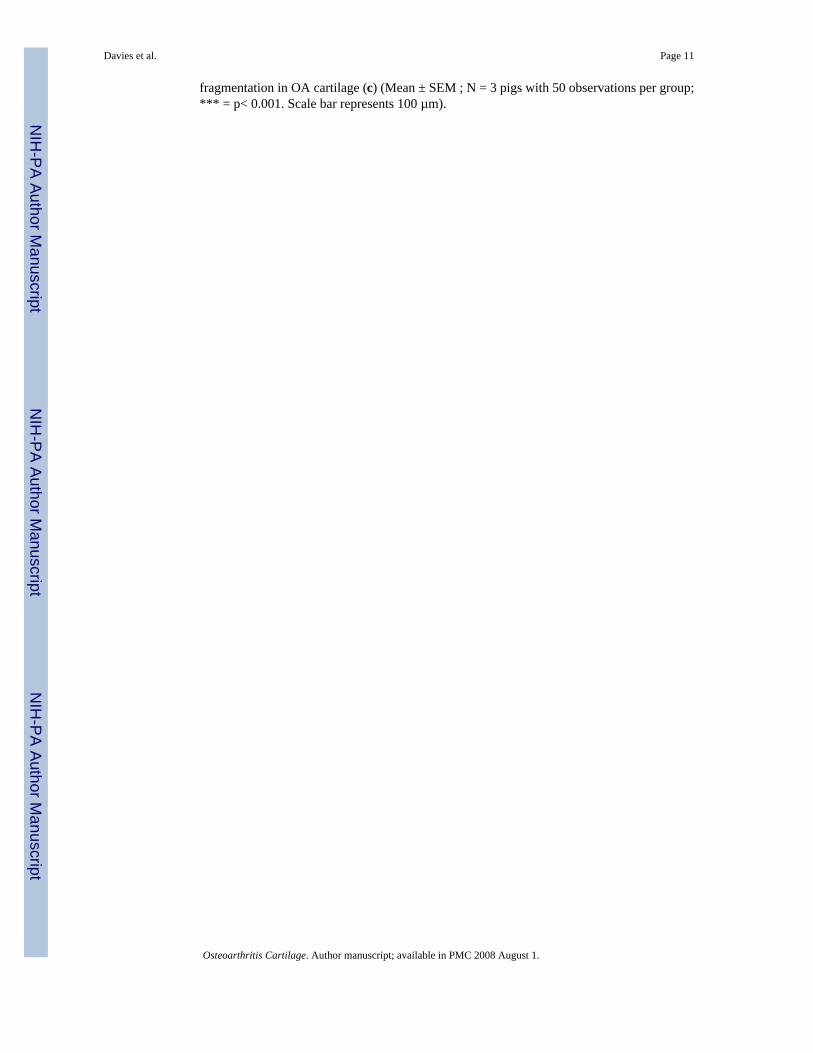
Effects of IL-1α on DNA damage in articular cartilageOA articular cartilage is characterized by an increase in the level of the pro-inflammatorycytokine IL-1. Therefore we investigated the effects of IL-1 on DNA damage in non-OAcartilage. IL-1α caused a concentration-dependent increase in comet tail length. Exposure to1 ng/ml ≥ IL-1 over a 24h period caused a significant increase in DNA damage as measuredby the OTM, compared to untreated non-OA chondrocytes (Fig. 2). There was a significant,concentration-dependent increase in NO production in response to IL-1 compared with controlcells.
The effects of the NOS2 inhibitor 1400W, or the free radical scavenger SOD on IL-1-mediatedDNA damage in chondrocytes were investigated. A significant reduction in the OTM was seenin chondrocytes incubated with IL-1 and 1400W compared with chondrocytes treated withIL-1 alone. SOD also inhibited IL-1-induced DNA damage (Fig. 3). There was no significantchange in levels of DNA damage when chondrocytes were incubated with 2 mM 1400W or50 µM SOD (Data not shown).
Effects of NO donors on DNA damageTo further confirm that NO or superoxide could induce DNA damage in non-OA chondrocytes,we tested the effects of NO donors on DNA damage. NOC18, a pure NO donor, caused aconcentration-dependent increase in DNA damage (Fig. 4a) and, as predicted more NO wasnoted in the cultures (Fig. 4b). SIN-1, which releases both NO and superoxide and generatesperoxynitrite, had similar effects (Fig. 4c & 4d).
Identification of the type of DNA damage caused by IL-1To determine the type of DNA damage occurring, the comet assay was performed in thepresence of DNA repair enzymes. Fpg reveals oxidized purines, and Endo III reveals oxidizedpyrimidines. The addition of the oxidative DNA damage lesion-specific enzymes Fpg (Fig.5a) and Endo III (Fig. 5b) caused a significant increase in the OTM of control cells treated with10 ng/ml IL-1. The mean values of Fpg-sensitive sites were significantly higher in chondrocytescultured with IL-1 (10 ng/ml) for 24 hours compared to untreated chondrocytes. The meanvalues of EndoIII-sensitive sites were significantly higher in chondrocytes cultured with IL-1(10 ng/ml) for 24 hours compared with untreated chondrocytes.
DiscussionSince OA is associated with increased reactive nitrogen and oxygen species and cartilagedamage, we sought to determine if there was also cartilage DNA damage and if NO andsuperoxide might mediate DNA damage in chondrocytes. We found that chondrocytes in OAcartilage exhibit significantly more DNA damage than those in non-OA cartilage. Our invitro studies showed that IL-1 causes a concentration-dependent increase in DNA damage inchondrocytes in alginate, and this damage is associated with increased NO production. Wenoted both oxidative DNA strand breaks and base modifications of purines and pyrimidines.IL-1-induced DNA damage is inhibited by a NOS2 inhibitor or by superoxide dismutase,indicating important contributing roles of NO and superoxide in the mechanism of DNAdamage. Use of the pure NO donor (NOC-18) or peroxynitrite generator (SIN-1) causes DNAdamage of chondrocytes in vitro. A small proportion of OA chondrocytes or non-OAchondrocytes treated with IL-1 or NO donors did not show evidence of DNA damage.Collectively, our data indicate that increased reactive nitrogen and oxygen associated with OAcontribute to DNA damage in articular chondrocytes.
Superoxide dismutase did not reduce the level of DNA damage as much as the NOS2-selectiveinhibitor 1400W. Our data agree with findings in other eukaryotic cell types in which NO can
Davies et al. Page 5
Osteoarthritis Cartilage. Author manuscript; available in PMC 2008 August 1.
NIH
-PA Author Manuscript
NIH
-PA Author Manuscript
NIH
-PA Author Manuscript
cause DNA damage31–33. Specifically, NO but not superoxide, caused DNA damage in ratislets of Langerhans and insulin-containing HIT-T15 cells treated with IL-1β (0.1 nM)31.However, in our system, superoxide was responsible for some of the IL-1-induced DNAdamage. The advantage of 1400W over SOD in reducing IL-1-induced DNA damage invitro in articular cartilage might be accounted for by differences in the rate constants of theNO/superoxide reaction compared with the superoxide/SOD reaction. The rate constant of theNO/superoxide reaction to form peroxynitrite is 6.7×10⁹ M−1 s−1, a rate constant which is 3.5times faster than that for the dismutation of superoxide by SOD34.
Although we found that IL-1 and NO damage chondrocyte DNA significantly in our system,no cell death was observed under these conditions. This finding is in agreement with previousstudies showing neither IL-1, nor NOC 18 at these concentrations caused chondrocyte celldeath15. SIN-1 can cause significant cell death in human chondrocytes cultured in serum freemedium15, but SIN-1 did not cause cell death in our system using serum.
Our results demonstrate that Fpg-sensitive and EndoIII-sites in the DNA of chondrocytes areincreased by incubation with IL-1α. This suggests NO might mediate formation of bothoxidized purines and oxidized pyrimidines. In agreement with others, a significant number ofFpg and EndoIII sensitive sites were present even in the absence of IL-1 treatment35. This islikely related to spontaneous, basal oxidation that occurs normally in oxygen-containingatmospheres35. Oxidative DNA damage in articular chondrocytes has been reported in serially-passaged chondrocytes cultured at 21% O2 for 60–70 days19, but not in primary chondrocytescultured in the alginate beads system that maintains the chondrocytic cellular phenotype invitro. However, oxidative protein damage (3-nitrotyrosine formation) has been observed inarticular cartilage explants and in articular chondrocytes cultured in alginate36,37 supportingthe role of peroxynitrite in cell damage. In our studies, levels of NO are greater than 1 µMgiving potential for reactive nitrogen species-mediated modification of proteins, as well asDNA38. Oxidation and nitration of bases are a more severe consequence of ROS and RNS.Many DNA base modifications can occur, but the oxidation of guanine to form 8-oxodG is oneof the most common markers of base oxidation39.
The importance of the DNA damage observed in our studies in the pathogenesis of OA isunknown. Daughter cells inheriting oxidative- and nitrative-modified bases in their DNA mightlead to mutagenic lesions due to base mis-incorporation opposite the lesion during mitosis.This would lead to base pair trans-versions, which in many tissues, such as gastric, liver, andcolon tissue, can be carcinogenic39. However, in articular cartilage, after the completion ofskeletal growth, little cell division occurs. Neuronal cells are another example of non-dividingcells. Large amounts of 8-oxoG are formed in the RNA in the neuronal tissues of someneurodegenerative diseases such as Parkinson’s disease, Alzheimer’s disease and Down’ssyndrome40.
IL-1 and NO can cause catabolic effects on articular cartilage in vitro41. Free radical-inducedDNA damage may modify transcription, causing transcription errors that result in erroneousprotein formation40,42. The error rate of RNA transcription is estimated to be much greaterfor RNA transcription (10−5 per residues) than DNA replication (10−9 per residues). The DNAdamage may alter binding of transcription factors43,44, or block gene transcription. A numberof gene promoter elements contain a succession of guanine residues in their transcription factorrecognition sequences, such as NFκB. Oxidative modification to 8-oxodG can alter the bindingaffinity of transcription factor NFκB45. Alternatively, the ROS and RNS could incapacitateDNA repair proteins, leading to further DNA damage over time46. Accumulation of damagedDNA, proteins, and lipids may be responsible for disrupting normal cell functions, which mightexplain the increased incidence of OA with age.
Davies et al. Page 6
Osteoarthritis Cartilage. Author manuscript; available in PMC 2008 August 1.
NIH
-PA Author Manuscript
NIH
-PA Author Manuscript
NIH
-PA Author Manuscript
Some cell division may occur in late stage OA47, and the spontaneous replication ofchondrocytes with impaired genetic material could result in chondrocyte apoptosis48. Sincethe development of OA is age-related, damage accumulation over time could be particularlysignificant when the chondrocyte does divide in the later stages of OA. Epigenetic changes,heritable changes in DNA without changes in the sequence, such as DNA methylation, arepossibly important in the pathogenesis of OA49. Changes in DNA methylation and basemodification could have a role in altering the chondrocyte phenotype in OA.
Collectively, our findings demonstrate that OA articular chondrocytes contain DNA damage,that high levels of IL-1 induced increases in NO result in DNA damage in non-OA articularchondrocytes in alginate through both strand breaks and base modifications. A NOS2 inhibitorand SOD reduce IL-1-mediated DNA damage. Although the full consequence(s) ofchondrocyte DNA damage is not currently known, these results provide further evidence thatagents that reduce reactive nitrogen and oxygen species in vivo might be beneficial for thetreatment of OA.
Acknowledgements
This study was supported by NIH grants AR49790, AR50245, AG15768 and the VA Research Service.
Grant Support: This study was supported by NIH grants AR49790, AR50245, AG15768 and the VA Research Service.
References1. Buckwalter JA, Saltzman C, Brown T. The impact of osteoarthritis: implications for research. Clin
Orthop Relat Res 2004;427:S6–S15. [PubMed: 15480076]2. Guilak F, Fermor B, Keefe FJ, Kraus VB, Olson SA, Pisetsky DS, et al. The role of biomechanics and
inflammation in cartilage injury and repair. Clin Orthop Relat Res 2004;423:17–26. [PubMed:15232421]
3. Griffin TM, Guilak F. The role of mechanical loading in the onset and progression of osteoarthritis.Exerc Sport Sci Rev 2005;33:195–200. [PubMed: 16239837]
4. Amin AR, Di Cesare PE, Vyas P, Attur M, Tzeng E, Billiar TR, et al. The expression and regulationof nitric oxide synthase in human osteoarthritis-affected chondrocytes: evidence for up-regulatedneuronal nitric oxide synthase. J Exp Med 1995;182:2097–2102. [PubMed: 7500055]
5. Henrotin Y, Kurz B, Aigner T. Oxygen and reactive oxygen species in cartilage degradation: friendsor foes? Osteoarthritis cartilage 2005;13:643–654. [PubMed: 15936958]
6. Yudoh K, Nguyen T, Nakamura H, Hongo-Masuko K, Kato T, Nishioka K. Potential involvement ofoxidative stress in cartilage senescence and development of osteoarthritis: oxidative stress induceschondrocyte telomere instability and downregulation of chondrocyte function. Arthritis Res Ther2005;7:R380–R391. [PubMed: 15743486]
7. Kobayashi M, Squires GR, Mousa A, Tanzer M, Zukor DJ, Antoniou J, et al. Role of interleukin-1 andtumor necrosis factor alpha in matrix degradation of human osteoarthritic cartilage. Arthritis Rheum2005;52:128–135. [PubMed: 15641080]
8. Goldring SR, Goldring MB. The role of cytokines in cartilage matrix degeneration in osteoarthritis.Clin Orthop Relat Res 2004:S27–S36. [PubMed: 15480070]
9. Pelletier JP, Caron JP, Evans C, Robbins PD, Georgescu HI, Jovanovic D, et al. In vivo suppressionof early experimental osteoarthritis by interleukin-1 receptor antagonist using gene therapy. ArthritisRheum 1997;40:1012–1019. [PubMed: 9182910]
10. Stadler J, Stefanovic-Racic M, Billiar TR, Curran RD, McIntyre LA, Georgescu HI, et al. Articularchondrocytes synthesize nitric oxide in response to cytokines and lipopolysaccharide. J Immunol1991;147:3915–3920. [PubMed: 1658153]
11. Fermor B, Weinberg JB, Pisetsky DS, Misukonis MA, Banes AJ, Guilak F. The effects of static andintermittent compression on nitric oxide production in articular cartilage explants. J Orthop Res2001;19:729–737. [PubMed: 11518285]
Davies et al. Page 7
Osteoarthritis Cartilage. Author manuscript; available in PMC 2008 August 1.
NIH
-PA Author Manuscript
NIH
-PA Author Manuscript
NIH
-PA Author Manuscript
12. Agarwal S, Deschner J, Long P, Verma A, Hofman C, Evans CH, et al. Role of NFkB transciptionfactors in anti-inflammatory and proinflammatory actions of mechanical signals. Arthritis Rheum2004;50:3541–3548. [PubMed: 15529376]
13. Pelletier JP, Jovanovic D, Fernandes JC, Manning P, Connor JR, Currie MG, et al. Reducedprogression of experimental osteoarthritis in vivo by selective inhibition of inducible nitric oxidesynthase. Arthritis Rheum 1998;41:1275–1286. [PubMed: 9663486]
14. van den Berg WB, van de Loo F, Joosten LA, Arntz OJ. Animal models of arthritis in NOS2-deficientmice. Osteoarthritis cartilage 1999;7:413–415. [PubMed: 10419784]
15. Del Carlo M Jr. Loeser RF. Nitric oxide-mediated chondrocyte cell death requires the generation ofadditional reactive oxygen species. Arthritis Rheum 2002;46:394–403. [PubMed: 11840442]
16. Pacher P, Beckman JS, Liaudet L. Nitric oxide and peroxynitrite in health and disease. Physiol Rev2007;87:315–424. [PubMed: 17237348]
17. Regan E, Flannelly J, Bowler R, Tran K, Nicks M, Carbone BD, et al. Extracellular superoxidedismutase and oxidant damage in osteoarthritis. Arthritis Rheum 2005;52:3479–3491. [PubMed:16255039]
18. Aigner T, Fundel K, Saas J, Gebhard PM, Haag J, Weiss T, et al. Large-scale gene expression profilingreveals major pathogenetic pathways of cartilage degeneration in osteoarthritis. Arthritis Rheum2006;54:3533–3544. [PubMed: 17075858]
19. Martin JA, Klingelhutz AJ, Moussavi-Harami F, Buckwalter JA. Effects of oxidative damage andtelomerase activity on human articular cartilage chondrocyte senescence. J Gerontol A Biol Sci MedSci 2004;59:324–337. [PubMed: 15071075]
20. Lee SH, Chang DK, Goel A, Boland CR, Bugbee W, Boyle DL, et al. Microsatellite instability andsuppressed DNA repair enzyme expression in rheumatoid arthritis. J Immunol 2003;170:2214–2220.[PubMed: 12574395]
21. Simelyte E, Boyle DL, Firestein GS. DNA mismatch repair enzyme expression in synovial tissue.Ann Rheum Dis 2004;63:1695–1699. [PubMed: 15547100]
22. Reiland S. Pathology of so called leg weakness in the pig. Acta Radiologica - Supplementum1978;358:23–44.
23. Turner GV, Collett MG, Veary CM, Kruger C. Arthritis in slaughter pigs. Journal of the South AfricanVeterinary association 1991;62:107–109. [PubMed: 1770479]
24. Collins DH, Mc ET. Sulphate (35SO4) uptake by chondrocytes in relation to histological changes inosteoarthritic human articular cartilage. Ann Rheum Dis 1960;19:318–330. [PubMed: 13694746]
25. Collins AR, Dobson VL, Dusinska M, Kennedy G, Stetina R. The comet assay: what can it really tellus? Mutat Res 1997;375:183–193. [PubMed: 9202728]
26. Singh NP, McCoy MT, Tice RR, Schneider EL. A simple technique for quantitation of low levels ofDNA damage in individual cells. Exp Cell Res 1988;175:184–191. [PubMed: 3345800]
27. Collins AR, Duthie SJ, Dobson VL. Direct enzymic detection of endogenous oxidative base damagein human lymphocyte DNA. Carcinogenesis 1993;14:1733–1735. [PubMed: 8403192]
28. Konca K, Lankoff A, Banasik A, Lisowska H, Kuszewski T, Gozdz S, et al. A cross-platform publicdomain PC image-analysis program for the comet assay. Mutat Res 2003;534:15–20. [PubMed:12504751]
29. Olive PL, Banath JP, Durand RE. Heterogeneity in radiation-induced DNA damage and repair intumor and normal cells measured using the "comet" assay. Radiat Res 1990;122:86–94. [PubMed:2320728]
30. Granger DL, Anstey NM, Miller WC, Weinberg JB. Measuring nitric oxide production in humanclinical studies. Methods Enzymol 1999;301:49–61. [PubMed: 9919553]
31. Delaney CA, Green MH, Lowe JE, Green IC. Endogenous nitric oxide induced by interleukin-1 betain rat islets of Langerhans and HIT-T15 cells causes significant DNA damage as measured by the'comet' assay. FEBS Lett 1993;333:291–295. [PubMed: 8224196]
32. Jaiswal M, LaRusso NF, Burgart LJ, Gores GJ. Inflammatory cytokines induce DNA damage andinhibit DNA repair in cholangiocarcinoma cells by a nitric oxide-dependent mechanism. Cancer Res2000;60:184–190. [PubMed: 10646872]
33. Li CQ, Wogan GN. Nitric oxide as a modulator of apoptosis. Cancer Lett 2005;226:1–15.
Davies et al. Page 8
Osteoarthritis Cartilage. Author manuscript; available in PMC 2008 August 1.
NIH
-PA Author Manuscript
NIH
-PA Author Manuscript
NIH
-PA Author Manuscript
34. Burney S, Caulfield JL, Niles JC, Wishnok JS, Tannenbaum SR. The chemistry of DNA damagefrom nitric oxide and peroxynitrite. Mutat Res 1999;424:37–49. [PubMed: 10064848]
36. Collins AR, Dusinska M, Gedik CM, Stetina R. Oxidative damage to DNA: do we have a reliablebiomarker? Environ Health Perspect 1996;104:465–469. [PubMed: 8781365]
36. Loeser RF, Carlson CS, Del Carlo M, Cole A. Detection of nitrotyrosine in aging and osteoarthriticcartilage: Correlation of oxidative damage with the presence of interleukin-1 beta and withchondrocyte resistance to insulin-like growth factor 1. Arthritis Rheum 2002;46:2349–2357.[PubMed: 12355482]
37. Del Carlo MD Jr. Loeser RF. Increased oxidative stress with aging reduces chondrocyte survival:correlation with intracellular glutathione levels. Arthritis Rheum 2003;48:3419–3430. [PubMed:14673993]
38. Abramson SB, Amin AR, Clancy RM, Attur M. The role of nitric oxide in tissue destruction. BestPract Res Clin Rheumatol 2001;15:831–845. [PubMed: 11812024]
39. Kawanishi S, Hiraku Y. Oxidative and nitrative DNA damage as biomarker for carcinogenesis withspecial reference to inflammation. Antioxid Redox Signal 2006;8:1047–1058. [PubMed: 16771694]
40. Ishibashi T, Hayakawa H, Ito R, Miyazawa M, Yamagata Y, Sekiguchi M. Mammalian enzymes forpreventing transcriptional errors caused by oxidative damage. Nucleic Acids Res 2005;33:3779–3784. [PubMed: 16002790]
41. Evans CH, Watkins SC, Stefanovic-Racic M. Nitric oxide and cartilage metabolism. MethodsEnzymol 1996;269:75–88. [PubMed: 8791639]
42. Doetsch PW. Translesion synthesis by RNA polymerase:occurrence and biological implications fortranscriptional mutagenesis. Mutation Research 2002;510:131–140. [PubMed: 12459449]
43. Ghosh R, Mitchell DL. Effect of oxidative DNA damage in promoter elements on transcription factorbinding. Nucleic Acids Res 1999;27:3213–3218. [PubMed: 10454620]
44. Parsian AJ, Funk MC, Tao TY, Hunt CR. The effect of DNA damage on the formation of protein/DNA complexes. Mutat Res 2002;501:105–113. [PubMed: 11934442]
45. Hailer-Morrison MK, Kotler JM, Martin BD, Sugden KD. Oxidized guanine lesions as modulatorsof gene transcription. Altered p50 binding affinity and repair shielding by 7,8-dihydro-8-oxo-2'-deoxyguanosine lesions in the NF-kappaB promoter element. Biochemistry 2003;42:9761–9770.[PubMed: 12911319]
46. Jaiswal M, LaRusso NF, Shapiro RA, Billiar TR, Gores GJ. Nitric oxide-mediated inhibition of DNArepair potentiates oxidative DNA damage in cholangiocytes. Gastroenterology 2001;120:190–199.[PubMed: 11208728]
47. Aigner T, Hemmel M, Neureiter D, Gebhard PM, Zeiler G, Kirchner T, et al. Apoptotic cell death isnot a widespread phenomenon in normal aging and osteoarthritis human articular knee cartilage: astudy of proliferation, programmed cell death (apoptosis), and viability of chondrocytes in normaland osteoarthritic human knee cartilage. Arthritis Rheum 2001;44:1304–1312. [PubMed: 11407689]
48. D'Lima DD, Kuhn K, Lotz MK. Detection of apoptosis in cartilage in situ and in isolated chondrocytes.Methods Mol Med 2004;100:275–290. [PubMed: 15280601]
49. Roach HI, Aigner T. DNA methylation in osteoarthritic chondrocytes: a new molecular target.Osteoarthritis cartilage 2007;15:128–137. [PubMed: 16908204]
Davies et al. Page 9
Osteoarthritis Cartilage. Author manuscript; available in PMC 2008 August 1.
NIH
-PA Author Manuscript
NIH
-PA Author Manuscript
NIH
-PA Author Manuscript
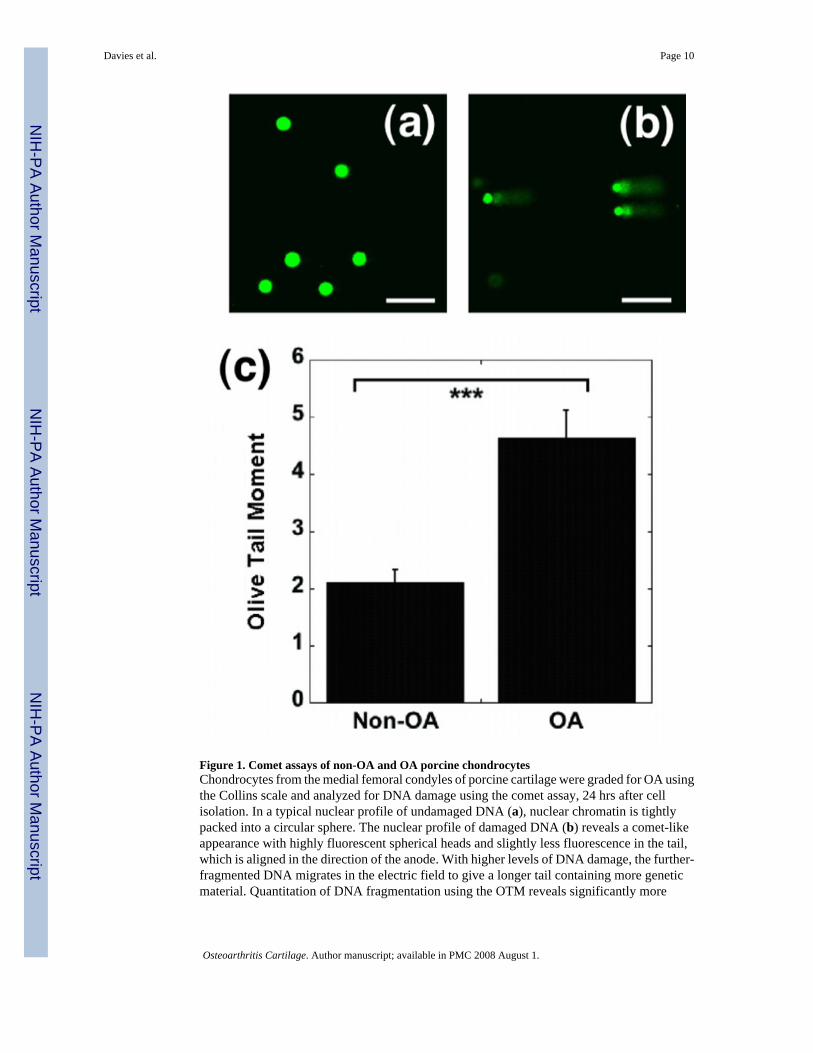
Figure 1. Comet assays of non-OA and OA porcine chondrocytesChondrocytes from the medial femoral condyles of porcine cartilage were graded for OA usingthe Collins scale and analyzed for DNA damage using the comet assay, 24 hrs after cellisolation. In a typical nuclear profile of undamaged DNA (a), nuclear chromatin is tightlypacked into a circular sphere. The nuclear profile of damaged DNA (b) reveals a comet-likeappearance with highly fluorescent spherical heads and slightly less fluorescence in the tail,which is aligned in the direction of the anode. With higher levels of DNA damage, the further-fragmented DNA migrates in the electric field to give a longer tail containing more geneticmaterial. Quantitation of DNA fragmentation using the OTM reveals significantly more
Davies et al. Page 10
Osteoarthritis Cartilage. Author manuscript; available in PMC 2008 August 1.
NIH
-PA Author Manuscript
NIH
-PA Author Manuscript
NIH
-PA Author Manuscript
fragmentation in OA cartilage (c) (Mean ± SEM ; N = 3 pigs with 50 observations per group;*** = p< 0.001. Scale bar represents 100 µm).
Davies et al. Page 11
Osteoarthritis Cartilage. Author manuscript; available in PMC 2008 August 1.
NIH
-PA Author Manuscript
NIH
-PA Author Manuscript
NIH
-PA Author Manuscript
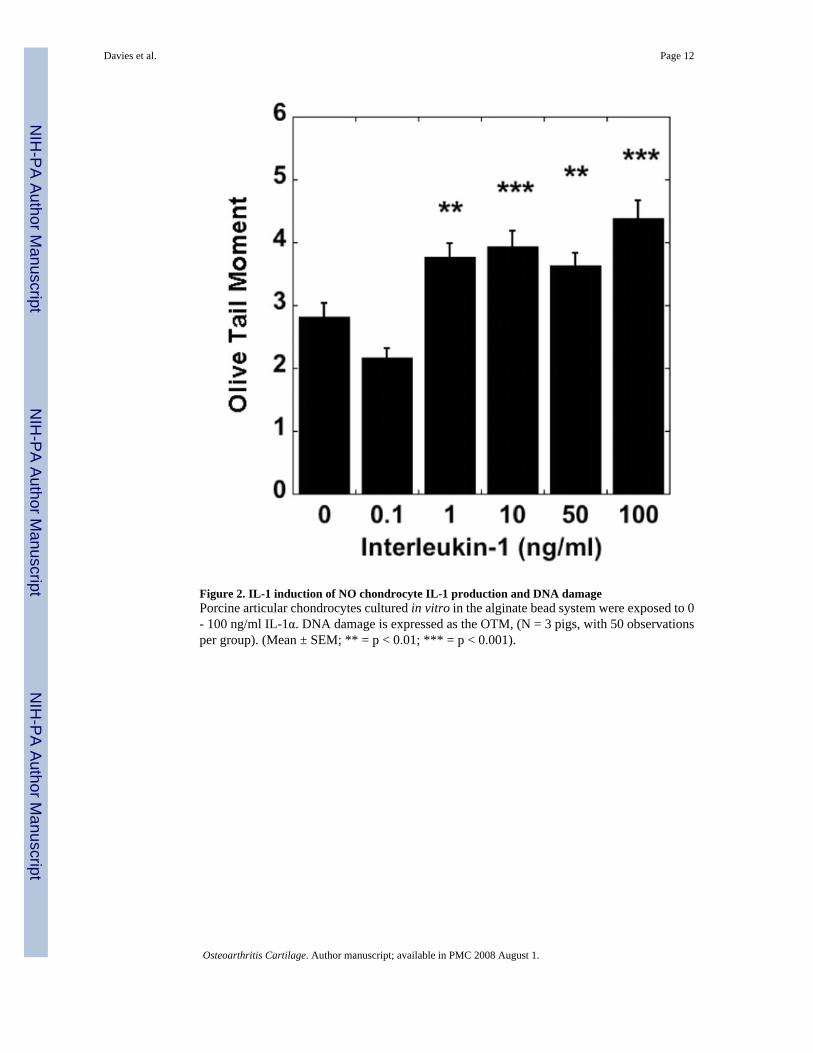
Figure 2. IL-1 induction of NO chondrocyte IL-1 production and DNA damagePorcine articular chondrocytes cultured in vitro in the alginate bead system were exposed to 0- 100 ng/ml IL-1α. DNA damage is expressed as the OTM, (N = 3 pigs, with 50 observationsper group). (Mean ± SEM; ** = p < 0.01; *** = p < 0.001).
Davies et al. Page 12
Osteoarthritis Cartilage. Author manuscript; available in PMC 2008 August 1.
NIH
-PA Author Manuscript
NIH
-PA Author Manuscript
NIH
-PA Author Manuscript
Figure 3. Effects of IL-1, NOS2 inhibitor, and SOD on DNA damage in chondrocytesPorcine articular chondrocytes cultured in vitro in alginate bead system were exposed to 0 and10 ng/ml IL-1, 10 ng/ml IL-1 with 2 mM 1400W or 10 ng/ml IL-1 with 50 µg/ml SOD (N =3, with 50 observations). DNA damage is expressed as the OTM. (Data represent Mean ± SEM;** = p < 0.01; *** = p < 0.001).
Davies et al. Page 13
Osteoarthritis Cartilage. Author manuscript; available in PMC 2008 August 1.
NIH
-PA Author Manuscript
NIH
-PA Author Manuscript
NIH
-PA Author Manuscript
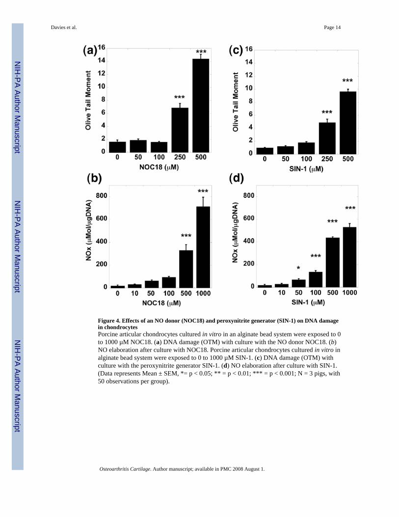
Figure 4. Effects of an NO donor (NOC18) and peroxynitrite generator (SIN-1) on DNA damagein chondrocytesPorcine articular chondrocytes cultured in vitro in an alginate bead system were exposed to 0to 1000 µM NOC18. (a) DNA damage (OTM) with culture with the NO donor NOC18. (b)NO elaboration after culture with NOC18. Porcine articular chondrocytes cultured in vitro inalginate bead system were exposed to 0 to 1000 µM SIN-1. (c) DNA damage (OTM) withculture with the peroxynitrite generator SIN-1. (d) NO elaboration after culture with SIN-1.(Data represents Mean ± SEM, *= p < 0.05; ** = p < 0.01; *** = p < 0.001; N = 3 pigs, with50 observations per group).
Davies et al. Page 14
Osteoarthritis Cartilage. Author manuscript; available in PMC 2008 August 1.
NIH
-PA Author Manuscript
NIH
-PA Author Manuscript
NIH
-PA Author Manuscript
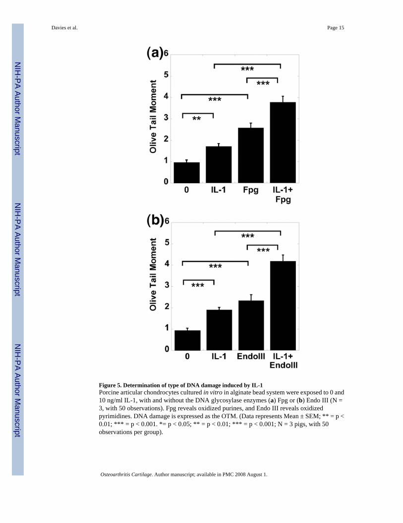
Figure 5. Determination of type of DNA damage induced by IL-1Porcine articular chondrocytes cultured in vitro in alginate bead system were exposed to 0 and10 ng/ml IL-1, with and without the DNA glycosylase enzymes (a) Fpg or (b) Endo III (N =3, with 50 observations). Fpg reveals oxidized purines, and Endo III reveals oxidizedpyrimidines. DNA damage is expressed as the OTM. (Data represents Mean ± SEM; ** = p <0.01; *** = p < 0.001. *= p < 0.05; ** = p < 0.01; *** = p < 0.001; N = 3 pigs, with 50observations per group).
Davies et al. Page 15
Osteoarthritis Cartilage. Author manuscript; available in PMC 2008 August 1.
NIH
-PA Author Manuscript
NIH
-PA Author Manuscript
NIH
-PA Author Manuscript
Copyright © 2022 FDOKUMEN