Tissue structure modification in knee osteoarthritis by use of ...
19
We use cookies to improve our service and to tailor our content and advertising to you. More info Home / Archive / Volume 70, Issue 8 Article Text Article menu Clinical and epidemiological research Extended report Tissue structure modification in knee osteoarthritis by use of joint distraction: an open 1-year pilot study Abstract Background Background Modification of joint tissue damage is challenging in late-stage osteoarthritis (OA). Few options are available for treating end-stage knee OA other than joint replacement. Objectives Objectives To examine whether joint distraction can effectively modify knee joint tissue damage and has the potential to delay prosthesis surgery. Methods Methods 20 patients (<60 years) with tibiofemoral OA were treated surgically using joint distraction. Distraction (∼5 mm) was applied for 2 months using an external fixation frame. Tissue structure modification at 1 year of follow-up was evaluated radiographically (joint space width (JSW)), by MRI (segmentation of cartilage morphology) and by biochemical markers of collagen type II turnover, with operators blinded to time points. Clinical improvement was evaluated by Western Ontario and McMaster Universities Osteoarthritis Index (WOMAC) and Visual Analogue Scale (VAS) pain score. Femke Intema 1 , Peter M Van Roermund 2 , Anne C A Marijnissen 1 , Sebastian Cotofana 3 , Felix Eckstein 3 , Rene M Castelein 2 , Johannes W J Bijlsma 1 , Simon C Mastbergen 1 , Floris P J G Lafeber 1 Author affiliations PDF PDF
-
Upload
khangminh22 -
Category
Documents
-
view
2 -
download
0
Transcript of Tissue structure modification in knee osteoarthritis by use of ...
We use cookies to improve our service and to tailor our content and advertising to you. More info
� �
� � � �Home / Archive / Volume 70, Issue 8
�Article Text �Article menu
Clinical and epidemiological researchExtended report
Tissue structure modification in knee osteoarthritis byuse of joint distraction: an open 1-year pilot study
Abstract
BackgroundBackground Modification of joint tissue damage is challenging in late-stage osteoarthritis (OA).
Few options are available for treating end-stage knee OA other than joint replacement.
ObjectivesObjectives To examine whether joint distraction can effectively modify knee joint tissue damage
and has the potential to delay prosthesis surgery.
MethodsMethods 20 patients (<60 years) with tibiofemoral OA were treated surgically using joint
distraction. Distraction (∼5 mm) was applied for 2 months using an external fixation frame. Tissue
structure modification at 1 year of follow-up was evaluated radiographically (joint space width
(JSW)), by MRI (segmentation of cartilage morphology) and by biochemical markers of collagen type
II turnover, with operators blinded to time points. Clinical improvement was evaluated by Western
Ontario and McMaster Universities Osteoarthritis Index (WOMAC) and Visual Analogue Scale (VAS)
pain score.
Femke Intema1, Peter M Van Roermund2, Anne C A Marijnissen1, Sebastian Cotofana3, Felix
Eckstein3, Rene M Castelein2, Johannes W J Bijlsma1, Simon C Mastbergen1, Floris P J G
Lafeber1
Author affiliations
PDFPDF

ResultsResults Radiography demonstrated an increase in mean and minimum JSW (2.7 to 3.6 mm and 1.0
to 1.9 mm; p<0.05 and <0.01). MRI revealed an increase in cartilage thickness (2.4 to 3.0 mm;
p<0.001) and a decrease of denuded bone areas (22% to 5%; p<0.001). Collagen type II levels
showed a trend towards increased synthesis (+103%; p<0.06) and decreased breakdown (−11%;
p<0.08). The WOMAC index increased from 45 to 77 points, and VAS pain decreased from 73 to 31
mm (both p<0.001).
ConclusionsConclusions Joint distraction can induce tissue structure modification in knee OA and could
result in clinical benefit. No current treatment is able to induce such changes. Larger, longer and
randomised studies on joint distraction are warranted.
This paper is freely available online under the BMJ Journals unlocked scheme, see
http://ard.bmj.com.ez.srv.pmu.ac.at/info/unlocked.dtl
http://dx.doi.org.ez.srv.pmu.ac.at/10.1136/ard.2010.142364
Statistics from Altmetric.com
Review history and Supplementary material Request Permissions
Osteoarthritis (OA) is a degenerative joint disorder characterised by progressive cartilage damage
and loss, changes in bone and other periarticular tissues and commonly also, secondary joint
inflammation. These changes in tissue structure are associated with pain, stiffness and functional
disabilities.1
Knee OA affects roughly 6% of the adult population and is the most common form of OA, with a
huge socioeconomic and healthcare burden.2
Few options are available for treatment of end-stage knee OA and none have clearly been shown to
halt or even reverse tissue structure damage.3 Removal of pain by replacing the destroyed joint
with an endoprosthesis is the currently accepted treatment option for severe knee OA.
Consequently, the number of total knee prostheses is exponentially increasing in the Western
world and causes major economic burden.4 5 Over 40% of all knee replacements and up to 44% of
all total knee revisions are performed in patients aged under 65.6 Importantly, the procedure has a
See more details
Tweeted by 1Referenced in 1 patents
81 readers on Mendeley
higher risk of failure in younger patients than in older patients.7 8 As such, development of
alternative treatment strategies for severe knee OA, specifically those that can postpone a first
prosthesis, are urgently needed.
Joint distraction is a surgical procedure in which the two bony ends of a joint are gradually
separated to a certain extend for a certain period of time. Initially, joint distraction was used in the
treatment of joint malalignment and joint contracture. An external fixation frame was used to
actively reposition the joint and to increase the range of motion. Distraction was performed to
prevent damage (compression) of the joint cartilage during the forced repositioning. In some of
these patients OA was present in the treated joint and an unexpected clinical improvement of the
OA was observed.9 10 These clinical observations led us to a proof-of-concept study examining the
benefit of joint distraction, by treating young patients with severe ankle OA.11 Two-thirds of
patients treated for 3 months with joint distraction experienced significant clinical benefits for a
period of up to 10 years.12 Based on preliminary radiographic outcome in a limited number of
patients, it was suggested that joint distraction may lead to tissue structure modification as well.
We aimed to explore whether joint distraction can halt or reverse joint degeneration in knee OA in
those cases where joint replacement surgery is indicated, and whether it has the potential to delay
knee replacement surgery in relatively young patients in an open, uncontrolled clinical trial.
Patients and methods
PatientsTwenty patients with knee OA and with an indication for knee replacement surgery were included
between 2006 and 2008 according to the following criteria: age <60 years, Visual Analogue Scale
(VAS) of pain ≥60 mm, radiographic signs of joint damage and primarily tibiofemoral OA (not
patella-femoral OA). Exclusion criteria were severe symptoms in both knees, a history of
inflammatory or septic arthritis and severe knee malalignment requiring surgical correction (>10°).
Patients had been referred from peripheral hospitals to our academic hospital for a second opinion
because indicated joint replacement surgery was refused by the patient or the patient's age
prevented orthopaedic surgeons from carrying out knee prosthetic surgery. The study was
approved by the medical ethical review committee of the University Medical Center Utrecht (No
04/086). All patients gave written informed consent.
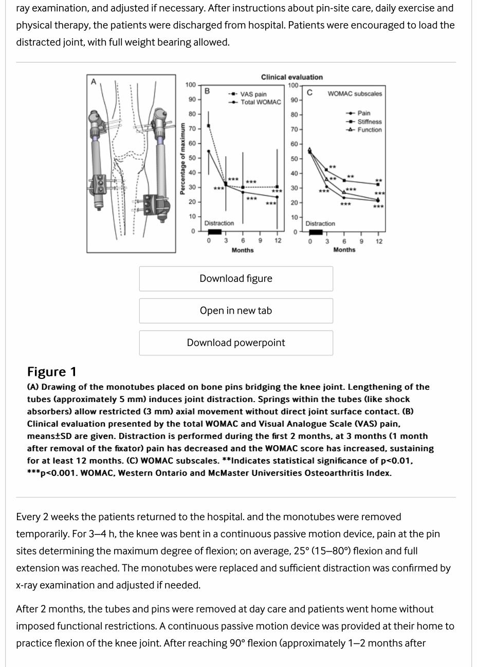
Distraction methodTwo monotubes with internal coil springs (Stryker, Monotube Triax) were placed parallel on the
medial and lateral side, bridging the knee joint (figure 1A). Each monotube was fixed to two bone
pins (Stryker, 6 mm self-drilling half pins) on each end and they were lengthened 2 mm, all under
anaesthesia. Pinholes were placed as far as possible from the joint line in order not to compromise
the area needed for possible future prosthesis surgery. Over the following 3 days the joint was
distracted twice a day by 0.5 mm, bringing the total distraction to 5 mm, which was confirmed by x-
ray examination, and adjusted if necessary. After instructions about pin-site care, daily exercise and
physical therapy, the patients were discharged from hospital. Patients were encouraged to load the
distracted joint, with full weight bearing allowed.
Download figure
Open in new tab
Download powerpoint
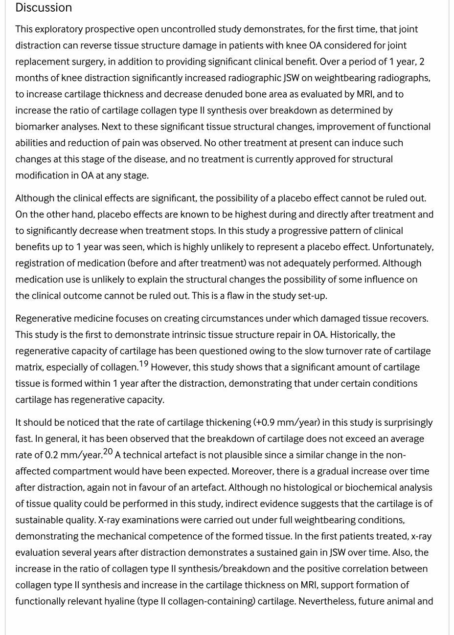
Figure 1Figure 1(A) Drawing of the monotubes placed on bone pins bridging the knee joint. Lengthening of the(A) Drawing of the monotubes placed on bone pins bridging the knee joint. Lengthening of the
tubes (approximately 5 mm) induces joint distraction. Springs within the tubes ( l ike shocktubes (approximately 5 mm) induces joint distraction. Springs within the tubes ( l ike shock
absorbers) al low restricted (3 mm) axial movement without direct joint surface contact. (B)absorbers) al low restricted (3 mm) axial movement without direct joint surface contact. (B)
Clinical evaluation presented by the total WOMAC and Visual Analogue Scale (VAS) pain,Clinical evaluation presented by the total WOMAC and Visual Analogue Scale (VAS) pain,
means±SD are given. Distraction is performed during the first 2 months, at 3 months (1 monthmeans±SD are given. Distraction is performed during the first 2 months, at 3 months (1 month
after removal of the fixator) pain has decreased and the WOMAC score has increased, sustainingafter removal of the fixator) pain has decreased and the WOMAC score has increased, sustaining
for at least 12 months. (C) WOMAC subscales. **Indicates statistical significance of p<0.01,for at least 12 months. (C) WOMAC subscales. **Indicates statistical significance of p<0.01,
***p<0.001. WOMAC, Western Ontario and McMaster Universities Osteoarthritis Index.***p<0.001. WOMAC, Western Ontario and McMaster Universities Osteoarthritis Index.
Every 2 weeks the patients returned to the hospital. and the monotubes were removed
temporarily. For 3–4 h, the knee was bent in a continuous passive motion device, pain at the pin
sites determining the maximum degree of flexion; on average, 25° (15–80°) flexion and full
extension was reached. The monotubes were replaced and sufficient distraction was confirmed by
x-ray examination and adjusted if needed.
After 2 months, the tubes and pins were removed at day care and patients went home without
imposed functional restrictions. A continuous passive motion device was provided at their home to
practice flexion of the knee joint. After reaching 90° flexion (approximately 1–2 months after
removal of the frame), the patients were advised to gain muscle strength by, for example, cycling.
Structural outcomePatients visited the outpatient clinic twice before treatment (baseline), every 2 weeks during
treatment, and 3, 6 and 12 months after the start of the treatment.
Radiographic analysisAt all visits, weightbearing, semiflexed, posterior–anterior radiographic views were acquired for
evaluation by knee images digital analysis (KIDA) software.13 No physical limitations in obtaining
adequate semiflexed views were observed. KIDA analyses provided minimal and mean joint space
width (JSW) in both compartments. Mean subchondral bone density was determined by measuring
density in a total of 16 regions adjoined to the bone cartilage interface in both compartments in
tibia and femur, normalising the grey scale to that of an aluminium step-wedge reference. Analyses
were performed blinded to the order of acquisition and characteristics of the patients.
Quantitative MRI analysisAt baseline and at 12 months, MRI acquisition (1.5T Philips Achieva) was performed using
sequences validated for quantitative measurement of cartilage morphology.14 15 Coronal images
were used to segment the femorotibial cartilage plates and bone surface, the operator and quality
control reader being blinded to the order of sequence (baseline vs follow-up). Cartilage parameters
were computed using custom software (Chondrometrics, Ainring, Germany). The primary structural
outcomes16 were thickness of cartilage over total area of bone (ThCtAB) and the percentage area
of denuded bone (dABp). Secondary structural outcome parameter was thickness of cartilage over
area of bone covered with cartilage (ThCcAB).
Biomarker analysisSerum and urine samples were collected and stored at −80°C. Cartilage collagen type II synthesis
and breakdown were determined by serum N-propeptide of type IIA procollagen (PIIANP; Linco,
EZPIIANP-53K) and urinary C-telopeptide of type II collagen (CTXII), Cartilaps; corrected for urine
creatinine), respectively. Samples were analysed in duplicate, and longitudinal samples of one
patient were assayed in one plate, to eliminate interkit variability.
Clinical outcomeThe primary clinical outcome parameter was the Western Ontario and McMaster Universities
Osteoarthritis Index (WOMAC),17 normalised to a 100% scale, 100% being the worst condition. The
secondary clinical outcome parameters were the VAS for pain (0–100 mm) and physical
examination of the joint (pain on palpation, crepitus, pain with flexion and joint effusion).
Statistical analysisParametric statistics (two-sided paired t test) were used for all parameters to compare whether the
follow-up values significantly differed from the baseline values. Spearman correlation coefficients
were used to relate longitudinal changes at 1 year between parameters. Means±SDs are given and
p<0.05 was considered a statistically significant difference.
Results
Twenty-three patients were considered for treatment; one was excluded because of bilateral OA,
one was excluded because of remaining metal in the knee after anterior cruciate ligament
reconstruction, and one withdrew from treatment after inclusion. Of the 20 patients included, aged
48±7 years, 11 were men. Eleven left knees and nine right knees were treated. Eighteen patients
had predominantly OA in the medial compartment while two patients had OA in the lateral
compartment. Three, four, 11 and two patients had a baseline Kellgren and Lawrence (K&L)
grade18 of 1, 2, 3 and 4, respectively. The average body mass index was 30 (range 25–36). Of the
20 patients 16 had had previous knee surgery. In one case anterior cruciate ligament
reconstruction was carried out, in four patients a tibial osteotomy was performed and in 15
patients an arthroscopy was performed, 12 of those had a partial meniscectomy or meniscopexy.
All surgery took place more than 1 year before distraction and without satisfactory results.
ComplicationsTwo patients had lung emboli despite appropriate anticoagulative prevention (nadroparin).
Patients were admitted to hospital for a week and given anticoagulative treatment (nadroparin),
after which they were discharged in good condition continuing treatment (acenocoumarol) for 6
months. Of the 20 patients, 17 had single or multiple pin tract infections. All were successfully
treated with antibiotics (flucloxacillin) for an average of 4 weeks. One patient had to be admitted to
the hospital for 1 week to receive antibiotics intravenously. None of the patients had any signs of
osteomyelitis.
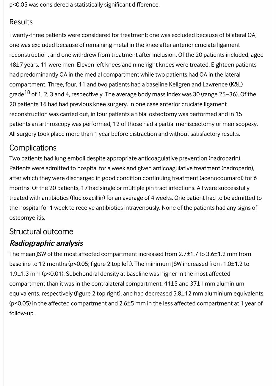
Structural outcomeRadiographic analysisRadiographic analysisThe mean JSW of the most affected compartment increased from 2.7±1.7 to 3.6±1.2 mm from
baseline to 12 months (p<0.05; figure 2 top left). The minimum JSW increased from 1.0±1.2 to
1.9±1.3 mm (p<0.01). Subchondral density at baseline was higher in the most affected
compartment than it was in the contralateral compartment: 41±5 and 37±1 mm aluminium
equivalents, respectively (figure 2 top right), and had decreased 5.8±12 mm aluminium equivalents
(p<0.05) in the affected compartment and 2.6±5 mm in the less affected compartment at 1 year of
follow-up.
Download figure
Open in new tab
Download powerpoint
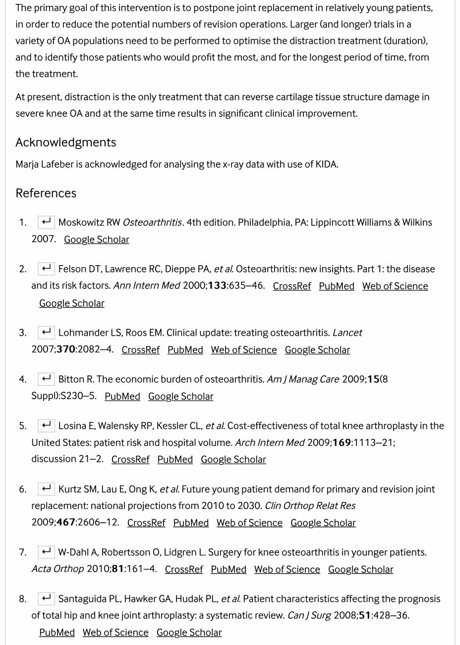
Figure 2Figure 2Joint space width (JSW) (KIDA measurement; mean±SD). Minimum JSW (min) continuouslyJoint space width (JSW) (KIDA measurement; mean±SD). Minimum JSW (min) continuously
increased after distraction. The mean JSW of the affected (OA) compartment also increased overincreased after distraction. The mean JSW of the affected (OA) compartment also increased over
time. The mean JSW of the less affected compartment (NA), did not change over time.time. The mean JSW of the less affected compartment (NA), did not change over time.
Subchondral bone density (KIDA, as mm aluminium (Al) equivalents; using a reference). TheSubchondral bone density (KIDA, as mm aluminium (Al) equivalents; using a reference). The
affected (OA) compartment showed a decrease in bone density, the less affected compartmentaffected (OA) compartment showed a decrease in bone density, the less affected compartment
(NA) did not. *p<0.05, **p<0.01. Representative radiographs before and 3 years after distraction;(NA) did not. *p<0.05, **p<0.01. Representative radiographs before and 3 years after distraction;
clear increase in JSW in affected (OA) compartment. KIDA, Knee Images Digital Analysis; OA,clear increase in JSW in affected (OA) compartment. KIDA, Knee Images Digital Analysis; OA,
osteoarthritis.osteoarthritis.
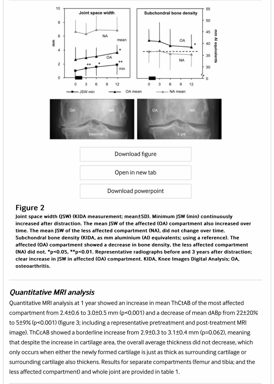
Quantitative MRI analysisQuantitative MRI analysisQuantitative MRI analysis at 1 year showed an increase in mean ThCtAB of the most affected
compartment from 2.4±0.6 to 3.0±0.5 mm (p<0.001) and a decrease of mean dABp from 22±20%
to 5±9% (p<0.001) (figure 3; including a representative pretreatment and post-treatment MRI
image). ThCcAB showed a borderline increase from 2.9±0.3 to 3.1±0.4 mm (p=0.062), meaning
that despite the increase in cartilage area, the overall average thickness did not decrease, which
only occurs when either the newly formed cartilage is just as thick as surrounding cartilage or
surrounding cartilage also thickens. Results for separate compartments (femur and tibia; and the
less affected compartment) and whole joint are provided in table 1.
VIEW INLINE VIEW POPUP
Download figure
Open in new tab
Download powerpoint
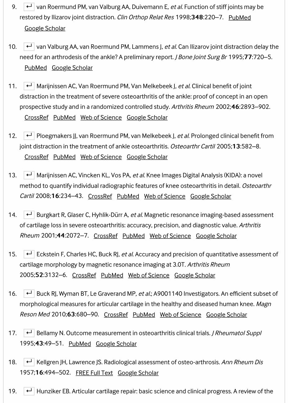
Figure 3Figure 3Representative image of single sl ides before and 1 year after treatment, showing an increase inRepresentative image of single sl ides before and 1 year after treatment, showing an increase in
carti lage tissue in the affected compartment. Quantitative MRI analysis of carti lage of thecarti lage tissue in the affected compartment. Quantitative MRI analysis of carti lage of the
affected compartment of the individual 20 patients (grey l ines) at baseline (BL) and after 1 year ofaffected compartment of the individual 20 patients (grey l ines) at baseline (BL) and after 1 year of
follow-up (1 YR). Black l ines indicate mean values. ThCtAB, thickness of carti lage over total areafollow-up (1 YR). Black l ines indicate mean values. ThCtAB, thickness of carti lage over total area
of bone; dABp, area of denuded bone (both **p<0.01); ThCcAB, thickness of carti lage over area ofof bone; dABp, area of denuded bone (both **p<0.01); ThCcAB, thickness of carti lage over area of
bone covered with carti lage (p<0.062).bone covered with carti lage (p<0.062).
Table 1Table 1MRI outcome for femoral and tibial side of the most affected compartment (OA, osteoarthritis)MRI outcome for femoral and tibial side of the most affected compartment (OA, osteoarthritis)
and the less affected compartment (NA, not affected) as well as for the whole joint (bothand the less affected compartment (NA, not affected) as well as for the whole joint (both
compartments) of 20 patients treated for 2 months with joint distraction, before distraction (BL,compartments) of 20 patients treated for 2 months with joint distraction, before distraction (BL,
baseline) and after 1 year, including two-sided p valuesbaseline) and after 1 year, including two-sided p values
BiomarkersBiomarkersBiomarkers showed an initial high increase during distraction, normalising 1 month after
distraction (data not shown). Changes between 6 and 12 months' follow-up showed a trend
towards a decrease of collagen type II breakdown marker CTXII (−11±39%; p=0.078) and an
increase of collagen type II synthesis marker PIIANP (+103±298%; p=0.060). The mean change in
the ratio of PIIANP/CTXII between 6 and 12 months suggested a net increase in collagen synthesis
(p=0.056).
Clinical outcomeThe total WOMAC index questionnaire decreased from 55±16 points at baseline to 23±21 points at
1 year (p<0.001; figure 1B). Of the 20 patients, 18 showed an improvement of >10% and 16 of
>25%. The individual components of the WOMAC index (pain, stiffness and function) all improved
significantly (p<0.001; figure 1C). VAS pain decreased from 73±9 mm at baseline to 31±26 mm
(p<0.001) at 1 year (figure 1B). Physical examination of the knee showed an improvement from
46±22% to 75±24% (p<0.001) of the maximum score (data not shown).
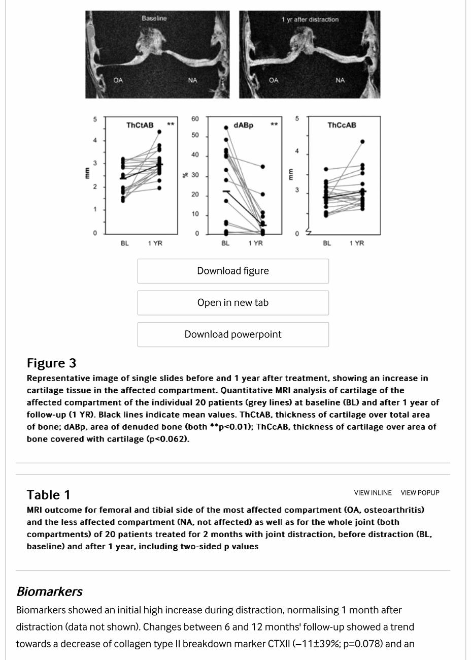
Correlation between structural parametersAll MRI parameters correlated positively and significantly with the increase in mean radiographic
JSW (all r>0.51 and p<0.01). The increase in collagen type II synthesis marker PIIANP between 6 and
12 months correlated with the change in ThCtAB and dABp (figure 4). CTXII change did not show
such correlations. There were no clear correlations between structural and clinical parameters.
Download figure
Open in new tab
Download powerpoint
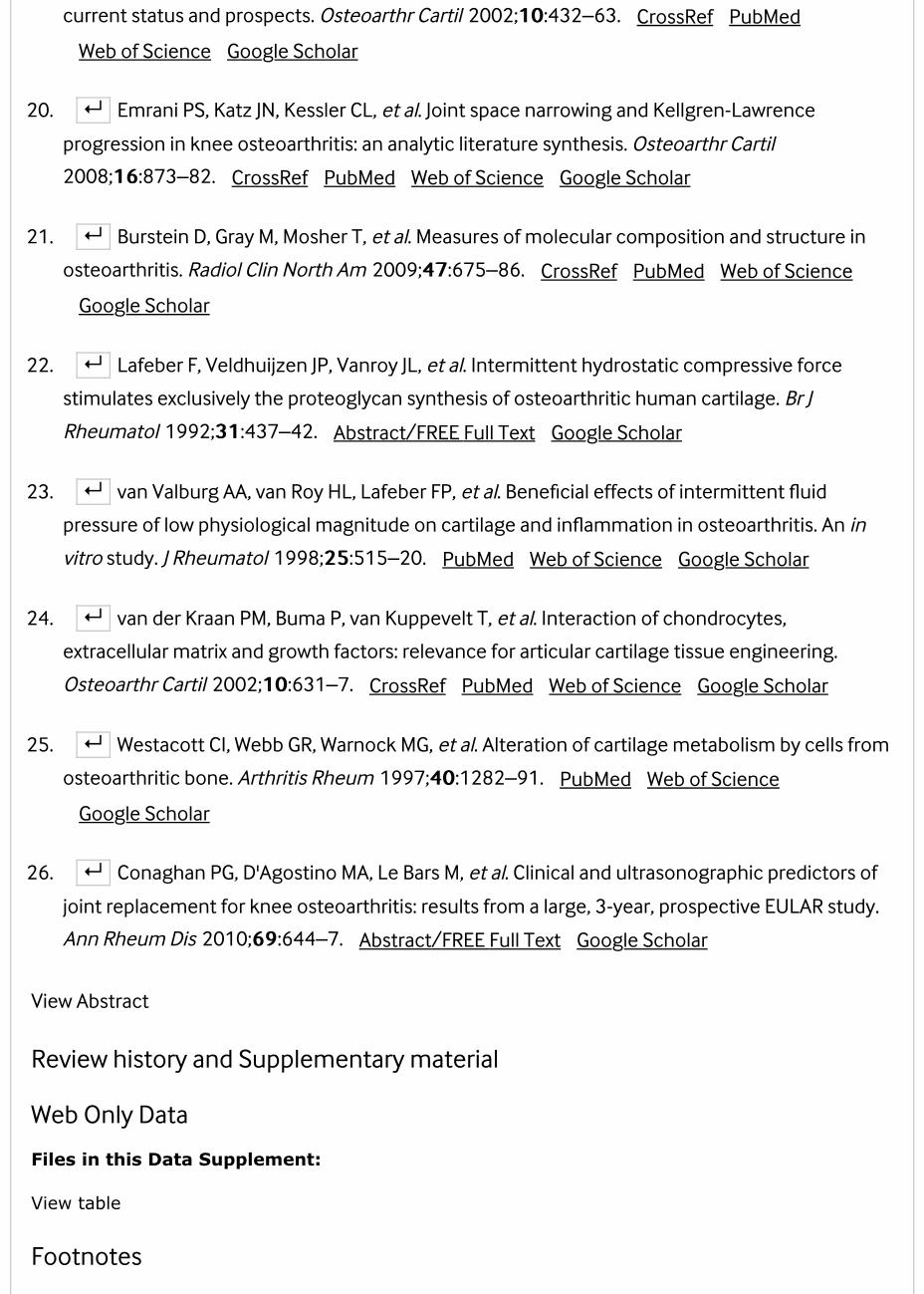
Figure 4Figure 4Correlations between the changes (compared with baseline) in carti lage thickness (ThCtAB; mm)Correlations between the changes (compared with baseline) in carti lage thickness (ThCtAB; mm)
and area of denuded bone (dABp; %) on MRI and the change in serum N-propeptide of type IIAand area of denuded bone (dABp; %) on MRI and the change in serum N-propeptide of type IIA
procollagen (PIIANP; ng/ml) between 6 months and 1 year of follow-up. **p<0.01. ThCtAB,procollagen (PIIANP; ng/ml) between 6 months and 1 year of follow-up. **p<0.01. ThCtAB,
thickness of carti lage over total area of bone.thickness of carti lage over total area of bone.
Individual results are shown in the online supplementary data.
Discussion
This exploratory prospective open uncontrolled study demonstrates, for the first time, that joint
distraction can reverse tissue structure damage in patients with knee OA considered for joint
replacement surgery, in addition to providing significant clinical benefit. Over a period of 1 year, 2
months of knee distraction significantly increased radiographic JSW on weightbearing radiographs,
to increase cartilage thickness and decrease denuded bone area as evaluated by MRI, and to
increase the ratio of cartilage collagen type II synthesis over breakdown as determined by
biomarker analyses. Next to these significant tissue structural changes, improvement of functional
abilities and reduction of pain was observed. No other treatment at present can induce such
changes at this stage of the disease, and no treatment is currently approved for structural
modification in OA at any stage.
Although the clinical effects are significant, the possibility of a placebo effect cannot be ruled out.
On the other hand, placebo effects are known to be highest during and directly after treatment and
to significantly decrease when treatment stops. In this study a progressive pattern of clinical
benefits up to 1 year was seen, which is highly unlikely to represent a placebo effect. Unfortunately,
registration of medication (before and after treatment) was not adequately performed. Although
medication use is unlikely to explain the structural changes the possibility of some influence on
the clinical outcome cannot be ruled out. This is a flaw in the study set-up.
Regenerative medicine focuses on creating circumstances under which damaged tissue recovers.
This study is the first to demonstrate intrinsic tissue structure repair in OA. Historically, the
regenerative capacity of cartilage has been questioned owing to the slow turnover rate of cartilage
matrix, especially of collagen.19 However, this study shows that a significant amount of cartilage
tissue is formed within 1 year after the distraction, demonstrating that under certain conditions
cartilage has regenerative capacity.
It should be noticed that the rate of cartilage thickening (+0.9 mm/year) in this study is surprisingly
fast. In general, it has been observed that the breakdown of cartilage does not exceed an average
rate of 0.2 mm/year.20 A technical artefact is not plausible since a similar change in the non-
affected compartment would have been expected. Moreover, there is a gradual increase over time
after distraction, again not in favour of an artefact. Although no histological or biochemical analysis
of tissue quality could be performed in this study, indirect evidence suggests that the cartilage is of
sustainable quality. X-ray examinations were carried out under full weightbearing conditions,
demonstrating the mechanical competence of the formed tissue. In the first patients treated, x-ray
evaluation several years after distraction demonstrates a sustained gain in JSW over time. Also, the
increase in the ratio of collagen type II synthesis/breakdown and the positive correlation between
collagen type II synthesis and increase in the cartilage thickness on MRI, support formation of
functionally relevant hyaline (type II collagen-containing) cartilage. Nevertheless, future animal and

clinical studies (including qualitative MRI parameters based on, for example, delayed gadolinium-
enhanced MRI of cartilage)21 will have to explore the compositional properties of the newly formed
tissue.
In addition, the question arises, what might the underlying mechanism of the observed structure
repair be? It is hypothesised that the temporary distraction prevents mechanical stress on the
cartilage, prevents further wear and tear and allows tissue repair to begin. Joint fluid pressure
changes are maintained during the distraction period, because the springs in the distraction tubes
allow limited axial oscillation during loading and unloading of the distracted joint. These fluid
pressure oscillations may provide nutrition and may trigger the cartilage cells to initiate tissue
repair (re-differentiation of the diseased chondrocytes).22 23 During distraction, the load on the
bone (the biomechanical trigger for normal bone formation) is transferred through the frame
instead of the subchondral bone, leading to subchondral bone resorption, which subsequently
normalises after distraction. This significant bone turnover may trigger the release of growth
factors as bone matrix provides a store of resident growth factors such as transforming growth
factors β, bone morphogenetic proteins and insulin-like growth factors that stimulate cartilage
tissue repair.24 25
Treatment in this study was accompanied by two major safety concerns. In two out of 20 patients,
a pulmonary embolism developed. In retrospect, in both patients there was a family history of
venous thrombosis. In future studies additional attention should be paid to this severe
complication in anamnesis. A higher dose of anticoagulative agents could be considered. In 17 out
of 20 patients pin tract infections developed, which could be treated adequately with antibiotics.
Pin tract infections are a general complication of the application of external fixators and did not
result in deep infections. Although these patients might have prosthetic surgery in the future, the
risk of infection was minimised by placing bone pins outside the expected future operating area.
Nonetheless, more attention should be paid to reducing the number of these complications in
future studies.
It is currently unclear which group of patients would benefit best from this treatment. In this study,
only young patients (<60 years) with severe OA considered for joint replacement surgery were
treated. Selection by referral from peripheral hospitals might have resulted in an inclusion bias.
Thus, results from this population cannot be generalised to all patients considered for prosthetic
surgery. This patient group showed a diversity of OA stages with K&L grade varying between 1 and
4. In general practice patients with a low K&L grade but significant joint pain are also considered for
knee replacement surgery; this has been the subject of a recent discussion.26 Overall, patients
showed a positive change in structural parameters but effects were variable. Unfortunately, group
size does not allow valid analyses to identify predictive factors for clinical or structural benefit.
Potential relations found may depend on coincidence. Prediction of efficacy needs to be examined
in future larger studies.
The primary goal of this intervention is to postpone joint replacement in relatively young patients,
in order to reduce the potential numbers of revision operations. Larger (and longer) trials in a
variety of OA populations need to be performed to optimise the distraction treatment (duration),
and to identify those patients who would profit the most, and for the longest period of time, from
the treatment.
At present, distraction is the only treatment that can reverse cartilage tissue structure damage in
severe knee OA and at the same time results in significant clinical improvement.
Acknowledgments
Marja Lafeber is acknowledged for analysing the x-ray data with use of KIDA.
References
1. ↵ Moskowitz RW Osteoarthritis . 4th edition. Philadelphia, PA: Lippincott Williams & Wilkins
2007. Google Scholar
2. ↵ Felson DT, Lawrence RC, Dieppe PA, et al. Osteoarthritis: new insights. Part 1: the disease
and its risk factors. Ann Intern Med 2000;133133:635–46. CrossRef PubMed Web of Science
Google Scholar
3. ↵ Lohmander LS, Roos EM. Clinical update: treating osteoarthritis. Lancet
2007;370370:2082–4. CrossRef PubMed Web of Science Google Scholar
4. ↵ Bitton R. The economic burden of osteoarthritis. Am J Manag Care 2009;1515(8
Suppl):S230–5. PubMed Google Scholar
5. ↵ Losina E, Walensky RP, Kessler CL, et al. Cost-effectiveness of total knee arthroplasty in the
United States: patient risk and hospital volume. Arch Intern Med 2009;169169:1113–21;
discussion 21–2. CrossRef PubMed Google Scholar
6. ↵ Kurtz SM, Lau E, Ong K, et al. Future young patient demand for primary and revision joint
replacement: national projections from 2010 to 2030. Clin Orthop Relat Res
2009;467467:2606–12. CrossRef PubMed Web of Science Google Scholar
7. ↵ W-Dahl A, Robertsson O, Lidgren L. Surgery for knee osteoarthritis in younger patients.
Acta Orthop 2010;8181:161–4. CrossRef PubMed Web of Science Google Scholar
8. ↵ Santaguida PL, Hawker GA, Hudak PL, et al. Patient characteristics affecting the prognosis
of total hip and knee joint arthroplasty: a systematic review. Can J Surg 2008;5151:428–36.
PubMed Web of Science Google Scholar
9. ↵ van Roermund PM, van Valburg AA, Duivemann E, et al. Function of stiff joints may be
restored by Ilizarov joint distraction. Clin Orthop Relat Res 1998;348348:220–7. PubMed
Google Scholar
10. ↵ van Valburg AA, van Roermund PM, Lammens J, et al. Can Ilizarov joint distraction delay the
need for an arthrodesis of the ankle? A preliminary report. J Bone Joint Surg Br 1995;7777:720–5.
PubMed Google Scholar
11. ↵ Marijnissen AC, Van Roermund PM, Van Melkebeek J, et al. Clinical benefit of joint
distraction in the treatment of severe osteoarthritis of the ankle: proof of concept in an open
prospective study and in a randomized controlled study. Arthritis Rheum 2002;4646:2893–902.
CrossRef PubMed Web of Science Google Scholar
12. ↵ Ploegmakers JJ, van Roermund PM, van Melkebeek J, et al. Prolonged clinical benefit from
joint distraction in the treatment of ankle osteoarthritis. Osteoarthr Cartil 2005;1313:582–8.
CrossRef PubMed Web of Science Google Scholar
13. ↵ Marijnissen AC, Vincken KL, Vos PA, et al. Knee Images Digital Analysis (KIDA): a novel
method to quantify individual radiographic features of knee osteoarthritis in detail. Osteoarthr
Cartil 2008;1616:234–43. CrossRef PubMed Web of Science Google Scholar
14. ↵ Burgkart R, Glaser C, Hyhlik-Dürr A, et al. Magnetic resonance imaging-based assessment
of cartilage loss in severe osteoarthritis: accuracy, precision, and diagnostic value. Arthritis
Rheum 2001;4444:2072–7. CrossRef PubMed Web of Science Google Scholar
15. ↵ Eckstein F, Charles HC, Buck RJ, et al. Accuracy and precision of quantitative assessment of
cartilage morphology by magnetic resonance imaging at 3.0T. Arthritis Rheum
2005;5252:3132–6. CrossRef PubMed Web of Science Google Scholar
16. ↵ Buck RJ, Wyman BT, Le Graverand MP, et al.; A9001140 Investigators. An efficient subset of
morphological measures for articular cartilage in the healthy and diseased human knee. Magn
Reson Med 2010;6363:680–90. CrossRef PubMed Web of Science Google Scholar
17. ↵ Bellamy N. Outcome measurement in osteoarthritis clinical trials. J Rheumatol Suppl
1995;4343:49–51. PubMed Google Scholar
18. ↵ Kellgren JH, Lawrence JS. Radiological assessment of osteo-arthrosis. Ann Rheum Dis
1957;1616:494–502. FREE Full Text Google Scholar
19. ↵ Hunziker EB. Articular cartilage repair: basic science and clinical progress. A review of the
current status and prospects. Osteoarthr Cartil 2002;1010:432–63. CrossRef PubMed
Web of Science Google Scholar
20. ↵ Emrani PS, Katz JN, Kessler CL, et al. Joint space narrowing and Kellgren-Lawrence
progression in knee osteoarthritis: an analytic literature synthesis. Osteoarthr Cartil
2008;1616:873–82. CrossRef PubMed Web of Science Google Scholar
21. ↵ Burstein D, Gray M, Mosher T, et al. Measures of molecular composition and structure in
osteoarthritis. Radiol Clin North Am 2009;4747:675–86. CrossRef PubMed Web of Science
Google Scholar
22. ↵ Lafeber F, Veldhuijzen JP, Vanroy JL, et al. Intermittent hydrostatic compressive force
stimulates exclusively the proteoglycan synthesis of osteoarthritic human cartilage. Br J
Rheumatol 1992;3131:437–42. Abstract/FREE Full Text Google Scholar
23. ↵ van Valburg AA, van Roy HL, Lafeber FP, et al. Beneficial effects of intermittent fluid
pressure of low physiological magnitude on cartilage and inflammation in osteoarthritis. An in
vitro study. J Rheumatol 1998;2525:515–20. PubMed Web of Science Google Scholar
24. ↵ van der Kraan PM, Buma P, van Kuppevelt T, et al. Interaction of chondrocytes,
extracellular matrix and growth factors: relevance for articular cartilage tissue engineering.
Osteoarthr Cartil 2002;1010:631–7. CrossRef PubMed Web of Science Google Scholar
25. ↵ Westacott CI, Webb GR, Warnock MG, et al. Alteration of cartilage metabolism by cells from
osteoarthritic bone. Arthritis Rheum 1997;4040:1282–91. PubMed Web of Science
Google Scholar
26. ↵ Conaghan PG, D'Agostino MA, Le Bars M, et al. Clinical and ultrasonographic predictors of
joint replacement for knee osteoarthritis: results from a large, 3-year, prospective EULAR study.
Ann Rheum Dis 2010;6969:644–7. Abstract/FREE Full Text Google Scholar
View Abstract
Review history and Supplementary material
Web Only Data
Files in this Data Supplement:
View table
Footnotes
FundingFunding:: This study was financially supported by the Dutch Arthritis Foundation.
Competing interestsCompeting interests:: None.
Ethics approvalEthics approval:: This study was conducted with the approval of the University Medical Center
Utrecht.
Provenance and peer reviewProvenance and peer review:: Not commissioned; externally peer reviewed.
Request Permissions
If you wish to reuse any or all of this article please use the link below which will take you to the
Copyright Clearance Center’s RightsLink service. You will be able to get a quick price and instant
permission to reuse the content in many different ways.
Request permissions
Copyright information: Copyright information: Published by the BMJ Publishing Group Limited. For permission to use
(where not already granted under a licence) please go to
http://group.bmj.com.ez.srv.pmu.ac.at/group/rights-licensing/permissions
This paper is freely available online under the BMJ Journals unlocked scheme, see
http://ard.bmj.com.ez.srv.pmu.ac.at/info/unlocked.dtl
Recruiter: Ashford and St Peters Hospital NHS Trust
Apply for this job
Recruiter: Taunton and Somerset NHS Foundation TrustApply for this job
Recruiter: Pennine Care NHS Foundation Trust
Apply for this job
Consultant Rheumatology and General Medicine - 10 PA Job PlanAshford, Chertsey, Surrey YC72
You will contribute to all aspects of Rheumatology work including involvement in departmental meetings, teachingand supervising registrars ....
Consultant RheumatologistTaunton, Somerset From £77,913 - £105,042 per annum
Rheumatology Consultant An exciting opportunity has arisen to join the Rheumatology Department within Tauntonand Somerset NHS Foundation Trust, a...
Consultant Community Paediatrician with Special Interest in Audiology (CPS)Rochdale, Greater Manchester £76,761 to £103,490 pa
Exciting, newly developed post for a Consultant Paediatricians required predominantly assess & treat children withneuro disability
Clinical Fellow, Equivalent to ST6+, in Paediatric NeurosurgeryLondon (Central), London (Greater) £41,300 – £49,086

Powered by
Other content recommended for you
SAT0649 Changes in cartilage quality (DGEMRIC) following knee joint distraction or high tibialosteotomy: a two-year follow-upN Besselink et al., Ann Rheum Dis
SAT0572 Initial structural response predicts long-term survival of knee joint distraction as atreatment for knee osteoarthritisM. Jansen et al., Ann Rheum Dis
OP0060 Knee joint distraction compared with high tibial osteotomy and total knee arthroplasty:two-year clinical, structural, and biomarker outcomesK. Wiegant et al., Ann Rheum Dis
SAT0536 Axial alignment of the knee – importance in cartilage repair? high tibial osteotomy vs.distractionN Besselink et al., Ann Rheum Dis
THU0440 Clinical Benefit and Cartilaginous Tissue Repair After Knee Joint Distration: 5 YearsFollow-UpF.P. Lafeber et al., Ann Rheum Dis
CONTENT
Latest content
Current issue
Lay summaries
Archive
Browse by topic

Most read articles
Responses
� " � #
JOURNAL
About
Editorial board
Thank you to our reviewers
Sign up for email alerts
Subscribe
EULAR textbooks
AUTHORS
Instructions for authors
Submit an article
Editorial policies
Open Access at BMJ
BMJ Author Hub
HELP
Contact us
Reprints
Permissions
Advertising
Feedback form
Website Terms & Conditions
Privacy & Cookies
Contact BMJ

Online: ISSN 1468-2060 Print: ISSN 0003-4967
Copyright © 2018 BMJ Publishing Group Ltd & European League Against Rheumatism. All rights reserved.
京ICP备15042040号-3