World Congress on Osteoporosis, Osteoarthritis and ...
282
World Congress on Osteoporosis, Osteoarthritis and Musculoskeletal Diseases (WCO-IOF-ESCEO 2014): Poster Abstracts # International Osteoporosis Foundation and National Osteoporosis Foundation 2014 P101 COMPARISON OF THE INTERNATIONAL REFERENCE VALUES OF BONE SPEED OF SOUND IN PEDIATRIC POPULATION: META-ANALYSIS R. Rivas-Ruiz 1 , G. Huitrón 2,4 , J. Salmeron-Castro 3,7 , J. O. Talavera 1 , J. Tamayo 5 , P. Clark 6 , D. O. Castelán-Martínez 6 , L. Méndez-Sánchez 6 1 Centro de Adiestramiento en Investigación Clínica, IMSS, México City, Mexico, 2 Centro de Investigación en Ciencias Médicas, UAEM, Toluca, Mexico, 3 Unidad de Investigación Epidemiológica y en Servicios de Salud, Instituto Mexicano del Seguro Social. Cuernavaca, Morelos, Mé, Cuernavaca, Mexico, 4 Centro de Investigación en Ciencias Médicas, Universidad Autónoma del Estado de México, Toluca, Mexico, 5 Comité Mexicano para la Prevención de la Osteoporosis, Mexico City, Mexico, 6 Clinical Epidemiology Unit, Hospital Infantil de México, Federico Gómez, Mexico City, Mexico, 7 Unidad de Investigación Epidemiológica, IMSS, Cuernavaca, Mexico Objective: To compare the international reference values of the speed of sound (SoS) assessed by multisite quan- titative ultrasound (QUS) in tibia and radius in pediatric population comparing these values by age and by countries. Material and Methods: We conducted a systematic review with meta-analysis of the studies published from 1965 to November 2013 on the reference values of QUS (Sunlight Omnisense) in newborn, infants and children. A sensitive search of the evidence was performed in PubMed, Embase, LILACS, ARTEMISA, Cochrane con- trolled trials register and Bandolier. We calculated de mean differences of SoS in tibia and radio between countries by gender and age. The results are presented in tables and forest plots to compare raw reference data between countries. Results: Ten studies with a total of 6,250 patients were included in the meta-analysis. In the newborn population we found two studies Portugal and Israel that include subjects with a gestational age between 27 and 42 weeks, with a mean difference (Portugal - Israel [CI 95 %] m/s) of 23.62 [CI 95 % 6.29, 40.95] m/s. In pediatric population there were eight studies from Canada, México, Israel, Greece, Portugal and Turkey. There were no significant differences between SoS Israel reference values neither in tibia or radius in comparison of vs. Turkey, vs. Greece or vs. Canada (p >0.05). We found significant differences of the mean SoS (m/s) measurements in tibia in the comparison of Mexican references values with other countries (Mexico- Other [CI 95 %] m/s) as follows: Israel −105.29 [CI 95 % −140.05, −70.54], Portugal −115.14 [CI 95 % −164.86, −65.42], Greece −239.14 [CI 95 % −267.67, −210.62], Turkey −115.14 [CI 95 % −164.86, −65.42], and Canada −113.51 [CI 95 % −140.25, −86.77] (p <0.001). Conclusion: This study confirms that there are differences between references values of QUS between countries. These differences are between Mexican versus European countries, but there are no differences between European countries. P102 LOWER SCLEROSTIN IN ACQUIRED IMMUNE DEFICIENCY SYNDROME (LSAIDS) A. Y. Almansouri 1 , M. S. M. Ardawi 1,2 H. H. Mosli 3 , M. E. Abdulfatah 1 , O. H. Baaqil 1 , A. A. Bakheet 1 , S. A. Turki 1 , A. M. Althubaiti 1 , M. M. Almaghrabi 1 , B. M. Madani 1 , M. M. Kotb 4 , T. A. Madani 3 , G. A. Jamjoom 5 1 Center of Excellence for Osteoporosis Research, King Abdulaziz University, Jeddah, Saudi Arabia, 2 Department of Clinical Biochemistry, Faculty of Medicine, King Abdulaziz University, Jeddah, Saudi Arabia, 3 Department of Medicine, DOI 10.1007/s00198-014-2642-4 Osteoporos Int (2014) 25 (Suppl 2):S159–S440
-
Upload
khangminh22 -
Category
Documents
-
view
0 -
download
0
Transcript of World Congress on Osteoporosis, Osteoarthritis and ...
World Congress on Osteoporosis, Osteoarthritisand Musculoskeletal Diseases (WCO-IOF-ESCEO 2014):Poster Abstracts
# International Osteoporosis Foundation and National Osteoporosis Foundation 2014
P101COMPARISON OF THE INTERNATIONALREFERENCE VALUES OF BONE SPEED OF SOUNDIN PEDIATRIC POPULATION: META-ANALYSISR. Rivas-Ruiz1, G. Huitrón2,4, J. Salmeron-Castro3,7, J. O.Talavera1, J. Tamayo5, P. Clark6, D. O. Castelán-Martínez6,L. Méndez-Sánchez61Centro de Adiestramiento en Investigación Clínica, IMSS,México City, Mexico, 2Centro de Investigación en CienciasMédicas, UAEM, Toluca, Mexico, 3Unidad de InvestigaciónEpidemiológica y en Servicios de Salud, Instituto Mexicanodel Seguro Social. Cuernavaca, Morelos, Mé, Cuernavaca,Mexico, 4Centro de Investigación en Ciencias Médicas,Universidad Autónoma del Estado de México, Toluca,Mexico, 5Comité Mexicano para la Prevención de laOsteoporosis, Mexico City, Mexico, 6Clinical EpidemiologyUnit, Hospital Infantil de México, Federico Gómez, MexicoCity, Mexico, 7Unidad de Investigación Epidemiológica,IMSS, Cuernavaca, Mexico
Objective: To compare the international reference valuesof the speed of sound (SoS) assessed by multisite quan-titative ultrasound (QUS) in tibia and radius in pediatricpopulation comparing these values by age and bycountries.Material and Methods: We conducted a systematic reviewwith meta-analysis of the studies published from 1965to November 2013 on the reference values of QUS(Sunlight Omnisense) in newborn, infants and children.A sensitive search of the evidence was performed inPubMed, Embase, LILACS, ARTEMISA, Cochrane con-trolled trials register and Bandolier. We calculated demean differences of SoS in tibia and radio betweencountries by gender and age. The results are presentedin tables and forest plots to compare raw reference databetween countries.
Results: Ten studies with a total of 6,250 patients wereincluded in the meta-analysis. In the newborn population wefound two studies Portugal and Israel that include subjectswith a gestational age between 27 and 42 weeks, with a meandifference (Portugal - Israel [CI 95 %] m/s) of 23.62 [CI 95 %6.29, 40.95] m/s. In pediatric population there were eightstudies from Canada, México, Israel, Greece, Portugal andTurkey. There were no significant differences between SoSIsrael reference values neither in tibia or radius in comparisonof vs. Turkey, vs. Greece or vs. Canada (p>0.05). We foundsignificant differences of the mean SoS (m/s) measurements intibia in the comparison of Mexican references values withother countries (Mexico- Other [CI 95 %] m/s) as follows:Israel −105.29 [CI 95 % −140.05, −70.54], Portugal −115.14[CI 95 % −164.86, −65.42], Greece −239.14 [CI 95 %−267.67, −210.62], Turkey −115.14 [CI 95 % −164.86,−65.42], and Canada −113.51 [CI 95 % −140.25, −86.77](p<0.001).Conclusion: This study confirms that there are differencesbetween references values of QUS between countries.These differences are between Mexican versus Europeancountries, but there are no differences between Europeancountries.
P102LOWER SCLEROSTIN IN ACQUIRED IMMUNEDEFICIENCY SYNDROME (LSAIDS)A. Y. Almansouri1, M. S. M. Ardawi1,2 H. H. Mosli3, M. E.Abdulfatah1, O. H. Baaqil1, A. A. Bakheet1, S. A. Turki1, A.M. Althubaiti1, M. M. Almaghrabi1, B. M. Madani1, M. M.Kotb4, T. A. Madani3, G. A. Jamjoom5
1Center of Excellence for Osteoporosis Research, KingAbdulaziz University, Jeddah, Saudi Arabia, 2Department ofClinical Biochemistry, Faculty of Medicine, King AbdulazizUniversity, Jeddah, Saudi Arabia, 3Department of Medicine,
DOI 10.1007/s00198-014-2642-4Osteoporos Int (2014) 25 (Suppl 2):S159–S440
Faculty of Medicine, King Abdulaziz University, Jeddah,Saudi Arabia, 4Department of Radiology, Faculty ofMedicine, King Abdulaziz University, Jeddah, SaudiArabia, 5Special Infectious Agents Unit, King Fahad MedicalResearch Center, King Abdulaziz University, Jeddah, SaudiArabia
Objective: Determine the association of sclerostin levels toBMD changes in patients with HIV infection.Material and Methods: Serum sclerostin levels, serumdickkopf 1, standardized BMD (sBMD) of lumbar spineand femoral neck, and bone turnover markers were mea-sured for 30 HIV-infected treatment naïve subjects andcompared to 65 healthy seronegative controls matched forage and sex.Results: The mean of measured sclerostin was significantlylower in the HIV group than the control group (37.52 vs.76.71 pmol/L; p<0.001). The HIV group also had asignificantly lower BMD in the lumbar spine and femoralneck. The mean levels of serum osteocalcin, a bone for-mation marker, was significantly lower in the HIV thanthe control group (15.53 vs. 24.42 ng/ml; p<0.001). TheHIV group had a mean CD4 count of 170.21 cells/mm3
(AIDS range).Conclusion: In patients with advanced HIV infection and lowBMD, we hypothesised that depleted sclerostin levels couldbe part of the severely immunocompromised status caused byHIV infection.References: Cain CJ, et al. J Bone Miner Res. 2012;27:1451.Acknowledgements: We would like to thank Dr. Yasir M.Mansouri (Department of Cardiology, King Abdulaziz Med-ical City for National Guard, Jeddah, Saudi Arabia) for hisgenuine and continuous support for the principle author. Weare grateful for the Center of Excellence for OsteoporosisResearch (CEOR), King Abdulaziz University for fundingour research. We would like to thank Mr. Abdullah A.Almazrooa and Mr. Mishari Al-Romaihy (Medical Students,Faculty of Medicine, King Abdulaziz University, Saudi Ara-bia) for their help in sample collections. Special thanks to MrsRose (Laboratory Technician at CEOR) and Ms. Taghreed S.Almansouri (Lecturer in Clinical Biochemistry, Department ofMedical Laboratory Technology, King Abdulaziz University,Jeddah, Saudi Arabia) for her help in running the laboratorysamples.
P103TREATMENT OF UNSTABLE OSTEOPOROTICFRACTURES OF PROXIMAL HUMERUS WITHLOCKING PLATESV. L. Rusimov11Arthroscopic Clinic, Military Medical Academy, Sofia,Bulgaria
Objective:Almost 20% of the proximal humeral fractures areunstable with absolute indications for surgical treatment.Good postoperative result requires surgical technique, whichmaintains local blood supply, with good reduction and stablefixation and ability for early rehabilitation. The persistingosteoporosis is challenging for treatment choice and expectedresults. Low profile locking proximal humerus plates (PHLPand S3) give a good opportunity for realizing of this condi-tions. The aim of the report is to represent our opinion andresults of using proximal humerus locking plates in cases withmoderate to severe osteoporosis.Material and Methods: For period of 5 y (2005–2010) wehave operated on 23 patients - 18 female and 5male in averageage 71 (62–83) y. Fractures distribution according to AO: 8 -A2.2; 5 - А3.1; 4 - В2.1; 3 - С2.1; 3 - С3.1. Surgicaltechnique: Open reduction and internal fixation via delto-pectoral approach. Soft tissue preservation. Stable fixationwith locking plates - 10 type S3 (DePuy) and 13 type LPHP(Synthes).Results: Follow-up was minimum 3 y. We made rating ac-cording to Constant Shoulder Assessment (CSA): 14 (60.9 %)excellent and good; 9 (39.1 %) satisfactory; 2 (8.7 %) poorresults. АVN in 2 (8.7 %) patient. We have no observedinfection and neurovascular complications.Conclusion: Specially designed proximal humerus lockingplates decrease complications connected with convention-al plates in osteoporotic patients, indicated for surgicaltreatment. Angular stability allows early rehabilitationeven in osteoporosis. Avascular necrosis of the humeralhead depends on fracture type and local soft tissuedamage.
P104THE EFFECT OF GRAFT ORIENTATION INOSTEOCHONDRAL AUTOTRANSPLANTATION OFEARLY STAGE OSTEOARTHRITISS. E. Park1, C. H. Kim1, J. D. Kim1, O. J. Kwon11Orthopaedic Surgery, Dongguk University, InternationalHospital, Seoul, Republic of Korea
Objective: To introduce the clinical results of arthroscopicosteochondral autotransplantation are greatly affected by sur-gical technique and graft orientation.Material and Methods: From Nov 2005 to March 2011, 74patients, 76 arthroscopic osteochondral autotransplantationswere practiced. The Lysholm knee score and NRS scale wasrecorded initially and 3 months, 6 months, 12 months after thesurgery. Follow up knee MR were done on 30 patients. Theradiologic parameter, α angle and β angle was analyzed in thegroup.Results: The mean Lysholm knee score improved from 64.43to 91.1. The Pearson coefficient of the clinical results of
Osteoporos Int (2014) 25 (Suppl 2):S159–S440S160

patients with α angle and β angle was −0.548, −0.792, re-spectively. P-value was 0.002 and 0.0001, respectively.Conclusion: The surgical technique and graft orientationin the arthroscopic osteochondral autotransplantation isimportant parameter affecting clinical results of patients.β angle was stronger parameter to predict the clinicalresults.
P105KNEE DEFORMITIES AMONG CHILDRENATTENDING PRESCHOOLS IN KANDYEDUCATIONAL ZONER. A. D. Jayawardana1, A. A. J. Rajaratne21Department of Physiotherapy, University of Peradeniya,Peradeniya, Sri Lanka, 2Department of Physiology, Universityof Peradeniya, Peradeniya, Sri Lanka
Objective: Knee deformities are frequently present in chil-dren of school going ages. Most commonly these deformitiesrepresent normal variations for the growth and developmentof the child. It needs no treatment except for observation andreassurance of the parents. If the condition worsens or persistsinto late childhood and adulthood then further action is neededin addition to cosmetic concerns as they are typically prone toinjuries. Although there are many types of knee deformities,this study was based on bow legs, back knees and knockknees. This study was done to assess the common types ofknee deformities and to find out the association between thedevelopment of the knee deformities with body weight, bodyheight, obesity and gender in children attending preschools inKandy Educational Zone.Material and Methods: A preschool based observationalanalytical study was conducted in 32 preschools in KandyEducational Zone in 2011. Students were screened fordeformities using physical examinations which involvedmeasuring body weight, body height, intercondylar dis-tance, intermalleolar distance and knee hyperextension bymeans of weighing balance, measuring tape andgoniometer.Results: A total of 53 (12.184 %) had at least one type ofdeformity among 435 students screened. 26 (5.977 %)had back knees, among which 18 (4.138 %) had unilat-eral back knee and 8 (1.839 %) had bilateral backknees, 15 (3.448 %) had bow legs and 14 (3.218 %)had knock knees. The commonest presentation was backknees.Conclusion: There was no association between genderand the development of these three types of knee de-formities. Body weight, body height and obesity wereassociated with the development of knock knees where-as no association was found with back knees and bowlegs.
P106CHOLESTEROL LEVELS AND RISK OF HANDOSTEOARTHRITIS: THE CHINGFORD STUDYD. Prieto-Alhambra1, M. Garcia-Gil2, R. Ramos3, M. T.Sanchez1, N. K. Arden11Musculoskeletal Epidemiology Unit, Botnar ResearchCenter, Oxford, United Kingdom, 2Unitat SuportRecerca Girona, IDIAP Jordi Gol, Girona, Spain, 3Unitatde Suport Recerca Girona, Catalan Health Institute, Girona,Spain
Objective: To examine whether serum cholesterol profile isassociated with the incidence of hand osteoarthritis.Material andMethods: Prospective population-based cohortinvolving the 277 women included in the Chingford cohortwho: 1) completed year 11 of follow-up, 2) had no handosteoarthritis at baseline, and 3) had measures available oftotal cholesterol (TC), HDL-cholesterol, LDL-cholesterol andtryglicerides (TG) as well as covariates (age, physical activity,BMI, classical risk factors, concomitants drugs, laboratorytests, menopause) at baseline. Main outcome: incidentradiographic hand OA at any hand joint according toKellgren and Lawrence criteria (grade ≥2) in year 11thof follow-up. Cholesterol variables were categorised intoquartiles, and the association with outcome was modeledusing multivariate logistic regression adjusted for theconfounders listed above.Results: 143/277 participants developed radiographic handOA after 11 years of follow-up (cumulative incidence51.6 %). HDL levels were inversely associated with the riskof developing hand OA, with adjusted ORs 0.33 [0.16–0.69],0.47 [0.22–0.98], and 0.42 [0.19–0.95] for the 2nd, 3rd, and4th quartiles compared to the first (reference group). Con-versely, TG levels were directly associated (borderline signif-icant) with such risk: adjusted OR 2.09 [0.99–4.44]. Norelationship was found between baseline TC or LDL levelsand hand OA risk.Conclusion: In this population-based cohort of UK wom-en, higher levels of HDLc appear to be protective againstradiographic hand OA after 10 years of prospectivefollow-up. Conversely, higher levels of TGs seem to con-fer a higher risk of hand OA, although the associationobserved was of borderline significance. More research isneeded to confirm these findings and to examine theexcess risk.
P107IMPACT EVALUATION OFA HIP FRACTUREPREVENTION DEVICEM. Szpalski1, J. C. Le Huec2, F. Sibilla2, S. Costes3, R.Winzenreith4, C. Vienney4
Osteoporos Int (2014) 25 (Suppl 2):S159–S440 S161

1Iris South Hospitals, Molière Longchamp, Brussels, Belgium,2Groupe Hospitalier Pellegrin, Bordeaux, France, 3CentreHospitalier de Libourne, Libourne, France, 4Hyprevention,Pessac, France
Objective: The second fracture of the upper part of the femuris associated to a dramatic increase of the mortality rate (from20 to 50 % depending of the studies). Therefore, it is clearlyimportant to prevent this second fracture. The purpose of thisstudy is to evaluate the impact of a new prevention dedicatedosteosynthesis implant (PDOI) on patient in terms of safetyand effectiveness.Material and Methods: The study was performed in an on-going, prospective series of 15 PDOIs. To date, three patientswere implanted. The PDOI was implanted into the contralateralhip during the same surgery time of fractured hip gamma nailimplantation. Mean follow-up was 3 months. Clinical evalua-tion included the Oxford hip score, the WOMAC scores. Plan-tar pressure measurements were evaluated at 3 weeks and3 months after the surgery using a Win-Pod (Medicapteurs).Results:Mean Age and BMI of patients were 83±3 years and25±9 kg/m2, respectively. Mean duration of surgery was43 min (range 35–58). Cement quantities were similar overthe three patients (6–7 cc). At 3 weeks, comparison betweenthe two legs’ plantar pressures revealed no differences (50–50 %). Experiences of pain were comparable between the twolegs (0.7 and 1 for the Gamma Nail and the PDOI, respective-ly). At 3 months, WOMAC scores for pain and functionalitywere 6 and 36, respectively, and an OHS score of 25. Expe-riences of pain were similar between the two legs (3 and 4 forthe Gamma Nail and the PDOI, respectively). Concerning theplanar pressures, results obtained were in favor of thePDOI compared to the Gamma nail (53 % vs. 47 %).No osteolysis or implant loosening was observed at thedifferent follow-ups.Conclusion: At 3 months, patients have maintained goodphysical health without any inconveniences, in the contralat-eral hip, caused by the implantation of the PDOI. Furthermore,patients tend to more bear their weight on the leg with thePDOI. These first results are very encouraging and suggest thatPDOI did not cause additional troubles or pains to the patient.
P108CORRELATION BETWEEN FUNCTIONALEVALUATION, PAIN SEVERITY, FATIGUE AND IL-6IN HAND OSTEOARTHRITISD. Mohasseb1, M. El-Bardawil1, M. Hassan1, D. Hashad2, A.Zenalabdeen11Physical Medicine, Rheumatology and Rehabilitation,Alexandria Faculty of Medicine, Alexandria, Egypt,2Clinical and Chemical Pathology, Alexandria Facultyof Medicine, Alexandria, Egypt
Objective: To correlate functional evaluation, pain severityand fatigue with IL-6 in patients with hand osteoarthritis.Material and Methods: 25 patients were selected accordingto EULAR recommendations for diagnosis of hand osteoar-thritis (HOA). Patients were subjected to anthropometric mea-surements, clinical examination, functional assessment bychronic illness therapy fatigue scale (FACIT), pain severityassessment by VAS, determination of Australian CanadianHand index (AUSCAN), determination of ESR, CRP, rheu-matoid factor and uric acid (to exclude secondary causes ofHOA), and measurement of IL-6. 15 healthy volunteers con-stituted the control group.Results: IL-6 was significantly higher in patients than incontrols group. There was positive correlation between IL-6and VAS. There was positive correlation between IL-6 andAUSCAN index. There was positive correlation between IL-6and FACIT.Conclusion: Hand osteoarthritis has important functionalconsequences in terms of pain, reduced grip force, activitylimitations and participation restrictions. IL-6 correlated sig-nificantly with VAS, AUSCAN and FACIT. IL-6 inhibitorsmay be recommended in treatment of HOA.
P109IMPACT OF STRONTIUM RANELATE ON THEQUALITY OF LIFE IN PATIENTS WITH PROXIMALFEMORAL FRACTURESO. Lupescu1, M. Nagea2, C. Patru2, G. H. Popescu1, A. L.Dimitriu2, D. Lupescu31University of Medicine and Pharmacy, Clinical EmergencyHospital, Orthopedics and Trauma, Bucharest, Romania,2Clinical Emergency Hospital, Orthopedics and Trauma, Bu-charest, Romania, 3University of Medicine and Pharmacy,Bucharest, Romania
Objective: Among fragility fractures, those of proximalfemur have a huge impact on morbidity and mortality,significantly influencing the quality of life. The authorsevaluate the impact of strontium ranelate on life qualityso to see if this antiosteoporotic agent is beneficial ornot.Material and Methods: Two groups, total of 60 patientsoperated for proximal femoral fractures between01.01.2011–01.01.2012 were prospectively analysed forquality of life using SF 36 questionnaire and the HAQ-DI (Health Assessment Questionnaire Disability Index)6 months and 1 year after surgery; group 1 (36 patients)received strontium ranelate, group 2 (24 patients) had noassociated treatment. The fractures were trochanteric andfemoral neck and surgery consisted in osteosynthesis withDHS (23 patients), Gamma-nail (21 patients), arthroplasty(26 patients).
Osteoporos Int (2014) 25 (Suppl 2):S159–S440S162

Results: Significant differences appeared between the twogroups, with better results in group 1, regarding the physicalcomponent score, physical functioning, pain and generalhealth components of the SF36 and HAQ-DI after 1 year, withno significant differences after 6 months. PCS was negativelycorrelated to HAQ-DI, no correlation regarding age and sexcould be identified. Furthermore, there were no significantdifferences within the two groups regarding the type of frac-ture or the type of surgery.Conclusion: The study reveals that strontium ranelate hasdirect effect on the outcome of proximal femoral fractures;whereas there is no direct correlation between DXA in thestudied groups and quality of life, expressed by the twodescribed tools. Significant improvement can be producedby adding strontium ranelate after surgical stabilisation, ac-cording to prescription. Due to the small number of cases,further randomised control trials are necessary in order todefinitely describe the effect of antiosteoporotic treatmentupon the quality of life of these patients.Disclosures: Dr. O. Lupescu: financial support from ServierPharma for speaker and advisory activities.
P110THE CLINICAL AND RADIOLOGICALCHARACTERISTIC OF PATIENTS WITHRHEUMATOID ARTHRITIS WITH FRACTURES OFPERIPHERAL BONES AND VERTEBRAEE. Petrova1, I. Dydykina1, A. Smirnov1, M. Podvorotova1,E. Taskina1, E. Nasonov1, P. Dydykina2, O. Alekseeva2,E. Vetkova21Research Institute for Rheumatology RAMS, Moscow, Rus-sian Federation, 2Federal State Budgetary Institution, Re-search Institute of Rheumatology V.A.Nasonova, RussianAcademy of Medical Sciences, Moscow, Russian Federation
Objective: To present the clinical and radiological character-istic of patients with rheumatoid arthritis with fractures ofperipheral bones and vertebrae.Material and Methods: In this research it is included 106women with RA, age 23–69 years. The X-ray morphometricanalysis of vertebral deformations by the Genant method wasperformed. Radiological signs of RA progressing weredetermined by the Sharp/van der Heijde method at 67patients. The BMD was assessed by DXA HologicDiscovery A at lumbar spine (L1-4), at hip neck andat nondominant hand.Results: By existence or absence of peripheral fractures inanamnesis patients are distributed in two groups: with frac-tures (group 1) - 37(35%) patients, without fractures (group 2)- 69(65 %). The mean age was comparable: 54.1±9.6 years ingroup 1, 51.1±10.6 years in group 2. The duration of RA ingroup 1 was 16.5±11.5 years vs. 11.6±9.1 years in group 2
(р<0.01). In group 1 - 15(40 %) patients had vertebraldeformations, in group 2 - in 9(13 %) (р<0.05), steroids(>3 months) were received in 30(81 %) and 50(43 %)patients, respectively, the cumulative dose of steroids washigher in group 1 (p<0.05). The BMD in group 1 waslower at L1-4 and hand: 0.857±0.147 g/сm2 vs. 0.935±0.117 g/сm2, 0.433±0.104 g/сm2 vs. 0.479±0.057 g/сm2,respectively (р<0.05). At least in one of analyzed sitesosteoporosis was revealed in 14(38 %) patients in group1 and in 18(26 %) patients in group 2 (р<0.05). Thequantity of erosions in group 1 amounted 61.2±64.1, ingroup 2–25.3±37.9 points, a total Sharp score 156.3±99.1and 109.9±72.1 points, respectively (р<0.05); the index ofvertebral deformations of thoracic spine was 0.74±0.13and 0.79±0.03 points, respectively (р<0.05).Conclusion: Every third patient with RA had fractures ofperipheral bones, every fourth had vertebral deforma-tions. Patients with peripheral changes had longer dura-tion of disease, a higher cumulative dose of steroids,quantity of erosions and a total Sharp score, thus thatosteoporosis (by BMD) was defined only in 38 % ofpatients.
P111ANY ROLE FOR SERUM MAGNESIUM IN THEBLUNTED RESPONSE OF PARATHORMONEHORMONE IN EUCALCEMIC PATIENTS WITHHYPOVITAMINOSIS D?H. M. Al Attia1,2, S. Nasir11Internal Medicine, La Laguna, Tenerife, Spain, 2InternalMedicine and Rheumatology, Abu Dhabi, United ArabEmirates
Objective:Hypovitaminosis D is not always accompanied bysecondary hyperparathyroidism. Some patients mayexpressed blunted response of PTH. In the search of possiblereason for the blunted response of the PTH, we reviewed theserum magnesium (S/Mg) level in patients with and withoutsuch response.Material and Methods: The records of cohort of adults withhypovitaminosis D who also had their S/Mg concurrentlyestimated. Magnesium assay was carried out by enzymaticmethod (Architect, Abbott, N=1.6–2.6 mg/dl). Otherparameters of interest were age of the patients, serumcalcium, 25 (OH)D and PTH level. The patients wereprimarily, divided on the basis of the PTH response,i.e., with secondary hyperparathyroidism and others withblunted response.Results: 198 patients aged between 14 and 74 years werediagnosed with hypovitaminosis D (25(OH) D, <30 ng/ml).Data for S/Mg were available for 65 patients (35 males & 30females), 34(52.5 %) with secondary hyperparathyroidism
Osteoporos Int (2014) 25 (Suppl 2):S159–S440 S163

(>69 pg/ml) and 31 (47.5 %) with blunted PTH response(mean 106±25.6 vs. 51.7±11.6 pg/ml, p=0.0001). Their agewas comparable in the two groups (44.3±13.9 vs. 43.1±13.3 year), respectively, p=0.72. Likewise was the mean ofS/Mg in the 2 groups (1.84±0.275 vs. 1.81±0.202 mg/dl), p=0.64. Mild hypomagnesemia was documented in 3/34 patientswith secondary hyperparathyroidism (9 %) vs. 4 inothers with blunted PTH response (13 %), p=0.75.The mean value was 1.46 and 1.47 mg/dl, respectively.The mean of serum calcium was also comparable (9.37±0.432 vs. 9.47 ± 0.50 mg/dl), p = 0.41. Borderlinehypocalcaemia, however, was noted in 2 patients neither ofwhom had an associated hypomagnesemia. Finally, the meanof 25(OH)D was 15.3±6.23 in those with secondary hyper-parathyroidism vs. 16.8±5.86 ng/ml in others with bluntedresponse, p=0.31.Conclusion: In this cohort of eucalcemic patients withhypovitaminosis D, the serum magnesium was not discrimi-natory hence, did not provide any clue towards the bluntedresponse of the PTH.
P112SIGNIFICANT FUNCTIONALHYPOPARATHYROIDISM IN EUCALCEMICPATIENTS WITH HYPOVITAMINOSIS DH. M. Al Attia1, S. Nasir21Internal Medicine and Rheumatology, Abu Dhabi, UnitedArab Emirates, 2Internal Medicine, La Laguna, Tenerife,Spain
Objective: To assess the observation that some patients diag-nosed with hypovitaminosis D were noticed to have normallevels of PTH in the UAE.Material andMethods:We reviewed the records of cohort of198 adults (14–74) including 100 males diagnosed withhypovitaminosis D assayed by chemiluminscent microparticleimmunoassay for total 25 hydroxyvitamin D3 (25(OH)D).PTH was also assayed by chemiluminscent microparticleimmunoassay.Results: 51(25.5 %) patients were deficient. Apart from fourpatients, the rest were eucalcemic (mean of 9.41±0.419 mg/dl). The mean 25(OH)D in deficient patientswas 9.77±1.93 vs. 18.9±4.51 in the insufficient ones,p<0.0001. 81(41 %) of the group exhibited bluntedresponse of the PTH to the hypovitaminosis D; <69 pg/ml,(15/51(29 %) with deficiency vs. 66/147(45 %) with insuffi-ciency), p=0.068. The mean of 25(OH)D was not different inpatients with blunted response and other with secondary hy-perparathyroidism (16.8±5.02 (7.2–28.9 ng/ml) vs. 16.7±5.90 (2.06–29.7 ng/ml), respectively, p=0.88. For the PTH,the mean was significantly higher in patients with secondaryhyperparathyroidism (69.4–329, mean of 108 pg/ml±39.9)
than in others with blunted response (29–68.4, mean of55.4 pg/ml±10.6, p≤0.0001). The mean of blunted PTHresponse, however, was not different between those withdeficient (16) (56.8 pg/ml±8.7) and insufficient (66)patients (55.1 pg/ml±11), p=0.57. Both, the age factorand serum phosphorus were nondiscriminatory in pa-tients with blunted and normal response of secondaryhyperparathyroidism, respectively.Conclusion: Significant blunted PTH response coexistedin these patients with hypovitaminosis D and eucalcemiaregardless to the vitamin status. Such finding poses achallenge to the discriminatory role played by PTH andcalcium values in classifying hypovitaminosis D patients.Moreover, repeating the PTH assay would not be feasiblein the follow up of treated patients with the bluntedresponse. Only the assay for vitamin D would besufficient.
P113SENSITIVITY/SPECIFICITYANALYSIS AMONGRECOMMENDATIONS FOR BONE MINERALDENSITY MEASUREMENT IN TAIWANESE50 YEARS OLD AND OLDER MEND. H. Liu1, W. C. Chiu2, J. F. Chen2,7, K. M. Lin3, B. C.Cheng4, S. F. Yu2, H. Y. Huang5, Y. K. Soong5, J. S. Hwang6,D. C. Chan8, R. S. Yang9, T. T. Cheng2, C. Y. Hsu21Department of Physical Medicine and Rehabilitation, TaipeiTzu Chi Hospital, The Buddhist Tzu Chi Medical Foundation,New Taipei City, Taiwan, Province of China,2Section ofRheumatology, Allergy and Immunology, Chang Gung Me-morial Hospital - Kaohsiung Medical Center, Kaohsiung,Taiwan, Province of China, 3Section of Rheumatology, Aller-gy and Immunology, Chang Gung Memorial Hospital - Chia-yi, Chia-yi, Taiwan, Province of China, 4Section of Nephrol-ogy, Chang Gung Memorial Hospital - Kaohsiung MedicalCenter, Kaohsiung, Taiwan, Province of China, 5DepartmentObstetrics and Gynecology, Chang GungMemorial Hospital -LinkouMedical Center, Taoyuan County, Taiwan, Province ofChina, 6Division of Endocrinology and Metabolism, ChangGung Memorial Hospital - Linkou Medical Center, Taoyuan,Taiwan, Province of China, 7Division of Endocrinology andMetabolism, Chang Gung Memorial Hospital - KaohsiungMedical Center, Kaohsiung, Taiwan, Province of China, 8De-partment of Geriatrics and Gerontology, National TaiwanUniversity Hospital, Taipei, Taiwan, Province of China, 9De-partment of Orthopaedics, National Taiwan University Hos-pital, Taipei, Taiwan, Province of China
Objective: To evaluate the recommendations for BMD test inTaiwanese men.Material and Methods: A bus, equipped with DXA, servingfor countrywide BMD test was available between 2008 and
Osteoporos Int (2014) 25 (Suppl 2):S159–S440S164

2011. Participants must complete a questionnaire regardingrisk factors of osteoporotic fracture in FRAX® tool beforeBMD test. Osteoporosis was defined as lowest T-score≤-2.5 at any sites, including lumbar spine(L1~L4),total hip, femoral neck. We made use of the database ofthe project to compare the positive predictive value ofosteoporosis among the recommendations by NationalOsteoporosis Foundation (NOF), Osteoporosis SimpleTool for Taiwan (OSTAi), and National OsteoporosisGuideline Group (NOGG) for Taiwanese men. We alsoanalyze the sensitivity/specificity of each recommenda-tion by the receiver operating characteristic (ROC)curve and calculating area under the curve (AUC) todetermine the more adequate recommendation for BMDtest for Taiwanese men.Results:A total of 2,562 Taiwanese men (mean age: 69.7±9.5 years) were enrolled in this study. Among thesesubjects, 422 met the definition of osteoporosis and253 reported history of fragility fractures. According tothe previous Taiwan study, we select −2 as the cutoffvalue in OSTAi. Based on the index score, thesensitivity/specificity of OSTAi recommendation was0.656/0.644, compared to 0.924/0.247 (NOF), and0.746/0.415 (NOGG), respectively. The ROC curveanalysis, as illustrated in Fig, revealed AUC was 0.705(95%CI (CI95):0.677–0.733), 0.586 (CI95:0.558–0.613),and 0.581 (CI95:0.552–0.610) for OSTAi, NOF, andNOGG, respectively (P<0.001, between each recom-mendation, except for NOF versus NOGG withP>0.001).
Conclusion: Compared to recommendation by NOF orNOGG, OSTAi seems a better recommendation to screen forosteoporosis and BMD test in Taiwanese men.
Acknowledgements: The authors would like to thank TaiwanOsteoporosis Association for offering the data and authorizingthe data management.
P114INCIDENCE OF COLLES’ FRACTURES INPATIENTS WITH OSTEOPOROSIS: ANALYSIS OFDATA FROM THE “BONLINK” DATABASEJ. Vasic1, J. Zvekic-Svorcan2, F. Gojkovic1, J. Elez3, V.Culafic Vojinovic3, L. J. Nikcevic41Physical Medicine and Rehabilitation, Railway HealthcareCenter, Belgrade, Serbia, 2Rheumatology, Special Hospitalfor Rheumatic Diseases, Novi Sad, Serbia, 3InternalMedicine, Railway Healthcare Center, Belgrade, Serbia,4Physical medicine and rehabilitation, Special Hospitalfor Cerebrovascular Diseases Sveti Sava, Belgrade,Serbia
Objective: The aim of this study is to determine incidence offractures of distal part of radius (Colles’ fractures) in correla-tion with BMD and age.Material and Methods: Prospective study was performedfrom November 2011 to April 2013 and included2,625 participants. Data were collected from two“Bonlink” databases from referent DXA centers in Ser-bia. Participants were referred for lumbar spine and hipDXA scan. Results were interpreted according to thecurrent definition of osteoporosis. They were tested onpresence of risk factors. Previous distal radius fracturewas documented by radiograms or medical history. Weconsidered the fractures sustained in a fall at the samelevel or a little trauma. Statistical analysis (descriptivestatistics and central tendency) was performed using theprogram “Bonlink”.Results: The sample consisted of 97 % postmenopausalwomen and 3 % men, average age 63.89±8.18 years.Average BMD on lumbar spine was 0.818±0.14, femo-ral neck 0.614±0.23 and total hip 0.789±0.16. Of allparticipants 56.5 % suffered low energetic fracture.There were detected 586 vertebral and 964 nonvertebralfractures: hip in 7.8 % participants, forearm 52.2 %,humerus 13.1 %, and other fractures 27 %. Two ormore fractures suffered in 11.6 % participants. Amongnonvertebral fractures, half were Colles’ fractures(52.2 %). Highest incidence of Colles’ fractures,52.7 % was among patients with BMD in osteopeniclevel, 38 % had BMD in osteoporotic level and 9.3 %had normal values. Incidence of Colles’ fractures perage groups was as follows: in group 40–49 years was48 fractures, 50–59 192, 60–69 -161, 70–79 50 and in80–89 52 fractures.
Osteoporos Int (2014) 25 (Suppl 2):S159–S440 S165

Conclusion: The most common nonvertebral osteoporoticfracture is Colles’ fracture and highest incidence is in patientswith low BMD and in age 50–69 years. Forearm fracture oftenprecedes other osteoporotic fracture. Adequate therapy startedon time is very important and can reduce occurrence ofosteoporotic fractures.
P115INCIDENCE OF NONVERTEBRAL ANDVERTEBRAL FRACTURES IN RELATION TO BONEMINERAL DENSITY: ANALYSIS OF DATA FROMTHE DATABASE “BONLINK”J. Elez1, F. Gojkovic2, J. Vasic2, V. Culafic Vojinovic1,J. Zvekic-Svorcan31Internal Medicine, Railway Healthcare Center, Belgrade,Serbia, 2Physical Medicine and Rehabilitation, RailwayHealthcare Center, Belgrade, Serbia, 3Rheumatology, SpecialHospital for Rheumatic Diseases, Novi Sad, Serbia
Objective: To determine incidence of nonvertebral andvertebral fractures in relation of BMD.Material and Methods: Study included 763 patients whowere sent on DXA scan in Railway Healthcare Center,Belgrade during 2012. BMD was measured on lumbarspine and hip. Vertebral fracture assessment (VFA) wasdone in all participants. Results were interpreted accord-ing to current definition of osteoporosis. All participantswere examined about presence of low energetic fracture(anamnesis, medical history, radiograms, VFA). Datawere collected and analysed in Bonlink program. Descriptivestatistics and central tendency were used in statisticalanalysis.Results:Most of participants were women, 98 %, and just2 % were men, average age 65.02±9.31 years. Out of all,63 % had T-score on lumbar spine or hip at the osteopeniclevel, 32 % had T-score at level of osteoporosis, and only5 % had normal value of T-score. Of the summary numberof the participants, 49.9 % (381/763) had fractures, andfrom that number 46.2 % (176/381) had vertebral fractures,and 53.8 % (205/381) had nonvertebral fractures. Someparticipants had more than one fracture. The most com-mon nonvertebral fractures were at the osteoporotic level55.2%, than at the osteopenic level 39.4 %, and 5.48 %had fracture with normal BMD. Unless, vertebral fracturesare most common in the group of participants with BMDat the osteopenic level 50.5 %, then 45.1 % at the osteoporoticlevel of BMD, and only 4.4 % with normal value ofBMD.Conclusion: Measurement of BMD on lumbar spine and hipis the “gold standard” for detecting osteoporosis, but weshould seek more for other risk factors in order to preventfurther osteoporotic fractures.
P116INCIDENCE OF FRACTURES IN RELATION TORISK FACTORS PRESENCE: ANALYSIS OF DATAFROM THE DATABASE “BONLINK”F. Gojkovic1, J. Elez2, J. Vasic1, V. Culafic Vojinovic2, J.Zvekic-Svorcan31Physical Medicine and Rehabilitation, Railway HealthcareCenter, Belgrade, Serbia, 2Internal Medicine, RailwayHealthcare Center, Belgrade, Serbia, 3Rheumatology, SpecialHospital for Rheumatic Diseases, Novi Sad, Serbia
Objective: The aim of this study was to determine incidenceof fractures in relation to risk factors presence.Material and Methods: Prospective study was performedduring 2012 and included 763 subjects, average age 65.02±9.31 years. They were referred for BMD testing on lumbarspine and hip in Railway Healthcare Center, Belgrade.Measuring was done on Hologic Discovery C device.All of them were previously examined on presence of riskfactors: previous low energetic fracture, family history ofhip fracture, smoking, alcohol intake, glucocorticoid ther-apy, low BMI, early menopause, rheumatoid arthritis,other diseases that can affect bone and >3 falls per year.Results were interpreted according to the current defini-tion of osteoporosis. Data were collected and analysed inBonlink program. Descriptive statistics and central ten-dency were used in statistical analysis.Results: Sample was made from 98 % women and 2 % men.Average BMD on lumbar spine/hip was 0.728±0.09/0.724±0.0988 and average T-score on lumbar spine/hip was −2.17±0.830/−2.4±0.628. Among all participants, 49.9 %(381/763)had fracture on small trauma or accidental fracture. Incidenceof fractures in relation to risk factors was presence of previousfracture 299, positive family history of fractures 134, smoking193, alcohol intake 3, glucocorticoid therapy 15, low BMI 28,early menopause 117, rheumatoid arthritis 9, other diseasesthat can affect bone 12, >3 falls per year 11.Conclusion: Most frequent risk factors in individuals withpresence of fracture were previous fracture presence,smoking, positive family history of fractures, early menopauseand BMI<18. Risk factors are very important in fracture riskassessment and we should actively look for them. Individualswith presence of risk factors should be send to DXA scan inorder to start therapy on time in those who are in high risk tohave fracture.
P117EFFECT OF GLUCOCORTICOIDS INDEVELOPMENT OF DECREASED BONE MINERALDENSITYJ. Zvekic-Svorcan1, K. Filipovic1,1, T. Jankovic1,1, S. Subin-Teodosijevic2, K. Boskovic3, M. Lazarevic1,1, J. Vasic4
Osteoporos Int (2014) 25 (Suppl 2):S159–S440S166

1Rheumatology, Special Hospital for Rheumatic Diseases,Novi Sad, Serbia, 2Rheumatology, General Hospital DjordjeJoanovi, Zrenjanin, Serbia, 3Rheumatology, Clinic for Medi-cal Rehabilitation, Clinical Center of Vojvodina, Novi Sad,Serbia,4Physical Medicine and Rehabilitation, RailwayHealthcare Center, Belgrade, Serbia
Objective: Glucocorticoid-induced osteoporosis, and thus in-creased risk of osteoporotic fractures, is one of the major sideeffects of glucocorticoids use. The aim of the study was toestablish the effect of glucocorticoids in development of de-creased BMD.Material and Methods: Prospective study has been donebetween November 2011 and April 2013, encompassing2,625 patients of both genders and various ages. Thesample was obtained by merging databases from tworeference osteodensitometric centers in Serbia, SpecialHospital for Rheumatic Diseases at Novi Sad and RailwayHealthcare Center at Belgrade. All subjects had theirBMD measured at the lumbar spine and hip. Results wereinterpreted according to the valid osteoporosis definition.Patients were included after being on glucocorticoid ther-apy (doses ≥5 mg) more than 3 months. In statisticalanalysis, descriptive statistics, central tendency measuresand chi-square test were used.Results: From total sample, 97 % were females and 3 %males, with average age 63.89±8.18 years. Average BMDvalue at the hip was 0.789±0.16 with T-score −1.81±0.83, and at the lumbar spine BMD was 0.818±0.14,with T-score −2.39±0.97. Glucocorticoid therapy waspresent in 9 % patients, with average treatment duration2.19±0.89 years. Regarding relation between glucocor-ticoid therapy and BMD, a connection was found forboth the hip and the lumbar spine (X2=15.71; p=0.000and X2=11.96; p=0.000, respectively) with statisticalsignificance of p<0.01.Conclusion: Glucocorticoids bring to rapid decrease ofBMD. Therefore, when prescribing glucocorticoid ther-apy it is necessary to prescribe simultaneous supple-mentation of vitamin D, and depending on glucocorti-coids dose, also an antiresorption therapy in order toinhibit osteoclastic activity as prevention of osteoporoticfractures.
P118STRUCTURAL ALTERATIONS INDUCED BYBOTULINUM TOXIN INJECTION IN JUVENILEVERSUS ADULT RAT MUSCLES. M. Hassan1, M. H. Badawoud1, A. A. Al-Hayani11Anatomy Department, Faculty of Medicine, King AbdulazizUniversity, Jeddah, Saudi Arabia
Objective: To study and compare the effect botulinum neu-rotoxin type A (BoNT-A) injections on structural changes injuvenile and adult muscle.Material and Methods: The present study was conducted inthe Department of Anatomy, Faculty of Medicine, KingAbdulaziz University, Jeddah, Kingdom of Saudi Arabia. Thisinvestigation examined 32 adults and 32 juvenile rats. Elec-tron microscopy and immunohistochemical which includedneurofilament immunohistochemistry techniques were usedto perform the morphological study.Results: The results showed that the use of BoNT-A injec-tions induced morphological changes in the form of musclefiber atrophy, disorganization of the muscle fiber structure,extension of nerve terminal sprouts, and formation of newneuromuscular junctions. The same set of structural changestook place in both groups. However, the time scale of thesechanges occurred earlier in juvenile rats than adult muscle.Conclusion: The injection of BoNT-A leads to morphologicalchanges in juvenile and adult rat muscle. These changes werefound to be the same in both groups.Acknowledgements: We would like to thank and ac-knowledge the Deanship of Scientific Research (DSR),King Abdulaziz University for their technical and financialsupport.
P119HANDS EXAMINATION IN ABSENCE OFHAND-RELATED COMPLAINTS: AN OUTPATIENTSURVEYH. M. Al Attia1, M. A. Ibrahim1
1Internal Medicine and Rheumatology, Abu Dhabi, UnitedArab Emirates
Objective: Performing careful physical examination of thehands is an important exercise. A few clinicians, however,may be routinely employing it in their day-to- day practice. Inan early survey, a reference to hand examination was made inonly in 13 (8.5 %) of 148 randomly selected records ofpatients seen in the medical outpatient. This paper aims toassess the value of hand examinations of patients in clinicalpractice.Material and Methods: 195 adult patients seen in internalmedicine outpatient were examined for physical findings in-volving the hands. Their selection was based on the lack ofhand-related complaints. Their age ranged between 16 and86 years (average 47.8±14.2). 99 (51 %) were males.Results: 90 (46 %) exhibited physical findings vs. 105 (54 %)who did not, p=0.15. In those with findings, 26/90 (29%) hadfindings relevant to the underlying diagnosis or consistentwith the complaint (group 1) vs. 64 (71 %) who manifestedfindings nonrelevant to the underlying diagnosis but formed apotential towards another diagnosis/es (group 2), p=0.0001.
Osteoporos Int (2014) 25 (Suppl 2):S159–S440 S167

A total of 95 findings were identified in those patients (ratio of1.06: each patient) representing a wide spectrum of conditions(27 & 68) findings distributed in the two groups respectively,p=0.0001. Heberden’s nodes and palmar erythema followedby clubbing and psoriatic rash were the most commonfindings. Hemorrhagic telangiectasia, tylosis palmaris,sclerodactyly, hands hyperpigmentation and palmar erythemaprovided clues and directed efforts to rule out potentiallyserious conditions. There was no gender difference in thedistribution of the findings (46 in males vs. 44 in females,p=0.88). The clinical examination of the hands, therefore,may reveal physical findings with or without relevance tothe underlying diagnosis in up to 46 % of the patients someof which may reflect significant disorders.Conclusion: The hand examination remains a pivotal andintegral instrument for a better assessment of patients in theclinical practice.
P120CORRELATION BETWEEN OSTEOPOROSIS ANDOSTEOARTHRITISV. Khanna1, V. Shrimal2, C. Shrivastava21Orthopaedics, Ranjana Hospital, Allahabad, India, 2Ortho-paedics, Eras Lucknow Medical College, Lucknow, India
Objective: To find the correlation between osteoporosis andosteoarthritis by using Kellgren Lawrence (KL) grading forosteoarthritis and Singh and Maini Index for osteoporosis.Material and Methods: 300 patients ageing above 45 yearswere assessed using KL grading for Osteoarthritis in the kneejoint and Singh and Maini Index for Osteoporosis in the hipjoint. The KL grading increases with increasing severity ofosteoarthritis. The Singh and Maini Index decreases withincreasing severity of osteoporosis. All the patients with anyhistory predisposing them to secondary arthritis were exclud-ed along with patients on long term use of corticosteroids. Allcases were assessed by a single doctor so as to avoid interob-server variation. Both the KL grading and the Singh andMainiindex were analyzed and compared with each other with thehelp of Pearson’s coefficient of correlation.Results: On comparing a total of 300 cases it is seen thatosteoporosis is most commonly seen in postmenopausal fe-males and also in the urban population; whereas osteoarthritisis seen most commonly in the rural population. There is aslight positive correlation between the KL grading and theSingh and Maini Index which indicates that osteoarthritis andosteoporosis are slightly inversely proportional to each other.Conclusion: There is a slight inverse correlation between theseverity of osteoarthritis and osteoporosis. This is comparableto studies which have correlated the BMD using DXA forosteoporosis and KL grading for osteoarthritis. KL gradingand the Singh and Maini Index have never been compared.
This study also indirectly points towards the uselessness ofcalcium supplementation in osteoarthritis as it has an inverserelationship with osteoporosis. Large scale studies are re-quired to assess this comparison further, which is possibleby using these simple screening techniques.References: Singh M et al., J Bone Joint Surg Am1970;52:457. Bellamy N et al., J Rheumatol 1988;15:1833.Roos EM et al., Osteoarthritis Cartilage 1999;7:216.
P121BONE MINERAL DENSITYAND FREQUENCY OFOSTEOPOROSIS IN NONMENOPAUSALWOMENWITH RHEUMATOID ARTHRITISM. F. Aiche1, C. Haouichat1, F. Z. Lakehal1, N.Hammoumraoui1, H. Djoudi11EHS de Douéra, Algiers, Algeria
Objective: To quantify the BMD and to evaluate frequency ofosteoporosis in nonmenopausal women. To examine variablesassociated with low BMD in patients with RA.Material and Methods: Transversal study based on a groupof 130 nonmenopausal women with RA. All the patientsfulfilled the 1987 criteria of the American College of Rheu-matology for RA diagnosis. The DXAmethod (Hologic QDR2000) was used to measure BMD in the anteroposterior lum-bar spine (L1-L4) and left hip, and clinical data werecollected.Results:The mean age of the patients was 38.4±8.2 years andthe BMI was 25.2±5.8 kg/m2. The median disease durationwas 8.2±6.3 years. The mean disease activity score (DAS28)was 4.42±1.3, and mean Health Assessment Questionnairescore (HAQ) was 0.989±0.79. 91.2 % of our patients take anaverage of 6.2±4 mg/d of corticosteroids; whereas 82 % ofour patients take DMARDs. The mean values of the spine andhip BMD are, respectively, of 0.927±0.13 g/cm2 and 0.845±0.13 g/cm2, they’re inferior to those of the control groupwhich mean values are, respectively, of 0.983±0.11 g/cm2
and 0.916±0.11 g/cm2 (p<0.0002, p<9.10-6). The prevalenceof osteoporosis at lumbar spine and hip are, respectively,6.9 % and 12 %. Osteopenia occurred in 33.8 % at lumbarspine and 27 % at total hip. Bivariate analysis demonstratedthat several factors such as BMI, disease duration, HAQ score,joint damage (modified Sharp score), and cumulative cortico-steroid dose were correlated with low BMD at both measure-ment sites. However, multivariate analysis reveal that the lowBMI were associated with low BMD at both hip and spine. Inaddition, disease activity and level of disability were an im-portant determinant of hip BMD.Conclusion: This study suggests the magnitude of the osteo-porosis problem in female RA population and confirms theBMD reduction in RA, independently of the menopause andcurrent use of corticosteroids.
Osteoporos Int (2014) 25 (Suppl 2):S159–S440S168

P122BONE MINERAL DENSITY CHANGES IN PATIENTSWITH RHEUMATOID ARTHRITISK. Senel1, T. Baykal2, B. Seferoglu1, E. U. Altas11Physical Medicine and Rehabilitation, Ataturk UniversityMedical Faculty, Erzurum, Turkey, 2Physical Medicine andRehabilitation, Batman Medical Park Hospital, Batman,Turkey
Objective: Our aim was to determine BMD values,osteoporosis frequency and to analyse the risk factorsthat affect bone loss in rheumatoid arthritis (RA)patients.Material and Methods: BMD was measured by DXA atlumbar spine and femoral neck in 45 female RA pa-tients and 40 healthy age and gender matched controlgroup. The correlations between BMD and age, diseaseduration, BMI, physical activity, disease activity(DAS28 score), functional status (HAQ score), rheuma-toid factor (RF), erythrocyte sedimentation rate (ESR),C-reactive protein (CRP). We assessed clinical featuresand laboratory parameters.Results: The mean BMD values were significantly decreasedin RA patients compared to control group (p<0.01).Approximately 72 % of patients had osteoporosis andosteopenia. The rate of osteoporosis in RA patients was36 % at lumbar spine and 35 % at femoral neck. The riskfactors for osteoporosis was age, disease duration, physicalactivity and HAQ scores. The was no correlation betweenESR, CRP, RF titers, DAS28 and BMD values.Conclusion: Our findings demonstrated that age, diseaseduration, physical activity and HAQ scores in RA patientsmay be accepted as risk factors for bone loss.
P123CHANGES OF MINERAL BONE DENSITY INPATIENTS WITH RHEUMATOID ARTHRITISTREATED WITH METHOTREXATE AFTERONE-YEAR APPLICATION OF ETANERCEPTT. Jankovic1, J. Zvekic-Svorcan1, B. Erdeljan11Rheumatology Special Hospital for Rheumatic Diseases,Novi Sad, Serbia
Objective: The reduction of BMD in rheumatoid arthritis(RA) starts early, proinflammatory cytokines are playing animportant role in its emerging. To determine the effect ofetanercept on BMD in patients with RA treated with metho-trexate (MTX) after 1-year application.Material and Methods: The study was conducted in theperiod of 1 year in 100 patients with RA, 86 women and 14men, median age 57, with mean disease duration of 8.9 years,who had been treated with MTX for at least 3 years in the
average weekly dose of 12.5 mg. Patients were monitored anddivided into two groups. The first group consisted of 44patients, 38 women and 6 men in whom beside MTX,etanercept was included in the weekly dose of 50 mg.The other group included 56 patients, 48 women and 8men who continued taking MTX. The use of glucocor-ticoids was the excluding criterion for further monitor-ing. All patients at baseline and after 1 year hadosteodensitometric examination (DXA) done using LU-NAR review. BMD was measured at the lumbar spine(LS) and hips, expressed in absolute values (g/cm2).Statistical analyses were done in statistical package forThe Science 20.0 program.Results: In the first group of patients the received initial valueof BMD and DXA control finding, located on the LS(1.08 g/cm2, 1.04 g/cm2) and hip (0.868 g/cm2, 0.846 g/cm2),showed statistical difference (t=4.43, p=0.000) and (t=4.14,p=0.000). In the second group the received BMD values atLS (0.997 g/cm2, 0.960 g/cm2) and hip (0.834 g/cm2,0.815 g/cm2) were statistically significant (t=5.29, p=0.000) and (t=5.33, p=0.000). Comparing changes inBMD in both groups of patients, patients who wereonly on MTX had a statistically significant decrease inBMD values.Conclusion: Changed BMD values in patients treated onlywith MTX, after a year, was higher and showed a statisticallysignificant difference, as well as when compared with thechange in BMD in patients treated in combination withetanercept.
P124OSTEOPOROSIS AND TYPE 2 DIABETESL. Chislari1, L. Groppa11UEMP Nicolae Testemitanu, Internal Medicine No. 5,Department of Rheumatology and Nephrology, Chisinau,Republic of Moldova
Objective:Determination of the correlation of type 2 diabeteswith indices of BMD and biochemical indices of bonemetabolism.Material and Methods: The research group included 80patients with a mean age of 52.1±0.2 years (ratio M/F=20/60) with type 2 diabetes hospitalized consecutive in the De-partment of Endocrinology Republican Clinical Hospital,Chisinau. In the control group there were 50 patients withoutdiabetes but with other basic parameters. All patients wereinvestigated clinically complex, anthropometric, causes thebone alkaline phosphatase was performed BMD and DXAlumbar region.Results: The frequency of risk factors for osteoporotic frac-tures was significantly higher in the group of patients withtype 2 diabetes. Alkaline phosphatase levels in patients with
Osteoporos Int (2014) 25 (Suppl 2):S159–S440 S169

type 2 diabetes was similar indexes the control group(176.2±11.34 vs. 185.5±13.7 nmol/l◦s, p>0.05). Whilethe acid phosphatase as an indicator of bone resorptionwas significantly higher in the group with type 2 dia-betes (114.64±6.58 vs. 82.4±6.0 nmol/l◦s, p<0.05). Theaverage T-index in the lumbar region in patients withtype 2 diabetes was −3.3±0.3 SD, and the control group−1.7±0.2 SD (p<0.05). In type 2 diabetic group thenumber of patients with low BMD corresponding toosteopenia and osteoporosis as T-index results made up55 % and 21 %, respectively. The research group with-out diabetes these indices were 21 % and 9 % (p<0.005for osteoporosis).Conclusion: Data show increase indices of bone metabolismand a high frequency of detection of osteoporosis in womenand men with type 2 diabetes.
P125MORTALITY IN BRITISH HIP FRACTUREPATIENTS, 2000–2010: A POPULATION-BASEDRETROSPECTIVE COHORT STUDYF. DeVries1, H. G.M. Leufkens1, J. Bijlsma2, P. Elders3, N. C.Harvey4, C. Cooper5, P. Welsing2, C. Klop11Utrecht University, Utrecht, Netherlands, 2University Medi-cal Centre Utrecht, Utrecht, Netherlands, 3VU Medical Cen-tre, Amsterdam, Netherlands, 4University of Southampton,Southampton, United Kingdom, 5MRC Lifecourse Epidemi-ology Unit, Southampton General Hospital, Southampton,United Kingdom
Objective: The aims of this study were to examine, over thelast decade, secular trends in mortality within hip fracturepatients and to compare this to mortality trends in the generalpopulation of the United Kingdom.Material and Methods: A cohort study was conducted with-in the British Clinical Practice Research Datalink (CPRD).CPRD is the world’s largest primary care (n>5 million). Datawas linked to national death certificates data for 58 % of allpatients. Patients with a first record of hip fracture (Jan2000–Dec2010; n=31,495) were matched to 4 controls (n=116,649)by age, sex, and practice. All patients were followed for death,and hazard ratios (HRs) were calculated. Analyses were ad-justed for age, sex, disease and drug history, BMI, smokingstatus and alcohol use.Results: The overall mortality rate was 22 % 1-year post hipfracture as compared to 7.8 % in controls. The 1-year mortal-ity risk after hip fracture dropped from ≥2009 and was 14 %lower in the years <2009 (adj. HR 0.9, 95%CI: 0.8–0.9). Thedecline was attributable to secular changes in elderly patientsand was greater for males. Respiratory infections were thesignificant component of the decrease in all-cause mortalityafter hip fracture in females, as were declines in
cerebrovascular and malignant diseases in males. How-ever, the mortality risk remained unaltered over thedecade when this was compared to controls for bothmales (<2009 vs. ≥2009: adj. HR 1.0, 95%CI: 0.8–1.2)and females (adj. HR 1.1, 95%CI: 1.0–1.2).Conclusion: The risk of death in the first year after hipfracture has declined over the last decade in the UK,with underlying comorbid contributors that differed be-tween males and females. However, the difference inmortality risk between hip fracture patients and thegeneral population remained unaltered. These observa-tions highlight the need for the continued implementa-tion of evidence- based standards for good hip fracturecare to target potentially preventable complications andsecondary fractures.
P126SEVER DISEASE: AN IMPORTANT CAUSE OFHEELPAIN IN CHILDREN: CASE REPORTL. Tekin1, A. Y. Karahan1, A. N. Tekin2, A. Kucuk31Department of Physical Rehabilitation and Rehabilitation ofState Hospital of Beyhekim, Konya, Turkey, 2Department ofPhysical Rehabilitation and Rehabilitation of Fizikon, Konya,Turkey, 3Department of Rheumatology University ofNecmettin Erbakan, Konya, Turkey
Objective: Calcaneal apophysitis is the most commoncause of heel pain in athletic children, typically occurringduring a growth spurt and at the beginning of a new sportseason. It was first described by Sever in 1912 and laterwas named Sever Disease (SD). Sever described a painfulcondition of the heel that occurs only in children andnever after puberty.Material and Methods: Case report: A 12-year-old malepresented in physical therapy and rehabilitation outpatientclinic with 3 month history of bilateral heel pain which wasaggravated by activity and shoe wear in the area where theAchilles tendon attaches to the heel. The pain was aggravatedby activity and relieved by rest. He walkedwith a limping gait.Examination of the foot revealed marked tenderness at theposterior calcaneus of both side. Also bilateral subtalar jointranges of motion were decreased in dorsiflexion, plantar flex-ion, inversion, and eversion. Radiograph revealed sclerosisand fragmentation within the calcaneal apophysis. MRI sub-sequently revealed changes in the metaphysis of the calcaneusand the apophyseal area consistent with bony bruising andmicrofractures.Results:The desired outcome of physical therapy for a patientwith SD is the patient’s return to participation in all physicalactivities after the symptoms have resolved. For this to occur,the patient must be able to bear weight through the heel
Osteoporos Int (2014) 25 (Suppl 2):S159–S440S170

without pain. Once the patient is no longer experiencing pain,a gradual return to activities can be undertaken.Conclusion: In a child with heel pain, the differential diagno-sis may include Achilles tendonitis, retrocalcaneal bursitis,calcaneal stress fractures, calcaneal cysts, osteomyelitis, andplantar fasciitis. Usually, these causes can be ruled out with awell performed clinical evaluation. SD is a common conditionin the growing child. We present the clinical features of onesuch case found in our region.
P127LSD1-MEDIATED DEMETHYLATION OF HISTONEH3 LYSINE 9 CONTRIBUTES TO INTERLEUKIN1-INDUCED MICROSOMAL PROSTAGLANDIN ESYNTHASE-1 EXPRESSION IN HUMANOSTEOARTHRITIC CHONDROCYTESF. E. El Mansouri1, S. S. Nebbaki1, M. Kapoor1, H. Afif1, J.Martel-Pelletier1, J.-P. Pelletier1, H. Fahmi11Osteoarthritis Research Unit, CRCHUM, Montreal, Canada
Objective:Microsomal prostaglandin E synthase-1 (mPGES-1) catalyzes the terminal step in the biosynthesis of PGE2, acritical mediator in the pathophysiology of osteoarthritis(OA). Histone methylation plays an important role inepigenetic gene regulation. In this study, we investigatedthe roles of histone H3 (H3K9) methylation in interleukin-1β (IL-1)-induced mPGES-1 expression in humanchondrocytes.Material and Methods: Chondrocytes were stimulatedwith IL-1 and the expression of mPGES-1 mRNA wasevaluated using real-time reverse transcriptase PCR.H3K9 methylation and the recruitment of the histonedemethylase LSD1 to the mPGES-1 promoter wereevaluated using chromatin immunoprecipitation (ChIP)assays. The role of LSD1 was further evaluated usingthe pharmacological inhibitors, tranylcypromine andpargyline.Results: The induction of mPGES-1 expression by IL-1 cor-related with decreased levels of mono- and dimethylatedH3K9 at the mPGES-1 promoter. These changes were con-comitant with the recruitment of the histone demethylaseLSD1. Treatment with tranylcypromine and pargyline, potentinhibitors of LSD1, prevented IL-1-induced H3K9 demethyl-ation at the mPGES-1 promoter and mPGES-1 expression,suggesting that LSD1 mediates IL-1-induced mPGES-1 ex-pression via H3K9 demethylation.Conclusion: These results indicate that H3K9 demethylationby LSD1 contributes to IL-1-induced mPGES-1 expressionand suggest that this pathway could be a potential target forpharmacological intervention in the treatment of OA andpossibly other arthritic conditions.
P128PHOSPHO1: RECOGNITION OF ROLES BEYONDSKELETAL MINERALIZATIONK. J. Oldknow1, J. L. Millán2, V. E. Macrae1, G. Karsenty3,M. Ferron4, D. Ball5, L. Bunger6, C. Huesa1, S. Rajoanah7,8,M. C. Yadav2, N. M. Morton7, C. Farquharson11The Roslin Institute, The University of Edinburgh,Edinburgh, United Kingdom, 2Sanford Children’s HealthResearch Center, La Jolla, United States, 3ColumbiaUniversity, New York, United States, 4Institut deRecherches Cliniques de Montréal, Montreal, Canada,5Heriot Watt University, Edinburgh, United Kingdom,6SRUC, Edinburgh, United Kingdom, 7University ofEdinburgh, Edinburgh, United Kingdom, 8University ofEdinburgh, Edinburgh, United States
Objective: To determine if the ablation of the bone specificphosphatase (Phospho1, P1) confers protection against obesi-ty and diabetes in mice.Material and Methods: All experiments were carried out onwildtype (WT) and P1−/− mice. Calvarial osteoblasts wereextracted in the standard manner for protein and mRNAanalysis. Metabolic studies were conducted on 120 day malemice fed either a control (6.2 % fat) or high fat diet (HFD,58 % fat) for 3 months.Results: Esp (encoding the phosphatase OST-PTP) whichcontrols hormonally active osteocalcin (GLU13-OCN) secre-tion, was 20-fold more highly expressed in P1−/− osteoblasts(p<0.05). Unexpectedly, serum levels of GLU13-OCN werenormal suggesting an OCN-independent mechanism of P1regulated energy metabolism. P1−/−mice were hypoglycaemic(WT 9.48±0.31 mmol/L, P1−/− 8.30±0.26 mmol/L; p<0.01)and showed improved glucose and insulin tolerance comparedtoWTmice (p<0.05). These observations were consistent withthe finding of smaller (mg/g BW) subcutaneous (WT 4.51±0.37, P1−/− 2.79±0.42; p<0.01), mesenteric (WT 13.2±1.34,P1−/− 5.56±1.61; p<0.01) and epididymal (WT 13.7±1.81,P1−/− 6.96±0.58; p<0.001) fat deposits noted in P1−/−mice at necropsy and confirmed by MRI and CT. Re-markably, P1−/− mice resisted the pronounced weightgain (WT 38.0±1.54 g, P1−/− 32.4±1.26 g; p<0.05)and diabetes (WT 10.3±0.53 mmol/L, P1−/− 9.27±0.77 mmol/L; p<0.05) exhibited by WT mice whenfed a chronic HFD, not explained by altered activity.Histology revealed smaller epididymal adipocytes, de-creased fat content, decreased pancreatic islet numberand increased mitochondria number in brown fat(p<0.05). However, no differences were observed inbrown fat specific genes including Ucp1 suggestingcanonical thermogenesis does not underlie metabolicprotection.Conclusion:Our findings indicate P1 deficiency improves themetabolic profile of mice in vivo and confers resistance to
Osteoporos Int (2014) 25 (Suppl 2):S159–S440 S171

obesity and diabetes most likely through a primary effect onbone metabolism/turnover.
P129ASSOCIATIONS BETWEEN VITAMIN C ANDQUANTITATIVE HEEL ULTRASOUND AND SPINEFRACTURE RISKH. Finck1, A. R. Hart1, M. A. Lentjes2, A. Jennings1, R. N.Luben2, K. T. Khaw2, A. A. Welch11Norwich Medical School, University of East Anglia, Nor-wich, United Kingdom, 2Department of Public Health andPrimary Care, University of Cambridge, Cambridge, UnitedKingdom
Objective: Vitamin C sufficiency may play a role inpreventing osteoporosis and fractures via osteoblastic colla-gen synthesis1, osteoblastogenesis2 and osteoclastogenesis3.This study aimed to determine cross-sectional and prospectiveassociations between intakes and plasma levels of vitamin Cwith heel ultrasound and spinal fracture risk in older Britishmen and women.Material and Methods: A random subcohort of 4,000participants and 1,502 participants with fractures, includ-ing 202 spine fractures, were selected from the EPIC-Norfolk prospective cohort study (39–77 years). VitaminC intake from foods was estimated using a 7-day dietdiary and plasma levels with a fluorometric assay. Heelbroadband ultrasound attenuation (BUA) and velocity ofsound (VOS) were determined with a CUBA ClinicalUltrasonometer (McCue Ultrasonics, UK) at 18-monthsfollow-up. After excluding those with incomplete data,adjusted BUA and VOS were assessed by quintiles (Qs)of intake (n=2323) and plasma levels (n=2077) usingANCOVA. Spinal fracture risk was calculated for intake(n=4142) and status (n=3643) using adjusted Cox pro-portional hazard ratios (HRs).Results: The median follow-up was 12.6 years. VOS wassignificantly higher in Q4 (β 9.65 m/s, P=0.019) and Q5 (β8.79 m/s, P=0.035) compared to Q1 of vitamin C intake inmen, and BUA in women (Q4 β 2.56 dB/MHz, P=0.041; Q5β 4.06 dB/MHz, P=0.001). The association across all quin-tiles of intake but not status was also significant (men VOS β2.47 m/s, P=0.008; women BUA β 0.81 dB/MHz, P=0.004).Spinal fracture risk was not related to intake but those men inplasma Q4 had a significantly lower risk than Q1 (HR 0.26,95%CI 0.10–0.69).Conclusion: Higher vitamin C intake from foods is asso-ciated with higher heel ultrasound in men and women.Further work is needed to assess the preventive effects ofvitamin C intake and status with spinal fracture risk.
References: 1. Franceschi, J Bone Miner Res 1990;5. 2.Hie, J Nutr Biochem 2011;22. 3. Park, Int J Exp Pathol2012;93.
P130QUALITY OF LIFE IN PATIENTS WITHOSTEOARTHRITIS IN ASSOCIATION WITH ANDWITHOUT TYPE 2 DIABETES ASSESSED BYQUESTIONNAIRE SF-36L. Groppa1, L. Chislari1, V. Cazac11UEMP Nicolae Testemitanu, Internal Medicine No. 5, De-partment of Rheumatology and Nephrology, Chisinau, Re-public of Moldova
Objective: Impact on the quality of life at patients withosteoarthritis in association and without type 2 diabetes byquestionnaire SF-36.Material andMethods: To achieve the aim and objectives ofthe study a group of 80 patients with type 2 diabetes wasselected (mean age of 58.1±0.2 years), in association withosteoarthritis. A second group of 80 persons, with osteoarthri-tis, was selected as a control group, which corresponded byage and sex with the study group. Patients in the studiedgroups were subjected to a detailed assessment using SF-36questionnaire.Results: Following analysis of SF-36 questionnaire re-sults, according to patients responses, it was determinedthat the average physical activity performed by patientswith type 2 diabetes, such as the possibility of self-service, walking distance 500 m, was 25.90±0.45, com-pared to group II, which was 27.22±0.26 (p<0.05).Reduced working capacity and the difficulties arisingin relation to it, led to an average of 6.87±0.13 onthe RP scale in patients with type 2 diabetes, comparedto group II patients (8.93±0.02) (p<0.001). Somaticpain in group I patients showed a mean of 3.68±0.22,and 3.01±0.03 in group II (p<0.001). Worsening gen-eral health resulted in averages of 15.27±0.26 in groupI, and 23.67±1.27 in group II (p<0.001). Social activityin group I patients showed averages of 6.03±0.13, and7.68±0.05 in group II patients (p<0.001). Reduced activ-ity and inattention in group I patients showed averages of5.37±0.09, compared to group II (5.85±0.04). The stateof anxiety and depression in patients with type 2 diabetesresulted in averages of 18.3±0.23, compared to group IIpatients (24.5±0.21) (p<0.001).Conclusion: Thus, we conclude that type 2 diabetes andits osteoarticular complications significantly affect thepatient’s physical and psychoemotional status, providinga major contribution to the solitary physical and motordisorders.
Osteoporos Int (2014) 25 (Suppl 2):S159–S440S172
P131INCLUSIONOFANTHROPOMETRIC PARAMETERSIN THE CREATION OF REFERENCE CURVES FORPEDIATRIC BONE MINERAL DENSITYAND BONEMINERAL CONTENT: IMPACT ONCLASSIFICATION OF BELOW-NORMALINDIVIDUALST. N. Hangartner1, D. F. Short1, V. Gilsanz2, H. J. Kalkwarf3,J. M. Lappe4, S. Oberfield5, J. A. Shepherd6, B. S. Zemel7,K. Winer81Wright State University, Dayton, OH, United States, 2Chil-dren’s Hospital Los Angeles, Los Angeles, CA, United States,3Cincinnati Children’s Hospital Medical Center, Cincinnati,OH, United States, 4Creighton University, Omaha, NE, Unit-ed States, 5Columbia University, New York, NY, UnitedStates, 6University of California at San Francisco, SanFrancisco, CA, United States, 7Children’s Hospital of Phila-delphia, Philadelphia, PA, United States, 8Eunice KennedyShriver National Institute of Child Health and Human Devel-opment, Bethesda, MD, United States
Objective: To assess the implication of including anthropo-metric variables in the creation of reference curves for arealBMD (aBMD) and bone mineral content (BMC) in pediatrics.Material and Methods: Analysis of the DXA data collectedas part of the BMD in Childhood Study (2012 boys and girls,5–22 y old, 10,525 visits), resulting in aBMD and BMCobservations at lumbar spine, hip, forearm and whole body.Multivariate statistics were used to rank order the independentvariables age, sex, race, height, weight, percent body fat(%fat) and sexual maturity. Two different models were createdfor each aBMD and BMC parameter, the practical model withage, sex, race, height and weight, and the full model, adding%fat. We compared the number of subjects that fell below 2standard deviations in our models with those below 2SD ofthe standard LMS model1, which is based on age, sex andrace, and of the height- adjusted Z-scores2.Results: 50–82 % of subjects identified as below normal(≤2 SD) based on the LMS model were not classified asbelow normal in our practical model. Using the fullmodel, misclassification increased for all aBMD andBMC parameters, ranging from 49 % to 92 %. Height-adjusted Z-scores reduced misclassifications to 33–60 %in comparison to the practical model and to 41–73 % incomparison to the full model. For both models, misclas-sifications in comparison to the LMS model were worsefor BMC than aBMD. As BMC is more influenced bybone size than aBMD, inclusion of height and weight inthe model reclassifies small subjects away from the low-er tail of the distribution, which is not done by the LMSmodel, which takes care of body size through the surrogateof age.
Conclusion: The traditional comparison of pediatric BMDand BMC data against age-, sex- and race-matched controlscan be refined if anthropometric parameters are taken intoaccount.References: 1Zemel BS et al., J Clin Endocrinol Metab2011;96:3160. 2Zemel BS et al., J Clin Endocrinol Metab2011;95:1265.
P132OSTEOPOROSIS SCREENING AND MANAGEMENTIN OLDER FALLERS PRESENTING TO HOSPITAL:PILOT EXPERIENCE WITH A DEDICATEDGERONTOLOGY NURSEP. K. Shibu1, R. Visvanathan1, D. Hudson21Aged and Extended Care, Queen Elizabeth Hospital,Adelaide University, Woodville, Adelaide, Australia,2Aged and Extended Care, Queen Elizabeth Hospital,Woodville, Adelaide, Australia
Objective:To evaluate effectiveness of a gerontology nurse inosteoporosis screening and management in older hospitalfallers.Material and Methods: We conducted a prospective audit-intervention-audit cycle based on standards of UK NationalInstitute of Clinical Excellence guideline: “Osteoporosis:assessing the risk of fragility fracture”. A gerontology nurseaudited current practice over 2 months on all eligible ortho-pedic, cardiology inpatients and falls clinic patients aged65 years or older in our hospital in South Australia. She thendid osteoporosis and fracture risk screening, facilitated inves-tigations, treatment and implemented an osteoporosis educa-tion program during a 2 month intervention period on recruit-ed patients. Evaluation for compliance was done telephoni-cally at 2 months.Results: 43 patients were audited pre-intervention and 30patients recruited post intervention. Both groups had compa-rable baseline demographics and mean Charlsons comorbidityindex. FRAX risk scoring was done in 30/30 patients postintervention compared to 3/43(p<0.001). Vit D and BMDtesting was done in 27/30(90 %) and 18/30(60 %) post inter-vention compared to 22/43(51.1 %) and 5/43(11.6 %)(p<0.001). Appropriate osteoporosis diagnosis was made in26/30(100 %) and antiresorptives started in 20/26(76.9 %)post intervention compared to 22/43(62.8 %) and 16/35(45.7 %) (p<0.001). Osteoporosis diagnosis was conveyedto GP in 26/26(100 %) post intervention compared to 22/35(62.8 %) (p<0.001). Screening, clinical investigations, di-agnosis, treatment and clinical handover was lower in theward areas compared to the falls clinic but improved postintervention across all areas.2 month follow up demonstrated
Osteoporos Int (2014) 25 (Suppl 2):S159–S440 S173

good compliance with osteoporosis education and 90 % ad-herence to antiresorptives.Conclusion: This study confirms a major osteoporosis caregap that can be improved with a gerontology nurse focusingon osteoporosis management in older hospital fallers.Disclosures: The investigator initiated study was supportedby an educational grant from Novartis Inc.
P133EFFECTIVENESS OF TREATMENT OFPOSTMENOPAUSAL OSTEOPOROSIS PATIENTSAND RHEUMATOID ARTHRITIS PATIENTS WITHIBANDRONIC ACIDM. Ivanova11Ministry of Interior Medical Institute MVR, Sofia, Bulgaria
Objective: The osteoporosis is concomitant disease for rheu-matoid arthritis patients and occurs in postmenstrual period. Acomparative 12 month research about effectiveness ofibandronic acid tablets, 150 mg per month was performed inwhich participated 62 patients with postmenopausal osteopo-rosis or rheumatoid arthritis.Material and Methods: The patients were divided in twogroups: group A consists of 34 postmenopausal women withosteoporosis and group B of 28 women with rheumatoidarthritis and secondary osteoporosis. The bone density of thelumbar spine is measured before the treatment with ibandronicacid and 1 year after that. The patients have taken ibandronicacid tablets, 150 mg per month for 1 year. They all have takencalcium supplement 1200 and vitamin D3, 1 μg daily. Inclu-sion criteria: postmenopausal osteoporosis patients and rheu-matoid arthritis patients for whom the arthritis occurred atleast 2 years before. Exclusion criteria: patients with endo-crine disease and secondary osteoporosis, women with earlysurgical menopause.Results: Baseline characteristics were similar between thegroups in terms of age 63.6±4.2 years. In the beginning theBMD of lumbar spine was L1-L4 T-score (−2.56±1.32) forgroup A patients and L1-L4 T-score (−2.61±1.24) for group Bpatients. After 1 year of treatment the results are the BMD oflumbar spine for group A is L1-L4 T-score (−1.86±0.24), andfor group B is L1-L4 T-score (−2.15±0.42). Comparing theobtained values a very significant statistical difference(p>0.001) was noticed.Conclusion: The effectiveness of treatment with ibandronicacid of rheumatoid arthritis patients is lower than the effec-tiveness of treatment of postmenopausal osteoporosis patientsunder all other equal conditions and without concordant dis-eases lowering the BMD being present. Lower increase of theBMD was measured for rheumatoid arthritis patients com-pared to postmenstrual osteoporosis patients.
P134PROGRESSIVE INCREASES IN HIP BONEMINERAL DENSITY (BMD) WITH DENOSUMABTREATMENT COULD BE EXPLAINED BYCONTINUOUS MODELING-BASED BONEFORMATIONC. Libanati1, M. S. Ominsky1, R. W. Boyce1, P. Kostenuik1,R. Baron2, R. B. Wagman1, D. Dempster31Amgen Inc., Thousand Oaks, CA, United States, 2HarvardSchool of Medicine and of Dental Medicine, Cambridge,United States, 3Columbia University, New York, UnitedStates
Objective: DMAb is associated with progressive BMDincreases with long-term administration up to 8 yearsdespite persistently low bone turnover and evidence oflimited iliac crest tetracycline labeling.1 To test whetherthese BMD increases result from a non-remodeling de-pendent mechanism to accrue bone matrix, we examinedfluorochrome labeling in proximal femur from ovariec-tomized (OVX) cynomolgus monkeys (cynos) treatedwith DMAb for 16 months (mo).Material andMethods: Following OVX,mature 9+-year-oldcynos were treated for 16 mo with vehicle (n=20) or25 mg/kg/month DmAb (n=14). Fluorochrome labels wereadministered at 6, 12 and 16 month.Results: Despite very low bone resorption and forma-tion indices histologically and by markers,2 DXA fe-mur neck BMD with DmAb further increased from5.9 % at 6 month to 11.3 % over baseline at 16 month.Proximal femur sections confirmed the low surfaceextent of label in the trabecular compartment. However,there was consistent and prominent labeling in thecortex, primarily on the superior endocortex (12/14cynos) and inferior periosteal surface (11/14 cynos).These regions typically contained all 3 superimposedlabels over smooth cement lines, spanning 6 to16 month, suggesting that modeling-based bone forma-tion progressed continuously during DmAb administra-tion. Persistent modeling on a background of maximalsuppression of remodeling could explain continuedBMD and mass increases with DmAb in the corticalcompartment at the hip, already suggested by increasedmass and thickness in QCT images in clinical studies.Importantly this augmentation of bone mass occurred atbiomechanically relevant sites on the superior and in-ferior aspects of the femur neck, and corresponded tobone strength increases.3
Conclusion: In cynos, continual modeling-based bone forma-tion occurs during DmAb therapy. This is the first histologicalevidence of a potential mechanism for the clinical observa-tions of progressive BMD increases with long-term DmAb atthe hip.
Osteoporos Int (2014) 25 (Suppl 2):S159–S440S174

References: 1 Papapoulos 2013, 2 Kostenuik 2011, 3 Ominsky2011Disclosures: Amgen/GSK
P135CLINICAL RELEVANCE OF CHANGES IN HIPCORTICAL BONE PARAMETERS IN RESPONSE TODENOSUMAB (DMAB) VS. PLACEBO (PBO) INPOSTMENOPAUSALWOMEN WITHOSTEOPOROSIS <75 AND ≥75 YEARS OLDH. K. Genant1, T. M. Keaveny2, C. Zapalowski3, K. Engelke4,T. Fuerst5, D. L. Kendler6, C. Recknor7, S. Boonen8, A.Wang9, P. Dakin9, C. Libanati9, M. R. Mcclung101USCF & Synarc Inc, San Francisco, United States, 2UCBerkeley and O.N. Diagnostics, Berkeley, United States,3Amgen In, Thousand Oaks, United States, 4Synarc,Erlangen, Germany, 5Synarc, San Francisco, UnitedStates, 6University of British Columbia, Vancouver,Canada, 7United Osteoporosis Centers, Gainesville, UnitedStates, 8Leuven University, Leuven, Belgium, 9Amgen Inc.,Thousand Oaks, CA, United States, 10Oregon OsteoporosisCenter, Portland, United States
Objective: Cortical bone deterioration is an important con-tributor to the exponential increase in fracture risk in olderwomen. Treatment efficacy data in such women are lacking.In FREEDOM, DMAb treatment improved both cortical andtrabecular bone compartments, with significant gains in hipvolumetric BMD (vBMD), cortical thickness and mass, andtotal hip strength. DMAb also robustly reduced hip fracturerisk in older postmenopausal women with osteoporosis. Tofurther explore these associations, we evaluated DMAb’seffects on hip cortical bone parameters in women<75/≥75 years old.Material and Methods: In FREEDOM, women receivedDMAb 60 mg or Pbo Q6M for 36 months (mo) and dailycalcium/vitamin D. Hip QCT scans were obtained at baseline(BL) and 12, 24 and 36 month in a subset of women. Scanswere analysed in a blinded-to-treatment manner to measurehip vBMD using MIAF, hip cortical thickness and mass usingBone Mapping, and hip strength using FEA. Analyses includ-ed subjects with observed data at BL and 36 month (n=36DMAb; n=26 Pbo).Results: In women <75 (n=40) and ≥75 (n=22), DMAbresulted in similar and significant increases in hipvBMD and cortical thickness and mass vs. both BLand Pbo at 36 mo (p<0.02), associated with significantincreases in hip integral and cortical strength vs. BL inboth age groups (p<0.02). Losses in measured parame-ters of hip cortical bone were observed in Pbo-treatedwomen ≥75, in whom hip fracture risk is known toexponentially increase.
Conclusion: With DMAb treatment, hip vBMD, corticalthickness and mass, and resulting strength increasedregardless of age (</≥75). Measured cortical parametersdecreased in Pbo-treated women ≥75. Significant im-provements in cortical bone parameters in women ≥75with DMAb vs. Pbo and, perhaps more importantly, vs.BL may be particularly relevant as they occur whencortical bone is deteriorating and fracture risk is expo-nentially increasing. DMAb’s positive effect on corticalbone may be a vital factor to reduce hip fractures inwomen at greatest fracture risk.Disclosures: Amgen/GSK
P136SECONDARY OSTEOARTHRITIS OF HIPS BYACHONDROPLASIA: CASE REPORTG. Chiriti1, D. M. Dimulescu1, G. Mologhianu11Medical Rehabilitation, University of Medicine and PharmacyCarol Davila, Bucharest, Romania
Objective: Skeletal dysplasia represents a heterogeneousgroup of diseases, characterised by abnormalities of growthand remodeling of the cartilage and bone, affecting theskull, spine and extremities, in varying degrees; achon-droplasia is the most common dwarfing condition hav-ing a prevalence of 1/25000 live births. The purpose ofthis case report was to assess the role of a complexprogram which include physical and kinetics rehabilita-tion treatment for a patient with secondary osteoarthritisof hips by achondroplasia.Material and Methods: We present the case of a female(42 years), hospitalised for gait disorders, mechanical painof the thoracolumbar spine, hips and knees. Clinical find-ings: thoracic lumbar kyphosis, lumbar hyperlordosis, hipflexum bilateral, genu revurvatum bilateral, deficit at thelevel of the elbow extension (20°); height of this patient:120 cm. Radiologic findings: degenerative changes of thethoracic-lumbar spine, osteoarthritis of hips developedlesions. The lumbar MRI examination: lumbar canal ste-nosis. The complex program of rehabilitation utilisedpharmacological treatment; electrotherapy with antalgicand myorelaxant effect; sedative massage for thedorsolumbar spine; kinetotherapy for improvement ofthe gait.Results: Physical and kinetics rehabilitation treatment im-proved pain - VAS score was reduced from 10 points to 7points; the score of Tinetti Gait Scale has improved from 6points to 9 points; ADL score improved from 40 points to 35points.Conclusion: Clinical and functional indices (pain, disability,gait parameters) has improved by physical and kineticstreatment.
Osteoporos Int (2014) 25 (Suppl 2):S159–S440 S175

References: Bombelli R, Structure and function in normaland abnormal hips, Springer, Berlin, 1993.Winn J et al, Am J Med Genet A 2007;143:2502.
P137SECONDARY OSTEOARTHRITIS OF HIPASSOCIATED WITH CERVICAL AND LUMBARDEGENERATIVE DISC: CASE REPORTD. M. Dimulescu1, G. Chiriti11Medical Rehabilitation, University of Medicine and PharmacyCarol Davila, Bucharest, Romania
Objective: The purpose of this case report was toassess the role of a complex program which includephysical and kinetics rehabilitation treatment for apatient with secondary osteoarthritis of hips producedby dysplasia, associated with cervical and lumbardegenerative disc.Material andMethods:We present the case of a 67 years oldpatient, hospitalised in our clinic for gait disturbances;mechanical pain at the cervicolumbar spine and forpermanent pain in the lower limbs joints. In clinical-functional assessing of the patient we found vertebralstatic syndrome and vertebral dynamic syndrome in-tensely modified; at the hips: articular evaluation-flexion=70°, extension=0°, abduction=adduction=15°, in-ternal rotation=external rotation=0°; muscular testing-gluteus maximus and medius 3, ilio-psoas 3-, quadriceps3-, hamstrings muscles 3. Difficult walk with bilateralTrendelenburg, with support in walking frame. Thecomplex program of rehabilitation utilised: pharmaco-logical treatment; electrotherapy; massage; kinetotherapyfor improvement the paravertebral muscles flexibilityand hamstrings muscles, strengthening the abdominalmuscles, gluteus muscles, quadriceps; improvement ofthe walk using a Canadian crutch.Results: VAS score was reduced from 12 points to 9 points;the score of Tinetti Gait Scale has improved from 5 points to 8points; moving capacity has increased, making possible walkout of the house, with limits; ADL score got after the rehabil-itation program a slight improvement from 42 points to 36points.Conclusion: The rehabilitation program improved clini-cal and functional indices (pain, disability, gait parame-ters), without a significant influence on myoarticulartesting.References: Bombelli R, Structure and function in normaland abnormal hips, Springer, Berlin, 1993.Sbenghe T, Kinesiology-Movement Science, MedicalPublishing House, Bucharest, 2005.
P138ANALYSIS AND EVALUATIONOFRISKOF FALLINGIN POSTMENOPAUSAL WOMEN WITHOSTEOPOROSISK. Lin1, H. Li2, X. Zhu2, H. Lin21Washington University School of Medicine, St. Louis, Unit-ed States, 2Center of BoneMetabolic Diseases, Nanjing DrumTower Hospital, Nanjing, China
Objective: To study the relationship between osteoporosisand the risk of falling in postmenopausal women.Material and Methods: 104 cases of postmenopausal wom-en over 60, who take out-patient examination or treatment inour hospital, were divided into two groups by BMD results:53 postmenopausal women with osteoporosis and 51 post-menopausal womenwithout osteoporosis. The age, height andbody weight were not significantly different, while a balancedexamination was informed to get the risk of falling index.Results: There is difference between two groups. The risk offalling index of osteoporosis group (49.8±3.938) was higherthan nonosteoporosis group (38.16±2.916) (t=2.376,P≤0.05).Conclusion: Postmenopausal women with osteoporosis aremore likely to fall than non-osteoporosis women, which leadsto the risk of osteoporotic fracture increased. Postmenopausalwomen with osteoporosis should pay more attention to bal-ance training and prevent osteoporotic fractures caused by theoccurrence of falls.
P139AUDITONKNEEPAIN INACUTEREHABILITATIONW. F. Lim1, A. Stone11Derriford Hospital, Plymouth, United Kingdom
Objective:
& Ascertain knee pain prevalence in an elderly population inPlymouth, United Kingdom
& Aggressive pain relief escalation with intra-articularsteroid and local anaesthetic when simple analgesia(paracetamol, topical NSAIDS1) not effective
& Knee aspiration and lab analysis of synovial fluid& Allow patients with knee pain to rehabilitate less
painfully
Material and Methods:
& Consecutive patients admitted to ward over 4 months wereasked if they had knee pain
& VAS for pain (VASP) were obtained (0–10) and timed upand go test (TUAG) was performed by physiotherapist
& Simple analgesia offered
Osteoporos Int (2014) 25 (Suppl 2):S159–S440S176
& Subsequently, second cohort of consecutive patients admit-ted to ward over 5 months were asked if they had knee pain
& VASP and TUAG done& Simple analgesia given& Patients with pain despite simple analgesia were offered
knee aspiration and injection with steroids (80 mgtraimcinolone) and local anaesthetic (5 ml 1 % lidocaine)2,3
& Knee aspiration attempt done; any synovial fluid tappedwas sent to the laboratory for microscopy
& After 48 h, VASP and TUAG was repeated
Results:
& Cohort 1 had 227 consecutive patients. 6 %(14) had kneepain. Mean TUAG=82 second(s), VASP=7.0
& Cohort 2 had 382 consecutive patients. 8 %(34) had kneepain not improved by simple analgesia. Mean TUAG=91.7 s, VASP=7.0
& 48 hours after injection, mean TUAG=53.5 s (p<0.001),VASP=2.2 (p<0.001) 27 %(9)
& aspirates show crystal arthropathy [18 %(6) pseudo gout,9 %(3) gout], 15 %(5) pus cells, 15 %(5) no pus cells, nogrowth, 39 %(11) dry tap and 3 %(1) not sent
Conclusion:
& Knee pain prevalence in Plymouth acute care of the elder-ly ward is 6–8 %
& Pain significantly decreased by intra-articular steroid andlocal anaesthetic injection; enabling faster TUAG
& There is underdiagnosis of crystal arthropathy in thePlymouth elderly cohort
References:1. NICE guidance OA knee (CG59)2. Age Ageing 2013;42:1513. Cochrane Database of Systematic Reviews, 2006: 2
“Intra-articular corticosteroid for treatment of osteoar-thritis of the knee”
P140IL-17 INHIBITS RAT OSTEOGENESIS BOTH INVITRO AND IN VIVOY. G. Kim1, Y. Lee21Department of Periodontology, School of Dentistry,Kyungpook National University, Daegu, Republic of Korea,2Department of Biochemistry, School of Dentistry,Kyungpook National University, Daegu, Republic of Korea
Objective: Interleukin-17 (IL-17) is a set of proinflammatorycytokines produced by a subset of helper T cells. IL-17 is notonly involved in the immune response of the tissue but also
plays a role in bone metabolism. However, although theassociation of IL-17 has been extensively studied inosteoclast-mediated bone resorption, the role duringosteoblast-mediated bone formation has rarely been explored.Material and Methods: For in vitro evaluation of osteogen-esis, rat calvarial osteoblast precursor cells were cultured for14 days in osteogenic media in the presence or absenceof 100 ng/ml IL-17. The osteogenic activities were ob-served by alkaline phosphatase staining and alizarin redstaining. The mRNA expression of alkaline phosphatase,osteocalcin, and osterix was also measured using real-time PCR. To further test whether IL-17 affects boneformation in vivo, bone filling was examined by μCTand histological observation at 8 weeks after criticalsized defects were made on rat calvaria.Results: IL-17 significantly reduced the expression ofalkaline phosphatase, osteocalcin, and osterix as well asalkaline phosphatase and alizarin red staining in vitro.IL-17 also significantly inhibited the filling of calvarialdefects in vivo.Conclusion: The negative effect of IL-17 on bone formationin a rat model may suggest a species-specific role of IL-17 onosteogenesis.Acknowledgements: This work was supported by Biomedi-cal Research Institute grant, Kyungpook National UniversityHospital 2013.
P141DIAGNOSIS OF BONE LOSS IN WOMEN OFDIFFERENTAGES BY QUANTITATIVE COMPUTEDTOMOGRAPHYI. S. Zakharov1, G. I. Kolpinskiy1, G. A. Ushakova1, A. S.Shkaraburov21Kemerovo StateMedical Academy, Kemerovo, Russian Fed-eration, 2Clinical Consultative and Diagnostic Centre, Keme-rovo, Russian Federation
Objective: Osteoporosis is an important health problem dueto the high prevalence and the risk of complications arising.This disease is characterized by decreased bone mass, de-creased bone strength and high risk for fracture. Diagnosticsign of osteoporosis is a decrease in BMD [1,2], which beginswith the reduction of trabecular bone. In this regard, thedefinition of BMD in women of different ages by QCT isrelevant and will assist in the prediction and early diagnosis ofbone loss.Material and Methods: Kemerovo State Medical Academyand Clinical Consultation and Diagnostic Center conducted astudy of BMD by QCT in women of different age groups.Area of diagnostic interest were II-IV lumbar vertebrae (L2-L4). Investigated the trabecular and cortical parts of the
Osteoporos Int (2014) 25 (Suppl 2):S159–S440 S177

vertebrae. BMD was expressed in mgCa-HA/ml. The studyinvolved 145 women aged over 20 years.Results: Based on this study following data were obtained. Inthe I- th group (at the age of 20–29 years) total trabecular boneBMD L2, L3 and L4, respectively, was: 165.5, 168.5 and182.6 mgCa-HA/ml.In the II- th group (at the age of 30–39 years): 153.4; 154.8and 160.8 mgCa-HA/ml. In the III- th group (at the age of 40–49 years): 137.4, 134.9 and 139.0 mgCa-HA/ml. In the IV- thgroup (over 50 years): 109.9, 105.9 and 111.8 mgCa-HA/ml.Conclusion: According to the results of densitometry carriedout by QCT revealed that the women surveyed moderatedecrease in BMD occurs at the age of 30–39 years, a signif-icant loss observed after 40 years of age (p<0.05).References: 1. Official Positions of the International Societyfor Clinical Densitometry, October 2007, supersedes all prior«Official Positions» publications.2. WHO Study Group “Assessment of fracture risk and itsapplication to screening for postmenopausal osteoporosis”,Geneva, Switzerland: World Health Organization, 1994.
P142RISK FACTORS OF OSTEOPENIA IN THE ELDERLYFEMALE PATIENTS OF GERIATRIC CLINIC,BANGKOK HOSPITAL IN 2013T. Chokkatiwat1, P. Wattanapanom1
1Bangkok Hospital, Bangkok, Thailand
Objective: To study the prevalence and risk factors ofosteopenia in the elderly female patients attending GeriatricClinic, Bangkok Hospital during January to June 2013.Material and Methods:We searched for the medical recordsof all elderly female patients who visited the clinic andunderwent BMD measurement during January to June 2013.The baseline characteristics including personal history,medications and laboratory parameters were collected.The BMD was measured by DXA at the hip andlumbar spine. The data were analyzed using SPSSversion 19.0.Results: Of 116 patients, the prevalence of osteopenia andosteoporosis were 50.9 % and 12.9 %, respectively. Theprevalence of osteopenia at hip was 44.3 %, compared with38.8 % at lumbar spine. Obese patients (69 % vs. 36.1 % innormal BMD and abnormal BMD groups respectively; p=0003), alcohol consumption (9.5 % vs. 1.4 %; p=0.037) wereassociated with a lower risk of abnormal BMD in a univariate.In logistic regression analysis, the protective factors of abnor-mal BMD were obese-group BMI (BMI>30), (odds ratio[OR] 0.16; 95%CI 0.05–0.47; p=0.001) and alcohol con-sumption (OR 0.07; 95%CI 0.01–0.78; p=0.03). Subgroupanalysis of abnormal BMD at hip showed that the significantrisk factor was advanced age (>65 years) (OR 2.25; 95%CI
1.07–4.74; p=0.033) while obese-group BMI was the protec-tive factor (OR 0.22; 95%CI 0.09–0.55; p=0.001). Subgroupanalysis of abnormal BMD at spine showed that advanced agewas also the significant risk factor (OR 2.32; 95%CI 1.1–4.87;p=0.027).Conclusion: The prevalence of osteoporosis andosteopenia are high in elderly female. Obese group hasthe benefit over the bone mass density and the majorrisk factor for low bone density is advanced age. Earlyscreening and detection of low bone density will pro-mote development and testing of medical interventionsfocusing on at-risk adults and will bolster effectiveosteoporosis preventive behaviors.
P143ASSOCIATIONS OF GAMMA-GLUTAMYLTRANSFERASE ACTIVITYWITH BONEMETABOLISM INPATIENTS WITH HIP FRACTURE:PATHOPHYSIOLOGICAL AND CLINICAL ASPECTSA. Fisher11Departments of Geriatric Medicine and Orthopaedic Surgery,Australian National University Medical School & CanberraHospital, Canberra, Australia
Objective: To examine the relationship between serum γ-glutamyltransferase (GGT) activity and parameters ofmineral and bone metabolism and its prognostic valuefor short-term outcomes in patients with osteoporotichip fracture (HF).Material and Methods: In 761 HF patients (mean age 82.3±8.8 years, 75 % women) serum levels of GGT and other livermarkers, 25(OH)vitamin D, PTH, calcium, phosphate, mag-nesium, parameters of bone formation (osteocalcin, OC, andbone specific alkaline phosphatase, BAP) and bone resorption(urinary N- terminal telopeptide of type 1 collagen correctedfor creatinine, NTx/Cr), adiponectin, leptin, resistin, vitaminB12, folic acid, markers of iron metabolism and thyroidfunction were measured and clinical characteristics recorded.Results: GGTwas elevated (>128 U/L) in 7.5 % of patients.GGTwas significantly and negatively associated with age (r=−0.198, p=0.001), OC (r=−0.206; p<0.001), NTx/Cr (r=−0.179; p=0.004), thyroxine (T4, r=−0.185; p=0.002), andpositively with alanine aminotransferase (ALT, r=0.371;p<0.001), BAP (r=0.302; p<0.001), TSH (r=0.116; p=0.050), and vitamin B12 (r=0.224; p<0.001). In multivariatelogistic regression after adjustment for age, sex, markers ofliver function, mineral and bone metabolism, adipokines,alcohol consumption, diabetes and cardiovascular disease,OC (p=0.006), NTx/Cr (p=0.007), adiponectin (p=0.034),ALT (p=0.002) and age (p<0.001) were independent andsignificant determinants of serum GGT activity. Higher GGTlevels (>30 U/L) were prevalent in men (OR=1.9, p=0.016),
Osteoporos Int (2014) 25 (Suppl 2):S159–S440S178

patients with diabetes (OR=2.2, p=0.013), and alcoholoverusers (≥3 times a week, OR=6.4, p=0.002). GGT>30 U/L on admission was an independent predictor ofprolonged hospital stay (>20 days, OR=2.0, p=0.019).Conclusion: In patients with HF serum GGT levels (withinthe physiological range) and parameters of bone remodellingare significantly associated. Higher GGT levels were predic-tive for prolonged hospital stay.
P144HYDROLYZED COLLAGEN PROMOTESOSTEOBLASTOGENESIS AND PRESERVES BONEMASS IN OVARIECTMIZED MICEA. Daneault1, V. Coxam1, V. Fabien Soulé2, Y. Wittrant11INRA, Theix, France, 2Rousselot, Courbevoie, France
Objective: Collagen I is the main component of organic bonematrix. Its correct synthesis, folding and degradation are cru-cial for bone homeostasis. According to this pivotal role inbone structure, we investigated the potential health benefits ofhydrolyzed collagen (HC) on bone using translationalapproaches.Material and Methods: Translational approaches: from cellculture to preclinical trial.Results: Regarding the influence of HC on bone formingcells in vitro, we first insured the absence of cytotoxicity ofHC addition in culture media. As compared to BSA controlconditions, HC even promoted pre-osteoblast proliferation.Then cells were tested for differentiation parameters in thepresence of HC. HC from bovine origin resulted in asignificantly higher alkaline phosphatase activity after7 days of incubation when compared to its BSA controlcondition. This observation was supported by mineraliza-tion assays demonstrating that bovine HC enhanced Ca/Pnodule formation in MC3T3-E1 cultures. To confirm theseencouraging results, C3H/HeN mice were ovariectomized(OVX) to induce bone loss and were given, in parallel, HCenriched diets to determine whether HC intake may con-tribute to bone health by preventing decrease in BMD uponOVX. Diets were designed to contain 15 % casein, 17.5 %casein or 15 % casein plus 2.5 % HC from bovine origin.As expected, OVX induced a dramatic loss of BMD.However, HC fed OVX mice exhibited a significant higherBMD than OVX control mice, thus validating a protectiveeffect of HC on bone health.Conclusion: Finally, from an integrated point of view,our results further support the relevance of HC-basednutritional strategies in the management of osteoporosisprevention.Disclosures: This work was partly funded by Rousselot SAS,Courbevoie, France.
P145COMPLIANCE BETWEEN THE NEED FORTREATMENT OF OSTEOPOROSIS ANDRECOMMENDATIONS FORTREATMENT IN REALCLINICAL PRACTICE IN PATIENTS WITHRHEUMATOID ARTHRITIS (RA) IN RUSSIANFEDERATIONI. Dydykina1, E. Vetkova1, M. Podvorotova1, E. Taskina1, A.Smirnov1, A. Sinenko2, T. Ruskina3, D. Peshekhonov4, S.Myasoedova5, B. Zavodovski6, P. Dydykina1, E. Petrova1,V. Zhigulin1, L. Alekseeva1, E. Nasonov11Research Institute for Rheumatology RAMS, Moscow, Rus-sian Federation, 2Regional Clinical Hospital of Vladivostok,Vladivostok, Russian Federation, 3Medical University, Keme-rovo State Medical Institute, Russian Ministry of Health,Kemerovo, Russian Federation, 4Medical UniversityVoronezh State Medical Academy Burdenko, Voronezh,Russian Federation, 5Medical University, Ivanovo StateMedical Academy, Ministry of Health, Ivanovo, RussianFederation, 6Research Experimental Institute of Rheuma-tology, Russian Academy of Medical Sciences, Volgograd,Russian Federation
Objective: Fractures, which arise in consequence of general-ized bone loss in RA, often lead to disability and death inpatients. Timely made recommendation for the treatment ofosteoporosis (OP) is an important aspect of clinical practice.We studied the compliance between the need for treatment ofOP and recommendations for treatment in clinical practice inpatients with RA.Material and Methods: Inside of the cross-sectional retro-spective study of a large cohort of patients with RA from 12clinical centers in Russian Federation, 296 patients aged 41–89 years were selected. The threshold of therapeutic interven-tion was defined by FRAX and by our predictive model,which has been designed upon the basis of the statisticalanalysis of a wide range of clinical parameters to identifypatients with RA with high risk of low-traumaticfractures.Results: 262 (89%)women and 34 (11%)menwere includedthe study. Women’s average age was 64.2 yo, men’s was65.1 yo. OP was diagnosed in 22 % patients, 15 % patientshad a history of low-traumatic fractures. 111 (38 %) patientshad indications for the treatment defined by FRAX, and 94(31 %) patients had by model for patients with RA. In theclinical practice the treatment for osteoporosis was recom-mended 27 % and 24 % patients, respectively. 185 (62 %)patients didn’t have the indications for the treatment definedby FRAX, and 202 (68 %) did not have indications for thetreatment defined by model, however, in the clinical practicethe treatment was recommended 22 % and 24 % patients,respectively. 71 (24 %) patients received recommendationfor the treatment of OP. Adherence of the therapy was 69 %.
Osteoporos Int (2014) 25 (Suppl 2):S159–S440 S179

Conclusion:Detected incompliance between the need forthe treatment of OP in patients with RA and recommen-dation for the treatment in clinical practice, while ad-herence of the therapy was high. Introduction of FRAXor predictive model for allocation of group of patientswith RA with a high risk of fracture in the clinicalpractice will contribute to a personalized approach tothe treatment prescription.
P146QUANTITATIVE ULTRASOUND MEASUREMENTSOF STIFFNESS INDEX IN YOUNGADULT FEMALESL. F. Hammad11Department of Radiological Science, College of AppliedMedical Sciences King Saud University, Riyadh, SaudiArabia
Objective: The aim of the study was to investigate the use ofquantitative ultrasound (QUS) in the young (20–25 years)Saudi females to obtain stiffness index values related to bonequality.Material and Methods: In 101 young females recruited,QUS measurements were performed in the calcaneus region.Measurements were made using Lunar Achilles Insight TM -GE Healthcare which is a heel water bath ultrasound system.Stiffness index (automatically calculated from broadband ul-trasound attenuation and the speed of sound), T-score and Z-score were recorded using a standard protocol supplied by themanufacturer.Results: 33% and 3 % had osteopenia and osteoporosis in thecalcaneus, respectively, stiffness index values=81.52 and54.33, respectively. Of the 101 subjects, 65 young femalesdid not suffer from osteopenia in that region with a meanstiffness index=100.95. A strong association between Stiff-ness index with weight was found but not with height.Conclusion: We found that more than a third of the youngSaudi females sampled suffered from osteopenia in the calca-neus region; body weight had a positive relationship withstiffness index.
P147TANDEM SPINAL STENOSIS: A 926 MULTIRACIALASIAN PATIENTS’ PREVALENCE AND RISKFACTORS ANALYSISW. L. B. Tan1, G. Liu1, H. K. Wong11National University Hospital, Singapore, Singapore
Objective: Spinal stenosis is regarded as a consequence ofdegenerative osteoarthritis of the spine. Tandem spinal steno-sis (TSS) is defined as concomitant spinal canal stenosis inboth cervical & lumbar spines. The incidence of TSS ranges
from 5 to 25 % in noncomparative, small cohort stud-ies. We aim to compare the prevalence of TSS and itsrisk factors of development in a large multiracial Asianpopulation.Material and Methods: Retrospective review of midsagittalMRI spine images at a University hospital in 1 year. Spinalstenosis was defined as canal diameter of ≤10 mm, measuredfrom the posterior cervical vertebra/disc wall to anteriorsurface of the corresponding lamina. Patients were divid-ed into four groups, no stenosis, lumbar stenosis only,cervical stenosis only and TSS. Patients’ demographics,race, comorbidities and lumbar radiological report datawere examined.Results: 926(479♂,447♀) patients with average age 50(20–96)yrs were studied. Cervical canal diameters in TSS patientswere the narrowest among the 4groups with C2/3 disc: 11.6,C3/4:9.7, C4/5:9.4, C5/6:8.9, C6/7:10.0 and C7T1:11.4 mm.The incidence of TSS was 26.2 %. The prevalence of TSS inChinese was 30.7 %, Indian 12.5 %, Malay 22.5 %. The TSSprevalence in patients with 1 level lumbar canal stenosis was12.5 %, 2 levels 6.4 % and 3 levels 4.1 %. Multivariateanalysis showed patients aged 40–59 year (p=0.000,Exp(B):5.8, 95%CI 2.8–12.0), aged >60 year (p=0.000,Exp(B): 10.5, 95%CI 4.8–22.9), Chinese race (p=0.008,Exp(B): 2.5, 95%CI 1.3–4.9), patients with 1 level lumbarstenosis (p=0.000, Exp(B): 63.3, 95%CI 29.2–137.3), 2levels lumbar stenosis (p=0.000, Exp(B): 67.7, 95%CI29.4–155.7) and 3 levels lumbar stenosis (p=0.000, Exp(B):106.6, 95%CI 43.6–260.5) are significant risk factors for TSSdevelopment.Conclusion: TSS is a highly prevalent condition with patientshaving the narrowest cervical canal measurements. The prev-alence of TSS in Chinese is the highest. Patients of advancedage or increased levels of lumbar canal stenosis are at risk ofdeveloping TSS.
P148AGE-RELATED CHANGES IN BONE QUALITYUSING DXA AND HR-PQCT: A FIVE-YEARLONGITUDINAL STUDY OF THE CALGARYPOPULATION-BASED COHORTD. A. Hanley1, L. A. Burt2, A. L. Ménard3, H. M. Macdonald4,S. K. Boyd21CaMos Centre Director, Departments of Medicine, Commu-nity Health Sciences, and Oncology, University of Calgary,Calgary, Canada, 2Department of Radiology, Faculty of Med-icine, McCaig Institute for Bone and Joint Health, Universityof Calgary, Calgary, Canada, 3Department of MechanicalEngineering, Ecole Polytechnique of Montréal, CHU Ste-Justine Research Center, Montréal, Canada, 4Department ofOrthopaedics, Child & Family Research Institute, Universityof British Columbia, Gurgaon, India
Osteoporos Int (2014) 25 (Suppl 2):S159–S440S180

Objective:Age-related bone loss measured byDXA occurs ata rate of 1 % per year, but the age at which bone loss occursvaries with skeletal site and imaging modality. Age-relatedbone loss measured with HR-pQCT may differ from DXA.We compared subject-specific longitudinal age-related bonechanges at different skeletal sites using two imagingmodalities.Material and Methods: Women (N=135, 60+ yrs) from theCalgary cohort of the Canadian Multicentre OsteoporosisStudy (CaMos) participated in a 5-year follow-up study. ArealBMD (aBMD) at the femoral neck (FN) and total hip (TH)were obtained from DXA (Hologic) scans (left hip). Thenondominant radius and left tibia were scanned usingHR-pQCT (Scanco Medical). Total volumetric BMD(Tt.BMD), cortical BMD (Ct.BMD), trabecular BMD(Tb.BMD) and cortical porosity (Ct.Po) were assessedusing standard and automated segmentation methods.Finite element analysis (FEA) estimated apparent bonestrength. Repeated measures ANOVA and T-tests assessedchange over time.Results: Results are expressed as percentage change per yearfrom 5-year data. DXA-derived aBMD decreased between0.8 % (FN) and 1.0 % (TH) whereas HR-pQCT- derivedTt.BMD declined between 0.5 % (tibia) and 1.5 % (radius)(p<0.05). At the radius, Tb.BMD decreased 1.2 % andCt.BMD 0.7% (p<0.01). At the tibia Tb.BMD did not change(p>0.05) and Ct.BMD decreased by 0.9 % (p<0.01). Thegreatest change was in Ct.Po, which increased by10.3 % at the radius and 6.5 % at the tibia (p<0.01).FEA results revealed a 0.6 % annual loss in radial bonestrength (p<0.05).Conclusion:Our 5-year longitudinal study of women 60+ yrsrevealed that age- related changes differ according to imagingmodality and skeletal site: more bone loss occurred at theradius than tibia. Whether this holds true for men will be thesubject of our continued study, and we will further explore theuse of HR-pQCT and FEA to relate these changes to fracturerisk.
P149HIP STRUCTURAL ANALYSIS OFATYPICALFEMORAL FRACTURES IN BISPHOSPHONATETREATED WOMENA. C. C. Chou1, J. S. B. Koh2, A. C. M. Ng2, D. T. C. Chua3,M. A. Png2, D. C. E. Ng2, T. S. Howe21Duke-NUS Graduate Medical School, Singapore, Singapore,2Singapore General Hospital, Singapore, Singapore, 3ChangiGeneral Hospital, Singapore, Singapore
Objective: The aim of this study was to examine hipstructural geometry parameters derived from DXA scans
in Asian female atypical femoral fracture (AFF) patientsand compare them with sex and age-matched controls.Material and Methods: Thirty-one Asian female AFF pa-tients were sex and age- matched to 31 femoral neck (NOF)and 31 intertrochanteric (IT) fracture patients. The parameterswere generated from Thomas Beck's Hip Structural Analysis(HSA) program, which interprets DXA data into engineeringmetrics. The parameters analyzed were BMD, cross-sectionalarea (CSA), cross-sectional moment of inertia (CSMI), sectionmodulus (SM), average cortical thickness (ACT), and buck-ling ratio (BR). The regions analyzed by HSA comprised ofthree cross-sections measured at the narrowest diameter of thefemoral neck (NN), the intertrochanteric area (IT), and theproximal femoral shaft (FS). A 2-sample t-test was used tocompare HSA parameters in the AFF group (n=31) with thecontrol group (n=62), with statistical significance defined asp<0.05.Results: AFF patients had significantly greater BMD, CSA,and ACT and lower BR at all three measured regions com-pared to sex and age-matched NOF and IT fractures. Femoralshaft section modulus was not significantly greater in AFFpatients than controls.Conclusion: AFF patients have better metrics of bonemineral mass and hip structural geometry at the NN andIT regions compared to sex and age-matched controls.These metrics support why AFF patients do not fractureat the NN or IT regions and are consistent with changesobserved with prolonged bisphosphonate exposure. AFFpatients have better bone mineral mass, but not hip struc-tural geometry, at the femoral shaft, which is contrary toimprovements at the FS region frequently observed withprolonged bisphosphonate exposure. Further research isneeded to clarify the difference in femoral shaft sectionmodulus between AFF patients and patients withprolonged bisphosphonate exposure.
P15012-MONTH PERSISTENCE WITH DENOSUMAB(DMAB) IN WOMEN WITH POSTMENOPAUSALOSTEOPOROSIS (PMO): INTERIM RESULTS OFA24-MONTH PROSPECTIVE OBSERVATIONALSTUDY IN GERMANY, AUSTRIA, GREECE ANDBELGIUMP. Hadji1, N. A. Papaioannou2, E. Gielen3, M. F. Tepie4, E.Zhang4, L. Kalouche-Khalil5, A. Fahrleitner-Pammer61Philipps-University of Marburg, Marburg, Germany,2University of Athens, Medical School, KAT Hospital,Athens, Greece, 3University Hospitals Leuven, Leuven,Belgium, 4Amgen Ltd, Uxbridge, United Kingdom,5Amgen (Europe) GmbH, Zug, Switzerland, 6MedicalUniversity Graz, Graz, Austria
Osteoporos Int (2014) 25 (Suppl 2):S159–S440 S181
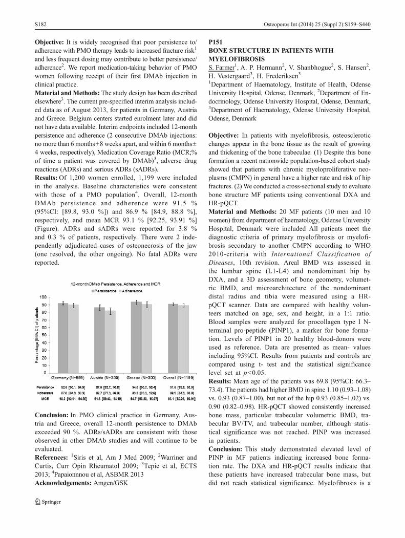
Objective: It is widely recognised that poor persistence to/adherence with PMO therapy leads to increased fracture risk1
and less frequent dosing may contribute to better persistence/adherence2. We report medication-taking behavior of PMOwomen following receipt of their first DMAb injection inclinical practice.Material andMethods: The study design has been describedelsewhere3. The current pre-specified interim analysis includ-ed data as of August 2013, for patients in Germany, Austriaand Greece. Belgium centers started enrolment later and didnot have data available. Interim endpoints included 12-monthpersistence and adherence (2 consecutive DMAb injections:no more than 6 months+8 weeks apart, and within 6 months±4 weeks, respectively), Medication Coverage Ratio (MCR;%of time a patient was covered by DMAb)3, adverse drugreactions (ADRs) and serious ADRs (sADRs).Results: Of 1,200 women enrolled, 1,199 were includedin the analysis. Baseline characteristics were consistentwith those of a PMO population4. Overall, 12-monthDMAb persistence and adherence were 91.5 %(95%CI: [89.8, 93.0 %]) and 86.9 % [84.9, 88.8 %],respectively, and mean MCR 93.1 % [92.25, 93.91 %](Figure). ADRs and sADRs were reported for 3.8 %and 0.3 % of patients, respectively. There were 2 inde-pendently adjudicated cases of osteonecrosis of the jaw(one resolved, the other ongoing). No fatal ADRs werereported.
Conclusion: In PMO clinical practice in Germany, Aus-tria and Greece, overall 12-month persistence to DMAbexceeded 90 %. ADRs/sADRs are consistent with thoseobserved in other DMAb studies and will continue to beevaluated.References: 1Siris et al, Am J Med 2009; 2Warriner andCurtis, Curr Opin Rheumatol 2009; 3Tepie et al, ECTS2013; 4Papaionnnou et al, ASBMR 2013Acknowledgements: Amgen/GSK
P151BONE STRUCTURE IN PATIENTS WITHMYELOFIBROSISS. Farmer1, A. P. Hermann2, V. Shanbhogue2, S. Hansen2,H. Vestergaard3, H. Frederiksen31Department of Haematology, Institute of Health, OdenseUniversity Hospital, Odense, Denmark, 2Department of En-docrinology, Odense University Hospital, Odense, Denmark,3Department of Haematology, Odense University Hospital,Odense, Denmark
Objective: In patients with myelofibrosis, osteoscleroticchanges appear in the bone tissue as the result of growingand thickening of the bone trabeculae. (1) Despite this boneformation a recent nationwide population-based cohort studyshowed that patients with chronic myeloproliferative neo-plasms (CMPN) in general have a higher rate and risk of hipfractures. (2) We conducted a cross-sectional study to evaluatebone structure MF patients using conventional DXA andHR-pQCT.Material and Methods: 20 MF patients (10 men and 10women) from department of haematology, Odense UniversityHospital, Denmark were included All patients meet thediagnostic criteria of primary myelofibrosis or myelofi-brosis secondary to another CMPN according to WHO2010-criteria with International Classification ofDiseases, 10th revision. Areal BMD was assessed inthe lumbar spine (L1-L4) and nondominant hip byDXA, and a 3D assessment of bone geometry, volumet-ric BMD, and microarchitecture of the nondominantdistal radius and tibia were measured using a HR-pQCT scanner. Data are compared with healthy volun-teers matched on age, sex, and height, in a 1:1 ratio.Blood samples were analyzed for procollagen type I N-terminal pro-peptide (PINP1), a marker for bone forma-tion. Levels of PINP1 in 20 healthy blood-donors wereused as reference. Data are presented as mean- valuesincluding 95%CI. Results from patients and controls arecompared using t- test and the statistical significancelevel set at p<0.05.Results: Mean age of the patients was 69.8 (95%CI: 66.3–73.4). The patients had higher BMD in spine 1.10 (0.93–1.08)vs. 0.93 (0.87–1.00), but not of the hip 0.93 (0.85–1.02) vs.0.90 (0.82–0.98). HR-pQCT showed consistently increasedbone mass, particular trabecular volumetric BMD, tra-becular BV/TV, and trabecular number, although statis-tical significance was not reached. PINP was increasedin patients.Conclusion: This study demonstrated elevated level ofPINP in MF patients indicating increased bone forma-tion rate. The DXA and HR-pQCT results indicate thatthese patients have increased trabecular bone mass, butdid not reach statistical significance. Myelofibrosis is a
Osteoporos Int (2014) 25 (Suppl 2):S159–S440S182

rare disease and the number of patients included wassmall.References: 1. Mellibovsky L et al. Bone 2004;34:3302. Farmer S et al. Br J Haematol 2013;163:603Acknowledgements: This study was founded by the Regionof Southern Denmark.
P152PROGRESSION OF IDIOPATHIC COXARTHROSIS:VITAMIN D ASPECTA. V. Kalashnikov1, G. V. Gayko1, O. V. Kalashnikov1,L. I. Apukhovskaya21State Institution, Institute for Traumatology and Orthopae-dics of NAMS of Ukraine, Kyiv, Ukraine, 2State Institution,Institute for Biochemistry O.V. Palladin of the NAS ofUkraine, Kyiv, Ukraine
Objective: To define interrelation between changes in mineraland vitamin D exchange and idiopathic coxarthrosisprogression.Material andMethods: 30 patients with stage IVof idiopath-ic coxarthrosis according to Kellgren and Lawrence have beenstudied and compared with 30 healthy persons (controlgroup), statistically standardized in age and gender. We havesubdivided forms of idiopathic coxarthrosis into rapidly pro-gressive (≤5 years from the beginning to the final stage of thedisease), moderately progressive (from 5 to 10 years) andslowly progressive (>10 years). Insufficiency of vitamin Dexchange has been verified according to Gomez classification(2003); the content of 25(OH)D, phosphorus and calcium,activity of total alkaline phosphatase and its isoenzymes inblood serum have been defined.Results: For idiopathic coxarthrosis, notable is the 100 %prevalence of vitamin D disorders and 20.25 % of the severestvitamin D deficits. Rapidly progressive idiopathiccoxarthrosis has shown 44.5 % cases of vitamin D deficiency,55.5 % of vitamin D deficit; moderately progressive form ofthe disease demonstrated 82 % of vitamin D deficiency and18 % of vitamin D deficit; slow progressing idiopathiccoxarthrosis has shown 50 % of vitamin D deficiency,12.5 % of vitamin D deficit and 37.5 % of vitamin Dhypovitaminosis. Vitamin D supply in case of idiopathiccoxarthrosis is closely connected with mineral metabolismand its disorders. Rapidly progressive form has shown signif-icantly reduced (p≤0.01) coxarthrosis indicators of serumcalcium, phosphorus, alkaline phosphatase and its isoenzymes(bone marrow and the gastrointestinal) and 25(OH)D, com-pared to slowly progressive form.Conclusion: Signs of idiopathic coxarthrosis are: insufficien-cy of mineral and vitamin D exchange, usual for 100 % ofpatients. Progression of idiopathic coxarthrosis depends onthe level of vitamin D production by human body.
References: Gómez Alonso C et al. Nefrologia 2003;23(Suppl 2):73.
P153PRESARCOPENIA, SARCOPENIA AND SEVERESARCOPENIA AMONG IRANIAN ELDERLIES: THEFIRST EPIDEMIOLOGICAL REPORTR. Heshmat1, R. Hashemi2, A. R. Dorosty Motlagh2,M. Payab2, A. Esmailzadeh3, P. Pasalar4, F. Siassi21Chronic Diseases Research Center, Tehran University ofMedical Sciences, Tehran, Islamic Republic of Iran, 2Schoolof Nutritional Sciences and Dietetics, Tehran University ofMedical Sciences, Tehran, Islamic Republic of Iran, 3Schoolof Nutrition and Food Science, Isfahan University of MedicalSciences, Isfahan, Islamic Republic of Iran, 4Endocrinologyand Metabolism Research Institute, Tehran University ofMedical Sciences, Tehran, Islamic Republic of Iran
Objective: The purpose of this study was to determine theprevalence of sarcopenia, the age related loss of skeletalmuscle mass in Iranian older adults based on the definitionby European working group on sarcopenia (EWGSOP).Material and Methods:Using cluster random sampling, 300participants (150 of each sex) were selected in Tehran, thecapital of Iran. In each cluster, 2 individuals (1 male, 1 female)were invited through home interviews from the following agegroups: 55–59, 60–64, 65–69, 70–74, and over 75. Skeletalmuscle mass was measured by DXA. Appendicular skeletalmuscle (ASM) was calculated as well. Muscle strength andperformance were evaluated according to hand grip strengthand 4-m gait speed. According to EWGSOP definition, indi-viduals with abnormal ASM were considered aspresarcopenic. Presarcopenic individuals with abnormal mus-cle strength or performance were considered sarcopenic. Par-ticipant with all three abnormal criteria were considered se-verely sarcopenic.Results: The prevalence of presarcopenia, sarcopenia andsever sarcopenia in total population were 30 %, 18 % and5.7 %, respectively. The prevalence of presarcopenia,sarcopenia and sever sarcopenia in female were 25.3 %,15.3 % and 5.3 % compared to 52.7 %, 20.7 % and 14.7 %for male. An increase in the prevalence of sarcopenia wasobserved with age. This increase was more significant amongthe age group over 75 years old in both genders. The lowestprevalence of sarcopenia was observed in age group 65–69 forfemale and 60–64 for male.Conclusion: This is the first study performed on sarcopenia incommunity-dwelling elderly Iranians. The prevalence ofsarcopenia was considerable in Iranian elderly. Unlike studiesin western populations, Iranian men suffered more fromsarcopenia compared to women.
Osteoporos Int (2014) 25 (Suppl 2):S159–S440 S183

P154FRAX TO EVALUATE THE FRACTURE RISK OFPEOPLE IN NANJINGL. Bao1, X. Xu1, X. Cheng11Nuclear Medicine, First Affiliated Hospital, NanjingMedicalUniversity, Nanjing, China
Objective:To evaluate the applicability and to predict fracturerisk of different gender groups in Nanjing using the WHOreleased FRAX fracture risk assessment tool.Material and Methods: 1,383 cases of people were groupedand the personal data including the risk factors were collectedand entered the FRAX tool in Nanjing Research. Then 10-year fracture probability of hip and osteoporotic fractures werecalculated, various aspects containing fracture history andBMD were compared.Results: Fracture risk of female population is much higherthan male, the probability of fracture risk of hip and body aresimultaneously increased with age, and history of fractures isan important risk factor, With or without BMD values willaffect the calculation of FRAX 10-year probability of hipfracture recurrence, 10-year fracture risk of recurrence ofprevious fracture population is much higher than people with-out fracture history. After excluding history of fractures, bodyand hip fracture prediction showed no significant differencewith or without BMD.Conclusion: Our research successfully established thatFRAX tool can be effectively applied to fracture risk assess-ment in Nanjing, FRAX tool still has clinical significanceeven if without previous fracture history and BMD value,FRAX tool combined BMD have clinical value in patientswith or without previous fractures.
P155THE EFFECTS OF LEG POSITIONING ON SPINALBONE MINERAL DENSITY MEASUREMENTS ANDTHE DIAGNOSIS OF OSTEOPOROSISS. Ikegami1, M. Kamimura2, S. Uchiyama1, K. Mukaiyama1,Y. Nakamura1, H. Kato11Department of Orthopaedic Surgery, Shinshu UniversitySchool of Medicine, Matsumoto, Japan, 2Center for Osteopo-rosis and Spinal Disorders, Kamimura Orthopaedic Clinic,Matsumoto, Japan
Objective: The aim of this study was to investigate whichdifferences in leg positioning affect spinal BMD measure-ments and the diagnosis of osteoporosis.Material and Methods: Subjects included 1,039 Japanesepatients, 878 women and 161 men (mean ages: 67 and71 years, respectively). Spinal BMD (L1-4) was measuredusing DXAwith patients lying in two different positions; thestandard lumbar scanning position, with patients supine on the
scanning table with hips flexed and knees flexed over a 90ºsupport pad (modified supine position) and supine (supineposition). Predictive indices were calculated for spinal BMDDXA measurements acquired with patients in the supineposition; these included sensitivity, specificity, and the likeli-hood ratio (LR) for a negative test (-). A diagnosis of osteo-porosis was excluded for a LR(-) less than 0.2. Osteoporosiswas defined as a BMD T-score of ≤−2.5.Results: For the modified supine and supine position duringDXA scanning in women, BMD measurements were0.911 g/cm2 and 0.915 g/cm2, respectively; in men theywere 1.117 g/cm2 and 1.124 g/cm2, respectively. The differencein BMD between positions was 0.40 % (95%CI: 0.29, 0.51;P<0.001). The coefficient of variance (CV%) for BMD mea-surements was 0.66%. Accordingly, the clinical equivalency ofthe spinal BMD results between the two positions was pre-served. The sensitivity, specificity, and LR(-) of osteoporosisdiagnosis following DXA scanning in the supine position was94 %, 99 %, and 0.066, respectively, compared with resultsacquired using the standard position.Conclusion: DXA measurements acquired with patients inthe supine position slightly overestimated BMD vs. the mod-ified supine position. However, this BMD overestimation issmall and not significant. The clinical equivalency betweenpositioning methods for DXA is preserved to the extent thatosteoporosis can be reliably diagnosed in the supine position.
P156COMMUNITY OSTEOPOROSIS AND FALLPREVENTION PROGRAM IN NORTHERN TAIWAND. C. Chan1, J. S. Hwang2, L. C. Lim1, K.-S. Tsai3, R. S.Yang41Department of Gerontology and Geriatrics, National TaiwanUniversity Hospital, Taipei, Taiwan, Province of China, 2Di-vision of Endocrinology and Metabolism, Department of In-ternal Medicine, Chang Gung Memorial Hospital, Taoyuan,Taiwan, Province of China, 3Superintendent Office, NationalTaiwan University Hospital BeiHu Branch, Taipei, Taiwan,Province of China, 4Department of Orthopaedics, NationalTaiwan University Hospital, Taipei, Taiwan, Province of China
Objective:To determine the feasibility and effectiveness of anosteoporosis and fall screening plus referral program inNorthern Taiwan.Material andMethods: In 2013, seven community educationcampaigns on osteoporosis and fall prevention were conduct-ed in Taipei City, New Taipei City, and Taoyuan County,Taiwan. Ten questions were tested before and after the educa-tion courses to determine the effectiveness of the program.Participants were screened with FRAX® and fall risk ques-tionnaires (FRQ). High risk subjects (10-year probability ofpredicted risk ≥3 % for hip fracture, ≥20 % for major
Osteoporos Int (2014) 25 (Suppl 2):S159–S440S184
osteoporotic fracture, ≥2 fall in previous year, or scored ≥4 onFRQ) were referred to hospitals for further osteoporosis andfall assessments and managements.Results:Among 1,159 participants, 1,051 with complete datawere analyzed. Mean age was 69.5±10.3 years with 82.8 %(n=870) women. Mean test score improved from 6.1±1.8 to8.3±1.7 points (paired t-tests p<0.001) and 95 % were satis-fied with the educational courses. Roughly 3/5 (61.8 %, n=649) were considered high risk. However, only 127 (12.1 %)were successfully referred for further managements. Amongthem, 53 (41.7 %) had clinical diagnosis of osteoporosisdefined as having a fragility fracture or BMD T score ≤−2.5.Although 27 (21.3 %) received anti-osteoporosis medications,only 10 (7.9 %) were eligible for National Health Insurancereimbursements. Referred subjects had multiple fall risk fac-tors including fall related chronic conditions (n=96, 75.6 %),taking high risk medications (n=109, 85.8 %), abnormal gait(n=44, 34.6 %), weak muscle power (n=48, 37.8 %), andbalance problem (n=38, 29.9 %).Conclusion: Community educational campaigns were wel-comed and effective in improving osteoporosis and fall relatedknowledge. Even significant numbers of high risk individualswere screened out, relatively few actually accepted the referralfor further managements. Efforts should be made to improvethe referral efficiency.Acknowledgements: The authors thank the Wang Jhan-YangPublic Trust Fund for sponsoring the study. The sponsorreviewed and approved the study protocol but did not interferewith study conduction, data analysis or interpretation. Theauthors also thank the Taiwanese Osteoporosis Associationfor coordinating this study.
P157IN MEN, SEVERE SPINE OSTEOARTHRITIS ISASSOCIATED WITH ABDOMINAL AORTICCALCIFICATION AND ALL CAUSE MORTALITY:THE MINOS STUDYC. Estublier1, R. D. Chapurlat1, F. Marchand2, P. Szulc11INSERM UMR 1033, Université de Lyon, Hospices Civilsde Lyon, Hôpital E Herriot, Lyon, France, 2CARMI,Montceau les Mines, France
Objective: We studied the association of spine osteoarthritis(OA) with abdominal aortic calcification (AAC) severity andits progression rate as well as with all-cause mortality in oldermen.Material and Methods: A cohort of 766 men aged 50–85 year was followed up prospectively for 7.5 years (forAAC) and for 10 years (for mortality). Spine OAwas assessedat six intervertebral spaces using Lane’s score. Total score ofeach parameter was calculated as sum of its values for sixintervertebral levels. AAC was assessed in the abdominal
aorta using Kauppila’s semiquantitative score. During thefollow-up, 182 men died.Results:After adjustment for confounders, the odds of severeAAC score (>6) increased with total disc narrowing score(OR[95%CI]=1.26 per SD increase [1.01–1.58]). After simi-lar adjustment, the odds of AAC>6 were higher in the highesttertile of total disc narrowing score vs. the lowest tertile (OR=1.89 [1.04–3.42]). Osteophytosis and subchondral sclerosiswere not associated with AAC severity. Prospective data onAAC progression were available in 613 men. In 148 menAAC was stable. After adjustment for confounders, the prob-ability of AAC stability decreased with increasing total osteo-phyte score (OR=0.76 per SD increase [0.60–0.99]). Theprobability of AAC stability was lower in the highest tertileof total osteophyte score vs. two lower tertiles combined(OR=0.51 [0.30–0.86]). After adjustment for confounders(including AAC), total OA grade (HR=1.25 per SD increase[1.03–1.50]) predicted mortality. Both higher total OA gradeand severe AAC contributed to the risk of death. After adjust-ment for confounders, men who had higher total OA gradeand severe AAC had higher risk of death (HR=2.63 [1.48–4.65]) compared with men who had lower total OA grade andless severe (or absent) AAC.Conclusion: In older men, severe spine OA is associated withgreater AAC severity and greater risk of AAC progression.Severe OA and severe AAC contribute jointly and indepen-dently to higher all-cause mortality in older men.
P158ASSESSMENT OF BONE MICROARCHITECTUREIMPROVES INCIDENT FRACTURE PREDICTION INMEN: THE STRAMBO STUDYP. Szulc1, S. Boutroy1, R. D. Chapurlat11INSERM UMR 1033, Université de Lyon, Hospices Civilsde Lyon, Hôpital E Herriot, Lyon, France
Objective: Areal BMD (aBMD) measured by DXA poorlyidentifies men at high fracture risk. We studied the value ofHR-pQCT for fracture prediction in men.Material andMethods:Among 821men aged 60–87 follow-ed up for 6 year, 70 men sustained fragility fractures. Wecalculated aBMD T-score using hip and distal radius aBMDin young men (STRAMBO) and hip aBMD in young women(NHANES).Results: Most microarchitectural parameters were associatedwith fracture risk when adjusted for age, weight, prior fallsand fractures (HR=1.25–1.94 per SD, p<0.05). After furtheradjustment for distal radius aBMD, low trabecular number(Tb.N) and more heterogeneous trabecular distribution (highTb.Sp.SD) were associated with higher fracture risk (HR[95%CI]=1.94 [1.46–2.56] and 1.61 [1.25–2.08] per SD). Ina similar model including hip aBMD, higher Tb.Sp.SD at
Osteoporos Int (2014) 25 (Suppl 2):S159–S440 S185

distal tibia was associated with higher fracture risk (HR=1.41[1.06–1.86] per SD). In men with osteopenia at the hip andelevated Tb.Sp.SD (highest quartile), fracture risk was highervs. men with normal hip aBMD (HR=2.77 [1.36–5.65]) andvs. osteopenic men with normal Tb.Sp.SD (HR=2.20 [1.10–4.41]). Their fracture incidence was close to that in men withthe hip T-score<−2.5 (3.5 vs. 3.3 /100 p-yrs). The results weresimilar when hip aBMD in women was used to calculate T-score thresholds. In men with osteopenia and low Tb.N (low-est quartile) at distal radius, fracture risk was higher vs. menwith normal aBMD (HR=2.34 [1.07–5.13]) and vs.osteopenic men with normal Tb.N (HR=3.05 [1.28–7.28]).Their fracture incidence was close to that in men with T-score<−2.5 at distal radius (3.2 vs. 3.3 /100 p-yrs). The resultswere similar for osteopenia and Tb.Sp.SD at distal radius. Inosteopenic men with normal bone microarchitecture, fractureincidence was similar to that in men with normal aBMD.Conclusion: Assessment of bone microarchitecture by HR-pQCT may improve fracture prediction in older osteopenicmen.
P159SERUM SCLEROSTIN IS HIGHER IN MEN WITHSEVERE OSTEOPHYTES AT THE SPINE: THEMINOS STUDYP. Szulc1, C. Estublier1, C. Bertholon1, F. Marchand2, R. D.Chapurlat11INSERM UMR 1033, Université de Lyon, Hospices Civilsde Lyon, Hôpital E Herriot, Lyon, France, 2CARMI,Montceau les Mines, France
Objective: To analyze cross-sectionally the association be-tween spine osteoarthritis (OA) severity and serum sclerostinlevel in older men.Material and Methods: In 694 men aged 50–85 year, spineOA was assessed at six intervertebral spaces using Lane’sscore. Total score of each parameter was calculated as sumof its values for six intervertebral levels. Sclerostin level wasmeasured in fasting serum (TECOsclerostin EIA,TECOmedical). BMD of total hip was measured by DXA(Hologic QDR1500).Results: After adjustment for age, weight, 17ß-estradiol leveland glomerular filtration rate, serum sclerostin level increasedacross quartiles of total osteophyte score (trend<0.001).Sclerostin level was 15 % (0.42SD, p<0.001) higher in thehighest vs. the lowest quartile. After similar adjustments,sclerostin level increased with the increasing osteophyte grade(trend<0.001). It was 13 % (p<0.005) higher in men withsevere osteophytes vs. men who had no or mild osteophytes.The link between osteophyte severity and sclerostin level wassignificant (p<0.05) after further adjustment for hip BMD,disc space narrowing and subchondral sclerosis. After
adjustment for confounders, serum sclerostin increased acrossthe quartiles of total disc space narrowing score (trend<0.001).Average serum sclerostin increased with the increasing gradeof disc space narrowing (p for trend<0.005). However, all theassociations between disc space narrowing and sclerostinlevels lost significance (p>0.23) after additional adjustmentfor total hip BMD, osteophyte severity and subchondral scle-rosis score. Men with subchondral sclerosis at ≥2 interverte-bral levels had slightly higher sclerostin levels vs. other men(p=0.053). This trend became nonsignificant after adjustmentfor total hip BMD, osteophyte severity and disc spacenarrowing score (p=0.88).Conclusion: In older men severe osteophytes, but not discspace narrowing or subchondral sclerosis, were independentlyassociated with higher sclerostin concentration.
P160RISK FACTOR PROFILE OF YOUNG HIPFRACTURE PATIENTS IN INDIAP. Dharmshaktu1, D. Dhanwal1, V. Dixit11Department of Medicine, Maulana Azad Medical College,New Delhi, India
Objective: There has been a paucity of studies in youngpopulation with hip fracture and no such study was done inIndia. Therefore, this prospective study was performed toevaluate profile of risk factors of hip fracture in youngpatients.Material andMethods:Over a span of 3 years, 51 patients offragility hip fracture under 50 years of age were enrolled inthis study. Background data and risk factors were evaluated tothe preset proforma. Fasting venous samples were analyzedfor 25-hydroxyvitamin D (25-OHD), intact PTH, alkalinephosphatase (ALP), calcium, and phosphorus.Results: The mean age of hip fracture subjects below 50 yearsage was 46.27±12.78 which was comparable in both men andwomen. Only four of 51 study subjects were under 40 years ofage. Of 51 subjects, 26 were men and 25 were women.Majority of men with hip fracture were smokers. All thewomen were nonsmokers. Sun exposure was found to beinadequate inmajority of study subjects. Vitamin D deficiencywas found in 39 out of 51 subjects accounting 76.47 % of totalyoung population. Among vitamin D deficiency subjects, 21were men and 18 were women. Mean 25-OHD was 12.22±7.6 ng/ml in men and 10.97±7.3 ng/ml in women. Hyperpara-thyroidism was found in 28 (54.9 %) out of total 51 studysubjects.Conclusion: Majority of young patients with fragility hipfracture both men and women in India have vitamin D defi-ciency and secondary hyperparathyroidism. Therefore, vita-min D deficiency is a major risk factor along with smokingamong men in young hip fracture patients from India. Further,
Osteoporos Int (2014) 25 (Suppl 2):S159–S440S186

vitamin D intervention studies are needed for its role inprevention of hip fracture in young population.Acknowledgements: Abhishek Misra for his immense sup-port and contribution to the statistics in the study.
P161FRACTURES AND CLINICAL OUTCOME INHEMODIALYSIS PATIENTSS. O. Mazurenko1, K. G. Staroselsky2, A. A. Enkin3, A. N.Vasiliev4, O. G. Mazurenko51Saint Petersburg State University, Saint Petersburg, RussianFederation, 2Saint Petersburg Hospital 26, Dialysis Centre,Saint Petersburg, Russian Federation, 3Leningrad RegionalHospital, Dialysis Centre, Saint Petersburg, Russian Federa-tion, 4Saint Petersburg State Medical University, DialysisCentre, Saint Petersburg, Russian Federation, 5Saint Peters-burg Sokolov Hospital, Centre for Osteoporosis and SkeletonMetabolic Diseases, Saint Petersburg, Russian Federation
Objective: Patients with endstage renal disease have in-creased risk for fracture. This high fracture rate is a conse-quence of accelerated loss of BMD. Various studies suggest astrong association between BMD and survival in patients withendstage renal disease. The aim of this study was to investi-gate contribution of factures to clinical outcome in hemodial-ysis patients.Material and Methods: This prospective study enrolled 640patients (334 men and 312 women) with stage 5 chronickidney disease receiving hemodialysis who were then follow-ed for a mean of 49 (range 16–122) months. Among 159patients (77 men and 82 women) who had fractures, 54(34 %) patients had fractures of axial skeleton (vertebra, hip,pelvis). Lumbar spine, femoral neck, and distal forearm BMDwere measured by DXA. The Kaplan-Meier estimator ofsurvival and the Cox proportional hazards model was usedto calculate and determine the relations between mortalityBMD and fractures.Results: Patients were followed for 49 months, after whichtime 131 patients (20.5 %) died. The cause of death in 51 %patients were cardiovascular diseases. The Cox proportionalhazards model was used to calculate and determine relationsbetween mortality, BMD and fractures. We did not find anyinfluence of BMD on all- cause mortality of hemodialysispatients. At the same time low BMD was strongly associatedwith cardiovascular mortality. We did not find any influenceof peripheral fractures on mortality, and the same time axialfractures were strongly associated with cardiovascular(p<0.001) and all-cause mortality of hemodialysis patients(p<0.01).Conclusion: Axial fractures are good independent predictorsof cardiovascular and all-cause mortality in hemodialysispatients.
P162THE STATUS OF SERUM VITAMIN D IN PATIENTSATTENDING A PHYSICAL MEDICINE ANDREHABILITATION DEPARTMENT IN TURKEYM.Okumus1, G. Demir1, T. Ozgun2, P. Borman1, A. Karagoz11Department of Physical Medicine and Rehabilitation ofAnkara Training and Research Hospital, Ankara, Turkey,2Department of Biochemistry of Ankara Training andResearch Hospital, Ankara, Turkey
Objective: Deficiency of vitamin D is becoming a globalpublic health problem. Vitamin D plays important roles inmaintaining the balance of calcium and phosphorus me-tabolism and keeping normal BMD levels. Vitamin Dstatus is affected by many factors such as geographicalenvironment, physiological factors and lifestyle. The ob-jectives of present study were to estimate the prevalenceof vitamin D deficiency among patients attended in theoutpatients clinic of Physical Medicine and Rehabilitation(PMR) department.Material andMethods:This was a retrospective study of 999patients who presented to the outpatient clinic of PMRbetween January 2011 and April 2013 were recruitedconsecutively into the study. Biochemical markers of pa-tients were measured. Total 25(OH)D was measured fromstored plasma specimens using liquid chromatography-mass spectrometry. Vitamin D deficiency was defined asserum 25(OH)D 20 ng/ml. Sociodemographic characteris-tics, lifestyle and dietary habits were obtained usingquestionnaires.Results: The mean age of the participants was 60.2 (range:18–97) and 88.7 % were women. The mean serum 25(OH)Dlevel was 22.5 ng/ml and the prevalence of serum 25(OH)Dlevel was in 54.1 % of all patients. The mean level of25(OH)D in women was significantly higher than in men(22.9 ng/mL vs. 19.8 ng/mL, p=0.019). The participants aged60 years and above have highest serum 25(OH) D levels(25.7 ng/ml vs. 18.4 ng/ml, p=0.000).Conclusion: Mean serum 25(OH)D level was close to repre-sent range considered to represent vitamin D deficiency. Par-ticipants aged 60 or older showed the highest mean values ofserum 25(OH)D. Future studies are certainly warranted tounderstand the prevalence of vitamin D deficiency andinfluencing factors.
P163ONE-YEAR DISEASE-RELATED HEALTHCARECOSTS OF INCIDENT OSTEOPOROTICVERTEBRAL FRACTURES IN GERMANYA. Lange1, J. Zeidler1, S. Braun21Center for Health Economics Research Hannover, Hannover,Germany, 2Herescon GmbH, Hannover, Germany
Osteoporos Int (2014) 25 (Suppl 2):S159–S440 S187

Objective: Osteoporotic vertebral compression fractures(OVCF) are among the most common fractures related toosteoporosis. They have been shown to be associated withexcess mortality, and meaningful healthcare costs. Costs cal-culations have illustrated the significant financial burden tosociety and national social security systems. However, infor-mation on disease-related costs of OVCF is not available forGermany. Therefore, the aim of the present study was toestimate the direct disease-related healthcare costs of OVCFin patients with newly diagnosed fracture in the first year afterindex in Germany.Material andMethods:Claims data of a large German healthinsurance fund was used for the analyses. Patients older than60 years with a new OVCF between 2006 and 2010 werestudied retrospectively compared to a matched paired OVCF-free group. All-cause and fracture-specific medical costs werecalculated in the 1-year baseline and the 1-year follow-upperiod. A Generalized linear model (GLM) was applied toestimate adjusted total follow-up healthcare cost.Results: 2,277 pairs of matched OVCF and OVCF-free pa-tients were included in the analysis. Baseline costs werehigher in the OVCF group. Mean unadjusted all-causehealthcare cost difference in the first year following the indexdate between OVCF and OVCF-free patients was 8200 €(p<0.001). Of the difference, one-quarter was attributable toprescription drug costs and almost two-third to inpatient ser-vices. The regression model revealed that OVCF-related costsin the first year after the index date add up to 6490 € (p<0.001;CI: 5809 €–6731 €).Conclusion: Despite limitations of this study, including spec-ificity and sensitivity of claims-based diagnoses, and general-izability issues, our results are consistent with other studiesand demonstrate that OVCFs are associated with significantexcess costs. Against the background of the high and increas-ing incidence and prevalence of these fractures, the resultsemphasize the importance of research in this field.Disclosures: This study was financially supported byMedtronic International.
P164ATYPICAL FEMORAL FRACTURES AREASSOCIATED WITH HIGH CYCLIC TENSILESTRAIN REGIONS DURING WALKINGS. Martelli1,3,4, M. E. Kersh2, P. Pivonka3,4, P. R. Ebeling4,3,M. G. Pandy21Medical Device Research Institute, School of ComputerScience, Engineering and Mathematics, Flinders University,Bedford Park, Australia, 2Department of Mechanical Engineer-ing, University of Melbourne, Parkville, Australia, 3AustralianInstitute forMusculoskeletal Science, University ofMelbourne,St Albans, Australia, 4NorthWest Academic Centre, Universityof Melbourne, St Albans, Australia
Objective: Atypical femoral fractures (AFF) are rare low-energy stress fractures progressing from the lateral femur[1]. However, the aetiology of fracture onset is unclear. Wetested the hypothesis that AFF onset is associated with thefemur strain pattern during walking.Material and Methods: Ten volunteers underwent a motioncapture session and a computed tomography (CT) of the thighregion. A generic musculoskeletal model was scaled to thevolunteers’ anthropometry. Muscle and hip reaction forceswere calculated using the scaled-generic models, the recordedskin-marker trajectories and ground reaction forces duringwalking. Hip reaction forces showed consistency with pub-lished measurements [2]. The femur finite element modelswere generated from the CT images using a well-establishedprocedure [3] and subjected to the calculated muscle and hipreaction forces to calculate peak principal strains in 4 diaph-yseal levels and 4 aspects (anterior, posterior, medial, lateral).The median principal tensile and compressive strains, duringthe walking stance phase for the 32 femoral subregions, werecalculated and compared.Results: The lateral femoral shaft was mainly loaded in ten-sion throughout walking, reaching peak strain levels at thetime of peak hip reaction force. The interparticipant average ofthe median tensile strain reached 1500 με in the lateralsubtrochanteric region and 1000 με in the distal-lateral shaft.The interparticipant range of the median tensile strain in thelateral shaft ranged from 600 με to 2500 με.Conclusion: AFFs are associated with high tensile strainregions occurring cyclically during walking, with the lateral-subtrochanteric region experiencing the higher tensile strainlevels. Tensile strain levels vary among subjects and mayexplain individual susceptibility to AFFs. Subject-specificmodels may help discriminate patients most at risk for AFFs.References: 1. Shane et al., JBMR. 2010;25:112. Bergmann et al., J Biomech 1993;34:73. Schileo et al., J Biomech 2007;40:13Acknowledgements: Australian Research Council(DE140101530)
P165OSTEOPOROSIS IN SAUDI ARABIA: IS ITAPROBLEM?J. Palencia1, M. Alhashim1, A. León21Ortho Department KSMC, Riyadh, Saudi Arabia, 2Serviciode Traumatología y Cirugía Ortopédica. Hospital ClínicoUniversitario, Valladolid, Spain
Objective: The goals of the present study are:- Analysis of the Saudi Arabia osteoporosis prevalence.- The future expectations of osteoporosis in Saudi Arabia. -Recommendations and advice.
Osteoporos Int (2014) 25 (Suppl 2):S159–S440S188

Material and Methods: - General review about the preva-lence of osteoporosis in Saudi Arabia.- Assuming the expectation of osteoporotic cases in the future.- Recommendations and guidelines for management of oste-oporosis according to the population culture and food habits.Results: - Currently, the population in Saudi Arabia is 25.7million. 10 % of them >50 y.o. and 2 % of them >70 y.o.- According to the Saudi Reference Data, the prevalence ofosteoporosis is 28.2 % in females >50 y.o. and 37.8 % inmales >50 y.o. The osteopenia is 43.8 % in females >50 y.o.and 54.1 % in males >50 y.o.- We need to give a solution for this current and future problem.- The causes of this high prevalence of osteoporosis are:1. Low or deficiency of Vitamin D in children and adults2. Sedentary lifestyle. Young people don’t reach enough os-
seous stock.3. Postmenopausal females don’t receive replacement
hormonal therapy4. Bad food habits, with low levels of calcium 5. Less sun
exposure due to a uniform culture6. No well defined management program to osteoporosisConclusion: - Osteoporosis is a serious and increasing prob-lem among Saudi Population.- To overcome it, the Health Authorities should perform:1. Programmes and activities to raise education, level of the
awareness and prevention (guidelines, bulletins, work-shops, etc.)
2. Well developed medicine management protocols with com-bination of The Saudi Osteoporotic Society
P166PREVALENCE OF SARCOPENIA ACCORDING TODIFFERENT DIAGNOSTIC TOOLSC. Beaudart1, F. Buckinx1, J. Slomian1, A. Quabron1,J. Petermans2, J.-Y. Reginster3, O. Bruyère1, S. Gillain21Department of Public Health, Epidemiology and HealthEconomics, University of Liège, Liège, Belgium, 2GeriatricDepartment, CHU Liège, Liège, Belgium, 3Bone and CartilageMetabolism Department, CHU Liège, Liège, Belgium
Objective: Sarcopenia can be defined as a progressive andgeneralized loss of muscle mass with either a loss of musclestrength or a loss of physical performance. Currently, there isno recommendation regarding the diagnostic tools to use tomeasure these three outcomes. In this cross-sectional study,we compared the prevalence of sarcopenia when using differ-ent diagnostic tools.Material and Methods: To measure muscle mass, musclestrength and physical performance, we used for eachoutcome two different diagnostic tools. For muscle mass,we used DXA and Bio-impedance (BIA); for musclestrength, we used a hydraulic dynamometer (HD) and a
pneumatic dynamometer (PD); for physical performancewe used the Short Physical Performance Battery test(SPPB) and the walk speed (WS). Eight diagnostic groupswere thereby established.Results: A total of 200 consecutive subjects were recruited inan outpatient clinic in Liège, Belgium (62 % of women, meanage: 73.8 years). Prevalence of sarcopenia varied from 8.72 %(BIA-HD-WS) to 28.5 % (DXA-PD-SPPB) depending of thedefinition. Regarding muscle mass, it seems that BIA system-atically underestimate muscle mass compared to DXA (meanof prevalence with BIA=13.4 %; mean of prevalence withDXA=21.4 %). For muscle strength, the pneumatic dyna-mometer diagnosed twice more sarcopenic subjects than thehydraulic dynamometer (mean of prevalence with PD=23.2 %; mean of prevalence with HD=11.6 %). Finally, areally small difference of prevalence was observed betweenthe walk speed and the SPPB test (mean of prevalence withWS=17.3 %; mean of prevalence with SPPB=17.6 %).Conclusion: Within the same definition of sarcopenia, prev-alence of sarcopenia is highly dependent on the diagnostictool used. It is necessary to reach a consensus on the recom-mended diagnostic tools to be used in order to make studiescomparable.
P167LITERATURE REVIEWAND META-ANALYSIS OFPERSISTENCE WITH ORAL BISPHOSPHONATESL. Karlsson1, J. Lundkvist2, E. Psachoulia3, M. Intorcia3,O. Ström1
1Quantify Research, Stockholm, Sweden, 2Amgen AB, Solna,Sweden, 3Amgen (Europe) GmbH, Zug, Switzerland
Objective: Treatment persistence is an important aspectof managing chronic illness. In this study, a literaturereview was conducted summarizing published data onretrospectively observed persistence with oral bisphosphonates(OBPs).Material and Methods: A structured literature review ofretrospective studies estimating persistence (consistent drugdispensing, without failing to refill within a study-specifictime period, the permissible gap) with OBPs at 12 and24 months was conducted using the PubMed database. Thesearch included articles indexed before 22 November 2013with an English abstract. Pooled estimates of persistenceat 12 and 24 months were calculated using inverse var-iance estimation. In subgroup analyses, these estimateswere grouped by weekly and daily administration and byregion. The effect of persistence by varying the permissiblegap was also analyzed.Results: 663 unique articles were identified for title review.After applying exclusion criteria, 43 articles were included inthe final review, with 41 and 18 reporting at least one estimate
Osteoporos Int (2014) 25 (Suppl 2):S159–S440 S189

of 12- and 24-month persistence, respectively. Persis-tence at 12 months varied from 10 % to 78 %, with apooled estimate of 44.0 % (CI95: 44.0–44.1 %). Persis-tence at 24 months varied from 16 % to 46 %, with apooled estimate of 37.8 % (CI95: 37.7–37.9 %). Allstudies comparing daily with weekly OBPs reported low-er persistence with daily OBPs (12-month persistencepooled estimates: 35.4 % vs. 48.8 %). European studiesreported higher 12-month persistence (pooled estimate:46.7 %) compared to North American studies (pooledestimate: 43.8 %). Studies which varied the permissiblegap reported a wider gap to be associated with higherestimated persistence.Conclusion: There is a fairly large body of internationalevidence describing retrospectively measured persistence withOBPs. This meta-analysis shows that persistence with OBPsis low with only 44 % of patients persistent at 12 months and38 % at 24 months.Acknowledgements: This study and abstract was funded byAmgen and GSK.
P168CHARACTERISTICS OF POSTMENOPAUSALWOMEN WITH OSTEOPOROSIS INITIATINGDENOSUMAB IN BULGARIAM. Boyanov1, A. Shinkov2, E. Psachoulia3, M. Intorcia3,R. Petkova41University Hospital Alexandrovska, Sofia, Bulgaria,2University Hospital of Endocrinology Acad. Iv. Penche,Sofia, Bulgaria, 3Amgen (Europe) GmbH, Zug, Switzerland,4Amgen Bulgaria, Sofia, Bulgaria
Objective: Describe the characteristics of postmenopausalwomen with osteoporosis (OP) and reasons for initiatingdenosumab in routine clinical practice in Bulgaria.Material and Methods: This retrospective study, conductedin 11 Bulgarian practices, included postmenopausal women≥50 year old with a clinical diagnosis of OP, who initiateddenosumab on/after Oct 2011 and received a 2nd injectionwithin the next 7 months (and until Aug 2013). All studyvariables (e.g., age, prescribing rationale, etc.) were recordedas per routine clinical practice at 1st injection; no other studyprocedures were required. Continuous variables weresummarized as mean±SD. For categorical variables,the number and percentage of subjects in each category weresummarised.Results: 222 women met the eligibility criteria with a meanage of 64.2±8.54 year; half (49.5 %) were <65 year old andonly 13.1 % ≥75 year. Mean age at menopause was 48.1±3.98 year. The most common reason for prescribingdenosumab was a BMD T-score of <-2.5 (98.6 % of patients),reflecting local reimbursement criteria whereby denosumab is
reimbursed in such patients. Other reasons were history of OPfracture (22.1 %), multiple risk factors for fracture (20.3 %)and failure of previous OP therapy (15.3 %). Mean BMD T-score was −2.7±0.71 at the femoral neck, −3.2±0.63 at thelumbar spine and −2.3±0.83 at the total hip. One-third ofwomen (31.5 %) had received OP therapy prior to denosumabinitiation. One-quarter (26.6 %) had experienced a previousfracture, with vertebral being the most common type (71.2 %)followed by hip fractures (6.8 %) and others (32.2 %, exclud-ing hip). 2.7 % of women received vitamin D only, 5.9 %calcium supplements only and 35.1 % both.Conclusion: Women initiating denosumab in Bulgarian clin-ical practice had a mean age of 64.2 year and a low BMD T-score (within the OP range) with or without other risk factors.Acknowledgements: This study and abstract were funded byAmgen and GSK.
P169OXIDATIVE STRESS IN PATIENTS WITHOSTEOPOROSISD. Alhamdany11Mosul University, Mosul, Iraq
Objective: To assess the oxidative stress and total antioxidantstatus in patients with osteoporosis.Material and Methods: Sixty female patients with posi-tive DXA results whose ages range between 50 and60 years old were enrolled in the study and were dividedinto 2 groups, osteopenia and osteoporosis. Another 30apparently healthy females, age matched with the pa-tients, were considered as a controls. Total antioxidantstatus (TAS) and malondyaldehyde (MDA) were mea-sured in all groups. A pilot study was designed to com-pare the TAS and MDA between patients with osteopeniaand osteoporosis, using the ranges of T-score betweenthe medians of each group (−1.7 to −2.5 and −2.6 to−3.3,.respectively).Results: The TAS values of both patients groups (1.59±0.15 mmol/l and 1.05±0.37 mmol/l, respectively) were sig-nificantly lower than that of the control group (2.18±0.26 mmol/l) (p<0.001). MDAvalues of both patients groups(osteopenia and osteoporosis) (1.20±0.43 μmol/l and 2.40±0.66 μmol/l, respectively), were significantly higher than thatof the control group (0.47±0.14μmol/l) (p<0.001). The resultof pilot study, showed no difference between these two select-ed values groups for TAS and MDA, (p-value <0.1 and 0.07)respectively.Conclusion: The present study demonstrated that osteoporo-sis patients have higher levels of MDA and lower levels ofTAS than the control group. There is no difference betweenosteopenia and osteoporosis regarding oxidative stress in thepilot study.
Osteoporos Int (2014) 25 (Suppl 2):S159–S440S190
P170JOINT USE OF VITAMINS D3 AND E PREVENTSBONE AND CARTILAGE DISORDERS CAUSED BYCORTICOSTEROIDSA. V. Kalashnikov1, A. T. Brusko1, E. L. Kusiv1, L. I.Apukhovskaya2, Y. I. Shchehlova11State Institution, Institute for Traumatology and Orthopae-dics of NAMS of Ukraine, Kyiv, Ukraine, 2State Institution,Institute for Biochemistry O.V. Palladin of the NAS ofUkraine, Kyiv, Ukraine
Objective: Biochemical and morphological study of vitaminsD3 and E effect with underlying use of corticosteroids on boneand cartilage tissue structural changes.Material and Methods: The study used 68 Wistar rats in 4series of experiments, 17 rats for each. The first group wascontrol one, the second group received 0.5 mg of prednisolone,the third –0.5 mg of prednisolone and 100 IU of vitamin D3, andthe fourth –0.5 mg of prednisolone, 100 IU of vitamin D3 and0.726 IU of vitamin E. The agents were administered once dailyintragastrically within a month. Calcium level, activity of totalalkaline phosphatase and its isoenzymes and 25(OH)D content inblood serum have been determined. After sacrificing the ratspursuant to international ethical standards, bone tissue ash con-tent and mineral components of the ash have been studied. Bonetissue was morphologically analyzed by conventional methods.The results have been statistically processed.Results: Prednisolone leads to mineral exchange disorders, asevidenced by biochemical indications of calcium level inblood, increase of alkaline phosphatase and its isomers levelsand reliable (р<0.05) reduction of 25(ОН)D in blood serum.Combination of vitamins D3 and E inhibits the negative effectof prednisolone and facilitates mineral exchange normaliza-tion, although mineral exchange indicators have not reachedthe levels of the control group at the end of the experiment(р<0.05). Morphological data confirmed the results ofbiochemical investigations. The rats, that received D3 andE with underlying use of prednisolone mostly preservedtheir bone tissue and epiphyseal cartilage.Conclusion: The study demonstrated that joint use of D3 andE vitamins facilitates normalization of bone tissue mineralexchange, prevents bone tissue demineralization and reducesnegative effect of prednisolone on bone tissue and cartilagefunctional and structural conditions.
P171SERUM DICKKOPF1 (DKK1): RELATIONSHIPWITH BONE METABOLISM ANDATHEROSCLEROTIC DISEASE IN TYPE 2DIABETESR. Reyes-Garcia1, P. Rozas-Moreno1, A. Garcia-Martin2,B. Garcia-Fontana1, S. Morales-Santana1, M. Muñoz-Torres1
1Bone Metabolism Unit, Endocrinology Division, HospitalUniversitario San Cecilio, Granada, Spain, 2BoneMetabolismUnit, Endocrinology Division Hospital Universitario SanCecilio, Granada, Spain
Objective: Type 2 diabetes (T2DM) is a risk factor for oste-oporotic fractures and cardiovascular disease. The Wnt sig-naling pathways are involved in diverse developmental andphysiological processes, including anabolic effects on boneand atherosclerotic disease (AD). Dickkopf-1 (DKK1) is apotent inhibitor of Wnt signaling. The aims of our study wereto evaluate serum DKK1 levels in a cohort of T2DM patientsand to analyze its relationships with bonemetabolism andAD.Material and Methods: Cross-sectional study including 73patients with T2DM and 54 control subjects. Lumbar spineand femoral BMD were measured by DXA (Hologic QDR4500). The presence of cardiovascular disease (cerebrovascu-lar disease, peripheral arterial disease, coronary heart disease)was recorded. Intima- media thickness (IMT) was determinedby Doppler ultrasonography (Toshiba PowerVision 6000) andaortic calcification by evaluation of lateral view conventionalX-rays of the thoracic and lumbar spine according to themethod described by Kauppila et al. DKK-1 was measuredby quantitative sandwich ELISA developed by Biomedica(BiomedicaMedizinprodukte GmbH and Co. KG,Wien, Aus-tria), with an intra- and inter-assay variability were of 7 % and9 %, respectively. Results were analysed using SPSS 15.0.Results: There were no differences in DKK-1 according togroup. In T2DM group, women had higher DKK-1 concen-trations than men: 27.50±17.18 pmol/l vs. 19.41±9.22 pmol/lvs, p=0.019. T2DM with aortic calcifications had higherconcentrations of DKK-1 27.89±17.13 pmol/l vs. 20.53±11.45 pmol/l vs, p=0.054. In T2DM patients with cardiovas-cular disease DKK-1 concentrations were higher compared topatients without CV disease: 29.79±16.75 pmol/l vs. 21.47±12.81 pmol/l vs, p=0.043. There was also a positive relation-ship between DKK-1 and FN BMD (r 0.305, p=0.003).Conclusion: Our preliminary data show a relationship be-tween DKK-1 and vascular disease and bone mass in T2DM.
P172WHAT DOES MULTIPLICATION OF TIDEMARKSMEAN IN THE OVINE FEMORO-TIBIAL JOINT?R. Pirson1, A. Matagne1, J. F. Nisolle1, F. Hontoir1,P. Meirlaan2, P. Gustin2, N. Kirschvink1, P. Clegg3,J. M. Vandeweerd11University of Namur-NARILIS, Namur, Belgium, 2Universityof Liège, Liège, Belgium, 3University of Liverpool, Namur,Belgium
Objective: One reported histological feature of OA is multi-plication of tidemarks. Several studies in animals suggest that
Osteoporos Int (2014) 25 (Suppl 2):S159–S440 S191

multiple tidemarks are not necessarily associated with OA.The objectives of this study were to assess whether the num-ber of tidemarks varies with age and histological changesassociated with OA in a population of sheep with no clinicalsigns of OA.Material and Methods: 74 knees from 37 Texel and Ile deFrance crossed ewes were assessed. There were 8, 15, 12 and2 animals, respectively, in four categories of age (between 0–3 years old; 4–6; 7 and 8; ≥9). Osteochondral slabs werecollected in 8 anatomical regions (axial and abaxial areas ofthe median part of medial and lateral tibial and femoralcondyles). The OARSI criteria were used for histologicalevaluation of articular cartilage. The number of tidemarkswas counted.Results: The OARSI scores were the highest for the axial partof the medial femoral (mean 8.7, range 2–20) and tibial (mean7.1, range 1–24) condyles. This study showed thatmultiplicated tidemarks were present in samples without car-tilage defect (OARSI score for structure 0 and 1), up to anumber of 6.7 in older sheep. The number of tidemarksincreased significantly with age in samples without any carti-lage defect up to 8 years. While considering all categories ofage together, the number of tidemarks was not significantlydifferent in samples with intact (OARSI structural score of 0and 1) and with fibrillated or eroded (OARSI structural scoreshigher than 1) cartilage. There was no correlation between thenumber of tidemarks and the other subscores of the OARSIscale.Conclusion: Our study showed that multiplicated tidemarkswere present in normal cartilage and was not necessarilycorrelated to other histological changes seen in OA. Thisshould be taken into account for histological assessment ofcartilage of the ovine knee.
P173BIOCHEMICAL COMPOSITION OF CARTILAGEWITH NATURALLY OCCURRING DEFECTS IN THEOVINE FEMORO-TIBIAL JOINTA. Matagne1, P. Clegg2, S. Tew2, N. Kirschvink1, P. Gustin3,P. Meirlaen3, F. Hontoir1, J. F. Nisolle4, R. Pirson1, J. M.Vandeweerd11University of Namur-NARILIS, Namur, Belgium, 2Univer-sity of Liverpool, Liverpool, United Kingdom, 3University ofLiège, Liège, Belgium, 4University of Louvain-NARILIS,Louvain-la-Neuve, Belgium
Objective: Biochemical changes of cartilage in OA includedegradation of collagen network, loss of glycosaminoglycans(GAGs), and modification of water content. There is few peerreviewed information about biochemical changes in the ovinefemoro-tibial (FT) joint model, especially in naturally ageingand deteriorating joints. The objectives of this study were (1)
to assess whether cartilage composition varies with anatomi-cal regions, age, and histological changes associated withOA;(2) to map the composition of the cartilage, in the ovineFT joint.Material and Methods: We assessed 70 knees from 8,15, and 12 ewes, respectively, aged from 0–3 years old,4–6, and 7–8. Osteochondral slabs and cartilage sampleswere collected in 8 anatomical regions (axial and abax-ial areas of the median part of medial and lateral tibialand femoral condyles). The OARSI criteria were usedfor histological evaluation. Cartilage samples wereweighted before and after freeze drying to determinewater content. GAGs content was measured by usingthe dimethylene blue assay, and collagen through thehydroxyproline assay.Results: Water and GAGs contents were significantly higherin the medial condyle than in the lateral condyle both for thetibia and the femur. GAGs were significantly in higher con-centration in axial regions than abaxial regions, while collagentended to be higher abaxially. Though trends could be identi-fied, there was no significant influence of age on biochemicalcontent, except for the axial part of the medial tibial condylewhere proteoglycans decreased with increasing age. Watercontent increased significantly with increasing OARSIsubscore for structure, while contents in proteoglycans andcollagen did not vary significantly. A map of biochemicalcontents was designed in function of age and histologicalscore.Conclusion: The current study provided useful reference data.Since cartilage has nearly no potential for healing, this studywill be useful for research in early subclinical stages of OA inan ovine model.
P174EFFECT OF ZOLEDRONIC ACID ON THEINCREASE IN THE THICKNESS OF THE FEMORALNECK CORTICAL BONES. S. Rodionova1, A. N. Torgashin1, N. S. Morozova11Federal Scientific Research Institute for Traumatology andOrthopedics, Moscow, Russian Federation
Objective: Assess the impact of a single injection of zoledro-nic acid on thickness of cortical bone of the femoral neck.Material and Methods: The study included 20 patients(women), the average age of which was 60.5 years they weredivided into two groups: 10 - The study group receivedzoledronic acid 5 mg once after a hip fracture, and controlgroup without the use of zoledronic acid. All patients of bothgroups received calcium and vitamin D. Using DXA date, weanalyzed the hip geometric parameters by HAS software(Hologic Inc.). The HSA program uses mineral mass anddimensional date from conventional DXA images of the hip
Osteoporos Int (2014) 25 (Suppl 2):S159–S440S192

to measure the structural dimensions of bone cross-sectionscorresponding to 3 thin regions traversing the proximal femur(the narrow-neck (NN) region, the intertrochanteric (IT) re-gion, and the shaft region (FS)). A multivariate analysis wasperformed using the geometric variables statistically signifi-cant in the univariate analysis (p<0.05) by SPSS.Results: Structural analysis of the femur showed that patientstreated with zoledronic acid had significantly (p<0.01)increase in the thickness cortical bone of the femoralneck (NN). Average values in the group amounted to0.171 g/cm2, in the control group 0.162 g/cm2.Conclusion: Variation in the thickness of cortical bone mea-sured by software HSATM (densitometer Hologic) may serveas an additional criterion for evaluating the effectiveness oftreatment of systemic osteoporosis.
P175CAPABILITIESOFREFRACTIVE IMAGINGONTHESYNCHROTRON RADIATION SOURCE IN THEASSESSMENT OF NEOGENESIS OF OWN BONETISSUE IN BIOCOMPOSITE MATERIALSA. N. Torgashin1, K. M. Podurec2, S. S. Rodionova11Federal Scientific Research Institute for Traumatology andOrthopedics, Moscow, Russian Federation, 2Russian ResearchCenter Kurchatov Institute, Moscow, Russian Federation
Objective: To explore the possibility of refractive imaging atthe synchrotron radiation source to assess bone formation.Material and Methods: As experimental animals used 93females of white nonlinear rats who framed bone defect involume 15–20 mm3 in the field of a tibia diaphysis. Defectfilling carried out on nondemineralized bone allografts. Ex-periment terms have made 90 days. For estimation of graftsreorganization were used the morphological and radiologicalmethods including a refractive nondestructive testing on syn-chrotron radiation. At the synchrotron radiation source wereevaluated radiological signs such as the presence and severityof periosteal reaction, the presence of endosteal sclerosis,restoration of the medullary canal and other. Next conductedmorphological evaluation of bone formation in the area oftunable transplant. To determine the degree of correlationbetween the test signs used Cramer’s coefficient, which rangesfrom 0 (no connection) to 1 (maximum bond), as well as thecoefficient γ, which varied from −1 (feedback) to 1 (directlink). Determined coefficient p. SPSS, with a significancelevel of p<0.05.Results:Revealed that for the prediction of the intensity of theprocess of bone formation has the greatest significance ofradiological signs, as the restoration of the medullary canal.On the background of reduced medullary canal resulted insignificant (49.0 %) ormoderate (36.7 %) bone formation, andonly 14.3 % of its features remained weak. If not restored
medullary canal expressed (9.1 %) and moderate (27.3 %)bone formation is less common in most animals (63.6 %) ofhis symptoms were mild. Communication between radiolog-ically determined by reduction of the medullary canal andbone formation was significant (p<0.001), a fairly strong(Cramer’s=coefficient of 0.548) and direct (γ=0.777).Conclusion: We prove that a marked recovery in refractiveimaging medullary canal, with a probability of more than80 % bone formation predicts moderate or severe intensity.
P176CORRELATION BETWEEN GENETIC ANDBIOCHEMICAL MARKERS IN BELARUSSIANWOMEN WITH OSTEOPOROSISP. M. Marozik1, I. B. Mosse1, E. V. Rudenka2, A. V.Rudenka2, O. V. Samakhavets2, K. V. Zhur1, M. D.Ameliyanovich11Institute of Genetics & Cytology NAS Belarus, Minsk,Belarus, 2Belarussian Medical Academy of PostgraduateEducation, Minsk, Belarus
Objective: Osteoporosis is a systemic skeletal disease char-acterized by loss of bone mass with increased susceptibility tofracture. It is considered to be a multifactorial disease withenvironmental and genetic factors interacting. Genetic factorsplay an important role in the pathogenesis of osteoporosis.Association analysis of candidate genes is an efficient way toidentify the modest but real genetic effects of individualpolymorphisms. In present study, we performed a control-case study in order to reveal whether a relationship existsbetween analyzed gene polymorphisms and risk of fracturein Belarussian postmenopausal women.Material and Methods: A total of 54 women with severepostmenopausal osteoporosis (58.3±6.2 years) and 77 womenof the control group without osteoporosis (56.7±7.42 years)were included in this study. DXAwas used to measure BMD.Polymorphic sites in osteoporosis predisposition genes (ApaI,BsmI, TaqI and Cdx2 polymorphisms of VDR gene, G2046Tpolymorphism of COL1A1 gene and T-13910C polymor-phism of LCT gene) were determined using polymerase chainreaction (PCR) analysis.Results: The data shows that VDR ApaI, BsmI and LCT T-13910C polymorphisms are likely to influence the risk ofpostmenopausal osteoporosis and make the greatest contribu-tion to its development in Belarusian population. For thebearers of AA-genotype of VDR ApaI gene polymorphism,the risk of osteoporosis was 3.3 times higher, and for B-allelebearers of VDR BsmI, the risk of osteoporosis was 2.6 timeshigher if compared to controls. A statistically significant cor-relation between VDR ApaI and VDR TaqI risk genotypes andBMD level was observed.
Osteoporos Int (2014) 25 (Suppl 2):S159–S440 S193

Conclusion: By the analysis, we revealed the genetic mech-anisms, determining decrease of BMD, and gene polymor-phisms, which can be considered as markers of predispositionto osteoporosis. Screening of these genetic markers in clinicsmay enable early identification of risk groups to performpreventive measures.
P177IMPACT OF MENISCAL EXTRUSION ON THEPROGRESSION OF KNEE OSTEOARTHRITISSTRUCTURAL CHANGES AND THE EFFECTS OFTREATMENT: DATA FROM THE OSTEOARTHRITISINITIATIVE PROGRESSION COHORTJ.-P. Pelletier1, C. Roubille1, F. Abram2, P. Delorme1,J. P. Raynauld1, J. Martel-Pelletier11Osteoarthritis Research Unit, University of MontrealHospital Research Centre (CRCHUM), Montreal, Canada,2Medical Imaging Research & Development, ArthroLabInc., Montreal, Canada
Objective: To determine, using data from participants en-rolled in the progression cohort of the OAI, the effects ofexisting meniscal extrusion on the extent of cartilage lossand structural changes over time, and response to pharmaco-logical treatment including the combination of glucosamineand chondroitin sulfate (Glu/CS).Material and Methods: Knee osteoarthritis (OA) patientswere stratified based on the presence/absence of medialmeniscal extrusion at baseline and on whether or not theyreceived conventional OA pharmacological treatment (anal-gesics/NSAIDs) and/or Glu/CS for 24 consecutive months.The main outcomes were knee structural changes includingcartilage volume assessed by MRI and loss of joint spacewidth (JSW).Results: Participants reported taking (+; n=300) or not taking(−; n=300) OA treatment (analgesics/NSAIDs). Those withmeniscal extrusion had more severe disease at baseline. In the−analgesics/NSAIDs group with meniscal extrusion, partici-pants taking Glu/CS had significantly reduced loss of cartilagevolume at 24 months in the medial tibiofemoral compartment(p=0.02) and lateral plateau (p=0.05). No effect of Glu/CSwas observed in patients without extrusion. In the +analgesic/NSAIDs group without meniscal extrusion, those taking Glu/CS had significantly reduced loss of cartilage volume in theglobal knee (p=0.055), medial compartment (p=0.05), lateralplateau (p=0.007), and trochlea (p=0.04). No effect of Glu/CS treatment occurred in participants with extrusion. Nosignificant reduction in JSW was found between those takingand not taking Glu/CS treatment.Conclusion: This study confirms that combined administra-tion of Glu/CS has significant protective effects on structure inknee OA. The presence of meniscal extrusion was found to be
an important factor that can influence the drug effect oncartilage volume. X-rays were found to be much less sensitivethanMRI at documenting the protective effect of treatment onstructural changes.Disclosures: Funded in part by Bioiberica. JPP, JMP: consul-tants for Bioiberica, shareholders in ArthroLab. JPR: consul-tant for ArthroLab. FA: employee of ArthroLab. CR: bursaryfrom the Fondation du CHUM.
P178RISKOF FALLSANDOSTEOPOROTIC FRACTURESAMONG WOMEN AGED 50 AND ABOVEO. A. Nikitinskaya1, A. J. Feklistov1, N. V. Toroptsova11FSBI Research Institute of Rheumatology V.A. NasonovaRAMS, Moscow, Russian Federation
Objective: To evaluate the frequency of falls and osteo-porotic fractures among women aged 50 and above in1-year prospective study.Material and Methods: A cohort of 276 women (mean age63±7) was randomly chosen among Moscow region popula-tion. The participants completed questionnaire for evaluationof the 10-year fracture risk (FRAX® tool) and performed ShortPhysical Performance Battery (SPPB). Telephone contact forregistration of falls and osteoporotic fractures (OPF) was donein a year.Results: 210 women (76 %) responded after 1 year passed,among them 65 women (31%) were in a high 10-year fracturerisk for any OPF. 35 of Subjects (17 %) reported about fallsduring the year after the exam, among them 24 (69 %) hadalready a history of falls during 1 year prior to the study. In agroup of 175 women without falls only 25(14 %) had pasthistory of falls (p<0.01). Mean SPPB at baseline was lower inpatients who had falls than among those without falls (8.7±2.4 vs. 9.5±2.3, respectively). Nine women (4.3 %) had OPFduring the year: 7–with high FRAX and 2–with low FRAXvalue (11 % vs. 1.4 %, p=0.002). Fracture probability was8.63 times higher in a high FRAX group comparing to thelow-risk group (OR 8.63: 95%CI 1.5; 62.11). In a high-riskgroup fractures occurred more often among those who hadhistory of OPF comparing to those who had high FRAX, butdid not have OPF in the past (67 % vs. 17 %, p=0.002).Fractures in the past and high FRAX increased the risk ofOPF in 11 times (OR 11: 95%CI 8.3; 32.5), falls in a yearbefore baseline exam and high FRAX–in 5.25 times (OR5.25: 95%CI 0.82; 42.4).Conclusion: During the year each 6th woman aged 50 andabove had at least one fall, among the fallers each 4th womanhad a fracture. High FRAX, history of fractures and falls in thepast can be a basis in Russia to start medical treatment ofosteoporosis without making such an expensive medical sur-vey as densitometry.
Osteoporos Int (2014) 25 (Suppl 2):S159–S440S194
P179CORRECTION OF VALGUS HINDFOOTDEFORMITY IN SUBTALAR JOINT CONTRIBUTESTO ANKLE JOINT PAIN RELIEF IN RHEUMATOIDARTHRITIS CASESM. Hirao1, H. Tsuboi2, S. Akita1, M. Matsushita2, S.Ohshima3, Y. Saeki3, J. Hashimoto21Orthopaedics, National Hospital Organization, OsakaMinami Medical Center, Kawachinagano, Japan, 2Rheumatolo-gy, National Hospital Organization, Osaka Minami MedicalCenter, Kawachinagano, Japan, 3Clinical Research, NationalHospital Organization, Osaka Minami Medical Center,Kawachinagano, Japan
Objective: We often see painful ankle joint destruction com-plicated with painful subtalar valgus deformity in rheumatoidarthritis (RA). For such cases, subtalar deformity was firstlycorrected and fused, and then ankle joint pain has beenobserved.Material and Methods: Two RA women were underwentcorrect ion and fusion surgery in subtalar andtalonavicular joint, and/or calcaneal osteotomy with au-tograft and allograft bone. They had been using wheel-chair because of severe painful destruction in ankle jointand hindfoot despite of TCZ (tocilizumab) biologicstherapy. After surgery, teriparatide (PTH) administrationand low intensity pulsed ultrasound (LIPUS) irradiationwere performed to promote bone formation andstrengthening in both cases, under keeping nonweight-bearing with BK casting. After 2 months later, partialweight bearing was started, and additional 2 weeks laterfull weight-bearing was started.Results: Of course, both patients complained no pain insubtalar, (case 1: 15 months, case 2: 6 months after sur-gery), furthermore drastic pain reduction in ankle joint wasalso observed (VAS; case 1: 15 mm, case 2: 5 mm), con-sequently they could walk without any support. In addi-tion, observation of X-ray picture of hip to calcaneus view(HC view) [1] revealed that loading axis of whole lowerextremities passed more nearly to the center of ankle joint(case 1: 15 mm preoperative→6.5 mm postoperative, case2: 21 mm preoperative→7 mm postoperative). In thevarus/valgus stress view of X-ray picture, case 1 showedthe instability in ankle joint, on the other hand, case 2showed no instability.Conclusion: Correction of valgus hindfoot deformity insubtalar joint causes the centralization of weight bear-ing line in ankle joint, so it has effect on not onlyelimination of subtalar pain, but also ankle pain relief.From this observation, there is a possibility to pre-serve ankle joint under RA tight control without anklesurgery.References: [1] Haraguchi et al. J Jpn Soc Surg Foot 2010
P180EVOLUTION OVERTWO YEARS OF FUNCTIONALAND MOTOR ABILITIES AMONG NURSING HOMERESIDENTSF. Buckinx1, C. Beaudart1, J. Slomian1, D. Maquet2, M.Demonceau2, S. Gillain3, J. Petermans3, J.-Y. Reginster1, O.Bruyère11Department of Public Health, Epidemiology and Health Eco-nomics, University of Liège, Liège, Belgium, 2Department ofMotricity Sciences, University of Liège, Liège, Belgium,3Geriatric Department, CHU Liège, Liège, Belgium
Objective: The objective of this study was to observe theevolution, over a 2-year period, of functional and motorabilities among institutionalized elderly people.Material andMethods:A total of 100 subjects were includedin the study and followed prospectively for a 2-yearperiod to assess the evolution of their functional andmotor skills. The Tinetti test and a quantitative gaitanalysis performed by a triaxial accelerometer test wereperformed, in single and dual task, at the beginning andat the end of the study.Results: The final tests were performed on 36 subjects (27deceases, 20 physical disability, 12 refusals and 5 relocations).Patients who completed the final tests showed, at baseline,clinical characteristics significantly different from patientswho did not carried out these tests for the dependence scoreof Katz (13.6±3.7 vs. 17.5±5.1, p=0.0001), the Tinetti score(21.3±3.8 vs. 17.5±4.5, p=0.00004) and step length (0.79±0.24 vs. 0.68±0.27, p=0.03). Gait speed (p=0.0003), steplength (p=0.004) and coefficient of regularity of gait cycles(p=0.00002) decreased significantly between the beginningand the end of the study. Quantitative gait analysis, measuredby dual task, showed a significant reduction in gait speed(p=0.00002) and regularity of gait cycles (p=0.03). Theevolution of the Tinetti score, over a 2-year period, wasnot significant (p=0.38) but was significantly correlatedwith changes in step length (r=0.57) and regularity of gaitcycle (r=0.75).Conclusion: The degradation of some gait parameters isobserved in elderly institutionalized patients followed for aperiod of 2 years. The evolution of step length and regularityof gait cycles is not correlated to the evolution of the Tinettitest. This test seems to be less sensitive to changes than certainparameters of quantitative gait analysis.
P181PROGNOSTIC FACTORS OF DEATH AMONGNURSING HOMES RESIDENTS FOLLOWEDPROSPECTIVELY FOR A PERIOD OF 2 YEARSF. Buckinx1, J. Slomian1, D. Maquet2, M. Demonceau2,S. Gillain3, J. Petermans3, J.-Y. Reginster1, O. Bruyère1
Osteoporos Int (2014) 25 (Suppl 2):S159–S440 S195

1Department of Public Health, Epidemiology and Health Eco-nomics, University of Liège, Liège, Belgium, 2Department ofMotricity Sciences, University of Liège, Liège, Belgium,3Geriatric Department, CHU Liège, Liège, Belgium
Objective: The objective of this research was to determine thepredictors factors of risk of death among nursing homesresidents, followed prospectively for a 2-year period.Material and Methods: A total of 100 institutionalizedsubjects were included in the study and were followedprospectively for 2 years. At the beginning of the monitoringperiod, demographics characteristics were collected and func-tional tests (Tinetti test) and motor analysis (quantitative gaitanalysis performed using a triaxial accelerometer) wereperformed.Results: At the end of the study period, 27 deaths had oc-curred. The patients who deceased had, compared to subjectsstill alive, a BMI significantly lower (23.3±4.9 kg/m2 vs.26.5±5.3 kg/m2, p=0.007), a dependence score of Katzsignificantly higher (18.3±4.9 vs. 15.3±4.9, p=0.009)and a score of Tinetti significantly lower (16.9±4.6 vs.19.6±4.4, p=0.008). However, there was no differencebetween these two groups for gender, age, number ofdrugs consumed and the use of a walking support. Inaddition, 48.1 % of the deceased had a history of repeatedfalls against 24.7 % among the patients still alive (p=0.01). After adjustment on potential confounding vari-ables, only BMI was statistically significantly associatedwith the risk of death with an odds ratio of 0.86 (95%CI:0.77–0.96, p=0.04).Conclusion: A decreased BMI seems to be a predictor of riskof death among patients living in nursing homes. However,many confounding variables (strength, weakness, etc.) werenot evaluated in this study and therefore these results shouldbe interpreted with caution.
P182RELATIONSHIP BETWEEN BODY MASS INDEXAND BONE MINERAL DENSITY IN SAUDI WOMENABOVE 40 YEARS WITH VITAMIN D DEFICIENCYA. Oommen1, I. Alzahrani2, A. Shoro31Department of Anatomy, Northern Border University, Arar,Saudi Arabia, 2Department of Pathology, Northern BorderUniversity, Arar, Saudi Arabia,3Department of Radiology,Arar Central Hospital, Arar, Saudi Arabia
Objective: 1) To find out the prevalence of vitamin Ddeficiency in Saudi women above 40 years of age. 2)To find out the association, if any, between BMD andBMI in Saudi women above 40 years of age who havevitamin D deficiency.
Material and Methods: The study was conducted in Ararcentral Hospital, Arar, Saudi Arabia. All patients were Saudiwomen residing in Arar and have completed 40 years of age.A questionnaire containing demographic data, habits, diet,exercise, medical history and drugs taken was given to thepatients. The vitamin D level <50 nmol/l was considered asvitamin D deficiency as per the WHO definition. The BMIwas calculated from height in cm. and weight in kg. The BMIwas categorized as follows: <18.5 was considered under-weight, 18.5–24.9 was considered normal and >30 as obese.BMD was measured at the femoral neck on the right side andleft side and the lumbar spines L1-L4. To find out whetherthere is any significant association between BMD andindependent factors like age, BMI and exercise, Fisher’sexact test was used. The level of significance was fixedat P value <0.05.Results: Among the 100 female patients selected, 82 % hadvitamin D deficiency. The BMI was normal in only 6.1 % ofthe patients while 93.9 % of the patients were overweight orobese. Only 8.5 % of the patients were in the habit of doingexercise while 91.5 % of the patients were not doing anyexercise. 80 % of the patients with normal body weight hadlow BMDwhile 50 % of the obese patients had normal BMD.Among the patients who did exercise 85.7 % had normal bonedensity, 14.3 % had osteopenia.Conclusion:The results of the present study show that obesityis not a protective factor for BMD inwomen above 40 years ofage. Exercise, smoking and calcium deficient diet are impor-tant factors related to the development of osteoporosis inSaudi women.Acknowledgements: The authors are grateful to Directorof Arar central Hospital, Deanship of Research, and Dr.Zakariya M.S Mohammed for their valuable help in thisproject
P183HIGH ADHERENCE TO DENOSUMAB THERAPY INTHE TREATMENT OF POSTMENOPAUSALOSTEOPOROTIC WOMEN IN BULGARIAP. Solakov1, S. Kuzmanova11Clinic of Rheumatology, Medical University, Plovdiv,Bulgaria
Objective: To assess medical adherence of women withpostmenopausal osteoporosis to a long-term treatmentwith denosumab. Also the factors limiting the imple-mentation of prescribed treatment with denosumab wereanalyzed.Material and Methods: We included 112 postmenopausalwomen, age from 55 to 80 years, who had been switched todenosumab after different previous medication withbisphosphonates (95 oral and 17 intravenous). This open,
Osteoporos Int (2014) 25 (Suppl 2):S159–S440S196

prospective, single- center study were has been to evaluatedadherence terms of two-yearly injection- based treatment withdenosumab 60 mg sc injections every 6 months in osteopo-rotic patients in routine clinical care setting. We describewhether positive feedback of OP patients based on measuredBMD increases and good safety profile, have an impact onpatient’ real life medical adherence. The diagnosis osteoporo-sis was based on international accepted inclusion criteria wereBMD value at DXAT- score -2.5 to −4.0 SD. The study wasconducted over 36-month period (2011–2013).Results: Patients received denosumab within 24 monthstook a greater medication adherence, in detail: compli-ance after 12 months was 95.9 %; after 24 months -97.3 %; persistence after 12 months was 92.6 %; after24 months - 81.4 %; adherence after 12 months was90.2 %; after 24 months - 78.8 %. The patient medicaladherence dropped out because 21 patients gained DXA T-score<−2.5, and other 3 because of health economic problems.Conclusion: Postmenopausal women with osteoporosis avastly high degree of medical adherence to treatment todenosumab was found. After 12 and more after 24 monthsmedication adherence was reduce from treated patients whichpass in osteopenia.
P184DIAGNOSIS OF VERTEBRAL FRACTURE WITHDXA IMAGE IN POSTMENOPAUSALWOMENY. Z. Li1, S. Q. Cai2, L. S. Yan2, H. F. Zhuang31Department of Orthopedics, Second Affiliated Hospital ofFujian Medical University, Quanzhou,Fujian, China, 2Depart-ment of Radiology, Second Affiliated Hospital of FujianMedical University, Quanzhou, Fujian, China, 3Departmentof Orthopedics, Second Affiliated Hospital of Fujian MedicalUniversity, Quanzhou, Fujian, China
Objective: In order to improve the diagnosis of vertebralfracture, vertebral fracture assessment and bone densitometrywere done for the postmenopausal women at the same time.Material and Methods: VFA and bone densitometry wereperformed for postmenopausal women aged >50 year. Theeffects of BMD, age and menopause duration on vertebralfracture were analyzed.Results: Vertebral fractures were present in 59 of 217(27.2 %) patients. Vertebral fracture rate was 21.6 % in 125patients with normal BMD or osteopenia and 34.8 % in 92patients with osteoporosis (p<0.05). Vertebral fracture ratesincreased with age and was 12.5 %, 25.6 % and 44.8 %,respectively, in the patients aged 50–59 year, 60–69 yearand >70 year (p<0.05). Vertebral fracture rates increased withduration of menopause and were 14.6 %, 20.7 % and 45.5 %,respectively, in the patients with menopause duration of 0–9 year, 10–19 year and >20 year.
Conclusion: Vertebral fracture is a common and severe com-plication of osteoporosis in the postmenopausal women. Itoften occurs earlier than hip fracture and increases the riskof new vertebral and hip fractures. The drug therapy caneffectively reduce the risk of new fracture. Early detection ofvertebral fracture is important, but the underdiagnosis isworldwide. DXA VFA improves the detection of vertebralfracture and is lower cost, less radiation and greater patientsconvenience than standard spine radiographs. The vertebralfractures were underdiagnosed in 69 % patients before DXAVFA in our series and were present in the patients with normalBMD or osteopenia. The vertebral fracture rate was higher inthe postmenopausal women with lower BMD, advancing ageand long duration ofmenopause. The T-score of <−2.5 SD andfragile fracture are frequently used as intervention threshold.The detection of vertebral fracture is good for the patients tostart the therapy for osteoporosis. We recommended that VFAwas performed with BMD test for postmenopausal women infirst check and in check for monitoring the response of thetherapy.
P185FREQUENCY OF POLYMORPHISMS INCANDIDATE GENES FOR OSTEOPOROSIS VDR,ESR1, LRP5, OPG AND SOST IN PATIENTS FROMCENTRAL MORAVIA, CZ: PART IP. Novosadová1, P. Hrdý1, P. Fojtik2, R. Richterova3,V. Krišáková3, A. Boday31Mediekos Amb. Ltd., Osteological Centre, Zlín, CzechRepublic, 2AGEL Research and Training Institute,Ostrava-Vítkovice, Czech Republic, 3AGEL Researchand Training Institute, Nový Jičín, Czech Republic
Objective: In cohorts examined for osteopenia and osteopo-rosis in different time intervals from 2011 to 2013, we exam-ined the following polymorphisms: VDR- BsnI(G>A),ESR1-(PvuII(T>C) LRP5-(Val667 Met) LRP5-(Ala I 330Val), OPG 1181G>C, SOST (10565ins/delGGA). In a totallyindependent cohort we examined a composition according totype Apo E.Material and Methods: Laboratory methods: DNAwas iso-lated by kit MagAttract DNA Blood Mini M48 using auto-mated isolator Biorobot M48 (Qiagen) from 200 μlnoncoagulable blood samples. Detection of all polymor-phisms was carried out by using a method of real time PCRusing hydrolysation and FRET probes on LC 480 II(Roche).Clinical characteristics: These cohorts are with primary oste-oporosis or osteopenia. Probands with secondary osteoporosisor with skeleton damaging medications were excluded. Thesewere composed of female population including children, agewas not considered. Average age of all cohorts was 58.5 years.
Osteoporos Int (2014) 25 (Suppl 2):S159–S440 S197

Control cohort had 80 probands, average age 59 years. Malepopulation was not evaluated.I. n=2127 polymorphisms VDR, ESR 1II. n=1624 polymorphisms VDR, ESR 1, LRP5(Val667Met),LRP5(Ala I 330Val)III. n=457 polymorphisms VDR, ESR, OPG, SOSTIV. n=780 according to type Apo EV. n=80 controls with no polymorphisms, with no symptomsof osteoporosisResults: Cohort III: VDR - MUT 62 - 13.56 % ESR1 - MUT110 - 24.07 % OPG - MUT 104 - 21.89 % SOST- MUT 78 -16.42 % (MUT - mutated polymorphism, complete results ofall cohorts in the poster)Conclusion: The occurrence of polymorphisms which havean influence on skeletal phenotype in the examined populationis biologically and statistically very important. The most fre-quent are heterozygote forms. The importance of this findingis also intensified by the fact that this is a population wecommonly meet at our clinics. In the second part of theresearch the cohort was examined to follow the frequency ofcombinations of individual polymorphisms. Here we paidattention only to mutated polymorphisms.
P186FREQUENCY OF COMBINATIONS OF MUTATEDPOLYMORPHISMS IN CANDIDATE GENES FOROSTEOPOROSIS VDR, ESR1, LRP5, OPG AND SOSTIN PATIENTS FROMCENTRALMORAVIA, CZ: PARTIIP. Novosadová1, P. Hrdý1, P. Fojtik2, R. Richterova3,V. Krišáková3, A. Boday31Mediekos Labor s.r.o., Osteological Centre, Zlín, CzechRepublic, 2AGEL Research and Training Institute,Ostrava-Vítkovice, Czech Republic, 3AGEL Researchand Training Institute, Nový Jičín, Czech Republic
Objective: In the examined cohort for osteopenia and osteo-porosis we examined the following polymorphisms: VDR-BsnI(G>A), ESR1-(PvuII(T>C) LRP5-(Val667 Met)LRP5-(Ala I 330 Val), OPG 1181G>C, SOST (10565ins/delGGA). In this work only mutated polymorphisms wereprocessed.Material and Methods: Laboratory methods: DNAwas iso-lated by kit MagAttract DNA Blood Mini M48 using auto-mated isolator Biorobot M48(Qiagen) from 200 μlnoncoagulable blood samples. Detection of all polymor-phisms was carried out by using a method of real time PCRusing hydrolysation and FRET probes on LC 480 II(Roche).Clinical characteristic of the cohort: These cohorts arewith primary osteoporosis or osteopenia, we graduallyexamined a given number of polymorphisms, their typesand combinations. Probands with secondary osteoporosis
or with skeleton damaging medications were excluded.These cohorts were composed of female populationincluding children, age was not considered. Averageage of all cohorts is 58.5 years. The control cohorthad 80 probands, average age 59 years. Male population wasnot evaluated.Results: III. cohort n=457 frequency of combinations VDR,ESR, OPG, SOSTVDR-ESR 15 - 3.15 % VDR-OPG 28 - 5.89 % VDR-SOST 8- 1.68 % ESR-OPG 17 - 3.57 %ESR-SOST 21 - 4.42 %IV. cohort n=457 more frequent combinations of mutatedpolymorphismsVDR-ESR1- LRP5 (AlaI 330 Val) -0VDR-ESR-LRP5 (Val667 Met) -0VDR-ESR1 - OPG 7 - 1.47 %VDR-ESR1 - SOST 0VDR-ESR1- OPG-SOST- 3 - 0.67 % (Complete results in theposter.)Conclusion: Based on the results it is possible to state thatbiologically significant combinations of mutated gene poly-morphisms are formed by mutated polymorphismsVDR-ESR-OPG or SOST. The high frequency of OPGor SOST combinations is relevant; these are genes withan impact on the controlling axes of bone tissue metabolismRANK/RANKL/OPG. The statistic importance of theinfluence these mutations make on the bone phenotypeis elaborated mathematically in the third part of thisreport.
P187BONE MICROARCHITECTURE ASSESSMENT BYTRABECULAR BONE SCORE IN PATIENTS WITHSYSTEMIC SCLEROSISK. Simic-Pasalic1, N. Damjanov1, G. Marinkovic21Institute of Rheumatology, Belgrade, Serbia, 2Health CentarZvezdra, Belgrade, Serbia
Objective: Trabecular bone score (TBS) is a novel softwareapplication for bone quality assessment, in addition to BMDmeasurement. Objective is to examine the microarchitecturalbone status by TBS in SSc patients and its relation to BMDand clinical features of the disease.Material and Methods: The cross-sectional study included40 female SSc patients. The parameters of lumbar spineBMD and total body (lumbar spine BMD and T-score andBMC - bone mineral content) were examined by DXA onHologic Discovery device, TBS analysis was carried outby Insight TBS® - MedImaps. Demographic data andclinical characteristics of SSc patients were collected fromthe EULAR Scleroderma Trials and Research minimalessential data set database. SSc activity was determined
Osteoporos Int (2014) 25 (Suppl 2):S159–S440S198

using Valentini’s questionnaire (SSAS). We examined thecorrelation between the TBS and demographic and clinicalcharacteristics of the patients.Results:The average age of the patients was 57.22 (36–73.2±8.69) years, postmenopausal 37/40, duration of disease 9.06(1–26±6.8) years, BMI 24.42 (17.5 to 37.1±4.3). Diffusedisease subtype (dSSc) had 19/40 (47.5 %), limited (lSSc)21/40 (52.5 %). The most frequent clinical features wereRaynaud phenomenon, sclerodactily, digital ulcers,esophageal dysfunction, joint contractures, dyspnea,etc. Average disease activity as by SSAS=6.5. SpineBMDwas 1.03±0.321 g/cm2, TBS 1.364±0.034, whole bonemineral content 2.25±0.03 kg. There was no statisticallysignificant correlation between the BMD, BMC and TBS.TBS was inversely correlated with the age and use of GC(r=−0.330, −0.385, p=0.03), while positively correlated withSSc activity, presence of digital ulcerations and calcinosis (r=0.342, 0.341, −0.367, respectively; p<0.05).Conclusion: Lower TBS values are associated with age andthe use of steroids in the treatment. Higher TBS values areassociated with the presence of digital ulcers, calcinosis andhigher disease activity.
P188RELATION AMONG BONE PHENOTYPES ANDGENE POLYMORPHISMS FORVITAMIN DRECEPTOR (VDR) AND ESTROGEN RECEPTORALPHA (ESRΑ) IN THE CENTRAL MORAVIAREGIONP. Hrdý1, P. Novosadová1, P. Fojtik2, R. Richterova3, A.Boday3, P. Horak41Mediekos Ambulance s.r.o., Zlin, Czech Republic, 2AGELResearch and Training Institute - Ostrava-Vítkovice Branch,Ostrava-Vítkovice Hospital, Centre of Care for GIT, Ostrava,Czech Republic, 3AGEL Research and Training Institute - NovýJičín Branch, AGEL Laboratories, Novy Jicin, Czech Republic,4University Hospital Olomouc, Olomouc, Czech Republic
Objective: Characteristics of a bone mineral are described inbone phenotypes: BMD, bone quality, bone turnover markers(BTM). A level of clinical expression of bone phenotypes issignificantly influenced by genes for osteoporosis. Individualgenotypes are determined by combinations of polymorphismsof these genes. In our work we studied 2 polymorphisms.Material andMethods:DNAwas isolated by kit MagAttractDNA Blood Mini M48 using automated isolator BiorobotM48(Qiagen) from 200 μl noncoagulable blood samples.We studied 2 polymorphisms: in gene for VDR BsmI(rs1544410,G>A), in gene for ESRα PvuII (rs2234693,T>C).Osteological parameters were then compared with frequencyof genotype occurrence. BTM as osteocalcin (OC) and β-crosslaps (CTX) were measured using standard biochemical
methods. BMD was measured in lumbar spinal column, totalhip and femoral neck using densitometer (DXA) Lunar iDXA(GE Healthcare).Results: We studied a group of 596 female patients withpostmenopausal osteoporosis. Above stated polymorphismswere compared with a level of expression of bone phenotypesin the given individuals. Studied phenotypes are: BMD, lab-oratory parameters of calcium phosphate metabolism (Ca, P),BTM (OC, CTX, bone ALP). From anamnesis we followedmainly typical osteoporotic fractures. We managed to prove asignificantly lower BMD in the lumbar spine area for geno-type BB SNP Bsml: 0.864 g/cm2 vs. 0.896 g/cm2 (p=0.05).Next we proved a significantly lower BMD in the neck of theproximal femur for genotype pp SNP Pvull: 0.742 g/cm2 vs.0.771 g/cm2 (p<0.05). In individuals with a BB and ppgenotype combinations the lowered BMD is also significantlylower, approximately by 17 %.Conclusion: Our observations show a slight but statisticallysignificant difference in an amount of bone phenotype BMDexpression in observed polymorphisms Bsml and Pvull. Incombinations of these selected genotypes the effect of loweredBMD is increased. This proves a theory of mutual interactionsof individual genotypes.
P189STATISTICAL SIGNIFICANCE OF INDIVIDUALMUTATED POLYMORPHISMS VDR, ESR1, OPG,SOSTAND THEIR COMBINATIONS IN PATIENTSFROM CENTRAL MORAVIA, CZ: PART IIIP. Novosadová1, P. Hrdý1, P. Fojtik2, R. Richterova3,M. Janura4, K. Janurová51Mediekos Amb. Ltd., Osteological Centre, Zlín, Czech Repub-lic, 2AGEL Research and Training Institute, Ostrava-Vítkovice,Czech Republic, 3AGEL Research and Training Institute, NovýJičín, Czech Republic, 4Department of Nature Science inKinanthropology Faculty of Physical Culture Palacky Univer-sity, Olomouc, Czech Republic, 5Department of Applied Math-ematics, Technical University, Ostrava, Czech Republic
Objective: In the examined cohort for osteopenia and osteo-porosis we examined the following polymorphisms: VDR -BsmI, ESR1-PvuII, OPG - 1181G>C, SOST- 10565ins GGAand their combinations for osteological parameters in patientswith osteoporosis and osteopenia.Material and Methods: Laboratory methods: DNA was iso-lated by kit MagAttract DNA Blood Mini M48 using automat-ed isolator Biorobot M48 (Qiagen) from 200 μl noncoagulableblood samples. Detection of all polymorphisms was carried outby using a method of real time PCR using hydrolysation andFRET probes on LC 480 II(Roche). Clinical cohort were com-posed of female population from 45 to 79 years. The controlcohort had 63 probands. The total cohort n=213.
Osteoporos Int (2014) 25 (Suppl 2):S159–S440 S199

Examined osteological parameters: Examined BMD L- ofspine and proximal femur (iDXAGE Lunar) markers for bonemetabolism OC (osteocalcin) and CTX, Vitamin 25OHD3,PTH, Ca (total calcium).Results: Statistical method: The normality of data distributionwas verified using the Kolmogorov-Smirnov test. The datawere analyzed using a one-way ANOVA. A Fisher’s leastsignificant difference (LSD) procedure was used for all posthoc pairwise comparisons (Statistica Version 10.0, Stat-Soft,Inc., Tulsa, Oklahoma, USA). The level of significance was setat 0.05. For statistical processing we used groups where n>8.
Table. Significant differences among measured groups
Parameter significant differences
BMD_P 2×8*, 3×8*, 6×8῀, 7×8῀
BMD_K 1×6*, 2×6*, 3×6*, 4×6*, 6×7῀, 1×8*, 2×8**, 3×8**, 7×8῀
CTx 3×7῀, 4×7῀
PTH 3×8*
Ca 5×8῀
Legend: 1 - ESR; 2 - OPG; 3 - SOST; 4 - VDR, OPG; 5 - ESR,OPG; 6 - ESR, SOST; 7 - OPG, SOST; 8 - control;**p<0.01; *p<0.05; ῀p<0.10
Conclusion:We can find statistical significance of influencingbasic osteological parameters even in this smaller statisticalcohort. This conclusion gradually contributes to an approvalfor an introduction of some gene examinations in the contextof individual genomic and personalized medicine in the fieldof osteology.
P190PREVALENCE OF 25-HYDROXYVITAMIN DDEFICIENCY IN PEDIATRIC POPULATION OFBELARUS CAPITAL CITYS. V. Baiko1, A. V. Sukalo1, E. V. Rudenka21Belarusian State Medical University, Minsk, Belarus,2Belarussian Medical Academy of Postgraduate Education,Minsk, Belarus
Objective: Vitamin D deficiency is more and more prevalentworldwide whereas current data show that optimal levels shouldbe >30 ng/mL. The aim of this study was to assess vitamin Dstatus in health children aged 5–17 years living in metropolis.Material and Methods: Biomarkers of phosphate/calciummetabolism, among them 25-hydroxyvitamin D (25(OH)D),were measured in a prospective cohort of 114 children (55boys, age 12.1±3.4, 59 girls age 11.4±3.8), all patents weredivided in age groups: 5–9, 10–13, 14–17. A 25(OH)D
deficiency was defined by a 25(OH)D level below 20 ng/mLand an insufficiency by a level between 20 and 29 ng/mL.Results: The mean level of 25(OH)D in patients populationwas 32.5±9.3 ng/mL. The levels of it were not differ amongboys (33.7±10.0 ng/mL) and girls (31.7±8.4 ng/mL)(p>0.05). Overall, 7 % (n=8) of the pediatric population ofthe big city were 25(OH)D deficient and 31.6 % (n=36)insufficient. The levels of 25(OH)D among age group: in boys5-9y -33.5±8.5; 10–13y−31.5±8.3, 14–17y−36.5±12.7 ng/mL(p>0.05 among groups), in girls 5–9y −35.9±8.8; 10–13y−30.1±6.6, 14–17y −27.8±7.7 ng/mL (p5-9/10-13<0.05, p5-9/14-17<0.05). In age 14–17y 25(OH)Dwas significantly lowerin girls compare with boys (p<0.05). 70.6 % 14–17y girls were25(OH)D deficiency or insufficiency.Conclusion: 25(OH)D deficiency and insufficiency is com-mon in the general pediatric population of Belarus capital city,with more frequency in girls at 14–17 years.
P191DETECTION OF VERTEBRAL FRACTURES IN DXAVFA IMAGESS. Q. Cai11Department of Radiology, Second Affiliated Hospital ofFujian Medical University, Quanzhou, China
Objective: For the improvement of the diagnosis of the ver-tebral fractures, the BMD examination and lateral image ofthoracolumbar spine were taken through the DXA for theassessment of the vertebral fractures.Material and Methods: BMD of hip and lateral image ofthoracolumbar spine were taken through DXA for the 204patients aged >50 year. There were 184 females and 20 males.There were 85 patients between 50–60 year, 60 between 61–70 year, and 59 patients aged >70 year. The vertebral fractureassessment was performed.Results: There were 98 fractured vertebrae in 54 of 204patients (26.5 %). The vertebral fracture rate was 16.5 % inthe patients aged 50–60 year, 16.7 % in the patients aged 61–70 year and 50.8 % in the patients aged >70 year. Thevertebral fractures were present in 3 of 24 patients (12.5 %)with normal BMD, 19 of 97 patients (19.6 %) with osteopeniaand 32 of 83 patients (38.6 %) with osteoporosis. The verte-bral fractures increased with advancing age and lower BMD.Conclusion: The vertebral fractures are the most commonosteoporotic fracture and are the hallmark of osteoporosis. Itincreases with age and with decreased BMD. It is a strongpredictor of fracture risk. Presence of a vertebral fracture has5-fold increased risk of new vertebral fracture and 2-foldincreased risk of hip fracture. Effective therapies can reduce
Osteoporos Int (2014) 25 (Suppl 2):S159–S440S200
new fractures. The detection of vertebral fractures are critical-ly important, but many studies show that vertebral fracturesare often not diagnosed and only 30 % of vertebral fracturescome to medical attention. Modern DXA image allows forvertebral fracture assessment at the time of a bone densitom-etry test. The vertebral fracture rate was 26.5 % in our seriesand 64.8 % patients with vertebral fractures were not diag-nosed clinically before DXA VFA. The vertebral fracturesincreased with advancing age and decreased BMD. DXAVFA is effective, rapid, safe and practical. We recommendthat DXAVFAwas performed with bone densitometry test forthe patients aged >50 year who took the BMD test for the firsttime.
P192RISK FACTORS FOR FALL AND EMERGENCEFRAGILITY FRACTUREK. Boskovic1, S. Tomasevic-Todorovic1, S. Kevic1, R. Doder1,J. Zvekic-Svorcan2, M. Cvjetkovic-Bosnjak11University of Novi Sad, Faculty of Medicine, Clinical Centerof Vojvodina, Novi Sad, Serbia, 2Rheumatology, Special Hos-pital for Rheumatic Diseases, Novi Sad, Serbia
Objective: Osteoporotic fractures are a significant cause ofmorbidity in the elderly population. The aim of the study wasto determine fall risk factors and emergence fragility fracturesin women.Material and Methods: The survey was conducted on asample of 124 women, 63 years old, treated at the ClinicalCenter of Vojvodina, Serbia. Fall risk factors were assessedwith the fracture risk assessment and the Morse fall scale.Results: The average age of menopause at 48 years, most ofthem were born, but they are not breastfed. Fracture previous-ly had 56% of respondents. Only 20% had a family history ofhip fracture. Arthritis rheumatoid had 26.7 % and secondaryosteoporosis 20 %. Cognitive impairment (45.6 %), visualimpairment (38.7 %), and depression (10 %) were the ob-served fall risk factors. Corticosteroid taken 46.7 %, antide-pressants 10 %, and cytostatics 3.3 %. The most number ofpatients had DXA findings in the range of osteopenia. Ac-cording the Morse scale to fall more than 2/3 of the respon-dents (73.6 %) had moderate and high risk for a fall.Conclusion: Early menopause, osteoporotic fractures in thepast, use of corticosteroids, cognitive and visual impairment,the influence of the increased risk of falls and the occurrenceof fragility fractures. Fracture risk assessment andMorse scalemay be helpful to assess risk factors.References: 1. Summers S, et al. Osteoporosis: assessing therisk of fragility fracture. NICE clinical guideline 2012;146
2. Unnanuntana A, et al. J Bone Joint Surg Am 2010;92:743.3. Boskovic K, et al. Balneoclim 2013;39:281. 4. Boskovic K,et al. Med Pregl 2013;66:221.
P193HISTORY- AND PHYSICAL EXAMINATION-BASEDMODEL SHOWED GOOD DISCRIMINATIONABILITY IN FRACTURE PREDICTIONY. C. Woo1, H. N. C. Loong1, E. Y. L. Hui2, E. L. Y. Leung1,A. W. C. Kung2, K. C. B. Tan21Department of Medicine, QueenMary Hospital, Hong Kong,Hong Kong, 2Department of Medicine, University of HongKong, Hong Kong, Hong Kong
Objective: To identify the predictors of fracture in Chineseindividuals with low BMD and develop a fracture predictionmodel based on parameters and measurements from historyand physical examination findings.Material and Methods: All Southern Chinese subjects re-ferred for management of low BMD from 2008 to 2012 wererecruited for analysis. Systematic assessments including aVAS for back pain and thorough investigations were per-formed in the first visit. The occurrence of incident osteopo-rotic fracture was determined by subjects’ yearly self-reportsand data retrieved from the hospital’s computer medical sys-tem. Subjects with and without fractures were compared.Variables with statistically significant difference in univariateanalysis were further assessed in a multiple logistic regressionmodel. Area under the receiver operating characteristic (ROC)curve was calculated to assess the discrimination ability of themodel constructed by the independent predictors.Results: 1,702 subjects were included for analysis. 949 fe-male (56 %) and 753 male (44 %) were included in the study.The mean age of the subject was 63.4±8.7 years and medianduration of follow-up was 3 (interquartile range: 1–4) years.Among the study subjects, 70 cases (4.1 %) developed newlow-trauma osteoporotic fracture (spine: 24, 1.4 %; hip: 10,0.6 %; distal radius: 8, 0.5 %; others: 28, 1.6 %). Logisticregression analysis showed that loss in body height(OR: 8.14, P<0.001) and VAS for back pain (1.62,P<0.001) were the independent predictors for incidentfractures. These two predictors were added to age, gen-der and BMI, which were of clinical relevance, to formthe final model. The area under the ROC curve of thismodel was 0.88.Conclusion: Prediction model based on clinical measuresshowed good discrimination ability for incident fractures. Itwould be of clinical significance in triaging patients with lowbone mass for appropriate treatment.
Osteoporos Int (2014) 25 (Suppl 2):S159–S440 S201

P194SAFETY OF ZOLEDRONIC ACID IN POST-MENOPAUSALWOMEN WITH OSTEOPOROSIS:A SINGLE CENTER OBSERVATIONAL ONE-YEARSTUDYE. Segal1, M. Nodelman2, S. Ish-Shalom3
1Endocrinology Department, Rambam Health Care Campus,Haifa, Israel, 2Endocrinology Department, Western GalileeHospital, Nahariya, Israel, 3Faculty of Medicine, Technion -Israeli Institute of Technology, Haifa, Israel
Objective: In clinical trials treatment with zoledronic acid(ZOL) was reported to induce acute phase reaction in about15 % of patients after the first infusion. This study was aimedto evaluate the incidence of adverse events (AE) in the regularclinical setting.Material andMethods: 215 consecutive patients received IVZOL 5 mg, of them 52 patients had repeated infusion of ZOL.AE were reported by patients using a detailed form during10 days after the infusion.Results:Women aged 69.7±9.59, (45–100) were included inthe study. 151 (70 %) patients were previously treated with:alendronate 65 pts (43 %); raloxifene 9 (5.96 %); risedronate−31(20.5 %); teriparatide 23(15.2 %); tibolone (0.7 %); calci-um supplements 133 (61.9 %); vitamin D 147 (68 %). AnyAE were reported in 136(63.3 %) and 32(61.5 %) patients:fever 69 (50.7 %) and 10 (31.3 %), p=0.024; bone/jointpain/headache 129 (94.8 %) and 0, p<0.0001; weakness 109(80.1 %) 20 (62.5 %), p=0.05; muscles pain 106 (77.9 %) and10 (31.3 %), p=0.04; uveitis 2 (1.47 %) and 0 after the firstand second dose respectively. AE duration was 2–5 days withno correlation to prior treatments. Previous fractures werereported in 114 (53 %) patients. There were 5 (2.3 %) newfractures: 3 hip; 2 ramus pubis.Conclusion: Incidence of the acute phase reaction in the studywas higher than previously reported with a significant de-crease after the second dose.
P195PREVALENCE OF KNEE AND DIGITALOSTEOARTHRITIS IN WOMEN IN DOUERA CITY(ALGIERS): A POPULATION-BASED STUDYC. Haouichat1, M. F. Aiche1, F. Z. Lekhal1, S. Melal1,H. Djoudi1, F. Z. Bouzid11EHS de Douéra, Algiers, Algeria
Objective: To determine prevalence of clinical and radiolog-ical knee and digital osteoarthritis in women of 50 years oldand more in the locality of Douera (Algiers).Material and Methods: A cross-sectional epidemiologicalstudy using a stratified sampling approach to select wom-en in the locality of Douera, a semi-urban district. All
subjects completed a questionnaire. Standard radiographsof hands and knees were performed by a single operatoralong with a bone densitometry (Hologic QDR 2000).Criteria of the American College of Rheumatology(ACR) and radiological Kellgren and Lawrence gradefor knee osteoarthritis (grade≥II) and of Kellgren andMoore for digital osteoarthritis were adopted. The func-tional status was assessed by functional indices WOMACand Lesquene for knee osteoarthritis and Dreiser index fordigital osteoarthritis.Results: 400 women were included in this study. The preva-lence of knee osteoarthritis is estimated at 21 % (95%CI: 16–26 %), the mean age was 64.6±9.23 years; 19/84 (22.6 %)lived in rural areas and 90 % were unemployed. Mean BMI:30.07±5.99 kg/m2. All women were postmenopausal.Mean age of menopause was 46.6±5.29 5 years. Densi-tometric osteoporosis was found in 30 women (35.7 %),osteoarthritis known and treated in 69 % of cases. Meanalgo functional WOMAC indices and Lesquene were,respectively, 26.07±16.12 and 10.07±4.87. Digital osteo-arthritis was found in 127 women (31.7 %, 95%CI: 29.7–33.7 %) with a mean Dreise index of 3.2±4.80. Riskfactors associated with the occurrence of osteoarthritiswere: age (p<0.0016), BMI (p<0.040) and diabetes typeII (p<0.05).Conclusion: Knee and digital osteoarthritis seem commonand serious in our study, the prevalence is comparable toestimates from studies in other populations. These risk factorsshould be validated by a larger study.
P196VERTEBRAL OSTEOPOROTIC FRACTURESACCORDING TO DIFFERENT MEASUREMENTSITESO. Ilic Stojanovic1, M. Lazovic1, M. Vuceljic2, N.Radosavljevic1, M. Hrkovic1, M. Jovicic11Institute for Rehabilitation, Belgrade, Serbia, 2Allabo, Bio-chemical Laboratory 2, Belgrade, Serbia
Objective: The aim of this study was to evaluate the correla-tion between BMDs measured at different sites and the fre-quency of vertebral fractures.Material and Methods:We studied 130 menopausal women(aged 45–83, 61.9±9.35). Age at menopause rangedfrom 43 to 58 years (47.9±5.06) with a postmenopausalduration of 14.27±9.19 years. BMD was measured byDXA at the distal forearms (DTX-200 Osteometer-Denmark), and the hip and lumbar spine using a LunarDPX-L device. Vertebral fractures (VF) were examinedusing thoracic and lumbar radiography.Results: T-score reference values were found in 24(18.5 %)patients in the dominant (D) forearm, 30(23.6 %) in the
Osteoporos Int (2014) 25 (Suppl 2):S159–S440S202

nondominant (ND) forearm, 11(10 %) at the spine, and9(20 %) at the hip. Osteopenia was found in 63(48.5 %),65(51.2 %), 53(47 %), and in 28(62 %), respectively.Osteoporosis was found in 43(33.1 %), 32(25.2 %),49(43 %) and in 8(18 %), respectively. VF at the thoracicand lumbar spine were registered in 35 % of patients. VFwere found in patients with: T-score reference values inthe D forearm in 22.5 %, in the ND forearm 28.9 %, at thespine 9.1 %, and at the hip 11.8 % of cases; osteopenia inthe D forearm in 42.5 %, 47.4 %, 36.4 %, and in 58.8 %of the patients, respectively; osteoporosis at the D forearmin 35 %, 23.7 %, 54.5 %, and in 29.4 % of the patients,respectively. In patients with reference values of T-scorein forearm and hip, we noticed a T score in all categoriesat the other measurement sites (p<0.05). Reference T-score results at lumbar spine were in correlation withvalues measured at other skeletal sites (p>0.05).Conclusion: The reduction of BMD varies with thechoice of measurement sites. Using a unique T-scorefor different sites presents additional difficulties in thediagnosis of osteoporosis by DXA. VFs were noticed inall categories of reduced BMD at all measurement sites,with the highest percentage in patients with osteopeniaat the hip and forearm, and osteoporosis at the lumbarspine.
P197OSTEOPOROSIS IN PATIENTS WITH GOUTL. Rotaru1,2, L. Groppa1,2, O. Sarbu21Medical Clinic No. 5, Department of Rheumatology andNephrology, SUMPh Nicolae Testemiţanu, Chisinau, Repub-lic of Moldova, 2Laboratory of Rheumatology, RepublicanClinical Hospital, Chisinau, Republic of Moldova
Objective: Evaluation of patients with the metabolic syn-drome and gout assessment of BMD in men.Material and Methods: We studied 75 patients with gout(diagnosis established according to the 1987 ACR criteria), ofwhich 61 patients had chronic gout and 14 patients–acutegout. The age of patients ranged between 25 and 76 years.Patients were surveyed from 2007 to 2012. Disease durationvaried between 1 and 35 years. All patients were duly assessed(age, weight, waist circumference, BMI), laboratory tests(general analysis of blood, uric acid in serum and in urine,lipidogramma, creatinine clearance, blood glucose, C-reactiveprotein) and instrumental examinations (X-rays of the affectedjoints, joint ultrasonography BMD absorptiometry with DXAof the lumbar spine and proximal femur). The metabolicsyndrome was defined according to the International DiabetesFederation criteria.Results: The patients have been divided into 2 groups: thosewith gout associated with metabolic syndrome and gout
without metabolic syndrome. The share of patients who hadmetabolic syndrome was 44 % in the study group. Age,weight, waist circumference, blood glucose, triglycerides, C-reactive protein, expressed as mean±SD were higher in thegroup with metabolic syndrome. Percentage of patients withhypertension was higher in the group with metabolic syn-drome. Spine and femur BMD was significantly higher inpatients with metabolic syndrome. Statistically, C-reactiveprotein was not significantly different between the twogroups.Conclusion: Statistical analysis showed that in the firstgroup, BMD was higher in patients with the combina-tion of gout and the metabolic syndrome than in thesecond group. Obesity affects the synthesis of estrogenand testosterone, which in turn has direct effects onBMD.
P198HIGH BONE IRON CONTENT IS MOREPROMINENT THAN HIGH SERUM FERRITIN INPREDICTING LOW BONE MINERAL DENSITY INPOSTMENOPAUSALWOMEN WITH HIPFRAGILITY FRACTURESG. Li1, Y. Xu11Department of Orthopaedics, Second Affiliated Hospital ofSoochow University, Suzhou, China
Objective: We investigated and compared the associationbetween femoral head bone iron content and BMD, basedon the data from 202 postmenopausal women with hip fragil-ity fractures.Material and Methods: This is a retrospective clinical studyfrom the date of Second Affiliated Hospital of SoochowUniversity, including 202 postmenopausal women withhip fragility fractures aged 56–93 years. BMD was mea-sured using DXA. The bone tissue iron content wasmeasured by ICP-MS. The iron of bone tissue wasstained by Prussian blue.Results: Initially, we divided the subjects into eight agegroups with the interval of 5 years old. During aging, BMDvalues at all measured sites decreased and serum ferritinconcentrations increased meanwhile. Simultaneously, concur-rent but inverse changes occurred between femoral head boneiron content and BMD values. Multiple regression analysisshowed that serum ferritin was inversely associated withBMD values at only one site of five measured sites, whilefemoral head bone iron content was inversely associated withBMD values at four sites of five measured sites. Furthermore,when we divided these women into bone iron content quar-tiles, the odds for prevalent osteoporosis were 5.798-fold(95%CI=2.328–14.438) higher in subjects in the highestquartile compared with those in the lowest quartile. In
Osteoporos Int (2014) 25 (Suppl 2):S159–S440 S203

addition, bone iron Prussian blue staining confirmed the boneiron content in the corresponding quartile.
Conclusion: The bone iron content is associated with BMD,and the association maybe more prominent than that betweenserum ferritin and BMD.
P199SELF-REPORTED HEALTH AND BONE MASS INADOLESCENTS: FINDINGS FROM THE TROMSØSTUDY- FIT FUTURESA. Winther1, C. S. Nielsen2, R. Jorde3,4, G. Grimnes5,4,A. S. Furberg6, L. A. Ahmed1, E. M. Dennison7,8, N. Emaus11Department of Health and Care Sciences, UiT, Arctic Uni-versity of Norway, Tromsø, Norway, 2Division of MentalHealth, Norwegian Institute of Public Health, Oslo, Norway,3Department of Clinical Medicine, UiT, The Arctic Universityof Norway, Tromsø, Norway, 4Division of Internal Medicine,University Hospital of North Norway, Tromsø, Norway, 5En-docrine Research Group, Department of Clinical Medicine,UiT, Arctic University of Norway, Tromsø, Norway, 6Depart-ment of Community Medicine, UiT, Arctic University ofNorway, Tromsø, Norway, 7Victoria University, Wellington,New Zealand, 8MRC Lifecourse Epidemiology Unit, South-ampton, United Kingdom
Objective: Previous studies have suggested relationships be-tween self-reported health (SRH) and BMD among olderadults, but far fewer data are available in younger populations.Here we report associations between SRH and BMD amongNorwegian adolescents participating in the Fit Futures study.Material and Methods: In 2010–2011 more than 90 % of allfirst year comprehensive school students in the Tromsø region(a total of 1,038) attended the Fit Futures study, an expansionof the Tromsø study. BMD at total hip, femoral neck and totalbody was measured as g/cm2 by DXA (GE Lunar prodigy).Lifestyle variables were collected by self-administered ques-tionnaires and interviews, including the question ‘how do youin general consider your own health to be?’ Respondents wereasked to indicate excellent, good, neither good nor bad, bad orvery bad as their answer. The analyses included 464 girls and484 boys aged 15–18 years.
Results: Boys rated their health slightly, but significant abovegirls (p=0.004). A higher number of comorbid conditionswere associated with lower SRH among girls (p=0.030), butnot among boys. Increasing obesity was inversely associatedwith SRH (p<0.001 and p=0.002 for boys and girls, respec-tively), but underweight adolescents also scored lower thancounterparts of normal weight. Higher levels of leisure phys-ical activity were associated with better SRH in both sexes(p<0.001). On univariate analysis, total hip, femoral neck andtotal body BMD were all positively associated with SRH inboys (p<0.001, p<0.001 and p=0.005, respectively), whilewe saw relationships between SRH and femoral neck and totalbody BMD in girls (p=0.005 and p=0.018). In both sexesthese relationships turned out non-significantly in multipleregression models that included BMI and physical activity.Conclusion: Self-rated health is associated with BMD inNorwegian adolescents, and this relationship is partly ex-plained by BMI and physical activity.
P200IS FRACTURE RISK IN PATIENTS WITHOSTEOPOROSIS AND RHEUMATIC DISEASESINFLUENCED BY POPULATIONCHARACTERISTICS?D. Nemes1, E. Amaricai1, L. Catan1, D. Popa1, D. Andrei1,G. Puenea1, M. Cojocaru11Medical Rehabilitation, Victor Babes University of Medicineand Pharmacy, Timisoara, Romania
Objective: To evaluate a method that can predict osteoporoticpatients’ quality of life taking into account their population-based characteristics. The study hypothesis was to use FRAX-Austria in order to calculate osteoporosis risk in the westernRomanian population for a more accurate prediction. FRAX-Romania was designed after assessing the southern Romanianpopulation. Instead, the western Romanian population is morerelated to the Austrian one.Material and Methods: 217 patients with most commonrheumatic diseases and osteoporosis were included and eval-uated in a 2-year study using both FRAX algorithms forRomania and Austria: group I- 131 patients suffering ofrheumatoid arthritis, group II- 29 patients with other inflam-matory rheumatic diseases and group III- 57 patients withosteoarthritis.Results: For all of the study patients the 10-year probability ofa major fracture and the risk of a major osteoporotic fracture ofthe femoral neck in the next 10 years were significantly higherwhen using FRAX-Austria (23.38±14.01%; 13.65±12.03%)compared with FRAX-Romania (13.09±8.72 %; 7.63±7.47 %). For each group the risk of a major osteoporoticfracture and the probability of a major osteoporotic fracture
Osteoporos Int (2014) 25 (Suppl 2):S159–S440S204

of the femoral neck were also significantly higher when usingFRAX-Austria.Conclusion: Patients with rheumatic diseases and osteoporo-sis are at risk to develop osteoporosis-related fractures in thenext 10 years no matter what FRAX model was used. Whenapplying both algorithms on the western Romanian popula-tion we noticed an increased risk of fracture for FRAX-Austria. Different FRAX algorithms should be applied inconcordance with features of populations belonging to regionsof the same country. Osteoarthritis and inflammatory rheu-matic diseases such as ankylosing spondylitis, systemic lupuserythematosus, fibromyalgia, increase the risk of developingosteoporosis. Besides rheumatoid arthritis, these rheumaticdiseases should be included as osteoporotic risk factors.
P201ROLE OF REHABILITATION IN PREVENTINGRADICULOPATHIES DUE TO OSTEOPOROTICVERTEBRAL COMPRESSION FRACTURESD. Nemes1, L. Catan1, D. Popa1, E. Amaricai1, D. Andrei1,G. Puenea11Medical Rehabilitation, Victor Babes University of Medicineand Pharmacy, Timisoara, Romania
Objective: To establish a correlation between BMD andquality of life. Another objective is to point out the importanceof a long-term rehabilitation in increasing the BMD andquality of life in patients with radiculopathies due to osteopo-rotic vertebral compression fractures. BMD score is expressedby T-score measured by DXA, and quality of life is measuredby Health Assessment Questionnaire (HAQ).Material and Methods: The 2-year study included 134 pa-tients, aged between 54 and 87 years, diagnosed withradiculopathies due to vertebral compression fractures.Ninety-eight of the patients were women and 36 patients weremen. The average T-score was −3.14±0.7. The patients weredivided into two groups according the therapy. Group 1followed a complex medical treatment (antiosteoporotic ther-apy with bisphosphonates, antalgics, nonsteroidal anti-inflammatory drugs and trophic products). Group 2 patientsfollowed the same medical treatment combined with a reha-bilitation program (10 daily sessions every 6 months and ahome adapted exercise program, as well as a thoracolumbarorthosis). All patients were assessed at the beginning of thestudy, after 1 year and after 2 years measuring total T-scoreand HAQ score.Results: The group 2 patients had a statistically significantimprovement of BMD at 1-year and 2-year assessments(p<0.001; T0 score=−3.21; T1 score=−2.06; T2 score=−1.68)in comparison to group 1 patients. In group 1 there was also asignificant increase of BMD but with a high risk of fracture(p < 0.001; T0 score=−3.19; T1 score=−2.78; T2
score=−2.63). In both groups, HAQ scores were correlatedwith T-scores.Conclusion: BMD influences quality of life in patients withradiculopathies due to osteoporotic vertebral compressionfractures. The medical treatment associated with a rehabilita-tion program and home adapted physical activities is abso-lutely necessary in order to increase the BMD and to improvethe overall quality of life.
P202QUALITY OF LIFE IN PATIENTS WITHSECONDARY HIP OSTEOARTHRITIS ANDOSTEOPOROSIS WHO FOLLOWED ATOTAL HIPREPLACEMENTE. Amaricai1, L. Catan1, D. Popa1, D. Andrei1, G. Puenea1,M. Cojocaru1, D. Nemes11Medical Rehabilitation, Victor Babes University of Medicineand Pharmacy, Timisoara, Romania
Objective: To assess the general health status in young andadult patients diagnosed with secondary hip osteoarthritis andosteoporosis who followed a total hip replacement andantiosteoporotic treatment.Material and Methods: 27 patients with secondary hip oste-oarthritis and osteoporosis were included into the study. Thepatients had severe hip osteoarthritis due to avascular necrosisand needed a total hip replacement. After the surgery theyalso started medical antiosteoporotic treatment withbisphosphonates. The patients were between 21 and 56 yearsold (mean age 41.3±11.8 years). They were assessed beforesurgery, postoperatively after 6 weeks and after 6 monthsusing the EuroQoL Quality of Life Scale (EQ-5D and EQVAS).DXAscoreswere recorded preoperatively and 6monthspostoperatively.Results: 23 patients completed the study. There weresignificant improvements in EQ VAS at intermediate andfinal assessments (EQ VASinitial: 87.61±13.63; EQ VAS6 weeks: 42.91±12.47; EQ VAS 6 months: 28.94±9.33).Regarding the EQ-5D scale, the most important improve-ments were found in the dimensions mobility, usual ac-tivities and pain/discomfort. There were significant corre-lations between pain/discomfort dimension of EQ-5D andDXA scores both at the beginning and at the end of thestudy. There were also significant correlations betweenmobility and usual activities dimensions of EQ-5D andDXA scores at preoperative evaluation.Conclusion: Total hip arthroplasty and anti-osteoporotictreatment with bisphosphonates lead to significant im-provements in quality of life and DXA scores, as wellas in pain relief in patients with avascular hip necrosisand osteoporosis. Postoperatively, it is necessary an ex-tensive period of follow-up (at least 1 year) in order to
Osteoporos Int (2014) 25 (Suppl 2):S159–S440 S205
establish possible correlations among the dimensions ofEQ-5D and DXA scores.
P203IS THERE A CORRELATION BETWEENMETABOLICCONTROLANDVITAMIND STATUS INTYPE 2 DIABETES PATIENTS ON ORALANTIDIABETIC DRUGS?M. A. Boyanov1, D. J. Bakalov1, A. Tsakova2, M. Nikolova31Clinic of Endocrinology, Department Internal Medicine,Alexandrovska University Hospital, Medical University Sofi,Sofia, Bulgaria, 2Department of Clinical Laboratory andClinical Immunology, Alexandrovska University Hospital,Medical University Sofia, Sofia, Bulgaria, 3Department ofHygiene, Medical Ecology and Nutrition, Medical UniversitySofia, Sofia, Bulgaria
Objective: To describe the correlation between serum25(OH)D levels and metabolic parameters in Bulgarian type2 diabetes patients on oral antidiabetic drugs.Material andMethods:One hundred type 2 diabetes patientsparticipated – 56 men and 44 women. The mean age anddiabetes duration of the women was 59 and 9.8 years, of themen – 58 and 7.7, respectively. Complete patient history wastaken and physical examination (body weight, waist circum-ference, blood pressure) was performed. Body compositionwas measured on a leg-to-leg body impedance analyzer (TBF-215, Tanita Corp., Tokyo, Japan). Serum levels of vitamin Dwere measured as 25-(ОН) D Total (Immunotest, Roche Di-agnostics, Switzerland). Serum and 24 h urine creatinine wereanalyzed on a Cobas Integra analyzer, together with glycatedhemoglobin A1c, fasting plasma glucose, cholesterol profilesand triglycerides. Correlation analysis was performed on aSPSS 13.0 for Windows platform and included 10 possiblecurves. The data were analyzed post hoc separately for menand women as well as subdivided into tertiles of vitamin Dlevels.Results: The mean serum 25-OH-vitamin D levels were 23.8±12.1 nmol/l in women and 33.3±20.0 nmol/l in men. We wereunable to find any statistically significant correlation betweenserum 25(OH) vitamin D and fasting plasma glucose, glycatedhemoglobin A1c, total cholesterol, HDL- and LDL-cholesterol,triglycerides, systolic/diastolic blood pressure. A very weakpositive association was seen with body weight and a strongerone with% fat mass. The subanalyses (men vs. women oraccording to tertiles of vitamin D) did not produce any addi-tional information.Conclusion: The influence of vitamin D on the metaboliccontrol in type 2 diabetes might be so weak on an individuallevel, that it could be demonstrated only in large epidemio-logical surveys.
Disclosures: This work was sponsored by an educationalgrant of the Medical University Sofia (project 46/2011).
P204THE STUDY OF BONE’S MINERAL DENSITY INHYPERTHYROIDISMS. A. Preda1, M. Bistriceanu2, I. Bistriceanu3, A. Turculeanu4,D. M. Albulescu5, S. K. Bondari5, A. Covei6, M. L. Riza7, O.M. Marioara81Faculty of Dental Medicine, University of Medicine andPharmacy, Craiova, Romania, 2Department of Endocrinology,University of Medicine and Pharmacy, Craiova, Romania,3Department of Endocrinology, Emergency Hospital, Craiova,Romania, 4Biochemical Department, University of Medicineand Pharmacy, Craiova, Romania, 5Department of Radiology,University of Medicine and Pharmacy, Craiova, Romania,6Endocrinology Department, Philanthropy Hospital, Craiova,Romania, 7Department of Urology, University of Medicineand Pharmacy, Craiova, Romania, 8University of Medicineand Pharmacy, Craiova, Romania
Objective: Hyperthyroidism represents a complex of clinicalmanifestations inducted by the presence of excessive thyroidhormones at tissular level and also at receptor level. Theillness is considered the fourth cause for the endocrine osteo-porosis. In hyperthyroidism is affected the trabecular bone andalso the cortical one and the mechanism seems to be inductedin the growth of resorption by acceleration of local turnover.Aim was identification of hyperthyroidism cases, evaluationof thyroidal hormone status and the study of BMD.Material and Methods: In the study, were included 32 caseswith hyperthyroidism, aged between 22 and 54, of that:Basedow’s goiter (12), multiheteronodular toxic goiter (17),Plummer toxic adenoma (3). In every case were studied: totaland free serum thyroxine (T4 and FT4), total and free triiodo-thyronine (T3 and FT3), thyroid-stimulating hormone (THS),titre of antibodies for peroxidase and thyroglobulin. Thyroidalecho was made in order to determinate gland’s dimension,homogeneous/nonhomogeneous aspect, the presence of nod-ules and the type of vascularisation. In every case, BMD wasevaluated by DXA.Results: Osteodensitometry highlighted the presence of oste-oporosis in all cases with Graves-Basedow Illness, Plummertoxic adenoma and in 7 cases with the multiheteronodulartoxic goiter (41.1 % from total cases).Conclusion: 1. For all cases with hyperthyroidism is neces-sary a hormonal, immunological and osteodensitometryevaluation.2. The inclusion of synthesis antithyroidal therapy, betablockings and immunosuppressants in association withantiresorptive medication, contribution of bone mass growthand reduction of fragility fractures incidence.
Osteoporos Int (2014) 25 (Suppl 2):S159–S440S206
3. Hyperthyroidism is affecting the trabecular bone and corti-cal one and the mechanism seems to be bound by the resorp-tion through acceleration of local turnover.
P205EVALUATION OF BIOCHEMICAL MARKERS OFBONE TURNOVER IN HYPOGONADOTROPHHYPOGONADISMS. A. Preda1, A. M. Marioara2, A. Covei3, D. M. Albulescu4,S. K. Bondari5, D. Bondari5, A. Turculeanu6, I.Bistriceanu7, M. Bistriceanu8, M. L. Riza91Faculty of Dental Medicine, University of Medicine andPharmacy, Craiova, Romania, 2University of Medicine andPharmacy, Craiova, Romania, 3Endocrinology Department,Philanthropy Hospital, Craiova, Romania, 4Department ofRadiology, University of Medicine and Pharmacy, Craiova,Romania, 5Department of Psychiatry University of Medicineand Pharmacy, Craiova, Romania, 6Biochemical Department,University of Medicine and Pharmacy, Craiova, Romania,7Department of Endocrinology, Emergency Hospital, Craio-va, Romania, 8Department of Endocrinology, University ofMedicine and Pharmacy, Craiova, Romania, 9Department ofUrology, University of Medicine and Pharmacy, Craiova,Romania
Objective: The sexoidoprive osteoporosis represents a pro-cess dependant of the deficit or the absence of one or allsexoide hormones. The bone, being a sexualisable organ, isfollowing exactly the process of general sexualisation of theorganism, in its growth and reshuffle. The modification ofbone mass during the osteogenesis is recognizing threephases: growth, consolidation and diminution of it. This phas-ing process of physiological game of bone mass is directlyproportional with gonad’s functional capacity.Material andMethods:The study was made in 15 cases withhypogonadotroph hypogonadism of which: hypopituitarydwarfism with sexual infantilism (3 cases), adiposogenitalsyndrome (8 cases), insufficiency primary pituitary tumor (2cases), secondary pituitary insufficiency of GnRH deficit (2cases-brothers). Were evaluated hormones on gonadotropeaxis and biochemical markers of bone turnover: osteocalcinand crosslaps. BMD was appreciated by DXA.Results: Highlighting the low values of hormones ongonadotrope axis is pleading for hypogonadotrophhypogonadism. The 2 cases with hypogonadotrophhypogonadism of hypothalamic cause were a positive re-sponse at simulation test with gonadoliberin.Were highlightedlow values for biochemical markers of bone turnover of allcases included in the study but the osteodensitometry infirmedthe presence of osteoporosis of 4 cases with adiposogenitalsyndrome of which the Z- score was suggestive forosteopenia.
Conclusion: The work is suggesting 2 major objectives intherapeutically strategy of osteoporosis/osteopenia of caseswith hypogonadotroph hypogonadism: 1. The precocious di-agnosis of gonadal insufficiency in the purpose of adoptingsome prophylaxis measures of bone modifications, since fromprepubertal stage, pubertal or postpubertal for assuring thestabilization or growth of bone mass corresponding to sexand age. 2. Is associated the substitution estroprogestative/androgenic with antiresorptive or proformation medication.
P206EVALUATION OF BONE MINERAL DENSITY INGONADAL DISGENESIASS. A. Preda1, O. M. Marioara2, A. Covei3, D. Bondari4, S. K.Bondari5, A. Turculeanu6, D. M. Albulescu5, I. Bistriceanu7,M. Bistriceanu8, M. L. Riza91Faculty of Dental Medicine, University of Medicine andPharmacy, Craiova, Romania, 2University of Medicine andPharmacy, Craiova, Romania, 3Endocrinology Department,Philanthropy Hospital, Craiova, Romania, 4Department ofPsychiatry University of Medicine and Pharmacy, Craiova,Romania, 5Department of Radiology, University of Medicineand Pharmacy, Craiova, Romania, 6Biochemical Department,University of Medicine and Pharmacy, Craiova, Romania,7Department of Endocrinology, Emergency Hospital, Craio-va, Romania, 8Department of Endocrinology, University ofMedicine and Pharmacy, Craiova, Romania, 9Department ofUrology, University of Medicine and Pharmacy, Craiova,Romania
Objective: Alteration of gonadogenesis process (the morpho-logical dysgenesis) has multiples implications: perturbation ofhormonal biosynthesis process, alteration of structure reactiv-ity on gonadic hormones and, under clinical aspect, theperturbing of sexualisation process. The exclusion from theorganism economy of sexual hormones is seriously influenc-ing the bone structure, being the principal cause of osteopo-rosis. Identification of cases with alteration of sexualisationprocess (gonadal dysgenesis), hormonal evaluation ongonadotrope axis and the study of BMD and biochemicalmarkers of bone turnover.Material andMethods:The study was done on 24 cases withgonadal dysgenesis with ages between 12 and 30 years, fromwhich: female genotype Turner syndrome (15 cases),Klinefelter syndrome (7 cases) and feminine testicle (2cases-sisters). In the same time with karyotype study ofgonadic and gonadotropic hormones were evaluated the bio-chemical markers of bone turnover (serum osteocalcin andcrosslaps) and BMD was appreciated by DXA.Results: It was highlighted osteoporosis on 12 cases (50 %),osteopenia in 8 cases and the remaining patients (4) biochem-ical markers and BMD were in normal limits.
Osteoporos Int (2014) 25 (Suppl 2):S159–S440 S207
Conclusion: 1. The study of biochemical markers of boneturnover and BMD is obligatory for all cases with gonadaldysgenesis.2. The precocious diagnosis of osteoporosis/osteopenia isclaiming the hormonal substitution specifically to clinicalform which represent the therapeutically attitude from themain intention.3. Hormonal substitution in association with therapeuticallymeans specific to bone remineralisation is preventing theapparition of fragility fractures.
P207MULTIPLE VERTEBRAL FRACTURES IN PATIENTWITH UNRECOGNIZED HYPOPARATHYROIDISM(PHPT): CASE REPORTS. Subin-Teodosijevic1, J. Zvekic-Svorcan2, K. Filipovic2,T. Jankovic2, G. Kozlovacki3, T. Lazic-Cilerdzic41Rheumatology, Internal Department, General Hospital Dj.Joanovic, Zrenjanin, Serbia, 2Rheumatology, Special Hospitalfor Rheumatic Diseases, Novi Sad, Serbia, 3Nephrology,Internal Department, General Hospital Dj. Joanovic,Zrenjanin, Serbia, 4Endocrinology, Internal Department,General Hospital Dj. Joanovic, Zrenjanin, Serbia
Objective: In spite of the fact that hypoparathyroidism usu-ally increases bone density, many studies suggest that PhPThas a great impact on bone structure which could not benecessarily detected by BMD measurements.Material and Methods: Male patient, age 64, normal BMI,came into our clinic with pain in thoracic and lumbar spinewith sudden onset after minor fall. He had history of severalbrain strokes and hypertension, without anamnestic risk factorsfor osteoporosis. Radiography of spine showed multiple verte-bral fractures (vTh8-vL4), wedge type. We prescribed painkillers, spine orthosis and started investigations. Additional find-ings eliminated malignancies. Laboratory findings showed nopresence of inflammation and hematological abnormalities, nor-mal levels of serum proteins, alkaline phosphatase, phosphorus,liver, kidney, thyroid functions, and low normal levels of totalcalcium 2.2 mmol. We started vitamin D and calcium supple-mentation and per oral weekly bisphosphonates and ordered:DXA, bone turnover markers, PTH, 25OHD and MRI of spinebut our patient did not perform those tests for another 6 months.Results:When he finally came to control visit he was walkingwith minimal pain in spine. DXA results showed normalBMD of hip and spine 0.970 g/cm2, normal values ofosteocalcin and crosslaps, low ionized calcium 1.0 mmol/l,insufficient 25OHD 60 ng/ml and unmeasurable low levels ofPTH, below 5 μg/l. MRI of spine confirmed just vertebralfractures. We discontinued bisphosphonates and referred patientto endocrinologist who confirmed hypoparathyroidism and con-tinued therapy with calcitriol and calcium supplements.
Conclusion: We concluded that his previous brain strokeswere probably unrecognized hypocalcaemic crisis but causesof increased vertebral fragility and good response on osteopo-rotic therapy remains unclear, probably due to vitamin Dsupplementation. Some studies suggest that vertebral fragilityoccurs in PhPT despite normal or even high BMD.
P208IMPORTANTASPECTS OF GROUP COHESIONWHICH LEAD TO PERSONALITYSTRENGTHENING IN ART THERAPEUTICWORKSHOPS FOR PATIENTS WITHOSTEOPOROSIS AT OSTEOLOGYACADEMY INZLINH. Atcheson1, J. Přibylová1, P. Novosadová1,11Osteology Academy Zlín, Zlín, Czech Republic
Objective: In connection with our previous theoretical studieswhere we referred to psychological needs of patients withosteoporosis and means of art therapy for dealing with depres-sive states, we present here our findings related to groupcohesion and strengthening of mutual cooperation in groupart therapy. This report has a descriptive and explorativecharacter and is composed to show a positive outcome ofgroup art therapy on personal growth in women patients withosteoporosis. We follow the ability to find determination forsolving problems connected with the illness, strengthening ofthe sense of belonging and better self- esteem.Material and Methods: Group characteristics: Four groupsof 6–8 female members, aged from 56 to 80 years. Regular,one-weekly 2-h sessions, in a period of 6 years. The wayindividual members of the groups are involved: commoncollaboration on artwork, individual solutions of artwork as-signments and collective interaction with feedback, expres-sion of emotions, confidence in problem solving, acceptanceof others, focusing on making constructive personal changes.Results: We were able to observe gradual changes in groupdynamics from strive for cohesion through regulating tensionto balanced cooperation. Our findings proved that cohesion isa demonstration of mutual relations and powers which affectthe group members in a way that they strive for sustaining oftheir group. Group tension is a moving power which supportsindividual members strives for change in their attitudes toillness and for change of other psychological factors relatedto the treatment.Conclusion: Based on our observations we point out theimportance of group art therapy for women with osteoporosiswhich leads to better coping with the situations related to theosteoporosis treatment. Members of the group mutually sup-port their will to change the current life situation, to changetheir previous attitude to the chronic illness and start beingmore active in their approach to medical treatment.
Osteoporos Int (2014) 25 (Suppl 2):S159–S440S208

P209CONCEPTUAL FRAME OF PSYCHOTHERAPY FORPATIENTS WITH OSTEOPOROSIS FROM A POINTOF VIEW OF CLINICAL PSYCHOLOGYM. Lhotová1, P. Novosadová1,1, H. Atcheson1, J. Přibylová11Osteology Academy Zlín, Zlín, Czech Republic
Objective: It is known that most of the chronic diseases areaccompanied with scepticism, anxieties and depressions. Re-actions to the illness are, next to the actual state and itsduration, also based on patient’s personal characteristics, frus-tration tolerance, resilience and coherence. Accumulation ofdepressive symptoms is also one of the risk factors of in-creased injury rate. Clinical psychology aims to understandindividual links between psychic and somatic state of a patientin adaptation to an illness, motivation for treatment, findingpersonal integrity and looking for life meaningfulness. 1.Detailed specification of psychotherapeutic care concept andits effective factors related to characteristics of osteoporoticpatients’ personalities and their specific vulnerability to de-pressive states. 2. Research for comorbidity of osteoporosisand depression.Material and Methods: Qualitative analysis of researchrealised in years 2012 and 2013 at Osteology Academy (Arttherapy as a therapeutic and preventive care for mental stabil-ity of a patient with osteoporosis and Assessment of needs ofpatients with developed osteoporosis as a prerequisite formental health care). Qualitative analysis of research relatedto comorbidity of osteoporosis and depression.Results: Development of depression and anxiety symp-toms is connected with a level of unfulfilled needs whichwas in our research probe significantly related to thetreatment. Patients require and expect to obtain somelevel of independence, some physical, mental and socialabilities. Restrictions which the patients experience inconnection with the osteoporotic illness are closely related tothe quality of life which may be partly compensated withpsychological care.Conclusion: Psychological care can be arranged at severallevels: 1. Work targeted to adaptation.2.Work with developmental themes or tasks of late adulthood,early old age and old age, i.e., periods of generativity andintegrity. 3. Work focused on a relation to person’s body. 4.Psychological work with pain.
P210POSSIBLE INVOLVEMENT OF BFGF PRODUCTIONIN WOUND HEALING OF CHRONIC BURSITISAFTER INTRABURSAL INJECTION OF OK-432T. T. Tonai1, T. I. Inoue1, T. S. Sasa1, H. M. Manabe11Dept. of Orthopedic Surgery, Shikoku Med. Cent. forChildren and Adults, Zentsuji City, Japan
Objective: In view of a tissue adhesive effect of OK-432,which originally developed for the treatment of malignanttumors, on lymphangioma, we examined a therapeutic effectof OK-432 as a nonoperative treatment for chronic bursitis.Material and Methods: Twenty-eight patients with chronicbursitis were entered into this study. After aspiration of thecontents in the bursa, we injected 5 KE of OK-432 dissolvedin 2 ml of saline for 3 ml of bursal aspirate, followed by acompressive dressing for 3–5 days. The OK-432 treatmentwas repeated in the patients who did not show successfulresults. Further, we collected bursal fluid from 6 patientsbefore and after the injection of OK-432 and followed time-dependent changes of concentrations of pro-inflammatorycytokines, IL-1α, -β and TNF-α, and basic fibroblast growthfactor (bFGF) by enzyme immunoassay.Results: The number of mean treatment of OK-432 was 1.4times. We achieved complete wound healing in 27 patients(96 %). The only failure case was a rupture of the bursa.Additionally, transient skin necrosis was observed as compli-cations in 2 patients. All cytokines and bFGF were not detect-ed before the injection of OK-432. In contrast, OK-432 injec-tion resulted in a marked increase in contents of the pro-inflammatory cytokines, which reached a peak level on day1 and gradually decreased. On the other hand, bFGF level roseto a peak level on day 3 after the injection, and then declininggradually.Conclusion: Pro-inflammatory cytokines, IL-1 and TNF-α,are the potent inducers of bFGF which plays an importantphysiological role in tissue regeneration and wound healing.Sequential production of pro-inflammatory cytokines andbFGF in this study was attributable to OK-432. Consequently,our data suggest that the production of bFGF mediated byOK-432 promotes wound healing of the bursitis.
P211EARLY DIAGNOSIS OFATYPICAL FEMORALFRACTURE USING DXA BY EXTENDING LENGTHOF FEMUR IMAGES. Van Der Kamp1, E. Heffernan1, M. J. Mckenna1, C.Hurson21DXA Unit, St Vincent University Hospital, Dublin, Ireland,2Orthopaedic Surgery, St Vincents Hospital, Dublin, Ireland
Objective: Atypical femoral fractures (AFF) are associatedwith prolonged bisphosphonate therapy. A major feature ofAFF is a localised periosteal reaction: described as a“beaking” appearance if a fracture line is visible, or as a“flaring” appearance if a fracture line is not visible.McKiernan demonstrated that AFF could be visualised onDXA by extending image length. We sought to evaluate theutility of DXA in the diagnosis of AFF.
Osteoporos Int (2014) 25 (Suppl 2):S159–S440 S209

Material andMethods: Patients over age 50 years, who weretaking bisphosphonates therapy in excess of 5 years, had anextended hip scan performed bilaterally. BMD was measuredat spine and hip bilaterally using a Hologic Discovery ModelA or C. The extended femur scan was performed in arraymode; the default setting of 15.3 cmwas extended to 22 cm. Ifan abnormality was noted on the outer aspect of the femur,then patients were referred for plain radiograph of pelvis withimaging of lateral femurs.Results:An abnormality was suspected on the DXA image in19 of 257 (7.4 %) of subjects. On radiograph, 7 (2.7 %)showed no abnormality, 7 (2.7 %) showed evidence of AFF,and 5 (2.0 %) showed an additional abnormality (anosteochondroma, an abnormal lucency, and 3 cases of corticalthickening). Of the 7 cases with incomplete AFF, 5 had aperiosteal flare and 2 had a visible fracture line. The latter 2patients both had thigh pain; so they underwent prophylacticintramedullary nail insertion in order to prevent completefemoral fracture.Conclusion: In a prospective survey of 257 patients over age50 years on bisphosphonate therapy for over 5 years, we foundthat 7 (2.7 %) had evidence of AFF with 2 patients needingprophylactic intramedullary nail insertion. It is feasible to useDXA as a means of detecting AFF early. We have nowswitched to single-energy imaging. This mode gives superiorimage quality, views the full extent of the femur, and is muchfaster. It is now easier and more accurate to use DXA as ameans of diagnosing AFF early.References: McKiernan FE, J Clin Densitom 2010;13:102.
P212DETERMINATION AND MODULATION OF TOTALAND SURFACE CALCIUM-SENSING RECEPTOREXPRESSION IN MONOCYTES IN VIVO AND INVITROC. B. Boudot1, A. M. Mary1, S. K. Kamel1, T. B. D. Drueke1,P. F. Fardellone1, Z. M. Massy1, M. B. Brazier1, R. M.Mentaverri1, J. P. Paccou11INSERM U1088, Université de Picardie Jules Verne,Amiens, France
Objective: Expression of the calcium-sensing receptor(CaSR) has previously been demonstrated in human circulat-ing monocytes (HCM). The present study was designed tomeasure CaSR expression in HCM and to examine its poten-tial modulation by pro-inflammatory cytokines, Ca2+, vita-min D sterols in U937 cell line.Material and Methods: Twenty healthy volunteersunderwent blood sampling with subsequent isolation of pe-ripheral blood mononuclear cells (PBMC) at 3 visits. Flow
cytometry analysis (FACS) was performed initially (V1) and19 days later (V2) to examine intra- and intersubject fluctua-tions of total and surface CaSR expression in HCM and15 weeks later (V3) to study the effect of vitamin D supple-mentation. In vitro experiments were conducted to assess theeffects of pro-inflammatory cytokines, calcidiol, calcitriol andCa2+ on CaSR expression in U937 cell line.Results: By FACS analysis, more than 95 % of HCM exhib-ited cell surface CaSR staining. In contrast, CaSR stainingfailed to detect surface CaSR expression in other PBMC.After cell permeabilization, total CaSR expression was ob-served inmore than 95% of all types of PBMC. Both total andsurface CaSR expression in HCM showed a high degree ofintra-assay reproducibility (<3 %) and a moderate intersubjectfluctuation. In response to vitamin D supplementation, therewas no significant change for both total and surface CaSRexpression. In the in vitro study, U937 cells showed strongtotal and surface CaSR expression, and both were moderatelyincreased in response to calcitriol exposure. Neither total norsurface CaSR expression was modified by increasing Ca2+concentrations. Total CaSR expression was concentration de-pendently decreased by TNFα exposure.Conclusion: CaSR expression can be easily measured byflow cytometry in human circulating monocytes. In thein vitro study, total and surface CaSR expression in theU937 cell line were increased by calcitriol but total CaSRexpression was decreased by TNFα stimulation.
P213MAINTENANCE THERAPY WITH GLUCOSAMINESULFATE FOR PATIENTS WITH OSTEOARTHRITISOF THE HIPM. P. Ivanova11Ministry of Interior Medical Institute, Sofia, Bulgaria
Objective: Osteoarthritis of the hip is a chronic progressivedisease. It causes chronic pain and damage to the joint carti-lage. The quality of life of the patients is reduced and theymaybecome invalid. Three year research is made for the effect ofthe maintenance therapy with glucosamine sulfate (GS) andnonsteroidal anti-inflammatory drugs (NSAIDs) for 179 pa-tients with the osteoarthritis of the hip.Material and Methods: The patients are divided into twogroups. The first group of 87 patients is treated 4 months peryear with 1,500 mg GS daily and NSAIDs if it becomesnecessary and the second group of 92 patients is treated onlywith NSAIDs. The pain was measured in the beginning of thefirst and in the end of the third year in compliance with theVAS and Algofunctional Index for osteoarthritis of the hip byLequesne.
Osteoporos Int (2014) 25 (Suppl 2):S159–S440S210

Results:At the end of the third year the pain under VAS in thefirst group increased with 9.6 mm and for the second group–with 14.7 mm in comparison to the initial values.Algofunctional Index by Lequesne at the end of the third yearincreased with 1.87 points for the first group and with 2.91 forthe second group. Comparing the obtained values, a verysignificant statistical difference (p>0.05) was noticed.Conclusion: The maintenance therapy with GS 1,500 mgdaily of patients with osteoarthritis of the hip 4 months peryear is more effective than the treatment only with NSAIDs.References: Lequesne MG, J Rheumatol. 1997;24:779.
P214BIGLYCAN: AN INFLAMMATORY PLAYER INOSTEOARTHRITISG. Barreto1, A. Soininen2, P. Ylinen2, E. Kaivosoja1, D.Nordström3, Y. Konttinen11Institute of Clinical Medicine, Faculty of Medicine, Univer-sity of Helsinki, Helsinki, Finland, 2ORTON OrthopaedicHospital, Helsinki, Finland, 3Helsinki University CentralHospital, Helsinki, Finland
Objective: Biglycan is major extracellular matrix (ECM)protein classified as damage-associated molecular pattern oralarmin that is released in high amounts into the synovial fluidof osteoarthritis (OA) patients as consequence of the continu-ous degradation of articular cartilage. Our aim was to study ifbiglycan in its soluble form is able to induce a catabolicresponse in chondrocytes from OA patients.Material and Methods: Cartilage of tibial plateaus samplesfrom total knee arthroplasty operations from 12 OA patientswere macroscopic evaluated following the guidelines of theFrench Society of Arthroscopy (SFA). Isolated cartilage ex-plants (n=96) were selected from twomacroscopic categories:Grade I and Grade IV. Cartilage explants were stimulated withsBG (10 μg/ml) for 48 h.Messenger RNA levels of cytokines,cartilage matrix molecules,MMPs and TLRswere determinedwith RT-PCR. Proteoglycan content was quantified throughsafranin O staining intensity of paraffin-embedded (FFPE)cartilage explants.Results: Soluble biglycan caused a significantly in situ up-regulation of catabolic markers (MMP-9, ADAMTS-4,ADAMTS-5, CAT-K and IL-6). The catabolic response wasdependent in the stage of OA. Interestingly the anabolicmarker COL-II was upregulated in Grade I cartilage explants,while in Grade IV COL-II was downregulated. Stimulationwith soluble biglycan cause a dramatic 55 % reduction ofproteoglyan content in OA cartilage.Conclusion: The current results show for the first time thatsoluble BG is a powerful inflammatory alarmin able to elicit
an in situ catabolic response in chondrocytes from human OAcartilage. This study also demonstrates that chondrocytes fromlow degraded OA cartilage (Grade I) have a stronger catabolicresponse than chondrocytes from highly degenerated OAcartilage (Grade IV) to sBG stimulation.Acknowledgements: This study was supportted by SigrisJuselius Foundation, Maire Lisko Foundation, Center of In-ternational Mobility of Finland and The Finnish Society forRheumatology.
P215THERAPEUTICALLYATTITUDESDIFFERENTIATED IN OSTEOPOROSIS FROM LATEPUBERTYS. A. Preda1, M. Bistriceanu2, I. Bistriceanu31Faculty of Dental Medicine, University of Medicine andPharmacy, Craiova, Romania, 2Department of Endocrinology,University of Medicine and Pharmacy, Craiova, Romania,3Department of Endocrinology, Emergency Hospital,Craiova, Romania
Objective: The late spontaneous debut of the puberty after16 years old is labelled as being late puberty and is rising tothe practitioner difficult problems of etiologic diagnosis. Inlate puberty are some perturbations of the entire organism andthe alteration of sexualisation process is remaining the majormanifestation. An inclined factor for osteoporosis from thelate puberty is represented by organism’s failure to obtain anoptimal bone mass during the growth and teenage period as aconsequence of primary ovarian and testicular insufficiency,secondary, congenital or obtained (pre- and postpubertal).Identification of cases with delay of spontaneous debut ofpuberty until 16 years old and evaluation: hormonal ongonadotrope axis, cytogenetic, imagistic.Material and Methods: Were selected and included in thestudy 18 patients with age between 17 and 19. The repartitionby clinical forms was: Klinefelter Syndrome (6 cases), func-tional deficit of GnRH (2), cromofob hipofizar adenoma (4),functional adipose-genital syndrome (6 cases). Paraclinicalinvestigations palette were pointed on: hormonal exploitation(LH,FSH, estradiol, progesterone, testosterone, PRL, TSH,FT4), cytogenetic (karyotype, Chromosomal band, Barr chro-matin), imagistic (standard cranial X-ray and carpal regionsfor bone age, CT, RMN). The measuring of mineral bonedensity was realised by DXA.Results: Osteodensitometry highlighted the presence ofosteoporosis at 70 % from the cases included in the studyand the rest of patients (6 with functional adipose- genitalsyndrome) T-score and Z were suggestive for osteopenia(−1.70 at 2.30 SD).
Osteoporos Int (2014) 25 (Suppl 2):S159–S440 S211

Conclusion: 1. For all cases with late puberty is imposed thehormonal evaluation, cytogenetic, imagist ic andosteodensitometry.2. Estroprogestive hormonal substitution or androgenic inassociation with antiresorptive/proformation medication iscontributing at bone mass growth and reduction of fragilityfractures incidence.
P216DETERMINATION AND MODULATION OF TOTALAND SURFACE CALCIUM-SENSING RECEPTOREXPRESSION IN MONOCYTES IN VIVO AND INVITROJ. P. Paccou1, C. B. Boudot1, A. M. Mary1, S. K. Kamel1,T. B. D. Drueke1, P. F. Fardellone1, Z. M.Massy1, M. B. Brazier1,R. M. Mentaverri11INSERM U1088, Université de Picardie Jules Verne,Amiens, France
Objective: Expression of the calcium-sensing receptor(CaSR) has previously been demonstrated in human circulat-ing monocytes (HCM). The present study was designed tomeasure CaSR expression in HCM and to examine its poten-tial modulation by pro-inflammatory cytokines, Ca2+, vita-min D sterols in U937 cell line.Material and Methods: Twenty healthy volunteersunderwent blood sampling with subsequent isolation of pe-ripheral blood mononuclear cells (PBMC) at 3 visits. Flowcytometry analysis (FACS) was performed initially (V1) and19 days later (V2) to examine intra- and intersubject fluctua-tions of total and surface CaSR expression in HCM and15 weeks later (V3) to study the effect of vitamin D supple-mentation. In vitro experiments were conducted to assess theeffects of pro-inflammatory cytokines, calcidiol, calcitriol andCa2+ on CaSR expression in U937 cell line.Results: By FACS analysis, more than 95 % of HCM exhib-ited cell surface CaSR staining. In contrast, CaSR stainingfailed to detect surface CaSR expression in other PBMC.After cell permeabilization, total CaSR expression was ob-served inmore than 95% of all types of PBMC. Both total andsurface CaSR expression in HCM showed a high degree ofintra-assay reproducibility (<3 %) and a moderate intersubjectfluctuation. In response to vitamin D supplementation, therewas no significant change for both total and surface CaSRexpression. In the in vitro study, U937 cells showed strongtotal and surface CaSR expression, and both were moderatelyincreased in response to calcitriol exposure. Neither total norsurface CaSR expression was modified by increasing Ca2+concentrations. Total CaSR expression was concentration de-pendently decreased by TNFα exposure.Conclusion: CaSR expression can be easily measured byflow cytometry in human circulating monocytes. In the
in vitro study, total and surface CaSR expression in theU937 cell line were increased by calcitriol but total CaSRexpression was decreased by TNFα stimulation.
P217BONE AND NEUROPSYCHIC DISPLAYS IN GRAVES’DISEASES. A. Preda1, M. Bistriceanu2, I. Bistriceanu3, O. M.Marioara41Faculty of Dental Medicine, University of Medicine andPharmacy, Craiova, Romania, 2Department of Endocrinology,University of Medicine and Pharmacy, Craiova, Romania,3Department of Endocrinology, Emergency Hospital,Craiova, Romania, 4University of Medicine and Pharmacy,Craiova, Romania
Objective: Graves’ disease is the most frequent form ofthyrotoxicosis, predominant at females with an autoimmunepathogenisis. Bone and neuropsychic modifications from thy-rotoxicosis are mostly determinate by the increase of thesensibility of these structures at catecholamines (thyroid hor-mones) and also by the modifications of bone and cerebralmetabolism.Identification of cases with Graves’ disease, clinical and bio-logical objectification of thyrotoxicosis diagnosis, identifica-tion of nervous and psychic clinical manifestations, evaluationof BMD by DXA.Material and Methods: Were studied 12 cases with clinicaldiagnosis of Graves’ disease and were made the followingparaclinical investigations: FT4 and TSH dose, thyroidal ech-o, exoftalmometry, EKG, EEG, EMG and DXA.Results: The most important nervous manifestations wererepresented by: impatience, exaggerated sensibility, rapidthinking process, repetitivity, agitation in gestures,tachykinesia, neglected writing, and sleeping insomnia. Aspsychical disorders were identified: emotional liability, irrita-bility, anxiety, depression, euphoria, confusion. At 9 cases theT-score had values of −3.2 to −4.1 SD al lumbar spine leveland also at femoral cervix.Conclusion: 1. Neuropsychic disorders are frequent inthyrotoxicosis.2. Bone manifestation are represented by hyperalgic osteopo-rosis, spinal subsidence, scapulohumeral periarthritis.3. The therapeutically onset is differentiated by rapport withevolutive stage: medicines, surgical, isotopic by following theefficiency criteria (clinical and biological).
P218THE STUDY OF OSTEOPOROSIS INCIDENCE INTHE SYNDROME OF PAUPER OVARIESS. A. Preda1, M. Bistriceanu2, I. Bistriceanu3
Osteoporos Int (2014) 25 (Suppl 2):S159–S440S212

1Faculty of Dental Medicine, University of Medicine andPharmacy, Craiova, Romania, 2Department of Endocrinology,University of Medicine and Pharmacy,, Craiova, Romania,3Department of Endocrinology, Emergency Hospital,Craiova, Romania
Objective: At patients with premature ovarian insufficiencyor the syndrome of pauper ovaries, the follicular dower ismuch reduced and as a result is the deficiency of ovarianhormonopoiesis. As the number of follicles is lower theovary’s life span is reduced, making the clinical spectrum tobe characterized through the absence of puberty sexuality totalor partial until the precocious installation of climacterium.During the perturbation of hormonal secretion which controlsthe bone homeostasis, ratio bone formation-resorption is dam-aged and thus, bone mass decreases and causes osteoporosis,which motivates the approach of this subject.Material and Methods: The study was performed on 52patients whose ages ranged from 20 to 40 years. Hormonalinvestigations focused on the study of FSH, LH, PRL, estra-diol, progesterone. The patients underwent utero-ovarian pel-vic sonography. BMD was measured by DXA at the spine,pelvis and radius. The biochemical markers of bone turnoverstudied were serum osteocalcin and crosslaps by ELISA.Results: Hormonal doses showed low levels of estradiol andprogesterone, instead, gonadotropic hormones were above thenormal upper limit between 210 and 385. BMD measure-ments revealed the presence of osteoporosis in 24 cases whichrepresents 45.1 % of all cases investigated, BMD valuescorrelate with biochemical markers of bone turnover.Conclusion: 1. Evaluation of BMD and biochemical markersof bone turnover in premature ovarian failure must be doneregularly to identify patients who rapidly lose bone mass andare at increased risk of osteoporosis.2. Estrogen-progesterone substitution is the main and firsttreatment in premature ovarian failure to prevent osteoporosis,metabolic and visceral complications.3. Patients with osteoporosis will receive antiresorptive agentsor proformative medication to prevent fragility fractures.
P219TREATMENT OF IBANDRONIC ACID INHYPOGONADIC OSTEOPOROSISS. A. Preda1, I. Bistriceanu2, M. Bistriceanu31Faculty of Dental Medicine, University of Medicine andPharmacy, Craiova, Romania, 2Department of Endocrinology,Emergency Hospital, Craiova, Romania, 3Department of En-docrinology, University of Medicine and Pharmacy, Cra-iova, Romania
Objective: Discovering frequently cases with hypogonadism(feminine or masculine), is motivating the therapeutically
broaching of hypogonadism osteoporosis that is installedmore precocious in comparing with the menopauseone. Gonadal insufficiency diagnosis and its etiology,BMD evaluation, adopting differentiated therapeuticallymeasures by rapport with the evolutive stage of bonemass deficit (osteoporosis/osteopenia) and with hypogonadismetiology.Material and Methods: Were included in the study 63cases with hypogonadotropic or hypogonadotropichypogonadism with ages 14–31. At all cases BMD wasevaluated by DXA. The therapeutically options wereaiming to: nonpharmacological undertake (a diet with apositive level of calcium and vitamin D, modifying thelifestyle and easy physical exercises) and pharmacologicaltherapy (report difference with hypogonadism etiology inassociation with antiresorptive agent - ibandronic acid in150 mg doses at 30 days).Results: Osteoporosis was confirmed at 32 cases and at 9subjects, T-score suggestive for osteopenia. The efficiency ofthe treatment with ibandronic acid, after 12 months of admin-istration, was observed at 85 % of cases with osteoporosis atlumbar spine level and also at femoral cervix level. It wasobserved an increase of BMDwith 4.2 % at lumbar spine leveland with 2.1 % at femoral cervix level.Conclusion: 1. The study is confirming the efficiency ofibandronic acid on BMD at lumbar spine level and also atfemoral cervix level.2. The differentiate therapeutically study is imposing theassociation of antiresorption medication with boneremineralisation and gonadic/gonadotropic hormones in re-port with hypogonadism etiopathogeny.
P220NEW WAY TREATMENT OF POSTMENOPAUSALOSTEOPOROSISV. I. Strukov11Penza Institute of Physicians, Penza, Russian Federation
Objective: Development of more effective method of treat-ment of postmenopausal osteoporosis among women havingandrogenic deficiency.Material and Methods: Treatment of osteoporosis withnatural hormones has a number of disadvantages. Wepropose treatment with drone brood hormones (RussianPatent No 2497533, 2498811). We investigated 81women in the age of 49–77 with postmenopausal oste-oporosis, having cavities in trabecular bones. Diagnos-tics was carried out on the basis of clinical, biochemi-cal, radiographical methods. Hormonal profile was de-termined on the base of ELISA method. Women weredivided into two comparison groups. The first group (39women) received calcium citrate 250 mg, vitamin Dз
Osteoporos Int (2014) 25 (Suppl 2):S159–S440 S213

150 IU and drone brood 50 mg - 2 tablets in themorning and 2 tablets in the evening during 10 months.The second group (42) received the same medicine onthe same scheme and dosage but without drone brood.All patients were investigated before and after 10 monthscourse of treatment, including determination of BMDand measurement of bone cavities by X- ray absorptionosteometry method. The area of cavities and their dy-namics were calculated by means of mathematical imageprocessing with use of Delphi language.Results: After treatment the concentration of testosterone inblood serum among the women of first group increased from1.1±0.4 to 2.5±0.6 nM/l (р<0.05). The X- ray osteometrypicture showed positive changes among 29 patients showedpositive changes (74±8 %), incl. reduction of cavities among19 (49.4±7 %) and closing of cavities among 10 women(25.6±7 %).In the second group positive changes were recorded among 20women (47.6±8 %) in comparison with 74±8 % in 1st group(р<0.05). Closing of cavities occurred 3 times more rarelyamong 7 women (18±6 %, р<0.05).Conclusion: Drone brood hormones stimulate endogenousproduction of androgens, bone mineralization and closing ofbone cavities. It ensures the more effective treatment of post-menopausal osteoporosis.
P221VITAMIN D STATUS IN LATVIAN POPULATION:RESULTS FROM LABORATORY DATABASESM. Mukane1, I. Rasa21Int. Med., RECUH, RSU, LOBMDA, Riga, Latvia,2Endocrinology, RECUH, RSU, LOBMDA, Riga, Latvia
Objective:Vitamin D deficiency is much more common thanpreviously thought, especially in the Northern latitude coun-tries. The aim of the study was to evaluate vitamin D(25(OH)D3) status among adults in one of the Northern Eu-ropean country - Latvia.Material and Methods: Retrospective study (May 2007–November 2011) included data of 7,869 pts from twoelectronic databases (Central Laboratory Ltd and E.Gulbja Laboratory Ltd). Sufficient level of 25(OH)D3
was defined as >30.0 ng/ml, insufficiency as 20.0–29.9 ng/ml and deficiency as <19.9 ng/ml. To determinethe differences of seasonal 25(OH)D3 concentration, as-tronomical seasons were applied. All interval data werepresented in median values with interquartile range(IQR).Results: A total of 7,022 females and 847 males werestudied. Females were older than males (60 (IQR, 69–51) yrs vs. 56 (IQR, 67–40) yrs, p=0.001). The medianserum 25(OH)D3 level in the study population was 18.7
(IQR, 26.1–12.1) ng/ml. Females had higher 25(OH)D3
level than males: 19.0 (IQR, 26.3–12.2) ng/ml vs. 17.8(IQR, 25.6–12.0) ng/ml, p=0.018. Sufficient level of25(OH)D3 was seen in 17.0 % of the study population,insufficient level in 29.0 % and 37.5 % had 25(OH)D3
deficiency, but 16.5 % of the study population hadsevere deficiency (<10.0 ng/ml). In the age group 18–49 year 25(OH)D3 level was 19.0 (IQR, 26.9–12.0) ng/ml, inthe age group 49–69 year 25(OH)D3 level was 19.0(26.1–12.7) ng/ml. Patients older than 70 year had alower level 25(OH)D3 - 18 (IQR, 26.0–11.6, p=0.016)ng/ml, but there was no difference between females andmales in this age group (p=0.571). Analyzing seasonalperiods 2007–2011, 25(OH)D3 level in winter was 14.0(IQR 19.2–9.3) ng/ml, and in summer it was 22.0 (IQR,29.7–15.0) ng/ml (p<0.001), but in spring 25(OH)D3
level was 15.0 (IQR, 22.3–9.3) ng/ml and in autumn itwas 22.9 (IQR, 30.2–16.1) ng/ml (p<0.001).Conclusion: Vitamin D insufficiency and deficit were com-mon in the study population regardless of gender, season andage, especially in people older than 70 years.
P222PREDICTION OF FRAGILITY FRACTURE BEYOND10 YEAR BY DXA IN WOMEN: THE OFELY STUDYE. Sornay-Rendu1, F. Duboeuf1, S. Boutroy1, R. D.Chapurlat11INSERM UMR 1033, Université de Lyon, Hospices Civilsde Lyon, Hôpital E Herriot, Lyon, France
Objective: Low BMD is a major determinant of fragilityfractures (Fx), but its very long term prediction is poorlydocumented.Material and Methods: In the OFELY study, we analyzedthe risk of Fx over 20 year, and specifically beyond10 year, in women aged 40 year and more at theinclusion (n=867, mean age 59±10 year), according tobaseline BMD obtained by DXA, clinical risk factorsand the FRAX score.Results: During a median (IQ) follow-up of 19.9 (2.9) yrs,245 women sustained one or more incident fragility Fx.Women who sustained Fx beyond 10 years (Fx 10–20, n=109) were younger and had lower values of FRAX comparedwith those in the first 10 years (Fx 0–10, n=136). Afteradjusting for age, Fx 10–20 had greater grip strength andspine BMD and used more often HRT, compared with Fx 0–10 (p=0.01 to 0.03). Parental hip Fx was associated with anincreased risk of Fx 10–20 but contrasting with Fx 0–10, therisk of Fx 10–20 was not associated with age, previous Fx andFRAX except in women younger than 70 year. Each SDdecrease of BMD at the spine, femoral neck, total hip andultradistal radius was associated with an increased risk of
Osteoporos Int (2014) 25 (Suppl 2):S159–S440S214
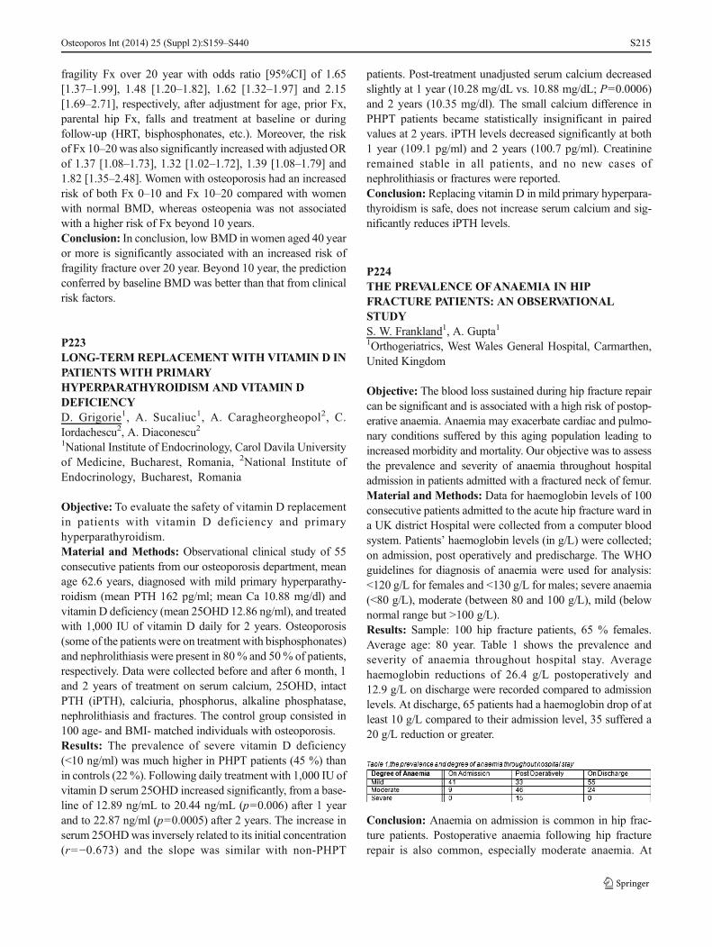
fragility Fx over 20 year with odds ratio [95%CI] of 1.65[1.37–1.99], 1.48 [1.20–1.82], 1.62 [1.32–1.97] and 2.15[1.69–2.71], respectively, after adjustment for age, prior Fx,parental hip Fx, falls and treatment at baseline or duringfollow-up (HRT, bisphosphonates, etc.). Moreover, the riskof Fx 10–20was also significantly increased with adjusted ORof 1.37 [1.08–1.73], 1.32 [1.02–1.72], 1.39 [1.08–1.79] and1.82 [1.35–2.48]. Women with osteoporosis had an increasedrisk of both Fx 0–10 and Fx 10–20 compared with womenwith normal BMD, whereas osteopenia was not associatedwith a higher risk of Fx beyond 10 years.Conclusion: In conclusion, low BMD in women aged 40 yearor more is significantly associated with an increased risk offragility fracture over 20 year. Beyond 10 year, the predictionconferred by baseline BMD was better than that from clinicalrisk factors.
P223LONG-TERM REPLACEMENTWITH VITAMIN D INPATIENTS WITH PRIMARYHYPERPARATHYROIDISM AND VITAMIN DDEFICIENCYD. Grigorie1, A. Sucaliuc1, A. Caragheorgheopol2, C.Iordachescu2, A. Diaconescu21National Institute of Endocrinology, Carol Davila Universityof Medicine, Bucharest, Romania, 2National Institute ofEndocrinology, Bucharest, Romania
Objective: To evaluate the safety of vitamin D replacementin patients with vitamin D deficiency and primaryhyperparathyroidism.Material and Methods: Observational clinical study of 55consecutive patients from our osteoporosis department, meanage 62.6 years, diagnosed with mild primary hyperparathy-roidism (mean PTH 162 pg/ml; mean Ca 10.88 mg/dl) andvitamin D deficiency (mean 25OHD 12.86 ng/ml), and treatedwith 1,000 IU of vitamin D daily for 2 years. Osteoporosis(some of the patients were on treatment with bisphosphonates)and nephrolithiasis were present in 80 % and 50% of patients,respectively. Data were collected before and after 6 month, 1and 2 years of treatment on serum calcium, 25OHD, intactPTH (iPTH), calciuria, phosphorus, alkaline phosphatase,nephrolithiasis and fractures. The control group consisted in100 age- and BMI- matched individuals with osteoporosis.Results: The prevalence of severe vitamin D deficiency(<10 ng/ml) was much higher in PHPT patients (45 %) thanin controls (22 %). Following daily treatment with 1,000 IU ofvitamin D serum 25OHD increased significantly, from a base-line of 12.89 ng/mL to 20.44 ng/mL (p=0.006) after 1 yearand to 22.87 ng/ml (p=0.0005) after 2 years. The increase inserum 25OHDwas inversely related to its initial concentration(r=−0.673) and the slope was similar with non-PHPT
patients. Post-treatment unadjusted serum calcium decreasedslightly at 1 year (10.28 mg/dL vs. 10.88 mg/dL; P=0.0006)and 2 years (10.35 mg/dl). The small calcium difference inPHPT patients became statistically insignificant in pairedvalues at 2 years. iPTH levels decreased significantly at both1 year (109.1 pg/ml) and 2 years (100.7 pg/ml). Creatinineremained stable in all patients, and no new cases ofnephrolithiasis or fractures were reported.Conclusion: Replacing vitamin D in mild primary hyperpara-thyroidism is safe, does not increase serum calcium and sig-nificantly reduces iPTH levels.
P224THE PREVALENCE OFANAEMIA IN HIPFRACTURE PATIENTS: AN OBSERVATIONALSTUDYS. W. Frankland1, A. Gupta11Orthogeriatrics, West Wales General Hospital, Carmarthen,United Kingdom
Objective: The blood loss sustained during hip fracture repaircan be significant and is associated with a high risk of postop-erative anaemia. Anaemia may exacerbate cardiac and pulmo-nary conditions suffered by this aging population leading toincreased morbidity and mortality. Our objective was to assessthe prevalence and severity of anaemia throughout hospitaladmission in patients admitted with a fractured neck of femur.Material and Methods: Data for haemoglobin levels of 100consecutive patients admitted to the acute hip fracture ward ina UK district Hospital were collected from a computer bloodsystem. Patients’ haemoglobin levels (in g/L) were collected;on admission, post operatively and predischarge. The WHOguidelines for diagnosis of anaemia were used for analysis:<120 g/L for females and <130 g/L for males; severe anaemia(<80 g/L), moderate (between 80 and 100 g/L), mild (belownormal range but >100 g/L).Results: Sample: 100 hip fracture patients, 65 % females.Average age: 80 year. Table 1 shows the prevalence andseverity of anaemia throughout hospital stay. Averagehaemoglobin reductions of 26.4 g/L postoperatively and12.9 g/L on discharge were recorded compared to admissionlevels. At discharge, 65 patients had a haemoglobin drop of atleast 10 g/L compared to their admission level, 35 suffered a20 g/L reduction or greater.
Conclusion: Anaemia on admission is common in hip frac-ture patients. Postoperative anaemia following hip fracturerepair is also common, especially moderate anaemia. At
Osteoporos Int (2014) 25 (Suppl 2):S159–S440 S215

discharge, high percentages of patients remain anaemic withsome suffering substantial reductions in their haemoglobincompared to admission. Further analysis to assess the effectof anaemia on morbidity, mortality and length of stay isrequired.
P225EVALUATION OF PERIPHERIC VERTIGO INPOSTMENOPAUSAL OSTEOPOROTIC CASESOVER 65 YEARS OLDS. Atar1, E. Demirhan1, B. Huner1, Y. Atar2, Z. Salturk21Department of Physical Medicine and Rehabilitation,Okmeydani Training and Research Hospital, Istanbul, Turkey,2Department of Otorhinolaryngology, Okmeydani Trainingand Research Hospital, Istanbul, Turkey
Objective: To assess the peripheric vertigo in cases who wasover 65 years old postmenopausal women and had vertigo andbalance disturbance.Material and Methods: Cases who had T-score of ≥−2.5 byDXA was accepted as osteoporosis. 48 osteoporosis casesover 65 years and had vertigo was included to study. Theywere evaluated at ENT clinic and vestibular tests were per-formed. Age, attack frequency and duration, DXA scores,diagnosis, and treatment protocols were analyzed.Results:Themean age of the cases was 69.8±6.2. Duration ofthe complaints was 2.4±1.5 years. 33 cases had less than 6attacks per year, 17 cases had one attack per month, 8 caseshad one attack per week, 2 cases had one attack per day. 35cases had attacks lasting less than a minute, 9 cases hadvertigo up to 15 min and 3 cases had attacks <1 h. Only 1patient had attacks lasting 24 h. The mean DXA score was−2.9±0.3. 34 cases were diagnosed as benign paroxysmalpositional vertigo and 6 cases had vertebrobasillary insuffi-ciency. Moreover, 2 possible Meniere’s disease, 1 benignintracranial hypertension, and 5 psychological vertigo weredetected.Conclusion: Benign paroxysmal positional vertigo was themost common reason of vertigo in the cases who were over65 years and had osteoporosis.
P226IMPACT OF BONE MARROW LESION ON THEPROGRESSION OF KNEE OSTEOARTHRITIS INTHE SEKOIA STUDYC. Parsons1, M. H. Edwards1, O. Bruyère2, F. Petit-Dop3,P. Beliaa3, H. K. Genant4, A. Guermazi4, F. Roemer4,S. Zaim4, R. D. Chapurlat5, J.-Y. Reginster2, E. M. Dennison1,C. Cooper11MRC Lifecourse Epidemiology Unit, Southampton GeneralHospital, Southampton, United Kingdom, 2Department of
Public Health and Health Economics, University of Liège,Liège, Belgium, 3Innovative Therapeutic Pole of Rheumatology,Servier, Suresnes, France, 4Synarc, San Francisco, United States,5INSERM Unit 433, Lyon, France
Objective: Osteoarthritis (OA) is a common degenerativejoint disease that affects around 80 % of those aged over75 years. Many factors appear to influence progression, therate of which is very variable. Established factors include sex,obesity, and prior knee injuries. While imaging of OA wastraditionally achieved using radiographs, more recently, a rolehas been established for MRI. In this study we examined therelationship between bone marrow lesions (BML) visualisedusing MRI and subsequent progression of knee OA, using theplacebo arm of a randomised controlled trial of a therapy forOA.Material and Methods: 559 men and women over 50 yearswith clinical knee OA (K&L 2–3) were recruited to theplacebo arm of the SEKOIA study (98 centre; 18 countries).Minimal tibiofemoral joint space on plain radiograph of theknee was assessed by two independent readers at baseline andyearly follow-up (up to 3 years). In a subset of 176, serial kneeMRIs were performed. Individuals with a BML of grade 2 orabove at the tibiofemoral joint at baseline were classified asBML positive. Relationships between joint space and riskfactors were assessed using linear regression.Results: The mean (standard deviation (SD)) age of studyparticipants was 62.8(7.5) years. 73 % were female and themean(SD) BMI at baseline was 29.8(5.1). Just over one thirdof those studied had BMLs (38.6%). The prevalence of BMLsdid not differ significantly by age or BMI. On average, jointspace reduced by −0.18(0.30) and −0.13(0.23) mm/year inmen and women, respectively. Those with BMLs had a sig-nificantly higher level of annualised joint space narrowing(JSN), with the relationship remaining robust after adjustmentfor age, sex and K&L grade; β(95%CI) -0.10(−0.18,−0.02)mm/year. Age, sex, and baseline K&L grade did not influencerate of JSN.Conclusion: The rate of JSN was similar in men and women.BMLs on knee MRI predicted rate of radiographic JSN. Thisrelationship was independent of age, sex, and baseline K&Lgrade.Disclosures:C. Cooper has received honoraria and consultingfees from Amgen, Eli Lilly, Medtronic, Merck, Novartis andServier. F. Petit-Dop and P. Belissa are employees of Servier.
P227AN OBSERVATIONAL STUDY INVESTIGATINGSERUM CREATININE LEVELS IN HIP FRACTUREPATIENTSS. W. Frankland1, A. Gupta1
Osteoporos Int (2014) 25 (Suppl 2):S159–S440S216

1Orthogeriatrics, West Wales General Hospital, Carmarthen,United KingdomObjective: Kidney dysfunction is common in the elderlyand has been proven to have an association with in-creased hip fracture incidence. Our objective was toassess the renal function of hip fracture patientsthroughout their hospital stay using serum creatininelevels as the marker of renal function.Material and Methods: Data on serum creatinine levelswas collected from records for 100 consecutive patientsadmitted to a hip fracture ward in a UK district Hospi-tal. Patients’ serum creatinine levels were recorded; pre-admission (from previous blood tests), on admission andbefore discharge. Laboratory specific creatinine refer-ence ranges were used for analysis; 44–80 μmol/L(females), 62–106 μmol/L (males). Comparisons weremade between levels pre-admission to discharge (PA-DC) and admission to discharge (OA-DC).Results: 100 hip fracture patients, 65% females. Average age:80 years. Table 1 shows creatinine levels in hip fracturepatients. The average creatinine reduction PA- DC was10.2 μmol/L, OA-DC it was 19.7 μmol/L. A creatinine re-duction of over 20 μmol/L was noted in 26 patients OA-DCbut in only 11 patients PA-DC.
Conclusion: This study suggests substantial percentages ofpatients presenting to hospital with a hip fracture have somedegree of renal impairment as defined by a high creatininelevel. Many of these patients’ creatinine levels normaliseduring their hospital stay. The greatest reductions in creatininewere observed between admission and discharge; this sug-gests a substantial number of patients have acute kidney injuryon admission. As such, reversible causes of renal impairmentshould be actively sought and treated. The contribution ofrenal impairment to postoperative outcomes and bone fragilityneeds further investigation.
P228EFFECTS OF SPACEFLIGHT ON SUBCHONDRALBONE AND ARTICULAR CARTILAGE HEALTH:ARE THEY GOOD NEIGHBORS?L. F. Mellor1, T. Baker2,3, M. Hiremath3, E. G. Loboa1,J. T. Oxford31Joint Department of Biomedical Engineering, North CarolinaState University and UNC Chapel Hill, Raleigh, UnitedStates, 2Biomolecular Research Center, Boise State Universi-ty, Boise, United States, 3Biomedical Research Center, BoiseState University, Boise, United States
Objective: The skeletal system is constantly exposed to me-chanical loading here on Earth, and lack of loading has drasticeffects in skeletal muscles atrophy and bone density loss.However, little is known about the possible effects of micro-gravity on articular cartilage of the synovial joints, andmore importantly, how the changing subchondral bonecan affect the adjacent cartilage integrity. Changes insubchondral bone density are commonly associated withcartilage degradation in osteoarthritis (OA), and there-fore, crosstalk between the two tissues could lead toprogress of pathological conditions. Understanding themolecular mechanisms involved in bone and cartilagemechanotransduction in response to microgravity is cru-cial to identify potential therapeutic targets to develop criticalcountermeasures to prevent skeletal degradation duringspaceflight.Material and Methods: Primary chondrocytes were exposedto simulated microgravity using a rotating wall vessel biore-actor. Morphological changes of the actin cytoskeleton wereevaluated using confocal microscopy. Sclerostin (SOST) ex-pression levels were examined in chondrocytes exposed tosimulated microgravity and compared to levels expressed bycontrol chondrocytes under normal gravity conditions. ELISAwas used to measure secreted sclerostin expression in themedia.Results: Wnt signaling has been shown to be an importantmechanisms in bone mechanostransduction in unloading con-ditions. Our data also shows changes in Wnt signaling inresponse to simulated microgravity in cartilage, however,Wnt activation and inhibitions have opposite effects on adja-cent bone and articular cartilage tissues, and therapeutictargeting against Wnt inhibitors to prevent bone loss needsto be further investigated.Conclusion: This is the first study to integrate the interactionsof articular cartilage and bone in spaceflight conditions toassess synovial joint integrity, and to investigate specificsignaling pathways and mechanisms that result in bone re-sorption and cartilage degradation.
P229EXCELLENTADHERENCE TO 6-MONTHLYDENOSUMAB INJECTIONS DUE TO POSITIVEFEEDBACK BASED ON 6 AND 12 MONTHS BMDINCREASES AND RARE ADVERSE EVENTS INPATIENTS WITH DIFFERENT FORMS OFOSTEOPOROSISJ. D. Ringe1, P. Farahmand11West German Osteoporosis Center (WOC), KlinikumLeverkusen, University of Cologne, Leverkusen, Germany
Objective: About 50 % of osteoporosis patients discontinuebisphosphonate (BP) therapy within the first year of treatment.
Osteoporos Int (2014) 25 (Suppl 2):S159–S440 S217

Denosumab’s (Dmab) longer dosing interval with its s.c.administration every 6 months might result in a better adher-ence than BP treatment regimen.Material andMethods: In an open prospective observationalinvestigator-initiated trial in routine clinical practice we stud-ied whether a medical explanation of treatment results 6 and12 months after the first Dmab injection focussing on signif-icant BMD increases, no or only mild and probably not drugrelated AEs and improvement in back pain have effects onpatients’ drug perception and future adherence with furtherDmab injections. We included 142 patients (69 with postmen-opausal, 42 with male, and 32with GC-induced osteoporosis).Results:Overall, 93% of patients reported no negative chang-es in their health condition after two injections of Dmab, andonly 7.0 % reported AEs. These were all mild to moderate andobviously not drug related. The significant DXA mean BMDincrease rates for all patients at month 6 were +4.7 % at thelumbar spine (LS) and +2.1 % at the total hip (TH), and atmonth 12 +7.8 % at the LS site and +3.7 % at the TH area,respectively. There were only 5 vert. and 4 nonvert. fracturesduring the 142 patient years follow-up. The back pain scoremeasured by VAS 0–10 decreased in all 3 groups significantlyafter 6 and 12 months. This clinical effect together with thescarcity of AEs and the positive feedback of a rapid BMDincrease at both sites resulted for 141 (99 %) patients after6 months and 139 (97 %) after 12 months in a willingness toaccept a further injection.Conclusion:Our results indicate that the convenient treatmentregime together with back pain improvements, very rare ad-verse events and the consistency of rapid and highly signifi-cant BMD increases after 6 and 12 months of Dmab therapyused as a positive reinforcement had a significant, positiveimpact on patients’ adherence to continue with the 6-monthlys.c. Dmab injections.
P230ALTERED COLLAGEN FIBERS DEPOSITION INSYNOVIAL JOINTS OF DIABETIC RATS’ MODELINDUCED BY STREPTOZOTOCINC. Goldenstein-Schainberg1, E. Parra1, W. R. Teodoro1,V. L. Capelozzi1, S. A. Atayde1, J. Morais1, S. Catanozi1,A. P. Velosa1, P. C. Andrade1, W. Teodoro11Reumatologia, Faculdade De Medicina, Universidade DeSao Paulo, Sao Paulo, Brazil
Objective: To analyze synovial collagen (COL) fibers in amodel of rat diabetic joints induced by streptozotocin and theircorrelation with the temporal evolution of the diabetes.Material and Methods: Twenty diabetic Wistar rats inducedby 35mg/kg streptozotocin infusion were divided in 2 groups:G1 (n=10) was euthanized after 2 week and G2 (n=10) after2 month of induction. Control groups (C1=10 and C2=10)
received saline. The experimental protocol complied localanimal experimentation rules and was approved by our ethicscommittee. After euthanasia, weight, blood glucose and plas-matic anticarboximetilisin were analysed. Knee synovial tissueswere included in paraffin, histological sections stained with HEand the amount of total collagen, COL I, COL III and COLVwere evaluated by 4-hydroxyproline analysis, picrosirius redstaining, immunofluorescence and image analysis.Results:Blood glucose was significantly increased in diabeticgroups vs. controls (p<0.005), whereas weight of diabeticanimals reduced compared to controls (p<0.001). Higherquantities of COL were observed by 4-hydroxyproline analy-sis in G2 vs. C2 (p<0.005), similarly for G1 vs. C1 butwithout statistical significance. Morphologic analysis demon-strated substitution of the subsynovial layer fat by fibrotictissue in diabetic groups with important deposition of collagenfibers around small vessels. Histomorphometry analysis re-vealed increased amount of coarse collagen fibers (22.70±8.20) with reduction of fine collagen fibers in G2 (16:29±6:10 %) vs. C2 (14:45±5:39 %, 21:39±6.14, respectively,p<0.05), similarly to G1 vs. C1 but not significantly. Theanalysis of COL I, III and V were statistically higher in G2vs. C2 (p<0.05), though not significant in G1 vs. C1.Conclusion: The morphologic changes observed in synovialtissues from diabetic rats and increased amount of COL fibersdeposition in this structure reinforce the concept that COLfibers deposition and remodeling may be relevant for thepathogenic pathway progression in diabetic patients joints.
P231BONE MASS IS DETERMINED BY NUTRITIONALSTATUS AND PHYSICAL ACTIVITY IN MALE BUTER Α PVU II GENOTYPE OUTWEIGH IN FEMALEIN COMMUNITY-INDWELLING KOREANELDERLYS. Choi1, N. Kwon21College of Nursing, Seoul National University, Seoul,Republic of Korea, 2College of Natural Science, HankukUniversity of Foreign Studies, Seoul, Republic of Korea
Objective: To determine the stiffness index (SI) and gender-specific factors that can be related to SI including gene poly-morphisms vitamin D receptor gene (Bsm I) and two estrogenreceptor (ER) α genes (Xba I and Pvu II) in a Korean elderlycohort.Material andMethods:Data were collected from two nearbysenior centers in Seoul, South Korea, in January and February2009. We investigated socio demographic/lifestyle factors,nutritional status/nutrient intakes, and gene polymorphisms,with relation to the SI.Results: Of the initial 307 subjects, a total of 261 men andwomen aged ≥65 years participated in this study. The mean SI
Osteoporos Int (2014) 25 (Suppl 2):S159–S440S218

was significantly higher for the men than the women(p<0.001). In multiple regression analysis, among elderlymen, age (β=−0.306, p<0.001), physical activity (β=0.243,p=0.003), and nutritional status (β=0.181, p=0.026) weresignificant predictors of SI. Among the elderly women, age(β=−0.252, p=0.002), drinking alcohol (β=−0.241, p=0.003), education level (β=0.234, p=0.005), and PP or Ppgenotype of ER α gene Pvu II (β=0.206, p=0.011) weresignificant predictors of SI. Low SI was common in bothelderly men and women. Gender differences in factors otherthan age can be linked to low SI. In the men, nutritional statusand physical activity were more important factors whereasalcohol consumption, educational level, and genetic polymor-phism were significant factors predicting low SI in thewomen.Conclusion: These gender different lifestyles and nutritionalstatus should be factored into the development of healtheducation programs for osteoporosis prevention in theelderly.
P232COMBINATION AND SEQUENTIALTHERAPY FORSEVERE OSTEOPOROSISS. Marwah11Department of Orthopaedics, Child & Family Research In-stitute, University of British Columbia, Gurgaon, India
Objective: •Concomitant administration of a single infu-sion of intravenous zoledronic acid (5 mg) with dailysubcutaneous teriparatide (20 μg) yielded larger, morerapid increments in both spine and hip BMD than eitheragent alone.•Cotreatment with zoledronic acid also prevented the PTHinduced increase in cortical porosity, thus strengthening thecortex, and increased hip BMD beyond values achieved withPTH alone.Material and Methods: My own experience:•Zoledronic acid infused from 2009 to 2012 August in >300Patients•Reinfusions done in 100 patients till date•PTH cases 235 (since 2005) and 73 PTH cases initiatedin 2013 which is highest globally in the clinicalexperience•Combination PTH followed by ZOL acid 50 patients• Sequential ZOL acid followed by PTH 100 patientsWhen do I use combination therapy?•Age more than 65 years•BMD T score<−3.5•Impending fractures hip and spine•Postoperative implant failure due to severe osteoporosis•In rheumatoid arthritis casesSequential therapy ZOL acid followed by PTH
My protocol:•ZOL 5 mg infusion•ZOL infusion followed by PTH For 2 years•2nd dose of ZOL acid at first and second year dependingupon requirement of the patientResults: •BMD increased in all patients•No secondary fracture observed•Combination treatments were well tolerated•PTH followed by ZOL acid now extending up to 6–9 yearsPTH followed by Zoledronic acid:• Since 2009•After PTH all cases put on ZOL acid - minimum duration3 yearsConclusion: •These observations in the combination grouplikely result from•PTH-induced increases in osteoblastic activity, together with•Zoledronic acid-induced reductions in bone remodelling andcortical porosity•Combination therapy might therefore be appropriate treat-ment for patients at with very low hip BMD, high risk for hipand other fractures, rheumatoid arthritis or patients in whomrapid response is required.
P233BASELINE CHARACTERISTICS OFAPROSPECTIVEOBSERVATIONAL STUDY INCZECHREPUBLIC (CZE) AND SLOVAKIA (SVK) TODESCRIBE MANAGEMENT OF PATIENTS WITHPOSTMENOPAUSAL OSTEOPOROSIS (PMO)RECEIVING DENOSUMAB (DMAB) IN ROUTINECLINICAL PRACTICEP. Hrdý1, P. Kasalický2, S. Tomkova3, M. Feudjo-Tepie4,E. Zhang4, L. Kalouche-Khalil5, Z. Killinger61MEDIEKOS Ambulance, Osteocentrum Zlín, Czech Repub-lic, 2Bone Metabolism Unit, Mediscan Group, Prague, CzechRepublic, 3Osteocentrum, Kosice-Saca, Slovakia, 4AmgenLtd, Zug, United Kingdom, 5Amgen (Europe) GmbH, Zug,Switzerland, 65th Department of Internal Medicine, MedicalFaculty of Comenius University, University Hospital,Bratislava, Slovakia
Objective: Describe the characteristics of PMO women initi-ating DMAb in clinical practice in CZE and SVK.Material and Methods: The study enrolled PMO women inCZE and SVK who had received their first DMab injection inthe previous 8 weeks. Data recorded as per standard clinicalpractice is collected for 2 years after enrolment, with noadditional procedures required. Study outcomes include pa-tient characteristics and clinical osteoporosis (OP) manage-ment. We report baseline data.Results: As of July 2013, 600 patients had enrolled across 32centers (300 patients per country). In both countries, patients
Osteoporos Int (2014) 25 (Suppl 2):S159–S440 S219
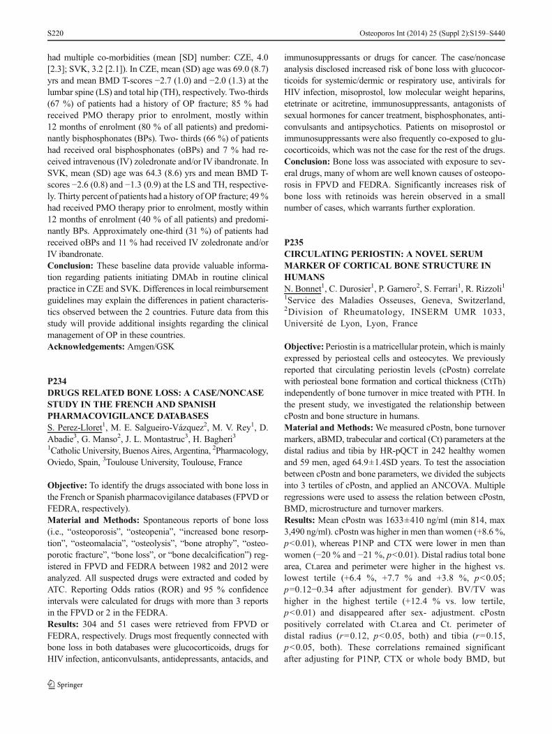
had multiple co-morbidities (mean [SD] number: CZE, 4.0[2.3]; SVK, 3.2 [2.1]). In CZE, mean (SD) age was 69.0 (8.7)yrs and mean BMD T-scores −2.7 (1.0) and −2.0 (1.3) at thelumbar spine (LS) and total hip (TH), respectively. Two-thirds(67 %) of patients had a history of OP fracture; 85 % hadreceived PMO therapy prior to enrolment, mostly within12 months of enrolment (80 % of all patients) and predomi-nantly bisphosphonates (BPs). Two- thirds (66 %) of patientshad received oral bisphosphonates (oBPs) and 7 % had re-ceived intravenous (IV) zoledronate and/or IV ibandronate. InSVK, mean (SD) age was 64.3 (8.6) yrs and mean BMD T-scores −2.6 (0.8) and −1.3 (0.9) at the LS and TH, respective-ly. Thirty percent of patients had a history of OP fracture; 49%had received PMO therapy prior to enrolment, mostly within12 months of enrolment (40 % of all patients) and predomi-nantly BPs. Approximately one-third (31 %) of patients hadreceived oBPs and 11 % had received IV zoledronate and/orIV ibandronate.Conclusion: These baseline data provide valuable informa-tion regarding patients initiating DMAb in routine clinicalpractice in CZE and SVK. Differences in local reimbursementguidelines may explain the differences in patient characteris-tics observed between the 2 countries. Future data from thisstudy will provide additional insights regarding the clinicalmanagement of OP in these countries.Acknowledgements: Amgen/GSK
P234DRUGS RELATED BONE LOSS: A CASE/NONCASESTUDY IN THE FRENCH AND SPANISHPHARMACOVIGILANCE DATABASESS. Perez-Lloret1, M. E. Salgueiro-Vázquez2, M. V. Rey1, D.Abadie3, G. Manso2, J. L. Montastruc3, H. Bagheri31Catholic University, Buenos Aires, Argentina, 2Pharmacology,Oviedo, Spain, 3Toulouse University, Toulouse, France
Objective: To identify the drugs associated with bone loss inthe French or Spanish pharmacovigilance databases (FPVD orFEDRA, respectively).Material and Methods: Spontaneous reports of bone loss(i.e., “osteoporosis”, “osteopenia”, “increased bone resorp-tion”, “osteomalacia”, “osteolysis”, “bone atrophy”, “osteo-porotic fracture”, “bone loss”, or “bone decalcification”) reg-istered in FPVD and FEDRA between 1982 and 2012 wereanalyzed. All suspected drugs were extracted and coded byATC. Reporting Odds ratios (ROR) and 95 % confidenceintervals were calculated for drugs with more than 3 reportsin the FPVD or 2 in the FEDRA.Results: 304 and 51 cases were retrieved from FPVD orFEDRA, respectively. Drugs most frequently connected withbone loss in both databases were glucocorticoids, drugs forHIV infection, anticonvulsants, antidepressants, antacids, and
immunosuppressants or drugs for cancer. The case/noncaseanalysis disclosed increased risk of bone loss with glucocor-ticoids for systemic/dermic or respiratory use, antivirals forHIV infection, misoprostol, low molecular weight heparins,etetrinate or acitretine, immunosuppressants, antagonists ofsexual hormones for cancer treatment, bisphosphonates, anti-convulsants and antipsychotics. Patients on misoprostol orimmunosuppressants were also frequently co-exposed to glu-cocorticoids, which was not the case for the rest of the drugs.Conclusion: Bone loss was associated with exposure to sev-eral drugs, many of whom are well known causes of osteopo-rosis in FPVD and FEDRA. Significantly increases risk ofbone loss with retinoids was herein observed in a smallnumber of cases, which warrants further exploration.
P235CIRCULATING PERIOSTIN: A NOVEL SERUMMARKER OF CORTICAL BONE STRUCTURE INHUMANSN. Bonnet1, C. Durosier1, P. Garnero2, S. Ferrari1, R. Rizzoli11Service des Maladies Osseuses, Geneva, Switzerland,2Division of Rheumatology, INSERM UMR 1033,Université de Lyon, Lyon, France
Objective: Periostin is a matricellular protein, which is mainlyexpressed by periosteal cells and osteocytes. We previouslyreported that circulating periostin levels (cPostn) correlatewith periosteal bone formation and cortical thickness (CtTh)independently of bone turnover in mice treated with PTH. Inthe present study, we investigated the relationship betweencPostn and bone structure in humans.Material and Methods:We measured cPostn, bone turnovermarkers, aBMD, trabecular and cortical (Ct) parameters at thedistal radius and tibia by HR-pQCT in 242 healthy womenand 59 men, aged 64.9±1.4SD years. To test the associationbetween cPostn and bone parameters, we divided the subjectsinto 3 tertiles of cPostn, and applied an ANCOVA. Multipleregressions were used to assess the relation between cPostn,BMD, microstructure and turnover markers.Results: Mean cPostn was 1633±410 ng/ml (min 814, max3,490 ng/ml). cPostn was higher in men than women (+8.6 %,p<0.01), whereas P1NP and CTX were lower in men thanwomen (−20 % and −21 %, p<0.01). Distal radius total bonearea, Ct.area and perimeter were higher in the highest vs.lowest tertile (+6.4 %, +7.7 % and +3.8 %, p<0.05;p=0.12−0.34 after adjustment for gender). BV/TV washigher in the highest tertile (+12.4 % vs. low tertile,p<0.01) and disappeared after sex- adjustment. cPostnpositively correlated with Ct.area and Ct. perimeter ofdistal radius (r=0.12, p<0.05, both) and tibia (r=0.15,p<0.05, both). These correlations remained significantafter adjusting for P1NP, CTX or whole body BMD, but
Osteoporos Int (2014) 25 (Suppl 2):S159–S440S220

not for gender. There was no difference in whole body,spine and proximal femur aBMD, PTH, P1NP and CTXbetween tertiles. Separate analyses per tertile in menand women indicated similar, but nonsignificant trends.Conclusion: These results recorded in a homogeneous popu-lation of healthy 65-year old subjects indicate a positivecorrelation of cPostn with cortical bone structure, indepen-dently of bone turnover markers as previously observed inmice, but likely through a sex-dependent effect.
P23612-MONTH PERSISTENCE WITH DENOSUMABTHERAPYAMONG OSTEOPOROTIC WOMEN INTHE CANADIAN PATIENT-SUPPORT PROGRAMA. Papaioannou1, A. Khan1, A. Belanger2, W. Bensen1, D. L.Kendler3, F. Theoret4, M. Amin5, L. Brekke6, M. Erdmann7,V. Popovic7, V. Walker6, J. D. Adachi11Medicine, McMaster University, Hamilton, Canada, 2FamilyMedicine, Courcelette, Canada, 3Medicine, University of Brit-ish Columbia, Vancouver, Canada, 4Family Medicine,Hawkesbury, Canada, 5Amgen Inc., Mississauga, Canada,6Optum, Burlington, Canada, 7Formerly Amgen Inc.,Mississauga, Canada
Objective: To evaluate persistence with denosumab (DMAb)among women with osteoporosis participating in theCanadian patient-support program (ProVital).Material and Methods: DMAb is an injectable therapeuticoption for osteoporosis that is administered subcutaneouslyevery 6 months. ProVital, a support program in which patientsvoluntarily enrol, provides next injection reminder calls andeducational material. A retrospective database analysis ofpatient-reported outcomes was conducted among osteoporoticwomen aged ≥50 who enrolled in the ProVital program andreceived their 1st DMAb injection between August 2010 andJune 2011. To achieve 12-month persistence, patients had toreceive their 2nd injection no more than 6 months + 8 weeksfollowing the 1st.Results: A total of 1,676 patients (mean age 74 year) wereincluded. At baseline, 43 % of patients reported previousfractures; 13% had a parent with a fracture; 14% had arthritis;2 % had secondary osteoporosis; 10 % used glucocorticoids;79 % used a bisphosphonate previously; 51 % had private and37 % public drug coverage. The 12-month persistence was82 % (1367/1676). Of the 1,676 patients, 1,419 received asecond DMAb injection (mean 192, min 120, max 852 daysapart). In a multivariate regression model, characteristics thatpredicted persistence were private medication insurance, noglucocorticoid use, and residence in Quebec. Among patientswho discontinuedDMAb (136), the most common reasonwasadverse events (69 patients).
Conclusion: The 12-month persistence with DMAb amongpatients enrolled in the ProVital program was over 80 %.Disclosures: MA is an employee of and shareholder inAmgen, and VP and ME are former Amgen employees. VWand LB are employees of Optum contracted by Amgen. Thefollowing relate to Amgen: AP, AK, WB, DK, JA receivedresearch funding. AP, AB, AK, WB, DK, JA, FT were con-sultants. AP, WB, JA, DK, FT were speakers. DK, AB, AK,FT were advisors. AP, AK, DK received honoraria.Acknowledgements: This study was sponsored by Amgen.Jesse Potash (Optum) provided writing assistance.
P237ELBOWARTHROPLASTY IN COMPLEXOSTEOPOROTIC FRACTURESA. Miti1, D. Katusic1, E. Chiarello11Orthopaedic and Traumatologic Unit Ospedale dell’angelo,Venezia Mestre, Italy
Objective: Osteoporotic elbow fractures (EF) represent achallenge. Open reduction and internal fixation is hard toachieve due to poor mechanical bone proprieties, for thisreason elbow arthroplasty (EA) is increasingly popularity incase of comminuted fractures of the distal humerus and isassociated with a rapid recovery of elbow motion. The aim ofthis study is to evaluate clinical outcomes of our series.Material and Methods: From September 2007 to March2013 we performed 30 EA in 29 patients; 25 females and 4males. All patients had poor bone quality with a distal humeralfracture or a complex EF or a terrible triad of the elbow. Thesurgical approach used was a universal posterior incision withtriceps preservation. At each follow-up we performed x-raysof the elbow and a clinical evaluation with the Mayo ElbowPerformance Score.Results: Mean age was 72 years (range 45–94); we reportedan isolated distal humeral fracture in 15 cases; a complex EFin 10 and a terrible triad of the elbow in 5. In 2 cases wasnecessary using a homologous bone graft. In 28 cases a totalEA (18 Coonrad-Morrey TEA Zimmer; and 10 Latitude TEAToriner); one patient received an hemiarthroplasty (Latitudehumeral component) and in another case a custom-madeprosthesis was implanted (LINK). In 2 cases a supplementaryosteosynthesis was performed. Good to excellent functionalresults were reported in 24 cases (80 %) and only in 6 casesfair and poor results were recorded:3 traumatic periprostheticfractures, one prosthesis loosening due to an insufficient stemcementation and 2 superficial infections treated with antibi-otics. Despite poor biomechanical properties of the osteopo-rotic bone in elderly patients, our results seem to be slightsuperior to than reported in literature, this can be probablyattributed to the surgical approach that preserves both theolecranon and the triceps extensor mechanism.
Osteoporos Int (2014) 25 (Suppl 2):S159–S440 S221

Conclusion: In case of complex osteoporotic EF we preferuse EAwith a triceps preserving approach.
P238STRONTIUM RANELATE TREATMENT IMPROVESBONE MATERIAL LEVEL PROPERTIES ANDMICROARCHITECTURE OF HUMAN TRANSILIACBONE BIOPSY SPECIMENSP. Ammann1, R. Rizzoli11Division of Bone Diseases, Department of Internal medicineSpecialties, University Hospital Geneva, Geneva, Switzerland
Objective:Bone strength, hence fracture risk, is dependent onbone geometry, microstructure and bone material level prop-erties.We previously reported that microstructure and materiallevel properties contribute independently to the increase inbone strength of rats treatedwith strontium ranelate for 2 years,as evaluated by μCT-based finite element analysis. We inves-tigated the effects of strontium ranelate (SrRan) treatment onbone material level properties of transiliac bone biopsy and onbonemicroarchitecture from postmenopausal osteoporotic pa-tients in three studies.Material and Methods: In a longitudinal study, 84 pairedbiopsies were obtained at baseline, and after 6 or 12 months oftreatment with 2 g/day SrRan. In SOTI/TROPOS studies 3paired biopsies were obtained at baseline and after 36 monthsof treatment. Elastic modulus, hardness and working energywere blindly analyzed by nanoindentation at the level of theinterstitial bone of the cortex and of trabecular nodes underhumid conditions. Parameters of microarchitecture wereevaluated μCT (Scanco Medical). Values correspond todifferences expressed in percent between 2 paired biopsies.Significance of differences are evaluated by Student’s un-paired t-test,* <0.05, **p<0.01, ***p<0.001.Results:
Conclusion: Overall, these results detected in 90 humanbiopsies indicates an early improvement of bonematerial levelp roper t i e s fo l lowed by la t e r changes on bonemicroarchitecture in bone specimens collected in patientstreated with SrRan. Both effects could contribute to increaseof bone strength and to fracture risk reduction. These resultssuggest different kinetics of SrRan action on bonemicroarchitecture and bone material level properties.
Disclosures: The study was supported by a grant oflaboratoire Servier.
P239ILIOCOSTAL IMPINGEMENT SYNDROME/FLANKAND BACK PAIN IN OSTEOPOROSIS/OSTEOPENIA:SUCCESSFUL MANAGEMENT THROUGH SPINALPROPRIOCEPTIVE EXTENSION EXERCISEDYNAMIC (SPEED) PROGRAMM. Sinaki11Mayo Clinc, Rochester, Minnesota, United States
Objective: With bone loss spinal deformities develop subse-quent to the repetitive strain beyond biomechanical compe-tence of the spine. Among the complications related to Ky-phosis we will address flank pain or iliocostal impingementsyndrome. There is a paucity of literature on the managementof this syndrome. Some of the recommended interventionsinclude injections or bracing.Material and Methods: Forty patients with osteopenia orosteoporosis with flank or back pain or both who had notresponded to common interventions were included. AP andlateral spine x-rays were obtained on all patients. Radiographsrevealed kyphosis with reduced space between lower ribs andthe ilium. All had neurological and musculoskeletal evalua-tions. They were all instructed in a home exercise program,with emphasis on back extensor muscle strengthening inaddition to education on the use of a weighted kypho-orthosis (WKO) to be worn 20–30 min twice to three timesdaily until the patient could perform back exercises withoutpain.Results:Before enrolment all had a trial ofWKO, if their painwas reduced they were included in the study. Considering thegeographic location 16 were available for follow up assess-ment and x-ray studies at 1 month while the remaining 24were reviewed upon data from their routine annual visits. Nonew compression fractures were seen. This group showedstatistically significant improvement in pain scale (P=0.001), height, back strength, level of physical activity(P<0.001)/quality of life.Conclusion: Proprioceptive re-education of posture throughapplication of WKO is effective for static reduction of kypho-sis and facilitation of back extension exercise program. Thismethod is based on spinal facet joint proprioceptive reeduca-tion and kyphosis reductions.References: 1) Sinaki M et al. Mayo Clin Proc 2005;80:849.2) Sinaki M et al. Osteoporos Int 2005;16:1004.3) Pfeifer M et al. J Bone Miner Res 2004;19:1208.4) Sinaki M. Phys Med Rehabil Clin N Am 2007;18:593,xi–xii.5) Huntoon EA et al. Mayo Clin Proc 2008;83:54.
Osteoporos Int (2014) 25 (Suppl 2):S159–S440S222

P240COMPLEX ASSESSMENT OF OSTEOPOROTICFEMALES WITH DEGENERATIVE LUMBARSPONDYLOLISTHESISR. Traistaru1, O. Rogoveanu1, D. Matei1, R. S. Popescu11University of Medicine and Pharmacy of Craiova, Craiova,Romania
Objective: Females will develop osteoporosis and degenera-tive spondylolisthesis (DS) over males. DS is a commonpathologic entity of the lumbar spine. In most females willdevelop osteoporosis and degenerative spondylolisthesis (DS)over cases of this complex type pathology, certain muscles ofmales. DS is a common pathologic entity of the lumbar spine.In most cases of this complex type pathology, certain musclesof the back that stabilize the spine are reflexively inhibited anddo not spontaneously recover, even if patients are painfree with a return to daily activity levels. First, weinvestigated the incidence of DS at L4-L5 level infemales with osteoporosis and low back pain (LBP).Second, we assessed the thickness of the erector spinae(ES) muscle in these patients and evaluated the correlationbetween thickness difference in three different trunk posturesand functional parameters.The back that stabilize the spine are reflexively inhibitedand do not spontaneously recover, even if patients arepain free with a return to daily activity levels. First, weinvestigated the incidence of DS at L4-L5 level in femaleswith osteoporosis and low back pain (LBP). Second, weassessed the thickness of the erector spinae (ES) muscle inthese patients and evaluated the correlation betweenthickness difference in three different trunk posturesand functional parameters.Material and Methods: 48 LBP females with osteoporosisand DS at L4-L5 were enrolled in this observationstudy. Frontal (AP) and lateral lumbosacral regions wereradiological evaluated. Clinical and functional parame-ters were collected by a physiotherapist and an USexamination was performed by a physiatrist within72 h of the clinical examination. We performed ultrasonog-raphy to measure the thickness of the ES muscle at L4 andL5 level in maximum flexion, neutral posture, and maximumextension. All collected clinical and imagistic data werestatistically analyzed.Results: There was significant correlation between the degreeof anterior slippage of L4 on L5 and bone density (T-scoreDXA exam). Multivariate analysis showed that thicknessdifferences between flexed and neutral, and flexed and ex-tended maximally positions were correlated statistically withfunctional parameters.Conclusion:A relationship between transitional the degree ofslippage in DS females with vertebral osteoporosis has beenestablished. Though sacralization and anterior slippage
of L4 on L5 are associated with stability of that seg-ment, pain is more likely to arise in correlation with theerector vertebral muscle status. Visual observation of theimage during contraction indicates that US may be avaluable biofeedback tool.
P241CORRELATION BETWEEN METABOLIC STATUSAND REHABILITATION PARAMETERS IN KNEEOSTEOARTHRITIS PATIENTSR. Traistaru1, D. Trasca1, O. Rogoveanu1, A. Bighea11University of Medicine and Pharmacy of Craiova, Craiova,Romania
Objective: Osteoarthritis of the knee (OAK) is reported to bea major health problem worldwide. OAK has been linked notonly to obesity but also to other metabolic factors. The adap-tation of rehabilitation program to the metabolic status ofpatient is important for the maintaining of an optimal func-tional status. The aim of this randomized and controlled studywas double: 1) to establish the correlation between the meta-bolic risk factors and the severity of knee osteoarthritis pro-cess, 2) to assess the efficacy of comprehensive rehabilitationprogram associated with viscosupplementation.Material andMethods: 56 patients with OAKwere random-ly assigned into studied group (SG - 30 patients) and controlgroup (CG - 26 patients). All patients were completedassessed (clinical, functional, lab and imagistic). SG per-formed a rehabilitation program (medication, physical therapyand kinetotherapy) followed by joint injection with hyaluronicacid. CG stayed at home and followed their individuallyprescribed drug therapy. Patients were assessed at baseline(week 0), after 2 weeks and during follow-up period at16 weeks. We monitored the following outcomes: pain(VAS), BMI, serum level of glucose, lipoprotein profile, andWOMAC scale.Results: The severity of OAK was correlated with the pres-ence and number of the risk metabolic factors. Comparing thetwo group differences, the SG was superior to CG in painreduction and in physician’s global assessment at all timepoints. Significant improvement in pain and WOMAC scoreswere found at week 2 and week 16 in the SG compared tobaseline.Conclusion: Our study demonstrated the superiority of reha-bilitation program that included the viscosupplementationcompared to drug therapy in the treatment of patientswith OAK. The superior efficiency of pain and qualityof life in the metabolic syndrome patients that followeda complete rehabilitation program confirm the medicaldata about the chondrocytes disturbances in metabolicsyndrome.
Osteoporos Int (2014) 25 (Suppl 2):S159–S440 S223

P242OSTEOGENIC DIFFERENTIATION ANDMINERALIZATION OFADULT MESENCHYMALSTEM CELLS ISOLATED FROM HUMANPERIODONTAL LIGAMENTC. Romagnoli1, N. Nuti1, R. Zonefrati1, F. S. Martelli2,M. Martelli2, E. Fanti2, M. Duvina1, P. Tonelli1, L. Cianferotti1,A. Tanini1, M. L. Brandi11University of Florence, Department of Surgery and Transla-tional Medicine, Florence, Italy, 2I.R.F. in Microdentistry,Florence, Italy
Objective: Periodontal ligament (PDL) is a dynamicconnective tissue embedded between the cementum andthe alveolar bone; its role is to anchor the tooth rootwithin the jaw, cushioning mechanical load that precedefrom mastication. Since mesenchymal stem cells havebeen isolated from PDL (PDLSCs), they are supposedto have a great potential in regenerative dentistry forrepairing bone defects, caused by periodontal diseases.Aim of the study was to isolate PDLSCs and to evalu-ate their osteogenic potential analyzing the alkalinephosphatase (ALP) activity and the mineralizationprocess.Material and Methods: Two primary cultures of PDLSCs,obtained from extracted healthy human third molars, wereinduced towards the adipogenic phenotype with theadipogenic medium (AM) (Ham’s F12 Coon’s modificationmedium, 10 % FBS, 1 μM dexamethasone, 10 μM bovineinsulin, 0.5 mM isobutylmethylxanthine, 100 μM indometh-acin, 1 % antibiotics). The osteoblastic phenotype was in-duced with the osteogenic medium (OM) (Ham’s F12 Coon’smodification medium, 10 % FBS, 10 nM dexamethasone,10 mM β-glycerophosphate, 50 μg/ml L-ascorbic acid 2-phosphate, 1 % antibiotics). The phenotypes were assessedfrom 4 to 21 days by cytochemical staining (Oil Red O or FastBlue BB/naphthol AS-MX) and microscopic observations.The ALP activity and Ca2+ deposition were quantified byfluorometric assay.Results: With AM, accumulation of intracellular lipid-filleddroplets after 21 days was observed. With OM, significantincrements of ALP activity at 4 days (+104 %) and7 days (+86 %) vs. control were observed, and thenALP decreases, while mineralization increases at 14 days(+316 %) and 21 days (+814 %), as demonstrated by agreat deposition of Ca2+ vs. control.Conclusion: Preliminary data suggest PDL as an optimalsource of stem cells that can be used for bone regener-ation in dentistry. Studies are in progress to evaluate theeffect of bioactive factors that could facilitate theprocess.
Acknowledgements: Study supported by Institut deRecherches Internationals Servier and Technologie Servier.
P243SERUM LEVELS OF PROCOLLAGEN TYPE 1AMINO-TERMINAL PROPEPTIDE (P1NP) ANDRISKOF HIP FRACTURE IN ELDERLY WOMEN. THEHORDALAND HEALTH STUDYT. E. Finnes1, C. M. Lofthus2, H. E. Meyer3, E. F. Eriksen2,E. M. Apalset4, G. S. Tell5, K. Holvik41Department of Internal Medicine, Innlandet Hospital, Trust,Hamar, Norway, 2Department of Endocrinology, Oslo Uni-versity Hospital, Oslo, Norway, 3Institute of Health and Soci-ety, University of Oslo, Oslo, Norway, 4Department of GlobalPublic Health and Primary Care, University of Bergen, Nor-wegian Institute of Public Health, Oslo, Norway, 5Departmentof Global Public Health and Primary Care, University ofBergen, Bergen, Norway
Objective: Background: The usefulness of bone turnovermarkers as determinants of fracture risk in osteoporosis needsfurther investigations, and IOF/IFCC recommends the boneformation marker procollagen type 1 amino-terminalpropeptide (P1NP) to be used in studies to clarify its utilityin clinical use. Aim: To investigate the relation between serumP1NP levels and the risk of subsequent hip fractures in elderlywomen with, and without BMD.Material and Methods: A case-cohort study was performedin a cohort of 1,817 women born 1924–1927 participating in ahealth study covering western parts of Norway from 1997 to1999. In a subsample of about 60 % of the women, baselinetotal hip BMD was measured by a DXA (Lunar Expert-XL).Information on incident hip fractures was obtained from com-puterized discharge diagnoses records from the hospitals serv-ing the region. P1NP was measured in frozen serum samplesobtained at baseline in all women who suffered a hip fracture,and in a randomly collected subcohort of 9 % of the totalcohort. P1NP was determined by Multigamma radioimmuno-assay kit (Orion Diagnostica). The current analyses includewomen not medication known to influence on the P1NP level,and with valid BMD and P1NP measurements. The final dataset included 72 women, without a hip fracture, from thesubcohort, and 80 women with a hip fracture. Cox propor-tional hazards regression adapted for the case-cohort designwas performed with penalized splines of P1NP and withtertiles of serum P1NP as explanatory variables. The lowesthazard ratio (HR) was observed in the 2nd tertile, which wasused as reference. All analyses were adjusted for age bymethod.
Osteoporos Int (2014) 25 (Suppl 2):S159–S440S224

Results: Serum P1NP levels ranged from 13 to 124 ng/mL(median 44 ng/mL). Unadjusted spline analysis revealed nosignificant relation between P1NP and the risk of hip fracture.Compared to the 2nd tertile of P1NP, HR for hip fracture in the1st tertile was: 1.26 (95%CI: 0.60–2.65), and in the 3rd tertile:1.69 (95%CI: 0.53–2.53). Adjusted for baseline BMD, asignificant higher risk was seen in the 1st tertile of P1NP,compared to the 2nd tertile (HR: 3.37 (95%CI: 1.24–8.61),p=0.012).Conclusion: A weak relation between P1NP and hipfractures was found in the unadjusted analyses. Adjustedfor BMD, the current analyses revealed low P1NP to bea risk factor for hip fractures. The mechanisms causinghigh fracture risk among those with the lowest P1NPlevels remain to be elucidated.
P244GENDER DIFFERENCES IN VITAMIN D STATUSAND PARAMETERS OF CALCIUM AND BONEMETABOLISML. Y. Rozhinskaya1, E. A. Pigarova1, L. K. Dzeranova1,G. A. Melnichenko2, A. V. Il’in21Neuroendocrinology and Bone Diseases, EndocrinologyResearch Centre, Moscow, Russian Federation, 2EndocrinologyResearch Centre, Moscow, Russian Federation
Objective:Vitamin D deficiency is widespread in most of theworld and is associated with low sun exposure and lowvitamin D intake. Our goal was to identify the gender differ-ences in the levels of 25(OH)D and parameters of calcium andbone metabolism.Material and Methods: The study included patients (n=163, 30M/133F; mean age 48.5±18 years) in whichlevels of total 25(OH)D (Liaison, DiaSorin) and PTHwere measured during autumn period (September–Octo-ber). In selection of patients we used exclusion criteria:presence of primary hyperparathyroidism, secondary ortertiary hyperparathyroidism on the background terminalchronic renal failure, hypercortisolism, blood creatininelevel of >100 mmol/l or GFR <60 ml/min/1.73 m2,intake of active vitD metabolites within 1 month priorthe blood test. Our lab takes part in the internationalprogram of external control and standardization of vitDin the blood (DEQAS, UK).Results: In males, levels of vitD were significantly lower thanin females (16±5.9 vs. 20±8.5 ng/ml, respectively) despitethe fact that males were generally younger (mean age 42±18.5vs. 51±17.8 years old, respectively). There was a noticeabletrend to higher levels of PTH in men (62.6±40 vs. 52.1±
30 pg/ml, respectively), but the differences were not con-firmed statistically. Differences between the groups by otherparameters were not identified. Correlation analysis noted asimilar negative association between vitD and age in bothsexes, the relationship of vitD with total Ca, P, PTH, OK,CTX were unidirectional, but statistically significant onlyin women, which can be explained by fewer men in thestudy group. At the same time correlations of vitD andionized calcium were reciprocal in men (r=0.60) andwomen (r=−0.19).Conclusion: VitD levels significantly lower in men than inwomen, with gender-related reciprocal relationship betweenvitD levels and blood ionized calcium.Acknowledgements: This work was supported by the Grantof the President of Russian Federation НШ-4527.2014.7.
P245PREDICTORS OF SECOND FRACTURE WHILE ONTREATMENT WITH ORAL BISPHOSPHONATES: AMULTINATIONAL RETROSPECTIVE COHORTSTUDYS. Hawley1, G. Wallace1, M. K. Javaid1, K. H. Rubin2, A.Judge1, N. K. Arden1, P. Vestergaard3, R. Eastell4, A. Diez-Perez5, C. Cooper6, B. Abrahamsen7, D. Prieto-Alhambra11Nuffield Department of Orthopaedics, Rheumatology andMusculoskeletal Sciences, University of Oxford, Oxford,United Kingdom, 2Odense Patient Data Exploratory Network,Institute of Clinical Research, University of Southern Den-mark, Odense C, Denmark, 3Department of Endocrinology,Aalborg University Hospital, Aalborg, Denmark, 4Depart-ment of Human Metabolism, University of Sheffield, Shef-field, United Kingdom, 5Hospital del Mar, Barcelona, Spain,6MRC Lifecourse Epidemiology Unit, University of South-ampton, Southampton, United Kingdom, 7Research Centrefor Ageing and Osteoporosis, Department of Medicine M,Glostrup Hospital, Glostrup, Denmark
Objective: To identify predictors of inadequate response tooral bisphosphonate therapy, defined as the incidence of ≥2fractures while on treatment among incident oral bisphospho-nate users with high refill compliance (≥80 %).Material andMethods:Data from computerized records andpharmacy invoices were obtained from SIDIAP (Catalonia,Spain) and Danish Health Registries (Denmark) for all inci-dent users of oral bisphosphonates in 2006–2007 and 2000–2001 respectively. Exclusion criteria were: Paget disease, age<40 years, anti- osteoporosis treatment in the previous year,and suboptimal refill compliance (<80 %). Fine and Graysurvival models accounting for the competing risk of therapy
Osteoporos Int (2014) 25 (Suppl 2):S159–S440 S225

cessation were used to identify predictors of ≥2 fractures whileon treatment after 6 months of treatment initiation.Results: 7,449/21,385 (34.8 %) and 7,885/13,949 (56.5 %)were compliant oral bisphosphonate users in Catalonia andDenmark respectively. Significant predictors of ≥2 fractureswhile on treatment in Catalonia and Denmark were older age,and history of recent fracture. Sub-hazard ratios (SHRs) foreach of the predictors in each of the datasets are reported inFigure 1.
Figure 1. SHR for the identified key predictors of ≥2 fractureswhile on treatment in Catalonia, Spain (CAT) and Denmark(DK).Conclusion: Older age and recent fracture history are predic-tors of inadequate response as confirmed in two separatecohorts. Monitoring strategies and/or alternative therapiesshould be considered for these patients.
P246PREVALENCEOFVITAMINDDEFICIENCY INTWOBULGARIAN TOWNS–ROUSSE AND SANDANSKY(IN THE NORTH AND SOUTH PART OF THECOUNTRY)A.M. Borissova1, A. Shinkov1, J. Vlahov1, L. Dakovska1,T. Todorov1, L. Kassabova2, D. Svinarov21University Hospital of Endocrinology, Medical University ofSofia, Sofia, Bulgaria, 2Central Laboratory of Therapeutic DrugManagement and Clinical Pharmacology, Alexander Universi-ty Hospital, Medical University - Sofia, Sofia, Bulgaria
Objective: Bulgaria is situated between 41°N and 44°N andthe climate is characterized by four seasons with differentelevation of the sun over the horizon. The aim of the studywas to determine the seasonal changes in the 25(OH)D levelsin subjects with confirmed winter vitamin D deficiency25(OH)D <25 nmol/l in two Bulgarian cities–Rousse in thenorth and Sandanski in the south of the country.
Material and Methods: Thirty subjects from each city (total60, 43 women and 17 men) had 25(OH)Dmeasured in spring,summer and autumn. Three age groups were defined: 20–44(n=29), 45–59 (n=20) and ≥60 (n=11).Results:Nodifferenceswere found between the two cities in theprevalence of vitamin D deficiency in the four seasons exceptsummer. The prevalence of low 25(OH)D was significantlylower in spring, summer and autumn vs. winter (all p>0.05).In the elderly women in Rousse the deficiency was sustained atall measurements. The prevalence of D deficiency did not differbetween the genders. The men from both towns and age groupsand the young and middle-aged women from Sandanski had nodeficiency in the summer. No deficiencywas found in spring andautumn among the young and middle-aged subjects with theexception of the young men in Sandanski. It could be speculatedthat the latter related to in-house occupations.Conclusion: No difference was found between the northernand southern cities. Sun exposure in both towns is not suffi-cient to ensure the recommended 25(OH)D levels in round theyear. Probably other factors such as occupation or air pollutionplay a role in the cities.
P247C.O.D.E. STUDY: CONNECTIONS BETWEENOUTCOMES OF OSTEOPOROTIC FRACTURES,DEPRESSION, DELIRIUM & DEMENTIA IN THEELDERLYA. Metozzi1,2, M. L. Brandi2, D. Black3, A. Fasano4, G. L.Di Tanna5, P. Piscitelli6, S. Parri2, A. Guazzini7, P. Mecocci8,L. Bonamassa1, I. Prioletta8, C. Ruggiero8, G. Brandi9,E. Benvenuti91University of Florence, Department of Surgery and Transla-tional Medicine, Azienda Ospedaliero-Universitaria Careggi,Florence, Italy, 2University of Florence, Department of Sur-gery and Translational Medicine, Florence, Italy, 3Universityof California, San Francisco, CA, United States, 4Universityof Toronto, Movement Disorders Centre - Toronto WesternHospital, Toronto, Canada, 5Sapienza University of Rome,Roma, Italy, 6Euro Mediterranean Scientific Biomedical In-stitute - ISBEM., Brindisi, Italy, 7University of Florence,Department of Science of Education and Psychology, Flor-ence, Italy, 8Department of Medicine, University of Perugia,Gerontology and Geriatric Institute, Department of Clinicaland Experimental Medicine, Perugia, Italy, 9Local HealthAuthority of Florence, ASF, Perugia, Italy
Objective: To evaluate the functional recovery after hip fra-gility fracture in persons with major depression, delirium andcognitive impairment.Material and Methods: All participants underwent neuro-psychological and physical functioning assessment duringhospital stay and at 3-, 6- and 12-month follow-up. The
Osteoporos Int (2014) 25 (Suppl 2):S159–S440S226

baseline evaluation includes Mini Mental Scale Evaluation(MMSE), Geriatric Depression Scale (30-GDS), CognitiveAssessment Method (CAM),Mini International Neuropsychi-atric Interview (MINI) Plus; Barthel Index, for assessingability to perform daily living activities; Cumulated Ambula-tion Score (CAS), to evaluate functional recovery in the 1st,2nd–3rd day after surgery. Additional tests were performed atfollow-up including Short Physical Performance Battery(SPPB).Results: The study sample consists of 442 patients (106 menand 336 women) aged 83.4±7.47 years. Depressive symp-toms and cognitive impairment increased suddenly after hipfracture. Interestingly, the incidence of cognitive impairmentand the severity of cognitive symptoms decreased while theincidence of depression and the severity of depressive symp-toms increased during the follow-up. The occurrence of de-pressive symptoms during hospital stay was inversely associ-atedwith early motor recovery in the 1st, 2nd and 3rd day aftersurgery. The onset of delirium and cognitive impairment didnot affected early motor recovery. Major depression and de-mentia were associated with a poorer functional recovery anda higher level of disability. Major depression was also associ-ated with poor early motor recovery.Conclusion: Persons affected by major depression or demen-tia experience poor functional recovery after fragility hipfracture and develop high level of disability. An early inter-vention on recovery motivation and an early mobilization arevery important in fractured persons with depression and/orcognitive impairment. Appropriate interventions on mood andcognitive symptoms may be useful to improve the process offunctional recovery.
P248SOME FEATURES OF SKELETAL DETERIORATIONFOLLOWING OVARIECTOMYARE A DIRECTCONSEQUENCE OF ESTROGEN LOSS WHILEOTHERS ARE RELATED TO PHYSICALINACTIVITYH. Fonseca1, D. Moreira-Gonçalves1, J. L. Esteves2, J. A.Duarte11CIAFEL, Faculty of Sport, University of Porto, Porto,Portugal, 2INEGI, Faculty of Engineering, University ofPorto, Porto, Portugal
Objective: Ovarian failure is accompanied by major de-creases in motor activity (MA). As mechanical loading has avital role in skeletal health, our aim was to determine to whatextent were decreases in MA contributing to ovariectomy(OVX) induced bone losses.Material and Methods: Thirty-two Wistar rats were OVX orsham-operated (SHAM) and housed in standard (OVX+C;SHAM+C) or cages with running wheels (OVX+VR;
SHAM+VR) where they could exercise ad libitum. At36 weeks all animals were sacrificed. Bone turnover markerswere assayed in the serum by ELISA. Both femurs wereharvested for assessment of bone geometry, proximal anddistal metaphysis trabecular and cortical bonemicroarchitectureby histology, mineralization degree by atomic absorption spec-troscopy and biomechanical properties by compression of thefemoral head. Differences were determined by one-wayANOVA.Results: Running distance was 10-fold lower in OVX+VRcompared to SHAM+VR. Although reduced MA and sexsteroid deficiency (SSD) resulted in decreases in trabecularbone volume, decreases in trabecular number were mostlyassociated with the direct effect of SSD while decreases intrabecular thickness were mostly associated with reducedMA. Changes in cortical bone were mostly influenced byMA while bone turnover rate and bone tissue mineralizationdegree were primarily affected by SSD, even though theywere further aggravated by sedentary behavior. SSD was alsothe main contributor for increases in femur length whilefemoral neck length seemed to be influenced equality by bothsedentary behavior and SSD. Notably, differences in femoralneck mechanical properties resulted mostly from differencesin physical activity.Conclusion:Both the direct effects of SSD and the significantdecreases in MA observed following OVX seem to affect thefemur material and structural properties somewhat indepen-dently, contributing both in an additive way to OVX inducedbone fragility.Acknowledgements: FCT grants SFRH/BPD/78259/2011,PTDC/DES/103047/2008 and PEst-OE/SAU/UI0617/2011
P249CAPTURE THE FRACTURE BY SMS: A SWISSFEASIBILITY STUDYR. Theiler11Rheumaklinik Triemlispital, Zurich, Switzerland
Objective: Only a small percentage of patients who sufferfrom osteoporotic fractures get an antiosteoporotic drug treat-ment after their hospital stay. The aim of this study was to testthe feasibility of an SMS reminder program and its impact onpatient awareness and drug treatment.Material and Methods: 4 centres in Switzerland (Zurich, StGallen, Basel, Bern) participated. Patients with osteoporoticfractures over the age of 50 were asked to participate und got areminder to see their GP after the hospital stay. The FRAXscore was calculated according to the risk profile in eachpatient and the medical recommendation was based on thetreatment guidelines according to the SVGO society. Sixmonths after the SMS reminder patient were asked about theimpact of the reminder on the patient and the GP. The project
Osteoporos Int (2014) 25 (Suppl 2):S159–S440 S227
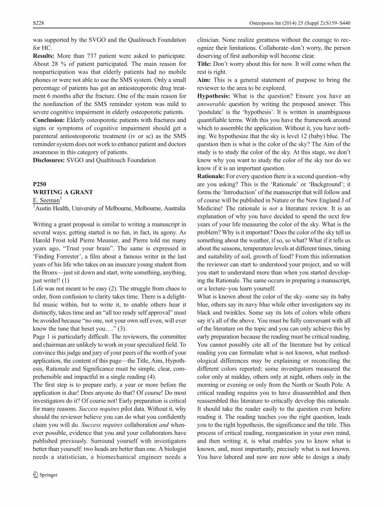
was supported by the SVGO and the Qualitouch Foundationfor HC.Results: More than 737 patient were asked to participate.About 28 % of patient participated. The main reason fornonparticipation was that elderly patients had no mobilephones or were not able to use the SMS system. Only a smallpercentage of patients has got an antiosteoporotic drug treat-ment 6 months after the fracture. One of the main reason forthe nonfunction of the SMS reminder system was mild tosevere cognitive impairment in elderly osteoporotic patients.Conclusion: Elderly osteoporotic patients with fractures andsigns or symptoms of cognitive impairment should get aparenteral antiosteoporotic treatment (iv or sc) as the SMSreminder system does not work to enhance patient and doctorsawareness in this category of patients.Disclosures: SVGO and Qualtitouch Foundation
P250WRITING A GRANTE. Seeman11Austin Health, University of Melbourne, Melbourne, Australia
Writing a grant proposal is similar to writing a manuscript inseveral ways; getting started is no fun, in fact, its agony. AsHarold Frost told Pierre Meunier, and Pierre told me manyyears ago, “Trust your brain”. The same is expressed in‘Finding Forrester’, a film about a famous writer in the lastyears of his life who takes on an insecure young student fromthe Bronx—just sit down and start, write something, anything,just write!! (1)Life was not meant to be easy (2). The struggle from chaos toorder, from confusion to clarity takes time. There is a delight-ful music within, but to write it, to enable others hear itdistinctly, takes time and an “all too ready self approval”mustbe avoided because “no one, not your own self even, will everknow the tune that beset you….” (3).Page 1 is particularly difficult. The reviewers, the committeeand chairman are unlikely to work in your specialized field. Toconvince this judge and jury of your peers of the worth of yourapplication, the content of this page—the Title, Aim, Hypoth-esis, Rationale and Significance must be simple, clear, com-prehensible and impactful in a single reading (4).The first step is to prepare early, a year or more before theapplication is due! Does anyone do that? Of course! Do mostinvestigators do it? Of course not! Early preparation is criticalfor many reasons. Success requires pilot data. Without it, whyshould the reviewer believe you can do what you confidentlyclaim you will do. Success requires collaboration and when-ever possible, evidence that you and your collaborators havepublished previously. Surround yourself with investigatorsbetter than yourself: two heads are better than one. A biologistneeds a statistician, a biomechanical engineer needs a
clinician. None realize greatness without the courage to rec-ognize their limitations. Collaborate–don’t worry, the persondeserving of first authorship will become clear.Title: Don’t worry about this for now. It will come when therest is right.Aim: This is a general statement of purpose to bring thereviewer to the area to be explored.Hypothesis: What is the question? Ensure you have ananswerable question by writing the proposed answer. This‘postulate’ is the ‘hypothesis’. It is written in unambiguousquantifiable terms. With this you have the framework aroundwhich to assemble the application. Without it, you have noth-ing. We hypothesize that the sky is level 12 (baby) blue. Thequestion then is what is the color of the sky? The Aim of thestudy is to study the color of the sky. At this stage, we don’tknow why you want to study the color of the sky nor do weknow if it is an important question.Rationale: For every question there is a second question–whyare you asking? This is the ‘Rationale’ or ‘Background’; itforms the ‘Introduction’ of the manuscript that will follow andof course will be published in Nature or the New England J ofMedicine! The rationale is not a literature review. It is anexplanation of why you have decided to spend the next fewyears of your life measuring the color of the sky. What is theproblem?Why is it important? Does the color of the sky tell ussomething about the weather, if so, so what? What if it tells usabout the seasons, temperature levels at different times, timingand suitability of soil, growth of food? From this informationthe reviewer can start to understood your project, and so willyou start to understand more than when you started develop-ing the Rationale. The same occurs in preparing a manuscript,or a lecture–you learn yourself.What is known about the color of the sky–some say its babyblue, others say its navy blue while other investigators say itsblack and twinkles. Some say its lots of colors while otherssay it’s all of the above. You must be fully conversant with allof the literature on the topic and you can only achieve this byearly preparation because the reading must be critical reading.You cannot possibly cite all of the literature but by criticalreading you can formulate what is not known, what method-ological differences may be explaining or reconciling thedifferent colors reported; some investigators measured thecolor only at midday, others only at night, others only in themorning or evening or only from the North or South Pole. Acritical reading requires you to have disassembled and thenreassembled this literature to critically develop this rationale.It should take the reader easily to the question even beforereading it. The reading teaches you the right question, leadsyou to the right hypothesis, the significance and the title. Thisprocess of critical reading, reorganization in your own mind,and then writing it, is what enables you to know what isknown, and, most importantly, precisely what is not known.You have labored and now are now able to design a study
Osteoporos Int (2014) 25 (Suppl 2):S159–S440S228
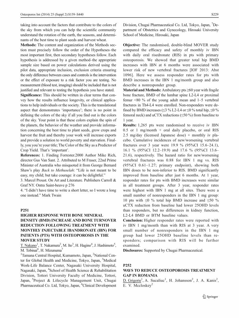
taking into account the factors that contribute to the colors ofthe sky from which you can help the scientific communityunderstand the rotation of the earth, the seasons, and determi-nants of the best time to plant seeds and harvest wheat.Methods: The content and organization of the Methods sec-tion must precisely follow the order of the Hypotheses–themost important first, then secondary hypotheses follow. Eachhypothesis is addressed by a given method–the appropriatesample size based on power calculations derived using thepilot data, appropriate inclusion, exclusion criteria ensuringthe only difference between cases and controls is the interventionor the effect of exposure to a risk factor you are testing. Nomeasurement (blood test, imaging) should be included that is notjustified and relevant to testing the hypothesis you have stated.Significance: This should be written in clear terms that con-vey how the results influence longevity, or clinical applica-tions to help individuals or the society. This is the translationalaspect that demonstrates ‘importance’; there is no point indefining the colors of the sky if all you find out is the colorsof the sky. Your point is that these colors explain the spin ofthe planets, the behavior of the weather and provide informa-tion concerning the best time to plant seeds, grow crops andharvest the fruit and thereby your work will increase exportsand provide a solution to world poverty and starvation. Final-ly, you you’re your title; The Color of the Sky as a Predictor ofCrop Yield. That’s ‘important’.References: 1. Finding Forrester.2000 Author Mike Rich,director Gus Van Sant. 2. Attributed to M Fraser, 22nd PrimeMinister of Australia who misquoted it from George BernardShaw’s play Back to Methuselah: “Life is not meant to beeasy, my child; but take courage: it can be delightful.”3. Marcel Proust. On Art and Literature. Publishers Carroll &Graf NY. Ontra Saint-beuve p 2764. “I didn’t have time to write a short letter, so I wrote a longone instead.” Mark Twain
P251HIGHER RESPONSE WITH BONE MINERALDENSITY (BMD) INCREASEANDBONETURNOVERREDUCTION FOLLOWING TREATMENT WITHMONTHLY INJECTABLE IBANDRONATE (IBN) FORPATIENTS (PTS) WITH OSTEOPOROSIS IN THEMOVER STUDYT. Nakano1, T. Nakamura2, M. Ito3, H. Hagino4, J. Hashimoto5,M. Tobinai6, H. Mizunuma71Tamana Central Hospital, Kumamoto, Japan, 2National Cen-ter for Global Health and Medicine, Tokyo, Japan, 3MedicalWork-Life Balance Center, Nagasaki University Hospital,Nagasaki, Japan, 4School of Health Science & RehabilitationDivision, Tottori University Faculty of Medicine, Tottori,Japan, 5Project & Lifecycle Management Unit, ChugaiPharmaceutical Co. Ltd, Tokyo, Japan, 6Clinical Development
Division, Chugai Pharmaceutical Co. Ltd, Tokyo, Japan, 7De-partment of Obstetrics and Gynecology, Hirosaki UniversitySchool of Medicine, Hirosaki, Japan
Objective: The randomised, double-blind MOVER studycompared the efficacy and safety of monthly iv IBNwith daily oral risedronate (RIS) in pts with primaryosteoporosis. We showed that greater total hip BMDincreases with IBN at 6 months were associated withlower risk of new vertebral fractures [IOF 2013: Abst1096]. Here we assess responder rates for pts withBMD increases in the IBN 1 mg/month group and alsodescribe a nonresponder group.Material andMethods:Ambulatory pts ≥60 year with fragilebone fracture, BMD of the lumbar spine L2-L4 or proximalfemur <80 % of the young adult mean and 1–5 vertebralfractures in Th4-L4 were enrolled. Non-responders were de-fined by BMD increases (≤3 % L2-L4 or ≤0 % total hip, ≤0 %femoral neck) and uCTX reductions (≤50 %) from baseline to1 year.Results: 1,265 pts were randomised to receive iv IBN0.5 or 1 mg/month + oral daily placebo, or oral RIS2.5 mg/day (licensed Japanese dose) + monthly iv pla-cebo. Cumulative incidences of new/worsening vertebralfractures over 3 year were 19.9 % (95%CI 15.6–24.1),16.1 % (95%CI 12.2–19.9) and 17.6 % (95%CI 13.6–21.6), respectively. The hazard ratio for new/worseningvertebral fractures was 0.88 for IBN 1 mg vs. RIS(95%CI 0.61–1.27; primary endpoint), showing bothIBN doses to be non-inferior to RIS. BMD significantlyimproved from baseline after just 6 months. At 1 year,responder rates for pts with BMD increases were similarin all treatment groups. After 3 year, responder rateswere highest with IBN 1 mg at all sites. There were asmall number of nonresponders in the IBN 1 mg group:10 pts with ≤0 % total hip BMD increase and ≤50 %uCTX reduction from baseline had lower 25OHD levelsthan responders, but no differences in kidney function,L2-L4 BMD or BTM baseline values.Conclusion: Higher responder rates were reported withiv IBN 1 mg/month than with RIS at 3 year. A verysmall number of nonresponders in the IBN 1 mggroup had lower 25OHD baseline levels than re-sponders; comparison with RIS will be furtherexamined.Disclosures: Supported by Chugai Pharmaceutical.
P252WAYS TO REDUCE OSTEOPOROSIS TREATMENTGAP IN ROMANIAD. Grigorie1, A. Sucaliuc1, H. Johansson2, J. A. Kanis2,E. V. Mccloskey2
Osteoporos Int (2014) 25 (Suppl 2):S159–S440 S229
1National Institute of Endocrinology, Carol Davila Universityof Medicine, Bucharest, Romania, 2WHO CollaboratingCentre for Metabolic Bone Diseases, University of Sheffield,Sheffield, United Kingdom
Objective: In Romania the overwhelming majority of indi-viduals at high risk of fracture are untreated (86 %; 94 % inmen and 83 % in women). The aim of this study was toidentify the causal factors and possible remedies, in the con-text of economic crisis.Material and Methods: A panel from Romanian Society ofOsteoporosis and Musculoskeletal Diseases was asked toaudit the framework of clinical practice of osteoporosis inRomania, the availability and provision of logistical resources,the national guidelines, education and proficiency in osteopo-rosis by family doctors.Results: Several factors were identified that contributed to thehigh treatment gap. The cost of medical intervention in Ro-mania is high in relation to GDP and even higher in relation tothe health care spend. Another barrier to treatment is thatreimbursement is only granted where the prescription is issuedby a specialist. National guidelines are available but they arenot implemented and updated. The currently accepted criteri-on for treatment of osteoporosis in Romania is a BMD T-score≤−2.5 SD, which is also the reimbursement threshold formedical intervention and reimbursement of osteoporosisagents is about 50 %. There is limited access to DXA andmoreover, the reimbursement for DXA is less than the cost ofDXAwhich imposes its own financial constraints on individ-uals. There are also incompatibilities between recommenda-tions for risk assessment or treatment with reimbursementpolicy. FRAX-based assessment and intervention thresholdsare available but not implemented. For example, guidelinesrecommend the use of FRAX which is not provided for inreimbursement provision and specific treatments are recom-mended but not reimbursed. General practitioners are notfamiliarized with risk assessment based on FRAX.Conclusion: Adjusting existing policies in agreementwith the current strategy of absolute risk assessment wouldbe a good low cost first step in addressing the high treatmentgap.
P253IMPACT OF SEASON AND COMORBIDITY ON HIPFRACTURE INCIDENCE. A NOREPOS STUDYS. M. Solbakken1, J. H. Magnus1, H. E. Meyer2, N. Emaus3,G. S. Tell4, K. Holvik5, G. Grimnes6, S. Forsmo7, B. Schei8,A. J. Søgaard9, T. K. Omsland11Institute of Health and Society, University of Oslo, Oslo,Norway, 2Institute of Health and Society, University of Oslo,Norwegian Institute of Public Health, Oslo, Norway, 3Depart-ment of Health and Care Sciences, University of Tromsø,
Tromsø, Norway, 4Department of Global Public Health andPrimary Care, University of Bergen, Bergen, Norway, 5Depart-ment of Global Public Health and Primary Care, University ofBergen, Norwegian Institute of Public Health, Oslo, Norway,6Tromsø Endocrine Research Group, University of Tromsø,Division of Internal Medicine, University Hospital of NorthNorway, Tromsø, Norway, 7Institute of Public Healthand General Practice, NTNU, Trondheim, Norway, 8Instituteof Public Health and General Practice, NTNU, Department ofObstetrics and Gynecology, St. Olav’s University Hospital,Trondheim, Norway, 9Norwegian Institute of Public Health,Oslo, Norway
Objective: Seasonal variation in hip fracture incidence hasbeen reported in several studies. The link of this variation tocomorbidity is unknown. The aim of this study was to exam-ine the seasonal variation in hip fracture incidence in Norway,related to age, gender and comorbidity.Material and Methods: Our analyses were based on theNOREPOS Hip Fracture Database with hip fractures(139,913) in Norway between 1994 and 2008. Addition-al diagnosis and surgical procedure codes were used toidentify incident fractures. Charlson index (0–2), a co-morbidity measure, was calculated based on additionaldiagnoses. Winter was defined as December, Januaryand February and summer as June, July and August.Negative binominal models were used in the analyses.The population at risk was based on data from StatisticsNorway. We also performed analyses by gender andthree age groups: 50–64, 65–79 and >80 years.Results: 136,140 fractures were eligible for the study(72.5 % women). In men, we found an age-adjustedrelative risk of 1.40 (95%CI: 1.36–1.45) in winter com-pared to summer. This ratio was 1.26 (95%CI: 1.23–1.28) in women. June had the lowest proportion offractures in both genders, while January and Decemberwere the peak months. Both genders had seasonal var-iation in all age groups, but less distinct with increasingage. We found a significant interaction between seasonand comorbidity. After adjustments for age and gender,winter versus summer incidence was 1.41 (95%CI:1.30–1.52) in those with no comorbidity (Charlson in-dex=0), 1.28 (95%CI: 1.19–1.39) in those with somecomorbidity (Charlson index=1), and 1.18 (95%CI:1.09–1.28) in those with most comorbidity (Charlsonindex=2).Conclusion: This study demonstrates a clear seasonalvariation in hip fracture incidence, present in all sub-groups by gender, age or comorbidity. Interestingly, theseasonal variation was most pronounced in those withleast comorbidity. The considerable seasonal variationshould be taken into account when planning healthcareservices.
Osteoporos Int (2014) 25 (Suppl 2):S159–S440S230
P254COMPARATIVE EVALUATION OF KNEE ANDANKLE STRENGTH IN PATIENTS WITH KNEEOSTEOARTHRITIS AND HEALTHY CONTROLS: APILOT STUDYY. Erdoğanoğlu1, Y. Yakut2, S. Güneri3, L. Özçakar31Okan University-Academy of Health Sciences, İstanbul,Turkey, 2Hacettepe University, School of Physiotherapyand Rehabilitation, Ankara, Turkey, 3Hacettepe UniversityMedical School, Ankara, Turkey
Objective: Osteoarthritis is characterized by joint pain, localtenderness, limited motions, crepitation, effusion and variousdegrees local inflammation without systemic findings. Theaim of this study was to compare the knee and ankle strengthvalues of patients with osteoarthritis and those of healthycontrols.Material and Methods: Six female patients with a mean ageof 66.17±13.73 years and seven female healthy volunteerswith a mean age of 57.14±12.5 years were recruited in thestudy. Knee (flexion/extension) and ankle (plantar/dorsiflexion) strength measurements were performed by anisokinetic dynamometer (Biodex System 3). Peak torque/bodyweight values were taken into the statistical analysis.Results: Neither the knee nor the ankle measurements werefound to be different between the two groups (p>0.05).Conclusion: It seems that further analyses involving widergroup of individuals are warranted for better estimation ofcomparative knee and ankle strength in patients withosteoarthritis.
P255BONE STATUS IN PATIENTS WITH ANOREXIANERVOSA AND RELATIONSHIP WITHBIOCHEMICAL FINDINGS AND FINDINGS ATPSYCHIATRIC INTERVIEWS. A. Eriksen1, H. Prietzel2, J. R. Ibsen3, M. B. Lauritsen4,P. Vestergaard5, G. K. Telléus61Department of Clinical Medicine, Aalborg University,Aalborg, Denmark, 2Clinic for Child and AdolescentPsychiatry, Aalborg University Hospital, Aalborg, Denmark,3Department of Endocrinology, Aalborg University Hospital,Aalborg, Denmark, 4Research Unit for Child and AdolescentPsychiatry, Aalborg University Hospital and Clinic for Childand Adolescent Psychiatry, Aalborg University Hospital,Aalborg, Denmark, 5Department of Clinical Medicine,Aalborg University and Department of Endocrinology,Aalborg University Hospital, Aalborg, Denmark, 6Departmentof Clinical Medicine, Aalborg University and Section of EatingDisorders, Department of Psychiatry, Aalborg UniversityHospital and Unit for Psychiatric Research, AalborgUniversity Hospital, Aalborg, Denmark
Objective: To investigate bone status and biological mecha-nisms involved in the negative impact of anorexia nervosa(AN) on osteogenesis.Material and Methods: A cross-sectional study was con-ducted based on a Danish clinical database established atDepartment of Psychiatry, Aalborg University Hospital. Thedatabase includes biochemical data, bone scans (DXA) aswellas general health and medical information during the period2009–2011. A total of 30 AN patients who underwent bonescans were included in the study.Results:AN patients had a mean Z-score around −1.5 to −1.6in lumbar spine and total hip, respectively. Hip Z-scoredecreased with duration of disease, and a positive correla-tion between serum 25-hydroxyvitamin D level and spineZ-score but not hip Z-score was seen. BMD did not seemto change with time since diagnosis. Additionally, a nega-tive correlation between serum 25-hydroxyvitamin Dlevels and serum total alkaline phosphatase levels wasfound. A serum 25-hydroxyvitamin D level below50 nmol/l was associated with increased alkaline phosphataselevels.Conclusion: Disease duration was the main predictor of bonestatus rather than clinical measures including BMI and bio-chemical measures. This implies that long term disease dura-tion should be a main factor in selecting patients for referral toDXA. Moreover, results from this study indicate normal os-teoblastic response to malnutrition.
P256RECOMMENDATIONS FOR OSTEOPOROSISMANAGEMENTAND FRACTURE PREVENTIONFORTHE FRAIL ELDERLY IN LONG-TERM CARE(LTC)A. Papaioannou1, N. Santesso2, S. Morin3, A. M. Cheung4, R.G. Crilly5, L. M. Giangregorio6, K. Grady7, S. Jaglal8, R.Jain7, R. Josse4, S. Kaasalainen9, P. Katz10, A. Moser4, L.Pickard1, C. J. Skidmore1, H. Weiler11, S. Whiting12, J. D.Adachi11Department of Medicine, McMaster University, Hamilton,Canada, 2Clinical Epidemiology and Biostatistics, McMasterUniversity, Hamilton, Canada, 3Medicine, McGill University,Montreal, Canada, 4Department of Medicine, University ofToronto, Baycrest, Toronto, Canada, 5Medicine, University ofWestern Ontario, London, Canada, 6Department of Kinesiol-ogy, University ofWaterloo,Waterloo, Canada, 7OsteoporosisCanada, Toronto, Canada, 8Department of Physical Therapy,University of Toronto, Toronto, Canada, 9Department ofNursing, McMaster University, Hamilton, Canada,10Baycrest, Toronto, Canada, 11Dietetics and HumanNutrition, McGill University, Montreal, Canada, 12Depart-ment of Nutrition and Dietetics, University of Saskatchewan,Saskatoon, Canada
Osteoporos Int (2014) 25 (Suppl 2):S159–S440 S231

Objective: Clinicians practicing in LTC face unique chal-lenges caring for frail elderly individuals including multiplecomorbidities, polypharmacy, and end of life care, but practiceguidelines typically do not address this population. Guidanceregarding the management of osteoporosis and fracture pre-vention in LTC, a high risk population, is needed.Material and Methods: The GRADE approach was used. Asurvey of LTC physicians informed key questions and out-comes. Interviews with resident representatives and a litera-ture review informed values and preferences. The quality ofevidence, benefits and harms, costs, resident values andpreferences, and available resources were consideredwhen making recommendations.Results: For residents at high risk for fracture, we suggestmultifaceted interventions that are individually tailored toreduce the risk of falls and fractures, and we suggest balance,strength and functional training exercises only when part of amultifaceted intervention. We recommend hip protectors. Werecommend vitamin D3 supplements daily (800–2000 IU) andcalcium supplementation up to 500 mg daily if unable to meetrecommended dietary allowances through food. We recom-mend the following therapeutic agents: alendronate,risedronate, zolendronic acid, or denosumab. Teriparatide isa suggested option. We suggest raloxifene and etidronate notbe used.Conclusion: These are the first guidelines developed for thecare of osteoporosis and fracture prevention in LTC usingGRADE. In LTC, strategies to prevent fractures and falls mustconsider resident values and preferences, comorbidities, lifeexpectancy, and quality of life.Disclosures: Dr. Alexandra Papaioannou has received grants/research support from Amgen, Eli Lilly, Merck and WarnerChilcott. Dr. Papaioannou has been on a speakers bureau andreceived honoraria from Amgen, Eli Lilly, and Merck. Dr.Papaioannou has received consulting fees from Amgen, EliLilly and Merck and is an employee of McMaster University.Nancy Santesso, no conflicts. Dr. Suzanne Morin, has re-ceived grants/research support from Amgen. Dr. Morin hasreceived consulting fees for Amgen, Merck and Eli Lilly. Dr.Morin has been on speakers bureau and received honorariafrom Amgen and Eli Lilly. Dr. Angela Cheung has receivedgrants and honorarium from Amgen, Eli Lilly, Merck. Dr.Richard Crilly, no conflicts. Dr. Lora Giangregorio hasreceived grants/research support from Merck. KerryGrady, no conflicts. Dr. Robert Josse has been an advisoryboard member, received speaker honoraria and/or researchgrants from Lilly, Amgen, Novartis, Warner Chilcott,Merck. Dr. Susan Jaglal, no conflicts. Ravi Jain, no con-flicts. Dr. Sharon Kaasalainen, no conflicts. Dr. AndreaMoser, no conflicts. Laura Pickard, no conflicts. CarlySkidmore, no conflicts. Dr. Hope Weiler, no conflicts.Dr. Susan Whiting, no conflicts. Dr. Jonathan Adachi,has participated in clinical trials for Amgen, Eli Lilly,
Merck, Novartis. Dr. Adachi has been on a speakers bureau,received honoraria and consulting fees from Amgen, Eli Lilly,Merck, Novartis, Warner Chilcott. Dr. Adachi is an employeeof McMaster University.Acknowledgements: We would like to thank our consensuspanel of 50 LTC stakeholders across Canada.
P257RHEUMATOID ARTHRITIS AND FATIGUEX. Grapton1, P. Khalifa2, D. Leclere3, P. Lemesle4, L. Poulain5,P. Renard61Private Practice, Colombes, France, 2Private Practice, Paris,France, 3Private Practice, Suresnes, France, 4Private Practice,Bois-Colombes, France, 5Private Practice, La Garenne Co-lombes, France, 6Private Practice, Courbevoie, France
Objective: To define the profile of tired patients with rheu-matoid arthritis (RA) and the influence of treatment, to ana-lyze the consequences of fatigue, and to determine the relevantcriteria to assess the fatigue.Material and Methods: • 155 cases, 85 % were women,mean age 58 years, seen by 18 rheumatologists (Rh).• 2 control groups: RA not tired and tired patients without RA.• Average duration of history of RA: 10 years.• Degree of severity: light 12 %, moderate 67 %, severe 21 %.• Progressive nature: low 32 %, moderate 57.5 %, aggressive10.5 %.• ESR average of 18 mm, mean CRP 8.4.• 48 % have two DMARDs, 28 % have 1, 24 % 3 or 4.• 81 % are under MTX, 15 % under biotherapy, 49 % understeroids.• 20 % have another cause of fatigue.Results: • 66% of RA patients studied are tired vs. 47% in theFrench population and 42 % in the rheumatologic patient database. 67 % spontaneously evoke their fatigue.• At D0, 77 % of patients have pains, 43 % are disabled.• VAS pain=44 mm, VAS handicap=46 mm, VAS fatigue=47 mm.• In 72 % of cases RA was already tired, sometimesat treatment discontinuation (12 %) or at the intro-duction (15 %). Fatigue is the main complaint (38 %).• It is linked to RA (68 %) and runs in parallel to it(70 %).• It is more intense and permanent than ordinary fatigue andoccurs without cause.• It increases the recovery time (85 %), affects householdchores (79 %), resulting in daytime sleepiness (60 %).• It is more physical than moral, but often it is both (64 %).• 69 % of cases are sad, 63 % unmotivated, and 37 %depressed.• Fatigue affects spare-time (71 %) then work, sex, social life.
Osteoporos Int (2014) 25 (Suppl 2):S159–S440S232

• Of 10 criteria, three are relevant to evaluate the fatigue: VASfatigue >50 mm, recovery time extended by at least 50 %, notiring cause.• At 3- associated criteria, there is correlation between thedegree of fatigue, its evocation, evolution of RA, high CRP.• At 2 criteria with anemia, handicap, VAS pain, need to nap,lack of activities.• There is a parallelism between fatigue and previous RA,elevated ESR, disease flare, decreasedmuscle strength, lack ofenergy and mainly improvement thanks to biotherapy.• Not tired RA are not very aggressive and frequently withMTX alone or biotherapy.Conclusion: RA tired is often progressive and with elevatedESR. Fatigue is both physical and moral. It is permanent,unusual, intense and debilitating and fades to the introductionof MTX and/or biotherapy.
P258IMPACT OF DEMOGRAPHIC CHARACTERISTICSON BONE MICROARCHITECTURE INDEX (TBS) INHEALTHY WOMEN IN MENOPAUSES. Novkovic1, G. Marinkovic2, M. Prelic1, M. Terek11Institute for Rheumatology, Belgrade, Serbia, 2Health CenterZvezdara, Belgrade, Serbia
Objective: To assess whether age, duration of menopause,body weight, height and BMI affect TBS in healthy, postmen-opausal women.Material andMethods:A cross-sectional study included 257healthy menopausal women, with TBS score measured at thelumbar spine. Subjects with risk factors known to affect bonemicroarchitecture (using steroids, previous fractures, systemicdisease, excessive use of alcohol and cigarettes, and the use ofdrugs with a negative impact on bone metabolism) wereexcluded. Statistical analysis was performed in SPSS.Results: The average age of patients was 64.3±7.9 years.Menopause occurred in 49.5 (37–60) years. and lasted anaverage of 15.9±9.5 years. The average body weight was69.4±13.0 kg. Body height 159.8±6.7 cm, and BMI 27.2±4.9. Average TBS was 1.223±0.101. We found a significantnegative correlation between age and TBS (ρ=−0.218,p<0.01), between the duration of menopause and TBS (r=−0.187, p<0.01), whereas the correlation between the age atthe time of menopause and TBSwas not significant (ρ=0.085,p=0.172). A significant negative correlation was observedbetween TBS and the body weight (r=−0.222, p<0.01), andthe TBS and the BMI (r=−0 268, p<0.01).Conclusion: The value of the bone microarchitecture index(TBS) decreases with age, duration of menopause, with anincrease in body weight and BMI. Body height, and age at thetime of onset of menopause did not correlate with TBS.
P259THE PHARMACOKINETICS OF ODANACATIB 50MG ARE NOTAFFECTED BY SEVERE RENALINSUFFICIENCYS. A. Stoch1, C. Liu1, S. Zajic1, R. Witter1, J. Stone1, A.Mehta1, P. Auger2, D. Stypinski2, N. Wang2, A. Hohnstein2,H. Alcorn3, W. Smith4, T. Marbury51Merck & Co., Inc, Whitehouse Station, United States,2Celerion, Lincoln, United States, 3DaVita Clinical Research,Minneapolis, United States, 4Volunteer Research Group/Graduate School of Medicine, University of Tennessee,Knoxville, United States, 5Orlando Clinical Research Center,Orlando, United States
Objective: Odanacatib, a selective cathepsin K (CatK) inhibi-tor, is in development for the treatment of osteoporosis. Ap-proximately 10 % of the clearance of odanacatib is believed tobe due to renal elimination. This study was done to determinethe effect of severe renal insufficiency on odanacatib pharma-cokinetics, pharmacodynamics, and tolerability.Material andMethods: In this open-label, single-dose study,odanacatib was orally administered to subjects (age: 40–79 years) with severe renal insufficiency (defined as a Cock-croft Gault creatinine clearance of <30 mL/min) and healthycontrol subjects (creatinine clearance ≥90 mL/min), matchedfor age, gender, and BMI. Plasma and urine samples werecollected for the analysis of odanacatib concentrations andbiomarkers at predose and at specified time points over15 days postdose. An analysis of covariance (ANCOVA)model was used to analyze odanacatib AUC0-∞ on log scale.The AUC0-∞ geometric mean ratio (GMR) was determined toevaluate the similarity of pharmacokinetics between the twogroups based on the 90 % CI for the GMR contained withinthe comparability bounds of (0.40, 2.50). Cmax was analyzedin a similar fashion. Summary statistics were computed forTmax and apparent terminal t1/2. Bone biochemical markers(serum NTx and urine NTx/Creatinine) were also assessed toevaluate pharmacodynamics; a linear mixed effect model wasused for the analysis of these parameters. Adverse events(AEs) were monitored throughout the study and evaluatedfor severity and relation to study medication at study visits.Results: The GMRs (90 % CI) for AUC0-∞ and Cmax were1.62 (1.17, 2.24) and 1.47 (1.19, 1.80), respectively. Thegeometric mean half-life for odanacatib 50 mg was 74.2 hsubjects with renal impairment and 80 h in the healthymatched controls. At 168 h postdose, least square (LS)mean (95%CI) serum NTx va lues were −36.5(−47.30,−23.38) [renal impairment] and −43.2 (−53.39,−30.76) [healthy]; LS Mean (95%CI) urine NTx/Cr valueswere −59.3 (−65.81, −51.51) [renal impairment] and−43.4 (−52.83, −32.08) [healthy]. There were no seriousAEs or discontinuations due to AEs. Mild headache wasthe most common drug-related AE and was reported in
Osteoporos Int (2014) 25 (Suppl 2):S159–S440 S233

one patient in the renal impairment group and one patientin the healthy control group.Conclusion: There were no meaningful differences betweensubjects with severe renal impairment compared with healthy-matched controls in odanacatib 50 mg pharmacokinetics, asassessed by comparison of AUC0-∞ values, or its pharmaco-dynamics, as assessed by change from baseline in serum NTxand urine NTx/Cr. Odanacatib 50 mg was generally welltolerated in patients with severe renal impairment.Disclosures: SAS, CL, SZ, RW, JS, AM are employees of andown stock in Merck & Co., Inc, the study sponsor. DS, NW,AH are employees of Celerion, which was contracted byMerck to run the study. HA, WS, and TM were investigatorsand received research support from Merck.
P260SHINYAND NEW: UPTAKE AND PERSISTENCEWITH NON-ORAL OSTEOPOROSIS MEDICATIONSIN ONTARIO, CANADAA. M. Burden1, M. Tadrous1, L. Wong1, A. Calzavara2, Y. Y.Liu1, S. M. Cadarette11Department of Pharmaceutical Sciences, University of To-ronto, Toronto, Canada, 2Institute for Clinical and EvaluativeSciences, Toronto, Canada
Objective: Two non-oral osteoporosis drugs were added tothe public drug formulary in Ontario, Canada in the lastdecade: annual zoledronic acid in 2006 (modified in 2012)and semi-annual denosumab in 2012. We aimed to describethe use and persistence of these new drugs since formularyaddition.Material and Methods: We used Ontario administrativeclaims data to identify new users of zoledronic acid anddenosumab from formulary entry through to 2013. Descrip-tive characteristics of prescribing physicians and patients weresummarized. The number of new patients and new prescribingphysicians were plotted by month and examined over time.Time series analysis was used to examine the impact of theformulary change to zoledronic acid in 2012. Persistence withindex therapy and switching to different therapies was exam-ined over time.Results: We identified 1,508 zoledronic acid users (86 %female, mean age=77) treated by 630 physicians (27 % spe-cialist) and 16,736 denosumab users (97 % female, meanage=79) treated by 2,904 physicians (12 % specialist). Moredenosumab users had prior oral therapy (55 % vs. 34 %), yetfewer received BMD testing (20 % vs. 33 %). In comparisonto zoledronic acid (<5 new prescribers and patients),uptake of denosumab was rapid (>450 new prescribersand >1,200 new patients) in the first 2 months on theformulary. Time series analysis identified a significantincrease in zoledronic acid use following a modification
to the limited use criteria. We identified that 57 % (zole-dronic acid) and 55 % (denosumab) patients persistedbeyond the first year, and 30 % of zoledronic acid patientspersisted for 3+ years.Conclusion: Zoledronic acid and denosumab may be en-ticing options to improve persistence with osteoporosistherapy. Over half of patients received doses beyond thefirst year, and more than a third received prior oral ther-apy. A provincial formulary modification that broadenedaccess criteria for zoledronic acid in 2012 significantlyincreased prescribing.
P261VITAMIN D SUPPLEMENTATION AFTERBARIATRIC SURGERY: IS 30 MICROGRAM DAILYENOUGH? ATWO-YEAR FOLLOW-UPAFTERGASTRIC BYPASS OR SLEEVE GASTRECTOMYT. A. Pekkarinen1, M. Leivonen2, N. Jaser2, A. Juuti2,T. Sane31Department of Medicine, Division of Endocrinology,Helsinki University Hospital, Peijas Hospital, Vantaa,Finland, 2Department of Bariatric Surgery, HelsinkiUniversity Central Hospital, Vantaa, Finland, 3Departmentof Medicine, Division of Endocrinology, Helsinki UniversityCentral Hospital, Helsinki, Finland
Objective: Poor vitamin D status in the obese is well docu-mented. After bariatric surgery vitamin D deficiency canbecome even worse. Optimal vitamin D supplementation dosein the bariatric patients is not known.Material and Methods: We assessed serum 25OHD3
concentration at one and 2 years control in 216 severelyobese patients (67 % female, mean age 48 years, meanBMI 49 kg/m2) who underwent either gastric bypass(GBP, n=137) or sleeve gastrectomy (SG, n=79) atHelsinki University Central Hospital, Finland, between2008 and 2010. The mean preoperative weight was142 kg (range 92–220). All patients received a prescrip-tion of cholecalciferol 30 μg daily at hospital dischargeand at 1 year control. Serum 25OHD3 was determinedby the HPLC method published earlier (Turpeinen et al.2003).Results: The mean weight loss at 1 year control was 24.0 %(range 4.2–42.9 %) in the GBP group and 21.6 % (1.8–43.6 %) in the SG group, p=0.04, and at 2 year control24.3 % (3.6–46.7 %) and 22.2 % (−2.1–50.9 %), p=0.16,respectively. At 1 year control, the mean 25OHD3 was62.3 nmol/l (21–122) after GBP and 63.8 nmol/l (13–110)after SG, p=0.27 and at 2 year control 63.8 (14–148) and 72.1(24–149), respectively, p=0.03. At 1 year control, serum25OHD3 was below 50 nmol/l in 37 (27 %) of the GBPpatients and in 24 (30 %) of the SG patients, p=0.64, and at
Osteoporos Int (2014) 25 (Suppl 2):S159–S440S234

2 year control in 24 (18 %) and 10 (13 %) of the patients, p=0.44, respectively. Below 75 nmol/l at 1 year control was 109(80 %) of the GBP patients and 56 (71 %) of the SG patients,p=0.18, and at 2 year control 99 (78 %) and 41 (59 %), p=0.005, respectively.Conclusion: In these bariatric patients using daily 30 μgvitamin D supplementation, vitamin D status was suboptimalif level of 75 nmol/l is used as a threshold, but satisfactory if50 nmol/l is considered optimal for bone health. The GBPpatients are at a higher risk for vitamin D deficiency than theSG patients.References: Turpeinen U et al. Clin Chem 2003;49:1521.
P262HIP FRACTURE INCIDENCE IS MUCH HIGHER INHONG KONG CHINESE WOMEN THAN BEIJINGCHINESE WOMEN DESPITE HIGHER BONEDENSITY IN HONG KONG WOMEN: MAJORIMPLICATIONS FORHIP FRACTURE PREVENTIONR. Chung1, E. Lau1, G. Qin2, P. C. Ha21CCBR (Hong Kong), Hong Kong, Hong Kong, 2CCBR(Beijing), Beijing, China
Objective: Hong Kong has the highest incidence of hipfracture in Asia, while mainland China still has very lowincidence of hip fracture. However, it has been predicted therewill be a major epidemic of hip fracture in China. The objec-tive of the current study is to compare the BMD at the hipbetween a large population sample of Hong Kong and main-land Chinese women, so that insight can be gained into theetiology of hip fracture.Material and Methods: 7,130 ambulatory Hong KongChinese women and 7,037 mainland Chinese womenliving in Beijing (aged 50–96) were recruited from thecommunity. They volunteered for the study. BMD wasmeasured by Lunar Prodigy Advance Whole Body BoneDensitometers. Cross calibration was carried out by aphantom. The prevalence of osteoporosis was calculatedaccording to the World Health Organization criteria.BMD at the total spine and femoral neck were adjustedfor age, height and weight and compared between the 2populations.Results: According to published data, the incidence rate ofhip fracture was 465/100,000 in Hong Kong Chinese womenand 229/100,000 in Beijing Chinese women. The prevalenceof osteoporosis at the total hip in our study was 24.8 % inHong Kong Chinese women and 20.0 % in Beijing Chinesewomen, but these did not account for difference in weight andheight. After adjustment for age, height and weight, the BMDof Hong Kong Chinese women was 2.8 % higher at the hipand 1.5 % higher at the femoral neck than Beijing Chinesewomen. This implies that BMD is a poor measurement of
bone strength, and also that there are many extraskeletalfactors causing hip fracture.
Conclusion: The difference in hip fracture incidence betweenHong Kong Chinese and Beijing Chinese women cannot beexplained by difference in BMD. Further research shouldfocus on better measurement of bone strength in Chinesewomen, as well as extraskeletal factors for hip fracture, sothat recommendations can be made to prevent the imminentepidemic of hip fracture in mainland China.
P263PRDX2 AS A POSSIBLE PATHOLOGIC MEDIATOROF HIP OSTEOARTHRITIS REVEALED BYPROTEOMICST. Yuasa1, Y. Suehara1, Y. Makino1, K. Kaneko11Department of Orthopaedic Surgery, Juntendo University,Tokyo, Japan
Objective: To understand the molecular mechanisms of hipOA and identify pathologic mediators and biomarkers, weexamined the proteomic profile using two- dimensional dif-ference gel electrophoresis.Material and Methods:We examined surgical specimens offemoral head samples from 20 patients with hip disease(10 OA cases and 10 with femoral neck fractures) whounderwent surgery at the Juntendo University Hospitalconsecutively from February 2007 to August 2009. Fro-zen samples of cartilage and subchondral bone tissue wereused for the proteomic analysis, and were available as alearning set in 16 cases, including 8 OA-hip and 8 N-hipsamples. A Western blot analysis was performed on 20cases, consisting of the learning set (16 cases) and thevalidation set (four additional cases, including two OA-hipand two N-hip samples).Results: We identified 32 protein spots demonstrating a sta-tistical difference in intensity between femoral heads with hipOA (OA-hip) (8 cases) and those from normal hips (N-hip) (8cases) (p<0.01).We found that peroxiredoxin-2 (PRDX2) hadthe highest p-value (p=0.00016) and fold difference ratio(6.22) in 32 spots. Two of the 32 spots that derived fromPRDX2 had a higher intensity than that found in the N-hipsamples. We employed an SDS-PAGE/Western blottingtest to further examine the relationship of PRDX2 ex-pression with the OA-hip and N-hip samples, andshowed that PRDX2 expression was higher in the N-
Osteoporos Int (2014) 25 (Suppl 2):S159–S440 S235

hip samples in comparison to the OA-hip samples(p<0.0001). We used an SDS-PAGE/Western blottingtest to verify our findings in 4 additional OA-hip (n=2)and N-hip samples (n=2). The samples showed that theN-hip samples had higher expression of PRDX2 in comparisonto the OA-hip samples.Conclusion: These results establish PRDX2 as a possiblepathologic mediator candidate for OA.
P264GRAND 3 - THE GERMAN RETROSPECTIVECOHORTANALYSIS ON NONADHERENCE INOSTEOPOROTIC PATIENTS:PERSISTENCE-ANALYSIS OF FEMALE PATIENTSTREATED WITH DENOSUMAB (DMAB)T. Schmid1, P. Hadji2, C. Eisen1, M. Intorcia3, E. Psachoulia31Amgen GmbH, München, Germany, 2Philipps-University ofMarburg, Marburg, Germany, 3Amgen (Europe) GmbH, Zug,Switzerland
Objective: To be effective, osteoporosis therapy must betaken according to the prescribed dosing regimen. A retro-spective claims analysis reported low persistence with oraland intravenous bisphosphonates (BPs) at 12 and 24 months:8.5 %, 23.6 % and 35.0 % for daily, weekly and quarterlyadministration; and 4.7 %, 9.3 %, 13.1% and 36.0% for daily,weekly, quarterly and yearly administration, respectively1,2.We present persistence data for patients treated with DMAb(subcutaneously once every 6 months) in Germany; 12- and24-month data from this study have been reported previously3.Material andMethods: This retrospective analysis evaluatedthe IMS®LRx-data (06/2010–10/2013; covering filled pre-scriptions of statutory health insurances [SHI] in Germany).DMAb naïve women aged ≥45 year who had received theirinitial prescription between 06/2010 and 10/2011 were includ-ed. Outcome measures were persistence at 12, 18, 24 &30 months, defined as receiving follow-up DMAb prescrip-tions within 30-, 60- and 90-day grace periods after the pre-vious injection.Results: The analysis included a total of 6,159 patients. Usinga 30 day grace period, approximately 70 % of patients werepersistent at 12 months, 60 % at 24 months and 43 % at30 months.
Table. Persistence rates using grace periods of 60 and 90 dayswere similar to those for a 30-day gap.
Conclusion: Persistence to DMAb was higher than report-ed previously for BPs. This improved persistence maylead to better outcomes, including increased fracture riskreduction.References: 1Hadji et al., ECCEO 2013 (BEST); 230-dayrefill gap for weekly and quarterly administration; 90-daygap for yearly administration; 3Hadji et al., ECCEO 2013(GRAND3)Acknowledgements: Amgen/GSK
P265PATIENTSWITHOSTEOPOROSISWHOREMAINATHIGH RISK FOR FRACTURE DESPITE BENEFITOFPRIOR BISPHOSPHONATE TREATMENT: A DANISHPERSPECTIVEL. Hansen1, S. A. Eriksen1, A. Krishna2, A. D. Jorgensen3,P. Vestergaard11Aalborg University, Aalborg, Denmark, 2Merck & Co.,Whitehouse Station, United States, 3MSDDenmark, Ballerup,Denmark
Objective: To estimate the proportion of osteoporosis patientswho remain at a high risk for fracture despite being compliantto bisphosphonate treatment.Material and Methods: This case-control study is based onDanish national health registry data, including data on allhospitalizations, ICD-10 code at admission, and performedtests. Furthermore, results from BMD scans were collectedfrom three of the five Danish regions, and aggregated withdata from the registries.The patient group of 2,406 patients (66.3 % women and33.7 % men) was identified as:
& Age ≥ 50 years& First bisphosphonate prescription date between 01.01.1996
and 31.12.2008, defined as index date& ≥12 months pre- and ≥36 months post-observational data& A medication possession ratio ≥80 % for 24 months post
index& ≥1 claim of BMD test OR a fracture 12 months prior to
30 days after index date
‘High risk’ patients were defined as having the followingoutcomes despite compliance for 24 months:
& A fracture at 24–36 months following index OR& BMD T-score<−2.5 at 24–36 months after index OR
Any drop in BMD between index and 24–36 months A totalof 352 high risk patients were identified, and served as cases.The remaining 2,054 served as controls.
Osteoporos Int (2014) 25 (Suppl 2):S159–S440S236

Results:The proportion of high risk patients despite benefit ofprior bisphosphonate treatment in Denmark is 14.6 % (14.3 %for women and 15.2 % for men). T-scores were significantlydifferent at both index and follow-up between cases andcontrols (p<0.001). High risk patients were more likely tosmoke (OR: 2.5, p<0.001), have lower plasma calcium and/orvitamin D (OR: 3.4, p<0.001), and were more frequentlydiagnosed with anorexia nervosa (OR: 5.9, p=0.045). Fur-thermore, the high risk patients had a significant higherCharlson score (p=0.014).Conclusion: In Denmark, 14.6 % of the osteoporotic patientsremained at high risk for fracture despite persistent bisphos-phonate treatment. Although these patients may have benefit-ed from their therapy, alternative treatments could be consid-ered for such patients.Disclosures: This study was supported by an unrestrictedgrant from Merck & Co. AD Jorgensen is an employee ofMSD Denmark. A Krishna is an employee of Merck & Co. PVestergaard: Travel grants from Novartis, Amgen, Servier,and Eli Lilly.
P266CLINICAL AND SONOGRAPHIC ASSESSMENT OFTHE EFFECTIVENESS OF COLLAGENINJECTIONS GUNA MDS IN PATIENTS WITHPARTIALTHICKNESS ROTATOR CUFF TEARS OFTHE SHOULDER. Nestorova1, R. Rashkov2, T. Petranova2, N. Nikolov31Centre of Rheumatology, Sofia, Bulgaria, 2Clinic of Rheu-matology, Medical University, Sofia, Bulgaria, 3Clinic ofRheumatology and Cardiology, Pleven, Bulgaria
Objective: To evaluate the effectiveness of Injectablecollagen GUNA MDs regarding pain, functioning andrestoration of periarticular tissues of the shoulder inpatients with partial thickness tears (PTT) of the rotatorcuff (RC). Musculoskeletal ultrasonography (US) is anapproved imaging technique for diagnosis of the RCpathology and monitoring of therapy.Material and Methods: We studied 22 patients with painfulshoulder and sonographic proved PTT of the RC. Clinicalassessment included demographic and clinical data, a VASfor pain (0–100) and Shoulder Function Assessment (SFA)scale (0–70) on the baseline and on the 60th day. Evaluationsof the efficacy according to the patient and the physician wereperformed. All patients had US of both shoulders withMindray M5 scanner with multifrequency linear transducer(7.5–10 MHz). We applied a combination of GUNA MD-Shoulder and GUNA MD-Muscle into the subacromial spacein a total course of treatment 8 weeks. Physical therapy wasnot administered during the follow-up period.
Results: Pain was significantly reduced. There was a statisti-cally significant improvement of SFA Index. 75 % of patientsgave a very good and good assessment of efficacy, whichcoincided with the opinion of the physician. 78 % of patientshad a complete recovery or improved structure of the RC onsecond visit which was proved by sonography.Conclusion: Injectable collagen is an innovative approachwith regenerative effect in the treatment of PTT of theRC. GUNA MDs significantly reduced pain and increasedfunctional activity of the shoulder, thereby increasing thequality of life. No adverse events were registered duringthe treatment.References: 1. Naredo E et al. Ann Rheum Dis 2002;61:132.
P267PTH TREATMENT INDUCESWNT10B EXPRESSIONIN HUMANS LYMPHOID CELLSP. D’amelio1, F. Sassi1, I. Buondonno1, E. Spertino1,L. D’amico1, I. Roato1, G. C. Isaia11Dept. Medical Science University of Torino, Torino, Italy
Objective: Intermittent PTH treatment reduces vertebral frac-tures risk in osteoporotic patients. The mechanisms troughwhich PTH acts are not completely understood, it has beenobserved to activate Wnt pathways in osteoblasts (OBs).Activation of this pathway induces OB proliferation, differen-tiation and prevents apoptosis. Recently it has been demonstrat-ed an increased expression of Wnt10b by T cells during inter-mittent PTH, whereas there is no increase in Wnt10b duringcontinuous PTH infusion in mice. In order to evaluate if PTHacts on lymphoid cells by increasingWnt10b expression also inhumans, we measured this molecule at baseline and after PTHtreatment in osteoporotic subject and in patients affected byprimary hyperparathyroidism before and after surgery.Material and Methods: We enrolled 40 women with post-menopausal osteoporosis, patients were randomly assigned totherapy for 18 months with: 1-84 PTH 100 μg plus calcium1,200 mg and vitamin D 800 UI daily, or with calcium1,200 mg and vitamin D 800 UI daily and return for controlvisit and exams at 3, 6, 12 and 18 months of therapy. We alsoenrolled 20 patients affected by primary hyperparathyroidismand subjected to surgical parathyroidectomy; patients wereevaluated at baseline and 1 month after surgery. Real-timePCR for WNT10b was performed on peripheral blood lym-phoid cells after red blood cells lysis.Results:Our data show an increase inWNT10b expression bylymphoid cells that was maximum (more than 20-fold) after6 months of treatment, after 18 months WNT10b returned tobasal expression. The WNT10b curve acts similarly to theosteocalcin one. In patients treated with calcium and vitaminD alone no increase in WNT10b was observed. Also in
Osteoporos Int (2014) 25 (Suppl 2):S159–S440 S237

patients affected by hyperparathyroidism there was no differ-ence in WNT10b before and after surgery.Conclusion:Our data suggest an effect of intermittent, but notcontinuous PTH on the expression of WNT10b by lymphoidcells, this could be one of the mechanisms trough which PTHtreatment increases OB formation and function.
P268INFLUENCE OF DEGENERATIVE DISORDERS ONTHE LUMBAR SPINE BMD AND TBS WITH AGE:THE COHORT OSTEOLAUSB. Aubry-Rozier1, D. Stoll1, M.Metzger1, D. Hans1, O. Lamy1,I. Padlina11Lausanne University Hospital, Center of Bone Diseases,Lausanne, Switzerland
Objective: After menopause, typically, the values of lumbarspine (LS) BMD and TBS decrease with age. In practice, weare often faced with values of LS BMD “relatively” high forthe age, which can lead to erroneous interpretations(reassuring diagnosis, therapeutic gain). Preliminary studieshave shown that osteoarthritis (oa) does not influence TBS.The aim of this study was to measure changes in LS BMD andTBS values in women 50–80 years, taking into account theimpact of fractured vertebrae (VFx) and oa (Voa).Material and Methods: The 1,502 women 50–80 years oldincluded in the cohort OsteoLaus (Lausanne, Switzerland) havehad LS BMD and TBS, and VFA. 46 exams were not inter-pretable. All exams were analyzed independently by two ex-perts following the guidelines of the ISCD, excluding in par-ticular VFx (2.7 % of participants) and vertebra with >1 SDdifference with the vertebra immediately adjacent (Voa 16.2 %).Results: We included 1,456 women: age 66.7±11.7 years,BMI 25.7±4.4. The correlation between BMD and TBS waslow (r2=0.16). Participants were divided into six age groups:50–55 years (n=175 gr1), 55–60 (n=229, gr2), 60–65 (n=342, gr3), 65–70 (n=325, gr4), 70–75 (n=215, gr5) and 75–80 years (n=169, gr6). BMD was artificially increased from1.2 to 3.3 % on average according to the gr before excludingVFx and Voa (p<0.001). TBS did not change after excludingVFx and Voa (<0.1 % to 0.3 %, ns). The correlation (Vfx andVoa excluded) between age and BMDwas −0.03, between ageand TBS +0.34. BMD (Vfx and Voa excluded) was for the sixage gr: 0.955±0.140 g/cm2, 0.923±0.158, 0.906±0.158,0.915±0.166, 0.914±0.165 and 0.934±0.187, respectively.Loss between 52.5 and 77.5 years was 2.16 %, or 0.09 %/yearwith a loss of 0.51 %/year from 52.5 to 62.5 years, followedby a gain of 0.21 %/year between 62.5 and 77.5 years. Thechange in BMD between two consecutive age gr was signif-icant only between gr1 and gr2 (p<0.02). TBS (Vfx and Voaexcluded) was for the six age gr: 1.357±0.093, 1.318±0.100,1.292±0.091, 1.272±0.097, 1.257±0.103 and 1.236±0.099,
respectively. Loss between 52.5 and 77.5 years was 8.96%, or0.36 %/year with a loss of 0.48 %/year between 52.5 and62.5 years and 0.29 %/year between 62.5 and 77.5 years.Changes in TBS between 2 consecutive age gr was still highlysignificant (p<0.02 - p<0.0001). These results are confirmedand more pronounced in a sub-analysis including only womenwith Voa (with and without exclusion of Voa).Conclusion: This study confirms the low correlation betweenLSTBS andBMD. It shows a double interest in TBS, particularlyin the older population: 1) on average, TBS is not affected byfractures or oa disorders, 2) while BMD increases after 65 years(moderate degenerative disorders cannot be excluded from theanalysis), TBS continues to decline. For the LS evaluation, TBSshould play a leading role not only for diagnosis but also fortreatment monitoring in view of its independence to osteoarthritis.Disclosures: Didier Hans: Shareholder of medimaps group
P269PHYSICALFITNESS INCHILDRENTREATEDWITHGROWTH HORMONE BY JUMPINGMECHANOGRAPHY: LEONARDO GROUNDREACTION FORCE PLATFORMR. S. Lorenc1, A. Kępka1, M. Ginalska-Malinowska1, P.Pludowski1, J. Łukaszkiewicz21Children’s Memorial Health Institute, Warsaw, Poland,2Children’s Memorial Health Institute and Jan Kochanowski,Warsaw, Kielce, Poland
Objective: Growth hormone therapy is efficient in growthretardation caused by Turner syndrome (TS) and growthhormone deficiency (GHD). However it is unknown, if in-creasing of body size is accompanied by adequate increasingin muscle function. The aim of this study was evaluation ofchanges in physical fitness in children with GHD and TS afterstart of growth hormone therapy.Material and Methods: The study group comprised of 25children with GHD (11.2±2.9 year) and 8 girls with TS (9.2±2.1 year). Measurements were done before start of growthhormone therapy and after 3, 6 and 12 months of therapy.Physical fitness was evaluated basing on single two leg jump.Leonardo (Novotec Medical GmbH, Germany) was used.Leonardo measures dynamics of ground reaction forces andcalculates two parameters: Efficiency and Esslinger FitnessIndex, expressed as Z-scores. ANOVAwith repeatedmeasure-ment was used for analysis of the data.Results: Before start of the treatment, mean (SD) in childrenwith GHD was −0.99 (1.62) for Efficiency Z-score and −1.88(1.42) for Esslinger Fitness Index Z-score; and −2.87 (1.14) and−1.52 (0.65) in girls with TS, respectively. In childrenwithGHD,mean change after 12 months of therapy was 0.28 (n.s.) forEfficiency Z-score and 0.47 (p<0.005) for Esslinger FitnessIndex Z-score. In girls with TS mean changes were 1.12
Osteoporos Int (2014) 25 (Suppl 2):S159–S440S238

(p<0.05) and 0.41 (n.s.), respectively. In childrenwithGHDafterstart of growth hormone therapy Efficiency increases as fast as inreference data and Esslinger Fitness Index increases even fasterthan in reference data. On the contrary, in girls with TS, Efficien-cy increases faster than in reference data and Esslinger FitnessIndex increases as fast as in reference data.Conclusion: The data suggests that growth hormone therapysignificantly attains to catch up of physical fitness.Acknowledgements:Acquiring of Leonardowas co-financed byERDF (EU Structural Funds) project POIG.02.01.00-14-059/09
P270MARKERS OF BONY METABOLISM IN PEOPLEWITH POSTINFARCTION CARDIOSCLEROSISL. V. Yankouskaya1, I. S. Karpova21Grodno State Medical University, Grodno, Belarus,2Republican Scientific-Practical Center Cardiology,Chisinau, Republic of Moldova
Objective:To assess the state of osseous tissue andmarkers ofbony metabolism (MBM) in patients with postinfarctioncardiosclerosis.Material and Methods: Group I included 16 healthy indi-viduals (62.3±6.05 years), group II - 29 patients withpostinfarction cardiosclerosis (62.9±5.46 years). Structuraland functional state of osseous tissue was assessed by meansof ultrasound densitometry of calcaneus using a Sahara device(Hologic, USA). Serum level of the marker of bony tissueformation–osteocalcin (OC) and the marker of resorption β-crosslaps was determined by the immunoenzymatic methodon analyzer «Eleksys 2010». Blood plasma calcium (Ca) andphosphorus (P) levels were estimated of the spectrophotome-ter. Statistical analysis was done using Statistica 7.0.Results: Extrapolated index of calcaneal BMDwas in group I0.516±0.11 g/сm2, in group II - 0.462[0.397; 0.510] g/сm2
and differed (p=0.018) between the groups by Median test(χ2=5.6). T-score parameter in group II was lower (p<0.05;χ2=9.01) than in group I and was −1.1[−1.8;−0.8]. Parametersof ultrasound densitometry of calcaneus by T-score in group IIcorresponding to osteopenia were in 59 % and correspondingto osteoporosis - only in 7 %. OC amounted to 14.7±5.14 ng/ml in group I, and to 17.03±7.83 ng/ml in group II,β-crosslaps was 0.214[0.182; 0.349] ng/ml and 0.354±0.17 ng/ml, respectively. The values of MBM didn’t differbetween the groups (p>0.05). There was a strong correlationbetween OC and β-crosslaps (R=0.86; p<0.05), in all thesubjects their values were either within normal limits or re-duced and only one patient had elevated β-crosslaps. Thegroups didn’t differ by levels of Ca and P.Conclusion: Thus, in patients with postinfarctioncardiosclerosis findings of ultrasound densitometry of calca-neus by T-score most often evidence osteopenia, rarely
osteoporosis. Assessment of the MBM (OC, β-crosslaps) incases when T-score parameters correspond to norm andosteopenia is less informative.
P271INTERRELATION OF BONE MINERAL DENSITYWITH EROSIVE CHANGES AT HAND JOINTS MRIIN PATIENTS WITH EARLY RHEUMATOIDARTHRITISD. Vershynina1, E. Mikhalchenko2, V. Ryzhyk1, I. Golovach31Radiology Department, National Ivano-Frankivsk MedicalUniversity, Ivano-Frankivsk, Ukraine, 2Radiology Department,Clinical Hospital Feofania, Kyiv, Ukraine, 3Rheumatology De-partment, Clinical Hospital Feofania, Kyiv, Ukraine
Objective: To assess changes in BMD in patients with earlyrheumatoid arthritis (ERA) and how the BMD is related to theerosive changes of wrist MRI.Material and Methods: Тhe study involved 56 patients withERAwho had suffered of articular syndrome for 1 year (9.3±2.4months). Clinical and laboratory studies included: DAS28,ESR, RF, anti-CCP, anti-CMV tests. Hands wereradiographed at baseline. Distal radius and lumbar spineBMD were assessed by DXA “Challenger” (DMS). MRIs ofthe patient’s dominant wrists were obtained using 1.5T MRI“Magnetom Espree”.Results: The DXA showed bone loss in patients with ERA.When analyzing the lumbar spine osteoporosis was found in 3patients and osteopenic syndrome was identified in 21 pa-tients. In the study of the distal radius the more substantialbone loss was established: osteoporosis was diagnosed in 12patients, osteopenic syndrome in 32 patients. The BMD de-crease at the distal radius correlated with DAS28. As for BMDof the lumbar spine, only the correlation with DAS28 wasfound. Bone erosions were identified with standard radiogra-phy in 12 patients. Detection of erosions on radiographscorrelated with the presence of synovitis hand joints, clinicallyestablished, DAS28, seropositivity for the three antibodies.MRI of the dominant wrist revealed the next MR symptoms:swelling of the bone marrow in 54 patients, synovitis in 40,erosion in 34. MRI detected the erosions 2.8 times more casesthan the conventional radiography. Erosion was con-firmed in patients with a high rate of DAS28, in sero-positive cases, and those with the synovitis. Strongcorrelation between BMD of the distal radius and thepresence of erosions detected by MRI, and lumbar spineBMD and erosions was established.Conclusion: The BMD changes can be predicted early by thedevelopment of erosive process in ERA patients. The earlyloss of a wrist bone measured in the first year of the diseaseusing the DXA is an independent predictor of erosiveprogression.
Osteoporos Int (2014) 25 (Suppl 2):S159–S440 S239

P272ATYPICAL FEMORAL FRACTURE RISK FACTORS:A POPULATION-BASED CASE-CONTROLSTUDYA. L. Adams1, F. Xue2, J. Q. Wang1, R. M. Dell3, S. M. Ott4,C. W. Critchlow2
1Kaiser Permanente Southern California, Pasadena, CA,United States, 2Amgen Inc., Thousand Oaks, CA, UnitedStates, 3Kaiser Permanente Southern California, Downey,CA, United States, 4University of Washington, Seattle, WA,United States
Objective: To identify risk factors for atypical femoral frac-tures (AFF) in women enrolled in a large integrated healthcare organization.Material and Methods: This population-based case-control study included women aged ≥45 years with aqualifying fracture between 2007–2011 and ≥6 monthsenrolment in the health plan prior to index. Eachphysician-validated AFF case was matched 1:1 on indexdate and medical center to two control groups: 1) othersubtrochanteric/diaphyseal fractures (SUBTROCH), and2) “classic” femoral neck or intertrochanteric hip frac-tures (HIP). Potential risk factors included demographiccharacteristics, clinical factors, bisphosphonate (BP) use,and other pharmacologic exposures up to 3 years priorto index.Results: 115 AFF cases were matched to 107SUBTROCH and 115 HIP control subjects. Comparedto SUBTROCH, younger age (median age 70 vs.80 years, p<0.01), Asian race (vs. White, OR 13.0;95%CI 3.1–54.8), osteoporosis diagnosis prior to index(OR 2.5; CI 1.2–4.9), active use of BPs at index (OR6.4; CI 3.0–13.4) were independent predictors of AFFstatus. AFF risk increasing monotonically from OR 5.8(CI 1.9–17.2) for BP use <4 years to OR 13.3 (CI 4.2–42.2) for use ≥8 years (p for trend<0.01). Compared toHIP, younger age (median age 70 vs. 83 years, p<0.01),Asian race (vs. White, OR 11.7; CI 3.6–37.9), osteopo-rosis diagnosis prior to index (OR 2.1; CI 1.1–3.9),active use of BPs at index (OR 6.8; CI 3.2–14.2), andlonger duration of use of BPs (median 6.6 vs. 2.4 years,p<0.01) were independent predictors of AFF status.AFF risk increased from OR 3.3 (CI 1.3–8.8) for<4 years of BP use to 10.5 (CI 3.6–30.8) for ≥8 yearsuse (p for trend<0.01).Conclusion: AFF subjects appear to differ from thosewith other hip/femur fractures by age, race, and patternsof BP use prior to fracture. As investigation into thepathophysiology of AFF continues, our findings mayprovide insight into the role of potentially modifiablerisk factors such as BP use.Disclosures: Study funded by Amgen, Inc.
P273THE IN VIVO ROLE OF BONE SPECIFIC EPHB4RECEPTOR OVEREXPRESSION INOSTEOARTHRITIC SYNOVIAL MEMBRANEJ. Martel-Pelletier1, G. Valverde-Franco1, D. Hum1, B.Lussier2, K. Matsuo3, J.-P. Pelletier1, M. Kapoor11Osteoarthritis Research Unit, University of Montreal HospitalResearch Unit (CRCHUM), Montreal, Canada, 2Faculty ofVeterinary Medicine, Clinical Science, University of Montreal,St-Hyacinthe, Canada, 3Laboratory of Cell and Tissue Biology,School of Medicine, Keio University, Tokyo, Japan
Objective: Osteoarthritis (OA) is characterized by joint de-struction including synovial membrane inflammation and ab-normalities including fibrosis. Members of the ephrinfamily, the EphB4 receptor and its specific ligand ephrin-B2,were found to positively impact OA subchondral bone andcartilage1,2. An in vivo study3 on overexpression of EphB4 inbone (TgEphB4) demonstrated a protective effect onsubchondral bone and cartilage during OA. We further inves-tigated, in vivo, the effect of TgEphB4 on synovial membraneduring OA.Material and Methods: Knee OA was surgically induced(DMM) in 10-week-old male TgEphB4 and wildtype (WT)mice. Synovial membrane evaluation was performed at12 weeks postsurgery, and factors including fibrotic markersand signaling molecules were determined.Results:Data demonstrated a significant decrease in synovialmembrane thickness (p≤0.02), procollagen type I (p≤0.01),and fibrin (p≤0.04) in DMM-TgEphB4 compared to DMM-WT. The expression levels of the fibrotic markers connectivetissue growth factor (CTGF, p≤0.02), smooth muscle actin α(SMAα, p≤0.03) and serum cartilage oligomeric matrix pro-tein (COMP, p≤0.03) were significantly reduced in DMM-TgEphB4 compared to DMM-WT. Although TGF-β wasdecreased in the DMM-TgEphB4mice, statistical significancewas not reached. However, the level of TGF-β signalingprofibrotic members, TGFR1/ALK1, pSmad-1 and a memberof the heat shock protein family, HSP90β, known to play acrucial role in enhancing TGF-β signaling, were all signifi-cantly decreased (p≤0.04) in DMM-TgEphB4.Conclusion: This is the first in vivo evidence thatprotecting the subchondral bone prophylactically reducesthe severity of structural and pathological changes insynovial membrane during the OA process. Moreover,overexpression of bone- specific EphB4 in mice pre-vents the development and/or progression of fibrosis inOA synovial membrane. This study stresses the in vivoimportance of subchondral bone structure/biology in OAjoints.References: 1Kwan Tat et al. Arthritis Rheum 2008;58:3820;2Kwan Tat et al. Arthritis Res Ther 2009;11:R119; 3Valverde-Franco et al. Arthritis Rheum 2012;64:3614.
Osteoporos Int (2014) 25 (Suppl 2):S159–S440S240
P274NOVEL DIAGNOSTIC TOOL: IN VIVO BONESTRONTIUM QUANTIFICATION, RECENTDEVELOPMENTSA. Pejovic-Milic1, H. Moise2, D. R. Chettle21Department of Physics, Ryerson University, Toronto, Canada,2Medical Physics and Applied Radiation Sciences, McMasterUniversity, Hamilton, Canada
Objective: To present a novel diagnostic tool suitable for thein vivo bone strontium measurement and it application formonitoring osteoporotic patient treated with strontium basedmedications.Material and Methods: The in vivo X–ray fluorescence(IVXRF) based diagnostic tool, with I-125 brachytherapy seedsas an excitation source and a lithium drifted silicon detector, isin clinical use for the in vivo measurement of strontium [1,2].Results: The IVXRF was successfully used to measure non-invasively patients self- administrating strontium citrate over4 years [1,2]. The system was sufficiently sensitive to detectinitial strontium levels in all subjects and to demonstrateincreases in bone strontium even during the first week ofadministration.Conclusion: This noninvasive, in vivo and painless diagnos-tic tool could be used for both clinical use and research into thelong term effects of strontium in healthy and osteoporoticpatients. As strontium bone levels continue to rise after yearsof its administration, the importance of monitoring patientsover a long-term period and the issue of correcting for BMDtest scores in such individuals becomes essential. Consump-tion of strontium medications and supplements for more than6 months affects BMD scores for years following the intake,such that BMD scores are falsely increased [3]. Hence, theIVXRF based diagnostic tool emerges as a promising noveldiagnostic tool to measure and monitor bone strontium levels,at even lower risk than the conventional BMD method, alongwith allowing for the correction of BMD scores in patientstreated with strontium based medications or supplements.References: [1] Moise H et al. Bone 2012;51:93. [2] Moise Het al. Bone 2014;doi: 10.1016/j.bone.2014.01.002. [3] BlakeGM & Fogelman I. J Bone Miner Res 2006;21:1417.
P275QUALITY OF LIFE IN PATIENTS WITHPOSTMENOPAUSAL OSTEOPOROSISV. Grbovic1, A. Jurisic Skevin2, A. Lucic2, K. ParezanovićIlić21Clinical Center Kragujevac, Kraujevac, Serbia, 2Faculty ofMedical Sciences, Kragujevac, Serbia
Objective: To investigate the quality of life of patients withpostmenopausal osteoporosis and to compare it with the
quality of life of women in the control group (withhypothyreosis) by using a questionnaire tomeasure the qualityof life of patients with osteoporosis (Qualeffo-41), whichconsists of 41 questions grouped into five domains: pain,physical function, social function, general health perception,and mental function.Material and Methods: This is a clinical prospective studyconducted in two parallel groups of patients. The first groupconsisted of 100 patients having a diagnosis of osteoporosis(T-score ≤−2.5 SD). Diagnosis using DXA in the officeosteodensitometry (Discovery™ bone densitometer), ClinicalCenter, Kragujevac. The second group includes 30 patientswith hypothyroidism (control group) and normal bone densityfindings (T-score to −1.6).Results:Between the two groups of women with osteoporosisand hypothyroidism there was statistically significant highervalues Qualeffo-41 questionnaire on quality of life, in allaspects of testing (pain, physical function, social function,general health perception, and mental function) (p<0.001).In the domain of pain, physical function, social function,general health perception the results were worse in the groupof patients with osteoporosis indicating poorer quality of lifein these areas, while in the field of mental function quality oflife was worse in the control group.Conclusion: The overall conclusion is the result of this studyshowing that osteoporosis itself leads to lower quality of lifeas measured by Qualeffo-41.
P276VASTUS MEDIALIS MUSCLE FAT CONTENTASASSESSED BY MAGNETIC RESONANCE IMAGING(MRI) IS A RISK FACTOR FOR KNEEOSTEOARTHRITIS PROGRESSION: RELEVANCE INA CLINICALTRIALJ.-P. Pelletier1, J. P. Raynauld1, F. Abram2, M. Dorais3,Y. Wang4, J. Fairley4, F. Cicuttini4, J. Martel-Pelletier11Osteoarthritis Research Unit, University of Montreal Hospi-tal Research Centre (CRCHUM),Montreal, Canada, 2MedicalImaging Research &Development, ArthroLab Inc., Montreal,Canada, 3StatSciences Inc., Notre-Dame-de-l’Ile-Perrot,Canada, 4Department of Epidemiology & PreventiveMedicine, School of Public Health & Preventive Medicine,Monash University, The Alfred Centre, Melbourne, Australia
Objective: Recent osteoarthritis studies propose vastusmedialis (VM) muscle surface as a variable associated withcartilage volume loss (CVL) over time. However, suchmusclearea may also include a significant proportion of fatty infiltra-tion (%Fat) which may influence the knee mechanics/metabolism. We contrasted the muscle %Fat with CVL andchanges in bone marrow lesions (BMLs) assessed by MRIusing data from a recent randomized clinical trial in knee OA1.
Osteoporos Int (2014) 25 (Suppl 2):S159–S440 S241
Material and Methods:A subgroup of 143 patients from theATP population of a 2-year randomized clinical trial evaluat-ing the impact of licofelone vs. naproxenand having MRIacquisitions at baseline and 2 years were studied. MR imagesof the VM (mm2) were evaluated semi-automatically and VM%Fat by a fully automated software.Results: The average %Fat/VM surface area was 6.3±3.9 %.In the VM, the median baseline %Fat (5.1 %) was chosen toseparate patients with high vs. low fat content. Female (p=0.004), higher BMI (p=0.0012), and disability (WOMACfunction, p=0.04) were associated with a higher %Fat.Change at 2 years in %Fat was univariately strongly associat-ed with an increase in the BML score in the global knee (p=0.0002) and CVL in the global knee (p=0.01), lateral com-partment (p=0.02), plateau (p=0.01), and medial plateau (p=0.03). No correlations were found between %Fat change andchange in symptoms over time. Multivariate analysescorrecting for age, gender, BMI, and meniscal damage re-vealed correlations with %Fat for the CVL of the global knee(p=0.011), plateau (p=0.012), medial plateau (p=0.019), andcondyle (p=0.027). Importantly, %Fat change was indepen-dently and strongly associated with BML change (p<0.0001).All the above changes were found irrespective of the treatmentthe patients had during the clinical trial.Conclusion: These data demonstrated, for the first time, thatthe %Fat in the VMwas strongly associated with CVL and theoccurrence and progression of BMLs.References: 1Raynauld JP et al. Ann Rheum Dis2009;68:938.Disclosures: JPR: consultant for ArthroLab. JPP, JMP: share-holders in ArthroLab. FA: employee of ArthroLab.
P277THE EVALUATION OF THE TRYPTOPHAN ANDKYNURENINE PATHWAY IN OSTEOPOROTIC HIPFRACTURESE. V. Dincel1, Y. Ozkan2, M. Sukuroglu3, A. Sepici Dincel4,H. M. Ozsoy1, V. Sepici51Clinics of Orthopedics and Traumatology, Ministry ofHealth, Ankara Training and Research Hospital, Ankara, Tur-key, 2Department of Biochemistry, Gazi University, Faculty ofPharmacy, Ankara, Turkey, 3Department of PharmaceuticalChemistry, Gazi University, Faculty of Pharmacy, Ankara,Turkey, 4Gazi University Faculty of Medicine, Ankara, Tur-key, 5Department of Physical Medicine and Rehabilitation,Gazi University, Faculty of Medicine, Ankara, Turkey
Objective:Different studies suggested that the degradation oftryptophan could be formed around the inflamed surface andspeculated on the altered expression of indolamine 2,3-
dioxygenase (IDO) which might cause the inhibition of in-flammatory cascades and prevent or slow down the tissuedamage. This research was aimed to evaluate the differencesof kynurenine/tryptophan pathway and their relation to bonemetabolism.Material and Methods: The study group were included 62patients with osteoporotic hip fracture; 41 female-21 male,mean age was 76.26±6.65–77.28±7.32 years. Twenty-four ofthem (Group 1) had collum femoris fractures and 38 of those(Group 2) had intertrochanteric fractures of hip. All fractureswere due to low energy trauma, simple falls. BMD measure-ments were done with Lunar DXA. The measurements wereperformed on the intact side of the hip andmeasurements wereobtained as femoral neck, wards, trochanteric and total BMDvalues. Measurement of tryptophan and kynurenin (Kyn)levels were done by HPLC with UV detection. The eluatewas monitored by the programmed wavelength detectionsetting at 360 nm for Kyn and at 278 nm for Trp. The ratioof kynurenine and tryptophan concentrations was calculatedto estimate the activity of IDO.Results: All BMD values were in agreement for osteoporosisand no significant differences between the two groups. Themean and standard deviation values for Kyn and Trp levels inGroup 1 (2.657±0.829, 24.990±8.773, respectively) and inGroup 2 (2.344±0.803, 30.800±11.902, respectively) werenot different. However the ratio of Kyn and Trp concen-trations for Group 1 was 118.20±50.81 and for Group 2 is83.60±34.81 and it was statistically significantly different(p=0.011).Conclusion: The increased degradation of tryptophanand the ratio of kynurenine/tryptophan may indicatethe immune activation and the increased macrophageIDO activity of a low energy trauma induced osteoporotichip fracture.
P278DENOSUMABTHERAPY FORTHETREATMENTOFPERSISTENTORRELAPSEDHYPERCALCEMIAOFMALIGNANCY (HCM)W. Misiorowski1, M. Hu2, I. Glezerman3, S. Leboulleux4, K.Insogna5, R. Gucalp6, B. Yu7, P. Zorsky7, D. Tosi8, A.Bessudo9, A. Jaccard10, G. Tonini11, W. Ying12, A. Braun12,R. Jain121Endocrinology Department, Medical Center for PostgraduateEducation, Warsaw, Poland, 2University of Texas, MD An-derson Cancer Center, Houston, United States, 3MemorialSloan-Kettering Cancer Center and Weill Cornell MedicalCollege, New York, United States, 4Institut Gustave Roussy,Villejuif, France, 5Yale University, School of Medicine, NewHaven, United States, 6Montefiore Medical Center-Bronx,
Osteoporos Int (2014) 25 (Suppl 2):S159–S440S242

New York, United States, 7Peninsula Regional Medical Cen-ter, Salisbury, United States, 8Centre Val d’Aurelle PaulLamarque, Montpellier, France, 9California Cancer Associ-ates for Research and Excellence, Encinitis, United States,10Centre Hospitalier Universitaire de Limoges, HôpitalDupuytren, Limoges, France, 11Policlinico UniversitarioCampus Biomedico, Rome, Italy, 12Amgen Inc., ThousandOaks, CA, United States
Objective: HCM, caused primarily by tumor-inducedbone resorption, is often treated with intravenous (IV)bisphosphonates (BisP). HCM may persist or relapsedespite such therapy. Denosumab binds to RANK li-gand (RANKL) to inhibit osteoclast-mediated boneresorption.Material and Methods: In this single-arm, open-labelstudy, patients (pts) with HCM (corrected serum calcium[CSC] >12.5 mg/dL) despite IV BisP treatment ≥7 and≤30 days before screening, received subcutaneousdenosumab 120 mg on days 1, 8, 15, and 28, thenevery 4 weeks. The primary endpoint was the propor-tion of pts with CSC ≤11.5 mg/dL within 10 days ofdenosumab initiation.Results: The study enrolled 33 pts (64 % men; mean age60 years; 76 % with advanced solid tumors, 39 % with bonemetastases [BM]), with a median (25th, 75th percentile [Q1,Q3]) follow-up of 56 (18, 79) days. Median (Q1, Q3) baselineCSC was 13.7 (13.2, 14.2) mg/dL; 19 pts (58 %) had HCMsymptoms. Median (Q1, Q3) time from last BisP treatment tofirst dose was 17 (13, 22) days. By day 10, 21 pts (64 %)reached CSC ≤11.5 mg/dL, including 7 of 13 pts (54 %) withand 14 of 20 (70 %) without bone metastases. Over the courseof the study, 23 pts (70 %) reached CSC ≤11.5 mg/dL. Acomplete response (CSC ≤10.8 mg/dL) occurred in 12 pts(36 %) by day 10 and in 21 pts (64 %) over the course ofthe study. In pts who reached CSC ≤11.5 mg/dL, the estimatedmedian duration of response was 104 days. The mostfrequently reported serious adverse event were hypercal-cemia (5 pts, 15 %) and dyspnea (3 pts, 9 %) Two ptshad isolated episodes of CSC levels ≤8.0 mg/dL; no ptshad CSC <7.0 mg/dL. No osteonecrosis of the jaw wasreported.Conclusion: In pts with HCM despite recent IV BisP treat-ment, 64 % of pts responded to denosumab within 10 days.The sustained duration of response observed is a favorableoutcome in this population. No unexpected safety findingswere identified. Denosumabmay offer a new treatment optionfor HCM.Disclosures: Study was Amgen funded. WM, MH, IG:Amgen research grants. KI, RG, SL, BY, AB, AJ, GT: nodisclosures. PZ: company stock options. DT: Novartis re-search grant. WY, AB, RJ; Amgen employees.
Acknowledgements: Albert Rhee (Amgen) provided editingassistance.
P279FLAVONOL INHIBITS FORMATION OFOSTEOCLASTS IN CULTURE SYSTEM RATHERTHAN FLAVANONEH. Hagiwara1, K. Nakata1, S. Indah1, K. Yosida11Biomedical Engineering, Toin University of Yokohama,Yokohama, Japan
Objective: Polyphenol have been reported to have physiolog-ical effects with respect to alleviating diseases such as osteo-porosis and osteopetrosis. We recently reported that the olivepolyphenol hydroxytyrosol accelerates bone formation bothin vivo and in vitro (Eur J Pharmacol 2011;662:78). Thepresent study was designed to evaluate the in vitro effects offlavonoid, such as flavonol and flavanone, on the formation ofmultinucleated osteoclasts in culture system.Material and Methods: Multinucleated osteoclasts wereformed from the mouse splenic cells by coculture system withST2 cells stimulated by 100 nM vitamin D3. Osteoclastic cellswere fixed and stained for TRAP activity. TRAP-positive multi-nucleated cells (five or more nuclei) were counted under amicroscope. We used fisetin, quercetin, and rhamnetin as flavo-nol and hesperetin, naringenin, and sakuranetin as fla-vanone in this study.Results: Fisetin, quercetin, and rhamnetin at 10 μM complete-ly inhibited the formation of multinucleated osteoclasts. Bycontrast, the weak inhibition of osteoclast formation were ob-served with hesperetin, naringenin, and sakuranetin at 10 μM.Conclusion: These results suggested that the hydroxyl groupat carbon 3-position of flavonol might play a critical role oninhibition of osteoclast formation.
P280IDENTIFYING THE CUTOFF POINT OFSUPPRESSION OF EXCESSIVE PTH SECRETION:METHOD OF FINDING THE POINT OFCORRELATION CHANGEL. Y. Rozhinskaya1, E. A. Pigarova1, L. K. Dzeranova1, A. V.Kuznetsova2, O. V. Sen’ko3, E. A. Cherepanova4, G. A.Melnichenko41Neuroendocrinology and Bone Diseases, Endocrinology Re-search Centre, Moscow, Russian Federation, 2Emanuel Insti-tute of Biochemical Physics of Russian Academy of Sciences(IBCP RAS), Moscow, Russian Federation, 3DorodnicynComputing Centre of Russian Academy of Sciences, Moscow,Russian Federation, 4Endocrinology Research Centre, Moscow,Russian Federation
Osteoporos Int (2014) 25 (Suppl 2):S159–S440 S243
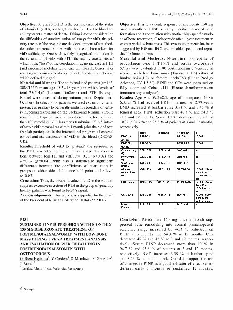
Objective: Serum 25(OH)D is the best indicator of the statusof vitamin D (vitD), but target levels of vitD in the blood arestill represent a matter of debate. Taking into the considerationthe difficulties of standardization of assays for vitD, the pri-ority arrears of the research are the development of a method-dependent reference values with the use of biomarkers forvitD sufficiency. One such widely recognized biomarker isthe correlation of vitD with PTH, the main characteristic ofwhich is the “loss” of the correlation, i.e., no increase in PTH(and associated mobilization of calcium from the bones) afterreaching a certain concentration of vitD, the determination ofwhich defined our goal.Material andMethods: The study included patients (n=163,30M/133F; mean age 48.5±18 years) in which levels oftotal 25(OH)D (Liaison, DiaSorin) and PTH (Elecsys,Roche) were measured during autumn period (September–October). In selection of patients we used exclusion criteria:presence of primary hyperparathyroidism, secondary or tertia-ry hyperparathyroidism on the background terminal chronicrenal failure, hypercortisolism, blood creatinine level of morethan 100 mmol/l or GFR less than 60 ml/min/1.73 m2, intakeof active vitD metabolites within 1 month prior the blood test.Our lab participates in the international program of externalcontrol and standardization of vitD in the blood (DEQAS,UK).Results: Threshold of vitD to “plateau” the secretion ofthe PTH was 24.8 ng/ml, which separated the correla-tions between logPTH and vitD, R=−0.31 (p<0.02) andR=0.04 (p=0.04), with also a statistically significantdifference between the coefficients of correlation ingroups on either side of this threshold point at the levelp<0.05.Conclusion: Thus, the threshold value of vitD in the blood tosuppress excessive secretion of PTH in the group of generallyhealthy patients was found to be 24.8 ng/ml.Acknowledgements: This work was supported by the Grantof the President of Russian Federation НШ-4527.2014.7
P281SUSTAINED P1NP SUPPRESSION WITH MONTHLY150 MG RISEDRONATE TREATMENT OFPOSTMENOPAUSALWOMEN WITH LOW BONEMASS DURING 1 YEARTREATMENTANALYSISAND EVALUATION OF RISK OF FALLING INPOSTMENOPAUSALWOMEN WITHOSTEOPOROSISG. Riera-Espinoza1, Y. Cordero1, S. Mendoza1, Y. Gonzalez1,J. Ramos11Unidad Metabolica, Valencia, Venezuela
Objective: It is to evaluate response of risedronate 150 mgonce a month on P1NP, a highly specific marker of boneformation and its correlation with another high specific mark-er of bone resorption, C telopeptide after 1 year treatment inwomen with low bone mass. This two measurements has beensuggested by IOF and IFCC as a reliable, specific and repro-ducible bone markers.Material and Methods: N-terminal propeptide ofprocollagen type I (P1NP) and serum β-crosslaps(CTx) were evaluated in 80 postmenopausic Venezuelanwomen with low bone mass (T-score <−1.5) either atlumbar spine(LS) or femoral neck(FN) (Lunar ProdigyAdvance, CV 1.5 %). P1NP and CTx were measured onfully automated Cobas e411 (Electro-chemiluminescentimmunoassay analyzer).Results: Age was 59.8±8.3, age of menopause 46.8±6.3, 26 % had received HRT for a mean of 2.99 years.BMD increased at lumbar spine 3.58 % and 3.45 % atfemoral neck. P1NP reduction was: 46.3 % and 54.3 %at 3 and 12 months. Serum P1NP decreased more than10 % in 94.7 % and 95.8 % of patients at 3 and 12 months,respectively.
Conclusion: Risedronate 150 mg once a month sup-pressed bone remodeling into normal premenopausalreference range measured by 46.3 % reduction onP1NP at 3 months and 54.3 % at 12 months. CTxdecreased 48 % and 42 % at 3 and 12 months, respec-tively. Serum P1NP decreased more than 10 % in94.7 % and 95.8 % of patients at 3 and 12 months,respectively. BMD increases 3.58 % at lumbar spineand 3.45 % at femoral neck. Our data support the useof changes in P1NP as a good indicator of effectivenessduring, early 3 months or sustained 12 months,
Osteoporos Int (2014) 25 (Suppl 2):S159–S440S244

treatment of postmenopausal women with low bonemass with risedronate 150 mg once a month.Acknowledgements: Not restricted grant from LaboratoriosLeti. Venezuela
P282VITAMIN D STATUS IN A POPULATIONHOSPITALISED IN OUR CLINICAL SERVICEC. Galesanu1, I. Grozavu1, A. Loghin1, D. Ungureanu1,L. Apostu21Endocrinology, University of Medicine and PharmacyGr.T.Popa, Iasi, Romania, 2Biochemistry, University ofMedicine and Pharmacy Gr.T.Popa, Iasi, Romania
Objective: Vitamin D3 is a major factor in bone metab-olism. Vitamin D deficiency has been recognized as apandemic with a myriad of health consequences. Vita-min D deficiency is more common than expected, par-ticularly among adolescents, women, the elderly or peo-ple who live in northern regions. Our aim was todetermine plasma levels of vitamin D3 in patients ad-mitted in our clinical area for various endocrine pathol-ogies and postmenopausal osteoporosis.Material and Methods: We evaluated the status of vita-min D3 by measurement of serum 25-hydroxyvitaminD3 (25OHD3) in 176 patients admitted to our clinicalsector between January–August 2013. The age of pa-tients ranged from 4 to 87 years. Of this total, 59.6 %patients were in postmenopause and the diagnosis ofosteoporosis has been shown from 45.16 % of themusing the DXA determination. Serum 25OHD3 concen-tration was measured by chemiluminescence assay withminimum detection limit of 3.0 ng/ml. Vitamin D3insufficiency has been defined for the serum levels<20 ng/mL(<50 nmol/L) and Vit. D3 deficiency<10 ng/mL(<25 nmol/L).Results:We found a high prevalence of both vitamin Ddeficiency:17.14 % and insufficiency:57.86 % in ourstudy population. The mean serum level of 25OHD3was 16.90 ng/mL. In our study, vitamin D deficiencywas observed in: −16.47 % postmenopausal women,−19.05 % patients with osteoporosis, −10 % childrenand adolescents. Vitamin D insufficiency was observedin: −43.53 % postmenopausal women, −61.9 % osteopo-rotic patients, −40% children and adolescents. 50% of childrenand adolescents were presented vitamin D levels between 20and 30 ng/ml but, none of them had vitamin D levels>30 ng/ml. Vitamin D sufficiency >30 ng/mL was observedonly at 5 % of all study population.
Conclusion: In our study, we have demonstrated insuffi-ciency, deficiency in vitamin D3, illustrating thisthrough in a sample of patients hospitalized in ourservice of Endocrinology. That is an important clinicalproblem in the world.
P283VIOLATION OF CALCIUM-PHOSPHORUSMETABOLISM AND CALCIUM-REGULATINGHORMONES (CRH) AS A MAJOR DETERMINANTOF OSTEOPOROSIS WHICH ASSOCIATED WITHLIVER CIRRHOSIS (LC)I. Golovach1, Z. Mytnyk21Rheumatology, Clinical Hospital Feofania, Kyiv, Ukraine,2Gastroenterology, National Institute of Surgery andTransplantology A.А.Shalimov, Kyiv, Ukraine
Objective: To determine violations of mineral metabolism,concentration of calcium regulating hormones, as well as toestablish influence of disturbances in these systems of osteo-porosis associated with LC.Material and Methods: We observed 172 patients with LC,the average age 49.3±7.7 years. There were 108 (62.8 %) menand 64 (37.2 %) women. BMD was determined by DXA“Challenger” (DMS, France). We also determined the serumconcentration of calcium and phosphorus, urinary excretion,the level of PTH and 25(OH)D.Results: We found significant violations of calcium-phosphorus metabolism, changes in the CRH, and reducedBMD in patients with LC, but the severity of the imbalancedepended on the severity of the disease and correlated with themain laboratory criteria of liver dysfunction. We observeddecrease serum concentration of total and ionized calcium,as well as expressive tendency to hypercalciuria. The concen-tration of 25(OH)D was 9.88±4.36 ng/ml (in control 26.76±9.27 ng/ml; P<0.01). The level of deficit depended on thedegree of liver dysfunction. In the case of compensated of LCthe level of 25(OH)D was 12.93±5.42 ng/ml and decompen-sated—4.53±1.22 ng/ml (a decrease of 2.8 times). The re-ceived results emphasize that abnormal liver function signif-icantly affects formation of 25(OH)D. PTH was elevated in amajority of patients. Serum concentrations of PTH were onaverage 82.2±5.3 pg/ml in patients with LC. It significantlychanged at different degrees of disease activity and severity ofhepatocellular insufficiency. The PTH was 39.8±3.1 pg/ml incompensation stage, 89.8±4.2 pg/ml in subcompensation,103.2±11.7 pg/ml in decompensation.Conclusion: Thus, in patients with LC, violations of mineralmetabolism with emergence of resistant hypocalcemia, andlack of active vitamin D metabolites, as well as development
Osteoporos Int (2014) 25 (Suppl 2):S159–S440 S245

of secondary hyperparathyroidism, especially in severe liverdysfunction, are observed. These patients are diagnosed withgrave osteoporosis.
P284CLINICAL FEATURES OFKNEEOSTEOARTHRITIS(OA) DEPENDING ON THE STATE OF BONEMINERAL DENSITY (BMD)I. Semeniv1, I. Golovach2, I. Zazirny1, V. Sulyma31Orthopedics Department, Clinical Hospital Feofania, Kyiv,Ukraine, 2Rheumatology, Clinical Hospital Feofania, Kyiv,Ukraine, 3Orthopedics and Traumatology Department,National Ivano-Frankivsk Medical University, Ivano-Frankivsk, Ukraine
Objective: The aim of the study was to determine the rela-tionship between the frequencies, clinical characteristics ofpatients with knee OAwith different BMD.Material andMethods: The study included 103 women withestablished osteoarthritis of the knee, the average age was67.6±9.8 years. BMD and degree of bone loss was deter-mined by DXA at the “Challenger” (MSD, France). Forclinical assessment of the joints, Leken’s index, range ofmotion, pain index according to the verbal scale of pain,functional index were determined. Knees were radiographedat baseline.Results: The incidence of tibio-femoral and patella-femoralOAwas significantly lower in women with osteoporosis thanin women with normal bone. Thus, when the probable osteo-porosis tibio-femoral OA (TFOA) was diagnosed in 53.3 % ofpatients, patella-femoral OA (PFOA)–in 13.3 %. At the sametime in women with a normal bone state knee OA of sites wasdiagnosed in 78.6 % and 53.6 %, respectively. In case ofidentifying the osteopenic syndrome TFOA was diagnosedin 64.4 % of women and PFOA–in 15.8 %. We have notestablished the relationship between radiographic OA andbone loss. In patients with OA of the III stage, osteoporosisand normal bone tissue are discovered with approximatelyequal frequency. Analysis of the anthropometric charac-teristics revealed significant differences in BMI. Inwomen with osteoporosis, BMI was significantly lowerthan in patients with normal bone mass. Patients withosteoporosis have longer duration of osteoarthritis; theirLeken’s index is more pronounced and their joints func-tional impairment is of a higher degree. There is arelationship of correlation (r=−0.76; P<0.01) betweenthe functional index and BMD.Conclusion: The study revealed that in patients withknee OA of various disorders, marked state of bonetissue and the degree of reduction in BMD are determined
by numerous factors. In patients with osteoporosis incidenceof knee OA was lower compared to women with normalBMD.
P285RECOGNITION OF OSTEOPOROSIS RISKFACTORS IN HIP FRACTURE PATIENTS: FIRSTDATA FROM LATVIAM. Mukane1, I. Rasa1, I. Daukste2, M. Mukans31Riga Stradins University, Riga East Clinical UniversityHospital, Latvian Osteoporosis and Bone MetabolismDiseases Association, Riga, Latvia, 2Latvian Osteoporosis andBone Metabolism Diseases Association, Riga 2nd Hospital,Riga, Latvia, 3Riga Stradins University, Riga East ClinicalUniversity Hospital, Riga, Latvia
Objective: Osteoporosis (OP) and its related fractures arehighly prevalent condition worldwide with known risk fac-tors. The aim of the study was to analyze recognition of OPrisk factors in patients with hip fractures.Material and Methods: Retrospective medical recordanalysis of patients (pts) attended to Riga 2nd Hospitalspecialized in traumatology and orthopaedics. All ptsincluded in the study were admitted to hospital in3 year period (08.12.2009–19.12.2012) and had a hipfracture as a result of low energy trauma. OP riskfactors such as gender, age, habits, comorbidities, med-ication, including glucocorticosteroid (GCs) use, previ-ous fracture were analyzed. All interval data werepresented in median values with interquartile range(IQR).Results: A total of 888 pts medical records were analyzed:predominantly females (73.1 %) older than males 80 (IQR,86–72) yrs vs. 72 (IQR, 80–61) yrs (p<0.001). Suchrisk factors were recognized: previous fracture–14.8 %(more than one fracture 9.2 %), smoking–12.5 %, alco-hol abuse–7.7 %. Totally 24.4 % of all pts had comor-bidities increasing risk of OP (28.0 %–chronic obstruc-tive pulmonary disease or sarcoidosis, 9.9 %–diabetesmellitus, 8.7 %–chronic kidney disease, 1.5 %–rheuma-toid arthritis, 0.7 %–gastrectomy). Only 1.4 % (n=12)of all pts used GCs in long-term. Medication increasingrisk of OP (e.g., anticonvulsants, proton pump inhibi-tors) used 6.2 % (n=55) of all pts. Almost half of thepts had at least 1 OP risk factor, 30.6 % of pts had 2risk factors, 9.2 % had 3 risk factors, 2.5 %–4 riskfactors and 0.3 %–6 risk factors. Only in 2.8 %(n=25) pts OP diagnosis was found in medical recordsand 2.5 % (n=22) pts had recommendations for furtherOP evaluation or treatment.
Osteoporos Int (2014) 25 (Suppl 2):S159–S440S246

Conclusion: Although most often cause of hip fractures islow BMD, especially in elderly, OP risk factors not always arerecognized in Latvia and, as a result, there are future fractures,chronic pain and disability.
P286EXAMINATION OF BONE MINERAL DENSITYANDFAT MASS IN TYPE 2 DIABETES MELLITUSPOSTMENOPAUSAL WOMENA. P. Shepelkevich1, V. Labashova1, O. Salko2, N. A.Vasilieva21Belarusian State Medical University, Minsk, Belarus,2Republican Centre for Medical Rehabilitation andBalneotherapy, Minsk, Belarus
Objective: The metabolic processes occurring in diabeticpostmenopausal women could produce deviations in tis-sue distribution and affect the components of bodycomposition. Several studies indicated that postmeno-pausal diabetic women have a high risk of osteoporoticfractures. Nevertheless the data relating to BMD in thiscategory of patients are rather contradictable. The cen-tral distribution of body fat as a predictor of cardiovas-cular diseases is important for clinicians to accuratelymeasure. The aim of the study was to assess BMD andfeatures of fat mass distribution in women with type 2DM in comparison with nondiabetic women of similarage and BMI.Material and Methods:We examined 205 women with type2 DM (mean age: 58.32±7.32 year, the duration of DM:11.03±5.34 year, age of manifestation: 48.63±8.97 year,BMI: 32.34±5.16, HbA1c: 9.2±1.7 %) and 68 age-,BMI- matched controls. The investigation involved an-thropometry of patients (height, weight, BMI, waistcircumference), general clinic examination and DXA(including BMD measuring at femoral neck and lumbarspine and body composition program).Results: BMD (g/cm2) was statistically lower in diabeticpatients both at spine (1.01±0.16 vs. 1.13±0.15, p<0.05)and at femoral neck (0.81±0.164 vs. 0.92±0.153, p<0.05)in comparison with controls. Fat mass distribution parametersin type 2 DM patients and controls were: Total Body: 38.67±7.31 % vs. 37.24±6.73 % (p=0.24); Android: 45.06±7.56 %vs. 44.23±8.34 % (p=0.005); Gynoid 39.60±8.04 % vs.40.68±7.54 % (p=0.201); Trunk/Total: 0.57±0.05 vs. 0.51±0.07 (p<0.001);(Arms+Legs)/Total: 0.67±0.18 vs. 0.87±0.28(p<0.001).Conclusion: The results of study revealed the prevalence ofcentral (android) distribution of body fat among postmeno-pausal women with type 2 DM in comparison with nondia-
betic patients. Also the data confirmed bone loss manifesta-tion in type 2 DMpostmenopausal women both at spine and atfemoral neck.
P287LIFESTYLE INFLUENCE AND INCIDENCE OFOSTEOPOROSIS IN THE SEASHORE AREA OFROMANIAE. Circo11Ovidius University, Constanta, Romania
Objective: Evaluation of lifestyle and secondary factors andinvolvements in osteoporosis incidence in postmenopausalwomen from an area of Romania with a high level of sunnydays by year.Material and Methods: The study included 984 postmeno-pausal women, hereupon was evaluated BMD by DXA andappreciated the T- and Z-scores (by WHO criteria). In Sep-tember it was evaluated the serum level of calcidiol (25-OH-vitamin D). The patients answered to a questionnaire aboutmenopause, risk factors and lifestyle.Results: Medium age was 61.5±8.2 years. 503 women(48.7 %) were diagnosed with osteoporosis (T-score>−2.5),414 with generalized osteoporosis (82 %) and 89 with seg-mentary osteoporosis (18 %). 491 patients (38 %) were withosteopenia (T- score between −1/−2.5) and 38 women withoutosteoporosis (BMDnormal). The age ofmenopause onset was44.1±1.8 years at women with osteoporosis, 47.3±1.4 yearsat osteopenia and 49.4±1.4 years at women without osteopo-rosis. The sun exposure was 25±4.1 days in women withosteoporosis and 29±9.6 day in women with osteopenia.102 patients (20.2 %) with osteoporosis taken calcium andvitamin D and 46 (4.5 %) had secondary causes ofosteoporosis. The results of questionnaire denoted thatonly 97 patients (9.7 %) of women with osteoporosisand osteopenia had physical activity after menopause,353 patients (35.5 %) made periodical investigationsand 143 patients had fractures.Conclusion: The incidence of osteoporosis andosteopenia is higher and underestimated, probably, atwomen from seaside area of Romania. The age ofmenopause onset is under the medium value from wom-en of European Community. There is a negative corre-lation between Z-score and years from beginning ofmenopause. The serum level of calcidiol (25-OH-vita-min D) was slightly elevated although the number ofsunny days was around 50 % per year. Periodical med-ical investigations, medical access information and levelof education were superior to women withoutmenopause.
Osteoporos Int (2014) 25 (Suppl 2):S159–S440 S247

P288OSTEOPOROSIS IN PATIENTS WITH TOTAL HIPARTHROPLASTYV. M. Ciortea1, I. M. Borda1, L. Irsay1, R. Ungur1, I. Onac11Department of Medical Rehabilitation, UMF Iuliu-Hatieganu, Cluj-Napoca, Romania
Objective:The objective of the study is to evidence the role ofBMD in the rehabilitation of patients with total hiparthroplasty.Material and Methods: The study, a representative sampleanalysis, was carried out at the Department of Medical Reha-bilitation of the “Iuliu Haţieganu”UMPh Cluj- Napoca, in theperiod June–December 2009. The study inclusion criteriawere met by a number of 58 patients aged between 30 and83 years with uni- and bilateral cemented and uncementedtotal hip endoprostheses. A standard study protocol was elab-orated, which included the measurement of BMD in the spineand both hips, using DXA, with the Lunar Prodigy Advanceosteodensitometer, the software for orthopedic prostheses be-ing available. The device used en. Core 11.X programs andcomputers with the Windows XP Professional operating sys-tem, allowing to determine bone mineral content BMC(grams) and BMD (g/cm2) in seven different areas aroundthe endoprosthesis, known as the Gruen zones. Thepatients were clinically evaluated using two scales: theOxford Hip Score and Qualeffo-41. Statistical calcula-tions were performed using the SPSS13.0 and MicrosoftExcel applications.Results: The two scores were significantly correlated(p<0.005) with the diagnosis made based on DXAexamination and with the type of hip endoprosthesis(cemented or uncemented); they were higher in the caseof low BMD (osteopenia/osteoporosis) and cementedendoprostheses.Conclusion: Low periprosthetic BMD values delay the reha-bilitation of patients with total hip endoprostheses. The lowerthe BMD levels in the entire skeleton, the less favorable thepostoperative evolution, which also limited the quality of lifeof these patients.References: Lou XF et al. Zheijang Univ Sci B 2007;8:76.
P289HYALURONIC ACID “OSTENILTENDON” INPARTIALTHICKNESS TEARS OF THESUPRASPINATUS TENDON: CLINICAL ANDSONOGRAPHIC ASSESSMENTR. Nestorova1, R. Rashkov2, T. Petranova2, Z. I. Kolarov2, I.Sheytanov2, R. Stoilov2, S. Monov2, Z. Stefanov31Centre of Rheumatology, Sofia, Bulgaria, 2Clinic ofRheumatology, Medical University, Sofia, Bulgaria,3Sports Academy, Sofia, Bulgaria
Objective: To determine the efficacy of hyaluronic acid (HA)injections Ostenil Tendon concerning pain, functional activityof the shoulder and tendon recovery in patients with partialthickness tears (PTT) of the supraspinatus tendon (SSP). Theaccuracy of ultrasonography (US) for diagnosis of PTT of therotator cuff, benefit of performance of US-guided proceduresand monitoring of therapy have been proved.Material andMethods: 23 patients with painful shoulder andsonographic proved PTTof the SSP were included. A pain diarywith a ten point VAS, Shoulder Function Assessment (SFA)scale (0–70) and questionnaires of the efficacy according to thepatient and the physician were evaluated. US examination wasprovided by Mindray M5 scanner with multifrequency lineartransducer (7.5–10 MHz). Ostenil Tendon 40 mg/2.0 ml wasinjected around the affected tendon once a week for a total of twoinjections. All applications were performed under US control.Results: Pain was significantly reduced after the first injectionand this effect was maintained until the end of the observa-tional period. SFA Index was significantly improved. 76 % ofpatients gave a very good and good assessment of the efficacy,which coincided with the opinion of the physician. 77 % ofpatients had a complete recovery or improved structure of theSSP which was US demonstrated.Conclusion: HA Ostenil Tendon led to a relatively rapid andsustained relief of pain and increased functional activity of theshoulder. US proved repair process of the tendon structure andgliding of the SSP as a result of lubricating and viscoelasticproperties of the HA. No adverse events were observed. Injec-tions under US control were accepted favorable by the patients.
P290PROXIMAL FEMUR FRACTURE PATIENTSFEATURES IN A FLS IN BRAZILB. Stolnicki11Prevrefrat Hospital Federal de Ipanema Creb, Rio de Janeiro,Brazil
Objective: To describe the features of patients who have hadproximal femur fractures (PFF) in Prevrefrat–a FLS in Riode Janeiro, Brazil.Material andMethods: 63 patients withminimal trauma PFFhave been followed from February 2011 to November 20013.The patients features, fractures, risk factors, associated dis-eases, BMD values, seric vitamin D levels, and incidence ofnew fractures are reported.Results: 57 women and 6 men, from 51 to 95 years old had aminimal trauma PFF. The main fracture was transtrochanteric(30), femoral neck (29) and subtrochanteric (4). 3 women hadbilateral fractures. 18 patients had only the PFF. 23 had onemore fracture and 22 had two or more fractures. 38 patientshad prior vertebral fracture. After DXA scan 49 were classi-fied as osteoporosis and 14 as osteopenia. 44 patients had
Osteoporos Int (2014) 25 (Suppl 2):S159–S440S248

vitamin D insufficiency or deficiency, 30 had arterial hyper-tension and 6 had diabetes. 45 prior fragility fractures, 19 earlymenopause and 11 with parent fractured hip were the morefrequent fracture risk factors. All patients received calciumand vitamin D supplementation. Sixty-two patients receivedzoledronic acid 5 mg and 1 patient with stage 4 kidney diseasereceived denosumab 60 mg. 3 patients had flu-like symptoms.All patients had 100 % of treatment adherence. No newfractures occurred. In all patients, there was no decrease inheight.Conclusion: In most cases the patients with minimal traumaproximal femur fracture had a previous fragility fracture. Insuch a group of patients with high risk of new fractures (1), weprefer not to use oral drugs due to historical poor adherence(2). No new fractures occurred using zoledronic acid anddenosumab.References: 1. Johnell O et al. Osteoporos Int 2004;15:175.2. Landfeldt E et al. Osteoporos Int 2012;23:433Disclosures: Speaker: GSK, Sanofi Aventis, Servier, NovartisAdvisory board: Servier, Novartis
P291CASE REPORT: OSTEOPOROTIC VERTEBRALFRACTUREV.M.Ciortea1, M. Nechita1, R. Ungur1, L. Irsay1, I. Onac1,I. M. Borda11Department of Medical Rehabilitation, UMF IuliuHatieganu, Cluj-Napoca, Romania
Objective: Osteoporosis is the most frequent metabolic bonedisease characterized by: reduction of bone mass, alteration ofbone architecture, deterioration of the quality of bone, andincrease of the risk of fracture. The frequency of osteoporoticvertebral fractures reported by the literature varies between 33and 85 %; of these, 25–33 % have a clinical manifestationwhen they occur. The case report brings into discussion thecase of a patient in whom vertebral fracture was the firstclinical manifestation of osteoporosis.Material and Methods: Patient LP, aged 57 years, presentedto our service in April 2013 for marked pain in thedorsolumbar spine, with onset 3 weeks before, causing herimmobilization in bed. Of the patient’s personal history andlife conditions, we mention the onset of menopause at the ageof 39 years, and smoking for approximately 25 years, 20cigarettes/day. The objective examination of the patient atthe time of presentation: BMI=23 kg/m2, spontaneous painon the percussion and mobilization of the dorsolumbar spine,without dural or neurological signs. Dorsolumbar spine X-ray,vertebral CT and the measurement of BMD using the methodof DXA allowed to make a positive diagnosis and to initiateadequate antiosteoporotic treatment.
Results:Any change in the shape, size, outline or structure ofa vertebral body should be interpreted in a clinical context.The majority of osteoporotic fractures are located in the tho-racic or thoracolumbar region; an osteoporotic fracture aboveT7 is unusual and the suspicion of malignancy should beeliminated.Conclusion: After 2 weeks of treatment (strontium ranelate2 g/day and vitamin D3), we obtained am important improve-ment of symptomatology, and 4 weeks after the initiation oftreatment, the patient resumed most of her daily activities.
P292ALVEOLAR BONE LOSS AND BONE METABOLISMIN PREMENOPAUSALWOMENI. Mazur1, V. Gabal11Department of Stomatology Institute of Stomatology ShupykNational Medical Academy of Postgraduate Education, Kiev,Ukraine
Objective: Premenopause period is characterized by a signif-icant reduction in secretion of estrogen in a woman’s body,which leads to the disturbance of bone metabolism. The aimof this study was to investigate the features of periodontaltissue statement, alveolar process and bone metabolism inpremenopausal women.Material andMethods: The 94 patients were selected on twogroups: main group (54 women 40–49 years old, mean age46.7±2.3 years) in premenopause, control group (40 patients20–39 years old, mean age 31.4±3.4 years). Clinical andradiographic (panoramic X-Ray) conditions of periodontalstatement were evaluated. Metabolic processes of bone tissuewere evaluated by the serum levels of calcium related hor-mones (calcitonin PTH) and biochemical marker of boneremodeling such as deoxypyridinoline (DPD) in urine.Results: In premenopausal period the rate of alveolar boneloss in premenopausal women group 2.53±0.3 mm was sta-tistically significantly (p<0.05) higher compared with thecontrol of 1.09±0.4 mm. Increased secretion of PTH in wom-en in premenopause period is 45.49±2.12 compared withcontrol of 38.33±1.92. Inhibition of secretion of calcitonin1.02±0.43 compared to control 3.2±0.34. Unbalanced ofremodeling processes reinforcing the resorption processdeoxypyridinoline 8.62±0.43 nmol was statistically signifi-cantly (p<0.01) higher compared with 6.36±0.41 nmol. Inhi-bition of the bone formation 18.96±1.54 mmol/l was statisti-cally significantly (p<0.05) higher compared to control 2.01±2.21 mmol/l..Conclusion: Homeostasis violation of calcium related hor-mone reduced secretion of calcitonin and increased parathy-roid hormone, misbalanced of the bone tissue remodelingprocesses, accelerated the rate of resorption and oppression
Osteoporos Int (2014) 25 (Suppl 2):S159–S440 S249

bone forming are a risk factor for resorption of alveolar boneand reduce the height of it.
P293WORLDWIDE QUANTITATIVE IMPACT OF BONEMINERAL DENSITY ON HIP FRACTUREINCIDENCEL. Sanchez-Riera1, R. Norman2, L. Veerman2, T. Vos3, N.Wilson1, D. Hoy2, E. Smith1, L. March11Institute of Bone and Joint Research, Royal North ShoreHospital, University of Sydney, Sydney, Australia, 2Schoolof Population Health, University of Queensland, Herston,Australia, 3Institute for Health Metrics and Evaluation, Seat-tle, United States
Objective: As part of the Global Burden of Diseases Study2010 the Musculoskeletal Expert Group analysed the world-wide burden of low BMD as a risk factor for fractures usingComparative Risk Assessment (CRA) methodology1. Esti-mates on the influence of population BMD levels on the hipfracture incidence by world region are presented.Material and Methods: A systematic review was donethrough MEDLINE, EMBASE, CINAHL, CAB abstract,WHOLIS, and SIGLE to search for population based studieswith BMD at femoral neck (FNBMD) measured with DXA,which was analysed as a continuous variable. Age- and sex-specific values of FNBMD (mean and SD) in g/cm2 wereextracted, standardized and pooled using a Bayesian meta-regression tool, DisMod-MR, for population aged 50 yearsand over. The CRA methodology was used to estimate theproportion of fractures attributable to sub-optimal BMD,using the Potential Impact Fraction (PIF). The sex- and age-specific 90th percentile FNBMD from NHANES III was usedas the theoretical minimum exposure risk distribution. Fromthe authors of a previousmeta-analysis2 we obtained estimatesof the gradient of risk of hip fracture (RR/SD) for BMD Zscores based on the combined data for men and women, andconverted the RR/SD values into RR/0.1 g/cm2 values. ThePIFs were estimated for 21 world regions for 1990, 2005 and2010, and expressed on a 0–1 scale. Results for 2005 areshown.Results: There were large differences across regions.Lowest PIFs were found in North Americans aged 80–84 years both for males (0.412) and females (0.140),while highest PIFs were found in Sub-Saharan WestAfricans aged 55–59 years for females (0.871) as wellas males (0.859). In general PIFs decreased with agemore in females than in males.Conclusion: The potential impact of BMD on hip fractureincidence varies widely among world regions. Influence ofBMD on fracture risk decreases with age, particularly inwomen.
References: 1 Lim S et al Lancet 2012; 2 Johnell O et al JBone Miner Res 2005.
P294VITAMIN D AND PATIENTS WITH PRIMARYHYPERPARATHYROIDISM (PHPT): HOSPITALBASED STUDY IN LATVIAG. Avotina1, I. Rasa21Riga East Clinical University Hospital, University of Latvia,Latvian Osteoporosis and Bone Metabolism Diseases Associ-ation, Riga, Latvia, 2Riga East Clinical University Hospital,Riga Stradins University, Latvian Osteoporosis and BoneMetabolism Diseases Association, Riga, Latvia
Objective: Vitamin D deficiency is more frequent con-dition in PHPT patients (pts) than in the general pop-ulation. There is worsening of PHPT when vitamin Ddeficiency coexists. Vitamin D deficiency may worsenmanifestations of PHPT by contributing more pro-nounced parathyroid gland proliferation. The aim ofthis study was to analyze vitamin 25(OH)D3 level inPHPT pts in tertiary care multiprofile hospital inLatvia.Material and Methods: Hospital based case control ret-rospective study analyzed medical records of pts admit-ted to the hospital in 4.5 year period (01.01.2009–30.06.2013). The statistical data were processed usingSPSS 16.0. All the data were presented in the meanvalue with standard deviations. Normal range for calci-um was defined 2.1–2.6 mmol/L, for PTH - 12.0–72.0 pg/mL, for phosphorus - 0.8–1.6 mmol/L, foralkaline phosphatase - <117.0 U/L. Vitamin sufficient con-centration defined as >30.0 ng/ml, insufficiency as - 29.9–20.0 ng/ml and deficiency as <19.9 ng/ml.Results: There were 176 medical records of 140 pts,37 % of whom had their 25(OH)D3 assessed. Studyincluded 94.2 % females and 5.8 % males. Femaleswere older than males, 62.4±12.1 and 56.7±3.8 year,respectively (p=0.09). Pts calcium level in the studypopulation was 2.9±0.4 mmol/L, PTH level - 288.7±365.7 pg/mL, phosphorus level - 0.9±0.3 mmol/L, al-kaline phosphatase - 88.6±39.3 U/L. Pts vitamin25(OH)D3 level was 15.5±8.9 ng/mL, female - 15.4±9.1 ng/mL, male - 17.7±7.4 ng/mL (p=0.64). For fe-males aged up to menopause (n=5) vitamin 25(OH)D3
level was 3.7±0.7 ng/mL, postmenopausal (n=44) -16.7±8.6 ng/mL (p=0.002). A higher level of vitaminD was in postmenopausal females, presumably it couldbe explained by vitamin D supplementation.Conclusion: Vitamin 25(OH)D3 deficiency was found in allpts with PHPT in all groups.
Osteoporos Int (2014) 25 (Suppl 2):S159–S440S250

P295EFFECTS OF PHYSICALTHERAPY IN THEREHABILITATION OF PATIENTSWITH ARTHROSISOF THE HIPN. Mandic1, S. Kozomara2, T. Petrusic1, S. Petrovic11Dom Zdravlja Nis, Nis, Serbia, 2Institut Niska Banja, Nis,Serbia
Objective: To present the importance of physical therapy inthe rehabilitation of patients with arthrosis of the hip.Coxarthrosis is one of the most common degenerative rheu-matic diseases of the peripheral joints. It is characterized bychronic pain and limited mobility and they are major socio-medical problem. Arthritis is more common after the age of 40and more frequently in women.Material and Methods: In the period January–October2013 in DZ Nis treated 80 patients, 52 women and 28men, mean age 58 years, with a treatment duration of14 days. Coxarthrosis was diagnosed history, clinicaland radiographic findings. In the first group (40 pa-tients) applied the IMP, IFS and kinetic therapy, whilethe other (40 patients), IFS and kinetic therapy. Assess-ment of treatment was performed on the basis of mea-surements of the volume turns into the hip, gross motorstrength (GMS) pelvifemoral muscles and the subjectiveexperience of pain (VAS).Results: In all patients there was an improvement, but therange of motion in a statistically significant only in the firstgroup. Abduction with 20° increased to 35° of flexion withstretched knee to 50° to 70°. The measurement of the exten-sion is no statistical significance. GMS muscle actuator of thehip, in the first group increased with 2+ to 3+. When it comesto pain according to the VAS scale improvement was presentin both groups, but the statistical significance of first group(p<0.05).Conclusion: Physical therapy consisting of IMP, IFS andkinetic therapy, increases the range of motion of the affectedthe hip, strengthens muscle strength, reduces pain and pro-vides the functionality of the diseased hip and disposed oforthopedic treatment.
P296UNMASKING OF PAGET’S DISEASE AFTER THEINTRODUCTION OF TERIPARATIDE TREATMENTA. Zavratnik1, M. Krajnc11Division of Endocrinology and Diabetology, UniversityMedical Centre Maribor, Maribor, Slovenia
Objective: Paget’s disease is the focal disorder of bone me-tabolism characterized by accelerated rate of bone remodeling,resulting in overgrowth of bone at single or multiple sites andimpaired integrity of affected bone. The majority of patients
with Paget’s disease are asymptomatic. The diagnosis in suchpatients is usually made incidentally following the routinechemistry screen showing elevated serum concentration ofalkaline phosphatase (AP) of bone origin or an imaging studyobtained for other reasons that shows Pagetic changes in bone.Bisphosphonates are the most widely used antiresorptiveagents for Paget’s disease and are now considered the first-choice treatment.Material and Methods: Case report: A 65-year old womanwith primary osteoporosis.Results:Raloxifene was started in 2003. The treatment failed,as new OP fractures of the vertebras TH 8, TH 11 and L4 wereconfirmed in 2008. Raloxifene was stopped and risedronateintroduced. New vertebral fractures were confirmed in 2010(TH 1 and TH 5). Teriparatide was suggested, but the patientdisagreed, so strontium ranelate was started. In 2011, patientdecided to take teriparatide, and it was first introduced inNovember 2011. From 2003 to 2011, no signs of secondarycauses of bone disease were confirmed. At the first follow upvisit in May 2012, elevated AP was first noticed, and this wasconfirmed 1 month later. The comprehensive diagnostic pro-cedures were performed. With all tests done we confirmed theelevation of bAP and enhanced radionuclide uptake to theskull and the right distal femur. Subsequently performed plainradiography suggested the osteolytic lesions of the skull.Teriparatide was discontinued and zolendronic acid (ZA) ini-tiated. After one course of the ZA treatment AP normalisedand the lesions on bone scintigraphy and bone radiographydisappeared.Conclusion: Teriparatide treatment is able to unmask theunderlying Paget’s disease. Regular determinations of APare mandatory during the entire course of teriparatidetreatment.
P297TEN YEARS OF HIP FRACTURES IN ITALY: FORTHE FIRST TIME A DECREASING TREND INELDERLY WOMENP. Piscitelli1, A. Distante1, M. L. Brandi2, G. Iolascon3,S. Parri2, F. M. Liuni1, G. Quarta1, C. Neglia1, E. Gasbarra4,M. Celi4, C. Rao4, M. Feola4, U. Tarantino41Euro Mediterranean Scientific Biomedical Institute - ISBEMResearch Centre, Brindisi, Italy, 2Department of TranslationalMedicine, University of Florence, Florence, Italy, 3Depart-ment of Orthopaedics and Rehabilitative Medicine, SecondUniversity of Naples (SUN), Naples, Italy, 4Division ofOrthopaedics and Traumatology, University of TorVergata, Rome, Italy
Objective: We aimed to evaluate hospitalization rate of fem-oral neck fractures in the elderly Italian population over10 years.
Osteoporos Int (2014) 25 (Suppl 2):S159–S440 S251

Material and Methods: We analyzed national hospitaliza-tions records collected at central level by Ministry of Healthfrom 2000 to 2009. Age- and sex-specific rates of fracturesoccurred at femoral neck in people ≥65 years old. Weperformed a subanalysis over a 3-year period (2007–2009), presenting data per 5-year age groups, in orderto evaluate the incidence of the hip fracture in theoldest population.Results:We estimated a total of 839,008 hospitalizations dueto femoral neck fractures between 2000 and 2009 in people≥65, with an overall increase of 29.8 % over 10 years. Theincidence per 10,000 inhabitants remarkably increased inpeople ≥75, passing from 158.5 to 166.8 (+5.2 %) and from72.6 to 77.5 (+6.8 %) over the 10-year period in women andmen, respectively. The oldest age group (people >85 yearsold) accounted for more than 42% of total hospital admissionsin 2009 (n=39,000), despite representing only 2.5 % of theItalian population. Particularly, women aged >85 accountedfor 30.8 % of total fractures, although they represented just1.8 % of the general population. The results of this analysisindicate that the incidence of hip fractures progressively in-creased from 2000 to 2009, but a reduction can be observedfor the first time in women ≤75 (−7.9 % between 2004 and2009).Conclusion: Incidence of hip fractures in Italy are continu-ously increasing, although women aged 65–74 years oldstarted showing a decreasing trend.
P298EVALUATION OF THE ROLE OF VITAMIN DRECEPTOR (VDR) GENE POLYMORPHISMS INTYPE 1-ASSOCIATED BONE DISORDERY. Dydyshko1, A. P. Shepelkevich1, V. Zhukouskaya21Department of Endocrinology, Belarusian State MedicalUniversity, Minsk, Belarus, 2Department of Clinical Scienceand Community Health, Milan, Italy
Objective: Low BMD and fracture risk are associated withtype 1 diabetes (T1D). Vitamin D receptor (VDR) polymor-phisms have been suggested to be associated with the diabeticcomplications. Therefore, the aim of study was to assess theassociation between VDR single nucleotide polymorphisms(SNPs) in type 1 diabetic patients and low BMD.Material andMethods:We studied 66 T1D patients (28 menand 38 women; mean age 31.23±841; duration of the disease13.40±7.41; НвА1с 8.25±0.95 %). BMD was measured byDXA. QIAamp DNA Blood Mini Kit (Qiagen, USA) wasused to purify DNA from whole blood, gene polymorphismswere detected in PCR-RFLP (restriction fragment length poly-morphism) analysis. The following restriction enzymes wereused to determine the appropriate polymorphism: VDR-FOKI- FokI (BseGI), VDR-ApaI - ApaI.
Results: The presence of the mutant allele VDR-FokI wasdetected in 79 % of cases (in 41 % cases as heterozygotes andin 38 % as homozygotes). VDR- ApaI SNPs was found in77 % of cases (in 50 % cases as heterozygotes and in 27 % ashomozygotes). There was a significant prevalence of low bonemass among carriers individuals with VDR-FokI SNPs(30.7 % vs. 14.2 %, p<0.001) and individuals with VDR-Apal SNPs (31.37 % vs. 26.6 %, p<0.001).Conclusion: The results of the study reflect the high frequen-cy of VDR (FokI, ApaI) SNPs and a significant decrease inbone density in these individuals. VDR gene polymorphismsseem to play a major role in influencing on bone loss in T1D.
P299TOTALBODYBONEDENSITYAND FAT/LEANMASSDISTRIBUTION IN TYPE 1 DIABETIC PATIENTSA. P. Shepelkevich1, Y. Dydyshko1, N. A. Vasilieva21Department of Endocrinology, Belarusian State MedicalUniversity, Minsk, Belarus, 2Republic Center of MedicalRehabilitation, Minsk, Belarus
Objective: There is epidemiological evidence that reducedamount and a decline in the quality of muscle mass associatedwith an increased risk of fracture. The aim of the study was theexamination of total body bone density (TBBD) and thecomponents of body composition in type 1 diabetes mellitus(DM) patients.Material and Methods: We studied 66 type 1 DM patients(28 men and 38 women; mean age 31.23±8.41; duration ofthe disease 13.40±7.41; НвА1с 8.25±0.95 %). The researchinvolved anthropometry of patients, general clinic examina-tion, glycated hemoglobin test, DXA performed on “ProdigyLunar” using a program “ total body” and “bodycomposition”.Results: TBBD (g/cm2) (1.156±0.10 vs. 1.194±0.084,p<0.01) and total Z-score (−0.15±0.94 vs. 0.72±0.63,p<0.001) was statistically lower in diabetic patients in com-parison with controls. Fat mass distribution parameters in type1 DM patients and controls were: Total Body: 29.63±12.80 %vs. 30.01±9.68 % (p=0.32); Android: 29.68±12.14 % vs.29.90±12.7 % (p=0.50); Gynoid 36.50±13.34 % vs.36.98±10.88 % (p=0.09); A/G Ratio: 0.80±0.29 vs.0.87±0.28 (p=0.022); Trunk/Total: 0.46±0.87 vs. 0.48±0.08 (p=0.68); (Arms+Legs)/Trunk: 1.078±0.15 vs. 1.26±0.18 (p=0.009); Total Body Lean mass: (49963.42±2849.845 g vs. 44057.80±9932.179 g, p<0.001).Conclusion: The data confirmed low total body bone densityin type 1 DM patients. There are changes in total body fatmass and lean mass among patients with type 1 DM. Thusmechanisms responsible for the formation of healthy bonesrequire further research.
Osteoporos Int (2014) 25 (Suppl 2):S159–S440S252

P300ORAL HEALTH ATTITUDES AND PRACTICESAMONGST PATIENTS WITH OSTEOPOROSIS INUNITED KINGDOMS. Gupta1, A. Gupta21Dental Sciences, Cardiff, United Kingdom, 2GlangwiliHospital, Carmarthen, United Kingdom
Objective: Many patients with osteoporosis are elderly andoral health is an important aspect of patient care especially inpatients on bisphosphonates. Our aim was to determine oralhealth attitudes and practices in patients with osteoporosisreceiving bisphosphonates.Material and Methods: A questionnaire study was conduct-ed after informed consent on cognitively intact patients diag-nosed with osteoporosis who were attending hospital osteo-porosis clinic and received bisphosphonate treatments.Results: 200 patients age range 50–95 years (average70 years) 75 % female. Patients receiving treatments werealendronate (80 %), risedronate (10 %), ibandronate (4 %),zoledronate (6 %). One hundred percent considered care ofteeth is important. Ninety-two percent considered cleaning ofteeth daily is important. Twenty percent considered they donot need regular specialist dental care. Ninety percent thoughttooth loss is normal consequence of ageing. Ninety percentconsidered tooth loss has an association with osteoporosis.Thirty-two percent felt access to dental services was a barriertowards improving oral health. Twenty percent used dentures.Sixty-five percent had visited dentist in last 1 year. Fiftypercent regularly visit dentist twice a year. Ten percent werenot registered with a dentist. Fifty-nine percent clean theirteeth twice daily. Eighty percent had informed their dentistabout osteoporosis medications. Ten percent were aware ofrare side effect of jaw osteonecrosis.Conclusion: This large study shows that most elderly havefavourable attitudes towards improving their oral health. Butthere are gaps in patients knowledge and there is suboptimaldental access, behaviour and practice amongst a large numberof patients. Further education and improved provision offacilities for dental care is needed.
P301FACTORS ASSOCIATED WITH LOW BONEMINERAL DENSITY IN PATIENTS WITHANKYLOSING SPONDYLITISB. Stojic1, I. Jeremic1, S. Janjic1, V. G. Bajec1, N. Djurovic11Institute of Rheumatology, Belgrade, Serbia
Objective: To assess the prevalence of low BMD and associ-ation between BMD on the hip and the lumbar spine andosteoporosis risk factors as well as parameters of disease
activity, functional status and spine mobility in patients withankylosing spondylitis (AS).Material and Methods: Our study included 62 patients (49males, 9 females), mean age 39.3±11.7 years, withdiagnosis of AS (modified New York criteria). Meanage at disease onset was 25.5±8.7 years and meandisease duration was 13.9±8.2. BMD was measuredusing DXA on the hip and the lumbar spine. LowBMD was defined as T-score≤−1 including osteopeniaand osteoporosis. Clinical (Bath AS Functional Index-BASFI, Bath AS Disease Activity Index-BASDAI, BathAS Metrology index-BASMI) and laboratory data(erythrocyte sedimentation rate-ESR and C reactiveprotein-CRP) were collected. The data of risk factorsfor osteoporosis were also collected (use of corticosteroids,disease modifying anti-inflammatory drugs (DMARD),nonsteroidal anti-inflammatory drugs (NSAID), smoking,BMI).Results: Osteoporosis on the hip and the lumbar spine wasfound in 14.5 % and 3.2 % patients, respectively, whileosteopenia was found in 45.2 % patients on the hip and24.2 % patients on the lumbar spine. There was significantinverse correlation between hip BMD and age at disease onset(p=0.032), BASFI (p=0.017) and BASMI (p=0.001), whilelumbar spine BMD was in inverse correlation only with ESR(p=0.045). Patients treated with corticosteroids had signifi-cantly more often osteoporosis (p=0.003). No significantcorrelation between BMD and smoking, use of NSAID,DMARD, disease duration, BASDAI, CRP and BMI wasfound.Conclusion: In patients with AS low BMD is found morefrequently on the hip and is associated with corticosteroid use.No relation with other risk factors for osteoporosis was found.Decreased BMD was associated with decreased functionalability (measured by BASFI) and back mobility (measuredby BASMI).
P302INFLUENCE OF DISEASE ACTIVITY ON PAININTENSITY, FATIGUE AND GENERAL HEALTHJ. Jovanovic1, V. Jovanovic2, M. Stojanovic3, S. Bozilov1,M. Karadzic1, V. Skakic11Institute for Treatment and Rehabilitation Niska Banja, Nis,Serbia, 2Clinic for Orthopedics, Clinical Centre Nis, Nis,Serbia, 3Faculty of Medicine, Nis, Nis, Serbia
Objective: To estimate the influence of disease activity onpain intensity, fatigue and general health in patients withrheumatoid arthritis.Material and Methods: The study included 95 patients withRA, according to ACR/EULAR criteria, 2010. There were 76women and 19 men. The average age was 58.8 years, and the
Osteoporos Int (2014) 25 (Suppl 2):S159–S440 S253

average duration of the disease was 10.4 years. Disease activ-ity was measured by an index DAS28SE. Pain, fatigue, andgeneral health represented by VAS. In relation to diseaseactivity, patients were divided into three groups: Group IDAS28SE <3.1- low disease activity; group II DAS28SE3.2–5.1 - moderate disease activity and group III DAS28SE>5.1 - high disease activity. For statistical analysis of the datawe used the analysis of variance (ANOVA), t-test.Results: The patients with high disease activity-Group III hadsignificantly worse values of pain intensity estimated by VASscale 65.28±12:43 compared to patients with moderate diseaseactivity Group II 39.2±18.54, p<0.001 and compared to pa-tients with low disease activity 8.68±6.05 Group I, p<0.001.The patients with high disease activity-Group III had signifi-cantly worse values of fatigue estimated VAS scale 66.18±15.62 compared to patients with moderate disease activity41.56±19.32 Group II, p<0.001 and compared to patients withlow disease activity 12.68±6.52 Group I, p<0.001. The pa-tients with high disease activity-Group III had significantlyworse values of general health estimated by VAS scale 32.24±13.22 compared to patients with moderate disease activity51.86±14.82 Group II, p<0.001 and in compared to patientswith low disease activity 82.10±7.32 Group I, p<0.001.Conclusion: The high disease activity causes higher painintensity, pronounced fatigue and poor general health in pa-tients with RA.
P303SEQUENTIALTHERAPY IN SEVEREOSTEOPOROSISR. Pastore1, A. Bultrini1, P. Di Giacinto2, A. Cannuccia2,S. Frontoni21Ospedale S.Giovanni Calibita, Fatebenefratelli, IsolaTiberina, UOC Endocrinology, Rome, Italy, 2Dept. ofSystems Medicine University of Rome Tor Vergata,Rome, Italy
Objective: Some evidence in literature suggests the utility ofthe addition of antiresorptive drugs, such as bisphosphonates,following teriparatide treatment, in severe osteoporosis. Theaim of the present study was to evaluate the impact of theaddition of 5 different treatments, following a 24-month ther-apy with teriparatide, on osteoporosis progression.Material and Methods: 81 women with severe postmeno-pausal osteoporosis, were treated for 24 months (first step)with teriparatide and serum osteocalcin and ß-CTX and lum-bar and femoral BMD changes, evaluated by DXA-BMD,were monitored at 6, 12, 18 and 24 months. After 24 months,60 of 81 patients were randomly allocated to receive one of thefollowing treatments, on top of calcium (1 g/day) and vitaminD (5,600 IU/week): Alendronate or risedronate weekly,ibandronate monthly or strontium ranelate daily or placebo,
for 24 months (second step). At the end of the second step ofthe study, lumbar and femoral BMD were evaluated again.Results: After 24 months, T-score slightly increased frombaseline at femoral level (p=0.07, Wilcoxon test) and signif-icantly increased at the lumbar spine level (p<0.001,Wilcoxon test). Osteocalcin values displayed a 4.3-fold in-crease (ANOVA, p<0.001), from baseline at month 6, and a5.4, 4.1 and 3.7, at 12, 18 and 24 months, respectively(Bonferroni, p<0.001). β-CTX levels showed a 2.5-fold in-crease from baseline at month 6 and a 2.6, 2, 1 and 1.8 at 12,18 and 24 months, respectively (p<0.001). At the end of thesecond step of the study, T-scores showed a further significantincrease, only in the group of patients treated with ibandronate(p<0.05), ranelate (p<0.05), and risedronate (p<0.05). Anal-ysis of variance showed a significant difference (p=0.008)between risedronate and placebo.Conclusion: Our data confirm the efficacy of teriparatide andsuggest that in severe osteoporosis the treatment of choice shouldinclude a first 24 month-step with teriparatide, followed by theaddition of antiresorptive drugs or strontium ranelate therapy.
P304NATIONWIDE COMMUNITY OSTEOPOROSISEDUCATION AND SCREENING PROGRAM INTAIWANR. S. Yang1, D. C. Chan2, K. H. Hsu3, C. Y. Yang4,J. F. Chen5,11, S. T. Tu6, C. H. Chen7, H. Y. Chiou8,L. C. Lim9, J. S. Hwang101Department of Orthopaedics, National Taiwan UniversityHospital, Taipei, Taiwan, Province of China, 2Department ofGeriatrics and Gerontology, National Taiwan University Hos-pital, Taipei, Taiwan, Province of China, 3Laboratory forEpidemiology, Department of Health Care Management,Chang Gung University, Taoyuan County, Taiwan, Provinceof China, 4Department of Public Health, College of HealthSciences, KaohsiungMedical University, Kaohsiung, Taiwan,Province of China, 5Division of Endocrinology and Metabo-lism, Kaohsiung Chang Gung Memorial Hospital, ChangGung University, Kaohsiung, Taiwan, Province of China,6Superintendent office, Lukang Christian Hospital,Changhwa, Taiwan, Province of China, 7Division of SportMedicine, Department of Orthopedic Surgery, Taipei MedicalUniversity Hospital, Taipei, Taiwan, Province of China,8School of Public Health, Taipei Medical University, Taipei,Taiwan, Province of China, 9Department of Gerontology andGeriatrics, National Taiwan University Hospital, Taipei, Tai-wan, Province of China, 10Division of Endocrinology andMetabolism, Chang Gung Memorial Hospital - Linkou Med-ical Center, Taoyuan, Taiwan, Province of China, 11Divisionof Endocrinology and Metabolism, Chang Gung MemorialHospital - Kaohsiung Medical Center, Kaohsiung, Taiwan,Province of China
Osteoporos Int (2014) 25 (Suppl 2):S159–S440S254
Objective: To report the efforts of a large scale nationwidecommunity osteoporosis education and screening program inTaiwan.Material and Methods: In 2013, the Wang Jhan-YangPublic Trust Fund (WJYPTF) sponsored a programwith 4 sub-studies from the Taiwanese OsteoporosisAssociation (TOA) to conduct series of osteoporosiseducation and screening courses for the entire country.Structured educations on osteoporosis and/or fall pre-ventions with brief nutrition assessments were providedto all participants. Osteoporosis awareness wasassessed in most studies. Participants were screenedwith different instruments including FRAX®, Osteopo-rosis Self-Assessment Tool for Asians, One-Minute Os-teoporosis Risk Test, Osteoporosis Self-AssessmentTool, and calcaneus quantitative Ultrasounds. High riskindividuals (definitions varied by studies) were referredto hospitals for further osteoporosis and fall assessments andmanagements.Results: Overall, 74 courses were provided and 4,808 effec-tive questionnaires were collected. Mean age varied from 50to 70 years of age at different study sites but most participantswere women. Roughly 60–70 % were considered as high-risks in different studies. Successful referral rate was around15–30 % in studies reported this indicator. Osteoporosisknowledge and awareness were consistently low butsignificantly improved after education course when posttest data were available. Nutritional assessments showedthat most participants did not have adequate intake ofcalcium and dairy products. Participant satisfactionswere high across studies.Conclusion: Community education and screening cam-paigns were highly appreciated and were able to im-proved osteoporosis awareness. However, wide varia-tions existed among study protocols. Efforts should bemade to collect common variables for comparisonsacross study sites.Acknowledgements: The authors thank the WJYPTF forsponsoring the study. The sponsor reviewed and approvedthe study protocol but did not interfere with study conduction,data analysis or interpretation. The authors also thank theTOA for coordinating the study.
P305A RANDOMIZED CONTROLLED TRIALCOMPARING HYDROTHERAPYANDCONVENTIONAL PHYSIOTHERAPY TO PATIENTSWITH HIP OSTEOARTHRITIS (HOA)S. Patru1, I. R. Marcu1, A. C. Bighea1, D. Caimac1, R. S.Popescu1, D. Matei11University of Medicine and Pharmacy of Craiova, Craiova,Romania
Objective: It has been demonstrated that hydrotherapy canactivate the cutaneous circulation by reflexive vasodilatationand has positive effects on immunoregulation. Even if thereare only a few RCTs or meta-analyses which investigate theuse of hydrotherapy for osteoarthritis of the hip we designedthis clinical study to find if the effects of alternate cold andwarm thigh affusions on pain, range of motion and quality oflife are comparable with those of conventional physiotherapy.Material and Methods: In this two-armed study we random-ized 30 inpatients with osteoarthritis of the hip. In the firstgroup (HTT) 15 patients received hydrotherapy daily withwater applied in the form of alternate cold and warm thighaffusions while patients in the second group (PT) receivedphysiotherapy. The primary outcome measure was intensity ofpain in the affected hip on a ten-point VAS. The secondaryoutcomes included health related quality of life on ArthritisImpact Measurement Scale (AIMS2) and patient mobility andrisk of falling through the timed “up and go” (TUG) test.Results: There was no significant difference between the twogroups at baseline for any of the clinical outcome measures.Pain intensity improved significantly from baseline to the endof the treatment after only 2weeks in both the hydrotherapy andphysiotherapy groups (29 % vs. 25 %). At the final assessment,the AIMS2 scores significantly declined from baseline in thehydrotherapy group, but the change was not significantly dif-ferent between groups (0.56 vs. 0.38). This study has shownthat both the HTT and PT programmes successfully improvedphysical function but the alternate affusions were found to bemore effective in improving TUG test (−0.9 vs. −0.7 s).Conclusion: Both the hydrotherapy and conventional phys-iotherapy interventions produce positive functional outcomesfor patients with HOA, are cost effective and without sideeffects.
P306COMPARATIVE ANALYSIS OFADHERENCE TODIAGNOSING AND TREATMENT OFOSTEOPOROSIS IN PATIENTSWITHLOW-ENERGYFRACTURES IN SAINT PETERSBURG, RUSSIAA. U. Kochish1, S. N. Ivanov1, E. V. Sannikova1,V. N. Khrulev11Russian Research Institute of Traumatology and OrthopedicsR.R.Vreden, Saint Petersburg, Russian Federation
Objective: The objective of the research was both to study theadherence to diagnosing of osteoporosis in patients with low-energy fractures of extremities and vertebrae being able tomove without assistance and to raise attention of orthopedicsurgeons to prevention of secondary fractures in such patients.Material and Methods: The women and the men being ableto move without assistance who had turned to the outpatienttrauma departments of the city for low- energy radius, ulna,
Osteoporos Int (2014) 25 (Suppl 2):S159–S440 S255

humerus or malleoli fractures as well as compression vertebralfractures were included into the study. 513 patients were sentto the Institute by the doctors of 4 trauma outpatient depart-ments from March of 2012 to May 2013. 184 of them(35.9 %) came to the Institute for the consultation. On thebasis of personal history of fractures and results of DXA theosteoporosis was diagnosed in all 175 patients. More often weused either a combination of alendronic acid and vitamin D3.Results: Obtained results showed the high prevalence ofosteoporosis in patients with low-energy fractures of men-tioned above localizations in comparison with the study wecarried out formerly (2009–2010) in Petersburg using thesame design. We obtained practically the same results ofdensitometric examination (in the former study the meanvalue of T-score was −2.4 SD (σ=1.0) in L1-L4 vertebraeand -1.6 SD (σ=0.95) in right femoral neck), slightly higherincidence of visits to specialists of patients sent by the doctorsof outpatient departments (from 31.0 to 35.95 %) and morethan three times higher attendance at densitometric examina-tion (from 44.0 to 95.1 %).Conclusion: On the whole we can state that adherence ofpatients with low-energy fractures to diagnosing of osteopo-rosis continues to be low in Saint Petersburg.
P307DESCRIPTIVE STUDY OF RENAL IMPAIRMENT INPATIENTS WITH OSTEOPOROSIS IN DENMARKL. Hansen1, S. A. Eriksen1, A. Krishna2, A. D. Jorgensen3,P. Vestergaard11Aalborg University, Aalborg, Denmark, 2Merck & Co.,Whitehouse Station, United States, 3MSDDenmark, Ballerup,Denmark
Objective: To estimate the prevalence of renal impairment forosteoporotic patients, and compare demographic characteris-tics for osteoporotic patients across level of renal impairment.Material and Methods: This cohort study is based ondata from the Danish national health registries andblood measurements of estimated Glomerular FiltrationRate (eGFR) from three of five Danish regions. Lastblood measurement of eGFR is used as index-date. Theinclusion criteria were:
& age ≥50 years& AND either diagnosed with osteoporosis& OR had a history of fracture& OR a BMD t-score of lumbar spine or femoral neck <−2.5.
Stages of renal impairment ranged from normal to failure(KDOQI 1–5) measured by eGFR and a separate category of‘not recommended for BIS treatment’ (eGFR<35).
Results: In total 7,336 patients were identified, of which6,614 were women. The prevalence of renal failure (stage 5)amongst osteoporotic patients was 0.1 %, and the prevalenceof an eGFR<35 (‘not recommended for BIS’) was 3.6 %. Themedian time from diagnosis of osteoporosis to measurementof eGFR was 7.2±5.1 years. The mean age was 72.5±10.3 years. The age increased significantly with decreasingeGFR from 69.0±10.1 for stage 1 (normal) to 81.5±7.6 forstage 5 (failure) patients (p<0.001). Weight was significantlyhigher for KDOQI 3 (moderate renal impairment, eGFR 30–59) patients (p=0.021), while BMI similarly increased withdecreasing eGFR (p<0.001). For comorbidities diabetes wassignificantly associated with decreased eGFR (p<0.001).Conclusion: Based on eGFR<35, bisphosphonate treatmentwould not be recommended in 3.6 % of osteoporotic patientsdue to renal impairment.Disclosures: This study was supported by an unrestrictedgrant from Merck & Co. AD Jorgensen is an employee ofMSD Denmark. A Krishna is an employee of Merck & Co. PVestergaard: Travel grants from Novartis, Amgen, Servier,and Eli Lilly.
P308IN VIVO QUANTIFICATION OF TRABECULARMICROARCHITECTURE IN THE LUMBARVERTEBRAE OF HEALTHY VOLUNTEERS BYFINESA: A MAGNETIC RESONANCE TECHNIQUEA. Cox1, J. Rafferty1, R. P. Hugtenburg21Acuitas Medical, Swansea, United Kingdom, 2College ofMedicine, Swansea University, Swansea, United Kingdom
Objective:Osteoporotic fractures resulted in a higher numberof DALYs lost in Europe than all types (except lung) of cancerin 2000. BMD by DXA is widely used to predict fracture riskbut cannot predict occurrence of fracture. Trabecular micro-structure can provide further information on fracture risk. TBSuses DXA images to indirectly measure microstructure basedon grey level variations which can have many causes. HR-pQCT and μMRI are restricted to peripheral sites by radiationdose and SNR, respectively, but fragility fractures often occurat the hip and spine. We thus evaluated fineSA™, an MRtechnique, to characterise microstructure in 1.5 min in thelumbar spine of healthy volunteers in vivo.Material and Methods: fineSAwas run on 13 females aged30–40 and 25 males aged 30–50 following ethics approvalfrom the South West Wales REC. Subjects were assumed tohave reached peak bone mass and not had significant boneloss based on health questionnaires. Data was acquired in themedial/lateral direction in 2 regions of 5 lumbar vertebraeusing a Siemens 3T MRI scanner. Ten sets of fineSA dataper subject underwent linear discriminant analysis (LDA)using training data from a prior study of postmenopausal
Osteoporos Int (2014) 25 (Suppl 2):S159–S440S256

women. Specificity was calculated for the performance ofLDA in classifying subjects as having normal microstructure.Results: Specificity values of 78–94 % for all vertebrae wereobtained when classifying fineSA data from the central regionof the vertebra using LDA. fineSA data from the anteriorregion of the vertebra showed slightly poorer specificity(60–91 %) after LDA.Conclusion: LDA of fineSA data correctly identified subjectsas having normal microstructure (specificity 60–94 %).Higher specificity was observed for L1 and generally for thecentral region of the vertebra. This may be because LDAtraining data was from the central region of the L1 vertebraand the vertebral body is heterogeneous.Disclosures: A. Cox, J. Rafferty are employed by, and R.P.Hugtenburg has consulted for, Acuitas Medical who devel-oped fineSA.
P309STATIN USE AND ONSET OF SYMPTOMATIC KNEEOSTEOARTHRITIS: A POPULATION-BASEDCOHORT STUDYD. Prieto-Alhambra11Nuffield Department of Orthopaedics, Rheumatology andMusculoskeletal Sciences. University of Oxford, Oxford,United Kingdom
Objective: To examine whether statin use is associated with adecreased incidence of OA in the community.Material andMethods:Data were obtained from the Sistemad’Informació pel Desenvolupament de la Recerca en AtencióPrimària (SIDIAP) database, which contains anonymized lon-gitudinal patient electronic medical records for a representa-tive 5 million people in Catalonia, Spain. Men and womenaged 40 years or older registered in SIDIAP from April/2006to December/2011 were eligible. Statin incident users(exposure) were defined as participants with at least 6 previ-ous months without any statin dispensation, and with a med-ication possession ratio of >70 % over the first 6 months afterstatin therapy initiation. Incident knee OA cases (study out-come) were identified using ICD-10 codes recorded in thestudy period. Propensity scores for statin use (as definedabove) were derived using multivariable logistic regressionmodels including age, gender, co-morbidities, use of concom-itant drugs, and socio-economic status. Statin non-users withcomparable characteristics were thenmatched to the identifiedstatin users by propensity scores. Time varying conditionalCox models were used to calculate hazard ratios (HR) for riskof incident knee OA according to statin use in the propensity-matched population.Results: There were 26,139 individuals in each cohort: 51 %were women, mean age 65 years, mean BMI: 29 kg/m2.Median follow-up was 4.8 years. OA occurred in 1,862
participants in the statin new-users group and in 2,119 in thenonusers. Incident OA rates were 16.3 (95%CI, 15.6–17.1)per 1,000 person-years, in the statin users, and 18.1 (95%CI,17.3–18.8) per 1,000 person-years, in the statin nonusers. HRfor symptomatic knee OA was 0.95 (95%CI:0.89–1.00) forstatin users. No significant interactions between age and sexwere identified.Conclusion: Statins were not associated with a reduction inincidence of clinical knee OA in members of the communityaged 40 years or older.
P310MODERN ASPECTS OF TREATMENT OFOSTEOPOROSIS IN POSTMENOPAUSAL AGE INDEVELOPING COUNTRIESD. Iobashvili11Venus Georgia, Tbilisi, Georgia
Objective: Based on the study of the dynamics of indicatorsof quality of life, BMD, vertebrogenic pain and patients’adherence to treatment develop and implement criteria forevaluating clinically significant effect of drug therapy forpostmenopausal osteoporosis. The problem of osteoporosiscurrently retains its relevance not only in relation to theseriousness of the complications of this disease, fractures ofthe spine and extremities but also because of the steadyincrease in the number of older people in modern society,and age based diseases.Material and Methods: Subject: history data, quality of life,the data of X-ray densitometry, the level of vertebral pain, testpatients’ adherence to treatment, laboratory parameters ofbone metabolism. Object of study: 125 women aged 45–75 years, of which 103 women diagnosed with osteoporosis,including 69 women with osteoporosis postmenopausal aged50–65 years and 22 women aged 50–65 years without osteo-porosis, comparable social status and comorbidity with agroup of postmenopausal osteoporosis.Results: Evaluation of therapy with these instruments is jus-tified in 3 months, 6 months and 12 months of treatment, but adecision on the correct treatment on the basis of assessmentmust be carried out after 6 months of treatment withalendronate (p<0.01) and 12 months of treatment with stron-tium ranelate (p<0.01). For clinically significant changes inQoL at CSI as duration of therapy with alendronate andstrontium ranelate exceed 12 months (p<0.01).Conclusion: The results obtained allowed us to develop long-term monitoring scheme antiosteoporotic therapy and criteriafor evaluating its effectiveness; follow- up examinations withthe use of the proposed methods provide the opportunity toparticipate in the evaluation of patients and the treatment of itsobjective status that will help them improve their adherence totreatment.
Osteoporos Int (2014) 25 (Suppl 2):S159–S440 S257

P311ASSOCIATION OF HUMAN MORPHOGENICPROTEIN-4 (BMP 4) GENE POLYMORPHISM WITHCLINICAL INDICATORS OF OSTEOPOROSIS INPOSTMENOPAUSAL SLOVAK WOMENV. Krajcovicova1, J. Durisova1, D. Galbavy2, R. Omelka11Constantine the Philosopher University, Nitra, Slovakia,2Private Orthopedic Ambulance, Nitra, Slovakia
Objective: Bone morphogenic protein-4 (BMP4) is a multifunc-tional growth factor belonging to the TGF-β super familywhich isknown to play an important role in the determination of osteoblastphenotype and bone turnover. The aim of the present study was toexamine possible associations of T538C (V147A) polymorphismin the BMP4 gene with a variability of femoral, spinal BMD,alongwith circulating alkaline phosphatase, osteocalcin (formationmarkers), β- crosslaps (CTx; resorption marker) and fractureincidence in 161 Slovak postmenopausal women.Material and Methods: Postmenopausal women (63.70±0.53 years) were selected according to strict inclusion criteria.Genetic polymorphism was detected by PCR-RFLP method.Genotype frequencies and frequencies of fractures were testedusing the chi-square test. The differences of quantitative var-iables between the genotypes were analyzed by covarianceanalysis (GLM procedure) after correction of the measure-ments for age and BMI.Results: The prevalence of each genotype was 24.22 %,52.18 % and 23.6 % for CC, TC, and TT genotypes, respec-tively. We reported a statistically significant effect of BMP4genotypes on ALP concentrations (P<0.05). Homozygousgenotype CC was significantly associated with decreasedALP values compared to other genotypes. Similarly, femoral(P=0.081) and spinal BMD (P=0.057) values were decreasedin subjects with CC genotype, which could indicate increasedbone resorption rate in this group. Biochemical markers asosteocalcin and β-crosslaps were not significantly associatedwith BMP4 genotypes as well as fracture incidence.Conclusion: Our results suggest that BMP4/T538C polymor-phism could contribute to the genetic regulation of BMD or boneturnovermarkers in population of Slovak postmenopausalwomen.All procedures were approved by the Ethical Committee of theSpecialized Hospital of St. Svorad in Nitra (Slovakia).Acknowledgements: The study was supported by the grantKEGA 035UKF-4/2013 and UGAVII/60/2013.
P312CORRELATION BETWEEN FUNCTIONALEVALUATION, SEVERITYANDULTRASONOGRAPHY FINDINGS IN KNEEOSTEOARTHRITISD.Mohasseb1, A. M. Helal1, A. Naguib1, A. H. Abdullah2,A. El-Belasy1
1Physical Medicine, Rheumatology and Rehabilitation De-partment, Alexandria Faculty ofMedicine, Alexandria, Egypt,2Radiodiagnosis Department, Faculty ofMedicine, Universityof Alexandria, Alexandria, Egypt
Objective: To assess correlation between functional evaluation,severity and ultrasonography findings in knee osteoarthritis.Material and Methods: Thirty patients diagnosed with kneeosteoarthritis according to the ACR criteria. Control: 20healthy age and sex matched individuals. Each patient wassubjected to: demographic data collection, clinical examina-tion, laboratory investigations, ultrasonographic assessment(concerning: sonographic effusion, cartilage erosion, jointspace narrowing, synovitis and osteophytes).Results: Subclinical sonographic effusion and synovial thicken-ing were detected in most of the studied patients. Articularcartilage erosions and joint space narrowing were also detectedby ultrasound. Statistically significant correlation was detectedbetween clinical and ultrasound indices regarding; joint effusion,synovial thickening, articular cartilage erosion and joint spacenarrowing. This has been proven in other studies as well. Oncorrelating OARSI-OMERACT Initiative New OA Pain Mea-sure with clinical and ultrasonographic findings, there was astatistically significant correlation between constant pain scoreand US narrowing, synovitis, clinical effusion and ROM limita-tion. Statistically there was significant difference between con-stant pain score and clinical tenderness and clinical effusion.Conclusion: 1. Musculoskeletal ultrasonography is superiorover conventional radiograph in assessing soft tissue andcartilaginous changes accompanying knee osteoarthritis.2.Musculoskeletal ultrasonography is sensitive in detection ofsubtle joint effusion and synovial thickening,3. Sonographic imaging assessment of patients with OA could beof relevant, practical value, because clinical and conventionalradiologic findings might be elusive when the disease is in theearly phase.4. US is a bedside procedure that should be ideally carried outby the rheumatologist in the context of traditional clinicalevaluation.5. The combined assessment of clinical and US data canradically modify the decision making process
P313THE RELATIONSHIP BETWEEN DXA MEASURESOFMATERNAL, PATERNALANDOFFSPRINGBONEMASS: FINDINGS FROM THE SOUTHAMPTONWOMEN’S SURVEYC. Holroyd1, P. Taylor2, S. C. Crozier1, H. M. Inskip1, K. M.Godfrey1, C. Cooper1, N. C. Harvey11MRC Lifecourse Epidemiology Unit, Southampton GeneralHospital, Southampton, United Kingdom, 2SouthamptonGeneral Hospital, Southampton, United Kingdom
Osteoporos Int (2014) 25 (Suppl 2):S159–S440S258

Objective: There is evidence that bone mass may be partlyinherited, but scant data from which to elucidate the indepen-dent influences of mother and father. We examined the rela-tionships between childhood and parental bone mass, usingthe Southampton Women’s Survey (SWS).Material and Methods: The SWS is a prospective cohortfrom which 3,156 pregnancies were followed. DXA assess-ment of bone mass was obtained in the child (8–9 year) andboth parents. Measurements included bone area (BA), bonemineral content (BMC), areal BMD (aBMD) and size-corrected BMC at whole body minus head (WB), lumbarspine (LS) and total hip sites. Correlation and linear regressionwere used to assess relationships.Results: Data were available for 126 parent-offspring trios.Strong positive associations were observed between parentalWB, hip and LS measures (BA, BMC, BMD) and the corre-sponding offspring indices (maternal β=0.17–0.39; all p<0.001;paternal β=0.09–0.21; all p<0.01). Associations for scBMC ateach site were weaker, but remained significant (p<0.01). Inmultivariate modelling, independent relationships were observedbetween either parent and the offspring (mother-child β=0.10–0.33, p<0.05; father-child β=0.09–0.18, p<0.05). Larger effectsizes were observed formaternal than paternal-offspring relation-ships for WB BMD (β=0.21, p<0.01 vs. β=0.12, p<0.05), LSBA (β=0.30, p<0.001 vs. β=0.15, p<0.05), and hip BA (β=0.33, p<0.001 vs. β=0.11, p<0.05).Conclusion: We observed independent associations betweenoffspring bone mass, and corresponding measures in bothparents, with evidence of a greater maternal than paternal effectfor whole body BMD and BA at the LS and hip. Althoughdirect genetic inheritance offers a mechanistic explanation, thelow proportion of variance in bone mass explained by knowngenetic polymorphisms, increasing understanding of epige-netics, and disparity betweenmaternal and paternal associationssuggest that such relationships could be partly underpinned bygene-environment interactions in early life.
P314BONE REMODELLING MARKERS IN CHRONICOBSTRUCTIVE PULMONARY DISEASEP. Restituto1, J. P. De Torres2, P. Rivera2, A. Calleja3, I.Colina4, J. I. Monreal1, N. Varo11Department of Clinical Biochemistry, Clínica Universidad deNavarra, Navarra, Spain, 2Department of Neumology, ClínicaUniversidad de Navarra, Navarra, Spain, 3Department of En-docrinology, Clínica Universidad de Navarra, Navarra, Spain,4Department of Internal Medicine, Clínica Universidad deNavarra, Navarra, Spain
Objective:Chronic obstructive pulmonary disease (COPD), aleading cause of morbidity and mortality worldwide, is notjust a lung-specific disease as other comorbidities are common
in patients with COPD. Osteoporosis is highly prevalentamong older COPD patients, however, little is known of theusefulness of bone remodeling markers in the diagnosis ofosteoporosis in these patients. To evaluate the prevalence ofosteoporosis in smokers with COPD and its association withof bone remodeling markers.Material and Methods: A pilot study was performed in 16patients attending the Neumology Department of the ClinicUniversity of Navarra, 8 smokers with COPD (FEV1/FVCpostbronchodilatator <0.7) in spirometry and 8 smokers with-out COPD (FEV1/FVC postbronchodilatator >0.7), matchedby age, BMI and amount of cigarettes. A bone densitometryby DXAwas performed and patients were diagnosed of oste-oporosis according to the criteria of the WHO. Levels ofcalcium, β- CTX, osteocalcin and propeptide of procollagentype I were measured by immunoassay.Results: The prevalence of osteoporosis in the smokers pop-ulation was 37.5 %. Higher prevalence of osteoporosis wasfound in smokers with COPD than in those without (12.5 %vs. 62.5 % Pearson X2 P=0.039). Patients with COPD presentlower BMD compared with patients without COPD (0.946±0.14 vs. 0.926±0.14 g/cm2) and T-score (-1.000±0.47 vs.−1.243±0.57) in lumbar spine. And also in the forearm:BMD (0.989±0.02 vs. 0.856±0.06 g/cm2) and T-score(0.338±0.19 vs. −0.650±0.400, P=0.045). Moreover, pa-tients with COPD tend to have higher levels of bone remod-eling markers: β-CTX (0.25±0.04 vs. 0.31±0.06 ng/mL) andP1NP (25.8±7.0 vs. 35.7±8.7 ng/mL), which became signif-icant for osteocalcin (14.24±2.2 vs. 21.08±3.0 ng/mL, P=0.029). ROC curves indicate that β-CTX and osteocalcinshow adequate analytical performance for the diagnosis ofosteoporosis in smokers (AUC=0.81, p=0.047 and AUC=0.94, p=0.024, respectively).Conclusion: Prevalence of osteoporosis in smokers withCOPD is higher than in smokers without COPD, and associ-ates with increased levels of bone remodeling markers. Futurestudies will be needed to elucidate the role of bone remodelingmarkers in the diagnosis of osteoporosis in COPD.
P315CLINICAL EFFECTS OF DIFFERENT MODALITIESOF LOW-LEVEL LASEROTHERAPY ONFUNCTIONAL STATUS IN CHRONICINFLAMMATION OF THE SUPRASPINATUSTENDONV. G. Bajec1, L. Konstantinovic2, S. Janjic1, B. Stojic1, O.Stankovic1, G. Radunovic1, M. Svenda11Institute of Rheumatology, Belgrade, Serbia, 2Clinic for Re-habilitation, Medical University, Belgrade, Serbia
Objective: 1. To establish therapeutic effects of laserotherapyand laseropunctura on functional status in chronic painful
Osteoporos Int (2014) 25 (Suppl 2):S159–S440 S259

shoulder syndrome caused by tendinitis supraspinatus. 2. Tocompare effects of the two methods.Material and Methods: Randomised prospective clinicalstudy included 24 patients with acute shoulder pain, compa-rable with regard to sex, age and concomitant diseases. Diag-nosis was made with regard to clinical, radiological and ultra-sonographic examination. Patients were divided in the twotherapeutic groups. I group: patients, aged 55.57±6.24 treatedwith laserotherapy, Midlaser Irradia, λ904 ηm, f5000 Hz. Tenpoints of the body located on anatomical projection inflamedregion, treated by accumulative daily D 10 J/cm2. X therapies.II group, patients aged 53.70 ± 5.83 treated withlaseropunctura (LP). Acupuncture points (AP): LI 4, G 38,UB 57, LI 11, LI 15 i LI 16 and four painful points (PP)located on anatomical projection inflamed region were treat-ed. AP were treated by f 70 Hz, λ 780 nm, dose of 0.6 J /cm2
per point, PP were treated by f 2,500 Hz, λ 780, D 2.1 J /cm2,X therapies. All of the patients were given exercises forchronic phase of the tendinitis supraspinatus. Measured pa-rameters were: local functional status, measured withConstant- Murley functional scale. Wilcoxon, Kruscal-Wallisand Mann Whitney tests (T) were used for the statisticalanalysis.Results: 1. Highly significant statistical difference was forti-fied before and after the treatment in the I and II therapeuticgroups, concerning functional status (Wilcoxon, p<0.001).2. Significant difference was fortified comparing functionalstatus improvement among therapeutic groups (Kruscal Wal-lis, p<0.05). The best functional status improving were foundin group II (Mann-Whitney, p<0.05).Conclusion: In patients with chronic inflammation of thesupraspinatus tendon laserotherapy and laseropuncture arehighly effective for functional status improving, but amongthe two investigated modalities of therapy laseropunctura hadthe better effect.
P316NONVERTEBRAL FRACTURE PATIENTSFEATURES IN A FLS IN BRAZILB. Stolnicki1, R. Galdino De Paula11Prevrefrat Hospital Federal de Ipanema Creb, Rio de Janeiro,Brazil
Objective: To describe the features of women, above 60 yearsof age, who have had as main fracture a nonvertebral fracture(NVF) in Prevrefrat–a FLS in Rio de Janeiro, Brazil.Material and Methods: 86 women above 60 years old withminimal traumaNVF have been followed from February 2011to November 20013. The patients features, fractures, riskfactors, BMD values, seric vitamin D levels are reported.Results: 40 women had only a minimal trauma NVF as themain fracture. Forty-six women had vertebral fracture
associate to main NVF. The main fracture was distal radius(51 %), proximal humerus (23 %) and others (26 %). No face,hand and foot fractures were included. After DXA scan 60 %were classified as osteoporosis and 40 % as osteopenia. 78 %of patients had vitamin D insufficiency or deficiency. Besidethe prior fragility fracture, 25 % had early menopause and19 % had a parent with fractured hip were the more frequentfracture risk factors. 82 % of the patients had complete treat-ment adherence. In the group had only a minimal trauma NVFas the main fracture, 45 % were classified as osteoporosis and55 % as osteopenia. 73 % had vitamin D insufficiency ordeficiency. Parent with fractured hip was the most frequentclinical risk factor. In the group had a vertebral fractureassociated to a minimal trauma NVF as the main fracture,69.5 % were classified as osteoporosis and 30.5 % asosteopenia. 83 % had vitamin D insufficiency or deficiency.Early menopause was the most frequent clinical risk factor.Conclusion:Distal radius was the most frequent NVF follow-ed by proximal humerus fracture. Women who had a vertebralfracture associate to main NVF showed high percentage ofdensitometric osteoporosis, vitamin D insufficiency or defi-ciency and parents with fractured hip than women with NVFonly. No differences in adherence to treatment.Disclosures: Bernardo Stolnicki - Speaker: GSK, SanofiAventis, Servier, Novartis; Advisory board: Servier, Novartis
P317HYPOPHOSPHATEMIC RICKETS: CASE REPORTL. Brunerova11Mediscan Euromedic and 3rd Faculty of Medicine, CharlesUniversity, Prague, Czech Republic
Objective: Hypophosphatemic rickets is a genetic form ofrickets (X-linked mutation of PHEX gene) characterized bylow serum phosphate and resistance to vitamin D treatment.Diagnosis is based on clinical presentation usually in child-h o o d a n d c o n f i rm e d b y h y p o p h o s p h a t em i a ,hyperphosphaturia, normal serum calcium, 25-OH vitaminD and typical radiographs. Treatment options in childreninclude calcitriol, growth hormone, phosphate supplementa-tion, and thiazide diuretics acting as anticalciurics. However,the treatment in adult patients is controversial.Material and Methods: A case of a man (born 1971) firstexamined in 2011 is presented. He was of a disproportionateshort stature (160 cm, 89 kg) and complained of dental prob-lems and long bone pain. Lab tests showed findings typical forhypophosphatemic rickets (S-phosphate 0.63 mmol/l, phos-phaturia 68 mmol/day), lower 25-OH vitamin D and mildsecondary hyperparathyroidism. Densitometry revealedosteopenia in forearm (T/Z- score −2.2/−2.2) and proximalfemur (−1.6/−1.5). Therapy with rocaltrol in daily dose of1.5 mg and supplementation of phosphate (Natrii
Osteoporos Int (2014) 25 (Suppl 2):S159–S440S260

hydrogenphosphate 390.9 g, phosphoric acid 378 ml andAauae ad 1,000 ml in dose 3 spoons daily) was introduced.Results: After the treatment, significant improvement in den-tal problems (implants finally healed, no abscesses) and sig-nificantly decreased pain symptoms were reported by thepatient. Lab tests did not show any increase in phosphatemia(0.6 mmol/l) either decrease in phosphaturia (71 mmol/day) orany changes in increased bone turnover (β-crosslaps 673 pg/ml and PINP 60.55 ng/l), however vitamin D and PTH levelsnormalized. In 20 months, significant improvement was ob-served in densitometry (proximal femur 0.6/0.6 a forearm−0.5/−0.5).Conclusion: Phosphate supplementation in an adult patientwith hypophosphatemic rickets led to significant clinical anddensitometry improvement, despite no laboratory changes.Thus, phosphate might be considered in treatment of adultpatients with hypophosphatemic rickets.
P318CIRCULATING MICRORNAS IN BONE DISEASEM. Hackl1, S. Weilner2, S. Skalicky2, E. Schraml3, U.Heilmeier4, T. Link4, H. Redl5, P. Dovjak6, P. Pietschmann7,R. Grillari-Voglauer2, J. Grillari31TAmiRNAGmbH, Vienna, Austria, 2Evercyte GmbH, Vien-na, Austria, 3Department of Biotechnology, University ofNatural Resources and Life Sciences, Vienna, Austria, 4De-partment of Radiology and Biomedical Imaging, Universityof California, San Francisco, San Francisco, United States,5Ludwig Boltzmann Institute for Experimental and ClinicalTraumatology, Vienna, Austria, 6Salzkammergut-KlinikumBad Ischl, Gmunden, Vöcklabruck, Gmunden, Austria, 7De-partment of Pathophysiology, Medical University Vienna,Vienna, Austria
Objective: Osteoporosis is a bone disorder characterized bythe systemic loss of bone mass due to alterations of thehomeostasis of bone metabolism, which increases the risk ofsevere bone fractures. Recently, it was discovered thatmicroRNAs (miRNAs), which are small non-coding RNAsthat regulate gene expression, play an important role in bonemetabolism by controlling osteogenesis (bone formation) aswell as osteoclastogenesis (bone resorption). It is also knownthat miRNAs are selectively secreted into the bloodstreamfrom malignant as well as normal cell types. This insight hasopened up a novel area of research, where circulatingmiRNAs are investigated as predictive and prognostic bio-markers of disease. The objective of this work was the sys-tematic analysis of the clinical utility of circulatingmicroRNAs for the diagnosis of fracture risk and accelerationof bone healing in the context of osteoporosis.Material and Methods: We established a qPCR-basedworkflow for analysis of circulating miRNA levels in serum.
A pilot study was conducted to pre-select circulating miRNAsbased on assay sensitivity and biological relevance for bonemetabolism. This resulted in a panel of 384 miRNAs, whichwas subsequently analyzed in a case/control cohortrepresenting 40 subjects with low-trauma osteoporotic frac-tures and 40 control subjects. Individual miRNAs that exhib-ited differential expression as well as combinations of thesewere analyzed for their sensitivity and specificity in predictingfracture risk. In addition in vitro experiments were performedto study the effect of miRNA overexpression and knockdownon osteoblast differentiation and osteoclast activation.Results:Our data suggest that specific miRNAs correlate withfracture risk during the progression of osteoporosis. ThesemiRNAs regulate the molecular signalling events that underlieosteogenesis as well as osteoclastogenesis.Conclusion: We provide proof-of-principle that miRNAsmight serve as a diagnostic and therapeutic targets inosteoporosis.
P319EFFECTSOFLASERTHERAPY INGONARTHROSISTREATMENTS. Janjic1, V. G. Bajec1, B. Stojic1, S. Dolijanovic Pavlov11Institute of Rheumatology, Belgrade, Serbia
Objective: To compare the therapy with laser application onpainful areas of the affected ankles with laser application onacupunctural points on pain relief and ankle flexion amplitudein patients with gonarthrosis.Material and Methods: 46 patients (30 women and 16 men)aged 60–76 were included and prospectively followed duringgonarthrosis treatment. Diagnosis was made on ACR criterialasted from 4 to 20 years. Pain was measured by VAS scalebefore and after therapy. Ankle movement was measured witharthrometer in degrees before and after completion of therapy.Patients were randomly assigned in two groups: First groupwith 18 women and 6 man and second group with 12 womanand 10 men aged. Both groups were comparable in age,gender distribution and gonarthrosis duration. Laser used inthis study was Mediclaser 637 Electronic design. First groupof patients was treated with Laser beam applied on painfulareas with 70 mW, freq. of 2,500 Hz and energy absorption 3times a week, in 10 consecutive doses. Patients in secondgroup were treated with laser applied on acup. points VF41,VU40, VF34, H8, G34, G35, PE31, PE32, with freq. of70 Hz, power of 40 mW, 0,6 J/cm2 energy absorption in30 s, 3 times a week with 10 consecutive doses.Results: 1. It estimated high statistical significance in bothgroups, Kruscal Wallis, p<0.001.2. High statistical significance is achieved also in improve-ment of ankle flexion amplitude in both groups Kruscal Wallistest, p<0.001.
Osteoporos Int (2014) 25 (Suppl 2):S159–S440 S261
3. Significantly better pain relief and increased knee flexionwas detected in group II- Laser acupuncture applicationMann-Whitney, p<0.05.Conclusion: Analysis clearly shows positive impact of Lasertherapy in pain relief, and ankle movement amplitude, withbetter results of laser applications on acupunctural pointsduring treatment.
P320THE INVESTIGATION OF INFLUENCE OF THETREATMENT WITH ANTI-CD20 ANTIBODIES(RITUXIMAB) ON BONE MINERAL DENSITY INPATIENTS WITH RHEUMATOID ARTHRITISP. Dydykina1, I. Dydykina1, A. Devyataikina1, G. Lukina1, A.Smirnov1, E. Nasonov11Federal State Budgetary Institution, Research Institute ofRheumatology V.A.Nasonova, Russian Academy of MedicalSciences, Moscow, Russian Federation
Objective: To access the dynamics of BMD after 12-monthsrituximab treatment in patients with rheumatoid arthritis (RA).The disease activity in RA is the one of significant reasons ofincreasing bone resorption and decreasing BMD. The controlof inflammation activity by biologics, including anti-CD20antibodies (rituximab), can be considered as possibility todecrease bone resorption and stabilize BMD.Material and Methods: A retrospective study of 54 womenwith Rahman (SD) age at start of rituximab treatment was48.6±13.4 years, mean duration of disease 9.2±7.6 years. Thehigh activity of RA (DAS-28) was in 50(93 %) patients,moderate - in 4(7 %). The BMD was assessed by DXA atbaseline and 12 months after at least in one of following sites:L1-L4 (n=45), hip neck (n=39) and hand (n=20). The DXAwas performed at Hologic Discovery A.Results: Depending on the received dose of rituximab within12 months of treatment patients were distributed in threegroups: the 1 group (n=20) received totally 500–1,900 mgof rituximab; the 2 group (n=26) received 2,000 mg; the 3group (n=8) received 2,500–4,000 mg. In the 1 group themean BMD L1-L4 (n=14) before/after the treatment wasrespectively: 0.889±0.119 g/cm2 and 0.883±0.152 g/cm2; athip neck (n=12) 0.680±0.091 g/cm2 and 0.651±0.075 g/cm2;at hand (n=7) 0.466±0.065 g/cm2 and 0.423±0.062 g/cm2. 2group: mean BMD L1-L4 (n=23) was respectively: 0.951±0.148 g/cm2 and 0.960±0.124 g/cm2; at hip neck (n=20)0.729±0.121 g/cm2 and 0.688±0.131 g/cm2; at hand (n=9)0.505±0.128 g/cm2 and 0.473±0.074 g/cm2. 3 group: meanBMD L1-L4 (n=8) was respectively: 1.017±0.164 g/cm2 and1.011±0.148 g/cm2; at hip neck (n=7) 0.746±0.157 g/сm2
and 0.745±0.144 g/сm2; at hand (n=5) 0.523±0.065 g/сm2
and 0.519±0.074 g/сm2.
Conclusion: The BMD in patients with high activity of RA,taking rituximabwithin 12months, remained stable regardlessof the received total dose of a drug. Studying of a contributionof other factors, which influence on BMD change, requirescarrying out more careful analysis.
P321THE RISK OF OSTEOPOROTIC FRACTURES BYPROGRAM FRAX IN MEN WITH LONGEXPERIENCE OF SMOKINGE. Kochetova11Petrozavodsk State University, Petrozavodsk, Russian Fed-eration
Objective: To study the risk of osteoporotic fractures andexercise tolerance in patients with chronic obstructive pulmo-nary disease (COPD).Material and Methods: 125 patients with COPD were ob-served. The investigated group was made by the men havingthe long experience of smoking. Smoker index was 240. Theexperience of smoking (packs/year) was 40(30–47). The av-erage age was 61.4±6.4 years. The patients received basictherapy system glucocorticoids have been excluded from theresearch. Research of function of external breath was studiedwith multimodular installation of type «Master-Lab/Jaeger».Research of mineral density of a bone fabric was studied withthe method x-ray absorptiometry with the densitometer LunarDPX-NT. The assessment of risk of osteoporotic fractureswere calculated by means of the computer program FRAX.For calculation of risk by the procedure FRAX was used T-criteria of femoral neck. Tolerance Exercise test was deter-mined the 6-min walk distance (6MWD).Results: The highest absolute risk of hip fracture 4.3 (1.45–8.25) and the highest absolute risk of major fractures 7.4 (5–11) were detected in patients with COPD of 4 stage. Theabsolute risk of hip fracture in patients with COPD 3 stagewas 2.35(0.95–5.3). Patients with COPD 3–4 stages have areliably higher risk of fractures compared with patients withCOPD stage 2. The absolute risk of hip fracture was minimalin patients with COPD 2 stage 0.7(0.4–1.9). Decrease inexercise tolerance was associated with worsening stageof COPD. The lowest level of 6MWD was noted inpatients with 4 stage COPD. 6MWD in patients withCOPD 4 stage was 233.75±117.34. The best result6MWD was in patients with COPD 2 stage 386.57±100.53. The correlation coefficient between 6MWD andrisk of major osteoporotic fracture was −0.46 (p<0.005),the correlation coefficient between 6MWD and the riskof hip fracture was −0.46 (p<0.005).Conclusion: The highest risk of fractures and the lowest levelof exercise tolerance was detected in patients with COPD 4stage.
Osteoporos Int (2014) 25 (Suppl 2):S159–S440S262

P322IN VIVO CARTILAGE-SPECIFIC DELETION OFEPHRIN-B2 IN MICE RESULTS IN BONEDEVELOPMENT LEADING TO OSTEOARTHRITISFEATURESJ. Martel-Pelletier1, G. Valverde-Franco1, B. Lussier2, D.Hum1, J. Wu3, J.-P. Pelletier1, M. Kapoor11Osteoarthritis Research Unit, University of Montreal Hospi-tal Research Centre (CRCHUM), Montreal, Canada, 2Facultyof Veterinary Medicine, Clinical Science, University of Mon-treal, Saint-Hyacinthe, Canada, 3Laboratory of Immunology,University of Montreal Hospital Research Centre (CRCHUM), Montreal, Canada
Objective: Ephrins and their receptors have been implicatedin mediating developmental events. We demonstrated that amember of the ephrin family, ephrin-B2, plays a role inosteoarthritis (OA) knee joint pathology1−3. This study aimedto comprehensively delineate the in vivo role of ephrin-B2 inmusculoskeletal growth and development using cartilage-specific ephrin-B2 knockout (ephrin-B2KO col) mice.Material and Methods: The skeletal development of theephrin-B2KOcol mice was evaluated on postnatal days (P)0,P15, P21, and at 8 weeks and 1 year old. The ephrin-B2f/f
littermates were used as controls.Results: Ephrin-B2KOcol mice exhibited reduced size post-natally, and at P15 and P21 reduced weight (p<0.01) andlength (p<0.01). At 8 weeks, ephrin-B2KOcol mice had sig-nificantly shorter femur (p<0.03) and tibia length (p<0.01)and reduced BMD in the total skeleton (p<0.04), femur(p<0.03) and spine (p<0.009). μCT analyses revealed thatthe distal femur and proximal tibia in KO mice had decreasedbone volume (p<0.03) and trabecular thickness (p<0.02),increased trabecular separation (p<0.05), and reduction inmineralized cartilage matrix at the chondro- osseous junctionof the growth plate. At 1 year, ephrin-B2KOcol mice demon-strated OA features in both knee and hip. The ephrin-B2KOcol
mice exhibited a hip- associated locomotory defect as soon asthey walked. At 8 weeks the hip of the KO mice displayedabnormalities related to a smaller pelvic bone (p<0.01) andcanal width (p<0.03); this was not found at 1 year. Theproximal femoral head with respect to the acetabular param-eters did not show major differences, suggesting that thelocomotory defect is not due to a developmental hipabnormality.Conclusion: This study was the first to show that in vivoephrin-B2 is essential for normal bone growth and develop-ment and that cartilage-specific ephrin-B2 deficiency leads tosignificant long bone alterations, dysregulation of mineraliza-tion, and OA features.References: 1Kwan Tat et al. Arthritis Rheum 2008:58:3820;2Kwan Tat et al. Arthritis Res Ther 2009:11:R119; 3Valverde-Franco et al. Arthritis Rheum 2012:64:3614.
P323OSTEOPOROTIC FRACTURES AND THESEVERITY OF BRONCHIAL OBSTRUCTION INPATIENTS WITH COPDE. Kochetova11Petrozavosk State University, Petrozavodsk, RussianFederation
Objective: To study the risk of osteoporotic fractures andseverity of bronchial obstruction in patients with COPD.Material andMethods: 125 patients with chronic obstructivepulmonary disease (COPD) were observed. The investigatedgroup was made by the men having the long experience ofsmoking. Smoker index was 240. The experience of smoking(packs/year) was 40(30−47). The average age was 61.4±6.4 years. The patients received basic therapy system gluco-corticoids have been excluded from the research. Research offunction of external breath was studied with multimodularinstallation of type “Master-Lab/Jaeger”. Research of mineraldensity of a bone fabric was studied with the densitometerLunar DPX-NT. The assessment of risk of osteoporotic frac-tures were calculated by means of the computer programFRAX.Results: Bronchial obstruction progressed with worsening ofstage of COPD. FEV1 in COPD patients 2 stage was 61.3±6.06 %, FEV1 in COPD patients 3 stage was 38.59±8.45 %,FEV1 in COPD patients 4 stage was 24.89±2.89 % (p<0.05).The number of patients with a high risk hip fracture increasedwith the progression of bronchial obstruction. Among patientswith FEV1<50 % the high absolute risk of hip fracture wasdetected in 48.05 % of patients in this group. Among patientswith FEV1>50 % the high absolute risk of hip fracture wasdetected in 12.77 % of patients in this group (p<0.05). Thecorrelation coefficient between SaO2 and risk of osteoporoticfractures was −0.34.Conclusion: Among COPD patients with severe bronchial ob-struction was detected the increase in risk of osteoporoticfractures.
P324HISTORY OF FRACTURE AND CHRONIC WHOLEBODY PAIN: FINDINGS FROM UK BIOBANKG. Ntani1, K. Walker-Bone1, G. T. Jones2, C. Cooper1, N. C.Harvey11MRC Lifecourse Epidemiology Unit, University of South-ampton, Southampton, United Kingdom, 2MusculoskeletalResearch Collaboration (Epidemiology Group), Universityof Aberdeen, Aberdeen, United Kingdom
Objective: We explored the association between chronic“pain all over the body”, as a marker of chronic widespreadpain, and fractures in the UK Biobank cohort.
Osteoporos Int (2014) 25 (Suppl 2):S159–S440 S263

Material and Methods: UK Biobank is a large prospectivecohort comprising 500,000 men and women aged 40–69 years, with detailed assessment at baseline. Specificallydata relating to past fracture [upper limb (UL), hip, spine] overthe last 5 years, and the presence of “pain all over the body”>3 months duration (PATB) as a marker of chronic wide-spread pain, were obtained. Poisson regression models withrobust confidence intervals were used to explore associationsbetween presence of PATB and fracture in the past 5 years,with adjustment for confounding factors. Results are present-ed as risk ratios (RR).Results: The mean (SD) age of participants was 57 (8.1) yearsand just over half were female (54 %). The overall prevalenceof PATB was 1.4 %, but was higher amongst those with aprevious fracture (2.2 % for UL, 4.1 % for hip and 4.4 % forspine). After adjustment for demographic characteristics,PATB was most strongly associated with spine fracture(RR:3.2), then hip (RR:3.0) and UL (RR:1.5) (all p<0.001).Associations were somewhat attenuated by adjustment forlifestyle, socio- economic factors and psychological indices(spine: RR=2.37, hip: RR=2.16, UL: RR=1.27, all p<0.01).Conclusion: In this large cohort, history of fracture wasassociated with increased risk of PATB, particularly for spineand hip more than upper limb fractures, even after adjustmentfor confounders. These results require replication in othersettings, but raise the possibility that fracture may predisposeto chronic widespread pain, perhaps differentially by fracturetype, possibly through perturbation of the HPA axis, or psy-chological stressors.
P325STRENGTH FEATURES OF THE HUMERUS ANDMANDIBLE AFTER LONG TERM INHALATION OFEPICHLOROHYDRINV. I. Luzin1, A. N. Skorobogatov1, V. A. Gavrilov1, D. A.Astrakhantsev11State Institution, Lugansk State Medical University, Lu-gansk, Ukraine
Objective: To examine strength features of humerus andmandible in rats after 60 day inhalation of epichlorihydrinand correction of its negative effects with thiotriazoline andEchinaceae Tinctura (ET).Material and Methods: The experiment involved 420 malerats (young, mature and old): intact animals, animals thatreceived daily epchlorohydrin inhalations as a single 5-hexposure to 10 MPC for 60 day and the groups 3 and 4received 2.5 % solution of thiotriazoline in dosage 117.r mg/kg of body weight or per os ET in dosage of 0.1 mg of activecomponent per 100 g of body weight. The animals werewithdrawn from the experiment by the 1st-60th days afterdiscontinue of 60 d cycle of epichlorohydrin inhalations.
Strength measures were performed with bending load at load-ing rate of 0.25 mm/min up to destruction.Results: After discontinue of epichlorohydrin inhalationbreaking point and fracture energy of humerus in younganimals were lower than those of controls by 14.16 % and16.57 %, in adult—by 12.70 % and 15.67 % and in old—by10.17 % and 8.31 %, respectively. Same characteristics of themandible were lower by 14.60 % and 14.04 %, 12.73 % and15.72 % and 10.01 and 11.05 %, respectively. In readaptationperiod young animals exhibited fast bone strength recoverywhile in adults strength decrease kept at the same level up tothe 15th day of observation yet significant changes wereobserved up to the 60th day of observation. In old animalsbone strength recovery of both mandible and humerus was notobserved. Administration of thiotriazoline or ET reduced neg-ative effects of epichlorohydrin during inhalation and after it.Thiotriazoline appeared to be more effective than ET.Conclusion: 60-day inhalation of epichlorohydrin results indecrease of strength and increase of fragility of both humer-us and mandible in rats of different ages. Deviationsdegree and recovery rate depend on age of animals. Applica-tion of thiotriazoline or ET reduces negative effects ofepichlorohydrin.
P326THREE YEARS FOLLOW UP FOR FRACTURES INELDERLY WOMEN WITH OSTEOPOROTICVERTEBRAL FRACTURESM. S. Karadzic11Institute Niska Banja, Nis, Serbia
Objective: Prospective follow up with 3 years of examinationof patients with vertebral fractures treated withbisphosphonates on the incidence of new fractures.Material and Methods: 77 postmenopausal women olderthan 65, having osteoporosis, have been analysed. Upon themade densitometric examination of lumbosacral part of thespine L1-L4 and the hip, which helped to identify osteoporo-sis, X-ray test of the Th4 - L5 portion of the spine was made inall the patients, aimed at detecting vertebral fractures, AP andprofile, which have been analysed by semi-quantitative meth-od (Genant) the analysis was made by a radiologist. Patientwere treated with weekly 87 % and monthly 13 %bisphosphonates, 5,600 weekly vitamin D3 supplementationand calcium carbonate 500 IU daily. During 3 years of treat-ment follow up was done for vertebral fractures any newfractures, BMD changes.Results: Vertebral fractures were found out in 21 patients.Symptomatic fractures were reported by 4 patients whereas 17patients had the asymptomatic ones. One fracture was regis-tered with 11 patients 2 fractures with 6 and 3 and morefractures with 4 patients. 1st degree fractures were found out
Osteoporos Int (2014) 25 (Suppl 2):S159–S440S264

in 12, 2nd degree fractures were registered with 8 patients and3rd degree fractures with 1 patient. Study was finished after3 years, and same diagnostic procedures was done. On followup there were 71 patients, one developed colon carcinoma, 2died from cardiovascular death, and 3 left study due to gas-trointestinal reasons 3.8 %. As it concerns fractures one hadfracture of femur (1.3 %), two (2.7 %) fractures of radius locotypico, and there were no new vertebral fractures.Conclusion: The results obtained from research indicate ahigh percentage of asymptomatic vertebral fractures in theexamined group of elderly postmenopausal women havingosteoporosis. After 3 years of treatment that had low percentof new fractures, there were increase of BMD registered, nonew vertebral fractures, and three new nonvertebral fractures.
P327INCIDENCE AND LOCALIZATION OF FRACTURESIN GLUCOCORTICOID INDUCED OSTEOPOROSISIN PATIENTS WITH RHEUMATOID ARTHRITIST. Jankovic1, M. Lazarevic1, S. Subin-Teodosijevic2, K.Boskovic3, J. Zvekic-Svorcan11Special Hospital for Rheumatic Diseases, Rheumatology,Novi Sad, Serbia, 2General Hospital Dr Djordje Joanović,Rheumatology, Zrenjanin, Serbia, 3Clinic for Medical Reha-bilitation, Clinical Center of Vojvodina, Novi Sad, Serbia
Objective: To determine the frequency and localization offractures in glucocorticoid-induced osteoporosis in patientswith RA.Material andMethods:The 1-year study included 210 patientswith RA, 175 women (83.3 %) and 35 men (16.7 %) of medianage 58.3 with average disease duration of 8.7 years. All patientswere on methotrexate in the average weekly dose of 15 mg, andprednisone in doses greater than 7.5mg for longer than 3months.The patients were undertaken to osteodensitometric examination.The incidence of fractures was based on anamnesis and radio-logical findings. Statistical analyses were done in the StatisticalPackage for The Sciences 20.0 program.Results: Of 210 patients, 87 patients had osteoporosis(41.4 %), 100 osteopenia (47.6 %) and normal findings werenoted in 23 patients (11 %). Fractures had 79 patients(37.6 %). In the group of patients with osteoporosis, partici-pation of fractures was 54 % (47/87) and with osteopenia32 % (32/100). The most common were vertebral fractures53.1 % (42/79) mostly localized in the area of TH10 and Th12vertebra. Two patients had a hip fracture (2.6 %), while thenonvertebral fractures were 44.3 % (35/79) mostly localizedin the region of the forearm. One fracture was detected in22.8 % (18/79), two in 40.5 % (32/79) and multiple fracturesin 36.7 % (29/79) of patients.Conclusion: The use of glucocorticoids leads to changes inthe properties of the bones and increases the risk of fractures in
patients with RA. Therefore, it is of great importance itsprevention, early diagnosis and treatment through regulardetermination of BMD and redesigning GK therapy.References: 1. Jankovic T et al. Osteoporos Int 2013;24:1652. Zvekic-Svorcan J et al. MD-Medical Data 2013;5:235
P328EFFECT OF RISK FACTORS ON PROBABILITY OFOSTEOPOROSIS DEVELOPMENT IN WOMENJ. Zvekic-Svorcan1, T. Jankovic1, K. Filipovic1, J. Vasic2, K.Boskovic3, S. Tomasevic-Todorovic31Rheumatology, Special Hospital for Rheumatic Diseases,Novi Sad, Serbia, 2Physical Medicine and Rehabilitation,RailwayHealthcare Center, Belgrade, Serbia, 3Rheumatology,Clinic for Medical Rehabilitation, Clinical Center of Vojvodi-na, Novi Sad, Serbia
Objective: To assess effect of several risk factors on proba-bility that female patients develop osteopenia/osteoporosis.Material andMethods: The study encompassed 1,268 wom-en with average 64.2±7.03 years of age, examined at theDXA of the Special Hospital for Rheumatic Diseases, NoviSad, Serbia. They were also asked identical questions regard-ing risk factors that may be responsible for development ofosteoporosis: early menopause, BMI, previous fractures, his-tory of fractures in a family, treatment by glucocorticoids,smoking and alcohol consumption. For statistical analysiswe used binear logistic regression.Results: The only factor for development of osteopenia at thehip is the low BMI, increasing risk for osteopenia develop-ment for 52 % (OR 1.126; 95%CI 1.410–2.273). The mostimportant predictor of osteoporosis at the hip is the earlymenopause, which increases osteoporosis development riskfor 39 % (OR 0.663; 95%CI 1.051–2.175), then follow glu-cocorticoids with 29 % (OR 0.421; 95%CI 1.262–4.513) andprevious fractures with 28.5 % (OR 0.415; 95%CI 1.716–3.949). The complete model with all predictors was significant(χ2=47.77; p<0.01), i.e., it distinguishes patients havingosteopenia and osteoporosis at the lumbar level. The onlysignificant factor is the low BMI, increasing chances fordevelopment of osteopenia for 52 % (OR 1.127; 95%CI1.091–1.165). In women, predictor for incidence ofosteopenia at the spine is the early menopause, which in-creases chances for development of this disease for 33 %(OR 0.519, 95%CI 0.393–0.687). After that, autoimmunediseases follow with 32 % (OR 0.517; 95%CI 0.320–0.837)and previous fractures with 26 % (OR 0.359; 95%CI 0.270–0.476) increase of chances for development of spineosteoporosis.Conclusion: Besides the osteodensitometric tests in diagnos-ing osteoporosis, risk factors are also of importance andshould be sought actively.
Osteoporos Int (2014) 25 (Suppl 2):S159–S440 S265

References: 1. Boskovic K et al. Osteoporos Int 2012;23:1892. Zvekic-Svorcan J et al. MD-Medical Data 2013;5:217.
P329BONE QUALITY IN MIDDLE AGED CUBAN MENWITH DIABETES MELLITUS 2M. Diaz-Curiel11Universidad Autonoma Madrid, Madrid, Spain
Objective: Identify bone quality in middle aged Cuban menwith diabetes mellitus 2 (DM2).Material and Methods: Subjects: We evaluates 105 middleaged (40–59 years old) men, 50 with DM2 without invalidcomplications nor kidney damage that were recruited onArnaldo Milian Hospital, and 55 healthy men living inRevoluciton Square Municipality. Methods: Each subjectwas clinically evaluated and performed DXA (Lexxos,France, on lumbar column (L1-L4) and left hip (WHO criteriafor evaluated results). In diabetic patients we determinedglycatd HB (HbA1c <6 % as good control criteria) andmicroalbuminuria (24 h). In all subjects plasma levels oftestosterone (RIA 10–14 nmol normal range). Statistical anal-ysis: We used frequency, media and standard deviation, chi2
test (quality variables) and T Students Test (quantitative var-iables), simple correlations, and ANOVA (p<0.05).Results: Both groups were formed principally of Caucasians,aged 48.26 years old (diabetics) and 50.10 years old (controlgroup), consume alcohol 7.7 % of diabetics vs. 20 % ofhealthy subjects (p<0.05). Diabetic patients were obese(872.5 %), with <10 years of DM (64 %) and with worsemetabolic control (52 %). Poor bone quality/low bone mass +osteoporosis in total hip occurred on 53 % of healthy subjectsvs. 33 % of DM2 patients (p<0.05) while in lumbar columnwas 20 and 25 %, respectively. Low levels of testosteronewere found on 15 % os diabetes patients vs. 36 % of healthygroup (p<0.05).Conclusion: In spite of a few number of subjects (limitants) inthis report we did not find that DM affect bone quality inmiddle aged men with DM2.
P330BONE MINERAL DENSITY IN PATIENTS WITHSTROKES. Tomasevic-Todorovic1, K. Boskovic1, K. Filipovic2, J.Zvekic-Svorcan2, M. Grajic3, F. Hanna41University of Novi Sad, Faculty of Medicine, Clinical Centerof Vojvodina, Novi Sad, Serbia, 2Rheumatology, Special Hos-pital for Rheumatic Diseases, Novi Sad, Serbia, 3University ofBelgrade, Faculty of Medicine, Clinic for Physical Medicineand Rehabilitation, Clinical Center of Serbia, Belgrade, Ser-bia, 4Monash University, Department of Epidemiology and
Preventive Medicine, Prahran, Australia & Baker-IDI Heartand Diabetes Institute, Melbourne, Australia
Objective:Osteoporosis (OP), in terms of reduced BMD, andfractures are a well- known complication of stroke. Purpose:to determine the level of BMD in patients with stroke and toassessment the association of motor and functional recoveryand BMD.Material and Methods: We studied 74 patients with firststroke (<1 year post stroke), and mean age of 66.59±9.607 year who had been examined in Clinic for MedicalRehabilitation, Clinical Center of Vojvodina. BMD was mea-sured at lumbar spine (L1-4) and at proximal femur by DXA.Osteoporosis was defined as BMD>−2.5 T-score. The clinicalseverity of the hemiplegia was evaluated with SigneBrunnstrom staging classification, and the functional statuswas evaluated with Barthel index (BI) and Rivermead Mobil-ity Index (RMI).Results: There were 27 (36.5 %) right-sided and 47 (63.5 %)left-sided patients with stroke. The frequency of osteoporosisin the whole sample was 10 (13.51 %). The paretic side hadsignificantly lower BMD than the nonparetic side (0.809±0.107 g/cm2 vs. 0.917±0.135 g/cm2; P<0.001). There was apositive correlation between S. Brunnstrom stage, BI, RMIand total BMD at proximal femur in the affected side(p<0.05).Conclusion: Stroke is a risk factor for the development ofosteoporosis and increased bone loss at the paretic side. Thisrisk for osteoporosis in patients with stroke increases morewith worse motor recovery and functional status.
P331PREVALENCE OF OSTEOPOROSIS IN ALBANIANPOSTMENOPAUSALWOMEN: BMD AND T-SCOREMEAN VARIATION AGE RELATEDA. Kollcaku1, J. Kollcaku21University Hospital Center Mother Teresa, Tirana, Albania,2Polyclinic 3, Tirana, Albania
Objective: To measure BMD at the calcaneus bone and tofind out the prevalence of osteoporosis and osteopenia, and t-score mean results variation according to the age and diseasestatus in Albanian postmenopausal women.Material andMethods: It is a cohort prospective study. Bonemass measurement was performed by QUS at the calcaneusbone. 637 postmenopausal women subjects were seen to bestudy eligible and all of them were enrolled on it.Results:Osteoporosis was prevalent 5.024 %, and osteopeniawas prevalent 54.79 %. Older people were at higher risk ofosteoporosis than younger. Important statistical difference onthe mean age was found between osteoporosis andnonosteoporosis group (p<0.001). Mean age of osteoporosis
Osteoporos Int (2014) 25 (Suppl 2):S159–S440S266

group was found 70 years old, and 60.79 it was on the normalBMD group. Important statistical difference was found on t-score mean results between osteoporosis and nonosteoporosisgroup (p<0.001). All cases with osteoporosis had a mean t-score result lower than normal group.Conclusion: The older postmenopausal women were seen tobe in higher risk of osteoporosis than younger and lower t-score mean result was found on them vs. normal BMD group.
P332EVOLUTION OF BONE MINERAL DENSITY (BMD)BEYOND HORMONE REPLACEMENT THERAPY:THE OSTEOCLAST DOES NOT MAKE HOLIDAYSM. Guinot Gasull1, M. Campillo Ajenjo1, S. MartínezGarcía1, I. J. Gich Saladich2, J. Calaf Alsina11Department of Obstetrics and Gynecology, Hospital de laSanta Creu i Sant Pau, Barcelona, Spain, 2Department ofClinical Epidemiology, Hospital de la Santa Creu i Sant Pau,Barcelona, Spain
Objective: To assess the evolution of BMD after stoppinghormone replacement therapy (HRT). The influence of subse-quent osteoprotective treatments in maintaining BMD will bealso studied.Material andMethods:A retrospective study was performedin 168 postmenopausal patients undergoing HRT. BMD(g/cm2) was determined at lumbar spine (L1-L4) and femoralneck by DXA technique, before and after completion of theHRT. The impact on the maintenance of bone mass withosteoprotective treatments leaving HRT was also assessed.Data were analysed by Student’s t-test and ANOVA; p-value<0.05 was considered statistically significant.Results:After stopping HRT, a decrease in bone mass in bothlumbar spine (0.884 g/cm2 to 0.852 g/cm2, 3.2 %), and fem-oral neck (0.772 g/cm2 to 0.757 g/cm2, 1.5 %) was observed(p<0.001). This decrease was observed even if a subsequentalternative treatment was instituted. In 47.6 % of patientshygiene and dietetic measures were introduced with a de-crease bone mass of 3.8 % at lumbar spine (p<0.001) and1.8 % at femoral neck (p<0.05). The other 52.4 % of patientsrequired another posterior treatment: 20.2 % of them raloxi-fene and 17.3 % bisphosphonates. In this case, the resultsshowed that the vertebral bone mass decreased a 4.1 % withraloxifene and a 4.5 % with bisphosphonates (p<0.001). Atthe femoral neck the decrease was 3.4 % and 1.5 % (p<0.05)with the above mentioned treatments, respectively. Neverthe-less, no statistically significant differences were observed inthe evolution of bone mass as was the treatment given.Conclusion: After the HRT, the rate of bone loss return tonormal postmenopausal levels. Alternative treatments do notreach the same degree of bone protection that is achieved withHRT.
P333KALSIS AS PROTECTION OF BONE MASS DURINGMENOPAUSALTRANSITIONM. Diaz-Curiel11Universidad Autonoma de Madrid, Madrid, Spain
Objective: To propose a protocol for prevention of postmen-opausal osteoporosis based on basic and clinical results.Material and Methods: Analysis of results of effect of sele-nium principal element of Kalsis on bone metabolism. Kalsisis a nutriceutical that contains selenium, vitamin E and mag-nesium. Selenium has a mitochondrial effect that facilitatescalcium absorption and incorporation into bone tissue. Vita-min E retarad oxidation of unsaturated fatty acids and to createperoxide. Magnesium is necessary in the stabilization of cellmembranes. In rats with osteopenia induced by oophorectomy(Montero M, Diez Perez A, et al) demonstrated reduces par-tially loss of bone mineral content (BMC). In women aged20–40 years, with osteopenia associated with the use of sup-pressive doses of levothyroxine (Turcios S, Rodriguez A) andin postmenopausal women (Navarro D, Triana M) aged 40–59 years old, Kalsis partially reduces the loss of BMC.Results: Kalsis is useful for reduce the loss of BMC, so thatfor preserve bone mass.Conclusion: As an expression of hypoestrogenism duringmenopause transition an increase in the annual rate of BMCloss occurs, which could favor fragility fractures in laterstage of women life. Our proposition is use this nutri-ceutical in the stages of life when peak bone mass isformed (adolescent) and/or in the years around menopausemay be an option with less undesirables long terms effects,and better adhesion than therapeutical drugs using for osteo-porosis treatment.
P334DISPLACEMENT OF THE TRANSITIONAL ZONES(TZ) AT THE SUBCHONDRAL BONE (SB) LAYERS INAN EXPERIMENTAL MODEL OF OSTEOPOROSIS(OP) AND OSTEOARTHRITIS (OA) IN RABBITSS. Castañeda1, D. Guede2, J. R. Caeiro2, J. A. Román-Blas3,R. Largo3, G. Herrero-Beaumont31Rheumatology Service, Bone and Joint Research Laboratory,Fundación Jiménez Díaz, Universidad Autónoma, Madrid,Spain, Spain, 2Trabeculae S.L. Parque Tecnológico de Gali-cia, San Cibrao das Viñas, Ourense, Spain, 3Bone and JointResearch Unit, Service of Rheumatology. Bone and JointResearch Laboratory, Fundación Jiménez Díaz, UniversidadAutónoma, Madrid, Spain, Spain
Objective: To analyze, through μCT, the microstructuralcharacteristics of the SB layers in an experimental model of
Osteoporos Int (2014) 25 (Suppl 2):S159–S440 S267

OP and OA to determine the effect of the SBmicroarchitecturein the OA severity.Material and Methods: Experimental OAwas induced in 12female NZ white rabbits (8 month old) by ACLT and partialmedial meniscectomy (OA) in the left knees. In 6 of them, OPwas previously induced by bilateral oophorectomy and sub-sequent prednisolone administration (OPOA knees). Rightknees of OPOA were used as OP knee controls, while rightknees of OA group were used as healthy knees. After sacri-fice, knees of both groups were carefully dissected and cylin-drical samples of SB (4 mm in diameter by 9 mm in length)were extracted from femoral condyles. The microarchitecturalcharacteristics of the samples were studied using μCT with aSkyScan 1172 (Bruker μCT NV). Statistical comparison wasperformed using the Kruskal-Wallis H test, and post-hocanalysis using Dunn’s test (SSPS vs. 19).Results: According to the bone area fraction (B.Ar/T.Ar)profile at least three bone layers can be clearly characterizedat SB: the SB plate (SBP), a dense trabecular bone (trabecularSB) and the subarticular trabecular bone. Furthermore, twodifferent TZ between these layers could be distinguished. SBPthickness was diminished in OP, OA and OPOA groups withrespect to healthy (p<0.05). Some of the other variablesanalyzed (Tb.Th, Tb.Sp, Tb.N, polar moment of inertia andTb.Pf) show the same behavior. As well, the dispersion curvesof the parameters analyzed showed a consistent displacementof the TZ toward the joint surface in all the groups.Conclusion: In this work, it seems to predict the existence atleast of three different microstructural layers at SB level andtwo different TZ between them. As well, there was a clear andconcordant displacement toward the superficial areas of thejoint in all experimental groups, transforming the biomechan-ics of SB that might partially explain the OA aggravation inrabbits with previous OP.
P335FEMALE PATIENTS WITH LOW SYSTEMIC BMDEXHIBITAN INCREASED CUP MIGRATION AFTERTOTAL HIP REPLACEMENTH. T. Aro1, S. Finnilä2, E. Svedström3, N. Moritz21Department of Orthopaedic Surgery, Turku University Hos-pital, Turku, Finland, 2Orthopaedic Research Unit, Universityof Turku, Turku, Finland, 3Department of Diagnostic Radiol-ogy, Turku University Hospital, Turku, Finland
Objective: Osteoporosis may jeopardize initial stability ofcementless components in total hip replacement of postmen-opausal women. Indeed, low systemic BMD and age-relatedgeometric changes of the proximal femur have been found toincrease initial migration of cementless femoral stems in fe-male patients (1). It is possible that low BMD compromisesthe early stability of cementless acetabular cups as well.
Material and Methods: This prospective study included 36female total hip replacement patients with the mean age of64 years (41–78). Radiostereometric analysis (RSA) was usedto measure the 3D migration of cementless hydroxyapatitecoated cups with ceramic-ceramic bearings. The inclusioncriteria included a generally healthy woman with age of<80 years with advanced hip osteoarthritis and signed in-formed consent. Exclusion criteria comprised of inflammatoryarthritis, untreated parathyroid disease, severe undiagnosedosteoporosis (T-score<−3.5) and ongoing osteoporosis or cor-ticosteroid therapy. Ten patients (28 %) had normal and 26patients (72 %) had low systemic BMD (osteopenia or osteo-porosis) based on preoperative DXA. The patients werefollowed with repeated RSA for 2 years.Results: The patients with low BMD showed significantlygreater proximal migration of the acetabular cups than patientswith normal BMD (p<0.007). Based on the perceived risk ofcup revision (2), there were no cases of unacceptable migra-tion (proximal migration more than 1 mm). However, therewere 15 (68 %) cases of patients at risk (proximal translationof 0.2–1.0 mm) in the low BMD group, while there were onlytwo such cases among patients with normal BMD (p=0.021).There has not been any radiographic loosening or revisions ofthe cups to date.Conclusion: This study represents the first evidence of de-layed stabilization of uncemented cups in female patients withlow systemic BMD.References: (1) Aro et al. Acta Orthop 2012; 83:107, (2) Pijlset al. Acta Orthop 2012; 83:583.Acknowledgements: The study was supported by the Acad-emy of Finland.
P336EFFECT OFADJUVANT THERAPY ON BONEMINERAL DENSITY IN PATIENTS WITH BREASTCANCERM. Tsagareli1, E. Giorgadze2, N. Khachidze3, N. Sharikadze4,N. Dolidze2, T. Sulikashvili1, N. Jeiranashvili11Skeletal Endocrinology Unit, National Institute of Endocri-nology, Tbilisi, Georgia, 2National Institute of Endocrinology,Tbilisi, Georgia, 3Clinic Curatio, Tbilisi, Georgia, 4ClinicMediClub, Tbilisi, Georgia
Objective: To assess the effect of aromatase inhibitors (AIs)and selective estrogen receptor modulators (SERMs) on BMDin patients with breast cancer.Material and Methods: 38 Caucasian women (41–78 years)were enrolled in the study. Twenty-one patients were receivingAIs, 17 patients - SERMs (tamoxifen). We have measuredBMI, calcium ionized (Ca++); lumbar spine (LS) and proxi-mal femur (PF) BMD values were obtained using DXA.Twenty-one women on AIs therapy were grouped in two
Osteoporos Int (2014) 25 (Suppl 2):S159–S440S268

major subgroups - 11 women, who were diagnosed to haveOsteopenia and started with OB (oral bisphosphonate) and 10women - normal BMD, no particular therapy aimed to BMDvalues. Seventeen women receiving tamoxifen with normalBMD on DXAwere not given any antiresorptive medication.DXA BMD values were assessed in two points of researchbefore and after therapy with the interval of 12 months.Results: After 2 years interval in AIs group BMD loss wascalculated in 11 women (with osteopenia and treated with OB)- DXA bone loss estimated to 1.7 %, 10 women not treatedwith antiresorptive agents showed bone loss up to 5 % (lumbarspine - median loss 4.2 %, total hip median loss 3.8 %). In 17women treated with SERMs - tamoxifen positively affectedBMD - in lumbar spine 2.1%, and total hip 1.3%, respectively.Conclusion: An AI treatment is associated with significantBMD loss (5 %). Nonsignificant BMD increase was observedin patients who had been receiving SERMs (tamoxifen).
P337HIP FRACTURES IN SWEDEN AND DENMARK1987–2010: PERIOD AND COHORT EFFECTSB. Abrahamsen1, C. Cooper2, J. Bjork3, B. Rosengren41Research Centre for Ageing and Osteoporosis, Departmentof Medicine M, Glostrup Hospital, Glostrup, Denmark,2MRC Lifecourse Epidemiology Unit, Southampton GeneralHospital, Southampton, United Kingdom, 3Unit for MedicalStatistics and Epidemiology at R&D Centre Skane, SkaneUniversity Hospital Lund, Lund, Sweden, 4Clinical and Mo-lecular Osteoporosis Research Unit, Department of Orthope-dics and Clinical Sciences, Skane University Hospital Malmo,Lund University, Malmo, Sweden
Objective: The reasons for the recent decrease in hip fracturerates remain unclear. While current antiosteoporosis effortsare important also factors earlier in life seem essential and weexamined age-period-cohort (APC) effects in hip fractureincidence in the Sweden (SE) and Denmark (DK).Material and Methods: We studied the entire populationsaged ≥50 years from 1987 to 2010 in SE and DK andascertained acute hip fractures in nationwide discharge regis-ters using diagnosis and surgical procedure codes for proximalfemoral fracture. APC effects were evaluated country specificby log likelihood estimates in Poisson regressionmodels (withadjustment for sex and a scale parameter included to accountfor overdispersion). Results are presented as Incidence RateRatios (IRR) compared to the most recent 3-year period(2008–2010) or 6 year birth cohort (1953–60).Results: During the examined years there were 399,596 hipfractures in SE and 207,304 in DK. The combined period andcohort effects were generally stronger in SE than DK and inwomen than men. IRR ranged from 1.05–1.30 in SE women,1.04–1.18 in SE men, 1.21–1.11 in DKwomen and 0.95–1.11
in DK men per period. The corresponding IRR per birthcohort ranged from 1.15–3.13 in SE women, 1.07–1.78 inSE men, 1.07–1.67 in DK women and 0.85–1.14 in DK men.Relative period effects increased with successive period formen and women in SE and described a convex curve for bothmen and women in Denmark with higher than expected risk inthe periods in the middle of the examination years. Relativecohort effects were increasing with successive birth cohort forboth genders in both countries but with markedly lower risksfor DKwomen born 1925–44 and DKmen born 1929–52 anda lower risk for SE women born 1933–44.Conclusion: Cohort and period effects were different in SEand DK. This may in part be referred to differences in generalhealth as evident in differences in life expectancy and todifferences in exposure to war and famine as well as differ-ences in use of osteoporosis drugs.
P338SCLEROSTIN SERUM LEVELS ACCORDING TOGENDER IN TYPE 1 DIABETES MELLITUS:ASSOCIATIONS WITH BONE MASS, TURNOVERMARKERS AND CLINICAL FEATURESA. Catalano1, N. Morabito1, B. Pintaudi1, G. Di Vieste1, A. DiBenedetto1, A. Lasco11University of Messina, Department of Clinical and Experi-mental Medicine, Messina, Italy
Objective: SOST gene product, sclerostin, is an osteocyte-derived glycoprotein which works as inhibitor of the Wnt/βCatenin signaling, a critical pathway for osteoblast prolifera-tion and activity. Published data on sclerostin levels in type 1diabetes mellitus (T1DM) are few. Aims of our research wereto investigate gender differences in sclerostin serum levels,and the associations between sclerostin, bone mass, bonemetabolism and the main clinical characteristics of subjectswith T1DM.Material and Methods: Sixty-nine T1DM Caucasian sub-jects (mean age 33.7±8.1; 51 % females) were consecutivelyenrolled in this study and evaluated for the presence of dia-betic related complications. BMD was measured by quantita-tive ultrasound (QUS) at phalangeal site. Markers of boneresorption (PYR, D-PYR, OH-PRO) and bone formation (B-ALP and BGP) were assessed in addition to sclerostin.Results: D-PYR and sclerostin were significantly higher inwomen in comparison to men (P=0.04). In the whole studypopulation, a disease duration greater than 15 years was associ-ated to higher sclerostin levels (P=0.03). Bone turnover markersand QUS parameters were not correlated to sclerostin. A signif-icant negative correlation was observed between QUS parame-ters, BMI and OH-PRO. Sclerostin serum levels correlated withhomocysteine (r=−0.34; P=0.005) and vitamin B12 (r=−0.31;P = 0.02). General ized linear model showed that
Osteoporos Int (2014) 25 (Suppl 2):S159–S440 S269
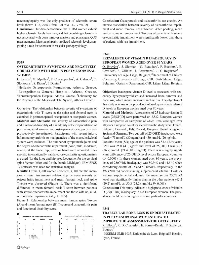
macroangiopathy was the only predictor of sclerostin serumlevels (beta=−11.8, 95%CI from −21.9 to −1.7; P=0.02).Conclusion: Our data demonstrate that T1DM women exhibithigher sclerostin levels thanmen, and that circulating sclerostin isnot associated with bone turnover markers and phalangeal QUSmeasurements.Macroangiopathy predicted sclerostin levels, sug-gesting a role for sclerostin in vascular pathophysiology.
P339OSTEOARTHRITIS SYMPTOMS ARE NEGATIVELYCORRELATED WITH BMD IN POSTMENOPAUSALWOMENG. Lyritis1, M. Mpallas2, E. Chronopoulos3, A. Galanos4, C.Sfontouris2, S. Rizou1, I. Dontas41Hellenic Osteoporosis Foundation, Athens, Greece,2Evangelismos General Hospital, Athens, Greece,3Konstantopoulion Hospital, Athens, Greece, 4Laboratory forthe Research of the Musculoskeletal System, Athens, Greece
Objective: The relationship between severity of symptoms ofosteoarthritis with T- score at the spine and femoral neck isexamined in postmenopausal osteoporotic or osteopenicwomen.Material and Methods: The severity of osteoarthritic painand functional disability of a randomly selected population ofpostmenopausal women with osteopenia or osteoporosis wasprospectively investigated. Participants with recent injury,inflammatory arthritis or malignancies of the musculoskeletalsystem were excluded. The number of symptomatic joints andthe degree of osteoarthritic impairment (none, mild, moderate,severe) at the knee, hip, neck or hand were recorded. Site-specific internationally validated osteoarthritis questionnairesare used (for the knee and hip used Lequesne, for the cervicalspine Vernon Mior and for the hands Michigan). IBM SPSS17 software was used for statistical analysis.Results: Of the 3,900 women screened, 3,000 met the inclu-sion criteria. An inverse relationship between severity ofosteoarthritic impairment and mean femoral neck and spineT-score was observed (Figure 1). There was a significantdifference in mean femoral neck T-score between patientswith severe osteoarthritic impairment and those with no, mild,or moderate impairment (all p<0.005).Figure 1: Relationship between mean lumbar spine T-score(A) and mean femoral neck (B) T-score and osteoarthritis painand functional disability score.
Conclusion: Osteoporosis and osteoarthritis can coexist. Aninverse association between severity of osteoarthritic impair-ment and mean femoral neck T-score. After pooling, meanlumbar spine or femoral neck T-scores of patients with severeosteoarthritic impairment were significantly lower than thoseof patients with less impairment.
P340PREVALENCE OF VITAMIN D INADEQUACY INEUROPEAN WOMEN AGED OVER 80 YEARSO. Bruyère1, J. Slomian1, C. Beaudart1, F. Buckinx1, E.Cavalier2, S. Gillain3, J. Petermans3, J.-Y. Reginster11University of Liège, Liège, Belgium, 2Department of ClinicalChemistry, University of Liege, CHU Sart-Tilman, Liège,Belgium, 3Geriatric Department, CHU Liège, Liège, Belgium
Objective: Inadequate vitamin D level is associated with sec-ondary hyperparathyroidism and increased bone turnover andbone loss, which in turn increases fracture risk. The objective ofthis study is to assess the prevalence of inadequate serum vitaminD levels in European women aged over 80 years.Material and Methods: Assessments of 25-hydroxyvitamin Dlevels [25(OH)D] were performed on 8,532 European womenwith osteoporosis or osteopenia of which 1984 were aged over80 years. European countries included in the study were: France,Belgium, Denmark, Italy, Poland, Hungary, United Kingdom,Spain and Germany. Two cut-offs of 25(OH)D inadequacy werefixed: <75 nmol/L (30 ng/ml) and <50 nmol/L (20 ng/ml).Results: Mean (SD) age of the patients was 83.4 (2.9) years,BMI was 25.0 (4.0)kg/m2 and level of 25(OH)D was 53.3(26.7)nmol/L (21.4 [10.7] ng/ml). There was a highly signif-icant difference of 25(OH)D level across European countries(p<0.0001). In these women aged over 80 years, the preva-lence of 25(OH)D inadequacy was 80.9 % and 44.5 % whenconsidering cutoffs of 75 and 50 nmol/L, respectively. In the397 (20.0 %) patients taking supplemental vitamin D with orwithout supplemental calcium, the mean serum 25(OH)Dlevel was significantly higher than in the other patients (65.2(29.2) nmol/L vs. 50.3 (25.2) nmol/L; P<0.001).Conclusion: This study indicates a high prevalence of vitaminD [25(OH)D] inadequacy in old European women. The prev-alence could be even higher in some particular countries.
P341TRABECULAR BONE LOSS IS UNDERESTIMATEDIN POSTMENOPAUSALWOMEN: HOW TOIMPROVE THE ASSESSMENT–THE OFELY STUDYR. Ellouz1, R. D. Chapurlat1, E. Sornay-Rendu1, P. Szulc1, S.Boutroy11INSERMUMR 1033, Université de Lyon, Hôpital E Herriot,Lyon, France
Osteoporos Int (2014) 25 (Suppl 2):S159–S440S270
Objective: Definition of cortical (Ct) and trabecular (Tb)compartments is crucial for bone loss assessment. Age-related Ct thinning produces remnants that are artefactuallyconsidered as Tb bone. In longitudinal studies, accurate as-sessment of Ct and Tb bone loss in their original compart-ments would permit to determine true bone loss. Our goal wasto compare Ct and Tb bone loss assessed by HR-pQCT at thedistal radius over 6 years, using standard procedures andbaseline-defined compartments after 3D registration.Material and Methods: Forty-six postmenopausal women(60±7 year) were measured by HR-pQCT at the distal radiusat baseline and after 6 year. The longitudinal analysis wasperformed using three methods:- Method 1: Standard software: volume of interest (VOI) iden-tified by cross-sectional area (CSA) matching, Ct and Tb boneseparated by filtering and thresholding. - Method 2: VOI basedon CSA matching and double contouring of the Ct bone- Method 3: VOI based on 3D registration and doublecontouring of the baseline compartment used for all scans.Results: Tb bone volume (Tb.TV) increased with methods 1and 2 (2.6 and 2.1 %, p<0.001) confirming that Ct remnantsare considered as Tb bone during follow-up. Tb BMD de-creased by −4 % and −3 % (p<0.005) with those methods butwas not significantly different between the two methods.Albeit both significant, decrease in Ct thickness was lowerusing method 2 than method 1 (−6 % vs. −15 %; p<0.001).When Tb.TV was kept constant with method 3, the decreasein Tb BMD was markedly higher than those obtained withmethods 1 and 2 (−10 % vs. −4 % and −3 %, respectively,p<0.01). Moreover, the increase in Ct porosity (Ct.Po) wasgreater (92 vs. 65 %, p<0.01) and consistent with a greaterdecrease in Ct BMD (−6 % vs. −5 %; p<0.01) than thatobtained with method 2.Conclusion: With an accurate registration technique, Tb andCt bone loss can be assessed in their original compartments,therefore precluding the artefactual underestimation of Tbbone loss and Ct.Po increase observed with standardprocedures.
P342THE PREVALENCE OF OSTEOPOROTICFRACTURES IN THE ELDERLY POPULATIONT. Petrusic1, N. Mandic11Dom Zdravlja Nis, Nis, Serbia
Objective: Osteoporosis is a metabolic bone disease thatresults in fractures and occurs in women of postmenopausalage and men over 60. Osteoporosis follows encumbered mo-bility, pain in the lower back and joint pain, spasms andweakness and muscle, decrease body height, increase the riskof bone fractures and spontaneous fracture. BMD, bonesbecome porous and brittle and break easily.
Material and Methods: The period from 1.3.2011 to1.3.2012 in the department of physical medicine and rehabil-itation in Nis R Serbia, trial included 298 elderly patients over75 diagnosed with osteoporosis 10 years ago, based onosteodensitometry (T-score −2.5), clinical features, the pres-ence of more than two risk factors. The analyze of the foundout that in 88 (29.53 %) patients reported a fracture in lesstrauma or spontaneously without trauma compression fractureof the vertebrae also occur repeated breaks of a few months oryears.Results: Results of the work of the total number of respon-dents 298, no fracture was 210 (70.47 %) patients, whileosteoporotic fracture occurred in 88 (29.53 %) and in 83(94.21 %) representative from the female population and in5 (5.69 %) men.Conclusion: Osteoporotic fractures were low-energy frac-tures that occur spontaneously (vertebra) or in the fall (hip).Osteoporosis is a significant risk factor for Op Fx (osteopo-rotic fractures). Op Fx may be symptomatic or even asymp-tomatic. Op Fx may be symptomatic or even asymptomatic.Op Fx have their medical socio-economic implications. Op Fxare accompanied by difficulties in activities of modern life,and only a third of patients reach prefracture quality of life.The main risk factors for Fx are female sex, age, estrogendeficit, low weight and BMI, osteoporosis in the family,smoking, and previous Fx. Low BMD, decrease in physicalstrength and bone geometry, gracility. The biggest socio-economic problems are hip fractures with significant mortalityof 12–20 % in the first year and major disability over 50 %.
P343SERUM PERIOSTIN IS INDEPENDENTLYASSOCIATED WITH OSTEOPOROSIS- RELATEDFRACTURE RISK AMONG POSTMENOPAUSALWOMEN: THE CEOR STUDYM. S. M. Ardawi1, A. A. Rouzi2, S. A. Al-Sibiani2, N. S.Senani21Center of Excellence for Osteoporosis Research and Depart-ment of Clinical Biochemistry, Faculty of Medicine, KingAbdulaziz University Hospital, King Abdulaziz University,Jeddah, Saudi Arabia, 2Center of Excellence for OsteoporosisResearch and Department of Obstetrics and Gynecology,Faculty of Medicine, King Abdulaziz University Hospital,King Abdulaziz University, Jeddah, Saudi Arabia
Objective: Periostin (POSTN) is a secreted extracellular ma-trix protein preferentially expressed in bone by osteocytes andperiosteal osteoblasts. Decreased POSTN expression mayaffect osteoblast differentiation and collagen type 1 synthesispredisposing to low BMD, osteoporosis and increased risk offracture. We therefore hypothesized that postmenopausal
Osteoporos Int (2014) 25 (Suppl 2):S159–S440 S271
women with increased circulating POSTN levels have a great-er risk for osteoporosis-related fractures (ORFs).Material and Methods: We examined the association be-tween circulating POSTN [measured by ELISA method,(USCNK-Life Science Inc., China)] levels and the risk ofORFs in 707 postmenopausal women, 50 years of age or olderin a population-based study with a mean follow-up period of5.2±1.3 years. Multivariate Cox proportional- hazards regres-sion models were used for analysis of the risk of fracture withadjustment for age, body-mass index and other potential riskfactors that may be associated with the risk of fracture or withhigher circulating levels of serum POSTN.Results: High serum POSTN levels were associated with anincreased risk of ORFs. Following adjustment for age andother confounders, the relative risk of ORFs for each incre-ment of 1 SD in POSTN level was about 2.3-fold amongpostmenopausal women [RR=2.36 (95%CI: 1.13–3.66)].Further, women in the highest quartile of POSTN levels hadan increase in the risk of ORFs so that the risk was 3.6-fold forPOSTN. The risk of ORFs that was attributable to POSTNlevels (in the highest quartile) was estimated at 35.8 %. Theassociation between POSTN levels and the risk of fractureseems to be independent of BMD and other confounding riskfactors for fracture.Conclusion: Higher serum POSTN levels are associated witha greater risk of ORFs independent of several other risk factorsamong postmenopausal women. Serum POSTNmeasurementcould be useful to improve fracture risk assessment.
P344HIP FRACTURE RISK IN INCIDENT TYPE 2DIABETIC PATIENTS: A POPULATION-BASEDPARALLEL COHORT STUDYD. Martinez-Laguna1, C. Tebé2, C. Carbonell-Abella1, A.Conesa-Garcia1, X. Nogués-Solan3, A. Diez-Perez4, D.Prieto-Alhambra51Institut Catala de La Salut, Barcelona, Spain, 2AQuAS, Bar-celona, Spain, 3Department of Internal Medicine, Hospital delMar-IMIM and Autonomous University of Barcelona, Barce-lona, Spain, 4Hospital del Mar-IMIM-Autonomous Universi-ty of Barcelona, Barcelona, Spain, 5Nuffield Department ofOrthopaedics, Rheumatology and Musculoskeletal Sciences.University of Oxford, Oxford, United Kingdom
Objective: Type 2 diabetes (T2DM) and osteoporosis are twoprevalent chronic diseases. Data on the association betweenT2DM and osteoporotic fractures is controversial. We esti-mated hip fracture rates in newly diagnosed T2DM patients,and compared these to matched nondiabetic peers.Material and Methods: We conducted a population-basedparallel cohort study using data from the SIDIAP Database(www.sidiap.org). SIDIAP contains clinical information from
primary care records, hospital admissions, and pharmacyinvoice data for >5 million patients (80 % of the population)in Catalonia, Spain. We selected all newly diagnosed T2DMpatients registered in SIDIAP in 2006–2010. Up to 2 diabetes-free controls were matched to each T2DM participant on age,gender, and primary care center. Main outcome was incidenthip fracture in 2006–2011, using ICD10 codes. We used Fineand Gray survival modelling to estimate risk of hip fractureaccording to T2DM status accounting for competing risk withdeath. Multivariate models were adjusted for BMI, previousfracture and use of oral corticosteroids.Results: We identified 58,483 T2DM patients and 113,448controls, who were observed for a median (interquartile range)of 2.63 (2.93) years. 444 (0.8 %) T2DM patients sustained ahip fracture in the study period (incidence rate 2.7/1,000person-years) compared to 776 (0.7 %) matched controls(2.4/1,000). This is equivalent to an unadjusted (age andgender-matched) SHR 1.11 [0.99–1.24], and adjusted SHR1.20 [1.06–1.35]. In stratified analyses, the excess risk asso-ciated with T2DM was highest amongst T2DM patients withprevalent IHD (adjusted SHR 1.39 [0.98 to 1.98]), CKD(adjusted SHR=1.26 [1.03 to 1.55]) or grade 2 obesity (ad-justed SHR 1.37 [0.92 to 2.06]).Conclusion: Newly diagnosed T2DM patients are at a 20 %increased risk of hip fracture even in early stages of disease.More data is needed on the causes for an increased fracturerisk in T2DM patients as well as on the predictors of osteo-porotic fractures among these patients.
P345STUDY OF THE CORRELATION BETWEEN ANTHROPOMETRIC INDICATORS AND TRIGLYCERIDELEVELS IN POSTMENOPAUSALWOMENPARTICIPANTS OF THE PHYSICAL EXERCISEPROGRAM FOR OSTEOPOROSISO. Matos1, C. Orsso1, P. Miranda11Biochemistry and Densitometry Laboratory, Federal Techno-logical University of Paraná (UTFPR), Curitiba, Brazil
Objective: To evaluate the correlation between anthropomet-ric indicators of total and central obesity and triglyceridelevels in physically active postmenopausal women.Material and Methods: We evaluated 47 postmenopausalwomen, aged 60±6.1 years, participants of the Physical Ex-ercise Program for Osteoporosis. Triglyceride levels weremeasured by colorimetric assays (Mindray) after 12 h fasting.Total obesity was assessed using BMI and the percentage oftotal body fat was measured with DXA (Hologic DiscoveryA). Central obesity was assessed from the waist hip ratio(WHR), abdominal circumference (AC) and abdominal fatpercentage measured with DXA. Person’s test was used.
Osteoporos Int (2014) 25 (Suppl 2):S159–S440S272
Results: The mean value of BMI (27.2±5.1 kg/m2) classifiedthe group as pre- obese, while the percentage of total fatindicated high values 38.2±4.4 %. All central obesity indica-tors showed high values: WHR 0.84±0.07; AC 95.7±12.4 cm; abdominal fat percentage 37.4±6.7 %. The percent-age of total body fat was strongly correlated with BMI(0.751); abdominal fat percentage was positively correlatedwith WHR (0.588) and a strong correlation with AC (0.712).Only 14.9 % of the sample had elevated triglyceride levels(≥150 mg/dL), and the mean values (96.4±37.2 mg/dL) werereflected below the expected values for this age group. Tri-glyceride levels showed positive but weak correlation onlywith WHR (0.390 p=0.007).Conclusion: The triglyceride levels had a weak but statisti-cally significant correlation only withWHR, showing that thiscan be a good indicator of fat in the abdominal region. Lowlevels of triglycerides can be explained by the use of lipid-lowering drugs and physical exercise. Both obesity indicatorsdenoted that the group had high fat levels, more than the idealfor the age, increasing the development of heart diseases.However, this overweight provides a protective effect in thebones of this population, since a higher overload on themprevents the decrease of the BMD.
P346EFFECTS OF ETHANOL EXTRACT OFAMOMUMTSAO-KO ON ANTI- INFLAMMATORYANDANTI-OSTEOARTHRITIC ACTIVITIES IN VITROAND IN VIVOY. J. Park1, E.-K. Ahn1, S. S. Hong1, J. S. Oh21Natural Products Research Institute, Gyeonggi Institute ofScience & Technology Promotion, Suwon-si, Republic ofKorea, 2College of Pharmacy, Dankook University,Cheonan-si, Republic of Korea
Objective: Amomum tsao-ko is well known to use in tradi-tional medicine for the treatment of stomach inflammation andlipid problems.Material and Methods: This study was investigated to de-termine whether the ethanol extract of Amomum tsao-ko(EEA) influenced on inflammation reaction in lipopolysac-charide (LPS)-stimulated RAW264.7 murine macrophagesand the temporal changes in the tibial subchondral bonearchitecture in monosodium- iodoactate(MIA)-induced osteo-arthritis rat model.Results: The results showed that EEA inhibits lipopolysac-charide (LPS)-induced nitric oxide (NO) and prostaglandin E2
release through down-regulation of NO synthase andcyclooxygenase-2, respectively. In addition, treatment withEEA suppressed the expression of LPS-induced inflammatorycytokines such as IL-6, IL-10, and TNF-α in LPS-stimulatedRAW264.7 cells. In animal study, μCT analysis showed
protective effects of EEA on trabecular bone. EEA tended toincrease in trabecular thickness (Tb.Th) and trabecular bonevolume fraction (BV/TV). Treatment with EEA significantlydecreased the level of trabecular spacing (Tb.Sp).Conclusion: These findings indicate that EEA could be de-veloped as a potential therapeutic agent for inflammation andosteoarthritis.Acknowledgements: This research was supported by a grantfrom the Next- Generation BioGreen 21 Program (No.PJ009551), Rural Development Administration, Republic ofKorea.
P347CONTRIBUTION OF BONE ARCHITECTURE TOFRACTURE RISK EVALUATIONS. Cappelle1, A. Peretz1, R. Karmali1, S. Rozenberg2, C.Muhire1, M. Moreau3, M. Paesmans3, P. Bergmann4, J. J.Body11Department ofMedicine, CHU-Brugmann, ULB, Brussels, Bel-gium, 2Department of Obstetrics & Gynaecology, CHU Saint-Pierre, Free University of Brussels, Brussels, Belgium, 3DataCenter - Institut J. Bordet - ULB, Brussels, Belgium, 4Departmentof Nuclear Medecine and Laboratory Experimental Medecine- CHU Brugmann - ULB, Brussels, Belgium
Objective: The WHO FRAX is the most used of fracture riskmodels though it does not include some significant risk factorsand its predictive value remains suboptimal. One of the vari-ables which could influence significantly fracture prediction isbone geometry and architecture. The aim of this project is toexamine if bone architecture and resistance parameters mea-sured by HR-pQCT in a subgroup of women who haveundergone a fragility fracture to a control group withoutfracture but with the same level of absolute fracture risk bythe FRAX model.Material andMethods: This ongoing project is a nested casecontrol study carried out within the Fracture RISk BrusselsEpidemiological Enquiry (FRISBEE study) on postmeno-pausal and senile osteoporosis. FRISBEE is a prospectiveobservational study including 3,578 women of the Brusselsarea, aged from 60 to 85 years, recruited since 5 years (2007–2013) who will be followed up for 10 years. BMD and clinicalrisk factors (CRFs) (included or not in the FRAX) wererecorded at inclusion, with the aim of building a risk modelallowing absolute fracture risk prediction and comparing theweight of the different CRFs. A fragility fracture was alreadyreported in 144 women included during the first 3 years. Threecontrols are selected for each fracture case to match the FRAXestimation of fracture risk. All subjects have a HR-pQCT(Scanco Medical AG, Bassedorf, Switzerland) at the radiusand tibia, as well as a second DXA and a complete re-evaluation of their CRFs.
Osteoporos Int (2014) 25 (Suppl 2):S159–S440 S273

Results: Several studies have demonstrated that some structuralparameters, from morphometry and from the Finite ElementAnalysis, can discriminate patients with fragility fracture. Condi-tional logistic regression models will be used to estimate whetherthe HR-pQCT parameters as well as other CRFs (not included inthe FRAX) contribute significantly to the risk of fracture.Conclusion: This study should demonstrate if HR-pQCTderived morphometric parameters and bone strength predictfracture risk independently of DXA and FRAX score.
P348EXPRESSION OF GAP JUNCTION PROTEINSCONNEXINS 26, 30, AND 43L. A. Holzer1, A. Cör2, G. Holzer311Department of Orthopaedic Surgery, Medical University ofGraz, Graz, Austria, 2Faculty of Health Sciences, Universityof Primorska, Izola, Slovenia, 3Department of Orthopaedics,Medical University of Vienna, Vienna, Austria
Objective: Dupuytren’s disease (DD) is a benignfibroproliferative process of the palmar aponeurosis showingsimilarities to wound healing. Communication of cells involvedin wound healing is mediated by the composition of gap junction(GJ) proteins. We investigated the expression of 3 GJ proteins,connexins 26, 30, and 43 (Cx26, Cx30, and Cx43) in DD.Material and Methods: Fragments of Dupuytren’s tissuefrom 31 patients (mean age 56 (30–76) years, 24 male) wereanalyzed immunohistochemically and compared to controltissue for expression of the GJ proteins Cx26, Cx30, andCx43 and also alfa-smooth muscle actin (α-SMA).Results: 14 of 31 samples could be attributed to the involu-tional phase (α-SMA positive) whereas 17 samples had to beconsidered cords in the residual phase (α- SMA negative).Expression of Cx26 and Cx43 was seen in 12 of the 14samples from the involutional phase, and Cx30 was seen in7 of these. Only 4 of the 17 samples from the residual phaseshowed any Cx, and there was none in the controls.Conclusion: The high expression of GJ proteins Cx26, Cx30,and Cx43 in α-SMA positive myofibroblast-rich nodules,which are characteristic of the active involutional phase ofDD, suggests that connexins could be a novel treatment targetfor the treatment of DD.
P349NUTRITIONAL STRATEGY TO PROTECTMATERNAL BONE LOSS DURING GESTATION ANDLACTATION AS AN APPROACH TO PREVENTOSTEOPOROSIS LATER IN LIFEP. Bueno-Vargas1, M. Manzano1, M. L. Jiménez1, R. Rueda1,J. M. Lopez-Pedrosa11Discovery R&D, Abbott Nutrition, Granada, Spain
Objective: Pregnancy and lactation have been related withtemporary decreases in maternal BMD. Nutrition during theseperiods could be key on maternal bone preservation. Long-chain polyunsaturated fatty acids (LCPUFAs) and prebiotichave been shown to have healthy effects on age-associatedbone loss. Currently, maternity is becoming closer tothe perimenopause period with the subsequent negativeeffect of increasing the risk of bone loss associated tothe onset of menopause. Therefore, strategies addressedto reduce maternal bone loss are crucial to delay bonedeterioration later in life. The aim of this study was tocompare the effectiveness of calcium fortification vs.prebiotic supplementation or LCPUFAs fortification inpregnant rats as a nutritional approach to reduce boneloss produced during gestation and lactation.Material and Methods: Pregnant Sprague Dawley rats weredivided into four groups: Control group (CC group) was fedwith a standard semipurified diet until the end of the lactationperiod. Ca group, Pre group and LCPUFAs were fed with thesame diet fortified with 0.5 % calcium carbonate (total calci-um content 1 %), supplemented with an inulin-type fructans(7.5 % of the total carbohydrate) or fortified with LCPUFAs(3.5 % Eupoly-DHA®), respectively. At the end of the lacta-tion, BMD and bone mineral content (BMC) were determinedby DXA; bone volume fraction as well as 3D parameters oftrabecular architecture (trabecular number, thickness, separa-tion and connectivity density) were analyzed by μCT.Results: In appendicular bones, higher BMD and BMCwere found for Pre and LCPUFAs groups as comparedwith calcium fortified group. Furthermore μCT datashowed that Pre and LCPUFAs groups had an increasein bone volume/tissue volume, trabecular thickness, tra-becular number and connectivity density value, with aconcomitant reduction in the trabecular separation ascompared not only with control but also and with cal-cium fortification group.Conclusion: Based on our data, both prebiotic supple-mentation as well as LCPUFAs fortification exert aprotection on maternal skeleton during pregnancy andlactation. The beneficial effect may delay the onset ofosteoporosis and its deleterious derived- consequenceslater in life.
P350CHEMICAL COMPOSITION OF BONES ANDLOWER INCISOR DENTIN IN MATURE RATSAFTER 60-DAYADMINISTRATION OF SODIUMBENZOATEL. Galina1, L. Vladyslav2, T. Alexey2, M. Vitaly21National University of Physical Education and Sport ofUkraine, Kiev, Ukraine, 2Lugansk State Medical University,Lugansk, Ukraine
Osteoporos Int (2014) 25 (Suppl 2):S159–S440S274
Objective: To investigate of chemical composition of hipbone, mandible and lower incisor dentin in mature white ratsafter 60-day administration of sodium benzoate.Material and Methods: For the purposes of study we select-ed 105 rats with initial body weight of 200–210 g. The controlgroup received per os sodium chloride daily in dosage of 1 mlof 0.9 % solution throughout 60-day observation period. Therest of the animals received per os sodium benzoate (SB) dailyin dosage of 500 or 1,000 mg per 1 kg of body weight as 1 mlof solution (groups B1 and B2, respectively). At the end of thecycle of administration investigated the chemical compositionof the hip bone, mandible, and lower incisor dentin by gravi-metric method.Results: By the 3rd day after the end of SB administrationmineral content of hip bone and body of mandible in B1 groupwas lower than that of the control group by 7.45% and 8.27%and organic substances content—by 8.56 % and 6.54 %,respectively. The lower incisor dentin these deviationsamounted to 5.88 % and 8.36 %. In readaptation period ofgroup B1 changes remained significant up to 15th day, whenthe share of mineral component was reduced by 5.29 % and4.83 %, while the proportion of organic substances—by4.38% and 5.29%. Higher dose of SB (1,000 mg/kg) resultedin more expressed changes—by the 3rd day after the end ofmedication in the group B2 mineral content in the hip boneand organic substances content decreased by 8.69 % and9.24 %. In the readaptation period of group B2 revealedchanges recovered more slowly than in group B1: in the hipbone and the mandible deviation persisted up to 24th days,and in the incisor dentin mineral content remained by 3.85 %lower than the control.Conclusion: Daily per os SB in dosage of 500 mg/kg of bodyweight accompanied by a decrease of organic and mineralsubstances in the hip bone, the body of the mandible, as wellas in lower incisor dentin. When increasing the dosage of SBto 1,000 mg/kg, the severity of the changes increases.
P351A LOW DOSE 3D QCT PROTOCOL FORTHE SPINEK. Engelke1, A. Heinemann2, M. Krause3, O. Museyko1, C.C. Glüer41Institute of Medical Physics, University of Erlangen, Erlang-en, Germany, 2Institute for Forensic Medicine, University ofHamburg, Hamburg, Germany, 3Institute for Osteology andBiomechanics, University of Hamburg, Hamburg, Germany,4Biomedical Imaging, Diagnostic Radiology, University ofKiel, Kiel, Germany
Objective: To develop a low dose in vivo 3D QCT protocolfor the spine.Material and Methods: In 27 cadavers T6-L4 were scannedtwice on a Philips MX8000: with 120 kVand 100 mAs and a
lower dose protocol with 90 kVand 150 mAs. Reconstructiondiameter was 15 cm, slice thickness 1.3 mm, and convolutionkernel was ‘B’ for both scans. A BDC calibration phantom(QRMMöhrendorf, Germany) was used for conversion of HUto BMD values. MIAF-Spine was used to analyze total verte-bral body volume and BMD. Additionally, mean and standarddeviation of the HU values obtained in the three cylindricalphantom inserts (0, 100, and 200 mg/cm3) were measured ineach of the 27 dataset pairs. All parameters were comparedbetween the two scans using matched pair t-tests (Wilcoxonsigned rank test for the volume) and Pearson correlationscoefficients (r). Dose reduction was estimated usingImpactDose software (CT Imaging, Erlangen).Results: Effective dose values for 90 kV are approximately60 % lower compared to 120 kV. BMD values were highlycorrelated but a significant offset (p<0.001) indicates differ-ences in field inhomogeneity; segmented volume was notaffected by the lower kV (p>0.5). All correlation coefficientswere significant (p<0.001). Noise in the images assessed bythe standard deviation of the HU values of the phantom insertsof the vertebrae were significantly higher in the 90 kV scans:20.8±4.7 HU vs. 16.2±3.0 HU, 22.0±4.9 HU vs. 17.1±2.9HU, and 23.3±5.0 HU vs. 18.0±3.1 HU in the phantom insertwith 0, 100, and 200 mg/cm3, respectively.Conclusion: As expected, noise values were higher in the90 kV protocol. However, increased noise had no effect onsegmentation. The difference in calibrated BMD values indi-cates differences in the scan field inhomogeneity at 90 and120 kV requiring different corrections. As the correlationbetween BMD values at 90 and 120 kV was very high, alinear correction approach, e.g., based on the scans of the ESPwill suffice to obtain the same BMD results at 90 and 120 kV.Acknowledgements: supported by BMBF Bioasset01EC1005
P352THE GROWTH HORMONE RECEPTOR GENEEXON 3 DELETION POLYMORPHISM HAVEINFLUENCE ON IGF-1 LEVELS IN CHILDRENTREATED WITH RHGHW. Lepionka1, J. Łukaszkiewicz2, M. Kruk1, M. Jaworski1, E.Czekuć-Kryśkiewicz1, M. Szalecki3, B. Parafiniuk1, E.Karczmarewicz1, A. Kępka1, P. Płudowski1, R. S. Lorenc11Department of Biochemistry, Radioimmunology and Exper-imental Medicine, The Children’s Memorial Health Institute,Warsaw, Poland, 2Department of Biochemistry, Faculty ofPharmacy, Medical University of Warsaw, Warsaw, Poland,3Clinic of Endocrinology and Diabetology, The Children’sMemorial Health Institute, Warsaw, Poland
Objective: Patients treated with recombinant human growthhormone present interindividual variability regarding
Osteoporos Int (2014) 25 (Suppl 2):S159–S440 S275

responses to this therapy. The aim of the study was to verify arelationship between GHR exon 3 deletion polymorphism orother genetic factors in GHR gene promoter and effectivenessof rhGH therapy.Material and Methods: Children (n=30) aged 3–16 yearsold with somatotrophin pituitary insufficiency (n=20) andTurner Syndrome (n=10), which began rhGH therapy andreceived the same rhGH doses for 3 months. After 3rd monthsome doses was modified to ensure therapy safety for allchildren. Exon 3 deletion GHR gene polymorphism was de-tected by multiplex-PCR reaction and correlated with clinicalfactors, which were measured after 3rd, 6th and 12th month.Statistical analyses for genetic associations (ANOVA) wereconducted with SPSS Statistics for Windows.Results: Patients homozygous d3/d3 (n=3) presented signif-icantly lower increases of IGF-1 (p=0.028; 30.58±34.06 ng/ml), than fl/d3 (n=10; 191.13±66.33 ng/ml) and fl/fl (n=17; 122.31±105.92 ng/ml) genotypes after 3-monththerapy. Exclusion of puberty patients and all children above13 years old made this relationship even more significant (p=0.005; p=0.0180, respectively). We did not observe any as-sociations between GHR genotypes and growth increases orgrowth velocity as well as biochemical markers of bonemetabolism levels (CTx, PINP, PIIINP, NT- proCNP). Patientswith at least one d3 allele (n=13) presented lower spin BMCand total body BMC after 12-month therapy (p=0.054; 19.02±6.06 %; p=0.059; 16.82±5.24 %, respectively). Sequenceanalysis of GHR gene promoter region did not show anypolymorphisms or mutations which could influence on re-sponse variability.Conclusion: The genetic factors have significant influence oneffectiveness of rhGH therapy. Exon 3 deletion GHR genepolymorphism might determine IGF-1 level increase after 3-month therapy, though it did not correlate with growth veloc-ity and growth increase. The further studies are required.Acknowledgements: Financial support: CMHI InternalGran t s : 183 /07 . Acqu i r i ng o f K ingF i she r mL(ThermoScientific), ultra-low temperature freezers MDF-U500Vx (Sanyo), Varioscan Flash (ThermoScientific),Thermocycler C1000 (BioRad), electrophoresis systemOWL D3-14, electrophoresis power supply EC 3000 XL(ThermoScientific), biological safety cabinet ESCO II(Labculture) and Captairbio were cofinanced by ERDF (EUStructural Funds) project POIG.02.01.00-14-059/09.
P353SECONDARY OSTEOPOROSIS IN HOSPITALSAMPLEK. M. Miladinovic1, N. V-H. Vavra-Hadziahmetovic1, D. C.Celik11Clinic of Physical and Rehabilitation Medicine, UniversityClinical Centre, Sarajevo, Bosnia and Herzegovina
Objective: Secondary osteoporosis sti l l remainssubdiagnosed entity. The aim of this study was to analyzethe pattern of hospital patients who have had the diseaseswhere development of secondary osteoporosis is expected.Material and Methods: Out of all medical records of thepatients who were treated at the inpatient Clinic of Physicaland Rehabilitation Medicine (n=531) in 1 year period, thepatients who have had the diseases where development ofsecondary osteoporosis is expected were selected. Patientswere divided in two groups. The first group consisted ofpatients whom secondary osteoporosis was diagnosed andtreated, and the second group consisted of patients whomdiagnostic procedures for secondary osteoporosis presencewere not performed.Results:Out of total number of hospitalized patients (n=531),13.2 % (n=70) have had the diseases where development ofsecondary osteoporosis is expected. Secondary osteoporosiswas diagnosed and treated in 20 % of these patients (n=14),and they presented the first group. In this group the mostfrequent disease was spinal lesion, presented by 57.1 % (n=8), followed by multiple sclerosis and rheumatoid arthritiswith 14.3 % (n=2) each, than renal failure and malignomawith 7.1 % (n=1) each. Male patients formed 42.8 % (n=6) ofthis group. The second group was presented by 80 % ofselected patients (n=56). In this group the most frequentdisease was multiple sclerosis, presented by 39.3 % (n=22),followed by malignoma with 26.76 % (n=15), than spinallesion and diabetes mellitus with 10.7 % (n=6) each, andrheumatoid arthritis with 3.6 % (n=2). Ratio between malesand females was 1:1.Conclusion: Patients with certain diseases, like multiple scle-rosis, spinal cord lesion, diabetes mellitus, rheumatoid arthritisand malignoma, regardless on stage and time of disease oc-currence, demand higher attention for secondary osteoporosisdiagnostic processing.
P354BONE MINERAL DENSITY OF PATIENTS WITHLOW-ENERGY FRACTURESN. Dedukh1, E. Pobel2, V. Maltseva11Morphological Connective Tissue Laboratory, Institute Spineand Joint Pathology National Academy of Medical Science,Kharkov, Ukraine, 2Zaporozhye Medical Academy of Post-graduate Education, Zaporozhye, Ukraine
Objective: Osteoporotic fractures are a serious complicationof osteoporosis, however, a fracture that occurs at differentstages of life, can also affect on bone health and it can be a riskfactor for osteopenia and osteoporosis. Objective was to esti-mate BMD of patients with a history of low-energy fracturesbefore peak bone formation, physiological stability (up to40 years) and after 40 years.
Osteoporos Int (2014) 25 (Suppl 2):S159–S440S276

Material and Methods: BMD was assessed by DXA (Ex-plorer QDR, Hologic) of lumbar spine in three groups ofwomen with any location fractures: 1) n=42, median age51.19±1.83, fracture was in age 14.74±0.88; 2) n=32, medi-an age 49.31±1.51, fracture was in age 33.72±0.73 and 3) n=202, median age 62.23±0.58, fracture was in age 55.72±0.6.BMI was similar in all groups (27.71±0.86; 29.05±1.30; 29.6±0.37).Results: BMD on the osteoporosis level was present in thefirst group in 11.9 % of patients, on the osteopenia level in35.7 % and regular T-score was present in 52.4 % of patients;in the second group was present osteoporosis level - 12.5 %,osteopenia - 43.75 %, normal value - 43.75 %; in the thirdgroup: osteoporosis - 39.1 %, osteopenia - 38.6 %, normalvalue - 22.27 %. The comparison of the T-score (one wayANOVA) of three groups patients, it was found that the lowestrates T-score (−1.89±0.09; p<0.05) was found for the thirdgroup, the differences between the first and the second groupswere not detected. In the third group of women risk factor ismenopause.Conclusion: Patients with history of prior fracture have high riskof developing osteopenia and osteoporosis. The results of ourstudy have important significance for clinical practice, becausepatients with prior fracture need for osteoporosis prevention.
P355SETTING THE SECONDARY FRACTURE LIAISONPROGRAMME AT THE UNIVERSITY HOSPITALCENTRE AS AN EXAMPLE FORTHE NATIONWIDESERVICE IN CROATIAS. Grazio1, A. Matejcic2, D. Durdevic2, T. Nikolic1, M.Punda3, D. Balen1, G. Tajsic1, M. Mustapic4, D. Miklic2, D.Car2, V. Altabas5, T. Nemcic1, F. Grubisic1, R. Kolundzic2, A.Muljacic21University Department of Rheumatology, Physical and Re-habilitation Medicine, Clinical Hospital Centre Sestremilosrdnice, Zagreb, Croatia, 2University Department ofTraumatology, Clinical Hospital Centre Sestre milosrdnice,Zagreb, Croatia, 3University Department of Nuclear Medicineand Oncology, Clinical Hospital Centre Sestre milosrdnice,Zagreb, Croatia, 4Clinical Department of Diagnostic and In-terventional Radiology, Clinical Hospital Centre Sestremilosrdnice, Zagreb, Croatia, 5University Department of In-ternal Medicine, Clinical Hospital Centre Sestre milosrdnice,Zagreb, Croatia
Objective: To plan, elaborate and apply a multidisciplinarysecondary fracture liaison programme at the University Hos-pital Centre in Croatia, as an example for the nationwideservice in Croatia.Material and Methods: At the initiation meeting medicaldoctors of different specialties were informed on the concept
of secondary osteoporotic fracture prevention. After the rele-vant literature search an assessment of the current healthcaresystem in Croatia and specifically of the University HospitalCentre Sestre milosrdnice in Zagreb was performed and mul-tidisciplinary teamwas built. The programme was put forwardto the broader audience at the symposium which was held forthe World Osteoporosis Day. The conclusions of the meetingswere transformed into the action plan and implemented ineveryday practice.Results: A need for a systematic approach, where capture ofpatients is automatic, a major involvement of the orthopaedicsurgeon and a coordinator-based service were appreciatedduring the development of this secondary prevention pro-gramme.We set our goals, identified the patients with fragilityhip fractures to be more appropriate for intervention, weagreed on the roles assigned for each member of the team(including three dedicated nurses at the orthopedic surgeons’wards as coordinators), developed a questionnaire in order toassess the risks for fragility fractures as a basis for cycle goalsand measurement what was done, looked for improvementand determined the evolving actions until goals are met. Theimplementation of the programme started with no major dif-ficulties and the level of compliance is high.Conclusion:We developed and implemented the coordinator-based programme of secondary fractures. In this still ongoingfracture liaison service we have seen that the system withdedicated coordinators is effective in our University HospitalCentre and is a potential model to become a standard of carefor patients with fragility fractures in Croatia.
P356EFFECTIVENESS OF REHABILITATION PROGRAMON LOWER LIMB FUNCTIONAL STATUS AFTERKNEE ARTHROPLASTYD.Matei1, A. Bighea1, S. Patru1, R. Traistaru1, R. S. Popescu11University of Medicine and Pharmacy of Craiova, Craiova,Romania
Objective: To evaluate if the rehabilitation treatment canimprove pain control, improve muscle strength and ambula-tion in patients with knee arthroplasty (KA).Material and Methods: 27 subjects with recent total kneearthroplasty were randomly assigned to either a group withrehabilitation program started immediately after surgical in-tervention (n=14), who received 12 supervised rehabilitationsessions combined with exercises at home after KA, or to acontrol group (n=13), who received standard care. All partic-ipants were evaluated at baseline (2 weeks after KA), 1 and6 months later. The patients’ functional status was evaluatedby VAS for pain, the lower limb main muscle strength; alsothe 6-min walk test (6MWT) and quality of life (SF-36)assessed were performed at 6 month after KA.
Osteoporos Int (2014) 25 (Suppl 2):S159–S440 S277
Results: Subjects in the rehabilitation program group walkedlonger distances using 1 cane (range, 20–24 m) in 6 min at the2nd evaluation than subjects in the control group. At thesecond and third evaluation we found significantly loweraverage pain scores and higher overall satisfaction than thecontrol group; the same patients also had less difficulty inperforming daily activities.Conclusion: Complex rehabilitation programme appears tobe beneficial to patients with arthroplasty of the knee. Theprogramme was effective in improving the short-term lowerleg functional ability. More intensive rehabilitation should bepromoted in all KA patients, to optimize functional outcomesafter surgery.
P357BONE QUALITYAND BIOMECHANICALFUNCTION: A LESSON FROM HUMAN OSSICLESF. Duboeuf1, B. Burt-Pichat1, D. Farlay1, P. Suy2, E. Truy2, G.Boivin11INSERM UMR 1033, Université de Lyon, Lyon, France,2Pavillon U, ORL, Hôpital Edouard Herriot, Lyon, France
Objective: Human middle ear contains 3 ossicles (mal-leus, incus and stapes) with a mechanical property high-ly specific (transmission of vibrations)1. In normal ossi-cles and in cases with inflammatory disease leading to adecline in hearing (cholesteatoma), the bone quality ofossicles was rarely studied. Our purpose was to assessthe intrinsic bone quality and the mechanical propertiesof incuses.Material and Methods: Thirteen inflammatory incuses from13 patients [12 cholesteatoma (54±20 year), 1 chronic otitis(30 year)], 15 non-inflammatory ones from 15 patients with-out bone pathology (35±32 year), and human control corticalfemoral samples, were used. Bones were analyzed by μCT,histology, microhardness2 and Fourier Transform InfraRedMicrospectroscopy (FTIRM)3–4.Results: Incuses were compact without bone marrow andwith sparse vessels. As the final size of incuses wasobtained early during modeling, remodeling activity wasrarely observed (few osteoclasts, osteoblasts and osteoidtissue). In ossicles with either woven or lamellar tex-tures, many periosteocytic lacunae, sometimes empty,were present. Architecture of inflammatory incuses wasdegraded. BMD of non-inflammatory incuses was higherthan that of control bone (p=0.003) and inflammatoryincuses (p=0.0001). Non-inflammatory incuses were lesshard than both control cortical bone (p=0.003) andinflammatory incuses (p=0.0001). Incuses were moremineralized and less mature than control bone(p<0.001). Moreover, mineral of inflammatory incuseswas more carbonated (p=0.011) and collagen matrix
less mature (p<0.013) than in non-inflammatoryincuses.Conclusion: Bone quality of stiffen incuses is welladapted to their specific function. Inflammation de-grades architecture, increases microhardness and chang-es material characteristics; this partly explains the de-cline of hearing.References: 1Kanzaki et al. 2006, Bone 39:414; 2Boivin et al.2008, Bone 43:532; 3 Farlay et al. 2011, PLoS ONE6(12):e287; 4Farlay et al. 2010, J Bone Miner Metab 28:433.
P358EFFECTIVENESS OF KINETOTHERAPY INPATIENTS WITH OSTEOARTHRITIS OF HIPD.Matei1, R. S. Popescu1, A. Bighea1, R. Traistaru1, S. Patru11University of Medicine and Pharmacy of Craiova, Craiova,Romania
Objective: To determine the effects of therapeutic exerciseson the functional status of the hip, whether the effects of anexercise programme are sustained at 6 months’ follow up andalso the place of kinetotherapy in the complex rehabilitationprogram in patients with hip osteoarthritis (OA).Material and Methods: Twenty-five patients with prim-itive bilateral hip OA (1–3 Plain film grading) weredivided into 2 random groups. The patients in group Ireceived medication (supplements, such as glucosamineand chondroitin, NSAIDs or acetaminophen) andnonpharmacologic therapies (lose weight through diet,rest of hip from overuse, electrotherapy); group II re-ceived the same treatment and kinetotherapy: low-impact aerobics and strength training for the musclesaround the hip joint. Exercise programs wereindividualised to the patient’s specific needs, abilitiesand preferences (resistance training, aerobic exerciseand flexibility exercises) and were continued by patientsat home, daily. The patients’ functional status was eval-uated by VAS for pain, muscle strength across the hipand knee, ambulation speed before and at 6 monthslater.Results: The results showed that the patients in each treatedgroup had significant improvement in pain reduction com-pared with their initial status. In the second group, the musclestrength was significantly improved (p<0.01), as were walk-ing time and speed (21%, 32%, respectively) and pain (10 %)after treatment and at 6 months later.Conclusion:We found a stronger favorable effect on the levelof pain when kinetotherapy was associated with pharmaco-logical treatment but therapeutical exercise associated to theother nonpharmacological treatment had the greatest effect onwalking speed and also in decreasing of disability also at6 months later.
Osteoporos Int (2014) 25 (Suppl 2):S159–S440S278

P359THE PLACE OF KINETOTHERAPY INREHABILITATION PROGRAM OF KNEEOSTEOARTHRITIS PATIENTSD.Matei1, A. Bighea1, S. Patru1, R. Traistaru1, R. S. Popescu11University of Medicine and Pharmacy of Craiova, Craiova,Romania
Objective: To investigate the effects of therapeutic exerciseson the functional status of knee and also the place ofkinetotherapy in the complex rehabilitation program in pa-tients with knee osteoarthritis (OA).Material and Methods: Sixty-two patients with primitivebilateral knee OA (I-III stage) were divided into tworandom groups. The patients in group I received medi-cation (supplements, such as glucosamine and chondroi-tin, NSAIDs or acetaminophen) and nonpharmacologictherapies (lose weight through diet, electrotherapy);group II received the same treatment and kinetotherapy:low- impact aerobics and strength training for the mus-cles around the joint (increases stability and decreasesthe likelihood of additional joint damage). The thera-peutic exercise were continued daily by patients, athome. The patients’ functional status was evaluated byVAS for pain, muscle strength across the knee (exten-sion and flexion), ambulation speed, and Lequesne in-dex before and 6 months later.Results: The results showed that the patients with OA in eachtreated group had significant improvement in pain reductionand in walking speed after treatment compared with theirinitial status. In the second group, the muscle strength wassignificantly improved (p<0.05), as were walking time (18%)and pain (21 %) after treatment and at 6 months later.Conclusion: Therapeutical exercise associated to the othernonpharmacological treatment had the greatest effect on painreduction after treatment and caused the greatest increase ofwalking speed and decrease of disability at 6 months later aftertreatment.
P360COSTAND HEALTH CARE RESOURCE USEASSOCIATED WITH NONCOMPLIANCE WITHORAL BISPHOSPHONATE THERAPY: AN ANALYSISUSING DANISH HEALTH REGISTRIESJ. Kjellberg1, A. Jorgensen2, P. Vestergaard3, R. Ibsen4, A.Krishna51KORA - National Institute for Municipalities’ and Regions’Analysis and Research, Copenhagen, Denmark, 2MSD Den-mark, Ballerup, Denmark, 3Aalborg University, Aalborg,Denmark, 4iTracks, Århus, Denmark, 5Merck & Co.,Whitehouse Station, United States
Objective: To estimate the rate of oral bisphosphonatecompliance among Danish women and to examine theassociation of noncompliance with health care resourceuse and cost.Material and Methods: Women aged 55 and over with aprescription claim for the oral bisphosphonates alendronate,risedronate, or ibandronate were identified from the Danishnational health registries between 2003 and 2008. First pre-scription claim was defined as index. Compliance was mea-sured as the medication possession ratio (MPR) during thefirst 12 months post-index. Cost and health care resource usewas collected for the following 12 months.Results: Among the 38,234 women meeting the study inclu-sion criteria, 29.9 % were noncompliant (MPR <70 %). Com-pliance rates were 69.8%, 66.5%, and 77.3%, respectively, inpatients taking alendronate, risedronate, and ibandronate.Younger age was associated with higher odds of compli-ance (OR [95%CI] 1.22 [1.15–1.29] for ages 55–64 and1.18 [1.12–1.24] for ages 65–74; reference age group≥75 years). Rates of all-cause health care resource usewere significantly higher in noncompliant subjects:28.9 % vs. 23.0 % had inpatient admissions, 16.5 % vs.13.0 % had emergency room visits, and 48.7 % vs.43.3 % used outpatient services; P<0.001 for all compar-isons. The total mean (SD) osteoporosis-related and all-cause cost per patient (excluding office visits) was €626(2344) and €4178 (7854), respectively. Compliant subjectsaccrued significantly lower all-cause and OP- related costthan noncompliant subjects (Figure).
Conclusion: Approximately 30 % of the Danish female pop-ulation over age 55 is noncompliant with bisphosphonatetherapy. Noncompliance was significantly associated withincreased health care resource use and cost.Disclosures: This study was supported by an unrestrictedgrant from Merck & Co. AD Jorgensen is an employee ofMSD Denmark. A Krishna is an employee of Merck & Co. PVestergaard: Travel grants from Novartis, Amgen, Servier,and Eli Lilly.
Osteoporos Int (2014) 25 (Suppl 2):S159–S440 S279
P361EFFECT OF SELECTED ANTIDEPRESSANT DRUGSON RAT BONE LEVELS OF SCLEROSTIN AFTERORCHIDECTOMYV. Palicka1, S. Fekete1, M. Mzik1, I. Karesova1, P. Zivny1, H.Zivna21Osteocentre, Charles University, University Hospital andSchool of Medicine, Hradec Kralove, Czech Republic, 2Ra-dioisotope Laboratories and Vivarium, Charles University,School of Medicine, Hradec Kralove, Czech Republic
Objective: Antidepressant drugs have a negative effect onbone metabolism. Sclerostin is a protein, which is producedby osteocytes. Sclerostin works by inhibiting the Wnt andbone morphogenetic protein signaling pathways that are crit-ical for osteoblast proliferation and activity. We determinedthe effect of mirtazapin, trazadone and venlafaxin on bonelevels of sclerostin in the orchidectomized rats.Material and Methods: Rats were divided into five groups,eight rats in each group. The sham-operated control group(SHAM) and the control group after orchidectomy (ORX). Thethree experimental groups after orchidectomy (ORX) receivedstandard laboratory diet (SLD) enriched with mirtazapine -ORX+MIRTA (1.98 mg/25 g of the diet), trazadone - ORX+TRA (12 mg/25 g of the diet) and venlafaxin - ORX+VENLA(12 mg/25 g of the diet) for 12 weeks. The levels of sclerostinweremeasured in a bone homogenates using the ELISAmethod.Results: Levels of sclerostin in ORX (0.249 ng/ml, n=8) waslower vs. SHAM (0.415 ng/ml, n=8). In ORX+MIRTA(0.389 ng/ml, n=8) and ORX+VENLA (0.339 ng/ml, n=8)was higher vs. control group ORX with borderline statisticalsignificance (p<0.05). In ORX+TRA (0.245 ng/ml, n=8)levels of sclerostin were unchanged vs. control ORX group.Conclusion: The results suggest that mirtazapin a velnafaxinhave a negative effect on bone metabolism by inhibiting theWnt signaling pathway. Further studies will be needed toconfirm these findings.Acknowledgements: The study was supported by a ResearchProject of PRVOUK 37/11 Charles University in PragueProject and MH CZ - DRO (UHHK, 00179906).
P362INCIDENCE OF VERTEBRAL ANDNONVERTEBRAL FRACTURES IN RELATION TOAGEV. Culafic Vojinovic1, J. Vasic2, F. Gojkovic2, J. Elez1, N.Paunović3, J. Zvekic-Svorcan41Internal Medicine, Railway Healthcare Center, Belgrade,Serbia, 2Physical Medicine and Rehabilitation, RailwayHealthcare Center, Belgrade, Serbia, 3Railway HealthcareInstitute, Belgrade, Serbia, 4Rheumatology, Special Hospitalfor Rheumatic Diseases, Novi Sad, Serbia
Objective: Osteoporosis is defined as a reduction in bonemineral mass and bone quality, resulting in increased bonefragility and increased fracture risk. Aim: To estimate thefrequency of vertebral and nonvertebral fractures consideringthe age.Material and Methods: 763 examinees participated in thestudy which was conducted from January to June 2013. 748 or98 % were women and 15 or 2 % were men. Bone density ofthe all participants was measured by DXA, by Hologic Dis-covery C (S/N 83200) apparatus. Measures were done at thefemoral neck and lumbar spine (L1-L4). The existence ofvertebral and nonvertebral fractures was confirmed by theradiography.Results: The average age of the examinees was 65.02±9.31.Based on the value of T-score (spine/hip) 244 or 32 % of theexaminees had the osteoporosis, 481 or 63 % had osteopeniaand 38 or 5 % had a normal value of BMD. 176 or 46.24 % ofthe examinees had vertebral fractures and the total number ofthe fractures was 189. 205 or 53.8 % of the examinees hadnonvertebral fractures and the total number of these fractureswas 257. There were two examinees in the group under40 years whom the fractures weren’t found. In the group40–50 years old, there were 33 examinees who did not havevertebral fractures and 4 nonvertebral fractures or 10.28 %were confirmed. There were 225 examinees from the age of50–60 in the study who had 34 vertebral or 17.9 % and 47nonvertebral fractures or 18.3 %. From the age of 60–70group, 60 (31.7 %) of 273 examinees had vertebral and 96(37.35 %) nonvertebral fractures. 209 examinees belonged tothe 70–80 group and 80 of them or 42.3 % had vertebral and95 or 36.9 % nonvertebral fractures. The last age group wasbeyond 80 and there were 21 examinees, 15 of them or 7.9 %had vertebral and 15 or 15.8 % nonvertebral fractures.Conclusion: The highest incidence of the vertebral fractures42.3 % was in the age group 70–80. In the age group 60–70there was the highest incidence of the nonvertebral fractures37.35 %.
P363EFFECTS OF TRAINING WITH JUMPS IN THEMECHANICAL PROPERTIES AND MINERAL BONEDENSITY OF OSTEOPENIC RATSG. R. Yanagihara1, A. G. Paiva1, A. P. Macedo1, A. B.Morgenstern1, A. C. O. Penoni1, J. A. Tida1, G. A. Gasparini1,J. B. Volpon1, A. C. Shimano11University of São Paulo, Ribeirão Preto, Brazil
Objective: To evaluate the effect of physical training jumpexercise on the mechanical properties and BMD of bone ofrats submitted to tail suspension.Material and Methods: 30 female Wistar rats were dividedinto three groups: Control Group (Gc), Suspension Group
Osteoporos Int (2014) 25 (Suppl 2):S159–S440S280
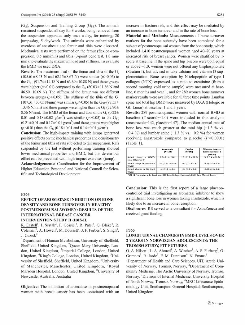
(GS), Suspension and Training Group (GST). The animalsremained suspended all day for 3 weeks, being removed fromthe suspension apparatus only once a day, for training, 20jumps/day, 5 day/week. The animals were euthanized byoverdose of anesthesia and femur and tibia were dissected.Mechanical tests were performed on the femur (flexion-com-pression, 0.5 mm/min) and tibia (3-point bend test, 1.0 mm/min), to evaluate the maximum load and stiffness. To evaluatethe BMD we used DXA.Results: The maximum load of the femur and tibia of the Gc
(105.61±8.43 N and 62.15±8.67 N) were similar (p>0.05) tothe GST (91.74±14.18 N and 63.69±10.88 N) and these groupswere higher (p<0.01) compared to the GS (80.03±11.86 N and46.50±10.09 N). The stiffness of the femur was not differentbetween groups (p>0.05). The stiffness of the tibia of the Gc
(107.31±30.05 N/mm) was similar (p>0.05) to the GST (97.55±13.46 N/mm) and these groups were higher than the GS (72.90±8.56 N/mm). The BMD of the femur and tibia of the Gc (0.22±0.01 and 0.18±0.02 g/cm2) was similar (p>0.05) to the GST
(0.21±0.01 and 0.17±0.01 g/cm2) and these groups were higher(p<0.01) than the GS (0.18±0.01 and 0.14±0.01 g/cm
2).Conclusion: The high-impact training with jumps generatedpositive effects on the mechanical properties and densitometryof the femur and tibia of rats subjected to tail suspension. Ratssuspended by the tail without performing training showedlower mechanical properties and BMD, but this deleteriouseffect can be prevented with high-impact exercises (jump).Acknowledgements: Coordination for the Improvement ofHigher Education Personnel and National Council for Scien-tific and Technological Development
P364EFFECT OFAROMATASE INHIBITION ON BONEDENSITYAND BONE TURNOVER IN HEALTHYPOSTMENOPAUSALWOMEN: RESULTS OF THEINTERNATIONAL BREAST CANCERINTERVENTION STUDY II (IBIS-II)R. Eastell1, I. Sestak2, F. Gossiel1, R. Patel3, G. Blake4, R.Coleman5, A. Howell6, M. Dowsett7, J. F. Forbes8, S. Singh2,J. Cuzick21Department of Human Metabolism, University of Sheffield,Sheffield, United Kingdom, 2Queen Mary University, Lon-don, United Kingdom, 3Imperial College, London, UnitedKingdom, 4King’s College, London, United Kingdom, 5Uni-versity of Sheffield, Sheffield, United Kingdom, 6Universityof Manchester, Manchester, United Kingdom, 7RoyalMarsden Hospital, London, United Kingdom, 8University ofNewcastle, Australia, Australia
Objective: The inhibition of aromatase in postmenopausalwomen with breast cancer has been associated with an
increase in fracture risk, and this effect may be mediated byan increase in bone turnover and in the rate of bone loss.Material and Methods: Measurements of bone turnovermarkers for the bone substudy have been completed for asub-set of postmenopausal women from the bone study, whichincluded 1,410 postmenopausal women aged 40–70 years atincreased risk of breast cancer. Women were stratified by T-score at baseline; if the spine and hip T-score were both equalor above −1.0, women were not offered any bisphosphonate(Stratum I), but advised to take calcium and vitamin D sup-plementation. Bone resorption by N-telopeptide of type Icollagen (NTX) expressed as a ratio to creatinine (from asecond morning void urine sample) were measured at base-line, 6 months and year 1, and for 289 women bone turnovermarker results were available for all three time points. Lumbarspine and total hip BMDwere measured by DXA (Hologic orGE Lunar) at baseline, 1 and 3 years.Results: 289 postmenopausal women with normal BMD atbaseline (T-score≥−1.0) were included in this analysis(anastrozole=142, placebo=147). The median annual rate ofbone loss was much greater at the total hip (−1.3 % vs.−0.4 %) and lumbar spine (−1.3 % vs. −0.2 %) for womenreceiving anastrozole compared to placebo (P<0.0001)(Table 1).
Conclusion: This is the first report of a large placebo-controlled trial investigating an aromatase inhibitor to showa significant bone loss in women taking anastrozole, which islikely due to an increase in bone resorption.Disclosures: RE served as a consultant for AstraZeneca andreceived grant funding.
P365LONGITUDINALCHANGES INBMD-LEVELSOVER2 YEARS IN NORWEGIAN ADOLESCENTS: THETROMSØ STUDY, FIT FUTURESO. A. Nilsen1, L. A. Ahmed1, A. Winther1, A. S. Furberg2, G.Grimnes3, R. Jorde3, E. M. Dennison4, N. Emaus11Department of Health and Care Sciences, UiT, Arctic Uni-versity of Norway, Tromsø, Norway, 2Department of Com-munity Medicine, The Arctic University of Norway, Tromsø,Norway, 3Division of Internal Medicine, University Hospitalof North Norway, Tromsø, Norway, 4MRC Lifecourse Epide-miology Unit, Southampton General Hospital, Southampton,United Kingdom
Osteoporos Int (2014) 25 (Suppl 2):S159–S440 S281

Objective: BMD is a strong indicator of fracture risk. Maxi-mization of the genetic potential for BMD during growth mayprevent primary osteoporosis later in life. Studies exploringthe timing of peak bone mass in adolescents are scarce. Theaim of this population-based longitudinal study was to de-scribe changes in BMD-levels over 2 years in Norwegianadolescents aged 15–17 years at baseline and to examine theachievement of peak bone mass during this period.Material and Methods: In 2010–2011 we invited all firstcomprehensive school students in Tromsø to the Fit Futuresstudy and 1,038 adolescents (93 %) attended. We measuredtotal body (TB), total hip (TH), and femoral neck (FN) BMDas g/cm2 by DXA (GE Lunar prodigy). Two years later, in2012–2013, we invited all participants to a follow-up surveyand 820 adolescents attended, providing 688 repeated mea-sures of BMD. Data was analysed using paired sample t-testand one- way ANOVA. We calculated annual BMD changesat each sites and included 372 girls and 316 boys in theanalysis.Results: Mean follow-up time was 2 years (SD 0.2). In theoverall study-population BMD increased significantly(p<0.05) at all sites in both sexes. Mean annual percentageincrease for FN, TH and TB was 0.3, 0.5, 0.8 in girls and 1.5,1.0 and 2.0 in boys, respectively (p<0.05). In one-wayANOVA analyses, the differences in changes between agegroups were significant (p<0.008) at all sites except at theTH in boys. The annual BMD accrual decreased successivelyat all sites from 15 to 17 years. In girls, aged 17 years atbaseline FN BMD even decreased significantly (−0.7 %,p<0.009) and so did TH BMD (−0.2 %), but nonsignificantly.Conclusion: Overall, BMD levels are still increasing in ado-lescents aged 15–17 years, but our analysis suggest that BMDaccrual is slowing down during this period. Further analysesshould explore the effect of initial BMD and lifestyle factorson these changes.Acknowledgements: The authors are grateful to the staff atthe Centre and Clinical Research and Education, Universityhospital of North Norway and the Fit Futures research staff.
P366ROLE OF CALCIUM SUPPLEMENTATION ANDHEALTH EDUCATION IN BONE HEALTH IN RURALINDIAV. Khanna11Orthopaedics, Ranjana Hospital, Allahabad, India
Objective: To assess the role of calcium supplementation inthe bone health of rural India and also provide health educa-tion to improve the bone health in rural India.Material and Methods: 2 villages about 30 km apart wereselected for the study, Village1 and village2. On the first visitin both the villages initial BMDwas assessed, with the help of
QUS technique, of random people above 30 year. and theirname and other details were registered and regular follow upwas advised. Along with this health education was provided inthe form of information about food stuff rich in calcium andalso ways and means to increase the nutritional content offood items. In village1 free calcium supplements were alsoprovided to all the registered patients. All the patients wereregularly followed up with the help of weekly camps alongwith visits to the homes of the registered patients if they didnot reach the camp to avoid dropouts and ensure full followups. Health camps were organised every week for the entirevillage. The BMD was assessed after 4 months and comparedto the initial value.Results: 150 patients each from village1 and village2 weretaken up for study out of which during the initial screening107 were found to be in the category of osteoporosis andosteopenia in village1 as compared to 103 in village2. Elevenpatients from village1 and 7 patients from village2 were lost tofollow up. The BMD taken after 4 months was slightly in-creased as compared to the initial value. The percent ofincrease in the BMD of village1 was found to be greater thanthat of village2.Conclusion: The bone health of the two villages increasedduring the 4 months when the study was conducted. Thevillage in which calcium supplementation was given, village1,had a greater increase in the BMD as compared to the villagein which only health education was provided. This proves thatcalcium supplementation is helpful in improving the bonehealth of the people. Also the generalised bone health wasincreased.
P367INSIGHT INTO THE ROLE OF MENISCALEXTRUSION AND BONE MARROW LESIONS INKNEE OA PROGRESSION AND THEIR IMPACT ONRESPONSE TO STRONTIUM RANELATETREATMENT IN A SUBSET OF PATIENTS FROMTHE SEKOIA STUDYJ.-P. Pelletier1, C. Roubille1, F. Abram2, M. Dorais3, P.Delorme1, J. P. Raynauld1, J. Martel-Pelletier11Osteoarthritis Research Unit, University of Montreal HospitalResearch Centre (CRCHUM), Montreal, Canada, 2Medical Im-aging Research&Development, ArthroLab Inc., Montreal, Can-ada, 3StatSciences Inc., Notre-Dame-de-l’Ile-Perrot, Canada
Objective: To evaluate the role of meniscal extrusion (mExt)on knee osteoarthritis (OA) progression and its impact onresponse to strontium ranelate (SrRan) treatment assessed byX-ray (change in JSW) and qMRI (cartilage volume loss[CVL]) in the medial compartment at 36 months (M36) insubjects with (mExt+) or without (mExt-) mExt, in association(+) or not (−) with bone marrow lesions (BML).
Osteoporos Int (2014) 25 (Suppl 2):S159–S440S282

Material and Methods: Patients from the qMRI substudy ofthe SEKOIA trial (modified ITT, n=330) were stratified basedon whether mExt (mExt+, n=60; mExt−, n=270) and BMLwere present or not (BML+, n=84; BML−, n=246) and ontheir association in the medial compartment at baseline.Results: In the placebo group, mExt+ patients had signifi-cantly more JSW loss (p=0.002) and CVL in the medialcompartment than mExt- patients. mExt−/BML+ patients(n=18) had significantly more JSW loss (p=0.003) and atrend (p=0.09) toward more CVL compared to mExt-/BML-patients (n=68). mExt+/BML+ patients (n=12) had a trendtoward more CVL in the medial compartment (p=0.10) thanmExt+/BML- (n=14), while JSW change showed no differ-ence. Importantly, in the medial compartment, the JSW lossand CVL were greater when mExt and BML were simulta-neously present. In mExt+ patients, while no difference wasfound in the JSW loss between groups, SrRan at 2 g/dayreduced CVL in the plateaus (p=0.007) with a trend towarddecrease in the medial plateaus (p=0.081) compared to pla-cebo. In mExt+/BML+ patients, SrRan 2 g/day significantlyreduced CVL in the medial plateaus (p=0.046), whereas therewas no difference in JSW loss.Conclusion: Progression of knee OA assessed both byX-ray and qMRI was greater in mExt+ patients andfurther increased when co-localized with BML. Basedon qMRI, SrRan 2 g/day showed beneficial DMOADstructural effects in mExt+ and mExt+/BML+ patients,targeting a subpopulation at higher risk of knee OAprogression, while JSW loss was not sensitive enoughto provide evidence of such effects.Disclosures: Funded in part by Servier. CR: bursary from theFondation du CHUM. JMP, JPP: shareholders in ArthroLab.FA: employee of ArthroLab. JPR: consultant for ArthroLab.
P368BONE ACCRETION IN HIGH RISK NEWBORNSASSESSED BY QUALITATIVE ULTRASOUNDR. Rivas-Ruiz1, D. Villanueva2, M. Segundo2, D. Ibarra2, D.O. Castelán-Martínez3, P. Clark3, F. Rodriguez Islas3, J.Zayas11Centro de Adiestramiento en Investigación Clínica, IMSS,México City, Mexico, 2Neonatology, Hospital Infantil deMéxico, Mexico City, Mexico, 3Clinical Epidemiology Unit,Hospital Infantil de México, Federico Gómez, Mexico City,Mexico
Objective: To detect bone accretion deficit through quantita-tive ultrasound (QUS) by measuring the speed of sound (SoS)in tibia of newborn patients at high risk of osteopenia withdifferent pathologies during the neonatal period. To comparedthe bone accretion deficit between healthy vs. different pa-thologies of newborns.
Material and Methods: A cross-sectional study was madebased on the neonatology tracking outpatient program (nicugraduates) at Hospital Infantil de Mexico Federico Gómez(Mexico City). Complete somatometry was performed andSoS measurement by ultrasound (Sunlight Omnisense®8,000 s), with 2 transducers: CM 6823 (in over 10 kg), CS6616 (under 10 kg) at midshaft of the tibia, from January toJune 2013. Patients were classified into the following groups:1: pulmonary pathology (RDS, BPD, pneumonia), Group 2:congenital heart disease (CHD), 3: abdominal pathology (AP)(NEC III and surgical patients), 4: Neonatal sepsis, 5hyperbilirubin group AND 6 Healthy newborns. Then, asso-ciation of SOS in between the different groups was made withanalysis of co-variance (ANCOVA) adjusted by age, gesta-tional age and height.Results:We evaluated 198 patients, 98 were female (49.5 %),classified according to gestational age: 159 term newborn and26 preterm infants. With median of age of 36 (interquartilerange 30–270) days. The SoS Adjusted by were group 1Pulmonary pathology: 3124±39 m/s, G2 CHD: 3075±38 m/s,G3 Abdominal Pathology: 3072±37 m/s, G4 Neonatal sepsis:3045±37 m/s, G5 hyperbilirrubin: 2981±41 m/s and G6Healthy: 3141±17 m/s. There were differences between healthyand all pathologic groups (p=0.006)Conclusion: Bone accretion measured with QUS is dismissedin high-risk patients with different pathologies during theneonatal period compared with healthy term infant newborns.NICU patients are a high risk population for osteopenia. ThisNICU graduates persist with bone alterations during the firstdays of life.
P369PRODUCTIVITY LOSS AMONG WORKINGPATIENTS WITH KNEE OSTEOARTHRITIS INREPUBLIC OF MOLDOVAV. Salaru1, C. Baerwald2, L. Mazur-Nicorici1, N. Revenco1,D. Bartos3, M. Mazur11State University of Medicine and Pharmacy NicolaeTestemitanu, Chisinau, Republic of Moldova, 2UniklinikumLeipzig, Sektion Rheumatologie, Department für InnereMedizin, Leipzig, Germany, 3UMF “Carol Davila”, Bucuresti,Romania
Objective: To assess the health-related quality of life (QoL)and work productivity in correlation with clinical pattern inpatients with knee osteoarthritis (OA).Material and Methods: Retrospective, cross-sectional studywas performed. There were 93 patients that fulfilled the ACRcriteria for knee OA (1991). Productivity loss was measuredby Work Productivity and Impairment questionnaires (WPAI-OA) that recorded days off work (absenteeism) and reducedproductivity at work (presenteeism) for previous 7 days. The
Osteoporos Int (2014) 25 (Suppl 2):S159–S440 S283

QoL was assessed by KOOS- Knee injury and OsteoarthritisOutcome Score.Results: In the study were included 93 employed patients; 72(77.4 %) females, the mean age±SD 56.7±6.9 (range 37–65)years. Disease duration±SD 5.6±4.0 (range 1–21) years. TheKOOS results showed that the QoL was 43, 3 %, qualified aslow. The level of activity in daily living was 52.9 %, lowerthan the level of pain (61.4 %) or other symptoms (69.1 %).The level of pain by VAS±SD was high 62.2±1.8 mm. Overthe 12-month follow up period, 63 (67.7 %) participantsreported one or more days off work due to knee problems,the mean value being 13.8±3.2 days. The level of absenteeismwas 10, 0%;meanwhile 84 patients (90.3%) reported reducedproductivity at work (63.4 %). The presenteeism showed aclose correlation with VAS pain (r 009, p<0.0001) and deter-mine a poor QoL (r-0.4, p<0.0001). The painful knee induce adiminution of productivity in daily activities for 93 (98.9 %)patients, mean value 65.9±18.1 %. We established moderatecorrelations between the WPAI- OA absenteeism and func-tion, pain, and disease severity (r 0.3–0.4). Thereforepresenteeism was strongly correlated with health outcomes(r 0.7–0.9).Conclusion: Patients with knee OA have been impaired QoL.For the employees with knee osteoarthritis, the work produc-tivity reduced is mostly due to presenteeism rather absentee-ism. The level of pain was the most important factor for lossproductivity in patients with knee OA.
P370RELATIONSHIP BETWEEN ACTIVITY OFRHEUMATOID ARTHRITIS, PARAMETERS OFBONE METABOLISM AND VITAMIN DL. Sterancakova1, K. Brazdilova1, Z. Killinger1, J. Payer1, D.Cierny115th Department of Internal Medicine, Medical Faculty ofComenius University, University Hospital, Bratislava,Slovakia
Objective: Rheumatoid arthritis (RA) is a chronic inflamma-tory disorder. It is well known that RA is also associated withdecreased BMD and development of osteoporosis. High in-flammatory disease activity and serum concentration of vita-min D3 are believed to be involved in the pathophysiology ofRA-associated osteoporosis. Our objective was to determinethe relationship between activity of RA with selected labora-tory parameters of bone metabolism and vitamin D3 serumconcentration.Material and Methods: We examined 43 patients with dif-ferent levels of RA activity. The mean age of patients was52.4 years, female to male ratio was 3.3:1. Patients was nottaking any antiporotic drugs or vitamin D. Disease activitywas evaluated by DAS28 score (disease activity score), bone
metabolism by bone turnover markers—osteocalcin (OC),CTx (C-terminal telopeptide of collagen type I). We alsomeasured serum levels of 25-hydroxycholecalciferol. For sta-tistical evaluation, we used the Pearson correlation coefficient.Results: We have shown a statistically significant negativecorrelation between disease activity and levels of vitamin D(p<0.001). Relationship between osteomarkers and DAS28score not reached statistical significance, however, we ob-served a trend in bone turnover increase with increased activ-ity of RA.Conclusion: Vitamin D plays an important role in immuneregulation. Some studies suggest that individuals with lowserum levels of vitamin D have a higher incidence of RA. Inour study, the severity of vitamin D deficiency correlated withactivity of RA. This relationship may indicate the need ofadequate supplementation of vitamin D in patients with RA.Sufficient concentrations of vitamin D may have a positiveeffect on the activity of RA and also contribute to slowerdevelopment in secondary osteoporosis.
P371OSTEOPRTEGERIN, SOLUBLE RANK LIGAND ANDTHEIR CORRELATION WITH BONE MINERALDENSITY IN PATIENTS WITH TYPE 1 DIABETESMELLITUSN. P. Botushanov1, M. M. Orbetzova1, A. N. Botushanov11Medical University- Clinic of Endocrinology, Plovdiv,Bulgaria
Objective: To evaluate a possible relationship between theserum OPG and sRANKL and BMD in patients with type 1diabetes mellitus (DM).Material and Methods: 162 patients with type 1 DM (97females and 65 males) age 29.17 year (20–40) and 200 (100women and 100 men) age- and sex-matched healthy controlswere analyzed for BMD of lumbar spine and femoral neck byDXA using Lunar DPX-A. Serum levels of OPG andsRANKL were determined using ELISA. We performed sta-tistical analysis (t-test, ANCOVA), Spearman’s rank correla-tion, data are expressed as mean±SD and as percentages.Results: BMD in type 1 DM showed statistically significantlower levels both for lumbar spine L1-L4- men (1.21±0.15 g/cm2 DM vs. 1.33±0.16 g/cm2 controls, P<0.05) and L1-L4women (1.10±0.12 g/cm2 DM vs. 1.19±0.12 g/cm2 controls,P<0.05) and femoral neck - men (0.91±0.21 g/cm2 DM vs.0.98±0.15 g/cm2 controls, P<0.05) and women (0.86±0.12 g/cm2 DM vs. 0.92±0.14 g/cm2 controls, P<0.05). Se-rum OPG levels were significantly lower in diabetic patients(37.57±21.76 pg/ml) than in control group (43.58±23.71 pg/ml p=0.029). Serum sRANKL did not differ significantlybetween both groups (0.39±0.21 pmol/L vs. 0.491±0.23 pmol/l, p=0.16). However sRANKL/OPG ratio (0.011
Osteoporos Int (2014) 25 (Suppl 2):S159–S440S284

±0.08 vs. 0.065±0.013, p=0.034) was significantly lower inDM group than in the controls. We observed positive correla-tion between serum OPG and sRANKL/OPG levels andBMD (r=0.54; P=0.03 and r=0.34; P=0.13 resp.) and weaknegative correlation between BMD and sRANKL.Conclusion: BMD measured at lumbar spine and femoralneck was significantly lower in patients with type 1 DM thanin age-and sex matched controls. Decreased levels of OPGand sRANKL/OPG ratio may be the cause for increased boneresorption and decreased BMD in patients with type 1 DM.OPG and/ sRANKL system may play role in the bone remod-eling in concert with other factors influencing bone changes indiabetes mellitus type 1.
P372HIP FRACTURE TYPES IN CANADIAN MEN ANDWOMEN CHANGE DIFFERENTLY WITH AGEM. Kloseck1, R. G. Crilly2, S. Mequanint21Faculty of Health Sciences, University of Western Ontario,London, Ontario, Canada, 2Department of Medicine, Univer-sity of Western Ontario, London, Ontario, Canada
Objective: There is evidence that the type of hip fracture,intertrochanteric (IT) versus subcapital (SC), changes differ-ently with age in men and women. We have explored this in aCanadian population and looked at community dwelling andinstitutionalized seniors to see if frailty plays a role.Material and Methods: Three years of hip fracture data(2007–2009) were obtained from the Canadian Institute forHealth Information for all Canada excluding Quebec, andstratified by age and gender. Statistical Analysis Logisticregression was used to test for a significant interaction be-tween age and gender on the likelihood of sustaining onefracture type compared to the other.Results: The database contained a total of 18,316 (28 %) hipfractures in men (mean age 78.48±10.90) and 47,343 (72 %)in women (82.08±9.63). Overall the prevalence of the ITfracture rises with age, but when analyzed by gender,intertrochanteric fractures rose in women (p<0.05), from35.9 % in the youngest category to 50.7 % in the oldest, butnot in men. For each year of age the odds of sustaining an ITfracture vs. a SC fracture rises by 2.7 % in women. Analysis ofcommunity and institutionalized subjects showed no differ-ence between them.Conclusion: Results confirm the proportion of the two mainhip fracture types changes differently in men and women withage. Previously identified risk factors for subcapital fracturesinclude unchanging structural factors (neck length, neck/shaftangle) so the risk of a subcapital hip fracture in a fall willremain constant and be determined by the frequency of falling.Intertrochanteric fractures are better predicted by trabecularbone mass, loss of which is greater in women. Thus the risk of
an intertrochanteric fracture per fall will rise with age inwomen. Frailty, a condition seen mostly in institution-alized people, does not appear to influence the type ofhip fracture.
P373RADIOGRAPHIC CHARACTERIZATION OF THEBONE HEALING PROCESS AFTER PROTEINAPPLICATION EXTRACTED FROM HEVEABRASILIENSISB. G. Dos Santos Kotake1, A. A. Ferreira De Carvalho2, L. M.Pescinini Salzedas2, E. Ervolino2, M. Gonçalves Gonzaga1,M. Mizusaki Iyomasa1, J. Ramos1, D. Mizusaki Iyomasa1, J.P. M. Issa11University of São Paulo, Ribeirão Preto, Brazil, 2PaulistaState University, Araçatuba, Brazil
Objective: Current literature has shown that the proteinextracted from the rubber tree, Hevea brasiliensis, hasinteresting properties regarding to tissue repair, especial-ly for soft tissues, biocompatible and with angiogenicproperties. The aim of this study was to study radio-graphically the bone healing process in rat skulls, usingdifferent types of bone grafts and protein extracted fromHevea brasiliensis.Material andMethods: For this work, 112 albinoWistar rats(~250 g) were divided into eight groups, waiting in each onefor sacrifice, 4 and 6 weeks. The groups are the following: AT:Particulate autologous bone graft; HOM: Particulate homolo-gous bone graft; HET: Particulate heterologous bone graft(lyophilized bovine bone); ATP1: Particulate autologous bonegraft + 5 μg protein; HOMP1: Particulate homologous bonegraft + 5 μg protein; HETP1: Particulate heterologous bonegraft (lyophilized bovine bone) + 5 μg protein; P1: 5 μg ofprotein; DO: bone defect only. Images of the samples wereobtained using the X-ray machine GE-100 (General Electric,Milwaukee, USA), operating at 50 Kvp, 10 mA, 8 pulses,handled by the digital system (Soredex, Orion Corporation,Helsinki, Finland), Digora software for Windows 1.51. Datawere subjected to statistical analysis, ANOVA and Tukey test(p<0.05).Results: Results showed that at 4 weeks, the DO data werestatistically different than AT and HET (p<0.05), and the lastone was different than HOMP1, AUTP1 (p<0.05). For6 weeks, the P1, AT, ATP1, HOMP1 data were statisticallydifferent than DO (p<0.05).Conclusion:According to obtained data, we can supposethat this protein, at 6 weeks, was able to improve thebone healing process in this experimental model usingthis methodology.Acknowledgements: FAPESP: 2012/03310-8
Osteoporos Int (2014) 25 (Suppl 2):S159–S440 S285

P374COST-EFFECTIVENESS OF TOTAL HIPREPLACEMENTS: A COMPREHENSIVE MODELR. Pinedo-Villanueva1, D. Turner2, J. P. Raftery3, C. Cooper4,N. K. Arden1, The Coast Group11Nuffield Department of Orthopaedics, Rheumatology andMusculoskeletal Sciences, University of Oxford, Oxford,United Kingdom, 2Norwich Medical School, University ofEast Anglia, Norwich, United Kingdom, 3Faculty of Medi-cine, University of Southampton, Southampton, United King-dom, 4University of Southampton, Southampton, UnitedKingdom
Objective: Total hip replacements (THRs) have beenwidely reported to be highly cost-effective interventions.However, most economic models of THRs have as-sumed that, unless a revision surgery is performed, theoutcome of the primary would be the same and excel-lent for all patients. We aimed to build a comprehensiveeconomic model to assess the cost-effectiveness ofTHRs in the UK.Material andMethods: A life-time cohort Markov modelwas developed including two outcome categories afterprimary and revision surgeries. Preoperative transitionprobabilities were obtained from an expert elicitationexercise and postoperative probabilities from HES-PROMs. Data from HES-PROMs were also used toestimate quality-adjusted life years (QALYs). Surgerycosts were estimated from HES data combined withNHS healthcare resource groups. Primary care costswere estimated based upon resource use data obtainedfrom GPRD extracts, which were combined with pre-liminary results from the COASt cohort to estimatepostoperative values by outcome category.Results: Current practice is associated with both highercosts and higher QALYs compared to not performingTHRs. For women 45 years of age, current practice ofconducting THRs would represent discounted costs of£11,500 and 14.5 QALYs over lifetime. If THRs werenot performed, costs would drop to £4,300 and sowould QALYs to 6.7, producing an incremental cost-effectiveness ratio (ICER) of £928 per QALY gained.Results varied only slightly between genders but quitesignificantly with patients’ age entering the model.Conclusion: THRs are highly cost-effective even whendistinguishing between very good and fair outcomes aftersurgery and accounting for corresponding primary care costs.The significant improvement in the quality of life of patientsundergoing the procedure is at the heart of these results. Theintervention is more cost- effective for younger patients andnot much difference is reported between genders other thanhigher QALY gains for women given their lower mortalityrate.
P375HYDROXYAPATITE AND FIBRIN SEALANT FORBONE REPAIR IN RATSM. Rodrigues Da Cunha1, F. A Menezes1, G. R Dos Santos2,V. C Amaro Martins3, S. AYoshioka3, A. M. G Plepis3, R. SFerreira Junior4, J. P. M. Issa51UniAnchieta, Jundiai, Brazil, 2Faculty of Medicine, Jundiai,Brazil, 3University of São Paulo, São Carlos, Brazil, 4PaulistaState University, Botucatu, Brazil, 5University of São Paulo,Ribeirão Preto, Brazil
Objective: To evaluate the bone healing process in rat skulldefects grafted with porous hydroxyapatite and fibrin sealantderived from snake venom.Material and Methods: A skull defect was performed surgi-cally in 40 rats and these animals were divided into fourgroups: G1 (control, no graft); G2 (defect grafted with hy-droxyapatite); G3 (defect grafted with fibrin sealant), and G4(defect grafted with hydroxyapatite and sealant). The animalswere euthanized after 2 and 6 weeks of the surgery and thesamples submitted to macroscopic, radiologic and histologicalanalyses. The newly formed bone was quantified based on theprinciple of Delesse and the results were compared byANOVA and the Tukey test (p<0.05).Results:Macroscopic analysis showed absence of inflammatorysigns, characteristic of rejection of the employed biomaterial.Radiographical analysis showed a radiolucency inside of thebone defects. In animals sacrificed after 2 weeks, immaturetrabecular bone was formed from the borders of the defect. InG2 andG4, qualitative histological aspects showed few hydroxy-apatite particles surrounded by new bone. After 6weeks, the newbone tissue showed mature aspects, and surrounded severalhydroxyapatite particles in G4, without connective tissue inter-position. The relative volume (%) and standard deviation for newbone tissue in the surgical area after 2 weeks was 5.66±0.57,6.66±0.57, 20±1.0 and 21±1.0 in G1, G2, G3 and G4, respec-tively. There was no significant difference betweenG1 andG2 orbetween G3 and G4. After 6 weeks, the new bone tissue volumeand the standard deviation were respectively, 10.66±0.57, 20.66±1.15, 29.66±1.52 and 53.66±0.57.Conclusion: The fibrin sealant derived from snake venom,according to the limitations of this study showed positiveproperties for biocompatibility and was able to improve thebone healing process when combined to hydroxyapatite.
P376LEAD CHARACTERIZATION IN BLOOD AND TIBIASAMPLES IN RATS EXPOSED TO 30 MG/L OF LEADIN DRINKING WATERF. A. Tocchini De Figueiredo1, M. A. Veiga1, F. V. Nakadi1, J.Ramos1, E. R. Kawakita1, R. F. Gerlach1, J. P. M. Issa11University of São Paulo, Ribeirão Preto, Brazil
Osteoporos Int (2014) 25 (Suppl 2):S159–S440S286

Objective: Bone is considered the best marker for lead expo-sure, and bone lead determinations can be easily made. Evenso, most animals studies carried out nowadays only determinewhole blood lead concentrations. The aim of this study was toobserve whether there would be differences in whole bloodversus tibia lead concentrations over time in growing ratswhen these were exposed to relatively low lead prenatallyuntil days 28 and 60.Material andMethods:Leadwas given in the drinking waterat 30 mg/L from the time the dams were pregnant untiloffspring was 28 or 60 days old. Male Wistar rats were usedin this study. Concentrations of lead were measured in wholeblood and in bone (tibia) after 28 (28D) and 60 days (60D) incontrol (C) and in lead exposed animals (Pb). Calcium andLead were measured by Flame and Graphite Furnace AtomicAbsorption Spectrometry.Results:Whole blood lead in the Pb-28D animals was 8.0 μg/dl (±1.1), and in the Pb-60D, 7.2 μg/dl (±0.89) (p>0.05 fordifference between these groups), while control animals leadlevels were 0.2 μg / dl (±0.4) (p<0.001 for comparison withrespective age groups). There was a significant differencebetween the tibia masses of the C-60D group (0.86±0.13 g)and the ones of the Pb-60D (0.61±0.11 g) (p=0.0004), a 25%decrease in tibia bone mass. Bone lead concentrations were8.02 μg/g (±1.12) in the Pb-28D, and 43.3 μg/g (±13.26) inthe Pb-60D (p<0.01 for difference between these groups),while the C-28D and C-60D showed tibia lead concentrations<1 μg/g (p<0.001 for comparison with respective agegroups).Conclusion: The five times higher amount of lead found inthe bone of older animals (Pb-60D vs. Pb-28D)(when nodifferences in lead in whole blood were observed) reinforcesthe importance of using bone lead as an exposure biomarker,instead of blood, particularly when studying the effects oflower lead levels. Furthermore, in this study lead exposuredecreased the mass of tibial bone in the Pb-60D, but not in thePb-28D group.Acknowledgements: FAPESP
P377DISTRIBUTION OF VERTEBRAL FRACTURESVARIES AMONG PATIENTS ACCORDING TO HIPFRACTURE TYPEJ. Watt1, R. G. Crilly1, L. Cox11Division of Geriatric Medicine, University of Western On-tario, London, Ontario, Canada
Objective: Of the two main types of hip fractures:intertrochanteric (IT) and subcapital (SC) either can haveassociated vertebral fractures (VF) but they are fewer in theSC patients who also tend to have better bone density. Thisraises the question of whether VF in the SC patients indicate
osteoporosis or if a different etiology, such as trauma, could bethe cause. In this study, we explored the nature of VF in thetwo hip fracture populations.Material andMethods: This was an analysis of 120 patients:40 with SC fractures and VF, 40 with IT fractures and VF, andfor comparison, 40 osteoporotic patients with VF alone. Basedon Genant’s semiquantitative assessment method, the distri-bution, type and severity of each patient’s vertebral fractureswere described1.Results: Patients with SC fractures had significantly fewertotal VF (p=0.005 and p=0.019), fewer fractures from T4-T10 (p=0.005 and p=0.042), than patients with IT fracturesand those with VF alone. The number of VF from T11-L4 andat the T12-L1 peak did not differ among the groups. Patientswith SC fractures were more likely to have only one VF(p<0.001). The distribution of VF in those with IT fracturesand VF and in those with VF alone was similar and signifi-cantly correlated (r=0.6496, p=0.009).Conclusion: Patients with IT fractures and VF closely resem-ble the spinal osteoporotic patients in number and distributionof the VF. The distribution of VF in patients with SC fracturesand VF differs from the other fracture groups in being usuallysingle and concentrated in the lower spine, suggesting adifferent etiology. The SC and VF of some of these patientsmay be a consequence of trauma and not osteoporosis.References: 1. Genant HK et al. J Bone Miner Res1996;11:984
P378NORMALANDANIONICCOLLAGENMEMBRANESIN AN EXPERIMENTAL MODEL FOR BONEHEALING PROCESSM. Silva Pettian1, G. R Dos Santos1, V. C Amaro Martins2, S.AYoshioka2, A. M. G Plepis2, J. P. M. Issa3, M. Rodrigues DaCunha41Faculty of Medicine, Jundiai, Brazil, 2University of SãoPaulo, São Carlos, Brazil, 3University of São Paulo, RibeirãoPreto, Brazil, 4UniAnchieta, Jundiai, Brazil
Objective: Biomaterials constituted by extracellular matrixcomponents have osteogenic properties that are importantfor the bone repair. Materials composed by collagen derivedfrom pericardium or bovine intestinal serosa are been used inmany regenerative therapies. Therefore, the aim of this studywas to evaluate the osteogenic capacity of natural collagenmembranes (native) and anionic collagen obtained by alkalinehydrolysis, both derived from bovine serosa.Material and Methods: Fifty Wistar rats were submitted tosurgical skulls defects. The animals were then divided intofive groups: group 1, empty defect; group 2, defect filled withnonmineralized native membrane; group 3, defect filled withmineralized native membrane; group 4, defect filled with non-
Osteoporos Int (2014) 25 (Suppl 2):S159–S440 S287

mineralized anionic membrane; and group 5, defect filled withmineralized anionic membrane. The animals were sacrificedafter 2 and 6 weeks, and the samples were submitted tomacroscopic, radiologic and histological analyses.Results: Macroscopic and radiologic analyses revealed theintegrity of bone tissue in the surgical area and in adjacentareas, without signs of pathological alterations. Histologically,the new bone tissue projected from themargins of the defect inall groups, and the newly formed bone matrix exhibited goodbirefringence of type I collagen fibers and immunoexpressionof osteocalcin. Morphometric analysis showed a greater con-centration of bone matrix in the groups that received theanionic membranes.Conclusion: In conclusion, the anionic collagen membranepresented better osteogenic properties for this experimentaldesign model, however, was not sufficient for complete bonehealing, considered the short period of time used in this study.Acknowledgements: FAPESP
P379ACTION OF PHYSICAL EXERCISE COMBINEDWITH RISEDRONATE IN OSTEOPENIC BONEUSING EXPERIMENTAL ANIMAL MODELR. Carminati Shimano1, A. P. Macedo1, M. José Falcai1, R.Antônio Andraus1, A. C. Shimano1, J. P. M. Issa11University of São Paulo, Ribeirão Preto, Brazil
Objective: This study evaluated the effects of physical train-ing associated with the risedronate, as prophylactic and ther-apeutic environment in osteopenic bones.Material and Methods: 48 rats were divided into 8 groups:sedentary for 12 weeks and ovariectomized (OVX); ovariec-tomized and the 12 weeks following administered risedronate(OVXM); ovariectomized and physical training on the tread-mill during 12 weeks (OVXC); ovariectomized and 12 weeksfollowing administered risedronate associated with physicaltraining on the treadmill (OVXCM); simulated surgery andsedentary for 12 weeks (SHAM); simulated surgery, and the12 weeks following administered risedronate (SHAMM);simulated surgery followed by physical training on the tread-mill during 12 weeks (SHAMC); simulated surgery, and the12 weeks following administered Risedronate associated withphysical training on the treadmill (SHAMCM).After 12weeksof the experiments, tibias were surgically dissected and ana-lyzed by different methodologies.Results:We obtained as a result of mechanical tests regardingthe stiffness, the SHAMM group with values below theOVXM (p=0.001); at maximum strength full, the SHAMMgroup also with values below the OVXM and OVXCM (p=0.002), the global densitometry showed no statistical differ-ence between the groups, since the proximal densitometry, theOVX group with values less than SHAMM and SHAMCM
(p<0.000). For histological analysis, according to quantifica-tion of trabecular bone microarchitecture, the OVX group hadlower values than SHAMC, OVXM, SHAMCM andOVXCM, and OVXCM and SHAMCM groups having supe-rior results than SHAM, SHAMM and OVXC (p<0.001).Conclusion: It is concluded that the physical exercise and theemployed medicine, changes to better the biomechanicalproperties of bone of ovariectomized rats. The risedronatehas no benefits on primary prevention and the quantificationof bone trabecular, the medicine was more effective in relationto race in ovariectomized groups, and when associated, thetreatments were more effective.
P380EVALUATION OF THE TIBIAL BONE STRUCTUREIN RATS SUBMITTED TO THE TAIL SUSPENSIONAND TREATED WITH SWIMMING EXERCISESJ. B. Volpon1, A. Valadares Da Silva1, M. José Falcai1, M. J.Quirino Louzada1, B. G. Dos Santos Kotake1, J. P. M. Issa11University of São Paulo, Ribeirão Preto, Brazil
Objective: This study investigated the effectiveness of swim-ming in the recovery of bone quality assessed in the tibia ofrats with induced osteopenia by tail suspension.Material and Methods: 50 Wistar rats were divided into 5experimental groups (n=10 each). Group suspended by thetail for 21 consecutive days (S) and its control remained incages for the same period (CI). Group suspended and trained,remained in suspension during 21 day and then underwent 30sessions of swimming (ST). Suspended for 21 day and re-leased after 30 day in cages (SNT). In the control group II ratswere observed during 51 day without intervention (CII). Bonequality assessment was made by DXA and mechanical testson the right tibia, and in the proximal metaphyseal region ofthe left tibia was performed the histomorphometric study.Results:Mechanical tests showed that for the group S valuesof maximum force (-14.03 %, p=0.0003), stiffness(−21.68 %, p=0.0055), BMD (−17.62 %, p=0.019) and per-centage of trabecular bone (−57.2 %, p=0.0001) decreasedsignificantly as compared with the IC group. The simul-taneous comparison of mechanical properties betweengroups evaluated after 51 day (STxCIIxSNT) showedsignificant differences between groups for peak force(p=0.0014), stiffness (p=0.0010), BMD (p=0.0095)and the percentage of trabecular bone (p<0.0001). TheST group showed an increase in maximum force (+10.23 %, p<0.05), stiffness (+21.91 %, p<0.001),BMD (+9.46 %, p<0.05) and the percentage of trabec-ular bone (+48.82 %, p<0.001) compared to the SNTgroup. The groups SNT and CII also decreased signifi-cantly (−14.4 %, p<0.05) for maximum strength, stiff-ness (−25.21 %, p<0.005), BMD (−13.34 %, p<0.05)
Osteoporos Int (2014) 25 (Suppl 2):S159–S440S288

and the percentage of trabecular bone (−52.06 %,p<0.001). Finally, the comparison between the SNTand CII groups showed no significant difference(p>0.05) for the values of maximum strength, stiffness,and BMD percentage of trabecular bone.Conclusion: The swimming reversed the osteopenic deterio-ration caused by hypoactivity, with complete and quicklyrecovery of bone quality.
P381WHETHERWE COULD PREDICT HIP FRACTUREM. Kocic1, L. Dimitrijevic1, I. Stankovic1, M. Spalevic1, V.Zivkovic1, A. Stankovic11Clinical Center Nis, Clinic of Physical Medicine and Reha-bilitation, Nis, Serbia
Objective: Hip fracture is a serious medical and socioeco-nomic issue. Hip fractures occur in the low trauma in patientswith osteoporosis. The FRAX questionnaire is designed toidentify patients with an increased risk of fractures in generaland risk of hip fracture. Objective: To determine whetherpatients who have sustained a hip fracture had an increasedrisk of hip fracture based on FRAX calculator.Material and Methods: The study included 50 patients agedover 60 who have suffered a hip fracture to a small trauma andare operated at the Orthopedic Clinic in Nis. None of thepatients were analyzed regarding the risk of fractures byFRAX Calculator prior to the fracture incident. Only threeof all the patients had undergone bone densitometry whichshowed presence of osteopenia in one of the patients andosteoporosis in two of them. Subsequently after the surgeryall patients were analyzed by FRAX questionnaire calculatorto determine whether they had had an increased risk of hipfracture.Results: The patients were divided according to age in 3groups: 60–69 years old (12 patients), 70–79 (29 pa-tients) 80 and over (9 patients). As regarding the pres-ence of other risk factors in addition to age and femalegender, 39 patients had no other risk factors, 10 patientshad one, and only one patient had 2 additional riskfactors. In the age group of 60–69 only 1 patient hada risk of a hip fracture greater than 3 which requiresintroduction of therapy for osteoporosis. In the 70–79age group, 27 out of 29 patients had a risk factor of 3or more as well as all of patients in the 80 and olderage group.Conclusion: Our results showed that almost all women agedover 70 had a risk factor of over 3 and that they neededtreatment for osteoporosis, which could reduce the risk ofhip fracture. Also, in patients under 70 years of age, thecurrent issues FRAX questionnaire were not sufficient fordetermining increased risk for hip fracture.
P382ADMINISTRATIONOF PROTONPUMP INHIBITORSIN LONG-TERM DECREASES BONE MINERALDENSITY OF THE FEMUR OFADULT RATSA. C. O. Penoni1, G. R. Yanagihara1, R. Annoni2, A. G.Paiva1, M. P. Neto3, L. H. L. Torres4, M. J. Quirino Louzada5,A. C. Shimano11Department of Biomechanics, Medicine and Rehabilitationof the Locomotor System, Ribeirão Preto, Ribeirão Preto,Brazil, 2University of São Paulo, Faculty of Medicine, Uni-versity of São Paulo, Department of Pathology, São Paulo,Brazil, 3University of Vale do Sapucaí, UNIVAS, PousoAlegre, Brazil, 4University of São Paulo, Faculty of Pharma-ceutical Sciences, São Paulo, Brazil, 5University of São Paulo,Ribeirão Preto, Brazil
Objective: To assess BMD of rats that were subjected to theuse of omeprazole in the long term.Material and Methods: Fifty Wistar rats, adult male ofaverage weight between 200 and 240 g were equally distrib-uted (n=10): 1) OMP300 Group - intake of omeprazole at300 μmol/kg; 2) OMP200 Group - intake of omeprazole doseof 200 μmol/kg; 3) OMP40 group - intake of omeprazole at40 μmol/kg;4) OMP10 group - intake of omeprazole at10 μmol/kg; and 5) CONT group - control group, only intakeof vehicle dilution. For 90 day the animals of the experimentalgroups received orally, their respective doses of omeprazoleand control animals receiving vehicle dilution, compounddoses natrosol solubilized in sodium bicarbonate. After eutha-nasia, the right femurs were dissected and subjected to anal-ysis of BMD in a densitometer dual X-ray emission, DPX-alpha, Lunar®.Results: The bone density of OMP300 (0.20±0.008 g/cm2)group was lower than the CONT group (0.22±0.107 g/cm2, p=0.006). There was no difference in the comparison between thecontrol group (0.22±0.107 g/cm2) and the other groups:OMP200(0.21±0.019 g/cm2, p=0.644), OMP40 (0.21±0.015 g/cm2, p=0.305) and OMP10 (0.21±0.016 g/cm2, p=0.410).Conclusion:With these results, we observed that the relation-ship between the use of PPIs with bone tissue is true, as theBMD has changed over use of the highest dose.
P383USING TRABECULAR BONE SCORE INMONITORING OF THE OSTEOPOROSISTREATMENT EFFECTM.Kuzma1, L. Sterancakova1, D. Holovacova1, P. Jackuliak1,Z. Killinger1, J. Payer115th Department of Internal Medicine, Comenius UniversityFaculty of Medicine and University Hospital, Bratislava,Slovakia
Osteoporos Int (2014) 25 (Suppl 2):S159–S440 S289
Objective: BMD as a widely used quantitative bone param-eter does not bring whole information about real bone status.Therefore, bone quality shows as main determinant of bonestrength, fracture prediction and treatment effect monitoring.Amethod to assess bone quality through the grey-level textureanalysis from lumbar spine DXA scan is trabecular bone score(TBS). The aim of our study was to evaluate the effect ofstrontium ranelate (SRn), denosumab(DMAb) andteriparatide (TPT) using TBS in comparison to the effect onBMD.Material and Methods: Noncontrolled comparative analysisof 3 osteoporosis treatment modalities in our osteocentrum.All patients included were naive. TBS from lumbar spineDXA scans using TBS INsight® (Med-Imaps) software wasevaluated during the 4-years of treatment, annually. Boneturnover was assessed using CTx - bone resorption markerand osteocalcin (OC) - bone formation marker. A standarddose (800 IU/day) of 25-OH-D3 and oral calcium (1,000 mg/day) has been administered to the patients.Results: A total of 245 patients (SRn: N=113; mean age72 year; DMAb: N=74; 71.7 year; TPT: N=58; 76.2 year)were included. After 1 year in SRn subgroup BMD increased4.8 % (p<0.007) vs. 0.5 % (NS) increase in TBS; DMAbsubgroup: BMD + 3.8 % (p=0.003) vs. TBS +2.43 % (p=0.001); TPT subgroup: +2.05 % (NS) vs. +3.1 % (p=0.03).After 2 year: SRn subgroup: BMD +6.5% (p<0.001) vs. TBS+2.5 % (NS); DMAb subgroup was not analysed; TPT sub-group: BMD + 5.9 % (NS) vs. TBS +6.1 % (NS). After year 3and 4 was analysed only SRn subgroup with maximum in-crease after 4 year (BMD +13.4 % (p<0.001) vs. TBS +7.2 %(p=0.01)).Conclusion: Despite lack of long-term results for DMAb andTPT we have observed positive effect of all three treatmentmodalities on trabecular bone architecture. According to pre-vious data seems SRn, DMAb and TPT as 3 most effectivet r ea tmen t op t ions pos i t i ve ly in f luenc ing bonemicroarchitecture which results in improvement of bonequality.
P384OSTEOANABOLIC TREATMENT OFGLUCOCORTICOID INDUCED OSTEOPOROSIS:RESULTS FROM SLOVAK NATIONAL REGISTRYJ. Payer1, Z. Killinger1, P. Jackuliak1, S. Tomkova2, P.Vanuga3, P. Masaryk4, A. Letkovska4, Z. Kmecova515th Department of Internal Medicine, Comenius UniversityFaculty of Medicine and University Hospital, Bratislava, Slo-vakia, 2Osteocentrum, Kosice-Saca, Slovakia, 3National Insti-tute of Endocrinology and Diabetology, Lubochna, Slovakia,4National Institute of Rheumatic Diseases, Piestany, Slovakia,5Osteocentrum of Faculty Hospital F.D. Roosevelt, BanskaBystrica, Slovakia
Objective: GIOP is the most frequent and severe form ofsecondary osteoporosis. The rate of bone loss is greatest inthe first year and the first osteoporotic fracture occur as earlyas in first 3–6 months of hypercorticism. One of the treatmentmodalities is osteoanabolic treatment. The primary outcomeswere defined as changes of BMD and BTM after 12 M and18 M. Secondary outcomes included prevalence of clinicalfractures, tolerability and safety of the treatment.Material and Methods: Prospective, open label, non-randomized, 18 M study. The inclusion criteria: (1) T-score<−2.9 or ≥1 fracture, (2) use of 5 mg corticoids for >3 M.Patients were treated 20 μg of rhPTH(1–34) s.c. All patientsreceived 500–1,000 mg of calcium and 400–800 IU of Vita-min D. The register has 186 pts (150 women and 36 men,mean age 58.3 year). The measurement was at baseline, 6, 12and 18 M. Statistical analyses were performed according tothe intention-to-treat principle. T- test was used to determinethe changes in BMD and BTM in 6, 12 and 18M compared tobaseline.Results: TPTD treatment resulted in increase of BMD totalhip after 12 M (+3.8 %, p=0.035) and 18 M (+3.4 %, p=0.135). Themost significant increase was found in LS, +4.8%(p<0.001) during 12 M of treatment and an increase of 9.8 %after 18 M of treatment with TPT (p<0.001). 18 M of TPTDtherapy led to significant increase of CTx and OC. The in-crease of CTx was 169 % after 6 M and 183 % after 12 M(p<0.001). As for OCn, the increase represented 242% after 6and 257 % after 12 M (p<0.001). Treatment was well toler-ated and no serious side effects were observed. Borderlineasymptomatic hypercalcemia was present in 5.3 % of patients.Interesting finding was low levels of 25-OH-D (<30 ng/ml) in82.9 % of patients after 18 M of treatment.Conclusion: Osteoanabolic treatment using teriparatide waseffective, well tolerated and safe. Main problem is prevalenthypovitaminosis D during treatment period because of lowersupplementation dosage to avoid severe hypercalcemia.
P385EQ-5D AND QUALITY OF LIFE OF OSTEOPOROSISAT-RISK PATIENTS IN A SWEDISH OSTEOPOROSISPATIENT REGISTRYA. Krishna1, D. Mellström2, Z. Li3, C. P. S. Fan3, S.Salomonsson4, E. Waern21Merck & Co., Inc., Whitehouse Station, United States, 2Cen-tre for Bone and Arthritis Research at Sahlgrenska Academy,Unive rs i ty of Gothenburg , Göteborg , Sweden ,3AsclepiusAnalytics Ltd., Hong Kong, Hong Kong, 4Merck& Co., Inc., Solna, Sweden
Objective: To assess health-related quality of life (HRQoL)using EQ-5D questionnaires and identify factors associatedwith HRQoL in a Swedish osteoporosis (OP) patient registry.
Osteoporos Int (2014) 25 (Suppl 2):S159–S440S290

Material and Methods: The OP registry collected patientinformation in an outpatient clinic in Göteborg region from1991 to 2009. New patients were referred to the clinic for OPassessment and BMD measurement (new patients). Patientswith prior osteoporotic BMD (T-score≤−2.5) or OP diagnosismay return for follow-ups (returning patients). Each patientfilled the EQ-5D questionnaire once, which covered mobility,self-care, usual activities, pain/discomfort and anxiety/depression. Responses were aggregated into a HRQoL index,between −0.074 (worst) and 1 (best), using the Europeanformula. Descriptive and multivariable analyses were usedto identify factors associated with patients’ HRQoL.Results: 3,240 patients completed the EQ-5D questionnaire.89 % were female, average age was 66 [SD=11.9]. 48.4 %were new patients, 51.6 % were returning. 34.1 % reportedproblems in mobility, 8.0 % in self-care, 27.8 % in usualactivities, 74.7 % in pain/discomfort, and 43.3 % in anxiety/depression. Average HRQoL was 0.7 [SD=0.2]. Patients witha history of fractures had lower HRQoL than those withoutfractures (0.69 vs. 0.71; p<0.01). Among those with fractures,lower HRQoL was indicated in patients with hip (0.62),vertebral (0.63) and arm (0.66) fractures. Compared withpatients receiving OP medication (bisphosphonates, raloxi-fene, teriparatide, estrogen), those without reported more painor discomfort (77 % vs. 73 %; p=0.01). New patients hadlower HRQoL than returning patients (0.68 vs. 0.72; p<0.01).Other factors affecting HRQoL included elder age, history offalls and comorbidities (hypertension, arthritis, and asthma).Conclusion: In a Swedish OP registry, average HRQoL basedon EQ-5D was 0.7. 75 % patients reported pain/discomfortand 43 % reported anxiety/depression. Factors affectingHRQoL included age, history of fractures and/or falls,comorbidities.Disclosures: This study was funded by Merck & Co., Inc
P386STUDY REGARDING THE INFLUENCE OF FRANKANDSUBCLINICALHYPERTHYROIDISMONBONEMINERAL DENSITYA. Chereji1, F. Cioara21University of Oradea, Oradea, Romania, 2Medical Rehabili-tation, University of Medicine and Pharmacy Oradea, Oradea,Romania
Objective: To evaluate the incidence and risk of osteopenia/osteoporosis in women with frank and subclinicalhyperthyroidism.Material and Methods: The study was performed at theCounty Hospital of Oradea, in premenopausal and postmeno-pausal women (between 42 and 76 years of age), divided in 2groups: patients with frank hyperthyroidism (n=39) and pa-tients with subclinical hyperthyroidism (n=32). In this cross-
sectional study there were taken measurements of lumbarspine and femoral neck bone mineral densities using DXA,values of TSH and thyroid hormones levels. The obtainedresults were compared with those provided by a control ref-erence population of similar age and sex with the first twogroups (n=61).Results: In the group of patients with frank hyperthyroidismresults showed a decrease of bone mineral densities in89.74 % of cases, indicating a significant difference from thesex and age-matched reference population (p<0.001). In pa-tients with subclinical hyperthyroidism, bone mineral densi-ties for all the scanned sites were decreased in 71.87 % ofcases. The decrease of BMD was more marked in the post-menopausal women (p<0.001) than in the premenopausalwomen (p<0.05). Even so, statistical calculus shows that inpremenopausal women with subclinical hyperthyroidismthere is a 3.2 higher risk of osteopenia and a 1.9 higher riskfor osteoporosis then in the control reference population(p<0.001).Conclusion: This study indicates that frank or subclinicalhyperthyroidism significantly influences the BMD. This phe-nomenon is more obvious in postmenopausal women. Sub-clinical hyperthyroidism may be considered a risk factor todevelop osteoporosis even in premenopausal women. Toavoid premature BMD decrease or its complications, prophy-lactic measures should be taken that involve evaluation ofTSH and thyroid hormonal levels to identify thyroiddysfunction.
P387INDEX OF OSTEOPOROTIC RISK IN THEEVALUATION OF THE DENOSUMAB TREATMENTS. Shubeska-Stratrova11Clinic of Endocrinology, Diabetes and Metabolic Disorders,Medical Faculty, Skopje, Former Yugoslav Republic ofMacedonia
Objective: Predomination of bone resorption compared tobone formation in postmenopausal osteoporotic women andinversion of this relation during denosumab treatment (DT),indicated the need to discover their relationship as an index ofthe osteoporotic risk (IOR). Osteocalcin and CTX reductionand IOR increase were determined after 3 and 6months of DT.Material and Methods: Bone turnover markers N-MIDosteocalcin (O) and β- crosslaps (CTX) were determined, aswell as their ratio IOR=O/CTX, and their post- treatmentvalues. O and CTX levels were expressed in ng/ml. The meanpercentage (%) of O and CTX reduction and IOR increasefrom the basal levels was also determined during DT.Results: Pretreatment mean O levels were 20±4.23 ng/ml,and lowered to 14±4.09 ng/ml (p<0.007) and 12.58±3.53 ng/ml (p<0.0001), after 3 and 6 months of DT as well
Osteoporos Int (2014) 25 (Suppl 2):S159–S440 S291

as the correspondent CTX levels 0.37±0.06 ng/ml, 0.1±0.06 ng/ml and 0.04±0.009 ng/ml (p<0.0001). PretreatmentIOR mean levels were 63.45±19.56, and increased to 163.25±82.22 and 249.25±93.85 (p<0.0001) after 3 and 6months ofDT. The mean% of CTX reduction for the first 3 months was74.17±12.96%, and for 6 months 88.16±2.74%. Themean%of O reduction for the first 3 months was 29.06±35.48 % andfor 6 months 35.48±16.87 %. The mean% of IOR increase forthe first 3 months was 154.71±135.56, and for 6 months330.63±303.07.Conclusion: Significant O decrease, highly more significantCTX and CTX% decrease, IOR significant increase con-firmed bone formation predomination compared to bone re-sorption, decreased bone turnover, which indicates lower boneloss, reduced osteoporotic risk in postmenopausal women andreduced fracture risk as a result of DT. Determination of therelation of the two processes, bone resorption and bone for-mation through IOR will greatly assist in the conduction andevaluation of clinical trials, and follow up of the level of theosteoporotic risk and the efficacy of the treatment. IOR con-firmed very high efficacy of DT in postmenopausalosteoporosis.
P388DENOSUMAB THERAPY IN CONTINUATION TOIBANDRONATE THERAPYS. Shubeska-Stratrova11Clinic of Endocrinology, Diabetes and Metabolic Dis-orders, Medical Faculty, Skopje, Former Yugoslav Re-public of Macedonia
Objective: Patients treated with bisphosphonates for osteopo-rosis may require a switch to other therapies. The outcomesafter changing therapies are important to investigate. Thepresent study was conducted in postmenopausal women(PM) previously treated with ibandronate to evaluate theeffects of transitioning to denosumab therapy (DT) on bio-chemical bone turnover markers (BTM), in comparison withbranded ibandronate therapy (IT).Material and Methods: Biochemical BTM of bone forma-tion osteocalcin (O) and β- Crosslaps (CTX) as a marker ofbone resorption were determined in PM receiving i.v.ibandronate 150 mg every 3 months for 1 year (O-IT andCTX-IT). Subjects continued without pause with subcutane-ous denosumab 60 mg and were followed for 6 months. Ovalues were determined after 3 (O-DT3) and 6 months DT (O-DT6), as well as the correspondent CTX values (CTX-DT3and CTX-DT6). O and CTX levels were expressed in ng/ml.Higher reduction of CTX values in comparison to O levelsindicates better treatment effect.Results: O-IT levels were 15.7±3.78 ng/ml, O-DT3 were11.9±3.78 ng/ml and O- DT6 (11.8±3.49 ng/ml) and were
not significantly different (p=0.07) as well as the correspon-dent CTX levels, CTX-IT (0.094±0.07 ng/ml), CTX-DT3(0.054±0.014 ng/ml) and CTX-DT6 (0.052±0.03 ng/ml)(p=0.27). The reduction of O and CTX remained nonsignif-icant and stable at months 3 and 6. Calcium levels remainedstable after 3 and 6 months of DT (2.45±0.05 mmol/l; 2.35±0.08 mmol/l) compared to Ca levels before the DT (2.4±0.08 mmol/l).Conclusion: Transitioning of intravenous ibandronate thera-py to denosumab therapy was associated with no significantdifference in O-I, O-DT3 and O-DT6 values as well as thecorrespondent values of CTX, CTX-DT3, CTX-DT6. Thisindicated not significant change of bone turnover markers asa result of the treatment with the two different medicines, andit confirmed the efficacy of consecutive continuous osteopo-rotic denosumab therapy, which is very important especiallyin bisphosphonates resistant states.
P389MALE OSTEOPOROSIS AND TYPE 2 DIABETESMELLITUSM. Hernández Bonilla1, K. Guerrero Franco2, N. BautistaLitardo3, M. Navarro Chavez4, A. Segale Bajana2, E. LópezGavilanez11Servicio Endocrinología, Hospital Docente Policía NacionalGuayaquil No. 2, Guayaquil, Ecuador, 2Servicio MedicinaInterna, Hospital Docente Policía Nacional Guayaquil No. 2,Guayaquil, Ecuador, 3Servicio Endocrinología, HospitalClínica Kennedy Alborada, Guayaquil, Ecuador, 4ServicioMedicina Interna, Hospital Luis Vernaza, Guayaquil, Ecuador
Objective: To determine the prevalence of osteoporosis(OSP) and osteopenia (OST) in a sample of male patients withT2DM, evaluated in the Teaching Hospital National Police(HDPN-G No. 2).Material and Methods: We selected 110 males, aged50 years, who came to control the T2DM to the Endocrinol-ogy Outpatient Clinic HDPN-G No. 2, during the periodJanuary-September 2013. We measured BMD at lumbar spine(LS) and femoral neck (FN) with DXA (Hologic DiscoveryW®). We use the densitometric criteria developed byWHO toclassify osteoporosis. Data treatment and analysis were per-formed by computer support, using the Epidat program v.3.1for Windows.Results:Mean age 64.8±9.22 (n=110). We found that in LS 9(8.3 %) had OSP, 31 (28.4 %) OST and 68 (62.4 %) were inranges of normal (N). In FN: 41 (37.6 %) had OST, 5 (4.6 %)had OSP, and 63 (57.8 %) were in the normal range. Theprevalence of bone disease in the age groups 65–74 and over75 years was 27.3 % and 14.5 % in the group aged 50–64 years.Conclusion: In this sample of diabetic men skeletal involve-ment is high. (LS=36.7 % and 42.2 % FN). The highest
Osteoporos Int (2014) 25 (Suppl 2):S159–S440S292

prevalence was found in the older age groups (65–74) and thegroup aged 75 years, and the lowest in the younger age group.Osteoporosis remains underdiagnosed in men, so we recom-mend screening programs for osteoporosis in our entire malepopulation.
P390DEVELOPMENT OF THE ASSAY METHODDETECTING THE SUPPRESSION OFGASTROINTESTINAL MOVEMENT BYTERIPARATIDE USING POSITRON EMISSIONTOMOGRAPHY IMAGING ANALYSISN. Kato1, T. Shingaki2, Y. Katayama2, T. Okauchi2, E.Hayashinaka2, Y. Wada2, Y. Cui2, T. Kuroda1, Y. Watanabe21Project for Bone Metabolic Disease, Asahi Kasei PharmaCorporation, Tokyo, Japan, 2RIKEN Center for Life ScienceTechnologies, Kobe, Japan
Objective: During an osteoporosis treatment, patients occasion-ally feel discomfort such as nausea and vomiting after receivingthe medication such as teriparatide. Although side effects havebeen reported due to the suppression of gastrointestinalmotility, the care for the abdominal discomfort has notbeen clarified yet. The aim of this study is to develop themethod detecting the suppression of gastrointestinal move-ment by teriparatide for exploring the antiemetic reagent giv-ing a relief to patients.Material andMethods: Rats subcutaneously received low orhigh dose of teriparatide (5 or 50 μg/kg, respectively) or onlyvehicle as a control group 30min before oral administration of2-deoxy-2-[18F]-D-glucose ([18F]FDG). Under the awakingcondition of rat, positron emission tomography (PET) wasperformed on the abdominal region for 90 min by a μPETFocus220 scanner (Siemens, Knoxville, TN) designed forlaboratory animals immediately after oral administration of[18F]FDG. Blood samples were also taken at appropriate timepoints. The quantitative PET imaging data analysis was car-ried out for the evaluation of pharmacokinetic parameters.Results: The absorption rate constant of [18F]FDG obtainedfrom the time- radioactivity in blood curve significantly de-creased after both teriparatide groups. PET imaging data anal-ysis revealed that the residual radioactivity in the forestomachincreased in both teriparatide groups. Moreover, area underthe time- radioactivity curve on the duodenum was signifi-cantly elevated on the high dose teriparatide group comparedwith the control group.Conclusion: The suppression of gastrointestinal movement wasdetected by pharmacokinetic PET imaging data analysis on theabdominal region in rat, indicating that PETmay be able to solvethe vomiting process induced by teriparatide. The molecularimaging technique using PET is applicable to explore the anti-emetic reagents to ease drug induced nausea and vomiting.
P391ONCE-WEEKLY TERIPARATIDE REDUCESVERTEBRAL FRACTURE RISK: SUBGROUPANALYSIS FROM THE TERIPARATIDE ONCEWEEKLY EFFICACY RESEARCH (TOWER) TRIALY. Nakamura1, T. Kuroda1, T. Sugimoto2, M. Shiraki3, T.Nakano4, H. Kishimoto5, M. Ito6, M. Fukunaga7, H. Hagino8,T. Sone7, T. Nakamura91Asahi Kasei Pharma Corporation, Tokyo, Japan, 2ShimaneUniversity, Izumo, Japan, 3Research Institute and Practice forInvolutional Diseases, Nagano, Japan, 4Tamana Central Hos-pital, Tamana, Japan, 5Nojima Hospital, Yonago, Japan, 6Na-gasaki University Hospital, Nagasaki, Japan, 7KawasakiMed-ical School, Kurashiki, Japan, 8Tottori University, Yonago,Japan, 9National Center for Global Health and Medicine,Tokyo, Japan
Objective: We analyzed the effects of once-weeklyteriparatide (human PTH1-34) injection on incident vertebralfracture in various subgroups using data from the TOWERtrial (2012 JCEM).Material and Methods: Antifracture efficacy with weekly56.5 mg teriparatide injection was examined in a randomized,double-blind, placebo-controlled trial of 542 Japanese patientswith osteoporosis (65–95 years). Patients were divided bybaseline age, number or deformity grade of prevalent vertebralfractures, BMD level, bone turnover marker levels, and levelof renal function using estimated glomerular filtration rate(eGFR). Relative risk (RR) was calculated using Cox regres-sion analysis.Results: Significant fracture risk reductions were observed inthe subgroup of subjects <75 years (RR, 0.06; p=0.007) and≥75 years (RR, 0.32; p=0.015). For prevalence of vertebralfractures, a significant risk reduction was observed in thesubgroup with 1 vertebral fracture (RR, 0.08; p=0.015) andin those with ≥2 vertebral fractures (RR, 0.29; p=0.009). Inthe subgroup based on deformity of vertebral fracture, asignificant risk reduction was observed in subjects with grade3 deformity (RR, 0.26; p=0.003). Significant risk reductionwas observed in the subgroup with lumbar BMD <−2.5 SD(RR, 0.25; p=0.035). No incident vertebral fracture was ob-served in the subgroups with no prevalent vertebral fractures,with vertebral deformity grade 0–2, and with lumbar BMD≥−2.5 SD in the teriparatide group. Significant risk reductionswere observed in the subgroups over/under the median valuefor each bone turnover marker. The subgroups over/under theeGFR levels: 70 ml/min/1.73 m2 showed a significant reduc-tion in fracture risk (RR, 0.13, p=0.001; RR, 0.31; p=0.004,respectively).Conclusion: Once-weekly 56.5 mg teriparatide injection re-duced the vertebral fracture risk in patients with varyingdegrees of fracture risk, age, vertebral fracture number andgrade, bone turnover level, and renal function.
Osteoporos Int (2014) 25 (Suppl 2):S159–S440 S293

P392PROSPECTIVE COMPARISON OF QCT BMDMEASUREMENT USING EITHER ASYNCHRONOUSOR SIMULTANEOUS CALIBRATION AT THEFEMORAL NECK AND LUMBAR SPINEG. Bodeen1, J. K. Brown1, A. Brett11Mindways Software, Inc, Austin, United States
Objective: Conventional QCT calibration requires si-multaneous scanning of patient and phantom. We com-pared BMD estimates using conventional versus asyn-chronous QCT calibration, in which the calibrationphantom is scanned separately from the patient. Ad-vantages of this method include simplification ofworkflow, dual-use of CT studies, and retrospectiveanalysis of images.Material and Methods: IRB approval was obtained for aprospective study of measurement bias between asynchronousand simultaneous QCT BMD estimates. The study waspowered to detect a 1 % bias relative to mean young normalBMD. Three clinical sites recruited 43 subjects (ages 44–80).Each subject received two QCT scans: with and without acalibration phantom present.Results: Regression analysis shows that the lumbar spinemeasurements are strongly correlated (R>0.97) with smallstandard error (SEE)=5.98 mg/cm3. A paired t-test indicatesthe means are significantly different (P<10−6), with a differ-ence of means of 3.7 mg/cm3 or 0.14 T-Scores. Linear regres-sion at the femoral neck shows strong correlation (R=0.96)with small standard error (SEE)=0.025 g/cm2. A paired t- testdid not indicate a significant difference in mean BMD (P=0.47).
Conclusion: No bias was found in proximal femuraBMD estimates. A small bias was found in lumbarspine vBMD estimates in the presence versus absenceof a CT calibration phantom. This bias is considerablyless than the error usually associated with BMD mea-surement, typically±0.4 T-scores for DXA, and is there-fore very unlikely to be clinically significant. Thisworkflow change could greatly enhance osteoporosisscreening since BMD can be easily measured regardlessof the clinical indication for CT scanning.
P393CORRELATION OF BMD AND FRACTURES WITHDIETARY INTAKE OF CALCIUM : A BRIEFANALYSIS OF COLLECTIVE DATAWITHIN AMETROPOLITAN HOSPITAL IN SOUTHBANGALOREM. Shankar11ISBMR & Apollo Hospitals, Bangalore, India
Objective: To correlate BMD levels using DXA and historyof dietary intake of calcium intake.Material and Methods: Osteopenia and osteoporosis wasevaluated among adult patient (>18 years) population of amidsized hospital in a metropolitan city Bangalore, SouthIndia. A case matched control (age and sex) were enrolled.BMD results, using DXA, estimated and T-score & Z-scorederived.Results: Of 75 case-patients who underwent routine or refer-ral testing, the median age was 61 years (range: 27–87 year),82 % were females 18 % male. The 50 control patients (withno fractures) who also underwent BMD testing were in themedian age of 58 (range: 28–80), 86% female and 14%male.Of them risk factors were present in 48% of the fracture group(40 % of females and 8 % of males), and 96 % of patients inthe case matched control group with no fractures (84 % fe-males and 12 % males). The most common risk factor wasmenopause in females and smoking in males and site offracture was the hip in both. In the fracture group, the lowestT- score was estimated at the hip at −7.3 with a correspondingBMD value of 0.105. And the Lowest BMDwas documentedat the spine 0.04. with a Tscore of - 5.2. 9 patients (12%) gavehistory of gave intake of calcium rich history and 24 (32 %) ofthem were on calcium supplementation. In the group with nofractures, both the lowest BMD and T-score were in the samepatient at −0.358 and −5.7, respectively, at forearm. 17 ofthem (34 %) gave history of good calcium intake in their dietand 5 (1 %) were on calcium supplementation.Conclusion: Detailed analysis of data show that group ofpatients with fractures higher number of patients (88 %) hadlower dietary calcium intake and amongst the group of pa-tients with no fractures lesser number of patients (1 %) re-quired calcium supplementation. This also suggests that pres-ence of fracture was a prelude for the further management ofosteoporosis and therefore calcium supplementation.
P394REFERENCE INTERVALS FOR BONE TURNOVERMARKERS IN SPANISH PREMENOPAUSALWOMENN.Guañabens1, X. Filella2, A.Monegal1, C. Gómez-Vaquero3,M. Bonet4, D. Buquet5, E. Casado6, D. Cerdà7, A. Erra8,S. Martínez9, N. Muntalà10, C. Pitarch11, E. Kanterewickz12,M. Sala13, X. Surís14, F. Torres15
Osteoporos Int (2014) 25 (Suppl 2):S159–S440S294
1Rheumatology, Hospital Clínic, Barcelona, Spain, 2Labora-tory, Hospital Clínic, Barcelona, Spain, 3Rheumatology, Hos-pital Bellvitge, L’Hospitalet, Spain, 4Rheumatology, Hospitalde l’Alt Penedés, Vilafranca del Penedés, Spain, 5Rheumatol-ogy, Hospital Arnau de Vilanova, Lleida, Spain, 6Rheumatol-ogy, Hospital Parc Taulí, Sabadell, Spain, 7Rheumatology,Hospital Moisés Broggi, Barcelona, Spain, 8Rheumatol-ogy, Hospital Sant Rafael, Barcelona, Spain, 9Rheuma-tology, Hospital Mútua de Terrassa, Terrassa, Spain,10Rheumatology, Hospital Santa Maria, Lleida, Spain,11Rheumatology, Hospital Esperit Sant, Santa Colomade Gramenet, Spain, 12Rheumatology, Hospital de Vic,Vic, Spain, 13Rheumatology, Hospital de Figueres,Figueres , Spain, 14Rheumatology, Hospi ta l deGranollers, Granollers, Spain, 15Universitat Autònomade Barcelona, Bellaterra, Spain
Objective: Bone turnover markers (BTMs) are used inclinical practice for assessing patients with osteoporosisand their treatment. In Spain it is necessary to finetune the reference intervals, since they were establishedyears ago in a low number of individuals. The aims ofthis study were to establish robust reference intervalsfor BTMs in healthy young premenopausal Spanishwomen and to investigate the factors influencingBTMs.Material and Methods: We included 185 women; aged 35–45 year, from 13 centres in Catalonia. Period: February–June2013. Blood and second-void urine samples were collectedbetween 8 and 10 a.m. after an overnight fast. Serum PINPand βCTX were measured by two automated methods(Elecsys, Rochea and IDS-ISYS, ImmunodiagnosticSystemsb), bone ALP by ELISA (IDS, Vitro), osteocalcin byIRMA (Cis Bio) and urinary NTX by ELISA (Osteomark,Vitro). PTH and 25OHD levels were measured in all partici-pants, who completed a questionnaire on lifestyle factors. Aquantile regression was fit to estimate the 5 %, 50% and 95%percentiles for the BMTs, and the Fisher’s exact test and non-parametric tests were used to assess the influence of factors onBTMs.Results: The median (P5-P95) for BTMs were: boneALP 9.3 (6.0–13.8) ng/ml, PINPa 35.9 (20.8–60.6) ng/ml, PINPb 35.8 (20.8–64.9) ng/ml, NTX 32.7 (19.3–68.9) nM/mM, CTXa 0.250 (0.137–0.480) ng/ml, CTXb
0.246 (0.107–0.541), osteocalcin 14.0 (8.0–23.0) ng/ml.Oral contraceptive pills (OCPs) were reported in 10.9 %of participants, mean BMI was 23 and 60 % had25OHD levels lower than 20 ng/ml. Women on OCPshad lower PINP levels (p=0.007). 25OHD levels didn’tinfluence BTMs, but low BMI was associated withhigher levels of almost all BTMs.Conclusion: Robust reference intervals for BTMs in a south-ern European country are provided.
P395EFFECTS OFANTIHYPERTENSIVE ANDLIPID-LOWERING THERAPY ON BONE MASSDEPENDING ON THE RISK FACTORS OFOSTEOPOROSISI. A. Skripnikova1, K. E. Sobchenko1, E. S. Abirova1, V. E.Novikov1, L. M. Murashko1, P. A. Alyasova11Research Center for Preventive Medicine, Moscow, RussianFederation
Objective: To investigate the effect of treatment with β-blockers (β-AB), inhibitors of angiotensin converting enzyme(ACEI) and statins on BMD depending on the risk factors(RF) of osteoporosis.Material and Methods: In a retrospective study included1,163 outpatients (1,121 women) aged over 40 years, whohad the first DXA examination prior to start of the treatmentfor osteoporosis. Baseline characteristics of pts. including dataon osteoporosis risk factors (RF) and medication were obtain-ed at the initial visit which had taken place between 2001 and2011. BMD at the lumbar spine (LS), femoral neck (FN) andtotal hip (TH) were measured by DXA (Hologic Delphi W).418 pts. have been taking β-AB, ACEI, statins and theircombination not less than 6 months before the DXA exami-nation (“users group”), 745 pts. have not been receiving anytherapy (“nonusers group”).Results: In the “users group” risk of reduction of BMD waslower than in the nonusers [RR=1.6 (95%CI 1.25–2.022)p<0.001], osteoporosis was diagnosed 1.3 times less frequent-ly, and the BMD in LS, FN and TH were significantly higherthan these parameters in “nonusers group”. The highest BMDwere noted in pts. on combined therapy with statins. The riskof BMD reduction not depends in both groups on RF such asage, postmenopause duration, presence of early or surgicalmenopause, low body weight, physical inactivity, previousfractures, fractures in relatives, rheumatoid arthritis, glucocor-ticoid use or alcohol abuse. In multivariate regression analysisafter adjustment with these RF, BMD at all measured locationsin users group maintained significantly higher than in non-users. There was no correlation between BMD and duration ofβ-AB, ACEI and statins therapy.Conclusion: Prolonged use of β-AB, ACEI and statins incombination as well as monotherapy could has a protectiveeffect on bone mass regardless of osteoporosis risk factors.
P396COMPLIANCE, PERSISTENCE AND PREFERENCESTOWARDS OSTEOPOROSIS TREATMENTAMONGPOST-MENOPAUSAL ISRAELI WOMEN DURINGACTIVE THERAPY OR DRUG HOLIDAYP. Rotman Pikielny1, A. Amitai2, M. Maram Edry3, S.Yosselson Superstine4, A. Eliasaf2
Osteoporos Int (2014) 25 (Suppl 2):S159–S440 S295

1Bone Health Service, Endocrine Unit, Department of Medi-cine E, Meir Medical Center, affiliated to Sackler Faculty ofMedicine, Tel-Aviv University, Kfar-Saba, Israel, 2Division ofClinical Pharmacy, Pharmacy Services, Meir Medical Center,affiliated to Sackler Faculty of Medicine, Tel-Aviv University,Kfar-Saba, Israel, 3 Medical Management Department, MeirMedical Center, affiliated to Sackler Faculty of Medicine, Tel-Aviv University, Kfar-Saba, Israel, 4Division of Clinical Phar-macy, School of Pharmacy, Hebrew University of Jerusalem,Jerusalem, Israel
Objective: Adherence to osteoporosis treatments after 1 yearis only about 50 % and is related to increased risk of fractures.Our study objectives were to determine compliance and per-sistence with osteoporosis therapy and attitudes regardingresuming treatment among patients of a metabolic bone clinicon active therapy or drug holiday. Understanding possiblereasons for low compliance can provide a basis for a strategicplan to improve compliance.Material and Methods: Compliance was assessed bymedication possession ratio (MPR), representing thenumber of doses dispensed in relation to those pre-scribed. Persistence was defined as continuation of treat-ment without a >30-day gap in refills. Data were col-lected by personal interviews.Results: Of 100 patients interviewed (70.2±7.7 years old),55 % were taking medication; 60 % oral medication, mostly abisphosphonate. MPR≥80 % was found in 82 % of patientsand <50 % in 13 %. MPR was 100 % for zoledronate,denosumab and raloxifene, and 92 %, 89 % and 71 % forteriparatide, oral bisphosphonates and strontium ranelate, re-spectively. Of 27 patients who took oral bisphosphonates,63 % Persisted with treatment. Of patients on oralbisphosphonates, 87 % took them as directed, compared to25 % of 8 patients taking strontium ranelate. Of 40 patients ona scheduled medication break, 20 % expressed concern aboutresuming treatment, while 65 % expressed confidence in theirphysician’s treatment choice.Conclusion: Compliance among our patients was higherthan reported in the literature. MPR for bisphosphonatetreatment was high; lower for strontium ranelate. Wefound a high rate of taking oral bisphosphonates asdirected, compared with lower rates for strontiumranelate. Compliance with percutaneous treatments(zoledronate and denosumab) was 100 %. High persis-tence and compliance may be specific to patients from adedicated bone diseases clinic. This study provides newinformation about the attitudes of osteoporosis patientson a scheduled drug holiday. Most were not concernedabout resuming treatment and did not have a preferredmedication. These results indicate that a trusting rela-tionship between doctor and patient seems to be animportant factor in medication compliance.
P397LOW BONE MINERAL DENSITY IN SWEDISH MENWITH DISTAL RADIUS FRACTUREK. Akesson1, L. Egund1, F. E. Mcguigan1, K. Onnby11Clinical and Molecular Osteoporosis Research Unit, Depart-ment of Clinical Sciences, Lund University, Malmo, Sweden
Objective: Distal radius (DR) fracture is a predictor of futureosteoporosis risk in women. However, this has not beenextensively studied in men. Only a small portion of men withDR fracture are evaluated for osteoporosis and even fewerreceive treatment. The purpose of this study is to evaluateBMD of adult Swedish men of all ages with DR fracturecompared to the background population.Material andMethods: This cross sectional study recruited 233adult men fromMalmö, Sweden, with aDR fracture in twoways:1. All men who fractured in 1999–2000 (n=288) were invitedto participate in 20032. Men who fractured in 2003–2007 (n=460) were invited tothe prospective study-arm and followed during the first yearpostfractureMale controls from the same geographical areawere randomlyselected from the population registry. BMD was measured byDXA (femoral neck (FN), total hip (TH) and lumbar spine(LS)). Medical history and risk factors were obtained byquestionnaire. To evaluate mean BMD levels participantswere grouped by age at fracture: 20–39, 40–64 and ≥65 yearand in 10-year age-bands.Results:Mean age at fracture was 52.2 y. Distribution by agegroup was 27 %, 44 % and 29 %. Men with DR-fracture hadlower BMD (p<0.001) than controls (FN: 40–64 y: 0.936 vs.1.004; ≥65 y: 0.839 vs. 0.941); (TH: 40–64 y: 0.993 vs. 1.059;≥65 y: 0.898 vs. 1.019); (LS: 40–64 y: 1.161 vs. 1.228, ≥65 y:1.109 vs. 1.286). The proportion with osteoporosis at any sitewas higher at all ages: 20–39 y: 8.5 % vs. 1.5 %; 40–64 y:16.8 % vs. 5.1 %; ≥65 y: 23.3 % vs. 8.3 % (p=0.023; <0.001;0.005). The corresponding T-score difference was 0.4–0.8(FN) and 0.5–0.9 (TH). Compared to controls, this equatesto 6.3–6.7% lower BMD in patients 40–64 y and 10.7–11.9%in ≥65 y, while not significant <40 y.Conclusion: In this study, to our knowledge the largest onmen with DR fracture, those with fracture have significantlylower BMD at all sites. Interestingly, compared to controls thedifference in BMD becomes more pronounced with higherage, but is already evident from middle age.
P398SCREENING FOR OSTEOPOROSIS WITH HEELULTRASOUND IN A SAMPLEOF ELDERLYWOMENWITH A HIGH PREVALENCE OF FALLSK. Thomsen1, J. Ryg2, L. Matzen2, A. P. Hermann3, T.Masud4
Osteoporos Int (2014) 25 (Suppl 2):S159–S440S296
1Institute of Clinical Research, University of Southern Den-mark, Department of Geriatric Medicine, Odense UniversityHospital, Odense, Denmark, 2Department of Geriatric Medi-cine, Odense University Hospital, Odense, Denmark, 3Depart-ment of Endocrinology, Odense University Hospital, Odense,Denmark, 4Nottingham University Hospitals, NHS Trust, De-partment of Geriatric Medicine, Odense University Hospital,Nottingham, United Kingdom
Objective: Falls are common in older people and cause muchmorbidity including fragility fractures. Assessment for osteo-porosis is important in fallers. Currently DXA is the preferredmethod. Heel ultrasound (QUS) is an alternative technique forassessing bone fragility. However, few studies have focusedon older fallers. The objective of this study was to investigatethe ability of QUS to identify women with osteoporosis in asample of elderly women with a high prevalence of falls.Material and Methods: A total of 221 women were assessedwith DXA of hip and spine (Hologic Discovery) and QUS of theheel (Achilles Lunar). Using QUS we measured broadbandultrasound attenuation (BUA). We defined osteoporosis asDXA T-score ≤−2.5 at the hip or spine. Sensitivity, specificity,positive and negative predictive values (PPV, NPV) and receiveroperator characteristics (ROC) curve for QUS were calculated.The optimal cutoff for QUS was identified using Youden index.Applying a triage approach, as recommended by UK NationalOsteoporosis Society, we identified the cut-offs for 90 % sensi-tivity and 90 % specificity and calculated the proportion ofwomen who would not need further examination with DXA.Results: Median age was 80 years [IQR 75–86, range 65–98].135 (61.1 %) reported at least one fall within the last 12 months.Prevalence of osteoporosis was 45.2 % (100/221). Area under theROC-curve (AUC)was 0.811 (95%CI 0.755–0.868). The optimalcutoff (BUA=93.88) yielded a sensitivity of 74.0 % and speci-ficity of 78.5 %, PPV 74.0% and NPV 78.5%. At the cut-offs of90 % sensitivity (BUA=105.98) and 90 % specificity (BUA=86.63) 69 women would be diagnosed as non-osteoporotic, 62women osteoporotic. 131 women (59.3 %) would not needfurther examination with DXA at the expenses of 10 (4.5 %)women being false negative and 12 (5.4 %) false positive.Conclusion: In a two-step triage approach heel ultrasoundperformswell in diagnosing osteoporosis among older womenwith falls.
P399ASSOCIATION OF SPINAL OSTEOARTHRITISWITH OBESITYG. Stefanovski1, Z. Banjanin21IPRM Dr Miroslav Zotović, Banja Luka, Bosnia and Herze-govina, 2Department of Public Health and Primary Care,University of Cambridge, Laktasi, Banja Luka, Bosnia andHerzegovina
Objective: Osteoarthritis (OA) is a common disease thataffects articular tissues. The obesity showed strong link withOA in multiple studies (Coggon et al, 2001). Relationshipbetween obesity, gender, and degenerative changes in thespine varies in different studies and countries (Zukowskiet al, 2012). Objective: To determine the association of spinalOA with obesity, gender and age within adult population ofBanjaluka region.Material and Methods: The study included a retrospectiveanalysis of 7,089 medical records of patients with establisheddiagnosis of OA. We used the electronic data base of primarycare and CBR units. Analyzed parameters were: age, genderand BMI in patients with OA. The obesity was classed asBMI>30.0 kg/m2 (NIH, 2009). The baseline characteristics ofthe participants are presented as means (SD) and percentagesin relation to BMI and age. Association between OA andBMI, and OA and age were assessed by a chi-square test withsignificance threshold of 0.05. All statistical analyses wereperformed using SPSS software version 15.0 (SPSSInc.2006).Results:Out of 7,089 patients with OA (mean age 50.6±14.2;age range 19–91), the number of spinal OA was 5,918, withsignificant increase with age (p<0.01). The number of cervi-cal OAwas 1,211 (65.2 % in women and 34.8 % in men; χ2=0.48; p>0.05). Lumbar OA was presented in 4,707 partici-pants (52.6 % women and 47.4 % men; χ2=39.66; p<0.01).Obesity was most evident at age >50 years. From a total of1,787 OA in obese subjects, percentage of spinal OA was77.9 %. The correlation of obesity and lumbar OAwas statis-tically significant (χ2=18.9; p<0.01). No statistical signifi-cance was fond with obesity and cervical OA (χ2=3.6;p>0.05).Conclusion: Aging and female gender have a high statisticalsignificance in the development of OA at all sites (p<0.01).There was a high statistical significance between obesity andOA of lumbar spine (p<0.01), but not with obesity andcervical OA (p>0.05).
P400EVALUATION OF TOPICAL ALENDRONATE ONSUCCESS OF MAXILLARY SINUS ELEVATION INCASES OFALENDRONATE TREATED ANDNONTREATED OSTEOPOROSISS. Adarnli1,2, Y. Al Moudallal3, N. Kochaji41Damascus University, Faculty of Dentistry, Damascus, Syri-an Arab Republic, 2Member of the Syrian National Osteopo-rosis Society (SYNOS), Damascus, Syrian Arab Republic,3Oral & Maxillofacial Surgery, Faculty of Dentistry, Damas-cus University, Damascus, Syrian Arab Republic, 4Oral Pa-thology, Oral Histology and Pathology Department, Facultyof Dentistry, Damascus University, Damascus, Syrian ArabRepublic
Osteoporos Int (2014) 25 (Suppl 2):S159–S440 S297

Objective: Previous experimental and clinical studies resultsshowed that osteoporosis negatively affects bone graftshealing, the aim of this study is to evaluate the effect ofosteoporosis on the healing of maxillary sinus grafts, and toevaluate the effect of topical application 10−8 M/L ofalendronate on the healing of maxillary sinus grafts.Material and Methods: Study was performed on 34 adultNew Zeeland females rabbits, Rabbits were divided into 3groups: rabbits of normal bone “10 rabbits”, osteoporoticrabbits treated with subcutaneous alendronate 50 mg/week“12 rabbits”, nontreated osteoporotic rabbits “12 rabbits”.Osteoporosis was induced via bilateral ovariectomy in addi-tion to intramuscular injection of prednisolone 1 mg/kg/dayfor 1 month, and it was confirmed through the evaluation offemur and vertebral bone density on CT scan, and the evalu-ation of serum CTX. A bilateral sinus lifting procedures wereperformed for all experimental animals with the application oftopical alendronate with the bone graft in the left side, whilesaline was used with the bone graft in left side “controlgroup”, sacrificing the animals and obtaining histologic biop-sies of the whole maxillary sinus with surrounding bone wasperformed 2 months after surgery. Sagittal and cross sectionalsections were prepared for each biopsy, and all sections werestained with H&E, Masson’s trichrome, and TRAP stains.Bleeding, inflammation, bone formation, and osteoclasts pres-ence were evaluated on H&E sections, Osteoblast activity wasevaluated on sections stained with Masson’s trichrome, andosteoclast activity was evaluated on sections stained withTRAP immune technique. Histomorphometric study was per-formed for both sagittal and cross sectional sections stainedwith H&E to evaluate and compare the healing process be-tween different groups and between the experimental andcontrol samples within each group.Results:Histologic study for H&E sagittal and cross sectionalsections showed no differences between topical alendronateand control samples within each group concerning the bleed-ing and inflammation, while bone formation was higher intopical Alendronate samples in both H&E and Masson’strichrome sections, no differences in osteoclast presence werefound on H&E sections, while osteoclasts counts and activitywere lesser in topical Alendronate group in comparing tocontrol group on TRAP. Histomorphometric study resultsshowed that the ratio of newly formed bone in control groupof rabbits of normal bone was greater than the ratio of newlyformed bone in control group of treated and non-treatedosteoporotic bone, and the ratio of newly formed bone insinuses in which topical alendronate was applied with bonegraft was greater than the ratio of newly formed bone insinuses in which saline was applied with bone graft in thewhole study group.Conclusion:Both treated and non-treated osteoporosis affectsthe healing of bone grafts in maxillary sinus negatively andthis effect present as a reduction in newly formed ratio, topical
application of alendronate 10−8 M/L helps in increasingbone grafts healing in maxillary sinus in cases ofalendronate treated and non-treated osteoporosis andalso in normal bone.
P401CHANGES IN BONE MINERAL DENSITY INPATIENTS WITH SERUM POSITIVE OR NEGATIVERHEUMATOID ARTHRITISN. Ganea1, L. Groppa1, E. Russu11Rheumatology and Arthrology Laboratory, Clinical Repub-lican Hospital, Chisinau, Republic of Moldova
Objective: To examine variables associated with BMD inpatients with positive or negative rheumatoid factor rheuma-toid arthritis (RA).Material and Methods:We investigated 72 patients withlow to moderately active positive or negative rheuma-toid factor RA. Demographic and clinical data werecollected. BMD was measured by means of DXA. As-sociations between demographic and clinical measure-ments on the one hand and BMD on the other wereinvestigated in regression analyses.Results: The patient group consisted of middle aged,mainly female, patients. The median (interquartile range)disease duration was 5.4 (3.2–9.8) years, the meandisease activity score (SD) was 4.7 (1.1). Of the group,57 % was rheumatoid factor positive, and 86 % (n=62)had never used corticosteroids. The median Larsen scoreof hands and feet was 32 (9–59). Greater age and lowBMI were related to low BMD at the hip and spine.High Larsen score for hands and feet was significantlyassociated with low BMD at the hip. Joint damage atbaseline and joint damage progression according to theSharp-van der Heijde score were independently associ-ated with more BMD loss after 1 year. The use ofcorticosteroids was not independently associated withBMD. Median BMD loss after 1 year was 0.8 % and1.0 % of baseline in the spine and the hip, respectively.No significant differences between positive or negativerheumatoid factor groups were observed with regard toBMD loss after 1 year of treatment.Conclusion: BMD data of patients with low to moder-ately active positive or negative rheumatoid factor RAdemonstrated an association between high radiologicalRA damage and low BMD at the hip, which suggestsan association between the severity of RA and the riskof generalized bone loss, which also occurred in corti-costeroid naive patients. There were no significant dif-ferences between positive or negative rheumatoid factorgroups in BMD loss after 1 year of treatment.
Osteoporos Int (2014) 25 (Suppl 2):S159–S440S298

P402THE RELATIONSHIPS BETWEEN PHYSICALPERFORMANCE VARIABLES AND BONEMINERALDENSITY IN A GROUP OF YOUNG LEBANESEADULTSE. Zakhem1, G. Zunquin2, D. Theunynck2, R. El Hage11Department of Physical Education, University of Balamand,Kelhat, Lebanon, 2Department STAPS, URePSSS-EA 4110,EA448, ULCO, Dunkerque, France
Objective: To explore the relationships between performancesin different physical tests and bone parameters (bone mineralcontent (BMC) and BMD) in a group of young Lebanese adults.Material and Methods: 100 young Lebanese adults (42women and 58 men) whose ages range from 17 to 34 yearsparticipated in this study. Weight and height were measured,and BMI was calculated. Daily calcium intake (DCI), dailyprotein intake (DPI) and physical activity level (h/week) wereevaluated using validated questionnaires. Physical perfor-mance variables were measured using several physical tests:vertical jump test, standing long jump test, 3-jump-test, 5-jump-test and 1-RM half-squat. Body composition, BMC andBMD at whole body (WB), lumbar spine (L2-L4), total hip(TH) and femoral neck (FN) were measured by DXA.Results: Inwomen,weight, height, leanmass, and performancesin vertical jump test, standing long jump test, 3- jump-test, 5-jump-test and 1-RM half-squat were positively correlated toBMC and BMD values. In men, weight, height, BMI, DCI,DPI, lean mass and performances in standing long jump testand 1-RM half-squat were positively correlated to BMC andBMD values.Conclusion: This study suggests that lean mass and maxi-mum strength obtained in the half-squat test are positivedeterminants of BMC and BMD in young Lebanese adult.Our findings may have practical implications in the field ofprevention of osteoporosis.
P403IS TBS ABLE TO IDENTIFY THORACICVERTEBRAL FRACTURE AS WELL AS LUMBARONES?L. Del Rio1, E. Bonel1, M. García1, R. Winzenrieth2, S. DiGregorio31CETIR Centre Mèdic, Barcelona, Spain, 2R&D Department,Med-Imaps, Bordeaux, France, 3CETIR Grup Medic,Barcelona, Spain
Objective: Establish the useful of TBS to identified subjectwith altered bone structure, which could explain the vertebralfractures in subject categorized as normal by BMD. Evaluate anew method combining information on BMD and TBS toimprove the identify subject with high risk.
Material and Methods: We analyzed 333 patients (263women; 70 men) with a mean age of 67.9±5 years. Amongthem, 139 subjects sustained no fracture (WFx). Subjects withfracture were stratified by fracture type: lumbar (L, n=62)-;thoracic (T, n=97) or with fracture in both sectors (TL, n=35);by TBS: no rma l (N , TBS>1 . , 350 ) , pa r t i a l (P,1.200<TBS≤1.,350) or severe (S,TBS≤1.200) and by BMD(WHO recommendation).Results: WFx: 36 % and 34 % of subjects withoutfractures had normal BMD or normal TBS; DorsalFractures: 85/97 (87.62 %) of patient had osteopeniaor osteoporosis; 94/97 (96.9 %) had TBS altered (P-S); Lumbar fractures: 16/62 (25.8 %) patients hadosteopenia or osteoporosis while 61/62 (98.4 %) hadTBS altered (P-S). Only one of them showed a normalTBS. Considering Thoracic-lumbar fractures, 28/35(80 %) had osteopenia or osteoporosis, whereas allthese patient had TBS in pathological range (P-S). Re-sults are presented in the table below:
Conclusion: This results showed, for the first time, that TBSevaluated in lumbar region permit to detect patients withthoracic fractures. In addition, TBS thoracic and lumbar frac-ture sensitivities are similar (96.9 vs. 98.4 % considering the Pand S zones). For the TL fracture group, TBS exhibits the bestsensitivity. The evaluation of TBS in lumbar spine can be usedto screen patient and choose to perform a VFA.Disclosures:R.Winzenrieth is Senior Scientist atMed-Imaps.
P404KNEE JOINT BIOMECHANICS DURING STAIRDESCENT IN PATIENTS WITH KNEEOSTEOARTHRITIS VS. CONTROLSK. Doslikova1, C. N. Maganaris2, V. Baltzopoulos3, S. M. P.Verschueren4, F. P. Luyten4, M. J. Callaghan5, R. K. Jones6,D. T. Felson7, N. D. Reeves11Manchester Metropolitan University, Manchester, UnitedKingdom, 2Liverpool John Moores University, Liverpool,United Kingdom, 3Brunel University, London, United King-dom, 4Katholieke Universiteit Leuven, Leuven, Belgium,5University of Manchester, Manchester, United Kingdom,6University of Salford, Manchester, United Kingdom, 7Bos-ton University, Boston, United States
Osteoporos Int (2014) 25 (Suppl 2):S159–S440 S299

Objective: To investigate biomechanical differences at theknee between knee osteoarthritis (OA) and healthy controlparticipants during stair descent.Material and Methods: Thirty male and female participants(58.9±7.7 years) with patellofemoral and medial tibiofemoralOA and thirty age- and BMI-matched control participantswere recruited. Participants descended a 7-step staircase at astandardised speed. Kinematic data were obtained by trackingthe movement of rigid clusters and markers using a 10-cameramotion analysis system (Vicon) and a modified 6° of freedomfull body model. Ground reaction forces (GRF) were mea-sured from force platforms embedded into 4 steps. Jointmoments were calculated through inverse dynamics tech-niques by combining kinematic and GRF data. Pain wasassessed using a VAS. An independent t-test was used to testfor differences between groups. Values are mean±SD.Results: The OA group had a significantly reduced minimalknee flexion angle [OA: 13.0±3.3°; control: 16.4±3.5°] andinternal peak knee extension moment [OA: 0.96±0.23 Nm/kg; control: 1.16±0.19 Nm/kg] during the stance phase com-pared to controls. Additionally, compared to controls duringthe stance phase, the OA group had a significantly increasedmaximal knee adduction angle [OA: 5.9±5.8°; control: 2.8±5.2°]. The OA group descended stairs in significantly morepain [OA: 32±29 mm on a 0–100 VAS; control: 0±0 mm], ata slower gait speed [OA: 0.49±0.06 m/s; control: 0.52±0.03 m/s] and with a wider stride width [OA: 0.15±0.02 m;control: 0.14±0.03 m] compared to controls.Conclusion: Stair descent is a challenging activity for peoplewith knee OA affecting knee biomechanics as proven by ourfindings of a reduced knee flexion, increased knee adduction,lower knee extension moment, slower pace and wider stridewidth, which may all together be results of the pathologywhile experiencing more pain during the task.Acknowledgements: Funding: European Commission viaMOVE-AGE (2011–2015).
P405BONE MATERIAL STRENGTH IN LONG-TERMBISPHOSPHONATE USERS IS DECREASED INPATIENTS THAT SUFFER FRACTURES WHILE ONTREATMENTX.Nogués-Solan1,D. Prieto-Alhambra2, E. Torres1, L.Vilaplana1,R. Güerri-Fernandez1, L. Mellibovsky1, A. Diez-Perez11Department of Internal Medicine, Hospital del Mar-IMIMand Autonomous University of Barcelona, Barcelona, Spain,2Nuffield Department of Orthopaedics, Rheumatology andMusculoskeletal Sciences. University of Oxford, Oxford,United Kingdom
Objective: Bisphosphonates (BP) reduce fracture risk in pa-tients with osteoporosis but some still suffer fractures while on
treatment even with good adherence. Moreover, there is de-bate on the effects of long-term BP on bone quality. Bonemicroindentation estimates bone material strength (BMS)in vivo. We aim to compare BMS values in patients on long-term BP with and without incident fractures while on treat-ment with good adherenceMaterial and Methods: Case-control study of postmeno-pausal women with osteoporosis, on treatment with oral BPfor >4 y, with good adherence. DXA (Hologic) andmicroindentation with an Osteoprobe™ (Active Life Sci, SBarbara, CA) at the anterior midtibia after local anesthesia, ata force of 20 N making 8 measurements were performed.BMS units express 100× the ratio indentation distance into acalibration PMMA/ indentation distance into the bone. Inci-dent fractures on treatment were validatedResults: 40 female patients were included (age 70.0±6.8 y,time on BP 4–14 y). BMS values were lower in the 22 cases ofincident fracture while on BP (81.65±6.26) than in the 18without (72.17±9.78) (mean±SD). These differences weresignificant after adjusting by age, BMI, lumbar BMD andyears on BP treatment (p=0.01). The risk of suffering afracture while on BP decreased with increased BMS (OR0.79, 95%CI 0.68,0.93 per each BMS unit increase). Discrim-ination between incident fracture and no-fracture yielded anAUC of 0.82 (95%CI 0.69, 0.95) for microindentation’ BMSvalues while for BMD ranged between 0.63 and 0.69 for thedifferent measured regions.Conclusion: BMS at a tissue level is a strong conditioningfactor of suffering incident fractures in treated patients withoral BP. Microindentation can detect cases in which tissuelevel properties are not fully restored by the treatment. Futureagents might target some BMD-independent aspects of bonestrength to further increase the efficacy of the treatments.Disclosures: A Diez-Perez owns stocks of Active Life Sci.Acknowledgements: RETICEF, Instituto Carlos III, SpanishMinistry of Economy and Innovation
P4063D-DXA: A 3D MODELLING METHOD OF THEPROXIMAL FEMUR INTEGRATED IN DMS DXADEVICEL. Humbert1, Y. Martelli1, S. Di Gregorio2, L. Der RíoBarquero21Galgo Medical, Barcelona, Spain, 2CETIR Grup Medic,Barcelona, Spain
Objective: To present the 3D-DXA technology: a 3D model-ling method of the proximal femur embedded in Stratos dRDXA device (DMS, Montpellier, France); and evaluates itsaccuracy in comparison with QCT.Material andMethods: The 3D-DXA technology relies on a3D statistical model constructed from a dataset of QCT scans
Osteoporos Int (2014) 25 (Suppl 2):S159–S440S300

and describing the statistical variations in shape and BMDdistribution. A 3D subject-specific model is subsequentlyobtained by registering the statistical model onto the 2DDXA image of the patient so that the projection of the modelmatches the DXA image. The 3D modelling process is fully-automated. 3D modelling accuracy was evaluated by compar-ing 3D subject- specific models reconstructed from DXAimages (Stratos dR, DMS) with QCT acquisitions. The studyinvolved 40 patients, including patient with osteoporosis,osteopenia and normal bone density (mean age: 53±12 years,range: 23–84 years).
Results:Mean computing time was 2 min. on an Intel Xeon,3.40GHz, 8GB of RAM, 64bit platforms. Mean shape accu-racy evaluated on the 40 patients was 0.9 mm. VolumetricBMD and BMC reconstructed by 3D-DXAwere highly cor-related with Volumetric BMD and BMC computed from QCT(r=0.94 and r=0.98, respectively, p<0.001).Conclusion: This method presents a high potential for clinicalroutine use, by providing 3D models of shape and vBMD ofthe femur while maintaining DXA as the current standardmodality. This should potentially improve the diagnosis ofosteoporosis and fracture prevention.
P407COMPLICATIONS AFTER LOWMINIMALLY-INVASIVE FIXATION OF CALCANEUMFRACTURER. Ene1, A. Cursaru1, C. Cirstoiu11Ortopaedics and Traumatology Department, EmergencyUni-versity Hospital of Bucharest, Bucharest, Romania
Objective: Fractures of the calcaneus is still a delicate pointregarding the indication for osteosynthesis. Knowing the poorvascularization of the back leg skin we face trying to choosebetween an open outbreak invasive osteosynthesis and correctreduction or minimally invasive osteosynthesis, preservingthe quality of the soft parts.Material and Methods: In the traumatology and orthopedicclinic of the Emergency University Hospital of Bucharest in
2009–2012 were performed 66 surgeries that targeted reduc-tion and internal fixation of calcaneal fractures. Patients wereaged between 25 and 65 years. Sex ratio M/F=40/26. In 29cases underwent open reduction and internal fixation withosteosynthesis outbreak being made with plates and screwsor Kirschner wires and in 37 cases underwent minimallyinvasive reduction and osteosynthesis technique EssexLopresti. Fracture diagnosis and postoperative follow up as-sumed clinical examination and radiological incidents bothaxial and profile.Results: Postoperatively no patient walked on the operatedlimb for 6 weeks then those who underwent open reductionand fixation with plate and screws were loaded graduallyreaching full load after 10 weeks postoperatively. Postoperativerecovery was excellent in 30 cases operated minimally invasiveand 20 cases who underwent open reduction. No patient whounderwent minimally invasive reduction had skin lesions, butshowed pain following injury occurrence subtalar joint arthro-sis. 4 patients who underwent open reduction and internalfixation had postoperative wound infections, skin necrosisand 2 of them had skin necrosis which delayed healing.Conclusion: Given that you can get an accurate reduction ofthe articular surface of the calcaneus avoiding invasion of softtissues and joints, consider that minimally invasiveosteosynthesis is the method of choice in fractures of thecalcaneus, postoperative recovery is encumbered with fewercomplications resulting absolutely favorable long term.
P408PREFERENCES OF PATIENTS FOR OSTEOPOROSISDRUG TREATMENT: A CROSS-EUROPEANDISCRETE CHOICE EXPERIMENTM. Hiligsmann1, B. G. Dellaert2, C. D. Dirksen1, T. Van DerWeijden1, V. Watson3, S. Goemaere4, J.-Y. Reginster5, C.Roux6, B. Mcgowan7, C. Silke7, B. Whelan7, A. Diez-Perez8, E. Torres8, G. Papadakis9, R. Rizzoli9, C. Cooper10,G. Pearson10, A. Boonen11Maastricht University, Maastricht, Netherlands, 2ErasmusRotterdam University, Rotterdam, Netherlands, 3Universityof Aberdeen, Aberdeen, United Kingdom, 4Ghent UniversityHospital, Ghent, Belgium, 5University of Liège, Liège, Bel-gium, 6Paris Descartes Université, Paris, France, 7Our Lady’sHospital, Manorhamilton, Ireland, 8Hospital del Mar-IMIMand RETICEF, Barcelona, Spain, 9Geneva University Hospi-tals, Geneva, Switzerland, 10University of Southampton,Southampton, United Kingdom
Objective: To evaluate the preferences of European patientswith, or at risk of, osteoporosis for medication attributes, andto establish how they trade between these attributes.Material and Methods: A discrete choice experiment wasconducted using a questionnaire in Belgium, France, Ireland,
Osteoporos Int (2014) 25 (Suppl 2):S159–S440 S301

Spain, Switzerland and United Kingdom. Patients were askedto choose between two hypothetical unlabelled drug treat-ments (and an opt-out option) that vary in several attributes:efficacy in reducing the risk of fracture, type of potentialcommon side effects, mode and frequency of administrationand out-of-pocket costs (only in countries with patients’ con-tribution on the cost of treatment). An efficient design wasused to construct the treatment option choice sets and a mixedlogic model was used to estimate patients’ preferences.Results: A total of 1,124 patients completed the experiment,with at least 100 patients per country. As expected, patientspreferred treatment with higher effectiveness and lower cost.In all countries, patients preferred 6-month subcutaneous in-jection above weekly oral tablets. In most countries, patientsalso preferred oral monthly tablet and yearly intravenousinjections above weekly oral tablets. Patients disliked beingat risk of gastro-intestinal disorders more than being at risk ofskin reactions and flu-like symptoms. There was significantvariation in preferences across the sample for most attributes.Conclusion: This study revealed that European osteoporoticpatients prefer 6-month subcutaneous injection above weeklyoral tablets. In most countries, they also prefer oral monthlytablets and yearly intravenous above weekly oral tablets, andthey dislike gastro-intestinal disorders. Patients are willing totrade efficacy (or cost) for their preferred outcomes. We founddifferences in preferences across patients which highlight thepotential importance of clinical decision-making taking indi-vidual preferences into account to improve osteoporosis care.Disclosures: Unrestricted educational grant from Amgen
P409RECOVERYASPECTS IN ACL RECONSTRUCTIONWITH SEMITENDINOUS AND GRACILIS TENDONSR. Ene1, A. Cursaru1, C. Cirstoiu11Orthopaedics and Traumatology Department of EmergencyUniversity Hospital of Bucharest, Bucharest, Romania
Objective: ACL reconstruction is the surgical interventionused to replace the damaged ACL with a bone-patellatendon-bone (BTB) graft or with soft parts (semitendinosus -gracilis muscles (ST-G) - a method more frequently usednowadays). Our purpose was to highlight how to optimizepostoperative recovery after ACL reconstruction using a min-imally invasive approach compared with BTB technique.Material and Methods: During the period 2010–2012, anumber of 64 arthroscopic ACL reconstructions with ST-Gmuscles were studied and a number of 22 B-T-B ACL recon-struction, all performed to treat ACL isolated injuries orinjuries associated with complex trauma of the knee. Mostof the patients were male (72) and aged between 17 and39 years (arthroscopic ACL reconstruction) and 19 malepatients for the B-T-B reconstruction. Arthroscopy was the
main method of diagnosis in 64 cases, and in the other 22cases anMRI examinationwas used. The surgical interventionwas performed for all 86 patients at more than 3 weeks fromthe occurrence of the trauma which caused the ACL injury.Results: Postoperative pain is greatly reduced due to theminim-invasive character of the surgical intervention:
• The neoligament is more resistant due to its thickness andmeans of attachment.
• Recovery is early, after 24 h or immediately after theremoval of the drainage tube.
• Good mobility of the knee is obtained very fast, approx-imately 60° of flexion after the first 72 h.
• The active mobilization of the patient is early.• Through the faster postoperative recovery and the signif-
icant decrease of the postinterventional pain, the hospi-talization costs are substantially decreased by reducingthe period of hospitalisation.
Conclusion: Arthroscopic ACL reconstruction with autograftfrom the semitendinosus and gracilis muscles provides earlyrecovery with minimum postinterventional pain, increased mo-bility of the knee as early as the first 2 weeks postoperatively andsignificant decrease of the hospitalization duration and costs.
P410ATYPICAL FRACTURES OF FEMUR IN PATIENTSTREATED BY BISPHOSPHONATESV. Vyskocil1, T. Pavelka11Department of Orthopaedic Surgery, Charles University Hos-pital PLZEN, Czech Republic
Objective: Prevention of atypical fracture requires carefulattention to the symptoms that precede a fracture. Additionalmedication is considered as a risk factor, usually glucocorti-coids, proton pump inhibitors and bisphosphonates (BF).Material and Methods: 4,213 patients treated with BFs;2,147 patients were treated with original alendronate and2,066 with original ibandronate. Totally 15 fractures werefound in the alendronate group (2 in adult patients withosteogenesis imperfecta) and 14 in the ibandronate group (1in adult patients with osteogenesis imperfecta). All radio-graphic examinations of any fracture in the study group weresubjected to the retrospective analysis of fracture type identi-fication including fractures of femur and humerus.Results: In the alendronate group 4 pertrochanteric, 6 diaphysis offemur, 2 subtrochanteric and 2 periprothetic fractures were identi-fied. No fracture of femur was found in this group. Based onradiography analysis 3 fractures met the criteria for AF in thealendronate group. In the ibandronate group 4 pertrochanteric, 2subtrochanteric, 5 fractures of femur diaphysis and 2 peripatheticfractures in patients with endoprothesis (which were implanted
Osteoporos Int (2014) 25 (Suppl 2):S159–S440S302

before BF treatment) were identified. Again, there was no fractureof femur found. Only 1 fracture met the criteria for AF in theibandronate group.Markers of bone remodelingwere examined inthe study group - serum osteocalcin, PINP, PTH, ALP and CTX.Conclusion: From the whole study group of 4,213 patientsonly 4 AFs were identified. Patients with AFwere treated withBF for 7–10 years on average. Two patients were treated withglucocorticoids and 3 with proton pump inhibitors through thewhole course of BF treatment. The authors discuss the earlyidentification of risk factors, especially the difference betweenmechanic and anatomic axis of femur. Emergence of AF isprimarily related to inadequate therapy decision or to mislead-ing interpretation of risk factors after 5 years of BF treatment.
P411TRABECULAR BONE SCORE IN PATIENTS WITHRHEUMATOID ARTHRITISV. V. Povoroznyuk1, T. A. Karasevska1, B. Aubry-Rozier2, D.Hans2, R. V. Povoroznyuk11Institute of Gerontology NAMS Ukraine, Kiev, Ukraine,2Center of Bone Diseases, Lausanne University Hospital,Lausanne, Switzerland
Objective: To evaluate influence of age, duration of postmeno-pausal period (PMP) and duration of disease on trabecular bonescore (TBS) andBMDofwomenwith rheumatoid arthritis (RA).Material and Methods: 129 women with RA aged 21–83 years were examined (age 52.4±12.7 year; height 162.6±6.4 cm; weight 68.5±13.8 kg; duration of disease 9.1±7.6 years). BMD of lumbar spine, proximal femur and totalradius were measured using the DXA method (Prodigy,GEHC Lunar, Madison, WI, USA) and PA spine TBS wasassessed by means of TBS iNsight® software installed on ourDXA machine (Med-Imaps, Pessac, France).Results: We have observed a significant decrease of TBS in50 year-old women with RA as compared to women aged 30–39 years (p=0.001)/ The same was true of BMD of lumbarspine (p=0.04), femur neck (p=0.02), total radius (p=0.04).TBS is significantly lower in patients with a PMP duration ofmore than 3 years, as compared to women who were stillmenstruating (p=0.007). Femoral neck (FN) BMD significant-ly decreased when PMP duration was 5–10 years, as comparedto womenwithout menopause (p=0.0004). A similar trendwasobserved in case of spine BMD (p=0.001) and total radius,(p=0.001) when the duration of PMP was more than 10 years.Duration of disease did not influence TBS (p=0.336). Howev-er, Total Radius BMD (p=0.03) significantly decreased whenRA lasted more than 3 years, spine (p=0.008) and FN (p=0.04) BMD when RA lasted more than 10 years, as comparedto patients whose duration of RA did not exceed 3 years.Conclusion: Age influences both TBS and BMD to the sameextent, these parameters significantly decrease from 50 years
onwards. TBS rapidly reacts to the changing hormonal statuswhich is observed during menopause, and significantly de-clines after 3 years. Duration of RA influences only BMD, andits significant decrease is observed when the disease lasts formore than 5 years.
P412INFLUENCE OF GLUCOCORTICOIDS ONTRABECULAR BONE SCORE IN PATIENTS WITHRHEUMATOID ARTHRITISV. V. Povoroznyuk1, T. A. Karasevska1, B. Aubry-Rozier2, R.V. Povoroznyuk1, D. Hans2, N. I. Dzerovych11Institute of Gerontology NAMS Ukraine, Kiev, Ukraine,2Center of Bone Diseases, Lausanne University Hospital,Lausanne, Switzerland
Objective: To evaluate the influence of GC on the trabecularbone score (TBS), BMD and TBS dynamics during 1 year inpatients with RA.Material and Methods: 134 examined women with RA (age52.5±12.8 years; height 162.6±6.4 cm, weight 68.2±13.7 kg)were divided into three groups: first group, G1, includes 37patients who did not use GC, second group, G2 - 50 patientswho used GC in a dose of more than 5 mg of prednisolone formore than 3 years, third one, G3 - 47 patients who took GC onlyat the exacerbated stage for less than 6 month. All the patientshad been taking methotrexate as a basic treatment. BMD of totalbody, PA lumbar spine, proximal femur and forearm were mea-sured using the DXA method (Prodigy, GEHC Lunar, Madison,WI, USA) and PA spine TBS was assessed by means of TBSiNsight® software package installed on ourDXAmachine (Med-Imaps, Pessac, France). Evaluation of TBS dynamics in thepatients of G1 & G2 groups during the year was conducted onthe background of ongoing therapy which included doses of GCand/or without any osteotropic treatment.Results: The 3 groups did not differ as to age, basic anthro-pometric parameters, duration of disease and duration ofpostmenopausal period in these groups. TBS in G2 was sig-nificantly lower compared to G1 (TBSL1-L4: 1.147±0.168vs. 1.250±0.135; t=-3.07; p=0.003), and G3 compared to G1(TBS L1-L4: 1.274±0.138; t=3.95; p=0.0002). However,there were no differences of BMD of PA spine and hip amonggroups. Only forearm BMD in the second group was signif-icantly lower compared to the first one (0.583±0.176 g/cm2
vs. 0.675±0.229 g/cm2; t=−2.18; p=0.032). Spine TBS de-creased by 1.4 % after 1 year for G1 and by 5.8 % for G2.Conclusion: For patients who are GC-users, TBS, but notBMD, reflects bone microarchitecture deterioration which isan indicator for those patients to of a higher vertebrae andnonvertebral risk of fracture. TBS is a determinant of bonestate and must be monitored during the long-term treatmentwith GC.
Osteoporos Int (2014) 25 (Suppl 2):S159–S440 S303

P413IMPACT OF COMPONENTS OF THE METABOLICSYNDROME ON PROGRESSION OF KNEEOSTEOARTHRITIS IN THE SEKOIA STUDYC. Parsons1, M. H. Edwards1, F. Eymard2, J.-Y. Reginster3, O.Bruyère3, F. Petit-Dop4, P. Richette5, X. Chevalier2, E. M.Dennison1, C. Cooper11MRC Lifecourse Epidemiology Unit, University of South-ampton, Southampton, United Kingdom, 2Department ofRheumatology, AP-HP Henri Mondor Hospital, Créteil,France, 3Department of Public Health and Health Economics,University of Liege, Liege, Belgium, 4Innovative TherapeuticPole of Rheumatology, Servier, Suresnes, France, 5Depart-ment of Rheumatology, AP-HP Lariboisière Hospital, Paris,France
Objective: Recent studies have suggested that components ofthe metabolic syndrome might be involved in the pathophys-iology of osteoarthritis (OA). However, their impact on jointspace narrowing in patients with established OA is unknown.We investigated the impact of each component of the meta-bolic syndrome on knee joint space narrowing.Material and Methods: 559 men and women aged over50 years with clinical knee OA (K&L 2-3) were recruited tothe placebo arm of the SEKOIA study (98 centres; 18 coun-tries). The presence or absence of type 2 diabetes, hyperten-sion, and hyperlipidaemia was determined at baseline inter-view. Height and weight were measured and BMI calculated.Minimal tibiofemoral joint space on plain radiographs of theknee was assessed by two independent readers at baseline andthen yearly for up to 3 years.Results: The mean(SD) age of participants was 62.8(7.5) years.A total of 43.8 % had a BMI>30, 6.6 % had type 2 diabetes,45.1 % hypertension and 27.6 % hyperlipidaemia. Those withtype 2 diabetes had significantly faster rates of joint space lossover the duration of the study than those without (0.26 and0.14 mm/year, respectively; p=0.01). This relationship also heldtrue for an annualised assessment of joint space narrowing. Noevidence of an association was found between the remainingcomponents of the metabolic syndrome (obesity, hypertensionand hyperlipidaemia) and the rate joint space narrowing. Therelationship between type 2 diabetes and joint space lossremained statistically significant after adjustment for BMI.Whensexes were examined separately, type 2 diabetes was a significantpredictor of joint space loss in men but not women.Conclusion: Type 2 diabetes was a predictor of the rate of jointspace narrowing in individuals with established knee OA. Nosuch relationships were found for obesity, hypertension, andhyperlipidaemia. Further studies are required to replicate thesefindings and to explore a biological explanation.Disclosures: C Cooper has received honoraria and consultingfees from Amgen, Eli Lilly, Medtronic, Merck, Novartis andServier. F Petit-Dop is an employee of Servier.
P414BONE MINERAL DENSITY IN PEDIATRICPATIENTS WITH CHRONIC KIDNEY DISEASEV. V. Povoroznyuk1, T. V. Budnik2, N. I. Balatska11Institute of Gerontology NAMS Ukraine, Kiev, Ukraine, 2SILugansk State Medical University, Lugansk, Ukraine
Objective: Chronic kidney disease (CKD) is associated withincreased fracture risk and skeletal deformities one hand andvitamin D (VD) by known hormonal effects may well takeplace in renoprotective events on the other hand. Aim: Bonemineral status in children with chronic kidney disease (CKD)stages I-III.Material and Methods: The main group were exam-ined 32 children aged 10–17 years. The control group(n=45) consisted of patients without CKD of the sameage. Methods conducted chemiluminescent content of 25OH vitamin D and PTH, ultrasound densitometry. Theresults are statistically processing using the programStatistica 6.0.Results: In the main group of patients the mean serumlevels of 25 OH VD was 27.5±16.5 nmol/L, which wasnot statistically different with the control group (24.4±19.9 nmol/L, p>0.05) and match a condition VD defi-ciency level below 37.5 nmol/L. Number of childrenwith CKD with deficiency VD was 91.2 % vs.82.9 % (p<0.05). In VD state failure (range 38–74 nmol/L) was 9.8 % of children with CKD vs.15.9 % (p<0.05). Children with CKD and normal VDwere not found (vs. 1.2 %). The content of PTH in themean group was 30.8±10.6 pg/ml vs. 24.4±19.9 pg/ml(p>0.05). The index of the strength of bone densitom-etry to match a state of osteopenia in 10.2 % of chil-dren with CKD vs. 8.7 % (p>0.05), of osteoporosis - in3.1 % of children with CKD (control group these chil-dren were not).Conclusion: Children with CKD stages I-III had low BMDand VD status. How much VD replacement therapy willimprove the prognosis of CKD remains to be explored.
P415DISCRIMINATION OF HIP FRACTURE INPOSTMENOPAUSALWOMEN USING A 3DRECONSTRUCTION METHOD FROM 2D DXAA. Bagué1, L. Del Rio2, S. Di Gregorio2, Y. Martelli3, X.Sevillano4, M. A. González Ballester1,5, L. Humbert31SIMBioSys - Simulation, Imaging and Modelling for Bio-medical Systems, Universitat Pompeu Fabra, Barcelona,Spain, 2CETIR Centre Mèdic, Barcelona, Spain, 3Musculo-skeletal Unit, GALGOMedical S.L., Barcelona, Spain, 4Grupde Recerca en Tecnologies Mèdia, La Salle, UniversitatRamón Llull, Barcelona, Spain, 5ICREA, Barcelona, Spain
Osteoporos Int (2014) 25 (Suppl 2):S159–S440S304

Objective: Evaluate the hip fracture discriminative power of3D volumetric BMD (vBMD) computed over 3D shape anddensity models reconstructed from 2D DXA.Material andMethods:A retrospective study was carriedout to collect 86 DXA scans of postmenopausal Cauca-sian women (CETIR Grup Mèdic, Barcelona). Selectioncriteria included: (1) No osteoporotic fracture at base-line, (2) femoral fracture between 1 and 7 years frombasal scan for half of the patients (fracture group) and(3) no osteoporotic fracture during at least a 7 years offollow-up from basal scan (control group). 3D patient-specific femurs were obtained from baseline DXA scansby registering a 3D active appearance model of thefemoral shape and density onto each patient DXA im-age. vBMD in the neck, shaft, trochanter and totalfemur regions were computed over those 3D patient-specific models. Discrimination ability between fractureand control group was computed for each parameter.Results: Area under the receiver operative curve (AUC) ofvBMD, before and after age-adjustment, measured over 3Dfemur reconstructed, and areal BMD measured over 2D DXAat the neck, trochanter, shaft and total hip regions are shownbelow in Table 1.
Conclusion: Higher AUC values are consistently obtain-ed by vBMD in neck, trochanter, shaft and total hipregions, which suggests that vBMD computed over 3Dreconstructed femur from 2D DXA potentially providesa better discrimination between hip fracture cases andcontrols than densitometry measures computed from 2DDXA image. This will potentially improve fracture riskestimation while maintaining DXA as the current stan-dard modality.
P416SECONDARY FRACTURE PREVENTION AFTERHIP FRACTURE: A QUALITATIVE STUDY OF THEEXPERIENCES OF CLINICIANS AND SERVICEMANAGERS OF MAKING BUSINESS CASES FOR AFRACTURE PREVENTION SERVICES. Drew1, R. Gooberman-Hill2, L. Graham3,M. K. Javaid1, C.Cooper41Oxford NIHR Musculoskeletal Biomedical Research Unit,Nuffield Department of Orthopaedics, Rheumatology andMusculoskeletal Sciences, University of Oxford, Southamp-ton, United Kingdom, 2School of Clinical Sciences, Bristol,United Kingdom, 3Oxford University Hospitals NHS Trust,Oxford, United Kingdom, 4MRC Lifecourse EpidemiologyUnit, Southampton, United Kingdom
Objective: To better understand the challenges healthcareprofessionals face when making business cases to develop afracture prevention service (FPS) and the strategies they use toconstruct them effectively.Material and Methods: Patients with hip fracture are at ahigh risk of subsequent osteoporotic fractures. However, onlya third of hospitals in England have a FPS. To developservices healthcare professionals may submit business casesto managerial bodies within the Trust. Final funding approvalmay be required by their local Clinical CommissioningGroup(s). We have conducted 42 interviews with healthcareprofessionals involved in delivering FPS within nine NHSAcute Trusts. These have explored their experiences of mak-ing business cases, views on what informs decisions to intro-duce new services and ways of making business cases effec-tively. Interviews have been audio-recorded, transcribed,anonymised and coded using NVivo software. A frameworkapproach is being used.Results: Participants reported several challenges in makingbusiness cases. Clinicians felt that they lacked the skills anddid not receive enough managerial support. Cost savings wereseen as the most important factor in making decisions tointroduce new services whilst National Guidelines andimprovements in patient care were seen as less so. Thechallenges of budget compartmentalisation, the need todemonstrate immediate cost savings, the vested interestsof commissioners and the low profile of osteoporosiswere highlighted. A number of strategies were identifiedfor making effective business cases, including citationof successful models, published research and audit data.Building support networks and involving stakeholders inservice design were advocated.Conclusion: Participants identified a number of challengeswhen making business cases and strategies they used to con-struct them effectively. Stakeholders may find knowledge ofthese of benefit when considering how best to develop busi-ness cases in the future.
Osteoporos Int (2014) 25 (Suppl 2):S159–S440 S305

P417EARLYAND PROFOUND DETERIORATION OFBONE QUALITY IN PATIENTS INITIATINGGLUCOCORTICOIDS IS PREVENTED BYTREATMENTSL. Mellibovsky1, D. Prieto-Alhambra2, E. Torres1, L.Vilaplana1, R. Güerri-Fernandez1, F. Mellibovsky3, X.Nogués-Solan1, A. Diez-Perez11Hospital del Mar-IMIM and RETICEF, Barcelona, Spain,2Nuffield Department of Orthopaedics, Rheumatology andMusculoskeletal Sciences. University of Oxford, Oxford,United Kingdom, 3ETSEIB, UPC, Barcelona, Spain
Objective: Fracture risk increases early after starting gluco-corticoids (GC). Microindentation detects changes in bonematerial strength (BMS) related with fracture (JBMR 2010).We assess early BMS changes in patients initiating GC andresponse to treatment.Material and Methods: 35 patients within 4 weeks ofstarting GC. General laboratory and BMD were performed.BMSmeasurement at the anterior midtibia were made with anOsteoprobe® (Active Life Sci S Barbara CA). Measurementswere done at baseline, after 7±1.5 weeks (V1) and at week 18(V2). Treatment with Ca+vitD3 (Ca+D) alone, or withbisphosphonates (BP) or teriparatide (TPTD) was started ac-cording to risk categories following Spanish guidelines. Lin-ear regression was used adjusting by age, gender, and cumu-lative dose of GC.Results: BMS declined by 10.5 % after 7 weeks in 17 caseson Ca+D, did not change in the 13 on BP and increasedsignificantly by 12.6 % in the 5 participants on TPTD.These differences stood for multivariate adjustment forpotential confounders (Table). At visit 2 all these chang-es remained although cases on Ca+D that declined (10/17) were switched to active treatment group and exclud-ed, as pre specified by ethics requirements. No changesin BMD were observed
Baseline(V0) 7 weeks(V1) 18 weeks(V2)
Mean(SD) Mean(SD) Ad Mean(95%CI)* Mean(SD)BMS
Ca+D 81.6 (6.3) 72.8 (10.6) Ref group 76.3 (9.0)
BP 82.9 (12.1) 83.9 (12.7) 10.5 (1.6–19.3)¥ 85.3 (10.2)
TPTD 68.7 (5.3) 83.9 (12.7) 23.7 (11.3–36.1)§ 87.7 (5.9)
*BMS loss differences; ¥p=0.022;;§p=0.001Conclusion:BMS declines very early, after only 7 weekson GC. BP prevent this decline whereas teriparatidesignificantly increases this bone tissue quality parame-ter. These changes persist after 4.5 months. BMDmeasurements are insensitive to these changes.Microindentation might monitor changes induced byGC and their treatments well beyond what BMD cur-rently allows.
P418GOUT IS ASSOCIATED WITH AN EXCESS RISK OFOSTEOPOROTIC FRACTURE: FINDINGS FROM ADANISH REGISTRYE. M. Dennison1, K. Hass Rubin2, N. C. Harvey1, K. Walker-Bone1, P. Schwarz2, C. Cooper1, B. Abrahamsen21MRC Lifecourse Epidemiology Unit, Southampton GeneralHospital, Southampton, United Kingdom, 2Research Centrefor Ageing and Osteoporosis, Department of Medicine M,Glostrup Hospital, Copenhagen, Denmark
Objective: Although metabolic syndrome is common ingout patients, recent reports that BMD may actually bereduced (and falls common) in this group have ledresearchers to hypothesise that osteoporotic fracturemay be more common in subjects with gout than inhealthy controls. We tested this hypothesis in a nationalDanish registry.Material and Methods: We identified subjects as new usersof allopurinol, a proxy for gout, for the years 1996–2010.Each incident user was assigned up to 10 age- and gendermatched controls. We used propensity score matching toidentify a highly matched control population. Patients with adiagnosis of malignancy in the year prior to the first allopuri-nol prescription were excluded. A final propensity score mod-el included hospital diagnoses since 1994; Charlson indexcomponents; and prior osteoporotic fractures; use of drugs(including osteoporosis medication, prednisolone and HRT)in the last year. Conditional Cox regression modelling wasundertaken.Results:We studied 86,129 patients and the same number ofcontrols (58,129 men and 28,000 women). Thirteen thousandand ninety one cases and 12,188 controls sustained anyosteoporotic fracture; the number of major osteoporoticfractures was 5,574 in the cases and 4,893 in the controlgroup. We found a modest adjusted effect of allopurinolprescription on major osteoporotic fractures; an associationwith hip fractures just failed to attain statistical signifi-cance (see table). Among patients who were incidentallopurinol users and who also had at least one hospitalcontact with a gout diagnosis (about 20 % of allopurinolusers, median number of allopurinol prescriptions 12 vs. 6in nonhospital group), we found stronger associations.
Conclusion: These data suggest that gout requiring allopuri-nol prescription is a risk factor for osteoporotic fracture.Disclosures: Lilly, Amgen, Novartis, Merck, Alliance forBetter, Glaxo Smith Kline, Roche, Merck, Wyeth, Servier,Nycomed, Takeda
Osteoporos Int (2014) 25 (Suppl 2):S159–S440S306

P419LOW BONE MINERAL DENSITY IS PRESENT INYOUNG CHILDREN (<18Y) OF MEN WITHIDIOPATHIC OSTEOPOROSISC. Uvin1, S. Vandewalle1, B. Lapauw1, S. Goemaere2, G.Roef1, K. Toye1, J. De Schepper3, J.-M. Kaufman11Department of Endocrinology, Ghent University Hospital,Ghent, Belgium, 2Unit for Osteoporosis & Metabolic BoneDiseases, Ghent University Hospital, Ghent, Belgium, 3De-partment of Pediatrics, Brussels University Hospital, Brussels,Brussels, Belgium
Objective: Previous research in adult men with idiopathic osteo-porosis (IO) and their adult sons has provided indirect evidencethat the condition is caused by a deficient bone mass acquisition.To investigate if a low bone mass is already present in childhood,we studied the young children (<18 y) of men with IO.Material andMethods: 38 male children of IO men (definedas Z-score<−2 on the lumbar spine or hip) aged 6–19 years oldand 38 healthy age-matched controls were included in thiscross-sectional study. Whole body and lumbar areal (aBMD)were determined by DXA. Trabecular (distal site) and cortical(proximal site) volumetric BMD (vBMD) were assessed at thenondominant forearm and leg using peripheral QCT.Results: Compared to age-matched controls, children of menwith IO have a significant lower median z-score (P25/P75) atthe lumbar spine (−0.9 (−1.2/−0.27) vs. −0.4 (−0.8/0.19);p<0.01) and whole body (−1.0 (−2.0/−0.1) vs. −0.1(−0.7/0.4); p<0.05). In absolute values, children of IO men have alower median aBMD (P25-P50) at the whole body (0.86 (0.80–0.95) vs. 0.95 (0.85–1.1) g/cm2; p<0.05), a lower mediantrabecular vBMD at the radius (170 (154–200) vs. 192 (169–212) mg/cm3; p<0.05) and a lower median trabecular vBMD atthe tibia (189 (168–199) vs. 226 (194–242) mg/cm3; p<0.001).aBMD at the lumbar spine was also lower in the children ofmen with IO (0.67 (0.56–0.86) vs. 0.85 (0.60–0.89) g/cm2)however, significance was not reached. Cortical density at theradius and tibia was similar in both groups. Studying line plots,the differences in whole body and lumbar spine aBMD becomemore apparent from the age of 11–12 years onwards.Conclusion: Low BMD is present in children of men withidiopathic osteoporosis at different skeletal sites. There areindications that the deficit develops during puberty. In order toconfirm our findings and unravel the underlying mechanisms,prospective longitudinal study is required.
P420PLASMA SPHINGOSINE 1-PHOSPHATE LEVELSAND THE RISK OF OSTEOPOROTIC FRACTURES:THE CEOR STUDYM. S. M. Ardawi1, A. A. Rouzi2, S. A. Al-Sibiani2, N. S.Senani2, M. H. Qari3
1Center of Excellence for Osteoporosis Research and Depart-ment of Clinical Biochemistry, Faculty of Medicine, KAUHospital, King Abdulaziz University, Jeddah, Saudi Arabia,2Center of Excellence for Osteoporosis Research and Depart-ment of Obstetrics and Gynecology, Faculty of Medicine,KAU Hospital, King Abdulaziz University, Jeddah, SaudiArabia, 3Center of Excellence for Osteoporosis Researchand Department of Haematology, Faculty of Medicine, KAUHospital, King Abdulaziz University, Jeddah, Saudi Arabia
Objective: In vivo and in vitro studies showed that sphingo-sine 1-phosphate (S1P) to act as a coupling factor stimulatingosteoclastogenesis and controlling the migration of osteoclastprecursors between blood and bone compartments. Also, S1Pis known to stimulate osteoblasts proliferation, migration andsurvival. We hypothesized that postmenopausal women withincreased plasma S1P levels have a greater risk forosteoporosis-related fractures (ORFs).Material and Methods: We examined, the association be-tween circulating S1P [measured by ELISA kit (EchelonBiosciences Inc., USA)] and ORF risk in 707 postmenopausalwomen (age ≥50 year), in a population-based study with amean follow-up period of 5.2±1.3 years. Multivariate Coxproportional-hazards regression models were used to analyzefracture risk, adjusted for age, BMI, and other confoundingrisk factors.Results: Plasma S1P levels (μmol/L) were significantlyhigher in women with ORFs (7.23±1.79) than in those with-out ORFs (5.02±1.51) (P<0.0001). High S1P levels werestrongly associated with increased fracture risk. After adjust-ment for age and other confounders, the relative risk was >6.2-fold among postmenopausal women for each 1-SD incrementincrease in plasma S1P level.Women in the highest quartile ofS1P levels had a 10-fold increase in fracture risk. Results weresimilar when we compared S1P at the 1-year visit to anaverage of 2–3 measurements. Fracture risk attributable toS1P levels was 41.5 % in the highest quartile. Associationsbetween S1P levels and fracture risk were independent ofBMD and other confounding risk factors.Conclusion: High plasma S1P levels are a strong and inde-pendent risk factor for ORFs among postmenopausal womenand could be a useful biomarker to improve fracture riskassessment.
P421BONE MICROARCHITECTURE ASSESSED BYHIGH RESOLUTION PERIPHERAL QUANTITATIVECOMPUTED TOMOGRAPHY IS ASSOCIATEDWITHFRACTURE STATUS IN OLDER MEN AND WOMENM. H. Edwards1, K. A. Ward2, C. Parsons1, J. Thompson2, E.M. Dennison1, C. Cooper1
Osteoporos Int (2014) 25 (Suppl 2):S159–S440 S307
1MRC Lifecourse Epidemiology Unit, University of South-ampton, Southampton, United Kingdom, 2MRC Human Nu-trition Research, University of Cambridge, Cambridge, Unit-ed Kingdom
Objective: Assessment of BMD by DXA is the acceptedmethod for osteoporosis diagnosis and is used in fracture riskprediction. However, half of fragility fractures occur innonosteoporotic women. Bone microarchitecture from HR-pQCT also contributes to bone strength. We comparedmicroarchitecture in older men and women with and withoutprevalent fractures.Material and Methods: 180 men and 165 women, aged72.1–80.9 years, from the Hertfordshire Cohort Study werestudied. HR-pQCT (XtremeCT) images (voxel 82 μm) wereacquired from the distal radius and tibia. Standard analyseswere performed for assessment of macrostructure, densitom-etry, cortical porosity and trabecular microarchitecture. Fem-oral neck (FN) BMD and vertebral fracture assessments werecompleted using DXA (Lunar Prodigy Advanced).Nonvertebral fracture status was obtained from participantinterviews.Results: Forty five men (25 %) and 51 women (31 %) hadprevalent fractures. In both sexes, cortical thickness and den-sity were lower in fracture cases (tibial cortical thickness,mean±SD: fracture 0.86±0.20, no fracture 0.95±0.18 mm,p=0.004 in women; fracture 1.13±0.27 mm, no fracture1.24±0.26 mm, p=0.014 in men). Trabecular density, num-ber, and thickness also tended to be lower in those withprevalent fractures but these differences only reached statisti-cal significance in women. In men, total and trabecular areawere greater in those that had fractured (tibial total area:fracture 961±157 mm2, no fracture 893±138 mm2, p=0.006). After adjustment for FN BMD, relationships withcortical and trabecular microarchitecture were partially atten-uated but associations in men between fracture status and bothtotal and trabecular area were maintained.Conclusion: This study is the first to show that patterns ofradial and tibial bone microarchitecture by fracture statusdiffer in men and women. Most associations are maintainedafter adjustment for BMD and are therefore likely to add tofracture prediction by DXA.Disclosures: C Cooper has received honoraria and consultingfees from Amgen, Eli Lilly, Medtronic, Merck, Novartis andServier.
P422SECONDARY PREVENTION OF FRACTURESAFTER HIP FRACTURE: A QUALITATIVE STUDYOF EFFECTIVE SERVICE DELIVES. Drew1, A. Judge2, M. K. Javaid1, C. Cooper3, A. Farmer4,R. Goobermen-Hill5
1Oxford NIHR Musculoskeletal Biomedical Research Unit,Nuffield Department of Orthopaedics, Rheumatologyand Musculoskeletal Sciences, University of Oxford,Oxford, United Kingdom, 2Oxford NIHR Musculoskel-etal Biomedical Research Unit, Oxford, United King-dom, 3MRC Lifecourse Epidemiology Unit, Southamp-ton General Hospital, Southampton, United Kingdom,4Department of Primary Care Health Sciences Universi-ty of Oxford, Oxford, United Kingdom, 5School ofClinical Sciences, University of Bristol, Bristol, UnitedKingdom
Objective: This study aims to identify and describehealthcare professionals’ views on effective care forhip fracture, focusing on prevention of secondaryfractures.Material and Methods: Patients with hip fracture are ata high risk of subsequent osteoporotic fractures. Whilethe NHS recommends effective fracture prevention ser-vices, there is wide variation in service organisation. 42semistructured interviews have been conducted withhealthcare professionals involved in delivering fracturepreventions services (FPS) in nine NHS Acute Trusts.These explored their views on the best models of carefor the four components of a FPS: (1) case finding, (2)osteoporosis assessment, (3) treatment initiation, and (4)monitoring (treatment adherence) and care coordination.Interviews were audio-recorded, transcribed, anonymisedand coded using NVivo software. A framework ap-proach is being used.Results: Case finding: A number of approaches werediscussed. Using multiple methods ensured that therewas a ‘backstop’ if patients were overlooked. Osteopo-rosis assessment: There was no consensus on whoshould conduct this. The location of the DXA scannerwas seen to influence the likelihood of patients receiv-ing a scan. Treatment initiation: It was felt this was bestdone in an inpatient setting rather than in outpatientsor primary care. In primary care this was reliant oncomprehensive discharge summaries. Monitoring(adherence): Adherence was a major concern and par-ticipants felt more monitoring could be conducted insecondary care. Coordination of care: Participants advo-cated using dedicated coordinators and formal and in-formal methods of communication. A gap between pri-mary and secondary care was identified, and strategiessuggested for addressing this.Conclusion: A number of ways of organising effectivefracture prevention services after hip fracture wereidentified. It is hoped that this will help healthcareprofessionals to identify gaps in care and provide themwith the information to develop their services in thefuture.
Osteoporos Int (2014) 25 (Suppl 2):S159–S440S308

P423INFLUENCE OF BALNEOPHYSICALTHERAPY ONFUNCTIONAL STATUS OF HAND IN PATIENTSWITH RHEUMATOID ARTHRITISD. Pavlović1, S. Prodanović1, J. Paunović1, A. JurisicSkevin2, K. Parezanović Ilić21Specialized Hospital for Rehabilitation Bukovicka BanjaArandjelovac, Arandjelovac, Serbia, 2Faculty of Medical Sci-ence, Kragujevac, Kragujevac, Serbia
Objective: Rheumatoid arthritis is an inflammatory systemic(organ nonspecific), autoimmune disease of unknown cause withchronic course that leads to progressive destruction of articularand periarticular structures. The aim of this study was to evaluatethe influence of balneophysical therapy on functional status ofwrists in patients with moderate active rheumatoid arthritis.Material andMethods: The research included 33 patients: 29(87.7 %) women and 4 (12.3 %) men, average age 64.78(ranges 40–81),mean duration of illness was 22.06 years(ranges 8–30). Between October 2010 and February 2013, allthe patients were referred for 28 days to the Specialized Hos-pital for Rehabilitation Bukovicka banja of Arandjelovac.They underwent treatment with mineral peloid therapy(36 °C to 1 h), local baths (35 °C to 20 min), electrotherapy(dyadinamic currents CP3′±LP3′±), magnetotherapy (10 mT,50 Hz, 30 min), individual kinesiotherapy. The assessment ofdisease activity was performed at the beginning and after therehabilitation through the calculating DAS28. Majority of pa-tients (84.92 %) had moderate active disease activity DAS28(3.2–5.1). Physical therapy was performed 24 days. Statisticalanalysis was performed by descriptive methods (mean, SD,SE) while significance was tested using Student’s t-test.Results: Results of rehabilitation are objectified followingmeasurements: pain intensity was measured by VAS lengthin cm. Joint mobility was determined by the patient’s ability toform a fist and by the patient’s ability to bring the thumb awayfrom the palm as much as possible with following scale: 0-norestriction, 1-low to moderate restriction, 2-severe restriction.Ritchie index was used to assessment of joint tenderness. Gripstrength was measured by using a dynamometer.Conclusion: Balneophysical therapy leads to reduction ofpain, improves the joints mobility and the grip strength inpatients with moderate active rheumatoid arthritis.
P424CYP2R1 GENETIC POLYMORPHISMS AREASSOCIATED WITH VARIABILITY IN25-HYDROXYVITAMIN D LEVELS IN THEELDERLY LEBANESEA. Arabi1, N. Khoueiry-Zgheib2, R. Mahfouz3, Z. Awada2, R.Baddoura4, G. Halaby5, M. Rahme1, K. Firikh1, M. Hoteit1,G. El-Hajj Fuleihan1
1Calcium Metabolism and Osteoporosis Program,American University of Beirut, Beirut, Lebanon, 2Phar-macology & Toxicology, American University of Bei-rut, Beirut, Lebanon, 3Pathology and Laboratory Med-icine, American University of Beirut, Beirut, Lebanon,4Rheumatology, Saint Joseph University, Beirut, Leba-non, 5Endocrinology, Saint Joseph University, Beirut,Lebanon
Objective: To study the association between variabilityin 25(OH) vitamin D levels (25-OHD) and polymor-phisms in four single nucleotides (SNP) of CYP2R1gene, in 250 Lebanese elderly (109 men and 141 wom-en) aged ≥65 years (71.0±4.7), participating in an on-going randomized controlled trial evaluating the impactof two doses of vitamin D on musculoskeletal outcomesand nonclassical parameters.Material and Methods: Genotyping was performed forrs12794714, rs10741657, rs1562902, rs10766197 SNPsusing Real-Time PCR. Blinded duplicate sample analy-ses were performed for all assays. 25-OHD levels weremeasured by chemiluminescent platform Liaison assayDiaSorin.Results: The mean age was 71.0±4.7 year, mean BMI30.0±4.5 kg/m2, mean 25-OHD level 18.4±7.6 ng/ml.Genotype frequencies were in Hardy-Weinberg equilib-rium. There was a significant difference in 25-OHDlevels between genotypes. For rs10741657 andrs1562902 SNPs, the mutant genotype had the highestlevels compared to wild and heterozygous genotypes.Conversely, for rs10766197 SNP, the wild genotypehad the highest levels. After adjustment for age, sea-son, gender and BMI, mutant genotype had 25-OHDlevels higher by 4.8 and 4.3 ng/ml than wild genotypefor rs10741657 SNP and for rs1562902 SNP respec-tively (p<0.01). Conversely, for rs10766197 SNP, mu-tant and heterozygous genotypes had 25-OHD levelslower by 3.6 and 2.7 ng/ml, respectively, compared towild genotype (p < 0.01) . Mutant genotype forrs12794714 SNP had levels lower by 3 ng/ml com-pared to wild genotype (p=0.02). Subgroup analysesby gender revealed comparable results for rs10741657SNP in men and for rs1562902 and rs10766197 SNPsin women.Conclusion: This study showed a difference in 25-OHD levels between CYP2R1 genotypes that equatesa daily supplementation of 200–400 IU vitamin D.This underscores possible genetic causes for the highprevalence of hypovitaminosis D in the Middle East.Acknowledgements: The study was funded by:Lebanese National Council for Scientific Research,American University of Beirut, and Saint JosephUniversity.
Osteoporos Int (2014) 25 (Suppl 2):S159–S440 S309

P425INCREASED LOW-DENSITY LIPOPROTEINCHOLESTEROL LEVEL IS ASSOCIATED WITHNONVERTEBRAL FRACTURES INPOSTMENOPAUSALWOMENM. Yamauchi1, T. Yamaguchi1, K. Nawata2, S. Takaoka1, T.Sugimoto11Internal Medicine 1, Shimane University Faculty of Medi-cine, Shimane, Japan, 2Health and Nutrition, University ofShimane, Matsue, Japan
Objective: Although a high serum low-density lipoproteincholesterol (LDL-C) level is an established risk factor foratherosclerosis, it is unclear whether it is associated withosteoporosis. In this study, the associations between the serumLDL-C level and BMD, bone metabolic markers, and thepresence of prevalent vertebral or nonvertebral fractures wereexamined.Material and Methods: We enrolled 211 postmenopausalwomen, who were undergoing examination for osteoporosis.Serum levels of total cholesterol (TC), high-density lipopro-tein cholesterol (HDL-C), LDL-C, triglyceride, Ca, P, Cr,PTH, 25-hydroxyvitamin D {25(OH)D}, N-terminalpropeptide of type I collagen (PINP) and C-terminalcrosslinked telopeptide of type I collagen (CTX) were mea-sured. The BMD of the lumbar spine and femoral neck wasmeasured using DXA, the presence or absence of morpholog-ical vertebral fracture was determined.Results: Prevalent vertebral and non-vertebral fractures werefound in 49 (23.2 %) and 36 (17.1 %) subjects, respectively.Simple regression analyses showed that the serum LDL-C levelwas not significantly correlated with lumbar or femoral BMD orserum levels of PINP or CTX. Logistic regression analysesadjusted for age and BMI showed that the serum LDL-C levelwas significantly and positively associated with prevalentnonvertebral fractures [odds ratio 1.50 (1.03–2.18), p=0.034],but not with vertebral fractures. This result was still significantafter additional adjustments for bone markers, BMD, serum25(OH)D, grip strength, tandem gait test, and use of drugs forhyperlipidemia [odds ratio 1.69 (1.11–2.58), p=0.015].Conclusion: These findings suggest that a high serum LDL-Clevel may be a risk factor for prevalent non-vertebral fracturesindependent of bone turnover, bone mass, vitamin D insuffi-ciency, or frail status in postmenopausal women, and that itmay be detrimental to bone, as well as blood vessels.
P426IS BONE STRUCTURE AND BONE TURNOVER OFOESTROGEN DEFICIENT RATS AFFECTED BYDIABETES?A. Agripino1, C. DeMello-Sampayo2, D. Stilwell3, B. Vidal4,A. Lopes4, H. Canhão4, B. Silva-Lima5, M. C. Marques6
1Unidade de Biotecnologia Ambiental (UBiA), Departamentode Ciências e Tecnologia da Biomassa, Faculdade de Ciênciase Tecnologia da Universidade Nova de Lisboa, Lisbon, Por-tugal, 2Pharmacological Sciences Unit, iMed.ULisboa,Faculdade de Farmácia, Universidade de Lisboa,Departamento de Ciências Farmacológicas, Faculdade deFarmácia da Universidade de Lisboa, Lisbon, Portugal,3Clinica Veterinária de Colares, Sintra, Portugal, 4Rheumatol-ogy Research Unit, Instituto de Medicina Molecular,Faculdade de Medicina da Universidade de Lisboa, Lisbon,Portugal, 5Pharmacological Sciences Unit, iMed.ULisboa,Faculdade de Farmácia, Universidade de Lisboa, PortugalDepartamento de Ciências Farmacológicas, Faculdade deFarmácia da Universidade de Lisboa, Lisbon, Portugal,6Unidade de Biotecnologia Ambiental (UBiA), Departamentode Ciências e Tecnologia da Biomassa, Faculdade de Ciênciase Tecnologia da Universidade Nova de Lisboa, Departamentode Ciências Farmacológicas, Faculdade de Farmácia daUniversidade de Lisboa, Lisbon, Portugal
Objective:Osteoporosis is a common bone disease. Deficien-cy of 17-β estradiol, caused either by menopause or ovariec-tomy, results in accelerated bone loss and bone mass decline.Osteopenia is recognized in diabetic patients but there iscontroversy about its effects on bone remodelling. The degreeof bone loss differs between type 1 and type 2 diabetes (1). Insome studies, type 2 diabetes has been associated with anincrease in BMD in postmenopause women (2). Our aimwas to compare the effects of ovariectomy, in bone structureand bone turnover, both in healthy and diabetic type 2 Wistarrats.Material and Methods: 3 month old rats (n28) were dividedinto 4 equal groups: control (C); ovariectomized (OV); diabe-tes mellitus induced by streptozotocin (DM); diabetesmellitus+ovariectomized (DM+OV). Serum glucose, triglyc-erides (TG), cholesterol, Ca, E2, CTX, and PINP were esti-mated on day 56 postovariectomy. Body weight and bonevertebral histomorphometry were evaluated. Data were ana-lyzed using Mann-Whitney nonparametric test (statistical sig-nificance considered at 0.05 level).Results: Glucose and TG levels were higher in DM rats(p<0.05). A significant increase in bone turnover was ob-served in ovariectomized groups (OV & OV+DM) (p<0.05)when compared to C group, but not between DM and C.However the ratio PINP/CTX was higher in DM comparedto OV and C group (p<0.05) pointing to an unbalanceformation/resorption, favouring formation. A similar trendwas observed in OV+DM group. Histomorphometric datawere consistent with these results.Conclusion: Diabetic rats were less prone to bone fragilitywhen exposed to ovariectomy compared to nondiabetic OVrats, which is consistent with studies referring an increase inBMD in type 2 diabetic women with osteoporosis.
Osteoporos Int (2014) 25 (Suppl 2):S159–S440S310

References: (1) Vestergaard P. Osteoporos Int 2007;18:427.(2) Rubin RM et al. Exp Rev Endocrinol Metab 2013;8:423Acknowledgements: Grant PEst-OE/SAU/UI4013/2011sponsored by the Portuguese Foundation for the Science andTechnology (FCT)
P427INTERLEUKIN-17, INTERLEUKIN-1BETA,OSTEOPROTEGERIN, SRANKL IN PATHOLOGY OFBONE METABOLISM IN PATIENTS WITHLEŚNIOWSKI-CROHN DISEASE AND ULCERATIVECOLITISI. Krela-Kaźmierczak1, L. Łykowska-Szuber1, A. Szymczak1,P. Eder1, M. Michalak2, E. Wysocka3, K. Stawczyk-Eder1, K.Klimczak1, K. Linke1, W. Horst-Sikorska41Department of Gastroenterology and Human Nutrition, Uni-versity of Medical Sciences, Poznań, Poland, 2Department ofComputer Science and Statistics, University of Medical Sci-ences, Poznań, Poland, 3Department of Clinical Biochemistryand Laboratory Medicine, University of Medical Sciences,Poznań, Poland, 4Department of Family Medicine Universityof Medical Sciences, Poznań, Poland
Objective: Inflammatory bowel disease (IBD) are pathologyrelated to bone resorption cased osteoporosis. IL-17 inducesbone destruction and is strongly mediated by loss of the localRANKL/OPG balance. IL-17 induces proinflammatory cyto-kines IL-1β and TNF-α and RANKL. IL-17 can synergizewith these cytokines (IL-1, TNF-α, and RANKL). IL-1β isstimulator of bone resorption and has been implicated in thepathogenesis of high bone turnover and osteoporosis.Material andMethods: Evaluation of BMD by DXA, serumconcentrations [c] of IL-17, IL-1β, sRANKL and OPG byELISA in patients with IBD and in control group and evalu-ation correlation between IL-17, IL-1β and OPG, s-RANKLand BMD.Results: Group: Leśniowski-Crohn (I:L-C) n=37 mean age31.7 years SD 8.0, 15 female and 22 male, in ulcerative colitis(II:CU) n=37 mean age 40.6 years SD 15.1 21 female and 16male and control (III:C) n=37mean age 29.6 years SD 8.0, 18female, 19 male. Mean BMD (g/cm2) in group I - L-C in L2-L4: 1.109±0.193 in neck: 0.922±0.202, II-CU in L2-L4:1.168±0.155 in neck: 0.965±0.160, III-C in L2-L4: 1.224±0.084 in neck: 1.0859±0.159. Prevalence of osteoporosis andosteopenia in I - L-C - 18.92 % and 32.43 % in L2-L4;13.51 % and 35.13 % in neck, II - CU - 2.7 % and 37.84 %in L2-L4; 2.7 % and 29.73 % in neck. Mean serum [c] of: IL-17 (pg/ml), IL-1β (pg/ml), OPG (pmol/l), s-RANKL (pmol/l):I - L-C: 7.71±6.77; 0.73±1.18; 8.76±3.22; 284.87±213.05,II -CU: 8.86±7.85; 0.56 v 0.48; 6.02±2.51; 223.81±118.14III-C: 5.32±2.01; 0.51±1.51; 9.42±2.10; 236.84±111.63.Serum [c] of IL-1β and OPG differ significantly in group I,
II, III. Serum [c] of IL-17 correlated negative with OPG in CUgroup and correlated negative with Z-score L2-L4 in allpersons.Conclusion: Prevalence of osteopenia and osteoporosis inpatients with IBD is frequent and IL-17 and IL-1β maydecrease BMD by modulation OPG.Acknowledgements: This work was financed by the Grant:NN 402 481 737
P428SHOULD PTH SERUM LEVEL INPOSTMENOPAUSAL OSTEOPOROTIC WOMEN BEROUTINELY EXAMINED?A. Wawrzyniak1, M. Marcinkowska1, M. Ignaszak-Szczepaniak1, W. Nosowicz11Department of Family Medicine Poznan University of Med-ical Sciences, Poznan, Poland
Objective: Primary hyperparathyroidism (PHP) is a rare dis-ease. However, its frequency might be underestimated inspecific populations, e.g., patients with osteoporosis,nephrolithiasis, some gastrointestinal abnormalities. The aimof this paper was to assess the validity of PTH serum assess-ment in differential diagnosis in osteoporosis in postmeno-pausal women, particularly in order to recognize PHP.Material and Methods: The study group comprised 326postmenopausal women aged 46–92 (mean 70.5 years) withconfirmed osteoporosis (WHO criteria/previous low-energyfracture), treated in osteoporotic outpatient clinic, (Poznan,Poland). Following medical history and physical examination,differential diagnosis in terms of secondary osteoporosis wasperformed (i.e., serum level of calcium, iPTH, 25-OH- D).Results: 76 subjects (23.3 % of the whole group) were shownto have serum level of PTH beyond upper range norm(≥65 pg/ml). PHP was further confirmed in seven patients(2 % of the whole group). Secondary hyperparathyroidism(due to malabsorption syndrome and/or kidney disease) wasrecognized in 63 (19.3 %) patients, osteomalacia in 5 (1.5 %),and Paget’s disease in one patient.Conclusion: Diagnosis of osteoporosis gives a rationale toroutinely evaluate serum PTH, as the PHP might be treatedcausally and therefore complications might be prevented.
P429CORRELATIONS BETWEEN CLINICAL ANDFUNCTIONAL STATUS IN PATIENTS WITHRHEUMATOID ARTHRITISR. S. Popescu1, D. Matei1, A. C. Bighea1, S. Patru1, R.Traistaru11University of Medicine and Pharmacy of Craiova, Craiova,Romania
Osteoporos Int (2014) 25 (Suppl 2):S159–S440 S311

Objective: To determine the importance of correlations be-tween clinical parameters and functional disability in patientswith rheumatoid arthritis (RA).Material and Methods: We observed 48 patients with RAduring 2 years (RA was diagnosed based on 1987 ACRrevised criteria): mean age 46.23; mean disease duration48.36 months; 79 % from patients are rheumatoid factorpositive; functional disabilities were classified based on1991 ACR Functional Classification. The patients were labo-ratory evaluated by complete blood count, ESR, CRP and RF.The results were analyzed with the medical statistics pro-gramme SPSS.Results: The most common clinical manifestations weremorning stiffness (100 %), hands arthritis (88.6 %) and rheu-matoid nodules were on found in 7.5 % of patients; systemicand extra articular manifestations were: underweight(BMI≤20 kg/m2) (32.5 %), anemia (34.6 %), andthrombocytosis (29.2 %). The functional capacity assessed(HAQ) was significantly determined by disease duration (r0.680), age (r 0.442), ESR and CRP (r 0.384), age of onset (r0.324), and rheumatoid factor (r 0.313), respectively.Conclusion: We concluded that, for our patients, dura-tion of RA, age of patient, ESR, age of onset andrheumatoid factor can be considered as risk factors forfunctional status in RA.
P430ANTIOSTEOPOROSIS MEDICATION USE REMAINSLOWAFTER FRACTURE BUT IS RELATEDTO SELFPERCEIVED FRACTURE RISK: FINDINGS FROMTHE GLOW STUDYE. Dennison1, J. E. Compston2, J. Flahive3, E. S. Siris4, J. D.Adachi5, R. D. Chapurlat6, A. Diez-Perez7, F. H. Hooven8, A.Z. Lacroix9, J. Coen Netelenbos10, J. Pfeilschifter11, M.Rossini12, C. Roux13, K. G. Saag14, S. Silverman15, N. B.Watts16, S. L. Greenspan17, J. Nieves18, L. March19, C. L.Gregson1,20, S. H. Gehlbach21, C. Cooper1,221MRC Lifecourse Epidemiology Unit, University of South-ampton, Southampton General Hospital, Southampton, Unit-ed Kingdom, 2School of Clinical Medicine, Addenbrooke’sHospital, University of Cambridge, Cambridge, United King-dom, 3University of Massachusetts Medical School, Worces-ter, MA, United States, 4Department of Medicine, ColumbiaUniversityMedical Center, New-York, NY, United States, 5St.Joseph’s Hospital, McMaster University, Hamilton, Ontario,Canada, 6INSERM U831, Université de Lyon, Division ofRheumatology, Hôpital E. Herriot, Lyon, France, 7Hospitaldel Mar-IMIM-Autonomous University of Barcelona, Barce-lona, Spain, 8Center for Outcomes Research, UMASS Medi-cal School, Worcester, MA, United States, 9Fred HutchinsonCancer Research Center, Seattle, WA, United States, 10De-partment of Endocrinology, VU University Medical Center,
Amsterdam, Netherlands, 11Alfried Krupp Krankenhaus, De-partment of Internal Medicine III, Essen, Germany, 12Depart-ment of Rheumatology, University of Verona, Verona, Italy,13Paris Descartes University, Cochin Hospital, Paris, France,14University of Alabama-Birmingham, Birmingham, Ala-bama, United States, 15Department of Rheumatology,Cedars-Sinai/UCLA, Los Angeles, CA, United States,16Bone Health and Osteoporosis Center, University ofCincinnati, Cincinnati, Ohio, United States, 17Universityof Pittsburgh, Pittsburgh, Pennsylvania, United States,18Helen Hayes Hospital and Columbia University, WestHaverstraw, New York, United States, 19Faculty ofMedicine and Department of Public Health, Universityof Sydney, Sydney, Australia, 20Musculoskeletal Re-search Unit, University of Bristol, Avon OrthopaedicCentre, Southmead Hospital, Bristol, United Kingdom,21Center for Outcomes Research, University of Massa-chusetts, Medical School, Worcester, Massachusetts,United States, 22Institute of Musculoskeletal Sciences,University of Oxford, Oxford, United Kingdom
Objective: Traditionally the use of antiosteoporosismedication (AOM) is low following fracture, with typ-ical reported 1 year compliance rates of 10–30 % inwomen adhering to therapy at 1 year postfracture. Weconsidered whether a woman’s self-perception of herown fracture risk (SPR) might influence the initiationand continued use of AOM, and investigated this usingthe GLOW study.Material and Methods: GLOW is an international co-hort study involving 723 physician practices across 10countries in Europe, North America and Australasia.Sixty thousand three hundred ninety-three women aged≥55 years completed baseline questionnaires detailingmedical history, including comorbidities, fractures andSPR, defined as much or a little lower than average;about the same; much or a little higher than average.Annual follow-up determined self-reported incident frac-tures and AOM use.Results: Of the 16,491 women with a low baselineSPR, 5,516 (11 %) were taking AOM at baseline whileof the 8,389 women with a high SPR, 3,773 (45 %)were taking AOM at baseline. There were 780 incidentfractures by 1 year. Women not using AOM at baselinewho incurred a fracture were more than twice as likelyto start AOM, than women without a fracture. Womenalready on AOM at baseline exhibited no further in-creased use after fracture, but usage was already high(83 %, 85 %, and 90 % use at baseline in the 3 riskgroups). At 1 year, the proportion of women who hadsustained a fracture that started AOM was higher amongwomen with a high SPR compared with those with alow SPR. More women with a high SPR remained on
Osteoporos Int (2014) 25 (Suppl 2):S159–S440S312

treatment at 1 year as compared with women with a lowSPR, as displayed below.
Conclusion: While SPR does appear to be associated withAOM uptake, these figures suggest that a low proportion ofwomen who sustain a fracture are commenced on AOM. Ourresults highlight the need for secondary fracture preventioninitiatives.Disclosures: The authors have the following disclosures:Servier, Shire, Nycomed, Novartis, Amgen, Procter & Gam-ble, Wyeth, Pfizer, The Alliance for Better Bone Health,Roche, GlaxoSmithKline; Eli Lilly, Merck, Sanofi, AstraZeneca, Bristol-Myers Squibb, Maxence Pharma, Kyphon,GE Lunar, Orion Pharma.Acknowledgements: All GLOW participants andinvestigators
P431EFFECTIVENESS OF REHABILITATION PROGRAMOF THE HAND IN RHEUMATOID ARTHRITISPATIENTSR. S. Popescu1, D. Matei1, A. C. Bighea1, R. Traistaru1, S.Patru11University of Medicine and Pharmacy of Craiova, Craiova,Romania
Objective: To verify how the rehabilitation programme canimprove the functional status of the hand in rheumatoid ar-thritis (RA) patients and to assess the clinical and functionalevolution of the hand complex in correlation with pain.Material and Methods: 76 RA patients were complete eval-uated (clinical and functional, lab and imagistic assessments).Average age of patients was 43.2 (SD=5.57) and duration ofdisorder was 7,8 years. All patients performed a completerehabilitation program (including occupational therapy), twicedaily, 5 day/week, 4 weeks and completed the VAS and HAQscales and Lee index, at the beginning (T1 - time1) and after4 weeks (T2 - time2).Results:ThemedianLee andHAQvalues inT2 (Lee index=16.5;HAQ score=18.1) were significantly correlated with themedian corresponding scores inT1 (r=0.887,p<0.001) (Leeindex=14.3; HAQ score=15.1). The presence of ultrasoundpathologic aspects of the soft tissues into hypothenar andthenar eminences was significantly correlated with the medianVAS and HAQ scores.Conclusion: The rehabilitation programme in RA is complexand multidisciplinary and it must be initiated as soon as
possible. If everyone respects the kinetic principles of therehabilitation program (including occupational therapy), thatare associated with the correct pharmacological treatment, allpatients have optimal prehension and adequate quality of life.
P432FRACTURE PREDICTION IN ECUADORIAN MENWITH THE FRAX TOOLE. López Gavilanez1, M. Hernández Bonilla1, N. BautistaLitardo2, K. Guerrero Franco3, M. Navarro Chavez4, A.Segale Bajana31Servicio Endocrinología, Hospital Docente Policía NacionalGuayaquil No. 2, Guayaquil, Ecuador, 2Servicio Endocrinología,Hospital Clínica Kennedy Alborada, Guayaquil, Ecuador,3Servicio Medicina Interna, Hospital Docente Policía NacionalGuayaquil No. 2, Guayaquil, Ecuador, 4Servicio MedicinaInterna, Hospital Luis Vernaza, Guayaquil, Ecuador
Objective: The FRAX® tool has been widely used in theprediction of osteoporotic fractures. In Ecuador the usefulnessof this instrument is unknown. Objectives: To study the utilityof FRAX in predicting osteoporotic fractures in a group ofEcuadorian men with type 2 diabetes mellitus.Material andMethods:We included 207 Ecuadorian men withtype 2 diabetes mellitus, aged 50 years, without previous treat-ment of osteoporosis. Bone density was measured at the femoralneck with DXA (Hologic Discovery W®), and values areexpressed as units T-score (T-DOF). The risk of hip fracture(FRAX-FN) and major osteoporotic (FRAX-M) was calculatedusing the FRAX tool with data generated for the Ecuadorianpopulation (<ahref="http://www.shef.ac.uk/FRAX/tool">www.shef.ac.uk/FRAX/tool</a>.Aspx?Country=3). Treatment andanalysis of data was performed using computerized format byusing the EPIDAT program v.3.1 for Windows.Results:Mean age 64.5±8.7 years (range 50–99); BMI 28±4;51 men had osteopenia, 5 had osteoporosis, and 151 had T-DOF in normal ranges, mean T-DOF -0.61±1.14;meanFRAX-FN 0.18±0.33, mean FRAX-M 0.72±0.5; only 3men (1.4 %) developed FRAX values >3 % for hip fractures,nomale presented FRAX values >20% for major osteoporoticfractures. The FRAX sensitivity was 25 %, CI (95 %) 0.00–79.93; specificity 99.49 %, CI (95 %) 98.24–100.00. Thepositive predictive value 50 %, CI (95 %) 0.0–100 and neg-ative predictive value 98.48 %, CI (95 %) 96.53–100.00. Theprevalence 2 %, CI (95 %) 0.0–4.19. Using ROC curves, thearea under the curve was 0.622, CI (95 %) 0.377–0.867.Conclusion: According to the FRAX tool, the risk of osteo-porotic fractures in this group is low, no male had a positiveFRAX for major osteoporotic fractures, and 3 (1.4 %) menhad a positive FRAX for hip fractures. The umbral of theFRAX tool for therapeutic intervention and evaluation withDXA has not been established in our population.
Osteoporos Int (2014) 25 (Suppl 2):S159–S440 S313

P433OSTEOPOROSIS SCREENING SELF-ASSESSMENTTOOL FOR PREDICTION OF LOW BONE MASS INECUADORIAN MENE. López Gavilanez1, M. Hernández Bonilla1, N. BautistaLitardo2, K. Guerrero Franco3, M. Navarro Chavez4, A.Segale Bajana51Servicio Endocrinología, Hospital Docente Policía NacionalGuayaquil No. 2, Guayaquil , Ecuador, 2ServicioEndocrinología, Hospital Clínica Kennedy Alborada, Guaya-quil, Ecuador, 3Servicio Medicina Interna, Hospital DocentePolicía Nacional Guayaquil No. 2, Guayaquil, Ecuador,4Servicio Medicina Interna, Hospital Luis Vernaza, Guaya-quil, Ecuador, 5Servicio Medicina Interna, Hospital DocentePolicía Nacional Guayaquil N°2,, Guayaquil, Ecuador
Objective: The score OST [self-assessment OsteoporosisScreening Tool] is a tool to estimate the risk of osteoporosisby means of simple clinical variables. Objectives: To estimatethe risk of low bone mass in men by the score OST, as a wayof screening for osteoporosis in a group of Ecuadorian men.Material and Methods: 208 men who attended from 1 Juneto 30 December 2013 to control their type 2 diabetes in theTeaching Hospital of the National Police No. 2 were included.All were measured BMD at the femoral neck and lumbarspine using DXA (Hologic Discovery W®). The OST scorewas calculated using the formula: 0.2 × (weight in kg - age inyears). OST score of <2 indicated reduced bone mass (osteo-porosis + osteopenia) and corresponded to a T-score of ≤−1(Table 1). We use the T-score of BMD in the femoral neck (T-DOF) as the reference test. The processing and analysis ofdata was performed using computerized format by using theEPIDAT program v.3.1 for Windows.Results: The sensitivity OST was 65 %, CI (95 %) 53.92–76.08; specificity 72.6 %, CI (95 %) 64.54–80.77. The posi-tive predictive value 59.77 %, CI (95 %) 48.89–70.65 andnegative predictive value 76.86 %, CI (95 %) 68.93–84.79.The prevalence 38.46 %, CI (95 %) 31.61–45.31. Using ROCcurves, the area under the curve was 0.68; CI (95 %) 0.62–0.75.Table 1
Score OST
Number of patients (n=208) T-DOF ≤−1 (With osteopenia + osteoporo-sis) T-DOF>-1
(Without osteopenia + osteoporosis)
<2 52 35 ≥2 28 93
Conclusion: In men 50 years and older, the OST score is easyto identify men at risk for osteoporosis/osteopenia and refer tothe DXA method for confirmation. The OST seems to be anexcellent method to identify men at high and low risk ofosteoporosis.
P434A META-ANALYSIS OF REFERENCE MARKERS OFBONE TURNOVER FOR PREDICTION OFFRACTUREE. V. McCloskey1, H. Johansson1, A. Odén1, J. A. Kanis1, H.A. Morris2, C. Cooper3, S. Vasikaran41WHO Collaborating Centre for Metabolic Bone Diseases, Uni-versity of Sheffield, Sheffield, United Kingdom, 2School ofPharmacy and Medical Sciences, University of South Australia,Adelaide South Australia, Australia, 3MRC Lifecourse Epidemi-ology Unit, Southampton General Hospital, Southampton, Unit-ed Kingdom, 4Department of Core Clinical Pathology and Bio-chemistry, PathWest LaboratoryMedicine, Royal Perth Hospital,Perth, WA, Australia and School of Pathology and LaboratoryMedicine, University of Western Australia, Nedlands, Australia
Objective: The IFCC/IOF recently recommended s-PINP ands-CTX as the primary candidates for reference markers ofbone turnover. The aim of this report was to summarise theclinical performance of two reference bone turnover markers(BTMs) in the prediction of fracture risk.Material and Methods:We used an updated systematic reviewto examine the performance characteristics of s-PINP and s-CTXin fracture risk prediction in untreated individuals in prospectivecohort studies. Ten potentially eligible publications were identi-fied and six included in meta-analysis.Results: There was a significant association between s-PINPand the risk of fracture. The hazard ratio per SD increase in s-PINP (gradient of risk: GR) was 1.23 (95%CI: 1.09–1.39) formen and women combined, unadjusted for BMD. There wasalso a significant association between s-CTX and risk offracture, GR 1.18 (95%CI: 1.05–1.34) unadjusted for BMD(Figure). For the outcome of hip fracture, the associationbetween s-CTX and risk of fracture was slightly higher 1.23(95%CI: 1.04–1.47).
<p style="margin-right:41 pt">Figure. Forest plot for the re-lationship between s- CTX and fracture risk.
Osteoporos Int (2014) 25 (Suppl 2):S159–S440S314

Conclusion: There is a modest but significant associationbetween BTMs and risk of future fractures. Whether thispredictive ability remains significant following adjustmentfor other risk factors is unknown.
P435EARLY EFFICACYAND SAFETY OF HYALURONICACID OF DIFFERENT MOLECULARWEIGHTS INKNEE OSTEOARTHRITISN. V. Vaskova11Department of Family Medicine, Urals State Medical Uni-versity, Ekaterinburg, Russian Federation
Objective:Evaluation the efficacy and safety of intra-articulartherapy of hyaluronic acid derivative Rusvisk, “Rusvisk”,Russia, MW 3.5 million Da, in comparison with Ostenil,Chemedica, Germany, MW 1.2–1.4 million Da, in reducingsymptoms of knee OA.Material and Methods: 50 patients (25 Rusvisk groupand 25 Ostenil group) were included in a randomized,double-blind, parallel-group study and received a courseof 3 intra-articular injections. Inclusion criteria: kneeOA stage II-III, Lequesne index score ≥ 4 and ≤ 12.Exclusion criteria: OA stage IV; BMI≥35 kg/m2; inflam-matory diseases; trauma target joint history; intra-articular injection of corticosteroids and physical therapywithin the last 3 months. The WOMAC index, pain onVAS, an overall assessment of the effectiveness of ther-apy the patient were evaluated after the end of treatment(4 weeks from first injection) and after 8 and 12 weeksand 6 months after treatment (not presented here).Results: The intensity of weight-bearing pain VASafter 4 weeks from first injection decreased signifi-cantly in both groups: group Rusvisk from 52 mm to16, group Ostenil from 56 mm to 25 mm. The totalWOMAC index decreased in the group Rusvisk 56 %,in the group Ostenil 65 %. Differences betweengroups were not statistically significant (p=0.59 and0.63 respectively). In the group Rusvisk answer“much improved” and “improved” gave 21 of the 25patients, which was higher than in the group receivingOstenil: 14 of 25 (p=0.003). Tolerability was satis-factory and did not differ significantly between thegroups. No case of acute pseudoseptic arthritis wasobserved.Conclusion: For short-term observation efficacy and safety ofintra-articular therapy intermediate molecular weighthyaluronate is comparable to that of low molecular weighthyaluronate. At the same regimen both reduced the painintensi ty at movement and improved funct ionalcharacteristics.
P436INCIDENCE OF OSTEOPOROSIS AMONG TWOENDOGAMOUS POPULATIONS OF INDIAG. Chelluri1, S. Amara21Human Genetics, V.S.lakshmi Degree & PG College, Kakina-da, India, 2V.S.Lakshmi Degree & PG College, Kakinada, India
Objective: Osteoporosis is a multifactorial disease which ischaracterized by low bone mass and micro-architectural deterio-ration of bone tissue with increased susceptibility to fracture.BMD, the net result of bone mass achieved in early adult lifeand bone loss later in life, is ameasurable predictor of bonemass.Both genetic and nongenetic factors are involved in maintenanceof bone mass. BMD is one of the major determinant of osteopo-rotic fracture risk. The present study includes two endogamouspopulations of East Godavari District of Andhra Pradesh. Allsubjects were interviewed using a structured schedule and testedfor BMD using speed of sound (SOS) at calcaneum by QUS(qualitative ultrasound) method and two endogamous postmen-opausal women were compared for incidence of osteoporosis.Material and Methods: Subjects: The present study wasconducted in East Godavari region, Andhra Pradesh, India.For the study, two endogamous populations kapu & koppulavelama were tested using SOS at calcaneum by QUS methodto determine the BMD. BMD Analysis: Bone mass wasassessed by SOS (m/s) at the calcaneus using QUS device. Itmeasures bonemass in the form of T-score which is calculatedby using peak speed of sound value for a defined populationof young adults, and its standard deviation. Based on T-scorevalues of QUS device subjects were classified in to normal(>−1), osteopenia (−1 to −2. 5) and osteoporosis (<−2.5).Results: BMD Koppula Velama KapuNormal 22 % 5 %Osteopenia 26 % 39 %Osteoporosis 51 % 55 %Conclusion: Among two endogamous populations, koppulavelama whose main occupation is agricultural labourer wereshowing more percentage of normal BMD postmenopausalwomen when compared with kapu endogamous population.Acknowledgements: This study was funded by Women Sci-entist Scheme-A (Wos- A), Department of Science &Technol-ogy, Government of India.
P437SUPPRESSION OF UNDERCARBOXYLATEDOSTEOCALCINBYALENDRONATE ISASSOCIATEDWITH ATRANSIENT DECREASE OF INSULINSENSITIVITYAND ADIPONECTIN IN WOMENWITH OSTEOPOROSIST. Kocjan1, M. Jensterle Sever1, M. Pfeifer1, A. Sabati Rajic1,K. Bajuk Studen1, J. Prezelj11Department of Endocrinology, Diabetes and Metabolic Dis-eases, University Medical Centre Ljubljana, Ljubljana, Slovenia
Osteoporos Int (2014) 25 (Suppl 2):S159–S440 S315

Objective: Undercarboxylated osteocalcin (ucOC), the activeform of osteocalcin (OC), promotes insulin sensitivity (IS) andsecretion in rodents, whereas its impact on glucose homeostasisin humans needs further clarification. We examined the associa-tion of decrease in ucOC levels on IS and adiponectin in osteo-porotic women on antiresorptive therapy with alendronate.Material andMethods: 30 postmenopausal women (mean±SD,aged 68.1±11.4 years, with BMI 26.9±4.4 kg/m2) with newlydiagnosed primary osteoporosis and normal glucose homeostasishad anthropometric measurements and fasting venous bloodsample taken for the determination of ucOC, intact OC, bloodglucose, insulin, and adiponectin. IS was assessed by glucose toinsulin (G/I) ratio. All the measurements were repeated at 6 and12 months of newly introduced treatment with fixed dose com-bination alendronate 70mg plusD3 5,600 IU onceweekly tablet.Results: As expected, ucOC (mean±SD; baseline: 4.0±1.5 ng/mL, 6 months: 2.8±1.4 ng/mL, 12 months: 2.4±1.1 ng/mL; p<0.001) and intact OC (baseline: 7.5±3.9 ng/mL,6 months: 2.6±1.1 ng/mL, 12 months: 2.0±0.8 ng/mL;p<0.001) values continuously decreased during treatment inter-vention. Significant changes in fasting insulin (baseline: 6.9±3.3 mU/L, 6 months: 9.4±3.9 mU/L; p=0.029), G/I ratio (me-dian (Q25; Q75); baseline: 0.78 (0.61; 0.95), 6 months: 0.62(0.50; 0.80); p=0.032) and adiponectin (baseline: 16.6 (11.55;20.15) mg/L, 6 months: 11.39 (6.85; 14.82) mg/L; p=0.024)levels were detected only at 6 months, whereas at 12 months thechanges from baseline were not significant anymore.Conclusion:Decreases of ucOC levels caused by alendronateare accompanied with a transient decrease of IS andadiponectin in osteoporotic postmenopausal women with nor-mal glucose homeostasis. Further studies are needed to clarifythe escape of glucose metabolism parameters almost back tobaseline at 12 months of treatment.
P438THE INCIDENCE OFA FIRST MAJOROSTEOPOROTIC FRACTURE IN ICELAND ANDIMPLICATIONS FOR FRAXH. Johansson1, K. Siggeirsdottir2, T. Aspelund2, E.Gudmundsson2, B. Mogensen3, B. Y. Jonsson4, V.Gudnason2, E. V. McCloskey1, G. Sigurdsson2, J. A. Kanis11WHO Collaborating Centre for Metabolic Bone Diseases,University of Sheffield, Sheffield, United Kingdom, 2Icelan-dic Heart Association Research Institute, Kopavogur, Iceland,3University of Iceland, Reykjavik, Iceland, 4Skane UniversityHospital, Department of Orthopaedics, Malmö, Sweden
Objective: The construct for FRAX models depends on algo-rithms to adjust for double counting of fracture outcomes in somemodels, and in others to estimate the incidence of amajor fracturefrom hip fracture rates. The aim of the present study was to testthe validity of these algorithms in a large prospective cohort.
Material and Methods: The incidence of hip, clinical spine,distal forearm and humerus fracture was determined in theprospective and ongoing population based Reykjavik Study withfollow up of 257,001 person-years. The incidence of a first majorfracture was compared with the correction factors used in FRAXto adjust the incidence of several fracture outcomes for doublecounting. In addition the incidence of a major osteoporoticfracture estimated from the Icelandic hip fracture rates wascompared with the Malmo ratios used in FRAX.Results: The adjustments necessary to account for multiplefracture outcomes were similar to those previously derivedfrom Sweden (Figure). Additionally incidence of a first majorosteoporotic fracture was similar to that derived for FRAXmodels.
Figure The incidence of a first major osteoporotic fracture (per100,000 person years), by age and sex observed in the presentstudy and that computed from Malmo.Conclusion: The findings of the present study support thealgorithms used in FRAX to estimate the incidence of a firstmajor fracture and the predictive value of hip fracture for othermajor fractures.
P439PRELIMINARY RESULTS FROM ARETROSPECTIVE PILOT DATABASE STUDY INSWEDEN (SWE) AND NETHERLANDS (NED) TOEXPLORE METHODS FOR COMPARINGFRACTURE RATES ACROSS ANTIRESORPTIVETHERAPIESO. Ström1, J. Mesterton1, L. Karlsson1, I. Ferreira2, M.Intorcia3, J. Overbeek4, M. Feudjo-Tepie2
Osteoporos Int (2014) 25 (Suppl 2):S159–S440S316
1Quantify Research, Stockholm, Sweden, 2Amgen Ltd, Zug,Switzerland, 3Amgen (Europe) GmbH, Zug, Switzerland,4Pharmo Institute for Drug Outcomes Research, Utrecht,Netherlands
Objective: Examine differences in patient (pt) characteristicsacross antiresorptive therapies; explore methods to adjust forthese when comparing fracture rates.Material and Methods: For women initiating alendronate orrisedronate (oral bisphosphonates [OBPs]), or zoledronate(ZOL) in SWE and NED from 2006 to 2011, medication data(SWE prescription register/NED PHARMO data) were linkedto death dates, outpatient (SWE only) and inpatient care. Ptswere followed for up to 6 years. Pts dispensed both OBP andZOL were allocated to ZOL and prior OBP use accounted for.Relative fracture risk (ZOL vs. OBP) was measured in crudeand adjusted survival models adjusted for pt characteristics,prior fracture, comorbidities, prior osteoporosis (OP) treat-ment and concomitant therapies. In a second approach, a ptwas used as their own control and their fracture risk over thefirst 90 days of therapy compared with their risk thereafter.Results: ZOL pts had higher prevalence of prior OP treatment(SWE: 52 % vs. 3 %; NED: 43 % vs. 2 %), and prior fracture(SWE: 24 % vs. 19 %; NED: 9 % vs. 6 %). In SWE, 199 and2,775 fractures were observed for ZOL and OBPs; in NED, 5and 898 were observed. Results of Cox proportional hazardmodels are shown below. Results of other models were sim-ilar. In SWE, hip fracture rates for ZOL (based on 24 fractures)were 1.36 pt-years over the first 90 days of therapy and 0.74thereafter, vs. 1.07 and 1.17 for OBPs.
Conclusion: Pt characteristics differ between women receiv-ing ZOL and OBPs. In SWE, risk of new fracture was signif-icantly higher for ZOL. The HR remained >1 in adjustedmodels, likely due to nonavailability of key baseline data,e.g., BMD. The own-control analysis is a promising approachbut requires a larger sample.Disclosures: Amgen/GSK
P440ANEVALUATIONOFTHEAGE, GENDERAND ITEMSPECIFICITY OF DIFFICULTY IN PHYSICALFUNCTIONINGN. Bellamy11School ofMedicine, University ofQueensland, Brisbane, Australia
Objective: The age and gender dependency of symptomexperience in the general population has been investigated atthe domain level using two modified osteoarthritis (OA) spe-cific PROMs. While domain-level analyses have been infor-mative (Bellamy et al. Inflammopharmacology 2009), wehave recently investigated the relationship between age, gen-der, and self-reported difficulty in performing 69 individualactivities of daily living.Material andMethods:Using the physical function item banksfrom the WOMAC (n=37) and AUSCAN (n=32) Indices, andremoving attribution statements to arthritis, a scannable question-naire containing health status and demographic questions wasdeveloped, pretested and distributed by Australia Post to a ran-dom sample of 24,000members of the Australian general public,generated by the Australian Electoral Commission (AEC).WOMAC and AUSCAN item responses were scaled on 0–10Numerical Rating Scales [0=none, 10=extreme].Results: In this analysis, 50th and 75th percentiles for age andgender-specific profiles were estimated, for 32 upper and 37lower extremity physical function items, based on data fromapproximately 5,500 respondents. The data indicate that thedegree of difficulty in performing physical function activitiesis gender-specific, varies with age group and differs betweenitems. Lower extremity items such as “squatting”, “kneeling”,“jumping” and “running” and upper extremity items such as“picking up large heavy objects” are associated with higherdegrees of difficulty in the general population.Conclusion: The observed age-associated differences inphysical function profiles for different items, indicate thatcomposite physical function scores and global function scoresreflect a complex phenomenon in which different activities ofdaily living are associated with differing degrees of difficultyfor men and women at various stages of life. These observa-tions have important implications for benchmarking healthstatus in OA, particularly in older respondents.
P441RISEDRONATE FOR PREVENTION OF STEROIDINDUCED OSTEOPOROSIS IN RHEUMATOIDARTHRITIS PATIENTSS. Stoica1, G. Zugravu21Emergency Hospital Elena Beldiman, Barlad, Romania, 2Re-habilitation Hospital, Iasi, Romania
Objective:Corticosteroids are widely used to suppress hyper-active inflammation in rheumatoid arthritis (RA). Bone loss isa serious side effect of this therapy. Risedronate is frequentlyused for prevention and treatment for corticosteroid inducedosteoporosis. The objective of this study is to assess the effectsof risedronate for the prevention and treatment of corticoste-roid induced osteoporosis.
Osteoporos Int (2014) 25 (Suppl 2):S159–S440 S317

Material and Methods: The study includes 42 RA postmen-opausal women aged between 54 and 62 years which requirelong-term corticosteroid therapy at >5 mg/day prednisolonedaily. All RA patients fulfilled the 1987 American College ofRheumatology (ACR) revised criteria for RA. All patientswere interviewed and examined for the gathering of informa-tion on disease and treatment history. All patients receivedrisedronate (35 mg/week),elemental calcium (1,000 mg/day)and vitamin D (800 U/day) for 12 months. BMD (lumbarspine, hip and whole body) and bone turnover markers (urinedeoxypyridinoline, serum osteocalcin) were assessed at base-line, month 6 and month 12. Plain radiographs of the thoracicand lumbar spine for fractures were taken at baseline andmonth 12. Patients with gastrointestinal disease and neo-plasms were excluded.Results: The duration and dose of prednisolone received bythe participants was 18.4±20 months. Osteopenia or osteopo-rosis (T-scores<−1.0) of the lumbar spine and the hip occurredin 74 % at baseline. The BMI of the participants was 21.9±2.3 kg/m2. At month 12, a significant gain in BMD at thelumbar spine (+1.3±2.4 %; p=0.005) and the hip (+1.1±2.6 %; p=0.01) was observed. No new fracture was reported.4 patients were withdrawn from the study because non- com-pliance to treatment (N=3) and adverse events - dyspepsia(N=1).Conclusion: Risedronate is effective for preventing andtreating bone loss at the lumbar spine and femoral neck after12 months treatment in RA postmenopausal women receivinglong-term glucocorticoids.
P442VALIDATION OF STATISTICAL SHAPE MODELINGTO PREDICT HIP OSTEOARTHRITIS IN FEMALES:DATA FROM TWO PROSPECTIVE COHORTSTUDIES (CHECK AND CHINGFORD)R. Agricola1, K.M. Leyland2, S.M.A. Bierma-Zeinstra3, G. E.Thomas2, T. D. Spector4, H. Weinans5, J. H. Waarsing1, N. K.Arden21Department of Orthopaedics, Erasmus University MedicalCentre, Rotterdam, Netherlands, 2Nuffield Department of Or-thopaedics, Rheumatology and Musculoskeletal Sciences,NIHRMusculoskeletal Biomedical Research Unit, Universityof Oxford, Nuffield Orthopaedic Centre, Oxford, UnitedKingdom, 3Department of Orthopaedics and Department ofGeneral Practice, Erasmus University Medical Centre, Rotter-dam, Netherlands, 4Department of Twin research and GeneticEpidemiology, King’s College, London, United Kingdom,5Department of Orthopaedics and Department of Rheumatol-ogy, University Medical Centre Utrecht and Department ofBiomechanical Engineering, Delft University of Technology,Delft, Utrecht, Netherlands
Objective: Statistical shape modeling (SSM), a technique toquantify hip shape, can predict the need for total hip replace-ment (THR) within a cohort, but the validity of the predictiveshape variants between different cohorts is unknown. Theaims of this study were to investigate which shape variantscan predict the need for THR in women, and to validate theresulting shape variants in the prediction of THR.Material and Methods: Hip shape on basel ineanteroposterior pelvic radiographs was assessed using SSM.Female participants from the CHECK cohort without radio-graphic OA (K&L<2) at baseline were included (1,100 hips);22 hips had a THR within 5 years follow-up. For theChingford cohort, with only female participants, hips withoutradiographic OA at baseline were selected and a nested case-control design was used, with 19 THR cases within 19 yearsfollow-up and 95 controls matched for age and BMI. Theassociation between each shape variant and THR was calcu-lated by logistic regression.Results: In the CHECK and Chingford cohorts, the re-spective mean(SD) age was 55.8(±5.1) and 53.6(±5.4), andBMI 26.14(±4.3) and 25.7(±3.3). Multiple modes of shapevariation could predict (p<0.05) the need for THR both inthe CHECK cohort (mode 4,11,15,17, and 22) and in theChingford cohort (mode 2 and 17). However, only mode17, representing a flattened head-neck junction and flatmajor trochanter(fig1), could be validated in the Chingfordcohort.
Conclusion: Several shape variants can predict THR within acohort. One predictive shape variant in the CHECK cohortcould be validated in the Chingford cohort. SSM is not easilytransferable between cohorts; reasons may include differencesin participant characteristics, radiographic protocol, andfollow-up time.
P443DECLINING HIP FRACTURE RISK IN SWEDENA. Odén1, E. V. McCloskey1, H. Johansson1, J. A. Kanis11WHO Collaborating Centre for Metabolic Bone Diseases,University of Sheffield, Sheffield, United Kingdom
Osteoporos Int (2014) 25 (Suppl 2):S159–S440S318
Objective: Secular changes in hip fracture incidence are de-scribed in many regions of the world. The objective of thisstudy was to determine long term trends in hip fracture inci-dence in Sweden.Material and Methods: The fracture hazard function wasestimated between 1987 and 2009 by Poisson regressionincluding the variables sex, age, latitude, population density,and day of the year.Results: Women contributed 38.4 million person years and264,362 hip fractures and men 33.3 million person years and104,888 fractures. In women, there was a minor reduction ofthe risk with time (Table). For the period 1994–2001, forexample, the change in risk corresponded to a reduction of1.27 years of age. Thus, at the end of the period the hipfracture risk of a lady of the age 81.50 years was the sameas the risk of a lady of the age 81.50–1.27=80.23 years 8 yearsearlier. For men there was an increase of the risk during theperiod 1987–1993.
Conclusion: There has been a small decrease in the age andsex specific incidence of hip fracture for women since 1987and in men since 1994.
P444HYPOVITAMINOSIS D AND TYPE 2 DIABETESMELLITUS IN POSTMENOPAUSALWOMEN WITHOSTEOPOROSISS. Vujosevic1, S. Borozan1, S. Aligrudic2, N. Radojevic3, S.Kavaric1, K. Kazic2, N. Miketic2, O. Boskovic4, D. Bozovic51Department of Endocrinology, Clinical Centre Montenegro,Faculty of medicine, University of Montenegro, Podgorica,Serbia, 2Department of Rheumatology, Clinical Centre Mon-tenegro, Podgorica, Serbia, 3Clinical CentreMontenegro, Fac-ulty of medicine, University of Montenegro, Podgorica, Ser-bia, 4Department of Endocrinology, Clinical Centre Montene-gro, Podgorica, Serbia, 5Clinical Biochemistry, Clinical Cen-tre Montenegro, Podgorica, Serbia
Objective: To investigate an association between the serumlevel of 25-hydroxivitamin D (25-OHD) and incidence of type2 diabetes mellitus (T2DM) in postmenopausal women withosteoporosis (OS).Material and Methods: Prospective observational study in-volved 97 postmenopausal Caucasian women with OS: T-score of spine (L1-L4) and hip of 2.5 SD or more below themean peak bone mass measured by DXA, Hologic Discovery
A device and hypovitaminosis D (serum 25-OHD level below75 nmol/L) who were monitored for 2 years for incidence ofT2DM. During that time, once monthly oral dose ofibandronate 150 mg followed by calcium (1,000 mg) and25-OHD (800 IU) supplementation was administered to allpatients with strict instructions for use. Statistical analysis wasperformed using SPSS ver.12 for Win software package.Logistic regression analysis was used to establish an associa-tion and prognostic value of vitamin D to the onset of T2DM.Se rum l eve l o f 25 -OHD was mea su r ed u s ingimmunochemiluminescence in March and April 2011.Results: Out of the 97 patients (mean age 51.64±5.86 years,range 36.0–73.0), 21 (21.65 %) were diagnosed with T2DMduring the observational period. Increased amounts of 25-OHD significantly reduced the probability of T2DM occur-rence (p<0.05). Study showed that patients with low levels ofvitamin D were more susceptible to being diagnosed withT2DM (OR=0.958). The cut-off value of the vitamin D belowwhich postmenopausal women with OS have a greaterchances to develop T2DM using ROC curve was62.36 nmol/L with sensitivity of 39.5 % and specificity of90.5 %.Conclusion:Our study showed that in patients with postmen-opausal OS, serum level of 25-OHD has an predictive poten-tial in the onset of T2DMwith an increased risk for those withhypovitaminosis D.Acknowledgements:We are grateful to pharmaceutical com-pany Hoffmann-La Roche, Podgorica, Montenegro for pro-viding us with technical support during the study.
P445PREOPERATIVE DETECTION OF PARATHYROIDADENOMAS WITH 3T MRI IN PATIENTS WITHHYPERPARATHYROIDISMD. Diacinti1, R. Argiro’2, B. Sacconi2, C. Cipriani3, A.Iannarelli2, D. Pisani4, E. Fratini3, E. Romagnoli3, S.Minisola31Department of Radiological Sciences, University Sapienza 2,Rome, Italy, 2Department of Radiological, Oncological AndPatological Sciences University, Rome, Italy, 3Department ofClinical Sciences, University Sapienza, Rome, Italy, 4Depart-ment of Clinical and Molecular Medicine UniversitySapienza, Rome, Italy
Objective: To assay the role of a fast protocol with a 3T MRunit in the localization of hyperfunctioning parathyroid inorder to propose, in the near future, the potential use of MRIas second level imaging technique as alternative or substituteto CT in case of discordant US and 99-Tc sestamibi scans.Material and Methods: 24 patients (7 M and 17 F) affectedby primary hyperparathyroidism with both positive US andTc-99 sestamibi scan underwent toMR examination with a 3T
Osteoporos Int (2014) 25 (Suppl 2):S159–S440 S319

unit (Discovery MR750, GE) using a dedicated protocol: T2IDEAL on three-plane, axial T2 FS BH and axial T1 IDEALbefore and after administration of 10 ml of gadolinium. Tworadiologists in consensus identified five features suggestivefor parathyroid adenomas in controls: homogenous or mar-bled hyperintense signal on T2 IDEAL or on T2 Breath-Hold;Indian ink artefact on T2 outphase IDEAL; rapid enhance-ment in post-contrast T1-weighted images. MRI localizationof parathyroid adenomas was graded in high (presence of 4–5features), medium (2–3 features) or low (1 feature) positivityand negativity (no features). All patients with negative MRIexam underwent to a further CT scan.Results: All parathyroid adenomas were correctly identifiedin the proper location showed by US and Tc-99 sestamibi scan(100 %). Average lesion size was 14.8×8.7 mm (range 5–33 mm). All the lesions were located in the classical anatomicsite. The two radiologist detected hyperintensity of the lesionson T2-weighted sequences in 29/29 cases (100 %); marbledaspect in 23/29 cases (79.3 %); oblong morphology in 26/29cases (89.7 %), clevage plane between adenomas and thyroidin 23/29 (79.3 %) and rapid enhancement in 9/29 cases(31 %).Conclusion: This study demonstrates clear localization byMRI of parathyroid adenomas, in most of cases, also in thepre-contrast sequences. It could be of benefit to patients withchronic kidney failure. Therefore we suggest MRI as secondlevel imaging technique as substitute to CT in case of discor-dant US and 99-Tc sestamibi scans.
P446VITAMIN D DEFICIENCY: CAUSES ANDCONSEQUENCESM. Vuksanovic1, B. Arsenovic1, T. Beljic Zivkovic2, Z.Milojevic3, K. Markovic31Osteoporosis Unit, Clinical Department of Endocrinology,Diabetes and Metabolism, Zvezdara University Medical Cen-ter, Belgrade, Serbia, 2Faculty of Medicine at the BelgradeUniversity, School of Medicine, Belgrade, Serbia, 3Depart-ment of Clinical Hematology, Zvezdara University MedicalCenter, Belgrade, Serbia
Objective: Vitamin D has an important role in the organism.Adequate levels of vitamin D are essential for the maintenanceand preservation of good health. A vitamin D deficiency(vitamin D deficiency syndrome, VDDs) is defined when apatient has a plasma concentration of 25(OH)D of <50 nmol/L. Vitamin D deficiency is considered to be a major cause ofoccurrence of the pathology in different types of cancer, heartdisease, hypertension, autoimmune disease, diabetes, depres-sion, chronic pain, osteoarthritis, osteoporosis, muscle weak-ness, muscle loss. Vitamin D deficiency is very commonamong older adults but it is not rare nowadays even among
younger people because of the way of life, reduced tannin andbad eating habits.Material and Methods: 37-year-old female patient was ad-mitted in hospital because of pain in bones, weakness, head-ache and cough.Results: In the laboratory examination find anemia (Hgb10.8 g/L), elevated serum proteins 84 g/L without hypoalbu-minemia, with elevated IgA 28.02 g/L and the restriction ofIgM and IgG. There’s not found kidney and liver disease, theentire electrolyte status is regular, only higher serum valuesfor C-reactive protein 2.40 mg/L and sedimentation SE80 mm/h. On CT scans of the thorax were found enlargedlymphatic glands. In myelogram was found 30 % plasma cells.Electrophoresis of serum proteins found in the β2 zoneparaprotein 30.5 g/l and immunofixation of serum proteinsIgA monoclonal lambda type chains was found. Bence-Jonesprotein are negative, β-2 microglobulin 1.47 mg/l. On X-rayswere not found changes in the bone structure of the skull, pelvisand spine that indicate underlying disease. Bone marrow biop-sies performed with the IHC, which indicates the monoclonalplasma cell infiltration from about 40 %, thus confirming thediagnosis of multiple myeloma. Bonemineral loss evaluated byDXA on lumbar (BMD 0.894 g/cm2; Z-score -1.2SD) withgreater loss on femoral neck (BMD 0.718 g/cm2; Z-score-1.0SD). Low bone mass is the result of vitamin D deficiency(25OHD3 7.50 nmol/L) with relative hyperparathyroidism69.56 pg/ml, but with normal range of β-crosslaps 0.450 ng/ml.Conclusion: This report highlights a causes and conse-quences of vitamin D deficiency in multiple myeloma. Sincevitamin D deficiency is a widespread public health issuelinked to cancer and other health risks, healthcare providersshould not ignore this condition. Therefore, evaluation ofvitamin D levels and vitamin D supplementation especiallyin younger population should be taken into consider-ation, promoting bone health and potentially reducingcancer risks.
P447HOW WELL DOES SELF-PERCEPTION OFFRACTURE RISK RELATE TO FRACTUREPROBABILITY USING FRAX? FINDINGS FROMTHE GLOW STUDYE.M.Dennison1, J. E. Compston2, A.Wyman3, E. S. Siris4, S. H.Gehlbach3, J. D. Adachi5, R. D. Chapurlat6, A. Diez-Perez7, F.H. Hooven8, A. Z. Lacroix9, J. C. Netelenbos10, J.Pfeilschifter11, M. Rossini12, C. Roux13, K. G. Saag14, S.Silverman15, N. B. Watts16, S. L. Greenspan17, J. Nieves18,L. March19, C. L. Gregson1,20, C. Cooper1,211MRC Lifecourse Epidemiology Unit, University of South-ampton, Southampton General Hospital, Southampton, Unit-ed Kingdom, 2School of Clinical Medicine, Addenbrooke’sHospital, University of Cambridge, Cambridge, United
Osteoporos Int (2014) 25 (Suppl 2):S159–S440S320

Kingdom, 3Center for Outcomes Research, University ofMassachusetts Medical School, Worcester, Massachusetts,United States, 4Department of Medicine, Columbia Uni-versity Medical Center, New-York, NY, United States,5St. Joseph’s Hospital, McMaster University, Hamilton,Ontario, Canada, 6INSERM U831, Université de Lyon,Division of Rheumatology, Hôpital E. Herriot, Lyon,France, 7Hospital del Mar-IMIM- Autonomous Univer-sity of Barcelona, Barcelona, Spain, 8Center for Out-comes Research, UMASS Medical School, Worcester,Massachusetts, United States, 9Fred Hutchinson CancerResearch Center, Seattle, WA, United States, 10Depart-ment of Endocrinology, VU University Medical Center,Amsterdam, Netherlands, 11Alfried Krupp Krankenhaus,Department of Internal Medicine III, Essen, Germany,12Department of Rheumatology, University of Verona,Verona, Italy, 13Paris Descartes University, Cochin Hos-pital , Paris , France, 14Universi ty of Alabama-Birmingham, Birmingham, Alabama, United States,15Department of Rheumatology, Cedars- Sinai/UCLA,Los Angeles, California, United States, 16Bone Healthand Osteoporosis Center, University of Cincinnati, Cin-cinnati, Ohio, United States, 17University of Pittsburgh,Pittsburgh, Pennsylvania, United States, 18Helen HayesHospital and Columbia University, West Haverstraw,New York, United States, 19Faculty of Medicine andDepartment of Public Health, University of Sydney,Sydney, Australia, 20Musculoskeletal Research Unit,University of Bristol, Avon Orthopaedic Centre,Southmead Hospital, Bristol, United Kingdom, 21Insti-tute of Musculoskeletal Sciences, University of Oxford,Oxford, United Kingdom
Objective: Among women participating in Global Longitu-dinal study of Osteoporosis in Women (GLOW), increasedself-perceived fracture risk is associated with incident fracturerates. Here we investigate how self-perception of risk (SPR) inthis group aligns with risk as assessed in fracture predictiontools such as FRAX.Material andMethods: GLOW is an international cohortstudy involving 723 physician practices across 10 coun-tries in Europe, North America and Australasia. Sixtythousand three hundred ninety-three women aged≥55 years completed baseline questionnaires detailingmedical history, including co-morbidities, fractures andself-perceived fracture risk, defined as much or a littlelower than average; about the same; much or a littlehigher than average. Annual follow-up determined self-reported incident fractures. We calculated FRAX riskwithout BMD measurement.Results:Of the 27,623 women with complete follow-up data,1,625 (5.9%) sustained an incident major fracture over 5 yearsof follow-up. Table 1 below shows how SPR and FRAX risk
aligned. In a model containing both FRAX risk and SPR bothwere significant (p<0.0001).
Conclusion: These observational data suggest that SPR offersa further contribution to fracture prediction, independent offracture prediction algorithms such as FRAX.Disclosures: The authors have the following disclosures:Servier, Shire, Nycomed, Novartis, Amgen, Procter & Gam-ble, Wyeth, Pfizer, The Alliance for Better Bone Health,Roche, GlaxoSmithKline; Eli Lilly, Merck, Sanofi, AstraZeneca, Bristol-Myers Squibb, Maxence Pharma, Kyphon,GE LUNAR, Orion Pharma.Acknowledgements: All GLOW participants andinvestigators.
P448IS THERE ANY NEUROPATHIC COMPONENT OFPAIN IN KNEE OSTEOARTHRITIS?Z. Eğilmez1, A. Icagasioglu1, S. Murat1, E. Mesci11Medeniyet University Goztepe Training and Research Hos-pital Physical Medicine and Rehabilitation Department, Istan-bul, Turkey
Objective:Osteoarthritis(OA)-related pain has long been con-sidered as nociceptive pain caused by local tissue injury. Inrecent data suggest that people with OA can experience paindue to not only nociceptive also neuropathic mechanisms. Ourobjective was to investigate the existence of a neuropathiccomponent to the pain of OA knee using the neuropathic pain(NP) questionnaires.Material and Methods: 71 patients with knee OA wereevaluated in this study. Data on sociodemographic factors,pain scores using VAS, WOMAC, Lequesne Index, Neuro-pathic Pain Diagnostic Questionnaire (DN4), severity of OAusing the Kellgren- Lawrence (KL) system scored by radiol-ogist were obtained and evaluated DN4 scores and correla-tions with other parameters.Results:Our study identified 26.8 % of our knee OA patientsas likely to have NP. DN4 score was significantly correlatedwith Lequesne (p=0.01) andWOMAC (p=0.001) pain sever-ity. Compared with DN4, there was positive correlation with
Osteoporos Int (2014) 25 (Suppl 2):S159–S440 S321
KL grade and VAS score but the correlations were notsignificant.Conclusion: DN4 scores identified one quarter of patientswith knee OA pain as NP. This is important rate to considerexistence of NP and identification of neuropathic componentin coming days may lead to new treatment strategies of kneeOA.
P449A PARATHYROID CELL LINE WITH STABLEEXPRESSION OF CASR AND PTH GENESA. R. Gomes1, S. Fabbri1, C. Mavilia1, R. Zonefrati1, G.Galli1, A. Tanini1, M. L. Brandi11University of Florence, Department of Surgery and Transla-tional Medicine, Florence, Italy
Objective: To stably overexpress calcium-sensing receptor(CaSR) and PTH genes in a rat parathyroid cell line namedPTH-C1 and to study the role of CaSR in parathyroidtumourigenesis.Material and Methods: CaSR and PTH genes were insertedin two separate pcDNA3.1/Zeo(+) plasmid expression vectorsand cells were subsequently transfected. Stably transfectedcells were selected with zeocin and were cloned by serialdilutions afterwards. Stable integration of CaSR and PTHgenes at mRNA and protein level was verified by PCR andimmunocytochemistry, respectively. Study of cell prolifera-tion was performed at 1,2 mM calcium (Ca2+), with growthcurves, by direct cell counting of growth plates in aLSM510META Microscope every day, during 4–5 days.Results:Transfection allowed obtaining: 2 clones stably over-expressing CaSR gene, 4 clones stably overexpressingboth CaSR and PTH genes and 9 clones stably overex-pressing PTH gene. PCR results showed stable integra-tion of CaSR and PTH genes at mRNA level. Immuno-cytochemistry allowed to confirm CaSR and PTH ex-pression at protein level. Study of cell proliferationshowed a statistical significant higher population dou-bling time for one of the clones stably transfected withCaSR gene, named clone B6, when compared with thenon-transfected PTH-C1 cells, which have low endoge-nous expression of CaSR and PTH genes.Conclusion: CaSR and PTH genes were successfully stablytransfected in PTH-C1 cell line. Both genes areexpressed at mRNA and protein level and populationdoubling times of the clones were determined withgrowth curves at 1.2 mM Ca2+. The higher populationdoubling time showed by clone B6, with stable overex-pression of CaSR gene, could support a role for CaSRin inhibiting PTH-C1 cell proliferation, as seen in nor-mal parathyroid glands. Obtained results indicate that
PTH-C1 stably transfected clones can be a good modelto study parathyroid gland physiology and pathology.
P450HEALTH CARE RELATED NEEDS 36 MONTHSAFTERVERTEBRAL AND DISTAL FOREARMFRACTURE: RESULTS FROM ICUROS INLITHUANIAV. Alekna1, V. Sinkeviciene1, D. Kalibatiene1, F. Borgström2,M. Tamulaitiene11Medical Faculty, Vilnius University, Vilnius, Lithuania,2LIME/MMC, Karolinska Institute, Stockholm, Sweden
Objective: To evaluate the changes of health care relatedneeds in 36 months after clinical vertebral and distal forearmfracture.Material and Methods: Patients aged 50 years andolder, with vertebral fracture (VFx) or distal forearmfracture (FFx) enrolled and observed for 18 months inthe International Costs and Utilities Related to Osteopo-rotic fractures Study (ICUROS) in Lithuania, were fur-ther interviewed in 24 and 36 months after the fracture.In this study, visits to health care specialists due to thefracture were analyzed. Exclusion criteria were: otherfractures or conditions, which could significantly changethe health status. The McNemar test was used to com-pare dichotomous variables between time periods. Thechi-square test was used to determine p-value betweengroups.Results: In total, 256 persons were included in this study: 65subjects with VFx (51 women and 14 men) and 191 subjectswith FFx (179 women and 12 men). The age did not differsignificantly between the groups (p=0.346). Our resultsshowed that 29.9 % of patients with FFx and 60.3 % ofpatients with VFx were hospitalized in connection to thefracture. In all patients an x-ray was used. Just after thefracture, surgery was performed in 17.1 % of patient withforearm fracture and in 2.2 % of patients with VFx; 2.9 % ofpatients with FFx underwent the repeated surgery during thefollowing 5–12months. During the first year after fracture, thehealth care specialists were visited more frequently, thanduring the second and third years (p<0.001), in both groups.During 25–36 months, GPs were visited more frequently bypatients with FFx than patients with VFx (35.9 % and 14.4 %,respectively). In average, more physiotherapy procedures perpatient were performed in the cases of VFx comparing to FFx(p<0.001). 36 months after the FFx, the procedures of occu-pational therapy were still needed in 1.6 % of patients.Conclusion: There is still a need of fracture related health careduring the third year after a vertebral or a distal forearmfracture.
Osteoporos Int (2014) 25 (Suppl 2):S159–S440S322

P451DEFINING A MUSCULOSKELETAL FOOTANDANKLE ASSESSMENT PROTOCOLTO BE USEDWITHIN THE INVESTIGATION OF LOWER LIMBOSTEOARTHRITIS: RESULTS OFANINTERNATIONAL CONSENSUS STATEMENTL. Gates1, C. J. Bowen1, L. Mcculloch2, N. K. Arden31Faculty of Health Sciences, University of Southampton,Southampton, United Kingdom, 2Dorset HealthCare Univer-sity NHS Foundation Trust, Dorset, United Kingdom, 3Mus-culoskeletal Biomedical Research Unit, University of Oxford,Oxford, United Kingdom
Objective: The focus of evidence of knee and hip oste-oarthritis is such that we now have a good understand-ing of pathology, mechanisms and cost effectiveness ofinterventions. However such evidence for foot and ankleosteoarthritis is lacking. An absence of agreement forthe ideal methods of assessing the physical musculo-skeletal status of the foot and ankle is a fundamentallimitation for clinical investigations. The objective ofthis study was to define a core set of standard clinicalfoot and ankle measures to be used within clinicalinvestigations.Material and Methods: An expert derived core set ofmusculoskeletal foot and ankle measures was developedusing an evidence driven approach via: 1) A systematicliterature review to identify and evaluate current footand ankle musculoskeletal assessments 2) A Delphiexercise, including international foot and ankle expertsfrom a variety of professions, to gain consensus on footand ankle assessment measures to include in a protocol3) An expert meeting to ascertain future researchagendas 4) Strength of recommendation values for eachagreed measure.Results: Systematic review: There was an absence ofagreement for current foot and ankle assessment mea-sures, with considerable variation in reliability and alack of robust validity testing. Delphi: Via four roundsof a Delphi exercise, 20 foot and ankle assessmentmeasures were identified. Expert meeting: Agreementwas made that the set of measures should be used forclinical and research screening purposes. Strength ofrecommendation: Clinical and research strength of rec-ommendation values were established for each measure.Scores were consistently wide and predominantly higherfor clinical than research use.Conclusion: The results of the study form the first stagein a process towards developing a novel musculoskeletalfoot and ankle assessment tool applicable for both clin-ical and research purposes. Future work is required tovalidate these measures against outcomes of diseaseseverity, pain and function.
P452GENOME-WIDE ASSOCIATION STUDY OFOSTEOARTHRITIS IN A GREEK (THESSALY)POPULATION OF 399 INDIVIDUALSD. Kafetzopoulos1, L. Takacs1, K. Kalantzaki2, V. Gkretsi3, C.Zoumadakis1, E. S. Bei2, M. Zervakis2, A. Tsezou31FORTH IMBB, Heraklion Crete, Greece, 2Department ofElectronic and Computer Engineering, TUC, Chania,Greece, 3University of Thessaly School of Medicine,Larissa, Greece
Objective: Osteoarthritis (OA) is known to be causedby both environmental and genetic factors, and therehave been several large-scale genome-wide associationstudies (GWAS) 1,2,3,4 aiming to detect gene targets tobe used for either susceptibility identification or treat-ment. The current study focuses on identifying OA-associated single nucleotide polymorphisms and theirrespective genes, in the Greek (mostly Thessaly) pop-ulation, by studying a cohort of 399 individuals,whose family history and medical records areavailable.Material and Methods: The genotyping platformsused were the Affymetrix Human Mapping 250KNsp Array, and the Affymetrix Genome-Wide HumanSNP Array 6.0. The odds ratio (OR) statistical testwas used to detect and evaluate statistically signifi-cant polymorphisms between the OA and controlsamples.Results: The SNPs with the strongest observed OA associa-tion were rs8713 (CAV1, OR=0.38, p=3.4×10⁻⁶), rs3213031(CDK1, OR=0.33, p=2.5×10⁻⁶), rs5968981 (DACH2, OR=5.35, p=4.2×10⁻⁶) and rs10501580 (DLG2, OR=2.84, p=7.8×10⁻⁶).Conclusion: Based on other detected significant polymor-phisms, as well as support from literature5,6, we are currentlyfocusing our research in genes CAV1, TGF-a, DOCK, andRHOCK1 which are currently being validated via biochemi-cal and immunohistochemical methods and proteomicsanalysis.References: 1. Nakajima M et al. (2010) PLoS One5:e97232. Evangelou E et al. (2011) Ann Rheum Dis 70:3493. Panoutsopoulou K et al. (2011) Ann Rheum Dis70:8644. Nakajima M et al. (2012) J Orthop Res 30:12445. Yudoh K et al. (2009) Int J Rheum Dis 12:906. Riancho JA et al. (2012) Eur J Endocrinol 166:69Acknowledgements: This research has been co-financedby the European Union and Greek national fundsthrough the Operational Programme «Competitivenessand Entrepreneurship» of GSRT Cooperation project:OASYS 09ΣΥΝ-13-705.
Osteoporos Int (2014) 25 (Suppl 2):S159–S440 S323

P453THE BENEFITS OF STRETCHING IN THETREATMENT OF OSTEOPOROSISC. Nistor-Cseppento1, L. Lazar1, M. Cevei1, L. Vicas1, M.Stoicescu1, F. Marcu11Medical Rehabilitation, University of Medicine and Pharma-cy Oradea, Oradea, Romania
Objective: Osteoporosis is one of the most commonmetabolic bone disease, is responsible for a high per-centage of all fractures occur. Physical therapy is im-portant in both the prevention and treatment of osteo-porosis. Bone forming activity is stimulated by muscletension through a mechanism still unknown. Prolongedinactivity is a leading cause muscle atrophy and boneloss. Statistics show that women who did aerobic exer-cise is quite had higher bone mass than those whopracticed only walking as physical activity.Material and Methods: We included a total of 26 womenaged between 58 and 72 years, diagnosed with osteoporosis,T-score between −2.5 and −2.8 being. Patients were dividedinto two groups. We conducted four assessments of pain usingVAS score initially 2 weeks of initiating treatment, at 6monthsand 1 year, when he again led the T-score. Treatment wasfollowed by anti-resorptive patients with physiotherapy.Group I followed a special training with stretching torso andlegs twice a week throughout the study, and the second groupcontinued normal living arrangements.Results:Tscore:media group Iwas −2.68 initially, after 1 yearthe average reaches −2.33 , group II: mean of −2.89 −2.91reach.Conclusion: The results are as expected, in the case of patientsundergoing complex treatment, both medical and physical train-ing T-score values corresponding condition improves.References: Revista Medicalå Românå - Vol. Lv, Nr. 1, 2008,Pg34
P454EFFICACY OF TERIPARATIDE ON THE RISK OFVERTEBRAL FRACTURE AND THE INTERACTIONWITH FRAXE. V. McCloskey8, A. Odén8, T. Nakamura1,4, T. Kuroda2, M.Eto3, M. Shiraki5, T. Sugimoto6, S. Tanaka7, J. A. Kanis8, H.Johansson81Project for Bone Metabolic Diseases, Asahi Kasei PharmaCorporation, Chiyoda-ku, Tokyo, Japan, 2Project for BoneMetabolic Disease, Asahi Kasei Pharma Corporation, Tokyo,Japan, 3Portfolio Management & Strategy Department, AsahiKasei Pharma Corporation, Tokyo, Japan, 4National Centerfor Global Health and Medicine, Tokyo, Japan, 5ResearchInstitute and Practice for Involutional Diseases, Nagano, Ja-pan, 6Internal Medicine 1, Shimane University Faculty of
Me d i c i n e , S h im a n e , J a p a n , 7D e p a r tm e n t o fPharmacoepidemiology, Graduate School of Medicine andPublic Health, Kyoto University, Kyoto, Japan, 8WHO Col-laborating Centre for Metabolic Bone Diseases, University ofSheffield, Sheffield, United Kingdom
Objective: Teriparatide has been shown to significantly de-crease the risk of vertebral fractures in Japanese patients withosteoporosis. The aim of this study was to evaluate the effi-cacy of teriparatide on fracture outcomes as a function offracture risk.Material and Methods: The phase III study was a double-blind, placebo-controlled randomized, 72 week study thatenrolled 542 Japanesemen and women aged 65–95 years witha prevalent vertebral fracture and low BMD. The active arm ofthe study received weekly sc injections of teriparatide 56.5 μgand the effect on morphometric vertebral fracture was com-pared with placebo. The relationship between 10-year fractureprobabilities (FRAX) and efficacy was examined by an ex-tension of Poisson regression.Results: 44 incident vertebral fractures occurred during afollow up of up to 72 weeks. Overall, teriparatide was associ-ated with a significant decrease in incident morphometricvertebral fractures compared to placebo (hazard ratio HR=0.21; 95%CI=0.09–0.48). Baseline 10 year probability of amajor osteoporosis fracture (FRAX) calculated withoutBMD was available in all individuals, mean age75 years. For FRAX without BMD there was no signif-icant interaction with the effect of the treatment (p=0.28). 346 (64 %) participants had FRAX calculatedwith BMD and there was a small but significant inter-action (p=0.028) between efficacy and baseline fractureprobability. For example, at the 25th percentile ofFRAX probability calculated without BMD, teriparatidewas associated with a HR of 0.13 (95%CI=0.03–0.49)for vertebral fractures and for the 75th percentile it was0.29 (95%CI=0.11–0.73). The corresponding HR forFRAX with BMD was 0.05 (95%CI=0.01–0.37) and0.21 (95%CI=0.06–0.72).Conclusion: Weekly teriparatide significantly decreased therisk of morphometric vertebral fractures. Overall, the efficacyof weekly teriparatide was not significantly dependent on thelevel of fracture risk assessed by FRAX calculated withoutBMD.
P455OSTEOPOROSIS IN PATIENTSWITH RHEUMATOIDARTHRITIS: METHOTREXATE COMPARED WITHLEFLUNOMIDE TREATMENTALONEN. Ganea1, L. Groppa1, E. Russu11Rheumatology and Arthrology Laboratory, Clinical Repub-lican Hospital, Chisinau, Republic of Moldova
Osteoporos Int (2014) 25 (Suppl 2):S159–S440S324
Objective: To examine the effect of methotrexate (MTX)compared with leflunomide (LF) on bone loss in patients withrheumatoid arthritis (RA) in a randomised study design.Material and Methods: All 40 patients with RA (20 patientsin each treatment group) had active RA. BMDwas assessed atthe hand, lumbar spine (L2-4) and hip by DXA at baseline and12 months’ follow-up. Clinical data were collected at regularvisits.Results: Demographics showed: mean (SD) age 48.4 (16.3)years, BMI 24.9 (5.1) kg/m2, and median (range) diseaseduration 3.2 (0.1–5.0) years. BMD loss was significantlyreduced in the MTX group compared with the LF group atthe femoral neck (−0.42 % vs. −3.12 %, p=0.01), total hip(−0.31 % vs. −3.02 %, p=0.02) and spine (−0.63 % vs.−2.56 %, p=0.01), but not at the hand (−2.1 % vs. −2.3 %,p=0.76). Measures of disease process and joint damage werefound to be independently associated with bone loss. At theend of the study mean BMD was reduced by −2.2 % at thefemoral neck, −1.1 % at the total hip, and −1.0 % at the spineL2-4 in both groups. In subgroups BMD increased in patientstreated with bisphosphonates (femoral neck +1.6 %, total hip+3.2 %, spine L2-4 +4.5 %), whereas BMD decreased at allsites in patients not treated with antirersorptive treatment, bothfor users MTX (femoral neck −4.4 %, total hip −2.4 %, spineL2-4 −2.1 %) or LF (femoral neck −4.2 %, total hip −2.6 %,spine L2-4 −2.4 %).Conclusion: This study provides strong evidence of a causallink between DMARD therapy and bone loss in RA. LFinduce more expressive osteoporosis from the very beginningof the treatment in comparison with MTX, but in time, after9 months the osteoporosis was expressed the same in bothgroups.
P456CORRELATIONS OF FRAX SCORE WITH PAINSCOREC. Nistor-Cseppento1, L. Lazar1, F. Cioara1, M. Cevei11Medical Rehabilitation, University of Medicine and Pharma-cy Oradea, Oradea, Romania
Objective:BMDmeasured at the spine and hip using a deviceDPX-α, which cause T-score on DXA osteoporosis is definedwhen T-score at least 2.5 SDs below the young adult andvalues between −1 and −2.5 is increased in osteopenia. Aimof the study was to track the correlation of pain with fracturerisk in patients with osteoporosis and osteopenia.Material and Methods: We included a total of 20 women,aged between 50 and 65 years, with a mean age of 63 years,diagnosed with osteoporosis. Average T-score (determined byDXA machine) was −2.8. Based on height, weight, age,calculated FRAX score. We conducted two evaluations at
baseline and at 1 year after treatment initiation complex,antiresorptive and therapist.Results: The first evaluation: The mean VAS score was 48,the average major FRAX fracture was 13. At the secondevaluation after 1 year, the average VAS values were 39, andthe average value of fracture risk for major fractures was 12.Conclusion: The results are as expected, in the case of pa-tients undergoing complex treatment, both medical and phys-ical training properly illness, pain values to decrease in paral-lel with the decrease in fracture risk.
P457THERAPEUTIC MEASURES TO IMPROVEQUALITY OF LIFE IN PATIENTS WITHOSTEOPOROSISL. Vicas1, F. Cioara1, C. Nistor-Cseppento11Medical Rehabilitation, University of Medicine and Pharma-cy Oradea, Oradea, Romania
Objective: To evaluate the effectiveness of balneo-kinetotherapy in improving the quality of life in patients with oste-oporosis type I.Material and Methods: We conducted a prospective studyusing a sample of 53 female patients, aged between 56 and64 years old diagnosed with osteoporosis type I, divided intotwo groups. Group I consists of 23 patients with a mean age of61.32 years old. Group II consists of 30 patients with a meanage of 58.69 years old. The monitoring was carried out over aperiod of 1 year. Patients received a treatment based onspecific kinetic and balneo-kinetic therapy means specificfrom Felix Spa Resort in Romania, combined with drugtherapy. We evaluated the quality of life by applying standard-ized questionnaire Qualeffo-41. We also apply tests the high-light the diminishing functionality of legs, an important factorin falls (Tandem Standing, Up & Go, Chair Rising Test).Results: Outcomes assessed before and at the end of themonitoring showed improvement of the scores that reachstatistical borderline significance (p≤0.05) at the motivatedgroup to perform physical therapy. Benefits of regularly prac-ticed exercise are associated with weight loss. There is a greatreceptivity and efficiency in the elderly, proving the undeni-able value of complex functional recovery as a possibility ofmaintaining, strengthening or developing the remaining func-tionality.Conclusion: Physical exercises, especially those with loadingand resistance, have a beneficial effect on quality of lifecontributing among other factors to the prevention of worsen-ing of osteoporosis and falls, especially in elderly women.External cure with oligo-thermal spa water influences bodyreactivity and the positive state of mind that the patient feelsdue to natural factors of therapy and contributes to thereaching of the proposed therapy objectives.
Osteoporos Int (2014) 25 (Suppl 2):S159–S440 S325

References: Påun R.- Tratat de Reumatologie - Bucureşti,1999 Quality of Life Questionnaire, Qualeffo 41, <ahref="http://www.osteofound.org">www.osteofound.org</a>.
P458NORMOCALCEMIC PRIMARYHYPERPARATHYROIDISM: BMD GAIN AT THEINDIVIDUAL PATIENT LEVEL AFTER PARATHYROIDECTOMYE. Koumakis1, J. C. Souberbielle2, J. Payet1, E. Sarfati3, D.Borderie4, A. Kahan1, C. Cormier11Rheumatology Department, Cochin Hospital, Paris, France,2Explorations Fonctionnelles, Hôpital Necker EnfantsMalades, Paris, France, 3Chirurgie Générale et Endocrinienne,Hôpital Saint-Louis, APHP, Paris, France, 4Laboratoire deBiochimie Générale et Spécialisée, Hôpital Cochin, APHP,Paris, France
Objective: To assess BMD gains after parathyroidectomy(PTX) in normocalcemic primary hyperparathyroidism(PHPT) at the individual level and to identify predictors ofBMD gain after PTX in this context.Material and Methods: Longitudinal cohort study of 55PHPT patients referred for low bone mass and mild abnor-malities of calcium/phosphorus metabolism, and successfullytreated by PTX. BMD was assessed at the spine, hip andforearm before and 1 year after PTX using QDR 4500(Hologic). To determine the impact of PTX on BMD at theindividual level, the mean difference between pre and post-PTXBMD values was calculated (g/cm2). BMD gain at 1 yearwas considered significant if ≥0.030 g/cm2 at one site or more,without any equivalent BMD loss at another site. A logisticregression analysis was performed to identify predictive fac-tors of individual gain.Results: Among the 55 PHPT included, 36 patients withnormocalcemic PHPT, defined by normal pre-PTX serumtotal (albumin-corrected) calcium (tCa), were identified. At1 year of PTX, an individual gain was observed in 44.4 % ofnormocalcemic patients. In univariate analysis, PHPT patientswith a significant BMD gain were more likely to have lowerpre-PTX eGFR (72.8±18.6 vs. 79.9±13.4 ml/min/1.73 m2,p=0.02), tended to have higher pre-PTX alkaline phosphataseactivity (ALP) (79.1±30.7 vs. 65.1±23.1 UI/l, p=0.06), andhigher pre-PTX serum tCa (2.56±0.14 vs. 2.51±0.12 mmol/l,p=0.07).Multivariate analysis revealed that ALP levels abovemedian were predictive of BMD gain, both in the overallcohort (OR=4.9, 95%CI 1.3–18.9), and in the normocalcemicgroup: OR=8.4, 95%CI 1.4–56.6. No association was identi-fied with any other pre-PTX characteristic.Conclusion: Successful PTX is followed at 1 year by asignificant individual BMD gain in nearly half ofnormocalcemic PHPT patients with osteoporosis. ALP levels
above median may contribute to the therapeutic decision inthis context.
P459RISK FRACTURE TOOLS IN ELDERLYPOPULATION: A MISSED OPPORTUNITY FORTREATMENT?A. González Ramírez1, C. Pablos Hernández1, M. A. GarcíaIglesias2, J. F. Jiménez Viseu Pinheiro3, J. M. JuliánEnríquez3, D. Pescador Hernández3, J. F. Blanco31Department of Geriatrics, Hospital Universitario de Salaman-ca, Salamanca, Spain, 2Public Health Service, Valladolid,Spain, 3Department of Traumatology, Hospital Universitariode Salamanca, Salamanca, Spain
Objective: To assess the reliability of 2 common risk scales(FRAX index, QFracture) for the evaluation of patients withno previous treatment for osteoporosis admitted in anorthogeriatric unit with a major osteoporotic fracture. Wouldthey have received treatment with an assessment made the daybefore the fracture? Would they have been treated using thesesame scales 10 years ago?Material andMethods: Retrospective epidemiological studyof patients admitted in an Orthogeriatric Unit with majorosteoporotic fracture (Jul 2013-Jan 2014). Statistical analysisSPSS 15.0.Results: 106 patients (women 79.2 %, mean age 85.21) withmajor osteoporotic fracture (pertrochanteric 53.8 %,subcapital hip fracture 34 %, others 11.2 %). No previousosteoporosis treatment: 100 %. Ten years ago (valid n=105):according to FRAX for major fractures, 56.6 % of women and9.1 % of men from our sample should have received treat-ment; according to FRAX for hip fracture, 67.5 % of womenand 45.5 % of men should have been treated. RegardingQFracture global risk for women, 59 % of them should havebeen treated (for men 73.3 %) Regarding QFracture hip frac-ture risk, 60.2 % of women and 81.8 % of men should havereceived treatment. Nowadays, with the same scales for thesuitable population (26 patients excluded due to age over91 years), should have been treated: 83.9 % of women,18.8 % of men (FRAX major fracture); 87.3 % of women,75 % of men (FRAX hip); 95.2 % of women, 100 % of men(QFracture global); 100 % of women and men (QFracturehip).Conclusion: 1. Risk fracture assessment tools are a highlyeffective method for detecting people with osteoporosis at riskthat should have been received treatment.2. Ten years ago, near 2/3 of our patients would have beentreated. The day before the fracture, between 70.5 and 91.1 %of our global sample would have received pharmacologicaltherapy.
Osteoporos Int (2014) 25 (Suppl 2):S159–S440S326

3. In global terms for elderly men of our sample, QFractureseems to bemore suitable for the detection of osteoporosis thatshould be treated without further additional tests.
P460OSTEOPOROSIS IN MEN: HOW TO TREAT?L. A. Kilasonia1, N. Kirvalidze1, L. Lagvilava11Department of Rheumatology, THVC, Tbilisi, Georgia
Objective: Our research studied 205 male patients (aged 40–70) with osteoporosis who have been on strontium ranelatetreatment for 3 years, with the standard scheme—daily 2.0 mgCa and D3 with combined drugs. Osteoporosis was diagnosedthrough X-ray densitometry method (Hologic-100).Material and Methods: On selecting strontium ranelate as abasic medication, its pathogenesis, its anabolic effect on oste-oblasts and an antiresorptive effect caused by influencing onRANKL in OPG conditions; BMD basic index was alsoconsidered in the studied category. Despite the clinical formof the disease, average index of T- criteria in spinal ribs andhip fluctuated between 2.5 SD and 2.7 SD, which is morelikely to be related to the empirically higher peek bone massindex in males. In 68 cases out of 205 patients,hypogonadotropic hypogonadism was verified; 52 patientswere with thyroid gland diseases; 48 patients with diabetesmellitus; 37 patients with rheumatoid arthritis.Results: As a result of densitometric study after 3 year treat-ment, it was established that: 1) BMD in the treated patientsincreased, in 62 % of cases reaching 7.2 % in hip proximalpart, while it was 4.8 % in 51% of patients. Bone increase wasidentified in spinal ribs. 2) Our data proves that besidesantiosteoporotic effect, strontium ranelate has analgesic effecttoo which was identified in 70 % of patients. 3) All patientsunderwent the treatment well, without possible undesirablecomplications.Conclusion: The positive treatment effect of strontiumranelate among the osteoporotic men, and perfect tolerability,proves that strontium ranelate has priority effect and can beselected as a basic medication for treatment of maleosteoporosis.
P461SUBSTANTIAL INCREASES IN FEMORAL BMDAND STRENGTH IN POSTMENOPAUSALWOMENWITH OSTEOPOROSIS AFTER A LOCALOSTEO-ENHANCEMENT PROCEDUREB.M. Huber1, M. L. Bouxsein2, T. M. Keaveny3, J. G. Howe41Orthopaedic Surgery and Rehabilitation Services, CopleyHospital, Morrisville, United States, 2Beth Israel DeaconessMedical Center and Department of Orthopedic Surgery, Har-vard Medical School, Boston, United States, 3Departments of
Mechanical Engineering and Bioengineering, University ofCalifornia, Berkeley, United States, 4Department of Orthopae-dics and Rehabilitation, University of Vermont College ofMedicine, Burlington, United States
Objective: The economic and clinical burden of osteoporosisis large and growing. We evaluated the effects on BMD andbone strength of a novel local osteo- enhancement procedurewith a synthetic bone graft substitute injected into the proxi-mal femur of osteoporotic patients. Adverse events weretracked.Material and Methods: In this prospective, single-cohortpilot study, 12 postmenopausal women with osteoporosis(DXA T-score≤−2.5 at femoral neck or total hip) receivedunilateral injections of the graft material in the left femoralneck, their noninjected right hip serving as a pairwise control.All patients were maintained on their current osteoporosistreatments. Femoral BMD was measured at baseline and at1, 6, 12, 18, and 24 week and 12, 18, and 24 months postin-jection. Femoral strength was estimated by nonlinear finiteelement analysis (FEA) conducted on quantitative CT scanstaken preoperatively and 12 and 24 week post injection (per-formed by O.N. Diagnostics, Berkeley, CA). The study wasIRB- approved; subjects gave written informed consent.Results: Baseline BMD of treated and control hips did notdiffer. At 12 months, the graft material had been substantiallyresorbed as observed on both X-ray and CT. Median (range)changes from baseline in femoral neck BMD were67.1 %(38.0, 95.3) and −2.3 %(−8.5, 8.5) in the treated andcontrol hips, respectively, at 12 months (P<0.01), andwere maintained at 62.4 %(18.0, 94.6) and −2.2 %(−8.4,10.8) at 24 months (P<0.01). Consistent with thesechanges, mean (±SD) FEA-estimated femoral strengthof treated hips in fall loading increased 69.0±29.0 %and 60.7±30.2 % from baseline at 12 and 24 weeks,respectively (P<0.05). No serious procedure-related ad-verse events were reported.Conclusion: Osteoporotic patients treated with a novel localosteo-enhancement procedure demonstrated substantial in-creases in DXA-measured hip BMD and FEA- estimatedfemoral strength. Given these results and the favorable safetyprofile, further evaluation of the effects of this novel proce-dure is warranted.Disclosures: This study was sponsored by Wright MedicalTechnology. B.M. Huber: Consultant with Equity Interest and/or Stock Options and/or Research Grant (AgNovosHealthcare and Conformis); Consultant (Videoscope,Covidien, and Arthrocare); Research Grant (Stryker). M.L.Bouxsein: Consultant (AgNovos Healthcare). T.M. Keaveny:Equity Interest (O.N. Diagnostics); Consultant (AgNovosHealthcare, Merck, Amgen, O.N. Diagnostics). J.G. Howe:Company Employee and Equity Interest (AgNovosHealthcare).
Osteoporos Int (2014) 25 (Suppl 2):S159–S440 S327

P462FEMORAL CORTICAL INDEX AS A SPY OF BONEFRAGILITY IN PATIENTS WITH HIP FRACTUREM. Feola1, C. Rao1, V. Tempesta1, M. Celi1, F. M. Liuni1, E.Gasbarra1, U. Tarantino11Division of Orthopaedics and Traumatology, University ofTor Vergata, Rome, Italy
Objective: The femoral cortical index (FCI) uses the ratiobetween the diameter of the femoral shaft and the thickness ofthe cortical bone calculated 10 cm distal to the small trochan-ter in an AP view X-Ray of the femur. Aim of our study is toevaluate a possible association among low values of FCI, riskfactors, comorbidities and serum 25-hydroxyvitamin D levelsand to establish the importance of FCI as a potential predictorof a new fracture.Material and Methods: We conducted a retrospective studyon 160 consecutive patients (44 men and 116 women, range60–103 ya) surgically treated for hip fractures in 2012. FCI hasbeen calculated by routine clinical radiographs of the pelvisboth on fractured femur and on the opposite side. For eachpatient, we analyzed the presence of comorbidities (such asdiabetes, hypertension, IRC, rheumatoid arthritis),osteoporosisrisk factors and blood levels of vitamin D, usually evaluated inour patients with fragility fractures.Results: Average values of FCI were 0.42 (range 0.18–0.58)at the fractured femur and 0.48 at the opposite side (range0.25–0.66) with a statistically significant difference (p=0.002). At the fractured side an average value of 0.45 wasfound in men, and of 0.40 in women. Patients with severehypovitaminosis D (serum concentration <12 ng/ml) had aminor FCI compared to those with a moderate deficiency(0.41 vs. 0.46, P<0.01). The presence of comorbidities orosteoporosis risk factors had a different influence on thevalues of FCI.Conclusion: In our study, we found a correlation amonglow values of FCI, clinical factors related to bonefragility and severe hypovitaminosis D in elderly pa-tients with hip fractures. As described in the literatureregard DXA limitations in elderly, FCI could be anuseful tool in terms of bone fragility evaluation andfracture risk prediction. As osteoporosis causes a corti-cal bone trabecolarization that leads to fracture, FCI cantherefore give a measure of specific cortical bone at lowcost using a X-ray standard examination.
P463EFFECTS OF TRAINING AND SEDENTARISM ONBONES OF OVARIECTOMIZED RATSA. P. Macedo1, J. A. Tida1, D. F. Tafarel1, R. C. Shimano1, J.P. M. Issa1, A. C. Shimano11University of São Paulo, Ribeirão Preto, Brazil
Objective: To evaluate the effects of treadmill training onBMD and mechanical properties of bone from ovariectomizedrats.Material and Methods: 40 female Wistar rats were dividedinto 4 groups: OVXS: ovariectomized rats and sedentary;OVXE: ovariectomized rats with training; SHAMS: rats sub-mitted sham surgery and sedentary; and SHAME: rats sub-mitted sham surgery with training. The ovariectomy and shamsurgery with expose of ovaries were performed bilaterally.After surgery the animals in the sedentary groups were placedindividually in plastic boxes with limited space, with the aimof reducing their movement, making them sedentary. Theanimals in groups (OVXE and SHAME) underwent treadmilltraining. The initial phase of adaptation was 2 weeks and aphase of gradual evolution speed to be achieved the speed of17 m/min lasting 60 min. The training was conducted 5 day/week. After 12 weeks the animals were euthanized. Thefemurs were dissected, cleaned of soft tissues, weighed andsubjected to analysis of BMD in the proximal parts. Subse-quently, the mechanical test flexion-compression of the headof each femur was performed. The speed of application offorce was 1 mm/min. The mechanical properties evaluatedwere: the maximum load (N) and stiffness (N/mm).Results: The weight of the femurs showed a significant dif-ference between the types of treatment (p=0.035), with great-er weight to the bones of trained animals. The BMD of theOVX groups was lower (p<0.000) than the SHAM groups,and trained groups are higher (p=0.005) than the sedentarygroups. The maximum load of SHAME group was higherthan the SHAMS (p=0.011) and it was observed that SHAMgroups showed higher stiffness compared to the OVX groupand this difference was significant (p=0.015).Conclusion: The ovariectomy caused a decrease and physicaltraining with treadmill improved densitometry and mechani-cal properties of femurs.Acknowledgements: National Council for Scientific andTechnological Development
P464BONE MINERAL DENSITYAFTER OSTEOTOMYAND IMMOBILIZATION: AN EXPERIMENTALSTUDY IN OSTEOPENIC RATSA. G. Paiva1, G. R. Yanagihara1, V. A. Castania1, T. T.Bernardo1, J. A. Tida1, A. C. O. Penoni1, F. H. B. Amorim1,A. P. Macedo1, J. B. Volpon1, A. C. Shimano11University of São Paulo, Ribeirão Preto, Brazil
Objective: To analyze the effects of different times of immo-bilization on fracture healing osteopenic rats by densitometricanalysis.Material and Methods: 24 female Wistar rats were random-ized into four groups: OL2: free rats and, after osteotomy,
Osteoporos Int (2014) 25 (Suppl 2):S159–S440S328

were immobilized for 2 weeks; OS2: osteopenic rats and afterosteotomy were immobilized for 2 weeks; OL6: free rats andafter osteotomy were immobilized for 6 weeks; OS6:osteopenic rats and after osteotomy were immobilized for6 weeks. The animals in groups OS2 and OS6 were subjectedto tail suspension, initially for 3 weeks to installation ofosteopenia and during immobilization, while those groupsOL2 and OL6 were free throughout the experiment. After3 weeks, was performed in all groups, partial osteotomy inthe medial region of the right tibia. Immediately after surgery,the limb was immobilized with an orthesis designed specifi-cally for this study, by 2 or 6 weeks, as specified in the groups.The right tibias were dissected for densitometric evaluation ofcalluses.Results: The statistical results for the values of BMD showedsignificant differences, being lower in the group OL2 (0.164±0.014 g/cm2) when compared at OL6 (0.191±0.010 g/cm2)with p=0.013. Similarly, the group OS2 (0.154±0.009 g/cm2)was lower than the OS6 (0.218±0.009 g/cm2) with p=0.004.However, the group OL2 (0.164±0.014 g/cm2) was higherthan the OS2 (0.154±0.009 g/cm2), but no showed statisticaldifference. Already the group OL6 (0.191±0.010 g/cm2) waslower than the OS6 (0.218±0.009 g/cm2) with p=0.005,statistically significant result.Conclusion: The calluses of animals 2 weeks of immobiliza-tion had lower BMD than 6 weeks, both for the free animals asfor the suspended animals. The suspension and immobiliza-tion for 6 weeks of an osteopenic bone, submitted theosteotomy caused a higher BMD in their callus.Acknowledgements: The State of São Paulo Research Foun-dation (FAPESP) and National Council for Scientific andTechnological Development (CNPq).
P465BONE MINERAL ACCRUAL AND FRACTUREOUTCOMES IN CHILDREN WITH OSTEOGENESISIMPERFECTATREATED BY PAMIDRONATE:SINGLE CENTER EXPERIENCEM. Kostik1, I. Chikova1, N. Buchinskaya1, O. Kalashnikova1,L. Scheplyagina2, V. Larionova31St. Petersburg State Pediatric Medical University, Saint-Petersburg, Russian Federation, 2Moscow Scientific and Re-search Clinical Institute M.F. Vladimirskiy, Moscow, RussianFederation, 3Turner’s Scientific and Research Institute forChildren’s Orthopedics, St. Petersburg, Russian Federation
Objective: To evaluate the bone mineral accrual and fractureoutcomes in children with osteogenesis imperfecta treated bypamidronate (PAM).Material and Methods: In our retrospective study 21 chil-dren with different types of OI were included: 7 boys (33.3 %)and 14 girls (66.7 %). According to clinical OI classification
proposed by D. Sillence, patients were divided in 3 types: OI Itype -11 (52.4 %), OI III type - 7 (33.3 %) OI IV type - 3(14.3 %). Due to limited number of participants all patientswere divided in 2 groups: mild to moderate (OI I type) andmoderate-to severe (III and IV types). The standard protocolwith cyclic PAM infusions (3 consequent days 3–4 times in ayear) was applied in annual cumulative dose ranged from 9 to12 mg/kg. All children received vitamin D and calcium sup-plementation in physiological doses. Observation period was36 months. Bone mineralization parameters were detected byDXA of lumbar spine L1-L4 (densitometer Hologic QDR4500C, with pediatric reference database). We evaluatedBMD Z-score, measured in standard deviations and deficien-cy in percentages.Results: The age of initiation of PAM infusions depended ontype of OI: 9.6 (4.5; 12.7) years in I type and 0.56 (0.24; 7.9)years in III+IV types (p=0.01). There were no differences inbone mineral accrual between types of OI. The maximumefficacy in bone mineral accrual was observed in first year(+32.9 %) and second year (+22.1 %) and no real improve-ment in BMD in third year. Reduction of fractures in OI Itypes was from 0.87 (0.64; 1.08) to 0 (0.0; 0.5) fractures peryear (p=0.09). In severe OI group fracture reduction was moreimpressive: from 28.5 (4.6; 56.2) to 1.1 (0.86; 2.6) fracturesper year (p=0.02). No side effects besides “flu-like” syndromewere observed.Conclusion: PAM treatment was effective in bone mineralaccrual and fracture reduction. The maximum efficacy in bonemineral accrual was observed in first 2 years.
P466ZOLEDRONIC ACID IN OSTEOPOROSISSECONDARY TO MASTOCYTOSISL. Idolazzi1, R. Zanotti2, A. Artuso2, G. Tripi1, O. Perbellini2,O. Viapiana1, D. Gatti1, M. Bonifacio2, M. Rossini1, S.Adami11Department of Medicine, Rheumatology Unit, University ofVerona, Verona, Italy, 2Department of Medicine, HematologyUnit, University of Verona, Verona, Italy
Objective: Osteoporosis is the prevalent manifestation ofbone involvement in patients with systemic mastocytosis.Mastocytosis-related osteoporosis is characterized by bothabsolute and relative prevalence of osteoclastic activity, con-sistent with the positive results reported in small series ofpatients with antiresorptive drugs, such as bisphosphonates.Aim of this study is to investigate the efficacy of zoledronicacid (ZOL) in patients with mastocytosis-related osteoporosis.Material and Methods: Twenty five patients with osteopo-rosis secondary to indolent systemic mastocytosis (ISM) weregiven a single intravenous (iv) infusion of 5 mg ZOL dis-solved in 100 mL of 0.9 % saline over 60 min.
Osteoporos Int (2014) 25 (Suppl 2):S159–S440 S329

Results: After 1 year the mean increase in BMD was 6.0±4.4 % at the spine and 2.7±3.2 % at the total hip. Serum levelsof bone turnover markers (BTMs) decreased vs. baseline:bone alkaline phosphatase −34 % and −35 %, and C-terminal telopeptide −68 % and −56 % at 6 and 12 months,respectively. None of the patients reported new fractures dur-ing the year of follow-up. In all the first 20 treated patients atransitory acute phase response was observed but this wasprevented in 4 out of 5 subsequent patients in whom acet-aminophen was systematically given during the 3 dayspostinfusion.Conclusion: A single 5 mg ZOL iv infusion in patients withosteoporosis secondary to ISM is associated with significantincreases in spine and hip BMD and decreases of BTMs overa least 1 year. Yearly ZOLmight represent a therapeutic optionfor ISM associated osteoporosis.
P467ESTIMATION OF MICROSTRUCTURALPARAMETERS IN THE PROXIMALTIBIA BYMEANS OF SHAPE REGRESSION: COMBININGHIGH RESOLUTION X-RAY COMPUTEDTOMOGRAPHYAND GEOMETRICMORPHOMETRICSS. Senck1, M. Coquerelle2, G. W. Weber3, S. G. Hofstaetter4,J. Kastner11University of Applied Sciences Upper Austria,Wels, Austria,2Department of Stomatology, University Rey Juan Carlos,Madrid, Spain, 3Department of Anthropology, University ofVienna, Vienna, Austria, 4Orthopädische Abteilung,Klinikum Wels-Grieskirchen, Wels, Austria
Objective: In order to contribute to the pre-surgical diagnosisof bone degradation, we investigate patterns of variation andcovariation between the shape and microstructure of the prox-imal tibia. The general objective of this pilot study is theprediction of bone microstructure in aged and osteoarthritis(OA) individuals based on the estimation of microstructuralparameters via the external shape of the tibia.Material and Methods: Our sample comprises 32 driedhuman tibiae (age range: from 20 to 80 years), including ninecases of moderate osteoarthritis. The microstructural parame-ters were obtained from high resolution X-ray computedtomography (CT) images (isometric voxel size of 50 μm)and include relative bone volume, mean trabecular thickness,number, and spacing. The shape of the tibiae is captured bydense CT-derived meshes that were processed by geometricmorphometric methods. Patterns of variation and covariationbetween proximal tibial shape and microstructural parameterswere estimated via multiple multivariate regression (MMR)and two-block partial least squares (PLS) analyses.
Results: Using MMR, we found a high covariation betweentibial shape and microstructure (R2=0.55). The outcomes ofthe PLS additionally show that a decrease in bone volumefraction, as seen in old individuals, correlates with a flatteningand broadening of the proximal tibia. These findings mayreflect age- related structural adaptations in the knee due tocartilage damage in aged individuals showing OA.Conclusion:Using this new approach it is possible to estimatestructural parameters in the tibia according to its geometry.This method may assist in the clinical diagnosis of tibial bonedegradation and delivers anatomical information that may beof great value in the design of custom knee implantcomponents.Acknowledgements: Supported by the project HMV-3D fi-nanced by “Regio13-Regionale Wettbewerbsfähigkeit” of theEuropean Commission and the Government of Upper Austria.
P468THE MINERAL DENSITY OF THE BONEDEPENDING ON ANKYLOSING SPONDYLITISFORM IN MENT. A. Raskina1, O. A. Pirogova1, O. S. Malyshenko1, V. B.Fanaskov21Kemerovo StateMedical Academy, Kemerovo, Russian Fed-eration, 2Clical Hospital, Kemerovo, Russian Federation
Objective: To estimate BMD in men with ankylosing spon-dylitis (AS) depending on a disease form.Material andMethods:Under supervision there were 70malepatients with the diagnosis the AS (according to the modifiedNew York criteria of 1984). The average age 43.2±9.1 years.Among 70 surveyed sick 62.8 % had only axial defeat. In60.0 % of cases at a X-ray analysis of a backbone came to lightsindesmofit, from them in lumbar department at 52.3 % ofpatients, in the chest - 45.2 % and in cervical - 35.7 %.Sindesmofita in two departments of a backbone came to lightat 35.7 %, and in all departments - at 15 % of patients, up tosymptom formation “a bamboo stick”. Damage of peripheraljoints had 26 (37.1 %) patients. More often joints of the bottomextremities - 35.7 % of cases, in 25.7 % - joints of the topextremities were surprised. 65% of patients had clinical signs ofdamage of coxofemoral joints (pain and/or function restriction),and 45.7 % from them had bilateral localization. BMD wasmeasured by the two-power x-ray densitometry method (densi-tometer Exceell XR-46, Norland, USA). BMD and Z - criteriawas estimated at the femoral neck and lumbar spine.Results:At patients with peripheral arthritis decrease in BMDcame to light authentically more often than at patients withmainly axial defeat (p<0.05).Conclusion: Damage of peripheral joints associates with de-crease in BMD at patients with the AS.
Osteoporos Int (2014) 25 (Suppl 2):S159–S440S330

P469THE DYNAMICS OF CHANGES IN BONEMINERALDENSITY IN PATIENTS WITH RHEUMATOIDARTHRITISO. Alekseeva1, I. Dydykina1, A. Smirnov1, M. Podvorotova1,E. Petrova1, P. Dydykina1, E. Taskina1, L. Alekseeva1, E.Nasonov11Federal State Budgetary Institution, Research Institute ofRheumatology V.A.Nasonova, Russian Academy of MedicalSciences, Moscow, Russian Federation
Objective: To get data about dynamics in BMD inpatients with long-term duration of rheumatoid arthritis(RA).Material and Methods: Retrospective study of theBMD dynamics included 129 women with RA, age18–75 years. In all patients it was performed DXA withHologic Discovery A with the BMD assessment at thelumbar spine (L1-L4) and femoral neck (FN); conductedclinical and laboratory examination, including dataabout treatment of RA and osteoporosis (OP). Patientswere divided into 3 groups: group1 (n=67) - dynamicsof BMD in 3 years, group2 (n=34) - 4 years, group3(n=28) - 5 years.Results: Average age of the patients of the group1 was43.1(19–75) years, the group2 - 42.0(21–67) years, thegroup3 - 41.3(18–67) years, duration of RA: 10.8(2–40)years, 13.7(5–31) years and 14.1(5–34) years, respec-tively. Steroid therapy has been taken by 15(22 %),11(32 %) and 12(43 %) patients, respectively;20(30 %), 16(47 %) and 14(50 %) patients, respectively,took the disease-modifying antirheumatic drugs(DMARDs); biological drugs - 40(60 %), 18(53 %)and 10(36 %) patients, respectively; drugs for the pre-vention and treatment of OP - 25(37 %), 17(50 %) and10(36 %) patients, respectively. BMD at L1-L4 in thegroup1 was 0.9571±0.237 g/cm2, then became 0.971±0.198 g/cm2; in group2 - 0.882±0.185 g/cm2 vs. 0.930±0.202 g/cm2; in group3 - 0.827±0.230 g/cm2 vs. 1.114±0.287 g/cm2. BMD at FN in group1 was 0.747±0.145 g/cm2,then became 0.817±0.138 g/cm2, in group2 - 0.703±0.120 g/cm2 vs. 0.803±0.138 g/cm2; in group3 - 0.753±0.120 g/cm2
vs. 0.915±0.136 g/cm2. Differences were not significant.Number of patients with OP at L1-L4 in the group1was 5(8 %), became 19(29 %); 2nd group - 5(16 %) vs.4(13 %) and in 3rd - 3(18 %) vs. 3(18 %). Number ofpatients with OP at FN in group1 was 19(29 %), became15(23 %); group2 - 5(16 %) vs. 9(28 %) and in 3rd- 3(20 %)vs. 5(33 %).Conclusion: BMD in RA patients with long-term monitoringremained stable on the background of anti-inflammatory andantiosteoporotic therapy.
P470MORTALITY RATE AND RISK FACTOR OFPATIENTS WITH FRAGILE HIP FRACTURET. Sangkomkamhang1, U. Swadpanich Sangkomkamhang21Department of Orthopedic, Khon Kaen Regional Hos-pital and Academic Center, Khon Kaen, Thailand, 2De-partment of Obstetrics and Gynecology, Khon KaenRegional Hospital and Academic Center, Khon Kaen,Thailand
Objective: To evaluate the effects of history, fracturetype, method of treatment and complications on themortality risk in elderly patients with fragile hipfracture.Material and Methods: The 418 patients (306 womenand 112 men) who older than 60 years who underwentsurgery for fracture around hip at the Khon KaenHospital between January 2010 and October 2012.The follow-up ranged from 12 to 36 months. Thefollowing was recorded: age, gender, underlying dis-eases, pre-injury status, fracture type (AO classifica-tion), time between injury and surgery, anesthesia tech-nique, surgical technique and instruments, complica-tions and death. Statistical significance at the 95%CIwas using for analyzed variables by multivariateanalysis.Results: The average age was 76 years. Overall 1-year postoperative mortality was 28 % and mortalityafter hip fracture at the end of the follow-up was43 %. The increase in age, the shorter of survivaltime as each additional year reduced survival by7.12 %. The male gender, two and more underlyingdiseases in the patient’s medical history, the com-plications, failed instrumentation; revision surgeryand the time between injury and surgery was a riskfactor for shorter survival. The patient who canmove without walking support before injury, thesurvival was significantly longer than in a patientusing a walking or in a bedridden patient. Thefracture type, type of anesthesia, surgical technique,type of implant were not significant for the lengthof survival.Conclusion: In the patients who aged over 60 years,a significantly shorter time was related with manyfactors, for example increase age, male gender, mul-tiple morbidity, less mobility status before injury,development of postoperative complication (pressuresores), failed instrumentation with revision surgeryand time between injury and surgery. No relation tosignificantly survival was found for the followingfactors: type of fracture, type of anesthesia and oper-ative technique.
Osteoporos Int (2014) 25 (Suppl 2):S159–S440 S331

P471DIFFERENTIAL EXPRESSION OFINFLAMMATION- AND REGENERATION-ASSOCIATED MARKERS OFMACROPHAGE-POLARIZATION (M1 VS. M2) INBISPHOSPHONATE RELATED OSTEONECROSISOF THE JAW (BRONJ) ANDOSTEORADIONECROSIS (ORN)F. W.Wehrhan1, M.W.Weber1, P. M.Moebius1, R. P. Preidl1,P. S. Stockmann1, F. W. N. Neukam1, K. Amann11Department of Oral and Maxillofacial Surgery, University ofErlangen-Nuremberg, Erlangen, Germany
Objective: Suppression of the local osseous remodelingand inflammation of jaw bone are involved in the path-ogenesis of BRONJ. ORN has been described to berelated to chronic inflammation and fibrotic bone re-modeling. Inflammatory processes are mediated byM1-polarized macrophages (CD68-, iNOS-expression),whereas M2 macrophages (CD163-expression) are criti-cal for tissue regeneration. Aminobisphosphonates wereshown to shift macrophage polarization towards the M1-type. It is unknown, if BRONJ and ORN differ regard-ing the polarization of involved osteoclasts and macro-phages. The aim of this study was to compare the M1-and M2-polarization of osteoclasts and macrophages inBRONJ and ORN by immunohistochemistry analysiscompared to healthy jaw bone (NB).Material and Methods: 15 specimens of BRONJ-associatedbone, 15 of ORN-bone and 15 of healthy jaw bone wereprocessed for immunohistochemistry (Peroxidase/DAB+,DAKO Autostainer). A staining of polarization markers(M1: CD 68; iNOS; M2: CD163) was performed. The spec-imens were completely digitalized in 400× magnificationusing “Whole-Slide-Imaging” and in each case the three fieldsof view with the highest expression level within the bonetissue were selected for cell counting (cells/mm2, ANOVA-Test).Results:A significantly (p<0.05) increased CD68-expressionwas seen in ORN compared to BRONJ and NB. Additionallya significantly (p<0.05) increased expression of CD163-positive cells was seen in ORN compared to BRONJ. TheiNOS-expression in BRONJ-specimens was significantly(p<0.05) higher than in ORN samples.Conclusion: The results indicate a differential polariza-tion of osteoclasts and macrophages in BRONJ- andORN-affected bone. Compared to ORN, BRONJ sam-ples show an increased M1- and a reduced M2-polarization. These findings are a possible explanationfor the clinically and histopathologically observed fi-brotic tissue remodelling processes in ORN comparedto the dramatically impaired tissue proliferation andregeneration in BRONJ affected bone.
P472BMD, TBS AND FRACTURE ASSESSMENT INWOMEN UNDER CHRONIC GLUCOCORTICOIDTREATMENTT. Petranova1, I. Sheytanov1, S. Monov1, R. Nestorova2, D.Kalinova1, R. Rashkov11Clinic of Rheumatology,Medical University, Sofia, Bulgaria,2Center of Rheumatology St. Irina, Sofia, Bulgaria
Objective: To assess BMD and bone microarchitecturechanges, evaluated by trabecular bone score (TBS), such astheir relations to the osteoporotic fractures in women underchronic glucocorticoid (GC) treatment.Material and Methods: The study group consisted of 109patients (pts) - 37 premenopausal and 72 postmenopausalwomen, who received ≥5 mg/day of GCs for at least 1 year.Lumbar and hip BMD was measured by DXA (Prodigy, GE)and TBS values were assessed using TBS iNsight software(Med-Imaps, France). X-ray of thoracic and lumbar spinewere made to identify vertebral fractures. For the 72 subjectsin the group of the postmenopausal women, TBS L1-L4values of a corresponding reference group were obtained byBMD L1-L4 /normal, osteopenia, osteoporosis/ and age-matching. Stat ist ical analysis was performed byKolmogorov- Smirnov Test, Paired samples Test or WilcoxonSigned Ranks Test using the SPSS 13.0 for Windows.Results: GCs-treated postmenopausal women showed signif-icant decrease of TBS (p<0.0001) compared with the valuesof the reference group. This data were confirmed for the entirepopulation as well for the subgroups of the osteoporotic andosteopenic women. Vertebral fractures were found in 11 pts(31 %) with osteopenia and 5 pts (31 %) with normal BMD,but low TBS. In the group of the premenopausal women bonemicroarchitecture deterioration (low TBS) was found in 17 pts(46 %), while only 9 pts (24 %) exhibited low BMD. Vertebralfractures were detected in 7 pts with normal BMD, but withlow TBS.Conclusion: Chronic GC treatment leads to significant deg-radation of bone microarchitecture and higher susceptibility tofractures, independent of the changes in BMD. TBS is a goodnoninvasive technique for assessment of bone quality in GCtreated patients, giving additional information about vertebralfracture risk. It could be used for decision taking about startingwith bone protection or treatment.
P473IMPACTS OF OBESITY ON PAIN THRESHOLD,DEPRESSION AND QUALITY OF LIFES. Turan Turgut1, R. S. Atlig2, S. Senturk3, A. Oguz41Karaman State Hospital, Department of Physical Medicineand Rehabilitation, Karaman, Turkey, 2Avicenna Umut Hos-pital, Department of Physical Medicine and Rehabilitation,
Osteoporos Int (2014) 25 (Suppl 2):S159–S440S332

Istanbul, Turkey, 3Medeniyet University Goztepe Trainingand Research Hospital, Department of Radiology, Istanbul,Turkey, 4Medeniyet University Goztepe Training and Re-search Hospital, Department of Internal Medicine, Istanbul,Turkey
Objective: To assess the effects of obesity on pain threshold,depression and quality of life.Material and Methods: The study was designed as a cross-sectional observational study and carried out on 80 adult obeseand nonobese people aged between 18 and 40 years who wereadmitted to the obesity and metabolic syndrome outpatientclinics. The subcutaneous adipose tissue thickness was mea-sured for rectus femoris, triceps muscle, and umbilicus byusing ultrasonography. The pressure pain thresholds in 3muscles including the deltoid, tibialis anterior, and firstinterosseus dorsalis muscle of the hand were measured byusing a digital pressure algometer. The subjects were evaluat-ed by VAS when a pressure was applied on the first distalphalanx (FDP) at a rate of 25 Newton. Depression level of thesubjects were evaluated by the Beck Depression Inventory(BDI). Quality of life was evaluated by Short Form 36.Results: The mean pain threshold values showed no statisti-cally significant difference between groups (p>0.05). Themean values of adipose thickness were significantly higherin the obese group (p<0.01). No statistically significant dif-ference was determined between the groups in terms of DPVAS scores (p>0.05). The obese group had significantlyhigher BDI scores than the control group (p<0.01). All pa-rameters of SF-36 were significantly lower in obese individ-uals (p<0.01).Conclusion: Our findings suggest no significant correlationbetween obesity and pain threshold. But we found strongrelationship between obesity and both for depression andquality of life.References: 1. Dodet P et al. Clin J Pain 2013;29:432. Maffiuletti NA et al. Muscle Nerve 2011;44:202
P474SLOVAK REGISTER OF PATIENTS TREATED BYZOLEDRONIC ACID AFTER OSTEOANABOLICTREATMENTP. Vanuga1, S. Tomkova2, P. Masaryk3, A. Letkovska3, Z.Kmecova4, Z. Killinger5, P. Jackuliak5, J. Payer51National Institute of Endocrinology and Diabetology,Lubochna, Slovakia, 2Osteocentrum, Kosice-Saca, Slovakia,3National Institute of Rheumatic Diseases, Piestany, Slovakia,4Osteocentrum of Faculty Hospital F.D. Roosevelt, BanskaBystrica, Slovakia, 55th Department of Internal Medicine,Comenius University Faculty of Medicine and UniversityHospital, Bratislava, Slovakia
Objective: Osteoanabolic treatment is highly effective forsevere PMO, GIOP and OP in man, but after discontinuationis there a decline of BMD leading to an increased risk offracture. Therefore is an urgent need to continue with othertreatment. Meaningful and recommended are antiresorptivedrugs.We present a register of patients treated with zoledronicacid after PTH treatment. The outcome was to define theBMD and BTM changes.Material andMethods: In 2013 started a register of pts treatedwith ZOL after PTH. First infusion was applied 60 days aftertermination of PTH treatment. All patients were supplementedwith calcium (avg. 723 mg) and vitamin D (avg. 736 IU). In allpatients were recorded changes in BTM (osteocalcin, CTx,P1NP), calcium, vitamin D and creatinine after 3 M and thenin yearly period. Measurement of BMD (lumbar spine, femoralneck and total hip) is planned in yearly intervals.Results: At this time we present baseline characteristic andeffect of treatment by ZOL on BTM after 3 M with focus onpatients with GIOP. Totally 68 pts were enrolled (17 males)with average age 69 year. Average length of PTH treatmentwas 556 days. From the whole group of patients 39 were withPMO, 14 with OP in man and 15 with GIOP. Patients withGIOP were younger than whole population (average 63 year).Average Prednisolone-equivalent dose usage was 9 mg/day,what corresponds to 3.28 g/year. Length of corticoid treatmentwas 113 days. For all GIOP patients baseline densitometrywas performed: BMD (g/cm2) at LS 0.841; Total Hip 0.731and Femoral neck 0.597, what was lower than for wholepopulation. 3 M after ZOL infusion BTM were reduced(47 % for OC, 30 % for CTx and 52 % for P1NP). Only 1new fracture was recorded during course of PTH treatmentand no during 3 M of ZOL follow-up.Conclusion: According of mode of action and dosage regi-ment zoledronic acid represent a suitable treatment for patientsafter osteoanabolic treatment. Longer follow up of patients inthis registry is necessary for more conclusions.
P475SERUM 25-HYDROXYVITAMIN D LEVELS OFHEALTHYADULTS IN GREECEE. Grigoriou1, G. Trovas2, D Dontas2, N. A. Papaioannou2,M. Stathopoulou3, G. Dedoussis11Department of Dietetics-Nutrition, Harokopio University,Athens, Greece, 2Laboratory for Research of the Musculo-skeletal System, School of Medicine, University of Athens,Athens, Greece, 3UMR U 1122, INSERM, Nancy, France
Objective: To identify the prevalence of vitamin D deficiencyin healthy adults in Greece, as reflected by the levels of 25-hydroxyvitamin D (25(OH)D), since recent data indicate thatvitamin D deficiency can be common in countries previouslyconsidered as low risk (e.g., Mediterranean countries).
Osteoporos Int (2014) 25 (Suppl 2):S159–S440 S333

Material and Methods: A population of 974 communitydwelling adults (134 males, 840 females) was recruited atthe health promotion events carried out by the Hellenic Soci-ety for the Support of Patients with Osteoporosis in rural andurban areas throughout Greece. Serum total calcium (Ca),phosphorus (P), creatinine, PTH and 25(OH)D were mea-sured. The study was approved by the Ethics Committee ofHarokopio University.Results: The mean age of the population was 49.58 years,(range, 18–86 years) while 87.2 % were 18–65 years old.Mean serum 25(OH)D was 20.38 ng/mL, mean PTH was41.24 pg/mL and mean Ca, P and creatinine were 9.97, 3.57and 0.79 mg/dL, respectively. Concerning the vitamin Dlevels, 53.6 % of the subjects had deficient (0–19.9 ng/mL),34.4 % had insufficient (20–29.9 ng/mL) and only 12.0 % hadadequate (30–150 ng/mL) levels, while 8.0 % had vitamin Dlevels ≤10 ng/mL. PTH was at normal range (15–65 pg/mL)for 89.7% of the population and 8.2% had high PTH (>65 pg/mL). The levels of serum Ca, P, and creatinine, were withinthe normal range.Conclusion: Themajority of Greek adults (88%) in this studyhad vitamin D levels below 30 ng/mL. Given that low levelsof 25(OH)D are associated with increased risk for fracturesand exacerbate bone loss, this study highlights the emergingissue of 25(OH)D insufficiency in Greek population and theneed for targeted interventions even in age groups not previ-ously considered as at risk.Acknowledgements: The study was supported by the Hellen-ic Society for the Study of Bone Metabolism.
P476SERUM VITAMIN D, PARATHYROID HORMONEAND CALCIUM ARE ASSOCIATED WITHNONVERTEBRAL FRACTURE INDEPENDENT OFPROXIMAL FEMUR MICROARCHITECTUREÅ. Bjørnerem1, R. Zebaze2, R. Shigdel1, R. Joakimsen1, E. F.Eriksen3, L. A. Ahmed11UiT - Arctic University of Norway, Tromsø, Norway, 2Uni-versity of Melbourne, Melbourne, Australia, 3Oslo UniversityHospital, Oslo, Norway
Objective: Vitamin D deficiency and hyperparathyroidism isassociated with bone loss. However, it is less clear whether theserum levels of vitamin D and PTH are associated with risk offracture. We therefore hypothesized that lower vitamin Dlevels and higher PTH and calcium levels are associated withnonvertebral fracture.Material andMethods:Wemeasured serum 25(OH) vitaminD, PTH and total calcium, femoral neck (FN) areal BMD(aBMD), and femoral subtrochanteric microarchitecture ascortical porosity and thickness, and trabecular volumetricBMD (vBMD) in 211 postmenopausal women aged 54–
94 years with nonvertebral fractures and 232 controls inTromsø, Norway. Odds ratio (OR) for fracture was calculatedusing logistic regression analysis.Results: Women with fracture had lower serum levels ofVitamin D (76.4 vs. 82.9 nmol/L), and higher PTH (4.58 vs.4.13 pmol/L), and total calcium (2.43 vs. 2.35 mmol/L) thancontrols, all p<0.05. In addition, cases had higher porositywithin each of the cortical compartments; compact appearingcortex (32.7 vs. 31.5 %), outer transitional zone (45.5 vs.44.5%), and inner transitional zone (79.4 vs. 78.6%), reducedcortical thickness (4.06 vs. 4.36 mm) and lower trabecularvBMD (117.9 vs. 126.6 mg HA/cm3), all p<0.01. Each stan-dard deviation decrease in vitamin D and increase in PTH andcalcium were associated with increased risk of fracture (OR1.38 95%CI (1.10–1.72), 1.31 (1.03–1.65) and 1.77 (1.38–2.27), respectively, all p<0.05) after adjustment for age,height, weight, FN aBMD, femoral subtrochanteric corticalporosity, cortical thickness and trabecular vBMD.Conclusion: Calciotropic hormones are associated with in-creased risk of fracture, independent of the corticalmicroarchitecture and trabecular density.Disclosures: R Zebaze is inventor of the StrAx1.0 software.
P477FRACTURES IN PATIENTS WITH ENDOGENOUSCUSHING’S SYNDROME AND THEIR INFLUENCEON QUALITY OF LIFE AND FUNCTIONALPERFORMANCEN. V. Dragunova1, Z. E. Belaya1, A. G. Solodovnikov2, L. Y.Rozhinskaya1, G. A. Melnichenko11National Research Center for Endocrinology, Moscow, Rus-sian Federation, 2Ural State Medical Academy, Ekaterinburg,Russian Federation
Objective: Endogenous Cushing’s syndrome (CS) character-izes by numerous severe complications one of which is lowtraumatic fractures. This study evaluates the difference inquality of life and functional performance of patients withactive Cushing’s syndrome who sustained low traumatic frac-tures as compared to those who did not.Material and Methods: 139 consecutive patients with activeCS fulfilled EQ-5D and ECOS-16 questionnaires. They per-formed “up-and-go”, “tandem” and “chair- rising” tests. Allpatients underwent lateral thoracic and lumbar X-Ray to re-veal vertebral fractures and were interviewed on the recentlow-traumatic fractures. The level of free urinary cortisol (24 hUFC) was measured on a Vitros ECi, reference range 59.2–413 nmol/24 h.Results: Among 139 patients (35 (26–47) y.o.; 115 females;24 males; 24hUFC-1470 (856–2937) nmol/24 h) fractureswere diagnosed in 61 cases (43.9 %): in 55 cases vertebral(43 - multiple) and in 17 cases cured nonvertebal fractures.
Osteoporos Int (2014) 25 (Suppl 2):S159–S440S334

Patients with CS who sustained fractures reported more lim-itation in their usual activity (1.48 (0.57) vs. 1.25 (0.48) p=0.02) and suffered from more severe pain and discomfort(1.42(0.53) vs. 1.23 (0.42) p=0.03) than patients with activeCS without fractures. All others dimensions of EQ-5D werenot different. The results of VAS showed that patients withfractures estimated their health status of being worse as com-pared with patients without fractures: 46.7 (17.8) vs. 55.4(19.9) p=0.01. The total score of ECOS-16 was higher inpatients with CS suffering from low-traumatic fractures 2.83(0.79) vs. 2.30 (0.70) p<0.001. Patients with fractures per-formed worse in a “tandem” test 24 (7–30) vs. without frac-tures 30 (15–30) s, but did not differ in up-and-go 11 (8–13)vs. 9(7–11) p=0.41 or chair-rising 14 (8–18) vs.12(9–14) s p=0.28 tests.Conclusion: Patients with CS complicated with a history offractures have more limited usual activities and balance per-formance, suffer from more pain than patients with CS andother complications.
P478IF TREATMENT OF DEPRESSION WITHSELECTIVE SEROTONIN REUPTAKE INHIBITORSINCREASED RISK FOR OSTEOPOROSISK. Boskovic1, S. Tomasevic-Todorovic1, T. Jankovic2, R.Doder11Medical Faculty, Novi Sad, Serbia, 2Special Hospital forRheumatic Diseases, Novi Sad, Serbia
Objective: Selective serotonin reuptake inhibitors (SSRIs) aremost prescribed of all antidepressants in Europe, but this factin future can be relativised as results of some studies suggest-ing that SSRIs may lower BMD.Material and Methods: BMD by DXA and biochemicalmarkers of bone metabolism were measured in 44 wom-en with recurrent depressive disorder, age 44,5 years,the SSRI group treated with SSRI (n=23) and theSNRB group treated with selective reuptake noradrena-lin blockers (n=21). Average daily doses of antidepres-sants were compared to maprotiline 88.5 mg/die andsertraline 75 mg/die.Results: Of the total sample of 25 % of the patients withcentral DXA method lower BMD. In the SSRI group ofpatients treated with sertraline, in 6 patients with diagnosedosteopenia, the level of 25-hydroxycholecalciferol (25OHD3)was on average 23±3.4 nmol, osteocalcin 18.2±0.4, β-crosslaps 371±16.2, ionized calcium 0.96±0.2. In the SNRBgroup of patients treated with maprotiline, in 5 patients werediagnosed osteoporosis, the average levels of 25OHD3 wereboth 25±2.2 nmol/l; osteocalcin 19.8±0.9, β-crosslaps 310±20.6; ionized calcium 1.01±0.4.
Conclusion: Patients with recurrent depressive disorderhave reduced bone cell metabolism in which the mostpronounced decrease 25OHD3. BMD was less reducedin patients who were treated with the SSRI with respectto the treated SNRB.References: 1. Cvjetkovic-Bosnjak M et al. HealthMED2013;7:1392. Boskovic K et al. Balneoclimatologia 2013;39:281
P479PREDICTIVE MATHEMATICAL MODEL FORTHEASSESSMENT OF COMORBIDITIES IN PATIENTSWITH DIFFUSE IDIOPATHIC SKELETALHYPEROSTOSIS: MULTIFACTOR REGRESSIONANALYSISM. Geneva-Popova1, A. Batalov1, V. Popova1, S. Tersyiska1,R. K. Karalilova1, P. Selimov2, S. Popova2, I. Jkov21Medical University Plovdiv, UMHAT, Plovdiv, Bulgaria,2Medical University Plovdiv, Plovdiv, Bulgaria
Objective: The importance of cardiovascular changes,o s t e opo ro s i s and me t abo l i c d i s e a s e s i n t h eetiopathogenesis of diffuse idiopathic skeletal hyperos-tosis (DISH) is undeniable. The aim of the report is toanalyze the importance of the most common diseases ofthe cardiovascular system and metabolic diseases systemas a risk factor as an element for multifactor analyze forthe development of DISH.Material and Methods: 124 patients were analyzed withDISH, hospitalized in the Clinic of RheumatologyUMHAT Sv. Georgi, Plovdiv or visited rheumatologicpractice of Medical centre Sv.Vrach, Plovdiv becauseof complications of the diseases of the cardiovascularsystem and metabolic disease and the receiving treat-ment for them. Control group analyzed 270 individ-uals with proven spondylosis. Methods used are inter-views, instrumental and paraclinical results for thecollection of information. Risk assessment is madeby a multifactor regression analysis computer programSPSS 19.Results: Regression analysis demonstrated the importance ofthe most important diseases that participate in this model - themost important in hypertension (RR 62.62, 95%CI 10.542;372.02), ischemic heart disease (RR 62.62, 95%CI 10.542;372.02), TIA (RR 45.606, 95%CI 58.44; 355.81), provenosteoporosis (RR 14.618, 95%CI3.312; 64.561).Conclusion:Multifactor regression analysis demonstrated theimportance of the most important diseases for the develop-ment of DISH.References: Zincarelli C et al. Arthritis Care Res2012;64:1765. Wesrerveld L et al. Rheumatology2009;48:1133.
Osteoporos Int (2014) 25 (Suppl 2):S159–S440 S335
P480CORRELATIONOFRADIALBONEFRACTUREANDOSTEOPOROSIS IN POSTMENOPAUSAL PATIENTST. Vrga1, R. Čizmić2, R. Čop3, T. Šimunić1, T. Cikač41Department of Physical Medicine and Rehabilitation, Gener-al Hospital Dr. Ivo Pedišić, Sisak, Croatia, 2Clinics of Rheu-matoid Diseases, Physical Medicine and Rehabilitation Dr.Drago Čop, Zagreb, Croatia, 3Healthcare institution ZagrebCenter, Physical Medicine and Rehabilitation, Zagreb, Croa-tia, 4Department of General Practice, Varaždin, Croatia
Objective: The goal of investigation is, in a routine practice ofphysiatric clinics, establish the link between radial bone frac-ture in typical site and osteoporosis in postmenopausal pa-tients, and to stress the need for diagnostics of osteopenia/osteoporosis as early as possible.Material and Methods: In time interval from beginning ofJanuary to the end of June 2013, we observed the targetedgroup of 115 examinees aged 55–80 years with radial bonefracture in typical site. 42 of these examinees had provenosteopenia, at 73 examinees analysis of bone density wasnot performed earlier.Results: In 73 patients radiologic densitometry was per-formed, with that osteoporosis was verified in 41 examinees(56.2 %), osteopenia in 20 patients (27.2 %), while in only 12examinees (16.4 %) decrease of bone density was notestablished. In patients with verified osteoporosis andosteopenia, with fracture in predilected site, adequate medica-mentous therapy was introduced on an individual basis.Targeted medical rehabilitation was performed, with stress onmedical gymnastics, and education for prevention of falling.Conclusion: Achieved results point to the necessity forperforming mineral density of bones analysis at all postmeno-pausal women, and if need be, as soon as possible, start theadequate treatment in goal of prevention and diminishing thenumber of further fractures, that is, improvement of life quality.
P481MANAGEMENT OF PERIPROSTHETIC KNEEINFECTIONS WITH HANDMADEANTIBIOTIC-IMPREGNATED ARTICULATINGCEMENT SPACERA. Cursaru1, R. Ene1, C. Cirstoiu11University Emergency Hospital of Bucharest, Bucharest,Romania
Objective: To follow the efficacy of custom-made cementantibiotic-loaded mobile cement spacers in the treatment ofperiprosthetic knee infections.Material and Methods: A retrospective study included pa-tients who were hospitalized and diagnosed withperiprosthetic knee infection in Emergency University
Hospital of Bucharest over a period of 7 years (2006–2012).21 patients were diagnosed with septic knee arthroplasty (afrequency of 2.1 %), and of these 13 were treated with hand-made antibiotic-impregnated articulating cement spacers, 2patients with arthrodesis and 6 cases of debridement andimplant preservation. The surgical procedure consisted ofdebridement, removal of all components of prosthesis andcement, followed by custom-made mobile cement spacersloaded with antibiotic according to the antibiogram.Results:All patients were followed from the time of interven-tion with removal of prosthesis until the time of revisionprosthesis implant. Rate of success was 84.6 % (11 cases), 2patients still with increased inflammatory tests. The averagepost- revision follow-upwas 2.3 years (between 1 and 3 years).Of the 13 patients, 8 were women and 5 men, aged 62–76 years old, mean 68.6. the Interval elapsed since primaryarthroplasty to the manifestation of a septic phenomenon wason average 1.2 years (between 14 days and 3 years). Thediagnosis of infection was made on the following criteria:inflammatory markers, culture from the puncture fluid. Thequantity of cement required for the intraoperative hand-madeof spacers was 3 envelopes in 11 cases, and only 2 in 2 othercases. The antibiotic used for loading cement was gentamicin2.5 % (9 cases) and vancomycin (3 cases), which are usedaccording to the antibiogram. The amount of vancomycin waswithin 2–4 g per 40 g of cement powder.Conclusion: Intraoperative handmade spacers with antibioticload represent a reliable approach in the management of septicknee arthroplasty treatment and their cost is low comparedwith prefabricated spacers. The custom-made of spacersdoesn’t require a laborious technique and can be made on aproperly adapted size opposed to prefabricated spacers. Theantibiotic used to load the spacer observes the result of theantibiogram.
P482BONE MINERAL DENSITY IN PATIENTS WITHPRIMARY HYPERPARATHYROIDISM AFTERPARATHYROIDEСTOMYA. P. Shepelkevich1, E. V. Brutskaya-Stempkovskaya1, E. V.Kliausova1, N. A. Vasilieva21Belarusian State Medical University, Minsk, Belarus, 2Re-public Center of Medical Rehabilitation, Minsk, Belarus
Objective: To study BMD in patients with PHPT after 1 yearof surgery.Material and Methods: We studied 63 patients with PGPT(100 % of men were in the age group up to 50 years, 14women were of childbearing age and 47 postmenopausalwomen) general medical examination has been made, indica-tors of calcium-phosphorus metabolism (PTH, Ca, Ca2+, P)bone markers (alkaline phosphatase, osteocalcin, β-CTX),
Osteoporos Int (2014) 25 (Suppl 2):S159–S440S336
sonography of the thyroid and PTG, scintigraphy PTG, BMDwas also examined. BMD was measured by DXA. Patientswith comorbidities and conditions associated with low BMDwere excluded from the study.Results: In the group of postmenopausal women low BMDwas detected in of 83 %. In the lumbar spine low bone masswas 74.4% (1 year after parathyroidectomy positive trend wasobserved in 44.8 %, 2 % - no changes, 2 % noted progressionof osteoporosis). In 59.5 % of patient low bone mass localizedat the femoral neck with the positive dynamics after parathy-roidectomy in 100 %. In fertile women low bone mass wasregistered in 14% of cases, increase of bone density was notedin 100% after surgery. Inmen younger than 50 years low bonemass was registered in 100 %, increasing bone density wasnoted in 100 % after the operation.Conclusion: The findings suggest that the combined lesionsof the axial skeleton in PHPT preferentially localized lowbone mass in the lumbar spine in postmenopausal womenand the high efficiency of surgical treatment.
P483SERUM PTH CONCENTRATION IS RELATED TOTOTAL, BUT NOT BIOAVAILABLE,25-HYDROXYVITAMIN DN. Binkley1, D. Krueger1, E. Fidler1, G. Borchardt1, D.Wiebe1, I. Haller21University ofWisconsin,Madison,Wisconsin, United States,2Essentia Institute of Rural Health, Duluth, Minnesota, UnitedStates
Objective: Recent work suggests that measurement ofalbumin-bound plus free 25-hydroxyvitamin D [25(OH)D],i.e., “bioavailable” 25(OH)D [Bio-25D], may improve assess-ment of vitamin D (D) status. Substantial variability exists inthe total 25(OH)D increase following D supplementation. Thecause(s) of such variability is not well understood, but may berelated to age, body size/body fat and efficiency of absorption/rate of degradation. Whether similar variation occurs withBio-25D has received limited study.Material and Methods: This randomized trial evaluated therelationship of total and Bio-25D with PTH and also the effectof age and body composition on total and Bio-25D increasefollowing daily intake of D3-fortified food. Ninety-nine wom-en in 3 age groups (20–30, 55–65 and 75+ years) receivedeither a chocolate disk fortified with D3 2,300 IU or amatching placebo daily for 4 month. Body composition wasdetermined at baseline by total body DXA. Serum total25(OH)D, Bio-25D and PTH were measured/determined atbaseline and 4 month.Results: Baseline total 25(OH)D and Bio-25D (mean [SEM])were 31.1 [1.0] and 3.7 [0.2] ng/mL, respectively; neitherdiffered by age. At baseline, total 25(OH)D was negatively
correlated with total body, fat and lean mass (p<0.05). After4 month of supplementation, total 25(OH)D and Bio-25Dincreased (p<0.001) by 14.2 [1.3] and 1.7 [0.2] ng/mL, re-spectively. No age-group effect on the total or Bio-25D in-crease was observed. Changes in total and Bio-25D at 4 monthwere unrelated to fat, lean or total body mass. At baseline and4 month, total 25(OH)D but not Bio-25D was negativelycorrelated with PTH (p<0.05).Conclusion: Age and body composition were unrelatedto the total 25(OH)D or Bio-25D increase. Unexpected-ly, serum PTH was negatively associated with total, butnot bioavailable 25(OH)D. Further evaluation of theimportance of Bio-25D on health status and the mech-anism(s) underlying differences in total 25(OH)D andBio-25D increase following oral D supplementation areneeded.
P484STUDY OF MEASURING THE SERUM LEVELS OFS-RANKL ON PATIENTS WITH DIFFUSEIDIOPATHIC SKELETAL HYPEROSTOSIS,ANKYLOSING SPONDYLITIS AND SPONDYLOSISM. Geneva-Popova1, A. Batalov1, V. Popova1, S. Tersyiska1,R. K. Karalilova1, P. Selimov2, S. Popova21Medical University Plovdiv, UMHAT, Plovdiv, Bulgaria,2Medical University Plovdiv, Plovdiv, Bulgaria
Objective: S-RANKL, one of the basic markers for bonemetabolism, has not been thoroughly studied on patients withDISH and AS. The aim of the study is to estimate the serumlevels of s-RANKL on patients with diffuse idiopathic skeletalhyperostosis (DISH), ankylosing spondylitis (AS) andspondylosis (SP).Material and Methods: s-RANKL is estimated on 55 pa-tients with DISH, 25 patients with AS, 50 patients withspondylosis and 15 particularly healthy people aged 55–65,10 particularly healthy people aged 20–25. The measuring ofthe s- RANKL is done by ELISA sandwich method, with a kitof eBioscience, Austria. The statistic processing is done withSPSS 19 programme (p<0.001).Results: Results: The average measurements of s-RANKL ofpatients with DISH and SP are higher in comparison with theresults of patients with AS and healthy people, regardless oftheir age (p<0.05). S-RANKL in patients with DISH was197.00±35.9, in patients with SP 200.6±66.1 pg/ml, in pa-tients with AS 90.8±18.5 pg/ml in controls 83.00±14.98 pg/ml.Conclusion: S-RANKL is significantly increased on patientswith DISH and SP in comparison with the results of patientswith AS and healthy people, but it is really hard to explain thereason why at the present moment. Measuring the levels of s-RANKL can be used for early diagnose of DISH due to
Osteoporos Int (2014) 25 (Suppl 2):S159–S440 S337

a difference between the serum levels on patients withDISH and AS, with which differential diagnose is mostlydone.
P485HIP FRACTURE AND SARCOPENIA: A MODEL OFOSTEOPOROSIS-RELATED MUSCLE LOSSM. Celi1, J. Baldi1, C. Rao1, M. Feola1, G. Di Pietro2, S.Capuani2, U. Tarantino11Division of Orthopaedics and Traumatology, University ofTor Vergata, Rome, Italy, 2Physics Department, SapienzaUniversity of Rome, Rome, Italy
Objective: p>To evaluate the degree of muscular atrophy byusing diffusion tensor imaging (DTI) in osteoporotic patientsand to determine the role of IGF-1/PI(3)/Akt signaling path-ways in the genesis of muscle atrophy.Material and Methods: We performed vastus lateralisbiopsy in 25 women with osteoporosis (OP) undergoingsurgery for hip fracture and in 25 age matched womenundergoing arthroplasty for hip osteoarthritis (OA) withno significant functional limitations. All patients gaveinformed consent. We evaluated the DTI protocol withmagnetic resonance system operating at 9.4 T the Frac-tional Anisotrophy (FA), the mean Diffusivity (MD) andthe three eigen values (λ1>λ2>λ3). All computation wasmade using an homemade script in Matlab®. Meanvalues and standard deviation were obtained for eachvariable for OP and OA subjects.Results: Our findings revealed a high percentage ofatrophic type II fibers (37 %) in OP, whereas in OAlower figures were observed (12 %). Furthermore, weshow that: 1) in OP, atrophic type II fibers a) are3-foldmore frequent than atrophic type I fibers (p<0.01), b)significantly correlate with the degree of OP(p<0.05);2) in OA, type II fibers atrophy a) is 1.5-fold morefrequent than type I atrophy (p<0.001), b) significantlycorrelates with type I fiber atrophy, disease duration,degree of pain and functional impairment of hip joint.The average values of Akt in OP, the most affected byatrophy of type II muscle fibers, is two and a half times(60 %) lower (p<0.01) than all Akt OA subjects, wherethe muscle fibers of type II are almost normal. FA wassignificantly higher in OA compared to OP (P=0.022)while MD, λ2 and λ3 were lower in OA compare to OP(P=0.039, P=0.040, P=0.022, respectively).Conclusion:The reduction of Akt in osteoporoticmusclemaybe one of the possible moments of compromise in intracellularIGF-1/PI(3)/Akt signaling pathway that is likely to stimulateprotein synthesis and cell survival as a result of activation ofthe complex mTOR/p70S6K22.
P486PREVALENCE AND RISK FACTORS OFOSTEOPOROSIS IN ALBANIANPOSTMENOPAUSALWOMENA. Kollcaku1, J. Kollcaku2, V. Duraj1, E. Petrela31Rheumatology Clinic, University Hospital Center MotherTeresa, Tirana, Albania, 2Polyclinic 3, Tirana, Albania, 3Sta-tistical Department, Medicine Faculty, Tirana, Albania
Objective: To measure BMD at the calcaneus bone in Alba-nian postmenopausal women, and to evaluate the influence ofrisk factors of osteoporosis on BMD changes.Material and Methods: Bone mass measurement was per-formed by quantitative ultrasound at the calcaneus bone. Adetailed questionnaire for determination of risk factors forosteoporosis (number of children born; menopause age, coffeeand tea consumption, smoking, corticosteroids, rheumaticdiseases, BMI, lifestyle, etc.) was administered to all subjectsenrolled in this study. 507 postmenopausal ambulatory wom-en were seen to be study eligible.Results: Osteoporosis was prevalent 4.73 %. Osteopenia wasprevalent 25.27 %. Important statistical relationships werefound by Kendal’s correlation coefficient between menopauseand changes on BMD (r=0.174; p=0.001), and BMD chang-es and BMI (r=0.111; p=0.003). Through multiple regressionanalysis were found important relationships between BMDchanges (dependent variable) and number of children born(p=0.003), coffee consumption (p=0.048), treatment withdiuretics (p=0.050), rheumatoid arthritis (p=0.035).Conclusion: Responsible factors for changes on BMD inAlbanian post menopause women, except menopause, arecoffee consumption, BMI, number of children born, treatmentwith diuretics and rheumatoid arthritis.
P487ALTERNATIVES FOR CHRONIC LUMBAR PAINM. Brotat Rodríguez1, B. García Medrano1, M. MartínezIbeas1, L. García Flórez11Hospital Clínico Universitario de Valladolid, Valladolid,Spain
Objective:The high of osteoporosis in the general population,limited diagnostic capacity and controversy about the mostappropriate treatment have caused new therapeutic options.Material andMethods:A 71 year old woman with history ofsignificant osteoporosis, morbid obesity that goes to clinicsbecause of lumbar pain of years of evolution. A L4-L5 de-compression and circumferential fusion with interbody peekimplants and pedicle screws was done 5 years ago. Threemonths after surgery the patient continues with the samesymptoms. It was decided to perform another interventionby withdrawing the screws on the right side, noting significant
Osteoporos Int (2014) 25 (Suppl 2):S159–S440S338

fibrosis and releasing L4-L5 roots. TAC is requested andshows how interbody implants protrude into the channelobliterating the foramina of conjunction.Results: It is decided to perform another operation, left ped-icle screws are removed although it is impossible to removethe interbody implant because of fibrosis and the importantrisk of dural and root injury. It Is performed an instrumentedposterolateral fusion with grafts. Due to the persistence of painit was decided to place a neurostimulator at the level of D9-D10-D11. Despite the initial success, the psychosocial prob-lems of the patient difficult current valuation.Conclusion: Osteoporosis, in recent decades, has become amajor problem and due to the increase in life expectancy islikely to increase the incidence. Other pathologies such as lowback pain secondary to disc degeneration is also a majorchallenge in our profession. Surgery has been used for de-cades in the treatment of degenerative disc disease. However,the disparity between clinical results presents difficult choicein cases where the source of pain is not clearly identified.Although good results have been observed, there are still noconclusive evidence of effectiveness of neurostimulators.However, they are increasingly used as an alternative in thosepatients in which salvage surgery is no longer an option.
P488THE CORRELATION BETWEEN WOMAC ANDLEQUESNE INDEX IN PATIENTS WITH PRIMARYKNEE OSTEOARTHRITISK. Filipovic1, J. Zvekic-Svorcan1, N. Naumovic2, B. Bobic1,D. Demeši Drljan31Rheumatology, Special Hospital for Rheumatic Diseases,Novi Sad, Serbia, 2Physiology, Medical Faculty, Novi Sad,Serbia, 3Institute for Child and Youth Health Care of Vojvo-dina, Novi Sad, Serbia
Objective: To determine the correlation between WOMACand Lequesne index in patients with knee osteoarthritis.Material and Methods: The analysis included 52 patientswith knee osteoarthritis. The criterion for inclusion wasthe duration of the disease (up to 5 years). All patientsfilled WOMAC and Lequesne index. WOMAC ques-tionnaire consisted of three parts: WOMAC pain,WOMAC stiffness and WOMAC function. For eachlisted parts of the WOMAC was calculated the total,then the average value for each part of the question-naire. Lequesne index was divided into three groups.First part included questions related to the sensation ofpain and discomfort. In the second part relating to themaximum distance walked and last part included ques-tions related to activities of daily living. Filling in bothquestionnaires was performed by patients selecting oneof the answers to the above explanation of doctors.
Results: The average age was 59.04±6.71. Women weremore frequent (88.50 %). The average BMI was 27.86±2.11 kg/m2 and a greater number of patients had secondradiological degree by the Kellgren-Lawrence score(76.92 %). The patients were approximately evenly involve-ment of the left and right knees and at 36.5 % were affectedboth. In patients with primary knee OA, WOMAC pain was10.63±4:34; WOMAC stiffness was 3.0±2:09, WOMACfunction 31.42±13:43 and total WOMAC was 45.05±18:42.The average Lequesne index was 9.91±3.13. There was astatistically significant correlation between the total WOMACscore and Lequesne index (r=0.64, p<0.01).Conclusion: WOMAC significantly correlates with theLequesne index and the use of both is justified in the osteoar-thritis of the knee.References: Filipovic K et al. Timocki medicinski glasnik2011;36:208
P489CARDIOVASCULAR DISEASE ASSOCIATED WITHLOW BONE MINERAL DENSITY IN OLDER MENI. Bancu1, E. Deseatnicova1, I. Popovici2, L. Groppa11Medical Clinic No. 5, Department of Rheumatology andNephrology, SUMPh Nicolae Testemiţanu, Chisinau, Repub-lic of Moldova, 2Republican Scientific-Practical Center Car-diology, Chisinau, Republic of Moldova
Objective: To investigate lumbar spine and femoral neckBMD and T score particularities in documented coronaryartery disease (CAD) men.Material and Methods: The association between CAD,BMD, and fracture risk in older men is uncertain. We studiedspine and femoral neck BMD in 47 angiographically docu-mented coronary artery disease male patients and 31men withnormal angiography by DXA. Patients age varies from 64 to73 years.Results: The mean age was 69.5±4.3 years. In multipleregression analysis, after adjustment of age, sex andBMI, severity of coronary artery disease was indepen-dently correlated with BMD of lumbar spine. BMD wassignificantly lower in patient with coronary artery dis-ease (P=0.04). Prevalence of spine osteopenia and os-teoporosis in patients with coronary artery disease was44.3 % whereas 20.8 % of men with normal angiogra-phy had femur osteoporosis or osteopenia (P=0.01,OR=4.37; CI95%, 1.29–14, 77).Conclusion: A negative correlation between CVD and BMDin elder men was demonstrated in our results. A suggestion ofbone status evaluation should be considered in patients withvascular disease for earlier diagnosis and prevention of oste-oporosis and osteoporotic fractures.
Osteoporos Int (2014) 25 (Suppl 2):S159–S440 S339

P490FAILURE IN CERVICAL INSTRUMENTATIONM. Brotat Rodríguez1, B. García Medrano1, M. MartínezIbeas1, L. García Flórez1, M. Á. Martín Ferrero11Hospital Clínico Universitario de Valladolid, Valladolid,Spain
Objective: Osteoporosis is a widespread disease of the skel-etal system characterized by loss of bone mass andmicroarchitectural deterioration of bone tissue that compro-mises bone strength and conditions an increased bone fragil-ity, susceptibility to fractures and an increased risk of mobili-zation of osteosynthesis material.Material and Methods: This is an 82 years old woman whohas HTA, right inguinal hernia repair, intestinal occlusion andT9 fracture who goes to clinics because of progressive gaitdisturbance with hypoesthesia in both upper limbs. On exam-ination we can observe strength 4+/5 in all cervical rootswithout motor impairment of the lumbar roots. Hypoesthesiais observed innervated territories by C6, C7 and T8 of bothupper limbs without changes in reflexes or in sphincterscontrol. Severe cervicoarthrosis in cervical segment corre-sponding to C3-C6 is seen in radiographs. MRI confirmsdegenerative in stenosis at these levels especially in C3-C4where there are myelopathy signs. There is also a decrease indisc height and posterior osteodiscal bulging and degenerativechanges are also noted in uncovertebral joints.Results: On 07/06/2010 is operated performing anteriorapproach, C4 and C5 corpectomy and replacement withmesh filled with graft, discectomy C6-C7 and placementof anterior plate C3-C7. During the immediate postop-erative period it is observed distal screws and meshmobilization. Five days later, reoperation is performed.Mobilized osteosynthesis material is removed and alonger mesh is placed with iliac crest graft and a newlonger plate with six cancellous screws.Conclusion: The percentage of mobilization of cervical in-strumentation is very high in patients suffering from osteopo-rosis especially if multiple corpectomies are made, most oc-curring in the first 6 weeks. For this reason, it would bedesirable to consider the combination with posterior instru-mentation for best results and avoid further surgeries.
P491INTERLEUKIN-13, INTERLEUKIN-4 ANDOSTEOPROTEGERIN AND SRANKL INPATHOLOGY OF BONEMETABOLISM IN PATIENTSWITH LEŚNIOWSKI-CROHN DISEASE ANDULCERATIVE COLITISI. Krela-Kaźmierczak1, L. Łykowska-Szuber1, A. Szymczak1,P. Eder1, M. Michalak2, E. Wysocka3, K. Stawczyk-Eder1, K.Klimczak1, K. Linke1, W. Horst-Sikorska4
1Department of Gastroenterology and Human Nutrition, Uni-versity of Medical Sciences, Poznań, Poland, 2Department ofComputer Science and Statistics, University of Medical Sci-ences, Poznań, Poland, 3Department of Clinical Biochemistryand Laboratory Medicine, University of Medical Sciences,Poznań, Poland, 4Department of Family Medicine Universityof Medical Sciences, Poznań, Poland
Objective: Inflammatory bowel disease (IBD) is pathologyrelated to bone loss and osteoporosis. IL-4 is a pleiotropicimmune cytokine that is closely related to IL-13. Both cyto-kines bind to receptor IL-4Rα. IL-4 regulates bone metabo-lism by effects on osteoclast interfering with NF-κB andcalcium signaling. IL-4 and IL-13 increase OPG expressionand decrease RANKL expression. Evaluation of BMD byDXA, serum concentrations [c] of IL-13, IL-4, sRANKLand OPG by ELISA in patients with IBD and in control groupand evaluation correlation between IL-4, IL-13 and OPG, s-RANKL and BMD.Material and Methods: Group: Leśniowski-Crohn (I:L-C)n=37 mean age 31.7 years SD 8.0, 15 female and 22 male, inulcerative colitis (II:CU) n=37 mean age 40.6 years SD 15.1,21 female and 16 male and control (III:C) n=37 mean age29.6 years SD 8.0, 18 female, 19 male. Mean BMD (g/cm2) ingroup I:L-C in L2-L4: 1.109±0.193 in neck: 0.922±0.202,II:CU in L2-L4: 1.168±0.155 in neck: 0.965±0.160, III:C inL2-L4: 1.224±0.084 in neck: 1.0859±0.159. Prevalence ofosteoporosis and osteopenia in I:L-C - 18.92 % and 32.43 %in L2-L4; 13.51 % and 35.13 % in neck, II:CU - 2.7 % and37.84 % in L2-L4; 2.7 % and 29.73 % in neck. Mean serum[c] of: IL-13 (pg/ml), IL-4 (pg/ml), OPG (pmol/l), s-RANKL(pmol/l): I:L-C: 65.85±48.61; 0.06±0.12; 8.76±3.22; 284.87±213.05, II:CU: 109.36±42.84; 0.26±0.36; 6.02±2.51;223.81±118.14 III:C: 5.32±2.01; 0.51±1.51; 9.42±2.10;236.84±111.63.Results: Serum [c] of IL-13 and OPG differ significantly ingroup I, II, III. Serum [c] of IL-4 correlated negative with BMDL2-L4 in L-C group and IL-13 correlated negative with neckBMD in all person. OPG correlated negative with IL-13 in allperson. s-RANKL correlated negative with IL-4 in CU group.Conclusion: Osteopenia and osteoporosis in patients withIBD are frequent. IL-13 and IL-4 may decrease BMD inIBD by modulation OPG and sRANKL.Acknowledgements: NN 402 481 737
P492BONE METABOLISM AND ADIPOSE TISSUEEXPRESSION OFADIPOCYTES IN CHRONICOBSTRUCTIVE PULMONARY DISEASEE. Kochetkova1, L. Ugay2, Y. Maistrovskaiy21Centre de Pharmacovigilance, Strasbourg, France, 2Therapy,Vladivostok, Russian Federation
Osteoporos Int (2014) 25 (Suppl 2):S159–S440S340

Objective: To investigate the serum adiponectin, leptin andosteoprotegerin (OPG) levels and its expressions in the adi-pose tissue, and their relationships with bone metabolism andin patients with severe COPD.Material and Methods: Serum leptin, adiponectin, OPG, thereceptor activator of nuclear factor-kB ligand (RANKL) andbone turnover markers (osteocalcin and type I collagen C-telopeptide (CTx)) were determined in 52 patients with severeCOPD and 42 age- and sex-matched healthy controls. BMDand body composition was assessed by DXA at the lumbarspine (LS) and left femur neck (FN). Subcutaneous adiposetissue samples were analyzed by immunocytochemicalanalyses.Results: Adipose tissue expression of leptin (LepR) was lowand adipose tissue expression of adiponectin (AdipoR1) washigher in COPD group than in control. Compared to patientswithout osteoporosis, those with the disease had significantlylower serum leptin, OPG levels and LepR, in association withincreased serum CTx, RANKL, adiponectin and AdipoR1expressions (p<0.05). LepR was inversely related to serumCTx (p<0.01), and directly to serum leptin (p<0.01), to fatfree mass (FFM) (r=0.44, p<0.01) and to BMDFN and BMDLS (p<0.05 for all relationships). Serum leptin was correlatedpositively with OPG (p<o.05) and negatively with RANKL(p<0.05); serum adiponectin was negative association withserum OPG (p<0.05) and positive correlation with RANKL(p<0.05) in severe COPD. AdipoR1 expression was negative-ly related to FFM (r<-0.51, p=0.01) and directly to serumadiponectin and CTx (p<0.01). Adipose tissue OPG expres-sion was related to BMD FN only (p<0.05).Conclusion: Our results suggest that adipose tissue leptin,adiponectin and OPG expressions are related to developmentof osteoporosis in severe COPD, and appear to act as media-tors between fat mass and bone density.
P493IMPACT OF OSTEOPOROSIS IN SPINALFRACTURESM. Brotat Rodríguez1, B. García Medrano1, M. MartínezIbeas1, L. García Flórez1, M. Á. Martín Ferrero11Hospital Clínico Universitario de Valladolid, Valladolid,Spain
Objective: Osteoporosis is an important problem of greatsocial impact represents a high health care spending. Althoughthe incidence of osteoporotic fractures is lower in men (39 %of the total), morbidity and mortality is higher so early diag-nosis is critical.Material and Methods: This is a 62 years old man withhistory of hypertension, hypertensive heart disease, osteogen-esis imperfecta, rheumatoid arthritis, complete thrombosis ofinternal carotid and facial paralysis which goes to clinics for
chronic back pain. On examination we observe general pain inlumbar region and negative Lassegue and Bragard signs.There is a dorsolumbar scoliosis with right- left curve struc-tures. There is no involvement of strength or feeling in bothlower limb and sphincters are not affected. Lumbar scoliosis isconfirmed in radiographs and in MRI marked degenerativechanges in vertebral bodies are seen with decreasing heightand anterior wedging mainly in level D11, L1 and L5.Results:On 27/03/12 he was operated doing a decompressionand fusion with cemented screws from D11 to S1 using aposterior approach. During the postoperative period she pre-sents febrile syndrome with MRSA bacteremia treated withRifampin and Linezolid which is solved after several monthsof treatment. The patient reports pain relief allowing her toperform works of daily living.Conclusion: Vertebral fractures are the most common type ofosteoporotic fractures. Most fractures happen at thethoracolumbar junction and are associated with low bonedensity. However it has been found that other factors are alsoimportant as bone disease (osteogenesis imperfecta in ourcase), activities of daily life or trauma. It is important to makean early diagnosis since it has been observed that these frac-tures cause significant reductions in quality of life and in-creased mortality.
P494INTERLEUKIN-10, TNF-α, OSTEOPROTEGERIN,SRANKL IN PATHOLOGY OF BONE METABOLISMIN PATIENTSWITHLEŚNIOWSKI-CROHNDISEASEAND ULCERATIVE COLITISI. Krela-Kazmierczak1, L Lykowska-Szuber1, A Szymczak1,P. Eder1, M. Michalak2, E. Wysocka3, K. Stawczyk-Eder1, K.Klimczak1, K. Linke1, W. Horst-Sikorska41Department of Gastroenterology and Human Nutrition, Uni-versity of Medical Sciences, Poznań, Poland, 2Department ofComputer Science and Statistics, University of Medical Sci-ences, Poznań, Poland, 3Department of Clinical Biochemistryand Laboratory Medicine, University of Medical Sciences,Poznań, Poland, 4Department of Family Medicine Universityof Medical Sciences, Poznań, Poland
Objective: Evaluation of BMD by DXA, serum concentra-tions of IL-10, TNF-α sRANKL and OPG by ELISA inpatients with IBD and in control group and evaluation corre-lation between IL-10, TNF-α, OPG, s-RANKL and BMD.Evaluation of influence disease duration and number of hos-pitalization on BMD.Material and Methods: Group: Leśniowski-Crohn (I:L-C)n=37 mean age 31.7 years SD 8.0, 15 female and 22 male, inulcerative colitis (II:CU) n=37 mean age 40.6 years SD 15.1,21 female and 16 male and control (III:C) n=37 mean age29.6 years SD 8.0, 18 female, 19 male.
Osteoporos Int (2014) 25 (Suppl 2):S159–S440 S341
Results:MeanBMD (g/cm2) in group I - L-C in L2-L4: 1.109±0.193 in neck: 0.922±0.202, II - CU in L2-L4: 1.168±0.155in neck: 0.965±0.160, III-C in L2-L4: 1.224±0.084 in neck:1.0859±0.159. Prevalence of osteoporosis and osteopenia in I- L-C - 18.92 % and 32.43 % in L2-L4; 13.51 % and 35.13 %in neck, II - CU - 2.7 % and 37.84 % in L2-L4; 2.7 % and29.73 % in neck. BMD Neck and T-score in I-L-C differsignificantly with III-C (p<0.05=0.0007) but not differ withII-CU. Mean serum concentration of: IL-10 (pg/ml), TNF-α(pg/ml), OPG (pmol/l), s-RANKL (pmol/l): I - L- C- 1.67±3.69; 4.62±5.16 8.76±3.22; 284.87±213.05, II - CU- 5.97±22.80; 3.82±4.54; 6.02±2.51; 223.81±118.14, III-C - 0.84±1.78; 2.04±0.81; 9.42±2.10; 236.84±111.63. Serum concen-trations of IL-10 correlated negative with OPG in all patients.Serum concentration of cytokines differ between group but wedidn’t observe any correlation with BMD. In CU group weobserved positive correlation between TNF-α and OPG. Du-ration of disease (in years) were: I - L-C -8.05±5.29, II - CU8.03±7.92 and correlated with neck T-score and Z-score.Similar correlation was for number of hospitalizations.Conclusion: Prevalence of osteopenia and osteoporosis inpatients with IBD increase with disease duration and numberof hospitalizations. Patients with Leśniowski-Crohn diseasehave higher probability of bone resorption. Interleukin-10 andTNF-α may modulated BMD by OPG. Acknowledgements:NN 402 481 737
P495BONE LOSS IN WOMEN WITH CROHN’S DISEASEMI Zodelava1, NG Tskhovrebashvili1, NS Tatishvili2, TNMamaladze1, MG Tskhovrebashvili31Department of Osteoporosis and Diabetic Foot, Clinic ofAesthetic, Reconstructive and Plastic Surgery, Caraps Medline,Tbilisi, Georgia, 2Department of Rheumatology, Central Clin-ical Hospital, Tbilisi, Georgia, 3Center of Endocrinology,Metabology and Dietology Enmedic, Tbilisi, Georgia
Objective: Gastrointestinal disease is often overlooked as acause of osteoporosis (OP). Gastrointestinal diseases associ-ated with OP include early onset of disease (and, therefore,prolonged exposure to risk factors for developing OP, partic-ularly with inflammatory bowel disease), malabsorption, andmaldigestion of nutrients necessary for bone health and main-tenance (calcium, vitamin D), as well as the impact of gluco-corticoids. The aim of this study was to investigate the BMDin patients with Crohn’s disease to propose some strategies formanagement OP.Material and Methods: Two groups of women with Crohn’sdisease were studied: I group 11 premenopausal women in theage 25–45 years; II group 13 postmenopausal women aged of45–65 years. All patients were treated periodically with glu-cocorticoids. BMD measurements were accomplished by
quantitative ultrasound technique Sunlight Omnisense7000S. Results were interpreted in accordance with criteriaadopted by the WHO by T-score. In both groups BMD wasstudied before and after one year of medication with supple-mental calcium and vitamin D; in the II group patients werereceived intravenous injections of ibandronate 3 mg/3 mlevery 3 months.Results: In both groups of women with Crohn’s diseasebefore medication the mean BMD was decreased. I group T-score: distal 1/3 radius −1.6±0.06; midshaft tibia −1.7±0.06;proximal phalanx- 2.0±0.08; II group T-score: −2.7±0.07;−2.5±0.05; −2.8±0.06. After one year medication in bothgroups of women BMD was increased: I group T-score:−1.2±0.06; −0.5±0.06; −1.4±0.08; II group T-score: −2.0±0.08; −1.6±0.06; −1.6±0.07, respectively.Conclusion: Patients with Crohn’s disease are at increasedrisk of developing disturbances in bone and mineral metabo-lism because of several factors. All patients with Crohn’sdisease should be screened for OP by means of a BMDmeasurement in addition to full correction of any potentialcalcium and vitamin D deficiency.
P496NEW POINT OF CARE METHOD FOROSTEOPOROSIS DIAGNOSTICS IN USJ. P. Karjalainen1, O. Riekkinen1, J Schousboe21Bone Index Finland Ltd., Kuopio, Finland, 2Park NicolletInstitute, Minneapolis, United States
Objective: Currently, majority of the osteoporotic patients arenot diagnosed (1). A new ultrasound based device (Bindex®)has been recently introduced for osteoporosis (OP) screeningand diagnostics at primary healthcare (2). Bindex® measurescortical thickness and determines parameter called densityindex (DI). Thresholds for DI in OP assessment have beendetermined in Finnish-Caucasian (F- C) population (n=448)along the International Society of Clinical Densitometry(ISCD) guidelines (3). In this study, these thresholds are testedin American- Caucasian (A-C) population.Material and Methods: A total of 221 A-C females partici-pated the study (age 69.7±9.4 years). Subjects were measuredwith DXA to determine BMD at proximal femur. Further, thecortical thickness was measured at three locations (distalradius, distal and proximal tibia) with Bindex®. Subjects werediagnosed with OP when T- score at femoral neck or totalproximal femur was below −2.5 (NHANES III). DI wascalculated either by using measurement at one location (DI1,proximal tibia) or all three locations (DI3). By using thediagnostic thresholds, subjects were classified as healthy, os-teoporotic or in need of DXA examination to verify diagnosis.Results:A total of 74.2 % and 73.8% of the subjects could bedirectly diagnosed by using Bindex® measurement, with DI1
Osteoporos Int (2014) 25 (Suppl 2):S159–S440S342
and DI3, respectively. Both parameters showed significantlinear correlation with total proximal femur BMD (r=0.62–0.70). Sensitivity in OP diagnostics was 80.9 % and 89.7 %for DI1 and DI3, respectively. Specificity was 86.9 % and84.3 % for DI1 and DI3, respectively. OP was diagnosed in68 subjects in total.Conclusion: In this study, fewer subjects would have neededadditional DXA examination to verify diagnosis when com-pared to previous findings. The correlation between BMD andDI was similar than previously observed. These results sug-gest that F-C thresholds may be applicable for A-Cpopulation.References: 1. Nguyen, Med J Aust.,200142. Karjalainen JP, ASBMR, Baltimore, 20133. Hans, J Clin Densitom., 2008Disclosures: 1. Nguyen, Med J Aust 200142. Karjalainen JP, ASBMR, Baltimore, 20133. Hans, J Clin Densitom 2008
P497MARKERS OF BONE METABOLISM(OSTEOCALCIN AND Β-CROSSLAPS (Β- CTX)) ANDPARATHYROID HORMONE IN MEN WITHCHRONIC OBSTRUCTIVE PULMONARY DISEASE(COPD) (AGED 40–70 YEARS)S. S. Lemiasheuskaya1, A. E. Makarevich11Department of Internal Medicine No. 1, Belarusian StateMedical University, Minsk, Belarus
Objective: COPD and osteoporosis are strongly associated.Our aim was to study the imbalance of markers of bonemetabolism in men with COPD.Material and Methods: Serum PTH was determined by theenzyme-linked immunosorbent assay. Bone markers weredetermined by the electrochemiluminiscence immunoassays.We examined 3 groups of patients and control group (15healthy men with mean age 56 year, mean BMI 26 kg/m2).The COPD pts were subdivided into groups according toCOPD severity: the 1st was made of 20 men; GOLD I degree;mean age 55 years; FEV1 78 %, the 2nd included 43 patients;GOLD II degree; mean age 57; FEV1 63 %; the 3d -20 pts;GOLD III degree; mean age 60; FEV1 41 %.Results: The level of b-CTX increased during COPD pro-gression: in the control −0.29 ng/ml; 1st group - 0.30; 2nd-0.40 and 3rd group - 0.37; p<0.05 vs. the control and the 1stgroup. b-crosslaps was elevated in 5 % patients in the 1stgroup, in 24 % patients in the 2nd group. Increased b-crosslaps was noted in 11 % patients in the 3rd group, butwithout significant differences with 1st and 2nd groups.Osteocalcin level was decreased in 32 % pts in the 1st groupand 45 % in the 2nd group. Osteocalcin was reduced in 47 %pts in the 3rd group, but with no significant differences with
1st and 2nd groups. The level of PTH decreased during COPDprogression: in the control - 38.5 (35.6; 46.7) pg/ml; 1st group- 32.5 (24.6; 41.3); 2nd- 26.6 (22.3; 33.6) and 3rd group - 24.9(19.0; 35.4); p<0.05 control vs. the 1st and 2nd groups.Conclusion: Bone metabolic imbalance caused predominant-ly disturbance of bone formation then increasing bone resorp-tion in men with COPD. This is supported by a significantnumber of patients (43 %) with a marked reduction ofosteocalcin as compared with patients (9 %) with higher levelof b-crosslaps (χ2=2035, p<0.001). The observed reductionof osteocalcin against reduction of serum level of PTH indi-cated the predominance of the anabolic effect of PTH in menwith COPD.
P498EFFECTS OFANTIRESORPTIVE OSTEOPOROSISTHERAPY ON SPINE BMD AND TRABECULARBONE SCORE (TBS) IN POSTMENOPAUSALWOMENG. Bonaccorsi1, A. Poggi2, E. Fila1, S. Ferrazzini1, M. C.Castaldini1, M. Giganti3, A. Capatti11Menopause and Osteoporosis Center, University of Ferrara,Ferrara, Italy, 2Department of Morphology, Surgery and Ex-perimental Medicine, Section of Obstetrics and Gynecology,University of Ferrara, Ferrara, Italy, 3Department of Morphol-ogy, Surgery and Experimental Medicine, Laboratory of Nu-clear Medicine, University of Ferrara, Ferrara, Italy
Objective: BMD measurements (DXA) is the reference stan-dard for diagnosing osteoporosis. TBS, a new grey-leveltexture measurement that can be extracted from DXA, is anindex of bone microarchitecture independent of BMD andseveral studies have documented its value for fracture riskassessment. To date there are no conclusive data on the role ofTBS in the monitoring of antifracture treatment The aim ofthis study was to assess the effects of antiresorptive therapy onlumbar spine BMD and TBS in postmenopausal women.Material andMethods:A group of untreated Caucasian wom-en (age 49–83 years) were candidate for treatment (oralbisphosphonates 94 % and raloxifene 6 %), if osteoporotic(64 %) or osteopenic (36 %) with high fracture risk (FRAX).All patients received vitamin D and calcium supplements. WeperformedDXAexamination (Hologic Explorer) before and aftera treatment period of 24 months. TBS was calculated for eachlumbar spine exam. The correlations between lumbar spine BMDand TBS and between FRAX (total) and TBS were examined.Results: N 150 women (mean age 64, menopause mean age50) completed the study, the adherence was >75 %. A weakcorrelation was seen between BMD and TBS values before(r=0.39) and after therapy (r=0.32).A significant difference(p=0.019) were observed for the mean value of FRAX (total)among patients in the lowest quartile of TBS (n.38 cases)
Osteoporos Int (2014) 25 (Suppl 2):S159–S440 S343

compared to those in the higher quartiles. After therapy, weobserved a significant increase in mean spine BMD (0.072±0.08 gr/cm2 vs. 0.806±0.09 gr/cm2, p<0.001) and a, notsignificant increase in mean TBS (1.074±0.13 vs. 1.180±0.11).Conclusion: Our data suggest a weak sensitivity of TBS todetect the effects of osteoporosis treatment in postmenopausalwomen over a period of 2 years. The results are consistentwi th the known effect of maintenance of bonemicroarchitecture of antiresorptive agents. Additional longi-tudinal studies are required to clarify the role of TBS changesin the monitoring of various osteoporosis therapies.
P499OSTEOPOROTIC OR METASTATIC VERTEBRALCOLLAPSE IN ONCOLGY: WHAT IS THECONTRIBUTION OF THE SPECT/CT?H. Chorfi11Nuclear Medicine, University Hospital Hassan II, Fes, Mo-rocco
Objective: Differentiation of benign and malignant causes ofvertebral collapse can be difficult because both of them occurin an elderly population with no notion of trauma. The aim ofour study is to determine the contribution of SPECT/CT as acomplement of planar bone scintigraphy in the identificationof benign and malignant causes of vertebral collapses.Material and Methods: Our study included 10 patients re-ferred to the department of nuclear medicine for the stagingassessment of their primitive neoplasy. Planar bone scintigra-phy showed spinal hot spots. The complement SPECT/CTrevealed a vertebral collapse.Results: All patients included in our study had a knownmalignancy. Sex ratio H/F: 0.25, the mean age of the patientswas 61.4 years. The location of spinal hot spots was classifiedas panvertebral in 9 cases and hemivertebral in one case. Thecomplement SPECT/CT highlighted a vertebral collapse thatinterested the lumbar spine in 6 cases and the thoracic spine in4 cases. The aetiology of vertebral collapse was osteoporoticin 8 cases and metastatic in just 2 cases.Conclusion: Planar bone scintigraphy is very sensitive toevolutive vertebral lesions, however, it has a very weak spec-ificity. Therefore, the SPECT/CT is useful in the distinctionbetween the benign and malignant cause of vertebralcollapses.
P500VERTEBRAL FRACTURE ASSOCIATED TOOSTEOPOROSIS AND HEMANGIOMAB. García Medrano1, M. Brotat Rodríguez2, L. García Flórez2,M. Á. Martín Ferrero2
1Traumatology and Orthopaedic Department Hospital ClinicoUniversitario Valladolid, Valladolid, Spain, 2Hospital ClínicoUniversitario de Valladolid, Valladolid, Spain
Objective: The fractures of vertebral bodies in aged patientsare really common after making a small effort or activity dueto the poor bone quality, and a second cause a hemangioma orany other benign or malignant process. They can lead tochronic lumbar pain and other complications. Kyphoplastyis a good treatment to restore the shape and volume of thevertebra and to get good functional results in aged patients.Material and Methods: An 81 years old female patientpresented an increase of her lumbar pain not irradiated tolower extremities and with no neurological deficiencies aftera small effort. The examination showed lumbar pain, Lasegueand Bragard signs were negative. A small compression frac-ture in L1 was demonstrated in the X-ray and confirmed witha TC. The TC also showed a hemangioma in the body of L1and a low bone density.Results: A kyphoplasty of L1 was made percutaneously withan expansive device and posterior cement augmentation withX-ray control. Lumbar pain was controlled with analgesicsand ceased 3 days later. After one year follow-up the patientdoes not have back pain, she realizes the basic dairy activitiesby herself and do not require analgesics.Conclusion: Lumbar fractures in patients with low bone massare really common and can be produce by small efforts ordaily activities. A hemangioma in an osteoporotic lumbarspine is a bigger risk for this kind of fractures. The back paincaused can be very invalidating and kyphoplasty is a goodtreatment reaching a full recovery and a total reincorporationto the daily activities.
P501PROGRESSIVE NEUROLOGICAL DEFICIENCYAFTER AN OSTEOPOROTIC VERTEBRALFRACTUREM. Brotat Rodríguez1, B. GarcíaMedrano1, L. García Flórez1,M. Á. Martín Ferrero11Hospital Clínico Universitario de Valladolid, Valladolid,Spain
Objective: The neurological deficiency can be the first dem-onstration of nondiagnosed osteoporotic vertebral fracture andit is not very common. The neurological deficiency can berecovered depending on the time of evolution, the progressionof the symptoms and an adequate surgical treatment.Kyphoplasty combined with lumbar fusion is a good treat-ment to relieve pain and obtain good clinical results.Material and Methods: An 88 years old female patientpresented an increase in her chronic lumbar pain with pro-gressive neurological deficiency in her inner extremities
Osteoporos Int (2014) 25 (Suppl 2):S159–S440S344

without previous trauma for a week. The examination showeda diminished force in her limbs, hypoesthesia in L4, L5 and S1dermatomes, inability to stand up and could not controlsphincters. X-ray demonstrated a compression fracture ofT12 and L3 and an osteoporotic lumbar spine, confirmed bycapting image in T2 sequence of MRI as acute T12 fractureand spinal cord compression at this level.Results: Initial treatment with intravenous corticoids wereunsuccessful. A percutaneous kyphoplasty combined with aT11-L2 decompression and fusion were made. Back paindiminished after surgery and a bilateral anti-echinus braceswere used to controlled the residual echinus. After a 6-mothfollowed-up the patient has no back pain, does not need anti-echinus braces, walks with help and has regained control ofbladder and anal sphincters.Conclusion: Undiagnosed osteoporotic vertebral fractures inaged people can developed as a first symptom a neurologicaldeficiency. MRI has great utility in diagnosis of acute verte-bral fractures and spinal cord compression. An emergencysurgical treatment is elective to achieve a recovery of theneurological symptoms. The percutaneous kyphoplasty andvertebral fusion in aged patients is a good treatment to mini-mize opened surgery risks, and the results are very similar tothe ones obtained by open surgery.
P502ANALYSIS OF PATIENTS TREATED AT THEINSTITUTE FOR ORTHOPEDIC SURGERY“BANJICA” FOR MOST SEVERE OSTEOPOROTICCOMPLICATIONSL. Urosevic1, V. Jovanovic11Institute for Orthopedic Surgery Banjica, Belgrad, Serbia
Objective: Hip fractures are the most severe complicationsrelated to osteoporosis. The aim of this study was to make aretrospective analysis of patients with hip fracture that weretreated at our Institute.Material and Methods: 110 patients hospitalized for hip frac-tures in the geriatrics department of the Institute for orthopedicsurgery during 2012–2013 were studied. The following param-eters are presented: age of the patient, level of the fracture,number of days spent in the hospital, previously made diagno-sis of osteoporosis, presence of simultaneousmultiple fractures,as well as result of the treatment at the end of hospital stay.Results: 110 female patients whose mean age was 74.66 yearswere treated (the youngest patient had 55 years and the oldesthad 92 years). Fracture level: 56 patients had fracture of thefemoral neck, 45 patients had intertrochanteric fractures of thehip and 9 patients had subtrochanteric fractures. Mean timespent in the hospital was 28.49 days (min. 4, max. 82 days).Only four patients had osteoporosis diagnosed before frac-tures and were on medication therapy. 56 patients were treated
operatively and 54 conservatively. There was a lethal outcomein 8 patients (2 of which were treated operatively and 6conservatively).Conclusion: Results of this analysis indicate that greater carein early detection of osteoporosis is a serious task for bothindividuals, family, health institutions and the society in gen-eral. Such behavior would also help in prevention of the mostsevere osteoporotic complication - hip fracture.
P503APPROPRIATENESS OF OSTEOPOROSISTREATMENT WITHIN A MEDICAL INPATIENTPOPULATION IN AWELSH DISTRICT GENERALHOSPITALK. V. Siddarth1, S. Manupati2, A. Gupta31Orthopaedics, West Wales General Hospital, Carmarthen, Unit-ed Kingdom, 2Geriatric Medicine, West Wales General Hospital,Carmarthen, United Kingdom, 3Orthogeriatric Medicine, WestWales General Hospital, Carmarthen, United Kingdom
Objective: To assess the pharmacological management ofosteoporosis in a cohort of hospitalized medical in-patientwithin a Welsh district general hospital.Material and Methods: We collected data using medicalrecords on a structured proforma as a point prevalence studytargeting medical inpatients aged fifty and above in an admit-ted hospital cohort.Results: All of the 117 patients studied (100 %) had at least 2or more risk factors. 83 (71 %) of them had >3 risk factors. 28(24 %) of the inpatients were on any form of treatment forosteoporosis and an equal number (i.e.24 %) had undergone afracture risk assessment. Only 16 (13.6 %) of them were onboth calcium-vitamin D3 and a bisphosphonate. 25 (21.4 %)of them were receiving calcium and vitamin D3. 19 (16.%) ofthem were on bisphosphonates [17 on alendronate, 1 ondenosumab, 1 on risedronate]. 8 (6.8 %) of them had contra-indications to bisphosphonates and among them only 4 ofthem were on calcium and vitamin D3.Conclusion: A significant percentage of hospitalized inpa-tients have risk factors for osteoporosis. Several of them havemultiple risk factors but are inadequately treated for osteopo-rosis. Creating an awareness in this aspect among healthprofessionals for identifying and adequately treating patientswith osteoporosis would be a step towards reducing the bur-den of this chronic and treatable condition.
P504DECREASED OPG LEVELS IN THE SERUM OFBORRELIA AFZELII SEROPOSITIVE PATIENTSE. Nagy1,2, G. Tripon2, E. Csifo1, B. Kovacs1, A.Crihalmeanu3, J. Kolozsi3, E. Horvath4, I. Kezdi5
Osteoporos Int (2014) 25 (Suppl 2):S159–S440 S345

1Department of Pharmaceutical Biochemistry, U.M.Ph., TarguMures, Romania, 2Laboratory of Medical Analyses, MuresClinical County Hospital, Targu Mures, Romania, 3Departmentof LaboratoryMedicine, U.M.Ph., TarguMures, Romania, 4De-partment of Pathology, U.M.Ph., Targu Mures, Romania, 5I.Clinic of Infectious Disease, U.M.Ph., Targu Mures, Romania
Objective: Borrelia infection has a colorful symptomatology,which due to colonization of various tissues span from ery-thema migrans, arthritis, carditis to various neurologicalsymptoms. Recently, some Borrelia surface antigens, namelyOspA, DbpA, and BBA64 antigens have been identified asdeterminants of cellular adherence. As arthritis characterizes60 % of cases, it might be of interest to find its predictivebiomarkers. Osteoprotegerin (OPG) became our target as itsadministration in mice proved to be protective against carti-lage destruction and prevented chondrocyte apoptosis.Material and Methods: An overall of 76 Borrelia afzeliiseropositive patients (18 with IgM/IgG, 31 IgM, 27 IgGpositivity) and 44 seronegative controls have been tested forserumOPG levels. Samples were drawn from individuals whovisited our ambulatory service for recent tick-bite or serolog-ical follow-up after treatment. Borrelia afzelii IgM and IgGhas been measured by commercially available ELISA assays(Sekisui Virotech, Germany), OPG was determined by asandwich ELISA applying the OPG Duoset kit (R&D Sys-tems, DY805).Results:All seropositive groups had significantly lower OPGlevels than the control group. The highest OPG values havebeen detected at the control group, 3.12±0.21 ng/mL, whilethe double positive group showed the lowest levels of 1.57±0.20 ng/mL. IgM and IgG seropositive cases possessed similarOPG levels of 2.18±0.17 vs. 2.20±0.18 ng/mL.Conclusion: The significant decrease of OPG levels inBorrelia afzelii seropositive patients may suspend its protec-tive effect on the cartilage tissue and could contribute to theformation and progression of Lyme arthritis. For this consid-eration, OPG should be considered as a new predictive markerof arthritis related to tick- borne disease.Acknowledgements: This paper is partly supported by theSectorial Operational Programme Human Resources Devel-opment (SOPHRD), financed from the European Social Fundand by the Romanian Government under the contract numberPOSDRU 80641.
P505OSTEOPOROSIS SCREENING AND RISK OFFRACTURE PREDICTION TOOLS IN THEECUADORIAN POPULATIONE. López Gavilanez1, N. Bautista Litardo2, M. NavarroChavez3, K. Guerrero Franco4, M. Hernández Bonilla5, A.Segale Bajana4
1Servicio Endocrinología. Hospital Docente Policía NacionalGuayaquil No. 2, Guayaquil , Ecuador, 2ServicioEndocrinología Hospital Clínica Kennedy Alborada, Guaya-quil, Ecuador, 3Servicio Medicina Interna. Hospital LuisVernaza, Guayaquil, Ecuador, 4Servicio Medicina Interna.Hospital Docente Policía Nacional Guayaquil No. 2, Guaya-quil, Ecuador, 5Servicio Endocrinología. Hospital DocentePolicía Nacional #2, Guayaquil, Ecuador
Objective: To analyze the OST score (self-assessmentOsteoporosis Screening Tool) as a screening method for oste-oporosis and predict fracture risk osteoporotic calculated bythe FRAX® tool to a group of Ecuadorian men and women.Material and Methods: 286 patients, 206 men and 80 wom-en were included. All patients were given a bone densitometryin lumbar spine and hip with a Hologic Discovery W® densi-tometer. The OST score was calculated using the formula: 0.2× (weight in kg - age in years). A OST score <2 indicatedosteoporosis and corresponded to a T-score in femoral neck of≤−2.5. The risk of major and hip osteoporotic fracture wascalculated using the FRAX tool with data generated for theEcuadorian population.Results: Men: mean age 64.5±8.7 years; mean FRAX hip0.18±0.32; Sensitivity 25 % (0.00–79.93); Specificity99.49 % (98.24–100); Positive predictive value 50 % (0.0–100); Negative predictive value 98.48 % (96.53–100); Areaunder the ROC curve 0.622 (0.377–0.867). Mean OST 2.3±2.98; sensitivity 100 % (90–100); specificity 59.60 % (52.51–66.68); positive predictive value 5.88 % (0.29–11.47); nega-tive predictive value 100 % (99.58–100); area under the ROCcurve 0.79 (0.763–0.832). Women: mean age women 61.9±11 years; mean FRAX hip 0.64±0.30; sensitivity 28.57 %(6.87–50.27); specificity 100 % (99.09–100); positive predic-tive value 100 % (91.67–100); negative predictive value78.57 % (68.24–88.90); area under the ROC curve 0.64(0.050–0.543). Mean OST 0.31±0.05; sensitivity 94 % (82–100); specificity 42.11 % (28.41–55.8); positive predictivevalue 35.9 % (21.20–49.39); negative predictive value 96 %(86.32–100); area under the ROC curve 0.68 (0.601–0.766).Conclusion: Score OST has a high sensibility for screening toosteoporosis in both Ecuadorian, men and women. We con-sider that the version for the Ecuador of the FRAX tool,underestimate the probability of fracture osteoporotic. FRAXcutoffs for assessment with DXA and therapeutic interventionhas not been established in our population.
P506LITERATURE REVIEW OF THE LOCKEDMETACARPOPHALENGEAL JOINTA. Akel1, S. T. Elhadidy11Orthopedic and Trauma Surgery, Jordan University Hospital,Amman, Jordan
Osteoporos Int (2014) 25 (Suppl 2):S159–S440S346

Objective: in this presentation our aim is to focus on thedifferentiation between two different pathologies, stenosingflexor tenosynovitis and the locked MCP, with literature re-view of this entity.Material and Methods: A case repot is presented with theinitial presentation, with its management, followed by timesequence literature review of this entity from its initial emer-gence, with exploration of the etiological classification systemfor the locked MCP.Results:LockedMCP joint of long fingers refers to loss of activeand passive extension of the MCP joint without loss of flexionand with a normal mobility of the interphalengeal (IP) joints, asdefined by Posner. In this presentation our aim is to focus on thedifferentiation between two different pathologies, stenosing flexortenosynovitis and the locked MCP, with literature review of thisentity. A case repot is presented with the initial presentation, withits management, followed by time sequence literature review ofthis entity from its initial emergence, with exploration of theetiological classification system for the locked MCP.Conclusion: Locked MCP joint of long fingers refers to lossof active and passive extension of the metacarpophalengealJoint without loss of flexion and with a normal mobility of theIP joints, as defined by Posne; in this presentation our aim is tofocus on the differentiation between two different pathologies,stenosing flexor tenosynovitis and the locked MCP, withliterature review of this entity. A case repot is presented withthe initial presentation, with its management, followed by timesequence literature review of this entity from its initial emer-gence, with exploration of the etiological classification systemfor the locked MCP.References: Langeskiold A. Chir Scand 1950;99:73
P507CROSS-CALIBRATION OF BMD MEASUREMENTBETWEEN A DEDICATED PQCT SCANNER ANDQCT OF PERIPHERAL SITES USING A CLINICALWHOLE-BODY HELICAL CT SCANNERG. Bodeen1, S. Mahboubi2, J. K. Brown1, B. S. Zemel2, A.Brett1, M. B. Leonard21Mindways Software, Inc, Austin, United States, 2Children’sHospital of Philadelphia, Philadelphia, PA, United States
Objective: Dedicated pQCT scanners have restricted avail-ability, require minutes of scanning time often causing motionartefact, and image isolated slices. Whole- body helical CTscanners are widely available and require only seconds perscan. A previous study has explored the use of whole-bodyscanners for pQCT [1] but the provision of pediatric normaldata has not been addressed.Material and Methods: Our 287 M & F subjects (14.4±2.7 years; range 8–23) cohort had tibia CT imaging with QCTcalibration (Mindways, Austin, TX) and pQCT using Stratec
XCT2000 (Orthometrix, White Plains, NY) on 1 day. Wholetibia CT was 120 kVp, 1 mm slices and 0.5 mm pixels;2.3 mm pQCTslices were at 3 % proximal to the distal physis,0.4 mm voxels. Volumetric integral BMD (vBMD) was com-pared by matching bone cross-sectional area (CSA) of XCTscans with QCT slices correcting for resolution difference.Cross-calibration was by linear least- squares.Results: The effective “boundary layer” for maximum corre-lation when comparing CSAs from the two methods was~0.4 mm (see graph). Using this thickness, a linear correlationfor vBMD was found with R=0.990 and a standard error(SEE) of 4.6 mg/cm3. A linear cross-calibration equation forvBMD was determined as XCT=0.96 * QCT + 75 mg/cm3.
Conclusion: A strong correlation in vBMD estimates wasobserved with a an offset of −75 mg/cm3 consistent withStratec machines using fat as the zero for BMD whileMindways uses water. The SEE of 4.6 mg/cm3 is much lessthan expected normal- population vBMD dispersion esti-mates. The observed cross-calibration equation could be usedto convert whole-body CT derived vBMD estimates for com-parison to existing Stratec reference data.References: [1] Engelke et al. Bone 2009;45:110
P508RELATION AMONG ANTHROPOMETRICVARIABLES, SARCOPENIA AND BONE MINERALDENSITY IN POSTMENOPAUSAL ADULT WOMENM. N. K. Kochi1, G. C. Costa2, J. G. Reis1, F. J. A. De Paula3,D. C. C. De Abreu21Department of Biomechanics, Medicine and Rehabilitationof Locomotor System, Ribeirao Preto, Brazil, 2Department ofBiomechanics, Medicine and Rehabilitation of LocomotorSystem, Medical School of Ribeirao Preto, University ofSao Paulo, Ribeirao Preto, Brazil, 35th Department of InternalMedicine, Medical School of Ribeirao Preto, University ofSao Paulo, Ribeirao Preto, Brazil
Objective: To evaluate the relation among anthropometricvariables, sarcopenia and BMD at lumbar spine (LS) in post-menopausal adult women.Material and Methods: There were selected by convenience39 postmenopausal women until 60 years old, all attending the
Osteoporos Int (2014) 25 (Suppl 2):S159–S440 S347

Public Hospital of Ribeirão Preto. Data were collected atLaboratory of Balance Evaluation and Rehabilitation of theMedical School at the University of São Paulo. The subjectswere classified according to World Health Organization:G1(T-score>−1 SD): normal BMD, G2(-1<T-score<−2.5SD): osteopenia and G3 (T-score<−2.5 SD): osteoporosis.All were submitted to LS and whole-body DXA to determineBMD and relative skeletal muscle index (RSMI). The Timedup and Go Test (TUG), Handgrip Test (HG) and anthropo-metric variables measured and BMI calculated. The groupswere compared using t-test/Mann- Whitney test with differ-ences considered significant p≤0.05. The correlations be-tween variables were assessed using Pearson’s analysis.Results: The LS BMD showed the prevalence of 39.5 % withnormal BMD (n=15), 50 % with osteopenia (n=19) and10.5 % osteoporosis (n=4). As only 4 women presentedosteoporosis, the statistical comparisons were done betweenG1 and G2. No differences were found for variables age, ageof menopause, weight, height, BMI, RSMI, TUG and HG(P>0.05). Only 13.3 % of G1 and 5.3 % of G2 reached theRSMI cutoff of 5.5 kg/m2 for sarcopenia. There were moder-ate correlation between LS BMD and RSMI (r=−0.34; p=0.05) and between RSMI and TUG (r=0.39; p=0.06), but astrong correlation between RSMI and weight (r=0.73;p<0.0001).Conclusion:The results shown that 60% ofwomen presenteddecrease of LS BMD, which corroborate with studies thatshown changes in BMD before 65 years. Also, results suggestthat lean mass could affect the spine BMD and the protectivefactor of weight on BMDmay be associated to lean mass. Thecutoff 5.5 kg/m2 for RSMI may not be appropriate for theevaluated sample.
P509OSTEOPOROSIS RISK ASSESSMENT IN AWELSHDISTRICT GENERAL HOSPITALS. Manupati1, K. V. Siddarth2, A. Gupta1, S. Chenna11Orthogeriatrics, West Wales General Hospital, Carmarthen,United Kingdom, 2Orthopaedics, West Wales General Hospi-tal, Carmarthen, United Kingdom
Objective: To determine the prevalence of clinical risk factorsfor osteoporosis in medical inpatients.Material and Methods: Data was collected on a patientquestionnaire through a point prevalence study targeting pa-tients aged fifty and above.Results: 117 patients (62 female; 55 male) met the agecriteria. Nonmodifiable risk factors were white/Caucasian race(117), age 75 years and above (77), female sex (62), previousfragility fracture (16) family history of osteoporosis and fra-gility fractures (4). Potentially modifiable risk factors wererestricted mobility (63), low BMI (33), frequent falls (20),
smoking (17), excess alcohol use (9). Secondary causes ofosteoporosis (68) were chronic kidney disease (25), insulindependent diabetes mellitus (8), rheumatoid arthritis (7), hor-mone therapy for breast cancer/prostate cancer (5), inflamma-tory bowel disease (5), chronic liver disease (5), hyperthyroid-ism (2), hyperparathyroidism (1), premature menopause (1)and Paget’s disease of bone (1). 14 were on long term oralsteroids. 71 % were at high risk for osteoporosis with morethan three risk factors.Conclusion: Identification of clinical risk factors is a recom-mended case finding strategy for osteoporosis. Our studyshows that clinical risk factors for osteoporosis are common.Majority of them had multiple risk factors. Screeninghospitalised patients for osteoporosis can be utilised as anadditional opportunity to identify and treat the disease andthereby reduce the burden of osteoporosis related falls andfractures. Ideally, all the patients at high risk clinically shouldbe offered further assessment for osteoporosis. The literaturesuggests an additive effect for risk factors and guides us toprioritise patients who would need a DXA scan thus enablingan efficient use of scarce resources.1
References: 1. Lydick E et al. Am J Manag Care 1998;4:37
P510OSTEOPOROTIC AXIS FRACTURE: DECISIONSAND COMPLICATIONSB. García Medrano1, M. Brotat Rodríguez1, M. MartínezIbeas1, L. García Flórez1, M. Á. Martín Ferrero11Hospital Clínico, Valladolid, Spain, Valladolid, Spain
Objective:An older person usually sustains a type II odontoidfracture in a fall from standing or a seated height. Cervicalspine fractures in the elderly are potentially life threatening.The best treatment remains unclear because of the morbidityassociated with prolonged cervical immobilisation versus therisks of surgical intervention. Special situation would be anosteoporotic axis fracture in which first fixation could becomplicated with another pathologies or the synthesis failure.Material andMethods:Man of 80 years old who suffered anacute pain in his neck after a minimal effort and small falldown, with cervical trauma. He had a 15 points Glasgowscale, without discovering any alteration in his physical ex-amination. After X-ray and TC study, a C2 fracture wasdiscovered: Anderson and D’Alonzo type II odontoid fracture.The injury was reduced by open reduction and synthesis withone central screw; this technique was completed by anteriorapproach and the use of an intraoperative 3D image-TC (O-arm).Results: C2/C3 joint seemed to be stable after that surgicaltreatment; C3/C4 became to be dislocated after a discitis. Itwas decided to remove the infection soft tissue inside C3 andC4 disc space and vertebral bodies, and replace it for two
Osteoporos Int (2014) 25 (Suppl 2):S159–S440S348
titanium implants and an anterior plate with screws. Aftercompleting antibiotic treatment, actual outcomes offer thepatient stable fixation, with normal analytic parameters andwithout any radiological sign of failure. Anti-resorptive treat-ment was ordered for medical support.Conclusion: Older patients seem to sustain Type II odontoidfractures because, during a simple fall, the rotation of the headproduces torque force on the osteoporotic dens-body junction.Some authors defend immediate single-stage anterior-posterior reduction, instrumentation, and arthrodesis, directly.Cement- augmented screw could prevent osteoporotic failureor collapse. O-arm offers high-resolution 2D/3D images thatfacilitate the accurate and safe insertion of CPS via highquality navigation.
P511SPINE PHANTOMS ARE INADEQUATE FOR DXAWHOLE BODY COMPOSITIONCROSS-CALIBRATIOND. Krueger1, J. Libber1, J. Sanfillippo1, H. J. Yu2, C. Miller2,N. Binkley11University of Wisconsin, Madison, WI, United States,2BioClinica, Newtown, PA, United States
Objective: Patient assessment continuity requires measure-ments be consistent between densitometers; consequently,cross-calibration is necessary. To this end, when replacing aDXA unit with the same model, ISCD recommends scanningphantoms 10 times on each instrument and states spine BMDshould be within 1 %, while %fat, lean and fat mass should bewithin 2 % of the prior instrument. We report a total bodycross-calibration experience with phantoms and humans.Material and Methods: Cross-calibration between anexisting and new Lunar iDXAwas performed using 3 encap-sulated spine phantoms (GE-Lunar, BioClinica & Hologic),one body composition phantom (BioClinica) and 30 humanvolunteers. Thirty scans of each phantom and a total bodyscan of human volunteers were obtained on each instrument.Results:All spine phantom BMDmeans were similar (within1 %) between existing and new unit, −0.010 g/cm2 bias. Thetotal body phantom (TBP) BMDandBMC values were within2 % with biases of 0.005 g/cm2 and 3.3 g. However, lean, fatand %fat mass measurements differed by 4.6 to 7.8 % withbiases of +463 g, −496 g and −2.8 %. In vivo comparisonverified TBP data; BMD/BMC were within ~2 % but lean, fatand %fat differed from 1.6 to 4.9 % with biases of +833 g,−860 g and −1.2 %. As all body composition values exceededthe recommended 2 %, the new instrument was recalibrated toconform with existing scanner. Post recalibration analysis ofin vivo scans revealed reduced bias for lean and fat; −22.7 gand −4.6 g, reducing difference to 0.1 %. Similarly, agreementof TPB lean and fat improved.
Conclusion: Recognizing we studied only iDXAs, thesedata suggest the BioClinica TBP behaves similar tohuman in vivo measurements for densitometer cross-calibration. Additionally, although falling within therecommended 1 % BMD and BMC, spine phantomsdid not detect substantial differences in lean and fatmass observed with the TBP and in vivo assessments.Consequently, spine phantoms alone are not adequateto assure whole body composition DXA cross-calibration.
P512SPONTANEOUS BONE EDEMAVS. KNEETRANSIENT OSTEOPOROSISB. García Medrano1, M. Brotat Rodríguez1, M. MartínezIbeas1, L. García Flórez1, M. Á. Martín Ferrero11Hospital Clínico, Valladolid, Spain, Valladolid, Spain
Objective: Temporary or transient osteoporotic boneedema is a rare disease of unknown etiology self-limited course, with only three cases reported in theknee joint. It generates MRI hyperintensive images onT2, suggesting spontaneous osteonecrosis. It could be,in some cases, similar to algodystrophy or reflex sym-pathetic dystrophy.Material and Methods: 52 year old woman with right kneepain nontraumatic NSAIDs partially sagging. Pressure pain inmedial femoral condyle. No single radiologic bone disease.But the MRI shows extensive edema in the medial femoralcondyle bone irregularities in the subchondral bone. With thediagnosis of spontaneous idiopathic osteoporosis is indicatedNSAIDs and bisphosphonates; unloaded and early ambulationof magnet.Results: After 3 months, clinically asymptomatic. A newcontrol MRI shows decreased bone edema, with mildsubchondral lesion, informed as “suggestive of osteonecrosisin evolution”. Start full charge, continuing the pattern ofdiphosphonates. Complete recovery from pain and radiologi-cal edema sign after 15 months of follow-up and a new MRIcontrol.Conclusion: The etiology of idiopathic transient osteo-porosis is still unknown, although some authors relate itwith microvascular injury and tissue ischemia, second-ary to trauma or vitamin C deficiency. The transientdemineralization is a self-limited, with osteoclastic ac-tivity inhibited by bisphosphonates. His knowledge isimportant to avoid misunderstood, especially comple-mentary imaging tests.References: 1. Ververidis AN et al. Knee Surg SportsTraumatol Arthrosc 2009;17:10612. Gaeta M et al. Eur Radiol 2002;12 Suppl 3:S40
Osteoporos Int (2014) 25 (Suppl 2):S159–S440 S349

P513CLINICAL INTERVENTION IN PATIENTSWITHHIPFRACTURE IN THE ORTHOPEDIC UNIT OFTHE UNIVERSITY HOSPITAL OF CANARYISLANDSJ. Alvisa Negrín1, A. Pérez Ramirez1, E. Rodriguez-Rodriguez1, O. Elvira-Cabrera1, C. Martin-Gonzalez1, J. ViñaRodríguez1, R. Ros Vilamajó1, F. Santolaria Fernández1, M.Herrera Perez2, L. Lorenzo De La Peña11Internal Medicine, La laguna, Tenerife, Spain, 2Servicio deTraumatología y Cirugía Ortopédica. Hospital Universitariode Canarias, La laguna, Tenerife, Spain
Objective: To evaluate bone metabolism markers, vitamin Dstatus and osteoporosis treatment of elderly patients withfragility hip fractures admitted to the Traumatology Unit ofour Hospital between January 1, 2011 and December 30,2011.Material and Methods: 103 elderly patients with hip frac-tures admitted during the study period in our orthopedic unitwere included. Demographic data, comorbidities, nutritionalstatus, previous diagnosis of osteopenia or osteoporosis andosteoporosis treatment were analyzed. Vitamin D levels (25-hydroxyvitamin D) and bone metabolism markers werestudied.Results: 103 patients were included with a mean age of 78±12 year-old, 23 % male and 77 % women. Mean levelof 25-hydroxyvitamin D was 33±18 ng/ml. We foundvitamin D insufficiency in 77 % of patients (22.1±3.5 ng/dl) and was more frequent among patients withdementia. Previous diagnosis of osteoporosis wereestablished in 20 % of patients. However less than9 % were taken calcium or vitamin D and only 5 %of patients received bisphosphonates before the fracture.No relationship among vitamin D concentrations, fallsand fractures were found in this population. Proximalfemur fractures were present in 82.7 % of patients (hipfractures 73.7 %). Hip fractures were more frequent inwomen (p=0.03). Clinical evaluation and medical careduring hospitalization was performed by an internistassigned to the orthopedic service. Secondary preventionof fractures was initiated before discharge in 70.4 % ofpatients with calcium-vitamin D and bisphosphonates in34 % of patients.Conclusion: In this population mean levels of 25-hydroxyvitamina D were normal and no relationshipwith fractures or falls were found. Clinical interventionduring hospitalization performed by an internistachieved an increased rate of prescription of anti-osteoporotic treatment after admission. Interventions toimprove early diagnostic and treatment of osteopenia-osteoporosis and management post-fracture need to bedeveloped and implemented.
P514WHY NOT USE RESORBIBLE CEMENT INOSTEOPOROTIC YOUNG PEOPLE FRACTURES?B. García Medrano1, M. Brotat Rodríguez1, M. MartínezIbeas1, L. García Flórez1, M. Á. Martín Ferrero11Hospital Clínico, Valladolid, Spain, Valladolid, Spain
Objective: Consequences of a bad reduction of a vertebralosteoporotic fracture will be observed over time. The use ofacrylic cements during the fixation keeps reducing, but doesnot eliminate the risk of late collapse at 100 % and has nobiological properties.Material and Methods: We compared two cases of menunder 50 years with vertebral fractures. Man of 22, withtraumatic crushing fracture A1 of L1 with the subsequent wallcondition and fracture A1 of T10 and T11 bodies. Reductionof L1 fracture is carried on through kyphoplasty Spine-Jackand instrumentation USS T12-L2. Election of a resorbableceramic cement of calcium sulphate, injected under x-raycontrol. Forty-seven years-old male with back pain after aminimal effort. Height loss of vertebral bodies L3, L5, andD12. After rejecting serious underlying pathology, D12 andL5, the most severe levels, were fixed by reduction andconventional cement balloon kyphoplasty, recovering 40 %and 30 % loss of height, respectively. Biopsy and X-Rayabsorptiometry were decisive to confirm osteoporosisetiology.Results: The evolution was favorable describing rapid im-provement in pain. No radiological signs of vertebral bodiescollapse or alignment deformity in any case. X- Ray and CTat18 months, in the first man, denoted a positive osseo-integration of cement; the biomaterial began to deteriorateand the radiographic image showed that the radiopacity wasbecoming more tenuous, indicating reabsorption.Conclusion: The vertebral fracture status is a powerful andindependent risk factor for all new fractures, which is a greathealth care problem. Our goal is the anatomical reduction. Themechanism of pain relief of percutaneous kyphoplasty isrelated to the stabilization of micromovements, the preventionof progressive collapse by cement augmentation, and to ther-mal and chemical nerve ablation. Consolidation will deter-mine the prognosis. We will make progress in the study ofbiocompatible materials with osteogenic capacity.
P515REGULATION OF OSTEOCLASTDIFFERENTIATION AND FUNCTION BYMICRORNA-124Y. Lee11Department of Biochemistry, School of Dentistry,Kyungpook National University, Daegu, Republic of Korea
Osteoporos Int (2014) 25 (Suppl 2):S159–S440S350
Objective: Osteoclasts are specialized cells for bone-resorption originated from precursors of macrophage/monocyte lineage. The receptor activator of NFκB ligand(RANKL) initiates osteoclast differentiation, in which nuclearfactor of activated T cell cytoplasmic 1 (NFATc1) plays a keyrole as a master transcription factor. MicroRNAs are smallRNAs involved in numerous cellular functions. However,their role in osteoclastogenesis have been rarely studied.Material and Methods: Osteoclasts were differentiatedin vitro from mouse bone marrow macrophages in the pres-ence of M-CSF and RANKL. The expression and the role ofmiR-124 were investigated by real-time RT-PCR, western blotanalysis, and osteoclast migration assays.Results: In the present report, we show that microRNA-124(miR-124) regulates osteoclastogenesis of mouse bone mar-row macrophages by suppressing NFATc1 expression. On theother hand, synthetic inhibitor that binds specifically to miR-124 enhanced osteoclast differentiation and NFATc1 expres-sion. The overexpression of a constitutively active form ofNFATc1 prevented the inhibitory effect of miR-124 on osteo-clastogenesis. Finally, miR-124 also affected the proliferationand motility of osteoclast precursors, the latter coinciding withthe reduced expression of RhoA and Rac1.Conclusion: These findings not only reveal unprecedentedrole of miR-124 in osteoclastogenesis but also suggest a novelmode of regulation of NFATc1 in osteoclasts.Acknowledgements: This work was supported by a grant ofthe Korea Healthcare Technology R&D project, Ministry ofHealth and Welfare, Republic of Korea (A120195, Y.L.) andby the National Research Foundation of Korea (NRF) fundedby the Ministry of Science, ICT & Future Planning(2012M3A9B6055415, Y.L.).
P516PREVALENCE OF VITAMIN D DEFICIENCY INRESIDENTS OF NORTHWEST RUSSIAN REGIONT. L. Karonova1, I. L. Nikitina1, A. T. Andreeva2, P. V. Globa2,E. V. Tsvetkova1, E. Y. Vasilieva1, E. N. Grineva11Federal Scienti f ic Researching Medical CentreV.A.Almazov, St. Petersburg, Russian Federation, 2St. Peters-burg State Medical University I.P.Pavlov, St. Petersburg, Rus-sian Federation
Objective: Recent studies suggest high prevalence of vitaminD deficiency in the world. Russian Federation has geographic,demographic and economic preconditions for development ofvitamin D insufficiency and deficiency. We examined resi-dents from northwest region of Russia Federation (St. Peters-burg and Petrozavodsk) and analysed serum 25(OH)D level tostudy a prevalence of vitamin D deficiency.Material and Methods: Were examined 1,654 residents (in-cluding 1,349 adults and 120 children and adolescents). Level
of 25(OH)D was determined in 1,226 participants (1,106adults and 120 children/adolescents) by chemiluminiscentmethod on AbbottArchitect 8000 (USA) with commercialreagents (Abbot, USA). Were used different vitamin D defi-ciency criteria (Institute of Medicine (IOM), 2011 and Endo-crine Society (ES), 2011).Results: The serum 25(OH)D level was from 9.8 to147.5 nMol/L: main level in adults St. Petersburg population−54.8±0.7 nMol/L, in Petrozavodsk population −49.6±1.6 nMol/L, in children/adolescents −46.8±1.6 nMol/L. The25(OH)D in women was lower than in men (53.9±0.8 nMol/L, 67.2±2.2 nMol/L, accordingly; p<0.01). Overweight andobese people had lower 25(OH)D level than one with normalBMI (44.8±2.0 nMol/L and 52.5±2.8 nMol/L, p<0.05). Wefound negative correlations between serum 25(OH)D andBMI in adults and children (r=−0.17, p=0.03 and r=−0.41,р=0.03) and WC (r=−0.15, p=0.02) (only for adults). Wefound high prevalence of vitamin D deficiency. When apply-ing ES criteria for vitamin D deficiency we revealed that16.8 % of the all study population had a normal vitamin Dlevel, up to 37.5 % had insufficiency and 45.7 % deficiency ofvitamin D. On the other hand when applying the IOM criteria,49.6 % showed to have normal level, 40.0 % were insufficientand only 10.4 % were deficient in vitamin D.Conclusion: The results of this study showed high prevalencevitamin D insufficiency and deficiency in residents of thenorthwest region of Russian Federation.Acknowledgements:RussianMinistry Health program 2012-14, #01201254545
P517OSTEOPROTECTIVE EFFECTAND SUSTENANCEOF TRABECULAR MICRO-ARCHITECTURE BYLIPOSOME-GLYCOL-CHITOSAN BASEDDELIVERY SYSTEM OF WITHAFERIN AV. Khedgikar1, P. Kushwaha1, J. Gautam1, A. Verma2, S.Sharma2, N. S. Sangwan3, P. Trivedi4, P. Ranjan Mishra2, R.Trivedi11Division of Endocrinology, Central Drug Research Institute,CSIR-CDRI, Lucknow, India, 2Division of Pharmaceutics,Central Drug Research Institute, CSIR-CDRI, Lucknow, In-dia, 3CSIR-Central Institute of Medicinal and AromaticPlants, Lucknow, India, 4Plant Gene Expression Laboratory,CSIR-National Botanical Research Institute, Lucknow, India
Objective: To enhance the osteoprotective effect of withaferinA (WFA) by formulating WFA with liposomes-glycol chito-san delivery system. The efficacy of this formulation wasassessed after treatment and withdrawal in a rodent modelfor postmenopausal osteoporosis.Material and Methods: liposome based formulation wasdeveloped using DSPC, Soya PC and cholesterol (7:3:3 mols).
Osteoporos Int (2014) 25 (Suppl 2):S159–S440 S351
Formulated WFA was administrated in rodent model at10 mg/kg dose and plasma was withdrawn for bioavailability.For postmenopausal model, Balb/c mice were bilaterallyovariectomized and kept for 8 weeks to achieve osteopeniccondition. After 8 weeks the mice were administrated withdifferent doses (WFA, WFA+formulation and PTH) for8 weeks. For withdrawal studies treated mice were analyzedat day 15 and day 30 using μCT.Results: Developed formulation (WFA/GC-Lip) showed en-hanced WFA bioavailability in rodent against plain WFA atthe same dose 10 mg/kg. WFA/GC-Lip promotes bone mar-row cell differentiation by enhancing expression osteogenicmolecular markers. As a result of enhanced differentiation ofcells, there was increased bone marrow osteoprogenitor cellmineralization, new bone formation and improved trabecularmicro-architecture in osteopenic bones. Withdrawal of formu-lated WFA show sustainment of bone trabecular micro-architecture up to day 15 and gradually decreases at day 30comparable to FDA approved bone anabolic drug iPTH.Conclusion: Overall, our results show that WFA/GC-Lipformulation modulated the pharmacokinetic parameters ofexisting drugs by providing mucoadhesive properties thatresulted in the increased plasma concentration as well as thehalf-life of WFA. This novel formulation based strategy pro-vides preclinical data for reducing risk of fracture, improvingbone health in postmenopausal osteoporosis and maintains itseffect after withdrawal of treatment comparable to iPTH.
P518REAL-WORLD DATA ON PERSISTENCE WITHBISPHOSPHONATES THERAPYAND THE RISK OFHIP FRACTURESI. Goldshtein1, V. Shalev1, C.Weil1, Y. Sharon2, Y.Weisman3,G. Chodick11Medical Division, Maccabi Healthcare Services, Tel Aviv,Israel, 2Sackler Faculty of Medicine, Tel Aviv, Israel, 3BoneDisease Unit, Tel Aviv Medical Center, Tel Aviv, Israel
Objective: To provide real-world data on the long-term effec-tiveness of bisphosphonates therapy for reducing the risk ofhip fractures in postmenopausal women.Material and Methods: The computerized databases ofMaccabi Healthcare Services in Israel were used to retrospec-t ively fol low women aged 60–75 who ini t ia tedbisphosphonates therapy in 2002–2007. Study outcomes werediagnosis and/or surgery for hip fracture. Persistence withbisphosphonates was assessed by calculating the proportionof days covered (PDC).Results: During the study follow-up (50,630 person-years,median follow-up of 6 years), 218 incident hip fractures weredocumented among the study population (n=8563). 40 %were persistent with bisphosphonates (PDC≥80 %). In a
multivariable analysis, no significant or monotone associationwas observed between persistence and hip fracture incidence,where patients with PDC of 20–79 % demonstrated reducedrisk compared to nonpersistent patients (<20 %) with HR of0.84 (95%CI: 0.56–1.26) and patients with PDC≥80 % ex-hibited increased risk with HR of 1.22 (95%CI: 0.84–1.80)compared to non-persistent. Hip fractures were significantlyassociated with age, diabetes, and use of benzodiazepines andglucocorticoids.Conclusion: These results indicate that persistence tobisphosphonates therapy among postmenopausal women isinadequate, with no evidence of a negative association be-tween persistence and hip fracture risk. Further study is inplace to examine alternative methodology to quantify expo-sure instead of PDC, and gain a better understanding of themechanisms of action of bisphosphonates.
P519CHONDROCYTE HYPERTROPHY, MEASURED BYTHE SECRETION OF COLLAGEN TYPE X, ISASSOCIATED WITH CARTILAGE DEGRADATIONAND SYSTEMIC INFLAMMATION INOSTEOARTHRITISA. C. Bay-Jensen1, Y. He1, K. K. Petersen2, O. Simonsen2, L.Lindhardt Egsgaard3, T. Eskehave3, H. C. Hoeck3, M. A.Karsdal41Rheumatology, Nordic Bioscience, Herlev, Denmark, 2Dept.Orthopedic Surg, Aalborg, Denmark, 3Center for Clinical andBasic Research and C4Pain, Aalborg, Denmark, 4Rheumatol-ogy, Herlev, Denmark
Objective: Osteoarthritis (OA) is the most common degener-ative joint disease, of which the pathogenesis is inadequatelyunderstood. Hypertrophic changes have been observed in theinitiation and progression of OA. The aim of this study was toinvestigate the relationships of chondrocyte hypertrophy, car-tilage degradation and systemic inflammation by measuringthree biomarkers in serum of OA patients.Material andMethods:A competitive ELISA, C-Col10, wasdeveloped as a marker of chondrocyte hypertrophy. C-Col10,C2M (MMP fragments of type II collagen) and hsCRP werequantified by ELISA in 271 patients, stratified by Kellgren-Lawrence (KL) score 0–4. Pearson correlations were donecomparing the levels of the biomarkers. The data is shownas mean [95 %-CI]. Full-depth cartilage biopsies from OApatients with different disease stages were stained for ColXand C2M.Results: There was a trend towards increasing C-Col10 levelswith increasing KL score: KL0 52[24–80] pg/ml (n=10); KL165[54–76] pg/ml (n=59); KL2 86[73–98] pg/ml (n=144);KL3 80[60–101] pg/ml (n=36); and KL4 87[47–127] pg/ml(n=22). There was a significant correlation of levels between
Osteoporos Int (2014) 25 (Suppl 2):S159–S440S352

C-Col10 and hsCRP (r=0.23, P<0.0001), and C2M (r=0.55,P<0.0001). OA patients with elevated levels of hsCRP (>5)showed increased C-Col10 independent of cartilage degrada-tion. The immunolocalization showed that ColX was in thedeep zone around the pre- hypertrophic chondrocytes in mildOA and around chondrocyte clusters. C2M was observed inall layers of the OA cartilage.Conclusion: Serum C-Col10 levels were significantly higherin patients with above normal hsCRP levels, suggesting thatinflammation is associated with chondrocyte hypertrophy.Correlation between C-Col10 and cartilage degradation indi-cated that chondrocyte hypertrophy may be involved in thecartilage degradation. The data show that chondrocyte hyper-trophy is an essential step in the pathogenesis of OA.Acknowledgements: The study was supported by DanishAdvanced Technology Foundation.
P520IS INCREASED AWARENESS OF VITAMIN DDEFICIENCY RESULTING IN AN EPIDEMIC OFVITAMIN D TOXICITY?S. K. Mishra1, P. Kaur1, A. Dasgupta1, A. Mithal11Endocrinology & Diabetes, Gurgaon, India
Objective: Widespread prevalence of vitamin D deficiencyhas been reported from India across all ages and regions.Increased awareness of vitamin D deficiency related healthproblems among health professionals and general populationhas resulted in ordering of a greater number of serum 25(OH)vitamin D test. Further it has led to availability of numerousvitamin D formulations (oral and intramuscular) for treatment.There is thus increased usage of vitamin D preparations by allmedical specialties in India.Material and Methods: We analyzed our hospital databasefor serum 25(OH) vitamin D performed over 3 years (2011–2013).Results: During this period 46,348 tests were performed forserum 25(OH) vitamin D. The number has increased 6-foldfrom 3,874 (2011) to 25,332 (2013). The proportion of pa-tients with vitamin D deficiency (<20 ng/ml) was 52–56%.Ofthe total 46,348 tests, 226 had serum 25(OH) vitamin D levelabove 150 ng/ml. During this period 20 cases of vitamin Dtoxicity related hypercalcemia were managed at our center.Azotemia was recorded in 9 cases, neurological manifesta-tions were seen in 5 cases and 2 had pancreatitis. Of thepatients who presented with vitamin D toxicity related hyper-calcemia, 18 cases had received frequent intramuscular vita-min D formulations and 2 had long term high dose oralpreparations. Hypercalcemia management resulted in normal-ization of calcium and improvement in clinical status in allexcept one case where death occurred because of aspirationpneumonia.
Conclusion: Our data highlights vitamin D toxicity as anemerging and potentially fatal health problem related to indis-criminate and inappropriate therapeutic use of vitamin D.
P521EVALUATION OF TREATMENT EFFICACY OFORLISTAT IN OBESE WOMEN WITH KNEEOSTEOARTHRITISE. Strebkova1, I. Solovyeva2, E. Sharapova1, A. Mkrtumyan2,L. Alekseeva11Federal State Budgetary Institution, Research Institute ofRheumatology V.A. Nasonova, Russian Academy ofMedical Sciences, Moscow, Russian Federation, 2Mos-cow State University of Medicine and Dentistry, Mos-cow, Russian Federation
Objective: Osteoarthritis (OA) is an important social andmedical problem. Obesity is an significant modifiable riskfactor for the development and progression of knee OA.Material and Methods: The study included 50 women, aged45–65 years with knee OA stage II-III Kellgren-Lawrence andobesity (BMI>30 kg/cm2). Obese patients were randomizedinto 2 groups. Group 1 (25 patients) took orlistat a dose of120 mg 3 times daily for 6 months in combination with a lowcalorie diet and physical activity. Group 2 (25 patients)nonpharmacological therapy of obesity (hypocaloric diet andphysical activity). Functional index WOMAC, quality of lifeaccording to VAS were evaluated.Results:Weight reduction was more signified in the group ofpatients on orlistat therapy by 9.05 %, compared with patientswho were only on a hypocaloric diet where the weight hasdecreased by 2.54 %. WOMAC pain for patients who are onorlistat therapy decreased by 48.7 % and was significantlylower (p=0.012) than in the second group, where the ratedeclined by only 32.2 %. Similar changes were observed infunctional failure: the dynamics of this index in the first groupwas significantly lower than in the second group (p=0.004) (adecrease of 49.75 % and 32.77 %, respectively). After6 months against the background of the weight reduction thetotal index WOMAC decreased in both groups (at 49.31 %and 32.9 %, respectively), but was significantly lower in thegroup treated with orlistat (p=0.006). Moreover, in this grouprevealed a significant improvement of life quality comparedwith those with a smaller weight loss (p<0.001).Conclusion: Our study demonstrated that weight loss, espe-cially while taking drugs that reduce weight by obese patientswith knee OA leads to a reduction in the clinical developmentsof knee OA: relieve pain and improve the functional state ofthe knee. In this connection, drugs that affect the weight lossshould be included in the treatment regimen of patients withOA and obesity.
Osteoporos Int (2014) 25 (Suppl 2):S159–S440 S353

P522COMPREHENSIVE TREATMENT OFOSTEOPOROSIS WITH KNEE OSTEOARTHRITISJ. Yang1, G. Wang1, Y. Zhu11Putian First Affiliated Hospital of Fujian Medical University,Putian, China
Objective: Through to the knee osteoarthritis patients withosteoporosis with comprehensive treatment, the treatment wasobserved.Material and Methods: From June 2009 to March 2009admitted to our department, 86 cases of knee osteoarthritiswith osteoporosis patients, 23 cases of male, female 63 cases,48–91 years old, mean age 58.18±17.25 years, all of thepatients had the symptom such as knee joint swelling andtenderness, the X-ray, CT and/or MRI examination confirmedthat osteoarthritis, BMD instrument detection in the diagnosisof osteoporosis, 86 patients were randomly divided into treat-ment group and control group (n=43), the control group givencervus and cucumis polypeptide Injection 160–200 mg/day;calcitriol, 0.25 QD; glucosamine hydrochloride capsule 0.24TID; celecoxib,0.2, QD; 2 weeks treatment. The treatmentgroup with subcutaneous injection with calcitonin 50 units,QD, for 2 weeks treatment, the Lysholm knee score before andafter treatment in both groups, joint pain intensity evaluationmethod (VAS) to evaluate the curative effect. Using SPSSsoftware for data analysis, all dose data expressed as a meanstandard deviation.Measurement data between the two groupsof mean more use of two independent sample t-test; Groupbefore and after treatment of measurement data of meancompare with paired t-test; P0.05 significant for differences.Results: Two groups of patients after treatment Lysholm kneescore and pain in the knee joint were improved P<0.05, andthe treatment group effect better P<0.05.Conclusion: Cervus and cucumis polypeptide, calcitriol, glu-cosamine hydrochloride and celecoxib comprehensive treat-ment with calcitonin for knee osteoarthritis can rapidly im-prove symptoms, improve patient quality of life, worthy ofclinical popularization and application.
P523SCLEROSTIN AND CORTICALTHICKNESS AREASSOCIATED WITH SEVERE VASCULARCALCIFICATIONS IN HEMODIALYSIS PATIENTSC. B. Confavreux1, S. Pelletier2, J. Haesebaert3, M. C.Carlier4, G. London5, M. H. Lafage6, R. D. Chapurlat1, D.Fouque71INSERM UMR1033 - Université de Lyon, Lyon, France,2INSERM UMR1033 - Université de Lyon - Departementde Néphrologie, Lyon, France, 3Pôle Information MédicaleEvaluation Recherche, Hospices Civils de Lyon, Lyon,France, 4Fédération de Biochimie, Hospices Civils de Lyon,
Lyon, France, 5INSERMUMR 970 - Service de Néphrologie,Hôpital Européen Georges Pompidou, Paris, France,6INSERM UMR 1059-Université de Saint Etienne, SaintEtienne, France, 7Departement de Néphrologie - CARMEN,INSERM UMR 1060-Université de Lyon, Lyon, France
Objective:Hemodialysis (HD) patients have a high cardiovascu-larmortality risk. Severe abdominal aortic calcifications (AAC) arepredictive of cardiovascular morbidity. In chronic kidney diseasepatients, bone biopsies showed that higher AAC score was asso-ciated with lower bone turnover. The Wnt inhibitor sclerostinacting on osteoblast to reduce bone formation, may favor adynam-ic bone. Thus we hypothesized that higher sclerostin may beassociated with higher AAC score in HD patients.Material and Methods:Morning fast serum before HD wereused to assay sclerostin and other parameters. Framinghamscore was computed for each patient. HR-pQCT was per-formed at the tibia. AAC score was assessed according toKauppila method on lateral spine imaging using DXA. Ourprimary criterion was the high AAC score corresponding tothe highest quartile of AAC score.Results: Fifty three HD patients, aged 53 [35–63] median[Q1-Q3] were included. In univariate analysis, sclerostin, age,high Framingham score and cortical thickness were associatedwith an increased risk of high AAC score. In multivariate,association between serum sclerostin and high AAC scoreremained significant after adjustment on age, HD duration,diabetes, smoking, 25OH-vitamin D, CRP and cortical thick-ness (OR[95%CI]=9.62 [1.40–66.05]; p=0.02). In a cardio-vascular perspective, we performed another model adjustedon high Framingham score and previous confounders.Sclerostin remained associated with high AAC score (OR=12.96 [2.12–79.14]; p=0.006) whereas cortical thickness wasprotective (OR=0.13 [0.02–0.98]; p=0.048).Conclusion: Sclerostin is associated with AAC in HD pa-tients. Sclerostin may contribute directly or indirectly to thebone-vascular axis in renal deficiency.
P524THE RELATIONSHIP BETWEEN25-HYDROXYVITAMIN D AND PTH AND BONEMINERAL DENSITY IN HEALTHY PEOPLE INPUTIAN AREA IN FUJIANG. Wang1, J. Yang11Orthopedics, Gerontology Department, Fujian Fist AffiliatedHospital of Fujian Medical University, Putian, China
Objective: Through the investigation of Fujian Putian areaChinese healthy people serum 25-hydroxyvitamin D(25OHD)and PTH levels and BMD, analysis of healthy people in Putianarea and to evaluate its relationship with BMD in wintervitamin D status
Osteoporos Int (2014) 25 (Suppl 2):S159–S440S354

Material and Methods: In 2010, October to December, theinvestigation of 613 cases of healthy people, male 183 cases,female 430 cases, age 10–94 years, mean age 55.96±15.51.Apparatus and reagents used Roche, blood examination ofserum 25OHD, PTH. At the same time, the United StatesOsdeometer application Medi Tech company production oftype DTX-200 bone density measurement, the radial forearmdistal ulna 1/3 BMD. The levels of serum 25OHD weredivided into severe deficiency as 25OHD less 25OHD morethan 20 ng/ml and less than 30 ng/ml, and sufficiency as25OHD more than 30 ng/ml.Results: 613 cases of serum 25OHDmeasuring average valueof 15.76±8.88 ng/ml, serum 25OHD had no relationship withgender or age. It positively related to BMD and had no relationwith PTH. The number of people with severe deficiency,deficiency, relatively insufficiency, and sufficiency was 122(19.9 %), 237 (38.66 %), 197 (32.14 %) and 57 (9.3 %),respectively. The total 25OHD deficiency and insufficiencyaccounted for 90.7 %.Conclusion: In healthy population in Putian in the winter ofthe lack of vitamin D, vitamin D nutritional status was signif-icantly associated with BMD, and PTHwas not related. BMDassociated with vitamin D is consistent with the ChangchunCity ZhangMengmeng reported, and PTH is not obvious, andthe city of Shanghai Wang pure reported no correlation isconsistent. In this study, the serum levels of 25OHD with anaverage of 15.76±8.88 ng/ml, its value is Chongqing hospitalpatients is high, but compared to Shanghai’s low. Vitamin Ddeficiency is a risk of osteoporosis and osteoporotic fracturefactors is important and independent risk factors, which af-fected by the impact of sunshine, exercise and nutritionfactors.
P525RELATION OF PHYSICAL ACTIVITY LEVELWITHQUALITY OF LIFE, SLEEPAND DEPRESSION INPATIENTS WITH KNEE OSTEOARTHRITISE. Mesci1, A. Icagasioglu1, N. Mesci2, S. Turan Turgut31Medeniyet University, Göztepe Training and Research Hos-pital, Department of Physical Medicine and Rehabilitation,İstanbul, Turkey, 2Haydarpaşa Numune Training andResearch Hospital, Department of Physical Medicineand Rehabilitation, İstanbul, Turkey, 3Karaman StateHospital, Department of Physical Medicine and Rehabil-itation, Karaman, Turkey
Objective: Knee osteoarthritis (OA) represents a major causeof morbidity due to associated pain and physical disability.Weaimed to evaluate the effects of physical activity on quality oflife, depression and sleep in patients with chronic knee OA.Material and Methods: Fifty-five patients (30 females, 25males) fulfilling ACR diagnostic criteria for knee OA were
enrolled. Patients with restricted joint movement or acute kneepain which could affect physical activity level were excluded.VAS was used for assessment of pain severity. InternationalPhysical Activity Questionnaire-Short Form (IPAQ-SF), BeckDepression Inventory (BDI), Pittsburgh Sleep Quality Index(PSQI) and SF-36 Health Survey were completed for allpatients. Based on IPAQ-SF scores, patients with a score equalto or lower than 600 MET-min/week were included in inade-quate activity group (IAG) (n=28) and those with greaterscores included in physically active group (PAG) (n=27).Results: There was no significant difference between IAGand PAG in mean age (72.7±5.9 and 69.8±4.6, respectively),average pain rating by VAS (7.2±1.9 and 6.6±2.3) and BMI(28.8±4.3 and 27.2±4.1 kg/m2) (all p>0.05).MeanBDI scoreof IAG (15.6±9.8) was significantly greater than that of PAG(10.2±5) (p=0.015). However, mean PSQI score of IAG (7±4.5) was not significantly different from that of PAG (5.5±3.5) (p=0.242). IAG had significantly lower SF-36 scores inphysical health domain (33.7±11.3 and 42±13.3) (p=0.016),physical role subscale (27.7±43.8 and 63.9±47.2) (p=0.008)and physical functioning subscale (46.6±25 and 61.5±28.4)(p=0.04) compared to those achieved in PAG.Conclusion: Among patients with chronic knee OA withcomparable pain severity, those with greater physical activitywere found to have a better quality of life and low tendency todevelop depression. Regular physical activity should not beneglected during management of knee OA.
P526BONE MINERAL DENSITY IN GRAVIDA: EFFECTOF PREGNANCIES AND BREASTFEEDING INWOMEN OF DIFFERING AGES AND PARITYD. Elstein1, Y. Mishukov2, L. Babchenko1, A. Samueloff3, A.Zimran1, E. Lebel21Gaucher Clinic, Shaare Zedek Medical Center affiliated withHebrew University-Hadassah School of Medicine, Jerusalem,Israel, 2Orthopedic Surgery/Shaare Zedek Medical Center,Jerusalem, Israel, 3Obstetrics & Gynecology/Shaare ZedekMedical Center, Jerusalem, Israel
Objective: To evaluate BMD in women of differing parityand lactation histories immediately post-partum to ascertainwhether there is a quantitative and/or qualitative difference inBMD (based on T-scores and Z-scores) based on the cumula-tive months of pregnancies and approximate cumulativemonths of breastfeeding based on parity and/or age.Material and Methods: All women still in hospital postpar-tum were asked to participate. BMD was performed on astandard DXA machine (Hologic, Bedford MA USA) by asingle technician. Results included BMD, T-scores, and Z-scores at femoral neck (FN) and lumbar spine (LS).
Osteoporos Int (2014) 25 (Suppl 2):S159–S440 S355
Results: IRB (Helsinki Committee) approval was received.Of 132 women who participated, 73 (55.3 %) were ≤30 years;27 (20.5 %) were primiparous; 36 (27.3 %) were grand-multiparous; 35 (26.5 %) never breastfed. Mean FN T-scoresand Z- scores were higher than respective mean LS scores, butall means for the group were within the normal range. MeanLS T-scores Z-scores were highest in the grand- multiparas.Only 2 (1.5%) outliers with low Z-scores might have transientosteoporosis.Conclusion: In a large cohort of Israeli women with BMDparameters assessed by DXAwithin 2 days postpartum, meanT-scores and Z-scores at both the LS and FN were withinnormal range regardless of age (20–46 years), parity (1–13viable births), and history of prolonged months of lactation(up to 11.25 years).
P527DEVELOPMENT OF USE HERBAL REMEDIES FORPOSTMENOPAUSAL OSTEOPOROSISA. Pallag1, F. Cioara2, N. R. Suciu3, A. Honiges41University of Oradea, Faculty of Medicine and Pharmacy,Romania, Oradea, Romania, 2Medical Rehabilitation, Univer-sity of Medicine and Pharmacy Oradea, Oradea, Romania,3Medical Rehabilitation Hospital, Oradea, Romania, 4VasileGoldis Western University of Arad, Romania, Arad, Romania
Objective: The study represents an analysis on the use andevolution of using the herbal remedies in postmenopausalosteoporosis in last 3 years (2011–2013).Material and Methods: Herbal remedies offers natural andnoninvasive solutions for patients. Phytotherapy in the treat ofthe postmenopausal osteoporosis focuses on the use of plantswhose main functions are to help calcium absorption and haveestrogenic properties that compensate for the loss of the estro-gen. More clinical trials indicate that phytotherapy may be apotential treatment for postmenopausal osteoporosis and cur-rent evidence suggests that phytotherapy may possess a sim-ilar effect on BMD values but is not associated with a highincidence of uterine bleeding and breast pain as is hormonaltherapy. In postmenopausal osteoporosis phytotherapy recom-mends tincture of Anethum graveolens, tincture ofGlycyrrhiza glabra, tincture of Salvia officinalis, tincture ofUrtica dioica, tincture of Valeriana officinalis, tincture ofMelissa officinalis Equisetum arvense herba, gemoderivatesof Pinus silvestris (pine buds), gemoderivates of Rubusidaeus, gemoderivates of Rubus fructicosus, gemoderivatesof Rosa canina (rosehip buds), gemoderivates of Pinus mon-tana (juniper buds) gemoderivates of Rosmarinus officinalis.Results: Our results show a continuous increase in the use ofherbal products, but especially in the use of gemoderivates,which is observed an increase of 26 % in 3 years. In the sametime all herbal products consumption increases with 20 %.
Conclusion: These results can be explained with improvingthe knowledge about Gemmotherapy, which use only embry-onic tissues of plants. These embryonic tissues macerated in amixture of water, alcohol and glycerine, are used to manufac-ture solutions in which active principles of plants are concen-trated, so have higher therapeutic efficiency.
P528TOTAL SERUM 25(OH)D LEVELS MISCLASSIFYVITAMIN D STATUS IN SAUDI MEN AND WOMEN:THE ROLE OF VITAMIN D-BINDING PROTEINM. S.M. Ardawi1, A. A. Rouzi2, M. H. Qari3, A. Y. Ali4, G. Y.Refai4, S. A. Mousa51Center of Excellence for Osteoporosis Research and Depart-ment of Clinical Biochemistry, Faculty of Medicine, KingAbdulaziz University Hospital, King Abdulaziz University,Jeddah, Saudi Arabia, 2Center of Excellence for OsteoporosisResearch and Department of Obstetrics and Gynecology, Fac-ulty of Medicine, King Abdulaziz University Hospital, KingAbdulaziz University, Jeddah, Saudi Arabia, 3Center of Ex-cellence for Osteoporosis Research and Department ofHaematology, Faculty of Medicine, KAU Hospital, KingAbdulaziz University, Jeddah, Saudi Arabia, 4Center of Ex-cellence for Osteoporosis Research, King Abdulaziz Univer-sity, Jeddah, Saudi Arabia, 5Pharmaceutical Research Insti-tute, Albany College Pharmacy and Health Sciences, NewYork, United States
Objective: Low levels of total 25-hydroxyvitamin-D[25(OH)D] are highly prevalent among Saudi men and wom-en, most were classified as vitamin D deficient. We hypothe-sized that changes in the levels of vitamin D-binding protein(VDBP) may explain vitamin D deficiency (VDD) in Saudimen and women. The aim of the present study was to deter-mine whether variations in circulating levels of VDBP and/orgenotypes contribute to the observed high prevalence of VDDamong Saudi men and women.Material and Methods: A total of 1985 healthy Saudi men(n=960) and women (n=1025) (age range: 25–60 years) liv-ing in the Jeddah area were randomly selected and studiedduring a health survey. Anthropometric parameters, socioeco-nomic status, together with serum total 25(OH)D, VDBP,PTH, Ca and PO4 level measurements were recorded. BMDwas measured by DXA. We genotyped participants for twocommon polymorphisms in the VDBP gene (namely: rs7041and rs4588). Bioavailabe 25(OH)D was estimated.Results: The serum level of VDBP was low due to the highprevalence (65.0 %) of the common genetic variant (rs7041)among Saudi men and women. Subjects classified with VDDshowed lower levels of VDBP (μmol/L) [(2.71±0.82) andwomen (2.66±0.89)] resulting in levels of bioavailable
Osteoporos Int (2014) 25 (Suppl 2):S159–S440S356

25(OH)D similar to those considered vitamin D sufficientaccording to total 25(OH)D level classification.Conclusion: The results suggest that low serum total25(OH)D levels do not uniformly indicate VDD and call intoquestion routine supplementation in subjects with low serum25(OH)D and VDBP levels. Racial differences in the preva-lence of common genetic polymorphisms of VDBP maycontribute to such misclassification of vitamin D status amongSaudi men and women.
P529LONG-TERM (UP TO 5 YEARS) PERSISTENCEWITH DIFFERENTANTI-OSTEOPOROSISMEDICATIONS IN CATALONIA (SPAIN): APOPULATION-BASED COHORT STUDYC. Carbonell-Abella1, X. Nogues2, A. Pages-Castella3, A.Diez-Perez2, D. Prieto-Alhambra41Institut Catala de la Salut and IDIAP Jordi Gol, Barcelona,Spain, 2Musculoskeletal Research Unit, IMIM-Parc SalutMar, Barcelona, Spain, 3IDIAP Jordi Gol, Barcelona, Spain,4NDORMS, University of Oxford, IMIM-Parc Salut Mar, andIDIAP Jordi Gol, Barcelona, Spain
Objective: Most of reports from different countries suggestvery low persistence with oral bisphosphonates, but there is ascarcity of data regarding other anti-osteoporosis (OP) medi-cations. Our aim was to compare persistence to all availableoutpatient oral anti-OP drugs up to 5 years after therapyinitiation.Material and Methods: Population-based retrospective co-hort study using data from primary care computerized recordsin SIDIAP (www.sidiap.org). SIDIAP comprises of primarycare electronic medical records and pharmacy invoice datafor >5 million people in Catalonia (North-East Spain). Weincluded all SIDIAP participants starting an anti-OP drugat any time between 1/1/2007 and 30/06/2011. Users ofany of these drugs in the previous 2 years were excluded.We analysed persistence, rates of discontinuation andswitching to alternative therapies at up to 5 yearsfollow-up. Fine and Gray survival modelling was used toestimate risk of therapy discontinuation (sub-hazard ratios=SHR) for each drug compared to weekly alendronate, afteradjustment for: age, gender, fracture history, CharlsonIndex, use of oral glucocorticoids, of aromatase inhibitors,smoking, alcohol drinking, and BMI.Results:We identified 124,827 patients who started any anti-osteoporotic drug in the study period. The most commonlyprescribed drug was weekly alendronate (N=55,399). Persis-tence at 5 years ranged from 9.3 % (strontium ranelate) to26.7 % (monthly risedronate). Only monthly risedronate hadbetter persistence than weekly alendronate at 5 years: adjustedSHR 0.80 [95%CI 0.86–0.92]. Conversely, daily drugs were
the ones with worst persistence: adjusted SHR 1.45 [95%CI1.42–1.47], 1.46 [1.42–1.48], and 1.73 [1.57–1.87] for stron-tium ranelate, daily raloxifene, and daily risedronate,respectively.Conclusion: 5-year persistence with available therapies forOsteoporosis is very low.Whilst only monthly risedronate haslower cessation rates, all daily drugs have up to 70 % highercumulative discontinuation rates than alendronate.Disclosures: Partial (unrestricted) support from AmgenSpain.
P530BONE MICROARCHITECTURE (TBS) AND BONEMASS DEVELOPMENT DURING CHILDHOOD ANDADOLESCENCE IN A SPANISH POPULATIONGROUPL. Del Rio1, S. Di Gregorio2, R. Winzenrieth31CETIRGrupMedic, Barcelona, Spain, 2CERIRGrupMèdic,Barcelona, Spain, 3R&D Department, Med-Imaps, Bordeaux,France
Objective: To evaluate bone microarchitectural texture asassessed by trabecular bone score (TBS) and bone massmodification at spine in children of both sexes.Material and Methods: The study group was composed by4,126 children and adolescents of both sexes. (2,606 girls and1,520 boys). Age range 0–19 years. Height, weight and BMIZ-scores were evaluated and compared with Spain growthstandards. Pubertal stage was evaluated using Tanner score.TBS was evaluated using TBS iNsight v2.0 (Medimaps,France) from standard DXA spine scanfiles. The areal BMD(aBMD) was assessed at spine L1-L4 using GE-Lunar DXAdevices. Pseudo 3D BMD (vBMD) was calculated based oncylindrical model proposed by Kroeger et al. (Bone Miner,1992). The LMS statistical method proposed by Cole andGreen (Stat Med, 1992) was used to drawn TBS, aBMD andvBMD age-related curves using R software (v2.15.3).Results: Positive significant correlations (p<0.05) exists be-tween TBS and age, BMI, aBMD and vBMD (r=0.45, 0.32,0.56 and 0.55, respectively). Height, weight and BMI follow-ed normal pattern with age according the standards of Spanishpopulation. aBMD increases with the growth with an acceler-ation at the puberty. When normalized by the 3D volume,effect of puberty on vBMD is more visible. Before the puber-ty, vBMD trend seems to be flat. Concerning TBS, we ob-served that maximum value is reached in the first days of lifewith similar value in both sexes. Surprisingly, was observed adecreasing phase, reaching a minimum TBS values between6–8 years in girls and 8–10 years in boys. The texture im-proves again in both sexes up to 19 years.Conclusion: DXA can be used to assess trabecular bonemicroarchitectural texture, as assessed by TBS, in children
Osteoporos Int (2014) 25 (Suppl 2):S159–S440 S357

with a high degree of reliability. Age-related TBS curve can beuseful, in complement to the BMD curve, to help clinician toidentify children with bone microarchitectural modificationsinduced by chronic diseases or drug therapies.
P531A SINGLE-DOSE STUDY OF DENOSUMAB INPRIMARY HYPERPARATHYROIDISM PATIENTSD. Grigorie1, A. Sucaliuc1, A. Caragheorgheopol2, A.Diaconescu21National Institute of Endocrinology, Carol Davila Universityof Medicine, Bucharest, Romania, 2National Institute of En-docrinology, Bucharest, Romania
Objective:Denosumab is a monoclonal antibody that inhibitsosteoclast activity through inhibition of receptor activator ofnuclear factor κB ligand (RANKL). We have previouslyshown that serum RANKL levels were variably increased inpatients with primary hyperparathyroidism (PHPT) and cor-related with both markers of bone turnover and bone loss.Aim: to observe the effects of denosumab on BMD, boneturnover markers and serum calcium in patients with PHPT.Material andMethods: Five consecutive patients with PHPTwere administrated a single subcutaneous injection ofdenosumab, 60 mg. The patients (mean age 69.6 year)were 1–4 years since diagnosis (mean serum total Ca10.6 mg/dl; mean serum PTH 134 pg/ml) and werepreviously treated with various bisphosphonates (BP)for osteoporosis (mean LS BMD=0.818 g/cm2; meanFN BMD=0.694 g/cm2). Mean serum 25OHD was nor-mal at baseline (32.4 ng/ml) and during the follow-up.Subjects were followed up to 6 months as follows:serum Ca on days 1, 3, 7, 14, 30 and at 3 month and6 month; serum intact PTH, C-telopeptide and N-midosteocalcin (by chemiluminescence) at 3 month and6 month. BMD at the hip (FN) and lumbar spine (LS)were measured at baseline and at 6 month using a GE-iDXA machine.Results:At 6 month mean LS BMD increased by 4.37 % andFN BMD by 1.86 %. Serum CTX decreased by 90 % at3 months, and by 45 % at 6 months; the similar changes forserum osteocalcin were 45 % and 41 %, respectively. In thefirst 2 weeks, serum total Ca decreased vs. baseline by 0.79–1 mg/dl in four out of five patients. At 6 months mean totalserum Ca was significantly increased vs. baseline (11.6 mg/dlvs. 10.6 mg/dl, p=0.01). Intact PTH levels increased byaround 50 pg/ml at 3 mo and decreased marginally at 6 movs. baseline.Conclusion: These are the first published observations ondenosumab effects in patients with PHPT and they suggestthe potential use of this drug in increasing BMD and decreas-ing bone turnover.
P532ONEYEARAROMATASE INHIBITORSTREATMENTEFFECT ON BONE MINERAL DENSITY: IS ITSIGNIFICANT?C. G. Barbu1, D. Mocanu2, S. Fica11Endocrinology Department, Elias Hospital, Carol DavilaUniversity, Bucharest, Romania, 2Academica Medical Center,Bucharest, Romania
Objective: To assess the densitometric changes in womentreated with aromatase inhibitors in the first year of treatment.Material and Methods: Subjects were 32 women aged from60 to 76 years with breast cancer recent history, referred forbone densitometry assessment at the beginning of the adjuvanttherapy with aromatase inhibitors. Lumbar spine and femoralneck DXA (Lunar GE Prodigy) was performed at the baselineand after 1 year of treatment with letrozolum.Results: At baseline evaluation, 11 women had osteoporosisby densitometric criteria, of whom one had a prevalent fore-arm fracture. After 1 year of letrozol treatment, mean lumbarBMD decreased by 2.3 % (NS) with high variability amongsubjects. Femoral neck BMD decreased by 0.8 % (NS). Re-garding the densitometric criteria for osteoporosis, 7 more outof the 32 patients became osteoporotic during follow up. Fromthe 11 osteoporotic women at the baseline, only 4 experiencedBMD decrease and from those with normal BMD at baselineonly 3. No significant changes of estimated fat mass wasobserved in this group.Conclusion: During the first year of treatment with letrozol,14 out of 32 women experienced decrease in BMD either atlumbar spine or femoral neck site. In spite of not significance ofthe lumbar BMD or femoral neck BMD decrease at the grouplevel, a highly variable evolution was noticed, with increase ofosteoporosis prevalence from 11 to 18 out of 32 women duringthe follow up. Our results, in spite of the low number ofsubjects, suggests particular response in some individuals withdeterminants which might be interesting to be revealed.
P533PSYCHOLOGICAL EFFECTS FROMREHABILITATION TREATMENT IN PATIENTSWITH A SPINAL CORD INJURIESA. Matica1, F. Cioara2, L. Lazar21Medical Rehabilitation Hospital Baile Felix, Oradea, Roma-nia, 2Medical Rehabilitation, University of Medicine andPharmacy Oradea, Oradea, Romania
Objective: Spinal cord injury represents a major event withdevastating implications on all aspects of an individual’s life.Spinal cord injury to an individual subject to increased risk ofnegative psychological effects including mood disorders suchas depression and low self-esteem.
Osteoporos Int (2014) 25 (Suppl 2):S159–S440S358
Material and Methods: The study systematically examined:changing negative psychological states in two groups of pa-tients with spinal cord injury: the first batch that followed inthe Medical Rehabilitation Hospital Baile Felix comprehen-sive recovery program that contains various procedures suchas physiotherapy, occupational therapy, psychotherapy elec-trotherapy, hydrokinetotherapy, massage therapy,thermotherapy, and the second group whose program includesextra assistance recuperatoriu robotic locomotion (using gaitrehabilitation robotic device). These programs aim at enhanc-ing patient recovery through developing functional structures,adaptive.Results: Results were quantified by assessing the patient’squality of life, self-esteem and depressive mood level.Conclusion: Although no significant differences in the twogroups of patients with spinal cord injury, recovery programsmade by combining medical and psychological measurespsychologically significant improvements including: in-creased quality of life, self-esteem and reduced provisiondepression.
P534SUTUREANCHORFIXATION STRENGTHWITHORWITHOUTAUGMENTATION IN OSTEOPENIC ANDSEVERELY OSTEOPOROTIC BONES IN ROTATORCUFF REPAIR: A BIOMECHANICAL STUDYM. S. Er1, L. Altinel1, M. Eroglu2, O. Verim3, T. Demir4, H.Atmaca11Orthopaedics and Traumatology, Akdeniz University, Schoolof Medicine, Antalya, Turkey, 2Orthopaedics andTraumatology, Afyon Kocatepe University, School of Medi-cine, Afyon, Turkey, 3Department of Mechanical Engineer-ing, Faculty of Technology, University of Afyon Kocatepe,Afyon, Turkey, 4Department of Biomedical Engineering,TOBB University of Economics and Technology, Ankara,Turkey
Objective: To compare the results of various types of anchorapplications with or without augmentation in both osteopenicand severely osteoporotic bone models.Material andMethods:Two different types of suture anchorswere tested in severely osteoporotic (SOP) and osteopenicpolyurethane (PU) foam blocks using an established protocol.An Instron machine applied tensile loads parallel to the axis ofinsertion until failure, and mean anchor failure strengths werecalculated. The mode of failure (anchor pullout, suture break-age) was recorded. Anchors tested included the corkscrew (CS)(without augmentation, polymethilmetacrylate (PMMA) aug-mented and bioabsorbable tricalcium phosphate (TCP) cementaugmented), and corkscrew FT II (CS FT II) 5.5 mm.Results:Mean failure loads for both SOP and osteopenic PUfoam blocks, respectively, were as follows: CS; 16.2 N and
212.4 N, CS with TCP; 75.2 N and 396 N, CS with PMMA;101.2 N and 528.8 N, CS FT II; 13.8 N and 339.8 N.Conclusion:Augmentation of CS with TCP or PMMAwouldbe essential in SOP bones. On osteopenic bone model, al-though anchor fixation augmented with PMMA is the bestfixation method, CS augmented with TCP cement or CS-FT IIwithout any need for augmentation may also be used as analternative
P535PAIN SYNDROME, FUNCTIONAL ACTIVITYANDQUALITY OF LIFE IN POSTMENOPAUSALWOMENWITH KNEE OSTEOARTHRITIS DEPENDING ONTHE STRUCTURAL-FUNCTIONAL PARAMETERSOF BONEN. V. Grygorieva1, S. P. Krochak1, A. A. Tkachuk1, V. V.Povoroznyuk11Institute of Gerontology NAMS Ukraine, Kyiv, Ukraine
Objective: To evaluate the features of pain syndrome in kneejoints, functional activity and quality of life in patients withknee osteoarthritis (КОА) depending on the structural-functional state (SFS) of bone.Material and Methods: We examined 90 postmenopausalwomen aged 50–79 yearswithKOAof II-III degree, divided intoage groups: the 50–59, 60–69 and 70–79 years old. The diagno-sis of KOA was performed according to the criteria of ACR(1995), the stage by Кеllgren-Lawrence classification. The pa-rameters of pain syndrome in knee joints and functional activitywere assessed by VAS, index Lequense, static balancing and 15-m test; the quality of life was determined by Euro-Qol-5D. BMDwas measured by DXA (Prodigy). To analyze the performance ithas been allocated the following groups: 1 - patients with oste-oporosis and 2 - with normal bone parameters.Results: In patients with osteoporosis in the age group 60–69it was found significantly higher levels of pain in the kneejoints and lower parameters of functional activity comparedwith women with normal bone. Thus, the baseline values ofpain were 4.1±0.8 and 2.5±0.2 points accordingly (F=5.83,p=0.02), pain during long-term walking - 3.2±1.2 and 2.3±0.2 points (F=5.75, p=0.018), pain during upstairs walking -5.9±1.1 and 3.7±0.3 points (F=5.23, p=0.024), Lequenseindex - 13.9±1.4 and 11.2±0.4 points, respectively (F=3.07,p=0.05). Similar data we did not find in the age groups 50–59and 70–79. All age groups with patients with KOA didn’tsignificantly differ in data of static balancing, 15-m test andquality of life depending on the status of bone.Conclusion: SFS of bone in women aged 60–69 years withКОА of II-III degree significantly affect the data of painsyndrome and functional activity. Intensified pain syndromeand limitation of functional activity in patients with KOA andosteoporosis should be considered during treatment.
Osteoporos Int (2014) 25 (Suppl 2):S159–S440 S359

P536IS FOOT PAIN CONSIDERED IN THE DECISION TOTREAT KNEE OSTEOARTHRITIS WITHARTHROPLASTY?D. J. Culliford1, L. S. Gates2, C. J. Bowen2, C. Ball3, K.Chan4, C. Cooper5, N. K. Arden61Faculty of Medicine, University of Southampton, Southamp-ton, United Kingdom, 2Faculty of Health Sciences, Universityof Southampton, Southampton, United Kingdom, 3UniversityHospital Southampton NHS, Southampton, United Kingdom,4Cancer Research UK, University College London CancerTrials Centre, London, United Kingdom, 5MRCLifecourse Epidemiology Unit, University of Southamp-ton, Southampton, United Kingdom, 6NIHR Musculo-skeletal Biomedical Research Unit, University of Ox-ford, Oxford, United Kingdom
Objective: The decision to treat knee osteoarthritis(KOA) with arthroplasty is based on severity of kneesymptoms. The influence of other symptomatic jointsupon this decision has not yet been evidenced. Theobjective of this study is to describe the variation inpre-operative knee pain and function for different levelsof foot pain.Material and Methods: In a prospective cohort studyof patients awaiting knee arthroplasty, a subcohort ofN=147 underwent foot, ankle and knee examination.Various measures of pain and function were measuredfor the index limb. Descriptive summaries and plotswere produced for these continuous variables basedon the appropriate statistical method for the distribu-tion of each variable. These variables were then com-pared using a t-test or Mann-Whitney test, asappropriate.Results: Oxford Knee Score (OKS) was significantly worse(mean difference=4.2, 95%CI:0.9–7.6, p=0.014) forthose with foot pain (N=55) compared to those without(N=63). None of the other index limb variables (kneeflexion range, PainDetect score, Knee pain VAS, Kneefixed flexion deformity) showed significant differencesbetween the two levels of foot pain (none vs. any), butthe direction of association for all variables was thesame as for OKS (worse in foot pain group).Conclusion: Early findings suggest that patients withfoot pain awaiting knee arthroplasty have worse kneeseverity symptoms than those without foot pain. Theinfluence of foot pain on the outcome of surgery isnot yet known, however these findings suggest thedecision to treat KOA with arthroplasty may be takenwithout consideration of foot pain. Further analysis willbe carried out on this cohort after postoperative mea-sures have been collected in order to assess the influ-ence of foot pain on knee arthroplasty outcomes.
P537SYNOVIAL FLUID FROMBILATERALOSTEOARTHRITIS PATIENTS REVEALED IL-1Β AS THEPRINCIPLE MODULATOR FOR CARTILAGEDEGENERATION AND INFLAMMATIONP. Kulkarni1, A. Harsulkar1, S. Koppikar1, S. Deshpande2, N.Wagh21Cell and Molecular Biology, Interactive Research School ForHealth Affairs (IRSHA), Pune, India, 2Department of Ortho-pedics, Bharati Hospital, Pune, India
Objective: Inflammation and oxidative stress are closely in-tegrated processes in osteoarthritis (OA), however, pathogen-esis of synovitis remains largely unknown. To understanddisease associated local flares, we enrolled 7 bilateral OApatients and aspirated synovial fluid (SF) was analysed fordifferent biomarkers. We claim this to be the first report onbilateral SF analysis in Indian demography and uniquelyreveal correlation of cartilage degeneration, inflammationand histopathologies involved.Material and Methods: Inflammatory biomarkers like IL-1β, nitric oxide (NO) were estimated using kits. Glycosami-noglycan (GAG) was measured as cartilage degradation indi-cator by DMMB based dye binding assay. Antioxidantsnitrate/nitrite were estimated using plate based activity assays.Results: NO, nitrate/nitrites and GAG values remained com-parable for both knees of all patients. Interestingly, a 68 yearsold lady revealed higher IL-1β in right knee SF (SF-R 118.64±16.8, SF-L 30.45±2.3 pg/ml). These values were well cor-related with radiography and histopathology, showing morecartilage degradation in right knee.Conclusion: IL-1β was estimated high in all the patients andremained comparable for both knees while above describedpatient has showed a differential degree of cartilage loss in herknees. IL-1β has multiple actions in OA pathology, includingsynovitis, leading to accelerated cartilage loss. Elevated IL-1βhampers chondrocyte compensatory mechanism, inhibitingcartilage repair. The degenerating cartilage is further respon-sible for inducing secondary inflammation, mainly restrictedto local tissue. The differential IL-1β levels and variation incartilage degradation observed in this case both as atypicalfeatures of OA. To conclude, among various biomarkers as-sociated with OA pathology, IL-1β suggests strong linkagewith synovitis. Its correlation with GAG reflects associationbetween inflammation and cartilage loss which will bediscussed with the aid of histopathology slides and flow chart.
P538THE RELATIONSHIP BETWEEN ULTRASOUNDDIAGNOSED NAFLD AND FEMORAL NECK BONEMINERAL DENSITY: A NHANES III STUDYE. Cheung1, C. L. Cheung2
Osteoporos Int (2014) 25 (Suppl 2):S159–S440S360

1Department of Medicine and Geriatrics, United Chris-tian Hospital, Kowloon, Hong Kong, 2Department ofMedicine, University of Hong Kong, Hong Kong, HongKong
Objective: There have been data, mainly from Asia andMiddle East populations, supporting the inverse relation-ship between BMD and nonalcoholic fatty liver disease(NAFLD). However, this relationship has not been in-vestigated in US population. We therefore investigatedthis relationship in a national representative sample ofUS adults.Material and Methods: Data was taken from the thirdNHANES database (1998–1994). Fourteen thousandseven hundred ninety-seven participants, aged 20–74,received ultrasound examinations. Thirteen thousandeight hundred fifty-six were diagnosed to have NAFLD.Some participants were excluded due to the followingreasons: without valid femoral neck (FN) BMD mea-surement (n=2545), excessive alcohol consumption (>21drinks/week in men and >14 drinks/week in women),being hepatitis B or C carrier and transferrin saturation≥50 %. We further excluded 656 participants with miss-ing data in the multivariable model. Ten thousand sixhundred fifty-five participants were included in the finalanalysis. Ultrasound video images were reviewed by 3board-certified radiologists specialized in hepatic imag-ing. NAFLD was categorized into normal versus anydegree of steatosis (mild, moderate, or severe). BMDwas measured using Hologic QDR-100 x-ray bonedensitometer.Results:Among participants with NAFLD (N=3853), 34.6%and 3 % of them had osteopenia and osteoporosis (vs. 37.9 %and 3.4 % in participants without NAFLD). Logistic regres-sion showed that NAFLD was significantly associated withreduced odds of osteopenia (OR=0.645; 95%CI: 0.56–0.743;P<0.001) and osteoporosis (OR=0.657; 95%CI: 0.477–0.904; p=0.011), after adjustment for age, sex, andrace/ethnicity. After further adjusted for BMI, education,physical activity, smoking and drinking status in the fullmultivariable model, the association became insignificant forosteopenia (OR=0.931; 95%CI: 0.791–1.096; P=0.384) andosteoporosis (OR=1.194; 95%CI: 0.815–1.749; p=0.357).Analysis stratified by sex revealed similar results. When thegroup without steatosis was compared to those withsevere steatosis, the results were also similar. Amongparticipants with NAFLD, NAFLD fibrosis score waspositively associated with FN BMD. However, the as-sociation became insignificant after further adjustment inthe full multivariable model.Conclusion: Ultrasound-diagnosed NAFLD and fibrosisscore in NAFLD are not associated with FN BMD in USadults.
P539TREATMENT OF LOWER LIMB’S FRACTURESNONUNION BY STRONTIUM RANELATEF. Z. Bouzid1, C. Haouichat2, Y. Belbegra3, B. Boulanane3, Y.Guenane3, N. Zaabat2, D. Acheli2, H. Djoudi21Department of Rheumatology, Public Health Establishmentof Boumerdes, Algiers, Algeria, 2EHS de Douéra, Algiers,Algeria, 3Orthopaedic Service, EPH Thénia-Boumerdes, Al-giers, Algeria
Objective:Nonunion is a fracture that does not consolidate intime (beyond 6months). Strontium ranelate is a molecule usedin the treatment of osteoporosis, it has a bone formation andantiresorptive on bone. The objective of this study was toevaluate the efficacy of strontium ranelate on the nonunionfracture of the lower limb.Material and Methods: Prospective study on eight cases ofnonunion member lower evolving for more than 6 months,regardless of the etiology of thereof and the previous treatmentreceived. We excluded patients with infected nonunion andevaluation criterion focuses on the formation of a callus onpain (VAS) and function. Strontium ranelate at a dose of 2 g/day was administered to patients associatedwith a vitamin andcalcium treatment.Results: Seven men (87.5 %) and one woman are included.Nonunion seat femur in 62.5 % and leg bones in 37.5 %.Mean age of patients was 42±14 years (range: 20–62). Thelength of the fracture is an average of 20±10.4 months (range:8–42 months). All patients were treated by osteosynthesiswithin 4 days after the fracture average. In 75 % of the traumais severe (traffic accident), 25 % is moderate trauma (fallheight). The average duration of treatment with strontiumranelate is 9.87 months (range: 2–17 months). At baselinethe patients moved using two crutches unable to support theaffected limb. Pain (VAS) is on average 46±12. Patients(87.5 %) treated over a period of 03 months improvement inpain of 50 % (VAS) and function, walking is possible using asingle rod. Among those with a decrease of 6 months oftreatment is 87.5 %: pain increased on average 10±5. At12 months and more in all patients walking without a cane,the pain is absent, walking is done with slight limp. Radio-logically there is a beginning of consolidation or early healingof the nonunion in 05 patients.Conclusion: Strontium ranelate appears promising in non-union. Further studies are needed to confirm this hypothesis.
P540A DECADE OF IRANIAN OSTEOPOROSISOUTPATIENT STUDY (IROSTEOPS): T-SCOREDISCORDANCEP. Khashayar1, A. Keshtkar1, M. Ebrahimi1, Z. Mohammadi1,B. Larijani2
Osteoporos Int (2014) 25 (Suppl 2):S159–S440 S361

1Osteoporosis Research Center, Endocrinology and Metabo-lism Research Institute, Tehran, Islamic Republic of Iran,2Endocrinology and Metabolism Research Institute, Tehran,Islamic Republic of Iran
Objective: Considering the fact that the rate of bone lossdiffers at different sites, the importance of discordance, de-fined as the presence of different categories of T-scores in twoskeletal sites of a single patient, has been highlighted as it canaffect diagnosis and treatment plans. Given this backgroundand considering the need for the estimation of the impact ofthis phenomenon in our country, the present study was devel-oped to assess the prevalence of T-score discordance and itsrisk factors in a large sample of female Iranian population.Material and Methods: The retrospective observational de-scriptive analytical Iranian Osteoporosis Outpatient Study(IROSTEOPS) was conducted on a database of 13,523 pa-tients referred to a community-based outpatient BMD centerbetween 2000 and 2011. All the individuals underwent BMDusing Lunar densitometer (Lunar DPXM, Lunar, 1999). Mi-nor discordance happens when the different diagnostic classesare adjacent (osteoporosis in one site and osteopenia in theother or osteopenia in one site and normal in the other). If thepatient is diagnosed with osteoporosis in one site and normalat the other, the discordance however falls into the major class.Results: 8,146 postmenopausal women with the mean age of59.0±8.6 years were included. Discordance was noted in3,741 (45.9 %) of these individuals; from among which about91 % were categorized as minor discordance. Regressionanalysis revealed that older ages, higher BMI, higher numberof parities, and the use of hormone replacement therapy werelinked to higher risk of discordance. This is while sun expo-sure, higher age at menopause and menarche, the use ofcorticosteroids and breastfeeding however were not signifi-cantly associated with a higher discordance risk in this group.Conclusion: The agreement between bone loss rates at dif-ferent anatomic sites is low and thus performing DXA at asingle site is not adequate.
P541EFFICIENCY OF FORTIFIED VITAMIN D MILK INADOLESCENTS: THE COMMUNITYINTERVENTIONALTRIAL (CITFOMIST)A. Keshtkar1, P. Khashayar1, Z. Mohammadi1, M. Ebrahimi1,B. Larijani21Osteoporosis Research Center, Endocrinology and Metabo-lism Research Institute, Tehran, Islamic Republic of Iran,2Endocrinology and Metabolism Research Institute, Tehran,Islamic Republic of Iran
Objective: In Iran fortification is not a routine strategy anddespite the high prevalence of vitamin D deficiency in the
country not many studies have assessed its efficacy. Thepresent study was therefore conducted to assess the efficacyof fortified vitamin D milk in improving vitamin D levels.This is the first study to compare the effectiveness of fortifi-cation with two different doses of vitamin D in a group of bothguidance and high school students.Material and Methods: The cluster randomized trial(CITFOMIST) was conducted on 15–19 years old guidanceand high school students of both genders from different dis-tricts of the Iranian Capital, Tehran. A mixed sampling meth-od consisted of stratified random and 2-stage cluster samplingmethodwas applied. First, Tehran was divided into three stratabased on the socio-economic status using the Urban HealthEquity Assessment and Response Tool Study. Then a propor-tional to size allocation method was used to select the boysand girls guidance and high schools in the districts, taking intoeffect the design effect of 1.85. As a result 36 schools wereincluded. Six thousand two hundred fifty-seven students wererecruited to study compliance with milk consumption amongadolescents. 25(OH)D levels were measured in 468 of them attwo point before and after the milk consumption. The studentswere randomly classified to receive simple milk, milk fortifiedby 600 unit vitamin D3/L and milk fortified by 1,000 unitvitamin D3/L for a month.Results: Baseline mean serum 25(OH)D concentration of thestudents was 26.2±16.9 nmol/L. Serum levels of vitamin Dincreased after milk consumption in all the three groups. Theincrease was significantly higher in the group who used for-tified milk but there was no difference between the two dosing(simple: 20.3 vs. 23.5; 600 IU: 22.5 vs. 34; 1,000 IU: 26.2 vs.40.1). The overall milk consumption in this study was poor.Conclusion: Milk fortification is an effective strategy in im-proving vitamin D deficiency among adolescents.
P542VITAMIN D STATUS OF IRANIAN ADULTS: A10-YEAR NATIONWIDE STUDYB. Larijani1, M. Dini2, K. Etemad2, M. Ramezani3, H.Nabavi4, A. A. Keshtkar5, M. Ebrahimi5, Z. Mohammadi5,P. Khashayar51Endocrinology and Metabolism Research Institute, Tehran,Islamic Republic of Iran, 2Iranian Ministry of Health andMedical Education, Tehran, Islamic Republic of Iran,3Baghiatollah Hospital, Tehran, Islamic Republic of Iran,4Kordistan University of Medical sciences, Sannandaj, Islam-ic Republic of Iran, 5Osteoporosis Research Center, Endocri-nology and Metabolism Research Institute, Tehran, IslamicRepublic of Iran
Objective: Several studies have assessed the prevalence ofvitamin D deficiency in the Iranian population in the pastyears. The present study was conducted to compare the data
Osteoporos Int (2014) 25 (Suppl 2):S159–S440S362

obtained in the first phase of the Iranian Multicenter Osteo-porosis Study (IMOS) conducted in 2000 and that of the thirdphase performed in 2012.Material and Methods: IMOS is a population-based studyconducted by the Osteoporosis Research Center of the Endo-crinology and Metabolism Research Institute affiliated withTehran University of Medical Sciences in collaboration withthe Iranian Ministry of Health and Medical Education indifferent phases to assess bone health and the prevalence ofvitamin D deficiency in different parts of the country. Eachphase was conducted in different provinces with various alti-tudes, latitudes and lifestyle habits, so that the results could begeneralized to the country. The first was conducted in fivemain Iranian cities: Tehran - Shiraz - Bushehr - Mashhad -Tabriz in 2000. The third phase was conducted some 10 yearslater in 2012 in Arak and Sanandaj.Results: While about 28 % of the individuals suffered fromvitamin D deficiency in phase one, the rate had increased to82.6 % in phase three. Based on the results, younger individ-uals of both genders had lower vitamin D levels in both phases(women: 62.99 nmol/L in phase 1 vs. 17.31 nmol/L in phase 3- men: 70.34 nmol/L in phase 1 vs. 28.14 nmol/L in phase 3).In other words more than 95 % of 20–34 year old women and90 % of men of the same age group were vitamin D deficient.Conclusion: The prevalence of vitamin D deficiency hasworsened during years in Iran despite nationwide educationprograms held across the country, pointing out the need for theimplementation of nationwide strategies such as fortificationto fight the condition. The statistics also show that the severityof the condition is poles apart in different parts of the countrymainly due to their diverse environmental, genetics and life-style characteristics.
P543CELL MEDIATED ASSAY REVEALEDINFLAMMATORY POTENTIAL OF SYNOVIALFLUID OBTAINED FROM OSTEOARTHRITISPATIENT IN ACCORDANCE WITH THE DISEASESEVERITYS. Koppikar1, P. Kulkarni1, D. Ingale1, D. Shinde2, S.Deshpande3, A. Harsulkar11Department of Cell and Molecular Biology, Interactive Re-search School for Health Affairs (IRSHA), BharatiVidyapeeth University, Pune, India, 2Department ofKayachikisa, Bharati Ayurvedic Hospital, Bharati VidyapeethUniversity, Pune, India, 3Department of Orthopedics, BharatiHospital, Bharati Vidyapeeth University, Pune, India
Objective: A novel cell-challenge experiment was designedto evaluate inflammatory potential of synovial fluids (SF)obtained from knee osteoarthritis (OA) patients graded using
Kellgren-Lawrence (KL) scale. Different biomarkers wereassessed for comparison.Material and Methods: Inflammatory potential of 10 SFsamples (9 OA and 1 non-OA), was assessed using a novelcell-challenge experiment. Cultured rat synoviocytes fibro-blast (RSF) were challenged with SFs and released NO wasrecorded for inflammatory response. This enabled us to esti-mate the cumulative action of different factors present in SF.Pretreated cells with IL-1β or lipopolysaccharide (LPS) wasfurther challenged with SFs to study sustenance of inflamma-tion. SFs were analyzed for levels of biomarkers like IL-1β,Nitric oxide (NO) and glycosaminoglycans (GAG).Results: We found unique trends in levels of inflammatorybiomarkers and patient’s KL grade. Elevated levels of IL-1β,NO and its derivatives were found in grade-3 and grade-2patients whereas GAG level increased in KL-grade-3 SFs. Inour cell-challenge experiment, NO release was increased by 5-and 4-fold after 48 and 72 h, respectively, by SFs from KL-grade-2 patients. Cells pretreated with IL-1β and challengedby SF showed 11-fold increase NO (KL-2). Highest NOrelease after LPS pretreatment was recorded by KL-3 patient’sSFs. Unexpectedly, SFs from KL-grade-1 and 0 reducedreleased NO, indicating a potential buffering action againstthe inflammatory factors. SF from grade-4 patients failed toincrease NO with or without pre-treatment of IL-1β or LPS.Conclusion: SFs from KL-grade-2 and 3 patients inducedmore inflammation in cultured RSF as compared to grade-4and 1. Similar trend was observed in cells pre- treated witheither IL-1β or LPS suggesting that SFs from grade-2 and 3patients maximally accumulate factors responsible to induceand sustain inflammation. Interestingly, SFs from grade 1 and0, presumably contain factors tolerating inflammation inducedby IL-1β and LPS.
P544EFFECTS OF TREATMENT WITH PTH(1-84) ORSTRONTIUM RANELATE ON BONEBIOMECHANICS IN AN EXPERIMENTAL MODELOF MALE OSTEOPOROSISD. Guede1, M. Permuy2, M. Martín-Fernández3, M. López-Peña2, F. Muñoz2, C. De La Piedra3, A. González-Cantalapiedra2, J. R. Caeiro41Trabeculae, S.L., Ourense, Spain, 2Universidade de Santiagode Compostela, Lugo, Spain, 3Instituto de Investigación San-itaria Fundación Jiménez Díaz, Madrid, Spain, 4ComplexoHospitalario Universitario de Santiago de Compostela, Santi-ago de Compostela, Spain
Objective: Both strontium ranelate (SrR) and PTH(1-84)have demonstrated their ability to improve BMD andmicroarchitecture reducing the risk of fracture in postmeno-pausal osteoporosis (OP). Their effects on male OP are less
Osteoporos Int (2014) 25 (Suppl 2):S159–S440 S363

well studied. The aim of this study was to evaluate the effectsof PTH(1-84) and SrR on bone biomechanics in an experi-mental model of male OP.Material and Methods: Sixty 6-month-old male SpragueDawley rats were divided into 6 groups: SHAM (simulatedintervention); OQX (orchiectomized); OQX+PTH50 (OQXtreated with PTH 50 μg/kg/day SC); OQX+PTH10 (OQXtreated with PTH 10 μg/kg/day SC); OQX+SrR900 (OQXtreated with SrR 900 mg/kg/day VO); and OQX+SrR450(OQX treated with SrR 450 mg/kg/day VO). Treatmentsstarted 6 months after orchiectomy and lasted 45 days (SrR)and 90 days (PTH). After sacrifice, right femora were subject-ed to three-point bending mechanical testing and L5 vertebraeto compression tests.Results: In the vertebrae, orchiectomy caused a general wors-ening of the intrinsic and extrinsic mechanical properties,particularly noticeable in the values of work to failure andtoughness. In the femurs, however, the parameter showing thegreatest decrease in OQX versus SHAM is extrinsic stiffness.SrR treated groups showed similar values to OQX group in alldetermined mechanical parameters, not being able to improvethe biomechanical strength in compression or bending. Thelower dose of PTH has not been able to cause remarkablechanges in mechanical properties compared to OQX group,but the higher dose increased the values of many mechanicalparameters.Conclusion:None of the tested doses of SrR neither the lowerdose of PTH were able to modify the negative effects oforchiectomy on mechanical properties in this model of maleOP. However, the highest dose of PTH have demonstratedability to improve femoral and lumbar mechanical properties,reaching values even higher than SHAM group.Acknowledgements: Partially supported by Axencia Galegade Innovación, Xunta de Galicia (10CSA004E).
P545ASSOCIATION OF MATERNALVITAMIN D STATUSDURING PREGNANCY WITH BONE MINERALCONTENT IN OFFSPRING: THE BHSP-CEORSTUDYM. S.M. Ardawi1, A. A. Rouzi2, M. H. Qari3, A. Y. Ali4, G. Y.Refai4, F. S. Maky4, H. A. Yousif4, S. A. Mousa51Center of Excellence for Osteoporosis Research and Depart-ment of Clinical Biochemistry, Faculty of Medicine, KingAbdulaziz University Hospital, King Abdulaziz University,Jeddah, Saudi Arabia, 2Center of Excellence for OsteoporosisResearch and Department of Obstetrics and Gynecology, Fac-ulty of Medicine, King Abdulaziz University Hospital, KingAbdulaziz University, Jeddah, Saudi Arabia, 3Center of Ex-cellence for Osteoporosis Research and Department ofHaematology, Faculty of Medicine, KAU Hospital, KingAbdulaziz University, Jeddah, Saudi Arabia, 4Center of
Excellence for Osteoporosis Research, King Abdulaziz Uni-versity, Jeddah, Saudi Arabia, 5Pharmaceutical Research In-stitute, Albany College Pharmacy and Health Sciences, NewYork, United States
Objective:The role of maternal vitamin D status in pregnancyas a determinant of bone mineral content (BMC) in offspringis controversial. The aim of the present study was to examinethe associations between maternal vitamin-D status duringpregnancy and offspring BMC at 7–8 year of age among apopulation-based cohort.Material andMethods: The study population included 1,024mother-child pairs living in Jeddah, Saudi Arabia as part of the“Bone Health Study in Pregnancy” (BHSP) at CEOR. An-thropometric measurements, dietary intake, lifestyle measuresfor mothers during pregnancy were recorded. At 7–8 year ofage, anthropometric measurements, dietary intake, and totalbody (minus head) (TBLH) bone area (BA), BMC and arealBMD (aBMD) by DXA were also collected. Statistical anal-ysis were used to assess the possible associations betweenmaternal serum 25(OH)D (at 3rd trimester of pregnancy) andchildhood bone indices.Results: A total of 605 (59.1 %) mothers had deficient, 258(25.0 %) insufficient, and 161 (15.7 %) sufficient 25(OH)Dlevels in 3rd trimester of pregnancy. The mean offspring agewas 7.4 years. The TBLH and spinal BMC did not differamong offspring of mothers in the deficient or insufficient ascompared with sufficient group as indicated by serum25(OH)D levels. Maternal 25(OH)D levels in 3rd trimesterof pregnancy, was not associated with BMC of offspring orother variable recorded. Multivariate associations of the ma-ternal groups as per 25(OH)D levels [presented as adjustedmean difference in outcome (95%CI) per unit change in serum25(OH)D levels] in TBLH BMC (g) [−0.4 (−2.1,2.4); 0.5(−3.4,5.2)]; TBLH BMD (g/cm2) [(−0.031,0.006); −0.046(−0.11;0.08)]; spine BMD (g/km2) [0.001 (−0.009, 0.007);−0.021 (−0.016,0.013)], as compared with sufficient group,respectively.Conclusion: We found no significant association betweenmaternal vitamin D status at 3rd trimester of pregnancy amongSaudi women and their offspring BMC or other bone indicesmeasured at 7–8 year of age.
P546PARQVE - EDUCATIONAL PROGRAM: 1 YEARRESULTSM.U. Rezende1, M. I. Hissadomi1, R. Frucchi1, T. Pasqualin1,G. C. Campos1, N. L. R. Brito1, A. Pailo1, O. F. N. Santana1,M. M. Moreira1, C. G. Strutz1, N. B. S. Mattos1, O. P.Camargo11Instituto de Ortopedia e Trauamtologia - Hospital dasClínicas - Universidade de São Paulo, São Paulo, Brazil
Osteoporos Int (2014) 25 (Suppl 2):S159–S440S364

Objective: To evaluate the effects (changes in BMI, pain,function and quality of life) of an educational program admin-istered to patients with knee osteoarthritis (KOA).Material and Methods: 202 patients with knee OA wereevenly allocated in four groups. Three groups had 2 days oflectures on OA. All groups received printed material to readand a video with all the lectures. Groups 1, 2, and 3, hadlectures 1, 2 and 3 months apart, respectively. Group 4 re-ceived the educational material only. VAS, WOMAC,Lequesne and SF-36 questionnaires were applied at baseline,4 and 12 months after lectures and educational materialretrieval. Weight and height were measured at baselineand at 1 year after the educational program. BMI wascalculated. Schooling years and a coping scale wereverified. Participants were asked to diet and to exerciseat least three times a week.Results: BMI was significantly higher in the group 4 with anaverage BMI of 36.4, while groups 1 to 3 showed BMI of34.3, 32.8 and 30.1, respectively. All groups thatattended classes diminished BMI. Group 4 increasedBMI. BMI reduction did not correlate with pain andfunctional results although among those that reducedBMI, there were a greater number of patients withimproved function (WOMAC) than among those thatdid not reduce BMI. Patient’s schooling did not correlate withpain and functional results. At 4 months functional (WOMACand Lequesne), pain (VAS and WOMAC pain) and quality oflife (SF-36) improvements were better than at 1 year beingstatistically significant (WOMAC, p=0.006; WOMAC pain,p=0.034, SF-36 MCS, p=0.009 and SF-36 PCS, p=0.044)whereas at 1 year they were not. A positive correlation wasfound between coping skills focused on the problem andimprovement in the Lequesne functional score at 1 year(p=0.05).Conclusion: The educational program changed habits of pa-tients increasing exercise and reducing BMI, improving painand function.Acknowledgements: TRB Pharma Brasil - financial support
P547THE FRACTURE UNIT TO BRIDGE THEOSTEOPOROSIS CARE GAP IN ITALYC. Ruggiero1, E. Zampi2, M. Baroni2, P. Mecocci1, G.Rinonapoli3, A. Caraffa3, F. Conti4, M. L. Brandi51Department of Medicine, University of Perugia, Gerontologyand Geriatric Institute, Department of Clinical and Experi-mental Medicine, Perugia, Italy, 2Department of Medicine,University of Perugia, Perugia, Italy, 3Department of Ortho-paedics, University of Perugia, Perugia, Italy, 4Department ofMedicine, University La Sapienza, Roma, Italy, 5University ofFlorence, Department of Surgery and Translational Medicine,Florence, Italy
Objective: A wide care gap exists between the healthcareneeds of older persons with fragility fractures and the diag-nostic and therapeutic answers they receive for fracture sec-ondary prevention. The Fracture Unit (FU) is a model of caretailored to bridge the osteoporosis care gap. The aim of thisstudy is to investigate the impact of the activation of a FUmodel for the secondary prevention of fragility fractures.Material and Methods: This is a prospective observationalstudy with a pre- and post-intervention phase. Eligible personsare those aged >65 years who underwent fragility hip fracturesurgery. The clinical setting is an orthopaedic acute ward. TheFU is a multidisciplinary intervention to optimize the identi-fication of persons with fragility fracture, to improve theirevaluation through BMD testing and to initiate an osteoporot-ic treatment, when appropriate. In the pre-intervention phase,the records of 172 participants admitted before activation ofFU were evaluated. In the implementing phase, the FU holdsmultidisciplinary meetings focusing on the diagnostic andtherapeutic approaches to fragility fractures and activates spe-cific diagnostic-therapeutic pathways for fracture secondaryprevention. In the post-intervention phase, data from 211participants were gathered. All participants underwent tele-phone follow-up at 12 months. Descriptive analyses and sta-tistical comparisons between participants enrolled before andafter the FU implementation are presented.Results: Compared to the pre-intervention phase, participantsenrolled in the post-intervention phase have higher probabilityto receive lab diagnostic investigations during hospital stay(from 0 to 44 %, p<0.0001), and indications at discharge forBMD testing (from 14 to 49 %, p<0.0001), bone specialistevaluation (from 2.3 to 52 %, p<0.0001), Ca/vitamin Dsupplementation (from 15 to 48 %, p<0.0001) and anti-fracture drug prescription (from 16 to 48 %, p<0.0001). Inaddition, the FU improves 12 month patient’s adherence todiagnostic and therapeutic indications received at hospitaldischarge.Conclusion: The FU is a multidisciplinary health care modeleffective and efficient to optimize the identification, evalua-tion and treatment of older persons at the highest risk offragility fracture.
P548EFFECT OF RITUXIMAB THERAPY ON LIFEQUALITY WITH RHEUMATOID ARTHRITISPATIENTSM. V. Koroleva1, T. A. Raskina11Kemerovo State Medical Academy, Kemerovo, RussianFederation
Objective: To evaluate the effect of rituximab on the lifequality with rheumatoid arthritis (RA) patients according to
Osteoporos Int (2014) 25 (Suppl 2):S159–S440 S365

the results of the overall EQ-5D questionnaire and a specificquestionnaire HAQ.Material and Methods: We observed 98 patients with adocumented diagnosis of RA. All patients were randomizedinto two groups, depending on the basic therapy variant:Group 1 (n=56) - patients with the combined therapy ofmethotrexate and rituximab; Group 2 (n=42) - patients withmethotrexate therapy only. Follow-up was 24 months. Toassess life quality (LQ) questionnaires EQ-5D and HAQ wereused.Results:While filling in the EQ-5D questionnaire at baseline,all RA patients noted health problems in varying degrees.After a year of therapy a statistically significant increase inthe health index was established both in the group 1 and group2 - 0.61±0.04 and 0.63±0.07 (p<0.05 and p<0.05, respec-tively). A statistically significant increase was found with theVAS RA patients in group 1 in 12 months (46.7±6.3 mm,p<0.05) and in 24 months (49.3±11.4 mm, p<0.05) on theinitial data. In assessing the HAQ index in 12 months astatistically significant reduction was revealed in the groupof patients treated with rituximab, relative benchmarks - 1.125±0.08 g/cm2 (p<0.05). In the group of patients receivingmethotrexate monotherapy, a statistically significant changein HAQ index was not obtained. Similar patterns persistedthrough 24 months.Conclusion:By EQ-5D questionnaire satisfactory therapeuticeffect was observed in the group of the patients with a com-bined therapy of methotrexate and rituximab, while in thegroup with MTX only–it’s minimal one. When evaluatingthe HAQ index a pronounced clinical benefit was noted inthe group of the patients receiving rituximab while in thegroup with MTX only–it is a minimal one.
P549TRABECULAR BONE SCORE, BONE MINERALDENSITYAND BODY COMPOSITION IN MEN OFDIFFERENTAGESV. V. Povoroznyuk1, A. S. Musiienko1, R. V. Povoroznyuk1,N. I. Dzerovych1, D. Hans21Institute of Gerontology NAMS Ukraine, Kyiv, Ukraine,2Center of Bone Diseases, Lausanne University Hospital,Lausanne, Switzerland
Objective: To evaluate the trabecular bone score (TBS),BMD and body composition in men of various ages.Material and Methods: 300 men aged 40–87 years (meanage - 60.5±0.6 year; mean height - 1.61±0.003 m; meanweight - 84.1±0.9 kg) were examined. The patients weredivided into the following age-dependent groups: 40–49 year(n=52), 50–59 year (n=90), 60–69 year (n=88), 70–79 year(n=58), 80–87 year (n=12). The BMD of total body, PAlumbar spine and proximal femur were measured by the
DXA method (Prodigy, GEHC Lunar, Madison, WI, USA)and PA spine TBS were assessed by the TBS iNsight® soft-ware package installed on our DXA machine (Med-Imaps,Pessac, France).Results: We observed a significant decrease of TBS (L1-L4)as a function of age (40–49 year - 1.161±0.022; 50–59 year -1.108±0.018; 60–69 year - 1.114±0.016; 70–79 year - 1.061±0.024; 80–87 year - 1.105±0.049; F=2.49; p=0.04). Wealso found the decrease of BMD of lumbar spine (40–49 year- 1.186±0.003 g/cm2; 50–59 year - 1.128±0.021 g/cm2; 60–69 year - 1.224±0.026 g/cm2; 70–79 year - 1.247±0.034 g/cm2; 80–87 year - 1.131±0.064 g/cm2; F=3.25; p=0.01) andproximal femur (40–49 year - 1.050±0.021 g/cm2; 50–59 year- 0.996±0.018 g/cm2; 60–69 year - 1.032±0.018 g/cm2; 70–79 year - 1.004±0.021 g/cm2; 80–87 year - 0.879±0.050 g/cm2; F=3.34; p=0.01) with age. Significant correlation wasobserved between TBS and BMD of lumbar spine (TBS=1.017+0.079×BMD (L1-L4); r=0.11; t=1.90; p<0.05) andlean (TBS=1.441–0.000006×Lean mass (g); r=−0.25; t=−4.50; p=0.00001) and fat (TBS=1.33–0.000009×Fat mass(g); r=−0.54; t=−11.04; p<0.001) masses.Conclusion: TBS and BMD in examined men significantlydecreased with ageing. We have also found a significantcorrelation of TBS and BMD of lumbar spine, lean and fatmasses.
P550BMI AND EDUCATION IN KNEE OSTEOARTHRITISM. U. Rezende1, N. L. R. Brito1, M. I. Hissadomi1, G. C.Campos1, A. F. Pailo1, T. Pasqualin1, R. Frucchi1, O. P.Camargo11Instituto de Ortopedia e Trauamtologia - Hospital dasClínicas - Universidade de São Paulo, São Paulo, Brazil
Objective: To evaluate the effects of an educational programof patients with knee osteoarthritis (KOA) by means of BMIchange.Material and Methods: 205 patients with knee OA wereevenly allocated in four groups. Three groups had 2 days oflectures on OA. All groups received printed material to readand a video with all the lectures. Groups 1, 2, and 3, hadlectures 1, 2 and 3 months apart respectively. Group 4 re-ceived the educational material only. Half of the patients(subgroups A) received four telephone calls (3 months apart)after the final lecture or after receiving the educational mate-rial (Group 4). Weight, height, were measured at baseline andat 1 year after the educational program. BMI was calculated.Participants were encouraged to maintain a balanced diet andto exercise at least three times a week.Results:All groups were similar in age, BMI was significant-ly different in the group that did not attend classes (Group 4).Table 1 shows changes in BMI. All groups that attended
Osteoporos Int (2014) 25 (Suppl 2):S159–S440S366

classes (Groups 1 to 3) diminished BMI. Group 3 (higherbaseline BMI) decreased BMI the most. Group 4 (educationalmaterial only) increased BMI changing from obesity grades Iand II, at baseline, to obesity grades II and III (morbid obesity)in 1 year. Among those who lost weight, the higher initialBMI, the greater initial weight loss (p=0.03). Differenceswere significant between groups:(1–3) mean=− 1.64, 95%CI (−6.50, −0.77), p=0.006(1–4) mean=−3.55, 95%CI (−6.27, −0.83), p=0(2–4) mean=−4.60, 95%CI (−7.35, −1.849), p=0(3–4) mean=−7.19, 95%CI (−9.94, −4.44), p=0.00Conclusion: The educational program with its clarificationabout the importance of healthy eating and exercise improvedanthropometric parameters of this population, which adheredto the guidelines.Acknowledgements: We would like to thank TRB PharmaBrasil for the financial support.
P551TRABECULAR BONE SCORE, BONE MINERALDENSITYAND BODY COMPOSITION IN WOMENOF DIFFERENTAGESV. V. Povoroznyuk1, N. I. Dzerovych1, R. V. Povoroznyuk1,A. S. Musiienko1, D. Hans21Institute of Gerontology NAMS Ukraine, Kyiv, Ukraine,2Center of Bone Diseases, Lausanne University Hospital,Lausanne, Switzerland
Objective: The aim of this study was to evaluate the trabec-ular bone score (TBS), BMD and body composition in womenof various ages.Material andMethods: 494 women aged 41–89 years (meanage - 63.6±0.4 year; mean height - 1.61±0.003 m; meanweight - 74.0±0.6 kg) were examined. The patients weredivided into the following age-dependent groups: 40–49 year(n=35), 50–59 year (n=130), 60–69 year (n=177), 70–79year(n=128), 80–88 year (n=24). BMD of total body, PA lumbarspine and proximal femur were measured by the DXAmethod(Prodigy, GEHC Lunar, Madison, WI, USA) and PA spineTBS were assessed by the TBS iNsight® software packageinstalled on our DXA machine (Med-Imaps, Pessac, France).Results: We observed a significant decrease of TBS (L1-L4)as a function of age (40–49 year - 1.321±0.021; 50–59 year -1.245±0.012; 60–69 year - 1.189±0.011; 70–79 year - 1.166±0.001; 80–88 year - 1.114±0.033; F=14.28; p<0.001). Wealso found the lumbar spine BMD (40–49 year - 1.156±0.038 g/cm2; 50–59 year - 1.068±0.018 g/cm2; 60–69 year -1.022±0.016 g/cm2; 70–79 year - 1.003±0.001 g/cm2; 80–89 year - 1.007±0.037 g/cm2; F=5.11; p=0.0005) and prox-imal femur BMD (40–49 year - 1.012±0.037 g/cm2; 50–59 year - 0.940±0.013 g/cm2; 60–69 year - 0.923±0.011 g/cm2; 70–79 year - 0.843±0.012 g/cm2; 80–89 year - 0.741±
0.020 g/cm2; F=20.09; p<0.001) decrease with age. Signifi-cant correlation was observed between TBS and BMD oflumbar spine (TBS=0.93+0.26×BMD(L1-L4); r=0.37; t=8.61; p<0.001), proximal femur (TBS=0.97+0.27×BMD(L1-L4); r=0.29; t=6.61; p<0.001) and lean (TBS=1.34–0.000003×Lean mass (g); r=−0.11; t=−2.47; p=0.01) andfat (TBS=1.25–0.000003×Fat mass (g); r=−0.100; t=−2.200; p=0.03) masses.Conclusion: TBS and BMD of the examined women signif-icantly decreased with ageing. We have also found a signifi-cant correlation of TBS and BMD of lumbar spine and prox-imal femur, lean and fat masses.
P552FUNCTIONAL IMPROVEMENT BY HOMEEXERCISE AND EDUCATIONAL PROGRAM:PARQVEM. U. Rezende1, F. E. S. Farias1, C. H. A. Cernigoy1, C. A. C.Silva1, O. P. Camargo11Instituto de Ortopedia e Traumatologia, Hospital dasClínicas, Unversidade de São Paulo, São Paulo, Brazil
Objective: Evaluate the improvement of function and balancein patients with OA undergoing an educational program of aday with multidisciplinary.Material and Methods: 202 patients with knee OA weresubmitted to two tests: Timed and Go (TUG) and Five TimesSit to Stand Test (FTSST) at enrolment and 1 year after aneducational program (PARQVE). Patients were divided infour groups and received take home written and audiovisualmaterial on OA. Groups 1 to 3 had 2 days of lectures withorthopedic surgeons, physical therapists, psychologists, occu-pational therapists, nutritionist, physical educators, socialworkers. Group 4 received the written and audio material.All patients were oriented to exercise at least three times aweek. Each group was subdivided in A (received bimonthlytelephone calls) and B (no telephone calls).Results: All groups improved in TUG irrespective of thegroup they were in with no significant difference betweenthem (p=0.097). When considering only groups 1 to 4, (irre-spective of telephone calls) FTSST showed a difference be-tween groups (p=0.037), however ANOVA could not showwhat group was different. But when comparing groups thathad classes (1+2+3) with the group that just received theeducational material (4), TUG showed trends of differencep=0.066 and FTSST improved significantly in the class groupp=0.012.Conclusion: Patients improve function and balance with ed-ucation and attention.Acknowledgements: We would like to thank TRB PharmaBrasil for financial support.
Osteoporos Int (2014) 25 (Suppl 2):S159–S440 S367

P553ANTHROPOMETRIC CHARACTERISTICS OFPOSTMENOPAUSALWOMEN DEPENDING ONAPPENDICULAR SKELETAL MASSV. V. Povoroznyuk1, N. I. Dzerovych1, R. V. Povoroznyuk11Institute of Gerontology NAMS Ukraine, Kyiv, Ukraine
Objective: To evaluate the anthropometric characteristics inpostmenopausal women depending on appendicular skeletalmass.Material and Methods: We examined 8,882 women aged20–89 years (mean age - 56.7±0.14 year; mean height - 162.5±0.07 cm; mean weight - 73.5±0.16 kg), among them anthro-pometric measures were performed in 79 postmenopausalwomen aged 40–82 year (mean age - 63.53±1.08 year, meanheight - 157.54±0.79 cm, mean weight - 74.75±1.68 kg).Appendicular skeletal mass (ASM) was measured in all fourlimbs with DXA. We also calculated the appendicular skeletalmass index (ASMI) as ASM/height (kg/m2). During quartileanalysis depending on ASMI the examined women weredivided on following groups: Q1 - ASMI<6.38 kg/m2 (n=20), Q2 - ASMI=6.38–6.83 kg/m2 (n=20), Q3 - ASMI=6.84–7.36 kg/m2 (n=20), Q4 - ASMI>7.36 kg/m2 (n=19).Anthropometric characteristics of the women were evaluatedby the method of Bunak V.V. (1941) in the modificationShaparenko P.F. (1994). Lean and fat masses were measuredby DXA using a densitometer Prodigy, GE.Results: Frequency of sarcopenia in women aged 65 year andolder was 7 %. Quartile analysis (depending on ASMI) showsthat women of Q1 and Q2 groups had significantly lower thefollowing anthropometric characteristics: weight (F=5.24; р=0.002), neck circumference (F=5.68; р=0.001), abdomencircumference (F=11.52; р<0.0001), shoulder width (F=2.22; р=0.09), narrow tibia circumference (F=6.44; р=0.0006). We also observed the significantly lower thoraxcircumference in women of Q1 group (F=3.82; р=0.01) incomparison with women of Q4 group (F=3.82; р=0.01).Conclusion: Women with lower ASMI (Q1 and Q2 groups)had the significantly lower following anthropometric charac-teristics: weight, neck circumference, abdomen circumfer-ence, shoulder width, narrow tibia circumference. Thus, wecan use the anthropometric measures for determining thegroups with the relative risk of sarcopenia and itscomplications.
P554HIP-SPINE DIAGNOSTIC DISCORDANCE IN THEUNITED ARAB EMIRATESN. Wilson1, L. Sanchez Riera1, I. Hussein1, S. Nuhaily1, N.Qahtani1, N. Ibrahim1, R. Aneja1, T. Khan1, H. Maashari1, S.Waheeduddin1, S. Gonuguntla1, M. Al Maini1
1Department of Rheumatology, Mafraq Hospital, Abu Dhabi,United Arab Emirates
Objective: Diagnostic discordance for osteoporosis is thepresence of different T-scores in two skeletal sites in the samesubject leading to different WHO diagnostic categories. Dis-cordance is defined as minor when the difference between twosites is no more than one WHO diagnostic class and majorwhen one site is osteoporotic and the other is normal. Todetermine the percentage of minor and major diagnostic dis-cordance and identify associated factors in patients diagnosedwith osteoporosis.Material and Methods: All Emirati patients ≥50 years oldseen in rheumatology clinics of our hospital from 2011 to2013 diagnosed with osteoporosis were identified through aninternal audit. Details of the first DXA during the study periodwere extracted, including weight, height, T-score at femoralneck and total hip in the right side (RFN, RTH) and left side(LFN, LTH) and T-score at lumbar spine (LS). Differences inT-scores and degree of discordance between sites were calcu-lated. Age, weight, height and BMI were analysed as contrib-uting factors.Results: One hundred patients with osteoporosis and DXAtest were identified. The mean age was 64.2 (±9.4 SD) and88 % were females. Diagnostic agreement among all skeletalsites was found in 15 % of patients, while 30 % and 55 %showed at least one major or minor discordance, respectively.No significant correlation with age, weight, height or BMIwas found. Maximum concordance was found between RFNand LFN (80 %) and minimum (29 %) between LS and LTHor RFN. Minor discordance was present in about half of thepatients when comparing spine to any hip site, and around onethird when comparing ipsilateral TH and FN sites. Majordiscordance in LS compared to RTH or LTH was found in23% of patients, and in 15% and 13%when comparing LS toRFN and LFN, respectively.Conclusion: Spine-hip major and minor discordance is highin patients diagnosed with osteoporosis, consistent with pre-vious reports. Multiple site measurements seemmandatory forosteoporosis diagnosis.
P555COULD OVARIECTOMY EXERT DIFFERENTEFFECTS ON BIOMECHANICAL PROPERTIES OFDIABETIC AND NONDIABETIC RAT BONES?A. Agripino1, C. DeMello-Sampayo2, D. Stilwell3, B. Vidal4,A. Lopes4, M. P. Vaz5, A. C. Vale5, H. Canhão6, B. Silva-Lima7, M. C. Marques81Unidade de Biotecnologia Ambiental (UBiA), Departamentode Ciências e Tecnologia da Biomassa, Faculdade de Ciênciase Tecnologia da Universidade Nova de Lisboa, Lisbon, Por-tugal, 2Pharmacological Sciences Unit, iMed.ULisboa,
Osteoporos Int (2014) 25 (Suppl 2):S159–S440S368

Faculdade de Farmácia, Universidade de LisboaDepartamento de Ciências Farmacológicas, Faculdade deFarmácia da Universidade de Lisboa, Lisbon, Portugal,3Clinica Veterinária de Colares, Sintra, Portugal, 4Rheumatol-ogy Research Unit, Instituto de Medicina Molecular,Faculdade de Medicina da Universidade de Lisboa, Lisbon,Portugal, 5Instituto de Ciência e Engenharia de Materiais eSuperfícies, Instituto Superior Técnico, Universidade de Lis-boa, Lisbon, Portugal, 6Rheumatology Research Unit,Instituto de Medicina Molecular, Faculdade de Medicina daUniversidade de Lisboa, Lisbon, Portugal, 7PharmacologicalSciences Unit, iMed.ULisboa, Faculdade de Farmácia,Universidade de Lisboa, Portugal Departamento de CiênciasFarmacológicas, Faculdade de Farmácia da Universidade deLisboa, Lisbon, Portugal, 8Unidade de BiotecnologiaAmbiental (UBiA), Departamento de Ciências e Tecnologiada Biomassa, Faculdade de Ciências e Tecnologia daUniversidade Nova de Lisboa Departamento de CiênciasFarmacológicas, Faculdade de Farmácia da Universidade deLisboa, Lisbon, Portugal
Objective: Osteoporosis is associated with an increased riskof bone fractures. This study used an animal model of diabetesand osteoporosis to evaluate the impact of diabetes on biome-chanical properties of bone.Material and Methods: Wistar rats (3 month-old) were di-vided into 4 equal groups (n=28): sham (SH); ovariectomized(OV); diabetes mellitus induced by streptozotocin (DM) anddiabetes mellitus+ovariectomized (DM+OV). CTX and PINPserum concentrations were estimated on day 56 post ovariec-tomy. Body weight and bones diameters were measured.Femoral bones biomechanical properties were evaluated bybending tests. Data were analyzed using Mann-Whitney non-parametric and Spearman correlation tests (statistical signifi-cance ≤0.05).Results: Body weight increased in all groups compared tosham (SH) (p<0.005), being the gain higher in ovariecto-mized groups (OV and OV+DM). A significant increase inbone turnover was observed in the OV groupswhen comparedto the SH. The ratio PINP/CTX was higher in the DM whencompared to OVand SH group (p<0.05). A similar trend wasobserved in the OV+DM group, suggesting an increased boneformation in diabetic rats. The increased mechanicalstrength (evaluated by the yield stress and ultimatestress) observed in the DM group did correlate positive-ly with registered bone turnover biomarkers changes.When bone diameter was taken into account on bonemechanical measured parameters no significant differ-ences were detected between groups.Conclusion: These results show that the increased bone turn-over observed in diabetic rats produces strengthen bones.Furthermore, changes on cortical bone due to osteoporosismight not be detect by normalised bending tests used to detect
bone changes induced by rheumatoid arthritis(1) in mice andby tocotrienols supplementation in rats(2).References: 1) Caetano-Lopes et al. Clin Experim Rheumatol2009;27:475. 2) Shuid et al. J Bone Miner Metab2010;28:149.Acknowledgements: Grant PEst-OE/SAU/UI4013/2011 bythe Portuguese Foundation for the Science and Technology(FCT)
P556BODY COMPOSITION AND BONE MINERALDENSITY IN POSTMENOPAUSALWOMENV. V. Povoroznyuk1, N. I. Dzerovych1, R. V. Povoroznyuk11Institute of Gerontology NAMS Ukraine, Kyiv, Ukraine
Objective: To evaluate the peculiarities of body compositionin postmenopausal women depending on BMD.Material and Methods: We examined 8,882 women aged20–89 year (mean age - 56.7±0.14 year; mean height - 162.5±0.07 cm; mean weight - 73.5±0.16 kg). Anthropometricmeasures were performed in 80 postmenopausal women aged40–82 year (mean age - 63.53±1.08 year, mean height -157.54±0.79 cm, mean weight - 74.75±1.68 kg), who weredivided on the following groups depending on BMD: 1.(N) -32 women with normal BMD, 2.(OSN) - 28 women withosteopenia; 3.(OSP) - 20 women with osteoporosis. Anthro-pometric characteristics of the women were evaluated by themethod of Bunak V (1941) in the modification Shaparenko P(1994). Lean and fat masses, BMD were measured by DXAusing a densitometer Prodigy, GE.Results: Frequency of sarcopenia in women aged 65 year andolder was 7 %. We found that women with osteoporosis hadsignificantly lower the following anthropometric characteris-tics: weight (N - 81.50 kg, OSN - 72.5 kg, OSP - 69.4 kg; F=5.62; р=0.005), head circumference (N - 558 mm, OSN -558 mm, OSP - 541 mm; F=4.59; p=0.01), circumference ofthe forearm widest part (N - 272 mm, OSN - 252 mm, OSP -246 mm; F=9.41; p=0.0002), calf diameter (N - 110 un.,OSN - 107 un., OSP - 98 un.; F=3.90; p=0.02), shoulderwidth (N - 89 un., OSN - 82 un., OSP - 80 un.; F=4.09; p=0.02), transverse diameter of the chest (N - 310 un., OSN - 292un., OSP - 278 un.; F=4.69; p=0.01). We observed thatwomen with osteoporosis had significantly lower lean (N -43382 g, OSN - 40042 g, OSP - 40702 g; F=3.73; p=0.03)and fat (N - 36826 g, OSN - 31160 g, OSP - 27323 g; F=6.03;p=0.004) masses.Conclusion:Women with osteoporosis had significantly low-er weight, head circumference, circumference of the forearmwidest part, calf diameter, shoulder width, transverse diameterof the chest and lean and fat masses in comparison withwomen with normal BMD. Thus, we can form the
Osteoporos Int (2014) 25 (Suppl 2):S159–S440 S369

“anthropometric portrait” of women suffering from osteopo-rosis and sarcopenia.
P557AGE-DEPENDENT LOSS OF MICROVESICULARGALECTIN-3 AND ITS CONSEQUENCES ON BONEFORMATION IN VITRO AND IN VIVOS. Weilner1, P. Heimel2, V. Keider1, E. Schraml1, F. Weiß1, R.Grillari-Voglauer3, H. Redl2, H. Y. Chen4, J. Grillari11Department of Biotechnology, University of Natural Re-sources and Life Sciences Vienna, Vienna, Austria, 2LudwigBoltzmann Institute for Experimental and ClinicalTraumatology, Vienna, Austria, 3Evercyte GmbH, Vienna,Austria, 4Institute of Biomedical Sciences, Academia Sinica,Taipei, Taiwan, Province of China
Objective: Mesenchymal stem cells (MSCs) counteractthe decline of physiologic functions but their regenera-tive power decreases with age. In particular osteogenicdifferentiation capacity of MSCs has been shown todecrease with age thereby contributing to slowed downbone formation and osteoporosis. While much is known aboutcellular aging of MSCs, little is known about extrinsic factorsinfluencing their functionality. Here we set out to identifycirculating factors of the aged systemic environment thatinfluence osteogenesis.Material and Methods: -Results: While searching for factors extracellular vesicles(EVs) were found. Exposition of MSCs to EVs isolated fromplasma of human elderly donors failed to induce osteogenesiscompared to EVs of young donors raising the question whichage-dependent secreted vesicular components impact onMSCs functionality. We identified vesicular Galectin-3 as aninfluential component. Plasma and vesicular Galectin-3 levelswere reduced in elderly compared to young human donors andwe could demonstrate that vesicular Galectin-3 levels indeedimpact on osteogenic differentiation capacity of MSCs. Over-expression of Galectin-3 in MSCs was shown to boost osteo-genic differentiation capacity while reducing its protein ex-pression by siRNA inhibited osteogenesis in vitro. Moreoverintracellular Galectin-3 levels of MSCs correlated with theirosteogenic differentiation potential. NanoCT scan onGalectin-3 knockout mice revealed a reduction of femoralcortical as well as trabecular thickness compared to wild typelittermates.Conclusion: We showed that the composition of circulatingEVs changes with age and that they deliver factors impactingon the osteogenic differentiation capacity of MSCs. Amongother factors vesicular Galectin-3 was shown to be enrichedwithin EVs isolated from young human donors and toenhance osteogenesis. Reduction in vesicular Galectin-3plasma levels with age might lead to a reduced uptake
of Galectin-3 by MSC and therefore contributing toimpaired osteogenesis with age.
P558COMBINING BINDEX® AND FRAX® INTREATMENT DECISION PATHWAY FOROSTEOPOROSISJ. P. Karjalainen1, O. Riekkinen1, H. Kröger21Bone Index Finland Ltd., Kuopio, Finland, 2Department ofOrthopaedics, Traumatology and Hand Surgery/Kuopio Uni-versity Hospital, Kuopio, Finland
Objective: According to National Osteoporosis Foundation(NOF) guidelines, treatment is recommended for osteo-porotic patients and patients with osteopenia and highfracture probability (FRAX with BMD over 3 % for hipand/or over 20 % for other fractures). In this study, apocket size pulse-echo (PE) ultrasound (US) device andFRAX with BMI is used in treatment pathway analysisand compared to NOF guidelines.Material and Methods: Elderly Caucasian woman (n=427,age=69±9 years) were examined using Bindex® device.Bindex® reports a diagnostic parameter, density index, DI2.Previously, the 90 % sensitivity and specificity thresholds forDI were determined along ISCD guidelines3 in diagnostics ofosteoporosis. By using these thresholds, subjects were classi-fied as healthy (green), osteoporotic (red) or in need of DXAexamination to verify diagnosis (yellow). Osteoporosis wasassessed by proximal femur axial DXA. In addition, FRAXscores with BMD (FRAXBMD) and with BMI (FRAXBMI)were determined.Results:A total of 173 subjects (73 osteoporotic) were select-ed to be treated along NOF guidelines. FRAXBMI was ana-lyzed for patients with DI value in yellow or green area.Subjects with red DI value and yellow DI value withFRAXBMI over 20 % were selected to be treated. Subjectswith yellow DI value and FRAXBMI under 20 % orgreen DI value and FRAXBMI over 20 % were selectedfor additional DXAmeasurement. The patients with green DIvalue and FRAX under 20 % were considered healthy. Thesensitivity and specificity of treatment decisions were 84 %and 93 %, respectively. Only 31 % of the patients were foundto require additional DXA measurement to verify the treat-ment decision.Conclusion: The present results demonstrate that the ultra-portable US instrument with FRAXBMI shows strong agree-ment (89 %) with treatment decisions using NOF guidelines.References: [1]Nguyen, Med J Aust., 2004. [2]Karjalainen,Osteoporos Int., 2012. [3]Hans, J Clin Densitom., 2008.Disclosures: Karjalainen JP and Riekkinen O are employeesat Bone Index Finland Ltd.
Osteoporos Int (2014) 25 (Suppl 2):S159–S440S370

P559CORRELATION BETWEEN QUALITY OF LIFE,STRESS AND SELF ESTEEM IN PATIENTS WITHOSTEOPOROSISD. M. Farcas1, F. Marc2, A. I. Gasparik3, C. Suteu4, C.Moldovan21Physical Rehabilitation, Faculty of Medicine and Pharmacy,ASPOR, Oradea, Romania, 2Internal Medicine, Faculty ofMedicine and Pharmacy, Oradea, Romania, 3ASPOR, TirguMures, Romania, 4Occupational Medicine, Faculty of Medi-cine and Pharmacy, Oradea, Romania
Objective: To assess quality of life, stress and self-esteem inpatients with osteoporosis, and the correlations among them.Material and Methods: Our study consisted in assessingpatients with osteoporosis. We divided the patients in twogroups, first group of 25 women, second group of 25 men.The entire group of patients were recruited from ambulatorysystem Bihor county, Romania. The mean age in the group ofwomen was 63.24±5.31 and in the group of men was of 69.92±4.49. The main characteristics of the groups, included edu-cational level were similar in both groups. The inclusioncriteria were: fulfilling the WHO criteria for osteoporosis,complying with the principles of medical ethics. The exclu-sion criteria were: severe diseases, noncompliance. All thepatients were assessed for osteoporosis using DXA method.We used Qualeffo 41 questionnaire for assessing quality oflife, Levenstein Index for stress and Rosenberg Self EsteemScale for self-esteem. All the questionnaires that we used werevalidated in international studies.Results: Quality of life was low in both groups, but in thegroup of women with osteoporosis was lower than in menwith osteoporosis. The mean stress value was almost similarin both groups, and the mean self-esteem value was lower inthe group of women than in the group of men with osteopo-rosis. We found also correlations between quality of life andstress and self-esteem.Conclusion: Our study showed that patients with osteoporo-sis, both women and men have an impaired quality of life, indifferent degrees. Psychological well-being was also affectedby osteoporosis. We found also correlations between qualityof life and some psychological factors like self-esteem andstress in patients with osteoporosis. Further studies should bedone to develop strategies to cope with stress and low self-esteem as part of psychological wellbeing and health relatedquality of life in patients with osteoporosis.
P560INFLUENCEOFVITAMINDDEFICIENCYONBONETURNOVER MARKERS IN MEN OF DIFFERENTAGEV. V. Povoroznyuk1, N. I. Balatska1
1Institute of Gerontology NAMS Ukraine, Kyiv, Ukraine
Objective: The aim of the research is to determine the fre-quency of vitamin D deficiency and its influence on boneturnover markers in men of different ages.Material andMethods:There were examined 215 men, aged(54.33±1.74) years. The level of 25(OH)D, iPTH, bone turn-over markers (osteocalcin - marker of turnover rate, β-СТх -marker of resorption, and P1NP - marker of bone formation)were evaluated by electrochemiluminescence method(Elecsys 2010, Roche). Vitamin D deficiency was de-fined as a 25(OH)D below 20 ng/ml (50 nmol/L), andvitamin D insufficiency as 25(OH)D of 21–29 ng/ml(50.1–74.9 nmol/L).Results: Only 6.0 % of examined men had optimal 25(OH)Dlevel. Vitamin D insufficiency was diagnosed in 18.7 %, andvitamin D deficiency was recorded in 75.3 % observed pa-tients. Severe vitamin D deficiency (25(OH)D level is below25 nmol/L) was registered in 31.6 %. All observed men weredivided into 4 groups according 25(OH)D level: the 1st groupincluded patients with severe vitamin D deficiency, 2nd group- with 25(OH)D level 25–50 nmol/L, 3rd group - with vitaminD insufficiency, and 4th group - with optimal 25(OH)D level.iPTH level was lower in men of 2nd group [37.80 [25.83;45.33] pg/mL, and significantly higher in observed with opti-mal 25(OH)D level [43.15 [26.03; 45.90] pg/mL (p<0.05). Ithasn’t been found the significant difference in osteocalcinlevel and bone formation marker (P1NP) in observed menwith different 25(OH)D level. The concentration of β-СТхwas lower in patients with optimal 25(OH)D level (0.351[0.251; 0.493]) ng/mL and significantly higher in group withsever vitamin D deficiency (0.545 [0.400; 0.680]) ng/mL(p<0.05).Conclusion: Only 6.0 % of Ukrainian men has optimallevel of 25(OH)D in blood serum. Decreasing concen-tration of 25(OH)D in blood serum leads to increasingthe markers of bone resorption. High level of vitaminD deficiency makes doctors to search the effectivetreatment and prevention methods of revealeddisorders.
P561INFLUENCE OF VITAMIN D DEFICIENCY TOSTRUCTURAL AND FUNCTIONAL STATE OF BONETISSUE IN SCHOOLCHILDRENV. V. Povoroznyuk1, O. V. Tyazhka2, N. I. Balatska1, T. V.Budnik3, I. V. Kubey4, N. B. Haliyash41Institute of Gerontology NAMS Ukraine, Kyiv, Ukraine,2Bogomolets National Medical University, Kyiv, Ukraine,3Lugansk State Medical University, Lugansk, Ukraine, 4I.Y.Horbachevsky Ternopil State Medical University, Ternopil,Ukraine
Osteoporos Int (2014) 25 (Suppl 2):S159–S440 S371

Objective: To determine the influence of vitamin D deficien-cy on BMD in schoolchildren.Material and Methods: There were examined 304 childrenaged 10–18 years. The boys consisted 55.0 %. The averageage of boys was 12.9±0.2 and girls 12.4±0.2 year old. Re-searches included ultrasound densitometry of calcaneus bySAHARA (Hologic), blood chemistry, 25(OH)D and intactPTH (iPTH) in plasma were determined by Elecsys 2010.Also, it was evaluated the average content of calcium andvitamin D in the diet form the products consumption frequen-cy questionnaire.Results: Vitamin D deficiency was founded in 92.2 % ofschoolchildren, and vitamin D insufficiency was diagnosedin 6.1 % of cases. Secondary hyperparathyroidism was veri-fied in 0.9 % of children. The average level of consumption ofcalcium and vitamin D in children was below recommendeddata, and consisted (Me 649 [488.7; 691.86]) mg/day forcalcium and (Me 68.69 [58.45; 117.3]) IU/day for vitaminD. Children with vitamin D insufficiency had significantlyhigher data of structural and functional state of bone tissue incomparison with the data of pupils with severe deficiency ofvitamin D: stiffness index 105.03±6.12 vs. 93.7±2.51 %(p<0.02); BMD 0.574±0.024 vs. 0.528±0.019 (p<0.02)and speed of sound 1573.61±6.70 vs. 1557.2±5.41 (p<0.01).Conclusion: High level of vitamin D deficiency (92.2 %),secondary hyperparathyroidism (0.9 %), low data of ultra-sound densitometry in severe vitamin D deficient childrenmake doctors to research the effective methods of treatmentand prophylactics of revealed disorders.
P562VITAMIN D IN ACUTE HIP FRACTURER. Lannon1, E. Lynn1, P. Mccauley1, B. Mccarthy1, N.Fallon1, M. C. Casey1, J. B. Walsh11Bone Health Unit, St James Hospital, Dublin, Ireland
Objective: Vitamin D is considered an important therapy inprevention of fracture in known osteoporosis. Hip fracture isone of the most serious fractures than can occur and conferssignificant morbidity and mortality. Supplementation in thosewho are not known to be osteoporotic is controversial thoughgenerally recommended in the elderly, institutionalised orknown vitamin D deficiency. However vitamin D deficiencyitself is not widely screened for. In this study we aimed toestablish the prevalence of vitamin D deficiency in consecu-tive admissions with hip fracture. We also looked at vitamin Dlevels in those already prescribed supplements for knownosteoporosis as a reflection of persistence and efficacy.Material and Methods: Consecutive hip fracture admissionswho were seen by our bone health team had serum vitamin Dlevels, serum PTH and calcium levels checked. Preadmissionmedications are also documented.
Results: 152 subjects with hip fractures were admitted to ourinstitution from September 2012 to September 2013. Meanage 77.7 years. 138/152 (91 %) had serum vitamin D levelsperformed. Mean level was 41.7 nmol/L. Median 32 nmol/L.90/138 (65 %) were vitamin D deficient with levels <50 nmol/L. 68 subjects (49 %) had levels under 30 nmol/L. Secondaryhyperparathyroidism was also evident in this latter group withmean serum PTH levels of 76 pg/ml (15–65) compared to63 pg/ml in the total study population. 35 subjects wereprescribed vitamin D supplementation for known osteoporosispreadmission. 10 (29 %) of these had levels <50 nmol/Lsuggesting they may not have been compliant.Conclusion: Vitamin D deficiency is widespread in an olderpopulation presenting with hip fracture. Supplementation isnot. Compliance is poor in those who are on supplementation.All subjects in this study had vitamin D supplements pre-scribed by our bone health team during admission for hipfracture. Our next step is to review compliance and vitaminD levels in these subjects when they return for review andDXA.
P563MEDICATION USE BEFORE AND AFTER HIPFRACTURE: A POPULATION-BASED COHORTSTUDYL. Idolazzi1, M. Rossini1, S. Buda2, O. Viapiana1, C.Veronesi2, D. Gatti1, L. Degli Esposti2, M. Risoli1, S. Adami11Department of Medicine, Rheumatology Unit, University ofVerona, Verona, Italy, 2CliCon S.r.l. Health, Economics &Outcomes Research, Ravenna, Italy
Objective: To investigate the changes of concomitant treat-ments observed before and after hip fracture in the elderlypopulation, in comparison with a matching cohort of subjectswithout hospitalisation for fractures.Material and Methods: Design: The data of the study pop-ulation has been extracted from a large population-basedadministrative database of the Italian National Health Author-ities. Participants: A retrospective analysis was conductedinvolving female patients (6,431) aged ≥65 years andhospitalised for a hip fracture. The control group comprisedage-matched subjects (38,586) not hospitalised for fracture.Measurements: Changes in drug prescriptions 1 year beforeand 1 year after hip fracture and differences vs. controls.Results: Prior to the fracture, patients were taking more anti-Parkinsons, antidepressants, medications for chronic obstruc-tive pulmonary disease (COPD), bisphosphonates andcalcium-vitamin D supplements, although the intake of theroutinely monitored drug classes were significantly infre-quent. As compared to controls polypharmacy was less fre-quent in fractured women before fracture (22 % vs. 25 %,respectively; P<0.001), but it was more frequent (30 %,
Osteoporos Int (2014) 25 (Suppl 2):S159–S440S372

P<0.001) post fracture. The incident fracture was associatedwith a significant increase in the use of a number of drugclasses: insulin, nonsteroidal anti-inflammatory drugs or anal-gesics, gastroprotectors, loop diuretics, β-blockers, antide-pressants, anti- Parkinsons, antiepileptics and drugs forCOPD.Conclusion:Our study confirms a strong association betweenthe use of some drugs (antidepressants, anti-Parkinsons, drugsfor COPD) with the risk of hip fracture, but drug use isglobally less common than in controls. Hip fracture is associ-ated with a significant increase in drug use suggesting a globaldeterioration of health conditions.
P564ASSOCIATION OF TREATMENT COMPLIANCEWITH FRACTURE (FX)-RELATEDHOSPITALISATIONS AND THEIR ASSOCIATEDCOSTS AMONG HUNGARIAN WOMEN WITHPOSTMENOPAUSAL OSTEOPOROSIS (PMO)P. Lakatos1, P. Rakonczai2, Z. Cina2, B. Nagy2, E.Psachoulia3, M. Intorcia31Semmelweis University, Budapest, Hungary, 2HealthwareConsulting Ltd, Budapest, Hungary, 3Amgen (Europe)GmbH, Zug, Switzerland
Objective: Low compliance to PMO therapy is associatedwith increased risk of fx and fx-related hospitalisation. Westudied the association of compliance with fx-relatedhospitalisation costs during the 1st year of PMO therapy inHungarian women.Material andMethods: From the ‘National Health InsuranceFundAdministration’ database, we includedwomen ≥50 yearsold with an osteoporosis diagnosis, who started PMO treat-ment during 2007–2012. Each patient could have ≥1 obser-vation, with a non-treatment period ≥13 months between eachobservation. For each observation, compliance was measuredusing medication possession ratio (MPR); MPR≥80 % con-sidered compliant. From the database, fx-relatedhospitalisation costs were measured for 1 year after treatmentinitiation. Mean costs were calculated per fx-relatedhospitalisation, and per observation (i.e., denominator=numberof observations reporting a hospitalisation, and number ofobservations, respectively).Results: 185,759 women with 215,376 observations met theinclusion criteria; 42.9 % of all observations met the compli-ance definition. During the 1st year of treatment, 3.5 % of allobservations had a fx, 21.8 % of which resulted inhospitalisation. Compliant observations accounted for27.1 % of all fx and 30.2 % of all hospitalisations. Compliantcompared to noncompliant observations had half the proba-bility of fx (2.23 % vs. 4.49 %) and fx-related hospitalisation(0.54 % vs. 0.94 %). Total fx-related hospitalisation costs for
1st year of treatment were 1.3 billion HUF with compliantobservations contributing 26.6 % of these. Mean cost per fx-related hospitalisation was 16.5 % lower for a compliant vs. anoncompliant observation (687 k vs. 823 k HUF). Mean costper observation was 51.8 % lower for a compliant vs. anoncompliant observation (3719 vs. 7710 HUF).Conclusion: Patients compliant to PMO therapy have fewerfx and fx-related hospitalisations and lower fx-relatedhospitalisation costs than noncompliant patients.Acknowledgements: This study and abstract were funded byAmgen and GSK.
P565INTER-METHOD VARIABILITY IN BONEALKALINE PHOSPHATASE MEASUREMENT:CLINICAL IMPACT ON THE MANAGEMENT OFDIALYSIS PATIENTSE. Cavalier1, P. Delanaye21Department of Clinical Chemistry, University of Liege, CHUSart-Tilman, Liège, Belgium, 2Department of Nephrology,Liège, Belgium
Objective: Bone-specific alkaline phosphatase (BAP) is nowrecommended to assess bone turnover in hemodialysis (HD)patients. However, little is known about potential variabilitybetween methods available to measure BAP.Material andMethods:Wemeasured BAP in 76HD patientswith six different assays (Beckman-Coulter Ostase IRMA,IDS iSYS Ostase, IDS Ostase enzyme immunoassay,DiaSorin Liaison Ostase and Quidel MicroVue BAP).Results: We observed a high correlation between all theassays ranging from 0.9948 (IDS iSYS vs. IDS EIA) to0.9215 (DiaSorin Liaison vs. Quidel Microvue). However,using the regression equations, the equivalent concentrationof a Beckman-Coulter Access value of 10 μg/L can range to7.7–14.4 μg/L and of 20 μg/L can range to 16.9–27.9 μg/Lwith other assays. According to Beckman-Coulter Access,13 %, 50 % and 37 % of the patients presented BAP values≤10, between 10–20 and ≥20 μg/L, respectively. Discrepan-cies are observed when other assays are used (concordancefrom 10 to 100 %).Conclusion: Analytical problems leading to inter-methodvariation should be overcome to improve the usefulness ofthis marker in clinical practice. According to correlation re-sults, recalibration of BAP assays is necessary but should notbe a major issue.
P566HYPERURICEMIA, BONE MINERAL DENSITYANDTBS OF UKRAINIAN MENV. V. Povoroznyuk1, H. S. Dubetska1
Osteoporos Int (2014) 25 (Suppl 2):S159–S440 S373

1Institute of Gerontology NAMS Ukraine, Kyiv, Ukraine
Objective: To determine the prevalence of hyperuricemiaaffecting the Ukrainian men in relation to BMD and TBS.Material and Methods: Ukrainian men (n=132), age of theexamined patients from 50 to 80 years. Average age of exam-ined patients was 58.2±1.3 years. According to the levels ofuric acid in the blood serum, all patients were divided in fourquartiles. Uric acid level in blood plasma was determined bythe uricase-peroxidasemethod, BMDbymeans of the Prodigyunit (CE Medical systems, model 8743, 2005). The TBS wasevaluated using the installed TBS iNsight® software for an X-ray densitometer (Med-Imaps, Pessac, France).Results: The rate of hyperuricemia affecting the Ukrainianmen was 23 % in the age group of 50–59 year-olds, 33 % inthe age group of 60–69 year-olds, 29 % in the age group of70–79 year-olds. The frequency of osteoporosis in men withhyperuricemia was lower compared with men who had anormal level of uric acid (4 % and 17 % at the level of thelumbar spine, and 4 % and 15 % at the level of femoral neck).BMD was significantly higher in case of men having thehighest levels of uric acid in the lumbar spine (F=2.78; p=0.04), radius 33 % (F=3.96; p=0.01) and total body (F=2.70;p=0.04). TBSwas significantly higher in the patients who hadthe lowest levels of uric acid compared with the patients whohad the highest level of uric acid (Q1=1.17±0.02, Q4=1.04±0.02; p<0.05).Conclusion: We determined that men with the low levels ofuric acid had the significantly lower levels of BMD, but theTBS in men who have the highest levels of uric acid is higher.
P567THE EFFECT OF RITUXIMAB THERAPY ON BONEMINERAL DENSITY WITH RHEUMATOIDARTHRITIS PATIENTSM. V. Koroleva1, T. A. Raskina11Kemerovo StateMedical Academy, Kemerovo, Russian Fed-eration
Objective:Assess the impact of rituximab treatment on BMDof the femoral neck with rheumatoid arthritis (RA) patientsafter 4 years of observation.Material and Methods: We observed 92 patients with adocumented diagnosis of RA. The patients were randomizedinto two groups, depending on the basic therapy variant:group 1 (n=52) - patients who received combined therapywith methotrexate and rituximab, group 2 (n=40) - patientswith methotrexate only. BMD was measured by DXA withsteady bone densitometer Exceell XR-46 (Norland, USA)once a year for 4 years.Results: Found that the patients in both groups showed adecrease BMD of the femoral neck (group 1 - BMD 0.7779
±0.018 g/cm2, T-score -2.05±0.15 SD, group 2 - BMD0.7877±0.02 g/cm2, T-score -1.87±0.18 SD). After 3 yearsof follow up, a statistically significant increase of BMD and T-criterion of the femoral neck with patients treated with ritux-imab, relative to benchmarks are received (BMD 0.8381±0.0210 g/cm2, T-score -1.56±0.15 SD, р=0.033 and р=0.022). In the group of patients treated with methotrexate only,a statistically significant change in densitometric parametersof the femoral neck is not received. A similar positive BMDtrend was observed in 4 years of the therapy with rituximaband methotrexate (BMD 0.8422±0.023 g/cm2, T-score -1.58±0.16 SD, р=0.045 and р=0.048).Conclusion: The positive impact of rituximab on BMD of thefemoral neck was significantly determined after three coursesof application and maintained after the fourth course.
P568EVALUATION OFAUTOMATED IMMUNOASSAYSFOR 25(OH)-VITAMIN D DETERMINATION INDIFFERENT CRITICAL POPULATIONS BEFOREAND AFTER STANDARDISATION OF THE ASSAYSE. Cavalier1, P. Delanaye21Department of Clinical Chemistry, University of Liege, CHUSart-Tilman, Liège, Belgium, 2Department of Nephrology,Liège, Belgium
Objective: Standardisation of immunoassays for 25(OH)-vi-tamin D determination is a major problem in clinical practice.A worldwide standardisation program has started to addressthis and will reduce the bias observed between immunoassays.We aimed to calibrate 5 immunoassays on a LC-MS/MStraceable to the SRM 2972 and the ID-LC-MS/MS25(OH)D Reference Method Procedure to see if therestandardization would be efficient in a population of 3rdtrimester pregnant women (PW), hemodialysis (HD) and os-teoporosis (OP) patient.Material and Methods: 184 serum samples (25(OH)D: 8.4–87 ng/ml) were selected to calibrate the immunoassays (Ab-bott-Architect, Roche-Elecsys, DiaSorin-Liaison, Siemens-Centaur and IDS-iSYS). Chromsystems MassChrom methodwas used as the referenced. Serum obtained in 34 PW, 25 HDand 34 OP patients were used as comparatives.Results: After adjusting to LC-MS/MS, immunoassays hadregression slopes nearly identical to 1.0 with intercepts<0.5 ng/ml. However, in special populations, a systematicbias was still observed, excepted for iSYS. Conclusions: re-standardisation of 25(OH)D immunoassay will globally im-prove the differences. However, patients with different serummatrix will still present significantly different results whenthey will be run with different methods. For those patients,LC-MS/MSmethod seems to be the method of choice, even ifsome immunoassays are less influenced than others.
Osteoporos Int (2014) 25 (Suppl 2):S159–S440S374

Conclusion: After adjusting to LC-MS/MS, immunoassayshad regression slopes nearly identical to 1.0 with intercepts<0.5 ng/ml. However, in special populations, a systematic biaswas still observed, excepted for iSYS. Conclusions: re-standardisation of 25(OH)D immunoassay will globally im-prove the differences. However, patients with different serummatrix will still present significantly different results whenthey will be run with different methods. For those patients,LC-MS/MSmethod seems to be the method of choice, even ifsome immunoassays are less influenced than others.
P569MOBILITYAND BALANCE TRAINING IN OLDERFEMALE PATIENTS WITH HIPAND KNEEOSTEOARTHRITISA. Totorean1, R. Onofrei1, A. Nita1, O. Suciu11Victor Babes University of Medicine, Timisoara, Romania
Objective: To improve functional mobility and balance inolder female patients with hip and/or knee osteoarthritis.Material andMethods: 30 female patients with hip and kneeosteoarthritis were evaluated for mobility and balance deficits.23 patients (mean age 66.73±5.24 years) were enrolled andparticipated in a mobility and balance training program, for5 months. Patients were evaluated using Timed Up&Go Test,Manual Timed Up&Go and the Four Square Step Test.Results: All patients had a significant improvement in func-tional mobility and balance after the 5 months of training.Although an improvement was recorded, results showed thatpatients over 70 years still had a functional deficit, with anincreased risk of falling compared with patients under70 years.Conclusion: A mobility and balance training program willimprove functional status and reduce the risk of falling inolder patients with hip and knee osteoarthritis.
P570LONGITUDINAL CHANGES IN CALCIUM ANDVITAMIN D INTAKES, AND RELATIONSHIP TOBONE MINERAL DENSITYAND BONEULTRASOUNDAFTER FIVE YEARS FOLLOW-UP INSPANISH OSTEOPOROTIC WOMEN UNDERGOINGTREATMENTJ. M. Moran1, R. Roncero-Martin1, J. M. Lavado-Garcia1, M.L. Canal-Macias1, P. Rey-Sanchez1, J. D. Pedrera-Zamorano11Metabolic Bone Disease Research Group, University of Ex-tremadura, Caceres, Spain
Objective:To study changes in calcium and vitamin D intakesover time, and their longitudinal associations with BMD and
bone ultrasound in Spanish osteoporotic women undergoingtreatment.Material and Methods: We followed 184 women aged >50for 5 years. Participants completed health history and foodfrequency questionnaires and underwent BMD testing byDXA. Quantitative ultrasound (QUS) measurements wereperformed using a DBM Sonic Bone Profiler. We studiedthe rate of bone loss over the 5 years. At the baseline, all theparticipants were osteoporotic were under treatment.Results: Calcium and vitamin D intakes did not increase/changed over time. In the whole sample, significant changesof BMD were found in trochanter (P<0.001), L3 (P=0.025),L4 (P=0.001) and L2-L4 (P=0.007). We did not observechanges in femoral neck (P=0.123) and L2 (P=0.094). Sig-nificant changes were also detected in QUS (P<0.001). Therate of BMD changewere respectively of +1.33%, +0.67%, +0.71 % and +0.71 % per year for trochanter, L3, L4 and spine.respectively. QUS decreased −0.48 %/year in our sample.After further grouping for Ca intake (<800 mg/day; 800–1,200 mg/day; >1,200 mg/day) no significant changes inBMD were detected in the hip, but increased up to 1.44 %/year in the spine for women with 800–1,200 mg Ca/d intake afigure that doubled the observed for the whole population. Adecrease in QUS figures were detected in the <800 mg Ca/dgroup (−0.62 %) and in the >1,200 mg (−0.65 %) that repre-sent a relative increase of 32.29 % in the bone ultrasound losswhen the Ca intake is out of the requirements. At baseline,calcium and vitamin D intake were respectively 1233.19±529.73 mg/day and 424.28±763.16 UI/day.Conclusion: Although total intakes were out of the recom-mendations we found some positive associations betweentotal calcium and vitamin D intake and bone health in Spanishosteoporotic women undergoing treatment, mostly in thespine, when the calcium intake is within the recommendedranges.
P571INFORMATIVE VALUE OF DIFFERENT FRAXMODELS FOR ESTIMATION OF OSTEOPOROTICFRACTURE RISK IN UKRAINIAN WOMENV. V. Povoroznyuk1, N. V. Grygoryeva11Institute of Gerontology NAMS Ukraine, Kyiv, Ukraine
Objective: In the recent years worldwide, the FRAX is animportant tool for assessment of the risk of fragile fractures.Data of the preliminary studies revealed that the FRAX scoresused in the NOF guidelines as the criteria set for the treatmentinitiation could not be used for the estimation of fracture riskand for treatment initiation in the Ukrainian women, becausewith different models for the postmenopausal women inUkraine who require treatment the target scores were signifi-cantly lower. With the aim to collect the normative data for
Osteoporos Int (2014) 25 (Suppl 2):S159–S440 S375

FRAX algorithm we’ve examined 3,757 women aged 40–89.We estimated 10-years risk for all fractures and hip osteopo-rotic fractures in particular using the FRAX algorithm,applying different country models for analysis (Austria,Sweden, Germany, Hungary, Czech Republic, Turkey,Russian Federation, the United States (Caucasians),France, Spain, Poland).Material and Methods: All women were assigned to 2groups regarding their need for treatment, according to theNOF Guidelines (2013). Analysis of the results shows a widerange of scores both among the healthy subjects and amongthe patients who require an antiosteoporotic treatment, al-though a non-significant overlapping of the low and highquartiles in studied groups could prove the sufficiency ofsensitivity and specificity scores of the models.Results:Thereby, analysis of the obtained results suggests it ispossible to estimate osteoporotic fracture risk in thepopulation of Ukrainian women based on the differentFRAX models; only target points determined in processof the study are important for the treatment initiation.Age features of the FRAX scores, when differentmodels were used, suggest the necessity of a differentialapproach towards the FRAX scores in the antiosteoporotictreatment initiation.Conclusion:Variability analysis of FRAX-all and FRAX-hip,sensitivity and specificity scores also allowed to choose theAustrian model as a priority one to estimate risk of theosteoporotic fractures in Ukrainian women.
P572LEVELS OF CIRCULATING VESICULARMICRORNA-31 INCREASEWITH AGE ASWELL ASIN THE CASE OF OSTEOPOROSIS AND INHIBITOSTEOGENIC DIFFERENTIATION CAPACITY OFMESENCHYMAL STEM CELLSS. Weilner1, E. Schraml2, K. Wassermann1, M. Wieser3, P.Messner1, K. Schneider4, L.Micutkova5, K. Fortschegger6, A.B.Maier7, R.Westendorp7, H. Resch8, S.Wolbank4, H. Redl4,P. Jansen-Dürr5, P. Pietschmann9, R. Grillari-Voglauer3, J.Grillari11Department of Biotechnology, University of Natural Re-sources and Life Sciences Vienna, Vienna, Austria, 2Depart-ment of Biotechnology, University of Natural Resources andLife Sciences, Vienna, Austria, 3Evercyte GmbH, Vienna,Austria, 4Ludwig Boltzmann Institute for Experimental andClinical Traumatology, Vienna, Austria, 5Institute of Biomed-ical Aging Research, Austrian Academy of Sciences, Inns-bruck, Austria, 6Children’s Cancer Research Institute (CCRI),St. Anna Kinderkrebsforschung, Vienna, Austria, 7Depart-ment of Gerontology and Geriatrics, Leiden University Med-ical Center, Leiden, Netherlands, 8Department of Pathophys-iology and Allergy Research, Center of Pathophysiology,
Infectiology and Immunology, Medical University, Vienna,Austria, 9Department of Pathophysiology, Medical UniversityVienna, Vienna, Austria
Objective:Aging is a complex process that results in the declineof physiologic functions due to accumulation of damage in cellsand tissues. Mesenchymal stem cells (MSCs) counteract thisdecline but their regenerative power decreases with age. Inparticular osteogenic differentiation potential of MSCs has beenshown to decrease with age thereby contributing to slowed downbone formation and osteoporosis. While much is known aboutcellular aging of MSCs, little is known about factors of the agedsystemic environment influencing their functionality.Material and Methods: Isolation of extracellular vesicles bydifferential centrifugation followed my immunopurification. De-termination of miRNA-31 levels by quantitative real time PCR.Results: While searching for extrinsic factors of the agedsystemic environment that influence osteogenesis of MSCs ex-tracellular vesicles (EVs) were found. Exposition of MSCs toEVs secreted by senescent endothelial cells (senECs), whichwere shown to accumulate with age in vivo, or isolated fromplasma of human elderly donors failed to induce osteogenesiscompared to MSCs incubated with secreted EVs of youngendothelial cells or plasma derived EVs of young donors. Weattributed the age-dependent impairment of osteogenesis by EVsto vesicular miR-31which was shown to be enrichedwithin EVsof senECs and within plasma derived EVs of elderly donors butalso in EVs of patients suffering from osteoporosis. Overexpres-sion of miR-31 in MSCs reduced osteogenic differentiationcapacity while inhibiting miR-31 enhanced osteogenesisin vitro. MiR-31s underlying molecular inhibitory effect wasilluminated by demonstrating that miRNA-31 targets FZD3, afactor necessary for osteogenic differentiation. Finally we wereable to rescue MSCs from the inhibitory effect of EVs isolatedfrom senECs or from plasma of elderly donors by transfectingthem with a miR-31 inhibitor.Conclusion: Summarizing our data suggest that vesicularmiR-31 is enriched within EVs of elderly donors as well asin the case of osteoporosis and that it is able to inhibit osteo-genesis. Thus it might serve as a diagnostic and therapeutictarget whenever osteogenesis is a limiting factor.
P573ASSESSMENT OF THE POSSIBLE USE OF THEAUSTRIAN MODEL OF FRAX ALGORITHM TOPREDICT FRAGILITY FRACTURE RISK FACTORSIN UKRAINIAN WOMENV. V. Povoroznyuk1, N. V. Grygoryeva11Institute of Gerontology NAMS Ukraine, Kyiv, Ukraine
Objective: In the recent years worldwide the FRAX algorithmis used more extensively to estimate the risk of fragility
Osteoporos Int (2014) 25 (Suppl 2):S159–S440S376
fractures. Since 2009, the Ukrainian Scientific Medical Centreon Osteoporosis has been extensively using the FRAX algo-rithm to estimate the fragility fracture risk. The aim of thestudy was to establish the normative data for the Austrianmodel of FRAX algorithm.Material and Methods: We examined 3,405 women aged40–89. For the statistical analysis of results the Statistica-7.0and SPSS-17 softwares were used.Results: Analyzing the relationship between FRAX-1 andFRAX-2 scores for all fragility fractures and hip fractureswe found strong correlations for different subgroups, theresults suggest that the FRAXmodel without the BMD scoreshas a sufficient informative value and could be used in thedecision making on treatment initiation. The analysis ofFRAX scores revealed that FRAX-all and FRAX-hip scoresof 11.5 and 2.5, respectively, when the Austrian model isapplied to the postmenopausal women, are the criteria forthe osteoporotic treatment initiation. FRAX-all and FRAX-hip scores of 7.0 and 1.5, respectively, are the criteria for afurther investigation with DXA scan. This set of criteria couldbe used in decision making on the antiosteoporotic therapyinitiation, especially if DXA scan is not available. However,the researchers should remember that the BMD score is asignificant characteristic of the bone tissue state that couldbe used in monitoring effectiveness of the treatment.Conclusion: The results of our study have defined a newapproach toward an antiosteoporotic treatment initiation inthe Ukrainian women, however, the limitation of this studyis explained by using the models developed for differentpopulations, and it could be a source of system error due tothe regional features of osteoporosis and its complications inthe Ukrainian population. It was a reason to start a multicenterepidemiological survey on fragility fractures prevalence.
P574OSTEOARTHRITIS, COMORBIDITIES ANDFUNCTIONAL LIMITATIONS IN ACTIVITIES INOLDER POPULATIONS: RESULTS FROM THEEUROPEAN PROJECT ON OSTEOARTHRITISSTUDY (EPOSA)A. Gesmundo1, P. Siviero1, S. Zambon2, F. Limongi1, M.Noale1, S. Maggi1, G. Crepaldi11CNR, Neuroscience Institute - Aging Branch, Padova, Italy,2Department ofMedicine, University of Padova, Padova, Italy
Objective: Osteoarthritis (OA) and comorbidities are impor-tant contributors to functional impairment in older persons.The objective of this study was to analyze the role of comor-bidities and the effect of pain in the association between OAand functional limitations in older European individuals.Material and Methods: The analyses are performed on thebaseline data from the European Project on OsteoArthritis
(EPOSA), a study involving six European cohort studies onageing. The project focuses on the personal and societalburden of OA in older persons. EPOSA is an observationalstudy on older community-dwelling persons aged 65 to85 years. The interview and the clinical examination includeddemographic information, both self-reported and clinical OAdeterminations (knee, hip and hand), body composition eval-uation, psychosocial domain and physical functioning (self-reported activity limitations and physical performance), med-ications. Self-reported and objectively measured diseaseswere considered as comorbidities. A follow-up evaluationwas performed 12–18 months after baseline. To evaluate theassociation between OA and functional limitations, logisticregression analyses were performed. Analyses were adjustedfor age, sex, country, education and number of diseases.Results: 2,942 persons with a mean age of 74.2 years wereincluded in the baseline assessment. 26.5 % of participantwere affected by 3 or more comorbidities. 31.7 % of theoverall sample was affected by any of hand, hip or kneeclinical OA. The analyses revealed a significant associationbetween hip or knee clinical OA and self-reported functionallimitations. This association was only partially affected by thecomorbidity [(3 or more diseases) OR=19.9, 95%CI=15.3–25.9)], while it was strongly reduced by self-reported pain(OR=2.2, 95%CI=1.5–3.2).Conclusion:A strong association between hip or knee clinicalOA and functional limitations was observed in these popula-tions. The analysis showed that pain had a significant medi-ating role in this association.
P575RELATIONSHIP BETWEEN THE LEVEL OFURINARY CALCIUM EXCRETION AND LEVELS OFBONETURNOVERSAND 25(OH)DAMONGWOMENWITH POSTMENOPAUSAL OSTEOPOROSISK. Vasilenko1, E. Rudenka11Minsk City Center of Osteoporosis, Minsk, Belarus
Objective: Urinary excretion of calcium is the result of thecomplex interplay of the gastrointestinal tract, the kidney andbone and is regulated by multiple hormones. The aim of thisstudy was to examine the relationship between the level ofurinary calcium excretion and levels of bone turnovers amongwomen with postmenopausal osteoporosis.Material and Methods: 44 women with postmenopausalosteoporosis were examined. Median age and BMI were63.1±7.79 years and 26.9±3.68 kg/m2, respectively. Exclu-sion criteria were the history of any kind of proved endocrineor rheumatologic disease and calcium supplementation. Allthe examined were divided into 2 groups according to theirlevels of urinary calcium excretion: group I (n=23) hadnormocalciuria=0.34 [4.15:6.74] mmol/day, group II (n=21)
Osteoporos Int (2014) 25 (Suppl 2):S159–S440 S377

had hypercalciuria=9.6 [8.78:10.9] mmol/day. Hypercalciuriais defined as a urinary concentration of more than 8.0 mmol/day, while patients are on their usual diet. We measured serumlevels of calcium, phosphorus, PTH, 25(OH)D, osteocalcin,β- crosslaps.Results: Group II showed a significantly higher serum levelof calcium 2.52 [2.42:2.68] mmol/l than group I, where serumlevel of calcium was 2.45 [2.38:2.53] mmol/l (p=0.036).There were no statistical differences between the groups ofnormocalciuria and hypercalciuria for serum levels of phos-phorus, PTH, 25(OH)D, osteocalcin and β-crosslaps(p>0.05).Conclusion: Postmenopausal women with osteoporosis andhypercalciuria showed significantly higher serum levels ofcalcium than women with normocalciuria.
P576BONE DENSITY IN EHLERS-DANLOS SYNDROMEJ.-P. Devogelaer1, M. Vikkula2, D. Hans3, D. H. Manicourt11Rheumatology, UCL 5390, University Hospital St Luc, Brus-sels, Belgium, 2Human Genetics, De Duve Institute, UCL,Brussels, Belgium, 3Centre des Maladies Osseuses, CentreHospitalo-Universitaire de Lausanne, Lausanne, Switzerland
Objective: We know little about BMD in Ehlers-Danlossyndrome (EDS). This case-control design study investigatedwhether 20–45 years-old women with type III EDS and nor-mal BMI had differences in bone mass and trabecular bonescore (TBS) compared with age and BMI-matched controls.Material andMethods: Twenty-four women were within the20–34 years age group, and 19 were within the 35–45 yearsage group. We observed mild scoliosis (<20°) in 17 subjects,but none of included women had spine X-ray osteoarthriticchanges. As observed in control subjects, patients had a meancalcium intake above 1 g/day; their caffeine intake was <1 g/day and their alcohol intake less than 3 units a week. Wemeasured by DXA (Hologic Discovery A) their BMD at thelumbar spine L1-4, left hip, and femoral neck. TBS iNsightprogram computed the trabecular bone score (TBS), a param-eter that reflects the average thickness and volume fraction oftrabecular bone microarchitecture. Normal reference valuesare those obtained in cohorts of age-matched normal womenincluded in previous bone density and metabolic studies.Statistics included t-tests and ANOVA with a 0.05 level ofsignificance.Results: In the 20–34 years age group, EDS women had asignificantly (p<0.05) lower mean BMD (g/cm2) in the lum-bar spine (1.005±0.08), the hip (0.891±0.08) and the femoralneck (0.805±0.08) as well as a lower TBS (1.32±0.04). In the35–45 years age group, EDS also had a significantly (p<0.05)lower mean BMD (g/cm2) of the lumbar spine (0.935±0.091),hip (0.845±0.010) and femoral neck (0.746±0.11) as well as a
lower TBS (1.31±0.04). There was no difference in the meanlevels of physical activity (work and leisure) between EDSwomen and normal control women.Conclusion: This clinical study identifies a low bone mass inpatients with EDS. Results also suggest that patients with EDSalso have an abnormal trabecular bone structure.
P577INFLUENCE OF SNPS VIATHE WNT-Β-CATENINPATHWAY ON THE PHENOTYPE OFOSTEOPOROTIC PATIENTSB. Szili1, P. Horvath1, P. Lakatos1, I. Takács11Semmelweis University, 1st Department of Internal Medi-cine, Budapest, Hungary
Objective: To identify relationships between the single nucle-otide polymorphisms (SNPs) in the genes of the Wnt-β-catenin pathway, and the BMD of postmenopausal women.Material and Methods: During the research, the DNA sam-ples of 932 Hungarian postmenopausal women were studied.First, their BMD values at different sites (spine, femur neck)were measured, using a Lunar Prodigy DXA scanner. There-after, T-score values and the patients’ BMIs were calculated,while information about the fracture history of the samplepopulation was also collected. We genotyped eleven SNPsof the following three genes: LRP5,GPR177 and SP7, using aSequenom MASSarray Analyzer 4 instrument. The genomicDNA samples used for genotyping were extracted from thebuccal mucosa of the subjects. Statistical analyses were car-ried out using STATISTICA 11 and R.Results: The results of this analysis showed a significantcorrelation between SNPs rs599083 of LRP5 and patients’femoral T-score values.Conclusion: Therefore, our findings demonstrate the relation-ship between LRP5 and bone phenotype, which can be used toreinforce the results of a previous GWAS study. However, ourresearch was unable to reveal an interaction between geno-types of GPR177 and SP7 and bone phenotype in our clinicalsamples.
P578BONE MINERAL DENSITY IN THYROID CANCERPATIENTS ON SUPRAPHYSIOLOGIC DOSES OFTHYROXIN: AN INSTITUTIONAL REVIEWM. Razi1, S. Riaz1, H. Bashir1, A. Hassan1, M. K. Nawaz1, S.A. Raza21Nuclear Medicine Department, Shaukat Khanum MemorialCancer Hospital & Research Center, Lahore, Pakistan, 2Inter-nalMedicine Department, Shaukat KhanumMemorial CancerHospital & Research Center, Lahore, Pakistan
Osteoporos Int (2014) 25 (Suppl 2):S159–S440S378

Objective: Thyroid hyperfunction is associated with osteopo-rosis. Most differentiated thyroid cancer (DTC) patients areput on long term supra-physiologic doses of thyroxin toachieve thyroid stimulating hormone (TSH) suppression. Wepresent a cross-sectional review of the BMD, assessed byDXA of DTC patients, at various stages of treatment andfollow up, at our tertiary care cancer center. WHO criteriawas applied to define osteopenia and osteoporosis.Material and Methods: Electronic Hospital information sys-tem (HIS) was used to identify 59 thyroid cancer patients whounderwent DXA scanning for BMD assessment between Sep-tember 2012 and December 2013 at the department of NuclearMedicine.Results: Out of 59, 48 (81 %) were females consistent withthe higher incidence of thyroid cancer in women. Mean age offemales was 48.4±11.5 years and males was 43±14.5 years.Out of 59, 23 (39 %) patients had abnormal BMD results.Patients were divided into three groups: premenopausal fe-males (n: 24), postmenopausal (n: 24) and males (n: 11). Eachof these groups was further subdivided on the basis of timesince on supraphysiologic thyroxin: 3 years, 6 years or>6 years. In the postmenopausal group 17/24 (71 %) patientshad abnormal BMD (osteopenia=11 patients, osteoporosis=6patients). In the premenopausal group 3/24 (12 %) patientshad abnormal BMD (all 3 had osteopenia). Amongst themales 4 (36 %) patients had low BMD (osteopenia=3, osteo-porosis=1). Out of the 7 osteoporotic patients 5 (71 %) havebeen on thyroxin for more than 6 years and all belonged to thepostmenopausal group, along with the one male patient. Outof the 16 patients with osteopenia 11 (69 %) have been onthyroxin for <6 years.Conclusion: Based on our preliminary data, post-menopausalfemales on prolonged thyroxin therapy are more prone todeveloping osteoporosis. BMD screening in newly diagnosedpost-menopausal thyroid cancer patients should beconsidered.
P579ANALYSIS OF DIFFERENT THERAPIES IN THETREATMENT OF KNEE OSTEOARTHRITISS. Kevic11Clinic for Medical Rehabilitation, Clinical Center of Vojvo-dina, Novi Sad, Serbia
Objective: To determine the efficiency of applied th andfunctional status of patients depending on the implemented.Material and Methods: The study included 90 patients withOA. Group A-30 patients, in which we conducted outpatientphysical th over a period of 8 weeks. Group B-30 patients,who received three applications of ia 1 % hyaluronic acidinjections. Group C-30 patients, treated with hondroprotrktoryper os for 8 weeks. RTG changes were assessed according to
Kellgren-Lawrence classification. Assessment of functionalstatus was performed according to the Oxford knee score.The degree of pain was estimated with VAS.Results: In A group, the average value of pain according VASwas 8 before, and 6 after the treatment. The average value ofthe functional score was 22 before and 31 after. In B group, theaverage value of pain was 9 before and 4 after. The averagefunctional status was 20 before and 39 after. In group C, theaverage of pain was 9 before and 7 after. The average func-tional status was 20 before and 23 after treatment.Conclusion: Patients who received 1 % hyaluronic acid iainjections and then performed physical th have a significantreduction in pain as well as improvement in functional status.
P580IDENTIFICATION OF OSTEOARTHRITIS PATIENTSWITH INFLAMMATION DRIVEN JOINTDESTRUCTION: POSSIBLY ELIGIBLE FOR ANANTI-INFLAMMATORY TREATMENTM. A. Karsdal1, A. S. Siebuhr2, H. C. Hoeck3, C.Christiansen1, A. C. Bay-Jensen21Biomarker and Research, Nordic Bioscience, Herlev, Den-mark, 2Rheumatology, Nordic Bioscience, Herlev, Denmark,3Center for Clinical and Basic Research and C4Pain, Aalborg,Denmark
Objective: A subpopulation of osteoarthritis (OA) patientsexperiences chronic tissue inflammation andmay benefit fromanti-inflammatory treatment. The inflammation marker C-reactive protein (CRP) has shown limited use in predictingprogression or response to anti-inflammatory treatment. Theaim of this study was to segregate patients into groups depen-dent on the present/absence of systemic or chronic tissueinflammation and describe these groups by novel serummarkers of matrix metalloproteinase-mediated jointdestruction.Material and Methods: The serum markers were measuredin a cross-sectional study including 281 patients with symp-tomatic knee OA (KL0, n=12; KL1-2, n=202; KL3-4, n=57),60 OA patients undergoing TKR and 30 healthy controls:hsCRP and CRPM (systemic and chronic tissue inflamma-tion), MMP-mediated fragments of type I, II and III collagen;C1M (connective tissue), C2M (cartilage) and C3M(synovium). Patients were divided in quartiles (Q1; low-lowand Q4 high-high) based on cut-off values: CRPM12 ng/mL and hsCRP 5 μg/mL. Data are shown asmean [95 %-CI].Results: hsCRPwas only elevated in TKRs (5.9 [3.6–8.2] μg/mL) compared to controls. ThemeanCRPM levels were twiceas high in the OA group (10–14 ng/mL) than the controls(5 ng/mL). C1M and C2M, but not C3M, were significantlyelevated in the TKRs compared to KL1-2 (p<0.001) and
Osteoporos Int (2014) 25 (Suppl 2):S159–S440 S379

KL3-4 (p<0.01). Patients in Q4 had significantly higher KLthan patients in Q1 (p<0.0001), Q2 (P=0.017) and Q3(p<0.0001). C1M, C2M and C3M were lower in Q1 com-pared to all other quartiles.Conclusion: OA patients could be divided into quartiles: i)those who may benefit from anti-inflammatory treatment (Q3,Q4), and ii) those eligible for a tissue centric treatment (Q1,Q2). Patients with high chronic tissue inflammation (Q2 andQ4) had higher levels of the tissue degradation markers sug-gesting that they had elevated tissue turnover. In alignment,those OA patients undergoing TKR had even higher levels oftissue turnover markers, suggesting a distinct TKR serologicalphenotype.
P581FUNCTIONAL IMPACT OF LOW BACK PAIN ANDCOMORBIDITIESM. Cevei1, D. Stoicanescu21University of Oradea, Faculty of Medicine and Pharmacy,Psychoneurosciences and Rehabilitation Departament, Ora-dea, Romania, 2University of Medicine and Pharmacy “VictorBabes” Timisoara, Microscopic morphology Departament,Timişoara, Romania
Objective: To establish the functional impact of low backpain in patients who associate other chronic disorders.Material and Methods: Between January 2013 and Decem-ber 2013 we conducted an observational study on 190 pa-tients, all Caucasians, diagnosed with low back pain accordingto ACR criteria and treated in the Medical RehabilitationClinical Hospital Baile Felix, Romania. Information on co-morbidities was collected from each patient using the Cumu-lative Illness Rating Scale. Pain assessment was performedwith VAS scale. There were 60 % men and 40 % women,mean age 54.88±13.86 years.Results: Lumbar disc hernia was confirmed by MRI 9.47 %of cases and two cases had lumbar canal stenosis. X raysrevealed dorsolumbar advanced spondylosis in 17.89 % pa-tients, moderate form in 13.68 %, early form in 33.68 %,idiopathic scoliosis in 3.15 %, ankylosing hyperostosis(Forestier’s disease) in 5.26 % cases and vertebral anomaliesin 15.78 %, of which half were sacralization of L5. 88.94 % ofthe patients had at least one associated disorder. Morbiditycount was 518. Mean number of comorbidities was 2.77±2.05. The most common comorbidities were obesity andoverweight (74.44 %), other musculoskeletal diseases(47.93 %), hypertension (36 %), chronic venous disease(16.8 %), soft tissue rheumatism (15.2 %). To highlight theimpact of low back pain on functionality and activities of dailyliving we used Oswestry disability questionnaire. MeanOswestry score revealed a moderate disability, with 34 %deficit. Mean Oswestry score was 28.8 % for men and
37.4 % for women. Mean Oswestry score of patients whodid not have other comorbidities also revealed a moderatedisability, with 32.6 % deficit.Conclusion: Patients with low back pain often have associat-ed degenerative diseases. Functional impact of low back painwas moderate in all cases, even in those without comorbiditiesrevealing the disabling potential of low back pain.
P582PAIN ASSESSMENTACCORDING TO ONSET OFLOW BACK PAIND. Stoicanescu1, M. Cevei21Microscopic Morphology Department, University of Medi-cine and Pharmacy Victor Babes, Timişoara, Romania,2Psychoneuro Sciences and Rehabilitation Department, Fac-ulty of Medicine & Pharmacy, University of, Oradea, Roma-nia
Objective: To assess pain in patients with different durationfrom the onset of low back pain.Material and Methods: Between January and December2013 we conducted an observational study on 190 patients,all Caucasians, diagnosed with low back pain according toACR criteria and treated in the Medical Rehabilitation Clini-cal Hospital Baile Felix, Romania. Pain assessment was per-formed with VAS scale.Results: There were 60 % men and 40 % women, mean age54.88±13.86 years. X-rays revealed dorsolumbar advancedspondylosis in 17.89 % patients, moderate form in 13.68 %,early form in 33.68 %, idiopathic scoliosis in 3.15 %, anky-losing hyperostosis (Forestier’s disease) in 5.26 % cases andvertebral anomalies in 15.78 %, of which half were sacraliza-tion of L5. Evaluation of pain revealed: pain now (baseline5.87±2.32 and 4.37±2.38 after therapy), maximum pain dur-ing last 4 weeks was 7.22±2.19 and mean pain during last4 weeks 5.89±2.15. Medical rehabilitation ameliorated pain,as demonstrated by VAS index evolution. Mean value forduration of pain was 142.38±128.46 months. We dividedthe study lot into 4 subgroups according to duration of pain,group 1 with a duration under 35 months, group 2 withduration between 36 and 71 months, group 3 with durationbetween 72 and 107 months and group 4 with duration over108 months. Mean pain during last 4 weeks had increasingvalues, with the increasing of disease duration: 4.6±2.29 ingroup 1, followed by 5.6±1.53, 6.11±2.52 and 6.13±2.16.Evaluation of pain in women revealed: pain now 6.18±2.28,maximum pain during last 4 weeks 7.59±2.10 and mean painduring last 4 weeks 6.21.89±2.23. In men these values werepain now 5.63±2.35, maximum pain during last 4 weeks 6.96±2.14 and mean pain during last 4 weeks 5.62±1.96.Conclusion: Pain was not concordant with the severity of X-ray changes. Slightly higher VAS values were noticed in
Osteoporos Int (2014) 25 (Suppl 2):S159–S440S380

women. Pain increased with the duration of disease, even inthe presence of rehabilitation treatment, which was effectivefor short-term.
P583MANAGEMENT OFAROMATASEINHIBITORS—INDUCED BONE LOSS INPOSTMENOPAUSALWOMEN WITH HORMONERECEPTOR POSITIVE BREAST CANCERR. Karalilova1, A. Batalov1, Z. Batalov11Clinic of Rheumatology, Medical University, Plovdiv,Bulgaria
Objective: In postmenopausal women the use of aromataseinhibitor (AI) increases bone turnover and induces bone loss,leading to an increase of fracture incidence compared to that,seen during tamoxifen use. The bone loss is much moremarked in young women with treatment-induced ovariansuppression followed by AI therapy. This therapy iswell established in postmenopausal women with hor-mone receptor-positive breast cancer. The aim of thestudy is to investigate the ability of oral bisphosphonates(ibandronic acid), intravenous bisphosphonates (zolendronicacid) and denosumab, to protect against AI-induced bone loss.Material and Methods: Eligible 98 women with hormonereceptor-positive nonmetastatic breast cancer treated with ad-juvant AI therapy were stratified by duration of AI therapy(>6 months), received supplemental calcium and vitamin D,and received oral bisphosphonates (ibandronic acid) (n=42),intravenous bisphosphonates (zolendronic acid) (n=30)or subcutaneous denosumab 60 mg (n=26). At enrol-ment, all patients were required to have evidence of lowbone mass. The primary end point was percentagechange from baseline at month 24 in lumbar spine andtotal hip BMD.Results: At 24 month, lumbar spine BMD, respectively totalhip increased by 2.73 % and 0.95 % in the oralbisphosphonates group, 5.09 % and 2.3 % in the intravenousbisphosphonates group and 7.2% and 3.8% in the denosumabgroup.Conclusion: All patients receiving denosumab gained thehighest both lumbar spine and total hip BMD compared tooral and intravenous bisphosphonates groups. Such ther-apy led to significant increases in BMD over 24 monthsat trabecular and cortical bone and was well tolerated.The early therapy seems to be the preferred treatmentstrategy vs. delayed administration, as it significantlyand progressively increases BMD in postmenopausalwomen with an early breast cancer. Unsatisfactory in-crease of BMD after 24 months on oral bisphosphonatesshould switch to i.v. bisphosphonate.
P584BISPHOSPHONATES AND THE RISK OF BREASTCANCER IN OSTEOPOROTIC WOMEN: APOPULATION-BASED STUDYV. Rouach1, I. Goldstein2, G. Chodick2, N. Stern1, R. Catane3,D. Cohen41Institute of Endocrinology, Tel Aviv Souraski Medical Cen-ter, Tel Aviv, Israel, 2Medical Division, Maccabi HealthcareServices, Tel Aviv, Israel, 3Sheba Medical Center, Institute ofOncology, Ramat Gan, Israel, 4Tel Aviv University, School ofPublic Health, Tel Aviv, Israel
Objective: Bisphosphonates (BP) are widely used in osteo-porosis treatment. By inhibiting the mevalonate pathway,bisphosphonates may affect cell function and survival, includ-ing the viability of tumor cells. Recently, a protective effect ofbisphosphonates on breast cancer risk has been suggested byseveral studies, which were unable to exclude the possibilityof a confounder effect due to low cumulative exposure toestrogen in osteoporotic women versus controls. The objec-tive of the present study is to assess the association betweendifferent levels of bisphosphonate exposure and breast cancerincidence in a cohort of osteoporotic postmenopausal women.Material andMethods:This historical prospective study wasconducted using the computerized databases of MaccabiHealthcare Services (MHS). Included in the study werecancer-free women aged 55–75 who started bisphosphonatetherapy between 1998 and 2012. Bisphosphonate exposurewas expressed in quintiles of proportion of days covered withBP during follow-up period (PDC). Cancer incidence wasascertained by the Israel National Tumor Registry.Results: A total of 16,628 eligible MHS members wereidentified and 275 cases of breast cancer diagnosed during atotal follow-up period of 76,710 person-years. Compared towomen with a PDC with bisphosphonates of 20 % or lower,the hazard ratio for breast cancer were HR=0.89 (p=0.74),HR=0.74 (p=0.38), HR=0.71 (p=0.29) and HR=1.38 (p=0.21) among women with 20–40 %, 40–60 %, 60–80 %, and80 % or higher, respectively.Conclusion: In the present study, we did not find any signif-icant negative association between persistence withbisphosphonates and risk of breast cancer.
P585CORRELATION OF CIRCULATING ADIPOKINESWITH BONE MINERAL DENSITY IN PATIENTSWITH TERMINAL STAGE OF CHRONICRESPIRATORY FAILUREE. Kochetkova1, L. Ugay2, Y.Maistrovskaiy2, T. Sourovenko21Centre de Pharmacovigilance, Strasbourg, France, 2Therapy,Vladivostok, Russian Federation
Osteoporos Int (2014) 25 (Suppl 2):S159–S440 S381

Objective: To investigate the role of adipokines in formationof osteoporosis in the patients with terminal stage of chronicrespiratory failureMaterial and Methods: Adipokines, osteopontin, osteopro-tegerin (OPG), RANKL and bone biomarkers were deter-mined in 82 patients with end-stage of chronic respiratoryfailure and 74 healthy. BMD, body composition was mea-sured by DXA at the lumbar spine (LS) and left femoral neck(FN).Results: Procollagen type I amino-terminal propeptide(P1NP) was higher in lung diseases and osteocalcin wassimilar between patients and controls. Type I collagen C-telopeptide (CTx) was higher in lung group and was inverselyrelated to FN (r=−0.64, p<0.01) and was a direct relationshipwith P1NP (r=0.71, p<0.001). The adiponectin, resistin,visfatin, TNF-α, IL-6, osteopontin, RANKL were higher;leptin, OPG was low in lung pathology. Where was positivecorrelation between leptin (r=0.62, p<0.001; r=0.54,p<0.01) and negative between adiponectin (r=−0.49,p<0.01; r=−0.42, p=0.01), TNF-α (r=−0.43, p<0.05; r=−0.41, p<0.01) and BMD in FN and LS; positive correlationbetween resistin (r=0.53, p<0.01) in L2-L4 only. Omentin-1were correlated with BMD at the FN (r=0.46, p<0.05), BMI(r=0.51, p<0.01), osteopontin (r=0.46, p<0.05), OPG andosteocalcin (r=0.52, p<0.05) and RANKL (r=−0.54,p<0.05) in lung patients. No correlations were found betweenvisfatin, biochemical, and BMD in both groups. Both totalghrelin (p<0.01) and active ghrelin (p<0.01) were higher inpulmonary group and were associated negatively with BMI(total ghrelin: r=−0.56, p<0.01; active ghrelin: r=−0.47,p<0.05). Resistin showed inverse correlations with FEV1%;FEV1/FVC% and positive with BMI. Visfatin was correlatedwith FEV% (r=0.39, p<0.05) and TNF-α in lung group (r=0.44, p<0.05). There was no significant correlation betweenvisfatin, BMI, body composition and BMD.Conclusion: Results show possible role of adipokines in theincreasing of bone loss at the terminal stage of chronic respi-ratory failure.
P586SEASONALVARIATIONS IN VITAMIN D LEVELS INPREGNANT WOMEN AND THEIRVERY LOWANDEXTREMELY LOW BIRTH WEIGHT INFANTSA. Popivanova1, S. Hitrova1, L. Vakrilova1, N. Yarakova1, B.Slancheva1, A. Nikolov1, P. Popivanov21University Hospital of Ob. & Gyn., Sofia, Bulgaria, 2Uni-versity Hospital Alexandrovska, Sofia, Bulgaria
Objective: Vitamin D has multiple functions critical ingrowth and development. Increasingly research has revealedthat vitamin D deficiency is associated with a lot of healthproblems during pregnancy: preeclampsia, gestational
diabetes, preterm birth with low birth weight babies. Thereis neither local or national data concerning vitamin D statusduring pregnancy, nor national recommendations for vitaminD supplementation during this very important period. The aimof the study is to assess 25(OH)D status of mothers and theirpreterm neonates. To evaluate seasonal variations in 25(OH)Dplasma levels in this population.Material andMethods: 95mothers and their 108 babies bornbefore 32 weeks of gestation were included in this prospectivestudy held between 09.2011 and 09.2013 in the UniversityHospital of Obstetrics and Gynaecology. Blood samples weretaken from the mother and babies umbilical cord at birth. The25(OH)D serum levels were measured using immunoassayECLIA and defined as sufficient >30 ng/ml; insufficient 20–30 ng/ml and deficient <20 ng/ml.Results: At delivery 25(OH)D deficiency was estimated in66.3 % of women and 66.6 % of premature newborns withaverage serum levels of 11.03 ng/ml±−4.9 and 14.39 ng/ml±9.8, respectively. A strong correlation was observed betweenmaternal and infant 25(OH)D concentrations (r=0.516, P=0.002). There is a clear seasonal variations in the 25(OH)Dserum levels-mothers delivered in the winter were with lower25(OH)D levels than mothers and babies born in August–October period.Conclusion: There is a need of systematic screening for25(OH)D status in pregnant women in the country. Keeping25(OH)D serum levels in normal limits during pregnancy willhave positive influence on the rate of prematurity and onmothers and newborn health. Especially in winter periodsupplementation will be most important.Acknowledgements: This study was supported by the Med-ical University - Sofia, Council ofMedical Science, Project 71Grand/2011; Project 36 Grand/ 2013 and by CompanyMERCK
P587DISABILITY INDICATORS IN LOW BACK PAIN OFDISK ETIOLOGYR. N. Suciu1, C. Nistor-Cseppento2, M. Cevei3, D.Stoicanescu41Medical Rehabilitation, University of Medicine and Pharma-cy, Oradea, Romania, 2Medical Rehabilitation, University ofMedicine and Pharmacy Oradea, Oradea, Romania,3Psychoneuro Sciences and Rehabilitation Department, Fac-ulty of Medicine & Pharmacy, University of, Oradea, Roma-nia, 4Microscopic Morphology Department, University ofMedicine and Pharmacy Victor Babes, Timişoara, Romania
Objective: Over 60 % of the patients with lumbar disk herniaconfirmed by MRI get late in a neurosurgery or orthopedicsservice for surgical intervention. The aim of the study was to
Osteoporos Int (2014) 25 (Suppl 2):S159–S440S382
establish the functional impact of lumbar disk hernia related toits treatment.Material and Methods: The study lot was formed by 206patients, mean age 48.44±8.55 years, ranging from 21 to75 years, who met the inclusion criteria, selected from 667cases diagnosed with low back pain, examined in the MedicalRehabilitation Clinical Hospital Baile-Felix, between Januaryand December 2013. 61.16 % were men and only 32.03 %cases were retired. All patients were investigated by MRI,which confirmed the clinical diagnosis and established thelevel of disk herniation. We used Oswestry questionnaire forlumbar pain and disability assessment.Results: According to the applied treatment we have dividedthese patients into two lots: lot I 54.37 % of cases, withconservative treatment and lot II with surgical treatment, buthaving lumbar pain±residual radiculopathy. Mean Oswestryscore was 49.36 % in group I and 48.43 % in lot II, revealingsevere disability in both groups, differences between them notstatistically significant. Lumbar pain caused severe disabilityrelated to all investigated domains, except personal care ac-tivities in lot I, the most important deficits were noticed forpain intensity 68.2 % and weightlifting 62.1 %. Pain intensityameliorated in lot II (59.1 %), but important deficits werenoticed for weightlifting −66 %, sitting 51.9 % or social life51.1 %.Conclusion: Lumbar pain affects wellbeing, causing severedisability, the deficit in lot I patients was over two thirds higherthan in cases with surgery. Pain caused the most importantdeficit, almost equal in the two groups, followed by liftingweights and sitting in lot I, but after surgery weight liftingdeficit became more important than pain in lot II. Differencesbetween deficits of the items investigated with Oswestry scorein the two lots were not statistically significant.
P588COULD INTERNET USE BE PROMISING INHEALTH PREVENTION AND PROMOTION INMENOPAUSALWOMEN? A PRELIMINARYREPORTJ. Slomian1, S. Streel1, G. Appleboom2, C. Beaudart3, F.Buckinx3, J.-Y. Reginster3, O. Bruyère11Support Unit in Epidemiology and Biostatistics, Universityof Liège, Liège, Belgium, 2Neurodigital Laboratory, Colum-bia University, New York, United States, 3Department ofPublic Health, Epidemiology and Health Economics, Univer-sity of Liège, Liège, Belgium
Objective: To assess the interest and the level of Internet useof women regarding menopause health issues.Material and Methods: Self-administered questionnaireswere distributed in a menopause centre (Liège, Belgium) inorder to collect the necessary information.
Results: To date, 33 patients have responded to the survey butthe survey is ongoing. The average age of respondents is57.7 years, 54.5 % of them are still professionally active. Allwomen are using the Internet; 54.5 % of them use it every dayand 97.0 % of them use it at least two to three times per week.About 75 % of the women reported at least one health prob-lem. In this population, 97.0 % of them seek additional infor-mation to that given by their doctors and 78.1 % of them saythey use the Internet to search information about their health.Whatever the means of information, once informed, 87.1 % ofthem need to talk about this with their relatives, friends orphysicians. They allocated an average score of 6.4/10 regard-ing the consistency of information found on the Internet. Theuse of Internet to search for health information was found tobe unrelated to age, socio-economic status or number of healthproblem.Conclusion: Menopausal women seem to need to be in-formed about their health. They are in a period of their lifewhere they could bemore physically and mentally frail. In thispopulation, 100 % of them use the Internet and 70 % of themalready do it to obtain information on their health. Therefore,the use of a social network on an Internet platform dedicatedto Health could be a goodway to inform and empower womenabout their health.
P589FUNCTIONAL IMPACT OF LOW BACK PAIN ANDCOMORBIDITIESM. Cevei1, D. Stoicanescu21Psychoneuro Sciences and Rehabilitation Department, Fac-ulty of Medicine & Pharmacy, University of Oradea, Oradea,Romania, 2Microscopic Morphology Department, Universityof Medicine and Pharmacy Victor Babes, Timişoara, Romania
Objective:This study aimed to establish the functional impactof low back pain in patients who associate other chronicdisorders.Material and Methods: Between January and December2013 we conducted an observational study on 190 patients,all Caucasians, diagnosed with low back pain according toACR criteria and treated in the Medical Rehabilitation Clini-cal Hospital Baile Felix, Romania. Information on comorbid-ities was collected from each patient using the CumulativeIllness Rating Scale. Pain assessment was performed withVAS scale.Results: There were 60 % men and 40 % women, mean age54.88±13.86 years. Lumbar disc hernia was confirmed byMRI 9.47% of cases and two cases had lumbar canal stenosis.X-rays revealed dorsolumbar advanced spondylosis in17.89 % patients, moderate form in 13.68 %, early form in33.68 %, idiopathic scoliosis in 3.15 %, ankylosing hyperos-tosis (Forestier’s disease) in 5.26 % cases and vertebral
Osteoporos Int (2014) 25 (Suppl 2):S159–S440 S383

anomalies in 15.78 %, of which half were sacralization of L5.88.94 % of the patients had at least one associated disorder.Morbidity count was 518. Mean number of comorbidities was2.77±2.05. The most common comorbidities were obesityand overweight (74.44 %), other musculoskeletal diseases(47.93 %), hypertension (36 %), chronic venous disease(16.8 %), soft tissue rheumatism (15.2 %). To highlight theimpact of low back pain on functionality and activities of dailyliving we used Oswestry disability questionnaire. MeanOswestry score revealed a moderate disability, with 34 %deficit. Mean Oswestry score was 28.8 % for men and37.4 % for women. Mean Oswestry score of patients whodid not have other comorbidities also revealed a moderatedisability, with 32.6 % deficit.Conclusion: Patients with low back pain often have associat-ed degenerative diseases. Functional impact of low back painwas moderate in all cases, even in those without comorbiditiesrevealing the disabling potential of low back pain.
P590QUALITY OF LIFE IN LOW BACK PAIN OF DISKETIOLOGYM. Cevei1, D. Stoicanescu2, F. Cioara31Psychoneuro Sciences and Rehabilitation Department, Fac-ulty of Medicine & Pharmacy, University of, Oradea, Roma-nia, 2Microscopic Morphology Department, University ofMedicine and Pharmacy Victor Babes, Timişoara, Romania,3Medical Rehabilitation, University of Medicine and Pharma-cy Oradea, Oradea, Romania
Objective: The aim of the study was to establish the comparequality of life in patients with lumbar disk hernia treated byconservative methods vs. surgery.Material and Methods: The study lot was formed by 206patients, 61.16%men, mean age 48.44±8.55 years, examinedin the Clinical Rehabilitation Hospital Baile-Felix, betweenJanuary and December 2013, investigated byMRI. Accordingto the applied treatment we have lot I 54.37 % of cases, withconservative treatment (subdivided in two groups, dependingon the presence or absence of radiculopathy) and lot II withsurgical treatment with lumbar pain±residual radiculopathy.We used SF-36 for life quality assessment.Results:Mean SF-36 score in lot I revealing a loss of 46.3 %and in lot II 60.8 %, revealed a severely impaired quality oflife, statistically significant differences (p<0.05). Lumbar dischernia (lot I) affects quality of life, in descending order, bylimitations due to physical health 50.64 %, body pain35.02 %, physical function 21.56 %, general health 21.17 %,limitations due to emotional problems 18.8 %, social function17.01 %, mental wellbeing 11.84 %, vitality 7.06 %. In lot IIquality of life was affected, in descending order, by: limita-tions due to physical health - 72.92 %, limitations due to
emotional problems 51.51 %, physical function 48.08 %,social function 39.68 %, body pain 37.88 %, general health29.28 %, mental health 9.24 %, vitality 7.71 %. Women fromlot I have greater activity restriction, except physical functionthan men. Men from lot II have higher deficits than women,for the items body pain, general health, social function, limi-tations due to health problems, mental health. Cases over50 years who had conservative treatment presented, as expect-ed, more severe disabilities than those under this age.Conclusion: Investigated areas revealed greater impact onquality of life in cases with surgical treatment, except mentalwellbeing item. Therapeutical approach in low back pain ofdisk etiology has different influences on the two genders.
P591BONE MINERAL DENSITYAND VITAMIN D LEVELIN PATIENTS WITH CORONARYARTERY DISEASEAFTER MYOCARDIAL INFARCTIONI. Bancu1, E. Deseatnicova1, I. Popovici2, L. Groppa11Medical Clinic no. 5, Department of Rheumatology andNephrology, SUMPh Nicolae Testemiţanu, Chisinau, Repub-lic of Moldova, 2Republican Scientific-Practical Center Car-diology, Chisinau, Republic of Moldova
Objective: To study 25-hydroxyvitamin D (25-OHD) andBMD values in patients with angiographically documentedcoronary artery disease (CAD) and previous myocardial in-farction (MI).Material and Methods: Epidemiological observations sug-gest links between low BMD, low level of vitamin D and riskof acute cardiovascular events and vice versa. We performedan investigation of BMD, T-score by DXA and blood vitaminD level in the group of 44 CAD patients with the history ofMIand in the control group of 42 healthy participants.Results: We evaluated women and men aged 55–79 years.Patients from study group had a documentary support of MI,an angiography report and were treated in Cardiological Re-publican Clinic. Patients with comorbidities that could influ-ence BMD results were excluded. After adjustment of age, sexand BMI, severity of coronary artery disease was indepen-dently correlated with low BMD (odds ratio 1.28, [95%CI,1.11–1.62] p=0.04) and low level of 25-OHD (odds ratio1.22, [95%CI, 1.23–1.69] p=0.03). Our results showed asignificant association between self- reported myocardial in-farction and low lumbar spine BMD (odds ratio 1.27, [95%CI,1.01–1.73] p=0.03). Lower 25-OHD concentration was asso-ciated with severity of coronary artery disease (odds ratio1.25, [95%CI, 1.19–1.67] p=0.03).Conclusion: Our investigation confirms the connection be-tween cardiovascular disease (especially CAD and MI) andlow spine BMD. The findings suggest that bone status evalu-ation should be done in cardiovascular patients in order to
Osteoporos Int (2014) 25 (Suppl 2):S159–S440S384

identify candidate patients for preventive and therapeuticmeasures. Also, larger trials and studies are necessary toassess the effect of vitamin D therapy on prevention of CADin patients with vitamin D insufficiency.
P592PATELLOFEMORAL OSTEOARTHRITIS ANDRADIOLOGICAL PARAMETERS IN AN ASIANPOPULATIONW. L. B. Tan1, S. L. Low1, L. Shen2, S. Das De11National University Hospital, Singapore, Singapore,2National University of Singapore, Singapore, Singapore
Objective: To analyse the prevalence, severity and treatmentmodalities involved in knee patellofemoral osteoarthritis in anAsian population, and to provide basic demographic radiolog-ical measurements.Material and Methods: Surgical cases aged 40 and abovewho underwent tibial tubercle elevation and discharged fromour institution were enrolled. Pre-operative radiographs werereviewed for radiological degree/severity of tibiofemoral &patellofemoral osteoarthritis, Sulcus and Merchant congru-ence angles were measured for both knees. Clinical data interms of surgical treatment employed was also recorded.Results: There were a total of 21 cases enrolled (7 male, 14female; 16 Chinese, 3 Malay and 2 Indian). RadiologicalPFOA was seen in 19 cases for the Right knee, 20 cases inthe Left knee. Radiological tibiofemoral OA was seen in 19cases for the right knee and 19 cases in the left. Mean rightknee sulcus angle was 135.6°, left knee 136.4°, andmean rightknee Merchant congruence angle was 19.5°, left knee 25.6°.Stratified according to Race, Mean sulcus & merchant angleswere as follows:Chinese - 135.4, 23.6 (right); 136.8, 28.9 (left)Malay - 137.6, 8.6 (right); 135.1, 16.2 (left) Indian - 134.9, 5.1(right); 135.7, 13.9 (left) Male - 134.6, 10.4 (right); 134.7,14.3 (left)Female - 136.2, 24.4 (right); 137.3, 31.3 (left)Based on analysis, increased knee PFOA grade resulted in agreater Merchant angle for both knees (right: p=0.000,Spearman’s correlation coefficient 0.824; left: p=0.004,Spearman’s 0.655). Race, gender the presence/degree oftibiofemoral OAwere not statistically significant in determin-ing sulcus or merchant angles.Conclusion: Patellofemoral osteoarthritis is a frequent condi-tion seen in the Asian population and may occur in theabsence of significant tibiofemoral osteoarthritis. Thisis the first recent Southeast Asian study to show basicradiological measurements for patients with PFOA, andfuture studies are planned to evaluate surgical outcomeand treatment options.
P593INTRAARTICULAR HYALURONIC ACIDTREATMENT IN OSTEOARTHRITIS OF THE KNEEV. Skakic1, J. Jovanovic1, A. Skakic21Institute for Treatment and Rehabilitation, Niska Banja, Ser-bia, 2Klinicki Centar Nis, Nis, Serbia
Objective: To show our experience in order to determine theeffectiveness and possible side effects of intraarticular appli-cation of hyaluronic acid (HA) in patients with osteoarthritisof the knee (OAK).Material and Methods: Twenty consecutive patients OAKhave been treated with intraarticular HA of the same manu-facturer. We selected patients who had second and third stagesof Kellgren-Lawrence classification on radiographs. Admin-istration of the HAwas once a week, 3 weeks in a row. Patientswere evaluated for pain on the VAS (0–10), and the presenceof synovitis (effusion and/or synovial hypertrophy) usingmusculoskeletal ultrasound (MSUS). Patients were examinedbefore and after 6 months of HA application. Statistical anal-ysis was performed using Student’s t-test.Results: The average age of our patients was 62 years (49–78),15 (75 %) persons were females. After 6 months there was astatistically significant reduction in VAS pain scale from 6.1 to4.8 (p<0.006) and decrease in synovitis from 56 to 45 %, whichwas not statistically significant (p=0.54). Patients were dividedinto two groups according to the age: 11 (56 %) over 65 yearsand 9 (44 %) under 65 years. Pain intensity on the VAS scale inthe older group before treatment was 6.2 compared to the youn-ger in which pain intensity was 5.9, which was not statisticallysignificant. After 6 months, pain intensity in older and youngerage group was 4.9 and 4.2, respectively. We had no side effects.Conc lu s i on : Ou r e xp e r i e n c e s hows t h a t HAviscosupplementation has an impact on the reduction of painin patients with OAK. This allow patients in longer period toreduce the dose of NSAIDs or completely suspended them.This is particularly significant in the group of elderly patientswith gastrointestinal and cardiovascular comorbidities.
P59425-HYDROXYVITAMIN D AND CENTRAL DXAEVALUATION: A STUDY IN 505 POSTMENOPAUSALWOMENC. Poiana1, M. Carsote1, V. Radoi2, A. Mihai3, A. Geleriu4, C.Capatana11Endocrine/I.Parhon/Davila UMPh, Bucharest, Romania,2Davila UMPh, Bucharest, Romania, 3Endocrine/I.Parhon,Bucharest, Romania, 4Endocrine/I.Parhon, Bucharest,Romania
Objective: The low level of vitamin D as pointed by serum25-hydroxyvitamin D (25-OH D) is very frequent, especially
Osteoporos Int (2014) 25 (Suppl 2):S159–S440 S385

in postmenopause. It is difficult to establish if the vitamin Ddeficient is more frequent in patients with low BMD, regard-less the age and geographic area. We analyze the levels of 25-OH D in menopausal women with different WHO DXAgroups in order to find out if any group of patients is at higherrisk of D hypovitaminosis.Material and Methods: This is a cross-sectional study inRomanian population. The patients were women in meno-pause, evaluated between 2009 and 2013. The bone assess-ment included clinical parameters generally correlated withfracture risk; 25-OH D assay as well as central DXA (Lunar),at least at lumbar site. The exclusion criteria were previoustherapy for osteoporosis or fracture risk reduction at anymoment of life. No patient with secondary causes of osteopo-rosis was included. The statistical analysis was performed inSPSS 21 and the statistical significance was at p value <0.05.Results: 505 women were admitted. The mean age was57.8 years (ranges 40–80 years). The median 25-OH D was15 ng/mL (normal levels <30 ng/mL). Less than one third wassmokers and mean menopause age was 47 years. The WHODXA groups were: normal DXA (n=161), osteopenia (227),osteoporosis (n=117). The mean lumbar BMD according toDXA was: 1.2 g/cm2 (normal DXA group), 1.01 g/cm2
(osteopenia group), and 0.8 g/cm2 (osteoporosis). The differ-ence of 25-OH D between these three groups was not statis-tical significant.Conclusion:Based on our observations, an overall prevalenceof D vitamin deficiency was registered. We mention that thepopulation was not preselected regarding the vitamin D andcalcium supplements. All the 25-OH D assays were per-formed at the same laboratory from CI Parhon National Insti-tute of Endocrinology. Despite the fact that the parathormone(iPTH) was evaluated in a collateral analysis in less than halfof these patients, no relationship could be established betweeniPTH and 25-OH D. Also, the D hypovitaminosis is veryfrequent in postmenopausal women; there is not a specificprevalence in groups with different levels of BMD (normal/osteopenia/osteoporosis).
P595EFFECT OF HIGH FAT DIET ON BONE SEDENTARYOVARIECTOMIZED RATSA. P. Macedo1, R. C. Shimano1, D. T. Ferrari1, A. A. JordãoJr1, J. P. Mardegan Issa1, A. C. Shimano11University of São Paulo, Ribeirão Preto, Brazil
Objective: The aim of this study was to evaluate the effects ofhigh fat diet on BMD and physical properties of bones fromovariectomized sedentary rats.Material and Methods: 40 female Wistar rats, 8 weeks oldand weighing 200 g, were divided into 4 groups (n=10):OVXC - ovariectomized rats with standard diet, OVXH -
ovariectomized rats with high fat diet, SHAMC- sham ratswith standard diet and SHAMH - sham rats with high fat diet.Bilateral ovarian removal was performed in groups OVXCand OVXH and sham surgery with bilateral exposures of theovaries in groups SHAMH and SHAMC. After surgery theanimals were individually placed in plastic boxes with smallspace, making them sedentary. Standard diet was used in thegroups OVXC and SHAMC, and standard diet with added30 % of lipids in the groups OVXH and SHAMH. After12 weeks, the animals were killed and the tibiae were re-moved, subjected to analysis of BMD and mechanical tor-sional test at high speed impact. The test parameters usedwere: 25 cJ impact energy at a speed of 11.5 rads/s, wereobtained the fracture angle (°) and the maximum torque(N.cm).Results: The mean and standard deviation of BMD (g/cm2)was: OVXP (0.173±0.011), OVXH (0.170±0.009), SHAMP(0.172±0.012), SHAMH (0.173±0.014); the angle of fracture(°): OVXP (19.2±4.1), OVXH (20.8±3.2), SHAMP (13.9±4.4), SHAMH (15.3±4.8); and maximum torque (N.cm):OVXP (0.21±0.05), OVXH (0.23±0.04), SHAMP (0.17±0.06) and SHAMH (0.18±0.05). The ovariectomy influencedand resulted in a statistical difference in the angle of fracture(p<0.000) and the maximum torque (p=0.009), but not inBMD. The diets did not influence any of the variables studied.Conclusion: Therefore, it is concluded that for sedentary rats,the decrease in estrogen levels after ovariectomy alters bonestrength, but the diet with 30 % fat does not change boneproperties studied.Acknowledgements: This study was supported by a Grantfrom the National Council for Scientific and TechnologicalDevelopment.
P596NOVEL MODELS OF OSTEOPOROSIS INTRANSGENIC MICE OVEREXPRESSING HUMANRANKLE. Douni1, R. Dacquin2, N. Bonnet3, P. Jurdic2, S. Ferrari3, V.Rinotas11Department of Biotechnology, Agricultural University ofAthens/BSRC Al. Fleming, Athens, Greece, 2Institut deGénomique Fonc t ionne l l e de Lyon , UMR5242CNRS/INRA/UCBL/ENS Ecole Normale Supérieure de Ly-on, Lyon, France, 3Department of Rehabilitation and Geriat-rics, Geneva University Hospital and Faculty of Medicine,Geneva, Switzerland
Objective: Receptor activator of nuclear factor-κB ligand(RANKL) is a central regulator of bone remodeling by mediat-ing osteoclast-induced bone resorption. Overproduction ofRANKL is implicated in a variety of degenerative bone diseasessuch as osteoporosis. We have recently generated transgenic
Osteoporos Int (2014) 25 (Suppl 2):S159–S440S386

mice overexpressing human RANKL (TghuRANKL) in orderto model human RANKL-mediated pathologies.Material and Methods: To achieve a correct pattern ofhuman RANKL expression in the mouse, a 200 kb genomicfragment containing the whole human RANKL gene was usedas a transgene.Results: TghuRANKL mice of both sexes developed early-onset bone loss and the levels of huRANKL expression werecorrelated with disease severity. Low copy Tg5516 miceexpressing huRANKL at low levels displayed a mild osteo-porotic phenotype as shown by trabecular bone loss andreduced biomechanical properties. Overexpression ofhuRANKL, in the medium copy Tg5519 line, resulted insevere early-onset osteoporosis characterized by lack of tra-becular bone, destruction of the growth plate, increased oste-oclastogenesis, bone marrow adiposity, increased bone re-modeling and severe cortical bone porosity accompanied bydecreased bone strength. Notably, TghuRANKLmice rescuedthe osteopetrotic phenotype of mutant mice expressing aninactive form of endogenous RANKL, showing that the hu-man RANKL protein is fully active in the mouse. Interesting-ly, treatment of TghuRANKLmice with known antiresorptivedrugs effectively inhibited bone resorption proving the signif-icance of such mice in preclinical evaluation studies of novelantiosteoporotic compounds.Conclusion: These novel human RANKL transgenic modelsof osteoporosis represent a unique tool for understanding thepathogenic mechanisms in bone resorption as well as for thepreclinical evaluation of novel inhibitors that target humanRANKL and osteoclasts.References: Rinotas V et al. J Bone Miner Res 2013;doi:10.1002/jbmr.2112.
P597STRONTIUM RANELATE IMPROVED BONEHEALING IN PATIENT WITH RHEUMATOIDARTHRITIS AND FRACTURE OF TIBIA: A CASEREPORTT. M. T. Mai11E Hospital, Hanoi, Viet Nam
Objective: Evaluation improve bone healing of strontiumranelate in the patient rheumatoid arthritis and fracture of tibia.Material and Methods: A female patient, 50 years old,30 years of rheumatoid arthritis and fractured tibia (right).X-ray the leg bone (right), before and after treatment withstrontium ranelate 2 g/day, patients treated for 18 consecutivemonths.Results: After 18 months of treatment strontium ranelatepatients and improve patient advocacy will be strengthened.Conclusion: Strontium ranelate may be improve bone healingin patients with rheumatoid arthritis and fracture of tibia.
P598BONE MINERAL DENSITY IN DIABETIC PATIENTSWITH CHRONIC KIDNEY DISEASEO. Vasilkova1, K. Zekenova2, M. Zshmailik2, T. Mokhort31Department of Endocrinology, Gomel State Medical Univer-sity, Gomel, Belarus, 2Department of Endocrinology, Repub-lican Research Center for Radiation and Human Ecology,Gomel, Belarus, 3Department of Endocrinology, BelarusianState Medical University, Minsk, Belarus
Objective: Epidemiological studies indicate that many pa-tients with osteoporosis are characterized by diminishedglomerular filtration rate (GFR), which indicates variousdegrees of chronic kidney disease (CKD). This studyevaluated BMD in diabetic patients with early stages ofCKD.Material and Methods: A total of 59 (26 male and 33female) adult cases with diabetes type 2 and 16 control sub-jects were enrolled for our study. BMD, serum creatinine andother measures were obtained. GFR was estimated using theCockcroft-Gault formula, with adjustment for body surfacearea. BMDwas measured by DXA at the lumbar spine and theproximal femur.Results: The prevalence of T-scores≤−2.5 SD in the group ofpatients over 50 years was 15.2 % in females and 12.5 % inmales. We found a reduction of BMD in comparison withgender- and age-matched normal population values at the totalhip (Z-score=−0.27±1.11) and the femoral neck (T-score=−0.23±1.12). After adjustment for all variables, multiple re-gression analysis showed that BMD in the total femur andlumbar spine were positively associated with eGFR in bothmales and females.Conclusion: Our preliminary data showed that diabetic pa-tients with early stages of CKD may be at higher risk ofosteoporosis. However, larger prospective cohort studies areneeded to confirm the etiologic importance of reduced GFRand bone density.
P599PREVALENCE OF OSTEOPOROSIS AND HIPFRACTURES IN FEMALE-PATIENTS WITHSYSTEMIC SCLEROSIS AND RHEUMATOIDARTHRITISC. Criveanu1, F. A. Vreju1, I. Cojocaru-Gofita1, M. Florea1, A.L. Barbulescu1, A. Rosu1, P. Ciurea11Department of Rheumatology, University of Medicine andPharmacy of Craiova, Craiova, Romania
Objective: To analyse the results of bone densitometry and toevaluate the related-fractures in systemic sclerosis (SSc) incomparison to age in female-patients with rheumatoid arthritis(RA).
Osteoporos Int (2014) 25 (Suppl 2):S159–S440 S387

Material andMethods:The study group included 25 patientswith SSc and 43 patients with RA, all postmenopausal. Allpatients were evaluated for their BMD T-score at the lumbarspine and total hip by DXA.Results: All patients were women, mean age-58 years, meandisease duration in SSc-18.9 years, in RA-23.6 years,mean menopause duration-8.6 years. There were nodifferences in BMD measurements between patientswith diffuse and limited SSc. Lumbar spine BMD andT-score were similar between groups. Total hip BMDand T-score were significant lower in patients with SScvs. RA. Hip fractures were lower in SSc group incomparison to RA patients.Conclusion:Age, disease severity, prolonged menopause anddisease duration were identified as risk-factors of hip fracturesin SSc patients, comparing to age and corticosteroid treatmentthat were associated with osteoporosis and hip fractures in RApatients.
P600DETECTION OF INCOMPLETE NONDISPLACEDATYPICAL FEMUR FRACTURES BYDENSITOMETERA. M. Cheung1, R. Bleakney2, G. Tomlinson1, L. Tile1, H.Mcdonald-Blumer1, R. Ridout1, H. K. Genant31University Health Network, Toronto, Canada, 2Mount SinaiHospital, Toronto, Canada, 3University of California SanFrancisco, San Francisco, United States
Objective: DXA scanners have been used to screen for ver-tebral fractures using single energy scan mode. We conducteda cross-sectional study to test the operating characteristics ofsingle energy femur (SE-femur) scans to detect incompleteatypical femur fractures (AFFs), using conventional radio-graphs as a gold standard.Material and Methods: Patients from the Ontario AFF co-hort study as well as patients screened from 3 bonedensity laboratories were included. All patients receivedan SE-femur scan using Hologic Discovery A densitom-eters, scanning from above the lesser trochanter to themedial condylar flare, and a plain radiograph of theaffected femur. Two musculoskeletal radiologists blindedto patient identifiers read the SE-femur scans and radio-graphs independently in 2 batches, with discussion be-tween batches to fine-tune the process. Using plainradiographs as the reference standard, the diagnosticaccuracy of SE-femur scans was assessed separatelyfor each reader and then averaged. For each abnormalfinding, inter-rater agreement on SE-femur scans andplain radiographs were measured with the kappastatistic.
Results: Results shown in Table.
Conclusion: SE-femur scans are a promising point-of-carediagnostic tool for detecting incomplete AFFs.
P601SKIN AUTOFLUORESCENCE, A NONINVASIVEMARKER FOR AGE ACCUMULATION, ISASSOCIATED WITH BONE MINERAL DENSITY INTYPE 2 DIABETIC PATIENTD. Gogas Yavuz1, D. Yazıcı1, O. Kocakaya2, Y. Sertbaş3, O.Deyneli4, S. Akalin11Marmara University School of Medicine, Department ofEndocrinology and Metabolism, İstanbul, Turkey, 2MarmaraUniversity School of Medicine, İstanbul, Turkey, 3UskudarDevlet Hastanesi, İstanbul, Turkey, 4Marmara UniversitySchool of Medicine Department of Endocrinology and Me-tabolism, İstanbul, Turkey
Objective: Although risk of bone fracture increased, BMD isincreased rather than decreased in type 2 diabetes.Accumulat ing evidence suggest that advancedglycation end products (AGEs) could adversely affectthe fracture resistance of bone in type 2 diabeticpatients. Skin autofluorescence (SAF) is a validatednoninvasive measure of tissue AGEs. We hypothe-sized that SAF is associated with low BMD in type2 diabetic patients.Material and Methods: This case-control study was per-formed in 766 type 2 diabetic patients (F/M: 365/401,54.4±15 year) and age and sex matched 100 controls(F/M: 58/42, 52.6±11 year). Skin autofluorescence is amethod used to detect the accumulation of AGEs inskin collagen using AGE Reader (DiagnOptics B.V.,Groningen, The Netherlands). BMD was measured with
Osteoporos Int (2014) 25 (Suppl 2):S159–S440S388
DXA (Hologic), HbA1c was measured by HPLCmethod.Results: SAF was higher in diabetic patients (2.1±0.01)arbitrary Units (AU) compared with controls (1.7±053 AU) (p=0.001). The mean values of SAF for sub-jects with femur neck BMD T-scores<−2.5 SD was 2.82±0.57 AU, and 2.22±0.49 AU for those with normal T-scores, a significant difference (P<0.05). BMD of femurneck was not different between the groups. (0.880±0.32 g/cm2 vs. 0.885±0.17 g/cm2). Femur neck T-score<−2.5 have higher SAF than normal BMD diabeticpatients. Correlation analysis showed a negative correla-tion between SAF and femur neck BMD (r=−0.23,p<0.0001), L1-4 BMD (r=−0.21, p=0.003), Femur neck T-score (r=−0.22, p<0.0001) and a positive correlation betweenSAF and HbA1c (r=0.32, p=0.002).Conclusion: Accumulation of skin AGEs is increasedin diabetic patients. Higher skin fluorescence andlower BMD indicating a relationship between AGEaccumulation and bone strength in diabetic patients.A long-term prospective study is needed to clarify thecausality.
P602RANDOMIZED PLACEBO CONTROLLED TRIALOF DYNAMIC ELECTRONEUROSTIMULATIONEFFICIENCY IN PATIENTS WITH OSTEOPOROTICSPINE FRACTURESL. Evstigneeva1, T. Polyanskaya2, A. Vlasov21Sverdlovsk Regional Clinical Hospital #1, Ekaterinburg,Russian Federation, 2Corporation DENASMS, Ekaterinburg,Russian Federation
Objective: Patients with osteoporotic spine fractureshave chronic pain syndrome and functional limitationsw h i c h d e c r e a s e q u a l i t y o f l i f e . D y n am i celectroneurostimulation (DENS) is non-drug treatmentof pain. Aim was to evaluate DENS influence on backpain and quality of life of patients with osteoporoticspine fracturesMaterial and Methods: 60 postmenopausal women withvertebral fractures were randomized into the main group(n=30) and placebo group (n=30). The mean age was71.7±7.5 SD years. All women had at least one osteo-porotic vertebral fracture and suffered from chronic backpain. Baseline variables in Qualeffo score and VASscale of back pain were not significantly different be-tween groups. Intervention: Мain group - DiaDENS-PCdevice. An application was carried out in the area ofmaximal painfulness with 10 Hz frequency during5 min and then in 77 Hz frequency during next15 min. Control group–an imitation of DENS-effect by
DiaDENS-PC apparatus-placebo. Procedures were ac-complished daily or every other day. The courseconsisted of 10 procedures.Results:There was improvement in total Qualeffo score in themain group from 51.3 to 46.3 after course, p=0.002,and there were no change in the control group (from51.1 to 51.4, p=0.12). In the main group quality of lifeimproved in domains: “pain” from 57.8 to 44.9, p=0.001, “activities of daily living” from 33.3 to 24.2,p<0.001, “mobility” from 41.0 to 35.6, p=0.001. Inthe control group no changes found in none of thedomains. There were significant between-group differ-ences in the domains “pain” and “activities of dailyliving” after course. VAS of back pain decreased inmain group from 61.1 to 45.1, p<0.0001. In the controlgroup VAS of back pain statistically significantly notchanged.Conclusion:DENS reduces a pain, improves quality of life ofpatients with osteoporotic spine fractures.
P603TBS NOMOGRAN: A NEW PROPOSED APPROACHTO CATEGORIZE PATIENTS FRACTURE RISKCOMBINING BMD WITH MICROARCHITECTUREANALYSISL. Del Rio11CETIR Centre Mèdic, Barcelona, Spain
Objective: To develop a method that displays graphically andin combina t ion the s ta tus of bone mass andmicroarchitecture in patients. The diagnosis of osteopo-rosis prior to fragility bone fractures is based on aerialBMD measurements using DXA. The trabecular bonescore (TBS) is a new parameter that is determined fromgray-level analysis of DXA images. Major clinical frac-ture trials confirm that combining the TBS trabeculartexture index along with BMD improves dramaticallyfracture prediction in osteoporotic patients.Material and Methods: A Cartesian coordinate system wasdesigned to allow graphic display with combining BMD vs.TBS data. This nomogram representing the differences be-tween patient data matched from reference values using stan-dard deviation as units.Results: The horizontal axis display the spine BMD T-scoreand the vertical axis the corresponding TBS relativevalue, calculated comparing the patient result with themaximum value reached in the reference population(TBS=1.385). We have taken as risk thresholds in theX-axis, the BMD T-score WHO diagnostic criteria. Onthe Y-axis the following classification are displayed:Normal microarchitecture pattern TBS T-score>−0.4(TBS=1.350); partially degraded pattern −0.4≥TBS T-
Osteoporos Int (2014) 25 (Suppl 2):S159–S440 S389
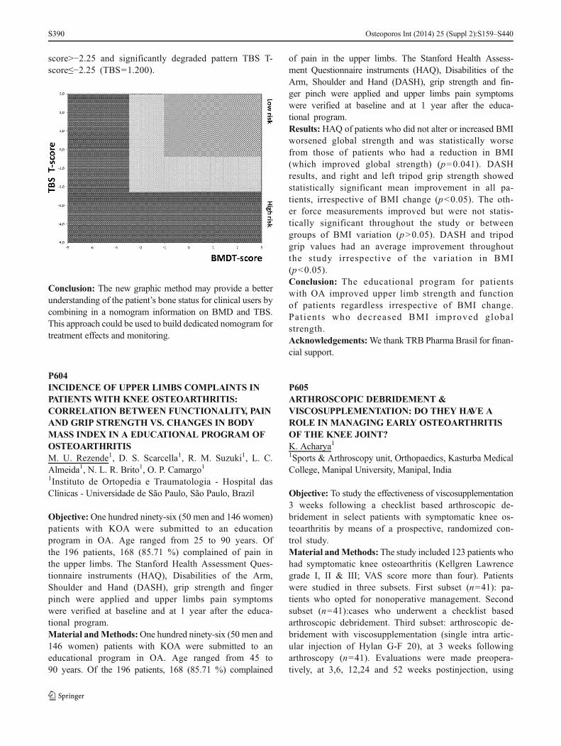
score>−2.25 and significantly degraded pattern TBS T-score≤−2.25 (TBS=1.200).
Conclusion: The new graphic method may provide a betterunderstanding of the patient’s bone status for clinical users bycombining in a nomogram information on BMD and TBS.This approach could be used to build dedicated nomogram fortreatment effects and monitoring.
P604INCIDENCE OF UPPER LIMBS COMPLAINTS INPATIENTS WITH KNEE OSTEOARTHRITIS:CORRELATION BETWEEN FUNCTIONALITY, PAINAND GRIP STRENGTH VS. CHANGES IN BODYMASS INDEX IN A EDUCATIONAL PROGRAM OFOSTEOARTHRITISM. U. Rezende1, D. S. Scarcella1, R. M. Suzuki1, L. C.Almeida1, N. L. R. Brito1, O. P. Camargo11Instituto de Ortopedia e Traumatologia - Hospital dasClínicas - Universidade de São Paulo, São Paulo, Brazil
Objective: One hundred ninety-six (50 men and 146 women)patients with KOA were submitted to an educationprogram in OA. Age ranged from 25 to 90 years. Ofthe 196 patients, 168 (85.71 %) complained of pain inthe upper limbs. The Stanford Health Assessment Ques-tionnaire instruments (HAQ), Disabilities of the Arm,Shoulder and Hand (DASH), grip strength and fingerpinch were applied and upper limbs pain symptomswere verified at baseline and at 1 year after the educa-tional program.Material andMethods:One hundred ninety-six (50 men and146 women) patients with KOA were submitted to aneducational program in OA. Age ranged from 45 to90 years. Of the 196 patients, 168 (85.71 %) complained
of pain in the upper limbs. The Stanford Health Assess-ment Questionnaire instruments (HAQ), Disabilities of theArm, Shoulder and Hand (DASH), grip strength and fin-ger pinch were applied and upper limbs pain symptomswere verified at baseline and at 1 year after the educa-tional program.Results: HAQ of patients who did not alter or increased BMIworsened global strength and was statistically worsefrom those of patients who had a reduction in BMI(which improved global strength) (p=0.041). DASHresults, and right and left tripod grip strength showedstatistically significant mean improvement in all pa-tients, irrespective of BMI change (p<0.05). The oth-er force measurements improved but were not statis-tically significant throughout the study or betweengroups of BMI variation (p>0.05). DASH and tripodgrip values had an average improvement throughoutthe study irrespective of the variation in BMI(p<0.05).Conclusion: The educational program for patientswith OA improved upper limb strength and functionof patients regardless irrespective of BMI change.Pat ients who decreased BMI improved globalstrength.Acknowledgements:We thank TRB Pharma Brasil for finan-cial support.
P605ARTHROSCOPIC DEBRIDEMENT &VISCOSUPPLEMENTATION: DO THEY HAVE AROLE IN MANAGING EARLY OSTEOARTHRITISOF THE KNEE JOINT?K. Acharya11Sports & Arthroscopy unit, Orthopaedics, Kasturba MedicalCollege, Manipal University, Manipal, India
Objective: To study the effectiveness of viscosupplementation3 weeks following a checklist based arthroscopic de-bridement in select patients with symptomatic knee os-teoarthritis by means of a prospective, randomized con-trol study.Material andMethods: The study included 123 patients whohad symptomatic knee osteoarthritis (Kellgren Lawrencegrade I, II & III; VAS score more than four). Patientswere studied in three subsets. First subset (n=41): pa-tients who opted for nonoperative management. Secondsubset (n=41):cases who underwent a checklist basedarthroscopic debridement. Third subset: arthroscopic de-bridement with viscosupplementation (single intra artic-ular injection of Hylan G-F 20), at 3 weeks followingarthroscopy (n=41). Evaluations were made preopera-tively, at 3,6, 12,24 and 52 weeks postinjection, using
Osteoporos Int (2014) 25 (Suppl 2):S159–S440S390

a patient satisfaction questionnaire, VAS, and theWOMAC osteoarthritis index.Results: Mean age: 55±5 years. Percentage of changein VAS was maximum in cases with arthroscopy withviscosupplementation subset. Following viscosupple-mentation, patient satisfaction, WOMAC and VASscores were significantly improved in comparisonwith no injection group. Adverse events were notedin nonoperative subset (gastritis, diarrhea, noncom-pliance for bracing). No significant adverse eventswere noted in other subsets. beneficial effects ofa r th roscopy were pro longed wi th add i t ion ofviscosupplementation.Conclusion: At the end of 1 year suggest thatviscosupplementation following a checklist based arthro-scopic debridement is an effective treatment option forselect patients with knee osteoarthritis. Beneficial effectsof arthroscopic debridement alone are the best at 6–24 weeks postoperatively. The beneficial effects of ar-throscopic debridement were prolonged following addi-tional viscosupplementation. Nonoperative managementhas the risk of noncompliance to the prescribed medi-cations or bracing, adverse events and dependency onanalgesics.
P606IS THE TBS USEFUL FORTREATMENTFOLLOW-UP?S. Di Gregorio11CETIR Grup Medic, Barcelona, Spain
Objective: Facing osteoporosis, clinicians have a huge thera-peutic arsenal available to them. Monitoring treatmenteffects is the key point for patient care. These treat-ments can boost BMD, bone microstructure or both.Until recently, only the BMD evaluation was used tomonitor treatment effects. TBS is an index of bonemicroarchitectural texture extracted from DXA relatedto bone microarchitecture status. The objective of thestudy is to assess longitudinal effects of different oste-oporosis treatments on lumbar spine.Material andMethods:We analyzed 301 subjects with basalscan and at least one scan in follow-up with the sametreatment. We excluded 31 subjects because they haveinconsistent data. Finally we analyzed 271 patients(men: 78; women: 193). We divided by treatmentgroups (Age-years; and Follow-up-months showed inTable). The follow-up changes were analyzed by T-Test. Variation in% from baseline were assessed andnormalized at 24 months. (statistical significance wasset at p<0.05)
Results: BMD and TBS evolution irrespectively to the treat-ment are presented Table 1.
Conclusion: Non correlated effects have been observed onBMD and TBS irrespectively to the treatment. As expectedfrom the literature, TBS of non-treated subjects decreasedwith age. Among these treatments, patients under denosumabexhibit a significant TBS increase whereas no significanteffects have been observed under risedronate and alendronatewhich is consistent with previous presented data. TBS couldbe better to select patient’s treatment.
P607CANTRABECULARBONE SCOREDIFFERENTIATEWOMENWITHANDWITHOUT FRAGILITY FRACTURES?M. R. Mascarenhas1, A. P. Barbosa1, A. A. Gonçalves2, V.Simões3, D. Hans4, M. Bicho51Endocrinology, Diabetes & Metabolism Department, SantaMaria University Hospital, CHLN, Lisboa, Portugal, 2Endo-crinology, Diabetes & Metabolism Department, Santa MariaUniversity Hospital / CHLN, EPE, Lisboa, Portugal, 3Endo-crinology and Metabolic Diseases of Lisbon’s Faculty ofMedicine, CEDML - Lisbon’s Endocrinology, Diabetes andMetabolism Clinic (Osteoporosis Unit), Lisboa, Portugal,4Center of Bone Diseases, Lausanne University Hospital,Lausanne, Switzerland, 5Endocrinology and Metabolic Dis-eases of Lisbon’s Faculty of Medicine (Genetic Lab), Lisboa,Portugal
Objective: Bone mass and microarchitecture can influencebone strength. The extreme fragility of the bones due to thediminished mass and the deterioration of microarchitecturemay originate an augmented risk of osteoporotic fractures,mainly at the distal radius, at the spine and at the hip, also
Osteoporos Int (2014) 25 (Suppl 2):S159–S440 S391

associated with increased morbidity and mortality. At thisdate, DXA scan is the gold standard to measure BMD, inorder to diagnose preciously osteoporosis and estimate frac-ture risk. The decrease in the BMD is positively related to therisk of osteoporotic fractures. TBS, a novel gray-level mea-surement of lumbar spine DXA image texture, is related tomicroarchitecture and fracture risk independently of BMD.The aim of this study was to compare the lumbar spine bonemicroarchitecture as estimated by TBS with the spine BMDscans and the correlation between spine TBS and BMD inpost-menopausal women with and without fragility fractures.Material and Methods: BMD (g/cm2) at the lumbar spinewas measured by DXA (Discovery W, Hologic Inc., USA) in155 women with (fracture group) and without osteoporoticfractures (no fracture group). Site matched spine TBS wasderived for each lumbar spine DXA scan (TBS iNsight soft-ware, Medimaps, France). Adequate statistical tests were usedwith the significance level at P<0.05.Results: No significant difference was detected between thetwo groups for the mean age, mean height, mean weight andmean BMI. The mean BMD was identical between thesegroups of women, while the TBS was lower in the fracturegroup, as compared with the other group. No significantrelationship between spine TBS and BMD, in the fracturegroup was detected.Conclusion: In the osteoporotic fracture group the lumbarspine TBS was significantly low; however, an overlap of theBMD values was observed in both groups, not differentiatingwomen with and without fragility fractures. These data mayindicate that TBS can complement the BMD lumbar spinescans in the clinical routine osteoporosis management.
P608THE RESULTS OF TOTAL KNEE PROSTHESIS INPATIENTS WITH GONARTHROSISS. Lapshina1, I. Akhtyamov1, L. Myasoutova1, I.Gilmutdinov21Kazan State Medical University, Kazan, Russian Federation,2Republican Clinical Hospital, Kazan, Russian Federation
Objective: To analyze the results of total knee prosthesis TKPin patients with rheumatic diseases.Material and Methods: 48 patients (42 women, 6 men) with3–4 radiographic stage of gonarthrosis and persistent jointpain, average age 63.7±19.9 years. The first group consistedof 24 patients with primary osteoarthritis (OA). The second(n=24) group with knee OA on the background of rheumatoidarthritis (RA). In each group, 12 (50 %) patients were treatedwith nonsteroidal anti-inflammatory drugs (NSAIDs), course2 days before surgery and after surgery in a stable averagedaily dose of diclofenac, the other half of patients receivedNSAIDs on demand only after surgery. Before surgery, and
after the 6 months joint pain (VAS), functional capacity indexHAQ and WOMAC were assessed.Results: After 6 months from VAS decreased 2.8 times to37.9±10.7 mm in OA group and 2.5 times at 32.1±19.3 mm(p<0.05) in patients with RA. HAQ decreased from 1.4±0.7to 0.9±0.6 (p<0.05) in the first group and from 1.6±0.9 to 1.0±0.8 (p<0.05) in the second group, WOMAC score wassignificantly (p<0.05) decreased from 55.1±9.1 to 18.8±12.1 and 53.1±10.7 to 18.3±11.8 to the first and secondgroups, respectively. In patients with primary OA receivingNSAIDs before surgery, a positive trend in relation to pain andjoint function was greater, but not significantly. In patientswith RA treated with NSAIDs in the preoperative period, aVAS decrease was −36.8±18.9 mm, HAQ=−0.7±0.3, whichsignificantly (p<0.05) compared VAS those treated withNSAIDs alone after surgery on demand pain VAS=−27.9±19.7 mm, HAQ=−0.5±0.2. Postoperative complications wererecorded.Conclusion: Total knee prosthesis is an effective method toimprove functional capacity, relief of pain in both primary andsecondary gonarthrosis. Preventive NSAID analgesia has ad-vantages over NSAIDs only after demand operation, especial-ly when the secondary gonarthrosis amidst inflammation inrheumatoid joints.
P609INDICATORS OF PHYSICAL ACTIVITYAND THEIRINFLUENCE ON MORTALITY OF PATIENTS WITHHIP FRACTURESO. Ershova1, O. Ganert2, A. Degtyarev1, K. Belova1, M.Belov1, O. Sinitsyna2, M. Romanova31Solovev’s Hospital, Yaroslavl, Russian Federation,2Yaroslavl Medical Academy, Yaroslavl, Russian Federation,3Regional Hospital, Yaroslavl, Russian Federation
Objective: The analysis of indicators of physical activity andits influence on survival during 1 year after a hip fracture inpatients 50 years and older.Material and Methods: A prospective study included allinhabitants of Yaroslavl at the age of 50 years and older, withhip fractures occurred from 01.09.2010 to 31.08.2011. Indi-cators of physical activity were walking duration per day, andalso according to the questionnaire by Katz Activities of dailyliving (ADL). For an assessment of survival Kaplan-Mayer’smethod was used.Results: The study included 446 patients: 334 (74.88 %)women, 112 (25.12 %) men (middle age was 76.83±10.32 years). The general mortality during 12 months was37.54 %. The mortality was higher at patients who could walk≤30min/day before the fracture (p=0.02), ≤30min/day after atrauma (p<0.0001), had low values on a scale of daily activityof Katz (p<0.0001). Among the survived patients confined to
Osteoporos Int (2014) 25 (Suppl 2):S159–S440S392

a bed after during 1 year were 20 (8.90 %), among diedpatients 61 (81.30 %) people, p<0.05. Among the survivedpatients could go outside 127 (56.95 %), among died patientsnobody could go outside, p<0.05. Among those who wentoutside to a trauma, 109 (68.60 %) patients returned to formerlevel of activity, confined to a bed remained 9 (5.66 %)patients. Among the patients who were not going outsidebefore the trauma, in 1 year confined to a bed were 14(20.89 %) patients (p<0.05), 40 (59.7 %) returned to formerlevel of activity (p<0.05). The share of the patients with dailyactivity corresponded as levels A and B (independence in themajority of functions on self-service) in 3 months was50.00 %, and in 12 months - 77.02 %, p<0.05.Conclusion: The mortality among the patients with hip frac-tures during a year was 37.54 %. Higher rates of mortality wasconnected with low level of physical activity. Indicators ofphysical activity were higher at the patients who were goingoutside before fracture.
P610ASSOCIATION BETWEEN SKELETAL MUSCLEMORPHOMETRIC INDICES AND BODYCOMPOSITION IN MEN WITH SARCOPENIAA. Mastaviciute1, V. Alekna1, M. Tamulaitiene1, A.Laurinavicius2, D. Petroska2, V. Strazdiene31Faculty of Medicine, Vilnius University, Vilnius, Lithuania,2National Center of Pathology, Vilnius, Lithuania, 3NationalOsteoporosis Center, Vilnius, Lithuania
Objective: Sarcopenia is important cause of frailty, disabilityand loss of independence in the elderly. There are not suffi-cient data about muscle fiber composition and morphology inelderly men with sarcopenia. The aim of the study was toanalyze the associations of histomorphometric indices of skel-etal muscle with body composition in men with age-relatedsarcopenia.Material and Methods: This is a cohort study on men aged70 years and more. Total fat mass, lean mass, and BMD wereestimated by iDXA (GE Lunar). Sarcopenia was defined asappendicular skeletal muscle mass divided by stature squared<7.26 kg/m2. Skeletal muscle needle biopsy (16 gauge) wastaken to analyse possible morphological and histochemicalchanges in myocites. Samples were frozen and stored inliquid nitrogen, the sections were made by cryostat.Stainings with haematoxylin/eosin (H/E), oil red O(ORO), acid phosphatase (AF), cytochromoxydase/succinildehidrogenase (COX/SDH), and adenosinetriphosphatase (ATP) were made.Results: Thirty skeletal muscle microbiopsies were per-formed on sarcopenic men. On average, the sample waschosen for further analysis of 100 fibers. The average cross-sectional area - 2110±125.6 μm2. The cross-sectional area of
fiber was significantly positively associated with total bodylean mass (r=0.86, p=0.02), leg muscle mass (r=0.84, p=0.03) and weakly associated with total hip BMD (r=0.08, p=0.05). No correlations between fat mass and skeletal musclehistomorphometric indices were found. There was no corre-lation between fiber type and body composition in men withage-related sarcopenia.Conclusion: Our findings demonstrate that cross-sectionalarea of muscle fiber of musculus vastus lateralis is positivelyassociated with total body lean mass, leg muscle mass, andweakly associated with total hip BMD inmen with age-relatedsarcopenia.Acknowledgements: We would like to thank ElvyraStapcinskiene, Audrone Misiunaite and Edita Merkeliunienefor valuable assistance in this study.
P611RELATIONSHIP BETWEEN PHYSICAL ACTIVITY,HANDGRIP STRENGTH, FEAR AND RISK OFFALLING IN INSTITUTIONALIZEDOLDERADULTSM. Tamulaitiene1, A. Mastaviciute1, V. Strazdiene2, A.Cernovas3, V. Alekna11Faculty of Medicine, Vilnius University, Vilnius, Lithuania,2National Osteoporosis Center, Vilnius, Lithuania, 3Healthand Sport Centre, Vilnius University, Vilnius, Lithuania
Objective: To investigate relationship between physical ac-tivity, handgrip strength, fear and risk of falling in institution-alized older adults.Material and Methods: This cross-sectional study was con-ducted on nursing home residents (in Vilnius, Lithuania) aged65 years and older. Exclusion criteria were musculoskeletal ornervous system diseases or conditions that restricted move-ments in the upper or lower extremities. Physical activity wasassessed using Rapid Assessment of Physical Activity(RAPA) questionnaire. Muscle strength was assessed byhandgrip strength (HGS) which was measured using a hand-held dynamometer. Fear and risk of falling were measuredusing Falls Efficacy Scale-International (FES-I) and MorseFall Scale (MFS). Data were analyzed using SPSS 18.0 forWindows program.Results: The study population consisted of 20 men and 37women with a mean age of 80.5±6.8 years, the youngestparticipant was 67 years old, the oldest—95 years old. Inorder to compare different level of HGS the score cutpointof 50th percentile was established. According to this cutpointmen and women were divided into a weak (men ≤20 kg,women ≤10 kg) and strong (men ≥20 kg, women ≥10 kg)HGS groups. The significant differences of MFS score be-tween weak and strong HGS women groups (p=0.05) wasfound greater MFS score in weak group. The study datashowed positive correlation between age and FES-I score in
Osteoporos Int (2014) 25 (Suppl 2):S159–S440 S393

women (r=0.35, p=0.04). In women with weak HGS, nega-tive strong (r=−0.57, p=0.01) correlation between RAPA andMFS scores was observed. In men with weak HGS, negativestrong correlation (r=−0.64, p=0.03) between RAPA and agewas found.Conclusion: Our study showed the positive correlation be-tween age and fear of falling in women, and negative correla-tion between age and physical activity in men with weakhandgrip strength. Negative strong correlation between phys-ical activity and risk of falling was observed in women withweak handgrip strength.Acknowledgements: We would like to thank nursing home“Antaviliai” staff for assistance of this study.
P612TREATMENT OF POSTMENOPAUSALOSTEOPOROSIS: STATE OF THE PROBLEM INBELARUSA. V. Rudenka1, A. Adamenka21Belarussian Medical Academy of Postgraduate Education,Minsk, Belarus, 2Minsk City Center of Osteoporosis, Minsk,Belarus
Objective: To investigate the characteristics of the treatmentof postmenopausal osteoporosis in Belarus.Material and Methods: 376 postmenopausal women (meanage 61.1±8.35 years) living in different regions of the countrywere randomly chosen for participation in the study. Measure-ment of BMD at lumbar spine and femurs was performed bythe method of DXA (Lunar Prodigy, GE, USA). Evaluation ofrisk factors of osteoporosis and fragility fractures was per-formed by questioning.Results: According to the obtained clinical and instrumentaldata 195 (51.9 %) of the examined had osteoporosis, 69(18.4 %) - osteopenia and normal showings of BMDwere detected in 112 (29.7 %) women. 85 (22.6 % ofall the examined and 43.6 % of osteoporotic women)had low-energy fractures in anamnesis. The proportionof patients with a history of fragility fractures, to whomantiosteoporotic treatment was prescribed, was 50.5 %(43 patients). Among all the patients with diagnosedosteoporosis only 73 (37.5 %) received antiosteoporotictreatment, 43 (58.9 %) of them were receiving onlycalcium and vitamin D supplementation, 2 (2.7 %) -calcium with strontium ranelate and 28 (38.3 %) -calcium and bisphosphonates.Conclusion: The majority of postmenopausal women withestablished osteoporosis in Belarus do not receiveantiosteoporotic drugs, and more than 50 % of patientsassigned to treatment only take calcium supplements in com-bination with vitamin D.
P613CARTILAGE-SPECIFIC DELETION OF MTORUPREGULATES AUTOPHAGYAND PROTECTSMICE FROM OSTEOARTHRITISM. Kapoor1, F. Vasheghani1, Y. Li1, M. Blati1, H. Fahmi1, B.Lussier1, J.-P. Pelletier1, J. Martel-Pelletier1, Y. Zhang11Osteoarthritis Research Unit, University of MontrealHospital Research Centre (CRCHUM), Montreal,Quebec, Canada
Objective: mTOR (a serine/threonine protein kinase) is amajor repressor of autophagy, a cell survival mecha-nism. The specific in vivo mechanism of mTOR signal-ling in OA pathophysiology is not fully characterised.We determined the expression of mTOR and knownautophagy genes in human OA cartilage as well asmouse and dog models of experimental OA. We createdcartilage-specific mTOR knockout (KO) mice to deter-mine the specific role of mTOR in OA pathophysiologyand autophagy signalling in vivo.Material and Methods: Inducible cartilage-specificmTOR KO mice were generated and subjected tomouse model of OA. Human OA chondrocytes weretreated with rapamycin and transfected with Unc-51-like kinase 1 (ULK1) SiRNA to determine mTORsignalling.Results: mTOR is overexpressed in human OA cartilage aswell as mouse and dog experimental OA. Upregulationof mTOR expression co-relates with increased chondro-cyte apoptosis and reduced expression of key autophagygenes during OA. Subsequently, we show for the firsttime that cartilage-specific ablation of mTOR results inincreased autophagy signalling and a significant protec-tion from destabilization of medial meniscus (DMM)-induced OA associated with a significant reduction inthe articular cartilage degradation, apoptosis and syno-vial fibrosis. Furthermore, we show that regulation ofULK1/adenosine monophosphate-activated protein ki-nase (AMPK) signalling pathway by mTOR may in partbe responsible for regulating autophagy signalling andthe balance between catabolic and anabolic factors inthe articular cartilage.Conclusion: This study provides a direct evidence of the roleof mTOR and its downstream modulation of autopha-gy in articular cartilage homeostasis. Targeting cellu-lar homeostasis mediators, such as mTOR and itsdownstream signaling by autophagy pathway may bea p romi s i ng the r apeu t i c s t r a t egy to ach i evechondroprotection and correct the imbalance betweencatabolic and anabolic processes during OA and re-lated disorders.Acknowledgements: This work was performed with CIHROperating Grant Funded to Mohit Kapoor.
Osteoporos Int (2014) 25 (Suppl 2):S159–S440S394

P614EFFECT OF COFFEE AND CALCIUMADMINISTRATION IN THE BONE QUALITYA. C. O. Penoni1, B. E. B. Hilário2, M. M. Borges2, D. P.Alvarenga2, G. R. Yanagihara1, A. C. Shimano1,M. J. QuirinoLouzada3, B. R. S. Oliveira4, P. F. P. Goulart51Department of Biomechanics, Medicine and Rehabilitation ofthe Locomotor System, Ribeirão Preto, Brazil, 2Department ofPhysiotherapy, University Center of Lavras, Lavras, Brazil, 3Uni-versity of São Paulo, Ribeirão Preto, Brazil, 4State UniversityPaulista - UNESP - Araçatuba Faculty of Veterinary Medicine,Araçatuba, Brazil, 5University Center of Lavras, Lavras, Brazil
Objective: To verify if coffee intakes in moderate shotschange bone remodeling, what it would impact on BMD, inbiochemical and biomechanical characteristics of bone tissue.Material and Methods: 24 male rats 60 days old Wistar weredivided by three experimental groups, making the treatment:Group 1 (control), Group 2 (coffee - 3 ml/kg), Group 3 (coffee- 3 ml/kg + 5 mg calcium) and Group 4 (5 mg calcium). After56 days of tests, the animals were anesthetized, it was collected5ml of blood trough cardiac puncture and analyzed the values ofglucose and ionized calcium. After procedure, the animals wereeuthanized and necropsied, dissecting both femurs. The leftfemurs were submitted to analysis densitometric and right tobending of head flexion and compression of middle third. Bio-mechanically, it determined maximum strength and rigidity.Results: The results showed smaller ionic calcium concentra-tion in the ionic Group in relation to experimental (p=0.0000).The studied glycemic analysis and biomechanical variables hadnot showed significant differences between the groups. It con-cludes that coffee intakes in moderate shots can take to BMDdiminish without changing bone biomechanical properties andsimultaneous calcium consumption does not change this result.Conclusion: The daily calcium consumption tends to keepionic calcium level lower, however it is not determinant tointerfere beneficially on BMD.
P615THE RELATIONSHIP BETWEEN HEIGHT, WEIGHT,AGE AND T-SCORE IN PATIENTS 50 YEARS ANDOLDERH. Ramanau1, E. V. Rudenka2, N. Predko2, A. V. Rudenka2,O. Fomin31Gomel State Medical University, Gomel, Belarus, 2Belarus-sian Medical Academy of Postgraduate Education, Minsk,Belarus, 3Clinical Hospital No. 1, Minsk, Belarus
Objective: To examine the relationship between Т-score oflumbar spine (LS) and femoral neck (FN) and anthropometricdata in patients of both sexes aged over 50 years.
Material and Methods: There were examined 14,947 pa-tients aged over 50 years, 13,886 (92.9 %) women and 1,061(7.1 %) men. T-score was assessed with DXA by LunarProdigy in LS (L1-L4) and FN. The response variable Т-scorewas assessed by the method of multiple linear regression withstepwise inclusion in the model and test for colinearity usingtest Durbin-Watson according to the known values of severalpredictor variables, selected using correlation analysis.Results: Mean age in the examined women was 65.6±9.6 years (95%CI 65.0; 662), and in men - 65.4±9.0 years(95%CI 65.3; 65.6). 23.3 % of patients were diagnosed tohave osteoporosis, 48.1 % - osteopenia and 28.6 % hadnormal values of BMD. The anthropometric data in men andwomen had statistically significant differences in height(169.9±7.1 vs. 157.7±6.3; р<0.001), and weight (80.0±14.9 vs. 74.7±14.4; р<0.001), thus the regression analysiswas performed separately by gender. Constructed multiplelinear regression model revealed the relationship betweenthe values of T-score and anthropometric data. The variabilityof the values of LS T- score can be explained by the meaningsof height, weight and age in men and women by 19 % and21 %, respectively. The calculated predictive value of LS T-score in women was (−4.944) + 0.009*height, cm +0.046*weight, kg - 0.018*age, years; in men - (−6.702) -0.001*height, cm + 0.055*weight, kg + 0.031*age, years.The variability of FN T-score was determined by 20 % and36 % in men and women with the same predictor variables.Predictive value of FN Т-score in women was (−2.803) +0.016*height, cm + 0.028*weight, kg - 0.046*age, years; inmen - (−3.197) + 0.003*height, cm + 0.032*weight, kg -0.019*age, years.Conclusion: The proposed model allows to predict the valuesof LS and FN T-score on the basis of anthropometric data inpersons aged over 50 years of both sexes.
P616CHANGES IN APPENDICULAR BODYCOMPOSITION CAN BE DETECTED BYPERIPHERAL COMPUTED TOMOGRAPHYALREADYAT THE INITIAL STAGE OFRHEUMATOID ARTHRITISR. Müller1, M. Kull1, A. Aart1, M. Lember1, R. Kallikorm1
1University of Tartu, Tartu, Estonia
Objective: Rheumatoid arthritis (RA) causes persistent de-structive changes in joints and bone. Detection of early struc-tural damage is vital to predict further joint destruction. In thecourse of RA a shift in body composition (BC) occurs—leantissue is substituted with fat while maintaining stable bodyweight. Our objective was to assess the ability of pQCT todetect a change and quantify the effect of RA on BMD and
Osteoporos Int (2014) 25 (Suppl 2):S159–S440 S395

hand BC parameters in a prospective study of early RApatients.Material and Methods: We recruited 32 patients with earlyRA into the study. Radius trabecular BMD and area, corticalBMD and area, muscle and fat tissue density, cross-sectionalarea and ratio were measured with Stratec XCT2000 pQCTmachine (Germany) twice (at diagnosis and 1 year follow-up).Patients underwent a laboratory panel and thorough physicalexamination. DAS 28(CRP) value was used to categorizepatients to moderate-high (>3.2) and low (<3.1) disease activ-ity groups. Changes in and differences between patient groupswere assessed by Wilcoxon signed-rank test. The study wasIRB approved.Results: Mean patient age was 51.8 years (range 23–80),median time from symptom onset 91 (range 9–900) days,82 % were female. A 25 % increase in fat area (p=0.01) and11 % decrease in fat/muscle ratio (p=0.03) was seen in themoderate-to-high disease activity group. No BC changes weredetected in low disease activity group. Trabecular density wasthe sole bone parameter where decrements during the firstyear after disease onset could be detected (−3.5 %, p=0.05).Conclusion: pQCT detects significant body compositionchanges (most significant in the body fat compartment) inearly RA patients already 1 year after diagnosis.
P617BONE MINERAL DENSITY, 25(OH)D AND BONETURNOVER MARKERS IN PATIENTS WITHPSORIATIC ARTHRITISV. Samakhavets1, E. V. Rudenka11City Centre of Osteoporosis, Minsk, Belarus
Objective:The aim of this study was to assess BMD and boneturnover markers in patients with PsA, and to investigate therelationship between clinical parameters and markers of boneturnover.Material andMethods: 71 patients with PsA (32 men and 39women; mean age 49.22±10.34 years; mean duration of PsA9.4±5.9 years) without the history of any treatment includingglucocorticoid intake and 76 healthy controls (28 men, 48women, mean age 47.8±9.43 years) were included in thestudy. Laboratory assessment included evaluation of ECRand C-reactive protein, serum Ca, P, alkaline phosphatase(ALP), serum type I collagen cross-linked C telopeptide(CTX), and 25(OH)D level. Serum levels of 25(OH)D andbone turnover markers were determined using the chemilumi-nescent assay (analyzer - Cobas e 411). BMD was measuredby DXA at lumbar spine (anterior-posterior projection at L1-L4) and femurs. Statistical analysis was performed using theprogram Statistica 8.Results: Decreased BMD was detected in 33 patients withPsA: 18 patients (25 % of total) had osteoporosis and 15
patients (20 %) - osteopenia. Showings of BMD in controlgroup were normal. Mean levels of 25(OH)D in patients withPsAwere 18.44 [9.90; 27.73] ng/ml and 21.31 [15.79; 30.39]ng/ml in control group, but the differences were not statisti-cally significant. VitaminD deficiency was found in 66 (92%)of PsA patients and in 61 (80 %) of controls. Serum CTX,ALP, Ca were not significantly different in arthritic patientsand controls (p>0.05). There were no revealed any correla-tions between vitamin D levels disease activity and BMD. Inpatients with PsA, the duration of arthritis was negativelycorrelated with BMD values of lumbar spine and total femurand serum CTX levels, suggesting an association of increaseddemineralization with the duration of joint disease.Conclusion: Patients with PsA and longer duration of jointdisease may be at risk for osteoporosis, which can requirepreventative treatment efforts.
P618UNDERSTANDING BETWEEN PHYSICIAN ANDPATIENT WITH OSTEOARTHRITIS AS A KEYFACTOR IN INCREASINGTHE EFFECTIVENESS OFTHERAPYS. Lapshina1, L. Myasoutova11Kazan State Medical University, Kazan, Russian Federation
Objective: To identify the most significant problems for thepatient with osteoarthritis (OA) in order to create a system ofeffective interaction between physician and patient in future.Material and Methods: 50 patients with confirmed OAdiagnosis (the average duration of the disease 1245±5.53 years, the average age of patients 49.5±15.82 years)responded to 10 questions concerning their relationship tothe disease and the impact on the daily life with the need toevaluate the importance of each question on a scale from 1 to10. Rheumatologists (n=10) and physicians (n=40) answeredthe same questions from their point of view, what is importantfor patients. Responses were ranked and compared.Results: Physicians and patients opinions coincided in an-swering the most important question—the forecast for thefuture in relation to the possibility of movement and overallhealth. The second most important for patients was the cost oftreatment, the third—understanding the causes of the diseaseand its consequences. Thereafter, patients worried about con-stant pain and the need to constantly take drugs and possibleside effects. In contrast, doctors believed that patients areworried about limitations in everyday life and disability, per-sistent pain. To a lesser extent they are concerned about thecauses of disease and the cost of therapy.Conclusion: Understanding the patients opinion to their dis-ease will allow to restructure the communication with thepatient, educational programs for them. Physician must paymore attention to the explanation of the causes of disease, the
Osteoporos Int (2014) 25 (Suppl 2):S159–S440S396

effectiveness and safety of therapy that will improve thequality of life of patients.
P619LEFLUNOMIDE TREATMENTAND ITS EFFECTSON SERUM 25-(OH)D LEVELS IN WOMEN WITHRHEUMATOID ARTHRITISV. tepa1, L. Groppa2, E. Russu2, N. Ganea31Department of Rheumatology and Nephrology of The StateUniversity of Medicine and Pharmacy, Chisinau, Republic ofMoldova, 2Department of Rheumatology and Nephrology,SMPU, Chisinau, Republic of Moldova, 3Rheumatology andArthrology Laboratory, Clinical Republican Hospital,Chisinau, Republic of Moldova
Objective: Leflunomide is an immunomodulatory drug thatinhibits dihydroorotate dehydrogenase (an enzyme involvedin pyrimidine synthesis), and is used for the treatment ofpatients with moderate to severe rheumatoid arthritis (RA).The aim of this study was to assess the effects of DMARDtreatment with Leflunomide on serum concentrations of 25-hydroxyvitamin D (25-(OH)D).Material and Methods: Thirty-five women were included inthe study, mean age 48.4 years, mean disease duration8.6 years. All of them received leflunomide treatment.Blood samples were taken and the following indicesassessed before treatment and at 6 months: serum25-(OH)D, C-reactive protein (CRP), rheumatoid factor(RF) and anticyclic citrullinated peptide (anti-CCP).During the clinical examination at the 2 checkpointsDAS-28 was calculated. Radiographic images beforetreatment and at 6 months were assessed using theSharp van der Heijde Score for joint damageprogression.Results: As was to be expected, after 6 months of DMARDtreatment DAS-28, CRP, anti-CCP and RF concentrationsdecreased considerably. Sharp van der Heijde Score showedno significant changes after 6 months of leflunomide treat-ment. All 35 patients had decreased 25-(OH)D serum levels(below 50 nmol/l) before starting DMARD therapy. No sta-tistically significant difference was found in comparing25-(OH)D serum levels before and after 6 months ofLeflunomide therapy. After second assessment the majorityof them had to be considered for supplementation in order toavoid complications.Conclusion: This study showed that 6 months Leflunomidetreatment had an insignificant effect on serum 25-(OH)Dlevels. No other associations between 25-(OH)D levels andmarkers as CRP, RF or anti-CCP were found. Further studiesusing larger patient lots could show different results, and areneeded for a definite conclusion.
P620EPIDEMIOLOGYOFOSTEOPOROSISANDRISKOFFRACTURES IN REPUBLIC OF BELARUSH. Ramanau1, E. V. Rudenka21Gomel State Medical University, Gomel, Belarus, 2Belarus-sian Medical Academy of Postgraduate Education, Minsk,Belarus
Objective: The objective of this study was to define theincidence of osteoporosis and risk of fractures by age andBMD for Belarusian population.Material and Methods: The measurements of BMD weredone in the lumbar spine (LS) and femoral neck (FN) by DXA(Lunar Prodigy, GE, USA) for 14,947 individuals of 50 yearsand older.Results: It was revealed 1,003 vertebral (VF) and 2,636nonvertebral fractures (NF) in the study group. Osteoporosiswas verified in 24.1 % of patients: at the age group 50–60 years in 13.3 % cases, 60–70 years - 22.2 %, 70–80 yearsin 34.0 % and for patients aged over 80 years in 47.6 % cases.The frequency of VF by age groups 50–60, 60–70, 70–80 and80+ years old was 3.1 %, 6.2 %, 12.0 % and 14.8 %, respec-tively. The odds ratio (OR) of VF for patients with normalBMD in different age groups was 2.25 (95%CI 1.34–3.72),3.33 (95%CI 1.90–5.80) and 2.97 (95%CI 1.08–10.56) incomparison with patients of 50–60 years. The risk of VFincrease for patient with OP depending on age: for age group50–60 OR=5.71 (95%CI 3.44–9.31), 60–70 years OR=9.48(95%CI 6.04–14.44), 70–80 years OR=14.31 (95%CI 9.27–21.32) and the highest OR=15.79 (95%CI 9.73–24.92) forpatients aged over 80 years. The NF frequency is higher thatVF and was 15.0 %, 23.9 %, 30.8 % and 35.2 %, respectively,by different age groups. The odds ratio (OR) of NF for patientswith normal BMD in different age group was 1.39 (95%CI1.10–1.74), 1.49 (95%CI 1.13–1.98) и 1.75 (95%CI 0.97–3.36) in comparison with patients aged 50–60. The risk of NFfor patient with OP by age groups is 2.45 (95%CI 1.90–3.15),3.42 (95%CI 2.77–4.22), 3.74 (95%CI 3.07–4.56) and forpatients aged over 80 years OR=3.75 (95%CI 2.82–4.99).Conclusion: The prevalence of OP in the Republic of Belarusin patients over the age of 50 years is 24.1 % with a maximumfrequency at the age of 80 years and older. The age and BMDare significant risk factors for vertebral and nonvertebral frac-tures, which should be considered for predicting fractures anddetermining the treatment plan.
P621THE INFLUENCE OF UPPER LIMBS DISORDERSON SELF ESTEEM AND QUALITY OF LIFEC. Suteu1, D. M. Farcas21Occupational Medicine, Faculty of Medicine and Pharmacy,Oradea, Romania, 2Physical Rehabilitation, Faculty of
Osteoporos Int (2014) 25 (Suppl 2):S159–S440 S397

Medicine and Pharmacy, Oradea, Romania
Objective: To assess self-esteem and quality of life in patientswith upper limbs disorders.Material and Methods: Our group of study consisted in 108patients with upper limbs disorders. It was divided in twosubgroups, first of 54 patients (29 women/25 men), meanage 48.2±11.4 who underwent a complex rehabilitation treat-ment and the second subgroup of 54 patients (28 women/26men), mean age 49.7±3.4, with no treatment. All patientswere recruited from ambulatory system Bihor county, Roma-nia. The main characteristics of the groups were similar inboth groups. The inclusion criteria were: active subjects withspecific jobs, complying with the principles of medical ethics.The exclusion criteria were: retired subjects, severe diseases,noncompliance. All the patients were assessed with ShortForm 36 Questionnaire and Rosenberg Self Esteem Scale atthe entrance in the study and 6 months later.Results: In the subgroup of patients who underwent rehabil-itation program both Physical Component Summary andMental Component Summary of the SF 36 improved at6 months than in the second subgroup of sedentary patientswhich showed no improvement in quality of life. Also self-esteem improved in the group of patients included in rehabil-itation program after 6 months than in the sedentary group ofpatients which did not modify at the end of the study.Conclusion:Our study showed that patients with upper limbsdisorders have a low self-esteem and an impaired quality oflife. Rehabilitation program played a beneficial role on bothself-esteem and quality of life of such patients.
P622KNEE OSTEOARTHRITIS DIAGNOSTIC GAP:MOVING BEYOND CLASSICAL RISK FACTORSD. Simões1, R. André2, L. Carmona3, R. Lucas11Institute of Public Health of the University of Porto (ISPUP),Porto, Portugal, 2Hospital Beatriz Ângelo, Loures, Portugal,3Institute for Musculoskeletal Health, Madrid, Spain
Objective: To study determinants of misdiagnosis in kneeosteoarthritis (OA).Material and Methods:We used data from a national healthsurvey (Portugal, 2005–2008). Subjects were inquired aboutknee OA diagnosis by self-report. Subsequently, each partic-ipant answered 6 questions about nontraumatic joint pain.These items were used to build a validated algorithm to selectparticipants for evaluation by a rheumatologist. Evaluationcomprised clinical history, physical examination and radio-graphic evaluation. Individuals were classified according tothe American College of Rheumatology clinical criteria forOA. From the original sample, 265 participants were classi-fied into one of the following groups: accurately reported OA
when the clinician confirmed the presence of self-reportedOA, previously undiagnosed OA when the clinician diag-nosed OA and the participant was unaware of the disease,and over-reported OA when the participant reported a priordiagnosis but there was no clinical confirmation.We estimatedthe risk of misdiagnosis for specific patient characteristicsusing multinomial logistic regression. We present odds ratios(OR) and their 95% confidence intervals (CI95%), adjusted forage, sex, education and BMI.Results: Among all participants clinically-diagnosed, 41.4 %were unaware of the condition. As expected, these had lesssevere radiographic changes and complaints than those whoaccurately reported OA. In participants with clinical disease,underdiagnosed subjects had lower BMI (AdjOR=3.30,CI95%: 1.29–8.45, reference: BMI≥30 kg/m2) and youngerage (AdjOR=1.89, CI95%: 1.01–3.57, reference: age≥70 years) than those previously diagnosed. Sex, education,socioeconomic status and healthcare utilization did not predictunderdiagnosis. Participants over-reporting OAwere not sig-nificantly different from confirmed cases.Conclusion: Narrowing the diagnostic gap to improve man-agement is likely to require searching for less severe cases ofpatients without classical risk factors such as old age andobesity.
P623THE RESULTS OF TOTAL HIP PROSTHESIS INRHEUMATOID ARTHRITIS PATIENTS WITHSECONDARY COXARTHROSISI. Akhtyamov1, S. Lapshina1, L. Myasoutova1, I.Gilmutdinov21Kazan State Medical University, Kazan, Russian Federation,2Republican Clinical Hospital, Kazan, Russian Federation
Objective: To review the results of total hip prosthesis ac-cording to baseline disease activity.Material and Methods: Total hip prosthesis was used on 36RA patients (32 women, 4 men), mean age of 49.5±15.82 years (disease duration - 12.45±5.53 years). High dis-ease activity by DAS28 was observed in 10 patients (27.8 %),moderate - in 19 (52.8 %), low - in 7 (19.4 %) patients, themean DAS28 4.5±1.7. At the time of surgery (before andafter), 24 (66.7 %) of the patients continued to take basicantirheumatic drugs (DMARDs) - (methotrexate 7.5–15 mgper week - 15, leflunomide - 7, sulfasalazine - 2). 6 (16.6 %)patients received biological therapy: infliximab - 2,adalimumab - 1, rituximab - 3, which were cancelled beforethe operation in accordance with the terms specified in thenational guidelines. 19 (52.8 %) patients received steroids, ofwhich, in combination with DMARDs - 7 (19.4 %). Beforethe surgery, in the postoperative period and after 6 monthsVAS, DAS28, index HAQ have been evaluated.
Osteoporos Int (2014) 25 (Suppl 2):S159–S440S398

Results: For 6 months after surgery VAS decreased to 31.7±19.4 mm (p<0.05), disease activity decreased (high - 4(11.1 %), moderate 17 (47.2 %), low -15 (41.7 %)).DAS28=1.1±0.7, HAQ index decreased from 1.63±0.91 to1.04±0.78 (p<0.05). VAS in patients receiving steroids alone(n=12) was - 24.9±18.6 mm, steroids + DMARD (n=7) -29.1±19.7 mm, only DMARDs (n=11) 34.8±18.9 mm, bio-logical therapy + DMARD (n=6) 37.4±20.8 mm. DAS28 -did not depend from treatment. HAQ in patients in the group(n=17), receiving DMARDs with or without biological ther-apy (D HAQ=−0.69±0.34) compared to patients receivingsteroids without basic therapy (n=12) - D HAQ=−0.48±0.31was significantly better improved (p<0.05).Conclusion: Total hip prosthesis is an effective way to im-prove functional capacity, to relieve pain and to reduce theactivity of RA. Functional ability after surgery is higher inpatients using DMARDs continuously and receiving biologi-cal therapy compared to patients receiving steroids.
P624MAY DOXYCYCLINE-RIFAMPICIN COMBINATIONBE THE FIRST-LINE THERAPY FOROSTEOARTICULAR BRUCELLOSIS?Ş. Hocaoğlu1, E. Mesci21Kayseri Training and Research Hospital, Department ofPhysical Medicine and Rehabilitation, Kayseri, Turkey,2Medeniyet University, Göztepe Training and Research Hos-pital, Department of Physical Medicine and Rehabilitation,İstanbul, Turkey
Objective: Brucellosis is an infectious disease which canmimic rheumatoid diseases due to its osteoarticular (OA)involvements. Treatment failure and recurrences have notbeen completely eliminated yet in patients with OA brucello-sis. This study aimed to investigate rheumatologic manifesta-tions observed in patients with OA brucellosis and the out-comes of combination therapy with doxycycline and rifampi-cin (DR) in such patients.Material and Methods: The study enrolled patients whowere diagnosed with brucellosis based on blood cultures andserological examinations conducted for differential diagnosisdue to presence of rheumatologic symptoms. Serological testsand radiological examinations were performed for diagnosisof the OA involvements. As for treatment, DR therapy wasgiven for 45 days to patients with OA involvement other thanspondylitis and for 12 weeks to patients with spondylitis.Clinical and serological evaluations were repeated at 2 weeksand 1, 2, 3, 6 and 12 months after treatment initiation.Results:Mean age of the 49 patients enrolled in the study was44.08±13.04 years. The most common osteoarticular mani-festation was spondylitis which was observed in 17 patients(34.7 %). In addition, 14 patients (28.6 %) had sacroiliitis,
nine patients (18.4 %) had monoarthritis and 3 patients(6.1 %) had oligoarthritis. Complete recovery was observedin all patients based on clinical and serological evaluations.During follow-up visits up to 12 months, none of the patientshad recurrence.Conclusion: Brucellosis should be considered in the differen-tial diagnosis of the rheumatic diseases, mainlyspondyloarthropathies. When administered for a sufficientperiod of time, DR combination is a powerful therapeuticoption for patients with osteoarticular brucellosis includingthose with spondylitis.
P625SERUM SCLEROSTIN IN HEPATITIS C VIRUSINFECTED PATIENTSR. Pelazas González1, J. Alvisa Negrín1, R. Hernández Luis1,C. Jorge Ripper1, E. González Reimers1, D. García Rosado1,C. Fernández Rodríguez1, O. Pérez Hernández1, Y. LalondrizBueno1, G. Quintero Platt1, C. Martin González11Internal Medicine, La Laguna, Tenerife, Spain
Objective: Sclerostin is an endogenous inhibitor of the Wnt/β-catenin pathway secreted by osteocytes. It inhibits osteo-blast function, differentiation, and survival. Theoretical-ly, sclerostin tends to decrease bone mass, althoughseveral studies show opposite results. In addition, itmay be related to insulin resistance and carbohydratemetabolism, as osteocalcin. Hepatitis C virus (HCV)-infected patients may present osteoporosis, and frequent-ly show liver steatosis, in relation with insulin resis-tance. The behaviour of sclerostin in these patients isunknown. The aim of this work was to analyse therelationship between serum sclerostin and osteocalcinlevels and BMD, liver function derangement, the inten-sity of liver steatosis and biochemical markers of bonehomeostasis and insulin resistance in HCV-infectedpatients.Material and Methods: Forty HCV patients and 20 age andsex-matched controls were included and underwent bonedensitometry. Serum sclerostin, osteocalcin, collagentelopeptide, adiponectin, leptin, insulin, resistin,TNF-α, and IL-6 were determined. Liver fat washistomorphometrically assessed.Results: Sclerostin levels were slightly higher in patients thanin controls, and were directly related to BMD at different partsof the skeleton, to serum telopeptide, and to liver steatosis andTNF-α. On the contrary, osteocalcin showed a significantdirect relationship with serum adiponectin, and an inverseone with IL-6.Conclusion: Therefore, serum sclerostin levels were raised inHCV patients, and correlated directly with BMD and serumtelopeptide. In addition direct relationships of sclerostin, but
Osteoporos Int (2014) 25 (Suppl 2):S159–S440 S399

inverse ones of osteocalcin, with variables associated withinsulin resistance suggest a role of bone on intermediarymetabolism.
P626THE INFLUENCE OF SUBCUTANEOUSDENOSUMAB ON THE PERIOSTEUM IN PATIENTSWITH POSTMENOPAUSAL OSTEOPOROSIS:PRELIMINARY RESULTSV. Cazac1, L. Groppa1, E. Russu1, A. Zoltur-Mosneaga21Department of Rheumatology and Nephrology, SMPUNicolae Testemitanu, Scientifico-practical Laboratory ofRheumatology and Arthrology of the Republican ClinicalHospital, Chisinau, Republic of Moldova, 2Excellence Med-ical Center, Chisinau, Republic of Moldova
Objective:To assess the influence of denosumab treatment onthe periosteum in patients with postmenopausal osteoporosis.Material and Methods: A group of 27 patients with post-menopausal osteoporosis, confirmed by lumbar spine and hipDXA, with a T-score of maximum −2.5 was selected. Patientswere evaluated clinically, after which they underwentultrasound examination of the calcaneal periosteum.Using colour and spectral Doppler, the colour fractionand resistance index (an intramuscular thenar artery -sample) were obtained, both at baseline - prior to 60 mgof denosumab, and 6 months after the injection. Thenormal ranges were adopted from data obtained byTerslev L. et al (1). Also a group of 12 healthy femalecontrols were examined at baseline in a similar manner.Statistical analysis was performed using the softwareStatSoft 9.0. A Student’s t-test was used for data eval-uation. The level of significance was p=0.05.Results: In 51.8 % patients at baseline calcaneal periostealhypoechoic thickening was detected, as well ashypervascularization in 62.9 % patients from the study group.In controls, 8.3 % patients had calcaneal periostealt h i c k e n i n g , a n d 1 6 . 6 % h a d p e r i o s t e a lhypervascularization. The mean colour fraction in thestudy group was 0.047±0.16 (range=0.26–0.75) at base-line, compared to a mean of 0.005±0.01 (range=0–0.04)in controls. The mean resistance index was 0.27±0.45(range=0.1–1) at baseline, compared to a mean of 0.95±0.09 (range=0.71–1) in controls. The ultrasound analysisperformed at re-evaluation yielded a significantly lowermean colour fraction of 0.32±0.12 (range=0.2–0.61), p=0.047, and a significantly higher mean resistance indexof 0.54±0.25 (range=0.2–1), p=0.049.Conclusion: Our preliminary data show that treatmentwith denosumab in patients with osteoporosis has abeneficial effect on periosteum, reducing its degree ofvascularization.
References: 1. Terslev L et al. Ann Rheum Dis 2004;63:644
P627DIAGNOSTIC ACCURACY OFA NOVELULTRASOUND-BASED METHODOLOGY FORSPINAL DENSITOMETRY ON A COHORT OFFEMALE PATIENTS (45–65 Y)F. Conversano1, M. Muratore2, R. Franchini1, E. Casciaro1, F.Chiriacò1,M. D. Renna1, A. Greco1, L. Quarta2, E. Quarta2, S.Casciaro11National Research Council, Institute of Clinical Physiology,Lecce, Italy, 2O.U. of Rheumatology, Galateo Hospital, SanCesario di Lecce, ASL-LE, Lecce, Italy
Objective: To test the diagnostic accuracy of a novel ultra-sound (US)-based method to perform spinal densitometrywithout employing X-rays.Material and Methods: A cohort of 345 female patients wasrecruited according to the following criteria: 45–65 years,BMI <25 kg/m2, no severe deambulation impairments, med-ical prescription for a spinal DXA, signed informed consent.All the enrolled patients underwent two examinations: aconventional spinal DXA (Hologic Discovery) and anUS scan of lumbar spine. US data were analyzed by anovel algorithm that processed both echographic imagesand “raw” radiofrequency signals and calculated thesame diagnostic parameters provided by DXA (BMD,T-score, Z-score). Diagnostic accuracy of obtained re-sults was evaluated through a direct comparison withDXA output as a function of patient age.Results: For 88.1 % of the patients US diagnosis (osteoporot-ic, osteopenic, healthy) was the same of the correspondingDXA one. In particular, diagnostic accuracy showed the fol-lowing behavior as a function of patient age range:accuracy was 95.0 % in 45–50 year, 88.9 % in 50–55 year, 92.0 % in 55–60 year and 78.6 % in 60–65 year. Pearson correlation coefficient (r) betweenDXA and US measurements was also evaluated for eachdiagnostic parameter (BMD, T-score, Z-score) for pa-tients in the same age range: all the obtained valuesof r were within the interval 0.63–0.84 (p<0.001) andtheir trends against age qualitatively reflected the ob-served diagnostic accuracy profile.Conclusion: The proposed US approach to spinal bone den-sitometry showed a very good agreement with DXA diagno-ses. This new nonionizing method has the potential for beingextremely useful for early osteoporosis diagnosis throughpopulation mass screenings and for therapeutic outcomemonitoring.Acknowledgements: This work was partially funded byFESR P.O. Apulia Region 2007–2013 - Action 1.2.4, grantn. 3Q5AX31.
Osteoporos Int (2014) 25 (Suppl 2):S159–S440S400

P628PREVALENT FRAGILITY FRACTURES IN 498MENOPAUSALWOMEN AND SKELETONASSESSMENTM. Carsote1, R. Dusceac2, D. Niculescu1, C. Capatana1, R.Danciulescu Miulescu1, D. Opris3, C. Poiana11Endocrine/I.Parhon/Davila UMPh, Bucharest, Romania,2Endocrine/I.Parhon, Bucharest, Romania, 3Rheumatology/Sf. Maria Hospital/Davila UMPh, Bucharest, Romania
Objective: The fragility (osteoporotic) fractures represent aneconomical issue, especially in menopausal women. Thedifferent fracture risk evaluation tools for a future(potential) fragility fracture vary from golden standardDXA, based on BMD to new tools as 10-year absoluterisk of major or hip fracture based on FRAX model(MF or HF). We analyzed the risk of fracture in patientswith or without prevalent fractures based on DXA,FRAX, and bone metabolism profile.Material and Methods: The study design is cross-sectional study. The studied population is Romanianwomen in menopause (for at least 1 year of meno-pause). The persons with previous medication for oste-oporosis as bisphosphonates, SERMs or teriparatide,denosumab, etc. were not included. Neither was thesubjects preciously known with the diagnosis of osteo-porosis. The skeleton assessment consisted in: lumbarDXA (L1-4), with a GE Lunar Prodigy device; MF orHF was calculated based on FRAX for Romania (with-out BMD); serum 25-hydroxyvitamin D (25-OH D); thebone formation marker serum osteocalcin (OC), and thebone resorption markers serum alkaline phosphatase(AP), as well as serum crosslaps (CL). The prevalentfractures were already known from patients’ medicalhistory. The statistical significance (SS) was at p<0.05.Results: 498 women were enrolled. The studied groupincluded women with fractures (n=69); the controlgroup (fracture free) included 429 women. The patients’age and time period in menopause was SS higher ingroup 1 (59.4 vs. 57 years; and 13 vs. 10 years). Thelumbar BMD was SS lower in group 1 (0.96 vs. 1.02 g/cm2, p=0.02); one third of the women with fractureshad osteoporosis and half of them osteopenia. The bonemarkers were not SS different between the groups.Neither was the 25-OH D, the median value for bothgroups were in D deficiency area: 14 ng/mL, respective 15 ng/mL. The HF was SS higher in group 1 (3 % vs. 0.9 %,p<0.0005), and also the MF (9 % vs. 4 %, p<0.0005). Aslimits of the study we mention that fact that we did not takeinto account the number of previous fractures, neither the timesince the fracture.Conclusion: Based on our observations, both DXA and10-year absolute risk of fracture based on FRAX
algorithm pointed statistical significant differences be-tween women with and without prevalent fragility frac-tures, but the bone resorption markers were notdifferent.
P629EFFICACYAND SAFETY OF STRONTIUMRANELATE IN THE TREATMENT OF KNEEOSTEOARTHRITISS. Lapshina1, L. Myasoutova11Kazan State Medical University, Kazan, Russian Federation
Objective: To evaluate the efficacy and tolerability of stron-tium ranelate in patients with gonarthrosis.Material and Methods: 40 patients with a documenteddiagnosis of gonarthrosis: women - 32 (80 %), men - 8(20 %), mean age - 57.4 years (range 49–72 years).Comorbidities: hypertension - 24 (60 %), ischemic heartdisease - in 6 (15 %), chronic gastro - 18 (45 %). Allpatients had severe pain: VAS 61.2±10.9 mm, WOMAC- 42.9±12.4. All patients received analgesics andNSAIDs. Patients were divided into two groups matchedfor the main parameters: age, sex, pain intensity, dura-tion of the disease, received therapy. The first group ofpatients (n=20) was appointed as strontium ranelate ata dose 2 g/day in daily intake, the second group of 20people received supplementation of chondroitin sul-phate 2 g/day. Efficacy was assessed in terms of thejoint syndrome, WOMAC index after 3 months oftherapy.Results: After 3 months of treatment with strontium ranelatepatients showed a significant (p<0.05) reduction in pain dur-ing movement (VAS 23.2±7.2 mm), WOMAC (24.3±9.0).Dynamics of pain was significantly lower in the groupof patients receiving chondroitin sulphate. The effect oftreatment was evaluated as good (pain reduction of50 % or more) in 12 (60 %) of patients receivingstrontium ranelate and in 10 (50 %) - the control group,satisfactory (pain reduction of 20–50 %) in 7 (35 %)and 8 (40 %), no effect in 1 (5 %) and 2 (10 %)patients. After 1 month of treatment 10 patients(50 %) of each group did not use NSAIDs and 2(10 %) of first and 3 (15 %) patients of the secondgroup - continued to take NSAIDs in the same dosage.Serious adverse effects, worsening of comorbiditieswere not registered.Conclusion: By observing the results of strontiumranelate has a pronounced analgesic effect which maybe caused by the action of the subchondral bone.Strontium ranelate is safe in the treatment of kneeosteoarthritis.
Osteoporos Int (2014) 25 (Suppl 2):S159–S440 S401

P630EFFECT OF TERIPARATIDE ON BONE MINERALMASS, VOLUMETRIC BONE MINERAL DENSITYAND BONE GEOMETRY IN POSTMENOPAUSALWOMEN WITH ESTABLISHED OSTEOPOROSIS: ATIBIA PERIPHERAL QUANTITATIVE COMPUTEDTOMOGRAPHY (PQCT) STUDYK. Stathopoulos1, E. Damianou1, I. Bournazos1, A.Partsinevelos1, E. Atsali1, I. Dionysiotis1, A. B. Zoubos1, P.Papaggelopoulos1, G. Skarantavos11Bone Metabolic Unit, 1st Department of Orthopedics, Uni-versity of Athens, School of Medicine, Attikon UniversityGeneral Hospital, Athens, Greece
Objective: We assessed the effect of inj. teriparatide20 mg s.c. daily for 18 months on bone mineral mass(BMC), volumetric BMD and bone geometry in postmeno-pausal women with established osteoporosis using pQCT ofthe tibia.Material and Methods: We reviewed medical records of 76postmenopausal women with established osteoporosis.Inclusion criteria: 1) Age >50 year, 2) Establishedosteoporosis as defined by DXA T-score<−2.5 SD(spine/hip) plus at least one vertebral morphometriclow energy fracture, 3) pQCT of the tibia at baselineand 18 months after therapy with ter iparat ide20 μg s.c. Exclusion criteria: 1) Bone metabolic dis-orders or conditions affecting bone other than post-menopausal osteoporosis, 2) Malignancies, 3) High-energy fractures. All patients underwent pQCT of thetibia (XCT 2000 scanner, Stratec Medicintechnic, Ger-many). We assessed parameters of trabecular bone(BMC, BMD and area) at the 4 % slice, and param-eters of subcortical and cortical bone at the 14 % and38 % slice (BMC, BMD, cortical area, cortical thick-ness, periosteal and endosteal circumference). We per-formed statistical analysis and data is expressed asmean±SD.Results: 26/76 patients fulfilled all inclusion and exclusioncriteria. 11 women were <70 year (55–69) and 15>70 year (70–85). 5/26 women had been previouslytreated with bisphosphonates (mean 2.5 year, range 1–4 year). We found no statistically significant differencesafter 18 m of teriparatide concerning parameters oftrabecular bone, with a tendency for improvement ofBMC and BMD. Volumetric Cortical BMD was signif-icantly lower after treatment (1095.40±50.70 vs. 1077.1±49.4, p=0.005). Endosteal circumference was found tobe reduced in all patients after treatment and the effectwas more pronounced in women >70 year (46.19±4.87vs. 45.49±4.59, p=0.09). No other statistically signifi-cant differences in subcortical and cortical bone wereobserved.
Conclusion: Our results indicate that teriparatide may havediffering effects on peripheral sites such as the tibia, andprovide evidence of endosteal apposition mainly in elderpatients that could explain the mechanism of bone formationinduced by the drug in long bones.
P631MCPIP1 IS A REGULATOR OF INTERLEUKIN-6EXPRESSION IN HUMAN OSTEOARTHRITISCHONDROCYTEST. Haqqi1, A. Haseeb1, M. S. Makki11Anatomy & Neurobiology, Northeast Ohio Medical Univer-sity, Rootstown, United States
Objective: MCPIP1 or ZC3H12A is a novel protein withCCCH-type zinc-finger domain and a PIN-like RNase domain.Osteoarthritis (OA) is a leading cause of disability worldwide andhas complex pathophysiology. IL-6 has recently gained attentioninOApathophysiology because of its high levels in synovial fluidand ability to induce MMP-13. In the present study we deter-mined whether MCPIP1 regulates IL-6 expression in OA.Material and Methods: Human chondrocytes were preparedfrom OA cartilage by the enzymatic digestion. RNA fluores-cent in situ hybridization (ISH) for IL-6 andMCPIP1 expres-sion was performed using RNAScope. Wildtype or mutantMCPIP1 was overexpressed in chondrocytes. Knockdown ofIL-6 and MCPIP1 was by using siRNAs. For RNA immuno-precipitation, chondrocytes were stimulated with IL-1ß(1 ng/ml) for 12 h and then treated with 1 % formaldehydeto cross link protein-RNA complexes. Cell lysates were incu-bated overnight with isotype control IgG or with anti-MCPIP1antibody. Gene expression of IL-6, MCPIP1 & IL-6 targetingmiRNAs was assessed using TaqMan assays.Results: IL-1ß induced high levels of IL-6 mRNA inchondrocytes which peaked at 8 h poststimulation while ex-pression of MCPIP1 mRNA peaked at 6 h poststimula-tion. Using multiplex RNA FISH, expression of bothIL-6 and MCPIP1 mRNAs were localized in the nucleiand in the cytoplasm with distinct speckle patterns. Overex-pression of wild type MCPIP1 but not of mutant MCPIP1reduced the expression of IL-6mRNAwhile siRNA mediatedknockdown of MCPIP1 elevated the IL-6 mRNA expression.Analysis of the immunoprecipitated mRNAs showed thatanti- MCPIP1 antibody pulled down larger amount of IL-6mRNA than control IgG. In majority of cartilage samplesanalyzedMCPIP1 expression was downregulated in damagedcartilage but the expression of IL-6 was high.Conclusion: Expression of MCPIP1 in human cartilage andchondrocytes is shown. Taken together, our data suggests thatMCPIP1 may be an important player in OA pathogenesis.Acknowledgements: NIH (AT003267; AT005520;AT007373) and Neomed.
Osteoporos Int (2014) 25 (Suppl 2):S159–S440S402

P632A NEW METHOD TO ESTIMATE THE GENERALOSTEOPOROTIC FRACTURE RISK FROM ANULTRASOUND SPINAL SCANS. Casciaro1, F. Conversano1, P. Pisani1, A. Greco1, R.Franchini1, E. Casciaro1, E. Quarta2, L. Quarta2, M. Muratore21National Research Council, Institute of Clinical Physiology,Lecce, Italy, 2O.U. of Rheumatology, Galateo Hospital, SanCesario di Lecce, ASL-LE, Lecce, Italy
Objective: To evaluate the performance of a new ultrasound(US)-based method for the prediction of generic osteoporoticfractures.Material and Methods: 58 female patients (50–70 years;BMI ≤30 kg/m2) were enrolled, 28 with a recent nonvertebralfragility fracture and 30 controls without fracture history. All thepatients underwent two examinations: a conventional spinalDXA (Hologic Discovery) and an abdominal US scan of lumbarvertebrae. US datawere analyzed by an innovative algorithm thatprocessed both echographic images and “raw” radiofrequencysignals providing as final output a new parameter named Fragil-ity Score (FS), whose value is proportional to the skeletal fragilityand, consequently, to the fracture risk. Fracture discriminationpower of FS was compared with DXA-measured BMD bycalculating areas under the receiver operating curve (AUC) andusing unpaired two-sided Student t-test.Results: Both FS and BMD discriminated significantly betweenfractured and nonfractured women: FS values found in the frac-tured patients (56.8±15.2) were significantly higher than the cor-responding values found in the control group (46.6±9.3, p<0.01)and BMD values of the fractured group (0.846±0.143 g/cm2)were significantly lower than the corresponding values found innon-fractured women (0.971±0.139 g/cm2, p<0.01). The com-parison between the AUC values indicated that BMD (AUC=0.73) performed only slightly better than FS (AUC=0.71).Conclusion: The proposed US approach showed a goodperformance in the discrimination between fractured andnonfractured patients and, therefore, has the potential to be-come an innovative tool for the estimation of osteoporoticfracture risk through early identification of frail patients.Acknowledgements: This work was partially funded byFESR P.O. Apulia Region 2007–2013 - Action 1.2.4, grantn. 3Q5AX31.
P633EFFECTIVE TREATMENT OF OPPORTUNISTICINFECTIONS IN PATIENTS WITH RHEUMATOIDARTHRITIS AND GOUT USING BACTERIOPHAGESA. G. Shusharin1, M. P. Polovinka11Institute of Chemical Biology and Fundamental Medicine,Siberian Branch of the Russian Academy of Sciences(ICBFM SB RAS), Novosibirsk, Russian Federation
Objective: Patients with rheumatic disease (RD) have longbeen recognized to suffer a greater burden of serious infection.Risk of serious and non-serious bacterial infections increasesduring treatment with systemic medications. In most cases,antibiotic treatment is ineffective in these patients. The mostimportant rheumatologic emergencies comprise septic arthri-tis, gout, etc. The goal of our study was to increase theeffectiveness of treatment of opportunistic infections in pa-tients with RD using bacteriophages.Material and Methods: The study involved 15 patients, 8men and 7 women with diagnosis of rheumatoid arthritis (5persons) and gout (10 people), aged 32–57 years. All patientshad an exacerbation in the form of synovitis of the knee or hip.Verification of diagnosis, joint aspiration and treatment wasperformed under ultrasound guidance. To determine the bacte-rial pathogen using real-time PCR. By using PCR patients’blood were identified Staphylococcus aureus, Streptococcusspp., Borrelia burgdorferi, etc. In all patients with gout PCRof blood and synovial fluid were identified Mycoplasmahominis. The problem was solved using the method of treat-ment of synovitis, comprising a puncture of synovial cavity,aspiration of its contents and the introduction into the cavity ofpolyvalent bacteriophage in volume, slightly smaller than theextracted effusion.Results: The process was completely arrested after 2 injectionsof bacteriophage into the joint cavity in cases of 7 patients (47%)with synovitis, for the rest of patients - after 3 or 4 intraarticularprocedures. In heavy cases, treatment was performed by integra-tion using a antibiotics corresponding diagnosed pathology.Polyvalent bacteriophage has a strong analgesic and anti-inflammatory effect. Pain in the joint passes after 3–24 h afterinjection.Conclusion: Intraarticular injection of bacteriophage in pa-tients with RA and gout gives stable positive effect. In allcases, treatment with bacteriophages not led to the exacerba-tion primary disease.
P634BODY MASS INDEX AND HEEL QUANTITATIVEULTRASOUND/DUAL X-RAYABSORPTIOMETRY:THE RELATIONSHIP IN MENOPAUSALWOMENM. Carsote1, V. Radoi2, R. A. Trifanescu1, D. Peretianu3, I.Baciu1, D. Opris4, C. Poiana11Endocrine/I.Parhon/Davila UMPh, Bucharest, Romania,2Davila UMPh, Bucharest, Romania, 3Endocrine/SCMPovernei, Bucharest, Romania, 4RheumatologyI/Sf. MariaHospital/Davila UMPh, Bucharest, Romania
Objective: Menopausal status is associated with increasedrisk of bone loss. One of the most interesting factors associ-ated with the fracture risk (up to some point) is BMI. Thefracture risk is evaluated by traditional methods as heel
Osteoporos Int (2014) 25 (Suppl 2):S159–S440 S403

quantitative ultrasound (QUS) which is still useful especiallyin countries with low socio-economic profile or by DXA. Ouraim is to analyze the correlation between BMI and QUS, onone hand, and DXA, on the other hand.Material and Methods: The design of the study is transver-sal. The population is represented by unselected subjectsadmitted at Parhon National Institute of Endocrinology, fromBucharest, Romania, between 2008–2013, for different diag-nosis but not for previously osteoporosis. Women older than40 years and having at least 12 months of menopause wereincluded if they have never been treated with drugs targetingosteoporosis or osteoporotic fracture risk. BMI was calculated(kg/m2). Left QUS (GE Achilles Insight) was performed: twomeasurements, used as mean for stiffness index (STI). DXAwas assessed (Lunar Prodigy). Statistical analyses used BMD.The database was created in Excel and the statistical data wereobtained using SPSS 21 (statistical significance at p<0.05).Results: The mean age was 57.1 years. The mean BMI insubjects with normal DXA/osteopenia/osteoporosis was:30.5/28.5/19.4 kg/m2. The mean STI was 78±17. The linearregression coefficient between STI and BMI was r=0.24(p<0.0005). The curbilinear correlation using the third gradeequation (y=39.14+1.39x+0.017x2 −0.000055x3) had a rvalue of 0.07, (p<0.0005). The mean lumbar BMD was1.03±0.19 g/cm2.The linear regression between lumbarBMD and BMI was r=0.12 (p=0.03). The regression usinga cubic third grade equation provided a coefficient of correla-tion of r=0.2 (p=0.0005). The femoral neck BMDwas 0.89 g/cm2. The linear regression coefficient between femoral neckBMD and BMI was r=0.27 (p<0.0005). The simple regres-sion coefficient between hip BMD (mean of 0.96 g/cm2) andBMI was r=0.32 (p<0.0005).Conclusion: BMI and QUS or BMI and DXA have similarstatistical significant, modest, positive coefficients of correla-tion. The mean BMI decreases from normal DXA to osteopo-rosis (from obese to normal weight). Among central DXAsites, the strongest correlation with BMI is for total hip.
P635EFFECTS OFA DIET RICH IN ANTIOXIDANTS ONBONE METABOLISM IN POSTMENOPAUSALWOMENG. Bonaccorsi1, A. Romani2, E. Fila1, M. Biasiolo1, E.Cremonini2, C. Cervellati2, M. C. Castaldini1, S. Ferrazzini1,M. Squerzanti2, B. Ravani3, E. Canducci3, L. Massari41Menopause and Osteoporosis Center, University of Ferrara,Ferrara, Italy, 2Dept. of Biomedical and Specialist SurgicalSciences, University of Ferrara, Ferrara, Italy, 3Dept. of Med-ical Sciences, Section of Internal and Cardiopulmonary Med-icine University of Ferrara, Ferrara, Italy, 4Dept. of Morphol-ogy, Surgery and Experimental Medicine, Section of Ortho-paedic Clinic, University of Ferrara, Ferrara, Italy
Objective: Several studies suggest a critical role of oxidativestress (OxS) in the development of postmenopausal osteopo-rosis (PO). Indeed, our previous published data showed acorrelation between OxS and bone loss. The aim of the presentresearch was to evaluate whether the enrichment in antioxi-dant content of the habitual diet could positively influencebonemetabolism in untreated osteopenic or osteoporotic post-menopausal women with low risk fracture.Material and Methods: Twenty-five women (mean age 58,mean BMI 24 kg/m2) were enrolled for this study, and, thehabitual diet was modified in order to achieve a prioriestablished threshold of antioxidants (vitamin A, E and C,etc.). The adherence to the prescribed new dietetic regimenwas checked by regular and standardized interviews. Thediagnosis of osteopenia or osteoporosis were made by DXAand the risk fracture was evaluated with FRAX. To verify theeffects of the prescribed diet on oxidative stress and bone,serum markers of OxS (hydroperoxides and total antioxidantpower) as well as BAP and CTX-1 were measured at baselineand after 8 weeks.Results: 54 % of the sample subjects showed a significantimprovement in systemic oxidative balance (increase in serumantioxidant and/or decrease in hydroperoxides), 26 % exhib-ited no variations while only in 20% the OxS appeared worse.The level of BAP was found to be significantly decreased andremained stable in 40 % and 32 % of women, respectively,whereas CTX-1 was decreased in 27 % and remained unvar-ied in 30 % of women, respectively.Conclusion: Taken together, our preliminary data suggest thatdietetic intervention used in our study may improve the oxi-dative balance status and, in the same time, influence bonehealth. Thus, this approach could be proposed as anonpharmacological lifestyle intervention in the preventionof osteoporotic risk, particularly in “early” postmenopausalwomen.
P636VERTEBRAL SIZE AND LOW ENERGYVERTEBRAL FRACTURES IN POSTMENOPAUSALWOMEND. Donchovska1, M. Iliev1, A. Debreshlioska2, S.Donchovska11PHI Clinical Hospital Shtip, Shtip, Former Yugoslav Repub-lic ofMacedonia, 2Goce Delchev University, Medical Faculty,Shtip, Former Yugoslav Republic of Macedonia
Objective: The clinical importance of osteoporosis as a skel-etal disease lays in the increased risk of fragility fractures.Although BMD is an important predictor of bone strength,o ther bone characte r i s t ics , such as micro- andmacroarchitecture also play a role in determining bone qualityand its susceptibility to fracture. Some studies on vertebral
Osteoporos Int (2014) 25 (Suppl 2):S159–S440S404

fractures show statistically significant difference of vertebralsize and geometry between fractured and non-fracturedgroups, suggesting that vertebral size may be an importantrisk factor for vertebral fracture.1 Aim of this study was toassess the difference between vertebral size measured withDXA in patients with and without low energy vertebralfractures.Material andMethods: 61 postmenopausal women with lowenergy vertebral fractures identified on Th4-L4 thoracolumbarX-rays and 59 postmenopausal women without vertebral frac-ture (confirmed on thoracolumbar X-rays) were included inthe study. L1-L4 DXA scans were performed on a Lunar DPXNT machine for all patients. Data for vertebral dimensionswas extracted from DXA reports.Results:Average age was 60.8 years in nonfracture group and68.6 in fracture group (p<0.05). There was no statisticallysignificant difference in BMI between groups. L1-L4 verte-brae were 0.14 cm higher in the nonfractured group comparedto nonfractured vertebrae in the fracture group. Difference wasstatistically significant for p<0.05. Difference between verte-bral area or width was not statistically significant. There wassignificant difference between stature adjusted T-scores forvertebral height in nonfracture (T-score -0.47) and fracture(−1.23) group with excluded fractured vertebrae. Vertebralheight might be an independent risk factor for low energyvertebral fracture.Conclusion: Vertebral height might be an independent riskfactor for low energy vertebral fracture.References: 1. Link TM Osteoporos Int 2000;11:304
P637ASSOCIATED HYPERPARATHYROIDISM ANDHYPERTHYROIDISM: CASE REPORTR. Petris1, C. Poiana1, M. Carsote1, A. Dragutescu2, R.Danciulescu3, D. Constantin1, D. Paun11National Institute of Endocrinology, Bucharest, Romania,2Medlife, Bucharest, Romania, 3UMF Carol Davila Bucha-rest, Bucharest, Romania
Objective: Along with the development of exploratory tech-niques increased the incidence of primary hyperparathyroid-ism although it was initial known as a rare condition.Material and Methods:We report the case of a 53 years oldwoman which has presented for endocrinological evaluationaccusing weight loss, sweating, muscle pain. From her per-sonal antecedents we retain toxic multinodular goiter whichwas treated 7 years ago with antithyroid drugs and totalhysterectomy for uterine leiomyoma without significantheredocollateral antecedents. At this presentation she hasmultinodular goiter supressed TSH, high fT4, hypercalcemia(total calcium:), hypophosphatemia and slightly elevated gly-cemia. Thyroid scintigraphy showed cold nodules. In this
context PTH was performed and its value was 522.9 pg/ml.25(OH)Vit D was 14.5 ng/ml and DXA showed lumbar T-score: −0.8 SD, left hip T-score: −1.5 SD and third distalradius T-score: −3.2 SD. Parathyroid scan with 99mTcSestamibi showed uptake in the inferior projection area ofright thyroid lobe and the diagnosis of primary hyperparathy-roidism was made. Total thyroidectomy and right inferiorparathyroidectomy were performed but the value of serumcalcium and PTH have not decreased after surgery (serumcalcium: 13.9 md/dl, PTH: 370 pg/ml) and there was noparaneoplasic syndrome. Parathyroid scan was performedafter surgery but there was no uptake of 99mTc Sestamibi.Computer tomography ofResults: We recommend surgical exploration of the 6.5/4.2/6 mm. Head and medistinum showed right paratracheal nod-ule of parathyroid glands and paratracheal area, with the removalof paratraheal nodule and all the suspicious looking parathy-roid glands or if no suspicious parathyroid glands are discov-ered the removal of all parathyroid glands and the implanta-tion of one of them in the forearm.Conclusion: It is an interesting case in which hyperthyroidismand hyperparathyroidism were associated making the casemore difficult to manage.
P638DEVELOPING A BASELINE PICTURE OF THEBONE HEALTH OFADULTS WITH ANINTELLECTUAL DISABILITY IN IRELANDE. Burke1, J. B. Walsh2, P. Mccallion3, M. Mccarron41School of Nursing and Midwifery Trinity College Dublin,Dublin, Ireland, 2Bone Health Unit, St James Hospital, Dub-lin, Ireland, 3Center for Excellence in Aging Services, Uni-versity of Albany NY, New York, United States, 4Faculty ofHealth Science, Trinity College Dublin, Dublin, Ireland
Objective: To develop a baseline picture of the bone healthstatus of older adults with an intellectual disability.Material and Methods: Osteoporosis is a systemic skeletaldisease and a source of mortality and morbidity throughoutthe world. People with an intellectual disability (ID) have beenreported to present with greater risk for poor bone health. Thisstudy examines first wave data from the 753 nationally repre-sentative participants in the Intellectual Disability Supplementto The Irish Longitudinal Study on Ageing (IDS-TILDA).There is self-report data on physician diagnosed chronichealth conditions, health care utilisation and screening, dietaryintake and frequency, medication use and activity levels.Results: Of the sample (N=753) just 8 % (n=60) reported adiagnosis of osteoporosis. However only 425 answered aquestion on DXA screening (N=425) with 16.6 % of thosereporting having completed a DXA scan. High levels ofmodifiable risk factors were also evident with 69 % (n=519)
Osteoporos Int (2014) 25 (Suppl 2):S159–S440 S405

reporting they never or rarely participated in moderate exer-cise and 38 % (n=284) never or rarely drank milk. Theprevalence of epilepsy at 30 % (n=229) and the high levelsof antiepileptic medication (38.1 %) indicate further increasedrisk for poor bone health.Conclusion: The reported low levels of diagnosed osteopo-rosis was inconsistent with reported diet, sedentary lifestyle,documented epilepsy levels and medication use risks, raisingconcerns that prevalence of this debilitating condition is hid-den for people with ID. Communications difficulties experi-enced by people with an ID also suggest that reporting ofsymptoms may be low meaning the prevalence of osteoporo-sis is far greater. There is a need for further objective investi-gation of osteoporosis among people with ID.Acknowledgements: Special thanks to the participants in thestudy, their family members, staff and service providers whogave so willingly of their time to support the study.
P63910-YEAR PROBABILITY OF OSTEOPOROTICFRACTURE USING FRAX PROGRAMA. Vasilyev1, L. Myasoutova2, D. Abdulganieva2, S.Lapshina21City Centre of Osteoporosis, Kazan, Russian Federation,2Kazan State Medical University, Kazan, Russian Federation
Objective: To assess the 10-year probability of osteoporoticfracture using the program FRAX.Material and Methods: The study included 90 patients(25 men and 65 women) aged 40–90 years who haveapplied to the largest clinics in Kazan, for variousreasons during the period from January to June 2013.In each of the five clinics (by districts) were randomlyselected 30 patients (10 men and 20 women). All pa-tients included in the study were invited to visit KazanOP centre to perform risk assessment and calculation ofthe 10-year probability of osteoporotic fractures (femoralneck and total) through FRAX (www.shef.ac.uk/FRAX),validated for this purpose. All patients underwent DXAwith determination of BMD at the femoral neck BMD.Continue to seek a 10-year fracture probability-adjustedBMD at the femoral neck. Intervention threshold (be-ginning antiosteoporotic therapy) considered the 10-yearprobability of fracture more than 20 % of all sites forfractures and/or >3 % for hip fractures.Results: 10-year probability of fracture in all locations morethan 20 % was observed in 15 (21.4 %) patients (11 (31.4 %)of 35 women and 4 (11.4 %) of the same number of men). 10-year probability of hip fracture over 3 % was observed in 13(18.5 %) patients (10 (28.6 %) of 35 women and 3 (8.6 %) of35 men). After the X-ray densitometry 10-year probability offracture in all locations more than 20 % (adjusted for BMD at
the femoral neck) was observed in 19 (27.1 %) patients (13(37.1 %) of 35 women and 6 (17.1 %) of 35 men). 10-yearprobability of hip fracture over 3 % (adjusted for BMD at thefemoral neck) was observed in 20 (28.6 %) patients (13(34.3 %) of 35 women and 7 (20 %) of 35 men).Conclusion: FRAX program to evaluate a 10-year prob-ability of osteoporotic fractures and determine thethreshold of intervention in individuals at risk. Theadvantage of this program is the ability to determinethe likelihood of fractures in the absence of data onBMD at the femoral neck.
P640COMPARISON BETWEEN PERIPHERAL BLOODAND SYNOVIAL FLUID MIRNA EXPRESSION INPATIENTS WITH OSTEOARTHRITIS IN REGARDTO THEIR USE AS BIOMARKERS IN CLINICALPRACTICER. Shumnalieva1, D. Kachakova2, S. Monov1, V.Shoumnalieva-Ivanova3, R. Kaneva2, Z. l. Kolarov1, R.Rashkov11Department of Internal Medicine, Medical University, Clinicof Rheumatology, Sofia, Bulgaria, 2Molecular Medicine Cen-ter, Medical University, Sofia, Bulgaria, 3Alexandrovska Hos-pital, Medical University, Sofia, Bulgaria
Objective: Recently several reports have shown that osteoar-thritis (OA) is related to abnormal microribonucleic acid(miRNA) expression both in synovial membrane and in jointcartilage. miRNAs play a role in regulating the osteoclasto-genesis and the expression of matrix metalloproteinases. Theaim of the study is to compare the expression of certainmicroRNA in peripheral blood and synovial fluid in patientswith OA in regard to their clinical presentation and laboratorydata.Material and Methods: Peripheral blood (PB) and syno-vial fluid (SF) were collected from 30 OA patients and30 healthy controls (HCs). The PB samples were col-lected in PAX gene tubes and SF samples were collect-ed and stored with RNeasy Protect Cell Mini kit (ac-cording to manufacturer’s protocol). MicroRNAs fromthe samples were isolated and PCR was further per-formed. The results were analyzed in regard to theclinical picture, ultrasound findings, radiographic stageand laboratory data for each patient.Results: Our preliminary data showed differences in theexpression of the chosen miRNAs between PB and SFin OA patients compared to HCs. The miRNA expres-sion was related to the clinical picture in these patientsas well as to the disease stage and activity according tothe radiographic and ultrasound findings. The further
Osteoporos Int (2014) 25 (Suppl 2):S159–S440S406

statistical analysis will show if these microRNAs couldbe used as biomarkers in OA.Conclusion: Correlations between microRNAs expression inPB and SF and patient’s clinical presentation will eventuallyshow if microRNAs could be reliable biomarkers for diseaseactivity and severity in OA. Future studies of the functional roleof microRNAwill reveal potentially new therapeutic targets.
P641UREA AND RELATED VALUES OFANALYSIS ANDITS APPLICATIONS IN GOUTYARTHROPATHYM. Nicolov1, M. Cevei21University of Medicine and Pharmacy Victor Babes, Timi-soara, Romania, 2Psychoneuro Sciences and RehabilitationDepartment, Faculty of Medicine & Pharmacy, Universityof, Oradea, Romania
Objective: Acute gouty arthritis is thought to be an inflamma-tory response to microcrystals of monosodium urate that precip-itate in joint tissues from supersaturated body fluids or are shedfrom preexisting articular deposits. Gout is a metabolic diseasecharacterised by recurrent episodes of arthritis associatedwith thepresence of monosodium urate crystals in the tissue or synovialfluid during the attack. Gout may be associated with otherinflammatory, endocrine diseases. Urea values and comorbiditieswere analyzed for the patients having gout arthropathies.Material and Methods: The present study was done for 102patients, 81 male and 21 female, hospitalized in MedicalRehabilitation Clinical Hospital Felix Spa for gout in differentforms, mean age 60.67±7.69 with age limits in between 44and 74. Gouty arthropathy, gouty spondiloarthopathy, uricarthropathy were studied. Urea and other related analysis wereinvestigated.Results: Morbidity count was 513, and the mean number ofcomorbidities were 5.02±1.81. The diagnosis of gout wasestablished by an average of 55.85±66.54 months ago. Forthe patients investigated the average values for uric acid was7.09±2.30 mg for 100 ml and all the values were higher thanusual. But for 17.36 % cases of patients urea was higher thanaverage, respectively 37.91±11.49 mg for 100 ml., 8.8 %from the studied patients had increased value of creatinineand 26.4 % had increased value for cholesterol, higher thanthe average 219.905±39.56 mg for 100 ml. 47 % from thestudied patients had higher level of triglyceride which averagelevels is 195.61±81.26 mg for 100 ml, 28.58 % were withhigher glucose level than normal, 29.41 % cases with higherlevel of glutamate pyruvate transaminase and glutamate oxa-late transaminase.Conclusion: Patients with gout have multiple comorbiditiesthat complicate the course of treatment. Urea appears to beincreased in cases with gout and there is consistent withduration of disease.
P642HEARING IMPAIRMENT SEEMS TO BEASSOCIATED TOAHIGHER DISEASE ACTIVITY INRHEUMATOID ARTHRITIS PATIENTS. A CASECONTROL STUDYR. Ilham1, R. Hanan2, E. A. Ali1, T. Dakka3, L. Essakalli1, N.Hajjaj Hassouni41ORL and Maxillofacial Surgery Department, Specialties’Hospital, University Hospital Rabat-Salé, Rabat, Moroc-co, 2Rheumatology Department, El Ayachi Hospital,University Hospital Rabat-Salé, Physiology Laboratory,Faculty of Medicine and Pharmacy, University Moham-med Vth Souissi, Rabat, Morocco, 3Physiology Labora-tory, Faculty of Medicine and Pharmacy, UniversityMohammed Vth Souissi, Rabat, Morocco, 4Rheumatol-ogy Department, El Ayachi Hospital, University Hospi-tal Rabat-Salé, Rabat, Morocco
Objective: The aim of this study was to evaluate the pattern ofhearing impairment in rheumatoid arthritis (RA) patients com-pared to healthy controls and to examine associations betweenhearing impairment and disease activity of RA assessed by thedisease activity index-DAS 28.Material andMethods: This is a cross-sectional study with 2groups: 22 RA patients (20 women and 2 men, mean age of44.2 years, disease duration median of 41 months (21, 141))and 17 healthy subjects (13 women and 4 men, mean age of41 years) appraised for age and sex. No subject of the 2 groupshas had any abnormalities at otoscopic examination. Hearingimpairment was evaluated by pure tone audiometry andtympanometry including the static compliance, middle earpressure, stapedial reflex threshold test. RA disease activitywas assessed using the DAS28 (remission was defined by aDAS 28<2.6).Results: Conductive hearing loss (CHL) was found in 14patients vs. 2 controls (p=0.05). The stapedial reflex was absentin 10 RA patients vs. none in healthy controls (p=0.04). Sen-sorineural hearing loss (SNHL) was found in 4 patients andnone in controls.Mixed hearing loss was found in 4 patients butnone in controls. Hearing loss in RAwas more prevalent in RApatients with an active disease (p=0.04).Conclusion: This study suggests that hearing loss risk ishigher in RA patients compared to healthy controls and seemsto be associated to disease activity. Audiological evaluationshould be performed periodically to identify possible audio-logical damage. Nevertheless, those results should also beconfirmed by larger studies.
P643ADHERENCE TO BISPHOSPHONATESTREATMENT OF OSTEOPOROSISS. Stoica1, G. Zugravu2
Osteoporos Int (2014) 25 (Suppl 2):S159–S440 S407
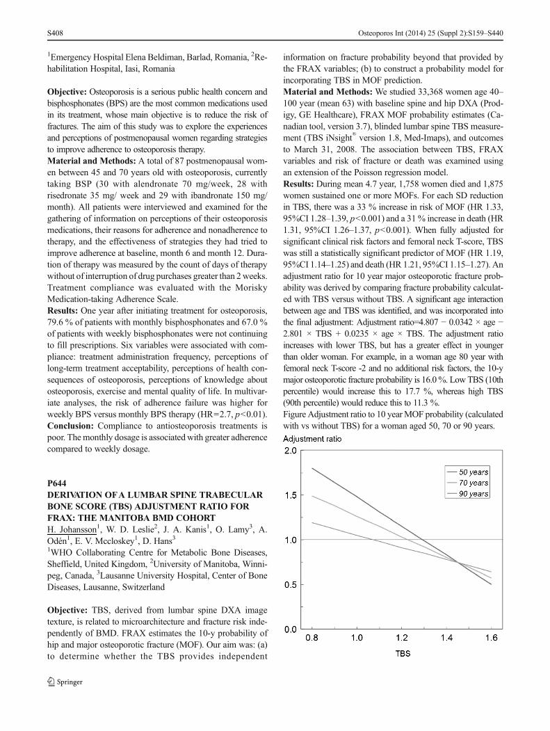
1Emergency Hospital Elena Beldiman, Barlad, Romania, 2Re-habilitation Hospital, Iasi, Romania
Objective: Osteoporosis is a serious public health concern andbisphosphonates (BPS) are the most common medications usedin its treatment, whose main objective is to reduce the risk offractures. The aim of this study was to explore the experiencesand perceptions of postmenopausal women regarding strategiesto improve adherence to osteoporosis therapy.Material and Methods: A total of 87 postmenopausal wom-en between 45 and 70 years old with osteoporosis, currentlytaking BSP (30 with alendronate 70 mg/week, 28 withrisedronate 35 mg/ week and 29 with ibandronate 150 mg/month). All patients were interviewed and examined for thegathering of information on perceptions of their osteoporosismedications, their reasons for adherence and nonadherence totherapy, and the effectiveness of strategies they had tried toimprove adherence at baseline, month 6 and month 12. Dura-tion of therapy was measured by the count of days of therapywithout of interruption of drug purchases greater than 2weeks.Treatment compliance was evaluated with the MoriskyMedication-taking Adherence Scale.Results: One year after initiating treatment for osteoporosis,79.6 % of patients with monthly bisphosphonates and 67.0 %of patients with weekly bisphosphonates were not continuingto fill prescriptions. Six variables were associated with com-pliance: treatment administration frequency, perceptions oflong-term treatment acceptability, perceptions of health con-sequences of osteoporosis, perceptions of knowledge aboutosteoporosis, exercise and mental quality of life. In multivar-iate analyses, the risk of adherence failure was higher forweekly BPS versus monthly BPS therapy (HR=2.7, p<0.01).Conclusion: Compliance to antiosteoporosis treatments ispoor. Themonthly dosage is associatedwith greater adherencecompared to weekly dosage.
P644DERIVATION OFA LUMBAR SPINE TRABECULARBONE SCORE (TBS) ADJUSTMENT RATIO FORFRAX: THE MANITOBA BMD COHORTH. Johansson1, W. D. Leslie2, J. A. Kanis1, O. Lamy3, A.Odén1, E. V. Mccloskey1, D. Hans31WHO Collaborating Centre for Metabolic Bone Diseases,Sheffield, United Kingdom, 2University of Manitoba, Winni-peg, Canada, 3Lausanne University Hospital, Center of BoneDiseases, Lausanne, Switzerland
Objective: TBS, derived from lumbar spine DXA imagetexture, is related to microarchitecture and fracture risk inde-pendently of BMD. FRAX estimates the 10-y probability ofhip and major osteoporotic fracture (MOF). Our aim was: (a)to determine whether the TBS provides independent
information on fracture probability beyond that provided bythe FRAX variables; (b) to construct a probability model forincorporating TBS in MOF prediction.Material and Methods: We studied 33,368 women age 40–100 year (mean 63) with baseline spine and hip DXA (Prod-igy, GE Healthcare), FRAX MOF probability estimates (Ca-nadian tool, version 3.7), blinded lumbar spine TBS measure-ment (TBS iNsight® version 1.8, Med-Imaps), and outcomesto March 31, 2008. The association between TBS, FRAXvariables and risk of fracture or death was examined usingan extension of the Poisson regression model.Results: During mean 4.7 year, 1,758 women died and 1,875women sustained one or more MOFs. For each SD reductionin TBS, there was a 33 % increase in risk of MOF (HR 1.33,95%CI 1.28–1.39, p<0.001) and a 31% increase in death (HR1.31, 95%CI 1.26–1.37, p<0.001). When fully adjusted forsignificant clinical risk factors and femoral neck T-score, TBSwas still a statistically significant predictor of MOF (HR 1.19,95%CI 1.14–1.25) and death (HR 1.21, 95%CI 1.15–1.27). Anadjustment ratio for 10 year major osteoporotic fracture prob-ability was derived by comparing fracture probability calculat-ed with TBS versus without TBS. A significant age interactionbetween age and TBS was identified, and was incorporated intothe final adjustment: Adjustment ratio=4.807 − 0.0342 × age −2.801 × TBS + 0.0235 × age × TBS. The adjustment ratioincreases with lower TBS, but has a greater effect in youngerthan older woman. For example, in a woman age 80 year withfemoral neck T-score -2 and no additional risk factors, the 10-ymajor osteoporotic fracture probability is 16.0%. LowTBS (10thpercentile) would increase this to 17.7 %, whereas high TBS(90th percentile) would reduce this to 11.3 %.Figure Adjustment ratio to 10 yearMOF probability (calculatedwith vs without TBS) for a woman aged 50, 70 or 90 years.
Osteoporos Int (2014) 25 (Suppl 2):S159–S440S408

Conclusion: Lumbar spine TBS is a risk factor forMOF and arisk factor for death. TBS is able to predict incident MOFindependent of FRAX clinical risk factors and femoral neckBMD even after accounting for the increased death hazard. Ifvalidated in other prospective cohorts, a relatively simplearithmetic adjustment to FRAX probability, incorporatinglumbar spine TBS and age, may be clinically useful forenhancing fracture prediction from FRAX.Disclosures: Didier Hans: TBS patent: co-owner. Stock op-tions or royalties: Med- Imaps.
P645EFFECTIVENESS OF EDUCATIONAL ACTIVITIESFOR PATIENTS WITH OSTEOPOROSISL. Myasoutova1, S. Lapshina11Kazan State Medical University, Kazan, Russian Federation
Objective: To evaluate the effectiveness of educational activ-ities (schools) for patients with osteoporosis (OP).Material and Methods: 55 patients with OP from primarygroup were trained in the schools (4 sessions per year), 40patients with OP were in control group, which only visitedrheumatologist 4–5 times a year. The groups were matched bysex, age, structure (primary and secondary) and the severity ofOP, the presence of fractures, received therapy. At baselineand after 6 months pain intensity on VAS back pain, adher-ence to treatment were determined. At baseline and after12 months determination of BMD was measured by DXA.Results:After 6 months of training intensity of back pain wassignificantly (p<0.01) decreased by 2.5 times in the primarygroup and by 1.5 times fold in control, 52 % of patients fromthe primary group, and 37.5 %—in the control wore anorthopedic corset. 95.8 % of patients who visited schoolsand 90 % of patients in the control group were continuouslytaking calcium and vitamin D during 6 months, after12 months—93.7 % and 85 %, respectively. Pathogenetictherapy was applied to all patients, initially these drugs beganto take 97.9 % of the study group and 92.5 % of the controlgroup patients, after 6 months of continuous administration ofdrugs 89.5 % from group trained patients and 67.5 %—thecontrol group continued treatment, after 12 months commit-ment to therapy was 87.5 % and 42.5 % in the test and controlgroups, respectively. Densitometry results in dynamics after12 months showed that the increase in BMDwas significantly(p<0.05) higher in the group that has been schooled for thepatients and was 5.6±6.2 % at the spine, 4.2±4.5 % at thefemoral neck in the main group, 3.1±4.8 % and 1.9±3.2 %,respectively, in the control group.Conclusion: Educational programs improve functional statusand quality of patients life, increase commitment totreatment.
P646UNTREATED MALE OSTEOPOROSIS: CASEREPORTP. S. Pantelic11IOHB, Belgrade, Serbia
Objective: To underline problems connected with osteoporo-sis in men through case report. There are need to clarify thepathogenesis of osteoporosis and fractures in men and todevelop diagnostic criteria as well as establish most effectivetreatment.Material and Methods: After low intensive trauma 64 yearsold man had back pain and pain in left forearm. X-ray showfractures of Th 12 and L2 vertebrae and left forearm. In hispersonal history there is also after low intensive trauma elbowand hip fracture in ages of 52 and 53. There are no other riskfactor in personal and family history. DXA evaluation results:T-score total LS vertebrae −4.7 SD and T-score total −2.4healthy hip. Laboratory tests: high rate of thyroid hormoneand low rate of 25OH vitamin D. We started with treatment.Results: Osteoporosis is leading causes of morbidity andmortality in elderly. “ Silent disease” progress without symp-toms until a fractures occurred. Hip fracture is very seriouscomplication. One fifth of it occurred in men but it rise about10 years later than in woman. In 65–70 ages men loosing bonemass at the same rate as woman and the absorption of calciumdecrease in both sex. For men before 65 years and high riskmen recommendation is to begin periodic osteoporotic riskassessment and performing DXA. When we have diagnosis itis important to consider and exclude secondary causes of boneloss. It is almost certain lifestyle, diseases and medications.More than 40 % are primary or idiopathic osteoporosis inmen. Careful history, physical examination and laboratorytests are enough component for making decision about treat-ment. Except medication men should be educated about ade-quate lifestyle measures as the prevention.Conclusion: Osteoporosis in men is an important and oftenoverlooked problem. The consequences of it areunderestimated and the condition is often unrecognised anduntreated. There is no sufficient scientific based evidence formen osteoporosis. It is still an important public healthproblem.
P647BALNEARY PHYSICAL KINETIC THERAPYEFFECTS OVER GONARTHROSIS SYMPTOMSF. Cioara1, S. D. Birsan1, L. Vicas1, C. Nistor-Cseppento1, M.Rus2, L. Lazar11Medical Rehabilitation, University of Medicine and Pharma-cy Oradea, Oradea, Romania, 2University of Medicine andPharmacy Oradea, Oradea, Romania
Osteoporos Int (2014) 25 (Suppl 2):S159–S440 S409

Objective: The aim of the study is to evaluate the effects ofbalneary physical kinetic therapy on clinical and functionalparameters in patients with knee osteoarthritis compared withadministration of therapies based on electrotherapy and kinet-ic therapy.Material and Methods: We conducted the study on twogroups of patients similar in terms of disease severity viaKellgren-Lawrence staging, for Lequesne index and qualityof life. Also the lots are homogeneous in terms of age (65–75 years), gender, lifestyle, environment of origin and associ-ated diseases. The first group made electrotherapy and phys-ical therapy program and the second received additional spe-cific thermal water balneotherapy in Felix Thermal WaterResort in Romania. Rehabilitation program was performedin two courses of 3 weeks, with an interval of 3 months.Evaluation was performed at the beginning and in the end ofmonitoring. We assessed pain by VAS divided into 100 steps;we performed pain and functional Lequesne index, quality oflife and perception of patients and physicians over the diseasestatus.Results: The outcomes assessed before and at the end of themonitoring showed improvement of scores that reach the limitof statistical significance (p≤0.05) in the group that carried outthe recovery program including balneotherapy.Conclusion: The study demonstrated the superiority of reha-bilitation means in knee osteoarthritis that includes balnearyphysical kinetic therapy in thermal water. The benefits ofexercise that is practiced regularly are associated with weightloss. There is an openness and greater efficiency for theelderly, proving the undeniable value of complex functionalrecovery as being possible to maintain, strengthen and defeatthe lack of functionality.
P648IMPACTOFKNEEOSTEOARTHRITIS ONQUALITYOF LIFER. Hanan1, L. Touria2, B. Imane2, A. Souad2, H. H. Najia21Rheumatology Department, Mohamed Vth UniversitySouissi, El Ayachi Hospital, Salé, Ibn Sina University Hospi-tal, Laboratory of Physiology, Faculty of Medicine and Phar-macy, Mohamed Vth University Souissi, Rabat, Morocco,2Rheumatology Department, Mohamed Vth UniversitySouissi, El Ayachi Hospital, Salé, Ibn Sina University Hospi-tal, Rabat, Morocco
Objective: The purpose of this study was to investigate theinfluence of knee osteoarthritis (OA) on the quality of life(QoL) in patients with nonendstage knee OA using the Mo-roccan version of the OA knee and hip QoL questionnaire(OAKHQOL). We suggest also to examine factors associatedto different domains of QOL assessed by OAKHQOL.
Material andMethods:TheMoroccan versions ofWOMACandOAKHQOLwere administered toMoroccan patients withsymptomatic knee OA, fulfilling the revised criteria of theAmerican College of Rheumatology. Because of the highpercentage of illiteracy in Moroccan population, OA patientswere interviewed individually (using face-to-face technique).Results: Fifty women with knee OAwomen were enrolled inthis study (mean disease duration of 24 months (12.72). Themean VAS pain (0–100) was 54.6[10–80]±21.2. The averageWOMAC score was 34[3–93]±16.9. The mean Lequesnescore was 10.1[3–19]±4.2. The average scores of theOAKHQOL domains were as following: social support 44.5,social functioning 45.8, physical activities 47, mental health62.2 and pain 62.3. Correlation analysis showed fair to goodcorrelation between 3 dimensions of OAKHQOL (physicalactivities, mental health, pain) and VAS pain, Lequesne andWOMAC scores (p<0.05).Conclusion: This data describe a burden of knee OA on QoLin patients suffering from nonendstage knee OA. A fair to astrong correlation was found between 3 dimensions ofOAKHQOL (physical activities, mental health, pain) andVAS pain, Lequesne and WOMAC scores. These resultsshould be confirmed by larger studies.
P649MANAGEMENT OF MYOFASCIAL SYNDROME INOSTEOPOROSISS. D. Birsan1, F. Cioara11Medical Rehabilitation, University of Medicine and Pharma-cy Oradea, Oradea, Romania
Objective: Myofascial pain syndrome is a painful musclecondition characterized by chronic pain or radiation locatedat the level of point-to-trigger, comprising a muscle or groupof muscles. Myofascial pain occurs very frequently and al-most as eCare man develops trigger points at some stage oflife. Myofascial syndrome is the most common cause of painin the upper portions of the column vertebral, due to insuffi-cient understanding of the pathophysiology ofmuscle pain butoften remain undiagnosed in the osteoporosis.Material and Methods: Myofascial syndrome is explainedby exceeding the limits of normal physiological responsive-ness to stress factors. Trigger points are discrete points focalhyperirritable with typical location strained muscle fiber.Results: The pain caused by trigger points, causing decreasedmuscle length at rest and reduced joint mobility. Vasoconstric-tion generate muscle spasm, localized ischemia, occurringmetabolites nerve irritation properties.Conclusion: Informing patients and awareness of the corre-lation between stress and muscle tension, between muscletension and pain can improve their condition and complexapproach to the problem by rheumatologists, general
Osteoporos Int (2014) 25 (Suppl 2):S159–S440S410

practitioners, medical and psychological rehabilitation wouldlead to optimize the treatment of patients with myofascialsyndrome is a condition miofascial. Myofacial syndrome isnot fatal but can reduce significantly the quality of life andalso are a major cause of limiting the ability employment ofpeople affected, causing great economic damage to society.
P650ALENDRONATE SODIUM/VITAMIN D3COMBINATION TABLET VS. CALCITRIOL FOROSTEOPOROSIS IN CHINESE POSTMENOPAUSALWOMEN: A RANDOMIZED, OPEN-LABEL STUDYZ. L. Zhang1, E. Y. Liao2, W. B. Xia3, H. Lin4, Q. Cheng5, L.Wang6, Y. Q. Hao7, D. C. Chen8, H. Tang9, Y. D. Peng10, L.You10, L. He11, Z. H. Hu12, C. L. Song13, F. Wei14, J. Wang14,L. Zhang14, A. C. Santora151Shanghai Sixth People’s Hospital, Shanghai Jiaotong Uni-versity School of Medicine, Shanghai, China, 2The SecondXiangya Hospital of Central South University, Changsha,China, 3Peking Union Medical College Hospital, Beijing,China, 4Nanjing Drum Tower Hospital, Nanjing, China,5Huadong Hospital Affiliated to Fudan University, Shanghai,China, 6TianJin Hospital, Tianjin, China, 7Shanghai NinthPeople’s Hospital, Shanghai, China, 8West China School ofMedicine, West China Hospital, Sichuan University, Cheng-du, China, 9Beijing Friendship Hospital, Capital Medical Uni-versity, Beijing, China, 10Shanghai First People’s Hospital,Shanghai, China, 11Beijing Jishuitan Hospital, Beijing, China,12Peking University People’s Hospital, Beijing, China, 13Pe-king University Third Hospital, Beijing, China, 14GlobalMedical Affairs, Merck Sharp & Dohme China, Shanghai,China, 15Merck Research Laboratories, Rahway, NJ, UnitedStates
Objective: To investigate efficacy and safety of alendronate70 mg/vitamin D3 5,600 IU combination tablets (ALN/D5600) vs. calcitriol in osteoporotic Chinese postmenopausalwomen.Material and Methods: A 6-month, randomized, open label,active-comparator study with 6-month extension(clinicaltrials.gov NCT01350934). Postmenopausal womenaged >55 years with osteoporosis (low BMD with/withoutprior nonpathological fracture) from 13 centers in China.Patients were randomized to ALN/D5600 once weekly orcalcitriol 0.25 μg daily. Primary efficacy endpoint:% changefrom baseline in lumbar spine BMD at Mo 6. Hypercalcemiaand hypercalciuria were safety events of special interest.Results: 219 patients (ALN/D5600 n=111; calcitriol n=108)were randomized. Baseline characteristics were similar,30.3 %: baseline 25(OH)D ≤15 ng/mL. At Mo 6 and 12,changes in lumbar spine BMD from baseline were 3.54 %vs. 1.59 % and 5.17 % vs. 2.26 % for ALN/D5600 vs.
calcitriol (both p<0.001), respectively. At Mo 12, 25(OH)Dwas <15 ng/mL in 1.0 % (ALN/D5600) vs. 25.5 % (calcitriol)(p<0.001). Between-group differences in P1NP and s-CTxmean% changes from baseline at Mo 6 were −42.37 % and−52.03 %, respectively (greater with ALND/5600, p<0.001).Overall safety profile was similar between groups. Incidenceof hypercalciuria (24-h urine Ca >300 mg) was 8.4 % (ALN/D5600) vs. 13.9 % (calcitriol) (p=NS) over 12 month. Onepatient (calcitriol) had hypercalcemia.Conclusion:ALN/D5600 produced greater increases in BMDat the lumbar spine, greater decreases in bone turnovermarkers, and less vitamin D insufficiency vs. calcitriol inosteoporotic Chinese women. Safety was similar and consis-tent with established profiles.Disclosures: Funding: Merck & Co., Inc., Whitehouse Sta-tion, NJ, USA. Employee: MSD China (FW, JW, LZ), MerckSharp & Dohme Corp., a subsidiary of Merck & Co., Inc.,Whitehouse Station, NJ USA (ACS). Stock: FW, LZ, ACS(Merck). Research support: ZLZ, EYL, WBX, HL, DCC(Merck). Consultant: ZLZ, EYL, WBX, HL, DCC, HT(Merck).
P651PHYSICAL PERFORMANCE DOES NOT DIFFERBETWEEN RIGHTAND LEFT FOOTED YOUNGSOCCERPLAYERS: FIELDAND ISOKINETICTESTSANALYSISR. Hanan1, A. Abdellatif2, A. Souad2, E. Leila2, B. Imane3, E.A. El Mustapha4, H. H. Najia3, D. Taoufiq21Laboratory of Physiology, Faculty of Medicine and Pharma-cy, Mohammed Vth University Souissi, Rheumatology De-partment, El Ayachi hospital, Ibn Sina University Hospital,Mohammed Vth University Souissi, Rabat, Morocco, 2Labo-ratory of Physiology, Faculty of Medicine and Pharmacy,Mohammed Vth University Souissi, Rabat, Morocco, 3Rheu-matology Department, El Ayachi hospital, Ibn Sina UniversityHospital, Mohammed Vth University Souissi, Rabat, Moroc-co, 4Rehabilitation Department, Military Hospital Moham-med V, Mohammed Vth University Souissi, Rabat, Morocco
Objective: To compare physical performance using field testsand isokinetic muscle strength between right and elite youngsoccer players.Material and Methods: Forty-nine young, healthy, malesoccer players (mean±SD age:±, range: to) who were schol-arship holders of the Moroccan academy Mohammed VI ofsoccer served as subjects in this study. Players were dividedinto 2 groups: right dominant limb (group 1) and left dominantlimb (group 2). All participants received the same entrainmentschedule.Soccer players underwent a clinical examination (weight,height and BMI), field tests (30 M sprint, TUB II and vertical
Osteoporos Int (2014) 25 (Suppl 2):S159–S440 S411
jump) and evaluation of knee flexor and extensor musclestrength of their dominant limb on an isokinetic dynamometer.To ensure consistent and accurate measurements, each param-eter was evaluated by the same examiner utilizing the samemethods.Results: Field tests results show no differences betweenplayers of the 2 groups. Muscular strength did not differbetween right and left footed as well at low as at speedvelocities.Conclusion: This study highlights that physical performancetests, including field tests and isokinetism, did not differbetween right and left footed young soccer players. Duringthe 90-min of soccer game, numerous explosive bursts ofactivity are required, including jumping, kicking, tackling,turning, sprinting, changing pace, and sustaining forcefulcontractions to maintain balance and control of the ball againstdefensive pressure. Briefly, soccer performance improvesneed not only physical but also technical and tactical devel-opment. Further studies on large samples should be conduct-ed. It’s extremely important that those studies integrate acomplete analysis of soccer performance including physical,technical/biomechanical, tactical and mental proprieties.Then, we can get out the independent factors eventuallyassociated with better performance in soccer players accord-ing their side lateral dominance.
P652ASSESSMENT OF BONE RESORPTION AFTERWITHDRAWAL OF LONG TERM IBANDRONATETHERAPYP. Kasalický1, L. Brunerova1, S. Skacelova11Bone Metabolism Unit, Mediscan Group, Prague, CzechRepublic
Objective: There is a controversial question in the length ofbisphosphonate treatment. The concept of drug holiday hasbeen introduced from medical (prevention of “frozen bone”)and economic reasons as well. International guidelines discussthe length of the treatment and recommendations after with-drawal for alendronate and risedronate, respectively. Howev-er, a few data is available for assessment of bone resorptionafter long term ibandronate treatment and for the management(laboratory and densitometry follow up) after its withdrawaland for safe duration of drug holiday, respectively. The aim ofour study was to assess the degree of suppressed bone remod-eling after withdrawal of long term therapy with ibandronate.Material and Methods: Ibandronate administration (150 mgp.o. once monthly) was introduced in our bone metabolismunit in autumn 2006. Since that time 2,402 patients have beentreated with ibandronate. However, the criteria of at least5 year treatment and bone remodeling markers available be-fore treatment initiation and 1 year after ibandronate
withdrawal were fulfilled only by 28 postmenopausal womentreated in accordance with good clinical practice.Results: Bone resorption marker B-crosslaps (CTX) was 598±230 ng/l before treatment initiation, at the moment ofibandronate withdrawal 220±105 ng/l and 1 year after thewithdrawal 363±112 ng/l. One year after ibandronate with-drawal CTX increased in 65% in comparison with the level atthe end of long term ibandronate therapy (p<0.000001) andthey remain at 61 % of initial levels (before the treatment wasintroduced).Conclusion: Bone resorption does not remain deeply sup-pressed 1 year after ibandronate withdrawal which should beconsidered in the post-withdrawal management of the patientsand in assessment of the safe length of drug holiday.
P653DIAGNOSIS OF LOW VITAMIN D AMONG SAUDIARABIANS: DID WE OVERSHOOT THE RUNWAY?M. Sadat-Ali1, A. M. Al-Elq2, H. A. Al-Turki3, A. K. Al-Ali41University Department of Orthopaedic Surgery, College ofMedicine, University of Dammam. King Fahd Hospital of theUniversity, AlKhobar, Saudi Arabia, 2University Departmentof Internal Medicine, College of Medicine, University ofDammam. King Fahd Hospital of the University, AlKhobar,Dammam, Saudi Arabia, 3University Department of Obstet-rics and Gynecology, College of Medicine, University ofDammam. King Fahd Hospital of the University, AlKhobar,AlKhobar, Saudi Arabia, 4University Department of Bio-chemistry, College of Medicine, University of Dammam,Alkhobar, Saudi Arabia
Objective: Reports indicate that deficiency of vitamin D inthe Saudi Arabian population is reaching an epidemic. Ourobjective was to compare the accuracy of three commonlyused 25-OHD assays among sample of Saudi population.Material and Methods: This cross-sectional study was car-ried out during the period between January 2011 and Decem-ber 2012. After an informed consent, blood samples for mea-surement of 25-OHD level was extracted from 200 adultpatients attended the outpatients clinics of King Fahd Hospitalof the University, AlKhobar. Patients receiving vitamin Dsupplements and those with advanced organ failure wereexcluded. Vitamin D level of each patients was determinedusing chemiluminescence immunoassay (CLIA), Radioim-munoassay (RIA) and liquid chromatography-tandem massspectrometry (LC-MS/MS) methods. Between assay agree-ment was examined using regression analysis and Bland-Altman plots. Assays were also compared using commonlyused cut points for classification of vitamin D deficiency.Results: The average age of all patients was 45.7±16.1 years.There were 50males (48.1±17.1 years) and 150 females (44.8±15.7 years) patients. Significant difference between the three
Osteoporos Int (2014) 25 (Suppl 2):S159–S440S412

assays was found. The mean 25-OHD levels were highest forthe LC-MS/MS (21.65 ng/mL, 95%CI 19.74–23.56), inter-mediate for RIA (16.607 ng/mL, 95%CI 14.8718.32) andlowest for CLIA method (13.864 ng/mL, 95%CI 12.109–15.618). Using a 30 ng/mL as cut-off value, 6 %, 9 % and22 % were found to have normal levels of 25-OHD usingCLIA, RIA and LC-MS/MS, respectively.Conclusion: This study revealed that levels of 25-OHD andthe prevalence of vitamin D deficiency are dependent on theassay used. The reported high prevalence of hypovitaminosisD among Saudi population possibly related to the use of anassay which underestimates the vitamin D level, due to falsepositive results of deficiency.Acknowledgements: The study was completed by the grantby the Deanship of Scientific Research, University ofDammam, Dammam, Saudi Arabia.
P654THE BENEFITS OF REHABILITATION PROGRAMIN POST-TRAUMATIC PATHOLOGY OF THE HIPF. Cioara1, N. R. Suciu2, A. Pallag3, S. D. Birsan1, C. Nistor-Cseppento1, N. Pascalau41Medical Rehabilitation, University of Medicine and Pharma-cy Oradea, Oradea, Romania, 2Medical Rehabilitation Hospi-tal, Oradea, Romania, 3University of Oradea, Faculty of Med-icine and Pharmacy, Romania, Oradea, Romania, 4MedicalRehabilitation / University of Medicine and Pharmacy, Ora-dea, Romania
Objective: Lately, the hip is developing a new category of“traumatic injury” and that is balance operated. Rapid increasein the number of orthopedic interventions did that after frac-ture surgery have the highest incidence. The present study wasto evidence the benefits of the rehabilitation program in pa-tients with post-traumatic pathology of the hip.Material andMethods:We introduced a study of 55 patientswith traumatic hip pathologies, resulting from sprains, frac-tures or acquired postoperative recovery that followed treat-ment in Felix Medical Rehabilitation Hospital. Evaluation ofpatients was done on day 0 and after 3 weeks. The objectivesof rehabilitation therapy were: pain control, maintaining andstrengthening the hip stability, maintain and increase jointmobility.Results: Pain relief, assessed subjectively by the patient,increase joint stability, assessed objectively by muscle testing,increase joint mobility, assessed objectively by joint testing.Pain score decreased from 7.46 to 5.22 (percentage of im-provement being 30.02 %). Average cumulative physical dys-function shows an improvement of 14.99 %. Average im-provement was 24.22 % disability. The overall score is statis-tically significantly improves the rate of improvement being24.12.
Conclusion: Functional recovery by physical methods has itswell defined role in completing the treatment phase afterexceeding orthopedic surgery.
P655STUDYOVER PHONOPHORESISWITH CAPSAICININ KNEE OSTEOARTHRITISS. D. Birsan1, F. Cioara11Medical Rehabilitation, University of Medicine and Pharma-cy Oradea, Oradea, Romania
Objective: There are numerous methods of administeringdrugs to the body, both passive and active. A recent reviewof the literature on phonophoresis reports that 75 % of thestudies reviewed reported positive effects, ultrasound energywith drugs like gel or cream can travel through body tissue.Capsaicin was the active ingredient in hot chilli peppers hasselection actions on unmyelinated C fibres and thinly myelin-ated A primary sensory neurons. We underwent an observa-tional prospective study to propose measurement ofphonoporesis with capsaicin on pain of the knee, in compar-ison with phonoporesis with ketoprofen applications.Material and Methods: The study included 60 patients aged45 years and over with clinical and radiological osteoarthritisof knee according to ACR criteria. All the patients wereincluded in the standard therapy program (drugs and rehabil-itation) over a period of 10 days. Group A - 30 patients wastaken phonoporesis with capsaicin and Group B - 30 patientswas taken phonoporesis with ketoprofen, both intensity was0.6 W/cm2 on the knee, 6 min every day. The assessedparameters at hospitalization and discharge were pain, jointmobility, muscle strength and WOMAC scale.Results: In group A patients presented a significant improve-ment of pain according VAS scale, on the other group B whopresent an insignificant improvement of pain for the shorttime.Conclusion: We can consider phonoporesis with capsaicin asignificant method of therapy in knee osteoarthritis with animportant benefit towards long term pain relief for the patient.
P656QUANTITATIVE ULTRASONOMETRY OF THEPHALANGES IN POSTMENOPAUSALWOMENWITH TYPE 2 DIABETES MELLITUS: THE FIRSTRESULTS OFATHREE-YEAR LONGITUDINALSTUDYC. Neglia1, N. Agnello2, G. Chitano2, A. Argentiero2, M. E.Gianicolo2, R. Ciccarese2, A. Vigilanza2, V. Denetto2, G.Quarta2, L. Nibio2, A. Caretto3, A. Distante2, P. Piscitelli11University of Salento - DiSTeBA (Biological and Environ-mental Sciences and Technologies Department, Lecce, Italy,
Osteoporos Int (2014) 25 (Suppl 2):S159–S440 S413

2ISBEM (Istituto Scientifico Biomedico Euro Mediterraneo),Brindisi, Italy, 3Health Local Authority of Brindisi - Depart-ment of Endocrinology, Brindisi, Italy
Objective: Type 2 diabetes mellitus (T2DM) is associated tohigher risk of fractures despite a normal or increased BMD[1]. The purpose of this 3-year longitudinal study was toassess the changes of quantitative ultrasound (QUS) parame-ters in a group of postmenopausal women with T2DM and inhealthy controls.Material and Methods: The analyses were performed on agroup of 35 postmenopausal women attending to theOSTEOLAB at ISBEM Research Institute (Brindisi, Italy)within the PROF project (Prevention of Osteoporosis andFracture) in collaboration with the Local Health Authority ofBrindisi. We selected 17 women affected by T2DM and 18healthy controls, aged 55–70 years old. Subjects had baselineand 40 months follow-up measurements of phalangealultrasonometry performed by using DBMSonic Bone Profiler1200 (Igea®) as well as information about medical history,current drug therapy and risk factors for fractures.Results: At the baseline, only ultrasound bone profile index(UBPI) was significantly lower in T2DM group (p<0.05). Inthis latter group, we found at follow up (40 months) a signif-icantly lower bone transmission time (BTT) (p<0.01) andamplitude- dependent speed of sound (AD-SoS) (p<0.02).During the study period, we found a decrease of both BTTand UBPI, which was significantly higher in T2DM group(p=0.01 and p=0.03, respectively).Conclusion: Diabetic and control patients did not show anydifference in AD-SoS, the ultrasound parameter which isusually associated to BMD, thus representing the most usedpredictor parameter of fractures risk in clinical practice. At theopposite, BTT and UBPI, that give information about bonemineral quality, resulted decreased in T2DM subjects, pre-senting a possible future role in the clinical practice for thediagnosis of alterations of bone micro-architecture in T2DMsubjects.References: 1. Leslie WD et al. J Bone Miner Res2012:11:2231.
P657OSTEOPOROSIS AND ITS RISK FACTORS INRHEUMATOID ARTHRITIS FEMALE PATIENTSV. Duraj1, A. Kollcaku1, T. Backa1, A. Bano2, A. Tafaj11Rheumatology Clinic, University Hospital Center, Tirana,Albania, 2Internal Medicine Clinic, University Hospital Cen-ter, Tirana, Albania
Objective: Rheumatoid arthritis (RA) is very often compli-cated with osteoporosis (OP). About 0.5–1 % of the popula-tion in the whole world suffers from RA, which is the most
common form of inflammatory arthritis. The aim of this studyis to assess osteoporosis and its risk factors in RA femalepatients.Material and Methods: A cross-sectional study was per-formed during the period of time 2012–2013 on 60 womenwith RA, from 48 to 65 years old. Based on their BMD status,they were separated in two groups: with and without osteopo-rosis. All the evidence of OP and its presumable risk factorswere registered. In addition, in order to compare the twogroups, the T-test was utilized for quantitative variables andχ2 for qualitative variables.Results: The patients had the average age 52.7±9.1. Thedisease had lasted 10.1±6 years. The T-score≤−2.2 was foundin 39 of patients (65 %) respectively (60.1 %) in the lumbarspine and (39.9 %) in the femoral neck region. The cardinalfactors related to OP were the age (p=0.02) and BMI p=0.01.In patients with hysterectomy and oophorectomy, the meno-pause induced by these interventions was conversely relatedto OP of femoral neck (p=0.03). Principally, OP in femoralneck region was associated with seropositivity for RF (p=0.05) and BMI (p=0.02). The correlation between fractures offemoral neck and osteoporosis was p=0.02.Conclusion: OP was found in more patients than it wasthought. Aging, low BMI, menopause and seropositivity werethe most important risk factors of osteoporosis.
P658STUDYABOUT INFLUENCE OF ULTRASOUNDTHERAPY FORTREATING PES ANSERINEBURSITISS. D. Birsan1, F. Cioara11Medical Rehabilitation, University of Medicine and Pharma-cy Oradea, Oradea, Romania
Objective: Inflammation of the conjoined insertion of thesartorius, gracilis and semitendinosus muscles along the prox-imal medial aspect of the tibia is recognized as pes anserinebursitis and was first described almost 70 years ago. Thesemuscles are primarily flexors of the knee and have a secondaryinternal rotational influence on the tibia, protecting the kneeagainst rotatory as well as valgus stress. The purpose of thisstudy was to determine the role of ultrasonotherapy in pesanserine bursitis.Material andMethods: 15 patients with pes anserine bursitiswere studied (9 females and 6 males, mean age 50.4 years)between March and August 2008. Patients presenting kneepain suspected to be due to internal derangement and detectedon MRI. The commonest clinical presentation was pain alongthe medial joint line mimicking a medial meniscal tear. Forevaluated the long effect we used a 10-point categorical scale,asking patient to appreciate the level of pain after 10ultrasonotherapy applications.
Osteoporos Int (2014) 25 (Suppl 2):S159–S440S414

Results: 10 patients reported diminuated pain and 5 reportedno pain relief.Conclusion:An accurate diagnosis of pes anserine bursitis onMRI and initiate early treatment will help prevent unnecessaryarthroscopy.
P659PARATHYROID GLANDS RESPONSE TO LOWVITAMIN D LEVELS IN HEALTHYADULTS: ACROSS-SECTIONAL STUDYM. Sadat-Ali1, A. S. Al-Omran2, H. A. Al-Turki31University Department of Orthopaedic Surgery, King FahdHospital of the University, AlKhobar, Saudi Arabia, 2Univer-sity Department of Orthoapedic Surgery, College ofMedicine,University of Dammam, King Fahd Hospital of the Universi-ty, AlKhobar, Saudi Arabia, 3University Department of Ob-stetrics and Gynecology, College of Medicine, University ofDammam, King Fahd Hospital of the University, AlKhobar,Saudi Arabia
Objective: To assess the correlation of serum PTH and vita-min D (25OHD) levels of healthy Saudi Arabians living ineast coast.Material and Methods: A cross-sectional study was con-ducted in 200 patients (150 women and 50 men) betweenJanuary 2011 and December 2012, in the age group of (18–69 years) attending outpatients clinics of King Fahd Hospitalof the University, AlKhobar. Serum calcium, phosphorous,alkaline phosphatase, PTH and 25OHD were performed. 25-OHD was assessed using levels using chemiluminescenceimmunoassay (CLIA), Radioimmunoassay using Wallac1470 Gamma Counter and HPLC -LC.MS/MS (high perfor-mance liquid chromatography- liquid chromatography withmass spectrometry. The data was collected and entered in thedatabase and analyzed using SPSS Inc Version 14.Results: The mean age was 45.8±15.8 (18–74) years, Calci-um Level was 9.09±0.63 mg/dl. Alkaline Phosphatase was88.91±35.94 (34–302) IU, Parathormone 6.7±3.06 (1.35–21.2) (1.3–6.8 pmol/L). 188 were either insufficient or defi-cient CLIA 11.85±6.14 (2–29.6) and 91 (48.4 %) of them hadsecondary hyperparathyroidism 9.48±4.55 pc/L (CLIA25OHD normal levels had normal PTH levels, insufficiency4/21 (19 %) had raised PTH levels and 81/166 (48.79 %)deficiency had raised levels), whereas with HPLC 156 wereeither insufficient or deficient 97 (with PTH level of 7.41±4.2.). Patients with Insufficiency by HPLC-LCMS-MS 13 /41(31.7 %) had raised PTH. All patients with vitamin D defi-ciency as diagnosed by HPLC-LC-MS/MS had secondaryhyperparathyroidism.Conclusion: The association of secondary hyperparathyroid-ism and Vitamin D deficiency is common and definite.
Reports which give no association probably comes frominaccuracy of the assays in the assessment of vitamin D levels.
P660REHABILITATION AFTER A DISTAL RADIUSFRACTURE EPIPHYSES IN PATIENTS WITHOSTEOPOROSISF. Cioara1, A. Venter2, S. D. Birsan1, F. Bodog2, M. Rus21Medical Rehabilitation, University of Medicine and Pharma-cy Oradea, Oradea, Romania, 2University of Oradea, Facultyof Medicine and Pharmacy, Oradea, Romania
Objective: Osteoporotic fractures represent one of the mostcommon cause of disability and one of the major voice in thehealth economic budget in many countries of the world. Thepresent study was to evidence the importance of the rehabil-itation program in patients with distal radius fractureepiphyses.Material and Methods: Our study included 20 outpatientsattending to Medical Rehabilitation Hospital Felix with distalfracture epiphyses. The parameters studied were: age, jointtesting, muscle testing, using VAS pain, response to balneo-kinetictherapy. Data were analyzed using SPSS pack and testsof statistical significance by Student method (t test) and χ2.We used 10 days cure period with balneokinetic therapy inoligomineral thermal water specific of Baile Felix Resort,antialgic electrotherapy, thermo therapy, and physicalexercise.Results: They observed an improvement of the functionaltests during the observed period, improvement that is obtainedonly with a continuous and systematic practice of physicaltherapy (p<0.05) and the occupational therapy. The physicaltraining centered on customary gestures with occupationallytherapy obtained the biggest improvement and 100 %compliancy.Conclusion: Rehabilitation program is of utmost importancein distal radius fracture epiphyses so it should be applied soonafter the orthopedic treatment.
P661CARDIOVASCULAR RISK FACTORS IN PATIENTSWITH PRIMARY HYPERPARATHYROIDISMR. A. Trifanescu1, M. Carsote1, D. Soare1, C. Popa2, L.Procopiuc2, C. Poiana11Dept. of Endocrinology, Carol Davila University ofMedicineand Pharmacy, Bucharest, Romania, 2C. I. Parhon NationalInstitute of Endocrinology, Bucharest, Romania
Objective: To assess cardiovascular risk factors in patientswith primary hyperparathyroidism.
Osteoporos Int (2014) 25 (Suppl 2):S159–S440 S415

Material and Methods: 64 consecutive patients (4M/60F),aged 56.8±12.2 years were retrospectively assessed. Serumintact PTH was measured by immunoradiometric assay and25OH vitamin D by radioimmunoassay. Resting ECG wasperformed. Statistical analysis: independent sample t-test andPearson correlation test were used.Results:Average total calcium serum levels at diagnosis were11.3±1.3 mg/dL (range 9.3–18.4), median PTH level was147.7 pg/mL (range 64.2–1982.3). Average 25OH vitaminD was 16.6±8.7 ng/dL (range 4–36.2);. 16 out of 64 patients(25 %). showed asymptomatic primary hyperparathyroidismand were conservatory treated. In the whole series, averageBMI was 27.2±4.9 kg/m2; 29 patients (45.3 %) were over-weight and 12 patients were obese (18.8 %). Age at diagnosispositively correlated with BMI (r=0.335, p=0.007). Type 2diabetes mellitus was recorded in 3 patients (4.7 %), dyslip-idemia in 34 cases (53.1 %). PTH levels positively correlatedwith triglycerides (r=0.374, p=0.006); PTH negatively cor-related with HDL-cholesterol (r=−0.415, p=0.016). Arterialhypertension was present in 37 patients (57.8 %). Hyperten-sive patients were significantly older than normotensive ones(59.4±11.5 vs. 53.4±12.4 years, p=0.05). In hypertensivepatients, serum calcium and PTH levels tends to be higherthan in normotensive ones, and 25OH vitamin D tends to belower. Pheochromocytoma was associated in one case andACTH independent Cushing syndrome due to bilateral adre-nal hyperplasia in one patient. Ischemic heart disease withresting ECG signs was present in 14 patients (21.9 %). Ar-rhythmia was present in 5 patients (7.8 %).Conclusion: There is a high prevalence in cardiovascular riskfactors in patients with primary hyperparathyroidism, espe-cially in elderly ones and in patients with high PTH levels.
P66210-YEAR ABSOLUTE RISK OF FRACTURE BASEDON FRAX AND QUANTITATIVE ULTRASOUND INROMANIAN POSTMENOPAUSALWOMENM. Carsote1, V. Radoi2, A. Mihai3, A. Geleriu3, C. G. Barbu4,D. Paun1, C. Poiana11Endocrine/I.Parhon/Davila UMPh, Bucharest, Romania,2Davila UMPh, Bucharest, Romania, 3Endocrine/I.Parhon,Bucharest, Romania, 4Endocrine/Elias Hospital/DavilaUMPh, Bucharest, Romania
Objective: The relationship between a traditional method offracture risk assessment as heel quantitative ultrasound (QUS)and a new method as the 10-year probability of fracture basedon FRAX model is complex since the methods are indepen-dent. This aspect might have a good economical profile espe-cially in regions with socio-economic issues since themethodsare especially useful in screening population at high risk offracture as women in menopause. We analyze the correlation
between QUS and 10-year absolute risk of fragility fracturesbased on FRAX results in menopausal women.Material and Methods: This is a retrospective analysis inwomen. The inclusion criteria were menopausal status; at leastone evaluation of clinical fracture risks as pointed by FRAX, acentral DXA assessment (at least femoral neck, a Lunar Prod-igy device), and heel QUS (Achilles Insight device) availabledata. The exclusion criteria were pretherapy for osteoporosisor osteoporotic fractures at any moment of life. The statisticsused SPSS 21, with statistical significance at p<0.05. Thepatients’ data were from 2008 to 2013. Since Romanian datain the FRAX model were provided from 2011, we retrospec-tively calculated the 10-year absolute fracture risk of majorfractures (MOF) and hip fractures (HIF) based on FRAX inpatients enrolled before this.Results: 197 women in menopause were included (median of57 years, ranges between 41 and 79 years). Some of the riskfactors included in FRAXwere: 4 % previous prednisone use,30 % smokers, 15 % with prevalent fragility fractures. Thelinear correlation between stiffness index (QUS) and MOFwas r=−0.3 (p<0.0005) or r2=0.1 (p<0.0005). The simplecorrelation between stiffness index and HIF was r=−0.3(p<0.005) or r2=0.2 (p<0.0005). The same correlations wereanalyzed after adjusting for years of life: for MOF r=−0.2(p<0.0005), respective for HIF r=−0.2 (p<0.0005); and aftertriple controlling for age (years), period of time in menopause(years) and BMI for MOF r=−0.18 (p=0.01), and for HIF r=−0.14 (p=0.04).Conclusion: Week, negative, statistically significant correla-tions were found between stiffness index provided by heelQUS and 10-year absolute risk of fracture based on FRAXmodel. The correlation does not improve when adjusting forage, years since menopause, BMI. The data were similar formajor osteoporotic fracture risk as well as hip fracture risk.
P663DO DEPRESSIVE SYMPTOMS IMPACTFUNCTIONAL OUTCOME IN HIP FRACTUREPATIENTS?E. Dubljanin Raspopovic1, S. Tomanovic Vujadinovic1, N.Krstic1, N. Ilic1, S. Stojicic1, U. Nedeljkovic11Clinic for Physical Medicine and Rehabilitation, ClinicalCenter of Serbia, Faculty ofMedicine, University of Belgrade,Belgrade, Serbia
Objective:Depression is themost common ofmood disordersin elderly people, and one of the most prevalent comorbiditiesin older people with hip fracture. The aim of the present studywas to evaluate if depressive symptoms assessed on hospitaladmission impact early functional outcome after hip fracturesurgery.
Osteoporos Int (2014) 25 (Suppl 2):S159–S440S416

Material and Methods: We studied 112 patients whounderwent surgery for hip fracture during a 6 month period.Depressive symptomswere assessed on admission to the acutesetting using the 30-item Geriatric Depression Scale (GDS).Multidimensional assessment included sociodemographiccharacteristics, general health status, cognitive status, func-tional status prior to injury, and perioperative variables. Theprimary outcome measure was motor-FIM at discharge.Results: Adjusted multivariate regression analysis revealedthat presence of moderate to severe depressive symptoms(GDS ≥20), older age, and female gender were independentlyrelated to motor FIM at discharge.Conclusion: Adjusted multivariate regression analysis re-vealed that presence of moderate to severe depressive symp-toms (GDS ≥20), older age, and female gender were indepen-dently related to motor FIM at discharge.References: 1. Iolascon G et al. Clin Cases Miner BoneMetab 2011;8:49. 2. Holmes JD, House AO. Age Ageing2000;29:537.3. Voshaar RC et al. Am J Geriatr Psychiatry 2007;15:807. 4.Sharp LK, Lipsky MS. Am Fam Physician 2002;66:1001.5. Lenze EJ et al. Int J Geriatr Psychiatry 2004;19:472.
P664PROGRESSION OF VERTEBRAL COMPRESSIONFRACTURES IN THE FIRST SIX MONTHS OFANTIOSTEOPOROTIC THERAPYC. Corradini1, S. Pasqualotto1, F. Boisio1, V. Macchi11Department of Biomedical and Surgical Sciences, State Uni-versity of Milan c/o Orthopaedic Institute G. Pini, Milan, Italy
Objective: In the conservative treatment of a vertebral com-pression fracture (VCF) in postmenopausal women, an anti-osteoporotic drug is recommended. But their relationshipswith healing processes are still debated. The aim of thisprospective study was the evaluation of radiographic progres-sion of one or more VCF during assumption of risedronate,strontium ranelate or teriparatide.Material and Methods: Women with recent osteoporoticVCF verified through magnetic resonance were assigned toreceive either risedronate (RIS group, n=26) or strontiumranelate (SR group, n=22) or teriparatide (TPTD group, n=34) following guidelines of Italian regulatory agency. Serumand urinary bone turnover markers and lateral thoracolumbarspine X-rays were obtained at 0, 1, 3 and 6 months of therapy.Morphometric parameters were digitally acquired with spe-cialized software. Lumbar BMD was measured by DXAbefore and 6 months after treatment initiation.Results: Lumbar BMD increased at 6 month in all groups andsignificantly in TPTD group. An inconstant progression inVCF on radiograms were detected in RIS and SR groups. TheD7 and D12-L1 were more involved of others. A significant
correlation has been found between the progression and lowformation markers. At time 0 serum markers of bone forma-tion alkaline phosphatase, osteocalcin (OC) and of bone re-sorption desoxipiridoline (DPD). Between 1st and 3rd monthwithin the consolidation process, OC initially peaked in TPTDgroup decreases, while those in RIS group and SR groupremained significantly lower. DPD remained high in TPTDgroup, while in the others significantly reduced in 6 months.Conclusion: In recent osteoporotic VCF a divergence be-tween the formation and resorption markers has been revealedbetween antiosteoporotic therapies with a different radio-graphic progression. The clinical relevance is the possibilityof choice different antiosteoporotic treatments on radiographicand metabolic behaviour of VCF.References: Corradini C. Aging Clin Exp Res 2011;23:45.
P665FACTORS INFLUENCING BALANCERESTORATION IN ELDERLYAFTER HIPFRACTUREN. Radosavljevic1, M. Lazovic1, D. Nikolic2, Z.Radosavljevic31Institute for Rehabilitation, Belgrade, Serbia, 2Physical Med-icine and Rehabilitation Department, University Children’sHospital, Belgrade, Serbia, 3General Hospital, Mladenovac,Serbia
Objective: The risks of second fracture among patients withinitial brittle fracture are substantial. Secondary prevention isvery important as treatment of osteoporosis and also as bal-ance restoring, motor functional rehabilitation and fall-downprevention training. The purpose of this study was to evaluateeffectiveness of our rehabilitation program in balance im-provement measured by Berg Balance Scale (BBS) and todetermine factors of influence on balance restoration after hipfracture in patients above 65 years.Material and Methods:We have evaluated 203 patients thatwere referred to Institute for Rehabilitation in Belgrade forrehabilitation treatment after hip fracture. Eligible participantswere evaluated by BBS at admission, at discharge, 3 monthpost discharge and 6 months post discharge. We observedbalance improvement by BBS regarding age, sex, durationof time from operation to admission, comorbidity measuredby Cumulative Illness Scale for Geriatrics (CIRS-G), type offracture (femoral neck or trochanteric region), type of opera-tion (osteosynthesis or endoprothesis) and type of rehabilita-tion program (with or without hydrotherapy).Results: There is significant increase (p<0.001) in balancemeasured by BBS in observed period (admission-24.61±14.94; discharge −34.1±7.47; after 3 month R-37.98±10.09,after 6 months-39.44±11.08). We found out significant corre-lation between increase of BBS and age (R −0.3628, p=
Osteoporos Int (2014) 25 (Suppl 2):S159–S440 S417

0.0004), comorbidity measured by total score of CIRS-G (R−0.4675, p=0.0005), duration of time from operation to ad-mission (R −0.4617, p=0.0004) ad type of rehabilitation (R−0.5632, p=0.0000).Conclusion: We have demonstrated that our inpatient reha-bilitation program significantly improves balance in elderlyafter hip fractures. Younger patients with less comorbidity,started with rehabilitation earlier after operation, and includedin rehabilitation program with hydrotherapy has betterchances to restore their balance and to prevent fall-down andsecond osteoporotic fractures.
P666CHRONIC INFLAMMATION IS THE KEYMECHANISM RESPONSIBLE FOR DETERIORATEDBONE METABOLISM IN CHILDHOOD OBESITYJ. Konstantynowicz1, P. Abramowicz1, J. Jamiolkowski2, J. D.Piotrowska-Jastrzebska11Dept. of Pediatrics and Developmental Disorders, MedicalUniversity, Bialystok, Poland, 2Dept. of Public Health, Med-ical University, Bialystok, Poland
Objective: Some reports show a beneficial role of adiposity inmaintenance of skeletal mass, whereas others demonstrate anincreased fragility related to overweight. The aim of this cross-sectional study was to evaluate effect of adipokines and in-flammation on bone metabolism and bone mass in obesity.Material andMethods: In 40 obese boys and girls aged 7.1–17.6 y (BMI SDS: 3.12±1.6) and 30 non-obese controls (BMISDS: −0.31±0.7), total and lumbar BMD and body fat/ leanmass were determined using DXA. Serum lipids, calcium(Ca), 25-hydroxyvitamin D (25OHD), PTH, insulin and glu-cose (HOMA-IR), leptin, adiponectin, osteocalcin (OC), os-teoprotegerin (OPG), RANK-ligand (RANKL) and high-sensitivity CRP (hsCRP), reflecting inflammation, weremeasured.Results: Increased levels of leptin, hsCRP, Ca, PTH (allp<0.001) were found in obese children. Adiponectin andOC were lower in obese individuals (p=0.005), with simulta-neous increased RANKL/OPG ratio (0.51±0.5 vs. 0.32±0.5;p=0.01). Bonemineral apparent density (BMAD) adjusted forskeletal size and body composition in obese children waslower than in normal weight individuals (0.080 vs. 0.084 g/cm3). There was an association between excessive adiposityand hsCRP level which correlated with RANKL (r=0.61,p<0.001) and RANKL/OPG ratio (r=0.49, p=0.003). Inflam-mation, ascertained with composite models including BMI,FM and LM, was an independent risk factor of increased boneresorption (β=0.415, p=0.001). In obese individuals, hsCRPwas associated with a higher PTH (52.5±17.9 vs. 32.0±10.0 pg/ml; p<0.001) and higher serum Ca levels (2.47±
0.13 vs. 2.39±0.12 mmol/L; p=0.01). No differences in25OHD concentrations between the groups were observed.Conclusion: Chronic low-grade inflammation with alteredadipokine levels may be responsible for inadequate bonemetabolism in childhood obesity. Higher PTH and calciumconcentrationsmay reflect moderate parathyroid hyperactivityas a potential mechanism responsible for bone resorption inobese individuals.
P667PHYSICALTHERAPY INTREATMENTOFPATIENTSWITH GLENOHUMERAL OSTEOARTHRITISM. Kocic1, L. Dimitrijevic1, I. Stankovic1, M. Spalevic1, H.Colovic1, A. Stankovic11Clinical Center Nis, Clinic of Physical Medicine and Reha-bilitation, Nis, Serbia
Objective: A large number of patients develop glenohumeralosteoarthritis (OA) after dislocation or fracture of a shoulderand are treated with different physical therapy modalities. Weevaluated the effect of different combinations of physicaltherapy modalities on pain and shoulder range of motion inpatients with glenohumeral OA.Material and Methods: 30 patients with moderateglenohumeral OA with pain and reduced forward flexionand abduction, were randomly allocated to two groups with15 in each. Group A had exercises and breaststroke swim-ming, with galvanic current therapy and paraffin therapy for aperiod of 3 weeks. Group B had exercises and breaststrokeswimming with low level laser therapy (LLLT) and transcu-taneous electrical nerve stimulation (TENS) for a period of3 weeks. Patients were not allowed to use paracetamol orNSAIDs. The outcome assessment was performed beforeand after treatment using: 1) VAS for pain evaluation and 2)measurements of shoulder range of forward flexion andabduction.Results: Group A included 7 women and 8 men with a meanage of 59.3±6.2. Group B included 5 women and 10 men,with a mean age of 56.2±8.1. The groups were comparablewith regard to pain and forward shoulder flexion and abduc-tion before therapy (p>0.05). Both groups showed statisticallysignificant improvement for pain (p<0.01), forward flexion(p<0.001) and abduction (p<0.01) after therapy, compared tobasic values. Statistical analyses showed more improvementin group B than in group A for pain (p<0.05), and there wasno difference between groups for forward shoulder flexionand abduction (p>0.05).Conclusion:Both combination of physical therapymodalitiesled to a significant decrease in pain and increased in shoulderrange of motion in patients with glenohumeral OA, but relatedto the pain application of LLLTand TENS was more effective
Osteoporos Int (2014) 25 (Suppl 2):S159–S440S418

than the application of galvanic current therapy and paraffintherapy.
P668FUNCTIONAL ASSESSMENT IN POSTTRAUMATICANKLE OSTEOARTHRITISA. Totorean1, O. Suciu1, R. Onofrei1, A. Nita11Victor Babes University of Medicine, Timisoara, Romania
Objective: The objective of this paper was to assess a lot ofpatients with posttraumatic ankle osteoarthritis before andafter rehabilitation treatment. Our purpose was also to studythe effect of an individualized treatment on patients with thementioned pathology.Material and Methods: We evaluated 50 patients with post-traumatic ankle osteoarthritis. All patients were evaluatedbefore and after rehabilitation treatment clinically and func-tionally using Ankle Osteoarthritis Scale (AOS). The rehabil-itation treatment was 6 months long and involved physicaltherapy and exercises (kinetotherapy). Every patient had anindividualized rehabilitation treatment according to age, phys-ical possibilities, traumatic lesion which caused the osteoar-thritis, other pathologies and the functional assessment made.Results: After 6 months of treatment the total AOS scoredecreased significantly (p=0.04). The pain subscale scoreimproved more than dysfunction subscale score. The subjec-tive functional scale and the clinical assessment showed sig-nificant improvements. After rehabilitation treatment patientsstill had some problems with climbing and descending stairs.Conclusion: The rehabilitation treatment mainly based onindividualized kinetotherapy and physical therapy is impor-tant in patients with posttraumatic ankle osteoarthritis decreas-ing pain as well as dysfunction.
P669DIGITAL ULCER MANAGEMENT IN SYSTEMICSCLEROSIS: A SINGLE CENTER EXPERIENCEP. Athanassiou1 , I . Kostoglou-Athanassiou2, D.Basdragianni1, N. Kalaycheva1, A. Spyridis11Department of Rheumatology, St. Paul’s Hospital,Thessaloniki, Greece, 2Department of Endocrinology, RedCross Hospital, Athens, Greece
Objective: Systemic sclerosis is a multisystem disorder ofunknown aetiology. Pulmonary and cutaneous manifestationsof the disease cause significant morbidity, affect quality of lifeand are resistant to therapeutic interventions. Digital ulcers insystemic sclerosis remain a significant problem. They tend torecur and are difficult to treat. Recently, there have beenadvances in the management of digital ulcers in systemicsclerosis. In particular, new therapeutic developments such
as the dual endothelin receptor antagonist bosentan have beenintroduced in the management of digital ulcers in patients withsystemic sclerosis. The aim was to present the experiencewithin a single center in the management of patients withsystemic sclerosis and digital ulcers.Material and Methods:Within a period of 1 year, 8 patients,5 with systemic sclerosis and 3 with limited scleroderma, aged37–68 years, mean age 55.1 years, 7 female and 1 male, werestudied. All patients developed digital ulcers.Results: All patients received bosentan 62.5 mg twice dailyinitially increasing to 125 mg twice daily. In all patients digitalulcers improved over a period of a year. In 2 patients a rapidresponse to the treatment with bosentan was observed. Insome of the patients iloprost iv was simultaneously adminis-tered and in some hyperbaric oxygen was also applied.Conclusion: Systemic sclerosis is a multisystem disorderwhich was treated symptomatically until recently. Digitalulcers, which may appear during the course of the disease,are resistant to treatment and even if treated they may recur.New therapeutic achievements, such as the dual endothelinreceptor antagonist bosentan have made possible the success-ful treatment of digital ulcers in patients with systemic sclero-sis. In some of the cases described the rapid response of thedigital ulcers to the administration of bosentan is noted.
P670WNT SIGNALING IS INVOLVED IN HUMANARTICULAR CHONDROCYTEDEDIFFERENTIATION IN VITROL. Laadhar1, N. Sassi1, M. Allouche1, M. Kallel-Sellami1, S.Makni1, S. Sellami11Immuno-rheumatology Research Laboratory, Tunis, Tunisia
Objective: Osteoarthritis is the most prevalent form of arthri-tis in the world. Certain signaling pathways, such as the wntpathway, are involved in cartilage pathology. Osteoarthriticchondrocytes undergo morphological and biochemical chang-es that lead to chondrocyte dedifferentiation. We investigatedwhether the Wnt pathway is involved in dedifferentiation ofhuman articular chondrocytes in vitro.Material and Methods: Human articular chondrocytes werecultured for four passages in the presence or absence of IL-1 inmonolayer or micromass culture. Changes in cell morphologywere monitored by light microscopy. Protein and gene expres-sion of chondrocyte markers and Wnt pathway componentswere determined by western blotting and qPCR after culture.Results: After culturing for four passages, chondrocytes ex-hibited a fibroblast-like morphology. Collagen type II (col II)and aggrecan protein and gene expression decreased, whilecollagen type I (Col I), matrix metalloproteinase (MMP) 13,and nitric oxide synthase (eNOS) expressions increased. Wntmolecule expression profiles changed; Wnt5a protein
Osteoporos Int (2014) 25 (Suppl 2):S159–S440 S419

expression, the Wnt target gene, c-jun, and the Wnt pathwayregulator, sFRP4 increased. Treatment with IL-1 caused chon-drocyte morphology to become more filament-like. Thischange in morphology was accompanied by extinction ofcol II expression and increased col I, MMP13 and eNOSexpression. Changes in expression of the Wnt pathway com-ponents were also observed. Wnt7a decreased significantly,while Wnt5a, LRP5, β-catenin and c-jun expressionsincreased.Conclusion: Culture of human articular chondrocytes with orwithout IL-1 not only induced chondrocyte de-differentiation,but also changed the expression profiles of Wnt components,which suggests that the Wnt pathway is involved in chondro-cyte dedifferentiation in vitro.
P671MUTATION IN OSTEOACTIVIN ENHANCESRANKL-MEDIATED SIGNALING, PROMOTINGOSTEOCLAST DIFFERENTIATION AND SURVIVALS. M. Abdelmagid1, G. Sondag2, F. M.Moussa2, M. Khol2, C.Fulp2, F. F. Safadi21Anatomy and Neurobiology, Neomed, Rootstwon, UnitedStates, 2Anatomy and Neurobiology, Neomed, Rootstown,United States
Objective: The novelty of osteoactivin (OA) in bone regen-eration has been characterized and reported by our group.Recent study demonstrated that OA/Gpnmb is highlyexpressed by osteoclasts (OCs) in vitro. To specify the criticalrole and mechanism of action of OA in osteoclastogenesis, wecharacterized the osteoclastic phenotype in a mouse modelwith natural mutation in the OA gene. We hypothesized thatmutation of OA increases OC formation and decreases itsactivity.Material and Methods: For our purpose, we used a geneti-cally modified D2J mouse model at age of 8 weeks. D2J miceare mutant for OA. Mouse femurs were evaluated by μCT,histology and histomorphometric analyses. Mice sera wereexamined by ELISA for bone resorption markers. Bonemarrow-derived hematopoietic stem cells were differentiatedinto osteoclasts using RANKL and MCSF. OC differentiationwas evaluated by TRAP activity and staining. OC resorptivefunction was examined using corning disc osteoassay. Forosteoclast survival, cells were cultured for two additional daysin the presence of RANKL. Markers of OC differentiationwere tested by qPCR. MAPK and AKTsignaling pathways inosteoclast were examined by western blot analysis. Statisticalanalysis was carried out by student-t-test.Results: μCT analysis of D2J femur diaphysis showed in-creased cortical thickness, while cortical bone surface as wellas porosity were reduced in comparison toWT. Serum ELISAof TRAP and RANKL/OPG ratio were not altered, however
CTX-1 was significantly lower in D2J compared to WTmice.Differentiation of D2J OCs was substantially increased,marked by OC number, TRAP activity and surface area,compared to WT. In addition, qPCR analysis showed upreg-ulated expression of TRAP and key regulators of OC. More-over, the survival of mature OCs was improved in D2Jmarkedby reduced caspase-3/7.Conclusion: Taken together, these data suggest that OA/Gpnmb acts as a negative regulator of osteoclast differentia-tion and survival by inhibiting the ERK/AKT signalingpathways.
P672THE ROLES OF CANONICAL ANDNONCANONICALWNT SIGNALING IN HUMANDEDIFFERENTIATED ARTICULARCHONDROCYTESL. Laadhar1, N. Sassi1, M. Allouche1, M. Kallel-Sellami1, S.Sellami1, S. Makni11Immuno-rheumatology Research Laboratory, Tunis, Tunisia
Objective: Osteoarthritis is the most prevalent form of arthri-tis in the world and it is becoming a major public healthproblem. Osteoarthritic chondrocytes undergo morphologicaland biochemical changes that lead to dedifferentiation. Theinvolvement of signaling pathways, such as the Wnt pathway,during cartilage pathology has been reported. Wnt signalingregulates critical biological processes. Wnt signals are trans-duced through at least three intracellular signaling pathwaysincluding the canonical Wnt/β-catenin pathway, the Wnt/Ca2pathway and the Wnt/planar cell polarity pathway. We inves-tigated the involvement of the Wnt canonical and non-canonical pathways in human articular chondrocyte dediffer-entiation in vitro.Material and Methods: Human articular chondrocytes werecultured through four passages with no treatment, or withsFRP3 treatment, an inhibitor of Wnt pathways, or withDKK1 treatment, an inhibitor of the canonical pathway.Chondrocyte- secretedmarkers andWnt pathway componentswere analyzed using western blotting and qPCR.Results: Inhibition of the Wnt pathway showed that thecanonical Wnt signaling is probably responsible for inhibitionof collagen II expression, activation of metalloproteinase 13expression and regulation of Wnt7a and c-jun expressionduring chondrocyte dedifferentiation in vitro. Our results alsosuggest that expressions of eNOS, Wnt5a and cyclinE1 areregulated by noncanonical Wnt signaling.Conclusion: Understanding the exact role that each wnt path-ways plays in osteoarthritis is of major importance. This willclarify what role of each of these molecules during the pro-cesses of proliferation, differentiation or de-differentiation.
Osteoporos Int (2014) 25 (Suppl 2):S159–S440S420

P673EFFECT OF HYDROTHERAPY VS.CONVENTIONAL LAND-BASED EXERCISE INPATIENTS WITH HIPARTHROPLASTY FOROSTEOARTHRITISI. R. Marcu1, S. Patru1, A. Bighea1, R. S. Popescu1, A. M.Bumbea11University of Medicine and Pharmacy, Craiova, Romania
Objective: Hydrotherapy (HT) is usually employed in phys-ical medicine and rehabilitation. Underwater exercisesstrengthen weak muscles, restore joint motion and pain. Thestudy evaluated the functional outcome following hiparthroplasty in patientswho underwent hydrotherapy 6monthsafter discharge from a rehabilitation unit.Material andMethods:A total of 58 patients with recent hiparthroplasty (<14 days) for osteoarthritis were referred to ourrehabilitation unit. All participants were discharged andreturned to their home at the end of the rehabilitation program(12 days). Twenty-eight participants were randomly assignedinto the hydrotherapy group and thirty participants into theconventional exercise group. Participants were interviewedwith WOMAC Index at admission, at discharge and after6 months.Results: The WOMAC subscales for pain, stiffness and func-tion improved in both groups. Statistical analysis indicatesthat scores on all subscales were significantly lower for thehydrotherapy group. The benefits gained by the time of dis-charge were still found after 6 months.Conclusion: In this trial, hydrotherapy proved to be a bettertreatment than conventional exercise for patients with hiparthroplasty, since pain, stiffness and function impairmentwere significantly lower for the hydrotherapy group.
P674REGULATION OF INTERLEUKIN-6 EXPRESSIONBYA RIBONUCLEOTIDYLTRANSFERASE ZCCHC6IN HUMAN CHONDROCYTEST. Haqqi1, A. Arida1, N. Akhtar11Anatomy and Neurobiology, Neomed, Rootstown, UnitedStates
Objective: Interleukin (IL)-1β is the major cytokine involvedin osteoarthritis (OA) pathogenesis and induces the high levelsexpression of IL-6. Ribonucleotidyl transferases catalyze theaddition of nucleotides to the 3′ end of mRNAs. However,their expression or role in cytokine regulation in OA is un-known. We investigated whether ribonucleotidyl transferaseZCCHC6 is expressed in OA cartilage and whether ZCCHC6is involved in the regulation of IL-6 expression in OAchondrocytes.
Material and Methods: Chondrocytes derived from OApatients (n=14) were stimulated with IL-1β (5 ng/ml) orActinomycin D or NF-кB inhibitor SC514 or a JNK inhibitor.Total RNA was used to quantify ZCCHC6 or IL-6 mRNAexpression using TaqMan assays. SiRNA-mediated knock-down of ZCCHC6 was used to study the effect on inflamma-tory cytokine expression using a cytokine array. Protein ex-pression of ZCCHC-6 and IL-6 was studied using westernimmunoblotting and by ELISA.Results: Expression of ZCCHC6 and IL-6 was high in dam-aged cartilage compared to unaffected OA cartilage. IL-1βstimulation showed significant increase in the expression ofZCCHC6 and IL-6 mRNA and protein in chondrocytes.ZCCHC6 depletion significantly decreased IL-6 mRNA andprotein expression in IL-1β-stimulated chondrocytes. Cyto-kine array analysis of culture supernatants from control orZCCHC6 siRNA treated and IL-1β-stimulated chondrocytesshowed a substantial decrease in a subset of cytokines includ-ing IL-6. In chondrocytes with depleted ZCCHC6 IL-6mRNAs had shorter poly-A tails and also showed a decreasein constitutive IL-6 mRNA and protein expression. Inhibitionof NF-кB had no effect on ZCCHC6 expression but inhibitionof JNK blocked ZCCHC6 expression in OA chondrocytes.Conclusion: Taken together our results demonstrate thatZCCHC6 is highly expressed in damaged OA cartilage andmodulates IL-6 expression at the post-transcriptional level bypossibly influencing cytokine mRNA stability. These resultsidentify ZCCHC6 as a potential therapeutic target for thetreatment of OA.Disclosures: Grants from NIH (RO1 AT-003267, RO1 AT-005520, RO1 AT-007373) and funds from NeomedUniversity.
P675INFLUENCE OF VITAMIN D STATUS ONFRACTURES IN SPANISH ELDERLY WITH HIGHPARATHYROID HORMONE LEVELSR. Roncero-Martin1, J. M. Moran1, R. Ortés-Gómez1, F. J.Rodriguez-Velasco1, G. Gil-Fernandez1, J. F. Calderon-Garcia1, J. D. Pedrera-Zamorano11Metabolic Bone Diseases Research Group, University ofExtremadura, Caceres, Spain
Objective: To analyze whether there is an association be-tween serum PTH and the prevalence of fractures and to assessthe influence of serum 25-hydroxyvitamin D (25OHD).Material and Methods: A total of 248 Spanish elderly (149females and 98 males) were recruited. Serum PTH and25OHD concentrations, as well as fracture prevalence wererecorded. Serum PTH was divided into quartiles, and elderlywere grouped into those in the highest quartile (>132.8 pg/ml)
Osteoporos Int (2014) 25 (Suppl 2):S159–S440 S421

and those below. Serum 25OHD levels were stratified in 4categories (>40, 20–40, 10–20, <10 ng/ml).Results:Mean PTH and 25OHD values were similar betweenmales and females (103.5±65.4 vs. 107.6±65.3 for PTH and11±7 vs. 12±8 for 25OHD (P>0.05 both)). Values lower than10 ng/ml 25OHDwere found in the 34.6% of the males and inthe 65.4 % of the females. Within the males the prevalence offractures was of 43.8 % in the 10–20 ng/ml, 48.8 % in the<10 ng/ml and 12.5 % within the 20–40 ng/ml 25OHDgroups, respectively. In the female group, prevalence of frac-tures increases to 47.1 % in the <10 ng/ml, with lower figuresof 25.6 % for both the 20–40 ng/ml and the 10–20 ng/ml25OHD groups, respectively. Stratification of males by PTHlevels showed the highest prevalence in the 10–20 ng/ml25OHD within the top quartile of serum PTH with a 66.7 %vs. 38.5 % observed in those below. The highest prevalence inwomen was observed in the <10 ng/ml 25 OHD group with a50 % of the fractures within the highest quartile group of PTHwith similar figures (45.8 %) in those below. No significantincreases in fracture risk were observed in patients with highPTH levels (P>0.05 in all cases). After further stratificationby 25OHD levels, lack of increased risk was also observed(P>0.05 in all cases).Conclusion: Elevated PTH levels are not associated withincreased prevalence of fractures in Spanish elderly. Ourresults indicate that most of the fractured patients had vitaminD insufficiency/deficiency, suggesting that his condition maybe closely associated with fracture in elderly people.
P676EFFICACY OF VISCOSUPPLEMENTATION WITHINTRA-ARTICULAR HYALURONIC ACID IN KNEEOSTEOARTHRITISI. R. Marcu1, S. Patru1, A. Bighea1, R. S. Popescu1, A. M.Bumbea11University of Medicine and Pharmacy of Craiova, Craiova,Romania
Objective: The standard pharmacological treatment of osteo-arthritis with nonsteroidal anti-inflammatory drugs is essen-t i a l l y symp toma t i c , a im ing a t r e l i ev ing pa in .Viscosupplementation with intra-articular hyaluronic acid isan alternative to the treatment of symptomatic knee osteoar-thritis. The aim of this study was to assess the safety andtolerability profile of intra-articular sodium hyaluronate inpatients with symptomatic knee osteoarthritis over 24 weeks.Material and Methods: The study included 56 patients (36women and 20 men), aged between 42 and 76 years, withconfirmed primary or secondary knee osteoarthritis. All pa-tients underwent weekly intra-articular injections withhyaluronic acid for 5 consecutive weeks and were followedup for 19 additional weeks with control visits at week 6, 12, 18
and 24. The patients had to answer an adverse event (AE)questionnaire and the WOMAC OA Index questionnaire ateach visit. The WOMAC total score and the three WOMACsubscores (pain, stiffness and physical function) were ana-lyzed. All analgesics, nonsteroidal anti-inflammatory drugsor corticosteroids were suspended up to the end of the study.Results: The WOMAC score was significantly reduced after2 weeks of treatment (from 4.32±1.85 to 3.65±1.96, p=0.011), decreased by the end of the injection series (2.67±1.91; p<0.0001) and maintained during the follow-up (2.35±1.1.68; p<0.0001). The WOMAC subscores were also signif-icantly reduced from week 2 for pain and from week 5 forstiffness and physical function. The most frequent AE waspain at the injection site (6.2 % of the injections).Conclusion: These results confirm the evidence of efficacyand safety of efficacy and safety of intra-articular hyaluronicacid in the treatment of symptomatic knee osteoarthritis.
P677ANALYSIS OF TWO FUNCTIONALPOLYMORPHISMS IN THE LRP5 GENE INRELATION TO BONE MINERAL DENSITYANDFRACTURE RISK INMALTESE POSTMENOPAUSALWOMENM. Formosa1, A. Xuereb Anastasi11Department of Applied Biomedical Science, Faculty ofHealth Sciences, University of Malta, Msida, Malta
Objective: The low-density lipoprotein receptor-related pro-tein 5 (LRP5) is involved in osteoblast differentiation andbone formation, making it an important determinant of bonemass and strength. The influence of two functional missensesingle nucleotide polymorphisms (SNPs) A1330V(rs3736228; C>t) and V667M (rs4988321; G>a) wereanalysed in relation to BMD and different low-trauma frac-tures in Maltese postmenopausal women.Material and Methods: 1,040 women aged 40 to 79 yearswere recruited and their BMD measurements were performedby dual-energy X-ray absorptiometry. Subjects without a his-tory of a fragility fracture were subdivided in three groups:normal (n=228), osteopenic (n=266) or osteoporotic (n=280)according to their BMD results.Womenwith a fracture historywere classified as cases (n=266). Genotyping of the A1330Vpolymorphism was performed by PCR followed by restrictionenzyme digest, whereas real-time PCR was performed for theV667M SNP.Results: Using logistic regression analysis adjusted for age,the A1330V SNP was associated with reduced BMD at thelumbar spine (TT: p=0.02; CT: p=0.01) and to a lesser extentreduced femoral neck BMD (TT: p=0.07; CT: p=0.01). Frac-ture cases carrying one or both copies of the minor allele T hadan increased fracture risk compared to women with the CC
Osteoporos Int (2014) 25 (Suppl 2):S159–S440S422

genotype (CT: p=0.04; TT: p=0.05). The TT genotype wasthe most common among subjects with a wrist, humerus orhip fracture; nonetheless the difference was not significant(p>0.05). The haplotype with both risk alleles (AT) wasassociated with reduced BMD (p=0.05) as opposed to thehaplotype-reference (GC) which was strongly linked to a highBMD (p=0.002).Conclusion: The results indicate that the A1330V andV667M polymorphisms within the LRP5 gene are associatedwith reduced BMD and/or increased fracture susceptibility inMaltese postmenopausal women.
P678CLINICAL AND HISTOMORPHOMETRICALASSESSMENT OF BONE QUALITY IN HIPOSTEOARTHRITIS AND OSTEOPOROSISC. Rao1, M. Feola1, M. Celi1, F. M. Liuni1, E. Gasbarra1, U.Tarantino11Division of Orthopaedics and Traumatology, University ofTor Vergata, Rome, Italy
Objective:Osteoarthritis (OA) and osteoporosis (OP) are twodiseases characterized by the alteration of bone quality thataffect mainly elderly people reducing their quality of life.Although an inverse relationship between has been shownby some studies, other reports supported the coexistence ofthese pathologies. In this study we combined clinical (HarrisHip Score) and structural features (BMD and bonehistomorphometry) to clarify the relationship between OAand OP.Material and Methods: Among all the patients whounderwent a total hip arthroplasty in our hospital betweenJune 2011 and March 2013, we selected 120 patients dividedinto 4 groups according to BMD values and diagnosis, fem-oral neck fractures (n=30, mean age 71.9) or OA (n=90, withnormal/osteopenic/osteoporotic BMD; mean age 72.1 years).X-rays of the hips were also taken to establish the grade of OAthrough the Kellgren-Lawrence scale and OA patients werealso evaluated by HHS to assess the functionality of the hip.During surgery, a double osteotomy of the femoral head wasperformed and the samples were used for histomorphometrythrough Bio Quant software.Results: Clinical and histomorphometric features were com-pared between fractured patients and OA patients stratifiedaccording to BMD values. Histomorphometrical analysisshow that bone volume fraction (BV/TV) was significantlylower in subjects with femoral neck fracture (20.77±4.33 %)than subjects with OA and normal BMD (36.49±7.73 %;P<0.01) or osteopenic BMD (32.92±6.82 %; P<0.01), whileno difference between fractured subjects and those with com-bined OA and OP (20.71±5.23 %) was detected.
Conclusion:Our findings show that low bone mass is accom-panied by an impairment of bone histomorphometrical prop-erties, which were comparable between osteoporotic OA pa-tients and subjects with femoral fracture. This study supportevidence from the recent studies indicating impaired bonequality in OA and absence of protective effect against OP.
P679MECHANICAL AND STRUCTURAL PROPERTIESOF HUMAN FEMORAL HEADS IN DIFFERENTSTAGES OF OSTEOARTHROSISM. Kozuń1, P. Gabryś2, S. Dragan2, A. Nikodem1
1Department of Biomedical Engineering, Mechatronics andTheory of Mechanism, Wroclaw University of Technology,Wroclaw, Poland, 2Department and Clinic of Orthopaedic andTraumatologic Surgery, Wrocław Medical University,Wroclaw, Poland
Objective: Structure and mechanical properties of humanbone tissue adjust to changing load conditions. The structureof bone tissue is related to mechanical properties and adjust-ment of mechanical properties to changing load conditionscauses changes in the bone tissue structure. The knowledge ofthose properties is important to understand the mechanism ofpathological bone changes. The main purpose of the conduct-ed studies was to determine a correlation between structuraland mechanical properties of human trabecular bones withosteoarthrosis (OA).Material andMethods:The subject of the studies was elevenhuman femoral heads. The material was divided into 3 sepa-rate groups, based on classification of OA proposed byOuterbridge. In order to determine both structural and me-chanical properties 60 cubic specimens (10×10×10 mm) ofcancellous bone were prepared from each head. Mechanicalproperties were measured in uniaxial compression test usingmaterial testing machine 858MTSMiniBionix with strain rate0.01 s−1. Additionally, for each of the specimen the physicaldensity was determined using electronic weighing machineRADWAG. To find correlation betweenmechanical and struc-tural properties we conducted linear and multilinear regressionanalysis.Results:Our results show strong correlation between physicaldensity and strength (R2=0.8) The highest values of structuraland mechanical properties were obtained for specimens withIII grade of OA. The femoral heads from this group were alsocharacterized by loss of cartilage to the subchondral bone anddeterioration of bone. In this case we observed appearance ofcysts as well as bone remodeling.Conclusion: The results show that values of structural andmechanical properties depend on the grade of osteoarthritis. Inthe case of femoral head with III grade of OAwe obtained the
Osteoporos Int (2014) 25 (Suppl 2):S159–S440 S423

highest values of physical density, strength and Young’s mod-ulus even in comparison with healthy bone tissue.Acknowledgements: This work was supported by the Na-tional Science Centre grant no. N N518 505139.
P680BILATERAL FEMORAL NECK INSUFFICIENCYFRACTURES: AN UNEXPECTED CAUSE OF PAIN INA POSTMENOPAUSALWOMANT. Nakajima1, K. Tanabe1, S. Yagi11Nisninomiya Municipal Central Hospital, Nishinomiya,Hyogo, Japan
Objective: Older patients tend to suffer from osteoporosis. Insuch cases, several types of fractures can easily occur. Wereport a case of bilateral femoral neck insufficiency fracturesin a postmenopausal woman.Material and Methods:We report a case of bilateral femoralneck insufficiency fractures in a postmenopausal woman di-agnosed in our hospital.Results: The patient is 79 years old postmenopausal woman andin osteoporotic condition, suffered from the unknown suddenonset hip pain and edema of lower extremitiesmore than amonthbefore our first examination. She complained of the bilateral hippain when she move her hip joint, especially when she stand upor walk, but it became less when they lay down. Not only thepatient herself but also her families were all fear of some kind ofmalignant disease. At first examination by X-ray andMRI, therewas no obviously clear cause of pain. Because of her persistedpain, 2 weeks later, we took second X-ray and found bilateralfemoral neck fracture (Garden Stage IV). We performed bilateralbipolar hip arthroplasty and her quality of life was improved.Conclusion:When older patients complain of any discomfortaround their hip joint without any injury, femoral neck insuf-ficiency fractures must be considered. If the diagnosis weredelayed, it is better to take care that femoral head dislocationmight occur secondly. The patients with femoral neck insuf-ficiency fracture suffered from osteoporosis, so other fractureson their pelvic ring might be easily occurred.
P681ADULT STILL’S DISEASE: SUCCESSFULTREATMENT WITH CANAKINUMABP. Athanassiou1, D. Basdragianni1, A. Tzanavari1, E.Triantafyllidou1, I. Kostoglou-Athanassiou21Department of Rheumatology, St. Paul’s Hospital,Thessaloniki, Greece, 2Department of Endocrinology, RedCross Hospital, Athens, Greece
Objective: Adult Still’s disease is a systemic inflammatorydisorder. Still’s disease was first described in children, but it
has been now been described also in adults and is known asadult Still’s disease. The aim was to describe the case of apatient with adult Still’s disease who was successfully treatedby the administration of canakinumab.Material andMethods:A female patient, aged 38, presentedwith adult Still’s disease. During the 31st week of a pregnancyshe developed dyspnea, intense cough and fever. She wassubmitted to caesarean section and gave birth to a healthyinfant. Subsequently, she developed fever, cholestasis, in-creased liver enzymes, arthralgias, myalgias, a salmon coloredrash and enlarged lymph nodes.Results: Methylprednisolone was administered at a dose of32mg daily with partial remission of the disease. She was thengiven anakinra 100 mg sc daily, however the rash recurred.Subsequently canakinumab 180 mg/month was administered.Complete remission of the disease was induced and the doseof corticosteroids was reduced.Conclusion: Adult Still’s disease may respond to anti-inflammatory drugs as well as to corticosteroids. However, incases of disease resistant to treatment new agents are beingapplied. The recombinant human monoclonal antibody tointerleukin-1β canakinumab induced remission in this case ofadult Still’s disease.
P682MECHANICAL AND STRUCTURAL DEGREE OFANISOTROPY OF HUMAN OSTEOARTHRITICTRABECULAR BONEM. Kozuń1, M. Tomanik1, A. Nikodem1
1Department of Biomedical Engineering, Mechatronics andTheory of Mechanisms, Wroclaw University of Technology,Wroclaw, Poland
Objective: It is a common knowledge that the cancellousbone changes its structure when exposed to external loads.Osteoarthritis (OA) is degenerative joint disease caused by thebreakdown of cartilage and is most frequent in hips. As OAprogress the mechanical stress on joint increases, cause degener-ation and deformation of a femur head. Due to changes ofmechanical conditions in the joint the structure of bone adaptsto new loads. The main purpose of the study was to investigatethe mechanical and structural properties of the human femoralbone tissue in different stages of osteoarthritis in order to deter-mine correlation between the degree of mechanical anisotropy(DMA) and the structural anisotropy (DA).Material and Methods: The material study consisted of 26human femoral heads in different stages of OA that were dividedinto two groups: form region under the cartilage C and distal D,the nominal size of specimens was 10×10×10 mm.Structural measurements were conducted using X-rays scansfrom microtomography SkyScan1172, Bruker ®, while the
Osteoporos Int (2014) 25 (Suppl 2):S159–S440S424

mechanical degree of anisotropy was determined by uniaxialcompression test in three orthogonal axis.Results: Results show that the degree of structural anisotropyof the trabecular bone specimens from under the cartilage isgrater then the degree of mechanical anisotropy. With theprogression of the disease distinction between DA andDMA from region C and D became lower.
Conclusion: Results unveil strong correlation between me-chanical and structural parameters of the osteoarthritic trabec-ular in all stages of the OA.Acknowledgements: This work was supported by the Na-tional Science Centre grant no. N N518 505139.
P683PREVALENCE OF OSTEOPOROSIS/OSTEOPENIA,VERTEBRAL FRACTURES AND KYPHOSIS INPATIENTSWITHNONADVANCEDBREASTCANCERDUE TO RECEIVE AROMATASE INHIBITORSL. Brage1, R. Hernandez-San Gil2, H. Roldan1, E. Rodriguez-Rodriguez3, A. Perez- Ramirez3, J. Cruz-Jurado2, V. Rocha1,V. Hernandez-Hernandez1, I. Ceballos2, M. J. Sanchez-Perez31Neurosurgery Department, Hospital Universitario deCanarias, La Laguna Tenerife, Spain, 2Oncology Department,Hospital Universitario de Canarias, La Laguna Tenerife,Spain, 3Internal Medicine, Hospital Universitario de Canarias,La Laguna Tenerife, Spain
Objective: The treatment with aromatase inhibitors (AI) mayreduce bone mass and increase the risk of vertebral fractures(VF). Our aim was to analyze the prevalence of osteoporosis/osteopenia, VF and kyphosis in women with non-advancedbreast cancer who were to receive AI.Material andMethods: 121women (median age 62 years, 34–80) were prospectively included in the study. A lateral X-ray of
the dorsal and lumbar spine was obtained in all patients beforetreatment to assess VF and kyphosis, as well as a DXA scan.Results: 7.4 % of X-rays displayed at least one VF (9/121): twoat T7 (grades IC and IIIC), one at T8 (IC), one at T12 (grade IA),two at L1 (grades IIA and IIB), one at L2 (grade IIC), one at L4(grade IIA) and one with multiple fractures at L1, L3 and L5.38.8 % of all the patients had a normal bone mass, 43.8 % hadosteopenia and 17.4 % osteoporosis by OMS criteria. Fracturesbefore treatment were more prevalent in women older than65 years (16 % vs. 1.4 %, p=0.004). All women with VF hadabnormal bone mass: osteoporosis (5/9) or osteopenia (4/9) (p=0.001). Women with VF before treatment had a nonsignificanttrend towards diminished lumbar bone mass (p=0.052), whereasthe bone mass at the femoral neck and total hip was significantlydecreased. 8 of the 9 patients with VF (6.6 % of all X-rays)showed some degree of kyphosis (median 11°, 5–20). One of theVF patients had 0° kyphosis.Conclusion: Our prevalence of VF is slightly lower than thatof general population of a similar age. All womenwith VF hadlow hip bone mass. The lack of significance of the decreasedlumbar bone mass may come from the reduction in vertebralbody height in cases with VF. Our results highlight the rele-vance of this problem in this specific subset of patients whomay already have low bonemass and VF before beginning AI.
P684VITAMIN D DEFICIENCYAND ITS RELATION TOPHYSICAL CAPACITY IN PATIENTS WITHCHRONIC OBSTRUCTIVE PULMONARY DISEASE(COPD)S. H. Scharla1, U. G. Lempert1, K. Schultz21Endocrine Practice, Bad Reichenhall, Germany, 2Klinik BadReichenhall, Bad Reichenhall, Germany
Objective: Osteoporosis is an important comorbidity in pa-tients with chronic obstructive pulmonary disease (COPD).One important risk factor is Vitamin D deficiency. Vitamin Dstatus was associated with BMD and functional exercise ca-pacity in COPD patients in a previous study in the Netherlands(Romme et al. 2012). Therefore, we studied the prevalence ofvitamin D deficiency and its possible relationship with phys-ical function in German COPD patients.Material and Methods: We recruited consecutive patientswith COPD who were admitted to the Klinik Bad Reichenhall(a specialized hospital with rehabilitation therapy) from Feb-ruary to November 2013 (informed consent). Blood samplesand physical function data were obtained at the beginning ofthe hospital stay. 25-hydroxyvitamin D (25-OH-D) was mea-sured in serum by an automated method (Roche Diagnostics).Further, all participants were tested for the maximum inspira-tion force (PI) and for the 6-Minute-Walking-Distance(6MWD).
Osteoporos Int (2014) 25 (Suppl 2):S159–S440 S425
Results: 376 participants were studied (245 males, 131 fe-males). The serum 25-OH-D was 40±30 nmol/l (nM), mean±SD. The minimum was 5 nM and the maximum 169 nM.There was no gender difference and no significant correlationwith age. 73 % of the participants had vitamin D deficiency(defined as a serum 25-OH-D concentration below 50 nM).25-OH-D exhibited a slight seasonal variation (32±30 nM inwinter vs. 48±28 nM in summer, p<0.05). There was nosignificant association of 25-OH-D with 6MWD (p=0.33).However, the patients with vitamin D deficiency (25-OH-D<50 nM) exhibited a positive association between serum 25-OH-D and maximum inspiration force (PI), p<0.01.Conclusion: We found a high prevalence of Vitamin D defi-ciency in German COPD patients. This is not only importantfor the risk of secondary osteoporosis, but probably also forthe pulmonary function. Vitamin D deficiency was associatedwith less inspiration force. However, a causal relationship hasyet to be proved.References: Romme et al. Ann Med 2013;45:91
P685BONE MINERAL DENSITY EVALUATION IN RATSSUBMITTED TO DAILY DIET OF COFFEE ANDSOFT DRINKSM. B. Carrazzone Cal Alonso1, A. I. Vespasiano Silva1, J. F.Quirino Luzada2, P. C. Aranha Watanabe3, F. Haiter-Neto11Department of Oral Diagnosis and Dental Radiology-Piracicaba Dental School - University of Campinas(UNICAMP), Piracicaba, Sao Paulo, Brazil, 2Department ofStomatology, Public Health and Forensic Dentistry - RibeirãoPreto School of Dentistry - University of São Paulo (USP),Ribeirão Preto, Sao Paulo, Brazil, 3Department of Support,Product and Animal Health, medicine Veterinarian School ofAraçatuba (UNESP), Araçatuba, Sao Paulo, Brazil
Objective: To investigate and compare the effects of coffeeand soft drinks (Coca-Cola and Guaraná) in bone metabolismin male and female rats, by assessing BMD.Material and Methods: A total of 80 rats (Rattus norvegiauAlbinus, Wistar), 40 males and 40 females were used, 8groups of 10 animals each were divided into: Group 1 and2: control group which received only water, Groups 3 and 4:water management and coffee; Groups 5 and 6: water andCoca-Cola soft drink; Groups 7 and 8: water and guaraná softdrink. All the left femora from each animal were dissected andexamined by DXA and the BMD was obtained. Data weretabulated and statistical analysis was performed usingANOVA and Tukey test.Results: For females, significant differences were found whenall groups except for Coca-Cola X guarana (p>0.01) wereevaluated. No differences between groups were found for
males. Comparing sexes, there were differences for all groupsexcept for the control group (p>0.01).Conclusion: BMD of rats was unaffected by coffee or softdrinks. For females BMD was significantly affected by softdrinks and coffee intake being the coffee which caused morebone reduction, thus representing a risk factor for bone qualityof the femoral region studied.References: 1- Lacerda AS et al. Braz Dent J 201;21:199. 2-Sarazin M et al. Joint Bone Spine 2000;67:408. 3- Tsuang YHet al. J Orthop Surg 2006;7:1.Acknowledgements: CAPES-Brazil
P686OSTEOPOROSIS PROGRESSION AT PATIENTSWITH RHEUMATOID ARTHRITIS IN DIFFERENTTYPES OF TREATMENT—EXTENSIVE STUDYA. M. Bumbea1, R. Traistaru1, A. C. Bighea1, R. S. Popescu11University of Medicine and Pharmacy, Craiova, Romania
Objective: We evaluated the progression of osteoporosis atpatients with rheumatoid arthritis and osteoporosis in therapywith bisphosphonates at two groups of patients; one inremissive treatment with leflunomid or methotrexate versusbiological agent, in a retrospective study for a 6-year period oftime, 2007–2013.Material and Methods: We have analyzed a number of 54patients split in two homogeneous groups - eachwith 27 patients;all the patients have been diagnosed with osteoporosis. The AGroup has received a treatment with 20 mg of leflunomid dailyand non steroidal anti-inflammatory drugs or methotrexate,20 mg per week. The B Group has received a treatment withbiological agents. Both groups have received bisphosphonates intherapeutic doses being evaluated for osteoporosis. We alsomention that at 12 patients treated with leflunomid there wasnecessary corticosteroids therapy in doses of 16 mg/day with adiminishing level of 8 mg on a period of 2 up to 6 months; thepatients withmethotrexate did not need corticoids therapy duringthe study. Patients in biological therapy did not need corticoste-roids therapy.Results: The A group of study presented the maintenance ofmedian of T-score at −2.9 SD. The B Group of study presentedthe diminishing of the level of osteoporosis from a T-score from−3.9 to 3.0. These levels showed osteopenia in the witness groupa T-score from −1.8 to −2.4 with a passing to osteoporosis at 6patients who needed to receive bisphosphonates.Conclusion: The diminishing level of osteoporosis with 0.2SD per year at the group of patients in therapy with biologicagents as compared to the group treated with leflunomidproves the fact that despite the beginning of the first groupwith lower values it obtained better results in both reducingthe level of disease activity and the pathogenic immune in-flammatory context. The patients treated with methotrexate
Osteoporos Int (2014) 25 (Suppl 2):S159–S440S426

have received the same result as group threated with biolog-ical therapy.
P687CHANGES IN BONE MASS, VERTEBRALFRACTURES AND KYPHOSIS IN PATIENTS WITHNONADVANCEDBREAST CANCERTREATEDWITHAROMATASE INHIBITORS, CALCIUM, VITAMIN DAND IBANDRONATEH. Roldan1, R. Hernandez-San Gil2, L. Brage1, E. Rodriguez-Rodriguez3, L. M. Rodriguez-Rodriguez2, J. Alvisa Negrín3,J. Plata-Bello1, E. Martin-Ponce3, M. Monereo3, J. M.Gonzalez-Perez31Neurosurgery Department, Hospital Universitario deCanarias, La Laguna Tenerife, Spain, 2Oncology Department,Hospital Universitario de Canarias, La Laguna Tenerife,Spain, 3Internal Medicine, La laguna, Tenerife, Spain
Objective:Aromatase inhibitors (AI) can decrease bone massand increase the risk of vertebral fractures (VF). This second-ary effect can be prevented at least partially with a concomi-tant treatment with calcium, vitamin D and bisphosphonates.Our aim was to study the evolution of bone mass, VF andkyphosis in women with nonadvanced breast cancer who hadbegun AI and calcium, vitamin D plus monthly ibandronate.Material and Methods: 118 women (median age 62 years,34–80) were prospectively included in the study. A lateraldorsal and lumbar spine X-ray and a DXA-scan were obtainedat the onset of treatment and after 1 year. Differences in bonemass between patients with or without VF, changes in VFpattern and kyphosis progression were studied.Results: Treatment adherence was 99 %. Baseline lumbar,femoral neck and total hip bone mass increased significantly(p<0.0001) after 1 year of treatment without significant differ-ences between patients with and without VF (P=NS). Womenwith baseline VF exhibit a higher risk of subsequent fractures inthe X-ray at 1 year (77.8 % vs. 22.2 %, p<0.0001). Two X-rays(1.7 %) displayed VF not present in baseline X-ray (one at L1grade IIB and one at L3 grade IIC). Two (1.7 %) X-rays showedprogression in fracture grade (from IA to IIA, from IC to IIIC)and three demonstrated increase of the degree of kyphosis.Globally, five patients showed new fractures or some kind offracture worsening.Conclusion: The treatment with calcium, vitamin D andibandronate increases bone mass in patients with nonadvancedbreast cancer treated with AI. Despite that fact, two patients hadnew VF and about one third of our patients with previous VFexhibited fracture worsening. These data can be useful both inprimary and secondary prevention in this specific subset ofpatients, including other therapies like preventive vertebral bodycementation in women with fractures at the onset of treatment.
P688BONE MASS AND FRACTURE RATE IN PATIENTSWITH INFLAMMATORY BOWEL DISEASEC. Horvath1, A. Tabak1, D. Marton1, S. Meszaros1, V.Ferencz1, K. Bors2, P. L. Lakatos1, P. Miheller3, E. Hosszu41Semmelweis University, 1st Department of Internal Medi-cine, Budapest, Hungary, 2Rehabilitation Hospital, Visegrad,Hungary, 32nd Department of Internal Medicine, SemmelweisUniversity, Budapest, Hungary, 42nd Department of Pediat-rics, Semmelweis University, Budapest, Hungary
Objective: Patients with inflammatory bowel disease (IBD)suffer 20 % more fractures than healthy people. The risk offracture in IBD increased by 59 % for hip and 74 % for vertebralfractures, but the pathomechanism of these fractures has not yetbeen clarified. Our study aimed to study the bonemineralmass inIBD patients and looking for the role of low bone density in theincreased fragility.Material and Methods: 110 IBD patients (54 men and 56women, aged 35.2±17 years) with IBD (80 colitis ulcerosaand 30 Crohn’s disease) were involved into the study. 110 age-and sex-matched patients with suspected metabolic bone dis-ease due to other reasons and regularly referred for BMDmeasurement were used for comparison. BMD of the lumbarspine, hip and proximal forearm were measured by Prodigy(GE Lunar) densitometer, and Z-scores were evaluated. Pre-vious low- trauma fractures were also registered.Results: 23 patients with IBD has suffered fractures (RR: 1.41,CI 1.27–1.56). Normal bone density was found in the total IBDgroup (Z-scores as follows: spine: −0.67, femoral neck: −0.44,total hip: −0.51, radius: −0.48, p>0.1) as well as in the 31 IBDpatients of age more than 40 years (Z: −0.65, −0.38, −0.25,−0.39, respectively, p>0.1). Bone mass of IBD patients withfractures showed a borderline decrease of BMD (Z: −0.79,−0.57, −0.67, −0.55, respectively, p=0.05). Patients withsuspected other bone diseases had significantly lower BMD forspine, femoral neck and radius than that of IBD patients.Conclusion: IBD patients have normal BMD, in spite of theirhigher fracture rate. Compared to other metabolic bone dis-eases, IBD produces higher fragility by a different mechanismwhich seems to be independent of bone mineral mass.
P689MORBID OBESITY, VITAMIN D DEFICIENCYANDDESTRUCTIVE OSTEOARTHRITISI. Kostoglou-Athanassiou1, P. Athanassiou2, E. Xanthakou3,A. Gkountouvas41Department of Endocrinology, Red Cross Hospital, Athens,Greece, 2Department of Rheumatology, St. Paul’s Hospital,Thessaloniki, Greece, 3Endocrinologist, Sparti, Greece, 4En-docrinologist, Pireaus, Greece
Osteoporos Int (2014) 25 (Suppl 2):S159–S440 S427

Objective: Morbid obesity is known to be associated with im-paired glucose tolerance, arterial hypertension and fatty liver infil-tration. Morbid obesity is, however, also associated with musculo-skeletal disorders, in particular destructive inflammatory osteoar-thritis. The aimwas to describe a cohort of 10 patients withmorbidobesity, vitamin D deficiency and destructive osteoarthritis.Material and Methods: All patients were women, 3 premeno-pausal, 7 postmenopausal. All had morbid obesity, BMI>40 andmetabolic syndrome. All had vitamin D deficiency. Rheumatoidfactor was negative and C-reactive protein was mildly increased.Results:All had destructive inflammatory osteoarthritis man-ifesting with severe pain and causing mobility impairment. In2 of the patients successful dieting had as a result weight loss.Subsequently painful inflammatory osteoarthritis went intoremission. The patients were able to stop the anti-inflammatory and analgesic agents they were taking.Conclusion: We hypothesize that the adipose tissue, especiallythe abdominal adipose tissue, produces inflammatory cytokineswhich are responsible for the development of severe destructiveinflammatory generalized osteoarthritis. Additionally, in thesepatients vitamin D deficiency is observed, which may be dueto the acute inflammatory response observed in patients withmorbid obesity and may contribute to the painful musculoskel-etal disorder.
P690CLINICAL AND SUBCLINICALHYPERTHYROIDISM IN ADULT MEN: IMPACT ONTHE BONE MINERAL DENSITYAND SOFT TISSUECOMPOSITIONA. P. Barbosa1,2,3,4, M. R.Mascarenhas1,2,3,4, M. Bicho2, I. DoCarmo1,2,41Endocrinoly, Lisbon’s Endocrinology, Diabetes and Metab-olism Clinic (Osteoporosis Unit), Lisbon, Portugal, 2Environ-mental Health Institute, Lisbon’s Endocrinology, Diabetes and
Metabolism Clinic (Osteoporosis Unit), Lisbon, Portugal,3CEDML, Lisbon’s Endocrinology, Diabetes and MetabolismClinic (Osteoporosis Unit), Lisbon, Portugal, 4Endocrinology,Diabetes & Metabolism Department, Santa Maria UniversityHopistal, CHLN-EPE, Lisbon, Portugal
Objective:Hyperthyroidism associates to reduced BMD and/or osteoporosis and to fragility fractures. In subclinical hyper-thyroidism (normal thyroid hormones, suppressed TSH), bonedeleterious effects can already be detected due to both boneformation and resorption inhibition by the TSH alone. More-over, the negative impact of the disease on the lean mass canworsen the risk of falls, which is crucial in the physiopathol-ogy of osteoporotic fractures. Evaluation of the subclinicaland overt hyperthyroidism effects on the BMD and in thebody soft tissue composition of elderly men.Material andMethods:Agroup of 86men older than 50 yearswere divided in the subclinical (n=13), the overt hyperthyroid-ism (n=30) and two control groups (n=13 and n=30). TheBMD(g/cm2) at the lumbar spine (L1-L4), at the hip, at the distal radius(1/3 or 33%) and at the whole body, as well as the total body softtissue composition (lean and fat masses, kg) were evaluated byDXA (Hologic QDR Discovery W). Fasting blood collectionwas performed to measure: TSH, FT4, FT3, pituitary hormoneslevels and bone remodelling markers. The hyperthyroid sub-groups were paired to otherwise normal men (control sub-groups). No patient was previously treated for hyperthyroidismor low bone mass/osteoporosis. Descriptive, Anova and regres-sion analysis statistical tests were used.Results: The means of the total body masses and the BMDbetween the subclinical hyperthyroidism and the control wereidentical. Overt hyperthyroidism subgroup, the mean BMD inall skeletal regions except L1-L4 (total hip g/cm
2 1.019 (±0.1)vs. 0.934 (±0.1) p0.0053; distal radius g/cm2 0.762 (±0.05) vs.0.706 (±0.08) p0.0086); the mean total lean mass (57.4 (±8.2)vs. 53.1 (±6.1) kg, p0.0337) were significantly lower.
Conclusion: The results of this study suggest that the bodycomposition in a group of subclinical hyperthyroid adult men is
similar to normal, while in the overt hyperthyroidism there aresignificant decreases in the cortical bone and in the lean mass.
Osteoporos Int (2014) 25 (Suppl 2):S159–S440S428

P691VITAMIN D DEFICIENCY IN HUNGARIANCHILDREN WITH TYPE 1 DIABETES MELLITUSOR IMPAIRED GLUCOSE TOLERANCEE. Hosszu1, B.Menczel1, S.Meszaros2, V. Ferencz2, K. Bors3,E. Csupor4, C. Horvath212nd Department of Pediatrics, Semmelweis University, Bu-dapest, Hungary, 2Semmelweis University, 1st Department ofInternal Medicine, Budapest, Hungary, 3Rehabilitation Hos-pital, Visegrad, Hungary, 4Budavar Endocrine Unit, Budapest,Hungary
Objective: A growing body of data suggest that vitaminD status has a significant impact not only for bonehealth but also for the general health, including theavoiding of some malignancies, autoimmune or meta-bolic diseases. Diabetes mellitus is one of the largestsocial-medical problem worldwide, and vitamin D statusprobably plays an important role in the development ofthis disease. The aim of our study was to estimate theoccurrence of vitamin D deficiency in Hungarian chil-dren with type 1 diabetes mellitus (DM-T1) and inimpaired glucose tolerance (IGT).Material and Methods: 180 children (healthy, DM-T1 orIGT) of both genders between 4 and 18 years of agewere involved into the study (BMI 3-97 percentiles).Fasting serum level of 25OHD3 was determined in allcases. BMD for lumbar spine and for total body werea lso measured by a Hologic Discovery bonedensitometer.Results: Normal 25OHD levels (>30 ng/ml) were foundin 17 % of healthy, 18 % of IGT and 18 % of DM-T1children. Insufficient vitamin D status (25OHD: 20–30 ng/ml) was found in 83 %, 31 % and 44 %, respec-tively. Vitamin D deficiency (25OHD <20 ng/ml) wasnot detected among healthy children while it occurred in42 % of IGT and 36 % of DM-T1 participants. Severevitamin D deficiency was found in 5 % of IGT and 2 %of DM-T1. All differences were significant between thepathologic and the healthy groups while no differencewas shown between the IGT and DM-T1 groups. BMDZ-scores were found below −2 in 37 % of IGT and in25 % of DM-T1 but only 0.3 % in healthy children.Relationship between vitamin D status and bone mass isunder evaluation.Conclusion: These results provide further argument thatvitamin D deficiency has harmful potential to the de-velopment of DM-T1 not only in adults but in child-hood as well. Parallel detection of low bone mineralmass suggest that these pathologic conditions in theperiod of body and bone development can play animportant role in the higher fragility observed in adultswith diabetes mellitus.
P692CINACALCET IN THE TREATMENT OF PRIMARYHYPERPARATHYROIDISMI. Kostoglou-Athanassiou1, P. Athanassiou2, E. Xanthakou3,A. Gkountouvas4, P. Kaldrymides51Department of Endocrinology, Red Cross Hospital,Athens, Greece, 2Department of Rheumatology, St.Paul’s Hospital, Thessaloniki, Greece, 3Endocrinologist,Sparti, Greece, 4Endocrinologist, Pireaus, Greece, 5De-partment of Endocrinology, Metaxa Hospital, Pireaus,Greece
Objective: Primary hyperparathyroidism is currently rec-ognized with increasing frequency by routine calciummeasurement in biochemical examinations. Primary hy-perparathyroidism may be due to a parathyroid adeno-ma, parathyroid hyperplasia and, rarely, parathyroid car-cinoma. Cinacalcet is used in the medical managementof primary hyperparathyroidism. The aim was to assessthe role of cinacalcet in the treatment of primaryhyperparathyroidism.Material and Methods: Patients with primary hyperpara-thyroidism (n=20) (aged 56–85 years) were studied.Amongst them 4 patients had parathyroid hyperplasiaand 16 had a parathyroid adenoma. Calcium and PTHlevels were increased in all patients. All patients hadultrasonography and a 99mTc-Sestamibi scan. In 16 pa-tients a parathyroid adenoma was observed either onultrasound or on scanning or in both. In 4 of thepatients a parathyroid adenoma was not localized byimaging.Results: Cinacalcet was used in all 16 parathyroidadenoma patients to normalize serum calcium levelsprior to surgery. In 10 of the parathyroid adenomapatients the adenoma was surgically excised, in afemale aged 56, hyperparathyroidism recurring a yearafter surgery. Sequentially, cinacalcet was adminis-tered at a dose of 30 mg twice daily and serumcalcium levels normalized. Within the group of pa-tients with a parathyroid adenoma 6 were elderly,aged >75 years, with comorbidity and cinacalcet wasadministered at a dose of 30 mg twice daily in 3 and60 mg twice daily in 1 to avoid surgery. In the groupof patients with parathyroid hyperplasia cinacalcetwas used for the treatment of hypercalcemia. Withinthe whole group, 2 patients experienced mild gastro-intestinal symptoms, but discontinuation of the drugcould be avoided.Conclusion: Cinacalcet may be used for the treatment ofprimary hyperparathyroidism. It can be used for thenormalization of serum calcium prior to surgery, ifsurgery is not an option, in the event of recurrence aftersurgery and in parathyroid hyperplasia.
Osteoporos Int (2014) 25 (Suppl 2):S159–S440 S429

P693JAW OSTEONECROSIS IN A PATIENT WITHPOSTMENOPAUSAL OSTEOPOROSIS ONANTIRESORPTIVE TREATMENTI. Kostoglou-Athanassiou1, P. Athanassiou2, E. Xanthakou3,P. Kaldrymides41Department of Endocrinology, Red Cross Hospital, Athens,Greece, 2Department of Rheumatology, St. Paul’s Hospital,Thessaloniki, Greece, 3Endocrinologist, Sparti, Greece, 4De-partment of Endocrinology, Metaxa Hospital, Pireaus, Greece
Objective: Postmenopausal osteoporosis is a common condi-tion affecting many women. Management of postmenopausalosteoporosis involves the administration of vitamin D, calci-um and antiresorptive agents. The long-term management ofpostmenopausal osteoporosis with antiresorptive agents maypresent some dangers and adverse effects such as bone necro-sis or atypical bone fractures. The aimwas to describe the caseof a female patient with postmenopausal osteoporosis whopresented with acute pain in the jaw and was diagnosed withjaw osteonecrosis.Material andMethods:A female patient, aged 68, presentedwith acute pain in the left side of the mandible after a toothextraction. X-ray examination revealed jaw osteonecrosis.The patient had postmenopausal osteoporosis. She had pre-sented with premature menopause at the age of 40 and hadbeen treated with oestrogens. Thereafter alendronate had beenadministered. Alendronate was discontinued and a year laterdenosumabwas initiated. The patient presented with acute jawpain lasting for a month. As she had periodontitis, she attrib-uted the pain to this affliction.Results: CT examination revealed osteonecrosis of the man-dible. Antibiotics were administered and oral hygiene withchlorhexidine was instructed. The patient adhered and the painimproved, not ceasing, however, completely.Conclusion: Jaw osteonecrosis is a rare adverse effect ofantiresorptive therapy for postmenopausal osteoporosis. It isknown to occur especially in patients with poor oral hygieneand may be related to excessive suppression of bone turnover.It can cause diagnostic difficulties as the intense pain may beattributed to various dental problems that the patient may beexperiencing. Management of jaw osteonecrosis involves theadministration of antibiotics, oral hygiene with chlorhexidineand in some cases surgical removal of the affected bone.
P694CHRONIC OSTEITIS DUE TO CANDIDA ALBICANSIN AN IMMUNOCOMPETENT PATIENTL. A. Fatma1, B. S.Wafa1, A.Wafa1, C.Wafa1, L. Chawki2, S.Mohamed Habib11Tahar Sfar Hospital, Mahdia, Tunisia, 2Fattouma BourguibaHospital, Monastir, Tunisia
Objective: Osteoarticular fungal infections are rare. Theiroccurrence is favored by invasive procedures, surgery, diabe-tes, broad spectrum antibiotics and immunosuppression.Material and Methods: We report a case of chronic osteo-myelitis of the leg to Candida albicans in a young immuno-competent patient.Results: Z.S. 37 years old, with a history of Crohn’s disease,adrenal insufficiency, chronic osteomyelitis of the left fibula in2003, a bone graft in 2011 and a bacterial bone infection in2013, was hospitalized for fever, bone pain at the left leg.Physical examination is a painful inflammatory closet at theouter side of the left leg with a fistula making welling ofyellowish serous fluid. Biologically, she had leukopenia3,600/mm3, CRP 36 mg/l. Radiography of the leg showedimages of osteolysis and a periosteal reaction to the tibial andfibular level. The diagnosis of bacterial osteomyelitis wasraised and the patient was treated with two antibiotics (cefo-taxime and fosfomycin). The evolution was marked by per-sistent fever with worsening local inflammatory signs. Thepatient was then operated. The mycological examination ofintraoperative specimen isolated Candida albicans. Antifun-gal therapy (fluconazole) at a dose of 400 mg/day for a totalperiod of 6 months was established. The outcome wasfavorable.Conclusion: Bone and joint infections due to Candida arerare. They are dominated by spondylodiscitis. The osteitis oflong bones are less frequent. Their clinical presentation isnonspecific. Their diagnosis is often delayed. The prognosisdepends on the terrain and the time of initiation of appropriateantifungal therapy.
P695THE RISK OF OSTEONECROSIS OF THE JAW INTAIWANESE OSTEOPOROTIC PATIENTS TREATEDWITH ORAL ALENDRONATE OR RALOXIFENEW.-Y. Chiu1, J.-Y. Chien2, W.-S. Yang3, J.-M. J. Juang2, J.-J.Lee4, K.-S. Tsai11Division of Endocrinology and Metabolism, Department ofInternal Medicine, National Taiwan University Hospital, Tai-pei, Taiwan, Province of China, 2Department of Internal Med-icine, National Taiwan University Hospital, Taipei, Taiwan,Province of China, 3Graduate Institute of Clinical Medicine,College of Medicine, National Taiwan University, Taipei,Taiwan, Province of China, 4Division of Oral and Maxillofa-cial Surgery, Department of Dentistry, National Taiwan Uni-versity Hospital, Taipei, Taiwan, Province of China
Objective: To explore the possible association betweenosteonecrosis of the jaw (ONJ) and oral alendronate or ralox-ifene used for osteoporosis and to estimate its absolute andattributable risks in Taiwanese population.
Osteoporos Int (2014) 25 (Suppl 2):S159–S440S430

Material and Methods: Using an electronic medical recordssystem and manual confirmation of ONJ, we identified pa-tients who began taking alendronate or raloxifene for osteo-porosis and developed ONJ between January 2000 and April2012.Results: The frequency of ONJ related to oral alendronateover a 12-year period was 0.54 %. The incidence rate of ONJattributed to alendronate exposure was 280 per 100,000 per-sons per year. On multivariate Cox proportional analysis,adjusting for the potential confounders, alendronate remainsan independent predictor for ONJ occurrence (HR 7.38 [1.01–53.78]) compared with raloxifene. Advanced age, drug dura-tion, coexisting diabetes and rheumatoid arthritis are contrib-uting factors to the development of oral alendronate-relatedONJ.Conclusion:We provided the evidence to support the associ-ation of ONJ with oral alendronate used in treating orpreventing osteoporosis.
P696SERUMCTXAND P1NPAND THEIRCORRELATIONWITH BMD IN POSTMENOPAUSALWOMEN WITHLOW BONE MASS: DATA FROM ARZOXIFENEGENERATION TRIALH. P. Arantes1, A. Y. Chiang2, J. P. Bilezikian3, M. Lazaretti-Castro11UNIFESP, Sao Paulo, Brazil, 2Eli Lilly, Indianapolis, UnitedStates, 3Columbia University, New York, NY, United States
Objective: Bone turnover markers (BTMs) have been used inclinical trials to understand the therapeutic agents’mechanismof action, to evaluate adherence to treatment, predictors ofchanges in BMD loss and fracture risk. The objective of thisstudy was to evaluate serum BTMs in postmenopausal Bra-zilian women with low bone mass.Material and Methods: The source was a subset from thephase III trial of Arzoxifene Generation Trial (n=619), com-prised of low bone mass patients, 60–85 years old. Measure-ments of serum P1NP (Orion Diagnostica, Espoo, Finland),serum CTX (Crosslaps ELISANordic Bioscience DiagnosticsAS, Herlev, Denmark), alkaline phosphatase, ionized calcium,creatinine and baseline BMD (lumbar spine, neck and totalhip) were measured. Statistical analysis was performed byprogram SPSS 17.0. Median (minimum and maximum) werecalculated for all parameters and Spearman correlation, whenappropriate, as well as distribution of BTMs.Results:Median (minimum and maximum) for age, CTX andP1NP were, respectively, 66.0 (60–85) years, 0.580 (0.099–1.929 ng/mL) and 47.9 (8–158.0 μg/L). Distribution of CTXand P1NP with percentiles 5, 25, 50, 75 and 95 % was,respectively, 0.24, 0.42, 0.58, 0.78, 1.15 and 24.1, 36.9,47.9, 61.7, 90.3. Both BTMs were in the normal range of
each method (0.142–1.351 ng/mL and 16–96 μg/L, for CTXand P1NP, respectively). Correlation between both BTMs washigh (r=0.72). Both BTMs are correlated with alkaline phos-phatase: CTX (r=0.36) and P1NP (r=0.46). Negative corre-lations were noted between CTX (r=−0.20, p<0.001) andP1NP (r=−0.18, p=0.001) with lumbar spine T-score. CTX(r=−0.13, p<0.001) and P1NP (r=−0.12, p=0.001) werecorrelated with age.Conclusion: Median BTMs were in the normal range. Nega-tive correlation between BTMs and lumbar spine and age wasfound. These values can help as a Brazilian reference databasefor postmenopausal women with low bone mass.Disclosures: Dr. Chiang is Eli Lilly’s employee.
P697ANALYTICAL STUDY ON JAPANESE HEALTHEDUCATION TEXTBOOKS FOR ELEMENTARYANDHIGH SCHOOL STUDENTS ON OSTEOPOROSISS. Iwanaga1, M. Noami1, K. Kusaka1, K. Harano2, M. Tanaka3,K. Satomura1, T. Nakahara11Department of Public Health, Faculty of Medicine, KyotoUniversity, Kyoto, Japan, 2Embassy of Japan, Rwanda, Kiga-li, Rwanda, 3Osaka City Mental Health Center, Osaka, Japan
Objective: As appropriate habits and behaviors from a youngage can help prevention of osteoporosis, school health educa-tion is important. We observed and evaluated the descriptioncontent on osteoporosis in Japanese health education textbooks.Material andMethods:All the health education textbooks inuse in elementary, junior and senior high schools in Japanwere analyzed for comparison.Results: All the textbooks for lower grades of elementarystudents (ages 9–10) devote 2–3 pages for a unit named“Better BodyGrowth”which contains the sentences, “calciummakes the bone and tooth” and “exercise makes bone strong”,those basic and important facts. There is nothing described inthe textbooks for higher grades of elementary students (ages11–12) on bone health. All the textbooks for junior highstudents (ages 13–15) have 2 pages each for “diet and health”and “exercise and health”, there contains the sentences, “ex-ercise increases bone mass/density”, “young time behaviordictates bone growth” and 3 out of 4 of them shows figuresof osteoporosis bone, but only one textbook describes theword “osteoporosis”. All the senior high textbooks have theword “osteoporosis” related with aging or life time health.Calcium deficiency is mentioned in two textbooks of eachhigh school, and two of senior high textbooks are mentioned“phosphorus prevents absorption of calcium”. Figure of oste-oporosis bone is not found in the senior high textbooks.Conclusion: Very basic issues of bone development both onnutrition and exercise are described for the lower primarystudents which should be valued for the early education.
Osteoporos Int (2014) 25 (Suppl 2):S159–S440 S431

And also there are not a little scientific description on bonehealth in high school textbooks relevant tomany aspects of thelife stage. Although uninterrupted education supposed beconsidered in higher grades of elementary years which isimportant period for the bone growth.
P698RADIOLOGICAL AND CLINICAL PROFILE OFOSTEOARTHRITIC PATIENTS UNDERGOINGTOTAL KNEE REPLACEMENT SURGERYA. Neuprez1, G. François1, W. Kurth2, T. Thirion2, C. Daniel2,J. P. Huskin2, P. Gillet2, J.-Y. Reginster11Department of Public Health, Epidemiology and Health Eco-nomics, Liège, Belgium, 2Orthopaedic Surgery CHU, Liège,Belgium
Objective: Arthritic conditions are extremely painful for thepatient and are associatedwith a significant reduction in healthrelated quality of life (HRQOL), mainly in term of physicaland functional impairment. Therefore, the demand for totaljoint replacement (TJR) is increasing. The objective of thepresent study was to access the demographic information,radiological and clinical presentation of osteoarthritic patientsundergoing of total knee replacement surgery.Material and Methods:We analyzed the demographic infor-mation, preoperative radiologic (Kellgren-Lawrence score)and clinical status (HRQOL - EQ-VAS) of 279 consecutivepatients undergoing total knee replacement for symptomaticosteoarthritis of the knee. We compared also the clinicalparameters in 2 groups according to age (≤ or >65 years).Specific HRQOL was assessed with the WOMAC Index andthe health with the EQ-VAS. These questionnaires were ad-ministered preoperatively. Student t-tests were used to com-pare mean values of BMI, duration of complaints, EQ-VASand WOMAC in the two age groups. ANOVAwas performedto compare mean values of EQ-VAS and WOMAC accordingto the Kellgren-Lawrence score.Results: Our cohort included 127 men and 152 women agedfrom 39 to 86 years (mean 66.8±8.9 (SD)) and with a BMI of29.3±4.7. 123 of them received left knee prosthesis and 126of them right knee prosthesis. 124 (44.4 %) were ≤65 yearsand 155 (55.6 %) were >65 years.The mean duration of complaints was 3889.2 days±3890.8 or10.7 years±10.7 years. They were all diagnosed with primaryOA following the ACR criteria. MeanBMI andmean durationof complaints were statistically different (higher in patient’sgroup ≤65 years) in the two age groups. 272 pre-operative X-ray assessments were available and analyzed. Out of the 272,4.4 % (n=12) were presenting with a KL-score of 1; 15.8 %(n=43) were presenting with a KL-score of 2; 68.8% (n=187)with a KL-score of 3 and 11 % (n=30) with a KL-score of 4.There was no difference in the two age groups. The mean
WOMAC score values were 10.9±3.7 for pain, 4.5±1.9 forstiffness, 36.4±12.8 for physical function. The total WOMACscore was 51.9±16.9 (0–96). The mean value EQ-VAS healthwas 64.9 (mean)±16.2 (SD).There was no difference in the two age groups.Mean EQ-VASvalue and WOMAC were not statistically different betweenthe various KL-score groups:
Conclusion: We concluded that patients undergoing totalknee replacement in a University setting, in Belgium, dopresent with a moderate to severe radiological OA. Indepen-dently of the age and the radiographic stage, all patientsundergoing total joint replacement have similar symptomaticfeatures. The patients aged <65 years had higher BMI andlonger duration of complaints. Surgical decision process ismainly related to impairment of pain and function.
P699TRABECULAR BONE SCORE (TBS) IMPROVESIDENTIFICATION OF OSTEOPENICPOSTMENOPAUSALWOMEN SUSCEPTIBLE TOOSTEOPOROTIC FRACTURES: POLISH STUDYM. Ignaszak-Szczepaniak1, A. Wawrzyniak2, M. Michalak31Poznan University of Medical Sciences, Laboratory of Met-abolic Bone Diseases, Poznan, Poland, 2Department of Fam-ily Medicine Poznan University of Medical Sciences, Poznan,Poland, 3Department of Computer Science and Statistics,University of Medical Sciences, Poznań, Poland
Objective: Most osteoporotic fractures occurs in patientsclassified as osteopenia according to WHO definition. Theaim was to assess whether TBS can differentiate postmeno-pausal osteopenic women with previous osteoporotic fracturesfrom those without and to estimate how TBS can improveassessment of fracture risk in these patients.Material and Methods: 392 female patients of OsteoporosisSpecialty Outpatient Clinic in Poznan, Poland were recruitedto the study. For each of them fracture status was reviewed inmedical record and vertebral fractures (VFs) were identifiedby VFA. Spine DXA images of all subjects (Lunar Prodigy)were reanalyzed using TBS iNsight software. Further analysis
Osteoporos Int (2014) 25 (Suppl 2):S159–S440S432

considered only women with -1.0<T-scores<−2.5, who werecompared in terms of TBS, fracture status and location. 3recommended thresholds of TBS were applied.Results: In 145 osteopenic women aged 50–92 (mean70.1 years), we identified 50 previous major osteoporotic frac-tures (osteoFx), including 24 VFs Genant grade 2/3. Mean TBSfor osteoFx was 1.13 vs. 1.21 for nonfractured (p=0.005); InVFs, patients mean TBS was 1.08 vs. 1.20 in women withoutVFs (p<0.0003). 68 % of osteoFx subjects were below thelowest TBS tertile and 10%were over the highest one. InwomenwithVFs 87.5% had TBS<1.200 and only 4%TBS>1.350. Thedifference was significant (p=0.0005). Comparing women withand without osteoFx depending on TBS thresholds, ORs wererespectively: 3.6 for L vs. H tertile (p=0.014) and 2.7 for L vs.Mtertile (p=0.016). Risk of VFs was 9.90 for L vs. H tertile (p=0.009) and 8.52when compared L vs.M tertile (p=0.001). Therewas no difference between tertiles M and G.Conclusion: 1. The relationship between TBS and fractures inwomen with osteopenia is significant - the probability offracture increases with TBS decrease.2. TBS improves fracture risk assessment in osteopenic fe-male patients, particularly identification of women susceptibleto vertebral fractures.3. In osteopenia TBS<1.200 is related to 10 times higher riskof vertebral fracture as compared to TBS>1.350.
P700THERAPEUTIC EFFECT VISCOSUPLEMENTA ANDLOCAL INFILTRATION OF CORTICOSTEROID INPATIENTS WITH KNEE OSTEOARTHRITISM. Jeremic11Institute of Rheumatology, Belgrade, Serbia
Objective: To evaluate the therapeutic effects of infiltrationcorticosteroids (C) and hyaluronan sodium in patient sufferingfrom knee osteoarthritis.Material and Methods: Clinical study included 45 patients(27 women and 8 men) between the ages of 41–68 year, anaverage of 54.5±8.2 treated at the Institute of rheumatology. Theduration of illness ranged from 0.6 to 5 years, the average 2.8.g.(3.1±0.5). Length of the last deterioration was 2–7months (6.2±1.2). The study included patients with knee osteoarthrosis II andIII level by Kellgren-Lawrence without effusion, who previouslydid not have a positive therapeutic response to analgesics andNSAIDs or physiotherapy. The diagnosis was based on clinical,X-ray and ultrasound examination. The patients were based onclinical findings randomly divided into two groups: Group Iincluded 20 patients who received local intraarticular infiltrationof corticosteroid (dyprophos amp. 7 mg/ml betamethasone) atbaseline. II group of 25 patients received intraarticular infiltrationof lubricant (GO-ON25mg/2.5ml sodium hyaluronate for threeconsecutive weeks). Both groups had the same exercises therapy.
Clinical evaluation was performed at the baseline, after 1 monthand 6 months later. To assess the functional state used theLequesne index. For statistical analysis of data therewere appliedSPSS Windows 16.Results: In both groups of patients after a month of therapy,there was statistically significant improvement of Lequesneindex p<0:000. Significant remission was maintained after6 months I group p<0.002 and II group p<0.000. Comparingthe results between I and II groups using Pearson correlationtest, II group showed statistically better value p<0.005.Conclusion: Analysis of the data showed that infiltration ofhyluronan was significantly better in patients with knee OA inII and III stage of disease by Kellgren-Lawrence and signifi-cantly improves the functional capacity of the knee joint andthereby reducing the progression of the disease.
P701INFLUENCE OF THE SHARED EPITOPE IN BONEMINERALEDENSITY INRHEUMATOIDARTHRITISS. Boussaid11Habib Thameur Hospital, Tunis, Tunisia
Objective: The most important genetic risk factor for rheu-matoid arthritis (RA) is the HLA-class II alleles. In particular,the HLA-DRB1 alleles. The aim of this study is to focus onthe association between RA, HLA class II gene (DRB1 andDQB1) and osteoporosis.Material and Methods: This prospective study was per-formed on a total of 81 Tunisian patients with RA including67 women and 14 men (sex ratio 4.78). All patients fulfilledthe American College of Rheumatology (ACR 1987) criteriafor RA. RA associated with other auto-immune pathologieshave been excluded from the study. For each patient BMDwas measured in lumbar spine, femoral neck, trochanter, andWard’s triangle. The DNA was extracted from lymphocytesusing a commercial kit (Qiagen). The HLA class II (DQB1and DRB1) was performed by PCR technique specifying-sequence primers (PCR-SSP). The specific products of PCRwere analyzed by 2.5 % agarose gel electrophoresis.Results: Demographic traits of the patients were: mean age49.17±11.21 years (age 24–78). The disease average durationwas 7.44±2.12 years (4 months–29 years), 82.71 % werewomen and 17.29 % men. Seropositive RA were 80.24 %,and 71 % of RA have anti CCP positive antibody.HLADQ*0201, *0301, *0501 and *0601 were associatedwith seropositive RA. HLADQB1*0401 and *0302 were alsoassociated with seropositive RA, but with less significance.Four DRB1*0401 were homozygotes and all of them wereseropositive RA. The occurrence of osteoporosis is not corre-lated with the presence of shared epitope (p=0.6). However,the number of patients carrying the shared epitope and withosteoporosis is much lower than in patients without (41
Osteoporos Int (2014) 25 (Suppl 2):S159–S440 S433

patients against 9). The shared epitope in single doseappears to be a protective factor against osteoporosis(p<0.001).Conclusion: Our results indicate that, in addition to HLA-BRB1 alleles, HLA-DQB1 alleles also augments the geneticsusceptibility to seropositive RA.
P702ANATOMICAL DAMAGE OF WRISTS AND BONEMINERAL DENSITY IN FEMALE PATIENTSSUFFERING FROM RHEUMATOID ARTHRITISN. Krstic11Clinical Centre of Serbia, Department of Physical Medicineand Rehabilitation, Belgrade, Serbia
Objective: To study the level of anatomical damage on wristsand BMD in female patients suffering from RA, and then tostudy as to whether there is a correlation between thesechanges.Material and Methods: The cross-sectional study covered100 female patients suffering from RA, who were treated onthe Institute of Rheumatology, Nov 2006–Nov 2007. On allthe female patients osteodensitometry was performed in thefirst year after the diagnosis had been made and X-ray imagesof wrists were also made. The level of anatomical damage onwrists was monitored and assessed applying the Larsen meth-od. A higher value of the Larsen index indicated a poorercondition of the analyzed wrists. Thereafter, a correlation wasmade between the values of the T-score and the values of theLarsen index.Results: In the studied sample of female patients, the totalvalue of the Larsen score was 40.46±18.38. By strati-fication of the values of the scores for the left and theright wrist, it was noticed that the value of the left wristLarsen score was 20.11±9.27 and, of the right one, itwas 20.35±9.44, without a statistically significant dif-ference (t=−0.696, p=0.488). The total value of theLarsen index was 2.05±1.02. By stratification of thevalues of the scores for the left and the right wrist, itwas noticed that the value of the left wrist Larsen indexwas 2.23±1.03 and, of the right one, it was 2.26±1.05.BMD was measured in all the female patients and, in32 (32 %), osteoporosis was established (the T-score -3.35±1.35). From the moment of verification of osteo-porosis, 3.41±1.80 years (from 1 to 5 years) passed onaverage.Conclusion: Osteoporosis was diagnosed in 32 female pa-tients suffering from rheumatoid arthritis. The Larsen index isstatistically highly significantly correlated with the values ofthe T-score.References: Larsen A. J Rheumatol 1995;22:1974
P703PHYSICAL FUNCTION OF PATIENTS WITH KNEEOSTEOARTHRITIS AFTER ARTHROPLASTYZ. Jovicic11Department of Orthopaedic Surgery, Institute for Rehabilita-tionDrMiroslav Zotovic, Banjaluka, Bosnia and Herzegovina
Objective: Osteoarthritis of the knee is one of the leadingcauses of disability in the elderly population. Total kneearthroplasty has been shown to be an effective procedure inimproving of physical function of these patients. Clinicalassessment of patients before knee arthroplasty could predictphysical function of patients with knee osteoarthritis afterarthroplasty. The aim of this study was to establish correlationbetween physical function 6 weeks after knee arthroplasty andearly rehabilitation with clinical parameters (age, BMI, phys-ical function, pain and stiffness) before arthroplasty.Material and Methods: Prospective research includes 96patients (average age 67.5±9.2 years, range of 45–78 years)that underwent total knee arthroplasty after preoperative reha-bilitation. Early program of kinesitherapy and occupationaltherapy was performed. Instrument used for assessment of thephysical function, pain and stiffness is modified version ofWOMAC Index. All patients completed the questionnairespreoperatively and 6 weeks postoperatively. Pearson test ofcorrelation was used to analyze numerical data.Results: Physical function 6 weeks after arthroplasty showssignificant correlation with physical function preoperatively(r=0.219, p<0.05), but with age, BMI, pain and stiffness wasnot.Conclusion: Results of our research show that physical func-tion of the patients with knee osteoarthritis before arthroplastyinfluences on the level of physical function after total kneearthroplasty. These findings can be important for creating theprogram of preoperative rehabilitation and for assessment ofindications for knee arthroplasty.
P704RHUPUS: THE COEXISTENCE OF SYSTEMICLUPUS ERYTHEMATOSUS AND RHEUMATOIDARTHRITISP. Athanassiou1, A. Tzanavari1, N. Kalaycheva11Department of Rheumatology, St. Paul’s Hospital,Thessaloniki, Greece
Objective: The term rhupus is traditionally used for descrip-tion of the coexistence of systemic lupus erythematosus (SLE)and rheumatoid arthritis (RA). Within a group of 103 SLEcases 10 had rhupus. In 5 of these patients lupus characteris-tics appeared first and arthritis followed whereas in the restarthritis appeared either simultaneously or before lupus
Osteoporos Int (2014) 25 (Suppl 2):S159–S440S434
characteristics. The aim was to describe two cases of patientswith coexistence of SLE and RA or rhupus syndrome.Material and Methods: Two cases of rhupus, coexistence ofSEL and RA are described. Both patients were female, aged43 and 57 years, respectively. The first patient had the typicalclinical picture of RA for 15 years, ulnar hand deviation andtypical radiologic RA characteristics. She developed protein-uria, hematuria, leucopenia, low complement C3 and C4 levelsand positive ANA and anti-dsDNA. Renal biopsy revealedhyperplastic glomerulonephritis. The second patient had SLEfor 20 years with positive ANA, light sensitivity, fatigue anddiffuse arthralgias. She had taken low dose corticosteroids andhydroxychloroquine. Approximately 6 months ago she ap-peared with the clinical picture of seropositive RAwith sym-metric polyarthritis and positive anti-CCP antibodies.Results: In the first patient, with the appearance of SLE andglomerulonephritis, methylprednisolone and cyclophospha-mide pulse therapy was administered with good response.Subsequently, low dose corticosteroids orally and mycophe-nolate mofetil were given. Arthritis remitted. In the secondpatient, with the appearance of RAmethotrexate 7.5 mg/weekand golimumab 50 mg/4 week sc were administered. Thepatient is now well and inflammation indices are negative.Conclusion: Two cases of rhupus syndrome are described. Inthe first RA appeared first and SLE features with renal in-volvement appeared many years later, whereas in the secondSLE features appeared first and RA characteristics appeared20 years later. In patients with rhupus arthritis is a predomi-nant feature.
P705IMPACT OF THE SHARED EPITOPE ON THE BONEMINERAL DENSITY IN RHEUMATOID ARTHRITISB. S. Boussaid1, K. S. Kochbatti1, B. D. Ben Dhaou1, B. L.Baili1, B. F. Boussema11Habib Thameur Hospital, Tunis, Tunisia
Objective: It is at present widely accepted that both geneticand environmental factors contribute to the susceptibility andseverity of the disease. Indeed, HLA genes have been impli-cated in one third to one half of RA cases. In particular, theHLA-DRB1 alleles encoding for the shared epitope (SE). Theaim of this study is to focus on the association between RA,the shared epitope and osteoporosis.Material and Methods: This prospective study was per-formed on a total of 81 Tunisian patients with rheumatoidarthritis including 67 women and 14 men (sex ratio 4.78). Allpatients fulfilled the American College of Rheumatology(ACR 1987) criteria for RA. RA associated with other auto-immune pathologies have been excluded from the study. Foreach patient BMD was measured in lumbar spine, femoralneck, trochanter, and Ward’s triangle. The DNAwas extracted
from lymphocytes using a commercial kit (Qiagen). The HLADRB1 was performed by PCR technique specifying-sequenceprimers (PCR-SSP). The specific products of PCR were ana-lyzed by 2.5 % agarose gel electrophoresis. All tests includepositive and negative controls appropriate for each bloodsample.Results: Demographic traits of the patients were: mean age49.17±11.21 years (age 24–78). The disease average durationwas 7.44±2.12 years (4 months–29 years), 82.71 % werewomen and 17.29 % men. Seropositive RA were 80.24 %,and 71 % of RA have anti CCP positive antibody. FourDRB1*0401 were homozygotes and all of them were sero-positive RA. The occurrence of osteoporosis is not correlatedwith the presence of shared epitope (p=0.6). However, thenumber of patients carrying the shared epitope and withosteoporosis is much lower than in patients without (41 pa-tients against 9). The shared epitope in single dose appears tobe a protective factor against osteoporosis (p<0.001).Conclusion: Our results indicate that, the shared epitopealleles augments the genetic susceptibility to seropositive RA.
P706IMPACT OF THE SHARED EPITOPE IN THEPRESENCE OFANTI CITRULLINATED PEPTIDE INRHEUMATOID ARTHRITISB. S. Boussaid1, K. S. Kochbatti1, B. L. Baili1, B. D. BenDhaou1, B. F. Boussema11Habib Thameur Hospital, Tunis, Tunisia
Objective: To focus in the association between the sharedepitope (SE) and rheumatoid arthritis (RA), and to investigatethe involvement of these antigens in predisposition to RA, inthe severity of the RA and in the production of anti-CCPantibodies.Material and Methods: This prospective study was per-formed on a total of 81 Tunisian patients with RA including67 women and 14 men (sex ratio 4.78). All patients fulfilledthe American College of Rheumatology (ACR 1987) criteriafor RA. RA associated with other auto-immune pathologieshave been excluded from the study. For each patient BMDwas measured in lumbar spine, femoral neck, trochanter, andWard’s triangle. The DNA was extracted from lymphocytesusing a commercial kit (Qiagen). The HLA DRB1 was per-formed by PCR technique specifying-sequence primers(PCR-SSP). The specific products of PCR were analyzed by2.5 % agarose gel electrophoresis. All tests include positiveand negative controls appropriate for each blood sample. Thephenotypes of patients were obtained through the SoftwareOne Lambda DNA/Software (SSP2L-generic DRB).Results: Through this work we have confirmed the involve-ment of the SE in the susceptibility to RA.We also proved thatthere is a strong correlation between the alleles carrying the SE
Osteoporos Int (2014) 25 (Suppl 2):S159–S440 S435

(DRB1*0101, *0401) and the presence of anti-CCP. Thiscorrelation is particularly strong for HLA-DRB1*0401.Conclusion: Through this work we confirmed the involve-ment of the SE in the susceptibility to RA. We also demon-strated that there is a strong correlation between the allelescarrying the shared epitope (DRB1*0101, 0401) and thepresence of anti-CCP. This correlation is particularly strongfor the HLA-DRB1*0401 allele.
P707INCREASING TRENDS AND SIGNIFICANCE OFHYPOVITAMINOSIS D: A POPULATION-BASEDSTUDY IN THE KINGDOM OF SAUDI ARABIAA. Hussain1, M. Shaker1, H. Raef1, A. Gabr11KFSHRC, Riyadh, Saudi Arabia
Objective: To determine the prevalence and significance ofVitamin D deficiency in Saudi population and to help developnational consensus for its prevention, screening, andmanagement.Material and Methods: This was a retrospective observa-tional study which involved 10,709 patients, recruited fromthe department of Family Medicine & Polyclinic, King FaisalSpecialist Hospital and Research Centre (KFSH&RC), SaudiArabia, over a period of 5 years. The endpoints includedoverall status of vitamin D level and severity of vitamin Ddef i c i ency. Serum measuremen t s inc luded 25-hydroxyvitamin D (25(OH)D), parathormone, calcium, phos-phate, alkaline phosphatase, albumin levels, eGFR levels,BMD.Results: A total of 10,709 patients were analyzed; 31.4 %were males and 68.6%were females, with a preponderance ofSaudis (68.5 %) compared to Non-Saudis (31.5 %). Theprevalence of vitamin D deficiency was 83.6 %(31.9 % severe, 32.0 % moderate and 19.7 % mild),when cut points of less than 25, 50 and 75 nmol/l,respectively, were used. Mean serum 25(OH)D was44.58±34.80 nmol/l. There was significant differencein severity of Vit D deficiency stratified by age, genderand nationality. More females had severe 25(OH)D de-ficiency compared to males (35.6 vs. 23.7 %, p<0.000).Severe 25(OH)D deficiency was markedly high amongadolescents as compared to other age groups (49.2 %vs. 30.9 %, p<0.000). More Saudis were found to bevitamin D deficient compared to Non-Saudis (37.2 %vs. 20.3 %, p<0.000).Conclusion: The prevalence of hypovitaminosis D is signif-icantly high among Saudi population, especially among wom-en, despite abundant sunshine. Vitamin D screening is strong-ly recommended at an earlier age especially among womenand children.
P708BONE MINERAL DENSITY IN PATIENTS WITHPRIMARY HYPERPARATHYROIDISM AFTERPARATHYROIDEСTOMYA. P. Shepelkevich1, E. V. Brutskaya-Stempkovskaya1, E. V.Kliausova1, N. A. Vasilieva21Belarusian State Medical University, Minsk, Belarus, 2Re-public Center of Medical Rehabilitation, Minsk, Belarus
Objective: Low BMD and osteoporosis in PHPT is the resultof the acceleration of bonemetabolismwith overproduction ofparathyroid hormone and the rate of resorption exceedsbone formation. Progressive decrease in BMD withmicroarchitectonics deterioration increased bone fragilityand risk of fracture. Objective: studying bone mineral densityin patients with PHPT after 1 year of surgery.Material and Methods: We studied 63 patients with PGPT(100 % of men were in the age group up to 50 years, 14 womenwere of childbearing age and 47 postmenopausal women) gen-eral medical examination has been made, indicators of calcium-phosphorus metabolism (PTH, Ca, Ca2+, P) bone markers (alka-line phosphatase, osteocalcin, β-CTX), sonography of the thy-roid and PTG, scintigraphy PTG, BMD was also examined.BMD was measured by DXA. Patients with comorbidities andconditions associated with low BMD were excluded from thestudy.Results: In the group of postmenopausal women lowBMDwasdetected in of 83 %. In the lumbar spine low bone mass was74.4 % (1 year after parathyroidectomy positive trend was ob-served in 44.8 %, 2 % - no changes, 2 % noted progression ofosteoporosis). In 59.5%of patient low bonemass localized at thefemoral neckwith the positive dynamics after parathyroidectomyin 100%. In fertilewomen lowbonemasswas registered in 14%of cases, increase of bone density was noted in 100 % aftersurgery. In men younger than 50 years low bone mass wasregistered in 100 %, increasing bone density was noted in100 % after operation.Conclusion: The findings suggest that the combined lesionsof the axial skeleton in PHPT preferentially localized lowbone mass in the lumbar spine in postmenopausal womenand the high efficiency of surgical treatment.
P709VITAMIN D IS A REGULATOR OF ENDOTHELIALNITRIC OXIDE SYNTHASE AND ARTERIALSTIFFNESS IN MICES. Slavic1, O. Andrukhova1, U. Zeitz1, S. C. Riesen2, T. D.Ambrisko2, M. Markovic2, W. M. Kuebler3, R. G. Erben11Department for Biomedical Sciences/Institute of Physiology,Pathophysiology and Biophysics/University of VeterinaryMedicine, Vienna, Vienna, Austria, 2Department for Compan-ion Animals and Horses, University of Veterinary Medicine,
Osteoporos Int (2014) 25 (Suppl 2):S159–S440S436

Vienna, Vienna, Austria, 3Institute of Physiology, Charité,Berlin, Germany
Objective: Epidemiological data in humans have shown thatvitamin D insufficiency is associated with hypertension, leftventricular hypertrophy, and increased arterial stiffness. How-ever, the underlying pathophysiological mechanisms remainlargely unexplained. We aimed here to further elucidate therole of vitamin D in the regulation of cardiovascular function.Material and Methods: We examined male 3- and 9-month-old mice with a nonfunctioning vitamin D receptor (VDRΔ/Δ).To normalize mineral homeostasis, the mice were kept lifelongon a rescue diet enriched with calcium, phosphate, and lactose.Central arterial pressure, arterial stiffness and cardiac hemody-namics were assesed using a SPR-671NR pressure catheter.Serum and urinary nitric oxide (NO) concentration was assesedby colorimetric assay (Cayman). mRNA and protein levels ofendothelial NO synthase (NOS3) were analysed in aortic mate-rial using a quantitative RT-PCR and western blotting. Directeffects of vitamin D signaling on NOS3 transcriptional activitywas asssed in HEK cells after double transient transfection withhuman VDR and NOS3 plasmids.Results: Elderly but not youngVDRΔ/Δmice showed increasedheart/body weight ratio and left ventricular muscle mass. Meanarterial pressure as well as renal renin expression and serumaldosterone were unchanged in young and older VDRΔ/Δ miceon rescue diet. However, arterial catheterization revealed pro-foundly increased central pulse pressure together with increasedarterial stiffness, increased aortic impedance, structural remodel-ing of the aorta, and impaired systolic and diastolic heart functionin elderly, but not in young VDRΔ/Δ mice. Furthermore, youngand older VDRΔ/Δ mice were characterized by lower bioavail-ability of the vasodilator NO, and showed decreased aorticexpression of endothelial NOS3, the main regulator of vascularNO levels. Conversely, incubation of mouse aortic rings with1,25(OH)2D3 increased NOS3mRNA abundance in vitro. Final-ly, stimulation with 1,25(OH)2D3 significantly increased NOS3expression in HEK cells transfected with human VDR andNOS3plasmids indicating that 1,25(OH)2D3 is a direct transcriptionalregulator of NOS3.Conclusion: Our data demonstrate the importance of intactVDR signaling in the preservation of vascular function, andmay provide a mechanistic explanation for epidemiologicaldata in humans showing that vitamin D insufficiency is asso-ciated with hypertension and endothelial dysfunction.
P710ULTRASONOGRAPHYAS ATOOL IN THEASSESSMENT OF FEMORAL CARTILAGE INRHEUMATOLOGY PATIENTSF. A. Vreju1, P. Ciurea2, I. Cojocaru-Gofita2, M. Ivan3, B. A.Chisalau3, C. Criveanu2
1Rheumatology/University of Medicine and Pharmacy Craio-va, Craiova, Romania, 2Department of Rheumatology, Uni-versity of Medicine and Pharmacy of Craiova, Craiova, Ro-mania, 3Rheumatology/Emergency County Hospital Craiova,Craiova, Romania
Objective: To compare the femoral cartilage thickness valuesin patients with osteoarthritis (OA), rheumatoid arthritis (RA),systemic lupus erythematosus (SLE) and ankylosing spondy-litis (AS) using ultrasonography.Material andMethods:Twelve patients with OA, 10 patientswith RA, 7 patients with SLE and 6 patients with AS wereenrolled in this cross-sectional study. The mean age was 63.41±5.03 in OA group, 50.1±10.32 in the RA group, 38.71±6.01in SLE group and 42.83±4.35 in the AS group. The thicknessof femoral articular cartilage was measured using an ultra-sound high frequency linear probe, having the patient insupine position, with knees fully flexed, taking three midpointmeasurements: at the lateral condyle, intercondylar area, andmedial condyle.Results:The SLE and AS patients had thicker cartilage valuesthan the RA and OA groups at all measurement sites. Therewas a correlation between the presence of the corticosteroidtreatment and the cartilage thickness values in RA and SLEpatients. Patients with RA, without CS treatment tend to havea thinner cartilage, comparable to patients in OA group.Conclusion: These findings could be explained by the factthat the mean age of the SLE and AS patients is lower than inthe other groups and by the fact that the corticosteroid usecould be protective for the cartilage and could lead to anincreased thickness, probable through favorable effects onchondrogenesis.
P711OSTEOPOROSIS ASSESSMENT IN PATIENTS WITHKNEE OSTEOARTHRITISF. A. Vreju1, P. Ciurea1, I. Cojocaru-Gofita1, B. A. Chisalau2,M. Ivan21Department of Rheumatology, University of Medicine andPharmacy of Craiova, Craiova, Romania, 2Rheumatology/Emergency County Hospital Craiova, Craiova, Romania
Objective: Taking into consideration the fact that osteoarthri-tis (OA) and osteoporosis (OP) can lead to important changein the quality of patient’s life and to increased medical carecosts, a possible relation between them is highly relevant forthe appropriate management. Aim: To measure BMD in pri-mary knee osteoarthritis and to detect if there is differencebetween mild and severe deforming symptomatic OA.Material andMethods: This is a cross-sectional study whichinvolved 30 consecutive patients diagnosed with primary kneeOA. Using a Kellgren-Lawrence score on the bilateral
Osteoporos Int (2014) 25 (Suppl 2):S159–S440 S437

anteroposterior weight bearing position knee radiographs, theOA was classified based on joint space width in medialcompartment as follows: mild knee OA when the space is>1.5 mm and advanced knee OA, when the space is <1.5 mm.All the patients were assessed by DXA in the lumbar spine,femoral neck and arm.Results: Based on the joint space we had 21 patients withmild knee OA and 9 patients with severe knee OA, with meanage of 63.0±6.66 and 72.11±11.06 years. When evaluatingthe osteoporosis in the femoral neck, we took into consider-ation the side with highest T-score. In patients with mild OA,the T-score in the femoral neck was −2.18±0.7 and −2.88±0.78 in severe OA. Mean lumbar spine T-score in mild OAgroup was −1.93±0.61 and −2.3±0.32 in severe osteoarthritisrespectively. DXA of the arm showed a T-score of −1.96±0.56 in the mild OA and - 2.03±0.46 in the severe OA.Conclusion: Femoral neck was significantly more osteopo-rotic in severe osteoporosis by T-score possibly due to inac-tivity of the limbs induced by pain and deformity. We found asignificant correlation between severe knee OA and BMD offemoral neck and lumbar spine, but no significant relationbetween the severity of the knee OA and BMD in the arm.In this way a diagnosis of severe OA of the knee shouldalways point to an assessment of the osteoporosis.
P712EFFICIENCY OF TREATMENTS FORTHEOSTEOPOROSIS INWOMENWITH ANDWITHOUTTHYROID DYSFUNCTION IN RELATION WITHWEIGHTAND SOMATIC SITUATIONC. Prado1, M. Diaz-Curiel2, V. Calabria31Departamento de Biología - Facultad de Ciencias,Universidad Autónoma de Madrid, Madrid, Spain,2Departamento de Metabolismo óseo, Hospital UniversitarioFundación Jimenez Díaz de la Universidad Autonoma, Ma-drid, Spain, 3Departamento de Biología - Facultad deCiencias, Universidad Autónoma de Madrid, Madrid, Spain
Objective: Attending to somatic and corporal composition,the study focus in efficiency of osteoporosis treatments inwomen with normality or thyroid dysfunction.Material and Methods: Study realized at FoundationJiménez-Díaz of Madrid on 381 women. The IBP havefollowed, for the anthropometric measures, tetrapolarbioimpedance to study body composition, the BMD has beenrealized by DXA, with reference to lumbar column (L2-L4)and neck of femur the categories according with T-scorevalues. Two successive measurements spaced 12 monthswas made.Results: The presence of hypothyroidism is 16.8 %. Hypo-thyroid women have significant major muscular mass(p>0.017), fat mass (p>0.05). The values of BMD and T-
scores increased significantly top both in relation with firstmeasure, but in case of the women with thyroid dysfunctionpositive evolution of BMD is shows only in the area of the hip(femoral neck BMD, p=0.017 and hip T-score, p=0.043).Normal women also increased lumbar column. Type of treat-ment not differ in both groups. Treatment in osteoporoticwomen with hypothyroidism is effective, but has a lowersensitivity.Conclusion: The differences in body composition may beaffecting BMD and thus the frequency of osteoporosis inwomen with hypothyroidism, affecting the sensitivity of thetreatment of osteoporosis.
P713THE INFLUENCE OF VITAMIN D STATUS ON THEVALUE OF ΒETA-CROSSLAPS IN WOMEN WITHPOSTMENOPAUSAL OSTEOPOROSISS. Milenkovic1, I. Aleksic1, V. Zivkovic1, B. Stamenkovic1, S.Stojanovic11Institute for Treatment and Rehabilitation Niska Banja, Nis,Serbia
Objective: The aim of this research was that women withnewly diagnosed postmenopausal osteoporosis to determinevitamin D status and the level of initial influence of vitamin Don the value of β-crosslaps during 12 months of bisphospho-nate therapy.Material andMethods:The study included 121 patients withnewly diagnosed postmenopausal osteoporosis. Determined: β-crosslaps (β-CTx) and levels of 25(OH)D in serum, BMD at thelumbar spine and hip. During the 12 months followed thepatients were treatment: bisphosphonate (alendronate 70 mg/week), 800 IU vitamin D per day, and 500 mg Ca per day. Thegroup consisted of 108 patients with postmenopausal osteoporo-sis and insufficiency and deficiency of vitamin D. The controlgroup consisted of 13 patients with normal status of vitamin D.Results: Normal vitamin D status was found in 13 subjects,vitamin D insufficiency in 97, deficiency of vitamin D in 11.Valueβ-crosslaps in patients with vitamin D deficiency beforetherapy averaged 1.100±0.49 ng/ml after 3 months was sig-nificantly decreased to 0.700±0.40 ng/ml, and after 12months0.600±0.40 ng/ml. Women with vitamin D insufficiency av-erage the level of β-crosslaps was 0.800±0.34 ng/ml, after3 months was significantly decreased to 0.400±0.10 ng/ml,and after 12 months decreased to 0.300±0.10 ng/ml. In pa-tients with normal vitamin D status an average level of β-crosslaps was 0.700±0.31 ng/ml, 0.300±0.20 ng/mL after3 months and 0.200±0.12 ng/ml after 12 months of treatment.In all three measurements of the levels of β-crosslaps awomen with vitamin D deficiency was highest, in the groupof patients with normal vitamin D status of the lowest, but the
Osteoporos Int (2014) 25 (Suppl 2):S159–S440S438

differences between the examined groups were not statistical-ly significant (p>0.05).Conclusion: The negative correlation of vitamin D status withvalues β-crosslaps. Greatest decrease in the value of β-crosslaps was in patients with normal vitamin D statusbut no statistical significance between the groups werecompared.
P714STRONTIUM RANELATE ACCELERATES MLOY4OSTEOCYTE LINEAGE WOUND HEALINGH. Toumi1, P. Aveline1, C. Leduc2, E. Lespessailles1, C.Pichon2, C. L. Benhamou11EA 4708 I3MTO, France Centre Hospitalier RégionalD’Orléans CHRORLEANS, Orléans, France, 2CBMOrléans,CNRS UPR4301, Orléans, France
Pre-osteocyte wound healing plays an important role in bonefracture or microcrack repair. We have recently settled a woundhealing experiment on in vitro cell culture. This experimentpermits subsequent study of cell proliferation and migrationduring the healing process. Strontium Ranelate (SrRan) isknown to act on the bone forming lineage. Purpose: The aimof the present study was to investigate the wound healingprocess on MLOY4 cell culture with or without SrRan. Meth-od: Osteocyte woung healing was analysed by a MLOY4 cellculture in 24-well plates each containing inserts (Woundhealing assay kit, Cell Biolabs). Cells were incubated 3 dayswith or without SrRan (0, 0.1, 1 and 5 mM) before removinginsert to generate a wound field. 2 pictures per well were takenevery hour during 72 h. Changes in cells density between T0hand T48h were avaluated by counting the number of cells perwell. Results: MLOY4 cells were multiplied by 5.7 and 4.8 atT48h compared to T0h respectively for 0 and 0.1 mM groupsand by 3.4 for both 1 and 5 mM groups. Concerning MLOY4culture healing, all wound fields with SrRan were closed atT35h after baseline. While, the control group (0 mM) was notcompletely haeled at T35h nor at T48h. The healing kineticsshowed a dose effect of SrRan. Conclusions: SrRan has apositive dose dependent effect on wound healing and negativedose response on cell density inMLOY4 cell cultures.Whetherthis effect is related to proliferation or migration (or change intheir balance) remains to be evaluated.
P715MAJOR OSTEOPOROTIC FRACTURE IN OLDESTOLD PATIENTS: FUNCTIONALAND CLINICALDIFFERENCES WITH YOUNGER POPULATIONC. Pablos Hernández1, A. González Ramírez1, M. A.García Iglesias2, J. M. Julián Enríquez3, J. F. Jiménez
Viseu Pinheiro3 F. J. Domínguez Hernández3 J. F.Blanco Blanco31Department of Geriatrics, Hospital Universitario deSalamanca, Salamanca, Spain, 2Public Health Service,Valladolid, Spain, 3Department of Traumatology, Hos-pital Universitario de Salamanca, Salamanca, Spain
Objective: To describe the clinical and functional profile ofoldest old patients (patients of 85 years and more) with majorosteoporotic fracture; to analyze the similarities and differ-ences compared to young old (65-74) and middle old patients(75-84).Material andMethods: Retrospective epidemiological studyconcerning patients admitted in an Orthogeriatric Unit due tomajor osteoporotic fracture (Jul 2013-Jan 2014). Statisticalanalysis SPSS 15.0.Results: 106 patients (women 79.2%; mean age 85.21±7.92). 58.5% of our sample over 85 years. Majorosteoporotic fracture type: pertrochanteric 53.8%,subcapital hip fracture 34%, proximal humerus fracture7.5%, others 3.8%. Previous fracture: 40.6%. DiabetesMellitus type 2: 14.2%. Cardiovascular events (stroke,angina, myocardial infarction): 23.6%. Cognitive im-pairment: 31.6%. History of previous falls: 72.6%.80.6% of our sample with polypharmacy (accordingto MHO criteria). No statistically significant differ-ences among young old, middle old and oldest oldpatients. Functional assessment: Katz index: 1.54±2.33(young old); 1.89±1.95 (middle old); 2.45±1.83 (oldestold). No statistically significant differences among thethree groups. Barthel Index: 84.23±26.83 (young);78.25±24.76 (middle); 69.41±25.10 (oldest) (p valueless than 0.05). Lawton Index: 4.23±3.21 (young);3.88±2.79 (middle); 2.26±2.64 (oldest) (p value lessthan 0.05). Red Cross Index: 1±1.18 (young); 1.81±1.03 (middle); 2.30±1.09 (oldest) (p value less than0.05).Conclusion: 1. Due to high life expectancy, oldestold are usual patients in our wards. Despite theycommonly suffer higher rates of comorbidity andchronic diseases, patients with major osteoporoticfracture in our sample have the same rates of diabe-tes, cardiovascular events and cognitive impairmentcompared with younger groups. 2. It is remarkablethe high prevalence of recurrent falls and previousfractures in our sample. This could be explained dueto the good prior functional status of these patients,with no differences regarding age. 3. In our studythere are no significant differences between the threegroups for basic activities performance (using KatzIndex), but we find significant worst functional out-comes in Barthel and Lawton Indexes for the oldestold.
Osteoporos Int (2014) 25 (Suppl 2):S159–S440 S439

P716EFFICACY OFA 6-MONTH TREATMENT WITHSTRONTIUM RANELATE 2G/DAY INOSTEOPOROTIC PATIENTS IN THEIMPROVEMENT OF LONG BONE FRACTURESWITH DELAYED UNION OR NON-UNIONJ.M. Feron1, V. Vyskocil2, C. V. Albanese3, L. A. T. Russo4, J.D. Laredo51Service de chirurgie orthopédique et traumatologique,Hôpital Saint Antoine, GHU Est , AP-HP, Paris, France,2Osteocentrum, 3. interní klinika, Všeobecná fakultnínemocnice, Praha, Czech Republic, 3Dipertimento di ScienzeRadiologiche, Università degli studi « la sapienza »,Policlinico Umberto I, Rome, Italy, 4CCBR Brasil, Rio deJaneiro, Brazil, 5Hopital Lariboisière, Service RadiologieOstéo-Articulaire, Paris, France
Objective: In an open label study, strontium ranelate has beensuggested to improve fracture healing. In this complementaryanalysis, the efficacy of strontium ranelate 2 g/day wasassessed in a subgroup of osteoporotic patients with asepticdelayed union or non-union. These fractures are of particularclinical importance as they are causes of high disability andpain in this elderly population.Material and Methods: International, phase III, open labelsingle arm study with a treatment duration of 6 months. Pri-mary endpoint was the qualitative radiological status of thefracture (progress to union, union, failure to union) assessedby two independent central readers (Pr Feron, Pr Laredo,France). The assessment criteria established that if the twocentral readers reported an identical evaluation, this would bethe results considered. In case of differences the two centralreaders will meet to decide which one will be considered.Results: 22 patients were osteoporotic at inclusion 6 malesand 16 females. Mean age at inclusion was 62.6±12.4 years.Among them 13 (59.1 %) patients had a delayed union frac-tures and 9 patients (40.9 %) had a non-union fracture. Thequalifying fracture was localised for 15 patients in the lowerlimb, and for 7 patients in the upper limb/clavicle. The meanduration of the qualifying fracture was 24±37 months.Seventeen osteoporotic patients were included in the ITTpopulation. The majority of osteoporotic patients improved(12 patients; 70.6 %), with 8 patients (47.1 %) reaching a fullunion and 4 patients (23.5 %) improving and rated as ofprogress to union over 6 months.Patients reported a trend toward a decrease in pain, with a meandecrease inVAS from baseline (41±28mm) to last post-baselinevalue of −10±22 mm (p=0.0885) and a trend toward improve-ment in quality of life assessed by the EQ5D© (p=0.0797).Conclusion: This open label study suggests a beneficial effectof a 6-month treatment with strontium ranelate (2 g/day) in
osteoporotic patients on healing of aseptic delayed or non-united fractures of the limbs or of the clavicle.
P717CUMULATIVE EFFECTS OF LOW DOSE OFANTI-SCLEROSTIN ANTIBODYAND PHYSICALACTIVITY ON BONE REMODELLING ANDSTRENGTH IN OVARIECTOMIZED RATSH. Toumi1, D. Benaitreau1, S. Pallu1, M. Mazor1, Z. Achiou1,M. S. Ominsky2, E. Lespessailles11EA4708, IPROS, CHRO, Orléans, France, 2Metabolic Dis-orders, Amgen Inc, Thousand Oaks, CA, United States
Sclerostin antibody represents a promising new therapeuticapproach for medical conditions such as in fracture healingand osteoporosis. Bone formation, and, consequently, BMDare enhanced by physical activity, and sclerostin expressionhas been reported to be affected by the mechanical load.Purpose: Investigated the effects of a combination of smalldose of Scl-Ab and physical activity on bone formation.Methods: Sixty female Wistar rats, aged 8 months wererandomly assigned to five groups: (1) (SH): sedentaryrats injected twice a week with saline, (2) (OVX):ovariectomized rats injected twice a week with saline,(3) (OVX+E): ovariectomized rats injected twice a weekwith saline and trained on a treadmill (5 times/week,one hour/day), (4) (OVX+E+S): ovariectomized ratsinjected twice a week with 5mg/kg Scl-Ab and trainedon a treadmill and (5) (OVX+S): ovariectomized rats,injected twice a week with Scl-Ab. After 14 weeks oftreatment, body composition and areal BMD were de-termined by DXA and serum were collected for analy-sis. Bone microarchitecture were analysed at the distalfemur metaphysis using µCT. Osteocalcin and NTXwere measured in the serum by ELISA. Bone bendingstrength was assessed at the femur midhshaft.Results: Ovariectomy decreased whole body and femur arealBMD. Only Scl-Ab prevented these effects and increasedareal BMD. OCN was significantly higher in all groups com-pared to Sham. Only Scl-Ab prevented the OVX-mediateddecrease in ultimate load and stiffness. However, only exer-cise reduced NTX.Conclusion: Low dose of Scl-Ab resulted in similarincreases in bone formation, bone mass, and bonestrength in OVX rats with or without exercise. Exercisedid not alter the effects of Scl-Ab treatment, suggestingthat although sclerostin expression may have changed inresponse to mechanical stress, it did not significantlyaffect the ability for Scl-Ab to increase bone formationin this model.
Osteoporos Int (2014) 25 (Suppl 2):S159–S440S440