Twitter and Tear Gas. The Power and Fragility of Networked ...
Upload
independentCategory
view
4download
0
T h e n e w e ngl a nd j o u r na l o f m e dic i n e
n engl j med nejm.org 1
original article
Surgery versus Physical Therapy for a Meniscal Tear and Osteoarthritis
Jeffrey N. Katz, M.D., Robert H. Brophy, M.D., Christine E. Chaisson, M.P.H., Leigh de Chaves, P.T., O.C.S., Brian J. Cole, M.D., M.B.A., Diane L. Dahm, M.D., Laurel A. Donnell-Fink, M.P.H., Ali Guermazi, M.D., Ph.D., Amanda K. Haas, M.A., Morgan H. Jones, M.D., M.P.H., Bruce A. Levy, M.D., Lisa A. Mandl, M.D., M.P.H.,
Scott D. Martin, M.D., Robert G. Marx, M.D., Anthony Miniaci, M.D., Matthew J. Matava, M.D., Joseph Palmisano, M.P.H., Emily K. Reinke, Ph.D.,
Brian E. Richardson, P.T., M.S., S.C.S., C.S.C.S., Benjamin N. Rome, B.A., Clare E. Safran-Norton, P.T., Ph.D., O.C.S., Debra J. Skoniecki, M.S.N., A.N.P.,
Daniel H. Solomon, M.D., M.P.H., Matthew V. Smith, M.D., Kurt P. Spindler, M.D., Michael J. Stuart, M.D., John Wright, M.D.,
Rick W. Wright, M.D., and Elena Losina, Ph.D.
From Brigham and Women’s Hospital ( J.N.K., L.C., L.A.D.-F., S.D.M., B.N.R., C.E.S.-N., D.J.S., D.H.S., J.W., E.L.) and Boston University (C.E.C., A.G., J.P.) — both in Boston; Washington University, St. Louis (R.H.B., A.K.H., M.J.M., M.V.S., R.W.W.); Rush University, Chicago (B.J.C.); Mayo Clinic, Rochester, MN (D.L.D., B.A.L., M.J.S.); Cleveland Clinic, Cleveland (M.H.J., A.M.); Hospital for Special Surgery, New York (L.A.M., R.G.M.); and Vanderbilt University, Nash-ville (E.K.R., B.E.R., K.P.S.). Address reprint requests to Dr. Katz at the Orthopedic and Arthritis Center for Outcomes Re-search, Department of Orthopedic Sur-gery, Brigham and Women’s Hospital, 75 Francis St., BC-4016, Boston, MA 02115, or at [email protected].
This article was published on March 19, 2013, at NEJM.org.
N Engl J Med 2013.DOI: 10.1056/NEJMoa1301408Copyright © 2013 Massachusetts Medical Society.
A bs tr ac t
Background
Whether arthroscopic partial meniscectomy for symptomatic patients with a menis-cal tear and knee osteoarthritis results in better functional outcomes than nonop-erative therapy is uncertain.
Methods
We conducted a multicenter, randomized, controlled trial involving symptomatic pa-tients 45 years of age or older with a meniscal tear and evidence of mild-to-moderate osteoarthritis on imaging. We randomly assigned 351 patients to surgery and postop-erative physical therapy or to a standardized physical-therapy regimen (with the option to cross over to surgery at the discretion of the patient and surgeon). The patients were evaluated at 6 and 12 months. The primary outcome was the difference between the groups with respect to the change in the Western Ontario and McMaster Universities Osteoarthritis Index (WOMAC) physical-function score (ranging from 0 to 100, with higher scores indicating more severe symptoms) 6 months after randomization.
Results
In the intention-to-treat analysis, the mean improvement in the WOMAC score after 6 months was 20.9 points (95% confidence interval [CI], 17.9 to 23.9) in the surgical group and 18.5 (95% CI, 15.6 to 21.5) in the physical-therapy group (mean difference, 2.4 points; 95% CI, −1.8 to 6.5). At 6 months, 51 active participants in the study who were assigned to physical therapy alone (30%) had undergone surgery, and 9 patients assigned to surgery (6%) had not undergone surgery. The results at 12 months were similar to those at 6 months. The frequency of adverse events did not differ sig-nificantly between the groups.
Conclusions
In the intention-to-treat analysis, we did not find significant differences between the study groups in functional improvement 6 months after randomization; however, 30% of the patients who were assigned to physical therapy alone underwent surgery within 6 months. (Funded by the National Institute of Arthritis and Musculoskeletal and Skin Diseases; METEOR ClinicalTrials.gov number, NCT00597012.)
The New England Journal of Medicine Downloaded from nejm.org by BENJAMIN ROME on March 19, 2013. For personal use only. No other uses without permission.
Copyright © 2013 Massachusetts Medical Society. All rights reserved.

T h e n e w e ngl a nd j o u r na l o f m e dic i n e
n engl j med nejm.org2
Symptomatic, radiographically con-firmed osteoarthritis of the knee affects more than 9 million people in the United
States.1 Meniscal tears are also highly prevalent, with imaging evidence of a meniscal tear ob-served in 35% of persons older than 50 years of age; two thirds of these tears are asymptomatic.2 Meniscal damage is especially prevalent among persons with osteoarthritis3,4 and is frequently treated surgically with arthroscopic partial men-iscectomy. This procedure, in which the surgeon trims the torn meniscus back to a stable rim, is performed for a range of indications in more than 465,000 persons annually in the United States.5
The high prevalence of meniscal tears in pa-tients with osteoarthritis of the knee and the observation that these lesions are often asymp-tomatic challenge the ability of clinicians to de-termine whether symptoms are caused by the tear, osteoarthritis, or both. Clinicians who sus-pect that the tear is symptomatic may refer the patient to a surgeon for arthroscopic partial men-iscectomy. The role of arthroscopic surgery in patients with osteoarthritis has been studied in two randomized, controlled trials over the past decade. One trial6 compared arthroscopic dé-bridement and lavage with a sham surgical pro-cedure, and the other7 compared arthroscopic débridement with a nonoperative regimen. Nei-ther trial showed a statistically significant or clinically important difference between the ar-throscopic and nonoperative groups with respect to functional improvement or pain relief over a period of 24 months.6,7
These landmark trials established that ar-throscopic treatment was not superior to the other interventions in the treatment of knee os-teoarthritis, but they did not focus on manage-ment of a symptomatic meniscal tear, which is a frequent indication for knee arthroscopy in pa-tients with osteoarthritis of the knee. The effi-cacy of arthroscopic partial meniscectomy in symptomatic patients with a meniscal tear and osteoarthritis has been evaluated, to our knowl-edge, in only one randomized, controlled trial, which was a single-center study involving 90 pa-tients.8,9 This study did not show a significant difference in pain relief or functional status be-tween arthroscopic partial meniscectomy plus a physical-therapy regimen and physical therapy alone. Given the frequency and cost of arthroscop-ic partial meniscectomy and the paucity of data,
we designed the Meniscal Tear in Osteoarthritis Research (METEOR) trial to assess the efficacy of arthroscopic partial meniscectomy as compared with a standardized physical-therapy regimen for symptomatic patients with a meniscal tear and concomitant mild-to-moderate osteoarthritis.
Me thods
Study Design and Oversight
This randomized, controlled trial was conducted in seven U.S. tertiary referral centers. Details of the trial design and conduct have been published elsewhere.10 The study was approved by the Part-ners HealthCare Human Research Committee and overseen by a data and safety monitoring board assembled by the National Institute of Arthritis and Musculoskeletal and Skin Diseases. There was no commercial sponsorship of this trial. The first and last authors vouch for the accuracy of the reported data and analyses and the adherence of the study to the protocol; the protocol and the statistical analysis plan are available with the full text of this article at NEJM.org.
Enrollment and Randomization
We enrolled symptomatic patients 45 years of age or older with a meniscal tear as well as osteoar-thritis detected on magnetic resonance imaging (MRI) or radiography. Since osteoarthritis-defin-ing features can be seen on MRI before changes consistent with osteoarthritis can be detected on radiography, patients with normal findings on radiography and cartilage defects on MRI were eligible. We required that patients have at least one symptom that was consistent with a meniscal tear11 that had persisted for at least 1 month de-spite pharmacologic treatment, physical therapy, or limitation of activity. Detailed entry and exclu-sion criteria (including specific symptoms that were consistent with a meniscal tear) are provided in Table 1 in the Supplementary Appendix, available at NEJM.org.
Research coordinators at each center reviewed outpatient schedules to identify patients who were potentially eligible to participate in the study. The surgeon assessed eligibility criteria and re-ferred eligible patients to the research coordina-tor, who introduced the study using a standard-ized script. Surgeons and coordinators told patients randomly assigned to physical therapy alone that they would have the opportunity to cross over to
The New England Journal of Medicine Downloaded from nejm.org by BENJAMIN ROME on March 19, 2013. For personal use only. No other uses without permission.
Copyright © 2013 Massachusetts Medical Society. All rights reserved.
Meniscal Tear and Osteoarthritis
n engl j med nejm.org 3
arthroscopic partial meniscectomy over time if the patient and surgeon thought it was clinically indicated. Patients who wished to participate pro-vided written informed consent and completed a baseline questionnaire.
Patients were then randomly assigned in a 1:1 ratio to a treatment group with the use of a secure program on the trial website. Randomization was conducted in blocks of varying size within each site, stratified according to sex and the extent of osteoarthritis on baseline radiography (either Kellgren–Lawrence grade 0 to 2 [no joint-space narrowing] or Kellgren–Lawrence grade 3 [<50% joint-space narrowing]).
After randomization, the patient was informed about the treatment assignment; the surgeon was informed as part of the surgical booking pro-cess. Treatment was generally scheduled within 2 to 4 weeks after randomization.
Interventions
Teams of surgeon investigators met in person on two occasions and regularly by telephone confer-ence call throughout enrollment, as did teams of physical therapists. These teams developed stan-dardized surgical and physical-therapy interven-tions that were implemented in all study centers. Standardization was developed further in telephone conference calls and meetings with the use of case examples. All surgeons were fellowship-trained and performed at least 50 arthroscopic partial meniscectomies annually. Most of the therapists were board-certified.
Arthroscopic Partial MeniscectomyThe protocol called for surgeons to perform an arthroscopic partial meniscectomy by trimming the damaged meniscus back to a stable rim. Sur-geons removed loose fragments of cartilage and bone, but this procedure did not involve penetra-tion of the subchondral bone. Preoperative anti-biotics were used routinely. Postoperatively, pa-tients were allowed to bear weight as they were able. Bracing was not used. Patients were referred to a physical therapist for a postoperative stan-dardized physical-therapy program with the use of the same protocol as that used in the physical-therapy group, described below.
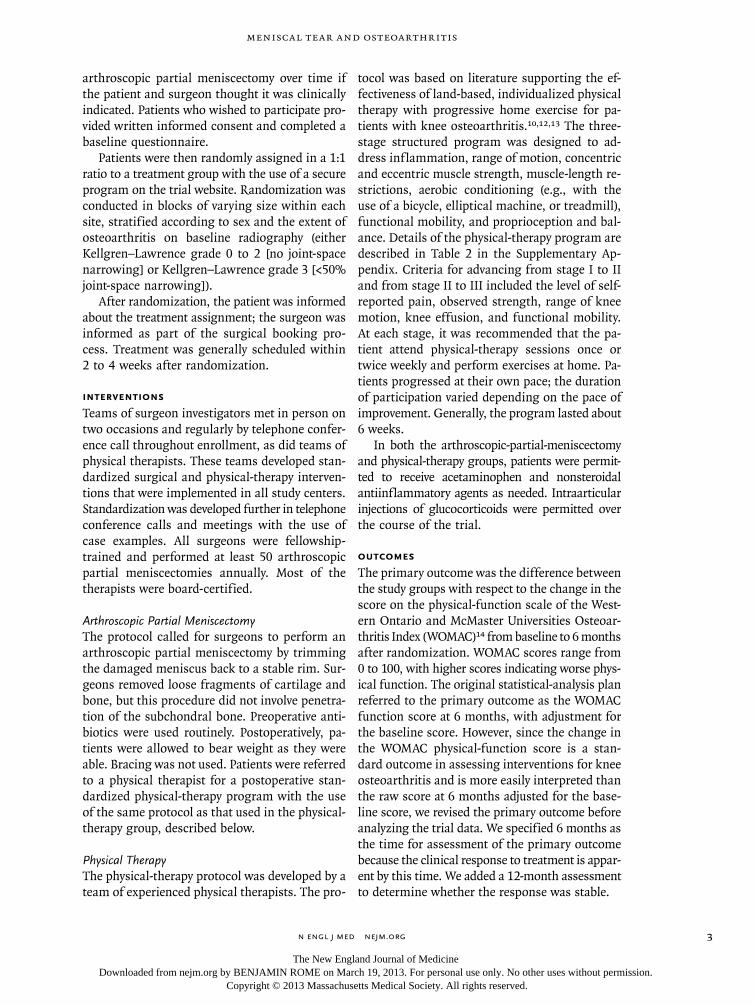
Physical TherapyThe physical-therapy protocol was developed by a team of experienced physical therapists. The pro-
tocol was based on literature supporting the ef-fectiveness of land-based, individualized physical therapy with progressive home exercise for pa-tients with knee osteoarthritis.10,12,13 The three-stage structured program was designed to ad-dress inflammation, range of motion, concentric and eccentric muscle strength, muscle-length re-strictions, aerobic conditioning (e.g., with the use of a bicycle, elliptical machine, or treadmill), functional mobility, and proprioception and bal-ance. Details of the physical-therapy program are described in Table 2 in the Supplementary Ap-pendix. Criteria for advancing from stage I to II and from stage II to III included the level of self-reported pain, observed strength, range of knee motion, knee effusion, and functional mobility. At each stage, it was recommended that the pa-tient attend physical-therapy sessions once or twice weekly and perform exercises at home. Pa-tients progressed at their own pace; the duration of participation varied depending on the pace of improvement. Generally, the program lasted about 6 weeks.
In both the arthroscopic-partial-meniscectomy and physical-therapy groups, patients were permit-ted to receive acetaminophen and nonsteroidal antiinflammatory agents as needed. Intraarticular injections of glucocorticoids were permitted over the course of the trial.
Outcomes
The primary outcome was the difference between the study groups with respect to the change in the score on the physical-function scale of the West-ern Ontario and McMaster Universities Osteoar-thritis Index (WOMAC)14 from baseline to 6 months after randomization. WOMAC scores range from 0 to 100, with higher scores indicating worse phys-ical function. The original statistical-analysis plan referred to the primary outcome as the WOMAC function score at 6 months, with adjustment for the baseline score. However, since the change in the WOMAC physical-function score is a stan-dard outcome in assessing interventions for knee osteoarthritis and is more easily interpreted than the raw score at 6 months adjusted for the base-line score, we revised the primary outcome before analyzing the trial data. We specified 6 months as the time for assessment of the primary outcome because the clinical response to treatment is appar-ent by this time. We added a 12-month assessment to determine whether the response was stable.
The New England Journal of Medicine Downloaded from nejm.org by BENJAMIN ROME on March 19, 2013. For personal use only. No other uses without permission.
Copyright © 2013 Massachusetts Medical Society. All rights reserved.

T h e n e w e ngl a nd j o u r na l o f m e dic i n e
n engl j med nejm.org4
Secondary outcomes were the pain score on the Knee Injury and Osteoarthritis Outcome Scale (KOOS), which has been used frequently in stud-
ies involving patients with a meniscal tear,15,16 and the score on the physical-activity scale of the Medical Outcomes Study 36-Item Short-Form Health Survey (SF-36). Scores on both scales range from 0 to 100, with higher KOOS scores indicat-ing more severe pain and higher SF-36 scores indicating greater physical activity.17 We also con-sidered a binary outcome that was defined as improvement in the WOMAC physical-function score of at least 8 points (a clinically relevant difference specified a priori10,18,19) without cross-over to the other study group.
Assessments
Questionnaires were administered at baseline and 3, 6, and 12 months after randomization. The primary outcome was assessed at 6 months, with the 3-month and 12-month assessments used to capture the trajectory and stability of the treat-ment response. Site coordinators contacted the participants by telephone every other week for the first 3 months after randomization and quar-terly thereafter to ascertain adverse events and compliance with physical therapy. Surgeons, pa-tients, and research staff were aware of the treat-ment assignments.
Radiographs of the weight-bearing knee were assessed at each study site by the participating surgeon on the basis of the Kellgren–Lawrence grade20 and were then reassessed centrally (also on the basis of the Kellgren–Lawrence grade)21 by a musculoskeletal radiologist. The concordance be-tween these readings was 71.8%. Readings per-formed at the clinical site were used for assess-ing eligibility and randomization strata, whereas central readings were used in the analysis. Analy-ses performed with readings at the clinical site did not materially differ from those performed with central readings.
Statistical Analysis
The primary analysis was implemented with an analysis of covariance with changes in the WOMAC physical-function score from baseline to 6 months as the dependent variable, treatment as the independent variable of interest, and study site as a covariate. Other covariates, such as age, sex, and baseline Kellgren–Lawrence grade, were balanced across groups and were therefore not included in the analysis. The primary analysis used a modified intention-to-treat approach in which patients who did not withdraw from the
351 Underwent randomization
14,430 Patients were assessedfor eligibility
14,079 Did not undergo randomization
1,092 Were not screened byphysician
12,008 Did not meet inclusioncriteria
3690 Underwent previoussurgery
2691 Did not have MRI2198 Had patellofemoral
disorder1816 Had grade 4 on
Kellgren–Lawrence scale
1613 Had other reasons195 Were eligible, but were
not referred784 Were eligible, but declined
to participate283 Had preference for
APM166 Had preference for PT335 Had other reason
174 Were assigned to APM 177 Were assigned to PT
13 Did not complete study through6 mo
1 Died3 Underwent TKR7 Withdrew2 Were ineligible
161 Were evaluated in 6-mo follow-up9 Did not undergo APM
8 Did not complete study through6 mo
1 Died1 Underwent TKR4 Withdrew2 Were lost to follow-up
169 Were evaluated in 6-mo follow-up51 Crossed over and underwent APM
18 Did not complete study through12 mo
1 Died5 Underwent TKR9 Withdrew2 Were ineligible1 Was lost to follow-up
156 Were evaluated in 12-mo follow-up9 Did not undergo APM
13 Did not complete study through12 mo
1 Died3 Underwent TKR7 Withdrew2 Were lost to follow-up
164 Were evaluated in 12-mo follow-up59 Crossed over and underwent
APM
AUTHOR:
FIGURE:
SIZE
4-C H/TLine Combo
Revised
AUTHOR, PLEASE NOTE: Figure has been redrawn and type has been reset.
Please check carefully.
Katz
1 of 2
ARTIST:
TYPE:
ts
05-09-13JOB: 36819 ISSUE:03-19-13OLF:
4 col22p3
Figure 1. Trial Enrollment and Follow-up.
APM denotes arthroscopic partial meniscectomy, MRI magnetic resonance imaging, PT physical therapy, and TKR total knee replacement.
The New England Journal of Medicine Downloaded from nejm.org by BENJAMIN ROME on March 19, 2013. For personal use only. No other uses without permission.
Copyright © 2013 Massachusetts Medical Society. All rights reserved.
Meniscal Tear and Osteoarthritis
n engl j med nejm.org 5
study were evaluated in the group to which they were randomly assigned. We performed three sec-ondary analyses: an analogous intention-to-treat analysis of covariance with the use of either the KOOS pain score or the SF-36 physical-activity score as the dependent variables and a logistic regression, with adjustment for the study site, which used the binary outcome defined above. We prespecified one subgroup analysis based on the baseline radiographic grade (Kellgren–Lawrence grade 0 to 2 vs. Kellgren–Lawrence grade 3).10,22 Additional analyses with adjustment for uncer-tainty due to missing data are described in the Supplementary Appendix.23
We powered the study to detect a 10-point dif-ference in the WOMAC physical-function score between the arthroscopic-partial-meniscectomy and physical-therapy groups. This was the dif-ference we noted in observational pilot data, and it is close to the minimal clinically important difference in the WOMAC physical-function score among patients with osteoarthritis.18,19 On the basis of a type I error rate of 5% and a power of 80%, and taking into account potential losses to follow-up and crossovers from the as-signed group to the other group before the as-sessment of the primary outcome, we set the target sample size at 340 patients.
R esult s
Characteristics of the Study Population
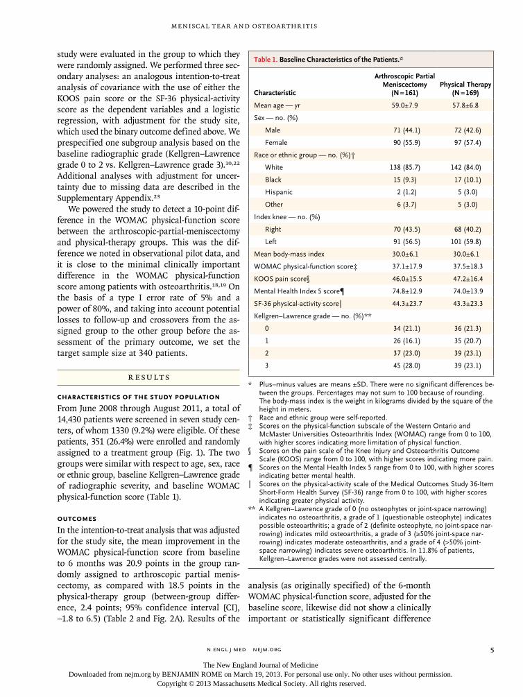
From June 2008 through August 2011, a total of 14,430 patients were screened in seven study cen-ters, of whom 1330 (9.2%) were eligible. Of these patients, 351 (26.4%) were enrolled and randomly assigned to a treatment group (Fig. 1). The two groups were similar with respect to age, sex, race or ethnic group, baseline Kellgren–Lawrence grade of radiographic severity, and baseline WOMAC physical-function score (Table 1).
Outcomes
In the intention-to-treat analysis that was adjusted for the study site, the mean improvement in the WOMAC physical-function score from baseline to 6 months was 20.9 points in the group ran-domly assigned to arthroscopic partial menis-cectomy, as compared with 18.5 points in the physical-therapy group (between-group differ-ence, 2.4 points; 95% confidence interval [CI], −1.8 to 6.5) (Table 2 and Fig. 2A). Results of the
analysis (as originally specified) of the 6-month WOMAC physical-function score, adjusted for the baseline score, likewise did not show a clinically important or statistically significant difference
Table 1. Baseline Characteristics of the Patients.*
Characteristic
Arthroscopic Partial Meniscectomy
(N = 161)Physical Therapy
(N = 169)
Mean age — yr 59.0±7.9 57.8±6.8
Sex — no. (%)
Male 71 (44.1) 72 (42.6)
Female 90 (55.9) 97 (57.4)
Race or ethnic group — no. (%)†
White 138 (85.7) 142 (84.0)
Black 15 (9.3) 17 (10.1)
Hispanic 2 (1.2) 5 (3.0)
Other 6 (3.7) 5 (3.0)
Index knee — no. (%)
Right 70 (43.5) 68 (40.2)
Left 91 (56.5) 101 (59.8)
Mean body-mass index 30.0±6.1 30.0±6.1
WOMAC physical-function score‡ 37.1±17.9 37.5±18.3
KOOS pain score§ 46.0±15.5 47.2±16.4
Mental Health Index 5 score¶ 74.8±12.9 74.0±13.9
SF-36 physical-activity score‖ 44.3±23.7 43.3±23.3
Kellgren–Lawrence grade — no. (%)**
0 34 (21.1) 36 (21.3)
1 26 (16.1) 35 (20.7)
2 37 (23.0) 39 (23.1)
3 45 (28.0) 39 (23.1)
* Plus–minus values are means ±SD. There were no significant differences be-tween the groups. Percentages may not sum to 100 because of rounding. The body-mass index is the weight in kilograms divided by the square of the height in meters.
† Race and ethnic group were self-reported.‡ Scores on the physical-function subscale of the Western Ontario and
McMaster Universities Osteoarthritis Index (WOMAC) range from 0 to 100, with higher scores indicating more limitation of physical function.
§ Scores on the pain scale of the Knee Injury and Osteoarthritis Outcome Scale (KOOS) range from 0 to 100, with higher scores indicating more pain.
¶ Scores on the Mental Health Index 5 range from 0 to 100, with higher scores indicating better mental health.
‖ Scores on the physical-activity scale of the Medical Outcomes Study 36-Item Short-Form Health Survey (SF-36) range from 0 to 100, with higher scores indicating greater physical activity.
** A Kellgren–Lawrence grade of 0 (no osteophytes or joint-space narrowing) indicates no osteoarthritis, a grade of 1 (questionable osteophyte) indicates possible osteoarthritis; a grade of 2 (definite osteophyte, no joint-space nar-rowing) indicates mild osteoarthritis, a grade of 3 (≥50% joint-space nar-rowing) indicates moderate osteoarthritis, and a grade of 4 (>50% joint-space narrowing) indicates severe osteoarthritis. In 11.8% of patients, Kellgren–Lawrence grades were not assessed centrally.
The New England Journal of Medicine Downloaded from nejm.org by BENJAMIN ROME on March 19, 2013. For personal use only. No other uses without permission.
Copyright © 2013 Massachusetts Medical Society. All rights reserved.

T h e n e w e ngl a nd j o u r na l o f m e dic i n e
n engl j med nejm.org6
Tabl
e 2.
Pri
mar
y an
d Se
cond
ary
Out
com
es o
f the
Tri
al.*
Out
com
e
Art
hros
copi
c Pa
rtia
l M
enis
cect
omy
(N
= 1
61)
Phys
ical
The
rapy
(N
= 1
69)
Impr
ovem
ent
from
Bas
elin
e
Bet
wee
n-G
roup
D
iffer
ence
in
Impr
ovem
ent
fr
om B
asel
ine
Art
hros
copi
c Pa
rtia
l M
enis
cect
omy
Phys
ical
The
rapy
6 M
onth
s
WO
MA
C p
hysi
cal-f
unct
ion
scor
e —
mea
n (9
5% C
I)14
.7 (
12.0
to 1
7.5)
19.0
(16
.3 to
21.
7)20
.9 (
17.9
to 2
3.9)
18.5
(15
.6 to
21.
5)2.
4 (−
1.8
to 6
.5)†
KO
OS
pain
sco
re —
mea
n (9
5% C
I)21
.1 (
18.3
to 2
3.9)
25.2
(22
.4 to
28.
0)24
.2 (
21.3
to 2
7.1)
21.3
(18
.4 to
24.
2)2.
9 (−
1.2
to 7
.0)
SF-3
6 ph
ysic
al-a
ctiv
ity s
core
— m
ean
(95%
CI)
69.2
(65
.2 to
73.
2)66
.1 (
62.1
to 7
0.1)
24.2
(20
.3 to
28.
0)23
.1 (
19.2
to 2
7.0)
1.1
(−4.
4 to
6.6
)
Trea
tmen
t suc
cess
— n
o. (
%)‡
108
(67)
74 (
44)
Trea
tmen
t fai
lure
— n
o. (
%)
40 (
25)
82 (
49)
WO
MA
C p
hysi
cal-f
unct
ion
scor
e im
prov
emen
t <8
poin
ts
and
no c
ross
over
— n
o./t
otal
no.
(%
)32
/40
(80)
31/8
2 (3
8)
Cro
ssov
er w
ithin
6 m
o —
no.
/tot
al n
o. (
%)§
8/40
(20
)51
/82
(62)
Dat
a m
issi
ng —
no.
(%
)13
(8)
13 (
8)
12 M
onth
s —
mea
n (9
5% C
I)
WO
MA
C p
hysi
cal-f
unct
ion
scor
e13
.7 (
11.2
to 1
6.2)
14.5
(12
.0 to
16.
9)23
.5 (
20.5
to 2
6.5)
22.8
(19
.8 to
25.
8)0.
7 (−
3.5
to 4
.9)
KO
OS
pain
sco
re19
.1 (
16.4
to 2
1.9)
19.3
(16
.6 to
22.
0)26
.8 (
23.7
to 3
0.0)
27.3
(24
.1 to
30.
4)−0
.4 (
−4.8
to 4
.0)
SF-3
6 ph
ysic
al-a
ctiv
ity s
core
69.0
(64
.6 to
73.
4)71
.4 (
67.0
to 7
5.7)
25.0
(20
.9 to
29.
1)28
.1 (
24.0
to 3
2.1)
−3.0
(−8
.8 to
2.7
)
* B
etw
een-
grou
p di
ffere
nces
may
not
equ
al t
he d
iffer
ence
s in
cha
nge
from
bas
elin
e be
twee
n th
e pa
rtia
l-men
isce
ctom
y an
d ph
ysic
al-t
hera
py g
roup
s be
caus
e of
rou
ndin
g. C
I de
note
s co
n-fid
ence
inte
rval
.†
Thi
s be
twee
n-gr
oup
diffe
renc
e w
as t
he p
rim
ary
outc
ome.
‡
Tre
atm
ent
succ
ess
indi
cate
s an
impr
ovem
ent
in t
he W
OM
AC
phy
sica
l-fun
ctio
n sc
ore
of 8
poi
nts
or m
ore,
with
no
cros
sove
r.§
Eigh
t pa
tient
s in
the
par
tial-m
enis
cect
omy
grou
p cr
osse
d ov
er t
o su
rger
y w
ithin
6 m
onth
s, a
nd 1
cro
ssed
ove
r af
ter
6 m
onth
s.
The New England Journal of Medicine Downloaded from nejm.org by BENJAMIN ROME on March 19, 2013. For personal use only. No other uses without permission.
Copyright © 2013 Massachusetts Medical Society. All rights reserved.

Meniscal Tear and Osteoarthritis
n engl j med nejm.org 7
between groups (difference, 3.4 points; 95% CI, −0.04 to 6.8). In the intention-to-treat analysis of the KOOS pain score, the mean decreases (i.e., improvements) from baseline to 6 months were 24.2 points in patients assigned to arthroscopic partial meniscectomy versus 21.3 points in those assigned to physical therapy alone (between-group difference, 2.9 points; 95% CI, −1.2 to 7.0) (Table 2 and Fig. 2B). In intention-to-treat analy-ses of 12-month outcomes adjusted for study site, the two groups had similar changes from base-line in the WOMAC physical-function and KOOS pain scores (Table 2).
Among 330 active participants in the study, by 6 months of follow-up, 51 patients assigned to physical therapy alone (30.2%) had undergone arthroscopic partial meniscectomy, whereas 9 pa-tients assigned to surgery (5.6%) had not under-gone the procedure. An additional 8 active pa-tients in the study (4.7%) who were assigned to the physical-therapy group crossed over to ar-throscopic partial meniscectomy between 6 and 12 months. At 6 months, 67.1% of the patients assigned to arthroscopic partial meniscectomy had an improvement of at least 8 points in the WOMAC physical-function score and had not crossed over to the other study treatment, as compared with 43.8% of patients assigned to the physical-therapy group (P = 0.001). Patients in the physical-therapy group who crossed over and underwent arthroscopic partial meniscectomy during the first 6 months had WOMAC physical-function scores at 12 months that were similar to those of patients assigned to the arthroscopic-partial-meniscectomy group (Fig. 2C). The pro-portion of patients who crossed over from physi-cal therapy to arthroscopic partial meniscectomy ranged from 0.0 to 59.5% across study centers. In
WO
MA
C P
hysi
cal-F
unct
ion
Scor
e
50
45
40
35
25
20
10
30
15
0Baseline 3 6 12
Months
B
A
No. at RiskAPMPT
159167
154155
147153
140144
APM
PT
KO
OS
Pain
Sco
re
50
45
40
35
25
20
10
30
15
0Baseline 3 6 12
Months
No. at RiskAPMPT
160167
155154
148153
142145
Baseline 3 6
9
9
9 12
Months
151*105 26 24 12
14694262411
14095242311
13392212110
APM
PT
C
WO
MA
C P
hysi
cal-F
unct
ion
Scor
e
50
40
45
35
30
20
15
10
25
0
No. at RiskAPMPT, no crossoverPT, crossover <3 moPT, crossover 3–6 moPT, crossover >6 mo
APMPT, no crossoverPT, crossover <3 mo
PT, crossover >6 moPT, crossover 3–6 mo
Figure 2. Scores on the WOMAC Physical-Function Scale and KOOS Pain Scale over the 12-Month Follow-up Period.
Panel A shows the scores on the physical-function scale of the Western Ontario and McMaster Universities Osteoarthritis Index (WOMAC), and Panel B shows the scores on the pain scale of the Knee Injury and Osteoarthritis Outcome Scale (KOOS); scores on both scales range from 0 to 100, with higher scores indicating more severe symptoms. I bars indicate 95% confidence intervals. Panel C shows WOMAC physical-function scores in the APM group and in the PT group accord-ing to crossover status. The asterisk indicates that nine patients assigned to APM did not undergo surgery.
The New England Journal of Medicine Downloaded from nejm.org by BENJAMIN ROME on March 19, 2013. For personal use only. No other uses without permission.
Copyright © 2013 Massachusetts Medical Society. All rights reserved.

T h e n e w e ngl a nd j o u r na l o f m e dic i n e
n engl j med nejm.org8
general, the patients assigned to receive physical therapy alone who crossed over to surgery did not have substantial improvement in functional status during the period from randomization until the time of crossover (Fig. 2C).
In the physical-therapy group, patients were scheduled for an average of 9.3 physical-therapy visits and attended an average of 8.4 visits (90.6%). In the arthroscopic-partial-meniscectomy group, patients were scheduled for an average of 7.4 visits and attended 6.9 visits (92.9%). In the physical-therapy group, 21 patients (12.4%) re-ceived intraarticular glucocorticoid injections, as did 9 patients (5.6%) in the arthroscopic-partial-meniscectomy group.
The between-group difference in functional improvement from baseline to 6 months did not differ significantly according to the Kellgren–
Lawrence grade of radiographic severity (P = 0.13 for interaction) (Table 3 in the Supplementary Appendix).
Adverse Events
There were no significant between-group dif-ferences in the frequencies of overall or specific adverse events. Over the 12-month period of follow-up, serious adverse events occurred in 3 partici-pants assigned to arthroscopic partial meniscec-tomy and 2 participants assigned to physical therapy alone (including one death in each group); adverse events rated as mild or moderate in sever-ity occurred in 15 participants in the arthroscopic-partial-meniscectomy group and 13 participants in the physical-therapy group (Table 3). Total knee replacement (coded not as an adverse event but rather as an indication for discontinuation from the study) was performed in 5 participants assigned to arthroscopic partial meniscectomy and 3 participants assigned to physical therapy alone (Fig. 1).
Discussion
In this seven-center randomized, controlled trial involving symptomatic patients 45 years of age or older with a meniscal tear and imaging evidence of mild-to-moderate knee osteoarthritis, there were no significant differences in the magnitude of improvement in functional status and pain af-ter 6 and 12 months between patients assigned to arthroscopic partial meniscectomy with postopera-tive physical therapy and patients assigned to a standardized physical-therapy regimen. These re-sults were achieved with a 30% rate of crossover to arthroscopic partial meniscectomy at 6 months. At 12 months, among 169 participants (not all of whom provided data at the 1-year evaluation), the rate of crossover to surgery was 35%.
In a prior small, single-center, randomized, controlled trial comparing arthroscopic partial meniscectomy with standardized physical thera-py for symptomatic patients with a meniscal tear and knee osteoarthritis, the two groups had simi-lar functional outcomes at 6 months, and the similarity between the groups persisted through 5 years of follow-up.8,9 To our knowledge, this is the first large, multicenter, randomized, controlled trial to examine the efficacy of arthroscopic par-tial meniscectomy as compared with a standard-ized physical-therapy regimen.
Table 3. Adverse Events at 12 Months in All Patients Assigned to Treatment.
Event
Arthroscopic Partial Meniscectomy
(N = 174)Physical Therapy
(N = 177)
number of patients
Serious adverse events
Cardiovascular
Pulmonary embolism (fatal) 1 0
Acute myocardial infarction 1 0
Sudden death 0 1
Stroke 0 1
Hypoxemia 1 0
Total 3 2
Nonserious adverse events
Musculoskeletal
Pain from fall or other trauma 2 4
Tendonitis 3 0
Knee bursitis 0 1
Rupture of Baker’s cyst 1 0
Knee pain 1 1
Pain in the back, hip, or foot 2 4
Cardiovascular
Deep-vein thrombosis 2 0
Syncope 1 0
Atrial fibrillation 0 1
Skin 2 1
Other 1 1
Total 15 13
The New England Journal of Medicine Downloaded from nejm.org by BENJAMIN ROME on March 19, 2013. For personal use only. No other uses without permission.
Copyright © 2013 Massachusetts Medical Society. All rights reserved.

Meniscal Tear and Osteoarthritis
n engl j med nejm.org 9
Surgical randomized, controlled trials present methodologic challenges, including crossover from one group to the other.24,25 To account for cross-overs, we defined an additional outcome a priori in which patients were deemed to have a suc-cessful treatment response if they had improve-ment of at least 8 points on the WOMAC physical-function scale (a clinically important difference) and they did not cross over from their assigned treatment. A total of 67% of patients assigned to arthroscopic partial meniscectomy met this thresh-old for success, as compared with 44% of pa-tients treated with physical therapy alone. We acknowledge, however, that because the treat-ment assignments were not blinded, and be-cause crossover could not occur in the ar-throscopic-partial-meniscectomy group once the surgery had been performed, this secondary analysis was vulnerable to bias.
Several limitations of the study warrant dis-cussion. First, because we enrolled only 26% of eligible patients, our findings must be general-ized cautiously. The most frequent reason that patients declined enrollment was a strong pref-erence for one treatment or the other. Since pa-tients’ preferences may be associated with treat-ment outcome, our trial may be vulnerable to selection bias. Participating surgeons may not have referred potentially eligible patients because they were uncomfortable randomly assigning these patients to treatment; this form of selec-tive enrollment may also create bias.26 Second, because the trial was conducted in academic referral centers, the findings should be general-ized carefully to community settings. Third, we did not formally assess the fidelity of the physi-cal therapists or surgeons to the standard inter-vention protocols. Finally, our study was not blinded, since our investigative group did not consider a sham comparison group feasible.
These limitations notwithstanding, the results of our trial may help guide management in the care of patients with knee symptoms, a meniscal tear, and imaging evidence of osteoarthritis. Our findings suggest that both arthroscopic partial meniscectomy and referral to physical therapy — with an opportunity to consider arthroscopic partial meniscectomy if substantial improve-ments are not achieved — are likely to result in considerable improvement in functional status and knee pain over a 6-to-12-month period. Given that improvements in functional status and pain
at 6 months did not differ significantly between patients assigned to arthroscopic partial menis-cectomy and those assigned to physical therapy alone and that 70% of the patients in the physi-cal-therapy group did not undergo surgery, these data provide considerable reassurance regarding an initial nonoperative strategy. It is uncertain whether patients who undergo arthroscopic par-tial meniscectomy are at greater risk for progres-sion of underlying osteoarthritis than patients treated nonoperatively.27-30 Longitudinal assess-ment of imaging studies in our trial is planned to address this question.
In summary, symptomatic patients with a meniscal tear and imaging evidence of mild-to-moderate osteoarthritis who were randomly as-signed to arthroscopic partial meniscectomy with postoperative physical therapy had improvements in functional status and pain at 6 months that did not differ significantly from the improvements in patients randomly assigned to a standardized physical-therapy regimen alone. However, 30% of patients assigned to the physical-therapy group crossed over to surgery in the first 6 months. These findings should help inform decision mak-ing by patients and their physicians.
Supported by grants (R01AR055557, K24AR057827, and P60AR047782) from the National Institute of Arthritis and Muscu-loskeletal and Skin Diseases of the National Institutes of Health.
Dr. Brophy reports receiving fees for providing expert testi-mony on behalf of Genzyme on the use of Synvisc and grant support through his institution from the Orthopaedic Research and Education Foundation. Dr. Cole reports receiving consult-ing fees from Arthrex, Carticept, DJ Orthopedics, Genzyme, Johnson & Johnson, DePuy Orthopedics, Regentis, and Zimmer, lecture fees from Genzyme, fees for providing expert testimony for medical malpractice defense and third-party liability cases on personal injury and product liability, and royalties from Ar-threx, DJ Orthopedics, Elsevier, and W.B. Saunders, holding eq-uity in Carticept and Regentis, receiving grant support through his institution from Major League Baseball, Arthrex, Zimmer, Arthrosurface, Johnson & Johnson, and Medipost and fellow-ship support through his institution from DJ Orthopedics, Ossur, and Smith and Nephew. Dr. Guermazi reports receiving consult-ing fees from Merck Serono, Pfizer, and Genzyme, and holding stock in Boston Imaging Core Lab. Dr. Jones reports receiving consulting fees from Allergan and grant support through his institution from DJ Orthopedics, Stryker, Smith and Nephew, Breg, and Arthrex. Dr. Levy reports receiving consulting fees from Arthrex and royalties from VOT Solutions and Arthrex. Dr. Matava reports receiving consulting fees from ISTO Technolo-gies, Schwartz Biomedical, and Ostesys, and grant support through his institution from Arthrex and Breg. Dr. Miniaci re-ports receiving consulting fees from Arthrosurface and Stryker and lecture fees, payment for the development of educational presentations, and travel support from Arthrosurface, holding stock in Arthrosurface, and receiving royalties from Arthrosur-face for granted and pending patents regarding retrograde deliv-ery of resurfacing devices, an articular surface implant and de-livery system, and a system and method for a retrograde procedure and grant support through his institution from DJ
The New England Journal of Medicine Downloaded from nejm.org by BENJAMIN ROME on March 19, 2013. For personal use only. No other uses without permission.
Copyright © 2013 Massachusetts Medical Society. All rights reserved.

n engl j med nejm.org10
Meniscal Tear and Osteoarthritis
Orthopedics, Stryker, and Arthrex. Dr. Reinke reports receiving grant support through her institution from Smith and Nephew, DonJoy, and the National Football League. Dr. Smith reports receiving consulting fees from ISTO Technologies. Dr. Spindler reports receiving consulting fees from the National Football League and payment for patents from Connective Orthopedics on a biologic replacement for fibrin clots. Dr. Stuart reports re-ceiving consulting fees and royalties from Arthrex and grant support through his institution from Stryker. Dr. J. Wright re-ports receiving consulting fees from DePuy Orthopedics. Dr. R. Wright reports receiving consulting fees from Flexion Therapeu-
tics and ISTO Technologies, and grant support through his in-stitution from Smith and Nephew. No other potential conflict of interest relevant to this article was reported.
Disclosure forms provided by the authors are available with the full text of this article at NEJM.org.
We thank the METEOR scientific advisory board (Thomas S. Thornhill, M.D., chair, John A. Baron, M.D., and Alan M. Jette, Ph.D.) for their advice; Nizar N. Mahomed, M.D., Sc.D., for sup-port and scientific input; and the dozens of investigators and research staff and hundreds of patients across seven centers who participated in the trial.
References
1. Lawrence RC, Felson DT, Helmick CG, et al. Estimates of the prevalence of arthritis and other rheumatic conditions in the United States. Arthritis Rheum 2008;58:26-35.2. Englund M, Guermazi A, Gale D, et al. Incidental meniscal findings on knee MRI in middle-aged and elderly persons. N Engl J Med 2008;359:1108-15.3. Bhattacharyya T, Gale D, Dewire P, et al. The clinical importance of meniscal tears demonstrated by magnetic reso-nance imaging in osteoarthritis of the knee. J Bone Joint Surg Am 2003;85:4-9.4. Sowers M, Karvonen-Gutierrez CA, Jacobson JA, Jiang Y, Yosef M. Associa-tions of anatomical measures from MRI with radiographically defined knee osteo-arthritis score, pain, and physical func-tioning. J Bone Joint Surg Am 2011;93: 241-51.5. Kim S, Bosque J, Meehan JP, Jamali A, Marder R. Increase in outpatient knee ar-throscopy in the United States: a com-parison of National Surveys of Ambula-tory Surgery, 1996 and 2006. J Bone Joint Surg Am 2011;93:994-1000.6. Moseley JB, O’Malley K, Petersen NJ, et al. A controlled trial of arthroscopic surgery for osteoarthritis of the knee. N Engl J Med 2002;347:81-8.7. Kirkley A, Birmingham TB, Litchfield RB, et al. A randomized trial of ar-throscopic surgery for osteoarthritis of the knee. N Engl J Med 2008;359:1097-107. [Erratum, N Engl J Med 2009;361:2004.]8. Herrlin S, Hållander M, Wange P, Weidenhielm L, Werner S. Arthroscopic or conservative treatment of degenerative medial meniscal tears: a prospective ran-domised trial. Knee Surg Sports Trauma-tol Arthrosc 2007;15:393-401.9. Herrlin SV, Wange PO, Lapidus G, Hållander M, Werner S, Weidenhielm L. Is arthroscopic surgery beneficial in treating non-traumatic, degenerative me-dial meniscal tears? A five year follow-up. Knee Surg Sports Traumatol Arthrosc 2013;21:358-64.10. Katz JN, Chaisson CE, Cole B, et al. The MeTeOR trial (Meniscal Tear in Os-teoarthritis Research): rationale and de-
sign features. Contemp Clin Trials 2012; 33:1189-96.11. Niu NN, Losina E, Martin SD, Wright J, Solomon DH, Katz JN. Development and preliminary validation of a meniscal symptom index. Arthritis Care Res (Hoboken) 2011;63:208-15.12. Fransen M, McConnell S. Exercise for osteoarthritis of the knee. Cochrane Da-tabase Syst Rev 2008;4:CD004376.13. Fransen M, McConnell S. Land-based exercise for osteoarthritis of the knee: a metaanalysis of randomized controlled trials. J Rheumatol 2009;36:1109-17.14. Bellamy N, Buchanan WW, Gold-smith CH, Campbell J, Stitt LW. Valida-tion study of WOMAC: a health status instrument for measuring clinically im-portant patient relevant outcomes to anti-rheumatic drug therapy in patients with osteoarthritis of the hip or knee. J Rheu-matol 1988;15:1833-40.15. Roos EM, Roos HP, Ekdahl C, Loh-mander LS. Knee Injury and Osteoarthri-tis Outcome Score (KOOS) — validation of a Swedish version. Scand J Med Sci Sports 1998;8:439-48.16. Roos EM, Roos HP, Lohmander LS, Ekdahl C, Beynnon BD. Knee Injury and Osteoarthritis Outcome Score (KOOS) — development of a self-administered out-come measure. J Orthop Sports Phys Ther 1998;28:88-96.17. Ware JE Jr, Sherbourne CD. The MOS 36-Item Short-Form Health Survey (SF-36). I. Conceptual framework and item selec-tion. Med Care 1992;30:473-83.18. Angst F, Aeschlimann A, Michel BA, Stucki G. Minimal clinically important rehabilitation effects in patients with os-teoarthritis of the lower extremities. J Rheumatol 2002;29:131-8.19. Angst F, Aeschlimann A, Stucki G. Smallest detectable and minimal clini-cally important differences of rehabilita-tion intervention with their implications for required sample sizes using WOMAC and SF-36 quality of life measurement in-struments in patients with osteoarthritis of the lower extremities. Arthritis Rheum 2001;45:384-91.20. Kellgren JH, Lawrence JS. Radiologi-
cal assessment of osteo-arthrosis. Ann Rheum Dis 1957;16:494-502.21. Guermazi A, Hunter DJ, Li L, et al. Dif-ferent thresholds for detecting osteophytes and joint space narrowing exist between the site investigators and the centralized reader in a multicenter knee osteoarthritis study — data from the Osteoarthritis Ini-tiative. Skeletal Radiol 2012;41:179-86.22. Wang R, Lagakos SW, Ware JH, Hunter DJ, Drazen JM. Statistics in medicine — reporting of subgroup analyses in clinical trials. N Engl J Med 2007;357:2189-94.23. Little RJ, D’Agostino R, Cohen ML, et al. The prevention and treatment of miss-ing data in clinical trials. N Engl J Med 2012;367:1355-60.24. Frobell RB, Roos EM, Roos HP, Rans-tam J, Lohmander LS. A randomized trial of treatment for acute anterior cruciate ligament tears. N Engl J Med 2010;363:331-42. [Erratum, N Engl J Med 2010;363:893.]25. Weinstein JN, Tosteson TD, Lurie JD, et al. Surgical versus nonsurgical therapy for lumbar spinal stenosis. N Engl J Med 2008;358:794-810.26. Katz JN, Wright J, Levy BA, Baron JA, Losina E. Departures from community equipoise may lead to incorrect inference in randomized trials. J Clin Epidemiol 2011;64:280-5.27. Englund M, Lohmander LS. Risk fac-tors for symptomatic knee osteoarthritis fifteen to twenty-two years after meniscec-tomy. Arthritis Rheum 2004;50:2811-9.28. Mills PM, Wang Y, Cicuttini FM, et al. Tibio-femoral cartilage defects 3-5 years following arthroscopic partial medial meniscectomy. Osteoarthritis Cartilage 2008;16:1526-31.29. Papalia R, Del Buono A, Osti L, Den-aro V, Maffulli N. Meniscectomy as a risk factor for knee osteoarthritis: a system-atic review. Br Med Bull 2011;99:89-106.30. Englund M, Guermazi A, Roemer FW, et al. Meniscal tear in knees without sur-gery and the development of radiographic osteoarthritis among middle-aged and elderly persons: the Multicenter Osteoar-thritis Study. Arthritis Rheum 2009;60: 831-9.Copyright © 2013 Massachusetts Medical Society.
The New England Journal of Medicine Downloaded from nejm.org by BENJAMIN ROME on March 19, 2013. For personal use only. No other uses without permission.
Copyright © 2013 Massachusetts Medical Society. All rights reserved.
Copyright © 2022 FDOKUMEN