Electrochemical Detection of Glutathione Using Redox Indicators
Upload
khangminh22Category
view
0download
0
Coenzyme A biosynthesis and Coenzyme A-
dependent redox processes as targets for
anti-staphylococcal drug development
December 2015
Supervisor: Prof. Erick Strauss
Wessel Johannes Albertus Moolman
Dissertation presented for the degree of Doctor of
Philosophy (Biochemistry) in the
Faculty of Natural Sciences at
Stellenbosch University
ii
Declaration
By submitting this dissertation electronically, I declare that the entirety of the work contained therein is my
own, original work, that I am the owner of the copyright thereof (unless to the extent explicitly otherwise
stated), that reproduction and publication thereof by Stellenbosch University will not infringe any third party
rights and that I have not previously in its entirety or in part submitted it for obtaining any qualification.
..................................................... 2015-10-07 ...................................
Signature Date
Copyright © 2015 Stellenbosch UniversityAll rights reserved
Stellenbosch University https://scholar.sun.ac.za

iii
Summary
Staphylococcus aureus, the bacterium that causes most hospital-acquired in humans is rapidly becoming
more prevalent in the community and, alarmingly, increasingly resistant to the current arsenal of available
antibacterial agents. More than ever, new treatments are urgently needed to combat this threat. In this study
we proposed an alternative strategy to current drug development methodologies that entails the identification
and targeting of processes that are essential to the survival of pathogenic bacteria in their human host, i.e.
where they need to counter the defences of the human immune system. In particular, the focus of this study
is the importance of the central metabolic cofactor coenzyme A (CoA) in the defence mechanisms that S.
aureus employs under such circumstances, and therefore on the targeting of CoA biosynthesis and
enzymology as potential antistaphylococcal targets.
The viability of coenzyme A disulfide reductase (CoADR) as a potential antistaphylococcal drug target was
evaluated. The S. aureus CoADR (SaCoADR) enzyme structures in complex with mechanism-based Michael
acceptor-containing inhibitors were examined; specifically how its interaction with these compounds relates
to the observed differences in activity between them. Consequently, the observed enzyme inhibition could be
adequately explained when taking into account the chemical properties of the inhibitors in combination with
their interactions with SaCoADR. Also, the structural data in the study provided a strong starting point for
future inhibitor design. The reasons for the poor correlation between the in vitro inhibition of SaCoADR by
the Michael acceptor-containing CoA analogues and the whole cell inhibition of S. aureus by their
corresponding pantothenamide precursors were investigated and these results led us to the conclusion that
the poor correlation is due to SaCoADR not being essential under normal growth conditions. However, our
results suggest that under conditions where CoA levels are sufficiently reduced, CoADR might become
relevant, even under normal growth conditions. This opens the door for studies on the possible synergistic
effects of CoADR inhibitors and compounds that reduce CoA levels; such combinations most likely hold the
most potential for work focused on CoADR as a drug target.
The mechanism of inhibition of phosphopantothenoylcysteine synthetase (PPCS) enzymes by 4’-phospho-
CJ-15,801-cytidylate (PCJ-CMP) was investigated by determining the basis for the apparent stability of the
inhibitor. We showed that the PPCS protein itself plays no role in the mechanism of inhibition by PCJ-CMP,
but that the introduction of the double bond in the β-alanine moiety of the substrate with its extra π-electrons
renders the acyl phosphate resistant to nucleophilic attack by introducing new, stable resonance forms. This
mechanism of apparent stabilisation via resonance was also applied to an unrelated system and we were
able to convert substrates of human VNN1 pantetheinase into inhibitors of the enzyme. These studies
allowed us to rationalise the tight-binding inhibition observed for PCJ-CMP. Additionally, we uncovered a
new strategy whereby β-alanine-containing compounds can be rendered resistant to hydrolysis and/or acyl
transfer; this strategy can likely have wide-ranging applications in the design of such small molecule
inhibitors and therapeutics.
Stellenbosch University https://scholar.sun.ac.za

iv
Opsomming
Staphylococcus aureus, die bakterium verantwoordelik vir die grootste hoeveelheid hospitaalverworwe
infeksies in mense, is vinnig besig om meer wydverspreid in die gemeenskap voor te kom terwyl dit besig is
om meer weerstandbiedig te raak teen die huidige arsenaal van antimikrobiese middels. Nuwe behandelinge
word nou meer as ooit benodig om hierdie bedreiging te bekamp. In hierdie studie stel ons ‘n alternatiewe
strategie voor teenoor huidige antibiotiese ontwikkelingsmetodologieë wat die indentifisering en teiken van
prosesse behels wat essensieël is vir die oorlewing van patogeniese bakterieë binne hul menslike gasheer;
of te wel, waar hulle nodig het om die verdedigingsmeganismes van die menslike immuunsisteem teen te
werk. Dienooreenkomstig is die fokus van die studie die belangrikheid van die sentrale metaboliese kofaktor
koënsiem A (KoA) in die verdedigingsmeganismes wat S. aureus gebruik onder hierdie toestande en
gevolglik op die evaluering van KoA-biosintese en ensiemologie as potensiële antistafilokokale teikens.
Die lewensvatbaarheid van koënsiem A disulfied reduktase (KoADR) as 'n potensiële antistafilokokale teiken
is geëvalueer. Die S. aureus KoADR (SaKoADR) ensiemstrukture in kompleks met meganisme-gebaseerde
Michael-akseptor-bevattende inhibeerders is ondersoek; spesifiek hoe die interaksie met hierdie verbindings
betrekking het tot die waargenome verskille in hul aktiwiteite. Gevolglik kon die waargenome ensieminhibisie
voldoende verduidelik word met inagneming van die chemiese eienskappe van die inhibeerders in
kombinasie met hul interaksie met SaKoADR. Die strukturele data in die studie verskaf 'n sterk beginpunt vir
toekomstige inhibitorontwerp. Die basis vir die swak korrelasie tussen die in vitro inhibisie van SaKoADR
deur die Michael-akseptorbevattende KoA-analoë en die bakterieële groeiïnhibisie van S. aureus deur hulle
ooreenstemmende pantoteenamied voorlopers is ondersoek en die resultate het aangetoon dat dit te wyte is
aan die feit dat SaKoADR nie noodsaaklik onder normale groeitoestande is nie. Desnieteenstaande dui die
resultate daarop dat onder omstandighede waar KoA-vlakke voldoende verminder is, KoADR dalk relevant
sal word, selfs onder normale groeitoestande. Dit lê die grondslag vir studies op die moontlike sinergistiese
effek van KoADR-inhibeerders met verbindings wat KoA-vlakke verminder; sulke kombinasies het
waarskynlik die meeste potensiaal vir werk gefokus op KoADR as 'n antistafilokokale teiken.
Die meganisme van inhibisie van fosfopantotenoïelsisteïen-sintetase (FPS) ensieme deur 4'-fosfo-CJ-
15,801-sitidilaat (FCJ-SMP) is ondersoek deur die bepaling van die basis vir die oënskynlike stabiliteit van
die inhibeerder. Ons het bewys dat die FPS proteïen self geen rol speel in die meganisme van inhibisie deur
FCJ-SMP nie, maar dat die invoeging van die dubbelbinding in die β-alanien groep van die substraat met sy
ekstra π-elektrone die asielfosfaat weerstandbiedig maak teen nukleofiele-aanval deur die vorming van
nuwe, stabiele resonansvorms. Die meganisme van stabilisering deur resonansie is ook toegepas op 'n
onverwante stelsel en ons was in staat om substrate van menslike VNN1 panteteïnase te omskep in
inhibeerders van die ensiem. Hierdie studies het ons in staatgestel om die styf-bindende inhibisie
waargeneem vir FCJ-SMP te rasionaliseer. Daarbenewens stel ons ‘n nuwe strategie bekend waardeur β-
alanien-bevattende verbindings bestand gemaak kan word teen hidrolise en / of asiel-oordrag; hierdie
strategie kan waarskynlik ‘n wye toepassing hê in die ontwerp van sulke klein-molekule inhibeerders en
terapieë.
Stellenbosch University https://scholar.sun.ac.za

v
The financial assistance of the National Research Foundation (NRF) towards this research is hereby
acknowledged. Opinions expressed and conclusions arrived at, are those of the author and are not
necessarily to be attributed to the NRF.
Stellenbosch University https://scholar.sun.ac.za

vi
"The mind is not a vessel to be filled, but a fire to be kindled."
— Plutarch
Stellenbosch University https://scholar.sun.ac.za

vii
Acknowledgements
Completing a PhD study is no trivial task and entirely impossible by oneself and therefore I need to thank
everyone who helped me along the way. First, I’d like to thank my PhD supervisor, Erick Strauss for his
continual support, guidance and patience throughout this project. Erick, thank you for taking a chance on me
and giving me the opportunity to join your lab. You have put together a great group of people that is a
pleasure to work alongside with and you created an excellent environment in which to work successfully,
while being constantly challenged. The last five years spent on this project has helped me mature from a
hesitant student interested in science, into a critical thinking scientist and has also given me the confidence
to pursue a career as a researcher.
All the Strauss-lab members, past and present, thank you all for the encouragement, help with experiments
that refuse to work (and the ones that did), chats during coffee time and generally just making me look
forward to spending time in the lab.
To all my family and friends, thank you for your encouragement and interest and pretending to understand
what I spent most of my waking hours doing over the last few years. Lastly, to Liana Swart, thank you for all
your love and support and always believing in me, even the times when I didn't believe in myself.
Stellenbosch University https://scholar.sun.ac.za

viii
Additional Acknowledgements
The University of Stellenbosch for the opportunity to study at this institution.
Financial assistance from the National Research Foundation (NRF), the Ernst & Ethel Ericksen Trust
and Prof. Erick Strauss.
Dr. D.J. Brand and Ms. Elsa Malherbe of the NMR-unit of the Central Analytical Facility of the
University of Stellenbosch.
Dr. Marietjie Stander of the MS-unit of the Central Analytical Facility of the University of
Stellenbosch.
Prof. Carine Smith and Ms. Kelly Petersen for assistance with neutrophil isolations.
Stellenbosch University https://scholar.sun.ac.za

ix
Table of contents
Declaration .......................................................................................................................................................ii
Summary .........................................................................................................................................................iii
Opsomming .....................................................................................................................................................iv
Acknowledgements ..........................................................................................................................................vii
Additional acknowledgements .........................................................................................................................viii
Table of contents .............................................................................................................................................ix
List of abbreviations .........................................................................................................................................xiii
CHAPTER 1: INTRODUCTION AND BACKGROUND ................................................................................... 1
1.1 ANTIMICROBIAL RESISTANCE AND THE NEED FOR NEW ANTIMICROBIAL TARGETS ........................................ 1
1.2 HUMAN INNATE IMMUNE SYSTEM .................................................................................................................... 3
1.2.1 Epithelial surfaces help prevent infection ................................................................................................... 3
1.2.2 Recognition of conserved features of pathogens by human cells .......................................................... 3
1.2.3 Complement activation marks pathogens for phagocytosis or lysis ....................................................... 4
1.2.4 Toll-like proteins are pattern recognition receptors ................................................................................... 4
1.2.5 Phagocytic cells engulf and destroy pathogens ........................................................................................ 4
1.3 S. AUREUS DEFENCE MECHANISMS AGAINST THE HUMAN INNATE IMMUNE SYSTEM ..................................... 5
1.3.1 Overview .......................................................................................................................................................... 5
1.3.2 Resistance to oxidative stress: Direct elimination of reactive oxygen species ..................................... 6
1.3.3 Resistance to oxidative stress: Thiol-disulfide interchange reactions .................................................... 8
1.3.4 Conclusion ..................................................................................................................................................... 16
1.4 RESEARCH QUESTION ................................................................................................................................... 17
1.5 REFERENCES ................................................................................................................................................ 17
Stellenbosch University https://scholar.sun.ac.za

x
CHAPTER 2: RECENT ADVANCES IN TARGETING COENZYME A BIOSYNTHESIS AND
UTILIZATION FOR ANTIMICROBIAL DRUG DEVELOPMENT ........................................................................25
2.1 MINI-REVIEW ................................................................................................................................................. 25
2.2 SUMMARY OF MINI-REVIEW ........................................................................................................................... 41
2.3 PREVIOUS COADR RESEARCH .................................................................................................................... 41
2.4 PREVIOUS RESEARCH ON THE INHIBITION OF PPCS BY CJ-15, 801 .......................................................... 42
2.5 REFERENCES ......................................................................................................................................................... 42
CHAPTER 3: EVALUATION OF COADR AS AN ANTI-STAPHYLOCOCCAL DRUG TARGET .............43
3.1 INTRODUCTION ....................................................................................................................................................... 43
3.2 RESULTS ................................................................................................................................................................ 48
3.2.1 Structural characterisation of the SaCoADR–CoA analogue inhibitor interactions ............................. 48
3.2.2 Relative importance of conserved Tyr-residues for SaCoADR activity ................................................. 52
3.2.3 Lack of whole cell inhibition: a case of poor permeability? ..................................................................... 53
3.2.4 Confirming the metabolic activation of pantothenamides in S. aureus .................................................. 54
3.2.5 Establishing target-specificity for the Michael acceptor-containing CoADR inhibitors ........................ 56
3.2.6 Evaluating S. aureus’s resistance to chemically-induced oxidative stress in selected genetic
backgrounds .................................................................................................................................................. 58
3.2.7 Evaluating S. aureus’s resistance to neutrophil-induced oxidative stress in selected genetic
backgrounds .................................................................................................................................................. 60
3.2.8 Evaluating the effect of reduced CoA levels in selected genetic backgrounds .................................... 61
3.3 DISCUSSION ........................................................................................................................................................... 62
3.4 MATERIALS AND METHODS .................................................................................................................................... 65
3.4.1 ADP-coupled Sepharose affinity resin ...................................................................................................... 66
3.4.2 SaCoADR wt and mutants overexpression and purification .................................................................. 66
3.4.3 Synthesis of the CoADR substrate (oxidation of CoA to CoA disulfide) .............................................. 67
3.4.4 SaCoADR activity assay ............................................................................................................................. 68
3.4.5 Biosynthesis of CoA analogues from pantothenamide precursors ....................................................... 68
Stellenbosch University https://scholar.sun.ac.za

xi
3.4.6 Liquid chromatography mass spectrometry (LC–MS) analysis ............................................................. 68
3.4.7 Synthesis of relevant compounds .............................................................................................................. 69
3.4.8 S. aureus whole cell inhibition assay ......................................................................................................... 71
3.4.9 Dose-response of H2O2 against S. aureus ............................................................................................... 71
3.4.10 S. aureus overnight stress test with H2O2 and diamide .......................................................................... 71
3.4.11 Isolation of human neutrophils ................................................................................................................... 72
3.4.12 Neutrophil bactericidal assay ...................................................................................................................... 72
3.5 REFERENCES .............................................................................................................................................. 72
CHAPTER 4: CHARACTERIZATION OF THE MECHANISM OF INHIBITION OF THE NATURAL
PRODUCT CJ-15,801, A SELECTIVE INHIBITOR OF STAPHYLOCOCCUS AUREUS ................................77
4.1 INTRODUCTION .............................................................................................................................................. 77
4.2 RESULTS ....................................................................................................................................................... 79
4.2.1 A constrained binding pose as a basis for increased inhibitor stability ................................................ 79
4.2.2 Enzyme structure stabilisation as a basis for inhibition by PCJ-CMP .................................................. 80
4.2.3 Inhibitor stability based on the inherent electronic properties of the molecule.................................... 91
4.2.4 Application of the findings to an unrelated system: hydrolytic stability of modified pantetheinase
(Vanin) substrates ........................................................................................................................................ 93
4.3 DISCUSSION .................................................................................................................................................. 96
4.4 MATERIALS AND METHODS ........................................................................................................................... 98
4.4.1 Overexpression and purification of PPCS enzymes ............................................................................... 99
4.4.2 Overexpression and purification of His-MtCoaBC ................................................................................... 99
4.4.3 Protein Melting Temperature Determinations and Analysis ................................................................... 99
4.4.4 PPCS Inhibition Assays and Data Analyses .......................................................................................... 100
4.4.5 Progress curve analysis ............................................................................................................................ 101
4.4.6 Stopped-flow fluorimetry............................................................................................................................ 102
4.4.7 Fluorescamine assay of pantetheinase activity ..................................................................................... 103
Stellenbosch University https://scholar.sun.ac.za

xii
4.4.8 Continuous enzyme coupled assay of pantetheinase activity ............................................................. 103
4.4.9 Synthesis of model system compounds.................................................................................................. 104
4.5 REFERENCES .............................................................................................................................................. 108
CHAPTER 5: GENERAL CONCLUSIONS AND FUTURE WORK ............................................................. 112
5.1 SUMMARY OF RESULTS OBTAINED ....................................................................................................................... 112
5.1.1 CoADR as an antistaphylococcal drug target ........................................................................................ 112
5.1.2 Relevance of CoADR, BSH and a putative FDR to survival of S. aureus challenged with oxidative
stress conditions ......................................................................................................................................... 113
5.1.3 Mechanism of inhibition of PPCS by the natural product CJ-15,801 .................................................. 113
5.2 FUTURE STUDIES ................................................................................................................................................. 114
5.2.1 S. aureus redox balance ........................................................................................................................... 114
5.2.2 Hydrolysis resistant substrate analogues ............................................................................................... 114
5.3 FINAL REMARKS ................................................................................................................................................... 115
5.4 REFERENCES ....................................................................................................................................................... 115
APPENDIX: PUBLICATIONS .............................................................................................................................. 116
Stellenbosch University https://scholar.sun.ac.za

xiii
Abbreviations
aq. Aqueous
ADP Adenosine 5’-diphospate
Ahp Alkylhydroperoxide
AHR Alkylhydroperoxide reductase
Asp Aspartate
Asn Asparagine
ATP Adenosine 5’-triphospate
Boc tert-butyl carbonate
BSH Bacillithiol
BSSB Bacillithiol disulfide (oxidised BSH)
BuLi n-butyl lithium
Cbz Carbobenzoxy
CA-MRSA Community associated MRSA
CJ CJ-15,801
CoA Coenzyme A
CoaA Pantothenate kinase
CoaB Phosphopantothenoylcysteine synthetase
CoaBC Bifunctional phosphopantothenoylcysteine synthetase/ phosphopantothenoyl-
cysteine decarboxylase
CoaD Phosphopantetheine adenylyltransferase
CoaE Dephospho-coenzyme A kinase
CoASH Coenzyme A (reduced)
(CoAS)2 CoA disulfide (oxidised CoA)
CoADR CoA disulfide reductase
CMP Cytidine 5’-monophosphate
CTAB Cetyltrimethylammonium bromide
CTP Cytidine 5’-triphosphate
CSA 10-Camphorsulfonic acid
Cys Cysteine
Stellenbosch University https://scholar.sun.ac.za

xiv
DCM Dichloromethane
DMAP N,N-dimethyl aminopyridine
DMF N,N-Dimethylformamide
DMSO Dimethyl sulfoxide
DPCK Dephospho-coenzyme A kinase
DTNB 5,5-Dithiobis-(2-nitrobenzoic acid)
DTT Dithiothreitol
E. coli Escherichia coli (also Ec)
EDC 1-(3-Dimethylaminopropyl)-3-ethylcarbodiimide
Eq equivalents
ESI-MS Electronspray Ionisation Mass Spectroscopy
EtOH Ethanol
EtOAc Ethyl acetate
EWG Electron withdrawing group
FAD Flavin adenine dinucleotide
FDA United States Food and drug administration
FDR Flavoprotein disulfide reductases
GSH Glutathione
GSSG Glutathione disulfide
GR Glutathione reductase
H2O2 Hydrogen peroxide
HA-MRSA Hospital-acquired MRSA
His Histidine
HOBt N-Hydroxybenzotriazole
HPLC High Performance Liquid Chromatography
HOCl Hypochlorite
Hs Homo sapiens
IC50 Concentration required for 50% inhibition
IMAC Immobilized Metal Affinity Chromatography
IPTG Isopropyl β-D-1-thiogalactopyranoside
KatA Catalase
Stellenbosch University https://scholar.sun.ac.za

xv
kcat Turnover number
Kiapp
Apparent dissociation constant of enzyme inhibitor complex
Ki Dissociation constant of enzyme inhibitor complex
kobs Rate of inactivation
Km Michaelis constant
LB Luria Bertani
LC-MS Liquid Chromatography Mass Spectrometry
LogD logarithm of the distribution coefficient
LogP logarithm of the partition coefficient
LMWT Low molecular weight thiol
LipDH Lipoamide dehydrogenase
MeCN Acetonitrile
MerA Mercuric ion reductase
MeOH Methanol
MIC Minimum inhibitory concentration
MR Mycothione reductase (also Mycothiol disulfide reductase)
MRSA Methicillin resistant Staphylococcus aureus
MSH Mycothiol
M. tuberculosis Mycobacterium tuberculosis (also Mt)
NADH Nicotinamide adenine dinucleotide (reduced)
NADPH Nicotinamide adenine dinucleotide phosphate (reduced)
NEM N-ethyl morpholine
NMR Nuclear Magnetic Resonance Spectroscopy
NO Nitric oxide
NO3–
Peroxynitrite
O2-
Superoxide
OD Optical density
OH· Hydroxyl radical
Pan Pantothenic acid (also PanCOOH)
PanK Pantothenate kinase
PCJ 4’-Phospho-CJ-15,801
Stellenbosch University https://scholar.sun.ac.za

xvi
PCJ-CMP 4’-Phospho-CJ-15,801-cytidylate
PEP Phosphoenolpyruvate
PhH Benzene
PhMe Toluene
PMB p-Methoxybenzylidene
PNDOR Pyridine nucleotide disulfide oxidoreductase
PPan 4’-Phosphopantothenate
PPan-CMP 4’-Phosphopantothenoyl-cytidylate
PPAT Phosphopantetheine adenylyltransferase
PPCS Phosphopantothenoylcysteine synthetase
PPCDC/CoaC Phosphopantothenoylcysteine decarboxylase
PPi Inorganic pyrophosphate
Prx Peroxiredoxin
p-TsOH p-Toluenesulfonic acid
ROS Reactive oxygen species
RNS Reactive nitrogen species
rt Room temperature
S. aureus Staphylococcus aureus (also Sa)
SaCoADR Staphylococcus aureus CoA disulfide reductase
SDS PAGE Sodium dodecyl sulphate polyacrylamide gel electrophoresis
SOH Sulfenic acid
SO2H Sulfinic acid
SO3H Sulfonic acid
Sp Streptococcus pneumoniae
SPE Solid phase extraction
TBHP Tertiary butylhydroxyperoxide
TFA Trifluoroacetic acid
TFP Trx fold class of proteins
THF Tetrahydrofuran
TLR Toll-like receptor
Tm Melting temperature
Stellenbosch University https://scholar.sun.ac.za

xvii
TR Trypanothione reductase
Trx Thioredoxin
TLC Thin Layer Chromatography
TRIS Tris(hydroxymethyl)aminomethane
Tyr Tyrosine
Vmax Maximum reaction rate
VRSA Vancomycin-resistant S. aureus
wt wild-type
Stellenbosch University https://scholar.sun.ac.za

1
Chapter 1: Introduction and background
1.1 Antimicrobial resistance and the need for new antimicrobial targets
During the past 75 years of antimicrobial drug use bacteria have developed—and are still developing—
incredibly efficient mechanisms to ensure survival in antibiotic-containing environments. The resistance to
antimicrobial agents within a wide range of pathogenic organisms is a growing threat to public health.
According to a recent global surveillance report by the World Health Organisation, high levels of antimicrobial
resistance are already prevalent in all of the 114 countries that submitted data for the study [1].
Consequently, if current trends continue, a post-antibiotic era in which common infections can once again
kill, is a very real possibility for the 21st century.
The problem of antimicrobial resistance is two-fold: first, pathogenic organisms—especially bacteria—rapidly
acquire immunity against antibiotics via random mutation and/or through the exchange of plasmids. Second,
the pool of effective antibiotics is diminishing rapidly. What is particularly alarming is that most of the
antibiotic classes being used today were discovered before the 1960s and that between 1962 and 2000 no
new major classes of antibiotics were discovered (Figure 1.1) [2]. In addition, most of the antibiotic classes
introduced since 2000 affect the same targets within pathogenic bacteria as existing antibiotics and are
therefore potentially vulnerable against existing resistance mechanisms.
Figure 1.1: No major classes of antibiotics were discovered between 1962 and 2000 [1, 2].
Of particular significance are the so-called “ESKAPE” pathogens (Enterococcus faecium, Staphylococcus
aureus, Klebsiella pneumoniae, Acinetobacter baumannii, Pseudomonas aeruginosa, and Enterobacter
species) that account for most antibiotic-resistant infections in hospitals [3]. Of special interest to this study is
Stellenbosch University https://scholar.sun.ac.za

2
S. aureus, which historically has rapidly developed resistance against newly introduced anti-staphylococcal
agents. Among the most significant of these agents is methicillin, of which resistant S. aureus strains were
isolated as early as 1961 [4]. More recently resistant S. aureus strains have been identified against linezolid
in 2001 [5] and daptomycin in 2005 [6]. It is therefore not surprising that resistance to vancomycin (which
was discovered in the 1950s and has been the last line of treatment for several years) has also been
identified in S. aureus [3, 7, 8]. In the United States methicillin-resistant S. aureus (MRSA) infections are
estimated to be the leading cause of annual fatalities by infectious agents and exceed the number of deaths
due to HIV/AIDS [2, 9]. Alarmingly, over the past few years community-associated MRSA (CA-MRSA) has
also emerged rapidly and in 2008 it accounted for approximately 60% of clinical S. aureus strains isolated
from intensive care units in the United States [10]. Fortunately, in May 2014 the United States Food and
Drug Administration approved dalbavancin, a new lipoglycopeptide (which puts it in the same class as
vancomycin) for the treatment of S. aureus and Streptococcus pyogenes infections. Dalbavancin has been
shown to have activity against a broad range of Gram-positive pathogens, including MRSA [11], which
makes it an extremely welcome addition to the current antibiotic arsenal.
The increased prevalence of infections caused by resistant strains of S. aureus demands not only a
fundamental review of antimicrobial use and development of new agents, but also a reassessment of current
research approaches to overcome drug resistance [12]. To achieve this goal, new lead compounds and
previously unexploited targets need to be identified to develop novel drugs. Considering that all of the
antibiotic classes introduced since 2000 affect the same targets within pathogenic bacteria as existing
antibiotics, they may suffer from exactly the same problems as previous ones due to existing antibiotic
resistance mechanisms. If a new drug is introduced targeting a previously unexploited process in a
pathogenic bacterium, it should require the pathogen far more time to develop resistance against such
compounds; since there are no existing resistance mechanisms against the targeted process. This should in
turn relieve the pressure on currently available treatments. The main strategies whereby drug development
has been approached to date have been to use phenotypic screening to identify lead compounds [13-15],
while new targets have been identified through comparative genomics approaches and gene essentiality
studies [16-18]. However, an alternative strategy that has not been fully exploited to date would be to identify
processes that are essential to the survival of the bacteria in the human host, where they need to counter the
defences of the human immune system. As such, this study focuses on the importance of the central
metabolic cofactor coenzyme A (CoA) in the defence mechanisms that S. aureus employs under such
circumstances, and therefore on the targeting of CoA biosynthesis and enzymology as potential
antistaphylococcal targets. As background to the experimental work and to provide context for the study as a
whole, the remainder of the current chapter summarises the main aspects of the human innate immune
system, since this is the most relevant to protect against S. aureus infection. An overview of the bacterial
defence mechanisms against the human innate immune system is also provided, with a special focus on the
role of relevant low molecular weight thiols (LMWTs).
Stellenbosch University https://scholar.sun.ac.za

3
1.2 Human innate immune system
Our ability to avoid infection depends partly on the adaptive immune system, which recognises pathogens
with high specificity from previous encounters and destroys them upon subsequent exposure. However,
these responses are slow to develop upon first contact with a new pathogen and it can take approximately a
week before the responses are effective. The problem is that, by contrast, a single bacterium with a doubling
time of one hour can produce almost 20 million progeny, a full-scale infection, within 24 hours. Therefore,
during the first critical hours and days of contact with a new pathogen we rely on our innate immune system
to protect us from infection. Innate immune responses are not specific to a particular pathogen in the way
that the adaptive immune responses are. These are dependent on a group of proteins and phagocytic cells
to recognize conserved features of pathogens and become quickly activated to help eradicate invading
organisms. Whereas the adaptive immune system evolved less than 500 million years ago exclusively in
vertebrates, innate immune responses have been found among both vertebrates and invertebrates (as well
as in plants) and the basic mechanisms that regulate them are conserved. Also, the innate immune
responses in vertebrates are also required to activate adaptive immune responses [19]. Of particular
significance to this study is the interaction of S. aureus with the phagocytic cells of the innate immune
system as this is crucial for its proliferation of infection [20, 21]. A brief overview of the main parts of the
innate immune system is presented below.
1.2.1 Epithelial surfaces help prevent infection
The skin and other epithelial surfaces protect vulnerable cells and organs by providing a physical barrier
between the inside of the body and the outside world. These surfaces contain constricted intersections
between neighbouring cells that prevent easy entry by potential pathogenic organisms. The interior epithelial
surfaces are also covered with a mucus layer that protects against microbial invasion as well as against
mechanical and chemical damage. The slimy mucus coating consists of secreted mucin and other
glycoproteins, which physically prevent pathogens from adhering to the epithelium. Additionally, it also
facilitates their clearance by beating cilia on the epithelial cells. The other significant protection granted by
the mucus layer is mediated by substances that kill pathogens or inhibit their growth. The most abundant of
these are defensins, which are generally short (12–50 amino acids) and positively charged antimicrobial
peptides that also contain hydrophobic or amphipathic domains [22]. The defensins are part of a diverse
family with a broad spectrum of antimicrobial activity that is active against Gram-negative and Gram-positive
bacteria, fungi, various parasites and even enveloped viruses, such as HIV [19].
1.2.2 Recognition of conserved features of pathogens by human cells
Inevitably, microorganisms manage to occasionally breach the epithelial barriers, which can then lead to
infection. The body then relies on the innate and adaptive immune systems to first recognize and
subsequently destroy these invading organisms, while causing as little harm as possible to the host cells.
The innate immune system recognises specific types of molecules that are common to many pathogens, but
Stellenbosch University https://scholar.sun.ac.za

4
are absent in the host. Once these pathogen-associated molecules are recognised, two types of innate
immune responses are stimulated: inflammatory responses and phagocytosis (by cells such as neutrophils
and macrophages). Significantly, both of these responses can occur rapidly without the host ever being
previously exposed to the invading pathogen [19].
1.2.3 Complement activation marks pathogens for phagocytosis or lysis
The complement system represents a network of about 20 cooperating soluble proteins, produced mainly in
the liver, that circulate in the blood and extracellular fluid. Most are inactive and serve as pattern recognition
receptors that can be activated directly by pathogen-associated immunostimulants. The early complement
components are activated first and consist of three distinct pathways: the classical pathway, the lectin
pathway, and the alternative pathway [23]. These early components are all pro-enzymes, which are activated
sequentially by proteolytic cleavage. Specifically, the cleavage of each pro-enzyme in the series activates
the next component to generate a (serine) protease, which then cleaves the next pro-enzyme in the series
one after another. Each activated enzyme cleaves many molecules of the next pro-enzyme in the chain;
therefore the activation of the early components leads to an amplifying, proteolytic cascade. Many of these
cleavages release biologically active compounds that can promote an inflammatory response, enhance
phagocytosis of pathogenic cells or assist with the catalysis of subsequent steps in the complement cascade
[19].
1.2.4 Toll-like proteins are pattern recognition receptors
The Toll-like receptor (TLR) family are responsible for triggering host cell gene expression in response to
pathogens. Humans express at least ten TLRs, several of which have been shown to play crucial roles in the
innate immune recognition of pathogen-associated immunostimulants. TLR activation stimulates the
expression of molecules that both initiate an inflammatory response and induce adaptive immune responses
[24]. TLRs are found on the surface of macrophages and neutrophils, as well as on the epithelial cells lining
the lung and gut. They serve to alert both the innate and adaptive immune systems of an impending infection
[19].
1.2.5 Phagocytic cells engulf and destroy pathogens
The previous parts of the innate immune system are involved with either preventing pathogen entry into the
body, or recognition of the pathogen for phagocytosis or lysis. The way in which invading pathogens are
disposed of shortly after their recognition, is via engulfment by a phagocytic cell. There are two major classes
of phagocytic cells. Macrophages are long-lived cells that reside in tissues throughout the body and are
especially abundant in the lungs and gut, but are also present in large numbers in connective tissues, the
liver, and the spleen. These cells are usually among the first to encounter invading pathogens. Neutrophils
are short-lived cells, which are abundant in blood but are not found in normal, healthy tissues. They are
Stellenbosch University https://scholar.sun.ac.za
5
rapidly recruited to sites of infection both by activated macrophages and by molecules released by the
microbes themselves [19]. Macrophages and neutrophils contain a variety of cell-surface receptors that
enable them to recognize and engulf pathogens. Ligand binding to any of these receptors induces actin
polymerization at the site of pathogen attachment, causing the phagocyte's plasma membrane to surround
the pathogen and engulf it in a large membrane-enclosed phagosome.
Importantly, once the pathogen has been phagocytosed, the macrophage or neutrophil releases a vast array
of microbicidal compounds to destroy it. The phagosome is acidified and fuses with lysosomes, which
contain lysozyme and acid hydrolases that can degrade bacterial cell walls and proteins. Probably the most
important attack is the respiratory (or oxidative) burst. An NADPH oxidase complex is assembled on the
phagosomal membrane that catalyses the production of a series of highly toxic reactive oxygen (ROS) and
reactive nitrogen species (RNS), including superoxide (O2-), hypochlorite (HOCl), hydrogen peroxide (H2O2),
hydroxyl radicals (OH-), and nitric oxide (NO). These toxic compounds are produced together with a transient
increase in oxygen consumption by the pathogenic cells, which makes this strategy highly effective [19].
However, if the pathogen is not killed by the oxidative burst and manages to escape the phagosome it is free
to proliferate and cause infection until the adaptive immune system is able to combat it; at which time it is
usually too late to stop the infection and save the host.
1.3 S. aureus defence mechanisms against the human innate immune system
1.3.1 Overview
Generally, the human innate immune system is extremely successful at protecting us from infection, but due
to co-evolution, pathogenic bacteria have developed numerous efficient systems to circumvent these
defences. Gram-positive bacteria have developed proficient strategies to avoid ingestion by phagocytes. For
instance, certain bacteria are surrounded by a thick, slimy polysaccharide capsule that is not recognized by
the complement system or any phagocyte receptor. S. aureus also produces effective anti-inflammatory
molecules and employs numerous mechanisms to protect itself against the many aspects of the innate
immune system, which include host cationic antimicrobial molecules, defensin-like peptides and bacteriolytic
enzymes such as lysozyme. Significantly, certain S. aureus genes that assist with escape from innate host
defences are conserved in many human pathogens, which suggest that the underlying mechanisms are of
general significance in bacterial virulence [20]. However, what sets S. aureus apart from many other bacteria
is its interaction with the phagocytic cells of the innate immune system and this is also crucial for its
proliferation of infection [20, 21]. The success of S. aureus to survive and cause infection in its host can
mostly be attributed to its ability to detoxify the products of the respiratory burst and ultimately escape the
phagosome entirely.
The significance of the oxidative stress resistance of S. aureus was illustrated in a previous study on
hospital-acquired MRSA (HA-MRSA) and CA-MRSA strains, which showed that even though S. aureus was
exposed to neutrophil antibacterial components, there was significant survival after phagocytosis [25].
Moreover, not only did the strains show the ability to survive attack by neutrophils, but also caused
subsequent neutrophil lysis. A microarray-based assay during the same study evaluated the global changes
Stellenbosch University https://scholar.sun.ac.za

6
in the S. aureus transcriptome upon phagocytosis by neutrophils and revealed that the oxidative stress
response genes are significantly up-regulated [25]. This provides a basis explaining why this particular
pathogen is so effective in surviving the ROS-based killing mechanisms of neutrophils and macrophages. It
is therefore necessary to examine the major defences protecting the organism against damage caused by
ROS and RNS.
1.3.2 Resistance to oxidative stress: Direct elimination of reactive oxygen species
The first reason why S. aureus is incredibly successful at dealing with oxidative stress is due to its highly
efficient removal of ROS. The organism responds to oxidative stress directly through the action of enzymes
such as the Mn2+
-dependent superoxide dismutases (SodA and SodM) [26] that neutralize superoxide, and
catalase (KatA) [27, 28], peroxiredoxins (Prxs) and other members of the thioredoxin (Trx) fold class of
proteins (TFPs) that degrade hydrogen peroxide [29].
Superoxide dismutases (SODs) are metalloproteins that catalyse the dismutation of the superoxide anion
(O2–), which is produced by the reduction of oxygen. The conversion of O2·
– to H2O2 and O2 not only protects
against direct damage caused by the highly reactive O2·–, but also against indirect O2·
– toxicity by preventing
a Fe3+
-dependent catalytic reaction that leads to the formation of a hydroxyl radical (OH·) via the Haber–
Weiss reaction [30]. It has also been shown that O2·– can reduce hypochlorous acid (HOCl), a potent oxidant
derived from the interaction of H2O2 with phagocyte-derived peroxidases, to form OH· [31, 32]. S. aureus has
two SOD-encoding genes, sodA and sodM. The proteins expressed by these genes combine to form three
active SOD dimers: two homodimers and a heterodimer [33, 34]. In vitro data so far indicate that the SodA
homodimer is responsible for the majority of the S. aureus SOD activity. SodA is a Mn2+
-dependent enzyme
[33], while the metal requirement of SodM has been proposed to be Mn2+
also [34]. Both SodA and SodM
have been shown to have a role in resisting external O2– stress, as the absence of both SODs leads to
significantly reduced viability and Mn2+
-supplementation is incapable of restoring resistance [26]. It is
becoming apparent in a wide range of organisms that this oxidative stress resistance can be achieved by
complementary mechanisms involving enzyme activity and elemental dismutation. These dual systems allow
efficient resistance mechanisms to be maintained throughout growth [26].
Catalase is a monofunctional, heme-containing enzyme that degrades two molecules of H2O2 to water and
oxygen [35, 36]. In S. aureus the enzyme is a homotetramer containing one heme group per subunit, and it
reacts with H2O2 in a two-step process. First, the H2O2 molecule oxidizes the heme group and generates
water and compound I, an oxyferryl species in which one oxidation equivalent is removed from the iron and
one from the porphyrin ring to form a porphyrin cation radical. Second, compound I combines with another
molecule of H2O2 to regenerate the ferric enzyme and produce molecular oxygen and water. Catalases do
not follow Michaelis-Menten kinetics except at very low substrate concentrations. Since both reactions are
peroxide-dependent, the simplest model of enzyme activity, by Bonnichsen, Chance, and Theorell [37],
predict that the enzyme is never saturated with its substrate and that the turnover of substrate increases
indefinitely with an increase in H2O2 concentration. Catalases have exceptionally high activities and most of
the enzymes in this subgroup only begin to suffer inactivation by H2O2 at concentrations above 300–500 mM
Stellenbosch University https://scholar.sun.ac.za

7
and never reach the Vmax (as defined by the Michaelis-Menten model) that is predicted by extrapolation from
rates at low substrate concentrations [35].
Peroxiredoxins (Prxs), or alkyl hydroperoxide reductases (such as bacterial alkyl hydroperoxide subunit C,
AhpC), exert their protective antioxidant role by catalysing the reduction of H2O2, organic hydroperoxides,
and peroxynitrite (NO3–
) [38-40]. This detoxification function overlaps to some extent with those of catalases,
although their catalytic efficiencies (105 M
–1. s
–1) are far lower compared with those of catalases (10
6 M
–1. s
–1)
[38]. AhpC catalyses the peroxidase reaction without the involvement of bound heme or other metal or non-
metal cofactors; and instead it relies on catalytic Cys-residues. Mechanistic studies have clarified the roles of
reactive cysteinyl residues in the peroxidatic process and have highlighted the interaction with thiol–disulfide
interchange systems that support this process [39] (Figure 1.2). During the first step of the peroxidase
reaction a catalytic Cys in the active site of AhpC is oxidised to a sulfenic acid (Cys-SOH) [41]. This
conversion is reversible, but the Cys-SOH is susceptible to additional oxidation and can be irreversibly
converted to sulfinic (Cys-SO2H) and sulfonic acids (Cys-SO3H) [42]. In the next step of the mechanism, the
Cys-SOH is attacked by the Cys-thiol(ate) of an adjacent subunit to form an intersubunit disulfide bond [42].
The catalytic cycle is then completed by Prx reductase (PrxR), bacterial alkyl hydroperoxide reductase
subunit F (AhpF), which reduces the intersubunit disulfide bond of AhpC [39]. The reduction of the active site
Cys-SOH via protein disulfide bond formation is characteristic of most, but not all, Prxs. There are
mechanistic differences between Prxs at this step, which have led to the outlining of three categories of
these proteins. Prxs have been divided into 1-Cys and 2-Cys sub-families and this is based on the
conservation and involvement in catalysis of the various Cys-residues [43]. The 2-Cys Prxs have been
further divided into two classes labelled as the ‘‘typical’’ (intersubunit disulfide bond-containing) and
‘‘atypical’’ (intrasubunit disulfide bond-containing) 2-Cys Prxs [41].
Figure 1.2: During the first step of the peroxidase reaction a catalytic Cys in the active site of AhpC is oxidised to a sulfenic acid (Cys-
SOH) [41]. In the next step of the mechanism, the Cys-SOH is attacked by the Cys-thiol(ate) of an adjacent subunit
(indicated in red) to form an intersubunit disulfide bond [42]. The catalytic cycle is then completed by a disulfide reductase,
Prx reductase (PrxR), bacterial alkyl hydroperoxide reductase subunit F (AhpF), which reduces the intersubunit disulfide
bond of AhpC [39].
The mechanism of the Prxs is common to several other proteins that are involved in redox balance. In
particular, the two catalytic Cys-residues of Pxr form part of a canonical CxxC active site motif that is shared
among many of the TFPs [38]. These thiol-disulfide interchange systems are of particular significance to this
study and will be explored in more detail in the following sections.
Stellenbosch University https://scholar.sun.ac.za

8
1.3.3 Resistance to oxidative stress: Thiol-disulfide interchange reactions
One of the major challenges that bacteria face when bombarded with ROS is that it causes the oxidation of
protein thiol groups to sulfenic acids (RSOH) and disulfides. While this is a process central to the mechanism
of ROS neutralization for some Prxs, for most other proteins (including essential enzymes) such changes
could prove extremely harmful to their activity. Therefore, the second major mechanism for dealing with
oxidative stress (after the efficient removal of ROS) is the reduction of oxidised (macro)molecules,
particularly proteins with sulfur-containing active site residues. The reversal of protein-based oxidative
damage is accomplished by a tight-knit system of thiol-disulfide interchange reactions that occur between
LMWTs, TFPs with CxxC active site motifs [44], and flavoprotein disulfide reductases (FDRs) [45]. On the
one hand, the LMWT protects the cell against oxidative damage since it acts as a redox buffer and on the
other, its dedicated disulfide reductase (an FDR) reduces the LMWT after oxidation, essentially mediating
the reducing potential of NAD(P)H (Figure 1.3A). If any protein-based oxidative damage is incurred, the
TFPs then reduce disulfides of oxidised proteins to regenerate catalytic cysteine residues. TFPs can also
reduce oxidised LMWTs in the absence of a dedicated FDR for the particular LMWT. For example, in
bacteria lacking AhpF their Prxs are generally reduced by TFPs [39]. Then, as above, an FDR reduces the
TFP with reducing equivalents that originates from NAD(P)H (Figure 1.3B).
Figure 1.3: Thiol-disulfide interchange systems. Flavoprotein disulfide reductases (FDRs) accept reducing equivalents from NAD(P)H
through their flavin group (FAD) and mediates the reduction of low molecular weight thiols (LMWTs) and other thiol-
containing proteins either directly (A) or via thioredoxin (Trx) fold class proteins (TFPs).
Next, the individual players of the thiol-disulfide interchange systems and the interactions between them will
be discussed.
1.3.4.1 Low molecular weight thiols (LMWTs)
LMWTs represent the first line of defence against oxidative stress, since they act as redox buffers. All
aerobic organisms maintain high levels of intracellular LMWTs that take part in thiol-disulfide exchange
Stellenbosch University https://scholar.sun.ac.za

9
reactions as part of these organisms’ mechanisms to maintain their intracellular redox balance and to defend
their cells against oxidative damage [46, 47]. The principal LMWT used to maintain the intracellular redox
balance in aerobic eukaryotes—including humans—and Gram-negative bacteria is glutathione (γ-glutamyl-
cysteinyl-glycine, GSH, Figure 1.4A). It protects against oxidative damage by providing a reserve of slowly
autoxidising cysteine and by acting as a cofactor in the detoxification of epoxides, peroxides and other
oxygen reaction products. Significantly, GSH also functions as a cofactor in the reduction of disulfides and
ribonucleotides and in the isomerisation of protein disulfides. The FDR enzyme, GR, is responsible for the
maintenance of the intracellular redox potential by catalysing the NADPH-dependent reduction of oxidised
GSH (glutathione disulfide, GSSG). This is a ubiquitous mechanism where the LMWT acts as a redox buffer
and its dedicated disulfide reductase (an FDR) reduces the compound after oxidation with reducing
equivalents from NAD(P)H (Figure 1.3). However, several other organisms, including S. aureus, do not
produce GSH and relies on other LMWT(s) to fulfil the role. Such LMWTs include mycothiol (MSH, Figure
1.4B), coenzyme A (CoA, Figure 1.4C) and bacillithiol (BSH, Figure 1.4D). Indeed, while the importance of
the intracellular redox balance and the general mechanisms whereby this is achieved in most organisms are
well-known [48], there are many missing pieces in our understanding of the corresponding processes in S.
aureus.
Figure 1.4: Major LMWTs. (A) Glutathione, (B) Mycothiol, (C) coenzyme A and (D) Bacillithiol
CoA was, until recently, believed to be the major LMWT in S. aureus [47], since it maintains high levels of
this essential metabolic cofactor [49] and also expresses a dedicated disulfide reductase (CoADR, cdr gene
product). CoA undergoes copper-catalysed autoxidation at a quarter of the rate of GSH [47], making it an
appropriate protective thiol for an aerobic organism. A CoA-based redox balancing model has been
illustrated in Figure 1.4 and would function as follows: Under conditions of oxidative stress, CoA is
sacrificially oxidised to its disulfide (CoAS)2. The disulfide subsequently needs to be reduced in order for
intracellular redox balance, as well as the thiol-disulfide distribution of other thiol-containing molecules, to be
Stellenbosch University https://scholar.sun.ac.za

10
maintained. The free thiol at Cys43 of reduced CoADR nucleophilically attacks one sulphur atom of the
substrate and displaces one molecule of CoA to form a mixed Cys43—CoA disulfide. Next, NADPH reduces
the mixed disulfide via the FAD cofactor to regenerate the reduced form of the enzyme and release another
molecule of CoA [47]. In spite of the correlation of this system with the glutathione-based redox system in
other organisms, there are factors that cast doubt on the use of CoA and CoADR as the major LMWT
/disulfide reductase system in S. aureus. First, the cysteine residue in CoA is decarboxylated during
biosynthesis; therefore the molecule cannot function as a protected form of cysteine from which cysteine can
be regenerated, as is the case with GSH [50]. Also, it has been shown that CoADR is non-essential under
normal growth conditions [15] and its expression is not significantly upregulated under oxidative stress
conditions [25].
Bacillithiol (BSH) is a fairly recently discovered LMWT produced in S. aureus, as well as other Bacillus
species [50]. It is related to mycothiol (MSH), the major LMWT produced by the Gram-positive
Actinomycetes (such as Mycobacterium tuberculosis) [51, 52]. Both thiols are based upon the L-Cys-D-
glucosamine moiety, but differ with MSH being acetylated at the α-amino group of L-cysteine and the
glucosamine-linked α(1-1) to myo-inositol rather than α(1-2) to L-malate in BSH (Figure 1.4). The intracellular
concentration of BSH is approximately 30 times less than that of GSH and MSH in other organisms, but it is
efficiently maintained in its reduced form in the cell, with a redox ratio comparable to that established for
GSH in other organisms [50]. Recent studies have demonstrated the vital role BSH plays in S. aureus [53].
S. aureus mutants, in which the BSH biosynthesis genes have been disrupted, showed susceptibility to a
range of toxins, including oxidants, alkylating agents, and metals [53]. In particular, the bacterium maintains
constant BSH levels in response to disulfide stress and oxidative stress from H2O2 and cumene
hydroperoxide, implying a significant role in oxidative stress defence [53]. BSH has also been shown to play
an important role in detoxification and antibiotic resistance [53, 54]. FosB is a divalent-metal-dependent thiol-
S-transferase responsible for fosfomycin resistance in many pathogenic Gram-positive bacteria. The S.
aureus FosB uses BSH as its preferred physiological thiol substrate [54]. These provide convincing
arguments for BSH to be the main LMWT in S. aureus, however no disulfide reductase has yet been found
that can reduce BSH-disulfide (BSSB).
1.3.4.2 Thioredoxin-fold proteins (TFPs)
TFPs forms part of a broad collection of protein superfamilies that share use of the small Trx domain, which
consists of a four-stranded β-sheet sandwiched by three α-helices and are differentiated by the many
molecular functions catalysed by members of the fold [44]. TFPs are present in every organism and play
critical roles in several areas, including protein folding [55], enzymatic detoxification of xenobiotics [56] and
particularly defence from oxidative stress [57]. Going beyond the collective use of the Trx domain, class
members are associated with one another by a distribution of remnants of the canonical active site and its
related catalytic mechanism. In the archetypal catalytic mechanism of the Trx fold class, a disulfide bond in a
protein substrate is reduced by means of a dithiol CxxC active site [55] (see Figure 1.2). The first cysteine of
this canonical CxxC motif of Trx positions a nucleophilic thiolate at the N-terminus of an α-helix. In the
canonical Trx reaction, a disulfide bond is reduced in a substrate protein and the required nucleophilic
Stellenbosch University https://scholar.sun.ac.za

11
thiolate is partially stabilised by proton sharing between the N- and C-terminal cysteine thiols [58]. The TFPs
include many classes of proteins involved in the reduction of oxidised substrates, which are relevant to the
oxidative defence of S. aureus such as Trxs and Prxs.
Thioredoxin (S. aureus TrxA) is a relatively low molecular weight oxidoreductase, containing an active
thiol-disulfide site, with a variety of functions [29]. The enzyme contains a conserved active-site loop with two
redox-active cysteine residues (of the canonical Trx CxxC motif) in the sequence Trp-Cys-Gly-Pro-Cys [59]
and is highly efficient at the reduction of disulfide bonds [60], being orders of magnitude faster than
dithiothreitol or GSH [61]. In its reduced form Trx is a powerful protein disulfide bond oxidoreductase, with
the S. aureus enzyme (SaTrxA) exhibiting a redox potential of −268 mV [62]. It is interesting to note that the
active site environment is structured to lower the pKa value of the N-terminal Cys (EcTrx pKa ∼7) to enable
Trx to act as a nucleophile and attack disulfides in proteins [63]. During catalysis the two catalytic Cys
residues are oxidised to form a disulfide bridge between them. In order to regenerate the active form of the
enzyme the disulfide bridge is reduced to a dithiol by TrxR [64] (Figure 1.2B). The Trx-TrxR disulfide
interchange system is responsible for the reduction of disulfides of a large amount of oxidised proteins to
regenerate their catalytic cysteine residues.
1.3.4.3 Flavoprotein disulfide reductases (FDRs)
The FDRs represent a family of enzymes that catalyse the NAD(P)H-dependent reduction of a diverse range
of substrates from disulfides to mercuric ion [65]. They use at least one non-flavin redox centre to transfer
electrons from reduced NAD(P)H via a flavin adenine dinucleotide (FAD) prosthetic group [65]. Argyrou and
Blanchard defined three different types of FDR based on their reaction mechanisms [65]. Group 1 includes
enzymes that use a CXXXXC redox-active disulfide as the non-flavin redox centre. Group 2 comprises
enzymes that use two non-flavin redox centres. Bacterial Trx is also included in Group 2 even though it lacks
a second non-flavin redox centre; however the transfer of electrons is similar to that of the other members of
the group. Group 3 contains enzymes that use a Cys-SOH or a mixed enzyme-substrate disulfide as the
non-flavin redox centre. Significantly, all three types rely on catalytic Cys-residues and form disulfides as part
of their catalytic mechanism [65]. More recently, Ohja and co-workers preformed sequence and structural
analysis within the flavoprotein superfamily to identify additional families and subgroups [45]. Based on their
observations two major groups were defined with nine subgroups. Three of the subgroups are relevant to
this discussion and include: alkylhydroperoxide reductases (AHRs), disulfide reductases (DSRs) and NADH
peroxidase/oxidase and CoA-disulfide reductases (PORs) [45]. For the purpose of this study, it is more
convenient to discuss the FDRs in the groups by their mechanisms, as defined by Argyrou and Blanchard.
Group 1 FDRs include enzymes such as lipoamide dehydrogenase (LipDH), glutathione reductase (GR),
trypanothione reductase (TR) and mycothione reductase (MR). They are homodimeric flavoproteins that
use a molecule of tightly (non-covalently) bound FAD and one redox-active disulfide per polypeptide chain to
catalyse the NAD(P)H-dependent reduction of their disulfide-bonded substrates [65]. The active sites of
these enzymes are similar and invariably contain a redox-active disulfide in a CXXXXC motif and therefore
this group of enzymes have similar mechanisms. As can be expected, the disulfide substrate-binding sites
Stellenbosch University https://scholar.sun.ac.za

12
differ, since each enzyme has evolved to provide specificity for its own substrate. The complete reaction can
be divided into a reductive half reaction and an oxidative half reaction (Figure 1.5). The reductive half
reaction begins with reduction of the FAD by NAD(P)H to generate a transitory reduced flavin intermediate
[FADH2·NAD(P)+]. Next, intramolecular electrons are transferred to the redox-active disulfide via a covalent
flavin adduct [66] and NAD(P)+ dissociates, regenerating the reduced enzyme [65]. In the oxidative half
reaction, the N-terminal catalytic Cys of the reduced enzyme forms a mixed-disulfide intermediate with the
disulfide substrate. The free thiolate of the C-terminal catalytic Cys then nucleophilically attacks the mixed
enzyme-substrate disulfide, thereby completing the reduction of the disulfide substrate and regenerating the
oxidised enzyme for another round of catalysis.
Figure 1.5: The general mechanism of the Group 1 FDRs. The complete reaction can be divided into a reductive half reaction (blue)
and an oxidative half reaction (red). The reductive half reaction begins with reduction of the FAD by NAD(P)H to generate
a transitory reduced flavin intermediate [FADH2·NAD(P)+]. Next, intramolecular electrons are transferred to the redox-
active disulfide via a covalent flavin adduct [66] and NAD(P)+ dissociates, regenerating the reduced enzyme [65]. In the
oxidative half reaction, the N-terminal catalytic Cys of the reduced enzyme forms a mixed-disulfide intermediate with the
disulfide substrate. The free thiolate of the C-terminal catalytic Cys then nucleophilically attacks the mixed enzyme-
substrate disulfide, thereby completing the reduction of the disulfide substrate and regenerating the oxidised enzyme for
another round of catalysis [65].
AhpF (a PrxR), TrxR and mercuric ion reductase (MerA) are included with Group 2 FDRs. They catalyse
the reduction of disulfides in much larger protein substrates (the 12 kDa Trx and 22 kDa AhpC, respectively)
than the first group of FDR enzymes. This poses the problem of active site accessibility. In order to
overcome this, TrxR transfers reducing equivalents to the protein surface-through dramatic conformational
changes-to affect reduction of its substrate [67]. AfpF employs the same strategy, but also uses a second
Stellenbosch University https://scholar.sun.ac.za

13
non-flavin redox centre [68], which is generally the characterizing feature of Group 2 FDRs. In the case of
TrxR, the catalytic cycle is initiated by a priming reaction in which NADPH transfers reducing equivalents to
FAD [65] (Figure 1.6). NADP+ is then released and a domain rotation allows reduction of the enzymatic
disulfide by the reduced flavin (FADH2) [69, 70]. Another domain rotation, this time in the opposite direction,
yields a conformation in which the FAD can again be reduced by NADPH [69, 70]. While still in the this
conformation, Trx-disulfide (Trx(S)2) binds and undergoes dithiol-disulfide interchange with the enzymatic
dithiol to generate reduced Trx (Trx(SH)2). Release of Trx(SH)2 regenerates the initial form of the enzyme for
another round of catalysis [65].
Figure 1.6: Thioredoxin reductase (TrxR) mechanism of action. The catalytic cycle is initiated by a priming reaction in which NADPH
transfers reducing equivalents to FAD [65]. NADP+ is then released and a domain rotation allows reduction of the
enzymatic disulfide by the reduced flavin (FADH2) [69, 70]. Another domain rotation, this time in the opposite direction,
yields a conformation in which the FAD can again be reduced by NADPH [69, 70]. While still in the this conformation, Trx-
disulfide (Trx(S)2) binds and undergoes dithiol-disulfide interchange with the enzymatic dithiol to generate reduced Trx
(Trx(SH)2). Release of Trx(SH)2 regenerates the initial form of the enzyme for another round of catalysis [65].
Similar to TrxR, AhpF enters its catalytic cycle after a priming reaction where NADH transfer reducing
equivalents to FAD [71] (Figure 1.7). A domain rotation facilitates the transfer of electrons from FADH2 to the
C-terminal disulfide of AhpF [68]. Another domain rotation, this time in the opposite direction, yields a
conformation in which the FAD can again be reduced by NADH [71]. Next, dithiol-disulfide interchange
occurs between the C-terminal dithiol and N-terminal disulfide [72]. After this interchange, domain rotation
occurs simultaneous with the repositioning of the N-terminal disulfide in a conformation that allows it to
reduce the substrate, AhpC. The initial form of the enzyme is thus regenerated for another round of catalysis.
Stellenbosch University https://scholar.sun.ac.za

14
Figure 1.7: Alkyl hydroperoxide subunit F (AhpF) mechanism of action. AhpF enters its catalytic cycle after a priming reaction where
NADH transfer reducing equivalents to FAD [71]. A domain rotation facilitates the transfer of electrons from FADH2 to the
C-terminal disulfide of AhpF [68]. Another domain rotation, this time in the opposite direction, yields a conformation in
which the FAD can again be reduced by NADH [71]. Next, dithiol-disulfide interchange occurs between the C-terminal
dithiol and N-terminal disulfide [72]. After this interchange, domain rotation occurs simultaneous with the repositioning of
the N-terminal disulfide in a conformation that allows it to reduce the substrate, AhpC. The initial form of the enzyme is
thus regenerated for another round of catalysis [65].
Interestingly, MerA is similar in both primary [73] and tertiary structure [74] to Group 1 FDR enzymes, but it
contains two additional Cys-residues (Cys557 and Cys558) at its C-terminus, which are essential for in vitro
activity with some [75, 76], but not all [77, 78] Hg(II) substrates and also for in vivo resistance to mercuric
salts [75, 76]. These auxiliary Cys-residues do not cycle between oxidised and reduced states and are also
not required for the reduction of Hg(II) in small compounds, such as HgBr2 and Hg(CN)2 [75, 76]. However,
this supplementary dithiol is necessary to help accommodate more bulky ligands, such as cysteine and
GSH, into the active site of the enzyme [77, 78] (Figure 1.8). It has also been shown that mutant MerA with
Cys557 and Cys558 mutated to alanine residues (CCAA) behaves similar to Group 1 FDR enzymes in
dithionite titrations [79].
Figure 1.8: Mechanism of entry into the MerA active site of bulky Hg(II) ligands [65].
Stellenbosch University https://scholar.sun.ac.za

15
Contrary to Group 1 FDR enzymes that cycle between the Eox and EH2 states with each catalytic turnover,
MerA cycles between the EH2 and EH4 states (Figure 1.9) [80]. Similar to the first reductive half-reaction of
Group 1 FDR enzymes (Figure 1.5), MerA undergoes an initial priming step to generate EH2·NADP+. A
covalent C4a–flavin adduct is formed at a catalytically relevant rate at low pH during this priming reaction both
in wild-type MerA [81] and in an ACAA mutant [82], but not in any Group 1 FDR wild-type enzymes. In order
to accelerate internal electron transfer from the FAD to the disulfide, it has been suggested that substitution
of a histidine residue (His–Glu pair in Group 1 FDR enzymes) to a tyrosine (Tyr605) in MerA is responsible for
the altered stability of this intermediate. After exchange of NADP+ by NADPH in EH2 [83] Hg(II) enters the
active site (Figure 1.9). The bound NADPH then reduces the FAD to generate the EH4·NADP+·Hg(II)
intermediate, which subsequently reduces Hg(II) to Hg0. It should be noted that the mechanism of Hg(II)
reduction is unknown. Possibilities proposed include the intermediacy of covalent C4a–flavin adducts with
either CysC or Hg(II), as well as two single-electron transfers [73]. The exact coordination of Hg(II) in the
active site is also unknown.
Figure 1.9: MerA catalytic cycle. MerA undergoes an initial priming step to generate EH2·NADP+. After exchange of NADP
+ by
NADPH in EH2 [83], Hg(II) enters the active site. The bound NADPH then reduces the FAD to generate the
EH4·NADP+·Hg(II) intermediate, which subsequently reduces Hg(II) to Hg
0 [65].
Coenzyme A disulfide reductase (CoADR) is part of the Group 3 FDRs and contains a conserved Cys-
residue, which is present in an SFXXC motif [84, 85] at a position in the primary sequence corresponding to
the CXXXXC motif in the other FDR enzymes. The reduced CoADR enzyme contains a free thiol at Cys43
[47, 86]. When the enzyme comes into contact with a CoA-disulfide, the Cys43 thiolate anion nucleophilically
attacks one sulphur atom of the substrate, thereby displacing one molecule of CoA and forming a mixed
Cys43-CoA disulfide [87]. This enzyme-substrate complex has a stable oxidised state and this is also the form
in which the enzyme is isolated from cells [86]. Subsequently, NADPH binds the oxidised enzyme and
reduces the mixed disulfide via the FAD cofactor to regenerate the reduced form of the enzyme and release
another molecule of CoA [47]. The SaCoADR mechanism of action is illustrated in Figure 1.10.
Stellenbosch University https://scholar.sun.ac.za

16
Figure 1.10: SaCoADR reaction mechanism. the Cys43 thiolate anion nucleophilically attacks one sulphur atom of the substrate,
(CoAS)2, thereby displacing one molecule of CoA and forming a mixed Cys43-CoA disulfide [87]. This enzyme-substrate
complex has a stable oxidised state and this is also the form in which the enzyme is isolated from cells [86].
Subsequently, NADPH binds the oxidised enzyme and reduces the mixed disulfide via the FAD cofactor to regenerate the
reduced form of the enzyme and release another molecule of CoA [47].
1.3.4 Conclusion
While the importance of the intracellular redox balance and the general mechanisms whereby this is
achieved are well-known [48], there are large gaps in our understanding of the actors involved in this
process in S. aureus, and the specific interactions between them. In particular, the whole picture of the roles
that LMWTs play in S. aureus is still not complete. It is possible that both CoA and BSH together are
responsible for maintaining intracellular redox balance in the bacterium, which may very well be the reason
behind its exceptional resistance to oxidative attack. Also, the TFPs play a significant role in disulfide
reduction and it has been proposed that the redox status of BSH may be maintained by the Trx–TrxR system
[53, 60, 88], since no disulfide reductase has yet been found that can reduce BSH-disulfide (BSSB). It
appears then that the immense success of S. aureus’s oxidative stress resistance may be attributed to
elaborate systems of both redundancy and interdependence. Even so, the organism relies on a unique
system for its thiol redox balance, which presents the opportunity to investigate it as a possible avenue for
antistaphylococcal drug development. The fact that S. aureus maintains intracellular CoA at millimolar
concentrations [49] and that its first CoA biosynthesis enzyme is refractory to feedback inhibition [89] implies
a non-trivial role of the cofactor. Also, considering that the redox balancing mechanisms in S. aureus have
not yet been completely elucidated, the role of CoA as a redox buffer may very well become important under
conditions of oxidative stress.
Stellenbosch University https://scholar.sun.ac.za

17
1.4 Research question
At the beginning of this chapter an alternative strategy to current drug development methodologies was
proposed that entails the identification of processes that are essential to the survival of the pathogenic
bacteria in the human host, where they need to counter the defences of the human immune system. In
particular, the focus of this study is the importance of the central metabolic cofactor CoA in the defence
mechanisms that S. aureus employs under such circumstances, and therefore on the targeting of CoA
biosynthesis and enzymology as potential antistaphylococcal targets.
The main research question that needed to be addressed was whether CoA is involved in the maintenance
of intracellular redox balance and defence against oxidative damage in S. aureus. This was investigated
using several approaches:
1. The dedicated disulfide reductase of (CoAS)2 (SaCoADR) was evaluated as a potential
antistaphylococcal drug target by building on previous studies with small molecule inhibitors of the
enzyme. This work is described in Chapter 3.
2. The importance of several other potential role players in S. aureus oxidative stress defence was
evaluated and compared to CoA and SaCoADR under normal growth conditions, oxidative stress
conditions and under limited intracellular CoA conditions. This work is described in Chapter 3.
3. The mechanism of inhibition of a known CoA biosynthesis inhibitor (CJ-15, 801) was investigated,
since insights into its molecular basis of inhibition may be important for drug development against
similar bacterial targets. This work is described in Chapter 4.
Taken together, the aim of the study was to provide better insight into the viability of CoA biosynthesis and
CoA-dependent redox processes as targets for anti-staphylococcal drug development.
1.5 References
1. World Health Organization. Antimicrobial Resistance: Global Report on Surveillance, 2014, WHO:
Geneva.
2. Fischbach, M.A. and C.T. Walsh, Antibiotics for Emerging Pathogens. Science, 2009. 325(5944): p.
1089-1093.
3. Rice, L.B., Progress and Challenges in Implementing the Research on ESKAPE Pathogens.
Infection Control and Hospital Epidemiology, 2010. 31(S1): p. S7-S10.
4. M. T. Parker, M.P.J., A Survey of Methicillin Resistance in Staphylococcus Aureus. Postgrad Med J.,
1964. 40: p. 170-178.
5. Tsiodras, S., et al., Linezolid resistance in a clinical isolate of Staphylococcus aureus. The Lancet,
2001. 358(9277): p. 207-208.
Stellenbosch University https://scholar.sun.ac.za
18
6. Mangili, A., et al., Daptomycin-Resistant, Methicillin-Resistant Staphylococcus aureus Bacteremia.
Clinical Infectious Diseases, 2005. 40(7): p. 1058-1060.
7. Stefani, S., et al., Meticillin-resistant Staphylococcus aureus (MRSA): global epidemiology and
harmonisation of typing methods. International Journal of Antimicrobial Agents. 39(4): p. 273-282.
8. Weigel, L.M., et al., Genetic Analysis of a High-Level Vancomycin-Resistant Isolate of
Staphylococcus aureus. Science, 2003. 302(5650): p. 1569-1571.
9. DeLeo, F.R. and H.F. Chambers, Reemergence of antibiotic-resistant Staphylococcus aureus in the
genomics era. The Journal of Clinical Investigation, 2009. 119(9): p. 2464-2474.
10. Sakoulas, G. and R.C. Moellering, Increasing Antibiotic Resistance among Methicillin-Resistant
Staphylococcus aureus Strains. Clinical Infectious Diseases, 2008. 46(Supplement 5): p. S360-
S367.
11. Saravolatz, L.D., et al., Dalbavancin: a Novel Once-Weekly Lipoglycopeptide Antibiotic. Clinical
Infectious Diseases, 2008. 46(4): p. 577-583.
12. Rehm, S.J. and A. Tice, Staphylococcus aureus: Methicillin-Susceptible S. aureus to Methicillin-
Resistant S. aureus and Vancomycin-Resistant S. aureus. Clinical Infectious Diseases, 2010.
51(Supplement 2): p. S176-S182.
13. Hughes, J.P., et al., Principles of early drug discovery. British Journal of Pharmacology, 2011.
162(6): p. 1239-1249.
14. Kurosawa, G., et al., Comprehensive screening for antigens overexpressed on carcinomas via
isolation of human mAbs that may be therapeutic. Proceedings of the National Academy of Sciences
of the United States of America, 2008. 105(20): p. 7287-7292.
15. Fey, P.D., et al., A Genetic Resource for Rapid and Comprehensive Phenotype Screening of
Nonessential Staphylococcus aureus Genes. mBio, 2013. 4(1).
16. Forsyth, R.A., et al., A genome-wide strategy for the identification of essential genes in
Staphylococcus aureus. Molecular Microbiology, 2002. 43(6): p. 1387-1400.
17. Le Breton, Y., et al., Essential Genes in the Core Genome of the Human Pathogen Streptococcus
pyogenes. Sci. Rep., 2015. 5.
18. Yin, D. and Y. Ji, Identification of Essential Genes in Staphylococcus aureus by Construction and
Screening of Conditional Mutant Library, in Microbial Gene Essentiality: Protocols and
Bioinformatics, A. Osterman and S. Gerdes, Editors. 2008, Humana Press. p. 297-305.
19. Alberts, B.J., A; Lewis, J; Raff, M; Roberts, K and Walter, P, Molecular Biology of the Cell. 4th edition
ed2002, New York: Garland Science.
Stellenbosch University https://scholar.sun.ac.za

19
20. Fedtke, I., F. Götz, and A. Peschel, Bacterial evasion of innate host defenses – the Staphylococcus
aureus lesson. International Journal of Medical Microbiology, 2004. 294(2–3): p. 189-194.
21. Kim, H.K., et al., Recurrent infections and immune evasion strategies of Staphylococcus aureus.
Current Opinion in Microbiology, 2012. 15(1): p. 92-99.
22. Hancock, R.E.W. and G. Diamond, The role of cationic antimicrobial peptides in innate host
defences. Trends in Microbiology, 2000. 8(9): p. 402-410.
23. Dunkelberger, J.R. and W.-C. Song, Complement and its role in innate and adaptive immune
responses. Cell Res, 2009. 20(1): p. 34-50.
24. Aderem, A. and R.J. Ulevitch, Toll-like receptors in the induction of the innate immune response.
Nature, 2000. 406(6797): p. 782-787.
25. Voyich, J.M., et al., Insights into Mechanisms Used by Staphylococcus aureus to Avoid Destruction
by Human Neutrophils. Journal of Immunology, 2005. 175(Copyright (C) 2011 American Chemical
Society (ACS). All Rights Reserved.): p. 3907-3919.
26. Karavolos, M.H., et al., Role and regulation of the superoxide dismutases of Staphylococcus aureus.
Microbiology, 2003. 149(10): p. 2749-2758.
27. Wolf, C., et al., Proteomic analysis of antioxidant strategies of Staphylococcus aureus: diverse
responses to different oxidants. Proteomics, 2008. 8(Copyright (C) 2011 American Chemical Society
(ACS). All Rights Reserved.): p. 3139-3153.
28. Pöther, D.-C., et al., Diamide triggers mainly S thiolations in the cytoplasmic proteomes of Bacillus
subtilis and Staphylococcus aureus. Journal of Bacteriology, 2009. 191(Copyright (C) 2011
American Chemical Society (ACS). All Rights Reserved.): p. 7520-7530.
29. Kalinina, E.V., N.N. Chernov, and A.N. Saprin, Involvement of thio-, peroxi-, and glutaredoxins in
cellular redox-dependent processes. Biochemistry (Moscow), 2008. 73(Copyright (C) 2011 American
Chemical Society (ACS). All Rights Reserved.): p. 1493-1510.
30. Haber, F. and J. Weiss, The Catalytic Decomposition of Hydrogen Peroxide by Iron Salts.
Proceedings of the Royal Society of London. Series A - Mathematical and Physical Sciences, 1934.
147(861): p. 332-351.
31. Candeias, L.P., et al., Free hydroxyl radicals are formed on reaction between the neutrophil-derived
species Superoxide anion and hypochlorous acid. FEBS Letters. 333(1): p. 151-153.
32. Ramos, C.L., et al., Spin trapping evidence for myeloperoxidase-dependent hydroxyl radical
formation by human neutrophils and monocytes. Journal of Biological Chemistry, 1992. 267(12): p.
8307-12.
Stellenbosch University https://scholar.sun.ac.za

20
33. Clements, M.O., S.P. Watson, and S.J. Foster, Characterization of the Major Superoxide Dismutase
of Staphylococcus aureus and Its Role in Starvation Survival, Stress Resistance, and Pathogenicity.
Journal of Bacteriology, 1999. 181(13): p. 3898-3903.
34. Valderas, M.W. and M.E. Hart, Identification and Characterization of a Second Superoxide
Dismutase Gene (sodM) fromStaphylococcus aureus. Journal of Bacteriology, 2001. 183(11): p.
3399-3407.
35. Chelikani, P., I. Fita, and P.C. Loewen, Diversity of structures and properties among catalases.
Cellular and Molecular Life Sciences CMLS, 2004. 61(2): p. 192-208.
36. Nicholls, P., I. Fita, and P.C. Loewen, Enzymology and structure of catalases, in Advances in
Inorganic Chemistry2000, Academic Press. p. 51-106.
37. Bonnichsen, R.K.C., B.; Theorell, H., Catalase Activity. Acta Chemica Scandinavica, 1947. 1: p. 685-
709.
38. Wood, Z.A., et al., Structure, mechanism and regulation of peroxiredoxins. Trends in Biochemical
Sciences, 2003. 28(1): p. 32-40.
39. Poole, L.B., Bacterial defenses against oxidants: mechanistic features of cysteine-based
peroxidases and their flavoprotein reductases. Archives of Biochemistry and Biophysics, 2005.
433(1): p. 240-254.
40. Hofmann, B., H.J. Hecht, and L. Flohé, Peroxiredoxins, in Biological Chemistry2002. p. 347.
41. Ellis, H.R. and L.B. Poole, Novel Application of 7-Chloro-4-nitrobenzo-2-oxa-1,3-diazole To Identify
Cysteine Sulfenic Acid in the AhpC Component of Alkyl Hydroperoxide Reductase†. Biochemistry,
1997. 36(48): p. 15013-15018.
42. Ellis, H.R. and L.B. Poole, Roles for the Two Cysteine Residues of AhpC in Catalysis of Peroxide
Reduction by Alkyl Hydroperoxide Reductase from Salmonella typhimurium†. Biochemistry, 1997.
36(43): p. 13349-13356.
43. Chae, H.Z., et al., Cloning and sequencing of thiol-specific antioxidant from mammalian brain: alkyl
hydroperoxide reductase and thiol-specific antioxidant define a large family of antioxidant enzymes.
Proceedings of the National Academy of Sciences, 1994. 91(15): p. 7017-7021.
44. Atkinson, H.J. and P.C. Babbitt, An Atlas of the Thioredoxin Fold Class Reveals the Complexity of
Function-Enabling Adaptations. PLoS Comput Biol, 2009. 5(10): p. e1000541.
45. Ojha, S., E.C. Meng, and P.C. Babbitt, Evolution of Function in the “Two Dinucleotide Binding
Domains” Flavoproteins. PLoS Comput Biol, 2007. 3(7): p. e121.
46. Jaeger, T., et al., The thiol-based redox networks of pathogens: Unexploited targets in the search for
new drugs. BioFactors, 2006. 27(1): p. 109-120.
Stellenbosch University https://scholar.sun.ac.za

21
47. delCardayré, S.B., et al., Coenzyme A Disulfide Reductase, the Primary Low Molecular Weight
Disulfide Reductase from Staphylococcus aureus. Journal of Biological Chemistry, 1998. 273(10): p.
5744-5751.
48. Zuber, P., Management of Oxidative Stress in Bacillus. Annual Review of Microbiology, 2009. 63(1):
p. 575-597.
49. Newton, G.L., et al., Distribution of thiols in microorganisms: mycothiol is a major thiol in most
actinomycetes. Journal of Bacteriology, 1996. 178(7): p. 1990-1995.
50. Newton, G.L., et al., Bacillithiol is an antioxidant thiol produced in Bacilli. Nat Chem Biol, 2009. 5(9):
p. 625-627.
51. Jothivasan, V.K. and C.J. Hamilton, Mycothiol: synthesis, biosynthesis and biological functions of the
major low molecular weight thiol in actinomycetes. Natural Product Reports, 2008. 25(6): p. 1091-
1117.
52. Newton, G.L., et al., Distribution of thiols in microorganisms: mycothiol is a major thiol in most
actinomycetes. Journal of Bacteriology, 1996. 178(7): p. 1990-5.
53. Rajkarnikar, A., et al., Analysis of mutants disrupted in bacillithiol metabolism in Staphylococcus
aureus. Biochemical and Biophysical Research Communications, 2013. 436(2): p. 128-133.
54. Roberts, A.A., et al., Mechanistic studies of FosB: a divalent-metal-dependent bacillithiol-S-
transferase that mediates fosfomycin resistance in Staphylococcus aureus. The Biochemical journal,
2013. 451(1): p. 69-79.
55. Ito, K. and K. Inaba, The disulfide bond formation (Dsb) system. Current Opinion in Structural
Biology, 2008. 18(4): p. 450-458.
56. Armstrong, R.N., Structure, Catalytic Mechanism, and Evolution of the Glutathione Transferases.
Chemical Research in Toxicology, 1997. 10(1): p. 2-18.
57. A. Holmgren, C.J., C. Berndt, M.E. Lönn, C. Hudemann and C.H. Lillig, Thiol redox control via
thioredoxin and glutaredoxin systems. Biochemical Society Transactions, 2005. 33: p. 1375–1377.
58. Jeng, M.-F., A. Holmgren, and H.J. Dyson, Proton Sharing between Cysteine Thiols in Escherichia
coli Thioredoxin: Implications for the Mechanism of Protein Disulfide Reduction. Biochemistry, 1995.
34(32): p. 10101-10105.
59. Eklund, H., F.K. Gleason, and A. Holmgren, Structural and functional relations among thioredoxins of
different species. Proteins: Structure, Function, and Bioinformatics, 1991. 11(1): p. 13-28.
60. Holmgren, A., Thioredoxin. Annual Review of Biochemistry, 1985. 54(1): p. 237-271.
61. Arnér, E.S.J. and A. Holmgren, Physiological functions of thioredoxin and thioredoxin reductase.
European Journal of Biochemistry, 2000. 267(20): p. 6102-6109.
Stellenbosch University https://scholar.sun.ac.za

22
62. Messens, J., et al., How Thioredoxin can Reduce a Buried Disulphide Bond. Journal of Molecular
Biology, 2004. 339(3): p. 527-537.
63. Kallis, G.B. and A. Holmgren, Differential reactivity of the functional sulfhydryl groups of cysteine-32
and cysteine-35 present in the reduced form of thioredoxin from Escherichia coli. Journal of
Biological Chemistry, 1980. 255(21): p. 10261-10265.
64. Roos, G., et al., The Conserved Active Site Proline Determines the Reducing Power of
Staphylococcus aureus Thioredoxin. Journal of Molecular Biology, 2007. 368(3): p. 800-811.
65. Argyrou, A. and J.S. Blanchard, Flavoprotein Disulfide Reductases: Advances in Chemistry and
Function, in Progress in Nucleic Acid Research and Molecular Biology2004, Academic Press. p. 89-
142.
66. Thorpe, C. and C.H. Williams, Lipoamide dehydrogenase from pig heart. Pyridine nucleotide induced
changes in monoalkylated two-electron reduced enzyme. Biochemistry, 1981. 20(6): p. 1507-1513.
67. Williams, C.H., et al., Thioredoxin reductase. European Journal of Biochemistry, 2000. 267(20): p.
6110-6117.
68. Poole, L.B., et al., AhpF and other NADH:peroxiredoxin oxidoreductases, homologues of low Mr
thioredoxin reductase. European Journal of Biochemistry, 2000. 267(20): p. 6126-6133.
69. Williams, C.H., Mechanism and structure of thioredoxin reductase from Escherichia coli. The FASEB
Journal, 1995. 9(13): p. 1267-76.
70. Lennon, B.W. and C.H. Williams, Enzyme-Monitored Turnover of Escherichia coli Thioredoxin
Reductase: Insights for Catalysis†. Biochemistry, 1996. 35(15): p. 4704-4712.
71. Niimura, Y. and V. Massey, Reaction Mechanism of Amphibacillus xylanus NADH Oxidase/Alkyl
Hydroperoxide Reductase Flavoprotein. Journal of Biological Chemistry, 1996. 271(48): p. 30459-
30464.
72. Wood, Z.A., L.B. Poole, and P.A. Karplus, Structure of Intact AhpF Reveals a Mirrored Thioredoxin-
like Active Site and Implies Large Domain Rotations during Catalysis†,‡. Biochemistry, 2001. 40(13):
p. 3900-3911.
73. Moore, M.J., et al., Organomercurial lyase and mercuric ion reductase: nature's mercury
detoxification catalysts. Accounts of Chemical Research, 1990. 23(9): p. 301-308.
74. Schiering, N., et al., Structure of the detoxification catalyst mercuric ion reductase from Bacillus sp.
strain RC607. Nature, 1991. 352(6331): p. 168-172.
75. Miller, S.M., et al., Evidence for the participation of Cys558 and Cys559 at the active site of mercuric
reductase. Biochemistry, 1989. 28(3): p. 1194-1205.
Stellenbosch University https://scholar.sun.ac.za

23
76. Moore, M.J., S.M. Miller, and C.T. Walsh, C-Terminal cysteines of Tn501 mercuric ion reductase.
Biochemistry, 1992. 31(6): p. 1677-1685.
77. Engst, S. and S.M. Miller, Alternative Routes for Entry of HgX2 into the Active Site of Mercuric Ion
Reductase Depend on the Nature of the X Ligands. Biochemistry, 1999. 38(12): p. 3519-3529.
78. Engst, S. and S.M. Miller, Rapid Reduction of Hg(II) by Mercuric Ion Reductase Does Not Require
the Conserved C-Terminal Cysteine Pair Using HgBr2 as the Substrate. Biochemistry, 1998. 37(33):
p. 11496-11507.
79. Moore, M.J. and C.T. Walsh, Mutagenesis of the N- and C-terminal cysteine pairs of Tn501 mercuric
ion reductase: consequences for bacterial detoxification of mercurials. Biochemistry, 1989. 28(3): p.
1183-1194.
80. Miller, S.M., et al., Two-electron reduced mercuric reductase binds Hg(II) to the active site dithiol but
does not catalyze Hg(II) reduction. Journal of Biological Chemistry, 1986. 261(18): p. 8081-4.
81. Sahlman, L., A.-M. Lambeir, and S. Lindskog, Rapid-scan stopped-flow studies of the pH
dependence of the reaction between mercuric reductase and NADPH. European Journal of
Biochemistry, 1986. 156(3): p. 479-488.
82. Miller, S.M., et al., Use of a site-directed triple mutant to trap intermediates: demonstration that the
flavin C(4a)-thiol adduct and reduced flavin are kinetically competent intermediates in mercuric ion
reductase. Biochemistry, 1990. 29(11): p. 2831-2841.
83. Sahlman, L., et al., The reaction between NADPH and mercuric reductase from Pseudomonas
aeruginosa. Journal of Biological Chemistry, 1984. 259(20): p. 12403-12408.
84. delCardayré, S.B. and J.E. Davies, Staphylococcus aureus Coenzyme A Disulfide Reductase, a
New Subfamily of Pyridine Nucleotide-Disulfide Oxidoreductase: SEQUENCE, EXPRESSION, AND
ANALYSIS OF cdr. Journal of Biological Chemistry, 1998. 273(10): p. 5752-5757.
85. Ross, R.P. and A. Claiborne, Cloning, sequence and overexpression of NADH peroxidase from
Streptococcus faecalis 10C1: Structural relationship with the flavoprotein disulfide reductases.
Journal of Molecular Biology, 1991. 221(3): p. 857-871.
86. Mallett, T.C., et al., Structure of Coenzyme A−Disulfide Reductase from Staphylococcus aureus at
1.54 Å Resolution†,‡. Biochemistry, 2006. 45(38): p. 11278-11289.
87. Luba, J., V. Charrier, and A. Claiborne, Coenzyme A-Disulfide Reductase from Staphylococcus
aureus: Evidence for Asymmetric Behavior on Interaction with Pyridine Nucleotides. Biochemistry,
1999. 38(9): p. 2725-2737.
88. Mostertz, J., et al., The role of thioredoxin TrxA in Bacillus subtilis: A proteomics and transcriptomics
approach. Proteomics, 2008. 8(13): p. 2676-2690.
Stellenbosch University https://scholar.sun.ac.za
24
89. Leonardi, R., et al., A Pantothenate Kinase from Staphylococcus aureus Refractory to Feedback
Regulation by Coenzyme A. Journal of Biological Chemistry, 2005. 280(5): p. 3314-3322.
Stellenbosch University https://scholar.sun.ac.za

25
Chapter 2: Recent advances in targeting coenzyme A biosynthesis and
utilization for antimicrobial drug development
2.1 Mini-review
This chapter has been published as a mini-review in Biochemical Society Transactions in August 2014 and
discusses recent literature (since 2008) regarding CoA biosynthesis and utilisation as possible antimicrobial
drug targets. While it presents a fairly broad overview, it sets the stage for the experimental work on the
investigation of CoA biosynthesis and CoA-dependent redox processes as targets for anti-staphylococcal
drug development as described in the following chapters.
In this review, I wrote the abstract and the section headed “CoA biosynthesis: enzyme-specific inhibitors”.
Marianne de Villiers originally wrote the introduction and the section headed “Anti-CoAs”, while Erick Strauss
performed the final editing of the manuscript. The paper in its published form is included under Appendices.
Stellenbosch University https://scholar.sun.ac.za

26
Recent advances in targeting coenzyme A biosynthesis and
utilization for antimicrobial drug development
Wessel J.A. Moolman, Marianne de Villiers and Erick Strauss*
Department of Biochemistry, Stellenbosch University, Stellenbosch, 7600, South Africa.
*Contact information
Erick Strauss (contact corresponding author):
e-mail: [email protected]
Phone: +27-21-808-5866
Fax: +27-21-808-5863
Stellenbosch University https://scholar.sun.ac.za

27
KEY WORDS: coenzyme A, antimicrobial, antimetabolite, biosynthesis, pantothenamide, enzyme inhibitor.
ABBREVIATIONS: ACP, acyl carrier protein; AcpS, ACP-synthase; anti-CoA, CoA antimetabolite; anti-
Pan, pantothenate antimetabolite; BaPanKIII, Bacillus anthracis type III PanK; CoA, coenzyme A; CoADR,
CoA disulfide reductase; CoASy, CoA synthase; DPCK, dephospho-coenzyme A kinase; dPCoA,
dephospho-CoA; FAS-II, type II fatty acid synthase; HoPanAms, homopantothenamide; HpPPAT,
Helicobacter pylori PPAT; KpPanKI, Klebsiella pneumoniae type I PanK; MIC, minimal inhibitory
concentration; MtPanKI, Mycobacterium tuberculosis type I PanK; MtPanKIII, Mycobacterium tuberculosis
type III PanK; N5-Pan, N-pentyl pantothenamide; Pan, pantothenate; PanAm, N-substituted pantothenamide;
α-PanAm, α-pantothenamide; PanK, pantothenate kinase; PanKIII, type III PanK; PantSH, pantetheine;
PPan, 4'-phosphopantothenate; PPantSH, 4'-phosphopantetheine; PPanΔSH, 4′-phospho-N-(1-
mercaptomethylcyclopropyl)-pantothenamide; PPAT, phosphopantetheine adenylyltransferase; PPCDC,
phosphopantothenoylcysteine decarboxylase; PPCS, phosphopantothenoylcysteine synthetase; SaPanKII,
Staphylococcus aureus type II PanK; SAR, structure-activity relationship.
Stellenbosch University https://scholar.sun.ac.za

28
ABSTRACT
The biosynthesis and utilization of coenzyme A (CoA), the ubiquitous and essential acyl carrier in all
organisms, have long been regarded as excellent targets for the development of new antimicrobial drugs.
Moreover, bioinformatics and biochemical studies have highlighted significant differences between several of
the bacterial enzyme targets and their human counterparts, indicating that selective inhibition of the former
should be possible. Over the past decade, a large amount of structural and mechanistic data has been
gathered on CoA metabolism and the CoA biosynthetic enzymes, and this has facilitated the discovery and
development of several promising candidate antimicrobial agents. These compounds include both target-
specific inhibitors, as well as CoA antimetabolite precursors that can reduce CoA levels and interfere
processes that are dependent on this cofactor. In this review we provide an overview of the most recent of
these studies that, taken together, have also provided chemical validation of CoA biosynthesis and utilization
as viable targets for antimicrobial drug development.
Stellenbosch University https://scholar.sun.ac.za

29
INTRODUCTION
The biosynthesis of coenzyme A (CoA)—or more specifically, the utilization of pantothenate (Pan, Figure
1), the vitamin precursor of this essential cofactor—has long been regarded as an excellent target for the
development of selective antimicrobials [1]. Studies during the past decade have confirmed that all
organisms must obtain CoA through de novo biosynthesis from Pan using a conserved pathway consisting of
five enzymatic steps [2, 3]. However, in several cases the specific enzymes that catalyze these reactions in
humans and pathogenic microorganisms respectively show significant diversity on a sequence, structure and
mechanistic level, suggesting that the development of selective inhibitors of microbial CoA biosynthesis
should be possible, and highlighting these enzymes as targets for drug development [3, 4].
CoA can also be obtained from pantetheine (PantSH), a product of CoA and acyl carrier protein (ACP)
degradation, by means of a shortened pathway consisting of only three of the five biosynthetic enzymes
(Figure 1) [3, 5]. Importantly, this pathway can also be hijacked by pantothenate antimetabolites (anti-Pan),
compounds that resemble Pan but that do not allow the catalytically essential thiol to be incorporated into the
CoA structure. This results in the formation of CoA antimetabolites (anti-CoA) that can interfere and/or inhibit
CoA-dependent processes or reduce intracellular CoA levels (Figure 1).
Here we summarize recent advances made in the development of small molecule inhibitors of CoA
biosynthesis (and processes dependent on this cofactor) for the discovery of new potent antimicrobials.
COA BIOSYNTHESIS: ENZYME-SPECIFIC INHIBITORS
Pantothenate Kinase (PanK)
PanK (EC 2.7.1.33), which catalyzes the ATP-dependent phosphorylation of Pan to form 4'-
phosphopantothenate (PPan), has received the most attention as a potential target for antimicrobial drug
development. This is due to several factors: first, the enzyme catalyzes the first and committed step of CoA
biosynthesis [3, 6]; second, PanK is believed to be the rate-limiting enzyme of the pathway; and third, PanKs
show significant diversity, with two bacterial (types I and III) and one predominantly eukaryotic (type II) PanK
types having been described that show differences in sequence homology, structural fold, and catalytic and
inhibition properties [7, 8]. Together, these factors have strongly suggested that it should be possible to
develop selective small molecule inhibitors of the PanKs of pathogenic microorganisms of interest that show
little interaction with the human enzyme.
Stellenbosch University https://scholar.sun.ac.za

30
Figure 1. CoA biosynthesis and utilization as an antimicrobial drug target. The biosynthesis of CoA from Pan
proceeds in a five-step pathway catalyzed by PanK (pantothenate kinase), PPCS (phosphopantothenoylcysteine
synthetase), PPCDC (phosphopantothenoylcysteine decarboxylase), PPAT (phosphopantetheine adenylyltransferase)
and DPCK (dephospho-CoA kinase). Of these, PanK, PPCS and PPAT have been successfully targeted in inhibitor
development studies aimed at the discovery of new antimicrobials. Additionally, CoA can also be obtained from PantSH
(pantetheine, a CoA degradation product) by a shortened salvage pathway that consists of PanK, PPAT and DPCK. This
pathway can be hijacked by anti-Pan compounds (a generic structure of such compounds is shown), which are
subsequently converted into anti-CoAs that can interfere with CoA-dependent processes and/or reduce CoA levels.
By far the most published studies to date on PanK inhibitor discovery have focused on the type I PanK
enzyme from Mycobacterium tuberculosis (MtPanKI) as the target (Figure 2a). M. tuberculosis is the
causative agent of the debilitating infectious disease tuberculosis (TB), which leads to more than 2 million
deaths annually [9]. The enzyme has been crystallized in complex with its substrates, substrate analogues,
products and feedback inhibitor (CoA) [10-13]. AstraZeneca identified a range of potent inhibitors of
MtPanKI—based on chemical scaffolds of triazoles, thiazoles, quinoline carboxamides and biaryl acetic
acid—using a high-throughput screen based on ligand-induced shifts in the protein’s melting temperature
Stellenbosch University https://scholar.sun.ac.za

31
[14]. Eight of these compounds based on the triazole and biaryl scaffolds were studied in more detail (the
structures of the most potent examples of each scaffold are shown in Figure 2a), with co-crystal structures
being obtained to enable correlation of the biochemical and structural data [15]. The triazoles were shown to
bind competitive with respect to ATP, while the data obtained for the biaryls suggested a mode of binding
that is non-competitive towards ATP. Unfortunately, the compounds described in the study were inactive
against M. tuberculosis in whole cell inhibition assays, although several showed IC50 values (with [ATP] being
~Km) in the nanomolar range against the enzyme in vitro. This result was interrogated further by means of
vulnerability studies that used MtPanKI over-expression and knockdown strains [16]. These studies showed
that a reduction in PanK activity of more than 95% was required to achieve growth inhibition, due to the
steady state levels of CoA being far in excess of what is critical for cell survival—a finding that is in
agreement with those of previous studies [17, 18]. Consequently, the authors concluded that MtPanKI is not
an attractive antimycobacterial drug target due to its low vulnerability to inhibition. However, M. tuberculosis
also expresses a type III PanK enzyme (MtPanKIII), and although studies have shown that this enzyme
seems to be non-functional [19], the possibility remains that it is conditionally active and can rescue
deficiency in the type I enzyme.
The pantothenamides, a class of PantSH analogues that are mainly considered to exert their
antimicrobial activity by acting as CoA antimetabolite precursors, have also been considered as PanK
inhibitors in several studies (Figure 2b). A structure-activity relationship (SAR) study showed that these
compounds inhibit the Pan phosphorylation activity of several PanK enzymes, although the mode of
inhibition was not determined [20]. In a recent structure based study, co-crystal structures of the
Staphylococcus aureus type II PanK (SaPanKII) and the type I Klebsiella pneumoniae PanK (KpPanKI) with
N-[2-(1,3-benzodioxol-5-yl)ethyl] pantothenamide (N354-Pan, Figure 2b) were solved [21]. These structures
revealed that N354-Pan has two distinct conformations in these enzymes. Additionally, both enzymes
contained ADP, despite being incubated with ATP for crystallization, and in the case of SaPanKII
phosphorylated N354-Pan was bound in the active site. The authors postulated that SaPanKII undergoes a
conformational change to accommodate N354-Pan, but in doing so locks the enzyme in the closed
conformation, preventing the release of phosphorylated N354-Pan and halting the catalytic cycle. By
comparison, part of bound N354-Pan was visible through the putative product exit channel in KpPanKI, which
implied that N354-Pan leaves the active site via the channel after phosphorylation and is exchanged for new
N354-Pan.
Far less work has been done on type III PanKs (PanKIIIs), since these enzymes were only discovered
relatively recently [7]. Currently, the only known inhibitors for PanKIIIs are nucleoside triphoshate mimetics of
ATP, which were discovered through the preparation of a library of compounds in which the triphosphate
side chain of ATP was replaced by uncharged methylene-triazole linked monosaccharide groups [22].
However, PanKIIIs bind ATP very weakly in an open cavity with no interactions between the enzyme and the
adenosine moiety, and the question has been raised as to whether ATP is really the co-substrate of these
enzymes [7, 23]. Consequently, the best ATP mimetic (Figure 2c) showed a significant effect on the Bacillus
anthracis PanKIII (BaPanKIII) enzyme and competed with ATP binding in vitro with a Ki = 164 μM (a threefold
decrease of the enzyme’s KM-value for ATP of 510 μM). Since this value is still too high to be of
Stellenbosch University https://scholar.sun.ac.za

32
pharmaceutical interest, and no whole cell bacterial inhibition was reported, ATP mimetics might not be the
most promising avenue to pursue for the discovery of PanKIII inhibitors.
In summary, while bacterial PanK enzymes clearly remain valid and important potential targets for
antimicrobial drug development, no PanK inhibitor that shows viability in cell growth inhibition tests have
been discovered to date.
Figure 2. Inhibitor and antimetabolite structures. Structures of the most potent and/or representative inhibitors of the
CoA biosynthetic enzymes PanK, PPCS and PPAT; the actual targets and mode of inhibition are also indicated. PanAms
(pantothenamides, b) are considered to act as antimetabolites, although they could also be Pan competitive inhibitors of
PanK enzymes.
Phosphopantothenoylcysteine Synthetase (PPCS)/ Phosphopantothenoylcysteine
Decarboxylase (PPCDC)
PPCS (EC 6.3.2.5), the second enzyme of the CoA biosynthetic pathway, catalyzes the Mg2+
-dependent
formation of 4'-phosphopantothenoylcysteine (PPC) from PPan and L-cysteine using either CTP (in bacteria)
or ATP (in eukaryotes) for activation [3, 24, 25]. This difference in the nucleotide triphosphate required for
catalysis makes PPCS an excellent target for selective antimicrobial development. Additionally, in bacteria
the PPCS enzyme is fused to PPCDC, the enzyme that catalyzes the next step of CoA biosynthesis, to form
a bifunctional CoaBC protein (coaBC gene product, previously known as dfp) [26]. In contrast, the eukaryotic
enzymes are typically monofunctional, thereby providing an additional differentiating characteristic [3, 25,
27].
The first reported PPCS inhibitors were compounds that mimicked the structure of the reactive acyl
cytidylate intermediate that forms during bacterial PPCS catalysis, but which had the reactive acyl-phosphate
moiety replaced by either a phosphodiester or sulfamate isostere (Figure 2d) [28]. These compounds were
Stellenbosch University https://scholar.sun.ac.za

33
found to be non-competitive inhibitors that exhibited slow-onset, tight-binding inhibition with nanomolar IC50
and Ki values, and up to 1,000-fold selectivity over the human enzyme. Unfortunately, none of these
inhibitors also showed concomitant inhibition of bacterial cell growth, with a lack of cellular penetration being
cited as the most probable cause.
More recently, the natural product CJ-15,801 (Figure 2e) was characterized as the precursor to a tight-
binding inhibitor of PPCS activity [29]. CJ-15,801 was discovered in 2001 by Pfizer and was shown to inhibit
drug-resistant strains of S. aureus with micromolar MIC values, but not other bacteria [30]. The compound is
a structural analogue of Pan with the notable exception of a trans-substituted double bond in the β-alanine
moiety. CJ-15,801 was shown to be phosphorylated by the uniquely selective SaPanKII and subsequently
accepted as an alternate substrate for PPCS. Upon cytidylylation, a tight-binding structural mimic of the
native reaction intermediate is formed that shows nanomolar Ki values against E. coli PPCS (CoaB domain
of the CoaBC protein) and S. aureus CoaBC [29].
Very little work has been done on the discovery of inhibitors of PPCDC (EC 4.1.1.36), which catalyzes the
decarboxylation of PPC’s cysteine moiety to give 4'-phosphopantetheine (PPantSH). The only inhibitor
described to date is the mechanism-based inactivating agent 4′-phospho-N-(1-mercaptomethylcyclopropyl)-
pantothenamide (PPanΔSH) that alkylates a critical catalytic residue at a slow rate (kinact/KI ~1 s-1
·M-1
) [27].
Phosphopantetheine Adenylyltransferase (PPAT)
PPAT (EC 2.7.7.3), the fourth enzyme in the CoA biosynthesis pathway, catalyzes the reversible Mg2+
-
dependent adenylylation of PPantSH to form dephospho-CoA (dPCoA) and pyrophosphate as products [3].
Typically, the eukaryotic PPAT activity is fused to the last enzyme in the pathway, DPCK, to form a
bifunctional PPAT/DPCK protein that is also known as CoA synthase (CoASy) [31-33]. A single enzyme
catalyzes the reaction in bacteria, and this activity therefore presents another selective target for
antimicrobial development.
A recent high-throughput screening of an AstraZeneca compound library led to the validation of PPAT as
a novel target for antibacterial therapy [34], since none of the previously identified PPAT inhibitors exhibited
whole cell growth inhibition [35, 36]. The study identified a series of cycloalkyl pyrimidines (Figure 2f) as
potential inhibitors of both Gram-positive and Gram-negative species. Further optimization by structure-
based design led to substantial improvements against Gram-positive bacteria; these inhibitors were found to
be competitive with respect to PPantSH for S. aureus and S. pneumoniae PPAT. Enzyme inhibition
translated to potent growth inhibition against several clinical Gram-positive isolates in vitro and in mouse
infection models. However, the compounds could not be developed into suitable clinical candidates due to a
failure to reconcile their biological activity with drug-like properties.
Another recent study employed an in silico screening of the Helicobacter pylori PPAT (HpPPAT) crystal
structure with 407 compound structures retrieved from the PubChem compound database to identify lead
compounds that target this enzyme [37]. The screen identified D-amethopterin (methotrexate), a known
antimetabolite and anti-cancer drug, as a potential HpPPAT inhibitor. The compound exhibited activity
Stellenbosch University https://scholar.sun.ac.za

34
against HpPPAT in vitro (as a mixed inhibitor, blocking both PPantSH and ATP binding) and inhibited H.
pylori viability, but with low potency in both cases. The authors proposed structure optimizations to D-
amethopterin to increase binding interaction with HpPPAT, although target selectivity would clearly also have
to be addressed.
Dephospho-Coenzyme A Kinase (DPCK)
DPCK (EC 2.7.1.24), the final enzyme in the CoA biosynthetic pathway, catalyzes the selective MgATP-
dependent phosphorylation of the 3'-hydroxyl of the ribose moiety of dephospho-CoA to form CoA [3].
Unfortunately (from a drug design perspective), bacterial DPCKs show high sequence and structural
homology to the DPCK domain of the bifunctional eukaryotic CoASy protein, suggesting that selective
inhibition of the bacteria enzyme is unlikely to be achieved [31-33]. Consequently it has not been the focus of
any significant inhibitor development study.
COA ANTIMETABOLITES
Anti-CoAs are derived from anti-Pans, compounds that resemble the vitamin precursor of CoA but
prevents its catalytically essential thiol to be incorporated and/or that incorporates other reactive moieties
that are detrimental to essential CoA-dependent processes. Apart from CJ-15,801, the anti-Pan mentioned
above as a precursor to a PPCS inhibitor, the N-substituted pantothenamides (PanAms) are by far the best
known examples of anti-Pans. These compounds were initially described as growth inhibitors of selected
lactic acid bacteria and Escherichia coli in 1970 [38], but their potential as antimicrobials have been
investigated with renewed interest since it was shown that N-pentyl pantothenamide (N5-Pan, Figure 2b), the
prototypical example of this class of compounds, is transformed into ethyldethia-CoA, an anti-CoA, by the
CoA salvage pathway (Figure 1) [39].
PanAms can exert their inhibitory effects in two ways: first, by reducing the rate of CoA synthesis (since
anti-CoAs were found to be produced faster than CoA [39]) and therefore CoA levels, and second, through
the formed anti-CoAs inhibiting enzymes and processes dependent on CoA [40]. In regards to the latter, fatty
acid biosynthesis has been considered an especially vulnerable target, since the holo-acyl carrier protein
(holo-ACP), which is an essential requirement for type II fatty acid synthase (FAS-II) systems in bacteria, is
formed when ACP-synthase (AcpS) transfers the PPantSH group from CoA to apo-ACP. However, anti-
CoAs can also serve as substrates for this process, leading to the formation of so-called crypto-ACPs that
lack the ability to act as acyl carriers [18, 41]. The relevance of the two separate inhibition modes, and their
relative contributions to PanAm-induced bacterial growth inhibition, still remain a point of debate, as studies
supporting either mode have been reported [18, 40, 42].
N5-Pan and its heptyl counterpart, N7-Pan (Figure 2b), remains the most potent bacterial growth
inhibitors discovered to date, with N5-Pan exhibiting a minimal inhibitory concentration (MIC) against E. coli
of 50 µM, while N7-Pan was found to inhibit S. aureus with an MIC of 78 nM (N7-Pan also inhibited E. coli,
Stellenbosch University https://scholar.sun.ac.za

35
but its potency was reduced due to TolC-based efflux) [20]. These discoveries were made in a PanAm SAR
study conducted on 21 compounds that were tested as competitive substrates of the E. coli, S. aureus,
Aspergillus nidulans and mouse PanKs. The compounds were shown to impede PanK activity and to be
processed by the E. coli CoA salvage enzymes (PanK, PPAT, and DPCK) to form the corresponding anti-
CoAs and crypto-ACPs, supporting previous findings with N5-Pan in E. coli [18]. However, CoA levels in cells
treated with these compounds were not determined, and their exact mode of action was thus not pinpointed.
Follow-up studies in which the effect of varying the chain length, oxidation state and functional group
composition of the PanAm amide substituent on its ability to inhibit E. coli was investigated did not uncover
any compound more potent than N5-Pan [43]. Similarly, a study in which variations of the geminal dimethyl
groups of the PanAms’ pantoic acid moiety were introduced failed to identify a compound showing increased
potency against S. aureus (both oxacillin-resistant and -sensitive strains) compared to that of N7-Pan [44].
Finally, a small library of 141 pantothenamides was synthesized to evaluate the effects of modifying the
PanAm’s β-alanine moiety by replacing it with either glycine (yielding a set of α-pantothenamides, α-
PanAms) or γ-aminobutyric acid (giving the homopantothenamides, HoPanAms). However, in whole cell
inhibition studies on E. coli and S. aureus N5-Pan and N7-Pan were still found to be the most potent
inhibitors respectively [45].
PanAms have also been investigated as antiplasmodial agents, especially since the pathway has been
highlighted as an attractive drug target in the malaria parasite Plasmodium falciparum based on its essential
requirement for Pan and CoA in the blood stage of its life cycle [46]. The first study of the antiplasmodial
activity of this class of compounds showed them to have potencies rivaling that of the reference antimalarial
chloroquine, with N-phenethyl pantothenamide having an IC50 of 20 nM [47]. In addition, it was shown that
these compounds target Pan and/or CoA utilization and interact with PanK. However, these potency levels
were only obtained under conditions in which Vanin pantetheinases—enzymes ubiquitously present in serum
that degrade PantSH and its derivatives, and therefore also PanAms—were absent or inactivated. In a
follow-up study using the β-alanine modified library synthesized by Van Wyk & Strauss [45], it was
demonstrated that pantetheinase does not act on the α-PanAms or HoPanAms series, and these
compounds therefore do show antiplasmodial activity even when this enzyme is present, although with a
significant reduction in potency (the best candidates had IC50 values of ~1–2 µM) [48].
The discovery of pantetheinase-mediated degradation as a factor in PanAm potency led to the discovery
of pantetheinase inhibitors based on Pan as scaffold; using PanAms in combination with these compounds
prevented them from being broken down and showed that N5-Pan and N7-Pan have the most potential as
antibacterial agents specific for Gram-positive bacteria like S. aureus, Staphylococcus epidermidis, S.
pneumoniae and Streptococcus pyogenes [49].
Finally, PanAms designed to incorporate dedicated chemical warheads for the inactivation of specific
CoA-dependent enzymes have also been developed [50]. In this study, PanAms containing a Michael-
acceptor moiety were shown to be converted to the corresponding anti-CoAs, which subsequently
inactivated the CoA disulfide reductase (CoADR) enzyme of S. aureus that is thought to play a role in this
organism’s redox regulation. However, the PanAm precursors of the anti-CoAs showing the best CoADR
inhibition (kinact/KI ~40,000 s-1
·M-1
) did not show any inhibition of S. aureus growth under normal
Stellenbosch University https://scholar.sun.ac.za

36
circumstances, most likely due to poor cell penetration or to CoADR only being essential under oxidative
stress conditions (this has not been tested to date).
CONCLUSION
The results of the recent studies of inhibitors of CoA biosynthesis and/or utilization have served to further
strengthen the case for these processes being viable and tractable targets for antimicrobial drug
development, whether by pursuing an enzyme-focused inhibition strategy, or by exploiting the inhibitory
potential of CoA antimetabolites. Considering the dire and pressing need for new antimicrobials acting on
novel targets, such studies clearly need to be expanded with the aim of developing the first CoA-directed
inhibitor suitable for clinical use.
ACKNOWLEDGMENTS
WJAM and MdV gratefully acknowledge the National Research Foundation (NRF) for an Innovation
doctoral scholarship and Innovation postdoctoral fellowship respectively.
FUNDING
This work was supported by grants from the South African Medical Research Council (MRC) and National
Research Foundation (NRF).
REFERENCES
1 Spry, C., Kirk, K. and Saliba, K. J. (2008) Coenzyme A biosynthesis: an antimicrobial drug target. FEMS
Microbiol. Rev. 32, 56-106
2 Osterman, A. and Overbeek, R. (2003) Missing genes in metabolic pathways: a comparative genomics
approach. Curr. Opin. Chem. Biol. 7, 238-251
3 Strauss, E. (2010) Coenzyme A Biosynthesis and Enzymology. In Comprehensive Natural Products II
Chemistry and Biology (Mander, L. and Liu, H.-W., eds.). pp. 351-410, Elsevier, Oxford
4 Genschel, U. (2004) Coenzyme A Biosynthesis: Reconstruction of the Pathway in Archaea and an
Evolutionary Scenario Based on Comparative Genomics. Mol. Biol. Evol. 21, 1242-1251
5 Leonardi, R., Zhang, Y.-M., Rock, C. O. and Jackowski, S. (2005) Coenzyme A: Back in action. Prog.
Lipid Res. 44, 125-153
Stellenbosch University https://scholar.sun.ac.za
37
6 Jackowski, S. and Rock, C. O. (1981) Regulation of coenzyme A biosynthesis. J. Bacteriol. 148, 926-
932
7 Brand, L. A. and Strauss, E. (2005) Characterization of a New Pantothenate Kinase Isoform from
Helicobacter pylori. J. Biol. Chem. 280, 20185-20188
8 Yang, K., Eyobo, Y., Brand, L. A., Martynowski, D., Tomchick, D., Strauss, E. and Zhang, H. (2006)
Crystal Structure of a Type III Pantothenate Kinase: Insight into the Mechanism of an Essential
Coenzyme A Biosynthetic Enzyme Universally Distributed in Bacteria. J. Bacteriol. 188, 5532-5540
9 Dye, C., Watt, C. J., Bleed, D. M., Hosseini, S. and Raviglione, M. C. (2005) Evolution of tuberculosis
control and prospects for reducing tuberculosis incidence, prevalence, and deaths globally. JAMA. 293,
2767-2775
10 Das, S., Kumar, P., Bhor, V., Surolia, A. and Vijayan, M. (2006) Invariance and variability in bacterial
PanK: a study based on the crystal structure of Mycobacterium tuberculosis PanK. Acta Cryst. D. 62,
628-638
11 Chetnani, B., Das, S., Kumar, P., Surolia, A. and Vijayan, M. (2009) Mycobacterium tuberculosis
pantothenate kinase: possible changes in location of ligands during enzyme action. Acta Cryst. D. 65,
312-325
12 Chetnani, B., Kumar, P., Surolia, A. and Vijayan, M. (2010) M. tuberculosis Pantothenate Kinase: Dual
Substrate Specificity and Unusual Changes in Ligand Locations. Journal of Molecular Biology. 400, 171-
185
13 Chetnani, B., Kumar, P., Abhinav, K. V., Chhibber, M., Surolia, A. and Vijayan, M. (2011) Location and
conformation of pantothenate and its derivatives in Mycobacterium tuberculosis pantothenate kinase:
insights into enzyme action. Acta Cryst. D. 67, 774-783
14 Venkatraman, J., Bhat, J., Solapure, S. M., Sandesh, J., Sarkar, D., Aishwarya, S., Mukherjee, K.,
Datta, S., Malolanarasimhan, K., Bandodkar, B. and Das, K. S. (2012) Screening, Identification, and
Characterization of Mechanistically Diverse Inhibitors of the Mycobacterium tuberculosis Enzyme,
Pantothenate Kinase (CoaA). J. Biomol. Screen. 17, 293-302
15 Björkelid, C., Bergfors, T., Raichurkar, A. K. V., Mukherjee, K., Malolanarasimhan, K., Bandodkar, B.
and Jones, T. A. (2013) Structural and Biochemical Characterization of Compounds Inhibiting
Mycobacterium tuberculosis Pantothenate Kinase. J. Biol. Chem. 288, 18260-18270
16 Reddy, B. K. K., Landge, S., Ravishankar, S., Patil, V., Shinde, V., Tantry, S., Kale, M., Raichurkar, A.,
Menasinakai, S., Mudugal, N. V., Ambady, A., Ghosh, A., Tunduguru, R., Kaur, P., Singh, R., Kumar,
N., Bharath, S., Sundaram, A., Bhat, J., Sambandamurthy, V. K., Björkelid, C., Jones, T. A., Das, K.,
Bandodkar, B., Malolanarasimhan, K., Mukherjee, K. and Ramachandran, V. (2014) Assessment of
Mycobacterium tuberculosis Pantothenate kinase vulnerability through target knock down and
mechanistically diverse inhibitors. Antimicrobial. Agents Chemother., DOI: 10.1128/aac.00140-00114
Stellenbosch University https://scholar.sun.ac.za

38
17 Buchholz, A., Takors, R. and Wandrey, C. (2001) Quantification of Intracellular Metabolites in
Escherichia coli K12 Using Liquid Chromatographic-Electrospray Ionization Tandem Mass
Spectrometric Techniques. Anal. Biochem. 295, 129-137
18 Zhang, Y.-M., Frank, M. W., Virga, K. G., Lee, R. E., Rock, C. O. and Jackowski, S. (2004) Acyl Carrier
Protein Is a Cellular Target for the Antibacterial Action of the Pantothenamide Class of Pantothenate
Antimetabolites. J. Biol. Chem. 279, 50969-50975
19 Awasthy, D., Ambady, A., Bhat, J., Sheikh, G., Ravishankar, S., Subbulakshmi, V., Mukherjee, K.,
Sambandamurthy, V. and Sharma, U. (2010) Essentiality and functional analysis of type I and type III
pantothenate kinases of Mycobacterium tuberculosis. Microbiology. 156, 2691-2701
20 Virga, K. G., Zhang, Y.-M., Leonardi, R., Ivey, R. A., Hevener, K., Park, H.-W., Jackowski, S., Rock, C.
O. and Lee, R. E. (2006) Structure-activity relationships and enzyme inhibition of pantothenamide-type
pantothenate kinase inhibitors. Bioorg. Med. Chem. 14, 1007-1020
21 Hughes, S. J., Antoshchenko, T., Kim, K. P., Smil, D. and Park, H.-W. (2014) Structural characterization
of a new N-substituted pantothenamide bound to pantothenate kinases from Klebsiella pneumoniae and
Staphylococcus aureus. Proteins: Struct., Funct., Bioinf., DOI: 10.1002/prot.24524
22 Rowan, A. S., Nicely, N. I., Cochrane, N., Wlassoff, W. A., Claiborne, A. and Hamilton, C. J. (2009)
Nucleoside triphosphate mimicry: a sugar triazolyl nucleoside as an ATP-competitive inhibitor of B.
anthracis pantothenate kinase. Org. Biomol. Chem. 7, 4029-4036
23 Hong, B. S., Yun, M. K., Zhang, Y.-M., Chohnan, S., Rock, C. O., White, S. W., Jackowski, S., Park, H.-
W. and Leonardi, R. (2006) Prokaryotic Type II and Type III Pantothenate Kinases: The Same Monomer
Fold Creates Dimers with Distinct Catalytic Properties. Structure. 14, 1251-1261
24 Begley, T. P., Kinsland, C. and Strauss, E. (2001) The biosynthesis of coenzyme A in bacteria. Vitam.
Horm. 61, 157-171
25 Manoj, N., Strauss, E., Begley, T. P. and Ealick, S. E. (2003) Structure of Human
Phosphopantothenoylcysteine Synthetase at 2.3 Å Resolution. Structure. 11, 927-936
26 Strauss, E., Kinsland, C., Ge, Y., McLafferty, F. W. and Begley, T. P. (2001)
Phosphopantothenoylcysteine Synthetase from Escherichia coli : Identification and Characterization of
the Last Unidentified Coenzyme A Biosynthetic Enzyme in Bacteria. J. Biol. Chem. 276, 13513-13516
27 Strauss, E., Zhai, H., Brand, L. A., McLafferty, F. W. and Begley, T. P. (2004) Mechanistic Studies on
Phosphopantothenoylcysteine Decarboxylase: Trapping of an Enethiolate Intermediate with a
Mechanism-Based Inactivating Agent. Biochemistry. 43, 15520-15533
28 Patrone, J. D., Yao, J., Scott, N. E. and Dotson, G. D. (2009) Selective Inhibitors of Bacterial
Phosphopantothenoylcysteine Synthetase. J. Am. Chem. Soc. 131, 16340-16341
Stellenbosch University https://scholar.sun.ac.za

39
29 van der Westhuyzen, R., Hammons, Justin C., Meier, Jordan L., Dahesh, S., Moolman, Wessel J. A.,
Pelly, Stephen C., Nizet, V., Burkart, Michael D. and Strauss, E. (2012) The Antibiotic CJ-15,801 Is an
Antimetabolite that Hijacks and Then Inhibits CoA Biosynthesis. Chem. Biol. 19, 559-571
30 Sugie, Y., Dekker, K.A., Hirai, H., Ichiba, T., Ishiguro, M., Shiomi, Y., Sugiura, A., Brennan, L., Duignan,
J., Huang, L.H., Sutcliffe, J. and Kojima, Y. (2001) CJ-15,801, a novel antibiotic from a fungus,
Seimatosporium sp. J. Antibiot. 54, 1060-1065
31 Daugherty, M., Polanuyer, B., Farrell, M., Scholle, M., Lykidis, A., De Crecy-Lagard, V. and Osterman,
A. (2002) Complete reconstitution of the human coenzyme A biosynthetic pathway via comparative
genomics. J. Biol. Chem. 277, 21431-21439
32 Aghajanian, S. and Worrall, D. M. (2002) Identification and characterization of the gene encoding the
human phosphopantetheine adenylyltransferase and dephospho-CoA kinase bifunctional enzyme (CoA
synthase). Biochem. J. 365, 13-18
33 Zhyvoloup, A., Nemazanyy, I., Babich, A., Panasyuk, G., Pobigailo, N., Vudmaska, M., Naidenov, V.,
Kukharenko, O., Palchevskii, S., Savinska, L., Ovcharenko, G., Verdier, F., Valovka, T., Fenton, T.,
Rebholz, H., Wang, M.-L., Shepherd, P., Matsuka, G., Filonenko, V. and Gout, I. T. (2002) Molecular
Cloning of CoA Synthase: The Missing Link in CoA Biosynthesis. J. Biol. Chem. 277, 22107-22110
34 de Jonge, B. L. M., Walkup, G. K., Lahiri, S. D., Huynh, H., Neckermann, G., Utley, L., Nash, T. J.,
Brock, J., San Martin, M., Kutschke, A., Johnstone, M., Laganas, V., Hajec, L., Gu, R.-F., Ni, H., Chen,
B., Hutchings, K., Holt, E., McKinney, D., Gao, N., Livchak, S. and Thresher, J. (2013) Discovery of
Inhibitors of 4′-Phosphopantetheine Adenylyltransferase (PPAT) To Validate PPAT as a Target for
Antibacterial Therapy. Antimicrobial. Agents Chemother. 57, 6005-6015
35 Miller, J. R., Thanabal, V., Melnick, M. M., Lall, M., Donovan, C., Sarver, R. W., Lee, D.-Y., Ohren, J.
and Emerson, D. (2010) The Use of Biochemical and Biophysical Tools for Triage of High-Throughput
Screening Hits – A Case Study with Escherichia coli Phosphopantetheine Adenylyltransferase. Chem.
Biol. Drug Design. 75, 444-454
36 Zhao, L., Allanson, N. M., Thomson, S. P., Maclean, J. K. F., Barker, J. J., Primrose, W. U., Tyler, P. D.
and Lewendon, A. (2003) Inhibitors of phosphopantetheine adenylyltransferase. Eur. J. Med. Chem. 38,
345-349
37 Cheng, C.-S., Jia, K.-F., Chen, T., Chang, S.-Y., Lin, M.-S. and Yin, H.-S. (2013) Experimentally
Validated Novel Inhibitors of Helicobacter pylori Phosphopantetheine Adenylyltransferase Discovered
by Virtual High-Throughput Screening. PLoS ONE. 8, e74271
38 Clifton, G., Bryant, S. R. and Skinner, C. G. (1970) N1-(substituted) pantothenamides, antimetabolites of
pantothenic acid. Arch. Biochem. Biophys. 137, 523-528
Stellenbosch University https://scholar.sun.ac.za

40
39 Strauss, E. and Begley, T. P. (2002) The Antibiotic Activity of N-Pentylpantothenamide Results from Its
Conversion to Ethyldethia-Coenzyme A, a Coenzyme A Antimetabolite. J. Biol. Chem. 277, 48205-
48209
40 Thomas, J. and Cronan, J. E. (2010) Antibacterial Activity of N-Pentylpantothenamide Is Due to
Inhibition of Coenzyme A Synthesis. Antimicrobial. Agents Chemother. 54, 1374-1377
41 Mercer, A. C. and Burkart, M. D. (2007) The ubiquitous carrier protein—a window to metabolite
biosynthesis. Nat. Prod. Rep. 24, 750-773
42 Leonardi, R., Chohnan, S., Zhang, Y.-M., Virga, K. G., Lee, R. E., Rock, C. O. and Jackowski, S. (2005)
A Pantothenate Kinase from Staphylococcus aureus Refractory to Feedback Regulation by Coenzyme
A. J. Biol. Chem. 280, 3314-3322
43 Mercer, A. C., Meier, J. L., Hur, G. H., Smith, A. R. and Burkart, M. D. (2008) Antibiotic evaluation and
in vivo analysis of alkynyl Coenzyme A antimetabolites in Escherichia coli. Bioorg. Med. Chem. Lett. 18,
5991-5994
44 Akinnusi, T. O., Vong, K. and Auclair, K. (2011) Geminal dialkyl derivatives of N-substituted
pantothenamides: Synthesis and antibacterial activity. Bioorg. Med. Chem. 19, 2696-2706
45 van Wyk, M. and Strauss, E. (2008) Development of a method for the parallel synthesis and purification
of N-substituted pantothenamides, known inhibitors of coenzyme A biosynthesis and utilization. Org.
Biomol. Chem. 6, 4348-4355
46 Spry, C., van Schalkwyk, D. A., Strauss, E. and Saliba, K. J. (2010) Pantothenate utilization by
Plasmodium as a target for antimalarial chemotherapy. Infect. Disord. Drug Targets. 10, 200-216
47 Spry, C., Macuamule, C., Lin, Z., Virga, K. G., Lee, R. E., Strauss, E. and Saliba, K. J. (2013)
Pantothenamides Are Potent, On-Target Inhibitors of Plasmodium falciparum Growth When Serum
Pantetheinase Is Inactivated. PLoS ONE. 8, e54974
48 de Villiers, M., Macuamule, C., Spry, C., Hyun, Y.-M., Strauss, E. and Saliba, K. J. (2013) Structural
Modification of Pantothenamides Counteracts Degradation by Pantetheinase and Improves
Antiplasmodial Activity. ACS Med. Chem. Lett. 4, 784-789
49 Jansen, P. A. M., Hermkens, P. H. H., Zeeuwen, P. L. J. M., Botman, P. N. M., Blaauw, R. H., Burghout,
P., van Galen, P. M., Mouton, J. W., Rutjes, F. P. J. T. and Schalkwijk, J. (2013) Combination of
Pantothenamides with Vanin Inhibitors as a Novel Antibiotic Strategy against Gram-Positive Bacteria.
Antimicrobial. Agents Chemother. 57, 4794-4800
50 van der Westhuyzen, R. and Strauss, E. (2010) Michael acceptor-containing coenzyme A analogues as
inhibitors of the atypical coenzyme A disulfide reductase from Staphylococcus aureus. J. Am. Chem.
Soc. 132, 12853-12855
Stellenbosch University https://scholar.sun.ac.za

41
2.2 Summary of mini-review
Several aspects highlighted in the review reproduced on the preceding pages are specifically relevant to this
study. First, the essential requirement of CoA in all organisms: the cofactor is ubiquitous and must be
obtained through de novo biosynthesis from Pan using a conserved pathway consisting of five enzymatic
steps. Second, the potential for the development of selective inhibitors: in several cases the specific
enzymes that catalyse these reactions in humans and pathogenic microorganisms show significant diversity
in terms of sequence, structure and on a mechanistic level. Third, the CoA biosynthesis pathway enzymes
have been validated as drug targets, with most work having been done on the PanK, PPCS and PPAT
enzymes. Taken together, the microbial CoA biosynthetic enzymes are clearly ready for exploitation as new
antimicrobial drug targets. This is reinforced by the possible role of CoA in S. aureus redox balance under
conditions of oxidative stress.
2.3 Previous CoADR research
SaCoADR has been targeted by PanAms designed to incorporate dedicated chemical warheads [1]. In that
study, PanAms containing a Michael-acceptor moiety were shown to be converted to the corresponding anti-
CoAs, which subsequently inactivated SaCoADR. However, the PanAm precursors of the anti-CoAs showing
the best SaCoADR inhibition did not show any inhibition of S. aureus growth under normal circumstances,
most likely due to poor cell penetration or to CoADR only being essential under oxidative stress conditions
[1]. The effect of these compounds (as well as the relevance of SaCoADR) under conditions of oxidative
stress remains to be determined.
The crystal structures of SaCoADR bound to the Michael-acceptor-containing inhibitors, synthesised by Van
der Westhuyzen [1], has also been solved [2]. These structures highlighted two important features: First, two
tyrosine residues (Tyr361 and Tyr419) in the active site that were previously identified to potentially be
important for activity [3], were found to interact with one of the sulfone oxygen atoms of one of the CoA
analogue that showed the best inhibition of CoADR. This suggests that these tyrosine residues are very
likely to play an important role in the stabilisation of the inhibitors within the active site of SaCoADR, which
may also explain the excellent in vitro activity of these inhibitors against SaCoADR [1]. The relevance of
these interactions needs to be explored further for the development of more effective inhibitors of SaCoADR.
It is clear then that, in terms of the validation of SaCoADR as a drug target, two main questions need to be
addressed. First, since it is proposed to be active in the maintenance of redox balance in S. aureus, its
physiological importance under oxidative stress conditions need to be established. Second, the existing
SaCoADR inhibitors must be shown to have an increased effect on the growth of S. aureus under oxidative
stress conditions.
Stellenbosch University https://scholar.sun.ac.za

42
2.4 Previous research on the inhibition of PPCS by CJ-15, 801
As mentioned in the mini-review above, the natural product CJ-15,801 was characterised as the precursor to
a tight-binding inhibitor of PPCS activity [4]. CJ-15,801 was shown to be phosphorylated by the uniquely
selective SaPanKII and subsequently accepted as an alternate substrate for PPCS. Upon cytidylylation, a
tight-binding structural mimic of the native reaction intermediate is formed that shows nanomolar Ki values
against Escherichia coli PPCS (CoaB domain of the CoaBC protein) and S. aureus CoaBC. It is significant
that this inhibitor is seemingly resistant to hydrolysis or acyl transfer, even though it contains an activated
acyl phosphate moiety. Investigation of the molecular basis for the increased stability of PCJ-CMP compared
to PPan-CMP may provide valuable insights for future drug development and it might serve as a generalised
starting point for the development of other β-amino acid-containing tight-binding inhibitors. CJ-15,801 as an
inhibitor cannot really be improved, since its mechanism of action relies on its double bond and it fits
perfectly into the active site of SaPanKII already. Therefore, structural modification of the compound is
unlikely to yield better inhibition of PPCS, but insights into its molecular basis of inhibition may be important
for drug development against similar bacterial targets.
2.5 References
1. van der Westhuyzen, R. and E. Strauss, Michael Acceptor-Containing Coenzyme A Analogues As
Inhibitors of the Atypical Coenzyme A Disulfide Reductase from Staphylococcus aureus. Journal of
the American Chemical Society, 2010. 132(37): p. 12853-12855.
2. Wallace, B.D., et al., Turnover-Dependent Covalent Inactivation of Staphylococcus aureus
Coenzyme A-Disulfide Reductase by Coenzyme A-Mimetics: Mechanistic and Structural Insights.
Biochemistry, 2012. 51(39): p. 7699-7711.
3. Mallett, T.C., et al., Structure of Coenzyme A−Disulfide Reductase from Staphylococcus aureus at
1.54 Å Resolution†,‡. Biochemistry, 2006. 45(38): p. 11278-11289.
4. van der Westhuyzen, R., et al., The Antibiotic CJ-15,801 Is an Antimetabolite that Hijacks and Then
Inhibits CoA Biosynthesis. Chemistry & Biology, 2012. 19(5): p. 559-571.
Stellenbosch University https://scholar.sun.ac.za

43
Chapter 3: Evaluation of CoADR as an anti-staphylococcal drug target1
3.1 Introduction
The pathogenic bacterium Staphylococcus aureus is remarkably successful at dealing with oxidative stress,
specifically the oxidative bursts of reactive oxygen species (ROS) released as part of its host’s innate
immune system defences against infection. This is due to the bacterium’s highly efficient removal and
detoxification of ROS, which allows it to frequently survive such attacks. All organisms maintain redox
balance through a ubiquitous mechanism that consists of three sets of actors: 1) low molecular weight thiols
(LMWTs), which act as sacrificial reductants [1], 2) thioredoxin-fold proteins (TFPs) that either similarly act
as sacrificial reductants or act as intermediaries in the transfer of electrons [2, 3], and 3) dedicated flavin
disulfide reductase (FDR) enzymes that reduce the oxidised forms of both LMWTs and TFPs (using
NAD(P)H as electron source) to restore these to their original state [4]. While this is also the case in S.
aureus, significant uncertainty remains about the identity of the major LMWT responsible for redox balance
in this organism since it is incapable of producing glutathione (GSH, Figure 3.1, 1b), the main LMWT in most
organisms [5, 6]. Two alternative candidates have been proposed to fulfil this role instead: the essential
metabolic cofactor coenzyme A (CoA, Figure 3.1, 1a) [7], or bacillithiol (BSH, Figure 3.1, 1c), a unique
LMWT recently discovered in the Bacillales and in Deinococcus [8, 9]. The latter’s candidacy is supported by
recent studies highlighting its important role in the oxidative stress response of S. aureus, as well as its vital
functions in detoxification and antibiotic resistance [8, 10]. However, no dedicated disulfide reductase has
been found to date that can reduce bacillithiol disulfide (BSSB), although some candidates have been
proposed for this activity based on bioinformatics analysis [9, 11]. In the absence of a BSSB-reductase
enzyme, it remains unclear how free BSH would be regenerated. CoA, on the other hand, has long been
considered a promising candidate as the main LMWT in S. aureus since it is maintained at high
concentrations in S. aureus, and autoxidises at a quarter the rate of GSH as catalysed by copper [7, 12]. It
also has a dedicated disulfide reductase, coenzyme A disulfide reductase (CoADR) [7], which has been
characterised and shown to have a unique mechanism among FDRs. However, several factors also cast
doubt on the role of CoA as the major LMWT in S. aureus. These include the fact that CoA cannot serve as a
reservoir of cysteine (it contains cysteamine – decarboxylated cysteine – instead) [13], that CoADR is non-
essential under normal growth conditions [14] (also the knockout strain of cdr, the gene encoding CoADR, is
viable) and that its expression is not significantly upregulated under oxidative stress conditions [15].
1 The work on the structural characterization of CoADR described in this chapter has been published as part of a bigger
study: Wallace, B.D., Edwards, J.S., Wallen, J.R., Moolman, W.J.A., Van der Westhuyzen, R., Strauss, E., Redinbo, M.R. & Claiborne, A. Turnover-Dependent Covalent Inactivation of Staphylococcus aureus Coenzyme A-Disulfide Reductase by Coenzyme A-Mimetics: Mechanistic and Structural Insights. Biochemistry 51(39): 7699-7711 (2012). The published version of the article is reproduced in Appendix.
Stellenbosch University https://scholar.sun.ac.za

44
Figure 3.1: Major LMWTs: coenzyme A (CoA, 1a), Glutathione (GSH, 1b) and Bacillithiol (BSH, 1c).
Although several questions therefore remain regarding the redox balancing mechanisms in S. aureus,
CoADR remains the only known FDR that specifically acts on a LMWT disulfide in this organism. As such, it
remains a potentially interesting anti-staphylococcal drug target. Several additional reasons have also
prompted us to investigate it further: first, the inhibition of FDRs has previously been proven to be a
successful therapeutic strategy with examples that include glutathione reductase (GR) as an anti-cancer
treatment [16, 17], trypanothione reductase as target against Trypanosoma brucei (sleeping sickness) [18,
19], thioredoxin reductase against Plasmodium falciparum (malaria) [20, 21] and both lipoamide
dehydrogenase and mycothiol disulfide reductase against Mycobacterium tuberculosis (TB) [22, 23].
Second, CoADR presents a unique target, since it relies on a different reaction mechanism than most other
FDRs, using a single catalytic Cys-residue to form a mixed enzyme-substrate disulfide [24], which is then
reduced (Figure 3.2). This is in contrast to other FDRs such as GR, which uses a thiol-disulfide relay with a
redox-active disulfide contained in CXXXXC motif as the final electron acceptor [4]. Significantly, humans do
not possess a CoADR and uses GSH and GR to maintain redox balance, also making CoADR a selective
target. Third, although the gene encoding CoADR has been shown to be non-essential under normal growth
conditions, its importance to the survival of S. aureus under oxidative stress conditions – when it would be
most relevant – has not been determined.
In previous studies our group sought to explore the druggability of CoADR. This was done through the
design, synthesis and characterisation of the first known small molecule inhibitors of S. aureus CoADR
(SaCoADR) [25]. The design strategy behind these inhibitors was to mimic the structure of the enzyme’s
native substrate, (CoAS)2, and to irreversibly inhibit SaCoADR in a manner similar to what has previously
been done for cysteine protease inhibitors [26-28]. This was possible since, like the cysteine proteases,
CoADR also employs nucleophilic attack by a single catalytic cysteine residue during catalysis. In this
strategy, a Michael acceptor (α, β-unsaturated ethyl or tert-butyl esters, or methyl or phenyl vinyl sulfones) is
introduced into each inhibitor in such a manner that its electrophilic centre directly correlates to the position
of the disulfide in the native substrate (Figure 3.3A). Since the CoADR mechanism involves a nucleophilic
attack of the active site cysteine (Cys43) on the CoA-disulfide (CoAS)2 (Figure 3.2), this Cys43 is therefore
ideally positioned to react with the Michael acceptor to form a covalent link, which subsequently leads to
Stellenbosch University https://scholar.sun.ac.za

45
irreversible inhibition (Figure 3.3B). For both the substrate and the inhibitor a conserved tyrosine residue has
been implicated in catalysis by acting as the proton donor of either the departing thiol or the α-carbanion,
respectively [29].
Figure 3.2: SaCoADR reaction mechanism.
Figure 3.3: (A) Structures of CoA and Michael-acceptor CoA analogues. (B) Irreversible inhibition mechanism of CoADR by Michael-
acceptor CoA analogues. EWG = electron withdrawing group.
Stellenbosch University https://scholar.sun.ac.za

46
The CoA analogues (Figure 3.3A, 2a–d) were prepared via chemo-enzymatic synthesis from the
corresponding Michael acceptor-containing pantothenamide precursors in vitro [25]. This method has been
used successfully with several pantothenamides by either using the E. coli CoA biosynthetic enzymes
pantothenate kinase (PanK), phosphopantetheine adenylyl transferase (PPAT) and dephosphocoenzyme A
kinase (DPCK) to convert these to the CoA analogues in vitro, or by using the native biosynthetic enzymes
by treating bacterial cell cultures with these compounds [30]. The Michael acceptor-containing CoA
analogues prepared in this manner were subsequently characterised as inhibitors of SaCoADR in vitro
(Table 3.1). The Michael acceptor-containing pantothenamide precursors were also tested, but were found
not to show any inhibition. This indicated that the adenosine and phosphate moieties of CoA are essential for
recognition and binding in the active site of the enzyme, and that the pantothenamides must first be
metabolically activated before they can act as CoADR inhibitors [25]. Finally, the pantothenamide precursors
were also tested as whole cell growth inhibitors of S. aureus, assuming that these compounds would readily
enter the targeted bacteria as has been shown to be the case for several other similar pantothenamides [31,
32] (Table 3.1). The predicted pathway for inhibition is summarised schematically in Figure 3.4. The results
indicated that although especially the sulfone-containing inhibitors showed excellent in vitro inhibition of
CoADR (nanomolar range Ki); their corresponding pantothenamide precursors exhibited no whole cell growth
inhibition in vitro. Conversely, the ester-containing inhibitors were less efficient inhibitors of CoADR in vitro
than the sulfones, but their pantothenamide precursors did inhibit staphylococcal growth. While these results
therefore provided proof-of-concept evidence that Michael acceptor-containing CoA analogues can act as
excellent CoADR inhibitors in vitro, the lack of a clear correlation between the in vitro and whole cell
inhibition suggested that the enzyme might not be an ideal anti-staphylococcal drug target.
Figure 3.4: A schematic representation of the inhibition of SaCoADR by Michael-acceptor-containing CoA analogues in vivo. First a
pantothenamide precursor is taken up into the bacterial cell. The compound then enters the CoA biosynthesis pathway and
is converted by the PanK, PPAT and DPCK CoA-biosynthesis enzymes to the corresponding CoA analogue. The Michael-
acceptor is then the target of nucleophilic attack by the thiolate anion of Cys43 of SaCoADR that causes the active site
cysteine to be irreversibly modified. EWG = electron withdrawing group, PanK = pantothenate kinase, PPAT =
phosphopantetheine adenylyl transferase, DPCK = dephosphocoenzyme A kinase.
Stellenbosch University https://scholar.sun.ac.za
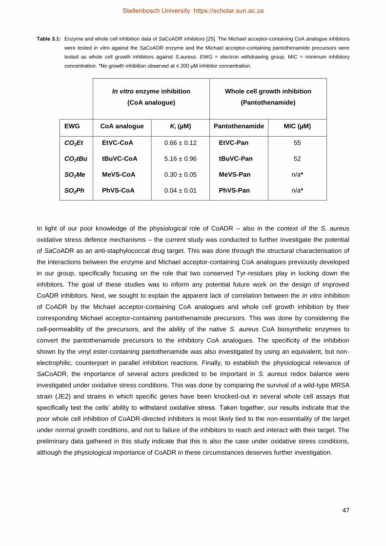
47
Table 3.1: Enzyme and whole cell inhibition data of SaCoADR inhibitors [25]. The Michael acceptor-containing CoA analogue inhibitors
were tested in vitro against the SaCoADR enzyme and the Michael acceptor-containing pantothenamide precursors were
tested as whole cell growth inhibitors against S.aureus. EWG = electron withdrawing group, MIC = minimum inhibitory
concentration. *No growth inhibition observed at ≤ 200 µM inhibitor concentration.
In vitro enzyme inhibition
(CoA analogue)
Whole cell growth inhibition
(Pantothenamide)
EWG CoA analogue Ki (µM) Pantothenamide MIC (µM)
CO2Et EtVC-CoA 0.66 ± 0.12 EtVC-Pan 55
CO2tBu tBuVC-CoA 5.16 ± 0.96 tBuVC-Pan 52
SO2Me MeVS-CoA 0.30 ± 0.05 MeVS-Pan n/a*
SO2Ph PhVS-CoA 0.04 ± 0.01 PhVS-Pan n/a*
In light of our poor knowledge of the physiological role of CoADR – also in the context of the S. aureus
oxidative stress defence mechanisms – the current study was conducted to further investigate the potential
of SaCoADR as an anti-staphylococcal drug target. This was done through the structural characterisation of
the interactions between the enzyme and Michael acceptor-containing CoA analogues previously developed
in our group, specifically focusing on the role that two conserved Tyr-residues play in locking down the
inhibitors. The goal of these studies was to inform any potential future work on the design of improved
CoADR inhibitors. Next, we sought to explain the apparent lack of correlation between the in vitro inhibition
of CoADR by the Michael acceptor-containing CoA analogues and whole cell growth inhibition by their
corresponding Michael acceptor-containing pantothenamide precursors. This was done by considering the
cell-permeability of the precursors, and the ability of the native S. aureus CoA biosynthetic enzymes to
convert the pantothenamide precursors to the inhibitory CoA analogues. The specificity of the inhibition
shown by the vinyl ester-containing pantothenamide was also investigated by using an equivalent, but non-
electrophilic, counterpart in parallel inhibition reactions. Finally, to establish the physiological relevance of
SaCoADR, the importance of several actors predicted to be important in S. aureus redox balance were
investigated under oxidative stress conditions. This was done by comparing the survival of a wild-type MRSA
strain (JE2) and strains in which specific genes have been knocked-out in several whole cell assays that
specifically test the cells’ ability to withstand oxidative stress. Taken together, our results indicate that the
poor whole cell inhibition of CoADR-directed inhibitors is most likely tied to the non-essentiality of the target
under normal growth conditions, and not to failure of the inhibitors to reach and interact with their target. The
preliminary data gathered in this study indicate that this is also the case under oxidative stress conditions,
although the physiological importance of CoADR in these circumstances deserves further investigation.
Stellenbosch University https://scholar.sun.ac.za

48
3.2 Results
3.2.1 Structural characterisation of the SaCoADR–CoA analogue inhibitor interactions2
As a first step towards an increased understanding of SaCoADR as a potential drug target, the crystal
structures of SaCoADR bound to the ethyl vinyl carboxylate-containing inhibitor 2a (EtVC-CoA), the methyl
vinyl sulfone-containing inhibitor 2c (MeVS-CoA) or the phenyl vinyl sulfone-containing inhibitor 2d (PhVS-
CoA) were solved [33]. The final refined models each included one FAD and one inhibitor molecule per
active site (Figure 3.5A), and showed that the inhibitor was bound in both active sites of the dimeric enzyme,
with full occupancy. In all three inhibitor complexes the –dethia-CoA moiety was clearly present in the same
site occupied by the Cys43-SSCoA redox centre in oxidised SaCoADR (Figure 3.5B).
Several significant observations were made in terms of improving on future inhibitor design.An unoccupied
cleft adjacent to bound CoAS– was also shown to be unoccupied in the inhibitor complexes and provides
ample opportunity for the elaboration of inhibitor structures (Figures 3.5B and 3.6). In particular, it is clear
that sufficient space is available around the phenyl ring of bound PhVS-CoA to introduce additional moieties
that could potentially interact with other parts of the enzyme active site for tighter and more selective inhibitor
binding.
2The work described in this section was done in collaboration with Profs. Al Claiborne (Wake Forest University, USA)
and Matt Redinbo (University of North Carolina, Chapel Hill) and has been published in modified format. All structures were solved by Bret Wallace and Jonathan Edwards. My major contribution to this work was the purification of enzymes and performing activity assays as described in the next section.
Stellenbosch University https://scholar.sun.ac.za
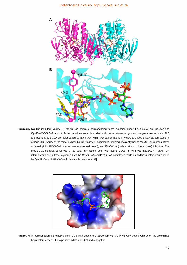
49
Figure 3.5: (A) The inhibited SaCoADR―MeVS-CoA complex, corresponding to the biological dimer. Each active site includes one
Cys43―MeVS-CoA adduct. Protein residues are color-coded, with carbon atoms in cyan and magenta, respectively. FAD
and bound MeVS-CoA are color-coded by atom type, with FAD carbon atoms in yellow and MeVS-CoA carbon atoms in
orange. (B) Overlay of the three inhibitor-bound SaCoADR complexes, showing covalently bound MeVS-CoA (carbon atoms
coloured pink), PhVS-CoA (carbon atoms coloured green), and EtVC-CoA (carbon atoms coloured blue) inhibitors. The
MeVS-CoA complex conserves all 12 polar interactions seen with bound CoAS– in wild-type SaCoADR. Tyr361′-OH
interacts with one sulfone oxygen in both the MeVS-CoA and PhVS-CoA complexes, while an additional interaction is made
by Tyr419′-OH with PhVS-CoA in its complex structure [33].
Figure 3.6: A representation of the active site in the crystal structure of SaCoADR with the PhVS-CoA bound. Charge on the protein has
been colour-coded: Blue = positive, white = neutral, red = negative.
Stellenbosch University https://scholar.sun.ac.za

50
In the structure of oxidised SaCoADR 12 polar interactions between CoAS– and protein residues were
identified and four additional residues provided hydrophobic contacts. There were also 13 solvent waters
identified as ligands to CoAS– (Figure 3.7A). These protein interactions were mostly conserved in the
inhibitor structures (Figure 3.7B-D). Critically, new electrostatic interactions between one of the sulfone
oxygen atoms of both MeVS-CoA and PhVS-CoA and two tyrosine residues, Tyr361 and Tyr419 were
identified. In comparison to the SaCoADR―PhVS-CoA complex (Figure 3.7B), the MeVS-CoA inhibitor
conserved all 12 polar interactions found with CoAS– in the oxidised enzyme, but lacked the Tyr419-
interaction with the sulfone oxygen (Figure 3.7C). Looking at the EtVC-CoA inhibitor, it lacked both the Ser39
and Asn42-interactions of CoAS– and the carboxyl oxygen had no contact with either Tyr361 or Tyr419 (Figure
3.7D). This decrease in the number of interactions moving down from PhVS-CoA to MeVS-CoA to EtVC-CoA
seemed to correlate with their observed Ki-values (Table 3.1), i.e. 0.04 μM, 0.3 μM, and 0.66 μM,
respectively [25]. However, it is important to note that although both Tyr361 and Tyr419-residues were
implicated to be playing a role during catalysis, this was determined using substrate-mimics such as methyl
methanethiosulfonate (MMTS) and not CoA-substrates.
Stellenbosch University https://scholar.sun.ac.za

51
Figure 3.7: LIGPLOT representation of polar protein and solvent water interactions between the SaCoADR and bound substrate and
inhibitors within their crystal structures [33]. These interactions are generally conserved in all four (A) CoASH, (B) PhVS-
CoA, (C) MeVS-CoA and (D) EtVC-CoA bound complexes. A number of residues lining the active-site cleft provide
electrostatic and hydrogen-bonding interactions with the -dethia-CoA moiety and with one sulfone oxygen of the inhibitor.
Additional hydrogen-bonding interactions with the -dethia-CoA portion are indicated with ordered water molecules.
The structural data in the study provided a strong starting point for future inhibitor design. Particularly, the
next generation inhibitors could be designed to exploit the open space in the CoAS-binding cleft, and in so
doing provide increased binding affinity and selectivity for the target enzymes. However, uncertainty
remained regarding a possible correlation between the possible catalytic role of Tyr361 and Tyr419 and their
interaction with our Michael acceptor-containing inhibitors. Although it was clear that Tyr361 and Tyr419
assisted in locking down the inhibitors, we needed to determine their relative importance to SaCoADR
catalysis using CoA-substrates, since interaction with these residues might be an important determinant in
the design of future inhibitors.
Stellenbosch University https://scholar.sun.ac.za

52
3.2.2 Relative importance of conserved Tyr-residues for SaCoADR activity
To determine the relevance of the two candidate residues (Tyr361 and Tyr419) implicated in catalysis and
inhibitor binding, site-directed mutagenesis was performed to obtain proteins in which these residues have
been exchanged for Phe. The two Tyr-residues were mutated both separately (SaCoADR Y361F and
SaCoADR Y419F) and in combination (SaCoADR Y361, 419F). In all cases the enzymes lacked any affinity
purification tag, and instead were purified on a column packed with ADP-Sepharose resin prepared by
coupling ADP to CNBr-activated Sepharose. The resulting proteins, as well as untagged wild-type SaCoADR
and His-SaCoADR (with an N-terminal His-tag, purified by immobilised metal affinity chromatography), were
subsequently assayed with (CoAS)2 at saturating substrate conditions for the wt enzyme using established
protocols [34]. The initial reaction rates were determined by following the oxidation of NADPH
spectrophotometrically (decrease in absorbance at 340 nm). The catalytic constant (kcat) was determined for
each enzyme to give an indication of the effect of each mutation in the SaCoADR active site on the activity of
the enzyme. The results are summarised in Figure 3.8.
Compared to the wild-type enzyme, SaCoADR Y361F showed 17% (± 0.35) activity and SaCoADR Y419F
showed 28% (± 0.02) activity. The double mutant, SaCoADR Y361, 419F showed only 4% (± 0.04) activity
compared to the wild-type. These results confirm that both the Tyr361 and Tyr419 residues play an important
role in the reaction mechanism of SaCoADR, and suggest that this is likely exploited by the most potent
inhibitor (PhVS-CoA) which showed a Ki of 0.04 µM. The design strategies for improved CoADR inhibitors
should therefore incorporate groups that engage in interactions with these residues.
Figure 3.8: Activities of SaCoADR wild-type and mutant enzymes. The figure represents one experiment performed in triplicate for each
enzyme and the error bars depict the standard deviation.
Stellenbosch University https://scholar.sun.ac.za

53
3.2.3 Lack of whole cell inhibition: a case of poor permeability?
We next set out to determine the basis for the weak correlation between the excellent in vitro inhibition of
CoADR by the Michael acceptor-containing inhibitors, and the poor whole cell inhibition results. We first
considered the cell permeability of their pantothenamide precursors, since although several different
pantothenamide structures have been shown to act as inhibitors of both Gram-negative and Gram-positive
bacteria [35-38], none of them incorporated sulfone moieties. A direct method to study the permeability of the
bacterial cell membrane to these pantothenamides would be to fluorescently label the compounds and to use
microscopy to visually establish if they label CoADR. However, since these molecules are quite small, the
attachment of a fluorescent probe would significantly alter their polarity and consequently affect the results of
the test. We also considered using electrospray ionisation mass spectroscopy (ESI-MS) to determine if the
Michael acceptor-containing pantothenamides can be found in cells treated with them. However, in our
experience these compounds show poor sensitivity in MS analysis due to poor ionisation. Also, such
experiments would likely not be able to discern between compound bound to the cell envelope and those
that have entered the cell.
We therefore decided to calculate several physicochemical properties of each compound in order to
compare their hydrophobicity/lipophilicity and to get an indication of their potential to pass through the S.
aureus cell envelopes. In a recent study, Davis and co-workers performed a systematic and quantitative
evaluation of small-molecule permeability in bacteria in order to accurately correlate calculated
physicochemical properties of compounds to their real world permeability and bio-availability [39]. The study
found positive correlations between hydrophobicity parameters and intracellular compound accumulation for
the Gram-positive bacteria Bacillus subtilis. These hydrophobicity parameters included the logarithm of the
partition coefficient (LogP), the logarithm of the distribution coefficient (LogD) and Tetko’s calculated LogP
hydrophobicity (ALogPs). Accordingly, since S. aureus is a Gram-positive bacterium like B. subtilis, we
decided to calculate these properties for our inhibitor compounds using the exact same methods as used by
Davis and co-workers. We also included N-pentyl pantothenamide (N7-Pan) for comparison, since it has
been well studied and has been shown to act as a potent inhibitor of S. aureus [40]. LogP is the ratio of
concentrations of a compound in a mixture of n-octanol and water at equilibrium. LogD denotes the ratio of
the sum of the concentrations of all forms of the compound, both ionised and non-ionised, in each of the two
phases. Both LogP and LogD were calculated using Instant JChem v15.6.1 from ChemAxon, which uses an
algorithm based on that of Viswanadhan [41]. ALogPs is also a computed LogP hydrophobicity based on the
strategy proposed by Tetko and co-workers [42, 43] and the values were calculated using VCCLab’s
ALOGPS v2.1 online app [44]. All the physicochemical results are summarised in Table 3.2. In all cases
higher LogP and LogD values correlate with increased hydrophobic character, while lower values designates
more hydrophilic character.
Stellenbosch University https://scholar.sun.ac.za

54
Table 3.2: Comparison of the structures and several physicochemical values of the pantothenamide inhibitor compounds. Estimation of
logarithm of the partition coefficient [n-octanol/water] (LogP) and the logarithm of the distribution coefficient (LogD) were
calculated using the cheminformatics tool Instant JChem v15.6.1 from ChemAxon. Tetko’s calculated LogP hydrophobicity
(ALogPs) was calculated using VCCLab’s ALOGPS v2.1.
Pantothen-
amide Structure LogP LogD ALogPs
EtVC-Pan
-0.40 ± 0.49 -0.76 ± 0.52 0.44 ± 0.29
tBuVC-Pan
0.09 ± 0.49 -0.06 ± 0.52 1.06 ± 0.29
MeVS-Pan
-2.44 ± 0.49 -2.59 ± 0.52 -0.24 ± 0.29
PhVS-Pan
-0.38 ± 0.49 -0.48 ± 0.52 0.64 ± 0.29
N7-Pan
0.47 ± 0.49 0.72 ± 0.52 1.99 ± 0.29
The results show that all the compounds analysed are all reasonably polar. MeVS-Pan is clearly the most
polar; this could possibly indicate that the compound is potentially unable to cross bacterial cell envelopes.
Even so, the absolute values should not be given too much credence as these are still calculated and not
experimentally determined. It is more useful to compare the calculated properties of each compound within
the series. It is clear then that the calculated hydrophobicity parameters of all the other compounds are quite
similar; specifically, in the case of EtVC-Pan which does show growth inhibition and PhVS-Pan which does
not, there is no significant difference in any of the parameters. From these calculations we can reasonably
conclude that cell permeability is not the reason for the poor correlation between the in vitro and whole cell
inhibition observed for the Michael acceptor-containing CoADR inhibitors.
3.2.4 Confirming the metabolic activation of pantothenamides in S. aureus
An alternative reason for the lack of whole cell inhibition by the Michael acceptor-containing
pantothenamides is a failure by the S. aureus native CoA biosynthetic enzymes to convert them into
inhibitory CoA analogues. Previously, the in vitro biotransformation of these pantothenamides was performed
with the E. coli PanK, PPAT and DPCK enzymes to prepare the CoA analogues that were subsequently
Stellenbosch University https://scholar.sun.ac.za

55
tested on SaCoADR. However, the ability of the corresponding S. aureus enzymes to perform these
reactions has not been confirmed. Considering that especially the S. aureus PanK enzyme has been shown
to have a different specificity profile compared to the E. coli enzyme [40], we considered it possible that
metabolic activation might not occur in S. aureus. To determine if this was the case, a biosynthesis reaction
was performed in vitro using the purified SaPanK, SaPPAT and SaDPCK enzymes to convert the
pantothenamides to CoA analogues (Figure 3.9). The enzymes were then precipitated out of each reaction
mixture and removed by filtration. The remaining reaction mixtures were subsequently analysed by liquid
chromatography mass spectrometry (LC-MS). The components were separated on an ultra-performance
liquid chromatography (UPLC) column and each enzyme product was detected by ESI-MS in the positive
mode with generation of MSE fragmentation data. MS
E is an acquisition method used to observe all
detectable, eluting species and their corresponding fragment ions. During data acquisition, the ionisation
energy is dynamically switched between a low-energy and a high energy state. This produces alternating
composite mass spectra of all intact molecular ions, which is then followed by chimeric mass spectra of all
precursor molecules. Post-acquisition data analysis methods subsequently extract both the chromatographic
and the mass spectrometric information of the generated fragments to provide time-resolved, accurate mass
measurements [45]. All the MS results are summarised in Table 3.3 below.
Figure 3.9: Metabolic activation of the Michael acceptor-containing pantothenamides to the corresponding inhibitory CoA analogues by
the S. aureus PanK (SaPanK), PPAT (SaPPAT) and DPCK (SaDPCK) enzymes. The observed MS mass-to-charge ratios
(m/z) are indicated.
All the expected products were observed with LC-MS, including the MSE fragments resulting from the loss of
the ATP-moiety. This was expected, since these compounds are similar to those analysed in a previous
Stellenbosch University https://scholar.sun.ac.za

56
study using analogous detection methods [46]. The results confirmed that the Michael acceptor-containing
pantothenamides act as substrates for the CoA biosynthesis enzymes in S. aureus and that they would most
likely be converted to the corresponding CoA analogues should they cross the cell envelope. Lack of
metabolic activation can therefore be excluded as a potential reason for the poor correlation between the in
vitro and whole cell inhibition data for the Michael acceptor-containing CoADR inhibitors.
Table 3.3: Observed MS parent- and MSE daughter ions (m/z) of the biosynthetic intermediates of the Michael acceptor-containing
CoADR inhibitors.
Pantothenamide SaPanK
product (m/z)
SaPPAT
product (m/z)
SaDPCK
product (m/z)
MSE daughter
ion (m/z)
PantSH 359.1 688.2 768.1 (1a) 261.1
EtVC-Pan 425.2 754.2 834.2 (2a) 327.2
MeVS-Pan 431.1 760.2 840.1 (2c) 333.1
PhVS-Pan 493.1 822.2 902.2 (2d) 395.1
3.2.5 Establishing target-specificity for the Michael acceptor-containing CoADR inhibitors
Having established that the Michael acceptor-containing pantothenamides most likely enter S. aureus and
are converted (i.e. metabolically activated) by the organism’s native CoA biosynthesis enzymes to the
corresponding CoA analogue inhibitors, we next set out to investigate their target specificity. This was done
by synthesising an analogue of EtVC-Pan, the pantothenamide precursor of EtVC-CoA, the only compound
that did show growth inhibition of S. aureus. This analogue (CO2Et-Pan) was identical to EtVC-Pan, except
that it did not contain the Michael acceptor-moiety (i.e. it contained a single bond instead of a double bond,
Figure 3.10). Since this compound had similar structure and polarity to the Michael acceptor-containing
inhibitor precursor, it was therefore expected to have a comparable efficiency in cellular uptake. However,
since it lacks the Michael acceptor it can bind, but not covalently modify, CoADR. The inhibition of S. aureus
by both compounds was compared using a whole cell inhibition assay under normal growth conditions
without any additional stresses. The compounds were tested against a wild-type MRSA strain (JE2), as well
as a mutant MRSA strain (NE1456) in which the cdr gene has been knocked out; this mutant strain was
provided by the freely accessible NARSA repository. The bacteria were grown in nutrient rich tryptic soy
broth (TSB).
Stellenbosch University https://scholar.sun.ac.za

57
Figure 3.10: CO2Et-Pan: a non-Michael acceptor-containing inhibitor.
At the outset we expected to see inhibition of staphylococcal growth by both compounds, since metabolic
activation of either pantothenamide would lead to the formation of physiologically inactive CoA
antimetabolites that lack the catalytically essential thiol of the cofactor [37, 38]. As such, the antimetabolites
can interfere with a range of metabolic processes downstream from CoA biosynthesis, including fatty acid
biosynthesis [47]. This is a mechanism of action that has been described for other pantothenamides not
directed to attack any specific target. However, in the case of the CoADR-directed inhibitors we expected to
see strain-based differences in their potency should they act on CoADR as a target: in the strain that lacks
cdr (the gene encoding CoADR in S. aureus) we expected similar potency for both CO2Et-Pan and EtVC-
Pan, because in the absence of CoADR all the observed inhibition would be due to the antimetabolite effects
of the compounds. Any differences in potency observed in the wild-type strain would then correlate with the
target-specificity of the inhibitors. The results are illustrated in Figure 3.11 and summarised in Table 3.4
below.
Figure 3.11: Charts comparing the IC50-values (A) and MIC-values (B) of CO2Et-Pan and EtVC-Pan against S. aureus JE2 (wt) and S.
aureus NE1456 (cdr knock-out) strains. Data is from one experiment performed in triplicate with error bars indicating the
standard error of the fit to calculate the inhibition parameters.
Stellenbosch University https://scholar.sun.ac.za

58
Table 3.4: Whole cell inhibition data of small molecule inhibitors against several S. aureus strains.
Staphylococcus aureus strains
Compound
JE2 (wt) NE1456 (cdr)
IC50
(µM)
MIC
(µM)
IC50
(µM)
MIC
(µM)
CO2Et-Pan 20.3 40.9 12.5 31.8
EtVC-Pan 8.0 15.7 6.3 16.7
The results show that the Michael acceptor-containing EtVC-Pan is a more potent inhibitor than the non-
Michael acceptor pantothenamide CO2Et-Pan in not only the wild-type strain, but also in the cdr knockout.
Additionally, its potency is similar in both strains. This therefore indicates that the observed growth inhibition
by EtVC-Pan is not due to CoADR-related effects. The observation that CO2Et-Pan shows higher potency in
the cdr knockout background is interesting, suggesting that CoA-dependent processes are more sensitive to
inhibition in the absence of this enzyme.
3.2.6 Evaluating S. aureus’s resistance to chemically-induced oxidative stress in selected genetic
backgrounds
The results presented above indicate that although CoADR is a structurally tractable target for drug design, it
could not be validated as a viable physiological target. However, all the whole cell experiments outlined
above, as well those directed at determining the essentiality of S. aureus genes, have been performed under
normal (i.e. optimal) growth conditions. If CoADR plays an important role in oxidative stress resistance, it is
more likely that its physiological relevance will also come to light under such conditions. We therefore
decided to test the survival of wild-type JE2 S. aureus, as well as certain knockout strains, under the
influence of the oxidative stressors hydrogen peroxide (H2O2) and diamide. Three mutant strains were
tested. First was a cdr knockout that lacks the gene encoding CoADR (NE1456). Second was a bshA
knockout that lacks the gene encoding the first enzyme of BSH biosynthesis (NE1728) (previous studies
have shown that such strains lack this thiol) [48]. Third was NE785, a strain lacking the gene previously
highlighted to be upregulated more than 80-fold in S. aureus challenged with neutrophils [15]; this gene
(SAUSA300_0576 from MRSA strain USA300) encodes a putative FDR enzyme that shows homology to
mercuric ion reductases. For these tests we first performed dose-response assays with H2O2 but found that
the data did not allow any clear differences between the strains to be distinguished (Figure 3.12A). Next, we
carried out overnight stress tests using H2O2 (25 mM) and diamide (5 mM) as well as a combination of the
two stressors. For these experiments subtle differences were observed, with the wild-type JE2 strain
showing slightly more resistance to oxidative stress than the other strains, which all showed similar inhibition
(Figure 3.12B). All the strains were highly resistant to the oxidative thiol stress induced by diamide, a
Stellenbosch University https://scholar.sun.ac.za

59
chemical stressor known to induce the formation of disulfide bonds (and which is therefore expected to
increase the ratio of oxidised to reduced CoA) [49, 50]. A combination of the two stressors successfully
inhibited growth of all the strains, with the cdr knockout being the most sensitive. This suggests that under
these conditions CoADR plays an important role in oxidative stress resistance.
Figure 3.12: (A) Dose response of H2O2 against S. aureus JE2, NE1456 (cdr knockout), NE1728 (bshA knockout) and NE785
(knockout of the gene encoding an FDR of unknown function implicated in oxidative stress resistance [15]). The figure
represents one experiment performed in triplicate and the error bars depict the standard deviation. (B) Survival of S.
aureus strains after an overnight (~18 hr) stress test with H2O2, diamide and a combination of H2O2 and diamide. The
figure represents the average of two experiments performed independently and the error bars depict the standard
deviation between the two data sets.
Next, the effects of a stressor were assessed at a single concentration over time by performing growth curve
analysis (Figure 3.13). The growth curve analysis confirmed that wild-type JE2 shows the most resistance to
oxidative stress induced by H2O2, with the cdr knockout showing intermediate resistance (Figure 3.13A). The
remaining two strains showed little growth under these conditions, indicating that BSH and the FDR of
unknown function are both important for resistance to H2O2. This finding support recent reports on the
importance of BSH for the oxidative stress resistance of S. aureus [48]. As in the previous experiment,
diamide did not reveal any significant differences between the wild-type, cdr knockout strain and the strain
lacking the FDR of unknown function, while the bshA knockout strain showed significant reduced growth at
later time points (Figure 3.13B).
Stellenbosch University https://scholar.sun.ac.za

60
Figure 3.13: Growth curves of S. aureus JE2, NE1456 (cdr knockout), NE1728 (bshA knockout) and NE785 (knockout of the gene
encoding an FDR of unknown function implicated in oxidative stress resistance [15]) challenged with (A) 10 mM H2O2 and
(B) 4 mM diamide. Both figures represent one experiment performed in triplicate and the error bars depict the standard
deviation.
The results of the chemically-induced oxidative stress tests therefore seem to indicate that overall, S. aureus
is quite resistant to H2O2 and diamide-induced stress, and that CoADR does not play a significant role in the
resistance to the stress induced by these compounds, unless they are used in combination. When measured
over time, lack of BSH production and a loss of the FDR of unknown function that has been implicated in
resistance to the killing mechanisms of neutrophils have much more pronounced effects on the bacterium’s
ability to survive. However, it must be noted that we found that the test outcomes showed significant
variation on subsequent repetitions. This variation was found to be introduced by performing the test at
different growth states (exponential vs. stationary), by using different initial bacterial cell concentrations and
by varying the oxidant concentrations that were used. In particular, varying the ratio of bacterial cells to
oxidant altered the relative response of the different S. aureus strains. However, it became evident from all
our results that CoADR did not contribute notably to the oxidative stress resistance of S. aureus.
Consequently we decided to rather use more physiologically relevant conditions to determine the relative
importance of CoADR, BSH and the FDR of unknown function to S. aureus oxidative stress resistance.
3.2.7 Evaluating S. aureus’s resistance to neutrophil-induced oxidative stress in selected genetic
backgrounds
In order to address the variation seen in the chemically-induced oxidative stress experiment, we decided to
instead challenge the bacteria with isolated human neutrophils. This would provide more physiologically
relevant results, since the bacteria would be subjected to the oxidative burst that these polymorphonuclear
leukocytes use to kill phagocytosed bacteria that are encountered during infection. The technique has
Stellenbosch University https://scholar.sun.ac.za

61
previously been applied successfully to the study of processes relevant to the survival of S. aureus when
challenged by the innate immune system [51, 52]. Also, the ratio of colony-forming units (CFUs) of bacteria
to neutrophils could be kept constant for uniformity during each assay.
Neutrophils were successfully isolated from heparinised venous blood and enumerated using a
haemocytometer. The white blood cell differential was calculated after staining with Wright-Giemsa stain (see
Materials and Methods for details). Briefly, the assay entails the isolation of neutrophils by differential
centrifugation and diluting them to the desired concentration in RPMI media. The bacteria are grown to
exponential phase in nutrient rich media, then washed and diluted to the desired concentration (CFU/mL) in
RPMI media. The neutrophils and bacterial cells are then combined in a 96-well culture plate in a 1:1 ratio
(CFU:neutrophil) and gently centrifuged to synchronise phagocytosis in each well. The plate is incubated at
37 °C and at the desired time points samples are taken from the plate, incubated on ice and plated on
suitable agar plates in order to determine bacterial survival after neutrophil stress by counting CFUs.
Unfortunately, we failed to successfully execute this test even after performing several trials with all the
available S. aureus strains. In each case the control samples failed to give the expected results: either no
viable cells could be isolated in the negative controls, or very little to no killing took place in the positive
control samples. This latter observation indicated that the isolated neutrophils were either not viable, or did
not stay viable for the duration of the experiment. To address this, we also performed the experiment with
whole blood as was done in tests on the importance of BSH for S. aureus oxidative stress resistance [48].
However, performing the experiment in this manner also did not give the desired control results.
Consequently, we chose to abandon the pursuit of neutrophil-induced oxidative stress tests at this stage of
the study, and to finally focus on the impact of specific small molecule inhibitors on S. aureus survival
instead.
3.2.8 Evaluating the effect of reduced CoA levels in selected genetic backgrounds
Having not been able to unequivocally establish the importance of CoADR to oxidative stress resistance in
S. aureus, we finally set out to determine the effect of reduced CoA levels in wild-type S. aureus, and the
three mutant strains lacking CoADR, BSH and the FDR of unknown function. Since CoA levels have been
reported to be higher in S. aureus than in other bacteria [12], the goal of these experiments were to
determine if small molecule inhibitors of CoA biosynthesis had a differentiated effect when these functions
were absent. In this manner we hoped to establish whether CoA indeed had a role in S. aureus beyond that
of acting as a metabolic cofactor.
For these tests three compounds that have been proved to have an effect on CoA biosynthesis or CoA-
dependent enzymes were used. N7-Pan is a well-studied inhibitor that inhibits the first CoA biosynthesis
enzyme, PanK, in S. aureus, but also has effects downstream of the pathway [40], CJ-15,801 forms a tight-
binding inhibitor of phosphopantothenoylcysteine synthetase (PPCS), the second CoA biosynthesis enzyme,
[53], and Compound D is a recently discovered inhibitor of the fourth CoA biosynthesis enzyme, PPAT [54].
Based on their targets being CoA biosynthetic enzymes, all three compounds are expected to decrease the
intracellular levels of available CoA and its esters. The inhibition of S. aureus by all three compounds was
Stellenbosch University https://scholar.sun.ac.za

62
compared using a whole cell inhibition assay first performed under normal growth conditions (using nutrient
rich tryptic soy broth) without any additional stresses in a manner similar to that described for the tests with
EtVC-Pan and CO2Et-Pan above. The compounds were tested against JE2, NE1456, NE1728 and NE785,
and the results are summarised in Figure 3.14.
Figure 3.14: (A) Structures of inhibitor compounds used to test against S. aureus strains. (B) MICs of N7-Pan, CJ-15,801 and
Compound D against S. aureus JE2 (wild-type), NE1456 (cdr knockout lacking CoADR activity), NE1728 (bshA knockout
lacking BSH) and NE785 (knockout of the gene encoding an FDR of unknown function implicated in oxidative stress
resistance [15]). Each strain was tested once with each inhibitor in triplicate (except CJ-15,801 was tested twice with each
strain). The error bars indicate the standard error of the fit to calculate the inhibition parameters from the triplicate values.
The results show that in the case of N7-Pan and CJ-15,801 there is little differentiation between the effects
that the inhibitors have on the various strains (taking both the IC50 and MIC data into account
simultaneously), although the compounds show a somewhat reduced potency in the SAUSA300_0576
knockout background compared to the other strains. Since this gene encodes an FDR of unknown function,
the relevance of this finding is unclear at the moment. Interestingly, Compound D – the most potent inhibitor
tested – was found to have increased potency in the cdr and the SAUSA300_0576 knockout backgrounds
than in the wild-type strain. This suggests that under conditions were CoA levels are sufficiently reduced;
CoADR does become relevant — even under normal growth conditions. The fact that the same effect is
observed in the absence of the SAUSA300_0576 gene product could imply that it has a function related or
dependent on CoA, although this deserves further study.
3.3 Discussion
In this study, we set out to first investigate the viability of CoADR as a potential antistaphylococcal drug
target and second, to investigate several other potential role players in S. aureus oxidative stress defence.
The research was a continuation of previous work on mechanism-based Michael acceptor-containing
inhibitors of the SaCoADR enzyme. Particularly, the questions raised by earlier studies were addressed and
used in the assessment of the conditions under which this enzyme could be a viable target. We started by
Stellenbosch University https://scholar.sun.ac.za

63
examining the SaCoADR enzyme and specifically how its interaction with the Michael acceptor-containing
inhibitors relates to the observed differences in activity between them. We already knew that the sulfones are
better Michael acceptors than the esters, which should make them better electrophilic traps for the active site
Cys43 of SaCoADR. However, the potency of the various inhibitors was also directly correlated to their
interaction with two SaCoADR active site residues, Tyr361 and Tyr419. Therefore, we could adequately
rationalise the observed enzyme inhibition. Also, the structural data in the study provided a strong starting
point for future inhibitor design, particularly, the next generation inhibitors could be designed to 1) exploit the
open space in the CoAS-binding cleft, and 2) interact with Tyr361 and Tyr419 of SaCoADR and in so doing
provide increased binding affinity and selectivity for the targeted enzyme.
Next, the reasons for the poor correlation between the in vitro inhibition of SaCoADR by the Michael
acceptor-containing CoA analogues and the whole cell inhibition of S. aureus by their corresponding
pantothenamide precursors were investigated. As far as we know, no CoA transporter has been found in
bacteria and also there is no homolog of the E. coli pantothenate transporter (PanF) in S. aureus, which
could affect the whole cell activity of the inhibitor compounds in terms of uptake into the cell. Therefore, we
considered that either the two pantothenamides that showed no inhibition could not cross the S. aureus cell
envelope or they were not accepted as substrates of the CoA-biosynthesis enzymes inside the cell. We first
calculated several physicochemical properties of each compound to compare their relative
hydrophobicity/lipophilicity and second, we performed a biosynthesis reaction using the purified SaPanK,
SaPPAT and SaDPCK enzymes to convert the pantothenamides to CoA-analogues and detected the
enzyme products using LC-MS. Our results confirmed that neither permeability nor biosynthesis is a reason
for the poor correlation between the enzyme inhibition of the Michael acceptor-containing CoA analogues
and the whole cell inhibition data for their pantothenamide precursors. Additionally we considered the target
specificity of the CoADR inhibitors, i.e. that the bacterial growth inhibition of the pantothenamide precursors
is mediated via a different target than CoADR. The results led us to the conclusion that the poor correlation
between the enzyme inhibition and the whole cell growth inhibition is due to SaCoADR not being essential
under normal growth conditions.
In spite of our findings suggesting that SaCoADR is not a viable target for antistaphylococcal drug design, it
remained possible that it could still be a relevant target under certain non-optimal conditions, i.e. conditions
different from those used for the whole cell experiments. It was therefore necessary to evaluate the
physiological relevance of CoADR in S. aureus under oxidative stress, since it is more likely that its
importance will also come to light under such conditions. We also investigated and compared the effects of
two other potential role players in S. aureus oxidative stress defence, BSH and a putative FDR enzyme that
displays homology to mercuric ion reductases. All the chemically induced oxidative stress results indicated
that CoADR did not contribute notably to the oxidative stress resistance of S. aureus. In contrast, BSH and
the FDR of unknown function seemed to have more pronounced contributions to the bacterium’s ability to
survive (Figure 3.12 and 3.13). Even when evaluated against the background of reduced CoA levels, very
little difference was seen between the effects of CoADR, BSH and the FDR of unknown function (Figure
3.14).
Despite the fact that this study could not unequivocally establish the importance of CoADR to oxidative
stress resistance in S. aureus; several significant findings were made during our evaluation of the potential
Stellenbosch University https://scholar.sun.ac.za

64
drug target. It is noteworthy that while both CoA and BSH are considered as candidates to be the major
LMWT responsible for redox balance in S. aureus, our results did not reveal any link between the two
compounds. Specifically, neither compound appeared to compensate for the absence of the other during our
studies with the knockout mutants of S. aureus. Also, CO2Et-Pan showed higher potency in the cdr knockout
background, suggesting that CoA-dependent processes are more sensitive to inhibition in the absence of
this enzyme. This correlated well with our results with the most potent CoA biosynthesis inhibitor tested
(Compound D), which was found to have increased potency in the cdr and the SAUSA300_0576 knockout
backgrounds than in the wild-type strain. This suggests that under conditions where CoA levels are
sufficiently reduced, CoADR might become relevant, even under normal growth conditions. Taking these
findings into consideration, it would be preferable if the assays with the small molecule inhibitors could be
done under oxidative stress conditions, since it would accentuate any effects that are related to redox
balance. However, bearing in mind the variability of our chemically-induced oxidative stress results as well as
our failure to successfully execute the neutrophil tests, this is currently unfeasible. Also, there is a possibility
that CoADR inhibitors could be used synergistically with Compound D, since intracellular levels of reduced
CoA would then be significantly lowered due to inhibition of CoA biosynthesis combined with diminished
reduction of (CoAS)2. As it stands, we managed to illuminate several uncertainties regarding CoADR as a
potential antistaphylococcal drug target, but also raised important questions regarding the organism’s redox
balance mechanisms in general. These findings pave the way for further investigation of compounds
targeting pathways involved in oxidative stress resistance as potential drugs against in S. aureus.
Stellenbosch University https://scholar.sun.ac.za

65
3.4 Materials and methods
Unless otherwise noted, all reagents and chemical compounds were purchased from commercial sources
and used without further purification. Lyophilised CNBr-activated Sepharose 4B, Isopropyl β-D-1-
thiogalactopyranoside (IPTG), Dextran-500 and Ficoll-Paque Plus were obtained from Sigma. HisTrapFF
metal affinity purification columns, HiTrap desalting columns and Vivaspin centrifugal concentrators were
from GE Healthcare Life Sciences. Tetrahydrofuran (THF) was distilled under nitrogen from sodium wire
using benzophenone as an indicator. Dichloromethane (DCM) was distilled under nitrogen from calcium
hydride. N,N-Dimethylformamide (DMF) was dried and purified by shaking up over potassium hydroxide
followed by distillation under reduced pressure and a nitrogen atmosphere, and was stored over 3 Å
molecular sieves. All column chromatography was performed using Merck silica gel 60 (particle size 0.040-
0.063 mm) using combinations of hexane, ethyl acetate, DCM and methanol as eluants. Thin layer
chromatography (TLC) was carried out on aluminium-backed Merck silica gel 60 F254 plates. Visualisation
was performed with a UV lamp followed by spraying with a cerium ammonium molybdate or ninhydrin
solution, followed by heating.
Protein purification: All His-tagged protein purification was performed using HisTrapFF metal affinity
purification columns on an ÅKTAprime purification system from GE Healthcare Life Sciences. The purified
proteins were desalted using 5 mL HiTrap desalting columns.
Spectrophotometry: All absorbance readings in 96-well plates were measured using a Thermo Scientific
Varioskan microplate spectrophotometer. All larger volume measurements were made in an Agilent
Technologies Cary 60 UV-Vis Spectrometer using a 1 cm cuvette.
HPLC analyses: High performance liquid chromatography (HPLC) analysis was performed on an Agilent
1100 instrument using a Supelcosil LC-18-T 3μ C18 column (150 x 4.6 mm) from Supelco.
NMR analyses: All 1H and
13C nuclear magnetic resonance spectra (NMR) were obtained using 300 MHz
Varian VNMRS (75 MHz for 13
C), 400 MHz Varian Unity Inova (100 MHz for 13
C) or 600 MHz Varian Unity
Inova (125 MHz for 13
C) instruments at the Central Analytical Facility (CAF) of the University of Stellenbosch.
Chemical shifts (δ) were recorded using the residual solvent peak or an external reference. All chemical
shifts are reported in ppm and all spectra were obtained at 25 °C. Proton spectral data are reported as
follows: chemical shift, multiplicity (ovlp = overlapping, s = singlet, d = doublet, t = triplet, q = quartet, p =
pentet, m = multiplet, br = broad), coupling constant (J) in Hz, and integration.
MS analyses: All ESI–MS were performed using a Waters Synapt G2 mass spectrometer using a Waters
HSS C18 column (2.1 x 150mm) at the CAF of the University of Stellenbosch.
Stellenbosch University https://scholar.sun.ac.za

66
3.4.1 ADP-coupled Sepharose affinity resin
ADP was coupled to Sepharose as follows: Lyophilised CNBr-activated Sepharose 4B (1 g) was weighed out
and suspended in HCl (1 mM). The medium swelled immediately and was then washed for 15 minutes with
HCl (1 mM) on a sintered glass filter (porosity G3), using approximately 200 ml HCl added in several
aliquots. ADP (10 µmol), the ligand to be coupled, was dissolved in coupling buffer containing 0.1 M NaHCO3
and 0.5 M NaCl at pH 8.3 (5 mL). The coupling solution containing the ligand was added to the prepared
medium suspension in a stoppered vessel. The mixture was rotated gently end-over end overnight at 4 °C.
Afterwards, excess ligand was washed away with 5 medium (gel) volumes of coupling buffer. Next, any
remaining active groups were blocked. The medium was transferred to 0.1 M Tris-HCl buffer, pH 8.0 and let
stand for 2 hours. The medium was washed with five cycles of alternating pH, which consisted of a wash with
0.1 M acetic acid and 0.5 M NaCl, pH 4.0 (5 medium volumes) followed by a wash with 0.1 M Tris-HCl and
0.5 M NaCl, pH 8 (5 medium volumes). A slurry was prepared with binding buffer in a ratio of 75% settled
medium to 25% buffer and packed into a column for manual loading and elution of protein. The resin was
stored in 60% ethanol at 4 °C.
3.4.2 SaCoADR wt and mutants overexpression and purification
Plasmid vectors encoding the SaCoADR wt, SaCoADR Y361F, SaCoADR Y419F or SaCoADR Y361, 419F
proteins were kindly provided by Prof Al Claiborne of Wake Forest University, USA. We also had the
plasmid vector encoding the N-terminal His-tagged SaCoADR wt (His-SaCoADR wt) protein in the
laboratory.
His-SaCoADR wt was expressed and purified using established protocols [25]. The plasmid encoding His-
SaCoADR wt (pET-28a-SaCoADR) was transformed into E. coli BL21(Star)DE3 for expression. Expression
was performed in Luria-Bertani broth supplemented with 30 mg/L kanamycin at 37 °C, and by induction with
1 mM IPTG at OD600 = 0.6. Cell growth was continued for a further 3h at 37 °C post-induction, after which the
cells were harvested by centrifugation. The obtained cell pellet was suspended in sonication buffer (5 mM
imidazole, 500 mM NaCl and 20 mM Tris–HCl, pH 7.9; 10 mL/1 g cell paste) and subsequently sonicated to
effect cell lysis. After centrifugation at 15 000 x g for 30 min to collect cell debris, the crude extract
(supernatant) was applied to a previously prepared 1 mL HisTrapFF metal affinity purification column using
an ÅKTAprime purification system. Weakly bound proteins were removed by washing with sonication buffer,
followed by sonication buffer containing 75 mM imidazole. His-SaCoADR wt was eluted by increasing the
imidazole concentration to 500 mM. Elution was monitored at A280. The purified protein was desalted using a
5 mL HiTrap desalting column (25 mM Tris, 5 mM MgCl2, pH 8.0). Glycerol was added to the pure protein
solution to a final concentration of 5%, after which the protein was aliquoted and stored at -80 °C.
SaCoADR wt, SaCoADR Y361F, SaCoADR Y419F and SaCoADR Y361, 419F was also expressed and
purified using established protocols [34]. The plasmid encoding these proteins were transformed into E. coli
BL21(Star)DE3 for expression. Expression was performed in Terrific Broth supplemented with 100 mg/L
ampicillin at 37 °C, and by induction with 1 mM IPTG at OD600 = 1.0. Cell growth was continued for a further
3h at 37 °C post-induction, after which the cells were harvested by centrifugation. The obtained cell pellet
Stellenbosch University https://scholar.sun.ac.za

67
was suspended in Tris-EDTA buffer (1 mM EDTA and 20 mM Tris–HCl, pH 7.4; 10 mL/1 g cell paste) and
subsequently sonicated to effect cell lysis. After centrifugation at 15 000 x g for 30 min to collect cell debris,
unwanted proteins were precipitated from the crude extract (supernatant) with (NH4)2SO4 up to 50%
saturation. After centrifugation at 4 500 x g for 30 min, the precipitated proteins were discarded and
SaCoADR was subsequently precipitated from the extract with (NH4)2SO4 up to 80% saturation. The
precipitated SaCoADR was then removed from the extract via centrifugation at 4 500 x g for 30 min and
resuspended in a minimal volume of Tris-EDTA buffer. Next, after (NH4)2SO4 was removed by dialysis
against 2x 2 L Tris-EDTA buffer, the SaCoADR extract was applied to the ADP-coupled Sepharose affinity
resin. Weakly bound proteins were removed by washing with Tris-EDTA buffer and SaCoADR was eluted by
increasing concentrations of NaCl (0 – 4 M). The purified protein was desalted using a 6 mL Vivaspin
centrifugal concentrator (50 mM phosphate, 0.5 mM EDTA, pH 7.0). Glycerol was added to the pure protein
solution to a final concentration of 5%, after which the protein was aliquoted and stored at -80 °C.
3.4.3 Synthesis of the CoADR substrate (oxidation of CoA to CoA disulfide)
CoA disulfide (CoAS)2 was prepared from CoA by using the established diamide-based oxidation of thiols
[49]. To a solution of 50 mM Tris–HCl (pH 7.6) in H2O (13 mL) was added CoA (final concentration 2.0 mM),
followed by diamide (final concentration 1.0 mM). The reaction mixture was stirred for 10 min at room
temperature, followed by extraction of diamide with DCM (4 × 4 mL). The concentration of (CoAS)2 in the
final solution was determined by preparing five dilutions of the solution, which were each individually
analysed spectrophotometrically to determine the absorbance at 260 nm. The (CoAS)2 concentration in each
dilution was subsequently calculated using the reported extinction coefficient for CoA disulfide (ε260 = 33600
M-1
.cm-1
) [7]. The average value was taken as the (CoAS)2 concentration in the original solution. The purity
of (CoAS)2 was confirmed by HPLC analysis. A typical chromatogram is shown below (Figure 3.15).
Figure 3.15: HPLC chromatogram of CoA disulfide, (CoAS)2.
HPLC analysis was performed on an Agilent 1100 instrument using a Supelcosil LC-18-T 3μ C18 column
(150 x 4.6 mm) from Supelco. The column was equilibrated in 95% solution A (100 mM potassium
Stellenbosch University https://scholar.sun.ac.za

68
phosphate, pH 6.5) and 5% solution B (acetonitrile), followed by elution with 95% A (0 - 5 minutes, isocratic),
a linear gradient increasing solution B to 40% (5 - 10 minutes), isocratic elution at 40% (10 - 12 minutes), a
linear gradient increasing solution B to 60% (12 - 13 minutes) and finally by an isocratic elution at 60% B (13
- 15 minutes). A flow rate of 1 mL.min-1
was maintained throughout.
3.4.4 SaCoADR activity assay
The SaCoADRs were assayed at saturating substrate conditions using established protocols [34]. Reactions
were initiated by adding 450 μL of an enzyme master mix to 50 μL containing (CoAS)2. CoADR activity was
assayed at 25 °C in a total volume of 500 μL containing 50 mM potassium phosphate (pH 7.0), 0.5 mM
EDTA, 32 μM NADPH, 20 nM CoADR and 60 μM (CoAS)2. The initial reaction rates were determined by
following the oxidation of the NADPH cofactor spectrophotometrically (decrease in absorbance at 340 nm) at
25 °C. All measurements were made in an Agilent Technologies Cary 60 UV-Vis Spectrometer using a 1 cm
cuvette.
3.4.5 Biosynthesis of CoA analogues from pantothenamide precursors
The biosynthesis reactions were initiated by adding 51 µg of each of SaPanK, SaPPAT and SaDPCK to a
solution with 16.5 mM ATP, 10 mM MgCl2, 10 mM pantothenamide and containing 60 mM HEPES (pH 8.0)
with a final volume of 500 µL. The reaction mixture was incubated at 37 °C for 2 hours. Afterwards, the
reaction mixture was diluted 1:1 with 10 mM NH4OAc (pH 6.0) and all protein was removed using a Pierce™
3K MWCO, 0.5 mL protein concentrator.
3.4.6 Liquid chromatography mass spectrometry (LC–MS) analysis
LC-MS analysis was performed on a Waters Synapt G2 instrument using a Waters HSS C18 column (2.1 x
150mm). The column was equilibrated in 98% solvent A (10 mM NH4OAc, pH 6.5) and 2% solvent B
(acetonitrile with 0.1% formic acid), followed by elution with 98% A (0 - 1 minutes, isocratic), a linear gradient
increasing solution B to 20% (1 - 10 minutes), a linear gradient increasing solution B to 100% (10 - 15
minutes), an isocratic elution at 100% B (15 - 16 minutes) and finally conditions were returned to 98% A (16
– 20 minutes, isocratic). A flow rate of 0.3 mL.min-1
was maintained throughout.
Electrospray ionisation was applied in the positive mode at a capillary voltage of 2.5 kV, cone voltage of 15
V, desolvation temperature of 275 °C and desolvation gas setting of 650 L/h. The rest of the MS settings
were optimised for best sensitivity. The instrument was calibrated with sodium formate and leucine
enkaphelin was used as lock mass for accurate mass determinations. The MS acquisition method consisted
of a low energy function at a trap voltage of 6 V and a high energy function where the trap collision energy
was ramped from 15 to 60 V to generate fragmentation data (MSE).
Stellenbosch University https://scholar.sun.ac.za

69
3.4.7 Synthesis of relevant compounds
All the Michael acceptor-containing inhibitor compounds were synthesised previously by Renier van der
Westhuyzen [25]. The synthesis scheme is included in Figure 3.16.
Figure 3.16: Synthesis of the Michael Acceptor-Containing Pantothenamides and CoA-analogues [25].
Ethyl 5-aminopentanoate: 5-amino-pentanoic acid was esterified with ethanol
using Fischer-esterification to 5-amino-pentanoate as follows. The acid (237.0 mg,
2.02 mmol) was dissolved in excess ethanol (±30 ml, 513 mmol) and concentrated
sulphuric acid (0.1 ml) was added as an acid catalyst. The mixture was heated at 80 °C while stirring for 2
hours under reflux. After removing from heat, the unreacted acid was removed by filtration. The mixture was
then concentrated in vacuo to remove all traces of ethanol and afford pure ethyl 5-amino-pentanoate as a
light yellow oil (259.0 mg, 1.78 mmol) at a 88% yield. 1H NMR (300 MHz, CDCl3): δ = 4.18 (q, J = 7.03, 2H),
2.46 (br, 2H), 1.74-1.72 (m, 2H), 1.70-1.68 (m, 2H), 1.27 (t, J = 7.03, 3H).
3-((4R)-2-(4-methoxyphenyl)-5,5-dimethyl-1,3-dioxane-4-carboxamido) prop-
anoic acid: Pantothenic acid was obtained from calcium pantothenate via cation
exchange. Calcium pantothenate (978.4 mg, 4.1 mmol) was passed through
Amberlite IR-120 cation exchange resin (1.1 g) and lyophilised to afford pantothenic
acid. The diol-group of the pantothenic acid was then protected using p-
methoxybenzaldehyde dimethyl acetal (PMB) to prevent any unwanted electrostatic
Stellenbosch University https://scholar.sun.ac.za

70
interactions in the following steps of the synthesis. Pantothenic acid (794.1 mg, 3.62 mmol) was placed
under nitrogen flow and crushed molecular sieves (4 Å) was added. Dry THF (10 ml) was added and the
mixture was swirled until all the pantothenic acid was dissolved. Camphor-10-sulfonic acid (CSA) (8.41 mg,
0.362 mmol) and PMB (1.24 ml, 7.24 mmol) was added and the reaction mixture was stirred overnight at 25
°C. The molecular sieves were then removed via filtration and the THF was removed in vacuo. The desired
product (PMB-protected pantothenic acid) precipitated as an off-white powder (822.65 mg, 2.44 mmol). The
yield was calculated as 67%. 1H NMR (DMSO–D6, 400MHz): δ = 0.95 (s, 3H), 1.01 (s, 3H), 2.40 (t, J = 7.1
Hz, 2H), 3.35 (m, 2H), 3.63 (dd, J = 11.2 Hz, 2H), 3.76 (s, 3H), 4.09 (s, 1H), 5.52 (s, 1H), 6.92 (d, J = 8.7 Hz,
2H), 7.43 (d, J = 8.9 Hz, 2H), 7.46 (br t, J = 6.1 Hz, 1H), 12.30 (br s, 1H).
Ethyl 5-(3-((4R)-2-(4-methoxyphenyl)-5,5-dimethyl-1,3-diox-
ane-4-carboxamido)propanamido)pentanoate: PMB-protected
pantothenic acid was coupled to ethyl 5-amino-pentanoate by N-
(3-Dimethylaminopropyl)-N′-ethylcarbodiimide (EDC)-coupling.
The amine, ethyl 5-amino-pentanoate (259.0 mg, 1.78 mmol), was
dissolved in dry Dimethylformamide (DMF) (3 mL) and added to a
2-neck round-bottom flask containing the PMB-protected
pantothenic acid (600.5 mg, 1.78 mmol), Hydroxybenzotriazole (HOBt) (290.47 mg, 2.15 mmol) and
Triethylamine (Et3N) (550 μl, 3.94 mmol), which were all dissolved in dry DMF (3 mL). The reaction mixture
was kept under nitrogen flow. After cooling to 0 °C, EDC (388.0 mg, 2.5 mmol) was dissolved in dry DMF (5
mL) and added to the reaction mixture. The mixture was then stirred for 2 hours at 0 °C after which it was
slowly heated to 25 °C and stirred overnight under nitrogen flow. The DMF was subsequently removed in
vacuo and the crude product was purified by flash chromatography (5% MeOH/DCM) using a 5 cm silica
column.The solvents were then removed in vacuo to afford the PMB-protected ethyl pentanoate
pantothenamide as a white powder (289.0 mg, 0.62 mmol). The yield was calculated as 34%. 1H NMR (300
MHz, CDCl3): δ = 7.44 (d, J = 9.37, 1H), 7.41 (d, J = 8.59, 1H), 6.93 (d, J = 9.37, 1H), 6.90 (d, J = 8.59, 1H),
5.45 (s, 1H), 5.30 (s, 1H), 4.15-4.05 (m, 2H), 3.8 (s, 3H), 3.7 (s, 2H), 3.6-3.5 (m, 2H), 3.3-3.15 (m, 2H), 2.41
(t, J = 6.25, 2H), 2.28 (t, J = 7.42, 2H), 1.66-1.58 (m, 2H), 1.54-1.48 (m, 2H), 1.25 (t, J = 7.03, 3H), 1.10 (br,
3H), 1.09 (br, 3H).
Ethyl 5-(pantothenamido) pentanoate: The final step of the
synthesis involved the removal of the PMB-group from PMB-
protected ethyl pentanoate pantothenamide. This
pantothenamide (289.0 mg, 0.62 mmol) was dissolved in THF (11 mL) and 1 M hydrochloric acid (HCl) (11
mL) was added. The reaction mixture was neutralised after 5 hours with the addition of Amberlite IRA-400
strong basic anion exchange resin until the pH reached 7. The mixture was then filtered and concentrated in
vacuo. Afterwards the mixture was dissolved in 20% methanol/dichloromethane and purified by flash
chromatography using a 4 cm silica column. The solvent was subsequently removed in vacuo to afford the
final product, ethyl 5-(pantothenamido) pentanoate as a white powder (70.5 mg, 0.204 mmol). The yield was
Stellenbosch University https://scholar.sun.ac.za
71
calculated as 32%. 1H NMR (300 MHz, CDCl3): δ = 4.21-4.15 (m, 2H), 4.00 (s, 1H), 3.60-3.48 (m, 4H), 3.20
(t, J = 7.03, 2H), 2.52 (t, J = 6.64, 2H), 2.41 (t, J = 7.42, 2H), 1.68-1.60 (m, 2H), 1.59-1.49 (m, 2H), 1.32-1.24
(m, 3H), 0.93 (s, 3H), 0.89 (s, 3H). [M+H]+ calculated for C16H31N2O6, 347.2182; found 347.2173.
3.4.8 S. aureus whole cell inhibition assay
S. aureus cells were streaked out on TSB-agar plates (no antibiotic for JE2; with erythromycin (10 µg.mL-1
)
for all the other strains for selection) and incubated at 37 °C overnight. A few (4 or 5) well-isolated colonies
were picked from each plate and used to inoculate 5 mL TSB growth medium (with antibiotic where
necessary for selection). The cultures were incubated at 37 °C for 5 - 6 hours until they reached mid-log
phase (OD600 = 0.4-0.5 for S. aureus). The mid-log bacterial cultures were diluted 15 000x (2 µL in 30 mL) in
TSB growth media and used to inoculate wells containing TSB in a 96-well cell culture plate. Inhibitor
compounds were added in doubling dilutions and the 96-well plate was then incubated at 37 °C for 18 hours
while shaking, after which the absorbance was read at 600 nm using a Thermo Scientific Varioskan
microplate spectrophotometer. The dose response of each compound was then determined and the IC50-
and MIC-values were calculated using SigmaPlot 12.0 software (Systat Software).
3.4.9 Dose-response of H2O2 against S. aureus
S. aureus cells were streaked out on TSB-agar plates (no antibiotic for JE2; with erythromycin (10 µg.mL-1
)
for all the other strains for selection) and incubated at 37 °C overnight. A few (4 or 5) well-isolated colonies
were picked from each plate and used to inoculate 5 mL TSB growth medium (with antibiotic where
necessary for selection). The cultures were incubated at 37 °C for 5 - 6 hours until they reached mid-log
phase (OD600 = 0.4-0.5 for S. aureus). The mid-log bacterial cultures were then pipetted into a 96-well cell
culture plate. H2O2 was added in doubling dilutions and the 96-well plate was then incubated at 37 °C for 18
hours after which the absorbance was read at 600 nm using a Thermo Scientific Varioskan microplate
spectrophotometer. The dose response of each compound was then determined and the IC50- and MIC-
values were calculated using SigmaPlot 12.0 software (Systat Software).
3.4.10 S. aureus overnight stress test with H2O2 and diamide
For overnight stress assays, cultures were prepared as described above for the H2O2 dose-response in 5 ml
TSB and grown in triplicate, with one tube containing H2O2 (25 mM), the other containing diamide (5 mM)
and the last tube containing both compounds at the stated concentrations. The cultures were then incubated
at 37 °C for 18 hours after which the absorbance was read at 600 nm.
Stellenbosch University https://scholar.sun.ac.za

72
3.4.11 Isolation of human neutrophils
Neutrophils were isolated from heparinised venous blood that was supplied by the Western Province Blood
Transfusion Service. Ficoll-Paque Plus (2 mL) at room temperature was pipetted into the bottom of a sterile
15 mL centrifuge tube and blood (4 mL) was then carefully overlaid on top of the Ficoll-Paque Plus. The
tubes were then centrifuged at 652 x g for 30 minutes at 21 °C to separate the blood in to four different
layers. The sediment containing PMNs and erythrocytes were retained and the supernatant layers containing
lymphocytes, monocytes and the plasma layer on top were discarded. The sediment was then mixed with an
equal volume of 6% Dextran-500 (in 0.9% NaCl) at room temperature and incubated at 37 °C for 45 minutes
to allow the sedimentation of erythrocytes. The pellet containing erythrocytes was subsequently discarded
and the neutrophil-rich supernatant was collected and mixed with 10 mL of PBS. The PBS-neutrophil mixture
was centrifuged at 290 x g for 10 minutes at 4 °C and the pellet containing the neutrophils was collected and
the supernatant was discarded. The pellet was then washed twice with 1 mL PBS. The final neutrophil pellet
was suspended into the desired volume of RPMI 1640 media (10 mM HEPES buffered at pH 7.4) to achieve
the appropriate cell concentration of 2 x 10⁻⁶
cells.mL-1
.
3.4.12 Neutrophil bactericidal assay
All S. aureus strains were plated on TSB-agar plates (without antibiotic for JE2) containing 10 µg.mL-1
erythromycin for selection for NE785, NE1456 and NE1728 and grown overnight at 37 °C. TSB (5 mL) was
inoculated with 4 - 5 single colonies bacteria and grown to mid-log phase (S. aureus OD600 = 0.4). An aliquot
of each culture (1 mL) was then centrifuged briefly (4 min. at 8 000 RPM on a small benchtop centrifuge) to
pellet the cells. The cells were then washed twice in PBS and 1 µL of each PBS-cell suspension was diluted
into HEPES buffered RPMI at pH 7.4. A sterile 96-well flat-bottom microtiter plate was coated with human
serum and washed extensively with PBS. Bacterial cells were then combined with PMNs at a ratio of 1:1
(CFU:PMN ratio) in the 96-well plate in a total volume of 198 µL. Next, the 96-well plate was centrifuged at
456 x g for 7 min to synchronise phagocytosis and afterwards the plate was incubated at 37 ° C for up to 120
min. At the desired time points, saponin (22 μl of a 1% solution, 0.1% final concentration) was added to each
well to break up any cell clumps and the contents were mixed and then incubated on ice for 15 min. The
surviving bacteria were enumerated the following day by CFU-count. Percentage survival was calculated by
comparing the numbers of surviving bacteria at each timepoint to those at t = 0.
3.5 References
1. Jaeger, T., et al., The thiol-based redox networks of pathogens: Unexploited targets in the search for
new drugs. BioFactors, 2006. 27(1): p. 109-120.
2. Atkinson, H.J. and P.C. Babbitt, An Atlas of the Thioredoxin Fold Class Reveals the Complexity of
Function-Enabling Adaptations. PLoS Comput Biol, 2009. 5(10): p. e1000541.
3. Holmgren, A., Thioredoxin. Annual Review of Biochemistry, 1985. 54(1): p. 237-271.
Stellenbosch University https://scholar.sun.ac.za
73
4. Argyrou, A. and J.S. Blanchard, Flavoprotein Disulfide Reductases: Advances in Chemistry and
Function, in Progress in Nucleic Acid Research and Molecular Biology2004, Academic Press. p. 89-
142.
5. Fahey, R.C., et al., Occurrence of glutathione in bacteria. Journal of Bacteriology, 1978. 133(3): p.
1126-1129.
6. delCardayré, S.B. and J.E. Davies, Staphylococcus aureus Coenzyme A Disulfide Reductase, a
New Subfamily of Pyridine Nucleotide-Disulfide Oxidoreductase: SEQUENCE, EXPRESSION, AND
ANALYSIS OF cdr. Journal of Biological Chemistry, 1998. 273(10): p. 5752-5757.
7. delCardayré, S.B., et al., Coenzyme A Disulfide Reductase, the Primary Low Molecular Weight
Disulfide Reductase from Staphylococcus aureus. Journal of Biological Chemistry, 1998. 273(10): p.
5744-5751.
8. Rajkarnikar, A., et al., Analysis of mutants disrupted in bacillithiol metabolism in Staphylococcus
aureus. Biochemical and Biophysical Research Communications, 2013. 436(2): p. 128-133.
9. Helmann, J.D., Bacillithiol, a New Player in Bacterial Redox Homeostasis. Antioxidants & Redox
Signaling, 2010. 15(1): p. 123-133.
10. Roberts, A.A., et al., Mechanistic studies of FosB: a divalent-metal-dependent bacillithiol-S-
transferase that mediates fosfomycin resistance in Staphylococcus aureus. The Biochemical journal,
2013. 451(1): p. 69-79.
11. Gaballa, A., et al., Biosynthesis and functions of bacillithiol, a major low-molecular-weight thiol in
Bacilli. Proceedings of the National Academy of Sciences, 2010. 107(14): p. 6482-6486.
12. Newton, G.L., et al., Distribution of thiols in microorganisms: mycothiol is a major thiol in most
actinomycetes. Journal of Bacteriology, 1996. 178(7): p. 1990-5.
13. Newton, G.L., et al., Bacillithiol is an antioxidant thiol produced in Bacilli. Nat Chem Biol, 2009. 5(9):
p. 625-627.
14. Fey, P.D., et al., A Genetic Resource for Rapid and Comprehensive Phenotype Screening of
Nonessential Staphylococcus aureus Genes. mBio, 2013. 4(1).
15. Voyich, J.M., et al., Insights into Mechanisms Used by Staphylococcus aureus to Avoid Destruction
by Human Neutrophils. Journal of Immunology, 2005. 175(Copyright (C) 2011 American Chemical
Society (ACS). All Rights Reserved.): p. 3907-3919.
16. Freitas, M., et al., Oxidative stress adaptation in aggressive prostate cancer may be counteracted by
the reduction of glutathione reductase. FEBS Open Bio, 2012. 2(0): p. 119-128.
17. Traverso, N., et al., Role of Glutathione in Cancer Progression and Chemoresistance. Oxidative
Medicine and Cellular Longevity, 2013. 2013: p. 10.
Stellenbosch University https://scholar.sun.ac.za

74
18. Kumar, S., M.R. Ali, and S. Bawa, Mini review on tricyclic compounds as an inhibitor of
trypanothione reductase. Journal of Pharmacy & Bioallied Sciences, 2014. 6(4): p. 222-228.
19. Ferreira, L.G. and A.D. Andricopulo, Inhibitors of Trypanosoma brucei trypanothione reductase:
comparative molecular field analysis modeling and structural basis for selective inhibition. Future
Medicinal Chemistry, 2013. 5(15): p. 1753-1762.
20. Andricopulo, A.D., et al., Specific inhibitors of Plasmodium falciparum thioredoxin reductase as
potential antimalarial agents. Bioorganic & Medicinal Chemistry Letters, 2006. 16(8): p. 2283-2292.
21. Theobald, A.J., et al., Discovery and Biochemical Characterization of Plasmodium Thioredoxin
Reductase Inhibitors from an Antimalarial Set. Biochemistry, 2012. 51(23): p. 4764-4771.
22. Mahapatra, A., et al., Activity of 7-methyljuglone derivatives against Mycobacterium tuberculosis and
as subversive substrates for mycothiol disulfide reductase. Bioorganic & Medicinal Chemistry, 2007.
15(24): p. 7638-7646.
23. Bryk, R., et al., Lipoamide Channel-Binding Sulfonamides Selectively Inhibit Mycobacterial
Lipoamide Dehydrogenase. Biochemistry, 2013. 52(51): p. 9375-9384.
24. Luba, J., V. Charrier, and A. Claiborne, Coenzyme A-Disulfide Reductase from Staphylococcus
aureus: Evidence for Asymmetric Behavior on Interaction with Pyridine Nucleotides. Biochemistry,
1999. 38(9): p. 2725-2737.
25. van der Westhuyzen, R. and E. Strauss, Michael Acceptor-Containing Coenzyme A Analogues As
Inhibitors of the Atypical Coenzyme A Disulfide Reductase from Staphylococcus aureus. Journal of
the American Chemical Society, 2010. 132(37): p. 12853-12855.
26. Powers, J.C., et al., Irreversible Inhibitors of Serine, Cysteine, and Threonine Proteases. Chemical
Reviews, 2002. 102(12): p. 4639-4750.
27. Hernandez, A.A. and W.R. Roush, Recent advances in the synthesis, design and selection of
cysteine protease inhibitors. Current Opinion in Chemical Biology, 2002. 6(4): p. 459-465.
28. Brinen, L.S., et al., A target within the target: probing cruzain’s P1′ site to define structural
determinants for the Chagas’ disease protease. Structure, 2000. 8(8): p. 831-840.
29. Mallett, T.C., et al., Structure of Coenzyme A−Disulfide Reductase from Staphylococcus aureus at
1.54 Å Resolution†,‡. Biochemistry, 2006. 45(38): p. 11278-11289.
30. Strauss, E., M. de Villiers, and I. Rootman, Biocatalytic Production of Coenzyme A Analogues.
ChemCatChem, 2010. 2(8): p. 929-937.
31. Clarke, K.M., et al., In Vivo Reporter Labeling of Proteins via Metabolic Delivery of Coenzyme A
Analogues. Journal of the American Chemical Society, 2005. 127(32): p. 11234-11235.
Stellenbosch University https://scholar.sun.ac.za

75
32. Meier, J.L., et al., Synthesis and Evaluation of Bioorthogonal Pantetheine Analogues for in Vivo
Protein Modification. Journal of the American Chemical Society, 2006. 128(37): p. 12174-12184.
33. Wallace, B.D., et al., Turnover-Dependent Covalent Inactivation of Staphylococcus aureus
Coenzyme A-Disulfide Reductase by Coenzyme A-Mimetics: Mechanistic and Structural Insights.
Biochemistry, 2012. 51(39): p. 7699-7711.
34. Luba, J., V. Charrier, and A. Claiborne, Coenzyme A-Disulfide Reductase from Staphylococcus
aureus: Evidence for Asymmetric Behavior on Interaction with Pyridine Nucleotides†. Biochemistry,
1999. 38(9): p. 2725-2737.
35. Virga, K.G., et al., Structure–activity relationships and enzyme inhibition of pantothenamide-type
pantothenate kinase inhibitors. Bioorganic & Medicinal Chemistry, 2006. 14(4): p. 1007-1020.
36. Hughes, S.J., et al., Structural characterization of a new N-substituted pantothenamide bound to
pantothenate kinases from Klebsiella pneumoniae and Staphylococcus aureus. Proteins: Structure,
Function, and Bioinformatics, 2014. 82(7): p. 1542-1548.
37. Zhang, Y.-M., et al., Acyl Carrier Protein Is a Cellular Target for the Antibacterial Action of the
Pantothenamide Class of Pantothenate Antimetabolites. Journal of Biological Chemistry, 2004.
279(49): p. 50969-50975.
38. Strauss, E. and T.P. Begley, The Antibiotic Activity of N-Pentylpantothenamide Results from Its
Conversion to Ethyldethia-Coenzyme A, a Coenzyme A Antimetabolite. Journal of Biological
Chemistry, 2002. 277(50): p. 48205-48209.
39. Davis, T.D., C.J. Gerry, and D.S. Tan, General Platform for Systematic Quantitative Evaluation of
Small-Molecule Permeability in Bacteria. ACS Chemical Biology, 2014. 9(11): p. 2535-2544.
40. de Villiers, M., et al., Variation in pantothenate kinase type determines the pantothenamide mode of
action and impacts on coenzyme A salvage biosynthesis. FEBS Journal, 2014. 281(20): p. 4731-
4753.
41. Viswanadhan, V.N., et al., Atomic physicochemical parameters for three dimensional structure
directed quantitative structure-activity relationships. 4. Additional parameters for hydrophobic and
dispersive interactions and their application for an automated superposition of certain naturally
occurring nucleoside antibiotics. Journal of Chemical Information and Computer Sciences, 1989.
29(3): p. 163-172.
42. Tetko, I.V., et al., Internet Software for the Calculation of the Lipophilicity and Aqueous Solubility of
Chemical Compounds. Journal of Chemical Information and Computer Sciences, 2001. 41(2): p.
246-252.
43. Tetko, I., et al., Virtual Computational Chemistry Laboratory – Design and Description. Journal of
Computer-Aided Molecular Design, 2005. 19(6): p. 453-463.
Stellenbosch University https://scholar.sun.ac.za

76
44. Tetko, I.V. Virtual Computational Chemistry Laboratory. 2005; Available from: http://www.vcclab.org.
45. Silva, J.C., et al., Simultaneous Qualitative and Quantitative Analysis of theEscherichia coli
Proteome: A Sweet Tale. Molecular & Cellular Proteomics, 2006. 5(4): p. 589-607.
46. Basu, S.S., et al., Stable Isotope Labeling by Essential Nutrients in Cell Culture for Preparation of
Labeled Coenzyme A and Its Thioesters. Analytical Chemistry, 2011. 83(4): p. 1363-1369.
47. Thomas, J. and J.E. Cronan, Antibacterial Activity of N-Pentylpantothenamide Is Due to Inhibition of
Coenzyme A Synthesis. Antimicrobial Agents and Chemotherapy, 2010. 54(3): p. 1374-1377.
48. Posada, A.C., et al., Importance of Bacillithiol in the Oxidative Stress Response of Staphylococcus
aureus. Infection and Immunity, 2014. 82(1): p. 316-332.
49. Kosower, N.S. and E.M. Kosower, [11] Diamide: An oxidant probe for thiols, in Methods in
Enzymology, P. Lester, Editor 1995, Academic Press. p. 123-133.
50. Pöther, D.-C., et al., Diamide triggers mainly S thiolations in the cytoplasmic proteomes of Bacillus
subtilis and Staphylococcus aureus. Journal of Bacteriology, 2009. 191(Copyright (C) 2011
American Chemical Society (ACS). All Rights Reserved.): p. 7520-7530.
51. Voyich, J.M., et al., Insights into Mechanisms Used by Staphylococcus aureus to Avoid Destruction
by Human Neutrophils. The Journal of Immunology, 2005. 175(6): p. 3907-3919.
52. Lu, T., et al., Phagocytosis and Killing of <b><i>Staphylococcus aureus</i></b> by Human
Neutrophils. Journal of Innate Immunity, 2014. 6(5): p. 639-649.
53. van der Westhuyzen, R., et al., The Antibiotic CJ-15,801 Is an Antimetabolite that Hijacks and Then
Inhibits CoA Biosynthesis. Chemistry & Biology, 2012. 19(5): p. 559-571.
54. de Jonge, B.L.M., et al., Discovery of Inhibitors of 4′-Phosphopantetheine Adenylyltransferase
(PPAT) To Validate PPAT as a Target for Antibacterial Therapy. Antimicrobial Agents and
Chemotherapy, 2013. 57(12): p. 6005-6015.
Stellenbosch University https://scholar.sun.ac.za

77
Chapter 4: Characterization of the Mechanism of Inhibition of the
Natural Product CJ-15,801, a Selective Inhibitor of
Staphylococcus aureus3
4.1 Introduction
Staphylococcus aureus, the bacterium that causes most nosocomial and many other opportunistic infections
in humans, is rapidly becoming more prevalent in the community and, alarmingly, increasingly resistant to
the current arsenal of available antibacterial agents [1]. More than ever, new treatments are urgently needed
to combat this threat. One compound that shows promise in this regard is the natural product CJ-15,801
(Figure 4.1, 2), which was discovered in 2001 by a Pfizer research team when it was isolated from
fermenting cultures of a Seimatosporium sp. fungus [2]. Subsequently, CJ-15,801 was characterised as an
antimetabolite that hijacks the coenzyme A (CoA) biosynthetic pathway in S. aureus, being activated by the
first enzyme (pantothenate kinase, PanK) before it inhibits the second (phosphopantothenoylcysteine
synthetase, PPCS) by forming an inhibitor in situ within the active site [3] (Figure 4.1). The inhibitory form of
CJ-15,801, 4’-phospho-CJ-15,801-cytidylate (PCJ-CMP, 6), is a structural mimic of 4’-phosphopantothenoyl-
cytidylate (PPan-CMP, 5), the natural PPCS reaction intermediate, with the only difference being the
introduction of a double bond in its β-alanine moiety (Figure 4.1). Detailed kinetic analysis showed that PCJ-
CMP acts as a tight-binding inhibitor of PPCS, with a Ki of 13.0 nM.
Mechanistically, PPCS resembles other synthetase enzymes that use ATP to transiently activate the
carboxyl group of one substrate for nucleophilic attachment by the amino group of another, forming an amide
bond in the process [4, 5]. In all of these cases a tightly bound acyl-adenylate intermediate is formed. This
feature has been exploited for inhibitor development in the past through the design and synthesis of non-
hydrolysable analogues of these intermediates; such compounds – in which the labile acyl-phosphate moiety
is exchanged for other, stable groups such as N-acyl sulphonamides [6] or phosphodiesters [7] – often show
tight binding kinetics with inhibitory constants in the nanomolar range [8]. As such, the discovery of PCJ-
CMP as an inhibitor of PPCS is not surprising. However, the basis for the apparent resistance of PCJ-CMP
to hydrolysis or acyl transfer is unclear, since it still contains an activated acyl phosphate moiety. In many
respects PPCS show several differences compared to other acyl-adenylate generating synthetases: first,
most of these enzymes belong to the ANL superfamily of adenylating enzymes, while PPCS enzymes belong
to the ribokinase family [9]. Second, bacterial PPCS enzymes use CTP to activate their substrates’
carboxylate groups instead of ATP [10, 11]; while this does not indicate a mechanistic difference, it could
imply structural differences are at play in the binding of the inhibitor. Third, the PPCS inhibitor is formed in
situ, while those characterized for other synthetase enzymes are usually prepared and introduced as the full
structural mimics of the respective enzymes’ reaction intermediates.
3 The work on the initial characterization of the mechanism of inhibition of SaCoaBC by PCJ-CMP has been published
as part of a bigger study: Van der Westhuyzen, R., Hammons, J.C., Meier, J.L., Dahesh, S., Moolman, W.J.A., Pelly, S.C., Nizet, V., Burkart, M.D. & Strauss, E. The Antibiotic CJ-15,801 is an Antimetabolite which Hijacks and then Inhibits CoA Biosynthesis. Chem. Biol. 19, 559–571 (2012). The published version of the article is reproduced in the Appendix.
Stellenbosch University https://scholar.sun.ac.za
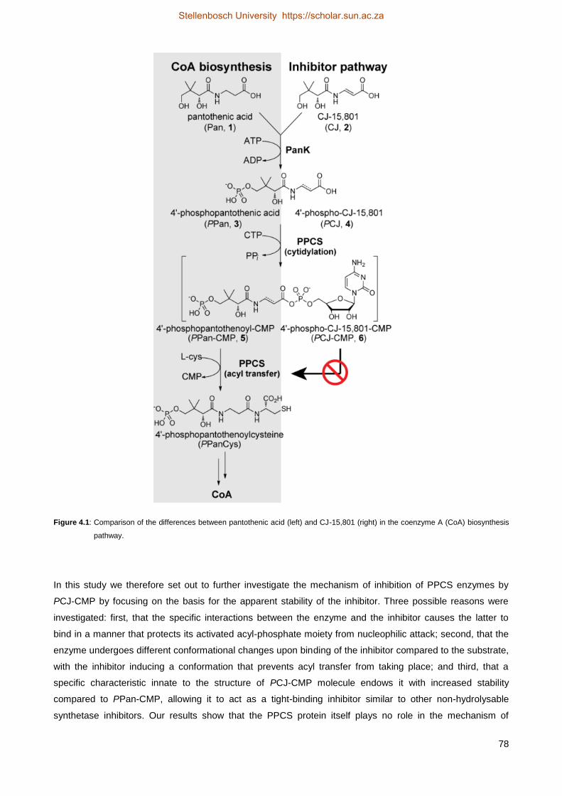
78
Figure 4.1: Comparison of the differences between pantothenic acid (left) and CJ-15,801 (right) in the coenzyme A (CoA) biosynthesis
pathway.
In this study we therefore set out to further investigate the mechanism of inhibition of PPCS enzymes by
PCJ-CMP by focusing on the basis for the apparent stability of the inhibitor. Three possible reasons were
investigated: first, that the specific interactions between the enzyme and the inhibitor causes the latter to
bind in a manner that protects its activated acyl-phosphate moiety from nucleophilic attack; second, that the
enzyme undergoes different conformational changes upon binding of the inhibitor compared to the substrate,
with the inhibitor inducing a conformation that prevents acyl transfer from taking place; and third, that a
specific characteristic innate to the structure of PCJ-CMP molecule endows it with increased stability
compared to PPan-CMP, allowing it to act as a tight-binding inhibitor similar to other non-hydrolysable
synthetase inhibitors. Our results show that the PPCS protein itself plays no role in the mechanism of
Stellenbosch University https://scholar.sun.ac.za

79
inhibition, but that the presence of the double bond with its extra π-electrons in the inhibitor renders its
carboxylate group resistant to nucleophilic attack through the creation of new, stable resonance forms. Apart
from shedding light on the mechanism of inhibition of this unique natural product, this finding might also
serve as a general starting point for the development of stable variants of other compounds containing β-
amino acid moieties.
4.2 Results
4.2.1 A constrained binding pose as a basis for increased inhibitor stability4
We first considered if inhibition by PCJ-CMP is caused by its planar N-acyl vinylogous carbamate
functionality imposing restrictions on the rotation of the molecule, thereby preventing it from taking on the
optimal binding pose required for attack by cysteine (or water). To establish if PPan-CMP and PCJ-CMP
have different binding modes, we used the only available structure of a PPCS enzyme with its intermediate
bound, that of the E. coli PPCS Asn210Asp mutant with PPan-CMP bound in the active site (PDB ID: 1U7Z).
This mutant, which accumulates sufficient amounts of the intermediate to allow direct confirmation of its
identity and structure [12, 13], is apparently unable to catalyse the second step of the PPCS reaction (Figure
4.1). However, the molecular basis for this catalytic disability remains speculative since no PPCS structure
with cysteine bound has been determined thus far. Nonetheless, the mutation clearly results in tightly-bound
PPan-CMP in the enzyme’s active site. We constructed a model of the native enzyme by computationally
reversing the Asn210Asp mutation in the crystal structure, and subsequently used it to dock PCJ-CMP in the
active site. For the docking, 150 starting conformations were used to ensure sufficient conformational space
was sampled considering the complexity of the molecule. The highest scoring docked pose of PCJ-CMP very
closely mimics the pose of the co-crystallised PPan-CMP in the crystal structure (Figure 4.2A), and facilitates
all the same hydrogen bonding interactions while maintaining a near planar geometry across the N-acyl
vinylogous carbamate moiety (Figure 4.2B). This suggests that the basis for inhibition by PCJ-CMP is not
due to it having a different binding mode, or taking on a potentially unreactive conformation.
4 Molecular modelling experiments reported in this section were performed by Dr. Stephen Pelly of the Department of
Chemistry and Polymer Science, Stellenbosch University.
Stellenbosch University https://scholar.sun.ac.za

80
Figure 4.2: (A) Structure of the PCJ-CMP inhibitor (stick structure with carbon atoms in yellow) modeled and overlaid on that of the
PPan-CMP intermediate (stick structure with carbon atoms in blue) bound in the active site of native EcPPCS. The model
was created from the crystal structure of EcPPCS Asn210Asp with co-crystalized PPan-CMP bound (PDB: 1U7Z) by
reversing the mutation, and docking PCJ-PMP. The highest scoring pose of PCJ-CMP closely resembles that of PPan-CMP
and similarly facilitates all hydrogen bonding interactions observed for PPan-CMP. Only selected residues are shown for
clarity. (B) Schematic view of polar interactions between PCJ-CMP and EcPPCS. Residues from one monomer are in blue
and those from the other monomer in green. The key interaction between Asn210 and the acyl phosphate carbonyl, mediated
via a bridging water molecule, is shown in red [3].
4.2.2 Enzyme structure stabilisation as a basis for inhibition by PCJ-CMP
We next set out to determine if formation of PCJ-CMP in the PPCS active site induces formation of a stable
conformation of the native protein structure beyond that which would normally be expected for a protein upon
binding of a ligand [14-16]. Initial investigation of ligand-induced stabilisation of the structures of E. coli
PPCS (EcCoaB, a dimer) and S. aureus CoaBC (SaCoaBC, a dodecamer that has both PPCS and
phosphopantothenoylcysteine decarboxylase (PPCDC) activities) showed that the former, experienced much
less stabilisation as measured by heat-induced denaturation than the latter [3]. Since EcPPCS was inhibited
by PCJ-CMP with an inhibition constant (Ki) of ~160 nM, while the Ki for SaCoaBC was ~13 nM, this
suggested a correlation between the potency of inhibition and the oligomeric structure of the protein [3].
To establish if this is indeed the case, we increased the sample size and performed the same analysis on
several bifunctional dodecameric CoaBC proteins with both PPCS and PPCDC activities [17], as well as
several monofunctional, dimeric PPCS enzymes [10]. Among the latter were some – like the human
(HsCoaB) and Streptococcus pneumoniae (SpCoaB) enzymes – that naturally occurs in this form, while
EcCoaB was prepared by expression of the PPCS domain of the bifunctional E. coli CoaBC (EcCoaBC)
protein. This analysis required the ligand-induced stabilisation and inhibition constants for each protein to be
determined as described below; the new expanded dataset could then be used to establish if increased
inhibition potency only correlates with increased structural stabilisation as would normally be expected [18,
19], or if some deviation or clustering was observed that could be related to the oligomeric structure of the
enzymes in question.
Stellenbosch University https://scholar.sun.ac.za

81
4.2.2.1 Melting temperature determination of CoaB and CoaBC enzymes
We used the melting temperatures (Tm) of the proteins to quantify their stability relative to one another. The
melting curves of the enzymes were determined in the presence of CTP and either its natural substrate, 4'-
phosphopantothenic acid (PPan, 3), or the inhibitor precursor, 4'-phospho-CJ-15,801 (PCJ, 4) (Figure 4.1),
by following the heat-induced denaturation of these mixtures by circular dichroism (CD). For HsCoaB, ATP
was used instead of CTP since eukaryotic PPCS enzymes have different substrate specificity than the
bacterial versions. The melting curves of all the proteins are shown in Figure 4.3. We observed a general
trend that the enzymes in the presence of inhibitor have higher Tm-values than in the presence of substrate,
as demonstrated by calculating the change in Tm-value change (ΔΔTm) between the two conditions. This
indicates that the binding of the inhibitor increases protein stability as expected (Figure 4.3A-F, Table 4.1).
Stellenbosch University https://scholar.sun.ac.za

82
Figure 4.3: Heat-induced protein melting curves for (A) EcCoaB, (B) SpCoaB, (C) HsCoaB*, (D) EcCoaBC, (E) MtCoaBC and (F)
SaCoaBC. The four curves in each graph represent from left to right: the free protein ( ); protein with MgCTP ( ); protein
with MgCTP and PPan (3) ( ); and protein with MgCTP and PCJ (4) ( ). The curves were determined by following
changes in the protein’s secondary structure by circular dichroism. *For HsCoaB MgATP was used instead of MgCTP since
eukaryotic PPCS enzymes have a different substrate specificity than the bacterial versions.
Stellenbosch University https://scholar.sun.ac.za

83
Table 4.1: Summary of the change in melting temperature (ΔTm) upon addition of either substrate (PPan+CTP) or inhibitor (PCJ+CTP)
to the apo CoaB and CoaBC proteins, as well as the difference between these (ΔΔTm).
PPCS enzyme Oligomeric
state
ΔTm substrate
(°C)
ΔTm inhibitor
(°C)
ΔΔTm
(°C)
EcCoaB dimer 10.0 ± 0.1 11.4 ± 0.1 1.4 ± 0.2
SpCoaB dimer 3.8 ± 0.1 16.1 ± 0.8 12.3 ± 0.9
HsCoaB dimer 1.2 ± 0.1 2.3 ± 0.1 1.1 ± 0.2
EcCoaBC dodecamer 12.7 ± 0.1 16.9 ± 0.2 4.2 ± 0.3
MtCoaBC dodecamer 8.0 ± 0.1 15.3 ± 3 7.3 ± 3.1
SaCoaBC dodecamer ~1 ~20 ~19
4.2.2.2 Determination of kinetic parameters of CoaB and CoaBC enzymes
The determination of inhibition constants for each protein required that their kinetic parameters (especially
Km) be determined first, since this value is used in the calculation of the inhibition constant of tight-binding
inhibitors. PPCS activity was assayed using the pyrophosphate reagent from Sigma-Aldrich that couples the
production of pyrophosphate (the product of the first step of the PPCS reaction, see Figure 4.1) to the
consumption of NADH, which was followed spectrophotometrically at 340 nm [20]. The initial rates were
calculated for each substrate concentration and fitted to the Michaelis-Menten equation (Equation 4.1) for
SaCoaBC, MtCoaBC and HsCoaB; interestingly, EcCoaB, EcCoaBC and SpCoaB showed substrate
inhibition at high PPan concentrations, so in their case the Michaelis-Menten equation that compensates for
substrate inhibition (Equation 4.2) was used instead. The results are shown in Figure 4.4 and the kinetic
parameters are summarised in Table 4.2.
𝑣 =𝑉𝑚𝑎𝑥[𝑆]
𝐾𝑚+[𝑆] ................................................................................................................................................... 4.1
𝑣 =𝑉𝑚𝑎𝑥
1+𝐾𝑚[𝑆]
+[𝑆]
𝐾𝑖
............................................................................................................................................... 4.2
Stellenbosch University https://scholar.sun.ac.za

84
Figure 4.4: Michaelis-Menten plots of (A) EcCoaB, (B) SpCoaB, (C) HsCoaB, (D) EcCoaBC, (E) MtCoaBC and (F) SaCoaBC at 1 mM
CTP (ATP for HsCoaB) with varying concentrations of PPan (3). Initial rates were determined in triplicate at each PPan
concentration and the error bars indicate the standard deviation between the triplicate values.
Stellenbosch University https://scholar.sun.ac.za
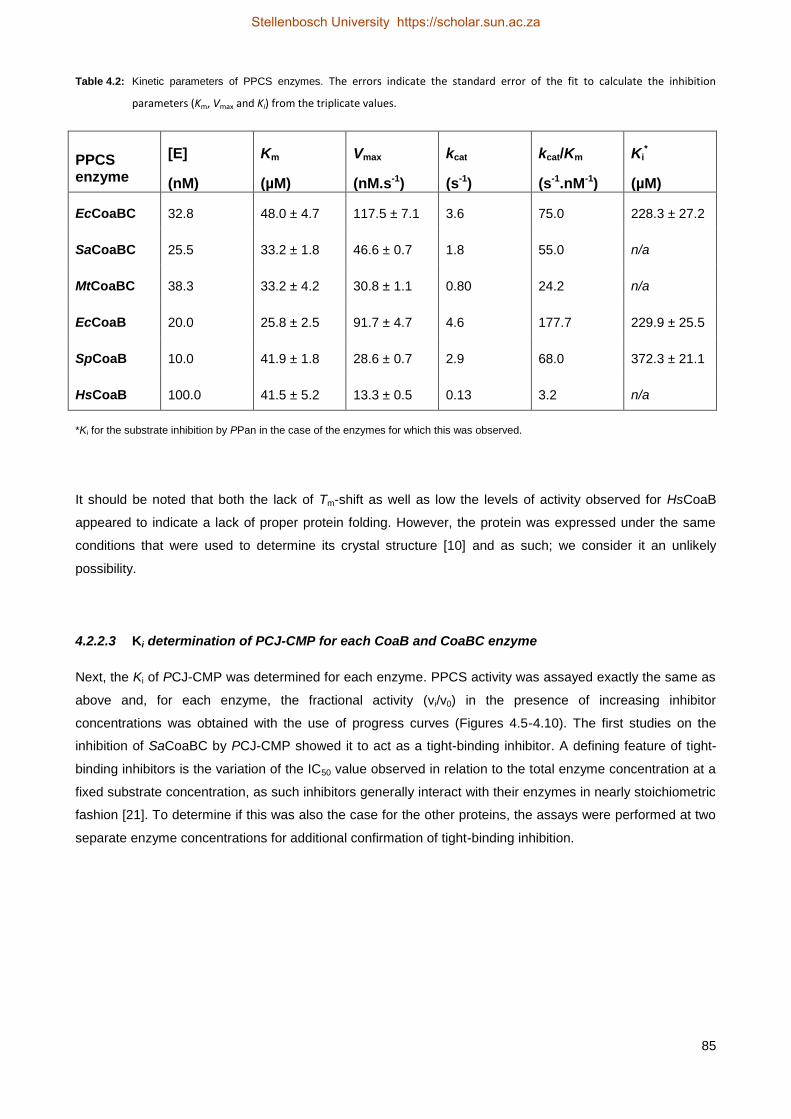
85
Table 4.2: Kinetic parameters of PPCS enzymes. The errors indicate the standard error of the fit to calculate the inhibition
parameters (Km, Vmax and Ki) from the triplicate values.
PPCS enzyme
[E]
(nM)
Km
(µM)
Vmax
(nM.s-1)
kcat
(s-1)
kcat/Km
(s-1.nM-1)
Ki*
(µM)
EcCoaBC 32.8 48.0 ± 4.7 117.5 ± 7.1 3.6 75.0 228.3 ± 27.2
SaCoaBC 25.5 33.2 ± 1.8 46.6 ± 0.7 1.8 55.0 n/a
MtCoaBC 38.3 33.2 ± 4.2 30.8 ± 1.1 0.80 24.2 n/a
EcCoaB 20.0 25.8 ± 2.5 91.7 ± 4.7 4.6 177.7 229.9 ± 25.5
SpCoaB 10.0 41.9 ± 1.8 28.6 ± 0.7 2.9 68.0 372.3 ± 21.1
HsCoaB 100.0 41.5 ± 5.2 13.3 ± 0.5 0.13 3.2 n/a
*Ki for the substrate inhibition by PPan in the case of the enzymes for which this was observed.
It should be noted that both the lack of Tm-shift as well as low the levels of activity observed for HsCoaB
appeared to indicate a lack of proper protein folding. However, the protein was expressed under the same
conditions that were used to determine its crystal structure [10] and as such; we consider it an unlikely
possibility.
4.2.2.3 Ki determination of PCJ-CMP for each CoaB and CoaBC enzyme
Next, the Ki of PCJ-CMP was determined for each enzyme. PPCS activity was assayed exactly the same as
above and, for each enzyme, the fractional activity (vi/v0) in the presence of increasing inhibitor
concentrations was obtained with the use of progress curves (Figures 4.5-4.10). The first studies on the
inhibition of SaCoaBC by PCJ-CMP showed it to act as a tight-binding inhibitor. A defining feature of tight-
binding inhibitors is the variation of the IC50 value observed in relation to the total enzyme concentration at a
fixed substrate concentration, as such inhibitors generally interact with their enzymes in nearly stoichiometric
fashion [21]. To determine if this was also the case for the other proteins, the assays were performed at two
separate enzyme concentrations for additional confirmation of tight-binding inhibition.
Stellenbosch University https://scholar.sun.ac.za
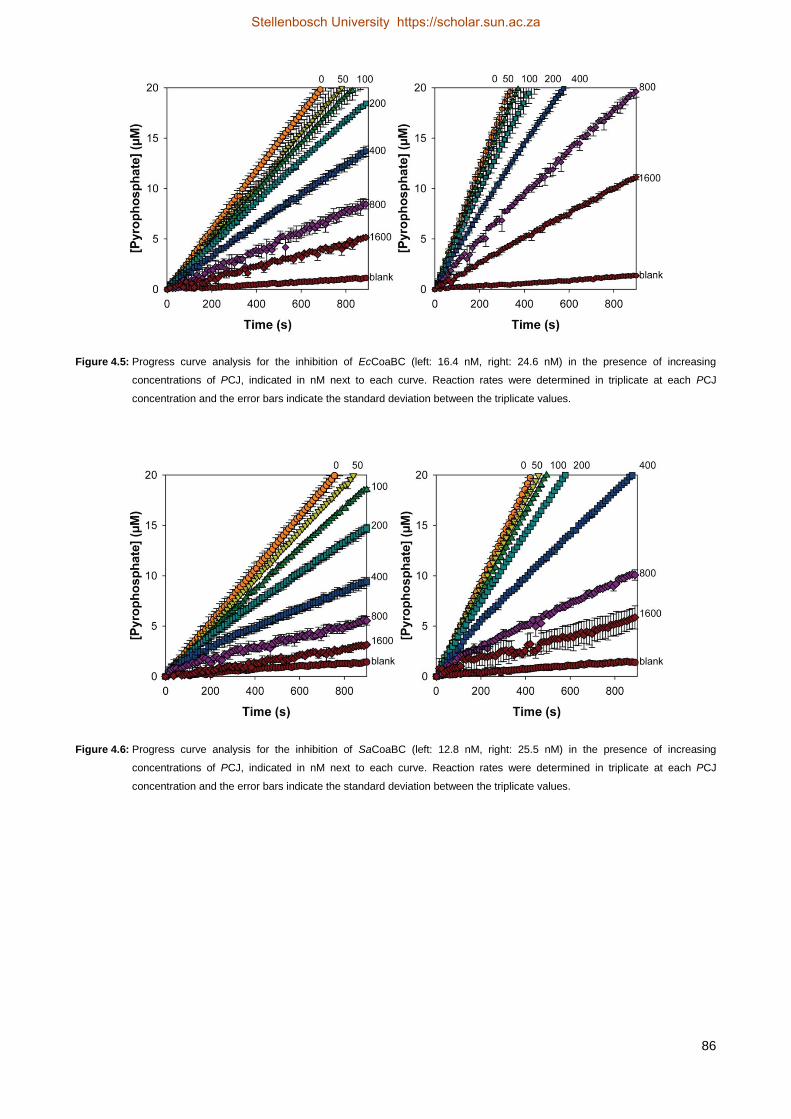
86
Figure 4.5: Progress curve analysis for the inhibition of EcCoaBC (left: 16.4 nM, right: 24.6 nM) in the presence of increasing
concentrations of PCJ, indicated in nM next to each curve. Reaction rates were determined in triplicate at each PCJ
concentration and the error bars indicate the standard deviation between the triplicate values.
Figure 4.6: Progress curve analysis for the inhibition of SaCoaBC (left: 12.8 nM, right: 25.5 nM) in the presence of increasing
concentrations of PCJ, indicated in nM next to each curve. Reaction rates were determined in triplicate at each PCJ
concentration and the error bars indicate the standard deviation between the triplicate values.
Stellenbosch University https://scholar.sun.ac.za

87
Figure 4.7: Progress curve analysis for the inhibition of MtCoaBC (left: 9.6 nM, right: 19.1 nM) in the presence of increasing
concentrations of PCJ, indicated in nM next to each curve. Reaction rates were determined in triplicate at each PCJ
concentration and the error bars indicate the standard deviation between the triplicate values.
Figure 4.8: Progress curve analysis for the inhibition of EcCoaB (left: 15 nM, right: 30 nM) in the presence of increasing concentrations
of PCJ, indicated in nM next to each curve. Reaction rates were determined in triplicate at each PCJ concentration and the
error bars indicate the standard deviation between the triplicate values.
Stellenbosch University https://scholar.sun.ac.za
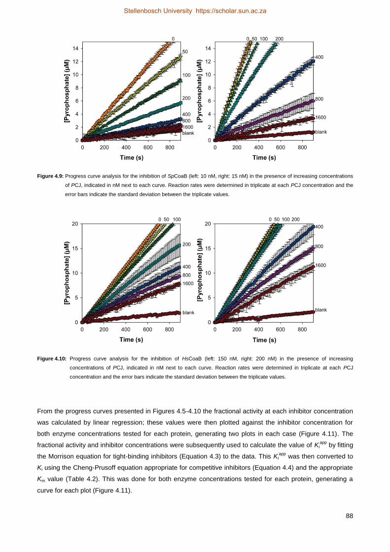
88
Figure 4.9: Progress curve analysis for the inhibition of SpCoaB (left: 10 nM, right: 15 nM) in the presence of increasing concentrations
of PCJ, indicated in nM next to each curve. Reaction rates were determined in triplicate at each PCJ concentration and the
error bars indicate the standard deviation between the triplicate values.
Figure 4.10: Progress curve analysis for the inhibition of HsCoaB (left: 150 nM, right: 200 nM) in the presence of increasing
concentrations of PCJ, indicated in nM next to each curve. Reaction rates were determined in triplicate at each PCJ
concentration and the error bars indicate the standard deviation between the triplicate values.
From the progress curves presented in Figures 4.5-4.10 the fractional activity at each inhibitor concentration
was calculated by linear regression; these values were then plotted against the inhibitor concentration for
both enzyme concentrations tested for each protein, generating two plots in each case (Figure 4.11). The
fractional activity and inhibitor concentrations were subsequently used to calculate the value of Kiapp
by fitting
the Morrison equation for tight-binding inhibitors (Equation 4.3) to the data. This Kiapp
was then converted to
Ki using the Cheng-Prusoff equation appropriate for competitive inhibitors (Equation 4.4) and the appropriate
Km value (Table 4.2). This was done for both enzyme concentrations tested for each protein, generating a
curve for each plot (Figure 4.11).
Stellenbosch University https://scholar.sun.ac.za

89
𝑣𝑖
𝑣0= 1 −
([𝐸]+[𝐼]+𝐾𝑖𝑎𝑝𝑝
)−√([𝐸]+[𝐼]+𝐾𝑖𝑎𝑝𝑝
)2−4[𝐸][𝐼]
2[𝐸] ................................................................................. 4.3
𝐾𝑖𝑎𝑝𝑝
= 𝐾𝑖 (1 +[𝑆]
𝐾𝑚) ................................................................................................................................... 4.4
These plots made it possible to evaluate the effect of enzyme concentration on IC50 to confirm the tight-
binding nature of the inhibition. The IC50 is indicated by the intersection between the Morrison plot and the
dashed line, and clearly shows that the higher enzyme concentration gave a higher IC50–value for all the
proteins tested, as expected for tight-binding inhibitors [21]. However, analysis of the fitted curves indicates
that the Morrison equation does not describe the observed inhibition profile as well in all cases (errors ~20%
of the calculated Ki) , pointing to these proteins not having a stoichiometric interaction between inhibitor and
protein as would be expected from a tight-binding inhibitor. When the same data obtained for these cases
were analysed using equations describing a standard reversible mechanism instead, the obtained fits were
worse in all cases. Consequently, we decided to determine the Ki-values in all cases using the Morrison and
Cheng-Prusoff equations as described above, although it appears as if PCJ-CMP has more tight-binding
character against some PPCS enzymes than others. Performing the analysis in this consistent manner also
allowed relative effects to be determined, an important aspect in this study. The Ki-value indicated in each
plot is the one obtained from the analysis that gave the smallest error of the fit. Using this methodology we
found the Ki of PCJ-CMP with SaCoaBC to be about twice as high as previously reported (27 nM vs 13 nM)
[3]. However, such a difference can be reasonably attributed to error and variation in the samples.
Stellenbosch University https://scholar.sun.ac.za
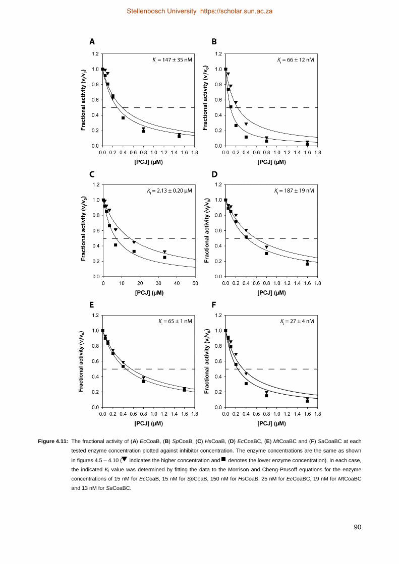
90
Figure 4.11: The fractional activity of (A) EcCoaB, (B) SpCoaB, (C) HsCoaB, (D) EcCoaBC, (E) MtCoaBC and (F) SaCoaBC at each
tested enzyme concentration plotted against inhibitor concentration. The enzyme concentrations are the same as shown
in figures 4.5 – 4.10 ( indicates the higher concentration and denotes the lower enzyme concentration). In each case,
the indicated Ki value was determined by fitting the data to the Morrison and Cheng-Prusoff equations for the enzyme
concentrations of 15 nM for EcCoaB, 15 nM for SpCoaB, 150 nM for HsCoaB, 25 nM for EcCoaBC, 19 nM for MtCoaBC
and 13 nM for SaCoaBC.
Stellenbosch University https://scholar.sun.ac.za

91
4.2.2.4 Comparison between the extent of structure stabilisation and the potency of inhibition
To establish if the correlation between the extent of structure stabilisation (as measured by the increase in
Tm) and the potency of inhibition (as embodied in the Ki-values) shows any difference between the dimeric
PPCS and the dodecameric CoaBC proteins, the difference in the Tm-values determined in the presence of
substrate and inhibitor (ΔΔTm) (Table 4.1) was plotted against the Ki-value obtained in each case (Figure
4.11). The plot obtained in this manner (Figure 4.12) shows that with the exception of HsCoaB (the only
protein tested that uses ATP and not CTP for the carboxylate activation step), a good correlation exists
between the ΔΔTm and Ki-values (R2 = 0.70), regardless if the protein is a dimeric PPCS or dodecameric
CoaBC. This suggests that the protein’s oligomeric state plays no role in inhibition, and that the potency of
inhibition is only dependent on the nature of the protein itself. Curiously, EcCoaBC shows a higher Ki than is
expected from a linear trendline; the basis for this is unclear, but it does suggest that this protein might be a
poor model for the inhibition of CoaBC enzymes in general. Removing it from the dataset improves the linear
correlation (R2 = 0.85) for the remaining data points.
Figure 4.12: Correlation between the extent of structure stabilisation and the potency of inhibition. The difference in the Tm-values
determined in the presence of substrate and inhibitor (ΔΔTm) is plotted against the Ki determined for each enzyme. The x-
axis error bars indicate the error of the fit for the Morrison equation to calculate the Ki-values and the y-axis error bars
indicate the propagated error from the combination of ΔTm of the substrate and ΔTm of the inhibitor. Where error bars are
not visible they are smaller than the symbols.
4.2.3 Inhibitor stability based on the inherent electronic properties of the molecule
After we established that the basis for inhibition of PPCS by PCJ-CMP is not due to differences in binding
mode or to oligomer-dependent stabilisation effects, we shifted our focus to the inherent chemical stability of
the molecule itself. As mentioned above, PCJ-CMP is a close structural analogue of the native PPCS
substrate, PPan-CMP, with the only difference being the introduction of a double bond in its β-alanine
moiety. We hypothesized that this change allows for the introduction of new, stable resonance forms that
consequently makes the acyl phosphate of PCJ-CMP more resistant to nucleophilic attack (Figure 4.13). To
Stellenbosch University https://scholar.sun.ac.za

92
test such a hypothesis would require the direct comparative analysis of the hydrolytic stability of pure PPan-
CMP and PCJ-CMP under identical conditions. However, considering the structural complexity of these
molecules and the significant synthetic effort that would be required to prepare them (and the additional
concern that especially PPan-CMP would hydrolyse too fast to allow analysis); we decided to instead use a
model system to evaluate the relative chemical stability of the N-acyl vinylogous carbamate functionality. We
additionally predicted that such a model system would likely give more reproducible results, which would
also be more generally applicable to other compounds containing β-amino acid moieties.
Figure 4.13: Resonance forms of the N-acyl vinylogous carbamate functionality illustrating the potential basis of its chemical stability.
For the proposed analysis we designed and synthesised model compounds 4-MU-3-benzamido-propanoate
(7a) and 4-MU-3-benzamido-acrylate (7b) (Figure 4.14A) to represent PPan-CMP and PCJ-CMP
respectively. In these models, the 4'-phosphopantoyl moiety of the two compounds were replaced by a
benzoyl group, while the acyl phosphate was replaced by an ester. The benzoyl exchange was chosen to
improve the synthetic tractability of the molecules, while the use of an ester instead of an acyl phosphate
improved the initial hydrolytic stability of the system to give a bigger dynamic range for analysis. The ester
group 4-methyl-umbelliferone (4-MU) was used, since free 4-MU is highly fluorescent, allowing a direct
method to detect hydrolysis of the esters. We analysed both compounds by stopped-flow fluorescent
spectroscopy and determined their initial rates of hydrolysis (within 60 s of initiating the reaction) using the
progress curve method at different buffered pH values. Both 7a and 7b were found to be highly stable under
low pH conditions (pH 2-8) and did not show any quantifiable hydrolysis for up to 10 h under these conditions
(Figure S1). However, at pH 8.5 and above, both compounds showed hydrolysis within a 60 s timeframe,
with the β-alanine ester 7a being significantly more reactive rate than the acrylate 7b (Figure 4.14B). These
results provided compelling evidence that the introduction of a double bond to the β-alanine moiety increases
the resistance of the acyl group to nucleophilic attack, most likely due to resonance effects.
Stellenbosch University https://scholar.sun.ac.za

93
Figure 4.14: (A) Structures of the fluorescent model system compounds: 4-MU-3-benzamido-propanoate (7a) and 4-MU-3-benzamido-
acrylate (7b). (B) The initial rates of hydrolysis of 7a ( ) and 7b ( ) between pH 8.5 – 11. The error bars show the
standard deviation between triplicate readings. Some of the bars are not visible behind the symbols.
4.2.4 Application of the findings to an unrelated system: hydrolytic stability of modified
pantetheinase (Vanin) substrates
To establish if this mechanism of apparent stabilisation via resonance could be applied to other systems for
the design of small molecule enzyme inhibitors, the human vanin-1 enzyme (VNN1, pantetheinase) was
chosen as test case. The Vanin pantetheinases belong to the nitrilase superfamily, and contain a conserved
Glu-Lys-Cys catalytic triad that is responsible for covalent catalysis in the hydrolysis of pantetheine (PantSH)
to produce pantothenic acid and cysteamine [22]. The key catalytic step is the nucleophilic attack by the
conserved cysteine of the triad on the pantothenoyl amide of its substrate [23]. Mechanistically, the
hydrolysis of PantSH by the Vanin pantetheinases is therefore equivalent to the acyl transfer step in the
PPCS reaction mechanism, since in both systems the key reaction is nucleophilic attack at the equivalent
carbonyl carbon. Importantly, the human VNN1 enzyme has been shown to also use other amide analogues
of pantothenic acid (pantothenamides) as substrates [24, 25]. We took advantage of this substrate
promiscuity and synthesised N-pentyl pantothenamide (N5-Pan, 8a), N-benzyl pantothenamide (Bn-Pan,
8b), N-pentyl-CJ (N5-CJ, 9a) and N-benzyl-CJ (Bn-CJ, 9b) (Figure 4.15) as potential VNN1 substrates.
These structures were chosen to determine if the compounds containing double bonds (i.e. that have an N-
acyl vinylogous carbamate functionality, namely N5-CJ and Bn-CJ) show increased resistance to
pantetheinase-mediated hydrolysis compared to those (N5-Pan and Bn-Pan) having unmodified β-alanine
moieties. This analysis required the development of two different assays as described separately below.
Stellenbosch University https://scholar.sun.ac.za

94
Figure 4.15: Structures of the compounds tested as substrates for VNN1 pantetheinase.
4.2.4.1 Characterisation of pantetheinase activity with alternative substrates: development of a
discontinuous assay based on amine formation
In order to perform the stability analyses of our pantetheinase test system, we needed the appropriate assay
conditions. First, the pantetheinase-mediated hydrolysis of the test compounds was assayed using
fluorescamine derivatisation of the amine-containing reaction products. Fluorescamine is known to readily
react with primary amines to form a fluorescent product [24], which can then be detected by fluorimetry. A
discontinuous fluorescamine-based assay was developed by adapting an existing method described by
Huang and Hernick [26]. After the reaction was initiated by adding enzyme to a substrate mixture, aliquots
were removed from the reaction mixture (at the desired time-points) and quenched by the addition of N-
ethylmaleimide. In the original protocol, the reaction is quenched by using acid precipitation of the enzyme,
followed by removal of the precipitated protein via centrifugation. We decided to use N-ethylmaleimide
instead, since it is a small compound that reacts with sulfhydryl groups such as the catalytic cysteine of
VNN1 to form stable, covalent thioethers [27]. This leads to rapid inactivation of the enzyme rapidly without
the need for an additional centrifugation step. The quenched reaction mixtures are then derivatised with
fluorescamine and the resulting fluorescence measured.
4.2.4.2 Characterisation of pantetheinase activity with pantetheine: development of continuous
assay based on pantothenate formation
We also required a continuous assay better suited for the characterisation of pantetheinase activity of VNN1
with its natural substrate pantetheine in the presence of the alternative substrates. Therefore, a new
enzyme-coupled assay for pantetheinase activity was developed using a type III PanK enzyme. This form of
PanK phosphorylates only pantothenic acid (Pan) and not PantSH; this substrate selectivity therefore allows
it to be used to discriminate between the substrate and product of the pantetheinase reaction (Figure 4.16).
For the assay, the Bacillus anthracis PanK (BaPanK) was used, and basing the conditions used by Rowan
and co-workers for the activity characterisation of this enzyme [28]. Pantetheinase activity was subsequently
characterised in the presence of sufficient amounts of BaPanK so that the coupling reaction was not rate
limiting. The reaction rate was determined indirectly by following the oxidation of NADH (produced by
Stellenbosch University https://scholar.sun.ac.za

95
coupling the ADP formed by BaPanK to the pyruvate kinase and lactate dehydrogenase reactions)
spectrophotometrically at 340 nm.
Figure 4.16: A schematic representation of the coupled assay to monitor pantetheinase activity. ATP = adenosine triphosphate, ADP =
adenosine diphosphate, PK = pyruvate kinase, LDH = lactate dehydrogenase, PEP = phosphoenol pyruvate, Pyr =
pyruvate.
4.2.4.3 Characterisation of CJ-amides as pantetheinase substrates (and inhibitors)
All the pantetheinase test compounds were tested with our fluorescamine assay at 20 µM, which is equal to
the reported Km of the native substrate, pantetheine for VNN1 [29]. The reactions for all the test compounds
were followed to completion (approximately 2 hours). The results are summarised in Figure 4.17 and show
that only N5-Pan and Bn-Pan were converted to product, while no activity was observed with N5-CJ and Bn-
CJ.
Figure 4.17: Time-course data of the pantetheinase-mediated hydrolysis of the various substrates using fluorescamine derivatisation of
their products. N-benzyl pantothenamide ( ), N-pentyl pantothenamide ( ), N-benzyl-CJ ( ) and N-pentyl-CJ ( ).
These results suggested that the presence of the double bond in N5-CJ and Bn-CJ increased resistance to
hydrolysis by pantetheinase as predicted by the results of the model system. However, one could argue that
the restricted rotation introduced by the presence of the double bond prevents these compounds from
entering the active site of the enzyme altogether. To demonstrate that this is not the case, we determined if
Stellenbosch University https://scholar.sun.ac.za

96
N5-CJ and Bn-CJ acted as competitive inhibitors of the pantetheinase reaction. First, we characterised the
pantetheinase activity of VNN1 with PantSH using our continuous assay. Then we characterised the
inhibition of VNN1 by N5-CJ at various concentrations (using the same PantSH concentration each time).
The results confirmed that N5-CJ is indeed a competitive inhibitor of pantetheinase (Figure 4.18). This
indicates that the lack of pantetheinase activity with the CJ-derived amides is due to their hydrolytic stability,
and not to them being excluded from the enzyme.
Figure 4.18: Inhibition of VNN1 pantetheinase by N-pentyl-CJ (N5-CJ). (A) Plots of initial rates of pantetheinase activity against the
concentration (at Km) of pantetheine at various fixed concentrations of N5-CJ. Kinetic parameters for the uninhibited
reaction were determined by non-linear regression analysis: Vmax = 0.0015 nmol.s-1, Km = 25.58 μM. These values
correspond to those determined using a discontinuous assay [29]. (B) Lineweaver-Burke plot of the data in A illustrating
competitive inhibition (increase in Km) of VNN1 pantetheinase by N5-CJ with Ki = 88.76 μM.
4.3 Discussion
In this study we set out to further investigate the mechanism of inhibition of PPCS enzymes by PCJ-CMP by
examining the basis for the apparent stability of the inhibitor. First, we probed any differences between the
binding mode of the inhibitor and that of the substrate, which might cause the activated acyl-phosphate in
PCJ-CMP to be protected from nucleophilic attack. However, our molecular docking studies showed that the
basis for inhibition by PCJ-CMP is not due to it having a different binding mode, or taking on a potentially
unreactive conformation. Next, we investigated whether the potency of inhibition by PCJ-CMP is determined
by the oligomeric structure of its target enzyme. Since proteins are stabilised upon the binding of a ligand,
we searched for a correlation between increased stabilisation (beyond that which would normally be
expected) and increased inhibitor potency among a small set of PPCS enzymes, consisting of three dimeric
CoaB and three dodecameric CoaBC enzymes. However, when the extent of structure stabilisation (as
measured by the increase in Tm) and the potency of inhibition of each enzyme were compared, we found no
correlation to oligomeric structure. Interestingly, a good correlation exists between the ΔΔTm and Ki-values of
the prokaryotic enzymes, except for EcCoaBC, which shows a higher Ki than what is expected from a linear
Stellenbosch University https://scholar.sun.ac.za

97
trendline. The basis for this is unclear, but it does suggest that this protein might be a poor model for the
inhibition of CoaBC enzymes in general. Ideally, more CoaB domains of bifunctional CoaBC enzymes need
to be tested for comparison. In fact, since the current sample size is quite small, by including data from
additional CoaB and CoaBC proteins more concrete conclusions might be made.
We also investigated if a specific characteristic innate to the structure of PCJ-CMP molecule endows it with
increased stability compared to PPan-CMP, allowing it to act as a tight-binding inhibitor. The relative
chemical stability of its N-acyl vinylogous carbamate functionality was evaluated by using a model system.
The results provided compelling evidence that the introduction of a double bond to the β-alanine moiety
increases the resistance of the acyl group to nucleophilic attack, most likely due to resonance effects. It
should be noted that the result is not entirely surprising, since we expected that the delocalisation of
electrons across the carbamate functionality will decrease the electrophilicity of its carbonyl carbon. This in
turn decreases the vulnerability of the carbonyl carbon to nucleophilic attack and increases the resistance of
the ester (in the model compound) and acyl phosphate (in PCJ-CMP) to acyl transfer. Additionally, the
results reinforced that it is not the planar N-acyl functionality of PCJ-CMP imposing restrictions on the
rotation of the molecule that prevents it from taking on the optimal binding pose required for attack by the
cysteine, since these compounds displayed increased stability in solution where they may freely rotate. The
stabilisation provided by a trans-enamine has previously been noted in the study of other antibiotics.
Padayatti and co-workers inhibited the SHV-1 β-lactamase by forming a stable, long lived trans-enamine
intermediate within the active site of the enzyme, which vastly lowered its deacylation efficiency [30]. We
went further and applied this mechanism of stabilisation via resonance to an unrelated system for the design
of small molecule enzyme inhibitors. The human vanin-1 enzyme (VNN1, pantetheinase) was chosen as test
case. As before, test compounds were synthesised to evaluate the stability of the N-acyl vinylogous
carbamate functionality compared to that of the β-alanine moiety as well as the compounds’ concomitant
resistance to hydrolysis (or lack thereof). All the test compounds were demonstrated to enter the VNN1
active site. The compounds with a double bond, which were consequently stabilised via new resonance
forms, were shown to be successfully protected against hydrolysis as predicted.
Taken together, our results show that the PPCS protein itself does not provide the basis for the tight-binding
mode of inhibition by PCJ-CMP, but that the introduction of the double bond in the β-alanine moiety of the
substrate with its extra π-electrons renders the acyl phosphate resistant to nucleophilic attack by introducing
new, stable resonance forms. This is similar to previous synthetase inhibitor design strategies in which stable
intermediate analogues were prepared; however PCJ-CMP differs from these as it is generated in situ. This
is a major advantage, since the inhibitor precursor, CJ, can pass through the bacterial cell envelope. It is
then selectively activated by S. aureus’ PanK to form PCJ, which is accepted into the active site of the target
PPCS enzyme where it forms PCJ-CMP as a tight-binding, stable inhibitor. The results of this study have
therefore allowed us to rationalise the basis for the tight-binding inhibition previously reported for PCJ-CMP.
Additionally, our results also uncovered a new strategy whereby the structures of β-amino acid-containing
compounds can be rendered stable to hydrolysis. This finding might also serve as a general starting point for
the development of stable variants of inhibitors or therapeutics that contain β-amino acid moieties.
Stellenbosch University https://scholar.sun.ac.za

98
4.4 Materials and methods
Unless otherwise noted, all reagents and chemical compounds were purchased from commercial sources
and used without further purification. Pyruvate Kinase was from Roche and Inorganic Pyrophosphatase was
from Sigma. Dichloromethane (DCM) was distilled under nitrogen from calcium hydride. N,N-
Dimethylformamide (DMF) was dried and purified by shaking up over potassium hydroxide followed by
distillation under reduced pressure and a nitrogen atmosphere, and was stored over 3 Å molecular sieves.
All column chromatography was performed using Merck silica gel 60 (particle size 0.040-0.063 mm) using
combinations of hexane, ethyl acetate, DCM and methanol as eluants. Thin layer chromatography (TLC) was
carried out on aluminium-backed Merck silica gel 60 F254 plates. Visualisation was performed with a UV
lamp followed by spraying with a cerium ammonium molybdate or ninhydrin solution, followed by heating.
NMR analyses: All 1H and
13C nuclear magnetic resonance spectra were obtained using 300 MHz Varian
VNMRS (75 MHz for 13
C), 400 MHz Varian Unity Inova (100 MHz for 13
C) or 600 MHz Varian Unity Inova
(125 MHz for 13
C) instruments at the Central Analytical Facility (CAF) of the University of Stellenbosch.
Chemical shifts (δ) were recorded using the residual solvent peak or an external reference. All chemical
shifts are reported in ppm and all spectra were obtained at 25 °C. Proton spectral data are reported as
follows: chemical shift, multiplicity (ovlp = overlapping, s = singlet, d = doublet, t = triplet, q = quartet, p =
pentet, m = multiplet, br = broad), coupling constant in Hz, and integration.
Protein purification: All His-tagged protein purification was performed using HisTrapFF metal affinity
purification columns on an ÅKTAprime purification system from GE Healthcare Life Sciences. The purified
proteins were desalted using 5 mL HiTrap desalting columns.
Spectrophotometry: All absorbance readings in 96-well plates were measured using a Thermo Scientific
Varioskan microplate spectrophotometer. All larger volume measurements were made in an Agilent
Technologies Cary 60 UV-Vis Spectrometer using a 1 cm cuvette.
NMR analyses: All 1H and
13C nuclear magnetic resonance spectra (NMR) were obtained using 300 MHz
Varian VNMRS (75 MHz for 13
C), 400 MHz Varian Unity Inova (100 MHz for 13
C) or 600 MHz Varian Unity
Inova (125 MHz for 13
C) instruments at the Central Analytical Facility (CAF) of the University of Stellenbosch.
Chemical shifts (δ) were recorded using the residual solvent peak or an external reference. All chemical
shifts are reported in ppm and all spectra were obtained at 25 °C. Proton spectral data are reported as
follows: chemical shift, multiplicity (ovlp = overlapping, s = singlet, d = doublet, t = triplet, q = quartet, p =
pentet, m = multiplet, br = broad), coupling constant in Hz, and integration.
MS analyses: All high–resolution electrospray ionisation mass spectrometry analyses (HR–ESI–MS) were
performed using a Waters Synapt G2 mass spectrometer.
Stopped-flow fluorimetry: All measurements were taken on an Applied Photophysics Chirascan Plus
circular dichroism spectrometer using a fluorescence detector and a stopped-flow injector. The total
Stellenbosch University https://scholar.sun.ac.za

99
fluorescence was measured with a long pass cut-off filter installed that blocked all fluorescence below 385
nm. A sample cell pathlength of 2 mm and monochromator bandwidth of 4 nm was used.
4.4.1 Overexpression and purification of PPCS enzymes
The phosphopantothenoyl cysteine synthetase (PPCS) enzymes of Staphylococcus aureus (SaCoaBC),
Escherichia coli (EcCoaBC and EcCoaB), Streptococcus pneumonia (SpCoaB) and Homo sapien (HsCoaB)
were expressed and purified according to published methods using BL21-star (DE3) (Invitrogen) as
expression strain [3, 7, 31, 32].
4.4.2 Overexpression and purification of His-MtCoaBC
The plasmid, pET-28a_MtcoaBC, was kindly provided by Michal Blaszczyk (from Prof. Tom Blundell’s lab
(Dept. of Biochemistry, University of Cambridge), which we transformed into E. coli BL21 Star (DE3)
(Invitrogen) for expression. Expression was performed in Luria-Bertani broth supplemented with 30 mg/L
kanamycin at 37 °C. Cultures were grown until OD600 = 0.6, at which point the temperature was lowered to
20 °C before expression was induced by addition of 1 mM IPTG. Cell growth was continued overnight at 20
°C post-induction, after which the cells were harvested by centrifugation. The obtained cell pellet was
suspended in sonication buffer (50 mM Tris-HCl, 0.5 M NaCl, 20 mM imidazole, 5% glycerol, pH 7.8; 10
mL/1 g cell paste) and subsequently sonicated to effect cell lysis. After centrifugation at 25 000 rpm for 20
min to collect cell debris, the crude extract (supernatant) was applied to a previously prepared 1 mL
HisTrapFF metal affinity purification column (Amersham Biosciences) using an ÄKTAprime purification
system. Weakly bound proteins were removed by washing with sonication buffer, followed by sonication
buffer containing 75 mM imidazole. His-MtCoaBC was eluted by increasing the imidazole concentration to
500 mM. Elution was monitored at A280. The purified protein was desalted with a 5 mL HiTrap desalting
column (Amersham Biosciences) using a 50 mM Tris-HCl buffer at pH 8.0, containing 0.15 M NaCl, 0.5 mM
TCEP and 5% glycerol. Afterwards, the protein was aliquoted and stored at -80 °C.
4.4.3 Protein Melting Temperature Determinations and Analysis
Protein melting curves were determined by measuring the change in circular dichroism (CD) at 220 nm as
the temperature of protein samples were increased from 35 °C to 80 °C at a rate of 1 °C per minute. All
measurements were made in an Applied Photophysics Chirascan-Plus CD Spectrometer using a 0.5 mm
cuvette.
Stellenbosch University https://scholar.sun.ac.za

100
Determining PPCS temperature melting curves
Four separate samples were prepared of each protein, all of which contained 10 µM PPCS and 1.0 mM
MgCl2 in 50 mM Tris–HCl buffer (pH 7.6). The first sample contained no additional components, while the
other three contained 150 μM CTP, 150 μM each of PPan and CTP, or 150 μM each of PCJ and CTP,
respectively. The final volume of all samples was 240 μL. A sampling time of 0.5 s per point was employed.
A temperature probe was used in the sample solutions and the preset instrument temperatures were
replaced with temperatures measured each minute by the probe for higher accuracy. The Tm were
subsequently determined by normalizing the melting curve data, and fitting the resulting curves
simultaneously to Equations 4.5, 4.6 and 4.7 (the Gibbs-Helmholtz equation) using SigmaPlot 12.0 software
(Systat Software), where lb and rb are the equations for the best linear fits to the left and right baselines of
the curves respectively. The following parameter constraints were used for the curve fit: Van’t Hoff enthalpy
(ΔHvH) > 0; Tm > 0 and ΔCp > 0.
𝑦 = 𝐹𝐹 × (𝑙𝑏 − 𝑟𝑏) + 𝑟𝑏 .......................................................................................................................... 4.5
𝐹𝐹 =1
1+(1
𝑒(
∆𝐺1.9872×𝑇
))
........................................................................................................ 4.6
∆𝐺 = ∆𝐻𝜗𝐻 (1 −𝑇
𝑇𝑚) − ∆𝐶𝑝 (𝑇𝑚 − 𝑇𝑙𝑛
𝑇
𝑇𝑚) .......................................................................................... 4.7
Determining SaCoaBC temperature melting curves
The same procedure was used for all the PPCS enzymes; however, accurate Tm determinations of
SaCoaBC were not possible since the protein was not fully denatured at the highest temperature that can be
obtained in the spectrometer. The Tm-values were subsequently estimated by normalizing the melting curve
data and determining the first derivatives of the curves.
4.4.4 PPCS Inhibition Assays and Data Analyses
PPCS assays were performed according to the published procedure [3]. The PPCS reaction was observed in
the forward reaction via an enzyme-linked assay in which the pyrophosphate that is released during transfer
of CMP from CTP to the substrate is continuously detected. This is achieved using the commercially
available pyrophosphate reagent (PR) from Sigma-Aldrich (P7275). Each vial of PR was resuspended in 4.5
Stellenbosch University https://scholar.sun.ac.za

101
mL dH2O. The assays were performed on a Thermo Scientific Varioskan microplate spectrophotometer at 37
°C. All curve fitting analyses were performed using SigmaPlot 12.0.
PPCS kinetic parameter determination (with PPan, 3)
A master assay mix containing 50 mM Tris–HCl buffer (pH 7.6), 2.5 mM MgCl2, 20 mM KCl, 1.0 mM CTP,
1.0 mM L-Cys, 2.5 mM DTT, PPCS enzyme (at varying concentrations for each enzyme) and 60 μL PR was
incubated at 37 °C for 15 min and added to a 96-well plate containing PPan (3) (12.5 μM to 500 μM) in
different wells to give a final assay volume of 150 μL. All measurements were obtained in triplicate. Initial
velocities were calculated for each substrate concentration and fitted to the Michaelis-Menten equation
(Equation 4.1) for SaCoaBC, MtCoaBC and HsCoaB; or the Michaelis-Menten equation that compensates
for substrate inhibition (Equation 4.2) for EcCoaB, EcCoaBC and SpCoaB using SigmaPlot 12.0. The results
are shown in Figure 4.4 and the kinetic parameters are summarised in Table 4.2. The following enzyme
concentrations were used: EcCoaBC 32.8 nM, SaCoaBC 25.5 nM, MtCoaBC 38.3 nM, EcCoaB 20.0 nM,
SpCoaB 10.0 nM and HsCoaB 100.0nM.
𝑣 =𝑉𝑚𝑎𝑥[𝑆]
𝐾𝑚+[𝑆] ................................................................................................................................................... 4.1
𝑣 =𝑉𝑚𝑎𝑥
1+𝐾𝑚[𝑆]
+[𝑆]
𝐾𝑖
............................................................................................................................................... 4.2
4.4.5 Progress curve analysis
To ensure that substrate depletion would not significantly affect the reaction rate, a low enzyme
concentration and high substrate concentration (PPan = 250 μM) were used ([21, 33]), which allowed the
control experiment (without inhibitor) to proceed linearly over ~15 min. Such a procedure should allow any
divergence from linearity, caused by slow onset or irreversible inhibition, to be clearly observable. However,
the enzymes that are subject to substrate inhibition were assayed at 100 µM to prevent interference by high
substrate concentrations.
Procedure
A master assay mix containing Tris–HCl buffer (50 mM, pH 7.6), MgCl2 (2.5 mM), KCl (20 mM), CTP (1.0
mM), L-Cys (1.0 mM), DTT (2.5 mM), PPCS (at varying concentrations for each enzyme) and 60 μL PR was
incubated at 37 °C for 15 min and added to separate wells of a 96-well plate containing a mixture of 4′-
phosphopantothenic acid (100 or 250 μM) and PCJ (in concentrations ranging between 0.025 μM to 1.6 μM),
Stellenbosch University https://scholar.sun.ac.za

102
followed by determination of the activity over a 15 min period. All measurements were obtained in triplicate
and the results are shown in Figures 4.5-4.10.
Data analysis
From the progress curves presented in Figures 4.5-4.10 the fractional activity at each inhibitor concentration
was calculated by linear regression. The fractional activity and inhibitor concentrations were used to
calculate the value of Kiapp
by fitting the data to the Morrison equation for tight binding inhibitors (Equation
4.3) using SigmaPlot 12.0. This Kiapp
was then converted to Ki using the Cheng-Prusoff equation appropriate
for competitive inhibitors (Equation 4.4) and the appropriate Km value (Table 4.2). The plots of the Morrison
equation for each enzyme are included in Figure 4.11. The Ki-values are included with the Tm-data in Table
4.2.
𝑣𝑖
𝑣0= 1 −
([𝐸]+[𝐼]+𝐾𝑖𝑎𝑝𝑝
)−√([𝐸]+[𝐼]+𝐾𝑖𝑎𝑝𝑝
)2−4[𝐸][𝐼]
2[𝐸] ................................................................................. 4.3
𝐾𝑖𝑎𝑝𝑝
= 𝐾𝑖 (1 +[𝑆]
𝐾𝑚) ................................................................................................................................... 4.4
4.4.6 Stopped-flow fluorimetry
The measurements were taken on an Applied Photophysics Chirascan Plus circular dichroism spectrometer
using a fluorescence detector and a stopped-flow injector. The total fluorescence was measured with a long
pass cut-off filter installed that blocked all fluorescence below 385 nm, which could potentially interfere with
the sample fluorescence. A sample cell pathlength of 2 mm and monochromator bandwidth of 4 nm was
used. The fluorescence excitation wavelength was set to 375 nm and emission was detected by scanning at
440 nm. The stopped-flow sequencer used a manual external trigger for injection and 1000 data points were
collected over 60 s.
4-MU standard curves
Separate standard curves were constructed for pH 8.50, pH 9.00, pH 9.50, pH 10.00 and pH 10.50. Glycine-
NaOH buffers (50 mM) were made up to buffer the reaction mixtures at each pH-value. A stock solution of 4-
MU in DMSO (50 mM) was diluted in water to make up standard solutions of 4-MU: 20 µM, 10 µM, and 5 µM.
Each 4-MU solution was injected together with each glycine buffer in triplicate and the resulting fluorescence
was measured and used to construct standard curves. No significant differences were observed for 4-MU
fluorescence between the various alkaline pH-values. Figure S1A shows the 4-MU standard curve at pH 9.
Stellenbosch University https://scholar.sun.ac.za

103
Fluorimetric analysis of model system compounds
Glycine-NaOH (50 mM) was used to buffer hydrolysis reactions at each pH-value. DMSO stocks of 4-MU-3-
benzamido-propanoate (7a) and 4-MU-3-benzamido-acrylate (7b) were diluted to 200 µM in water. The
reactants were loaded into the stopped-flow drive syringes: one with 200 µM 4-MU-amide and one with
glycine buffer. Samples were pushed through the flow circuit without collecting data three times to prime the
observation chamber. The detector was then calibrated according to the resulting fluorescent signal. Data
were then acquired in triplicate with each glycine buffer (Figure S1B).
Figure S1: (A) Standard curve of 4-MU at pH 9. (B) pH rate profile of 4-MU-benzamido propanoate (7a, ) and 4-MU-benzamido
acrylate (7b, ). The error bars in each figure show the standard deviation between triplicate readings. Some of the bars
are not visible behind the symbols.
4.4.7 Fluorescamine assay of pantetheinase activity
Assay mixtures containing DTT (0.5 mM), BSA (0.01 % (m/v)) and substrate (pantothenamide or CJ-amide,
20 μM) in phosphate buffer (100 mM, pH 7.6) were pre-equilibrated at 37 °C prior to the addition of vanin-1
(18.5 nM) to initiate the reaction. After various times, aliquots (30 μL) were removed from the reaction
mixture and quenched by the addition of N-ethylmaleimide (10 μL, 1.5 μM). Aliquots of the quenched
reaction mixture (25 μL) were transferred into a black 96-well plate, diluted with 1 M borate (pH 9.0, 75 μL),
and reacted with fluorescamine (10 mM, 30 μL in CH3CN). After 10 min the resulting fluorescence was
measured (excitation at 395 nm, emission at 485 nm) using a Thermo Scientific Varioskan fluorescent plate
reader.
4.4.8 Continuous enzyme coupled assay of pantetheinase activity
A master assay mix containing HEPES buffer (200 mM, pH 7.6), MgCl2 (10 mM), NH4Cl (60 mM), ATP (1
mM), NADH (0.5 mM), PEP (2 mM), PK (7.5 units), LDH (15 units), DTT (500 µM), BSA (50 µg.mL-1
),
BaPanK (44.44 µg.mL-1
), and vanin-1 (25 nM) was incubated at 37 °C for 15 min and added to separate
Stellenbosch University https://scholar.sun.ac.za

104
wells of a 96-well plate containing PantSH (4.69 µM – 300 µM) and N-pentyl-CJ (0 µM – 50 µM). The
reaction was monitored by following the decrease of NADH at 340 nm over a 40 min period at 37 °C. All
measurements were obtained in triplicate.
4.4.9 Synthesis of model system compounds5
N-benzoyl-β-aminopropionic acid (S1): Prepared according to previously
published methods [34].
4-methyl-2-oxo-2H-chromen-7-yl 3-benzamido-propanoate (7a): S1
(4.04 mmol, 780 mg) was suspended in 10 mL anhydrous DCM at
ambient temperature. 4-methyl-umbelliferone (4.04 mmol, 711.72 mg)
and DMAP (0.0018 mmol, 2.24 mg) was added to the acid while stirring.
Next, pivalic anhydride (4.04 mmol, 752.45 mg, 819.66 µL) was added and the reaction mixture was stirred
at 50 °C for 48 h. Subsequently, water (9 µL, 0.5 mmol) was added and the mixture was stirred at 50 °C for 1
h. The reaction mixture was then concentrated under vacuum and the product purified on a silica column by
eluting with 1 % methanol in DCM. The solvent was removed under vacuum and freeze-dried to afford the
product as a white powder (650 mg, 1.85 mmol). The yield was calculated as 45.8% 1H NMR (600 MHz,
DMSO): δ = 2.43 (s, 3H), 2.89 (t, J = 7.03 Hz, 2H), 3.65 (t, J = 7.03 Hz, 2H), 6.39 (s, 1H), 7.21 (d, J = 8.20
Hz, 1H), 7.29 (s, 1H), 7.45 – 7.55 (m, 3H), 7.81 – 7.87 (m, 3H), 8.71 (s, 1H). 13
C NMR (125 MHz, DMSO): δ
= 21.2, 37.2, 38.4, 113.2, 116.8, 120.5, 121.6, 129.9, 130.4, 131.7, 134.3, 137.3, 156.0, 156.5, 162.5, 169.6,
172.9. HRMS (m/z): [M+H]+ calculated for C20H18NO5, 352.1185; found 352.1191.
3-bromoacrylic acid (S2): Prepared using previously published methods [35, 36].
(E/Z)-4-methyl-2-oxo-2H-chromen-7-yl 3-bromoacrylate (S3.1): S2 (6.62
mmol, 1 g) was suspended in anhydrous DCM (10 mL) at ambient temperature.
7-Hydroxy-4-methylcoumarin (6.02 mmol, 1060.78 mg), 4-
Dimethylaminopyridine (0.003 mmol, 4.05 mg) and pivalic anhydride (6.62 mmol, 1130.98 mg, 1232 µL)
were added to the acid at ambient temperature. The mixture was then stirred at 50 °C for 48h. Water (9 µL,
0.5 mmol) was added and the reaction mixture was stirred for an additional hour at 50 °C. The solvent was
removed under reduced pressure and the product purified on a silica column by eluting with 1 %
5 The compounds marked with * were previously synthesised by Renier van der Westhuyzen.
Stellenbosch University https://scholar.sun.ac.za

105
methanol/DCM. The solvent was removed under vacuum and freeze-dried to yield the product as a white
powder (1.54 g, 4.98 mmol) The yield was calculated as 82.7%. 1H NMR (400 MHz, DMSO): δ = 2.43 (s,
3H), 6.39 (s, 1H), 6.92 (d, J = 9.38 Hz, 1H), 7.23 (d, J = 8.59 Hz, 1H), 7.81 (d, J = 9.38, 1H), 8.18 (d, J =
13.28 Hz, 1H). 13
C NMR (100 MHz, DMSO): δ = 19.3, 111.2, 114.0, 115.1, 119.0, 119.5, 127.7, 128.7, 132.2,
154.0, 160.8, 162.9. HRMS (m/z): [M+H]+ calculated for C13H10O4Br, 308.9762; found 308.9765.
General procedure for the synthesis of β-bromoacrylamides
To an oven-dried two neck flask were added S2 (1.00 g 6.62 mmol) and DMAP (0.162 g, 1.32 mmol). The
amine (9.94 mmol) and DCM (13 mL) were then added to the flask using oven-dried syringes. The solution
was cooled to 0 °C and DIC (1.13 mL, 7.29 mmol) was added drop-wise with a syringe. The reaction was
warmed to rt and stirred for 6 h, after which it was filtered through Celite and concentrated under reduced
pressure. After dilution with ethyl acetate, the organic layer was washed with saturated NaHCO3 (2 x 10 mL),
brine (1 x 10 mL) and the organic layer dried over Na2SO4. After filtration and concentration in vacuo the
amide was purified using flash chromatography.
*(E)-3-Bromo-N-pentylacrylamide (S3.2): The acrylamide was prepared
according to the general procedure using pentylamine (1.15 mL, 9.94 mmol). A
pale yellow solid (0.34 g, 12%) was obtained after purification with flash
chromatography (Hexane:EtOAc 8:2). 1H NMR (300 MHz, CDCl3): δ = 7.43 (d, J = 13.5 Hz, 1H), 6.46 (d, J =
13.1 Hz, 1H), 5.68 (br s, 1H), 3.34-3.26 (m, 2H), 1.53 (p, J = 14.1, 7.5 Hz, 2H), 1.38-1.28 (m, 4H), 0.90 (t, J =
7.0 Hz, 3H); 13
C NMR (CDCl3, 75.5 MHz): δ = 163.4, 130.9, 122.4, 39.7, 29.1, 29.0, 22.3, 13.9; HRMS–ESI:
m/z [M+H]+ calculated for C8H15BrNO: 220.0337; found: 220.0333.
*(E)-N-Benzyl-3-bromoacrylamide (S3.3): The acrylamide was prepared according
to the general procedure using benzylamine (0.982 mL, 9.94 mmol). A white solid
(0.774 g, 47%) was obtained after purification with flash chromatography
(Hexane:EtOAc 3:1). The 1H NMR spectrum agreed with the published spectra [37].
1H NMR (CDCl3,
300MHz): δ = 7.50 (d,J = 13.4 Hz, 1H), 7.37-7.26 (m, 5H), 6.49 (d, J = 13.6 Hz, 1H), 5.85 (br s, 1H), 4.48 (d,
J = 5.9 Hz, 2H).
*2,2,5,5-Tetramethyl-[1,3]dioxane-4-carboxylic acid amide (S4): The synthesis of S4 was
performed according to the literature procedure of Aquino et al. [38]. To an oven dried two-
neck round bottom flask containing 2,4-dihydroxy-3,3-dimethyl-butyramide (1.58 g, 10.7
mmol) was added anhydrous acetone (27 mL) and DCM (27 mL). To this solution was
added dimethoxypropane (2.10 mL, 21.4 mmol) and toluenesulfonic acid (p-TsOH) (0.204 g, 1.07 mmol).
The resulting mixture was then stirred for 1h at ambient temperature followed by filtration and neutralized by
Stellenbosch University https://scholar.sun.ac.za

106
adding triethyl amine (0.30 mL). The resulting mixture was dried over Na2SO4. After filtration the solution was
concentrated in vacuo and the amide purified using flash chromatography (Hex/EtOAc 2:1) to afford 4.2 as a
yellow oil (1.24 g, 62%) which solidified on standing. The 1H NMR spectrum agreed with the published
spectra.10 1H NMR (DMSO–D6, 400 MHz): δ = 7.10 (br s, 2H), 5.21 (d, J = 6.0 Hz, 1H), 4.47 (t, J = 5.5 Hz,
1H), 3.66 (d, J = 6.0 Hz, 1H), 3.30 (dd, J = 10.2, 5.5 Hz, 1H), 3.18 (dd, J = 10.4, 5.4 Hz, 1H), 0.82 (s, 3H),
0.81 (s, 3H).
General procedure for Pd-catalyzed couplings
These reactions were conducted according to an adapted procedure of Tanoury et al. [23]. To an oven dried
Schlenk tube under a nitrogen atmosphere were added Pd(OAc)2 (0.1 eq), xantphos (0.15 eq), K2CO3 (2 eq),
CTAB (0.2 eq), the bromide (1.1 eq), the amide (Benzamide or S4) (1 eq) and toluene (0.4 M with respect to
amide). The suspension was then degassed under high vacuum until no further gas evolution was observed,
after which it was warmed to 55 °C. After stirring for 1h, 3 eq of water was added and the reaction stirred for
a further 4h at 55 °C. After cooling to rt the reaction was diluted with EtOAc, washed with water and the
organic layer dried over Na2SO4. After filtration and concentration in vacuo the E and Z-enamides were
separated and purified using flash chromatography. The stereochemistry of the products was assigned by
comparing the coupling constants of the alkene protons.
(E/Z)-4-methyl-2-oxo-2H-chromen-7-yl 3-benzamido-acrylate (7b):
The synthesis was performed according to the general procedure using
bromoacrylamide S3.1 (1514.64 mg, 4.9 mmol). The product
precipitated in between the organic and aqueous layers, which was then
dissolved in a mixture of DCM and methanol and purified using flash chromatography on a silica column.
The solvent was removed under vacuum and freeze-dried to yield the product as a white powder (180 mg,
0.52 mmol). Both E and Z products were obtained after purification by flash chromatography in a combined
yield of 10.6%. 1H NMR (300 MHz, DMSO): δ = 2.45 (s, 3H), 5.85 (d, J = 13.8, 1H), 6.39 (s, 1H), 7.24 (d, J =
8.51, 1H), 7.30 (s, 1H), 7.50 – 7.67 (m, 3H), 7.83 (d, J = 8.51, 1H), 8.00 – 8.07 (m, 2H), 8.01 (d, J = 13.8,
1H). 13
C NMR (75.5 MHz, DMSO): δ = 18.7, 110.7, 112.5, 114.1, 117.8, 119.1, 126.8, 128.6, 128.7, 129.1,
129.2, 131.7, 132.5, 147.5, 153.5, 153.6, 154.0, 160.2, 165.6, 165.7. HRMS-ESI (m/z): [M]- calculated for
C20H14NO5, 348.0872; found 348.0875.
*(R)-2,2,5,5-Tetramethyl-N-(3-oxo-3-(pentylamino)prop-1-enyl)-1,3-
dioxane-4-carboxamide (S5.1): The synthesis was performed
according to the general procedure using bromoacrylamide S3.2 (0.194
g, 0.885 mmol). Both E and Z products were obtained after purification
by flash chromatography (Hexane/EtOAc 1:1 to 1:2) in a combined yield of 49% (E/Z ratio of 3:1). (E-isomer)
1H NMR (400 MHz, acetone–D6): δ = 9.38 (br d, J = 10.6 Hz, 1H), 7.80 (dd, J = 13.5, 10.9 Hz, 1H), 7.17 (br
Stellenbosch University https://scholar.sun.ac.za

107
s, 1H), 5.95 (d, J = 14.1 Hz, 1H), 4.26 (s, 1H), 3.76 (d, J = 11.7 Hz, 1H), 3.28–3.21 (m, 3H), 1.52-1.46 (m,
2H), 1.44 (s, 3H), 1.38 (s, 3H), 1.33–1.26 (m, 4H), 1.00 (s, 3H), 0.98 (s, 3H), 0.87 (t, J = 7.1 Hz, 3H); 13
C
NMR (100 MHz, acetone–D6): δ = 170.3, 168.0, 134.8, 107.9, 101.0, 78.8, 72.8, 72.6, 40.9, 34.8, 34.3, 31.1,
24.0, 23.1, 20.2, 20.0, 15.3; HRMS–ESI: m/z [M+H]+ calculated for C17H31N2O4: 327.2284; found: 327.2269.
*(R)-N-(3-(Benzylamino)-3-oxoprop-1-enyl)-2,2,5,5-tetramethyl-1,3-
diox-ane-4-carboxamide (S5.2): The synthesis was performed according
to the general procedure using bromoacrylamide S3.3 (0.353 g, 1.47
mmol). Both E and Z products were obtained after purification by flash
chromatography (Hexane/EtOAc 1:1 to 1:2) in a combined yield of 52% (E/Z ratio of 3:1). (E-isomer) The 1H
NMR of matches the literature spectra. 1H NMR (300 MHz, acetone–D6): δ = 9.46 (br d, J = 10.8 Hz, 1H),
7.89 (dd, J = 14.2, 11.2 Hz, 1H), 7.59 (br t, J = 4.6 Hz, 1H), 7.30– 7.20 (m, 5H), 6.03 (d, J = 14.1 Hz, 1H),
4.45 (d, J = 5.9 Hz, 2H), 4.27 (s, 1H), 3.76 (d, J = 11.7 Hz, 1H), 3.27 (d, J = 11.7 Hz, 1H), 1.44 (s, 3H), 1.38
(s, 3H), 1.01 (s, 3H), 0.99 (s, 3H).
*(R)-2,4-dihydroxy-3,3-dimethyl-N-(3-oxo-3-(pentylamino)prop-
yl)but-anamide (8a): Prepared using previously published
methods [20, 39].
*(R)-N-(3-(benzylamino)-3-oxopropyl)-2,4-dihydroxy-3,3-
dimethyl-but-anamide (8b): Prepared using previously published
methods [20, 39].
*(R,E)-2,4-dihydroxy-3,3-dimethyl-N-(3-oxo-3-
(pentylamino)prop-1-en-1-yl)butanamide (9a): The acetonide
group was deprotected according to the procedure of Han et al [40].
To a round-bottom flask containing S5.1 (0.088 g, 0.27 mmol), was added BiCl3 (17 mg, 0.54 mmol), water
(97.1 µL, 5.39 mmol) and MeCN (0.24 M). The reaction was then stirred at ambient temperature overnight,
filtered and concentrated. The residue was taken up in EtOAc and washed with sat. NaHCO3 (2 x 10 mL)
and the aqueous layer was extracted with EtOAc (1 x 10 mL). The organic layers were combined, dried over
Na2SO4, filtered and concentrated in vacuo. The crude product was then purified using flash chromatography
(EtOAc 100%) to afford the diol 9a (5 mg, 6%) as a yellow oil. 1H NMR (400 MHz, CD3OD): δ = 7.81 (d, J =
14.1 Hz, 1H), 5.89 (d, J = 14.1 Hz, 1H), 4.00 (s, 1H), 3.48 (d, J = 10.9 Hz, 1H), 3.38 (d, J = 10.9 Hz, 1H),
3.21 (t, J = 7.1 Hz, 2H), 1.55-1.50 (m, 2H), 1.36- 1.31 (m, 4H), 0.93–0.89 (m, 9H); HRMS–ESI: m/z [M+H]+
calculated for C14H27N2O4: 287.1971; found: 287.1973.
Stellenbosch University https://scholar.sun.ac.za

108
(R,E)-N-(3-(benzylamino)-3-oxoprop-1-en-1-yl)-2,4-dihydroxy-3,3-
di-methylbutanamide (9b): The acetonide group was deprotected
according to the procedure of Han et al [40]. To a round-bottom flask
containing S5.2 (0.100 g, 0.289 mmol), was added BiCl3 (18 mg, 0.058 mmol), water (105 µL, 5.78 mmol)
and MeCN (0.24 M). The reaction was then stirred at ambient temperature overnight, filtered and
concentrated. The residue was taken up in EtOAc and washed with sat. NaHCO3 (2 x 10 mL) and the
aqueous layer was extracted with EtOAc (1 x 10 mL). The organic layers were combined, dried over Na2SO4,
filtered and concentrated in vacuo. The crude product was then purified using flash chromatography (EtOAc
100%) to afford the diol 9b (30 mg, 34%) as a yellow oil. The 1H NMR matches the literature spectra [40].
1H
NMR (300 MHz, acetone–D6): δ = 9.90 (br d, J = 11.1 Hz, 1H), 8.00 (dd, J = 13.8, 11.1 Hz, 1H), 7.92 (br t, J
= 5.9 Hz, 1H), 7.34-7.29 (m, 4H), 7.27-7.19 (m, 1H), 6.11 (d, J = 14.2 Hz, 1H), 4.50 (d, J = 5.9 Hz, 2H), 4.23
(d, J = 5.03 Hz, 1H), 3.51 (d, J = 12.1 Hz, 1H), 3.46 (d, J = 12.4 Hz, 1H), 0.96 (s, 3H), 0.93 (s, 3H).
4.5 References
1. World Health Organization. Antimicrobial Resistance: Global Report on Surveillance, 2014, WHO:
Geneva.
2. Sugie, Y., e. al, and et al., ChemInform Abstract: CJ-15,801, a Novel Antibiotic from a Fungus,
Seimatosporium sp. ChemInform, 2002. 33(22): p. no-no.
3. van der Westhuyzen, R., et al., The Antibiotic CJ-15,801 Is an Antimetabolite that Hijacks and Then
Inhibits CoA Biosynthesis. Chemistry & Biology, 2012. 19(5): p. 559-571.
4. Bollinger, J.M., et al., Glutathionylspermidine Metabolism in Escherichia coli.: PURIFICATION,
CLONING, OVERPRODUCTION, AND CHARACTERIZATION OF A BIFUNCTIONAL
GLUTATHIONYLSPERMIDINE SYNTHETASE/AMIDASE. Journal of Biological Chemistry, 1995.
270(23): p. 14031-14041.
5. Winter, H.C. and E.E. Dekker, Purification and characterization of a novel 4-methyleneglutamine
synthetase from germinated peanut cotyledons (Arachis hypogaea). Journal of Biological Chemistry,
1986. 261(24): p. 11189-11193.
6. Thiyagarajan, N., et al., Functional and structural analyses of N-acylsulfonamide-linked dinucleoside
inhibitors of RNase A. The Febs Journal, 2011. 278(3): p. 541-549.
7. Patrone, J.D., et al., Selective Inhibitors of Bacterial Phosphopantothenoylcysteine Synthetase.
Journal of the American Chemical Society, 2009. 131(45): p. 16340-16341.
8. Koroniak, L., et al., Synthesis and Characterization of an N-Acylsulfonamide Inhibitor of Human
Asparagine Synthetase†. Organic Letters, 2003. 5(12): p. 2033-2036.
Stellenbosch University https://scholar.sun.ac.za

109
9. Gulick, A.M., Conformational Dynamics in the Acyl-CoA Synthetases, Adenylation Domains of Non-
ribosomal Peptide Synthetases, and Firefly Luciferase. ACS Chemical Biology, 2009. 4(10): p. 811-
827.
10. Manoj, N., et al., Structure of Human Phosphopantothenoylcysteine Synthetase at 2.3 Å Resolution.
Structure, 2003. 11(8): p. 927-936.
11. Yao, J., J.D. Patrone, and G.D. Dotson, Characterization and Kinetics of
Phosphopantothenoylcysteine Synthetase from Enterococcus faecalis†. Biochemistry, 2009. 48(12):
p. 2799-2806.
12. Kupke, T., Active-site residues and amino acid specificity of the bacterial 4′-
phosphopantothenoylcysteine synthetase CoaB. European Journal of Biochemistry, 2004. 271(1): p.
163-172.
13. Stanitzek, S., et al., Structural Basis of CTP-Dependent Peptide Bond Formation in Coenzyme A
Biosynthesis Catalyzed by Escherichia coli PPC Synthetase. Structure, 2004. 12(11): p. 1977-1988.
14. Schellman, J.A., Temperature, stability, and the hydrophobic interaction. Biophysical Journal. 73(6):
p. 2960-2964.
15. Niesen, F.H., H. Berglund, and M. Vedadi, The use of differential scanning fluorimetry to detect
ligand interactions that promote protein stability. Nat. Protocols, 2007. 2(9): p. 2212-2221.
16. Privalov, P.L., Stability of Proteins Small Globular Proteins, in Advances in Protein Chemistry, J.T.E.
C.B. Anfinsen and M.R. Frederic, Editors. 1979, Academic Press. p. 167-241.
17. Strauss, E., 7.11 - Coenzyme A Biosynthesis and Enzymology, in Comprehensive Natural Products
II, M. Editors-in-Chief: Lew and L. Hung-Wen, Editors. 2010, Elsevier: Oxford. p. 351-410.
18. Luque, I. and E. Freire, Structural stability of binding sites: Consequences for binding affinity and
allosteric effects. Proteins: Structure, Function, and Bioinformatics, 2000. 41(S4): p. 63-71.
19. Leavitt, S. and E. Freire, Direct measurement of protein binding energetics by isothermal titration
calorimetry. Current Opinion in Structural Biology, 2001. 11(5): p. 560-566.
20. Strauss, E. and T.P. Begley, The Antibiotic Activity of N-Pentylpantothenamide Results from Its
Conversion to Ethyldethia-Coenzyme A, a Coenzyme A Antimetabolite. Journal of Biological
Chemistry, 2002. 277(50): p. 48205-48209.
21. Copeland, R.A., Enzymes: A Practical Introduction to Structure, Mechanism, and Data Analysis. 2nd
Edition ed2000: John Wiley & Sons.
22. Pace H.C., B.C., The nitrilase superfamily: classification, structure and function. Genome Biol., 2001.
2(1): p. reviews0001.1–reviews0001.9.
Stellenbosch University https://scholar.sun.ac.za

110
23. Duprè, S., et al., The Enzymatic Breakdown of Pantethine to Pantothenic Acid and Cystamine.
European Journal of Biochemistry, 1970. 16(3): p. 571-578.
24. Jansen, P.A.M., et al., Combination of Pantothenamides with Vanin Inhibitors as a Novel Antibiotic
Strategy against Gram-Positive Bacteria. Antimicrobial Agents and Chemotherapy, 2013. 57(10): p.
4794-4800.
25. Spry, C., et al., Pantothenamides Are Potent, On-Target Inhibitors of <italic>Plasmodium
falciparum</italic> Growth When Serum Pantetheinase Is Inactivated. PLoS ONE, 2013. 8(2): p.
e54974.
26. Huang, X. and M. Hernick, A fluorescence-based assay for measuring N-acetyl-1-d-myo-inosityl-2-
amino-2-deoxy-α-d-glucopyranoside deacetylase activity. Analytical Biochemistry, 2011. 414(2): p.
278-281.
27. Partis, M.D., et al., Cross-linking of protein by ω-maleimido alkanoyl N-hydroxysuccinimido esters.
Journal of Protein Chemistry, 1983. 2(3): p. 263-277.
28. Rowan, A.S., et al., Nucleoside triphosphate mimicry: a sugar triazolyl nucleoside as an ATP-
competitive inhibitor of B. anthracis pantothenate kinase. Organic & Biomolecular Chemistry, 2009.
7(19): p. 4029-4036.
29. Wittwer, C., B. Wyse, and R.G. Hansen, Assay of the enzymatic hydrolysis of pantetheine. Analytical
Biochemistry, 1982. 122(2): p. 213-222.
30. Padayatti, P.S., et al., Rational Design of a β-Lactamase Inhibitor Achieved via Stabilization of the
trans-Enamine Intermediate: 1.28 Å Crystal Structure of wt SHV-1 Complex with a Penam Sulfone.
Journal of the American Chemical Society, 2006. 128(40): p. 13235-13242.
31. Strauss, E., et al., Phosphopantothenoylcysteine Synthetase from Escherichia coli: Identification and
Characterization of the Last Unidentified Coenzyme A Biosynthetic Enzyme in Bacteria. Journal of
Biological Chemistry, 2001. 276(17): p. 13513-13516.
32. Yao, J. and G.D. Dotson, Kinetic characterization of human phosphopantothenoylcysteine
synthetase. Biochimica et Biophysica Acta (BBA) - Proteins and Proteomics, 2009. 1794(12): p.
1743-1750.
33. Copeland, R.A., Evaluation of Enzyme Inhibitors in Drug Discovery: A Guide for Medicinal Chemists
and Pharmacologists2005: John Wiley & Sons. 384.
34. Buijs, W., et al., Structural and mechanistic studies of the copper(II)-assisted ortho-hydroxylation of
benzoates by trimethylamine N-oxide. Journal of Organometallic Chemistry, 2002. 641(1–2): p. 71-
80.
35. Luo, Y., et al., Enantioselective Synthesis of Allylboronates and Allylic Alcohols by Copper-Catalyzed
1,6-Boration. Angewandte Chemie International Edition, 2014. 53(16): p. 4186-4190.
Stellenbosch University https://scholar.sun.ac.za

111
36. Weir, J.R., B.A. Patel, and R.F. Heck, Palladium-catalyzed triethylammonium formate reductions. 4.
Reduction of acetylenes to cis-monoenes and hydrogenolysis of tertiary allylic amines. The Journal
of Organic Chemistry, 1980. 45(24): p. 4926-4931.
37. Tanoury, G.J., et al., Development of a Novel Pd-Catalyzed N-Acyl Vinylogous Carbamate Synthesis
for the Key Intermediate of ICE Inhibitor VX-765. Organic Letters, 2007. 10(2): p. 185-188.
38. Aquino, F., et al., A Convenient Dehydration Procedure for the Synthesis of Enantiomerically Pure
Cyanohydrins. Synthesis, 2000. 2000(05): p. 731-737.
39. van Wyk, M. and E. Strauss, Development of a method for the parallel synthesis and purification of
N-substituted pantothenamides, known inhibitors of coenzyme A biosynthesis and utilization.
Organic & Biomolecular Chemistry, 2008. 6(23): p. 4348-4355.
40. Han, C., et al., Copper-Mediated Synthesis of N-Acyl Vinylogous Carbamic Acids and Derivatives:
Synthesis of the Antibiotic CJ-15,801. Organic Letters, 2003. 6(1): p. 27-30.
Stellenbosch University https://scholar.sun.ac.za

112
Chapter 5: General conclusions and future work
5.1 Summary of results obtained
In this study we proposed an alternative strategy to current drug development methodologies that entails the
identification and targeting of processes that are essential to the survival of pathogenic bacteria in their
human host, i.e. where they need to counter the defences of the human immune system. In particular, the
focus of this study is the importance of the central metabolic cofactor CoA in the defence mechanisms that
S. aureus employs under such circumstances, and therefore on the targeting of CoA biosynthesis and
enzymology as potential antistaphylococcal targets. The research in this study built upon previous
investigations that raised several questions in this regard. While we managed to clarify several uncertainties
regarding CoADR and CoA redox balance as a potential antistaphylococcal drug target, many of the
important questions regarding the organism’s redox balance in general remain unanswered.
In addition, the study uncovered the mechanistic basis for the inhibition of PPCS enzymes by a metabolically
activated form of the natural product CJ-15,801. This work contributed to our understanding of non-
hydrolysable reaction intermediate analogues as inhibitors of important synthetase enzymes.
The achievements in regards to the objectives as outlined at the end of Chapter 1 can be summarised as
follows:
5.1.1 CoADR as an antistaphylococcal drug target
In Chapter 3 the evaluation of the viability of CoADR as a potential antistaphylococcal drug target was
described. The work in this chapter followed up on previous studies in which our research group explored the
druggability of CoADR with small molecule mechanism-based Michael acceptor-containing inhibitors. The
SaCoADR enzyme structures in complex with the Michael acceptor-containing inhibitors were examined;
specifically how its interaction with these compounds relates to the observed differences in activity between
them. Consequently, we could adequately explain the observed enzyme inhibition when taking into account
the chemical properties of the inhibitors in combination with their interactions with SaCoADR. Also, the
structural data in the study provided a strong starting point for future inhibitor design.
The reasons for the poor correlation between the in vitro inhibition of SaCoADR by the Michael acceptor-
containing CoA analogues and the whole cell inhibition of S. aureus by their corresponding pantothenamide
precursors were investigated, since elucidating the basis of this weak correlation should enable us to design
potent on target enzyme inhibitors with equivalent bacterial growth inhibition. We were able to show that
neither impermeability of the bacterial cell envelope to the inhibitor precursors, nor lack of metabolic
activation, are reasons for the poor correlation. While examining the target specificity of the CoADR
inhibitors, we found that the bacterial growth inhibition observed for their pantothenamide precursors are due
Stellenbosch University https://scholar.sun.ac.za

113
to mechanisms unrelated to CoADR. These results led us to the conclusion that the poor correlation between
the enzyme inhibition and the whole cell growth inhibition is due to SaCoADR not being essential under
normal growth conditions. Whether this is due to the CoADR activity not being required under these
conditions, or the presence of another protein that is able to reduce sufficient amounts of (CoAS)2 for survival
is not clear at the present moment.
5.1.2 Relevance of CoADR, BSH and a putative FDR to survival of S. aureus challenged with
oxidative stress conditions
Although our group has previously demonstrated SaCoADR to be a tractable target for inhibition by small
molecule inhibitors, its validation as a viable drug target remained uncertain. It was therefore necessary to
evaluate the physiological relevance of CoADR in S. aureus under oxidative stress, since it is more likely that
its importance will also come to light under such conditions. Additionally, we investigated and compared the
effects of two other potential role players in S. aureus oxidative stress defence, BSH and a putative FDR
enzyme shown to be upregulated more than 80-fold in S. aureus when challenged with neutrophils [1].
Unfortunately, these results also indicated that CoADR did not contribute notably to the oxidative stress
resistance of S. aureus. In contrast, BSH and the FDR of unknown function seemed to have more
pronounced contributions to the bacterium’s ability to survive. When evaluated against the background of
reduced CoA levels, very little difference was seen between the effects of CoADR, BSH and the FDR of
unknown function.
It is noteworthy that while both CoA and BSH are considered as candidates to be the major LMWT
responsible for redox balance in S. aureus, our study could not unequivocally support either for such a role.
Also, the results did not reveal any link between the two compounds; specifically, neither compound
appeared to compensate for the absence of the other during our studies. Of further significance are results
which suggested that CoA-dependent processes are more sensitive to inhibition in the absence of
SaCoADR. This included CO2Et-Pan that showed higher potency in the cdr knockout background as well as
the most potent CoA biosynthesis inhibitor tested (Compound D), which was found to have increased
potency in the cdr knockout backgrounds than in the wild-type strain. This suggests that under conditions
where CoA levels are sufficiently reduced, CoADR might become relevant, even under normal growth
conditions. This opens the door for studies on the possible synergistic effects of CoADR inhibitors and
compounds that reduce CoA levels; such combinations most likely hold the most potential for work focused
on CoADR as a drug target.
5.1.3 Mechanism of inhibition of PPCS by the natural product CJ-15,801
In Chapter 4 we further investigated the mechanism of inhibition of PPCS enzymes by PCJ-CMP by
determining the basis for the apparent stability of the inhibitor. We showed that the PPCS protein itself plays
no role in the mechanism of inhibition by PCJ-CMP, but that the introduction of the double bond in the β-
alanine moiety of the substrate with its extra π-electrons renders the acyl phosphate resistant to nucleophilic
Stellenbosch University https://scholar.sun.ac.za

114
attack by introducing new, stable resonance forms. This mechanism of apparent stabilisation via resonance
was also applied to an unrelated system and we were able to convert substrates of VNN1 pantetheinase into
inhibitors of the enzyme. The inhibition strategy of PCJ-CMP of acting as a non-hydrolysable reaction
intermediate is analogous to that used for the development of other synthetase inhibitors [2]; however, since
PCJ-CMP is generated in situ in the target’s active site it offers several major advantages. First, the inhibitor
precursor (CJ), which mimics the structure of vitamin B5 closely, can easily enter the cell. Second, it can then
be selectively activated by S. aureus’ PanK to form PCJ, which is accepted into the active site of the target
PPCS enzyme. Taken together, these studies allow us to rationalise the tight-binding inhibition observed for
PCJ-CMP. Additionally, we uncovered a new strategy whereby β-alanine-containing compounds can be
rendered resistant to hydrolysis and/or acyl transfer; this strategy can likely have wide-ranging applications in
the design of such small molecule inhibitors and therapeutics.
5.2 Future studies
5.2.1 S. aureus redox balance
The next step in our investigation into the redox balance of S. aureus would be to accurately assess the
effect of oxidative stress on the organism. This was recently accomplished in a study that targeted the nitric
oxide synthase (implicated in oxidative stress defence [3]) for inhibition of MRSA [4]. In the study inhibitors
directed against nitric oxide synthase are assayed against S. aureus under normal and oxidative stress
conditions. The inhibitors are shown to work synergistically with H2O2, an oxidative stressor, to significantly
limit bacterial growth. By using similar methods and conditions, we would like to compare the effects of all
the potential role players in redox balance effectively. Importantly, it will allow us to repeat our assays with
the small molecule inhibitors under oxidative stress conditions, since it would accentuate any effects that are
related to redox balance. Also, this methodology could potentially open up the possibility to investigate
synergism between CoADR inhibitors and CoA biosynthesis inhibitors. We expect to see increased
relevance of CoADR when CoA levels are significantly reduced, which could be further accentuated under
oxidative stress conditions.
5.2.2 Hydrolysis resistant substrate analogues
We would like to further explore the development of stable variants of other compounds containing β-amino
acid moieties and apply it to more biological systems. Also, it should be worthwhile to characterise the
thermodynamic differences between the substrates and their stable analogues, since it will provide a
quantifiable way to assess their relative stabilities.
Stellenbosch University https://scholar.sun.ac.za

115
5.3 Final remarks
In this study, we were able to demonstrate SaCoADR as a tractable target for drug design, but could not
unequivocally validate it as a viable target for antistaphylococcal drug development. However, these findings
pave the way for further investigation of redox balance in S. aureus as a potential drug target. We also
determined the basis of the tight-binding inhibition of PPCS enzymes by PCJ-CMP and in doing so,
presented a new form of non-hydrolysable reaction intermediate analogue, which might also serve as a
general starting point for the development of stable variants of other compounds containing β-amino acid
moieties.
5.4 References
1. Voyich, J.M., et al., Insights into Mechanisms Used by Staphylococcus aureus to Avoid Destruction
by Human Neutrophils. Journal of Immunology, 2005. 175(Copyright (C) 2011 American Chemical
Society (ACS). All Rights Reserved.): p. 3907-3919.
2. Padayatti, P.S., et al., Rational Design of a β-Lactamase Inhibitor Achieved via Stabilization of the
trans-Enamine Intermediate: 1.28 Å Crystal Structure of wt SHV-1 Complex with a Penam Sulfone.
Journal of the American Chemical Society, 2006. 128(40): p. 13235-13242.
3. Shatalin, K., et al., Bacillus anthracis-derived nitric oxide is essential for pathogen virulence and
survival in macrophages. Proceedings of the National Academy of Sciences, 2008. 105(3): p. 1009-
1013.
4. Holden, Jeffrey K., et al., Nitric Oxide Synthase as a Target for Methicillin-Resistant Staphylococcus
aureus. Chemistry & Biology. 22(6): p. 785-792.
Stellenbosch University https://scholar.sun.ac.za

116
Appendix: Publications
Publications on which I am a co-author are included in this appendix. First is the mini-review “Recent
advances in targeting coenzyme A biosynthesis and utilisation for antimicrobial drug development” included
in Chapter 2, in its final published form. Biochemical Society Transactions (2014) 42, 1080–1086;
doi:10.1042/BST20140131.
Second is “Turnover-Dependent Covalent Inactivation of Staphylococcus aureus Coenzyme A Disulfide
Reductase by Coenzyme A Mimetics: Mechanistic and Structural Insights,” referenced in Chapter 3.
Biochemistry (2012) 51, 7699−7711; dx.doi.org/10.1021/bi301026c.
Third is “The Antibiotic CJ-15,801 Is an Antimetabolite that Hijacks and Then Inhibits CoA Biosynthesis”,
upon which the work in Chapter 4 is based. Chemistry & Biology (2012) 19, 559–571; DOI
10.1016/j.chembiol.2012.03.013.
Stellenbosch University https://scholar.sun.ac.za

1080 Biochemical Society Transactions (2014) Volume 42, part 4
Recent advances in targeting coenzyme Abiosynthesis and utilization for antimicrobial drugdevelopmentWessel J.A. Moolman*, Marianne de Villiers* and Erick Strauss*1
*Department of Biochemistry, Stellenbosch University, Stellenbosch, 7600, South Africa
AbstractThe biosynthesis and utilization of CoA (coenzyme A), the ubiquitous and essential acyl carrier in allorganisms, have long been regarded as excellent targets for the development of new antimicrobial drugs.Moreover, bioinformatics and biochemical studies have highlighted significant differences between severalof the bacterial enzyme targets and their human counterparts, indicating that selective inhibition of theformer should be possible. Over the past decade, a large amount of structural and mechanistic data has beengathered on CoA metabolism and the CoA biosynthetic enzymes, and this has facilitated the discovery anddevelopment of several promising candidate antimicrobial agents. These compounds include both target-specific inhibitors, as well as CoA antimetabolite precursors that can reduce CoA levels and interfere withprocesses that are dependent on this cofactor. In the present mini-review we provide an overview of themost recent of these studies that, taken together, have also provided chemical validation of CoA biosynthesisand utilization as viable targets for antimicrobial drug development.
IntroductionThe biosynthesis of CoA (coenzyme A), or more specifically,the utilization of Pan (pantothenate) (Figure 1), the vitaminprecursor of this essential cofactor, has long been regardedas an excellent target for the development of selectiveantimicrobials [1]. Studies during the past decade haveconfirmed that all organisms must obtain CoA throughde novo biosynthesis from Pan using a conserved pathwayconsisting of five enzymatic steps [2,3]. However, in severalcases the specific enzymes that catalyse these reactions inhumans and pathogenic micro-organisms show significantdiversity on a sequence, structure and mechanistic level,suggesting that the development of selective inhibitorsof microbial CoA biosynthesis should be possible, andhighlighting these enzymes as targets for drug development[3,4].
CoA can also be obtained from PantSH (pantetheine), bymeans of a shortened pathway consisting of only three ofthe five biosynthetic enzymes [3,5] (Figure 1). PantSH isbelieved to be produced by dephosphorylation of PPantSH(4′-phosphopantetheine), one of the products of nucleotide
Key words: antimetabolite, antimicrobial, biosynthesis, coenzyme A (CoA), enzyme inhibitor,
pantothenamide.
Abbreviations: ACP, acyl carrier protein; anti-CoA, CoA antimetabolite; anti-Pan, pantothenate
antimetabolite; CoADR, CoA disulfide reductase; CoASy, CoA synthase; DPCK, dephospho-CoA
kinase; HoPanAm, homopantothenamide; HpPPAT, Helicobacter pylori PPAT; KpPanKI , Klebsiella
pneumoniae type I PanK; MIC, minimal inhibitory concentration; MtPanK, Mycobacterium
tuberculosis PanK; N354-Pan, N-[2-(1,3-benzodioxol-5-yl)ethyl]pantothenamide; N5-Pan, N-
pentylpantothenamide; N7-Pan, N-heptylpantothenamide; Pan, pantothenate; PanAm,
N-substituted pantothenamide; α-PanAm, α-pantothenamide; PanK, Pan kinase; PantSH,
pantetheine; PPan, 4′-phosphopantothenate; PPantSH, 4′-phosphopantetheine; PPAT, phos-
phopantetheine adenylyltransferase; PPC, 4′-phosphopantothenoylcysteine; PPCDC, phospho-
pantothenoylcysteine decarboxylase; PPCS, phosphopantothenoylcysteine synthetase; SaPanKII ,
Staphylococcus aureus type II PanK; SAR, structure–activity relationship.1To whom correspondence should be addressed (email [email protected]).
diphosphate-catalysed hydrolysis of CoA, and the prostheticgroup of the holo-ACP (acyl carrier protein) which is releasedduring recycling of this protein; however, these degradationpathways have not been studied in detail. Nonetheless theso-called CoA salvage pathway can also be hijacked byanti-Pans (pantothenate antimetabolites), compounds thatresemble Pan, but that do not allow the catalytically essentialthiol to be incorporated into the CoA structure. This resultsin the formation of anti-CoAs (CoA antimetabolites) thatcan interfere with and/or inhibit CoA-dependent processesor reduce intracellular CoA levels (Figure 1).
In the present mini-review we summarize the recentadvances made in the development of small moleculeinhibitors of CoA biosynthesis (and processes dependent onthis cofactor) for the discovery of new potent antimicrobials.
CoA biosynthesis: enzyme-specificinhibitors
PanKPanK (Pan kinase; EC 2.7.1.33), which catalyses the ATP-dependent phosphorylation of Pan to form PPan (4′-phosphopantothenate), has received the most attention as apotential target for antimicrobial drug development. This isdue to several factors: first, the enzyme catalyses the firstand committed step of CoA biosynthesis [3,6]; secondly,PanK is believed to be the rate-limiting enzyme of thepathway; and thirdly, PanKs show significant diversity, withtwo bacterial (types I and III) and one predominantlyeukaryotic (type II) PanK types having been described thatshow differences in sequence homology, structural fold,and catalytic and inhibition properties [7,8]. Together, these
C©The Authors Journal compilation C©2014 Biochemical Society Biochem. Soc. Trans. (2014) 42, 1080–1086; doi:10.1042/BST20140131Bio
chem
ical
So
ciet
y T
ran
sact
ion
s
ww
w.b
ioch
emso
ctra
ns.
org
Stellenbosch University https://scholar.sun.ac.za

Coenzyme A and Its Derivatives in Cellular Metabolism and Disease 1081
Figure 1 CoA biosynthesis and utilization as an antimicrobial
drug target
The biosynthesis of CoA from Pan proceeds in a five-step pathway
catalysed by PanK, PPCS, PPCDC, PPAT and DPCK. Of these, PanK, PPCS and
PPAT have been successfully targeted in inhibitor development studies
aimed at the discovery of new antimicrobials. Additionally, CoA can also
be obtained from PantSH (a CoA degradation product) by a shortened
salvage pathway that consists of PanK, PPAT and DPCK. This pathway
can be hijacked by anti-Pan compounds (a generic structure of such
compounds is shown), which are subsequently converted into anti-CoAs
that can interfere with CoA-dependent processes and/or reduce CoA
levels.
factors have strongly suggested that it should be possibleto develop selective small molecule inhibitors of the PanKsof pathogenic micro-organisms of interest that show littleinteraction with the human enzyme.
By far the most studies on PanK inhibitor discoverypublished to date have focused on the type I PanK enzymefrom Mycobacterium tuberculosis (MtPanKI) as the target(Figure 2a). M. tuberculosis is the causative agent of thedebilitating infectious disease TB (tuberculosis), which leadsto more than 2 million deaths annually [9]. The enzyme hasbeen crystallized in complex with its substrates, substrateanalogues, products and feedback inhibitor (CoA) [10–13]. AstraZeneca identified a range of potent inhibitors of
MtPanKI, based on chemical scaffolds of triazoles, thiazoles,quinoline carboxamides and biaryl acetic acids, using a high-throughput screen based on ligand-induced shifts in theprotein’s melting temperature [14]. Eight of these compoundsbased on the triazole and biaryl scaffolds were studied in moredetail (the structures of the most potent examples of eachscaffold are shown in Figure 2a), with co-crystal structuresbeing obtained to enable correlation of the biochemicaland structural data [15]. The triazoles were shown to bindcompetitively with respect to ATP, whereas the data obtainedfor the biaryls suggested a mode of binding that is non-competitive towards ATP. Unfortunately, the compoundsdescribed in the study were inactive against M. tuberculosisin whole cell inhibition assays, although several showedIC50 values against the enzyme in the nanomolar range (atATP concentration equivalent to the enzyme’s Km). Thisresult was interrogated further by means of vulnerabilitystudies that used MtPanKI-overexpression and -knockdownstrains [16]. These studies showed that a reduction in PanKactivity of more than 95 % was required to achieve growthinhibition, due to the steady state levels of CoA beingfar in excess of what is critical for cell survival, a findingthat is in agreement with those of previous studies [17,18].Consequently, the authors concluded that MtPanKI is notan attractive antimycobacterial drug target due to its lowvulnerability to inhibition. However, M. tuberculosis alsoexpresses a type III PanK enzyme (MtPanKIII) and, althoughstudies have shown that this enzyme seems to be non-functional [19], the possibility remains that it is conditionallyactive and can rescue a deficiency in the type I enzyme.
The pantothenamides, a class of PantSH analogues thatare mainly considered to exert their antimicrobial activityby acting as anti-CoA precursors, have also been consideredas PanK inhibitors in several studies (Figure 2b). A SAR(structure–activity relationship) study showed that thesecompounds inhibit the Pan phosphorylation activity ofseveral PanK enzymes, although the mode of inhibitionwas not determined [20]. In a recent structure-based study,co-crystal structures of the Staphylococcus aureus type IIPanK (SaPanKII) and the type I Klebsiella pneumoniaePanK (KpPanKI) with N354-Pan {N-[2-(1,3-benzodioxol-5-yl)ethyl]pantothenamide} (Figure 2b) were solved [21].These structures revealed that N354-Pan has two distinctconformations in these enzymes. Additionally, both enzymescontained ADP, despite being incubated with ATP forcrystallization, and in the case of SaPanKII phosphorylatedN354-Pan was bound in the active site. The authorspostulated that SaPanKII undergoes a conformational changeto accommodate N354-Pan, but in doing so locks theenzyme in the closed conformation, preventing the releaseof phosphorylated N354-Pan and halting the catalytic cycle.By comparison, part of bound N354-Pan was visible throughthe putative product exit channel in KpPanKI, which impliedthat N354-Pan leaves the active site via the channel afterphosphorylation and is exchanged for new N354-Pan.
Far less work has been done on type III PanKs (PanKIIIs),since these enzymes were only discovered relatively recently
C©The Authors Journal compilation C©2014 Biochemical Society
Stellenbosch University https://scholar.sun.ac.za

1082 Biochemical Society Transactions (2014) Volume 42, part 4
Figure 2 Inhibitor and antimetabolite structures
Structures of the most potent and/or representative inhibitors of the CoA biosynthetic enzymes PanK, PPCS and PPAT. The
actual targets and mode of inhibition are also indicated. PanAms (b) are considered to act as antimetabolites, although they
could also be Pan competitive inhibitors of PanK enzymes.
[7]. Currently, the only known inhibitors for PanKIIIsare nucleoside triphoshate mimetics of ATP, which werediscovered through the preparation of a library of compoundsin which the triphosphate side chain of ATP was replacedby uncharged methylene-triazole-linked monosaccharidegroups [22]. However, PanKIIIs bind ATP very weakly inan open cavity with no interactions between the enzyme andthe adenosine moiety, and the question has been raised asto whether ATP is really the co-substrate of these enzymes[7,23]. Consequently, the best ATP mimetic (Figure 2c) thatshowed a significant effect on the Bacillus anthracis PanKIII
(BaPanKIII) enzyme and competed with ATP binding in vitrohad a K i value of only 164 μM (a 3-fold decrease in theenzyme’s Km value for ATP of 510 μM). Since this value is stilltoo high to be of pharmaceutical interest, and no whole cellbacterial inhibition was reported, ATP mimetics might notbe the most promising avenue to pursue for the discovery ofPanKIII inhibitors.
In summary, although bacterial PanK enzymes clearlyremain valid and important potential targets for antimicrobialdrug development, no PanK inhibitor that shows viability incell growth inhibition tests have been discovered to date.
PPCS/PPCDCPPCS (phosphopantothenoylcysteine synthetase; EC6.3.2.5), the second enzyme of the CoA biosyntheticpathway, catalyses the Mg2 + -dependent formation ofPPC (4′-phosphopantothenoylcysteine) from PPan andL-cysteine using either CTP (in bacteria) or ATP (ineukaryotes) for activation [3,24,25]. This difference in thenucleotide triphosphate required for catalysis makes PPCSan excellent target for selective antimicrobial development.Additionally, in bacteria the PPCS enzyme is fused toPPCDC (phosphopantothenoylcysteine decarboxylase), theenzyme that catalyses the next step of CoA biosynthesis, to
form a bifunctional CoaBC protein (coaBC gene product,previously known as dfp) [26]. In contrast, the eukaryoticenzymes are typically monofunctional, thereby providing anadditional differentiating characteristic [3,25,27].
The first reported PPCS inhibitors were compoundsthat mimicked the structure of the reactive acyl cytidylateintermediate that forms during bacterial PPCS catalysis, butwhich had the reactive acyl-phosphate moiety replaced byeither a phosphodiester or sulfamate isostere (Figure 2d)[28]. These compounds were found to be non-competitiveinhibitors that exhibited slow-onset tight-binding inhibitionwith nanomolar IC50 and K i values, and up to 1000-foldselectivity over the human enzyme. Unfortunately, noneof these inhibitors also showed concomitant inhibition ofbacterial cell growth, with a lack of cellular penetration beingcited as the most probable cause.
More recently, the natural product CJ-15,801 (Figure 2e)was characterized as the precursor to a tight-binding inhibitorof PPCS activity [29]. CJ-15,801 was discovered in 2001by Pfizer and was shown to inhibit drug-resistant strainsof S. aureus with micromolar MIC (minimal inhibitoryconcentration) values, but not other bacteria [30]. Thecompound is a structural analogue of Pan with the notableexception of a trans-substituted double bond in the β-alaninemoiety. CJ-15,801 was shown to be phosphorylated by theuniquely selective SaPanKII and subsequently accepted as analternate substrate for PPCS. Upon cytidylylation, a tight-binding structural mimic of the native reaction intermediateis formed that shows nanomolar K i values against Escherichiacoli PPCS (CoaB domain of the CoaBC protein) and S. aureusCoaBC [29].
Very little work has been done on the discovery ofinhibitors of PPCDC (EC 4.1.1.36), which catalysesthe decarboxylation of PPC’s cysteine moiety to givePPantSH. The only inhibitor described to date is the
C©The Authors Journal compilation C©2014 Biochemical Society
Stellenbosch University https://scholar.sun.ac.za

Coenzyme A and Its Derivatives in Cellular Metabolism and Disease 1083
mechanism-based inactivating agent PPan�SH [4′-phospho-N-(1-mercaptomethylcyclopropyl)-pantothenamide]that alkylates a critical catalytic residue at a low rate(kinact/K i ∼1 s− 1·M− 1) [27].
PPATPPAT (phosphopantetheine adenylyltransferase; EC 2.7.7.3),the fourth enzyme in the CoA biosynthesis pathway, cataly-ses the reversible Mg2 + -dependent adenylylation of PPantSHto form dPCoA (dephospho-CoA) and pyrophosphate asproducts [3]. Typically, the eukaryotic PPAT activity is fusedto the last enzyme in the pathway, DPCK (dephospho-CoAkinase), to form a bifunctional PPAT/DPCK protein thatis also known as CoASy (CoA synthase) [31–33]. A singleenzyme catalyses the reaction in bacteria, and this activitytherefore presents another selective target for antimicrobialdevelopment.
A recent high-throughput screening of an AstraZenecacompound library led to the validation of PPAT as anovel target for antibacterial therapy [34], since none ofthe previously identified PPAT inhibitors exhibited wholecell growth inhibition [35,36]. The study identified aseries of cycloalkyl pyrimidines (Figure 2f) as potentialinhibitors of both Gram-positive and Gram-negative species.Further optimization by structure-based design led tosubstantial improvements against Gram-positive bacteria;these inhibitors were found to be competitive with respectto PPantSH for S. aureus and S. pneumoniae PPAT. Enzymeinhibition translated to potent growth inhibition againstseveral clinical Gram-positive isolates in vitro and in mouseinfection models. However, the compounds could not bedeveloped into suitable clinical candidates due to a failure toreconcile their biological activity with drug-like properties.
Another recent study employed an in silico screening ofthe HpPPAT (Helicobacter pylori PPAT) crystal structurewith 407 compound structures retrieved from the PubChemcompound database to identify lead compounds that targetthis enzyme [37]. The screen identified D-amethopterin(methotrexate), a known antimetabolite and anti-cancer drug,as a potential HpPPAT inhibitor. The compound exhibitedactivity against HpPPAT in vitro (as a mixed inhibitor,blocking both PPantSH and ATP binding) and inhibitedH. pylori viability, but with low potency in both cases. Theauthors proposed structure optimizations to D-amethopterinto increase binding interaction with HpPPAT, although targetselectivity would clearly also have to be addressed.
DPCKDPCK (EC 2.7.1.24), the final enzyme in the CoAbiosynthetic pathway, catalyses the selective MgATP-dependent phosphorylation of the 3′-hydroxy of the ribosemoiety of dPCoA to form CoA [3]. Unfortunately (froma drug design perspective), bacterial DPCKs show highsequence and structural homology to the DPCK domain ofthe bifunctional eukaryotic CoASy protein, suggesting thatselective inhibition of the bacterial enzyme is unlikely to beachieved [31–33]. Consequently it has not been the focus
of any significant inhibitor development study conducted todate.
Anti-CoAsAnti-CoAs are derived from anti-Pans, compounds thatresemble the vitamin precursor of CoA, but preventits catalytically essential thiol to be incorporated and/orincorporates other reactive moieties that are detrimental toessential CoA-dependent processes. Apart from CJ-15,801,the anti-Pan mentioned above as a precursor to a PPCSinhibitor, the PanAms (N-substituted pantothenamides)are by far the best known examples of anti-Pans. Thesecompounds were initially described as growth inhibitors ofselected lactic acid bacteria and E. coli in 1970 [38], buttheir potential as antimicrobials have been investigated withrenewed interest since it was shown that N5-Pan (N-pentyl-pantothenamide) (Figure 2b), the prototypical example of thisclass of compounds, is transformed into ethyldethia-CoA, ananti-CoA, by the CoA salvage pathway [39] (Figure 1).
PanAms can exert their inhibitory effects in two ways:first, by reducing the rate of CoA synthesis (since anti-CoAs were found to be produced faster than CoA [39])and therefore CoA levels; and secondly, through the formedanti-CoAs inhibiting enzymes and processes dependent onCoA [40]. In regards to the latter, fatty acid biosynthesis hasbeen considered an especially vulnerable target, since holo-ACP, which is an essential requirement for FAS-II (type IIfatty acid synthase) systems in bacteria, is formed when AcpS(ACP-synthase) transfers the PPantSH group from CoA toapo-ACP. However, anti-CoAs can also serve as substratesfor this process, leading to the formation of so-called crypto-ACPs that lack the ability to act as acyl carriers [18,41]. Therelevance of the two separate inhibition modes, and theirrelative contributions to PanAm-induced bacterial growthinhibition, still remain a point of debate, as studies supportingeither mode have been reported [18,40,42].
N5-Pan and its heptyl counterpart, N7-Pan (N-heptyl-pantothenamide) (Figure 2b), remains the most potentbacterial growth inhibitors discovered to date, with N5-Panexhibiting a MIC against E. coli of 50 μM, while N7-Panwas found to inhibit S. aureus with an MIC value of 78 nM(N7-Pan also inhibited E. coli, but its potency was reduceddue to TolC-based efflux) [20]. These discoveries were madein a PanAm SAR study conducted on 21 compounds thatwere tested as competitive substrates of the E. coli, S. aureus,Aspergillus nidulans and mouse PanKs. The compounds wereshown to impede PanK activity and to be processed by the E.coli CoA salvage enzymes (PanK, PPAT and DPCK) to formthe corresponding anti-CoAs and crypto-ACPs, supportingprevious findings with N5-Pan in E. coli [18]. However,CoA levels in cells treated with these compounds were notdetermined, and their exact mode of action was thus notpinpointed.
Follow-up studies in which the effect of varying the chainlength, oxidation state and functional group compositionof the PanAm amide substituent on its ability to inhibit
C©The Authors Journal compilation C©2014 Biochemical Society
Stellenbosch University https://scholar.sun.ac.za

1084 Biochemical Society Transactions (2014) Volume 42, part 4
E. coli was investigated did not uncover any compoundmore potent than N5-Pan [43]. Similarly, a study inwhich variations of the geminal dimethyl groups of thePanAms’ pantoic acid moiety were introduced failed toidentify a compound showing increased potency againstS. aureus (both oxacillin-resistant and -sensitive strains)compared with that of N7-Pan [44]. Finally, a small libraryof 141 pantothenamides was synthesized to evaluate theeffects of modifying the PanAm’s β-alanine moiety byreplacing it with either glycine [yielding a set of α-PanAms(α-pantothenamides)] or γ -aminobutyric acid [giving theHoPanAms (homopantothenamides]. However, in whole cellinhibition studies on E. coli and S. aureus N5-Pan and N7-Panwere still found to be the most potent inhibitors respectively[45].
PanAms have also been investigated as antiplasmodialagents, especially since the pathway has been highlighted asan attractive drug target in the malaria parasite Plasmodiumfalciparum based on its essential requirement for Pan andCoA in the blood stage of its life cycle [46]. The first studyof the antiplasmodial activity of this class of compoundsshowed them to have potencies rivalling that of the referenceantimalarial chloroquine, with N-phenethyl pantothenamidehaving an IC50 value of 20 nM [47]. In addition, it was shownthat these compounds target Pan and/or CoA utilization andinteract with PanK. However, these potency levels were onlyobtained under conditions in which Vanin pantetheinases,enzymes ubiquitously present in serum that degrade PantSHand its derivatives, and therefore also PanAms, were absentor inactivated. In a follow-up study using the β-alaninemodified library synthesized by Van Wyk and Strauss [45], itwas demonstrated that pantetheinase does not act on the α-PanAm or HoPanAm series, and these compounds thereforeshow antiplasmodial activity even when this enzyme ispresent, although with a significant reduction in potency (thebest candidates had IC50 values of ∼1–2 μM) [48].
The discovery of pantetheinase-mediated degradation as afactor in PanAm potency led to the discovery of pantetheinaseinhibitors based on Pan as scaffold; using PanAms incombination with these compounds prevented them frombeing broken down and showed that N5-Pan and N7-Panhave the most potential as antibacterial agents specific forGram-positive bacteria such as S. aureus, Staphylococcusepidermidis, S. pneumoniae and Streptococcus pyogenes[49].
Finally, PanAms designed to incorporate dedicatedchemical warheads for the inactivation of specific CoA-dependent enzymes have also been developed [50]. In thisstudy, PanAms containing a Michael-acceptor moiety wereshown to be converted to the corresponding anti-CoAs,which subsequently inactivated the CoADR (CoA disulfidereductase) enzyme of S. aureus that is thought to playa role in this organism’s redox regulation. However, thePanAm precursors of the anti-CoAs showing the bestCoADR inhibition (kinact/K i ∼40000 s− 1·M− 1) did not showany inhibition of S. aureus growth under normal circum-stances, most likely due to poor cell penetration or to CoADR
only being essential under oxidative stress conditions (this hasnot been tested to date).
ConclusionThe results of the recent studies of inhibitors of CoAbiosynthesis and/or utilization have served to furtherstrengthen the case for these processes being viable andtractable targets for antimicrobial drug development, whetherby pursuing an enzyme-focused inhibition strategy or byexploiting the inhibitory potential of anti-CoAs. Consideringthe dire and pressing need for new antimicrobials actingon novel targets, such studies clearly need to be expandedwith the aim of developing the first CoA-directed inhibitorsuitable for clinical use.
Funding
This work was supported by grants from the South African
Medical Research Council (MRC) and National Research Foundation
(NRF). W.J.A.M. and M.d.V. gratefully acknowledge the National
Research Foundation (NRF) for an Innovation doctoral scholarship
and Innovation postdoctoral fellowship respectively.
References1 Spry, C., Kirk, K. and Saliba, K.J. (2008) Coenzyme A biosynthesis: an
antimicrobial drug target. FEMS Microbiol. Rev. 32,56–106 CrossRef PubMed
2 Osterman, A. and Overbeek, R. (2003) Missing genes in metabolicpathways: a comparative genomics approach. Curr. Opin. Chem. Biol. 7,238–251 CrossRef PubMed
3 Strauss, E. (2010) Coenzyme A biosynthesis and enzymology. InComprehensive Natural Products II Chemistry and Biology (Mander, L.and Liu, H.-W., eds), pp. 351–410, Elsevier, Oxford CrossRef
4 Genschel, U. (2004) Coenzyme A biosynthesis: reconstruction of thepathway in archaea and an evolutionary scenario based on comparativegenomics. Mol. Biol. Evol. 21, 1242–1251 CrossRef PubMed
5 Leonardi, R., Zhang, Y.-M., Rock, C.O. and Jackowski, S. (2005) CoenzymeA: back in action. Prog. Lipid Res. 44, 125–153 CrossRef PubMed
6 Jackowski, S. and Rock, C.O. (1981) Regulation of coenzyme Abiosynthesis. J. Bacteriol. 148, 926–932 PubMed
7 Brand, L.A. and Strauss, E. (2005) Characterization of a newpantothenate kinase isoform from Helicobacter pylori. J. Biol. Chem.280, 20185–20188 CrossRef PubMed
8 Yang, K., Eyobo, Y., Brand, L.A., Martynowski, D., Tomchick, D., Strauss, E.and Zhang, H. (2006) Crystal structure of a type III pantothenate kinase:insight into the mechanism of an essential coenzyme A biosyntheticenzyme universally distributed in bacteria. J. Bacteriol. 188,5532–5540 CrossRef PubMed
9 Dye, C., Watt, C.J., Bleed, D.M., Hosseini, S. and Raviglione, M.C. (2005)Evolution of tuberculosis control and prospects for reducing tuberculosisincidence, prevalence, and deaths globally. JAMA 293,2767–2775 CrossRef PubMed
10 Das, S., Kumar, P., Bhor, V., Surolia, A. and Vijayan, M. (2006) Invarianceand variability in bacterial PanK: a study based on the crystal structure ofMycobacterium tuberculosis PanK. Acta Crystallogr. Sect. D Biol.Crystallogr. 62, 628–638 CrossRef
11 Chetnani, B., Das, S., Kumar, P., Surolia, A. and Vijayan, M. (2009)Mycobacterium tuberculosis pantothenate kinase: possible changes inlocation of ligands during enzyme action. Acta Crystallogr. Sect. D Biol.Crystallogr. 65, 312–325 CrossRef
12 Chetnani, B., Kumar, P., Surolia, A. and Vijayan, M. (2010) M.tuberculosis pantothenate kinase: dual substrate specificity and unusualchanges in ligand locations. J. Mol. Biol. 400, 171–185 CrossRef PubMed
C©The Authors Journal compilation C©2014 Biochemical Society
Stellenbosch University https://scholar.sun.ac.za

Coenzyme A and Its Derivatives in Cellular Metabolism and Disease 1085
13 Chetnani, B., Kumar, P., Abhinav, K.V., Chhibber, M., Surolia, A. andVijayan, M. (2011) Location and conformation of pantothenate and itsderivatives in Mycobacterium tuberculosis pantothenate kinase: insightsinto enzyme action. Acta Crystallogr. Sect. D Biol. Crystallogr. 67,774–783 CrossRef
14 Venkatraman, J., Bhat, J., Solapure, S.M., Sandesh, J., Sarkar, D.,Aishwarya, S., Mukherjee, K., Datta, S., Malolanarasimhan, K., Bandodkar,B. and Das, K.S. (2012) Screening, identification, and characterization ofmechanistically diverse inhibitors of the Mycobacterium tuberculosisenzyme, pantothenate kinase (CoaA). J. Biomol. Screen. 17,293–302 CrossRef PubMed
15 Bjorkelid, C., Bergfors, T., Raichurkar, A.K.V., Mukherjee, K.,Malolanarasimhan, K., Bandodkar, B. and Jones, T.A. (2013) Structuraland biochemical characterization of compounds inhibitingMycobacterium tuberculosis pantothenate kinase. J. Biol. Chem. 288,18260–18270 CrossRef PubMed
16 Reddy, B.K.K., Landge, S., Ravishankar, S., Patil, V., Shinde, V., Tantry, S.,Kale, M., Raichurkar, A., Menasinakai, S., Mudugal, N.V. et al. (2014)Assessment of Mycobacterium tuberculosis pantothenate kinasevulnerability through target knock down and mechanistically diverseinhibitors. Antimicrob. Agents Chemother. 58,3312–3326 CrossRef PubMed
17 Buchholz, A., Takors, R. and Wandrey, C. (2001) Quantification ofintracellular metabolites in Escherichia coli K12 using liquidchromatographic-electrospray ionization tandem mass spectrometrictechniques. Anal. Biochem. 295, 129–137 CrossRef PubMed
18 Zhang, Y.-M., Frank, M.W., Virga, K.G., Lee, R.E., Rock, C.O. and Jackowski,S. (2004) Acyl carrier protein is a cellular target for the antibacterialaction of the pantothenamide class of pantothenate antimetabolites. J.Biol. Chem. 279, 50969–50975 CrossRef PubMed
19 Awasthy, D., Ambady, A., Bhat, J., Sheikh, G., Ravishankar, S.,Subbulakshmi, V., Mukherjee, K., Sambandamurthy, V. and Sharma, U.(2010) Essentiality and functional analysis of type I and type IIIpantothenate kinases of Mycobacterium tuberculosis. Microbiology 156,2691–2701 CrossRef PubMed
20 Virga, K.G., Zhang, Y.-M., Leonardi, R., Ivey, R.A., Hevener, K., Park, H.-W.,Jackowski, S., Rock, C.O. and Lee, R.E. (2006) Structure–activityrelationships and enzyme inhibition of pantothenamide-typepantothenate kinase inhibitors. Bioorg. Med. Chem. 14,1007–1020 CrossRef PubMed
21 Hughes, S.J., Antoshchenko, T., Kim, K.P., Smil, D. and Park, H.-W. (2014)Structural characterization of a new N-substituted pantothenamidebound to pantothenate kinases from Klebsiella pneumoniae andStaphylococcus aureus. Proteins 82, 1542–1548 CrossRef
22 Rowan, A.S., Nicely, N.I., Cochrane, N., Wlassoff, W.A., Claiborne, A. andHamilton, C.J. (2009) Nucleoside triphosphate mimicry: a sugar triazolylnucleoside as an ATP-competitive inhibitor of B. anthracis pantothenatekinase. Org. Biomol. Chem. 7, 4029–4036 CrossRef PubMed
23 Hong, B.S., Yun, M.K., Zhang, Y.-M., Chohnan, S., Rock, C.O., White, S.W.,Jackowski, S., Park, H.-W. and Leonardi, R. (2006) Prokaryotic type II andtype III pantothenate kinases: the same monomer fold creates dimerswith distinct catalytic properties. Structure 14, 1251–1261CrossRef PubMed
24 Begley, T.P., Kinsland, C. and Strauss, E. (2001) The biosynthesis ofcoenzyme A in bacteria. Vitam. Horm. 61, 157–171 CrossRef PubMed
25 Manoj, N., Strauss, E., Begley, T.P. and Ealick, S.E. (2003) Structure ofhuman phosphopantothenoylcysteine synthetase at 2.3 Å resolution.Structure 11, 927–936 CrossRef PubMed
26 Strauss, E., Kinsland, C., Ge, Y., McLafferty, F.W. and Begley, T.P. (2001)Phosphopantothenoylcysteine synthetase from Escherichia coli:identification and characterization of the last unidentified coenzyme Abiosynthetic enzyme in bacteria. J. Biol. Chem. 276,13513–13516 CrossRef PubMed
27 Strauss, E., Zhai, H., Brand, L.A., McLafferty, F.W. and Begley, T.P. (2004)Mechanistic studies on phosphopantothenoylcysteine decarboxylase:trapping of an enethiolate intermediate with a mechanism-basedinactivating agent. Biochemistry 43, 15520–15533 CrossRef PubMed
28 Patrone, J.D., Yao, J., Scott, N.E. and Dotson, G.D. (2009) Selectiveinhibitors of bacterial phosphopantothenoylcysteine synthetase. J. Am.Chem. Soc. 131, 16340–16341 CrossRef PubMed
29 van der Westhuyzen, R., Hammons, Justin, C., Meier, J.L., Dahesh, S.,Moolman, Wessel, J.A., Pelly, S.C., Nizet, V., Burkart, M.D. and Strauss, E.(2012) The antibiotic CJ-15,801 is an antimetabolite that hijacks and theninhibits CoA biosynthesis. Chem. Biol. 19, 559–571 CrossRef PubMed
30 Sugie, Y., Dekker, K.A., Hirai, H., Ichiba, T., Ishiguro, M., Shiomi, Y.,Sugiura, A., Brennan, L., Duignan, J., Huang, L.H. et al. (2001) CJ-15,801,a novel antibiotic from a fungus, Seimatosporium sp. J. Antibiot. 54,1060–1065 CrossRef PubMed
31 Daugherty, M., Polanuyer, B., Farrell, M., Scholle, M., Lykidis, A., DeCrecy-Lagard, V. and Osterman, A. (2002) Complete reconstitution of thehuman coenzyme A biosynthetic pathway via comparative genomics. J.Biol. Chem. 277, 21431–21439 CrossRef PubMed
32 Aghajanian, S. and Worrall, D.M. (2002) Identification andcharacterization of the gene encoding the human phosphopantetheineadenylyltransferase and dephospho-CoA kinase bifunctional enzyme(CoA synthase). Biochem. J. 365, 13–18 PubMed
33 Zhyvoloup, A., Nemazanyy, I., Babich, A., Panasyuk, G., Pobigailo, N.,Vudmaska, M., Naidenov, V., Kukharenko, O., Palchevskii, S., Savinska, L.et al. (2002) Molecular cloning of CoA synthase: the missing link in CoAbiosynthesis. J. Biol. Chem. 277, 22107–22110 CrossRef PubMed
34 de Jonge, B.L.M., Walkup, G.K., Lahiri, S.D., Huynh, H., Neckermann, G.,Utley, L., Nash, T.J., Brock, J., San Martin, M., Kutschke, A. et al. (2013)Discovery of inhibitors of 4′-phosphopantetheine adenylyltransferase(PPAT) to validate PPAT as a target for antibacterial therapy. Antimicrob.Agents Chemother. 57, 6005–6015 CrossRef PubMed
35 Miller, J.R., Thanabal, V., Melnick, M.M., Lall, M., Donovan, C., Sarver,R.W., Lee, D.-Y., Ohren, J. and Emerson, D. (2010) The use of biochemicaland biophysical tools for triage of high-throughput screening hits: a casestudy with Escherichia coli phosphopantetheine adenylyltransferase.Chem. Biol. Drug Design. 75, 444–454 CrossRef
36 Zhao, L., Allanson, N.M., Thomson, S.P., Maclean, J.K. F., Barker, J.J.,Primrose, W.U., Tyler, P.D. and Lewendon, A. (2003) Inhibitors ofphosphopantetheine adenylyltransferase. Eur. J. Med. Chem. 38,345–349 CrossRef PubMed
37 Cheng, C.-S., Jia, K.-F., Chen, T., Chang, S.-Y., Lin, M.-S. and Yin, H.-S.(2013) Experimentally validated novel inhibitors of Helicobacter pyloriphosphopantetheine adenylyltransferase discovered by virtualhigh-throughput screening. PLoS ONE 8, e74271 CrossRef PubMed
38 Clifton, G., Bryant, S.R. and Skinner, C.G. (1970) N1-(substituted)pantothenamides, antimetabolites of pantothenic acid. Arch. Biochem.Biophys. 137, 523–528 CrossRef PubMed
39 Strauss, E. and Begley, T.P. (2002) The antibiotic activity ofN-pentylpantothenamide results from its conversion toethyldethia-coenzyme A, a coenzyme A antimetabolite. J. Biol. Chem.277, 48205–48209 CrossRef PubMed
40 Thomas, J. and Cronan, J.E. (2010) Antibacterial activity ofN-pentylpantothenamide is due to inhibition of coenzyme A synthesis.Antimicrob. Agents Chemother. 54, 1374–1377 CrossRef PubMed
41 Mercer, A.C. and Burkart, M.D. (2007) The ubiquitous carrier protein: awindow to metabolite biosynthesis. Nat. Prod. Rep. 24,750–773 CrossRef PubMed
42 Leonardi, R., Chohnan, S., Zhang, Y.-M., Virga, K.G., Lee, R.E., Rock, C.O.and Jackowski, S. (2005) A pantothenate kinase from Staphylococcusaureus refractory to feedback regulation by coenzyme A. J. Biol. Chem.280, 3314–3322 CrossRef PubMed
43 Mercer, A.C., Meier, J.L., Hur, G.H., Smith, A.R. and Burkart, M.D. (2008)Antibiotic evaluation and in vivo analysis of alkynyl coenzyme Aantimetabolites in Escherichia coli. Bioorg. Med. Chem. Lett. 18,5991–5994 CrossRef PubMed
44 Akinnusi, T.O., Vong, K. and Auclair, K. (2011) Geminal dialkyl derivativesof N-substituted pantothenamides: synthesis and antibacterial activity.Bioorg. Med. Chem. 19, 2696–2706 CrossRef PubMed
45 van Wyk, M. and Strauss, E. (2008) Development of a method for theparallel synthesis and purification of N-substituted pantothenamides,known inhibitors of coenzyme A biosynthesis and utilization. Org.Biomol. Chem. 6, 4348–4355 CrossRef PubMed
46 Spry, C., van Schalkwyk, D.A., Strauss, E. and Saliba, K.J. (2010)Pantothenate utilization by Plasmodium as a target for antimalarialchemotherapy. Infect. Disord. Drug Targets 10, 200–216CrossRef PubMed
47 Spry, C., Macuamule, C., Lin, Z., Virga, K.G., Lee, R.E., Strauss, E. andSaliba, K.J. (2013) Pantothenamides are potent, on-target inhibitors ofPlasmodium falciparum growth when serum pantetheinase isinactivated. PLoS ONE 8, e54974 CrossRef PubMed
48 de Villiers, M., Macuamule, C., Spry, C., Hyun, Y.-M., Strauss, E. andSaliba, K.J. (2013) Structural modification of pantothenamidescounteracts degradation by pantetheinase and improves antiplasmodialactivity. ACS Med. Chem. Lett. 4, 784–789 CrossRef
C©The Authors Journal compilation C©2014 Biochemical Society
Stellenbosch University https://scholar.sun.ac.za

1086 Biochemical Society Transactions (2014) Volume 42, part 4
49 Jansen, P.A.M., Hermkens, P.H.H., Zeeuwen, P.L.J.M., Botman, P.N.M.,Blaauw, R.H., Burghout, P., van Galen, P.M., Mouton, J.W., Rutjes, F.P.J.T.and Schalkwijk, J. (2013) Combination of pantothenamides with vanininhibitors as a novel antibiotic strategy against Gram-positivebacteria. Antimicrob. Agents Chemother. 57, 4794–4800CrossRef PubMed
50 van der Westhuyzen, R. and Strauss, E. (2010) Michaelacceptor-containing coenzyme A analogues as inhibitors of the atypicalcoenzyme A disulfide reductase from Staphylococcus aureus. J. Am.Chem. Soc. 132, 12853–12855 CrossRef PubMed
Received 6 May 2014doi:10.1042/BST20140131
C©The Authors Journal compilation C©2014 Biochemical Society
Stellenbosch University https://scholar.sun.ac.za

Turnover-Dependent Covalent Inactivation of Staphylococcus aureusCoenzyme A‑Disulfide Reductase by Coenzyme A‑Mimetics:Mechanistic and Structural InsightsBret D. Wallace,§,∥ Jonathan S. Edwards,§,∥,‡ Jamie R. Wallen,#,Δ Wessel J. A. Moolman,⊥
Renier van der Westhuyzen,⊥ Erick Strauss,*,⊥ Matthew R. Redinbo,*,§ and Al Claiborne*,#
§Departments of Chemistry and Biochemistry and Biophysics, University of North Carolina at Chapel Hill, Chapel Hill,North Carolina 27599-3290, United States#Center for Structural Biology, Wake Forest School of Medicine, Winston-Salem, North Carolina 27157, United States⊥Department of Biochemistry, Stellenbosch University, Matieland 7602, South Africa
ABSTRACT: Disruption of the unusual thiol-based redoxhomeostasis mechanisms in Staphylococcus aureus represents aunique opportunity to identify new metabolic processes andnew targets for intervention. Targeting uncommon aspects ofCoASH biosynthetic and redox functions in S. aureus, theantibiotic CJ-15,801 has recently been demonstrated to be anantimetabolite of the CoASH biosynthetic pathway in thisorganism; CoAS-mimetics containing α,β-unsaturated sulfoneand carboxyl moieties have also been exploited as irreversibleinhibitors of S. aureus coenzyme A-disulfide reductase(SaCoADR). In this work we have determined the crystalstructures of three of these covalent SaCoADR-inhibitor complexes, prepared by inactivation of wild-type enzyme duringturnover. The structures reveal the covalent linkage between the active-site Cys43-Sγ and Cβ of the vinyl sulfone or carboxylmoiety. The full occupancy of two inhibitor molecules per enzyme dimer, together with kinetic analyses of the wild-type/C43Sheterodimer, indicates that half-sites-reactivity is not a factor during normal catalytic turnover. Further, we provide the structuresof SaCoADR active-site mutants; in particular, Tyr419′-OH plays dramatic roles in directing intramolecular reduction of theCys43-SSCoA redox center, in the redox asymmetry observed for the two FAD per dimer in NADPH titrations, and in catalysis.The two conformations observed for the Ser43 side chain in the C43S mutant structure lend support to a conformational switchfor Cys43-Sγ during its catalytic Cys43-SSCoA/Cys43-SH redox cycle. Finally, the structures of the three inhibitor complexesprovide a framework for design of more effective inhibitors with therapeutic potential against several major bacterial pathogens.
The “thiolome” has been referred to as the proteome-wideredox state of cysteine residues.1 Coenzyme A-disulfide
reductase (CoADR), other flavoprotein disulfide reductases,and the thioredoxin fold proteins that include the putativebacilliredoxin, YphP, account for a major portion of thethiolomes in Staphylococcus aureus and Bacillus anthracis.2−6 Weextend the definition of the thiolome to apply to the diversity inthe structure, function, and distribution of low-molecular-weight thiols ― the metabolite-based thiolome. CoASH andthe recently identified bacillithiol7 function together with theprotein-based thiolome to maintain redox homeostasis in theseorganisms.6 Bioinformatics analyses8 have classified flavoproteindisulfide reductases into three subgroups: the disulfidereductases that include GR, the alkyl hydroperoxide reductasesthat include thioredoxin reductase, and the peroxidase-oxidase-reductases that include CoADR, the CoADR-RHD isoform ―which contains a C-terminal rhodanese homology domain, ascharacterized in B. anthracis9 and Shewanella loihica10 ― andthe Pelobacter carbinolicus “NAD(FAD)-dependent dehydro-genase” isoform encoded by the Pcar_0429 locus.11 In spite of
clear similarities with regard to the reactions that they catalyze,the CoADR enzymes are mechanistically distinct from thedisulfide reductase subgroup enzymes, in that a single active-sitecysteine (SaCoADR Cys43 and BaCoADR Cys42, respectively)reacts with the (CoAS)2 substrate to form a Cys-SSCoA mixeddisulfide redox center, which represents the resting state of theenzyme and is reduced by NAD(P)H via the flavin cofactor inthe catalytic cycle.12,13
Kinetic and structural analyses of the SaCoADR andBaCoADR enzymes have revealed several important featuresrelevant to catalysis. Detailed equilibrium titration studies havedemonstrated significant asymmetric behavior for both SaCoADR13
and BaCoADR,2 which occur as homodimeric enzymes. NADPHtitrations of SaCoADR lead to an asymmetric EH2·NADPH/EH4·NADP
+ complex in which only one FAD/dimer is reduced(Scheme 1). However, crystal structures of SaCoADR14 and
Received: July 31, 2012Revised: September 5, 2012Published: September 6, 2012
Article
pubs.acs.org/biochemistry
© 2012 American Chemical Society 7699 dx.doi.org/10.1021/bi301026c | Biochemistry 2012, 51, 7699−7711
Stellenbosch University https://scholar.sun.ac.za

BaCoADR,2 in oxidized form and in the reduced BaCoADREH2·CoASH·NAD(P)H complex, provide no structural basisfor such asymmetric behavior with NAD(P)H. In a recentstudy CoAS-mimetics were designed and used to inactivateSaCoADR by conjugate addition of the Cys43-thiolatein turnover.15 However, since the stoichiometry of themodification (i.e., whether full inactivation requires modifica-tion of one or both Cys43/dimer) was not determined, theseresults could not provide additional insight into the apparentasymmetric behavior.In addition, active-site analyses of the two CoADR structures
gave clear evidence for intersubunit cooperation, if notcooperativity, in catalysis. In the 1.5 Å structure of oxidized(FAD, Cys43-SSCoA) SaCoADR, a well-ordered chloride ion3.8 Å from the CoAS-sulfur (chain A) receives hydrogen bondsfrom several donors including Tyr361′-OH (chain B, 3.1 Å).The position of this chloride ion was taken as the anchorposition for the sulfur of the modeled CoAS-II that representsthe CoASH product formed on attack of (CoAS)2 by Cys43-S
−.A second conserved Tyr (Tyr419′, chain B) is 4.2 Å fromCys43-Sγ (chain A), but the Tyr361′−OH−Cl− interaction, inparticular, suggested that the latter served the primary role inprotonation of the departing CoAS-II thiolate. This workingconclusion came into question with the structural analysis ofthe 2.3 Å BaCoADR EH2·CoASH·NAD(P)H complex, whichprovides evidence for a reduced enzyme form in which Cys42adopts an alternate, external conformation. Though notsupportive of either Cys42-S− → FAD charge-transfer or in-line nucleophilic attack on the CoAS-I sulfur of (CoAS)2, thisCys42-S− interacts strongly with Tyr367′-OH (3.0 Å) and, inparticular, Tyr425′-OH (2.9 Å) (equivalent to SaCoADRTyr361′ and Tyr419′ respectively), suggesting that the latter infact serves as the primary proton donor.Given the structural equivalence of Tyr425 and human GR
His467, the essential acid−base catalyst in GS-I protonation,16
kinetic analyses were performed on BaCoADR and thecorresponding Tyr → Phe mutants to further elucidate thecatalytic importance of Tyr367 and Tyr425.2 Compared to awild-type activity of kcat = 27 s−1, the Y367F and Y425Fmutants exhibited kcat values of 7 s−1 (26%) and 0.9 s−1 (3%)with NADH and (CoAS)2 respectively, with no activityreported for the Y367,425F double mutant. These data suggestthat while Tyr367 (SaCoADR Tyr361) does play a cryptic roleas alternate proton donor, Tyr425 is the primary acid−basecatalyst (Scheme 2).
Kinetic parameters determined by stopped-flow initial velocityare very similar for Sa- and BaCoADR, with kcat = 27−29 s−1,Km(NADPH) ca. 1 μM, and Km[(CoAS)2] = 2−6 μM;BaCoADR differs primarily in that it exhibits dual NAD(P)Hspecificity. However, in stopped-flow studies in which analternate disulfide substrate, MMTS (Km
app = 78 μM and kcat =6.4 s−1, with wild-type SaCoADR), was used,9 the Y361Fmutant was actually found to be more efficient (kcat/Km = 2.2 ×10⁵ M−1 s−1) than wild-type enzyme (kcat/Km = 8.2 × 10⁴
M−1 s−1); Y419F and Y361,419F mutants were only 3−5% asefficient (kcat/Km = 2.1−4.0 × 10³ M−1 s−1) as wild-typeenzyme. It is important to note that only the internal redox step,i.e., the reduction of Cys43-SSCH3 by E(FADH2) that results inCH3SH formation requires a proton donor in MMTS turnover(ref 9, Scheme 4); with (CoAS)2 both internal and external(attack of Cys43 on CoAS-I) steps require proton donors.While the case for intersubunit cooperation in CoADR is
therefore supported by both structural and kinetic studies, thequestion remains as to whether the redox asymmetry seen withSaCoADR in particular is translated to the catalytic mechanism.If it is, are Tyr361 and/or Tyr419 important in intersubunitcommunication? Early studies on mercuric ion reductase17
strongly supported active-site communication in the dimericenzyme and led to an alternating sites hypothesis for catalysis.Although the catalytic core has spectroscopic and NADP(H)binding properties identical to those of the full-length protein,the high-resolution crystal structure was not informative18 onthe mechanism of functional asymmetry. Plasmodium falcipa-rum thioredoxin reductase is another disulfide reductasesubgroup enzyme for which intersubunit cooperativity wasconsidered in early work.19 While heterodimers of wild-typeenzyme and either C88A or C535A mutants are ca. 50% activewith thioredoxin, the wild-type/C88,535A heterodimer isinactive. Recent studies with the flavin oxidoreductase LuxG20
and with three bacterial biotin carboxylases21 provide detailedkinetic and structural evidence for half-sites-reactivity.In the interest of evaluating the roles of the SaCoADR Tyr
residues, the role of intersubunit cooperativity in catalysis, andthe possible link between Tyr361 and/or Tyr419 and thisfunctional asymmetry, we have taken several approaches. First,we determined the catalytic properties of the SaCoADR wild-type/C43S heterodimer and the high-resolution structure ofthe C43S mutant. Second, we performed reductive titrations oneach of the Tyr → Phe mutants and determined their crystalstructures. Finally, we also determined the crystal structures for
Scheme 1. Reductive Intermediates on NADPH Titration ofSaCoADR
Scheme 2. Proton Donors and Their Proposed Roles inBaCoADR
Biochemistry Article
dx.doi.org/10.1021/bi301026c | Biochemistry 2012, 51, 7699−77117700
Stellenbosch University https://scholar.sun.ac.za

the SaCoADR enzymes as inhibited by each of the threedifferent Michael acceptor-containing CoAS-mimetics used inthe recent CoADR inhibition study.15 Taken together, theresults provide a perspective on intersubunit cooperation inCoADR and on inhibition of this as yet unexploited drug targetthat establishes a platform for probing the thiol-based redoxnetworks in S. aureus, B. anthracis, and other pathogens.6
■ EXPERIMENTAL PROCEDURESMaterials. NADPH and NADP+ were purchased from
Boehringer Mannheim or Sigma-Aldrich. Adenosine 2′,5′-diphosphate agarose was purchased from Sigma. (CoAS)2 waseither purchased from Fluka or prepared as describedpreviously.15 All other chemicals, as purchased from sourcesdescribed previously,9 were of the best grades available.Expression and Purification of Wild-Type and Mutant
SaCoADRs. Full-length wild-type and mutant (Y361F, Y419F,Y361,419F, and C43S) SaCoADR proteins were expressed andpurified as previously described.9,13 Standard spectrophoto-metric SaCoADR activity assays and anaerobic titrations wereperformed as previously described for BaCoADR.2 Thefollowing extinction coefficients were used (in M−1 cm−1):NADPH, 6200 at 340 nm; NADP+, 18000 at 260 nm; (CoAS)2,33600 at 260 nm; and wild-type SaCoADR, 12800 at 452 nm.13
Co-Expression and Purification of Wild-Type/C43SSaCoADR Heterodimers in Escherichia coli C41(DE3). Theprocedure for coexpression and purification of SaCoADRhomo- and heterodimers followed the general protocoldescribed by Krnajski et al.19 E. coli C41(DE3) cells weretransformed with an Ampr plasmid containing the wild-type cdrwith an N-terminal Strep-tag and a Cmr plasmid encoding theC43S mutant (N-terminal His-tag). The protocol for large-scalegrowth and expression was essentially as described by Wallenet al.,9 but with both Amp and Cm supplementation of the growthmedia. Cell-free extracts were processed as in Luba et al.,13
including both ammonium sulfate fractionation and affinitychromatography with adenosine 2′,5′-diphosphate agarose. Theenriched total SaCoADR pool (ca. 125 mg of SaCoADR, basedon A450), in 20 mM Tris-HCl, 50 mM sodium phosphate, pH8.0, containing 4 M NaCl, was applied to a Ni-NTA Superflow(Qiagen) column equilibrated in the sodium phosphate buffercontaining 0.5 M NaCl. Some SaCoADR was collected in theflow-through, presumably the Strep-tagged wild-type homo-dimer, and the bound enzyme was eluted with a gradient from0 → 250 mM imidazole in the phosphate/NaCl buffer.Approximately 90 mg of SaCoADR, presumably heterodimerplus C43S homodimer, resulted at this stage. When this poolwas applied directly to a StrepTactin column, a large volumecontaining presumed C43S homodimer flowed through thecolumn. Elution, with the desthiobiotin-containing bufferprovided by the vendor, gave a small amount of presumedwild-type/C43S heterodimer. A yield of ca. 3.6 mg of purifiedSaCoADR Strep/His-tagged wild-type/C43S heterodimer wasestimated by A452.Crystallizations. Crystals of C43S, Y361F, Y419F, and
Y361,419F SaCoADR were obtained, largely following theprotocol for wild-type enzyme.14 For the C43S mutant, theoptimal condition was 31% PEG 600, 0.4 M MgCl2, and 0.1 MHEPES, pH 7.2, containing 1.3 mM NADP+ and 0.4 mM(CoAS)2. The Tyr361 and Tyr419 mutants behaved similarly,in the absence of (CoAS)2. Optimal conditions were 35−37%PEG 600, 0.3−0.4 M MgCl2, and 0.1 M HEPES, pH 7.5, plus1.3 mM NADP+. The best Y361,419F mutant crystals were
obtained in a nearly identical protocol, but with 27% PEG 600and 0.4 M MgCl2 at pH 7.2. Growth and handling of crystalsfor data collection followed the procedures published for wild-type SaCoADR.14 For preparation of cocrystals of wild-typeSaCoADR with the three Michael acceptor-containing CoAS-mimetics [prepared as described previously15], the enzyme wasfirst reduced using a 10-fold molar excess of NADPH:enzymeto ensure proper inhibitor binding, followed immediately by theaddition of a 20:1 molar ratio of each of the respectiveinhibitors. After a 1-h incubation on ice, the reduced enzyme−inhibitor complex was buffer exchanged into 10 mM HEPES,pH 7.2, using a 30 kDa MWCO Amicon concentrator, andsubsequently concentrated to >10 mg/mL. Sitting drop, vapordiffusion crystallization experiments were performed aspreviously detailed.14 Crystal nucleation formed within 3days, growing into large crystals after 5 days. Crystals werelooped and flash frozen in liquid nitrogen in preparation forX-ray data collection.
Data Collection, Structure Solution, and Refinements.X-ray diffraction data for all SaCoADR mutants were collectedeither in-house (C43S, Y361F) on a Rigaku Saturn-92 CCDdetector as described for native BaCoADR2 or at beamlinesX25 (Y419F) and X26C (Y361,419F) of the NationalSynchrotron Light Source, using ADSC Quantum-315 andQuantum-4 CCD detectors, respectively. Data sets wereindexed, integrated, and scaled in d*TREK,22 with modelbuilding and refinements being carried out using CNS23 andthe 1.5 Å wild-type SaCoADR structure (PDB entry 1YQZ) asthe starting model. Final refinements were performed usingTLS24 plus restrained refinement in REFMAC5,25 withCOOT26 being applied for manual rebuilding and addition ofwaters. X-ray diffraction data for the inhibitor complexes werecollected at the Advanced Photon Source at Argonne NationalLaboratories using the SER-CAT ID-23 beamline. Crystalswere indexed in the space group P21, with unit cells of a = 76.5Å, b = 64.8 Å, c = 94.6 Å, and β = 105°, α,γ = 90.0°. Theasymmetric unit was composed of two 51 kDa monomers.Diffraction data were indexed, integrated, and scaled usingHKL2000.27 Table 1 summarizes the data collection statisticsfor all structures. The inhibitor complex structures were phasedby molecular replacement using Phaser 28 and the wild-typeSaCoADR structure. Positive density was found in the Fo − FcSA-omit maps for the inhibitors which were covalently boundto Cys43 in the active site. Ligand models and parameter fileswere generated using the ProDRG server and JLigand.29
Structures were refined using simulated annealing, torsion angle,and B-factor refinement using the Phenix software suite.30
Manual refinement of protein residues and inhibitor models wascompleted using COOT. Refinement statistics for all models aresummarized in Table 2.
■ RESULTSSpectral and Redox Properties of SaCoADR Tyr
Mutants. Luba et al.13 reported the results for dithionite andNADPH titrations of wild-type and C43S SaCoADR. Withdithionite, the first equiv/FAD clearly reduces each Cys43-SSCoA in each subunit of the wild-type dimer as evidencedby a stoichiometric decrease in flavin fluorescence, with only aminor change in the absorbance spectrum. With the secondequivalent of dithionite/FAD, the flavin is stoichiometricallyreduced. Importantly, it should be noted that no EH2-likecharge-transfer intermediate is observed at 1 equiv of dithionite/FAD. Moreover, the same titration with wild-type BaCoADR2
Biochemistry Article
dx.doi.org/10.1021/bi301026c | Biochemistry 2012, 51, 7699−77117701
Stellenbosch University https://scholar.sun.ac.za

also fails to reveal any EH2-like charge-transfer intermediate at1 equiv/FAD, giving instead an asymmetric redox intermediate,E(FAD, Cys42-SH)A/E(FADH2, Cys42-SSCoA)B.The dithionite titration with wild-type SaCoADR also
demonstrates that the Cys43-SSCoA redox center is effectivelyfully reduced before flavin reduction is observed, at equilibrium.The difference in redox potentials, E2 − E1, where E2 representsreduction of oxidized wild-type SaCoADR to the EH2 (FAD,Cys43-SH) form and E1 is the redox potential for EH2 (FAD)→EH4 (FADH2),
31 is ca. 90 mV. In this respect SaCoADR issimilar to NADH peroxidase, a peroxidase-oxidase-reductasesubgroup enzyme with a Cys42-sulfenic acid (Cys42-SOH)redox center.32,33 But, while both nonflavin redox centers(Cys43-SSCoA and Cys42-SOH) are reduced preferentiallywith dithionite, and with no evidence of intersubunit redoxasymmetry, only NADH peroxidase reveals the characteristicEH2 charge-transfer interaction.With this in mind, dithionite titrations were performed with
SaCoADR Y361F, Y419F, and Y361,419F mutants. Detailedresults are presented for the Y419F mutant (Figure 1) and arevery similar to those obtained with the double mutant. Bothmutants behave quite differently compared to the wild-typeenzyme, in that flavin reduction is observed in even early stagesof the titration; however, full flavin reduction is only seen at2 equiv of dithionite/FAD. Flavin reduction is linear with equivdithionite added through ca. 1.1 equiv/FAD, and 56% of thetotal ΔA454 is seen at 1 equiv/FAD. In contrast with wild-typeenzyme, the extents of reduction (at equilibrium) for the flavinand nonflavin centers in Y419F SaCoADR are comparable,demonstrating that Tyr419 plays a major role in contributing tothe difference in redox potentials, E2 − E1. One explanation, inthe absence of a more quantitative analysis, is that Tyr419-OHhas a key stabilizing effect on (Cys43-SH + CoASH) in the[Cys43-SSCoA/(Cys43-SH + CoASH)] redox couple, and thisis consistent with the structure of the BaCoADR EH2·CoASH·NAD(P)H complex. In this context, the structure of the reducedBaCoADR Y425F mutant would be quite relevant.With NADPH, the earlier work of Luba et al.13 showed that
titration of wild-type SaCoADR was also biphasic, but in amanner strongly supportive of intersubunit redox asymmetry. Incontrast to reduction with dithionite, the first equiv of NADPH/FAD clearly gives significant flavin reduction; however, only oneflavin/dimer is reduced. The second flavin/dimer is not reduced(at equilibrium) on addition of 2.4 equiv NADPH/FAD. Theredox scheme (Scheme 1) proposed to account for theseobservations is provided above (see the Introduction). NADPHtitration of Y419F SaCoADR (Figure 1) also shows flavinreduction in early stages of the experiment. However, in contrastto wild-type SaCoADR, full (100%) formation of theE(FADH2·NADP
+, Cys43-SSCoA) intermediate is observed at1 equiv of NADPH/FAD. The strong FADH2·NADP
+ charge-transfer band centered at 750 nm is very similar to that observedwith C43S SaCoADR on NADPH titration13 and is, at the sametime, qualitatively different from that seen in the wild-typeSaCoADR experiment. As the second equiv of NADPH/FAD isadded, there are saturable, stoichiometric changes in absorbanceat 454 and 750 nm, and the presence of excess (beyond 1 equiv/FAD) NADPH is clearly indicated at 340 nm. As NADP+
binding to the wild-type SaCoADR EH2 form is associated withstoichiometric changes in A452,
13 we interpret this phase asrepresenting partial displacement of bound NADP+ in thepresence of excess NADPH. A similar observation was made inNADPH titrations of C43S SaCoADR. However, it appears as ifT
able1.DataCollectionStatistics
forWild
-typeSaCoA
DRCom
plexes
withtheThree
MichaelAcceptor-Con
tainingCoA
S-Mim
eticsandforSaCoA
DRC43SandTyr
Mutants
C43S
Y361F
Y419F
Y361,419F
EtVC-CoA
MeV
S-CoA
PhVS-CoA
X-ray
source
LABa
LAB
X25b
X26Cc
APS
SER-CATID
-23d
APS
SER-CATID
-23
APS
SER-CATID
-23
wavelength(Å)
1.5418
1.5418
1.1000
1.1000
1.0000
1.0000
1.0000
spacegroup
P21
P21
P21
P21
P21
P21
P21
Celldimensions
a,b,c(Å)
76.0,6
5.4,
94.5
75.9,6
5.1,
94.4
76.2,6
5.4,
94.6
76.2,6
5.5,
94.7
76.5,6
4.8,
94.6
76.5,6
4.8,
94.6
76.5,6
4.8,
94.6
α,β
,γ(deg)
90,105,9
090,105,90
90,105,90
90,1
05,90
90,1
05,90
90,105,9
090,105,90
resolutio
n(Å)
53.16−
1.70
(1.76−
1.70)e
53.00−
1.80
(1.86−
1.80)
51.35−
1.50
(1.55−
1.50)
53.29−
1.50
(1.55−
1.50)
50.0−2.40
(2.44−
2.40)
50.0−2.00
(2.03−
2.00)
50.0−1.83
(1.86−
1.83)
unique
reflections
98577
82678
137217
140142
33246
62908
79955
completeness(%
)99.9
(99.9)
99.9(100)
95.4(79.8)
97.1
(77.5)
98.1
(97.0)
97.2(85.2)
97.1(89.9)
redundancy
6.3(4.3)
7.0(6.8)
7.3(6.4)
6.9(5.2)
7.3(6.4)
4.1(2.6)
7.3(5.6)
I/σ
11.0
(3.1)
7.9(2.7)
17.1(4.6)
16.0
(4.1)
38.4
(7.6)
24.1(2.71)
38.1(3.98)
aCollected
onaRigakuSaturn-92CCDdetector
usingCuKαradiationfrom
aMicroMax-007
rotatin
ganodeX-ray
generator.bCollected
atbeam
lineX25
oftheNationalSynchrotron
LightS
ourceusing
anADSC
Quantum
-315
CCDdetector.cCollected
atbeam
lineX26Cof
theNationalSynchrotron
LightS
ourceusingan
ADSC
Quantum
-4CCDdetector.dCollected
atbeam
lineID
-23of
theAdvanced
Photon
Source
usingaMarmosaic300CCD
detector.eNum
bers
inparenthesesrepresentdata
forthehighest-resolutio
nshell.
Biochemistry Article
dx.doi.org/10.1021/bi301026c | Biochemistry 2012, 51, 7699−77117702
Stellenbosch University https://scholar.sun.ac.za

only a small fraction of enzyme molecules reflect this NADPHeffect, as addition of NADPH to 7.6 equiv/FAD gives little ifany further change in A452. Two additional observations can bemade: (1) there is no spectral evidence for any intersubunitredox asymmetry on NADPH titration of the Y419F mutant, indramatic contrast to the behavior of wild-type SaCoADR, and(2) while the absorbance changes brought on in the presence ofexcess NADPH could reflect intramolecular conversion of someE(FADH2·NADP
+, Cys43-SSCoA) (EH2′) to E(FAD·NADPH,Cys43-SH): (a) this would clearly be substoichiometric, and (b)we have no direct evidence for Cys43-SSCoA reduction in theY419F titration with NADPH; the spectral course is very similarto that for C43S SaCoADR. The crystal structure clearly shows2 CoAS-/dimer, present as the respective Cys43-SSCoAdisulfides, in the Y419F mutant. The NADPH titration behaviorof Y361F SaCoADR is quite similar to that of wild-type enzyme,and that of the Y361,419F double mutant is very similar to thatdescribed for the Y419F mutant.a Given that Tyr419 isstructurally equivalent with His439, the very important acid−base catalyst required for GS-I protonation in E. coli GR,34 thekinetic analysis of the GR H439A mutant also demonstrated analtered, rate-limiting intramolecular electron transfer from thatE(FADH2·NADP
+) intermediate to the redox-active Cys42-Cys47 disulfide.Specific Activities of Recombinant SaCoADR Homo-
and Heterodimers. Recombinant wild-type SaCoADR givesspecific activities of 29−34 units/mg (kcat = 25−29 s−1) in thestandard spectrophotometric assay.13 The C43S enzyme hasonly ca. 0.03% activity relative to wild-type SaCoADR. If theintersubunit redox asymmetry observed on NADPH titration istranslated to half-the-sites reactivity in the catalytic cycle, thewild-type/C43S heterodimer would be expected to have thesame specific activity as wild-type homodimer. Coexpression ofwild-type and C43S constructs in E. coli C41(DE3) cells resultedin Strep-tagged wild-type homodimers, His-tagged C43Shomodimers, and Strep/His-tagged wild-type/C43S hetero-dimers. Affinity purification over 2′,5′-ADP agarose, Ni-NTA,and StrepTactin resins gave ca. 3.6 mg of wild-type/C43S hetero-dimer. Specific activities in the standard assay were found to be(1) wild-type homodimer, 25 units/mg; (2) C43S homodimer,0.3 units/mg; and (3) wild-type/C43S heterodimer, 11 units/mg.The heterodimer is ca. 44% as active as wild-type homodimer;this result is consistent with full-site reactivity in wild-type
SaCoADR. On this basis we conclude that the intersubunit redoxasymmetry is not reflected in any half-the-sites catalytic activity.
Crystal Structures of SaCoADR C43S and Tyr Mutants.The absence of covalently bound CoAS- in C43S CoADR wasoriginally demonstrated by mass spectrometry.13 As expected,both dithionite and NADPH titrations led directly tostoichiometric flavin reduction, yielding the E(FADH2·NADP
+)complex in the latter case. The structure of C43S SaCoADRhas now been determined to 1.7 Å resolution and refined to anRfree value of 19.2%. The overall tertiary structures of wild-typeand C43S enzyme are very similar, with an all-atom rmsd of0.12 Å. The prime interest is residue 43 and its environment,including the residues of the CoAS-binding cleft. No densitycorresponding to CoAS- is observed in the mutant. Figure 2gives Fo − Fc SA-omit electron density for the C43S SaCoADRactive site, including Ser43, Tyr361′, Tyr419′, and portions ofthe FAD, together with the refined model. The Ser43 side chainadopts two conformations, and the superposition with theactive site of the reduced BaCoADR·CoASH·NADH complexreveals that the primary β (“external”) conformation corre-sponds to that for Cys42-Sγ in the reduced BaCoADRstructure,2 with optimal Ser43-Oγ―Tyr-OH distances of2.52 and 2.72 Å, respectively, for Tyr361′ and Tyr419′. Thesecondary α (“internal”) conformation gives a proximal Ser43-Oγ
to FAD-C4a distance of 2.9 Å but is unfavorable for interactionswith either Tyr. In our analysis of the BaCoADR structure, weconcluded that the external (now β) conformation resulted fromthe presence of tightly bound, reduced CoASH (CoAS-I). Thepresent observation with C43S SaCoADR, demonstrating thatan external conformation for the isosteric Ser side chain is wellpopulated, indicates that the equivalent Cys43-Sγ conformer mayalso be populated to a significant extent during catalysis ―and in the absence of bound CoASH product. A similar externalconformation for Cys44-Sγ in the E. coli lipoamide dehydrogen-ase EH2 form could correspond to the fluorescent species Iidentified in dithionite titrations.35 As discussed previously, anα-like conformation for BaCoADR Cys42-S− is required for boththe charge-transfer interaction with the flavin and nucleophilicattack on (CoAS)2.In the 1.5 Å resolution wild-type SaCoADR structure,14 a
chloride ion was identified in the active site, as confirmed byanomalous dispersion data. A strong interaction with Tyr361′-OH (3.1 Å) was described, and this chloride was taken to
Table 2. Crystallographic Refinement Statistics for Wild-type SaCoADR Complexes with the Three MichaelAcceptor-Containing CoAS-Mimetics and for SaCoADR C43S and Tyr Mutants
C43S Y361F Y419F Y361,419F EtVC-CoA MeVS-CoA PhVS-CoA
R-work 0.193 0.185 0.178 0.180 0.164 0.179 0.153(0.263)a (0.253) (0.272) (0.268) (0.187) (0.262) (0.225)
R-free 0.232 0.226 0.201 0.207 0.218 0.226 0.202(0.318) (0.315) (0.284) (0.275) (0.246) (0.321) (0.362)
molecules per asymmetric unit (AU) 2 2 2 2 2 2 2no. of amino acids per AU 874b 874 874 874 874 876c 876no. of waters per AU 939 1216 1268 1285 226 998 1084average B-factors 22.81 22.24 18.18 16.99 33.67 26.69 20.66Stereochemical ideality
bond length rmsd (Å) 0.030 0.006 0.005 0.005 0.02 0.008 0.01bond angle rmsd (deg) 2.220 1.047 1.032 1.027 1.816 1.719 1.758Φ,Ψ preferred (%) 98 97 98 98 94.6 97.5 97Φ,Ψ allowed (%) 1.7 2.6 1.6 1.6 4.4 2.3 2.6Φ,Ψ outliers (%) 0.23 0.34 0.34 0.34 1.0 0.20 0.34
aNumbers in parentheses represent data for the highest-resolution shell. bSaCoADR dimer. cLys438 included in the final refined model.
Biochemistry Article
dx.doi.org/10.1021/bi301026c | Biochemistry 2012, 51, 7699−77117703
Stellenbosch University https://scholar.sun.ac.za

represent the position of the modeled CoAS-II binding mode.In addition, the external conformation of the Cys42 side chainin the reduced BaCoADR complex places Cys42-Sγ in contactviolation (2.2 Å) with the SaCoADR chloride, as determinedfrom a superposition. Though not confirmed by anomalousdispersion, a chloride ion is also found in the same position in
each of the mutant structures. While we anticipated that theexternal β conformer of C43S SaCoADR would positionSer43-Oγ in contact violation with the chloride, this is not thecase. Ser43-Oγ in this conformer is shifted relative to Cys42-Sγ ofthe reduced BaCoADR complex and is 2.8 Å from the chloride.The observation of this chloride in all mutant structures is generallyconsistent with the absence of synchrotron reduction ―excepting the Y361,419F double mutant ― observedwith SaCoADR versus BaCoADR2 and BaCoADR-RHD9
crystals.The CoAS-binding cleft in wild-type SaCoADR14 consists of
residues from both segments of the FAD-binding domain(chain A) as well as from the Interface domain (chain B). Inparticular, the pantetheine-4′-pyrophosphate component is veryimportant to the CoAS-:protein interaction in oxidized wild-typeenzyme, with a combination of salt bridges, hydrogen-bonding
Figure 1. Dithionite and NADPH titrations of Y419F SaCoADR. (A)The enzyme (53.6 μM, in 1.0 mL of 50 mM potassium phosphate, pH7.0, plus 0.5 mM EDTA) was titrated with a 7.5 mM solution ofdithionite. Spectra shown, in order of decreasing absorbance at 454nm, correspond to the addition of 0 (red), 0.56 (blue), 0.93 (green),1.48 (black), and 2.04 (orange) equiv of dithionite/FAD. Inset:Absorbance change at 454 nm versus added dithionite. The end pointcorresponds to 1.69 equiv of dithionite/FAD. (B) The enzyme (42.7μM, prepared as above) was titrated with a 5.4 mM solution ofNADPH. Spectra shown, in order of decreasing absorbance at 454 nm,correspond to the addition of 0 (red), 0.2 (blue), 0.4 (green), 0.6(black), and 1.0 (magenta) equiv of NADPH/FAD. The increase inA454 on addition of a second equiv (cyan) of NADPH/FADcorresponds to partial formation of the EH2·NADPH species givenin Scheme 1. (C) Absorbance changes at 454 (red circles) and 750 nm(blue squares) versus added NADPH. The end points (ΔA454)correspond to 1.0 and 2.05 equiv of NADPH/FAD.
Figure 2. (A) Stereoview of the SaCoADR C43S mutant, focusing onthe active site. The refined model includes FAD (color-coded by atomtype, with portions omitted for clarity), both conformations (α and β)of the Ser43 side chain, and Tyr361′ and Tyr419′. An Fo − Fc SA-omitmap is shown contoured at 1.5σ. All protein residues are color-codedby atom type, with Cα and side chain carbon atoms colored cyan andmagenta for chain A and chain B of the homodimer, respectively.Chain A secondary structural elements are rendered as 50%transparent. (B) Superposition of the SaCoADR C43S mutant andthe BaCoADR EH2·CoASH·NADH complex. The refined model forthe reduced BaCoADR complex includes FAD, CoASH (carbonatoms colored orange), NADH (carbon atoms colored slate blue),Cys42-SH, and active-site Tyr residues 367′ and 425′. Color-codingfor SaCoADR is as in (A), above, with the active-site chloride ionrepresented as a green sphere. Side chain carbon atoms for BaCoADRchain A and chain B residues are colored light gray and dark gray,respectively. Most notable is that the external, β-conformation of theSer43 side chain in the mutant adopts the same conformation as thatof Cys42-SH in the reduced BaCoADR structure. Secondary structuralelements are rendered as 50% transparent.
Biochemistry Article
dx.doi.org/10.1021/bi301026c | Biochemistry 2012, 51, 7699−77117704
Stellenbosch University https://scholar.sun.ac.za

interactions, and hydrophobic contacts involving the pantothenicacid moiety. Even the bound CoASH of the reduced BaCoADRcomplex remains well ordered,2 with the analogous proteincontacts preserved. We expected that the absence of the large,extended CoAS- in C43S SaCoADR could also lead torearrangements within the cleft, proximal to the FAD in theactive site. Figure 3 gives an overlay for wild-type and C43SSaCoADR, focusing on several residues known to interact withbound CoAS- in the cleft. There are two significant differencesnoted within the cleft. The Lys427′ side chain normally interactsthrough a salt bridge with the 4′-phosphopantetheine moiety ofbound CoAS-; in the C43S mutant, two conformations areobserved for Lys427′. One superimposes very well with the sameresidue in the wild-type structure, even though CoAS- is absentin the mutant. The second conformer is stabilized by a newhydrogen-bonding interaction with Gln19-Oε1. Second, withsimilar resolutions of 1.7 and 1.5 Å, there are 11 additionalwaters in the CoAS-binding cleft of the C43S CoADR structure,relative to wild-type. These waters take the place of the boundCoAS-, either replacing the adenosine moiety (two waters) orother CoAS- components (nine waters) more proximal to Ser43.In human erythrocyte GR, a structural analysis showed thatNADPH binding physically displaced 14 waters;16 in both GRand CoADR, bound water molecules mimic the polar groups ofthe respective adenine nucleotide and may contribute to thefavorable entropic component for ligand binding.The structures of Y361F and Y419F SaCoADR mutants have
been determined to resolutions of 1.8 and 1.5 Å, respectively(Rfree = 21.0 and 19.7%). All-atom rmsd values are 0.08−0.1 Å
from comparisons with wild-type enzyme, confirming theabsence of major structural differences. Both active sites of bothasymmetric units contain oxidized Cys43-SSCoA redox centers,even after exposure to synchrotron radiation during datacollection. There is no specific perturbation in the juxtapositionof Cys43-SSCoA and FAD that would indicate unfavorableFADH2 → Cys43-SSCoA electron transfer. Considering the40-fold reduction in kcat/Km(MMTS)9 and the altered redox pro-perties observed in titrations with the NADPH substrate, bothwith Y419F SaCoADR, and the dramatically reduced activityobserved with (CoAS)2 for the equivalent Y425F mutant ofBaCoADR,2 the Y419F SaCoADR structure confirms that theseare not due to global structural changes in the oxidized enzyme.The structure of the Y361,419F double mutant, determined
to 1.5 Å resolution (Rfree = 20%), reveals partial reduction(ca. 50%) of both Cys43-SSCoA per asymmetric unit.a As thebehavior of the double mutant in both MMTS reductionkinetics and NADPH titrations is very similar to that of theY419F mutant, we interpret these catalytic and redox propertiesas a primary consequence of the Tyr419 replacement. Theabsence of activity in the (CoAS)2 reduction assay with theequivalent BaCoADR double mutant, compared with the residualactivity observed with the Y425F mutant, reflects the role ofBaCoADR Tyr367′ as a cryptic proton donor.
Crystal Structures of SaCoADR-Inhibitor Complexes.Following on the successful development of irreversible inhibitorsof cysteine proteases such as cathepsin K36 and cruzain37 ―targeting the respective active-site cysteine residues with electro-philic Michael acceptor-containing substrate analogues ― the
Figure 3. (A) Overlay of the wild-type and C43S mutant SaCoADR crystal structures, focusing on the CoAS-binding cleft. In addition to FAD,Cys43-S-, and Tyr361′ and Tyr419′, key residues known to interact with the CoAS- moiety within the active-site cleft are indicated for the wild-typestructure, with carbon atoms colored light gray and dark gray for chains A and B as before. CoAS- has been omitted from the model for clarity. ChainA and B carbon atoms for the mutant are colored cyan and magenta, as before. Notably, side chains lining the cleft maintain the conformations foundin the wild-type structure, even in the absence of bound CoAS-. Lys427′, however, adopts two conformations in the C43S mutant structureattributed to this absence. Secondary structural elements are rendered as 50% transparent. (B, left) Overlay from (A), above, but including the fullCys43-SSCoA model (CoAS- carbon atoms now in dark gray). (B, right) Same view as in (B, left), but now including the new bound solvent waters(red spheres) that are recruited into the cleft in the absence of CoAS-. CoAS- has been omitted from the wild-type SaCoADR model as in (A),above.
Biochemistry Article
dx.doi.org/10.1021/bi301026c | Biochemistry 2012, 51, 7699−77117705
Stellenbosch University https://scholar.sun.ac.za

Strauss laboratory recently demonstrated that a similar strategycan be used for the irreversible inhibition of SaCoADR.15 In theirstudy, Michael acceptor-containing CoAS-mimetics were pre-pared as electrophilic traps for the active-site Cys43 residue; thiswas accomplished by replacing the thiol of CoASH with one ofthree different electron-withdrawing groups, resulting in CoAS-analogues that contained either an α,β-unsaturated ethyl ester(EtVC-CoA), a methyl vinyl sulfone (MeVS-CoA), or a phenylvinyl sulfone (PhVS-CoA) moiety, respectively (Scheme 3).These mechanism-based inhibitors operate in a two-step processthat involves reversible binding (Ki ca. 40 nM for PhVS-CoA)followed by slow (kinact ca. 0.02 s−1) conjugate addition of theSaCoADR Cys43-S− to the inhibitor. As the crystal structures ofcathepsin K and cruzain complexed with the respective inhibitorsaccelerated structure-based drug design programs in bothcases,36,37 we undertook structural analyses of SaCoADRcomplexed with each of three CoAS-mimetics. Moreover, wehoped that such complex structures would provide further insightinto the roles of Tyr361′ and Tyr419′ as proton donors in theSaCoADR-catalyzed reaction, considering that protonation of theintermediate that forms on conjugate addition of Cys43-S− to theinhibitor is essential to ensure irreversible, covalent attachment tothe enzyme (Scheme 3).The inhibited enzyme complexes were prepared by incubating
SaCoADR with NADPH (10 equiv/FAD) and inhibitor (20equiv/FAD) on ice for 1 h. Following buffer exchange andcrystallization, synchrotron data sets were collected for complexeswith PhVS-CoA, MeVS-CoA, and EtVC-CoA. The structureswere determined to resolutions of 1.8 Å (Rfree = 20.2%), 2.0 Å(Rfree = 22.6%), and 2.4 Å (Rfree = 21.8%) (Table 2). Again, theoverall tertiary structures are very comparable to that of oxidized(Cys43-SSCoA) SaCoADR, with all-atom rmsd values for therespective dimers of 0.11−0.16 Å.Figure 4 gives the Fo-Fc SA-omit map contoured at 1.2σ, for
the active-site region of the SaCoADRMeVS-CoA complex;this map illustrates the accuracy of the final refined model and
is representative of those calculated for each complex. Each ofthe final refined models includes one FAD and one inhibitormolecule per active site (Figure 5). As the asymmetric unitcorresponds to the biological dimer, it is clear that inhibitor isbound in both active sites per enzyme molecule, with fulloccupancy. In combination with the activity data for the wild-type/C43S SaCoADR heterodimer presented above, thisobservation of full inhibitor occupancy in each subunit supportsthe conclusion that SaCoADR does not operate via half-the-sites reactivity. Each inhibitor has formed a covalent attachmentbetween its β-carbon (relative to the electron-withdrawinggroup) and the enzyme Cys43-Sγ ― as expected for aMichael addition of the Cys residue to the inhibitor. Moreover,in all three inhibitor complexes the -dethia-CoA moiety isclearly present in the CoAS-I site ― the same site occupiedby the Cys43-SSCoA redox center in oxidized SaCoADR. Weearlier identified an unoccupied cleft adjacent to bound
Scheme 3. Mechanism-Based Inhibition of SaCoADR by Covalent Modification of the Active Site Cys43 through NucleophilicAddition to the Michael Acceptor-Containing CoAS-Mimetics
Figure 4. Stereoview of the inhibited SaCoADR―MeVS-CoAcomplex, focusing on the active site. The presentation is similar to thatgiven in Figure 2A for the C43S mutant but includes Cys43 of thewild-type enzyme as modified with MeVS-CoA. Chain A and B proteinresidues are color-coded, with carbon atoms in cyan and magenta,respectively. FAD and bound MeVS-CoA are color-coded by atomtype, with FAD carbon atoms in yellow and MeVS-CoA carbon atomsin orange. An Fo − Fc SA-omit map contoured at 1.2σ is shown forMeVS-CoA. Secondary structural elements are represented astransparent.
Biochemistry Article
dx.doi.org/10.1021/bi301026c | Biochemistry 2012, 51, 7699−77117706
Stellenbosch University https://scholar.sun.ac.za

CoAS-;14 in the inhibitor complexes this cleft is also unoccupiedand provides ample opportunity for the elaboration of inhibitorstructures.One consideration for the exclusive association with the
CoAS-I site is whether the polar sulfone or carboxyl moieties ofthe inhibitors contribute strongly to productive binding andinactivation. In each of the four cruzain-inhibitor complexes,37
including that with a phenyl vinyl sulfone inhibitor (VSI)structurally equivalent to PhVS-CoA, there are four hydrogenbonds between one sulfone oxygen (O35) and the protein.These were noted to outweigh other potential interactions andto lock the inhibitor into the observed orientation. Althoughunproven, it is likely that these same electrostatic interactionscontribute to inhibitor binding to cruzain.b In all threeSaCoADR-inhibitor complexes, the respective sulfone andcarboxyl moieties occupy the chloride binding site described foroxidized SaCoADR, which provides hydrogen-bonding inter-actions with Cys43-N, Tyr361′-OH, and an active-site watermolecule. The 1.5 Å resolution structure of oxidized SaCoADRidentified 12 polar interactions between CoAS- and protein res-idues; four additional residues provide hydrophobic contacts.14
There are also 13 solvent waters identified as ligands to CoAS-.As indicated in Figure 6 and in Table 3, these protein inter-actions are conserved ― with two exceptions ― in the1.8 Å resolution SaCoADR−PhVS-CoA structure. There is no
hydrogen bond between Thr15-OH and the pantothenic acid2′-OH, and Arg22-Nη2 does not appear to interact directly withthe ribose 2′-OH. However, much more significant is thepresence of new electrostatic interactions between one of thesulfone oxygen atoms and both Tyr361′-OH (2.5 Å) andTyr419′-OH (3.2 Å). This environment for the PhVS- moietyresembles that observed in the analogous cruzain-VSI complex.Additionally, there is a hydrophobic interaction between thephenyl ring of the inhibitor and Pro426, which hovers above it.The conformation of the -dethia-CoA moiety of the boundPhVS-CoA inhibitor is essentially identical to that seen forCoAS- in oxidized SaCoADR. For example, the adenine base ispartially solvent-exposed and interacts via a cation-π stackingmode with the Arg22 guanidinium moiety, and the 3′-phosphateand ribose are entirely solvent exposed.For the cruzain-VSI complex the position of the phenyl ring
was determined solely via the electrostatic lock on oxygen O35of the sulfone group.37 There is an end-on interaction with theindole ring of Trp177, but there are no π-stacking interactions.In comparison with the SaCoADR―PhVS-CoA complex,the MeVS-CoA inhibitor conserves all 12 polar interactionsfound with CoAS- in the oxidized enzyme but lacks theTyr419′−OH interaction with the sulfone oxygen. There areeight solvent waters identified in this LIGPLOT analysis. TheEtVC-CoA inhibitor lacks both Ser39 and Asn42 interactions,the carboxyl oxygen has no contact with either Tyr, and onlyone solvent water (at the resolution of 2.4 Å) is identified. Thedecrease in the number of interactions moving from PhVS-CoAto MeVS-CoA to EtVC-CoA seems to be borne out bycompetitive inhibition Ki values determined for these threeinhibitors (Table 4) ― 0.04 μM, 0.3 μM, and 0.66 μM,respectively.15 The calculated ΔGi values therefore increase(are less favorable) by 1.7 kcal/mol through the series.
■ DISCUSSIONCoASH-Based Thiolomes as Targets. The success of S.
aureus and B. anthracis as bacterial pathogens is dependent onthe ability of each to circumvent the innate immune system ofthe human host.38,39 Low-molecular-weight thiols such as GSHserve as important intracellular redox buffers in bacteria tocounter this challenge.40,41 S. aureus and B. anthracis, however,are known to lack GSH;12,42 CoASH and the recently identifiedbacillithiol (Cys-GlcN-malate) replace it.6,7 In E. coli, thereduced form of GSH is required for all of its known functions,and the disulfide reductase subgroup enzyme GR is responsiblefor NADPH-dependent GSSG reduction.43 Similarly, the peroxidase-oxidase-reductase subgroup enzyme CoADR has been characterizedin detail in both S. aureus 14 and B. anthracis 2 ― in ourselection of nine NIAID bacterial pathogens, seven have oneor more of the CoADR isoforms. Recently, CoADR has alsobeen shown to be essential for infection of the mammalianhost by the “newly recognized” NIAID pathogen, Borreliaburgdorferi.44
Synchrotron Reduction of Cys-SSCoA Redox Centers,Cys-SH Conformations, and Reaction Coordinates. Thestability of the SaCoADR Cys43-SSCoA center to potentialsynchrotron reduction may be due to the presence of the well-ordered chloride ion 3.8 Å from the CoAS- sulfur.14 Althoughthe chain B CoAS- moiety is less well ordered, electron densityfor the disulfide remains well-defined. In contrast, bothBaCoADR Cys42-SSCoA and BaCoADR-RHD Cys44-SSCoAcenters are subject to synchrotron reduction, the latter to anextent of ca. 50%.2,9 As described earlier, the naturally reduced
Figure 5. (A) Asymmetric unit for the inhibited SaCoADR―-MeVS-CoA complex, corresponding to the biological dimer. Eachactive site includes one Cys43―MeVS-CoA adduct. All elementsare color-coded as in Figure 4. (B) Overlay of the three inhibitor-bound SaCoADR complexes, showing covalently bound MeVS-CoA(carbon atoms colored pink), PhVS-CoA (carbon atoms coloredgreen), and EtVC-CoA (carbon atoms colored blue) inhibitors. TheMeVS-CoA complex conserves all 12 polar interactions seen withbound CoAS- in wild-type SaCoADR. Tyr361′-OH interacts with onesulfone oxygen in both MeVS-CoA and PhVS-CoA complexes, andTyr419′-OH interacts similarly in the PhVS-CoA complex structure.
Biochemistry Article
dx.doi.org/10.1021/bi301026c | Biochemistry 2012, 51, 7699−77117707
Stellenbosch University https://scholar.sun.ac.za

Cys42-SH side chain of the BaCoADR EH2·CoASH·NAD(P)Hcomplex adopts a new external conformation in which Cys42-Sγinteracts with both Tyr367′-OH and Tyr425′-OH. While this
conformation has been attributed to the tightly bound CoASHproduct, it may be populated to some extent during catalysis.In the inactivated structures of SaCoADR presented in this
work, Cys43-Sγ adopts the internal conformation seen in theoxidized Cys43-SSCoA enzyme. While this favorable comparisonfor Cys43-SSCoA and Cys43-SCHR′CH2SO2Ph (R′ = -dethia-CoA) conformations is anticipated, the nucleophilic attack ofCys43-S− in the catalytic cycle, comparing the natural (CoAS)2substrate disulfide with the Michael acceptor-containing CoAS-mimetic, reveals insights into the respective transition states. Inthe former case, both Cys43-Sγ and the CoAS-I sulfur [of(CoAS)2] represent divalent sulfur; both atoms exhibit sp3
hybridization. In earlier work Rosenfield et al.45 analyzed crystalstructures of small molecules containing divalent sulfur in orderto establish preferred directions of nucleophilic attack (e.g., byCys43-S− on CoAS-I sulfur). For nonbonded S···S intermo-lecular contacts, the nucleophilic center tends to approach thecomplementary sulfur ca. along the extension of one of thecovalent bonds to that electrophilic center, i.e., in plane. ThisSN2-like displacement involves no change in bond order aboutCoAS-I sulfur (Scheme 2). The same applies as Cys43-S− attacksthe divalent sulfur center of MMTS.However, the electrophilic carbon center of the Michael
acceptor-containing CoAS-mimetics is sp2 hybridized with trigonalplanar geometry relative to its -dethia-CoA, -H, and Cα
neighbors. This raises several stereochemical and conformationalquestions for the reaction coordinate. First, does the protonaddition to Cα follow a syn or anti stereochemical course? Second,presuming the -dethia-CoA moiety serves as a conformationalanchor in the addition reaction, the Cα-SO2Ph moiety will
Figure 6. (A) LIGPLOT representation of polar protein and solvent water interactions with CoAS- in the wild-type SaCoADR crystal structure.These interactions are generally conserved in all three (MeVS-CoA, PhVS-CoA, and EtVC-CoA) inhibitor bound crystal structures, as detailed in thetext. (B) LIGPLOT representation of polar protein interactions with the PhVS-CoA inhibitor in the structure of that complex. A number of residueslining the active-site cleft provide electrostatic and hydrogen-bonding interactions with the -dethia-CoA moiety and with one sulfone oxygen of theinhibitor. Additional hydrogen-bonding interactions with the -dethia-CoA portion are indicated with ordered water molecules. All color coding is asin Figure 5B.
Table 3. Polar Interactions with CoAS− and PhVS-CoA inOxidized SaCoADR and the SaCoADR-Inhibitor Complex
oxidized SaCoADR SaCoADRPhVS-CoA complex
protein and CoAS-atomsa
D···A(Å) protein and PhVS-CoA atoms
D···A(Å)
Thr15-Oγ···CoAS-OAP 2.7 no contact with Thr15Ser18-Oγ···CoAS-O5A 2.8 Ser18-Oγ···PhVS-CoA-O5A 3.1Ser18-Oγ···CoAS-O2A 3.2 Ser18-Oγ···PhVS-CoA-O2A 3.3Gln19-Nε2···CoAS-N7A 2.8 Gln19-Nε2···PhVS-CoA-N7A 2.8Gln19-Nε2···CoAS-O5A 2.8 Gln19-Nε2···PhVS-CoA-O5A 3.2Gln19-Oε1···CoAS-N6A 2.9 Gln19-Oε1···PhVS-CoA-N6A 2.8Arg22-Nη2···CoAS-O2B 3.1 Arg22-Nη2···WAT319···PhVS
-CoA-O1A3.0/2.6
Ser39-Oγ···CoAS-O9P 2.6 Ser39-Oγ···PhVS-CoA-O9P 3.0Asn42-Oδ1···CoAS-N4P 3.0 Asn42-Oδ1···PhVS-CoA-N4P 2.6Lys71-Nζ···CoAS-O2A 2.9 Lys71-Nζ···PhVS-CoA-O1A 3.1His299-Nε2···CoAS-O5P 2.8 His299-Nε2···PhVS-CoA-O5P 2.7Lys427′-Nζ···CoAS-O4A 2.7 Lys427′-Nζ···PhVS-CoA-O4A 2.7
Tyr361′-Oη···PhVS-CoA-SO(O35)
2.5
Tyr419′-Oη···PhVS-CoA-SO(O35)
3.2
aAtom labeling for CoAS- is derived from the Protein Data Bankligand summary.50 Atom labeling for PhVS-CoA is based directly onthat for CoAS-, with that for O35 of the sulfone group being taken fromthat for the equivalent sulfone oxygen in the cruzain-VSI complex,37 asdescribed in the text. See also ref 51.
Biochemistry Article
dx.doi.org/10.1021/bi301026c | Biochemistry 2012, 51, 7699−77117708
Stellenbosch University https://scholar.sun.ac.za

change position in the reaction coordinate, as the Cβ electrophiliccenter assumes tetrahedral, sp3 character. This may contributeadditional transition-state stabilization in the conjugate additionreaction, which is 80-fold faster (kass) for R = −SO2Ph than for−SO2Me.15 The inactivation kinetics with R = −CO2Et, −SO2Me,and −SO2Ph can be compared with the reaction kinetics forSaCoADR with both (CoAS)2
13 and with MMTS (Table 3).9
From the individual kinetic parameters and specificity constants(inhibitor potencies), values for the binding energies and freeenergies of activation have been calculated for these reactions.Nucleophilic Additions of Thiols to Disulfides versus
Michael Acceptors. For the inactivation of cruzain with oneseries of phenyl-containing vinyl sulfone inhibitors, Brinen et al. 37
reported values for kass (see Table 4) ranging from 105−107M−1 s−1 at room temperature. In complexes with each of fourinhibitors, the electrostatic contributions of hydrogen bondinteractions with at least one sulfone oxygen lock each inhibitorin the observed orientation. For the VSI inhibitor, kass = 3.2 ×10⁵ M−1 s−1 corresponds to ΔG‡
T = 9.9 kcal/mol; increasinglength for the spacer connecting the sulfone component with theP1′ phenyl appears to have a favorable effect on kass. To theextent that reported IC50 values represent competitive Kiparameters, this also reflects a favorable effect on ΔGi forincreased inhibitor length. The reaction between cruzain Cys25and VSI in fact mimics that for the formation of the “tetrahedralintermediate” in protease catalysis.46 While the VSI phenyl groupis proximal to the P1′ binding site, with the addition of atomsbetween the phenyl ring and the sulfone moiety contributingfavorably to kass (and thus, to ΔG‡
T), these observations suggestthat the peptidyl component must move in the transition state.Although direct evidence that the sulfone group is locked inplace on VSI binding is lacking, this remains the workingconclusion.b
The kass values determined for cruzain (Cys25)37 andSaCoADR (Cys43)15 to similar phenyl vinyl sulfone-basedMichael acceptor-containing mimetics (VSI and PhVS-CoA,respectively, as corrected for different reaction temperatures)differ by ca. 16-fold in favor of the cruzain reaction. Thiscorresponds to a ΔΔG‡
T of ca. 2 kcal/mol, in part reflecting thenon-natural conformational change about Cβ of the PhVS-CoAelectrophilic center in the reaction coordinate for SaCoADRCys43 reaction. The kass value of 4 × 10⁴ M−1s−1 determinedfor SaCoADR and PhVS-CoA at 37 °C is ca. 2 orders of
magnitude lower than kcat/Km[(CoAS)2] (9.0 × 10⁶ M−1 s−1)measured at 25 °C. While this corresponds to a ΔΔG‡
T of ca.3.6 kcal/mol (not correcting for different reaction temper-atures) favoring reduction of (CoAS)2 over reaction with theCoAS-mimetic, the calculated ΔGi for PhVS-CoA is 3 kcal/molmore favorable than ΔGs calculated for (CoAS)2.The rates of reaction for an organic thiol with compounds
similar to those in the cruzain series have been reported, but onlyas relative rates.47 Szajewski and Whitesides48 have analyzed thereactions of organic thiols, both with free GSSG and withprotein-SSCH3 (and other) disulfides. The observed correlationbetween thiol pKa and rate of nucleophilic attack on GSSG wasinterpreted in support of a simple SN2 transition state, assuggested by crystallographic analyses cited previously,45 yieldinga preferred direction of approach for a sulfur nucleophile to adisulfide along that S−S axis. There is significant negative chargeon all three S atoms in this transition state (Snuc···Sc···Slg), butit is concentrated on Snuc and Slg. While rates of GSSG reductionin these model studies are generally slow, the reaction rates ofthiols with papain Cys25-SSCH3, for example, approach 8300M−1 s−1 (= k1) for 2-mercaptoethanol.49 There is no evidencefrom this work to suggest deviation from the SN2 displacementreaction previously described for thiol-disulfide interchange; localfactors contributing to Cys-S− leaving group acidity, rather thansteric effects of protein structure, dominate the reactivity asobserved.
Inhibitor Design Potential. The observation that thecovalent inhibitors ― detailed in the cocrystal structuresreported here with S. aureus CoADR ― leave unoccupiedspace in the CoAS-II binding cleft has been noted. Additionally,it is clear that Tyr361′ and Tyr419′ assist in locking down theinhibitors, as outlined in Figures 5B and 6B. Thus, there is goodevidence that the existing compounds provide a strong startingpoint for novel inhibitor design. Indeed, next generationinhibitors could be designed to exploit the open space in theCoAS-II binding cleft, and in so doing provide increasedbinding affinity and selectivity for the target enzymes in a rangeof host organisms. If such compounds could be developed andvalidated, they could contribute both basic and translationalleads in the search for new clinical approaches to severalNIAID priority pathogens, such as S. aureus, B. anthracis, andB. burgdorferi.
Table 4. Kinetic Parameters and Associated Free Energies for SaCoADR Catalysis and Inactivation
T = 298 K Km kcat kcat/Km ΔGSa ΔG‡
Tb ΔG‡c
(CoAS)2wild-type 3 μM 27 s−1 9.0 × 10⁶ M−1 s−1 −7.5 kcal·mol−1 8.0 kcal·mol−1 15.5 kcal·mol−1
MMTSwild-type 78 μM 6.4 s−1 8.2 × 10⁴ M−1s−1 −5.6 kcal·mol−1 10.7 kcal·mol−1 16.3 kcal·mol−1
Y361F 47 μM 10.4 s−1 2.2 × 10⁵ M−1s−1 −5.9 kcal·mol−1 10.2 kcal·mol−1 16.1 kcal·mol−1
Y419F 575 μM 1.2 s−1 2.1 × 10³ M−1s−1 −4.4 kcal·mol−1 12.9 kcal·mol−1 17.3 kcal·mol−1
T = 310 Kd Kie kinact (k2)
f kass (k2/Ki*)g ΔGi
h ΔG‡Ti ΔG‡j
EtVC-CoA 0.66 μM 219 M−1 s−1 −8.8 kcal·mol−1 14.8 kcal·mol−1 23.6 kcal·mol−1
MeVS-CoA 0.3 μM 500 M−1 s−1 −9.3 kcal·mol−1 14.3 kcal·mol−1 23.6 kcal·mol−1
PhVS-CoA 0.04 μMk (9.4 × 10⁻³ s−1)k 4.0 × 10⁴ M−1s−1 −10.5 kcal·mol−1 11.6 kcal·mol−1 22.1 kcal·mol−1
aCalculated assuming Km = Kd.bCalculated directly from kcat/Km.
cCalculated from (ΔG‡T − ΔGS).
dKinetic parameters taken from ref 15.eDetermined from competitive inhibition kinetics. fThe first-order rate constant, kinact
15 = k2 (ref 52, Scheme X).gThe second-order rate constant,
kass (ref 52, Scheme IX) can be set = k2/Ki* in cases where no EI* is observed kinetically. Ki* ≠ Ki.hCalculated assuming Ki = Kd.
iCalculateddirectly from kass.
jCalculated from (ΔG‡T − ΔGi).
kAs reported by van der Westhuyzen and Strauss,15 the plot of kobs versus [I] is hyperbolic onlyfor the PhVS-CoA inhibitor, allowing determination of k2 and Ki* only in its case. This value of k2 is given in parentheses. Free energy parameters aredefined as in ref 53.
Biochemistry Article
dx.doi.org/10.1021/bi301026c | Biochemistry 2012, 51, 7699−77117709
Stellenbosch University https://scholar.sun.ac.za

■ ASSOCIATED CONTENT
Accession CodesCoordinates have been deposited with the Protein Data Bankunder the file names 4EQX, 4EQR, 4EQS, 4EQW, 4EM4,4EM3, and 4EMW.
■ AUTHOR INFORMATION
Corresponding Author*Tel.: +27-21-808-5866, Fax: +27-21-808-5863. E-mail:[email protected] (E.S.); Tel.: (919) 843-8910, Fax: (919)962-2388. E-mail: [email protected] (M.R.R.); Tel.: (336) 716-3914, Fax: (336) 713-1283. E-mail: [email protected] (A.C.).
Present Addresses‡Department of Biological Engineering, Massachusetts Instituteof Technology, Cambridge, MA 02139.ΔDepartment of Biochemistry and Molecular Biophysics,Washington University School of Medicine, St. Louis, MO63110.
Author Contributions∥These authors contributed equally to this work.
FundingThis work was supported by National Institutes of Health(NIH) Grant AI-078924 (M.R.R.), by North Carolina Bio-technology Center Grant 2011-MRG-1116 (A.C.), by NationalResearch Foundation (NRF) Grant CPR20110726000022216(E.S.), and by a grant from the Medical Research Council(E.S.). W.J.A.M. and R.v.d.W. were recipients of NRF ScarceSkills scholarships.
NotesThe authors declare no competing financial interest.
■ ACKNOWLEDGMENTS
We especially thank Dr. Conn Mallett and Dr. Derek Parsonagefor their leading roles in protein expression and crystallographywith the SaCoADR mutants.
■ ABBREVIATIONS
CoADR, coenzyme A-disulfide reductase; GR, glutathionereductase; SaCoADR, Staphylococcus aureus coenzymeA-disulfide reductase; BaCoADR, Bacillus anthracis coenzymeA-disulfide reductase; (CoAS)2, coenzyme A-disulfide; EH2,two-electron reduced enzyme; CoAS-I and GS-I, assignedhalves of coenzyme A-disulfide and GSSG corresponding to thesecond CoASH and GSH products, respectively, duringcatalytic reduction; MMTS, methyl methanethiolsulfonate;EtVC-CoA, α,β-unsaturated ethyl ester-containing CoAS−mimetic inhibitor; MeVS-CoA, methyl vinyl sulfone-containingCoAS−mimetic inhibitor; PhVS-CoA, phenyl vinyl sulfone-containing CoAS−mimetic inhibitor; VSI, phenyl vinyl sulfone-containing inhibitor of cruzain
■ ADDITIONAL NOTESaThe crystal structure of the Y361,419F double mutantindicates that both Cys43-SSCoA per asymmetric unit arepartially reduced (ca. 50%). As with the BaCoADR andBaCoADR-RHD structures, however, this is attributed tosynchrotron reduction. The Y361,419F enzyme as purifiedcontains two Cys43-SSCoA disulfides per dimer.bW.R. Roush, personal communication.
■ REFERENCES(1) www.narcis.nl/research/RecordID/OND1345442/Language/en.(2) Wallen, J. R., Paige, C., Mallett, T. C., Karplus, P. A., andClaiborne, A. (2008) Pyridine nucleotide complexes with Bacillusanthracis coenzyme A-disulfide reductase: a structural analysis of dualNAD(P)H specificity. Biochemistry 47, 5182−5193.(3) Argyrou, A., and Blanchard, J. S. (2004) Flavoprotein disulfidereductases: advances in chemistry and function. Prog. Nucleic Acid Res.Mol. Biol. 78, 89−142.(4) Atkinson, H. J., and Babbitt, P. C. (2009) An atlas of thethioredoxin fold class reveals the complexity of function-enablingadaptations. PLoS Comput. Biol. 5, e1000541.(5) Chi, B. K., Gronau, K., Maeder, U., Hessling, B., Becher, D., andAntelmann, H. (2011) S-bacillithiolation protects against hypochloritestress in Bacillus subtilis as revealed by transcriptomics and redoxproteomics. Mol. Cell. Proteomics 10, M111.009506.(6) Parsonage, D., Newton, G. L., Holder, R. C., Wallace, B. D.,Paige, C., Hamilton, C. J., Dos Santos, P. C., Redinbo, M. R., Reid, S.D., and Claiborne, A. (2010) Characterization of the N-acetyl-α-D-glucosaminyl L-malate synthase and deacetylase functions forbacillithiol biosynthesis in Bacillus anthracis. Biochemistry 49, 8398−8414.(7) Newton, G. L., Rawat, M., La Clair, J. J., Jothivasan, V. K.,Budiarto, T., Hamilton, C. J., Claiborne, A., Helmann, J. D., and Fahey,R. C. (2009) Bacillithiol is an antioxidant thiol produced in Bacilli.Nat. Chem. Biol. 5, 625−627.(8) Ojha, S., Meng, E. C., and Babbitt, P. C. (2007) Evolution offunction in the “two dinucleotide binding domains” flavoproteins.PLoS Comput. Biol. 3, e121.(9) Wallen, J. R., Mallett, T. C., Boles, W., Parsonage, D., Furdui, C.M., Karplus, P. A., and Claiborne, A. (2009) Crystal structure andcatalytic properties of Bacillus anthracis CoADR-RHD: implications forflavin-linked sulfur trafficking. Biochemistry 48, 9650−9667.(10) Warner, M. D., Lukose, V., Lee, K. H., Lopez, K., Sazinsky, M.H., and Crane, E. J., III. (2011) Characterization of an NADH-dependent persulfide reductase from Shewanella loihica PV-4:implications for the mechanism of sulfur respiration via FAD-dependent enzymes. Biochemistry 50, 194−206.(11) Haveman, S. A., DiDonato, R. J., Jr., Villanueva, L., Shelobolina,E. S., Postier, B. L., Xu, B., Liu, A., and Lovley, D. R. (2008) Genome-wide gene expression patterns and growth requirements suggest thatPelobacter carbinolicus reduces Fe(III) indirectly via sulfide production.Appl. Environ. Microbiol. 74, 4277−4284.(12) delCardayre, S. B., Stock, K. P., Newton, G. L., Fahey, R. C., andDavies, J. E. (1998) Coenzyme A disulfide reductase, the primary lowmolecular weight disulfide reductase from Staphylococcus aureus.Purification and characterization of the native enzyme. J. Biol. Chem.273, 5744−5751.(13) Luba, J., Charrier, V., and Claiborne, A. (1999) Coenzyme A-disulfide reductase from Staphylococcus aureus: evidence for asym-metric behavior on interaction with pyridine nucleotides. Biochemistry38, 2725−2737.(14) Mallett, T. C., Wallen, J. R., Karplus, P. A., Sakai, H., Tsukihara,T., and Claiborne, A. (2006) Structure of coenzyme A-disulfidereductase from Staphylococcus aureus at 1.54 Å resolution. Biochemistry45, 11278−11289.(15) van der Westhuyzen, R., and Strauss, E. (2010) Michaelacceptor-containing coenzyme A analogues as inhibitors of the atypicalcoenzyme A disulfide reductase from Staphylococcus aureus. J. Am.Chem. Soc. 132, 12853−12855.(16) Karplus, P. A., and Schulz, G. E. (1989) Substrate binding andcatalysis by glutathione reductase as derived from refined enzyme -substrate crystal structures at 2 Å resolution. J. Mol. Biol. 210, 163−180.(17) Miller, S. M., Massey, V., Williams, C. H., Jr., Ballou, D. P., andWalsh, C. T. (1991) Communication between the active sites indimeric mercuric ion reductase: an alternating sites hypothesis forcatalysis. Biochemistry 30, 2600−2612.
Biochemistry Article
dx.doi.org/10.1021/bi301026c | Biochemistry 2012, 51, 7699−77117710
Stellenbosch University https://scholar.sun.ac.za

(18) Ledwidge, R., Patel, B., Dong, A., Fiedler, D., Falkowski, M.,Zelikova, J., Summers, A. O., Pai, E. F., and Miller, S. M. (2005)NmerA, the metal binding domain of mercuric ion reductase, removesHg2+ from proteins, delivers it to the catalytic core, and protects cellsunder glutathione-depleted conditions. Biochemistry 44, 11402−11416.(19) Krnajski, Z., Gilberger, T. W., Walter, R. D., and Muller, S.(2000) Intersubunit interactions in Plasmodium falciparum thioredoxinreductase. J. Biol. Chem. 275, 40874−40878.(20) Nijvipakul, S., Ballou, D. P., and Chaiyen, P. (2010) Reductionkinetics of a flavin oxidoreductase LuxG from Photobacterium leiognathi(TH1): half-sites reactivity. Biochemistry 49, 9241−9248.(21) Mochalkin, I., Miller, J. R., Evdokimov, A., Lightle, S., Yan, C.,Stover, C. K., and Waldrop, G. L. (2008) Structural evidence forsubstrate-induced synergism and half-sites reactivity in biotincarboxylase. Protein Sci. 17, 1706−1718.(22) Pflugrath, J. W. (1999) The finer things in X-ray diffraction datacollection. Acta Crystallogr. D55, 1718−1725.(23) Brunger, A. T., Adams, P. D., Clore, G. M., DeLano, W. L.,Gros, P., Grosse-Kunstleve, R. W., Jiang, J. S., Kuszewski, J., Nilges, M.,Pannu, N. S., Read, R. J., Rice, L. M., Simonson, T., and Warren, G. L.(1998) Crystallography & NMR system: a new software suite formacromolecular structure determination. Acta Crystallogr. D54, 905−921.(24) Winn, M. D., Isupov, M. N., and Murshudov, G. N. (2001) Useof TLS parameters to model anisotropic displacements in macro-molecular refinement. Acta Crystallogr. D57, 122−133.(25) Murshudov, G. N., Vagin, A. A., and Dodson, E. J. (1997)Refinement of macromolecular structures by the maximum-likelihoodmethod. Acta Crystallogr. D53, 240−255.(26) Emsley, P., and Cowtan, K. (2004) Coot: model-building toolsfor molecular graphics. Acta Crystallogr. D60, 2126−2132.(27) Otwinowski, Z., and Minor, W. (1997) Processing of X-raydiffraction data collected in oscillation mode. Methods Enzymol. 276,307−326.(28) McCoy, A. J., Grosse-Kunstleve, R. W., Adams, P. D., Winn, M.D., Storoni, L. C., and Read, R. J. (2007) Phaser crystallographicsoftware. J. Appl. Crystallogr. 40, 658−674.(29) Schuttelkopf, A. W., and van Aalten, D. M. (2004) PRODRG: atool for high-throughput crystallography of protein-ligand complexes.Acta Crystallogr. D60, 1355−1363.(30) Adams, P. D., Afonine, P. V., Bunkoczi, G., Chen, V. B., Davis, I.W., Echols, N., Headd, J. J., Hung, L. W., Kapral, G. J., Grosse-Kunstleve, R. W., McCoy, A. J., Moriarty, N. W., Oeffner, R., Read, R.J., Richardson, D. C., Richardson, J. S., Terwilliger, T. C., and Zwart, P.H. (2010) PHENIX: a comprehensive Python-based system formacromolecular structure solution. Acta Crystallogr. D66, 213−221.(31) Matthews, R. G., and Williams, C. H., Jr. (1976) Measurementof the oxidation-reduction potentials for two-electron and four-electron reduction of lipoamide dehydrogenase from pig heart. J. Biol.Chem. 251, 3956−3964.(32) Poole, L. B., and Claiborne, A. (1986) Interactions of pyridinenucleotides with redox forms of the flavin-containing NADHperoxidase from Streptococcus faecalis. J. Biol. Chem. 261, 14525−14533.(33) Poole, L. B., and Claiborne, A. (1989) The non-flavin redoxcenter of the streptococcal NADH peroxidase. I. Thiol reactivity andredox behavior in the presence of urea. J. Biol. Chem. 264, 12322−12329.(34) Rietveld, P., Arscott, L. D., Berry, A., Scrutton, N. S., Deonarain,M. P., Perham, R. N., and Williams, C. H., Jr. (1994) Reductive andoxidative half-reactions of glutathione reductase from Escherichia coli.Biochemistry 33, 13888−13895.(35) Wilkinson, K. D., and Williams, C. H., Jr. (1979) Evidence formultiple electronic forms of two-electron-reduced lipoamide dehydro-genase from Escherichia coli. J. Biol. Chem. 254, 852−862.(36) McGrath, M. E., Klaus, J. L., Barnes, M. G., and Bromme, D.(1997) Crystal structure of human cathepsin K complexed with apotent inhibitor. Nat. Struct. Biol. 4, 105−109.
(37) Brinen, L. S., Hansell, E., Cheng, J., Roush, W. R., McKerrow, J.H., and Fletterick, R. J. (2000) A target within the target: probingcruzain’s P1′ site to define structural determinants for the Chagas’disease protease. Structure 8, 831−840.(38) Kobayashi, S. D., and DeLeo, F. R. (2009) An update oncommunity-associated MRSA virulence. Curr. Opin. Pharmacol. 9,545−551.(39) Passalacqua, K. D., and Bergman, N. H. (2006) Bacillusanthracis: interactions with the host and establishment of inhalationalanthrax. Future Microbiol. 1, 397−415.(40) Fahey, R. C. (2001) Novel thiols of prokaryotes. Annu. Rev.Microbiol. 55, 333−356.(41) Zuber, P. (2009) Management of oxidative stress in Bacillus.Annu. Rev. Microbiol. 63, 575−597.(42) Nicely, N. I., Parsonage, D., Paige, C., Newton, G. L., Fahey, R.C., Leonardi, R., Jackowski, S., Mallett, T. C., and Claiborne, A. (2007)Structure of the type III pantothenate kinase from Bacillus anthracis at2.0 Å resolution: implications for coenzyme A-dependent redoxbiology. Biochemistry 46, 3234−3245.(43) Toledano, M. B., Kumar, C., Le Moan, N., Spector, D., andTacnet, F. (2007) The system biology of thiol redox system inEscherichia coli and yeast: differential functions in oxidative stress, ironmetabolism and DNA synthesis. FEBS Lett. 581, 3598−3607.(44) Eggers, C. H., Caimano, M. J., Malizia, R. A., Kariu, T., Cusack,B., Desrosiers, D. C., Hazlett, K. R. O., Claiborne, A., Pal, U., andRadolf, J. D. (2011) The coenzyme A disulphide reductase of Borreliaburgdorferi is important for rapid growth throughout the enzootic cycleand essential for infection of the mammalian host. Mol. Microbiol. 82,679−697.(45) Rosenfield, R. E., Parthasarathy, R., and Dunitz, J. D. (1977)Directional preferences of nonbonded atomic contacts with divalentsulfur. 1. Electrophiles and nucleophiles. J. Am. Chem. Soc. 99, 4860−4862.(46) Brak, K., Kerr, I. D., Barrett, K. T., Fuchi, N., Debnath, M., Ang,K., Engel, J. C., McKerrow, J. H., Doyle, P. S., Brinen, L. S., andEllman, J. A. (2010) Nonpeptidic tetrafluorophenoxymethyl ketonecruzain inhibitors as promising new leads for Chagas diseasechemotherapy. J. Med. Chem. 53, 1763−1773.(47) Reddick, J. J., Cheng, J., and Roush, W. R. (2003) Relative ratesof Michael reactions of 2′-(phenethyl)thiol with vinyl sulfones, vinylsulfonate esters, and vinyl sulfonamides relevant to vinyl sulfonylcysteine protease inhibitors. Org. Lett. 5, 1967−1970.(48) Szajewski, R. P., and Whitesides, G. M. (1980) Rate constantsand equilibrium constants for thiol-disulfide interchange reactionsinvolving oxidized glutathione. J. Am. Chem. Soc. 102, 2011−2026.(49) Shaked, Z., Szajewski, R. P., and Whitesides, G. M. (1980) Ratesof thiol-disulfide interchange reactions involving proteins and kineticmeasurements of thiol pKa values. Biochemistry 19, 4156−4166.(50) www.rcsb.org/pdb/ligand/ligandsummary.do?hetId=COA.(51) www.rcsb.org/pdb/ligand/ligandsummary.do?hetId=VS1&sid=1F29.(52) Bieth, J. G. (1995) Theoretical and practical aspects ofproteinase inhibition kinetics. Methods Enzymol. 248, 59−84.(53) Fersht, A. (1999) Structure and Mechanism in Protein Science,W.H. Freeman and Company, New York.
Biochemistry Article
dx.doi.org/10.1021/bi301026c | Biochemistry 2012, 51, 7699−77117711
Stellenbosch University https://scholar.sun.ac.za
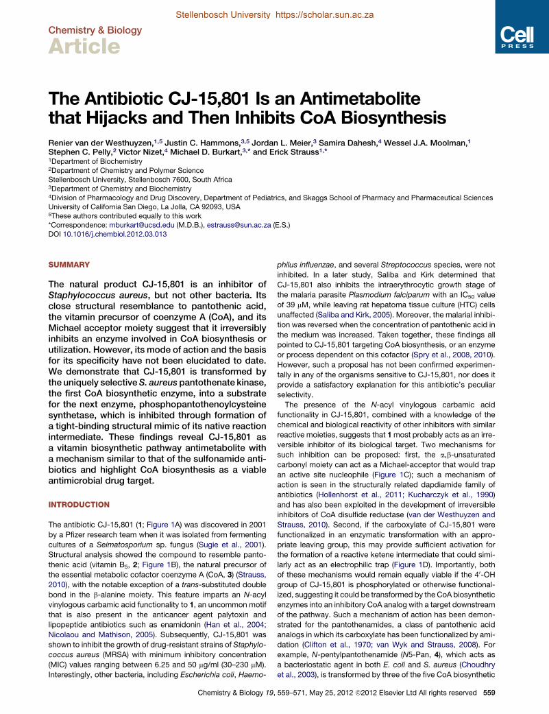
Stellenbosch University https://scholar.sun.ac.za
Stellenbosch University https://scholar.sun.ac.za
Chemistry & Biology
Article
The Antibiotic CJ-15,801 Is an Antimetabolitethat Hijacks and Then Inhibits CoA BiosynthesisRenier van der Westhuyzen,1,5 Justin C. Hammons,3,5 Jordan L. Meier,3 Samira Dahesh,4 Wessel J.A. Moolman,1
Stephen C. Pelly,2 Victor Nizet,4 Michael D. Burkart,3,* and Erick Strauss1,*1Department of Biochemistry2Department of Chemistry and Polymer ScienceStellenbosch University, Stellenbosch 7600, South Africa3Department of Chemistry and Biochemistry4Division of Pharmacology and Drug Discovery, Department of Pediatrics, and Skaggs School of Pharmacy and Pharmaceutical Sciences
University of California San Diego, La Jolla, CA 92093, USA5These authors contributed equally to this work
*Correspondence: [email protected] (M.D.B.), [email protected] (E.S.)
DOI 10.1016/j.chembiol.2012.03.013
SUMMARY
The natural product CJ-15,801 is an inhibitor ofStaphylococcus aureus, but not other bacteria. Itsclose structural resemblance to pantothenic acid,the vitamin precursor of coenzyme A (CoA), and itsMichael acceptor moiety suggest that it irreversiblyinhibits an enzyme involved in CoA biosynthesis orutilization. However, its mode of action and the basisfor its specificity have not been elucidated to date.We demonstrate that CJ-15,801 is transformed bythe uniquely selectiveS. aureus pantothenate kinase,the first CoA biosynthetic enzyme, into a substratefor the next enzyme, phosphopantothenoylcysteinesynthetase, which is inhibited through formation ofa tight-binding structural mimic of its native reactionintermediate. These findings reveal CJ-15,801 asa vitamin biosynthetic pathway antimetabolite witha mechanism similar to that of the sulfonamide anti-biotics and highlight CoA biosynthesis as a viableantimicrobial drug target.
INTRODUCTION
The antibiotic CJ-15,801 (1; Figure 1A) was discovered in 2001
by a Pfizer research team when it was isolated from fermenting
cultures of a Seimatosporium sp. fungus (Sugie et al., 2001).
Structural analysis showed the compound to resemble panto-
thenic acid (vitamin B5, 2; Figure 1B), the natural precursor of
the essential metabolic cofactor coenzyme A (CoA, 3) (Strauss,
2010), with the notable exception of a trans-substituted double
bond in the b-alanine moiety. This feature imparts an N-acyl
vinylogous carbamic acid functionality to 1, an uncommon motif
that is also present in the anticancer agent palytoxin and
lipopeptide antibiotics such as enamidonin (Han et al., 2004;
Nicolaou and Mathison, 2005). Subsequently, CJ-15,801 was
shown to inhibit the growth of drug-resistant strains of Staphylo-
coccus aureus (MRSA) with minimum inhibitory concentration
(MIC) values ranging between 6.25 and 50 mg/ml (30–230 mM).
Interestingly, other bacteria, including Escherichia coli, Haemo-
Chemistry & Biology 19,
philus influenzae, and several Streptococcus species, were not
inhibited. In a later study, Saliba and Kirk determined that
CJ-15,801 also inhibits the intraerythrocytic growth stage of
the malaria parasite Plasmodium falciparum with an IC50 value
of 39 mM, while leaving rat hepatoma tissue culture (HTC) cells
unaffected (Saliba and Kirk, 2005). Moreover, the malarial inhibi-
tion was reversed when the concentration of pantothenic acid in
the medium was increased. Taken together, these findings all
pointed to CJ-15,801 targeting CoA biosynthesis, or an enzyme
or process dependent on this cofactor (Spry et al., 2008, 2010).
However, such a proposal has not been confirmed experimen-
tally in any of the organisms sensitive to CJ-15,801, nor does it
provide a satisfactory explanation for this antibiotic’s peculiar
selectivity.
The presence of the N-acyl vinylogous carbamic acid
functionality in CJ-15,801, combined with a knowledge of the
chemical and biological reactivity of other inhibitors with similar
reactive moieties, suggests that 1most probably acts as an irre-
versible inhibitor of its biological target. Two mechanisms for
such inhibition can be proposed: first, the a,b-unsaturated
carbonyl moiety can act as a Michael-acceptor that would trap
an active site nucleophile (Figure 1C); such a mechanism of
action is seen in the structurally related dapdiamide family of
antibiotics (Hollenhorst et al., 2011; Kucharczyk et al., 1990)
and has also been exploited in the development of irreversible
inhibitors of CoA disulfide reductase (van der Westhuyzen and
Strauss, 2010). Second, if the carboxylate of CJ-15,801 were
functionalized in an enzymatic transformation with an appro-
priate leaving group, this may provide sufficient activation for
the formation of a reactive ketene intermediate that could simi-
larly act as an electrophilic trap (Figure 1D). Importantly, both
of these mechanisms would remain equally viable if the 40-OH
group of CJ-15,801 is phosphorylated or otherwise functional-
ized, suggesting it could be transformed by the CoA biosynthetic
enzymes into an inhibitory CoA analog with a target downstream
of the pathway. Such a mechanism of action has been demon-
strated for the pantothenamides, a class of pantothenic acid
analogs in which its carboxylate has been functionalized by ami-
dation (Clifton et al., 1970; van Wyk and Strauss, 2008). For
example, N-pentylpantothenamide (N5-Pan, 4), which acts as
a bacteriostatic agent in both E. coli and S. aureus (Choudhry
et al., 2003), is transformed by three of the five CoA biosynthetic
559–571, May 25, 2012 ª2012 Elsevier Ltd All rights reserved 559

Figure 1. Structures of the Antibiotic CJ-15,801 (1), Selected Coenzyme A Biosynthetic Intermediates, Antimetabolites, and Intermediate
Mimics
(A) Structure of the CJ-15,801 (1).
(B) Biosynthesis of CoA (3) from pantothenic acid (2). PanK, pantothenate kinase; PPCS, phosphopantothenoylcysteine synthetase; PPCDC, phosphopanto-
thenoylcysteine decarboxylase; PPAT, phosphopantetheine adenylyltransferase; DCPK, dephospho-coenzyme A kinase.
(C) Proposed mechanism for irreversible inhibition by CJ-15,801 acting as an electrophilic trap.
(D) Proposed mechanism for irreversible inhibition by CJ-15,801 after its transformation into a ketene intermediate.
(E) Biotransformation of the pantothenic acid analog N-pentylpantothenamide (4) to the CoA antimetabolite ethyldethia-CoA (5), which has the catalytically
essential thiol of the cofactor replaced by a propyl group. The antimetabolite subsequently serves as donor in the phosphopantetheinyl transferase (PPTase)-
catalyzed posttranslational modification of acyl and peptidyl carrier proteins (CPs), which results in similarly inactive crypto-CPs.
Chemistry & Biology
Mode of Action of the Antibiotic CJ-15,801
Stellenbosch University https://scholar.sun.ac.za
Stellenbosch University https://scholar.sun.ac.za
enzymes into ethyldethia-CoA (5), a CoA antimetabolite that
lacks the thiol required for the cofactor’s acyl carrier functions
(Figure 1E) (Strauss and Begley, 2002). This analog subsequently
serves as donor in the posttranslational modification of the
apo-acyl carrier protein (ACP), leading to the formation of cata-
lytically inactive crypto-ACP (Leonardi et al., 2005; Mercer and
Burkart, 2007; Zhang et al., 2004). The resulting loss of function
and subsequent impact on fatty acid metabolism is believed to
be the major cause for bacteriostasis, although a recent study
pointed to an impact on CoA biosynthesis as well (Thomas and
Cronan, 2010).
Interestingly, like CJ-15,801, the pantothenamides also exhibit
organism-based specificity. While the basis for this specificity
may partially reflect differences in the targeted organism’s cell
560 Chemistry & Biology 19, 559–571, May 25, 2012 ª2012 Elsevier
permeability and/or the nature of potential drug efflux pumps,
the most important factor relates to the nature of the pantothe-
nate kinase (PanK) enzyme present in the cell. PanK catalyzes
the first step of CoA biosynthesis, namely the ATP-dependent
phosphorylation of pantothenic acid (2) to form 40-phosphopan-tothenic acid (P-Pan, 6) (Figure 3A). This reaction is unique
because it is catalyzed by three distinct types of PanK, referred
to as type I, type II, and type III, respectively. These different
PanK types can be distinguished based on their sequence,
structure, the extent to which they experience feedback inhibi-
tion by CoA, and importantly, their ability to act on pantothena-
mides as alternative substrates (Brand and Strauss, 2005;
Hong et al., 2006; Strauss, 2010; Strauss et al., 2010; Yang
et al., 2008). Only type I and II PanKs (PanKI and PanKII)
Ltd All rights reserved

Figure 2. Synthesis of CJ-15,801, Its Methyl Ester and Their Respec-
tive Phosphorylated Versions
The respective reaction conditions are as follows: (a) Allyl- or methylacetyl-
triphenylphosphonium bromide, Et3N, toluene, 90�C, 75% (Villa et al., 2007);
(b) BiCl3, H2O, CH3CN, room temperature, 5 hr, 75%; (c) Pd(PPh3)4, pyrollidine,
THF, 81%; (d) SaPanK, ATP, MgCl2, Tris-HCl (pH 7.5), 98% (for 12); 79%
(for 13).
See also Figure S3.
Chemistry & Biology
Mode of Action of the Antibiotic CJ-15,801
Stellenbosch University https://scholar.sun.ac.za
Stellenbosch University https://scholar.sun.ac.za
phosphorylate pantothenamides, thereby allowing the forma-
tion of CoA antimetabolites. Organisms like Pseudomonas
aeruginosa that have type III PanK (PanKIII) enzymes that exclude
these compounds from their active sites are therefore refractory
to the effects of N-pentylpantothenamide and similar analogs
(Balibar et al., 2011). This suggests that the basis for CJ-
15,801’s unique specificity for S. aureus and P. falciparum may
also be based on the type specificity of their PanKs, both of
which have been characterized as atypical type II enzymes
(Hong et al., 2006; Leonardi et al., 2005; Spry et al., 2010).
In this study, we set out to identify the target of CJ-15,801’s
antibiotic action in bacteria, to elucidate its mechanism of action,
and to establish the basis for its specificity. Our results reveal
that CJ-15,801 acts as an antimetabolite by using the first
enzyme of the CoA biosynthesis as gateway to the pathway,
after which it inhibits the second CoA biosynthetic enzyme,
phosphopantothenoylcysteine synthetase (PPCS). This mode
of action is reminiscent of the sulfonamide antibiotics, which
block folic acid biosynthesis using a similar strategy. However,
contrary to expectations, detailed analysis of the inhibition
mechanism failed to provide any evidence of irreversible inhibi-
tion. Instead, our results show that CJ-15,801 is transformed
into a tight-binding mimic of the PPCS enzyme’s natural reaction
intermediate, a mechanism also utilized by other known natural
Chemistry & Biology 19,
product inhibitors of adenylating enzymes, such as ascamycin
and mupirocin (May et al., 2005; Pope et al., 1998). Taken
together, these findings not only provide insight into the basis
of CJ-15,801’s antibiotic action, but also suggest a new and
potentially general strategy for the development of synthetase
inhibitors with in vivo efficacy.
RESULTS
Synthesis of CJ-15,801 and Its EstersThe synthesis of CJ-15,801 and its related analogs presents
several challenges, the first of which relates to the preparation
of the N-acyl vinylogous carbamic acid moiety. In fact, several
groups have used the synthesis of CJ-15,801 to showcase
new methodologies developed specifically for the preparation
of this functionality (Han et al., 2004; Lee et al., 2006; Nicolaou
and Mathison, 2005). However, we found these methods to be
unsuitable for this study as they are either optimized for small-
scale synthetic preparations, or led to the preferential formation
of the unwanted (Z)-double bond configuration. We therefore
utilized the more recent method of Villa et al., which installs the
required N-acyl vinylogous carbamic acid moiety mainly with
an (E)-configuration via Wittig-type chemistry, for preparation
of the protected esters 8 and 9 (Figure 2) (Villa et al., 2007). A
second challenge derives from the sensitivity of the N-vinyl
amide moiety to both acid and base, which severely restricts
the options for the protection and deprotection of the synthetic
intermediates. In this case, established protocols that make
use of neutral conditions were used to remove the acetal pro-
tecting groups from 8 and 9 to, respectively, obtain CJ-
15,801’s allyl (10) and methyl (11) esters, and to remove the allyl
ester from 10 to provide CJ-15,801 itself. The (Z)-configured
analog of CJ-15,801 (Z-1) was obtained by similar deprotection
of the minor constituent of the Wittig coupling reaction product
mixture.
Recently, an updated report of the Wittig-based synthesis of
CJ-15,801 appeared in which several other options for deprotec-
tion of its carboxyl group are also provided (Sewell et al., 2011).
A Bacterium’s Susceptibility to CJ-15,801 Correlateswith Its PanK TypeThe original Pfizer discovery group found that in standard
susceptibility tests conducted using cation adjusted Mueller-
Hinton broth as growthmediumCJ-15,801 is uniquely and pecu-
liarly selective for S. aureus, with none of the other organisms
tested showing inhibition at 100 mg/ml (460 mM) (Sugie et al.,
2001). These resistant organisms included both Gram-positive
(Staphylococcus, Enterococcus, and Streptococcus spp.) and
Gram-negative (H. influenzae, Moraxella catarrhalis, and E. coli)
bacteria, indicating that cell envelope type alone does not deter-
mine selectivity. However, the panel of test organisms also
comprised bacteria expressing all three known pantothenate
kinase (PanK; Figure 3A) types: the enterococci, streptococci,
H. influenzae, and E. coli are known or predicted to have PanKI
enzymes, while M. catarrhalis is predicted to have a PanKIII
(Yang et al., 2006). Importantly, the staphylococci are the only
known bacteria to have an active PanKII, suggesting that the
presence of this PanK type is a prerequisite for inhibition by
CJ-15,801.
559–571, May 25, 2012 ª2012 Elsevier Ltd All rights reserved 561

Figure 3. Evaluation of PanK as a Potential Gate-
keeper to CJ-15,801’s Inhibitory Action
(A) The ATP-dependent phosphorylation of pantothenic
acid (2) catalyzed by PanK enzymes.
(B) HPLC analysis of the putative ATP-dependent phos-
phorylation of CJ-15,801 by SaPanK. The traces repre-
sent: I, a synthetically prepared standard of CJ-15,801 (1);
II, a reaction mixture containing CJ-15,801, ATP and
SaPanK that had been incubated for 1 min; III, the same
after 10 min of incubation; and IV, a synthetic P-CJ (13)
standard. The peak labeled with (d) represents ATP
and ADP.
See also Table S2.
Chemistry & Biology
Mode of Action of the Antibiotic CJ-15,801
Stellenbosch University https://scholar.sun.ac.za
Stellenbosch University https://scholar.sun.ac.za
To test this hypothesis, we repeated the CJ-15,801 suscepti-
bility tests against a panel of organisms representing all combi-
nations of cell envelope and PanK type. Since the original tests
were conducted in growth medium rich in pantothenic acid,
these experiments were conducted in 1% tryptone (that contains
essentially no pantothenic acid) instead to exclude any potential
antagonistic effects that the presence of the vitamin could have
on inhibition. The results show that the PanKII-containing
S. aureus (MRSA strain TCH1516) was the most sensitive to
the inhibitory effects of CJ-15,801, showing an MIC of 15 mM
(Table S1 available online). This value is 2-fold lower than the
lowestMIC reported in the previous susceptibility tests, suggest-
ing that pantothenate does affect inhibition similar to what was
seen in the experiments with malaria parasites (Saliba and
Kirk, 2005). Moreover, under these test conditions some of the
other organisms (most notably P. aeruginosa) also showed low
levels of inhibition (MIC �60 mM).
To determine which aspects of CJ-15,801’s structure are
required for inhibition, we evaluated the inhibitory activity of
the (Z)-configured analog (Z-1) and CJ-15,801’s methyl ester
(CJ-OMe, 11) against S. aureus. Neither compound showed
any inhibition, indicating that the configuration of the double
bond and the availability of the free carboxylate of CJ-15,801
are both important determinants for its mechanism of inhibition.
Only PanKII Enzymes Accept CJ-15,801 as SubstrateTo confirm that the observed inhibition results can indeed be
correlated with PanK type and activity, we performed in vitro
activity analyses using purified PanK enzymes. Based on the
inhibition results, we first used the most likely candidate,
the PanKII enzyme from S. aureus (SaPanKII), to determine if
it converts CJ-15,801 to phospho-CJ,15,801 (P-CJ, 13)
when incubated with ATP. High-performance liquid chromatog-
raphy (HPLC) analysis of the reaction mixture confirmed
this to be the case, based on the time-dependent formation
of P-CJ (Figure 3B). Next, the activity of the PanKI from
E. coli (EcPanKI), SaPanKII, and the PanKIII from P. aeruginosa
(PaPanKIII) toward CJ-15,801 were fully characterized kineti-
cally. The resulting data indicate that EcPanKI and PaPanKIII
only show activity toward pantothenic acid and not CJ-15,801;
562 Chemistry & Biology 19, 559–571, May 25, 2012 ª2012 Elsevier Ltd All rights rese
moreover, neither enzyme was inhibited by
100 mM CJ-15,801 (Table S2). However,
SaPanKII shows little distinction between
CJ-15,801 and pantothenic acid, exhibiting
specificity constants (kcat/Km) of 13.8 ± 4.2 mM�1$s�1 and
30.2 ± 8.7 mM�1$s�1 for the two compounds, respectively,
with the difference being mainly due to an elevated Km value.
The enzyme also acts on CJ-OMe, indicating that the lack of inhi-
bition seen for this analog is due to effects downstream of PanK.
Taken together, these results indicate that PanK acts as
a gatekeeper to the inhibitory effects of CJ-15,801 in S. aureus.
CJ-15,801’s Antistaphylococcal Activity Is Affectedby Pantothenic Acid and PantetheineThe observation that S. aureus’s susceptibility to inhibition by
CJ-15,801 is apparently affected by pantothenic acid (2), as
well as the finding that SaPanKII accepts both the inhibitor and
the vitamin as substrates, led us to perform checkerboard
assays to quantify these effects (Figure 4A). The same
assay was also conducted with pantetheine (17), the precursor
to the CoA salvage pathway that bypasses the PPCS and phos-
phopantothenoylcysteine decarboxylase (PPCDC) enzymes
(Strauss, 2010; Strauss et al., 2010) (Figure 4B). The results
show that the presence of either compound reduces the potency
of CJ-15,801, most likely through competition with SaPanKII.
However, their interaction with the natural product is complex,
since increasing the concentration of either compound above
a certain level (�7.5 mM for 2, and �15 mM for 17) leads to inhi-
bition being reestablished. While the basis for this observation
is currently unknown, the results confirm that the point of CJ-
15,801’s inhibitory action is a process dependent on pantothenic
acid (or pantetheine).
Pantothenamides Increase the Potency of CJ-15,801’sAntistaphylococcal ActivityPrevious studies have shown that treatment of S. aureus with
N5-Pan (and its analog, N-heptylpantothenamide, N7-Pan)
results in growth inhibition by formation of inactive carrier
proteins (Figure 1E) (Leonardi et al., 2005; Zhang et al., 2004).
To determine if the points of action of the pantothenamides
and CJ-15,801 overlap, a checkerboard assay with N5-Pan (4)
and CJ-15,801 was performed in 1% tryptonemedia (Figure 4C).
The results show that when combined, CJ-15,801 and N5-Pan
exhibited significantly reduced MICs of 3.75 mM and �1.0 mM,
rved

Figure 4. Synergism in the Antibacterial Action of CJ-15,801 and N5-Pan
(A) Checkerboard assay of the growth inhibition of S. aureus by CJ-15,801 (1) when grown in the presence of pantothenic acid (2) in 1% tryptone shows that
increasing the amount of vitamin (up to a certain concentration) reverses the inhibition. MIC value for CJ-15,801 is 15 mM.
(B) As for (A), but with pantetheine (17) added instead of pantothenic acid.
(C) Checkerboard assay of the growth inhibition of S. aureus grown in 1% tryptone by CJ-15,801 (1) and N5-Pan (4) demonstrates the synergism between these
compounds (FICI < 0.5). MIC value for N5-Pan is 30 mM.
(D) As for (C), but determined in the presence of 100 mM pantothenic acid (2), demonstrating that the combination of CJ-15,801 and N5-Pan can overcome the
antagonism caused by an excess of the natural substrate of the CoA biosynthetic pathway, but at the expense of converting the synergism to an additive effect
(0.5 < FICI < 1.0).
See also Tables S1 and S3.
Chemistry & Biology
Mode of Action of the Antibiotic CJ-15,801
Stellenbosch University https://scholar.sun.ac.za
Stellenbosch University https://scholar.sun.ac.za
respectively, with a fractional inhibitory concentration index
(FICI) of 0.25, indicating synergism (Table S3). Importantly,
when the same assay was performed in the presence of
100 mM pantothenic acid, the inhibition was not as adversely
affected as in the case of the individual compounds (Figure 4D).
Instead, theMIC values were slightly elevated and the previously
observed synergism was converted to an additive inhibitory
effect (FICI �0.5–1.0) (Table S3). This finding suggests that CJ-
15,801 and N5-Pan does not have the same mode of action
and indicates that in combination these compounds can counter
the antagonism caused by pantothenic acid.
Synthesis of the 40-Phosphates of CJ-15,801and Its Methyl EsterThe discovery that SaPanK phosphorylates both CJ-15,801 and
CJ-OMe (11) provided us with a convenient biocatalytic method
by which their 40-phosphorylated versions could be prepared.
P-CJ (13) and its methyl ester, 12 (P-CJ-OMe), were therefore
obtained by milligram-scale biotransformations using recombi-
nant SaPanK and ATP, followed by purification by either
preparative HPLC (for P-CJ) or solid-phase extraction (SPE)
(for P-CJ-OMe) (Figure 2).
PPCS Accepts P-CJ as Substrate and Is Then Inhibitedby ItWith the knowledge that CJ-15,801 can be converted to P-CJ
within S. aureus, we set out to determine if PPCS, the next
enzyme in the pathway, is the target for the antibiotic’s inhibitory
action. PPCS catalyzes the condensation of P-Pan (6) with
L-cysteine to form 40-phosphopantothenoylcysteine (PPC, 15)
using a two-step acyl transfer mechanism common to most
synthetase (C-N ligase) enzymes, including all the amino
acid tRNA synthetases and the adenylation domains of the
modular nonribosomal peptide synthetases (NRPSs) (Figure 5A)
Chemistry & Biology 19,
(Schimmel et al., 1998; Sieber and Marahiel, 2005). While
all known PPCSs follow this general scheme, bacterial
enzymes are unique in that they use CTP instead of ATP as
nucleotide source for the activation reaction, and therefore
form 40-phosphopantothenoyl-CMP (P-Pan-CMP, 14) as inter-
mediate (Kupke, 2002, 2004; Manoj et al., 2003; Stanitzek
et al., 2004; Strauss, 2010; Strauss et al., 2001).
In most bacteria PPCS activity is located on one domain of the
bifunctional CoaBC protein (which also carries PPCDC activity),
although monofunctional PPCS enzymes do occur in certain
enterococci (such as Enterococcus faecalis [Yao et al., 2009])
and streptococci. Among all bacterial PPCS enzymes the
PPCS activity of E. coli’s bifunctional CoaBC protein is by far
the best studied: its mechanism has been established through
trapping and isolation of P-Pan-CMP (14), and the structure of
the N210D mutant with the trapped intermediate 14 bound in
the active site has been determined (Kupke, 2002, 2004; Stanit-
zek et al., 2004). Moreover, many of these studies were per-
formed using the separate PPCS domain (EcPPCS) expressed
and purified on its own, showing that the two activities of the
bifunctional CoaBC protein are independent. This conclusion is
also supported by kinetic isotope studies of the PPCDC activity
of E. coli’s CoaBC (Strauss and Begley, 2001). We therefore
decided to use the EcPPCS domain to perform the initial tests
on the interaction of PPCS and P-CJ.
To determine if P-CJ is accepted as an alternate substrate by
PPCS and forms the corresponding P-CJ-CMP intermediate 16
(Figure 5B), mixtures of P-CJ and CTP were incubated with
increasing concentrations of EcPPCS. A clear correlation
between the rate of pyrophosphate release and enzyme concen-
tration was evident in such reactions (Figure 5C). Moreover,
HPLC analysis of the reaction mixture containing the highest
EcPPCS concentration (24 mM) showed the formation of
a peak absent in native reaction mixtures, or in mixtures
559–571, May 25, 2012 ª2012 Elsevier Ltd All rights reserved 563

Figure 5. Evaluation of PPCS Activity with P-CJ (13) as Substrate
(A) The reaction catalyzed by PPCS, showing the two steps of its mechanism
and the P-Pan-CMP (14) reaction intermediate.
(B) The structure of P-CJ-CMP (16) resulting from the putative cytidylation of
P-CJ (13) by PPCS.
(C) The rate of pyrophosphate release from a reaction mixture containing P-CJ
(13), CTP, and EcPPCS correlates with enzyme concentration. Experiments
were run in duplicate; the shown data points are the average of duplicate
assays (error bars indicate the SD).
(D) HPLC analysis of a reaction mixture containing P-CJ (13), CTP, and
EcPPCS (24 mM) (trace I) show the formation of a new peak (indicated with an
asterisk) that represents P-CJ-CMP (16) based on LC-MS analysis. This peak
is not present when P-CJ is replaced with P-Pan (6) (trace II) or in native
reaction mixtures containing P-Pan, CTP and L-cysteine. Note that the
retention time of pure P-CJ is�2.4min (see Figure 2B). The broad peak labeled
with (B) represents CTP and CMP.
See also Figure S1.
Chemistry & Biology
Mode of Action of the Antibiotic CJ-15,801
564 Chemistry & Biology 19, 559–571, May 25, 2012 ª2012 Elsevier
Stellenbosch University https://scholar.sun.ac.za
Stellenbosch University https://scholar.sun.ac.za
containing P-Pan (6) instead of P-CJ, suggesting the formation of
P-CJ-CMP (Figure 5D). Note that P-Pan-CMP is usually not
observed under these conditions, as was previously reported
(Kupke, 2002, 2004). Liquid chromatography-tandem mass
spectrometry (LC-MS) analysis was subsequently conducted
on the same mixture. While the P-CJ-CMP-derived molecular
ion was not observed, the corresponding mass spectrum did
show mass peaks that are in agreement with the fragmentation
of P-CJ-CMP (Figure S1). The same experiment was subse-
quently repeated with the bifunctional CoaBC protein from
S. aureus (SaCoaBC). A similar result was obtained, although
the apparent rate of conversion of P-CJ into P-CJ-CMP was
significantly slower than for EcPPCS (Figure S1).
In combination, these results indicate that P-CJ is accepted as
an alternate substrate by PPCS, which cytidylylates its carbox-
ylate to form P-CJ-CMP.
PPCS Transform P-CJ into P-CJ-CMP, which InhibitsIts ActivitySubsequent activity analysis of PPCS in the presence of P-CJ,
CTP, and L-cysteine failed to show catalytic turnover. Moreover,
when P-CJ was added to PPCS reaction mixtures containing the
enzyme’s native P-Pan substrate, the rate of the reaction was
reduced compared to mixtures without it, suggesting inhibition
by P-CJ. To confirm that such inhibition is dependent on the
reaction catalyzed by PPCS, an inhibition screen was performed
in which CJ-15,801, CJ-OMe, P-CJ, or P-CJ-OMe was added to
native PPCS reaction mixtures at a concentration of 100 mM. The
results showed that among these compounds, only P-CJ
inhibited EcPPCS (data not shown). The lack of inhibition by
P-CJ-OMe indicated that the free carboxylate group is required
for inhibition, while the inability of CJ-15,801 to affect enzyme
activity highlights the necessity of the phosphate group for
binding. Taken together with the results described above, these
findings reveal PPCS as CJ-15,801’s target of inhibitory action,
which is achieved through formation of P-CJ-CMP 16 as an
apparent dead-end inhibitor.
Kinetic Characterization Reveals P-CJ-CMPas a Tight-Binding Inhibitor of PPCSInhibition by P-CJ-CMP could potentially occur by either of the
proposed irreversible inhibition mechanisms (Figures 1C and
1D), or by a completely unrelated mechanism. To elucidate the
actual mechanism of inhibition, detailed kinetic analyses were
performed. First, dose-response curves were used to evaluate
the inhibition of the cytidyl-transfer reaction catalyzed by
EcPPCS. The resulting IC50 value of 2.66 ± 0.16 mM, determined
using saturating concentrations of CTP and 100 mM P-Pan (note
that EcPPCS experiences substrate inhibition, see Figure S2),
was less than 10-fold the concentration of enzyme used in these
assays (240 nM). This indicated that P-CJ-CMP acts as either
a tight-binding or irreversible inhibitor of EcPPCS (Copeland,
2000). The inhibition of EcPPCS was next studied by the prog-
ress curve method. The resulting curves increased in a linear
fashion, showing a decreased rate at increased concentrations
of P-CJ (Figure 6A). Surprisingly, this indicated that the inhibition
was tight binding and not irreversible (which would have led
to the steady-state rates approaching zero) as predicted
(Copeland, 2005). This finding was confirmed by showing that
Ltd All rights reserved

Figure 6. Kinetic Characterization of the CJ-15,801-Based Inhibition of PPCS
(A) Progress curves analysis for the inhibition of EcPPCS in the presence of increasing concentrations of P-CJ (13), indicated in nM next to each curve.
(B) Plot of the fractional activity (initial velocity vi relative to the inhibited velocity v0), as determined from the progress curves in (A), against inhibitor concentration.
The indicated Ki value is determined by fitting the data to the Morrison and Cheng-Prusoff equation.
(C) Determination of the inhibition modality of EcPPCS by P-CJ (13) by calculation of the IC50 values in the presence of increasing concentrations of P-Pan (6) (d),
and CTP (B).
(D) Progress curves analysis for the inhibition of SaCoaBC in the presence of increasing concentrations of P-CJ (13), indicated in nM next to each curve.
(E) Plot of the first order rate of inactivation constants (kobs), as determined from the data presented in (D), against inhibitor concentration. The indicated Ki value is
determined by fitting the data to the equation for a straight line.
(F) Plot of the fractional activity (steady-state velocity vs relative to the inhibited velocity v0), as determined from the progress curves in (D), against inhibitor
concentration. The indicated Ki value is determined by fitting the data to a Langmuir isotherm.
All progress curve analyses were performed in triplicate (error bars represent the SD).
See Supplemental Experimental Procedures for details on calculations, as well as Figure S2 for the determination of EcPPCS and SaCoaBC kinetic parameters.
Chemistry & Biology
Mode of Action of the Antibiotic CJ-15,801
Stellenbosch University https://scholar.sun.ac.za
Stellenbosch University https://scholar.sun.ac.za
removal of the inhibitor by gel filtration was sufficient to restore
the activity of a sample of EcPPCS that had been pretreated
with P-CJ and CTP.
The Kiapp (apparent inhibition constant) was subsequently
calculated by plotting the fractional activity versus inhibitor
concentration, fitting the data to the Morrison equation (Fig-
ure 6B), and converting the obtained value to the true Ki by
applying the appropriate Cheng-Prusoff equation (Copeland,
2000, 2005). The mode of inhibition was established using
a method specifically developed for tight-binding inhibitors in
which IC50 values are determined at a fixed enzyme concentra-
tion, but in the presence of increasing concentrations of either
P-Pan (6) or CTP (Figure 6C). A linear increase in IC50 values
with increasing concentration of P-Pan but not CTP indicates
that inhibition is competitive toward P-Pan but noncompetitive
toward CTP (Copeland, 2000, 2005). This observation agrees
with previous studies of the E. faecalis and human PPCS
enzymes that determined an ordered Bi Uni Uni Bi Ping-Pong
kinetic mechanism with CTP binding first, followed by P-Pan
(6) (Yao and Dotson, 2009; Yao et al., 2009). In this manner,
a Ki of 164.3 nM was determined for P-CJ’s inhibition of
EcPPCS.
While EcPPCS is a convenient and well-studied model to use
for characterization of CJ-based inhibition, E. coli is not affected
Chemistry & Biology 19,
by CJ-15,801 due to the gatekeeping activity of its PanK (Tables
S1 and S2). We therefore performed the same kinetic analysis on
SaCoaBC, which is expected to be the natural target for CJ-
15,801’s observed antistaphylococcal action. A progress curve
analysis similar to that conducted on EcPPCS showed that
SaCoaBC experiences time-dependent inhibition, since the
curves showed a fast initial velocity (vi) phase and a slower
steady-state velocity (vs) phase (Figure 6D). The first order rate
of inactivation constants (kobs) were subsequently determined
and plotted against the inhibitor concentration. The resulting
plot (Figure 6E) showed a linear relationship between kobs and
[I], which points to a one-step inhibition mechanism (Copeland,
2005). However, it is more likely that the true mechanism
consists of two steps, with P-CJ 13 binding first in a reversible
manner, followed by the slow formation of P-CJ-CMP. Such
a mechanism would be kinetically indistinguishable from a
one-step mechanism.
The Ki value for the inhibition of SaCoaBC by P-CJ-CMP can
be determined from the slope and y-intercept of the linear plot
shown in Figure 6E. The Ki value can also be calculated by using
the progress curves to determine the steady-state velocity (vs) at
each inhibitor concentration; a plot of the fractional activity
against inhibitor concentration yields an isotherm curve with
theKi at its midpoint (Figure 6F) (Copeland, 2005). In thismanner,
559–571, May 25, 2012 ª2012 Elsevier Ltd All rights reserved 565

Chemistry & Biology
Mode of Action of the Antibiotic CJ-15,801
Stellenbosch University https://scholar.sun.ac.za
Stellenbosch University https://scholar.sun.ac.za
Ki values of 13.0 and 13.5 nM were determined, respectively.
These values is nearly an order of magnitude lower than that
determined for EcPPCS, clearly showing that the nature of the
actual target protein also plays an important role in the extent
of inhibition. Taken together, these data show that P-CJ-CMP
acts as a tight-binding inhibitor of PPCS enzymes.
Analysis of the Mechanistic Basis for the Tight-BindingInhibition of P-CJ-CMPThe close structural homology between P-Pan-CMP (14) and
P-CJ-CMP (16), and the fact that both compounds contain reac-
tive acyl phosphate moieties, raises a question regarding the
mechanistic basis for the inhibition by P-CJ-CMP. We consid-
ered three possible explanations: first, inhibition is based on
differences in the electrophilicity of the carbonyl carbon of the
acyl phosphatemoieties of P-Pan-CMP and P-CJ-CMP, respec-
tively; second, inhibition is caused by the two compounds having
different binding modes in the active site, due to the structural
nature of the planar N-acyl vinylogous carbamate functionality
of P-CJ-CMP; and third, inhibition is caused by P-Pan-CMP
and P-CJ-CMP having different effects on any conformational
changes that occur during catalysis through stabilization of the
protein’s quaternary structure. While the significant synthetic
effort that is required to prepare P-Pan-CMP and P-CJ-CMP
precluded a direct experimental comparison of their respective
reactivities, we were able to perform tests to assess whether
the latter two proposed mechanisms contribute to inhibition.
To establish if P-Pan-CMP (14) and P-CJ-CMP (16) have
different binding modes, we used the only available structure
of a PPCS enzyme with its intermediate bound, that of the
EcPPCS Asn210Asp mutant with P-Pan-CMP bound in the
active site (PDB ID: 1U7Z). Thismutant, which accumulates suffi-
cient amounts of the intermediate to have allowed direct confir-
mation of its identity and structure (Kupke, 2004; Stanitzek et al.,
2004), is apparently unable to catalyze the second step of the
PPCS reaction. However, the molecular basis for this catalytic
disability remains speculative since no PPCS structure with
cysteine bound has been determined thus far. Nonetheless,
the mutation clearly results in tightly bound P-Pan-CMP in the
enzyme’s active site.
We constructed a model of the native enzyme by reversing the
Asn210Asp mutation in the crystal structure and used it to dock
P-CJ-CMP (16) in the active site using 150 starting conforma-
tions to ensure sufficient conformational space was sampled
considering the complexity of the molecule. The highest scoring
docked pose of P-CJ-CMP very closely mimics the pose of the
cocrystalized P-Pan-CMP in the crystal structure (Figure 7A),
and facilitates all the same hydrogen bonding interactions while
maintaining a near planar geometry across theN-acyl vinylogous
carbamate moiety (Figure 7B). This suggests that the basis for
inhibition by P-CJ-CMP is not due it having a different binding
mode, or taking on a potentially unreactive conformation.
Members of the ANL superfamily of adenylating enzymes,
such as the adenylation domains of the NRPSs, use a large
domain rotation event to facilitate the catalysis of their two-
step reactions (Gulick, 2009). While it is currently unknown
whether PPCS enzymes, which belong to the ribokinase family,
undergo similar large conformational changes during catalysis,
the determined crystal structures of the E. coli and human
566 Chemistry & Biology 19, 559–571, May 25, 2012 ª2012 Elsevier
proteins show that they occur as dimers, with the dimer interface
partially occluding the active site (Manoj et al., 2003; Stanitzek
et al., 2004). To evaluate the effect of different ligands on the
enzyme’s overall stability, the melting temperatures (Tm) of
the EcPPCS protein in the presence of its native substrates or
the inhibitor were determined by following the heat-induced
denaturation of these mixtures by circular dichroism (CD). The
results show that the Tm increases from 47.7�C (for the free
enzyme) to 49.7�C (for the enzyme bound to CTP) to 54.3�C(for the enzyme bound to P-Pan-CMP, formed in situ from
P-Pan and CTP), highlighting the many stabilizing interactions
the protein has with its native ligands (Figure 7C). Importantly,
the Tm of EcPPCS bound to P-CJ-CMP (formed in situ from
P-CJ and CTP) shows an even larger increase to 57.3�C. When
the same experiment was conducted with the bifunctional
SaCoaBC, the protein showed increased stability compared to
EcPPCS (CoaBC proteins exists as higher order oligomers,
usually dodecamers [Manoj et al., 2003]), but only small differ-
ences in the Tm values for the protein in the absence and
presence of its native ligands. However, the Tm showed an
impressive improvement from �65�C to 85�C for the proteins
bound to P-Pan-CMP and P-CJ-CMP, respectively (Figure 7D).
Taken together, these findings indicate that the binding of the
conformationally more rigid ligand 16 results in an increased
stabilization of the PPCS oligomers and the trapping of the ligand
in the active site. This stabilization, which may be due to interac-
tions between the ligand and residues of the adjacent monomer
(such as Lys289, see Figure 7B) contained in a disordered loop
that partially covers the entrance to the active site (Figure 7E),
is therefore most likely an important contributor to the mecha-
nism of inhibition by P-CJ-CMP.
DISCUSSION
One of the biggest obstacles to elucidation of the CJ-15,801
mode of action is scarcity of the natural product. The original
discoverers shared the bulk of the isolated material with the
authors of a malaria parasite inhibition study (Spry et al., 2008),
but new stocks are unlikely to be obtained in this manner since
the CJ-producing Seimatosporium strain can currently not be
traced due to closure of the laboratory in which it was character-
ized (Y. Sugie, personal communication). New developments in
the synthesis of CJ-15,801 have certainly improved the situation,
but the unique sensitivity of the N-acyl vinylogous carbamic acid
functionality still presents a significant challenge to the synthesis
of this antibiotic.
The original finding that CJ-15,801 is highly selective in its anti-
biotic action, affecting only S. aureus strains among a range of
other Gram-positive bacteria including Streptococcus and
Enterococcus spp. (Sugie et al., 2001), provoked curiosity in
regards to its mechanism of action. Here, we show that the
primary reason for this selectivity is the substrate specificity of
the host organism’s PanK enzyme. Only the PanKII of S. aureus
accepts CJ-15,801 and phosphorylates it, thereby allowing it to
enter the CoA biosynthetic pathway. Interestingly, SaPanKII is
the only known active bacterial representative of a type II PanK;
all other examples occur in eukaryotes. However, an earlier study
showed that CJ-15,801 does not inhibit rat hepatoma cells,
suggesting that typical eukaryotic PanKs do not act as
Ltd All rights reserved
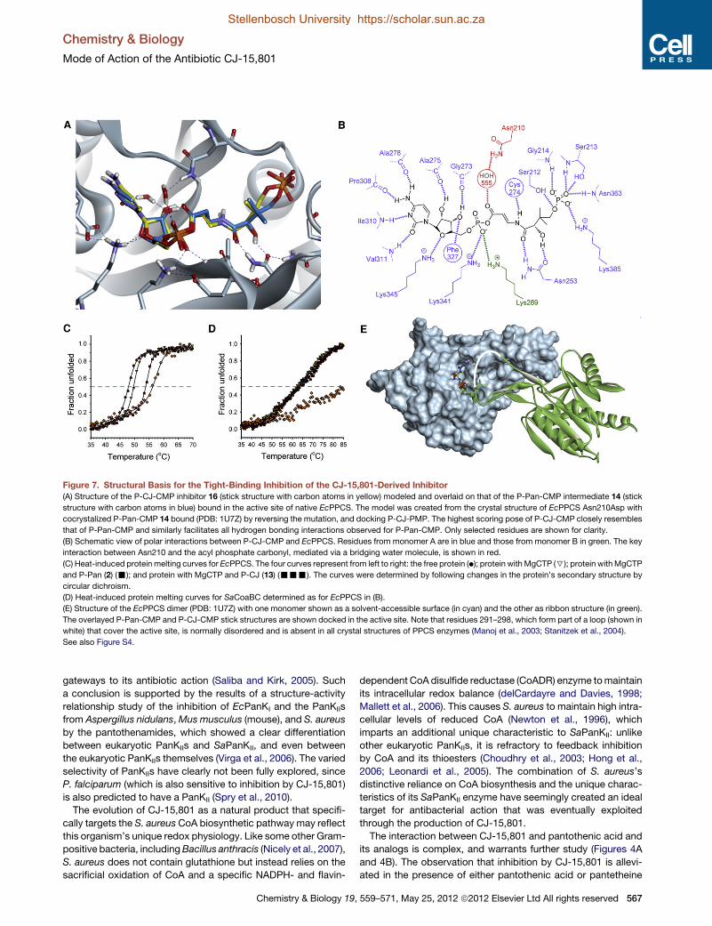
Figure 7. Structural Basis for the Tight-Binding Inhibition of the CJ-15,801-Derived Inhibitor
(A) Structure of the P-CJ-CMP inhibitor 16 (stick structure with carbon atoms in yellow) modeled and overlaid on that of the P-Pan-CMP intermediate 14 (stick
structure with carbon atoms in blue) bound in the active site of native EcPPCS. The model was created from the crystal structure of EcPPCS Asn210Asp with
cocrystalized P-Pan-CMP 14 bound (PDB: 1U7Z) by reversing the mutation, and docking P-CJ-PMP. The highest scoring pose of P-CJ-CMP closely resembles
that of P-Pan-CMP and similarly facilitates all hydrogen bonding interactions observed for P-Pan-CMP. Only selected residues are shown for clarity.
(B) Schematic view of polar interactions between P-CJ-CMP and EcPPCS. Residues from monomer A are in blue and those from monomer B in green. The key
interaction between Asn210 and the acyl phosphate carbonyl, mediated via a bridging water molecule, is shown in red.
(C) Heat-induced protein melting curves for EcPPCS. The four curves represent from left to right: the free protein (d); protein withMgCTP (7); protein withMgCTP
and P-Pan (2) (-); and protein with MgCTP and P-CJ (13) (---). The curves were determined by following changes in the protein’s secondary structure by
circular dichroism.
(D) Heat-induced protein melting curves for SaCoaBC determined as for EcPPCS in (B).
(E) Structure of the EcPPCS dimer (PDB: 1U7Z) with one monomer shown as a solvent-accessible surface (in cyan) and the other as ribbon structure (in green).
The overlayed P-Pan-CMP and P-CJ-CMP stick structures are shown docked in the active site. Note that residues 291–298, which form part of a loop (shown in
white) that cover the active site, is normally disordered and is absent in all crystal structures of PPCS enzymes (Manoj et al., 2003; Stanitzek et al., 2004).
See also Figure S4.
Chemistry & Biology
Mode of Action of the Antibiotic CJ-15,801
Stellenbosch University https://scholar.sun.ac.za
Stellenbosch University https://scholar.sun.ac.za
gateways to its antibiotic action (Saliba and Kirk, 2005). Such
a conclusion is supported by the results of a structure-activity
relationship study of the inhibition of EcPanKI and the PanKIIs
from Aspergillus nidulans,Mus musculus (mouse), and S. aureus
by the pantothenamides, which showed a clear differentiation
between eukaryotic PanKIIs and SaPanKII, and even between
the eukaryotic PanKIIs themselves (Virga et al., 2006). The varied
selectivity of PanKIIs have clearly not been fully explored, since
P. falciparum (which is also sensitive to inhibition by CJ-15,801)
is also predicted to have a PanKII (Spry et al., 2010).
The evolution of CJ-15,801 as a natural product that specifi-
cally targets the S. aureus CoA biosynthetic pathway may reflect
this organism’s unique redox physiology. Like some other Gram-
positive bacteria, includingBacillus anthracis (Nicely et al., 2007),
S. aureus does not contain glutathione but instead relies on the
sacrificial oxidation of CoA and a specific NADPH- and flavin-
Chemistry & Biology 19,
dependent CoAdisulfide reductase (CoADR) enzyme tomaintain
its intracellular redox balance (delCardayre and Davies, 1998;
Mallett et al., 2006). This causes S. aureus to maintain high intra-
cellular levels of reduced CoA (Newton et al., 1996), which
imparts an additional unique characteristic to SaPanKII: unlike
other eukaryotic PanKIIs, it is refractory to feedback inhibition
by CoA and its thioesters (Choudhry et al., 2003; Hong et al.,
2006; Leonardi et al., 2005). The combination of S. aureus’s
distinctive reliance on CoA biosynthesis and the unique charac-
teristics of its SaPanKII enzyme have seemingly created an ideal
target for antibacterial action that was eventually exploited
through the production of CJ-15,801.
The interaction between CJ-15,801 and pantothenic acid and
its analogs is complex, and warrants further study (Figures 4A
and 4B). The observation that inhibition by CJ-15,801 is allevi-
ated in the presence of either pantothenic acid or pantetheine
559–571, May 25, 2012 ª2012 Elsevier Ltd All rights reserved 567

Chemistry & Biology
Mode of Action of the Antibiotic CJ-15,801
Stellenbosch University https://scholar.sun.ac.za
Stellenbosch University https://scholar.sun.ac.za
is expected in light of its determined mode of action; however, it
is unclear why the inhibition returns when the concentrations
of the vitamin is increased beyond a certain level. It is possible
that the susceptibility to CJ-15,801 under these circumstances
is due to the high concentrations of pantothenic acid stimu-
lating CoA biosynthesis, as this would result in increased
amounts of P-CJ-CMP being formed. The low levels of inhibition
seen in the absence of pantothenic acid in organisms such as
P. aeruginosa that cannot form P-CJ (andwhich therefore should
not experience any inhibition by the mechanism proposed here)
is probably due to similar effects on the regulation of pantothenic
acid biosynthesis and/or utilization. However, such effects are
clearly weak, as they are completely abolished in the presence
of increased concentrations of the vitamin.
The finding that CJ-15,801 and members of the pantothena-
mide class of antibiotics work in a synergistic fashion supports
most studies that have indicated that the pantothenamides
have a target downstream of CoA biosynthesis (Figure 4C)
(Leonardi et al., 2005; Mercer et al., 2009; Thomas and Cronan,
2010; Zhang et al., 2004). Moreover, such a combination can
apparently maintain its inhibitory effects even in the presence
of pantothenic acid (Figure 4D). This indicates that it is possible
to increase the modest antistaphylococcal activity of CJ-15,801
and even counteract the antagonism caused by pantothenic
acid by combining it with other appropriate inhibitors, especially
those that also affect CoA-based metabolism.
The finding that P-CJ-CMP acts as a tight-binding inhibitor of
the PPCS enzyme can be rationalized in light of the findings of
previous studies that have shown that nonhydrolyzable mimics
of acyl nucleotidylate intermediates are often potent (tight-
binding) inhibitors of enzyme activity (Cisar and Tan, 2008; Kim
et al., 2003; Schimmel et al., 1998). In fact, this strategy was
also successfully applied to PPCS in a study in which the phos-
phodiester analog of P-Pan-CMP was shown to be a potent
inhibitor of the PPCS of E. faecalis (Patrone et al., 2009).
However, the finding that P-CJ-CMP does not undergo attack
by cysteine in spite of having an activated acyl phosphatemoiety
remains surprising. Of the three possible mechanisms that were
considered for the inhibition by P-CJ-CMP, the most likely is that
the introduction of the conjugated system in the b-alaninemoiety
reduces the reactivity of the acyl phosphate carbonyl toward
nucleophilic attack. Such a reduced activity could be based on
electronic effects, or be due to the potential tautomerization of
the carbonyl enamine function of the inhibitor to an enol imine.
Such a modification in reactivity has been noted in similar
systems, as in the case of the vinylogous carbamate ester that
forms as a result of the mechanism of action of the b-lactamase
inhibitor clavulanate (and penam sulfones), and which has been
reported to be stable toward hydrolysis (Imtiaz et al., 1993).
However, structural studies have indicated that this stability is
partly due to interactions with active site residues, and that
it can be enhanced by manipulation of these interactions
(Padayatti et al., 2005, 2006). Our model of P-CJ-CMP docked
in the active site of EcPPCS does not show the formation of
any new active site-ligand interactions that are not present
with P-Pan-CMP, suggesting that such factors are not at play
in the inhibition of PPCS by P-CJ-CMP.
Our experiences with the synthetic preparation of CJ-15,801
(Figure 2) indicated that the conjugation provided by the unsatu-
568 Chemistry & Biology 19, 559–571, May 25, 2012 ª2012 Elsevier
rated b-alanine moiety does deactivate this center. Specifically,
all chemical attempts to hydrolyze the methyl esters 9 and 11 en
route to 1 proved difficult and inefficient, which necessitated
the use of an allyl ester protection strategy that deprotects via
nonhydrolytic mechanisms. However, direct evidence for the
difference in reactivities between P-CJ-CMP and P-Pan-CMP
will have to be obtained through experimentation with model
systems; these studies are currently under way.
Nonetheless, the results of the temperature melting curve
analyses clearly show that P-CJ-CMP also significantly
improves the overall stabilization of PPCS oligomers compared
to the natural intermediate P-Pan-CMP, indicating that the stabi-
lization of certain conformations of the enzyme could also be an
important factor causing inhibition. The finding that the extent of
apparent stabilization is much larger for SaCoaBC than EcPPCS
(which is in agreement with the significantly lower Ki observed in
the case of the former protein) could be due to the increase in
stability being compounded in the bifunctional CoaBC proteins
that usually form dodecamers instead of the dimers formed by
most PPCS enzymes. We conclude that this stabilization
prevents further reaction by cysteine by protecting the poten-
tially reactive P-CJ-CMP intermediate, resulting in its trapping
in the active site and the observed inhibition. This analysis is
supported by the available EcPPCS structures, which show
significant differences in the active site architectures of the
CTP (PDB: 1U7W), P-Pan-CMP (PDB: 1U7Z), and CMP-bound
(PDB: 1U70) forms of the enzyme, indicating that the enzyme
undergoes significant movement during catalysis.
In conclusion, we have demonstrated that the natural product
CJ-15,801 hijacks the CoA biosynthetic pathway, and inhibits its
second enzyme by forming a tight-binding inhibitor in situ within
the active site. This mechanism is highly reminiscent of that of
the sulfonamide antibiotics, which inhibit the biosynthesis of
another vitamin—folic acid—in a similar manner. The mecha-
nism is also complementary to that employed by an adenosine
sulfamate inhibitor of ubiquitin-like protein conjugation path-
ways, which also forms a nonhydrolyzable mimic of its target’s
reaction intermediate in situ (Brownell et al., 2010; Chen et al.,
2011). Taken together, these findings provide important new
insights into inhibitors that target CoA biosynthesis and CoA-
utilizing enzymes. They also highlight an alternative strategy for
inhibitor development—one based on the stabilization of certain
protein conformations—that may also hold significant potential
for drug design efforts targeting other medicinally relevant
synthetase enzymes.
SIGNIFICANCE
The antibiotic CJ-15,801was found to be a selective inhibitor
of CoA biosynthesis inS. aureus due to the unique selectivity
of SaPanK, the first enzyme of the CoA pathway, which
currently is the only known example of an active bacterial
PanKII enzyme. This demonstrates that the diversity of
PanK enzymes can be exploited for selective inhibition or,
as in this case, specific gatekeeping functions. The phos-
pho-CJ-15,801 product that forms as a result of the phos-
phorylation by PanK subsequently acts as a substrate for
PPCS, which transfers a cytidyl group to its carboxylate to
form P-CJ-CMP. Surprisingly, this reaction does not result
Ltd All rights reserved

Chemistry & Biology
Mode of Action of the Antibiotic CJ-15,801
Stellenbosch University https://scholar.sun.ac.za
Stellenbosch University https://scholar.sun.ac.za
in the formation of an electrophilically reactive intermediate;
instead P-CJ-CMP acts as a potent tight-binding inhibitor of
the enzyme. The results of temperature-dependent stability
studies indicate that this inhibition is at least partly due to
stabilization of the interactions between the adjacent
monomers in the PPCS dimer, which seemingly prevents
the reaction of cysteine—the second substrate in the PPCS
reaction—with the activated acyl group of P-CJ-CMP. These
findings indicate that the inhibitionof synthetaseenzymesby
nonreactive analogs of their intermediates may have an
alternative mechanistic basis in some cases, which is the
stabilization of certain structural conformations, and that
this should be considered as a potential new strategy for
the inhibition of these enzymes. The findings also reveal
CJ-15,801 as an antimetabolite of a vitamin biosynthetic
pathway, similar to the sulfonamide antibiotics. Finally, we
show that the potency of CJ-15,801 is significantly improved
incombinationwith apantothenamide, aknownCoAantime-
tabolite precursor, confirming that these antimicrobials have
different points of action and establishing the potential of
drug development strategies that are focused on enzymes
involved in CoA biosynthesis and utilization.
EXPERIMENTAL PROCEDURES
See Supplemental Experimental Procedures for full details of all synthetic
procedures (including characterization data for all compounds), antibacterial
activity assays, procedures for the preparation of proteins, enzyme assays
and accompanying data analyses, and protein melting temperature determi-
nations and analysis.
Synthesis of CJ-15,801 and Its Analogs
The inhibitor and its analogs were prepared by modification of published
procedures (Figure S3) (Nicolaou and Mathison, 2005; Sewell et al., 2011; Villa
et al., 2007).
Inhibition Assays and Analysis
The activity of CJ-15,801 against test strains of S. aureus, Streptococcus
agalactiae, E. coli, P. aeruginosa, and Bacillus subtilis was assessed by MIC
assays performed by microbroth dilution in 96-well microtiter plates and turbi-
dometric analysis at OD600. The effect of pantothenate and its analogs on the
antibiotic activity of CJ-15,801 against S. aureus was assessed in a checker-
board assay to calculate FICI indicative of synergy, additivity, indifference, or
antagonism (Orhan et al., 2005).
PanK Assay and Data Analysis
Pantothenate kinase activity was determined using a continuous spectropho-
tometric assay that coupled the production of ADP to the consumption of
NADH as described previously (Brand and Strauss, 2005; Strauss and Begley,
2002). All enzyme assays were based on decrease of NADH concentration, as
monitored by change in absorbance at 340 nm. An extinction coefficient of
6,220 M�1$cm�1 was used for NADH. Assays were performed in 96-well
UV transparent plates (Greiner Bio) and monitored for between 10 min and
1 hr at 25�C using a Perkin-Elmer HTS 7000 Bio-Assay Reader or a Thermo
Scientific Varioskan Multimode Reader. For EcPanKI and SaPanKII, each
320 ml reaction contained ATP (1.5 mM), MgCl2 (10 mM), KCl (20 mM),
NADH (0.3 mM), phosphoenolpyruvate (0.5 mM), pyruvate kinase (5 units),
and lactate dehydrogenase (5 units) in 50 mM HEPES buffer (pH 7.5). For
PaPanKIII, each 300 ml contained ATP (5.0 mM), MgCl2 (1 mM), NH4Cl
(60 mM), NADH (0.5 mM), phosphoenolpyruvate (2.0 mM), pyruvate kinase
(2 units), and lactate dehydrogenase (2.75 units) in 100 mM HEPES buffer
(pH 7.5). The concentration of pantothenate, CJ-15,801, and other substrate
analogs was varied between 6.25 and 200 mM. Reactions were initiated by
addition of SaPanKII (3 mg), EcPanKI (3.5 mg), or PaPanKIII (5 mg). Initial
Chemistry & Biology 19,
velocities were calculated for each substrate concentration and fitted to the
Michaelis-Menten equation using Prism software (Graphpad) or SigmaPlot
11.0 (Systat software). All measurements were obtained in triplicate.
PPCS Assay and Data Analysis
PPCS assays were performed according to the published procedure (Patrone
et al., 2009; Yao et al., 2009). The PPCS reaction was observed in the forward
reaction via an enzyme-linked assay in which the pyrophosphate that is
released during transfer of CMP from CTP to the substrate is continuously
detected; this is achieved using the commercially available pyrophosphate
reagent from Sigma-Aldrich (Cat. # P7275). Each vial of the pyrophosphate
reagent was resuspended in 4.5 ml of dH2O. The assays were performed on
a Biotek PowerWave microplate spectrophotometer at 37�C. All curve-fittinganalyses were performed using SigmaPlot 11.0 (Systat software).
Molecular Modeling
Modeling studies were performed using Accelrys Discovery Studio 3.1 (DS
3.1). The coordinates for EcPPCS Asn210Asp containing P-Pan-CMP as
a cocrystalized ligand were obtained from the Protein Data Bank (PDB code:
1U7Z). The structure was optimized prior to any receptor-ligand calculations
(full details of the receptor preparation are given in the Supplemental Experi-
mental Procedures). Docking of P-CJ-CMPwas accomplished using CDocker
(Wu et al., 2003), launched from within DS 3.1. Deviations from the default
settings included generating 150 starting conformations of the ligands to
adequately sample conformational space and 50 poses were selected for
simulated annealing. The top scoring pose (CDOCKER_Energy) was visually
inspected and found to mimic the binding of the co-crystalized P-Pan-CMP.
To ensure that the optimized water bridging the connection between the
Asn210 amide side chain and the phosphate of P-Pan-CMP was not biasing
the pose found in the docking, the exercise was repeated without this water
molecule, which resulted in a nearly identical pose being found. Similarly,
docking of P-CJ-CMP in the original structure of the EcPPCS Asn210Asp
mutant (1U7Z) again showed that P-CJ-CMP very closely mimics the binding
pose of the P-Pan-CMP molecule cocrystallized in the active site (Figure S4).
Protein Melting Temperature Determinations and Analysis
Protein melting curves were determined by measuring the heat-induced
unfolding of the PPCS proteins by circular dichroism (CD) at 220 nm in
a 0.5 mm cuvette using an Applied Photophysics Chirascan-Plus CD spec-
trometer. Samples contained the PPCS protein (10 mM) and MgCl2 (1 mM) in
50 mM Tris-HCl buffer (pH 7.6) and CTP (150 mM), CTP and P-Pan (150 mM
each), or CTP and P-CJ (150 mM each).
SUPPLEMENTAL INFORMATION
Supplemental Information includes four figures, three tables, and Supple-
mental Experimental Procedures and can be found with this article online at
doi:10.1016/j.chembiol.2012.03.013.
ACKNOWLEDGMENTS
We thank the following persons for assistance with various experiments: Leisl
Brand for the preparation of the SaCoaBC-expression vector, Andrew Mercer
for expression of the PanK biosynthetic enzymes, Leanne Barnard for PanK
assays, and Andrew Smith for synthesis of CJ-15,801. This project was funded
by grants from the National Research Foundation (NRF; FA2007041600013)
and Medical Research Council (MRC) of South Africa to E.S., and from
National Institutes of Health to V.N. (GM084350), J.C.H. (T32CA009523), and
M.D.B (R01GM086225). R.v.d.W. was supported by a scarce skills scholarship
from the NRF. Author contributions: R.v.d.W. prepared the proteins, per-
formed all the PPCS enzyme activity assays and inhibitor characterization
studies, and prepared P-CJ-OMe; J.C.H. and J.L.M. prepared all other
compounds; J.L.M performed PanK kinetic assays; S.D. and V.N. performed
the inhibition studies and analyzed the resulting data; W.J.A.M. performed
the melting temperature determinations; S.C.P. was responsible for the
modeling studies; M.D.B. and E.S. designed and directed the research, and
E.S. wrote the paper with input from all the authors.
559–571, May 25, 2012 ª2012 Elsevier Ltd All rights reserved 569
Chemistry & Biology
Mode of Action of the Antibiotic CJ-15,801
Stellenbosch University https://scholar.sun.ac.za
Stellenbosch University https://scholar.sun.ac.za
Received: December 21, 2011
Revised: March 13, 2012
Accepted: March 27, 2012
Published: May 24, 2012
REFERENCES
Balibar, C.J., Hollis-Symynkywicz, M.F., and Tao, J. (2011). Pantethine
rescues phosphopantothenoylcysteine synthetase and phosphopantothe-
noylcysteine decarboxylase deficiency in Escherichia coli but not in
Pseudomonas aeruginosa. J. Bacteriol. 193, 3304–3312.
Brand, L.A., and Strauss, E. (2005). Characterization of a new pantothenate
kinase isoform from Helicobacter pylori. J. Biol. Chem. 280, 20185–20188.
Brownell, J.E., Sintchak, M.D., Gavin, J.M., Liao, H., Bruzzese, F.J., Bump,
N.J., Soucy, T.A., Milhollen, M.A., Yang, X., Burkhardt, A.L., et al. (2010).
Substrate-assisted inhibition of ubiquitin-like protein-activating enzymes: the
NEDD8 E1 inhibitor MLN4924 forms a NEDD8-AMP mimetic in situ. Mol.
Cell 37, 102–111.
Chen, J.J., Tsu, C.A., Gavin, J.M., Milhollen, M.A., Bruzzese, F.J., Mallender,
W.D., Sintchak, M.D., Bump, N.J., Yang, X., Ma, J., et al. (2011).
Mechanistic studies of substrate-assisted inhibition of ubiquitin-activating
enzyme by adenosine sulfamate analogues. J. Biol. Chem. 286, 40867–40877.
Choudhry, A.E., Mandichak, T.L., Broskey, J.P., Egolf, R.W., Kinsland, C.,
Begley, T.P., Seefeld, M.A., Ku, T.W., Brown, J.R., Zalacain, M., and
Ratnam, K. (2003). Inhibitors of pantothenate kinase: novel antibiotics for
staphylococcal infections. Antimicrob. Agents Chemother. 47, 2051–2055.
Cisar, J.S., and Tan, D.S. (2008). Small molecule inhibition of microbial natural
product biosynthesis-an emerging antibiotic strategy. Chem. Soc. Rev. 37,
1320–1329.
Clifton, G., Bryant, S.R., and Skinner, C.G. (1970). N’-(substituted) pantothena-
mides, antimetabolites of pantothenic acid. Arch. Biochem. Biophys. 137,
523–528.
Copeland, R.A. (2000). Tight binding inhibitors. In Enzymes: A Practical
Introduction to Structure, Mechanism, and Data Analysis (New York: Wiley-
VCH), pp. 305–317.
Copeland, R.A. (2005). Tight binding inhibitors. In Evaluation of Enzyme
Inhibitors in Drug Discovery (Hoboken, NJ: Wiley), pp. 179–213.
delCardayre, S.B., and Davies, J.E. (1998). Staphylococcus aureus coenzyme
A disulfide reductase, a new subfamily of pyridine nucleotide-disulfide oxido-
reductase. Sequence, expression, and analysis of cdr. J. Biol. Chem. 273,
5752–5757.
Gulick, A.M. (2009). Conformational dynamics in the Acyl-CoA synthetases,
adenylation domains of non-ribosomal peptide synthetases, and firefly lucif-
erase. ACS Chem. Biol. 4, 811–827.
Han, C., Shen, R., Su, S., and Porco, J.A., Jr. (2004). Copper-mediated
synthesis of N-acyl vinylogous carbamic acids and derivatives: synthesis of
the antibiotic CJ-15,801. Org. Lett. 6, 27–30.
Hollenhorst, M.A., Ntai, I., Badet, B., Kelleher, N.L., and Walsh, C.T. (2011). A
head-to-head comparison of eneamide and epoxyamide inhibitors of glucos-
amine-6-phosphate synthase from the dapdiamide biosynthetic pathway.
Biochemistry 50, 3859–3861.
Hong, B.S., Yun, M.K., Zhang, Y.-M., Chohnan, S., Rock, C.O., White, S.W.,
Jackowski, S., Park, H.-W., and Leonardi, R. (2006). Prokaryotic type II and
type III pantothenate kinases: the same monomer fold creates dimers with
distinct catalytic properties. Structure 14, 1251–1261.
Imtiaz, U., Billings, E., Knox, J.R., Manavathu, E.K., Lerner, S.A., and
Mobashery, S. (1993). Inactivation of class A. beta.-lactamases by clavulanic
acid: the role of arginine-244 in a proposed nonconcerted sequence of events.
J. Am. Chem. Soc. 115, 4435–4442.
Kim, S., Lee, S.W., Choi, E.C., and Choi, S.Y. (2003). Aminoacyl-tRNA synthe-
tases and their inhibitors as a novel family of antibiotics. Appl. Microbiol.
Biotechnol. 61, 278–288.
Kucharczyk, N., Denisot, M.A., Le Goffic, F., and Badet, B. (1990).
Glucosamine-6-phosphate synthase from Escherichia coli: determination of
570 Chemistry & Biology 19, 559–571, May 25, 2012 ª2012 Elsevier
the mechanism of inactivation by N3-fumaroyl-L-2,3-diaminopropionic
derivatives. Biochemistry 29, 3668–3676.
Kupke, T. (2002). Molecular characterization of the 40-phosphopantothenoyl-cysteine synthetase domain of bacterial dfp flavoproteins. J. Biol. Chem.
277, 36137–36145.
Kupke, T. (2004). Active-site residues and amino acid specificity of the bacte-
rial 40-phosphopantothenoylcysteine synthetase CoaB. Eur. J. Biochem. 271,
163–172.
Lee, J.M., Ahn, D.-S., Jung, D.Y., Lee, J., Do, Y., Kim, S.K., and Chang, S.
(2006). Hydrogen-bond-directed highly stereoselective synthesis of Z-enam-
ides via Pd-catalyzed oxidative amidation of conjugated olefins. J. Am.
Chem. Soc. 128, 12954–12962.
Leonardi, R., Chohnan, S., Zhang, Y.-M., Virga, K.G., Lee, R.E., Rock, C.O.,
and Jackowski, S. (2005). A pantothenate kinase from Staphylococcus aureus
refractory to feedback regulation by coenzyme A. J. Biol. Chem. 280, 3314–
3322.
Mallett, T.C., Wallen, J.R., Karplus, P.A., Sakai, H., Tsukihara, T., and
Claiborne, A. (2006). Structure of coenzyme A-disulfide reductase from
Staphylococcus aureus at 1.54 A resolution. Biochemistry 45, 11278–11289.
Manoj, N., Strauss, E., Begley, T.P., and Ealick, S.E. (2003). Structure of human
phosphopantothenoylcysteine synthetase at 2.3 A resolution. Structure 11,
927–936.
May, J.J., Finking, R., Wiegeshoff, F., Weber, T.T., Bandur, N., Koert, U., and
Marahiel, M.A. (2005). Inhibition of the D-alanine:D-alanyl carrier protein ligase
fromBacillus subtilis increases the bacterium’s susceptibility to antibiotics that
target the cell wall. FEBS J. 272, 2993–3003.
Mercer, A.C., and Burkart, M.D. (2007). The ubiquitous carrier protein—
a window to metabolite biosynthesis. Nat. Prod. Rep. 24, 750–773.
Mercer, A.C., Meier, J.L., Torpey, J.W., and Burkart, M.D. (2009). In vivo modi-
fication of native carrier protein domains. ChemBioChem 10, 1091–1100.
Newton, G.L., Arnold, K., Price, M.S., Sherrill, C., Delcardayre, S.B.,
Aharonowitz, Y., Cohen, G., Davies, J., Fahey, R.C., and Davis, C. (1996).
Distribution of thiols in microorganisms: mycothiol is a major thiol in most acti-
nomycetes. J. Bacteriol. 178, 1990–1995.
Nicely, N.I., Parsonage, D., Paige, C., Newton, G.L., Fahey, R.C., Leonardi, R.,
Jackowski, S., Mallett, T.C., and Claiborne, A. (2007). Structure of the type III
pantothenate kinase fromBacillus anthracis at 2.0 A resolution: implications for
coenzyme A-dependent redox biology. Biochemistry 46, 3234–3245.
Nicolaou, K.C., and Mathison, C.J.N. (2005). Synthesis of imides, N-acyl vinyl-
ogous carbamates and ureas, and nitriles by oxidation of amides and amines
with Dess-Martin periodinane. Angew. Chem. Int. Ed. Engl. 44, 5992–5997.
Orhan, G., Bayram, A., Zer, Y., and Balci, I. (2005). Synergy tests by E test and
checkerboard methods of antimicrobial combinations against Brucella meli-
tensis. J. Clin. Microbiol. 43, 140–143.
Padayatti, P.S., Helfand, M.S., Totir, M.A., Carey, M.P., Carey, P.R., Bonomo,
R.A., and van den Akker, F. (2005). High resolution crystal structures of the
trans-enamine intermediates formed by sulbactam and clavulanic acid and
E166A SHV-1 b-lactamase. J. Biol. Chem. 280, 34900–34907.
Padayatti, P.S., Sheri, A., Totir, M.A., Helfand, M.S., Carey, M.P., Anderson,
V.E., Carey, P.R., Bethel, C.R., Bonomo, R.A., Buynak, J.D., and van den
Akker, F. (2006). Rational design of a b-lactamase inhibitor achieved via stabi-
lization of the trans-enamine intermediate: 1.28 A crystal structure of wt SHV-1
complex with a penam sulfone. J. Am. Chem. Soc. 128, 13235–13242.
Patrone, J.D., Yao, J., Scott, N.E., and Dotson, G.D. (2009). Selective inhibitors
of bacterial phosphopantothenoylcysteine synthetase. J. Am. Chem. Soc.
131, 16340–16341.
Pope, A.J., Moore, K.J., McVey, M., Mensah, L., Benson, N., Osbourne, N.,
Broom, N., Brown, M.J.B., and O’Hanlon, P. (1998). Characterization of
isoleucyl-tRNA synthetase fromStaphylococcus aureus. II. Mechanism of inhi-
bition by reaction intermediate and pseudomonic acid analogues studied
using transient and steady-state kinetics. J. Biol. Chem. 273, 31691–31701.
Saliba, K.J., and Kirk, K. (2005). CJ-15,801, a fungal natural product, inhibits
the intraerythrocytic stage of Plasmodium falciparum in vitro via an effect on
pantothenic acid utilisation. Mol. Biochem. Parasitol. 141, 129–131.
Ltd All rights reserved

Chemistry & Biology
Mode of Action of the Antibiotic CJ-15,801
Stellenbosch University https://scholar.sun.ac.za
Stellenbosch University https://scholar.sun.ac.za
Schimmel, P., Tao, J., and Hill, J. (1998). Aminoacyl tRNA synthetases as
targets for new anti-infectives. FASEB J. 12, 1599–1609.
Sewell, A.L., Villa, M.V.J., Matheson, M., Whittingham, W.G., and Marquez, R.
(2011). Fast and flexible synthesis of pantothenic acid and CJ-15,801. Org.
Lett. 13, 800–803.
Sieber, S.A., and Marahiel, M.A. (2005). Molecular mechanisms underlying
nonribosomal peptide synthesis: approaches to new antibiotics. Chem. Rev.
105, 715–738.
Spry, C., Kirk, K., and Saliba, K.J. (2008). Coenzyme A biosynthesis: an antimi-
crobial drug target. FEMS Microbiol. Rev. 32, 56–106.
Spry, C., van Schalkwyk, D.A., Strauss, E., and Saliba, K.J. (2010).
Pantothenate utilization by Plasmodium as a target for antimalarial chemo-
therapy. Infect. Disord. Drug Targets 10, 200–216.
Stanitzek, S., Augustin, M.A., Huber, R., Kupke, T., and Steinbacher, S. (2004).
Structural basis of CTP-dependent peptide bond formation in coenzyme A
biosynthesis catalyzed by Escherichia coli PPC synthetase. Structure 12,
1977–1988.
Strauss, E. (2010). Coenzyme A biosynthesis and enzymology. In
Comprehensive Natural Products II Chemistry and Biology, L. Mander and
H.-W. Liu, eds. (Oxford: Elsevier), pp. 351–410.
Strauss, E., and Begley, T.P. (2001). Mechanistic studies on phosphopanto-
thenoylcysteine decarboxylase. J. Am. Chem. Soc. 123, 6449–6450.
Strauss, E., and Begley, T.P. (2002). The antibiotic activity of N-pentylpanto-
thenamide results from its conversion to ethyldethia-coenzyme a, a coenzyme
a antimetabolite. J. Biol. Chem. 277, 48205–48209.
Strauss, E., Kinsland, C., Ge, Y., McLafferty, F.W., and Begley, T.P. (2001).
Phosphopantothenoylcysteine synthetase from Escherichia coli. Identifica-
tion and characterization of the last unidentified coenzyme A biosynthetic
enzyme in bacteria. J. Biol. Chem. 276, 13513–13516.
Strauss, E., de Villiers, M., and Rootman, I. (2010). Biocatalytic production of
coenzyme A analogues. ChemCatChem 2, 929–937.
Sugie, Y., Dekker, K.A., Hirai, H., Ichiba, T., Ishiguro, M., Shiomi, Y., Sugiura,
A., Brennan, L., Duignan, J., Huang, L.H., et al. (2001). CJ-15,801, a novel anti-
biotic from a fungus, Seimatosporium sp. J. Antibiot. 54, 1060–1065.
Chemistry & Biology 19,
Thomas, J., and Cronan, J.E. (2010). Antibacterial activity of N-pentylpanto-
thenamide is due to inhibition of coenzyme a synthesis. Antimicrob. Agents
Chemother. 54, 1374–1377.
van der Westhuyzen, R., and Strauss, E. (2010). Michael acceptor-containing
coenzyme A analogues as inhibitors of the atypical coenzyme A disulfide
reductase from Staphylococcus aureus. J. Am. Chem. Soc. 132, 12853–
12855.
van Wyk, M., and Strauss, E. (2008). Development of a method for the parallel
synthesis and purification ofN-substituted pantothenamides, known inhibitors
of coenzyme A biosynthesis and utilization. Org. Biomol. Chem. 6, 4348–4355.
Villa, M.V.J., Targett, S.M., Barnes, J.C., Whittingham, W.G., and Marquez, R.
(2007). An efficient approach to the stereocontrolled synthesis of enamides.
Org. Lett. 9, 1631–1633.
Virga, K.G., Zhang, Y.-M., Leonardi, R., Ivey, R.A., Hevener, K., Park, H.-W.,
Jackowski, S., Rock, C.O., and Lee, R.E. (2006). Structure-activity relation-
ships and enzyme inhibition of pantothenamide-type pantothenate kinase
inhibitors. Bioorg. Med. Chem. 14, 1007–1020.
Wu, G., Robertson, D.H., Brooks, C.L., 3rd, and Vieth, M. (2003). Detailed
analysis of grid-based molecular docking: A case study of CDOCKER-A
CHARMm-based MD docking algorithm. J. Comput. Chem. 24, 1549–1562.
Yang, K., Eyobo, Y., Brand, L.A., Martynowski, D., Tomchick, D., Strauss, E.,
and Zhang, H. (2006). Crystal structure of a type III pantothenate kinase: insight
into the mechanism of an essential coenzyme A biosynthetic enzyme univer-
sally distributed in bacteria. J. Bacteriol. 188, 5532–5540.
Yang, K., Strauss, E., Huerta, C., and Zhang, H. (2008). Structural basis for
substrate binding and the catalytic mechanism of type III pantothenate kinase.
Biochemistry 47, 1369–1380.
Yao, J., and Dotson, G.D. (2009). Kinetic characterization of human phospho-
pantothenoylcysteine synthetase. Biochim. Biophys. Acta 1794, 1743–1750.
Yao, J., Patrone, J.D., and Dotson, G.D. (2009). Characterization and kinetics
of phosphopantothenoylcysteine synthetase from Enterococcus faecalis.
Biochemistry 48, 2799–2806.
Zhang, Y.-M., Frank, M.W., Virga, K.G., Lee, R.E., Rock, C.O., and Jackowski,
S. (2004). Acyl carrier protein is a cellular target for the antibacterial action of
the pantothenamide class of pantothenate antimetabolites. J. Biol. Chem.
279, 50969–50975.
559–571, May 25, 2012 ª2012 Elsevier Ltd All rights reserved 571
Copyright © 2022 FDOKUMEN