in vivo antimalarial activity, toxicity and phytochemical - UoN ...
77
IN VIVO ANTIMALARIAL ACTIVITY, TOXICITY AND PHYTOCHEMICAL SCREENING OF AQUEOUS AND ORGANIC EXTRACTS OF SELECTED ANTIMALARIAL PLANTS IN MSAMBWENI DISTRICT, KENYA. By FREDRICK MUTIE MUSILA BSc. Biology (First Class Honours) REG.No.I56/65121/2010 A THESIS SUBMITTED IN PARTIAL FULFILMENT FOR THE DEGREE OF MASTER OF SCIENCE IN PLANT TAXONOMY AND ECONOMIC BOTANY OF THE~ UNIVERSITY OF NAIROBI 2012 i
-
Upload
khangminh22 -
Category
Documents
-
view
1 -
download
0
Transcript of in vivo antimalarial activity, toxicity and phytochemical - UoN ...
IN VIVO ANTIMALARIAL ACTIVITY, TOXICITY AND PHYTOCHEMICAL SCREENING OF AQUEOUS AND ORGANIC EXTRACTS OF SELECTED
ANTIMALARIAL PLANTS IN MSAMBWENI DISTRICT, KENYA.
By
FREDRICK MUTIE MUSILA
BSc. Biology (First Class Honours)
REG.No.I56/65121/2010
A THESIS SUBMITTED IN PARTIAL FULFILMENT FOR THE DEGREE OF MASTER OF SCIENCE IN PLANT TAXONOMY AND ECONOMIC BOTANY OF THE~
UNIVERSITY OF NAIROBI
2012
i
DECLARATION
This is my original work and has not been presented for a degree in any other University.
Mr. Fredrick Mutie Musila, BSc (UON).
School of Biological Sciences,
College of Biological and Physical Sciences,
University of Nairobi
Supervisors
This proposal has been submitted with our approval as university supervisors
Ur. Joseph Mwanzia Nguta, b v m , msc , rn .u . t*JON).
Department of Public Health, Pharmacology and Toxicology,
Faculty o f Veterinary Medicine,
College o f Agriculture and Veterinary Sciences,
University of Nairobi
Dr.NQossaji SaifucTdin F., BSc, MSc, PhD.
School of Biological Sciences,
College of Biological and Physical Sciences,
University of Nairobi
Signature:_______
Dr. Catherine Lukhoba W. BSc, MSc, PhD.
School of Biological Sciences,
College of Biological and Physical Sciences,
University of Nairobi
DEDICATION
This thesis is dedicated to my family members and friends who provided me with moral and
financial support throughout my studies.
i
ii
ACKNOWLEDGEMENT
I am very grateful to the following individuals and organizations who contributed towards this thesis.
First and foremost I would like to thank God for His wisdom and guidance through my life and
studies.
I express my sincere gratitude to my supervisors Dr. J.M. Nguta, Dr.S.F Dossaji and Dr.C.
Lukhoba for their patience, guidance, suggestions, encouragement, support and excellent advice
through the course o f this study
I would like to express my appreciation to Prof. J.O Midiwo (Chemistry Department, University
of Nairobi) for his help and advice on phytochemical screening o f compounds.
I gratefully thank Dr. F.Tolo, Mr. J. Munyao, Mr. E. Moindi and Mr.G.Mutundu for their
assistance in in vivo antimalarial work at KEMRI.
I am also grateful to Mr. G. Nderitu for helping me to carry out acute toxicity experiments in the
department of Public Health, Pharmacology and Toxicology in the University o f Nairobi.
Lastly my acknowledgement goes to the University o f Nairobi and Kenya Medical Research
Institute (KEMRI-CTMDR) for allowing me to do my research.
/
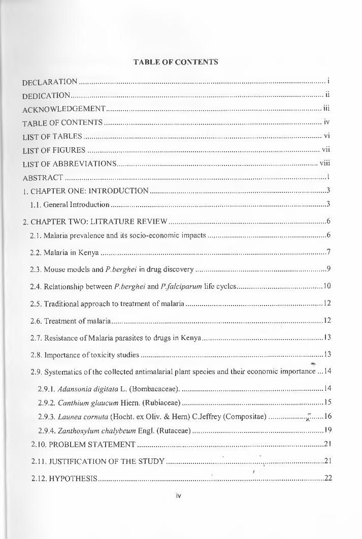
TABLE OF CONTENTS
DECLARATION..................................................................................................................................... i
DEDICATION.........................................................................................................................................ii
ACKNOWLEDGEMENT.................................................................................................................... iii
TABLE OF CONTENTS............ iv
LIST OF TABLES.................................................................................................................................vi
LIST OF FIGURES............................................................................................................................. vii
LIST OF ABBREVIATIONS.............................................................................................................viii
ABSTRACT.............................................................................................................................................1
1. CHAPTER ONE: INTRODUCTION...............................................................................................3
1.1. General Introduction....................................................................................................................3
2. CHAPTER TWO: LITRATURE REVIEW.....................................................................................6
2.1. Malaria prevalence and its socio-economic impacts................................................................6
2.2. Malaria in Kenya......................................................................................................................... 7
2.3. Mouse models and P.berghei in drug discovery...................................................................... 9
2.4. Relationship between P.berghei and P.falciparum life cycles.............................................. 10
2.5. Traditional approach to treatment of malaria.......................................................................... 12
2.6. Treatment of malaria.................................................................................................................. 12
2.7. Resistance of Malaria parasites to drugs in Kenya................................................................. 13
2.8. Importance o f toxicity studies.................................................................................................. 13
2.9. Systematics o f the collected antimalarial plant species and their economic importance ... 14
2.9.1. Adansonia digitata L. (Bombacaceae).............................................................................. 14
2.9.2. Canthium glaucum Hiem. (Rubiaceae)............................................................................ 15
2.9.3. Launea corimta (Hocht. ex Oliv. & Hern) C.Jeffrey (Compositae)....................J.......16
2.9.4. Zanthoxylum chalybeum Engl. (Rutaceae).......................................................................19
2.10. PROBLEM STATEMENT.................................................................................................... 21
2.11. JUSTIFICATION OF THE STUDY...................... ...... .'.................. ...................................21*
2.12. HYPOTHESIS............................................................!........................................................... 22
IV
2.13. RESEARCH OBJECTIVES................................................................................................22
2.13.1. General objective.............................................................................................................. 22
2.13.2. Specific objectives............................................................................................................ 22
3. CHAPTER THREE: MATERIALS AND METHODS................................................................23
3.1. Collection o f plant material..................................................................................................... 23
3.2. Preparation o f crude extracts.................................................................................................... 23
3.3. In vivo determination of anti-malarial activity....................................................................... 24
3.4 Brine shrimp toxicity test.................................................................................................. 28
3.5. Phytochemical screening for secondary metabolites using Thin Layer Chromatography(TLC)............................................................................ 30
3.6. Statistical analysis...................................................................................................................... 31
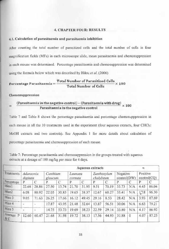
4. CHAPTER FOUR: RESULTS........................................................................................................33
4.1. Calculation o f parasitaemia and parasitaemia inhibition..................................................... 33
4.2. Mean plots...................................................................................................................................35
4.3. Mice mortality............................................................................................................................38
4.4. Brine shrimp toxicity.................................................................................................................39
4.5. Estimation o f Lethal Dose 50 ................................................................................................... 39
4.6. Phytochemical analysis o f crude plant extracts for secondary metabolites......................... 41
5. CHAPTER FIVE: DISCUSSION................................................................................................... 42
6. CHAPTER SIX: CONCLUSIONS AND RECOMMENDATIONS.................................... _ ...50
6. REFERENCES................................................................................................................................. 52
7. APPENDICES............................................................................................... 61
Appendix 1: Calculation of Parasitaemia and Chemosuppression............................................... 61
Appendix 2: Multiple comparison of chemosuppressions...................................................... 66
Appendix 3: Brine shrimp toxicity.................................................................................................. 67
*/
V
LIST OF TABLES
Table 1: Plant species collected from Msambweni district based on their use as antimalarials ...23
Table 2: Yield in grams after extraction of ground plant material with water and a mixture of chloroform-methanol (1:1)...................................................................................................................24
Table 3: Determination o f parasitaemia o f the donor mouse........................................................... 25
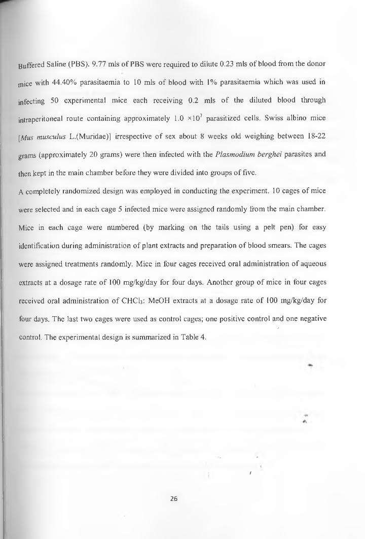
Table 4: Random allocation of infected mice.....................................................................................27
Table 5: Brine shrimp experimental set up......................................................................................... 30
Table 6: Screening for alkaloids, flavonoids, saponins and sesquiterpene lactones.......................31
Table 7: Percentage parasitaemia and chemosuppression in the groups treated with aqueous extracts at a dosage o f 100 mg/kg per mice for 4 days..................................................................... 33
Table 8: Percentage parasitaemia and chemossuppresion in the groups treated with CHCh:MeOH (1:1) extracts at a dosage of 100 mg/kg per mice for 4 days...............................................34
Table 9: Mean parasite density, chemosuppression and survival time o f P.berghei infected mice................................................................................................................................................................ 35
Table 10: Descriptive of chemosuppressions in the different treatment groups............................37
Table 11: ANOVA analysis..................................................................................................................37
Table 12: Acute toxicity of the crude plant extracts..........................................................................40
Table 13: Phytochemical screening of aqueous crude extracts............................... .'.......................41
Table 14: Phytochemical screening of the crude CHCI3: MeOH extracts......................................41
*/
VI
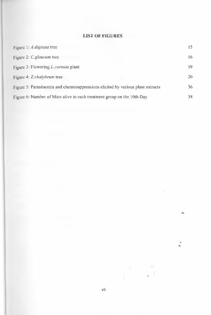
Figure 1: A.digitata tree 15
Figure 2: C.glaucum tree 16
Figure 3: Flowering L.cormita plant 19
Figure 4: Z.chalybeum tree 20
Figure 5: Parasitaemia and chemosuppressions elicited by various plant extracts 36
Figure 6: Number of Mice alive in each treatment group on the 10th Day 38
LIST OF FIGURES
vii
LIST OF ABBREVIATIONS
ANOVA: Analysis o f Variance
DMSO: Dimethylsulphoxide
DV: Dependent Variable
DW: Distilled Water
CQ: Chloroquine
CHC13: Chloroform
CTMDR: Center for Traditional Medicine and Drug Research
H2SO4: Sulphuric acid
IV: Independent Variable
KEMR1: Kenya Medical Research Institute
LD50: Lethal Dose 50
MeOH: Methanol
MF: Magnification Field
PBS: Phosphate Buffered Saline
SPSS: Statistical Package for the Social Sciences
TLC: Thin Layer Chromatography
UV Light: Ultra Violet Light
*l
viii
ABSTRACT
Malaria continues to kill over a million people each year, with more than 90% of these
cases found in sub-Saharan Africa. In many populations affected by malaria, conventional drugs
are often unaffordable or inaccessible, and increasing drug resistance by the malaria parasite, P.
falciparum, is of significant concern. The current study involved determination of antimalarial
activity and toxicity o f selected plants from Msambweni district, Coast province; Kenya.
Aqueous and organic [Chloroform: Methanol (1:1)] extracts from each plant were
prepared and used to determine in vivo anti-malarial activity and toxicity. They were also
screened for their phytochemical constituents. To evaluate antimalarial activity, Swiss albino
mice (Mus musculus L.) were infected with Plasmodium berghei parasites through
intraperitoneal route. Crude extracts were administered orally everyday at the same hour for four
days at a dosage of 100 mg/kg from Day 0 to Day 3. Thin blood smears were prepared on Day 4
from each mouse and stained with 10% Giemsa in phosphate buffer, pH 7.2 and examined
microscopically for assessment of parasitaemia. The mean parasitaemia in each group of mice on
Day 4 was used to calculate the percentage chemosuppression for each extract./
Toxicity o f the crude extracts was evaluated using Brine shrimp larvae (Artemia salina L.
nauplii). ANOVA was used to analyze the means of the parasite growth inhibitions while LDSo
was estimated using Finney’s probit analysis program. The screened plants exhibited varying
degrees of chemosuppression. Aqueous extracts of Adansonia digitata L. (Bombacaceae),
Zanthoxylum chalybeum Engl. (Rutaceae), Launea cornuta (Hocht. ex Oliv. & Hern) C.Jeffrey
(Compositae) and Canthium glaucum Hiern. (Rubiaceae,) had 60.47%, 44.93%, 38.13% and
31.98% parasite growth inhibition respectively. CHCI3: MeOH extracts of C.glaucum and
1
A digitata showed parasite growth inhibition o f 43.76% and 32.90% respectively. Chloroquine
(positive control) had 87.23% parasite growth inhibition.
Crude extracts of A.digitata and C.glaucum had LDstP'lOOO pg/ml and were considered
to be non toxic to Brine shrimp larvae unlike crude extracts o f Z.chalybeum and L.cornuta which
had LDso^OO ug/ml. Phytochemical screening of the crude extracts showed that alkaloids,
flavonoids, sesquiterpene lactones and saponins were present in A.digitata, C.glaucum,
L.cornuta and Z.chalybeum.
This study reports for the first time the in vivo antimalarial activity of the crude root
(aqueous and organic) extracts of C.glaucum and crude leaf (aqueous and organic) extracts of
L.cornuta. In addition the toxicity o f aqueous and organic root extracts o f C.glaucum is also
being reported for the first time. From the study A.digitata and C.glaucum have been shown to
possess promising antimalarial activity and were not toxic to the Brine shrimp larvae. These
results indicate that there is potential for isolation o f new scaffolds against Plasmodium
falciparum from the aqueous extracts of the two plants.
Key words/
Plasmodium berghei, Adansonia digitata, Canthium glaucum, Launaea cornuta, Zanthoxylum
chalybeum, Mus musculus (Swiss albino mouse), Artemia salina (Brine shrimp), Toxicity, In
vivo anti-malarial activity and Phytochemical screening
2
1. CHAPTER ONE: INTRODUCTION
1.1. General Introduction
Malaria continues to kill over a million people each year, with more than 90% of these cases
found in sub-Saharan Africa (Nguta et al., 2010). In many populations affected by malaria,
conventional drugs are often unaffordable or inaccessible, and increasing drug resistance by the
malaria parasite, P. falciparum, is of significant concern (Greenwood and Mutabingwa, 2002).
As an alternative, medicinal plants are often used to treat diseases such as malaria and
development of new drugs is very important to cut down the malaria scourge (Ajaiyeoba et al.,
1999). Vector resistance to drugs and mutating parasites has made it hard to control the spread of
the disease (Hilou et al., 2006). In addition most antimalarial drugs are not affordable to most
people especially those living in the third world countries as a result new cheap drugs are
needed.
Most drugs for malaria are expensive and not accessible to many locals who live away from
health centers (WHO, 2004). The ever-rising cost of modem medicine and its technical
inadequacies has prompted many people particularly in developing countries to result to
traditional medicine to treat a variety o f ailments they succumb to (Kokwaro, 2009).Traditional
remedies are employed by many communities to cure malaria and other infectious diseases
(Murray, 1995). Since time immemorial, man has depended on plants as the primary source of
medicine to cure a variety o f diseases. Various plants have been used by man in folk medicine to
treat malaria (Kokwaro , 2009) ,,
In the past, dmgs have been developed from plants. For example major drugs such as quinine
and artemisinin have been derived from traditional medicine (Nguta et al., 2010). Artemisinin
was derived from a leafy portion o f a Chinese traditional herb known as' Artemisia annua L.
3
(Compositae) (Akira et al., 2001). Plants provide rich source of drugs for treatment of malaria as
has been argued by Julie et al., (2007). From ethnomedicine data, it is documented that many
plants are used in control and treatment of malaria however very few of these plants have been
analyzed chemically (Garavito et al., 2006). As a result there is great need to analyze these plants
which form the basis o f ethnomedicine and are used to treat malaria to ascertain whether they
have effects on the malaria parasites.
On the other hand, plants have toxic effects on livestock and humans, such toxicity may be high
leading to death of an animal or humans consuming the plant. In traditional medicine, one is
likely to overdose the patient due to imprecise nature of diagnosis. This is not only unique to
traditional medicine but can also occur in modern medicine. Toxicity is attributed to certain
acting principles found in drugs; these chemical substances interact with living systems and
affect normal processes. All chemicals can cause harm to organisms at some level o f exposure.
Toxicity tests are important so as to determine the lethality o f drugs and to determine the
harmless concentration o f drugs for consumption (Hood, 2009). This ensures and enhances
human health, animal health and protection of the environment.
Plants produce array o f secondary metabolites which exhibit a broad spectrum of biological
activity. These include: alkaloids, sesquiterpene lactones, flavonoids, steroids, athraquinones and
essential oils besides others (Mazid et al., 2011). Biological activities o f plants extracts include
antifungal, anti-malarial, antibacterial, anti-fertility, cytotoxic, larvicidal and insecticidal
activities among others (Mazid et al., 2011). Biological activity o f plants is attributed to the class
and concentration of phytochemical constituents in the plants which makes some plant extracts
exhibit a variety o f activities (Wang et al., 2010). Phytochemical screening o f plant extracts
4
especially those which have been used in traditional medicine is therefore essential so as to
identify phytochemical constituents in the plants that are responsible for a given bioactivity.
Majority of malaria drugs in current use have developed resistance against Plasmodium
parasites. For instance, chloroquine resistance strains o f P.falciparum have been reported (David
et al., 2004). Other drugs such as amodiaquine and lapdap suffer cross-resistance with
chloroquine and fansidar (David et al., 2004). Nevertheless various strains o f Plasmodium
parasites have emanated which are tolerant to Artemisinin based combination therapies (ACTs)
which are considered as the best drugs for treatment of malaria (Tolu et al., 2007). Geographical
resistance to malaria drugs has also been reported which entails resistance to anti-malarial drugs
in various geographical areas (Shretta et al., 2000). An ideal drug for treatment of malaria should
be able to cure within a short period of time, safe, it should be less toxic, suitable for expectant
mothers and small children who are most vulnerable to malaria, it should be efficacious against
drug resistant strains and should be affordable by most people (David et al., 2004).
Since Plasmodium parasites have developed resistance to most o f the current drugs for malaria,
new anti-malarial drugs are needed urgently to counteract the disease. Regions which are/
endemic to malaria are faced with serious situation because the only available malaria drugs are
losing therapeutic value. Plant biodiversity provides one untapped source for new anti-malarial
drugs. This is supported by ethnomedicine where various plants have been used to treat malaria.
As a result numerous malaria development and discovery projects are underway. In the process
of evaluation o f anti-malarial properties o f plants and in drug discovery; drug efficacy,
pharmacology and toxicity are important parameters which need to be considered (David et al.,
2004).
5
2. CHAPTER TWO: LITRATURE REVIEW
2.1. Malaria prevalence and its socio-economic impacts
Malaria is one o f the most important human infectious diseases in the world. It kills about 1 to 3
million people and cause disease in 300-500 million people annually (UNICEF, 2007). Malaria
is caused by protozoan parasites of the genus Plasmodium. These malaria causing parasites are
host-specific. The four Plasmodium parasites infecting humans include P. falciparum, P.vivax,
P.ovale and P.malariae. O f these four parasites, P. falciparum is the most severe in terms of
infection rates and the pathology it causes because it produces more merozoites in blood than
any other plasmodium species (Robert et al., 2005; Carico et al., 2004). Other Plasmodium
parasites include P.knowlesi which causes malaria in monkeys; Plasmodium berghei,
Plasmodium yoelii, P.vinckei and P.chabaudi are malaria causing parasites in rats (Sinden,
1978).
Malaria is frequently referred to as disease of the poor, because it is concentrated in world’s
poorest countries. Sub-Saharan Africa is the region which is hardest hit by malaria with most
countries in the region being highly endemic for malaria transmission (UNICEF, 2007). Low
land areas which experience high temperatures have high malaria prevalence unlike highland
areas and deserts fringes. This is because temperature favors the life cycle o f the malaria parasite
vector. Children, pregnant women and people living with HIV are at high of contacting malaria.
Malaria mostly affects low income earners who live in poorly constructed houses (Worral et al.,
2003; UNICEF, 2007). Most of these people have low levels of education and live in unhygienic
environment. Malaria incidents are high in rural areas compared to urban areas due to socio
economic factors such as differences in education and income (Cox et al., 1999).
6

The burden of malaria is high among world’s poorest countries unlike in rich countries which
only account for 0.2% of the global malaria deaths. In terms of Disability Adjusted Life Years
(DALYS), the burden is high in poor countries of the world (Worral et al., 2003). Malaria is
expensive to treat and an estimated loss o f $12 billion per year in GDP is encountered in
treatment of malaria in most countries of Sub-Saharan Africa. Malaria increases government
health spending and reduces the income o f the common man because it is expensive to treat
(Worral et al., 2003). Malaria keeps poor people poor and this is a drawback to economic
development. Malaria is a serious and debilitating disease with negative side effects to a
community and to a given region at large hence infected people ought to be treated soon. Indeed
World Health Organization recommends that all children under the age of five with fever must
be treated with anti-malarial drugs in areas o f high or moderate malaria transmission (UNICEF,
2007)
2.2. Malaria in Kenya
Malaria is the leading cause of morbidity and mortality in Kenya 25 million out o f a population
of 34 million Kenyans are at risk o f malaria. It accounts for 30-50% of all outpatient attendance
and 20% of all admissions to health facilities. An estimated 170 million working days are lost to
the disease each year (MOH 2010). Malaria is also estimated to cause 20% o f all deaths in
children under five (MOH 2010). The most vulnerable group to malaria infections are pregnant
women and children under 5 years of age.
Zones of seasonal malaria transmission include the arid and semi-arid areas o f northern and
south-eastern parts of the country which experience short periods o f intense malaria transmission
during the rainfall seasons (KMSI, 2011). Temperatures are usually high and water pools created
during the rainy season provide the malaria vectors breeding sites. Extfeme climatic conditions
7
which cause flooding in these areas lead to epidemic outbreaks with high morbidity rates due to
low immune status of the population (DOMC, 2010a)
Malaria transmission in the western highlands of Kenya is seasonal, with considerable year-to-
year variation (KMSI, 2011). The epidemic phenomenon is experienced when climatic
conditions favor sustainability o f minimum temperatures around 18° C. This increase in
minimum temperatures during the long rains period favors and sustains vector breeding resulting
in increased intensity o f malaria transmission. The whole population is vulnerable and case
fatality rates during an epidemic can be up to ten times greater than what is experienced in
regions where malaria occurs regularly (KEMRI FACT SHEET, 2012)
Low risk malaria areas include the central highlands of Kenya including Nairobi (DOMC,
2010b). The temperatures are usually too low to allow completion o f the sporogonic cycle o f the
malaria parasite in the vector (KSM1, 2011). However with increasing temperatures and changes
in the hydrological cycle associated with climate change are likely to increase the areas suitable
for malaria vector breeding with introduction of malaria transmission in areas it never existed.
The burden o f malaria in Kenya is being monitored through several channels including health
information data from hospitals and clinics, sentinel sites surveys o f communities and health
facilities in various districts and national surveys including the Kenya Demographic Health
Survey (KSMI, 2011). Evidence from these sources now points towards increased coverage of
interventions with a downward trend in disease burden demonstrated by community reported
cases of malaria, hospital admissions and deaths due to malaria and childhood deaths from the
diseases (KEMRI FACT SHEET, 2012).However, malaria still remains a big threat to human life
in Kenya and ways o f combating the disease need to be put in place.
8
2.3. Mouse models and P.berghei in drug discovery
Plasmodium berghei is one o f the many species of malaria parasites that infect mammals other
than humans. P. berghei belongs to a group of four Plasmodium species that infect murine
rodents from Central Africa. These species are P. vinckei, P. chabaudi, P. yoelii and P. berghei
(Lin et al., 2010; Sinden, 1978). The rodent parasites are not of direct practical concern to man
or his domestic animals. The interest of rodent malaria parasites is that they are practical models
for the experimental study of mammalian malaria. These parasites have proved to be analogous
to the malaria parasites o f man and other primates in most essential aspects of structure,
physiology and life cycle (Carter and Diggs 1977).Therefore, for investigation o f different
aspects of human infection one could question whether or not the use of non-human malaria
parasites is still appropriate. Rodent parasites and their hosts are diverged from the human
parasites and human host and therefore careful comparison and assessment o f results from rodent
models is essential to assess their relevance for human disease (Sinden, 1978)
In the process o f studying human malaria parasites, rodent parasites are recognized as valuable
model parasites for the investigation o f the developmental biology o f malaria parasites, parasite-/
host interactions, vaccine development and drug testing (Menard et al., 1997). P. berghei is an
excellent model for research on the developmental biology o f malaria parasites, because o f the
availability of: Technologies for in vitro cultivation and large scale production and purification
of the different life cycle stages; Knowledge on the genome sequence and organization;
Methodologies for genetic modification of the parasite; Well characterized clones and
genetically modified mutant lines, including transgenic parasites expressing reporter genes such
as Green Fluorescent Protein and Luciferase (Calton et al., 2002; Janse et al., 1993).
9
r
The other rodent parasites are invaluable in different areas of malaria research. For example, P.
chabaudi is recognized as a useful model for investigations o f mechanisms of drug resistance
and antigenic variation (Peters, 1998). This parasite shows antigenic variation during long
lasting, non-lethal, infections in laboratory rodents. In contrast, P. berghei infections are usually
rapidly lethal to laboratory rodents which hamper studies on the in vivo generation and selection
of antigenic variants (Philips et al., 1997). Another example is P. yoelii, which is extensively
used in studies on the biology o f liver stage and blood stage antigens and their role in immunity
and vaccine development (Mota et al., 2001).
2.4. Relationship between P.berghei and P.falciparunt life cycles
The morphology of the different developmental stages is conserved between mammalian malaria
parasites. The life cycles and the different developmental stages o f all mammalian malaria
parasites are highly comparable. Mammalian malaria parasites share the following
characteristics; they are only infectious to Anopheline mosquitoes, haploid sporozoites invade
and develop only in liver-cells; after multiplication in the liver the parasites (merozoites) invade
and multiply in red blood cells; in the blood a relatively small percentage o f parasites develop
into gametocytes, the precursor cells o f the haploid gametes; fertilization and development of the
diploid zygote into ookinetes occur in the midgut of the mosquito; mature ookinetes penetrtfte the
cells o f the midgut wall and develop into oocysts on the outside o f the midgut (Sinden, 1978; Lin
et al., 2000).
The invasive, non-dividing stages such as merozoites, sporozoites, ookinetes and microgametes
of the different mammalian malaria parasites are very similar in size and morphology. The
similarity o f these stages of the different species is clear from investigations on the ultra-*
structure of these stages (Lin et al., 2000). Increasing evidence coming from molecular studies
10
also demonstrates the similarity o f these stages between the various mammalian Plasmodium
species (Sinden, 1978). For example, a number of surface proteins demonstrate conservation
both in structure and function between rodent and human malaria parasites such as CS, TRAP,
P45/48, CTRP, P25, P28, AMA-1, MSP-1 (Lin et al.,2000).
The genome organization is conserved between rodent and human malaria parasites. Recent
studies demonstrate a high level o f conservation of genome organization between rodent and
human parasites. The genome o f both P. falciparum and the four rodent parasites are organized
into 14 linear chromosomes, ranging in size from 0.5-3.8 Mb (Rich & Ayala 2003). Metabolic
pathways are conserved between mammalian malaria parasites. To our knowledge no gross
differences in metabolic pathways between mammalian malaria parasites have been reported
(Barnwell & Wertheimer, 1989). The similarity in sensitivity of mammalian malaria parasites to
antimalarial drugs and other specific inhibitors emphasizes the similarities in their metabolic
processes (Barnwell & Wertheimer, 1989).
Despite the overall similarity between mammalian malaria parasites, many 'small' differences
exist that are directly related to interaction of the parasites with their hosts. Differences in the life
cycle of P.berghei and P.falciparum or generally differences between various mammalian
malaria parasites life cycles are mainly restricted to the duration o f development and size of the
different dividing stages (Akawa & Seed, 1980). For example, there are significant differences in
developmental time and size o f liver schizonts, erythrocytic schizonts and oocysts. These small
differences may have a significant impact on host-parasite interactions and may influence
important features such as pathology, virulence and immune escape (Waters el al, 1991).
Generally all mammalian Plasmodium parasites induce malaria in a similar manner and studies
11

of malaria using P.berghei and mouse models can be used in the development o f drugs against
p. falciparum in human beings.
2.5. Traditional approach to treatment of malaria
In folk medicine people use decoctions, concoctions and infusions from various plants to treat
diseases they encounter. A variety o f plants have been used in ethnomedicine by different
communities to treat malaria. The plant parts used include leaves, fruits, roots, stem bark as well
as whole plant particularly when herbs are used. These drugs are prepared by boiling, soaking in
cold water, crushing of plant parts followed by soaking. The extracts can either be drunk alone,
mixed with food, soup or even milk to make them palatable. Sometimes a decoction from single
plant is administered alone or infusion containing extracts from two or three plants is given to the
patient (Kokwaro, 2009). However traditional treatment o f malaria and other diseases has several
disadvantages such as imprecise nature of diagnosis and over dosage. Nevertheless traditional
medicine remains the only source of medicaments to people who are not accessible to modern
drugs (Kahunu et al., 2011).
, /2.6. Treatment of malaria
Clinical manifestations of malaria include: fever, anemia, chills, metabolic acidosis, low birth
weight and still birth. Severe symptoms include: cerebral malaria, organ failure, coma and death.
Mostly used drugs for treatment of malaria include: chloroquine, artemisinin derivatives and
sulphfadoxine pyrimethamine (SP) combination commonly known as fansidar (David et al.,
2004). Other malaria drugs are mefloquinine, atavoquone and clindamycin. Artemisinin derived
drugs have short half lives and as a result they are combined with other long acting drugs (David
et al., 2004; Hombhanje & Huang, 2010). Artemisinin combined therapies (ACTs) are
12
considered as the best current drugs for treatment of malaria (Gathirwa et al., 2011). Malaria is a
major cause of mortality in young children, expectant women and people living with HIV and
effective drugs are needed to counteract it. In addition, the mutation rate for malaria parasites is
high and currently most malaria parasites have developed resistance to the current malaria
medicine (David et al., 2004). As a result, research and development o f new malarial drugs is
needed.
2.7. Resistance of Malaria parasites to drugs in Kenya
Resistance to antimalarial drugs in Kenya has been described for the four species that infect
humans (WHO, 2001). P. falciparum has developed resistance nearly to all antimalarials in
current use (WHO, 2001). P.falciparum in Kenya is moderately multidrug resistance as
determined by the various genotype and phenotypic analyses (UNICEF, 2007). Although
Malaria parasite mutation is a major cause o f drug resistance, Malaria treatment failure which
may be due to incorrect dosing, non-compliance with dosing regimen, poor quality drug and
misdiagnosis intensifies the intensity o f Plasmodium parasite resistance due to exposure to
suboptimal drug levels (D’Alessandro & Buttiens, 2001). Drug resistance by malaria parasites
increases the burden o f Malaria for countries like Kenya. Therefore implementation of control
measures is needed to reduce the burden of the disease. One way o f reducing the burdm is
coming up with a novel chemotherapy and study of botanicals can be a good way to start.
2.8. Importance of toxicity studies
Many substances we handle daily are toxic. Different organisms respond differently to the same
toxin. Lethal Dose 50 is a term which is used to describe acute toxicity. It refers to lethal dose
which is acutely lethal to 50% of all the organisms in a given experiment. LD50 is expressed in
13
mg/kg or ppm (parts per million). Toxicity study is a first step necessary for new drug
development. Brine shrimp lethality test is a general bioassay which is capable o f detecting
broad spectrum o f bioactivity o f crude plant extracts (Chanda & Bravalia, 2011). Plant products
are heterogeneous because they contain mixtures of bioactive constituents.
Simple bioassays for determination o f bioactivities of crude plant extracts are important for the
purpose of standardization or quality control of plant botanicals. Due to this Brine shrimp
lethality test is a “top bench” procedure useful in natural product chemistry (Jerry & Lingling,
1998). Using Brine shrimp in toxicity studies is economical, easy, uses small amount of test
material, inexpensive, simple and requires little chemical training. This test is a convenient probe
for preliminary assessment of toxicity of natural products from plant biodiversity, heavy metals
and pesticides (Aseer et al., 2009).
2.9. Systeinatics of the collected antiinalarial plant species and their economic importance
2.9.1. Adansonia digitata L. (Bombacaceae).
A. digitata is commonly known as Baobab. It is a massive deciduous tree indigenous to Africa
with huge bark and white flowers. It is regarded as the largest succulent plant in the world
(Kamatou et al., 2011). The plant is restricted to hot, semi-arid regions and dry woodlands and it
is found in Savannah woodlands o f Sub-Saharan Africa. Baobab fruits, leaves, and bafk are
sources of food, fiber and medicine. Kamatou et al., (2011) points out that over three hundred
traditional uses o f the plant have been documented. Due to its nutritional, medicinal and
cosmetic uses, the plant has attracted the interest of many pharmaceutical industries (Gdbauer et
al., 2002).
Fruit pulp, seeds and leaves o f the plant are eaten because they are a good source of vitamins,*
proteins, carbohydrates and lipids (Sidibe & Williams, 2002). The plant’s stem bark and leaves
14
are used in folk medicine to treat malaria (Gbadamosi et al., 2011; Kamatou et al., 2011). The
plant has antipyretic, antimicrobial, anti-inflammatory and analgesic activities (Ramadan et al,
1994). Powdered leaves have anti-asthmatic and have anti-tension and antihistamine properties.
Leaves are also used for other conditions such as diarrhea, dysentery, opthalmia and otitis media.
Bark is used as a substitute for quinine to curb high fever and act as prophylactic measure for
malaria as well (Sidibe & Williams, 2002).
Compaore et al. (2011) has shown that flavonoids, proanthocyanidins and phenolic compounds
are present in the pulp o f A. digitata which make it a good radical scavenger. Elsewhere the
fruits of A.digitata have been reported to contain proanthocyanidins as the major compounds
(Shahat, 2006). Triterpenes, alkaloids, anthraquinones, saponins, tannins have also been reported
to be present in the fruit pulp of A. digitata (Ramadan et al., 1994: Gbadamosi et al., 2011).
Figure 1: A.digitata tree /
15
2.9.1 Canthium glaucum Hiem. (Rubiaceae)
The plant has been reported to be used traditionally for the treatment of malaria. A fruit
decoction from the plant is taken orally by people of Msambweni district, Coast province; Kenya
for the treatment of malaria (Nguta et al., 2010). No bioactive compounds of C.glaucum have so
far been reported. Root powder of a related species; C. parviflorum Lam. is used as a treatment
for snake bites as reported by Mahishi et al. (2005). A powder from the same plant is used for
dressing wounds and bone injuries (Ghorbani et al., 2011). C. dicoccum (Gaertn.) Merr. a related
species has been shown to contain sesquiterpenoids, nitrogenous compounds, aldehydes,
terpinolene and phenols. These phytochemical constituents from extracts of C.dicoccum make
the plant to have antimicrobial, anti-tumor, immunomodulatory and antioxidant properties (Raja
e ta l, 2011).
Figure 2: C.glaucum tree
16
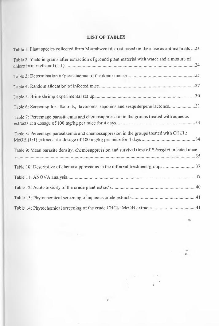
2.9.3. Launea cornuta (Hocht. ex Oliv. & Hern) C.Jeffrey (Compositae)
l cornuta is a perennial herb with erect stems which branch above to from a rosette of leaves
and diffuse inflorescence. It grows in disturbed and artificial grasslands and roadsides. This is a
vegetable used locally as a cheap source o f food by the local people. It is an important plant
species and is favored by most people because it has a good taste when cooked. Normally the
fresh young leaves and shoots are plucked and cooked (Kataariina, 2000). Studies have shown
that the plant contains high quantities o f Vitamin C, Minerals like sodium, potassium, calcium
and iron (Katariina, 2000). Likewise Lyimo et al. (2003) while investigating the nutrient
composition of L. cornuta, they found that the leafy vegetable had a considerable amount of
vitamin C, crude fiber, protein, fat, calcium and iron.
L. cornuta has been used locally for the management of prostate and breast cancer as reported by
Kareru et al. (2007). The whole plant is crushed and the decoction obtained after boiling the
crushed plant in water is given to the cancerous patient. Apart from cancer the plant species has
been used as a remedy for diabetes, the whole plant is boiled and the decoction is drunk to
reduce sugar levels in the body (Kareru et al., 2007). On the health aspect, leaves and roots of L./
cornuta are used medicinally in the management of malaria, stomach ache and in men they are
used to stop blood flow during circumcision. Launaea species are antimicrobial and L. cornuta is
one of the medicinal plants used by people o f Suba district-Kenya particularly people living with
HIV/AIDS to cure opportunistic diseases arising due to HIV/AIDS or to treat HIV/AIDS related
symptoms (Nagata etal., 2011).
L.nudicaulis (Linn.) Hook, has insecticidal activity against Rhizopertha dominica Fab.
(Bostrichidae) and Tribolium casteneum Herbst. (Tenebrionidae); it has antifungal activity
against Microsporum canis Bodin. (Arthrodermataceae) and Aspergilus flavus Joh.
17
(Trichocomaceae) and phytotoxic activity against Lemma acquinoctialis Welwitsch (Lemnaceae)
(Ali et al., 2003). L. intybacea (Jacq.) Beauverd, L.massaiensis (Fresen) Sch-Bip have
hepatoprotective activity against hepatotoxins such as carbon tetrachloride (Chaudhary et al.,
2010). L.procumbens (Roxb.) Ram. & Raj. is antimicrobial, has cytotoxicity against brine shrimp
nauplii, is antitumor and has antioxidant activity against a variety o f radicals in the body, lastly it
has hepatoprotective activity as has been demonstrated using rats by Rhamat (2010).
L.cassiniana Kuntze also has nematocidal activity against a number of soil nematodes.
L.intybacea (Jacq.) Beauverd likewise was found to have hepatoprotective activity on
paracetamol introduced hepatotoxicity in albino rats (Takate et al., 2010). L.sonchoides (Cass.)
N.Killian is antifungal and will inhibit the growth of Trichoderma hamatum (Bonord.) Bainier
(Hypocreaceae) and T.viridae J.P.H van Wyk (Hypocreaceae) as proved by studies done by
Abou-Zeid et al., (2008). Launaea genus is characterized by flavonoids, triterpenes,
sesquiterpene lactones, coumarins and steroids. Flavones like apigenin and luteolin are common
compounds in the genus. Steroids like stigmasterol, cholesterol, taraxasterol have been identified
in L. nudicaulis. Flavone glycosides such as apigenin-7-glycoside, luteolin-7-glycoside, luteolin-
7-rutinoside and Vitexin have been isolated from the extract of L.tenuiloba (L.) Kuntze and
L.resedifolia (L.) Kuntze. Delphinidin; an anthocyanin is found in L.asplenifolia Hook. (Ali et
al., 2003; Fairouz et al., 2010).
Various species of Launaea such as L.arborescens (Batt.) Murb., L.mucronata (Forssk.) Muschl,
L.nudicaulis and L.capitata (Spreng.) Dandy contains various types o f flavones such as luteolin,
• . . .
apigenin and flavone glycosides such as apigenin 7-O-glucoside, vitexin, luteolin 7-O-glucoside
and luteolin 7-O-rhamnoside besides others (Vipaporn & Christian, 2010). Two isoprenylated
flavonoids; asplenitin and asplenetin 5-O-neohesperidoside have be$n identified in L.
18
asplenifolia (Denis & Ragaj, 1996). Roots of L. mucronata (Forssk.) Muschl, aerial parts of L.
spinosa (Forssk.) Sch and aerial parts and roots of L.tenuiloba contain lactucin type guianolides
as stated by Christian (2008)
Figure 3: Flowering L.coniuta plant
2.9.4. Zanthoxylum chalybeum Engl. (Rutaceae)
Z. chalybeum is a deciduous tree which reaches a height of 12 meters. It has pale grey bark, with
dark smooth scales and prickles and the crown is rounded. The tree grows well in low altitude
areas, in dry woodlands and in savannah grasslands. The leaves when dried can be brewed to
make a kind of a tea. Leaves are used as fodder and are fed on by goats while the tree tryhk is a
good source of charcoal and durable timber (Bamford & Henderson, 2003).
Stem, root bark and leaves are traditionally used for malaria (Beetje, 1994; Kokwaro, 2009).
Roots and leave concoction is used in the management of chronic joint Spains (Wambugu et al.,
19
2011). Seed extracts of Z chalybeum has antiviral activity against measles virus (Olila et al.,
2002). It is active against Staphylococcus aureus Rosenbach. (Staphylococcaceae) and has anti
inflammatory activities (Matu & Staden et al., 2003). The plant has been used traditionally in the
management o f diabetes (Keter & Mutiso, 2011).
Z.chalybeum has been reported to contain alkaloids which have antibacterial and cytotoxic
activity (Chrian et al., 2011). Preliminary screening of Z armatum DC. has revealed that the
plant contains linalool as the major essential oil (Tiwary et al., 2007). Z. schinifolium Siebold &
Zucc contains linalool and estragole as the main essential oils besides ar-tumerone and limonene.
(Cheng et al., 2011). Zanthoxylum species contains various compounds such as alkaloids,
aliphatic and aromatic amides, lignans, coumarins, sesquiterpene lactones and sterols (Cheng et
al., 2011; He et al., 2002). The compounds have cytotoxic, molluscicidal, anticonvulsant, anti
sickling, anaesthetic, antibacterial, anti-hypertensive and anti-inflammatory activities (Adesina,
2005).
t1Figure 4: Z.chalybeum tree
20
2.10. PROBLEM STATEMENT
Malaria is a killer disease responsible for many deaths annually and in Kenya 25 million are at
risk of malaria, it is expensive to treat and manage and accounts for great loss in Gross Domestic
Product particularly in developing countries due to its morbidity. Nowadays there is great urge to
find alternative drugs to modern malaria medicines which are expensive and which may not be
effective since malaria parasites are always mutating and becoming resistant to current malaria
drugs. The search for antimalarials from plant biodiversity is one move to find alternative cheap
medicaments to modern malaria medicine. The present study was conducted to evaluate the
antimalarial activity, toxicity and phytochemical composition of selected anti-malarial plants
commonly used by the Msambweni community to treat malaria.
2.11. JUSTIFICATION OF THE STUDY
In Kenya malaria causes great morbidity and mortality which is a drawback to economic
development. Antimalarial drugs are expensive to poor communities in developing countries and
human malaria parasites have developed resistance to almost all current antimalarial drugs.
Nowadays researchers are trying to develop new drugs for malaria. Most modern medicines were
discovered through study of plants which were used traditionally to treat specific illnesses. In
addition, very few medicinal plants have been analyzed chemically and their bioactive
constituents are yet to be validated. As a result, knowledge from traditional medicine can be very
essential in the development o f cheap and effective antimalarial drugs.
1
21
2.12. HYPOTHESIS
Ho:
Crude extracts o f plants under investigation will not inhibit growth of Plasmodium berghei
merozoites in mice and will not be toxic to Artemia salina L. nauplii.
Ha=
Crude extracts o f plants under investigation will inhibit growth o f Plasmodium berghei
merozoites in mice and will be toxic to Artemia salina L. nauplii.
2.13. RESEARCH OBJECTIVES
2.13.1. General objective
To investigate in vivo anti-malarial activity, toxicity and phytochemical screening o f A.digitata,
C.glaucum, L.cornuta and Z.chalybeum which have been used in traditional medicine for the
treatment of malaria.
2.13.2. Specific objectives
1. To determine in vivo antimalarial activity of organic [MeOH: CHCb (1:1)] and aqueous
extracts o f A.digitata, C.glaucum, L.cornuta and Z.chalybeum against P.berghei
merozoites.
2 . To estimate acute toxicity (LD50) of A.digitata, C.glaucum, L.cornuta and Z.chalybeum
crude extracts using Brine shrimp (Artemia salina L.) nauplii
3. To analyse the phytochemical constituents in A.digitata, C.glaucum, L.cornuta and
Z.chalybeum crude extracts using Thin Layer Chromatography (TLC)
22
3. CHAPTER THREE: MATERIALS AND METHODS
3.1. Collection of plant material
Plant parts were collected from Msambweni district, Coast province, Kenya. Collection was
based on ethnopharmacological use as antimalarials through interviews with local communities
(Nguta et al., 2010). Information gathered included vernacular names (in parantheses) and the
parts used in preparation o f the herbal antimalarial remedies: A. digitata (Mbambitri) stem bark,
C. glaucum (Mhonga) roots, L. cornuta (Mtsunga wa utsunga) leaves and Z. chalybeum
(Mjafari/mporojo) stem bark.Plants were identified by a taxonomist from the University of
Nairobi and voucher specimens were deposited in the University o f Nairobi Herbarium. The
plant parts were air dried under a shade, chopped into small pieces and then ground into powder.
The collected plants are tabulated in Table 1.
Table 1: Plant species collected from Msambweni district based on their use as antimalarials
Plant Species/Family Plant Part Collected Voucher Number
Adansonia digitata L.(Bombacaceae) Stem Bark JN414
Canthium glaucum Hiern. (Rubiaceae) Roots JN426 ,
Launaea cornuta (Hocht.ex.Oliy. & Hiern.) C.Jeffrey (Compositae)
Leaves JN028
Zanthoxylum chalybeum. Engl. (Rutaceae) Stem Bark JN040
3.2. Preparation of crude extracts
For each plant, 50 grams of the ground plant material was extracted with 500 mis of distilled
water while another 50 grams was extracted with 500 mis of chloroform-methanol mixture (1:1
v/v) for four times at 48 hour intervals using cold maceration (Sulsen et al., 2011). The aqueous
extracts were filtered and the filtrate kept in a deep freezer then lyophilized resulting to a dry/
t
gummy substance. Organic (CHCb: MeOH) extracts were filtered and concentrated with a rotary
23
evaporator then left to dry powder. The dry solid extracts were stored at -20°C in air tight
containers until used. The yields o f the extracts are shown in Table 2.
Table 2: Yield in grams after extraction of ground plant material with water and a mixture of
chloroform-methanol ( 1:1)
Plant species/Family Aqueous extracts (Grams)
Organic (CHCl3:MeOH) extracts (Grams)
~Adansonia digitata L. (Bombacaceae) 7.58 4.13
~Canthium glaucum Hiern. (Rubiaceae) 9.52 4.40
Launaea cornuta (Hocht.ex.01iv.& Hiern) C.Jeffrey (Compositae)
8.12 5.60
Zanthoxylum chalybeum.Engl (Rutaceae) 16.02 6.48
3.3. In vivo determination of anti-malarial activity
In vivo anti-malarial activity was determined by 4-day suppressive anti-malarial assay according
to Waako et al. (2005) and Peters et al. (1975). Cryopreserved chloroquine sensitive Plasmodium
berghei (ANKA strain) parasites were obtained from KEMRI where they were stored at -80°C.
For the parasites to be used for experimental purposes, they were revived and stabilized in mouse
host according to Ravindran et al. (1982). Mice were housed in cages and given mice pellets and
water ad libitum. A group of three naive mice were used to revive and stabilize the P.berghei
parasites. Reviving, stabilizing and mantainance of the Plasmodium berghei parasite~was
achieved by continuous reinjection o f the parasite to new naive mice since infected mice died
after several days due to increased parasitaemia. One mouse was chosen as a donor mouse which
was then anesthetized using chloroform in a fume chamber and blood collected via tfhrdiac
puncture into heparinized bottles to make innoculum for infecting new naive mice. After drawing
blood, the donor mouse was sacrificed by cervical dislocation. Even after the parasite had
stabilized in the mouse host, passing the parasite to new mice ensured that stabilized parasites
24
were always available for experimental purposes. Normally, passages were done when the
parasitaemia reached about 40% which reached that point by the fourth day once the parasites
were introduced to naive mice. As a result, the process of passing parasites from donor mice to
naive mice was followed after every 4 days. Three passages were ideal to stabilize cryopreserved
parasite in the mouse host.
Appropriate innoculum for infecting mice had low parasitaemia o f around 1% and experimental
mice were innoculated with 0.2 mis o f blood with 1% parasitaemia (containing about 1.0 xlO7
parasitized cells) according to Waako et al., (2005). Before infecting experimental mice,
parasitaemia in the donor mice was first determined in order to dilute the blood to achieve 1%
parasitaemia with Phosphate Buffered Saline (PBS). Percentage parasitaemia of the donor mice
was calculated as follows:
P ercentage P arasitaem ia =Total Number of Parasitized Cells
Total Number of CellsX 1 0 0
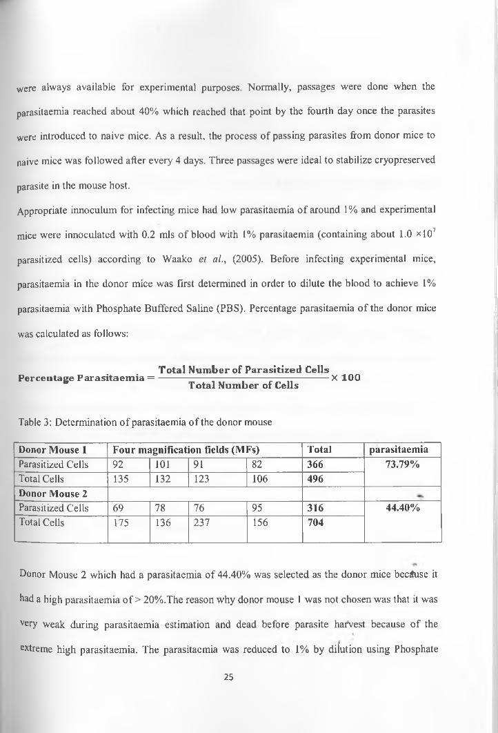
Table 3: Determination o f parasitaemia o f the donor mouse
Donor Mouse 1 Four magnification fields (MFs) Total parasitaemiaParasitized Cells 92 101 91 82 366 73.79%Total Cells 135 132 123 106 496Donor Mouse 2Parasitized Cells 69 78 76 95 316 44.40%Total Cells 175 136 237 156 704
Donor Mouse 2 which had a parasitaemia of 44.40% was selected as the donor mice because it
had a high parasitaemia o f > 20%.The reason why donor mouse 1 was not chosen was that it was
very weak during parasitaemia estimation and dead before parasite harvest because of the
extreme high parasitaemia. The parasitaemia was reduced to 1% by dilution using Phosphate
25
Buffered Saline (PBS). 9.77 mis of PBS were required to dilute 0.23 mis of blood from the donor
mice with 44.40% parasitaemia to 10 mis of blood with 1% parasitaemia which was used in
infecting 50 experimental mice each receiving 0.2 mis o f the diluted blood through
intraperitoneal route containing approximately 1.0 *107 parasitized cells. Swiss albino mice
[Mus musculus L.(Muridae)] irrespective o f sex about 8 weeks old weighing between 18-22
grams (approximately 20 grams) were then infected with the Plasmodium berghei parasites and
then kept in the main chamber before they were divided into groups o f five.
A completely randomized design was employed in conducting the experiment. 10 cages of mice
were selected and in each cage 5 infected mice were assigned randomly from the main chamber.
Mice in each cage were numbered (by marking on the tails using a pelt pen) for easy
identification during administration o f plant extracts and preparation o f blood smears. The cages
were assigned treatments randomly. Mice in four cages received oral administration of aqueous
extracts at a dosage rate o f 100 mg/kg/day for four days. Another group of mice in four cages
received oral administration of CHCI3: MeOH extracts at a dosage rate of 100 mg/kg/day for
four days. The last two cages were used as control cages; one positive control and one negative/
control. The experimental design is summarized in Table 4.
t
26
Table 4: Random allocation o f infected mice
TREATMENTSAqueous extracts Organic (CHCUiMeOH) extracts Controls
DAVS A B C D A1 B2 C2 D2 +Ve -Ve
DayO Cage 1 Cage 3 Cage 7 Cage 2 Cage10
Cage 4 Cage 6 Cage 9 Cage 5 Cage 8
Day 1 Cage 1 Cage 3 Cage 7 Cage 2 Cage10
Cage 4 Cage 6 Cage 9 Cage 5 Cage 8
|Day 2 Cage 1 Cage 3 Cage 7 Cage 2 Cage10
Cage 4 Cage 6 Cage 9 Cage 5 Cage 8
IDay 3 Cage 1 Cage 3 Cage 7 Cage 2 Cage10
Cage 4 Cage 6 Cage 9 Cage 5 Cage 8
"Day 4 Preparation of t tin blood smearsKey: A, B, C, D=Treatments, +Ve = Positive, -Ve =Negative
Stock solutions o f aqueous extracts (10,000 pg/ml) were made in distilled deionized water and
filter sterilized using 0.22 pm membrane filters in a laminar flow hood. To make stock solutions
of the CHCI3: MeOH extracts, the organic extracts were dissolved in dimethylsulphoxide
(DMSO) followed by subsequent dilution to lower concentration of DMSO to < 1% to avoid
carry over (solvent) effect. Plant extracts were administered once daily orally (DO to D3) at a
dosage rate of 100 mg/kg in a dose volume of 0.2 mis. Positive control drug used was
chloroquine (20 mg/kg/day) while negative control group was treated with distilled water (0.2
mls/mice/day). On the first day; plant extracts were administered 2 hours after infection of the
mice with the parasites
Blood was obtained from each of the experimental mice on Day 4. The tip o f the tail was cut
with a sterilized scissors and blood was squeezed from the tail and thin blood smears were
prepared. Forty eight (48) thin blood smears were prepared from the forty eight surviving mice
on Day 4. Smears were fixed with methanol for 5 minutes and stained with 10% Geimsa which
was prepared by mixing 100% Geimsa stain with staining buffer in the ratio 1:9.
27
The slides were observed under compound microscope under oil immersion at xlOOO to
determine the number of parasitized cell per given magnification field (MF). For each blood
smear specific for a given mouse, four magnification fields were observed and the number of
parasitized cells (schizonts) and the total number of cells in the magnification field were
recorded. The data obtained was used to determine percentage parasitaemia and parasite growth
inhibition in each mouse. The number of dead mice was also recorded every day since the start
of the four day suppressive test for the next 10 days.
3.4 Brine shrimp toxicity test
Brine shrimp-Leech (Artemia salina L.) larvae were used to determine the toxicity o f the crude
extracts according to Wanyoike et al., (2004). Brine shrimp eggs were hatched in a shallow
rectangular hatching tank containing artificial sea water and yeast .Yeast was provided to act acts
as food for the hatching larvae. Hatching tank measured 14 cm by 9 cm by 5 cm and had a
capacity o f 225 mis. The tank had two compartments; one small and one large compartment.
These two unequal compartments were created by a plastic divider with several 2 mm holes
which allow hatched Brine shrimp larvae to pass through.
Artificial sea water or 3.3% saline was prepared by dissolving 33.04 grams of commercial sea
salt in one liter of distilled water. 200 mis o f artificial sea water was poured into the hatching
tank to fill it to the brim. This was followed by pouring three dosage spoons o f Artemia salina
eggs and one gram of yeast to the larger compartment o f the hatching tank. The larger
compartment was then covered to prevent light exposure because Brine shrimp eggs hatch in
dark while the smaller compartment was exposed to light. Hatching occurred within 36 to 48 hrs
at 26°C. Once the Brine shrimp larvae hatched, they moved from the larger compartment to the
28
smaller compartment through the 2 mm holes in the plastic divider where they were collected
and transferred to fresh artificial sea water.
Various concentrations o f the crude extract in sea water were used: 10,100 and 1000 pg/ml. A
stock solution for each crude extract o fl0,000 pg/ml was initially prepared from which serial
dilutions were carried out to make the three concentrations which were used in the experiment.
For the aqueous extracts, the stock solution o f 10,000 pg/ml was prepared by dissolving O.lg of
each crude extract in 10 mis o f distilled water. For the CHCI3: MeOH extracts, O.lg of each
sample was first dissolved in dimethylsulphoxide (DMSO) then diluted further using artificial
water to 10 mis to make stock solution of 10,000 pg/ml. DMSO content in the stock solution was
less than 0.05% to avoid DMSO carry over effect on Brine shrimp larvae. The stock solutions
from various crude extracts were filtered using 0.22 pm micro filters under lamina flow hood
(Wanyoike et al., 2004).
Using Pasteur pipettes, 10 Brine shrimp larvae were transferred from the smaller compartment of
the hatching tank to plastic tubes. The volume of artificial sea water in each plastic tube
containing 10 Brine shrimp larvae was increased to 5ml except the tubes for 1,000 pg/ml which/
were topped to 4.5 mis with artificial sea water. Using micropipettes, 0.5 mis, 0.05 mis and 0.005
mis were transferred from the stock solution to the plastic tubes containing 5mls artificial sea
water to make experimental solutions containing 1000 pg/ml, 100 pg/ml and 10 pg/ml
respectively. A control group containing artificial sea water and Brine shrimp larvae only was
included in the experiment and this denoted a concentration o f 0 pg/ml. For each crude extract at
each concentration three repeats were carried out. Survivors were counted after 24 hours using a
magnifying glass (Meyer et al., 1982). For each crude extract, experimental set up in Table 5 was
followed.
29
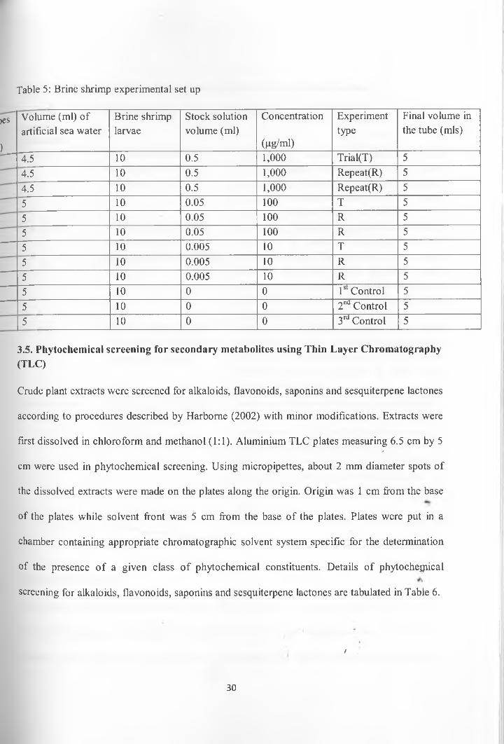
Table 5: Brine shrimp experimental set up
>es
)
Volume (ml) of artificial sea water
Brine shrimp larvae
Stock solution volume (ml)
Concentration
(pg/ml)
Experimenttype
Final volume in the tube (mis)
4.5 10 0.5 1,000 Trial(T) 54.5 10 0.5 1,000 Repeat(R) 54.5 10 0.5 1,000 Repeat(R) 55 10 0.05 100 T 55 10 0.05 100 R 55 10 0.05 100 R 55 10 0.005 10 T 55 10 0.005 10 R 55 10 0.005 10 R 55 10 0 0 1st Control 55 10 0 0 2nd Control 55 10 0 0 3rd Control 5
3.5. Phytochemical screening for secondary metabolites using Thin Layer Chromatography (TLC)
Crude plant extracts were screened for alkaloids, flavonoids, saponins and sesquiterpene lactones
according to procedures described by Harbome (2002) with minor modifications. Extracts were
first dissolved in chloroform and methanol (1:1). Aluminium TLC plates measuring 6.5 cm by 5/
cm were used in phytochemical screening. Using micropipettes, about 2 mm diameter spots of
the dissolved extracts were made on the plates along the origin. Origin was 1 cm from the base
of the plates while solvent front was 5 cm from the base o f the plates. Plates were put in a
chamber containing appropriate chromatographic solvent system specific for the determination
of the presence o f a given class o f phytochemical constituents. Details of phytochejpical
screening for alkaloids, flavonoids, saponins and sesquiterpene lactones are tabulated in Table 6.
/
30
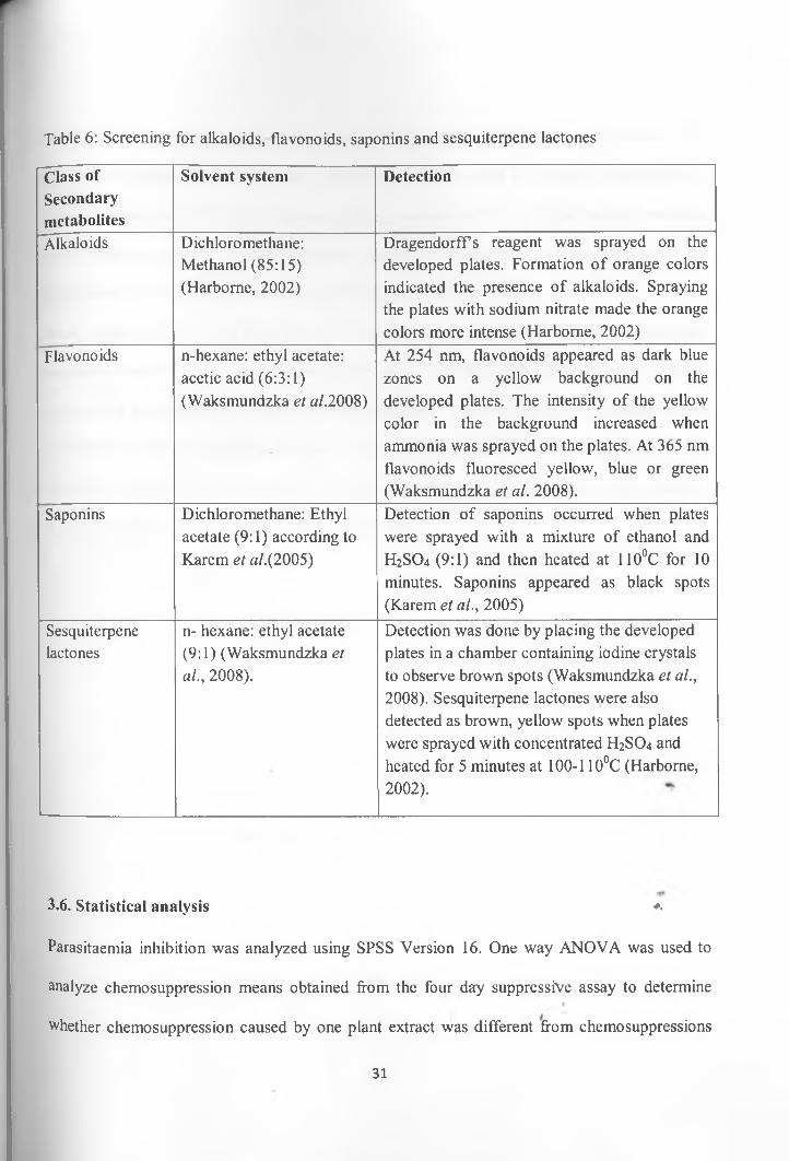
Table 6: Screening for alkaloids, flavonoids, saponins and sesquiterpene lactones
Class ofSecondarymetabolites
Solvent system Detection
Alkaloids Dichloro methane: Methanol (85:15) (Harborne, 2002)
Dragendorffs reagent was sprayed on the developed plates. Formation o f orange colors indicated the presence of alkaloids. Spraying the plates with sodium nitrate made the orange colors more intense (Harborne, 2002)
Flavonoids n-hexane: ethyl acetate: acetic acid (6:3:1) (Waksmundzka et a/.2008)
At 254 nm, flavonoids appeared as dark blue zones on a yellow background on the developed plates. The intensity o f the yellow color in the background increased when ammonia was sprayed on the plates. At 365 nm flavonoids fluoresced yellow, blue or green (Waksmundzka et al. 2008).
Saponins Dichloromethane: Ethyl acetate (9:1) according to Karem et a/.(2005)
Detection of saponins occurred when plates were sprayed with a mixture of ethanol and H2SO4 (9:1) and then heated at 110°C for 10 minutes. Saponins appeared as black spots (Karem et al., 2005)
Sesquiterpenelactones
n- hexane: ethyl acetate (9:1) (Waksmundzka et al., 2008).
Detection was done by placing the developed plates in a chamber containing iodine crystals to observe brown spots (Waksmundzka et al., 2008). Sesquiterpene lactones were also detected as brown, yellow spots when plates were sprayed with concentrated H2SO4 and heated for 5 minutes at 100-110°C (Harborne, 2002).
3.6. Statistical analysis *<
Parasitaemia inhibition was analyzed using SPSS Version 16. One way ANOVA was used to
analyze chemosuppression means obtained from the four dary suppressive assay to determine
whether chemosuppression caused by one plant extract was different from chemosuppressions
31
caused by the other plants extracts according to Morgan et al., (2004). Once the means were
found to be different from each other, Dunnett test was then used for multiple comparisons of
chemosuppressions to determine whether chemosuppressions arising from the various treatments
were different from the chemosuppression induced by chloroquine (positive control). The
significance level used in the analysis was 0.05 (Alpha Level < 0.05).
Brine shrimp toxicity was determined by probit analysis. Probit analysis is a specialized
regression model o f binomial response variable and it has been used to analyse dose-response
experiments in a variety o f fields. It involves conversion o f concentrations into logarithms and
the corresponding percentage mortalities into probits. The logarithms of the concentrations are
then plotted against the probits to give a straight line graph. This regression line is then used in
the determination o f LD50 (Ashford & Sowden, 1970).
/
32
4. CHAPTER FOUR: RESULTS
4.1. Calculation of parasitaemia and parasitaemia inhibition
After counting the total number o f parasitized cells and the total number of cells in four
magnification fields (MFs) in each microscope slide, mean parasitaemia and chemosuppression
in each mouse was determined. Percentage parasitaemia and chemosuppression was determined
using the formula below which was described by Hilou et al. (2006)
Total Number of Parasitized CellsP ercen tage P arasitaem ia = ---------------- --------- -------- ----- —-----------X 1 00
Total Number of Cells
Chemosuppression
(Parasitaemia in the negative control)-(Parasitaem ia with drug) ^Parasitaemia in the negative control
Table 7 and Table 8 shows the percentage parasitaemia and percentage chemosupppression in
each mouse in all the 10 treatments used in the experiment (four aqueous extracts, four CHCI3:
MeOH extracts and two controls). See Appendix 1 for more details about calculation of
percentage parasitaemia and chemosuppression of each mouse.
Table 7: Percentage parasitaemia and chemosuppression in the groups treated with aqueous extracts at a dosage o f 100 mg/kg per mice for 4 days.
Aqueous extracts
Treatments Adansoniadigitata
Canthiumglaucum
Launaeacornuta
Zanthoxylumchalybeum
Negathcontrol
/Q(DW)
Positivecontrol(CQ)
percentage P C P C P C P C P C P C'Micel 22.68 28.86 27.50 13.74 21.70 31.95 9.51 70.19 33.73 N/A 4.45 86.04Mice2 6.08 80.92 22.05 30.83 19.65 38.37 12.67 60.27 33.41 N/A 1,18 96.30Mice 3 9.05 71.63 26.25 17.66 16.12 49.45 29.16 8.53 28.42 N/A 3.93 87.69Mice 4 - - 17.87 43.95 21.48 32.64 13.87 56.51 30.06 N/A 6.63 79.21Mice 5 - - 14.75 53.73 19.69 38.23 22.59. 29.14 33.80 N/A 4.17 86.92Average P
L&C_12.60 60.47 21.68 31.98 19.72 38.13 17.56
t44.93 3T.88/ 0 4.07 87.23
33
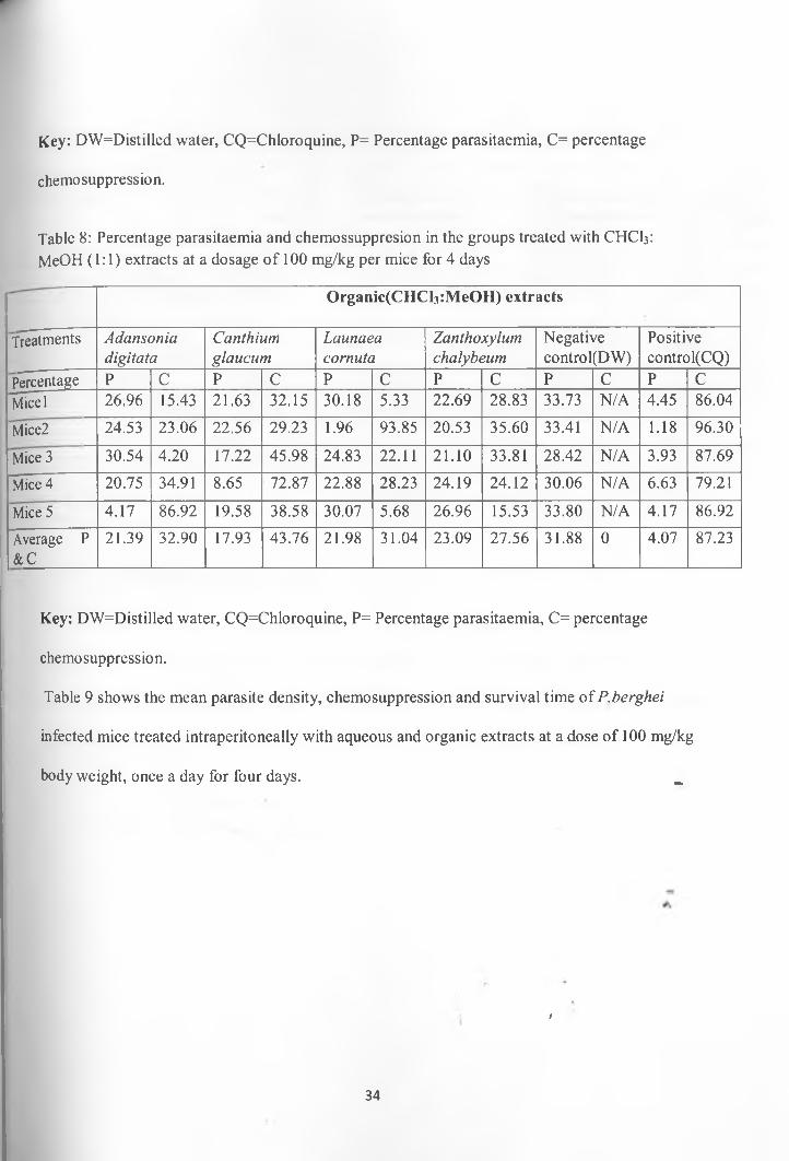
Key: DW=Distilled water, CQ=Chloroquine, P= Percentage parasitaemia, C= percentage
chemosuppression.
Table 8: Percentage parasitaemia and chemossuppresion in the groups treated with CHCI3: MeOH (1:1) extracts at a dosage o f 100 mg/kg per mice for 4 days
Organic(CHCl3:MeOH) extr acts
Treatments Adansoniadigitata
Canthiumglaucum
Launaeacornuta
Zanthoxylumchalybeum
Negatncontrol
/e(DW)
Positivecontrol(CQ)
Percentage P C P C P C P C P C P CMicel 26.96 15.43 21.63 32.15 30.18 5.33 22.69 28.83 33.73 N/A 4.45 86.04
Mice2 24.53 23.06 22.56 29.23 1.96 93.85 20.53 35.60 33.41 N/A 1.18 96.30
Mice 3 30.54 4.20 17.22 45.98 24.83 22.11 21.10 33.81 28.42 N/A 3.93 87.69
Mice 4 20.75 34.91 8.65 72.87 22.88 28.23 24.19 24.12 30.06 N/A 6.63 79.21
Mice 5 4.17 86.92 19.58 38.58 30.07 5.68 26.96 15.53 33.80 N/A 4.17 86.92
Average P &C
21.39 32.90 17.93 43.76 21.98 31.04 23.09 27.56 31.88 0 4.07 87.23
Key: DW=Distilled water, CQ=Chloroquine, P= Percentage parasitaemia, C= percentage
chemosuppression.
Table 9 shows the mean parasite density, chemosuppression and survival time of P.berghei
infected mice treated intraperitoneally with aqueous and organic extracts at a dose of 100 mg/kg
body weight, once a day for four days. ^
f
34
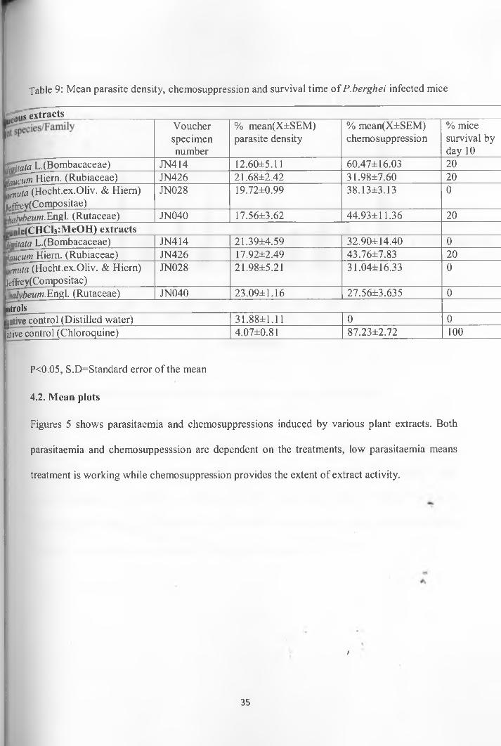
Table 9: Mean parasite density, chemosuppression and survival time of P .berghei infected mice
rT^i«extractsVoucherspecimennumber
% mean(X±SEM) parasite density
% mean(X±SEM) chemosuppression
% mice survival by day 10
■ j^taLX Bombacaceae) JN414 12.60±5.11 60.47± 16.03 20g ^ T H ie rn . (Rubiaceae) JN426 21.68±2.42 31.98±7.60 20^^(H ocht.ex .O liv . & Hiern) jeffrev(Compositae)
JN028 19.72±0.99 38.13±3.13 0
h^beum.Engl. (Rutaceae) JN040 17.56±3.62 44.93±11.36 20UnictCHC^cMeOH) extractsKritata L.(Bombacaceae) JN414 21.39±4.59 32.90±14.40 0twcumti^em- (Rubiaceae) JN426 17.92±2.49 43.76±7.83 20^rt^Hocht.ex.Oliv. & Hiern) leffrey(Compositae)
JN028 21.98±5.21 31.04±16.33 0
^Hvbeum.Engl. (Rutaceae) JN040 23.09±1.16 27.56±3.635 0ntrolsgative control (Distilled water) 31.88±1.11 0 0litive control (Chloroquine) 4.07±0.81 87.23±2.72 100
P<0.05, S.D=Standard error o f the mean
4.2. Mean plots
Figures 5 shows parasitaemia and chemosuppressions induced by various plant extracts. Both
parasitaemia and chemosuppesssion are dependent on the treatments, low parasitaemia means
treatment is working while chemosuppression provides the extent of extract activity.
/
35
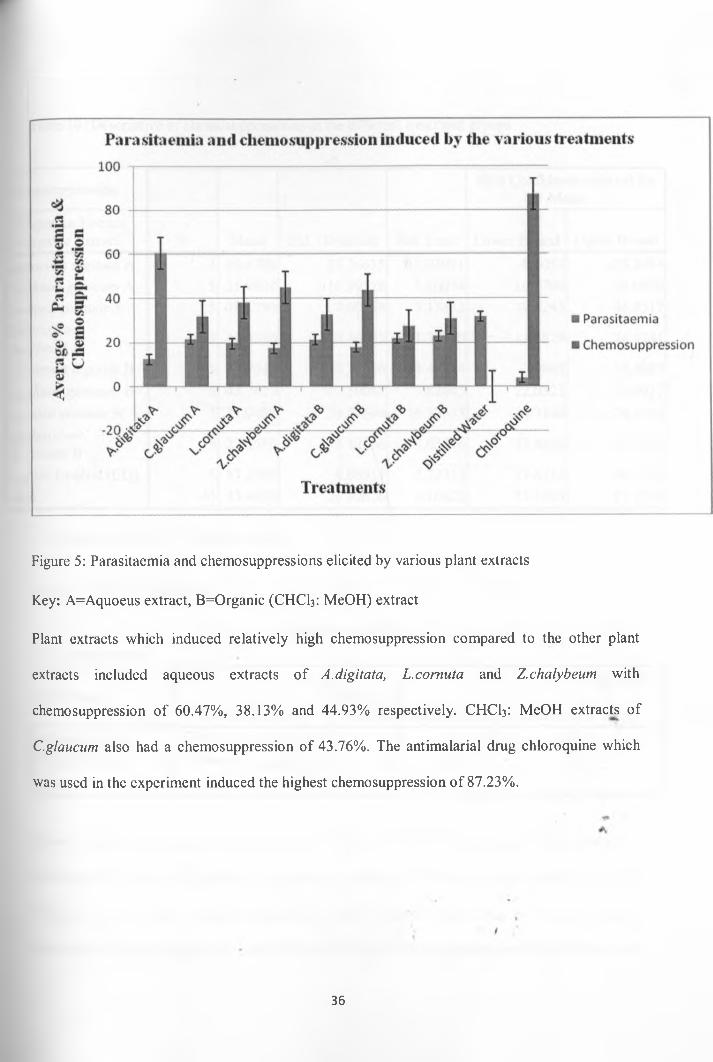
Figure 5: Parasitaemia and chemosuppressions elicited by various plant extracts
Key: A=Aquoeus extract, B=Organic (CHCI3: MeOH) extract
Plant extracts which induced relatively high chemosuppression compared to the other plant
extracts included aqueous extracts of A.digitata, L.cornuta and Z.chalybeum with
chemosuppression of 60.47%, 38.13% and 44.93% respectively. CHCI3: MeOH extracts of
C.glancum also had a chemosuppression of 43.76%. The antimalarial drug chloroquine which
was used in the experiment induced the highest chemosuppression o f 87.23%.
36
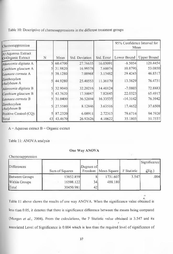
Table 10: Descriptive o f chemosuppressions in the different treatment groups
Chemosuppression
N Mean Std. Deviation Std. Error
95% Confidence Interval for Mean
A=Aqueous Extract 0=Organic Extract Lower Bound Upper Bound
Adansonia digitata A 3 60.4700 27.76635 16.03091 -8.5054 129.4454Canthiurn glaucum A 5 31.9820 16.99578 7.60074 10.8790 53.0850Launaea cornuta A 5 38.1280 7.00968 3.13482 29.4243 46.8317Zanthoxylum chalybeum A 5 44.9280 25.40553 11.36170 13.3829 76.4731
Adansonia digitata B 5 32.9040 32.20216 14.40124 -7.0803 72.8883Canthiurn glaucum B 5 43.7620 17.50047 7.82645 22.0323 65.4917Launaea cornuta B 5 31.0400 36.52694 16.33535 -14.3142 76.3942Zanthoxylum chalybeum B 5 27.5580 8.12846 3.63516 17.4652 37.6508
Positive Control (CQ) 5 87.2320 6.08911 2.72313 79.6714 94.7926Total 43 43.4670 26.92626 4.10622 35.1803 51.7537
A = Aqueous extract B = Organic extract
Table 11: ANOVA analysis
One Way ANOVA
Chemosuppression
DifferencesSum of Squares
Degrees of Freedom Mean Square F Statistic
Significance
tfig .)Between Groups 13852.859 8 1731.607 3.547 .004Within Groups 16598.122 34 488.180Total 30450.981 42
Table 11 above shows the results o f one way ANOVA. When the significance value obtained is
less than 0.05, it denotes that there is significance difference between the means being compared
(Morgan et al., 2004). From the calculations, the F Statistic value obtained is 3.547 and itsi
1
associated Level of Significance is 0.004 which is less than the required level o f significance of
37
0.05. This implies that the chemosuppressions in the various treatments were significantly
different from each other.
Multiple comparison of chemosuppressions show that the comparison o f the chemosuppression
induced by A. digitata aqueous extract and the chemosuppression induced by the positive control
gave a significance value o f 0.451. This significance level obtained of 0.451 is much larger than
the required 0.05 significance level. This implies that there was no significance difference
between chemosuppression induced by the A. digitata aqueous extract and the chemosuppression
induced by chloroquine according to Morgan et al., (2004). The comparisons of the other
treatments with the positive control resulted to a significance value which is less than 0.05 and
this implies that the chemosuppression induced by such treatments were different from the
positive control chemosuppression (See Appendix 2)
4.3. Mice mortality
N um ber of mice alive on 10th Day in the various treatm ent groups
Treatm ents
Figure 6: Number of Mice alive in each treatment group on the 10th Day
38
Key: A=Aqueous extract, B=Organic (CHClj: MeOH) extracts, Pve Control = Positive Control,
Nve = Negative Control.
Since the start o f the 4-day suppressive test, mortality o f the mice was monitored on daily basis.
Mice death occurred at different times and this could be attributed to the toxicity o f the plant
extracts administered, increased parasitaemia or intervening environmental factors. Figure 6
above shows the number of mice which died in each cage for a period o f 10 days since the start
of the 4-day suppressive antimalarial test.
The crude extracts administered to the mice were able to keep at least some of the mice alive up
to the 10th day. Groups treated with organic extracts of C.glaucum and aqueous extracts of
A.digitata, C.glaucum and Z.chalybeum had mice alive on the 10th day. No mice died in the
positive control group which received oral administration o f chloroquine on daily basis and all
the mice were healthy in the 10th day and were later sacrificed by cervical dislocation because
they didn’t die. In the negative control group which was receiving distilled water on daily basis,
all the mice died by the 7th day making it the first group to have 100% mortality by 7th day.
4.4. Brine shrimp toxicity
The numbers o f dead and surviving brine shrimp larvae were recorded 24 hours after they were
subjected to various concentrations of aqueous and organic extracts of four plants. The avefhge
mortality at each concentration was determined as it was essential for the estimation of LD 50
(See Appendix 3).
*4
4.5. Estimation of Lethal Dose 50
Finney Probit analysis Program was used to estimate the LD50 of the plant extracts on the brine
shrimp larvae according to Meyer et al., (1982). The average mortality ity the three
39

concentrations (1000 pg/ml, 100 pg/ml and 10 pg/ml) for each plant was fed into the program to
estimate the LDso-The results are tabulated below.
Table 12: Acute toxicity o f the crude plant extracts
Aqueous extracts’ Average mortality at various concentrations LD 50 Gradient(G)
plant species 1,000 pg/ml 100 pg/ml 10 pg/ml 0 pg/ml pg/ml'idigitata L. 2.33 0.33 0 0 9563.59 1.8009'Cglaucum Hiern 5.33 0.33 0.66 0 1209.67 0.6930'['cornuta(Hocht.ex.Oliv.)C. Jeffrey
8 1.33 0.33 0 341.64 0.4321
'ichalybeum .Engl. 10 0.33 0 0 268.28 0.3247CHCl3:Me<TH extracts
A.digitata L. 10 3.33 0.66 0 110.23 0.3203C.glaucum Hiern 2 0.66 0 0 1783.59 2.1700L. cornuta(Hocht.ex.Oliv.)C.Jeffrey
10 4 2 0 75.26 0.3400
Z.chalybeum Engl. 10 9 2 0 25.78 0.4357
A plant extract with a LD50 ranging between 0-500 pg/ml implies that the extract is highly toxic
while that ranging between 500-1000 pg/ml implies that the extract is moderately toxic. On the/
other hand a plant extract with a LD50 which is over 1000 pg/ml implies that the plant is non
toxic (Nguta etal., 2011).
From the experiment aqueous extracts of A.digitata stem bark and C.glaucum roots have LIT50
which is over 1000 pg/ml and hence they are non toxic on Brine shrimp larvae. However crude
extracts of Z.chalybeum stem bark and L.cornuta leaves have LD50 value which lies between 0
pg/ml and 500 pg/ml hence are highly toxic on Brine shrimp larvae
t
40
4.6. Phytochemical analysis of crude plant extracts for secondary metabolites
Crude extracts were screened for secondary metabolites using TLC and the results are tabulated
in Table 13 and Table 14.
Table 13: Phytochemical screening of aqueous crude extracts
Aqueous extracts Alkaloids Flavonoids Sesquiterpenelactones
Saponins
A.digitata L. +ve +ve -ve -ve
C.glaucum Hiern +ve +ve -ve -ve
L. cormita(Hocht.ex.Oliv.)C.Jeffrey
+ve +ve -ve +ve
Z.chalybeum Engl. +ve +ve -ve -ve
Key: +ve= Present, -ve= Absent
Table 14: Phytochemical screening of the crude CHCI3: MeOH extracts
CHCI3: MeOH extracts Alkaloids Flavonoids Sesquiterpenelactones
/
Saponins
A.digitata L. +ve +ve +ve +ve
C.glaucum Hiern. +ve +ve +ve +ve
L. cornuta(Hocht.ex.Oliv.)C. Jeffrey
+ve +ve +ve +ve
Z.chalybeum .Engl. +ve +ve +ve +ve
Key: +ve=Present, -ve=Absent
41

5. CHAPTER FIVE: DISCUSSION
Parasitaemia refers to the level o f infection by a particular parasite or the number of parasitized
cells circulating in blood. A decline in parasitaemia in a given organism is essential for the
recovery of symptomatic malaria. Effective antimalarial drugs when given in correct dosage will
usually lead to decrease in parasitaemia. When mice were infected with Plasmodium parasites,
the parasites multiplied in the erythrocytes and later caused bursting o f the erythrocytes releasing
merozoites into circulation which attacked more erythrocytes causing increased parasitaemia.
The percentage parasitaemia in the negative control group on D4 after infection with
Plasmodium berghei parasites was 31.88 % which was the peak parasitaemia witnessed in all the
groups treated with various plant extracts. The other treatments had lower parasitaemia levels
than this. Parasitaemia in the group which was treated with chloroquine was lowest (positive
control group) which was 4.07 % (See Table 7). Parasitaemia in the negative control being
higher than in the other treatment groups shows that the treatments had an effect on the growth
of Plasmodium berghei parasites in mice.
Parasitaemia increased gradually and without any antimalarial drug being administered in the/
negative control all the mice died on the 7th day. In the other treatment groups, most o f the mice
were alive by day seven post infection and even by the 10th day some groups had mice which
were alive. However no mice died in the positive control group and by 10th day all the mice were
alive and healthy (See Figure 6).
Drugs lead to decreased parasitaemia and subsequent recovery o f symptomatic malaria. They
also reduce parasitaemia through various ways like reducing parasite nutrient intake, interfering
with parasite metabolic pathways like heme metabolic pathway which is involved in the
metabolism of iron (De villers & Egon, 2009). Drugs also negatively affect parasite reproduction
42
and growth (Ziegler et al., 2002). The plant extracts to some extend cleared the parasites and
reduced the level o f parasitaemia and made the mice to survive even up to the 10th day.
Chloroquine had a good chemosuppression o f 87.23 % as determined on the fourth day post
infection and a 100% survival rate by day ten post infection.
Chemosuppression is inversely related to parasitaemia and plant extracts which have been shown
to reduce parasitaemia to low levels have been shown to have a corresponding high
chemosuppression. The aqueous extracts o f A. digitata had a chemosuppression of 60.47%
(p<0.05), which was the peak chemosuppression recorded in the experiment apart from the
chemosupression induced by chloroquine. Chemosuppression below 50% is usually taken as low
since calculations are based on the negative control parasitaemia as baseline. Due to that, the
other extracts had low chemosuppression which included the organic (CHCI3: MeOH) extracts of
C.glaucum, aqueous extracts o f Z. chalybeum and aqueous extracts o f L. cornuta with 43.76%,
44.93% and 38.13% respectively which their chemosuppression was slightly high among the
extracts with low chemosuppression.
Aqueous extracts of A.digitata induced a chemosuppression which was not significantly different/
from the chemopsuppression induced by chloroquine. Although the other plants extracts induced
chemosuppression which was significantly different from that induced by chloroquine, they
should also not be ruled out because to some extent some of the mice in those treatments had a
longer life span than the mice in the negative control group implying that the plant extracts had a
significant antimalarial activity but only which could not match that o f chloroquine
From the study, aqueous extracts from the stem bark of A.digitata exhibited high antimalarial
activity. This observation supports earlier work by Ajaiyeoba (2005) who reported that
methanolic extracts of the stem bark of A. digitata were able to reduce the number of
43
Plasmodium parasites in mice. Previous studies have shown that water and methanol extracts of
the stem bark o f Z. chalybeum have significant in vitro antimalararial activity against
chloroquine sensitive and chloroquine resistance strains o f Plasmodium falciparum (Rukunga et
al., 2009) which is in line with the results on Z.chalybeum obtained in the current study.
Methanolic extracts o f the root bark of Z.chalybeum have been reported to have significant
antimalarial activity on Plasmodium falciparum as argued by Muganga et al. (2010). This is also
in agreement with observations in the current study. Elsewhere aqueous extracts of a related
species Zanthoxylum usambarense (Engl.) Kokwaro have also been shown to exhibit a
significant antiplasmodial activity against Plasmodium falciparum (Kirira et al., 2006). Although
C.glaucum, L.cornuta are used in traditional medicine for treatment of malarial there is no
previous information which has been reported on their in vivo antimalarial activities. Therefore
the current study reports for the first time in vivo antimalarial activity o f the two plant species.
The significant within group variation in the percentage chemosuppression in the groups treated
with aqueous extracts o f A.digitata and Z.chalybeum can be attributed to the fact that
administration of plant extracts on mice was done orally. Due to this, some o f the mice may have
not taken the whole 100 mg/kg. dosage and this may have affected the parasitaemia in those
groups. Another factor which could account for the within group variation could be thatjthe
experimental mice were chosen irrespective of sex. Sex is a factor which affects many
physiological reactions one of them being drug interactions. Female mice and male mice may
have responded to the treatment differently and this may have brought the variation inA
chemosuppression within the given treatment groups. Lastly the fact that the experimental mice
weighed between 18-22g could have brought about the within group variation because an
44
approximate weight o f 20g was used and assigned to each mouse in the calculation o f the
required dosage o f 100 mg/kg and the slight differences in mice weight were not accounted.
LD50 is the concentration o f dose which kills fifty percent of all organisms in a given experiment.
Aqueous extracts o f both A.digitata and C.glaucum had a LD50 > 1000 pg/ml hence were
considered to be non toxic to Brine shrimp larvae while aqueous and organic (CHCI3: MeOH)
extracts of L.cornuta, Z.chalybeum which had a LD50 < 500 pg/ml were classified as being toxic
to Brine shrimp larvae with more toxicity being witnessed with the organic (CHCI3: MeOH)
extracts (See Table 12). A.digitata has also been shown to be non toxic on mice from previous
studies. For instance LD50 o f aqueous extract of the fruit pulp o f A.digitata on mice was found to
be over 8000 pg/ml (Ramadan et al., 1994). This justifies the results obtained in this study on the
non toxicity of stem bark o f A.digitata on Brine shrimp larvae. Non toxicity o f A.digitata
explains why most o f the plant parts: seeds, fruit pulps and leaves are consumed by many
communities. Nguta et al. (2011) while investigating toxicity of aqueous extracts of the leaves,
stem bark and root bark o f Z.chalybeum on Brine shrimp larvae obtained LDso<500 pg/ml for the
three plant parts which confirms the results obtained in this study implying that Z.chalybeum is a/
plant with high toxicity on Brine shrimp larvae and thus could not make safe antimalarial herbal
remedies. This calls upon for dose adjustment amongst communities using this plant for
preparation of antimalarial herbal decoctions. This vital information can reach the local people
through local arrangements with the local leaders where one can give advice on the correct
amount of ground material to be used in making concoctions so that local people do not consume
concentrated concoctions which may be toxic. In herbal practice, Z.chalybeum is usually
prepared as a concoction with other antimalarial plants (Nguta et al., 2010), and this could
explain why adverse effects have not been reported by those communities commonly using the
45
plant as an antimalarial phytotherapeutic remedy. Significant toxicity o f Z.chalybeum methanolic
root bark extract on human normal foetal lung fibroblast cells has also been reported by
Kamuhabwa et al. (2000). Similarly high toxicity of stem bark o f L.cornuta on Brine shrimp
larvae has also been reported by Nguta et al .(2011) and together with the high toxicity o f the
crude leave extract o f L.cornuta on Brine shrimp larvae witnessed in this study confirms
observations from other researchers. However there are no previous reports on toxicity o f C.
glaucum.
The aim of carrying toxicity tests was to determine whether the chemosuppression observed was
due to toxicity o f the plant extracts or due to the ability o f the plant extracts to elicit antimalarial
activity. Non toxicity o f A.digitata and C.glaucum extracts implies that the extracts from these
plants have a potential to inhibit the growth of Plasmodium parasites which is not associated
with toxicity. For instance, aqueous extracts o f Z.chalybeum had a chemosuppression of 44.93%
and were highly toxic (with a LD5o of 268.28 pg/ml) on Brine shrimp larvae implying that the
toxicity of the extracts could have lead to the witnessed chemosuppression of the Plasmodium
berghei parasites /
Biological activity is attributed to the presence o f various secondary metabolites in plants (Mazid
et al., 2011). Alkaloids and flavonoids in aqueous stem bark extracts o f A.digitata and alkaloids,
flavonoids, saponins, sesquiterpene lactones in organic root extracts o f C.glaucum were the
active constituents with antimalarial activity which reduced parasitaemia and hence prolonged
the life span the mice in the experiment. Just like growth inhibition, toxicity is also attributed to
presence of various secondary metabolites found in the plants extracts. Not only their presence,
but also the quantity o f the phytochemical constituents in a given plant extract determines the
extent of extracts’ bioactivity. In addition, presence o f more than one class of secondary
46
metabolites in a given plant extract will also determine the nature and extent of extract’s
biological activity (Wang et al., 2010)
All the plant extracts used in the experiment were screened using TLC for the presence or
absence of various classes o f secondary metabolites. Alkaloids and flavonoids were found to be
present in all crude plant extracts. Sesquiterpene lactones were present in the organic extracts of
A.digitata, C.glaucum, L.cornuta and Z.chalybeum. Saponins were present in organic extracts of
A.digitata, C.glaucum, L.cornuta and Z.chalybeum and aqueous extracts o f L.cornuta. Alkaloids,
flavonoids and sesquitepenes have been reported to be potent plant secondary metabolites with
broad spectrum o f bioactivities (Mazid et al., 2011). Alkaloids are major classes of compounds
possessing antimalarial activity; quinine is one of the most important and oldest antimalarial
drug which belongs to this class o f compounds (Saxena et al., 2003)
A.digitata stem bark contains medicinal compounds which are largely classified under saponins,
alkaloids and flavonoids such as lupeol acetate, P-sitosterol, scopoletin, ffiedelin, betullinic acid
and adansonin while the fruit pulp of A.digitata is rich in procyanidins (Sidibe & Williams,
2002; Shahat, 2006). Adansonin, an alkaloid has been proved to be one o f the active principles/
for treatment of malaria and other fevers (Sidibe & Williams, 2002). Thirteen alkaloids have
been isolated from tubers o f Stephania erecta Will. (Menispermaceae) while two alkaloids;
villastonine and alstonerine have been isolated from root extracts o f Alstonia angustifolia
Wall.ex. A. DC. (Apocynaceae). These alkaloids from S.erecta and A.angustifolia have been
shown to have in vitro antimalarial activity against P.falciparum (Saxena et al., 2003). In
addition, alkaloids from methanolic leaf extract of Annona senegalensis Pers. (Annonaceae) have
been reported to play a role in the growth inhibition o f Plasmodium berghei parasites in mice
(Ajaiyeoba et al, 2006). Flavonoids such as genistein and pratensein isolated from the leaf and
47

stem bark o f Andria inermis (W.wright) DC. (Leguminosae) have in vitro antimalarial activity
against P.falciparum (Saxena el al., 2003). Cowanin and Cowanol are two flavonoids isolated
from the ethanolic stem bark extracts of Garcinia cowa Roxb. (Guttiferae) which also have
antiplasmodial activity against P.falciparum (Saxena et al., 2003). Other compounds with potent
antimalarial activity include various sesquiterpene lactones such as artemisinin isolated from the
leaf extracts of Artemisia annua L. (Compositae) and arteinculton isolated from leaf extracts of
Artemisia maritima L. as reported by Saxena et al. (2003). Peroxycalamenene and patchoulenone
are sesquiterpene lactones isolated from tuber of Cyperus rotundus L. (Cyperaceae) which are
active against chloroquine-resistant strains o f P.falciparum (Saxena et al., 2003). Artemisinin is
a novel sesquiterpene lactone which is safe and effective antimalarial agent even in case of
chloroquine-resistant strains o f P. falciparum (Akira et al., 2001). Other studies have reported
that saponins isolated from methanolic and chloroformic leaf extracts of Taminalia cattapa L.
(Compretaceae) were active against P. falciparum (Mudi & Mohammed, 2009). Vernonia
ambigua Kotschy & Peyr. (Asteraceae) whole plant aqueous extract contains saponins which
have in vitro antiplasmodial activity against P.falciparum as argued by Builders et al., (2011).*
Results from phytochemical screening o f the plant extracts confirm the presence o f the classes of
compounds mentioned above. Hence these are the compounds which could have been
responsible for the reduction o f growth of Plasmodium parasites in the mice.
The above study is useful in treating malaria infections and in malaria drug discovery because it
is based on mouse models and P.berghei. All mammalian malaria parasites behave in ajdmilar
manner when they are in their hosts. Studies done using P.berghei will correlate very well with
studies done using human malaria parasites like the most infectious P.falciparum (Sinden,1978)
Aqueous extracts o f A.digitata have been found to have in vivo antimalarial activity against
48
P.berghei from the current study. Similar studies like this are important so as to identify plants
with antimalarial activity from plant diversity which can open new avenues for further studies as
far malaria drug discovery and treatment of malaria is concerned such as in vitro studies whereby
P.falciparum can be tested on the various plant botanicals and further phytochemical analyses to
identify active constituents in the various plant botanicals. Once plants with good antimalarial
activities have been identified, they can be cultivated and conserved in large scale so that they
can be easily available. For instance A.annua, a plant with antimalarial activity which is a major
source of artemisinin, one o f the major components of Artemisin Combined Therapies is grown
in Kenya, Madagascar, Tanzania and Uganda in large scale (Brisibe et ah, 2008)
6. CHAPTER SIX: CONCLUSIONS AND RECOMMENDATIONS
C. glaucum and A.digitata are two plants which from the current study have been shown to
inhibit the growth o f Plasmodium berghei parasites. Indeed, aqueous extracts of A.digitata had a
chemosuppression o f 60.47% implying that, this is a plant with good antimalarial activity against
Plasmodium berghei parasites. The stem bark of A.digitata and roots o f C.glaucum have been
proved to be non toxic on Brine shrimp larvae. This leads to the conclusion that Plasmodium
berghei growth inhibition by the two plants was not associated with their toxicity and suggests
that A.digitata and C.glaucum may be potential candidates for further antimalarial drug research
particularly A.digitata because it was found to have high growth inhibition on Plasmodium
berghei parasites
Z.chalybeum stem back extracts and L.cornuta leaf extracts on the contrary were shown to be
highly toxic to Brine shrimp larvae and possessed moderate antimalarial activity implying that
their parasite growth inhibition could be tied to their toxicity. This study reveals for the first time
in vivo antimalarial activity o f C.glaucum and L.cornuta. In addition this study confirms the
antimalarial activities o f A.digitata and Z.chalybeum which have been reported before. By/
carrying out in vivo antimalarial activity, the study shows that selected four plants species used in
traditional medicine for treatment of malaria have the capability o f inhibiting growth of malaria
parasites and consequently could have a potential in the treatment of malaria.
Flavonoids, alkaloids, saponins and sesquiterpene lactones were present in organic extracts of
A.digitata, C.glaucum, L.cornuta and Z.chalybeum. Alkaloids and flavonoids were also present
in aqueous extracts o f A.digitata, C.glaucum, L.cornuta and Z.chalybeum while saponins were
only present in aqueous extracts of L.cornuta. Further studies particularly on in vitro studies
using P.falciparum and on the phytochemical analysis of A.digitata and C.glaucum are
50
warranted. The specific compounds present in A.digitata and C.glaucum should be isolated and
investigated as potential candidates for antimalarial activity. Such studies can identify
compounds present with therapeutic value in the treatment of malaria. This would help in the
development of new antimalarial drugs from plant biodiversity.
i
51
6. REFERENCES
Abou-Zeid, A.M., Altalhi, A. D., Abd El-Fattah, R.I., 2008, Fungal Control of Pathogenic Fungi Isolated From Some Wild Plants in Taif Govemorate, Saudi Arabia, Mal.J. Microbiol., Vol.4, N o.l.pp. 30-39.
Adesina, S.K., 2005, The Nigerian Zanthoxylum; chemical and biological values, Afr. J. Trad. Cam, Vol. 2, No.3, pp. 282 - 301
Ajaiyeoba, E.O., 2005, In vivo antimalarial activity of methanolic extract of Adansonia digitata stem bark in mice model. New Strategy against Ancient Scourge. Fourth MIM Pan Africa Malaria Conference, Younde, Cameroon.
Ajaiyeoba, E.O., Abalogu, U.I., Krebs, H.C., Oduola, A.M.J., 1999, In vivo antimalarial activities o f Quassia amara and Quassia undulata plant extracts in mice, Journal of Ethnopharmacology, Vol.67, pp.321-325.
Ajaiyeoba, E.O.Falade ,M.,Ogbole,0.,Okpako, L.,Akinboye, D.,2006, In vivo antimalarial and cytotoxic properties o f Annona senegalensis extract, Afr.Journ.Trad.Comple.& Alt.Med.,Vol.3,No. 1 ,pp. 137-141.
Akawa, M. and Seed, T.M. (1980). Morphology of Plasmodia in M alaria, Vol. 1, pp.285-345, (Kreier, J.P. ed.), Academic Press, New York.
Akira, I., Chiaki, I., Mika, Y., Hirotaka, T., Toshio, M., Mamoru, T., 2001, A potent antimalarial activity of Hydrangea macrophylla var. Otaksa leaf extract against Plasmodium yoelii 17XL in mice, Parasitology International, Vol.50, pp. 33-39.
Ali, D., Hussein, S.M.S., Malik, A., Ahmed, Z., 2003, Chemical constituents o f the genus Launaea, Jour.Chem.Soc.Pak, Vol. 25,No. 4,pp. 341-347
Aseer, M., Sujith, S., Seghal, K., Joseph, S., Chippu, S., 2009. Cytotoxic Potentials of Red Alga, Laurencia brandenii Collected from the Indian Coast, Global Journal of Pharmacology' Vol.3, No.2, pp.90-94
Ashford, J.R., & Sowden, R.R., 1970, Biometrics, Vol.26, No.23.pp.535-546
Bamford, M.K, Henderson, Z.L., 2003, A reassessment of the wooden fragment from Florisbad, South Africa, Journal of Archaeological Science, Vol. 30, pp. 637-651 *'
Barnwell, J.W., Wertheimer, S.P., 1989, Plasmodium vivax: merozoite antigens, the Duffy blood group, and erythrocyte invasion, Prog Clin Biol Res. Vol.313, pp. 1—11.
Beetje, H.T., 1994, Kenya Trees, Shrubs and Lianas, National Museums o f Kenya, Nairobi
52
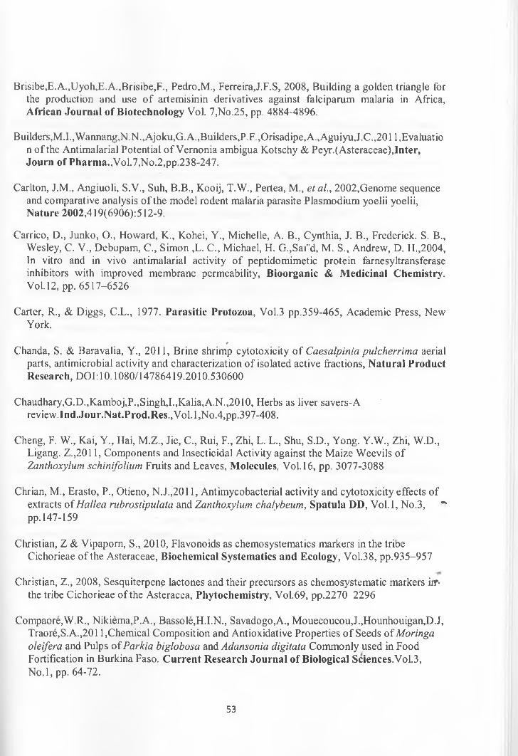
Brisibe,E.A.,Uyoh,E.A.,Brisibe,F., Pedro,M., Ferreira,J.F.S, 2008, Building a golden triangle for the production and use o f artemisinin derivatives against falciparum malaria in Africa, African Journal of Biotechnology Vol. 7,No.25, pp. 4884-4896.
Builders, M. I. ,Wannang,N.N.,Ajoku,G. A., Builders, P.F.,Orisadipe, A., AguiyuJ.C., 201 l,Evaluatio n of the Antimalarial Potential o f Vernonia ambigua Kotschy & Peyr.(Asteraceae),Inter, Journ of Pharma.,Vol.7,No.2,pp.238-247.
Carlton, J.M., Angiuoli, S.V., Suh, B.B., Kooij, T.W., Pertea, M., etal., 2002,Genome sequence and comparative analysis of the model rodent malaria parasite Plasmodium yoelii yoelii, Nature 2002,419(6906):512-9.
Carrico, D., Junko, O., Howard, K., Kohei, Y., Michelle, A. B., Cynthia, J. B., Frederick. S. B., Wesley, C. V., Debopam, C., Simon ,L. C., Michael, H. G.,Sai"d, M. S., Andrew, D. H.,2004, In vitro and in vivo antimalarial activity of peptidomimetic protein farnesyltransferase inhibitors with improved membrane permeability, Bioorganic & Medicinal Chemistry. Vol. 12, pp. 6517-6526
Carter, R., & Diggs, C.L., 1977. Parasitic Protozoa, Vol.3 pp.359-465, Academic Press, New York.
*Chanda, S. & Baravalia, Y., 2011, Brine shrimp cytotoxicity o f Caesalpinia pulcherrima aerial
parts, antimicrobial activity and characterization of isolated active fractions, N atural Product Research, DOl: 10.1080/14786419.2010.530600
Chaudhary,G.D.,Kamboj,P.,Singh,I.,Kalia,A.N.,2010, Herbs as liver savers-A review.Ind.Jour.Nat.Prod.Res.,Vol. l,No.4,pp.397-408.
Cheng, F. W., Kai, Y., Hai, M.Z., Jie, C., Rui, F., Zhi, L. L., Shu, S.D., Yong. Y.W., Zhi, W.D., Ligang. Z.,2011, Components and Insecticidal Activity against the Maize Weevils Of Zanthoxylum schinifolium Fruits and Leaves, Molecules, Vol. 16, pp. 3077-3088
Chrian, M., Erasto, P., Otieno, N.J.,2011, Antimycobacterial activity and cytotoxicity effects of extracts o f Hal lea rubrostipulata and Zanthoxylum chalybeum, Spatula DD, Vol.l, No.3, *■pp. 147-159
Christian, Z & Vipaporn, S., 2010, Flavonoids as chemosystematics markers in the tribe Cichorieae of the Asteraceae, Biochemical Systematics and Ecology, Vol.38, pp.935-957
Christian, Z., 2008, Sesquiterpene lactones and their precursors as chemosystematic markers iif< the tribe Cichorieae of the Asteracea, Phytochemistry, Vol.69, pp.2270-2296
Compaore,W.R., Nikiema,P.A., Bassole,H.I.N., Savadogo,A., Mouecoucou,J.,Hounhouigan,D.J, Traore,S.A.,2011,Chemical Composition and Antioxidative Properties o f Seeds of Moringa oleifera and Pulps of Parkia biglobosa and Adansonia digitata Commonly used in Food Fortification in Burkina Faso. C urrent Research Journal of Biological Sciences.Vol.3,No.l, pp. 64-72.
53
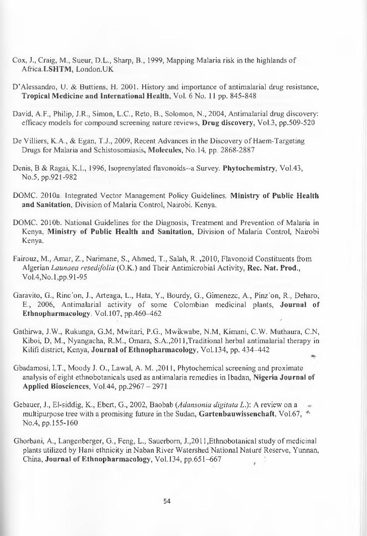
Cox, J., Craig, M., Sueur, D.L., Sharp, B., 1999, Mapping Malaria risk in the highlands of Affica.LSHTM, London.UK
D’Alessandro, U. & Buttiens, H. 2001. History and importance o f antimalarial drug resistance, Tropical Medicine and International Health, Vol. 6 No. 11 pp. 845-848
David, A.F., Philip, J.R., Simon, L.C., Reto, B., Solomon, N., 2004, Antimalarial drug discovery: efficacy models for compound screening nature reviews, Drug discovery, Vol.3, pp.509-520
De Villiers, K.A., & Egan, T.J., 2009, Recent Advances in the Discovery o f Haem-Targeting Drugs for Malaria and Schistosomiasis, Molecules, No. 14, pp. 2868-2887
Denis, B & Ragai, K.I., 1996, Isoprenylated flavonoids—a Survey. Phytochemistry, Vol.43,No.5, pp.921-982
DOMC. 2010a. Integrated Vector Management Policy Guidelines. Ministry of Public Health and Sanitation, Division o f Malaria Control, Nairobi. Kenya.
DOMC. 2010b. National Guidelines for the Diagnosis, Treatment and Prevention of Malaria in Kenya, Ministry of Public Health and Sanitation, Division o f Malaria Control, Nairobi Kenya.
Fairouz, M., Amar, Z., Narimane, S., Ahmed, T., Salah, R. ,2010, Flavonoid Constituents from Algerian Launaea resedifolia (O.K.) and Their Antimicrobial Activity, Rec. Nat. Prod.,Vol.4,No. 1,pp.91-95
Garavito, G., Rinc'on, J., Arteaga, L., Hata, Y., Bourdy, G., Gimenezc, A., Pinz'on, R., Deharo, E., 2006, Antimalarial activity o f some Colombian medicinal plants, Journal of Ethnopharmacology. Vol. 107, pp.460^162 /
Gathirwa, J.W., Rukunga, G.M, Mwitari, P.G., Mwikwabe, N.M, Kimani, C.W. Muthaura, C.N, Kiboi, D, M., Nyangacha, R.M., Omara, S.A.,2011,Traditional herbal antimalarial therapy in Kilifi district, Kenya, Journal of Ethnopharmacology, Vol. 134, pp. 434-442
Gbadamosi, I.T., Moody J. O., Lawal, A. M. ,2011, Phytochemical screening and proximate analysis of eight ethnobotanicals used as antimalaria remedies in Ibadan, Nigeria Journal of Applied Biosciences, Vol.44, pp.2967 - 2971
Gebauer, J., El-siddig, K., Ebert, G., 2002, Baobab {Adansonia digitata L.)\ A review on a multipurpose tree with a promising future in the Sudan, Gartenbauwissenchaft, Vol.67, *• No.4, pp.155-160
Ghorbani, A., Langenberger, G., Feng, L., Sauerborn, J.,2011 ,Ethnobotanical study o f medicinal plants utilized by Hani ethnicity in Naban River Watershed National Nature Reserve, Yunnan, China, Journal of Ethnopharmacology, Vol. 134, pp.651-667
54
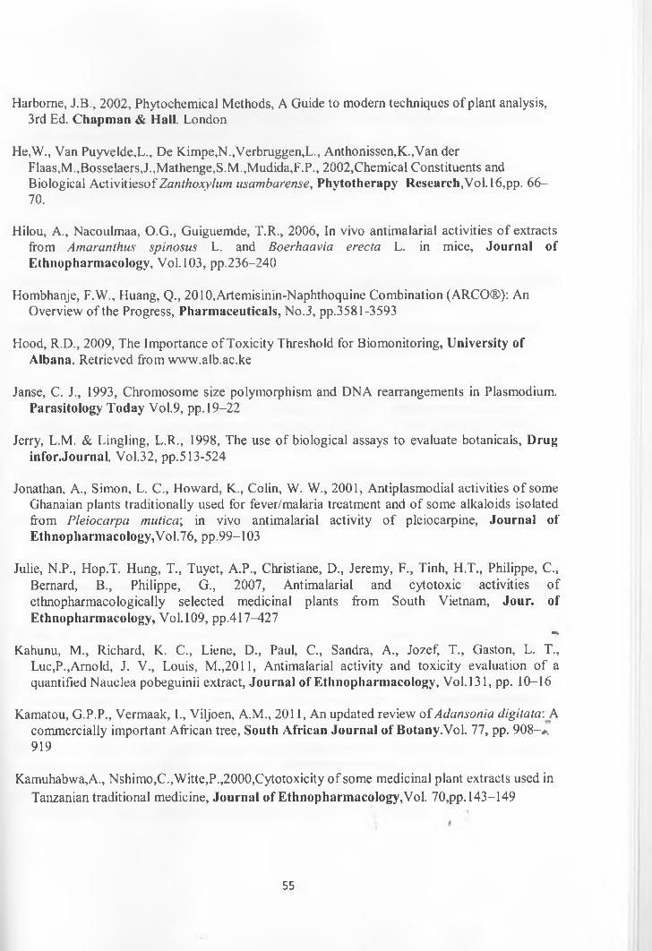
Harborne, J.B., 2002, Phytochemical Methods, A Guide to modern techniques o f plant analysis, 3rd Ed. Chapm an & Hall. London
He,W., Van Puyvelde,L., De Kimpe,N.,Verbruggen,L., Anthonissen,K.,Van derFlaas,M.,Bosselaers,J.,Mathenge,S.M.,Mudida,F.P., 2002,Chemical Constituents and Biological ActivitiesofZanthoxylum usambarense, Phytotherapy Research,Vol. 16,pp. 66- 70.
Hilou, A., Nacoulmaa, O.G., Guiguemde, T.R., 2006, In vivo antimalarial activities o f extracts from Amaranthus spinosus L. and Boerhaavia erecta L. in mice, Journal of Ethnopharmacology, Vol. 103, pp.236-240
Hombhanje, F.W., Huang, Q., 2010,Artemisinin-Naphthoquine Combination (ARCO®): An Overview of the Progress, Pharmaceuticals, No.3, pp.3581-3593
Hood, R.D., 2009, The Importance of Toxicity Threshold for Biomonitoring, University of Albana. Retrieved from www.alb.ac.ke
Janse, C. J., 1993, Chromosome size polymorphism and DNA rearrangements in Plasmodium. Parasitology Today Vol.9, pp. 19-22
Jerry, L.M. & Lingling, L.R., 1998, The use of biological assays to evaluate botanicals, Drug infor.Journal, Vol.32, pp.513-524
Jonathan, A., Simon, L. C., Howard, K., Colin, W. W., 2001, Antiplasmodial activities o f some Ghanaian plants traditionally used for fever/malaria treatment and of some alkaloids isolated from Pleiocarpa mutica; in vivo antimalarial activity o f pleiocarpine, Journal of Ethnopharmacology,Vol.76, pp.99-103
Julie, N.P., Hop.T. Hung, T., Tuyet, A.P., Christiane, D., Jeremy, F., Tinh, H.T., Philippe, C., Bernard, B., Philippe, G., 2007, Antimalarial and cytotoxic activities ofethnopharmacologically selected medicinal plants from South Vietnam, Jour, of Ethnopharmacology, Vol. 109, pp.417-427
O b
Kahunu, M., Richard, K. C., Liene, D., Paul, C., Sandra, A., Jozef, T., Gaston, L. T., Luc,P.,Arnold, J. V., Louis, M.,2011, Antimalarial activity and toxicity evaluation of a quantified Nauclea pobeguinii extract, Journal of Ethnopharmacology, Vol.131, pp. 10-16
Kamatou, G.P.P., Vermaak, I., Viljoen, A.M., 2011, An updated review of Adansonia digitata. A commercially important African tree, South African Journal of Botany.Vol. 77, pp. 908-*, 919
Kamuhabwa,A., Nshimo,C., Witte,P.,2000,Cytotoxicity o f some medicinal plant extracts used in Tanzanian traditional medicine, Journal of Ethnopharmacology,Vol. 70,pp. 143-149
55
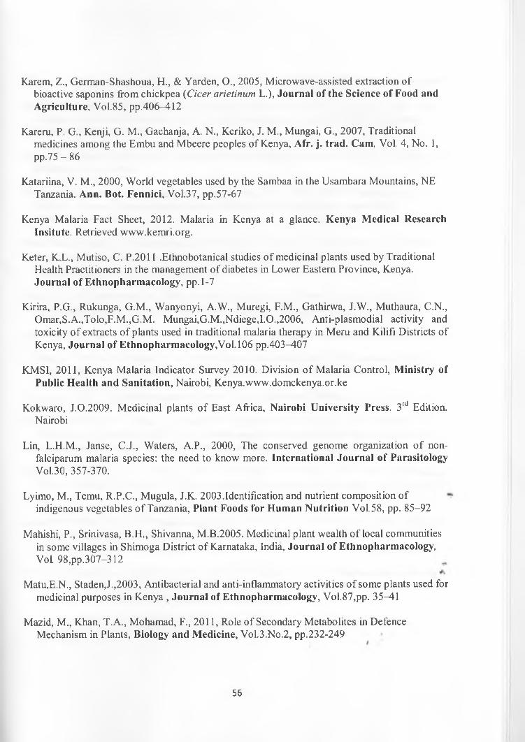
Karem, Z., German-Shashoua, H., & Yarden, O., 2005, Microwave-assisted extraction of bioactive saponins from chickpea (Cicer arietinum L.), Journal of the Science of Food and Agriculture, Vol.85, pp.406-412
Kareru, P. G., Kenji, G. M., Gachanja, A. N., Keriko, J. M., Mungai, G., 2007, Traditional medicines among the Embu and Mbeere peoples of Kenya, Afr. j. trad. Cam, Vol. 4, No. 1, pp.75 - 86
Katariina, V. M., 2000, World vegetables used by the Sambaa in the Usambara Mountains, NE Tanzania. Ann. Bot. Fennici, Vol.37, pp.57-67
Kenya Malaria Fact Sheet, 2012. Malaria in Kenya at a glance. Kenya Medical Research Insitute. Retrieved www.kemri.org.
Keter, K.L., Mutiso, C. P.2011 .Ethnobotanical studies of medicinal plants used by Traditional Health Practitioners in the management of diabetes in Lower Eastern Province, Kenya.Journal of Ethnopharmacology, pp. 1-7
Kirira, P.G., Rukunga, G.M., Wanyonyi, A.W., Muregi, F.M., Gathirwa, J.W., Muthaura, C.N., Omar,S.A.,Tolo,F.M.,G.M. Mungai,G.M.,Ndiege,I.O.,2006, Anti-plasmodial activity and toxicity o f extracts o f plants used in traditional malaria therapy in Meru and Kilifi Districts of Kenya, Journal of Ethnopharmacology,Vol. 106 pp.403-407
KMS1, 2011, Kenya Malaria Indicator Survey 2010. Division o f Malaria Control, Ministry of Public Health and Sanitation, Nairobi, Kenya.www.domckenya.or.ke
Kokwaro, J.0.2009. Medicinal plants of East Africa, Nairobi University Press. 3rd Edition. Nairobi
Lin, L.H.M., Janse, C.J., Waters, A.P., 2000, The conserved genome organization o f nonfalciparum malaria species: the need to know more. International Journal of Parasitology Vol.30, 357-370.
Lyimo, M., Temu, R.P.C., Mugula, J.K. 2003.Identification and nutrient composition of indigenous vegetables o f Tanzania, Plant Foods for Human Nutrition Vol.58, pp. 85-92
Mahishi, P., Srinivasa, B.H., Shivanna, M.B.2005. Medicinal plant wealth o f local communities in some villages in Shimoga District of Karnataka, India, Journal of Ethnopharmacology, Vol 98,pp.307-312
Matu,E.N., Staden,J.,2003, Antibacterial and anti-inflammatory activities o f some plants used for medicinal purposes in Kenya , Journal of Ethnopharmacology, Vol.87,pp. 35-41
Mazid, M., Khan, T.A., Mohamad, F., 2011, Role of Secondary Metabolites in Defence Mechanism in Plants, Biology and Medicine, Vol.3.No.2, pp.232-249
56

Menard, R., Sultan, A.A., Cortes, C., Altszuler, R., Janse, C.J., Waters, A.P., 1997, Nature 1997 385(6614) 336-40.
Meyer,B.N.,Ferrigin,N.R.,Putnam,J.E.,Jacobsen,L.S.,Nichols,D.E.,McLaughlin,J.L., 1982, Brine Shrimp: A convenient bioassay for active plant constituents, Journal of medicinal plant research,Vol.45,pp.31-34
Ministry of Health (MOH), 2010, National malaria strategy 2001-2010. Nairobi: Ministry of Health. Nairobi, Kenya.
Morgan, G.A., Leech, N.L., Gloeckner, G.W., Barrett, K.C., 2004, SPSS for Introductory Statistics, Use and Interpretation. Second Edition. Laurence Erlbaum Associates Publishers, London
Mota, M.M., Pradel, G., Vanderberg, J.P., Hafalla, J.C., Frevert, U., Nussenzweig, R.S,. 2001, Migration o f Plasmodium sporozoites through cells before infection. Science 2001,291(5501): 141-4.
Mudi, S.Y., & Muhammad, A., 2009, Antimalarial activity o f ethanolic extracts o f leaves of Terminalici catappa. L. Combretaceae (Indian almond), Bajopas, Vol. 2, No.l, pp. 14-18
Muganga, R„ Angenot, L., Tits, M., Frederich, M., 2010, Antiplasmodial and cytotoxic activitiesof
Mungai, G.M, Kofi-Tsekpo, W.M., 2009, Anti-plasmodial activity o f the extracts o f some Kenyan medicinal plants, Journal of Ethnopharmacology, Vol. 121, pp.282-285
Murray, M. 1995. The healing power of herbs. Prima Publishing. Rocklin, CA. pp. 162-171
Nagata, J.M., Alan, R. J., John, .K. Charles,R.S.,Elizabeth,A.B.,Craig,R.C.,2011, Medical pluralism on Mfangano Island: Use of medicinal plants among persons living with HIV/AIDS in Suba District, Kenya. Journal of Ethnopharmacology, Vol. 135, pp. 501-509
Nguta, J.M., Mbaria, J.M., Gakuya, D.W., Gathumbi, P.K., Kiama, S.G., 2010, Antimalarial herbal remedies ofMsambweni, Kenya, Journal of Ethnopharmacology, Vol.128, pp^24- 432
Nguta,J.M, Mbaria,J.M., Gakuya,D.W.,Gathumbi,P.K.,Kabasa,J.D.,Kiama,S.G.,2011, Biological Screening o f Kenya Medicial Plants using Artemia salina L. (Artemiidae), Pharmacologyonline ,Vol.2,pp.458-478
Olila, D., Olwa-Odyek., Opuda-Asibo.J., 2002, Screening of extracts of Zanthoxylum chalybeum and Warburgia ugandensis for activity against measles virus (Swartz and Edmonston strains) in vitro, African Health Sciences 2002, Vol.2, No.l, pp. 2-10
Peters, W., 1998, Drug resistance in malaria parasites of animals and man. Adv Parasitol.Vol.41, pp. 1-62. ,
57
Peters, W., Portus, J.H., Robinson, B.L., 1975, The chemotherapy o f rodent malaria XXII. The value of drug resistant strains of Plasmodium berghei in screening for schizontocidal activity. Ann. Trop. Med. Parasitol. 69: 155-71.
Phillips R.S., Brannan L.R., Balmer P, 1997, Antigenic variation during malaria infection: the contribution from the murine parasite Plasmodium chabaudi, Parasite lmmunol.Vol. 19:427- 34.
Raja, R.N., RamaLakshmi, S., Muthuchelian, K., 2011, GC-MS Analysis o f bioactivecomponents from the ethanolic leaf extract of Canthium dicoccum (Gaertn.), Teijsm & Binn., J. Chem. Pharm. Res., Vol. 3, pp.792-798
Ramadan, A., Harraz, F.M., EL-Mougy, S.A., 1994, Anti-inflamatory, analgesic and antipyretic effects of the fruit pulp o f Adansonia digitata, Fitoterapia, Vol. 65,No.5, pp.418-422
Ravindran,B.,Sharma,R.R.,Sharma,B.K.,Hussain,Q.Z., 1982, Circulating immune complexes in rodent and simian malaria, J.Biosci.,Vol.4,pp.491-498
Rhamat, A. K. ,2010, Phytochemical screening and pharmacological evaluation o f Sonchus asper and Launaea procumbens, Phd Thesis, Qauid-i-Azam University Islamabad, Pakistan
Robert, W. S., Carlos, A. G., Abdisalan, M. N., Hla, Y. M., Simon, I.H., 2005, The global distribution of clinical episodes o f Plasmodium falciparum malaria, Nature 2005, Vol.434, pp.214—217
Rukunga, G.M., Gathirwa, J.W., Omara, S.A., Muregi, F.W., Muthaura, C.N., Kirira, P.G., Rwandan medicinal plants used in the treatment of malaria, Journal of Ethnopharmacology, Vol. 128 pp.52-57
Saxena, S., Pant, N. Jain, D.C., Bhakuni, R.S., 2003, Antimalarial agents from plant sources, Current issue, Vol. 85, No. 9, pp. 1314-1329
Shahat, A.A.,2006, Procyanidins from Adansonia digitata, Pharmaceutical Biology, Vol. 44,No. 6, pp. 445-450
Shretta, R., Omumbo, J., Rapuoda, B., Snow, R.W. 2000. Snow Using evidence to change antimalarial drug policy in Kenya, Tropical Medicine and International Health, Vol.5 No. 11, pp. 755-764.
Sidibe, M., William, J.T., 2002, BAOBAB: Adansonia digitata L., International Center for Underutilized Crops, RPM Reprographics Chichester, England.
Sinden, R.E., 1978, Cell Biology. In: Rodent Malaria (R. Killick-Kendrick and W. Peters, eds.) pp 85-168.Academic Press, London.
58
Sulsen,V.,Gutierrez,D.Y., Laurella,L., Anesini,C., Gimenez,A.T.Martino,V.,Muschietti,L.,2011, In Vitro Antiplasmodial Activity o f Sesquiterpene Lactones from Ambrosia tenuifolia, Evidence-Based Complementary and Alternative Medicine, pp. 1-4
Takate, S.B., Pokharkar, R.D., Chopade, V.V., Gite, V.N. ,2010, Hepato-protective activity of the ethyl acetate extract o f Launaea intybacea (Jacq) Beauv in paracetamol induced hepato- toxicity in albino rats, Inter.Jour.Pharm.Sci.Vol. 1,No.2,pp.72-74
Tiwary, M., Naika, S.N., Kumar, D.T., Mittal, P.K., Yadav, S., 2007, Chemical composition and larvicidal activities o f the essential oil of Zanthoxylum armatum DC (Rutaceae) against three mosquito vectors, J Vect. Borne Dis, Vol.44, pp. 198-204
Tolu, O.O., Odunayo, R.A., Ibukun, E.A., Peter, O.F., 2007, Medicinal plants useful for malaria therapy in Okeigbo, Ondo state, southwest Nigeria, Afr. J. Trad. CAM, Vol. 4, No.2, pp. 191- 198
UNICEF, 2007, Malaria and Children; Progress in intervention coverage, United Nations Children Fund, New York.USA
Waako, P.J., Gumede, B., Smith, P., Folb, P.I., 2005, The in vitro and in vivo antimalarial activity o f Cardiospermum halicacabum L. and Momordica foetida Schumch, Et Thonn., Journal of Ethnopharmacology, Vol.99, pp. 137-143
Waksmundzka, H.M., Sherma, J., Kowalska, T., 2008, Thin Layer Chromatography in Phytochemistry, Chromatography Science Series.Vol.99.CRC Press
Wambugu, S.N., Mathiu, P.M., Gakuya, D.W., Kanui, T.I., Kabasa, J.D., Kiama, S.G.,2011, Medicinal plants used in the management of chronic joint pains in Machakos and Makueni counties, Journal of Ethnopharmacology Vol. 137, pp. 945-955
Wang,Y.F.,Ni,Z.Y.,Dong,M.,Cong,B.,Shi,W.Q.,Gu,Y.C.,Kiyota,H.,2010, Secondary Metabolites of Plants from the Genus Saussurea; Chemistry and Activity, Chemistry and Biodiversity, Vol.7, pp.2623-2659
Wanyoike, G.N., Chabra, S.C., Lang’at, C.C., Omar, S.A.,2004, Brine shrimp Jpxicity and antiplasmodial activity o f five Kenyan medicinal plants, Journal of Ethnopharmacology, Vol.90, pp. 129-133
Waters, A.P., Higgins, D.G., McCutchan, T.F.1991. Plasmodium falciparum appears to have arisen as a result o f lateral transfer between avian and human hosts. Proc Natl Acad Sci USA. Vol.88, No.8, pp.3140-3144. *
WHO, 2004, the Global Fund, and medical malpractice in malaria Treatment, Lancet, No.363, pp.237-40. Retrieved from www.who.org
59
WHO. 2001. Drug Resistance to Malaria. World Health Organization. Retrieved from www.who.org.
Worral, E., Basu, S., Hanson, K., 2003, The relationship between social economic status and malaria; a review of the literature, SES and Malaria, LSHTM, London.UK.
Ziegler, H.L., Staerk.D., Christensen, J., Hviid,L., Hagerstrand,H., Jaroszewsk, Z.W., 2002, In Vitro Plasmodium falciparum Drug Sensitivity Assay: Inhibition o f Parasite Growth by Incorporation o f Stomatocytogenic Amphiphiles into the Erythrocyte Membrane, Antimicrobial Agents and Chemotherapy, Vol.46, No.5, pp. 1441-1446
60

7. APPENDICES
Appendix 1: Calculation of Parasitaemia and Chemosuppression
CONTROLSNe gative Control Percentage
Parasitaemia
Percentage
ChemosuppressionMice
MF1 MF2 MF2 MF2 TOTALM l Parasitized Cells(PCs) 49 90 74 68 281 33.73 N/A
Total Cells(TCs) 174 267 194 198 833
M2 PCs 90 65 51 82 288 33.41 N/ATCs 255 204 177 226 862
M3 PCs 63 73 69 69 274 28.42 N/ATCs 239 265 253 207 964
M4 PCs 55 62 60 65 242 30.06 N/ATCs 227 168 203 207 805
MS PCs 75 67 72 71 285 33.8 N/ATCs 235 202 192 214 843AVERAGE 31.88 0
Positive Control Percentage
Parasitaemia
Percentage
ChemosuppressionMice
MF1 MF2 MF2 MF2 TOTALM l Parasitized Cells(PCs) 9 11 12 5 37 4.45 86.04
Total Cells(TCs) 210 207 216 198 831M2
M2 PCs 2 0 2 4 8 1.18 96.3TCs 181 170 173 155 679
M3M3 PCs 10 5 2 8 25 3.93 87.69
TCs 166 159 142 170 637M4
M4 PCs 20 6 18 12 56TCs 241- 189 214 201 845 6.23 79.21
M5M5 PCs 13 7 12 10 42 A
TCs 200 264 294 249 1007 4.17 86.92AVERAGE 4.07 87.23
/
61
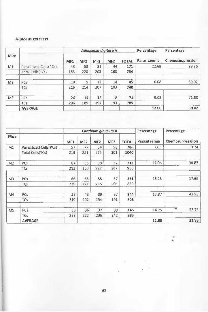
Aqueous extracts
Adansonia digitata A Percentage
Parasitaemia
Percentage
ChemosuppressionMice
MF1 MF2 MF2 MF2 TOTALM l Parasitized Cells(PCs) 43 53 31 44 171 22.68 28.86
Total Cells(TCs) 163 220 203 168 754
M2 PCs 10 9 12 14 45 6.08 80.92TCs 216 214 207 103 740
M3 PCs 26 14 13 18 71 9.05 71.63TCs 206 189 197 193 785AVERAGE 12.60 60.47
Canthium glaucum A Percentage
Parasitaemia
Percentage
ChemosuppressionMice
MF1 MF2 MF2 MF2 TOTALM l Parasitized Cells(PCs) 57 77 54 98 286 27.5 13.74
Total Cells(TCs) 213 251 275 301 1040
M2 PCs 67 56 38 52 213 22.05 30.83TCs 212 260 227 267 966
M3 PCs 66 53 55 57 231 26.25 17.66TCs 239 221 215 205 880
M4 PCs 25 43 39 37 144 17.87 43.95TCs 219 202 194 191 806
M5 PCs 33 36 37 39 145 14.75 ~ 53.73TCs 283 222 236 242 983AVERAGE 21.68 31.98
I
62
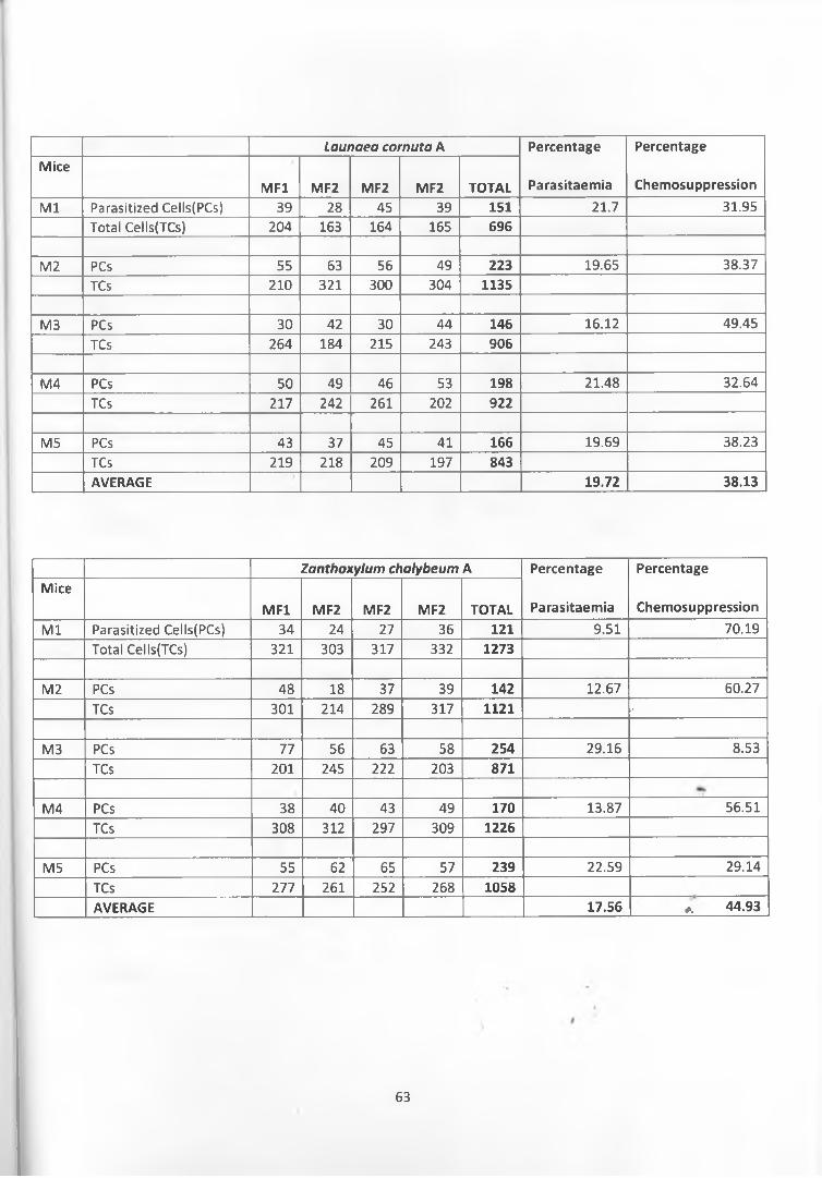
Launaea cornuta A Percentage
Parasitaemia
Percentage
ChemosuppressionMice
MF1 MF2 MF2 MF2 TOTALM l Parasitized Cells(PCs) 39 28 45 39 151 21.7 31.95
Total Cells(TCs) 204 163 164 165 696
M2 PCs 55 63 56 49 223 19.65 38.37TCs 210 321 300 304 1135
M3 PCs 30 42 30 44 146 16.12 49.45TCs 264 184 215 243 906
M4 PCs 50 49 46 53 198 21.48 32.64TCs 217 242 261 202 922
M5 PCs 43 37 45 41 166 19.69 38.23TCs 219 218 209 197 843AVERAGE 19.72 38.13
Zanthoxylum chalybeum A Percentage
Parasitaemia
Percentage
ChemosuppressionMice
MF1 MF2 MF2 MF2 TOTALM l Parasitized Cells(PCs) 34 24 27 36 121 9.51 70.19
Total Cells(TCs) 321 303 317 332 1273
M2 PCs 48 18 37 39 142 12.67 60.27TCs 301 214 289 317 1121 *
M3 PCs 77 56 63 58 254 29.16 8.53TCs 201 245 222 203 871
M4 PCs 38 40 43 49 170 13.87 56.51TCs 308 312 297 309 1226
M5 PCs 55 62 65 57 239 22.59 29.14TCs 277 261 252 268 1058AVERAGE 17.56 a 44.93
63

CHCh: MeOH extracts
Adansonia d gitata B Percentage PercentageMice
MF1 MF2 MF2 MF2 TOTAL Parasitaemia ChemosuppressionM l Parasitized Cells(PCs) 57 49 66 41 213 26.96 15.43
Total Cells(TCs) 152 221 237 180 790M2
M2 PCs 80 62 60 86 288 24.53 23.06TCs 319 260 283 312 1174
M3M3 PCs 84 86 81 77 328 30.54 4.2
TCs 292 280 269 233 1074M4
M4 PCs 60 64 71 70 265 20.75 34.91TCs 304 322 309 342 1277
M5M5 PCs 13 7 12 10 42 4.17 86.92
TCs 200 264 294 249 1007AVERAGE 21.39 32.90
Canthium glaucum B Percentage
Parasitaemia
Percentage
ChemosuppressionMice
MF1 MF2 MF2 MF2 TOTALM l Parasitized Cells(PCs) 81 50 50 39 220 21.63 32.15
Total Cells(TCs) 327 233 228 229 1017M2
M2 PCs 50 48 35 50 183 22.56 29.23TCs 211 197 180 223 811
M34*.
M3 PCs 34 31 42 38 145 17.22 45.98TCs 205 201 192 244 842
M4M4 PCs 23 15 14 12 64 8.65 72.87
TCs 179 189 181 191 740M5 ^ 4
M5 PCs 80 62 41 29 212 19.58 38.58TCs 341 248 217 277 1083AVERAGE 17.39 43.76
i
64
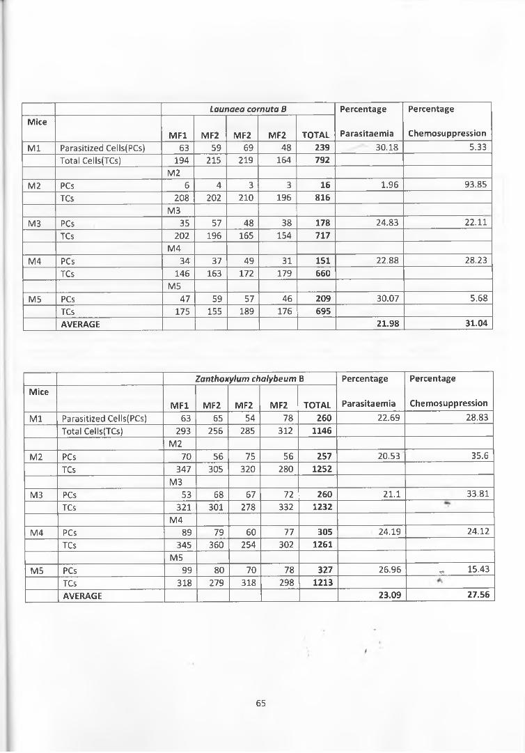
Launaea cornuta B Percentage PercentageMice
MF1 MF2 MF2 MF2 TOTAL Parasitaemia Chemosuppression
M l Parasitized Cells(PCs) 63 59 69 48 239 30.18 5.33Total Cells(TCs) 194 215 219 164 792
M2M2 PCs 6 4 3 3 16 1.96 93.85
TCs 208 202 210 196 816M3
M3 PCs 35 57 48 38 178 24.83 22.11TCs 202 196 165 154 717
M4M4 PCs 34 37 49 31 151 22.88 28.23
TCs 146 163 172 179 660M5
M5 PCs 47 59 57 46 209 30.07 5.68TCs 175 155 189 176 695AVERAGE 21.98 31.04
Zanthoxylum chalybeum B Percentage PercentageMice
MF1 MF2 MF2 MF2 TOTAL Parasitaemia Chemosuppression
M l Parasitized Cells(PCs) 63 65 54 78 260 22.69 28.83Total Cells(TCs) 293 256 285 312 1146
M2M2 PCs 70. 56 75 56 257 20.53 35.6
TCs 347 305 320 280 1252M3
M3 PCs 53 68 67 72 260 21.1 33.81
TCs 321 301 278 332 1232M4
M4 PCs 89 79 60 77 305 24.19 24.12
TCs 345 360 254 302 1261M5
M5 PCs 99 80 70 78 327 26.96 „ 15.43
TCs 318 279 318 298 1213AVERAGE 23.09 27.56
65
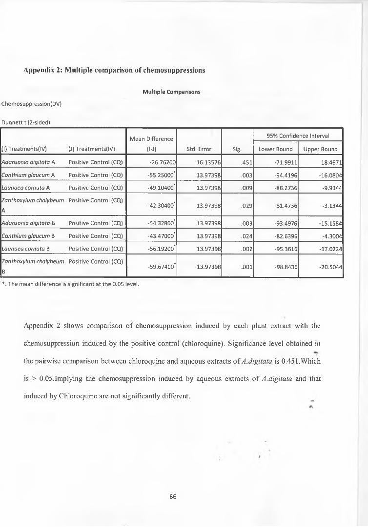
Appendix 2: Multiple comparison of chemosuppressions
Chemosuppression(DV)
Multiple Comparisons
Dunnett t (2-sided)
Mean Difference 95% Confidence Interval
(1) Treatments(IV) (J) Treatments(IV) (l-J) Std. Error Sig. Lower Bound Upper Bound
A d a n s o n ia d ig i t a t a A Positive Control (CQ) -26.76200 16.13576 .451 -71.9911 18.4671
C a n t h iu m g la u c u m A Positive Control (CQ) -55.25000* 13.97398 .003 -94.4196 -16.0804
L a u n a e a c o r n u t a A Positive Control (CQ) -49.10400* 13.97398 .009 -88.2736 -9.9344
Z a n th o x y lu m c h a ly b e u m
A
Positive Control (CQ)-42.30400* 13.97398 .029 -81.4736 -3.1344
A d a n s o n ia d ig it a t a B Positive Control (CQ) -54.32800* 13.97398 .003 -93.4976 -15.1584
C a n t h iu m g la u c u m B Positive Control (CQ) -43.47000* 13.97398 .024 -82.6396 -4.3004
L a u n a e a c o r n u t a B Positive Control (CQ) -56.19200* 13.97398 .002 -95.3616 -17.0224
Z a n th o x y lu m c h a ly b e u m
B
Positive Control (CQ)-59.67400* 13.97398 .001 -98.8436 -20.5044
*. The mean difference is significant at the 0.05 level.
Appendix 2 shows comparison o f chemosuppression induced by each plant extract with the
chemosuppression induced by the positive control (chloroquine). Significance level obtained in
the pairwise comparison between chloroquine and aqueous extracts o f A.digitata is 0.451.Which
is > 0.05.Implying the chemosuppression induced by aqueous extracts of A.digitata and that
induced by Chloroquine are not significantly different.
66
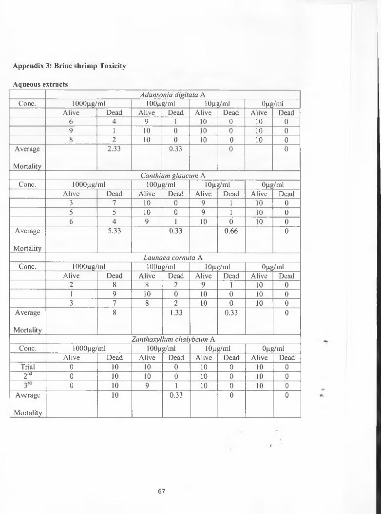
Appendix 3: Brine shrimp Toxicity
Aqueous extractsAdansonia digitata A
Cone. lOOOpg/ml lOOpg/ml lOp jg/ml Opg/mlAlive Dead Alive Dead Alive Dead Alive Dead
6 4 9 1 10 0 10 09 1 10 0 10 0 10 08 2 10 0 10 0 10 0
Average
Mortality
2.33 0.33 0 0
Canthium glaucum ACone. lOOOpg/ml lOOpg/ml 10p g/ml Opg/ml
Alive Dead Alive Dead Alive Dead Alive Dead3 7 10 0 9 1 10 05 5 10 0 9 1 10 06 4 9 1 10 0 10 0
Average
Mortality
5.33 0.33 0.66 0
Launaea cornuta ACone. lOOOpg/ml lOOpg/ml lOp g/ml Opg/ml
Alive Dead Alive Dead Alive Dead Alive Dead2 8 8 2 9 1 10 01 9 10 0 10 0 10 03 7 8 2 10 0 10 0
Average
Mortality
8 1.33 0.33 0
Zanthoxyllum chalybeum ACone. lOOOpg/ml 3.
Oo
g/ml lOp g/ml Opg/mlAlive Dead Alive Dead Alive Dead Alive Dead
Trial 0 10 10 0 10 0 10 02nd 0 10 10 0 10 0 10 0
0 10 9 1 10 0 10 0Average
Mortality
10 0.33 0 0
67
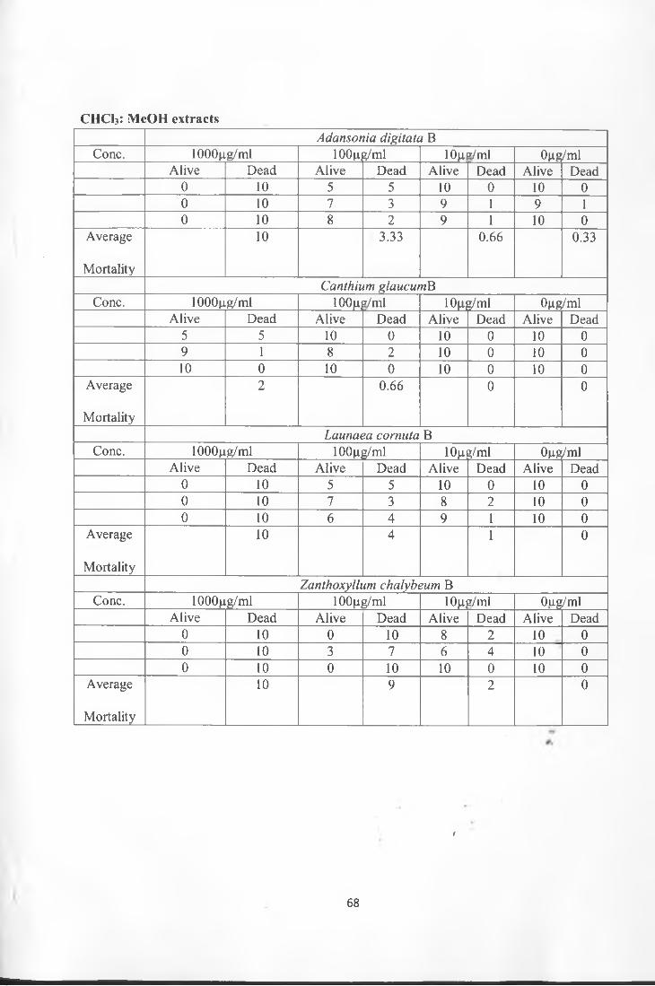
CHCb: MeOH extractsAdansonia digitata B
Cone. 1000ng/ml 100m?/ml 10n */ml Qng/mlAlive Dead Alive Dead Alive Dead Alive Dead
0 10 5 5 10 0 10 00 10 7 3 9 1 9 10 10 8 2 9 1 10 0
Average
Mortality
10 3.33 0.66 0.33
Canthium glaucumBCone. 1000|ig/ml lOOfij»/ml low?/ml Qgg/ml
Alive Dead Alive Dead Alive Dead Alive Dead5 5 10 0 10 0 10 09 1 8 2 10 0 10 010 0 10 0 10 0 10 0
Average
Mortality
2- 0.66 0 0
Launaea cornuta BCone. 1000gg/ml 100(ig/ml 10Ms/ml Qgg/ml
Alive Dead Alive Dead Alive Dead Alive Dead0 10 5 5 10 0 10 00 10 7 3 8 2 10 00 10 6 4 9 1 10 0
Average
Mortality
10 4 1 0
Zanthoxyllum chalybeum BCone. 1000fj.g/ml 100|ig/ml iow2/ml Oug/ml
Alive Dead Alive Dead Alive Dead Alive Dead0 10 0 10 8 2 10 00 10 3 7 6 4 10 00 10 0 10 10 0 10 0
Average
Mortality
10 9 2 0
t
68
fe