Phytochemical Screening & in vitro Antioxidant and ...
110
Phytochemical Screening & in vitro Antioxidant and Thrombolytic Activities of Aphanamixis polystachya (Wall.) Parker Leaf Extracts Submitted by: Ferdous Ara ID: 2010-3-79-030 Research Supervisor: Mr. Apurba Sarker Apu, Senior Lecturer. Department of Pharmacy East West University A thesis report, submitted to the Department of Pharmacy, East West University, in partial fulfillment of the requirements for the degree of Masters of Clinical Pharmacy and Molecular Pharmacology
-
Upload
khangminh22 -
Category
Documents
-
view
1 -
download
0
Transcript of Phytochemical Screening & in vitro Antioxidant and ...

Phytochemical Screening & in vitro Antioxidant and
Thrombolytic Activities of Aphanamixis polystachya (Wall.)
Parker Leaf Extracts
Submitted by: Ferdous Ara
ID: 2010-3-79-030
Research Supervisor: Mr. Apurba Sarker Apu, Senior Lecturer.
Department of Pharmacy
East West University
A thesis report, submitted to the Department of Pharmacy, East West
University, in partial fulfillment of the requirements for the degree of Masters
of Clinical Pharmacy and Molecular Pharmacology

RESEARCH REPORT ON
“Phytochemical Screening and in vitro Antioxidant
and Thrombolytic Activities of Aphanamixis
polystachya (Wall.) Parker Leaf Extracts”

DECLARATION BY THE CANDIDATE
It is my pleasure to submit the Research Report. The report is prepared on “Phytochemical
Screening and in vitro Antioxidant and Thrombolytic Activities of Aphanamixis polystachya
(Wall.) Parker Leaf Extracts” which was carried out by me under the guidance of Mr. Apurba
Sarker Apu, Senior lecturer, Department of Pharmacy, East West University, Dhaka. I have
given my best effort in preparing this report and to make it a worthy one. I have enjoyed working
on the report and gained some experience.
…………….
Ferdous Ara
ID: 2010-3-79-030
Department of Pharmacy
East West University, Dhaka.

CERTIFICATE BY THE INVIGILATOR
This is to certify that the dissertation, entitled “Phytochemical Screening and in vitro Antioxidant
and Thrombolytic Activities of Aphanamixis polystachya (Wall.) Parker Leaf Extracts” is a
bonafide research work done by Ferdous Ara, in partial fulfillment of the requirement for the
Degree of Masters of Clinical Pharmacy and Molecular Pharmacology.
……………………….
Mr. Apurba Sarker Apu
Senior Lecturer
Department of Pharmacy
East West University, Dhaka.

ENDORSEMENT BY THE CHAIRPERSON
This is to certify that the dissertation, entitled “Phytochemical Screening and in vitro Antioxidant
and Thrombolytic Activities of Aphanamixis polystachya (Wall.) Parker Leaf Extracts” is a
bonafide research work done by Ferdous Ara, under the guidance of Mr. Apurba Sarker Apu,
Senior Lecturer, Department of Pharmacy, East West University, Dhaka.
……………….
Dr. Sufia Islam
Chairperson and Associate Professor
Department of Pharmacy
East West University, Dhaka.

Acknowledgement
Above all, I express my gratitude to Almighty Allah for giving me the strength, energy
and patients to carry out this research work.
I would like to express my gratitude and admiration to Mr. Apurba Sarker Apu, Senior
Lecturer, Department of Pharmacy, East West University, for his suggestion, careful guidance,
sincere help, constructive criticism and valuable time without which I would not have been able
to complete this work. He had been very enthusiastic and supportive in my research.
I would like to extend my cordial gratitude to Dr. Sufia Islam, Chairperson and Associate
Professor of the Department of Pharmacy, East West University, for her constant inspiration and
whole hearted cooperation.
I would also like to take the opportunity to express my whole hearted gratitude to my
fellow researchers friends, near and dear ones who offered encouragement, information,
inspiration and assistance during the period of constructing the research report.
I am thankful to the laboratory instructors for their kind support during the laboratory
work.
I express my sincere gratitude to my caring parents for their support, inspiration and
guiding me all through my life, including that for my research project. I am very grateful to my
sisters who encouraged me enormously.

This Research paper is dedicated to
My beloved parents and sisters

TABLE OF CONTENTS
CHAPTER 1: INTRODUCTION
List of Contents Page
No.
Introduction 1-3
Plant Profile 3
Common name 3
Botanical name 3
Synonyms 3
Trade names 4
Scientific classification 4
Distributional Range 4
Native distribution 5
Botanical Descriptions of A. polystachya 5
Habit 5
Foliage 6
Plant shape 6
Trunk & bark 6
Branches and branchlets 6
Leaves 6-7
Inflorescence / Flower 7
Flowering 8
Fruit and seed 8
Type of stem 8
Wood 8
Roots 9
Plant utilities 9
Season 9

Ecology 9
Major Threat 9
Uses of A. polystachya 9
1. Medicinal uses 9
2. Common uses 10
Solvent System 10
1. n-hexane 10
Applications 10-11
2. Ethyl acetate 11
Applications 11-12
3. Methanol 12
Applications 12-13
Phytochemical Screening 13-17
Antioxidants 17
Free radical formation by oxygen 17-18
Problem of free radical 19
DPPH free radical scavenging assay 19-20
Nitric oxide scavenging 20-21
Estimation of total phenolic content 21-22
Thrombolytic Activity 22-25
Purpose of the Present Study 25
CHAPTER 2: LITERATURE REVIEW
List of contents Page No.
2.1 Phytochemical Studies 26-31
2.2 Pharmacological Studies 31-39

CHAPTER 3: MATERIALS & METHODS
List of Contents Page No.
Collection and Identification of Plant Material 40
Materials 41
Method 41
Extraction of plant material 41-42
Condensation of the leaf extracts 42-43
Screening of Phytochemical Constituents 44
Test for alkaloids: Hager's test 44
Materials 44
Method 44
Preparation of 2% H2SO4 solution 44
Preparation of Hager´s reagent 44
Procedure 44-45
Test for flavonoids: Ammonia test (modified) 45
Materials 45
Method 45
Preparation of 5% ammonia solution 45-46
Procedure 46
Test for steroids: Salkowski test 46
Materials 46
Method 47
Procedure 47
Test for terpenoids: Salkowski test (modified) 47
Materials 47
Method 47
Procedure 48
Test for carbohydrates: Fehling's (Reducing sugar) test (modified) 48
Materials 48
Method 48

Preparation of Fehling I solution 48-49
Preparation of Fehling II solution 49
Preparation of Fehling solution 49
Procedure 49
Test for saponins: Frothing test 49
Materials 49
Method 49
Procedure 50
Test for tannins: FeCl3 test 50
Materials 50
Method 51
Preparation of 0.1% ferric chloride solution 51
Procedure 51
Test for cardiac glycosides: Killer-Killani's test 51
Materials 51-52
Method 52
Procedure 52
Test for anthraquinones: Chloroform layer test 52
Materials 53
Method 53
Procedure 53
Antioxidant Test with DPPH 53
Materials 53
Method 54
Preparation of DPPH reagent 54
Preparation of extract solution 54
Procedure 54-55
Nitric Oxide Scavenging Capacity Assay 55
Materials 55
Method 55
Preparation of 5 mM solution of sodium nitroprusside 55-56

Preparation of Griss reagent 56
Preparation of stock of extract solution 56
Procedure 56-57
Estimation of Total Phenolic Content 57
Materials 57
Method 58
Preparation of solutions of plant extracts 58
Preparation of 10% Folin-Ciocalteu phenol reagent 58
Preparation of 700 mM Na2CO3 solution 58
Procedure 58-59
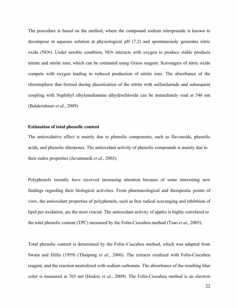
In Vitro Thrombolytic Activity Test 59
Materials 59-60
Method 60
Preparation of stock 60
Preparation of extracts solutions 60
Preparation of blood sample 60-61
Procedure 61
Statistical analysis 62
CHAPTER 4: RESULTS
List of Contents Page No.
Phytochemical Screening 63
DPPH Free Radical Scavenging Assay 64-69
Nitric Oxide Free Radical Scavenging of Leaf of A. polystachya 69-74
Total Phenolic Content 75-76
In vitro Thrombolytic Activity 76-78

CHAPTER 5: DISCUSSION
DISCUSSION 79
Phytochemical Screening 79
Antioxidant Activity 79
Thrombolytic Activity 80
CHAPTER 6: CONCLUSION
CONCLUSION 81
REFERENCES 82-92
LIST OF TABLES
Table No. Title Page No.
Table 1 Species, local name and local distribution of A. polystachya
available in Bangladesh
5
Table 2 Physical and chemical properties of n-hexane 11
Table 3 Physical and chemical properties of ethyl acetate 12
Table 4 Physical and chemical properties of methanol 13
Table 5 Methods for identification the chemical constituents 15-16
Table 6 Some popular methods used for the detection of various
phytochemicals
16-17
Table 7 Summary of the Phytochemical Studies on A. polystachya 31
Table 8 Summary of the pharmacological studies on A. polystachya 39
Table 9 Name of chemicals, equipments and glass apparatus for extraction 41
Table 10 Time and temperature of extraction 41
Table 11 Temperature, rpm and time of evaporation 43
Table 12 Characteristic of A. polystachya 43

Table 13 Name of chemicals, equipments and glass apparatus for alkaloid test 44
Table 14 Name of chemicals, equipments and glass apparatus for flavonoid
test
45
Table 15 Name of chemicals, equipments and glass apparatus for steroid test 46
Table 16 Name of chemicals, equipments and glass apparatus for terpenoid
test
47
Table 17 Name of chemicals, equipments and glass apparatus for
carbohydrate test
48
Table 18 Name of chemicals, equipments and glass apparatus for saponin test 49
Table 19 Name of chemicals, equipments and glass apparatus for tannin test 50
Table 20 Name of chemicals, equipments and glass apparatus for cardiac
glycoside test
52
Table 21 Name of chemicals, equipments and glass apparatus for
anthraquinone test
53
Table 22 Name of chemicals, equipments and glass apparatus for DPPH assay 53
Table 23 Name of chemicals, equipments and glass apparatus for nitric oxide
scavenging assay
55
Table 24 Name of chemicals, equipments and glass apparatus for the
estimation of total phenolic content
57
Table 25 Composition of Folin-Ciocalteu Reagent 58
Table 26 Name of chemicals, equipments and glass apparatus for
thrombolytic activity test
59-60
Table 27 Data of phytochemical screening of A. polystachya 63
Table 28 IC50 value of ascorbic acid (AA) 64
Table 29 IC50 value of the leaf of APHE 65
Table 30 IC50 value of the leaf of APEA 66
Table 31 IC50 value of the leaf of APME 67
Table 32 % inhibition of APHE, APEA, APME and AA 68
Table 33 IC50 value of ascorbic acid (AA) 69
Table 34 IC50 value of the leaf of APHE 70
Table 35 IC50 value of the leaf of APEA 71
Table 36 IC50 value of the leaf of APME 72
Table 37 % inhibition of APHE, APEA, APME and AA 73
Table 38 Absorbance of gallic acid 75

Table 39 Content of total phenol of the extracts of A. polystachya leaves 76
Table 40 % Thrombolysis of APHE, APEA, APME, streptokinase and normal
saline
76
Table 41 Clot lysis of blood sample data of extracts of A. polystachya fruit
and streptokinase
77
LIST OF FIGURES
Figure No. Name of the Figure Page No.
Figure 1 Habit 6
Figure 2 Bark 6
Figure 3 Branch 6
Figure 4 Shoot apex 6
Figure 5 Rachis and leaf insertion 6
Figure 6 Petiolule insertion 7
Figure 7 Terminal leaflets 7
Figure 8 Leaflet lower side 7
Figure 9 Leaflet upper side 7
Figure 10 Venation 7
Figure 11 Flowers 8
Figure 12 Dehisced fruit 8
Figure 13 Fruit insertion 8
Figure 14 Mechanism of anticoagulation of streptokinase 23
Figure 15 Identification plate of A. polystachya 40
Figure 16 Soxhlet apparatus 42
Figure 17 Extracts (before evaporation) 42
Figure 18 Rotary evaporator 42
Figure 19 Extracts (after evaporation) 43
Figure 20 Identification of alkaloids by Hager’s reagent 45
Figure 21 Identification of flavonoids by ammonia test 46
Figure 22 Identification of steroids by salkowski test 47

Figure 23 Identification of terpenoids by salkowski test 48
Figure 24 Identification of saponins by frothing test 50
Figure 25 Identification of tannins by FeCl3 test 50
Figure 26 Identification of cardiac glycosides by Killer-Killani's test 52
Figure 27 DPPH reagent added in extracts 55
Figure 28 UV-VIS spectrophotometer 55
Figure 29 Extract Solutions 57
Figure 30 Reagent added in extracts solution 59
Figure 31 Saline taken 60
Figure 32 Saline added in extracts 60
Figure 33 Clot formation 61
Figure 34 Serum withdrowing 61
Figure 35 Extract added in clot 62
Figure 36 After clot lysis serum withdrowing 62
Figure 37 % inhibition curve of ascorbic acid (AA) 64
Figure 38 Scavenging effects of the leaf of APHE 65
Figure 39 Scavenging effects of the leaf of APEA 66
Figure 40 Scavenging effects of the leaf of APME 67
Figure 41 DPPH radical scavenging activity of AA and the APHE, APEA &
APME extracts of A. polystachya leaf
68
Figure 42 % inhibition curve of Ascorbic acid (AA) 70
Figure 43 Scavenging effects of the leaf of APHE 71
Figure 44 Scavenging effects of the leaf of APEA 72
Figure 45 Scavenging effects of the leaf of APME 73
Figure 46 NO scavenging activity of AA and the APHE, APEA & APME
extracts of A. polystachya leaf
74
Figure 47 Standard curve of gallic acid 75
Figure 48 Clot lysis of blood samples of normal subjects by APHE, APEA,
and APME extracts of A. polystachya leaves, streptokinase and
normal saline
77

ABSTRACT
Purpose: In present study the leaves extracts of Aphanamixis polystachya (Meliaceae) have been
screened for their photochemical constituents, antioxidant, total phenolic content and in vitro
thrombolytic activities.
Methods: The n-hexane, ethyl acetate and methanol extracts were screened for the presence of
phytochemicals, using in vitro assay methods and their inhibition of 2,2-diphenyl-1-picryl-
hydrazyl (DPPH) free radical was used to evaluate their free radical scavenging activity at 517
nm. Nitric oxide scavenging capacity was measured by sodium nitroprusside and griss reagent.
The total phenolic content (TPC) measured by the Folin-Ciocalteu method. An in vitro
thrombolytic model was used to check out the clot lysis effects of three extracts using
streptokinase as a positive control.
Results: Phytochemical screening of the plant showed the presence of alkaloids, flavonoids,
terpenoids, saponins, tannins, cardiac glycosides and anthraquinones. All the extracts inhibited
DPPH, indicating their antioxidant activity. In DPPH and nitric oxide scavenging assay both n-
hexane extract showed highest IC50 values 74.84 µg/ml, 76.15 µg/ml and the lowest IC50 value
was 12.54 µg/ml of methanol extract and 39.90 µg/ml of ethylacetate extract. It appears that, it
has greater free radical scavenging capacity. Ethyl acetate extract had the highest total phenolic
content of 107.77 mg GAE/100 g extract. All the extracts showed significant % of clot lysis
effect (p<0.001) with reference to negative control.
Conclusion: The outcomes of the study are an indication of present of phytochemicals and may
be responsible for some of the therapeutic uses of these plants.
Keywords: Aphanamixis polystachya, Phytochemical screening, DPPH, Nitric oxide, Phenolic
content, In vitro Thrombolytic.

1
ABSTRACT
Purpose: In present study the leaves extracts of Aphanamixis polystachya (Meliaceae) have been
screened for their photochemical constituents, antioxidant, total phenolic content and in vitro
thrombolytic activities.
Methods: The n-hexane, ethyl acetate and methanol extracts were screened for the presence of
phytochemicals, using in vitro assay methods and their inhibition of 2,2-diphenyl-1-picryl-
hydrazyl (DPPH) free radical was used to evaluate their free radical scavenging activity at 517
nm. Nitric oxide scavenging capacity was measured by sodium nitroprusside and griss reagent.
The total phenolic content (TPC) measured by the Folin-Ciocalteu method. An in vitro
thrombolytic model was used to check out the clot lysis effects of three extracts using
streptokinase as a positive control.
Results: Phytochemical screening of the plant showed the presence of alkaloids, flavonoids,
terpenoids, saponins, tannins, cardiac glycosides and anthraquinones. All the extracts inhibited
DPPH, indicating their antioxidant activity. In DPPH and nitric oxide scavenging assay both n-
hexane extract showed highest IC50 values 74.84 µg/ml, 76.15 µg/ml and the lowest IC50 value
was 12.54 µg/ml of methanol extract and 39.90 µg/ml of ethylacetate extract. It appears that, it
has greater free radical scavenging capacity. Ethyl acetate extract had the highest total phenolic
content of 107.77 mg GAE/100 g extract. All the extracts showed significant % of clot lysis
effect (p<0.001) with reference to negative control.
Conclusion: The outcomes of the study are an indication of present of phytochemicals and may
be responsible for some of the therapeutic uses of these plants.
Keywords: Aphanamixis polystachya, Phytochemical screening, DPPH, Nitric oxide, Phenolic
content, In vitro Thrombolytic.

2
INTRODUCTION
Throughout the ages, humans have relied on nature for their basic needs for the production of
foodstuffs, shelter, clothing, means of transportation, fertilizers, flavors and not least, medicines.
Since origin of human’s life, plants continue to play a curative and therapeutic role in preserving
human health against disease and decay (Nair et al., 2005). Medicinal plants have been used in
all cultures as a source of medicine, since times immemorial. Herbal Medicine is still the
mainstay of health care in several developing countries. The widespread use of herbal remedies
and health care preparations, as those described in ancient texts like the Vedas and the Bible, and
obtained from commonly used traditional herbs and medicinal plants, has been traced to the
occurrence of natural products with medicinal properties (Nair et al., 2005).
The use of herbal medicine has become increasingly popular worldwide and medicinal plants are
believed to be an important source of new chemical substances with potential therapeutic effects.
Approximately, half of the world’s 25 best-selling pharmaceutical agents are derived from
natural products. Thus, emphasis is now given on the standardization of herbal medication by
screening of biological activities of medicinal plants and isolating active principles from them
(Arvigo & Balick, 1993).
The World Health Organization (WHO) has estimated that more than 80% of the world’s
population in developing countries depends primarily on herbal medicine for basic healthcare
needs. This scenario is similar to the one occurring in Bangladesh (Guerrero et al., 2004).
Bangladesh is an Asian country where only 20% of the total populations are provided with
modern healthcare services while the rest are dependent on traditional plant-based systems.

3
Moreover, it is estimated that only 500 medicinal plant species had been recorded in Bangladesh
out of approximately 1,900 species regarded as having medicinal value. There are several studies
on the botanical aspects of the plants of Bangladesh. However, although plants are used by a
great segment of the population, scarce investigation has been done on their biological activities.
In more recent years, with considerable research, it has been found that many plants do indeed
have medicinal values (Guerrero et al., 2004).
In a study it has been shown that at least 119 chemical substances, derived from 90 plant species,
can be considered as important drugs that are in use in one or more countries. Of these 119
drugs, 74% were discovered as a result of chemical studies directed at the isolation of the active
substances from plants used in traditional medicine (Arvigo & Balick, 1993). Various parts of
the plant are used by traditional medicine practitioners in Bangladesh in the management and
treatment of several disorders which include rheumatism, hypertension, cancer, and
inflammatory diseases (Guerrero et al., 2004).
Traditional medicine providing leads to bioactive natural products abound. Suffice it to point to
some recent confirmations of the wealth of this resource. Artemisine (qinghaosu) is the
antimalerial sesquiterpene from a Chinese medicinal herb Artemisia annua (wormwood) used in
herbal remedies since ancient times (Klaymann & Clark, 1985). Forskolin is the antihypertensive
agent from Coleus forskohlii Briq. (Labiatae), a plant whose use was described in ancient Hindu
Ayurvadic texts (Bhat et al., 1977).
Paclitaxel is the most recent example of an important natural product that has made an enormous

4
impact on medicine. It is interact with tubulin during the mitotic phase of the cell cycle, and thus
prevents the disassembly of the microtubules and their by interrupts the cell division (Wani et
al., 1991). The original target diseases for the compound were ovarian and breast cancers, but
now it is used to treat a number of other human tissue proliferating diseases as well (Strobel et
al., 2004).
New drugs of plant origin and new methods of producing them will continue to be an important
parts of the service and thus Plants are considered as are of the most important and interesting
subjects that should be explored for the discovery and development of newer and safer drug
candidates (Parker, 1997).
Plant Profile
Aphanamixis polystachya is a traditional medicinal plant of the Meliaceae (Neem family) family.
Common name: Amoora, Pithraj Tree
Botanical name: Aphanamixis polystachya
Synonyms: Amoora aphanamixis, Aglaia polystachya, Amoora rohituka, Andersonia rohituka,
Aphanamixis cumingiana, Aphanamixis rohituka, Ricinocarpodendron polystachyum (Parker,
1997). Royna, Tiktaraj, Baddiraj, Pitraj, Pitta-raj, Lashune (Bengali); Rohituka tree (English);
Harin-hara, Harinkhana, Mullu munthala, Rohado, Mangai, Khanda, Rohituka (Hindi);
Heirangkhoi (Manipuri); Raktharohida (Marathi); Malampuluvan, Sem, Semmaram (Tamil);
Chemmaram, Sem (Malayalam); Chevamanu, Rohitaka (Telugu); Mukhyamuttage,
Mullumuttaga, Mulluhitthalu, Roheethaka (Kannada); Sahala (Kuki); Dieng rata (Khasi); Agan
(Rongmei); Hakhori bakhori (Assamese); Rohitaka, Anavallabha, Ksharayogya, Lakshmi,
Lakshmivana, Lohita (Sanskrit); Elahirilla, Amoora, Pitraj (Sri Lanka); Thit-nee (Myanmar);

5
Chemmaram , Karagil (Malaysia); Bandniphal (Nepal).
Trade names: Amoora, Pitraj (Parker, 1997).
Scientific classification
Kingdom: Plantae
Phyllum: Magnoliophyta
Class: Magnoliatae
Sub Class: Rosidae
Order: Sapindales
Family: Meliaceae
Genus: Aphanamixis
Species: Aphanamixis polystachya
Distributional Range
The plant of A. polystachya is a timber tree mainly growing in the tropical areas of Asia. A.
polystachya grows in undisturbed mixed dipterocarp and coastal forests usually on hillsides and
ridges with sandy to clay soils. Also found on limestone. In secondary forests it usually present
as a pre-disturbance remnant.
A. polystachya is distributed mainly in the rain forests of a geographical area spanning from
India to South China to the Solomon Islands (Wiart, 2006). In Vietnam, this tree occurs in
evergreen tropical rainforest or monsoon forest (Sanyal & Datta, 1981). It can be mainly found
in Bangladesh, India, Myanmar, Malaysia, Nepal and Sri Lanka (Wiart, 2006).

6
Native distribution:
This tree is available in the tropical region of China, also available in Bhutan, Indochina,
Thailand, Indonesia, Singapore; Taiwan, Papua New Guinea and Philippines. In Bangladesh this
tree is also found in Narshingdi, Tangail, Maymensingh, Sylhet, Chittagong, Chittagong Hill
Tracts forests (Banderban, Khagrachari and Rangamati Hill districts) and Cox’s Bazaar districts.
A. polystachya also cultivated in the Neotropics and under glass in Europe (Parker, 1997).
Table 1: Species, local name and local distribution of A. polystachya available in Bangladesh
Species Local Name Local Distribution
Amoora chittagonga, Thitpasing Modhuchara, Chittagong
Aglaia chittagonga,
Aphanamixis chittagonga
Amoora cucullata Amoor, Latmi Sundarban, Hiron point,
Mongla, Khulna B.L.
College, Cox’s Bazar
(Ukhia)
Amoora rohituka Pitraj, Royna, Titraj, Chittagong, CHT, Dhamrai,
Aphanamixis polystachya, Pitti, Bajor, Baiddiraj, Northern parts of the
Aglaia polystachya Tiktaraj country
Amoora wallichii Lali Sylhet
Botanical Descriptions of A. polystachya
Habit: This tree occurs in evergreen tropical rainforest or monsoon forest. Trees up to 20-30
meter tall (Figure 1).

7
Figure 2: Bark Figure 1: Habit
Foliage: Evergreen
Plant shape: Spreading
Trunk & bark: Bark grayish brown to dark brown, rough, exfoliating in circular flakes grey,
fissured; blaze cream (Figure 2).
Branches and branchlets: Branchlets terete, glabrous (Figure 3).
Figure3: Branch Figure 4: Shoot apex Figure 5: Rachis and leaf
insertion
Leaves: Leaves are green color large, odd- or even- pinnate, 30-60 cm long, with 9-21 leaflets.
Leaflets are oblong-elliptic, elliptic, or ovate. Alternate, spiral, clustered at twig ends, 50 cm

8
long; rachis pulvinate (Figure 5), 30 cm or more long, often lepidote scaly; leaflets opposite to
subopposite, 4-8 pairs with terminal one (Figure 7), petiolule 0.4 to 1 cm long (Figure 6); lamina
oblong-lanceolate, apex acuminate (Figure 4), base asymmetric, margin entire, coriaceous,
glabrous; midrib slightly raised above, stout beneath; secondary nerves slender; tertiary nerves
broadly reticulate.
Figure 6: Petiolule insertion Figure 7: Terminal leaflets
Figure 8: Leaflet lower side Figure 9: Leaflet upper side Figure 10: Venation
Inflorescence / Flower: Inflorescence panicles; flowers polygamous. Flower clusters occur in
leaf axils, less than a foot long. Flowers are 6-7 mm in diameter, with 3 bracteoles. Flowers have
5 nearly curcular sepals, 1-1.5 mm across. Petals are 3-7 mm in diameter, concave. Inner

9
perianth pale yellow or cream-coloured, some or partly joined (Figure 11); stamens 3-8, present,
joined (to form a staminal tube), at base joined to the perianth; ovary superior, carpels joined.
Flowering: May-September.
Figure 11: Flowers
Fruit and seed: The fruits are 4cm in diameter (Figure 12), and exude a white latex after
incision. Capsule, subglobose, to 3 cm across, coriaceous, pale reddish, 3-celled, orange red
(Figure 13) when mature, seeds 1, greyish brown.
Figure 12: Dehisced fruit Figure 13: Fruit insertion
Type of stem: Hard wooded
Wood: Reddish brown.

10
Roots: Shallow roots, Tap roots
Plant utilities: Flower and Garden Plant, Commercial, Medicinal Plant, Timber crop
Season: Perennial
Ecology: Understorey to subcanopy trees in evergreen forests, up to 1300 m (Gamble, 1997).
Major Threat: The wood is used for construction of ships, vehicles, posts and agricultural tools.
Uses of A. polystachya
1. Medicinal uses
The bark of this plant is used as an astringent. Ayurveda recommends the decoction of root bark
in abdominal complaints like enlargement of glands, liver and spleen disorders and corpulence.
Seeds: Refrigerant, laxative, anthelmintic; diseases of the blood, lessen muscular pain. Oil of the
seeds is used to treatment of reumatism. Oil has pestisidal character. Folk people of Bangladesh
massage their body parts with this oil before laid down into poluted water to prevent insect
bite.Extract of bark, leaves and roots have antitumar activity. Ayurveda recommends Rohituka in
ulcer, dyspepsia, intestinal worms, skin diseases, leprosy, diabetes, eye diseases, jaundice,
haemorrhoids, burning sensation, arthritis and leucorrhoea. Special action: Digestive,
carminative, depurative, diuretic. In Ayurveda, Rohitaka infusion mixed with honey is given
internally to tackle skin diseases of a fast spreading nature (Sanyal & Datta, 1981). Amoora
rohituka is use in many Drops, syrups, tablets. Indication: Hepatostimulant, Hepatoprotective
and Hepatoregenerative. The herb Amoora rohituka (Rohitak) stimulates the liver to produce
more bile which in turn relieves the congestion in the liver. A. rohituka. containing herbal
medicine offers comprehensive coverage for virtually every manifestation of liver dysfunction. It
is not only helps impaired functions within the liver, but also related conditions ranging from

11
poor appetite to stunted growth and chronic constipation. In the presence of chronic liver
disorders such as cirrhosis, A. rohituka. containing herbal medicine arrest further progression and
salvage existing liver function. Clinically tried and trusted plusprin liver capsule containing
20mg of Amoora rohituka (Rohitak) is effective in hepatomegaly and protects against
hepatotoxic agents. Rakt rohida (Amoora rohituka) liquor is used as a natural Herb for womb-
cleansing (Sanyal & Datta, 1981).
2. Common uses
Boat building (general), Boat building: framing, Boxes and crates, Cabinetmaking, Canoes,
Furniture, Heavy construction, Joinery, Light construction, Plywood, Poles, Roofing, Shingles,
Textile equipment, Turnery.
Solvent System
1. n-hexane
CC C CC C H
HH
H H
H
H
H
H
HH
H
H H
Chemical structure of n-hexane
Applications
• n-hexane is widely used as cheap, relatively safe, largely unreactive, and easily
evaporated non-polar solvent.
• It is used in the extraction of mainly lipophilic compounds from many plant extracts.

12
• It is also used as an alcohol denaturant and as a paint diluent.
• n-hexane is also used to extract oil from grains as well as protein from soy and hexane can
persist in the final food product created (McCaine, 1990).
Table 2: Physical and chemical properties of n-hexane
Appearance Colorless liquid
Molecular formula C6H14
Molecular weight 86.10 g mol-1
Density 0.660 g/cm3
Boiling point 68.95°C
Melting point -95.3°C
Vapor pressure 25°C
Solubility in water 13 mg/L at 20°C
2. Ethyl acetate
C O CCH C
H
H
H H
H H
H
O
Chemical structure of Ethyl acetate
Applications
• Ethyl acetate is used as the solvent for various purposes in different sectors, e.g. as
electroplating-vapor degreasing solvent, solvent for dilution and extraction in laboratories,
solvents for Flexography and Gravure Printing, varnish solvent etc.

13
• Ethyl acetate is reasonably polar and it is chosen to extract a reasonably polar compound
from a mixture, is like a good rule of thumb for extractions.
Table 3: Physical and chemical properties of ethyl acetate
Appearance Colorless liquid
Molecular formula CH3COOC2H5
Molecular weight 88
Boiling point 77ºC (171ºF)
Melting point -83ºC (-117ºF)
Vapor pressure (mm Hg) 76 at 20ºC (68ºF)
Solubility 1 ml/10ml water at 25ºC
Odor Fruity odor
pH Acidic and about 5-7
3. Methanol
CH
H
OH
H
Chemical structure of Methanol
Applications
• Methanol, a common laboratory solvent, is especially useful for HPLC, UV/VIS
spectroscopy.
• The largest use of methanol by far is in making other chemicals. About 40% of methanol is
converted to formaldehyde, and from there into products as diverse as plastics, plywood,
14
paints, explosives, and permanent press textiles.
• Other chemical derivatives of methanol include dimethyl ether, which has replaced
chlorofluorocarbons as an aerosol spray and propellant, acetic acid. Dimethyl ether (DME)
also can be blended with liquefied petroleum gas (LPG) for home heating and cooking, and
can be used as a diesel replacement for transportation fuel (Blum, 2010).
Table 4: Physical and chemical properties of methanol
Physical state Colorless liquid
Molecular formula CH3OH
Molecular weight 32.0419 g /mol
Boiling point 65°C, 338 K, 149°F
Melting point −98--97°C, 175-176 K, -144--143°F
Vapor pressure 13.02 kPa at 20°C
Solubility Miscible in water
Odor Pungent
Reactivity Flammable; may explode when
exposed to flame
Lethal dosage LD50 5628 mg/kg (oral dose for rats)
(Verschueren, 1983)
Phytochemical Screening
The medicinal value of plants lies in some chemical substances that produce a definite
physiologic action on the human body have a beneficial effect on health or play an active role in
amelioration of diseases. In fact, some people claim that many of the diseases afflicting human
beings are the result of lack of phytonutrients in their diet (Hill, 1952). Phytonutrients have

15
various health benefits, for example, they may have antimicrobial, anti-inflammatory, cancer
preventive, antidiabetic and antihypertensive effects to mention but a few. The phytochemical
constituent of a plant will often determine the physiological action on the human body
(Pamplona-Roger, 1998). The most important of these bioactive constituents of plants are
alkaloids, flavonoids, tannins, phenolic compounds etc. (Hill, 1952).
Knowledge of the chemical constituents of plants is desirable, not only for the discovery of
therapeutic agents, but also because such information may be of value in disclosing new sources
of such economic materials as tannins, oils, gums, precursors for the synthesis of complex
chemical substances. In addition, the knowledge of the chemical constituents of plants would
further be valuable in discovering the actual value of folkloric remedies (Mojab et al., 2003).
Chemically constituents may be therapeutically active or inactive. The ones which are active are
called active constituents and the inactive ones are called inert chemical constituents (Iyengar,
1995).
Alkaloids have been associated with medicinal uses for centuries and one of their common
biological properties is their cytotoxicity20, and their absence in this plant tend to lower the risk
of poisoning by the plant. Flavonoids have been shown to exhibit their actions through effects on
membrane permeability, and by inhibition of membrane-bound enzymes such as the ATPase and
phospholipase A2. Flavonoids serve as health promoting compound. Steroids which are very
important compounds especially due to their relationship with compounds such as sex hormone
19. These phenolic compounds contribute to their anti-oxidative properties and thus the
usefulness of these in herbal medicament. Phenols have been found to be useful in the

16
preparation of some antimicrobial compounds such as dettol and cresol (Shrivastava &
Leelavathi, 2010). Saponins, which are present in plants, have been suggested as possible anti-
carcinogens. They possess surface-active characteristics that are due to the amphiphilic nature of
their chemical structure. The mechanisms of anticarcinogenic properties of saponins include
direct cytotoxicity, immune-modulatory effects, bile acid binding and normalization of
carcinogen-induced cell proliferation. Tannins are known to be useful in the treatment of
inflamed or ulcerated tissues and they have remarkable activity in cancer prevention and
anticancer (Shrivastava & Leelavathi, 2010).
Some methods for identification the chemical constituents are given in the following table:
(Vinod et al., 2010; Reuben et al., 2008; Shrivastava & Leelavathi, 2010)
Table 5: Methods for identification the chemical constituents
Name of chemical constituents Name of the methods
Hager’s Test
Alkaloids Mayer’s Test
Dragendorff’s test
Wagner’s test
Shinoda Test
Lead Acetate Test
Flavonoids Sodium Hydroxide Test
FeCl3 test
Pew’s test
Salkowski Test
Steroids Leibermann’s Reaction
Libarman-Burchard’s test

17
Noller’s test
Salkowski Test
Terpenoids Leibermann’s Reaction
Noller’s test
Fehling’s Test
Benedict’s test
Carbohydrates Molisch’s test
Burford’s test
Foam Test
Saponin Frothing test
NaOH solution
FeCl3 Solution Test:
Tannins Lead Acetate Test
Dil. HNO3 Test
Borntrager’s Test
Cardiac glycosides Legal’s Test
Keller-Killani Test
Anthraquinones Chloroform layer test
Table 6: Some popular methods used for the detection of various phytochemicals
Chemical Constituents Test
Alkaloid Hager's test
Flavonoid Ammonia test (modified)
Steroid Salkowski test
Terpenoid Salkowski test (modified)
Carbohydrates Fehling's (Reducing sugar) test (modified)
Saponins Frothing test
Tannins FeCl3 test

18
Cardiacglycoside Killer-Killani's test
Anthraquinones Chloroform layer test
Antioxidants
Anti-oxidants are substances that delay or inhibit oxidative damage to a target molecule, also
capable to mop up free radicals and prevent them from causing cell damage. Antioxidants cause
protective effect by neutralizing free radicals by donating one of their own electrons, ending the
carbon-stealing reaction. Which are toxic byproducts of natural cell metabolism. The human
body naturally produces antioxidants but the process is not 100 percent effective in case of
overwhelming production of free radicals and that effectiveness also declines with age.
Increasing the antioxidant intake can prevent diseases and lower the health problems.
Phytoconstituents are important source of antioxidant and capable to terminate the free radical
chain reactions. Antioxidants prevent cell and tissue damage as they act as scavenger (Sen et al.,
2010) and thus help body fight against the pathophysiology of aging and a multitude of diseases,
such as cancer, Alzheimer’s disease and Parkinson’s disease.
Free radical formation by oxygen
Oxygen has double-edged properties, being essential for life; it can also aggravate the damage
within the cell by oxidative events. Free radicals and its adverse effects were discovered in the
last decade. These are dangerous substances produced in the body along with toxins and wastes
which are formed during the normal metabolic process of the body (Sen et al., 2010). Oxidative
stress and impaired antioxidant system have been implicated in the pathophysiology of diverse
disease states (Agbafor & Nwachukwu, 2011).

19
The body obtained energy by the oxidation of carbohydrates, fats and proteins through both
aerobic and anaerobic process leads the generation of free radicals. Over production of the free
radicals can responsible for tissue injury. Cell membranes are made of unsaturated lipids and
these unsaturated lipid molecules of cell membranes are particularly susceptible to free radicals
(Sen et al., 2010). Oxidative damage can direct to a breakdown or even hardening of lipids,
which composition of all cell walls. Breakdown or hardening is due to lipid peroxidation leads to
death of cell or it becomes unfeasible for the cell to properly get its nutrients or get signals to
achieve another. In addition, other biological molecules including RNA, DNA and protein
enzymes are also susceptible to oxidative damage (Sen et al., 2010).
Reactive oxygen-free radicals (ROS) have been implicated in many diseases and in aging
process. These free radicals, which cause tissue damage via oxidative stress, are generated by
aerobic respiration, inflammation, and lipid peroxidation. Antioxidant systems minimize or
prevent deleterious effects of the ROS. High concentration of hydrogen peroxide is deleterious to
cells, and its accumulation causes oxidation of cellular targets such as DNA, proteins, and lipids,
leading to mutagenesis and cell death (Agbafor & Nwachukwu, 2011).
Environmental agents also initiate free radical generation leads different complication in body.
The toxicity of lead, pesticides, cadmium, ionizing radiation, alcohol, cigarette smoke, UV light
and pollution may all be due to their free radical initiating capability. A free radical may defined
as a molecule or molecular fragments containing one or more unpaired electrons in its outermost
atomic or molecular orbital and are capable of independent existence (Sen et al., 2010).

Problem
Free radi
progressi
hepatic i
neurodeg
needed to
2009).
Free radi
peroxidat
radicals.
protectio
DPPH fr
Free radi
reagent (
to determ
first repo
m of free rad
ical stress lea
ion of diseas
njury, aging
generative di
o prevent or
ical damage
tion is rega
The antiox
n against the
ree radical s
ical scaveng
(2, 2-dipheny
mine the anti
orted by Alex
dical
ads to a wide
se conditions
g, ischemia,
iseases and c
r stop the pr
e and oxidat
arded as on
xidants are
e deterioratin
scavenging
ging activity
yl-1-picrylhy
ioxidant pote
xander Prokh
DPP
e number of
s such as arth
reperfusion
carcinogene
rogression o
tive stress ar
e of the ba
the first-lin
ng outcome
assay
y is evaluate
ydrazyl), is m
ential (Guerr
horov in 196
PH (2,2-diph
f health prob
hritis, hemor
n injury of m
sis. Safer an
f free radica
re the major
asic mechan
ne defense a
(Agbafor &
ed with the
mixed with
rero et al., 2
63.
henyl-1-picr
lems which
rrhagic shoc
many tissues
ntioxidants s
al mediated
r reasons fo
nisms of tiss
against such
Nwachukwu
spectrometr
serial dilutio
2004). DPPH
rylhydrazyl)
include tissu
ck, atheroscle
s, gastritis, tu
suitable for l
disorders (K
or liver tissu
sue damage
h damage a
u, 2011).
ric method w
ons of the ex
H free radica
ue injury and
erosis, diabe
umor promo
long term us
Krishnaraju e
ue damage. L
e caused by
and thus pro
where the D
xtracts, is uti
l scavenging
20
d
etes,
otion,
se are
et al.,
Lipid
y free
ovide
DPPH
ilized
g was

The DPP
known ra
reaction
(Agbafor
The struc
the DPPH
The odd
antioxida
et al.). T
visual m
methanol
2011).
Nitric ox
PH assay wa
adical and a
upon additi
r & Nwachuk
O2N
*DPPH (Ox
cture of DPP
H free radica
electron of D
ant to form th
The color tur
monitoring of
l is taken as
xide scaveng
as used to m
trap ("scave
on of DPPH
kwu, 2011).
N
N
NO2
idized form)
PH and its re
al gives a str
DPPH radic
he reduced D
rns from dee
f the reactio
the solvent.
ging
measure the le
enger") for o
H is used as
NO2
+
eduction by
rong absorpt
al becomes
DPPH-H and
ep violet to
on. The abso
. Ascorbic a
evel of antio
other radicals
s an indicato
RH
an antioxida
tion maximu
paired with
d the absorpt
pale yellow
orbance is ta
cid is used a
oxidants in a
s. Therefore,
or of the ra
O2N
D
ant are show
um at 517 nm
hydrogen fr
tion reduces
w when neutr
aken by UV
as the standa
a substance.
, rate reducti
adical nature
N
NH
NO2
DPPH (Reduce
wn above. Th
m and is dee
rom a free ra
s from 9660 t
ralized. This
V-VIS spectr
ard (Agbafor
DPPH is a
ion of a chem
e of that rea
NO2
ed form)
he odd electr
ep violet in c
adical scaven
to 1640 (Pra
s property al
rophotometer
r & Nwachu
21
well-
mical
action
ron in
color.
nging
akash
llows
r and
ukwu,
22
The procedure is based on the method, where the compound sodium nitropruside is known to
decompose in aqueous solution at physiological pH (7.2) and spontaneously generates nitric
oxide (NO•). Under aerobic condition, NO• interacts with oxygen to produce stable products
nitrate and nitrite ions, which can be estimated using Griess reagent. Scavengers of nitric oxide
compete with oxygen leading to reduced production of nitrite ions. The absorbance of the
chromophore that formed during diazotization of the nitrite with sulfanilamide and subsequent
coupling with Naphthyl ethylenediamine dihydrochloride can be immediately read at 546 nm
(Balakrishnan et al., 2009).
Estimation of total phenolic content
The antioxidative effect is mainly due to phenolic components, such as flavonoids, phenolic
acids, and phenolic diterpenes. The antioxidant activity of phenolic compounds is mainly due to
their redox properties (Javanmardi et al., 2003).
Polyphenols recently have received increasing attention because of some interesting new
findings regarding their biological activities. From pharmacological and therapeutic points of
view, the antioxidant properties of polyphenols, such as free radical scavenging and inhibition of
lipid per oxidation, are the most crucial. The antioxidant activity of apples is highly correlated to
the total phenolic content (TPC) measured by the Folin-Ciocalteu method (Tsao et al., 2005).
Total phenolic content is determined by the Folin–Ciocalteu method, which was adapted from
Swain and Hillis (1959) (Thaipong et al., 2006). The extracts oxidized with Folin-Ciocalteu
reagent, and the reaction neutralized with sodium carbonate. The absorbance of the resulting blue
color is measured at 765 nm (Hodzic et al., 2009). The Folin-Ciocalteu method is an electron

23
transfer (ET) based assay and measures reducing capacity, which has normally been used to
expressed as phenolic contents of biological materials (Huang et al.,2005).
The Folin–Ciocalteu reagent (FCR) is a mixture of phosphomolybdate and phosphotungstate
used for the assay of phenolic and polyphenolic antioxidants. It works by measuring the amount
of the substance being tested needed to inhibit the oxidation of the reagent. However, this
reagent does not only measure total phenols and will react with any reducing substance. The
reagent therefore measures the total reducing capacity of a sample, not just the level of phenolic
compounds [48]
. Folin-Ciocalteau reagent is use for analysis of total phenol (Wangcharoen &
Morasuk, 2007).
Thrombolytic Activity
Medicinal plants contain different therapeutic agents which may have thrombolytic activity.
Atherothrombotic diseases such as myocardial or cerebral infarction occur as serious impacts of
the thrombus formed in blood vessels. Acute coronary syndrome (ACS) patients are at increased
risk of cardiovascular events, despite optimal antiplatelet medication. Thrombotic events depend
on the propensity for thrombus formation and the efficacy of endogenous thrombolytic activity
in preventing lasting arterial occlusion (Saraf et al., 2009).
Various thrombolytic agents are used to dissolve the clots that have already formed in the blood
vessels. One of the major causes of blood circulation problem is the formation of blood clots.
Thrombi or emboli can lodge in a blood vessel and block the flow of blood in that location
depriving tissues of normal blood flow and oxygen. This can result in damage, destruction or
even death of the tissues (necrosis) in that area. Thrombolytic therapy reduces mortality.

24
A blood clot (thrombus) is developed in the circulatory system due to failure of hemostasis
causes vascular blockage which formed from fibrinogen by thrombin and is lysed by plasmin,
which is activated from plasminogen by tissue plasminogen activator (tPA). Fribrinolytic drugs
has been used to dissolve thrombi in acutely occluded coronary arteries there by to restore blood
supply to ischaemic myocardium, to limit necrosis and to improve prognosis (Laurence &
Bennett, 1992).
Commonly used thrombolytic agents are alteplase, anistreplase, streptokinase, urokinase and
tissue plasminogen activator (tPA) to dissolve clots (Anwar et al., 2011). Streptokinase is an
antigenic thrombolytic agent used for the treatment of acute myocardial infarction. It reduces
mortality as effectively as the nonantigenic altreplase in most infarct patients while having the
advantages of being much less expensive. Tissue-type Plasminogen activator (tPA) is generally
preferred as being effective and safer than either urokinase or streptokinase type activators (Khan
et al., 2011). Streptokinase forms a complex with plasminogen (Figure 14) which then converts
plasminogen to plasmin. Plasmin breaks down clots as well as fibrinogen and other plasma
proteins (Banerjee et al., 2004).
Figure 14: Mechanism of anticoagulation of streptokinase

25
All available thrombolytic agents still have certain significant shortcomings, including the need
for large doses to be maximally effective, limited fibrin specificity and a significant associated
bleeding tendency which cause serious and sometimes fatal consequences. All thrombolytic
agents work by activating the enzyme plasminogen that clears the cross-linked fibrin mesh (Khan
et al., 2011).
Thrombolytic drugs are widely used for the management of cerebral venous sinus thrombosis
patients. During the last three decades of the 20th century, research activity in antithrombotic
field was devoted to compounds showing antiaggregatory potency. Several drugs were explored,
but except aspirin, among the huge number of synthetic molecules tested, only very few of them
found a clinical use (Dupin et al., 2002).
Aspirin (acetyl salicylic acid) a potent inhibitor of platelet aggregation, is therapeutically used
extensively both for the prevention and for the treatment of AIHD (acute ischemic heart disease).
The inhibition of platelet aggregation by aspirin has been reported to be related to the inhibition
of prostaglandin synthesis due to the inhibition of platelet cyclooxygenase (COX) by the
compound. Recently it was reported that aspirin through its ability to stimulate NO synthesis in
platelets can also inhibit platelet aggregation independent of the inhibition of COX. It is
currently believed that the antithrombotic effect of aspirin is limited to the inhibition of platelet
aggregation and this compound has no effect on the formed thrombus (Karmohapatra et al.,
2007).
Ticlopidine [5-(2-chlorophenyl)-methyl]-4,5,6,7-tetrahydro-thieno[3,2-c] pyridine hydrochloride

26
was first used in 1978 and finds a broad scope of applications. The mechanism of its
antiaggregant activity depends on its antagonism to ADP membrane platelet receptors. In 1996 it
was first observed that ingestion of ticlopidine, at a dose of 500 mg by patients with peripheral
arterial disease, was accompanied by an immediate fibrinolytic action, shown by shortening of
euglobulin clot lysis time.
Another synthetic molecule, clopidogrel thieno [3,2-c] pyridine-5(4H)-acetic acid, a(2
chlorophenyl)-6,7-dihydro-,methyl ester,(S) is available since 1998 as an antiplatelet drug used
in prevention or treatment of myocardial infarction and other diseases associated with
atherosclerosis. Clopidogrel and ticlopidine are usually called antiplatelet thienopyridines. Their
beneficial actions are linked with their ability to antagonise platelet ADP receptors. The
difference between ticlopidine and clopidogrel chemical structure consists in the replacement of
a CH2 group by a CH-COOCH3 group of atoms (Dupin et al., 2002).
Purpose of the Present Study
The tests were done to find the presence of the active chemical constituents such as alkaloids,
terpenoids, steroids, flavonoids, reducing sugar, tannin, saponins, cardic glycoside and
anthraquinones. The antioxidant property of fresh leaves of A. polystachya use in the
management and treatment of various diseases. The present study investigates the antioxidant
and the total phenolic content property of the plant of A. polystachya. Herbs and their
components can be use for thrombolysis and possessing antithrombotic activity has been
reported before (Prasad et al., 2007). In this study, an attempt has been made to investigate
whether A. polystachya leaves extracts possess thrombolytic activity or not.

27
LITERATURE REVIEW
Phytochemical Studies
The study of the plant Amoorarohituka has disclosed the presence of a class of limonoid,
andirobin. The isolation and characterization of a new limonoid, Amoorinin on the basis of
spectral and chemical methods (Agnihotri et al., 1987).
Amoorinin.
Aphanamixin (XVI) and aphanamixinin (XXIX) structures have been derived from the
studies of spectroscopic and chemical properties subsequently confirmed by their correlation
with compounds of known structure and established stereochemistry (Chatterjee et al., 1970).
Rohitukine, a chromane alkaloid, is a precursor of flavopiridol, a promising anti-cancer
compound. Currently in Phase III clinical trials, flavopiridol is a potent inhibitor of several
cyclin-dependent kinases (CDKs). Rohitukine was first reported from Amoora rohituka
(0.083% dry weight) followed by that in Dysoxylum binectariferum (0.9% dry weight), both
belonging to the family Meliaceae (Mohanakumara et al., 2010).
CH2OH
CH3
CH2

The
prese
The
guaia
dihyd
analy
stereo
select
structure of
ented on the
petroleum e
ane-derived
droxy guaian
ysis and by
ochemistry o
tive 1D-NOE
6く,7く-Ep
f a new lim
basis of spec
ether extrac
sesquiterpen
ne (2). The s
comparison
of the asymm
ESY experim
oxyguai-4-e
1
R
monoid, amoo
ctral and che
ct of the ste
noids, 6く, 7く
structures 1
n of their sp
metric center
ments (Chow
en-3-one (1)
Rohitukine
orinin, from
emical evide
em bark of
く-epoxyguai
and 2 were
pectral data
rs in 1 and 2
wdhury et al
and 6く,7く-E
Me
m the stem b
ences (Agnih
f Amoora ro
-4-en-3-one
determined
a with relate
2, except at C
l., 2003).
Epoxy-4く,5-d
2
Me
bark of Am
hotri et al., 1
ohituka affo
(1) and 6く,
d by extensiv
ed compoun
C-5 of 2, we
dihydroxygu
oora rohitu
987).
orded two n
7く-epoxy-4
ve NMR and
nds. The rel
ere determine
uaiane (2)
28
uka is
novel
4く, 5-
d MS
lative
ed by

From
polys
4,6,8
isolat
NMR
again
From
with
drege
analy
Compo
m an MeOH
stachyol, tw
(14),22-tetra
ted. The stru
R and mass s
nst HeLa cell
m the seed of
a known l
eana-1 and
ysis including
ounds:- R1 =
H extract of
wo lignan g
aen-3-one, w
uctures of th
spectroscopi
ls (Sadhu et
f Aphanamix
limonoid dr
the structur
g 1H-
1H CO
H, Rohituka
f the dried
glycosides,
with stigma
e isolated co
ic data. The
al., 1980).
St
xis polystach
regeana-1, w
al elucidatio
SY, HMQC
a-15; R1 = H
Po
bark of Ap
lyoniside a
asterol, and
ompounds w
compounds
igmasterol
hya (Meliace
was isolated
on of the n
and HMBC
HCO, Dregea
olystachin
phanamixis
and nudipos
d oleic and
were elucidat
s did not hav
eae), a new l
d. The 13
C
new compou
C experiment
ana-1; R2 = H
polystachya
side, and a
linoleic ac
ted by analy
ve growth in
limonoid nam
NMR data
unds were b
ts (Zhang et
H, Rohituka-
a a new lig
sterol, erg
cids, have
sis of 1D an
nhibitory ac
med rohituk
a assignmen
based on spe
al., 2002).
-12; R2 = HC
29
gnan,
gosta-
been
nd 2D
tivity
ka-15,
nt of
ectral
CO,

30
Two novel limonoids, Aphanamolides A and B, along with a structurally related known
limonoid, were isolated from the EtOH extract of the seeds of Aphanamixis polystachya.
Aphanamolide A featured an unprecedented carbon skeleton via the formation of a C-3-C-6
bond. Aphanamolide A was isolated as white amorphous powders. The molecular formula
was determined to be C35H44O14. Compounds showed cytotoxic activity against two tumor
cell lines (Yang et al., 2011).
A number of glycosides were isolate from the seeds and bark of Aphanamixis polystachya.
The seed oil was found to be comprised of the fatty acids: stearic, palmitic, oleic,g-linoleic,
isomeric linoleic and g-linolenic acids. From the ethyl acetate extract of the seeds, new
glycosides viz., 3ガ, 4ガ, 5ガ-trihydroxyflavonone-7-O-く-Dxylopyranosyl-く-D-
arabinopyranoside25; dihydrorobinetin-7-O-く-D glucopyranosyl-O-g-L-rhamnopyranoside
were isolated (Mabberley & Sing, 1931).
Isolation of a new Saponin was done from the seeds of Aphanamixis polystachya.
Phytochemical examination of the seeds of Aphanamixis polystachya resulted in the isolation
and identification of a new saponin, stigmata-5,24(28)-dien-3-O-[く-d-glucopyranosyl-g-L-
rhamnopyranoside] (Bhatt et al., 1981).

31
Stigmata-5,24(28)-dien-3-O-[く-D-glucopyranosyl-g-L-rhamnopyranoside]
A new saponin poriferasterol-3-rhamnoside, isolate from the seeds of Aphanamixis
polystachya (Bhatt et al., 1980).
Isolation and characterization of a new flavone glycoside from the root of Aphanamixis
polystachya whose structure is 8-C-methyl-quercetin-3-O-く-D-xylopyranoside have been
reported (Jain & Srivastava, 1985).
8-C-methyl-quercetin-3-O-く-D-xylopyranoside; R = H, R1 = CH3, R
2=Xylose
The structure of aphananin isolated from the fruits of Aphanamixis polystachya was
established as 21,23S-epoxytirucall-7-ene-3く,21く,24,25-tetrol 3く-monoacetate from spectral
R1
R
R
R
R
R2
HOH2C
CH3

32
analysis and chemical transformations (Kundu et al., 1985).
Two new highly oxidized A, B-seco limonoids, aphapolynins A and B, were isolated from
the fruits of Aphanamixis polystachya. Their structures were elucidated by spectroscopic
analysis; in particular, the absolute configuration of aphapolynin A was determined by a
single-crystal X-ray study using a mirror Cu Kg radiation. Aphapolynin A exhibit moderate
cytotoxicities when tested against a panel of tumor cell lines (Zhang et al., 2011).
Table 7: Summary of the Phytochemical Studies on A. polystachya
Pharmacological Studies
Plant part Findings Reference
Stem bark Aphanamixin and Aphanamixinin Chatterjee et al., 1970
Dried bark Polystachyol, Lignan glycosides, Lyoniside,
Nudiposide, a sterol Sadhu et al., 1980
Seed Flavanone glycoside Bhatt et al., 1981
Seed Saponin Bhatt et al., 1981
Root Flavone Glycoside Jain & Srivastava, 1985
Fruits Aphananin Kundu et al., 1985
Stem bark Amoorinin Agnihotri et al., 1987
Seed Rohituka-15 Zhang et al., 2002
Stem bark Guaiane sesquiterpenes Chowdhury et al., 2003
Whole plant Rohitukine Mohanakumara et al.,
2010
Seed Aphanamolides A and B Yang et al., 2011
Fruits Aphapolynins A and B Zhang et al., 2011

33
Free radical stress leads to tissue injury and progression of disease conditions such as
arthritis, hemorrhagic shock, atherosclerosis, diabetes, hepatic injury, aging and ischemia,
reperfusion injury of many tissues, gastritis, tumor promotion, neurodegenerative diseases
and carcinogenesis. Safer antioxidants suitable for long term use are needed to prevent or
stop the progression of free radical mediated disorders. Due to its natural origin and potent
free-radical scavenging ability Aphanamixis polystachya could be used as a potential
preventive intervention for free radical-mediated diseases (Krishnaraju et al., 2009).
Preliminary phytochemical investigation revealed the presence of carbohydrates, saponins
and triterpenoids in aqueous extract of Aphanamixis polystachya. Treatment with aqueous
extract of Aphanamixis polystachya significantly showed the anti-ulcer activity as compared
to control and other extracts. The histopathological study of the stomach also supported the
above results. The results were comparable to that of standard drug (Omeprazole). From the
literature survey and the work carried out, it may be confirmed that bark of Aphanamixis
polystachya does possesses anti-ulcer property. Phytochemical investigation suggests the
presence of saponins which may be responsible for the anti-ulcer activity (Shinkar, 2007).
The petroleum ether, dichloromethane and methanol extracts of Aphanamixis polystachya
demonstrated a good laxative potential at 250 and 400 mg/kg respectively and the data
obtained after 1 h of drug administration were statistically significant. The petroleum ether
and methanol extracts also showed significant gastrointestinal hypermotility following
barium sulphate milk in mice. The data showed dose dependency and were well correlated
with the findings of laxative screening. The crude extracts of Aphanamixis polystachya have

34
laxative principles comparable to those of a stimulant laxative, sennosideB (Chowdhury &
Rashid, 2003).
Aphanamixis polystachya protect against radiation-induced lethality, lipid peroxidation and
DNA damage. The fractional guided evaluation may help to develop new radioprotectors of
desired activities. The ethyl acetate fraction of Aphanamixis polystachya at a dose of 7.5
mg/kg b. wt. before exposure to 1–5 Gy of whole body gamma-radiation significantly
reduced the frequencies of aberrant cells and chromosomal aberrations like acentric
fragments, chromatid and chromosome breaks, centric rings, dicentrics, exchanges and total
aberrations at all post-irradiation scoring times. It also showed a concentration dependent
scavenging of hydroxyl, superoxide, 2, 2-diphenyl-1-picryl hydrazyl (DPPH) radicals and the
2, 2-azino-bis-3-ethyl benzothiazoline-6-sulphonic acid (ABTS) cation radicals in vitro
(Jagetia, 2007).
The effect of radiation on tumor tissue can be optimized by adding radiosensitizing agents, in
order to achieve a greater degree of tumor damage than expected from the use of either
treatment alone. The ethanolic extract of Aphanamixis polystachya (APE) was tested in
Swiss albino mice transplanted with Ehrlich ascites carcinoma (EAC) and exposed to various
doses of gamma-radiation. The best effect of APE and radiation was observed for 6 Gy
gamma-radiation. The APE treatment before irradiation elevated lipid peroxidation followed
by a reduction in the glutathione contents. Treatment of tumor bearing mice with APE before
irradiation further reduced the activities of various antioxidant enzymes like glutathione
peroxidase, glutathione-s-transferase, superoxide dismutase and catalase at different post last

35
drug administration (PLDA) times (Jagetia & Venkatesha, 2005).
Chemotherapeutic agents for cancer are highly toxic to healthy tissues and hence alternative
medicine avenues are widely researched. Majority of the recent studies on alternative
medicine suggested that Amoora rohituka possesses considerable antitumor and antibacterial
properties. Rohituka fractionated with petroleum ether, dichloromethane, and ethanol, were
explored for its anticancer potential against two breast cancer (MCF-7 and HTB-126) and
three pancreatic cancers (Panc-1, Mia-Paca2, and Capan1). The human foreskin fibroblast,
Hs68, was also included. Cytotoxicity of each extract was analyzed using the MTT assay
and label-free photonic crystal biosensor assay (Chan et al., 2011).
Normal tissue radiosensitivity is the major limiting factor in radiotherapy of cancer. The use
of phytochemicals may reduce the adverse effects of radiation in normal tissue. The effect of
ethyl acetate fraction of Aphanamixis polystachya (EAP) was investigated on the radiation-
induced chromosome damage in the bone marrow cells of Swiss albino mice exposed to
various doses of gamma-radiation. Irradiation of mice to different doses of gamma radiation
caused a dose dependent elevation in the frequency of aberrant cells and chromosome
aberrations like chromatid breaks, chromosome breaks, dicentrics, acentric fragments and
total aberrations at all the post-irradiation times studied (Jagetia & Venkatesha, 2006).
An invention describes 5-lipoxygenase inhibitory extracts or bio-enriched extracts or
fractions of Aphanamixis polystachya, methods of making 5-lipoxygenase inhibitory extract,
and methods of treating and preventing disease conditions mediated by 5-lipoxygenase. The

36
invention further discloses pharmaceutical or dietary compositions containing therapeutically
effective amounts of the extracts of Aphanamixis polystachya in combination with other
known anti-inflammatory agents useful for oral, parenteral and topical administration
(Gokaraju et al., 2010).
A crude ethanolic extract of the leaf of Aphanamixis polystachya shows a beneficial effect
on toxic liver injury. Its antihepatotoxic activity was evaluated on carbon tetrachloride
(CCl4)-induced liver injury in a rat model. The assessment of hepatoprotective activity was
evaluated by measuring the activities of aspartate aminotransferase (ASAT), alanine
aminotransferase (ALAT), alkaline phosphatase (ALP), acid phosphatase (ACP) and lactate
dehydrogenase (LDH), serum total bilirubin and albumin and histology of the liver. The
crude leaf extract significantly inhibits the enhanced ASAT, ALAT, ALP, ACP and LDH
activities released from the CCl4-intoxicated animals (Gole & Dasgupta, 2002).
The investigation of the possible CNS depressant and analgesic action of the methanol
extract of Aphanamixis polystachya leaf was done. Its CNS depressant activity was
evaluated by using thiopental sodium-induced sleeping time, hole cross and open field tests.
The analgesic activity was also investigated for its central and peripheral pharmacological
actions using hot plate and tail immersion test and acetic acid-induced writhing test in mice
respectively. The extract significantly maximized the duration of sleeping time when
administered with thiopental sodium. The extract increased in pain threshold both in hotplate
and tail immersion methods in a dose dependent manner. These results suggest that the
extract possesses strong CNS depressant and analgesic activity in mice (Hossain et al., 2009).

37
Amooranin, 25-hydroxy-3-oxoolean-12-en-28-oic acid, is a triterpene acid isolated from the
stem bark of a tropical tree (Amoora rohituka). The cytotoxic effects of amooranin and its
derivatives were studied. A. rohituka stem bark is used for the treatment of human
malignancies. Amooranin and its methyl ester showed greater cytotoxicity against MCF-7
and HeLa cells derived from tumour tissues (Rabi et al., 2002). The mechanism of cell death
was investigated associated with AMR cytotoxicity in human mammary carcinoma MCF-7,
multidrug resistant breast carcinoma MCF-7/TH and breast epithelial MCF-10A cell lines.
The induction of apoptosis in AMR treated cells was accompanied by the elevation of total
caspase and caspase-8 activities (Rabi et al., 2003).
The extracts of Amoora rohituka (stem bark), along with siderin, a major coumarin from T.
ciliata, exhibited significant in vitro antibacterial activity. The extracts also demonstrated
mild antifungal effect (Chowdhury et al., 2003).
Aphanamixis polystachya bark was a strong astringent, used for the treatment of liver and
spleen diseases, rheumatism and tumors. Antioxidant activity of the crude extracts of bark of
Aphanamixis polystachya was assessed using NBT, DPPH, ABTS and FRAPS assays. The
methanol, aqueous methanol and water extracts exhibited potent antioxidant activity
compared to known antioxidants (Krishnaraju et al., 2009).
The dried stem bark of Aphanamixis polystachya was extracted with alcoholic,
hydroalcoholic and aqueous solvent. All three extracts were further fractionized in to the
petroleum ether, ethyl acetate and n-butanol fractions and studied for in vitro antimicrobial

38
activity by Agar cup method using different bacterial strains in nutrient agar media. The
extracts dose of 500 mcg/cup were used against the kanamycin, which was used as standard
antimicrobial agent at the dose of 30 mcg/cup. The zone of inhibitions indicates that the
extracts of dried stem bark of Aphanamixis polystachya showed significant antimicrobial
activity as comparison to kanamycin (Yadav et al., 2010).
The oil and 20% crude alkaloid solution from Aphanamixis polystachya seeds, both at 20 and
40 µl/disc, were tested for antimicrobial activities against human bacterial strains
(Staphylococcus aureus, Escherichia coli, Shigella dysenteriae, Salmonella typhi, Bacillus
cereus and B. subtilis) and plant pathogenic fungi (Alternaria alternata, Cochliobolus
lunatus, Colletotrichum corchori, Fusarium equiseti, Macrophomina phaseolina, Drechslera
oryzae and Botryodiplodia theobromae). Among the bacteria, B. cereus, E. coli and
Staphylococcus aureus showed mild sensitivity (13-14 mm) to the seed oil while the rest did
not exhibit any degree of sensitivity. S. aureus and E. coli were mildly sensitive (15-16 mm)
to the alkaloid solution. The oil and alkaloid solution showed antifungal activities in different
degrees against all fungi tested. The highest growth inhibition exhibited by the seed oil
(40.28 and 50.24% at 20 and 40 µl, respectively) was recorded for D. oryzae. The alkaloid
showed the highest inhibition (71.47 and 78.87% at 20 and µl, respectively) on M.
phaseolina (Bhuyan et al., 2000).
Aphanamixis polystachya crude seed extracts were evaluated for their repellency, feeding
deterrency, contact toxicity and oviposition deterrency to rice weevils. The extracts had
strong repellent and feeding deterrent effects on rice weevils. The extracts were moderately

39
toxic to rice weevils. An ethanol extract was the most toxic and showed the lowest LD50 and
LT50 values. The ground leaves, bark and seeds at a ratio of 2.5% mixture provided good
protection for rice grains by reducing the F1 progeny emergence and the grain infestation
rates (Howse, 1994).
Seed extracts of Aphanamixis polystachya Wall and Parker (pithraj) were evaluated for their
repellent, anti-feedant and contact toxicity to adults of Tribolium castaneum. The crude seed
extracts were strong repellents and moderate feeding deterrents to T. castaneum. All extracts
were toxic to beetles . The ethanol extract was the most toxic and showed the lowest LD50
value. Ground leaves, barks and seeds were also tested for oviposition deterrency to T.
castaneum. The ground leaves, bark and seeds at a ratio of 2.5% mixture provided some
protection for wheat flour by reducing F1 progeny (Talukder & Howse, 1995).
Sub-fractions of an acetone extract of Aphanamixis polystachya seeds were evaluated for
their feeding deterrent effects on adult T. castaneum. The results showed that a sub-fraction
was highly deterrent to T. castaneum feeding. This fraction was isolated and analyzed by gas
chromatography-mass spectrometry. Four compounds were identified: glycerol, 2-methoxy-
2-hydroxy propanoic acid, 3-methyl-2-hydroxy pentanoic acid and 2,3,4-trihydroxy butanal.
2-methoxy-2-hydroxy propanoic acid might be the active compound against T. castaneum
(Talukder & Howse, 2000).

40
Table 8: Summary of the pharmacological studies on A. polystachya
Plant part Findings Reference
Seed Toxic & repellent properties Howse, 1994
Seed Repellent, antifeedant and contact toxicity Talukder & Howse, 1995
Seed Antimicrobial activity Bhuyan et al., 2000
Seed Feeding deterrent Talukder & Howse, 2000
Seed Deterrent and insecticidal effects Talukder & Howse 2011
Leaf Hepatoprotective activity Gole & Dasgupta, 2002
Stem bark Cytotoxic effect Rabi et al., 2002
Stem bark Laxative properties Chowdhury & Rashid,
2003
Stem bark Antimicrobial activity Chowdhury et al., 2003
Stem bark Anticancer activity on human breast carcinoma Rabi et al., 2003
Whole plant Anticancer activity on ehrlich ascites carcinoma Jagetia & Venkatesha,
2005
Whole plant
Inhibiting molecular interactions between
nuclear factors and target DNA sequences
mimicking NF-kappa B binding sites
Lampronti et al., 2005
Stem bark Reduction of radiation-induced chromosome
injury
Jagetia & Venkatesha,
2006
Whole plant Antiulcer activity Shinkar, 2007
Leaf CNS depressant and analgesic activity Hossain et al., 2009
Bark In vitro and in vivo antioxidant activity Krishnaraju et al., 2009
Stem bark Antimicrobial activity Yadav et al., 2010
Whole plant 5-lipoxygenase inhibitory product Gokaraju et al., 2010
Whole plant Found effective as radical scavengers and
inhibitors of lipid peroxidation Talukder & Howse, 2011
Plant Anticancer potential Chan et al., 2011

41
MATERIALS AND METHODS
Collection and Identification of Plant Material
Healthy, disease free leaves of Aphanamixis polystachya were collected during summer in the
month of July 2011 from Sher-e-Bangla Agricultural University, Dhaka, Bangladesh. For
taxonomical identification, the specimen of the plant has been submitted in the National
Herbarium, Mirpur, Dhaka, Bangladesh. Where identification of the plant was done by a spec
ialist. Then sample specimen was deposited in the archive of the National Herbarium under the
accession number DACB-35449 (Figure 15).
Figure 15: Identification plate of A. polystachya.

42
Materials
Table 9: Name of chemicals, equipments and glass apparatus for extraction
Method
Extraction of plant material
Thoroughly clean leaves were shade dried and then powdered (284.3 gm) with the help of
blender. Then the powders were preserved for solvent extraction process. The powdered leaf was
subjected to successive solvent extraction using solvents from nonpolar to polar like n-hexane,
ethyl acetate and methanol. (141.5 gm x 2) of powdered leaf material were then packed in the
soxhlet apparatus (Figure 16) and extracted with n-hexane, ethyl acetate and methanol
successively.
Table 10: Time and temperature of extraction
Solvent Temperature Time
n-hexane 40˚C 1 h 57 min
Ethyl acetate 50˚C 3 h 31 min
Methanol 50˚C 2 h 48 min
Chemicals Equipments Glass apparatus
n-hexane (Merck, Germany)
Ethyl acetate ( Merck, Germany)
Methanol (Merck, Germany)
Dichloromethane (Sigma-Aldrich
, Germany)
Acetone (Merck, Germany)
Balance (Shimadzu, Japan)
Blender (Miyako, Japan )
Soxhlet apparatus
Rotary evaporator (IKA, Germany)
Hot air oven (NUVE)
Beaker
Measuring cylinder
Funnel
Conical flask

43
Figure 16: Soxhlet apparatus Figure 17: Extracts (before evaporation)
The extracts were transferred in conical flask and opening was wraped by aluminum foil and
gelo tep. Then extracts were kept for evaporation to remove the excessive solvents (Figure 17).
Condensation of the leaf extracts
The extracts were transferred to the round bottle flask of rotary evaporator (Figure 18). Then
excess amount of solvents in the extracts were removed by rotary evaporator, with reduced
pressure which was done by using a vacuum pump. Following criterias (Table 11) were set for
evaporation process.
Figure 18: Rotary evaporator

44
Table 11: Temperature, rpm and time of evaporation
Solvent Temperature rpm Time
n-hexane 50˚C 120 25 min
Ethyl acetate 50˚C 120 1 h 7 min
Methanol 50˚C 120 1 h 7 min
After evaporation extracts were transferred in a beaker (Figure 19). Rest of the extract of leaf
was removed from the round bottle flask by using dichloromethane. Then extracts were kept in
hot air oven to get more dried extract. All beakers were covered with aluminum foil. The extracts
were then collected and stored in a cool (4˚C) dry place for further assay.
Figure 19: Extracts (after evaporation)
Table 12: Characteristic of A. polystachya
Characteristics n-hexane extract
(APHE)
Ethyl acetate
extract (APEA)
Methanol extract
( APME)
Extractive value 13.7 gm 21.9 gm 24.1 gm
Physical appearance Semisolid mass Semisolid mass Semisolid mass
Color Dark green Dark green Dark green
Odor Aromatic Aromatic Aromatic
Taste Bitter Bitter Bitter

45
Screening of Phytochemical Constituents
Phytochemical analysis of the extracts were conducted using the following procedures. The tests
were carried out to find the presence of the active chemical constituents such as alkaloids,
glycosides, terpenoids and steroids, flavonoids, reducing sugars and tannins.
Test for alkaloids: Hager's test
Materials
Table 13: Name of chemicals, equipments and glass apparatus for alkaloid test
Method
Preparation of 2% H2SO4 solution
2 ml of 1 N H2SO4 was taken in a volumetric flask. Volume was then adjusted to 100 ml using
distilled water.
Preparation of Hager´s reagent
0.25 gm of picric acid was weighed in a volumetric flask. 25 ml of distilled water was added to
prepare solution.
Procedure (Nobakht et al., 2010)
0.02 gm of n-hexane, ethyl acetate and methanol extracts of A. polystachya leaves were weighed
in 3 different test tubes. 10 ml of methanol was added in each test tube and test tubes were placed
Chemicals Equipments Glass apparatus
Methanol (Merck, Germany)
Acetone (Merck, Germany)
H2SO4 (BDH ltd., England)
Picric acid (Loba chemie, India )
Balance (Shimadzu, Japan)
Vortex mixture (Bibby sterilin ltd., UK)
Distillation apparatus (Hamilton
laboratory, USA)
Volumetric flask
Test tubes
Beaker
Funnel
Pipette

46
in vortex mixture to dissolve the extracts. The extract solutions were then filtered. 2 ml of filtrate
were taken and mixed with 4 drops of 2% H2SO4. To 1 ml of this mixture 6 drops of Hager´s
reagent was added. Yellow (turbid) color indicates presence of alkaloids (Figure 20).
Figure 20: Identification of alkaloids by Hager’s reagent
Test for flavonoids: Ammonia test (modified)
Materials
Table 14: Name of chemicals, equipments and glass apparatus for flavonoid test
Method
Preparation of 5% ammonia solution
5 ml of ammonia was transferred in a volumetric flask. Volume was then adjusted to 100 ml
Chemicals Equipments Glass apparatus
Ethyl acetate (Merck, Germany)
Ammonia (Loba chemie, India)
Acetone (Merck, Germany)
Balance (Shimadzu, Japan)
Water bath (Gemmy industrial corp.,
Taiwan)
Volumetric flask
Test tubes
Funnel
Pipette

47
using distilled water.
Procedure (Edeoga et al., 2005)
0.25 gm of n-hexane, ethyl acetate and methanol extracts of A. polystachya leaves were weighed
in 3 different test tubes. 5 ml of ethyl acetate was added in each test tube. Then test tubes were
heated at 40˚C for 3 min in water bath. The mixtures were filtered and 2 ml of filtrate were
taken. 0.5 ml of 5% ammonia solution was added. Yellow color shows the presence of flavonoid
(Figure 21).
Figure 21: Identification of flavonoids by ammonia test
Test for steroids: Salkowski test
Materials
Table 15: Name of chemicals, equipments and glass apparatus for steroid test
Chemicals Equipments Glass apparatus
Methanol (Merck, Germany)
Chloroform (Merck, Germany)
H2SO4 (BDH, England)
Acetone (Merck, Germany)
Balance (Shimadzu, Japan)
UV plate (Whatman biometra, Germany)
Test tubes
Pipette
Funnel

48
Method
Procedure (Nobakht et al., 2010)
0.02 gm of n-hexane, ethyl acetate and methanol extracts of A. polystachya leaves were weighed
in 3 different test tubes. The extracts were mixed with 2 ml of methanol and filtered. 1 ml
chloroform and 1 ml concentrated H2SO4 were added into the filtrate. Yellow green fluorescent
indicates the presence of steroids (Figure 22).
Figure 22: Identification of steroids by salkowski test
Test for terpenoids: Salkowski test (modified)
Materials
Table 16: Name of chemicals, equipments and glass apparatus for terpenoid test
Method
Chemicals Equipments Glass apparatus
Acetic anhydride ( BDH, England)
Chloroform (Merck, Germany)
H2SO4 (BDH, England)
Acetone (Merck, Germany)
Balance (Shimadzu, Japan)
Test tubes
Pipette

49
Procedure (Chhetri et al., 2008)
0.004 gm of n-hexane, ethyl acetate and methanol extracts of A. polystachya leaves were
weighed in 3 different test tubes. The extracts were treated with 0.5 ml of acetic anhydride and
0.5 ml of chloroform. 3 ml concentrated H2SO4 were added slowly. Red violet color represents
the presence of terpenoid (Figure 23).
Figure 23: Identification of terpenoids by salkowski test
Test for carbohydrates: Fehling's (Reducing sugar) test (modified)
Materials
Table 17: Name of chemicals, equipments and glass apparatus for carbohydrate test
Method
Preparation of Fehling I solution
Chemicals Equipments Glass apparatus
Methanol (Merck, Germany)
Fehling’s solution
Acetone (Merck, Germany)
H2SO4 (BDH, England)
Balance (Shimadzu, Japan)
Test tubes
Pipette
Conical flask

50
3.436 gm copper II sulphate crystals (Merck, Germany) in water containing a few drops of dilute
H2SO4 (5-10%) was dissolved in 20 ml distilled water. Final volume of solution was made upto
50 ml.
Preparation of Fehling II solution
6 gm of pure NaOH (Merck, Germany) and 17.1 gm of Na-K tartrate (BDH chemicals ltd.,
England) was dissolved in 20 ml of distilled water. Final volume of solution was made up to 50
ml.
Preparation of Fehling solution
5 ml of Fehling I solution and 5 ml of Fehling II solution was taken in a conical flask and mixed
properly.
Procedure (Chhetri et al., 2008)
0.02 gm of n-hexane, ethyl acetate and methanol extracts of A. polystachya leaves were weighed
in 3 different test tubes. The extracts were dissolved in 0.5 ml of methanol. Then 1 ml of water
was added in it. 5-8 drops of Fehling solution were added. The samples were heated.
Carbohydrate present or absent depends on brick red precipitate.
Test for saponins: Frothing test
Materials
Table 18: Name of chemicals, equipments and glass apparatus for saponin test
Method
Chemicals Equipments Glass apparatus
Olive oil (Acesur, Spain)
Acetone (Merck, Germany)
Balance (Shimadzu, Japan)
Vortex mixture (Bibby sterilin, UK)
Test tubes
Pipette

51
Procedure (Ayoola et al., 2008)
0.5 gm of n-hexane, ethyl acetate and methanol extracts of A. polystachya leaves were weighed
in 3 different test tubes. 5 ml of distilled water was added. The solutions were shaken vigorously
and observed for a stable persistent froth. The frothing were mixed with 3 drops of olive oil and
shaken vigorously. An emulsion formation indicates the presence of saponin (Figure 24).
Figure 24: Identification of saponins by frothing test
Test for tannins: FeCl3 test
Materials
Table 19: Name of chemicals, equipments and glass apparatus for tannin test
Chemicals Equipments Glass apparatus
Ferric chloride (Loba chemie,
India)
Acetone (Merck, Germany)
Balance (Shimadzu, Japan)
Vortex mixture (Bibby sterilin, UK)
Water bath (Gemmy industrial corp.,
Taiwan)
Test tubes
Pipette
Funnel

52
Method
Preparation of 0.1% ferric chloride solution
0.1 gm of ferric chloride was accurately weighed in a 100 ml volumetric flask and it was
dissolved in 50 ml of distilled water. Final volume was made upto 100 ml.
Procedure (Edeoga et al., 2005)
0.125 gm of n-hexane, ethyl acetate and methanol extracts of A. polystachya leaves were
weighed in 3 different test tubes. 5 ml of distilled water was added and dissolved by vortex
mixture. Then samples were boiled for 3 min. in water bath. The samples were filtered. 3 drops
of 0.1% ferric chloride solution were added into the filtrate. Brownish green or blue-black
colouration shows the presence of tannin (Figure 25).
Figure 25: Identification of tannins by FeCl3 test
Test for cardiac glycosides: Killer-Killani's test
Materials

53
Table 20: Name of chemicals, equipments and glass apparatus for cardiac glycoside test
Method
Procedure (Edeoga et al., 2005)
0.02 gm of n-hexane, ethyl acetate and methanol extracts of A. polystachya leaves were weighed
in 3 different test tubes. 5 ml of methanol was added in each test tube, and dissolved by vortex
mixture. Then the extract solutions were treated with 2 ml of glacial acetic acid containing 3
drops of ferric chloride solution. They were underlayed with 1 ml of concentrated H2SO4. A
brown ring of the interface indicates a deoxysugar characteristic of cardenolides (Figure 26).
Figure 26: Identification of cardiac glycosides by Killer-Killani's test
Test for anthraquinones: Chloroform layer test
Chemicals Equipments Glass apparatus
Methanol (Merck, Germany)
Glacial acetic acid (Merck, Germany)
Ferric chloride (Loba chemie, India)
H2SO4 (BDH, England)
Acetone (Merck, Germany)
Balance (Shimadzu, Japan)
Vortex mixture (Bibby sterilin, UK)
Test tubes
Pipette

54
Materials
Table 21: Name of chemicals, equipments and glass apparatus for anthraquinone test
Method
Procedure (Ayoola et al., 2008)
0.5 gm of n-hexane, ethyl acetate and methanol extracts of A. polystachya leaves were weighed
in 3 different test tubes. 10 ml of H2SO4 were added in each test tube. The samples were kept in
water bath for 3 min. to boil. Then they were filtered while hot. Filtrates were shaken with 5 ml
of chloroform. Chloroform layer were pipette into another test tube. 1 ml dilute ammonia was
added. Resulting solutions were observed for color changes.
Antioxidant Test with DPPH
Materials
Table 22: Name of chemicals, equipments and glass apparatus for DPPH assay
Chemicals Equipments Glass apparatus
H2SO4 (BDH, England)
Chloroform (Merck, Germany)
Ammonia (Loba chemie, India)
Acetone (Merck, Germany)
Balance (Shimadzu, Japan)
Water bath (Gemmy industrial corp.,
Taiwan)
Test tubes
Pipette
Funnel
Chemicals Equipments Glass apparatus
DPPH (Sigma-Aldrich,
USA)
Methanol (Merck, Germany)
Acetone (Merck, Germany)
Balance (Shimadzu, Japan)
Micropipette (Eppendorf, Germany)
UV-VIS spectrophotometer (Shimadzu,
Japan)
Vortex mixture (Bibby sterilin, UK)
Volumetric flask
Test tubes
Beaker
Funnel
Pipette

55
Method (Feresin et al., 2002)
Preparation of DPPH reagent
0.008 gm of DPPH was accurately weighed and dissolved in 20 ml of methanol. The reagent was
then kept in dark place for 30 min. The conc. of this solution was 400 µg/ ml.
Preparation of extract solution
0.005 gm of n-hexane, ethyl acetate and methanol extracts of A. polystachya leaves were
weighed in 3 different volumetric flasks and dissolved in methanol by vortex mixture. Then the
volume was made upto 50 ml with methanol. The extract solutions were filtered by using filter
paper and the filtrate was collected. The conc. of each solution was 100 µg / ml.
Procedure
1, 2, 3, 4, 5 ml filtrate of plant extract solutions were transferred in 5 test tubes. The volume was
then adjusted to 5 ml by adding methanol and the absorbances were taken at 517 nm. Again, in
another 5 test tubes 1, 2, 3, 4, 5 ml filtrates were transferred. Conc. of the test solutions in
different test tubes were 20 µg/ml, 40 µg/ml, 60 µg/ml, 80 µg/ml and 100 µg/ml respectively. In
each test tube 100 µl of DPPH reagent was added (Figure 27) and mixed properly. Then the
solution was kept in dark place for 20 min. After 20 min. the absorbance were taken at 517 nm
using UV-VIS spectrophotometer (Figure 28). The radical scavenging activity was expressed as
the percentage inhibition calculated using the following formula:
ガinhibition 噺 Absorbance of control 伐 Absorbance of sampleAbsorbance of control 抜 などど
Then % inhibitions were plotted against respective concentrations used and from the graph IC50
values were calculated. Ascorbic acid, a potential antioxidant was used as positive control.

56
Figure 27: DPPH reagent added in extracts Figure 28: UV-VIS spectrophotometer
Nitric Oxide Scavenging Capacity Assay
Materials
Table 23: Name of chemicals, equipments and glass apparatus for nitric oxide scavenging assay
Chemicals Equipments Glass apparatus
Sodium nitroprusside (Merck, Germany)
Sulfanilamide (Loba Chemie, India)
o-phosphoric acid (Merck, Germany)
N-(1-Naphthyl ethylenediamine
dihydrochloride) (Merck, Germany)
Ethanol (BDH, England)
Acetone (Merck, Germany)
Balance (Shimadzu, Japan)
UV-VIS spectrophotometer
(Shimadzu, Japan)
Vortex mixture (Bibby
sterilin, UK)
Volumetric flask
Test tubes
Beaker
Funnel
Pipette
Method (Balakrishnan et al., 2009; Hoque et al., 2011)
Preparation of 5 mM solution of sodium nitroprusside
0.075 gm of Sodium nitroprusside was accurately weighed in 50 ml volumetric flask which was
dissolved in distilled water. Then the volume was made upto 50 ml with distilled water. The
absorbance of this solution was taken at 546 nm as a control solution absorbance against distilled

57
water as a blank.
Preparation of Griss reagent
0.5 gm Sulfanilamide, 0.05 gm N-(1-Naphthyl ethylenediamine dihydrochloride), were
accurately weighed and taken in a volumetric flask. 1 ml o-phosphoric acid was added into the
volumetric flask and mixed properly. Then volume was adjusted by distilled water upto 50 ml.
Preparation of stock of extract solution
0.006 gm of n-hexane, ethyl acetate and methanol extracts of A. polystachya leaves were
weighed in 3 different volumetric flasks and it was dissolved in 30 ml of ethanol by vortex
mixture. Conc. of each extract solution was 200 µg / ml.
Procedure
The procedure was carried out in 3 steps. In each steps 5 test tubes were used.
Step 1: From the stock solutions 10, 5, 2.5, 1.5, 0.5 ml solutions were transferred to the 5 test
tubes marked as 1, 2, 3, 4, and 5. Then volume was adjusted to 10 ml by distilled water. Conc.
of the test solutions in different test tubes were 200 µg/ml, 100 µg/ml, 50 µg/ml, 30 µg/ml and
10 µg/ml respectively. Then absorbances were taken at 546 nm.
Step 2: 4 ml of solution was transferred from each previous test tube to another 5 test tubes. 1 ml
of sodium nitroprusside was added into each test tube and mixed properly (Figure 29). Then the
test tubes were kept in dark for 2 h.
Step 3: After 2 h 2 ml solutions were transferred from each previous test tube to another 5 test
tubes accordingly. 1.2 ml of Griss reagent was added in this test tube and mixed properly. Then
absorbances were taken at 546 nm using UV-VIS spectrophotometer. The nitric oxide scavenged
(%) activity was calculated from the following equation:

58
Nitric oxide scavenged 岫ガ岻 噺 Absorbance of control 伐 Absorbance of sampleAbsorbance of control 抜 などど
Figure 29: Extract Solutions
Estimation of Total Phenolic Content
Materials
Table 24: Name of chemicals, equipments and glass apparatus for the estimation of total
phenolic content
Chemicals Equipments Glass apparatus
Methanol (Merck, Germany)
Folin-Ciocalteu reagent ( Merck,
Germany)
Na2CO3 ( Merck, Germany)
Acetone (Merck, Germany)
Balance (Shimadzu, Japan)
Micropipette (Eppendorf, Germany)
Centrifuge machine (Hettich,
Germany)
Vortex mixture (Bibby sterilin, UK)
UV-VIS spectrophotometer
(Shimadzu, Japan)
Volumetric flasks
Test tubes
Screw cap test
tubes
Pipette
Beaker

59
Table 25: Composition of Folin-Ciocalteu Reagent
SL. No. Component Quantity (%)
1 Water 57.5
2 Lithium Sulfate 15.0
3 Sodium Tungstate Dihydrate 10.0
4 Hydrochloric Acid>=25% 10.0
5 Phosphoric Acid 85% solution in water 5.0
6 Molybdic Acid Sodium Dihydrate 2.5
Method (Ainsworth & Gillespie, 2007)
Preparation of solutions of plant extracts
0.02 gm of n-hexane, ethyl acetate and methanol extracts of A. polystachya leaves were weighed
in 3 different centrifuge test tubes. 2 ml of methanol was added in each test tube. The extracts
were dissolved using vortex mixture. It was kept at room temperature for 48 h in dark.
Preparation of 10% Folin-Ciocalteu phenol reagent
5 ml of F-C reagent was transferred in a volumetric flask. Volume was then adjusted to 50 ml
using distilled water.
Preparation of 700 mM Na2CO3 solution
7.416 gm of Na2CO3 was accurately weighed in a volumetric flask. 100 ml of distilled water was
added and dissolved.
Procedure
After 48 h test tubes were centrifuge in 5000 rpm for 5 min. at room temperature. After
centrifuge the sample supernatant were transferred in other test tubes. 300 µl sample supernatant

60
was withdrawn from each plant extract solution to a separate test tube. 300 µl of methanol
(blank) was taken as negative control to a separate test tube. 600 µl of 10% F-C reagent solution
were added in each test tubes and vortex thoroughly. 2.4 ml of 700 mM Na2CO3 solution was
added in each test tube (Figure 30) and it was kept at room temperature for 2 h. The absorbance
of total volume (3.3 ml) was taken in UV-VIS spectrophotometer at 765 nm. Steps, from
transferring 300 µl sample supernatant to taking the absorbance this were done for three times,
accordance to constitutive interval. The total phenolic contents were determined from a standard
curve prepared with gallic acid and the results were expressed as Mean±SD.
Figure 30: Reagent added in extracts solution
In Vitro Thrombolytic Activity Test
Materials
Table 26: Name of chemicals, equipments and glass apparatus for thrombolytic activity test
Chemicals Equipments Glass apparatus
Saline (0.95% NaCl) (Opsonin
infusion, Bangladesh)
Streptokinase (CSL Behring
Balance (Shimadzu , Japan)
Vortex mixture (Bibby sterilin, UK)
Incubator (EHRET Laboratory and
Test tubes
Beaker

61
GmbH , Germany)
Acetone (Merck , Germany)
Pharmatechnical, Germany)
Micropipette (Eppendorf , Germany)
Method (Khan et al., 2011)
Preparation of stock
5 ml saline (Figure 31) was added to streptokinase vial (15, 00,000 I.U) and mixed properly.
Preparation of extracts solutions
0.1 gm of n-hexane, ethyl acetate and methanol extracts of A. polystachya leaves were weighed
and suspended in 10 ml of saline (Figure 32) and it was shaked vigorously on vortex mixture. It
was kept for overnight. After that the solutions were filtered and the filtrate was collected.
Figure 31: Saline taken Figure 32: Saline added in extracts
Preparation of blood sample
Venous blood was collected from healthy human volunteers (n = 10) without having of oral
contraceptives or anticoagulant therapy where male = 5 and female = 5. 500 µl of blood was
transferred to previously weighed in 50 Eppendorf tubes. The tubes then kept for incubation to
the incubator for 2 h at 37˚C. After clot formation (Figure 33), serum was removed completely
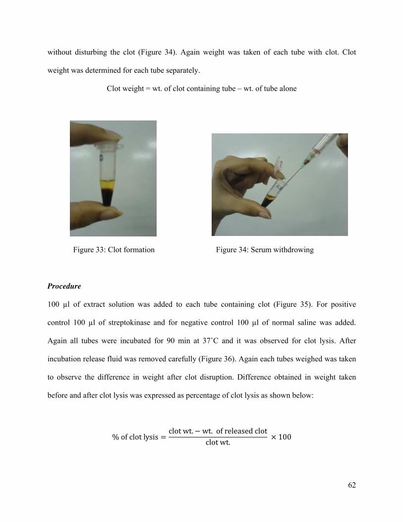
62
without disturbing the clot (Figure 34). Again weight was taken of each tube with clot. Clot
weight was determined for each tube separately.
Clot weight = wt. of clot containing tube – wt. of tube alone
Figure 33: Clot formation Figure 34: Serum withdrowing
Procedure
100 µl of extract solution was added to each tube containing clot (Figure 35). For positive
control 100 µl of streptokinase and for negative control 100 µl of normal saline was added.
Again all tubes were incubated for 90 min at 37˚C and it was observed for clot lysis. After
incubation release fluid was removed carefully (Figure 36). Again each tubes weighed was taken
to observe the difference in weight after clot disruption. Difference obtained in weight taken
before and after clot lysis was expressed as percentage of clot lysis as shown below:
ガ of clot lysis 噺 clot wt┻ 伐 wt┻ of released clotclot wt┻ 抜 などど

63
Figure 35: Extract added in clot Figure 36: After clot lysis serum withdrowing
Statistical analysis: Statistical comparisons were performed with Student's ‘t’ tests using
Microsoft Excel 2007. A p value of 0.001 or less was considered to be significant. Mean values ±
S.D. were calculated for the parameters where applicable.

64
RESULTS
Phytochemical Screening
Table 27: Data of phytochemical screening of A. polystachya
S.No Chemical
Constituents
Test APHE APEA APME
1 Alkaloids Hager's test + + ++
2 Flavonoids Ammonia test (modified) ++ + ++
3 Steroids Salkowski test – – –
4 Terpenoids Salkowski test (modified) +++ +++ +++
5 Carbohydrates Fehling's (Reducing sugar) test
(modified)
_ _ _
6 Saponins Frothing test – + –
7 Tannins FeCl3 test – – +
8 Cardiacglycosides Killer-Killani's test ++ + +
9 Anthraquinones Chloroform layer test +++ + +
Key:
– = Negative (absent); + = Positive (slightly present); ++ = Positive (moderately present); +++ =
Positive (strongly present)
Phytochemical screening of the plant A. polystachya leaf extracts (APHE, APEA and APME)
revealed the presence of various bioactive components. Alkaloids, flavonoids, terpenoids,
saponins, tannins, cardiac glycosides and anthraquinones were the most prominent (Table 27).

65
DPPH Free Radical Scavenging Assay
Table 28: IC50 value of ascorbic acid (AA)
Test tube
no.
Conc.(µg/ml) Absorbance of
AA
Absorbance of
blank
%
inhibition
Value of
IC50
0 0 0.197 0.197 0
1 20 0.132 0.197 32.99
2 40 0.119 0.197 39.59 76.11
3 60 0.107 0.197 45.69
4 80 0.099 0.197 49.75
5 100 0.086 0.197 56.35
Figure 37: % inhibition curve of ascorbic acid (AA)
y = 0.483x + 13.24R² = 0.815
0
10
20
30
40
50
60
70
0 20 40 60 80 100 120
% i
nh
ibit
ion
Concentration (µg/ml)

66
Table 29: IC50 value of the leaf of APHE
Test tube
no.
Conc.(µg/ml) Absorbance of
extract
Absorbance of
blank
%
inhibition
Value of
IC50
0 0 0.197 0.197 0
1 20 0.118 0.197 40.10
2 40 0.113 0.197 42.64 74.84
3 60 0.110 0.197 44.16
4 80 0.093 0.197 52.79
5 100 0.090 0.197 54.31
Figure 38: Scavenging effects of the leaf of APHE
y = 0.444x + 16.77R² = 0.696
0
10
20
30
40
50
60
70
0 20 40 60 80 100 120
% i
nh
ibit
ion
Concentration (µg/ml)

67
Table 30: IC50 value of the leaf of APEA
Test tube
no.
Conc.(µg/ml) Absorbance of
extract
Absorbance of
blank
%
inhibition
Value of
IC50
0 0 0.197 0.197 0
1 20 0.092 0.197 53.30
2 40 0.099 0.197 49.75 50.07
3 60 0.079 0.197 59.90
4 80 0.052 0.197 73.60
5 100 0.072 0.197 63.45
Figure 39: Scavenging effects of the leaf of APEA
y = 0.554x + 22.26R² = 0.643
0
10
20
30
40
50
60
70
80
90
0 20 40 60 80 100 120
% i
nh
ibit
ion
Concentration (µg/ml)

68
Table 31: IC50 value of the leaf of APME
Test tube
no.
Conc.(µg/ml) Absorbance of
extract
Absorbance of
blank
%
inhibition
Value of
IC50
0 0 0.197 0.197 0
1 20 0.013 0.197 93.40
2 40 0.009 0.197 95.43 12.54
3 60 0.007 0.197 96.45
4 80 0.007 0.197 96.45
5 100 0.003 0.197 98.48
Figure 40: Scavenging effects of the leaf of APME
y = 0.717x + 44.13R² = 0.468
0
20
40
60
80
100
120
140
0 20 40 60 80 100 120
% i
nh
ibit
ion
Concentration (µg/ml)

69
Table 32: % inhibition of APHE, APEA, APME and AA
Conc.(µg/ml) % inhibition
(APHE)
% inhibition
(APEA)
% inhibition
(APME)
% inhibition
(AA)
0 0.00 0.00 0.00 0.00
20 40.10 53.30 93.40 32.99
40 42.64 49.75 95.43 39.59
60 44.16 59.90 96.45 45.69
80 52.79 73.60 96.45 49.75
100 54.31 63.45 98.48 56.35
Figure 41: DPPH radical scavenging activity of AA and the APHE, APEA & APME extracts of
A. polystachya leaf
0
20
40
60
80
100
120
0 20 40 60 80 100 120
% o
f S
cav
eng
ing
Concentration (µg/ml)
APHE
APEA
APME
AA

70
It was interesting to notice that the extract of A. polystachia exhibited free radical scavenging
activity in the antioxidant assay. All the extracts inhibited DPPH, indicating their antioxidant
activity. The highest IC50 values among the extract of A. polystachia was 74.84 µg/ml (Table 29)
of leaf of n-hexane fraction while ascorbic acid had IC50 value 76.11 µg/ml (Table 28) indicating
the presence of potential candidates of antioxidant compounds. It appears that the APME has
greater DPPH free radical scavenging capacity as it has the lowest value of IC50 12.54 µg/ml
(Table 31). The IC50 value of APEA was 50.07 µg/ml (Table 30).
Nitric Oxide Free Radical Scavenging of Leaf of A. polystachya
Table 33: IC50 value of ascorbic acid (AA)
Test tube
no.
Conc.(µg/ml) Absorbance of
AA
Absorbance of
blank
%
inhibition
Value of
IC50
1 0 0.096 0.096 0.00
2 5 0.049 0.096 48.96
3 25 0.034 0.096 64.58 34.06
4 50 0.026 0.096 72.92
5 100 0.021 0.096 78.13
6 200 0.015 0.096 84.38

71
Figure 42: % inhibition curve of Ascorbic acid (AA)
Table 34: IC50 value of the leaf of APHE
Test tube
no.
Conc.(µg/ml) Absorbance of
extract
Absorbance of
blank
%
inhibition
Value of
IC50
0 0 0.096 0.096 0.00
1 5 0.095 0.096 1.04
2 25 0.089 0.096 7.29 76.15
3 50 0.026 0.096 72.92
4 100 0.018 0.096 81.25
5 200 0.002 0.096 97.92
y = 0.278x + 40.53R² = 0.467
0
20
40
60
80
100
120
0 50 100 150 200 250
% i
nh
ibit
ion
Concentration (µg/ml)

72
Figure 43: Scavenging effects of the leaf of APHE
Table 35: IC50 value of the leaf of APEA
Test tube
no.
Conc.(µg/ml) Absorbance of
extract
Absorbance of
blank
%
inhibition
Value of
IC50
0 0 0.096 0.096 0.00
1 5 0.070 0.096 27.08
2 25 0.046 0.096 52.08 39.90
3 50 0.009 0.096 90.63
4 100 0.008 0.096 91.67
5 200 0.006 0.096 93.75
y = 0.517x + 10.63R² = 0.759
0
20
40
60
80
100
120
0 50 100 150 200 250
% i
nh
ibit
ion
Concentration (µg/ml)

73
Figure 44: Scavenging effects of the leaf of APEA
Table 36: IC50 value of the leaf of APME
Test tube
no.
Conc.(µg/ml) Absorbance of
extract
Absorbance of
blank
%
inhibition
Value of
IC50
0 0 0.096 0.096 0.00
1 5 0.062 0.096 35.42
2 25 0.037 0.096 61.46 57.58
3 50 0.031 0.096 67.71
4 100 0.030 0.096 68.75
5 200 0.023 0.096 76.04
y = 0.391x + 34.4R² = 0.570
0
20
40
60
80
100
120
0 50 100 150 200 250
% i
nh
ibit
ion
Concentration (µg/ml)

74
Figure 45: Scavenging effects of the leaf of APME
Table 37: % inhibition of APHE, APEA, APME and AA
Conc.(µg/ml) % inhibition
(APHE)
% inhibition
(APEA)
% inhibition
(APME)
% inhibition
(AA)
0 0.00 0.00 0.00 0.00
5 1.04 27.08 35.42 48.96
25 7.29 52.08 61.46 64.58
50 72.92 90.63 67.71 72.92
100 81.25 91.67 68.75 78.13
200 97.92 93.75 76.04 84.38
y = 0.260x + 35.03R² = 0.474
0
10
20
30
40
50
60
70
80
90
100
0 50 100 150 200 250
% i
nh
ibit
ion
Concentration (µg/ml)

75
Figure 46: NO scavenging activity of AA and the APHE, APEA & APME extracts of A.
polystachya leaf
The highest IC50 values among the extracts of A. polystachia was 76.15 µg/ml (Table 34) of leaf
of n-hexane fraction while ascorbic acid had IC50 value 34.06 µg/ml (Table 33) indicating the
presence of potential candidates of antioxidant compounds. It appears that the APEA has greater
free radical scavenging capacity as it has the lowest value of IC50 39.90 µg/ml (Table 35). The
IC50 value of APME was 57.58 µg/ml (Table 36).
0
20
40
60
80
100
120
0 50 100 150 200 250
% o
f S
cav
eng
ing
Concentration (µg/ml)
APHE
APEA
APME
AA

76
Total Phenolic Content
Table 38: Absorbance of gallic acid
SL. No. Conc. of the Standard (µg/ml) Absorbance
0 0 0
1 10 0.234
2 20 0.487
3 30 0.718
4 40 1.140
5 50 1.521
6 60 2.294
7 70 2.285
8 80 2.711
9 90 3.188
10 100 3.665
Figure 47: Standard curve of gallic acid
y = 0.037x ど 0.209R² = 0.984
ど0.5
0
0.5
1
1.5
2
2.5
3
3.5
4
0 20 40 60 80 100 120
Ab
sorb
ance
Concentration (µg/ml)

77
Table 39: Content of total phenol of the extracts of A. polystachya leaves
Extracts Total phenolic content (n=3) (in mg/g,Gallic acid equivalents) Average SD
mg GAE/gm
(1)
mg GAE/gm
(2)
mg GAE/gm
(3)
APHE 95.11 85.55 95.62 92.09 5.67
APEA 112.12 106.62 104.58 107.77 3.90
APME 65.83 55.34 56.27 59.15 5.81
The total phenolic content of the A. polystachya leaves extracts were expressed as gallic acid
equivalent and are presented in (Table 39). The data clearly showed that APEA had the highest
total phenolic content followed by APHE and APME which mean value of 107.77 mg GAE/gm
extract, 92.09 mg GAE/gm extract and 59.15 mg GAE/gm extract, respectively.
In vitro Thrombolytic Activity
Table 40: % Thrombolysis of APHE, APEA, APME, streptokinase and normal saline
Sample
no.
%
Thrombolysis
APHE
%
Thrombolysis
APEA
%
Thrombolysis
APME
%
Thrombolysis
Streptokinase
%
Thrombolysis
Normal saline
1 15.327 5.294 10.611 43.988 5.892
2 23.574 9.192 8.277 36.431 4.430
3 14.874 8.162 14.600 37.152 3.398
4 15.434 5.029 9.998 45.533 4.211
5 17.944 8.385 16.027 40.146 5.181
6 23.619 7.113 10.572 48.768 5.142
7 20.983 9.860 8.968 46.007 4.508
8 16.432 12.383 7.140 38.811 6.147
9 12.489 5.996 10.111 38.413 3.647
10 18.617 9.698 8.936 32.299 4.559

78
Table 41: Clot lysis of blood sample data of extracts of A. polystachya fruit and streptokinase.
Extracts/
Positive
control
Mean±SD
(% Clot lysis)
APHE 17.93 ± 3.77*
APEA 8.11 ± 2.30*
APME 10.52 ± 2.76*
Streptokinase 40.75 ± 5.14*
Values are expressed in Mean±SD (n=10), p<0.001
Figure 48: Clot lysis of blood samples of normal subjects by APHE, APEA, and APME extracts
of A. polystachya leaves, streptokinase and normal saline.
When streptokinase (100 µl) was added to the clots maximum clot lysis was visually seen (Table
41, Figure 48). With normal saline 4.71% weight difference was seen. The in vitro thrombolytic
17.929
8.11110.524
40.755
4.7120
5
10
15
20
25
30
35
40
45
50
% C
lot
Lysi
s
APHE
APEA
APME
Streptokinase
Normal saline

79
activity study revealed that n-hexane (APHE), ethyl acetate (APEA) and methanol (APME)
extracts of A. polystachya leaves showed 17.93%, 8.11% and 10.52% clot lysis respectively and
compared with the negative control (normal saline) the mean clot lysis % difference was
significant (p < 0.001).

80
DISCUSSION
Phytochemical Screening
Previous researchers reported the presence of alkaloids, tannins, steroids, saponins, glycosides,
flavonoids, anthraquinones and terpenoids in A. polystachya (Krishnaraju et al., 2009;
Chowdhury & Rashid, 2003; Jagetia & Venkatesha, 2006; Sarla et al., 2011). The current study
also correlates with previous studies. Presence of varieties of chemical compounds in the plant is
directly or indirectly responsible for the significant amount of biological activities of A.
polystachya leaf extracts. All the phytochemicals are reported to possess good antioxidant
activities and has been reported to show numerous biological effects including anti-inflammatory
and antitumor activities (Chowdhury & Rashid, 2003; Jagetia & Venkatesha, 2006; Sarla et al.,
2011).
Antioxidant Activity
On the basis of the scavenging ability of the free radicals for DPPH, the highest antioxidant
activity was found in APME (Table 31) and for NO, the highest antioxidant activity was found in
APEA (Table 35). May be, due to the presence of polyphenolic compounds such as flavonoids
and tannins (Table 27) the antioxidant activity of the extracts were observed. The outcome is
agreed with the findings of the other researchers (Sarla et al., 2011; Krishnaraju et al., 2005) in
which the antioxidant activity of methanol and ethyl acetate extracts of A. polystachya was
reported and therefore could be used as a potential preventive measure against free radical-
mediated diseases. The highest total phenolic content was found in ethyl acetate extract followed
by n-hexane and methanol extract (Table 39).

81
Thrombolytic Activity
Leaf extracts of the plant showed significant (p<0.001) thrombolytic activity compared to the
negative control. The phytochemical analysis showed that the extracts were a rich source of
alkaloids, flavonoids, tannins and terpenoids which could be responsible for the clot lysis activity
(Chowdhury et al., 2011; Dwivedi, 2007).

82
CONCLUSION
The presence of the identified phytochemicals makes the leaves pharmacologically active. Their
antioxidant activity may be responsible for their usefulness in the management and treatment of
various diseases. The medicinal values of the plant leaves may be related to their constituent
phytochemicals. Due to its natural origin and potent free-radical scavenging ability A.
polystachya could be used as a potential preventive intervention for free radical-mediated
diseases. The presented data for total phenolic content are a basis of assessment of the preventive
role of A. polystachya against free radicals effect. From in vitro clot lysis study, we demonstrated
that A. polystachya have clot lysis activity. So that, we may assume that these extracts can be
considered as a potential source of natural thrombolytic agent.

83
REFERENCES:
Agbafor KN, Nwachukwu N. Phytochemical Analysis and Antioxidant Property of Leaf Extracts
of Vitex doniana and Mucuna pruriens. Biochemistry Research International. Volume 2011
(2011), Article ID 459839, 4 pages doi:10.1155/2011/459839.
Agnihotri VK, Srivastava SD, Srivastava SK. A New Limonoid, Amoorinin from the Stem Bark
of Amoora rohituka. Current Science, 1987; 56(15):770.
Agnihotri VK, Srivastava SD, Srivastava SK. A New Limonoid, Amoorinin, from the Stem Bark
of Amoora rohituka. Planta Med, 1987;53(3): 298-9.
Ainsworth EA, Gillespie KM. Estimation of total phenolic content and other oxidation substrates
in plant tissues using Folin-Ciocalteu reagent. Nat Protoc 2007;2(4)875-7.
Anwar MS, Khan IN, Barua S, Kamal ATMM, Hosen SMZ, Kawsar MH. Assessment of
Thrombolytic & Cytotoxic Activity of Herbal Preparations Originated from Botanical Source of
Bangladesh. Journal of Applied Pharmaceutical Science 2011;01(07):77-80.
Arvigo R, Balick M, Rainforest Remedies, Lotus Press, Twin Lakes 1993.
Ayoola GA, Coker HAB, Adesegun SA, Adepoju-Bello AA, Obaweya K, Ezennia EC,
Atangbayila TO. Phytochemical Screening and Antioxidant Activities of Some Selected
Medicinal Plants Used for Malaria Therapy in Southwestern Nigeria. Tropical Journal of
Pharmaceutical Research, September 2008;7(3):1019-1024.
Banerjee A, Chisti Y, Banerjee UC. Streptokinase—a clinically useful thrombolytic agent.
Biotechnology Advances 22 (2004):287-307.

84
Balakrishnan N, Panda AB, Raj NR, Shrivastava A, Prathani R. The evaluation of nitric oxide
scavenging activity of Acalypha indica Linn. root. Asian J Research Chem 2009;2(2):148-150.
Bhat SV, Bajwa BS, Dornauer H, Souza NJ, Fehlhaber HW, Tetrahedron Lett. 1977;1669-72.
Bhatta SK, Saxena VK, Nigam SS. Anew flavanone glycoside from the seeds of Amoora
rohituka Roxb. Abstract presented at the Convention of Chemist, IIT Bombay, 1980.
Bhatt SK, Saxena VK, Nigam SS. A New Saponin from Seeds of Amoorarohituka.
Phytochemistry, 1981;20(7):1749-1750.
Bhuyan MAK, Begum J, Chowdhury JU, Ahmed K, Anwar M N. Antimicrobial activity of oil
and crude alkaloids from seeds of Aphanamixis polystachya (Wall.) R.N. Parker.; Bangladesh
Journal of Botany; 2000;29(1):1-5; ISSN-0253-5416; Record Number-20013084157.
Blum D 2010. "The little-told story of how the U.S. government poisoned alcohol during
Prohibition. – By Deborah Blum – Slate Magazine".
Chatterjee A, Kundu AB, Chakrabortty T, Chandrasekharan S. Extractives of Aphanamixis
polystachya wall (Parker): The Structures and Stereochemistry of Aphanamixin and
Aphanamixinin. Tetrahedron, 1970;26(8):1859-1867.
Chan LL, George S, Ahmad I, Saujanya LG, Abbasi A, Cunningham BT, Watkin KL.
Cytotoxicity Effects of Amoora rohituka and chittagonga on Breast and Pancreatic Cancer Cells.
Evidence-Based Complementary and Alternative Medicine, 2011:1-8.

85
Chhetri HP, Yogol NS, Sherchan J, Anupa KC, Mansoor S, Thapa P. Phytochemical and
Antimicrobial evaluations of some medicinal plants of Nepal, Kathmandu University Journal of
Science, Engineering and Technology 2008;i(v):49-54.
Chowdhury R, Hasan CM, Rashid MA. Guaiane Sesquiterpenes from Amoora rohituka.
Phytochemistry, 2003;62(8):1213-1216.
Chowdhury R, Rashid RB. Effect of the crude extracts of Amoora rohituka stem bark on
gastrointestinal transit in mice. Indian J of Pharmacol 2003;35:304-307.
Chowdhury R, Hasan CM, Rashid MA. Antimicrobial Activity of Toona ciliata and Amoora
rohituka. Fitoterapia, 2003;74(1-2):155-8.
Chowdhury NS, Alam MB, Haque ASMT, Zahan R, Mazumder MEH, Haque ME. In vitro free
radical scavenging and thrombolytic activities of Bangladeshi aquatic plant Aponogeton
undulatus Roxb. Global J Pharmacol 2011;5(1):27-32.
Dupin JP, Gryglewski RJ, Gravier D, Hou G, Casadebaig F, Swies J, Chlopicki S. Synthesis and
thrombolytic activity of new thienopyrimidinone derivatives. Journal of physiology and
pharmacology 2002;53(4):625-634.
Dwivedi S. Terminalia arjuna Wight & Arn.-A useful drug for cardiovascular disorders. J
Ethnopharmacol 2007;114(2):114-129.
Edeoga HO, Okwu DE, Mbaebie BO. Phytochemical constituents of some Nigerian medicinal
plants. African Journal of Biotechnology 2005;4(7):685-688.

86
Feresin GE, Tapia A, Gutierrez RA, Delporte C, Backhouse EN, Schmeda-Hirschmann G. Free
radical scavengers, anti-inflammatory and analgesic activity of Acaena magellanica. J Pharm
Pharmacol 2002;54(6):835-44.
Gamble F. Madras 1: 181. 1997 (re. ed); Sasidharan, Indian Forester 57: 486. 1931; Biodiversity
documentation for Kerala- Flowering Plants, part 6: 88. 2004; Saldanha, Fl. Karnataka 2: 231.
1996.
Gokaraju GR, Gokaraju RR. Golakoti T, Chirravuri, VR, Raju VK, Bhupathiraju AK. Use of
Aphanamixis polystacha extracts or fractions against 5-lipoxygenase mediated diseases; 2010;
Patent application number: 20100178288.
Gole MK, Dasgupta S. Role of plant metabolites in toxic liver injury. Asia Pacific Journal of
Clinical Nutrition, 2002;11(1):48-50.
Guerrero RO, Khan MTH, Casanas B, Morales M. Specific bioassays with selected plants of
Bangladesh. Rev Cubana Plant Med 2004;9(2) On-line version ISSN 1028-4796.
Hill AF. Economic Botany. A textbook of useful plants and plant products. 2nd edn. McGarw-
Hill Book Company Inc, 1952, New York.
Hodzic Z, Pasalic H, Memisevic A, Srabovic M, Saletovic M, Poljakovic M. The Influence of
Total Phenols Content on Antioxidant Capacity in the Whole Grain Extracts. European Journal
of Scientific Research. ISSN 1450-216X 2009;28(3):471-477.

87
Hossain M, Biva IJ, Jahangir R, Vhuiyan IM. Central nervous system depressant and analgesic
activity of Aphanamixis polystachya (Wall.) parker leaf extract in mice. African Journal of
Pharmacy and Pharmacology 2009,3(5),282-286.
Hoque N, Imam MZ, Akter S, Mazumder MEH, Hasan SMR, Ahmed J, et al. Antioxidant and
antihyperglycemic activities of methanolic extract of Glinus oppositifolius leaves. J App Pharm
Sci 2011;1(7):50-3.
Howse PE. Laboratory evaluation of toxic and repellent properties of the pithraj tree,
Aphanamixis polystachya (Wall & Parker), against Sitophilusoryzae (L.). International Journal of
Pest Management, 1994;40(3):274-279.
HUANG D, BOXIN OU , RONALD L. PRIOR. The Chemistry behind Antioxidant Capacity
Assays. J. Agric. Food Chem. 2005, 53, 184118561841.
Iyengar MA. Study of Crude Drugs. 8th ed., Manipal Power Press, Manipal, India. 1995;2.
Jagetia GC, Venkatesha VA. Enhancement of radiation effect by Aphanamixis polystachya in
mice transplanted with Ehrlich ascites carcinoma. Biological and Pharmaceutical Bulletin,
2005;28(1):69-77.
Jagetia GC, Venkatesha VA. Treatment of mice with stem bark extract of Aphanamixis
polystachya reduces radiation-induced chromosome damage. International Journal of Radiation
Biology 2006;82(3):197-209.
Jagetia GC. Radioprotective Potential of Plants and Herbs against the Effects of Ionizing
Radiation. J. Clin. Biochem. Nutr, 2007;40:74-81.

88
Jain A, Srivastava SK. 8-C-methyl-quercetin-3-O-く-D-xylopyranoside, A New Flavone
Glycoside from the Roots of Amoorarohituka. Journal of Natural Products, 1985;48(2):299-301.
Javanmardi J, Stushnoff C, Locke E, Vivanco JM, Antioxidant activity and total phenolic content
of Iranian Ocimum accessions. Food Chemistry 83 (2003) 547–550.
Karmohapatra SK, Kahn NN, Sinha AK. The thrombolytic effect of aspirin in animal model. J
Thromb Thrombolysis 2007;24:123–129.
Khan IN, Sarker MI, Almamun A, Mazumder K, Bhuiya MAM, Mannan A. Cytotoxic and
Thrombolytic Activity of Ethanolic Extract of Zanthoxylum Budrunga (Fam: Rutaceae) Leaves.
European Journal of Scientific Research 2011;66(2):303-310.
Khan IN, Habib MR, Rahman MM, Mannan A, Sarker MMI, Hawlader S. Thrombolytic
potential of Ocimum sanctum L., Curcuma longa L., Azadirachta indica L. and Anacardium
occidentale L. J Basic Clin Pharm, 2011;2(3):125-7.
Klaymann DL, Clark AM, Science 1985, 228, 1049. Pharm. Res. 1996;13:1133-1144.
Krishnaraju AV, Rao TVN, Sundararaju D, Vanisreeb M, Tsay HS, Subbaraju GV. Assessment
of bioactivity of Indian medicinal plants using brine shrimp (Artemia salina) lethality assay. Int J
Appl Sci Eng 2005;3(2):125-134.
Krishnaraju AV, Rao CV, Rao TVN, Reddy KN, Trimurtulu G. In vitro and In vivo antioxidant
activity of Aphanamixis polystachya bark. American Journal of Infectious Diseases.
2009;5(2):60-67 DOI: 10.3844/ajidsp.

89
Kundu AB, Ray S, Chatterjee A. Aphananin, a triterpene from Aphanamixis polystachya;
Phytochemistry 1985;24(9):2123-2125.
Lampronti I, Khan MT, Bianchi N, Ather A, Borgatti M, VFabbri E, Rizziello LG. Bangladeshi
medicinal plant extracts inhibiting molecular interactions between nuclear factors and target
DNA sequences mimicking NF-kappaB binding sites 2005;1(4):327-33.
Laurence DR, Bennett PN. Clinical Pharmacology: Seventh Edition 1992: Page 483.
Mabberley DJ, Sing AM. Indian Forester,1931;62:486.
McCaine WD. The Properties of Petroleum Fluids (2nd
ed.), 1990, Oklahoma: PennWell
Publishing Company.
Mohanakumara P, Sreejayan N, Priti V. Dysoxylumbinectariferum Hook.F (Meliaceae), a Rich
Source of Rohitukine. Fitoterapia, 2010;81(2):145-148.
Mojab F, Kamalinejad M, Ghaderi N, Vahidipour H. Phytochemical Screening of Some Iranian
Plants. Iranian Journal of Pharmaceutical Research. 2003;77-82.
Nair RT, Kalariya J, Chanda S. Antibacterial activity of some selected Indian medicinal flora.
Tuky J. Biol. 2005;29:41┽47.
Nobakht GM, Kadir MA, Stanslas J. Analysis of preliminary phytochemical screening of
Typhonium flagelliforme. African Journal of Biotechnology 2010;9(11):1655-1657.
Pamplona-Roger D. Encyclopedia of Medicinal Plants, Saeliz, 1998, Spain.

90
Parker R, Aphanamixis polystachy, 1997. Indian Forester 57:486. 1931, Nomen number:
312562.
Prakash A, Rigelhof F, Miller E. Antioxidant Activity, A publication of Medallion Labs.
Prasad S, Kashyap RS, Deopujari JY, Purohit HJ, Taori GM, Daginawala HF. Effect of Fagonia
Arabica (Dhamasa) on in vitro thrombolysis. BMC Complementary and Alternative Medicine
2007;7:36.
Rabi T, Karunagaran D, Nair MK, Bhattathiri VN. Cytotoxic Activity of Amooranin and its
Derivatives. Phytotherapy Research 2002;16(1):84-86.
Rabi T, Ramachandran C, Fonseca HB, Nair RP, Alamo A, Melnick SJ, Escalon E. Novel Drug
Amooranin Induces Apoptosis Through CaspaseActivity in Human Breast Carcinoma Cell
Lines. Breast Cancer Research and Treatment 2003;80(3):321-330.
Reuben KD, Abdulrahman FI, Akan JC, Usman H, Sodipo OA, Egwu GO. Phytochemical
Screening and In Vitro Antimicrobial Investigation of the Methanolic Extract of Croton
Zambesicus Muell ARG. Stem Bark. European Journal of Scientific Research. ISSN 1450-216X.
2008;23(1):134-140.
Sadhu SK, Phattanawasin P, Choudhuri MSK, Ohtsuki T, Ishibashi M. A New Lignan from
Aphanamixis polystachya. Journal of Natural Medicines, 1980;60(3):258-260.
Sanyal M, Datta PC. Pharmacognosy of Aphanamixis polystachya stem bark. Quart. J Crude
Drug Res 1981;19(2-3):113-126.

91
Saraf S, Sharma S, Bensalha I, Wellsted D, Gorog D. Impaired endogenous thrombolysis in
acute coronary syndrome patients predicts recurrent events: results using a novel thrombosis test.
Heart 2009;95(Suppl I):A1–A87.
Sarla S, Prakash MA, Apeksha R, Subhash C. Free radical scavenging (DPPH) and ferric
reducing ability (FRAP) of Aphanamixis polystachya (Wall) Parker. Int J Drug Dev Res
2011;3(4):271-274.
Sen S, Chakraborty R, Sridhar C, Reddy YSR, De B. FREE RADICALS, ANTIOXIDANTS,
DISEASES AND PHYTOMEDICINES:CURRENT STATUS AND FUTURE PROSPECT.
International Journal of Pharmaceutical Sciences Review and Research, 2010;3(1) ISSN 0976 –
044X. Article 021.
Shinkar AS. Phytochemical investigation and antiulcer activity of Aphanamixis polystachya
(wall.) Parker, (Dissertation); Submitted to: Rajiv Gandhi University of Health sciences,
Karnataka, Bangalore, September, 2007.
Shrivastava S, Leelavathi S. PRELIMINARY PHYTOCHEMICAL EVALUATION OF LEAF
EXTRACTS OF CATUNAREGUM SPINOSA THUNB. International Journal of Pharmaceutical
Sciences Review and Research. ISSN 0976 – 044X. 2010;3(2) Article 020.
Strobel G, Daisy B, Castillo U, Harper J, J. Nat. Prod. 2004;67:257-268.
Talukder FA, Howse PE. Evaluation of Aphanamixis polystachya as a source of repellents,
antifeedants, toxicants and protectants in storage against Tribolium castaneum (Herbst); Journal
of Stored Products Research 1995;31(1):Pages 55-61.

92
Talukder FA, Howse PE. Isolation of secondary plant compounds from Aphanamixis
polystachya as feeding deterrents against adult Tribolium castaneum (Coleoptera:
Tenebrionidae); Journal of Stored Products Research 2000;107(5):498-504; ISSN-0340-8159;
Record Number-20003026520.
Talukder FA, Howse PE. Deterrent and insecticidal effects of extracts of pithraj,Aphanamixis
polystachya (Meliaceae), against Tribolium castaneum in storage, Journal of Chemical Ecology,
2011;19(11):2463-2471, DOI: 10.1007/BF00980683
Thaipong K, Boonprakob U, Crosby K, Cisneros-Zevallos L, Byrne DH. Comparison of ABTS,
DPPH, FRAP, and ORAC assays for estimating antioxidant activity from guava fruit extracts.
Journal of Food Composition and Analysis 2006;19:669–675.
Tsao R, Yang R, Xie S, Sockovie E, Khanizadeh S. Which polyphenolic compounds contribute
to the total antioxidant activities of apple? J Agric Food Chem. 2005;53(12):4989-95 15941346
Cit:26.
Verschueren KE. Handbook of Environmental Data on Organic Chemicals, 2nd ed. Van
Nostrand Reinhold Company, New York, 1983, pp. 818-820.
Vinod SK, Raghuveer I, Alok S, Himanshu G. Phytochemical investigation and chromatographic
evaluation of the ethanolic extract of whole plant extract of dendrophthoe falcata (l.f.) Ettingsh.
International journal of pharmaceutical sciences and research. Ijpsr 2010;1(1)

93
Wangcharoen W, Morasuk W. Antioxidant capacity and phenolic content of some Thai culinary
plants. Maejo International Journal of Science and Technology. ISSN 1905-7873.
2007;01(02):100-106.
Wani MC, Taylor HL, Wall ME, Coggon P, McPhail AT, J. Am. Chem. Soc.1971;93:2325-2327.
Wiart C. Medicinal Plants of Asia and the Pacific. New York, 2006 NY: CRC Press.
Yadav R, Chauhan NS, Chouhan AS, Soni VK, Omray L. Antimicrobial screening of various
extracts of Aphanamixis polystachya stems bark. International Journal of Advances in
Pharmaceutical Sciences, 2010;1:147-150.
Yang SP, Chen HD, Liao SG, Xie BJ, Miao ZH, Yue JM. Aphanamolide A, a New Limonoid
from Aphanamixis polystachya; American Chemical Society, Organic Letters, 2011;13(1):150-
153.
Zhang H, Chen F, Wang X, Wu D, Chen Q. A New Limonoid from the seed of Aphanamixis
polystachya. Chinese Chemical Letters, 2002;13(4):341-342.
Zhang Y, Wang JS, Wang XB, Wei DD, Luo JG, Luo J, Yang MH, Kong LY. Aphapolynins A
and B, two new limonoids from the fruits of Aphanamixis polystachya; Tetrahedron Letters
2011;52(20):2590-2593.