Nonproliferative and Proliferative Lesions of the Rat and ...

Food Chemistry 136 (2013) 120–129
Contents lists available at SciVerse ScienceDirect
Food Chemistry
journal homepage: www.elsevier .com/locate / foodchem
Phytochemical profile of Rosmarinus officinalis and Salvia officinalis extractsand correlation to their antioxidant and anti-proliferative activity
Vassiliki G. Kontogianni a,⇑, Goran Tomic b, Ivana Nikolic b, Alexandra A. Nerantzaki a, Nisar Sayyad a,Stanislava Stosic-Grujicic b, Ivana Stojanovic b,⇑, Ioannis P. Gerothanassis a, Andreas G. Tzakos a,⇑a Department of Chemistry, University of Ioannina, Ioannina GR-45110, Greeceb Department of Immunology, Institute for Biological Research ‘‘Sinisa Stankovic’’, University of Belgrade, Bulevar Despota Stefana 142, Belgrade, Serbia
a r t i c l e i n f o
Article history:Received 29 February 2012Received in revised form 18 July 2012Accepted 19 July 2012Available online 31 July 2012
Keywords:Cancer cell apoptosisNitric oxideRINm5FRosmarinus officinalis extractSalvia officinalis extractLC–MS/MS1H–13C HSQC and HMBC NMR
0308-8146/$ - see front matter � 2012 Elsevier Ltd. Ahttp://dx.doi.org/10.1016/j.foodchem.2012.07.091
⇑ Corresponding authors. Tel.: +381 11 2078 390Stojanovic), tel.: +30 26510 08397; fax: +30 26510 08+30 26510 08387; fax: +30 26510 08799 (A. G. Tzako
E-mail addresses: [email protected] (V.G. KontogStojanovic), [email protected] (A.G. Tzakos).
a b s t r a c t
The goal of this study was to monitor the anti-proliferative activity of Rosmarinus officinalis and Salvia offi-cinalis extracts against cancer cells and to correlate this activity with their phytochemical profiles usingliquid chromatography/diode array detection/electrospray ion trap tandem mass spectrometry (LC/DAD/ESI-MSn). For the quantitative estimation of triterpenic acids in the crude extracts an NMR based meth-odology was used and compared with the HPLC measurements, both applied for the first time, for thecase of betulinic acid. Both extracts exerted cytotoxic activity through dose-dependent impairment ofviability and mitochondrial activity of rat insulinoma m5F (RINm5F) cells. Decrease of RINm5F viabilitywas mediated by nitric oxide (NO)-induced apoptosis. Importantly, these extracts potentiated NO andTNF-a release from macrophages therefore enhancing their cytocidal action. The rosemary extract devel-oped more pronounced antioxidant, cytotoxic and immunomodifying activities, probably due to the pres-ence of betulinic acid and a higher concentration of carnosic acid in its phytochemical profile.
� 2012 Elsevier Ltd. All rights reserved.
1. Introduction Rosemary extract has shown anti-proliferative effects on vari-
Medicinal plants have served as rich sources of pharmacologi-cally active substances. Herbs have been used in a diverse arrayof purposes including medicine, nutrition, flavorings, beverages,dyeing, repellents, fragrances, cosmetics, charms, smoking andindustrial uses. Today, herbs are still found in 40% of prescriptiondrugs (Newman & Cragg, 2007). Lamiaceae plants are now culti-vated worldwide, mainly for use as culinary and medicinal herbs,and are widely studied as natural antioxidant sources since theyare enriched in polyphenols. Their potent bioactivity and relativelylow toxicity have rendered them useful ingredients in complemen-tary alternative medicine and as nutritional supplements. Rose-mary (Rosmarinus officinalis L.) and sage (Salvia officinalis) leaf arepopular herbal teas and essential-oil containing drugs. Rosemaryand sage are rich sources of di- and triterpenoids, phenolic acids,and flavonoids. Carnosic acid, carnosol and rosmarinic acid arethe main antioxidant compounds present in them (Cuvelier, Berset,& Richard, 1994).
ll rights reserved.
; fax: +381 11 2761 433 (I.799 (V. G. Kontogianni), tel.:s).ianni), [email protected] (I.
ous tumor cell lines (Cheung & Tai, 2007). Carnosol and carnosicacid isolated from rosemary leaves have also presented anticancerproperties, as determined in HL-60 cells (Bai et al., 2010). The cyto-toxic activity of sage has not been studied in detail, but it has beenshown that rosmarinic acid and sage extracts induce apoptosis incolorectal cancer cell lines (Xavier, Lima, Fernandes-Ferreira, &Pereira-Wilson, 2009). These findings suggest that Lamiaceae herbscontain several compounds with anti-proliferative activity againstdifferent cancers. As for their immunomodifying properties, therosemary extract was found to be mainly anti-inflammatory (Che-ung & Tai, 2007), while sage extract exhibited pro-inflammatoryeffects during its anti-leishmanial activity (Radtke, Yeap Foo, Lu,Kiderlen, & Kolodziej, 2003). There are still no studies looking intothe anti-proliferative activity of sage and rosemary extracts andalso at the effects of their major constituents on rat insullinomaRINm5F cells. There is also no general investigation of their phyto-chemical composition and there has been no comparative evalua-tion of their antioxidant and cytotoxic activities.
The aim of the present study was to characterize the composi-tion of plant extracts rich in biophenols belonging to the Lamiaceafamily (ethyl acetate extracts of R. officinalis and S. officinalis) usingLC/DAD/ESI-MSn. The main compounds of the extracts were quan-tified using HPLC and NMR methods. Furthermore, extracts werestudied colorimetrically for their total phenolic and flavonoid con-tents. Their antioxidant activities were evaluated using the DPPH
V.G. Kontogianni et al. / Food Chemistry 136 (2013) 120–129 121
(1,1-diphenyl-2-picrylhydrazyl) method. In addition, the effect ofthese plant extracts on viability, apoptosis and NO productionwas investigated in rat insulinoma RINm5F cells and their potentialimmunomodifying properties were monitored.
2. Materials and methods
2.1. Plant material, reagents and standards
R. officinalis and S. officinalis were commercial samples. All sol-vents were of appropriate purity and were purchased from varioussuppliers. Acetic acid (glacial) was of analytical grade from Merck(Darmstadt, Germany). Folin–Ciocalteu, phenol reagent and alu-minium chloride were obtained from Fluka (Switzerland), DPPH(�90%) was obtained from Sigma–Aldrich (Steinheim, Germany).Standard compounds were: ursolic acid (90%), caffeic acid (98%)and betulinic acid (90%) from Aldrich (Steinheim, Germany), olean-olic acid (97%), carnosic acid, carnosol and rutin (94%) from Sigma(Steinheim, Germany), rosmarinic acid (95%) and apigenin-7-O-glucoside (97%) from Fluka (Steinheim, Germany), genkwanin,luteolin (99%) and apigenin (99%) from Extrasynthese (Genay,France).
2.2. Sample preparation
The plant material was dried with liquid nitrogen and pulver-ized into a fine powder. The ground leaves were subsequently ex-tracted with two solvents of increasing polarity, hexane (extractionvolume 200 mL), and ethyl acetate (extraction volume 200 mL), ina Soxhlet apparatus for �6 h for each solvent. The ethyl acetate ex-tracts were concentrated in a rotary evaporator and kept intosealed flasks.
2.3. LC–MS analysis
2.3.1. InstrumentationAll LC-MSn experiments were performed on a quadrupole ion
trap mass analyzer (Agilent Technologies, model MSD trap SL) ret-rofitted to a 1100 binary HPLC system equipped with an degasser,autosampler, diode array detector and electrospray ionizationsource (Agilent Technologies, Karlsruhe, Germany). All hardwarecomponents were controlled by Agilent Chemstation Software.
2.3.2. AnalysisRosemary and sage extracts were dissolved in ethanol to the de-
sired concentration of 1 mg of dry extract/mL. A 10 ll aliquot wasfiltered (0.45 lm) and injected into the LC–MS instrument. Separa-tion was achieved on a 25 cm � 4.6 mm i.d., 5 lm Altima C18 ana-lytical column (Alltech, Deerfield, USA), at a flow rate of 0.6 mL/min, using as solvent A (water/acetic acid, 99.9: 0.1 v/v) and sol-vent B (acetonitrile). The gradient used for the analysis of rosemaryand sage extracts was: 0–15 min, 80–40% A; 15–20 min 40–25% A;20–45 min 25–15% A; 45–50 min 15–10% A; 50–65 min 10% A; 65–70 min 10–80% A. The UV/vis spectra were recorded in the range200–400 nm and chromatograms were acquired at 210, 280 and330 nm.
Both precursor and product (MS2 and MS3) ions scanning of thephenolic compounds were monitored between m/z 50–m/z 1,000 innegative polarity. The ionization source conditions were as fol-lows: capillary voltage, 3.5 kV; drying gas temperature, 350 �C;nitrogen flow and pressure, 12 L/min and 12 psi, respectively. Max-imum accumulation time of ion trap and the number of MS repeti-tions to obtain the MS average spectra were set at 30 and 3 ms,respectively.
2.4. NMR analysis for the presence of triterpenic acids
NMR experiments were performed on a Bruker AV-500 spec-trometer equipped with a cryoprobe. Spectra were obtained fromethyl acetate extracts dissolved in pyridine-d5 (20 mg of materialin 0.5 mL of solution in pyridine-d5) to investigate triterpenoids.The procedure followed was according to the method employedby Kontogianni, Exarchou, Troganis, and Gerothanassis, (2009)and described in detail in Supplementary material (Section S1).All measurements were performed in triplicate (on three differentextracts of the same sample).
2.5. HPLC analysis for the presence of triterpenic acids
Instrumentation and detection mode are described in Supple-mentary material (Section S2). For the analysis of triterpenoidsthe extracts were dissolved in methanol (1 mg/mL). Identificationof triterpenoids was based on retention time, UV spectra and spik-ing. All measurements were performed in triplicate (on three dif-ferent extracts of the same sample).
2.6. Determination of the total phenolic content
The total phenolic content of the extracts was measured usingthe Folin–Ciocalteu method according to the procedure of Gutfin-ger (1981), described in Supplementary material (Section S3).The results were expressed as mg of caffeic acid per g of dry ex-tract. All measurements were performed in duplicate.
2.7. Determination of the total flavonoid content
The content of flavonoids was determined according to Miliaus-kas, Venskutonis, and Van Beek (2004), described in Supplemen-tary material (Section S4) using rutin as a reference compound.All determinations were carried out in duplicate.
2.8. Antioxidant activity: DPPH- radical scavenging assay
The DPPH radical-scavenging effect was evaluated according tothe method employed by Choi et al. (2002), described in Supple-mentary material (Section S5). All measurements were performedin duplicate.
2.9. Tests performed on rat insulinoma cells (RINm5F) and immunecells
2.9.1. AnimalsC57BL/6 mice were bred and housed conventionally at the Insti-
tute for Biological Research ‘‘Sinisa Stankovic’’ (Belgrade, Serbia)under standard laboratory conditions (non-specific pathogen free)and provided with standard rodent chow and water ad libitum. Thehandling of animals and the study protocol were in accordancewith international guidelines and approved by the local Institu-tional Animal Care and Use Committee.
2.9.2. Cell lineRat insulinoma RINm5F cells were kindly donated by Dr. Kar-
sten Buschard (Bartholin Instituttet, Copenhagen, Denmark).
2.9.3. Isolation of peritoneal macrophagesResident peritoneal macrophages were collected by peritoneal
lavage with 3 mL cold PBS, centrifuged at 500g and their viabilitywas assessed by conventional trypan-blue staining (Sigma–Aldrich).Afterwards macrophages were suspended in culture medium(RPMI-1640 medium (Sigma–Aldrich, St. Louis, MO, USA) supple-mented with 2 mM L-glutamine, 20 lg/mL gentamycine (Galenika

122 V.G. Kontogianni et al. / Food Chemistry 136 (2013) 120–129
a.d., Serbia), 0.01% sodium pyruvate, 5 � 10�5 M 2-mercap-toethanol, antibiotics (all from Sigma–Aldrich) and 5% (v/v) heat-inactivated FCS (foetal calf serum) (PAA Chemicals, Pasching, Aus-tria)) and seeded into 96-well flat-bottom plates (Sartstedt, Numbr-echt, Germany) (1 � 105/well) for the Griess reaction and into 24-well plates (Sartstedt) (1 � 106/well) for ELISA.
2.9.4. Determination of cell viability and apoptosisRINm5F cells were grown in culture medium containing 10%
FCS in a humidified (5% CO2, 95% air) atmosphere at 37 �C. RINm5Fcells (1 � 104 cells/well) were seeded in 96-well plates (Sardstedt,Numbrecht, Germany) and treated with specific extracts dissolvedin DMSO (Sigma–Aldrich) (12.5–100 lg/mL) or matching concen-trations of the mentioned solvent. After 24 h incubation, the pro-portion of attached cell (cell viability) was assessed by crystalviolet staining. Briefly, after fixation with methanol for 10 min,crystal violet solution (MOL, Belgrade, Serbia) was added to theculture wells and incubated at 25 �C for the next 15 min. Afterthorough washing in tap water, incorporated dye was solubilizedin 33% acetic acid (Zorka Farma, Sabac, Serbia) and the developedcolour was recorded in an automated microplate reader (LKB5060–006, LKB, Vienna, Austria) at 570 nm, with a correction at670 nm. In the 3-(4,5-dimethylthiazol-2-yl)-2,5-diphenyltetrazoli-um bromide (MTT) (Sigma–Aldrich) assay, cell respiration was as-sessed by the mitochondrial-dependent reduction of MTT tocoloured formazan product (Mosmann, 1983), which reflected cellviability. Briefly, following incubation with the extracts, RINm5Fcells were pulsed for an additional hour with 0.5 mg/mL MTT, cul-ture media were aspirated and the cells lysed in DMSO. The con-version of MTT to formazan was monitored in an automatedmicroplate reader at 570 nm, with a correction at 670 nm. Cell via-bility was expressed as a percentage of the control value (un-treated cells) which was arbitrarily set to 100%. Theconcentration of the extracts that induced 50% reduction in cellviability (IC50) was calculated according to the formula:
((50%�lower inhibition)/(higher inhibition�lower inhibi-tion) � (higher dose�lower dose)) + lower dose, where lower inhi-bition represented % of non-viable cells below 50%, higherinhibition represented % of non-viable cells above 50%, higherand lower dose were corresponding concentrations of extracts.
For detection of DNA–histone complexes present in the cytoplas-mic fraction of apoptotic RINm5F cells (1 � 104 cells/well), CellDeath Detection ELISA (Roche, Basel, Switzerland) was used. Briefly,after 24 h treatment with the specific extracts, cells were disruptedin the supplied lysis buffer for 30 min at room temperature and thencentrifuged at 20,000g for 10 min. After centrifugation, supernatantscontaining the cytoplasmic fraction were removed carefully and as-sayed following manufacturer’s instructions. Results were displayedas absorbances detected at 450 nm, where higher absorbance repre-sents more DNA–histone complexes present.
2.9.5. Determination of nitric oxide (NO) productionNitrite accumulation, an indicator of NO production, was mea-
sured in cell culture supernatants using the Griess reagent. Thesupernatants (50 lL) were mixed with an equal volume of Griessreagent (a mixture at 1:1 of 0.1% naphthylethylenediamine dihy-drochloride and 1% sulfanilamide in 5% H3PO4–all from Sigma–Al-drich) and the absorbance at 570 nm was measured on amicroplate reader. The nitrite concentration was calculated froma NaNO2 standard curve. The data obtained from triplicates arepresented as lM of nitrite.
2.9.6. Determination of tumor necrosis factor a (TNF-a) secretionThe content of TNF-a (tumor necrosis factor alpha) in cell-free
culture supernatants was determined by sandwich ELISA usingMaxiSorp plates (Nunck, Rochild, Denmark). Samples were ana-
lysed in duplicate for murine TNF-a (BD Pharmingen, San Diego,CA, USA) according to the manufacturer’s instructions. The absor-bance at 450 nm was determined in a microplate reader. The re-sults were calculated using standard curves made on the basis ofknown concentrations of the recombinant TNF-a. To allow statisti-cal evaluation of the data, the samples in which the value of thecytokine was below the limit of sensitivity of the assay was as-signed values equivalent to this limit.
2.9.7. Statistical analysisThe results are presented as means ± SD of triplicate observa-
tions from one representative of at least three experiments withsimilar results. The significance of the differences between varioustreatments was analysed by analysis of variance (ANOVA), fol-lowed by Student-Newman–Keul’s test. A p value less than 0.05was considered to be statistically significant.
3. Results and discussion
3.1. The phytochemical composition of extracts – LC–MS analysis
The LC/DAD/ESI-MSn analysis of the rosemary extract led to theseparation and identification of the majority of the constituents;overall, 17 compounds were identified belonging to three repre-sentative classes of constituents: diterpenes, flavonoids and trite-rpenic acids. Rosmarinic acid was the only hydroxycinnamicderivative that was identified. Because polyphenols contain oneor more hydroxyl and/or carboxylic acid groups, MS data were ac-quired in negative ionization mode. Identification of the com-pounds was carried out by comparing retention times andmasses with those of the 7 authentic standards. For the remaining10 compounds for which no standards were available, identifica-tion was based on accurate mass measurements of the pseudomo-lecular [M–H]� ions and their fragmentation pattern, as has beendocumented in the literature (Cuvelier et al., 1994). In Fig. 1A,the total ion current (TIC) chromatogram (i) and UV chromatogram(ii) at 280 nm of the investigated extract are presented. Data ob-tained from the ESI-MSn analysis of the extract are summarisedin Table 1.
Compound 1 gave a [M–H]� ion at m/z 477 attributed to isorh-amnetin-3-O-hexoside. Its MS/MS data showed the loss of a hexosemoiety (162 amu) resulting in the fragment ions at m/z 315 corre-sponding to a deprotonated molecular ion of isorhamnetin and atm/z 300, whereas further fragmentation of the ion at m/z 315 pro-duced ions at m/z 300 and m/z 271 corresponding to subsequentloss of methyl groups, in agreement with literature data (Hossain,Rai, Brunton, Martin-Diana, & Barry-Ryan, 2010). Likewise, the MS2
and MS3 experiments for compound 2 with [M–H]� at m/z 461showed the loss of a hexose moiety and of a methyl group, respec-tively, confirming the presence of homoplantaginin, which hasbeen previously reported in rosemary and sage extracts (BorrásLinares et al., 2011; Cuvelier, Richard, & Berset, 1996). The last fla-vonoid identified, compound 4, was the aglycon hispidulin (at m/z299) eluting at 18.8 min.
Three peaks with [M–H]� at m/z 345, were found, i.e., isomers ofrosmanol (compounds 5, 6 and 7; retention times 21.1, 22.0, and22.6 min, respectively). MS2 analysis at m/z 345 gave the m/z 301and m/z 283 peaks, corresponding to the [M–H–CO2]� and [M–H–CO2–H2O]� fragments, respectively, in agreement with litera-ture data (Cuvelier et al., 1996; Zimmermann, Walch, Tinzoh, Stüh-linger, & Lachenmeier, 2011). Compound 9 could be attributed tomethoxycarnosol (m/z 359), as its MS2 analysis gave one mainproduct ion at m/z 329 corresponding to loss of methoxy groupand the MS3 spectra of the ion at m/z 329 yielded the ion at m/z285 attributable to the subsequent loss of a carbon dioxide mole-

Fig. 1. (A) Total ion chromatogram (i) and UV chromatogram (ii) at 280 nm of rosemary extract. (B) Total ion chromatogram (i) and UV chromatogram (ii) at 280 nm of sageextract.
V.G. Kontogianni et al. / Food Chemistry 136 (2013) 120–129 123
cule. The deprotonated molecular ion [M–H]� of compound 10 wasdetected at m/z 343, and its MS2 spectra gave the m/z 315 and m/z300 peaks, consistent with [M–H–CH2CH3]� and [M–H–CH2CH2CH3]� fragment ions, respectively. Such a fragmentationpathway for rosmadial has been described in the literature (Hoss-ain et al., 2010). The precursor MS analysis of compound 12 re-sulted in the deprotonated ion [M–H]� at m/z 315 that could betentatively characterized as rosmaridiphenol (Borrás Linareset al., 2011). Finally, compound 14 (m/z 345) corresponded tomethyl carnosate as its MS2 spectra gave a base peak at m/z 301due to loss of a carbon dioxide molecule, with further loss of amethyl group producing m/z 286 ions. This fragmentation patternwas in agreement with that reported by Herrero, Plaza, Cifuentes,and Ibáñez (2010) and Hossain et al. (2010).
Respectively, the LC/DAD/ESI-MSn analysis led to the separationand identification of the majority of the constituents; overall, 18compounds were identified in the sage extract (Table 1), most ofthem being common with those of rosemary extract. In Fig. 1Bthe total ion current (TIC) chromatogram (i) and UV chromatogram(ii) at 280 nm of the investigated extract are presented. Compound1 and 2 (apigenin-7-O-glucoside and homoplantaginin), havingvery close retention times, co-eluted giving one peak in the UVchromatogram and their discrimination was achieved using their
MS spectra. Compound 4 gave a [M–H]� ion at m/z 474 attributedto apigenin-acetyl-glucoside. Its MS/MS data showed the loss of anacetylhexose moiety (204 amu) resulting in the fragment ion at m/z 269 corresponding to a deprotonated molecular ion of apigenin.Compound 5 (m/z 315) eluting at 16.2 min was identified as isorh-amnetin since the loss of methyl groups, resulting in fragment ionsm/z 300 and 271, was observed in its fragmentation pathway. Lute-olin was co-eluted with isorhamnetin, identified by comparison ofits retention time and mass with those of the authentic standard.
3.2. Quantitative analysis
3.2.1. Content of triterpenic acidsThe triterpenic acids (oleanolic, ursolic and betulinic acid) have
previously been identified to possess anticancer properties in bothrat and human cell lines (Janakiram et al., 2008; Kassi et al., 2007;Pandey, Sung, & Aggarwal, 2010). Additionaly, oleanolic acid andits analogues prevented colon carcinogenesis in male F344 rats(Janakiram et al., 2008). Since the major purpose of our currentstudy was to investigate the effects of rosemary and sage extractson the apoptosis of cancer cell lines, it was essential to determinethe composition of these extracts regarding to the exact quantity ofthe above triterpenic acids.

Table 1Peak assignments of rosemary and sage extracts. (Rt: retention time).
Rt (min) [M–H]� (m/z) �MS2 [M–H]� (m/z) (%) �MS3 [base peak]� (m/z) (%) Compounds Detected inb
10.9 477 315 (100), 300 (27), 462 (4) 300 (100), 271 (41) Isorhamnetin-3-O-hexoside R111.9 431 269 (100) 269 (100) Apigenin-7-O-glucosidea S112.1 461 299 (100), 284 (70), 446 (44) 284 (100), 297 (97), 266 (10) Homoplantaginin R2, S212.4 359 161 (100), 179 (30), 197 (24) 133 (100) Rosmarinic acida R3, S314.2 474 269 (100) 269 (100) Apigenin-acetylglucoside S416.2 315 300 (100), 271 (40) 300 (100) Isorhamnetin-luteolina S518.3 269 269 (100) 269 (100) Apigenina S618.8 299 283 (100), 217 (21) 226 (100), 137 (51) Hispidulin R4, S721.1 345, 691 301 (100), 283 (4) 283 (100), 258 (19) Rosmanol isomer R5, S822.0 345 283 (100), 301 (85) 227 (100), 267 (47), 289 (60) Rosmanol isomer R6,S922.6 345 283 (100) 227 (100), 267 (50) Rosmanol isomer/ Epirosmanol R7, S1023.5 388 344 (100), 284 (15) 284 (100) Unknown S10
25.0 390 346 (100) 346 (100) Unknown S20
26.5 687 299 (100) 299 (100) Unknown S30
24.0 283 268 (100) 268 (100) Genkwanina R826.9 359 329 (100) 285 (100) Methoxycarnosol R9, S1127.7 329 285 (100) 269 (100), 299 (27), 201 (17) Carnosola R10, S1228.8 343 315 (100) 300 (100) Rosmadial R11, S1330.9 315 300 (100) 300 (100) Rosmaridiphenol R1232.4 331, 685 287 (100) 243 (100) Carnosic acida R13, S1436.2 345 301 (100), 286 (21) 286 (100) Methyl carnosate R14, S1537.4 317 317 (100), 179 (88) 314 (100) Unknown R10 , S40
40.9 317 315 (100), 287 (41) 287 (100) Unknown R20 ,S50
49.0 453 405 (100) 405 (100) Unknown R30 , S60
52.2 455, 565 452 (100) 452 (100) Betulinic acida R15, S1654.5 455, 565 407 (100) 377 (100) Oleanolic acida R16, S1755.1 455, 565 453 (100) 453 (100) Ursolic acida R17, S18
a Identification confirmed using commercial standards.b R: rosemary, S: sage, number indicates the peak in TIC chromatogram, 0 is used for nonidentified compounds.
124 V.G. Kontogianni et al. / Food Chemistry 136 (2013) 120–129
For the quantitative estimation of triterpenic acids a methodol-ogy introduced by our group for the discrimination and quantifica-tion of oleanolic and ursolic acid (Kontogianni et al., 2009) wasexpanded and applied to the identification of betulinic acid inthe rosemary extract. Since these analytes have weak chromoph-ores and low UV absorption, their resolution by LC is rather diffi-cult. Therefore, we employed a strategy based on 2D 1H–13CHSQC (1H–13C Heteronuclear Single-Quantum Coherence) and1H–13C HMBC (1H–13C Heteronuclear Multiple-Bond Correlation)NMR experiments. 1H–13C HSQC and 1H–13C HMBC spectra of amixture of oleanolic, ursolic and betulinic acid were first recordedin order to investigate whether their identification in the 2D NMRmap is feasible. Most of the one-bond proton and carbon nucleiinteractions of these triterpenic acids overlap; however, there aresignificant differences in the cross peaks of C12–H12, C18–H18,and C22–H22 for oleanolic and ursolic acid and in the cross peaksof C13–H13, C16–H16, C19–H19, C22–H22 and C29–H29 of betuli-nic acid. The 1H and 13C chemical shifts of the NMR signals thatwere utilized for the identification and quantification of these mol-ecules (the selected cross peaks that were plotted as a function ofconcentration), are reported in Table S1 (Supplementary data). Thepeak intensities were calculated as the mean value of the absoluteintensities of the cross peaks of C13–H13 and C19–H19 of betulinicacid and C12–H12 and C18–H18 for oleanolic and ursolic acid(Fig. 2A).
Focusing on the long range connectivities in the 2D 1H–13CHMBC map, of diagnostic importance for the presence of betulinicacid could be the cross-peaks of the allylic H29, showing long-range connectivities with C19 and C30 (Fig. 2B). Furthermore, ofdiagnostic importance could be the cross-peaks of H16 to C13,C14, C18, C17 and C28. For oleanolic and ursolic acid the diagnosticlong range connectivities in the 2D 1H–13C HMBC map are given inTable S1 (Supplementary data).
Quantitative results could be obtained from 2-D 1H–13C dataacquisitions of 14 min. The concentration limit was found to be3 mM. To further compare our NMR data, detailed HPLC measure-
ments were also carried out. For the HPLC analysis of triterpenicacids, addition of derivatized cyclodextrins (HP-c-CD) to the aceto-nitrile–phosphate buffer mobile phase according to Claude, Morin,Lafosse, and Andre (2004) was used to improve their resolution.The use of cyclodextrins for the determination of betulinic acidin plant extracts is reported for the first time herein. Using a flowrate of 1.0 mL/min, the retention time of betulinic acid was15.8 min, of oleanolic acid 30.2 min and of ursolic acid 33.5 min(Figure S1 Supplementary data).
In the sage extract, betulinic acid was found in trace amounts so itwas not possible to quantify it either with HPLC or with NMR basedmethods. The relevant results are presented in Table 2. In sage ex-tract, as in the case of the rosemary extract, the dominant com-pounds were ursolic acid, having a significantly high concen-tration, followed by oleanolic acid. Baricevic et al., (2001), reportedursolic acid as the main component of a chloroform extract of sage,detected at a concentration of 480 mg/g dry extract, which was re-ported to have anti-infammatory activity, along with oleanolic acid.
3.2.2. Quantitative measurement of diterpenes, flavonoids androsmarinic acid
Quantitative analysis was performed by means of HPLC methodsusing a diode array detector. Since reference compounds were notavailable for most of the detected compounds, flavonoid glucosideswere quantitated as apigenin-7-O-glucoside (at 330 nm), flavonoidaglycones as apigenin (at 330 nm) and diterpenes as carnosic acid(at 280 nm). The calibration curves were proved to be linear inthe ranges of 0.5–10 mg/L with R2 0.9984 (apigenin-7-O-glucoside),0.5–10 mg/L with R2 0.9909 (apigenin) and 2–250 mg/L with R2
0.9952 (carnosic acid). The calibration curves for genkwanin androsmarinic acid were also found to be linear in the ranges 1–20 mg/L with R2 0.9947 and 5–20 mg/L with R2 0.9947, respec-tively. In total, 7 flavonoids, 8 diterpenes, 3 triterpenic acids androsmarinic acid were quantified. In S. officinalis extract the contentof luteolin was determined together with isorhamnetin and apige-nin-7-O-glucoside together with homoplantaginin.

Fig. 2. (A) 500 MHz 2D 1H–13C HSQC spectrum of the rosemary extract (40 mg/ ml) (ns = 2, experimental time: 14 min); (B) 500 MHz 2D 1H–13C HMBC spectrum of the samesolution as in (A) (ns = 64, mixing time 50 ms, experimental time: 4 h & 16 min). (OA: oleanolic acid, UA: ursolic acid, BA: betulinic acid).
V.G. Kontogianni et al. / Food Chemistry 136 (2013) 120–129 125
The quantitative results are summarised in Table 2. The quanti-tatively dominating compounds in rosemary extract were ursolicacid followed by carnosic acid, while in the sage extract they wereursolic acid followed by oleanolic acid. In the rosemary extract,rosmanol and carnosol had similar concentrations, being eighttimes smaller in comparison with carnosic acid. In the sage extract,rosmanol had the same concentration as carnosic acid, followed bycarnosol. The content of rosmarinic acid in both extracts was the
same, while the flavonoids identified were minor components inboth extracts. The most frequently used extraction solvents re-ported in the literature for rosemary and sage were water, metha-nol, ethanol and acetone, resulting in extracts with maincompounds carnosic acid and rosmanol (Cuvelier et al., 1996), car-nosol and carnosic acid (Herrero, Plaza, Cifuentes, & Ibáñez, 2010),rosmarinic acid and luteolin-7-O-glucoside (Zimmermann et al.,2011).
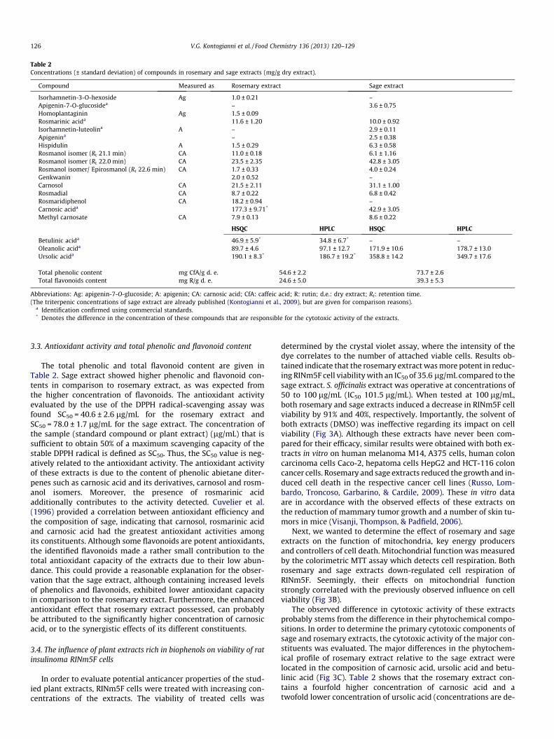
Table 2Concentrations (± standard deviation) of compounds in rosemary and sage extracts (mg/g dry extract).
Compound Measured as Rosemary extract Sage extract
Isorhamnetin-3-O-hexoside Ag 1.0 ± 0.21 –Apigenin-7-O-glucosidea – 3.6 ± 0.75Homoplantaginin Ag 1.5 ± 0.09Rosmarinic acida 11.6 ± 1.20 10.0 ± 0.92Isorhamnetin-luteolina A – 2.9 ± 0.11Apigenina – 2.5 ± 0.38Hispidulin A 1.5 ± 0.29 6.3 ± 0.58Rosmanol isomer (Rt 21.1 min) CA 11.0 ± 0.18 6.1 ± 1.16Rosmanol isomer (Rt 22.0 min) CA 23.5 ± 2.35 42.8 ± 3.05Rosmanol isomer/ Epirosmanol (Rt 22.6 min) CA 1.7 ± 0.33 4.0 ± 0.24Genkwanin 2.0 ± 0.52 –Carnosol CA 21.5 ± 2.11 31.1 ± 1.00Rosmadial CA 8.7 ± 0.22 6.8 ± 0.42Rosmaridiphenol CA 18.2 ± 0.94 –Carnosic acida 177.3 ± 9.71* 42.9 ± 3.05Methyl carnosate CA 7.9 ± 0.13 8.6 ± 0.22
HSQC HPLC HSQC HPLC
Betulinic acida 46.9 ± 5.9* 34.8 ± 6.7* – –Oleanolic acida 89.7 ± 4.6 97.1 ± 12.7 171.9 ± 10.6 178.7 ± 13.0Ursolic acida 190.1 ± 8.3* 186.7 ± 19.2* 358.8 ± 14.2 349.7 ± 17.6
Total phenolic content mg CfA/g d. e. 54.6 ± 2.2 73.7 ± 2.6Total flavonoids content mg R/g d. e. 24.6 ± 5.0 39.3 ± 5.3
Abbreviations: Ag: apigenin-7-O-glucoside; A: apigenin; CA: carnosic acid; CfA: caffeic acid; R: rutin; d.e.: dry extract; Rt: retention time.(The triterpenic concentrations of sage extract are already published (Kontogianni et al., 2009), but are given for comparison reasons).
a Identification confirmed using commercial standards.* Denotes the difference in the concentration of these compounds that are responsible for the cytotoxic activity of the extracts.
126 V.G. Kontogianni et al. / Food Chemistry 136 (2013) 120–129
3.3. Antioxidant activity and total phenolic and flavonoid content
The total phenolic and total flavonoid content are given inTable 2. Sage extract showed higher phenolic and flavonoid con-tents in comparison to rosemary extract, as was expected fromthe higher concentration of flavonoids. The antioxidant activityevaluated by the use of the DPPH radical-scavenging assay wasfound SC50 = 40.6 ± 2.6 lg/mL for the rosemary extract andSC50 = 78.0 ± 1.7 lg/mL for the sage extract. The concentration ofthe sample (standard compound or plant extract) (lg/mL) that issufficient to obtain 50% of a maximum scavenging capacity of thestable DPPH radical is defined as SC50. Thus, the SC50 value is neg-atively related to the antioxidant activity. The antioxidant activityof these extracts is due to the content of phenolic abietane diter-penes such as carnosic acid and its derivatives, carnosol and rosm-anol isomers. Moreover, the presence of rosmarinic acidadditionally contributes to the activity detected. Cuvelier et al.(1996) provided a correlation between antioxidant efficiency andthe composition of sage, indicating that carnosol, rosmarinic acidand carnosic acid had the greatest antioxidant activities amongits constituents. Although some flavonoids are potent antioxidants,the identified flavonoids made a rather small contribution to thetotal antioxidant capacity of the extracts due to their low abun-dance. This could provide a reasonable explanation for the obser-vation that the sage extract, although containing increased levelsof phenolics and flavonoids, exhibited lower antioxidant capacityin comparison to the rosemary extract. Furthermore, the enhancedantioxidant effect that rosemary extract possessed, can probablybe attributed to the significantly higher concentration of carnosicacid, or to the synergistic effects of its different constituents.
3.4. The influence of plant extracts rich in biophenols on viability of ratinsulinoma RINm5F cells
In order to evaluate potential anticancer properties of the stud-ied plant extracts, RINm5F cells were treated with increasing con-centrations of the extracts. The viability of treated cells was
determined by the crystal violet assay, where the intensity of thedye correlates to the number of attached viable cells. Results ob-tained indicate that the rosemary extract was more potent in reduc-ing RINm5F cell viability with an IC50 of 35.6 lg/mL compared to thesage extract. S. officinalis extract was operative at concentrations of50 to 100 lg/mL (IC50 101.5 lg/mL). When tested at 100 lg/mL,both rosemary and sage extracts induced a decrease in RINm5F cellviability by 91% and 40%, respectively. Importantly, the solvent ofboth extracts (DMSO) was ineffective regarding its impact on cellviability (Fig 3A). Although these extracts have never been com-pared for their efficacy, similar results were obtained with both ex-tracts in vitro on human melanoma M14, A375 cells, human coloncarcinoma cells Caco-2, hepatoma cells HepG2 and HCT-116 coloncancer cells. Rosemary and sage extracts reduced the growth and in-duced cell death in the respective cancer cell lines (Russo, Lom-bardo, Troncoso, Garbarino, & Cardile, 2009). These in vitro dataare in accordance with the observed effects of these extracts onthe reduction of mammary tumor growth and a number of skin tu-mors in mice (Visanji, Thompson, & Padfield, 2006).
Next, we wanted to determine the effect of rosemary and sageextracts on the function of mitochondria, key energy producersand controllers of cell death. Mitochondrial function was measuredby the colorimetric MTT assay which detects cell respiration. Bothrosemary and sage extracts down-regulated cell respiration ofRINm5F. Seemingly, their effects on mitochondrial functionstrongly correlated with the previously observed influence on cellviability (Fig 3B).
The observed difference in cytotoxic activity of these extractsprobably stems from the difference in their phytochemical compo-sitions. In order to determine the primary cytotoxic components ofsage and rosemary extracts, the cytotoxic activity of the major con-stituents was evaluated. The major differences in the phytochem-ical profile of rosemary extract relative to the sage extract werelocated in the composition of carnosic acid, ursolic acid and betu-linic acid (Fig 3C). Table 2 shows that the rosemary extract con-tains a fourfold higher concentration of carnosic acid and atwofold lower concentration of ursolic acid (concentrations are de-
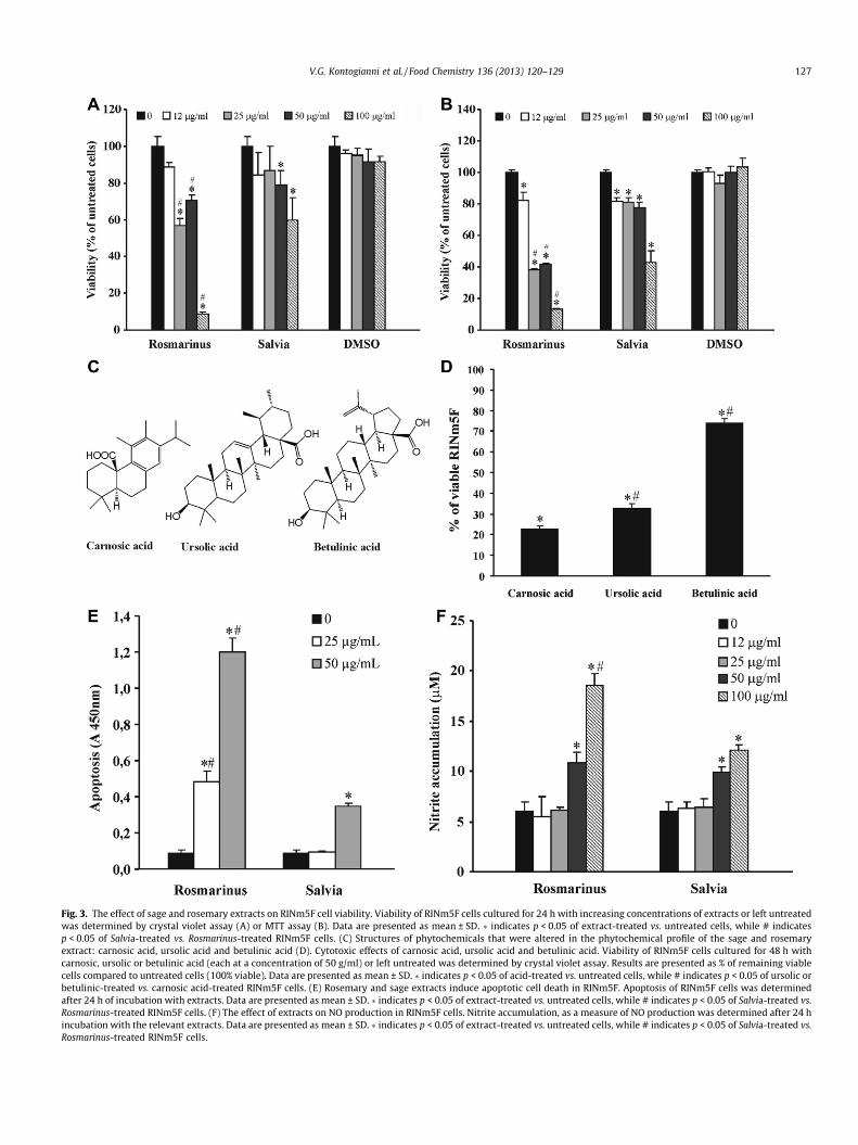
Fig. 3. The effect of sage and rosemary extracts on RINm5F cell viability. Viability of RINm5F cells cultured for 24 h with increasing concentrations of extracts or left untreatedwas determined by crystal violet assay (A) or MTT assay (B). Data are presented as mean ± SD. � indicates p < 0.05 of extract-treated vs. untreated cells, while # indicatesp < 0.05 of Salvia-treated vs. Rosmarinus-treated RINm5F cells. (C) Structures of phytochemicals that were altered in the phytochemical profile of the sage and rosemaryextract: carnosic acid, ursolic acid and betulinic acid (D). Cytotoxic effects of carnosic acid, ursolic acid and betulinic acid. Viability of RINm5F cells cultured for 48 h withcarnosic, ursolic or betulinic acid (each at a concentration of 50 g/ml) or left untreated was determined by crystal violet assay. Results are presented as % of remaining viablecells compared to untreated cells (100% viable). Data are presented as mean ± SD. � indicates p < 0.05 of acid-treated vs. untreated cells, while # indicates p < 0.05 of ursolic orbetulinic-treated vs. carnosic acid-treated RINm5F cells. (E) Rosemary and sage extracts induce apoptotic cell death in RINm5F. Apoptosis of RINm5F cells was determinedafter 24 h of incubation with extracts. Data are presented as mean ± SD. � indicates p < 0.05 of extract-treated vs. untreated cells, while # indicates p < 0.05 of Salvia-treated vs.Rosmarinus-treated RINm5F cells. (F) The effect of extracts on NO production in RINm5F cells. Nitrite accumulation, as a measure of NO production was determined after 24 hincubation with the relevant extracts. Data are presented as mean ± SD. � indicates p < 0.05 of extract-treated vs. untreated cells, while # indicates p < 0.05 of Salvia-treated vs.Rosmarinus-treated RINm5F cells.
V.G. Kontogianni et al. / Food Chemistry 136 (2013) 120–129 127
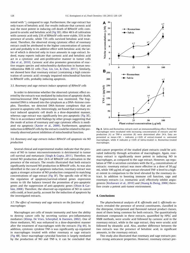
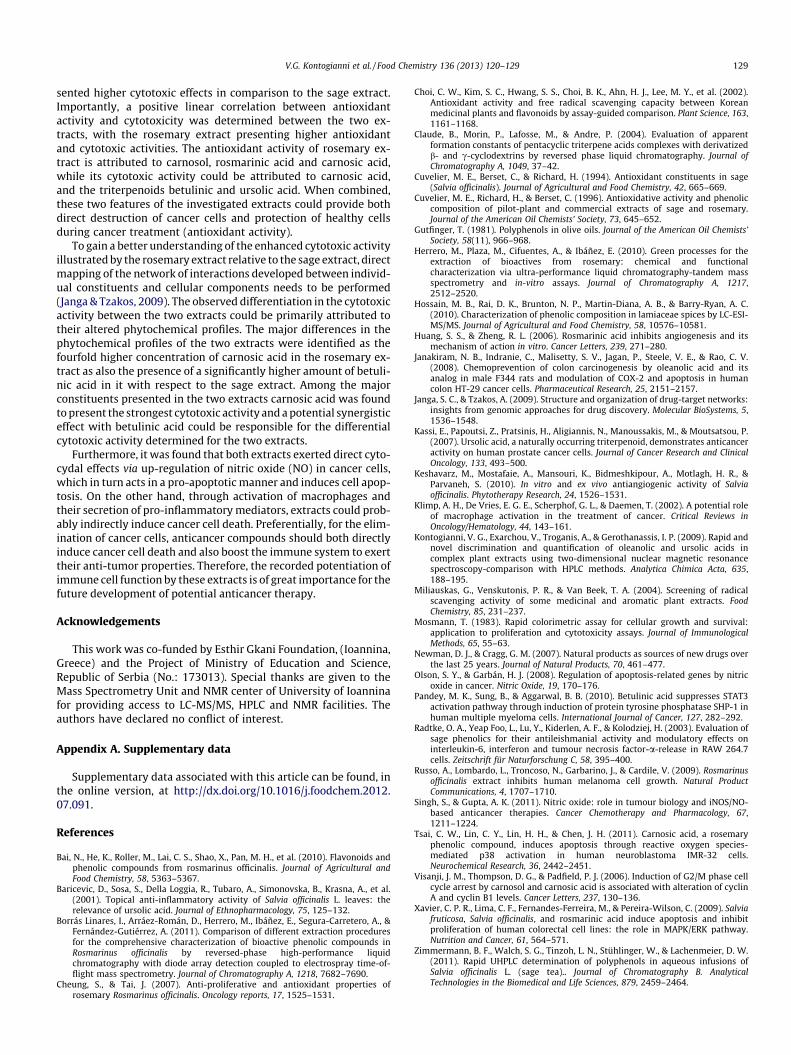
Fig. 4. Salvia and Rosmarinus extracts exert an immunomodifying effect. Peritonealmacrophages were incubated with increasing concentrations of extracts and NOproduction (A) or TNF-a secretion (B) were determined after 24 h. Data arepresented as mean ± SD. � indicates p < 0.05 of extract-treated vs. untreatedmacrophages, while # indicates p < 0.05 of Salvia-treated vs. Rosmarinus-treatedmacrophages.
128 V.G. Kontogianni et al. / Food Chemistry 136 (2013) 120–129
noted with ⁄), compared to sage. Furthermore, the sage extract hasonly traces of betulinic acid. Our results indicate that carnosic acidwas the most potent in inducing cell death of RINm5F cells com-pared to ursolic and betulinic acid (Fig 3D). After 48 h of cultivationwith carnosic acid only 23% of RINm5F cells were viable, 33% in thepresence of ursolic, while 73% cells survived betulinic acid treat-ment. Therefore, the observed strong cytotoxic effect of rosemaryextract could be attributed to the higher concentration of carnosicacid and probably to its additive effect with betulinic acid, the lat-ter of which is detected only in trace amounts in sage extract. In-deed, many reports indicate that carnosic acid and betulinic acidact in a cytotoxic and anti-proliferative manner in tumor cells(Bai et al., 2010). Carnosic acid also promotes generation of reac-tive oxygen species and mitochondria dysfunction in human neu-roblastoma IMR-32 cells (Tsai, Lin, Lin, & Chen, 2011). Similarly,we showed here that rosemary extract (containing a high concen-tration of carnosic acid) strongly impaired mitochondrial functionin RINm5F cells, probably inducing apoptosis.
3.5. Rosemary and sage extracts induce apoptosis of RINm5F cells
In order to determine whether the observed cytotoxic effect ex-erted by the extracts was mediated by induction of apoptotic death,internucleosomal DNA fragmentation was monitored. The frag-mented DNA is released into the cytoplasm as a DNA–histone com-plex. Therefore, we detected DNA–histone complexes that arepresent in apoptotic cells. Interestingly, we found that rosemary ex-tract induced apoptotic cell death in a dose-dependent manner,whereas sage extract was significantly less pro-apoptotic (Fig 3E).This is in accordance with findings by other groups suggesting thatthe mode of action of rosemary and sage cytotoxic effects is medi-ated via apoptosis (Xavier et al., 2009). Furthermore, apoptosisinduction in RINm5F cells by the extracts could be related to the pre-viously observed potent inhibition of mitochondrial function.
3.6. Rosemary and sage extracts induce RINm5F cell apoptosis via NOproduction
Several clinical and experimental studies indicate that the pres-ence of NO in tumor microenvironments is detrimental to tumorcell survival and metastasis (Singh & Gupta, 2011). Therefore, wetested NO production after 24 h of RINm5F cell cultivation in thepresence of the extracts. The results illustrated that both extractssignificantly increased NO production in RINm5F cells. As was alsoidentified in the case of apoptosis induction, rosemary extract wasagain a stronger activator of NO production compared to matchingconcentrations of sage extract (Fig 3F). The specific role of NO inthe regulation of apoptosis/survival-related genes expressionseems to tilt the balance toward the promotion of pro-apoptoticgenes and the suppression of anti-apoptotic genes (Olson & Gar-bán, 2008). Therefore, the observed up-regulation of NO in cancercells could, at least partly, account for the induction of apoptosis bythe investigated extracts.
3.7. The effect of rosemary and sage extracts on the function ofmacrophages
Macrophages are cells of innate immunity and have the abilityto destroy cancer cells by secreting various pro-inflammatorymediators (Klimp, De Vries, Scherphof, & Daemen, 2002). One ofthese mediators, NO, was enhanced in a dose-dependent mannerafter the treatment of macrophages with the extracts (Fig 4A). Inaddition, cytotoxic cytokine TNF-a was significantly up-regulatedin macrophages treated with either rosemary or sage extracts(Fig 4B). Since macrophage cytocidal function is largely mediatedby the production of NO and TNF-a, it can be concluded that
anti-cancer properties of the studied plant extracts could be acti-vated indirectly through activation of macrophages. Again, rose-mary extract is significantly more potent in inducing NO inmacrophages, as compared to the sage extract. However, up-regu-lation of TNF-a secretion correlates with the IC50 concentrations ofextracts: rosemary extract was more effective in a dose of 50 lg/mL, while 100 lg/mL of sage extract elevated TNF-a level to a high-er extent in comparison to the level elevated by the rosemary ex-tract. In addition to boosting immune cell function, sage androsemary extracts (i.e. rosmarinic acid) effectively inhibit angio-genesis (Keshavarz et al., 2010) and (Huang & Zheng, 2006) there-fore create a potent anti-tumor environment.
4. Conclusions
The phytochemical analysis of R. officinalis and S. officinalis ex-tracts revealed the presence of several constituents, classified inthe diterpene, triterpenoid and flavonoid natural product classes,most of them being common for both extracts. The quantitativelydominant compounds in these extracts, quantified by HPLC andNMR methods, were ursolic acid followed by carnosic acid in therosemary extract, while in the sage extract, they were ursolic acidfollowed by oleanolic acid. Also, another difference between thetwo extracts was the presence of betulinic acid, in significantamounts, in the rosemary extract.
This study also illustrated that rosemary and sage extracts pos-sess strong anticancer properties. However, rosemary extract pre-
V.G. Kontogianni et al. / Food Chemistry 136 (2013) 120–129 129
sented higher cytotoxic effects in comparison to the sage extract.Importantly, a positive linear correlation between antioxidantactivity and cytotoxicity was determined between the two ex-tracts, with the rosemary extract presenting higher antioxidantand cytotoxic activities. The antioxidant activity of rosemary ex-tract is attributed to carnosol, rosmarinic acid and carnosic acid,while its cytotoxic activity could be attributed to carnosic acid,and the triterpenoids betulinic and ursolic acid. When combined,these two features of the investigated extracts could provide bothdirect destruction of cancer cells and protection of healthy cellsduring cancer treatment (antioxidant activity).
To gain a better understanding of the enhanced cytotoxic activityillustrated by the rosemary extract relative to the sage extract, directmapping of the network of interactions developed between individ-ual constituents and cellular components needs to be performed(Janga & Tzakos, 2009). The observed differentiation in the cytotoxicactivity between the two extracts could be primarily attributed totheir altered phytochemical profiles. The major differences in thephytochemical profiles of the two extracts were identified as thefourfold higher concentration of carnosic acid in the rosemary ex-tract as also the presence of a significantly higher amount of betuli-nic acid in it with respect to the sage extract. Among the majorconstituents presented in the two extracts carnosic acid was foundto present the strongest cytotoxic activity and a potential synergisticeffect with betulinic acid could be responsible for the differentialcytotoxic activity determined for the two extracts.
Furthermore, it was found that both extracts exerted direct cyto-cydal effects via up-regulation of nitric oxide (NO) in cancer cells,which in turn acts in a pro-apoptotic manner and induces cell apop-tosis. On the other hand, through activation of macrophages andtheir secretion of pro-inflammatory mediators, extracts could prob-ably indirectly induce cancer cell death. Preferentially, for the elim-ination of cancer cells, anticancer compounds should both directlyinduce cancer cell death and also boost the immune system to exerttheir anti-tumor properties. Therefore, the recorded potentiation ofimmune cell function by these extracts is of great importance for thefuture development of potential anticancer therapy.
Acknowledgements
This work was co-funded by Esthir Gkani Foundation, (Ioannina,Greece) and the Project of Ministry of Education and Science,Republic of Serbia (No.: 173013). Special thanks are given to theMass Spectrometry Unit and NMR center of University of Ioanninafor providing access to LC-MS/MS, HPLC and NMR facilities. Theauthors have declared no conflict of interest.
Appendix A. Supplementary data
Supplementary data associated with this article can be found, inthe online version, at http://dx.doi.org/10.1016/j.foodchem.2012.07.091.
References
Bai, N., He, K., Roller, M., Lai, C. S., Shao, X., Pan, M. H., et al. (2010). Flavonoids andphenolic compounds from rosmarinus officinalis. Journal of Agricultural andFood Chemistry, 58, 5363–5367.
Baricevic, D., Sosa, S., Della Loggia, R., Tubaro, A., Simonovska, B., Krasna, A., et al.(2001). Topical anti-inflammatory activity of Salvia officinalis L. leaves: therelevance of ursolic acid. Journal of Ethnopharmacology, 75, 125–132.
Borrás Linares, I., Arráez-Román, D., Herrero, M., Ibáñez, E., Segura-Carretero, A., &Fernández-Gutiérrez, A. (2011). Comparison of different extraction proceduresfor the comprehensive characterization of bioactive phenolic compounds inRosmarinus officinalis by reversed-phase high-performance liquidchromatography with diode array detection coupled to electrospray time-of-flight mass spectrometry. Journal of Chromatography A, 1218, 7682–7690.
Cheung, S., & Tai, J. (2007). Anti-proliferative and antioxidant properties ofrosemary Rosmarinus officinalis. Oncology reports, 17, 1525–1531.
Choi, C. W., Kim, S. C., Hwang, S. S., Choi, B. K., Ahn, H. J., Lee, M. Y., et al. (2002).Antioxidant activity and free radical scavenging capacity between Koreanmedicinal plants and flavonoids by assay-guided comparison. Plant Science, 163,1161–1168.
Claude, B., Morin, P., Lafosse, M., & Andre, P. (2004). Evaluation of apparentformation constants of pentacyclic triterpene acids complexes with derivatizedb- and c-cyclodextrins by reversed phase liquid chromatography. Journal ofChromatography A, 1049, 37–42.
Cuvelier, M. E., Berset, C., & Richard, H. (1994). Antioxidant constituents in sage(Salvia officinalis). Journal of Agricultural and Food Chemistry, 42, 665–669.
Cuvelier, M. E., Richard, H., & Berset, C. (1996). Antioxidative activity and phenoliccomposition of pilot-plant and commercial extracts of sage and rosemary.Journal of the American Oil Chemists’ Society, 73, 645–652.
Gutfinger, T. (1981). Polyphenols in olive oils. Journal of the American Oil Chemists’Society, 58(11), 966–968.
Herrero, M., Plaza, M., Cifuentes, A., & Ibáñez, E. (2010). Green processes for theextraction of bioactives from rosemary: chemical and functionalcharacterization via ultra-performance liquid chromatography-tandem massspectrometry and in-vitro assays. Journal of Chromatography A, 1217,2512–2520.
Hossain, M. B., Rai, D. K., Brunton, N. P., Martin-Diana, A. B., & Barry-Ryan, A. C.(2010). Characterization of phenolic composition in lamiaceae spices by LC-ESI-MS/MS. Journal of Agricultural and Food Chemistry, 58, 10576–10581.
Huang, S. S., & Zheng, R. L. (2006). Rosmarinic acid inhibits angiogenesis and itsmechanism of action in vitro. Cancer Letters, 239, 271–280.
Janakiram, N. B., Indranie, C., Malisetty, S. V., Jagan, P., Steele, V. E., & Rao, C. V.(2008). Chemoprevention of colon carcinogenesis by oleanolic acid and itsanalog in male F344 rats and modulation of COX-2 and apoptosis in humancolon HT-29 cancer cells. Pharmaceutical Research, 25, 2151–2157.
Janga, S. C., & Tzakos, A. (2009). Structure and organization of drug-target networks:insights from genomic approaches for drug discovery. Molecular BioSystems, 5,1536–1548.
Kassi, E., Papoutsi, Z., Pratsinis, H., Aligiannis, N., Manoussakis, M., & Moutsatsou, P.(2007). Ursolic acid, a naturally occurring triterpenoid, demonstrates anticanceractivity on human prostate cancer cells. Journal of Cancer Research and ClinicalOncology, 133, 493–500.
Keshavarz, M., Mostafaie, A., Mansouri, K., Bidmeshkipour, A., Motlagh, H. R., &Parvaneh, S. (2010). In vitro and ex vivo antiangiogenic activity of Salviaofficinalis. Phytotherapy Research, 24, 1526–1531.
Klimp, A. H., De Vries, E. G. E., Scherphof, G. L., & Daemen, T. (2002). A potential roleof macrophage activation in the treatment of cancer. Critical Reviews inOncology/Hematology, 44, 143–161.
Kontogianni, V. G., Exarchou, V., Troganis, A., & Gerothanassis, I. P. (2009). Rapid andnovel discrimination and quantification of oleanolic and ursolic acids incomplex plant extracts using two-dimensional nuclear magnetic resonancespectroscopy-comparison with HPLC methods. Analytica Chimica Acta, 635,188–195.
Miliauskas, G., Venskutonis, P. R., & Van Beek, T. A. (2004). Screening of radicalscavenging activity of some medicinal and aromatic plant extracts. FoodChemistry, 85, 231–237.
Mosmann, T. (1983). Rapid colorimetric assay for cellular growth and survival:application to proliferation and cytotoxicity assays. Journal of ImmunologicalMethods, 65, 55–63.
Newman, D. J., & Cragg, G. M. (2007). Natural products as sources of new drugs overthe last 25 years. Journal of Natural Products, 70, 461–477.
Olson, S. Y., & Garbán, H. J. (2008). Regulation of apoptosis-related genes by nitricoxide in cancer. Nitric Oxide, 19, 170–176.
Pandey, M. K., Sung, B., & Aggarwal, B. B. (2010). Betulinic acid suppresses STAT3activation pathway through induction of protein tyrosine phosphatase SHP-1 inhuman multiple myeloma cells. International Journal of Cancer, 127, 282–292.
Radtke, O. A., Yeap Foo, L., Lu, Y., Kiderlen, A. F., & Kolodziej, H. (2003). Evaluation ofsage phenolics for their antileishmanial activity and modulatory effects oninterleukin-6, interferon and tumour necrosis factor-a-release in RAW 264.7cells. Zeitschrift für Naturforschung C, 58, 395–400.
Russo, A., Lombardo, L., Troncoso, N., Garbarino, J., & Cardile, V. (2009). Rosmarinusofficinalis extract inhibits human melanoma cell growth. Natural ProductCommunications, 4, 1707–1710.
Singh, S., & Gupta, A. K. (2011). Nitric oxide: role in tumour biology and iNOS/NO-based anticancer therapies. Cancer Chemotherapy and Pharmacology, 67,1211–1224.
Tsai, C. W., Lin, C. Y., Lin, H. H., & Chen, J. H. (2011). Carnosic acid, a rosemaryphenolic compound, induces apoptosis through reactive oxygen species-mediated p38 activation in human neuroblastoma IMR-32 cells.Neurochemical Research, 36, 2442–2451.
Visanji, J. M., Thompson, D. G., & Padfield, P. J. (2006). Induction of G2/M phase cellcycle arrest by carnosol and carnosic acid is associated with alteration of cyclinA and cyclin B1 levels. Cancer Letters, 237, 130–136.
Xavier, C. P. R., Lima, C. F., Fernandes-Ferreira, M., & Pereira-Wilson, C. (2009). Salviafruticosa, Salvia officinalis, and rosmarinic acid induce apoptosis and inhibitproliferation of human colorectal cell lines: the role in MAPK/ERK pathway.Nutrition and Cancer, 61, 564–571.
Zimmermann, B. F., Walch, S. G., Tinzoh, L. N., Stühlinger, W., & Lachenmeier, D. W.(2011). Rapid UHPLC determination of polyphenols in aqueous infusions ofSalvia officinalis L. (sage tea).. Journal of Chromatography B. AnalyticalTechnologies in the Biomedical and Life Sciences, 879, 2459–2464.
Copyright © 2022 FDOKUMEN