Modulation of mast cell proliferative and inflammatory responses by Leukotriene D4 and Stem Cell...
31
1 Original Research Article Modulation of mast cell proliferative and inflammatory responses by Leukotriene D 4 and Stem Cell Factor signaling interactions † Nosayba Al-Azzam 1 , Vinay Kondeti 1 , Ernest Duah 1 , Farai Gombedza 1 , Charles K. Thodeti 2 and Sailaja Paruchuri 1* 1 Department of Chemistry, University of Akron, Akron, OH, 2 Department of Integrative Medical Sciences, Northeast Ohio Medical University, Rootstown, OH Key words: calcium, c-fos, c-Kit, cys -LTs, LTD 4 , mast cells, MIP1β, proliferation, calcium, stem cell factor Running head: Cross-talk between SCF and LTD 4 in inflammation *Corresponding Author: Sailaja Paruchuri, PhD, Department of Chemistry, KNCL 406, 185 E Mill street, Akron, OH 44325; Phone: 3309722193; E-mail: [email protected] Contract grant sponsor: NHLBI; Contract grant number:HL098953. Contract grant sponsor: James Foght Assistant Professorship † This article has been accepted for publication and undergone full peer review but has not been through the copyediting, typesetting, pagination and proofreading process, which may lead to differences between this version and the Version of Record. Please cite this article as doi: [10.1002/jcp.24777] Additional Supporting Information may be found in the online version of this article. Received 26 March 2014; Revised 31 July 2014; Accepted 22 August 2014 Journal of Cellular Physiology © 2014 Wiley Periodicals, Inc. DOI 10.1002/jcp.24777
-
Upload
independent -
Category
Documents
-
view
2 -
download
0
Transcript of Modulation of mast cell proliferative and inflammatory responses by Leukotriene D4 and Stem Cell...

1
Original Research Article
Modulation of mast cell proliferative and inflammatory responses by Leukotriene D4 and Stem Cell Factor signaling interactions†
Nosayba Al-Azzam1, Vinay Kondeti1, Ernest Duah1, Farai Gombedza1, Charles K. Thodeti2 and Sailaja Paruchuri1*
1 Department of Chemistry, University of Akron, Akron, OH, 2 Department of Integrative Medical Sciences, Northeast Ohio Medical University, Rootstown, OH
Key words: calcium, c-fos, c-Kit, cys-LTs, LTD4, mast cells, MIP1β, proliferation, calcium, stem cell factor
Running head: Cross-talk between SCF and LTD4 in inflammation
*Corresponding Author: Sailaja Paruchuri, PhD, Department of Chemistry, KNCL 406, 185 E Mill street, Akron, OH 44325; Phone: 3309722193; E-mail: [email protected]
Contract grant sponsor: NHLBI; Contract grant number:HL098953. Contract grant sponsor: James Foght Assistant Professorship
†This article has been accepted for publication and undergone full peer review but has not been through the copyediting, typesetting, pagination and proofreading process, which may lead to differences between this version and the Version of Record. Please cite this article as doi: [10.1002/jcp.24777] Additional Supporting Information may be found in the online version of this article.
Received 26 March 2014; Revised 31 July 2014; Accepted 22 August 2014 Journal of Cellular Physiology © 2014 Wiley Periodicals, Inc.
DOI 10.1002/jcp.24777

2
Abstract
Mast cells (MCs) are important effector cells in asthma and pulmonary inflammation,
and their proliferation and maturation is maintained by stem cell factor (SCF) via its
receptor, c-Kit. Cysteinyl leukotrienes (cys-LTs) are potent inflammatory mediators that
signal through CysLT1R and CysLT2R located on the MC surface, and they enhance
MC inflammatory responses. However, it is not known if SCF and cys-LTs cross-talk
and influence MC hyperplasia and activation in inflammation. Here, we report the
concerted effort of the growth factor SCF and the inflammatory mediator LTD4 in MC
activation. Stimulation of MCs by LTD4 in the presence of SCF enhances c-Kit-mediated
proliferative responses. Similarly, SCF synergistically enhances LTD4-induced calcium,
c-fos expression and phosphorylation, as well as MIP1β generation in MCs. These
findings suggest that integration of SCF and LTD4 signals may contribute to MC
hyperplasia and hyper-reactivity during airway hyper-response and inflammation.

3
Introduction
Mast cells (MCs) are stem cell factor (SCF)-dependent hematopoietic cells that are
ubiquitously distributed throughout the body (Gurish and Boyce, 2006; Wedemeyer et
al., 2000) and they initiate inflammatory responses to allergens and infectious agents
(Okayama and Kawakami, 2006). MCs play an important role in asthma through the
secretion of several soluble inflammatory mediators. c-Kit is a member of the type III
subclass of receptor tyrosine kinases, comprising of an N-terminal extracellular ligand
binding domain, a transmembrane domain and a cytoplasmic kinase domain, which is
activated by ligand-mediated receptor dimerization (Mani et al., 2009). C-Kit activation
by SCF is crucial for the survival and proliferation of human MCs and and the
deregulation of the c-kit/SCF axis can lead to uncontrolled proliferation as seen during
systemic mastocytosis. Mastocytosis is the disturbance of the homeostatic mechanisms
that control the accumulation, proliferation, survival, and turnover rates of MCs,
contributing to inflammation, and remodeling. The mechanistic basis of MC hyperplasia
in asthma (or in any allergic disease) is not completely understood. The importance of
c-Kit signaling in MC proliferation makes it crucial to understand the basic mechanisms
by which Kit regulates MC function. Although the role of c-Kit signaling is extensively
studied in porcine aortic endothelial cells (Blume-Jensen et al., 1994; Blume-Jensen et
al., 1993), its role in MC proliferation is still not well known.
Cysteinyl leukotrienes (cys-LTs), comprising of LTC4, LTD4 and LTE4 are potent
bronchoconstrictors and they play an important role in asthma and airway inflammation
(Davidson et al., 1987; Drazen and Austen, 1987). They are derivatives of arachidonic
acid generated by MCs, eosinophils, basophils, macrophages, and myeloid dendritic

4
cells (Kanaoka and Boyce, 2004), and act through two main receptors, CysLT1R and
CysLT2R (Heise et al., 2000; Lynch et al., 1999). MCs not only generate cys-LTs, but
also express CysLT1R and CysLT2R (Mellor et al., 2003; Mellor et al., 2001). We and
others have previously shown that stimulation of human cord blood-derived MCs
(hMCs) with LTD4 potently induces calcium flux and cytokine generation through
CysLT1R (Mellor et al., 2002; Paruchuri et al., 2008).
During inflammation, various mediators prime each other’s responses, resulting
in amplified inflammatory milieu. For example, SCF-induced prolonged activation of
mast cells has been shown to play critical role in the progression of allergen-induced
airway hyper responsiveness (AHR) and chronic airway hypersensitivity (Hundley et al.,
2004). Also, SCF has been implicated in the induction of airway hyper-reactivity during
allergy-induced pulmonary responses in mouse models (Campbell et al., 1999).
Interestingly, SCF-induced airway hyper-reactivity has been shown to depend on
leukotriene production (Oliveira et al., 2001). Enhanced proliferation of MCs is
commonly seen at the site of inflammation and this increase in MC number correlates
with the severity of AHR. Given that both SCF and leukotrienes are produced at the site
of inflammation and induce AHR together with the fact that MCs express receptors for
both SCF and leukotrienes and enhanced MC proliferation is seen during
inflammation, we asked if c-Kit and CysLT1R can cross-talk and influence MC
proliferatory and inflammatory phenotypes. Therefore, in the current study, we tested
the MC proliferatory and inflammatory responses in the presence of both SCF and
LTD4. We hypothesized that the growth factor SCF can boost LTD4-mediated
inflammatory signals and an inflammatory mediator, LTD4, can increase SCF-induced,

5
c-Kit-mediated proliferative responses. Our results suggest that LTD4 and SCF
synergistically enhance MC proliferative (c-Kit phosphorylation and proliferation) and
inflammatory responses (c-fos phosphorylation, expression, and MIP1β production).
Materials and Methods
Reagents
LTC4, LTD4, LTE4, and MK571 were purchased from Cayman Chemicals, Fura-2 AM
from Molecular Probes, All phospho-specific antibodies and corresponding controls from
Cell Signaling Technology, GAPDH antibody from Fitzgerald (Acton, MA), XTT
proliferation assay kit was from Trevigen (Gaithersburg, MD), BrdU proliferation assay
kit was from Calbiochem, MIP1β ELISA kit was from R&D systems. All cytokines were
purchased from R&D systems.
Cell culture
The LAD2 MC leukemia line (Kirshenbaum et al., 2003) was a generous gift from Dr.
Arnold Kirshenbaum, NIH. LAD2 cells were cultured in stemPro-34 (Invitrogen)
supplemented with 2mM L-Glutamine (Invitrogen), Pen-strep (100 IU/ml) (Invitrogen),
and SCF (R&D systems) (100 ng/ml). Cell culture medium was hemi-depleted every
week with fresh medium and 100 ng/ml SCF. Cells were SCF-starved overnight before
stimulating with SCF in all the experiments. LAD2 cells are dependent on SCF for their
proliferation and retain excellent responses to cys-LTs in inducing calcium flux,
secretion of MIP1β and other chemokines, similar to isolated hMCs. (Paruchuri et al.,
2008; Paruchuri et al., 2009). The expression pattern of cys-LT receptors in these cells
are also similar to hMCs with high levels of CysLT1R compared to CysLT2R (Paruchuri

6
et al., 2008). Bone marrow derived mast cells (BMMCs) were isolated from C57BL/6
mice. Mice were euthanized according to guidelines and approval of the Institutional
Animal Care and Use Committee (IACUC) of the Northeast Ohio Medical University
(NEOMED). BMMCs were cultured in 80% RPMI 1640 supplemented with 10% fetal
bovine serum , 2mM L-Glutamine, Pen-Strep (100UI/ml), Sodium Pyruvate (1mM), Non-
Essential Amino acids, HEPES buffer (25mM) and ß-Mercaptoethanol (50uM) and 20%
WEHI-3 cell conditioned medium for 4-6 weeks. Maturity of BMMCs was examined by
Toluidine blue staining and >90% mature BMMCs were used for experiments.
Cord blood was obtained from Cleve land Cord Blood Center and MCs were
isolated as described (Mellor et al., 2002). Briefly, heparin-treated cord blood was
sedimented with 4.5% dextran solution and the buffy coat was layered onto Ficoll-
Hypaque and mononuclear cells (MNC) were obtained after centrifugation at the
interphase. Erythrocytes were further removed from MNC by hypotonic lysis and
cultured in RPMI-1640 (Gibco), 10%FBS, 2 mM L-glutamine, 0.1 mM non- essential
amino acids, Penicillin-Streptomycin, Gentamicin and 0.2µM 2-Mercaptoethanol in the
presence of SCF (100ng/ml), IL -6 (50 ng/ml) and IL -10 (10 ng/ml). Non-adherent cells
were transferred to fresh medium containing cytokines every week for 6-9 weeks.
Maturity of hMCs was examined by Toluidine blue staining and >90% mature hMCs
were used for experiments.
Calcium flux
Cells were cultured in SCF-free medium overnight. Thereafter, cells (0.5-1 x 10⁶/
sample) were washed and labeled with fura 2-AM for 30 minutes at 37oC. Cells were
further washed and stimulated with 500 nM of LTD4 and/or 100 ng/ml of SCF, and the

7
changes in intracellular calcium were measured using excitation at 340 and 380nm in a
fluorescence spectrophotometer (Hitachi F-4500) as described earlier (Paruchuri et al.,
2008). In some experiments, MK571 was added 10 minutes before the addition of
indicated agonists. The relative ratios of fluorescence emitted at 510 nm were recorded
and displayed as a reflection of intracellular calcium concentration.
Cell activation
Cells were either stimulated with 500 nM LTD4 and/or 100 ng/ml SCF (or with indicated
concentrations) for indicated time points (phosphorylation of c-Kit for 15 min,
phosphorylation and expression of c-fos for 1h, measurement of cytokines at transcript
level for 2h, and protein level for 6h) . The concentration of MIP1β was measured with
ELISAs according to the manufacturer’s protocol (Paruchuri et al., 2008).
Cell lysates and western blotting
After stimulation with the respective agonists, MCs (0.5X10⁶) were lysed with lysis buffer
(BD Bioscience) supplemented with protease inhibitor cocktail (Roche) and
phosphatase inhibitor cocktail (Pierce). Immunoblotting was performed as described
previously (Paruchuri et al., 2002). Briefly, lysates were subjected to 4-12% SDS-PAGE
and transferred to PVDF membrane. Membranes were incubated with respective
primary phospho- and total antibodies diluted in 1x TBS, 5% dry milk, 0.1% Tween-20
(1:1000) overnight at 40C on shaker, and then with secondary antibody (peroxidase-
conjugated anti-rabbit or anti-mouse). Western blot was incubated with ECL, and the
bands were visualized using imager (Protein Simple) and quantified using Alpha View
software (Protein Simple).
8
Real-time Quantitative PCR
The expressions of COX-2, MIP1β and TNFa transcripts were determined with real-time
PCR performed on Light cycler 480 (Roche). Cells were cultured and treated as
described above, and total RNA was isolated with an RNAeasy minikit (Qiagen)
according to manufacturer’s instructions. RNA concentration was determined using a
Take 3 module of Epoch micro plate reader (Biotek) at 260/280 nm. 1µg of total RNA
was used for reverse transcription using cDNA synthesis kit from Quanta Biosciences,
containing MgCl2, dNTPs, recombinant RNAse inhibitor protein, qScript Reverse
Transcriptase, random primers, oligo (dT) primers and stabilizers. Gene expression was
assayed by quantitative real-time PCR on LightCycler® 480 II (Roche Applied Science)
using LightCycler® 480 SYBR Green I Master mix, cDNA prepared as described above
and COX-2, MIP1β, TNFa and GAPDH forward and reverse Primers. Following are the
forward (F) and reverse (R) p rimers used
MIP1β− F- CCAGCCAGCTGTGGTATT
R-CAGTTCAGTTCCAGGTCATACA
TNFα- F- CCAGGGACCTCTCTCTAATCA
R-TCAGCTTGAGGGTTTGCTAC
COX-2- F-CAACTCTATATTGCTGGAACATGGA
R-TGGAAGCCTGTGATACTTTCTGTACT
GAPDH- F-TGCACCACCAACTGCTTAGC
R-GGCATGGACTGTGGTCATGAG
The ?∆Ct values for COX-2, MIP1β, and TNFa were calculated relative to the GAPDH
levels and values were expressed as fold change over the control (Duah et al., 2013).

9
Cell proliferation
Cells were plated at a density of 5000 cells/well of 96 well plate, cultured in SCF-free
medium overnight, and treated with increasing concentration of SCF and/or 500 nM of
LTD4. After 72 h, the proliferation was assayed either by XTT assay (Trevigen) or BrdU
ELISA (Millipore) according to the manufacturer’s protocol.
Data Analysis
Data is expressed as mean ± SEM from at least three experiments except where
otherwise indicated. Significance was determined using one way ANOVA and post-hoc
analysis.
Results
SCF induces concentration-dependent phosphorylation of c-Kit in MCs
SCF is the major growth factor for the proliferation of MCs, and it relays its responses
through the cell surface receptor, c-Kit (Galli et al., 1995; Iemura et al., 1994; Tsai et al.,
1991). To confirm if c-Kit is activated in response to SCF stimulation in LAD2 cells, we
first determined the effect of SCF on phosphorylation of c-Kit receptor, by treating the
cells with different concentrations of SCF. Stimulation with SCF for 15 minutes led to
dose-dependent phosphorylation (Fig. 1A, B) of c-Kit receptor. SCF induced
phosphorylation of c-Kit receptor at doses as low as 1ng/ml with a maximum response
at 100 ng/ml concentration.
C-Kit phosphorylation is synergistically activated by SCF and LTD4
To determine if there is crosstalk between c-Kit and CysLTRs, we stimulated LAD2 cells
with cys-LTs with or without SCF and analyzed c-Kit phosphorylation. We could not
detect any significant phosphorylation of c-Kit by cys-LTs alone. However, LTD4

10
significantly strengthened SCF-induced c-Kit phosphorylation (Fig. 2A, B, C). We also
found similar synergistic activation of c-Kit by SCF and LTD4 in BMMCs (Suppl. Fig. 1).
This synergism with SCF was not observed with LTC4 or LTE4 treatment (Fig. 2A).
Further, we found that treatment of cells with CysLT1R antagonist, MK571 prior to
stimulation with SCF and LTD4, inhibited this synergistic effect suggesting that it is
mediated through CysLT1R (Fig. 2B, C).
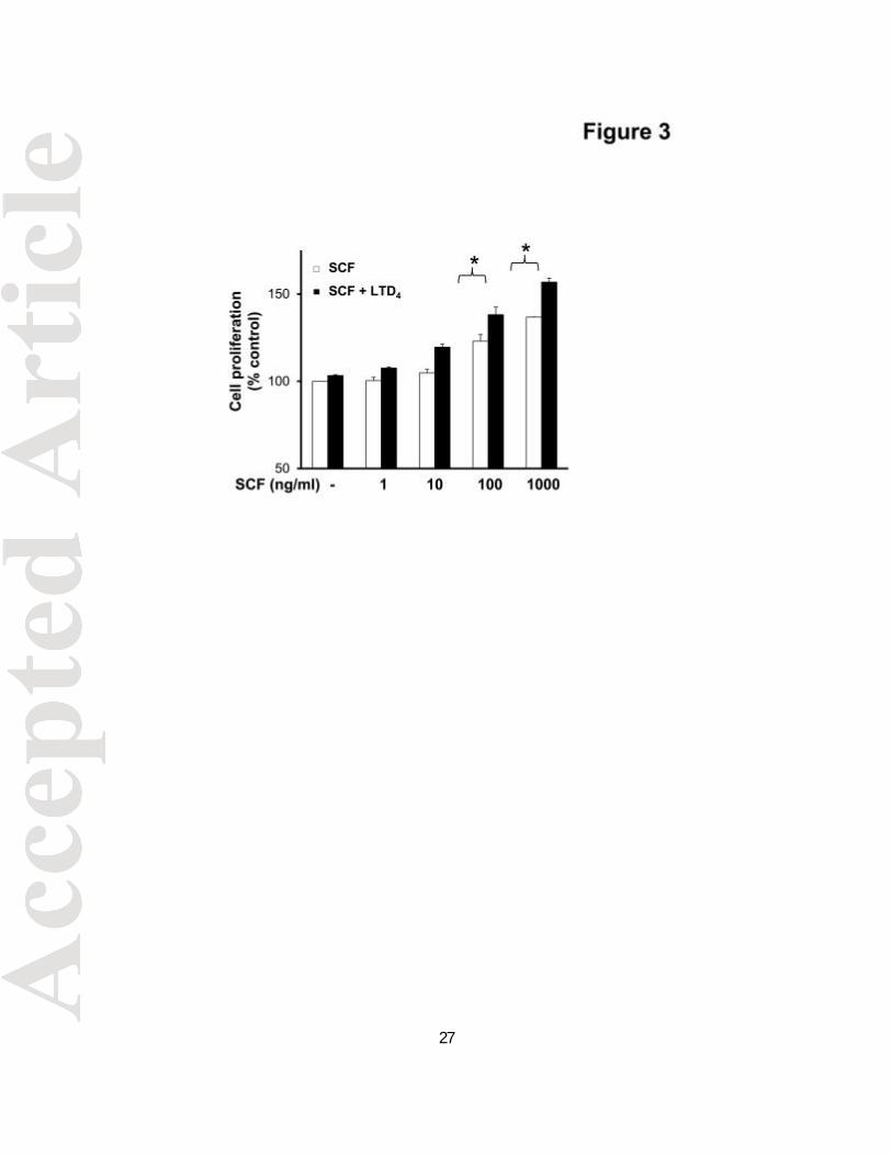
LTD4 potentiates SCF-induced MC proliferation
The fact that LTD4 augmented c-Kit phosphorylation by SCF prompted us further to
understand the significance of this potentiation. We analyzed if LTD4 treatment together
with SCF would amplify LAD2 cell proliferation by using XTT assay. Consistent with
earlier results (Laidlaw et al., 2011), we found that SCF promoted proliferation of LAD2
cells in a dose-dependent manner (Fig. 3). Interestingly, a significant increment in cell
proliferation was observed with combined stimulation of SCF and LTD4 at high doses of
SCF. These findings indicate that LTD4 can enhance c-Kit responses in LAD2 cells and
increase their proliferation. We also found that hMCs cultured in the presence of both
SCF and LTD4 induced around 2 fold increase in proliferation compared to SCF
treatment alone (Suppl. Fig. 2A). Taken together, these results suggest that LTD4 in
concert with SCF can modulate primary mast cell (hMC) and MC cell line (LAD2)
proliferation.
SCF pre-treatment amplifies CysLT1R function as measured by LTD4-induced
calcium flux
Above results clearly depict that combined stimulation of SCF and LTD4 enhances c-
Kit-mediated proliferative responses. Next, we asked if this cross-talk is bi-directional

11
i.e. if combined stimulation with SCF can augment LTD4-mediated inflammatory
responses, the same way as LTD4 can modulate SCF-induced proliferation. We have
previously shown that stimulation of LAD2 cells with LTD4 induces calcium flux, c-fos
phosphorylation and expression and MIP1β generation (Kondeti et al., 2013; Paruchuri
et al., 2008). First, we asked if SCF pre-treatment influences LTD4-mediated calcium
flux. We have previously shown that LTD4 was the most potent agonist among the cys-
LTs for eliciting calcium flux and completely desensitized MC to the calcium fluxes
induced by the two other cys-LTs (Paruchuri et al., 2008). We observed that SCF (100
ng/ml) induced minimal or no calcium flux by itself (Fig. 4B) or followed by priming with
LTD4 (Fig. 4A). However, stimulating cells with SCF prior to LTD4 significantly amplified
LTD4-induced calcium flux (Fig. 4B, C). Further, this calcium flux was inhibited by prior
treatment with CysLT1R antagonist, MK571, confirming that this signal is mediated by
CysLT1R (Fig. 4C).
SCF and LTD4 treatment synergistically amplifies c-fos phosphorylation and
expression
Next, we investigated if SCF has the potential to amplify other LTD4-induced MC
responses such as c-fos phosphorylation and expression. We have reported earlier that
stimulation of LAD2 cells with LTD4 led to phosphorylation and increased expression of
c-fos (Kondeti et al., 2013). SCF treatment alone did not induce significant c-fos
phosphoryla tion or expression, however, both LTD4-induced c-fos phosphorylation and
expression were synergistically enhanced in the presence of SCF (Fig. 5A, B), further
suggesting the ability of SCF/c-Kit to modulate LTD4-induced responses. We also
found that treatment of hMCs with a combination of SCF and LTD4 caused potentiation
12
of c-fos phosphorylation and expression compared to treatment with either of the
agonists (Suppl. Fig.2B) further confirming the synergistic activation in primary MCs.
Amplification of inflammatory signals by LTD4 and SCF treatment
COX-2 activation, MIP1β secretion and up-regulation of other inflammatory cytokine
expression are major MC responses downstream of CysLT1R signaling induced by
LTD4 (Paruchuri et al., 2008; Paruchuri et al., 2009). These responses further enhance
inflammation by recruiting other immune cells. To find out if SCF can increase LTD4-
induced MC inflammatory signals, we measured LTD4-induced inflammatory signals in
the presence or absence of SCF. LTD4 stimulation alone , but not SCF, induced the
expression of COX-2, TNF-a, and MIP1β in LAD2 cells . Interestingly, we found that
LTD4-induced expression of these inflammatory genes is significantly boosted by SCF
(Fig. 6 A, B, C). Finally, consistent with our transcript data, we found that LTD4 and SCF
synergistically increased the secretion of MIP1β protein in LAD2 cells (Fig.6 D).
However, either LTD4 or SCF treatment, or the combination of both failed to induce any
degranulation (data not shown) as determined by β-hexosaminidase assay using p-
NAG (p-Nitrophenyl-N-acetyl-ß-D-glucosaminidine) as substrate.
Discussion
In the present study, we demonstrate that an inflammatory mediator, LTD4, and a
growth factor, SCF synergistically regulate MC function. We clearly demonstrate that
LTD4 can prime SCF-induced c-Kit phosphorylation and proliferation of MCs, and that
SCF in turn can strengthen LTD4-induced calcium flux and inflammatory responses

13
such as c-fos phosphorylation and expression, as well as MIP1β production, suggesting
a potential cross-talk between the LTD4 and SCF receptors, CysLT1R and c-Kit (Fig. 7).
MCs are mediators of the early phase of allergic inflammation. MCs are released
into the blood stream as progenitor cells, and are then recruited to the tissues where
they undergo maturation (Gurish and Boyce, 2006; Kirshenbaum et al., 1999; Metcalfe
et al., 1997). SCF is an important regulator of MC growth, differentiation, survival and
chemotaxis (Grimbaldeston et al., 2006; Prussin and Metcalfe, 2006; Tkaczyk et al.,
2006). SCF acts through c-Kit tyrosine kinase receptor which activates downstream
signaling molecules such as PI3 Kinase and AKT to exert its effects on MCs (Ali et al.,
2004; Moller et al., 2005). SCF was also shown to enhance allergen induced FCεRI
mediated degranulation of MCs and release of inflammatory mediators (Gilfillan and
Tkaczyk, 2006; Ito et al., 2012; Jensen et al., 2007). These inflammatory mediators may
further activate MCs and boost inflammation.
Cys-LTs are produced by MCs and were also shown to activate MCs. We have
previously shown that LAD2 cells express both CysLT1R and CysLT2R and that cys-LTs
(LTC4, LTD4 and LTE4) induce calcium influx, c-fos expression and phosphorylation
(Kondeti et al., 2013; Laidlaw et al., 2011; Paruchuri et al., 2008). Both c-Kit signaling
and CysLTR signaling are shown to be mitogenic for MCs. In addition, CysLT1R has
been shown to transactivate c-Kit receptor in hMCs (Jiang et al., 2006). Interestingly,
patients with systemic mastocytosis showed significantly higher urinary excretion of cys-
LTs than controls (Raithel et al., 2011). Further, LTC4 synthase knockout mice were
unable to develop MC hyperplasia in the inflamed mucosal surface of the lung in a
model of allergen-induced pulmonary inflammation (Kim et al., 2006). However, it is not

14
known if these pro-inflammatory mediators act in concert with SCF. In the present
study, though we did not observe any c-Kit phosphorylation or proliferation with LTD4
alone, stimulation of MCs with SCF induced phosphorylation as well as MC proliferation.
Interestingly, c-Kit phosphorylation and MC proliferation was augmented in the
presence of both SCF and LTD4 suggesting the existence of a cross-talk between their
receptors. LTD4, but not LTC4 or LTE4, increased c-Kit phosphorylation by SCF,
indicating a specific role for LTD4 in this cross-talk. We also showed that this
potentiation of c-Kit is mediated through CysLT1R. Notably, we found that stimulating
MCs with SCF prior to LTD4 enhanced LTD4-induced calcium flux. In contrast, SCF
alone did not induce any calcium flux. Furthermore, we found that c-fos phosphorylation
and expression, COX-2, TNF-a and MIP1β transcript expression, as well as
MIP1β protein secretion are amplified by combined treatment with SCF and LTD4. Thus,
our findings suggest that while LTD4 synergistically activates SCF-induced proliferative
signals, SCF in turn potentiates LTD4-induced inflammatory signals in MCs. The
concept of this bi-directional cross-talk between LTD4 (CysLT1R) and SCF (c-Kit)
signaling is novel and may have important implications in targeting MC-mediated
inflammatory responses. Importantly, our results suggest that the inflammatory
microenvironment, but not a single molecule, dictate the inflammatory phenotype of
MCs. SCF was previously shown to enhance FCεRI mediated responses in MCs
(Gilfillan and Tkaczyk, 2006; Ito et al., 2012; Jensen et al., 2008; Jensen et al., 2007).
However, recently it was shown that while acute treatment of SCF potentiated
inflammatory phenotype of MCs, prolonged treatment of SCF inhibited PGE2 and
allergen-induced MC degranulation (Gilfillan and Tkaczyk, 2006; Ito et al., 2012; Jensen
15
et al., 2007). The underlying molecular mechanism could be alterations in the
cytoskeleton by SCF-mediated regulation of src kinase, hck (Ito et al., 2012; Smrz et al.,
2013).
In conclusion, our results suggest that the cross-talk between SCF and LTD4
induces MC proliferation, gene regulation, and cytokine production. SCF signaling
through c-Kit may regulate baseline maintenance of MCs. However, locally derived cys-
LTs produced at the site of inflammation enhance MC numbers by cross talking with c-
Kit receptor. Similarly, this c-Kit and CysLT1R crosstalk also potentiates LTD4 effects
enhancing MC inflammatory responses leading to enhanced pathological loop (Fig. 7).
Although SCF-induced MC activation in allergen mediated AHR was shown to be
dependent on leukotrienes (Oliveira et al., 2001), it is not known whether cys-LTs can
modulate SCF receptor. Thus, our findings that LTD4 can enhance c-Kit-dependent
proliferative response and SCF potentiate LTD4-mediated inflammatory responses are
very intriguing and may provide basis for novel therapeutic targets for. asthma and
allergic diseases
Acknowledgements
This work is supported by National Institutes of Health Grants (HL098953) and by
James Foght Assistant Professor to S.P. The authors have no conflict of interest.
Literature Cited
Ali K, Bilancio A, Thomas M, Pearce W, Gilfillan AM, Tkaczyk C, Kuehn N, Gray A,
Giddings J, Peskett E, Fox R, Bruce I, Walker C, Sawyer C, Okkenhaug K, Finan
16
P, Vanhaesebroeck B. 2004. Essential role for the p110d phosphoinositide 3-
kinase in the allergic response. Nature 431(7011):1007-1011.
Blume-Jensen P, Ronnstrand L, Gout I, Waterfield MD, Heldin CH. 1994. Modulation of
Kit/stem cell factor receptor-induced signaling by protein kinase C. J Biol Chem
269(34):21793-21802.
Blume-Jensen P, Siegbahn A, Stabel S, Heldin CH, Ronnstrand L. 1993. Increased
Kit/SCF receptor induced mitogenicity but abolished cell motility after inhibition of
protein kinase C. Embo J 12(11):4199-4209.
Campbell E, Hogaboam C, Lincoln P, Lukacs NW. 1999. Stem cell factor-induced
airway hyperreactivity in allergic and normal mice. Am J Pathol 154(4):1259-
1265.
Davidson AB, Lee TH, Scanlon PD, Solway J, McFadden ER, Jr., Ingram RH, Jr., Corey
EJ, Austen KF, Drazen JM. 1987. Bronchoconstrictor effects of leukotriene E4 in
normal and asthmatic subjects. Am Rev Respir Dis 135(2):333-337.
Drazen JM, Austen KF. 1987. Leukotrienes and airway responses. Am Rev Respir Dis
136(4):985-998.
Duah E, Adapala RK, Al-Azzam N, Kondeti V, Gombedza F, Thodeti CK, Paruchuri S.
2013. Cysteinyl leukotrienes regulate endothelial cell inflammatory and
proliferative signals through CysLT(2) and CysLT(1) receptors. Sci Rep 3:3274.
Galli SJ, Tsai M, Wershil BK, Tam SY, Costa JJ. 1995. Regulation of mouse and human
mast cell development, survival and function by stem cell factor, the ligand for the
c-kit receptor. Int Arch Allergy Immunol 107(1-3):51-53.
17
Gilfillan AM, Tkaczyk C. 2006. Integrated signalling pathways for mast-cell activation.
Nat Rev Immunol 6(3):218-230.
Grimbaldeston MA, Metz M, Yu M, Tsai M, Galli SJ. 2006. Effector and potential
immunoregulatory roles of mast cells in IgE-associated acquired immune
responses. Curr Opin Immunol 18(6):751-760.
Gurish MF, Boyce JA. 2006. Mast cells: ontogeny, homing, and recruitment of a unique
innate effector cell. J Allergy Clin Immunol 117(6):1285-1291.
Heise CE, O'Dowd BF, Figueroa DJ, Sawyer N, Nguyen T, Im DS, Stocco R, Bellefeuille
JN, Abramovitz M, Cheng R, Williams DL, Jr., Zeng Z, Liu Q, Ma L, Clements
MK, Coulombe N, Liu Y, Austin CP, George SR, O'Neill GP, Metters KM, Lynch
KR, Evans JF. 2000. Characterization of the human cysteinyl leukotriene 2
receptor. J Biol Chem 275(39):30531-30536.
Hundley TR, Gilfillan AM, Tkaczyk C, Andrade MV, Metcalfe DD, Beaven MA. 2004. Kit
and FcepsilonRI mediate unique and convergent signals for release of
inflammatory mediators from human mast cells. Blood 104(8):2410-2417.
Iemura A, Tsai M, Ando A, Wershil BK, Galli SJ. 1994. The c-kit ligand, stem cell factor,
promotes mast cell survival by suppressing apoptosis. Am J Pathol 144(2):321-
328.
Ito T, Smrz D, Jung MY, Bandara G, Desai A, Smrzova S, Kuehn HS, Beaven MA,
Metcalfe DD, Gilfillan AM. 2012. Stem cell factor programs the mast cell
activation phenotype. J Immunol 188(11):5428-5437.

18
Jensen BM, Beaven MA, Iwaki S, Metcalfe DD, Gilfillan AM. 2008. Concurrent inhibition
of kit- and FcepsilonRI-mediated signaling: coordinated suppression of mast cell
activation. J Pharmacol Exp Ther 324(1):128-138.
Jensen BM, Metcalfe DD, Gilfillan AM. 2007. Targeting kit activation: a potential
therapeutic approach in the treatment of allergic inflammation. Inflamm Allergy
Drug Targets 6(1):57-62.
Jiang Y, Kanaoka Y, Feng C, Nocka K, Rao S, Boyce JA. 2006. Cutting edge:
Interleukin 4-dependent mast cell proliferation requires autocrine/intracrine
cysteinyl leukotriene-induced signaling. J Immunol 177(5):2755-2759.
Kanaoka Y, Boyce JA. 2004. Cysteinyl leukotrienes and their receptors: cellular
distribution and function in immune and inflammatory responses. J Immunol
173(3):1503-1510.
Kim DC, Hsu FI, Barrett NA, Friend DS, Grenningloh R, Ho IC, Al-Garawi A, Lora JM,
Lam BK, Austen KF, Kanaoka Y. 2006. Cysteinyl leukotrienes regulate Th2 cell-
dependent pulmonary inflammation. J Immunol 176(7):4440-4448.
Kirshenbaum AS, Akin C, Wu Y, Rottem M, Goff JP, Beaven MA, Rao VK, Metcalfe DD.
2003. Characterization of novel stem cell factor responsive human mast cell lines
LAD 1 and 2 established from a patient with mast cell sarcoma/leukemia;
activation following aggregation of FceRI or Fc?RI. Leuk Res 27(8):677-682.
Kirshenbaum AS, Goff JP, Semere T, Foster B, Scott LM, Metcalfe DD. 1999.
Demonstration that human mast cells arise from a progenitor cell population that
is CD34(+), c-kit(+), and expresses aminopeptidase N (CD13). Blood 94(7):2333-
2342.

19
Kondeti V, Duah E, Al-Azzam N, Thodeti CK, Boyce JA, Paruchuri S. 2013. Differential
regulation of cysteinyl leukotriene receptor signaling by protein kinase C in
human mast cells. PLoS One 8(8):e71536.
Laidlaw TM, Steinke JW, Tinana AM, Feng C, Xing W, Lam BK, Paruchuri S, Boyce JA,
Borish L. 2011. Characterization of a novel human mast cell line that responds to
stem cell factor and expresses functional FceRI. J Allergy Clin Immunol
127(3):815-822 e811-815.
Lynch KR, O'Neill GP, Liu Q, Im DS, Sawyer N, Metters KM, Coulombe N, Abramovitz
M, Figueroa DJ, Zeng Z, Connolly BM, Bai C, Austin CP, Chateauneuf A, Stocco
R, Greig GM, Kargman S, Hooks SB, Hosfield E, Williams DL, Jr., Ford-
Hutchinson AW, Caskey CT, Evans JF. 1999. Characterization of the human
cysteinyl leukotriene CysLT1 receptor. Nature 399(6738):789-793.
Mani M, Venkatasubrahmanyam S, Sanyal M, Levy S, Butte A, Weinberg K, Jahn T.
2009. Wiskott-Aldrich syndrome protein is an effector of Kit signaling. Blood
114(14):2900-2908.
Mellor EA, Austen KF, Boyce JA. 2002. Cysteinyl leukotrienes and uridine diphosphate
induce cytokine generation by human mast cells through an interleukin 4-
regulated pathway that is inhibited by leukotriene receptor antagonists. J Exp
Med 195(5):583-592.
Mellor EA, Frank N, Soler D, Hodge MR, Lora JM, Austen KF, Boyce JA. 2003.
Expression of the type 2 receptor for cysteinyl leukotrienes (CysLT2R) by human
mast cells: Functional distinction from CysLT1R. Proc Natl Acad Sci U S A
100(20):11589-11593.

20
Mellor EA, Maekawa A, Austen KF, Boyce JA. 2001. Cysteinyl leukotriene receptor 1 is
also a pyrimidinergic receptor and is expressed by human mast cells. Proc Natl
Acad Sci U S A 98(14):7964-7969.
Metcalfe DD, Baram D, Mekori YA. 1997. Mast cells. Physiol Rev 77(4):1033-1079.
Moller C, Alfredsson J, Engstrom M, Wootz H, Xiang Z, Lennartsson J, Jonsson JI,
Nilsson G. 2005. Stem cell factor promotes mast cell survival via inactivation of
FOXO3a-mediated transcriptional induction and MEK-regulated phosphorylation
of the proapoptotic protein Bim. Blood 106(4):1330-1336.
Okayama Y, Kawakami T. 2006. Development, migration, and survival of mast cells.
Immunol Res 34(2):97-115.
Oliveira SH, Hogaboam CM, Berlin A, Lukacs NW. 2001. SCF-induced airway
hyperreactivity is dependent on leukotriene production. Am J Physiol Lung Cell
Mol Physiol 280(6):L1242-1249.
Paruchuri S, Hallberg B, Juhas M, Larsson C, Sjolander A. 2002. Leukotriene D(4)
activates MAPK through a Ras-independent but PKCe-dependent pathway in
intestinal epithelial cells. J Cell Sci 115(Pt 9):1883-1893.
Paruchuri S, Jiang Y, Feng C, Francis SA, Plutzky J, Boyce JA. 2008. Leukotriene E4
activates peroxisome proliferator-activated receptor ?and induces prostaglandin
D2 generation by human mast cells. J Biol Chem 283(24):16477-16487.
Paruchuri S, Tashimo H, Feng C, Maekawa A, Xing W, Jiang Y, Kanaoka Y, Conley P,
Boyce JA. 2009. Leukotriene E4-induced pulmonary inflammation is mediated by
the P2Y12 receptor. J Exp Med 206(11):2543-2555.
21
Prussin C, Metcalfe DD. 2006. 5. IgE, mast cells, basophils, and eosinophils. J Allergy
Clin Immunol 117(2 Suppl Mini-Primer):S450-456.
Raithel M, Zopf Y, Kimpel S, Naegel A, Molderings GJ, Buchwald F, Schultis HW,
Kressel J, Hahn EG, Konturek P. 2011. The measurement of leukotrienes in
urine as diagnostic option in systemic mastocytosis. J Physiol Pharmacol
62(4):469-472.
Smrz D, Bandara G, Beaven MA, Metcalfe DD, Gilfillan AM. 2013. Prevention of F-actin
assembly switches the response to SCF from chemotaxis to degranulation in
human mast cells. Eur J Immunol 43(7):1873-1882.
Tkaczyk C, Jensen BM, Iwaki S, Gilfillan AM. 2006. Adaptive and innate immune
reactions regulating mast cell activation: from receptor-mediated signaling to
responses. Immunol Allergy Clin North Am 26(3):427-450.
Tsai M, Takeishi T, Thompson H, Langley KE, Zsebo KM, Metcalfe DD, Geissler EN,
Galli SJ. 1991. Induction of mast cell proliferation, maturation, and heparin
synthesis by the rat c-kit ligand, stem cell factor. Proc Natl Acad Sci U S A
88(14):6382-6386.
Wedemeyer J, Tsai M, Galli SJ. 2000. Roles of mast cells and basophils in innate and
acquired immunity. Curr Opin Immunol 12(6):624-631.
Figure Legends
Figure 1. Dose dependent phosphorylation of c-Kit receptor by SCF. (A) LAD2
cells were stimulated with the indicated doses of SCF for 15 minutes and the c-Kit
phosphorylation was assessed by western blotting using phospho-specific c-Kit

22
antibodies. Blots were stripped and re-blotted for GAPDH to confirm equal loading. The
data shown are representative of three separate experiments. (B) Densitometric
analysis of data shown in A. The data represents mean ±?SEM of three separate
experiments. The significance was tested using one way ANOVA and post-hoc analysis
*P<0.05, **P<0.001.
Figure 2. LTD4 and SCF synergistically phosphorylate c-Kit. c-Kit phosphorylation
was analyzed by western blotting in LAD2 cell lysates (A) stimulated with 500 nM of
LTC4, LTD4 and LTE4 in presence or absence of 100ng/ml SCF for 15 minutes. (B)
Phospho-c-Kit levels upon stimulation with 500 nM LTD4 and/or of 100 ng/ml SCF for 15
minutes with/without MK571 (1µM) pre-treatment (30 min). (C) Densitometric analysis of
phospho-c-Kit levels upon SCF and /or LTD4 stimulation in the presence or absence of
MK571 (1µM). The data represents mean ±?SEM of three separate experiments. The
significance was tested using one way ANOVA and post-hoc analysis *P<0.05.
Figure 3. LTD4 enhances SCF-mediated cell proliferation. Proliferation of LAD2 cells
stimulated with 500 nM LTD4 and/or the indicated dose of SCF was measured by XTT
assay. Changes in cell proliferation were expressed as percentage of control. The data
represents mean ±?SEM of three separate experiments. Data was analyzed with one
way ANOVA and post-hoc analysis. *P<0.05.
Figure 4. SCF primes LTD4 -induced calcium flux. (A, B) LAD2 cells were loaded
with Fura-2AM and stimulated with 500 nM LTD4 and 100ng/ml SCF at indicated times
(arrows) and changes in intracellular calcium concentration were measured. (C)
Quantitative analysis of the three experiments performed. The data represents mean

23
±?SEM of three separate experiments. Data was analyzed with one way ANOVA and
post-hoc analysis. *P<0.05, **P<0.001.
Figure 5. SCF potentiates LTD4-induced c-fos phosphorylation and expression
LAD2 cells were treated with 500 nM LTD4 and/or 100 ng/ml of SCF for 1 h and the
phospho- and total c-fos levels were evaluated by western blotting. (B) Densitometric
analysis of c-fos expression shown in (A). The data represents mean ±?SEM of three
separate experiments. Data was analyzed with one way ANOVA and post-hoc analysis.
*P<0.05.
Figure 6. SCF augments LTD4-induced inflammatory gene repertoire
LAD2 cells were treated with 500 nM LTD4 and/or 100ng/ml of SCF for 2 h, followed by
mRNA extraction and cDNA synthesis. Transcript levels of COX-2 (A), TNFa (B), and
MIP1ß (C) were analyzed in these cDNAs using respective real time primers and were
analyzed compared to GAPDH. The graph represents fold change in the level of
transcripts compared to controls from three separate experiments. (D) LAD2 cells were
treated with 500 nM LTD4 in the presence or absence of 100ng/ml SCF for 6h. Culture
medium was collected and analyzed for secreted MIP1ß protein by ELISA. The data
shown represents mean ±?SEM of three separate experiments. Data was analyzed
with one way ANOVA and post-hoc analysis. *P<0.05, **P<0.001.
Figure 7. Schematic showing the possible cross-talk between LTD4 and SCF in
mast cells.
LTD4 alone induces calcium influx via CysLT1R leading to activation of c-fos and
generation of inflammatory chemokine, MIP1β in MCs, while SCF alone enhances their

24
proliferation through the activation of C-kit. However, simultaneous stimulation of MCs
with both LTD4 and SCF synergistically enhance each other’s responses via cross-talk
between associated signaling leading to augmented inflammatory and proliferatory
responses.

25

26
27

28

29

30

31
![D4 eR\Vd dYZ_V `WW 5ZhR]Z - Daily Pioneer](https://static.fdokumen.com/doc/165x107/631c31916c6907d368013173/d4-ervd-dyzv-ww-5zhrz-daily-pioneer.jpg)


![D4 c`aVd Z_ 43: :3 a`]ZTV - Daily Pioneer](https://static.fdokumen.com/doc/165x107/631df3871aedb9cd850f879c/d4-cavd-z-43-3-aztv-daily-pioneer.jpg)