HISTO-PHYTOCHEMICAL EVALUATION AND ... - CORE
114
HISTO-PHYTOCHEMICAL EVALUATION AND CHARACTERISATION OF THE FOLIAR STRUCTURES OF TAGETES MINUTA L. (ASTERACEAE) JESAMINE J. RIKISAHEDEW A research dissertation submitted in fulfilment of the academic requirements for the degree of Master of Science in Biological Sciences School of Life Sciences College of Agriculture, Engineering and Science University of KwaZulu-Natal Westville South Africa December 2018 Supervisor Prof Y. Naidoo Co-supervisor Prof Y. H. Dewir
-
Upload
khangminh22 -
Category
Documents
-
view
1 -
download
0
Transcript of HISTO-PHYTOCHEMICAL EVALUATION AND ... - CORE

HISTO-PHYTOCHEMICAL EVALUATION AND
CHARACTERISATION OF THE FOLIAR STRUCTURES
OF TAGETES MINUTA L. (ASTERACEAE)
JESAMINE J. RIKISAHEDEW
A research dissertation submitted in fulfilment of the academic requirements for
the degree of Master of Science in
Biological Sciences
School of Life Sciences
College of Agriculture, Engineering and Science University of KwaZulu-Natal
Westville South Africa
December 2018
Supervisor
Prof Y. Naidoo
Co-supervisor
Prof Y. H. Dewir

i
PREFACE
The research contained in this dissertation was completed by the candidate while based
in the Discipline of Biological Sciences, School of Life Sciences of the College of
Agriculture, Engineering and Science, University of KwaZulu-Natal, Westville, South
Africa. The financial assistance of the National Research Foundation (NRF) towards this
research is hereby acknowledged. Opinions expressed, and conclusions arrived at, are
those of the author and are not necessarily to be attributed to the NRF.
The contents of this work have not been submitted in any form to another university and,
except where the work of others is acknowledged in the text, the results reported are due
to investigations by the candidate.
As the candidate’s supervisor(s) I have approved this dissertation for submission.
________________________
Signed: Prof Y. Naidoo (Supervisor)
Email: [email protected]
Date: 5 December 2018
Signed: Dr Y.H. Dewir (Co-supervisor)
Email: [email protected]
Date: 5 December 2018
ii
DECLARATION: PLAGIARISM
I, Jesamine Jöneva Rikisahedew, declare that:
(i) the research reported in this dissertation, except where otherwise indicated or
acknowledged, is my original work;
(ii) this dissertation has not been submitted in full or in part for any degree or examination
to any other university;
(iii) this dissertation does not contain other persons’ data, pictures, graphs or other
information, unless specifically acknowledged as being sourced from other persons;
(iv) this dissertation does not contain other persons’ writing, unless specifically
acknowledged as being sourced from other researchers. Where other written sources have
been quoted, then:
a) their words have been re-written but the general information attributed to them has
been referenced;
b) where their exact words have been used, their writing has been placed inside quotation
marks, and referenced;
(v) where I have used material for which publications followed, I have indicated in detail
my role in the work;
(vi) this dissertation is primarily a collection of material, prepared by myself, published
as journal articles or presented as a poster and oral presentations at conferences. In some
cases, additional material has been included;
(vii) this dissertation does not contain text, graphics or tables copied and pasted from the
Internet, unless specifically acknowledged, and the source being detailed in the
dissertation and in the References sections.
________________________
Signed: Jesamine J. Rikisahedew
Email: [email protected]
Date: 5 December 2018

iii
ABSTRACT
Plants have been used as ethnomedicine for millennia. In recent years, there has been an
upward surge of interest in the use of plants as medicine due to the interest in drugs with
fewer side effects as well as the fight against antibiotic resistance. This study is based on
Tagetes minuta, an aromatic essential herb that is cultivated for its high percentage
essential oils which have been used in the treatment of various ailments. In addition, T.
minuta contains a myriad of secondary metabolites that serve in numerous industrial and
clinical applications. The aim of this study was to characterise the foliar structures
responsible for the production, storage, and exudation of these useful compounds, as well
as to examine the chemical constituents of the crude organic solvents derived from the
leaves of T. minuta. The potential for green synthesis of silver nanoparticles from the
crude methanolic extract and its potential as an antibacterial was also determined.
Stereomicroscopy and scanning electron microscopy revealed the presence of uniseriate
non-glandular trichomes on the foliar surfaces, as well as large pellucid secretory cavities.
Histochemical analyses on the non-glandular trichomes showed that they are capable of
storing various bioactive compounds, which is a novel discovery for this species. The
development of the subdermal secretory cavities show that the cells undergo autolysis in
order to form a schizolysigenous cavity in mature leaves, which was revealed using light
microscopy. The ultrastructure of the secretory epithelium within the secretory cavity was
analysed using transmission electron microscopy, which displayed the changes of the
plastids to contain lipid molecules as well as an increase in vesicles indicating the
presence of essential oils. Phytochemical analysis on the crude organic solvents derived
from the leaves of T. minuta revealed the presence of alkaloids, sterols, saponins,
terpenoids, phenols, and lipids. Gas-chromatography mass-spectrometry was carried out
to reveal that the constituents with the highest percentage were 9-octadecen-1-ol (4.51
%), β-sitosterol (6.07 %), olean-12-en-3-one (7.47 %), and 3-methyl-1-butanol (14.77 %),
all of which cause bacterial growth inhibition, as well as showing acaricidal activity, and
anticancer properties in studies focussed on clinical applications. Silver nanoparticles
were successfully synthesised from the methanolic leaf extract, which was confirmed
using UV-visible spectroscopy and energy dispersive x-ray analysis. UV–visible
spectrum of synthesised silver nanoparticles showed maximum peak at 442 nm, and

iv
transmission electron microscopy revealed the silver nanoparticles to be spherical in
shape, ranging from 10 to 50 nm in diameter. Preliminary antimicrobial activity was
determined using the agar well diffusion method, which showed growth inhibition against
E. coli, S. aureus, methicillin-resistant S. aureus, B. subtilis and P. aeruginosa. This study
has shown that T. minuta contains numerous bioactive compounds that have
pharmacological and medicinal uses, as well as characterising the non-glandular
trichomes present on the adaxial and abaxial leaves for the first time. The synthesis of
silver nanoparticles from the methanolic extract of T. minuta in this study is novel, and
shows promise for cheaper, more effective, and less risky nanotechnological applications.

v
PUBLICATIONS AND CONFERENCE CONTRIBUTIONS FROM
THIS THESIS
Details of contribution to publications that form part and/or include research presented in
this dissertation:
Publication 1: Rikisahedew, J.J. and Naidoo, Y., 2018. Phyto-histochemical evaluation
and characterisation of the foliar structures of Tagetes minuta L. (Asteraceae). South
African Journal of Botany 115, 328–329.
Author contributions: The candidate carried out experimental research, captured all
research data, interpreted results and formulated the discussion, completed the abstract,
and presented research outcomes during an oral and poster presentation.
Presentation 1: Rikisahedew, J.J. and Naidoo, Y., 2018. Phyto-histochemical evaluation
and characterisation of the foliar structures of Tagetes minuta L. (Asteraceae). South
African Journal of Botany 115, 328–329. 44th Annual Conference of the South African
Association of Botanists, Pretoria, South Africa. Oral and poster presentation.

vi
ACKNOWLEDGEMENTS
This work would not have been possible without the National Research Foundation
(NRF), who supported this dissertation financially. I also acknowledge the University of
KwaZulu-Natal (UKZN) Westville campus, for allowing the use of facilities and
resources needed for this project.
I am indebted to my supervisors, Prof Yougasphree Naidoo and Prof Yasser H. Dewir,
for their useful comments, remarks and engagement through the learning process of this
Master’s dissertation. Thank you for maintaining an open-door policy that always made
me feel welcome.
To the colleagues and friends that I have made in my years spent in office 05-074, thank
you for offering assistance without hesitation, hours of enlightening discourse, and many
weekends of fun. You have become my work family.
Thank you to the staff at the Microscopy and Microanalysis Unit (MMU) at the UKZN
Westville campus, particularly Mr Vishal Bharuth and Mr Subashen Naidu, for always
being willing to assist with my research and providing the support and necessary facilities.
In addition, thank you to Prof Lin for allowing me the use of his microbiology lab, without
which this project would remain incomplete.
To my younger siblings, thank you for giving me the motivation and drive to aim higher.
I owe the greatest thanks to my partner, Caveshen Rajman, for his never-faltering support
through this difficult but rewarding journey. I don’t often get to express my gratitude to
you. You have always been my primary motivator.
I wish to dedicate this dissertation to my late mother, Gonasagree Pillay, whose memory
has inspired every achievement I have ever made.

vii
TABLE OF CONTENTS
PREFACE .......................................................................................................................... i
DECLARATION: PLAGIARISM ................................................................................... ii
ABSTRACT .................................................................................................................... iii
PUBLICATIONS AND CONFERENCE CONTRIBUTIONS FROM THIS THESIS .. v
ACKNOWLEDGEMENTS ............................................................................................ vi
TABLE OF CONTENTS ............................................................................................... vii
LIST OF TABLES .......................................................................................................... ix
LIST OF FIGURES .......................................................................................................... x
LIST OF ABBREVIATIONS ........................................................................................ xii
CHAPTER 1: INTRODUCTION ..................................................................................... 1
1.1 Traditional medicine ............................................................................................... 1
1.2 Botanical description of Tagetes minuta L. ............................................................ 2
1.3 Rationale of the study ............................................................................................. 5
1.4 Aims and Objectives: .............................................................................................. 5
CHAPTER 2: LITERATURE REVIEW .......................................................................... 7
2.1 Introduction ............................................................................................................. 7
2.2 Asteraceae family and the Tagetes genus ............................................................... 8
2.3 The genus Tagetes .................................................................................................. 9
2.4 Phytochemical studies on Tagetes minuta ............................................................ 11
2.5 Trichome types and functions ............................................................................... 15
2.6 Secretory cavities .................................................................................................. 21
CHAPTER 3: FOLIAR STRUCTURES AND HISTOCHEMICAL ANALYSES OF
TAGETES MINUTA L. LEAVES ................................................................................... 23
3.1 Abstract ................................................................................................................. 23

viii
3.2 Introduction ........................................................................................................... 24
3.2 Methods and Materials .......................................................................................... 26
3.3 Results and Discussion ......................................................................................... 30
3.8 Conclusion ............................................................................................................ 49
CHAPTER 4: PHYTOCHEMICAL ANALYSES AND ANTIBACTERIAL
POTENTIAL OF TAGETES MINUTA L. ...................................................................... 50
4.1 Abstract ................................................................................................................. 50
4.2 Introduction ........................................................................................................... 51
4.3 Methods and Materials .......................................................................................... 53
4.4 Results and Discussion ......................................................................................... 57
4.5 Conclusion ............................................................................................................ 62
CHAPTER 5: GREEN SYNTHESIS OF SILVER NANOPARTICLES FROM
TAGETES MINUTA L. AND ITS ANTIBACTERIAL POTENTIAL........................... 64
5.1 Abstract ................................................................................................................. 64
5.2 Introduction ........................................................................................................... 65
5.3 Methods and Materials .......................................................................................... 67
5.4 Results and Discussion ......................................................................................... 70
5.5 Conclusion ............................................................................................................ 77
CHAPTER 6: GENERAL CONCLUSIONS AND RECOMMENDATIONS FOR
FURTHER RESEARCH ................................................................................................ 78
6.1 Main findings ........................................................................................................ 78
6.2 Future recommendations ....................................................................................... 79
CHAPTER 7: REFERENCES ........................................................................................ 80
APPENDIX A - Table A 1: ............................................................................................ 99
APPENDIX A - Table A 2: .......................................................................................... 100
APPENDIX B – Antibacterial activity ......................................................................... 101

ix
LIST OF TABLES
CHAPTER 4
Table 4. 1 : Preliminary phytochemical analysis of the crude leaf extracts of Tagetes
minuta. ............................................................................................................................ 59
Table 4. 2 : Phytochemical compounds with % peak area >1 in the methanolic extract of
T. minuta by GC-MS. ..................................................................................................... 61
Table 4. 3: Preliminary screening of antibacterial activity of silver nanoparticles derived
from leaves of T. minuta. ................................................................................................ 62
CHAPTER 5
Table 5. 1 : Preliminary screening of antibacterial activity of silver nanoparticles derived
from leaves of T. minuta. ................................................................................................ 76

x
LIST OF FIGURES
CHAPTER 1
Figure 1. 1 Vegetative growth and flowers of Tagetes minuta........................................ 4
Figure 1. 2 Worldwide distribution of Tagetes minuta. .................................................. 4
CHAPTER 2
Figure 2. 1 Chemical structures of constituents most abundant in the essential oil of
Tagetes minuta. ............................................................................................................... 14
Figure 2. 2 Non-glandular trichomes types of Asteraceae. ........................................... 18
Figure 2. 3 Multicellular non-glandular trichomes types of Asteraceae. ...................... 20
Figure 2. 4 Secretory cavity types: intercellular spaces. ............................................... 22
CHAPTER 3
Figure 3. 1 Vegetative growth and flowers of Tagetes minuta at the University of
Kwazulu-Natal Westville campus. ................................................................................. 30
Figure 3. 2 Stereomicrographs of the leaves of Tagetes minuta.................................... 32
Figure 3. 3 Secretory cavities of Tagetes minuta. ......................................................... 35
Figure 3. 4 Scanning electron micrographs of the non-glandular trichomes on the adaxial
surface of young leaves of Tagetes minuta. ................................................................... 36
Figure 3. 5 Single non-glandular trichomes of Tagetes minuta..................................... 38
Figure 3. 6 Mites on the surface of Tagetes minuta (Tyedeidae). ................................. 40
Figure 3. 7 Development of the secretory cavity in Tagetes minuta. ............................ 43
Figure 3. 8 Development of non-glandular trichome. ................................................... 44

xi
Figure 3. 9 Histochemical observations on the non-glandular trichomes of Tagetes
minuta: a) Coomassie Blue. b) Wagner’s reagent. c) Ruthenium red. d) Nile Blue. e)
Sudan III and IV. f) Acridine orange. g) Phloroglucinol. h) Ferric chloride. i)
Autofluorescence. ........................................................................................................... 45
Figure 3. 10 Electron micrographs of plastids in leaves of Tagetes minuta. ................. 47
Figure 3. 11 Electron micrograph of cells within the oil complex ................................ 48
CHAPTER 4
Figure 4. 1 GC-MS chromatogram of crude methanolic extract of T. minuta leaves .. 60
CHAPTER 5
Figure 5. 1 Photographs of silver nanoparticle synthesis using T.minuta extract from
leaves. ............................................................................................................................. 71
Figure 5. 2 UV-Vis absorption spectra of reduction of silver ions to silver nanoparticles
after 30 min reaction. ...................................................................................................... 71
Figure 5. 3 HR-TEM images of silver nanoparticles in low and high magnification. .. 72
Figure 5. 4 Frequency histogram for silver nanoparticle size range. ............................. 73
Figure 5. 5 EDX spectrum of synthesised silver nanoparticles using leaf extract of T.
minuta. ............................................................................................................................ 74
Figure 5. 6 FTIR spectrum of the synthesised silver nanoparticles. .............................. 75

xii
LIST OF ABBREVIATIONS
AgNP Silver nanoparticles
Chl Chloroplast
EDX Energy dispersive X-ray
EL Epidermal layer
EO Essential oil
FEGSEM Field emission scanning electron microscopy
FTIR Fourier transform infrared spectroscopy
GC-MS Gas-chromatography mass-spectrometry
Li Lipid
M Mitochondria
NGT Non-glandular trichomes
Pg Plastoglobuli
PS Parenchymal sheath
S Starch granules
SC Secretory cavity
SE Secretory epithelium
SEM Scanning electron microscopy
SP Secretory product
SPR Surface plasmon resonance
TEM Transmission electron microscopy
UV-Vis UV-visible spectrometry
Ve Vesicle

Chapter 1
1
CHAPTER 1: INTRODUCTION
1.1 Traditional medicine
South Africa is recognised as being one of the most ecologically biodiverse countries in
the world. It is home to approximately twenty-four thousand plant species, of which ten
thousand are endemic (Algotsson, 2009). The economic growth of South Africa’s
population is heavily reliant on the biodiversity of the country in the form of local
agriculture, recreational wellbeing and entrepreneurship (Street and Prinsloo, 2013).
Traditional medicine is firmly rooted in the culture and history of many communities
around the world. Many of these practices have gained popularity as sources of alternative
and complementary medicines such as Ayurveda, Chinese herbal medicines, and
traditional African medicines (Fabricant and Farnsworth, 2001). Since the early 2000s,
traditional medicine has been a fast-flourishing market due to the growing emphasis on
healthy living and concerns over the side effects of mainstream drugs. Contemporary
medicine is constantly searching for new treatments as several antibiotics and other life-
saving drugs have been rendered ineffective due to the rise in drug resistance (Zuber and
Takala-Harrison, 2018). New drugs can take years in research and development before
being released to the public for use. This has contributed to the upward rise in the use of
traditional medicines worldwide. Recent examples of this are the uses of various plants
in the treatment of malaria and tuberculosis (Ngarivhume et al., 2015; Madikizela et al.,
2017).
In many developing countries, traditional healthcare plays a vital role in meeting the
primary healthcare needs of their populations. Through centuries of refinement, herbal
remedies are prepared based on the plant being utilised and what condition is being
treated. These methods include infusions, macerations, tinctures, and inhalation of
powdered plant material (Nafiu et al., 2017).
Traditional medicine in South Africa is referred to as ‘Umuthi’ and has become an
increasingly popular industry. Rural poverty and weak healthcare systems are the driving
forces for approximately 60-80% of South Africans who rely on traditional medicine for

Chapter 1
2
an array of ailments (Mander et al., 2007). This industry continues to flourish due to the
country’s rich plant biodiversity which boasts at least four thousand indigenous
ethnobotanically-significant species (Van Wyk et al., 2009). Researchers are actively
exploring the ‘Umuthi’ markets for botanical resources that yield bioactive compounds
which could be medicinally beneficial.
1.2 Botanical description of Tagetes minuta L.
Tagetes minuta is an aromatic essential plant with a broad spectrum of biological
activities among which are medicinal, antioxidant and antibacterial properties (Shirazi et
al., 2014). It has been reported in literature that T. minuta produces highly volatile
essential oils that are widely used in the cosmetic and perfumery industries, as flavouring
agents in food and beverages, as well as a natural herbal medicine (Vasudevan et al.,
1997).
The genus Tagetes belongs to the Asteraceae family, of which T. minuta is an herbaceous
plant that produces composite flowers in the rainy season. A synonym of T. minuta is T.
glandulifera Schrank. The species’ name of T. minuta is derived from the Latin word
‘minute’ meaning small in reference to the size of the capitula. It is a weed of late summer
that disappears at the beginning of the colder seasons after the completion of its life cycle
(Chamorro et al., 2008). This species is indigenous to South America and Mexico
(common name: Mexican marigold) but has become naturalised in South Africa (common
names: Khakibos, Unukani, Mbanje) since the Spanish colonisation. This plant grows
best on soil with good drainage and grows up to 1.5 metres high. It is commonly used in
the agricultural industry as the roots secretions are known to deter weeds from growing
in its nearby vicinity.
Tagetes minuta is a widely distributed plant in South Africa and is used for different
purposes in different regions of the country. It has been used for the treatment of
headaches, body pain and epilepsy by either smelling the strong scent, chewing the leaves
or rubbing a paste made of the herb on the affected part, e.g. head, joints (Igwaran et al.,
2017; Karimian et al., 2014; Kyarimpa et al., 2014; Vasudevan et al., 1997). The plant’s
essential oil has also been used in the control of Rhipicephalus microplus (common tick)

Chapter 1
3
in cattle by reducing the spread via the halt in the tick reproduction cycle (Andreotti et
al., 2013). This indicates that the plant has a strong acaricidal activity and thus has
promise as an insecticide as well. Solvent extractions from other species in this genus
have also been shown to have promising anticancer properties (Gakuubi et al., 2016;
Kashif et al., 2015).

Chapter 1
4
Figure 1. 1 Vegetative growth and flowers of Tagetes minuta. a) Mature plant growing
alongside a verge at the University of KwaZulu-Natal (Westville campus), b) Fully
expanded mature pinnatisect leaves c) Tubular flowers of T. minuta.
Figure 1. 2 Worldwide distribution of Tagetes minuta, image adapted from Global
Biodiversity Information Facility webpage. (Source: http://www.discoverlife.org
accessed on 23/08/18)

Chapter 1
5
1.3 Rationale of the study
The active constituents of medicinal plant extracts have been shown to treat various
ailments and diseases that can lead to the development of novel and more cost-effective
treatments. Tagetes minuta has been examined for its phytochemical properties (Arora et
al., 2015; Shahzadi and Shah, 2015; Gakuubi et al., 2016) and its essential oils have been
used to treat malaria vectors and some types of cancer (Ibrahim and Mohamed, 2017;
Kimutai et al., 2017; Kyarimpa et al., 2014).
In South Africa, T. minuta is listed as an invasive species (SANBI, 2015). However, there
are beneficial uses for its phytocompounds, indicating the potential use of T. minuta as
underutilised minor crop. Scarce research was conducted on the foliar micromorphology
and ultrastructure of this species as well as no description of the mode by which the
various phytocompounds are secreted. This project describes and reviews the
micromorphology of the adaxial and abaxial surfaces of emergent, young, and mature
leaves to identify and determine trichome density, subdermal secretory structures, as well
as to analyse the histo-phytochemical properties of phytocompounds in T. minuta.
Trichome morphological attributes have additionally been key qualities in plant
taxonomic investigations. The morphology of trichomes, chemical nature of the secretory
products, and how the secretory products are exuded are vital aspects that need to be
addressed. In addition, the methanolic extract from the leaves of T. minuta was used in
the screening of preliminary antibacterial activity as well as determining the potential for
green synthesis of silver nanoparticles.
1.4 Aims and Objectives:
The aims and objectives of this study as per chapter are outlined below:
Chapter 3:
Aim: Examine and describe the foliar structures and secretory cavities of T. minuta
using various microscopy techniques.

Chapter 1
6
Objective 3.1: Employ the use of stereomicroscopy and scanning electron microscopy to
image and describe the foliar surface structures such as trichomes,
secretory cavities and stomata of the leaves.
Objective 3.2: Describe the internal cellular organelles and components of the cells
within the secretory cavities of the leaves.
Objective 3.3: Reveal the internal structures of trichomes with regard to phytocompound
classes using histochemical staining techniques.
Chapter 4:
Aim: Elucidate the location and composition of the phytocompounds of the leaves of T.
minuta using a variety of histochemical and phytochemical analyses.
Objective 4.1: Investigate the various compound classes present in the crude organic
solvent extracts of the leaves using phytochemical tests.
Objective 4.2: Elucidate the chemical constituents of the crude methanolic extract using
GC-MS.
Objective 4.3: Determine the antibacterial potential of the methanolic extract against
gram positive and gram negative bacteria.
Chapter 5:
Aim: Determine the potential for green synthesis of silver nanoparticles using the
methanolic extract of T. minuta as well as screen for antibacterial activity.
Objective 5.1: To produce silver nanoparticles derived from the crude methanolic extract
using green synthesis techniques.
Objective 5.2: To screen for antibacterial activity of the synthesised silver nanoparticles
against selected gram positive and gram negative bacteria.

Chapter 2
7
CHAPTER 2: LITERATURE REVIEW
2.1 Introduction
Traditional medicines refer to plant materials that are used for curative, preventative, or
rehabilitative treatment in accordance with traditional or cultural principles
(Cunningham, 1990). In many developing countries, where traditional medicine is the
only form of therapy, it fulfils the function of primary healthcare (Dauskardt, 1990; Bisi-
Johnson et al., 2017). Due to its potential in treating various ailments and diseases
naturally and effectively, this sector continues to grow worldwide (Nair and Chanda,
2007). The biological screening and isolation of bioactive compounds from medicinal
plants is the most common source of drug discovery and have resulted in the production
of many clinically used medicines (Balunas and Kinghorn, 2005). Pharmacognosy is the
chemical study of the natural products from herbal sources (Cardellina, 2002).
The high quantities of extractable organic substances plants produce such as essential
oils, tannins, saponins, rubber, and dyes are economically viable as raw materials for
various scientific and commercial applications (Balandrin et al., 1985). The secondary
metabolites have little bearing in the primary metabolism of the plant, but serve ecological
roles as pollinator attractants, chemical defences against insects and other predators, and
as chemical adaptors to environmental stresses (War et al., 2012). These metabolites tend
to be synthesised at specific developmental stages of the plant’s growth, and are produced,
stored, and liberated from specialised cell types on and within the plant’s organs (Werker,
2000).
Secretory structures in plants include trichomes, mucilage ducts, hydathodes, nectaries,
and laticifers (Fahn, 2000). These structures are specialised cell formations that are
responsible for the production of secondary metabolites that protect the plant from
numerous biotic and abiotic stressors (Werker, 2000). The morphoanatomical and
phytochemical analyses of these structures enable the understanding of how useful
bioactive compounds are produced, and how they can be utilised efficiently for medicinal
and pharmacological applications.

Chapter 2
8
2.2 Asteraceae family and the Tagetes genus
The Asteraceae family (syn. Compositae) is named for the Greek word ἀστήρ which
means ‘star’, in reference to composite star-like form of inflorescence. With
approximately 20000 species and 1300 genera, it is the largest family of flowering plants
(Bremer, 1987; Adedeji and Jewoola, 2008). Three subfamilies are recognized in
Asteraceae, namely the Asteroideae, the Cichorioideae (syn. Lactucoideae), and the
Barnadesioideae (Bremer et al. 1992). The family has a worldwide distribution,
colonising a wide variety of habitats, but are most common in semi-arid and arid regions
of the subtropics (Achika et al., 2014). Many species of Asteraceae have been deliberately
introduced to countries worldwide for medicinal use, food, and horticulture – making it
an economically vital family (Barkley et al., 2006).
Most Asteraceae species are known to have foliar structures known as trichomes and/or
secretory cavities that enable the production of chemical compounds, often in the form of
essential oils (Venkatachalam et al., 1984). Trichomes are appendages that resemble
small hairs and are found on the surface of leaves, stems, and sepals (Werker, 2000). They
vary in structure, size and function. Non-glandular trichomes serve to protect the plant
against herbivores and pathogens, whilst decreasing damage caused by UV-B rays and
can act as an insecticide (Adebooye et al., 2012) whereas glandular trichomes accumulate
and secrete essential oils that characterise the plant as aromatic and can also be used
therapeutically (Wagner, 1991). Secretory structures manifest a high degree of
morphological diversity and are fairly common in Asteraceae species. The secretory
cavities of species found in the Asteraceae family is well-documented in those plants that
are cultivated and harvested for their essential oils and similar bioproducts (Werker et al.,
1994; Monteiro et al., 1999; Milan et al., 2006; Lizarraga et al., 2017; Filartiga et al.,
2017; Bezerra et al., 2018).
Chapter 2
9
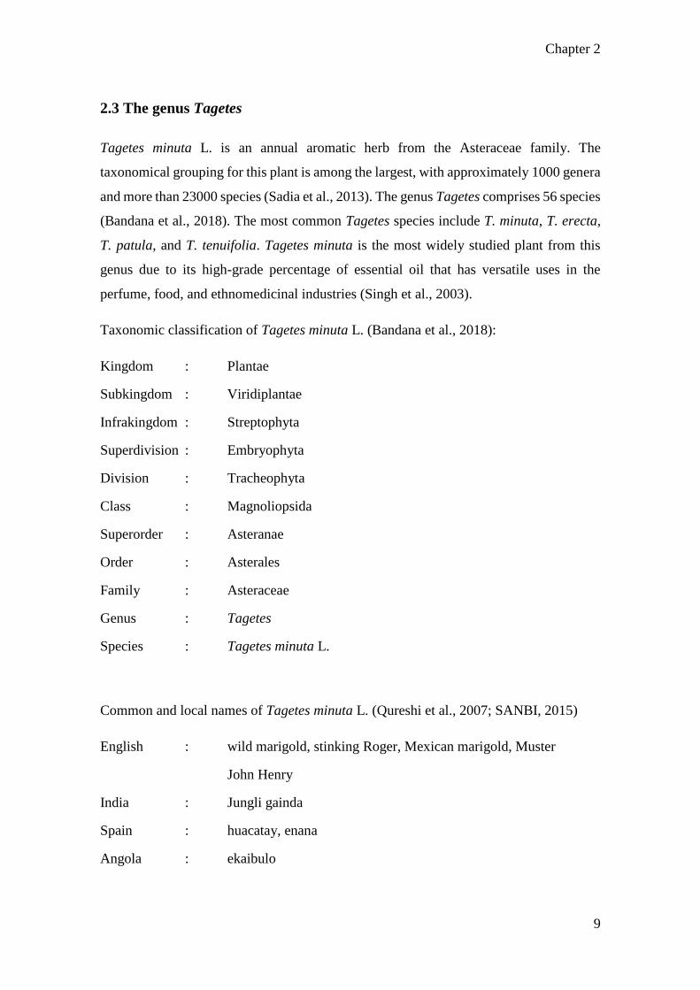
2.3 The genus Tagetes
Tagetes minuta L. is an annual aromatic herb from the Asteraceae family. The
taxonomical grouping for this plant is among the largest, with approximately 1000 genera
and more than 23000 species (Sadia et al., 2013). The genus Tagetes comprises 56 species
(Bandana et al., 2018). The most common Tagetes species include T. minuta, T. erecta,
T. patula, and T. tenuifolia. Tagetes minuta is the most widely studied plant from this
genus due to its high-grade percentage of essential oil that has versatile uses in the
perfume, food, and ethnomedicinal industries (Singh et al., 2003).
Taxonomic classification of Tagetes minuta L. (Bandana et al., 2018):
Kingdom : Plantae
Subkingdom : Viridiplantae
Infrakingdom : Streptophyta
Superdivision : Embryophyta
Division : Tracheophyta
Class : Magnoliopsida
Superorder : Asteranae
Order : Asterales
Family : Asteraceae
Genus : Tagetes
Species : Tagetes minuta L.
Common and local names of Tagetes minuta L. (Qureshi et al., 2007; SANBI, 2015)
English : wild marigold, stinking Roger, Mexican marigold, Muster
John Henry
India : Jungli gainda
Spain : huacatay, enana
Angola : ekaibulo
Chapter 2
10
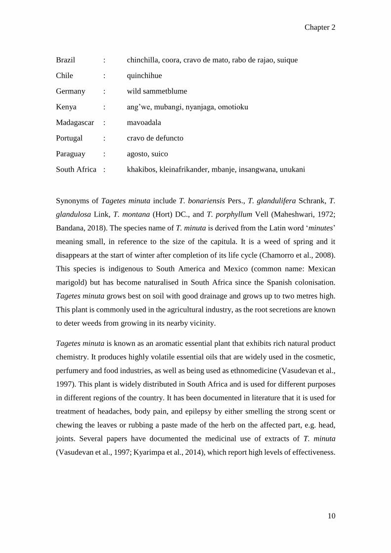
Brazil : chinchilla, coora, cravo de mato, rabo de rajao, suique
Chile : quinchihue
Germany : wild sammetblume
Kenya : ang’we, mubangi, nyanjaga, omotioku
Madagascar : mavoadala
Portugal : cravo de defuncto
Paraguay : agosto, suico
South Africa : khakibos, kleinafrikander, mbanje, insangwana, unukani
Synonyms of Tagetes minuta include T. bonariensis Pers., T. glandulifera Schrank, T.
glandulosa Link, T. montana (Hort) DC., and T. porphyllum Vell (Maheshwari, 1972;
Bandana, 2018). The species name of T. minuta is derived from the Latin word ‘minutes’
meaning small, in reference to the size of the capitula. It is a weed of spring and it
disappears at the start of winter after completion of its life cycle (Chamorro et al., 2008).
This species is indigenous to South America and Mexico (common name: Mexican
marigold) but has become naturalised in South Africa since the Spanish colonisation.
Tagetes minuta grows best on soil with good drainage and grows up to two metres high.
This plant is commonly used in the agricultural industry, as the root secretions are known
to deter weeds from growing in its nearby vicinity.
Tagetes minuta is known as an aromatic essential plant that exhibits rich natural product
chemistry. It produces highly volatile essential oils that are widely used in the cosmetic,
perfumery and food industries, as well as being used as ethnomedicine (Vasudevan et al.,
1997). This plant is widely distributed in South Africa and is used for different purposes
in different regions of the country. It has been documented in literature that it is used for
treatment of headaches, body pain, and epilepsy by either smelling the strong scent or
chewing the leaves or rubbing a paste made of the herb on the affected part, e.g. head,
joints. Several papers have documented the medicinal use of extracts of T. minuta
(Vasudevan et al., 1997; Kyarimpa et al., 2014), which report high levels of effectiveness.
Chapter 2
11
2.4 Phytochemical studies on Tagetes minuta
Phytochemical screening of medicinal plants enables the development of novel
therapeutic compounds with an improved efficacy to address variable health-related
issues.
Tagetes minuta is cultivated primarily due to its richness in essential oil, which is present
in all organs except the stem (Singh et al., 2003). In addition to the essential oil, T. minuta
also produces various secondary metabolite compounds including monoterpenes,
sesquiterpenes, flavonoids, aromatics, and thiophenes (Singh et al., 1995; Lawrence,
1996; Bansal et al., 1999; Brene et al., 2009; Sadia et al., 2013).
The major components of T. minuta oil are (Z)-β-ocimene, hydrocarbons (limonene),
acyclic unsaturated monoterpenoids, ketones, dihydrotagetone, tagetones (E, Z), and
ocimenones (E, Z) (Kyarimpa et al., 2014; Tiwari et al., 2016; Igwaran et al., 2017).
Meshkalasadat et al. (2010) characterised the volatile components of T. minuta cultivated
in Iran using nano-scale injection. Their study indicated that the essential oil is rich in
monoterpene hydrocarbons (28.3%), oxygenated monoterpenes (45.2%), sesquiterpene
hydrocarbons (2.5%), oxygenated sesquiterpenes (3.7%), diterpenes (0.6%), and other
compounds (17.2%).
The variation in essential oil content and composition of T. minuta in North India was
studied by Tiwari et al. (2016) using gas chromatography-mass spectrometry (GC-MS)
and gas chromatography with flame-ionization detector (GC-FID) for analysis. The
essential oil content varied from 0.52 to 0.78% in the different growth stages of the crop
(flower initiation, full flowering, late flowering, and seed setting stages). The main
constituent of the essential oil was found to be monoterpenoids (80.5-92.9%).
In Argentina, Gil et al. (2002) studied three wild accessions of T. minuta for the
composition of thiophenes and concluded that there were only quantitative differences.
The major constituents of the essential oil were dihydrotagetone, α-phellandrene,
limonene, o-cymene, β-ocimene, and tagetenone.
In South Africa, Mohammad et al. (2010) showed that the main components of the
essential oil were β-ocimene (32.0%), and dihydrotagetone (16.4%); while Igwaran et al.

Chapter 2
12
(2017) used GC-MS analyses to determine the main compounds to be β-ocimene (14.4%),
m-tert-butyl-phenol (9.41%), 2,6-dimethyl-, (E)-5,7-octadien-4-one (7.14%), hydro-
methyl-naphthalene (5.58%) and spathulenol (4.56%).
Tankeu et al. (2013) reported chemotypic variation of the T. minuta essential oil extracted
from South African plants, revealing two major chemotypes – (E)-tagetone,
dihydrotagetone, and (Z)-tagetone as the characteristic marker constituents for
Chemotype 1; while (Z)-β-ocimene, (E)-ocimenone, and (Z)-ocimenone characterised
Chemotype 2. This supplements the findings of Senatore et al. (2004), which also
identified two chemotypes for this species. One chemotype is characterised by a higher
content of tagetones in samples from the United Kingdom, and higher content of
ocimenes and ocimenones in samples taken from South Africa and Egypt.
In 2014, Al-Musayeib et al. isolated nine compounds and identified two of them to be
novel compounds (5-methyl-2,2',5',2'',5'',2''',5''',2''''-quinquethiophene and quercetagetin-
6-O-(6-O-caffeoyl-β-d-glucopyranoside) from the methanolic extract of Tagetes minuta,
which shows significant antioxidant activity.
Lizarraga et al. (2017) analysed the chemical composition of the essential oils of T.
minuta and T. terniflora. They deduced that the oil of T. minuta was characterised mainly
by high percentages of tagetone (56.2%) and cis-β-ocimene (19.9%), with lower amounts
of dihydrotagetenone (10.4%) and limonene (2.4%); whereas T. terniflora showed high
percentages of tagetones (60.6%) and ocimenones (10.3%).
Ibrahim et al. (2018b) isolated a new compound called ‘tagetnoic acid’ from the hexane
fraction of the leaves of T. minuta. This compound has been described as a strong
lipoxygenase inhibitor with IC50 of 1.17 µM as compared to the synthetic inhibitor
indomethacin (IC50 0.89 µM). This suggests that the consumption of T. minuta leaves
may alleviate inflammatory disorders.

Chapter 2
13
Tagetes minuta is both a cultivated crop and an invasive weed, based on the country. This
means that the differences in the chemical profile of essential oil can be attributed to both
anthropogenic and environmental factors, not limited to:
(i) method of harvesting
(ii) geographic location of the plant
(iii) growth stage of the harvested plants
(iv) the plant organs used
(v) climatic conditions of growth
Chemotypes are a result of genotypic × environmental interactions (Tankeu et al., 2013),
which leads to variations in the chemical composition of essential oil from Tagetes
minuta.

Chapter 2
14
Figure 2. 1 Chemical structures of constituents most abundant in the essential oil of
Tagetes minuta (Adapted from Sadia et al., 2013; Igwaran et al., Lizarraga et al., 2017).

Chapter 2
15
2.5 Trichome types and functions
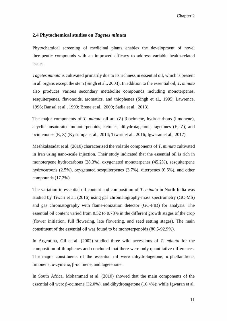
Trichomes are epidermal appendages that resemble small hairs on the surfaces of leaves,
stems, roots, and flowers of almost all angiosperms (Wagner et al., 2004). They have
served roles in the mechanisms of plants to adapt to a range of biotic and abiotic stressors
(Levin, 1973; Fahn, 2000; Wagner et al., 2004). Trichomes vary in shape, size, location,
and the ability to store and secrete bioactive compounds (Levin, 1973; Wagner, 1991).
More than 300 types of plant trichomes have been described (Aschenbrenner et al., 2013).
Conventionally, research on trichomes has focused primarily on the metabolic pathways
in some types of trichomes (Schilmiller et al., 2008; Spyropoulou et al., 2014); however,
studies show that trichomes can act as a defence syndrome against insects that feed on
the plant, both mechanically and chemically (Levin, 1973; Wagner et al., 2004). Plants
induce the growth of a denser cover of foliar trichomes in response to insect damage, as
well as stimulating an increase in secondary metabolites (such as tannins, terpenoids, and
alkaloids) that deter predators (Agrawal, 2006; War et al., 2012).
The classification of trichome types has been subjective; however, there are two major
categories, glandular (secretory) and non-glandular. As trichomes can be used for
taxonomic purposes, these can be subdivided further according to their relative
morphological characteristics (Kim et al., 2011). Glandular trichomes are named for their
terminal secretory head and ability to produce, sequestrate, accumulate, and exude
specialised secondary metabolites that deter predators by causing the plant to become
unpalatable, or secreting substances that attract pollinators (Levin, 1973; Wagner, 1991).
Non-glandular trichomes are often called ‘simple’ trichomes as their functions are mostly
aligned with their mechanical properties such as size, shape, and density (Matsuki et al.,
2004; Wagner et al., 2004). They may be unicellular, multicellular, or branched and are
associated with the texture of the epidermal surface of plants (Adedeji and Jewoola, 2008;
Gairola et al., 2008).
Non-glandular trichomes (NGT) form early in the development of plant organs and often
senesce shortly before the plant reaches maturity (Wagner et al., 2004). Werker (2000)
suggests that dead trichomes on mature plant organs may still serve mechanical functions
such as water absorption and abrasion protection. Non-glandular trichomes are most
commonly associated with plant defence. Agrawal and Fishbein (2006) demonstrated the
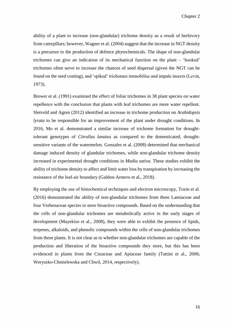
Chapter 2
16
ability of a plant to increase (non-glandular) trichome density as a result of herbivory
from caterpillars; however, Wagner et al. (2004) suggest that the increase in NGT density
is a precursor to the production of defence phytochemicals. The shape of non-glandular
trichomes can give an indication of its mechanical function on the plant – ‘hooked’
trichomes often serve to increase the chances of seed dispersal (given the NGT can be
found on the seed coating), and ‘spiked’ trichomes immobilise and impale insects (Levin,
1973).
Brewer et al. (1991) examined the effect of foliar trichomes in 38 plant species on water
repellence with the conclusion that plants with leaf trichomes are more water repellent.
Sletvold and Agren (2012) identified an increase in trichome production on Arabidopsis
lyrata to be responsible for an improvement of the plant under drought conditions. In
2016, Mo et al. demonstrated a similar increase of trichome formation for drought-
tolerant genotypes of Citrullus lanatus as compared to the domesticated, drought-
sensitive variants of the watermelon. Gonzales et al. (2008) determined that mechanical
damage induced density of glandular trichomes, while non-glandular trichome density
increased in experimental drought conditions in Madia sativa. These studies exhibit the
ability of trichome density to affect and limit water loss by transpiration by increasing the
resistance of the leaf-air boundary (Galdon-Armero et al., 2018).
By employing the use of histochemical techniques and electron microscopy, Tozin et al.
(2016) demonstrated the ability of non-glandular trichomes from three Lamiaceae and
four Verbenaceae species to store bioactive compounds. Based on the understanding that
the cells of non-glandular trichomes are metabolically active in the early stages of
development (Mayekiso et al., 2008), they were able to exhibit the presence of lipids,
terpenes, alkaloids, and phenolic compounds within the cells of non-glandular trichomes
from these plants. It is not clear as to whether non-glandular trichomes are capable of the
production and liberation of the bioactive compounds they store, but this has been
evidenced in plants from the Cistaceae and Apiaceae family (Tattini et al., 2006;
Weryszko-Chmielewska and Chwil, 2014, respectively).
Chapter 2
17

Chapter 2
18
Figure 2. 2 Non-glandular trichomes types of Asteraceae (adapted from Adedeji
and Ajewoola, 2008):
(A) unicellular trichome (narrow)
(B) unicellular trichome (spine-like)
(C) unicellular trichome (spike-like)
(D) bicellular hooked trichome
(E) bicellular trichome (large basal cell)
(F) bicellular trichome (normal basal cell)
(G) unicellular spiked trichome
(H) unicellular hooked trichome
(I) bicellular trichome (normal basal cell)
(J) bicellular trichome (spiked apical cell)
(K) multicellular trichome (pointed apical cell)
(L) multicellular trichome (acicular apical cells)
(M) multicellular trichome (tapered apical cell)
(N) multicellular trichome (rounded apical cell)
(O) unicellular trichome (pointed apical cell)
(P) multicellular trichome (tapered apical cell, normal basal cells)
(Q) bicellular long trichome (acicular cells)

Chapter 2
19

Chapter 2
20
Figure 2. 3 Multicellular non-glandular trichomes types of Asteraceae (adapted
from Adedeji and Ajewoola, 2008):
(A) apex pointed
(B) apex sickle-shaped
(C) with one shrivelled cell
(D) two shrivelled apical cells
(E) apical cell globular
(F) apical cell acicular at the end, bulbous cells
(G) apical cell shrivelled
(H) one cell slightly shrivelled
(I) one cell shrivelled
(J) apical cell acicular
(K) pointed apical cell
(L) pointed apical cell (bulbous basal cells)

Chapter 2
21
2.6 Secretory cavities
Plant secretory cavities are glands in plant organs that are made up of specialised
glandular cells surrounding a secretion-filled space (Turner et al., 1998). They often
contain essential oils, lipids, resins, alkaloids, flavonoids, tannins, or mucilage (Fahn,
1979, Bartoli et al., 2011). There are three primary types of developmental secretory
cavities: schizogenous, lysigenous, and schizolysigenous (Turner et al., 1998).
Schizogenous cavities are formed by the separation of a single glandular cell that appears
as a distinct intercellular space (lumina) lined by secretory epithelial cells (Tolke et al.,
2017). Lysigenous cavities are formed by the disintegration of several cells, whereby the
secretory cells degenerate as they release their secretory product into the developing space
by a process known as holocrine secretion (Arora and Kumar, 2108). Schizolysigenous
cavities form as an amalgamation of the previous two processes: the cavity develops from
a single cell, but the epithelial cells on the periphery of the forming gland undergo
autolysis in order to enlarge the storage cavity (Turner et al., 1998; Machado et al., 2017).
In the genus Tagetes, Sacchetti et al. (2001) showed that the secretory cavities of T. patula
seedlings were schizogenous as it formed a distinct intercellular canal bordered by
endodermal epithelial cells. Using transmission electron microscopy, they were able to
demonstrate that the cells on the periphery of the secretory cavity were more electron
dense and contained many more osmiophilic vesicles and plastids than its neighbouring
parenchymatic cells.
Russin et al. (1992) studied the changes in the chemical composition of the secretory
products of T. erecta during the development phase until late flowering of the plant. Their
results showed that indole comprised almost 99% of the secretory product, which is a
metabolite not commonly stored in its free state in vegetative plant organs. More
interestingly, the ratio of indole to piperitone varied with plant development and aging.
This also served as the first report in Tagetes of secretory products existing separately
from the lamina tissues, implying that secretory cavities produce as well as store
metabolic end products that change in concentrations based on the needs and
developmental growth of the plant.

Chapter 2
22
Lopez et al. (2009) and Lizarraga et al. (2017) described the secretory cavities of T.
minuta as pellucid, elliptic glands (70 – 200µm) that are covered by a parenchymal sheath
made up of cutinised epithelial cells. Both studies described the secretory cavities as being
found along the entire phyllaries. In senescent leaves, the secretory cavity collapses
inward due to the fragility of the epithelial cells along its periphery.
Figure 2. 4 Secretory cavity types: intercellular spaces: a) schizogenous intercellular
space in Bryophyllum stem. b) lysigenous intercellular space in leaf of Sequoia
sempervirens (Adapted from Fahn, 2000; Bartoli et al., 2011; Bombo et al., 2017).

Chapter 3
23
CHAPTER 3: FOLIAR STRUCTURES AND HISTOCHEMICAL
ANALYSES OF TAGETES MINUTA L. LEAVES
3.1 Abstract
Many species in Tagetes are known for producing essential oils and commercially useful
bioactive compounds. The objective of this study was to investigate the
micromorphological features of the internal and external foliar structures of Tagetes
minuta that produce and store these compounds. This was accomplished using
stereomicroscopy, light microscopy, scanning electron microscopy, transmission electron
microscopy, and histochemical analyses. The findings show that the trichomes on the
surface of T. minuta leaves appear to be linear and non-glandular, but still maintains the
ability to store various bioactive compounds within as shown by histochemical analyses.
The development of the subdermal secretory cavities show that the cells undergo autolysis
in order to form a schizolysigenous cavity in mature leaves. Ultrastructure of the
parenchymal sheath and secretory epithelium within the secretory cavity show the
changes of the plastids to contain lipid and osmiophilic molecules. These findings are
novel for T. minuta and enable a better understanding of the exudation process in order
to optimise essential oil production for industrial applications.
Keywords: histochemistry; non-glandular trichomes; micromorphology; microcopy;
secretory cavity

Chapter 3
24
3.2 Introduction
Tagetes minuta is a member of the Asteraceae family, which comprises 1535 genera and
approximately 23 000 species and is the largest family of angiosperms (Shen et al.,
2018a). Many of these species have substantial medicinal, ornamental, and economic
values due to their chemical composition and biological activities (Vidic et al., 2016).
Tagetes minuta is commonly known as Mexican marigold, and is native to South
America. It has since been naturalised in Europe, Asia, North America and Africa since
the Spanish colonisation (Meshkatalsadat et al., 2010). This plant has a long history of
human use as both food and allopathic medicine (Tereschuk et al., 1997; Scrivanti et al.,
2003; Senatore et al., 2004). Infusions and extracts prepared from the leaves of T. minuta
have been used to treat intestinal diseases, alleviate headaches and symptoms of epilepsy
(Igwaran et al., 2017; Vasudevan et al., 1997).
Species belonging to Tagetes are characterised by the macroscopic punctate oil glands
that are found on the abaxial leaf surfaces, which produce and store ‘Tagetes oil’. This
essential oil is marketed due to its medicinal properties and various health benefits (Lopez
et al., 2009; Sadia et al., 2013). The secretory cavity, the primary focus of the oil complex
in Tagetes is relatively well-documented in literature, however, the subject of the foliar
trichomes on T. minuta is a contentious topic (Lopez et al., 2009; Lizarraga et al., 2017).
Trichomes are specialised hair-like epidermal appendages that have been shown to play
a role in the plant defence system against biotic threats such as predators as both a physical
barrier and by the mediation of chemical defences, as well and abiotic factors such as
sunlight in the way of reflecting excess radiation (Valverde et al., 2001; Kariyat et al.,
2018). Depending on the plant species, these structures can be found on the leaves, stems,
roots, and even seed coats (Levin, 1973). Trichomes can be classified as either glandular
or non-glandular based on their shape and function, with the most distinct morphological
difference being the absence of a glandular head in the non-glandular trichomes (Werker,
2000). Due to the lack of a glandular head, non-glandular trichomes are considered to act
exclusively as mechanical barriers; whereas glandular trichomes are responsible for the
storage and/or exudation of biologically active phytocompounds (Levin, 1973; Werker,
2000).

Chapter 3
25
The trichomes of T. minuta have been described as both glandular (Cappellari et al., 2013)
and non-glandular (Lizarraga et al., 2017) with seemingly no consensus. The purpose of
this study was to determine the trichome type and further investigate the secretory cavities
of T. minuta using stereo- and electron microscopy, as well as elucidate the chemical
classes of the phytocompounds stored in its trichomes using histochemical analyses.

Chapter 3
26
3.2 Methods and Materials
3.2.1 Plant collection
Fresh (emergent, young, and mature) leaves of T. minuta were collected from a population
at the University of KwaZulu-Natal Westville campus located at 29.817°S 30.940°E in
Durban, South Africa. A voucher specimen was confirmed and deposited at the UKZN
Westville Herbarium (accession number 18216, voucher number 01).
3.2.2 Stereomicroscopy
Both the adaxial and abaxial leaf surfaces at three developmental stages (emergent,
young, and mature) were examined using the Nikon AZ100 Stereomicroscope, and
images captured with the Nikon DXM 1200C camera using NIS-Elements Software with
an emphasis on the foliar structures.
3.2.3 Scanning Electron Microscopy (SEM)
3.2.3.1 Chemical fixation
Fresh leaf material was cut into 3 mm2 sections and were fixed in 2.5 % glutaraldehyde
for 24 hrs. The sections were subsequently subjected to three washes (5 min each) with
0.1 M sodium phosphate buffer (pH 7.0), followed by post-fixation in 0.5 % osmium
tetroxide for 2 hrs. Thereafter, the sections were washed three times for 5 min each with
sodium phosphate buffer. The samples were then subjected to graded dehydration series
in ethanol (25 %, 50 %, 75 %, and 100 %) for two sessions (5 min each) and two sessions
(10 min each) in the 100 % ethanol, and were then critically-point dried using a Quorum
K850 Critical Point Dryer. The sections were fixed using carbon conductive tape onto
aluminium stubs and sputter-coated with gold using the Polaron SC 500 Sputter Coater.
The viewing and imaging were performed using a LEO 1450 SEM at 5 kV and a working
distance of 7 – 10 mm.

Chapter 3
27
3.2.3.2 Freeze fracture
Fresh leaves were fractured along the midrib for analysis. The samples were cut into 2
cm2 sections for freeze fracture. They were prepared by quenching rapidly in subcooled
liquid nitrogen and fractured using forceps. Thereafter the fractured segments were dried
using the Edwards-Modulyo freeze dryer at –60°C for 48 hrs in a vacuum of 10-2 Torr.
The fractured segments were mounted onto brass stubs and secured with carbon
conductive cement. The samples were sputter-coated with gold using a Polaron SC 500
sputter coater and imaged on a LEO 1450 SEM with a working distance of 12 mm.
3.2.3.3 Cryo-scanning electron microscopy (Cryo-SEM)
Cryo-SEM was performed using a Quorum PP3000T coupled to a Zeiss UltraPlus
FEGSEM. Fresh leaf material was cryo-fixed in a liquid nitrogen slush before being
transferred to a vacuum chamber held at -135°C. The samples were then fractured, etched,
and coated in platinum. The sections were viewed at 2 kV.
3.2.4 Light Microscopy and transmission electron microscopy (TEM)
Leaves from three developmental stages were trimmed to 2 mm2 and fixed in 2.5 %
glutaraldehyde for 24 hrs. Samples were rinsed three times in 0.1 M sodium phosphate
buffer (pH 7.0) and post-fixed in 0.5 % osmium tetroxide for 2 hours. Samples were
rinsed thrice in sodium phosphate buffer before undergoing dehydration using a graded
series of acetone (25 %, 50 %, 75 %, for two 5-minute sessions, and 100 % for two 10-
minute sessions). Propylene was used as a clearing agent, which was used in conjunction
with a graded series of Spurr’s resin (Spurr, 1969) for infiltration of the leaf tissues at
25 %, 50 %, 75 %, and 100 %, of resin in the propylene solution. Samples were then
placed in silicone moulds in whole resin and polymerised at 70°C for 8 hrs.
The sections were cut using a Reichert Jung Ultracut-E ultramicrotome. Semi thin
sections (0.5 µm) were used to determine the regions of interest. The survey sections were
stained with 1 % toluidine blue and imaged using the Nikon Eclipse 80i light microscope

Chapter 3
28
equipped with a Nikon DS-Fi1 camera and NIS-Elements D imaging software. Ultrathin
sections (90 – 120 nm) were collected on copper grids and stained with 2.5 % uranyl
acetate and lead citrate. The ultrathin sections were viewed using the JEOL 1010 TEM
equipped with an Olympus MegaView III CCD camera and iTEM software.
3.2.5 Histochemical tests
Fresh leaf sections (100 - 120µm) were obtained using an Oxford ® Vibratome Sectioning
System, and were stained with the reagents listed below. The purpose of this was to detect
and analyse the localisation of specific cellular phytochemicals in the leaf structure. The
images were captured on a Nikon Eclipse 80i compound light microscope.
a. Lipids
Nile Blue – The fresh sections were stained with 2 % Nile Blue for 1 minute at
37°C and then transferred to 1 % acetic acid for 1 min. Sections were rinsed with
distilled water, mounted, and viewed. Pink staining indicates the presence of
neutral lipids such as fats and oils, whereas blue staining indicates the presence of
acidic lipids such as phospholipids (Demarco, 2017).
Sudan III/IV – Sections were stained for 10 min before being rinsed in 70 %
ethanol and viewed. Tissues that stained red/orange indicates the presence of
lipids or cutin (Buda et al., 2009).
b. Lignins
Phloroglucinol – The fresh sections were immersed in phloroglucinol stain for 5
min and rinsed with distilled water. A pink/red colouration indicates the presence
of lignins (Jensen, 1962).
Acridine orange – The fresh sections were immersed for 10 min in the fluorescent
dye and rinsed with distilled water. They were viewed under blue light, where a
yellow/green fluorescence distinguishes the lignified cell walls, and red

Chapter 3
29
colouration indicates non-lignified tissues. The red/orange colour indicates cells
undergoing apoptosis which indicates cell viability (Gupta and De, 1983).
c. Phenolic compounds
Ferric Chloride – The fresh sections were immersed for 20 min in the stain before
being thoroughly washed with distilled water. Sections were mounted in
glycerine. Brown/black deposits indicate the presence of phenolic compounds
(Zarate and Yeoman, 1994).
Autofluorescence – No stains were used, but fresh sections were viewed under
UV light. Phenolic compounds emit a blue/green fluorescence, as well as some
terpenoids (Demarco, 2017).
d. Proteins
Coomassie Blue – The fresh sections were immersed in 0.25 % Coomassie blue
for 10 min and then transferred to 7 % acetic acid. After washing in distilled water,
sections were mounted in glycerine. Proteins stain blue (Demarco, 2017).
e. Alkaloids
Wagner’s Reagent – The fresh sections were stained for 20 min before being
washed with distilled water. Alkaloids stain with red/brown colouration
(Demarco, 2017).
f. Mucilage and pectins
Ruthenium Red – The fresh sections were stained for 2 min before being rinsed
with distilled water. The pink/red colouration indicates the presence of acidic
mucilages and pectins (Colombo and Rascio, 1977).

Chapter 3
30
3.3 Results and Discussion
3.3.1 Vegetative growth and flowering
Tagetes minuta is an erect annual herb that is capable of reaching 2 m tall (Fig 3.1 a). It
matures in summer, when the stem turns a distinct red-brown colour. The glossy, green
leaves are arranged alternately on the stem and are pinnately dissected. The small white-
yellow florets (10 – 15mm) are supported by fused involucre bracts. Each capitula is
formed from 5 – 10 florets, which groups together to form a clustered panicle on
flowering plants (Fig 3.1 b).
Figure 3. 1 Vegetative growth and flowers of Tagetes minuta at the University of
Kwazulu-Natal Westville campus: a) Young plant at the vegetative stage. b) Mature plant
at the flowering stage.

Chapter 3
31
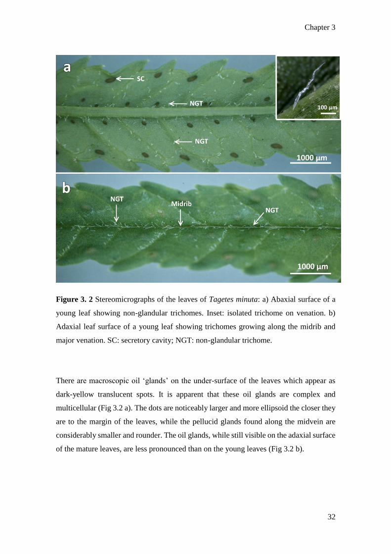
3.2 Stereomicroscopy
The pinnatisect leaves of T. minuta are long and narrow (3 – 15 cm in length and 2 – 8.5
cm in width), elliptic-lanceolate, and have an acute apex with serrated margins (Fig 3.2).
Stereomicroscopy images shows that the adaxial surfaces are a darker green in the mature
leaves, with a light green-yellow colour on the emergent and young leaves. They reveal
the presence of non-glandular trichomes which appear translucent on both the adaxial and
abaxial surfaces, with a denser cover on the abaxial leaf surface (Fig 3.2a inset). Between
the developmental stages, it appears that there are considerably fewer trichomes on the
mature leaves as compared to the emergent and young leaves.
Chapter 3
32
Figure 3. 2 Stereomicrographs of the leaves of Tagetes minuta: a) Abaxial surface of a
young leaf showing non-glandular trichomes. Inset: isolated trichome on venation. b)
Adaxial leaf surface of a young leaf showing trichomes growing along the midrib and
major venation. SC: secretory cavity; NGT: non-glandular trichome.
There are macroscopic oil ‘glands’ on the under-surface of the leaves which appear as
dark-yellow translucent spots. It is apparent that these oil glands are complex and
multicellular (Fig 3.2 a). The dots are noticeably larger and more ellipsoid the closer they
are to the margin of the leaves, while the pellucid glands found along the midvein are
considerably smaller and rounder. The oil glands, while still visible on the adaxial surface
of the mature leaves, are less pronounced than on the young leaves (Fig 3.2 b).

Chapter 3
33
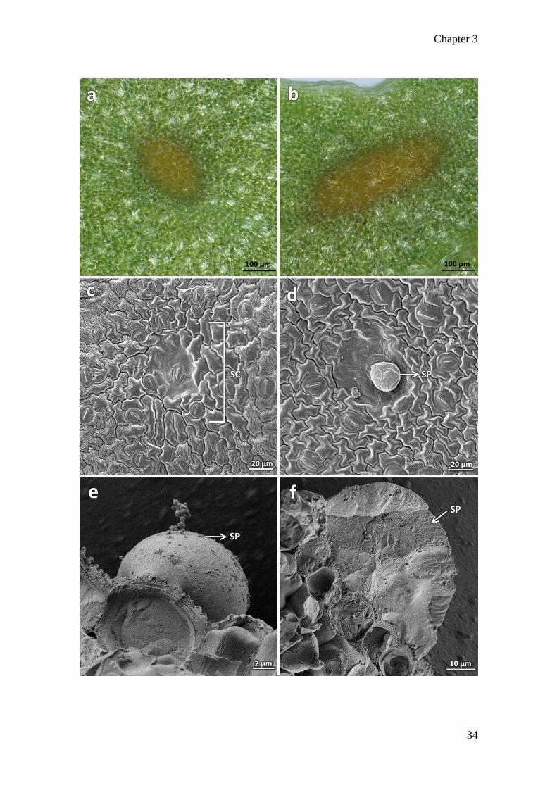
3.3 Secretory cavities
The stoma are anomocytic, with a limited number of subsidiary cells. They are in close
proximity to the oil glands, hereafter referred to as secretory cavities. The secretory
cavities range from 75 to 200µm in diameter. The secretory cavity (Fig 3.3 a) is round
and is noticeably present closer to the midvein while the more ellipsoid-shaped cavity
(Fig 3.3 b) is distributed along the leaf margins and phyllaries. The secretory cavities are
not immediately obvious from electron micrographs as they are covered by epidermal
cells on the abaxial and adaxial leaf surfaces, except at the sites of exudation. These
appear as slight depressions on the surface (Fig 3.3 c). The secretion of oils on the abaxial
leaf surface in a young leaf can be seen under SEM (Fig 3.3 d). Likewise, under cryo-
SEM, the oils from the secretory cavity appear on the leaf surface as globules (Fig 3.3 e)
that were released under pressure from the vacuum. An image of the cross-section through
an oil globule frozen in subcooled liquid nitrogen was also captured (Fig 3.3 f). It is
evident from this image that there are no cellular structures in the exudate, leading to the
conclusion that it is the released contents of the secretory cavity.
Chapter 3
34

Chapter 3
35
Figure 3. 3 Secretory cavities of Tagetes minuta: a) Stereomicrograph of rounded
secretory cavity on the abaxial leaf surface. b) Ellipsoid secretory cavity found alongside
the leaf margin. c) SEM of secretory cavity on abaxial leaf surface, in close relation to
the stomata. d) SEM of cavity showing exudation taking place. e) Cryo-SEM micrograph
showing the oil exudate expelled due to vacuum pressure. f) Cross-section of the exudate
with no apparent cellular components within. SC: secretory cavity; SP: secretory product.

Chapter 3
36
Figure 3. 4 Scanning electron micrographs of the non-glandular trichomes on the adaxial
surface of young leaves of Tagetes minuta: a) The growth of non-glandular trichomes
along the major leaf veins. b) Clustering of trichomes along the major leaf venation.
c) Dense populations of non-glandular trichomes along the midvein of the leaf.

Chapter 3
37
3.4 Non-glandular trichomes
Scanning electron microscopy shows the topology of the leaf. The trichomes of T. minuta
do not densely cover the leaf surface, but are clustered around the main and lateral veins
(Fig 3.4), with a more frequent occurrence on the adaxial surfaces of the emergent and
young leaves. There is only one type of trichome that is found on the leaves of T. minuta
and they appear to be non-glandular. Each trichome is uniseriate and multicellular with a
rounded head, as evident by the segmentation in Figures 3.4 and 3.5. The length of the
trichomes are not consistent and can range between 70 and 200 µm, where the longer
trichomes are more frequently found on the mature leaves (Fig 3.5).

Chapter 3
38
Figure 3. 5 Single non-glandular trichomes of Tagetes minuta: a) Single uniseriate non-
glandular trichome on the adaxial surface of a young leaf. b) Non-glandular trichome on
a mature leaf showing attachment to the leaf surface.

Chapter 3
39
Trichomes are fine outgrowths of hair-like structures that are found on most angiosperm
aerial organs, some gymnosperms and bryophytes (Johnson, 1975). They are highly
diverse structures that are in contact with the external environment and thus function in
response to different biotic and abiotic stimuli (Tooker et al., 2010; Li et al., 2018).
Trichomes serve as the first line of defence against predators because they usually
protrude from aerial surfaces and some types are capable of producing bioactive
compounds that may attract and guide pollinators (Wagner, 1991; Hegebarth et al., 2016).
Non-glandular trichomes are capable of enhancing plant defense systems by reducing UV
radiation due to surface reflectance, and assisting through drought tolerance by reducing
leaf temperatures and preventing stress from photoinhibition (Levin, 1973; Wagner,
1991; Werker, 2000). Szyndler et al. (2013) showed that trichomes are also capable of
limiting movement of herbivores such as insects, restricting the damage inflicted on the
plant tissues, while Kariyat et al. (2017) proved that non-glandular trichomes deter
feeding by insects due to post-ingestive gut damage.
The functional usefulness of trichomes has led to increasing commercial value of the
secretory metabolites in cosmetic, food, and pharmaceutical industries (Valkama et al.,
2003; Balcke et al., 2017). Trichomes are highly variable and diverse foliar structures that
provide mechanical and chemical barriers against herbivores (Fabricant and Farnsworth,
2001; Valkama et al., 2003). Trichome morphological traits have also been key
characteristics in plant taxonomic studies.

Chapter 3
40
Figure 3. 6 Mites on the surface of Tagetes minuta: a) Scanning electron micrograph
showing broad mite eggs and larvae on the abaxial surface of a young leaf. b) Micrograph
showing empty egg case in the crevasse created by the midvein on the abaxial leaf surface.
c) Micrograph of a scale mite from the family Tydeidae on the abaxial surface of a mature
leaf.

Chapter 3
41
3.5 Semi-thin sections of secretory cavities
The secretory cavities along the midvein are smaller (55 – 75 µm) than those found along
the leaf margins and phyllaries (150 – 200 µm). They appear as elliptic glands that seem
to take up the almost the entire width of the leaf blade. Figure 3.7 shows the development
of the secretory cavity. The gland begins as a group of modified parenchymal cells that
form concentric circles in the leaf blade, below the row of columnar epithelial cells on
the adaxial leaf surface (Fig 3.7 a and b). As the cavity expands to accommodate the
accumulated substances, the parenchymal cells elongate to form a multi-layered sheath
with cutinised walls (Fig 3.7 d and e). As previously mentioned, the secretory cavity is
limited by the chlorenchyma on the adaxial face, but grows adjacent to the epithelial cells
on the abaxial face of the leaf. This leads to the conclusion that secretions are expelled
from the abaxial leaf surface.

Chapter 3
42

Chapter 3
43
Figure 3. 7 Development of the secretory cavity in Tagetes minuta. a) Initiation of
secretory cavity in emergent leaf. b) Cells differentiating in emergent leaf. c) Elongation
of secretory epithelial cells in young leaf. d) Secretory epithelium extending under the
chlorenchyma. e) Mature secretory cavity showing the movement of lipids into the
schizolysigenous cavity. SE: secretory epithelium; SC: secretory cavity; PS: parenchyma
sheath; Li: lipids.
The foliar cavity appears to form from the middle lamella where the cells at the centre
pull apart (Fig 3.7 a and b). Once the lumen is developed (Fig 3.7 e), it appears that all
interior cells release their secretory products (lipids) via holocrine secretion into the now
schizolysigenous cavity. These cells undergo a process known as autolysis and has been
described by Russin et al. (1992) for Tagetes erecta.
Many secretory cells are derived from other plant tissues, mainly parenchymatous and
epidermal tissues (Buvat, 1989). In young leaves, the secretory cavities appear to be
lysigenous, in which the intercellular substance is only partly dissolved during
development, but are typically elongated in mature leaves, causing them to appear
schizolysigenous (Buvat, 1989; Turner and Lange, 2015). The secretory epithelium is
mainly responsible for the excretion of accumulated substances from the cavity, and can
either cause the products to be diffused through the cuticle or released via distention of
the epithelial lining itself (Buvat, 1989; Lopez et al., 2009).
Lopez et al. (2009) described the oil complex of Tagetes minuta from the standpoint of a
senescent leaf. While the secretory cavity in a mature leaf (Fig 3.7 e) appears to be
schizolysigenous, the secretory cavity in a senescent leaf appears lysigenous. The
development of the secretory cavity from an emergent to a young leaf is shown in Figure
3.7 a – d. It can be seen that the secretory epithelium is comprised of cutinised
parenchymal tissues that become narrow and elongated. Although the secretory cavity
grows to fit the width of a leaf blade (Fig 3.7 e), they are limited on the adaxial face by a
distinct row of chlorenchyma cells, which suggests that the leaf secretion occurs on the
abaxial leaf surface.

Chapter 3
44
Figure 3. 8 Development of non-glandular trichome. a) Epithelial cells begin
development by becoming cutinised. b) Cell undergoes periclinal division to create a
tapering end of the now developing trichome. c) Cell division continues to create a
uniseriate non-glandular trichome. d) Cutinised cells elongate to form a mature trichome.

Chapter 3
45
Figure 3. 9 Histochemical observations on the non-glandular trichomes of Tagetes
minuta: a) Trichome positively stained with Coomassie Blue. b) Positive staining for
alkaloids with Wagner’s reagent. c) Ruthenium red staining of a trichome. d) Acidic lipids
detected using Nile Blue stain. e) Positive test for lipids, stained with Sudan III&IV. f)
Acridine orange stain used under UV light showing presence of lignins. g) Trichome
stained with phloroglucinol, positive for lignins. h) Ferric chloride stain shows phenolics.
i) Autofluorescence of a trichome under UV light shows presence of phenolic
compounds.

Chapter 3
46
3.6 Histochemical analyses of the non-glandular trichomes
The linear non-glandular trichomes were evaluated for the presence of histochemical
compounds and localisation. It is evident that some of the bioactive compounds could be
located within the cells of the trichomes, despite them being non-glandular. They were
positively stained for lipids using Nile blue and Sudan III&IV and for lignins using
phloroglucinol and acridine orange stains. Phenolic compounds were identified using
ferric chloride and proteins were identified using the Coomassie blue stain. Alkaloids
were present as deduced using Wagner’s reagent, and ruthenium red stained positively
for mucilages and pectins (Fig 3.9 a – i). The histochemical tests appears to localise the
bioactive compounds in the apical cells of the trichomes, and occasionally, throughout
the trichome cells. Figure 3.9 b shows a non-glandular trichome stained with Wagner’s
reagent, and the alkaloids appear as droplets throughout the trichome.
Non-glandular trichomes have shown to be metabolically active during the earliest stages
of development (Levin, 1973; Mayekiso et al., 2008) but are thought to play only a minor
role during the lifespan of the plant past the stage of development. Tozin et al. (2016)
shows that while the traditional role of non-glandular trichomes has historically been
protection of the plant material from predators, UV, and abiotic factors, they have the
potential to produce, store, and liberate bioactive substances. While their work is
primarily based on Lamiaceae and Verbenaceae species, a comparison can be drawn with
T. minuta, which belongs to family Asteraceae. Chaffey (2006) first proposed that despite
the lack of openings in the cuticle and cell walls of non-glandular trichomes, substances
can traverse the body cells of the trichomes, which is visible in Figures 3.9 a, c, e, g, and
h. Although the trichomes on T. minuta appear to be non-glandular, anatomically they are
similar to the linear glandular trichomes found on other species in the Asteraceae family
that aid in the storage and production of essential oils (Aschenbrenner et al., 2013; Bombo
et al., 2017). This serves as the first study to histochemically analyse the contents and
localisation in the non-glandular trichomes of T. minuta.

Chapter 3
47
Figure 3. 10 Electron micrographs of plastids in leaves of Tagetes minuta: a) Chloroplast
containing starch grains. b) Chloroplasts outside the secretory cavity. c) Elongated
chloroplasts in the secretory epithelium containing lipids. d) Plastid containing
plastoglobuli. e) Single chloroplast in the parenchymal sheath of the secretory cavity
containing lipid globules. S: starch granules; M: mitochondria; Chl: chloroplast; Pg:
plastoglobuli; Li: lipids.

Chapter 3
48
Figure 3. 11 Electron micrograph of cells within the oil complex: a) Epidermal layer
showing electron-dense osmiophilic material within. b) Cells in the parenchymal sheath
showing plastoglobuli. c) Elongated cells that form the secretory epithelium in a mature
secretory cavity that contain vesicles. d) Secretory epithelial cells on the periphery of the
secretory cavity containing vesicles. EL: epithelial layer; Chl: chloroplast; Pg:
plastoglobuli; Ve: vesicle; SE: secretory epithelium.

Chapter 3
49
3.7 Ultrastructure of the secretory cavities
The secretory cavities of T. minuta range in size from 75 to 200 µm across, meaning that
they are not visible in their entirety using TEM. Figures 3.10 and 3.11 show parts of the
parenchymal sheath and secretory epithelium that make up the secretory cavity, as well
as the cells surrounding them. The changes in the plastids are evident from Figure 3.10 a
– e, where primary starch granules are present in the chloroplasts of Figures 3.10 a and b.
These plastids can be found in the neighbouring cells on the periphery of the secretory
cavity and often contain plastoglobuli which appear as electron-dense material. Note the
chloroplasts in these neighbouring cells with a normal thylakoid system. However, in the
cells found in the secretory epithelium, the chloroplasts contain a dense population of
what appear to be lipids surrounding the thylakoids and take up the space of the grana
(Fig 3.10 c and e).
Sacchetti et al. (2001) assessed the ultrastructural characteristics of the secretory cavities
of Tagetes patula, showing similar results to those above. The schizolysigenous spaces
that are bordered by the parenchymal sheath (Figure 3.11 c and d) appears rich in plastids
with electron-dense stroma and few thylakoids, which is a common feature in plants that
exude essential oils (Lopez et al., 2009; Lizarraga et al., 2017). This may be strongly
related to the production of terpene compounds such as thiophene that is prevalent in the
leaves of T. minuta (Al-Musayeib et al., 2014; Ibrahim et al., 2018a). Also visible are the
glandular cells which appear elongated and rich in vesicles.
3.8 Conclusion
The secretory cavities of T. minuta appear macroscopically on both the adaxial and
abaxial leaf surfaces, and are pronounced in mature leaves. For the first time, the
ultrastructural development of these secretory cavities shows the changes to the plastid
system within the secretory epithelial lining. Trichomes on the leaves of T. minuta appear
to be linear, uniseriate, and non-glandular. However, the histochemical analysis indicated
the storage of various histocompounds, suggesting that non-glandular trichomes in the
Asteraceae family have potential biological role that is more than protection against
predators and abiotic factors.

Chapter 4
50
CHAPTER 4: PHYTOCHEMICAL ANALYSES AND
ANTIBACTERIAL POTENTIAL OF TAGETES MINUTA L.
4.1 Abstract
Phytochemical analysis was carried out on the crude organic solvent extracts derived from
the leaves of T. minuta. Alkaloids, sterols, saponins, terpenoids, phenols, and lipids were
detected from the extracts. Gas-chromatography mass-spectrometry (GC-MS) was
carried out to reveal that the constituents with the highest percentage were 9-octadecen-
1-ol (4.51 %), β-sitosterol (6.07 %), olean-12-en-3-one (7.47 %), and 3-methyl-1-butanol
(14.77 %). Preliminary antibacterial activity was performed using the methanolic extract
which showed inhibitory activity against selected gram positive and gram negative
bacteria (P. aeruginosa, E. coli, S. aureus, MRSA, and B. subtilis).This study shows that
T. minuta contains bioactive compounds that have pharmacological and medicinal uses.
Keywords: antibacterial; bioactive compounds; GC-MS; methanol extract; Tagetes
minuta

Chapter 4
51
4.2 Introduction
Plant material has been used in traditional medicine for millennia in both developing and
developed countries due its ease of access and lesser risk of side effects (Qazi Majaz and
Molvi Khurshid, 2016). The use of ethnomedicine is becoming increasingly popular in
modern societies as natural alternatives to synthetic products and has become a socio-
cultural phenomenon of third world countries (Dixit et al., 2013).
In South Africa, healthcare is largely polarised between Western medical therapies and
traditional African healthcare systems. Although concurrent usage of traditional healers
and allopathic providers of medicine is common, allopathic healthcare facilities are often
limited and not easily accessible to most in South Africa (Bisi-Johnson et al., 2017). This
has resulted in the medicinal plant industry in South Africa being supported by
approximately 30 million local consumers with almost 70 thousand species of plants
being used for medicinal purposes, many of which are threatened by overharvesting and
international trade (Street and Prinsloo, 2013; Xego et al., 2016). The indiscriminate
overharvesting coupled with extensive land degradation has affected the availability of
medicinal plants that can be naturally foraged, resulting in an increasing demand for
medicinal plant material (Phondani et al., 2016; Tanga et al., 2018).
In order to establish strategies to protect endemic medicinal plants, it is vital that they are
screened for the bioactive phytochemicals responsible for their therapeutic properties.
This will ensure regulation for use of wild populations of medicinal plants by putting into
place mitigative measures such as systematic plant cultivation for the production and
monitoring of this underutilised resource (Tanga et al., 2018). Moreover, many isolated
phytometabolites from medicinal plants have led to the development of novel drugs and
therapies to treat several human diseases such as cancer and diabetes (Hosseinzadeh et
al., 2015; Shakya, 2016; Kudumela and Masoko, 2017).
Tagetes minuta belongs to the Asteraceae family, and its essential oils are renowned for
its medicinal, ornamental, and therapeutic values (Dixit et al., 2013; Rajvanshi and
Dwivedi, 2017). The essential oils of T. minuta has been researched for its nematocidal
(Gutierrez et al., 2006), cosmetic (Farjana et al., 2009), food additive (Nandita et al.,
2012), and antimalarial (Kyarimpa et al., 2014) properties.

Chapter 4
52
Phytochemical analyses of T. minuta essential oil show that the plant is rich in alkaloids,
phenolic compounds, flavonoids, and thiophenes which are responsible for its therapeutic
properties (Chamorro et al., 2008; Meshkatalsadat et al., 2010; Devika and Koilpillai,
2012). The oil has been reported as having a high inhibitory effect on both gram postitive
and gram negative bacteria and fungi (Hethelyi et al., 1986). Therapeutically, T. minuta
has several reported medicinal benefits such as remedies for respiratory inflammations,
stomach pains, chest infections, coughs, and congestion (Shirazi et al., 2014). It also has
a healing effect on wounds, cuts, and calluses (Rahimi et al., 2010; Maity et al., 2011).
This study was undertaken to examine the qualitative phytochemical composition using
phytochemical tests, determine the quantitative chemical composition using GC-MS, and
elucidate the preliminary antibacterial potential in the crude methanolic extract of T.
minuta leaves.

Chapter 4
53
4.3 Methods and Materials
4.3.1 Plant collection
Fresh leaves of T. minuta were collected from the UKZN Westville campus (29.817°S
30.940°E) and air-dried for 6 weeks at room temperature (24°C). A voucher specimen
was deposited at the UKZN Westville Herbarium (accession number 18216, voucher
number 01).
4.3.2 Extraction with organic solvents
Ten grams of T. minuta powdered leaf material were placed in a round bottom flask
containing 100 ml of hexane. The flask was attached to Soxhlet apparatus and boiled for
three 3-hour sessions to obtain the crude plant extract. This procedure was then repeated
using the same leaf material for chloroform and methanol, respectively. The crude
extracts were filtered through Whatman No. 1 filter paper and the stored in airtight jars at
4°C for further analysis.
4.3.3 Qualitative phytochemical analyses
a) Detection of carbohydrates
Molisch’s test – two drops of α-naphthol were added to 2 ml of extract and gently shaken.
One ml of concentrated sulphuric acid was slowly poured along the side of the test tube
and allowed to stand. The formation of a red-purple ring at the junction of the two liquids
is indicative of the presence of carbohydrates (Phani Deepthi Yadav et al., 2013).
Benedict’s test – One ml of Benedict’s reagent was added to 1 ml of filtrate and boiled
for 2 min in a water bath at 100°C. An orange-red precipitate indicates the presence of
reducing sugars (Phani Deepthi Yadav et al., 2013).
Fehling’s test – One ml each of Fehling’s solutions A and B were mixed into 1 ml of
extract and boiled in a water bath. The formation of a red precipitate indicates the
presence of carbohydrates (Phani Deepthi Yadav et al., 2013).

Chapter 4
54
b) Detection of amino acids
Ninhydrin test – two drops of ninhydrin reagent were mixed into 2 ml of extract. A purple
colour indicates the presence of amino acids and proteins (Minj et al., 2015).
c) Detection of alkaloids
Mayer’s test – three drops of potassium mercuric iodide solution were added to 2 ml of
filtrate. A yellow-orange precipitate confirms the presence of alkaloids (Surendra et al.,
2016).
Wagner’s test – two drops of Wagner’s reagent were added to 1 ml of extract. A brown
precipitate indicates the presence of alkaloids (Surendra et al., 2016).
Dragendorff’s test – two drops of Dragendorff’s reagent were added to 1 ml of extract.
An orange-red precipitate indicates the presence of alkaloids (Surendra et al., 2016).
d) Detection of saponins
Foam test – two ml of extract were diluted using 5 ml of distilled water and vigorously
shaken for 5 min. A persistent foam later above the mixture indicates the presence of
saponins (Bargah, 2015).
e) Detection of sterols
Salkowski’s test – three ml of chloroform and 2 drops of concentrated sulphuric acid was
added to 2 ml of extract and gently shaken. A red ring in the chloroform layer and green
precipitate in the extract indicates the presence of sterols (Bargah, 2015).
f) Detection of terpenoids
Sulphuric acid test – two ml of chloroform was added to 5 ml of extract and shaken gently.
Three ml of sulphuric acid was poured gently along the side of the test tube. A red-brown
ring indicates the presence of terpenes (Bargah, 2015).
Chapter 4
55
g) Detection of phenolic compounds
Ferric chloride test – two drops of 5% ferric chloride was added to 2 ml of filtrate. A
green-black precipitate indicates the presence of phenolic compounds (Surendra et al.,
2016).
h) Detection of fixed fats and oils
Filter paper test – One drop of extract was placed on Whatman No. 1 filter paper and left
to dry. A persistent oily residue present on the filter paper indicates a positive test for
fixed fats and oils (Minj et al., 2015).
4.3.4 Gas chromatography–mass spectrometry (GC-MS)
Five grams of powdered leaf material was submerged in 50 ml of methanol in a round
bottom flask and boiled for two 3 hr sessions. The crude extracts were filtered through
Whatman No. 1 filter paper and stored in an airtight jar at 4°C until analysed. The extract
was analysed by GC-MS using a QP-2010 Ultra Shimazdu system with a Rx-5SilMS
fused silica column of length 30 m (0.25 µm internal diameter and 0.1 µm film thickness).
Helium was used as the carrier gas at a constant pressure of 69 kPa. The flow rate was
0.96 ml/min with a total flow of 4.9 ml/min, along with a linear velocity of 36.7 cm/s at
purge flow of 3.0 ml/min. The injection port temperature was set at 250°C. The
temperature of the oven was initially set to 50°C for 1 min. The temperature was then
increased to 310°C at a rate of 5°C/min and was maintained for 10 min. The MS was
taken at 70 eV. The mass selective detector was operated in the scan mode between 50
and 800 m/z. Peak identification was carried out by comparison of the mass spectra with
mass spectra data available on database of NIST and WILEY libraries (Kataria et al.,
2016) The chemical compounds present in the crude methanolic extract of T. minuta were
expressed as percentages based on peak area.

Chapter 4
56
4.3.5 Antibacterial activity screening
The crude methanolic extract was first dried to a powder and re-suspended in deionized
water to avoid antibacterial activity from the methanol (Valle et al., 2016) and adjusted
to a final concentration of 1 mg/ml.
The preliminary antibacterial activity of the crude aqueous extract was evaluated against
Escherichia coli (ATCC 25218), Staphylococcus aureus (ATCC 29213), methicillin-
resistant Staphylococcus aureus (ATCC BAA-1683), Bacillus subtilis and Pseudomonas
aeruginosa (ATCC 25215). Agar plates were prepared using Mueller Hilton agar which
was poured into sterile petri dishes to set at room temperature. The bacterial strains were
cultured in a nutrient broth for 18 hrs at 37°C before being standardised using the 0.5
McFarland turbidity standard. The bacterial cultures were then swabbed uniformly onto
the plates using sterile cotton swabs. Five-millimetre wells were aseptically punched
using an agar corer (gel puncture). The samples were pipetted into the wells (90 µl), and
the plates were incubated at 37ºC. The antibacterial activity was assessed after 24 hrs by
measuring the diameter of the zone of inhibition (mm).

Chapter 4
57
4.4 Results and Discussion
The preliminary phytochemical screening of T. minuta crude methanolic extract was
performed qualitatively for three organic solvents, viz. hexane, chloroform, and
methanol, and these results are presented in table 4.1. The crude hexane extract tested
positively for the presence of alkaloids, saponins, sterols, terpenoids, and fixed fats and
oils. The crude chloroform extract tested positively for the presence of carbohydrates,
alkaloids, sterols, terpenoids and fixed fats and oils. The crude methanolic extract tested
positively for the presence of carbohydrates, alkaloids, amino acids, terpenoids, phenols,
and fixed fats and oils.
These bioactive compounds provide the plant with protection against pathogens,
predators, diseases, and abiotic factors (Saxena et al., 2013). A similar phytochemical
profile was identified for the essential oil of T. erecta (Rajvanshi and Dwivedi, 2017) who
suggested that these classes of compound are responsible for antifungal, antibacterial, and
anti-inflammatory properties of the plant. According to Shakya (2016), medicinal plants
that contain sterols and saponins exhibit antimicrobial and anticancer properties.
Alkaloids, flavonoids, terpenes, saponins, and phenolic compounds have been reported
to possess various pharmacological and therapeutic effects, including antimicrobial,
antioxidant, anti-diabetic, and anti-inflammatory activity (Abdul et al., 2018).
The chemical constituents of the crude methanolic extract of T. minuta were analysed
using GC-MS, as is evident from the chromatogram in figure 4.1. The separation of all
108 bioactive compounds were identified by retention time and height percentage.
Twenty-four out of the 108 compounds displayed a peak percentage greater than 1 and
are presented in this study as table 4.2. Compounds with a peak area less than 1% were
considered as low-level compounds (Appendix A) (Pakkirisamy et al., 2017).
The compounds with the highest composition percentage were 9-octadecen-1-ol (4.51
%), β-sitosterol (6.07 %), olean-12-en-3-one (7.47 %), and 3-methyl-1-butanol (14.77 %).
These results differ greatly as compared to the chemical profiling of the essential oils of
T. minuta, where monoterpenes, sequiterpenes, limonene, and tagetones are often the
most abundant compounds (Shahzadi et al., 2010; Shirazi et al., 2014; Rezaei et al., 2018).
The compound 9-octadecen-1-ol is a long-chain aliphatic alcohol that is known to

Chapter 4
58
decrease low-density lipoprotein (LDL) cholesterol and increase high-density lipoprotein
(HDL) cholesterol and is most commonly synthetically produced as a lipid-lowering drug
for high cholesterol patients (Santos et al., 2015). Additionally, Shen et al. (2018b) has
shown that 9-octadecen-1-ol is also a large constituent of Bidens pilosa. This becomes
important when paired with the findings of Cid et al. (2016) who studied the effects of
co-cropping B. pilosa with T. minuta, claiming that their similar chemical profile aids
their ability to bioaccumulate metals and act as an herbicide.
Beta-sitosterol is a nontoxic isoprenoid that has displayed anticancer effects against breast
cancer, prostate cancer, colon cancer, lung cancer, stomach cancer, ovarian cancer, and
leukaemia by interference of multiple cell signalling pathways (Bin Sayeed and Ameen,
2015). Ge et al. (2018) and Oladosu et al. (2018) claim that the effects of β-sitosterol and
olean-12-en-3-one on cancer cells in vitro are weak as compared to other oleanane-type
triterpenoids, but still exhibit antibacterial, antiviral, and gastroprotective properties.
Kyarimpa et al. (2014) and Athuman et al. (2016) studied the repellence of the malaria-
carrying mosquito, Anopheles gambiae, using essential oils from T. minuta. Both studies
concluded that some constituents in the essential oil causes 100 % mortality of mosquito
larvae as well as adult mosquitoes, but the compound is not isolated. In 2015, Zohdy et
al. established the use of 3-methyl-1-butanol as an odor-bait for Anopheles mosquitoes.
This chemical compound produced by both plants and animals acts a lure for mosquitoes.
This implies that T. minuta has the potential to act as lure for malaria-carrying
mosquitoes, as well as acting as an insecticide.
The presence of these phytochemicals make T. minuta a considerable candidate for the
production and optimisation of valuable compounds with pharmacological and
therapeutic benefits.

Chapter 4
59
Table 4. 1 : Qualitative phytochemical analysis of the crude leaf extracts of Tagetes
minuta (n=3).
Phytochemical
compound
Test Hexane
extract
Chloroform
extract
Methanol
extract
Carbohydrates
Molisch’s – + +
Benedict’s – + ++
Fehling’s – + –
Alkaloids
Mayer’s + + ++
Wagner’s ++ + +
Dragendorff’s + ++ +
Amino acids Ninhydrin – – +
Saponins Foam + – –
Sterols Salkowski’s + + –
Terpenoids Sulphuric acid + + +
Phenols Ferric chloride – – +
Fixed fats & oils Filter paper + + +
– Absent, + Present, ++ Intense positive reaction

Chapter 4
60
Figure 4. 1 GC-MS chromatogram of crude methanolic extract of Tagetes minuta leaves
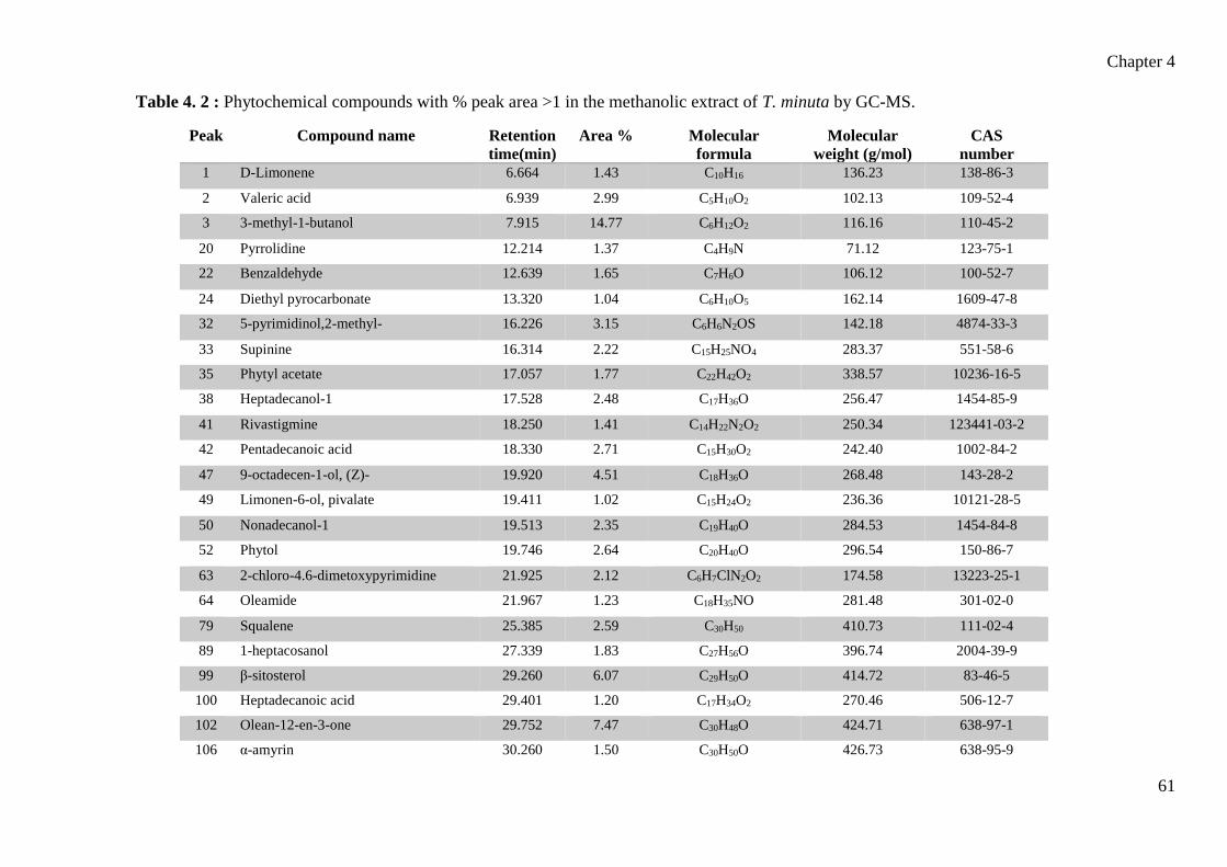
Chapter 4
61
Table 4. 2 : Phytochemical compounds with % peak area >1 in the methanolic extract of T. minuta by GC-MS.
Peak Compound name Retention
time(min)
Area % Molecular
formula
Molecular
weight (g/mol)
CAS
number
1 D-Limonene 6.664 1.43 C10H16 136.23 138-86-3
2 Valeric acid 6.939 2.99 C5H10O2 102.13 109-52-4
3 3-methyl-1-butanol 7.915 14.77 C6H12O2 116.16 110-45-2
20 Pyrrolidine 12.214 1.37 C4H9N 71.12 123-75-1
22 Benzaldehyde 12.639 1.65 C7H6O 106.12 100-52-7
24 Diethyl pyrocarbonate 13.320 1.04 C6H10O5 162.14 1609-47-8
32 5-pyrimidinol,2-methyl- 16.226 3.15 C6H6N2OS 142.18 4874-33-3
33 Supinine 16.314 2.22 C15H25NO4 283.37 551-58-6
35 Phytyl acetate 17.057 1.77 C22H42O2 338.57 10236-16-5
38 Heptadecanol-1 17.528 2.48 C17H36O 256.47 1454-85-9
41 Rivastigmine 18.250 1.41 C14H22N2O2 250.34 123441-03-2
42 Pentadecanoic acid 18.330 2.71 C15H30O2 242.40 1002-84-2
47 9-octadecen-1-ol, (Z)- 19.920 4.51 C18H36O 268.48 143-28-2
49 Limonen-6-ol, pivalate 19.411 1.02 C15H24O2 236.36 10121-28-5
50 Nonadecanol-1 19.513 2.35 C19H40O 284.53 1454-84-8
52 Phytol 19.746 2.64 C20H40O 296.54 150-86-7
63 2-chloro-4.6-dimetoxypyrimidine 21.925 2.12 C6H7ClN2O2 174.58 13223-25-1
64 Oleamide 21.967 1.23 C18H35NO 281.48 301-02-0
79 Squalene 25.385 2.59 C30H50 410.73 111-02-4
89 1-heptacosanol 27.339 1.83 C27H56O 396.74 2004-39-9
99 β-sitosterol 29.260 6.07 C29H50O 414.72 83-46-5
100 Heptadecanoic acid 29.401 1.20 C17H34O2 270.46 506-12-7
102 Olean-12-en-3-one 29.752 7.47 C30H48O 424.71 638-97-1
106 α-amyrin 30.260 1.50 C30H50O 426.73 638-95-9

Chapter 4
62
Table 4. 3: Preliminary screening of antibacterial activity of silver nanoparticles derived
from leaves of T. minuta (n=3, mean values represented).
Bacterial strains Zone of Inhibition (mm) Antibiotic
Methicillin-resistant
Staphylococcus aureus 16 0
Escherichia coli 10 18
Staphylococcus aureus 10 0
Bacillus subtilis 12 6
Pseudomonas aeruginosa 13 9
(mm) = inhibition zone including diameter of gel puncture
The antibacterial efficacy of the crude methanolic extract from T. minuta leaves are
summarised in table 4.3 and were assessed by measuring the diameter of the inhibition
zone of each well. The crude extract exhibited varying degrees of inhibition against 5
bacterial strains (MRSA, E. coli, S. aureus, B. subtilis, and P. aeruginosa). The antibiotic
used for the gram-positive bacteria was streptomycin and gentamycin for gram-negative
bacteria. In general, the crude methanolic extract proved to be more effective against
gram-positive than gram-negative bacteria.
The antibacterial effects are similar to the activity shown from the essential oils of T.
patula (Rondon et al., 2006) and T. erecta (Tripathi et al., 2012). These studies report
flavonoids, alkaloids, and phenolic compounds are responsible for the antibacterial
effects of extracts from Tagetes species (Tripathy et al., 2017).
4.5 Conclusion
Tagetes minuta has proved to be an important medicinal plant for human use as well as
an insecticide and herbicide. Its applications extend pharmaceutically to include
antidiabetic, anti-inflammatory, anticancer, and antimicrobial activities as evidenced
from the phytocompounds found in abundance in the methanolic extract from the leaves

Chapter 4
63
of T. minuta. The results of this study show potential for research regarding the regulation,
use, and development of bioactive compounds sourced from medicinal plants.
Additionally, the plethora of pharmacologically useful compounds sourced from T.
minuta makes a convincing case for the status to be changed from invasive species to
underutilised medicinal plant crop in South Africa.

Chapter 5
64
CHAPTER 5: GREEN SYNTHESIS OF SILVER NANOPARTICLES
FROM TAGETES MINUTA L. AND ITS ANTIBACTERIAL
POTENTIAL
5.1 Abstract
The plant mediated synthesis of metallic nanoparticles is a fast-growing area of interest
in the field of nanotechnology due to its eco-friendly approach. This study demonstrated
the green synthesis of silver nanoparticles (AgNPs) using Tagetes minuta leaf extract.
The AgNPs were characterised using UV-visible spectroscopy, transmission electron
microscopy (TEM), energy dispersive X-ray (EDX) analysis and Fourier transform
infrared (FTIR) spectroscopy. UV–visible spectrum of synthesised silver nanoparticles
showed maximum peak at 442 nm. TEM revealed that the particles were spherical in
shape and size ranging from 10 to 50 nm. The EDX spectrum confirmed the presence of
silver metal. Preliminary antibacterial activity was determined using the agar well
diffusion method, which showed growth inhibition against selected gram-positive and
gram-negative bacteria.
Keywords: Antibacterial activity; energy dispersive x-ray; FTIR spectroscopy; silver
nanoparticles; UV-visible spectrometry

Chapter 5
65
5.2 Introduction
Nanotechnology is the blanket term for the synthesis, manipulation, and application of
materials and structures that range between 1 and 100 nm, and is currently one of the most
active research fields in material science (Sarsar et al., 2013; Padalia et al., 2015; Khatoon
et al., 2017). Size, distribution, and morphology of nanoparticles determines their
biological effectiveness (Krishnamurthy et al., 2012). Noble metal nanoparticles such as
gold, silver, and platinum show promising applications in the fields of biotechnology,
bioengineering, solar energy conversion, medicine, and water treatment (Dahl et al.,
2007; Sharma et al., 2009).
Traditionally, nanoparticles are produced using chemical and physical methods, but these
methods often face several caveats in the way of costs and environmental damage (Ramya
and Subapriya, 2012). Green chemistry and green synthesis methods have since been
adopted in the efforts of reducing both costs and generated hazardous waste, as well as
decreasing the risk of safety concern over the products (Kaler et al., 2010). The biological
method of synthesising nanoparticles occurs by redox reaction that builds towards larger
and more complex systems beginning at the molecular level (Ramya and Subapriya,
2012). Based on green chemistry perspectives, the selection of an environmentally benign
solvent and nontoxic chemicals in the synthesis of nanoparticles are imperative (Sharma
et al., 2009). Natural material from microorganisms, enzymes, and plants are used for
green synthesis of silver nanoparticles (Kotakadi et al., 2014; Vidhu and Philip, 2014).
Silver has been used as an antimicrobial agent for many decades, but the rising interest in
the properties of metallic silver in the form of silver nanoparticles (AgNPs) is focused
towards the increasing threat of antibiotic resistance (Roy and Das, 2015; Ojo et al.,
2018). Ahmed et al. (2016) claimed that silver is the least toxic metal to animal cells that
is used as an antimicrobial, and is effective against over 650 microorganisms from
different classes such as gram-negative and gram-positive bacteria, fungi, and viruses. In
order for silver to have any antimicrobial properties, it must first be in its ionized form,
as the positive charge on the Ag+ ions is vital in forming nanoparticles (Ahmed et al.,
2016; AlSalhi et al., 2016). In conjunction, the medicinal properties of plants are
attributed to its phytochemicals, which are in a position to reduce, cap, and stabilise Ag+
ions (Chinnasamy et al., 2017; Tiwary and Jha, 2017). Organic chemicals present in plant

Chapter 5
66
aqueous extracts (e.g. carbohydrates, proteins, phenols, flavonoids, terpenoids, alkaloids)
are capable of donating an electron that results in the reduction of Ag+ ions to Ag0 (Roy
and Das, 2015; Tiwary and Jha, 2017).
Nanotechnology also allows for the exploitation of antimicrobial properties of silver, as
they are used in the form of nanoparticles. Silver nanoparticles display a large surface
area to volume ratio – which allows for broad contact with the nuclear content of the
bacteria, thus enabling the inactivation of DNA replication leading to growth inhibition
(Ojo et al., 2017). It has been reported that AgNPs attach to the cell walls of bacteria and
disturb the cell wall permeability and cellular respiration (Singh et al., 2008; Roy and
Das, 2015).
Tagetes minuta is an aromatic herbaceous plant belonging to the Asteraceae family that
has been reported for its high-grade essential oil that shows numerous uses in beverage,
cosmetic, and pharmaceutical industries (Shirazi et al., 2014). It is found along river
banks, forest margins, dry wooded valleys and hillsides in KwaZulu-Natal since its
naturalisation in South Africa. The leaves of T. minuta are used in the preparation of
traditional remedies for the management of stomach ailments, headaches, diarrhoea,
malaria, and epilepsy (Karimian et al., 2014; Kyarimpa et al., 2014; Igwaran et al., 2017).
The pharmacological effects of the leaf extracts are purported to be the result of various
secondary metabolites including essential oils particularly abundant in monoterpenes,
sesquiterpenes, flavonoids, aromatics, and thiophenes (Bansal et al., 1999; Brene et al.,
2009; Sadia et al., 2013). The aims of this study were to employ the use of aqueous leaf
extracts of T. minuta in the biosynthesis of AgNPs, characterise the particles, as well as
to observe its antimicrobial activity against both gram-positive and gram-negative
bacteria.

Chapter 5
67
5.3 Methods and Materials
5.3.1 Collection of plant material
Fresh leaves of T. minuta were collected from the UKZN Westville campus (29.817°S
30.940°E) and air-dried for 6 weeks. A voucher specimen was confirmed and deposited
at the UKZN Westville Herbarium (accession number 18216, voucher number 01).
5.3.2 Methanol extraction
The air-dried leaves were ground into a fine powder using a blender. Thirty grams of
ground material was placed in a round bottom flask containing 50 ml of methanol. The
flask was attached to Soxhlet apparatus and boiled for two 3-hour sessions to obtain the
crude plant extract. The extract was filtered using filter paper (Whatman No. 1) and stored
in an airtight jar at 4°C.
5.3.3 Fresh water extraction
Ten grams of fresh leaves were thoroughly washed with distilled water. The leaves were
cut into small pieces and boiled for 10 min in 100 ml distilled water and filtered through
Whatman No. 1 filter paper and stored in a concealed jar. The filtrate was used for the
synthesis of silver nanoparticles.
5.3.4 Green synthesis of AgNPs
Aqueous solution (1 mM) of silver nitrate (AgNO3) was prepared and used for the
synthesis of AgNPs. The ratio of 1 ml methanol extract to 19 ml silver nitrate solution
was used for the reduction of Ag+ ions. The solution was then incubated in a hot water
bath at 60°C until a colour change was observed. This process took 30 minutes. AgNP
solution was centrifuged at 16000 rpm (20°C) for 5 min on the Eppendorf Centrifuge
5415R. Thereafter, the pellet was suspended in deionized water.

Chapter 5
68
5.3.5 Characterisation of AgNPs
a) UV–Visible spectroscopy
UV–Vis spectra of synthesized nanoparticles were monitored on a spectrophotometer
(Shimadzu UV-1800) in 350–700 nm range at a resolution of 2 nm.
b) Energy dispersive X-ray analysis
A drop of the AgNP solution was placed on a glass cover slip, mounted onto a brass stub
and left to dry. The stub was then sputter coated with gold in a Polaron SC500 sputter
coater and viewed with a Zeiss Ultra Plus field emission scanning electron microscope
(FE-SEM) at 5 kV. Silver nanoparticle information by EDX microanalysis of elements
present was captured using Aztec software coupled to an Oxford X-MAX detector
(Oxford instruments, UK).
c) Fourier transform infrared (FTIR) spectral analysis
Each sample was centrifuged at 10000 rpm (Beckman Coulter Avanti J-E Centrifuge) for
30 min. The resulting pellet was suspended in deionized water and was used for further
characterisation. Spectroscopic measurements of the sample (200 µl) were determined
using a PerkinElmer FTIR Spectrum One spectrophotometer in the diffuse reflectance
mode operating at a spatial resolution of 4 cm-1.
d) Transmission electron microscopy
A copper grid was submerged in the silver nanoparticles sample and thereafter air-dried.
The size, shape, and overall morphology of the AgNPs was determined using the Zeiss
UltraPlus FEGSEM at 200 kV.
5.3.6 Antibacterial screening
The preliminary antibacterial activity of the synthesised AgNPs was evaluated against
Escherichia coli (ATCC 25218), Staphylococcus aureus (ATCC 29213), methicillin-
resistant Staphylococcus aureus (ATCC BAA-1683), Bacillus subtilis and Pseudomonas
aeruginosa (ATCC 25215). Mueller Hilton agar medium was prepared and poured into

Chapter 5
69
sterile petri dishes to set at room temperature. The bacterial cultures were swabbed
uniformly onto the plates using sterile cotton swabs. Wells of 5 mm in diameter were
made using an agar corer (gel puncture). The samples were pipetted into the wells (90
µl), and the plates were incubated at 37ºC. The effect of the samples on the bacteria was
assessed 24 hours later by measuring diameter of the zone of inhibition (mm).

Chapter 5
70
5.4 Results and Discussion
The fresh suspension of T. minuta was a light green in colour (Fig 5.1 a). However, after
the addition of silver nitrate and exposing to heat in a 60°C water bath for 30 mins, the
suspension turned a dark brown colour (Fig 5.1 b). The reduction of silver ions into silver
nanoparticles during exposure to the plant extract is indicated by this colour change. It
appears that the intensity of the colour reaction was directly proportional to the formation
of the AgNPs. The UV-vis spectra recorded from the methanolic extract of T. minuta is
presented in Fig 5.2, where the maximum absorption peak is shown to be at 442 nm.
Surface plasmon resonance (SPR) is the collective oscillation of electrons in the
conduction band on a nanoparticle surface (Suman et al., 2014). Siddiqui et al. (2018)
claimed that the absorption band from 400 to 500 nm represents the dipole component of
the SPR of silver nanoparticles. This implies that the peak wavelength, width, and effect
of these resonances yield a unique spectral fingerprint for a plasmonic nanoparticle with
a distinct size and shape. Using high-resolution TEM, Mock et al. (2002) showed that
silver nanoparticles that peak in the range of 410 – 500 nm are spherical; whereas in the
range of 500 – 700 nm, particles are usually triangular or pentagonal. This observation
strongly suggests that the AgNPs synthesised in this study were spherical in shape.

Chapter 5
71
Figure 5. 1 Images of silver nanoparticle synthesis using T.minuta extract from leaves:
a) Silver nitrate solution with leaf extract. b) Synthesised silver nanoparticle solution after
heating for 30 minutes.
Figure 5. 2 UV-Vis absorption spectra of reduction of silver ions to silver nanoparticles
after 30 min reaction.
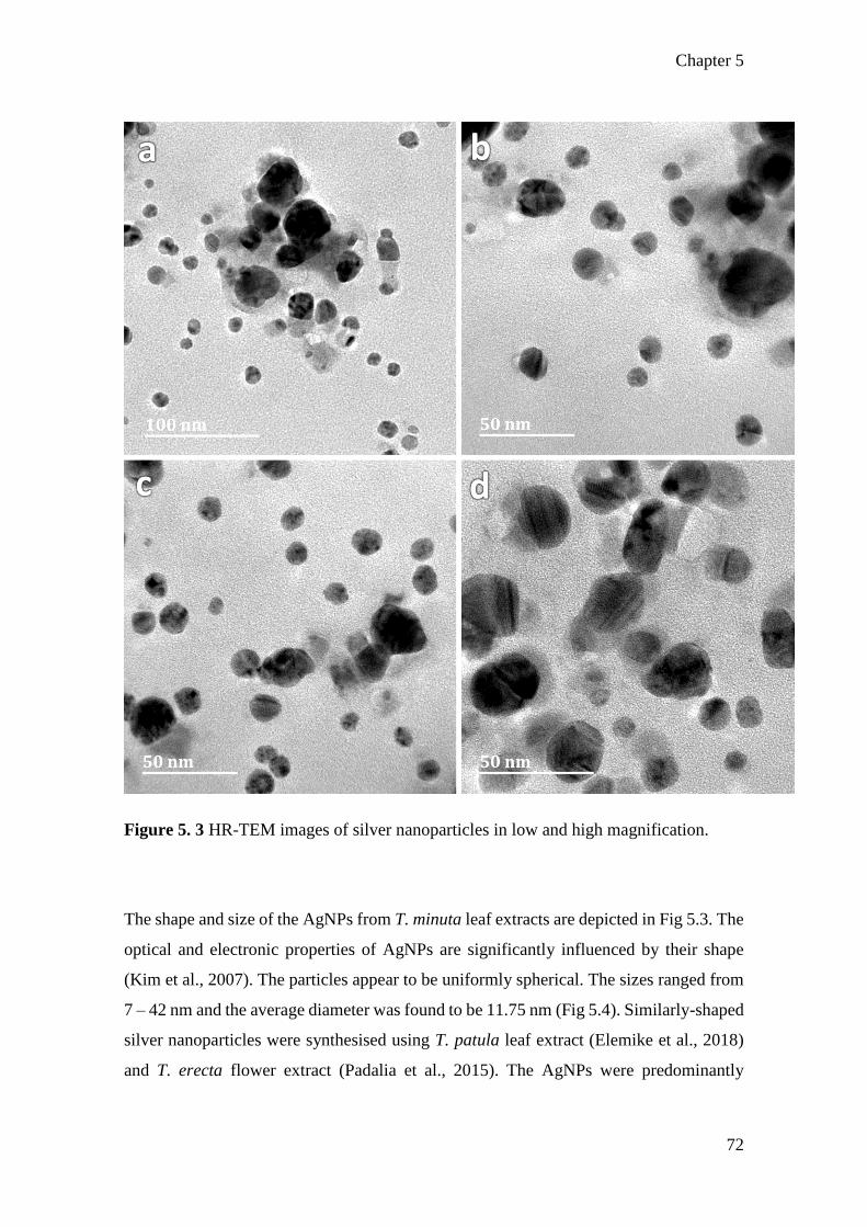
Chapter 5
72
Figure 5. 3 HR-TEM images of silver nanoparticles in low and high magnification.
The shape and size of the AgNPs from T. minuta leaf extracts are depicted in Fig 5.3. The
optical and electronic properties of AgNPs are significantly influenced by their shape
(Kim et al., 2007). The particles appear to be uniformly spherical. The sizes ranged from
7 – 42 nm and the average diameter was found to be 11.75 nm (Fig 5.4). Similarly-shaped
silver nanoparticles were synthesised using T. patula leaf extract (Elemike et al., 2018)
and T. erecta flower extract (Padalia et al., 2015). The AgNPs were predominantly

Chapter 5
73
monodispersed and stable. Organic material derived from the plant extraction process
causes the inherent functional group capping, which in turn offers stability and prevents
agglomeration (Shaik et al., 2014).
Figure 5. 4 Frequency histogram for silver nanoparticle size range.

Chapter 5
74
Figure 5. 5 EDX spectrum of synthesised silver nanoparticles using leaf extract of
Tagetes minuta.
A strong signal of silver is evident from the EDX spectrum at 1.5 keV and a weaker signal
at 3 keV (Fig 5.5) confirming the presence of elemental silver. A similar spectral profile
for silver has been reported by Hedaginal and Taranath (2017) for the leaf extract of
Thunbergia alata. Weak signals of carbon, oxygen, iron, manganese, silicon, chlorine,
and copper are also evident. The presence of silicon, carbon, and copper is likely to be
from the support glass cover slip and grid used in the methodology for EDX. Signals of
other trace elements may be the result of x-ray emission from organic compounds
involved in the capping of the nanoparticles and are bound to the surface of the nano-
silver (Singh et al., 2015).

Chapter 5
75
Figure 5. 6 FTIR spectrum of the synthesised silver nanoparticles using leaf extract of
Tagetes minuta.
The known active ingredients in T. minuta are terpenoids viz. limonene, caryophyllene,
and eucarvone (Tiwari et al., 2016; Igwaran et al., 2017); amines viz. pyrrolidine
(Meshkalasadat et al., 2010); ketones viz. tagetone, dihydrotagetenone, tagetenone (Gil
et al., 2002; Mohammad et al., 2010); and hydrocarbons viz. ocimene (Igwaran et al.,
2017). The FTIR spectrum analysis of biosynthesised silver nanoparticles are displayed
in Fig 5.6 which manifest absorption peaks located at the regions between 600 cm−1 and
3300 cm−1 in order to identify the functional groups of the extract involved in the
reduction of the synthesised AgNPs. Prominent peaks on the FTIR spectrum are marked
at 3272 cm-1, 2119 cm-1, 1637 cm-1, 1016 cm-1, and 595 cm-1. The absorption peak at 3272
cm-1 is assigned to –OH stretching in alcohols and phenolic compounds, which indicates
the possible involvement of the known terpenoids (Awwad et al., 2013; Elemike et al.,
2018). The peak at 2119 cm-1 suggest –C≡C and –CH groups that are found in terpenoid
ring structures and is close to that reported for aliphatic aldehydes (Shaik et al., 2014;

Chapter 5
76
Hedaginal and Taranath, 2017). The absorption peak at 1637 cm-1 arose due to C=C
stretches that are typical of aliphatic and aromatic amine structures and alkenes (Jha et
al., 2018). Peaks below the value 1300 cm-1 are usually indicative of C–C and C–O
groups, but are not as reliably interpreted due to a larger number of different vibrations
(Kumar et al., 2017).
Table 5. 1 : Preliminary screening of antibacterial activity of silver nanoparticles derived
from leaves of T. minuta (n=3, mean values represented).
Bacterial strains Zone of Inhibition (mm) Antibiotic
Methicillin-resistant
Staphylococcus aureus 9 0
Escherichia coli 11 10
Staphylococcus aureus 8 0
Bacillus subtilis 9 6
Pseudomonas aeruginosa 7 9
(mm) = inhibition zone including diameter of gel puncture
The antibacterial efficacy of silver nanoparticles synthesised from T. minuta leaves are
summarised in table 5.1 and were assessed by measuring the diameter of the zone of
inhibition of each well. The AgNPs exhibited varying degrees of inhibition against 5
bacterial strains (MRSA, E. coli, S. aureus, B. subtilis, and P. aeruginosa). The antibiotic
used for the gram-positive bacteria was streptomycin and gentamycin for gram-negative
bacteria. In general, the silver nanoparticles were more effective against gram-positive
than gram-negative bacteria.
The AgNPs were least effective against E. coli and P. aeruginosa, which showed
inhibition zones very similar to that of the antibiotic (gentamycin). Sondi and Salopek-
Sondi (2014) demonstrated silver nanoparticles penetrating the bacterial cell walls of E.
coli, but growth inhibition was dependent on the concentration of the AgNPs used, and
in most instances would only delay the growth of the bacterial colonies. Pal et al. (2007)

Chapter 5
77
studied the effects of nanoparticle shape against gram-negative bacteria, and found that
spherical and rod-shaped nanoparticles are less effective than triangular-shaped
nanoparticles. This may explain the ineffectiveness of the AgNPs synthesised in this
study against P. aeruginosa.
The bacterial growth of the gram-positive bacteria (MRSA, S. aureus, and B. subtilis)
was more severely inhibited by the AgNPs synthesised in this study. Guzman et al. (2012)
highlighted the importance of the size of AgNPs against gram-positive bacteria,
concluding that silver particles between 9 and 14 nm in diameter showed the highest
activity. In Fig 5.4, it is evident that the silver nanoparticles synthesised in this study that
were under 15 nm in diameter were the most frequent, which likely contributed to the
higher antibacterial activity. It has been reported that AgNPs induce the inactivation of
DNA replication and thus protein synthesis in gram-positive bacteria by interacting with
present thiol groups (Ojo et al., 2017).
5.5 Conclusion
This study provided a simple and rapid method for the biosynthesis of AgNPs using T.
minuta aqueous leaves extract. The secondary metabolites present in T. minuta perform
the dual function of formation and stabilisation of AgNPs through the action of various
terpenoid and amine compounds identified. The AgNPs were further confirmed by using
UV-Vis spectroscopy, EDX, and FTIR techniques. The shape of the nanoparticles was
determined to be spherical using HR-TEM. The biosynthesised AgNPs also exhibited
antimicrobial activity against selected gram-positive and gram-negative bacteria strains,
which is in part due to the size and shape of the AgNPs. Hence, this study supports the
efficiency of plant mediated green synthesis of AgNPs, which has the potential to be
utilised in various clinical applications.
Chapter 6
78
CHAPTER 6: GENERAL CONCLUSIONS AND RECOMMENDATIONS
FOR FURTHER RESEARCH
6.1 Main findings
Tagetes minuta L. is an annual herb that is used as ethnomedicine worldwide. It is reported to
have numerous medicinal benefits such as treatment for respiratory inflammations, stomach
pains, chest infections, coughs, and as well as a healing effect on wounds, cuts, and calluses.
Despite its rich literature in ethnomedicinal uses, the phytocompounds that are responsible for
its therapeutic properties have not been reported on. Additionally, the foliar structures that
produce, store, and exude these phytocompounds are not well understood. The aims of this
study were to critically analyse the leaves of T. minuta with regard to its micromorphology,
histochemistry, phytochemistry, green synthesis of silver nanoparticles, and potential for
antibacterial activity.
The leaves of T. minuta contain two types of foliar structures, viz. non-glandular trichomes and
secretor cavities. In Chapter 3, the leaves were studied using stereomicroscopy, scanning
electron microscopy (SEM), and transmission electron microscopy (TEM). Stereomicroscopy
showed a sparse coverage of trichomes that group along the midvein and major venation
network, suggesting that they do not play a large role in physical protection to the plant. The
secretory cavities can be seen macroscopically and appear as large pellucid yellow glands
between 70 and 200 µm in diameter. Scanning electron microscopy revealed the secretory
cavity openings along the anomocytic stoma. The trichomes were deduced to be uniseriate and
non-glandular, reaching up to 200 µm in length. TEM was used to view the cells within the
secretory cavity, which showed an increase in lipid and vesicle production within the organelles
as compared to surrounding cells. Histochemical analyses showed that the non-glandular
trichomes were able to store various classes of phytocompounds, in contrast to the literature
suggesting non-glandular trichomes are incapable of phytometabolites. This work is novel in
terms of the micromorphology of T. minuta.
Crude organic solvent extracts derived from the leaves of T. minuta were investigated in
Chapter 4. Phytochemical tests were used to determine the main chemical classes present in
the hexane, chloroform, and methanol crude extracts, revealing the presence of carbohydrates,

Chapter 6
79
alkaloids, amino acids, sterols, saponins, terpenoids, phenols, and lipids. The crude methanolic
extract was further subjected to analysis using GC-MS, which showed that the compounds with
the highest composition percentage were 9-octadecen-1-ol (4.51 %), β-sitosterol (6.07 %),
olean-12-en-3-one (7.47 %), and 3-methyl-1-butanol (14.77 %). These compounds are
discussed in Chapter 4 as having various pharmacological benefits. Antibacterial screening
using the methanolic extract showed greater growth inhibition on gram positive than gram
negative bacteria.
Green synthesis of silver nanoparticles was achieved in Chapter 5 using the methanolic extract.
Using TEM, the nanoparticles were proved to be spherical in shape and ranged between 10 and
50 nm in size. The presence of elemental silver was determined using EDX analysis and
polymeric chemical groups assessed using FTIR spectroscopy. The silver nanoparticles proved
to have a greater effect on the growth inhibition of gram positive bacteria.
6.2 Future recommendations
With regard to microscopy, TEM analysis could be taken further to include the intracellular
components of the non-glandular trichomes to determine whether they produce as well as store
secondary metabolites. Future research could compare and contrast the percentage composition
of phytochemicals from the organic solvent extracts to that of the essential oils of T. minuta,
as well as compare percentage composition from extracts taken at different growth
development and flowering stages of the plant. Silver nanoparticles derived from the
methanolic extract can be further investigated in terms of antimicrobial activity in order to
determine the minimum inhibitory concentrations. Additionally, T. minuta can be used in cell
culture for possible propagation, changing its status in South Africa from invasive weed to
underutilised medicinal crop.

Chapter 7
80
CHAPTER 7: REFERENCES
Abdul, W.S., Hajrah, N.H., Sabir, J.S.M., Al-Garni, S.M., Sabir, M.J., Kabli, S.A., Saini, K.S.,
Bora, R.S., 2018. Therapeutic role of Ricinus communis L. and its bioactive compounds in
disease prevention and treatment. Asian Pacific Journal of Tropical Medicine 11, 177–185.
Achika, J.I., Arthur, D.E., Gerald, I., Adedayo, A., 2014. A review on the phytoconstituents
and related medicinal properties of plants in the Asteraceae family. IOSR Journal of Applied
Chemistry 7, 1–8.
Adebooye, O.C., Hunsche, M., Noga, G., Lankes, C., 2012. Morphology and density of
trichomes and stomata of Trichosanthes cucumerina (Cucurbitaceae) as affected by leaf age
and salinity. Turkish Journal of Botany 36, 328–335.
Adedeji, O., Jewoola, O.A., 2008. Importance of leaf epidermal characters in the Asteraceae
family. Notulae Botanicae Horti Agrobotanici Cluj-Napoca 36, 7–16.
Agrawal, A.A., 2006. Macroevolution of plant defense strategies. Trends in Ecology and
Evolution 22, 103–109.
Agrawal, A.A., Fishbein, M., 2006. Plant defense systems. Ecology 87 Supplement, 132–149.
Ahmed, S., Ahmad, M., Swami, B.L., Ikram, S., 2016. A review on plants extract mediated
synthesis of silver nanoparticles for antimicrobial applications: a green expertise. Journal of
Advanced Research 7, 17–28.
Algotsson, E., 2009. Biological Diversity. In Strydom, H.A. and King, N.D., eds.
Environmental Management in South Africa. Cape Town: Juta, 97–125.
Al-Musayeib, N.M., Mohamed, G.A., Ibrahim, S.R.M., Ross, S.A., 2014. New thiophene and
flavonoid from Tagetes minuta leaves growing in Saudi Arabia. Molecules 19, 2819–2828.
AlSalhi, M.S., Devanesan, S., Alfuraydi, A.A., Vishnubalaji, R., Munusamy, M.A., Murugan,
K., Nicoletti, M., Benelli, G., 2016. Green synthesis of silver nanoparticles using Pimpinella
anisum seeds: antimicrobial activity and cytotoxicity on human neonatal skin stromal cells and
colon cancer cells. International Journal of Nanomedicine 11, 4439–4449.

Chapter 7
81
Andreotti, R., Garcia, M.V., Cunha, R.C., Barros, J.C., 2013. Protective action of Tagetes
minuta (Asteraceae) essential oil in the control of Rhipicephalus microplus (Canestrini, 1887)
(Acari: Ixodidae) in a cattle pen trial. Veterinary Parasitology 197, 341–345.
Arora, K., Batish, D.R., Singh, H.P., Kohli, R.K., 2015. Allelopathic potential of the essential
oil of wild marigold (Tagetes minuta L.) against some invasive weeds. Journal of
Environmental and Agricultural Sciences 3, 56–60.
Arora, S. and Kumar, G., 2018. Micro-morphological descriptions on Cenchrus species from
Rajasthan (India). Annals of Plant Sciences 7, 2141–2145.
Aschenbrenner, A., Horakh, S., Spring, O., 2013. Linear glandular trichomes of Helianthus
(Asteraceae): morphology, localization, metabolite activity and occurrence. Annals of Botany
5, 28–37.
Athuman, I., Innocent, E., Machumi, F., Augustino, S., Kisinza, W., 2016. Repellency
properties of oils from plants traditionally used as mosquito repellents in Longido district,
Tanzania. International Journal of Mosquito Research 3, 4–8.
Awwad, A.M., Salem, N.M., Abdeen, A.O., 2013. Green synthesis of silver nanoparticles using
carob leaf extract and its antibacterial activity. International Journal of Industrial Chemistry 4,
29–35.
Balandrin, M.F., Klocke, J.A., Wurtele, E.S., Bollinger, W.H., 1985. Natural plant chemicals:
source of industrial and medicinal materials. Science 228, 1154–1160.
Balcke, G.U., Bennewitz, S., Bergau, N., Athmer, B., Henning, A., Majovsky, P., Jimenez-
Gomez, J.M., Hoehenwarter, W., Tissier, A., 2017. Multi-omics of tomato glandular trichomes
reveals distinct features of central carbon metabolism supporting high productivity of
specialized metabolites. The Plant Cell, 29, 960–983.
Balunas, M.J. and Kinghorn, A.D., 2005. Drug discovery from medicinal plants. Life Sciences
78, 431–441.
Bandana, K., Raina, R., Kumari, M., Rani, J., 2018. Tagetes minuta: an overview. International
Journal of Chemical Studies 6, 3711–3717.

Chapter 7
82
Bansal, R.P., Bahl, S.N., Garg, A.A., Sharma, N.S., Ram, M., Kumar, S., 1999. Variation in
quality of essential oil distilled from vegetative and reproductive stages of Tagetes minuta crop
grown in north Indian plains. Journal of Essential Oil Research 11, 747–752.
Bargah, R.K., 2015. Preliminary test of phytochemical screening of crude ethanolic and
aqueous extract of Moringa pterygosperma Gaertn. Journal of Pharmacognosy and
Phytochemistry 4, 7–9.
Barkley, T.M., Brouillet, L., Strother, J.L., 2006. The Asteraceae Family. Flora of North
America 19, 3–12.
Bartoli, A., Galati, B.G., Tortosa, R.D., 2011. Anatomical studies of the secretory structures:
glandular trichomes and ducts in Grindelia pulchella Dunal (Astereae, Asteraceae). Flora 206,
1063–1068.
Bezerra, L.D.A., Mangabeira, P.A.O., de Oliveira, R.A., Costa, L.C.D.B., da Cunha, M., 2018.
Leaf blade structure of Verbesina macrophylla (Cass.) F.S. Blake (Asteraceae): ontogeny, duct
secretion mechanism and essential oil composition. Plant Biology (Stuttgart, Germany) 20,
433–443.
Bin Sayeed, M.S. and Amen, S.S., 2015. Beta-sitosterol: a promising but orphan nutraceutical
to fight against cancer. Nutrition and Cancer 1, 1–7.
Bisi-Johnson, M.A., Obi, C.L., Samuel, B.B., Eloff, J.N., Okoh, A.I., 2017. Antibacterial
activity of crude extracts of some South African medicinal plants against multidrug resistant
etiological agents of diarrhoea. BMC Complementary and Alternative Medicine 17, 321–329.
Bombo, A.B., Filartiga, A.L., Garcia, V.L., Appezzato-da-Gloria, B., 2017. Secretory
structures in Aldama species (Heliantheae – Asteraceae): Morphology, histochemistry and
composition of essential oils. Flora 228, 39–49.
Bremer, K., 1987. Tribal interrelationships of the Asteraceae. Cladistics 3, 210–253.
Bremer, K., Jansen, R.K., Karis, P.O., Kallersjo, M., Keeley, S.C., Kim, K., Michaels, H.J.,
Palmer, J.D., Wallace, R.S., 1992. A review of the phylogeny and classification of the
Asteraceae. Nordic Journal of Botany 12, 141–148.

Chapter 7
83
Brene, K., Tournayre, P., Fernandez, X., Meierhenrich, U.J., Brevard, H., Joulain, D., 2009.
Identification of odour impact compounds of Tagetes minuta essential oil: comparison of two
GC-olfactometry methods. Journal of Agricultural and Food Chemistry 57, 8572–8580.
Brewer, C.A., Smith, W.K., Vogelmann, T.C., 1991. Functional interaction between leaf
trichomes, leaf wettability and the optical properties of water droplets. Plant, Cell, &
Environment 14, 955–962.
Buda, G.J., Isaacson, T., Matas, A.J., Paolilo, D.J. and Rose, J.K.C., 2009. Three-dimensional
imaging of plant cuticle architecture using confocal scanning laser microscopy. The Plant
Journal 60, 378–385.
Buvat, R., 1989. Ontogeny, cell differentiation, and structure of vascular plants. Springer-
Verlag Berlin Heidelberg, 56.
Cappellari, L.D.R., Santoro, M.V., Nievas, F., Giordano, W., Banchio, E., 2013. Increase of
secondary metabolite content in marigold by inoculation with plant growth-promoting
rhizobacteria. Applied Soil Ecology 70, 16–22.
Cardellina, J.H., 2002. Challenges and opportunities confronting the botanical dietary
supplement industry. Journal of Natural Products 65, 1073–1084.
Chaffey, N., 2006. Esau’s Plant Anatomy, Meristems, Cells, and Tissues of the Plant Body:
Their Structure, Function, and Development. 3rd edn. Annals of Botany 99, 785–786.
Chamorro, E.R., Ballerini, G., Sequeira, A.F., Velasco, G.A., Zalazar, M.F., 2008. Chemical
composition of essential oil from Tagetes minuta L. leaves and flowers. Journal of the
Argentine Chemical Society 96, 80–86.
Chinnasamy, C., Tamilselvan, P., Karthik, V. and Karthik, B., 2017. Optimization and
characterization studies on green synthesis of silver nanoparticles using response surface
methodology. Advances in Natural and Applied Sciences 11, 214–221.
Cid, C.V., Rodriguez, J.H., Salazar, M.J., Blanco, A., Pignata, M.L., 2016. Effects of co-
cropping Bidens pilosa (L.) and Tagetes minuta (L.) on bioaccumulation of Pb in Lactuca
sativa (L.) growing in polluted agricultural soils. International Journal of Phytoremediation 18,
908–917.

Chapter 7
84
Colombo, P.M. and Rascio, N., 1977. Ruthenium red staining for electron microscopy of plant
material. Journal of Ultrastructure Research 60, 135–139.
Cunningham, A.B., 1990. Whose knowledge and whose resources? Ethnobotanists as brokers
between two worlds. Occasional paper 82. Institute of National Resources, Pietermaritzburg,
South Africa, 7.
Dahl, J.A., Maddux, B.L., Hutchison, J.E., 2007. Toward greener synthesis. Chemical Reviews
107, 2228–2269.
Dauskardt, R.P.A., 1990. The changing geography of traditional medicine: urban herbalism on
the Witwatersrand, South Africa. GeoJournal 22, 275–283.
Demarco, D., 2017. Histochemistry of single molecules: methods and protocols. Methods in
Molecular Biology 1560, 313–330.
Devika, R. and Koilpillai, J., 2012. Phytochemical screening studies of bioactive compounds
of Tagetes erecta. International Journal of Pharma and Bio Sciences 3, 596–602.
Dixit, P., Tripathi, S., Verma, N.K., 2013. A brief study on marigold (Tagetes species): A
review. International Research Journal of Pharmacy 4, 43–48.
Elemike, E.E., Onwudiwe, D.C., Abiola, O.K., Ibe, K.A., 2018. Surface characterisation and
reaction kinetics of silver nanoparticles mediated by the leaf and flower extracts of French
marigold (Tagetes patula). IET Nanobiotechnology 12, 957–962.
Fabricant, D.S. and Farnsworth, N.R., 2001. The value of plants used in traditional medicine
for drug discovery. Environmental Health Perspectives 109, 69–75.
Fahn, A., 1979. Secretory tissues in plants. Academic Press, London, 302.
Fahn, A., 2000. Structure and function of secretory cells. Advances in Botanical Research 31,
219–238.
Farjana, N., Rowshanul, H.M., Zahangir, A.S., Rezaul, M.K., Apurba, K.R., Shahriar, Z., 2009.
Toxicological evaluation of chloroform fraction of flower of Tagetes erecta Linn. on rats.
International Journal of Drug Development and Research 1, 161–165.

Chapter 7
85
Filartiga, A.L., Bombo, A.B., Garcia, V.L., Appezzato-da-Gloria, B., 2017. Belowground
organs of four Brazilian Aldama (Asteraceae) species: morphoanatomical traits and essential
oil profile. South African Journal of Botany 113, 150–159. Plant Biology 20, 433–443.
Gakuubi, M.M., Wanzala, W., Wagacha, J.M., Dossaji, S., 2016. Bioactive properties of
Tagetes minuta L. (Asteraceae) essential oils: A review. American Journal of Essential Oils
and Natural Products 4, 27–36.
Galdon-Armero, J., Fullana-Pericas, M., Mulet, P.A., Conesa, M.A., Martin, C., Galmes, J.,
2018. The ratio of trichomes to stomata is associated with water use efficiency in Solanum
lycopersicum (tomato). The Plant Journal 96, 607–619.
Ge, Y., Zhang, H., Lei, J., Wang, K., 2018. Chemical constituents of Viburnum odoratissumum
and their cytotoxic activities. Chemistry of Natural Compounds 54, 600–602.
Gil, A., Ghersa, C.M., Susana, P., 2002. Root thiophenes in Tagetes minuta L. accessions from
Argentina: genetic and environmental contribution to changes in concentration and
composition. Biochemical Systematics and Ecology 30, 1–13.
Gonzales, W.L., Negritto, M.A., Suarez, L.H., Gianoli, E., 2008. Induction of glandular and
non-glandular trichomes by damage in leaves of Madia sativa under contrasting water regimes.
Acta Oecologica 33, 128–132.
Gupta, H.S. and De, D.N., 1985. Uptake and accumulation of acridine orange by plant cells.
Proceedings of the National Academy of Sciences 6, 653–660.
Gutierrez, R.M.P., Luna, H.H.S., Garrido, H., 2006. Antioxidant activity of Tagetes erecta
essential oil. Journal of the Chilean Chemical Society 51, 883–886.
Guzman, M., Dille, J., Godet, S., 2012. Synthesis and antibacterial activity of silver
nanoparticles against gram-positive and gram-negative bacteria. Nanomedicine:
Nanotechnology, Biology, and Medicine 8, 37–45.
Hedaginal, B.R. and Taranath, T.C., 2017. Characterization and antimicrobial activity of
biogenic silver nanoparticles using leaf extract of Thunbergia alata Bojer ex Sims.
International Journal of Pharmaceutical Sciences and Research 8, 2070–2081.
Chapter 7
86
Hegebarth, D., Buschhaus, C., Wu, M., Bird, D., Jetter, R., 2016. The composition of surface
wax on trichomes of Arabidopsis thaliana differs from wax on other epidermal cells. The Plant
Journal 88, 762–774.
Hethelyi, E., Danos, B., Tetenyi, P., 1986. GC-MS analysis of the essential oils of four Tagetes
species and the anti-microbial activity of Tagetes minuta. Flavour and Fragrance Journal 1,
169–173.
Hosseinzadeh, S., Jafarikukhdan, A., Hosseini, A., Armand, R., 2015. The application of
medicinal plants in traditional and modern medicine: a review of Thysmusvulgaris.
International Journal of Clinical Medicine 6, 635–642.
Ibrahim, S.R.M. and Mohamed, G.A., 2017. Thiotagetin A, a new cytotoxic thiophene from
Tagetes minuta. Natural Product Research 31, 543–547.
Ibrahim, S.R.M., Abdallah, H.M., El-Halawany, A.M., Esmat, A., Mohamed, G.A., 2018a.
Thiotagetin B and tagetannins A and B, new acetylenic thiophene and digalloyl glucose
derivatives from Tagetes minuta and evaluation of their in vitro antioxidative and anti-
inflammatory activity. Fitoterapia 125, 78–88.
Igwaran, A., Iweriebor, B.C., Okoh, S.O., Nwodu, U.U., Obi, L.C., Okoh, A.I., 2017. Chemical
constituents, antibacterial and antioxidant properties of the essential oil flower of Tagetes
minuta grown in Cala community Eastern Cape, South Africa. BMC Complementary and
Alternative Medicine 17, 351–361.
Jensen, W.A., 1962. Botanical Histochemistry – principles and practice. University of
California, Berkeley. San Francisco and W. H. Freeman, 408.
Jha, B., Rao, M., Prasad, K., ha, A.K., 2018. Evaluation of antimicrobial activity of silver
nanoparticles from Piper betle leaves against human and plant pathogens. AIP Conference
Proceedings 1953, 030257.
Johnson, H.B., 1975. Plant pubescence: An ecological perspective. Botanical Review 41, 233–
258.
Kaler, A., Patel, N., Banerjee, U.C., 2010. Green synthesis of silver nanoparticles. Current
Research and Information on Pharmaceutical Science 11, 68–71.

Chapter 7
87
Karimian, P., Kavoosi, G., Amirghofran, Z., 2014. Anti-oxidative and anti-inflammatory
effects of Tagetes minuta essential oil in activated macrophages. Asian Pacific Journal of
Tropical Biomedicine 4, 219–277.
Kariyat, R.R., Hardison, S.B., Ryan, A.B., Stephenson, A.G., De Moraes, C.M., Mescher,
M.C., 2018. Leaf trichomes affect caterpillar feeding in an instar-specific manner.
Communicative and Integrative Biology 11, 1–6.
Kariyat, R.R., Smith, J.D., Stephenson, A.G., De Moraes, C.M., Mescher, M.C., 2017. Non-
glandular trichomes of Solanum carolinense deter feeding by Manduca sexta caterpillars and
cause damage to the gut peritrophic matrix. Proceedings of the Royal Society B 284, 20162323.
Kataria, D., Chahal, K.K., Kumar, A., 2016. Chemical transformation of carotol isolated from
carrot seed oil. Asian Journal of Chemistry 28, 1790–1792.
Khatoon, N., Mazumder, J.A., Sardar, M., 2017. Biotechnological applications of green
synthesized silver nanoparticles. Journal of Nanosciences: Current Research 2, 107.
Kim, H.J., Han, J., Kim, S., Lee, H.R., Shin, J., Cho, J., Kim, Y.H., Lee, H.J., Kim, B., Choi,
D., 2011. Trichome density of main stem is tightly linked to PepMoV resistance in chili pepper
(Capsicum annuum L.). International Journal of Plant Breeding Research 122, 1051–1058.
Kim, J.S., Kuk, E., Yu, K.N., Kim, J.H., Park, S.J., Lee, H.J., Kim, S.H., Park, Y.K., Park,
Y.H., Hwang, C.Y., Kim, Y.K., Lee, Y.S., Jeong, D.H., Cho, M.H., 2007. Antimicrobial effects
of silver nanoparticles. Nanomedicine 3, 95–101.
Kimutai, A., Ngeiywa, M., Mulaa, M., Njagi, P.G.N., Ingonga, J., Nyamwamu, L.B., Ombati,
C., Ngumbi, P., 2017. Repellent effects of the essential oils of Cymbogon citratus and Tagetes
minuta on the sandlfy, Phlebotomus duboscqi. BMC Research Notes 10, 99–103.
Kotakadi, V.S., Gaddam, S.A., Rao, Y.S., Prasad, T.N.V.K.V., Reddy, A.V., Gopal, D.V.R.S.,
2014. Biofabrication of silver nanoparticles using Andrographis paniculate. European Journal
of Medicinal Chemistry 73, 135–140.
Krishnamurthy, N.B., Nagaraj, B., Malakar, B., Liny, P., Dinesh, R., 2012. Green synthesis of
gold nanoparticles using Tagetes erecta L. (marigold) flower extract and evaluation of their
antimicrobial activities. International Journal of Pharma and Bio Sciences 3, 212–221.
Chapter 7
88
Kudumela, R.G. and Masoko, P., 2017. In vitro assessment of selected medicinal plants used
by the Bapedi community in South Africa for treatment of bacterial infections. Journal of
Evidence-Based Integrative Medicine 23, 1–10.
Kumar, B., Smita, K., Cumbal, L., Debut, A., 2017. Green synthesis of silver nanoparticles
using Andean blackberry fruit extract. Saudi Journal of Biological Sciences 24, 45–50.
Kyarimpa, C.M., Böhmdorfer, S., Wasswa, J., Kiremire, B.T., Ndiege, I.O., Kabasa, J.D., 2014.
Essential oil and composition of Tagetes minuta from Uganda. Larvicidal activity on Anopheles
gambiae. Industrial Crops and Products 62, 400–404.
Lawrence, B.M., 1996. Progress in essential oils, Myrtile oil. Perfumer and Flavorist 21, 57–
58.
Levin D.A., 1973. The role of trichomes in plant defense. The Quarterly Review of Biology
48, 3–15.
Li, S., Tosens, T., Harley, P.C., Jiang, Y., Kanagendran, A., Grosberg, M., Jaamets, K.,
Niinemets, U., 2018. Glandular trichomes as a barrier against atmospheric oxidative stress:
Relationships with ozone uptake, leaf damage, and emission of LOX products across a diverse
set of species. Plant, Cell and Environment 41, 1263–1277.
Lizarraga, E., Mercado, M.I., Galvez, C., Ruiz, A.I., Ponessa, G.I., Catalan, C.A.N., 2017.
Morpho anatomical characterization and essential oils of Tagetes terniflora and Tagetes minuta
(Asteraceae) growing in Tucuman (Argentina). Boletin de la Sociedad Argentina de Botanica
52, 55–68.
Lopez, M.L., Bonzani, N.E., Zygadlo, J.A., 2009. Allelopathic potential of Tagetes minuta
terpenes by a chemical, anatomical and phytotoxic approach. Biochemical Systematics and
Ecology 36, 882–890.
Machado, S.R., Canaveve, Y., Rodrigues, T.M., 2017. Structure and functioning of oil cavities
in the shoot apex of Metrodorea nigra A. St.-Hil. (Rutaceae). Protoplasma 254, 1661–1674.
Madikizela, B., Kambizi, L., McGaw, L.J., 2017. An ethnobotanical survey of plants used
traditionally to treat tuberculosis in the eastern region of O.R. Tambo district, South Africa.
South African Journal of Botany 109, 231–236.

Chapter 7
89
Maheshwari, J.K., 1972. Tagetes minuta Linn. In Shimla hills. Journal of Bombay Natural
History Society 69, 451.
Maity, N., Newma, N.K., Abedy, M.K., Sarkar, B.K., Mukherjee, P.K., 2011. Exploring T.
erecta Linn flower for the elastase, hyaluronidase and MMP-1 inhibitory activity. Journal of
Ethnopharmacology 137, 1300–1305.
Mander, M., Ntuli, L., Diederichs, N., Mavundla, K., 2007. Economics of the traditional
medicine trade in South Africa: healthcare delivery. South African Health Review 1, 189 –
196.
Matsuki, S., Sano, Y., Koike, T., 2004. Chemical and physical defence in early and late leaves
in three heterophyllous birch species native to Northern Japan. Annals of Botany 93, 141–147.
Mayekiso, B., Magwa, L.L., Coopoosamy, R., 2008. The morphology and ultrastructure of
glandular and non-glandular trichomes of Pteronia incana (Asteraceae). African Journal of
Plant Sciences 2, 50–60.
Meshkatalsadat, M.H., Safaei-Ghomi, J., Moharramipour, S., Nasseri, M., 2010. Chemical
characterization of volatile components of Tagetes minuta L. cultivated in south west of Iran
by nano scale injection. Digest Journal of Nanomaterials and Biostructures 5, 101–106.
Milan, P., Hayashi, A. H., Appezzato-da-Gloria, B., 2006. Comparative leaf morphology and
leaf anatomy of three Asteraceae species. Brazilian Archives of Biology and Technology 49,
135–144.
Minj, E., Britto, S.J., Marandi, R.R., Kindo, I., George, M., 2015. Phytochemical analysis and
antimicrobial activity of Putranjiva roxburghii Wall. World Journal of Pharmacy and
Pharmaceutical Sciences 5, 1157–1166.
Mo, Y., Yang, R., Liu, L., Gu, X., Yang, X., Wang, Y., Zhang, X., Li, H., 2016. Growth,
photosynthesis and adaptive responses of wild and domesticated watermelon genotypes to
drought stress and subsequent re-watering. Journal of Plant Growth Regulation 79, 229–241.
Mock, J.J., Barbic, M., Smith, D.R., Schultz, D.A., Schultz, S., 2002. Shape effects in plasmon
resonance of individual colloidal silver nanoparticles. Journal of Chemical Physics 116, 6755–
6759.

Chapter 7
90
Mohammad, H., Javad, S., Saeid, M., Morasaalsadat, N., 2010. Chemical characterization of
volatile components of Tagetes minuta L. cultivated in south west of Iran by nano scale
injection. Digest Journal of Nanomaterials and Biostructures 5, 101–106.
Monteiro, W.R., Fahn, A., Caldeira, W., de Moraes Castro, M., 1999. Ultrastructural
observations on the foliar secretory cavities of Porophyllum lanceolatum DC. (Asteraceae).
Flora 194, 113–126.
Nafiu, M.O., Hamid, A.A., Muritala, H.F., Adeyemi, S.B., 2017. Preparation, Standardization,
and Quality Control of Medicinal Plants in Africa, in: Medicinal Spices and Vegetables from
Africa. Academic Press, 171–204.
Nair, R. and Chanda, S.V., 2007. Antibacterial activities of some medicinal plants of the
western region of India. Turkish Journal of Biology 31, 231–236.
Nandita, D., Shivendu, R., Proud, S., Rahul, J., Swati, M., Arabi, M., 2012. Antibacterial
activity of leaf extract of Mexican marigold (Tagetes erecta) against different gram positive
and gram negative bacterial strains. Journal of Pharmaceutical Research 5, 4201–4203.
Ngarivhume, T., van’t Klooster, C.I.E.A., de Jong, J.T.V.M., Van der Westhuizen, J.H., 2015.
Medicinal plants used by traditional healers for the treatment of malaria in the Chipinge district
in Zimbabwe. Journal of Ethnopharmacology 159, 224–237.
Ojo, O.A., Oyinloye, B.E., Ojo, A.B., Afolabi, O.B., Peters, O.A., Olaiya, O., Fadaka, A.,
Jonathan, J., Osunlana, O., 2017. Green synthesis of silver nanoparticles (AgNPs) using
Talinum triangulare (Jacq.) Willd. leaf extract and monitoring their antimicrobial activity.
Journal of Bionanoscience 11, 292–296.
Ojo, O.A., Oyinloye, B.E., Ojo, A.B., Basiru, A., Olayide, I., Idowu, O., Olasehinde, O.,
Fadugba, A., Adewunmi, F., 2018. Green-route mediated synthesis of silver nanoparticles
(AgNPs) from Syzygium cumini (L.) Skeels polyphonic-rich leaves extract and investigation of
their antimicrobial activity. IET Nanobiotechnology 12, 305–310.
Oladosu, I.A., Aiyelaagbe, O.O., Afieroho, O.E., 2018. A novel normethylfriedelane-type
isoprenoid from Syzygium guineense stem bark. Chemistry of Natural Compounds 54, 112–
116.

Chapter 7
91
Padalia, H., Moteriya, P., Chanda, S., 2015. Green synthesis of silver nanoparticles from
marigold flower and its synergistic antimicrobial potential. Arabian Journal of Chemistry 8,
732–741.
Pakkirisamy, M., Kalakandan, S.K., Ravichandran, K., 2017. Phytochemical screening, GC-
MS, FT-IR analysis of methanolic extract of Curcuma caesia Roxb (black turmeric).
Pharmacognosy Journal 9, 952–956.
Pal, S., Tak, Y.K., Song, J.M., 2007. Does the antibacterial activity of silver nanoparticles
depend on the shape of the nanoparticle? A study of the gram-negative bacterium Escherichia
coli. Applied and Environmental Microbiology 73, 1712–1720.
Phani Deepthi Yadav, C.H.S.D., Bharadwaj, N.S.P., Yedukondalu, M., Methushala, C.H.,
Kumar, A.R., 2013. Phytochemical evaluation of Nyctanthes arbortristis, Nerium oleander and
Catharathnus roseus. Indian Journal of Research in Pharmacy and Biotechnology 1, 333–338.
Phondani, P.C., Bhatt, I.D., Negi, V.S., Kothyari, B.P., Bhatt, A., Maikhuri, R.K., 2016.
Promoting medicinal plants cultivation as a tool for biodiversity conservation and livelihood
enhancement in Indian Himalaya. Journal of Asia-Pacific Biodiversity 9, 39–46.
Rahimi, R., Shams-Ardekani, R.M.R., Abdollahi, M., 2010. A review of the efficacy of
traditional Iranian medicine for inflammatory bowel disease. World Journal of
Gastroenterology 16, 4504–4514.
Rajvanshi, S.K. and Dwivedi, D.H., 2017. Screening of secondary phytometabolite of hydro-
distilled essential oil from fresh flower and leaves of African marigold (Tagetes erecta L.).
International Journal of Minor Fruits, Medicinal and Aromatic Plants 3, 1–7.
Ramya, M. and Subapriya, M.S., 2012. Green synthesis of silver nanoparticles. International
Journal of Pharma Medicine and Biological Sciences 1, 54–61.
Rezaei, F., Jamei, R., Heidari, R., 2018. Evaluation of volatile profile, fatty acids composition
and in vitro bioactivity of Tagetes minuta growing wild in northern Iran. Advanced
Pharmaceutical Bulletin 8, 115–121.

Chapter 7
92
Rondon, M., Velasco, J., Hernandez, J., Pecheneda, M., Rojas, J., Morales, A., Carmona, J.,
Diaz, T., 2006. Chemical composition and antibacterial activity of the essential oil of Tagetes
patula L. (Asteraceae) collected from the Venezuela Andes. Revista Latinoamericana de
Química 34, 32–36.
Roy, S. and Das, T.K., 2015. Plant mediated green synthesis of silver nanoparticles – a review.
International Journal of Plant Biology and Research 3, 1044–1053.
Russin, W.A., Uchytil, T.F., Durbin, R.D., 1992. Isolation of structurally intact secretory
cavities from leaves of African marigold, Tagetes erecta L. (Asteraceae). Plant Science 85,
115–119.
Sacchetti, G., Romagnoli, C., Bruni, A., Poli, F., 2001. Secretory tissue ultrastructure in
Tagetes patula L. (Asteraceae) and thiophene localization through x-ray microanalysis. Phyton
(Horn, Austria) 41, 35–48.
Sadia, S., Khalid,S., Qureshi, R., Bajwa, A.A., 2013. Tagetes minuta L., a useful underutilized
plant of family Asteraceae: a review. Pakistan Journal of Weed Sciences Research 19, 179–
189.
SANBI, 2015. Tagetes minuta L. National Assessment: Red List of South African Plants
version 2015.1. http://redlist.sanbi.org/ (Accessed on 2017/06/25)
Santos, S.A.O., Vilela, C., Freire, C.S.R., Abreu, M.H., Rocha, S.M., Silvestre, A.J.D., 2015.
Chlorophyta and Rhodophyta macroalgae: A source of health promoting phytochemicals. Food
Chemistry 183, 122–128.
Sarsar, V., Selwal, K.K., Selwal, K., 2013. Green synthesis of silver nanoparticles using leaf
extract of Mangifera indica and evaluation of their antimicrobial activity. Journal of
Microbiology and Biotechnology Research 3, 27–32.
Saxena, M., Saxena, J., Nema, R., Singh, D., Gupta, A., 2013. Phytochemistry of medicinal
plants. Journal of Pharmacognosy and Phytochemistry 1, 168–182.
Schilmiller, Al.L., Last, R.L., Pichersky, E., 2008. Harnessing plant trichome biochemistry for
the production of useful compounds. The Plant Journal 54, 702–711.
Scrivanti, L.R., Zunino, M.P., Zygadlo, J.A., 2003. Tagetes minuta and Schinus areira essential
oils as allelopathic agents. Biochemical Systematics and Ecology 31, 563–572.

Chapter 7
93
Senatore, F., Napolitano, F., Mohamed, M., Harris, P.J., Mnkeni, P.N., Henderson, J., 2004.
Antibacterial activity of Tagetes minuta L. (Asteraceae) essential oil with different chemical
composition. Flavour and Fragrance Journal 19, 574–578.
Shahzadi, I. and Shah, M.M., 2015. Acylated flavonol glycosides from Tagetes minuta with
antibacterial activity. Frontiers in Pharmacology 6, 195–199.
Shahzadi, I., Hassan, A., Khan, U.W., Shah, M.M., 2010. Evaluating biological activities of
the seed extracts from Tagetes minuta L. found in Northern Pakistan. Journal of Medicinal
Plant Research 4, 2108–2112.
Shaik, S., Mkize, L., Khumalo, M., Singh, N., 2014. Tetradenia riparia-mediated synthesis of
nano-gold particles. Digest Journal of Nanomaterials and Biostructures 9, 567–573.
Shakya, A.K., 2016. Medicinal plants: future source of new drugs. International Journal of
Herbal Medicine 4, 59–64.
Sharma, V.K., Yngard, R.A., Lin, Y., 2009. Silver nanoparticles: green synthesis and their
antimicrobial activities. Advances in Colloid and Interface Science 145, 83–96.
Shen, Q., Zhang, L., Liao, Z., Wang, S., Yan, T., Shi, P., Liu, M., Fu., X., Pan, Q., Wany, Y.,
Lv, Z., Lu, X., Zhang, F., Jiang, W., Ma., Y., Chen, M., Hao, X., Li, L., Tang, Y., Lv, G., Zhou,
Y., Sun, X., Brodelius, P.E., Rose, J.K.C., Tang, K., 2018a. The genome of Artemisia annua
provides insight into the evolution of Asteraceae family and artemisin biosynthesis. Molecular
Plant, 1–13.
Shen, Y., Zhouliang, S., Shi, P., Wang, G., Wu, Y., Li, S., Zheng, Y., Huang, L., Lin, L., Lin,
X., Yao, H., 2018b. Anticancer effect of petroleum ether extract from Bidens pilosa L and its
constituents’ analysis by GC-MS. Journal of Ethnopharmacology 217, 126–133.
Shirazi, M.T., Gholami, H., Kavoosi, G., Rowshan, V., Tafsiry, A., 2014. Chemical
composition, antioxidant, antimicrobial and cytotoxic activities of Tagetes minuta and Ocimum
basilicum essential oils. Food Science and Nutrition 2, 146–155.
Siddiqui, M.N., Redwhi, H.H., Achilias, D.S., Kosmidou, E., Vakalpoulou, E., Ioannidou,
M.D., 2018. Green synthesis of silver nanoparticles and study of their antimicrobial properties.
Journal of Polymers and the Environment 26, 423–433.

Chapter 7
94
Singh, M., Singh, S., Prasad, S., Gambhir, I.S., 2008. Nanotechnology in medicine and
antibacterial effects of silver nanoparticles. Archives of Applied Science Research 2, 76–81.
Singh, V., Shrivastava, A., Wahi, N., 2015. Biosynthesis of silver nanoparticles by plants crude
extracts and their characterization using UV, XRD, TEM and EDX. African Journal of
Biotechnology 14, 2554–2567.
Singh, V., Singh, B., Kaul, V.K., 2003. Domestication of wild marigold (Tagetes minuta L.)
as a potential economic crop in Western Himalaya and North Indian plains. Economic Botany,
57, 535–544.
Singh, V., Singh, B., Sood, R.P., 1995. Herb, oil yield, oil content and constituent variation at
different stages of Tagetes minuta. Indian Perfumery 39, 102–106.
Sletvold, N. and Agren, J., 2012. Variation in tolerance to drought amongst Scandinavian
populations of Arabidopsis lyrate. Evolutionary Ecology 26, 559–577.
Sondi, I. and Salopek-Sondi, B., 2004. Silver nanoparticles as antimicrobial agent: a case study
on E. coli as a model for gram-negative bacteria. Journal of Colloid and Interface Science 275,
177–182.
Spurr, A.R., 1969. A low-viscosity epoxy resin embedding medium for electron microscopy.
Journal of Ultrastructure Research 26, 31–43.
Spyropoulou, E., Haring, M., Schuurink, R., 2014. RNA sequencing on Solanum lycopersicum
trichomes identifies transcription factors that activate terpene synthase promoters. BMC
Genomics 15, 402.
Street, R.A. and Prinsloo, G., 2013. Commercially important medicinal plants of South Africa:
a review. Journal of Chemistry 2013, 1–16.
Suman, T.Y., Rajasree, S.R.R., Ramkumar, R., Rajthilak, C., Perumal, P., 2014. The green
synthesis of gold nanoparticles using an aqueous root extract of Morinda citrifolia L.
Spectrochimica Acta Part A: Molecular and Biomolecular Spectroscopy 118, 11–16.
Surendra, T.V., Roopan, S.M., Arasu, M.V., Al-Dhabi, N.A., Sridharan, M., 2016. Phenolic
compounds in drumstick peel for the evaluation of antibacterial, hemolytic and photocatalytic
activities. Journal of Photochemistry and Photobiology 161, 463–471.

Chapter 7
95
Szyndler, M.W., Haynes, K.F., Potter, M.F., Corn, R., Loudon, C., 2013. Entrapment of bed
bugs by leaf trichomes inspires microfabrication of biomimetic surfaces. Journal of The Royal
Society Interface 10, 20130174.
Tanga, M., Lewu, F.B., Oyedeji, O.A., Oyedeji, O.O., 2018. Cultivation of medicinal plants in
South Africa: A solution to quality assurance and consistent availability of medicinal plant
materials for commercialization. Academia Journal of Medicinal Plants 6, 168–177.
Tankeu, S.Y., Vermaak, I., Viljoen, A.M., Sandasi, M., Kamatou, G.G.P., 2013. Essential oil
variation of Tagetes minuta in South Africa – a chemometric approach. Biochemical
Systematics and Ecology 51, 320–327.
Tattini, M., Matteini, P., Saracini, E., Traversi, M.L., Giordano, C., Agati, G., 2006.
Morphology and biochemistry of non-glandular trichomes in Cistus salvifolius L. leaves
growing in extreme habitats of the Mediterranean basin. Journal of Plant Biology 9, 411–419.
Tereschuk, M.L., Riera, M.V.Q., Castro, G.R., Abdala, L.R., 1997. Antimicrobial activity of
flavonoids from leaves of Tagetes minuta. Journal of Ethnopharmacology 56, 227–232.
Tiwari, A., Goswami, P., Bisht, B.S., Chauhan, A., Verma, R.S., Padalia, R.C., 2016. Essential
oil composition of African marigold (Tagetes minuta L.) harvested at different growth stages
in foothills agroclimatic conditions of North India. American Journal of Essential Oils and
Natural Products 4, 4–7.
Tiwary, M. and Jha, A.K., 2017. Biosynthesis of silver nanoparticles using plant extracts: new
approach in agriculture and pharmaceuticals: a review. International Journal for Innovative
Research in Multidisciplinary Field 3, 1–7.
Tolke, E.E.A.D., Lacchia, A.P.S., Demarco, D., Carmello-Guerreiro, S.M., 2017. Pericarp
ontogeny of Tapirira guianensis Aubl. (Anacardiaceae) reveals a secretory endocarp in young
stage. Acta Botanica Brasilica 31, 319–329.
Tooker J.F., Peiffer, M., Luthe, D.S., Felton, G.W., 2010. Trichomes as sensors: detecting
activity on the leaf surface. Plant Signaling and Behaviour 5, 73–75.
Tozin, L.R.S., Silva, S.C.M., Rodrigues, T.M., 2016. Non-glandular trichomes in Lamiaceae
and Verbenaceae species: morphological and histochemical features indicate more than
physical protection. New Zealand Journal of Botany 54, 446–457.

Chapter 7
96
Tripathi, B., Bhatia, R., Walia, S., Kumar, B., 2012. Chemical composition and evaluation of
Tagetes erecta (var. Pusa narangi genda) essential oil for its antioxidant and antimicrobial
activity. Biopesticides International 8, 138–146.
Tripathy, B., Satyanarayana, S., Khan, K.A., Raja, K., Tripathy, S., 2017. Preliminary
phytochemical screening and comparison study of in vitro antioxidant activity of selected
medicinal plants. International Journal of Pharmacy and Life Sciences 8, 5598–5604.
Turner, G.W. and Lange, B.M., 2015. Ultrastructure of grapefruit secretory cavities and
immunocytochemical localization of (+)- limonene synthase. International Journal of Plant
Sciences 176, 643–661.
Turner, G.W., Berry, A.M., Gifford, E.M., 1998. Schizogenous secretory cavities of Citrus
limon (L.) Burm. F. and a reevaluation of the lysigenous gland concept. International Journal
of Plant Sciences 159, 75–88.
Valkama, E., Salminem, J.P., Koricheva, J., Pihlaja, K., 2003. Comparative analysis of leaf
trichome structure and composition of epicuticular flavonoids in Finnish birch species. Annals
of Botany 91, 643–655.
Valle, D.L., Cabrera, E.C., Puzon, J.J.M., Rivera, W.L., 2016. Antimicrobial activities of
methanol, ethanol and supercritical CO2 extracts of Philippine Piper betle L. on clinical isolates
of gram positive and gram negative bacteria with transferable multiple drug resistance. PLoS
ONE 11, e0146349.
Valverde, P.L., Fornoni, J., Nunez-Farfan, J., 2001. Defensive role in leaf trichomes in
resistance to herbivorous insects in Datura stramonium. Journal of Evolutionary Biology 14,
424–432.
Van Wyk, B.E., Van Oudtshoorn, B., Gericke, N., 2009. Medicinal plants of South Africa (2nd
eds), Briza Publication, Pretoria, South Africa, 366.
Vasudevan, P., Kashyap, S., Sharma, S., 1997. Tagetes: a multipurpose plant. Bioresource
Technology 62, 29–35.
Venkatachalam, K.V., Kjonaas, R., Croteau, R., 1984. Development and essential oil content
of secretory glands of sage (Salvia officinalis). Plant Physiology 76, 148–150.

Chapter 7
97
Vidhu, V.K., Philip, D., 2014. Spectroscopic, microscopic and catalytic properties of silver
nanoparticles synthesized using Saraca indica flower. Spectrochimica Acta Part A: Molecular
and Biomolecular Spectroscopy 117, 102–108.
Vidic, D., Æavar Zeljkoviæ, S., Dizdar, M., Maksimoviæ, M., 2016. Essential oil composition
and antioxidant activity of four Asteraceae species from Bosnia. Journal of Essential Oil
Research 28, 445–457.
Wagner, G.J., 1991. Secreting glandular trichomes: more than just hairs. Plant Physiology 96,
675–679.
Wagner, G.J., Wang, E., Shepherd, R.W., 2004. New approaches for studying and exploiting
an old protuberance, the plant trichome. Annals of Botany 93, 3–11.
War, A.R., Paulraj, M.G., Ahmad, T., Buhroo, A.A., Hussain, B., Ignacimuthu, S., Sharma,
H.C., 2012. Mechanisms of plant defense against insect herbivores. Plant Signaling and
Behanvior 7, 1306–1320.
Werker, E., 2000. Trichome diversity and development, in: Hallahan, D.L., Gray, J.C. (Eds.).
Advances in Botanical Research 31, 1–30.
Werker, E., Putievsky, e., Ravid, U., Dudai, N., Katzir, I., 1994. Glandular hairs, secretory
cavities, and the essential oil in the leaves of tarragon (Artemisia dracunculus L.). Journal of
Herbs, Spices & Medicinal Plants 2, 19–32.
Weryszko-Chmielewska, E., Chwil, M., 2014. Structures of Hercleum sosnovskii Manden.
stem and leaves relaeasing photodermatosis-causing substances. Acta Agrobotanica 67, 25–32.
Xego, S., Kambizi, L., Nchu, F., 2016. Threatened medicinal plants of South Africa: case study
of the family Hyacinthaceae. African Journal of Traditional, Complementary and Alternative
Medicines 13, 169–180.
Zarate, R. and Yeoman, M.M., 1994. Studies of the cellular localization of the phenolic pungent
principle of ginger, Zingiber officinale Roscoe. New Phytologist 126, 295–300.
Zohdy, S., Derfus, K., Andrianjafy, M.T., Wright, P.C., Gillespie, T.R., 2015. Field evaluation
of synthetic lure (3-methyl-1-butanol) when compared to non odor-baited control in capturing
Anopheles mosquitoes in varying land-use sites in Madagascar. Parasites and Vectors 8, 145–
151.

Chapter 7
98
Zuber, J.A. and Takala-Harrison, S., 2018. Multidrug-resistant malaria and the impact of mass
drug administration. Infection and Drug Resistance 11, 299–306.
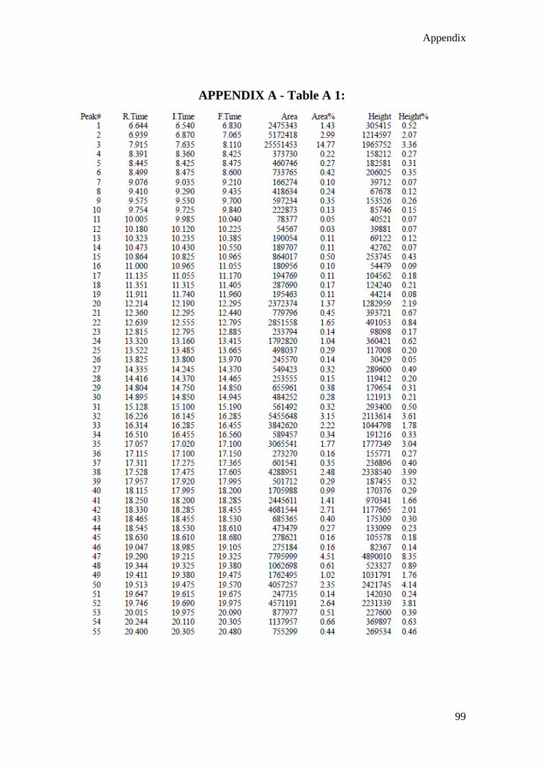
Appendix
99
APPENDIX A - Table A 1:

Appendix
100
APPENDIX A - Table A 2:

Appendix
101
APPENDIX B – Antibacterial activity
Appendix B: Antibacterial activity of crude methanolic leaf extract and silver
nanoparticles synthesised from the leaves of Tagetes minuta against gram positive and
gram negative bacteria: a) Bacillus subtilis. b) Escherichia coli. c) Pseudomonas
aeruginosa. d) Staphylococcus aureus. e) Methicillin-resistant Staphylococcus aureus.