Phytochemical Profile, Biological Properties, and Food ... - MDPI
Upload
khangminh22Category
view
3download
0
BBiioollooggiiccaall aaccttiivviittyy && PPhhyyttoocchheemmiiccaall SSttuuddyy ooff sseelleecctteedd
MMeeddiicciinnaall PPllaannttss
In
By
Musa Khan
A thesis submitted to the Quaid-i-Azam University,
Islamabad in partial fulfillment of the requirements for the
Degree of
Doctor of Philosophy in
Plant Sciences
(Plant Taxonomy)
Department of Plant Sciences Quaid-i-Azam University
Islamabad 2010
BBiioollooggiiccaall aaccttiivviittyy && PPhhyyttoocchheemmiiccaall SSttuuddyy ooff sseelleecctteedd
MMeeddiicciinnaall PPllaannttss
By
Musa Khan
Department of Plant Sciences Quaid-i-Azam University
Islamabad 2010

Certificate
CERTIFICATE
The theses of Musa Khan is accepted in its present form by the
Department of Plant Sciences, Quaid-i-Azam University, Islamabad as
satisfying the theses requirement for the degree of Doctor of Philosophy
in Plant Taxonomy.
Supervisor ________________________
Pro. Dr. Rizwana Aleem Qureshi
External Examinar._________________________
Dr. Mohammad Khan Laghari
(Director PMNH)
External Examinar__________________________
Charperson:____________________________
Prof. Dr. Asghari Bano
Dated: 07/05/2010
i
Acknowledgements
I have no words to thanks Allah almighty who gives me the opportunity to complete my
studies.
I feel obliged to my parent department “Defense Science & Technology Organization”
and the “Higher Education Commission of Pakistan” for providing me financial support
during my studies.
I heartily appreciate my supervisor Dr Rizwana Aleem Qureshi Prof, Department of Plant
Sciences, Quaid-i-Azam University, Islamabad, for her keen interest, kindness and her
valuable views and experience.
I would like to thanks Chairperson, Department of Plant Sciences, Prof. Dr Asghari Bano
for timely providing me all the necessary facilities and administrative support.
I also appreciate and thanks my foreign supervisors, Prof. Dr. Dr Brigitte Kopp
Department of Pharmacognosy (University of Vienna, Austria) and Dr George Krupitza,
Department of Tumor Biology, Medical University of Vienna, Austria, for technical
support and guidance during my six months stay in Austria (sponsored by Higher
Education Commission of Pakistan).
Thanks to all teachers, students and staff members of Department of Pharmacognosy,
University of Vienna, Austria, Department of Tumor Biology, Medical University of
Vienna and Department of Plant Sciences, Quaid-i-Azam University, Islamabad Pakistan
for sharing expertise and for providing a friendly environments.
In last I am greatly thankful to my parents who provide me support and put me on this
track but my mother could not survive to see me on this stage.
Musa Khan
ii
In memory of my dear mother
(July 2008)
iii
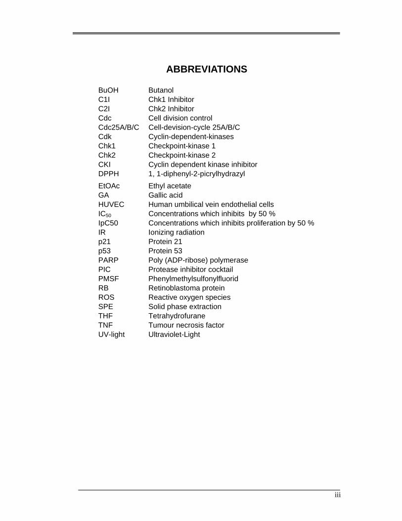
ABBREVIATIONS
BuOH Butanol C1I Chk1 Inhibitor C2I Chk2 Inhibitor Cdc Cell division control Cdc25A/B/C Cell-devision-cycle 25A/B/C Cdk Cyclin-dependent-kinases Chk1 Checkpoint-kinase 1 Chk2 Checkpoint-kinase 2 CKI Cyclin dependent kinase inhibitor DPPH 1, 1-diphenyl-2-picrylhydrazyl
EtOAc Ethyl acetate GA Gallic acid HUVEC Human umbilical vein endothelial cells IC50 Concentrations which inhibits by 50 % IpC50 Concentrations which inhibits proliferation by 50 % IR Ionizing radiation p21 Protein 21 p53 Protein 53 PARP Poly (ADP-ribose) polymerase PIC Protease inhibitor cocktail PMSF Phenylmethylsulfonylfluorid RB Retinoblastoma protein ROS Reactive oxygen species SPE Solid phase extraction THF Tetrahydrofurane TNF Tumour necrosis factor UV-light Ultraviolet-Light

iv
Table of Contents
Acknowledgments i
Dedication ii
Abbreviations iii
Summary 1
Introduction 4
1.1 General introduction 4
1.2 Pharmacognosy 5
1.3 Bioassay guided isolation of natural products 5
1.4 Medicinal plants as a source of important drug 6
1.5 Secondary metabolites 10
1.5.1 Small molecules 10
1.5.1.1 Alkaloids 10
1.5.1.1 Alkaloids 10
1.5.1.3 Glycosides 12
1.5.1.4 Phenols 14
1.5.1.5 Phenazines 15
1.5.2 Big “small molecules” 15
2.5.2.1 Polyketides 15
2.5.2.2 Nonribosomal peptides 15
1.6 Technique used in phytochemistry 16
1.6.1 Chromatography 16
1.6.2 Capillary electrophoresis 20
1.6.3 Spectroscopic Techniques 20
1.6.3.1 NMR spectroscopy 20
1.6.3.2 Two-Dimensional Nuclear Magnetic Resonance Spectroscopy
(2DNMR) 20
1.6.3.3 Infrared Spectroscopy 21
1.6.3.4 Fourier transform infrared spectroscopy 21
1.6.3.5 Ultraviolet-visible spectroscopy 22
1.6.4. Liquid chromatography-mass spectrometry 23
1.6.5. Gas chromatography-mass spectrometry (GC-MS) 23
1.7 Development of Anticancer agents from Medicinal plants 23
v
1.8 Development of cancer 24
1.8.1 Self-sufficiency in growth signals 25
1.8.2 Insensitivity to antigrowth signals 26
1.8.3 Evading apoptosis 26
1.8.4 Limitless replicative potential 26
1.8.5 Sustained angiogenesis 26
1.8.6 Tissue invasion and metastasis 27
1.8.7 The cell cycle 28
1.8.7.1 Cell cycle phases (short summary) 29
1.8.7.2 Presence of cyclins and Cdks during single phases 29
1.8.8 Function and activation of (proto)-oncogenes/oncogenes 30
1.8.8.1 Oncogenes 30
1.8.8.2 Cyclin D1 30
1.8.8 3 Cdc25A (Cell-division-cycle 25A) 30
1.8.8 4 Function and activation of tumor suppressor genes 31
1.8.8 5 p53 (protein 53) 31
1.8.8 6 Activation of p53 31
1.8.8 7 P21CIP (protein 21) 31
1.8.8 8 Activation of p21CIP 32
1.8.8.9 RB 32
1.8.8.10 Activation of RB 33
1.8.9 Cell death 33
1.8.9.1 Apoptosis 33
1.8.9.2 Autophagy 35
1.8.9.3 Necroses 35
1.9 Bioassays Techniques 37
1.9.1 Apoptosis assays (Hoechst 33258 propidium iodide (HOPI) double-staining)
37
1.9.2 Western blot assay 37
1.9.2.1 Steps in a western blot 37
1.9.3 Fluorescence Activated Cell Sorting (FACS) assay 40
1.9.3.1 Flow cytometers 41
1.9.3.2 Application 42
1.9.4 Comet assay 42
vi
1.9.4.1 Experimental procedure 42
1.9.4.2 Principals 44
1.9.5 Total Phenolics or Folin-Ciocalteau Micro Method 44
1.9.5.1 Calibration curve 45
1.9.6 Antioxidant activity 46
1.9.6.1 (1, 1-diphenyl-2-picrylhydrazyl) (DPPH) 47
1.10 Selection of Medicinal plants species 48
1.10.1 Berberis lycium Royle (Berberidaceae) 49
1.10.2 Mallotus philippensis (Lam.) Muell. Arg. (Euphorbiaceae) 49
1.10.3 Adhatoda vasica Nees (Acanthaceae) 50
1.10.4 Albizia lebbeck (L.) Benth. (Mimosaceae) 51
1.10.5 Bauhinia variegata Linn. (Caesalpinaceae) 51
1.10.6 Bombax ceiba Linn. (Bombacaceae) 51
1.10.7 Calotropis procera (Willd.) R. Br. 1. c (Asclepiadaceae) 52
1.10.8 Carrisa opaca Staff ex Haines (Apocynaceae) 53
1.10.9 Caryopteris grata Benth. (Verbenaceae) 53
1.10.10 Cassia fistula Linn (Caesalpinaceae) 53
1.10.11 Colebrookea oppositifolia Smith (Labiateae) 54
1.10.12 Debregeasia salicifolia (D.Don) Rendle in Prain (Urticaceae) 54
1.10.13 Dalbergia sissoo Roxb. (Papilionaceae) 55
1.10.14 Dodonaea viscosa (L.) Jacq., Enum. Pl. Carib. (Sapindaceae) 55
1.10.15 Ficus palmata Forssk. (Moraceae) 56
1.10.16 Ficus racemosa L. (Moraceae) 57
1.10.17 Jasminum humile Linn. (Oleaceae) 57
1.10.18 Lantana camara L. (Verbenaceae) 58
1.10.19 Melia azedarach L. (Meliaceae) 58
1.10.20 Olea ferruginea Royle (Oleaceae) 59
1.10.21 Phyllanthus emblica L. (Euphorbiaceae) 59
1.10.22 Pinus roxburghii Sargent (Pinaceae) 60
1.10.23 Pyrus pashia Buch. & Ham. (Rosaceae) 60
1.10.24 Punica granatum L. (Punicaceae) 61
1.10.25 Rubus ellipticus Smith (Rosacceae) 61
1.10.26 Viburnum cotinifolium D. Don (Caprifoliaceae) 62
1.11 Objectives 63
vii
Chapter: 2 Review of Literature 64
2.1 Berberis lycium Royle (Berberidaceae) 64
2.1.1 Ethnobotanical uses 64
2.1.2 Chemical constituents 64
2.1.3 Biological testing 65
2.2 Mallotus philippensis (Lam.) Muell. Arg. (Euphorbiaceae) 69
2.2.1 Ethnobotanical uses 69
2.2.2 Chemical constituents 69
2.2.3 Biological testing 69
2.3 Adhatoda vasica Nees in Wall (Acanthaceae) 73
2.3.1 Ethnobotanical uses 72
2.3.2 Chemical constituents 72
2.3.3 Biological testing 72
2.4 Albizia lebbeck (L.) Benth. (Mimosaceae) 73
2.4.1 Ethnobotanical uses 73
2.4.2 Chemical constituents 73
2.4.3 Biological testing 73
2.5 Bauhinia variegata Linn. (Caesalpinaceae) 74
2.5.1 Ethnobotanical uses 74
2.5.2 Chemical constituents 74
2.5.3 Biological testing 74
2.6. Bombax ceiba Linn. (Bombacaceae) 74
2.6.1 Ethnobotanical uses 74
2.6.2 Chemical constituents 74
2.6.3 Biological testing 74
2.7 Calotropis procera Linn. (Asclepiadaceae) 75
2.7.1 Ethnobotanical uses 75
2.7.2 Chemical constituents 76
2.7.3 Biological testing 76
2.8 Carissa opaca Stapf ex Haines (Apocynaceae) 76
2.8.1 Ethnobotanical uses 76
2.8.2 Chemical constituents 76
2.9 Cassia fistula Linn. (Caesalpinaceae) 77
2.9.1 Ethnobotanical uses 77

viii
2.9.2 Chemical constituents 77
2.9.3 Biological testing 77
2.10 Colebrookea oppositifolia Smith (Labiateae) 77
2.10.1 Ethnobotanical uses 77
2.10.2 Chemical constituents 77
2.11 Debregeasia salicifolia (D.Don) (Urticaceae) 78
2.11.1 Ethnobotanical uses 78
2.11.2 Chemical constituents 78
2.11.3 Biological testing 78
2.12 Dalbergia sissoo Roxb. (Papilionaceae) 78
2.12.1 Ethnobotanical uses 78
2.12.2 Chemical constituents 79
2.12.3 Biological testing 79
2.13 Dodonaea viscosa Linn. (Sapindaceae) 79
2.13.1 Ethnobotanical uses 79
2.13.2 Chemical constituents 80
2.13.3 Biological testing 80
2.14 Ficus palmata Forssk. (Moraceae) 81
2.14.1 Ethnobotanical uses 81
2.14.2 Chemical constituents 81
2.15 Ficus racemosa L. (Moraceae) 81
2.15.1 Ethnobotanical uses 81
2.15.2 Chemical constituents 82
2.15.3 Biological testing 82
2.17 Lantana camara Linn. (Verbenaceae) 83
2.17.1 Ethnobotanical uses 83
2.17.2 Chemical constituents 83
2.17.3 Biological testing 84
2.18 Melia azedarach Linn. (Meliaceae) 84
2.18.1 Ethnobotanical uses 84
2.18.2 Chemical constituents 85
2.18.3 Biological testing 85
2.19 Phyllanthus emblica L. (Euphorbiaceae) 86
2.19.1 Ethnobotanical uses 86
ix
2.19.2 Chemical constituents 86
2.19.3 Biological testing 87
2.20 Pinus roxburghii Sargent (Pinaceae) 87
2.20.1 Ethnobotanical uses 87
2.21 Punica granatum Linn. (Punicaceae) 88
2.21.1 Ethnobotanical uses 88
2.21.2 Chemical constituents 88
2.21.3 Biological testing 88
2.22 Rubus ellipticus Smith (Rosaceae) 89
2.22.1 Ethnobotanical uses 89
2.22.2 Chemical constituents 89
2.22.3 Biological testing 90
2.23 Viburnum cotinifolium D. Don (Caprifoliaceae) 90
2.23.1 Ethnobotanical uses 90
2.23.2 Chemical constituents 90
Chapter: 3 Materials & Methods 91
3.1 Reference Compounds 91
3.2 Plant Material 91
3.3 Anti bodies for western blot analyses 93
3.4 Miscellaneous Chemicals and Reagents 94
3.5 Cell culture and bacterial strains 95
3.6 Extraction 95
3.6.1 Extraction for Antioxidant and Total Phenolics Determination 95
3.6.2 Extraction of roots powder 95
3.6.3 Extraction for Flavonoids analyses 96
3.7 Chromatographic Methods 96
3.7.1 Thin Layer Chromatography (TLC) 96
3.7.1.1 Thin Layer Chromatography of Berberis lycium fractions
96
3.7.1.2 Thin Layer Chromatography for Flavonoids analyses 97
3.7.2 High Performance Liquid Chromatography (HPLC) 97
3.7.2.1 General HPLC Parameters 97
3.7.2.2 HPLC Method 97
3.7.2.3 Sample Preparation 98
x
3.7.3 Gas Chromatography and Mass Spectrometer 98
3.8 Biological Testing 99
3.8.1 Antineoplastic Activities 99
3.8.1.1 Anti-proliferation or Growth inhibition assay 99
3.8.1.2 Hoechst dye 33258 and propidium iodide double staining
(Apoptosis Assay) 99
3.8.1.3 Western blotting 99
3.8.1.4 Cell cycle distribution analysis (FACS analyses) 100
3.8.1.5 Single cell gel electrophoresis (SCGE)/Comet assay 101
3.8.1.6 Statistical analyses 102
3.8.2 Total Phenolics determination 102
3.8.3 1, 1-Diphenyl-2-picrylhydrazyl (DPPH) test 102
3.8.4 Antibacterial Determination 103
Chapter. 4 Results and Discussion 104
4.1 Results 104
4.1.1 Anti-neoplastic Activities and Phytochemicals studies of Berberis
lycium. 104
4.1.1.1 Qualitative Analysis of B. lycium extracts constituents by
TLC. 104
4.1.1.2 Separation and quantification of alkaloids by RP-HPLC
104
4.1.1.3 Inhibition of HL-60 cell proliferation by extracts of B.
lycium, Berberine and Palmatine. 112
4.1.1.4 Effect of BuOH extract, Berberine and Palmatine on cell
cycle distribution 115
4.1.1.5 Induction of apoptosis by extracts of B. lycium and
Berberine 118
4.1.1.6 Induction of stress response by extracts of B. lycium and
Berberine. 123
4.1.2 Anti-neoplastic Activities and Phytochemicals studies of Mallotus
phillipensis. 126
4.1.2.2 Induction of apoptosis by extract of Mallotus phillipensis
126
4.1.2.3 Effect of Hexane fraction on cell cycle distribution. 126
xi
4.1.2.4 Induction of stress response by extract of Mallotus
phillipensis. 131
4.1.2.5 GC-MS Analysis of Mallotus phillipensis Hexane Fraction.
131
4.1.3 Total Phenolics, Free radical scavenging activity and Flavonoids
finger printing of selected Medicinal Plants. 137
4.1.3.1 Total Phenolics Determination. 137
4.1.3.2 Determination of Free radical scavenging activity 137
4.1.3.3 Flavonoids finger printing of selected Plants 140
4.1.4 Antibacterial and Free radical scavenging activities,
Flavonoids finger printing of Mallotus philippensis. 150
4.1.4.1 Antibacterial activities 150
4.1.4.2 Free radical scavenging activities 150
4.1.4.3 Flavonoids finger printing of Mallotus philippensis. 150
4.2 Discussion 155
4.2.1 Anti-neoplastic Activities and Phytochemicals studies of Berberis lycium
155
4.2.2 Anti-neoplastic Activities and Phytochemicals studies of Mallotus
phillipensis. 157
4.2.3 Total Phenolics, Free radical scavenging activity and Flavonoids finger
printing of selected Medicinal Plants 159
4.2.4 Antibacterial and Free radical scavenging activities, Flavonoids finger
printing of Mallotus Philippensis. 163
Chapter. 5. Conclusion 166
List of Publications 169
Plates 170
Chapter. 6. References 182
List of Figures
Figure 1 Examples of new medicinal plant drugs 9
Figure 2 Acquired capabilities of cancer 25
Figure 3 Cyclin and Cdks distribution during the cell cycle 29
Figure 4 DNA damage induced by UV-light and further the activation of p53 32
xii
Figure 5 Mechanism of Apoptosis 36
Figure 6 Alkaloids of Berberis lycium 65
Figure 7 Compounds of Mallotus philippensis 72
Figure 8 TLC of Berberis lycium extracts 105
Figure 9 RP-HPLC Chromatogram of alkaloids standards 106
Figure 10 RP-HPLC chromatogram of n-Butanol fraction of Berberis lycium extract. 107
Figure 11 RP-HPLC chromatogram of water fraction of Berberis lycium extract 108
Figure 12. RP-HPLC chromatogram of Ethyl acetate fraction of Berberis lycium extract
109
Figure 13 Optimum UV spectra of standards compounds 110
Figure 14 Alkaloids percentage in Berberis lycium 112
Figure 15 Anti-proliferative effect of B. lycium extracts and its alkaloids 114
Figure 16 Analysis of cell cycle proteins 115
Figure 17 Cell Cycle Distribution of HL-60 cells upon treatment with of BuOH extract
and berberine for 48 h 117
Figure 18 Induction of apoptosis by the B. lycium extracts and berberine 120
Figure 19 Western blot analysis of pro-apoptotic mediators and effectors 121
Figure 20 The genotoxicity of increasing concentrations of BuOH extract and berberine
122
Figure 21 Comet assay 123
Figure 22 Induction of stress response by the BuOH extract and Berberine 125
Figure 23 Anti-proliferative effect of Mallotus phillipensis extracts 127
Figure 24 Induction of apoptosis by the Mallotus phillipensis Hexane fraction 128
Figure 25 Analysis of cell cycle proteins 129
Figure 26 Cell Cycle Distribution of HL-60 cells upon treatment with hexane Fraction of
Mallotus phillipensis 130
Figure 27 Induction of stress response by Mallotus phillipensis 131
Figure 28 GC/MS chromatogram of hexane soluble fraction of Mallotus phillipensis 136
Figure 29 Gallic acid standard curve 138
Figure 30 Total Phenolics and Extract yield per gram 139
Figure 31 Antioxidant cure of Ascorbic acid 139
Figure 32 Flavonoids finger printing of standard and selected plants 145
Figure 30 Percentage of Flavonoids in Plant samples 146
Figure 34 Types of Flavonoids in each sample 147

xiii
Figure 35 Antibacterial activities of Mallotus philippensis 151
Figure 36 Free radical scavenging activity of Mallotus philippensis 153
Figure 37 Flavonoids finger printing of Mallotus philippensis 154
List of Tables
Table 1 Reference compounds 91
Table 2 Investigated Plants species 92
Table 3 Anti bodies for western blot analyses 93
Table 4 Miscellaneous Chemicals and Reagents 94
Table 5 Parameters for HPLC-PDA analyses of Alkaloids 97
Table 6 Gradient elution systems used for HPLC separations 98
Table 7 Gas Chromatograph and Mass Spectrometer conditions 98
Table 8 10% Polyacrylamide Gel Preparation 101
Table 9 Linearity study of standard curve for standard compounds 111
Table 10 Percent composition of active alkaloids in Berberis lycium 111
Table 11 Comparative total Phenolic, extract yield per gram and IC50 Values 140
Table 12 Appearance of standards under UV 265nm 148
Table 13 Qualitative analyses of plants samples for Flavonoids types 149
Table 14 Antibacterial activities of roots and flower powder extract 152
Summary
1
SUMMARY
The present study deals with the exploration of some species of medicinal plants found in
Pakistan against cancer. Twenty seven plant species were selected from the local flora.
Roots of three plants i.e. Berberis lycium (Berberidaceae), Mallotus philippensis
(Euphorbiaceae) and Zizyphus nummularia (Rhamnaceae) were studied for anti-
neoplastic activity against p53 deficient human leukemia cell lines (HL-60). Although
roots of Zizyphus nummularia possess many complex alkaloids yet its extract was not
effective in checking proliferative activity.
Berberis lycium extract and its alkaloids berberine and palmatine are known for their
beneficial pharmacological properties. In the present study, the anti-neoplastic activities
of different B. lycium root extracts and the major constituting alkaloids, berberine and
palmatine were investigated in HL-60 cells to elucidate the anti-neoplastic trigger
mechanisms of the pure compounds and crude extracts in a p53-deficient background.
Growth inhibition, cell cycle distribution, and apoptosis were compared among the ethyl
acetate (EtOAc), n-butanol (BuOH) and water (H2O) extracts. The BuOH extract
inhibited cell proliferation most efficiently (IC50 < 2.77 µg extract weight/ml medium,
which corresponded to 250 µg dried root/ml). The IC50s for the EtOAc and H2O extracts
were 16.65 µg/ml and 104.25 µg/ml, respectively (corresponding for both extract types to
>7.5 mg dried root/ml). The chemical composition of the BuOH extract was analyzed by
preparative TLC and quantified by RP-HPLC and it was estimated that it contained 3.73
µM Berberine and 1.51µM Palmatine per 1 mg dried root. Therefore, HL-60 cells were
exposed to the respective concentrations of berberine and palmatine. Berberine showed an
IC50 < 1.87µM after 72 h of incubation, while palmatine had no significant effect up to
4.68 µM. The BuOH extract and berberine induced the intra-S-phase checkpoint causing
the accumulation of HL-60 cells in S-phase. In contrast to a very recent report by Liu et
al, (2006), It is found that the anti-cancer effects of berberine and the extract are not due
to genotoxicity but correlate with α-tubulin acetylation, strong activation of Chk2,
phosphorylation of Ser177-Cdc25A and its subsequent degradation as well as the
consequent inactivation of Cdc2 (CDK1) and furthermore, the down-regulation of the
proto-oncogene cyclin D1. The molecular effects were observed at low concentrations
(11.1 µg BuOH extract/ml; 1.4 µg berberine/ml) which inhibited ~ 50 % of the HL-60
cells proliferation after 24 h treatment, hence supporting the mechanistic conjunction.
Mallotus philippensis is a well known medicinal plant of Pakistan. It possesses different
classes of chemical compounds with unique pharmacological activities. Roots of Mallotus
Summary
2
philippensis was initially extracted and fractionated in organic solvents, n-hexane, ethyl
acetate (EtOAc), and n-butanol (BuOH). After evaporating each solvent, 9.23 g dried
hexane extract, 4.00 g dried EtOAc extract, and 7.08 g dried BuOH extract was obtained,
respectively. The n-hexane fraction showed the highest toxicity against HL-60 cells (IC50
1.5 mg dry roots equivalent /ml medium) after 72h. The hexane fractions regulated
protein expression and protein activation in HL-60 cells. The inhibition of HL-60
proliferation that was observed upon treatment with hexane extract was preceded by the
down regulation of the proto-oncogene Cdc25A and cyclin D1 after 48 h. All of these
effects have not been observed in any p53 deficient cell lines so far by Mallotus
phillipensis extracts and its chemical constituents. Valacchi et al (2008) has reported that
rottlerin deactivate cyclin D1 in HaCaT cell line. The hexane fraction induced 18%
apoptosis after 48h of treatment with 1.5 mg dry roots equivalent /ml medium. The ability
of M. phillipensis hexane fraction and the observation indicates that the anti-neoplastic
effects have been triggered by induction apoptosis through caspase-2 activation while
Brodie et al., 2003 reported that rottlerin activated caspase-3. The chemical composition
of the n-hexane fraction of M. phillipensis was analyzed by GC-MS. Different
compounds have been detected in the sample. Mass spectrometric data of some
compounds have been co-related with already reported compounds from different parts of
the same species. Lupeol, Betulin, Kamala Chalcones C like compounds and another
unknown compound (GC Rf = 39.9, 45.66, 43.905 and 47.735 minutes respectively) have
been detected. Rottlerin that has been reported in M. phillipensis was not detected in the
hexane fraction. It has been confirmed from the present anti-neoplastic assay that hexane
fraction is active against p53 deficient human leukemia cell lines (HL-60) and the activity
was due to compound/compounds other than rottlerin.
Kamala or Kamara (a red powder of M. philippensis reported to have different cytotoxic
compounds, flavonoids or Phenolic compounds) has compared with the roots of M.
philippensis for inhibition of different bacterial strains. Similarly Kamala has compared
with the aerial parts (leaves) of M. philippensis in scavenging free radicals. It has been
observed that (Kamala or Kamara) extract has shown activities against Gram positive
bacteria, Bacillus subtilis and Staphylococcus aureus (MICs 0.7 and 0.6 mg/ml), while it
does not shown any response against Salmonella setubal, Staphylococcus epidermidis and
Escherichia coli up to maximum concentration of 15 mg/ml. Roots extract was effective
against one Gram positive bacteria Bacillus subtilis and one Gram negative bacteria
Summary
3
Salmonella setubal (MICs 1.00 and 2.00 mg/ml) respectively but it has not shown any
activity against Staphylococcus aureus, Staphylococcus epidermidis and Escherichia coli
up to maximum concentration of 15 mg/ml. It has been observed that both Kamala and
leaves extract have free radical scavenging capacity but the leaves extract was more
active than Kamala powder in scavenging free radicals. Thin layer chromatography of the
leaves has shown the presence of Vitexin, Isovitexin and Rutin.
In another set of experiment 24 different plants species were checked to determine total
Phenolics, free radical scavenging capacity and flavonoids types. Some plants species
were reported medicinally in literature and the others have been selected randomly. The
medicinally important plants were Bauhinia variegata, Cassia fistula, Bombax ceiba,
Calotropis procera, Carissa opaca, Adhatoda vasica, Albizia lebbeck, Colebrookea
oppositifolia, Dalbergia sissoo, Dodonaea viscosa, Ficus palmata, Ficus racemosa,
Lantana camara, Melia azedarach, Phyllanthus emblica, Punica granatum, Rubus
ellipticus and Viburnum cotinifolium and the non medicinal plats were Jasminum humile,
Olea ferruginea, Pinus roxburghii, Caryopteris grata, Debregeasia salicifolia and Pyrus
pashia. Total Phenolics were studied by comparing with standard Gallic acid. Phyllanthus
emblica has shown highest amount of total Phenolics while comparing with Gallic acid.
The extract per gram of Phyllanthus emblica was also greater than others. Phenolic acids,
Kaempferol and Vitexin have been detected in the sample of Phyllanthus emblica by thin
layer chromatography. Vitexin has been reported for the first time in Phyllanthus emblica.
Rubus elepticus has shown comparatively highest capacity in scavenging free radicals.
Phenolic acids, Kaempferol, Vitexin, Rutin and Apigenin have been detected in the
sample of Rubus ellipticus by thin layer chromatography. All plants species have shown
Phenolic acids bands. Vitexin and Isovitexin were present in maximum numbers of plants
samples (58.33 and 54.8 % percent respectively); Catechin, Luteolin-7-glucoside,
Quercetin and Luteolin were not detected in any sample.

Chapter 1 Introduction
4
INTRODUCTION
1.1 General introduction
The flora of Pakistan due to its diverse climatic and soil conditions and many ecological
regions, is very rich in medicinal plants. According to a general survey of Pakistan about
6000 species of flowering plants have been exist, out of 6000 about 400-600 are
medicinally important species (Nasir and Ali, 1972; Hamayun et al; 2005). The history of
plants to be utilized as medicines is thousands of years old (Samuelsson, 2004). These
plant materials initially took the form of crude drugs such as poultices, teas, powders
tinctures, and many other herbal formulations (Samuelsson, 2004; Balick and Cox, 1997).
From near past it has been discovered that properties of medicinal plants are due to its
active chemical compounds and therefore the isolation of active compounds and in the
early 19th century morphine has been isolated from opium (Samuelsson, 2004; Kinghorn,
2001). The discovery of drug from medicinal plants has been started from the era when
the isolation of primarily drugs such as digitoxin, quinine, cocaine, and codeine has
begun. Like morphine some are still in use for different purposes (Butler, 2004; Newman et
al., 2000; Samuelsson, 2004). Numbers of scientists have been working in order to isolate
and characterize the pharmacologically active compounds from medicinal plants. Drug
discovery techniques have been discovered and applying for the standardization of herbal
medicines and to obtain analytical marker compounds.
Drug discovery from medicinal plants are not simple but it has evolved to include
numerous fields of inquiry and take advantages of different analytical procedures. The
process initiated with a botanist especially with ethnobotanist, ethnopharmacologist, or
plant ecologist that can easily collects and identifies their desired plant(s). Collection
may involve those species with known biological activity which need to be study for their
active compound(s) and new for isolation (e.g., traditionally used herbal remedies) or
may also involve those taxa that have been collected randomly for a large screening
purposes. It is also important to take care and respect the intellectual property rights of a
given area, country where plant(s) of interest are collected (Baker et al., 1995).
Phytochemists are also called natural product chemists. These phytochemists after proper
collection, identification and cleaning processes, make crude extracts from the selected
parts of the plant materials, subject these crude extracts to biological screening of their
desire assays, and commence the process of isolation and characterization of the active
chemical compound(s). The whole processes are called bioassay-guided fractionation.
Molecular biology is very important and taking essential part in drug discovery from
medicinal plant. Molecular biology determines and implements appropriate screening
Chapter 1 Introduction
5
technique that directed towards physiologically relevant molecular targets.
Pharmacognosy encapsulates all of the relevant fields into a distinct interdisciplinary
science.
1.2 Pharmacognosy
The term and practice of pharmacognosy have been used since about 200 years ago
(Samuelsson, 2004; Kinghorn, 2001), as medicinal plants have progressed to use as drug,
the formulation of crude drugs and to isolate the active compounds in drug discovery
research. According to the American Society of Pharmacognosy, the pharmacognosy can
be stated as ‘‘the study of the physical, chemical, biochemical and biological properties
of drugs, drug substances, or potential drugs or drug substances of natural origin as well as
the search for new drugs from natural sources’’. In the present era of research regarding
drug discovery from medicinal plants or in broad way from natural origin,
pharmacognosy compensate the broad study of natural products from various sources
including unicellular and multi cellular organism like bacteria, fungi, plants, and marine
organisms. In broad way, Pharmacognosy that study various parameter which includes
both botanical dietary supplements, including herbal remedies (Cardellina, 2002; Tyler,
1999), and searching for single chemically and pharmacologically active compound that
can be use as drug and may proceed through further development into Food and Drug
Administration (FDA)-approved medicines. According to Bruhn and Bohlin the
definition of pharmacognosy may proceed as ‘‘a molecular science that explores
naturally occurring structure–activity relationships with a drug potential’’ (Bruhn and
Bohlin, 1997).
1.3 Bioassay guided isolation of natural products
As natural sources have many useful and important bioactive compounds and many have
been discovered using bioactivity directed fractionation and isolation (BDFl). The
research of pharmacognosy or isolation of natural products facilitated by newly
development of new bioassay methods. It has been found that the bioactive compounds
are mostly plant secondary metabolites, which become medicine after processing to pure
compounds; some are very useful dietary supplements, and many useful commercial
products. Further modification of the active compounds lead to enhance the biological
profiles and a large number of such compounds which are approved or undergoing
clinical trials for clinical uses against different diseases like pulmonary diseases, cancer,
HIV/AIDS, malaria, Alzheimer’s and other diseases (Butler., 2004; Newman et al.,
2003).Crude herbs are used as drugs in different country of the world and therefore it take
Chapter 1 Introduction
6
a basic part of many traditional medicines worldwide. In Asia, traditional Chinese
medicine (TCM), Korean Chinese medicine, Japanese Chinese medicine (kampo),
ayurvedic medicine (India) and jamu (Indonesia), phytotherapy and hoemeopathy in
Europe, Alternative medicines are typically named when herbal therapies use with
various other traditional remedies in America. Integrative medicine came into being when
the alternative medicine, mainly the aforementioned traditional and folk medicines used
worldwide, with conventional medicine (Western medicine).
1.4 Medicinal plants as a source of important drug
Different type of isolation methods have been used to obtain pharmacologically active
compounds that can use as drug for different diseases. The methods which includes
isolation from plants and other natural sources, combinatorial chemistry, synthetic
chemistry, and molecular modeling (Geysen et al., 2003; Ley Baxendale, 2002 and
Lombardino and Lowe, 2004). Although there is much research in molecular modeling,
combinatorial chemistry, and other synthetic chemistry techniques which has been
funding by pharmaceutical companies and organizations, natural products which have
much complicated structural formulas and particularly medicinal plants, remain an
important source of new drugs, new chemical entities (NCEs) and new drug leads, (Butler,
2004; Newman et al., 2000, 2003). According to survey in 2001 and 2002, approximately
one quarter of the best-selling drugs in the world were natural products or derived from
natural products (Butler, 2004). It has also been reported that approximately 28% of
NCEs between 1981 and 2002 were natural products or natural product-derived natural
products (Newman et al., 2003) and another survey during this period 20% of NCEs were
considered natural product mimics, meaning that the synthetic compound was derived
from the study of natural products (Newman et al., 2003). On the bases of this report it
has been assumed that research on natural products accounts for approximately 48% of
the NCEs reported from 1981–2002.
Further more it has been known that natural products also provide a starting point for
laboratory syntheses with diverse structures and often with multiple stereo centers that
can be challenging synthetically (Koehn and Carter, 2005; Clardy and Walsh, 2004;
Peterson and Overman, 2004; Nicolaou and Snyder, 2004). Natural products shows many
structural features in common (e.g., aromatic rings, chiral centers, degree of molecule
saturation, complex ring systems, and number ratio of heteroatoms) which have been
shown to be very important to drug discovery efforts ( Feher and Schmidt, 2003; Piggott
and Karuso, 2004; Clardy and Walsh, 2004; Koehn and Carter, 2005; Lee and Schneider,
2001). Many synthetic and medicinal chemists are working in the creation of natural
Chapter 1 Introduction
7
product and natural-product like libraries that resembles the structural features of natural
products with the compound-generating potential of combinatorial chemistry ( Eldridge et
al., 2002; Burke et al., 2004; Hall et al., 2001a; Ganesan, 2004; Tan, 2004). Some natural
products that are isolated from medicinal plants can serve not only as new drugs
themselves but can also be made useful by further necessary modification by medicinal
and synthetic chemists.
Sometime new chemical structures are very difficult to found during drug discovery from
medicinal plants, in such cases known compounds with new biological activity can provide
important drug directions. Molecular target play important rule in drug discovery, since the
sequencing of the human genome, a lot new molecular targets have been identified as
important and useful in various diseases (Kramer and Cohen, 2004). The developments
of high-throughput screening technique may show to the point and more selective activity
directed towards these targets, when use the reported compounds from medicinal plants.
It has also be known that the compounds isolated from traditionally used medicinal
plants shown to act on newly validated molecular targets, one example is indirubin,
which targeted and inhibit cyclin dependent kinases (Eisenbrand et al., 2004; Hoessel et
al., 1999) and another example is kamebakaurin, which has been shown to target and
inhibit NF-nB (Lee et al., 2002; Hwang et al., 2001). There are many known compounds
which shown to act on novel molecular targets, this development leads to produce
interest in members of these frequently isolated plant compound classes. There are many
examples but some are cucurbitacin I, from the National Cancer Institute (NCI)
Diversity Set of many known compounds and it is found to be highly selective in
inhibiting the JAK/STAT3 pathway in case of tumors with activated STAT3 (Blaskovich
et al., 2003), another example is h-lapachone, which also selectively kills cancer cells
over normal cells by direct activation of checkpoint during the cell cycle (Li et al., 2003),
and betulinic acid is also the same type of compound, with selective melanoma
cytotoxicity which control the cell cycle by the activation of p38 (Tan et al., 2003;
Cichewicz and Kouzi, 2004; Pisha et al., 1995).
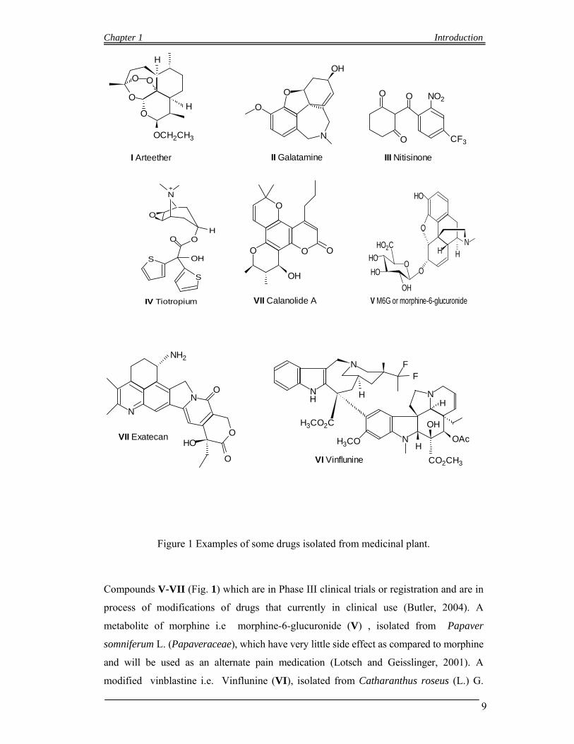
According to a review article by (Balunas and Kinghorn, 2005), Four new drugs which
have been derived from medicinal plants, and have been introduced recently to the U.S.
market (Fig. 1, I–IV). The drugs are, Arteether (I, or Artemotil®) is an effective anti-
malarial drug which is derived from artemisinin, which is a sesquiterpene lactone in its
class and isolated from Artemisia annua L. (Asteraceae). The plant A. annua are used in
traditional Chinese medicine (TCM) (Graul, 2001; van Agtmael et al., 1999;). There are
Chapter 1 Introduction
8
many derivatives of artemisinin which are used in Europe in different stages or clinical
trials as anti-malarial drugs (Van Agtmael et al., 1999).
Galantamine or galanthamine (II, Reminyl®) is a also an ethno botanical directed
isolated natural product in Russia in the early 1950s, which is first isolated from
Galanthus woronowii Losinsk. (Amaryllidaceae) (Pirttila et al., 2004; Heinrich and Teoh,
2004). This compound (Galantamine) is effective in Alzheimer’s disease and theirfore has
been approved for the treatment of Alzheimer’s disease, it take part in slowing the process
of neurological degeneration through inhibiting acetylcholinesterase (AChE) and it also
well bind nicotinic acetylcholine receptor (nAChR) and modulating the same. (Pirttila et
al., 2004; Heinrich and Teoh, 2004;).
An other compound, Nitisinone (III, or Orfadin®) is discovered very recently and has
been isolated from medicinal plant-derived, it shows a characteristic to control the rare
inherited disease, tyrosinaemia, which shows the usefulness of natural products as lead
structures (Frantz and Smith, 2003). Nitisinone in actual is the modified form of
mesotrione, which is an herbicide based on the natural product leptospermone, isolated
from Callistemon citrinus Stapf (Myrtaceae) (Mitchell et al., 2001; Hall et al., 2001b).
All these stated three triketones inhibit the same type of enzyme, 4-
hydroxyphenylpyruvate dehydrogenase (HPPD), while studying in humans and in maize
(Mitchell et al., 2001; Hall et al., 2001b). In maize it inhibits the HPPD enzyme which
shows an activity as an herbicide by the reduction of tocopherol and plastoquinone
biosynthesis. In humans the inhibition of the enzyme HPPD prevents the catabolism of
tyrosine and also the toxic byproducts accumulation in the liver and kidneys (Hall et al.,
2001b). Tiotropium (IV, Spirival as a trade name\) is another drug which has been
released recently to the United States market and has been used for the treatment of
chronic obstructive pulmonary disease (COPD) (Frantz, 2005); Mundy and Kirkpatrick,
2004. The drug Tiotroprium which is an inhaled anticholinergic bronchodilator, and
ipratropium based, which is a derivative of atropine, isolated from Atropa belladonna L.
(Solanaceae)as well as other members of the Solanaceae family (Dewick, 2002; Mundy
and Kirkpatrick, 2004; Barnes et al., 1995). Tiotropium is comparatively longer lasting
effects while comparing with other available COPD medications (Barnes, 2002; Mundy
and Kirkpatrick, 2004).
Chapter 1 Introduction
9
O
O
OH
OCH2CH3
O
H
I Arteether
O
O
N
OH
II Galatamine
NO2OO
O CF3
III Nitisinone
S
S
O
O
O
OH
H
N+
IV Tiotropium
O
O OO
OH
VII Calanolide A
O
N
OO
HO
HO
OH
HO2C
HO
HH
V M6G or morphine-6-glucuronide
N
O
N
NH2
O
O
HOVII Exatecan
N
N
N
NH
H
OH
CO2CH3
H3CO
H3CO2C
FF
HH
OAc
VI Vinflunine
Figure 1 Examples of some drugs isolated from medicinal plant.
Compounds V-VII (Fig. 1) which are in Phase III clinical trials or registration and are in
process of modifications of drugs that currently in clinical use (Butler, 2004). A
metabolite of morphine i.e morphine-6-glucuronide (V) , isolated from Papaver
somniferum L. (Papaveraceae), which have very little side effect as compared to morphine
and will be used as an alternate pain medication (Lotsch and Geisslinger, 2001). A
modified vinblastine i.e. Vinflunine (VI), isolated from Catharanthus roseus (L.) G.
Chapter 1 Introduction
10
Don (Apocynaceae) can be use as an anticancer agent with high efficacy (Bonfil et al.,
2002; Okouneva et al., 2003). Exatecan (VII) is developed as an anticancer agent and
very close similarity with camptothecin that have been isolated from Camptotheca
acuminata Decne. (Nyssaceae (Cragg and Newman, 2004; Butler, 2004). The process of
modifications of the existing natural products realizes the importance of drugs that have
been discovered from medicinal plants as NCEs and consider the possible new drug leads.
The drug, Calanolide A (VIII) is isolated from Malaysian rainforest tree (Calophyllum
lanigerum var. austrocoriaceum (Whitmore) P.F. Stevens (Clusiaceae), is a
dipyranocoumarin natural product, (Yang et al., 2001; Yu et al., 2003; Kashman et al.,
1992). It has been investigated that Calanolide A which shows an anti-HIV drug with a
very unique and high specific mechanism of action particularly as a non-nucleoside
reverse transcriptase inhibitor (NNRTI) of type-1 HIV and is very high effective against
AZT-resistant strains of HIV (Yu et al., 2003; Currens et al., 1996; Buckheit et al.,
1999;). The drug Calanolide A is in Phase II clinical trials process (Creagh et al., 2001).
1.5 Secondary metabolites
All those organic compounds present in plants and in animals that are not working in the
normal growth, development or reproduction of organisms but produced in different
metabolic processes. Secondary metabolites are not essential for life as compare to
primary metabolites, that the absence of secondary metabolites results not in failure of
life, but in long-term impairment of the organism's survivability/fecundity or aesthetics,
or perhaps in no significant change at all but it is useful for animal’s ailments and
normalizes the physiological abnormalities produced due to different diseases in animal
bodies. Secondary metabolites are often very restricted to a particular set of species
within a phylogenetic group. In broad sense secondary metabolites may be classify into;
small molecules (alkaloids, terpenoids, glycosides, Phenols and Phenazene), big small
molecules (Polyketides, Non ribosomal peptides etc), non small molecules (DNA, RNA,
ribosome, polysacharides).
1.5.1 Small molecules
1.5.1.1 Alkaloids
Alkaloids are natural product that contains basic nitrogen atoms. The name of alkaloids
derives from the “alkaline” and it was used to describe any nitrogen-containing base.
Alkaloids are naturally synthesis by a large numbers of organisms, including animals,
plants, bacteria and fungi. Alkaloids are a group of natural products (also called
secondary metabolites). Alkaloids can be easily purified from various crude extracts by

Chapter 1 Introduction
11
acid-base extraction. There are very many alkaloids which are toxic to other organisms.
They often have some pharmacological effects and are used for the treatment of various
diseases and recreational drugs. Some alkaloids are used as the local anesthetic and
stimulant as cocaine. Some alkaloids have stimulant property as caffeine and nicotine,
morphine are used as the analgesic and quinine as the antimalarial drug. Almost all the
alkaloids have a bitter taste.
Classification
Alkaloids may be classified in different groups on the bases of their structure formulas.
Pyridine group: Nicotine alkaloid found in tobacco (Nicotiana tabacum) plant
and Anabasine alkaloid found in the tree Tobacco (Nicotiana glauca) plant.
Pyrrolidine group: Hygrine found in Erythroxylum coca leaves
Tropane group: Atropine alkaloid found in Atropa belladonna and Datura
stramonium, Cocaine alkaloid found in Erythroxylum coca leaves.
Indolizidine group: one example is Swainsonine that was first obtained from a
very small plants like pea (e.g. Swainsona sp. and Astragalus sp).
Quinoline group: Quinine alkaloids isolated originally from Cinchona succirubra
and Strychnine alkaloids was obtained from the seeds of the Strychnos nux vomica
tree.
Isoquinoline group: The Opium alkaloids like narcotine, papaverine, narceine,
morphine, codeine, and heroine, sanguinarine, hydrastine, alkaloids like berberine,
emetine, berbamine, oxyacanthine from Berberis species
Phenanthrene alkaloids: Opium alkaloids like morphine, codeine, thebaine are
included in this group.
Phenethylamine group: Alkaloids found in many members of the Cactaceae like
Lophophora williamsii and Echinopsis pachanoi i.e. Mescaline alkaloids etc, and
some alkaloids found in Ephedra vulgaris i.e. ephedrine alkaloids etc are included
in this group.
Indole group: Serotonin is found in the enterochromaffin cells in the gut of
animals, but also found in mushrooms and plants, including fruits and vegetables,
Vinca alkaloids such as vinblastine, vincristine found in Catharanthus roseus etc.
Purine group: Caffeine type of alkaloids are abundant in genus Coffea Coffea
canephora (also known as Coffea robusta) and Coffea arabica are two speceis
which have been grown for this purpose.
Terpenoid group: Aconitum alkaloids such as aconitine, Steroid alkaloids such as
alkaloids found in Solanum i.e. solanine, solanidine and chaconine etc.
Chapter 1 Introduction
12
1.5.1.2 Terpenoids
The terpenoids sometimes called isoprenoids, are a class of natural products which are
very similar to terpenes, that have been derived from five-carbon isoprene units and can
be interchanged in thousands of ways. Most of the terpenoids have multi cyclic structures
that differ from one another by their functional groups and basic carbon skeletons. These
types of natural lipids can be found in every class of living things, and therefore
considered as the largest group of natural products
Classification
Terpenoids can be thought of as modified terpenes, where terpenes are hydrocarbons
resulting from the combination of several isoprene units. The classification of terpenoids
can be made according to the number of isoprene units used.
Hemiterpenoids: Consist of a single isoprene unit. The only hemiterpene is the
Isoprene itself, but oxygen-containing derivatives of isoprene such as isovaleric
acid and prenol is classify as hemiterpenoids.
Monoterpenoids: Biochemical modifications of monoterpenes such as oxidation
or rearrangement produce the related monoterpenoids. Monoterpenoids have two
isoprene units. Monoterpenes may be of two types i.e linear (acyclic) or contain
rings e.g. Geranyl pyrophosphate, Eucalyptol, Limonene and Pinene.
Sesquiterpenes: Sesquiterpenes have three isoprene units e.g. Farnesyl
pyrophosphate, Artemisinin, Bisabolol.
Diterpenes: It composed for four isoprene units and have the molecular formula
C20H32. They derive from geranylgeranyl pyrophosphate. There are some
examples of diterpenes such as cembrene, kahweol, taxadiene and cafestol
(precursor of taxol). Retinol, retinal, and phytol are the biologically important
compounds while using diterpenes as the base. Theses three compounds are
known to be antimicrobial and antiinflammatory. Geranylgeranyl pyrophosphate,
Retinol, Retinal, Phytol, Taxol, Forskolin Aphidicolin
Sesterterpenoids: Terpenoids having 25 carbons and five isoprene units.
Triterpenes: It consist of six isoprene units e.g. squalene found in wheat germ,
and olives.
Tetraterpenoids: It contain eight isoprene units which may be acyclic like
lycopene, monocyclic like gamma-carotene, and bicyclic like alpha- and beta-
carotenes.
Chapter 1 Introduction
13
Polyterpenoids: It consists of a larger number of isoprene units.
1.5.1.3 Glycosides
It is a group of natural product where a sugar group is directly bonded through its
anomeric carbon to another group by an O-glycosidic bond or an S-glycosidic bond. The
sugar group is then known as the glycone and the non-sugar group as the aglycone or
genin part of the glycoside. The glycone can consist of a single sugar group
(monosaccharide) or several sugar groups (oligosaccharide).
Classification
Glycosides may be classified in three ways
i) Type of glycone: If the glycone group of a glycoside is glucose, then the
molecule is a glucoside; if it is fructose, then the molecule is a fructoside; if it
is glucuronic acid, then the molecule is a glucuronide; etc. In the body, toxic
substances are often bonded to glucuronic acid to increase their water
solubility; the resulting glucuronides are then excreted.
ii) Type of glycosidic bond: It classified as α-glycosides or β-glycosides which
depending on bong geometry that whether the glycosidic bond lies "below" or
"above" the plane of the cyclic sugar molecule. On the bases of this particular
geometry some enzymes like α-amylase can only hydrolyze α-linkages; others,
like emulsin, can only affect β-linkages
iii) Type of aglycone. Glycosides are also classified according to the chemical
nature of the aglycone e.g.
Alcoholic glycoside: salicin is an example of an alcoholic glycoside is
which has isolated from the genus Salix. Salicin is converted to salicylic
in the body, which is closely related to aspirin and has analgesic,
antipyretic and antiinflammatory effects.
Anthraquinone glycosides: They are present in senna, rhubarb and aloes;
they have a laxative effect.These glycosides contain an aglycone group
that is a derivative of anthraquinone.
Coumarine glycosides: Psoralin and corylifolin obtained from dried
leaves of Psoralea corylifolia and the aglycone is coumarin. Apterin a
coumarine glycosides which is reported to dilate the coronary arteries as
well as block calcium channels.
Cyanogenic glycoside: The aglycone contains a cyanide group, and the
glycoside can release the poisonous hydrogen cyanide if acted upon by
Chapter 1 Introduction
14
some enzyme. They are stored in the vacuole but if the plant is attacked
they are released and become activated by enzymes in the cytoplasm.
These remove the sugar part of the molecule and release toxic hydrogen
cyanide. Storing them in inactive forms in the cytoplasm prevents them
from damaging the plant under normal conditions. An example of these is
amygdalin from almonds. They can also be found in the fruits (and wilting
leaves) of the rose family (including cherries, apples, plums, almonds,
peaches, apricots, raspberries, and crabapples).
Flavonoid glycosides: In this type of glycosides the aglycone units are
flavonoids e.g. Hesperidin (aglycone: Hesperetin, glycone : Rutinose),
Rutin (aglycone: Quercetin, glycone: Rutinose), Querctrin (aglycone:
Quercetin, glycone: Rhamnose).
Phenolic glycosides: The aglycone is a simple phenolic structure e.g.
Arbutin found in Arctostaphylos uva-ursi.
Saponin glycosides: The characteristic of saponin glycoside that they
normally produce soap-like foaming when shaken in aqueous medium, and
structurally saponin gycosides composed of one or more hydrophilic
glycoside moieties combined with a lipophilic triterpene derivative.
Saponin glycosides are found in liquorice (Glycyrrhiza glabra).
Steroidal glycosides: The aglycone part is a steroidal nucleus. e.g. the
glycosides of Digitalis, Scilla and Strophanthus. These glycosides are
more effective in heart diseases.
Steviol glycosides: The glycosides found in Stevia rebaudiana bertoni and
about 300 times sweetest than sucrose. e.g. stevioside and rebaudioside A,
are used as natural sweeteners in many countries.
Thioglycosides: These glycosides contain sulfur e.g. sinigrin and sinalbin
found in black and white mustard respectively.
1.5.1.4 Phenols
Phenols or Phenolic are hydroxyl group (-OH) containing class of chemical compounds
where the (-OH) bonded directly to an aromatic hydrocarbon group. Phenol (C6H5OH) is
considered the simplest class of this group of natural compounds. Other examples are
Resveratrol, Polyphenols (flavonoids and tannins), Gallic acid, Eugenols etc.
Chapter 1 Introduction
15
1.5.1.5 Phenazines
It is also called azophenylene, dibenzo-p-diazine, dibenzopyrazine, and acridizine, is a
dibenzo annulated pyrazine and the parent substance of many dyestuffs, such as the
eurhodines, toluylene red, indulines and safranines. Pyocyanin is a toxic blue crystalline
pigment C13H10N2O that is formed in the metabolism of a bacterium of the genus
Pseudomonas (P. aeruginosa), gives a bluish tint to pus infected with this organism, is a
quinone imine related to phenazine, and has antibiotic activity especially toward gram-
positive bacteria
1.5.2 Big “small molecules”
1.5.2.1 Polyketides
Secondary metabolites from bacteria, fungi, plants, and animals. Polyketides are Like a
process of fatty acid that are synthesis from fatty acid, the polyketides are also
biosynthesized by the polymerization of propionyl and acetyl subunits. They are also the
building blocks for variety of natural products or are further derivatized. Examples are
Macrolides: It includes Picromycin, the antibiotics of erthromycin A,
Clarithromycin and azithromycin, the immunosuppresent tacrolimus
(FK506).
Polyene antibiotics: It include Amphotercin which was isolated from
Streptomyces nodosus, a filamentous type bacterium and use as antifungal
drug.
Tetracyclines: The tetracycline family broad-spectrum polyketide
antibiotic produced by the Streptomyces genus of Actinobacteria, indicated
for use against many bacterial infections.
Acetogenins: It include Annonacin found in fruits such as the guanabana
and Uvaricin is a bis(tetrahydrofuranoid) fatty acid lactone present in the
roots of Uvaria accuminata.
1.5.2.2 Nonribosomal peptides
It usually produced by microorganisms like bacteria and fungi. Nonribosomal peptides
are also found in higher organisms, such as nudibranchs. Nonribosomal peptides are
synthesized by nonribosomal peptide synthetases, which, unlike the ribosomes, are
independent of messenger RNA. Example are
Vancomycin: It produced from the organism Amycolatopsis orientalis. It is a
glycopeptide type antibiotic and used for Gram-positive bacteria produced
prophylaxis and treatment of infections. It is very important antibiotic and not
Chapter 1 Introduction
16
always use, but only in cases where the other antibiotics had failed. It is therefore
named as a drug of "last resort".
Thiostrepton: Cyclic oligopeptide antibiotic, derived from several strains of
strepromycetes, such as Streptomyces azureus and Streptomyces laurentii.
1.6 Technique used in phytochemistry
1.6.1 Chromatography
Chromatography is a Greek word mean (chroma, color and graphein to write). The term
chromatography is used for a set of laboratory techniques which involve the separation of
mixtures. The mixture is dissolved in a solvent or a "mobile phase" which pass through a
stationary phase, which separates the different constituent of the solution and allows it to
be isolated. Chromatography may be classified as
Preparative: This type of chromatography is used when the separated
components of a mixture is applied for further use (and is thus a form of
purification).
Analytical: This type of chromatography is use just for measuring the relative
proportions of analytes in a mixture and therefore is done normally with smaller
amounts of material. Both preparative and analytical are not mutually exclusive.
Classification
Chromatographic technique can be classified in two ways
i) Techniques by difference in bed shape.
ii) Techniques by difference physical state of mobile phase
1.6.1.1 Techniques by difference in bed shape
It includes Column chromatography and Planar Chromatography.
1.6.1.1.1 Column chromatography
Column chromatography is a separating method which is used to purify every chemical
compounds from mixtures of different compounds. This type of chromatography is used
for from micrograms up to kilograms of separating samples.
In this, a glass tube of different diameter and length are used as column. A glass tube with
a diameter from 50 mm and a height of 50 cm to 1 m with a tap at the bottom can be used
as a classical preparative chromatography column. Slurry of the eluent with the stationary
phase powder is prepared and then carefully poured into the column. A special
precaution should be taken in order to avoid air bubbles. The slurry is then pipetted on top
of the stationary phase. This layer of slurry is usually protected with a small layer of sand
or with cotton or glass wool in order to protect the shape of the separating slurry mixture
Chapter 1 Introduction
17
from the pouring of newly added eluent or solvent. The eluent is slowly passed through
the column by opening the tap to move the component of the slurry of organic
compounds. It always useful to use a spherical eluent reservoir or an eluent-filled and
stoppered separating funnel is put on top of the column.
The stationary phase differently retained the individual components from each other and
separates them while they are running at different velocities through the column with the
eluent and therefore one compound can be elute at the end of the column at a time. A
series of fractions is collected during the entire chromatography process. The composition
of the eluent flow can be monitored thoroughly and therefore each fraction is analyzed for
dissolved compounds. For this purpose analytical chromatography, UV absorption, or
fluorescence technique can be used. Colored compounds (or fluorescent compounds with
the help of an UV lamp) can be seen through the column glass wall as moving bands.
Column chromatography divided into two phases i.e. Stationary phase or adsorbent and
mobile phase or eluent.
Stationary phase: The stationary phase or adsorbent is a solid material in column
chromatography. Mostly silica gel is used as stationary phase for column
chromatography and another is alumina which is second used stationary phase. In
the past cellulose powder has often been used. Also possible are affinity
chromatography or expanded bed adsorption (EBA) and ion exchange
chromatography, reversed-phase chromatography (RP). The finely ground
powders or gels are used as the stationary phases and/or are microporous for an
increased surface, while in EBA a fluidized bed is used.
Mobile Phase: It is either a pure solvent or of different solvents mixture. It is very
precisely studied so that the retention factor value of the compound of interest is
roughly around 0.2 - 0.3, it can be minimizing the time and the amount of eluent
to run the chromatography. The chosen of good eluent system is very important so
that the different compounds can be separated easily and effectively. The eluent
system is optimized in small scale pretests, in each case often using thin layer
chromatography (TLC) providing the same stationary phase.
The time required to run a column can be minimizes by maximizes the flow rate
of the eluent and thereby minimizes diffusion, which results a better separation,
see Van Deemter's equation for assistance. Although there are many technique to
maximize the column run rate, for example a simple laboratory column can be
runs by gravity flow which can be increased by extending the fresh eluent filled
column above the top of the stationary phase or negatively controlled with the tap
Chapter 1 Introduction
18
controls. A pump can also be used for better achievement of flow rates or
compressed gas (e.g. air, nitrogen, or argon) can also be used to push the solvent
through the column (flash column chromatography) (Still et al, 1978).
A spreadsheet that assists in the successful development of flash columns has been
developed. The spreadsheet calculate the retention volume as well as the band
volume of analytes, the fraction numbers expected to contain each analyte, and the
resolution between adjacent peaks. This information allows users to select optimal
parameters for preparative-scale separations before the flash column itself is
attempted (Fair and Kormos, 2008).
1.6.1.1.2 Planar Chromatography
Planar chromatography is also a separation technique in which the stationary phase is a
plane or present as a plane. A paper can be used as a plane, which may serves as such or
impregnated with stationary bed (paper chromatography), Glass plate can also be used on
which a layer of solid particles spread (thin layer chromatography). The traveling of
different compounds in the sample mixture travel with different velocities according to
how strongly they interact with the stationary phase as compared to the mobile phase. The
Retardation factor (Rf), which are very specific for each chemical and can be used to aid
in the identification of an unknown substance. Planar Chromatography divided into paper
chromatographic and thin layer chromatography.
1.6.1.1.2.1 Paper chromatography
The technique of paper chromatography is very simple in which a small dot or line of
sample solution placed onto a strip of chromatography paper. There is a jar containing a
shallow layer of solvent in which the chromatography paper placed and sealed the jar.
The solvent rises through the capillary action of the paper, it reach the sample mixture
which starts and travel along with the solvent toward the upper side of the paper. As the
paper is made of cellulose which is a polar substance, and the compounds within the
mixture travel farther in case if they are non-polar. While the polar substances bond with
the cellulose paper more strongly and therefore do not travel as far.
1.6.1.1.2.2 Thin layer chromatography
Thin layer chromatography (TLC) is very important technique for qualitative study in
both small and large scale and therefore widely-employed laboratory technique and it is
very closely related with paper chromatography. The only difference between thin layer
and paper chromatography is to used a stationary phase of a thin layer of adsorbent like
Chapter 1 Introduction
19
silica gel, alumina, or cellulose on a flat, inert substrate while in the other paper are used
as stationary phase. The TLC as compared to paper has the advantage of faster runs rate,
better separations of the component, and the choice between different adsorbents.
1.6.1.2 Techniques by physical state of mobile phase
1.6.1.2.1 Gas chromatography
The Gas chromatography (GC), or in other words Gas-Liquid chromatography, (GLC), is
also a separation technique in which gas is use as the mobile phase. Gas chromatography
is always carried out in a particular type of column, which is typically "packed" or
"capillary.
Stationary phase (often a liquid silicone-based material) and a mobile gas (most often
Helium) are used in Gas chromatography (GC). Partition equilibrium of analyte is based
on both stationary and mobile phase. The material of stationary phase is adhered to the
inside of a small-diameter glass tube (a capillary column) or a solid matrix inside a larger
metal tube (a packed column). Such system is always used for in analytical chemistry. GC
due to its high temperature unsuitable for high molecular weight biopolymers or proteins
(because heat denature protein molecule), frequently encountered in biochemistry. Such
type of chromatography is well suited for use in industrial chemical, the petrochemical,
environmental monitoring. GC is very important technique and largely used in chemistry
research.
1.6.1.2.2 Liquid chromatography
Liquid chromatography (LC) is another separation technique for organic compounds in
which the mobile phase is always a liquid. Liquid chromatography can be performed both
in a column or a plane. In the recent research liquid chromatography that generally
utilizes very small packing particles along with a relatively high pressure, such technique
is named as high performance liquid chromatography (HPLC).
In order to use the HPLC technique, the sample is accelerated by a liquid (mobile phase)
at high pressure through a column that is packed with irregularly or spherically shaped
particles or a porous monolithic layer (stationary phase). HPLC is further divided into two
different sub-classes which are based on both the polarity of the mobile and stationary
phases. Such GC technique in which the mobile phase is less polar than stationary phase
(e.g. toluene use as the mobile phase, and silica use as the stationary phase) is known as
normal phase liquid chromatography (NPLC), while in cases where the mobile phase is
polar than stationary phase (e.g. water-methanol mixture use as the mobile phase and C18
= octadecylsilyl use as the stationary phase) is known as reversed phase liquid
Chapter 1 Introduction
20
chromatography (RPLC). It has been known that the "normal phase" has very few
applications as compared to RPLC which has been used considerably more.
Such technique in which no pressure is used to accelerate the mobile phase through the
stationary phase are named as fast protein liquid chromatography which come under the
broad heading of chromatography.
The above mentioned chromatographic techniques are always used in phytochemistry
research. There are different other chromatographic techniques are also used e.g.,
Supercritical fluid chromatography, Affinity chromatography, Size exclusion
chromatography, Chiral chromatography, Ion exchange chromatography, Countercurrent
chromatography etc.
1.6.2 Capillary electrophoresis
Capillary electrophoresis (CE) introduced in the 1960s. As shown by its name the
Capillary electrophoresis (CE) or capillary zone electrophoresis (CZE), very small and
thin capillary tube can be used to separate ionic species by their charge and frictional
forces. In ordinary electrophoresis, electrically charged analytes move under the influence
of an electric field while using a conductive liquid medium. The technique of capillary
electrophoresis (CE) was designed under the principal of separating species that are based
on their size to charge ratio in the interior of a small capillary filled with an electrolyte.
1.6.3 Spectroscopic Techniques
1.6.3.1 NMR spectroscopy
Nuclear magnetic resonance spectroscopy or which is also known as NMR spectroscopy,
which has been named due to which the magnetic properties of certain nuclei used in this
technique. The principal and its origins of NMR spectroscopy are detailed in a separate
section. Both proton NMR and carbon-13 NMR spectroscopy are important applications
for the organic chemist. In principle, NMR is applicable to that entire nucleus which
possessing spin.
NMR spectrum gives us many types of information. Functional groups can be determined
by using infrared spectroscopy similarly analysis of a 1D NMR spectrum gives
information on the type and number of chemical entities which is present in a molecule.
However, NMR is much useful as compared to IR because a lot of information obtained
from NMR.
NMR can be applied to a wide variety of samples, both in the solution and the solid state.
Therefore its impact on the natural sciences has been substantial. NMR is also used to the
mixtures of analytes. It can also be used to understand the dynamic effects like
Chapter 1 Introduction
21
temperature and reaction mechanism and can also provide useful information regarding
protein and nucleic acid structure and function.
1.6.3.2 Two-Dimensional Nuclear Magnetic Resonance Spectroscopy (2DNMR)
Two-dimensional NMR is useful as compared to one-dimensional NMR because the two
dimensional spectra provide more information than one dimensional spectra about a
molecule and are gives a detail information regarding the structure of a molecule,
particularly in case of molecules that are too complicated to work with using one-
dimensional NMR. It has also known that Jean Jeener first proposed the first two-
dimensional experiment, COSY, in 1971, who was a professor at Université Libre de
Bruxelle. This work of Jean Jeener was further studied by Walter P. Aue, Enrico
Bartholdi and Richard R. Ernst, who published their work in 1976 (Martin and Zekter,
1988). There are other types of two-dimensional NMR such as exchange spectroscopy
(EXSY), J-spectroscopy, Nuclear Overhauser effect spectroscopy (NOESY), total
correlation spectroscopy (TOCSY) and heteronuclear correlation experiments, such as
HMBC, HMQC, and HSQC.
1.6.3.3 Infrared Spectroscopy
Infrared spectroscopy (IR spectroscopy) is also a part of spectroscopy that studies the
infrared region of the electromagnetic spectrum. There are different techniques which are
related with IR spectroscopy, the most common one is absorption spectroscopy. As with
all other spectroscopic techniques, it can also be useful in identifying compounds or
examination of sample composition. Infrared spectroscopy related tables are easily
available in literature.
Uses and applications
Applications of infrared spectroscopy for both organic and inorganic chemistry have been
highly successful (Lau, 1999). The applications of IR spectroscopy in the field of
semiconductor microelectronics are much beneficial. IR spectroscopy is useful in both
research and industry as very reliable and simple technique for dynamic measurement,
quality control and measurement. IR spectroscopy is useful technique in forensic analysis
for both criminal and civil cases and also useful to find out the degree of polymerization
in polymer synthesis. Due to the development in the instruments the infrared
measurements became easy across the whole range of interest as fast as 32 times a
second. IR spectroscopy techniques have been developed to analyze the quality of tea-
leaves. It has been understood that a well trained manpower can be used more sparingly,
at a significant cost saving (Luypaert et al., 2003).
Chapter 1 Introduction
22
1.6.3.4 Fourier transform infrared spectroscopy
Fourier transform infrared (FTIR) spectroscopy is form of IR spectroscopy and it is
measurement technique for collecting infrared spectra. Instead of recording the intensity
of energy absorbed when the frequency of the infra-red light is non constant
(monochromator), the infra red light is guided through an interferometer. After passing
through the sample under investigation, the measured signal is the interferogram.
Performing a mathematical Fourier transform on this signal results in a spectrum identical
to that from conventional (dispersive) infrared spectroscopy.
FTIR spectrometers are very cheaper than other conventional spectrometers because
building of interferometers is very easier as compared to the fabrication of a
monochromator. It has been noted that that the measurement of a single spectrum is much
faster for the FTIR technique due to simultaneous collection of the information at all
frequencies. These are the usefulness of the multiple samples to be collected and
calculated the averaged together which results an improvement in sensitivity. Due to the
various advantages of FTIR, virtually all latest infrared spectrometers are FTIR
instruments
1.6.3.5 Ultraviolet-visible spectroscopy
UV-visible spectroscopy or in other words ultraviolet-visible spectrophotometry (UV-Vis
or UV/Vis) related to the spectroscopy of photons in the UV-visible region. UV-visible
spectroscopy uses light in the visible ranges or its adjacent ranges i.e. near ultraviolet
(UV) and near infrared (NIR) ranges. The color of the chemicals involved is directly
affects the absorption in the visible ranges. Molecules undergo electronic transitions in
these ranges of the electromagnetic spectrum. This technique apposite the fluorescence
spectroscopy, in that fluorescence involved with transitions of molecule from the excited
state to the ground state, while in UV-visible spectroscopy the absorption measures
transitions from the ground state to the excited state.( Skoog, et al., 2007).
Application
UV/Visible spectroscopy is widely used in the quantitative analysis of transition metal
ions and highly conjugated organic compounds solutions. It has also been used for the
detector for HPLC. The presence and absence of an analyte gives an indication which can
be considered to be proportional to the concentration. For perfect results, the instrument's
indication about an analyte in the unknown should be compared with the indication of a
standard; this is identical to the use of calibration curves. The response or indications
Chapter 1 Introduction
23
(e.g., peak height) for a particular amount of concentration is known as the response
factor.
1.6.4. Liquid chromatography-mass spectrometry
Both liquid chromatography-mass spectrometry (LC-MS), or alternatively HPLC-MS) is
one of the technique that extensively used in analytical chemistry. It combines both the
physical separation capabilities of liquid chromatography and HPLC with the mass
analysis capabilities of mass spectrometry. There are many applications of LC-MS which
is much sensitive and specific. In the presence of other chemicals, one can determine the
specific one because its application is oriented towards the specific detection and
potential identification.
Applications
LC-MS is widely used in the field of drug development at many different stages including
Glycoprotein Mapping, Natural Products Dereplication, Peptide Mapping, Bioaffinity
Screening, In Vivo Drug Screening, Metabolic Stability determination, Metabolite
Identification, Impurity Identification and quantification, Degradant Identification,
Quantitative Bioanalysis, and in field of Quality Control. LC-MS also used in
pharmacokinetic studies of pharmaceuticals. On the basis of these studies one can
understand how quickly a drug will be cleared from the hepatic blood flow, and other
organs of the body. Due to high sensitivity and short analysis time MS is used for this and
exceptional specificity compared to UV (as long as the analyte can be suitably ionised).
1.6.5. Gas chromatography-mass spectrometry (GC-MS)
The combines features of gas-liquid chromatography and mass spectrometry are
combined in Gas chromatography-mass spectrometry (GC-MS to identify different
substances within a test sample. GC-MS have different application which includes drug
detection, fire investigation, environmental studies, explosives detection, and
determination of unknown samples. Airport security can also be used the GC/MS to
identify substances in both luggage and human beings. GC/MS can also identify trace
elements in materials that were far away of investigation previously and thought to have
disintegrated beyond identification. The GC-MS is used to perform a specific test, it is
therefore considered as a "gold standard" for forensic substance identification. It has also
been used to identify a particular substance in a given sample. A non-specific test only
shows that a substance falls into a category of substances. Although a non-specific test
could statistically recommend the identity of the substance, this could lead to false
positive identification.
Chapter 1 Introduction
24
1.7 Development of Anticancer agents from Medicinal plants
In order to develop new and clinically useful anticancer agents, both the sample sources
and bioassay screening systems are highly important. There are tow methods which have
been regarding screening methods i.e. mechanism of action (MOA)-based and cell-based
method. There are different cell cultures which are use in preliminary screening for
anticancer activity. Different screening techniques against a panel of human cancer cell
lines are implemented in order to develop active cancer agents against different types of
cancer. All those compounds that are successful in the in vitro studies are then further
tested for efficacy through in vivo xenograft studies. In the present scenario of research in
the field of anticancer drugs new MOA-based bioassay systems which are aimed at
particular molecular targets have also revolutionized the discovery of potential drug
candidates. There are different cell proteins which have been targeted by the anticancer
drugs; the protein includes DNA topoisomerases I and II, cyclin dependent kinases
(CDKs), growth and transcription factors, etc.
In order to consider sample sources, many effective, clinically useful anticancer drugs
are obtained from the higher plants. Some examples are the compounds such as Vinca
alkaloids, diterpenes from Taxus, alkaloids of Camptotheca, and lignans of Podophyllum.
There are also some modified related compounds. There a number of extensive reviews
on research in anticancer drugs (Suffness & Douros, 1982; Itokawa, 1988; Lee, 1993;
Itokawa et al., 1999, 2000, 2006; Tang et al., 2003a, 2003b; Lee., 2004, Mukherjee et al.,
2001, Cragg & Newman., 2004). The reviews that describing the influential discoveries
and development of taxol, which is a tubulin-interactive and camptothecin, which is topo
I-interactive, by Wall and Wani illustrate that how natural products have influenced the
further development of natural product-derived and synthetic entities (Cragg & Newman.,
2004, Wall & Wani., 1996, Oberlies & Kroll., 2004).
The terminology of cancer has often varied (Suffness and Douros, 1982) and they
recommended the following definitions to avoid confusion. The word cytotoxicity is used
when extracts or compounds contain activity against tumor cell lines and the word
antitumor or antineoplastic are used when the materials shows activity in vivo in
experimental systems, and the word anticancer used to extracts or compounds that are
clinically active against human cancer.
1.8 Development of cancer
The people of developing countries are more killed by cancer each year than AIDS,
tuberculosis or malaria and it has been confirmed in 2008 that more than 12 million new
cases of cancer were diagnosed world wide. Out of 12 millions 7.6 million deaths have
Chapter 1 Introduction
25
been occurred. The percentage is more in developing countries i.e. 60 percent and it has
been calculated that more than half of all new cases and fatalities occurred in developing
countries. Due to poverty in development countries the poor medical infrastructure often
means that cancer is a sure-fire death sentence. The rates of survival from cancer in
developing countries are exceptionally poor. Most people do not seek medical help until
their disease is advanced and incurable; it is due to lack of awareness, stigma and reliance
on traditional healers mean. Cancer diseases are after cardiovascular diseases the second
common cause of death. Because of the dramatic development, cancer research has give
rise to a rich and complex body of knowledge. The primarily step was set in the discovery
of mutations in proto-oncogenes that produce oncogenes with dominant gain of function
(Cyclin D1 and Cdc25A described below and tumour suppressor genes with recessive loss
of function (p53 and RB describe below) (Bishop and Weinberg, 1996). This first mutation
of these degenerated cells helps them to get an advantage in proliferation and progression
compared to normal cells. Hanahan and Weinberg published few years ago “The
Hallmarks of Cancer” (figure 2). In this review they described six different capabilities
which each cell needs to degenerate in a malignant cancer cell.
1.8.1 Self-sufficiency in growth signals
Self-sufficiency in growth signals was the first step which was clearly defined by cancer
researchers. Normally cells required growth signals to move from G0/G1 state of cell cycle
into an active proliferation state. These signals are found for example in the extracellular
matrix and are transmitted into the cell by transmembrane receptors. In absence of these
signals a normal cell and their receptors cannot start the proliferation machine, but many
oncogenes mimic these growth signals and initiate cell cycle on their own. For example
the epidermal growth factor receptor (EGFR) is upregulated in stomach, brain and breast
tumours. This liberation from dependence on exogenously derived signals disrupts a
critically important homeostatic mechanism that normally operates to ensure a proper
behaviour of various cell types within a tissue (Hanahan and Weinberg 2000).
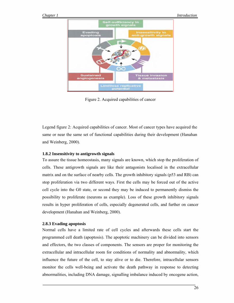
Chapter 1 Introduction
26
Figure 2. Acquired capabilities of cancer
Legend figure 2: Acquired capabilities of cancer. Most of cancer types have acquired the
same or near the same set of functional capabilities during their development (Hanahan
and Weinberg, 2000).
1.8.2 Insensitivity to antigrowth signals
To assure the tissue homeostasis, many signals are known, which stop the proliferation of
cells. These antigrowth signals are like their antagonists localised in the extracellular
matrix and on the surface of nearby cells. The growth inhibitory signals (p53 and RB) can
stop proliferation via two different ways. First the cells may be forced out of the active
cell cycle into the G0 state, or second they may be induced to permanently dismiss the
possibility to proliferate (neurons as example). Loss of these growth inhibitory signals
results in hyper proliferation of cells, especially degenerated cells, and further on cancer
development (Hanahan and Weinberg, 2000).
2.8.3 Evading apoptosis
Normal cells have a limited rate of cell cycles and afterwards these cells start the
programmed cell death (apoptosis). The apoptotic machinery can be divided into sensors
and effectors, the two classes of components. The sensors are proper for monitoring the
extracellular and intracellular room for conditions of normality and abnormality, which
influence the future of the cell, to stay alive or to die. Therefore, intracellular sensors
monitor the cells well-being and activate the death pathway in response to detecting
abnormalities, including DNA damage, signalling imbalance induced by oncogene action,
Chapter 1 Introduction
27
survival factor insufficiency, or hypoxia (Evan et al., 1998). The effectors are regulated
by these sensors and could start, if necessary, the apoptotic machine. This hallmark has a
profound consequence, because until this step, degenerated cells could be disposed and
eliminated via the programmed cell death and the homeostasis is assured, but loss of this
function is another step for cancer development.
1.8.4 Limitless replicative potential
All hitherto described capabilities together lead to an uncoupling of a cells growth
program from signals in its environment. After completing of a certain number of
doublings, they stop growing. Cancer cells have the ability to overcome this. They get
immortalized and that’s a big advantage compared to normal cells (Hanahan and
Weinberg, 2000)
1.8.5 Sustained angiogenesis
Nutrients and other important substances are supplied by vasculature and they are crucial
for the function and survival of cells and tissue. Cells which are within 100 ìm of a
capillary blood vessel get nourish by this vessel. Because of this dependence on nearby
capillaries, cells within a tissue would have an intrinsic ability to encourage blood vessel
growth. To growth exuberantly and progress a lager size of tumor, cancer cells need this
ability to sustain angiogenesis. The two different types of angiogenesis initiating signals
are vascular endothelial growth factor (VEGF) and fibroblast growth factor (FGF). Each
of these factors binds to transmembrane tyrosine kinase receptors displayed by
endothelial cells (Fedi et al., 1997). The ability to induce and sustain angiogenesis seems
to be acquired in a discrete step during tumour development, via an angiogenic switch
from vascular quiescence (Hanahan and Weinberg, 2000).
1.8.6 Tissue invasion and metastasis
Last but not least, the primary tumor masses start to spawn pioneer cells that move out of
the tumor, invade in other normal tissues and develop new colonies. This type of pioneer
cells is called metastase. This is the last step of tumor development; the metastasis
enables cancer cells to escape the primary tumor mass and colonize new terrain in the
body, where initially, nutrients and space are not limiting. For this step the degenerated
cells need a mutation and further an over-expression of cell-cell adhesion molecules
(CAMs); mostly an increasing level of E-cadherin was found in cancer, which is
ubiquitously expressed in endothelial cells. The function of E - cadherin is lost in a
majority of epithelial cancer.
Only three of these described mutations are enough to develop a tumor. But normally the
Chapter 1 Introduction
28
development of cancer is a long term disease. In the last years the increasing awareness of
patients help to detect a lot of early stage cancer by the preventive medical backup, and
treatment with drugs facilitates in many cases a longer life. But there is one big problem,
after long term treatment cancer cells start to get resistant against the drugs and research
aims to develop new pharmaceuticals to combat chemo resistant cancer cells.
1.8.7 The cell cycle
The Cell cycle in most of the cells includes four coordinated and controlled steps, G0/G1-
phase, S-phase, G2-phase and M-phase, which is classified in mitosis and cytokinesis.
Between this steps are three checkpoints, which protect cells of uncontrolled cell cycle or
replication of damaged DNA. The checkpoints are between G1/S-phase, G2/M-phase and
last but not least the spindle checkpoint during the M - phase. Only if each step is duly
completed the cell could transcend checkpoints. This time between transcend the next
phase will be used to repair the mismatch in the cell. The progression of the cell cycle is
catalyzed by cyclin and cyclin-dependentkinases (Cdk) in mammalian and cell division
control (Cdc) in yeast. In humans, we find six different cyclins, cyclin A, B1, B2, D1-D3,
and E. Only D-cyclins are represented in all phases, all others are fixed in one or two cell
cycle steps. The degradation of cyclins is assured by proteolysis. Cdks are divided in
Cdk1, Cdk2, Cdk4, Cdk5 and Cdk6. The catalytic unit of Cdks is activated when they are
associated with the regulatory unit of cyclins. Increasing activity of Cdks is given when a
conservative threonine rest (position 160 in Cdk1) is phosphorylated. On the other site
decreases the activity of Cdks when tyrosine- and threonine rests (Thr15 in Cdk1) are
phosphorylated. The Cyclin/Cdk complexes can be inhibited by cyclin dependent kinase
inhibitors (CKI). In mammalian cells are two different families of CKIs, Cdk interacting
protein (CIP) and polypeptide inhibitors of Cdk4 and Cdk6 (INK4). CIP family inhibits
complexes between Cdk2, Cdk4 and Cdk6 with cyclins respectively. The most important
member of the CIP family is p21CIP. The expression of p21CIP is stimulated by the tumor
suppressor gene p53 and p21C I P is also present during S-phase and has the possibility to
inhibit the DNA replication. The members of the second family are p16INK4a, p15INK4b,
p18INK4c and p19INK4d. These inhibitors are tissue dependent and control only Cdk4 and
Cdk6. In summary, there are three different ways to control cyclin/Cdk complexes. a)
proteolysis of cyclins, b) phosphorylation of tyrosine and threonine rests and c) CKI.
Cdks, cyclins and CKIs are mediators between extracellular signals and the state of
phosphorylation of the tumor suppressor gene retinoblastoma protein (RB). The
activation of Cdks in the G1- and S-phase of cell cycle involves phosphorylation of RB.
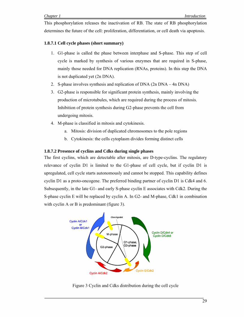
Chapter 1 Introduction
29
This phosphorylation releases the inactivation of RB. The state of RB phosphorylation
determines the future of the cell: proliferation, differentiation, or cell death via apoptosis.
1.8.7.1 Cell cycle phases (short summary)
1. G1-phase is called the phase between interphase and S-phase. This step of cell
cycle is marked by synthesis of various enzymes that are required in S-phase,
mainly those needed for DNA replication (RNAs, proteins). In this step the DNA
is not duplicated yet (2n DNA).
2. S-phase involves synthesis and replication of DNA (2n DNA – 4n DNA)
3. G2-phase is responsible for significant protein synthesis, mainly involving the
production of microtubules, which are required during the process of mitosis.
Inhibition of protein synthesis during G2-phase prevents the cell from
undergoing mitosis.
4. M-phase is classified in mitosis and cytokinesis.
a. Mitosis: division of duplicated chromosomes to the pole regions
b. Cytokinesis: the cells cytoplasm divides forming distinct cells
1.8.7.2 Presence of cyclins and Cdks during single phases
The first cyclins, which are detectable after mitosis, are D-type-cyclins. The regulatory
relevance of cyclin D1 is limited to the G1-phase of cell cycle, but if cyclin D1 is
upregulated, cell cycle starts autonomously and cannot be stopped. This capability defines
cyclin D1 as a proto-oncogene. The preferred binding partner of cyclin D1 is Cdk4 and 6.
Subsequently, in the late G1- and early S-phase cyclin E associates with Cdk2. During the
S-phase cyclin E will be replaced by cyclin A. In G2- and M-phase, Cdk1 in combination
with cyclin A or B is predominant (figure 3).
Figure 3 Cyclin and Cdks distribution during the cell cycle
Chapter 1 Introduction
30
1.8.8 Function and activation of (proto)-oncogenes/oncogenes
1.8.8.1 Oncogenes
Genes, which can potentially induce neoplastic transformation. They include genes
coding for growth factors, growth factor receptors, protein kinases, phosphatases, nuclear
phosphoproteins, and transcription factors. When these genes are constitutively expressed
after structural and or regulatory changes, uncontrolled cell proliferation may result
I just want to refer to two proto-oncogenes, which are crucial in this work, the cyclin D1
and a member of the Cdc25 family, Cdc25A.
1.8.8.2 Cyclin D1
Cyclin D1 is known as an important proto-oncogene. It regulates the transition between
G1- to S-phase (as described above). The binding of cyclin D1 to its kinase partners
(Cdk4/6) results in the active complexes, which phosphorylate RB. Hyper-
phosphorylation of RB causes the release of E2F transcription factor and furthermore the
expression of genes, which are required for entry into S-phase (Alao et al., 2006).
Normally, protein levels of cyclin D1 begin to increase in the early G1-phase of cell cycle
and after transition into S-phase cyclin D1 is translocated from nucleus into the cytoplasm
and degraded. Continuous over expression of cyclin D1 results in uncontrolled cell
proliferation and may cause cancer. Cyclin D1 is over-expressed in many types of cancer,
mantle cell lymphoma, prostate and breast cancer respectively. Inostamycin is an
effective cytostatic drug against cyclin D1 over expression (Baba et al., 2004).
1.8.8 3 Cdc25A (Cell-division-cycle 25A)
Cdc25A is a member of the Cdc25 family. Cdc25 was first described in
Schizosaccharomyces pombe fission yeast. In mammals the Cdc25 family includes three
types of Cdc25 homologs, Cdc25A, Cdc25B and Cdc25C. All members are essential and
specific for cell cycle. The mRNA of Cdc25C is predominantly expressed in G2/M-phase,
Cdc25B is expressed throughout the cell cycle and is elevated in G2-phase. In addition,
the mRNA of Cdc25A is expressed throughout the cell cycle, however with peak
expression during late G1- and S-phases (Ducruet et al., 1997).
The role of Cdc25A in initiation of DNA replication is also consistent with the ubiquitin–
proteasome-mediated destruction of Cdc25A in G1/S-phase checkpoint responses to
DNA damage and replication stress. This requires Chk1 or Chk2 (Checkpoint kinase 1/2)
mediated phosphorylation of Cdc25A, and phosphorylation of Ser123, Ser75 and Ser177
are a prerequisite for such accelerated ubiquitylation and degradation. Consequently, the
Chapter 1 Introduction
31
absence of Cdc25A in damaged cells precludes dephosphorylation of Thr14 and Tyr15 of
Cdk2, and locks this essential S-phase promoting kinase in its inactive form (Mailand et
al., 2000; Falck et al., 2001). New studies showed that Cdc25A is also stabilized in
G2/M-phase of cell cycle and could abrogate the G2 checkpoint (Mailand et al. 2002).
These findings mark Cdc25A as a potential proto-oncogene, because when Cdc25A is
over expressed and phosphorylated on the activating Ser17, all checkpoints in the cell
cycle is crossed.
1.8.8 4 Function and activation of tumor suppressor genes
A gene that encodes a product that negatively regulates the cell cycle and must be
inactivated or degraded before a cell can proceed division. Tumor suppressor genes
encode proteins, which normally suppress cell proliferation. Mutations, which decrease
their activity, may cause cancer. Examples for tumor suppressor genes are differentiation
factors, signal transductions proteins and negative cell cycle regulators (Leisser, 2004).
1.8.8 5 p53 (protein 53)
p53 a nuclear protein is the most relevant tumor suppressor gene in mammalian with
mutation in 50 % of all cancers. The name of p53 is based on the molecular weight. The
loss of p53 causes a proliferation advantage. Normally, cells have low p53 levels. The
level of p53 in cells increases when the cells are exposed to UV-light or other DNA
damaging treatments (Fig. 4).
1.8.8 6 Activation of p53
Activation of p53 has two different consequences, stop of proliferation or apoptosis. The
consequence depends on the state of cell cycle. If the activation of p53 is in the late G1-
phase, p21CIP will be induced by p53 and blocks the cycle, until the DNA damage is
repaired, but if the cell already passes the S-phase, p53 induces programmed cell death –
apoptosis.
1.8.8 7 P21CIP (protein 21)
P21CIP as described in before is known as a Cdk-inhibitor and is induced by both p53-
dependent and -independent mechanisms following stress. Induction of p21CIP may
cause cell cycle arrest (Cayrol et al., 1998). Increased expression of p21CIP inhibits the
activity of Cdk2-cyclin E complexes, as well as other Cdk/cyclin complexes, while
p16INK4a specifically sequesters and inactivates Cdk4/6 complexes. Inactivation of
these cyclin-Cdk complexes prevents phosphorylation of RB protein, which is
necessary for progression from G1- to S-phase of the cell cycle (Taylor et al., 2004).

Chapter 1 Introduction
32
Cell cycle blocked by activation of p21 via p53, and
re-entry cell cycle after repairing DNA
damage.
Induced programmed cell
death apoptosis, cell died.
Figure 4. DNA damage induced by UV-light and further the activation of p53.
1.8.8 8 Activation of p21CIP
After DNA damage caused by UV-light or chemical agents (Doxorubicin) p 2 1 C I P i s
activated via p53 (Wood et al., 2006). p21CIP inhibits Cdk2-cyclin E complexes which are
necessary to cross the G1 block. Hence p21 C I P has the potential to stop the cell cycle in
G1 until the DNA damage is repaired.
1.8.8.9 RB
RB protein was first found in a retina tumor, which occurs at early age. The onset of this
disease could be heritable or spontaneous due to a mutation in the RB gene (position
13q14). To manifest a retinal tumor both alleles must be mutated for breakout. The loss
of functional RB could cause many other types of cancer (osteosarcomas and small
platelet lung carcinomas respectively).
1.8.8.10 Activation of RB
RB is like p53 a nuclear protein which negatively regulates the cell cycle. In non-active
or arrested cells (G0/G1) RB is hypo-phosphorylated. At the end of G1-phase RB
becomes hyper-phosphorylated by Cdk/cyclin complex and returns to a hypo-
phosphorylated state during mitosis. Only in a hyper-phosphorylated state RB binds to
specific proteins. This interaction happens during the S-phase of the cell cycle. Target
genes of RB are the E2F family (transcription factor). Because of this binding to RB the
Chapter 1 Introduction
33
E2F genes are blocked and cannot activate transcription. An over-expression of RB
inhibits cell proliferation, but this effect could be abolished by over-expression of D
cyclins. They build complexes with Cdks and this combination is responsible for
phosphorylation of RB.
In summary, dephosphorylated RB blocks cell proliferation and its activation must be
abolished to assure transit through the cell cycle. This is ensured by cyclic
phosphorylation (Levin, 1998)
1.8.9 Cell death
Cell death is one of the fundamental cell regulatory system, which graduates the tissue
integrity, and in series the homeostasis of tissues. In general we distinguish between two
different types of cell death - the programmed cell death that includes apoptosis (type I)
and autophagy (type II) in contrast to necrosis which introduce inflammation of the tissue
(figure 18).
1.8.9.1 Apoptosis
Apoptosis is the main type of programmed cell death and is mediated by an intracellular
program. It was first described by John Kerr in the late 1960s (O' Rourke and Ellem
2000). A series of metabolic events lead to morphological differentiation of cells,
including blebbing, changes to cell membrane, cell shrinkage, nuclear fragmentation,
chromatin condensation and chromosomal DNA fragmentation. The cells which are
eliminated by apoptosis can be classified in different categories (Wagner,1999).
I. Cells without function; in fact apoptosis is essential for human embryonic
development. In example, the differentiation of fingers and toes in a developing
human embryo occurs because cells between the fingers undergo programmed cell
death - apoptosis.
II. Cells which are produced in excess; in this category are for example blood cells and
male gametes.
III. Cells which have already fulfil their functions; for example cells of the
endometrium. The cells will be eliminated by apoptosis during the premenstrual and
menstrual cycles.
Cells which could impair the cell integrity with their negative potential; it is qualitative
and quantitative the most important function of apoptosis. One example are the auto
reactive T-lymphocytes. They are negatively selected and eliminated in the thymus. Last
but not least, apoptosis is an important mechanism to innoxious tumour cells Each cell
Chapter 1 Introduction
34
has the potential to start the biochemical machine of apoptosis. This program is classified
in 4 different steps:
I. Determination and activation of apoptosis:
The first step of the program is dependent on two different categories of factors.
One category of factor releases (positive signals), and the second one inhibits
(negative signals) the apoptosis program.
Positive signals are Fas-Fas-ligand and the TNF-induced (tumour necrosis factor)
model and negative signals are all growth factors, human growth hormones and
cell adhesion. If there is an imbalance between these antagonists, the activation or
inactivation of apoptosis starts.
II. Execution of cell death
Four genes are necessary in this step. Three of this group are cell death genes
(ced) and the fourth is called egg-laying defective 1 (egl-1). Egl-1, ced-3 and ced-
4 are essential to perform the cell death program. If one of these genes is
deactivated the cell death program stops. The last ced gene is ced-9. This four
genes interact with themselves (elg-1 inhibits ced-9, ced-9 inhibits ced-4, ced-4
activates ced-3).
III. Phagocytosis
When cells undergo final stages of apoptosis they display phagocytotic molecules
on their cell surface (phosphatidylserine). These molecules mark the apoptotic
bodies of cells for phagocytosis. The removal of the apoptotic bodies by
phagocytes release no inflammation of the tissue.
IV. Degradation
After phagocytosis cell remnants will be removed by proteases and Ca++
dependent endonucleases.
1.8.9.2 Autophagy
Autophagy is a catabolic process where the cells start degradation of their own
components through the lysosomal machinery. It was first described in 1960. This kind of
programmed cell death is strictly regulated and is important for developing, cell growth
and homeostasis. Autophagy plays a role in some diseases to protect the organism against
infections by intracellular pathogens. In higher eukaryotes autophagic dysfunction has
Chapter 1 Introduction
35
been associated with heart disease, neurodegenerative disorders and tumour progression
(Leisser, 2004).
1.8.9.3 Necroses
Necrosis is definitely a non-programmed cell death. Necrotive cells start swelling,
chromatin becomes digested at random, plasma membranes and organelle membranes
become disrupted and finally cells lyse. This type of cell death effects injury and
provokes an inflammatory response.
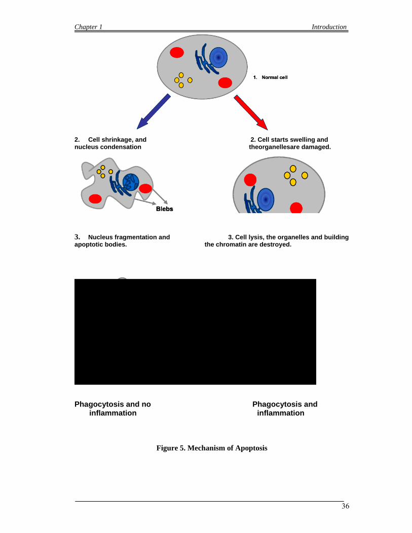
Chapter 1 Introduction
36
2. Cell shrinkage, and 2. Cell starts swelling and nucleus condensation theorganellesare damaged.
3. Nucleus fragmentation and 3. Cell lysis, the organelles and building apoptotic bodies. the chromatin are destroyed.
Phagocytosis and no Phagocytosis and inflammation inflammation
Figure 5. Mechanism of Apoptosis
Chapter 1 Introduction
37
1.9 Bioassays Techniques
1.9.1 Apoptosis assays (Hoechst 33258 propidium iodide (HOPI) double-staining)
Grusch et al, (2002) first described this method of apoptosis, in which propidium iodide
(PI) and Hoechst 33258 are directly added to the culture medium to final concentrations
of 2 mg/ml and 5 mg/ml, respectively. After one h incubation period at 37C, the cells are
then examined under a Zeiss Axiovert 35 fluorescence microscope with DAPI filters. The
cells were then photographed on Kodak Ektachrome P1600 film (Eastman Kodak
Company, Rochester, NY, USA) and the three type of cells i.e. viable, apoptotic, and
necrotic, were counted manually. The Hoechst dye will stains the nuclei of all cells and
therefore it became clear and easily monitor nuclear changes associated with apoptosis,
such as nuclear fragmentation and chromatin condensation. On the other hand PI is
excluded from live and early apoptotic cells and the uptake of PI by necrotic and late
apoptotic cells indicates loss of its membrane integrity characteristic. The selective uptake
of the two dyes along with the combination of fluorescence microscopy, help the
monitoring of the induction of apoptosis in intact cultures and also to distinguish it from
non-apoptotic cell death (necrosis). Necrosis is therefore characterized by nuclear PI
uptake without nuclear fragmentation or chromatin condensation.
1.9.2 Western blot assay
W. Neal Burnette (Burnette) first gave the name of western blot to the technique. The
western blot is also name as protein immunoblot. It is an analytical technique which is
widely used to identify specific proteins both qualitatively and quantitatively in a given
sample of extract or tissue homogenate. Gel electrophoresis is first use to separate native
or denatured proteins by the length of the polypeptide (denaturing conditions) or by the 3-
D structure of the protein (native/ non-denaturing conditions). After separation of the
proteins, it then transferred to nitrocellulose or PVDF membrane. The membrane are then
probed (detected) with specific antibodies in order to identify the target protein (Towbin
et al., 1979; Renart et al., 1979). The method of Western blotting is widely used in
different biological fields such as molecular biology, biochemistry, immunogenetics and
other molecular biology disciplines
Chapter 1 Introduction
38
1.9.2.1 Steps in a western blot
Tissue preparation: whole tissue or cell culture is used for extraction. In case of
tissue, blender is used in case of larger sample volume in order to broken down
the solid tissues and a homogenizer or sonication is used in case of smaller
volume for breaking the tissue. The above mention method can be used in case of
cell culture. Lyses of cells and solubilization of proteins can be encouraged by
employing of assorted detergents, salts, and buffers. In order to prevent the protein
from the digestion by its own enzymes, Protease and phosphatase inhibitors are
often used. At the time of protein and tissue preparation the temperature should be
very low or work should be completed in ice in order to avoid protein denaturing.
After completion the lyses process centrifugation can be made in order to separate
different organelles and cell compartments.
Gel electrophoresis: Gel electrophoresis is a separation technique which used in
biology for protein separation. Proteins separation is based on isoelectric point
(pI), electric charge, molecular weight, or a combination of all these factors. Both
treatment of the sample and the nature of the gel are responsible for the nature of
the separation. Polyacrylamide gels along with buffers loaded with sodium
dodecyl sulfate (SDS) are used in common type of gel electrophoresis. Once the
protein have been treated with strong reducing agents to remove secondary and
tertiary structure (e.g. disulfide bonds [S-S] to sulfhydryl groups [SH and SH]), it
has the characteristic of SDS-PAGE (SDS polyacrylamide gel electrophoresis) is
to maintains polypeptides in its denatured state and thus it became easy in
separation of proteins by their molecular weight. The protein of sampled become
covered in the negatively charged SDS and move through the acrylamide mesh of
the gel to the positively charged electrode. The protein thus migrate through this
mesh and hence smaller proteins migrate faster are thus separated according to
size (usually measured in kilo Daltons, kDa). The resolution of the gel depends on
the concentration of acrylamide- better resolution of lower molecular weight
proteins obtain with greater acrylamide concentration. The resolution of higher
molecular weight proteins will be better when the acrylamide concentration lower.
In most blots proteins travel only in one dimension along the gel. Different
samples are loaded one by one into wells in the gel. One lane of the gel is always
reserved for a marker or ladder. The marker is a commercially available mixture
of proteins and each having specific molecular weights which are typically stained

Chapter 1 Introduction
39
so as to form visible, colored bands. When voltage is applied along the gel,
proteins migrate into it at different speeds. These different rates of advancement
(different electrophoretic mobilities) separate into bands within each lane. Two-
dimensional (2-D) gel can also be used, which spreads the proteins from a single
sample out in two dimensions.
Transfer of separated protein to a membrane: A membrane made of
nitrocellulose or polyvinylidene difluoride (PVDF) is used in order to transfer
proteins from the gel onto the membrane. The protein transfers up the paper by
capillary action, bringing the proteins with it. Electro blotting is another method
for transferring the proteins which uses an electric current to pull proteins from
the gel into membrane. The proteins which are present in the gel transfer with the
same organization as they had with in the gel. Coomassie or Ponceau S dyes are
used in order to check the uniformity and overall effectiveness of transfer of
protein from the gel to the membrane. Ponceau S is the more common of the two,
due to Ponceau S's higher sensitivity and its water solubility makes it easier to
subsequently destain and probe the membrane.
Blocking of membrane: Both bovine serum albumin (BSA) and non-fat dry milk
are used a dilute solution of protein for the blocking of non-specific binding. The
blocking is achieved by placing the membrane in the dilute solution of the protein
and Tween 20 also used as a detergent in a minute percentage. The membrane is
covered with the protein in the dilute solution accept the places where the target
proteins have not attached. After addition of antibody, there is no space on the
membrane for it to attach other than on the binding sites of the specific target
protein. This confusion in the final product of the Western blot is reduces in this
way, which leads to clearer results, and eliminates false positives.
Detection of specific protein: The following two steps are involved in detection.
1. Primary antibodies: After the process of blocking the membrane, the
membrane is incubated under gentle agitation along with a very dilute
solution of the primary antibody (0.5 and 5 micrograms/ml). A small
percentage of detergent, buffered saline solution and sometimes with
powdered milk or BSA are present in the solution. The membrane can be
sealed along with the antibody solution and incubated anywhere from 30
minutes to overnight. The incubation temperature can be varied. The
warmer temperatures being associated with more binding, both specific (to
the target protein, the "signal") and non-specific ("noise").

Chapter 1 Introduction
40
2. Secondary antibodies: After removing unbound primary antibody by
rinsing the membrane, the membrane is treated with another antibody,
directed at a species-specific portion of the primary antibody. Due to its
targeting properties it is known as a secondary antibody which tends to be
referred to as "anti-mouse," "anti-goat," etc. All these antibodies isolated
from animal sources (or animal sourced hybridoma cultures); it therefore
very specific because an anti-mouse secondary will bind to just about any
mouse-sourced primary antibody. This allows some cost savings by
allowing an entire lab to share a single source of mass-produced antibody,
and obtain far more similar results. The secondary antibody has the
property that it is usually linked to biotin or to a reporter enzyme such as
alkaline phosphatase or horseradish peroxidase. The secondary antibodies
will bind to one primary antibody and therefore enhance the signal.
Analysis: After the unbound probes are washed away, the western blot is ready
for detection of the probes that are labeled and bound to the protein of interest. In
practical terms, not all westerns reveal protein only at one band in a membrane.
Size approximations are taken by comparing the stained bands to that of the
marker or ladder loaded during electrophoresis. The process is repeated for a
structural protein, such as actin or tubulin, that should not change between
samples. The amount of target protein is indexed to the structural protein to
control between groups. This practice ensures correction for the amount of total
protein on the membrane in case of errors or incomplete transfers.
Detection of protein intensity: Different types can be used for the detection of
protein intensity like Radioactive detection, Fluorescent detection,
Chemiluminescent detection, Colorimetric detection.
Radioactive detection: Radioactive labels do not require enzyme substrates, but
rather allow the placement of medical X-ray film directly against the western blot
which develops as it is exposed to the label and creates dark regions which
correspond to the protein bands of interest. The importance of radioactive
detections methods is declining because it is very expensive, health and safety
risks are high and ECL provides a useful alternative.
1.9.3 Fluorescence Activated Cell Sorting (FACS) assay
The Fluorescence-activated cell sorting (FACS) is a unique type of flow cytometry.
Heterogeneous mixtures of biological cells (one cell at a time) are sorted into two or more
Chapter 1 Introduction
41
containers which is run on a principal of the specific light scattering and fluorescent
characteristics of each cell. It is a useful scientific instrument, the fluorescent signals from
individual cells and the physical separation of cells of particular interest as well, are
performed by it through its objective and quantitative recording. The acronym FACS is
trademarked and owned by Becton Dickinson (Loken, 1990). While many immunologists
use this term frequently for all types of sorting and non-sorting applications, it is not a
generic term for flow cytometry. The first cell sorter was invented by Mack Fulwyler in
1965, using the principle of Coulter volume a relatively difficult technique to use for
sorting and one no longer used in modern instruments. The technique was expanded by
Len Herzenberg who was responsible for coining the term FACS. Herzenberg won the
Kyoto Prize in 2006 for his work in flow cytometry.
The suspension of cell which is analyzed is injected in the center of a narrow, rapidly
flowing stream of liquid. The system flow is arranged in such a way that there is a large
separation between the cells as compare to their diameter. A vibrating mechanism causes
the stream of the cells divide into individual droplets. The system is set in such a way that
there is a low probability of more than one cell's being in a droplet. Just before the stream
divide into droplets, the flow passes through a fluorescence intensity measuring station
where the fluorescent character of interest of each cell is measured. Just at the point
where the stream divides into droplets, an electrical charging ring is placed. A charge is
placed on the ring based on the immediately-prior fluorescence intensity measurement,
and the opposite charge is trapped on the droplet as it breaks from the stream. The
charged droplets then fall through an electrostatic deflection system that diverts droplets
into containers based upon their charge. In some systems, the charge is applied directly to
the stream, and the droplet breaking off retains charge of the same sign as the stream. The
stream is then returned to neutral after the droplet breaks off.
1.9.3.1 Flow cytometer
Modern flow cytometer are able to analyze several thousand particles every second, in
"real time," and can actively separate and isolate particles having specified properties. A
flow cytometer is similar to a microscope, except that, instead of producing an image of
the cell, flow cytometry offers "high-throughput" (for a large number of cells) automated
quantification of set parameters. To analyze solid tissues, single-cell suspension must first
be prepared.
A flow cytometer has 5 main components:
Flow cell: liquid stream (sheath fluid), which carries and aligns the cells so that
they pass single file through the light beam for sensing.
Chapter 1 Introduction
42
Optical system: commonly used are lamps (mercury, xenon); high-power water-
cooled lasers (argon, krypton, dye laser); low-power air-cooled lasers (argon
(488nm), red-HeNe (633nm), green-HeNe, HeCd (UV)); diode lasers (blue, green,
red, violet) resulting in light signals.
Detector and Analogue-to-Digital Conversion (ADC) system: which generates
FSC and SSC as well as fluorescence signals from light into electrical signals that
can be processed by a computer
Amplification system: linear or logarithmic
Computer: for analysis of the signals
1.9.3.2 Application
The technology can be applied in various fields such as pathology, molecular biology,
immunology, plant biology and marine biology. It became very useful when used with
fluorescence tagged antibodies in the field of molecular biology. These specific
antibodies bind to antigens on the target cells and help to give information on specific
characteristics of the cells being studied in the cytometer. It has many applications in
medicine (especially in transplantation, tumor immunology, hematology, and
chemotherapy, genetics and sperm sorting for sex preselection). In marine biology, the
auto-fluorescent properties of photosynthetic plankton can be exploited by flow
cytometry in order to characterize abundance and community structure. In protein
engineering, flow cytometry is used in conjunction with yeast display and bacterial
display to identify cell surface-displayed protein variants with desired properties.
1.9.4 Comet assay
Comet assay also known as the Single Cell Gel Electrophoresis (SCGE) assay is a simple
and much sensitive technique for the study of DNA damage at the level of the individual
eukaryotic cell. The technique was first described by Singh et al. in 1988. Now a days it
is much popular and standard technique for the study of DNA damage/repair, bio
monitoring and genotoxicity testing. The cells have encapsulated in a low-melting-point
agarose suspension, the cells are then lysis in neutral or alkaline (pH>13) conditions, and
electrophoresis of the suspended lysed cells. This is followed by visual analysis with
staining of DNA and calculating fluorescence to determine the extent of DNA damage.
This can be performed by manual scoring or automatically by imaging software.
1.9.4.1 Experimental procedure
Experimental are involved the following steps.

Chapter 1 Introduction
43
Encapsulation: A sample of cells either derived from an in vitro cell culture or
from an in vivo test subject is dispersed into individual cells and suspended in
molten low-melting-point agarose at 37°C. This mono-suspension is cast on a
microscope slide. A glass cover slip is held at an angle and the mono-suspension
applied to the point of contact between the coverslip and the slide. As the cover
slip is lowered onto the slide the molten agarose spreads to form a thin layer. The
agarose is gelled at 4°C and the coverslip removed. The agarose forms a matrix of
carbohydrate fibers that encapsulate the cells, anchoring them in place. The
agarose is considered to be osmotic-neutral, therefore solutions can penetrate the
gel and affect the cells without cells shifting position. In an in vitro study the cells
would be exposed to a test agent - typically UV light, ionizing radiation, or a
genotoxic chemical - to induce DNA damage in the encapsulated cells. For
calibration, hydrogen peroxide is usually used to provide a standardized level of
DNA damage
Lysis: The slides are then immersed in a solution that causes the cells to lyse. The
lysis solution often used in the comet assay consists of a highly concentrated
aqueous salt (often, common table salt can be used) and a detergent (such as
Triton X-100 or sarcosinate). The pH of the lyses solution can be adjusted (usually
between neutral and alkaline pH) depending upon the type of damage the
researcher is investigating. The aqueous salt disrupts proteins and their bonding
patterns within the cell as well as disrupting the RNA content of the cell. The
detergent dissolves the cellular membranes. Through the action of the lysis
solution the cells are destroyed. All proteins, RNA, membranes and cytoplasmic
and nucleoplasmic constituents are disrupted and diffuse into the agarose matrix.
Only the DNA of the cell remains, and unravels to fill the cavity in the agarose
that the whole cell formerly filled. This structure is called nucleoid (a general term
for a structure in which DNA is concentrated).
Electrophoresis: After lysis of the cells (typically 1 to 2 hours at 4°C) the slides
are washed in distilled water to remove all salts and immersed in a second solution
(an electrophoresis solution). Again this solution can have its pH adjusted
depending upon the type of damage that is being investigated. The slides are left
for ~20 minutes in the electrophoresis solution prior to an electric field being
applied. In alkaline conditions the DNA double helix is denatured and the
nucleoid becomes single stranded. An electric field is applied (typically 1 V/cm)
for ~20 minutes. The slides are then neutralized to pH 7, stained with a DNA-
Chapter 1 Introduction
44
specific fluorescent stain and analyzed using a microscope with an attached CCD
(charge-coupled device - essentially a digital camera) that is connected to a
computer with image analysis software e.g. Comet IV, Perceptive Instruments
Ltd., Haverhill, UK.
1.9.4.2 Principles
The concept underlying the SCGE assay is that undamaged DNA retains a highly
organized association with matrix proteins in the nucleus. When damaged, this
organization is disrupted. The individual strands of DNA lose their compact structure and
relax, expanding out of the cavity into the agarose. When the electric field is applied the
DNA, which has an overall negative charge is drawn towards the anode. Undamaged
DNA strands are too large and do not leave the cavity, whereas the smaller the fragments,
the farther they are free to move in a given period of time. Therefore, the amount of DNA
that leaves the cavity is a measure of the amount of DNA damage in the cell. The image
analysis measures the overall intensity of the fluorescence for the whole nucleoid and the
fluorescence of the migrated DNA and compares the two signals. The stronger the signal
from the migrated DNA the more damage there is present. The overall structure
resembles a comet (hence "comet assay") with a circular head corresponding to the
undamaged DNA that remains in the cavity and a tail of damaged DNA. The brighter and
longer the tail, the higher the level of damage.
1.9.5 Total Phenolics or Folin-Ciocalteau Micro Method
This method is used routinely in our lab to measure total phenol. The procedure is also
used for analysis of total phenol in various plants and fruits. It uses the minimum volume
of reagents and almost eliminates wasted reagent. Good micro pipets must be used for
reproducibility. Plastic or glass cuvettes can be used. It is based on the method reported
by Slinkard and Singleton, (1977), only the volumes have been reduced. If you cannot
reproducibly measure such small volumes, try to reduce the volumes to the smallest you
can. This reduces waste and disposal volume. The following reagents are used in this
assay.
Folin Ciocalteu Reagent: It is a mixture of phosphomolybdate and
phosphotungstate used for the colorimetric assay of phenolic and polyphenolic
antioxidants (Singleton et al., 1999). It works by measuring the amount of the
substance being tested needed to inhibit the oxidation of the reagent (Vinson et
al., 2005). However, this reagent does not only measure total phenols and will

Chapter 1 Introduction
45
react with any reducing substance. The reagent therefore measures the total
reducing capacity of a sample, not just the level of phenolic compounds. This
reagent forms part of the Lowry protein assay and will also react with some
nitrogen-containing compounds such as hydroxylamine and guanidine (Ikawa et
al., 2003). This is usually purchased as the 2N reagent available from Sigma
(F9252) or from Fisher Scientific (ICN19518690), and presumably others.
Singleton and Rossi, (1965) describe the preparation of the reagent from sodium
tungstate, sodium molybdate, lithium sulfate, bromine, and some acids.
Gallic Acid Stock Solution: In a 100-mL volumetric flask, dissolve 0.500 g of
dry gallic acid in10 mL of ethanol and dilute to volume with water. Can be opened
daily, but to store, keep closed in a refrigerator up to two weeks.
Sodium Carbonate Solution: Dissolve 200 g of anhydrous sodium carbonate in
800 mL of water and bring to a boil. After cooling, add a few crystals of sodium
carbonate, and after 24 hr, filter and add water to 1 L.
1.9.5.1 Calibration curve
To prepare a calibration curve, add 0, 1, 2, 3, 5, and 10 mL of the above phenol stock
solution into 100 mL volumetric flasks, and then dilute to volume with water. These
solutions will have phenol concentrations of 0, 50, 100, 150, 250, and 500 mg/L gallic
acid, the effective range of the assay.
From each calibration solution, sample, or blank, pipet 20 µL into separate cuvettes, and
to each add 1.58 mL water, and then add 100 µL of the Folin-Ciocalteu reagent, and mix
well. Wait for between 30 sec and 8 min, and then add 300 µL of the sodium carbonate
solution, and shake to mix. Leave the solutions at 20°C for 2 hour and determine the
absorbance of each solution at 765 nm against the blank (the "0 mL" solution) and plot
absorbance vs. concentration. Alternatively, they can be left at 40°C for 30 min before
reading the absorbance.
A calibration curve is created with the standards and determined the levels in the samples.
Do not neglect to multiply the observed concentrations by any dilution factor of the
original sample. Results are reported at Gallic Acid Equivalent, GAE, because the
phenols in sample contain mostly other phenols, and only small amounts of Gallic acid.
Since the assay measures all phenolics, the choice of Gallic acid as standard is based on
the availability of a stable and pure substance, and Gallic acid is both, and it is less
expensive than other options. In addition, the response to Gallic acid has been shown to
be equivalent to most other phenolics in extract on a mass basis. It has also tested the
Chapter 1 Introduction
46
stability of Gallic acid standard solutions and it can say they lose less than 5% of their
value over two weeks when refrigerated and kept tightly closed.
1.9.6 Antioxidant activity
The body uses oxygen and nutrients to make energy. Oxygen also helps the immune
system fight disease and harmful substances. Oxidation is a process that uses by products
formed from oxygen fighting disease to create molecular agents that react with body
tissues. Unfortunately, this process can form “free radicals” that cause cell damage.
Antioxidants help reduce the number of free radicals that form in the body, lower the
energy levels of existing free radicals, and stop oxidation chain reactions to lower the
amount of damage caused by free radicals. The antioxidants of food are thought to
prevent diseases caused by oxidative stress (Cutler, 1984; Frankel et al., 1993). Free
radicals are believed to be one of the causes of over sixty health problems, according to
various scientific and medical groups. These problems include cancer, aging, and
atherosclerosis. By increasing antioxidant intake and reducing exposure to free radicals
can help lower health risks and problems. Antioxidant enzymes are also produced by our
bodies and include catalase, superoxide dismutase, and glutathione peroxidase. These
enzymes also fight against free radicals. The enzymes are available in supplemental
forms, but it is believed that taking the building blocks of the enzymes in supplemental
form is more effective. Zinc, selenium, copper, and manganese are some of the building
blocks. Minerals and vitamins are also often antioxidants. Vitamins including lutein,
cysteine, beta-carotene, vitamin B2, vitamin C, vitamin E, and coenzyme Q10 and herbs.
Among the natural secondary metabolites, flavonoids play a key role in the antioxidant
mechanism by scavenging free radical produced during oxidation process. Flavonoids are
widely distributed in plant foods such as vegetables and fruits. They possess a unique
C6−C3−C6 structure (diphenylpropane structure) with phenolic hydroxy groups; more
than 4000 different functional group substitution patterns have been identified as natural
flavonoids. The average intake of flavonoids in Western diets was estimated to be 1 g/day
(Kuhnan, 1976). Flavonoids in citrus fruits are known as bioflavonoids or vitamin P,
which exhibit beneficial effects on capillary permeability and fragility (Rusznyak and
Szent-Gyorgyi, 1936). These compounds have been investigated regarding their
physiological functions such as anti-inflammatory, anticarcinogenic, and antitumor
activities (Bracke et al., 1994; Middleton and Kandaswami, 1994; Attaway, 1994). The
antioxidant activity of flavonoids has attracted much attention in relation to their
physiological functions. Dietary flavonoids are considered to aid in the prevention of
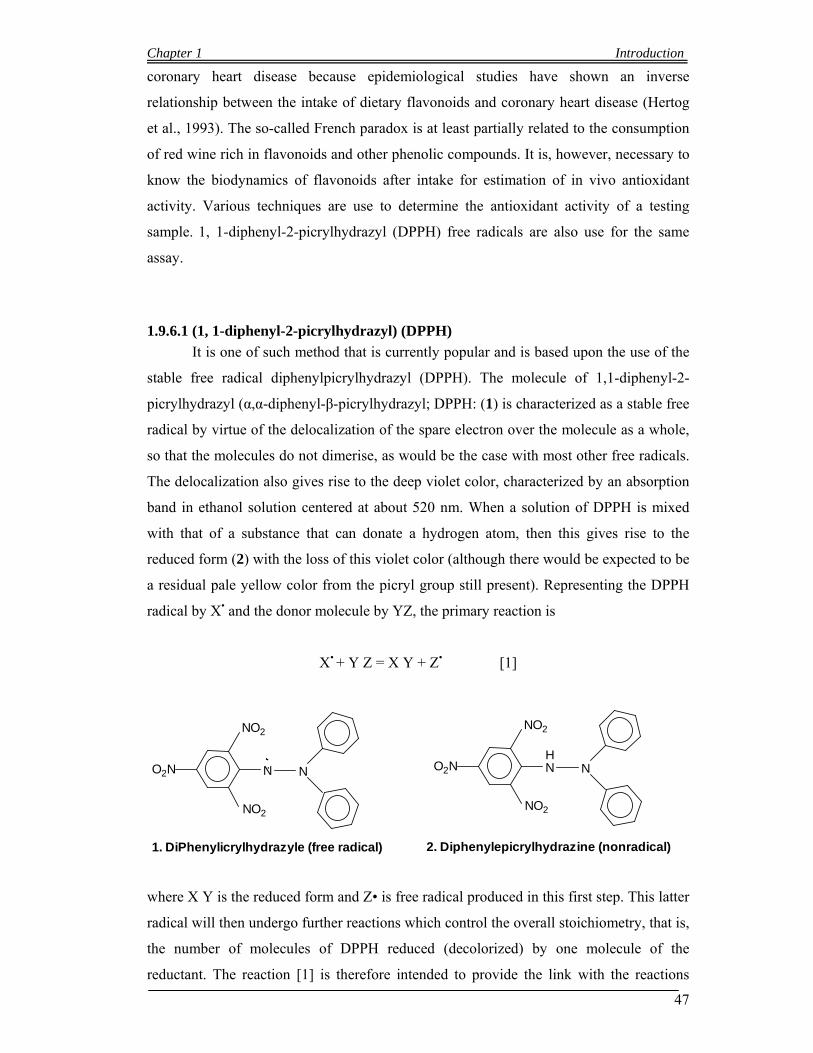
Chapter 1 Introduction
47
coronary heart disease because epidemiological studies have shown an inverse
relationship between the intake of dietary flavonoids and coronary heart disease (Hertog
et al., 1993). The so-called French paradox is at least partially related to the consumption
of red wine rich in flavonoids and other phenolic compounds. It is, however, necessary to
know the biodynamics of flavonoids after intake for estimation of in vivo antioxidant
activity. Various techniques are use to determine the antioxidant activity of a testing
sample. 1, 1-diphenyl-2-picrylhydrazyl (DPPH) free radicals are also use for the same
assay.
1.9.6.1 (1, 1-diphenyl-2-picrylhydrazyl) (DPPH)
It is one of such method that is currently popular and is based upon the use of the
stable free radical diphenylpicrylhydrazyl (DPPH). The molecule of 1,1-diphenyl-2-
picrylhydrazyl (α,α-diphenyl-β-picrylhydrazyl; DPPH: (1) is characterized as a stable free
radical by virtue of the delocalization of the spare electron over the molecule as a whole,
so that the molecules do not dimerise, as would be the case with most other free radicals.
The delocalization also gives rise to the deep violet color, characterized by an absorption
band in ethanol solution centered at about 520 nm. When a solution of DPPH is mixed
with that of a substance that can donate a hydrogen atom, then this gives rise to the
reduced form (2) with the loss of this violet color (although there would be expected to be
a residual pale yellow color from the picryl group still present). Representing the DPPH
radical by X• and the donor molecule by YZ, the primary reaction is
X• + Y Z = X Y + Z• [1]
N N
NO2
NO2
O2N..
1. DiPhenylicrylhydrazyle (free radical)
N N
NO2
NO2
O2N
2. Diphenylepicrylhydrazine (nonradical)
H
where X Y is the reduced form and Z• is free radical produced in this first step. This latter
radical will then undergo further reactions which control the overall stoichiometry, that is,
the number of molecules of DPPH reduced (decolorized) by one molecule of the
reductant. The reaction [1] is therefore intended to provide the link with the reactions
Chapter 1 Introduction
48
taking place in an oxidising system, such as the autoxidation of a lipid or other
unsaturated substance; the DPPH molecule X• is thus intended to represent the free
radicals formed in the system whose activity is to be suppressed by the substance Y Z.
The DPPH method as summarized above was evidently introduced nearly 50 years ago by
Marsden Blois, working at Stanford University (Blois, 1958). It was noted in the original
paper that among other compounds active in this reaction are glutathione, aromatic
amines (such as p-phenylene diamine and p-aminophenol), and α-tocopherol (Vitamin E -
2:1 stoichiometry) and polyhydroxy aromatic compounds (such as hydroquinone and
pyrogallol). On the other hand, monohydric phenols (such as tyrosine), simple sugars
(such as glucose), purines and pyrimidines, do not react, while proteins are precipitated. It
was also noted that “inorganic ions in lower valence states may of course interfere and
must be eliminated or determined separately” which presumably applies most importantly
to ferrous iron (Blois, 1958).
1.10 Selection of Medicinal plants species
Twenty seven plants were selected and collected from Margalla Hills Islamabad Pakistan.
Some species are reported as medicinal while some are not reported medicinal plants
species. Three plants species Berberis lycium Royle (Berberidaceae), Zizyphus
nummularia (Rhamnaceae) and Mallotus philippensis (Euphorbiaceae) were analyzed for
antineoplastic activities, other twenty four plants species were analyzed for free radical
scavenging activity, total Phenolic content and Flavonoids types. The plant samples used
in this study were Albizia lebbeck (Mimosaceae), Bauhinia variegata and Cassia fistula
(Caesalpinaceae), Bombax ceiba (Bombacaceae), Calotropis procera (Asclepiadaceae),
Carissa opaca (Apocynaceae), Colebrookea oppositifolia (Labiateae), Dalbergia sissoo
(Papilionaceae), Dodonaea viscosa (Sapindaceae), Ficus palmata and Ficus racemosa
(Moraceae), Jasminum humile and Olea ferruginea (Oleaceae), Adhadoda vasica
(Acanthaceae), Lantana camara Linn. (Verbenaceae), Melia azedarach L. (Meliaceae),
Phyllanthus emblica. L. (Euphorbiaceae), Pinus roxburghii Sargent (Pinaceae), Punica
granatum L. (Punicaceae), Rubus ellipticus Smith, Pyrus pashia Buch. & Ham.
(Rosacceae), Viburnum cotinifolium D. Don (Caprifoliaceae), Dabregeasia salicifolia
(Urticaceae) and Caryopteris grata (Verbenaceae).
Chapter 1 Introduction
49
2.10.1 Berberis lycium Royle (Berberidaceae)
Berberis lycium is a widely used medicinal plant in Pakistan, known by the common
name “Zyarh larghai” or “Kashmal”, whereas its English name is Barberry (Anwar, 1979)
Al-Biruni describes the plant under the name of Ambaribis and also mentioned its Persian
names as Zirkash (Said, 1996 ). Berberis taxa are important plants, with various
medicinal properties. Berberis is also included in Indian and British pharmacopoeias.
Description
Shrub, 2-3(-4) m tall, erect or suberect, semideciduous; stem and branches pale, whitish
to greyish, terete to subsulcate, glabrescent, younger ones obscurely to distinctly
puberulous; yellowish to straw-coloured. Leaves oblanceolate to oblong-obovate,
subsessile, usually conspicuously papillose, grey or white below, Racemes (6-)10-25-
flowered, 3-6(-7) cm long, rarely shorter and subfascicled. Flowers usually pale-yellow;
Outer sepals much smaller than the middle and inner sepals; inner sepals 4.5-5 mm long,
3 mm broad, obovate. Petals slightly shorter than the inner sepals, obovate, emarginate,
with lanceolate basal glands. Stamens slightly shorter than petals, connectives produced
or anthers apiculate. Ovules usually 4, shortly stipitate. Berries ovoid or obovoid-
subglobose, excluding 1 mm long style, blackish with heavy grey-white bloom.
Distribution
Native in the whole Himalaya Mountains range and widely distributed in temperate and
semi-temperate areas of India, Nepal, Afghanistan, Bangladesh and Pakistan.
1.10.2 Mallotus philippensis (Lam.) Muell. Arg. (Euphorbiaceae)
Description
Shrubs or small trees, 2-15 m tall. Branchlets, petiole, and inflorescences yellow-
brownish stellate-tomentose. Leaves ovate to lanceolate, leathery, margin subentire, apex
acuminate; basal veins 3. Male flowers 1-5-fascicled; calyx lobes 3 or 4, oblong, ca. 2
mm, tomentulose; stamens 15-30. Female flowers: calyx lobes 3-5, subovate, ca. 3 mm,
tomentose; ovary tomentose and red glandular-scaly; styles 3, 3-4 mm, plumose. Capsule
Chapter 1 Introduction
50
subglobose, 8-10 mm in diam., (2 or)3-locular, covered with a red glandular-scaly layer.
Seeds subglobose, ca. 4 mm in diam., black. Fl. Mar-May, fr. Jun-Aug.
Distribution
Mountain slopes or valleys, limestone hills or river valleys, forests; 300-1600 m. Anhui,
Fujian, Guangdong, Guangxi, Guizhou, Hainan, Hubei, Hunan, Jiangsu, Jiangxi, Sichuan,
Taiwan, Xizang, Yunnan, Zhejiang, Bangladesh, Bhutan, India, Laos, Malaysia,
Myanmar, Nepal, New Guinea, Pakistan, Philippines, Sri Lanka, Thailand, Vietnam; N
Australia.
1.10.3 Adhatoda vasica Nees (Acanthaceae).
Description
An erect much branched, gregarious, evergreen shrub, up to 2 (-2.5) m. Stem ±
quadrangular to nearly terete, young shoots greyish-pubescent Leaves elliptic-lanceolate,
glabrous above, pubescent on nerves beneath, basally attenuate, entire, acuinate. Flowers
white, c. 3 cm long, nearly sessile, in terminal and axillary spikes, up to 10 cm long, 2.5-3
cm broad; bracts leafy, broadly-elliptic; Calyx 5-lobed, lobes linear-lanceolate, 6-10 x c. 2
mm, acute, puberulous, imbricate. Corolla pale-white, tube 1.2-1.5 cm long, pubescent
outside, throat villous, limb 2-lipped, upper lip erect, shortly bifid, galeate, lower lip with
3 elliptic, obtuse lobes. Ovary oblong, c. 3 mm long, style 2-2.5 cm long. Capsule
stipitate, broadly clavate, pubescent. Seeds ± orbicular, 2-3 mm across, glabrous. Fl. Per.:
November-April (plains); July-October (hills).
Distribution:
Panama (probably) introduced), Indonesia, Malaya, S.E. Asia, India and Pakistan.
In Pakistan, it does well on waste lands up to 1300 m; it is also cultivated as an
ornamental.
1.10.4 Albizia lebbeck (L.) Benth. (Mimosaceae)
Description

Chapter 1 Introduction
51
A large deciduous tree with dark grey bark, usually cracked, young parts usually hairy.
Leaves bipinnate, rachis glabrous or tomentose, with a large gland 1.2-3.7 cm from the
base; Pinnae 1-4 pairs, often with glands between the upper pairs of leaflets or between
all the pairs. Leaflets 3-9 pairs, petiolule c. 1 mm long, the lateral leaflets oblong,
terminal obovate, obtuse or retuse, glabrous or hairy. Inflorescence pedunculate heads,
solitary or fasciculated; Flowers whitish, very fragrant, pedicel hairy, bracteate; Calyx
campanulate long, hairy, short toothed, teeth deltoid-acute. Corolla funnel shaped, lobes
c. 2 mm long, ovate, acute, hairy externally. Pod thin, pale straw coloured. Fl. Per. April
May.
Distribution
W. Pakistan, widely cultivated; Tropical Asia; N. Australia and Tropical Africa.
Commonly planted as a roadside tree. mills and wheels
1.10.5 Bauhinia variegata Linn. (Caesalpinaceae)
Description
A medium sized tree with dark brown nearly smooth bark; young shoots pubescent.
Leaves with a medium cleft reaching from 1 /4 to 1/3 the way down, lobes obtuse, the
base is deeply heart shaped, 9-15 nerved, pubescent beneath when young. Inflorescence
few flowered pubescent raceme tomentose, 5 toothed at the apex. Petals obovate, with
long rather broad claw, all white or 4 petals pale purple and fifth darker with purple veins.
Stamens 5, fertile, no staminodes. Ovary hairy, stipe 10-17 mm long; style long, stigma
capitate. Pods hard, flat; Fl. Per.: Feb.-April.
Distribution
Kashmir; W. Pakistan; India (Punjab, Uttar Pradesh, Bengal, Assam, Central India,
Madras; Sikkim); Nepal; Burma; China; widely cultivated in tropics.
1.10.6 Bombax ceiba Linn. (Bombacaceae)
Description
Tall trees, trunk usually unbranched up to considerable height. Bark grey, covered with
hard small conical prickles. usually disappearing with age. Petiole 10-30 cm long,
pulvinate at the base; stipules triangular, 5-10 mm x 4 mm with hairy margin, caducous.
Chapter 1 Introduction
52
Leaflets 5-7, glabrous, entire, elliptic-lanceolate, acuminate, attenuate at base, more or
less leathery, Inflorescence many fascicles of 1-4 flowers borne, at or near the end of
branches. Flowers large, showy, red (occasionally yellow or white); pedicel thick, Calyx
3-lobed (rarely 2-lobed), cup-shaped, Petals twisted in bud, elliptic-oblong. Ovary
conical, green, covered with silky hairs, 0.5-1.2 cm long; style simple, 5.9-6.5 cm long;
stigmas 5, filiform. 5-6 mm long. Capsule 10-12.5 cm long; oblong, woody, 5 valved,
profusely to finely tomentose. Fl.Per.: December-March.
Distribution:
Commonly cultivated as a roadside and garden tree in Pakistan. Wild in subhimalayan
tract from Hazara to eastword, up to 3500 ft., India, Ceylon, S,E.Asia, China, Australia
(Queenslands North Australia) and China (Yunnan).
1.10.7 Calotropis procera (Willd.) R. Br. (Asclepiadaceae)
Description
Erect shrub or small tree up to 3 m tall, much branched from the base, latex milky; young
branches covered with white cottony tomentum. Bark soft, corky. Leaves 5-15 x 1.8-10
cm, broadly ovate, ovate-oblong, elliptic or obovate, entire, base cordate, apex acute,
subsessile, young leaves covered with white cottony tomentum, becoming subglabrous.
Flowers white outside, purplish within, tips darker. Sepals c. 5 mm long. Corolla divided
c. 2/3 the way down, glabrous, lobes acute. Fruit recurved, tip not invaginated in the
tissue of the fruit. Fl. Per.: All the year round.
Distribution:
Pakistan, India, Afghanistan, Iran , Iraq
1.10.8 Carissa opaca Stapf ex Haines (Apocynaceae)
Description
Shrub, up to 3.5 meter, evergreen, branches glabrous, or puberulous, spines arising
between the petiole, straight or bifurcate, sharp, hard, 2.5-3.5 cm long, young shoots with
milky juice. Leaves glabrous, opposite, elliptic, ovate or rounded, shining green above,
paler, puberulous on nerves beneath; Flowers white or light rose, sweet scented; Calyx c.
2 mm long, lobes lanceolate, acuminate, puberulous or ciliate, reaching almost to the base
Chapter 1 Introduction
53
of the calyx tube. Corolla tube slender, 8-12 mm long, lanceolate, acute overlapping to
the right, in bud, spreading. Ovary one ovuled; stigma slightly bifid. Berry somewhat
ellipsoid or subglobose, 6-8 mm long, dark purple when ripe, with milky juice, edible. Fl.
Per.: April-June.
Distribution:
Drier parts of India and Pakistan (from Punjab-Himalayas upto 6000 ft, in Murree),
Burma and Sri Lanka.
1.10.9 Caryopteris grata Benth. (Verbenaceae).
Description
A straggling or rambling shrub, often purplish or brownish in colour. Leaves lanceolate or
elliptic, crenate-serrate to entire or subentire, acuminate, petiolate, pubescent. Cymes
short, axillary. Flowers small, white or purplish; bracts 2-3 mm long, somewhat subulate,
pubescent. Calyx with spreading lobes in fruit, divided half-way down but scarcely
enlarged in fruit. Corolla-tube , lobes 3.5-5 mm long, lower larger. Stamens exserted.
Capsules 2.5-4 mm long, subglobose, glabrous, slightly 4-lobed, red when ripe. Fl.Per.:
Feb.-May.
Distribution
Outer and sub-Himalayan tracts.
1.10.10 Cassia fistula Linn (Caesalpinaceae)
Description
Tree, up to 20 m tall. Rachis 12-25 cm long, terete, glabrous. Leaves compound with 3-8
pairs of opposite leaflets, smooth above, hairy below. Flowers arranged in drooping
racemes, each raceme c. 10-45 cm long; Calyx 5, green, folded backward on the stalk,
hairy, ovate, 9 mm long. Petals 5, obovate, blunt, distinctly veined. Ovary slender, thinly
Chapter 1 Introduction
54
appressed hairy, style sturdy, stigma punctiform. Pods terete, glabrous, indehiscent, 40-60
cm long, 1.5-2 cm broad, black glossy brown, 40-100 seeded. Fl. Per.: April June.
Distribution
W. Pakistan, Swat and Hazara eastwards, ascending to 4000 ft. and commonly planted in
gardens; common in deciduous forests throughout the greater part of India, Burma and
Ceylon.
1.10.11 Colebrookea oppositifolia Smith (Labiatae).
Description
Plants 1-3 m, much branched. Base broadly cuneate to rounded, margin crenulate-
serrulate, apex long acuminate, adaxially rugulose and puberulent, abaxially densely-
tomentose to lanate-tomentose. Panicles 10-15 cm, branches 4-7 cm; verticillasters 10-18-
flowered, globose; bracteoles ca. 1 mm, densely tomentose outside, glabrous inside.
Flowers ca. 2 mm., calyx minute, ca. 0.6 mm. Corolla to 3 mm; upper lip ovate-oblong,
ca. 0.5 mm, straight, emarginate; lower lip elongated, spreading, ca. 1.5 mm, middle lobe
ovate-oblong, 2 × as long as ovate lateral lobes. Style erect, slightly longer than corolla.
Nutlets obovoid, ca. 1 mm, yellow-brown, with a small basal white scar. Fl. Jan-Mar, fr.
Mar-Apr.
Distribution
Savanna forests, thickets in hot, dry regions; 200-2200 m. Yunnan [India, Myanmar,
Nepal, Thailand].
1.10.12 Debregeasia salicifolia (D.Don) Rendle in Prain (Urticaceae)
Description
A dioecious, evergreen tall shrub or small tree. Stem with dark brown fibrous bark
scabrous, young shoots whitish tomentose. Leaves with up to 2.5 cm long, densely
tomentose petiole; lamina oblong - lanceolate 2-15 cm long, 0.6-3 cm broad, silvery
tomentose beneath, scabrous and rugose above, serrate, acute; stipules linear-lanceolate
up to c. 1 cm long, brown, deciduous. Male flower clusters larger than female flower
heads. Calyx of male flowers campanulate, streaked orange-red and white, tomentose
Chapter 1 Introduction
55
outside, 4-lobed, shorter than brown bracteoles; tubular-ovoid with narrowed mouth in
female flowers. Stamens 3-5, exserted, anthers pale purple. Achenes fleshy, yellow, c. 1.5
mm long, pointed. Fl.Per.: March-June.
Distribution:
India, Pakistan (Punjab, N.W.F.Province, Kashmir) Afghanistan and Tropical Africa.
Common in moist places in the Northern Himalayas and Salt range, up to 2000 m.
1.10.13 Dalbergia sissoo Roxb. (Papilionaceae)
Description
Tree with rough bark and mainly longitudinal furrows, young branch pubescent. Leaf
imparipinnate, rachis c. 3.7-7.5 cm long; leaflets 3-5, c. 3.5-6.5 cm long, broadly ovate or
suborbicular, acuminate, glabrescent, petiolule c. 5-8 mm long; stipules c. 5 mm long.
Inflorescence an axillary panicle, composed of several short spikes with sessile to
subsessile flowers. Bract small, pubescent, caducous. Calyx c. 5 mm long, teeth ciliate,
unequal, shorter than the tube. Corolla yellowish white. Stamens 9, monadelphous, tube
slit on the upper side only, anthers uniform. Ovary pubescent, 2-4-ovulate, style glabrous,
stigma capitate. Fruit c. 3.7-10 cm long, c. 7.0-13 mm broad, strap-shaped, glabrous, 1-4-
seeded. Seed flattened. Fl. Per.: March-May.
Distribution
Pakistan; India; Sikkim; Afghanistan; Persia; Iraq; Very widely planted in the plains
along the roadsides, canals and fields and in the forest plantations.
1.10.14 Dodonaea viscosa (L.) Jacq., (Sapindaceae)
Description
An evergreen shrub up to 5 m tall; young parts covered with a yellow, viscid resin.
Leaves sub-sessile, oblanceolate to spathulate, glabrous, entire, sub-acute to apiculate.
Sepals 3-5, connate at the base, ovate, 3 mm long, puberulous; persistent. Stamens 6-8,
free, rudimentary in the female flower; anthers subsessile, oblong, 2-5 mm long, sparsely
hairy at the tip. Disc annular, cushion-shaped. Ovary triquetrous, 3-locular, sparsely
Chapter 1 Introduction
56
hairy, rudimentary in the male flower; style 3 mm long, minutely papillose; stigma 3-fid.
Capsule 2-4 valved; valves membranous, light brown, green or maroon, winged at the
back. Seed sub-globose, c. 4 mm long, black. Fl. Per: Jan-March.
Distribution
Australia, S. Africa, N. America, China, India, Ceylon and W. Pakistan. A component of
the scrub vegetation of low hilly areas.
1.10.15 Ficus palmata Forssk. (Moraceae)
Description
A large deciduous shrub or small tree, up to 10 m tall. Truck and branches. without aerial
roots, bark smooth, brownish-grey, young twigs densely hairy. Leaves broadly ovate to
suborbicular or orbicular upper surface scabrid, soft hairy on lower side to glabrate,
Hypanthodia solitary or sometimes paired, axillary, on c. 1-2.5 an long, tomentose
peduncles, subglobose to pear-shaped, tomentose, subtended by 3, deltoid, acute basal
bracts, apical orifice umbonate. Male flowers: numerous in the upper half, pedicellate;
sepals 4-5, free, lanceolate, hairy; stamens 3-6. Female flowers: basal, numerous; sepals
5, basally united, hairy; ovary ovoid with subterminal, long hairy style. Figs constricted
or gradually narrowed at base, 1.5-2.5 cm long, yellow or purple, hairy. Fl. & Fr. Per.:
May-November
Distribution:
Nepal, N. & N.W. India, Pakistan, Afghanistan, Iran, Arabian Peninsula, Somalia, Sudan,
Ethiopia and S. Egypt. This is a highly variable and common wild fig occurring in N.W.
Hills up to 2500 m on hot dry slopes in clay-loam soils in Baluchistan, Punjab and North
Western Frontier Province and Kashmir. Two subspecies are recoginzed. The type
subspecies from E. Africa and Saudi Arabia has more elongate, distinctly acute or
acuminate leaves with slight pubescence.
1.10.16 Ficus racemosa L. (Moraceae)
Description
Chapter 1 Introduction
57
A small to large, 10-20 (- 30) m tall, evergreen or occasionally deciduous tree. Leaves
with 2.6 (-7.5) cm long, grooved minutely hairy, brownish-scurfy petiole; lamina ovate-
lanceolate to ± elliptic-lanceolate, margin entire to ± used obtuse or subacute to
occasionally ± acuminate at apex, glabrous on both sides; lateral nerves 4-7 (-8) pairs,
bulging beneath, intercostals present; Male flowers: sessile, ostiolar in 23-whorls; 3(-4),
united, lobes dentate-lacerate, red; stamens usually 2, pistillode present. Female flowers:
sessile or subsessile. sepals as in male; ovary substipitate, with lateral, 2.3 long, glabrous
style, stigma simple. Gall flowers pedicellate, dispersed among female. Figs depressed
subglobose or pyriform, 2.54 cm in, diameter red, usually streaked. Seeds lenticular, c. 1
mm long.
Fl. & Fr. Per.: March-May & September-November.
Distribution:
Pakistan, India, Sri Lanaka, Bangle Dish, S. Chins, Burma, Thailand, Malayasia,
Indonesia to N. Australia.
1.10.17 Jasminum humile Linn. (Oleaceae)
Description
Shrub erect, 1 (-2) m tall, deciduous or evergreen, glabrous. Branches green, angular.
Leaves alternate, very variable in size, sometimes revolute; leaflets coriaceous, dark green
above, paler beneath, variable in shape, elliptic, ovate, or lanceolate, acute or obtuse,
terminal sometimes larger than lateral. Flowers in terminal corymbose cymes; Calyx tube
c. 3 mm long, teeth very short. Corolla yellow, tube 1-2.5 cm long, lobes 5, broadly
ovate-obtuse or round, reflexed when the flower is open. Berry simple or didymous,
globular-ellipsoid, 4-6 mm long, black when ripe, full of crimson juice. Fl. Per: April-
June. Fruit: September-December.
Distribution
Himalaya and Hindukush, from Afghanistan to Western China. Northern regions of
Pakistan, South Waziristan, Baluchistan, in temperate forests, 1000-3000 m, common.
Sometimes cultivated with Jasminum officinale.

Chapter 1 Introduction
58
1.10.18 Lantana camara L. (Verbenaceae)
Description
Evergreen shrub with rambling or straggling branches, 1-2(-4) m, tall; branches usually
minutely or inconspicuously pubescent, unarmed to conspicuously prickly with hooked
spines. Leaves opposite, decussate, ovate to ovate-oblong crenate-serrate, acute to shortly
acuminate, ± rugose, scabrid; Flowering heads axillary, peduncled, umbellate in flower,
shorter to exceeding the subtending leaves, 2-3 cm across. Bracts lanceolate to linear,
acute to subulate, rarely a few larger ones also present. Flowers mostly orange or yellow,
turning to red or scarlet later. Calyx thin, pubescent. Corolla-tube pubescent, slightly
enlarged and curved above the middle; limb 4 lobed with spreading, ± rounded lobes.
Drupe 3-5 mm in diameter, globose; fleshy, black, shining, 2-seeded. Fl. Per.: Throughout
the year.
Distribution
A native of trop. America widely introduced and naturalized in many tropical and
subtropical regions.
1.10.19 Melia azedarach L. (Meliaceae)
Description Tree, up to 12 m tall; young shoots tomentose. Leaves 2-(3)-pinnate, up to 60 cm long;
leaflets opposite, elliptic, 2.5-5 cm long, 5-19 mm broad, serrate to sub-serrate,
acuminate, often oblique, sub-sessile. Flowers lilac, sweet-scented, in axillary panicles;
pedicel 2-3 mm long, puberulous. Calyx 5-6-lobed; lobes c. 2 mm long, acute, pubescent.
Petals 7-9 mm long, spathulate to lanceolate, ciliate, imbricate in bud. Staminal tube 6-7
mm long, cylindrical, expanded at the base and apex, 10-striate, with 20 teeth at the apex;
anthers sessile, 1 between each pair of teeth. Disc glabrous, fused with the ovary base.
Ovary usually 5-locular; style 4-5 mm long; stigma capitate. Drupe 1.5-2 cm long,
globose, 3-6-seeded, yellow when ripe. Fl. Per. March-April.
Distribution
Chapter 1 Introduction
59
Wild in W. Himalaya, up to 1700m. Cultivated and naturalized in parts of Iran, China,
Burma, Turkey, India & W. Pakistan.
1.10.20 Olea ferruginea Royle (Oleaceae)
Description
Trees or shrubs, up to 10 m high, greyish green. Bark smooth when young, peeling off in
narrow strips when old. Leaves oblong-lanceolate to ovate, 3-10 cm long, often cuspidate,
very coriaceous, dark green and shining above, with a dense film of minute scales beneath
which turn reddish brown on older leaves, margins recurved, midrib prominent; petiole
short. Flowers whitish, in trichotomous axillary 2-4 cm long cymes. Calyx truncate or
with 4 short teeth. Corolla tube very short, lobes 4, 1-2 mm long, elliptic, obtuse or acute,
with a ridge along the middle. Drupe c. 8 mm long, 5 mm in diameter, ovoid, black when
ripe; pulp scanty, oily. Fl. Per.: April-May, sometimes September. Fruit: August-
November.
Distribution
Afghanistan, Pakistan, Kashmir. Very common in the lower hills, 500-2000 m,
gregarious, usually with Acacia modesta. Frequently planted in graveyards. the complex.
1.10.21 Phyllanthus emblica L. (Euphorbiaceae)
Description
Monoecious, deciduous tree; bark brownish; main stems terete, Leaves distichous;
stipules triangular-ovate, margins entire or denticulate, ciliate; leaf blade oblong or linear-
oblong, paler abaxially, green adaxially, drying reddish or brownish, base shallowly
cordate and slightly oblique, margin narrowly revolute, apex truncate, rounded or obtuse,
mucronate or retuse at tip; Fascicles with many male flowers and sometimes 1 or 2 larger
female flowers. Male flowers: sepals 6, membranous, yellow, obovate or spatulate,
subequal, apex obtuse or rounded, margin entire or shallowly denticulate; disk glands 6,
subtriangular; stamens 3; filaments coherent into column, 0.3-0.7 mm; anthers erect,

Chapter 1 Introduction
60
oblong, 0.5-0.9 mm, longitudinally dehiscent, apex mucronate. Female flowers: sepals 6,
oblong or spatulate, apex obtuse or rounded, thicker, margin membranous, ± lobate; ovary
ovoid, ca. 1.5 mm, 3-celled; styles , connate at base, deeply bifid, lobes divided at tip.
Fruit a drupe, globose, 1-1.3 cm in diam., exocarp fleshy, pale green or yellowish white,
endocarp crustaceous. Fl. Apr-Jun, fr. Jul-Sep.
Distribution
Dry open sparse forests or scrub, village groves; 200-2300 m. Fujian, Guangdong,
Guangxi, Guizhou, Hainan, Jiangxi, Sichuan, Taiwan, Yunnan [Bhutan, Cambodia, India,
Indonesia, Laos, Malaysia, Myanmar, Nepal, Philippines, Sri Lanka, Thailand; South
America (cultivated)].
1.10.22 Pinus roxburghii Sargent (Pinaceae)
Description
Trees up to 30 m tall with a soft flaky bark 2-5 cm thick. Leaves in clusters of 3,20-30 cm
long. Male cones c. 1.5 cm long, yellowish, in dense terminal clusters. Female cones
solitary or 2-3 at the tips of branches, mature ones woody; bract and scale distinct, umbo
prominently beaked. Wing 2-3 times longer than seed.
Distribution
Afghanistan, the Himalaya from Chitral eastward to Bhutan, Sikkim.
1.10.23 Pyrus pashia Buch. & Ham. (Rosaceae)
Description
Trees to 12 m tall, with branches often armed. Branchlets purplish brown or dark brown;
buds ovoid, apex obtuse; scales puberulous along margin. Stipules caducous, linear-
lanceolate, membranous, adaxially pubescent, margin entire, apex acuminate; petiole
initially pilose, soon glabrescent; leaf blade ovate or narrowly ovate, glabrescent, base
rounded, rarely broadly cuneate, margin obtusely serrate, apex acuminate or acute.
Raceme umbel-like, 7–13-flowered; peduncle initially tomentose, glabrescent; bracts
caducous, linear, membranous, both surfaces tomentose, margin entire, apex acuminate.
Pedicel initially tomentose, glabrescent. Petals white, obovate, base shortly clawed, apex

Chapter 1 Introduction
61
rounded. Stamens slightly shorter than petals. Pome brown, with pale dots, sepals
caducous. Fl. Mar–Apr, fr. Aug–Sep.
Destribution
Valleys, among shrubs; 600--3000 m. Guizhou, Sichuan, Xizang, Yunnan, Bhutan, India,
Kashmir, Laos, Myanmar, Nepal, W Pakistan, Sikkim, Thailand, Vietnam. This tree is
cultivated in Yunnan, and is often used as stock for grafting pear cultivars.
1.10.24 Punica granatum L. (Punicaceae)
Description
Tree or shrub, Branches terete, opposite, branchlets usually ending in spines. Leaves
glabrous, oblong-lanceolate to obovate or elliptic, subpetiolate, entire, apex sub-actue to
obtuse. Flowers scarlet red or white, conspicuous, 3 cm or more in length. Calyx indented
slightly above the middle, reddish, somewhat succulent; lobes triangular. Petals broadly
obovate, wrinkled, alternating with the sepal lobes. Ovary subglobose; style thick reddish;
stigma simple; slightly bilobed. Fruit globose, 2-8 cm in diameter, sometimes persistent,
pale red to scarlet, or brownish, partitioned by thin leathery yellow septa; the rind thick
and coriaceous. Fl.Per.: April July. Fr. Per.: Sept.-Dec.
Distribution:
Mediterranean Europe, Africa, and Asia. In Pakistan it grows wild from 1000-2000 m,
throughout the western range, (Baluchistan, N. & S. Waziristan, NWFP, Kurram, Dir,
Chitral); grows gregariously on dry limestone soils in the salt range and in the Hazara,
Also found in the Kashmir and Himalayan areas.
1.10.25 Rubus ellipticus Smith (Rosacceae)
Description
Shrubs 1–3 m tall. Branchlets purplish brown or brownish, pubescent, with sparse, curved
prickles and dense, purplish brown bristles or glandular hairs. Leaves imparipinnate, 3-
foliolate; petiole 2–6 cm, petiolule of terminal leaflet 2–3 cm, lateral leaflets subsessile,
petiolule and rachis purplish red bristly, pubescent, with minute prickles; stipules linear,
Chapter 1 Introduction
62
7–11 mm, pubescent, with intermixed glandular hairs; blade of leaflets elliptic or obovate,
terminal leaflet much larger than lateral leaflets, abaxially densely tomentose, with
purplish red bristles along prominent veins, adaxially veins impressed, pubescent along
midvein, base rounded, margin unevenly minute sharply serrate, apex acute, abruptly
pointed, shallowly cordate, or subtruncate. Inflorescences terminal, dense glomerate
racemes, Flowers: Calyx abaxially pubescent, intermixed yellowish tomentose, sparsely
bristly; sepals erect, ovate, abaxially densely yellowish gray tomentose, apex acute and
abruptly pointed. Petals white or pink, spatulate, longer than sepals, margin premorse,
densely pubescent, base clawed. Ovary pubescent; styles glabrous, slightly longer than
stamens. Aggregate fruit golden yellow, subglobose, glabrous or drupelets pubescent at
apex; pyrenes triangular-ovoid, densely rugulose. Fl. Mar–Apr, fr. Apr–May. 2n = 14.
Distribution
Slopes, montane valleys, sparse forests, thickets, roadsides; 300--2600 m. Guangxi,
Guizhou, Sichuan, S Xizang, Yunnan Bhutan, India, Laos, Myanmar, Nepal, Pakistan,
Philippines, Sikkim, Sri Lanka, Thailand, Vietnam.
1.10.26 Viburnum cotinifolium D. Don (Caprifoliaceae)
Description
A large shrub up to 3 m tall. Young branches and undersurface of leaves stellately
tomentose. Leaves ovate or orbicular, entire, crenate or wavy, lateral nerves 5-6 pairs,
obliquely bifurcating halfway between midrib and edge of leaf, prominent beneath.
Flower: 6-8 mm long, in peduncled, corymbose cymes; branches of inflorescence woody.
Bracts linear, narrow. Corolla white, shortly campanulate; Stigma subsessile. Drupe 8-9
mm long, oblong, compressed, red-black. Seed dorsally 2-grooved, ventrally 3-grooved.
Fl. Per.: March-May.
Distribution:
Afghanistan & Pakistan Himalaya. A common shrub with leaves white cottony below and
small white flowers which appear in early spring before the leaves. Found in open sunny
places N. W. Himalaya from 900-3500 m.
Chapter 1 Introduction
63
1.11 Objectives
The study was initiated with following objectives
To explore the national flora for medicinally importance species.
To identify the effect of Berberis and Mallotus scientifically against different
diseases.
To study the active chemical constituents of medicinal plants.
To help the national scientist in the field of drug discovery from medicinal plants.
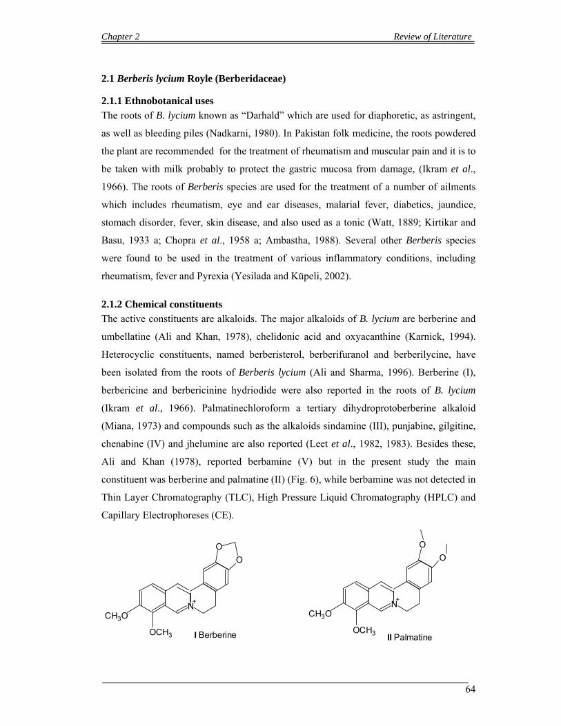
Chapter 2 Review of Literature
64
2.1 Berberis lycium Royle (Berberidaceae)
2.1.1 Ethnobotanical uses
The roots of B. lycium known as “Darhald” which are used for diaphoretic, as astringent,
as well as bleeding piles (Nadkarni, 1980). In Pakistan folk medicine, the roots powdered
the plant are recommended for the treatment of rheumatism and muscular pain and it is to
be taken with milk probably to protect the gastric mucosa from damage, (Ikram et al.,
1966). The roots of Berberis species are used for the treatment of a number of ailments
which includes rheumatism, eye and ear diseases, malarial fever, diabetics, jaundice,
stomach disorder, fever, skin disease, and also used as a tonic (Watt, 1889; Kirtikar and
Basu, 1933 a; Chopra et al., 1958 a; Ambastha, 1988). Several other Berberis species
were found to be used in the treatment of various inflammatory conditions, including
rheumatism, fever and Pyrexia (Yesilada and Küpeli, 2002).
2.1.2 Chemical constituents
The active constituents are alkaloids. The major alkaloids of B. lycium are berberine and
umbellatine (Ali and Khan, 1978), chelidonic acid and oxyacanthine (Karnick, 1994).
Heterocyclic constituents, named berberisterol, berberifuranol and berberilycine, have
been isolated from the roots of Berberis lycium (Ali and Sharma, 1996). Berberine (I),
berbericine and berbericinine hydriodide were also reported in the roots of B. lycium
(Ikram et al., 1966). Palmatinechloroform a tertiary dihydroprotoberberine alkaloid
(Miana, 1973) and compounds such as the alkaloids sindamine (III), punjabine, gilgitine,
chenabine (IV) and jhelumine are also reported (Leet et al., 1982, 1983). Besides these,
Ali and Khan (1978), reported berbamine (V) but in the present study the main
constituent was berberine and palmatine (II) (Fig. 6), while berbamine was not detected in
Thin Layer Chromatography (TLC), High Pressure Liquid Chromatography (HPLC) and
Capillary Electrophoreses (CE).
N
O
O
+
OCH3
CH3O
I Berberine
N
O
O
+
OCH3
CH3O
II Palmatine
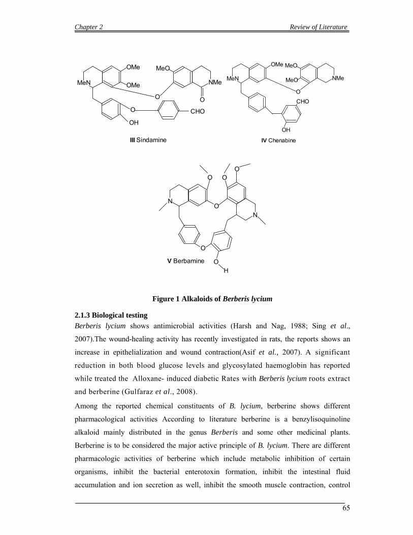
Chapter 2 Review of Literature
65
MeN NMe
MeOOMe
III Sindamine
OMe
O
O CHO
O
OH
MeN
O
NMe
CHO
MeO
MeO
OMe
OH
IV Chenabine
ON
OOO
N
O
O
H
V Berbamine
Figure 1 Alkaloids of Berberis lycium
2.1.3 Biological testing
Berberis lycium shows antimicrobial activities (Harsh and Nag, 1988; Sing et al.,
2007).The wound-healing activity has recently investigated in rats, the reports shows an
increase in epithelialization and wound contraction(Asif et al., 2007). A significant
reduction in both blood glucose levels and glycosylated haemoglobin has reported
while treated the Alloxane- induced diabetic Rates with Berberis lycium roots extract
and berberine (Gulfaraz et al., 2008).
Among the reported chemical constituents of B. lycium, berberine shows different
pharmacological activities According to literature berberine is a benzylisoquinoline
alkaloid mainly distributed in the genus Berberis and some other medicinal plants.
Berberine is to be considered the major active principle of B. lycium. There are different
pharmacologic activities of berberine which include metabolic inhibition of certain
organisms, inhibit the bacterial enterotoxin formation, inhibit the intestinal fluid
accumulation and ion secretion as well, inhibit the smooth muscle contraction, control

Chapter 2 Review of Literature
66
and minimizes the inflammation, inhibit the aggregation of platelet, elevate platelet count
in certain types of hrombocytopenia, stimulate the secretion of bile and bilirubin, and also
inhibit the of ventricular tachyarrhythmias (Birdsall et al., 1997; Akhter et al., 1979).
Diarrhea caused by Vibrio cholera and Escherichia coli has been the focus of numerous
berberine studies, and results indicate several mechanisms which may explain its ability
to inhibit bacterial diarrhea. An animal study found berberine reduced the intestinal
secretion of water and electrolytes induced by cholera toxin (Swabb et al., 1981). Other
studies have shown berberine directly inhibits some V. cholera and E. coli enterotoxins
(Sack and Froelich, 1982). It significantly reduces smooth muscle contraction and
intestinal motility (Akhter et al., 1979) and delays intestinal transit time in humans (Yuan
et al., 1994).
Berberine sulfate has also been found to be directly bacteriocidal to V. cholera (Amin et
al., 1969). In a report about it affect on E. coli, berberine sulfate was used in vitro
research which shows the bacterial inhibition of adherence to epithelial or mucosal
surfaces, the first step in the infective process. The over all effect may be due to the
berberine’s inhibitory activity on fimbrial structure formation on the surface of the treated
bacteria (Sun et al., 1988). Growth of some organism like Entamoeba histolytica, Giardia
lamblia, Trichomonas vaginalis and Leishmania donovani were positively inhibited by
berberine extracts and its salts (Kaneda et al., 1991; (Ghosh et al., 1985). It has also be
studied that the crude extracts of berberine are more effective than the salts of berberine
(Kaneda et al., 1990). In tropical climates Giardia lamblia infestation (giardiasis) is a
common occurrence, particularly in pediatric populations (Nair, 1970). In India, it has
been concluded after various clinical trials that berberine administration positively
improved gastrointestinal symptoms and reduction is occur in Giardia-positive stools. In
comparison to metronidazole (Flagyl), another popular giardiasis medication, Berberine
was nearly as effective at half the dose (Choudhry et al., 1979).
The in vitro and in vivo studies of berberine’s effects on Entamoeba histolytica indicated
berberine sulfate was rapidly amoebicidal and caused encystation, degeneration, and
eventual lyses of the trophozoite forms (Subbaiah and Amin, 1967). Berberine sulfate
rapidly inhibited the growth of Trichomonas vaginalis via formation of large autophagic
vacuoles that eventually result in lysis of the trophozoite forms (Kaneda et al., 1991).
Studies have shown berberine markedly decreased parasitic load and rapidly improved
hematologic parameters in infected animals. In vitro results indicated berberine inhibited
multiplication, respiration, and macromolecular biosynthesis of amastigote forms of the
Chapter 2 Review of Literature
67
parasite, interfered with the nuclear DNA of the promastigote form, and inhibited
organism maturation (Ghosh et al., 1985).
Aqueous berberine and sulfacetamide were both studied in a clinical trial against
Chlamydia trachomatis infection which was conducted on 51 subjects in an outpatient
eye clinic. It was concluded that while sulfacetamide eye drops gave some better clinical
results, while conjunctival scrapings of the patient under investigation were remained
positive for the infective agent and relapses occurred. While in case of, the conjunctival
scrapings of patients that intake the berberine chloride eye drops were found negative for
C. trachomatis and the relapses were also negative up to one year after treatment. It was
further studied that the berberine chloride had no direct anti-chlamydial properties, but it
is possible that it treated the infection by stimulating some protective mechanism in the
host (Babbar et al., 1982). In another clinical study it was found that berberine chloride is
better than sulfacetamide in both the clinical course of trachoma and in achieving drop
inserum antibody titers against C. trachomatis (Khosla et al., 1992).
Berberine administration were studied in both clinical trials and animal research, it was
found that it prevented ischemiainduced ventricular tachyarrhythmia, stimulated cardiac
contractility, and lowered both blood pressure and peripheral vascular resistance (Chun et
al., 1978; Marin-Neto et al., 1988). The mechanism for berberine’s antiarrhythmic effect
is unclear, but an animal study indicated it may be due to suppression of delayed after-
depolarization in the ventricular muscle (Wang et al., 1994). An animal study suggested,
in addition to affecting several other parameters of cardiac performance, berberine may
have a vasodilatory / hypotensive effect attributable to its potentiation of acetylcholine
(Chun et al., 1978). In vitro studies utilizing human cell lines demonstrated that berberine
inhibited activator protein 1 (AP-1), a key transcription factor in inflammation and
carcinogenesis (Fukuda et al., 1999). Another study, utilizing human peripheral
lymphocytes, showed berberine to exert a significant inhibitory effect on lymphocyte
transformation, concluding that its anti-inflammatory action may be due to inhibition of
DNA synthesis in activated lymphocytes (Ckless et al., 1995). A third study concluded
that during platelet activation in response to tissue injury, berberine had a direct affect on
several aspects of the inflammatory process. It exhibited dose-dependent inhibition of
arachidonic acid release from cell membrane phospholipids, inhibition of thromboxane
A2 from platelets (Huang et al., 1991) and inhibition of thrombus formation (Wu and Liu,
1995).

Chapter 2 Review of Literature
68
Berberine has demonstrated a number of other beneficial effects, including
immunostimulation because berberine increased blood flow to the spleen, activated of
macrophage, rising of platelet numbers in cases of primary and secondary
thrombocytopenia, and the excretion of conjugated bilirubin are increased in experimental
hyperbilirubinemia (Birdsall et al., 1997). The anticancer properties of berberine against
cancer cells established from cervical, esophageal, oral, colonic, prostate cancers,
leukemia melanoma and glioblastoma are known by different studies (Iizika et al., 2000;
Jantova et al., 2003, 2006; Kuo et al., 1995; Letasiova et al., 2006; Li et al., 2000; Lin et
al., 2006a, 2006b, 2007; Mantena et al., 2006a, 2006b; Piyanuch et al., 2007., Sanders et
al., 1998; Serafim et al., 2007; Zhang et al., 1990; Katiyar et al., 2008). Berberine was
studied in different assays it is concluded that it inhibits tumor cell growth via inducing
cell cycle arrest and/or apoptosis, and the expression pattern of genes which is responsible
for the regulation of cell cycle progression and apoptosis was correlated to the inhibition
of cellular proliferation. The activity of berberine against tumor cells may vary depending
on the duration of treatment , amount of dose and type of cancer cells, (Iizika et al., 2000;
Jantova et al., 2003, 2006; Kuo et al., 1995; Letasiova et al., 2006; Li et al., 2000a; Lin et
al., 2006a, 2006b, 2007; Mantena et al., 2006a, 2006b; Piyanuch et al., 2007; Sanders et
al., 1998; Serafim et al., 2007; Zhang et al., 1990). It has been studied the effect of
berberine on non-small cell lung cancer cells and concluded that the growth inhibition of
the cells were mediated by p53 (Zhang et al., 1990). But still it is under investigation that
how berberine initiates the cascade that eventually leads to cell cycle arrest and/or
apoptosis and it suggested in some studies that berberine may interfere with DNA
replication as a topoisomerase I inhibitor (Gatto et al., 1996; Kobayashi et al., 1995),
while in some others experiment it showed that berberine may cause directly DNA
damage (Krey and Hahn, 1969; Davidson et al., 1977; Li et al., 2000b; Ihmels et al.,
2005; Letasiova et al., 2006). A very recent study addressed the molecular mechanisms
of Berbeine-induced cell cycle arrest and apoptosis in osteosarcoma cells. The authors
concluded that Berberine inhibited osteosarcoma cell proliferation through its
genotoxicity causing p53-dependent G1 arrest and apoptosis, whereas G2 arrest was p53-
independent (Liu et al., 2009). In the present investigation studying extracts of B. lycium
and its main constituent, Berberine, we discovered another growth inhibitory mechanism
that did not involve genotoxicity, but the inactivation of Cdc25A and the acetylation of a-
tubulin reminiscent to the anti-neoplastic mechanism of taxol.The doses usage of
berberine in clinical situations is not considered toxic and cytotoxic or mutagenic. High
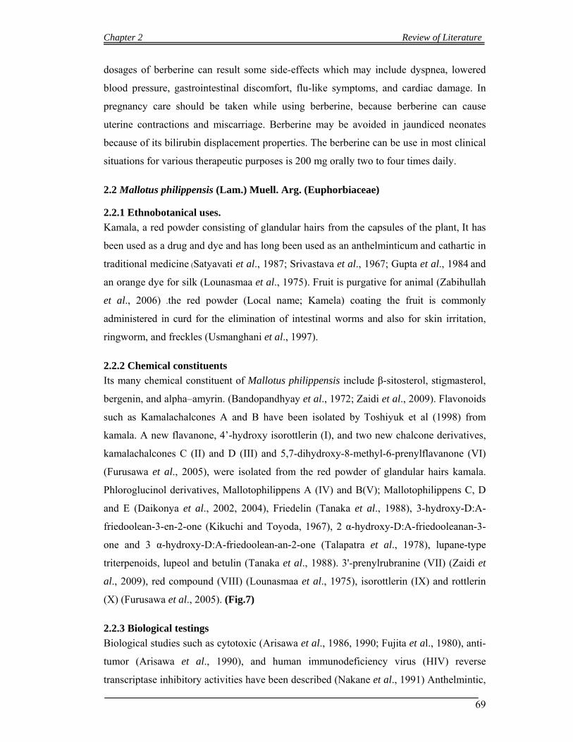
Chapter 2 Review of Literature
69
dosages of berberine can result some side-effects which may include dyspnea, lowered
blood pressure, gastrointestinal discomfort, flu-like symptoms, and cardiac damage. In
pregnancy care should be taken while using berberine, because berberine can cause
uterine contractions and miscarriage. Berberine may be avoided in jaundiced neonates
because of its bilirubin displacement properties. The berberine can be use in most clinical
situations for various therapeutic purposes is 200 mg orally two to four times daily.
2.2 Mallotus philippensis (Lam.) Muell. Arg. (Euphorbiaceae)
2.2.1 Ethnobotanical uses.
Kamala, a red powder consisting of glandular hairs from the capsules of the plant, It has
been used as a drug and dye and has long been used as an anthelminticum and cathartic in
traditional medicine (Satyavati et al., 1987; Srivastava et al., 1967; Gupta et al., 1984 and
an orange dye for silk (Lounasmaa et al., 1975). Fruit is purgative for animal (Zabihullah
et al., 2006) .the red powder (Local name; Kamela) coating the fruit is commonly
administered in curd for the elimination of intestinal worms and also for skin irritation,
ringworm, and freckles (Usmanghani et al., 1997).
2.2.2 Chemical constituents
Its many chemical constituent of Mallotus philippensis include β-sitosterol, stigmasterol,
bergenin, and alpha–amyrin. (Bandopandhyay et al., 1972; Zaidi et al., 2009). Flavonoids
such as Kamalachalcones A and B have been isolated by Toshiyuk et al (1998) from
kamala. A new flavanone, 4’-hydroxy isorottlerin (I), and two new chalcone derivatives,
kamalachalcones C (II) and D (III) and 5,7-dihydroxy-8-methyl-6-prenylflavanone (VI)
(Furusawa et al., 2005), were isolated from the red powder of glandular hairs kamala.
Phloroglucinol derivatives, Mallotophilippens A (IV) and B(V); Mallotophilippens C, D
and E (Daikonya et al., 2002, 2004), Friedelin (Tanaka et al., 1988), 3-hydroxy-D:A-
friedoolean-3-en-2-one (Kikuchi and Toyoda, 1967), 2 α-hydroxy-D:A-friedooleanan-3-
one and 3 α-hydroxy-D:A-friedoolean-an-2-one (Talapatra et al., 1978), lupane-type
triterpenoids, lupeol and betulin (Tanaka et al., 1988). 3'-prenylrubranine (VII) (Zaidi et
al., 2009), red compound (VIII) (Lounasmaa et al., 1975), isorottlerin (IX) and rottlerin
(X) (Furusawa et al., 2005). (Fig.7)
2.2.3 Biological testings
Biological studies such as cytotoxic (Arisawa et al., 1986, 1990; Fujita et al., 1980), anti-
tumor (Arisawa et al., 1990), and human immunodeficiency virus (HIV) reverse
transcriptase inhibitory activities have been described (Nakane et al., 1991) Anthelmintic,
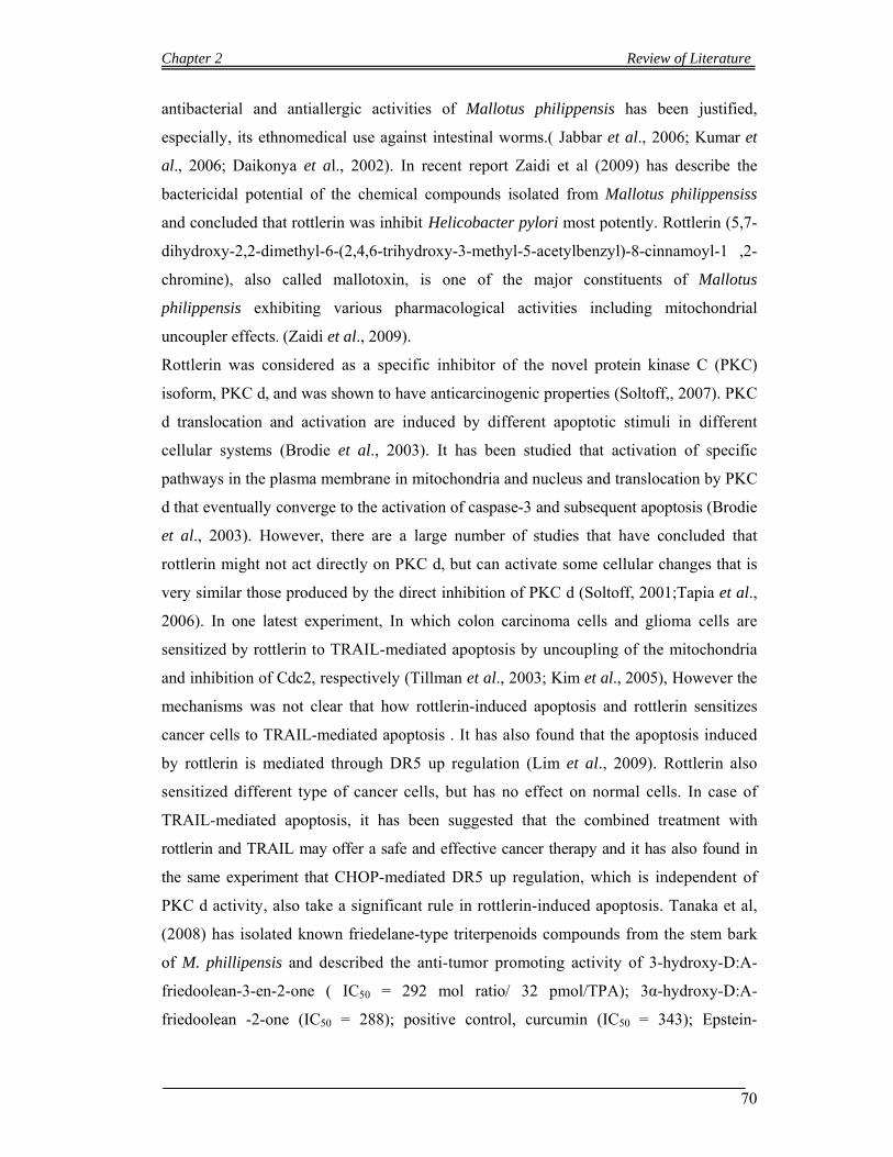
Chapter 2 Review of Literature
70
antibacterial and antiallergic activities of Mallotus philippensis has been justified,
especially, its ethnomedical use against intestinal worms.( Jabbar et al., 2006; Kumar et
al., 2006; Daikonya et al., 2002). In recent report Zaidi et al (2009) has describe the
bactericidal potential of the chemical compounds isolated from Mallotus philippensiss
and concluded that rottlerin was inhibit Helicobacter pylori most potently. Rottlerin (5,7-
dihydroxy-2,2-dimethyl-6-(2,4,6-trihydroxy-3-methyl-5-acetylbenzyl)-8-cinnamoyl-1 ,2-
chromine), also called mallotoxin, is one of the major constituents of Mallotus
philippensis exhibiting various pharmacological activities including mitochondrial
uncoupler effects. (Zaidi et al., 2009).
Rottlerin was considered as a specific inhibitor of the novel protein kinase C (PKC)
isoform, PKC d, and was shown to have anticarcinogenic properties (Soltoff,, 2007). PKC
d translocation and activation are induced by different apoptotic stimuli in different
cellular systems (Brodie et al., 2003). It has been studied that activation of specific
pathways in the plasma membrane in mitochondria and nucleus and translocation by PKC
d that eventually converge to the activation of caspase-3 and subsequent apoptosis (Brodie
et al., 2003). However, there are a large number of studies that have concluded that
rottlerin might not act directly on PKC d, but can activate some cellular changes that is
very similar those produced by the direct inhibition of PKC d (Soltoff, 2001;Tapia et al.,
2006). In one latest experiment, In which colon carcinoma cells and glioma cells are
sensitized by rottlerin to TRAIL-mediated apoptosis by uncoupling of the mitochondria
and inhibition of Cdc2, respectively (Tillman et al., 2003; Kim et al., 2005), However the
mechanisms was not clear that how rottlerin-induced apoptosis and rottlerin sensitizes
cancer cells to TRAIL-mediated apoptosis . It has also found that the apoptosis induced
by rottlerin is mediated through DR5 up regulation (Lim et al., 2009). Rottlerin also
sensitized different type of cancer cells, but has no effect on normal cells. In case of
TRAIL-mediated apoptosis, it has been suggested that the combined treatment with
rottlerin and TRAIL may offer a safe and effective cancer therapy and it has also found in
the same experiment that CHOP-mediated DR5 up regulation, which is independent of
PKC d activity, also take a significant rule in rottlerin-induced apoptosis. Tanaka et al,
(2008) has isolated known friedelane-type triterpenoids compounds from the stem bark
of M. phillipensis and described the anti-tumor promoting activity of 3-hydroxy-D:A-
friedoolean-3-en-2-one ( IC50 = 292 mol ratio/ 32 pmol/TPA); 3α-hydroxy-D:A-
friedoolean -2-one (IC50 = 288); positive control, curcumin (IC50 = 343); Epstein-
Chapter 2 Review of Literature
71
Barrvirus early antigen (EBV-EA) activation induced by 12-O-tetradecanoyl phorbol 13-
acetate (TPA) used in the experiment.
O O
O
O
OH
OH
OH
HO OH
Me
Me
Me
Me
I 4'-Hydroxyisorottlerin
O
O
Me
Me
Me
MeMe
OH
OH
OH
HO
O
O
H
H
II Kamalachalcones C
O
O
O
O
O
HO
MeMe
MeMe
MeMe
MeMe
Me
OH
OH
OH
HO
HO
O
O
OO
HO
III Kamalachalcone D
OH OHO HO
OHOH
Me
Me
Me
MeMe
OCO Pr i
IV Mallotophilippen A
OH OHO HO
OHOH
Me
Me
Me
MeMe
OCO
V Mallotophilippen B
Me
O
OOH
HO
VI 5,7-dihydroxy-8 methyl-6-prenylflavanone�
Chapter 2 Review of Literature
72
O
O
O
OH
VII 3 -prenylrubranine
O
OOH
HO
VIII Red compound
O O
HO OH
OH
OH
O
O
IX Isorottlerin
Figure 2 Compounds of Mallotus philippensis
2.3 Adhatoda vasica Nees (Acanthaceae)
2.3.1 Ethnobotanical uses
Adhatoda vasica is widely used in the Ayurvedic and Unani system of medicine for
treating bronchitis, asthma, fever and jaundice on account of the antispasmodic properties
of roots and leaves. A. vasica has also been used for the treatment of bronchial
obstruction which is created by allergen (Sharma et al., 1999; Amin and Mehta, 1959). It
has been used for asthma and tuberculosis (Dorsch and Wagner, 1991; Paliwa et al.,
2000; Barry et al., 1955; Grang et el., 1996; Gupta et al., 1954).
2.3.2 Chemical constituents
The chemical examination of Adhatoda vasica revealed to contain different types of
alkaloids, glycosides, different phenolic compounds and sterols components (Lateef et al.,
2003). The major chemically active components identified are two alkaloids: vasicinone
and vasicine (Das et al., 2005). Some minor alkaloids viz. Vasicol, adhatodinine and
vasicinol also present. The leaves contain an alkaloid vasicine and an essential oil.
2.3.3 Biological testing
Adhatoda vasica possesses hepatoprotective activity (Bhattacharyya et al., 2005). It
possesses antioxidant, chemopreventive agent and antibacterial (Karthikeyan et al.,2009).
It has the tendency to restore the hematological changes produced by irradiation in Swiss
albino mice (Kumar et al., 2005). The activities of Glutathione S-transferase are enhanced
in the liver of mice. A. vasica reduced glutathione (GSH) levels in liver, and also reduced
lipid peroxidation(LPO). It also reduced the acid and alkaline phosphatases in testis of
Chapter 2 Review of Literature
73
normal and irradiated mice (Singh et al., 2000). Leaves of Adhatoda vasica possess
anticestodal efficacy (Yadav and Tangpu, 2008). Due to alkaloids such as vasicine and
vasicinone it possesses the biological activities such as expectorant and mild bronchial
antispasmodic. (Lahiri and Pradhan, 1964; Gupta et al., 1971).
2.4 Albizia lebbeck (L.) Benth. (Mimosaceae)
2.4.1 Ethnobotanical uses
The wood of Albizia lebbeck is very similar to walnut and therefore very good for canoes,
furniture, house building, and picture frames, etc. the wood of A. lebbeck is also used for
cane crushers, oil. In the Ayurveda both the leaves and the bark of A. lebbeck have been
in clinical use for centuries. Spongy and ulcerative gums have been strengthening with
the powder of the bark of the roots. The leaves Juice are very useful in ophthalmia. The
leaves decoction are useful for night-blindness and therefore given internally. Bark is
astringent and is employed in diarrhea, dysentery, and hemorrhoids. Powdered bark is
useful for ulcers, and especially for snake wounds flowers possess the power of causing
retention of the seminal fluid. Seeds are astringent and are employed in diarrhea,
dysentery and hemorrhoids. The seeds are also used for ophthalmic diseases. The oil from
the seeds is considered useful in leprosy. The seeds are crushed and made into a paste,
which is applied to reduce enlarged cervical glands. Bronchial asthma is being treated
with the decoction of stem bark. Bark of A. lebbeck used as antifertility drugs (Shah et al.,
2009).
2.4.2 Chemical constituents
The leaves of Albizia lebbeck are good source of saponins (Pal et al., 1995).
2.4.3 Biological testing
The leaves of A. lebbeck, which contain different type of saponins, and possessed
nootropic activity (Chintawar et al., 2002), anticonvulsant activity (Kasture et al., 2000).
Antiasthmatic and antianaphylactic activity of A. lebbeck have been reported (Tripathi
and Das, 1977). Tripathi et al (1979) have conclude that A. lebbeck is not like disodium
chromoglycate, it exerts antianaphylactic activity in guinea pigs. A. lebbeck also enhance
the concentration of plasma cortisol level in patients of bronchial asthma (Tripathi et al.,
1978). Albizia lebbeck showed antioxidant activities in alloxan-induced diabetic rats
(Resmi et al., 2006).
Chapter 2 Review of Literature
74
2.5 Bauhinia variegata Linn. (Caesalpinaceae)
2.5.1 Ethnobotanical uses
Bauhinia variegata generally cultivated as an ornamental plant. The leaves are given to
cattle as fodder, flowers are used as pot herb and also made into pickles; wood is useful in
buildings and for making agricultural goods. The plant yields gum; the bark is useful for
tanning and dyeing. The plant is reputed to have medicinal properties also. The root is
tonic and carminative, the flowers laxative and the bark is astringent; various parts of the
plant are reputed to have healing properties also. In the traditional medicines of South
East Asia the same is used for skin diseases, as an astringent, bronchitis, tonic, leprosy,
anti inflammatory and for ulcers (Kirtikar and Basu, 1993). The roots decoction is useful
in dyspepsia and also used as an antidote to snake poison (Thammanna et al., 1990).
2.5.2 Chemical constituents
Several flavonoids have been isolated during the phytochemical studies on the stems,
bark, flowers and seeds (Gupta et al., 1979, 1980, 1984; Rahman and Begum, 1966;
Wahab et al., 1987;Yadava and Reddy, 2001). The non-woody aerial parts of B. variegata
were studied and isolated six flavonoids, and a triterpene caffeate, ( Rao et al., 2008).
Phenanthraquinone, named bauhinione has been isolated from Bauhinia variegate (Zhao
et al., 2005).
2.5.3 Biological testing
The non-woody aerial parts of B. variegate yield anti-inflammatory agents. It shows
insuline secretion activity from INS-1 cells. The ethanolic extract of B. variegate at the
rate of 250 mg/kg positively suppressed liver tumor (Rajkapoor et al., 2006). It has been
determined the anthelmintic activity of the leaves. (Sing et al., 2005).
2.6. Bombax ceiba Linn. (Bombacaceae)
2.6.1 Ethnobotanical uses
The cotton inside the fruits was used a substitute of cotton. Various parts of plant are used
in small pox, bleeding gums, toothache, carries, sores in mouth, pain in leg, fever,
enlarged spleen, atrophy, emaciation, rheumatism, spermatorrhoea, cholera, pneumonia,
pleurisy, intestinal neuralgia, leprosy and skin diseases (Ansari, 2004). The flower was a
common ingredient in Chinese herb tea. The gum has aphrodisiac, astringent, demulcent,
haemoptysis of pulmonary tuberculosis and influenza, malaena and menorrhagia and
Chapter 2 Review of Literature
75
acute dysentery with beneficial results. Flowers are used for haemorrhoids. Root has
stimulant, tonic and aphrodisiac properties. Plants are used for making light packing
boxes and in fisherman floats. In Punjab it is used for making water conduits, troughs and
bridges, the timber is also utilized in match industry. Buds are used as vegetables.
2.6.2 Chemical constituents
Preliminary tests show the presence of glycosides and tannins from root, stem and leaf. In
the stem some alkaloids and in root proteins are identified (Mehra, 1968). The stem bark
contains lupeol and b-sitostrol (Mukherjee, 1971). The root bark has 3 naphthalene
derivatives related to gossypol (toxic principle of cotton seed) and called as
'semigossypol' (Seshadri, 1973). Flowers contain b-sitosterol, traces of essential oil,
kaempherol and Quercetin (Gopal, 1972). On hydrolysis gum yield arabinose, galactose,
galacturonic acid and rhamnose.
2.6.3 Biological testing
Aqueous extract has moderate oxytoxic activity on gravid and non-gravid isolated rat
uteri and guinea pig and rabbit uterine strips. It has musculotrapic action in guinea pig
ileum and cardiac stimulant action on frog's heart (Misra, 1968). It has a negligible blood-
pressure elevating action in anaesthetized dog (Misra, 1966).
2.7 Calotropis procera Linn. (Asclepiadaceae)
2.7.1 Ethnobotanical uses
Calotropis procera is a medicinal plant. The latex is irritant to the skin and mucous
membrane and said to cause blindness. It is also used as a purgative and said to be
specific for Guinea worms. The seed floss is used for stuffing mattresses, pillows etc. It is
sometimes used to adulterate Indian Kapok but it is inferior to it in resilience and water
repellent properties. In Indian traditional system of medicine the different parts of the C.
procerat have been used for the treatment of various diseases such as ulcers, leprosy,
piles, tumors, and certain disease of abdomen, liver and spleen (Kirtikar and Basu, 1935).
C. procera (root) are useful carminative drug in the treatment of dyspepsia (Kumar and
Arya, 2006). Various tribes of central India use C. procera (root bark and leaves) as a
useful drug for jaundice, a curative agent and as antidote for snake poisoning (Samvatsar,
2000; Nandkarni,1976).
Chapter 2 Review of Literature
76
2.7.2 Chemical constituents
The latexes of C. procera are a rich source of many biologically active constituents that
include some glucosides, different tannins and proteins (Wititsuwannakul et al., 2002;
Dubey and Jagannadham, 2003). Alkaloids, cardiac glycosides, tannins, flavonoids,
sterols and triterpenes has been reported (Mossa et al., 1991). Singh and Rastogi reported
Calactin, Calotropin and Uscharidin were formed by substitution of glycosides at C-1 of
4, 6 deoxy sugar (Singh and Rastogi, 1970), Calotropin isolated from leaves and stalkes
(Perry and Metzger, 1980), toxic flavonoids have also been reported (Salunke et al.,
2005).
2.7.3 Biological testing
According to the study of Choedon et al, the aqueous extract of C. procera (latex) has
inhibit cellular infiltration and concluded that it afford protection against development of
neoplastic changes while using the transgenic mouse model of hepatocellular carcinoma
(Choedon et al., 2006). The roots of C. procera have been extracted with chloroform and
studied the protective activity against carbon tetrachloride induced liver damage which
shows a significance protective result. (Basu et al., 1992). Methanol extract possess
antioxidant activity in Trema orientalis (Uddin et al 2008) and Senna tora (Uddin et al
2008a). C. procera latex is also reported to possess very interesting unrelated activities
such as the ability to combat diarrhea or retard insect larval development and (Kumar et
al., 2001, 1994; Morsy et al., 2001). Chloroform extract of roots has been reported to
possess anti-inflammatory activity (Kumar and Basu, 1994; Basu and Chaudhuri, 1991).
Aqueous extract of the flowers has been found to exhibit analgesic, antipyretic and anti-
inflammatory activity (Mascolo et al., 1988).The alcoholic extract from different parts
has been found to possess antimicrobial and spermicidal activity (Kishore et al., 1997;
Qureshi et al., 1991). Alcoholic roots extract possesses a very strong antiimplantation
activity Ž100%.which may be due to its estrogenic activity (Jagadish et al., 2002).
Laticifer proteins (LP) recovered from the latex of the medicinal plant. Calotropis
procera, is a target for DNA topoisomerase I triggering apoptosis in cancer cell lines.
(Oliveira et al., 2007). Calotropis procera flowers possess hepatoprotective activity
(Ramachandra et al., 2007).
2.8 Carissa opaca Stapf ex Haines (Apocynaceae)
Chapter 2 Review of Literature
77
2.8.1 Ethnobotanical uses
Fresh leaves of Carissa opaca and roots of Sageretia brandrethiana are boiled in water
and used in case of Jaundice and Hepatitis, the decoction is taken orally, approximately
one cup twice a day for two to three weeks (Abbasi et al., 2009). Fruit is edible.
2.8.2 Chemical constituent
Carissone, palmatic acid, benzyl salicylate, benzyl benzoate, farnesene (Rai et al., 2005)
2.9 Cassia fistula Linn. (Caesalpinaceae)
2.9.1 Ethnobotanical uses
Cassia fistula is an ornamental tree, the bark is used as tanning material and wood ash is
used as mordant in dyeing. The pulp of pods is used in Bengal to flavour tobacco. The
durable wood is used for various purposes. The different parts of the plant are also
reported to have medicinal properties. It is also useful in the treatment of different skin
diseases, rheumatism, inflammatory diseases, anorexia and jaundice (Anonymous,1992;
Kirtikar and Basu 1991).
2.9.2 Chemical constituents
A flavone glycoside 5,3',4'-tri-hydroxy-6-methoxy-7-O-alpha-L-rhamnopyranosyl-(1 -->
2)-O-beta-D-galactopyranoside with antimicrobial activity was reported by (Yadava and
Verma, 2003). Compounds, 5-(2-hydroxyphenoxymethyl) furfural, (2'S)-7-hydroxy-5-
hydroxymethyl-2(2'-hydroxypropyl)chromone,benzyl-2-hydroxy-3,6-dimethoxybenzoate,
and benzyl 2beta-O-D-glucopyranosyl-3,6-dimethoxybenzoate, 5-hydroxymethylfurfural,
(2'S)-7-hydroxy-2-(2'-hydroxypropyl)-5-methylchromone, and two oxyanthraquinones,
chrysophanol and chrysophanein, were isolated from the seeds of Cassia fistula. (Kuo et
al., 2002)
2.9.3 Biological Testing
Luximon-Ramma et al (2002) has reported that antioxidant activities correlated to the
total Phenolic compounds. The hepatoprotective activity (Bhakta et al., 1999; Bhakta et
al., 2001) and the hypoglycemic activity (Esposito Avella et al., 1991) have been
reported.
2.10 Colebrookea oppositifolia Smith (Labiateae)
Chapter 2 Review of Literature
78
2.10.1 Ethnobotanical uses
In traditional medicine, the epilepsy diseases is treated with the roots of C. oppositifolia
and the leaves of the same plants are used for the healing of wounds and bruises (Chopra
et al., 1956)
2.10.2 Chemical constituents
Several flavone and flavone glycosides have isolated from the bark, stems, leaves, and
flowers of C. oppositifolia. (Ahmed et al., 1974; Patwardhan et al., 1981; Yang et al.,
1996).
2.11 Debregeasia salicifolia (D.Don) (Urticaceae)
2.11.1 Ethnobotanical uses
A strong fiber used to make ropes, is obtained from the bark.
2.11.2 Chemical constituent
Akber et al. (2001) reported quercetin, hisperidine, 3b-19alpha-dihydroxy-30-norurs-12-
ene,b-sitosterol, stigmasterol, oleanolic acid and lupeol in extracts of Debregeasia
salicifolia
2.11.3 Biological Testing
Ahmed et al. (2006) has identified that leaves extracts has IC50 values more than 100
µg/ml of DPPH radical scavenging activity while comparing with Ascorbic acid IC50 =
1.75
2.12 Dalbergia sissoo Roxb. (Papilionaceae)
2.12.1 Ethnobotanical uses
Plants of the genus Dalbergia are medicinally important and have been used for the
treatment of gonorrhoea, arthritis, and rheumatic pains (Anonymous. 1950; Nadkarni,
1982; Singh and Chaturvedi, 1966). It has been reported in folk medicine and is used
mainly as aphrodisiac, abortifacient, expectorant, anthelmintic and antipyretic. It is also
used in conditions like emesis, ulcers, leucoderma, dysentery, stomach troubles and skin
diseases. (Kirtikar and Basu, 1933 b; Nadkarni. 1954; Chopra et al., 1956 b). In Arabic
countries the aqueous leaves extract of D. sissoo has been used for the treatment of
Chapter 2 Review of Literature
79
gonorrhea (El-Dagwy, 1996).The hard wood D. sissoo which is very heavy and durable,
widely used for the manufacturing of boats furniture, wheels and carts, etc.
2.12.2 Chemical constituents
Phytochemical examination of genus Dalbergia has provided a large number of
compounds, which include flavonoids, furans, benzophenones, styrenes, and terpenoids
(Chawla and Chibber, 1981; Khan and Javed, 1997). Chemical constituent of D. Sissoo
have also been studied before (Ahluwalia et al., 1965; Ahluwalia et al., 1963; Banerji et
al., 1965, 1966; Banerji et al., 1963; Dhingra et al., 1974; Farag et al., 2001; Mukerjee et
al., 1971; Ramakrishna et al., 2001;Sharma et al., 1979a, 1979b, 1980a, 1980b). Two
new isoflavones glycosides along with other five known isoflavone glycosides have been
isolated from the leaves and stem bark by D. sissoo Salwa et al (2001). It has been
isolated several type of chemicals constituent from the green branches and aerial parts of
D. sissoo Roxb, such as biochanin-A, tectorigenin, isoflavones irisolidone, prunetin,
muningin, genestein, the flavone nor-artocarpotin, sissotrin and prunetin-4- O -
galactoside stigmasterol, ß-sitosterol and ß-amyrin (Kinjo et al., 1987; Ishikura et al.,
1989; Rao et al., 1989; Ramesh and Yuvarajan, 1995; Mathias et al., 1998). Sarg et al
(1999) and Labreque, (1983) reported the composition of the fatty acids in the fixed oil
which are myristic 5.56%, palmitic 21.70%, stearic 24.33%, arachidic 19.37%, linoleic
10.81% and oleic 9.4% (Labreque, 1983; The Wealth of India, 1988)
2.12.3 Biological testing
Hajare et al (2000) reported marked antipyretic and moderate analgesic activities by
Dalbergia sissoo leaves. Ethanolic extract of D. sissoo leaves possesses anti-
inflammatory activity (Hajare, 2001).The oil also showed strong repellent action (Ansari
et al., 2000). A dose-dependent inhibitory effect have been shown by the alcohol extract
of the green aerial parts on the motility of isolated rabbit duodenum, pronounced
bronchodilation and also shows a significant antipyretic, anti-inflammatory, analgesic,
and estrogen-like activities. The plant extract was studied with out any side effects in rats.
(Sarg et al.,1999).
2.13 Dodonaea viscosa Linn. (Sapindaceae)
2.13.1 Ethnobotanical uses
Leaves D. viscosa are useful for wounds healing, burns and swellings. It is also used as
febrifuge and is useful in rheumatism. The fruit is used as a fish poison. The decoction
should be used as mouthwash only and should not be swallowed (Qureshi et al., 2008). It

Chapter 2 Review of Literature
80
is useful remedy for the treatment of diarrhea, skin infections and rheumatism. The roots
of D. viscose are used for the treatment of inflammation and spasmodic in traditional
medicine. D. viscosa is used for malaria, wounds and burns (Al-Dubai and Al-khulaidi,
1996). It is also used as an antipuritic in skin rashes and for the treatment of sore throat,
dermatitis and hemorrhoids (Chhabra et al., 1991; Hedberg et al., 1983). In India, the
infusion of leaves were used to treat rheumatism, gout, hemorrhoids, fractures and snake
bites (Kirtikar and Basu, 1995; Nadkarni and Nadkarni, 1982) .The quick growth and
gregarious habit of this shrub makes it an excellent hedge plant. The branches are used as
fire-wood and as a support for the flat mud roofs in village houses. The wood can be used
for making walking sticks and tool-handles.
2.13.2 Chemical constituents
Aliarin, dodonic acid, viscosol (Sachdev and Kulshreshtha, 1986), stigmosterol,
isorhamnetin (Rao, 1962; Ramachandra et al., 1975), penduletin, quercetin, doviscogenin
(Khan et al., 1988), dodonosides A and B (Wagner et al., 1987) have been isolated from
D. viscose. Flavonoids, terpenes, coumarins and steroids are also reported by Abdel-
Mogib et al (2001) and Ahmad et al (1987).
2.13.3 Biological testing
Literature survey reveals that D. viscosa has an antimicrobial effect (Rojas et al., 1992;
Getie et al., 2004), this plant collected in different countries demonstrates variable
biological activity. The methanol extract of the entire plant collected in Saudi Arabia
possesses no activity against Escherichia coli, Proteus vulgaris, Staphylococcus aureus
and Pseudomonas aeruginosa and Candida albicans (Getie et al., 2003). On the other
hand, a similar extract of the leaves of the Mexican species showed weak activity against
E. coli, P. aeruginosa, S. aureus, Bacillus subtilis and C. albicans (Rojas et al., 1982).The
50% methanol of the flowers and leaves of the Nigerian species demonstrated
antibacterial activity against B. subtilis, E. coli, Proteus species, P. aeruginosa and S.
aureus (Ogunlana and Ramstad, 1975). An antipyretic and an antimicrobial agent has
been reported (Rojas et al., 1992, 1995, 1996; Getie et al., 2003; Ahmad et al., 1994)
The leaves were reported to possess local anesthetic, smooth muscle relaxant (Rojas et al,
1996), antifungal (Al-Yahya et al., 1983; Naovi et al., 1991) anti-inflammatory
(Mahadevan et al., 1998; Getie et al., 2003) and anti-ulcerogenic activity (Veerapur et al.,
2004). Sukkawala and Desai (1962) have reported that 95% ethanol extract of D. viscose
leaves has shown anti-scariasis, anthelmintic, cardiac depressant, hypotensive, uterine
Chapter 2 Review of Literature
81
relaxation and asoconstrictor activity in different experimental models. However the
pharmacopoeial standards of D. viscosa leaves have not been reported.
Khalil et al., (2006) reported that alcoholic extract of D. viscose possess anti-inflamatory
activity without toxic effect. Ramzi et al (2008) reported that the diterpenoid and
flavonoids derivatives (Sachdev and Kulshreshtha, 1984; Abdel-Mogib et al., 2001; Getie
et al., 2002) are mainly responsible for the remarkable antioxidant and antimicrobial
effect of this plant.
2.14 Ficus palmata Forssk. (Moraceae)
2.14.1 Ethnobotanical uses
The fruit is demulcent, emollient, laxative and poultice (Parmar and Kaushal, 1982;
Chopra et al., 1986). It is used as a part of the diet in the treatment of constipation and
diseases of the lungs and bladder (Chopra et al., 1986). The sap is used in the treatment of
warts. Fruit in raw is sweet and succulent (Hedrick, 1972). A very tasty fruit (Parmar. and
Kaushal, 1982), it is often dried for later use. The fruit is about 2.5cm in diameter and
annual yields from wild trees are about 25kg (Parmar. and Kaushal, 1982). The unripe
fruits and young growth are cooked and eaten as a vegetable. They are boiled, the water is
removed by squeezing and they are then fried. Used as a nice green vegetable (Parmar
and Kaushal, 1982). The pliable wood is of little value but has been used for making
hoops, garlands, ornaments.
2.14.2 Chemical constituents
The fruit contains about 6% sugars, 1.7% protein, 0.9% ash and 0.2% pectin (Parmar and
Kaushal, 1982 ). Low in vitamin C, about 3.3mg per 100g (Parmar. and Kaushal, 1982).
2.15 Ficus racemosa L. (Moraceae)
2.15.1 Ethnobotanical uses
The mature fruits are astringent, stomachic and carminative. Traditionally the fruit extract
is used in diabetes, leucoderma and menorrhagia. It is also used locally to relive
inflammation of skin wounds, lymphadenitis. They are eaten by locals people. The wood
is often employed in making cart frames, ploughs, box, fittings, match boxes and cheap
furniture. A decoction of the bark is used as a wash for wounds. The tree is planted for
shade in gardens.

Chapter 2 Review of Literature
82
2.16.2 Chemical constituents
Ficus racemosa is a chemically rich plant and possess glycosides, beta-sitosterol, lupeol,
dumurin, tiglic acid ester, taraxasterol and a new compound racemosic acid (Ghani, 1998;
Li et al., 2004). Jahan et al., (2008) has reported an antioxidant compound 3-O-(E)-
caffeoyl quinate through bio assay guided isolation. A tetra cyclic triterpene, Gluanol
acetate was isolated from bark acetone extract of F. racemosa and identified as new
mosquito larvicidal compound. A new anti-inflammatory glucoside, Racemosic acid
along with Bergenin isolated from Ficus racemosa. Racemosic acid possesses antioxidant
activity (Li et al., 2004).
2.16.3 Biological testing
The F. racemosa fruits are hypoglycaemic and antioxidant activities (Jahan et al, (2008).
The leaves and stem bark also contain hypoglycaemic activity (Baslas & Agha, 1985).
The aqueous bark extract possesses wormicidal activity and useful anthelmintic
(Chandrashekhar et al., 2008). The leaves of F. racemosa also contain antifilarial activity,
antidiuretic, antihepatotoxic, anti-pyretic, anti-inflammatory, Antifungal, analgesic,
antipyretic and hepatoprotective activities (Deranjyagala et al., 1988; Forestieri et al.,
1996; Mandal et al., 1998, 1999, 2000; Mishra et al., 2005; Rao et al., 2003, 2002). The
ethanolic extract of the bark is hypoglycemic and antiprotozoal activity. Decoction of the
bark is used as wash for wounds, in asthma, piles and menorrhagia (Yusuf et al., 1994;
Ghani, 1998). The bark and leaf of F. racemosa were also reported to have significant
antidiuretic activity, wound healing activity, antitussive activity, antinociceptive activity,
anti-pyretic activity, hypoglycemic activity, anti-bacterial activity, hepatoprotective
activity and anti-diarrhoeal activity (Mukherjee et al., 1998; Mandal et al., 1999, 2000;
Rao et al., 2002; Bhaskara et al., 2003; Ratnasooriya et al., 2003, Ferdous et al., 2008).
Khan and Sultana (2005) have reported in renal carcinogenesis induced with ferric
nitrilotriaceatate (Fe-NTA) treated rats and evaluate the chemomodulatory effect by F.
racemosa. KBrO3-mediated nephrotoxicity in rats is significantly suppresses by Ficus
racemosa extract and therefore considered a potent chemo preventive agent (Khan and
Sultana, 2005).
Chapter 2 Review of Literature
83
2.17 Lantana camara Linn. (Verbenaceae)
2.17.1 Ethnobotanical uses
A tea prepared from the leaves and flowers is useful against influenza and stomachache.
The leaves of L. camara are useful for different diseases like swelling or inflammation,
ulcers, catarrhal affection, itches, cuts, bilious fever, rheumatism, eczema eruptions, and
also useful for the treatment of snake-bite. The oil isolated from the leaves is useful
antiseptic for wound healing. The roots and flower are useful for toothache and chest
complaints of children respectively. Diaphoretic, Vulnerary, and carminative properties
are also present in L. camara. Several other activities are present in L. camara such as
treatment of high blood pressure, fistulae, asthma, pustules, tumours and cancers, atoxy of
abdominal viscera, malaria, in tetanus, leprosy, scabies, and bronchitis (Kirtikar and
Basu, 1918; Ghisalberti, 2000; Pullaiah, 2006; Mahathir, 2002; Johns et al., 1983; Begum
et al., 2000; Barua, 1969). The roots of L. camara are useful for the treatment of
gonorrhea. The plant is also useful for Alzheimcr's disease as well. It is a tonic to the
nervous system and used to treat insomnia and epilepsy. It relaxes the muscles, quickens
the senses and strengthens the memory (Siddiqui et al., 1995; Begum et al., 2003).
2.17.2 Chemical constituents
Lantanin by P. G. J. Louw (1943), lantadene B (Barton et al., 54), lantanolic acid (Barua
et al., 1969). Various steroids, terpenoids, and flavonoids have isolated by different
groups from the different parts of the plant (Pullaiah, 2006; Johns et al., 1983; Begum et
al., 2000). lantanoic acid and camaranoic acid, lantic acid (Begum et al., 2008), camarinic
acid (Siddiqui et al., 1995), camangeloyl acid, camarinin (Begum et al., 2003, 2006),
oleanonic acid, and ursonic acid (Siddiqui et al., 2000) lantanolic acid (Begum et al.,
2008, lantanilic acid (Barua et al., 1976, 1985), α-amyrin , β-sitosterol and lantadene B
(Ahmed et al., 1972), Iantoic acid (Roy and Barua, 1985), lantadene D (Sharma et al.,
1990), lantadenes.( Sastry and Mahadevan, 1963; Sharma et al., 2000), lantanolic acid,
oleanolic acid, 22/.-O-angeloyl-oleanolic acid, 22 β-O-senecioyl-oleanolic acid, 22β
hydroxyl-oleanonic acid , 19α-hydroxy ursolic acid and a new triterpenoid 3β-
isovaleroyl-l9α-hydroxy-ursolic acid (lantaiursolic acid) (Pan et al., 1993), camarinic
acid, camaric acid, camarilic acid and camaracinic acid (Siddiqui et al., 1995; Begum et
al., 1995), 25-hydroxy-3-oxolean-12-en-28-oic acid, hederagenin and 19-hydroxyursolic
acid (Singh, et al., 1996), novel trans lactone containing euphane triterpenes A, B and C
(O'Neill et al., 1998), phcnylpropanoid glycosides verbascoside, isoverbascoside,
Chapter 2 Review of Literature
84
isonuomioside A, calceolarioside E and derhamnosylverbascoside (Taoubi et al., 1997),
martynoside and verbascoside (Syah et al., 1998), theveside (Ford and Bcndall, 1980),
2.17.3 Biological testing
Wound-healing property and antihyperglycaemic activities have been reported in the
aqueous extract of the leaves and the shoot of L. camara exhibit antibacterial properties.
A steroid Lancamarone, isolated from the leaves of L. camara, possesses
cardiotonicproperty. The lantamine alkaloid isolated from the bark of stems and roots,
possesses effective antispasmodic and antipyretic properties as such as those of quinine
(Kirtikar and Basu, 1918; Ghisalberti, 2000; Pullaiah, 2006; Mahathir, 2002).
2.18 Melia azedarach Linn. (Meliaceae)
2.18.1 Ethnobotanical uses
Persian Lilac” is a fast growing tree of the plains and foot-hills, cultivated along road-
sides and in villages. The fruit is eaten by goats and sheep, and the stony endocarps are
used as beads. The exuded gum obtained from its trunk is considered useful in spleen
enlargement, its wood extract is prescribed internally in asthma (Dhiman, 2003),
decoction of bark is used in paroxysmal fever to relieve thirst, nausea, vomiting and
general debility, loss of appetite and skin diseases (Sharma et al., 2001).
Leaves are applied in the form of poultice to relieve nerves headach and to cure the
eruption on the scalp. Leaf juice is anthelmintic, diuretic and emmenagouge, expectorant,
vermifuge and their decoction is astringent, stomachic (Warrier et al., 1995; Dhiman,
2003; Sharma et al., 2001), employed in hysteria, they are used internally and externally
in leprosy, scrofula and other skin diseases (Nadkarni, 1954).
Flowers are astringent, anodyne, refrigerent, emmenagouge, diuretic, resolvent,
deobstruent and alexipharmic (Warrier et al., 1995; Sharma et al., 2001). They are
applied as a poultice to relieve nervous headache (Dhiman, 2003 ). They are stomachic
(Zhou et al., 2005), vermicide and valuable in eruptive skin diseases (Nadkarni, 1954)
and for killing lice. Fruits are anthelmintic, emmolient and purgative (Rani et al., 1999).
Fruits are considered tonic. Sushruta prescribed mahanimb fruits internally in indigestion,
colic and intestinal catarrh. Seeds: seeds are bitter, expectorent, anthelmintic and
aphrodiasic, and are useful in helminthiasis, typhoid fever, pain in the pelvic region,
uropathy, vitiated conditions of vata and scrofula (Warrier et al., 1995). They are
prescribed in rheumatism; oil obtained from seeds is applied locally in skin diseases
Chapter 2 Review of Literature
85
(Dhiman, 2003). They are taken with adjuvants like rice water and clarified butter;
ramyak Ghrita of sushurta was a specific remedy for gout. Sharangadhara prescribed
seeds for urinary disorders. Ashroghna vati, a classical compound of 16th centuary, was
prescribed for piles (Khare).
Roots are bitter, astringent, mildly thermogenic, anodyne, depurative, vulnerary,
antiseptic, constipating, expectorant, febrifuge, antiperiodic, urinary astringent,
anthelmintic, emmenagogue and bitter tonic in low doses. They are useful in headache,
sciatica, lumbago, leprosy, leucoderma, skin diseases, wounds, ulcers, piles, worm
infestation, cough, asthma, ammenorrhoea, dysmenorrhoea, diabetes, abnormal urethral
discharge, chronic and intermittent fevers, vomiting, post labour pain in uterus (Warrier et
al., 1995; Sharma et al., 2001).
2.18.2 Chemical constituents
Besides the chemical constituent of stem, fruits and bark, the leaves has been shown to
contain nimbinene, meliacin, quercetrin, quercetin-3-0-b-rutinoside, kaempferol- 3-0-b
rutinoside, rutin and kaempferol-3-L-rhamno-Dglucoside (Sharma et al., 2001). Hot
methanolic extract of Melia azedarach leaves contain dipentadecyl ketone, glycerol 1, 3-
bis-undec-9- enoate 2-dodec-9-enoate and glycerol tris-tridec-9-enoate.( Suhag et al.,
2003). Ethyl acetate extract of leaves of M. azedarach led to the isolation of the limonoid
1-cinnamoyl-3,11- dihydroxymeliacarpin (Alche et al., 2003).
2.18.3 Biological testing
Melia azedarach possesses haematological activity (Benencia et al., 1992),
Immunomodulatory activity (Benencia et al., 1997), Insecticidal activity (Rani et al.,
1999; Mahla et al., Pandey and Verma, 2002; Gajmer et al., 2003), Antiviral activity
(Wachsman et al., 1998; Alche et al., 2002, 2000), Antifungal activity (Carpinella et al.,
1999), Antibacterial activity (Carpinella et al., 1999; Khan et al., 2001), Cytotoxic
activity (Itokawa and. Qiao, 1995; Nam and Lee; 2004; Zhou et al., 2004; Petrera and
Coto., 2003) , Antimalarial activity: (Ofulla et al., 1995), Anthelmintic activity: (Pervez
et al., 1994). Antilithic activity: (Christina et al., 2006). Antifertility activity Choudhary
et al., 1990; Keshri et al., 2003; Roop, 2005; Keshri et al., 2005; Sharanabasappa and
Saraswati, 2004), Analgesic activity (Vohra and Dandia., 1992), Antifeedant activity (El-
Lakwah et al., 1995).

Chapter 2 Review of Literature
86
2.19 Phyllanthus emblica L. (Euphorbiaceae)
2.19.1 Ethnobotanical uses
The mature fruits are very sour and contain 1%-1.8% Vitamin C. They are eaten raw or
sweetened or preserved. The seeds, roots, and leaves are used as medicine. The dried
leaves are sometimes used as fillings in pillows. Different parts of P. emblica has been
used in traditional way of treatment for various purposes and diseases such as bleeding
piles, vomiting, gout, asthma, heart and bladder diseases, sore throat, hiccough, diarrhea,
(Kirtikar et al., 1935). Due to its special taste Emblica fruit is well accepted by the
people. It has both superoxide dismutase and vitamin C in large amount (Verma and
Gupta, 2004). The fruit is very popular and therefore used in various traditional medicinal
systems, such Ayurvedic medicine, Chinese herbal medicine, and Tibetan medicine
(Zhang et al., 2000).
2.19.2 Chemical constituents
Many new sesquiterpenoids has been isolated from the roots of P. emblica, (Zhang et al.,
2000, 2001a), the fruit juice contain many polyphenols and organic acid gallates (Zhang
et al., 2001b, 2001c), the leaves and branches contain flavonoids and ellagitannins i.e
naringenin, eriodictyol, kaempferol, dihydrokaempferol, quercetin, naringenin 7-O-
glucoside (prunin), naringenin 7-O-(60-O-galloyl)-glucoside, naringenin 7-O-(60-O-
trans-p-coumaroyl)-glucoside, kaempferol 3-O-rhamnoside, quercetin 3-O-rhamnoside,
myricetin 3-O-rhamnoside, 2-(2-methylbutyryl)-phloroglucinol 1-O-b-D-glucopyranoside
(multifidol glucoside) v,epigallocatechin 3-O-gallate, 1,2,3,6-tetra-O-, 1,2,4,6-tetra-O-
,15) and 1,2,3,4,5-penta-O-galloyl-b -Dglucose, and decarboxyellagic acid (Zhang et al.,
2001c, 2002), phyllaemblic acid and its glycosides phyllaemblicins A—C
sesquiterpenoids from the roots, organic acid gallates, L-malic acid 2-O-gallate , and
mucic acid 2-O-gallate together with hydrolysable tannins, 1-O-galloyl-b–Dglucose,
corilagin, and chebulagic acid, Elaeocarpusin and putranjivain A were the other two main
ellagitannins obtained from the fruit juice. Moreover, seven other tannins and flavonoids,
geraniin, phyllanemblinins C and E , prodelphinidin B1, prodelphinidin B2, -
epigallocatechin 3-O-gallate , and (S)-eriodictyol 7-[6-O-(E)-p-coumaroyl]-b-D-glucoside
(were the main phenolic compounds isolated from the branches and leaves of the plant
2.19.3 Biological testing
The fruit of Emblica have hypolipidemic activity (Thakur et al., 1988; Jacob et al., 1988;
Mathur et al., 1996; Anila and Vijayalakshmi, 2000), contain hypoglycemic activities

Chapter 2 Review of Literature
87
(Anila and Vijayalakshmi, 2000; Abesundara et al., 2004), it is also one of the important
constituent of many prescription available for hepatoprotective (Antarkar et al., 1980; De
et al., 1993; Panda and Kar, 2003). Emblica is useful antimicrobial agent (Dutta et al.,
1998; Godbole and Pendse, 1960; Rani and Khullar, 2004), anticancer (Jeena et al., 2001;
Zhang et al., 2004), and anti-inflammatory agent (Asmawi et al., 1993; Lampronti et al.,
2004; Perianayagam et al., 2004). The clastogenic effects induced with metal are highly
improved with Emblica. (Biswas et al., 1999; Dhir et al., 1990).
2.20 Pinus roxburghii Sargent (Pinaceae)
2.20.1 Ethnobotanical uses
The resin extracted from chir pine and other pines have been in use traditionally for
various purposes across the world. The resin used to repair broken ceramic pottery by
Hopi Indians, of American southwest, (Lanner, 1981). The resin of Pinus roxburghii,
known locally as Ahule sallo, used to relieve the symptoms of a cough in Nepal. About
two grams of resin and an equal amount of common salt are boiled in 250 -300 ml of
water and drunk warm before bedtime for 2-4 days. In addition, the resin from Pinus
wallichiana is used as a plaster for bone fractures. The resin is also mixed with an equal
amount of butter and is warmed to make a paste. This ointment is applied to the affected
parts regularly before bedtime to soften scar tissue (Bhattarai, 1992). In Uttaranchal, the
resin of chir pine was applied to boils, heel cracks and on either side of the eye to reduce
swelling (Singh et al., 1990). As the cones of chir pine is used for decoration, which can
be a flourishing business for indigenous communities. Besides United States, Europe is
becoming a strong market for decorative cones. For cones and most botanical products,
entrepreneurs have noted that the German market is about ten times that of the United
States' market (Coppen and Hone, 1995). There are opportunities in developing countries
with extensive conifer forests (e.g. Mexico and Central America or Eastern Europe) to
help meet the demand for decorative cones.
2.21 Punica granatum Linn. (Punicaceae)
2.21.1 Ethnobotanical uses
Pomegranate is grown for its edible fruit and as an ornamental plant. It exhibits many
varieties distinguished by the size of flower and fruit and taste of the fruit. Cultivated in

Chapter 2 Review of Literature
88
Baluchistan and NWFP (Pakistan) areas is “Kandahari”, originally from Kandahar,
Afghanistan, for its large, deep red, mostly acid-sweet pomegranates.
The fruit is delicious to eat; the juice is a useful tonic in fevers. The dried seeds of
Pomegranate are used for adding taste to certain foods. Bark of the root and wood is used
as a vermifuge for tapeworms; also used for diarrhea and dysentery. A number of dyes
can be obtained from it; black writing ink is also made from it. In Ayurvedic system of
treatment the pomegranate is considered “a pharmacy unto itself and consider useful
antiparasitic agent (Naqvi et al., 1991), a “blood tonic, (Lad et al., 1986), heal aphthae,
diarrhea, and ulcers (Caceres et al., 1987). In the Unani system of medicine the
Pomegranate is useful prescription for the treatment of diabetes and therefore much
popular in the Middle East and India ( Saxena and Vikram, 2004).
2.21.2 Chemical constituents
Quercetin, luteolin, and kaempferol were analyzed from pomegranate extracts,
Hydroquinone pyridinium alkaloid isolated from the leaves of Punica granatum L
(Schmidt et al., 2005). Various chemical constituents such as flavone glycosides i.e.
apigenin and luteolin (Nawwar et al., 1994) and tannins i.e. punicafolin and punicalin, are
reported from the leaves of Pomegranate.
2.21.3 Biological Testing
The fruit extracts of Pomegranate possesses different therapeutic properties (Lansky and
Newman, 2007) and other parts of the plant i.e. bark, roots, and leaves reported to have
various medicinal properties (Naqvi et al., 1991). Pomegranate leaves can inhibit the
development of obesity and hyperlipidemia in high-fat diet induced obese mice (Lei et
al., 2007).), antibacterial activity.(Meléndez et al., 2006; Mathabe et al., 2007). Jiménez
Misas et al., (1979) has reported that Punica granatum inhibit 50% inhibition while
studying plants of different plant families. Punica granatum showed moderate
anthelmintic action against human Ascaris lumbricoides (Raj, 1975). Parts of P. granatum
other than leaves were investigated for antioxidants activities (Gil et al., 2000; Rosenblat
et al., 2006; Guo et al., 2008). Different parts of P.granatum shows in vitro anticancer
activity (Lansky et al., 2005a, 2005b; Seeram et al., 2004, 2006; Cerda et al., 2004;
Mertens-Talcott et al., 2006; Gil et al., 2000; Rosenblat et al., 2006; Guo et al., 2006;
Guo et al., 2006; Rosenblat et al., 2006; Guo et al., 2008; Chidambara Murthy et al.,
2002; Albrecht et al., 2004; Malik and Mukhtar, 2006; Malik et al., 2005). It has been
tested for Alzheimer’s diseases (Hartman et al., 2006).
Chapter 2 Review of Literature
89
2.22 Rubus ellipticus Smith (Rosaceae)
2.22.1 Ethnobotanical uses
Due to useful medicinal properties Rubus species, it has been used in folk medicine (Patel
et al., 2004). Roots and young shoots of Rubus ellipticus are used for colic pain and
(Bhakumi, 1987). The leaves of (Rubus) blackberry are useful for the treatment of various
ailments such as hypoglycemic activities, antidiarrhoeic, astringent, and also used for
inflammation in mucous membrane of the oral cavity and throat (Borkowski et al., 1994;
Ozarowski and Jaroniewski, 1989). Various diseases such as heart and the cardiovascular
system, colic pain, diabetes, treating fever, influenza, alimentary canal, diarrhea,
menstrual pain, air-passage are treated traditionally with the leaves of Raspberry leaves
(R. idaeus L.). Externally the leaves of raspberry may also be applied as choleretic agents
sudorific, antibacterial, anti-inflammatory, diuretic (Ozarowski and Jaroniewski, 1989;
Czygan, 1995). Relaxant effects, particularly on uterine muscles have been reported in
Raspberry leaf extract (Burn and Withell, 1941; Robbers and Tyler, 1999; Rojas-Vera et
al., 2002). It has been noticed excellent supporting effects during pregnancy and labor in
the leaves of raspberry (Simpson et al., 2001). The inner bark of the Rubus ellipticus plant
is valued as a medicinal herb in traditional Tibetan medicine, including its use as a renal
tonic and antidiuretic. Its fruits are edible and can also be used to produce a purplish blue
dye (Plants For A Future, 2002). The juice of Rubus ellipticus Smith, which has an
attractive color and rich flavor, can be preserved as such and can also be used for squash-
making. A very good jam can also be prepared from this fruit. This fruit has also been
successfully introduced into Florida in the United States as a fruit and ornamental plant
(Anonymous, 1948). The fruits are juicy and contain 64.00 per cent extractable juice,
which comes out with a slight pressure.
2.22.2 Chemical constituents
Ursolic acid and Acuminatic acid has been reported in the roots of R. ellipticus (Talapatra
et al., 1989). New Pentacyclic Triterpene Acid “elliptic acid” from the leaves of Rubus
ellipticus has been isolated (Dutta et al., 1997). Leaves of Rubus species contains tannins
(Marczal, 1963; Okuda et al.,1992), derivatives of kaempferol and quercetin, phenolic
acids, triterpenes, mineral salts as well as vitamin C are reported in Rubus species (Gudej
and Rychlinska, 1996; Krzaczek, 1984; Wojcik, 1989). The leaves of raspberry contain
some derivatives of ellagic acid, quercetin and kaempferol (Gudej, 2003). Methyl gallate
Chapter 2 Review of Literature
90
and Methyl brevifolincarboxylate.is also reported with another known compound from
Rubus speceis (Gudej et al., 1998). 1-Octacosanol was isolated previously from roots of
Rubus ellipticus (Bhakuni et al., 1987)
2.22.3 Biological testing
Rubus ellipticus leaves were found to have anticonvulsant activity against electrically
induced convulsions, it potentiated the hypnotic effect of pentobarbitone sodium, it also
possessed positive inotropic and chronotropic effects (Rana et al., 1990). The extract of
R. ellipticus is active against hypothermia (Bhakumi et al., 1971). The roots of R.
ellipticus possess antiprotozoal activty against Entamoeba histolitica, and hypoglycemic
activity (Abraham et al., 1986). Antifertility activity of R. ellepticus has been reported in
Ayurvedic and Unani literature (Casey, 1960). Sharma et al (1981) reported
antiimplantation activity in roots and aerial parts of R. ellipticus. Some closely related
species of Rubus such as R. fruticosus contain hypoglycaemic activity, (Newall, 1996),
R. brasiliensis possesses anxiolysis activities (Nogueira et al., 1998). It has been studied
several times the effect of total extracts of the leaves of R. idaeus on the uterus in vitro
and studied the pharmacological effect on other smooth muscles preparation.(Burn et al.,
1941; Beckett et al., 1954; Patel et al., 1995). The constituent of R. pinfaensis such as
triterpenoids and phenol spossesses antibacterial activities (Richards et al., 1994) and the
constituent of Rubus imperialis such as triterpenes possesses and antinociceptive
properties (Niero et al., 1999). Methanolic extract of the leaves of Rubus idaeus possesses
more than 80% relaxant acticity in Guianea-pig ( Rojas-Vera et al., 2002).
2.23 Viburnum cotinifolium D. Don (Caprifoliaceae)
2.23.1 Ethnobotanical uses
The fruit is sweetish and edible. Fruit is considered to be laxative and blood purifiers.
Leaves extract is applied in menorrhagia. (Saghir et al., 2001; Qureshi et al., 2007)
2.23.2 Chemical constituents
A new biflavonoid named as 1-5, 11-5, 1-7, 11-7, 1-4', II-4'-hexahydroxy [6-0-8]
biflavone along with seven known flavonoids have been isolated from leaves of
Viburnum cotinifolium (Muhaisen Hasan et al., 2001).

Chapter 3 Materials & Methods
91
3.1 Reference Compounds
Standard reference compounds (Alkaloids and Phenolic) for quantitative and qualitative analyses RP-HPLC and TLC are listed in Table 1.
Table 1 Reference compounds
Reference compound Purity factor Company Berberine chloride dihydrate 98.92% Phyto Lab GmbH & Co, Germany Palmatine chloride 96.98% Phyto Lab GmbH & Co, Germany Berbamine dihydrochloride >95% Sigma-Aldrich (Schnelldorf,
Germany) Codeine hydrochloride >99% Heilmittelwerk Wien, Austria Kampferol >99% Roth, Karisruhe, Germany Myricetin >99% Roth, Karisruhe, Germany Catechin >98% Roth, Karisruhe, Germany Vitexin 96.30% Roth, Karisruhe, Germany Orientine 97.70% Roth, Karisruhe, Germany Isoquercetin >99% Roth, Karisruhe, Germany Hyperosid >99% Roth, Karisruhe, Germany isovitexin >99% Roth, Karisruhe, Germany Luteolin-7-glucoside 97.70% Extrasynthese, Geney, France Rutin 96.80% Merk, Darmstadt, Germany Kampferol-7-neohesperidoside
97.30% Roth, Karisruhe, Germany
Quercetin >99% Roth, Karisruhe, Germany Luteolin 98.10% Roth, Karisruhe, Germany Apigenin 93.80% Extrasynthese, Geney, France
3.2 Plant Material
Roots and aerial parts of selected plants species were collected from Margalla Hills and
Quaid-i-Azam University campus, Islamabad during flowering periods 2006-2009. The
investigated species are listed in Table 2. Identification of the plants material based on
morphological criteria was carried out by a plant taxonomist Professor Dr Rizwana
Aleem Qureshi, Faculty of Biological Sciences and Department of Plant Sciences Quaid-
i-Azam University Islamabad. The voucher specimens of the collected plant materials are
deposited at the Herbarium of Pakistan, of the same department.
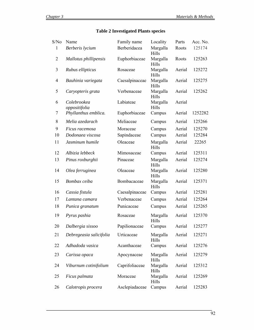
Chapter 3 Materials & Methods
92
Table 2 Investigated Plants species
S/No Name Family name Locality Parts Acc. No. 1 Berberis lycium Berberidacea Margalla
Hills Roots 125174
2 Mallotus phillipensis Euphorbiaceae Margalla Hills
Roots 125263
3 Rubus ellipticus Rosaceae Margalla Hills
Aerial 125272
4 Bauhinia variegata Caesalpinaceae Margalla Hills
Aerial 125275
5 Caryopteris grata Verbenaceae Margalla Hills
Aerial 125262
6 Colebrookea oppositifolia
Labiateae Margalla Hills
Aerial
7 Phyllanthus emblica. Euphorbiaceae Campus Aerial 1252282
8 Melia azedarach Meliaceae Campus Aerial 125266
9 Ficus racemosa Moraceae Campus Aerial 125270 10 Dodonaea viscosa Sapindaceae Campus Aerial 125284
11 Jasminum humile Oleaceae Margalla Hills
Aerial 22265
12 Albizia lebbeck Mimosaceae Campus Aerial 125311
13 Pinus roxburghii Pinaceae Margalla Hills
Aerial 125274
14 Olea ferruginea Oleaceae Margalla Hills
Aerial 125280
15 Bombax ceiba Bombacaceae Margalla Hills
Aerial 125371
16 Cassia fistula Caesalpinaceae Campus Aerial 125281
17 Lantana camara Verbenaceae Campus Aerial 125264
18 Punica granatum Punicaceae Campus Aerial 125265
19 Pyrus pashia Rosaceae Margalla Hills
Aerial 125370
20 Dalbergia sissoo Papilionaceae Campus Aerial 125277
21 Debregeasia salicifolia Urticaceae Margalla Hills
Aerial 125271
22 Adhadoda vasica Acanthaceae Campus Aerial 125276
23 Carissa opaca Apocynaceae Margalla Hills
Aerial 125279
24 Viburnum cotinifolium Caprifoliaceae Margalla Hills
Aerial 125312
25 Ficus palmata Moraceae Margalla Hills
Aerial 125269
26 Calotropis procera Asclepiadaceae Campus Aerial 125283
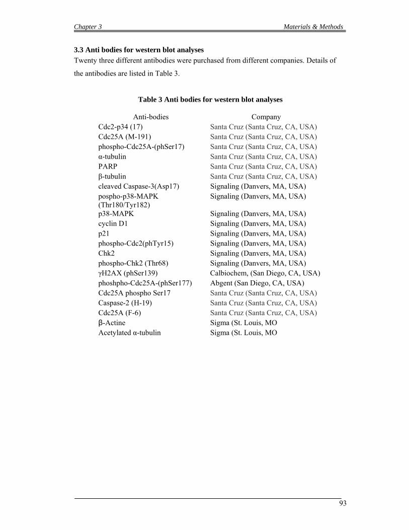
Chapter 3 Materials & Methods
93
3.3 Anti bodies for western blot analyses
Twenty three different antibodies were purchased from different companies. Details of
the antibodies are listed in Table 3.
Table 3 Anti bodies for western blot analyses
Anti-bodies Company Cdc2-p34 (17) Santa Cruz (Santa Cruz, CA, USA) Cdc25A (M-191) Santa Cruz (Santa Cruz, CA, USA) phospho-Cdc25A-(phSer17) Santa Cruz (Santa Cruz, CA, USA) α-tubulin Santa Cruz (Santa Cruz, CA, USA) PARP Santa Cruz (Santa Cruz, CA, USA) β-tubulin Santa Cruz (Santa Cruz, CA, USA) cleaved Caspase-3(Asp17) Signaling (Danvers, MA, USA) pospho-p38-MAPK (Thr180/Tyr182)
Signaling (Danvers, MA, USA)
p38-MAPK Signaling (Danvers, MA, USA) cyclin D1 Signaling (Danvers, MA, USA) p21 Signaling (Danvers, MA, USA) phospho-Cdc2(phTyr15) Signaling (Danvers, MA, USA) Chk2 Signaling (Danvers, MA, USA) phospho-Chk2 (Thr68) Signaling (Danvers, MA, USA) γH2AX (phSer139) Calbiochem, (San Diego, CA, USA) phoshpho-Cdc25A-(phSer177) Abgent (San Diego, CA, USA) Cdc25A phospho Ser17 Santa Cruz (Santa Cruz, CA, USA) Caspase-2 (H-19) Santa Cruz (Santa Cruz, CA, USA) Cdc25A (F-6) Santa Cruz (Santa Cruz, CA, USA) β-Actine Sigma (St. Louis, MO Acetylated α-tubulin Sigma (St. Louis, MO
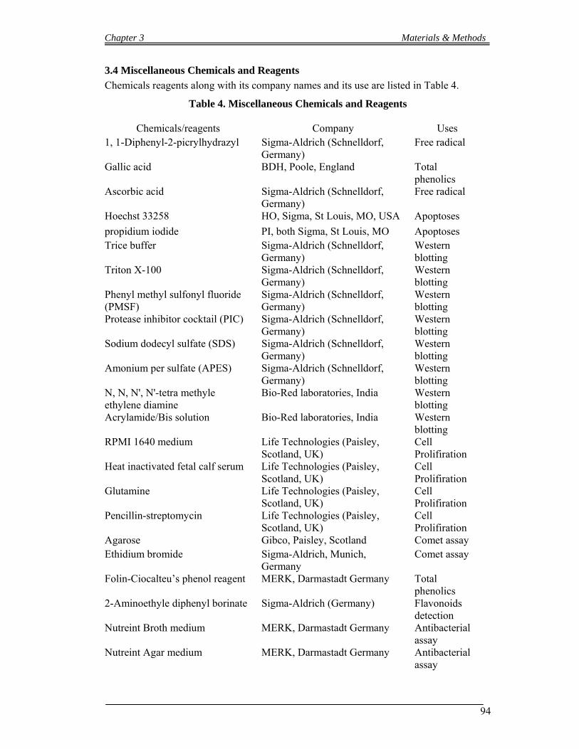
Chapter 3 Materials & Methods
94
3.4 Miscellaneous Chemicals and Reagents
Chemicals reagents along with its company names and its use are listed in Table 4.
Table 4. Miscellaneous Chemicals and Reagents
Chemicals/reagents Company Uses 1, 1-Diphenyl-2-picrylhydrazyl Sigma-Aldrich (Schnelldorf,
Germany) Free radical
Gallic acid BDH, Poole, England Total phenolics
Ascorbic acid Sigma-Aldrich (Schnelldorf, Germany)
Free radical
Hoechst 33258 HO, Sigma, St Louis, MO, USA Apoptoses
propidium iodide PI, both Sigma, St Louis, MO Apoptoses Trice buffer Sigma-Aldrich (Schnelldorf,
Germany) Western blotting
Triton X-100 Sigma-Aldrich (Schnelldorf, Germany)
Western blotting
Phenyl methyl sulfonyl fluoride (PMSF)
Sigma-Aldrich (Schnelldorf, Germany)
Western blotting
Protease inhibitor cocktail (PIC) Sigma-Aldrich (Schnelldorf, Germany)
Western blotting
Sodium dodecyl sulfate (SDS) Sigma-Aldrich (Schnelldorf, Germany)
Western blotting
Amonium per sulfate (APES) Sigma-Aldrich (Schnelldorf, Germany)
Western blotting
N, N, N', N'-tetra methyle ethylene diamine
Bio-Red laboratories, India Western blotting
Acrylamide/Bis solution Bio-Red laboratories, India Western blotting
RPMI 1640 medium Life Technologies (Paisley, Scotland, UK)
Cell Prolifiration
Heat inactivated fetal calf serum Life Technologies (Paisley, Scotland, UK)
Cell Prolifiration
Glutamine Life Technologies (Paisley, Scotland, UK)
Cell Prolifiration
Pencillin-streptomycin Life Technologies (Paisley, Scotland, UK)
Cell Prolifiration
Agarose Gibco, Paisley, Scotland Comet assay Ethidium bromide Sigma-Aldrich, Munich,
Germany Comet assay
Folin-Ciocalteu’s phenol reagent MERK, Darmastadt Germany Total phenolics
2-Aminoethyle diphenyl borinate Sigma-Aldrich (Germany) Flavonoids detection
Nutreint Broth medium MERK, Darmastadt Germany Antibacterial assay
Nutreint Agar medium MERK, Darmastadt Germany Antibacterial assay

Chapter 3 Materials & Methods
95
3.5 Cell culture and bacterial strains
The ATCC (American Type Culture Collection, Manassas, VA, USA) brand culture HL-
60 human promyelocytic cells were purchased. Three gram positive bacterial strains,
staphylococcus aureus (ATCC 65438), Bacillus subtilis (ATCC 6633) and
Staphylococcus epidermidis (ATCC 12228); three gram negative bacterial strains
Escherichia coli (ATCC 15224), and Salmonella setubal (ATCC 19196)
3.6 Extraction
Three different methods were used for extraction – one for analyses of free radical
scavenging activity (antioxidant activity) and total Phenolics determination in the aerial
parts of selected plants (Chapter 4.6.1), the second methods was to extracts the roots for
antibacterial and antineoplastic (anticancer) activities (Chapter 4.6.2), the third method
was to extract and prepare the aerial parts for flavonoids finger printing of the selected
medicinal plants (Chapter 4.6.3).
3.6.1 Extraction for Antioxidant and Total Phenolics Determination
Four grams of the aerial part of each plant species powder were weighed into a flask and
mixed in 40 ml (80% aqueous) methanol, and slightly stirred the suspension. Tubes were
sonicated for twenty minutes and centrifugated for ten min at (1500g), and collected
supernatants. Re-extracted the plant materials two times and the combined supernatants
were evaporated by Rota vapor to a volume of about 10 ml. These concentrated extracts
were lyophilized and weighed. Extracts obtained per gram dry powder are listed in (Table
11).
3.6.2 Extraction of roots powder
Roots of the plants were carefully washed, dried under shed and grounded. 200 g of
powdered B. lycium root and Mallotus phillipensis were extracted four times with
methanol (MeOH). These extracts were collected and concentrated with a Rotavapor at
40 ºC. The concentrated MeOH extract of B. lycium was dissolved in distilled water and
extracted three times each with organic solvents in sequence of increasing polarity such
as ethyl acetate (EtOAc), and n-butanol (BuOH). After evaporating each solvent from B.
lycium 0.44 g dried EtOAc extract, and 2.22 g dried BuOH extract was obtained,
Chapter 3 Materials & Methods
96
respectively 2.78 g dried material was recovered from the aqueous phase and considered
as H2O extract.
The concentrated MeOH extract of Mallotus phillipensis was dissolved in distilled water
and extracted three times each with organic solvents in sequence of increasing polarity
such as hexane, ethyl acetate (EtOAc), and n-butanol (BuOH). After evaporating each
solvent 9.23 g dried hexane extract, 4.00 g dried EtOAc extract, and 7.08 g dried BuOH
extract was obtained, respectively.
3.6.3 Extraction for Flavonoids analyses
One grams of the aerial part of each plant species powder were weighed and mixed in 10
ml of 70% aqueous methanol in a test tube. The suspension was prepared and stirred
slightly. After sonication of the tubes for twenty minutes, the tubes were centrifuged for
ten min at (1500g), and collected the supernatants. The plant materials were re-extracted
twice. The supernatants were combined and evaporated by Rota vapor to a volume of
about 2 ml. These concentrated extracts were lyophilized. The concentrated extracts re-
dissolved in 5 mL distilled water and extracted two times with hexane. The water soluble
fractions were acidified and reflux for twenty minutes. The extracts were extracted with
ethyl acetate. The ethyl acetate fractions were washed two times with distilled water.
3.7 Chromatographic Methods
3.7.1 Thin Layer Chromatography (TLC)
Thin layer chromatography was used for qualitative studies of alkaloids in Berberis
lycium and flavonoids analyses in the aerial parts of selected plants.
3.7.1.1 Thin Layer Chromatography of Berberis lycium fractions
The constituents of the Berberis lycium fractions were qualitatively studied by TLC.
Toluene-isopropanol-ethyl acetate-methanol and water (12:6:3:3:0.6) were used as a
mobile phase, TLC aluminum sheets 20 x 20 cm Silica gel 60 F254 (Merck, Darmstadt,
Germany) were used as stationary Phase. Two tank TLC chambers were used and one of
the tanks filled with the mobile phase and the other with liquid ammonia. The atmosphere
of the chamber was saturated 20 minutes prior chromatographic separation. Detection was
made by Dragendorff Reagent.
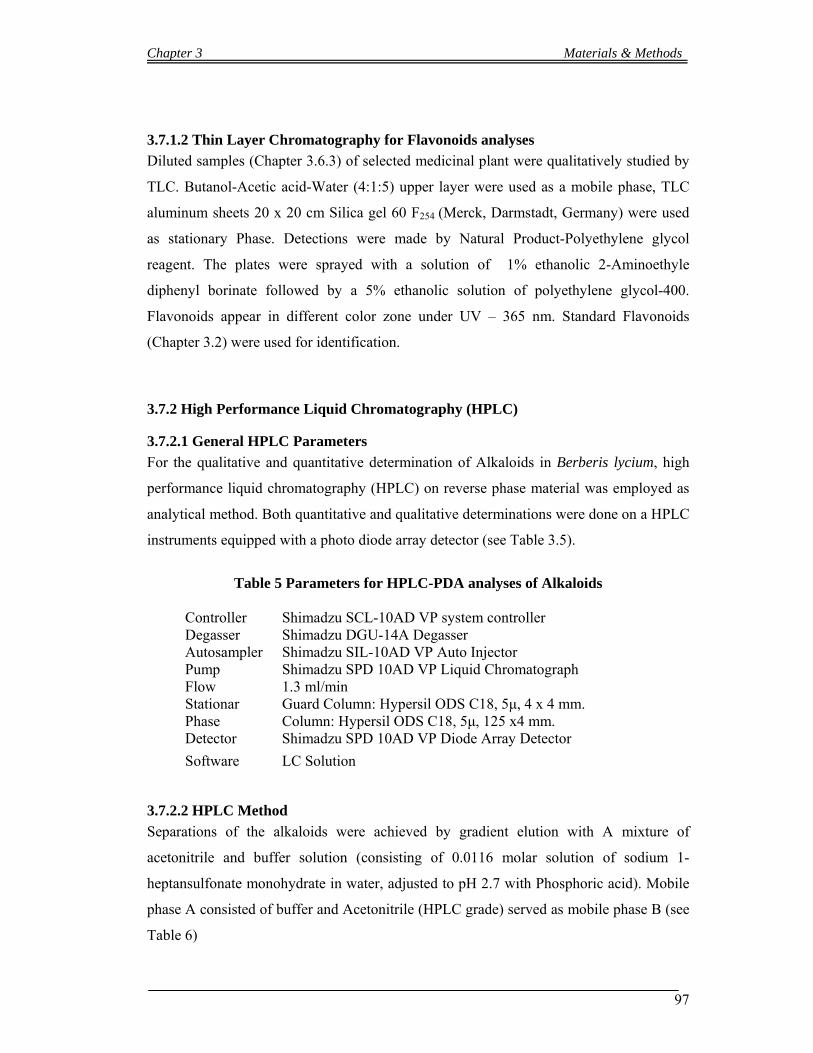
Chapter 3 Materials & Methods
97
3.7.1.2 Thin Layer Chromatography for Flavonoids analyses
Diluted samples (Chapter 3.6.3) of selected medicinal plant were qualitatively studied by
TLC. Butanol-Acetic acid-Water (4:1:5) upper layer were used as a mobile phase, TLC
aluminum sheets 20 x 20 cm Silica gel 60 F254 (Merck, Darmstadt, Germany) were used
as stationary Phase. Detections were made by Natural Product-Polyethylene glycol
reagent. The plates were sprayed with a solution of 1% ethanolic 2-Aminoethyle
diphenyl borinate followed by a 5% ethanolic solution of polyethylene glycol-400.
Flavonoids appear in different color zone under UV – 365 nm. Standard Flavonoids
(Chapter 3.2) were used for identification.
3.7.2 High Performance Liquid Chromatography (HPLC)
3.7.2.1 General HPLC Parameters
For the qualitative and quantitative determination of Alkaloids in Berberis lycium, high
performance liquid chromatography (HPLC) on reverse phase material was employed as
analytical method. Both quantitative and qualitative determinations were done on a HPLC
instruments equipped with a photo diode array detector (see Table 3.5).
Table 5 Parameters for HPLC-PDA analyses of Alkaloids
Controller Shimadzu SCL-10AD VP system controller Degasser Shimadzu DGU-14A Degasser Autosampler Shimadzu SIL-10AD VP Auto Injector Pump Shimadzu SPD 10AD VP Liquid Chromatograph Flow 1.3 ml/min Stationar Phase
Guard Column: Hypersil ODS C18, 5μ, 4 x 4 mm. Column: Hypersil ODS C18, 5μ, 125 x4 mm.
Detector Shimadzu SPD 10AD VP Diode Array Detector
Software LC Solution
3.7.2.2 HPLC Method
Separations of the alkaloids were achieved by gradient elution with A mixture of
acetonitrile and buffer solution (consisting of 0.0116 molar solution of sodium 1-
heptansulfonate monohydrate in water, adjusted to pH 2.7 with Phosphoric acid). Mobile
phase A consisted of buffer and Acetonitrile (HPLC grade) served as mobile phase B (see
Table 6)
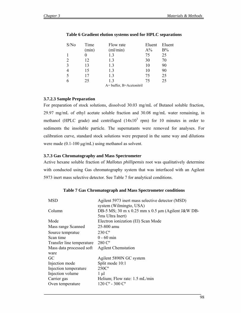
Chapter 3 Materials & Methods
98
Table 6 Gradient elution systems used for HPLC separations
S/No Time
(min) Flow rate (ml/min)
Eluent A%
Eluent B%
1 0 1.3 75 25 2 12 1.3 30 70 3 13 1.3 10 90 4 15 1.3 10 90 5 17 1.3 75 25 6 25 1.3 75 25
A= buffer, B=Acetonitril
3.7.2.3 Sample Preparation
For preparation of stock solutions, dissolved 30.03 mg/mL of Butanol soluble fraction,
29.97 mg/mL of ethyl acetate soluble fraction and 30.08 mg/mL water remaining, in
methanol (HPLC grade) and centrifuged (14x103 rpm) for 10 minutes in order to
sediments the insoluble particle. The supernatants were removed for analyses. For
calibration curve, standard stock solutions were prepared in the same way and dilutions
were made (0.1-100 μg/mL) using methanol as solvent.
3.7.3 Gas Chromatography and Mass Spectrometer
Active hexane soluble fraction of Mallotus phillipensis root was qualitatively determine
with conducted using Gas chromatography system that was interfaced with an Agilent
5973 inert mass selective detector. See Table 7 for analytical conditions.
Table 7 Gas Chromatograph and Mass Spectrometer conditions
MSD Agilent 5973 inert mass selective detector (MSD)
system (Wilmingto, USA) Column DB-5 MS; 30 m x 0.25 mm x 0.5 μm (Agilent J&W DB-
5ms Ultra Inert) Mode Electron ionization (EI) Scan Mode Mass range Scanned 25-800 amu Source tempratue 230 Cº Scan time 0 - 60 min Transfer line temperature 280 Cº Mass data processed soft ware
Agilent Chemstation
GC Agilent 5890N GC system Injection mode Split mode 10:1 Injection temperature 250CºInjection volume 1 μl Carrier gas Helium; Flow rate: 1.5 mL/min Oven temperature 120 Cº - 300 Cº
Chapter 3 Materials & Methods
99
3.8 Biological Testing
3.8.1 Antineoplastic Activities
3.8.1.1 Anti-proliferation or Growth inhibition assay
The cells culture of HL-60 (human promyelocytic cells) were seeded in T-25 tissue
culture flasks at a concentration of 1x10⁵ per ml and incubated with increasing
concentrations of the different extracts of Berberis lycium and Mallotus phillipensis or
with solutions of berberine and palmatine. Cell counts and IC50 values were determined in
the different fractions after 48 and 72 h, using a KX 21 N microcell counter (Sysmex,
Kobe, Japan). The media and other supplements were purchased from Life Technologies
(Paisley, Scotland, UK). The percent of cell divisions compared to the untreated control
were calculated as follows:
[(C48 h + drug - C24 h + drug) / (C48 h - drug - C24 h - drug)] x 100 = % cell division
C48 h + drug = cell number after 48 hours of drug treatment
C24 h + drug = cell number after 24 hours of drug treatment
C48 h - drug = cell number after 48 hours without drug treatment
C = cell number after 24 hours without drug treatment
3.8.1.2 Hoechst dye 33258 and propidium iodide double staining (Apoptosis Assay)
Hoechst staining was performed according to the method described by Grusch et al
(2002). HL-60 cells (0.1x10⁶ per ml) were seeded in T25 cell culture flasks and exposed
to increasing concentrations of B.lycium, M. phillipensis fractions and standard berberine
for 48 h. Hoechst 33258 (HO) and propidium iodide (PI, both Sigma, St Louis, MO) were
added directly to the cells to final concentrations of 5 and 2 mg/ml, respectively. After 60
min of incubation at 37 Co, the cells were examined under a fluorescence microscope
(Axiovert, Zeiss) equipped with a DAPI filter and a camera. This method allows
discriminating between early apoptosis, late apoptosis, and necrosis. Cells were judged
according to their morphology and the integrity of their cell membranes, which can easily
be seen after propidium iodide staining.
3.8.1.3 Western blotting
HL-60 cells were preincubated for increasing time periods (from 2 to 48 h) with 11.1 µg
BuOH extract /ml and 1.4 µg berberine/ml medium. In another set of experiment HL -60

Chapter 3 Materials & Methods
100
cells were preincubated for increasing time periods (from 2 to 48 h) with 1.5 mg/mL
hexane fraction of M. phillipensis. Then, cells were placed on ice, washed with ice-cold
PBS (pH 7.2), centrifuged (1000 rpm, 4 ºC, 4 min) and the pellets lysed in 150 µl buffer
containing 150 mM NaCl, 50 mM Tris ph 8.0, 1 % Triton X-100, 2.5 % 0.5 mM PMSF
and PIC (Sigma, Schnelldorf, Germany). Debris was removed by centrifugation (12,000
rpm, 4 ºC, 20 min) and the supernatant collected. Then, equal amounts of protein were
loaded onto 10 % polyacrylamide gels (See Table 8) Proteins were electrophoresed for 2
h and then electro-blotted onto PVDF membranes (Hybond P, Amersham,
Buckinghamshire, UK) at 4 ºC for 1 h. To confirm equal sample loading, membranes were
stained with Poinceau S. After washing with TBS, the membranes were blocked for 1 h in
Blotto (blocking solution: 5 % skimmed milk in TBS and 0.5 % Tween 20), washed 3
times in TBS/T, and incubated by gentle rocking with primary anti-bodies in Blotto (0.2-
0.3 : 1000), at 4 Cº overnight. Then, the membranes were washed in TBS/T (3 x for 5
min) and further incubated with the second antibody (peroxidase-conjugated anti-rabbit
IgG, or anti-mouse IgG dilution 1:2000 in Blotto, for 1 h at room temperature. The
membranes were washed with TBS/T and the chemo luminescence (ECL detection kit,
Amersham, Buckinghamshire, UK) was detected by exposure of the membranes to
Amersham HyperfilmTM ECL. The antibodies against Cdc2-p34 (17), Cdc25A (M-191),
phospho-Cdc25A-(phSer17), α-tubulin, PARP and β-tubulin were from Santa Cruz (Santa
Cruz, CA, USA), against cleaved Caspase-3(Asp17), pospho-p38-MAPK
(Thr180/Tyr182), p38-MAPK, cyclin D1, p21, phospho-Cdc2(phTyr15), Chk2, and
phospho-Chk2 (Thr68) were from Cell Signaling (Danvers, MA, USA), against γH2AX
(phSer139) from Calbiochem, (San Diego, CA, USA), and phoshpho-Cdc25A-
(phSer177) from Abgent (San Diego, CA, USA), and against acetylated α-tubulin and β-
actin were from Sigma (St. Louis, MO).
3.8.1.4 Cell cycle distribution analysis (FACS analyses)
The cells culture of HL-60 (0.5x10⁶ per ml) were seeded in T-25 tissue culture flasks and
incubated with 5.6 µg/ml BuOH extract, 0.7 µg/ml (1.86 µM) berberine, or 0.28µg/ml
(0.75 µM) palmatine (which were equivalent to 0.5 mg/ml dried root powder,
respectively) and 1.5 mg/mL hexane fraction of M. phillipensis according to cell culture
parameters. After 24 h of incubation, the cells were separated from the medium and re-
suspended in 5 ml cold PBS, after centrifugation (600 rpm, 5 min) the culture were again
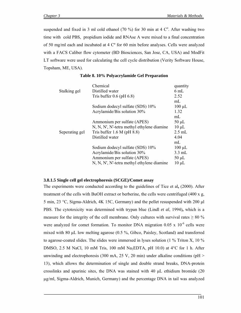
Chapter 3 Materials & Methods
101
suspended and fixed in 3 ml cold ethanol (70 %) for 30 min at 4 Co. After washing two
time with cold PBS, propidium iodide and RNAse A were mixed to a final concentration
of 50 mg/ml each and incubated at 4 Cº for 60 min before analyses. Cells were analyzed
with a FACS Caliber flow cytometer (BD Biosciences, San Jose, CA, USA) and ModFit
LT software were used for calculating the cell cycle distribution (Verity Software House,
Topsham, ME, USA).
Table 8. 10% Polyacrylamide Gel Preparation
Chemical quantity Stalking gel Distilled water 6 mL Tris buffer 0.6 (pH 6.8) 2.52
mL Sodium dodecyl sulfate (SDS) 10% 100 μL Acrylamide/Bis solution 30% 1.32
mL Ammonium per sulfate (APES) 50 μL N, N, N', N'-tetra methyl ethylene diamine 10 μL Seperating gel Tris buffer 1.6 M (pH 8.8) 2.5 mL Distilled water 4.04
mL Sodium dodecyl sulfate (SDS) 10% 100 μL Acrylamide/Bis solution 30% 3.3 mL Ammonium per sulfate (APES) 50 μL N, N, N', N'-tetra methyl ethylene diamine 10 μL
3.8.1.5 Single cell gel electrophoresis (SCGE)/Comet assay
The experiments were conducted according to the guidelines of Tice et al, (2000). After
treatment of the cells with BuOH extract or berberine, the cells were centrifuged (400 x g,
5 min, 23 °C, Sigma-Aldrich, 4K 15C, Germany) and the pellet resuspended with 200 μl
PBS. The cytotoxicity was determined with trypan blue (Lindl et al, 1994), which is a
measure for the integrity of the cell membrane. Only cultures with survival rates ≥ 80 %
were analyzed for comet formation. To monitor DNA migration 0.05 x 10⁻⁶ cells were
mixed with 80 μL low melting agarose (0.5 %, Gibco, Paisley, Scotland) and transferred
to agarose-coated slides. The slides were immersed in lyses solution (1 % Triton X, 10 %
DMSO, 2.5 M NaCl, 10 mM Tris, 100 mM Na2EDTA, pH 10.0) at 4°C for 1 h. After
unwinding and electrophoresis (300 mA, 25 V, 20 min) under alkaline conditions (pH >
13), which allows the determination of single and double strand breaks, DNA-protein
crosslinks and apurinic sites, the DNA was stained with 40 μL ethidium bromide (20
µg/ml, Sigma-Aldrich, Munich, Germany) and the percentage DNA in tail was analyzed
Chapter 3 Materials & Methods
102
with a computer aided image analysis system (Comet IV, Perceptive Instruments Ltd.,
Haverhill, UK). From each experimental point one slide was prepared and 50 cells were
scored per slide.
3.8.1.6 Statistical analyses
The results of the SCGE (single cell gel electrophoresis) experiments were analysed with
one-way ANOVA followed by Dunnett´s multiple comparison test, and the apoptosis and
proliferation experiments with t-test using GraphPad Prism version 4 (GraphPad Prism
Software, Inc., San Diego, CA, USA).
3.8.2 Total Phenolics determination
Phenolic compounds were determined with the method of Folin–Ciocalteu (Singleton and
Rossi, 1965). For the preparation of Gallic acid stock solution, took a 100-mL volumetric
flask, dissolved 0.5g of Gallic acid powder in 10 ml of ethanol, after complete dissolving
the volume were made up to 100 mL. To prepared Sodium carbonate solution, anhydrous
sodium carbonate (200g) was dissolved in 0.8 L of distill water and was boiled. After
cooling the solution, few crystals of sodium carbonate (Na2CO3) were added. After a
period of 24 hr, filtered the solution and add water to 1L. In order to draw a calibration
curve (Fig. 4.15), add 0, 100, 200, 500, 750, 1250, 2000 µL and extended to 4.00 mL of
the above Gallic acid stock solution into 100 ml volumetric flasks, and then dilute to
volume with water (100ml). These solutions will have phenol concentrations of 0, 5, 10,
25, 37.5, 62.5,100 and 4000 mg/L Gallic acid equivalent. Took 20 µL into separate
cuvettes from each calibration solution, sample or blank and to each add 1.58 ml distill
water, and then pipet 100 µL of the Folin-Ciocalteu reagent. After mixing the reagent and
sample well, kept for 30 sec and 8 min. Add 300 µL of the sodium carbonate solution,
and shacked well to mix. Left the solutions at 40°C for 30 min before reading the
absorbance and determine the absorbance of each solution at 765 nm against the blank
(the "0 mL" solution) and plot absorbance vs. concentration.
3.8.3 1, 1-Diphenyl-2-picrylhydrazyl (DPPH) test
The free radical scavenging activities of the leaves powder extracts were assessed by
using the DPPH radical (Brand-Williams, et al., 1995). The experiment was slightly
Chapter 3 Materials & Methods
103
modified by making 0.5 m. mole /L solution of DPPH in methanol and again diluted four
times (4X) in order to showed absorbance below 1. The solution of DPPH (1000µl) was
mixed with different concentration of each test compound (2.5-200µg/ml, 500µl) and the
absorbance change at 517 nm was measured 30 min later (Blois, et al., 1958). The
reaction solution without DPPH was used as a blank test and ascorbic acid (2.5-15µg/ml,
500µl) was used a positive control. The experiment conducted on Shimadzu
spectrophotometer (UV-120-01), by using low lens. Mean value of triplicate was plotted
in graph in order to calculate the concentration required for 50% reduction (50%
inhibition concentration, IC50) of DPPH radical (Yen, et al., 1994 and Kubo, et al., 1984).
GraphPad Prism version 4 (GraphPad Prim Software, Inc., San Diego, CA, USA) was
used for calculating the concentration.
3.8.4 Antibacterial Determination
The test was performed by simple Agar diffusion assay (Kavanagh., 1963; Leven et al.,
1979). Eight dilutions were tested (dilution were 1, 2, 3, 5, 7.5, 10, 12.5 and 15mg/ml)
against five strains of bacteria. Three gram positive bacterial strains, staphylococcus
aureus (ATCC 65438), Bacillus subtilis (ATCC 6633) and Staphylococcus epidermidis
(ATCC 12228); two gram negative bacterial strains Escherichia coli (ATCC 15224), and
Salmonella setubal (ATCC 19196).
Chapter 4 Results & Discussion
104
4.1 Results
4.1.1 Anti-neoplastic Activities and Phytochemicals studies of Berberis lycium
4.1.1.1 Qualitative Analysis of B. lycium extracts constituents by TLC
The EtOAc, BuOH, and H2O extracts of B.lycium roots were loaded on TLC plates which
were immersed in a saturated chromatography chamber containing toluene-EtOAc-
isopropanol-methanol-H2O (12:6:3:3:0.6) as running phase, and compared standards
berberine, berbamine and palmatine, which are known constituents of various Berberis
taxa with distinct anti-neoplastic properties.
All extracts contained berberine. The BuOH extract showed comparatively the highest
concentration (retention factor, Rf = 0.151). Also palmatine was detected in all extracts
but only traces were found in the H2O and EtOAc extracts (Rf = 0.088). Berbamine was
not found in any extract (the standard was occurring as a black spot under 254 nm UV
light, Rf = 0.405). Besides berberine and palmatine another unknown bands were present
in all extracts. (see figure 8).
4.1.1.2 Separation and quantification of alkaloids by RP-HPLC
The alkaloids seen in the different fractions during TLC analyses have been successfully
separated and determined by HP-HPLC. In order to obtained good resolutions and
minimize peaks tailing. A series of experiments using different solvent systems, column
types and running parameters (results not shown) were performed. A mixture of
acetonitrile and buffer solution (consisting of sodium 1-heptansulfonate monohydrate in
water, adjusted to pH 2.7 with Phosphoric acid) was considered best. Solvent gradients
are shown in table. 6 (chapter 4; page. 98). Chromatograms that shows separation in
different fractions of Berberis lycium and standards compounds are shown in Fig. 9.
Average retention times for internal standard codeine, berberine, palmatine and
berbamine were 4.52, 9.753, 9.190 and 8.056 minutes respectively. An unknown peak (Rf
= 8.062) was found in the UV range of palmatine (Fig. 10). Calibration curves have been
developed separately for Berberine and Palmatine. The linearity studies of standard curve
for standard compounds have been calculated (see Table. 9). Estimated concentrations of
berberine and palmatine are listed in Table 10.and Fig.14 page 112.

Chapter 4 Results & Discussion
105
Figure 8 TLC of Berberis lycium extracts
1. H2O Fraction (1 mg/ mL) 6 µl
2. Ethyl acetate.fraction (1 mg/ mL) 6µl
3. Ethyl acetate.Fraction (1 mg/ mL) 8 µl,
4. Standard palmatine chloride (0.125 mg/mL) 4µl
5. Standard berberine chloride dihydrate (0.25 mg/mL) 4 µl
6. Standard berbamine dihydrochloride (1 mg/mL) 2µl
7. BuOH Fraction (1mg/mL) 4µl
8. BuOH Fraction

Chapter 4 Results & Discussion
106
0.0 5.0 10.0 15.0 20.0 min
-25
0
25
50
75
100
125
150
mAU
0.00
0.05
0.10
0.15
0.20
0.25
0.30
0.35
0.40
0.45
0.50
0.55
0.60
0.65
0.70
0.75
0.80
0.85
0.90
0.95
barA.Press.(Status)280nm,4nm (1.00)
4.48
3/10
7131
8
5.52
4/29
216.
121/
1070
4
6.91
7/19
547.
465/
1398
58.
098/
1403
618.
585/
1053
69.
209/
1068
637
9.65
9/85
0449
Minutes
Figure 9 RP-HPLC Chromatogram of alkaloids standards. RP-HPLC chromatogram
of standard compounds: 1. Codeine hydrochloride, 2. Berbamine dihydrochloride, 3.
Palmatine chloride, 4. Berberine chloride dehydrate.
9 A 1
2
4
3
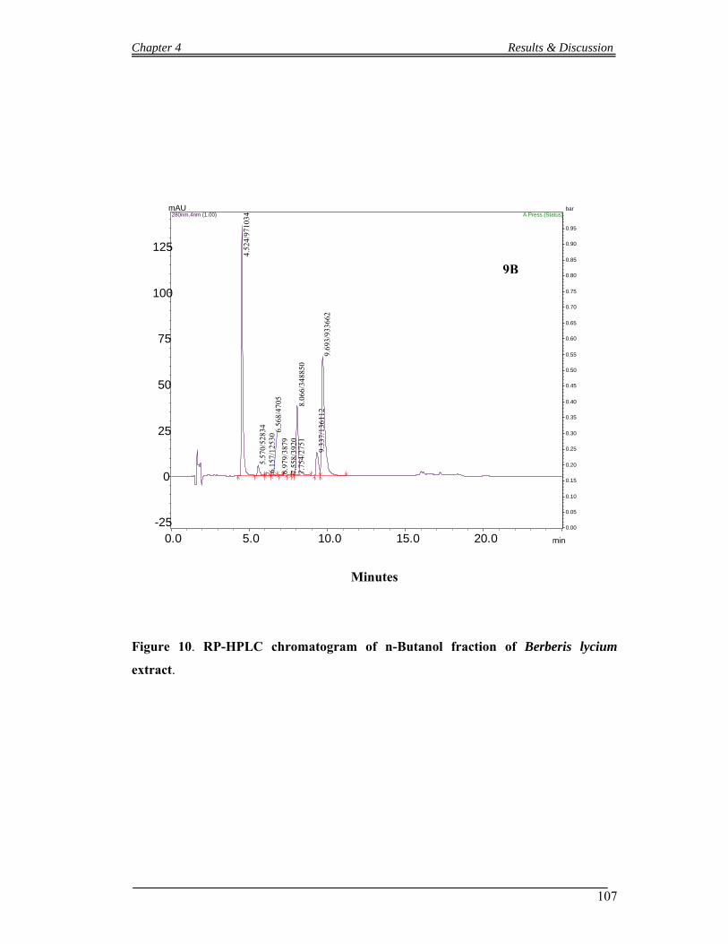
Chapter 4 Results & Discussion
107
0.0 5.0 10.0 15.0 20.0 min
-25
0
25
50
75
100
125
mAU
0.00
0.05
0.10
0.15
0.20
0.25
0.30
0.35
0.40
0.45
0.50
0.55
0.60
0.65
0.70
0.75
0.80
0.85
0.90
0.95
barA.Press.(Status)280nm,4nm (1.00)
4.52
4/97
1034
5.57
0/52
834
6.15
7/12
530 6.
568/
4705
6.97
9/38
797.
558/
3920
7.75
4/27
518.
066/
3488
50
9.33
7/13
6112
9.69
3/93
3662
Minutes
Figure 10. RP-HPLC chromatogram of n-Butanol fraction of Berberis lycium
extract.
9B
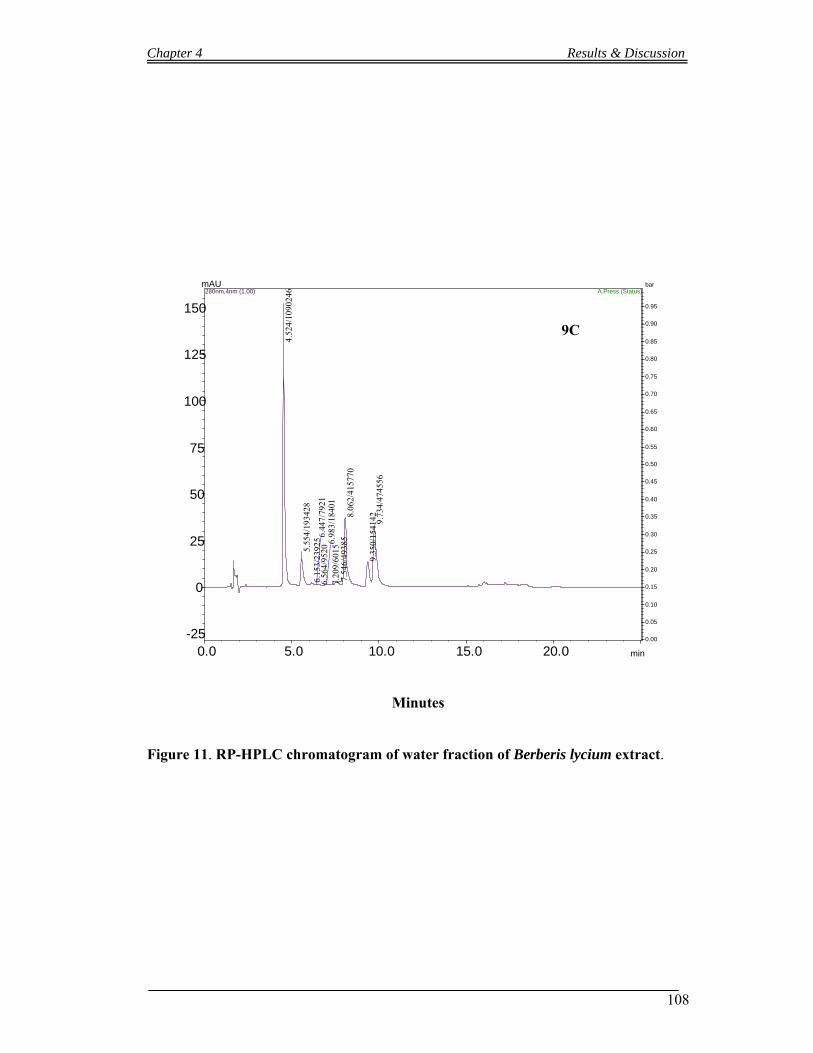
Chapter 4 Results & Discussion
108
0.0 5.0 10.0 15.0 20.0 min
-25
0
25
50
75
100
125
150
mAU
0.00
0.05
0.10
0.15
0.20
0.25
0.30
0.35
0.40
0.45
0.50
0.55
0.60
0.65
0.70
0.75
0.80
0.85
0.90
0.95
barA.Press.(Status)280nm,4nm (1.00)
4.52
4/10
9024
6
5.55
4/19
3428
6.15
3/23
925 6.
447/
7921
6.56
4/95
206.
983/
1840
17.
209/
6015
7.54
6/49
385
8.06
2/41
5770
9.35
0/15
4142 9.73
4/47
4556
Minutes
Figure 11. RP-HPLC chromatogram of water fraction of Berberis lycium extract.
9C
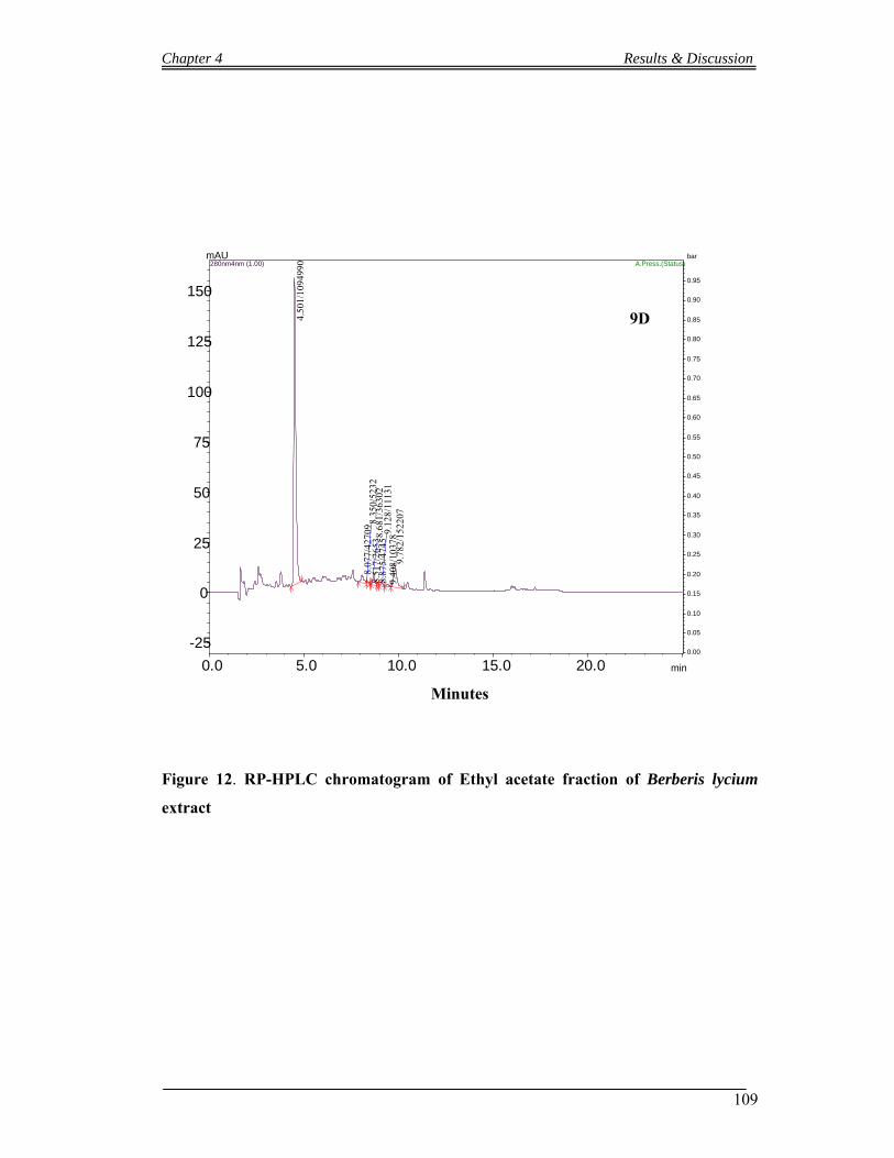
Chapter 4 Results & Discussion
109
0.0 5.0 10.0 15.0 20.0 min
-25
0
25
50
75
100
125
150
mAU
0.00
0.05
0.10
0.15
0.20
0.25
0.30
0.35
0.40
0.45
0.50
0.55
0.60
0.65
0.70
0.75
0.80
0.85
0.90
0.95
barA.Press.(Status)280nm4nm (1.00)
4.50
1/10
9499
0
8.07
7/42
709 8.
350/
5232
8.51
7/26
538.
681/
3630
28.
875/
4745
9.12
8/11
131
9.40
8/10
378
9.78
2/15
2207
Minutes
Figure 12. RP-HPLC chromatogram of Ethyl acetate fraction of Berberis lycium
extract
9D

Chapter 4 Results & Discussion
110
200.0 225.0 250.0 275.0 300.0 325.0 350.0 375.0 nm
0.0
1.0
2.0
mAU(x100) 9.54
20
9
24
8
30
2
38
0
22
9
26
4
34
7
200.0 225.0 250.0 275.0 300.0 325.0 350.0 375.0 nm
0.0
0.5
1.0
1.5
2.0mAU(x100)
9.13
20
2
21
2
24
9
30
2
37
8
34
6
22
6
26
5
20
6
200.0 225.0 250.0 275.0 300.0 325.0 350.0 375.0 nm
0.0
1.0
2.0
mAU(x100) 8.05
25
6
30
4
37
927
9
34
4
200.0 225.0 250.0 275.0 300.0 325.0 350.0 375.0 nm
0.0
0.5
1.0
1.5mAU(x1,000)
4.54 20
1
26
2
39
7
20
6
28
5
Figure 13 Optimum UV spectra of standards compounds: Optimum UV spectra of
standards compounds: 1. Berberine chloride dihydrate, 2 Palmatine Chloride., 3.
Berbamine dihydrochloride, 4. Codeine hydrochloride
3. Berbamine
1. Berberine
2. Palmatine
4. Codeine
Chapter 4 Results & Discussion
111
Table 9 Linearity study of standard curve for standard compounds
Compounds Calibration curve Slope Intercept R2 Range of injection
amount (μg/mL)
Berberine y= 34.59x+16.59 34.59 16.59 0.9994 10-100
Palmatine y=34.24x+44.368 34.24 44.37 0.9974 1-50.00
Berbamine y=3.0872x+22.946 3.0872 22.95 0.9948 1-100
Calculation:
The concentrations/percentage of Berberine and Palmatine were calculated with the
following equations
Factor of correction (FC) = Area (internal standard) x Mass (sample) Area (sample) x Mass (internal standard)
Sample (%) = Mass (internal standard) x Area (Sample) x FC (Factor of correction x 100 M (sample) x Area (internal standard)
Table 10 Percent composition of active alkaloids in Berberis lycium
Fractions Berberine % Palmatine %
BuOH Fraction 18.04 ± 0.01 2.80
Ethyl acetate Fraction 0.54 ± 0.0015 0.04 ± 0.00255
H2O Fraction 2.76 ± 0.026 0.93 ± 0.065
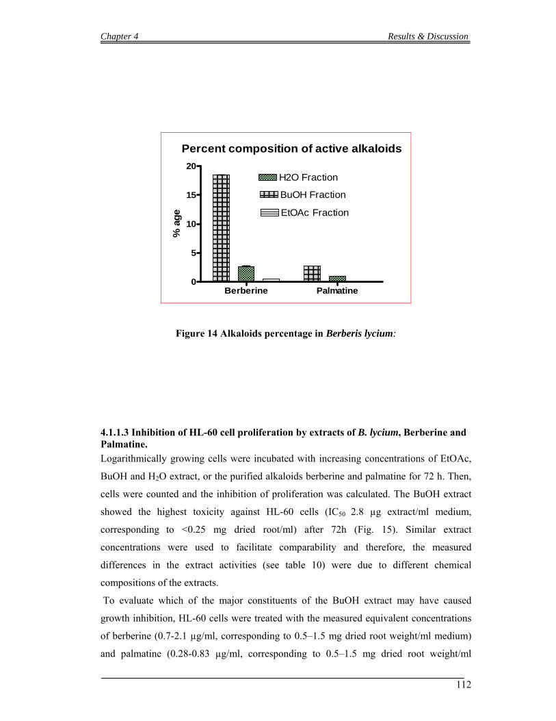
Chapter 4 Results & Discussion
112
Percent composition of active alkaloids
Berberine Palmatine0
5
10
15
20
BuOH Fraction
H2O Fraction
EtOAc Fraction
% a
ge
Figure 14 Alkaloids percentage in Berberis lycium:
4.1.1.3 Inhibition of HL-60 cell proliferation by extracts of B. lycium, Berberine and Palmatine.
Logarithmically growing cells were incubated with increasing concentrations of EtOAc,
BuOH and H2O extract, or the purified alkaloids berberine and palmatine for 72 h. Then,
cells were counted and the inhibition of proliferation was calculated. The BuOH extract
showed the highest toxicity against HL-60 cells (IC50 2.8 µg extract/ml medium,
corresponding to <0.25 mg dried root/ml) after 72h (Fig. 15). Similar extract
concentrations were used to facilitate comparability and therefore, the measured
differences in the extract activities (see table 10) were due to different chemical
compositions of the extracts.
To evaluate which of the major constituents of the BuOH extract may have caused
growth inhibition, HL-60 cells were treated with the measured equivalent concentrations
of berberine (0.7-2.1 µg/ml, corresponding to 0.5–1.5 mg dried root weight/ml medium)
and palmatine (0.28-0.83 µg/ml, corresponding to 0.5–1.5 mg dried root weight/ml
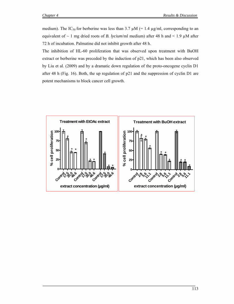
Chapter 4 Results & Discussion
113
medium). The IC50 for berberine was less than 3.7 µM (= 1.4 µg/ml, corresponding to an
equivalent of ~ 1 mg dried roots of B. lycium/ml medium) after 48 h and < 1.9 µM after
72 h of incubation. Palmatine did not inhibit growth after 48 h.
The inhibition of HL-60 proliferation that was observed upon treatment with BuOH
extract or berberine was preceded by the induction of p21, which has been also observed
by Liu et al. (2009) and by a dramatic down regulation of the proto-oncogene cyclin D1
after 48 h (Fig. 16). Both, the up regulation of p21 and the suppression of cyclin D1 are
potent mechanisms to block cancer cell growth.
Treatment with EtOAc extract
Contro
l17
.535
.046
.6
Contro
l17
.535
.046
.6
Contro
l17
.535
.046
.60
25
50
75
100
*
* *
*
* *
*
* *
extract concentration (µg/ml)
% c
ell
pro
life
rati
on
Treatment with BuOH extract
Contro
l2.8 5.
611
.1
Contro
l2.8 5.
611
.1
Contro
l2.8 5.
611
.10
25
50
75
100
* *
** *
* * **
extract concentration (µg/ml)
% c
ell
pro
life
rati
on
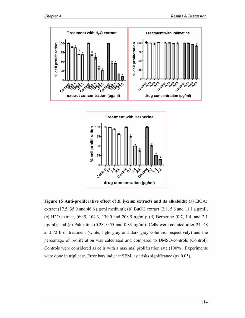
Chapter 4 Results & Discussion
114
Treatment with H2O extract
Contro
l69
.510
4.3
139.020
8.5
Contro
l69
.510
4.3
139.020
8.5
Contro
l69
.510
4.3
139.020
8.5
0
25
50
75
100
extract concentration (µg/ml)
* ** * *
*
* *
* *
* *% c
ell
pro
life
rati
on
Treatment with Palmatine
Contro
l 0.2
80.
550.8
3
Contro
l 0.2
80.
550.8
3
Contro
l 0.2
80.
550.8
30
25
50
75
100 *
drug concentration (µg/ml)
% c
ell
pro
life
rati
on
Treatment with Berberine
Contro
l0.7 1.
42.1
Contro
l0.7 1.
42.1
Contro
l0.7 1.
42.1
0
25
50
75
100
**
**
*
**
drug concentration (µg/ml)
% c
ell
pro
life
rati
on
Figure 15 Anti-proliferative effect of B. lycium extracts and its alkaloids: (a) EtOAc
extract (17.5, 35.0 and 46.6 µg/ml medium); (b) BuOH extract (2.8, 5.6 and 11.1 µg/ml);
(c) H2O extract, (69.5, 104.3, 139.0 and 208.5 µg/ml); (d) Berberine (0.7, 1.4, and 2.1
µg/ml); and (e) Palmatine (0.28, 0.55 and 0.83 µg/ml). Cells were counted after 24, 48
and 72 h of treatment (white, light gray and dark gray columns, respectively) and the
percentage of proliferation was calculated and compared to DMSO-controls (Control).
Controls were considered as cells with a maximal proliferation rate (100%). Experiments
were done in triplicate. Error bars indicate SEM, asterisks significance (p< 0.05).
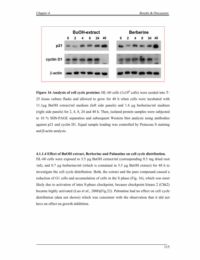
Chapter 4 Results & Discussion
115
Figure 16 Analysis of cell cycle proteins: HL-60 cells (1x10⁶ cells) were seeded into T-
25 tissue culture flasks and allowed to grow for 48 h when cells were incubated with
11.1µg BuOH extract/ml medium (left side panels) and 1.4 µg berberine/ml medium
(right side panels) for 2, 4, 8, 24 and 48 h. Then, isolated protein samples were subjected
to 10 % SDS-PAGE separation and subsequent Western blot analysis using antibodies
against p21 and cyclin D1. Equal sample loading was controlled by Poinceau S staining
and β-actin analysis.
4.1.1.4 Effect of BuOH extract, Berberine and Palmatine on cell cycle distribution.
HL-60 cells were exposed to 5.5 µg BuOH extract/ml (corresponding 0.5 mg dried root
/ml), and 0.7 µg berberine/ml (which is contained in 5.5 µg BuOH extract) for 48 h to
investigate the cell cycle distribution. Both, the extract and the pure compound caused a
reduction of G1 cells and accumulation of cells in the S phase (Fig. 16), which was most
likely due to activation of intra S-phase checkpoint, because checkpoint kinase 2 (Chk2)
became highly activated (Luo et al., 2008)(Fig.22). Palmatine had no effect on cell cycle
distribution (data not shown) which was consistent with the observation that it did not
have an effect on growth inhibition.
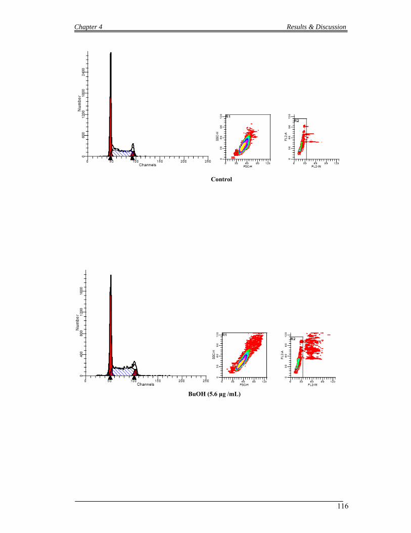
Chapter 4 Results & Discussion
116
Control
BuOH (5.6 μg /mL)

Chapter 4 Results & Discussion
117
Berberine (0.7 μg/ mL equ.)
Cell cycle distribution afterBuOH extract treatment (48 h)
G0-G1 S G2-M0
10
20
30
40
50 Control5.6 µg*
*
% c
ells
Cell cycle distribution afterBerberine treatment (48 h)
G0-G1 S G2-M0
10
20
30
40
50
60Control0.7 µg
*
*
*
% c
ells
Figure 17 Cell Cycle Distribution of HL-60 cells upon treatment with of BuOH
extract and berberine for 48 h: Cell Cycle Distribution of HL-60 cells upon treatment
with of BuOH extract and berberine for 48 h. Logarithmically growing HL-60 cells were
incubated with 5.6 µg/ml BuOH extract and 0.7 µg/ml berberine and then subjected to
FACS analysis. Experiments were done in triplicate. Error bars indicate SEM, and
asterisks significance (p<0.05).
Chapter 4 Results & Discussion
118
4.1.1.5 Induction of apoptosis by extracts of Berberis lycium and Berberine
HL-60 cells were treated with the three extracts (EtOAc, BuOH and H2O) and berberine
for 48h and the induction of cell death was analyzed. The three extract types induced
apoptosis and the BuOH extract was the most active followed by the EtOAc- and the H2O
extracts (see table 10; page.112) for comparison).
Berberine was used at a comparable concentration as contained in the BuOH extract and
this concentration caused a similar pro-apoptotic effect as the extract (Fig. 18).
High concentrations of berberine (10 – 50 µg/ml) were shown earlier to induce H2AX
phosphorylation (γH2AX) in osteosarcoma cells indicating genotoxicity (Liu et al.,
2009). In the present study we demonstrate that 0.7 and 1.4 µg/ml berberine and the
corresponding concentration of BuOH extract specifically induced apoptosis in HL-60
cells without concomitant induction of γH2AX (Fig. 19). This observation indicates that
the anti-neoplastic effects have not been triggered by berberine-caused genotoxicity.
Comet assay detecting DNA single strand breaks provided no evidence that berberine or
the BuOH extract cause DNA damage (Fig. 20). Thus, other mechanisms must be
responsible for cell cycle inhibition and apoptosis. Interestingly, berberine and the BuOH
extract caused acetylation of α-tubulin (Fig. 19), which is indicative for tubulin
polymerization reminiscent of the mechanism of taxol. Tilting the fine-tuned equilibrium
of polymerized/de-polymerized microtubule is incompatible with normal cell division and
this causes not only cell cycle arrest but also apoptosis.
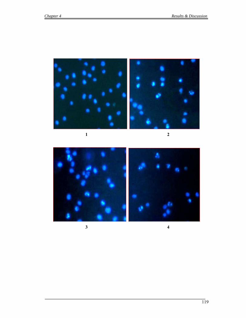
Chapter 4 Results & Discussion
119
1 2
3 4

Chapter 4 Results & Discussion
120
EtOAc extract treatment (48 h)
Contro
l5.9
11
.7
17.5
0
10
20
30
*
*
*
extract concentration (µg/ml)
% a
po
pto
tic
ce
lls
BuOH extract treatment (48 h)
Contro
l2.8 5.6 11
.10
10
20
30
*
**
extract concentration (µg/ml)
% a
po
pto
tic
ce
lls
H2O extract treatment (48 h)
Control
69.5
139.
220
8.5
0
10
20
30
**
extract concentration (µg/ ml)
% a
po
pto
tic
ce
lls
Berberine treatment (48 h)
Contro
l0.7
1.4
2.1
0
10
20
30
**
*
extract concentration (µg/ml)
% a
po
pto
tic
ce
lls
Figure 18 Induction of apoptosis by the Berberis lycium extracts and berberine:
(1) Control (2) BuOH extract 11.1 μg/ml (3) EtOAc extract 17.5 μg/ml (4) H2O extract
208.5 μg /ml. Error bars indicate SEM, asterisks significance (p<0.05).

Chapter 4 Results & Discussion
121
Figure 19 Western blot analyses of pro-apoptotic mediators and effectors

Chapter 4 Results & Discussion
122
1 2
3 4
Figure 20 The genotoxicity of increasing concentrations of BuOH extract and
berberine: The genotoxicity of increasing concentrations of BuOH extract and berberine
was investigated in logarithmically growing HL-60 cells. 50 µM H2O2 was used as
positive control and solvent-treated cells were used as negative control. The cytotoxicity
was determined with trypan blue (Lindl et al, 1994), which is a measure for the integrity
of the cell membrane. Only cultures with survival rates ≥ 80 % were analyzed for comet
formation. To monitor DNA migration 0.05 x 10⁻⁶ cells were mixed with 80 μL low
melting agarose (0.5 %, Gibco, Paisley, Scotland) and transferred to agarose-coated
slides. The slides were immersed in lyses solution (1 % Triton X, 10 % DMSO, 2.5 M
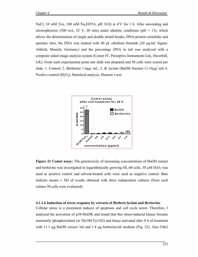
Chapter 4 Results & Discussion
123
NaCl, 10 mM Tris, 100 mM Na2EDTA, pH 10.0) at 4°C for 1 h. After unwinding and
electrophoresis (300 mA, 25 V, 20 min) under alkaline conditions (pH > 13), which
allows the determination of single and double strand breaks, DNA-protein crosslinks and
apurinic sites, the DNA was stained with 40 μL ethidium bromide (20 µg/ml, Sigma-
Aldrich, Munich, Germany) and the percentage DNA in tail was analyzed with a
computer aided image analysis system (Comet IV, Perceptive Instruments Ltd., Haverhill,
UK). From each experimental point one slide was prepared and 50 cells were scored per
slide. 1. Control; 2. Berberine 1.4µg/ mL; 3. B. lycium (BuOH fraction 11.14µg/ ml) 4.
Positive control (H2O2); Statistical analysis: Dunnett´s test.
C o m e t a s s a y
a fte r c e ll tre a tm e n t fo r 4 8 h
2O2
50 µM
HC
ontrol
2.8
5
.5
11.1
0.4
0.7
1.4
0
5
1 0
1 5
2 0
2 5
3 0 B u O H
B e rb e r in e
c o n c e n tra tio n (µ g /m l)
% t
ail
DN
A
Figure 21 Comet assay: The genotoxicity of increasing concentrations of BuOH extract
and berberine was investigated in logarithmically growing HL-60 cells. 50 µM H2O2 was
used as positive control and solvent-treated cells were used as negative control. Bars
indicate means ± SD of results obtained with three independent cultures (from each
culture 50 cells were evaluated).
4.1.1.6 Induction of stress response by extracts of Berberis lycium and Berberine
Cellular stress is a prominent inducer of apoptosis and cell cycle arrest. Therefore, I
analyzed the activation of p38-MAPK and found that this stress-induced kinase became
transiently phosphorylated (at Thr180/Tyr182) and hence activated after 8 h of treatment
with 11.1 µg BuOH extract /ml and 1.4 µg berberine/ml medium (Fig. 22). Also Chk2
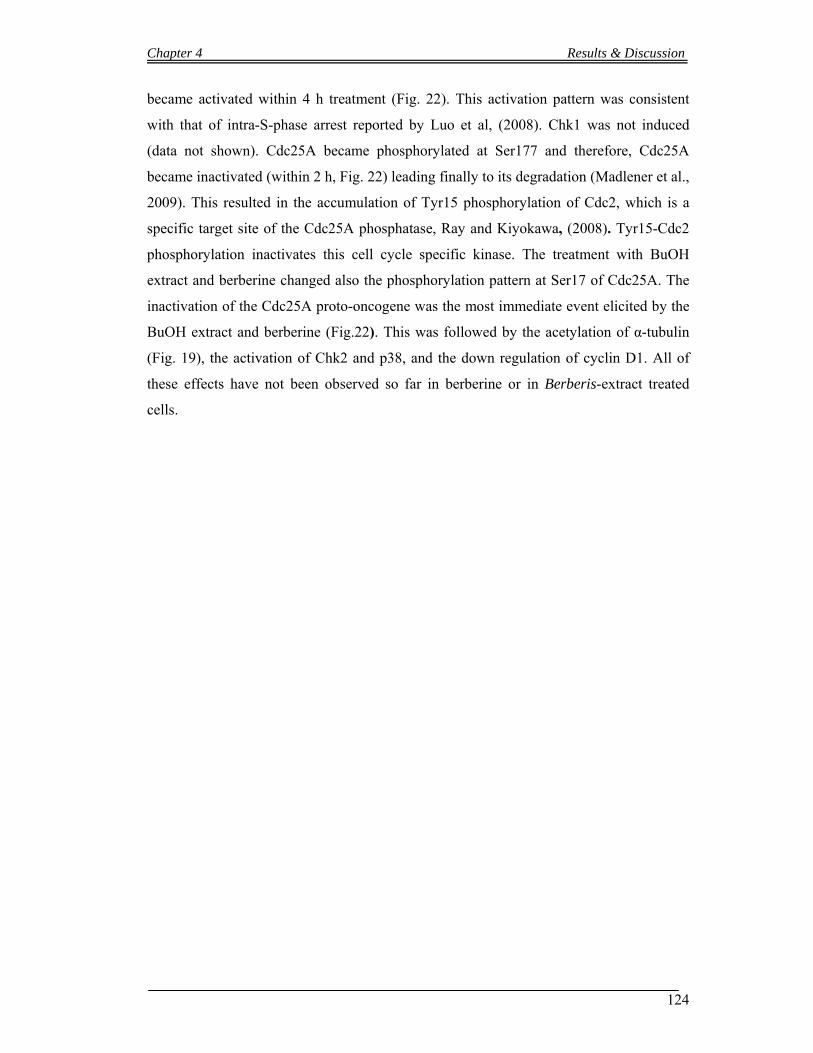
Chapter 4 Results & Discussion
124
became activated within 4 h treatment (Fig. 22). This activation pattern was consistent
with that of intra-S-phase arrest reported by Luo et al, (2008). Chk1 was not induced
(data not shown). Cdc25A became phosphorylated at Ser177 and therefore, Cdc25A
became inactivated (within 2 h, Fig. 22) leading finally to its degradation (Madlener et al.,
2009). This resulted in the accumulation of Tyr15 phosphorylation of Cdc2, which is a
specific target site of the Cdc25A phosphatase, Ray and Kiyokawa, (2008). Tyr15-Cdc2
phosphorylation inactivates this cell cycle specific kinase. The treatment with BuOH
extract and berberine changed also the phosphorylation pattern at Ser17 of Cdc25A. The
inactivation of the Cdc25A proto-oncogene was the most immediate event elicited by the
BuOH extract and berberine (Fig.22). This was followed by the acetylation of α-tubulin
(Fig. 19), the activation of Chk2 and p38, and the down regulation of cyclin D1. All of
these effects have not been observed so far in berberine or in Berberis-extract treated
cells.
Chapter 4 Results & Discussion
125
Figure 22 Induction of stress response by the BuOH extract of Berberis lycium and
Berberine:
Chapter 4 Results & Discussion
126
4.1.2 Anti-neoplastic Activities and Phytochemicals studies of Mallotus phillipensis
5.1.2.1 Inhibition of HL-60 cell proliferation by Mallotus phillipensis extracts
Logarithmically growing HL-60 cells were incubated with increasing concentrations of n-
Hexane, EtOAc and BuOH extract for 72 h. Then, cells were counted and the inhibition
of proliferation was calculated. The Hexane extract showed the highest toxicity against
HL-60 cells (IC50 1.5 mg dry roots equivalent /ml medium) after 72h (Fig. 23; page. 127).
The inhibition of HL-60 proliferation that was observed upon treatment with hexane
extract was preceded by the down regulation of the proto-oncogene cyclin D1 after 48 h
(Fig. 27; page. 131). Suppression of cyclin D1 is potent mechanisms to block cancer cell
growth.
4.1.2.2 Induction of apoptosis by extract of Mallotus phillipensis
Logarithmically growing cells were incubated with increasing concentrations of n-hexane
fraction of Mallotus phillipensis for 48h and the induction of cell death was analyzed. The
extract induced apoptosis 18% after 48h of treatment with 1.5 mg dry roots equivalent /ml
medium (Fig. 24; page. 128). I monitored the ability of Mallotus phillipensis hexane
fraction and the observation indicates that the anti-neoplastic effects have been triggered
by induction apoptosis through caspase-2 activation (Fig. 22; page. 129).
4.1.2.3 Effect of Hexane fraction on cell cycle distribution.
HL-60 cells were exposed to 1.5 mg dry roots equivalent /ml medium hexane fraction 48
h to investigate the cell cycle distribution. There were observed a slight alteration in cell
distribution but the results were not significant (Fig. 26; page 130).

Chapter 4 Results & Discussion
127
Treatment with Hexan extract
Control 1
1.5 2
Control 1
1.5 2
Control 1
1.5 2
0
25
50
75
100
* * *
*
*
*
*
extract concentration (mg/ml)
% c
ell
pro
life
rati
on
Treatment with EtOAc extract
Control 1
1.5 2
Control 1
1.5 2
Control 1
1.5 2
0
25
50
75
100
* *
**
extract concentration (mg/ml)
% c
ell
pro
life
rati
on
Treatment with BuOH extract
Control 1
1.5 2
Control 1
1.5 2
Control 1
1.5 2
0
25
50
75
100* * * * * * *
extract concentration (mg/ml)
% c
ell
pro
life
rati
on
Figure 23 Anti-proliferative effect of Mallotus phillipensis extracts: HL-60 cells were
seeded into T-25 tissue culture flasks (1x10⁵ cells/ml), grown for 24 h to enter
logarithmic growth phase, and incubated with increasing concentrations (a) Hexane
extract (1.00, 1.50, and 2.00 mg dry roots equivalent /ml medium); (b) EtOAc extract
(1.00, 1.50 and 2.0 mg dry roots equivalent /ml medium); (c)BuOH extract (1.00, 1.5 and
2.0 mg dry roots equivalent /ml) Cells were counted after 24, 48 and 72 h of treatment
(white, light gray and dark gray columns, respectively) and the percentage of proliferation
was calculated and compared to DMSO-controls (Control). Controls were considered as
cells with a maximal proliferation rate (100%). Experiments were done in triplicate. Error
bars indicate SEM, asterisks significance (p< 0.05).

Chapter 4 Results & Discussion
128
Hexan extract treatment (48 h)
Control 1
1.5
0
10
20
30
*
extract concentration (mg/ml)
% a
po
pto
tic
ce
lls
Figure 24 Induction of apoptosis by the Mallotus phillipensis Hexane fraction: HL-60
cells were incubated with increasing extract concentrations (1 and 1.5 mg/ml dry powder
equivalent) for 48 h. Then, cells were double stained with Hoechst 33258 and propidium
iodide and examined under a fluorescence microscope and a DAPI filter. Nuclei with
morphological changes indicating apoptosis (Methods) were counted. C. control; 1. 1 mg
dry roots equivalent /ml medium and 2. 1.5 mg dry roots equivalent /ml medium. The
percentages of vital and apoptotic cells calculated. Experiments were done in triplicate.
Error bars indicate SEM, asterisks significance (p<0.05).
2
C 1
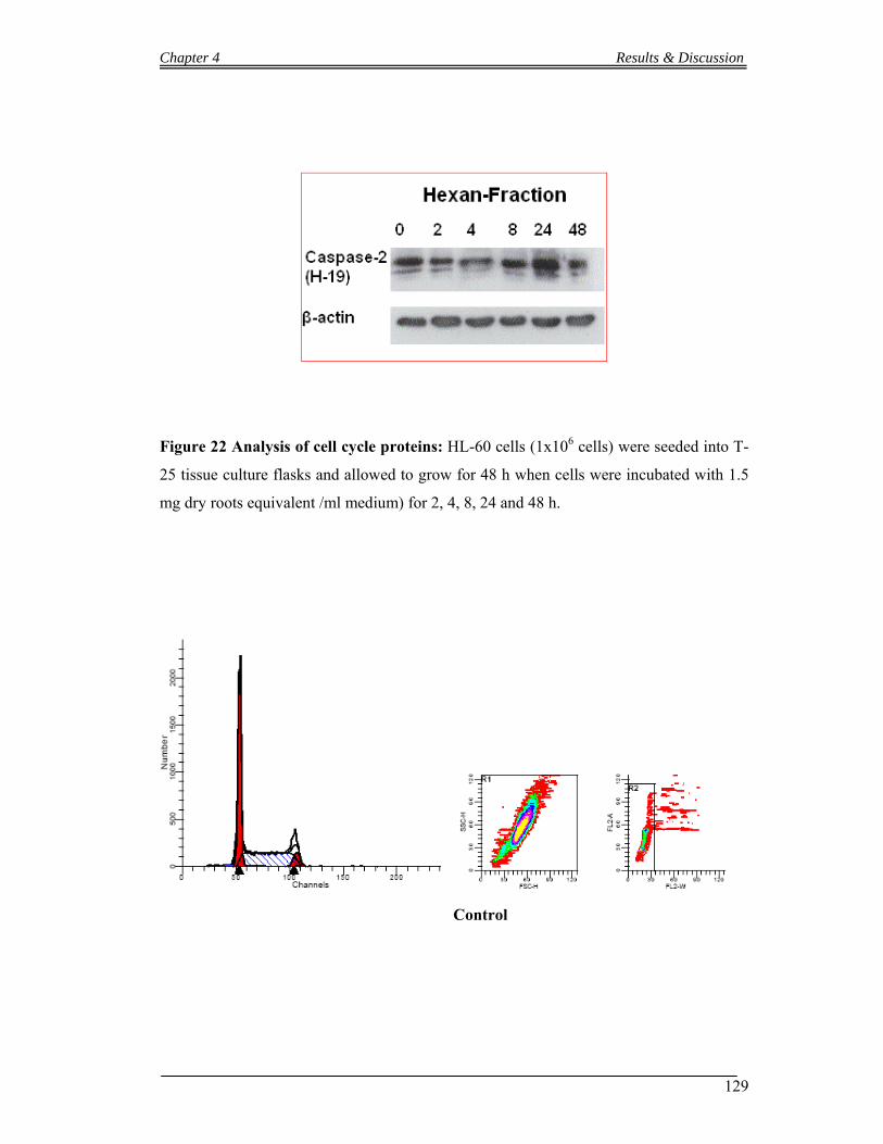
Chapter 4 Results & Discussion
129
Figure 22 Analysis of cell cycle proteins: HL-60 cells (1x10⁶ cells) were seeded into T-
25 tissue culture flasks and allowed to grow for 48 h when cells were incubated with 1.5
mg dry roots equivalent /ml medium) for 2, 4, 8, 24 and 48 h.
Control

Chapter 4 Results & Discussion
130
1.5 mg/ml dry roots equivalent /ml medium
Cell cycle distribution afterBuOH extract treatment (48 h)
G0-G1 S G2-M0
10
20
30
40
50 Control1.5 mg/ml
% c
ells
Figure 26 Cell Cycle Distribution of HL-60 cells upon treatment with hexane
Fraction of Mallotus phillipensis:
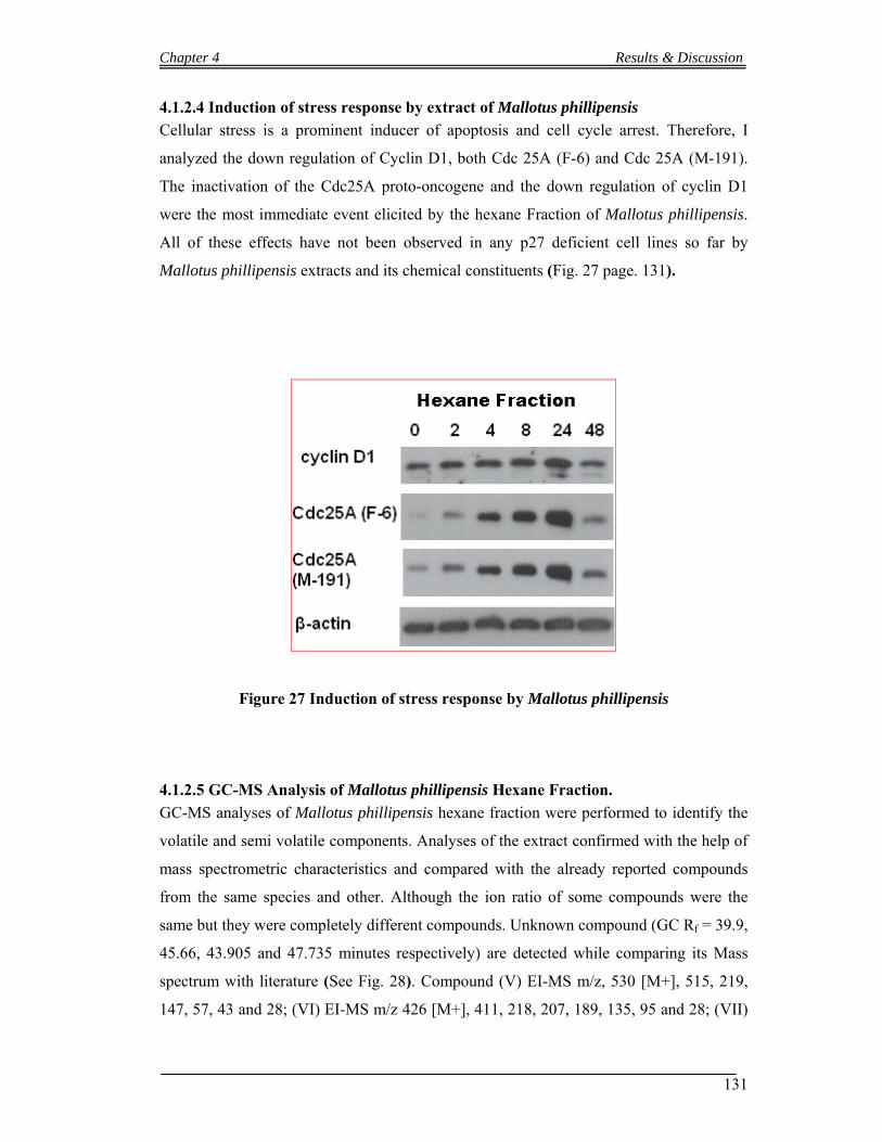
Chapter 4 Results & Discussion
131
4.1.2.4 Induction of stress response by extract of Mallotus phillipensis
Cellular stress is a prominent inducer of apoptosis and cell cycle arrest. Therefore, I
analyzed the down regulation of Cyclin D1, both Cdc 25A (F-6) and Cdc 25A (M-191).
The inactivation of the Cdc25A proto-oncogene and the down regulation of cyclin D1
were the most immediate event elicited by the hexane Fraction of Mallotus phillipensis.
All of these effects have not been observed in any p27 deficient cell lines so far by
Mallotus phillipensis extracts and its chemical constituents (Fig. 27 page. 131).
Figure 27 Induction of stress response by Mallotus phillipensis
4.1.2.5 GC-MS Analysis of Mallotus phillipensis Hexane Fraction.
GC-MS analyses of Mallotus phillipensis hexane fraction were performed to identify the
volatile and semi volatile components. Analyses of the extract confirmed with the help of
mass spectrometric characteristics and compared with the already reported compounds
from the same species and other. Although the ion ratio of some compounds were the
same but they were completely different compounds. Unknown compound (GC Rf = 39.9,
45.66, 43.905 and 47.735 minutes respectively) are detected while comparing its Mass
spectrum with literature (See Fig. 28). Compound (V) EI-MS m/z, 530 [M+], 515, 219,
147, 57, 43 and 28; (VI) EI-MS m/z 426 [M+], 411, 218, 207, 189, 135, 95 and 28; (VII)

Chapter 4 Results & Discussion
132
EI-MS m/z 442 [M+], 424, 355, 281, 207and 28; (VIII) EI-MS m/z 484[M+], 466, 406,
257, 189, 109, 43 and 28. Compounds (IX) EI-MS m/z, 351 [M+], 314, 286, 256, 197 and
97; (X) EI-MS m/z, 396 [M+], 337, 320, 294, 240, 154, 126, 83, 59 and 41 were present
in abundant (Rf = 17.917 and 31.125 minutes respectively).
H
HH
H
HO
I Lupeol
O
O
Me
Me
Me
MeMe
OH
OH
OH
HO
O
O
H
H
II Kamalachalcones C
H
HH
H
HO
OH
III Betulin
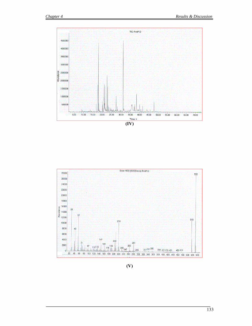
Chapter 4 Results & Discussion
133
(IV)
(V)

Chapter 4 Results & Discussion
134
(VI)
(VII)
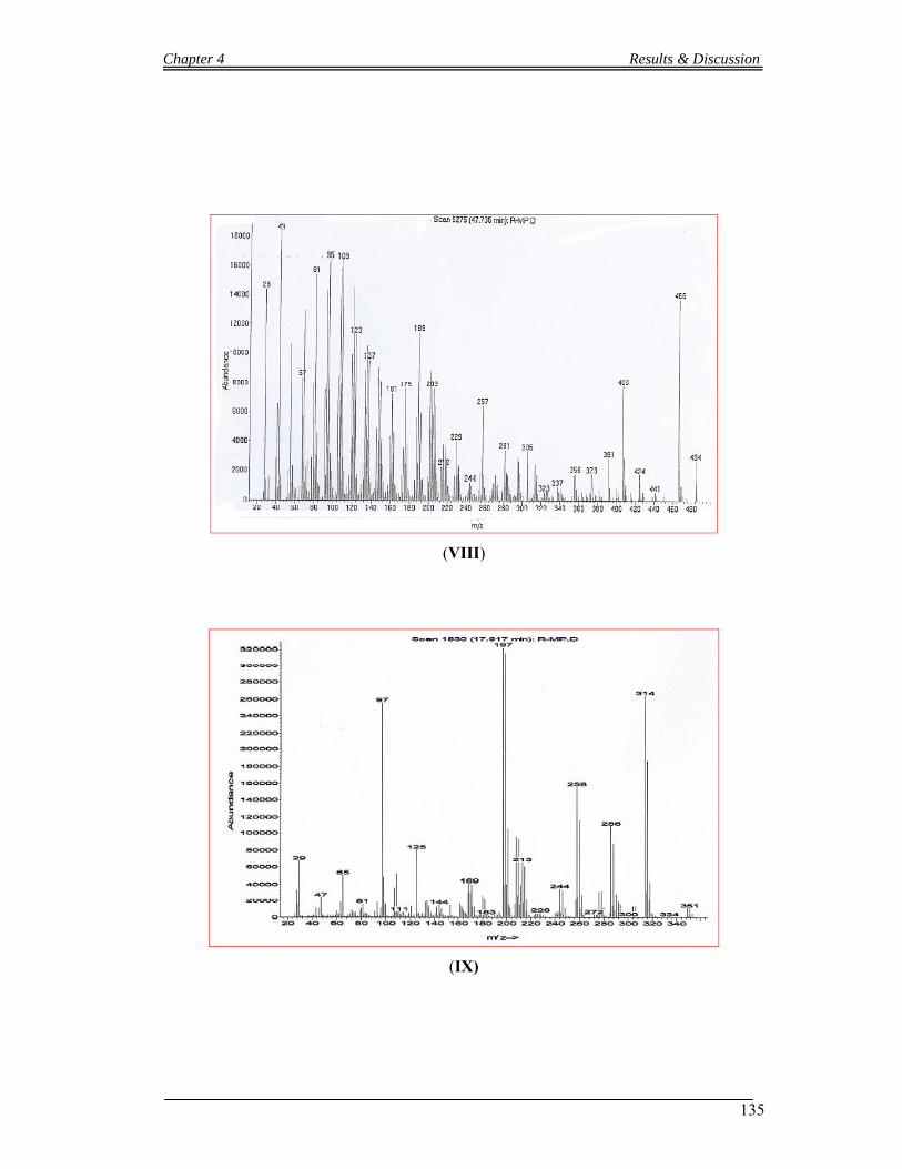
Chapter 4 Results & Discussion
135
(VIII)
(IX)
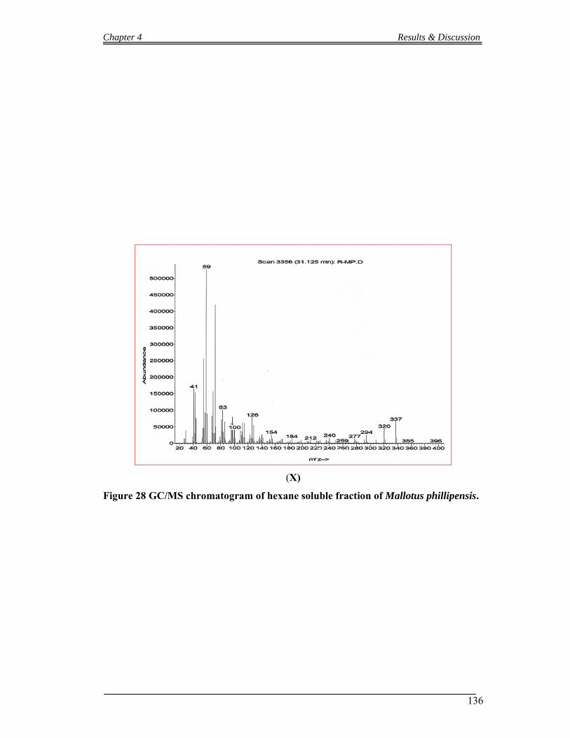
Chapter 4 Results & Discussion
136
(X)
Figure 28 GC/MS chromatogram of hexane soluble fraction of Mallotus phillipensis.
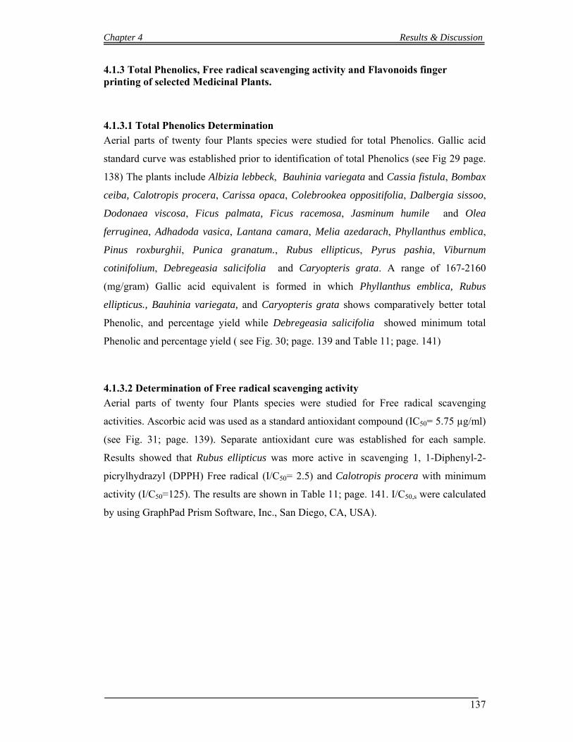
Chapter 4 Results & Discussion
137
4.1.3 Total Phenolics, Free radical scavenging activity and Flavonoids finger printing of selected Medicinal Plants.
4.1.3.1 Total Phenolics Determination
Aerial parts of twenty four Plants species were studied for total Phenolics. Gallic acid
standard curve was established prior to identification of total Phenolics (see Fig 29 page.
138) The plants include Albizia lebbeck, Bauhinia variegata and Cassia fistula, Bombax
ceiba, Calotropis procera, Carissa opaca, Colebrookea oppositifolia, Dalbergia sissoo,
Dodonaea viscosa, Ficus palmata, Ficus racemosa, Jasminum humile and Olea
ferruginea, Adhadoda vasica, Lantana camara, Melia azedarach, Phyllanthus emblica,
Pinus roxburghii, Punica granatum., Rubus ellipticus, Pyrus pashia, Viburnum
cotinifolium, Debregeasia salicifolia and Caryopteris grata. A range of 167-2160
(mg/gram) Gallic acid equivalent is formed in which Phyllanthus emblica, Rubus
ellipticus., Bauhinia variegata, and Caryopteris grata shows comparatively better total
Phenolic, and percentage yield while Debregeasia salicifolia showed minimum total
Phenolic and percentage yield ( see Fig. 30; page. 139 and Table 11; page. 141)
4.1.3.2 Determination of Free radical scavenging activity
Aerial parts of twenty four Plants species were studied for Free radical scavenging
activities. Ascorbic acid was used as a standard antioxidant compound (IC50= 5.75 µg/ml)
(see Fig. 31; page. 139). Separate antioxidant cure was established for each sample.
Results showed that Rubus ellipticus was more active in scavenging 1, 1-Diphenyl-2-
picrylhydrazyl (DPPH) Free radical (I/C50= 2.5) and Calotropis procera with minimum
activity (I/C50=125). The results are shown in Table 11; page. 141. I/C50,s were calculated
by using GraphPad Prism Software, Inc., San Diego, CA, USA).
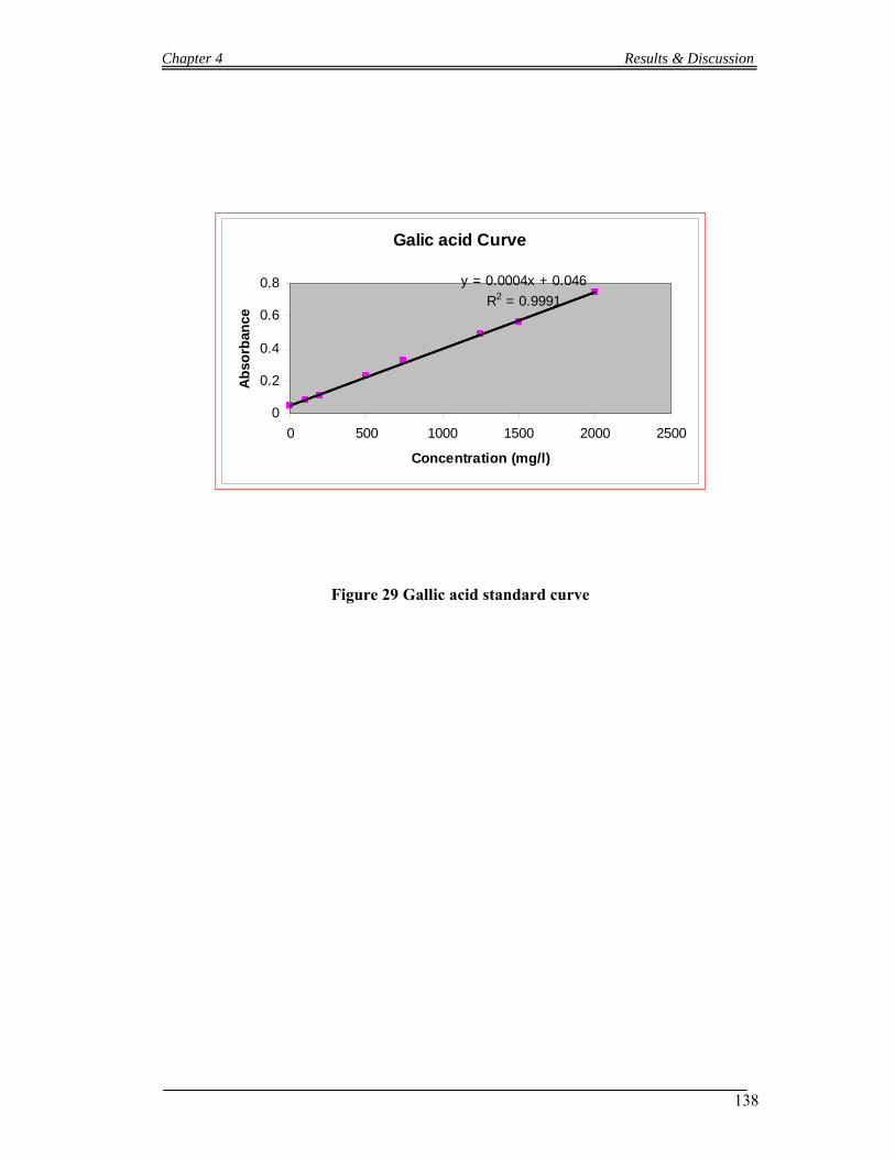
Chapter 4 Results & Discussion
138
Galic acid Curve
y = 0.0004x + 0.046
R2 = 0.9991
0
0.2
0.4
0.6
0.8
0 500 1000 1500 2000 2500
Concentration (mg/l)
Ab
sorb
ance
Figure 29 Gallic acid standard curve
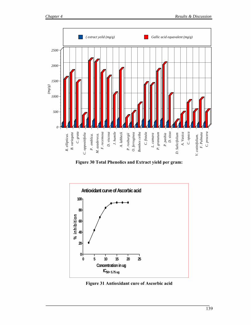
Chapter 4 Results & Discussion
139
R. e
llip
ticu
s.
B. v
arie
gata
C. g
rata
C. o
ppos
itifo
lia
P.
embl
ica.
M. a
zade
reca
. F
. rec
emos
a
D. v
isco
sa
J. h
umil
e
A. l
abbe
ck
P. r
oxbu
rgii
O. f
erru
gine
a
Bom
bex
ceib
a
C. f
istu
la
L. c
amar
a
P. g
rana
tum
P. p
ashi
a D
. sis
so
D. S
alic
ifol
ium
A. V
asic
a
C. o
paca
V. c
otin
ifol
ium
. F
. Pal
mat
a
C. p
roce
ra
0
500
1000
1500
2000
2500(m
g/g)
( extract yeild (mg/g) Gallic acid equavalent (mg/g)
Figure 30 Total Phenolics and Extract yield per gram:
Antioxidant curve of Ascorbic acid
0 5 10 15 20 250
20
40
60
80
100
Concentration in ugIC50= 5.75 ug
% i
nh
ibit
ion
Figure 31 Antioxidant cure of Ascorbic acid
Chapter 4 Results & Discussion
140
Table 11 Comparative total Phenolic, extract yield per gram and IC50 Values.
S/No Plants name Amount of extract/g
Total Phenolics a (mg of GAE/g dw)
IC50
1. Rubus ellipticus 0.137 ± 0.001 1558 2.54 2. Bauhinia variegata 0.13 ± 0.01 1775 3.26 3. Caryopteris grata 0.197 ± 0.001 1450 3.29 4. Colebrookea oppositifolia 0.07 ± 0.01 383 3.36 5. Phyllanthus emblica 0.255 ± 0.001 2160 3.58 6. Melia azedarach 0.21 ± 0.01 2120 7.32 7. Ficus racemosa 0.115 ± 0.001 1775 9.14 8. Dodonaea viscosa 0.19 ± 0.01 1600 10.26 9. Jasminum humile 0.137 ± 0.001 1050 11.32 10. Albizia lebbeck 0.25 ± 0.001 1850 12.1 11. Pinus roxburghii 0.085 ± 0.001 308 15.13 12. Olea ferruginea 0.11 ± 0.01 450 16.26 13. Bombax ceiba 0.09 ± 0.01 725 24.56 14. Cassia fistula 0.21 ± 0.01 1383 25.03 15. Lantana camara 0.185 ± 0.001 1358 30.65 16. Punica granatum. 0.165 ± 0.001 1817 32.38 17. Pyrus pashia. 0.225 ± 0.001 2027 32.50 18. Dalbergia sissoo 0.16 ± 0.01 1025 32.56 19. Debregeasia salicifolia 0.077 ± 0.001 167 33.37 20. Adhadoda vasica 0.117 ± 0.001 426 39.27 21. Carissa opaca 0.15 ± 0.01 800 40.26 22. Viburnum cotinifolium 0.137 ± 0.001 500 41.33 23. Ficus palmata 0.115±0.001 883 64.26 24. Calotropis procera 0.13±0.001 500 125
dw dry weight of the original sample.
4.1.3.3 Flavonoids finger printing of selected Plants
Selected medicinal plants have been studied for flavonoids types. Fourteen different
standards flavonoids have been run to determine the samples qualitatively. The standards
which have been used are Rutin, Quercetin, Kaempferol, Myricetin, Catechin, Vitexin,
Orientin, Isoquercitrin, Isovitexin, Hyperoside, Luteolin-7-glucoside, Kampferol-7-
neohesperidoside, Luteolin and Apigenin. Each sample and standard flavonoids have
been run two times on thin layer chromatographic plates as describe in TLC method
(4.7.1.2) (without standard and with standard). Different colours zone of flavonoids have
been appeared after spraying of reagent A (1% ethanolic 2-Aminoethyle diphenyl
borinate solution) and reagent B (5% ethanolic solution of polyethylene glycol-400)
under 365 nm UV light (Fig. 32). Retention time (R t) and colors after spraying reagent A
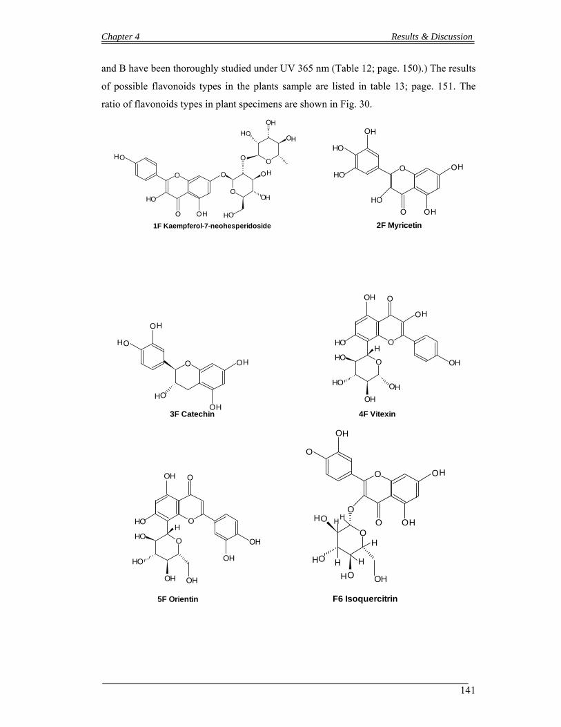
Chapter 4 Results & Discussion
141
and B have been thoroughly studied under UV 365 nm (Table 12; page. 150).) The results
of possible flavonoids types in the plants sample are listed in table 13; page. 151. The
ratio of flavonoids types in plant specimens are shown in Fig. 30.
O
OO
O
H
H
OH
1F Kaempferol-7-neohesperidoside
O
O O
O
O
O
O
O
O
O
H
HH
H
H
H
O O
OO
O
H
H
OH
H
HO
HO
2F Myricetin
O O
O
O
H
H
H
O
OH
H
3F Catechin
O
OH
OH
O
OHO
O
O
O
OH
H
H
HH
OH
4F Vitexin
OOH
O
OHO
O
O
O
H
H
HH
OH
OH
OH
5F Orientin
O O
OO
O
H
H
O
O
O
O
O
O
H
H
H
H OH
HH
H
H
H
F6 Isoquercitrin

Chapter 4 Results & Discussion
142
O O
OO
O
H
H
H
O
O
O
O
O
O
H
H
H
H OH
7F Hyperoside
O
O
O
OH
O
H
H
HOOO
O
OH
H
HOH
8F Isovitexin
O O
OO
O
H
OH
H
O
O
O
O
O
H
H
H
H
9F Luteolin 7-O-glucoside
O
OO
O
H
H
OH
11F Kaempferol-7-neohesperidoside
O
O O
O
O
O
O
O
O
O
H
HH
H
H
H
O O
OO
H
H
O
O
O
O
O
O
H
H
H
H
H
HO
OO
O
O
O H
H
H
10F Rutin
O O
OO
O
H
H
OH
H
HO
12F Quercetin
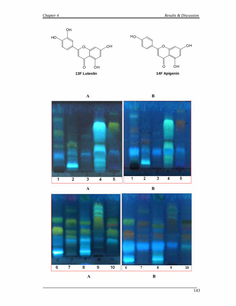
Chapter 4 Results & Discussion
143
O O
OO
O
H
H
OH
H
13F Luteolin
O O
OO
O
H
H
H
14F Apigenin
A B
A B
A B
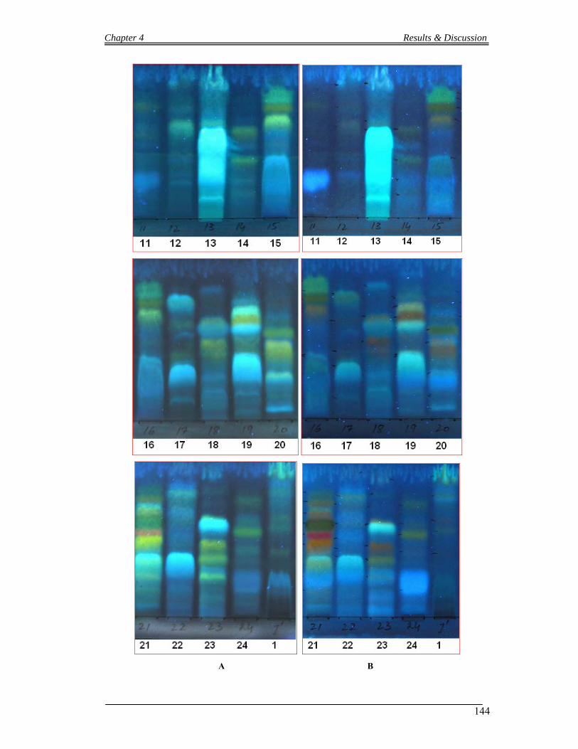
Chapter 4 Results & Discussion
144
A B

Chapter 4 Results & Discussion
145
Figure 32. Flavonoids finger printing.of standard and selected plants (1: R. ellipticus;
2. B. variegata; 3. C. grata; 4. C. oppositifolia; 5. P. emblica; 6. M. azedarach; 7. F.
racemosa; 8. D. viscosa; 9. J. humile; 10. A. lebbeck; 11. P. roxburghii; 12. O.
ferruginea; 13. B. ceiba; 14. C. fistula; 15. L. camara; 16. P. granatum; 17. P. pashia 18.
D. sissoo; 19. D. salicifolia; 20. A. Vasica; 21. C. opaca; 22. V. cotinifolium; 23. F.

Chapter 4 Results & Discussion
146
palmata; 24. C. procera) have been collected. The plants materials are extracted as
described in extraction procedure (see extraction procedure 3.6.1). Standard flavonoids.
F1. Kaempferol; F2. Myricetin; F3. Catechin; F4 Vitexin; F5 Orientin; F6 Isoquercitrin;
F7. Hyperoside; F8. Isovitexin; F9. Luteolin-7-glucoside; F10. Rutin; F11 Kaempferol-7-
neohesperidoside; F12 Quercetin; F13 Luteolin and F14 Apigenin)
PERCENTAGE OF FLAVONOIDS
100
29.16
4.16 0
258.33
54.16
0
29.1616.66
0 012.5
33.33
58.33
020406080
100120
Pheno
lic A
cids F1 F2 F3 F4 F5 F6 F7 F8 F9
F10 F11 F12 F13 F14
Flavonoids
%ag
e
Figure 33 Percentage of Flavonoids in Plant samples: Percentage of standards
flavonoids (F1. Kaempferol; F2. Myricetin; F3. Catechin; F4 Vitexin; F5 Orientin; F6
Isoquercitrin; F7. Hyperoside; F8. Isovitexin; F9. Luteolin-7-glucoside; F10. Rutin; F11
Kaempferol-7-neohesperidoside; F12 Quercetin; F13 Luteolin and F14 Apigenin)

Chapter 4 Results & Discussion
147
R. e
llip
ticu
s.
B. v
arie
gata
C. g
rata
C. o
ppos
itifo
lia
P. e
mbl
ica
M. a
zeda
rach
F. r
acem
osa
D. v
isco
saJ.
hum
ile
A. l
ebbe
ck
P. r
oxbu
rghi
i
O. f
erru
gine
aB
. cei
ba
C. f
istu
la
L. c
amar
a P
. gra
natu
m
P. p
ashi
a .
D.
siss
oo
. D. S
alic
ifoli
aA
. Vas
ica
C. o
paca
V. c
otin
ifol
ium
F. P
alm
ata
C. p
roce
ra
Phenolic Acid
F1
F2
F4
F5
F7
F8
F10
F11
F14
Figure 34. Types of Flavonoids in each sample
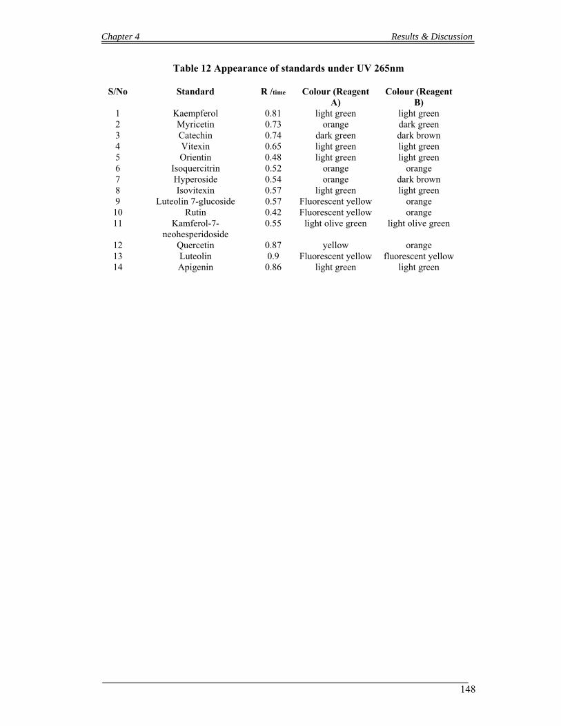
Chapter 4 Results & Discussion
148
Table 12 Appearance of standards under UV 265nm
S/No Standard R /time Colour (Reagent A)
Colour (Reagent B)
1 Kaempferol 0.81 light green light green 2 Myricetin 0.73 orange dark green 3 Catechin 0.74 dark green dark brown 4 Vitexin 0.65 light green light green 5 Orientin 0.48 light green light green 6 Isoquercitrin 0.52 orange orange 7 Hyperoside 0.54 orange dark brown 8 Isovitexin 0.57 light green light green 9 Luteolin 7-glucoside 0.57 Fluorescent yellow orange 10 Rutin 0.42 Fluorescent yellow orange 11 Kamferol-7-
neohesperidoside 0.55 light olive green light olive green
12 Quercetin 0.87 yellow orange 13 Luteolin 0.9 Fluorescent yellow fluorescent yellow 14 Apigenin 0.86 light green light green
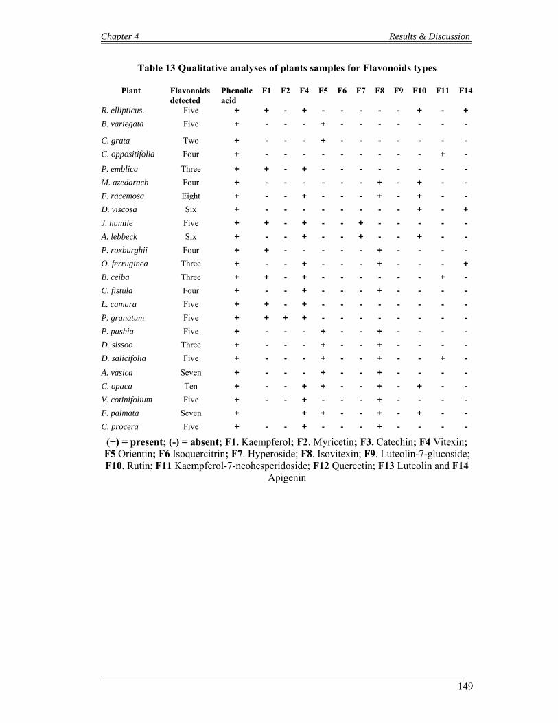
Chapter 4 Results & Discussion
149
Table 13 Qualitative analyses of plants samples for Flavonoids types
Plant Flavonoids detected
Phenolic acid
F1 F2 F4 F5 F6 F7 F8 F9 F10 F11 F14
R. ellipticus. Five + + - + - - - - - + - +
B. variegata Five + - - - + - - - - - - -
C. grata Two + - - - + - - - - - - -
C. oppositifolia Four + - - - - - - - - - + -
P. emblica Three + + - + - - - - - - - -
M. azedarach Four + - - - - - - + - + - -
F. racemosa Eight + - - + - - - + - + - -
D. viscosa Six + - - - - - - - - + - +
J. humile Five + + - + - - + - - - - -
A. lebbeck Six + - - + - - + - - + - -
P. roxburghii Four + + - - - - - + - - - -
O. ferruginea Three + - - + - - - + - - - +
B. ceiba Three + + - + - - - - - - + -
C. fistula Four + - - + - - - + - - - -
L. camara Five + + - + - - - - - - - -
P. granatum Five + + + + - - - - - - - -
P. pashia Five + - - - + - - + - - - -
D. sissoo Three + - - - + - - + - - - -
D. salicifolia Five + - - - + - - + - - + -
A. vasica Seven + - - - + - - + - - - -
C. opaca Ten + - - + + - - + - + - -
V. cotinifolium Five + - - + - - - + - - - -
F. palmata Seven + + + - - + - + - -
C. procera Five + - - + - - - + - - - -
(+) = present; (-) = absent; F1. Kaempferol; F2. Myricetin; F3. Catechin; F4 Vitexin; F5 Orientin; F6 Isoquercitrin; F7. Hyperoside; F8. Isovitexin; F9. Luteolin-7-glucoside; F10. Rutin; F11 Kaempferol-7-neohesperidoside; F12 Quercetin; F13 Luteolin and F14
Apigenin
Chapter 4 Results & Discussion
150
4.1.4 Antibacterial and Free radical scavenging activities, Flavonoids finger printing of Mallotus philippensis.
4.1.4.1 Antibacterial activities
Methanolic extracts of (roots and flower powder) M. philippensis were tested against five
strains of bacteria. Flower powder (Kamala or Kamara) extract was effective against
Gram positive bacteria, Bacillus subtilis and Staphylococcus aureus (MICs 0.7 and 0.6
mg/ml), while it could not showed any response against the remaining bacterial strains up
to maximum concentration of 15mg/ml. Roots extract was effective against one Gram
positive bacteria Bacillus subtilis and one Gram negative bacteria Salmonella setubal
(MICs 1.00 and 2.00 mg/ml) respectively, while it did not showed any response against
the remaining bacterial strains up to maximum concentration of 15mg/ml. the results
shown in Figure 35 and table 14 below.
4.1.4.2 Free radical scavenging activities
Methanolic extract of (flower powder “Kamala” and leaves) M. philippensis were
investigated for its free radical scavenging capacity. DPPH (1, 1-Diphenyl-2-
picrylhydrazyl) was used in the experiment. The bleaching of DPPH colour in the
experiment by Mallotus philippensis extracts represents the scavenging capacity of its
extracts. Results shows that the leaves extracts was more reactive than the kamala extracts
(IC50 33 and 47 µg/ml respectively) see Figure 36, Ascorbic acid was used as standard
antioxidant (IC50 = 5.75 µg /ml).
4.1.4.3 Flavonoids finger printing of Mallotus philippensis
Aerial parts of Mallotus philippensis have been studied for flavonoids types. Fourteen
different standards flavonoids have been run parallel to sample in order to identify
flavonoids in the samples qualitatively. The standards which have been used are Rutin,
Quercetin, Kaempferol, Myricetin, Catechin, Vitexin, Orientin, Isoquercitrin, Isovitexin,
Hyperoside, Luteolin-7-glucoside, Kaempferol-7-neohesperidoside, Luteolin and
Apigenin. The samples and standard flavonoids have been run on thin layer
chromatographic plates as describe in TLC method (4.7.1.2). Different colors zone of
flavonoids have been appeared after spraying of reagent A (1% ethanolic 2-Aminoethyle
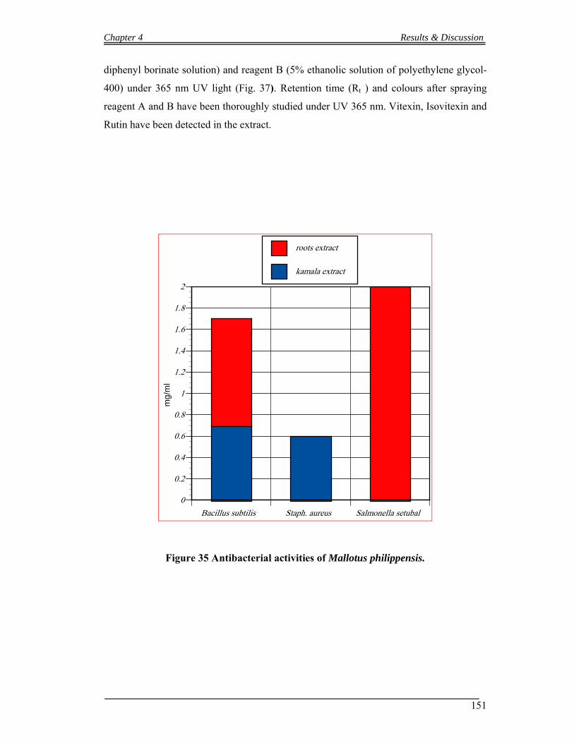
Chapter 4 Results & Discussion
151
diphenyl borinate solution) and reagent B (5% ethanolic solution of polyethylene glycol-
400) under 365 nm UV light (Fig. 37). Retention time (Rt ) and colours after spraying
reagent A and B have been thoroughly studied under UV 365 nm. Vitexin, Isovitexin and
Rutin have been detected in the extract.
Bacillus subtilis Staph. aureus Salmonella setubal
0
0.2
0.4
0.6
0.8
1
1.2
1.4
1.6
1.8
2
mg
/ml
kamala extract
roots extract
Figure 35 Antibacterial activities of Mallotus philippensis.
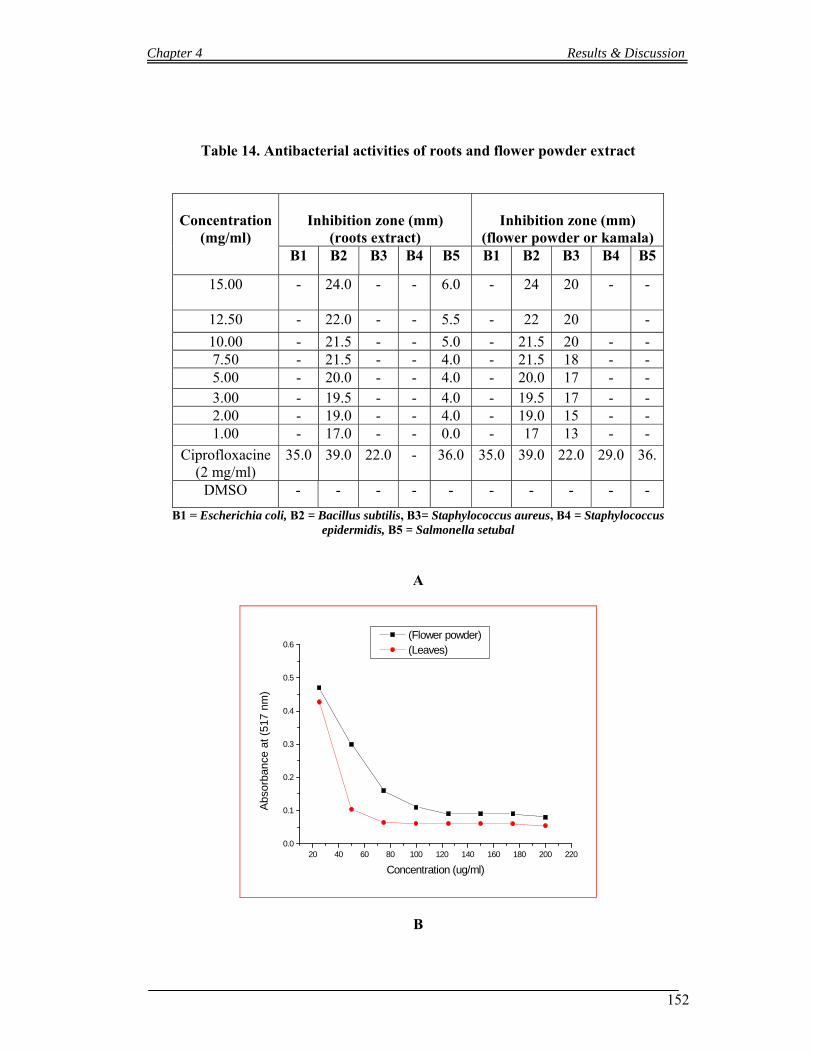
Chapter 4 Results & Discussion
152
Table 14. Antibacterial activities of roots and flower powder extract
Concentration
(mg/ml)
Inhibition zone (mm)
(roots extract)
Inhibition zone (mm)
(flower powder or kamala) B1 B2 B3 B4 B5 B1 B2 B3 B4 B5
15.00
- 24.0 - - 6.0 - 24 20 - -
12.50 - 22.0 - - 5.5 - 22 20 -
10.00 - 21.5 - - 5.0 - 21.5 20 - - 7.50 - 21.5 - - 4.0 - 21.5 18 - - 5.00 - 20.0 - - 4.0 - 20.0 17 - - 3.00 - 19.5 - - 4.0 - 19.5 17 - - 2.00 - 19.0 - - 4.0 - 19.0 15 - - 1.00 - 17.0 - - 0.0 - 17 13 - -
Ciprofloxacine (2 mg/ml)
35.0 39.0 22.0 - 36.0 35.0 39.0 22.0 29.0 36.
DMSO - - - - - - - - - -
B1 = Escherichia coli, B2 = Bacillus subtilis, B3= Staphylococcus aureus, B4 = Staphylococcus epidermidis, B5 = Salmonella setubal
A
20 40 60 80 100 120 140 160 180 200 2200.0
0.1
0.2
0.3
0.4
0.5
0.6 (Flower powder) (Leaves)
Abs
orba
nce
at (
517
nm)
Concentration (ug/ml)
B
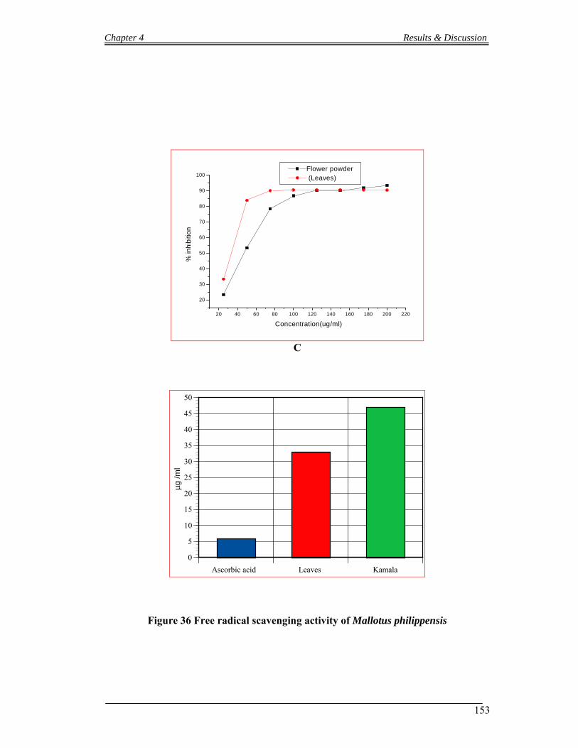
Chapter 4 Results & Discussion
153
20 40 60 80 100 120 140 160 180 200 220
20
30
40
50
60
70
80
90
100Flower powder (Leaves)
% in
hibi
tion
Concentration(ug/ml)
C
Ascorbic acid Leaves Kamala
0
5
10
15
20
25
30
35
40
45
50
µg
/m
l
Figure 36 Free radical scavenging activity of Mallotus philippensis

Chapter 4 Results & Discussion
154
A B
Figure 37 Flavonoids finger printing of Mallotus philippensis: (MP) Sample and
standard flavonoids (1F. Kaempferol; 4F Vitexin; 6F Isoquercetin; 7F. Hyperoside; 8F.
Isovitexin; 9F. Luteolin-7-glucoside; 10F. Rutin and S3 Quercetin and Rutin Mix)
Chapter 4 Results & Discussion
155
4.2 Discussion
4.2.1 Anti-neoplastic Activities and Phytochemicals studies of Berberis lycium
The roots of B. lycium were extracted with organic solvent methanol, evaporated
methanol and the extract was again dissolved in distilled water. The water solution was
again extracted with organic solvent hexane, ethyl acetate, butanol and the remaining of
water. The fractions were analyzed qualitatively by thin layer chromatography (TLC).
Berberine, palmatine and berbamine were used as reference compounds. Berbamine was
reported to be a constituent of B. lycium (Ali and Khan, 1978), while there was no
evidence of its presence in the here performed TLC and RP-HPLC analyses. All extracts
contained berberine (retention factor, Rf = 0.151) and palmatine (Rf = 0.088), whereas the
highest concentration of both compounds was detected in the BuOH extract. For
quantification RP-HPLC was used. Therefore berberine and palmatine were selected to
study as reference compounds in ordered to identify the active compound in B. lycium.
The effects of root extracts of B. lycium were studied against HL-60 human leukemia
cells and compared them with the purified constituent alkaloids, Berberine and Palmatine.
B. lycium is an erect small rigid shrub about 1.0-2.5 meters tall, with a thick woody shoot
covered with a thin brittle bark (Hooker, 1882) and is native in the whole Himalaya
Mountains range and widely distributed in temperate and semi-temperate areas of India,
Nepal, Afghanistan, Bangladesh and Pakistan. The active constituents of B. lycium are
alkaloids. The major alkaloids are Umbellatine, Berberine (Ali and Khan, 1978),
Oxyacanthine and Chelidonic acid (Karnick, 1994), Heterocyclic constituents named
Berberisterol, Berberifuranol and Berberilycine (Ali and Sharma, 1996), the alkaloids
Sindamine, Punjabine and Gilgitine (Leet et al., 1982, 1983), Palmatinechloroform a
tertiary dihydroprotoberberine alkaloid (Miana, 1973), Berbericine and Berbericinine
hydriodide (Ikram et al., 1966) were also found in the roots of B. lycium. Besides these,
Berbamine, starch grains and tannins are also present in small quantities (Ali and Khan,
1978).
In the present investigation berberine and the crude BuOH extract regulated protein
expression and protein activation in HL-60 cells similarly. Also the growth inhibiting-
and apoptosis-inducing potential was similar and FACS- and Comet data were almost
identical. This is a strong indication that BuOH-mediated cell cycle arrest was due to
berberine. I shows that the growth inhibitory properties of berberine and BuOH extract
correlated directly with the inactivation and down-regulation of the proto-oncogene

Chapter 4 Results & Discussion
156
Cdc25A resulting in the inactivation of Cdc2. Also the inhibition of human
nasopharyngeal carcinoma CNE-2 cell growth by berberine was associated with
suppression of cyclin B1, CDK1 (Cdc2), and Cdc25C proteins (Cai et al, 2006). In human
glioblastoma T98G cells berberine induced cell cycle retardation in G1-phase through
increased expression of p27 and suppression of CDK2, CDK4, cyclin D, and cyclin E
proteins (Eom et al., 2008), and HL-60 cell growth was significantly inhibited by
berberine in G0/G1 phase with a decrease in S-phase cells (Wang and Lin, 2004). In
another study FACS analyses indicated that berberine induced G2/M-phase arrest in HL-
60 cells and murine myelomonocytic leukemia WEHI-3 cells that was accompanied by
increased levels of Wee1 and 14-3-3sigma, and decreased levels of Cdc25C, CDK1 and
cyclin B1 (Lin et al., 2006). This is in contradiction to the reported G0/G1 arrest (Eom et
al., 2008) and to the intra-S-phase arrest observed in this study, but the differences were
most likely due to the different berberine concentrations used in these investigations.
Notably, intra-S-phase arrest correlated with the activation of Chk2 and this was also
demonstrated in the context of ionizing radiation (Luo et al., 2008). In addition, the
extract and the purified compound caused the down-regulation of the proto-oncogene
cyclin D1 after 48 h and this certainly added up to the cell division arrest. Therefore,
berberine and the BuOH extract down-regulated two potent oncogenes, Cdc25A and
cyclin D1.
The proliferation of human umbilical vein endothelial cells (HUVECs) was inhibited
upon incubation with 20 µg/ml berberine. This phenomenon was accompanied by a
significant decrease of PCNA, and a typical apoptotic appearance correlated with a
marked decline in the mitochondrial membrane potential. Berberine-mediated inhibition
of vascular endothelial cell proliferation suppressed neo-vascularization, and this might
be one of the mechanisms attenuating growth and metastasis of tumors (Hao et al., 2005)
Berberine and the BuOH extract in a 3-D metastasis model. This model utilizes single-
layers of lymphendothelial cells onto which MCF-7 cell spheroids are placed that repulse
the endothelial cells thereby generating gaps in the lyphendothelium underneath that
function as gates through which cancer cell bulks can penetrate. However, 10 - 100 µM
Berberine did not prevent lymphendothelial gap formation induced by MCF-7 spheroids
(data not shown).
It was further reported that an ethanol extract of Coptis teeta, which contains berberine
and other components, as well as purified berberine induced apoptosis of MCF-7 breast
cancer cells (Kang et al., 2005). Berberine triggered cell death was reported also in
Chapter 4 Results & Discussion
157
several other human cancer cell lines [Lin et al., 2006; Mantena et al., 2006; Hwang et
al., 2006), such as in human glioblastoma T98G cells that was concomitant with an
increased Bax/Bcl-2 ratio, disruption of the mitochondrial membrane potential, and the
activation of caspase-9 and caspase-3 (Eom et al., 2008). Berberine-induced apoptosis of
human leukemia HL-60 cells was shown to be associated with down-regulation of
nucleophosmin/B23 and telomerase activity (Wu et al., 1999). Furthermore, Liu et al
(2009) reported a cell cycle inhibitory effect of Berberine in a high concentration range
(between 10 - 50 µM), which correlated with DNA damage. In this study, the authors
hypothesize that Berberine inhibited osteosarcoma cell proliferation and induced
apoptosis through genotoxicity. In contrast, we found that the inhibition of proliferation
and the induction of apoptosis occurred with berberine doses and extract concentrations
that were devoid of genotoxic activity, although we agree that high Berberine
concentrations could cause DNA strand breaks. Our data suggest that another
molecular/cellular mechanism transduces the pro-apoptotic properties of Berberine and
BuOH extract and this correlated with α-tubulin acetylation, which is indicative for
microfilament polymerization (Marcus et al., 2005) Tilting the fine-tuned balance of
polymerized/de-polymerized microtubule is incompatible with proper cell division as
well as cell survival. Therefore, the anticancer properties of berberine and the BuOH
extract are reminiscent of that of taxol (Wilson and Forer, 1997) and independent of
genotoxicity.
4.2.2 Anti-neoplastic Activities and Phytochemicals studies of Mallotus phillipensis
The roots of M. phillipensis were extracted with organic solvent methanol. The
concentrated MeOH extract of M phillipensis was dissolved in distilled water and
extracted three times each with organic solvents in sequence of increasing polarity such
as hexane, ethyl acetate (EtOAc), and n-butanol (BuOH). After evaporating each solvent,
9.23 g dred hexane extract, 4.00 g dried EtOAc extract, and 7.08 g dried BuOH extract
was obtained, respectively. M. phillipensis is a deciduous tree widely distributed
throughout tropical Asia, Mountain slopes or valleys, limestone hills or river valleys,
forests; 300-1600 m. Fujian, Anhui, Guangdong, Guizhou, Guangxi, Hainan, Hunan,
Jiangsu, Taiwan, Jiangxi, Sichuan, Xizang, Yunnan, Hubei, Zhejiang, Bhutan,
Philippines, India, Laos, Thailand, Myanmar, Malaysia, Bangladesh, Nepal, Pakistan,
New Guinea, Sri Lanka, Vietnam and N Australia. Different type of chemical compounds
Chapter 4 Results & Discussion
158
(big and small) have been found in the different parts of the plant. Rottlerin (5, 7-
dihydroxy-2, 2-dimethyl-6-(2, 4, 6-trihydroxy-3-methyl-5-acetylbenzyl)-8-cinnamoyl-1
,2-chromine), which is also called mallotoxin, is one of the major constituents that have
been confirmed in different parts of M. phillipensis, exhibiting various pharmacological
activities including mitochondrial uncoupler effects. (Zaidi et al., 2009). Rottlerin was
evaluated and considered that it is a specific inhibitor of the novel protein kinase C
(PKC) isoform, PKC d, and identified as an anticarcinogenic chemical compound
(Soltoff,, 2007). The activation and translocation of PKC d are induced by different
apoptotic stimuli in various cellular systems (Brodie et al., 2003). It has been
demonstrated in various studies that PKC d might not directly inhibited by rottlerin, but
it can produce some cellular changes that is very closely resembles to those produced by
the direct inhibition of PKC d (Soltoff, 2001;Tapia et al., 2006). In one recent study, It
has been found that rottlerin sensitized both glioma cells and colon carcinoma cells to
TRAIL-mediated apoptosis through inhibition of Cdc2 and uncoupling of the
mitochondria, respectively (Tillman et al., 2003; Kim et al., 2005). However, both the
mentioned mechanisms by which rottlerin sensitizes cancer cells to TRAIL-mediated
apoptosis and rottlerin-induced apoptosis are not completely known. In very recent study
it is demonstrated that apoptosis induced by rottlerin was due to its up regulation
property of DR5 (Lim et al., 2009). Furthermore, it has been noticed that rottlerin does
not sensitized normal cell but potentially sensitized various cancer cells, to TRAIL-
mediated apoptosis. Therefore, it is strongly recommended that the combined treatment
with both TRAIL and rottlerin can be use as a safe and effective cancer therapy. It also
noticed that the up regulation of DR5 mediated by CHOP, that is independent of PKC d
activity, contributes to rottlerin-induced apoptosis. Tanaka et al, (2008) has isolated
known friedelane-type triterpenoids compounds from the stem bark of M. phillipensis
and described the anti-tumor promoting activity, and they found for 3-hydroxy-D:A-
friedoolean-3-en-2-one ( IC50 = 292 mol ratio/ 32 pmol/TPA); 3α-hydroxy-D:A-
friedoolean -2-one (IC50 = 288); curcumin was used as positive control, (IC50 = 343);
Epstein-Barrvirus was used as early antigen (EBV-EA) and activation induced by 12-O-
tetradecanoyl phorbol 13-acetate (TPA) used in the experiment.
In the present investigation the hexane fractions regulated protein expression and protein
activation in HL-60 cells. The extract showed the highest toxicity against HL-60 cells
(IC50 1.5 mg dry roots equivalent /ml medium) after 72h. The inhibition of HL-60
proliferation that was observed upon treatment with hexane extract was preceded by the
Chapter 4 Results & Discussion
159
down regulation of the proto-oncogene Cdc25A and cyclin D1 after 48 h. All of these
effects have not been observed in any p53 deficient cell lines so far by Mallotus
phillipensis extracts and its chemical constituents. Valacchi et al, 2008 has reported that
rottlerin deactivate cyclin D1 in HaCaT cell line. The hexane fraction induced 18%
apoptosis after 48h of treatment with 1.5 mg dry roots equivalent /ml medium. I
monitored the ability of M. phillipensis hexane fraction and the observation indicates that
the anti-neoplastic effects have been triggered by induction apoptosis through caspase-2
activation while Brodie et al., 2003 reported that rottlerin activated caspase-3. Caspase-2,
maybe even more versatile as previously thought by mediating such opposing
functions as either killing (Sidi et al., 2008; Olsson et al., 2009) or saving a cell
after DNA damage (Shi et al., 2009) and, subsequently, even more useful in
protecting the whole organism from developing cancer (Ho et al., 2009). It was
therefore concluded that , caspase-2 may represent the archetype member of this
protease family that still unifies many of the above-mentioned functions in a single
enzyme.
The hexane extract was analyzed with GC/MS and different compounds were detected in
the fraction. Mass spectrometric data of some compounds have been co-related with
already reported compounds from different parts of the same species. Some unknown
compounds which have the same m/z ratio as of Lupeol, Betulin, Kamala Chalcones C
(GC Rf = 39.9, 45.66, 43.905 and 47.735 minutes respectively) have been detected.
Rottlerin and friedeline types’ compound that has been reported cytotoxic from M.
phillipensis was not detected in the fraction. It has been confirmed from the present
antineoplastic assay that hexane fraction is active against p53 deficient human leukemia
cell lines (HL-60) and the activity was due to other than rottlerin and friedeline types’
compound.
4.2.3 Total Phenolics, Free radical scavenging activity and Flavonoids finger printing of selected Medicinal Plants.
I had selected twenty four different plants species were selected. Some plants species
were reported medicinally in literature and the others have been selected randomly. The
medicinally important plants were Bauhinia variegata, Cassia fistula, Bombax ceiba,
Calotropis procera, Carissa opaca, Adhatoda vasica, Albizia lebbeck, Colebrookea
oppositifolia, Dalbergia sissoo, Dodonaea viscosa, Ficus palmata, Ficus racemosa,
Chapter 4 Results & Discussion
160
Lantana camara, Melia azedarach, Phyllanthus emblica, Punica granatum, Rubus
ellipticus and Viburnum cotinifolium and the non medicinal plats were Jasminum humile,
Olea ferruginea, Pinus roxburghii, Pyrus pashia., Caryopteris grata and Debregeasia
salicifolia. Total Phenolics were studied by comparing with Gallic acid. A calibration
curve was established for Gallic acid. Phyllanthus emblica has shown highest amount of
total Phenolics while comparing with Gallic acid. The extract per gram of Phyllanthus
emblica was also greater than others; it is assumed that the highest amount of extract
yield by Phyllanthus emblica responsible for its highest total Phenolic contents. The
compounds isolated so far from the aerial parts of Phyllanthus emblica are ellagitannins
and flavonoids naringenin, eriodictyol, Kaempferol, dihydro Kaempferol, quercetin,
naringenin 7-O-glucoside (prunin), naringenin 7-O-(60-O-galloyl)-glucoside, naringenin
7-O-(60-O-trans-p-coumaroyl)-glucoside, Kaempferol 3-O-rhamnoside, quercetin 3-O-
rhamnoside, myricetin 3-O-rhamnoside, 2-(2-methylbutyryl)-phloroglucinol 1-O-b-D-
glucopyranoside (multifidol glucoside) v,epigallocatechin 3-O-gallate, 1,2,3,6-tetra-O-,
1,2,4,6-tetra-O-,15) and 1,2,3,4,5-penta-O-galloyl-b -Dglucose, and decarboxyellagic acid
(Zhang et al., 2001c, 2002). Seven other tannins and flavonoids, geraniin,
phyllanemblinins C and E , prodelphinidin B1, prodelphinidin B2, -epigallocatechin 3-O-
gallate , and (S)-eriodictyol 7-[6-O-(E)-p-coumaroyl]-b-D-glucoside were the main
Phenolic compounds isolated from the branches and leaves of the plants. Phenolic acid,
Kaempferol and Vitexin were detected in Phyllanthus emblica by thin layer
chromatography. Vitexin was reported for the first time in Phyllanthus emblica. Melia
azadereca contain second highest amount of total Phenolics. The compounds that have
been isolated from the aerial parts of Melia azedarach are nimbinene, meliacin, quercetin,
quercetin-3-0-b-rutinoside, Kaempferol- 3-0-b rutinoside, rutin and kaempferol-3-L-
rhamno-Dglucoside (Sharma et al., 2001). Dipentadecyl ketone, glycerol 1, 3-bis-undec-
9- enoate 2-dodec-9-enoate and glycerol tris-tridec-9-enoate were isolated from the hot
methanolic extract of Melia azedarach leaves (Suhag et al., 2003). Limonoid 1-
cinnamoyl-3,11- dihydroxy meliacarpin have been isolated from the ethyl acetate extract
of M. azedarach leaves (Alche et al., 2003). Phenolic compounds, isovitexin and rutin
were detected in Melia azedarach by thin layer chromatography. Isovitexin was not
reported in Melia azedarach before. Among the randomly selected plant species, Pyrus
pashia. showed highest yield per gram of dried powder and total Phenolics as well. It has
delicious fruits but no ethno botanical and phytochemical data in literature. Phenolic acid,
orientin and isovitexin were detected in Pyrus pashia.
Chapter 4 Results & Discussion
161
Rubus ellipticus has shown comparatively highest capacity in scavenging free radicals. Its
activity was strong than standards ascorbic acid. The compounds isolated from the aerial
parts of Rubus ellipticus are elliptic acid (Dutta et al., 1997), tannins (Marczal, 1963;
Okuda et al., 1992), derivatives of Kaempferol and Quercetin, Phenolic acids, triterpenes,
mineral salts as well as vitamin C are reported in Rubus species (Gudej and Rychlinska,
1996; Krzaczek, 1984; Wojcik, 1989). Gudej (2003) has reported derivatives in raspberry
leaves i.e. Kaempferol quercetin, ellagic acid and Methyl gallate. Methyl brevifolin
carboxylate is also reported with another known compound from Rubus species (Gudej et
al., 1998). Phenolic acids, Kaempferol, Vitexin, Rutin and Apigenin were detected from
the aerial parts of Rubus ellipticus by thin layer chromatography. Vitexin, Rutin and
Apigenin reported for the first time from the species. Rutin (quercetin-3-rhamnosyl
glucoside) is a kind of flavonoid glycoside found in buckwheat, many vegetables, fruits,
tea and wine, which are the plant-derived beverages (Manach et al., 1997). Rutin or
Vitamin P has antihypertensive, antiviral and antiplatelet properties, as well as strengthen
the capillaries, which is the result of its high radical scavenging activity and antioxidant
capacity (Guo et al., 2007). In addition, hypolipidaemic, cytoprotective antispasmodic
and anticarcinogenic activities have also been reported. All these properties are highly
useful in preventing different type of diseases and also help in protecting the stability of
the genetic material (Yang et al., 2008). Diagnosing genome instability in the cell are
performed by the micronucleus (MN) assay which is an efficient biomarker for such
diagnosing (Bonassi et al., 2001). Fenech, (2008) has suggested that the supplementation
with specific micronutrients such as rutin, a-tocopherol, ascorbic acid can normalized or
reduced the MN frequency , and that the genome damage rate can be minimizing with
optimal level of micronutrient intake. Furthermore, La Casa et al (2000) clearly indicate
that the gastric mucosal damage produced by intragastric instillation of the necrotizing
agent are significantly reduced by rutin, and also increased GPx activity. Robak and
Gryglewski (1988) have also observed that SOD-sensitive free radicals also scavenged by
rutin , which are produced during the activity of xanthine-oxidase. Dugas et al (2000)
measured the antioxidant activity of a series of flavonoids against peroxyl radicals
generated. In their study, the most active compound was the quercetin, followed by rutin.
They suggest that potential role for dietary intake of rutin and quercetin containing foods
in lowering the risk of certain pathophysiologies that have been associated with free
radical-mediated disease. It has also been studied that showed a dose-response effect in
inhibiting low density lipoprotein (LDL) per oxidation of rutin (Jiang et al., 2007;
Chapter 4 Results & Discussion
162
Ne`gre-Salvayre et al 1995. Moreover, Milde et al (2000) suggested that rutin is a
promising flavonoid for reducing the risk of atherosclerosis due to its inhibiting on LDL
oxidation.
Bauhinia variegata, Colebrookea oppositifolia and Phyllanthus emblica also shows
comparatively strong free radical scavenging activity. Six flavonoids, and a triterpene
caffeate, were also obtained from the non-woody aerial parts of Bauhinia variegata, (Rao
et al., 2008)
Phenanthraquinone, named bauhinione has been isolated from Bauhinia variegata. (Zhao
et al., 2005). Several flavone and flavone glycosides are reported from Colebrookea
oppositifolia (Ahmed et al., 1974; Patwardhan et al., 1981; Yang et al., 1996). Phenolic
acid and orientin were detected in Bauhinia variegata; Phenolic acid and Luteolin were
found in Colebrookea oppositifolia.
Among the non medicinal plants, Caryopteris grata has showed strong free radical
scavenging activity. Its ethno botanical and phytochemical data are not reported in
literature. I have found Phenolic acid and orientin in the aerial part of Caryopteris grata
by thin layer chromatography.
The aerial parts of the plants species under investigation were analyzed to determine
flavonoids by thin layer chromatography and standard flavonoids qualitatively and found
Vitexin, Rutin and Apigenin for the first time in Rubus ellipticus; Orientin in Bauhinia
variegata; Orientin in Caryopteris grata; Kamferol-7-neohesperidoside in Colebrookea
oppositifolia; Vitexin in Phyllanthus emblica; Isovitexin in Melia azedarach; Vitexin,
Isovitexin and Rutin in Ficus racemosa; Rutin and Apigenin in Dodonaea viscosa;
Kaempferol, Vitexin and Hyperoside in Jasminum humile; Vitexin, Hyperoside and Rutin
in Albizia lebbeck; Kaempferol and Isovitexin in Pinus roxburghii; Vitexin, Isovitexin
and Apigenin in Olea ferruginea; Kaempferol, Vitexin and Kamferol-7-neohesperidoside
in Bombax ceiba; Vitexin and Isovitexin in Cassia fistula; Kaempferol and Vitexin in
Lantana camara; Vitexin and Myricetin in Punica granatum; Orientin and Isovitexin in
Pyrus pashia.; Orientin and Isovitexin in Dalbergia sissoo; Luteolin, Orientin and
Isovitexin in Debregeasia Salicifolia; Orientin and Isovitexin in Adhatoda vasica;
Vitexin, Orientin, Rutin and Isovitexin in Carissa opaca; Vitexin and Isovitexin in
Viburnum cotinifolium; Vitexin, Orientin, Rutin and Isovitexin in Ficus Palmata; Vitexin
and Isovitexin in Calotropis procera.
Chapter 4 Results & Discussion
163
All plants species have shown Phenolic acids bands. Vitexin and Isovitexin were present
in maximum numbers of plants samples (58.33 and 54.8 % percent respectively),
Catechin, Luteolin-7-glucoside, Quercetin and Luteolin were not detected in any sample.
4.2.4 Antibacterial and Free radical scavenging activities, Flavonoids finger printing of Mallotus Philippensis.
Kamala, a red powder consisting of glandular hairs from the capsules of Mallotus
philippensis. It has been used as a drug and dye and has long been used as an
anthelminticum and cathartic in traditional medicine (Satyavati et al., 1987; Srivastava et
al., 1967; Gupta et al., 1984 and an orange dye for silk (Lounasmaa et al., 1975). Kamala,
coating the fruit is commonly administered in curd for the elimination of intestinal worms
and also for skin irritation, ringworm, and freckles (Usmanghani et al., 1997). In literature
different scientist isolated small and high molecular weight compounds from kamala.
Flavonoids such as Kamalachalcones A and B have been isolated by Toshiyuk et al
(1998) from Kamala. Furusawa et al (2005) has reported a new flavanone, 4’-hydroxy
isorottlerin; 5, 7-dihydroxy-8-methyl-6-prenylflavanone and two new chalcone
derivatives, Kamalachalcones C and D from Kamala. Daikonya et al (2002 and 2004) has
reported Phloroglucinol derivatives, Mallotophilippens A and B; Mallotophilippens C, D
and E that suppressed the NO production and iNOS gene expression. Rottlerin
(McGookin et al., 1937) and several other useful compounds have been isolated so
far from Kamala. Zaidi et al, 2009 has reported five compounds from kamala
powder (M. Philippensis) and studied their activity against Helicobacter pylori. Among
the isolated compounds from the genus Mallotus, rottlerin is considered the most potent
bactericidal compound with minimum bactericidal concentration (MBC) value of 3.12-
6.25 mg/l against different clinical H. pylori isolates including different Pakistani and
Japanese strains, seven metronidazole resistant (MR) strains and nine clarithromycin
resistant (CR). Strains were analyzed by E test and the minimum inhibitory concentration
MR (~256 mg/l) and (MIC) values of CR (8-256 mg/l).
Comparative study of roots extract and Kamala (M. philippensis) were made against five
strains of bacteria; Bacillus subtilis, Staphylococcus aureus, Salmonella setubal,
Staphylococcus epidermidis and Escherichia coli. Flower powder (Kamala or Kamara)
extract has shown activities against Gram positive bacteria, Bacillus subtilis and

Chapter 4 Results & Discussion
164
Staphylococcus aureus (MICs 0.7 and 0.6 mg/ml), while it has not shown any response
against the remaining bacterial strains up to maximum concentration of 15 mg/ml. Roots
extract was effective against one Gram positive bacteria Bacillus subtilis and one Gram
negative bacteria Salmonella setubal (MICs 1.00 and 2.00 mg/ml) respectively but it has
not shown any activity against the remaining bacterial strains up to maximum
concentration of 15 mg/ml. It has been concluded that there are difference in chemical
composition between the roots and Kamala powder that inhibit bacterial strains in two
different ways.
Similarly a comparative study was made to determine the free radical scavenging
capacity of Kamala powder and the leaves of Mallotus philippensis. Since both extracts
have Phenolic compounds but the leaves extract was more active than Kamala powder in
scavenging free radicals. It was important to know about the flavonoids in the leaves of
Mallotus philippensis. A simple test of thin layer chromatography was performed to
determine the flavonoids qualitatively and found vitexin, Isovitexin and Rutin in it. Rutin
(quercetin-3-rhamnosyl glucoside) is a kind of flavonoid glycoside found in buckwheat,
many vegetables, fruits, and plant-derived beverages such as tea and wine (Manach et al.,
1997). Rutin or in other words Vitamin P has antiviral, antihypertensive and antiplatelet
properties, due to its high radical scavenging activity and antioxidant capacity it
strengthen the capillaries (Guo et al., 2007). Several other properties of rutin such as
cytoprotective, antispasmodic, hypolipidaemic and anticarcinogenic have also been
reported. All these mentioned properties are much useful for protecting the stability of the
genetic material and preventing diseases (Yang et al., 2008). The micronucleus (MN)
assay is an efficient biomarker for diagnosing genome instability in the cell (Bonassi et
al., 2001). Fenech, (2008) has suggested that MN frequency can be normalized or
reduced on supplementation with specific micronutrients such as rutin, a-tocopherol,
ascorbic acid, and that there is an optimal level of micronutrient intake for minimizing
genome damage rate. Furthermore, La Casa et al (2000) clearly indicate that rutin
significantly reduced the gastric mucosal damage produced by intragastric instillation of
the necrotizing agent, and increased GPx activity. Robak and Gryglewski (1988) have
shown that rutin is a scavenger of the SOD-sensitive free radicals, which are generated
during the activity of xanthine-oxidase. Dugas et al (2000) measured the antioxidant
activity of a series of flavonoids against peroxyl radicals generated. In their study, the
most active compound was the quercetin, followed by rutin. They suggest that potential
role for dietary intake of rutin and quercetin containing foods in lowering the risk of
Chapter 4 Results & Discussion
165
certain pathophysiologies that have been associated with free radical-mediated disease.
There are also studies that show a dose-response effect in inhibiting low density
lipoprotein (LDL) per oxidation of rutin (Jiang et al., 2007; Ne`gre-Salvayre et al 1995.
Moreover, Milde et al.(2000 suggested that rutin is a promising flavonoid for reducing
the risk of atherosclerosis due to its inhibiting on LDL oxidation.
It has been concluded from the study that the flavonoids of the leaves are more effective
than the flavonoids of Kamala powder in scavenging free radical.
Chapter 5 Conclusions
166
Twenty seven plants species have been studied. Roots of three plants (Berberis lycium,
Mallotus philippensis and Ziziphus nummularia) were studied for antineoplastic activity
against p53 deficient human leukemia cell lines (HL-60). Roots extract of Ziziphus
nummularia did not showed activity. Roots of Berberis lycium (BuOH fraction) and
Mallotus philippensis (Hexane fraction) have shown good anti proliferation activity
against HL-60 cell lines.
Antineoplastic activity of Berberis lycium:
BuOH, EtOAc and H2O fractions of Berberis lycium roots were analyzed for its chemical
constituents through thin layer chromatography and reverse phase high performance
liquid chromatography. Berberine and palmatine have been detected in the samples. The
calculated berberine content was 18.04 %, 0.54 % and 2.76 % and palmatine content was
2.80 %, 0.04 % and 0.93 % in the BuOH, EtOAc and H2O extracts, respectively. To
evaluate which of the major constituents of the BuOH extract may have caused growth
inhibition, HL-60 cells were treated with the measured equivalent concentrations of
berberine (0.6-1.8 µg/ml) and palmatine (0.3-0.7 µg/ml). The IC50 for berberine was 1.2
µg/ml after 48 h. Palmatine did not inhibit cell growth after 48 h. The inhibition of HL-60
proliferation that was observed upon treatment with BuOH extract or berberine was
preceded by the induction of p21wafand by a dramatic down regulation of the proto-
oncogene cyclin D1 after 48 h. BuOH extract and berberine caused a reduction of G1
cells and accumulation of cells in the S phase during cell cycle and caused a similar pro-
apoptotic effect by acetylation of α-tubulin, which is indicative for tubulin
polymerization. Tilting the fine-tuned equilibrium of polymerized/de-polymerized
microtubule is incompatible with normal cell division and this causes not only cell cycle
arrest but also apoptosis.
Antineoplastic activity of Mallotus philippensis:
The inhibition of HL-60 cells proliferation that was observed upon treatment with hexane
extract of Mallotus philippensis was preceded by the down regulation of the proto-
oncogene Cdc25A and cyclin D1 after 48 h. The hexane fraction induced apoptosis 18%
after 48h of treatment with 1.5 mg dry roots equivalent /ml medium. I monitored the
ability of M. phillipensis hexane fraction and the observation indicates that the anti-
neoplastic effects have been triggered by induction apoptosis through caspase-2
activation. Hexane fraction of M. phillipensis analyzed with GC/MS and it has been
Chapter 5 Conclusions
167
detected different compounds in the fraction. Mass spectrometric data of some
compounds have been co-related with already reported compounds from different parts of
the same species. Unknown compound (GC Rf = 39.9, 45.66, 43.905 and 47.735 minutes
respectively) have been detected. It has been confirmed from the present antineoplastic
assay that hexane fraction is active against p53 deficient human leukemia cell lines (HL-
60) and the activity was due to compound/compounds other than rottlerin.
Total Phenolics, Free radical scavenging activities and Flavonoids finger printing:
Twenty four plant species were studied for total Phenolics, free radical scavenging
activities and flavonoids finger printings. Out of twenty four, eighteen plants species have
medicinal importance, which includes Bauhinia variegata, Cassia fistula, Bombax ceiba,
Calotropis procera, Carissa opaca, Adhatoda vasica, Albizia lebbeck, Colebrookea
oppositifolia, Dalbergia sissoo, Dodonaea viscosa, Ficus palmata, Ficus racemosa,
Lantana camara, Melia azedarach, Phyllanthus emblica, Punica granatum, Rubus
ellipticus and Viburnum cotinifolium and the remaining six species, Jasminum humile,
Olea ferruginea, Pinus roxburghii, Pyrus pashia, Caryopteris grata and Debregeasia
salicifolia were randomly selected. Phyllanthus emblica has shown highest amount of
total Phenolics. Gallic acid was used as standard Phenolic compounds. Pyrus pashia has
shown highest amount of total Phenolics among the randomly selected plant species.
Rubus ellipticus has shown comparatively highest capacity in scavenging free radicals. Its
activity was strong than standards ascorbic acid. Flavonoids finger printing of the plant
samples have shown the presence of Vitexin, Rutin and Apigenin for the first time in
Rubus elepticus; Orientin in Bauhinia variegata; Orientin in Caryopteris grata;
Kamferol-7-neohesperidoside in Colebrookea oppositifolia; Vitexin in Phyllanthus
emblica; Isovitexin in Melia azedarach; Vitexin, Isovitexin and Rutin in Ficus racemosa;
Rutin and Apigenin in Dodonaea viscosa; Kaempferol, Vitexin and Hyperoside in
Jasminum humile; Vitexin, Hyperoside and Rutin in Albizia lebbeck; Kaempferol and
Isovitexin in Pinus roxburghii; Vitexin, Isovitexin and Apigenin in Olea ferruginea;
Kaempferol, Vitexin and Kamferol-7-neohesperidoside in Bombax ceiba; Vitexin and
Isovitexin in Cassia fistula; Kaempferol and Vitexin in Lantana camara; Vitexin and
Myricetin in Punica granatum; Orientin and Isovitexin in Pyrus pashia.; Orientin and
Isovitexin in Dalbergia sissoo; Luteolin, Orientin and Isovitexin in Debregeasia
Salicifolia; Orientin and Isovitexin in Adhatoda vasica; Vitexin, Orientin, Rutin and
Isovitexin in Carissa opaca; Vitexin and Isovitexin in Viburnum cotinifolium; Vitexin,
Chapter 5 Conclusions
168
Orientin, Rutin and Isovitexin in Ficus palmata; Vitexin and Isovitexin in Calotropis
procera. All plants species have shown Phenolic acids bands. Vitexin and Isovitexin were
present in maximum numbers of plants samples (58.33 and 54.8 % percent), Catechin,
Luteolin-7-glucoside, Quercetin and Luteolin were not detected in any sample.
Antibacterial and Free radical scavenging activities of Mallotus philippensis:
Kamala, a red powder found on the surface of Mallotus philippensis has been
comparatively studied with roots extract of Mallotus philippensis for antibacterial activity
and aerial parts of Mallotus philippensis for free radical scavenging activity. Kamala
extract has shown activities against Gram positive bacteria, Bacillus subtilis and
Staphylococcus aureus (MICs 0.7 and 0.6 mg/ml), while it did not showed any response
against the remaining bacterial strains up to maximum concentration of 15 mg/ml. Roots
extract was effective against one Gram positive bacteria Bacillus subtilis and one Gram
negative bacteria Salmonella setubal (MICs 1.00 and 2.00 mg/ml) respectively but it did
not showed any activity against the remaining bacterial strains up to maximum
concentration of 15 mg/ml. It has been concluded that there are difference in chemical
composition between the roots and Kamala powder that inhibit bacterial strains in two
different ways. The leaves extract was more active than Kamala powder in scavenging
free radicals. Flavonoids finger printing of the leaves have shown the presence of vitexin,
isovitexin and rutin. It has been confirmed from the present investigation that flavonoids
of the leaves of Mallotus philippensis are more active than the flavonoids of kamala in
scavenging the free radical.
Future Prospect
In summary, the work done was much significant.
Berberis lycium was the most active medicinal plants and can be used for the
treatment of various infectious deceases. However the amount use in crude form
must be carefully studied.
The alkaloids of Berberis lycium are much active and therefore need a
comprehensive study regarding its side effect.
Hexane soluble fraction of Mallotus philippensis (roots) contain very active
compounds which still need to be explore.
Rubus ellipticus contain strong anti oxidant compounds and therefore the plant is
strongly recommended for further biological activities.
List of publications
169
List of Publication
1. Musa Khan Dawar, Fareeha Maheen, Rizwana Aleem Qureshi. Comparative
study of total Phenolic and Free radical scavenging activities of reported and
non reported medicinal plants of Margalla Hills, Islamabad. Proceeding of
International Seminar “Medicinal Plants: Isolation and Application” May 21-
23, 2008 at Lahore College for woman University Lahore, Pakistan. p. 174-
181.
2. Musa Khan Dawar, Rizwana Aleem Qureshi. In Vitro Antibacterial and
Antioxidant Activity of Mallotus philippinensis (Lam.) Müll.-Arg.
(Euphorbiaceae). Proceeding of International Seminar “Medicinal Plants:
Isolation and Application” May 21-23, 2008 at Lahore College for woman
University Lahore, Pakistan. p. 24-32.
3. Musa Khan, Benedikt Giessrigl, Caroline Vonach, Sibylle Madlener, Sonja
Prinz, Irene Herbaceck, Christine Hölzl, Sabine Bauer, Katharina Viola,
Wolfgang Mikulits, Rizwana Aleem Quereshi, Siegfried Knasmüller, Michael
Grusch, Brigitte Kopp, Georg Krupitza. Berberine and a Berberis lycium
extract inactivate Cdc25A and induce α-tubulin acetylation that correlate with
HL-60 cell cycle inhibition and apoptosis. Mutation Research. (2010) 683:
123-130.
4. Musa Khan Dawar, Rizwana Aleem Qureshi, Fareeha Maheen, Amir
Muhammad Khan. Comparative study of total Phenolic and Free radical
scavenging activities of reported and non reported medicinal plants of
Margalla Hills, Islamabad. Pakistan journal of botany (accepted)
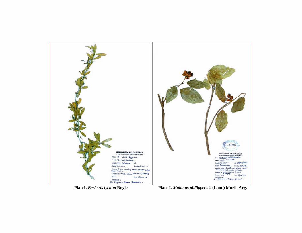
Plate1. Berberis lycium Royle Plate 2. Mallotus philippensis (Lam.) Muell. Arg.
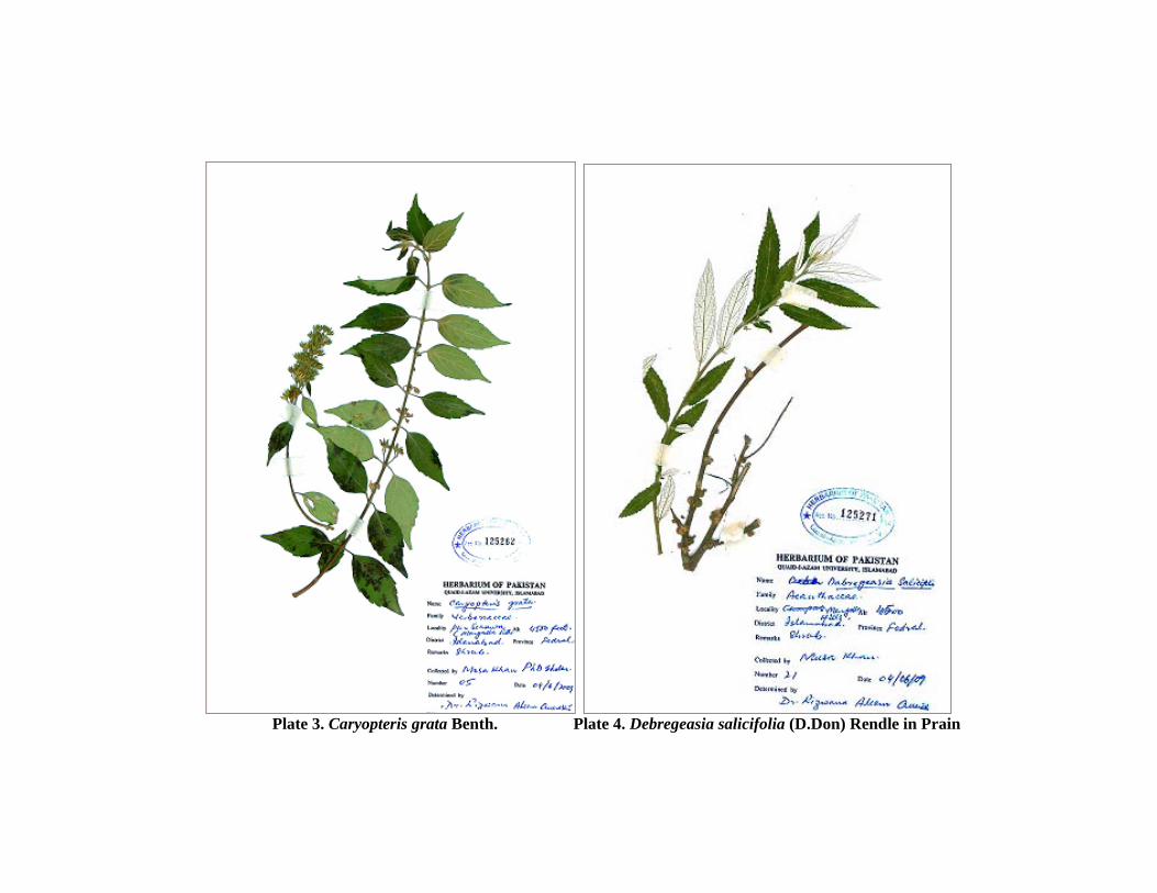
Plate 3. Caryopteris grata Benth. Plate 4. Debregeasia salicifolia (D.Don) Rendle in Prain
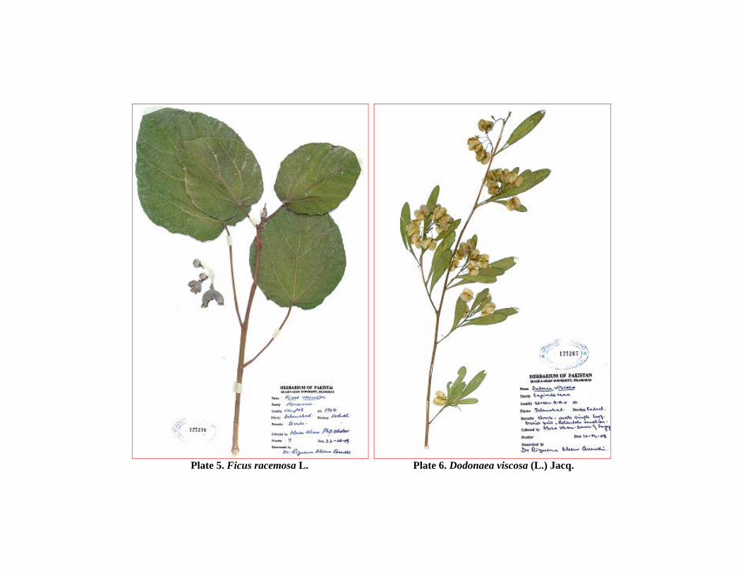
Plate 5. Ficus racemosa L. Plate 6. Dodonaea viscosa (L.) Jacq.
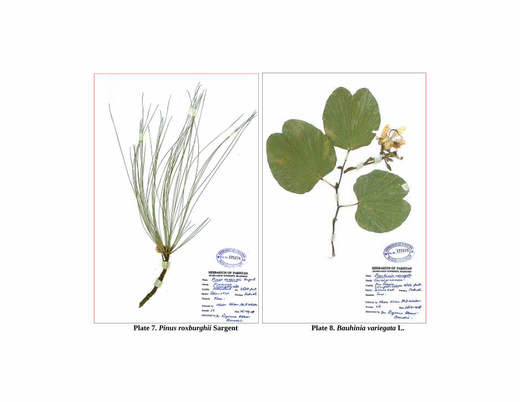
Plate 7. Pinus roxburghii Sargent Plate 8. Bauhinia variegata L.
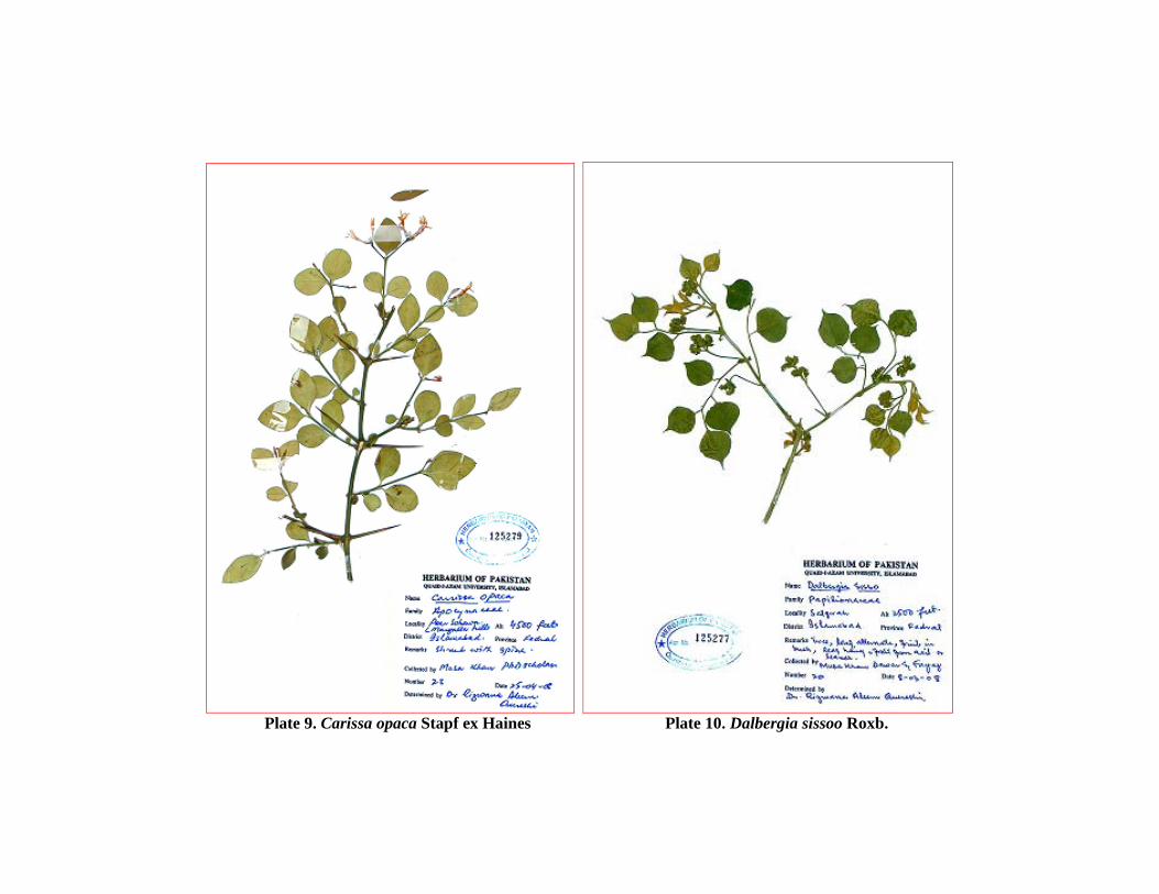
Plate 9. Carissa opaca Stapf ex Haines Plate 10. Dalbergia sissoo Roxb.

Plate 11. Rubus ellipticus Smith Plate 12. Ficus palmata Forssk.
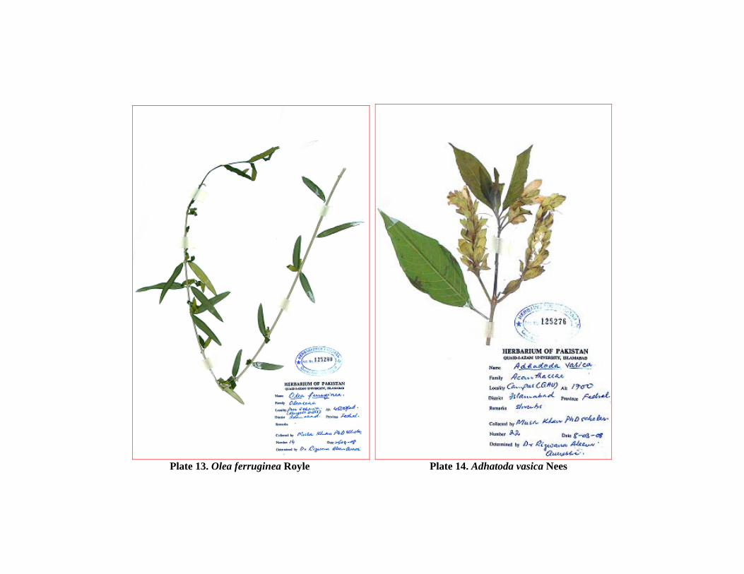
Plate 13. Olea ferruginea Royle Plate 14. Adhatoda vasica Nees
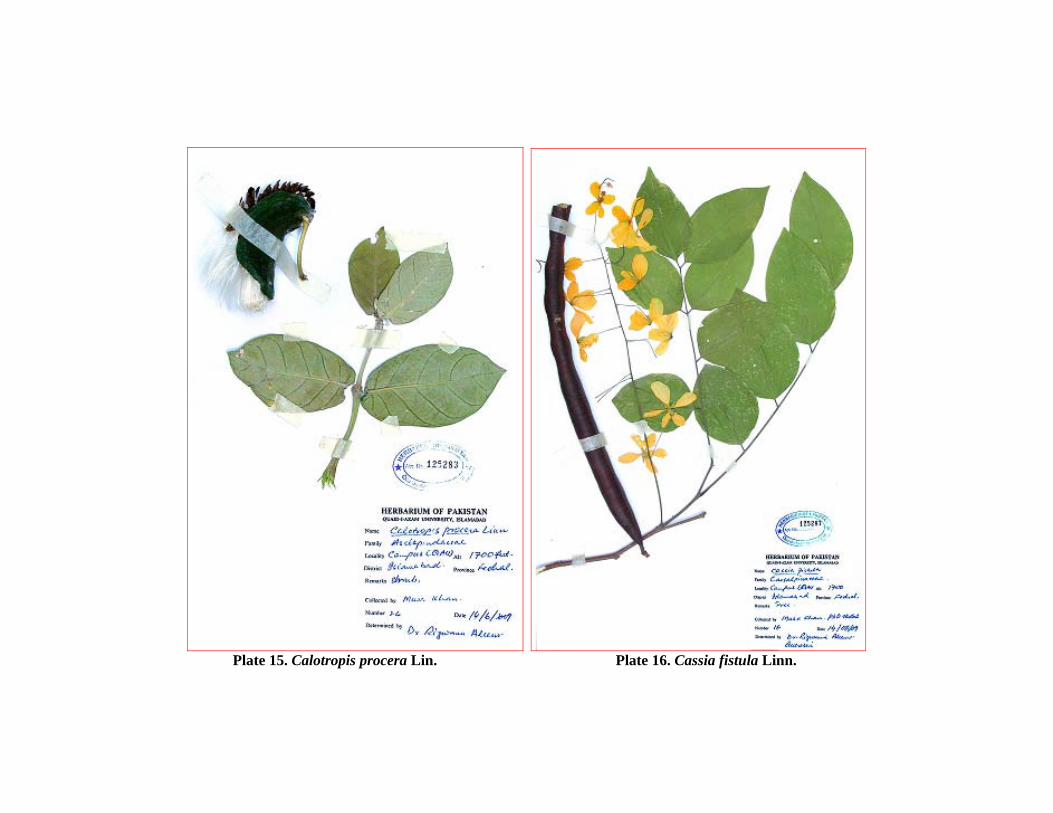
Plate 15. Calotropis procera Lin. Plate 16. Cassia fistula Linn.
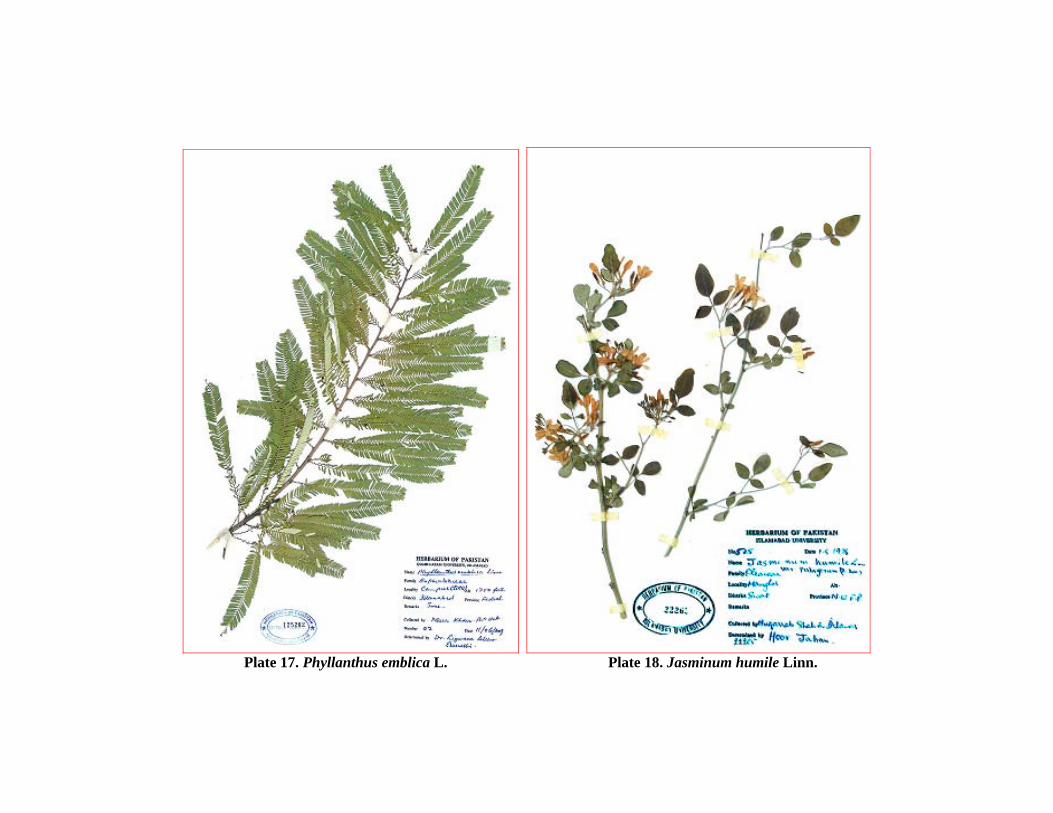
Plate 17. Phyllanthus emblica L. Plate 18. Jasminum humile Linn.
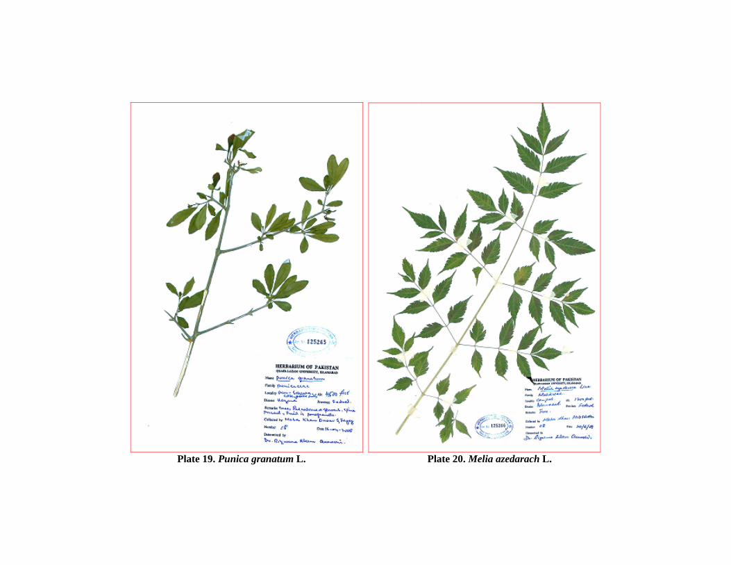
Plate 19. Punica granatum L. Plate 20. Melia azedarach L.
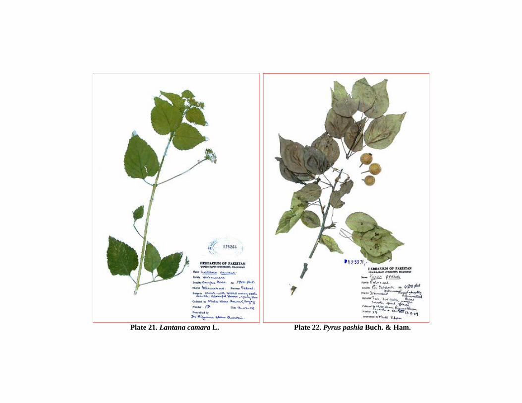
Plate 21. Lantana camara L. Plate 22. Pyrus pashia Buch. & Ham.
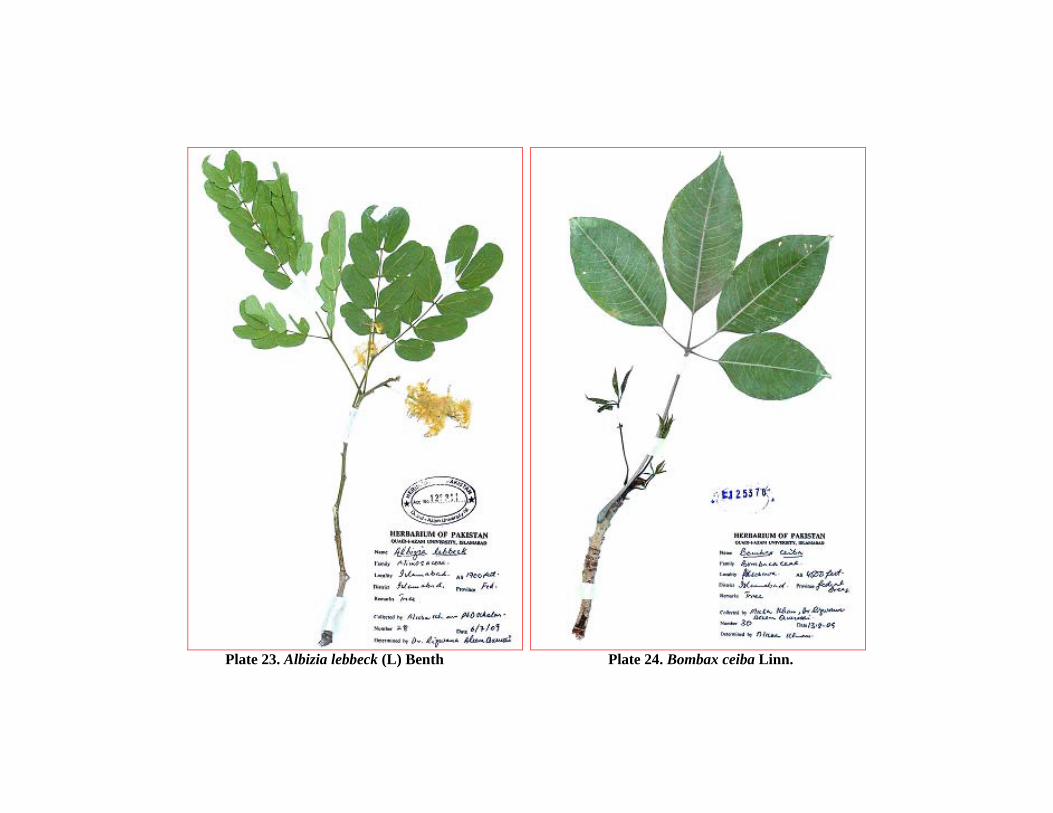
Plate 23. Albizia lebbeck (L) Benth Plate 24. Bombax ceiba Linn.
Chapter 6 References
182
Abbasi, A. M., Khan, M. A., Ahmad, M., Zafar, M., Khan, H., Muhammad, N.,
Sultana, S. Medicinal plants used for the treatment of jaundice and hepatitis
based on socio-economic documentation. AFR J BIOTECHNOL, 8:1643-
1650.
Abdel-Mogib, M., Basaif, S. A., Asiri, A. M., Sobahi, T. R., Batterjee, S. M. 2001.
New clerodane diterpenoid and flavonol-3-methyl ethers from Dodonaea
viscosa. PHARMAZIE, 56:830–1.
Abesundara, K. J. M., Matsui, T., Matsumoto, K. 2004. A-glucosidase inhibitory
activity of some Sri Lanka plant extracts, one of which, Cassia auriculata,
exerts a strong anti hyperglycemic effect in rats comparable to the therapeutic
drug acarbose. J. Agr. Food Chem., 52:2541-2545.
Abraham, Z., Bhakuni, D. S.; Garg, H. S.; Goel, A. K.; Mehrotra, B. N.; Patnaik, G.
K. 1986. Screening of Indian plants for biological activity: Part XII. Indian J.
Exp. Biology, 24:48-68..
Afaq, F. A., Sarfaraz, S. 2005. Pomegranate fruit juice for chemoprevention and
chemotherapy of prostate cancer. Proc. Natl. Acad. Sci. .U S A, 102:14813-
14818.
Ahluwalia, V. K., Seshadri, T. R. 1963. Dalbergenone from the heartwood of
Dalbergia sissoo. Curr. Sci., 32:455.
Ahluwalia, V. K., Sachdev, G. P., Seshadri, T. R. 1965. Chemical components of
immature green pods of Dalbergia sissoo. Indian J. Chem., 3:474.
Ahmad, I., Ahmad, M., Ahmad, A., 1994. Antimicrobial activity of Dodonaea
viscosa oil. Fitoterapia, 65:167-168.
Ahmad, N. S., Farman, M., Najmi, M. H., Mianand, K. B., Hasan, A. 2006. Activity
of polyphenolic plant extracts as scavengers of free radicals and inhibitors of
xanthine oxidase. J. Basic & App. Sci., 2:1.
Ahmad, S. S. 2007. Medicinal wild plants from Lahore-Islamabad motorway (m-2).
PAKISTAN J BOT., 39:355-375.

Chapter 6 References
183
Ahmad, V. U., Fatima, I., Fatima, A. 1987. The sapogenins from Dodonaea viscosa.
Fitoterapia, 58:361-362.
Ahmed, S. A., Siddiqui, S. A., Zaman, A., 1974. Flavones from Colebrookia
oppositifolia. Indian J. Chem., 12:1327-8
Ahmed, Z. F., El-Moghazy Shoaib, A. M., Wassel, G. M., E1-Sayyad, S. M. 1972.
Phytochemical study of Lantana camara, Terpenes and lactones II. PLANTA
MED., 22:34.
Akbar, E., Riaz, M., Malik, A. 2001. Ursene type nortriterpene from Debregeasia
salicifolia. Fitoterapia, 72:382-385.
Akhter, M. H., Sabir, M., Bhide, N. K. 1979. Possible mechanism of antidiarrhoel
effect of berberine. Indian J. Med. Res., 70:233-241.
Akihisa, T., Tokuda, H., Ichiishi, E., Mukainaka, T., Toriumi, M., Ukiya, M. 2001.
Anti-tumor promoting effects of multiflorane-type triterpenoids and cytotoxic
activity of karounidiol against human cancer cell lines. Cancer Lett., 173:9-14.
Albrecht, M., Jiang, W., Kumi-Diaka, J. 2004. Pomegranate extracts potently
suppress proliferation, xenograft growth, and invasion of human prostate
cancer cells. J Med Food, 7:274-283.
Alche, L. E., Barquero, A. A., Sanjuan, N. A., Coto, C. E. 2002. An antiviral principle
present in a purified fraction from Melia azedarach leaf aqueous extract
restrains herpes simplex virus type-1 propagation. Phytotherapy Res., 16:348-
352.
Alche, L. E., Berra, A., Veloso, M. J., Coto, C. E. 2000. Treatment with meliacine, a
plant derived antiviral, prevents the development of herpetic stromal keratitis
in mice. J. Med. virol., 61:474-480.
Alche, L. E., Assad, F. K., Meo, M., Coto, C. E., Maier, M. S. 2003. An antiviral
meliacarpin from leaves of Melia azedarach. zeitschrift-fur-naturforschung-
Section-C,- Biosciences, 58: 215-219.
Al-Dubai, A. S., Al-khulaidi, A. A. 1996.Medicinal and Aromatic Plants of Yemen

Chapter 6 References
184
(In Arabic), Sana’a, Yemen: Obadi Center for studies and Publishing.
Ali, M. N., Khan, A. A. 1978. Pharmacognostic studies of Berberis lycium Royle and
its importance as a source of raw material for the manufacture of berberine in
Pakistan, PAK J. FORE., 26.
Ali, M., Sharma, S. K., 1996. Heterocyclic constituents from Berberis lycium roots.
Ind. J. Hetero. Chemistry, 6:127-130.
Ambastha, S. P., The wealth of India, vol. 2B. Publication and Information
Directorate. New Delhi: CSIR, pp.118.
Amin, A. H., Subbaiah, T. V., Abbasi, K. M., 1969. Berberine sulfate: antimicrobial
activity, bioassay, and mode of action. Can J Microbiol, 15:1067-1076.
Amin, A. H., Mehta, D. R. 1959. A bronchodilator alkaloid (vasicinone) from
Adhatoda vasica Nees. Nature, 184:1317.
Anila, L., Vijayalakshmi, N. R. 2000. Beneficial effects of flavonoids from Sesamum
indicum, Emblica officinalis and Momordica charantia. PHYTOTHER. RES,
14:1-4.
Anonymous. 1950.In: Wealth of India. Raw materials, vol 3. CSIR, New Delhi.
Anonymous, 1992. The wealth of India, Vol-III. Publication and information
Directorate (CSIR), New Delhi, p. 337-343.
Ansari., M. M. 2004. Pharmacognostical and phytochemical studies on some
traditional plant drugs’(PhD Theses), Jamia Hamdardar (Hamdard Nagar)
New Delhi, 110062.
Ansari, M. A., Razdan, R. K., Tandon, M., Vasudevan, P. 2000. Larvicidal and
repellent actions of Dalbergia sissoo Roxb. (F. Leguminosae) oil against
mosquitoes. Bioresource Tech. 73:207-211.
Antarkar, D. S., Ashok, B. V., Doshi, J. C., Athavale, A. V., Vinchoo, K. S., Natekar,
M. R., Thathed, P. S., Ramesh, V., Kale, N. 1980. Doublebind clinical trial of
Arogyawardhani-an ayurvedic drug in acute viral hepatitis. Ind. J. Med.
Chapter 6 References
185
Research, 72:588-593.
Arisawa, M., Fujita, A., Suzuki, R., Hayashi, T., Morita, N., Kawano, N., Koshimura,
S. 1986. Studies on cytotoxic constituents in pericarps of Mallotus japonicus,
Part II. J. Nat. Prod., 49:298-302.
Arisawa, M., Fujita, A., Hayashi, T., Morita, N., Kikuchi, T., Tezuka, Y. Studies on
cytotoxic constituents in pericarps of Mallotus japonicus. IV. 1990 a. Chem.
Pharm. Bull., 38:698-00.
Arisawa, M., Fujita, A., Morita, N., Koshimura, S. 1990 b. Cytotoxic and antitumor
constituents in pericarps of Mallotus japonicus. PLANTA MED., 56:377-3 79.
Arun, K. Yadav, V. T. 2008. Anticestodal activity of Adhatoda vasica extract against
Hymenolepis diminuta infections in rats. J. Ethnopharmaco., 119:322-324.
Asif, A., Kakub. G., Mehmood, S., Khunum, R., Gulfraz, M. 2007. Wound healing
activity of root extracts of Berberis lyceum Royle in rats. Phytother. Res., 21:
589-591
Asmawi, M. Z., Kankaanranta, H., Moilanen, E., Vapaatalo, H. 1993. Anti-
inflammatory activities of Emblica officinalis Gaertn. Leaf extracts. J.
Pharmacy and Pharmaco., 45:581-584.
Attaway, J. A. 1994. Citrus juice flavonoids with anticarcinogenic and antitumor
properties. In Food phytochemicals for cancer prevention I, Fruits and
vegetables; Huang, M. T., Osawa, T., Ho, C. T., Rosen, R. T., Eds.; American
Chemical Society: Washington, DC, pp 240-248.
Babbar, O. P., Chhatwal, V.K., Ray, I. B., Mehra, M. K. 1982. Effect of berberine
chloride eye drops on clinically positive trachoma patients. Indian J. Med.
Res., 76:S83-S82.
Baker, J. T., Borris, R. P., Carte, B., Cordell, G. A., Soejarto, D. D., Cragg, G. M.,
Gupta, M. P., Iwu, M. M., Madulid, D. R., Tyler, V. E. 1995. Natural products
drug discovery and development: new perspectives on international
collaboration. J. Nat. Prod., 58:1325-1357.
Chapter 6 References
186
Bandopandhyay, M., Dhingra, V. K. Mukerjee, S. K., Pardeshi, N. P. 1972.
Triterpenoids and other components of Mallotus philippinencis.
Euphorbiaceae. PHYTOCHEMISTRY., 11:1511.
Banerji, A., Murti, V. V. S., Seshadri, T. R., Thakur, R. S. 1963. Chemical
components of the flowers of Dalbergia sissoo: Isolation of 7-
methyltectorigenin, a new isoflavone. Indian J. Chem., 1:25-27.
Banerji, A., Murti, V. V. S., Seshadri, T. R. 1965. Occurrence of 7, 40-
dimethyltectorigenin in the flowers of Dalbergia sissoo. Curr. Sci., 34:431.
Banerji, A., Murti, V. V. S., Seshadri, T. R. 1966. Isolation of sissotrin, a new
isoflavone glycoside from the leaves of Dalbergia sissoo. Indian J. Chem.,
4:70-72.
Barnes, P. J. 2002. New treatments for COPD. Nat. Rev. Drug Dis., 1:437-446.
Barnes, P. J., Belvisi, M. G., Mak, J. C., Haddad, E. B., O’Connor, B. 1995.
Tiotropium bromide (Ba 679 BR), a novel long-acting muscarinic antagonist
for the treatment of obstructive airways disease. Life Sci., 56:853-859
Barry, V. C., Conalty, M. L., Rylance, H. J., Smith, F. R.1955. Antitubercular effect
of an extract of Adhatoda vasica. Nature, 176:119-20.
Barton, D. H. R., De Mayo, P., Warnhoff, E. W., Jeger, O., Perold, G. W. 1954.
Triterpenoids. Part XIX. The constitution of lantadene B. J. Chem. Soci.,
3689-3692.
Barua, A. K., Chakrabarti, P., Chowdhury, M. K., Basak, A., Basu K. 1976. The
structure and stereochemistry of lantanilic acid, the β,β-dimethylacryloyl ester
of lantaninilic acid, isolated from Lantana camara. PHYTOCHEMISTRY,
15:987-989.
Barua, A.K., Chakrabarti, P., Chowdhury, M. K., Barak, A., Basu, K., Ray, S., Saha,
S. K. 1985. The structure and stereo- chemistry of lantanilic acid. J. Ind.
Chem. Soc., 62:298-305.
Barua, A. K., Chakrabarti, P., Sanyal, P. K., Das, B. 1969. Triterpenoids. XXXII.
Chapter 6 References
187
Structure of lantic acid: a new triterpene from Lantana camara. J. Indian
Chem. Soc., 46: 100-102.
Baslas, R. K., Agha, R. 1985. Isolation of a hypoglycemic principle from the bark of
Ficus glomerata Roxb. Him. Chem. Pharma. Bull., 2:13.
Basu, A., Chaudhuri, A.K. 1991. Preliminary studies on the anti-inflammatory and
analgesic activities of Calotropis procera root extract. J. Ethnopharmacol.,
31:319.
Basu, A., T. Sen., Ray, R. N., Nag Chaudhuri, A. K. 1992. Hepatoprotective effects of
Calotropis procera root extract on experimental liver damage in animals.
Fitoterapia, 63:507-514.
Baud, L., Ardaillou, R. 1986. Reactive oxygen species: Production and role in the
kidney. AM. J. PHYSIOL., 251:1765.
Beckett, A., Belthle, F., Fell, K., Lock, M. 1954. The active constituents of raspberry
leaves. J. Pharm. Pharmacol., 6:785-796.
Begum, S., Raza, S.M., Siddiqui, B.S., Siddiqui, S. 1995. Triterpenoids from the
aerial parts of Lantana camara. J. Nat. Prod., 58:1570-1574.
Begum, S., Wahab, A., Siddiqui, B. S., Qamar, F. 2000. Nematicidal Constituents of
the Aerial Parts of Lantana camara. J. Nat. Prod., 63:765-767.
Begum, S., Wahab, A., Siddiqui, B. S. 2003. Pentacyclic Triterpenoids from the
Aerial Parts of Lantana camara. Chem. Pharm. Bull., 51:134-137.
Begum, S., Zehra, S. Q., Siddiqui, B. S., Wahab, A. 2006. Triterpenoidal Secondary
Metabolites from Lantana Camara L.. Helv. Chim. Acta, 89:1932-1941.
Begum, S., Qamar, Z. S., Siddiqui, B. S., 2008. Two New Pentacyclic Triterpenoids
from Lantana camara LINN. Chem. Pharm. Bull., 56:1317-1320.
Benencia. F., Courreges. M..C., Coto, C., and Coulombie, F.C. 1997.
Immunomodulatory activities of Melia azedarach L. leaf extracts on human
monocytes. J. Herb Spices and Med. Plants. 5(3):7-13.
Chapter 6 References
188
Benencia, F., Courreges, M. C., Coulombie, F. C., Massouh, E. J. 1992. Effect of
Melia azedarach fresh leaf aqueous extract on mice hematological parameters.
Fitoterapia, 63:411-413.
Bhakta, T., Mukherjee, P. K., Mukherjee, K., Banerjee, S., Mandal, S. C., Maity, T.
K., Pal, M., Saha, B. P. 1999. Evaluation of hepatoprotective activity of
Cassia fistula leaf extract. J. Ethnopharmacol., 66:277-82.
Bhakta, T., Banerjee, S., Mandal, S. C., Maity, T. K., Saha, B. P., Pal, M. 2001.
Hepatoprotective activity of Cassia fistula leaf extract. Phytomedicine. 8:220-
224.
Bhakumi, D. S., Dhar, M. L., Dhar, M. M., Dhawan, B. N., Gupta, B., Srimal, R. C.,
1971. Indian J. Exp. Biology., 2:91.
Bhakuni, R. S., Shukla, Y. N. Thakur, R. S.1987. Chemical examination of the roots
of Rubus ellipticus. Indian Drugs, 24:272.
Bhaskara, R. R., Murugesan, T., Pal, M., Saha, B. P., Mandal, S.C. 2003. Antitussive
potential of methanol extractof stem bark of Ficus racemosa L. PHYTOTHER.
RES, 17:1117-1118.
Bhattarai, N. K. 1992. Medical ethnobotany in the Karnali Zone, Nepal. Eco.Botany,
46:257-261.
Birdsall, T. C., Kelly, G. S. 1997. Berberine: Therapeutic potential of an alkaloid
found in several medicinal plants. Altern. Med. Rev., 2:94-103.
Biswas, S., Talukder, G., Sharma, A. 1999. Protection against cytotoxic effects of
arsenic by dietary supplementation with crude extract of Emblica officinalis
fruit. PHYTOTHER. RES, 13:513-516.
Blois, M.S. 1958. Antioxidant determinations by the use of a stable free radical,
Nature, 81:1199-1200.
Bonassi, S., Fenech, M., Lando, C. 2001. Human MicroNucleus Project: International
database comparison for results with the cytokinesis- block micronucleus
assay in human lymphocytes: I. Effect of laboratory protocol, scoring criteria
Chapter 6 References
189
and host factors on the frequency of micronuclei. Env Mol Mutagenesis,
37:31.
Bonfil, R. D., Russo, D. M., Binda, M. M., Delgado, F. M., Vincenti, M. 2002. Higher
antitumor activity of vinflunine than vinorelbine against an orthotopic murine
model of transitional cell carcinoma of the bladder. Uro. Onco., 7:159-166.
Borkowski, B., Lutomski, J., Skrzydlewska, E., Zygmunt, B.1994. Rosliny lecznicze
w fitoterapii, IRiPZ, oznan pp. 470-471.
Bracke, M. E., Bruyneel, E. A., Vermeulen, S. J., Vennekens, K. I., Marck, V. V.,
Mareel, M. M. 1994. Citrus favonoid effect on tumor invasion and metastasis.
Food Technol., 48:121-124.
Brodie, C., Blumberg, P. M. 2003. Regulation of cell apoptosis by protein kinase c
delta. Apoptosis, 8:19-27.
Bruhn, J. G., Bohlin, L. 1997. Molecular pharmacognosy: an explanatory model.
Drug Disc. Today, 2:243-246.
Buckheit Jr, R. W., White, E. L., Fliakas-Boltz, V., Russell, J., Stup, T. L., Kinjerski,
T. L., Osterling, M. C., Weigand, A., Bader, J. P. 1999. Unique anti-human
immunodeficiency virus activities of the nonnucleoside reverse transcriptase
inhibitors calanolide A, costatolide, and dihydrocostatolide. ANTIMICROB
AGENTS CH., 43:1827-1834.
Burke, M. D., Berger, E. M., Schreiber, S. L. 2004. A synthesis strategy yielding
skeletally diverse small molecules combinatorially. J. Amer. Chem. Soc.,
126:14095-14104.
Burn, J., Camb, M., Withell, E. 1941. A principle in raspberry leaves which relaxes
uterine muscle. Lancet., 6149-6151.
Burn, J. H. and Withell, E. R. 1941. A principle in raspberry leaves which relaxes
uterine muscle. Lancet., 5:1-3.
Burnette, W. N. 1981. “Western blotting’: electrophoretic transfer of proteins from
sodium dodecyl sulfate - polyacrylamide gels to unmodified nitrocellulose and
Chapter 6 References
190
radiographic detection with antibody and radioiodinated protein A”.
Analytical Biochemistry (United States: Academic Press) 112:195-203.
Butler, M. S. 2004. The role of natural product chemistry in drug discovery. J. Nat.
Prod., 67:2141-2153.
Caceres, A., Giron, L.M., Alvarado, S.R., Torres, M. F. 1987. Screening of
antimicrobial activity of plants popularly used in Guatemala for treatment of
dermatomucosal diseases. J Ethnopharmacol., 20:223-237.
Cardellina II, J. H. 2002. Challenges and opportunities confronting the botanical
dietary supplement industry. J. Nat. Prod., 65:1073-1084.
Carpinella, M. C., Herrero, G. G., Alonso, R. A. and S. M. Palacios. 1999. Antifungal
activity of Melia azedarach fruit extract. Fitoterapia., 70:296-298.
Cerda, B., Espin, J. C., Parra, S. 2004. The potent in vitro antioxidant ellagitannins
from pomegranate juice are metabolised into bioavailable but poor antioxidant
hydroxy-6H-dibenzopyran-6-one derivatives by the colonic microflora of
healthy humans. Eur. J. Nutr., 43:205-220.
Chandrashekher, C. H., Latha, K. P., Vagdevi, H. M., Vaidya, V. P. 2008.
Anthelmintic activity of the crude extracts of Ficus racemosa. Int. J. Green
Pharmacy, 2:100-103.
Chawla, H. M., Chibber, S. S. 1981. Chemistry of Dalbergia Species. J. Sci. Ind. Res,
40:313.
Chhabra, S. C., Mahunnah, R. L. A., Mshiu, E. N. 1991. Plants used in traditional
medicine in Eastern Tanzania. V. Angiosperms (Passifloraceae to
Sapindaceae). J. Ethnopharmacol., 33:143-157.
Chidambara Murthy, K. N., Jayaprakasha, G. K., Singh, R. P. 2002. Studies on
antioxidant activity of pomegranate (Punica granatum) peel extract using in
vivo models. J Agric Food Chem., 50:4791-4795.
Chintawar, S. D., Somani, R. S., Veena, S., Kasture, S.B. 2002. KastureNootropic
activity of Albizzia lebbeck in mice. J. Ethnopharmacol., 81:299-305
Chapter 6 References
191
Choedon, T., Mathan, G., Arya, S., Kumar, V.L., Kumar, V. 2006. Anticancer and
cytotoxic properties of the latex of Calotropis procera in a transgenic mouse
model of hepatocellular carcinoma. World J. Gastroenterol., 12:2517-2522.
Chopra, R. N., Chopra, I. C., Handa, K. L., Kapoor, L. D. 1988. Indignenous drugs of
India. Kolkata: UN Dhur and Sons. (1958), pp. 503.
Chopra, R. N., Nayar, S. L., Chopra, I. C.1956 a. Glossary of Indian Medicinal Plants
(CSIRPublication, New Delhi, p. 74.
Chopra, R. N., Nayer, S. L., Chopra, I. C. 1956 b. Glossary of Indian Medicinal
Plants, New Delhi, CSIR, 90-91.
Choudhary, D. N., Singh, J. N. Verma, S. K., Singh, B. P. 1990. Antifertility effects
of leaf extracts of some plants in male rats. Indian J. Exp. Biol., 28:714-716.
Choudhry, V. P., Sabir, M., Bhide, V. N. 1972. Berberine in giardiasis. Indian
Pediatrics, 9:143-146.
Christina, A. J. M., Haja Najumadeen, N. A., Vimal kumar, S., Manikandan, N.,
Tobin, G. C., Venkataraman, S., Murugesh, N. 2006. Antilithiatic effect of
Melia azedarach on ethylene glycol induced nephrolithiasis in rats. PHARM.
BIOL., 44:480-485.
Chun, Y. T., Yip, T. T., Lau, K. L., Kong, Y. C. 1978. A biochemical study on the
hypotensive effect of berberine in rats. Gen. Pharmac., 10:177-182.
Cichewicz, R. H., Kouzi, S. A. 2004. Chemistry, biological activity, and
chemotherapeutic potential of betulinic acid for the prevention and treatment
of cancer and HIV infection. Med. Res. Rev., 24:90-114.
Ckless, K., Schlottfeldt, J. L., Pasqual, M. 1995. Inhibition of in-vitro lymphocyte
transformation by the isoquinoline alkaloid berberine. J. Pharm. Pharmacol.,
47:1029-1031.
Clardy, J., Walsh, C. 2004. Lessons from natural molecules. Nature, 432:829-837.
Coppen, J. J. W., Hone, G. A. 1995. Gum naval stores: turpentine and rosin from pine
Chapter 6 References
192
resin. FAO, Non-wood Forest Products No. 2, 62 pp.
Cragg, G. M., Newman, D. J. 2004. A tale of two tumor targets: topoisomerase I and
tubulin. The Wall and Wani contribution to cancer chemotherapy. J.
Nat.Prod., 67:232-244.
Creagh, T., Ruckle, J. L., Tolbert, D. T., Giltner, J., Eiznhamer, D. A., Dutta, B.,
Flavin, M. T., Xu, Z. Q. 2001. Safety and pharmacokinetics of single doses of
(+)-calanolide A, a novel, naturally occurring nonnucleoside reverse
transcriptase inhibitor, in healthy, human immunodeficiency virus-negative
human subjects. Anti. Agen. Chemo., 45:1379-1386.
Creasey, W.A. 1979. Biochemical effects of berberine. Biochem. Pharmacol.
28:1081-1084.
Currens, M. J., Gulakowski, R. J., Mariner, J. M., Moran, R. A., Buckheit Jr., R. W.,
Gustafson, K. R., McMahon, J. B., Boyd, M. R., 1996. Antiviral activity and
mechanism of action of calanolide A against the human immunodeficiency
virus type-1. J. Pharm. Exp.Therap., 279:645-651.
Cutler, R. G. 1984. Antioxidants, aging, and longevity. In Free Radicals in Biology;
Pryor, W. A., Ed.; Academic Press: New York, pp. 371-428.
Czygan, F. Ch., 1995. Die Himbeere-Rubus idaeus L. Portrait einer Arzneipflanze. Z.
PHYTOTHER. RES, 16:366-374.
Daikonya, A., Katsuki, S., Wu, J.-B., Kitanaka, S. 2002. Anti-allergic Activity of
New Phloroglucinol Derivatives from Mallotus philippensis
(Euphorbiaceae).Chem. Pharm. Bull., 50:1566-1659.
Das, C., Poi, R., Chowdhury, A. 2005. HPTLC determination of vasicine and
vasicinone in Adhatoda vasica. PHYTOCHEM. ANALYSIS, 16, 90-92.
De, S., Ravishankar, B., Bhavsar, G. C. 1993. Plants with hepatoprotective activity-a
review. Ind. Drugs, 30, 355-363.
Deranjyagala, S. A., Wijesundera, R. L. C., Weerasna, O. V. D. S. J. 1988. Antifungal
activity of Ficus racemosa leaf extract and isolation of the active compound.

Chapter 6 References
193
J. Nation. Sci. Council, 26, 19-26.
Dewick, P. M. 2002. Medicinal Natural Products: a Biosynthetic Approach, 2nd ed.
John Wiley and Sons, Chichester, England.
Dhiman, A. K. 2003. Sacred Plants and their Medicinal Uses, (Daya publishing
House, Delhi, pp. 125-127.
Dhingra, V. K., Seshadri, T. R., Mukerjee, S. K. 1974. Isotectorigenin from the bark
of Dalbergia sissoo. Indian J. Chem., 12:1118.
Dhir, H., Roy, R. K., Sharma, A., Talukdar, G. 1990. Modification of clastogenicity
of lead and aluminium in mouse bone marrow cells by dietary ingestion of
Phyllanthus emblica fruit extract. Mut. Research, 241:305-312.
Dorsch, W., Wagner, H. 1991. New antiasthmatic drugs from traditional medicine.
Int. Arch. Allergy App. Immunol., 94:262-5.
Dubey, V. K., Jagannadham, M. V. 2003. Procerain, a stable cysteine protease from
the latex of Calotropis procera. PHYTOCHEMISTRY., 62:1057-1071.
Dugas, A. J., Castaeda-Acosta, J., Bonin, G. C. 2000. Evaluation of the total peroxyl
radical-scavenging capacity of flavonoids: Structure–activity relationships. J.
Nat. Prod., 63:327.
Dutta, B. K., Rahman, I., Das, T. K. 1998. Antifungal activity of Indian plant extracts.
Mycoses, 41:535-536.
Dutta, S., Ghatak, K. L., Ganguly, S. N. 1997. Isolation and Structure Elucidation of
New Pentacyclic Triterpene Acid from the Leaves of Rubus ellipticus. Nat.
Pro. Sciences, 32:108-110.
Eisenbrand, G., Hippe, F., Jakobs, S., Muehlbeyer, S. 2004. Molecular mechanisms of
indirubin and its derivatives: novel anticancer molecules with their origin in
traditional Chinese phytomedicine. J. Can. Res. Clin.Onco., 130:627-635.
El-Dagwy, A. A. 1996. Encyclopedia for Production of Aromatic and Medicinal
Plants, Vol. 1. Imprimerie Atlas, Cairo, pp. 192.
Chapter 6 References
194
Eldridge, G. R., Vervoort, H. C., Lee, C. M., Cremin, P. A., Williams, C. T., Hart, S.
M., Goering, M. G., O’Neil-Johnson, M., Zeng, L. 2002. High-throughput
method for the production and analysis of large natural product libraries for
drug discovery. Anay.Chem. 74:3963-3971.
El-Lakwah, F. A., Mohamed, R., Darwish, A. A. 1995. Evaluation of toxic effect of
chinaberry. Annals of Agri. Sci., 33:389-398.
Esposito Avella, M., Diaz, A., de Gracia, I., de Tello, R., Gupta, M. P. 1991.
Evaluation of traditional medicine: effects of Cajanus cajan L. and of Cassia
fistula L. on carbohydrate metabolism in mice. Rev. Med. Panama., 16:39-45.
Fair, J. D.; Kormos, C. M. 2008. Flash column chromatograms estimated from thin-
layer chromatography data. J. Chromatogr., 1211:49-54.
Farag, S. F., Ahmed, A. S., Terashima, K., Takaya, Y., Niwa, M. 2001. Isoflavonoid
glycosides from Dalbergia sissoo. PHYTOCHEMISRY., 57:1263-1268.
Feher, M., Schmidt, J. M. 2003. Property distributions: differences between drugs,
natural products, and molecules from combinatorial chemistry. J. Chem. Infor.
Comp. Scie., 43:218-227.
Fenech, M. 2005. The Genome Health Clinic and Genome Health Nutrigenomics
concepts: Diagnosis and nutritional treatment of genome and epigenome
damage on an individual basis. Mutagenesis, 20:255.
Ferdous, M., Rouf, R., Shilpi, J. A., Uddin, S. J. 2008. Antinociceptive activity of the
ethanolic extract of Ficus racemosa Lin. (Moraceae). Ori. Pharm. Exp. Med.,
8:93-96.
Ford, C. W., Bcndall, M. R. 1980. Identification of the iridoid glucoside theveside in
Lantana camara (Verbenaceae) and determination of its structure and
stereochemistry by means of NMR. Aust. J. Chem., 33:509-518.
Forestieri, A. M., Monforrte, M. T., Ragusa, S., Trovata, A., Lauk, L. 1996. Anti-
inflammatory, analgesic and antipyretic activity in rodents of plant extracts
used in African medicine. PHYTOTHER. RES, 10:100-106.
Chapter 6 References
195
Frankel, E. N.; Kanner, J.; German, J. B.; Parks, E.; Kinsella, J. E. 1993. Inhibition of
oxidation of human low-density lipoprotein by phenolic substances in red
wine. Lancet, 341:454-457.
Frantz, S. 2005. 2004. Approvals: the demise of the blockbuster? Nat. Rev. Drug Dis.,
4:93-94.
Frantz, S., Smith, A. 2003. New drug approvals for 2002. Nat. Rev. Drug Dis., 2:95-
96.
Fujita, A., Hayashi, T., Arisawa, M., Shimizu, M., Morita, N., Kikuchi, T., Tezuka, Y.
1988. Studies on cytotoxic constituents in pericarps of Mallotus japonicus,
part III. J. Nat. Prod., 51:708-712.
Fukuda, K., Hibiya, Y., Mutoh, M. 1999. Inhibition of activator protein 1 activity by
berberine in human hepatoma cells. PLANTA MED., 65:381-383.
Fukuda, K., Hibiya, Y., Mutoh, M. 1999. Inhibition by berberine of cyclooxygenase-2
transcriptional activity in human colon cancer cells. J. Ethnopharmacol.,
66:227-233.
Fukuda, Y., Sakai, K., Matsunaga, S., Tokuda, H., Tanaka, R. 2005. Cancer
chemopreventive activity of lupane- and oleanane-type triterpenoids from the
cones of Liquidamber styraciflua. Chem Biodiv., 2:421-8.
Furusawa, M., Ido, Y., Tanaka, T., Ito, T., Nakaya, K., Ibrahim, I. 2005. Novel,
complex flavonoids from Mallotus philippensis (Kamala tree). Helv Chim
Acta; 88:1048-58
Gajmer, T., Singh, R., Saini, R. K., Kalidhar, S. B. 2002. Effect of methanolic extracts
of neem (Azadirachta indica A. Juss) and bakain (Melia azedarach L.) seeds
on oviposition and egg hatching of Earias vittella (Fab.) (Lepidoptera:
Noctuidae). J. Appl. Entomol., 126:238-243.
Ganesan, A., 2004. Natural products as a hunting ground for combinatorial chemistry.
Current Opin. in Biotech., 15:584-590.

Chapter 6 References
196
Gatto, B., Sanders, M. M., Yu, C., Wu, H. Y., Makhey, D., LaVoie, E. J., Liu, L. F.
1996. Identification of topoisomerase I as the cytotoxic target of the
protoberberine alkaloid coralyne. Cancer Res, 56:2795-2800.
Getie, M., Gebre-Mariam, T., Rietz, R., Hohne, C., Huschka, C., Schmidtke, M. 2003.
Evaluation of the anti-microbial and anti-inflammatory activities of the
medicinal plants Dodonaea viscosa, Rumex nervosus and Rumex abyssinicus.
Fitoterapia, 74:139-143.
Getie, M., Gebre-Mariam, T., Rietz, R., Neubert, R. H. 2002. Evaluation of the
release profiles of flavonoids from topical formulations of the crude extract of
the leaves of Dodonea viscosa (Sapindaceae). PHARMAZIE, 57:320-2.
Geysen, H. M., Schoenen, F., Wagner, D., Wagner, R. 2003. Combinatorial
compound libraries for drug discovery: an ongoing challenge. Nat. Rev. Drug.
Dis., 2:222-230.
Ghani, A. 1998. Medicinal Plants of Bangladesh.Asiatic Society of Bangladesh,
Dhaka.
Ghisalberti, E. L., 2000. Lantana camara L. (Verbenaceae). Fitoterapia, 71(5):467-486.
Ghosh, A. K., Bhattacharyya, F. K., Ghosh, D. K. 1985. Leismania donovani:
amastigote inhibition and mode of action of berberine. Exp. Parasitol, 60:404-
413.
Gil, M. I., Tomas-Barberan, F. A., Hess-Pierce, B. 2000. Antioxidant activity of
pomegranate juice and its relationship with phenolic composition and
processing. J. Agric. Food Chem., 48:4581-4589.
Giuseppe, V., Alessandra, P., Marzia, M., Paola, C., Vittoria, F., Michela, M.,
Emanuela, M. 2009. Rottlerin: a multifaced regulator of keratinocyte cell
cycle. Exp. Dermatology, 18:516-521.
Godbole, S. H., Pendse, G. S. 1960. Antibacterial property of some plants. Indian J.
Pharm., 22:39-42.
Gopal, H. 1972. Chemical constituents of Salmalia malabarica Schott and endl.
Chapter 6 References
197
Flowers. J. Pharm. Sci., 61:807-808.
Grange, J. M., Snell, N. J. 1996. Activity of bromohexine and ambroxiol,
semisynthetic derivatives of vaccine from the Indian shrub Adhatoda vasica,
against Mycobacterium tuberculosis in vitro. J. Ethnopharmacol., 50:49-53.
Graul, A. I. 2001. The year’s new drugs. Drug News and Perspectives, 14:12-31.
Greenwald, P. 2002. Cancer chemoprevention. B. Med. Journal, 324:714-718.
Grusch, M., Polgar, D., Gfatter, S., Leuhuber, K., Huettenbrenner, S., Leisser, C.
2002. Maintenance of ATP favours apoptosis over necrosis triggered by
benzamide riboside. Cell Death Differ., 9:169-178.
Gudej, J., 2003. Kaempferol and quercetin glycosides from Rubus idaeus L. leaves.
Acta Polon. Pharm., 60:313-316.
Gudej, J., Rychlinska, I. 1996. Flavonoid compounds from the leaves of Rubus idaeus
L. Herba Pol., 42:257-261.
Gudej, J., Tomczyk, M., Urban, E., Tomczykowa, M., 1998.Analysis of chemical
composition of Rubus saxatilis L. leaves. Herba Pol., 44:340-344.
Gulfraz, M., Mehmood, S., Ahmad, A., Fatima, N., Parveen, Z., Williamson. 2008.
Comparison of the Antidiabetic Activity of Berberis lycium Root Extract and
Berberine in Alloxan-induced Diabetic Rats. PHYTOTHER. RES., 22:1208-
1212.
Guo, C., Wei, J., Yang, J. 2008. Pomegranate juice is potentially better than apple
juice in improving antioxidant function in elderly subjects. Nutr. Res., 28:72-
77.
Guo, R., Wei, P., Liu, W. 2007. Combined antioxidant effect of rutin and vitamin C in
Triton X-100 micelles. J. Pharm. Biochem. Anal., 43:1580.
Gupta, A. K., Vidyapati, T. J., Chauhan, J. S. 1979. 5,7-Dihydroxyflavanone-4′-O-α-
L-rhamnopyranosyl-β-D-glucopyranoside from Bauhinia variegata. Ind. J.
Chem., 18:85-86.

Chapter 6 References
198
Gupta, A. K., Chauhan, J. S. 1984. Constituents from the stem of Bauhinia variegata.
Nat. Acad. Sci. Lett., 7:15-16.
Gupta, A. K., Vidyapati, T. J., Chauhan, J. S. 1980. Chemical examination of the stem
of Bauhinia variegata. PLANTA MED., 38:174-176.
Gupta, C. M., Bhaduri, A. P., Khanna, N. M., 1971. Biologically active quinazolones
and related compounds. J. Sci. Ind. Res., 30:101-106.
Gupta, K. C., Chopra, I. C. 1954. Anti-tubercular action of Adhatoda vasica (N.O.
acanthacea). Indian J. Med. Res., 42:355-8.
Hajare, S. W., Chandra, S., Tandan, S. K., Sarma, J., Lal, J., Telang, A. G. 2000.
Analgesic and antipyretic activities of Dalbergia sissoo leaves. Ind. J.
Pharmacology, 32:357-360.
Hajare, S. W., Chandra, S., Sharma, J., Tandan, S.K., Lal, J., Telang, A.G. 2001.
Anti-inflammatory activity of Dalbergia sissoo leaves. Fitoterapia, 72:131-
139.
Hall, D. G., Manku, S., Wang, F. 2001a. Solution and solid-phase strategies for the
design, synthesis, and screening of libraries based on natural product
templates: a comprehensive survey. J. Comb. Chem., 3:125-150.
Hall, M. G., Wilks, M. F., Provan, W. M., Eksborg, S., Lumholtz, B. 2001b.
Pharmacokinetics and pharmacodynamics of NTBC (2-(2-nitro-4-fluoro-
methylbenzoyl)-1, 3-cyclohexanedione) and mesotrione, inhibitors of 4-
hydroxyphenyl pyruvate dioxygenase (HPPD) following a single dose to
healthy male volunteers. B. J. Clin. Pharm., 52:169-177.
Halliwell, B., Aeschbach, R., Loliger, L. 1995. The characterization of antioxidants.
Food Chem. Toxicol., 33:601.
Hamayun, M., Khan, S. A., Iqbal, I., Rehman, G., Hayat, T., Khan, M. A. 2005.
Ethnobotanical Profile of Utror and Gabral Valleys, District Swat, Pakistan.
Eth. Leaflets.
Harsh, A. L., Nag, T. N. 1988 Flavonoids with antimicrobial activities of arid zone
Chapter 6 References
199
plants. Geobios. India, 15:32-35.
Hartman, R. E., Shah, A., Fagan, A. M. 2006. Pomegranate juice decreases amyloid
load and improves behavior in a mouse model of Alzheimer’s disease.
Neurobiol. Dis., 24:506-515.
Hedberg, I., Hedberg, O., Madati, P. J., Mshigeni, K., Mshiu, E. N., Samuelsson, G. J.
1983. Inventory of plants used in traditional medicine in Tanzania. Part III.
Plants of the families Papilionaceae–Vitaceae. Ethnopharmacol., 9:237-260.
Hedrick, U. P. 1972. Sturtevant’s Edible Plants of the World. Dover Publications
ISBN 0-486-20459-6
Heinrich, M., Teoh, H. L. 2004. Galanthamine from snowdrop the development of a
modern drug against Alzheimer’s disease from local Caucasian knowledge. J.
Ethnopharmaco., 92:147-162.
Hertog, M. G.; Hollman, P. C. H.; Katan, M. B.; Kromhout, D. 1993. Dietary
antioxidant flavonoids and risk of coronary heart disease. Lancet., 342:1007-
1011.
Ho, L. H., Taylor, R., Dorstyn, L., Cakouros, D., Bouillet, P., Kumar, S. 2009. A
tumor suppressor functions for caspase-2. Proc Natl Acad Sci USA 106:5336-
5341.
Hoessel, R., Leclerc, S., Endicott, J. A., Nobel, M. E., Lawrie, A., Tunnah, P., Leost,
M., Damiens, E., Marie, D., Marko, D., Niederberger, E., Tang, W.,
Eisenbrand, G., Meijer, L. 1999. Indirubin, the active constituent of a Chinese
antileukaemia medicine, inhibits cyclin-dependent kinases. Nat.Cell. Bio., 1:
60-67.
Hooker, J.D., 1882. Flora of British India. Reeve and Co. London, 3:640.
Huang, C. G., Chu. Z. L., Yang, Z. M. 1991. Effects of berberine on synthesis of
platelet TXA2 and plasma PGI2 in rabbits. Chung Kuo Yao Li Hsueh Pao,
12:526-528.
Hussain, Z., Waheed, A., Qureshi, R. A., Burdi, D. K., Verspohl, E. J., Khan, N.,

Chapter 6 References
200
Hasan, M. 2004. The effect of medicinal plants of Islamabad and Murree
region of Pakistan on insulin secretion from INS-1 cells. PHYTOTHER RES.,
18:73-77.
Hwang, B. Y., Lee, J. H., Koo, T. H., Kim, H. S., Hong, Y. S., Ro, J. S., Lee, K. S.,
Lee, J. J. 2001. Kaurane diterpenes from Isodon japonicus inhibit nitric oxide
and prostaglandin E2 production and NF-nB activation in LPS-stimulated
macrophage RAW264.7 cells. PLANTA MED., 67:406-410.
Ihmels, H., K. Faulhaber, D., Vedaldi, F., Dall’Acqua, G. V. 2005. Intercalation of
organic dye molecules into double-stranded DNA. Part 2: The annelated
quinolizinium ion as a structural motif in DNA intercalators. Photochem.
Photobiol., 81:1107-1115.
Iizuka, N., Miyamoto, K. K., Okita, A., Tangoku, H., Hayashi, S., Yosino, T., Abe,
T., Morioka, S., Oka, H. M. 2000. Inhibitory effect of Coptidis Rhizoma and
berberine on the proliferation of human esophageal cancer cell lines. Cancer
Lett., 148:19-25.
Ikawa, M., Schaper, T. D., Dollard, C. A., Sasner, J. J. 2003. Utilization of Folin-
Ciocalteu phenol reagent for the detection of certain nitrogen compounds. J.
Agric. Food Chem. 51:1811-5.
Ikram, M., Haq, M. E., Warsi, S. A. 1966. Alkaloids of Berberis lycium. PAKISTA. J.
SCI. IND. R., 9:343-6.
Ishikura, N., Watanabe, Y., Teramoto, S., 1989. The formation of flavonoids in cell
suspension cultures of Prunus yedoensis Matsum. Botanical Magazine, Tokyo
102:547-560.
Itokawa, H., Susan, L., Natschke, M., Akiyama, T., Lee, K. H. 2008. Plant-derived
natural product research aimed at new drug discovery. J. Nat. Med., 62:263-
280.
Itokawa, H., Qiao, Z. S. 1995. Cytotoic limonoids and tetranortriterpinoids from
Melia azedarach. Chem. Pharm. Bull., 43:1171-1175.
Chapter 6 References
201
Jabbar, A., Raza, M. A., Iqbal, Z., Khan, M. N. 2006. An Inventory of the
Ethnobotanicals used as Anthelmintics in the Southern Punjab (Pakistan). J.
Ethnopharm., 108:152-154.
Jacob, A., Pandey, M., Kapoor, S., Saroja, R. 1988. Effect of the Indian gooseberry
(amla) on serum cholesterol levels in men aged 35–55 years. Eur. J. Clin.
Nutrition, 42:939-944.
Jagadish, V. K., Rana, A. C. 2002. Preliminary study on antifertility activity of
Calotropis procera roots in female rats. Fitoterapia., 73:111-115.
Jahan, I. A., Nahar, N.,. Mosihuzzaman, M., Rokeya, B., Alic, L., Khan, A.K.,
Makhmur, T. Choudhary, M. I. 2009. Hypoglycaemic and antioxidant
activities of Ficus racemosa Linn. fruits. Nat. Prod. Research., 23:399-408.
Jantova, S., Cipak, L. M. 2003. Effect of berberine on proliferation, cell cycle and
apoptosis in HeLa and L1210 cells. J. Pharm. Pharmacol., 55:1143-1149.
Jantova, S., Letasiova, S., Brezova, V., Cipak, L., Labaj, J. 2006. Photochemical and
phototoxic activity of berberine on murine fibroblast NIH-3T3 and Ehrlich
ascites carcinoma cells. J. Photochem. Photobiol., 85:163-176.
Jeena, K. J., Kuttan, G., Kuttan, R. 2001. Antitumour activity of Emblica officinalis.
J. Ethnopharmacolo., 75:65-69.
Jiang, P., Burczynski, F., Campbell, C. 2007. Rutin and flavonois contents in three
buckwheat species Fagoprym esculeentum, F. Tataricum, F. homotroicum,
and their protective effects against lipid peroxidation. Food Res Inter., 40:356.
Jiménez Misas, C. A., Rojas Hernández, N. M., López Abraham, A. M. 1979.
Biological evaluation of Cuban plants. IV Rev Cubana Med Trop., 31:29-35.
Johns, S. R., Lamberton, J. A., Morton, T. C., Suares, H., Willing, R. I. 1983.
Triterpenes of Lantana-tiliaefolia - 24-hydroxy-3-oxours-12-en-28-oic acid, a
new triterpene. Aust. J. Chem., 36(12):2537-2547.
Kaneda, Y., Torii, M., Tanaka, T., Aikawa, M. 1991. In vitro effects of berberine
sulfate on the growth and structure of Entamoeba histolytica, Giardia lamblia,

Chapter 6 References
202
and Trichomonas vaginalis. Ann Trop Med Parasitol., 85:417-425.
Kaneda, Y., Tanaka, T., Saw, T. 1990. Effects of berberine, a plant alkaloid, on the
growth of anaerobic protozoa in axenic culture. Tokai. J. Exp. Clin. Med.,
15:417-423.
Karnick, C. R. 1994. Pharmacopoeial standards of herbal plants. Vol. 1, 1st Ed.,
Satguru Publication, Delhi, India. pp: 51.
Karthikeyan, A., Shanthi, V., Nagasathaya, A. 2009. Preliminary phytochemical and
antibacterial screening of crude extract of the leaf of Adhatoda vasica L. Int J
Green Pharm., 3:78-80.
Kashman, Y., Gustafson, K. R., Fuller, R. W., Cardellina II, J. H., McMahon, J. B.,
Currens, M. J., Buckheit Jr., R. W., Hughes, S. H., Cragg, G. M., Boyd, M. R.
1992. The calanolides, a novel HIV-inhibitory class of coumarin derivatives
from the tropical rainforest tree, Calophyllum lanigerum. J. Med. Chem.,
35:2735-2743.
Kasture, V. S. Chopde, C. T. Deshmukh, V. K. 2000. Anticonvulsive activity of
Albizzia lebbeck, Hibiscus rosa sinesis and Butea monosperma in
experimental animals. J. Ethnopharmaco., 71:65-75.
Katiyar, S. K., Meeran, S. M., Katiyar, N., Akhtar, S. 2008. p53 cooperates berberine
induced growth inhibition and apoptosis of non-small cell human lung cancer
cells in vitro and tumor xenograft growth in vivo. Mol. Carcinog., 48:24-37.
Kelloff, G. J., Crowell, J. A., Steele, V. E., Lubet, R. A., Malone, W. A., Boone, C.
W., Kopelovich, L., Hawk, E. T., Lieberman, R., Lawrence, J. A., Ali, I.,
Viner, J. L., Sigman, C. C. 2000. Progress in cancer chemoprevention:
development of diet-derived chemopreventive agents. J. of Nutrition,
130:467S-471S.
Keshri, G., Lakshmi, V., Singh, M. M. 2003. Pregnancy interceptive activity of Melia
azedarach Linn. In adult female Sprague-Dawley rats. Contaception, 68:303-
306.
Chapter 6 References
203
Keshri, G., Bajpai, M., Lakshmi, V., Setty, B. S., Gupta, G. 2004. Role of energy
metabolism in the pregnancy interceptive action of Ferula assafoetida and
Melia azedarach extracts in rat. Contraception, 70:429-432.
Khalil, N. M., Sperotto, J. S., Manfron, M P. 2006. Antiinflammatory activity and
acute toxicity of Dodonaea viscosa. Fitoterapia, 77:478-480.
Khan, M. S. Y., Shamshad, A., Jain, P. C. 1988. Chemical investigation of root bark
of Dodonaea viscosa Linn. J. Nat. Prod., 4:2-13.
Khan, M., Sultana, S. 2005. Chemomodulatory effect of Ficus racemosa extract
against hemically induced renal carcinogenesis and oxidative damage response
in wistar rats. Life Sci.; 77:1194-210.
Khan, M., Kihara, R. M., Omoloso, A. D. 2001. Antimicrobial activity of Horsfieldia
helwigii and Melia azedarach. Fitoterapia, 72:423-427.
Khare, C. P. Encyclopedia of Indian Medicinal Plants, (Springer, Germany) pp.305-
306.
Khosla, P. K., Neeraj, V. I., Gupta, S. K., Satpathy, G. 1992. Berberine, a potential
drug for trachoma. Rev Int Trach Pathol Ocul Trop Subtrop Sante Publique,
69:147-165.
Kikuchi, T., Toyoda, T. 1967. Isolation and structure determination of pachysandiol-
A and a note on the stereochemistry of cerin. Tetrahedron Lett, 33:3181-5.
Kim, E.H., Kim, S. U., Choi, K. S. 2005. Rottlerin sensitizes glioma cells to TRAIL-
induced apoptosis by inhibition of Cdc2 and the subsequent downregulation of
survivin and XIAP. Oncogene, 24:838-849.
Kinghorn, A. D. 2001. Pharmacognosy in the 21st century. J. Pharm. Pharmaco,
53:135-148.
Kinghorn, A. D., Su, B. N., Jang, D. S., Chang, L. C., Lee, D., Gu, J. Q., Carcache-
Blanco, E. J., Pawlus, A. D., Lee, S. K., Park, E. J., Cuendet, M., Gills, J. J.,
Bhat, K., Park, H. S., Mata-Greenwood, E., Song, L. L., Jang, M., Pezzuto, J.
M. 2004. Natural inhibitors of carcinogenesis. PLANTA MED., 70:691-705.
Chapter 6 References
204
Kinjo, J., Furusawa, J., Baba, J., Takeshita, T., Yamasaki, M., Nohara, T.1987.
Studies on the constituents of Pueraria lobata. III. Isoflavonoids and related
compounds in the roots and the voluble stems. Chem. Pharma. Bulletin,
35:4846-4850.
Kirtikar, K. R., Basu, B. D. 1933 a. Indian medicinal plants. Allahabad: LM Basu
Publication, pp. 2422.
Kirtikar, K. R., Basu, B. D. 1935. Indian Medicinal Plants, Lolit Mohan Basu,
Allahabad, India, pp:1606.
Kirtikar, K. R., Basu B. A. 1991. Indian Medicinal plants. Vol-II, 2nd edi,periodical
experts book agency, NewDelhi, pp.856-860.
Kirtikar, K. R, Basu, B. D. 1933 b. Indian Medicinal Plants 2nd ed. Vol. 1 Lalit Mohan
Basu, 49-Leader Road, Allahabad, pp. 818-9.
Kirtikar, K. R., Basu, B. D. 1995. Indian Medicinal Plants,Vol. I, International Book
Distributors, Dehradun, India. pp. 641-643.
Kirtikar, K. R., Basu, B. D. 1918. “Indian Medicinal Plants,” ed. by Basu S. N.,
Panini Office, Bhuwaneswari Asrama, Bahadurganj, Allahabad, India, pp.
984.
Kirtikar, K. R., Basu, B. D. 1935. Terminalia chebula. In: Kirtikar KR, Basu BD (eds)
Indian medicinal plants, vol 1, 2nd edn. Lalit Mohan Basu Publications,
Allahabad, pp 1020-102.
Kishore, N., Chopra, A. K., Khan ,O. 1997. Antimicrobial properties of Calotropis
procera Ait. in different seasons. A study in vitro. Biol Mem., 23:53.
Kobayashi, Y., Yamashita, Y., Fujii, N., Takaboshi, K., Kawakami, T., Kawamura,
M., Mizukami, T., Nakano, H. 1995. Inhibitors of DNA topoisomerase I and II
isolated from the Coptis rhizomes. PLANTA MED., 61:414-418.
Koehn, F. E., Carter, G. T. 2005. The evolving role of natural products in drug
discovery. Nat. Rev. Drug Dis., 4:206-220.
Chapter 6 References
205
Kramer, R., Cohen, D. 2004. Functional genomics to new drug targets. Nat.Rev.Drug
Dis., 3:965-972.
Krey, A. K., Hahn, F. E. 1977. Berberine: complex with DNA, Science
166(1969)755–757.M.W. Davidson, I. Lopp, S. Alexander, W.D. Wilson, The
interaction of plant alkaloids with DNA. II. Berberinium chloride. Nucleic
Acids Res., 4:2697-2712.
Krzaczek, T., 1984. Phenolic acids in some tannin drugs of the Rosaceae family.
Farm. Pol., 40:475-477.
Kumar, A., Ram, J., Samarth, R. M., Kumar, M. 2005. Modulatory influence of
Adhatoda vasica Nees leaf extract against gamma irradiation in Swiss albino
mice. PHYTOMEDICINE., 12:285-93.
Kumar, S., Dewan, S., Sangraula, H., Kumar, V.L. 2001. Anti-diarrhoeal activity of
the latex of Calotropis procera. J. Ethnopharmaco., 76:115-118.
Kumar, V. P., Chauhan, N. S., Padh, H., Rajani, M. 2006. Non-timber Forest Products
of Nepal. J. Ethnopharmaco., 107:182-188.
Kumar, V. L., Arya, S. 2006. Medicinal uses and pharmacological properties of
Calotropis procera. In: J.N. Govil, Editor, Recent Progress in Medicinal
Plants 11, Studium Press, Houston, Texas, USA, pp: 373-388.
Kumar, V. L., Basu, N. 1994. Anti-inflammatory activity of the latex of Calotropis
procera. J. Ethnopharmaco., 44:123-125.
Kuo, C. L., Chou, C. C., Yung, B. Y. 1995. Berberine complexes with DNA in the
berberine induced apoptosis in human leukemic HL-60 cells. Cancer Lett.,
93:193-200.
Kuo, Y. H., Lee, P. H., Wein, Y. S. 2002. Four new compounds from the seeds of
Cassia fistula. J. Nat. Prod., 65:1165-1167.
La Casa, C., Villegas, C., de la Lastra, A. 2000. Evidence for protective and
antioxidant properties of rutin, a natural flavone, against ethanol induced
gastric lesions. J ethnoparmacol., 71:45.
Chapter 6 References
206
Labreque, G.C., 1983. Botanicals and their derivatives in vector control. In:
Whitehead, D.L., Bowers, U.S. (Eds.), Natural Products for Innovative Pest
Management. Pergamon, New York, pp. 451.
Lad, V., Frawley, D. 1986. The Yoga of Herbs. Santa Fe, NM: Lotus Press, 135-136.
Lahiri, P. K., Pradhan, S. N. 1964. Pharmacological investigation of vasicinol an
alkaloid from Adhatoda vasica Nees. Ind. J. Exp. Biol., 2:219-223.
Lampronti, I., Khan, M. T. H., Bianchi, N., Borgatti, M., Gambar, R. 2004. Inhibitory
effects of medicinal plant extracts on interactions between DNA and
transcription factors involved in inflammation. Minerva Biotecno., 16:93-99.
Lanner, R. M. 1981. The piñon pine, a natural and cultural history. University of
Nevada Press, pp 208.
Lansky, E. P., Newman, R. A. 2007. Punica granatum (pomegranate) and its potential
for prevention and treatment of inflammation and cancer. J. Ethnopharmaco.,
109:177-206.
Lansky, E. P., Jiang, W., Mo, H. 2005a. Possible synergistic prostate cancer
suppression by anatomically discrete pomegranate fractions. Invest. New
Drugs, 23:11-20.
Lansky, E. P, Harrison, G., Froom, P., Jiang, W. G. 2005b. Pomegranate (Punica
granatum) pure chemicals show possible synergistic inhibition of human PC-3
prostate cancer cell invasion across Matrigel. Invest. New Drugs, 23:121-122.
Lateef, M., Iqbal, Z., Khan, M.N., Akhtar, M.S., Jabbar, A. 2003. Anthelmintic
activity of Adhatoda vasica roots. Int. J. Agric. Biol., 5:86-90.
Lau, W.S. 1999. Infrared characterization for microelectronics. World Scientific.
Lee, D., Bhat, K. P. L., Fong, H. H. S., Farnsworth, N. R., Pezzuto, J. M., Kinghorn,
A. D. 2001. Aromatase inhibitors from Broussonetiapapyrifera. J. Nat. Prod.,
64:1286-1293.
Lee, J. H., Koo, T. H., Hwang, B. Y., Lee, J. J. 2002. Kaurane diterpene,

Chapter 6 References
207
kamebakaurin, inhibits NF-kappa B by directly targeting the DNA-binding
activity of p50 and blocks the expression of antiapoptotic NF-kappa B target
genes. J. Bio. Chem., 277:18411-18420.
Lee, J. -H., Kim, S. -D., Lee, J. -Y., Kim, K. -N., Kim, H. -S. 2005. Analysis of
flavonoids in concentrated pomegranate extracts by HPLC with diode array
detection. F. Sci. Biotech.,. 14:171-174.
Leet, J. E., Hussain, S. F., Minard, R. D., Sharma, M. 1982. Sindamine. Punjabine and
gilgitine: three new secobisbenzylisoquinoline alkaloids. Heterocycles,
19:2355-60.
Leet, J. E, Elango, V., Hussain, S. F., Maurice, S. 1983. Chenabine and Jhelumine:
secobisbenzyleisoquinolines or simple isoquinoline benzylisoquinoline
dimmers. Heterocycles, 20:426-9.
Lei, F., Zhang, X. N., Wang, W., Xing, D. M., Xie, W. D., Su, H., Du, L. J. 2007.
Evidence of anti-obesity effects of the pomegranate leaf extract in high-fat diet
induced obese mice. Int. J. Obes., 31:1023-9.
Letasiova, S., Jantova, S., Cipak, L., Muckova, M. 2006. Berberine-antiproliferative
activity in vitro and induction of apoptosis/necrosis ofthe U937 and B16 cells.
Cancer Lett., 239:254-262.
Letasiova, S., Jantova, S., Miko, M., Ovadekova, R., Horvathova, M. 2006. Effect of
berberine on proliferation, biosynthesis of macromolecules, cell cycle and
induction of intercalation with DNA, dsDNA damage and apoptosis in Ehrlich
ascites carcinoma cells. J. Pharm. Pharmacol., 58:263-270.
Ley, S. V., Baxendale, I. R. 2002. New tools and concepts for modern organic
synthesis. Nat. Rev. Drug Dis., 1 (8):573-586.
Li, R. W., Leach, D. N., Myers, S. P., Lin, G. D., Leach, G. J., Waterman, P, G. 2004.
A new anti-inflammatory glucoside from Ficus racemosa L. Planta Med.,
70:421-6
Li, R. W., Leach, D. N., Myers, S. P., Lin, G. J., Waterman, P. G. 2004. A new anti-
Chapter 6 References
208
inflamatory glucoside from Ficus racemosa L. Planta Med., 421-6.
Li, T.,K., Bathory, E., LaVoie, E. J., Srinivasan, A. R., Olson, W. K., Sauers, R. R.,
Liu, L. F., Pilch, D. S. 2000. Human topoisomerase I poisoning by
protoberberines: potential roles for both drug–DNA and drug-enzyme
interactions. Biochem., 39:7107-7116.
Li, X. K., Motwani, M., Tong, W., Bornmann, W., Schwartz, G. K., Huanglian, A.
2000. Chinese herbal extract, inhibits cell growth by suppressing the
expression of cyclin B1 and inhibiting CDC2 kinase activity in human cancer
cells. Mol. Pharmacol., 58:1287-1293.
Li, Y., Sun, X., LaMont, J. T., Pardee, A. B., Li, C. J. 2003. Selective killing of
cancer cells by beta-lapachone: direct checkpoint activation as a strategy
against cancer. Proceedings of the National Academy of Sciences of the
United States of America, 100:2674-2678.
Lim, J. H., Park, J. W., Choi, K. S., Park, Y. B., Kwon, T. K. 2009. Rottlerin induces
apoptosis via death receptor 5 (DR5) upregulation through CHOP-dependent
and PKC d-independent mechanism in human malignant tumor cells.
Carcinogenesis, 5:729-736.
Lin, J. G., Chung, J. G., Wu, L. T. 1999. Effects of berberine on arylamine N-
acetyltransferase activity in human colon tumor cells. Am. J. Chin. Med.,
27:265-275.
Lin, J. S., Yang, J. T., Chen, S., Fan, F. S., Yu, J. L., Yang, C. C., Lu, M. C., Kao, A.
C., Huang, H. F., Lu, J. G. 2007. Berberine induces apoptosis in human HSC-
3 oral cancer cells via simultaneous activation of the death receptor-mediated
and mitochondrial pathway. Anticancer Res., 27:3371-3378.
Lin, J. P., Yang, J.S., Lee, J. H., Hsieh, W. T., Chung, J. G. 2006. Berberine induces
cell cycle arrest and apoptosis in human gastric carcinoma SNU-5 cell line.
World J. Gastroenterol., 12:21-28.
Lin, S.Y., Lin, Chung, J. G., Lin, J. P., Chen, G. W., Kao, S. T., 2006. Down-
regulation of cyclin B1 and up-regulation of Wee1 by berberine promotes
Chapter 6 References
209
entry of leukemia cells into the G2/M-phase ofthe cell cycle. Anticancer Res.,
26:1097-1104.
Liu, Z., Liu, Q., Xu, B., Wu, J., Guo, C., Zhu, F., Yang, Q., Gao, G., Gong, Y., Shao,
C. 2009. Berberine induces p53-dependent cell cycle arrest and apoptosis of
human osteosarcoma cells by inflicting DNA damage. Mutat Res., 9:75-83.
Loken, M. R. 1990. Immunofluorescence Techniques in Flow Cytometry and Sorting
(2nd ed.). Wiley. pp. 341-53.
Lombardino, J. G., Lowe III, J. A. 2004. The role of the medicinal chemist in drug
discovery then and now. Nat. Rev. Drug Dis., 3:853-862.
Lotsch, J., Geisslinger, G. 2001. Morphine-6-glucuronide: an analgesic of the future?
Clin. Pharmacokinetics, 40:485-499.
Lounasmaa, M., Widen, C. J., Tuff, C. M., Huhtikangas, A. 1975. On the
phloroglucinol derivatives of Mallotus philippinensis. PLANTA MED., 28:16-
31.
Louw, P. G. J. 1943. Lantanin, the active principal of Lantana camara L. Part I.
Isolation and preliminary results on the determination of its
constitution.Onderstepoort. J. Vet. Sci. Animal Ind., 18:197-202.
Luximon-Ramma, A., Bahorun, T., Soobrattee, M. A., Aruoma, O. I. 2002.
Antioxidant activities of phenolic, proanthocyanidin, and flavonoid
components in extracts of Cassia fistula. J Agric Food Chem., 50:5042-5047.
Luypaert, J.; Zhang, M. H.; Massart, D. L. 2003. Feasibility study for the use of near
infrared spectroscopy in the qualitative and quantitative analysis of green tea,
Camellia sinensis (L.). Analytica Chimica Acta, 478(2), Elsevier, pp. 303-312.
Mahadevan, N., Venkatesh, S., Suresh, B, 1998. Anti-inflammatory activity of
Dodonaea viscosa. Ancient. Sci. Life, 18:152-156.
Mahathir M., 2002. “Compendium of Medicinal Plants used in Malaysia,” Vol. 2,
Herbal Medicine Research Centre, Institute for Medical Research, Jalan
Pahang, Kuala Lumpur, pp. 85.
Chapter 6 References
210
Mahla M., Singh, R., Suhag, P., Bharti, Kalidhar, S.B. 2002. Biological efficacy of
Melia azedarach roots against Earias vittella larve. J. Med. Aromatic Plant
Sci., 24:726-728.
Malik, A., Mukhtar, H. 2006. Prostate cancer prevention through pomegranate fruit.
Cell Cycle, 5:371-373.
Manach, C., Morand, C., Demigne, C. 1997. Bioavailability of rutin and quercetin in
rats. FEBS Lett., 409:12.
Mandal, S. C., Maity, T., Das, J., Shaha, B. P., & Pal, M. 1998. Antihepatotoxic
activity of Ficus racemosa against paracetamol-induced acute liver damage in
rats. Nat. Prod. Sci., 4:174-179.
Mandal, S. C., Maity, T. K., Das, J., Saba, B. P., Pal, M. 2000. Anti-inflammatory
evaluation of Ficus racemosa Linn. leaf extract. J. Ethnopharmacol., 72:87-
92.
Mandal, S. C., Maity, T. K., Das, J., Pal, M., Saha, B. P. 1999. Hepatoprotective
activity of Ficus racemosa leaf extract on liver damage caused by carbon
tetrachloride in rats. PHYTOTHER RES., 13:430-2
Mandal, S. C., Maity, T. K., Das, J., Pal, M., Saba, B. P. 1999. Hepatoprotective
activity of Ficus racemosa L. leaf extract. PHYTOTHER. RES., 13:430-432.
Mantena, S. K., Sharma, S.D., Katiyar, S. K. 2006. Berberine, a natural product,
induces G1-phase cell cycle arrest and caspase-3-dependent apoptosis in
human prostate carcinoma cells. Mol. Cancer Ther., 5:296-308.
Mantena, S. K., Sharma, S. D., Katiyar, S. K. 2006. Berberine inhibits growth,
induces G1 arrest and apoptosis in human epidermoid carcinoma A431 cells
by regulating Cdki-Cdk-cyclin cascade, disruption of mitochondrial membrane
potential and cleavage of caspase 3 and PARP. Carcinogenesis, 27:2018-2027.
Marczal, G. 1963. Qualitative studies of the tannin content of Rubus idaeus leaf.
Herb. Hung., 2:347-357.
Marin-Neto, J. A., Maciel, B. C., Secches, A. L., Gallo, L. 1988. Cardiovascular
Chapter 6 References
211
effects of berberine in patients with severe congestive heart failure. Clin.
cardiology, 11:253-260.
Martin, G. E; Zekter, A. S. 1988. Two-Dimensional NMR Methods for Establishing
Molecular Connectivity; VCH Publishers, Inc: New York, pp.59.
Mascolo, N., Sharma, R., Jain, S. C., Capasso, F. 1988. Ethnopharmacology of
Calotropis procera flowers. J. Ethnopharmacol., 22:211.
Mathabe, M. C., Nikolova, R. V., Lall, N., Nyazema, N. Z. 2006. Antibacterial
activities of medicinal plants used for the treatment of diarrhoea in Limpopo
Province, South Africa. J. Ethnopharmacol., 105:286-293.
Mathias, L., Vieira, I. J. C., Braz-Filho, R., Rodrigues-Filho, E. 1998. A new
isoflavone glycoside from Dalbergia nigra. J. Nat. Prod. 61:1158-1161.
Mathur, R., Sharma, A., Dixit, V. P., Varma, M. 1996. Hypolipidaemic effect of fruit
juice of Emblica officinalis in cholesterol fed rabbits. J. Ethnopharmaco.,
50:61-68.
McGookin, A., Reed, F. P., Robertson, A. 1937. Rottlerin. Part I. J. Chem. Soc., 748-
755.
Mehra, P. N., Karnik, C. R. 1968. Pharmacognostic studies on Bombax ceiba Linn.
Indian J. Pharm., 30:284.
Meister, A., Anderson, M. E. 1983. Glutathione. Annu. Rev. Biochem., 52: 711.
Meléndez, P. A., Capriles, V. A. 2006. Antibacterial properties of tropical plants from
Puerto Rico. PHYTOMEDICINE., 13:272-276.
Mertens-Talcott, S. U., Jilma-Stohlawetz, P., Rios, J. 2006. Absorption, metabolism,
and antioxidant effects of pomegranate (Punica granatum L.) polyphenols
after ingestion of a standardized extract in healthy human volunteers. J. Agric.
Food Chem., 54:8956-8961.
Miana, G. A. 1973. Tertiary dihydroprotoberberine alkaloids of Berberis lycium,
Phytochemistry., 12:1822-1823.
Chapter 6 References
212
Milde, J., Elstner, E. F., Graßmann, J. 2000. Synergistic inhibition of lowdensity
lipoprotein oxidation by rutin, g-terpinene, and ascorbic acid. Phytomedicin.,
11:105.
Mishra, V., Khan, N. U., Singhal, K.C. 2005. Potential antifilarial activity of fruit
extracts of Ficus racemosa Linn. against Setaria cervi in vitro. Indian J. Exp.
Biol., 43:346-50.
Misra, R. B., Mishra, S. S., Misra, R. K. 1968. Pharmacology of Bombax
malabaricum D. C. Indian J. Pharm., 30:165.
Mishra, M. B., Tewari, J. P, Mishra, S. S. 1966. Studies in indigenous uterotonic
drugs (a preliminary note). Ind. J. Physiol. & Pharmacol., 10:59-60
Mitchell, G., Bartlett, D. W., Fraser, T. E., Hawkes, T. R., Holt, D. C., Townson, J.
K., Wichert, R. A. 2001. Mesotrione: a new selective herbicide for use in
maize. Pest Manag. Sci., 57:120-128.
Mondal, S. C., Mukherjee, P. K., Saha, K., Das, J., Pal, M., Saha, B. P. 1997.
Hypoglycemic activity of Ficus racemosa leaves in streptozotocin-induced
diabetic rats. Nat. Prod. Sci., 3:38-41.
Morsy, T. A., Rahem, M. A., Allam, K. A. 2001. Control of Musa domestica third
instar larvae by the latex of Calotropis procera (Family: Asclepiadaceae). J.
Egyp. Soc. Parasitology, 31:107-110.
Mossa, J, S., Tariq, M., Mohsin, A., Ageel, A. M., al-Yahya, M. A., al-Said, M. S.,
Rafatullah, S. 1991. Pharmacological studies on aerial parts of Calotropis
procera. Am. J. Chin. Med., 19:223-31.
mossambicensis, Melia azedarach and azadirachta indica against Plasmodium
falciparum. Afr. J. of Health Sci., 2:309-311.
Mothana, R. A. A., , Abdo, S. A. A., Hasson, S., Althawab. F. M. N., Alaghbari, S. A.
Z. Lindequist, U. 2008, Antimicrobial, Antioxidant and Cytotoxic Activities
and Phytochemical Screening of Some Yemeni Medicinal Plants. eCAM
Advance Access published online.
Chapter 6 References
213
Muhaisen Hasan, M. H., Ilyas, M., Mushfiq, M., Parvee, M., Basudan, O. A. 2002.
Flavonoid from Viburnum cotinifolium. J. Chem. Res., 10:480-481.
Mujtaba, G., Khan, M. A., Ahmad, M., Zafar, M., Khan, A. A. 2009. Observations on
antifertility and abortifacient herbaldrugs. Afr. J. Biotech., 8:1959-1964.
Mukerjee, S. K., Saroja, T., Seshadri, T. R. 1971. Dalbergichromene, a new
neoflavonoid from stem-bark and heartwood of Dalbergia sissoo.
Tetrahedron, 27:799-803.
Mukherjee, P. K., Saha, K., Murugesan, T., Mandal, S. C., Das, J., Pal, M., Saba, B.
P. 1998. Screening of antidirrhoeal profile of some plant extracts of a specific
region of West Bengal. J. Ethnopharmacol., 13:430-432.
Mundy, C., Kirkpatrick, P. 2004. Tiotropium bromide. Nat. Rev. Drug Dis., 3:643.
Nadkarni, A. K., Nadkarni, K. M. 1982. Indian materia medica vol 1. Bombay:
Popular Prakashan, pp. 432.
Nadkarni, K. M. 1980. Indian Material Mediea. 3rd Ed. (Ed. A.K. Nadkarni), Popular
Parakashan Depot, Bombay, India, pp. 180-190.
Nadkarni, K. M. 1954. Indian Materia Medica, 3rd ed. Vol. 1. Bombay, Popular Book
Depot, pp. 432.
Nadkarni, K. M., Nadkarni, A. K., 1982. Indian Materia Medica, Vol. I, Bombay
Popular Prakashan, Bombay, India, pp. 457.
Nadkarni, K. M. 1954. Indian Materia Medica, (Popular Prakashan, Bombay, pp. 784-
785.
Nair, K. P. 1970. Giardiasis in children. Ped. Clin. India, 5:45.
Nakane, H., Arisawa, M., Fujita, A., Koshimura, S., Ono, K. 1991. Inhibition of HIV-
reverse transcriptase activity by some phloroglucinol derivatives. FEBS Lett.,
286:83-85.
Nam, K A., Lee, S. K. 2000. Evaluation of cytotoxic potential of natural products in
cultured human cancer cells. Nat. Prod. Sci., 6:183-188.
Chapter 6 References
214
Nandkarni, A. K. 1976. Indian Materia Medica, 1. Popular Book Depot, Bombay, pp.
246.
Naovi, S. A. H., Khan, M. S.Y., Vohora, S. B. 1991. Antibacterial, antifungal and
anthelmintic investigations on Indian medicinal plants. Fitoterapia. 62:221-
228.
Naqvi, S. A, Khan, M. S., Vohora, S. B. 1991. Antibacterial, antifungal, and
antihelminthic investigations on Indian medicinal plants. Fitoterapia, 62:221-
228.
Nasir, E., Ali, S. I. 1971-91. Flora of West Pakistan No1-190. Pakistan Agri.Research
Council, Islamabad.
Nawwar, M. A., Hussein, S. A., Merfort, I. 1994. NMR spectral analysis of
polyphenols from Punica granatum. Phytochemistry., 36:793-798.
Ne‘gre-Salvayre, A., Mabile, L., 1995. Delchambre, J. Alpha-Tocopherol, ascorbic
acid, and rutin inhibit synergistically the copper-promoted LDL oxidation and
the cytotoxicity of oxidized LDL to cultured endothelial cell. Biol. Trace
Elem. Res., 47:81.
Newall, C. 1996. Herbal Medicines, A Guid for Healthcare professional.
Pharmaceutical press: London, 226.
Newman, D. J., Cragg, G. M., Snader, K. M. 2000. The influence of natural products
upon drug discovery. Nat. Prod. Rep., 17:215-234.
Newman, D. J., Cragg, G. M., Snader, K. M. 2003. Natural products as sources of
new drugs over the period 1981-2002. J. Nat. Prod., 66:1022-1037.
Nicolaou, K. C., Snyder, S. A. 2004. The essence of total synthesis. Proceedings of
the National Academy of Sciences of the United States of America,
101:11929-11936.
Niero, R., Cechinel, V., Souza, M., Montanari, J., Yunes, R., Monache, F. 1999.
Antinociceptive activity of nigaichigo side F1 from Rubus imperialis. J. Nat.
Prod., 62:1145-1146.
Chapter 6 References
215
Nogueira, E., Rosa, G., Vassilief, V. 1998. Involvement of GABA α-benzodiazepine
receptor in the anxiolytic effect induced by hexanic fraction of Rubus
brasiliensis. J. Ethnopharmacol., 61:119-126.
O’Neill, M J., Lewis, J.A., Noble, H. M., Holland, S., Mansat, C., Farthing, J. E.,
Foster, G., Noble, D., Lane, S. J., Sidebottom, P. J., Lynn, S. M., Hayes, M.
V., Dix, G. J. 1998. Isolation of translactone-containing triterpenes with
thrombin inhibitory activities from the leaves of Lantana camara. J. Nat.
Prod., 61:1328-1331.
Ofulla, A. V. O., Chege, G. M. M., Rukunga, G. M., Kiarie, F. K., Githure, J., Kofi,
T. M. W. 1995. In vitro antimalarial activity of neem leaf extracts against
Plasmodium falciparum. African J. Health Sci., 2:309-311.
Ogunlana, E. O., Ramstad, E. 1975. Investigations into the antibacterial activities of
local plants. Planta Med., 27:354-358.
Ohigashi, H., Takamura, H., Koshimizu, K., Tokuda, H., Ito, Y. 1986. Search for
possible anti-tumor promoters by inhibition of 12-O-tetradecanoylphorbol-13-
acetate-induced Epstein-Barr virus activation; ursolic acid and oleanolic acid
from an anti-inflammatory Chinese medical plant. Cancer Let., 30:143-51.
Okouneva, T., Hill, B. T., Wilson, L., Jordan, M. A. 2003. The effects of vinflunine,
vinorelbine, and vinblastine on centromere dynamics. Mol. Can. Therap.,
2:427-436.
Okuda, T., Yoshida, T., Hatano, T., Iwasaki, M., Kubo, M., Orime, T., Yoshizaki, M.,
Naruhashi, N. 1992. Hydrolysable tannins as chemotaxonomic markers in the
Rosaceae. Phytochemistry., 31:3091-3096.
Olsson, M., Vakifahmetoglu, H., Abruzzo, P. M., Hogstrand, K., Grandien. A.,
Zhivotovsky, B. 2009. DISC-mediated activation of caspase-2 in DNA
damage-induced apoptosis. Oncogene, 28:1949-1959.
Ozarowski, A., Jaroniewski, W. 1989. Rooeliny lecznicze i ich praktyczne
zastosowanie, IWZZ, Warszawa pp. 184, 243.

Chapter 6 References
216
Pal, B. C., Achari, B., Yoshikawa, K., Arihara, S. 1995. Saponins from Albizzia
lebbeck. Phytochemistry., 38:1287-1291.
Paliwa, J. K., Dwivedi, A. K., Singh, S., Gutpa, R. C2000. Pharmacokinetics and in-
situ absorption studies of a new anti-allergic compound 73/602 in rats. Int. J.
Pharm., 197:213-20.
Pan, W. D., Li, Y.J., Mai, L.T., Ohtani, K., Kasai, R., Tanaka, O., Yu, D.W. 1993.
Studies on triterpenoid constituents of the roots of Lantana camara. Yaoxue
Xuebao, 28:40-44.
Panda, S., Kar, A. 2003. Fruit extract of Emblica officinalis ameliorates
hyperthyroidism and hepatic lipid peroxidation in mice. Pharmazie, 58:753-
761.
Pandey, H. P., Verma, B. K. 2002. Biotoxicity screening of Melia azedarach L.
against mosquito (Anopheles stephensis Liston) larve. J. phytolog. Res.,
15:221-223.
Park, E. J., Pezzuto, J. M. 2002. Botanicals in cancer chemoprevention. Can. Meta.
Rev., 21:231-255.
Patel, A., Obiyan, J., Patel, N., Dacke, C. 1995. Raspberry leaf extract relaxes
intestinal smooth muscles in vitro. J. Pharm. Pharmacol., 47:1129.
Patel, A. V., Rojas-Vera, J., Dacke, C. G., 2004. Therapeutic constituents and actions
of Rubus species. Curr. Med. Chem., 11:1501-1512.
Patwardhan, S. A. and Gupta, A. S.1981. An octamethoxyflavone from Pogostemon
purpurascens. Phytochemistry., 20:1458-1459.
Perianayagam, J. B., Sharma, S. K., Joseph, A., Christina, A. J. M. 2004. Evaluation
of anti-pyretic and analgesic activity of Emblica officinalis Gaertn. J.
Ethnopharmacol., 95:83-85.
Pervez, K., Ashraf, M., Hanjra, A. H. 1994. Anthelmintic efficacy of Melia azedarach
against gastrointestinal nematodes in sheep. Applied parasitol., 35:135-137.
Chapter 6 References
217
Peterson, E. A., Overman, L. E. 2004. Contiguous stereogenic quaternary carbons: a
daunting challenge in natural products synthesis. Proceedings of the National
Academy of Sciences of the United States of America, 101:11943-11948.
Petrera, E., Coto, C. E. 2003. Effect of meliacine, a plant derived antiviral, on tumor
necrosis factor alpha. Fitoterapia, 74:77-83.
Pezzuto, J. M., Song, L. L., Lee, S. K., Shamon, L. A., Mata-Greenwood, E., Jang,
M., Jeong, H. J., Pisha, E., Mehta, R. G., Kinghorn, A. D. 1999. Bioassay
methods useful for activity-guided isolation of natural product cancer
chemopreventive agents. In: Hostettmann, K., Gupta, M.P., Marston, A.
(Eds.), Chemistry, Biological and Pharmacological Properties of Medicinal
Plants from the Americas. Harwood Academic Press, Amster-dam, pp. 81-
110.
Piggott, A. M., Karuso, P. 2004. Quality, not quantity: the role of natural products and
chemical proteomics in modern drug discovery. Comb.Chem. High
Throughput Screening, 7:607-630.
Pirttila, T., Wilcock, G., Truyen, L., Damaraju, C. V. 2004. Long-term efficacy and
safety of galantamine in patients with mild-to-moderate Alzheimer’s disease:
multicenter trial. Eur. J. Neurology, 11:734-741.
Pisha, E., Chai, H., Lee, I. S., Chagwedera, T. E., Farnsworth, N. R., Cordell, G. A.,
Beecher, C. W., Fong, H. H., Kinghorn, A. D., Brown, D. M., Wani, M. C.,
Wall, M. E., Hieken, T. J., Das Gupta, T. K., Pezzuto, J. M. 1995. Discovery
of betulinic acid as a selective inhibitor of human melanoma that functions by
induction of apoptosis. Nat. Medicine, 1:1046-1051.
Piyanuch, R., Sukhthankar, M., Wandee, G., Baek, S. J. 2007. Berberine, a natural
iso-quinoline alkaloid, induces NAG-1 and ATF3 expression in human
colorectal cancer cells.Cancer Lett., 258:230-240.
Pullaiah, T. 2006. “Encyclopaedia of World Medicinal Plants,” Vol. 3, Regency
Publications, New Delhi, pp. 1211.
Quresh, R. A., Ghufran, M. A., Gilani, S. A., Sultana, K., Ashraf, M. 2007.
Chapter 6 References
218
Ethnobotanical Studies of Selected Medicinal Plants of Sudhan Gali and
Ganga Chotti Hills, District Bagh, Azad Kashmir. Pakistan. J. Bot., 39:2275-
2283.
Qureshi, M. A., Qureshi, N. M., Arshad, R., Begum, R. 1991. A Study on the
Antisperm Activity in Extracts from Different Parts of Calotropis-
Procera.Pak. J. Zool., 23:161.
Qureshi, S. J., Khan, M. A., Ahmad, M. 2008. A survey of useful medicinal plants of
abbottabad in northern Pakistan. Trakia J. Sci., 6:39-51.
Rahman, N. N. K. M., Hassan, R. 1994. Bioactive components from Ficus glomerata.
Pure App. Chem., 66:2287-2290.
Rahuman, A. A., Venkatesan, P., Geetha, K., Gopalakrishnan, G., Bagavan, A.,
Kamaraj, C. 2008. Mosquito larvicidal activity of gluanol acetate, a tetracyclic
triterpenes derived from Ficus racemosa Linn. Parasitol Res., 103:333-9.
Rai, S. K., Mallavarapu, G. R., Rai, S. P., Srivastara, S., Sing, D., Mishra, R., Kuma,
S. 2005. Constituents of the flower oil of Carissa opaca growing in the
Aravalli mountain range at New Delhi. Flavour Fragr. J., 21:304-305.
Raj, R. K. 1975. Screening of indigenous plants for anthelmintic action against human
Ascaris lumbricoides: Part-II. Indian J. Physiol. Pharmacol., 19(1).
Rajkapoor, B., Jayakar, B., Murugesh, N., Sakthisekaran, D. 2006. Chemoprevention
and cytotoxic effect of Bauhinia variegata against N-nitrosodiethylamine
induced liver tumors and human cancer cell lines. J. Ethnopharmaco.,
104:407-409.
Ramachandran, N., Subramanian, A. G., Sankara, S. 1975. Isorhamnetin and
quercetin glycosides from Dodonaea viscosa and Sapindus emarginatus.
Indian J. Chem., 13:639-640.
Ramakrishna, N. V. S., Kumar, E. K. S. V., Kulkarni, A. S., Jain, A. K., Bhat, R. G.,
Parikh, S., Quadros, A., Deuskar, N., Kalakoti, B. S. 2001. Screening of
natural products for new leads as inhibitors of b-amyloid production: Latifolin

Chapter 6 References
219
from Dalbergia sissoo. Indian J. Chem., 40:539-540.
Ramesh, P., Yuvarajan, C. R. 1995. Coromandelin, a new isoflavones apioglucoside
from the leaves of Dalbergia coromandeliana. J. Nat. Prod., 58:1240-1241.
Rana, A. C., Santana, D. D., Saluja, A. K. 1990. Pharmacological screening of the
Alcohlic Extract of the leaves of Rubus Ellepticus. Indian J. Pharmac. Sci.,
52:174-177.
Rani, M., Suhag, P., Kumar, R., Singh, R., Kalidhar, S.B. 1999. Chemical component
and biological efficacy of Melia azedarach stems. J. Med. Aromatic Plant Sci.,
21:1043-1047.
Rani, P., Khullar, N. 2004. Antimicrobial evaluation of some medicinal plants for
their anti-enteric potential against multi-drug resistant Salmonella typhi.
Phytother. Res, 18, 670-673.
Rao, P. S., Asheervadam, Y., Khalilullah, M., Murti, V.V.S. 1989. A revised structure
for the isoflavone lanceolarin. Phytochemistry., 28:957-958.
Rao, R. B., Murugesan, T., Pal, M., Saha, B. P., Mandal, S. C. 2003. Antitussive
potential of methanol extract of stem bark of Ficus racemosa Linn. Phytother
Res. 17:1117-8.
Rao, R. B., Anupama, K., Swaroop, K. R., Murugesan, T., Pal, M., Saha, B. P.,
Mandal, S. C. 2002. Evaluation of antipyretic potential of Ficus racemosa
bark. Phytomedicine., 9:731-733.
Rao, Y. K., Fang, S. –Hua, Tzeng, Y. –Min. 2008. Antiinflammatory Activities of
Flavonoids and a Triterpene Caffeate Isolated from Bauhinia Variegate.
Phytother. Res., 22:957-962.
Ratnasooriya, W. D., Jayakody, J. R., Nadarajah, T. 2003. Antidiuretic activitity of
aqueous bark extract of Sri Lankan Ficus racemosa in rats. Acta Biol. Hung.,
54:357-363.
Reddy, L., Odhav, B., Bhoola, K. D. 2003. Natural products for cancer prevention: a
global perspective. Pharmacol. and Therapeut., 99:1-13.
Chapter 6 References
220
Renart, J., Reiser, J., Stark, G. R.1979. Transfer of proteins from gels to
diazobenzyloxymethyl-paper and detection with antisera: a method for
studying antibody specificity and antigen structure. Proc Natl Acad Sci U S A.,
75:3116-3120.
Resmi, C. R., Venukumar, M. R., Latha, M. S. 2006. Antioxidant activity of Albizzia
lebbeck (Linn.) Benth. in alloxan diabetic rats. Ind. J. Physiol. Pharmacol.,
50:297-302.
Rhaman, W., Begum, S. J. 1966. Flavonoids from the white flowers of Bauhinia
variegata. Naturwissenschaften, 53:384.
Richards, R., Durham, D., Liu, X. 1994. Antibacterial activity of compounds from
Rubus pinfaensis. Planta Med., 60: 471-473.
Robak, J., Gryglewski, R. J. 1988. Flavonoids are scavengers of superoxide anions.
Biochem Pharmacol., 37:837.
Robbers, J. E., Tyler, V. E. 1999. In: Tyler’s Herbs of choice: the therapeutic use of
phytomedicinals, The Haworth Herbal Press, New York-London pp. 63-64,
194-195.
Rojas, A., Cruz, S., Ponce-Monter, H., Mata, R. 1996. Smooth muscle relaxing
compounds from. Dodenaea Viscosa. Planta Med., 62:154.
Rojas, A., Cruz, S., Rauch, V., Linares, M. R. 1995. Spasmolytic potential of some
plants used in mexicane tradional medican for the treatment of gastrointestinal
disorder. Phytomedicine., 2:51.
Rojas, A., Hernandez, L., Pereda-Miranda, R., Mata, R. 1992. Screening for
antimicrobial activity of crude drug extracts and pure natural products from
Mexican medicinal plants. J. Ethnopharmacol., 35:275-83.
Rojas-Vera, J., Patel, A. V., Dacke, C. G. 2002. Relaxant activity of raspberry (Rubus
idaeus) leaf extract in guinea-pig ileum in vitro. Phytother. Res., 16:665-668.
Roop, J. K. 2005. Extracts of Azadirachta indica and Melia azedarach seeds inhibit
follculogenesis in albino rats. Braz. J. Med. Biol. Res. 38:943-947.
Chapter 6 References
221
Rosenblat, M., Volkova, N., Coleman, R., Aviram, M. 2006. Pomegranate by product
administration to apolipoprotein e-deficient mice attenuates atherosclerosis
development as a result of decreased macrophage oxidative stress and reduced
cellular uptake of oxidized low-density lipoprotein. J Agric Food Chem.,
54:1928-1935.
Roy, S., Barua, A. K. 1985. The structure and stereochemistry of a triterpene acid
from Lantana camara. Phytochemistry., 24:1607-1608.
Sachdev, K., Kulshreshtha, D. K. 1984. Dodonic acid, a new diterpenoid from
Dodonaea viscosa. Planta Med., 50:448-9.
Sachdev, K., Kulshrestha, D. K. 1983. A biologically active lipophilic flavonoids
from Dodnia viscosa. Phytochemistry., 22:1253.
Sack, R. B., Froelich, J. L. 1982. Berberine inhibits intestinal secretory response of
Vibrio cholera and Escherichia coli enteroxins. Infect. Immin., 35:471-475.
Saghir, M. I., Awan, A. A., Majid, S., Khan, M. A., Quereshi, S. J., Bano, S. 2001.
Ethnobotanical studies of Chikar and its Allied area of District Muzafarabad.
Online J. of Bio. Sciences, 1:1165-1170.
Said, H. M., 1996. Medicinal herbal-a textbook for medical students and doctors.
Volume 1. Hamdard Foundation, Nazimabad, Karachi-74600, Sindh, Pakistan.
Sallem, R., Ahmed, S. I., Shamim, S. M., Faizi, S., Siddiqui, B. S. 2002. Antibacterial
effect of Melia azedarach flowers on rabbits. Phytother Res., 16:762-764.
Salunke, B. K., Klotkar, H. M., Mendki P.S., Upasani S. M., Maheshwari V.L. 2005.
Efficacy of Flavonoids in controlling Callosobruchus chinensis (l.)
(Coleoptera: bruchidae), a post-harvest pest of grain legumes. Crop. Prot.,
24:888-893.
Salwa, F. F., Amany, S. A., Kenji, T., Yoshiaki, T., Masatake, N. 2001. Isoflavonoid
glycosides from Dalbergia sissoo. Phytochemistry., 57:1263-1268.
Samvatsar, S., Diwanji, V. B. 2000. Plant sources for the treatment of jaundice in the
tribals of Western Madhya Pradesh of India. J. Ethnopharmacol., 73:313-316.
Chapter 6 References
222
Sanders, M., M., Liu, A.,A., Li, T.,K., Wu, H.,Y., Desai, S.,D., Mao, Y., Rubin, E.,H.,
LaVoie, E.,J., Makhey, D., Liu, L. F. 1998. Selective cytotoxicity of
topoisomerase-directed protoberberines against glioblastoma cells. Biochem.
Pharmacol., 56:1157-1166.
Sarg, T., Abdel-Monem, A., Abdel-Ghani, A., Badr W., Shams, G. 1999.
Phytochemical and Pharmacological Studies of Dalbergia sissoo Growing in
Egypt. Pharmac. Biology, 37:54-62.
Sastry, M. S., Mahadevan, V. 1963. Chemical Investigation of Lantana camara.
Current Sci. (India), 32:71-76.
Satyavati, G. V., Gupta, K. A., Tandon, N. 1987. Medicinal Plants of India, 2:201.
Saxena, A., Vikram, N. K. 2004. Role of selected Indian plants in management of
type 2 diabetes: a review. J. Altern. Complement Med., 10:369-378.
Schmidt, A., Mordhorst, T., Nieger, M. 2005. Investigation of a betainic alkaloid from
Punica granatum. Nat. Prod. Res., 19:541-6.
Seeram, N. P., Lee, R., Heber, D. 2004. Bioavailability of ellagic acid in human
plasma after consumption of ellagitannins from pomegranate (Punica
granatum L.) juice. Clin. Chim. Acta., 348:63-68.
Seeram, N. P., Henning, S. M., Zhang, Y. 2006. Pomegranate juice ellagitannin
metabolites are present in human plasma and some persist in urine for up to 48
hours. J. Nutr., 136:2481-2485.
Serafim, T. L., Oliveira, P. J., Sardao, V. A., Perkins, E., Parke, D., Holy, J. 2007.
Different concentrations of berberine result in distinct cellular localization
patterns and cell cycle effects in a melanoma cell line. Cancer Chemother.
Pharmacol., 61:1007-1018.
Seshadri, V., Bhatta, A. K., Rangaswami. S. 1971. Phenolic compounds of Bombax
malabaricum (root-bark). Curr. Science, 40:630-631.
Seshadri. V., Bhatta, A. K., Rangaswami. S. 1973. Phenolic components of Bombax
malabaricum. Indian J. Chem., 11, 825.

Chapter 6 References
223
Setty, R., Quereshi, S., Viswanath, A. A., Swamy, A.H., Patil, T., Prakash, T., Prabhu,
K., Veeran, G. A. 2007. Hepatoprotective activity of Calotropis procera
flowers against paracetamol-induced hepatic injury in rats. Fitoterapia.,
78:451-4.
Sharanabasappa, A., Saraswati, B. P. 2004. Spermicidal activity of Melia azedarach:
In vitro and in vivo studies. Chem. Biol. Interface: Synergistic New Frontiers,
25:21-26.
Sharma, A., Chibber, S. S., Chawla, H. M. 1979a. Caviunin 7-O-gentiobioside from
Dalbergia sissoo pods. Phytochemistry., 18:1253.
Sharma, A., Chibber, S. S., Chawla, H. M. 1979b. Isocaviunin from mature pods of
Dalbergia sissoo. Indian J. Chem., 18:472-473.
Sharma, A., Chibber, S. S., Chawla, H. M. 1980a. Isocaviunin 7-gentiobioside, a new
isoflavone glycoside from Dalbergia sissoo. Phytochemistry., 19:715.
Sharma, A., Chibber, S. S., Chawla, H. M. (1980b). Isocaviudin, a new isoflavone
glucoside isolated from Dalbergia sissoo. Indian J. Chem., 19:237-238.
Sharma, B. B., Gupta, D. N., Varshney, M. D., Anand, O. P. 1981., Indian J. Veter.
Medicine, 5:25.
Sharma, M. L., Atal, C. K. 1999. Oxytocic, thrombopoietic and broncho-dilatory
activities of Vasicine-A novel molecule isolated form Adhatoda vasica Nees.
In: Sairam TV (ed). Home Remedies. Vol. II. Penguin, New Delhi.
Sharma, O. P., Singh, A., Sharma, S. 2000. Levels of lantadenes, bioactive
pentacyclic triterpenoids, in young and mature leaves of Lantana camara var.
aculeata. Fitoterapia 71: 487-491.
Sharma, Om. P., Dawra, R. K., Ramesh, D. I990. A triterpenoid acid, lantadene D
from Lantana camara var. Aculeata. Phytochemistry., 29:3961.
Sharma, P. C., Yelne, M. B., Dennis, T. J. 2001. Data base on Medicinal plants used
in Ayurveda, (Documentation and Publication Division, Central Council for
Research in Ayurveda and Siddha, New Delhi, pp. 389-406.
Chapter 6 References
224
Sheat, W. G. 1948. Propagation of Trees, Shrubs and Conifers. MacMillan and Co
Shi, M., Vivian, C. J., Lee, K. J., Ge, C., Morotomi-Yano, K., Manzl, C. 2009. DNA-
PKcs-PIDDosome: a nuclear caspase-2-activating complex with role in G2/M
checkpoint maintenance. Cell, 136:508-520.
Shoji, S. 2001. Chemistry and cancer preventing activities of ginseng saponins and
some related triterpenoid compounds. J Korean Med Sci., 16:S28-S37.
Siddiqui, B.S., Raza, S.M., Begum, S., Siddiqui, S., Firdous, S. 1995. Pentacyclic
triterpenoids from Lantana camara. J. Nat. Prod., 38:681-685.
Siddiqui, B. S., Wahab, A., Begum, S. 2000. Two new pentacyclic triterpenoids from
the aerial parts of Lantana camara Linn. Heterocycles, 53:681-687.
Sidi, S., Sanda, T., Kennedy, R. D., Hagen, A. T., Jette, C. A., Hoffmans, R. 2008.
Chk1 suppresses a caspase-2 apoptotic response to DNA damage that by
passes p53, Bcl-2, and caspase-3. Cell, 133:864-877.
Simpson, M., Parsons, M., Greenwood, J., Wade, K. 2001. Raspberry leaf in
pregnancy: its safety and efficacy in labor. J. Midwifery Womens Health,
46:51-59.
Sing, M., Srivastava, S., Rawat, A. K. S. 2007. Antibacterial activity of Indian
Berberis species. Fitoterapia, 78:574-576.
Singh, A., Bhardwaaj, P., Varshneya, C., Telang, R. S. 2005. Anthelmintic activity of
leaves of Bauhinia variegate. Ind. Vet. Journal., 82:855-857.
Singh, B., Rastogi, R. B. 1970. The nature and distribution of cardenolides.
Phytochemistry, 9:315.
Singh, H., Saklani, A., Lal, B. 1990. Ethnobotanical observations on some
Gymnosperms of Garhwal Himalaya, Uttar Pradesh, India. Eco. Botany,
44:349-354.
Singh, N.P.; McCoy, M. T.; Tice, R. R. Schneider, E. L. 1988. A simple technique for
quantitation of low levels of DNA damage in individual cells. Exp. Cell Res.,
Chapter 6 References
225
175:184-191.
Singh, R. P., Padmavathi, B., Rao, A. R. 2000. Modulatory influence of Adhatoda
vesica (Justicia adhatoda) leaf extract on the enzymes of xenobiotic
metabolism, antioxidant status and lipid peroxidation in mice. Mol. Cell
Biochem., 213:99-109.
Singh, R. H, Chaturvedi, G. N. 1966. Inhibition of formaldehyde induced arthritis by
certain indigenous drugs. Ind J Med Res, 54:188-195.
Singh, S.K., Singh, A., Tripathi, V. J., Finzi, P. V., J. 1996. Minor constituents of
Lantana camara. Indian Chem. Soc., 73:547.
Singleton, V. L., Orthofer, R., Lamuela-Raventos, R. M. 1999. Analysis of total
phenols and other oxidation substrates and antioxidants by means of Folin-
Ciocalteu reagent. Meth. Enzymol.; 299:152-178.
Singleton, V. L. Rossi, J. A. 1965. Colorimetry of total phenolics with
phosphomolybdic-phosphotungstic acid reagents. Am. J. Enol. Vitic. 16:144-
158.
Skoog, 2007. Principles of Instrumental Analysis. 6th ed. Thomson Brooks/Cole., 169-
173.
Slinkard, K., Singleton, V. L. 1977. Total phenol analysis: Automation and
comparison with manual methods. Am. J. Enol. Vitic. 28:49-55.
Soares de Oliveira, J., Pereira Bezerra, D., Teixeira de Freitas, C. D., Delano Barreto
Marinho Filho, J., Odorico de Moraes, M., Pessoa, C., Costa-Lotufo, L. V.,
Ramos, M. V., 2007. In vitro cytotoxicity against different human cancer cell
lines of laticifer proteins of Calotropis procera (Ait.) R. Br. Toxicol In Vitro.,
21:1563-73.
Soltoff, S. P. 2007. Rottlerin: an inappropriate and ineffective inhibitor of PKCdelta.
Trends Pharmacol. Sci., 28:453-458.
Soltoff, S. P. 2001. Rottlerin is a mitochondrial uncoupler that decreases cellular ATP
levels and indirectly blocks protein kinase Cdelta tyrosine phosphorylation. J.
Chapter 6 References
226
Biol. Chem., 276:37986-37992.
Sophia, D., Manoharan, S. 2007. Hypoglycemic activities of Fiscus racemosa Linn.
bark in alloxan-induced diabetic rats. Afr.J. Trad. Comp. Alter. Med., 4:279-
288.
Sporn, M. B., Dunlop, N. M., Newton, D. L., Smith, J. M. 1976. Prevention of
chemical carcinogenesis by vitamin A and its synthetic analogs (retinoids).
Federation Proceedings, 35:1332-1338.
Sporn, M. B. 1999. Prevention of cancer in the next millennium: report of the
chemoprevention working group to the American Association for Cancer
Research. Cancer Res., 59:4743-58.
Srivastava, M. C., Singh, S. W., Tewari, J. P. 1967. Ind. J. Med. Res., 55:46-748.
Still, W. C., Kahn, M., Mitra, A. 1978. J. Org. Chem., 43:2923-2925.
Subbaiah, T. V., Amin, A. H. 1967. Effect of berberine sulphate on Entamoeba
histolytica. Nature, 215:527-528.
Suhag, P., Merra, Kalidhar, S. B. 2003. Phytochemical investigation of Melia
azedarach leaves. J. Med. Aromatic Plant Sci., 25:397-399.
Sun, D., Abraham, S. N., Beachey, E. H.1988. Influence of berberine sulfate on
synthesis and expression of Pap fimbrial adhesin in uropathogenic Escherichia
coli. Antimicrob Agents. Chemother, 32:1274-1277.
Surh, Y. J. 2003. Cancer chemoprevention with dietary phytochemicals. Nat. Rev.
Cancer., 3:768-780.
Swabb, E. A., Tai, Y. H., Jordan, L. 1981. Reversal of cholera toxin-induced secretion
in rat ileum by luminal berberine. Am. J. Physiol., 241:248-252.
Syah, Y. M., Pennacchio, M., Ghisalberti, E. L. 1998. Cardioactive phenylethanoid
glycosides from Lantana camara. Fitoterapia, 69:285-286.
Talapatra, S. K., Pradhan, D. K., Talapatra, B., 1978. Terpenoids and related
compounds: part XV. 3á-Hydroxyfriedel-2-one and 2â-acetoxyfriedel-3-one
Chapter 6 References
227
(epicerin acetate), two new pentacyclic triterpenoids from cork waste, their
partial syntheses and one-step conversions to friedelin. Ind. J. Chem., 16:361-
5.
Talapatra, S. K., Shrestha, K. M., Talapatra, B. 1989. Chemical investigation of some
medicinal plants of Nepal: part II. Indian J. Chem., 28:880-881.
Tan, D. S. 2004. Current progress in natural product-like libraries for discovery
screening. Comb. Chem. High Throughput Screening.7:631-643.
Tanaka, R., Matsunaga, S. 1988. Triterpene dienols and other constituents from the
bark of Phyllanthus flexuosus. Phytochemistry., 27:2273-7.
Tanaka, R., Tabuse, M., Matsunaga, S. 1988. Triterpenes from the stem bark of
Phyllanthus flexuosus. Phytochemistry., 27:3563-7.
Tanaka, R., Minami, T., Tsujimoto, K., Matsunaga, S., Tokuda, H., Nishino, H. 2001.
Cancer chemopreventive agents, serratane-type triterpenoids from
Piceajezoensis. Cancer Lett., 172:119-26.
Tanaka, R., Nakata, T., Yamaguchi, C., Wada, S. –ichi., Yamada,T., Tokuda,.H.
2008. Potential Anti-Tumor-Promoting Activity of 3α-Hydroxy-D:A-
friedooleanan-2-one from the Stem Bark of Mallotus philippensis. Planta
Med., 74:413-416
Tanaka, T., Ito, T., Iinuma, M., Takahashit, Y., Naganaw, H. 1998. Dimeric chalcone
derivatives from Mallotus Philippensis. Phytochemistry., 48:1423-1427.
Taoubi, K., Fauvel, M. T., Gleye, J., Moulis, C., Fouraste, I. 1997. Phenylpropanoid
glycosides from Lantana camara and Lippia multiflora. Mania Med., 63:192.
Tapia, J. A., Jensen, R. T., García-Marín, L. J. 2006. Rottlerin inhibits stimulated
enzymatic secretion and several intracellular signaling transduction pathways
in pancreatic acinar cells by a non-PKC-delta-dependent mechanism. Biochim.
Biophys. Acta, 1763:25-38.
Thakur, C. P., Thakur, B., Singh, B., Singh, S., Sinha, P. K., Sinha, S. K. 1988. The
Ayurvedic medicines, Haritaki, Amla and Bahira reduce cholesterol induced
Chapter 6 References
228
atherosclerosis in rabbits. Ind. J. Cardiology, 21:167-175.
Thammanna, X., Narayana Rao, K., Madhava Chetty, K., 1990. Angiospermic Wealth
of Tirumala. TTD Press, Tirupati.
The Wealth of India, 1988. Raw Materials, vol. VI, Publication and Information
Directorate (CSIR), New Delhi, 1962. 1988, pp. 337.
Tillman, D. M., Izeradjene, K., Szucs, K. S., Douglas, L., Houghton, J. A. 2003.
Rottlerin sensitizes colon carcinoma cells to tumor necrosis factor-related
apoptosis-inducing ligand-induced apoptosis via uncoupling of the
mitochondria independent of protein kinase C. Cancer Res., 63:5118-5125.
Towbin, H., Staehelin, T., Gordon, J. 1979. “Electrophoretic transfer of proteins from
polyacrylamide gels to nitrocellulose sheets: procedure and some
applications.”. Proc Natl Acad Sci U S A., 76: 4350-4354.
Tripathi, R. M., Das, P. K. 1977. Studies on anti-asthamatic and anti-anaphylactic
activity of Albizzia lebbeck. Ind. J. Pharmaco., 9:189-194.
Tripathi, R. M., Sen, P. C., Das, P. K. 1979. Further studies on the mechanism of
action of Albizzia lebbeck, an Indian indigenous drug used in the treatment of
atopic allergy. J. Ethno. Pharmaco., 1:397-406.
Tripathi, S. N., Shukla, P., Mishra, A. K., Udupa, K. N. 1978. Experimental and
clinical studies on adrenal function in bronchial asthma: with special reference
to treatment with Albizzia lebbeck. Qua. J. Sur. Sci., 14:169-176.
Tsao, A. S., Kim, E. S., Hong, W. K. 2004. Chemoprevention of cancer. CAA Cancer.
J. for Clinicians, 54:150-180.
Tyler, V. E. 1999. Phytomedicines: back to the future. J. Nat. Prod., 62:1589-1592.
Uddin, S. N., Uddin, K. M. A., Ahmed, F. 2008. Analgesic and antidiarrhoeal
activities of Treama orientalis Linn. in mice. Oriental Pharm. Exp.Med.,
8:187-191.
Uddin, S. N., Ali, M. E., Yesmin, M. N. 2008a. Antioxidant and Antibacterial
Chapter 6 References
229
Activities of Senna tora Roxb. American J. Plant Physiol., 3:096-100.
Ukiya, M., Akihisa, T., Tokuda, H., Hirano, M., Oshikubo, M., Nobukuni, Y. 2002.
Inhibition of tumor-promoting effects by poricoic acids G and H and other
lanostane-type triterpenes and cytotoxic activity of poricoic acids A and G
from Poria cocos. J Nat Prod., 65:62-5.
Usmanghani, K., Saeed, A., Alam, M. T. 1997. “Indusynic Medicine, Research
Institute of Indusyunic Medicine,” A-952, Block H., North Nazimabad,
Karachi, pp. 285-287.
Van Agtmael, M. A., Eggelte, T. A., Van Boxtel, C. J. 1999. Artemisinin drugs in the
treatment of malaria: from medicinal herb to registered medication. Tre.
Pharmac. Sci., 20:199-205
Veerapur, V. P, Badiger, A. M., Joshi, S. D., Nayak, V. P., Shastry. C. S. 2004.
Antiulcerogenic activity of various extracts of Dodonaea viscosa (L) Jacq.
Leaves. Indian J. Pharm. Sci., 66:407-411.
Verma, R. C., Gupta, A. 2004. Effect of pre-treatments on quality of solar-dried amla.
J. Food Engin., 65:397-402.
Vinson, J., Zubik, L., Bose, P., Samman, N., Proch, J. 2005. Dried fruits: excellent in
vitro and in vivo antioxidants. J. Am. Coll. Nutr., 24:44-50.
Vohra, S. B., Dandia, P. C. 1992. Herbal analgesic drugs. Fitoterapia. 63:195-207.
Wachsman, M. B., Castilla, V., Coto, C. 1998. Inhibition of foot and mouth disease
virus (FMDV) uncoating by a plant-derived peptide isolated from Melia
azedarach L leaves. Arch. virol., 143:581-590.
Wagner, H., Ludwig, C., Grotjahn, L., Khan, M. S. Y.1987. Biologically active
saponins from Dodonaea viscosa. Phytochemistry., 26:697-701.
Wahab, S. M. A. E., Wassel, G. M., Ammar, N. M., Hanna, T. 1987. Flavonoid
constituents in the different organs of selected Bauhinia species and their
effect on blood glucose. Herba Hung., 26:27-39.
Chapter 6 References
230
Wang, Y. X., Yao, X. J., Tan, Y. H. 1994. Effects of berberine on delayed after
depolarizations in ventricular muscles in vitro and in vivo. J. Cardiovasc.
Pharmacol., 23:716-722.
Warrier, P. K., Nambiar, V. P. K. 1995. Ramankutty, C.Indian medicinal plants, a
compendium of 500 species, (Orient Longman Limited, Hyderabad, pp. 10-14.
Watt, G. 1889. A dictionary of the economic products of India. Published under the
authority of His Majesty,s Secretary of State for India in Council, Kolkatta.
London: Yohn Murry, pp. 652.
Wititsuwannakul, D., Chareonthiphakorn, N., Pace, M., Wititsuwannakul, R. 2002.
Polyphenol oxidases from latex of Hevea brasiliensis: purification and
characterization. Phytochemistry., 61:115-121.
Wójcik, E., 1989. Phytochemical investigation of Rubus plicatus blackberry. Acta.
Polon. Pharm., 46:386-390.
Wu, J. F., Liu, T. P. 1995. Effects of berberine on platelet aggregation and plasma
levels of TXB2 and 6-keto-PGF1 alpha in rats with reversible middle cerebral
artery occlusion. Yao Hsueh Hsueh Pao, 30:98-102.
Yadava, R. N. Verma, V. 2003. A new biologically active flavone glycoside from the
seeds of Cassia fistula (Linn.). J. Asian Na.t Prod. Res., 5:57-61.
Yadava, R, N., Reddy, V. M. 2003. Anti-inflammatory activity of a novel flavonol
glycoside from the Bauhinia variegata. Nat. Prod. Res., 17:165-169.
Yang, F., Li, X. C., Wang, H. Q., Yang, C. R. 1996. Flavonoid glycosides from
Colebrookia oppositifolia. Phytochemistry., 42:867-869.
Yang, J., Guo, J., Yuan, J. 2008. In vitro antioxidant properties of rutin. LTW,
41:1060.
Yang, S. S., Cragg, G. M., Newman, D. J., Bader, J. P. 2001. Natural product-based
anti-HIV drug discovery and development facilitated by the NCI
developmental therapeutics program. J. Nat. Prod., 64:265-277.

Chapter 6 References
231
Yesilada, E., Küpeli, E. 2002. Berberis crataegina DC, Roots exhibits potent anti-
inflammatory, analgesic and febrifuge effects in mice and rats. J.
Ethnopharmacol., 79:237-49.
Yousaf, Z., Shinwari, Z. K., Ali, S. M. 2004. Medicinally Important Flora of Dhibbia
Karsal Village (Mianwali District Punjab. Asian J. Plant Sci., 3:757-762.
Yu, D., Suzuki, M., Xie, L., Morris-Natschke, S. L., Lee, K. H. 2003. Recent progress
in the development of coumarin derivatives as potent anti-HIV agents. Med.
Res.Rev., 23:322-345.
Yuan, J., Shen, X. Z., Zhu, X. S. 1994. Effect of berberine on transit time of human
small intestine. Chung Kuo Chung His I Chieh Ho Tsa Chih, 14:718-720.
Zabihullah, Q., Rasheed, A., Akhter, N. 2006. Ethnobotanical survey in kot manzaray
baba valley. Pakistan. J. Pl. Sci., 12:115-121.
Zaidi, S. F. H., Yoshida, I., Butt F., Yusuf, M.A., Usmanghani, K., Kadowaki, M.,
Sugiyama, T. 2009. Potent Bactericidal Constituents from Mallotus
philippinensis against Clarithromycin and Metronidazole Resistant Strains of
Japanese and Pakistani Helicobacter pylori. Biol. Pharm. Bull., 32:631-636.
Zhang, R. X., Dougherty, D. V., Rosenblum, M. L. 1990. Laboratory studies of
berberine used alone and in combination with 1,3-bis(2-chloroethyl)-1-
nitrosourea to treat malignant brain tumors. Chin Med.J. (Engl.), 103:658-665.
Zhang, Y. -J., Tanaka, T., Iwamoto, Y., Yang, C. -R., Kouno, I. 2000. Novel
Norsesquiterpenoids from the Roots of Phyllanthus emblica. J. Nat. Prod.,
63:1507-1510.
Zhang, Y. -J., Tanaka, T., Iwamoto, Y., Yang, C. -R., Kouno, I. 2001. Novel
sesquiterpenoids from the roots of Phyllanthus emblica. J. Nat. Prod., 64:870-
873.
Zhang, Y. -J., Tanaka, T., Yang, C. -R., Kouno, I. 2001b. New Phenolic Constituents
from the Fruit Juice of Phyllanthus emblica. Chem. Pharm. Bull., 49:537-540.
Zhang, Y. -J., Abe, T., Tanaka, T., Yang, C. -R., Kouno, I. 2001c. Phyllanemblinins

Chapter 6 References
232
A−F, New Ellagitannins from Phyllanthus emblica J. Nat. Prod., 64:1527-
1532.
Zhang, Y. -J., Abe, T., Tanaka, T., Yang, C. -R., Kouno, I. 2002. Two New Acyalted
Flavanone Glycosides from the Leaves and Branches of Phyllanthus
emblica.Chem. Pharm. Bull., 50:841-843.
Zhang, Y. J., Tanaka, T., Iwamoto, Y., Yang, C. R., Kouno, I. 2000. Phyllaemblic
acid, a novel highly oxygenated norbisabolane from the roots of Phyllanthus
emblica. Tetrahed. Letters, 41:1781-1784.
Zhang, Y. J., Nagao, T., Tanaka, T., Yang, C. R., Okabe, H., Kouno, I. 2004.
Antiproliferative activity of the main constituents from Phyllanthus emblica.
Bio. Pharmac. Bulletin, 27:251-255.
Zhao, Y-Y., Cui, C-B., Cai, B., Han, B., Sun, Q-S. 2005. A new phenanthraquinone
from the stems of Bauhinia variegata L. J. Asian Nat. Prod. Res., 7:835-838.
Zhou, H., Hamazaki , A., Fontana, J. D., Takahashi, H., Esumi, T., Andcheer, C.B.,
Tsujimoto, H., Fukuyama,Y. 2004. New ring C-Seco limonoids from Brazilian
Melia azedarach and their cytotoxicity. J. Nat. Product., 67:1544-1547.
Zhou, H., Hamazaki, A., Fontana, J. D., Takahashi, H., Wandscheer, C. B.,
Fukuyama, Y. 2005. Cytotoxic limonoids from Brazilian Melia azedarach.
Bio. Pharm. Bull., 28:1362-1365.
Zwemer, C., Shoemaker Jr, J., Hazard III, S., Davis, R., Bartoletti, A., Phillips, C.
2000. Hyperoxic reperfusion exacerbates postischemic renal
dysfunction.Surgery, 128:815-821.
Copyright © 2022 FDOKUMEN