phytochemical characterization - ePrints@usm
260
PHYTOCHEMICAL CHARACTERIZATION, INDUCTION OF APOPTOSIS AND ACTIVATION OF NATURAL KILLER (NK) CELLS BY Abrus precatorius LEAVES EXTRACT ON HUMAN BREAST CANCER CELL LINE WAN SURIYANI FALIQ ADEEBA BINTI WAN IBRAHIM UNIVERSITI SAINS MALAYSIA 2020
-
Upload
khangminh22 -
Category
Documents
-
view
0 -
download
0
Transcript of phytochemical characterization - ePrints@usm

PHYTOCHEMICAL CHARACTERIZATION,
INDUCTION OF APOPTOSIS AND ACTIVATION
OF NATURAL KILLER (NK) CELLS BY Abrus
precatorius LEAVES EXTRACT ON HUMAN
BREAST CANCER CELL LINE
WAN SURIYANI FALIQ ADEEBA
BINTI WAN IBRAHIM
UNIVERSITI SAINS MALAYSIA
2020

PHYTOCHEMICAL CHARACTERIZATION,
INDUCTION OF APOPTOSIS AND ACTIVATION
OF NATURAL KILLER (NK) CELLS BY Abrus
precatorius LEAVES EXTRACT ON HUMAN
BREAST CANCER CELL LINE
by
WAN SURIYANI FALIQ ADEEBA
BINTI WAN IBRAHIM
Thesis submitted in fulfilment of the requirements
for the degree of
Doctor of Philosophy
June 2020

ii
ACKNOWLEDGEMENT
My first and greatest gratitude is towards the Almighty God, because I am
finally able to complete this work. I would like to express my gratitude to Universiti
Sains Malaysia (USM) for the Short Term Grant (304/PPSP/61413046) and Graduate
Assistantship awarded to me from April 2017 – Mac 2019. I would like to thank my
dearest supervisor, Dr Norzila Ismail for giving me the opportunity to work on this
project alongside her guidance and support. I am thankful to have Farhanah as my lab
partner and a dearie friend who is always there for me, ups and downs, and in laughter
and tears. Many thanks also to my other supervisors, Dr Rohimah Mohamud and Dr
Tuan Nadrah Naim Tuan Ismail. I would also like to thank Pn Mazni Yusoff, my field
supervisor. Without her help, the acquisition of blood sample from donors would be
impossible. Other important people whom I am also thankful for are En Jamaruddin
and En Azlan from Deparment of Immunology, Pn Halijah and all laboratory &
operational staffs from Department of Pharmacology, laboratory staffs & science
officers from the Cell Culture Lab, School of Health Science (PPSK), Department of
Pathology and Central Research Laboratory (CRL). I’m also thankful to friends in
Pathology, friends in cell culture lab in PPSK and friends in Neroscience, who has
always given me their assistances unhesitantly when needed. Last but not least, my
deepest and greatest gratitude is for my beloved family especially my dearest Mummy,
who keeps on believing in me and gives me endless support without fail. My other
parents, Daddy, Babaji and Mami, and my siblings who have also supported me
throughout this journey, thank you so much. One last additional thank is special for
my little boy, Zayd, which has graced my life with his presence and showed me the
best purpose of my life. Thank you and may Allah bestowed us all with success in this
world and the hereafter.
Faliq Adeeba

iii
TABLE OF CONTENTS
ACKNOWLEDGEMENT ......................................................................................... ii
TABLE OF CONTENTS .......................................................................................... iii
LIST OF TABLES .................................................................................................... ix
LIST OF FIGURES ................................................................................................... x
LIST OF ABBREVIATIONS ................................................................................ xiv
ABSTRAK .................................................................................................... xvii
ABSTRACT ...................................................................................................... xix
CHAPTER 1 INTRODUCTION .......................................................................... 1
1.1 Medicinal plants as potential anticancer agent ................................................. 1
1.2 Anti-proliferative Activity and Apoptosis ....................................................... 2
1.3 Medicinal Plants and Natural Killer Cells ....................................................... 3
1.4 Abrus precatorius as potential anticancer agent .............................................. 4
1.5 Rationale & Objectives of this study ............................................................... 5
CHAPTER 2 LITERATURE REVIEW .............................................................. 7
2.1 Cancer .............................................................................................................. 7
2.1.1 Hallmark of Cancer .......................................................................... 8
2.1.2 Cancer therapy ............................................................................... 11
2.2 Complementary and Alternative Medicine .................................................... 12
2.3 Medicinal Plants and Cancer .......................................................................... 14
2.3.1 Phytochemicals .............................................................................. 19
2.3.2 Plant extraction .............................................................................. 21
2.4 The Medicinal Plant: Abrus precatorius ........................................................ 25
2.4.1 Traditional uses of Abrus precatorius ................................................. 27
2.4.2 Phytochemistry of Abrus precatorius ................................................. 30
2.4.3 Reported Pharmacology activities of Abrus precatorius ............... 31

iv
2.4.3(a) Seeds ........................................................................................ 31 2.4.3(b) Roots ........................................................................................ 32 2.4.3(c) Aerial and Leaves .................................................................. 32
2.5 Apoptosis ........................................................................................................ 32
2.5.1 Apoptosis pathway ......................................................................... 36
2.5.2 Apoptosis proteins ......................................................................... 38
2.4.2(a) Tumour suppressor protein, p53 .......................................... 38 2.4.2(b) Bcl-2 Family Proteins ........................................................... 39 2.4.2(c) Caspase .................................................................................... 39
2.5.3 Targeting apoptosis for Cancer treatment ...................................... 40
2.6 Cancer and Immune Response ....................................................................... 41
2.6.1 Natural Killer Cells ........................................................................ 43
2.6.2 Phytocompounds and Natural Killer Cells .................................... 46
CHAPTER 3 GC-MS ANALYSIS OF PHYTOCHEMICAL COMPOUNDS
OF Abrus precatorius LEAVES EXTRACT ............................ 48
3.1 Introduction .................................................................................................... 48
3.2 Materials & Methodology .............................................................................. 50
3.2.1 Plant collections ............................................................................. 50
3.2.2 Preparation of leaves sample ......................................................... 51
3.2.3 Aqueous decoction of the Abrus precatorius leaves ...................... 51
3.2.4 Maceration extraction of the leaves by hexane, ethyl acetate and methanol solvents ........................................................................... 51
3.2.5 Successive solvent Soxhlet Extraction ........................................... 51
3.2.6 Gas Chromatography – Mass Spectrometry (GC-MS) .................. 52
3.2.7 Identification of phytochemical compounds .................................. 52
3.3 Results ............................................................................................................ 53
3.3.1 Abrus precatorius plant ................................................................. 53
3.3.1(a) Yield of all extracts ................................................................ 53
3.3.2 Aqueous extract by decoction ........................................................ 54

v
3.3.3 Maceration extraction of the leaves (Hexane) ............................... 56
3.3.4 Maceration extraction of the leaves (Ethyl acetate) ....................... 58
3.3.5 Maceration extraction of the leaves (Methanol) ............................ 59
3.3.6 Soxhlet extraction of the leaves (Hexane) ..................................... 60
3.3.7 Soxhlet extraction of the leaves (Ethyl Acetate) ............................ 61
3.3.8 Soxhlet extraction of the leaves (Methanol) .................................. 63
3.3.9 Comparison of the obtained between Maceration and Soxhlet by each solvent .................................................................................... 64
3.3.9(a) Hexane ..................................................................................... 64 3.3.9(b) Ethyl acetate ........................................................................... 66 3.3.9(c) Methanol ................................................................................. 68
3.3.10 Compounds with reported biological activity ................................ 70
3.4 Discussion ...................................................................................................... 78
3.5 Conclusion ...................................................................................................... 84
CHAPTER 4 ANTI-PROLIFERATIVE ACTIVITY AND APOPTOSIS
INDUCTION of Abrus precatorius LEAVES EXTRACT ON CANCER CELL ................................................................... 85
4.1 Introduction .................................................................................................... 85
4.2 Materials & Methodology .............................................................................. 87
4.2.1 Cell culture ..................................................................................... 87
4.2.2 Anti-proliferative activity assay of A. precatorius leaves extracts ........................................................................................... 87
4.2.3 Morphology of cell death ............................................................... 89
4.2.3(a) Bright field microscopy ......................................................... 89 4.2.3(b) Fluorescent microscopy(Hoechst Staining) ..................... 89
4.2.4 Cell Cycle Assay ............................................................................ 90
4.2.5 Apoptosis Assays ........................................................................... 90
4.2.5(a) AnnexinV and PI staining ..................................................... 90 4.2.5(b) Bax, Bcl-2, Caspase-3 and p53 activity .............................. 91
4.2.6 Statistical Analysis ......................................................................... 91

vi
4.3 Results ............................................................................................................ 92
4.3.1 Anti-proliferative activity of A. precatrius leaves extracts ............ 92
4.3.2 Determination of the anti-proliferative activity of A. precatorius aqueous extract (decoction) on selected normal and cancer cells ....................................................................................................... 93
4.3.3 Determination of the anti-proliferative activity of A. precatorius solvents extract (Soxhlet) on selected normal and cancer cells ..... 95
4.3.3(a) HeLa ........................................................................................ 95 4.3.3(b) MCF7 ...................................................................................... 96 4.3.3(c) MDA-MB-231 ........................................................................ 97 4.3.3(d) SW 480 .................................................................................... 98 4.3.3(e) NIH (3T3) ............................................................................... 99 4.3.3(f) MCF10A ................................................................................ 100
4.3.4 Determination of the anti-proliferative activity of A. precatorius solvents extract (maceration) on selected normal and cancer cells .............................................................................................. 101
4.3.4(a) HeLa ...................................................................................... 102 4.3.4(b) MCF7 .................................................................................... 103 4.3.4(c) MDA-MB-231 ...................................................................... 104 4.3.4(d) SW 480 .................................................................................. 105 4.3.4(e) NIH (3T3) ............................................................................. 106 4.3.4(f) MCF10A ................................................................................ 107
4.3.5 Summary of the IC50 values of all A. precatorius leaves extracts ..................................................................................................... 108
4.3.6 Observation on morphological changes upon treatment with APME .......................................................................................... 110
4.3.6(a) Bright field microscopy ....................................................... 112 4.3.6(b) Hoechst Staining ............................................................ 115
4.3.7 Cell Cycle Analysis ...................................................................... 116
4.3.8 Apoptosis Assays ......................................................................... 119
4.3.8(a) AnnexinV and PI staining ................................................... 119 4.3.8(b) p53, Bax, Bcl-2, and Caspase-3 protein expression ........ 122
4.4 Discussion .................................................................................................... 128
4.5 Conclusion .................................................................................................... 136

vii
CHAPTER 5 INDUCTION of NATURAL KILLER (NK) CELL ACTIVITY ON MDA-MB-231 CELLS BY Abrus precatorius METHANOL LEAVES EXTRACT (APME) ................................................. 137
5.1 Introduction .................................................................................................. 137
5.2 Materials & Methodology ............................................................................ 139
5.2.1 Healthy and Cancer Donor Criteria ............................................. 139
5.2.2 Isolation of human peripheral blood mononuclear cells (PBMC) ..................................................................................................... 139
5.2.3 NK Cell Isolation ......................................................................... 140
5.2.4 NK Cell Proliferation Assay (MTT) ............................................ 141
5.2.5 NK Cell Co-culture with MDA-MB-231 ..................................... 142
5.2.5(a) Cell Culture ........................................................................... 142 5.2.5(b) NK cells co-culture with MDA-MB-231 cells ................. 142 5.2.5(c) NK Cell Staining .................................................................. 143 5.2.5(d) Apoptosis Detection ............................................................ 143 5.2.5(e) ELISA (IL-2, IFN-g, PRF-1, GzmB) ................................. 144
5.2.6 Statistical Analysis ....................................................................... 145
5.3 Results .......................................................................................................... 147
5.3.1 NK Purification ............................................................................ 147
5.3.2 Isolated NK Cell in Healthy and Cancer Donor .......................... 148
5.3.3 Ability of APME to induce NK cells proliferation ...................... 149
5.3.4 Effects of APME-induced NK cells in co-culture with breast cancer cell MDA-MB-231 to promote apoptosis; and measurement of cytokines and cytotoxic granules protein level . 151
5.3.4(a) NK Cell Counts of Healthy and Cancer Donor after co-culture with MDA-MB-231 cells ........................ 152
5.3.4(b) Apoptosis Assay of MDA-MB-231 cells treated with APME-induced NK cells ........................................... 157
5.3.4(c) IL-2, IFN-g, PRF-1 and GzmB secretion following AMPE-induced NK cells co-culture with MDA-MB-231 cells ................................................... 165
5.4 Discussion .................................................................................................... 172
5.5 Conclusion .................................................................................................... 180

viii
CHAPTER 6 GENERAL DISCUSSION, CONCLUSION AND FUTURE DIRECTION .............................................................................. 181
6.1 General discussion ....................................................................................... 181
6.2 Conclusion .................................................................................................... 184
6.2 Future Reccomendation ............................................................................... 186
REFERENCES ..................................................................................................... 187
APPENDICES
APPENDIX A: MCF10-A MEDIA RECIPE
APPENDIX B: LIST OF CHEMICALS / REAGENTS / KIT
APPENDIX C: CELLS USED IN THIS STUDY
APPENDIX D: CELL COUNTING USING HEMOCYTOMETER
APPENDIX E: ETHICS APPROVAL FOR BLOOD SAMPLE COLLECTION
APPENDIX F: EXTENSION PERIOD OF THE ETHICS APPROVAL APPENDIX G: TEMPLATE OF PARTICIPANT INFORMATION SHEET AND CONSENT FORM APPENDIX H: STANDARD CURVE OF INTERLEUKIN-2 FOR ELISA
APPENDIX I: STANDARD CURVE OF INTERFERON GAMMA FOR ELISA
APPENDIX J: STANDARD CURVE OF PERFORIN FOR ELISA
APPENDIX K: STANDARD CURVE OF GRANZYME B FOR ELISA
APPENDIX L: PUBLICATION 1
APPENDIX M: PUBLICATION 2
LIST OF PUBLICATIONS & PRESENTATIONS

ix
LIST OF TABLES
Page Table 2.1 Medicinal plants with anticancer activities reported in the
year 2018 & 2019
16
Table 2.2 Different varieties of Abrus genus
26
Table 2.3 Ethnomedicinal use of A. precatorius summarized from Ross (2003)
28
Table 3.1 Compounds Present in The Leaves Aqueous Extracts of Abrus Precatorius Using GC-MS
55
Table 3.2 Compounds Present in The Leaves Hexane Extracts (Maceration) of Abrus Precatorius Using GC-MS
56
Table 3.3 Compounds Present in The Leaves Ethyl acetate Extracts (Maceration) of Abrus Precatorius Using GC-MS
58
Table 3.4 Compounds Present in The Leaves Methanol Extracts (Maceration) of Abrus Precatorius Using GC-MS
59
Table 3.5 Compounds identified in the leaves of A. precatorius hexane extracts (Soxhlet)
60
Table 3.6 Compounds identified in the leaves of A. precatorius ethyl acetate extracts (Soxhlet)
61
Table 3.7 Compounds identified in the leaves of A. precatorius methanol extracts (Soxhlet)
63
Table 3.9 Comparison of phytocompounds in A. precatorius leaves extracted with hexane by Soxhlet and maceration.
64
Table 3.10 Comparison of phytocompounds in A. precatorius leaves extracted with ethyl acetate by Soxhlet and maceration
66
Table 3.11 Comparison of phytocompounds in A. precatorius leaves extracted with methanol by Soxhlet and maceration
68
Table 3.12 Compunds identified in GCMS analysis with reported biological activity
70
Table 4.1 Serial dilution calculation of A. precatorius extracts
88
Table 4.2 IC50 values of A. precatorius leave extracts against selected normal and cancer cell lines (µg/ml).
109
Table 5.1 Experimental design for NK cells co-culture with MDA-MB-231 cells
144

x
LIST OF FIGURES
Page
Figure 2.1 Cancer cases reported in Malaysia in the year 2018 8
Figure 2.2 Phytochemical classifications. 21
Figure 2.3 The conventional extraction methods for medicinal plant extraction.
24
Figure 2.4 Abrus precatorius plant twining around a tree. 25
Figure 2.5 Drawn schematic diagram on morphological changes during apoptosis of a cell
35
Figure 2.6 Apoptosis pathway schematic diagram. 37
Figure 3.1 Abrus precatorius leaves used in this study. 53
Figure 4.1 Anti-proliferative activity of A. precatorius aqueous leaves extracts on selected cancer and normal cells.
94
Figure 4.2 Anti-proliferative activity of A. precatorius successive Soxhlet hexane-, ethyl acetate- and methanol- leaves extracts on HeLa cells.
95
Figure 4.3 Anti-proliferative activity of A. precatorius successive Soxhlet hexane-, ethyl acetate- and methanol- leaves extracts on MCF7 cells.
96
Figure 4.4 Anti-proliferative activity of A. precatorius successive Soxhlet hexane-, ethyl acetate- and methanol- leaves extracts on MDA MB-231 cells.
97
Figure 4.5 Anti-proliferative activity of A. precatorius successive Soxhlet hexane-, ethyl acetate- and methanol- leaves extracts on SW 480 cells.
98
Figure 4.6 Anti-proliferative activity of A. precatorius successive Soxhlet hexane-, ethyl acetate- and methanol- leaves extracts on NIH(3T3) cells.
99
Figure 4.7 Anti-proliferative activity of A. precatorius successive Soxhlet hexane-, ethyl acetate- and methanol- leaves extracts on MCF10A cells.
100
Figure 4.8 Anti-proliferative activity of A. precatorius successive (maceration) hexane-, ethyl acetate- and methanol- leaves extracts on HeLa cells.
102

xi
Figure 4.9 Anti-proliferative activity of A. precatorius successive (maceration) hexane-, ethyl acetate- and methanol- leaves extracts on MCF-7 cells.
103
Figure 4.10 Anti-proliferative activity of A. precatorius successive (maceration) hexane-, ethyl acetate- and methanol- leaves extracts on MDA-MB-231 cells.
104
Figure 4.11 Anti-proliferative activity of A. precatorius successive (maceration) hexane-, ethyl acetate- and methanol- leaves extracts on SW480 cells.
105
Figure 4.12 Anti-proliferative activity of A. precatorius successive (maceration) hexane-, ethyl acetate- and methanol- leaves extracts on NIH(3T3) cells
106
Figure 4.13 Anti-proliferative activity of A. precatorius successive (maceration) hexane-, ethyl acetate- and methanol- leaves extracts on MCF10A cells.
107
Figure 4.14 (a-c)
Bright field microscopy images of non-treated MDA-MB-231 at 24h, 48h and 72h
112
Figure 4.14 (d-f)
Bright field microscopy images of MDA-MB-231 cells treated with APME at 24h, 48h and 72h
113
Figure 4.14 (g-i)
Bright field microscopy images of MDA-MB-231 cells treated with Tamoxifen at 24h, 48h and 72h
114
Figure 4.15 Hoechst staining of MDA-MB-231 treated with APME. 115
Figure 4.16 Effects of APME on cell cycle progression in MDA-MB-231 cells (Histogram Plot)
117
Figure 4.17 Effects of APME on cell cycle progression in MDA-MB-231 cells.
118
Figure 4.18 The representative dot plot of the Apoptosis assay in a time-dependent manner.
120
Figure 4.19 Graph of the percentage of each phase of the MDA-MB-231 cell death following treatment with APME.
121
Figure 4.20 Apoptosis proteins expression by flow cytometry (Bax, Bcl-2, p53, Caspase-3).
123
Figure 4.21 Protein expressions of p53, following the treatment of MDA-MB-231 cell with APME by flow cytometry.
124
Figure 4.22 Protein expressions of Bax, following the treatment of MDA-MB-231 cell with APME by flow cytometry.
125

xii
Figure 4.23 Proteins expressions of Bcl-2, following the treatment of MDA-MB-231 cell with APME by flow cytometry.
126
Figure 4.24 Protein expressions of Caspase-3, following the treatment of MDA-MB-231 cell with APME by flow cytometry.
127
Figure 5.1 Isolation of peripheral blood mononuclear cells (PBMC)
140
Figure 5.2 Graphical summary of the experiment of APME-treated NK cells on MDA-MB-231 cells
146
Figure 5.3 Dot plot graphs representing the isolated NK cells using the Human NK cell isolation kit (Miltenyi Biotec).
147
Figure 5.4 NK cells count isolated from healthy and cancer donor 148
Figure 5.5 NK cells proliferation treated with A. precatorius methanol extract
150
Figure 5.6 The forward and side scatter of NK cell co-culture with MDA-MB-231 cells
151
Figure 5.7(A) Healthy donor NK cell counts in NK cells co-culture with MDA-MB-231 cells -Dot Plot
153
Figure 5.7(B) Healthy donor NK cell counts in NK cells co-culture with MDA-MB-231 cells
154
Figure 5.8(A) Cancer donor NK cell counts in NK cells co-culture with MDA-MB-231 cells -Dot Plot
155
Figure 5.8(B) Cancer donor NK cell counts in NK cells co-culture with MDA-MB-231 cells
156
Figure 5.9(A) MDA-MB-231 apoptotic cells from NK cells co-culture with MDA-MB-231 in Healthy Donor. – Dot Plot
158
Figure 5.9(B) MDA-MB-231 apoptotic cells from NK cells co-culture with MDA-MB-231 in Healthy Donor.
159
Figure 5.10(A) MDA-MB-231 apoptotic cells from NK cells co-culture with MDA-MB-231 in Cancer Donor. – Dot Plot-
160
Figure 5.10(B) MDA-MB-231 apoptotic cells from NK cells co-culture with MDA-MB-231 in Cancer Donor.
161
Figure 5.11 MDA-MB-231 apoptotic cells from NK cells co-culture with MDA-MB-231 cells.
163

xiii
Figure 5.12 Percentage of NK cells cytotoxicity. 164
Figure 5.13 The expression level of IL-2 in the co-culture experiment of NK cells with MDA-MB-231 cells.
168
Figure 5.14 The expression level of interferon gamma (IFN-g) in the co-culture experiment of NK cells with MDA-MB-231 cells.
169
Figure 5.15 The expression level of perforin (PRF-1) in the co-culture experiment of NK cells with MDA-MB-231 cells.
170
Figure 5.16 The expression level of granzyme B (GzmB) in the co-culture experiment of NK cells with MDA-MB-231 cells.
171

xiv
LIST OF ABBREVIATIONS
°C Degree Celsius
µg Microgram
ABP Name of a peptide from Abrus lectin
AD Alzheimer's disease
ADCC Antibody-dependent cell-mediated cytotoxicity
AGP Name of a peptide from Abrus lectin
AKT Protein kinase B
ALS Amyotrophic lateral sclerosis
ANOVA Analysis of variance
APME Abrus precatorius methanol leaves extract
ATCC American Type Culture Collection
ATP Adenosine triphosphate
BALB/c Albino laboratory-bred strain mice
Bax Bcl-associated X
BC Before Christ
Bcl-2 B-cell Lymphoma 2
BSA Bovine serum albumin
CAM Complementary and alternative medicine
Caspase Cysteine-aspartic proteases
CD Cluster of differentiation
CO2 Carbon dioxide
COMT catechol-O-methyl-transferase
DL Dalton's Lymphoma
DMSO Dimethyl sulphoxide
DNA Deoxyribonucleic acid
EDTA Ethylenediaminetetraacetic
ELISA Enzyme-linked immunosorbent assay
FA Fatty acids
FADD Fas dissociated death domain
FasL Fas ligand
FITC Fluorescein isothiocyanate
FLIP Fas-associated death domain-like interleukin-1-β-converting enzyme-inhibitory protein

xv
FSC Forward scatter
GCMS Gas chromatography Mass spectrometry
GzmB Granzyme B
h hour
HRP Horseradish peroxidase
IC50 Inhibitory concentration at 50%
IFN-g Interferon gamma
IL Interleukin
kDa Kilodalton
KIRs Killer-cell immunoglobulin-like receptors
MAG Monoacylglycerol
MAPK Mitogen Activated Protein Kinase
MCG Microglial cells
MHC 1 Major histocompatibility complex 1
ml Millilitre
MMP Matrix metalloproteinases
MOMP Mitochondrial Outer Membrane Protein
MTT 3-(4,5-dimethylthiazol-2-yl)-2,5-diphenyl tetrazolium
bromide
NCI National Cancer Institute
NCR Natural cytotoxic receptor
NF-κB nuclear factor kappa b
NK Natural killer
nm Nanometre
NO nitric oxide
OD Optical density
PBMC Peripheral blood mononuclear cells
PE Phycoerythrin
PI Propidium iodide
PPARg Peroxisome Proliferator-Activated Receptor gamma
PRF1 Perforin
RT Room temperature
SSC Side scatter
STAT3 Signal Transducer and Activator of Transcription 3

xvi
TNF-a Tumor Necrosis Factor alpha
TRAIL TNF Releated Apoptosis Inducing Ligand
TSG Tumour Suppressor Gene
USA United State of America
USD United State Dollar
USM Universiti Sains Malaysia
WHO World Heatlth Organization
XIAP X-linked inhibitor of apoptosis

xvii
PENCIRIAN FITOKIMIA, PENGARUHAN APOPTOSIS DAN
PENGAKTIFAN SEL PEMBUNUH SEMULAJADI (NK) OLEH EKSTRAK
DAUN Abrus precatorius KE ATAS TITISAN SEL KANSER PAYUDARA
MANUSIA
ABSTRAK
Kanser masih merupakan salah satu masalah global yang mengancam populasi
dunia secara amnya. Pencarian penawar untuk kanser masih lagi dijalankan dengan
pesat. Walaupun perubatan konvensional masih merupakan pilihan nombor satu dalam
rawatan kanser, perubatan secara tradisi tetap juga menjadi pilihan oleh pesakit kanser.
Pendekatan rawatan tradisional yang menggunakan tumbuhan ubatan masih lagi
diamalkan secara meluas sejak berpuluh dan ratusan tahun yang lampau. Kebolehan
tumbuhan berubat untuk menghalang pembiakan sel kanser berserta kemampuannya
untuk mengaktifkan sistem imun tubuh badan merupakan salah satu strategi yang
paling ideal untuk melawan kanser. Oleh itu, pemahaman dan pembuktian secara
saintifik berkenaan mekanisma keupayaan tumbuhan berubat untuk melawan kanser
akan mengurangkan jurang ilmu pengetahuan yang belum lagi diterokai berkenaan
tumbuhan tersebut. Dalam kajian ini, satu tumbuhan berubat yang dikenali sebagai
Abrus precatorius atau pokok saga, diselidiki. Daun tumbuhan ini digunakan secara
tradisional untuk merawat pelbagai penyakit termasuk kanser. Daun tumbuhan ini
dipilih dan diekstrak menggunakan beberapa teknik pengekstrakan dengan pelarut
yang berbeza. Analisis fitokimia dilakukan menggunakan GC-MS. Keupayaan ekstrak
tumbuhan ini untuk menghalang pembiakan sel kanser dianalisis menggunakan asai
MTT. Ekstrak terbaik yang menunjukkan IC50 paling rendah ke atas sel kanser terpilih
seterusnya dipilih untuk analisis selanjutnya untuk melihat mekanisma induksi

xviii
kematian sel. Ini diukur dengan analisis penghentian kitaran sel, pewarnaan apoptosis
dengan AnnexinV/PI, dan akhir sekali dengan mengukur ekspresi protein p53, Bax,
Bcl-2 dan Caspase-3. Keupayaan ekstrak ini untuk merangsang tindakbalas sistem
imun dengan mengaktifkan sel pembunuh semula jadi (NK) dinilai melalui uji kaji
yang melibatkan proses pengkulturan sel NK bersama dengan sel kanser sasaran,
MDA-MB-231. Ini diamati dengan analisis kematian sel sasaran dan kuantifikasi
rembesan sitokin interleukin-2 (IL-2) dan interferon gamma (IFN-g) berserta perforin
(PRF-1) dan granzyme B (GzmB). Hasil kajian mendapati, ekstrak yang diperoleh
melalui kaedah Soxhlet menggunakan pelarut etil asetat dan metanol mempunyai
sebatian finolik dan terpenoid yang paling tinggi berbanding ekstrak yang lain. Ekstrak
metanol yang diperolehi secara kaedah Soxhlet (APME) menunjukkan IC50 paling
rendah ke atas sel kanser MDA-MB-231. Analisis selanjutnya menggunakan sitometri
aliran, menunjukkan kemampuan APME untuk mengaruh kematian sel secara
apoptosis melalui perencatan DNA di fasa G0/G1 dalam kitaran sel, peningkatan
ekspresi protein p53, Bax dan Caspase-3; dan diregulasi protein Bcl-2. APME juga
mampu mengaktifkan sel NK (daripada penderma sihat) untuk menjadi sitotoksik dan
mengakibatkan apoptosis ke atas sel kanser. Peningkatan rembesan IFN-g dan PRF-1
dapat dilihat dari eksperimen ko-kultur ini. Penemuan ini menunjukkan keupayaan A.
precatorius untuk bertindak sebagai agen anti proliferasi ke atas sel kanser dan
perangsang ke atas sel NK dari penderma yang sihat. Ini mungkin diakibatkan oleh
kehadiran pelbagai sebatian kimia dalam profil tumbuhan tersebut yang bertindak
secara sinergistik.

xix
PHYTOCHEMICAL CHARACTERIZATION, INDUCTION OF APOPTOSIS
AND ACTIVATION OF NATURAL KILLER (NK) CELLS BY Abrus
precatorius LEAVES EXTRACT ON HUMAN BREAST CANCER CELL
ABSTRACT
Cancer is still one of the global menace and poses a threat to the general world
population. The search for cancer cure is also still on the race. Although conventional
medicine remains the number one choice in cancer treatment, traditional approach is
also still one of the favourable choices made by cancer patients to deal with this
horrible disease. Traditional approaches, mainly by utilising medicinal plants are
widely sought after in many countries since centuries ago. The ability of medicinal
plants to exhibit their anti-proliferative activity, together with the ability to activate
immune responses would be the ideal strategy to beat the disease. Therefore,
understanding the mechanisms of medicinal plants displaying their anticancer
properties scientifically would fill the gap of unknown knowledge about them. A
medicinal plant known as Abrus precatorius or ‘saga’ were used in this study. This
plant has been utilised traditionally to cure various ailments including cancer. The
leaves of A. precatorius were selected to be extracted by different extraction
techniques which employed different types of solvent. GC-MS was employed to
provide the phytochemical analysis of the extracts. The ability of those extract to
inhibit proliferation in cancer cells were measured using MTT assay. The best extract
exhibiting the lowest inhibitory concentration (IC50) on the selected cancer cell, was
selected to determine the mechanisms of action in inducing the cell death. Cell cycle
arrest analysis, apoptosis staining with AnnexinV/PI and quantification of the
expression of p53, Bac, Bcl-2 and Caspase-3 proteins were used to determine the

xx
mechanism of cell deaths. Finally, the ability of the extract to induce immune response
by activating NK cells was determined in a co-culture experiment of the NK cells with
the target cell, MDA-MB-231 cells. This was observed by the analysis of target cell
deaths and quantification of the secretion of cytokines, interleukin-2 (IL-2) and
interferon gamma (IFN-g); and the degranulation of the cytotoxic granules by
quantifying the perforin (PRF-1) and granzyme B (GzmB). The results showed that
the ethyl acetate and methanol extracts prepared using Soxhlet contained the highest
phenolic and terpenoid compounds comparing to the other extracts. The methanol
extract obtained by Soxhlet, APME (A. precatorius methanol extract), exhibited the
lowest IC50 value on MDA-MB-231 cells. Further analysis by flow cytometry revealed
APME induced cell death on MDA-MB-231 cells via apoptosis, through DNA arrest
at G0/G1 cycle, coupled with an increase of p53, Bax and Caspase-3 expression and
decrease of Bcl-2 expression. APME was also found to activate NK cells (from healthy
donor) by causing NK cell cytotoxic activity via apoptosis in the target cells. Increased
levels of INF-g and PRF-1 were also observed in this co-culture experiment. These
findings reflect the ability of A. precatorius leaves extract to exhibit the anti-
proliferative effect on cancer cells and stimulatory effect on NK cells from the healthy
donors. This might be due to the presence of various phytochemical compounds in the
extract that might act synergistically.

1
CHAPTER 1
GENERAL INTRODUCTION
Cancer is one of the leading causes of death worldwide. Choices of cancer
treatments are bone marrow transplantation, chemotherapy, hormone therapy,
immunotherapy, radiotherapy and surgery. Cancer chemotherapy employing cytotoxic
drugs targeting the apoptosis pathway still remains as the main choice to eradicate
cancer cells in medical oncology. However, the non-specificity of the drugs might also
cause toxicity to the neighbouring normal healthy cells. Prolong exposure to these
drugs lead to severe toxicity effects including drug resistance, infertility and
carcinogenicity. This limitation makes the search for alternative anticancer agents that
are not toxic to normal and healthy cells, in demand. Therefore, using the guide from
folklore medicinal practices (Ulung and Studi, 2014), a medicinal plant, Abrus
precatorius, was chosen for this current study. This medicinal plant works wonder in
traditional medicinal practices where it is widely used for many ailments such as
coughs, diarrhoea, wound healing and even including as anticancer and anti virus
(Ulung and Studi, 2014). The investigation started with the phytochemical analysis of
the plant extract, ability of the plant extract to promote cell death, and finally the ability
of the extract to activate the natural killer (NK) cells activity.
1.1 Medicinal plants as potential anticancer agent
Herbal medicine or medicinal plants or also referred as botanical medicine or
phytomedicine, is broadly defined as the use of part or whole plants for illness
prevention or treatment. Traditionaly, plants have always been the main source of
continuous remedies for mankind since thousand years ago. Their therapeutic
potentials were based on the observations and findings they made throughout those
years. This knowledge of practising traditional medicinal plant is known as wisdom.

2
It led to the continuous use of the medicinal plants, and has become the basis of many
studies that led to the discovery of drugs for many diseases including cancer.
Most of the drugs available currently, are obtained from medicinal plants, such
as reserpine from Rauvolfia sepentina, atropine from Atropa belladonna and
morphine, obtained from the unripe seedpods of Papaver somniferum dried latex
(Mondal et al., 2016). Aspirin, the analgesic drug is also one of the important example
of drug discoveries from willow bark tree (Gonzalez and Morer, 2017). Different plant
extracts have been proven to exert different biological activities among others include
anticancer. Medicinal plants known to exhibit anticancer properties are generally
comprised of a large collection of phenolic compounds. These compounds are found
to inhibit carcinogenesis by interfering at the specific stages of the event (David et al.,
2016). Therefore identification of the composition of phytochemicals in those plants
helped us to understand and discover more about the medicinal properties of those
plants, besides laying out the information needed to support the traditional wisdom of
the use of medicinal plants.
1.2 Anti-proliferative Activity and Apoptosis effect of Medicinal Plants
Many studies have been carried out to investigate the effectiveness of a
medicinal plant extracts to induce apoptosis in cancer cells. The effectiveness of the
plant extracts to exhibit the anti-proliferative activity on cancer cells was firstly
investigated, then ‘how’ or the mechanism of the cell death was determined. This is
often involved the determination of the IC50 of the extract, followed by analysis of the
apoptosis proteins expression. IC50 is the half maximal of the inhibitory concentration
to evaluate the performance of a test substance or drug (Sebaugh, 2011). Apoptosis is
an orchestrated programmed cell death that is characterized by specific biochemical
and morphological properties.

3
Apoptosis is a regulated and controlled pathway of multicellular organisms to
eliminate unwanted cells. Failure of apoptosis leads to uncontrolled cell proliferation
that may lead to cancer. One of the ways for cancer treatment is targeting the apoptosis
pathway, either stimulating the pro-apoptotic proteins or inhibiting anti-apoptotic
proteins. These apoptotic proteins are known to regulate the event of the cell death.
Among the widely investigated proteins are p53, Bcl-2 family proteins and the
caspases (Roy et al., 2018). These proteins create a network of communication among
each other as a response to the stimuli of initiating cell deaths. The stimuli includes
DNA damage or stressed cells due to heat, radiation or cytotoxic exposure. Medicinal
plants have been promoted as potential chemoprevention agents due to the human
consumption as dietary supplement and health maintenance purposes since decades
ago. Many scientific evidence have demonstrated that medicinal plants can inhibit the
carcinogenesis process effectively (Singh et al., 2019). As an alternative therapy,
medicinal plants were also administered to cancer patients to prevent and treat cancer
in recent years (Gezici and Şekeroğlu, 2019). Therefore, the understanding of a
medicinal plant extract ability to cause cell death through apoptosis will open up to
possibilities of new cancer therapy or chemoprevention.
1.3 Medicinal plants and Natural Killer Cells
Another area of interest in the medicinal plants research is the study on immune
response towards the introduction of the plant extract. Furthermore, this information
would answer if certain medicinal plants would stimulate the immune response in
order to promote cancer cell deaths. The immune system consists of cells that prevent,
detect and eliminate pathogens and unwanted cells in the body. Natural killer (NK)
cells are unique innate immune cells that are important in cancer immune surveillance.
NK cells kill target cells by recognition of the target cells surface proteins mainly the

4
lacking of MHC class I protein. However, cancer cells have their own mechanisms to
evade this immune - surveillance. Some natural compounds have demonstrated the
ability to act as NK cells stimulator. Vitamins are known to be useful for our body,
and vitamins such as vitamin A, B, C, D and E also have been found to help the
stimulation of NK cells. As reviewed by Grudzien and Rapak (2018), phytochemicals
that were found to act as NK cells stimulator are genistein, curcumin, ginseng extract,
garlic extract, resveratrol, poison gooseberry extract, kumquat pericarp extract,
prostratin, lectin and polysaccharides.
1.4 Abrus precatorius as potential anticancer agent
A. precatorius is native to India and mostly grows in tropical and subtropical
areas of the world. In traditional Hindu medicine, it has been used since ancient times
where in some regions the leaves were chewed to treat mouth ulcer. These similar
practices were also found in other ancient cultures including China. The leaves are also
used as nerve tonic and are useful for its anti-inflammatory properties to treat wounds
and swellings. Oil extracted from the A. precatorius seeds are used to promote hair
growth while the roots are used for jaundice, gonorrhoea and haemoglobinuria (Samy
et al., 2008).
Traditionally in Malaysia, the leaves of Abrus precatorius are used to treat
ailments such as fever, ulcer and mouth cancer (Ulung and Studi, 2014). These
traditional practices however have never been documented and the usage of the plant
is only based on popular folklore among the local people. Decoction of the leaves is
widely practised as the treatment for cold, coughs and colic. Juice from the leaves is
applied to swellings by mixing with oil (Bamola et al., 2018). Mixture of rice starch
and the leaf paste are consumed orally for anthrax treatment (Pokharkar et al., 2011).

5
Powdered leaves paste is used for conjunctivitis and convulsion in children (Joshi and
Tyagi, 2011).
Abrus precatorius is one of the medicinal plants listed to exhibit many types
of biological activities including anticancer (Ghosh et al., 2017; Gul et al., 2018; Lebri
et al., 2015; Oladimeji et al., 2016; Sofi et al., 2018). M. Gul et al., (2013, 2018)
reported anti-proliferative activities of A. precatorius against human acute monocytic
leukemia cell line (THP-1), while Sofi et al., (2013, 2018) reported the anti-
proliferative activities on MDA-MB-231. However, Sofi et al., (2013, 2018) used the
aqueous extract and fractions prepared from gradient elution of ethyl acetate extract.
Therefore, A. precatorius serves as a promising plant as the possible candidate for
cancer therapy. However, deeper understanding and fundamental information needed
to be gathered about this medicinal plant.
1.5 Rationale & Objectives of this study
Many people nowadays are looking for an alternative or complementary
treatment to chemotherapy that is not only effective to eradicate cancer cells but also
harmless to other healthy and normal functioning cells and tissues. Previous studies
of A. precatorius have shown the ability of the plant to exhibit anticancer properties
and it has been used in traditional settings since many years ago. However, most of
these studies are from India and Africa. Less is known about the ability of our home
grown species. In traditional setting, medicinal plants are mostly taken raw in crude
extract form. Synergistic actions among phytocompounds in the crude extract might
contribute to the medicinal properties of these plants (Ma et al., 2009), furthermore,
few studies have suggested that the crude extract usages were more effective compared
to using isolated single compound (Aiyelaagbe et al., 2011; Rasoanaivo et al., 2011).

6
A deeper understanding of the ability of A.precatorius to induce cytotoxicity and
promote immune stimulation were investigated, in order to provide beneficial
fundamental pharmacological information on this medicinal plant. Therefore, the
objectives of this study are as follows:
General Objective:
To study the ability of Abrus precatorius leaves as an anticancer agent through
its ability to induce apoptosis and to promote the activation of natural killer cell.
Specific Objectives:
1. To employ different extraction strategies on A. precatorius leaves employing
different extraction processes and solvents and analyse the presence of the
phytochemical compounds by gas chromatography mass spectometry (GC-
MS)
2. To determine the effects of the extracts as anti-proliferative agents on the
selected normal and cancer cell lines and to investigate the mechanism of cell
death imposed by the selected extract on the corresponding cancer cell.
3. To observe the ability of the selected extract to induce Natural Killer (NK)
cells activation in co-culture experiment with the selected cancer cells, using
NK cells isolated from healthy and cancer donors.

7
CHAPTER 2
LITERATURE REVIEW
2.1 CANCER
The word ‘cancer’ is originated from the word ‘carcinoma’ from Latin that
means, crab. Cancer is the most feared disease and it refers to the malignant tumours
resulted from abnormal cell growth. Cancer is one of the leading causes of death
globally with 9.6 millions mortalities in 2018 (World Health Organization, 2018). The
prevalence is increasing in both men and women where one in every six deaths is due
to cancer. Among the top leading cancer fatalities are colorectal, stomach, lung and
breast. In Malaysia, out of 43, 837 new cases reported in 2018, 7593 of them were
breast cancer cases (World Health Organization, 2019a). Prevalence of cancer cases
reported in Malaysia is presented in Figure 2.1. Most of the cancers affect the age
group of 50 – 60, however the incident of the disease is not affected by sex.
Conversely, the site of growth differs between men and women, which cause men to
be associated with intestine, prostate and lung cancer, while women are mostly
affected by breast, uterus, gall-bladder and thyroid cancer.

8
Figure 2.1: Cancer cases reported in Malaysia in the year 2018 The chart is recreated from the data published by
(World Health Organization, 2019a)
2.1.1 Hallmark of Cancer
Normal cells have many factors controlling their growth and proliferation.
Their growth are normally regulated by growth factors. If the cells are damaged, there
will be another regulatory mechanisms that will stop their growth and division until
they are repaired. If the damage is irreparable, the cells will “self destruct”. Therefore,
in order for cancer cells to survive, they have to overcome these regulatory factors
controlling the normal cells mechanisms. Hanahan and Weinberg (2016) has outlined
eight hallmark capabilities of most forms of cancers. Each capability has a different
functional role. The hallmark of cancers are as follow:
Breast cancer17%
Colorectum cancer14%
Nasopharynx cancer5%Liver cancer
4%
Other cancers 49%
Liver Cancer4%
Nasopharynx Cancer5%
Other Cancers
49%
Breast Cancer17%
Colorectal Cancer 14%
Lung Cancer11%

9
1. “Sustaining proliferating signalling”.
Generally, one of the known criteria of cancer is the uncontrollable cell proliferation.
The inappropriate cell proliferation is resulted from disrupt cellular regulatory
network. Induction and repressive signals control cell proliferation. The inductive
signals are chronically sustained, causing inappropriate stimuli for cell proliferation.
This often involves gene mutations that drive the cancer cell proliferation. These
mutated genes are known as oncogenes.
2. “Evading growth suppressors”
Cancer occurs when tumour suppressor genes (TSGs) failed to stop initiation of cancer
cell-division process. In the cells internal system, p53, one of the TSGs, mediates the
cells regulation to ensure they only proceed to their growth and division cycle after
appropriate state of cell physiological is achieved. In a stressful event in the cell, p53
will be activated and induce programmed cell death, thus stopping cell proliferation.
However, mutation or defect in p53 pathway were identified in majority of human
cancers that allows continuous cancer cell proliferation.
3. “Resisting cell death”
Normal and healthy cells have the ability to “kill themselves” in an orchestrated cell
death program known as apoptosis. Besides apoptosis, cell death also occurs by
autophagy, and necroptosis. Cancer cells lose this ability to self-destruct thus
promoting continous proliferation. Proper signalling to induce cell death is disrupted
causing cancer cells to resist cell death.
4. “Enabling Replicative Immortality”
Normal cells are able to die after several cell division processes however cancer cells
are able to escape this and become immortal, where they can not divide (senescence)

10
or die. This is due to the length of the telomeres in cancer cells DNA that has been
manipulated to increase at each division time, therrefre avoiding senescence.
5. “Inducing angiogenesis”
Angiogenesis is a process that demonstrates the formation of new blood vessels.
Cancer cells are able to initiate angiogenesis to ensure that they receive continuous
oxygen and other nutrients supply. Cancer cells need to activate their “angiogenic
switch” which reduce the factors inhibiting the formation of new blood vessels and
increase the factors promoting formation of new blood vessels.
6. “Activating invasion and metastasis”
Established cancer cells can become invasive and migratory. Cancer cells are able to
invade neighbouring tissues including the blood and lymphatic vessel that provide a
pathway for the cells to disseminate to other anatomical sites. This is where the tumour
will be categorized as being benign or malignant.
7. “Deregulating Cellular Energetics and Metabolism”
Cancer cells utilize the abnormal metabolic pathway to create energy to support their
proliferation and survival. This is a concept introduced almost a century ago by Otto
Warburg where the cancer cells uptook glucose and demonstrated glycolysis, even in
the presence of oxygen. This aerobic glycolysis produces building blocks and ATP
required for cell growth and division.
8. “Avoiding Immune Destruction”
Cancer cells must find ways to avoid the immune surveillance. They are able to avoid
the immune surveillance because of most of the antigens expressed on cancer cells are
most likely shared by their normal cell-of-origin. Antigens on the cells are being
ignored by the immune system and reflecting immune self-tolerance of the cancer
cells. Some of the cancer cells are also able to express antigens that are not tolerateable

11
by the immune system, such as new antigens produced due to the genome mutation
and embryonic antigens.
2.1.2 Cancer therapy
Upon diagnosis with cancer, patients will be subjected to different types of
treatments based on the type of cancer, locality, and stage. Cancer treatments available
today are surgery, chemotherapy, radiotherapy, immunotherapy, vaccination,
photodynamic therapy, stem cell transformation or any combination of the
aforementioned treatments. These treatments are normally accompanied by side
effects including toxicity, non-specificity, restriction in metastasis and fast clearance
(Mukherjee and Patra, 2016; Patra et al., 2014). A lot of efforts were made to reduce
the side effects of cancer therapy such as preventing damage of the chemotherapy
drugs on neighbouring cells, aggregate drug accumulation and lesion efficiency,
acquiring novel drug delivery and targeting system (Vinogradov and Wei, 2012).
Chemotherapeutic agents work on different molecular targets, such as:
1) topoisomerase inhibitors such as irinotecan and doxorubicin
2) alkylating agents such as oxaliplatin, melphalan, carboplatin, and
cisplatin
3) microtubule acting agents such as vincristine, vinblastine, paclitaxel
and docetaxel
These drugs give side effects such as neutropenia, diarrhoea, cardiotoxicity,
nephrotoxicity, gastrointestinal toxicity, hematologitoxicity and many more (Caruso
et al., 2000; Iqbal et al., 2017; Weaver, 2014). The aforementioned drugs are very
effective on a broad range of cancers however, their limitations are not disregardable,
among others include the high price-tag, not eco-friendly, besides having the side

12
effects and toxicity. Cancer cells might develop drug resistant as they progress
through mutation. For example, MCF-7, breast cancer cells exhibit over-expressed
drug resistant genes (ABCA12 and ABCA4) when docetaxel was applied. However,
downregulation of those genes was observed when application of docetaxel was
applied alongside with curcumin, a phytocompound found in tumeric (Aung et al.,
2017). Therefore, applying single-target anticancer agent is not the only option for
efficacy of cancer treatment. Thus, employing phytochemicals and their analogues
serve as alternative promising options for cancer treatment for better and lesser toxicity
treatment (Singh et al., 2016).
2.2 COMPLEMENTARY AND ALTERNATIVE MEDICINE
A huge reservoir of bioactive compounds exists in over 400 000 species of
plants on Earth, but only a small percentage have been examined in research studies.
Plants have been and continue to be an important source for therapeutic uses. In many
developed countries, plant products use in complementary and alternative medicine
(CAM) are popular. Approximately, more than 80% of the population worldwide
depend on the traditional medicine or folk medicine as their primary healthcare needs
as reported by WHO (Qi, 2013).
Herbal medicine usages in Asia embodies the history of the interaction between
human and the environment. In Africa, the ratio of traditional healers to population is
1:500, however, the ratio of medical doctors to the population is broader at 1:40 000.
This might be due to the locality of majority of the African population that lives in the
rural areas. On that note, even in well-developed countries equipped with advanced
conventional healthcare system like Singapore and Korea, 76-86% of their respective
population still relies on traditional medicine (Qi, 2013). About 62.9% of cancer

13
patients in non-Asian countries reported to have used CAM (Saghatchian et al., 2014).
Approximately 40% of the cancer patients in Australia, New Zealand, Europe, Canada
and the United States were reported to use CAM (Horneber et al., 2012).
Findings from the 2015 National Health Morbidity Survey showed that 29.95%
of Malaysian used CAM with consultation in their lifetime (World Health
Organization, 2019b). Report from WHO (2019) also stated that 9 million users of
CAM were reported among the Malaysian estimated population of 30 million. In
Malaysia, the reported use of CAM was USD 500 million, annually, comparing to
about USD 300 million spent on the use of conventional medicines (World Health
Organization, 2002). Among CAM practices available in Malaysia and recognized by
the Traditional and Complementary Medicine (Recognized Practice Areas) Order
2017, are traditional Malay medicine, traditional Indian medicine, traditional Chinese
medicine, Islamic medical practice, homeopathy, chiropractic and osteopathy (World
Health Organization, 2019b).
CAM users among cancer patients in Asian countries were reported as follows:
97.2% - China (Chen et al., 2008), 79.3% - Taiwan (Ku and Koo, 2012), 60.9% -
Thailand (Puataweepong et al., 2012), 55.0% - Singapore (Chow et al., 2010) and
57.4%-Korea (Kang et al., 2012). According to the study by Siti et al. (2009), the
usage of CAM among Malaysian’s adult was estimated about 67.6-71.2% during their
lifetime. They also highlighted the main CAM used were biological-based therapies
(88.9%), manipulative and body-based therapies (27.0%), mind-body medicine
(11.1%) and traditional medicine (1.9%) (Siti et al., 2009). The usage of CAM among
Malaysian breast cancer patients were 64.0% (Shaharudin et al., 2011) and 88.3%
(Gopal et al., 2013), while the prevalence of CAM usage among breast cancer
14
survivors in Peninsular Malaysia was 51.0% (Saibul et al., 2012). And in recent
studies, CAM users among Malaysian breast cancer patients was 70.7% (Chui et al.,
2018) and 34.8% (Zulkipli et al., 2018). Dietary supplementation was reported as the
most frequent use of CAM.
High demand on CAM usages indicates that more information is needed to be
explored and disseminate to the mass especially on the efficacy of the utilization of
medicinal plants as well as the toxicity dosage of the plant. Most CAM practices are
based on cultural and historical influences and this knowledge was passed on from one
generation to another generation, however, scientific evidence supporting their usages
are lacking. Malaysia has been actively regulating the traditional medicine practices
in order to control the usages and practices in this country. Efforts were made in order
to document all information as a reference for practitioners and consumers. Traditional
medicine units were also being set up in 15 hospitals around Malaysia. Integrative
traditional medicine and practices are practised in addition to the conventional
allopathic medicine and many patients have benefited from this integrative programme
since its introduction in 2007 (Meow, 2018).
2.3 MEDICINAL PLANTS AND CANCER
Medicinal plants have always centred around the traditional medicine
practices. These plants have also continuously providing resources for mankind in
search of remedies to various diseases and ailments. Historically, the initial usage of
medicinal plants originated from China in 5000 BC. Tyler (1999) reported that natural
medicines were widely used up until the first half of twentieth century, when after that
synthetic medicine took the front seat. Natural products such as vegetables, fruits, tea,
grains, spices, nuts, herbs, and medicinal plants are rich in phenolic, flavonoids,
alkaloids, carotenoids, vitamins, minerals and other organic materials. Therapeutics

15
capabilities of these plants, especially the medicinal plants include antiviral,
antitumour, antimalarial, and anti-inflammatory activity.
One of strategy to combat cancer is through chemoprevention using natural
product to suppress, prevent and reverse pre-malignancy before the cancer become
aggressive. Scientific interest towards medicinal plants to combat cancer has recently
gained popularity. 35 000 plant species were screened by The National Cancer
Institute, USA (NCI) for the anticancer activities and among that about 3000 plants
were able to demonstrate reproducible anticancer activity (Desai et al., 2008; Roy et
al., 2018).
Anticancer medicinal plants are known to contain a huge reservoir of
polyphenolic components (David et al., 2016) and other phytocompounds that are able
to inhibit progression and development of cancer (Aung et al., 2017). Table 2.1 listed
some medicinal plants with reported anticancer activity in the year 2018 and 2019.

16
Table 2.1 : Medicinal plants with anticancer activities reported in the year 2018 & 2019
Plant Scientific Name Common Name Reported Activity Reference Abrus precatorius
Pokok Saga
Induction of apoptosis and anti-proliferative activities against breast cancer cell, monocytic leukemia (THP-1), and chemopreventive effect in mice model experiment
Sofi et al. (2018), Gul et al. (2018), Wan-Ibrahim et al. (2019)
Alangium salviifolium Sage Induction of apoptosis and anti-proliferative activity against melanoma and non-melanoma cancer cells
Dhruve et al. (2019)
Allium cepa Onion Cytotoxic effect on colon cancer cells (WiDr)
Fadholly et al. (2019)
Allium sativum Garlic Anticancer effect on MKN74 cell line
Korga et al. (2019)
Alpinia galanga Galangal Induced cytotoxicity and apoptosis in human lung cancer cells and murine lymphoma Anticancer effect in T47D cells
Lakshmi et al. (2019), Dai et al. (2018),
Annona muricata Soursop Induced apoptosis in breast cancer cells Kim et al. (2018a), Arif et al. (2018)
Bacopa monnieri Indian pennywort
Inhibited growth of colon cancer cells by inducing cell cycle arrest and apoptosis
Smith et al. (2018)
Brassica oleoracea Cabbage Anticancer effect of ethanol extract on hepatotocellular carcinoma
Vanitha et al. (2018)

17
Table 2.1 Continued
Plant Scientific Name Common Name Reported Activity Reference Caralluma retrospiciens Bitter cress Apoptosis in breast cancer cell lines Alallah et al. (2018) Carica papaya L.
Papaya
In vitro and in vivo protective effect against oxidizing agent in cancer experimental models
Siddique et al. (2018)
Coriandrum sativum Coriander Anticancer effects on prostate cancer cell lines Elmas et al. (2019)
Crinum amobile Spider lily Anticancer activity of chloroform leaves extract on MCF-7, MDAMB-231, HCT-116 and HT-29 cells
Lim et al. (2019)
Curcuma longa Turmeric Anti-proliferative activity in cancer cells
Sheikh et al. (2018)
Cymbopogon citratus Lemongrass Decreases prostate cancer and glioblastoma cell survival
Bayala et al. (2018)
Eurycoma longifolia ‘Tongkat Ali’ Anticancer efficacy against lung carcinoma (A-549 cells) and breast cancer (MCF-7 cells), through upregulation of p53 and Bax, down regulation Bcl-2
Thu et al. (2018)
Diosphyros kaki L. Persimmon Inhibited liver tumour growth in vivo via enhancement of immune function in mice
Chen et al. (2018)
Ficus deltoidea
Mistletoe fig / ‘Mas cotek’
Ethyl acetate extract demonstrated anti-proliferative activity on MCF-7, MDA-MB-231, HCT 116
Abolmaesoomi et al. (2019)

18
Table 2.1 Continued Plant Scientific Name Common Name Reported Activity Reference Garcinia mangostana Mangosteen Cytotoxic activity on HeLa cells Muchtaridi et al. (2018) Glycine max
Soybean
Downregulation of histone demethylase JMJD5 prevent the progression of breast cancer cells
Wang et al. (2018b)
Lawsonia inermis Henna tree Branch methanolic extract inhibited the invasion of HT1080 cells strongly
Nakashima et al. (2018)
Moringa oleifera Moringa
Induction of apoptosis and downregulation of AKT pathway in human prostate cancer
Ju et al. (2018)
Murraya koenigii
Curry tree Exhibited anticancer activity on various cancer cell lines Samanta et al. (2018)
Nigella sativa Black seed Inhibited proliferation and angiogenesis, induced apoptosis in Hela and HepG2
(Maqbool et al., 2019)
Ocimum tenuiflorum Holy basil Anticancer activity of methanol leaves extract on MCF-7 cells
Lam et al. (2018)
Orthosiphon stamineus Java Tea / ‘Misai Kucing
Inhibit proliferation and induced apoptosis in uterine fibroid cells
(Pauzi et al., 2018)
Perekia bleo Rose cactus /‘Duri 7’
Induced cell death by cell cycle arrest and apoptosis in HeLa Mohd-Salleh et al. (2019)
Syzgium polyanthum Bay leaf Low cytotoxic effect against breast cancer cells MCF-7
Nordin et al. (2019)

19
2.3.1 Phytochemicals
Medicinal plants are generally known because of the medicinal properties that
they exhibited through their biological activity. Active compounds or substances refers
to the constituents produced or stored in the plants that have physiological effects on
living organisms (Rafieian-Kopaei, 2012). Most medicinal plants used for treatment
contain properties including compounds that give synergistic actions. These
compounds are beneficial as a source of drugs discoveries (Rasool Hassan, 2012).
Different parts of the plants are utilized for the medicinal purposes including root,
seed, leaves, flowers, stem, bark, fruits, or even the whole plant. Active compounds
from these organs may have indirect or direct therapeutic effect that make them
suitable as medicinal agents.
Phytochemicals are any of biologically active compounds found naturally
occurring in plants. The term ‘bioactive compound’ is defined by the ability of the
compound to interact with one or more component of a living tissue to generate
probable effects (Guaadaoui et al., 2014). Some of these compounds interact with each
other and gives synergistic actions and this interaction might be beneficial or harmful
to either of the components that contribute to their biological activities. These
compounds are also characterized by their ability to prevent certain disease
development including cancer.
Plants contain thousands of phytochemicals that are generally classified into
primary and secondary metabolite. Primary metabolites are compounds that are
responsible for plant growth, development and reproduction. Secondary metabolites
referes to compounds that do not involve in those processes (Singh, 2015). Some
classified these secondary metabolites as non-nutrient compounds that have been
found to exert biological activity in human and link with reductions of

20
noncommunicable chronic diseases (Liu, 2013). Out of that only a few belongs to the
primary group while the rest are classified as secondary metabolites which are
subdivided based on their chemical structures. Phytochemicals are also classified
based on their biosynthesis pathways, botanical origins, or biological properties.
Figure 2.2 exhibit the phytochemical classifications which consist of carbohydrate,
lipids, terpenoids, phenolic acids and alkaloid or other nitrogen containing
metabolites.
Phenolics are compounds with at least one aromatic ring containing hydroxyl
group. This compound is easily found in vegetables, fruits, legumes, cereals, wine,
chocolate, tea and coffee which contributed to more than 8000 of phenolic compounds
have been isolated (Gao and Hu, 2010). Phenolics exhibited anti-proliferative effect
on several cancer cells by inhibition of topoisomerase or phosphatidylinositol-3-kinase
and also cell cycle arrest. Phenolic compounds can also accelerate oxidative damage
either to the proteins, carbohydrates or to the DNA (Vaghora and Shukla, 2016).
Another phytochemical group belongs to the phenolic is known as flavonoid. In a
study using animal models, flavonoids were found to give protective effect against
tumour initaiton and progression (Batra and Sharma, 2013). Alkaloids also have
demonstrated the anti-proliferative effects on different types of cancers in vivo and in
vitro (Lu et al., 2012). Some of the anticancer agents found from alkaloids groups are
berberine, colchicine and morphine (Gach et al., 2011; Sueoka et al., 1996).

21
Figure 2.2: Phytochemical classifications. Adapted from (Huang et al., 2012; Mondal et al., 2019; Nizami and Sayyed, 2018;
Sharma et al., 2017; Vermerris and Nicholson, 2007)
2.3.2 Plant extraction
Human depends on raw plant materials to access the medical needs in health
maintenace and to cure diseases and ailment. Natural products extracted from
medicinal plants either as crude extract or pure isolated compounds gives endless
opportunity for novel drug discoveries since abundance of chemical diversity existed
from plants on our planet. Historically, in most household, medicinal plants were used
as a whole plant rather as a single pure compound (Cos et al., 2006). Definition of
crude extract is the use of the substance obtained after the extraction process.
Phytochemicals
Phenolic
Terpenoids
Alkaloids andother metabolitewith nitrogen
Lipids
Carbohydrates
flavanoids, phenolic acids, stillbenoids, tannins, lignans, xanthones, quinones,
coumarins, phenylpropanoids, benzofurans
carotenoids, monoterpenoids, diterpenoids, triterpenes, triterpenoidsaponins, sesquiterpenoids, sesquiterpene,
lactones, polyterpenoids
glucosinolates, amaryllidaceae, betalain,indole, isoquinoline, lycopodium peptide,
pyrrolidine, piperidine, quinolizidine,amino acids, amine, peptides, proteins
monounsaturated fat, polyunsaturated fat,saturated fat and fatty acids
monosaccharide, disaccharide, polysaccharide, oligossacharide,
sugar alcohol

22
Extraction is a crucial process in analysis of medicinal plants to obtain
bioactive compound from biomass materials. The extraction process is performed to
increase the amount of target compound. This will increase the chance of obtaining
maximum biological activities from the extract. This process starts with plant
collection and identification, washing, drying, and grinding. Grinding to homogenize
sample is important to increase surface contact area of the plant material with solvents.
If the plant of interest is chosen based on traditional uses, therefore it is important that
the preparation of the plant should be prepared as closely as possible mimicking the
traditional preparation.
Maceration is a technique used widely in medicinal plant research and it is
adapted from the art of wine making. This process involve soaking of the plant
materials with a solvent in a closed container for a period of time, with frequent
agitation (Chemat et al., 2017). Basic principle of maceration is to soften the cell wall
of the plant and then releasing the soluble phytochemicals. After the soaking period is
over, the mixture is filtrated, and the flow-through is then collected and dried to obtain
the extract. This process uses a large amount of solvent and a longer period of time.
Decoction is a method that uses similar principle as maceration except that the
plant is boiled in a specific volume of water. This extraction process is normally done
on hard plant materials such as root and barks and suitable for heat stable compound.
Another extraction method is known as Soxhlet extraction. A porous bag known as
“thimble” is used to place finely ground sample. Thimble is made of a strong cellulose
or filter paper and it is placed in a thimble chamber of the Soxhlet apparatus (Figure
2.3). This is a hot continuous process. Solvent is placed in the bottom flask, boiled and
the vapour arises will reach the sample thimble and condenses at the condensation
chamber and finally drip back into the bottom flask. Contrary to maceration, Soxhlet

23
uses lesser quantity of solvent , however the solvent used must be high-purity and that
could be costly (Azwanida, 2015). Figure 2.3 shows the conventional systems used for
medicinal plants extraction.
Extraction efficacy is highly depending on the extraction method, temparature,
time length and the solvent choice. Extraction yield obtained after an extraction
process and its biologocial activity, does not only relies on the extraction technique
but also the choice of the solvent used. In the same extraction condition, the most
important paramenter that need to be consider is the solvent choice (Ngo et al., 2017).
Different types of solvents can be use in addition to water which include hexane, ethyl
acetate, ethanol, methanol, chlorofom and many more. Choice of the solvent depends
on each plant and the target compound (Ajanal et al., 2012; Mahdi-Pour et al., 2012).
Hexane is normally used to dissolve non-polar compounds such as wax, lipid,
lignin and aglycon (Indarti et al., 2019). Ethyl acetate is a semi polar solvent, used to
dissolve semi and non polar compounds (Kasitowati et al., 2019) such as sterol,
alkaloid, terpenoid, and flavanoid. Methanol is able to extract polar compounds such
as amino acids, sugar, glycoside, low and medium molecular weight phenolic
compound, flavanoid, terpenoid, saponin, tannin and polyphenols (Solomon et al.,
2019; Wang et al., 2019).

24
Figure 2.3: The conventional extraction methods for medicinal plant extraction. Modified from Belwal et al. (2018)

25
2.4 The medicinal plant: Abrus precatorius
Figure 2.4: Abrus precatorius plant twining around a tree. Red arrows pointed on the leaves of the plant used in this study.
Abrus precatoriu, as shown in Figure 2.4, is a flowering plant that belongs to
legume family, Fabaceae. The common names of A. precatorius include: jequirity,
Crab’s eye, Rosary pea, precatory pea or bean, John crow bead, Indian licorice, Akar
saga, and jumble bead. Phenotypically this plant is characterized as a slender, perennial
climber that twines around trees, shrubs and hedges Abrus can be found in different
parts of the world. There are different varieties of the Abrus as listed in Table 2.2.

26
Table 2.2: Different varieties of Abrus genus.
Abrus Varieties Countries Reference Abrus aureus Madagascar Solanki and Zaveri (2012)
Abrus baladensis Somalia Thulin (1994)
Abrus bottae Saudi Arabia, Yemen Al-Safadi (1994)
Abrus canescens Africa Agbagwa and Okoli (2005)
Abrus cantoniensis China Zhang et al. (2015)
Abrus diversifoliatus Madagascar Okhale and Nwanosike (2016)
Abrus fruticulosus India, Brazil de Vasconcelos et al. (2018)
Abrus gawenensis Somalia Thulin (1994)
Abrus kaokensis Africa Swanepoel and Kolberg
(2011)
Abrus laevigatus South Africa Pandhure et al. (2010)
Abrus longibracteatus Laos, Vietnam Mondal and Parui (2014)
Abrus madagascariensis Madagascar Quattrocchi (2012)
Abrus parvifolius Madagascar Quattrocchi (2012)
Abrus precatorius India, Malaysia, Sri Lanka, Africa, Florida, Hawaii, South America, Australia, all tropical region
(Pavithra et al., 2019)
Abrus pulchellus Africa, China Zhang et al. (2015)
Abrus sambiranensis Madagascar Verdcourt (1970)
Abrus schimperi Africa Rahman et al. (2011)
Abrus somalensis Somalia Quattrocchi (2012)

27
2.4.1 Traditional uses of Abrus precatorius
Aerial, leaves, stem and roots are all parts of A. precatorius used in the
traditional system. Leaves are generally used to cure fever, stomach pain, headaches,
asthma and skin diseases including itching and wound healing. Maceration of the
leaves in warmed mustered oil is used to relieve pain of rheumatism affected area.
Similarly, the juice of the leaves, blended in some oil is also used to relieve local pain
(Joshi, 2019). In case of leukoderma and menorrhagia, powdered leaves mixed with
sugar are consumed. Some practices take the leaves for managing diarrhoea, gastritis,
insomnia, kidney diseases and cancer (Attal et al., 2010).
Roots of A. precatorius is known to be emetic and alexiteric. Aqueous extract
of the root is beneficial to relieve bad cough. It is used as substitute for liquorice and
taken orally for sore-throat reliever. The roots also useful in treatment of gonorrhoea,
jaundice and infections (Daniel, 2016). The seeds are used for nervous system remedy,
and applied externally for skin diseases, ulcers and on hair. Seed paste can be applied
for muscle stiffness, sciatica, shoulder pain, paralysis and other nervous disorder. For
contraception, the seeds are taken orally to prevent conception by interfering the
uterine function .
The traditional uses of A. preactorius also differ from one ethnic background
to another. Not only differ on the choice of plant parts, but also methods in preparing
them. Table 2.3 listed the traditional way of how A. precatorius is utilised in different
parts of the world.

28
Table 2.3: Ethnomedicinal use of A. precatorius summarized from Ross (2003)
Part of the plant Preparation Acclaimed medicinal use Country Leaves Powder Antidote Brazil
Decoction (dried) Nerve tonic
Juice Gonorrhoea, stomach-aches East Africa
Powder Swellings, cuts
Decoction Inflamed eyes, chest pain
Decoction Coughs and flu Haiti
Decoction Eye disease India
Decoction Aphrodisiac, facilitate in childbirth Ivory coast
Juice Coughs Africa
Paste Antifertility Sudan
Juice Antiinflammatory Thailand
Decoction Coughs Tanzania
Seeds Powder Aphrodisiac Afghanistan
Decoction Malaria Cambodia
Powder Intestinal worms, oral contraceptive Africa
Powder Aphrodisiac Egypt
Decoction Antifertility, contraceptive, for tuberculosis treatment and painful swelling India

29
Table 2.3 Continued
Part of the plant Preparation Acclaimed medicinal use Country Decoction Aphrodisiac Nepal
Decoction Aphrodisiac, abortion Pakistan
Decoction Coughs Virgin Island
Decoction Purgative West Indies
Roots Decoction Asthma, aphrodisiac Tanzania
Decoction Bronchitis, hepatitis Taiwan
Decoction Antimalarial, anticonvulsant Nigeria
Decoction Aphrodisiac Mozambique
Juice Abortion India
Chewed Aphrodisiac Africa
Chewed Snake bite Africa
Decoction Nerve tonic Africa

30
2.4.2 Phytochemistry of Abrus precatorius
Roots from A. precatorius have documented presence of compounds such as
abrol, abrasine, pre-casine and precol. Meanwhile the mostly studied and potent
bioactive compound from the seed is abrin. Abrin is toxic, containing poisonous
protein, which could be fatal when ingested. However, the seed coat protects human
from the toxin (Das et al., 2016).
A review written by Ross (2003) listed all the compounds identified in the leaf
of A. precatorius from the early years (1968 – 1981) include; arabinose, galactose,
glycyrrhizin, hemiphloin, hypaphorine, inositol, montanyl alcohol, myricyl alcohol,
pinitol, precatorine, trigonelline and xylose. Sweet tasting compounds were discovered
in the leaves of A. precatorius. These compounds are asabrusoside, which is a
triterpene glucoside; and glycyrrhizin. They are low in calorie but taste sweeter than
normal sucrose. The level of sweetness is 30-100 times more than sucrose.
Compounds that have been discovered from the leaves are abrusoside- (A-E)
(Choi et al., 1989; Kinghorn and Soejarto, 2002), pterocarpan (Malele et al., 2015);
abrusprecatins A-C, medicarpin, maackiain and 4-hydroxy-3-methoxy-8,9-
methylenedioxypterocarpan (Li et al., 2019); stigmasterol and b-monolinolein (Sofi et
al., 2018). Other compounds that were also discovered from the leaves are flavonoids
(vitexin, liquirtiginin-7-mono- and diglycosided), triterpene (abrusgenic acid,
abruslactone A, and mehyl abrusgenate) and toxifolin-3-glucosides (Attal et al., 2010;
Daniel, 2006).

31
2.4.3 Reported Pharmacological activities of Abrus precatorius
Anticancer activity of A. precatorius leaves has been previous mentioned at
section 1.4. Here, the other pharmacological activities of A. precatorius in seeds, roots,
aerial and leaves, are listed.
2.4.3(a) Seeds
The microglial cells (MCG) activation from the Alzheimer’s Disease (AD)
patients’ brain was able to be identified by A. precatoirius agglutinin. It was able to
identify the MCG in the white matter of the cerebral from all AD patients studied.
Identification of novel markers in MCG research is crucial in order to understand better
the role of these cells in the metabolic/ dysmetabolic control of AD (Zambenedetti et
al., 1998). Anti-diabetic activity of the chloroform and methanol extract of A.
precatorius was reported in diabetic rabbits (Monago and Alumanah, 2005). The
chloroform and methanol seed extract also showed anti-fertility in rats (Bhutia et al.,
2009). Ethanolic seed extract was found to cause DNA damage in spermatozoa that
lead to teratogenic effect which exhibit the antifertility activity of A. precatorius
(Jahan et al., 2009). Anti-spermatogenic effect was shown by the A. precatorius
hydraalcoholic extract (Soni et al., 2015). Aborfacient and anti-implantation effect was
also observed in pregnant rats (Solakhia et al., 2019). A. precatorius seeds extract also
showed potent antimicrobial activity (Bobbarala and Vadlapudi, 2009). The
antibacterial and antifungal activity was because of the presence of certain compounds
such as falvanoids, alkaloids and saponin (Vinayaka et al., 2010). Cytotoxic effects on
HepG2 cells was observed in the experiment using the hydroalcoholic seed extract
(Gautam, 2017). Significant immunomodulating activity was observed on cellular
immune response of the tumour bearing mice (Bhutia et al., 2009).

32
2.4.3(b) Roots
Alcoholic extract and ethanol extract from fresh roots showed anti-convulsant
activity and anti-depressant activity in mice, respectively (Moshi et al., 2005; Okhale
and Nwanosike, 2016).
2.4.3(c) Aerial and Leaves
Petroleum ether extract of the aerial part of the A. precatorius reported
neuroprotective activity in hypoxic neurotoxicity rats (Premanand and Ganesh, 2010).
The aqueous extract exhibited nephrotective effect on nephrotoxicity-induced
HEK293 (Sohn et al., 2009). Isoflavanquinone and abruquinone, isolated for the aerial
part demonstrated antimalarial activity (Limmatvapirat et al., 2004). Crude ethanol
extract of the leaves exerted significant neuromascular effect by blocking the phrenic
nerve diaphragm in the rat (Wambebe and Amosun, 1984). Antiepileptic activity was
reported from the aqueous extract of the leaves (Moshi et al., 2005). A recent study
showed that the ethanolic extract exhibited analgesic and neuropharmacological
activities that proposed the utilization of A. precatorius in pain management,
psychiatric and neurological conditions (Mondal et al., 2017). In another study, the
methanolic leaves extract showed antidiabetic, antimicrobial and cytotoxic effect in
brine shrimp lethality assay (Adekunle Alayande et al., 2017).
2.5 APOPTOSIS
The first visualized human cell death was observed in a report on yellow fever
in 1890 (Councilman, 1890), where he discovered vacuolated acidophilic bodies from
hepatic tissue of yellow fever patients. This bodies were sometimes referred to as the
Councilman bodies. Despite the early discovery of those bodies, it took about 70 years
to reveal that the Councilman bodies were actually dying or dead cells with the help
of the electron microscope (Bosurgi et al., 2017; Kerr, 1969). It turned out that those
33
dying cells were not only unique in liver damage observation but also in other tissues
and later, a detailed morphological and description of these cell deaths were published.
This phenomenon was then termed as apoptosis (Kerr, 1971; Kerr et al., 1972).
One of the causes of cancer is the failure of the cells to undergo apoptosis.
Failure of apoptosis in cells lead to tumorigenesis by inhibition of the signals and
activation of proteins involved in the apoptosis pathway. Apoptosis is process known
for a programmed cell death or “cellular suicide”. It is a key event in many biological
processes, such as removal of the webbing between fingers and toes in embryos. This
is known as programmed cell death because the death is specific and at the defined
times during development (Hassan et al., 2014). Another example of apoptosis is the
resumption of the tadpoles’ tails when they undergo metamorphosis. In human,
apoptosis occurs continually. When cells are infected by pathogens or when the white
blood cells reach the end of their lifespan, they are eliminated through apoptosis.
Apoptosis also occurs in a hormone-dependant physiologic involution such as
the involution of endometrium lining during the menstrual cycle. Cell deletions in
proliferation cell population such as in the intestinal crypt epithelium also demonstrate
the event of apoptosis. Among pathological causes that lead to apoptosis are stimuli of
mild injuries such as heat, radiation, or cytotoxic exposure that result in irreparable
DNA damage, which ends with the suicidal pathway. The event of apoptosis is also
beneficial in cell injuries cause by pathogens, besides it is mainly important in
programming the cell death in tumours. Millions of cells die every minute in human
body and being eliminated. However, mutations in some of the proteins that are
involved in apoptosis pathway, may lead to tumour formation and cancer (Cao and
Tait, 2018).

34
The first cell death programme was demonstrated in the nematode
(Caenorhabditis elegans), where the key genes involved in apoptosis were identified
(Malin and Shaham, 2015). Subsequent studies showed that many other organisms,
including mammals, use similar proteins to commit cellular suicidal. Apoptosis is
different from necrosis, another type of cell death. Necrosis involves swelling and
rupture of the injured cells, while apoptosis involves a specific series of events that
leads to cell death. Apoptosis is characterized by morphological changes including
membrane blebbing, chromatin condensation, and nuclear fragmentation (Figure 2.6).
During the early stage of apoptosis in a cell, DNA segregates near the periphery
of the nucleus thus the volume of the cytoplasm decreases. After that, cell begin to
produce small bubble-like cytoplasmic extensions known as “blebs” and the nucleus
and organelles begin to fragment into membrane-bound vesicles knows as apoptotic
bodies. The apoptotic bodies are eliminated through phagocytosis by macrophages or
degraded by lysosomes (Pfeffer and Singh, 2018).

35
Figure 2.5: Drawn schematic diagram on morphological changes during apoptosis of a cell.
Normal Cell
Chromatin condensation
/ Cell shrinkage
Nuclear collapse &
Fragmentation
Apoptotic bodiesMembrane blebbing

36
2.5.1 Apoptosis pathway
All of these morphological changes leading to apoptosis are the results of the
activation of a cascade of intracellular factors known as caspases (cysteine aspartyl-
specific proteases). It can occur by activation, up regulation and/or downregulation of
many types of protein. There are two main routes where caspases can be activated and
enters the apoptotic pathway.
The first pathway is known as the extrinsic pathway where cells receive death
signals produced by cytotoxic-T cells in response of damaged or infected cells that
initiate the apoptotic cascade (Zaman et al., 2014). Two well-known death signals are
received by the tumor necrosis factor (TNF) receptor and the Fas receptor or CD95.
The CD95 ligand (Fas Ligand) will bind to the CD95/Fas receptors of the infected cell.
This aggregation will result in the attachment of FADD (Fas associated death domain),
an adaptor protein, to the cluster. This will activate the procaspase-8, a protease, to
assemble at the site of the cluster and thus cleave into an active caspase-8 protein.
Procaspase-8 also cleaved procaspase-3 into caspase-3 protein. Activation of caspase-
8 and caspase-3 are important in order to activate subsequent steps in the apoptosis
event such as the activation of Bid, a cell death protein, by interacting with the proteins
at the surfaces of the mitochondria. This pathway may be inhibited by FLIP (FLICE-
like inhibitory protein) protein that binds to procaspase-8, thus inhibit the proteins
cleavage (Cao and Tait, 2018).
The second pathway of apoptosis involves the mitochondria and it is also called
as the intrinsic pathway. In this pathway, apoptosis occurs in absence of cell death
signals. This pathway is triggered by signals including the deprivation of growth
factor, DNA damage, and cytokine deprivation (Zaman et al., 2014). At the
mitochondria, the balance between death-promoting (proapoptotic) proteins and

37
death-preventing (antiapoptotic) proteins determine whether cell will undergo
apoptosis. Among known proapoptotic proteins are Bax, Bid, Bak or Bad, while the
antiapoptotic proteins are Bcl-2 and Bcl-x. In the case where a cell’s DNA is injured
by radiation, for example, apoptosis will be initiated by the activation of a protein
knows as p53. The p53 protein will activate Bax, a proapoptotic protein. Mitochondrial
permeability increases and cytochrome c molecules are released into the cytoplasm
and recruits proteins such as Apaf-1, an apoptosis activating factor, and procaspase-9,
which form a complex known as apoptosome. This assembly cleaved procaspase-9 to
caspase-9 and activates the caspase-3, which then leads to the activation of caspases
cascade. All of these events are summarised in Figure 2.7.
Figure 2.6: Apoptosis pathway schematic diagram. Extrinsic pathway starts with external stimuli which induces the cell death signals;
andintrinsic pathway starts with internal stimuli such as DNA damage. Adapted from the apoptosis event decription by Letai (2017)
DNA Damage
p53
BaxBcl-2
Cytochrome c
Apoptosomeactivation
Caspase-9
Caspaseactivation
Caspase-3
Cell death signals
Caspase-8
Bid
Apoptosis Activated

38
2.5.2 Apoptosis proteins
2.5.2(a) Tumour suppressor protein, p53
Tumour suppressor proteins are important in regulating cell division. Once
damaged DNA is detected in a cell, these proteins will halt the cell proliferation until
the damage is repaired. In some instances, specific tumour suppressor proteins will
encourage damaged cell to cell death. However, when the genes encoding these
proteins are malfunction, the cell with the damaged DNA will continue to proliferate
which may cause further DNA damage or eventually leads to the formation of cancer.
Tumour suppressor protein p53 is encoded by TP53 gene and approximately
50-55% of cancers loss this wild type activity of p53, making it the most silenced or
mutated gene in cancer (Wang et al., 2015). p53 activation causes responses such as
apoptosis or cell cycle arrest thus preventing the development of tumour. Cell stresses
such as DNA damage, hypoxia and oncogene activation increase p53 cellular level by
phosphorylation and acetylation. However, in tumour bearing wild type p53, p53
function is downregulated thus inhibiting p53 responses, as shown in estrogen-
receptor positive breast cancer, that directly interact with p53 and therefore prevent
the p53-mediated apoptosis response (Bailey et al., 2012b).
Scientists have demonstrated that p53 protein function also regulates
metabolism and homeostasis in cells and tissues without causing cell cycle arrest or
apoptosis (Schwartzenberg-Bar-Yoseph et al., 2004; Wang et al., 2015). A number of
proteins are involved in regulation of p53 activity to sustain energy homeostasis and
cell metabolism under normal and stresses physiological condition. p53 is an important
protein engaging in controlling the changes in tumour cell metabolisms. Mutation in
genes such as p53, leads to cancers in human. The p53 protein ensures response to

39
appropriate signals in order to decide the fate of the cell; therefore, known as “guardian
of the genome” (Lane 1992), that plays a protective role in the whole body.
2.5.2(b) Bcl-2 Family Proteins
Bcl-2 family consist of 18 members which are categorised into three functional
group based on their activities in apoptosis or their BH (Bcl-2 homology) domain.
Anti-apoptotic Bcl-2 proteins are Bcl-2, Bcl-xL, Bcl-w, Mc11 and A1. These proteins
contain four BH domains and prevent cell death by binding to the pro-apoptotic protein
of the Bcl-2 family (Bax, Bak, or Bid) (Chipuk et al., 2010). In order to maintain cell
homeostasis or balance between pro- and anti- apoptotic proteins and control of the
apoptosis, these proteins bind to each other and create a complex interaction network,
therefore determine whether a cell lives or dies (Peña‐Blanco and García‐Sáez, 2018).
An example of a pro-apoptotic protein is Bid. Bid belongs to the Bcl-2
superfamily (Degli Esposti, 2002). Once in active form, Bid will initiate the action of
mitochondria which will release the cytochrome c. Similar to the intrinsic pathway,
Bax, another pro-apoptotic protein also belonging to the Bcl-2 superfamily also will
initiate the action of mitochondria. Bax is activated by p53 protein and deactivated by
Bcl-2 anti-apoptotic protein (McCurrach et al., 1997; Peña‐Blanco and García‐Sáez,
2018).
2.5.2(c) Caspases
Caspase was first discovered as designated interleukin-1b converting enzyme
and cytokine-processing enzyme (Cerretti et al., 1992). Caspases belong to the
cysteine proteases family and involved in protein hydrolysis (Lopez and Tait, 2015),
which is the key event in apoptosis. Caspases are highly specific proteases synthesized
as zymogens and activated by cleavage at aspartate, which generates large and small

40
units of mature enzyme (McIlwain et al., 2015). Caspases are produced as inactive
precursors known as procaspases. Once activated, caspases cleaved proteins within
cells, resulting in precise and efficient killing of the cell in which they are activated.
Caspases are a group of proteins responsible for cleaving target proteins,
especially in apoptosis. Two types of caspases classified based on their roles in
apoptosis are known as the initiator caspases (caspase-2,-8,-9, and -10) and
executioner caspases (caspase-3, -6 and -7) (Pfeffer and Singh, 2018). Caspase -8 and
-9 are normally inactive pro-caspases and will be activated by dimerization instead of
cleavage and this process is known as an “induced proximity model” (McIlwain et al.,
2015; Muzio et al., 1998). In the model, the dimerization and activation of these
caspases are reliable on the upstream signals. Activation of the executioner caspases
are in controlled by their inactive pro-caspase dimers which needed the initiator
caspases to cleave them. This mechanism prevents from inappropriate activation of
the executioner caspases. Initiator caspases will cleave the executioner caspases
between the large and small subunit that allows a conformational change on the active
sites of the executioner caspase, and finally create a functional active mature caspase
(McIlwain et al., 2015). Once activated, caspase-8 will directly initiate apoptosis or
cleave few other proteins including caspase-3, that eventually will initiate apoptosis
after a series of proteins activation events occured.
2.5.3 Targeting apoptosis for Cancer treatment
A smart way discovered to treat cancer is to become the driver in controlling
and possibly end the uncontrollable proliferating cancer cells. This can be achieved by
using their own mechanism of programmed cell death, the apoptosis. Apoptosis plays
a major role in cancer by inhibiting the tumour growth. Targeting apoptosis can be
applied on various types of cancers as apoptosis evasion is one of the hallmarks of

41
cancer which is not specific to the type or the cause of cancer. Many anticancer drugs
work by targeting different types of proteins or stages in either intrinsic pathway or
the extrinsic pathway (Bao et al., 2017; Liu and Zhu, 2017). The most common
approaches reported are inhibition of anti-apoptotic proteins and stimulation of pro-
apoptotic proteins (Hassan et al., 2014). The research that has been done include Bcl-
2 inhibitors (Zaman et al., 2014), death-receptors ligands (Lopez and Tait, 2015),
alkylphospholipid analogs (apoptosis signals) and inhibiton of XIAP (Hassan et al.,
2014). Though more researches are currently underway, there is no indication as of
which target is the best. As we progress, more anticancer drugs inducing apoptosis will
be determined.
2.6 CANCER AND IMMUNE RESPONSE
Cancer is often diagnosed long after it is initially formed. In 1909, Ehrlich
(1909) believed that the immune system is able to protect host from cancer. However,
his hypothesis was not experimentally proven because of the experimental tools and
knowledge were inadequate at that time. He said,
“in the enormously complicated course of fetal and post-fetal
development, aberrant cells become unusually common. Fortunately,
they remain completely latent thanks to the organism’s positive
mechanisms”
(Ehrlich, 1909)
A resilient immune system is needed to eliminate or control the development
of cancer. In other words, Burnet and Thomas postulated the immune surveillance
hypothesis of cancer, which stated that sentinel thymus-dependent cells of the body
constantly surveyed host tissues for emerging transformed cells. This hypothesis has

42
been supported by animal model experiment where mice with an immune-compromise
status are susceptible to develop cancers compared to immune-competent mice
(Burnet, 1957; Burnet, 2014; Mosier et al., 1988; Thomas and Lawrence, 1959).
Patients receiving organ transplants that were under treatment of prolong
immunosuppressive drugs were developing tumours such as renal cancer, non-
Hodgkin lymphoma, Kaposi sarcoma and other carcinomas (Burra and Rodriguez-
Castro, 2015; Penn, 1993).
Over the years, treatments of cancers involved both ionizing radiation and
chemotherapeutic agents were used to eradicate the mass of tumour. These treatments
are beneficial and effective however there are cases of tumour relapse due to drug
resistance of the tumour cells. Cancer immunotherapies are alternatives to the
conventional treatments to enhance the ability of the immune system to kill-and-
destroy cancer cell and control the advancement of tumour growth. Since the
knowledge of the immune system is developing and slowly understood, various
immune pathways are identified as candidates to potentiate anti-tumour responses in
cancer patients. Several types of immunotherapy include monoclonal antibodies, non-
specific immunotherapies, oncolytic virus therapy, T-cell therapy, cancer vaccines and
other interventions (Hu et al., 2019).
The immune system responses involved both adaptive long-lived responses
and memory (T cells and B cells), and the innate cells that give short-lived responses,
which involved the monocytes, macrophages, dendritic cells, and natural killer (NK)
cells. The innate cells are the immediate responses that release the cytokines to lyse
the abnormal cells or capturing debris from dead cells that resulted in specific foreign
antigens peptide fragments, which are then identified by the adaptive T and B cells to
generate antigen-specific responses. Major effector cells that target cancer cells

43
specifically are NK cells, dendritic cells, polymorphonuclear (PMN) leukocytes,
macrophages, cytotoxic T lymphocytes (CTL) and mast cells (Yang et al., 2019).
2.6.1 Natural Killer Cells
Natural Killer cells or also known as NK cells are a large granular lymphocyte
(LGL) important to the innate immune system. NK cells was first discovered in 1970s
(Pross and Jondal, 1975) through a series of experimentation on cancer patients with
regards to cytotoxicity. NK cells are capable of killing virus-infected and cancer cells
without initial activation thus the term “Natural Killer” cells were used. NK cells are
originated from common lymphoid progenitor cells located in the bone marrow. NK
cells live in the primary and secondary lymphoid tissue, including in the non-lymphoid
tissue and also within the liver, lungs and peripheral blood (Freud et al., 2014).
Ten to 15% of the circulating lymphocytes are the NK cells. NK cells are
cytotoxic lymphocyte and can be characterised by the presence of surface marker
protein expressing CD56 and CD16. They are also identified by CD56+/CD3-, where
CD3 is specific for T-lymphocytes and is not expressed by NK cells (Grudzien and
Rapak, 2018). Ten percent (10%) of the NK cells found in the peripheral blood are
immature NK cells (CD56bright / CD16dim) that produce cytokine which is important in
the immunomodulation. CD56bright NK cells do not contain cytotoxic granules but are
able to respond to target cells by producing the TH1 cytokine IFNg. While more than
90% of NK cells in peripheral blood are mature NK (CD56dim/ CD16bright), which is
important in mediating immune response of NK cells (Daher and Rezvani, 2018). They
carry cytotoxic granules and are effective killers. NK cells in the liver, lymph nodes
and lung are less cytotoxic comparing to the blood NK. Although, NK cells from the
uterus contain cytotoxic granules, they do not respond to target cells. They are mainly

44
functioning to produce angiogenic factors and mediating in placenta formation (Sojka
et al., 2019).
Lymphocytes activation, survival, proliferation and differentiation are
regulated by cytokines. Interleukin in particular IL-2, IL-15, IL-12, IL-18 and IL-21
are known to promote NK cells proliferation and improve their anti-tumour ability
(Floros and Tarhini, 2015; Hu et al., 2019; Srivastava et al., 2013). A 15 000-kDa a-
helix cytokines known as IL-2 is produced predominant by activated CD4+ and CD8+
T cells. IL-2 is also produced by activated dendritic cells (DCs), NK cells, and NKT
cells (Granucci et al., 2001). IL-2 effects two important immune cell population, the
T and NK cells. It is known that IL-2 plays a role in the these cells proliferation and
not only that, it also enhances the NK cells cytotoxic ability aginst tumour cells (Floros
and Tarhini, 2015; Srivastava et al., 2013; Wang et al., 1999). Recently, IL-2 is
administered to induce NK proliferation in patient undergoing immunotherapy.
However, IL-2 caused side effects including nausea, flu and dizziness, therefore
limiting its application widely (Surayot and You, 2017). Furthermore, high dosage of
IL-2 also can result in toxicity in patients (Suck and Koh, 2010).
NK cells have the ability to promote cytotoxic activity and cytokine production
following immune system stimulation (Grossenbacher et al., 2016). Recently, NK
cells are classified as cytotoxic, interferon-g (IFNg)-producing innate lymphocytes
(ILC) (Elemam et al., 2017). IFNg is responsible in modulating immune response in
particular towards defence against intracellular pathogens and cancers. It is produced
by NK cells, NKT cells, CD8+ T cells and T-helper 1 (TH1) CD4+ cells (Schoenborn
and Wilson, 2007). IFNg supports TH1 differentiation, enhancing the function of
macrophages, encouraging leukocyte migration to the infection site, and exerting the

45
MHC expression for better T-cells recognition of infected or malignant cells (Farrar
and Schreiber, 1993; Schroder et al., 2004). Deficiency in IFNg signalling in mice and
humans contributes to the susceptibility to tumours and intracellular pathogens
infections such as mycobacteria (Bustamante et al., 2014; Ikeda et al., 2002).
NK cells induce apoptosis in their targets by producing the TNF family
molecules, cytokines and cytotoxic granules (Paul and Lal, 2017). NK cells respond
to target cells, either virus infected or malignant cells that are lacking MHC class I
molecules. NK cells activation is regulated by signals, either inhibitory or activating,
from target cells in the pro- and anti- inflammatory microenvironment. Once activated,
cytotoxic proteins are released from the pre-formed cytoplasmic granules of the NK
cells by exocytosis at the NK-target cell interface (Lotzova and Herberman, 2019).
This process is knows as degranulation which relase pore-forming protein, perforin
and granzyme B.
NK cells directly kill their target cells by mediation of perforin/granzyme or
by FasL mechanism (Lee et al., 2018b). These cytotoxicity granules proteins critically
drive direct cytotoxicity on target cells (Backes et al., 2018). Once activated, NK cells
release these proteins on the target cells. Perforin is the protein function to disrupt the
cell membrane and granzymes are protein belonging to the serine proteases (Sabry and
Lowdell, 2017). Perforin binds to the phospholipid on the components of the
membrane lipid bilayer in a calcium dependent manner and finally, facilitate the
entrance of the granzymes into the cytosol of the target cell. Granzymes will induce
apoptosis once they are inside the target cell.
As mentioned earlier, NK cells also kill target cell by engagement of cell
surface death receptors with the expression of Fas ligand (FasL) and TNF-related

46
apoptosis-inducing ligand (TRAIL). Once bound to the receptors on the target cells,
these proteins will induce apoptosis. Another direct killing method of NK cells
cytotoxicity involves antibody dependent cell mediated cytotoxicity (ADCC) and this
is usually mediated by immunoglobulin G (IgG) in humans. NK cells also kill their
target cells indirectly by producing cytokines and chemokines to destroy abnormal
cells and restore the regulation of innate and acquired immune responses (Fauriat et
al., 2010).
2.6.2 Phytocompounds and Natural Killer cells
Medicinal plants contain phytocompounds that can stimulate NK cells effect.
Vitamins are among the natural compound known to stimulate NK cells. Fat-soluble
vitamin A such as retinol, retinal, retinoic acid (RA) and carotenoids were able to
reduce tumour growth in several animal models. Administration of retinol in BALB/c
mice (with breast cancer) resulted in increased splenic NK cells activity and the highest
activity was recorded after 1 hour of the treatment (Fraker et al., 1986). Vitamin B is
important for cell metabolism and it a water soluble vitamins. Significant decrease of
NK cells cytotoxicity was observed in the spleen of the mice with vitamin B12
defeciency (Partearroyo et al., 2013). The water soluble vitamin C also have
demonstrated stimulatory NK cells activities. In an experiment conducted in vivo,
blood from healthy individuals were collected before and after administration of
vitamin C and aloe vera juice. NK cells cytotoxicity was significantly increased after
the vitamin C supplementation (Toliopoulos et al., 2012). Vitamin D is a insoluble
vitamin belonging to the steroid group. In experiment comparing between control mice
and high-fat diet mice showed that supplementation of vitamin D increased the NK
cells actvity in lean mice but not in obese diet mice (Lee et al., 2018a). Vitamin E is

47
another water insoluble vitamin that have demonstrated immunomodulatory effects in
animals and human models (Mutalip, 2018).
Some other reported phytochemicals promoting NK cells acitivity are
genistein, curcumin and ginseng. Genistein is an isoflavone compound identified in
soybean. The investigation of genistein and NK cells activation started with the
observation of lower incident rates of breast, colon and prostate cancer in countries
with higher dietary consumption of soybean (Tse and Eslick, 2016). Genistein (0.1-
0.5µmol/l) showed increased NK cells activity, however the activity declined with
concentration more than 0.5µmol/l (Zhang et al., 1999). Curcumin, form Curcumin
longa showed that the increased dosage was able to increase NO production by NK
cells therefore promoting apoptosis on the target cell (Bhaumik et al., 2000). Ginseng
plant was also able to significantly augment NK cells cytotoxicity (Takeda and
Okumura, 2019) and treatment of polysaccharide from ginseng berry also showed NK
cells cytotoxicity on cancer cells (Lee et al., 2019a).

48
CHAPTER 3
GC-MS ANALYSIS OF PHYTOCHEMICAL COMPOUNDS of Abrus precatorius LEAVES EXTRACT
3.1 INTRODUCTION
Medicinal plants are gaining worldwide recognitions because of their diversity
and have broad pharmacological activities from their therapeutic phytochemicals.
Phytochemicals are extracted from different parts of plants like seeds, seed coats,
barks, leaves, flowers, pulps, roots, and shoots. The extraction of the phytochemical
compounds is significant in the exploration of new therapeutic biomolecules that are
potential as medicinal agents. Plant contains a variety of chemical compounds that
can be utilised to treat chronic and infectious diseases (Duraipandiyan et al., 2006).
Phenolic compounds and flavonoids, for instance, have a great impact on health and
cancer prevention (Venugopal and Liu, 2012). To date, thousands of phytochemical
compounds have been reported to have beneficial biological activities such as
antimicrobial, antioxidant, anticancer, and many more.
The important part in studying the medicinal plants is the extraction procedure.
Extraction is a standard procedure to separate phytochemical compounds using
selective solvents. Decoction and maceration are commonly used in traditional
practices while the Soxhlet extraction is more familiar in the industrial settings. Other
modern extraction techniques include, microwave-assisted extraction (MAE) and
ultrasound-assisted extraction (UAE), which aim to increase yield with a minimal cost,
while superficial fluid extraction (SFE) and accelerated solvent extraction are less
preferred due to the high cost despite their efficiency (Azwanida, 2015).

49
Decoction is a method of extraction by means of boiling the plant materials to
extract volatile compounds, oils, and various other phytochemical compounds.
Maceration on the other hand is a technique adopted from wine making where plant
materials are soaked in a closed container with a solvent for at least three days at room
temperature (Handa, 2008). Soxhlet extraction or also known as continuous hot
extraction uses the Universal Extraction System (Buchi), where heated solvent in a
flask, vaporizes into the thimble containing grounded plant material, condenses in the
condenser and drip back into the hot flask. This process is repeated until the colour of
the solvents becomes clear. Decoction of the Abrus precatorius leaves is widely
practised as the treatment for cold, coughs and colic. Juice from the leaves is applied
to swellings by mixing with oil. Mixture of rice starch and the leaf paste are consumed
orally for anthrax treatment (Pokharkar et al., 2011). Powdered leaves paste are used
for conjunctivitis and convulsion in children (Joshi and Tyagi, 2011).
Plant extracts encompass of numerous phytochemical compounds, which pose
a challenge in order to separate and identify them due to their polarity.
Chromatography is a process to separate any molecules based on their shape, size and
charge. With the advancement of research and technologies, different separation
techniques have been introduced to identify and isolate these compounds such as gas
chromatography (GC), paper chromatography, thin layer chromatography (TLC),
high-performance thin layer chromatography (HPTLC), column chromatography,
overpressure layer chromatography (OPLC), and high-performance liquid
chromatography (HPLC). GC is a technique used to separate volatile compounds,
where the liquid phase is separated from the gas phase. It is one of the most important
analytical methods in organic chemical analysis to determine individual substances in
complex mixtures. Mass-spectrometry is an analytical method that measure masses
within a sample by ionizing the chemical species and sort their ions based on the mass-

50
to-charge ration. This detection method provides meaningful data by determining the
substance molecules or fragments directly. Therefore, the integration of gas-
chromatography and mass-spectrometry into a single GC-MS system has been a great
platform for many laboratories to run a quantified detection analysis due to its high
selectivity and very high sensitivity (Belwal et al., 2018).
Phytochemical analysis of the leaves and roots of A. precatorius demonstrated
the presence of glycyrrhizin (Karwasara et al., 2010), an important compound of
liquorice (Killacky et al., 1976), which is widely used in the food and pharmaceutical
industry. A known triterpenoid and three novel triterpenoids were identified from the
acid hydrolyzed methanol-soluble leaves extract (Kim et al., 2002) of A. precatorius.
From the n-butanol leaves extract of A. precatorius, other compounds identified were
abrusoside A (Choi et al., 1989), abrusosides B, C, D, plus three other sweet glycosides
based on the novel cycloartane-type aglycone, abrusogenin (Kinghorn and Soejarto,
2002). This chapter described the phytochemicals identified in the leaves extracts of
A. precatorius. Different extraction methods were performed and their phytochemicals
were characterised in order to possibly identify all phytochemicals in the leaves of A.
precatorius.
3.2 MATERIALS & METHODOLOGY
3.2.1 Plant collections
A. precatorius matured leaves (Figure 3.1) were collected from Kampung Sabak,
Pengkalan Chepa, Kelantan, Malaysia, around the months of August till September.
The plant was authenticated by Assoc. Prof. Dr. Rahmad Zakaria from the Herbarium
Unit, School of Biological Sciences, Universiti Sains Malaysia and the voucher
specimen (USM 11730) was submitted for future references.

51
3.2.2 Preparation of leaves sample
The leaves of A. precatorius were collected, cleaned and oven-dried at 50oC, and
then later ground to fine powder with a mechanical grinder.
3.2.3 Aqueous decoction of Abrus precatorius leaves
Decoction method was used in traditional medicine and hence was applied in
this experiment. For aqueous extract, 11g of dried fine powdered leaves were soaked
in 450 ml water at 50°C until the water reduced to one-third of the initial volume
(Wan-Ibrahim et al., 2018). The extract was then freeze-dried for the subsequent
analysis.
3.2.4 Maceration extraction of the leaves by hexane, ethyl acetate and methanol solvents
Three successive extractions of the A. precatorius leaves by maceration were
performed following the method by Irshad et al. (2012) with modifications.
Approximately 18g of dried powdered leaved were soaked in three different solvents
successively for about one month each. Initially the leaves were macerated in 250ml
of hexane (99%), followed by 250ml of ethyl acetate (99%) and lastly in 250ml of
methanol (99%). During the maceration period, the mixture was manualy agitated in
every other three days. After each solvent, the mixture of the leaves and solvent was
filtered then the filtrate was left to dry under the fume hood to obtain the dried crude
extract. The remaining leaves were also left under the fume hood to evaporate the
remaining solvent before macerating with the following solvent.
3.2.5 Successive solvent Soxhlet Extraction
About 22g of ground A. precatorius was subjected to successive Soxhlet
(Buchi) extraction with 250ml hexane, -ethyl acetate and -methanol, performed with

52
modification according to Kaneria and Chanda (2012) . Upon completion of the first
extraction with hexane, the solution was dried using a rotary evaporator. The
remaining powdered leaves in the thimble was left to dry overnight in fume hood to
evaporate residual hexane. Then subsequent extraction with ethyl acetate was
performed in the same manner and followed by methanol. All extracts were kept in -
20°C until further used.
3.2.6 Gas Chromatography – Mass Spectrometry (GC-MS)
Hewlett Packard 6890 Gas Chromatograph with 5973N Mass Selective
Detector was used to carry out the GC-MS. The column was fused silica capillary, HP-
5 column (30 m x 0.25 mm i.d x 0.25 µm film thickness) (Agilent Technologies, USA).
The carrier gas was helium with flow rate at 1.0 ml/min with the oven temperature was
programmed from 50°C (held for 2 min) to 280°C (held for 10 min) at a rate of
20°C/min. The injection and interface temperatures were set at 250°C and 280°C,
respectively. One microliter sample was injected in splitless mode and was analysed
in MS full scan mode (m/z 40-650). The electron ionisation was fixed at 70eV.
Acquisition of data was performed using Chemsation software.
3.2.7 Identification of phytochemical compounds
The mass spectrum of the GC-MS was interpreted against the database of the
National Institute of Standards and Technology (NIST02) and Wiley275 libraries with
matches of ≥80 % to identify phytochemical compounds.

53
3.3 RESULTS
3.3.1 Abrus precatorius plant
Figure 3.1: Abrus precatorius leaves used in this study.
3.3.1(a) Yield of all extracts after each extraction
Figure 3.1 showed the leaves collected for this study. The yield obtained after
each extractions of the A. precatorius leaves are as follows: aqueous extract (31.8%),
hexane extract by maceration (5.55%), ethyl acetate extract by maceration (6.10%),
methanol extract by maceration (8.67%), hexane extract by Soxhlet (4.67%), ethyl
acetate extract by Soxhlet (4.76%), and methanol extract by Soxhlet (7.90%).

54
3.3.2 Aqueous Extract by decoction
The GC-MS analysis showed that the classes of compounds identified in
aqueous extracts of A. precatorius leaves were phenolic compounds, terpenoids and
steroids.. Seventeen chemical compounds were identified as shown in Table 3.1. The
main class of compounds identified was phenolic compounds (2.82%). Four phenolic
compounds were identified, and the major phenolic compound was 4-vinylphenol
(1.17%). Other major compounds that found in A. precatorius leaves are methyl
jasmonate (1.89%), decylenic alcohol (1.46%) and cis-11-Tetradecen-1-ol (1.41%).
Methyl jasmonate is categorised as fragrance that belongs to structural group ketones
cyclopentanones and cyclopentenones (Scognamiglio et al., 2012). Decylenic alcohol
is also belongs to the fragrance group and it is also known as Rosalva.

55
Table 3.1: Compounds Present in The Leaves Aqueous Extracts of Abrus precatorius Using GC-MS
Retention
time (min)
Name of Compound Area (%)
Phenolic compound
8.4 4-vinylphenol 1.17 8.9 p-Vinylguaiacol 0.68 10.1 β-Phenoxyethyl iso-butyrate 0.47 11.2 Cinnamaldehyde, β-hexyl- 0.50
Terpenoids
9.9 β-lonone 0.17 12.6 Phytol 0.39
Steroids 16.5 Stigmasterol 0.31
Others
7.8 4H-Pyran-4-one,2,3-dihydro-3,5-dihydroxy-6-methyl- 0.66 9.8 6-Tridecene 0.13 10.7 Methyl dihydrojasmonate 1.89 10.9 Propanoic acid,2-methyl-3-[4-t-butyl]phenyl- 0.68 11.5 Decylenic alcohol 1.46 11.5 3-Decen-1-ol, (E)- 1.07 11.6 cis-11-Tetradecen-1-ol 1.41 13.7 Palmitic acid á-monoglyceride 0.19 15.7 1-Heneicosanol 0.72 16.6 1-Heptacosanol 1.17
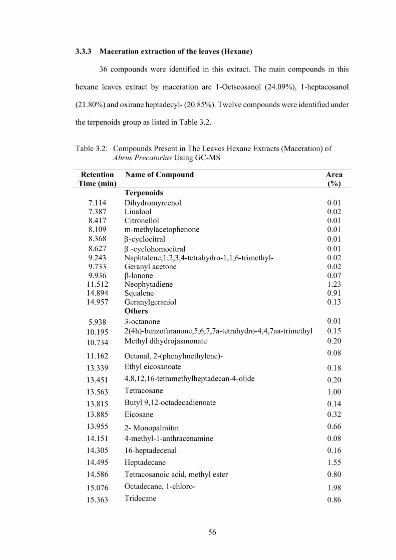
56
3.3.3 Maceration extraction of the leaves (Hexane)
36 compounds were identified in this extract. The main compounds in this
hexane leaves extract by maceration are 1-Octscosanol (24.09%), 1-heptacosanol
(21.80%) and oxirane heptadecyl- (20.85%). Twelve compounds were identified under
the terpenoids group as listed in Table 3.2.
Table 3.2: Compounds Present in The Leaves Hexane Extracts (Maceration) of
Abrus Precatorius Using GC-MS
Retention Time (min)
Name of Compound Area (%)
Terpenoids
7.114 Dihydromyrcenol 0.01 7.387 Linalool 0.02 8.417 Citronellol 0.01 8.109 m-methylacetophenone 0.01 8.368 b-cyclocitral 0.01 8.627 b -cyclohomocitral 0.01 9.243 Naphtalene,1,2,3,4-tetrahydro-1,1,6-trimethyl- 0.02 9.733 Geranyl acetone 0.02 9.936 β-lonone 0.07 11.512 Neophytadiene 1.23 14.894 Squalene 0.91 14.957 Geranylgeraniol 0.13
Others
5.938 3-octanone 0.01 10.195 2(4h)-benzofuranone,5,6,7,7a-tetrahydro-4,4,7aa-trimethyl 0.15 10.734 Methyl dihydrojasmonate 0.20
11.162 Octanal, 2-(phenylmethylene)- 0.08
13.339 Ethyl eicosanoate 0.18 13.451 4,8,12,16-tetramethylheptadecan-4-olide 0.20 13.563 Tetracosane 1.00 13.815 Butyl 9,12-octadecadienoate 0.14 13.885 Eicosane 0.32 13.955 2- Monopalmitin 0.66 14.151 4-methyl-1-anthracenamine 0.08 14.305 16-heptadecenal 0.16 14.495 Heptadecane 1.55 14.586 Tetracosanoic acid, methyl ester 0.80
15.076 Octadecane, 1-chloro- 1.98 15.363 Tridecane 0.86

57
Table 3.2 Continued Retention
Time (min) Name of Compound Area
(%) 15.531 1,19-eicosadiene 7.98 15.839 1-heptacosanol 21.80 15.923 Octadecanal 0.78 15.972 16-octadecenal 1.07 16.441 Oxirane heptadecyl- 20.85 16.868 1-octacosanol 24.09 17.582 Dl-a-tocopherol 0.62 19.585 Cyclotriacontane 0.20

58
3.3.4 Maceration extraction of the leaves (Ethyl acetate)
21 compounds were identified. The main compounds in this extract are 2-
hexadecene,3,7,11,15-tetramethyl-(R-(R*,R*-E)- (16.02%), octacosyl acetate
(8.67%) and phytol (7.6%).
Table 3.3: Compounds Present in The Leaves Ethyl acetate Extracts (Maceration) of Abrus precatorius Using GC-MS
Retention
Time (min) Name of Compound Area
(%) Phenolic compounds
8.984 2-Methoxy-4-vinylphenol 0.72 Terpenoids
12.59 Phytol 7.60 14.886 Squalene 0.73
Steroids 16.28 7-ergosterol 2.06 16.805 b-Sitosterol 1.78
Others 7.121 1-Ethyl-2-pyrrolidinone 0.01 8.41 Coumaran 0.12 9.229 Naphthalene,1,2-dihydro-2,5,8-trimethyl- 0.09
9.474 1-(3,6,6-Trimethyl-1,6,7,7a-tetrahydrocyclopenta[c]pyran-1-yl)ethanone 0.06
11.519 2-hexadecene,3,7,11,15-tetramethyl-(R-(R*,R*-E)- 16.02 11.855 Methyl hexadecanoate 1.70 12.786 Ethyl linolenate 0.18 13.941 2-Palmitoglycerol 1.88
14.515 Nonanoic acid,9-(3-hexenylidenecyclopropylidene)-,2-hydroxy-1-(hydroxymethyl) 5.03
15.055 Cyclotriacontane 2.52 15.244 D-d-tocopherol 2.54 15.902 Vitamin E 2.05
16.133 Z-14-Nonacosane 2.48 16.574 Octacosyl acetate 8.67 17.141 Triacontyl acetate 2.44
17.764 Cyclotriacontane 1.08
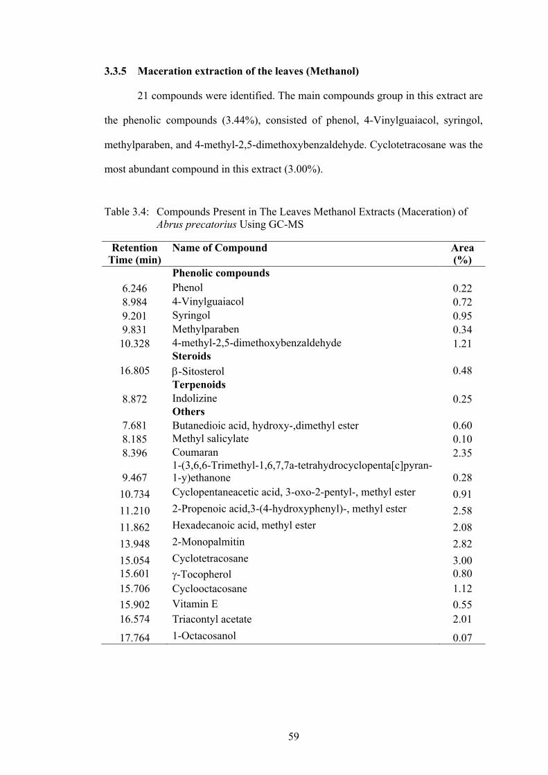
59
3.3.5 Maceration extraction of the leaves (Methanol)
21 compounds were identified. The main compounds group in this extract are
the phenolic compounds (3.44%), consisted of phenol, 4-Vinylguaiacol, syringol,
methylparaben, and 4-methyl-2,5-dimethoxybenzaldehyde. Cyclotetracosane was the
most abundant compound in this extract (3.00%).
Table 3.4: Compounds Present in The Leaves Methanol Extracts (Maceration) of
Abrus precatorius Using GC-MS
Retention Time (min)
Name of Compound Area (%)
Phenolic compounds 6.246 Phenol 0.22 8.984 4-Vinylguaiacol 0.72 9.201 Syringol 0.95 9.831 Methylparaben 0.34 10.328 4-methyl-2,5-dimethoxybenzaldehyde 1.21
Steroids 16.805 b-Sitosterol 0.48
Terpenoids 8.872 Indolizine 0.25
Others 7.681 Butanedioic acid, hydroxy-,dimethyl ester 0.60 8.185 Methyl salicylate 0.10 8.396 Coumaran 2.35
9.467 1-(3,6,6-Trimethyl-1,6,7,7a-tetrahydrocyclopenta[c]pyran-1-y)ethanone 0.28
10.734 Cyclopentaneacetic acid, 3-oxo-2-pentyl-, methyl ester 0.91 11.210 2-Propenoic acid,3-(4-hydroxyphenyl)-, methyl ester 2.58 11.862 Hexadecanoic acid, methyl ester 2.08 13.948 2-Monopalmitin 2.82 15.054 Cyclotetracosane 3.00 15.601 g-Tocopherol 0.80 15.706 Cyclooctacosane 1.12 15.902 Vitamin E 0.55 16.574 Triacontyl acetate 2.01
17.764 1-Octacosanol 0.07

60
3.3.6 Soxhlet Extraction of the leaves (Hexane)
22 compounds were identified from this extract. Main compound identified
from this extract is oxirane, hexadecyl- at 15.72%, followed by 1-eicosanol at 10.53%.
Table 3.5: Compounds identified in the leaves of A. precatorius hexane extracts (Soxhlet)
Retention Time (min)
Name of Compound Area (%)
Phenolic compounds 11.870 Phenol, 3-isopropoxy-5-methyl 0.05
Terpenoids 10.722 Dihydroactinidiolide 0.12 12.003 Neophytadiene 1.54 11.814 (-)-Loliolide 0.35 18.151 Alnulin 0.23
Steroids 17.437 Campesterol 0.33
Others 2.621 Octane 0.10 4.924 Nonane 1.00 10.57 Dodecanoic acid 0.05 10.834 3-Mercapto-2(1H)-pyridinone 0.04 11.660 Myristic acid 0.08 11.682 1-Methylbicyclo(6.3.0)undec-5-en-9-one 0.09 12.185 3,7,11,15-Tetramethyl-2-hexadecen-1-ol 0.57 12.354 Hexadecanoic acid, methyl ester 0.05 12.564 Hexadecanoic acid 5.63 13.031 2-Pentadecanone,6,10,14-trimethyl- 0.11 13.047 9,12,15-Octadecatrienoic acid, methyl ester 0.18 13.096 Octadecanoic acid 5.24 14.477 2-Monopalmitin 0.23 15.035 9,12,15-Octadecatrienoic acid 0.18
15.077 Octadecanoic acid,2-hydroxy-1-( hydroxymethyl)ethyl ester 0.35
15.378 Cyclooctacosane 0.13 15.792 Bicyclo (10.8.0)eicosane, (E)- 0.37 16.415 Stigmasta-5,22-dien-3-ol, acetate, (3.beta.,22Z)- 0.46 16.541 1-Eicosanol 10.53 16.744 Vitamin E 0.37 18.067 (23S)-ethylcholest-5-en-3.beta.-ol 0.49 18.403 1-Tetracosanol 0.69 18.788 Oxirane, hexadecyl- 15.72 19.152 Stigmast-4-en-3-one 0.08

61
3.3.7 Soxhlet Extraction of the leaves (Ethyl acetate)
Neophytadiene (32.56%) was the main compound idetified in this extract. 37
compounds were identified from this extract.
Table 3.6: Compounds identified in the leaves of A. precatorius ethyl acetate extracts (Soxhlet)
Retention Time (min)
Name of Compound Area (%)
Phenolic compounds 8.531 Benzoic acid 0.30 8.860 4-vinyl-phenol 3.26 9.834 2-Methoxy-4-vinylphenol 0.99 15.75 Naringenin 0.91
16.688 3-Methoxy-4,5,7-trihydroxyflavone 0.43 17.479 Cirsimaritin 4.48
Terpenoids 10.722 Dihydroactinidiolide 0.54 11.822 (-)-Loliolide 2.16 12.011 Neophytadiene 32.56 13.089 Phytol 1.71
Steroids 17.99 b-Sitosterol 0.64
Others 2.705 2-Propenoic acid, 2-methyl-,methyl ester 0.41 3.657 2-Pentanone,4-hydroxy-4-methyl- 0.35 8.089 2-Pentene,(Z)- 0.46 8.713 Benzoic acid, 2-hydroxy-, methyl ester 0.12 8.958 1H-Pyrrole-2,5-dione,3-ethyl-4-methyl- 0.25 9.378 Benzonitrile, 2-methyl- 0.19 9.735 Naphtalene,1,2-dihydro-1,1,6-trimethyl- 0.26 9.973 1-(3,6,6-Trimethyl-1,6,7,7a-
tetrahydrocyclopentan(c)pyran-1-yl)ethanone 0.22
10.561 2,5-Cyclohexadiene-1,4-dione,2,6-bis(1,1-dimethyl)- 0.57 10.750 Dodecanoic acid 0.57 10.820 2,6-Dimethyl-3-(methoxymethyl)-p-benzoquinone 1.16 11.660 Myristic acid 2.30 11.569 1,2-Benzenediol,3,5-bis(1,1-dimethylethyl)- 0.40 11.709 1-Methylbicyclo(6.3.0)undec-5-en-9-one 1.05 12.508 Hexadecanoic acid 4.76 12.620 Ethyl palmitate 0.22 12.872 Margaric acid 0.19 13.201 Linolenic acid 5.59

62
Table 3.6 Continued
Retention Time (min)
Name of Compound Area (%)
13.264 Stearic acid 2.97 13.362 Ethyl stearate 0.25 14.363 Heptacosanol 0.41 15.028 Nonanoic Acid,9-(3-Hexenylidenecyclopropylidene)- 1.71 15.351 4,5'-Dihydroxy-7-methoxyflavanone 0.44 15.841 Stigmastan-6,22-dien,3,5-dehydro- 0.29 16.408 Stigmastan-3,5,22-trien 0.90 16.744 Vitamin E 0.76
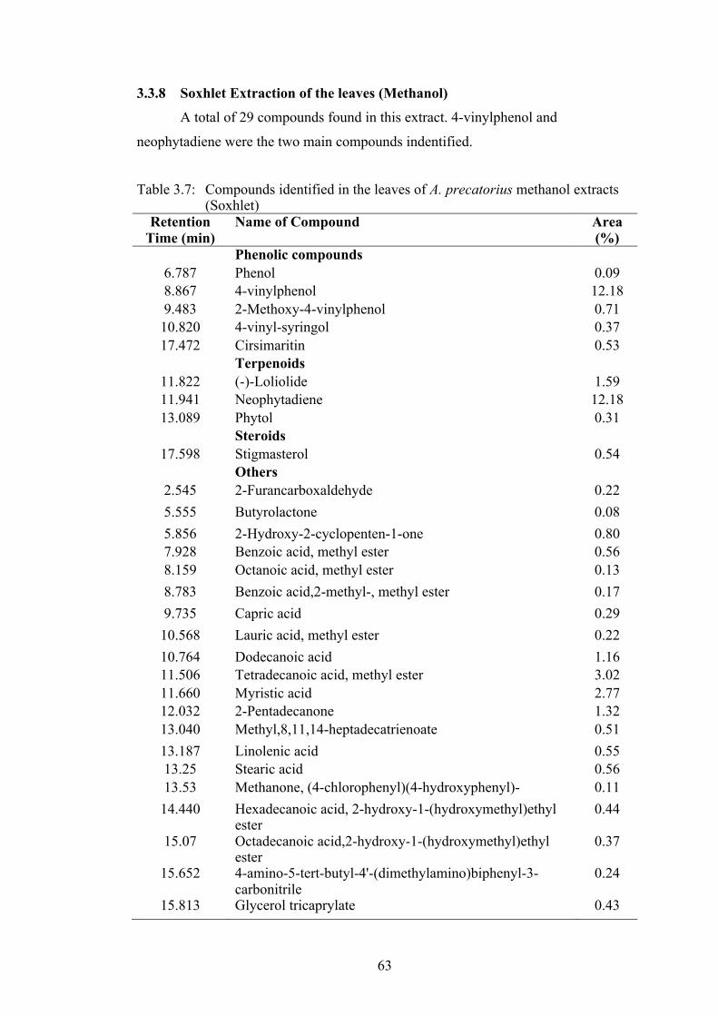
63
3.3.8 Soxhlet Extraction of the leaves (Methanol)
A total of 29 compounds found in this extract. 4-vinylphenol and
neophytadiene were the two main compounds indentified. Table 3.7: Compounds identified in the leaves of A. precatorius methanol extracts
(Soxhlet) Retention
Time (min) Name of Compound Area
(%) Phenolic compounds
6.787 Phenol 0.09 8.867 4-vinylphenol 12.18 9.483 2-Methoxy-4-vinylphenol 0.71
10.820 4-vinyl-syringol 0.37 17.472 Cirsimaritin 0.53
Terpenoids 11.822 (-)-Loliolide 1.59 11.941 Neophytadiene 12.18 13.089 Phytol 0.31
Steroids 17.598 Stigmasterol 0.54
Others 2.545 2-Furancarboxaldehyde 0.22 5.555 Butyrolactone 0.08 5.856 2-Hydroxy-2-cyclopenten-1-one 0.80 7.928 Benzoic acid, methyl ester 0.56 8.159 Octanoic acid, methyl ester 0.13 8.783 Benzoic acid,2-methyl-, methyl ester 0.17 9.735 Capric acid 0.29
10.568 Lauric acid, methyl ester 0.22 10.764 Dodecanoic acid 1.16 11.506 Tetradecanoic acid, methyl ester 3.02 11.660 Myristic acid 2.77 12.032 2-Pentadecanone 1.32 13.040 Methyl,8,11,14-heptadecatrienoate 0.51 13.187 Linolenic acid 0.55 13.25 Stearic acid 0.56 13.53 Methanone, (4-chlorophenyl)(4-hydroxyphenyl)- 0.11
14.440 Hexadecanoic acid, 2-hydroxy-1-(hydroxymethyl)ethyl ester
0.44
15.07 Octadecanoic acid,2-hydroxy-1-(hydroxymethyl)ethyl ester
0.37
15.652 4-amino-5-tert-butyl-4'-(dimethylamino)biphenyl-3-carbonitrile
0.24
15.813 Glycerol tricaprylate 0.43

64
3.3.9 Comparison of the obtained between Maceration and Soxhlet by each solvent
3.3.9(a) Hexane
Comparison of the compounds present in from both henaxe- Soxhlet and
maceration techniques are as shown in Table 3.9. Both extracts shared eight same
compounds. 1-Octacosanol appeared the highest with 24.31% (Soxhlet) and 24.09%
(Maceration).
Table 3.9: Comparison of phytocompounds in A. precatorius leaves extracted with hexane by Soxhlet and maceration.
Soxhlet Maceration 24.31% 1-Octacosanol 24.09% 0.24% 1-Heptacosanol 21.80% 2.17% 16-Octadecenal 1.07% 1.54% Neophytadiene 1.23% 0.02% a-inone 0.02% 0.31% Octadecanal 0.78% 0.23% 2-Monopalmitin 0.66% 0.49% Eicosane
0.32%
15.72% Oxirane, hexadecyl- Oxirane heptadecyl- 20.85% 10.53% 1-Eicosanol 1,19-Eicosadiene 7.98% 5.63% Hexadecanoic acid Octadecane,1-chloro- 1.98% 5.24% Octadecanoic acid Heptadecane 1.55% 1.01% Celidoniol, deoxy- Tetracosane 1.00% 1.00% Nonane Squalene 0.91% 0.69% (23S)-ethylcholest-5-en-
3.beta.-ol Tridecane 0.86%
0.57% 3,7,11,15-Tetramethyl-2-hexadecen-1-ol
Tetracosanoic acid, methyl ester
0.80%
0.49% 1-Tetracosanol dl-.a.-Tocopherol 0.62% 0.46% Stigmasta-5,22-dien-3-ol,
acetate, (3.beta.,22Z)- Digiprolactone 0.28%
0.37% Vitamin E Cyclopentaneacetic acid, 3-oxo-2-pentyl-,methyl ester
0.20%
0.37% Bicyclo (10.8.0)eicosane, (E)- Cyclotriacontane 0.20%

65
Table 3.9 Continued Soxhlet Maceration
0.35% (-)-Loliolide 4,8,12,16-Tetramethylheptadecan-4-olide
0.20%
0.35% Octadecanoic acid,2-hydroxy-1-(hydroxymethyl)ethyl ester
Eicosanoic acid, ethyl ester 0.18%
0.33% Campesterol 16-Heptadecenal 0.16% 0.23% Alnulin 2(4H)-
benzofuranone,5,6,7,7a-tetrahydro-4,4,7a-trimethyl-
0.15%
0.18% 9,12,15-Octadecatrienoic acid, methyl ester
Butyl 9,12-octadecadienoate 0.14%
0.18% 9,12,15-Octadecatrienoic acid Geranylgeraniol 0.13% 0.13% Cyclooctacosane Octanal, 2-
(phenylmethylene)- 0.08%
0.12% Dihydroactinidiolide 4-Methyl-1-anthracenamine 0.08% 0.11% 2-Pentadecanone,6,10,14-
trimethyl- β-lonone 0.07%
0.10% Octane Geranyl acetone 0.02% 0.09% 1-Methylbicyclo(6.3.0)undec-
5-en-9-one Linalool 0.02%
0.08% Stigmast-4-en-3-one 3-Octanone 0.01% 0.08% Myristic acid Dihydromyrcenol 0.01% 0.05% Dodecanoic acid Ethanone, 1-(3-
methylphenyl)- 0.01%
0.05% Phenol, 3-isopropoxy-5-methyl-
b-cyclocitral 0.01%
0.05% Hexadecanoic acid, methyl ester
Citronellol 0.01%
0.04% 3-Mercapto-2(1H)-pyridinone b -cyclohomocitral 0.01%

66
3.3.9(b) Ethyl acetate
Comparison of the compounds present in from both Ethyl acetate- Soxhlet and
maceration techniques are as shown in Table 3.10. Both extracts shared seven similar
compounds. Phytol appeared the highest with 1.71% (Soxhlet) and 7.60%
(Maceration). Neophytadiene was the highest compound in the extract by Soxhlet and
it was not identified in the extract by maceration.
Table 3.10: Comparison of phytocompounds in A. precatorius leaves extracted with
ethyl acetate by Soxhlet and maceration.
Soxhlet Maceration
1.71% Phytol 7.60% 0.31% Nonanoic Acid,9-(3-Hexenylidenecyclopropylidene)- 5.03% 0.41% Heptacosanol 4.45% 0.76% Vitamin E 2.05% 0.64% b-Sitosterol 1.78% 0.99% 2-Methoxy-4-vinylphenol 0.13% 0.26% Naphtalene,1,2-dihydro-1,1,6-trimethyl- 0.09%
32.56% Neophytadiene 2-hexadecene,3,7,11,15-tetramethyl-(R-(R*,R*-E)- 16.02%
5.59% Linolenic acid Octacosyl acetate 8.67% 4.76% Hexadecanoic acid Cyclotriacontane 2.52% 4.48% Cirsimaritin D-d-tocopherol 2.54% 3.26% 4-vinyl-phenol Z-14-Nonacosane 2.48% 2.97% Stearic acid Triacontyl acetate 2.44% 2.30% Myristic acid 7-Ergostenol 2.06%
2.16% (-)-Loliolide Hexadecanoic acid, 2-hydroxy-1-(hydroxymethyl) ethyl ester 1.88%
1.16% 2,6-Dimethyl-3-
(methoxymethyl)-p-benzoquinone
Hexadecanoic acid, methyl ester 1.70%
1.05% 1-Methylbicyclo(6.3.0)undec-5-en-9-one Cyclotriacontane 1.08%
0.91% Naringenin Squalene 0.73%
0.90% Stigmastan-3,5,22-trien 9,12,15-Octadecatrienoic acid, ethyl ester, (Z,Z,Z)- 0.18%

67
Table 3.10 Continued Soxhlet Maceration
0.57% 2,5-Cyclohexadiene-1,4-dione,2,6-bis(1,1-dimethyl)- Coumaran 0.12%
0.57% Dodecanoic acid 1-(3,6,6-Trimethyl-1,6,7,7a-tetrahydrocyclopenta[c]pyran-1-y)ethanone 0.06%
0.54% Dihydroactinidiolide 1-Ethyl-2-pyrrolidinone 0.01% 0.46% 2-Pentene,(Z)-
0.44% 4,5'-Dihydroxy-7-methoxyflavanone
0.43% 3-Methoxy-4,5,7-trihydroxyflavone
0.41% 2-Propenoic acid, 2-methyl-
,methyl ester
0.40% 1,2-Benzenediol,3,5-bis(1,1-dimethylethyl)-
0.35% 2-Pentanone,4-hydroxy-4-
methyl- 0.30% Benzoic acid
0.29% Stigmastan-6,22-dien,3,5-dehydro-
0.25% 1H-Pyrrole-2,5-dione,3-ethyl-4-methyl-
0.25% Ethyl stearate 0.22% Ethyl palmitate
0.22%
1-(3,6,6-Trimethyl-1,6,7,7a-tetrahydrocyclopentan(c)pyran-
1-yl)ethanone 0.19% Benzonitrile, 2-methyl- 0.19% Margaric acid
0.12% Benzoic acid, 2-hydroxy-,
methyl ester

68
3.3.9(c) Methanol
Comparison of the compounds present in from both methanol- Soxhlet and
maceration techniques are as shown in Table 3.11. Both extracts shared only four
similar compounds. They are hexadecenoic acid, methyl ester; 2-methoxy-4-
vinylphenol, hexadecanoic acid, 2-monopalmitin and phenol. Hexadecanoic acid,
methyl ester appeared the highest with 13.09% (Soxhlet) and 2.08% (Maceration).
Table 3.11: Comparison of phytocompounds in A. precatorius leaves extracted with
methanol by Soxhlet and maceration.
Soxhlet Maceration 13.09% Hexadecanoic acid, methyl ester 2.08%
0.71% 2-Methoxy-4-vinylphenol 0.72% 0.44% 2-Monopalmitin 2.82% 0.09% Phenol 0.22%
12.18% Neophytadiene 2-Propenoic acid,3-(4-
hydroxyphenyl)-, methyl ester 2.58%
3.02% Tetradecanoic acid, methyl ester
Coumaran 2.35%
2.77% Myristic acid Triacontyl acetate 2.01% 1.71% 4-vinylphenol Cyclotetracosane 3.00% 1.59% (-)-Loliolide 4-methyl-2,5-
dimethoxybenzaldehyde 1.21%
1.32% 2-Pentadecanone Cyclooctacosane 2.12% 1.16% Dodecanoic acid Syringol 0.95% 0.80% 2-Hydroxy-2-cyclopenten-1-
one Cyclopentaneacetic acid, 3-oxo-2-pentyl-, methyl ester
0.91%
0.56% Benzoic acid, methyl ester g-Tocopherol 0.80% 0.56% Stearic acid Butanedioic acid, hydroxy-
,dimethyl ester 0.60%
0.55% Linolenic acid Vitamin E 0.55% 0.54% Stigmasterol b-Sitosterol 0.48% 0.53% Cirsimaritin Methylparaben 0.34% 0.51% Methyl,8,11,14-
heptadecatrienoate
1-(3,6,6-Trimethyl-1,6,7,7a-tetrahydrocyclopenta[c]pyran-1-y)ethanone
0.28%
0.43% Glycerol tricaprylate Indolizine 0.25% 0.37% 4-vinyl-syringol Methyl salicylate 0.10%
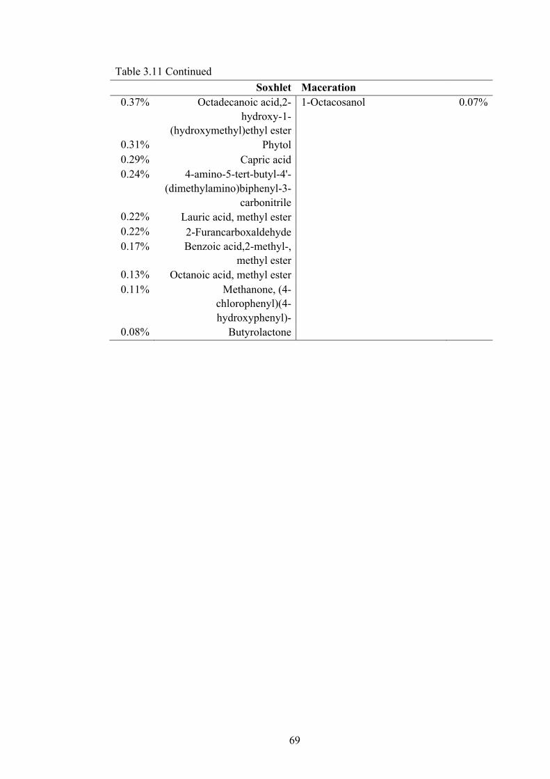
69
Table 3.11 Continued Soxhlet Maceration
0.37% Octadecanoic acid,2-hydroxy-1-
(hydroxymethyl)ethyl ester
1-Octacosanol 0.07%
0.31% Phytol 0.29% Capric acid 0.24% 4-amino-5-tert-butyl-4'-
(dimethylamino)biphenyl-3-carbonitrile
0.22% Lauric acid, methyl ester 0.22% 2-Furancarboxaldehyde 0.17% Benzoic acid,2-methyl-,
methyl ester 0.13% Octanoic acid, methyl ester 0.11% Methanone, (4-
chlorophenyl)(4-hydroxyphenyl)-
0.08% Butyrolactone

70
3.3.10 Compounds with reported biological activity Known anticancer compounds identified from each A. precatorius extracts are listed in Table 3.12. The extracts are aqueous (AQ), hexane
by maceration (HM), ethyl acetate by maceration (EAM), methanol by maceration (MM), hexane by Soxhlet (HS), ethyl acetate by Soxhlet (EAS),
and methanol by Soxhlet (MS).
Table 3.12: Compunds identified in GCMS analysis with reported biological activity.
Compound Name A. precatorius Extract
Reported Activity Reference
4-vinylphenol AQ, MS In vivo and in vitro antiangiogenic activities,
reducing the blood vessel number and tumour
size
Yue et al. (2015)
p-Vinylguaiacol
AQ Antioxidant and antibacterial Ao et al. (2009)
2-Methoxy-4-vinylphenol EAS, MS, EAM Anti-inflammatory Effect of 2-Methoxy-4-Vinylphenol via the Suppression of NF-κB and MAPK Activation, and Acetylation of Histone H3
Jeong et al. (2011)
Phytol AQ, EAM, EAS,
MS
Cytotixicity and antitumour activity de Alencar et al. (2019)
Citronellol HM Anti-inflammatory, analgesic Santos et al. (2018)

71
Table 3.12 Continued
Compound Name A. precatorius Extract
Reported Activity Reference
Neophytadiene
HM, HS, EAS, MS
A good analgesic, antipyretic, anti-inflammatory, antimicrobial, antioxidant and anticancer compound
Kumbum and Sivarao
(2012)
Squalene HM, EAM Cardioprotector, antioxidant, antibacterial and
antifungal, anticancer, detoxifying agent.
Lozano-Grande et al. (2018)
Dihydroactinidiolide HS, EAS Antioxidative and anti aggregation agent,
Inhibit acetylcholinesterase, demostrated
anticancer activity
Das et al. (2018)
Geranylgeraniol HM Anti-inflammatory, anti-tumorigenic,
neroprotective, enhanced testoteron and
progesteron level
Synergistic effect with d-d-tocotrienol on
prostate carninoma cells by cell cycle arrest at
G1
Ho et al. (2018)
Yeganehjoo (2015)
b-Sitosterol EAM, EAS, MM Anti-proliferative effects and induced
apoptosis in MCF7, HVT116 and HeLa
Antidiabetic
Alvarez-Sala et al. (2019)
Zeb et al. (2017)

72
Table 3.12 Continued
Compound Name A. precatorius Extract
Reported Activity Reference
Stigmasterol AQ, MS Membrane stabilizing activity in human red
blood cell, axhibitng anti-inflammatory effect
Zeb et al. (2017)
β-lonone
AQ, HM Anribacteria and anti-inflammatory Kurashov et al. (2016)
Tetracosane HM Cytotoxic against MDA-MB-231, HT-29, and
NIH(3T3). Induced apoptosis in AGS cells
Uddin et al. (2012)
3-octanone HM Role as a human urinary metabolite, an insect
attractant, a fungal metabolite, an antifeedant,
a plant metabolite and a biomarker.
Berendsen et al. (2013)
Vitamin E HS, EAS, EAM,
MM
Antioxidant Mutalip (2018)
D-d-tocopherol
EAM Anticancer Duke (1992-2019)
Dl-a-tocopherol
HM Antioxidant Bharati (2019)
g-Tocopherol MM Protect cells from NO damage
Potential biomarker in response to
pathological condition, as general marker for
mortality risk in populations
Sjöholm et al. (2000)
Chai et al. (2019)

73
Table 3.12 Continued
Compound Name A. precatorius Extract
Reported Activity Reference
Linolenic EAS, MS Antibacterial activity Chandrasekaran et al. (2008)
Myristic acid HS, EAS, MS Enhanced plasma activity, increased
immunemodulating activity
Tan et al. (2017)
(-)-loliolide HS, EAS, MS Inhibit cellular senecence in human dermal
fibroblast (HDFs)
Antibacterial, antitumour, anticancer
Yang et al. (2015)
Grabarczyk et al. (2015)
2-pentadecanone MS Wound healing and antibacterial acitivity,
collagen depostion, fibroblast proliferation
Siyumbwa et al. (2019)
2(4h)-benzofuranone,5,6,7,7a-
tetrahydro-4,4,7aa-trimethyl
HM Antifungal, antialgal, antioxidant, anbacterial
activity
Akhbari et al. (2012)
9,12,15-Octadecatrienoic acid, methyl ester
HS Anticancer, Antimicrobial, Antioxidant and Hyperchloesteralemic
Kumar et al. (2010)
Eicosane HM, HS Antifungal, antibacterial, antitumor and cytotoxic effects
Hsouna et al. (2011)
Hexadecanoic acid
HS Cytotoxic to human leukemic cells
Lai et al. (2010)

74
Table 3.12 Continued
Compound Name A. precatorius Extract
Reported Activity Reference
Hexadecanoic acid, methyl ester HS, EAM, MM, MS Antifungal, Antioxidant, hypocholesterolemic nematicide, pesticide, antiandrogenic flavour, haemolytic, 5-Alpha reductase inhibitor, potent antimicrobial activity.
Hema et al. (2011)
Naringenin EAS Cytotoxic effect on colon carninoma
Inihibit migration of lung cancer (A549) cells
by inhibing Akt activities and reduction of
MMP-2 and MMP-9 activities
Song et al. (2016)
Chang et al. (2017)
Linalool HM Antifungal, anti-inflammatory Dias et al. (2018)
Geranyl acetone HM Antimicrobial activity Bonikowski et al. (2015)
Methyl dihydrojasmonate AQ, HM Antitumour drug compound
da Silva et al. (2015)
4,8,12,16-
tetramethylheptadecan-4-olide
HM Against for PPARg Perumal et al. (2014)
2-Monopalmitin HS, MM, MS Members of MAG. MAG is important critical
nodein lipid signaling pathways. MAG lipase
inhibitor controls FA release of the sysnthesis
protumorigenic sigaling lipids.
Mulvihill and Nomura
(2013)

75
Table 3.12 Continued
Compound Name A. precatorius Extract
Reported Activity Reference
Tetradecanoic acid, methyl ester MS
Inhibit production of uric acid Duke (1992-2019)
Tetracosanoic acid, methyl ester HM Inhibit production of uric acid
Duke (1992-2019)
Octadecanal HM Main compound found in crude extract of
Nostoc linckia, algae, exhibited antimicrobial
activity against S, aureus and S. sonnei
Al-Wathnani et al. (2012)
Octadecanoic acid HS Antioxidant, hypoglycemic and thyroid
inhibitng properties
Duke (1992-2019)
1-Octacosanol HM, HS, MM 90% of antioxidant activity from rice with
high level of octacosanol
Cho et al. (2017)
Coumaran EAM, MM Antitubercular, anti-HIV, anticancer,
cytotoxic, antiprotozoal
Ilya et al. (2018)
Methyl hexadecanoate EAM COMT inhibitors – decrease off time in
Parkinson’s desease patients
Duke (1992-2019)
Indolizine MM Medicinal moeity, anticancer Sharma and Kumar
(2014)
Butanedioic acid, hydroxy-,dimethyl ester
MM Testosterone-hydroxylase-inducer
Inhibit production of uric acid
Duke (1992-2019)

76
Table 3.12 Continued
Compound Name A. precatorius Extract
Reported Activity Reference
Methyl salicylate MM Anti-inflammatory, analgesic, antipyretic and antithrombotic
Mao et al. (2014)
Cyclopentaneacetic acid, 3-oxo-
2-pentyl-, methyl ester
MM COMT inhibitors – decrease off time in
Parkinson’s desease patients
Antioxidant activity through methyl guanidine
inhibition
Duke (1992-2019)
2-Propenoic acid,3-(4-hydroxyphenyl)-, methyl ester
MM Antioxidant
Duke (1992-2019)
Cirsimaritin EAS, MS Inhibited NO production
Anti-inflammatory activity by inhibtion of c-
fos and STAT3 phosphorylation in
RAW264.7 cells
Shin et al. (2017a)
3-Methoxy-4,5,7-trihydroxyflavone
EAS Antioxidant, anti-inflammatory activities
Cytotoxic effect in human lung carcinoma
Limboonreung et al. (2019)
Wei et al. (2019)
4-vinyl-syringol
MS Inhibit gastric tumours initiation Cao et al. (2015)
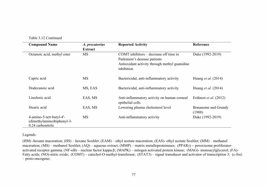
77
Table 3.12 Continued
Compound Name A. precatorius Extract
Reported Activity Reference
Octanoic acid, methyl ester
MS COMT inhibitors – decrease off time in
Parkinson’s desease patients
Antioxidant activity through methyl guanidine
inhibition
Duke (1992-2019)
Capric acid MS Bactericidal, anti-inflammatory activity Huang et al. (2014)
Dodecanoic acid MS, EAS Bactericidal, anti-inflammatory activity Huang et al. (2014)
Linolenic acid EAS, MS Anti-inflammatory activity on human corneal
epithelial cells
Erdinest et al. (2012)
Stearic acid EAS, MS Lowering plasma cholesterol level Bonanome and Grundy
(1988)
4-amino-5-tert-butyl-4'-(dimethylamino)biphenyl-3- 0.24 carbonitrile
MS Anti-inflammatory activity Duke (1992-2019)
Legends:
(HM) -hexane maceration; (HS) – hexane Soxhlet; (EAM) – ethyl acetate maceration; (EAS)- ethyl acetate Soxhlet; (MM) – methanol
maceration; (MS) – methanol Soxhlet; (AQ) – aqueous extract; (MMP) – matrix metalloproteinases; (PPARg) - peroxisome proliferator-
activated receptor gamma; (NF-κB) – nuclear factor kappa b; (MAPK) – mitogen activated protein kinase; (MAG)- monoacylglycerol; (FA)-Fatty acids; (NO)-nitric oxide; (COMT) – catechol-O-methyl-transferase; (STAT3) – signal transducer and activator of transcription 3; (c-fos) – proto-oncogene;

78
3.4 DISCUSSION
Medicinal plants contain organic compounds, which include phenolic
compounds, alkaloids, tannins, carbohydrates, flavonoids, terpenoids, and steroids.
These compounds existed as a result from being synthesized by the primary or
secondary metabolism of living thing.
Phytochemical studies of aqueous leaves extract of A. precatorius are still
limited as it only identifies the polar compounds. The composition of the compounds
identified in aqueous extracts of A. precatorius leaves is a complex mixture of several
classes of components, mainly phenolic compounds, terpenoids and steroids. Phenolic
compounds are identified by the presence of a phenol structure such as an aromatic
benzene ring with at least a hydroxyl substituent (Patra and Saxena, 2009; Robbins,
2003; Vermerris and Nicholson, 2006). Study by Hussain and Kumaresan (2014)
showed that only phenolic compounds and steroids were present in aqueous leaves
extract of A. precatorius. The aqueous extract was obtained via Soxhlet extraction
method, hence the prolonged heat exposure may resulted in the loss of terpenoids
compounds in their extract.
4-vinylphenol is the main phenolic compound identified in aqueous, ethyl
acetate (maceration) and methanol (Soxhket) extracts of A. precatorius leaves. Recent
study of 4-vinylphenol showed that this compound has anti-angiogenic activities (Yue
et al., 2015). Other identified compounds that have some therapeutic activities were
β-lonone, phytol and stigmasterol. β-lonone was found to have anti-proliferative
(Faezizadeh et al., 2016), antibacterial (Kubo et al., 1993; Patra et al., 2015) and
antitumor activities (Cho et al., 2016; Liu et al., 2008; Sharma et al., 2013; Yu et al.,
1995) besides it also can be used as fungicide, pesticide, and trichomonicide. Phytol

79
has antimicrobial (Pejin et al., 2014), anticancer (Song and Cho, 2015) and anti-
inflammatory activities (Silva et al., 2014). Stigmasterol is listed as antihepatotoxic
(El-Domiaty et al., 2009), anti-inflammatory (Gabay et al., 2010), antinociceptive
(Kamurthy et al., 2013), antiophidic, antiviral, cancer-preventive agent (Ali et al.,
2015; Kasahara et al., 1994), hypocholesterolemic agent (Barriuso et al., 2015),
ovulant agent (Zaman et al., 2015) and sedative agent (Habib et al., 2007).
The therapeutic activity of the aqueous leaves extract from A. precatorius,
which in traditional practices obtained by decoction, might be due to the presence of
phytol, stigmasterol and β-lonone. Individually, these phytochemical compounds have
exhibited their bioactivities, however, it does not answer how it works as a whole crude
extract as being practiced traditionally. Other studies indicated that other solvent
extracts from A. precatorius leaves are rich in alkaloids, carbohydrates, steroids,
phenolic compounds and terpenoids (Gul et al., 2013; Hussain and Kumaresan, 2014;
Yonemoto et al., 2014).
In this present study, two different methods were applied for the successive
solvent extraction, which were Soxhlet and maceration. Soxhlet extraction applied
heat in a shorter time while maceration involved prolonged soaking without heat.
Successive solvent extraction means the leaves were extracted first using hexane, ethyl
acetate and lastly with methanol, in the manner of increasing polarity index (P’).
Burdick and Jackson have arranged and listed solvents in order of increasing P’
(Barwick, 1997). Hexane has a P’ of 0.1, P’ for ethyl acetate is 4.4 and P’ for methanol
is 5.1. Meanwhile aqueous has the highest P’ at 10.2. Low P’ solvent will extract
higher volatile compound while the higher the P’, less volatile compound will be
extracted.

80
1-octacosanol was the main compound identified in both extraction by hexane
Soxhlet or hexane maceration. This compound is a fatty acid alcohol mostly found in
waxes of leaves and it is chemically similar vitamin E. To be beneficially for health,
this compound must be taken as supplement because only a small amount of it can be
ingested in the diet (Taylor et al., 2003). Red-coloured rice of the Korean rice genotype
presented highest antioxidant activity and contained the highest level of octacosanol
(Cho et al., 2017). Though not scientifically proven, this compound is used as
supplement for many things including Parkinson disease, managing high cholesterol
and atherosclerosis, improving athletic performances and also for amyotrophic lateral
sclerosis (ALS). A study recently published showed that combined supplement with
addition of 1-octacosanol can boost the fitness of drug detection dogs physically
(Menchetti et al., 2019). Another fatty alcohol highly presented in hexane maceration
extract is heptacosanol (21.80%) however, only 0.24% was identified in the hexane
Soxhlet extract. No health benefits or bioactivity has been associated with this
compound. 1-eicosanol is another fatty alcohol highly presented in hexane Soxhlet
extract which is mainly used as emollient in the cosmetics industry [PubChem
CID=12404] (Kim et al., 2018b).
Neophytadiene was the main compound found in the ethyl acetate Soxhlet
extraction. It is an antioxidant compound known for its biological activity as anti-
inflammaroty, antipyretic, good analgesic and antimicrobial (Swamy et al., 2017).
Neophytadiene belongs to a group of compounds known as sesquiterpenoids, which
consist of terpenes containing three consecutive isoprene units. This compound was
not found in the ethyl acetate maceration extract. This might be because the maceration
technique did not use any heat compared to the Soxhlet extraction method.
Neophytadiene is possibly occurred by dehydration of phytol (Changi et al., 2012).

81
This also explained the reason that this compound could only be identified in extracts
obtained via Soxhlet. The main compound found in the ethyl acetate maceration
extract was 2-hexadecene,3,7,11,15-tetramethyl-(R-(R*,R*-E)-. Neophytadiene
presence was very little with peak area of 1.54% in hexane Soxhlet extract, and 1.23%
in hexane maceration extract. This compound was also one of the main compound
identified in the methanol Soxhlet extract besides hexadecenoic acid, methyl ester.
Hexadecanoic acid, methyl ester is also known as methyl palmitate belongs to
a group of fatty acid methyl ester [PubChem, CID=8181]. This compound has been
reported to significantly induce dilation in aorta (Wang et al., 2018a), reduce the levels
of tumour necrosis factor-alpha (TNF-a), interleukin-10 (IL-10) and prostaglandin E2
(PGE2) without jeopardising the levels of ATP in cells. Besides that methyl palmitate
is also reported to inhibit nitric oxide production and phagocytic activity of certain
cells (Sarkar et al., 2006; Wang et al., 2018a; Wang et al., 2010b). Methyl palmitate
is also known as vasodilator which enhance blood flow in cerebral and promote
neuronal cell survival after cardiac arrest. This therapeutic potential of methyl
palmitate would lead to improvement of functional learning and memory subsequent
of cardiac arrest-induced brain injury (Lee et al., 2019b).
In this current study, extract from the ethyl acetate and methanol obtained by
Soxhlet exhibited the highest phenolic and terpenoid compounds. 4-vinylphenol was
the highest phenolic compound identified in the methanol extract (Soxhlet) and
neophytadiene was the highest terpenoid compound identified in that extract.
Cirsimaritin is another phenolic compound identified in both ethyl acetate and
methanol (Soxhlet) extracts. This compound inhibited nitric oxide production and
exhibited the anti-inflammatory activity (Shin et al., 2017a). Another compound

82
found in the ethyl acetate (Soxhlet) profile is 3-Methoxy-4,5,7-trihydroxyflavone, also
known as chrysoeriol. This compound is derived from luteolin, another
phytocompound widely studied in medicinal plants. Recently it is found that this
compound exhibited anti-inflammatory (Limboonreung et al., 2019) and anticancer
activity (Wei et al., 2019).
Phytochemicals found in plants are generally known as primary and secondary
compounds. Primary compounds are generally present as the building blocks of plants
which includes sugars, proteins, and chlorophyll. Secondary compounds include
phenolic compounds, alkaloids, terpenoids, steroids and many more (Wadood et al.,
2013). The biggest group of phytochemicals is the phenolic compound and most of
these compounds are found in plant-based foods mainly fruits and vegetables such as
cherries, grapes, citruses, tomatoes, apples, peaches and berries (Basli et al., 2017).
Phenolic compounds are widely studied for its health benefits especially the ability to
exhibit as an anti-cancer agent. This ability might be attributed to the antioxidant
activity poses by phenolic compounds. Oxidative stress is one of the causes in cancer
occurrences. Phenolic compounds chemopreventive structure are able to induce cell
cycle arrest thus inhibiting DNA binding and proliferation, and regulate the expression
of ontogenesis and carcinogen metabolism (Huang et al., 2009). Naringenin, a
phenolic compound identified in the ethyl acetate extract (Soxhlet), exhibited
cytotoxic effect on colon carninoma. In this particular study, naringenin was isolated
from the citrus (Song et al., 2016).
Terpenoids is also another compound of interest which have been identified to
demonstrate the anti-proliferative activity on cancer cells. Subclasses of terpenoids are
believed to contribute as anti-cancer agents include monoterpenoid, diterpenoid,
triterpenoid and sesquiterpenoid (Huang et al., 2012). In this current study, A.

83
precatorius leaves extract from ethyl acetate Soxhlet extraction presented the highest
terpenoids at 36.97%. While the methanol Soxhlet extraction showed the highest
phenolic compounds presented at 13.88% and terpenoids at 14.08%. Although there
are a lot of therapeutic potential of the compounds identified in all extracts, our study
are focusing on compounds promoting anticancer activity. Phenolic compounds found
in all A. preactorius extracts were 4-methyl-2,5-dimethoxybenzaldehyde, cirsimaritin,
and 4-vinyl-phenol. While compounds belong in the terpenoids group identified in all
extracts include neophytadiene, phytol, squalene, and (-)-Loliolide. Neophytadiene
presented in all extracts except ethyl acetate maceration and methanol maceration
extracts.
Previous study of A. precatorius leaves showed that their biological activities
are also related with their active compounds such as terpenoids and phenolics
compounds. Yonemoto et al. (2014) found that terpenoids isolated from A. precatorius
leaves had α-amylase inhibitory effect; one of the therapeutic approaches for
preventing diabetes mellitus. Phenolic compounds such as flavanoids and phenolic
acids are well known to have antioxidant and anti-proliferative activities (Gul et al.,
2013). A recent study from Iraq, using 80% aqueous ethanol solvents for A.
precatorius leaves extraction by Soxhlet revealed the presence of several compounds
including alkaloids, flavonoids, phytosterols and terpenoids. Three fractions were
obtained from the crude extract that showed the presence of alkaloids (fraction 1),
flavonoids (fraction 2) and steroids (fraction 3) (Khadem and Zahra’a, 2018).

84
3.5 CONCLUSION
Presence of phenolic compounds and terpenoids in most of the extracts showed
that A. preactorius leaves might have effects on cancer cell proliferation and mortality.
Major compound found consistently in all extract is neophytadiene, a terpenoids.
However, little is known as to whether these identified compounds work individually
or as a result of a synergistic effect. Therefore, in this study, we were exploring on the
usage of A. precatorius leaves extracts in crude form rather than as isolated
compounds. The ability of A. precatorius leaves extracts to possibly exert the anti-
proliferative activity against selected normal and cancer cell line and its ability to
induce apoptosis were further investigated and detailed in Chapter 4.

85
CHAPTER 4
ANTI-PROLIFERATIVE ACTIVITY AND APOPTOSIS INDUCTION of Abrus precatorius LEAVES EXTRACT ON CANCER CELLS
4.1 INTRODUCTION
Apoptosis is known as programmed cell death and it is the most studied and
characterized form of cell death. Dying cells are packaged into fragments which later
are consumed and eliminated by phagocytes. This process needs to occur without
disturbing the function of surrounding healthy tissues (Green, 2011). Equilibrium
between cell proliferation and cell death is important to ensure the cellular balance in
functioning tissues and to avoid cell disruption. Many clinical diseases including
cancer are developed because of this apoptosis imbalance either deficient or excessive
(Cao and Tait, 2018). Initiation of apoptosis can occur either by the extrinsic or
intrinsic pathway. The intrinsic pathway is also known as the mitochondrial pathway
and the extrinsic pathway is also known as the death receptor pathway. Apoptosis is
as important tool in cancer management used as a target by potent chemical or
biological apoptosis-inducing agents (Sreelatha et al., 2011).
Advances in the medical field have proven that allopathy treatment to be the
preferable choice to combat cancer. However, these advances are used with concern
due to their side effects and limitations. This scenario has generated an impact on the
increase demand of traditional medicines through utilizing medicinal plants either as
complementary to the allopathy treatments or as a complete alternative. Medicinal
plants are widely sought as an alternative in various treatment in traditional medicine
including cancer. Many issues were raised with this development especially
concerning the toxicity and efficacy of the medicinal plants extract. Therefore, many
studies have been conducted to fill in the knowledge gap in order to provide better

86
understanding of the biological activities and mechanism underlying those activities
of the medicinal plants.
Among the other popular medicinal plants that is highly studied is the Anonna
muricata. Ethanol extract of this plant was found to reduce viability and trigger
apoptosis in liver cancer cell, HepG2 (Liu et al., 2016). The apoptosis was triggered
through activation of multiple proteins involved in the endoplasmic reticulum stress
pathway. Another study by Smith et al. (2018), reported that bacopaside II, isolated
from the plant Bacopa monnieri significantly reduced cell viability in colon cancer
cells, SW 480, SW 620, Ht-29 and HCT 116. Ixeris dentata induced apoptosis by
inhibiting p-Akt and p-NF-κB signaling pathway in MDA-MB-231 cells (Shin et al.,
2017b). Root extract from Dillenia suffruticosa also induced apoptosis, arrest DNA at
G2/M cell cyle and activation of proapoptotic JNK1 and down regulation of ERK1
proteins (Foo et al., 2016). Medicinal plants in focus of this thesis is Abrus precatorius.
As mentioned earlier, many biological activities of A. precatorius have been reported
including anticancer. However, pharmacological properties of A. precatorius collected
from Malaysia need to be further explored.
Therefore, this chapter reported on the anti-proliferative activity of A.
precatorius leaves extracts on selected cancer and normal cells. The extract with the
lowest IC50 with its corresponding cell was selected for further analysis to elucidate
the involvement of apoptosis induction in promoting the cell deaths.
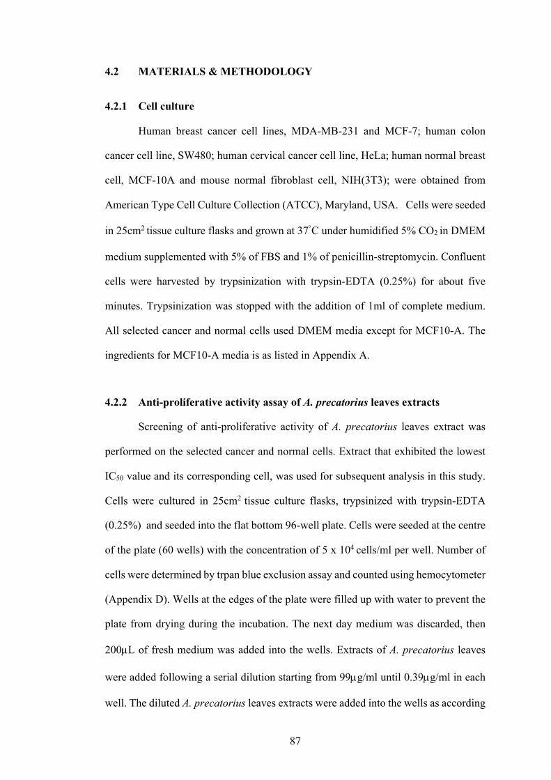
87
4.2 MATERIALS & METHODOLOGY
4.2.1 Cell culture
Human breast cancer cell lines, MDA-MB-231 and MCF-7; human colon
cancer cell line, SW480; human cervical cancer cell line, HeLa; human normal breast
cell, MCF-10A and mouse normal fibroblast cell, NIH(3T3); were obtained from
American Type Cell Culture Collection (ATCC), Maryland, USA. Cells were seeded
in 25cm2 tissue culture flasks and grown at 37°C under humidified 5% CO2 in DMEM
medium supplemented with 5% of FBS and 1% of penicillin-streptomycin. Confluent
cells were harvested by trypsinization with trypsin-EDTA (0.25%) for about five
minutes. Trypsinization was stopped with the addition of 1ml of complete medium.
All selected cancer and normal cells used DMEM media except for MCF10-A. The
ingredients for MCF10-A media is as listed in Appendix A.
4.2.2 Anti-proliferative activity assay of A. precatorius leaves extracts
Screening of anti-proliferative activity of A. precatorius leaves extract was
performed on the selected cancer and normal cells. Extract that exhibited the lowest
IC50 value and its corresponding cell, was used for subsequent analysis in this study.
Cells were cultured in 25cm2 tissue culture flasks, trypsinized with trypsin-EDTA
(0.25%) and seeded into the flat bottom 96-well plate. Cells were seeded at the centre
of the plate (60 wells) with the concentration of 5 x 10⁴ cells/ml per well. Number of
cells were determined by trpan blue exclusion assay and counted using hemocytometer
(Appendix D). Wells at the edges of the plate were filled up with water to prevent the
plate from drying during the incubation. The next day medium was discarded, then
200µL of fresh medium was added into the wells. Extracts of A. precatorius leaves
were added following a serial dilution starting from 99µg/ml until 0.39µg/ml in each
well. The diluted A. precatorius leaves extracts were added into the wells as according

88
to Table 4.1. Each concentration was added into three wells (triplicates) and each
extract was tested for three times (n=3).
Table 4.1: Serial dilution calculation of A. precatorius extracts
Well number
Initial Extract concentration
(mg/ml)
Final Concentration of
extract in well (mg/ml)
Final Concentration of
extract in well
(µg/ml)
Log 10 final concentrations
1 10 0.099 99 -1.00 2 5 0.0495 49.5 -1.31 3 2.5 0.0248 24.8 -1.61 4 1.25 0.0124 12.4 -1.91 5 0.625 0.00618 6.18 -2.21 6 0.3125 0.00309 3.09 -2.51 7 0.156 0.00155 1.55 -2.81 8 0.078 0.00077 0.77 -3.11 9 0.039 0.00039 0.39 -3.41
Anti-proliferative activity of A. precatorius leaves extracts was measured by
the MTT (3-[4,5-dimethyl thiazol-2-yl] 2,5-diphenyl tetrazolium bromide) assay,
which was performed after a 72h incubation post treatment with the extracts,
tamoxifen (positive control) and DMSO (negative control). Absorbance was read at
OD of 570nm. The absorption value at this wavelength directly represents the relative
cell numbers with comparison to the control group (Igarashi and Miyazawa, 2001).
The percentage of cell viability was determined according to the following equation.
The IC50 values of all extracts on each cancer/normal cells were determined by
plotting a graph of ‘Log10 of the final extracts concentration’ (as depicted in Table 4.1)

89
against ‘Cell Viability’. The extract that induced lowest IC50 values with its
corresponding cell, was selected for the subsequent analysis of this study.
4.2.3 Morphology of cell death
From anti-proliferative assays, it was shown that A. precatorius methanolic
leaves extract (Soxhlet) showed the lowest IC50 value at 26.4µg/ml on MDA-MB-231
cells (Table 4.2). This extract was designated as APME for the rest of the report in this
thesis. In order to visualize the morphological effects of APME on MDA-MD-231
cells, the APME-treated cells were observed under light microscope and fluorescent
microscope.
4.2.3(a) Bright field microscopy
Light microscope was used to view morphological effect of APME-treated
MDA-MB-231 cells and images were recorded with Dino-Eye Eyepiece Camera. Cells
were seeded in a six-well plate at 5x10⁴ cells/well. Cells were culturre using DMEM
with 5% of FBS and 1% of penicillin-streptomycin. Cells were incubated in a
humidified, 5% CO2 incubator at 37°C, overnight. After cells were confluence, cells
were treated with APME and further incubated for 24h, 48h and 72h. Untreated cells
(contains <1% DMSO) at each time point were also observed alongside with
Tamoxifen-treated cells.
4.2.3(b) Fluorescent microscopy (Hoechst Staining )
Hoechst 33258 was used to visualize nuclear changes in apoptotic cells. The
dye binds to the DNA in the cells, producing fluorescence blue color under
fluorescence microscope. Cells were firstly seeded in a petri dish containing a sterile
microscope slide at 5x10⁴ cells. They were incubated in a humidified, 5% CO2

90
incubator at 37°C, overnight. The next day, cells were treated with APME for 24, 48h
and 72h. After the incubation times, the slides were washed three times with PBS and
dried. Then they were fixed with 4% paraformaldehyde (30 minutes) at 4°C. All slides
were then incubated with 30 µg/mL Hoeshct 33258 (Invitrogen, USA) at room
temperature in a dark condition for 30 minutes. Nuclear morphology of each different
treatment time was examined under fluorescent microscope at 40X magnification
(Imaging Source Europe GmbH, Bremen, Germany). For long-term storage, the slides
were wrap with aluminum foil and kept in 4°C.
4.2.4 Cell Cycle Assay
MDA-MB-231 cells were treated with IC50 of the APME and incubated for 24,
48 and 72h. Cells were harvested by trypsinization and the cell cycle assay was
performed according to the manufacturer protocol, BD CycletestTM. All samples
readings were acquired with FACSCANTO II (BD Bioscience). Data obtained was
analysed with ModFit LT 5.0 software (Becton Dickinson, Franklin Lakes, NJ, USA).
4.2.5 Apoptosis Assays
4.2.5(a) AnnexinV and PI staining
MDA-MB-231 cells were treated with IC50 of the APME and incubated for
24h, 48h and 72h. Cells were harvested by trypsinization and the apoptosis assay was
performed according to the manufacturer protocol, AnnexinV-FITC detection kit I
(BD Bioscience). All samples readings were acquired with FACSCANTO II (BD
Bioscience). Data obtained was analysed with FlowJo software.

91
4.2.5(b) Bax, Bcl-2, Caspase-3 and p53 activity
MDA-MB-231 cells were treated with IC50 of the APME and incubated for
24h, 48h and 72h. Cells were harvested by trypsinisation following each incubation
time and washed twice with PBS. Ethanol (70%) was used to fix the cells at 4°C for
an hour. Cells were washed twice with PBS and then blocked with 2% BSA for 10
minutes at room temperature. Another cell wash was performed, and cells were
resuspended in PBS.
About 100µl of the cell suspension (1x10⁶ cells) were mixed independently in
different tubes, with antibodies (SantaCruz); Bax-PE (sc-7480), Bcl-2 – Alexa Fluor
647 (sc-7382), p53 – Alexa Fluor 488 (sc-126) and Caspase-3 – Alexa Fluor 488 (sc-
7272). These cells-antibodies mixtures were incubated for 20 minutes at room
temperature, then washed once and resuspended in 500µl PBS. All samples readings
were acquired with FACSCANTO II (BD Bioscience). Data obtained was analysed
with FlowJo software.
4.2.6 Statistical Analysis
The data were expressed as mean ±SD of three repeated experiments. The level
of statistical significance was tested using repeated measure one-way ANOVA,
followed by Dunnett’s multiple comparison test. The difference was considered
significant if P<0.05. Analyses were all done using GraphPad Prism7.

92
4.3 RESULTS
4.3.1 Anti-proliferative activity of A. precatrius leaves extracts
Anti-proliferative activity of A. precatorius was determined on human breast
cancer cell lines, MDA-MB-231 and MCF-7; human liver cancer cell lines, SW480;
human cervical cancer cell line, HeLa; human normal breast cell, MCF-10a and mouse
normal fibroblast cell, NIH (3T3). The potential anti-proliferative activity of A.
precatorius extracts was investigated by MTT assay. IC50 values were determined to
demonstrate the anti-proliferative effects of these extracts, with lower IC50 values
indicating higher anti-proliferative activity. The US National Cancer Institute and
Geran Protocol (Geran et al., 1972), listed out the criteria used to categorize
cytotoxicity of plant extracts against cancer cell line as follows: highly cytotoxic (IC50
≤ 20μg/ml), moderately cytotoxic (21≤ IC50 ≤ 200μg/ml), weakly cytotoxic (201≤ IC50
≤ 500μg/ml), and, no cytotoxicity (IC50 ≥ 501μg/ml). IC50 of all extracts were
determined by plotting the graph of concentration of the extract(s) or tamoxifen vs
percentage of cell viability.

93
4.3.2 Determination of the anti-proliferative activity of A. precatorius aqueous extract (decoction) on selected normal and cancer cells
A. precatorius aqueous leaves extract was prepared by decoction at 50°C.
Initially the cells were treated with concentration as listed in Table 4.1 (page 88).
However, no IC50 value was obtained even the cells were treated at the maximum
concentration of 99µg/ml. Therefore, the aqueous extract concentration was increased
to 990µg/ml. It was performed in the same manner as explained in section 4.2.2. A log
concentration vs cell viability was also plotted. Figure 4.1 showed the representative
points of each concentration, presented in means with ±SD of three independent
experiments. A. precatorius aqueous extract demonstrated a null toxicity against all
cells, according to the standard criteria used by The US National Cancer Institute and
Geran Protocol.

94
Figure 4.1: Anti-proliferative activity of A. precatorius aqueous leaves extracts on
selected cancer and normal cells.
The results were expressed as mean, ±SD of three independent experiments with three replicates
0
20
40
60
80
100
120
990495247.5128.861.930.915.57.73.9
Cell
viab
ility
(%)
Cocentration (µg/ml)
HeLa MCF-7 MDA-MB-231 SW 480 MCF10A NIH(3T3)

95
4.3.3 Determination of the anti-proliferative activity of A. precatorius solvents extract (Soxhlet) on selected normal and cancer cells
There were two types of extraction methods used for A. precatorius leaves.
Both methods applied solvents in a successive manner. Anti-proliferative activities of
A. precatotius successive Soxhlet hexane-, ethyl acetate- and methanol- leaves extracts
are shown in Figure 4.2 -4.7.
4.3.3(a) HeLa
Figure 4.2: Anti-proliferative activity of A. precatorius successive Soxhlet
hexane-, ethyl acetate- and methanol- leaves extracts on HeLa cells.
The only IC50 values obtained were for methanol extract was 73.6µg/ml and 4.32 µg/ml for Tamoxifen, while the rest of the extracts has the IC50
values of >99 µg/ml. The results were expressed as mean, ±SD of three independent experiments with three replicates.
0
20
40
60
80
100
120
9949.524.812.46.193.091.550.730.39
Cell
Viab
ility
(%)
Concentration (µg/ml)
Hexane Ethyl acetate Methanol Tamoxifen

96
4.3.3(b) MCF7
Figure 4.3: Anti-proliferative activity of A. precatorius successive Soxhlet hexane-, ethyl acetate- and methanol- leaves extracts on MCF7 cells.
The IC50 values obtained for hexane extract was 52.65µg/ml, ethyl acetate extract was 99µg/ml and methanol extract was 59.03µg/ml and 1.81µg/ml for Tamoxifen. The results were expressed as mean, ±SD of three independent experiments with three replicates.
0
20
40
60
80
100
120
140
9949.524.812.46.193.091.550.730.39
Cell
Viab
ility
(%)
Concentration (µg/ml)Hexane Ethyl acetate Methanol Tamoxifen

97
4.3.3(c) MDA-MB-231
Figure 4.4: Anti-proliferative activity of A. precatorius successive Soxhlet hexane-, ethyl acetate- and methanol- leaves extracts on MDA MB-231 cells.
The IC50 obtained for hexane, ethyl acetate, methanol extract and tamoxifen were 45.60µg/ml, 54.5µg/ml, 26.4µg/ml and 2.27µg/ml, respectively. The results were expressed as mean, ±SD of three independent experiments with three replicates
0
10
20
30
40
50
60
70
80
90
100
9949.524.812.46.193.091.550.730.39
Cell
Viab
ility
(%)
Concentration (µg/ml)
Hexane Ethyl acetate Methanol Tamoxifen

98
4.3.3(d) SW 480
Figure 4.5: Anti-proliferative activity of A. precatorius successive Soxhlet hexane-, ethyl acetate- and methanol- leaves extracts on SW 480 cells.
The IC50 obtained for methanol extract is 77.23µg/ml and 2.31µg/ml for Tamoxifen, while the rest of the extracts demonstrated the IC50 values of >99µg/ml. The results were expressed as mean, ±SD of three independent experiments with three replicates
0
20
40
60
80
100
120
140
9949.524.812.46.193.091.550.730.39
Cell
Viab
ility
(%)
Concentration (µg/ml)
Hexane Ethyl acetate Methanol Tamoxifen

99
4.3.3(e) NIH(3T3)
Figure 4.6: Anti-proliferative activity of A. precatorius successive Soxhlet
hexane-, ethyl acetate- and methanol- leaves extracts on NIH(3T3) cells.
The IC50 obtained for Tamoxifen is 3.78µg/ml, while the rest of the extracts demonstrated the IC50 values of >99µg/ml. The results were expressed as mean, ±SD of three independent experiments with three replicates
0
20
40
60
80
100
120
9949.524.812.46.193.091.550.730.39
Cell
viab
ility
(%)
Concentration (µg/ml)
Hexane Ethyl acetate Methanol Tamoxifen

100
4.3.3(f) MCF10A
Figure 4.7: Anti-proliferative activity of A. precatorius successive Soxhlet hexane-, ethyl acetate- and methanol- leaves extracts on MCF10A cells.
The IC50 obtained for Tamoxifen is 2.78µg/ml, while the rest of the extracts demonstrated the IC50 values of >99 µg/ml. The results were expressed as mean, ±SD of three independent experiments with three replicates.
0
20
40
60
80
100
9949.524.812.46.193.091.550.730.39
Cell
viab
ility
(%)
Concentration (µg/ml)
Hexane Ethyl acetate Methanol Tamoxifen

101
4.3.4 Determination of the anti-proliferative activity of A. precatorius solvents extracts (Maceration) on selected normal and cancer cells
Another method used for A. precatorius leaves extraction was by maceration.
The ground leaves were soaked with no heat successively with different solvents
following their polarity. Initially the concentration of the extracts used were as stated
in Table 4.1. However, no IC50 was recorded in all extracts even at the maximum
concentration used. Therefore, the extracts concentration was increased to the
maximum of 495µg/ml. Anti-proliferative activities of A. precatotius successive
(maceration) hexane-, ethyl acetate- and methanol- leaves extracts are shown in Figure
4.8 – 4.13.

102
4.3.4(a) HeLa
Figure 4.8: Anti-proliferative activity of A. precatorius successive (maceration) hexane-, ethyl acetate- and methanol- leaves extracts on HeLa cells.
The IC50 obtained for hexane extract was 325µg/ml, ethyl acetate extract was 371µg/ml and methanol extract was 352 µg/ml. The results were expressed as mean, ±SD of three independent experiments with three replicates.
0
20
40
60
80
100
120
495247.5128.861.930.915.57.73.91.9
Cell
Viab
ility
(%)
Concentration (µg/ml)
Hexane Ethyl acetate Methanol

103
4.3.4(b) MCF-7
Figure 4.9: Anti-proliferative activity of A. precatorius successive (maceration) hexane-, ethyl acetate- and methanol- leaves extracts on MCF-7 cells.
The IC50 obtained for hexane extract was 672µg/ml and methanol extract was 423µg/ml. While ethyl acetate extract was >495µg/ml. The results were expressed as mean, ±SD of three independent experiments with three replicates.
0
20
40
60
80
100
120
495247.5128.861.930.915.57.73.91.9
Cell
Viab
ility
(%)
Concentration (µg/ml)
Hexane Ethyl acetate Methanol

104
4.3.4(c) MDA-MB-231
Figure 4.10: Anti-proliferative activity of A. precatorius successive (maceration) hexane-, ethyl acetate- and methanol- leaves extracts on MDA-MB-231 cells.
The IC50 obtained for hexane extract was 80.75µg/ml, ethyl acetate extract was 207µg/ml and methanol was 255µg/ml. The results were expressed as mean, ±SD of three independent experiments with three replicates.
0
20
40
60
80
100
120
140
495247.5128.861.930.915.57.73.91.9
Cell
viab
ility
(%)
Concentration (µg/ml)Hexane Ethyl acetate Methanol

105
4.3.4(d) SW480
Figure 4.11: Anti-proliferative activity of A. precatorius successive (maceration) hexane-, ethyl acetate- and methanol- leaves extracts on SW480 cells.
The IC50 obtained for hexane extract was 301.3µg/ml, ethyl acetate extract was 447.5µg/ml and methanol 350.3was µg/ml. The results were expressed as mean, ±SD of three independent experiments with three replicates.
0
20
40
60
80
100
120
495247.5128.861.930.915.57.73.91.9
Cell
Viab
ility
(%)
Concentration (µg/ml)Hexane Ethyl acetate Methanol

106
4.3.4(e) NIH (3T3)
Figure 4.12: Anti-proliferative activity of A. precatorius successive (maceration) hexane-, ethyl acetate- and methanol- leaves extracts on NIH(3T3) cells.
No IC50 was obtained even at the maximum concentration of 495µg/ml. The results were expressed as mean, ±SD of three independent experiments with three replicates.
0
20
40
60
80
100
120
140
495247.5128.861.930.915.57.73.91.9
Cell
Viab
ility
(%)
Concentration (µg/ml)
Hexane Ethyl acetate Methanol
107
4.3.4(f) MCF10A
Figure 4.13: Anti-proliferative activity of A. precatorius successive (maceration) hexane-, ethyl acetate- and methanol- leaves extracts on MCF10A cells.
No IC50 was obtained even at the maximum concentration of 495µg/ml. The results were expressed as mean, ±SD of three independent experiments with three replicates.
0
20
40
60
80
100
120
495247.5128.861.930.915.57.73.91.9
Cell
viab
ility
(%)
Concentration (µg/ml)
Hexane Ethyl acetate Methanol

108
4.3.5 Summary of the IC50 values of all A. precatorius leaves extracts
The IC50 values of all extracts in inhibiting the cancer and the normal cells were
summarized in Table 4.2. Methanol extract (Soxhlet) was identified to have the lowest
IC50 value among all extracts on MDA-MB-231 cells at 26.4±2.45µg/ml, which is
categorized it to be moderately toxic. Methanol extract (Soxhlet) also exhibited
moderate anti-proliferative activity on all cancer cells but not toxic on normal cells.
Methanol extract (maceration) exhibited low cytotoxic activity on all cancer cells.
Hexane extract (Soxhlet) exhibited moderate cytotoxicity on MCF-7 and MDA-MB-
231 cells, while the maceration extract showed low anti-proliferative effect on HeLa,
MCF-7, and SW 480. Ethyl acetate extract (Soxhlet) exhibited moderate anti-
proliferative activity on MCF-7 and MDA-MB-231 cells, while low anti-proliferative
activity on HeLa, MDA-MB-231, and SW 480 was exhibited by the ethyl acetate
maceration extract. All extracts exhibited cytotoxic effects on MDA-MB-231 cells.

109
Table 4.2: IC50 values of A. precatorius leave extracts against selected normal and cancer cell lines (µg/ml).
Types of cancer cell
lines
Hexane extract
(Soxhlet)
Hexane extract
(Maceration)
Ethyl acetate extract
(Soxhlet)
Ethyl acetate extract
(Maceration)
Methanol extract
(Soxhlet)
Methanol extract
(Maceration) Aqueous Extract Tamoxifen
HeLa (cervix)
>99 325±38.4 >99 371±52.0 73.60±6.17 325±12.2 618±38.8 4.32±0.40
MCF7 (breast)
52.65±7.14 425.5±75.7 99.0±11.86 >495 59.03±9.40 330±37.0 668±176 1.81±1.78
MDA MB-231 (breast)
45.60±11.60 80.75±64 54.50±9.05 206.5±9.2 26.40±5.40 254.5±57 537±32.5 2.27±0.38
SW 480 (colon)
>99 301.3±39.1 >99 447.5±31.8 77.23±6.39 350.3±28 785±131 2.31±0.59
MCF-10a (normal breast)
>99 >495 >99 >495 >99 >495 680±58.5 2.78±0.79
NIH (normal
fibroblast)
>99 >495 >99 >495 >99 >495 >990 3.78±1.78

110
4.3.6 Observation on morphological changes upon treatment with APME
Morphology of cell death can be observed microscopically. Two methods used
in this study were direct observation via bright field microscopy and Hoechst staining
via fluorescence microscopy. From previous anti-proliferative assays, A. precatorius
methanolic leaves extract (Soxhlet) showed the lowest IC50 value at 26.4µg/ml on
MDA-MB-231 cells. Therefore, this extract (APME) was selected for further studies.
MDA-MB-231 cells were treated with APME for 24h, 48h, and 72h. The cells
were viewed under inverted light microscope at 20X magnification and the images
were captured with Dino-Eye Camera. The images are shown in Figure 4.14 (a) – (i).
No significant change was observed for the APME-treated cells at 24h comparing to
the untreated cells. At 48h and 72h, cells grew rapidly, and new cells formed layers,
while older cells formed clumps on their surface. APME-treated cells were showing
signs of apoptosis starting at 48h (Figure 4.14-e) post treatment. Longer time exposure
showed more cells with membrane blebbing and ballooning. Cells were also unable to
keep its spindle shaped cells. Rounder and shrunken cells signify dead cells and none
newly formed cells were observed in Figures 4.14 (e), (f), (h) and (i). Tamoxifen-
treated cells started to exhibit apoptotic-like structure at 24h post treatment. More cell
deaths were observed in tamoxifen-treated cells at 48h and 72h post treatment.
MDA-MB-231 treated cells were also observed under fluorescent microscope
to observe the nucleus changes. Hoechst 32588 was used to stain the cells as shown in
Figure 4.15. This dye enters the cell and binds to the AT-rich segment of the DNA in
the nucleus. This staining is important in order to demonstrate the DNA fragmentation
as the hallmark of apoptosis. The density of the fluorescent was observed higher in

111
APME-treated cells at 48h and 72h which indicated that the DNA in those cells started
to lose its integrity thus allowing more dye to bind to the exposed AT-regions.

112
4.3.6(a) Bright field microscopy
Figure 4.14 (a-c): Bright field microscopy images of non-treated MDA-MB-231 at 24h, 48h and 72h. Magnification: 20X..
a b c

113
Figure 4.14 (d-f): Bright field microscopy images of MDA-MB-231 cells treated with APME at 24h, 48h and 72h. Magnification: 20X
d e f

114
Figure 4.14 (g-i): Bright field microscopy images of MDA-MB-231 cells treated with Tamoxifen at 24h, 48h and 72h. Magnification: 20X
g h i
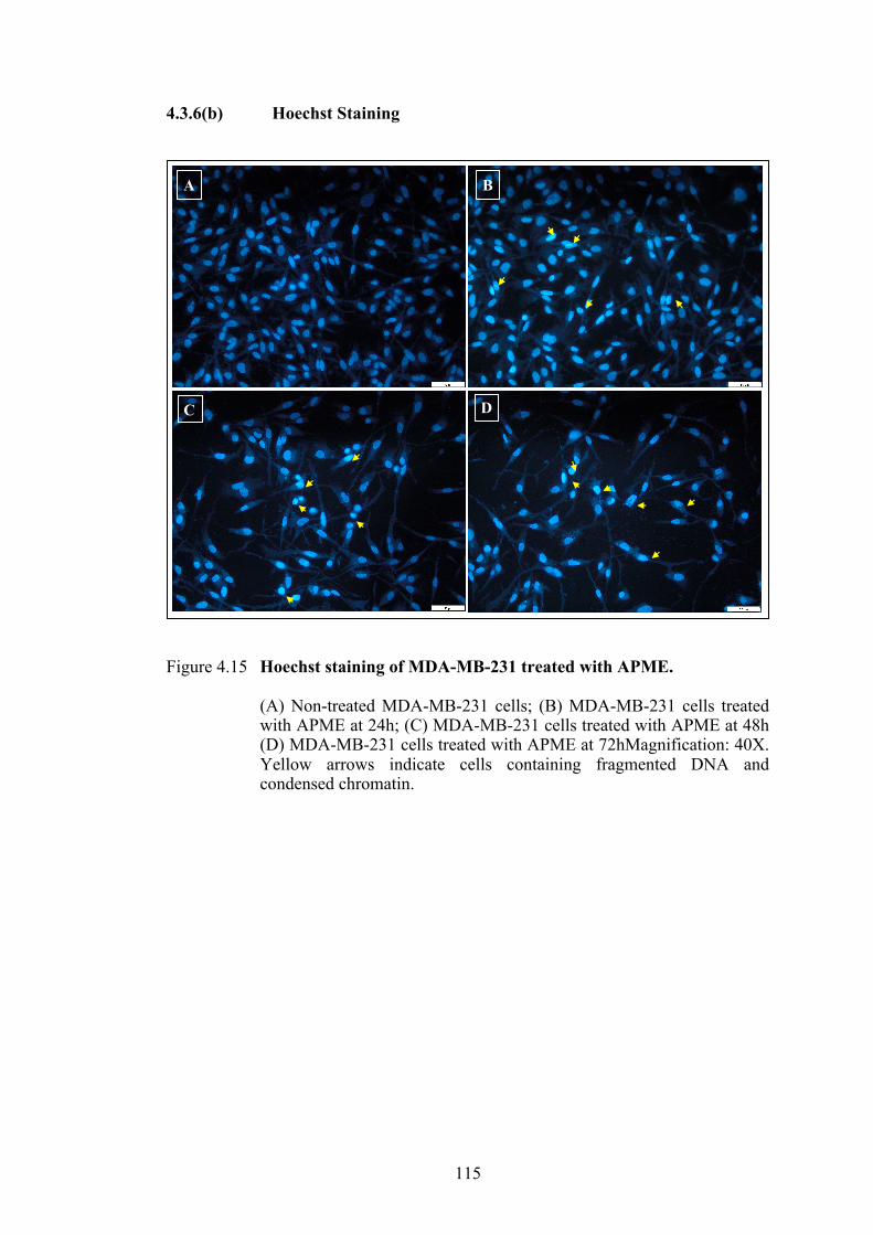
115
4.3.6(b) Hoechst Staining
Figure 4.15 Hoechst staining of MDA-MB-231 treated with APME.
(A) Non-treated MDA-MB-231 cells; (B) MDA-MB-231 cells treated with APME at 24h; (C) MDA-MB-231 cells treated with APME at 48h (D) MDA-MB-231 cells treated with APME at 72hMagnification: 40X. Yellow arrows indicate cells containing fragmented DNA and condensed chromatin.
A B
DC
50µm
50µm

116
4.3.7 Cell Cycle Analysis
The ability of the APME to reduce cell viability could be due to cell death
mediated by cell cycle arrest. Cell cycle arrest was investigated using flow cytometric
analysis of propidium iodide stained DNA. Representative profiles of the cell cycle
progression are presented in Figure 4.16 and percentage of each phase of cell cycle is
presented in Figure 4.17. Significant increment of the arrest was seen at G0/G1 phase
of the cell cycle, suggestive of cell death consistent with the reduction of cell growth
as the percentage of cell population decreased in both S-phase and G2/M phase.

117
Figure 4.16: Effects of APME on cell cycle progression in MDA-MB-231 cells.
Histogram plot of cell cycle arrest analysis of MDA-MB-231 cells

118
Figure 4.17: Effects of APME on cell cycle progression in MDA-MB-231 cells.
Graph represents the percentage of each phase of the cell cycle. Data are expressed as mean ±SD of three repeated experiments. P<0.05 is considered significance when comparing treated cells vs untreated cells.
0102030405060708090
100
Untreated 24h 48h 72h
Perc
enta
ge o
f cel
l (%
)
Time (h)
G0G1 S-Phase G2M
*
** * *
**

119
4.3.8 Apoptosis Assay Analysis
4.3.8(a) Annexin-V vs PI Staining
An apoptosis staining assay was performed using the AnnexinV-FITC
detection kit I (BD Bioscience) to determine the ability of the APME to induce
apoptosis. Representative profiles of the apoptosis assays are presented in Figure 4.18
and percentage of each phase of cell death progression is presented in Figure 4.19.
Live cells did not uptake any stain and represented at Q3 (quadrant 3). AnnexinV
bound to the phosphatidylserine of the plasma membrane which was exposed in early
apoptosis (Q4; annexinV positive, PI negative). Late apoptotic cells lost their cell
integrity thus allowing the penetration of PI (Q2; annexinV positive, PI positive),
while necrotic cells were stained with PI only (Q1; PI positive). APME induced early
apoptosis in MDA-MB-231 cells at 48h following treatment and late apoptosis at 72h
after treatment.

120
Figure 4.18: The representative dot plot of the apoptosis assay in a time-dependent manner.
(A) Non-treated MDA-MB-231 cells; (B) MDA-MB-231 cells treated with APME at 24h; (C) at 48h and (D) at 72h. Quadrants represent the percentage of cell populations; Q1- Necrosis, Q2- Late apoptosis, Q4-Early apoptosis, Q3-Live cells.
Q1 Q2
Q3 Q4
Q1 Q2
Q3 Q4
Q1 Q2
Q3 Q4
Q1 Q2
Q3 Q4
A B
C D

121
Figure 4.19: Graph of the percentage of each phase of the MDA-MB-231 cell death following treatment with APME.
Data are expressed as mean ±SD of three repeated experiments. P<0.05 is considered significance when comparing treated cells vs untreated cells.
0
10
20
30
40
50
60
70
80
90
100
Untreated 24h 48h 72h
Perc
enta
ge o
f ce
ll (%
)
Time (h)
Live Early Apoptosis Late Apoptosis Necrosis
*
* **
*
*
122
4.3.8(b) p53, Bax, Bcl-2, and Caspase-3 protein expression
p53, Bax, Bcl-2 and Caspase-3 proteins expression levels in APME–induced
apoptosis in MDA-MB-231 cells were measured by flow cytometry (Figure 4.20) at
24, 48, and 72h. Treatment of the MDA-MB-231 cells with the APME increased pro-
apoptotic protein, Bax (Figure 4.22) and reduced anti-apoptotic protein, Bcl-2 (Figure
4.23) expressions in time dependant manner. No significant changes were observed on
the p53 protein expressions (Figure 4.21). Caspase-3 protein expressions (Figure 4.24)
were also increased in time dependant manner. These findings indicated that APME
induced apoptosis in MDA-MB-231 cells by up regulating Bax protein and
downregulating Bcl-2 protein. Increase in Caspase-3 expression also signifies the
occurrence of apoptosis in MDA-MB-231 cells induced by APME. Combination of
these findings might suggest that the apoptosis in MDA-MB-231 cells was triggered
in the intrinsic pathway.

123
Figure 4.20: Apoptosis proteins expression of Bax, Bcl-2, p53, and Caspase-3, following the treatment of MDA-MB-231 cell with APME by flow cytometry.

124
Figure 4.21: Protein expressions of p53, following the treatment of MDA-MB-231 cell with APME by flow cytometry.
Data are expressed as mean ±SD of three repeated experiments. P<0.05 is considered significance when comparing treated cells vs untreated cells.
80
82
84
86
88
90
92
94
96
98
100
p53
Perc
enta
ge o
f cel
l (%
)
untreated
24h
48h
72h
*
**

125
Figure 4.22: Protein expressions of Bax, following the treatment of MDA-MB-231 cell with APME by flow cytometry.
Data are expressed as mean ±SD of three repeated experiments. P<0.05 is considered significance when comparing treated cells vs untreated cells.
0
5
10
15
20
25
30
35
40
Bax
Perc
enta
ge o
f cel
l (%
)
untreated24h48h72h
*
**

126
Figure 4.23: Proteins expressions of Bcl-2, following the treatment of MDA-MB-
231 cell with APME by flow cytometry.
Data are expressed as mean ±SD of three repeated experiments. P<0.05 is considered significance when comparing treated cells vs untreated cells
0
10
20
30
40
50
60
70
80
90
100
Bcl-2
Perc
enta
ge o
f cel
l (%
)
untreated24h48h72h
*
*

127
Figure 4.24: Protein expressions of Caspase-3, following the treatment of MDA-MB-231 cell with APME by flow cytometry.
Data are expressed as mean ±SD of three repeated experiments. P<0.05 is considered significance when comparing treated cells vs untreated cells.
0
5
10
15
20
25
30
35
40
45
50
Cas-3
Perc
enta
ge o
f cel
l (%
)
untreated24h48h72h
*
* *

128
4.4 DISCUSSIONS
A very large number of plant extracts have been screened for cytotoxic effects
against cancer cell lines over the last 25 years and the traditional use of a considerable
number of plants for cancer has been justified to some extent by the findings that have
shown that their extracts are cytotoxic, especially if selectivity is demonstrated, either
between different cancer cell lines or between cancer and non-cancer cell lines.
Cytotoxicity is the common method to determine the anti-proliferative activity of an
extract towards a cancer cell line. This method identifies the inhibitory effect of the
extract on the cell that are actively undergoing mitotic division. The growth rate is
indirectly measured by the formation of a colour, which also indicates the number of
cells present. This is the basic principle applied in the MTT assay that was used in this
study. In MTT, the mitochondria of the viable cells will reduce the tetrazolium salt
into a coloured product called formazan. The instensity of this coloured product is
measured spectrophotometrically with a plate reader. Such method can be used to
determine whether the cytotoxicity of the cells is cytostatic or cytocidal (Houghton et
al., 2007). Thus, it will give an idea whether the extracts really affect the cells or
merely just inhibit the growth.
This chapter reported on the potential of A. precatorius leaves to induce cell
deaths in cancer cells. Previous chapter has highlighted the phytochemicals identified
in A. precatorius leaves extracts and its possible compounds that contribute to the anti-
proliferative activity in A. precatorius leaves. Previous study on various cancer cell
lines showed that various phytocompound isolated from A. precatorius demonstrated
inhibitory effects and these properties should be able to induce apoptosis on various
types of cancers (Hickman, 1992). Extraction with water (aqueous) by decoction was
performed to be as closely to the traditional practise as possible. Initially, the extracts

129
showed no anti-proliferative effect on any of the cells even at the highest concentration
of 99µg/ml. Therefore, the extract was tested again on the cells with the maximum
concentration at 990µg/ml. This was done to ensure whether A. precatorius aqueous
leaves extract has any anti-proliferative effect because according to The US National
Cancer Institute and Geran Protocol, plants extracts that are more than 501μg/ml is
considered to not exhibit any cytotoxicity activity (Geran et al., 1972).
A. precatorius aqueous leaves extracts could be categorized as non-toxic to
cells. The lowest IC50 values was 573µg/ml on MDA-MB-231 cells. This value is more
than 501μg/ml, and this is definitely categorized as non-toxic. Lebri et al. (2015)
performed a different aqueous extraction on A. precatorius leaves and discovered that
it was able to inhibit 50% growth of murin mastocytoma cells (P815) at 200µg/ml.
Sofi et al (2013) demonstrated anti-proliferative activity of the aqueous leaves extract
of Abrus precatorius at 98µg/ml when tested against MDA-MB 231 cell line.
Maximum inhibition of the cells at 75% was obtained at 600µg/ml after 48h of
incubation (Sofi et al., 2013). Their extract was obtained by longer maceration time
(overnight) of sonicated powdered leaves in double distilled water, whereby in this
study the aqueous extract was obtained by decoction at 50°C. In the GCMS report of
A. precatorius aqueous leaves extract in Chapter 3, b-ionone was identified. This
compound was reported to exhibit anti-proliferative effects on human leukemia cell
line (Faezizadeh et al., 2016). However, in this study, this extract was not able to show
anti-proliferative effect below the standard range stipulated by The US National
Cancer Institute and Geran Protocol, despite the presence of b-ionone in the extract.
Two different successive solvent extraction methods were applied this study.
One was with the Soxhlet and the second one was by prolonged soaking or known as

130
maceration. The differences between these two methods are time and heat. Soxhlet
took a shorter time with heat while maceration took a longer time without heat. Both
started with hexane, followed by ethyl acetate and lastly with methanol. Extracts
obtained by Soxhlet exhibited a better anti-proliferative activity against the selected
cancer and normal cells compared to extracts obtained by maceration. The lowest IC50
values was exhibited by the A. precatorius methanol leaves extract on MDA-MB-231
cells. In fact, this extract also has a moderate anti-proliferative effect on other cancer
cells, HeLa, MCF7, and SW 480. On normal breast cell, MCF10A, and normal
fibroblast cell, NIH(3T3), this extract failed to display any anti-proliferative activity
at the maximum concentration of 99µg/ml. A. precatorius methanol leaves extract by
maceration demonstrated weakly anti-proliferative effects on all cancer cells, and
definitely exhibited non-cytotoxic on the normal breast cell, MCF10A and normal
fibroblast cell, NIH(3T3). The presence of (-)-Loliolide may contribute to the better
anti-proliferative effect of the A. precatorius methanol leaves extract by Soxhlet
because of its anti-cancer ability (Samanta et al., 2018). The GC-MS analysis also
revealed that this extract contained high content of phenolic and terpenoid compounds.
As discussed in Chapter 3, 4-vinylphenol exhibited antiangiogenesis and reduced the
size of the tumour (Yue et al., 2015), and this is the highest phenolic compound present
in the extract. Both methanol extracts were able to display their anti-proliferatve
activity on all cancer lines moderately by the Soxhlet extraction and lowly by the
maceration extraction. These results indicate that A. precatorius methanol leaves
extract both by Soxhlet extraction or maceration have the potential to induce cell
deaths on those selected cancer cells but at the same time did not harm the normal cell
lines.

131
A. precatorius ethyl acetate leaves extract by Soxhlet was able to moderately
inhibit the growth of MDA-MB-231 cells at 54.50µg/ml and MCF7 at 99µg/ml. On
the other hand, the maceration extracts exhibited weak cytotoxic on HeLa, MDA-MB-
231 and SW 480. No activity was observed in MCF7, MCF10A and NIH(3T3) cells.
Similar to methanol extract (Soxhlet), (-)-Loliolide was also identified in the ethyl
acetate extract by Soxhlet. This compound was not identified in the maceration
extracts. (-)-Loliolide was also presence in the A. precatorius leaves hexane extract
(Soxhlet) and not detected in the maceration extract. Soxhlet A. precatorius leaves
hexane extract exhibited moderate anti-proliferative activity on MDA-MB-231 cells
at 45.60µg/ml and MCF7 cells at 52.65µg/ml. No cytotoxicity was observed on this
extract on both MCF10A and NIH(3T3) cells at the maximum concentration of
99µg/ml. While the maceration extracts showed moderate anti-proliferative activity
only on MDA-MB-231 cells at 80.75µg/ml, it also exhibited weak anti-proliferative
activity on MCF7, HeLa, and SW480. Another study by Gul et al. (2013) claimed to
have stronger anti-proliferative activity of the hexanol and ethanol leaves extracts of
Abrus precatorius both in human colon adenocarcinoma cells (Colo-205) and human
retinoblastoma cancer cells (Y79), while milder anti-proliferative activities were
observed in human hepatocellular carcinoma cells (HepG2) and leukemia cells
(SupT1). These two studies were different with the current study on the incubation
time of the treated cells. They treated their cells for 48 h while ours was 72 h. From
these findings, the Soxhlet A. precatorius methanol extract (APME) was identified as
the most potent anti-proliferative extract on the MDA-MB-231 cells.
From this anti-proliferative experiment, both breast cancer cell lines showed
high sensitivity towards all A. precatorius leaves extract, except for ethyl acetate
extract by maceration. However, not all extracts were able to exclusively exhibit the
132
anti-proliferative activity in all cancer cell lines. All extracts were also unable to
induce cytotoxicity on normal cells. This provide an inclination of the selectivity of A.
precatorius leaves extracts which also conclude that MDA-MB-231 cells was the most
sensitive cells towards those extracts.
Cytotoxicity activity alone could not conclude the anticancer properties of the
extracts. Further studies are needed to determine the mechanism of the cell death.
Some natural products are found to act by novel mechanism. For example, paclitaxel
from species of yew (Taxus), inhibited mitosis by stabilizing microtubules and thus
preventing the formation of tubulin, which in contrast to other anticancer agents that
inhibit the formation microtubules since the beginning (da Rocha Dias and Rudd,
2001). APME was used to treat MDA-MB-231 cells in all subsequent assays to
explore more on the activity of this extract. First, the APME-treated cells were
observed for its morphological changes by light microscopy and fluorescence
microscopy. APME-treated cells started to show signs of cell deaths starting at 48h.
At 24h APME-treated cells did not exhibit significance difference with the untreated
cells. Tamoxifen-treated cells started to demonstrated signs of apoptosis as early as
24h post treatment. Cells undergoing apoptosis were identified with the presence of
membrane blebbing and ballooning, indicating that the plasma membrane started to
lose its integrity. Apoptotic bodies were also observed in the APME-treated cells at
48h post treatment.
Further evaluation of the morphological changes of the apoptosis events,
APME-treatted cells were also observed under fluorescence microscope following
staining with Hoechst. This dye is generally used to observe nuclear changes in the
cell. Besides Hoechst, another typical dye used for the same reason is 4’,6-diamidino-
2-phenylindole (DAPI). DNA becomes condensed and fragmented during apoptosis

133
and this event is the hallmark for cells undergoing apoptosis that distinguish them from
necrosis cells and healthy cells. DNA fragmentation can be clearly observed in APME-
treated cells after 72h post treatment.
For better understanding of the ability of APME to induce cell death, cell cycle
analysis assay was performed. Cell growth and proliferation of mammalian cells
occurs through cell cycle; thus, the inhibition of the cell cycle progression is the ideal
target for anticancer agents (Kim et al., 2008; Li and Blow, 2001). APME exhibited
growth inhibitory effects on the MDA-MB-231 cells, inducing cell cycle arrest at
G0/G1 phase. Increase percentage of cell population in G0/G1 phase and reduction of
the population in S-phase, in time dependant manner proved this claim. On a contrary,
a recent study showed that kaempferol, a flavonoid compound, induced cell cycle
arrest at G2/M phase in MDA-MB-231 cells (Zhu and Xue, 2019). In S-phase, genetic
information is transferred from one cell generation to another. Genome replication in
S-phase is important to segregate two daughter cells during mitosis or the M-phase.
Mitosis only occurs when S-phase is completed. Two gaps separate between M- and
S-phase. Between M- and S-phase, there is the G1, and between S- and M-phase, there
is G2. DNA damage activates these checkpoints. When growth arrest occurs at any
checkpoints, cells will repair the damage. If the damage is repaired, cell progression
will successfully resume, otherwise the cell will be eliminated through apoptosis (Li
and Blow, 2001). DNA arrest occurred during G0/G1 phase in this current study,
indicated that the cell proliferation was inhibited, thus showing reduction of the cell
percentage in S-phase and G2/M phase. At this point, it is clear that cell proliferation
was halted by DNA arrest at G0/G1 phase. Furthermore, it is important to establish if
the cell inhibition was caused by apoptosis.

134
Apoptosis induction is regarded as the best strategies in cancer treatment. It is
an important programmed cell death to eliminate unnecessary cells and thus became
the common mode of action for most chemotherapeutic agents (Cao and Tait, 2018).
Induction of apoptosis signifies the success of plant products as anticancer agents and
it is the optimal way in cancer treatment. In order to confirm whether the inhibition
of cell proliferation induced by APME is due to apoptosis, rather than necrosis,
apoptosis assay using AnnexinV-FITC and PI staining was performed following the
treatment with the extract. AnnexinV stains the phosphatidylserine of the inner cell
membrane which is exposed during the early stage of apoptosis. Our results
demonstrated that the APME promotes cell death via apoptosis. Early apoptosis
occurred after 48h and eventually led to late apoptosis following 72h of treatment.
These findings are in coherent with the morphological observation of the APME-
treated MDA-MB-231 cells, where signs of cells undergoing apoptosis started to show
at 48h post treatment. These signs explained the shrinkage of cells and eventually lead
to rounded cells and detachment from the well surface.
Apoptosis occurs through a series of events, either by extrinsic pathway or
intrinsic pathway, or both. One of the important proteins in inducing cell cycle arrest
or apoptosis is the p53. This protein is encoded by the TP53 gene. p53 is found mostly
mutated or silence in cancer where about 50% to 55% of human cancers have loss of
wild type p53 activity (Wang et al., 2015). In normal condition, p53 is lowly expressed
even undetectable, however, upon activation, p53 provides significance defence
against tumour development by inducing cell cycle arrest or apoptosis. MDA-MB-
231, a triple negative breast cancer (ER-) is known to express mutant p53 (Gartel et
al., 2003; Hui et al., 2006), which enable the survival of the cells and contribute to
suppressing apoptotic events. In ER+ breast cancer, though expressing wild type p53,

135
apoptosis responses by p53 are prevented because of the estrogen directly interacts
with the wild type p53 (Bailey et al., 2012a). p53 responses can be blocked in tumours
with wild type p53 by downregulation of its activity or its protein effector activity. In
our study, p53 expression was relatively high in MDA-MB-231 cells, however
significant increment was observed at 24h post APME treatment. The level started to
decrease insignificantly at 48h and significantly at 72h. Even though the level of p53
expression decreased at 72h, it was still significantly higher in comparison to the
untreated cell. Thus, indicating that increased p53 levels contributed to the cell cycle
arrest and/or apoptosis events in APME-treated MDA-MB-231 cells.
Decrease of mitochondrial outer membrane permeability (MOMP) indicated
the irreversible events of early apoptosis. MOMP is highly regulated by anti-apoptotic
and pro-apoptotic proteins (Czabotar et al., 2014). Bax is a pro-apoptotic protein and
Bcl-2 is an anti-apoptotic protein (Bai and Wang, 2014). Upon stimuli in the intrinsic
pathway, Bax is activated which induce MOMP. In this pathway, Bcl-2 prevented
MOMP by inhibiting the activity of the BH3-only proteins that are responsible to
activate Bax. MOMP allows the release of intermembrane space proteins, such as
cytochrome c and SMAC. Cytochrome c forms an apoptosome complex after binding
to the APAF-1 protein. This complex is responsible to activate caspase-3 and caspase-
7 which eventually lead to apoptosis.
Our results indicated that the expression of Bax proteins increased while Bcl-
2 proteins decreased. Chien et al. (2009) demonstrated that quercetin induced
apoptosis in MDA-MB-231 cells also by reducing Bcl-2 and increasing Bax protein
levels. Overexpression of Bcl-2 like proteins such as Bcl-2, Bcl-xL and MCL-1 were
detected in various cancers including lymphoma (Pepper et al., 1998), lung cancer
(Beroukhim et al., 2010), neuroblastoma(Castle et al., 1993) and breast cancer

136
(Olopade et al., 1997). MOMP is an important event of apoptosis because once it is
activated, cell will face death sentence regardless of caspase activation (Tait and
Green, 2010).
Caspase-3 increased protein expression signified the apoptosis events in the
MDA-MB-231 cells treated with the APME. Caspase-3 activation can occur both in
the extrinsic or intrinsic pathway. Upon activated, caspase-3, which is also known as
the executioner, cleaves hundreds of other proteins, that subsequently lead to the
biochemical and morphological signals of apoptosis. These hallmark events include
DNA fragmentation, plasma membrane blebbing and phosphatidylserine exposure
(McIlwain et al., 2015). As shown in the apoptosiss staining assay, exposure of
phosphatidylserine was detected by AnnexinV-FITC and this indicates the early
apoptosis event.
4.5 Conclusion
The results of this current study exhibited that APME inhibited MDA-MB-231
cell proliferation by causing DNA arrest at G0/G1 and inducing apoptosis cell death
through upregulation of p53 and Bax, down regulation of Bcl-2 and increase
expression of caspase-3 protein. These findings suggest that APME successfully
promotes cell death in breast cancer cell, MDA-MB-231 via apoptosis possibly
through mitochondria mediated pathway. Further analysis was perfomed to evaluate
the ability of APME to promote the NK cells activation.

137
CHAPTER 5
INDUCTION of NATURAL KILLER (NK) CELL ACTIVITY ON MDA-MB-231 CELLS BY Abrus precatorius METHANOL LEAVES EXTRACT (APME)
5.1 INTRODUCTION
Elimination of cancer cells by the immune system is the desirable strategy to
combat cancer naturally. This process utilises the highly specific coordinated immune
system to remove cancerous cells from the host without interfering or damaging the
surrounding cells. However, cancerous cells also have their own “mechanism” to delay
or evade this immune surveillance and therefore remain active. Any anti-cancer agents
that would augment the cytotoxicity of the immune cells in particular the Natural Killer
cells (NK) is highly demanded and many researchers are currently looking into the
insight of deeper understanding of these NK cells (Hu et al., 2019). NK cells are innate
immune cells which are able to eliminate various stressed or abnormal cells and even
kill target cells without preliminary sensitization (Grossenbacher et al., 2016;
O’Sullivan et al., 2015; Vivier et al., 2011). NK cells are the preferred focus for the
use in immunotherapy due to its independency on antigen specific T cells and its rapid
proinflammatory cytokines secretion that initiate the adaptive immune response
(Sconocchia et al., 2014). NK cells release the cytolytic granules upon bridging
immune synapses with target cells, including perforin and granzymes. These proteins
function to induce target cell lysis and this process leading to their expressions is
known as degranulation. NK cells degranulation is referred as indirect measurement
of NK cells cytotoxicity (Topham and Hewitt, 2009)
Plant-derived medicines have an important role in influencing the effects on
immune system. Discoveries of plant containing immunomodulatory properties is still
relevant and in demand especially to enhance current practices in immunotherapy.

138
Several studies have been conducted to grasp the better understanding of how plant
extracts or isolated phytocompounds affect the immunomodulation of NK cells.
Studies on revasterol suggested that it promotes target cell apoptosis via caspase
signalling pathway in high concentration ranges (Leischner et al., 2015). Another
study identified a fraction from Caesalpinia spinosa, rich with gallotannin as an anti-
tumour agents in breast carcinoma and melanoma (Gomez-Cadena et al., 2016). Lee
and Cho (2018) demonstrated that curcumin augments the cytotoxicity effect of NK-
92 on MDA-MB-231. For Abrus precatorius, to date most reported studies are on the
seeds, particularly on lectin and abrin. NK cells activity in tumour-bearing mice was
enhanced significantly by abrin (Ramnath et al., 2006). A. precatorius seeds peptides
AGP and ABP exerted immunostimulants response in Dalton’s Lymphoma (DL)
bearing mice (Bhutia et al., 2009). Another study conducted on another species of
Abrus known as Abrus agglutinin (seeds) also successfully demonstrated the ability of
the seeds to stimulate spleen derived NK in vitro to exhibit cytotoxicity effect against
Dalton’s lymphoma ascites cells (DLAC) (Ghosh and Maiti, 2007).
Previous experiment has described the ability of A. precatorius methanol
leaves extract (APME) to induce apoptosis in MDA-MB-231 cells. Therefore, the
ability of APME to induce NK cells activity was investigated in this current Chapter.
This was measured by the ability of APME-treated NK cells to promote cell death,
measured by apoptosis staining of the target cell with AnnexinV/PI, and the production
of interleukin-2 (IL-2), interferon gamma (IFN-g), perforin (PRF-1) and granzyme B
(GzmB) from the effector cell (NK cells).

139
5.2 MATERIALS & METHODS
5.2.1 Healthy and Cancer Donor Criteria
Three donors were chosen for both healthy and cancer individuals. 10ml of
blood was obtained from each donor. All healthy donors were female with the age
between 18 – 45 years old, no chronic disease, not consuming any immunosuppressive
drugs, non-smokers, and not pregnant. For cancer donors, the selected donors were
female between the age of 18 – 45 years old, diagnosed with breast cancer, not
receiving any treatment yet including chemotherapy and radiotherapy or consuming
any herbal and medicinal plants, non-smokers and not pregnant. Non-smokers were
preferable because smoking impacts immune cells including the NK cells (Qiu et al.,
2017).
5.2.2 Isolation of human peripheral blood mononuclear cells (PBMC)
Whole blood sample from donors was collected in an EDTA vacutainer. The
collected blood was diluted in PBS with ratio of 1:3 (v/v). The diluted blood was
layered carefully on Lymphocyte Separation Media, LSM (Capricorn Scientific), and
centrifuged at 400xg for 30 minutes at room temperature. After centrifugation, four
layers were formed started with the blood plasma phase, interphase, LSM phase and
erythrocyte phase (pellet), as illustrated in Figure 5.1. The interphase layer was
collected carefully and transferred into a new 15ml tube. About 30ml of PBS was
added to the tube and was centrifuged at 300xg for 10 minutes. This process was
repeated twice. The pellet collected was the lymphocyte and it was resuspended in
complete RPMI 1640 medium. The lymphocyte was transferred into the 75cm3 flask
and was incubated overnight in a humidified 5% CO2 incubator at 37°C. This process
was necessary to remove the monocyte by adherence to the flask plastic surface (Bose
et al., 2009). The next day, the supernatant was decanted into a new 15ml tube and
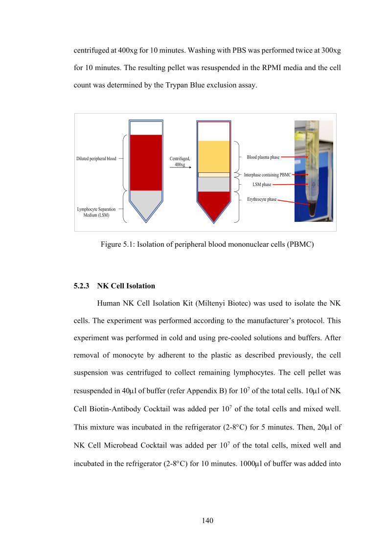
140
centrifuged at 400xg for 10 minutes. Washing with PBS was performed twice at 300xg
for 10 minutes. The resulting pellet was resuspended in the RPMI media and the cell
count was determined by the Trypan Blue exclusion assay.
Figure 5.1: Isolation of peripheral blood mononuclear cells (PBMC)
5.2.3 NK Cell Isolation
Human NK Cell Isolation Kit (Miltenyi Biotec) was used to isolate the NK
cells. The experiment was performed according to the manufacturer’s protocol. This
experiment was performed in cold and using pre-cooled solutions and buffers. After
removal of monocyte by adherent to the plastic as described previously, the cell
suspension was centrifuged to collect remaining lymphocytes. The cell pellet was
resuspended in 40µl of buffer (refer Appendix B) for 107 of the total cells. 10µl of NK
Cell Biotin-Antibody Cocktail was added per 107 of the total cells and mixed well.
This mixture was incubated in the refrigerator (2-8°C) for 5 minutes. Then, 20µl of
NK Cell Microbead Cocktail was added per 107 of the total cells, mixed well and
incubated in the refrigerator (2-8°C) for 10 minutes. 1000µl of buffer was added into
Diluted peripheral blood
Lymphocyte Separation Medium (LSM)
Centrifuged, 400xg
Erythrocyte phase
LSM phase
Interphase containing PBMC
Blood plasma phase

141
this suspension. Then, this suspension was proceeded to the subsequent magnetic cell
separation.
First the column was placed onto the magnetic field of the MACS separator.
The column was rinsed with 3ml of buffer then the suspension prepared earlier was
applied into the column. The collected flow-through was the unlabelled cells which
would represent the NK cells. The column was washed with the 3ml of buffer twice
and was combined with the effluent collected earlier. This suspension was centrifuged
at 300xg for 5 minutes and resuspended in 1ml PBS. The NK cells counts was
determined using the Trypan Blue exclusion assay. Flow cytometry was used to verify
the percentage of CD56+/CD3- cells population after the separation.
5.2.4 NK Cell Proliferation Assay (MTT)
Isolated NK cells were seeded into the 96 wells plate. A. precatorius
methanolic leaves extract (APME) was added in serial dilution with DMSO into each
well starting from 200µg/ml until 1.56µg/ml. Untreated-NK cells and DMSO (<1%)
were added into the wells as controls. The culture was incubated for 24h, 48h and 72h
in a humidified 5% CO2 incubator at 37°C. After each incubation point, 20µl of MTT
(5mg/ml) was added into the well and incubated for 4h. After 4h, 100µl of DMSO was
added into the wells to dissolve the formazan and the plate was read at absorbance
570nm. The absorption value at this wavelength directly represents the relative cell
numbers with comparison to the control group (Igarashi and Miyazawa, 2001). The
percentage of cell viability was determined by dividing the absorbance of the treated
cells with the absorbance of the control and multiply by a hundred. Besides the diluted
extract, the APME IC50 (26.4µg/ml) obtained from previous experiment in Chapter 4
was also tested individually. This was done to ensure the effect of that concentration

142
on the NK cell proliferation. This part of the experiment in general was performed to
determine the ideal concentration of APME needed to be used in the subsequent NK
co-culture with MDA-MB-231 cells.
5.2.5 NK Cell Co-culture with MDA-MB-231
Graphical summary of this experiment is shown in Figure 5.2. This experiment
was performed to evaluate the ability of APME in inducing NK cells activity on MDA-
MB-231 cells.
5.2.5(a) Cell Culture
MDA-MB-231 were seeded in 25cm2 tissue culture flask and grown at 37°C
under humidified 5% CO2 in DMEM medium supplemented with 5% of FBS and 1%
of penicillin-streptomycin. Confluent MDA-MB-231 cells were harvested by
trypsinization.
5.2.5(b) Co-cultutring of NK cells with MDA-MB-231 cells
This experiment was performed to evaluate the APME potential to induce
anticancer immune response by activating the NK cells. Freshly isolated NK cells
(effector) from each donor were pre-incubated for four hours according to the
experimental design as described in Table 5.1, before being added into the pre-seeded
(overnight) MDA-MB-231 (target) cell culture. The ratio of effector cells to target
cells was set at 20:1 following the methods detailed in Ismail et al. (2012) and
Nishimura et al. (2017) with given consideration of NK cells co-cultured with adherent
cells (Surayot and You, 2017) and specifically with MDA-MB-231 cells (Fu et al.,
2015). Following the incubation, the medium was collected into a fresh tube and the
adhered MDA-MB-231 cells were harvested by trypsinization. The trypsinized cells
were combined with the collected medium and centrifuged at 300xg to separate the

143
cell pellet from the supernatant. The medium (supernatant) was transferred into a fresh
1.5ml tubes and kept at -80°C for further analysis. The collected pellet containing both
NK cells and MDA-MB-231 cells were resuspended with RPMI 1640 complete
medium and divided into two fresh tubes for further analysis.
5.2.5(c) NK Cell Staining
The number of NK cells in the culture was determined by staining the cells
with PE-conjugated anti-CD56 (Santa Cruz) antibody and acquired by flow cytometry.
The collected cells (100µl) following the co-culture experiment, was washed twice
with 1XPBS. One mililitre of Bovine Serum Albumin (BSA) was added to the cells
and incubated for 10 minutes at room temperature. Five microlitre (5µl) of the PE-
conjugated anti-CD56 and FITC-conjugated anti-CD3 antibodies were added and
mixed well. This suspension was incubated for about 30 minutes on ice. Then, the cells
were washed with PBS (centrifuged at 300xg) twice to remove excess antibodies. The
resulted pellet was resuspended with 500µl PBS and the number of NK cells was
determined by flow cytometry FACSCANTO II (BD Bioscience). Data obtained was
analysed with FCS 7 software (De Novo).
5.2.5(d) Apoptosis Detection
Determination of the percentage of apoptotic cells of MDA MB 231 in the
culture was done by apoptosis staining of the target cells using the AnnexinV-FITC/PI.
The apoptosis assay was performed according to the manufacturer protocol,
AnnexinV-FITC detection kit I (BD Bioscience). The collected cells (100µl) following
the co-culture experiment, was washed twice with 1xPBS and then, 100µl of 1X
binding buffer was added prior to mixing with 5µl of AnnexinV and 5µl of PI. This
suspension was incubated for 15 minutes, at room temperature in dark. After the

144
incubation period, 500µl of 1X binding buffer was added into the tube. All samples
readings were acquired with FACSCANTO II (BD Bioscience). Data obtained was
analysed with FCS 7 software (De Novo). Percentage of NK cells cytotoxicity for each
donor category was determined by the following formula: ([percentage of cell deaths
by effector – percentage of cell deaths without effector] / [100 – percentage of cell
deaths without effector]) x 100 (Nishimura et al., 2017).
Table 5.1: Experimental design for NK cells co-culture with MDA-MB-231 cells
No Treatment Groups Details
1 NK/MDA-MB-231/APME NK cells were incubated with APME prior to treatment on MDA-MB-231 cells
2 NK/MDA-MB-231 NK cells were incubated on MDA-MB-231 cells
3 NK/MDA-MB-231/DMSO
NK cells were incubated with DMSO prior to treatment on MDA-MB-231 cells
4 NK ONLY NK cells were incubated on its own
5 APME/MDA-MB-231 MDA-MB-231 cells were treated with APME
5.2.5(e) ELISA (IL-2, IFN-g, PRF-1, GzmB)
ELISA was performed to quantify the expression level of the cytokines,
interleukin-2 (IL-2) and interferon gamma (IFN-g); and cytotoxic granules protein,
perforin (PRF-1) and granzymeB (GzmB). This assay was performed according to the
manufacturer’s protocol (Elabscience). In general, 100µl of sample/standard was
added into a pre-coated ELISA plate and incubated for 90 minutes at 37°C. The
samples or standards were diluted accordingly as needed. After the first incubation
period, liquid from each well was removed and 100µl of Biotinylated Detection Ab
was added into each well and incubated for 1h at 37°C. Then the liquid from each well
was aspirated and washed with 350µl of wash buffer was added. The washing step was
performed three times. 100µl of HRP conjugate was added after the washing steps and

145
the plate was incubated for 30 minutes at 37°C. Another five times of washing steps
were performed after the incubation. Then, 90µl of substrate reagent was added into
each well and was incubated for 15 minutes at 37°C in dark. 50µl of stop solution was
added after the incubation time ended and the optical density (OD value) of each well
was determined with a micro-plate reader at absorbance of 450nm.
5.2.6 Statistical Analysis
The data were expressed as mean ±SD of three repeated experiments. The level
of statistical significance among the group was tested using repeated measure one-way
ANOVA, followed by Tukey’s multiple comparison test. Level of significance
between two main groups were tested using paired or unpaired t-test. The difference
was considered significant if P<0.05.The normality of each data set was determined
by Shapiro-Wilk normality test. Analyses were all done using GraphPad Prism7 .
(Graphpad Software, La Jolla, CA, USA)

146
Figure 5.2: Graphical summary of the co-culture experiment of APME-treated NK cells with MDA-MB-231 cells.
NK cells incubated with
APME Transferred onto pre-seeded
MDA-MB-231 cells
Centrifuged harvested cells
with media
Supernatant collected for cytokine and cytotoxicity
analysis
Cells pellet divided into two
AnnexinV/PI Staining
CD56 / CD 3 Staining
ELISA analysis
Flow cytometry analysis
Supernatant collected for cytokine and
cytotoxic granule proteins analysis

147
5.3 RESULTS
5.3.1 NK Purification
NK cells were isolated from healthy and cancer donor using the Human NK
cell isolation kit (Miltenyi Biotec). Isolated NK cells were stained with CD56 (NCAM
(ERIC) - PE, Santa Cruz Biotechnology). Dot plot graph of the cell distribution events
after acquiring by flow cytometry were as shown in Figure 5.3. About 85.04% of NK
cells were successfully isolated using this NK isolation kit.
Figure 5.3: Dot plot graphs representing the isolated NK cells using the Human NK cell isolation kit (Miltenyi Biotec).
(A) The cell population gated as NK cells. (B) The cells were tagged with CD56 and CD3.

148
5.3.2 Isolated NK Cell in Healthy and Cancer Donor
In this experiment, NK cells were isolated from three healthy donors and three
cancer donors (breast cancer). Trypan blue exclusion assay was used to count the cells.
These isolated NK cells were used in the subsequent analysis following the isolation.
The average counts of NK cells isolated from healthy donor was significantly higher
(7.72 x 10⁵ cells/ml) than NK isolated from cancer donor (3.8 x 10⁵ cells/ml).
Figure 5.4: NK cells counts isolated from healthy and cancer donor.
The results were expressed as mean, ±SD of three independent donor for each group. P<0.05 is considered significance when comparing NK counts between healthy donor and cancer donor using unpaired t-test.
*

149
5.3.3 Ability of APME to induce NK cells proliferation
The ability of APME to induce NK cells proliferation, was analysed in this
experiment. This was carried out in order to identify the optimum concentration of
APME needed to be used in the subsequent analysis. APME with concentrations from
200µg/ml until 1.56µg/ml were added in the medium containing NK cells in order to
observe the ability of the extract to induce NK proliferation. The APME at 26.4µg/ml
was also added because it was the IC50 value that was identified in previous chapter
which was able to induce apoptosis in MDA-MB-231 cells. Isolated NK cells were
seeded in a 96 wells plate and were incubated for 4h before adding the extract. Then,
the cells were incubated for 24h, 48h and 72h. From Figure 5.5, it was found that the
proliferation of NK cells was stable at 24h, and the proliferation pattern rate was
fluctuating at 48h and 72h. This is because this NK cells were primary cells isolated
from donors and no other stimulant was added to expand the cell line. The results also
showed that at 24h, NK cells proliferation increased until 25µg/ml and 26.4µg/ml and
started to decline at 50µg/ml and increase again at 100µg/ml. Since the cells
proliferated well at APME IC50 value, this concentration was chosen for the
subsequent experiments since it was also used in the previous apoptosis experiments
in Chapter 4.

150
Figure 5.5: NK cells proliferation treated with APME
(A) 24h, (B) 48h, and (C) 72h. The results were expressed as mean, ±S.D of three independent experiments.
0
2
4
6
8
10
12
14
16
1.56 3.13 6.25 12.5 25 26.4 50 100 200
Cellp
rolife
ration
rate
(%)
Concentration (mg/ml)(C)
0
2
4
6
8
10
12
1.56 3.13 6.25 12.5 25 26.4 50 100 200
Cell P
rolife
ration
rate
(%)
Concentration µg/ml
(B)
0
2
4
6
8
10
12
14
16
18
1.56 3.13 6.25 12.5 25 26.4 50 100 200
Cell P
rolife
ration
rate
(%)
Concentration (mg/ml)
(A)

151
5.3.4 Effects of APME-induced NK cells on apoptosis of breast cancer cell MDA-MB-231; and measurement of cytokines and cytotoxic granules protein levels
NK cells from both healthy and cancer donor were treated in the same manner
as listed in Table 5.1. NK Cells was incubated with APME (26.4µg/ml) for 4h prior to
treatment on the MDA-MB-231 cells. Then this culture was further incubated for 20h.
After the incubation period, the media were collected for cytokines and cytotoxic
granules protein assay by ELISA and the remaining cells were harvested for
CD56/CD3 staining and apoptosis assay with Annexin-V/PI staining. Figure 5.6
demonstrated the forward and side scatter of the MDA-MB-231 cells and NK cells
population in the sample following co-culture experiment, acquired by flow
cytometry. The size of the MDA-MB-231 cells were bigger compared to the size of
the NK cells.
Figure 5.6: The forward and side scatter of NK cell co-culture with MDA-MB-231 cells .
Gating of the NK cell population and MDA-MB-231 cell population were marked in the circles.
- FSC (Forward Scatter) - SSC (Side Scatter)
MDA-MB-231
NK

152
5.3.4(a) NK Cell Counts of Healthy and Cancer Donor after co-culture with MDA-MB-231 cells
Figure 5.7(A) showed the representative profiles of dot plot of NK cells counts
in healthy donor and Figure 5.7(B) showed the results of the NK cells counts in
different treatments in healthy donor. In healthy donor, NK cell count in Group 1
(NK/MDA-MB-231/APME) is 50.5±8.7%, Group 2 (NK/MDA-MB-231) is
37.6±5.5%, Group 3 (NK/MDA-MB-231/DMSO) is 40.0±9.7% and Group 4 (NK-
Only) is 39.3±7.9%. NK cell count in Group 1 is significantly higher statistically when
comparing to the rest of the groups. In cancer donor (Figure 5.8), the NK cell counts
for all groups were slightly similar among each other except for Group 4 (NK-Only)
which has a slight increase at 45.2±5.5%. NK cell counts for Group 1 (NK/MDA-MB-
231/APME) is 41.9±5.0%, Group 2 (NK/MDA-MB-231) is 42.7±6.6% and Group 3
(NK/MDA-MB-231/DMSO) is 43.2±6.4%.

153
Figure 5.7(A): Healthy donor NK cell counts in NK cell co-culture with MDA-MB-231 cells .
The representative dot plots where (G-1) NK/MDA-MB-231/APME, (G-2) NK/MDA-MB-231 (G-3) NK/MDA-MB-
231/DMSO and (G-4) NK- ONLY. FITC-stained cells are represented at x-axis, while PE-stained cells at y-axis.

154
Treatment Groups
(B)
Figure 5.7(B): Healthy donor NK cell counts in NK cell co-culture with MDA-
MB-231 cells .
Graph representing the NK cell counts in Healthy Donor from the co-culture with MDA-MB-231 cells in different treatment groups. The results were expressed as mean, ±SD of three independent donor for each group and (p<0.05) is considered statistically significant.
NK/MDA-M
B-231/A
PME
NK/MD-M
B-231
NK/MDA-M
B-231/D
MSO
NK-ONLY
0
20
40
60
80
Perc
enta
ge o
f NK
cell
s (%
)
*
*
*
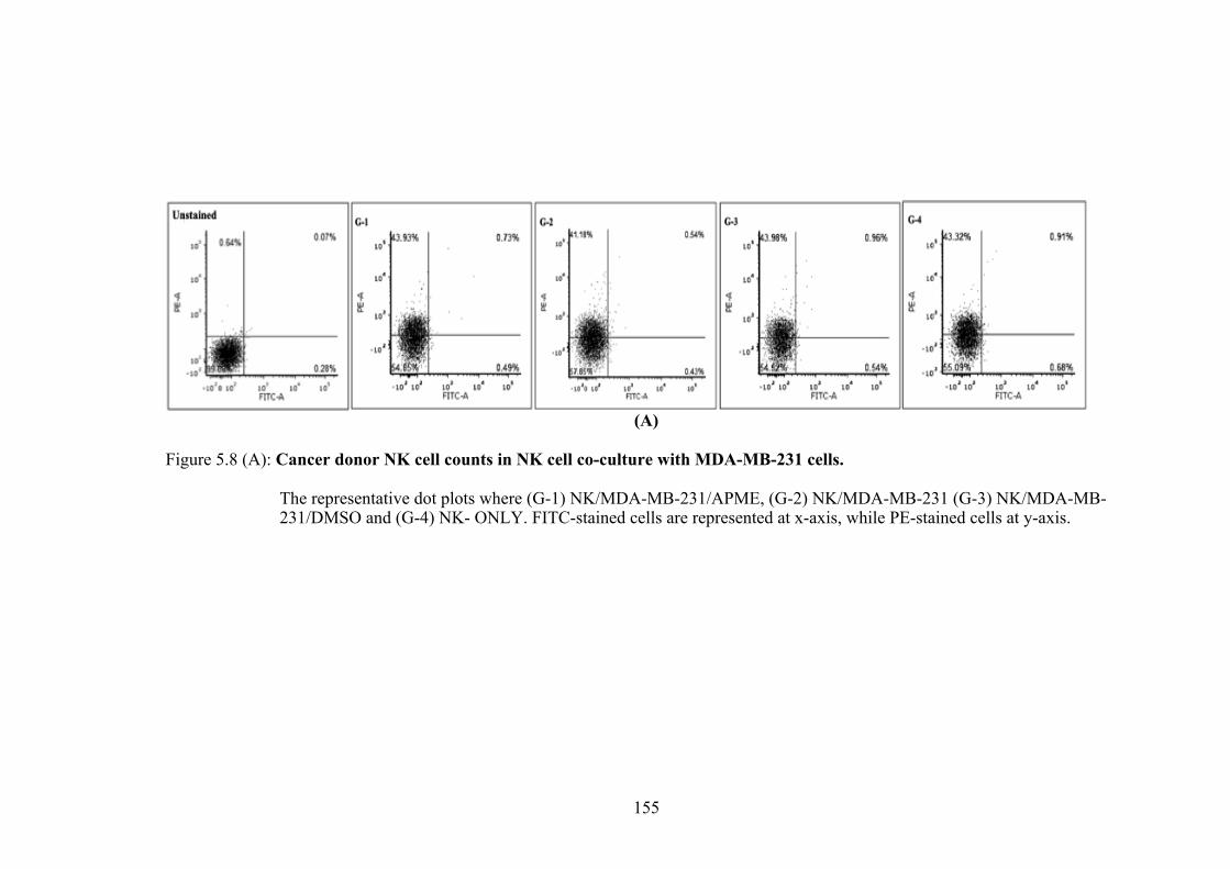
155
(A)
Figure 5.8 (A): Cancer donor NK cell counts in NK cell co-culture with MDA-MB-231 cells.
The representative dot plots where (G-1) NK/MDA-MB-231/APME, (G-2) NK/MDA-MB-231 (G-3) NK/MDA-MB-
231/DMSO and (G-4) NK- ONLY. FITC-stained cells are represented at x-axis, while PE-stained cells at y-axis.
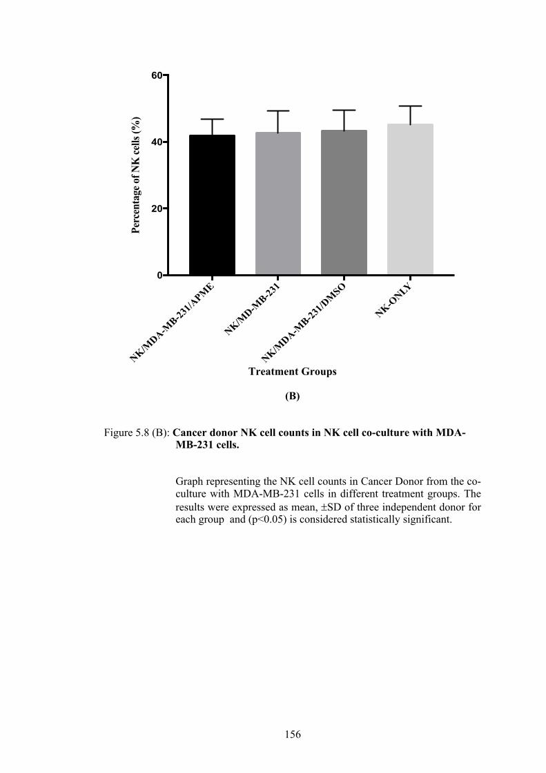
156
Treatment Groups
(B)
Figure 5.8 (B): Cancer donor NK cell counts in NK cell co-culture with MDA-MB-231 cells.
Graph representing the NK cell counts in Cancer Donor from the co-culture with MDA-MB-231 cells in different treatment groups. The results were expressed as mean, ±SD of three independent donor for each group and (p<0.05) is considered statistically significant.
NK/MDA-M
B-231/A
PME
NK/MD-M
B-231
NK/MDA-M
B-231/D
MSO
NK-ONLY
0
20
40
60
Perc
enta
ge o
f NK
cells
(%)

157
5.3.4(b) Apoptosis Assay of MDA-MB-231 cells treated with APME-induced NK cells
The ability of APME-induced NK cells to promote apoptosis and cell death in
MDA-MB-231 cells was assessed by staining with Annexin-V/PI. Figure 5.9(A)
represents the dot plot of the treated MDA-MB-231 cells in healthy donor acquired by
flow cytometry. Figure 5.9(B) shows the graph plotted from tabulated results of the
apoptotic MDA-MB-231 cells following co-culture with NK cells from healthy donor.
As presented in Figure 5.9(B), Group 1 (NK/MDA-MB-231/APME) showed
46.28±2.2% apoptotic cells, Group 2 (NK/MDA-MB-231) showed 30.50±1.8%,
Group 3 (NK/MDA-MB-231/DMSO) showed 29.4±1.5% and Group 4
(APME/MDA-MB-231) showed 27.55±0.8%. These results showed that there were
significantly higher apoptotic cells in Group 1 in the healthy donor category comparing
to the rest of the groups in that category.
Figure 5.10(A) represents the dot plot of the treated MDA-MB-231 cells in
cancer donor and Figure 5.10(B) shows the graph plotted from tabulated results of the
apoptotic MDA-MB-231 cells following co-culture with NK cells from cancer donor.
As presented in Figure 5.9(B), apoptotic cells in Group 1 (NK/MDA-MB-231/APME)
was 26.32±6.0%, Group 2 (NK/MDA-MB-231) was 26.84±56.1%, Group 3
(NK/MDA-MB-231/DMSO) was 24.28±4.3% and Group 4 (APME/MDA-MB-231)
was 20.68±2.0%. Group 1, 2 and 3, demonstrated higher apoptotic cell percentage
comparing to Group 4. However, these differences were not statistically significant.

158
(A)
Figure 5.9(A): MDA-MB-231 apoptotic cells from NK cell co-culture with MDA-MB-231 in Healthy Donor.
The representative dot plots where (G-1)NK/MDA-MB-231/APME, (G-2) NK/MDA-MB-231 (G-3) NK/MDA-MB-231/DMSO and (G-4) NK- ONLY.Quadrants represent the percentage of cell populations; Q1- Necrosis, Q2- Late apoptosis, Q4-Early apoptosis, Q3-Live cells.

159
Treatment Groups
(B)
Figure 5.9(B): MDA-MB-231 apoptotic cells from NK cell co-culture with MDA-
MB-231 in Healthy Donor.
Graph representing the apoptotic cells of MDA-MB-231 co-culture with NK cells from healthy donor. The results were expressed as mean, ±SD of three independent donor for each group and (p<0.05) is considered statistically significant.
NK/MDA-M
B-231/A
PME
NK/MD-M
B-231
NK/MDA-M
B-231/D
MSO
APME/M
DA-MB-23
10
20
40
60Pe
rcen
tage
of M
DA
-MB
-231
ce
lls d
eath
(%)
*
**

160
(A) Figure 5.10(A) :MDA-MB-231 apoptotic cells from NK cell co-culture with MDA-MB-231 in Cancer Donor.
The representative dot plots where (G-1) NK/MDA-MB-231/APME, (G-2) NK/MDA-MB-231 (G-3) NK/MDA-MB-231/DMSO and (G-4) NK- ONLY.Quadrants represent the percentage of cell populations; Q1- Necrosis, Q2- Late apoptosis, Q3-Early apoptosis, Q4-Live cells.

161
Treatment Group
(B)
Figure 5.10(B): MDA-MB-231 apoptotic cells from NK cell co-culture with MDA-MB-231 in Cancer Donor.
Graph representing the apoptotic cells of MDA-MB-231 co-culture with NK cells from cancer donor. The results were expressed as mean, ±S.D of three independent donor for each group and (p<0.05) is considered statistically significant.
NK/MDA-M
B-231/APME
NK/MD-M
B-231
NK/MDA-M
B-231/DMSO
APME/MDA-M
B-2310
10
20
30
40Pe
rcen
tage
of M
DA-M
B-23
1 ce
lls d
eath
(%)

162
Figure 5.11 showed the comparison of the target cell deaths between healthy
and cancer donor. Percentage of the target cell death by APME-treated NK cells from
the Healthy Donor were significantly higher than the cancer donor. This data inserted
into the NK cell cytotoxicity formula gave the percentage of NK cell cytotoxic activity
as shown in Figure 5.12. APME-treated NK cells from the healthy donor significantly
showed higher percentage comparing to the other treatment groups and also between
the APME-tretaed NK cells from the Cancer Donor.

163
Treatment Group
Figure 5.11: MDA-MB-231 apoptotic cells from NK cell co-culture with MDA-MB-231 cells.
Graph showing the comparison of MDA-MB-231 cell deaths between Healthy Donor and Cancer Donor. The results were expressed as mean, ±SD of three independent donor for each group and (P<0.05) is considered statistically significant, when comparing between healthy donor and cancer donor in each groups.
NK/MDA-M
B-231/A
PME
NK/MD-M
B-231
NK/MDA-M
B-231/D
MSO
APME/M
DA-MB-23
10
20
40
60
MD
A-M
B-23
1 ce
ll de
ath
(%)
Healthy Donor
Cancer Donor*

164
Treatment Group
Figure 5.12: Percentage of NK cells cytotoxic activity
Graph showing the comparison of NK cells cytotoxic activity between Healthy Donor and Cancer Donor. The results were expressed as mean ±SD of three independent donor for each group and (P<0.05) is considered statistically significant when comparison was made between all groups.
NK/MDA-M
B-231/A
PME
NK/MD-M
B-231
NK/MDA-M
B-231/D
MSO
0
10
20
30
40
NK
Cel
l Cyt
otox
icity
(%)
Healthy Donor
Cancer Donor*NK
Cel
l Cyt
otox
ic Ac
tivity
(%)

165
5.3.4(c) IL-2, IFN-g, PRF-1 and GzmB protein expression level following AMPE-induced NK cells co-culture with MDA-MB-231 cells
ELISA was performed to quantify the production of cytokines, interleukin-2
and interferon gamma (IFN-g); and cytotoxic granule proteins (PRF-1 and GzmB) in
the media collected following NK cells co-culture with MDA-MB-231 cells. Figure
5.13 presents the expression of interleukin-2 (IL-2), Figure 5.14 presents the
expression of interferon gamma (IFN-g), Figure 5.15 shows the expression of perforin
(PRF-1) and Figure 5.16 presents the expression of granzyme B (GzmB).
In healthy donor, as presented in Figure 5.13(A), the IL-2 expression in Group
1 (NK/MDA-MB-231/APME) was 388.7±80.6pg/ml, Group 2 (NK/MDA-MB-231)
was 353±56.5pg/ml, Group 3 (NK/MDA-MB-231/DMSO) is 356.9±57.33pg/ml, and
Group 4 (NK-Only) is 8.13±0.8pg/ml. IL-2 expression in Group 1 was slightly higher
but not significant than the expression in Group 2 and 3. In cancer donor, as presented
in Figure 5.13(B), the IL-2 expression in Group 1 (NK/MDA-MB-231/APME) is
427.5±82pg/ml, Group 2 (NK/MDA-MB-231) is 436.9±118pg/ml, Group 3
(NK/MDA-MB-231/DMSO) is 439.1±71.7pg/ml, and Group 4 (NK-Only) is
9.09±0.8pg/ml. Figure 5.13(C) showed the comparison of IL-2 expressions between
healthy donor and cancer donor in al treatment groups. The IL-2 expressions in cancer
donor were higher but not significant in all treatment groups comparing to healthy
donor.
Quantification of IFN-g in healthy donor, as presented in Figure 5.14(A), in
Group 1 (NK/MDA-MB-231/APME) was 812.5±82.3pg/ml, Group 2 (NK/MDA-MB-
231) was 656.5.7±88.3pg/ml, Group 3 (NK/MDA-MB-231/DMSO) was
607.9±98.6pg/ml, and Group 4 (NK-Only) was 8.34±0.5pg/ml, however the value

166
obtained in Group 4 is below the lower cut-off point of the minimum concentration
detected by the kit. Therefore, expression level in Group 4 is considered null or no
expression. The expression level of IFN-g was significantly higher in Group 1 of both
healthy and cancer donor categories. In cancer donor, as presented in Figure 5.14(B),
the expression of IFN-g in Group 1 (NK/MDA-MB-231/APME) was
2.5x10³±2.3x10²pg/ml, Group 2 (NK/MDA-MB-231) was 1.5x10³±1.7x10²pg/ml,
Group 3 (NK/MDA-MB-231/DMSO) was 1.4x10³±4.5x10²pg/ml, and Group 4 (NK-
Only) is 87.33±2.6pg/ml The expression IFN-g in cancer donor for all treatment
groups were significantly higher comparing to the expressions in healthy donor, as
depicted in Figure 5.14(C).
Similarly to the cytokines expression analysis, the cytotoxic granule protein,
perforin (PRF-1) and Granzyme B (GzmB) were all quantified using the ELISA. In
healthy donor, as presented in Figure 5.15(A), the expression of PRF-1 in Group 1
(NK/MDA-MB-231/APME) is 5.7x10³±3.3 x10²pg/ml, Group 2 (NK/MDA-MB-231)
is 4.6x10³±8.5x10²pg/ml, Group 3 (NK/MDA-MB-231/DMSO) is
4.7x10³±6.7x10²pg/ml, and Group 4 (NK-Only) is 248.8±12.6pg/ml, however, the
value obtained in Group 4 was below the lower cut off point of the minimum
concentration. Therefore, expression level in Group 4 is considered null. The
expression level of PRF-1 was the highest in Group 1 and statistically significant
comparing to the rest of the groups. In cancer donor, as presented in Figure 5.15(B),
the expression of PRF-1 in Group 1 (NK/MDA-MB-231/APME) was
8.4x10³±9.0x10²pg/ml, Group 2 (NK/MDA-MB-231) was 8.0x10³±1.1x10³pg/ml,
Group 3 (NK/MDA-MB-231/DMSO) was 7.7x10³±7.5x10² pg/ml, and Group 4 (NK-
Only) is 299pg/ml, however, the value obtained in Group 4 was below the lower cut
167
off point of the minimum concentration of the standard curve. Therefore, expression
level of PRF-1 in Group 4 is considered null. The expression level of PRF-1 was the
highest in Group 1 from the cancer donor category however it was not statistically
significant. The expression level of PRF-1 in cancer donor was overall significantly
higher comparing to the corresponding expression level of the healthy donor as shown
in Figure 5.15(C).
Another cytotoxic granule protein measured was the GranzymeB (GzmB). In
healthy donor, as presented in Figure 5.16(A), the expression of GzmB in Group 1
(NK/MDA-MB-231/APME) was 14.82±1.7pg/ml, Group 2 (NK/MDA-MB-231) is
13.02±0.7pg/ml, Group 3 (NK/MDA-MB-231/DMSO) was 13.58±1.1pg/ml, and
Group 4 (NK-Only) was 10.33±0.7pg/ml. In this category (healthy donor) Group 1 has
the highest GzmB protein expression but it was not statistically significant. In cancer
donor, as presented in Figure 5.16(B), the expression of GzmB in Group 1 (NK/MDA-
MB-231/APME) was 12.57±2.4pg/ml, Group 2 (NK/MDA-MB-231) was
14.12±3.2pg/ml, Group 3 (NK/MDA-MB-231/DMSO) was 15.35±1.1pg/ml, and
Group 4 (NK-Only) was 11.74±1.8pg/ml. In this category (cancer donor) Group 3 has
the highest GzmB protein expression, followed by Group 2, Group 1 and Group 4.
Nevertheless their GzmB protein concentration were all statistically insignificant.
Similarly, the GzmB expression between healthy and cancer donor were all slightly
similar and insignificant (Figure 5.16(C)).

168
Figure 5.13: The expression level of IL-2 in the co-culture experiment of NK cells with MDA-MB-231 cells.
(A) The IL-2 expression level in Healthy Donor. (B) The IL-2 expression level in Cancer Donor. (C) General comparison of IL-2 expressions between Healthy and Cancer Donor. The results were expressed as mean, ±SD of three independent donor for each group.

169
Figure 5.14: The expression level of interferon gamma (IFN-g) in the co-culture experiment of NK cells with MDA-MB-231 cells.
(A)The IFN-g expression level in Healthy Donor. (B) The IFN-g expression level in Cancer Donor. (C) General comparison of IFN-g expressions between Healthy and Cancer Donor. The results were expressed as mean, ±SD of three independent donor for each group and (P<0.05) is considered statistically significant, when comparing between groups in (A) and (B), and within the groups between healthy and cancer donor in (C).
(A) (B)
(C)
NK/MDA-M
B-231/A
PME
NK/MD-M
B-231
NK/MDA-M
B-231/D
MSO
NK-ONLY
0
1000
2000
3000
IFN
- γ (p
g/m
l)
Healthy Donor
Cancer Donor
*
**
NK/MDA-M
B-231/A
PME
NK/MD-M
B-231
NK/MDA-M
B-231/D
MSO
NK-ONLY
0
200
400
600
800
1000
IFN
- γ
(pg/
ml)
*
*
NK/MDA-M
B-231/A
PME
NK/MD-M
B-231
NK/MDA-M
B-231/D
MSO
NK-ONLY
0
1000
2000
3000
IFN
- γ
(pg/
ml)
*

170
Figure 5.15: The expression level of perforin (PRF-1) in the co-culture experiment of NK cells with MDA-MB-231 cells.
(A) The PRF-1 expression level in Healthy Donor. (B) The PRF-1 expression level in Cancer Donor. (C) General comparison of PRF-1 expressions between Healthy and Cancer Donor. The results were expressed as mean, ±SD of three independent donor for each group and (p<0.05) is considered statistically significant, when comparing between groups in (A) and (B), and within the groups between healthy and cancer donor in (C).
(A) (B)
(C)
NK/MDA-M
B-231/A
PME
NK/MD-M
B-231
NK/MDA-M
B-231/D
MSO
NK-ONLY
0
2000
4000
6000
8000
10000
PRF-
1 (p
g/m
l)
NK/MDA-M
B-231/A
PME
NK/MD-M
B-231
NK/MDA-M
B-231/D
MSO
NK-ONLY
0
2000
4000
6000
8000
10000
PRF-
1 (pg
/ml)
Healthy Donor
Cancer Donor
* **
NK/MDA-M
B-231/A
PME
NK/MD-M
B-231
NK/MDA-M
B-231/D
MSO
NK-ONLY
0
2000
4000
6000
8000
10000
PRF-
1 (p
g/m
l)*

171
Figure 5.16: The expression level of granzyme B (GzmB) in the co-culture experiment of NK cells with MDA-MB-231 cells.
(A) The GzmB expression level in Healthy Donor. (B) The GzmB expression level in Cancer Donor. (C) General comparison of GzmB expressions between Healthy and Cancer Donor. The results were expressed as mean, ±S.D of three independent donor for each group and (p<0.05) is considered statistically significant.
(A) (B)
(C)
NK/MDA-M
B-231/A
PME
NK/MD-M
B-231
NK/MDA-M
B-231/D
MSO
NK-ONLY
0
5
10
15
20
Gzm
B (p
g/m
l)
NK/MDA-M
B-231/A
PME
NK/MD-M
B-231
NK/MDA-M
B-231/D
MSO
NK-ONLY
0
5
10
15
20
Gzm
B (p
g/m
l)
NK/MDA-M
B-231/A
PME
NK/MD-M
B-231
NK/MDA-M
B-231/D
MSO
NK-ONLY
0
5
10
15
20
25
Gzm
B (p
g/m
l)
Healthy DonorCancer Donor

172
5.4 DISCUSSION
Previous chapter have reported that A. precatorius methanol leaves extract
(APME) was able to induce apoptosis in MDA-MB-231 cells. NK cell activation can
be measured by cytototoxic analysis of the target cell death, quantification of soluble
target cell death markers, quatification of cytokine relased upon NK cell activation and
finally the evaluation of the degranulation indicators (Baran et al., 2001; Blom and
Albers, 2009; Rudnicka et al., 2015; Wang et al., 2010a). This chapter reported on the
ability of APME to activate the NK cells activity upon co-culture with MDA-MB-231
cells. NK cells used in this study were freshly isolated from donors, three from healthy
and three from cancer patients. This was done to observe if APME would have any
effect on NK cells activation, thus only three healthy donors and three cancer donors
were chosen. These donors were selected in accordance with the ethics approval
guideline.
In the previous apoptosis study, APME was found to induce apoptosis in
MDA-MB-231 cells, a breast cancer cell line, hence the donors chosen for this NK
study were all females. Healthy donors must not have any known disease and must be
healthy at the time of blood withdrawal. While cancer donors were females with
diagnosed breast cancer but have not undergone any treatments. These three cancer
donors were on the waiting list to undergo chemotherapy at the time of blood donation.
They were at different stages of the disease where two of the donors were at stage 4
and one was at stage 3. NK cells were isolated from the peripheral blood using the
negative sorting NK isolation kit from Miltenyi Biotec. This kit applied the concept of
negative sorting that allows the tagging of non-NK cells and collect NK cells in the
flow through. The collected NK cells were tested for purity by staining with CD56-PE
conjugated and CD3-FITC conjugated, measured by flow cytometry. NK cells are

173
known to express CD56+ and CD3-. Lacking of CD3 expression differentiate NK from
T lymphocyte (Grudzien and Rapak, 2018). By using this kit, about 85% of sample
purity was achieved. A group recently published the acquisition of their isolated NK
cells from PBMC was around 86% (Sugita et al., 2018). Other study reported to obtain
purification of NK cells up to 90% - 95% (Kanevskiy et al., 2013), while another study
managed to obtain around 60.6% (Klingemann and Martinson, 2004). By using the
same kit, another study suggested the addition of MACS CD15 microbeads in order to
reach purification until 98% by improving further depletion of granulocyte (Pesce et
al., 2017). In the study by Wang et al. (2017), only 34.4% of NK cells were able to be
recovered and more granulocytes were present when using the kit. They also suggested
addition extra microbeads in order to obtain higher sample purity.
As shown by Figure 5.2, the amount of NK cells isolated from the healthy
donor were two folds higher than the amount of NK cells obtained from the cancer
donor. An experiment on NK cells expansion from healthy and cancer donor showed
that the NK cells counts in healthy donor was also higher than from cancer donor. The
same trend was also reported where reduced numbers of NK cells from breast cancer
patients were observed comparing to the healthy donor (Mamessier et al., 2011;
Shenouda et al., 2017). Another study by Yunusova et al. (2018) also reported lower
number of NK cells in the peripheral blood of ovarian cancer patients. Low NK cells
counts in cancer patients could be the cause of tumour cells escape which eventually
led to metastasis (Gulubova et al., 2009). Other studies also revealed that cancer
patients exhibiting advanced disease progression exhibited lower number of NK cells
in their peripheral blood and normally have lower chance of survival (Ishigami et al.,
2000; Li et al., 2016; Peng et al., 2017; Tang et al., 2020). NK cells from cancer
patients exhibited inhibitory phenotype which is characterized by decreased activating

174
markers expression such as NKG2D and natural cytotoxicity receptors (NCRs). On
the contrary, the inhibitory markers such as killer-cell immunoglobulin- like receptors
(KIRs) and NKG2A were upregulated. These phenotype leads to the inability of NK
cells from the cancer patients to exhibit cytotoxic activity comparing to the NK cells
from healthy donors (Costello et al., 2002; Pasero et al., 2015).
Lymphocytes activation, survival, proliferation and differentiation are
regulated by cytokines. Interleukin in particular IL-2, IL-15, IL-12, IL-18 and IL-21
are known to promote NK cells proliferation and improve their anti-tumour ability
(Floros and Tarhini, 2015; Hu et al., 2019; Srivastava et al., 2013). However, in this
study, freshly isolated unstimulated NK cells from healthy and cancer donor were used
for the co-culture assay to measure the NK cells cytotoxicity with the target cells in
the presence of plant extract or compound only (Ismail et al., 2012). Therefore, prior
to the co-culture assay with the target cell, the optimal concentration which APME
could induce proliferation of NK cells was determined. The ability of APME to
proliferate NK cells was evaluated by MTT assay (Lu et al., 2016; Shpakova et al.,
2000; Zhang et al., 1996). The experiment showed that the proliferation trends were
stable at 24h and were unstable at 48h and 72h. At 24h, NK cell proliferation trend
was seen to increase as the concentration increased. Thus IC50 value of APME was
chosen for the NK cells co-culture with MDA-MB-231 cells.
The ability of APME to induce NK cell cytotoxicity was assessed in the
experiment of NK cells co-culture with MDA-MB-231 cells, in the presence of APME.
NK cells were the effector and MDA-MB-231 cells were the target. The ability of
APME to promote effector to induce cytotoxicity on the target cell was the main aim
for this experiment. Three parameters were measured in this experiment, the first one
was the effect on NK counts following the incubation; secondly the effect on cell death

175
measured by apoptosis AnnexinV/PI assay; and thirdly the concentration of
interleukin-2 (IL-2), interferon gamma (IFN-g), perforin (PRF-1) and granzyme B
(GzmB).
Following the co-culture incubation, percentage of NK cells was determined
by observing CD56/CD3 expression. In healthy donor, percentage of NK cells was
significantly higher in the APME-treated NK cells comparing to the rest. However, no
significant differences can be seen in the percentage of NK cells from cancer donor.
At this point, the difference between APME-treated and untreated NK cells was only
observed in the healthy donor. To evaluate the ability of APME to induce cytotoxicity,
the cell death was measured by the apoptosis staining with AnnexinV/PI. The
advantage of this staining is that the NK cytotoxic activity that caused target cell
apoptosis or death could be determined.
In this study, APME-treated NK cells from healthy donor was able to induce a
significant MDA-MB-231 cell death comparing to the non-treated NK cells. While in
cancer donor, the percentage of cell deaths in all treatment groups were indifference.
Especially the APME-treated NK cells from healthy donor exhibited significant
cytotoxicity comparing to APME-treated NK cells from cancer donor. This is
supported by the percentage of NK cytotoxicity which was calculated based on a
formula as described previously (Mhatre et al., 2014; Nishimura et al., 2017). These
values were obtained simply by deducting the value presented by non-effector
treatment, which was the APME incubation with MDA-MB-231 cells only.
Overall, from these values, APME-treated NK cells from the healthy donor
definitely has the highest percentage of NK cell cytotoxic activity comparing to the
rest of the treatment groups, and the percentage of NK cells cytotoxic activity from

176
healthy donor were higher comparing to the NK cells from cancer donor. This
demonstrated that APME was able to further promote NK cell cytotoxic activity in NK
cells obtained from the healthy donor but not from the cancer donor. NK cells count
from cancer patients were not only lesser comparing to the healthy donor as previously
described (Mamessier et al., 2011; Shenouda et al., 2017), but these endogenous NK
cells also have less cytotoxic activity might be due to the alteration of the NK cells
receptors including decreased inhibitory receptors or/and increase activating receptors
(Guillerey et al., 2016). Because of that, in immunotherapy treatment involving NK
cells, immune activation is facilitated by addition of cytokines and antibodies to
sensitize the NK cells to assist its modulation towards the anti-tumour response (Hu et
al., 2019)
Medium from the co-culture experiment was collected to quantify the secretion
of interleukin-2 (IL-2), interferon gamma (IFN-g), perforin (PRF-1) and granzyme B
(GzmB). Levels of IL-2 secretion in all treatment groups from both healthy and cancer
donor showed insignificance differences. Presence of IL-2 indicated that the isolated
NK cells were contaminted with other IL-2 secreting lymphocytes because the purity
of NK cells obtained in this study was less than 90%. IL-2 is a 15K-kDa cytokine
mainly produced by activated CD4+/ CD8+ T cells, however, there were reports on the
scretion of IL-2 by dendritic cells, and NK cells (Gaffen, 2001; Granucci et al., 2001).
Nevertheless, the relevance of the IL-2 production by these cells remains ambiguos
(Malek, 2008).
The results obtained showed that the level of IFN-g production was
significantly increased in APME-treated NK cells. These production of IFN-g marked
the activation of NK cells. IFN-g, is the cytokine that mainly important in modulating

177
host immune response in particular controlling the spread of pathogen infection and
defence against cancers. IFN-g is also released by activated TH1 cells, CD8+ T cells,
and NK T cells, besides NK cells (Mah and Cooper, 2016; Schoenborn and Wilson,
2007). NK cells producing high level of IFN-g exhibited low cytotoxicity while on the
contrary, cytotoxic NK cells demonstrated low level of IFN-g production (Björkström
et al., 2016; Caligiuri, 2008). This coincide with the results of this current study where
even in extremely high level of IFN-g expression found in cancer donor comparing to
healthy donor, the cytotoxicity activity of NK cells from healthy donor were higher. It
was also reported that CD56dimCD16+ NK cells were able to produce IFN-g during
their cytolytic activity. Activated NK cells producing intrinsic cytokines such as IFN-
g and TNF-a promotes target cell lysis and neutralization of these cytokines caused
impaired function of the cell lysis (Wang et al., 2012). From these results, APME was
found to be able to stimulate NK cells from both healthy and cancer donor by
producing higher IFN-g levels comparing to non-tretated NK cells.
Perforin (PRF-1) and granzyme B (GzmB) are two granule proteins released
by NK cells upon activation by stimuli from the target cells. The synergistic effect of
PRF-1 and GzmB leads to the lysis of the target cell. PRF-1 is a potent pore-forming
protein that allows the entrance of cytotoxic proteases such as GzmB into the target
cell cytoplasm. This pore-forming process by PRF-1 is crucial in order to ensure the
granzyme proteases able to enter the cytoplasm of the target cells. Defects in this
pathway, particularly lacking of the perforin molecule will result in human disorder
termed as perforinopathies, which include immunoproliferative disease Familial
Haemophagocytic Lymphohistocytosis (FHL) type 2, haemotological malignancies
and protracted viral infections (Spicer et al., 2017). In this current study, the PRF-1
expression was significantly high in the APME-treated NK cells from healthy donor

178
and similar trend can be seen with its counterpart from the cancer donor, however this
slight difference was insignificance. Nevertheless, the expression of PRF-1 in NK cells
from the cancer donor were overall significantly increased comparing to the healthy
donor. Even though the NK cells cytotoxic activity from cancer donor was lesser
comparing to the NK cells from the healthy donor, their PRF-1 expression were
extremely high. This was parallel to the production of the IFN-g levels, where they
were higher in NK cells from the cancer donor. This phenomena was probably caused
by NK cells isolated from cancer donors have “memory” and were activated upon
stimulation with the target cells. The review by Fehniger and Cooper (2016) mentioned
that enhanced NK cells function was observed in antigen-specific stimulation and this
is termed as “memory” or “memory-like” depending on how the NK cells were
activated. NK cells memory would be beneficial use for anitumour immunotherapy,
especially in vitro expansion capability (Capuano et al., 2019).
GzmB induces cell deaths either through caspase-dependant or independent
manner (Martinvalet, 2019). GzmB initially is expressed as an inactive pre-cursor
protein that bears an N-terminal signal peptide. This terminal direct packaging of
GzmB into secretory granules followed by removal of dipeptide Glysine and
Glutamate by cysteine protease cathepsin C. Upon removal of those peptides, GzmB
becomes activated and kept together in a lytic granules together with other granzymes
and perforin. GzmB enters target cells after the activation of perforin, and instantly
induce apoptosis either by caspase-dependant or independent manner (Afonina et al.,
2010). In this current study, it was found that GzmB were lowly expressed in both
healthy and cancer donor. No conclusive remarks could be deduced from this finding
because the detected GzmB expression levels in all groups were very low. Comparison
between healthy and cancer donor also showed indifference expressions of GzmB.
179
AMPE-treated NK cells were unable to exhibit any significance differences in GzmB
expression from this co-culture experiment. This experiment conducted in vitro might
be the reason that very low amount of GzmB expressions were detected. Madakkannu
and Ravichandran (2017) have successfully demonstrated that Indigofera tinctoria and
Scoparia dulcis aqueous extracts were able to exhibit immunopreventive role in noise-
stress rats by elevation of GzmB in vivo. Another study involving a plant known as
sea buckthorn or Hippophae rhamnoides L. exhibited elevated level of GzmB and
PRF-1 expression in chronic-stress rat model (Diandong et al., 2016).
These results have demonstrated that APME was able to increase the
proliferative capacity of NK cells, induce NK cell cytotoxic activity thus promoting
cell death via apoptosis, and exhibited increased levels of IFN-g and PRF-1 protein
expressions by NK cells isolated from healthy donor. As for the cancer donor, although
higher expressions of IFN-g and PRF-1 were observed in comparison to the healthy
donor, the NK cells cytotoxicity was impaired and APME was unable to further
stimulate or better the activity of NK cells isolated from cancer donor. Therefore it is
believed that in healthy individuals, APME could help promote their NK cells activity
and this would benefit if A. precatorius could be introduce in their diet occasionally.
Thus helping them to combat the emergence of cancer cells. However, this assumption
needs to be scientifically proven.

180
5.5 CONCLUSION
In conclusion, it was found that APME was able to activate and promote better
NK cells cytotoxic activity from healty donor but not in NK cells from cancer donor.
It is suggested that more sample could be acquired especially from cancer donor in
order to have better observation and conclusion. Probably details analysis of the donor
such as their specific type of cells (example: breast – triple negative) will provide better
understanding on this matter. Expansion of NK cells is also recommended to ensure
better NK cells stability and viability. A better protocol to enhance NK purtity is also
recommended such as using another specific microbeads tag to eliminate as much
contaminant as possible. If screening a large number of donors, it is impossible to
acquire large volume of blood. Therefore, another robust and simple method using
MACSxpress® (Miltenyi) would solve this issue because isolation of NK cells is
possible to achieve from as little as 2ml blood.

181
CHAPTER 6
GENERAL DISCUSSION, CONCLUSION AND FUTURE DIRECTION
6.1 GENERAL DISCUSSION
Since many decades or even centuries ago, human depends on medicinal plants
for treatment of various health issues such as, high blood pressure, diabetes mellitus,
infections, arthritis, and cancers. With advancement of the science and technology, the
efficacy and mechanisms of the medicinal plants to treat these illnesses can be proven.
Furthermore, scientific findings has also provided the identifications of bioactive
compounds from these plants that have been tested in vitro, in vivo and even some are
successfully tested in clinical settings and practices (Ortega and Campos, 2019). In
this modern world, with high throughput research and technologies, many powerful
drugs and treatment for cancer are established. However, many cancer patients are also
relying on medicinal plants to help them battling with cancers and the side effect of
chemotherapy. For that matter, it is crucial that more information on medicinal plants
need to be studied in order to understand their effect and mechanisms of action.
The GC-MS phytochemical analysis of A. precatorius leaves extracted by
different methods and different solvents in this current studies have exhibited various
groups of phytocompounds including phenolic compound, terpenoid, sterols, vitamin,
lipids, and carbohydrates. GC-MS is an interesting tool to use for the analysis of
chemical compounds from medicinal plants, foods, cosmetics, drugs and
pharmaceutical products (Kolb et al., 2019). It is robust because the information
obtained are based on identified compound in their libraries. Different solvents used
in this study have different ability to extract different type of phytocompounds, and
have also affected the extraction yield and the bioactive compound content. These

182
differences would affect the biological activity of the extract (Ngo et al., 2017).
Information of these phytochemicals profile is desirable because it can facilitate
researcher to further explore any particular compound in relation to the bioactivity.
Acknowledging these profiles in the beginning of any study on medicinal plants will
avoid random sampling and selection of the plant organs and parts (Dirar et al., 2019).
In this current study, the extracts obtained from different extraction processes
were evaluated for their ability to inhibit cell proliferation. This was done by
performing the MTT assay. Among the tested extracts, the A. precatorius methanol
leaves extract (APME) obtained from Soxhlet extraction have exhibited the lowest
IC50 value on all cancer cells tested. This may be due to the presence of high phenolic
and terpenoid compounds (Basli et al., 2017; Huang et al., 2012). Many other studies
have demonstrated that the methanolic extract of the leaves of the medicinal plants
tested showed high level of phenolic, flavonoid, alkaloid and terpenoid compounds
(Kuppusamy et al., 2015; Ruiz-Ruiz et al., 2017; Truong et al., 2019). Many dietary
phenolics may block carcinogenesis through induction of apoptosis through different
mechanisms. In vitro studies showed that curcumin, a phenolic compound, induced
apoptosis through the modulation of the extrinsic pathway (Shankar et al., 2007).
Another phenolic compound, resveratrol, induced apoptosis via the intrinsic pathway
in prostate cancer-derived cell lines by activation of caspase-9 and -3 (Benitez et al.,
2007). The anti-proliferative effect of this extract was the lowest on the MDA-MB-
231 cells. However, this extract showed no toxicity on the normal cells used in this
study.
Further confirmation of the anti-proliferative effect of APME was analysed by
evaluating the cell cycle arrest and the ability to induce apoptosis. Various apoptosis
protein markers have been evaluated in many studies to evaluate the ability of an

183
extract or compound to induce apoptosis. These proteins are involved in the cell cycle
progression and apoptosis pathways and they include c-Myc, p53, Ras, pRb, protein
kinase A (PKA), protein kinase C (PKC), CDK, Bcl-2 family proteins, cyclins and
CK1. With stimulation, these proteins will able to promote or halt the proliferation of
cells, induce cell cycle arrest or apoptosis (Vermeulen et al., 2003). Most studies are
looking into the ability of medicinal plants and their derived compound to induce
cancer cell deaths but not the normal cell deaths. In this current study, APME has
demonstrated to promote cell deaths through up regulation of p53 and Bax, which
activated the apoptosis by increase Caspase-3 expression. APME were able to
selectively induce cell deaths in cancer cells but not the normal cells.
Immunomodulation is a process of the immune system response to stimulation
to prevent or suppress diseases. Immunomodulation mechanisms occur by
macrophage activation, lymphoid stimulation, phagocytosis stimulation, enhancement
of cellular immune function, increasing the production of antigen-specific
immunoglobulin, increasing the non-specific immunity mediators and stimulation of
the natural killer (NK) cells. Medicinal plants extracts that are able to facilitate the
immunomodulation is desirable. Medicinal plants have not only demonstrated their
ability to effect the target cells, but can also modulate, stimulate and activate the
immune system. This ability is depending on the phytocompounds such as terpenoid,
phenolics, polysaccharides, alkaloids, peptides, nucleotides and glycoproteins (Utami,
2019). As such, vitamin E has shown the ability to modulate the immune system
especially the NK cells. Higher vitamin E content will increase the NK cells activity
and lower vitamin E caused decrease NK cells activity (Mutalip, 2018).
In this study, the APME has shown the ability to increase NK cells activity
from the healthy donor, while insignificance differences were observed with the NK

184
cells from the cancer donor. As outlined by (Hanahan and Weinberg, 2016) cancer cell
exhibited eight hallmarks in order to survive, grow, proliferate, and metastasis.
Therefore maintaining a healthy lifestyle might help our immune system to be able to
perturb the formation of cancer. This includes eating foods that are rich in beneficial
phytochemical. NK cells from healthy donor were able to be stimulated to induce cell
deaths but not from the cancer donors. This shows that ingested phytochemical could
halt the progression of cancer cells and tumour formation by stimulating the immune
systems responses. Similarly as shown by Takeda and Okumura (2019), in a group of
ten volunteers consuming 1000mg of ginseng per day for 14 days, showed no
difference in NK cell size or CD56 expression but the cytotoxicity of the NK cells was
significantly increased.
6.2 CONCLUSION
From the results obtained in this study it can be concluded that A. precatorius
leaves extract possessed beneficial phytochemicals that are able to induce the anti-
proliferative activity on cancer cells. It is found that the A. precatorius methanol
extract from Soxhlet extraction showed the highest phenolic and terpenoid compounds
while the ethyl acetate demonstrated the highest terpenoid compound. The methanol
extract (APME) has shown the ability to moderately inhibit cancer cell proliferation
in all cancer cells tested. APME was able to induce cell deaths in MDA-MB-231 cells
via apoptosis. p53, Bax and Caspase-3 were activated and Bcl-2 protein was down-
regulated. Finally, APME was also found to significantly promote NK cells (from
healthy donor) cytotoxicity on MDA-MB-231 cells. NK cells activation was observed
by the production of INF-g and PRF-1. However, APME was unsuccessful to modulate
NK cells activity obtained from the cancer donors in vitro.

185
These findings provide a better understanding of A. precatorius as an
anticancer agent or better as chemopreventive agent. Different parts of A. precatorius
have been widely studied for this bioactivity, especially the seeds. This current study
adds the portfolio of the A. precatorius leaves. Since A. preactorius leaves has also
exhibited higher sweetness profile than sucrose, it is recommended to utilise this plant
as sugar substitute. Therefore, besides maintaining the “sweetness of life”, A.
precatorius could also maintain the general healthy state in human.

186
6.3 FUTURE RECCOMENDATION
Due to limited budget, this current study possesses many limitations therefore
better approaches would give better findings of the study. Here are some future
recommendations to improve the understanding of A. precatorius as an anticancer
medicinal plant.
1. Study on the mechanism of cell death and NK activation of other extracts of A.
precatorius exhibiting anti-proliferative effect.
2. Study on the ability of APME as a chemopreventive agent in animal models.
3. Study on other apoptosis proteins and markers such as cytochrome-c, caspase-
9, and Fas ligand.
4. Increase the number NK cell donors especially from cancer patients in order to
better understand the NK cell activity and provide a broader perspective.
5. Expand the isolated NK cells in order to have more stable NK cells.
6. Measure the effect of A. precatorius on NK cells directly in human by oral
intake. Therefore, optimisation of the safe dosage per body weight need to be
explored or the plant can also be consumed in the dosage as practised in
traditional setting.

187
REFERENCES
Abolmaesoomi, M., Aziz, A. A., Junit, S. M. & Ali, J. M. (2019). Ficus deltoidea: Effects of solvent polarity on antioxidant and anti-proliferative activities in breast and colon cancer cells. European Journal of Integrative Medicine, 28, 57-67. Adekunle Alayande, K., Sabiu, S. & Ashafa, O. T. A. (2017). Medicinal properties of Abrus precatorius L. leaf extract: antimicrobial, cytotoxicity and carbohydrate metabolising enzymes’ inhibitory potential. Transactions of the Royal Society of South Africa, 72(3), 242-250. Afonina, I. S., Cullen, S. P. & Martin, S. J. (2010). Cytotoxic and non‐cytotoxic roles of the CTL/NK protease granzyme B. Immunological Reviews, 235(1), 105-116. Agbagwa, I. O. & Okoli, B. E. (2005). Fruit epidermal micromorphology in the systematics of Abrus Adanson (Papilionaceae) in parts of tropical West Africa. Asian Journal of Plant Sciences, 4(6), 652-659. Aiyelaagbe, O. O., Hamid, A. A., Fattorusso, E., Taglialatela-Scafati, O., Schröder, H. C. & Müller, W. E. (2011). Cytotoxic activity of crude extracts as well as of pure components from Jatropha species, plants used extensively in African traditional medicine. Evidence-Based Complementary and Alternative Medicine, 2011. Ajanal, M., Gundkalle, M. B. & Nayak, S. U. (2012). Estimation of total alkaloid in Chitrakadivati by UV-Spectrophotometer. Ancient Science of Life, 31(4), 198. Akhbari, M., Batooli, H. & Kashi, F. J. (2012). Composition of essential oil and biological activity of extracts of Viola odorata L. from central Iran. Natural Product Research, 26(9), 802-809. Al-Safadi, M. M. (1994). The Hamadryas Baboon, Papio hamadryas (Linnaeus, 1758) in Yemen (Mammalia: Primates: Ceropthecidae). Zoology in the Middle East, 10(1), 5-16. Al-Wathnani, H., Ara, I., Tahmaz, R., Al-Dayel, T. & Bakir, M. (2012). Bioactivity of natural compounds isolated from cyanobacteria and green algae against human pathogenic bacteria and yeast. Journal of Medicinal Plants Research, 6(18), 3425-3433. Alallah, M. I., Alhemaid, F., Bai, F., Mothana, R. A., Elshikh, M. S., Farah, M. A., Ali, M. A., Lee, J. & Al-Anazi, K. M. (2018). The binding proximity of methyl β-lilacinobioside isolated from Caralluma retrospiciens with topoisomerase II attributes apoptosis in breast cancer cell line. Saudi Journal of Biological Sciences, 25(8), 1826-1833. Ali, H., Dixit, S., Ali, D., Alqahtani, S. M., Alkahtani, S. & Alarifi, S. (2015). Isolation and evaluation of anticancer efficacy of stigmasterol in a mouse model of DMBA-induced skin carcinoma. Drug Design, Development and Therapy, 9, 2793.

188
Alvarez-Sala, A., Attanzio, A., Tesoriere, L., Garcia-Llatas, G., Barberá, R. & Cilla, A. (2019). Apoptotic effect of a phytosterol-ingredient and its main phytosterol (β-sitosterol) in human cancer cell lines. International Journal of Food Sciences and nutrition, 70(3), 323-334. Ao, C., Deba, F., Tako, M. & Tawata, S. (2009). Biological activity and composition of extract from aerial root of Ficus microcarpa L. fil. International Journal of Food Science & Technology, 44(2), 349-358. Arif, N. N., Rizki, A. D. & Fitriastuti, D. (2018). Nanoencapsulation Of Graviola (Annona Muricata L.) Seed By-Products Ethanolic Extract For Breast Cancer Activity. Proceedings from ASEAN/Asian Academic Society International Conference Proceeding Series Attal, A. R., Otari, K. V., Shete, R. V., Upasani, C. & Nandgude, T. (2010). Abrus precatorius Linnaeus: a phytopharmacological review. Journal of Pharmacy Research, 3(11), 2585-2587. Aung, T. N., Qu, Z., Kortschak, R. D. & Adelson, D. L. (2017). Understanding the effectiveness of natural compound mixtures in cancer through their molecular mode of action. International Journal of Molecular Sciences, 18(3), 656. Azwanida, N. (2015). A review on the extraction methods use in medicinal plants, principle, strength and limitation. Medicinal and Aromatic Plants, 4(196), 2167-0412.1000196. Backes, C. S., Friedmann, K. S., Mang, S., Knörck, A., Hoth, M. & Kummerow, C. (2018). Natural killer cells induce distinct modes of cancer cell death: Discrimination, quantification, and modulation of apoptosis, necrosis, and mixed forms. Journal of Biological Chemistry, 293(42), 16348-16363. Bai, L. & Wang, S. (2014). Targeting apoptosis pathways for new cancer therapeutics. Annual Review of Medicine, 65, 139-155. Bailey, S. T., Shin, H., Westerling, T., Liu, X. S. & Brown, M. (2012a). Estrogen receptor prevents p53-dependent apoptosis in breast cancer. Proceedings of the National Academy of Sciences USA, 109(44), 18060-18065. doi: 10.1073/pnas.1018858109 Bailey, S. T., Shin, H., Westerling, T., Liu, X. S. & Brown, M. (2012b). Estrogen receptor prevents p53-dependent apoptosis in breast cancer. Proceedings of the National Academy of Sciences, 109(44), 18060-18065. Bamola, N., Verma, P. & Negi, C. (2018). A Review on Some Traditional Medicinal Plants. Int. J. Life. Sci. Scienti. Res. January, 1550-1556. doi: 10.21276/ijlssr.2018.4.1.7 Bao, H., Zhang, Q., Zhu, Z., Xu, H., Ding, F., Wang, M., Du, S., Du, Y. & Yan, Z. (2017). BHX, a novel pyrazoline derivative, inhibits breast cancer cell invasion by reversing the epithelial-mesenchymal transition and down-regulating Wnt/β-catenin Signalling. Scientific reports, 7(1), 9153.

189
Baran, J., Kowalczyk, D., Oz, M. & Zembala, M. (2001). Three-color flow cytometry detection of intracellular cytokines in peripheral blood mononuclear cells: comparative analysis of phorbol myristate acetate-ionomycin and phytohemagglutinin stimulation. Clin. Diagn. Lab. Immunol., 8(2), 303-313. Barriuso, B., Ansorena, D., Poyato, C. & Astiasarán, I. (2015). Cholesterol and stigmasterol within a sunflower oil matrix: thermal degradation and oxysterols formation. Steroids, 99, 155-160. Barwick, V. J. (1997). Strategies for solvent selection—a literature review. TrAC Trends in Analytical Chemistry, 16(6), 293-309. Basli, A., Belkacem, N. & Amrani, I. (2017). Health benefits of phenolic compounds against cancers. Phenolic Compounds-Biological Activity. London: InTechOpen, 193-210. Batra, P. & Sharma, A. K. (2013). Anti-cancer potential of flavonoids: recent trends and future perspectives. 3 Biotech, 3(6), 439-459. Bayala, B., Bassole, I. H., Maqdasy, S., Baron, S., Simpore, J. & Lobaccaro, J.-M. A. (2018). Cymbopogon citratus and Cymbopogon giganteus essential oils have cytotoxic effects on tumor cell cultures. Identification of citral as a new putative anti-proliferative molecule. Biochimie, 153, 162-170. Belwal, T., Ezzat, S. M., Rastrelli, L., Bhatt, I. D., Daglia, M., Baldi, A., Devkota, H. P., Orhan, I. E., Patra, J. K. & Das, G. (2018). A critical analysis of extraction techniques used for botanicals: Trends, priorities, industrial uses and optimization strategies. TrAC Trends in Analytical Chemistry, 100, 82-102. Benitez, D. A., Pozo‐Guisado, E., Alvarez‐Barrientos, A., Fernandez‐Salguero, P. M. & Castellón, E. A. (2007). Mechanisms Involved in Resveratrol‐Induced Apoptosis and Cell Cycle Arrest in Prostate Cancer—Derived Cell Lines. Journal of Andrology, 28(2), 282-293. Berendsen, R. L., Kalkhove, S. I., Lugones, L. G., Baars, J. J., Wösten, H. A. & Bakker, P. A. (2013). Effects of the mushroom-volatile 1-octen-3-ol on dry bubble disease. Applied Microbiology and Biotechnology, 97(12), 5535-5543. Beroukhim, R., Mermel, C. H., Porter, D., Wei, G., Raychaudhuri, S., Donovan, J., Barretina, J., Boehm, J. S., Dobson, J. & Urashima, M. (2010). The landscape of somatic copy-number alteration across human cancers. Nature, 463(7283), 899. Bharati, A. V. P. D. P. (2019). Antioxidant dietary fiber: A healthy functional ingredient. The Pharma Innovation Journal. 8(9): 272-275 Bhaumik, S., Jyothi, M. D. & Khar, A. (2000). Differential modulation of nitric oxide production by curcumin in host macrophages and NK cells. FEBS letters, 483(1), 78-82. Bhutia, S. K., Mallick, S. K. & Maiti, T. K. (2009). In vitro immunostimulatory properties of Abrus lectins derived peptides in tumor bearing mice. Phytomedicine, 16(8), 776-782.

190
Björkström, N. K., Ljunggren, H.-G. & Michaelsson, J. (2016). Emerging insights into natural killer cells in human peripheral tissues. Nature Reviews Immunology, 16(5), 310-320. Blom, W. M. & Albers, R. (2009). A cell-based screening assay for Natural Killer cell activity. International Immunopharmacology, 9(6), 746-752. Bobbarala, V. & Vadlapudi, V. (2009). Abrus precatorius L. seed extracts antimicrobial properties against clinically important bacteria. Int J PharmTech Res, 1(4), 1115-1118. Bonanome, A. & Grundy, S. M. (1988). Effect of dietary stearic acid on plasma cholesterol and lipoprotein levels. New England Journal of Medicine, 318(19), 1244-1248. Bonikowski, R., Świtakowska, P. & Kula, J. (2015). Synthesis, odour evaluation and antimicrobial activity of some geranyl acetone and nerolidol analogues. Flavour and fragrance journal, 30(3), 238-244. Bose, A., Chakraborty, K., Sarkar, K., Goswami, S., Chakraborty, T., Pal, S. & Baral, R. (2009). Neem leaf glycoprotein induces perforin-mediated tumor cell killing by T and NK cells through differential regulation of IFNγ signaling. Journal of Immunotherapy, 32(1), 42-53. Bosurgi, L., Hughes, L. D., Rothlin, C. V. & Ghosh, S. (2017). Death begets a new beginning. Immunological Reviews, 280(1), 8-25. Burnet, M. (1957). Cancer—a biological approach: III. Viruses associated with neoplastic conditions. IV. Practical applications. British Medical Journal, 1(5023), 841. Burnet, M. (2014). Immunological surveillance: Elsevier.pp 161 Burra, P. & Rodriguez-Castro, K. I. (2015). Neoplastic disease after liver transplantation: Focus on de novo neoplasms. World Journal of Gastroenterology: WJG, 21(29), 8753. Bustamante, J., Boisson-Dupuis, S., Abel, L. & Casanova, J.-L. (2014). Mendelian susceptibility to mycobacterial disease: genetic, immunological, and clinical features of inborn errors of IFN-γ immunity. Proceedings from Seminars in Immunology Caligiuri, M. A. (2008). Human natural killer cells. Blood, 112(3), 461-469. Cao, D., Jiang, J., Tsukamoto, T., Liu, R., Ma, L., Jia, Z., Kong, F., Oshima, M. & Cao, X. (2015). Canolol inhibits gastric tumors initiation and progression through COX-2/PGE2 pathway in K19-C2mE transgenic mice. PLoS One, 10(3), e0120938. Cao, K. & Tait, S. W. (2018). Apoptosis and Cancer: Force Awakens, Phantom Menace, or Both? In, International Review of Cell and Molecular Biology Vol. 337: Elsevier, pp 135-152.

191
Capuano, C., Pighi, C., Battella, S., Santoni, A., Palmieri, G. & Galandrini, R. (2019). Memory NK Cell Features Exploitable in Anticancer Immunotherapy. Journal of Immunology Research, 2019. Caruso, M., Colombo, A., Fedeli, L., Pavesi, A., Quaroni, S., Saracchi, M. & Ventrella, G. (2000). Isolation of endophytic fungi and actinomycetes taxane producers. Annals of Microbiology, 50(1), 3-14. Castle, V. P., Heidelberger, K. P., Bromberg, J., Ou, X., Dole, M. & Nuñez, G. (1993). Expression of the apoptosis-suppressing protein bcl-2, in neuroblastoma is associated with unfavorable histology and N-myc amplification. The American Journal of Pathology, 143(6), 1543. Cerretti, D. P., Kozlosky, C. J., Mosley, B., Nelson, N., Van Ness, K., Greenstreet, T. A., March, C. J., Druck, T. & Cannizzaro, L. (1992). Molecular cloning of the interleukin-1 beta converting enzyme. Science, 256(5053), 97-100. Chai, W., Maskarinec, G., Franke, A. A., Monroe, K. R., Park, S.-Y., Kolonel, L. N., Wilkens, L. R., Le Marchand, L. & Cooney, R. V. (2019). Association of serum gamma-tocopherol levels with mortality: the Multiethnic Cohort Study. Eur J Clin Nutr. Chandrasekaran, M., Kannathasan, K. & Venkatesalu, V. (2008). Antimicrobial activity of fatty acid methyl esters of some members of Chenopodiaceae. Zeitschrift für Naturforschung C, 63(5-6), 331-336. Chang, H. L., Chang, Y. M., Lai, S. C., Chen, K. M., Wang, K. C., Chiu, T. T., Chang, F. H. & Hsu, L. S. (2017). Naringenin inhibits migration of lung cancer cells via the inhibition of matrix metalloproteinases-2 and-9. Experimental and Therapeutic Medicine, 13(2), 739-744. Changi, S., Brown, T. M. & Savage, P. E. (2012). Reaction kinetics and pathways for phytol in high-temperature water. Chemical Engineering Journal, 189, 336-345. Chemat, F., Rombaut, N., Sicaire, A.-G., Meullemiestre, A., Fabiano-Tixier, A.-S. & Abert-Vian, M. (2017). Ultrasound assisted extraction of food and natural products. Mechanisms, techniques, combinations, protocols and applications. A Review. Ultrasonics Sonochemistry, 34, 540-560. Chen, L., Wei, Y., Zhao, S., Zhang, M., Yan, X., Gao, X., Li, J., Gao, Y., Zhang, A. & Gao, Y. (2018). Antitumor and immunomodulatory activities of total flavonoids extract from persimmon leaves in H 22 liver tumor-bearing mice. Scientific Reports, 8(1), 10523. Chen, Z., Gu, K., Zheng, Y., Zheng, W., Lu, W. & Shu, X. O. (2008). The use of complementary and alternative medicine among Chinese women with breast cancer. The Journal of Alternative and Complementary Medicine, 14(8), 1049-1055. Chien, S.-Y., Wu, Y.-C., Chung, J.-G., Yang, J.-S., Lu, H.-F., Tsou, M.-F., Wood, W., Kuo, S.-J. & Chen, D.-R. (2009). Quercetin-induced apoptosis acts through mitochondrial-and caspase-3-dependent pathways in human breast cancer MDA-MB-231 cells. Human & Experimental Toxicology, 28(8), 493-503.
192
Chipuk, J. E., Moldoveanu, T., Llambi, F., Parsons, M. J. & Green, D. R. (2010). The BCL-2 family reunion. Molecular Cell, 37(3), 299-310. Cho, M., So, I., Chun, J. N. & Jeon, J.-H. (2016). The antitumor effects of geraniol: Modulation of cancer hallmark pathways (Review). International Journal of Oncology, 48(5), 1772-1782. doi: 10.3892/ijo.2016.3427 Cho, Y.-H., Farhoudi, R., Farooq, M. & Lee, D.-J. (2017). Evaluating Korean rice genotypes and landraces for octacosanol contents and antioxidant activity. Natural Product Research, 31(23), 2778-2782. Choi, Y. H., Hussain, R. A., Pezzuto, J. M., Kinghorn, A. D. & Morton, J. F. (1989). Abrusosides A-D, four novel sweet-tasting triterpene glycosides from the leaves of Abrus precatorius. Journal of Natural Product, 52(5), 1118-1127. Chow, W. H., Chang, P., Lee, S. C., Wong, A., Shen, H.-M. & Marieke Verkooijen, H. (2010). Complementary and alternative medicine among Singapore cancer patients. Annals Academy of Medicine Singapore, 39(2), 129. Chui, P. L., Abdullah, K. L., Wong, L. P. & Taib, N. A. (2018). Complementary and alternative medicine use and symptom burden in women undergoing chemotherapy for breast cancer in Malaysia. Cancer Nursing, 41(3), 189-199. Cos, P., Vlietinck, A. J., Berghe, D. V. & Maes, L. (2006). Anti-infective potential of natural products: how to develop a stronger in vitro ‘proof-of-concept’. Journal of Ethnopharmacology, 106(3), 290-302. Costello, R. T., Sivori, S., Marcenaro, E., Lafage-Pochitaloff, M., Mozziconacci, M.-J., Reviron, D., Gastaut, J.-A., Pende, D., Olive, D. & Moretta, A. (2002). Defective expression and function of natural killer cell–triggering receptors in patients with acute myeloid leukemia. Blood, 99(10), 3661-3667. Councilman, W. (1890). Report on etiology and prevention of yellow fever. Public Health Bull, 2, 151-159. Czabotar, P. E., Lessene, G., Strasser, A. & Adams, J. M. (2014). Control of apoptosis by the BCL-2 protein family: implications for physiology and therapy. Nature Reviews Molecular Cell Biology, 15(1), 49. da Rocha Dias, S. & Rudd, C. E. (2001). CTLA-4 blockade of antigen-induced cell death. Blood, 97(4), 1134-1137. da Silva, G. B. R. F., Scarpa, M. V., Carlos, I. Z., Quilles, M. B., Lia, R. C. C., do Egito, E. S. T. & de Oliveira, A. G. (2015). Oil-in-water biocompatible microemulsion as a carrier for the antitumor drug compound methyl dihydrojasmonate. International Journal of Nanomedicine, 10, 585. Daher, M. & Rezvani, K. (2018). Next generation natural killer cells for cancer immunotherapy: the promise of genetic engineering. Current Opinion in Immunology, 51, 146-153.
193
Dai, M., Fadhilah, A., Rahmawati, J., Forentin, A. M., Usia, T., Maryati, M. & Saifudin, A. (2018). T47D cell-inhibiting indonesian medicinal plants and active constituents of Alpinia galanga rhizome. Pharmacognosy Magazine, 14(56), 359. Daniel, M. (2006) Medicinal plants: Chemistry and properties, ed 1st Oxford and IBH Publishing House Co. Pvt. Ltd, New Delhi. Daniel, M. (2016). Medicinal plants: chemistry and properties: CRC press. Das, A., Jain, V. & Mishra, A. (2016). A brief review on a traditional herb: Abrus precatorius (L.). International Journal of Forensic Medicine and Toxicological Sciences, 1(1), 1-10. Das, M., Prakash, S., Nayak, C., Thangavel, N., Singh, S. K., Manisankar, P. & Devi, K. P. (2018). Dihydroactinidiolide, a natural product against Aβ25-35 induced toxicity in Neuro2a cells: Synthesis, in silico and in vitro studies. Bioorganic chemistry, 81, 340-349. David, A. V. A., Arulmoli, R. & Parasuraman, S. (2016). Overviews of biological importance of quercetin: A bioactive flavonoid. Pharmacognosy Reviews, 10(20), 84. de Alencar, M. V. O. B., Islam, M. T., de Lima, R. M. T., Paz, M. F. C. J., dos Reis, A. C., da Mata, A. M. O. F., Filho, J. W. G. d. O., Cerqueira, G. S., Ferreira, P. M. P. & e Sousa, J. M. d. C. (2019). Phytol as an anticarcinogenic and antitumoral agent: An in vivo study in swiss mice with DMBA‐Induced breast cancer. IUBMB Life, 71(2), 200-212. de Vasconcelos, M. A., da Penha, S. S., e Silva, V. R. C., Leite, T. A., de Souza, E. B., Souza, B. W. S., Teixeira, E. H. & da Silva, A. L. C. (2018). Fruticulosin: A novel type 2 ribosome-inactivating protein from Abrus fruticulosus seeds that exhibits toxic and antileishmanial activity. Archives of Biochemistry and Biophysics, 658, 46-53. Degli Esposti, M. (2002). The roles of Bid. Apoptosis, 7(5), 433-440. Desai, A. G., Qazi, G. N., Ganju, R. K., El-Tamer, M., Singh, J., Saxena, A. K., Bedi, Y. S., Taneja, S. C. & Bhat, H. K. (2008). Medicinal plants and cancer chemoprevention. Current Drug Metabolism, 9(7), 581-591. Dhruve, P., Kale, R. K. & Singh, R. P. (2019). Induction of Apoptosis and Antiproliferative Activity of Alangium salviifolium against Non-melanoma and Melanoma Cancer Cells. Journal of Drug Delivery and Therapeutics, 9(4), 426-432. Diandong, H., Feng, G., Zaifu, L., Helland, T., Weixin, F. & Liping, C. (2016). Sea buckthorn (Hippophae rhamnoides L.) oil protects against chronic stress-induced inhibitory function of natural killer cells in rats. International Journal of Immunopathology and Pharmacology, 29(1), 76-83. Dias, I., Trajano, E., Castro, R., Ferreira, G., Medeiros, H. & Gomes, D. (2018). Antifungal activity of linalool in cases of Candida spp. isolated from individuals with oral candidiasis. Brazilian Journal of Biology, 78(2), 368-374.

194
Dirar, A., Alsaadi, D., Wada, M., Mohamed, M., Watanabe, T. & Devkota, H. (2019). Effects of extraction solvents on total phenolic and flavonoid contents and biological activities of extracts from Sudanese medicinal plants. South African Journal of Botany, 120, 261-267. Duke, J. (1992-2019). Dr. Duke's Phytochemical and Ethnobotanical Databases. doi: http://dx.doi.org/10.15482/USDA.ADC/1239279 Duraipandiyan, V., Ayyanar, M. & Ignacimuthu, S. (2006). Antimicrobial activity of some ethnomedicinal plants used by Paliyar tribe from Tamil Nadu, India. BMC Complement Altern Med, 6, 35. doi: 10.1186/1472-6882-6-35 Ehrlich, P. (1909). Über den jetzigen Stand der Chemotherapie. Berichte der deutschen chemischen Gesellschaft, 42(1), 17-47. El-Domiaty, M. M., Wink, M., Abdel Aal, M. M., Abou-Hashem, M. M. & Abd-Alla, R. H. (2009). Antihepatotoxic activity and chemical constituents of Buddleja asiatica Lour. Z. Naturforsch. C, 64(1-2), 11-19. Elemam, N. M., Hannawi, S. & Maghazachi, A. A. (2017). Innate lymphoid cells (ILCs) as mediators of inflammation, release of cytokines and lytic molecules. Toxins, 9(12), 398. Elmas, L., Secme, M., Mammadov, R., Fahrioglu, U. & Dodurga, Y. (2019). The determination of the potential anticancer effects of Coriandrum sativum in PC‐3 and LNCaP prostate cancer cell lines. Journal of Cellular Biochemistry, 120(3), 3506-3513. Erdinest, N., Shmueli, O., Grossman, Y., Ovadia, H. & Solomon, A. (2012). Anti-inflammatory effects of alpha linolenic acid on human corneal epithelial cells. Investigative Ophthalmology & Visual Science, 53(8), 4396-4406. Fadholly, A., Ansori, A. N., Jayanti, S., Proboningrat, A., Kusala, M. K., Putri, N., Rantam, F. A. & Sudjarwo, S. A. (2019). Cytotoxic Effect of Allium cepa L. Extract on Human colon Cancer (WiDr) Cells: In Vitro Study. Research Journal of Pharmacy and Technology, 12(7), 3483-3486. Faezizadeh, Z., Gharib, A. & Godarzee, M. (2016). Anti-Proliferative and Apoptotic Effects of Beta-Ionone in Human Leukemia Cell Line K562. Zahedan Journal of Research in Medical Sciences, In Press(In Press). doi: 10.17795/zjrms-7364 Farrar, M. A. & Schreiber, R. D. (1993). The molecular cell biology of interferon-gamma and its receptor. Annual Review of Immunology, 11(1), 571-611. Fauriat, C., Long, E. O., Ljunggren, H.-G. & Bryceson, Y. T. (2010). Regulation of human NK-cell cytokine and chemokine production by target cell recognition. Blood, 115(11), 2167-2176. Fehniger, T. A. & Cooper, M. A. (2016). Harnessing NK cell memory for cancer immunotherapy. Trends in Immunology, 37(12), 877-888.

195
Floros, T. & Tarhini, A. A. (2015). Anticancer cytokines: biology and clinical effects of interferon-α2, interleukin (IL)-2, IL-15, IL-21, and IL-12. Proceedings from Seminars in Oncology Foo, J. B., Yazan, L. S., Tor, Y. S., Wibowo, A., Ismail, N., Armania, N., Cheah, Y. K. & Abdullah, R. (2016). Dillenia suffruticosa dichloromethane root extract induced apoptosis towards MDA-MB-231 triple-negative breast cancer cells. Journal of Ethnopharmacology, 187, 195-204. Fraker, L. D., Halter, S. A. & Forbes, J. T. (1986). Effects of orally administered retinol on natural killer cell activity in wild type BALB/c and congenitally athymic BALB/c mice. Cancer Immunology, Immunotherapy, 21(2), 114-118. Freud, A. G., Yu, J. & Caligiuri, M. A. (2014). Human natural killer cell development in secondary lymphoid tissues. Proceedings from Seminars in Immunology Fu, D., Geschwind, J.-F., Karthikeyan, S., Miller, E., Kunjithapatham, R., Wang, Z. & Ganapathy-Kanniappan, S. (2015). Metabolic perturbation sensitizes human breast cancer to NK cell-mediated cytotoxicity by increasing the expression of MHC class I chain-related A/B. Oncoimmunology, 4(3), e991228. Gabay, O., Sanchez, C., Salvat, C., Chevy, F., Breton, M., Nourissat, G., Wolf, C., Jacques, C. & Berenbaum, F. (2010). Stigmasterol: a phytosterol with potential anti-osteoarthritic properties. Osteoarthritis Cartilage, 18(1), 106-116. doi: 10.1016/j.joca.2009.08.019 Gach, K., Wyrębska, A., Fichna, J. & Janecka, A. (2011). The role of morphine in regulation of cancer cell growth. Naunyn-Schmiedeberg's Archives of Pharmacology, 384(3), 221. Gaffen, S. L. (2001). Signaling domains of the interleukin 2 receptor. Cytokine, 14(2), 63-77. Gao, S. & Hu, M. (2010). Bioavailability challenges associated with development of anti-cancer phenolics. Mini Reviews in Medicinal Chemistry, 10(6), 550-567. Gartel, A. L., Feliciano, C. & Tyner, A. L. (2003). A new method for determining the status of p53 in tumor cell lines of different origin. Oncol Res, 13(6-10), 405-408. Gautam, D. N. S. (2017). Ethnomedicinal, Toxicity and Pharmacological study of Abrus precatorious: A Critical Review. Research Journal of Pharmacy and Technology, 10(10), 3621-3627. Geran, R., Greenberg, N., MacDonald, M., Schumacher, A. & Abbott, B. (1972). National Cancer Institute protocols for screening of anticancer compounds. Cancer Chemother. Rep. 3, 1-103. Gezici, S. & Şekeroğlu, N. (2019). Current perspectives in the application of medicinal plants against cancer: novel therapeutic agents. Anti-Cancer Agents in Medicinal Chemistry (Formerly Current Medicinal Chemistry-Anti-Cancer Agents), 19(1), 101-111.

196
Ghosh, D. & Maiti, T. K. (2007). Immunomodulatory and anti-tumor activities of native and heat denatured Abrus agglutinin. Immunobiology, 212(7), 589-599. Ghosh, T., Mitra, P., Jha, D. K. & Mitra, P. K. (2017). Abrus precatorius linnaeus and its biological activities-a review. World J Pharm Pharm Sci 6(8). Gomez-Cadena, A., Urueña, C., Prieto, K., Martinez-Usatorre, A., Donda, A., Barreto, A., Romero, P. & Fiorentino, S. (2016). Immune-system-dependent anti-tumor activity of a plant-derived polyphenol rich fraction in a melanoma mouse model. Cell Death & Disease, 7(6), e2243. Gonzalez, I. & Morer, P. (2017). Developing a workstation design assistance tool for older knowledge workforce inclusion. Universal Access in the Information Society, 16(3), 641-651. Gopal, R. R. L. K. R., Oranye, N., Ho, S., Zuraida, J. & Zakaria, S. Z. S. (2013). Complementary and alternative medicine use among breast cancer patients in a tertiary hospital in Malaysia. Malaysian Journal of Public Health Medicine, 13(1), 11-19. Grabarczyk, M., Wińska, K., Mączka, W., Potaniec, B. & Anioł, M. (2015). Loliolide–the most ubiquitous lactone. Folia Biologica et Oecologica, 11(1), 1-8. Granucci, F., Vizzardelli, C., Pavelka, N., Feau, S., Persico, M., Virzi, E., Rescigno, M., Moro, G. & Ricciardi-Castagnoli, P. (2001). Inducible IL-2 production by dendritic cells revealed by global gene expression analysis. Nature Immunology, 2(9), 882. Green, D. R. (2011). Means to an end: apoptosis and other cell death mechanisms: Cold Spring Harbor Laboratory Press. Grossenbacher, S. K., Canter, R. J. & Murphy, W. J. (2016). Natural killer cell immunotherapy to target stem-like tumor cells. Journal for Immunotherapy of Cancer, 4(1), 19. Grudzien, M. & Rapak, A. (2018). Effect of Natural Compounds on NK Cell Activation. Journal of Immunology Research, 2018. Guaadaoui, A., Benaicha, S., Elmajdoub, N., Bellaoui, M. & Hamal, A. (2014). What is a bioactive compound? A combined definition for a preliminary consensus. International Journal of Nutrition and Food Sciences, 3(3), 174-179. Guillerey, C., Huntington, N. D. & Smyth, M. J. (2016). Targeting natural killer cells in cancer immunotherapy. Nature Immunology, 17(9), 1025. Gul, M., Manjulatha, K., Bhat, M., Maurya, R., Qureshi, I. & Ghazi, I. (2018). Antiproliferative and Apoptosis-inducing Effects of Abrus precatorius Against Human Monocytic Leukaemia (THP-1) Cell Line. Indian Journal of Pharmaceutical Sciences, 80(2), 307-317. Gul, M. Z., Ahmad, F., Kondapi, A. K., Qureshi, I. A. & Ghazi, I. A. (2013). Antioxidant and antiproliferative activities of Abrus precatorius leaf extracts--an in vitro study. BMC Complement Altern Med, 13, 53. doi: 10.1186/1472-6882-13-53

197
Gulubova, M., Manolova, I., Kyurkchiev, D., Julianov, A. & Altunkova, I. (2009). Decrease in intrahepatic CD56+ lymphocytes in gastric and colorectal cancer patients with liver metastases. APMIS, 117(12), 870-879. doi: 10.1111/j.1600-0463.2009.02547.x Habib, M., Nikkon, F., Rahman, M., Haque, Z. & Karim, M. (2007). Isolation of stigmasterol and ß-sitosterol from methanolic extract of root. Pakistan Journal of Biological Sciences, 10(22), 4174-4176. Hanahan, D. & Weinberg, R. A. (2016). Biological hallmarks of cancer. Holland‐Frei Cancer Medicine, 1-10. Handa, S. (2008). An overview of extraction techniques for medicinal and aromatic plants. Extraction Technologies for Medicinal and Aromatic Plants, 1. Hassan, M., Watari, H., AbuAlmaaty, A., Ohba, Y. & Sakuragi, N. (2014). Apoptosis and molecular targeting therapy in cancer. BioMed Research International, 2014. Hema, R., Kumaravel, S. & Alagusundaram, K. (2011). GC/MS determination of bioactive components of Murraya koenigii. Journal of American Science, 7(1), 80-83. Hickman, J. A. (1992). Apoptosis induced by anticancer drugs. Cancer and Metastasis Reviews, 11(2), 121-139. Ho, H.-J., Shirakawa, H., Giriwono, P. E., Ito, A. & Komai, M. (2018). A novel function of geranylgeraniol in regulating testosterone production. Bioscience, Biotechnology, and Biochemistry, 82(6), 956-962. Horneber, M., Bueschel, G., Dennert, G., Less, D., Ritter, E. & Zwahlen, M. (2012). How many cancer patients use complementary and alternative medicine: a systematic review and metaanalysis. Integrative Cancer Therapies, 11(3), 187-203. Houghton, P., Fang, R., Techatanawat, I., Steventon, G., Hylands, P. J. & Lee, C. C. (2007). The sulphorhodamine (SRB) assay and other approaches to testing plant extracts and derived compounds for activities related to reputed anticancer activity. Methods, 42(4), 377-387. doi: http://dx.doi.org/10.1016/j.ymeth.2007.01.003 Hsouna, A. B., Trigui, M., Mansour, R. B., Jarraya, R. M., Damak, M. & Jaoua, S. (2011). Chemical composition, cytotoxicity effect and antimicrobial activity of Ceratonia siliqua essential oil with preservative effects against Listeria inoculated in minced beef meat. International Journal of Food Microbiology, 148(1), 66-72. Hu, W., Wang, G., Huang, D., Sui, M. & Xu, Y. (2019). Cancer Immunotherapy Based on Natural Killer Cells: Current Progress and New Opportunities. Frontiers in Immunology, 10, 1205. Huang, M., Lu, J.-J., Huang, M.-Q., Bao, J.-L., Chen, X.-P. & Wang, Y.-T. (2012). Terpenoids: natural products for cancer therapy. Expert Opinion on Investigational Drugs, 21(12), 1801-1818.

198
Huang, W.-C., Tsai, T.-H., Chuang, L.-T., Li, Y.-Y., Zouboulis, C. C. & Tsai, P.-J. (2014). Anti-bacterial and anti-inflammatory properties of capric acid against Propionibacterium acnes: a comparative study with lauric acid. Journal of Dermatological Science, 73(3), 232-240. Huang, W.-Y., Cai, Y.-Z. & Zhang, Y. (2009). Natural phenolic compounds from medicinal herbs and dietary plants: potential use for cancer prevention. Nutrition and Cancer, 62(1), 1-20. Hui, L., Zheng, Y., Yan, Y., Bargonetti, J. & Foster, D. A. (2006). Mutant p53 in MDA-MB-231 breast cancer cells is stabilized by elevated phospholipase D activity and contributes to survival signals generated by phospholipase D. Oncogene, 25(55), 7305-7310. doi: 10.1038/sj.onc.1209735 Hussain, A. Z. & Kumaresan, S. (2014). Phytochemical and antimicrobial evaluation of Abrus precatorius L. Asian Journal of Plant Science and Research, 4(5), 10-14. Igarashi, M. & Miyazawa, T. (2001). The growth inhibitory effect of conjugated linoleic acid on a human hepatoma cell line, HepG2, is induced by a change in fatty acid metabolism, but not the facilitation of lipid peroxidation in the cells. Biochimica et Biophysica Acta (BBA)-Molecular and Cell Biology of Lipids, 1530(2), 162-171. Ikeda, H., Old, L. J. & Schreiber, R. D. (2002). The roles of IFNγ in protection against tumor development and cancer immunoediting. Cytokine & Growth Factor Reviews, 13(2), 95-109. Ilya, E., Kulikova, L., Van der Eycken, E. V. & Voskressensky, L. (2018). Recent Advances in Phthalan and Coumaran Chemistry. Chemistry Open, 7(11), 914-929. Indarti, K., Apriani, E. F., Wibowo, A. E. & Simanjuntak, P. (2019). Antioxidant Activity of Ethanolic Extract and Various Fractions from Green Tea (Camellia sinensis L.) Leaves. Pharmacognosy Journal, 11(4). Iqbal, J., Abbasi, B. A., Mahmood, T., Kanwal, S., Ali, B., Shah, S. A. & Khalil, A. T. (2017). Plant-derived anticancer agents: A green anticancer approach. Asian Pacific Journal of Tropical Biomedicine, 7(12), 1129-1150. Irshad, S., Mahmood, M. & Perveen, F. (2012). In vitro antibacterial activities of three medicinal plants using agar well diffusion method. Res J Biol, 2(1), 1-8. Ishigami, S., Natsugoe, S., Tokuda, K., Nakajo, A., Che, X., Iwashige, H., Aridome, K., Hokita, S. & Aikou, T. (2000). Prognostic value of intratumoral natural killer cells in gastric carcinoma. Cancer, 88(3), 577-583. Ismail, N., Abdullah, H., Seidel, V. & Rotondo, D. (2012). Human natural killer (NK) cell activation by luteolin from Brucea javanica leaves. J. Cancer Res. Exp. Oncol. ISSN, 2141-2243. Jahan, S., Rasool, S., Khan, M. A., Ahmad, M., Arsahd, M. Z. M. & Abbasi, A. M. (2009). Antifertility effects of ethanolic seed extract of Abrus precatorius L. on sperm production and DNA integrity in adult male mice. Journal of Medicinal Plants Research, 3(10), 809-814.

199
Jeong, J. B., Hong, S. C., Jeong, H. J. & Koo, J. S. (2011). Anti-inflammatory effect of 2-methoxy-4-vinylphenol via the suppression of NF-κB and MAPK activation, and acetylation of histone H3. Archives of Pharmacal Research, 34(12), 2109-2116. Joshi, B. & Tyagi, V. (2011). Traditional Knowledge and Utilization of Medicinal Plants of Himalayan Region Nature and Science, 9(5), 1-6. Joshi, M. (2019). Hand book of Indian medicinal plants: Scientific Publishers. Ju, J., Gothai, S., Hasanpourghadi, M., Nasser, A. A., Ibrahim, I. A. A., Shahzad, N., Pandurangan, A. K., Muniandy, K., Kumar, S. S. & Arulselvan, P. (2018). Anticancer potential of Moringa oleifera flower extract in human prostate cancer PC-3 cells via induction of apoptosis and downregulation of AKT pathway. Pharmacognosy Magazine, 14(58), 477. Kamurthy, H., Sumalatha, C., Rao, N. & Sudhakar, M. (2013). Antinociceptive activity of stigmasterol-3-glyceryl-2'-linoleate, campesterol and daucosterol isolated from Aerva lanata Linn. aerial parts. Asian J Pharm Clin Res, 6, 149-152. Kaneria, M. & Chanda, S. (2012). Evaluation of antioxidant and antimicrobial properties of Manilkara zapota L.(chiku) leaves by sequential soxhlet extraction method. Asian Pacific Journal of Tropical Biomedicine, 2(3), S1526-S1533. Kanevskiy, L. M., Telford, W. G., Sapozhnikov, A. M. & Kovalenko, E. I. (2013). Lipopolysaccharide induces IFN-γ production in human NK cells. Frontiers in Immunology, 4, 11. Kang, E., Yang, E. J., Kim, S.-M., Chung, I. Y., Han, S. A., Ku, D.-H., Nam, S.-J., Yang, J.-H. & Kim, S.-W. (2012). Complementary and alternative medicine use and assessment of quality of life in Korean breast cancer patients: a descriptive study. Supportive Care in Cancer, 20(3), 461-473. Karwasara, V. S., Jain, R., Tomar, P. & Dixit, V. K. (2010). Elicitation as yield enhancement strategy for glycyrrhizin production by cell cultures of Abrus precatorius Linn. In Vitro Cellular & Developmental Biology - Plant, 46(4), 354-362. doi: 10.1007/s11627-010-9278-7 Kasahara, Y., Kumaki, K., Katagiri, S., Yasukawa, K., Yamanouchi, S., Takido, M., Akihisa, T. & Tamura, T. (1994). Carthami flos extract and its component, stigmasterol, inhibit tumour promotion in mouse skin two-stage carcinogenesis. Phytotherapy Research, 8(6), 327-331. doi: 10.1002/ptr.2650080603 Kasitowati, R., Wittriansyah, K., Trianto, A., Pratiwi, D. & Panjaitan, M. (2019). Antifungal activity of marine sponges (Class Demospongiae) collected from Biak, Indonesia. Proceedings from IOP Conference Series: Earth and Environmental Science Kerr, J. (1969). An electron‐microscope study of liver cell necrosis due to heliotrine. The Journal of Pathology, 97(3), 557-562.

200
Kerr, J. F. (1971). Shrinkage necrosis: a distinct mode of cellular death. The Journal of Pathology, 105(1), 13-20. Kerr, J. F., Wyllie, A. H. & Currie, A. R. (1972). Apoptosis: a basic biological phenomenon with wideranging implications in tissue kinetics. British Journal of Cancer, 26(4), 239. Khadem, E. J. & Zahra’a, S. N. (2018). Phytochemical Investigations of Iraqi Abrus precatorius Linn. Plant. Iraqi Journal of Pharmaceutical Sciences, 27(1), 30-38. Killacky, J., Ross, M. S. & Turner, T. D. (1976). The determination of beta-glycyrrhetinic acid in liquorice by high pressure liquid chromatography. Planta Med, 30(4), 310-316. doi: 10.1055/s-0028-1097735 Kim, J. Y., Dao, T. T., Song, K., Park, S. B., Jang, H., Park, M. K., Gan, S. U. & Kim, Y. S. (2018a). Annona muricata Leaf Extract Triggered Intrinsic Apoptotic Pathway to Attenuate Cancerous Features of Triple Negative Breast Cancer MDA-MB-231 Cells. Evidence-Based Complementary and Alternative Medicine, 2018. Kim, N. C., Kim, D. S. & Kinghorn, A. D. (2002). New triterpenoids from the leaves of Abrus precatorius. Nat Prod Lett, 16(4), 261-266. doi: 10.1080/10575630290020596 Kim, S., Chen, J., Cheng, T., Gindulyte, A., He, J., He, S., Li, Q., Shoemaker, B. A., Thiessen, P. A. & Yu, B. (2018b). PubChem 2019 update: improved access to chemical data. Nucleic Acids Research, 47(D1), D1102-D1109. Kim, S.-J., Min, H.-Y., Chung, H.-J., Park, E.-J., Hong, J.-Y., Kang, Y.-J., Shin, D.-H., Jeong, L. S. & Lee, S. K. (2008). Inhibition of cell proliferation through cell cycle arrest and apoptosis by thio-Cl-IB-MECA, a novel A3 adenosine receptor agonist, in human lung cancer cells. Cancer Letters, 264(2), 309-315. Kinghorn, A. D. & Soejarto, D. D. (2002). Discovery of terpenoid and phenolic sweeteners from plants. Pure and Applied Chemistry, 74(7), 1169-1179. doi: DOI 10.1351/pac200274071169 Klingemann, H. & Martinson, J. (2004). Ex vivo expansion of natural killer cells for clinical applications. Cytotherapy, 6(1), 15-22. Kolb, M., Wichmann, H. & Schröder, U. (2019). GC/MS-screening analyses of valuable products in the aqueous phase from microwave-assisted hydrothermal processing of Lemna minor. Sustainable Chemistry and Pharmacy, 13, 100165. Korga, A., Ostrowska, M., Iwan, M., Skierucha, M., Józefczyk, A., Pawłowski, P., Dudka, J., Maciejewski, R. & Sitarz, R. (2019). Ethanol extracts of Allium sp. regulate cyclooxygenase-2 and E-cadherin expression in gastric cancer MKN74 cell line and enhance doxorubicin toxicity. Food & Nutrition Research, 63. Ku, C. F. & Koo, M. (2012). Association of distress symptoms and use of complementary medicine among patients with cancer. Journal of Clinical Nursing, 21(5‐6), 736-744.

201
Kubo, I., Muroi, H. & Himejima, M. (1993). Combination effects of antifungal nagilactones against Candida albicans and two other fungi with phenylpropanoids. J Nat Prod, 56(2), 220-226. Kumar, P. P., Kumaravel, S. & Lalitha, C. (2010). Screening of antioxidant activity, total phenolics and GC-MS study of Vitex negundo. African Journal of Biochemistry Research, 4(7), 191-195. Kumbum, S. & Sivarao, S. (2012). Antibacterial, antioxidant activity and GC-MS analysis of Eupatorium odoratum. Asian Journal of Pharmaceutical and Clinical Research, 5, 12. Kuppusamy, P., Yusoff, M. M., Parine, N. R. & Govindan, N. (2015). Evaluation of in-vitro antioxidant and antibacterial properties of Commelina nudiflora L. extracts prepared by different polar solvents. Saudi Journal of Biological Sciences, 22(3), 293-301. Kurashov, E. A., Fedorova, E. V., Krylova, J. V. & Mitrukova, G. G. (2016). Assessment of the potential biological activity of low molecular weight metabolites of freshwater macrophytes with QSAR. Scientifica, 2016. Lai, C.-S., Mas, R. H., Nair, N., Mansor, S. & Navaratnam, V. (2010). Chemical constituents and in vitro anticancer activity of Typhonium flagelliforme (Araceae). Journal of Ethnopharmacology, 127(2), 486-494. Lakshmi, S., Suresh, S., Rahul, B., Saikant, R., Maya, V., Gopi, M., Padmaja, G. & Remani, P. (2019). In vitro and in vivo studies of 5, 7-dihydroxy flavones isolated from Alpinia galanga (L.) against human lung cancer and ascetic lymphoma. Medicinal Chemistry Research, 28(1), 39-51. Lam, S., Neda, G. & Rabeta, M. (2018). The anticancer effect of Ocimum tenuiflorum leaves. Food Research, 2(2), 154-162. Lebri, M., Tilaoui, M., Bahi, C., Achibat, H., Akhramez, S., Fofie, Y., Gnahoue, G., Lagou, S., Zirihi, G. & Coulibaly, A. (2015). Phytochemical analysis and in vitro anticancer effect of aqueous extract of Abrus precatorius Linn. Der Pharma Chemica, 7(8), 112-117. Lee, D.-Y., Park, C. W., Lee, S. J., Park, H.-R., Seo, D. B., Park, J. Y., Park, J. & Shin, K.-S. (2019a). Immunostimulating and Antimetastatic Effects of Polysaccharides Purified from Ginseng Berry. The American Journal of Chinese Medicine, 47(04), 823-839. Lee, G. Y., Park, C. Y., Cha, K. S., Lee, S. E., Pae, M. & Han, S. N. (2018a). Differential effect of dietary vitamin D supplementation on natural killer cell activity in lean and obese mice. The Journal of Nutritional Biochemistry, 55, 178-184. Lee, H. H. & Cho, H. (2018). Improved anti‐cancer effect of curcumin on breast cancer cells by increasing the activity of natural killer cells. J Microbiol Biotechnol, 28(6), 874-882.
202
Lee, J., Dieckmann, N. M., Edgar, J. R., Griffiths, G. M. & Siegel, R. M. (2018b). Fas Ligand localizes to intraluminal vesicles within NK cell cytolytic granules and is enriched at the immune synapse. Immunity, Inflammation and Disease, 6(2), 312-321. Lee, R. H.-C., e Silva, A. C., Possoit, H. E., Lerner, F. M., Chen, P.-Y., Azizbayeva, R., Citadin, C. T., Wu, C. Y.-C., Neumann, J. T. & Lin, H. W. (2019b). Palmitic acid methyl ester is a novel neuroprotective agent against cardiac arrest. Prostaglandins, Leukotrienes and Essential Fatty Acids, 147, 6-14. Leischner, C., Burkard, M., Pfeiffer, M. M., Lauer, U. M., Busch, C. & Venturelli, S. (2015). Nutritional immunology: function of natural killer cells and their modulation by resveratrol for cancer prevention and treatment. Nutrition Journal, 15(1), 47. Letai, A. (2017). Apoptosis and cancer. Annual Review of Cancer Biology, 1, 275-294 Li, A. & Blow, J. J. (2001). The origin of CDK regulation. Nature Cell Biology, 3(8), E182. Li, T., Zhang, Q., Jiang, Y., Yu, J., Hu, Y., Mou, T., Chen, G. & Li, G. (2016). Gastric cancer cells inhibit natural killer cell proliferation and induce apoptosis via prostaglandin E2. Oncoimmunology, 5(2), e1069936. doi: 10.1080/2162402X.2015.1069936 Li, X., Wu, Q., Xie, Y., Ding, Y., Du, W. W., Sdiri, M. & Yang, B. B. (2015). Ergosterol purified from medicinal mushroom Amauroderma rude inhibits cancer growth in vitro and in vivo by up-regulating multiple tumor suppressors. Oncotarget, 6(19), 17832. Li, Y., Wu, J.-W., Tan, H.-B., Li, B.-L. & Qiu, S.-X. (2019). Three new pterocarpans from the aerial parts of Abrus Precatorius. Natural Product Research, 1-9. Lim, C. P., Yam, M. F., Asmawi, M., Chin, V. K., Khairuddin, N. H., Yong, Y. K., Hassan, H. & Basir, R. (2019). Cytostatic and Antiproliferative Activities of F5 Fraction of Crinum amabile Leaf Chloroform Extract Showed Its Potential as Cancer Chemotherapeutic Agent. Evidence-Based Complementary and Alternative Medicine, 2019. Limboonreung, T., Tuchinda, P. & Chongthammakun, S. (2019). Chrysoeriol mediates mitochondrial protection via PI3K/Akt pathway in MPP+ treated SH-SY5Y cells. Neuroscience Letters, 134545. Limmatvapirat, C., Sirisopanaporn, S. & Kittakoop, P. (2004). Antitubercular and antiplasmodial constituents of Abrus precatorius. Planta Medica, 70(03), 276-278. Liu, J. R., Sun, X. R., Dong, H. W., Sun, C. H., Sun, W. G., Chen, B. Q., Song, Y. Q. & Yang, B. F. (2008). beta-Ionone suppresses mammary carcinogenesis, proliferative activity and induces apoptosis in the mammary gland of the Sprague-Dawley rat. Int J Cancer, 122(12), 2689-2698. doi: 10.1002/ijc.23453 Liu, N., Yang, H. L., Wang, P., Lu, Y. C., Yang, Y. J., Wang, L. & Lee, S. C. (2016). Functional proteomic analysis revels that the ethanol extract of Annona muricata L.

203
induces liver cancer cell apoptosis through endoplasmic reticulum stress pathway. J Ethnopharmacol, 189, 210-217. doi: 10.1016/j.jep.2016.05.045 Liu, R. H. (2013). Health-promoting components of fruits and vegetables in the diet. Advances in Nutrition, 4(3), 384S-392S. Liu, Y. & Zhu, X. (2017). Endoplasmic reticulum-mitochondria tethering in neurodegenerative diseases. Translational Neurodegeneration, 6(1), 21. Lopez, J. & Tait, S. (2015). Mitochondrial apoptosis: killing cancer using the enemy within. British Journal of Cancer, 112(6), 957. Lotzova, E. & Herberman, R. B. (2019). Immunobiology of natural killer cells (Vol. 2): CRC press. Lozano-Grande, M. A., Gorinstein, S., Espitia-Rangel, E., Dávila-Ortiz, G. & Martínez-Ayala, A. L. (2018). Plant sources, extraction methods, and uses of squalene. International Journal of Agronomy, 2018. Lu, C.-C., Hsu, Y.-J., Chang, C.-J., Lin, C.-S., Martel, J., Ojcius, D. M., Ko, Y.-F., Lai, H.-C. & Young, J. D. (2016). Immunomodulatory properties of medicinal mushrooms: differential effects of water and ethanol extracts on NK cell-mediated cytotoxicity. Innate immunity, 22(7), 522-533. Lu, J.-J., Bao, J.-L., Chen, X.-P., Huang, M. & Wang, Y.-T. (2012). Alkaloids isolated from natural herbs as the anticancer agents. Evidence-Based Complementary and Alternative Medicine, 2012. Ma, X., Zheng, C., Han, L., Xie, B., Jia, J., Cao, Z., Li, Y. & Chen, Y. (2009). Synergistic therapeutic actions of herbal ingredients and their mechanisms from molecular interaction and network perspectives. Drug Discovery Today, 14(11-12), 579-588. Madakkannu, B. & Ravichandran, R. (2017). In vivo immunoprotective role of Indigofera tinctoria and Scoparia dulcis aqueous extracts against chronic noise stress induced immune abnormalities in Wistar albino rats. Toxicology Reports, 4, 484-493. Mah, A. Y. & Cooper, M. A. (2016). Metabolic regulation of natural killer cell IFN-γ production. Critical Reviews™ in Immunology, 36(2). Mahdi-Pour, B., Jothy, S. L., Latha, L. Y., Chen, Y. & Sasidharan, S. (2012). Antioxidant activity of methanol extracts of different parts of Lantana camara. Asian Pacific Journal of Tropical Biomedicine, 2(12), 960-965. Malek, T. R. (2008). The biology of interleukin-2. Annu. Rev. Immunol., 26, 453-479. Malele, C. N., Thoruwa, C., Guantai, A. N., Sumesh, C., Namalwa, M. C., Awori, B. B., Masinde, E., Nyamu, S. N., Murungi, J. & Thoruwa, T. (2015). A New Pterocarpan from the Leaves of Abrus Pretorius L. Journal of Medicinal Plants Research, 9(27), 749-754, doi: 10.5897/jmpr2015.5787
204
Malin, J. Z. & Shaham, S. (2015). Cell death in C. elegans development. In, Current Topics in Developmental Biology Vol. 114: Elsevier, pp 1-42. Mamessier, E., Sylvain, A., Thibult, M.-L., Houvenaeghel, G., Jacquemier, J., Castellano, R., Gonçalves, A., André, P., Romagné, F. & Thibault, G. (2011). Human breast cancer cells enhance self tolerance by promoting evasion from NK cell antitumor immunity. The Journal of Clinical Investigation, 121(9), 3609-3622. Mao, P., Liu, Z., Xie, M., Jiang, R., Liu, W., Wang, X., Meng, S. & She, G. (2014). Naturally occurring methyl salicylate glycosides. Mini Reviews in Medicinal Chemistry, 14(1), 56-63. Maqbool, T., Awan, S. J., Malik, S., Hadi, F., Shehzadi, S. & Tariq, K. (2019). In-Vitro Anti-Proliferative, Apoptotic and Antioxidative Activities of Medicinal Herb Kalonji (Nigella sativa). Current Pharmaceutical Biotechnology. Martinvalet, D. (2019). Mitochondrial Entry of Cytotoxic Proteases: A New Insight into the Granzyme B Cell Death Pathway. Oxidative Medicine and Cellular Longevity, 2019. McCurrach, M. E., Connor, T. M., Knudson, C. M., Korsmeyer, S. J. & Lowe, S. W. (1997). bax-deficiency promotes drug resistance and oncogenic transformation by attenuating p53-dependent apoptosis. Proceedings of the National Academy of Sciences, 94(6), 2345-2349. McIlwain, D. R., Berger, T. & Mak, T. W. (2015). Caspase functions in cell death and disease. Cold Spring Harbor perspectives in Biology, 7(4). Menchetti, L., Guelfi, G., Speranza, R., Carotenuto, P., Moscati, L. & Diverio, S. (2019). Benefits of dietary supplements on the physical fitness of German Shepherd dogs during a drug detection training course. PLoS One, 14(6), e0218275. doi: 10.1371/journal.pone.0218275 Meow, C. K. (2018). Incorporating Traditional and Complementary Medicine (T&CM) into Pain Management in Ministry of Health Hospitals, 3rd National Pain Free Conference, Malaysia.
Mhatre, S., Madkaikar, M., Ghosh, K., Desai, M., Pujari, V. & Gupta, M. (2014). Rapid flow cytometry based cytotoxicity assay for evaluation of NK cell function. Indian Journal of Experimental Biology. 52, 983-988
Mohd-Salleh, S. F., Wan-Ibrahim, W. S. & Ismail, N. (2019). Pereskia bleo Leaves Extract Induces Cell Death via Cell Cycle Arrest and Apoptosis in Cervical Cancer Cells HeLa. Nutrition and Cancer, 1-9. Monago, C. & Alumanah, E. (2005). Antidiabetic effect of chloroform-methanol extract of Abrus Precatorius Linn Seed in alloxan diabetic rabbit. J Appl Sci Environ Mgt, 9(1), 85-88.

205
Mondal, A., Gandhi, A., Fimognari, C., Atanasov, A. G. & Bishayee, A. (2019). Alkaloids for cancer prevention and therapy: Current progress and future perspectives. European Journal of Pharmacology, 172472. Mondal, A. K. & Parui, S. M. (2014). The Phylogenetic Relations and Biogeography of Three Indian and Two African Species of Abrus Adanson. Annals of Plant Sciences, 3(06), 726-733. Mondal, S., Ghosh, D., Anusuri, K. C. & Ganapaty, S. (2017). Toxicological studies and assessment of pharmacological activities of Abrus precatorius L.(Fabaceae) ethanolic leaves extract in the management of pain, psychiatric and neurological conditions: An in-vivo study. Journal of Applied Pharmaceutical Science, 7(02), 207-216. Mondal, S., Ghosh, D. & Ramakrishna, K. (2016). A complete profile on blind-your-eye mangrove Excoecaria agallocha L.(Euphorbiaceae): Ethnobotany, phytochemistry, and pharmacological aspects. Pharmacognosy Reviews, 10(20), 123. Moshi, M. J., Kagashe, G. A. & Mbwambo, Z. H. (2005). Plants used to treat epilepsy by Tanzanian traditional healers. Journal of Ethnopharmacology, 97(2), 327-336. Mosier, D. E., Gulizia, R. J., Baird, S. M. & Wilson, D. B. (1988). Transfer of a functional human immune system to mice with severe combined immunodeficiency. Nature, 335(6187), 256. Muchtaridi, M., Afiranti, F. S., Puspasari, P. W., Subarnas, A. & Susilawati, Y. (2018). Cytotoxicity of Garcinia mangostana L. pericarp extract, fraction, and isolate on HeLa cervical cancer cells. Journal of Pharmaceutical Sciences and Research, 10(2), 348-351. Mukherjee, S. & Patra, C. R. (2016). Therapeutic application of anti-angiogenic nanomaterials in cancers. Nanoscale, 8(25), 12444-12470. Mulvihill, M. M. & Nomura, D. K. (2013). Therapeutic potential of monoacylglycerol lipase inhibitors. Life Sciences, 92(8-9), 492-497. Mutalip, S. S. M. (2018). Vitamin E: Nature’s Gift to Fight Cancer. In, Anticancer plants: Properties and Application: Springer, pp 367-393. Muzio, M., Stockwell, B. R., Stennicke, H. R., Salvesen, G. S. & Dixit, V. M. (1998). An induced proximity model for caspase-8 activation. Journal of Biological Chemistry, 273(5), 2926-2930. Nakashima, S., Oda, Y., Kishimoto, M., Uno, M., Aoki, M., Nakamura, S., Tanaka, H. & Matsuda, H. (2018). Anti-invasive activity of Lawsonia inermis branch and its potential target protein. Natural Product Communications, 13(10), 1934578X1801301026. Ngo, T. V., Scarlett, C. J., Bowyer, M. C., Ngo, P. D. & Vuong, Q. V. (2017). Impact of different extraction solvents on bioactive compounds and antioxidant capacity from the root of Salacia chinensis L. Journal of Food Quality, 2017.

206
Nishimura, Y., Kumagai-Takei, N., Lee, S., Matsuzaki, H., Yoshiotme, K. & Otsuki, T. (2017). A New Method to Determine Natural Killer Cell Activity Without Target Cells. Natural Killer Cells, 181. Nizami, G. & Sayyed, R. (2018). Phytochemicals with Anticancer Potential: Methods of Extraction, Basic Structure, and Chemotherapeutic Action. In, Anticancer plants: Properties and Application: Springer, pp 431-453. Nordin, M., Othman, A., Kadir, A., Shaari, R., Osman, A. & Mohamed, M. (2019). Antibacterial and cytotoxic activities of the Syzygium polyanthum leaf extract from Malaysia, Veterinary World, 12 (2): 236-242. O’Sullivan, T. E., Sun, J. C. & Lanier, L. L. (2015). Natural Killer Cell Memory. Immunity, 43(4), 634-645. Okhale, S. E. & Nwanosike, E. M. (2016). Abrus precatorius Linn (Fabaceae): phytochemistry, ethnomedicinal uses, ethnopharmacology and pharmacological activities. Int J Pharm Sci Res, 1, 37-43. Oladimeji, A. O., Babatunde, O., Musa, R. T., M'civer, F. A., Lawal, A. T. & Ogunwande, I. A. (2016). GC-MS analysis and cytotoxic activity of essential oils from the leaves of Abrus precatorius L. Gaertn. Asian Pacific Journal of Tropical Disease, 6(5), 372-375. Olopade, O. I., Adeyanju, M. O., Safa, A. R., Hagos, F., Mick, R., Thompson, C. B. & Recant, W. M. (1997). Overexpression of BCL-x protein in primary breast cancer is associated with high tumor grade and nodal metastases. The Cancer Journal from Scientific American, 3(4), 230-237. Ortega, A. M. M. & Campos, M. R. S. (2019). Medicinal Plants and Their Bioactive Metabolites in Cancer Prevention and Treatment. In, Bioactive Compounds: Elsevier, pp 85-109. Pandhure, N., Bansode, R. & Kothekar, V. (2010). In vitro Propagation of a Rare Medicinal Plant Abrus laevigatus E. May. Recent Research in Science and Technology, 2(7). Partearroyo, T., Úbeda, N., Montero, A., Achón, M. & Varela-Moreiras, G. (2013). Vitamin B12 and folic acid imbalance modifies NK cytotoxicity, lymphocytes B and lymphoprolipheration in aged rats. Nutrients, 5(12), 4836-4848. Pasero, C., Gravis, G., Granjeaud, S., Guerin, M., Thomassin-Piana, J., Rocchi, P., Salem, N., Walz, J., Moretta, A. & Olive, D. (2015). Highly effective NK cells are associated with good prognosis in patients with metastatic prostate cancer. Oncotarget, 6(16), 14360. Patra, A. K. & Saxena, J. (2009). Dietary phytochemicals as rumen modifiers: a review of the effects on microbial populations. Antonie Van Leeuwenhoek, 96(4), 363-375. doi: 10.1007/s10482-009-9364-1

207
Patra, C. R., Mukherjee, S. & Kotcherlakota, R. (2014). Biosynthesized silver nanoparticles: a step forward for cancer theranostics? Nanomedicine, 9(10), 1445-1448. Patra, J. K., Das, G. & Baek, K. H. (2015). Chemical Composition and Antioxidant and Antibacterial Activities of an Essential Oil Extracted from an Edible Seaweed, Laminaria japonica L. Molecules, 20(7), 12093-12113. doi: 10.3390/molecules200712093 Paul, S. & Lal, G. (2017). The molecular mechanism of natural killer cells function and its importance in cancer immunotherapy. Frontiers in immunology, 8, 1124. Pauzi, N., Mohd, K. S., Halim, N. H. A. & Ismail, Z. (2018). Orthosiphon stamineus Extracts Inhibits Proliferation and Induces Apoptosis in Uterine Fibroid Cells. Asian Pacific Journal of Cancer Prevention: APJCP, 19(10), 2737. Pavithra, K., Uddandrao, V. S., Mathavan, S., Gobeeswaran, N., Vadivukkarasi, S. & Ganapathy, S. (2019). Identification of bioactive factors from Abrus precatorius by GC–MS, NMR and evaluation of its antioxidant activity. Materials Today: Proceedings. Pejin, B., Savic, A., Sokovic, M., Glamoclija, J., Ciric, A., Nikolic, M., Radotic, K. & Mojovic, M. (2014). Further in vitro evaluation of antiradical and antimicrobial activities of phytol. Natural Product Research, 28(6), 372-376. doi: 10.1080/14786419.2013.869692 Peña‐Blanco, A. & García‐Sáez, A. J. (2018). Bax, Bak and beyond—mitochondrial performance in apoptosis. The FEBS journal, 285(3), 416-431. Peng, L.-s., Zhang, J.-y., Teng, Y.-s., Zhao, Y.-l., Wang, T.-t., Mao, F.-y., Lv, Y.-p., Cheng, P., Li, W.-h. & Chen, N. (2017). Tumor-associated monocytes/macrophages impair NK-cell function via TGFβ1 in human gastric cancer. Cancer Immunology research, 5(3), 248-256. Penn, I. (1993). Incidence and treatment of neoplasia after transplantation. The Journal of Heart and Lung Transplantation: The Official Publication of The International Society for Heart Transplantation, 12(6 Pt 2), S328-336. Pepper, C., Hoy, T. & Bentley, P. (1998). Elevated Bcl-2/Bax are a consistent feature of apoptosis resistance in B-cell chronic lymphocytic leukaemia and are correlated with in vivo chemoresistance. Leukemia & Lymphoma, 28(3-4), 355-361. Perumal, P. C., Sowmya, S., Pratibha, P., Vidya, B., Anusooriya, P., Starlin, T., Vasanth, R., Sharmila, D. J. S. & Gopalakrishnan, V. K. (2014). Identification of novel PPARγ agonist from GC-MS analysis of ethanolic extract of Cayratia trifolia (L.): a computational molecular simulation studies. J App Pharm Sci, 4, 006-011. Pesce, S., Thoren, F. B., Cantoni, C., Prato, C., Moretta, L., Moretta, A. & Marcenaro, E. (2017). The innate immune cross talk between NK cells and eosinophils is regulated by the interaction of natural cytotoxicity receptors with eosinophil surface ligands. Frontiers in Immunology, 8, 510.

208
Pfeffer, C. M. & Singh, A. T. (2018). Apoptosis: a target for anticancer therapy. International Journal of Molecular Sciences, 19(2), 448. Pokharkar, R., Saraswat, R., Bhavare, V. & Kanawade, M. (2011). GCMS Studies of Abrus Precatorius. Pharmacologyonline, 2, 1178-1189. Premanand, R. & Ganesh, T. (2010). Neuroprotective effects of Abrus precatorius Linn. aerial extract on hypoxic neurotoxicity induced rats. Int J Chem Pharmac Sci, 1(1), 9-15. Pross, H. & Jondal, M. (1975). Cytotoxic lymphocytes from normal donors. A functional marker of human non-T lymphocytes. Clinical and experimental immunology, 21(2), 226. Puataweepong, P., Sutheechet, N. & Ratanamongkol, P. (2012). A survey of complementary and alternative medicine use in cancer patients treated with radiotherapy in Thailand. Evidence-Based Complementary and Alternative Medicine, 2012. Qi, Z. (2013). WHO Traditional Medicine Strategy. 2014-2023. Geneva: World Health Organization. Qiu, F., Liang, C.-L., Liu, H., Zeng, Y.-Q., Hou, S., Huang, S., Lai, X. & Dai, Z. (2017). Impacts of cigarette smoking on immune responsiveness: Up and down or upside down? Oncotarget, 8(1), 268. Quattrocchi, U. (2012). CRC world dictionary of medicinal and poisonous plants: common names, scientific names, eponyms, synonyms, and etymology (5 Volume Set): CRC press. Rafieian-Kopaei, M. (2012). Medicinal plants and the human needs. Journal of HerbMed Pharmacology, 1. Rahman, A. A., Samoylenko, V., Jain, S. K., Tekwani, B. L., Khan, S. I., Jacob, M. R., Midiwo, J. O., Hester, J. P., Walker, L. A. & Muhammad, I. (2011). Antiparasitic and antimicrobial isoflavanquinones from Abrus schimperi. Natural Product Communications, 6(11), 1934578X1100601120. Ramnath, V., Kuttan, G. & Kuttan, R. (2006). Effect of abrin on cell-mediated immune responses in mice. Immunopharmacology and Immunotoxicology, 28(2), 259-268. Rasoanaivo, P., Wright, C. W., Willcox, M. L. & Gilbert, B. (2011). Whole plant extracts versus single compounds for the treatment of malaria: synergy and positive interactions. Malaria Journal, 10(1), S4. Rasool Hassan, B. (2012). Medicinal plants (importance and uses). Pharmaceut Anal Acta, 3, e139. Robbins, R. J. (2003). Phenolic acids in foods: an overview of analytical methodology. J Agric Food Chem, 51(10), 2866-2887. doi: 10.1021/jf026182t

209
Ross, I. A. (2003). Abrus precatorius L. In, Medicinal Plants of the World: Springer, pp 15-31. Roy, A., Jauhari, N. & Bharadvaja, N. (2018). Medicinal Plants as a Potential Source of Chemopreventive Agents. In, Anticancer Plants: Natural Products and Biotechnological Implements: Springer, pp 109-139. Rudnicka, K., Matusiak, A. & Chmiela, M. (2015). CD25 (IL-2R) expression correlates with the target cell induced cytotoxic activity and cytokine secretion in human natural killer cells. Acta Biochimica Polonica, 62(4). Ruiz-Ruiz, J. C., Matus-Basto, A. J., Acereto-Escoffié, P. & Segura-Campos, M. R. (2017). Antioxidant and anti-inflammatory activities of phenolic compounds isolated from Melipona beecheii honey. Food and Agricultural Immunology, 28(6), 1424-1437. Sabry, M. & Lowdell, M. W. (2017). NK Cells and Cancer. Natural Killer Cells, 15. Saghatchian, M., Bihan, C., Chenailler, C., Mazouni, C., Dauchy, S. & Delaloge, S. (2014). Exploring frontiers: use of complementary and alternative medicine among patients with early-stage breast cancer. The Breast, 23(3), 279-285. Saibul, N., Shariff, Z. M., Rahmat, A., Sulaiman, S. & Yaw, Y. H. (2012). Use of complementary and alternative medicine among breast cancer survivors. Asian Pacific Journal of Cancer Prevention, 13(8), 4081-4086. Samanta, S. K., Kandimalla, R., Gogoi, B., Dutta, K. N., Choudhury, P., Deb, P. K., Devi, R., Pal, B. C. & Talukdar, N. C. (2018). Phytochemical portfolio and anticancer activity of Murraya koenigii and its primary active component, mahanine. Pharmacol Res, 129, 227-236. doi: 10.1016/j.phrs.2017.11.024 Samy, R. P., Thwin, M. M., Gopalakrishnakone, P. & Ignacimuthu, S. (2008). Ethnobotanical survey of folk plants for the treatment of snakebites in Southern part of Tamilnadu, India. J Ethnopharmacol, 115(2), 302-312. doi: 10.1016/j.jep.2007.10.006 Santos, P. L., Matos, J. P. S., Picot, L., Almeida, J. R., Quintans, J. S. & Quintans-Junior, L. J. (2018). Citronellol, a monoterpene alcohol with promising pharmacological activities-A systematic review. Food and chemical toxicology. 123(2019), 459-469 Sarkar, S., Khan, M. F., Kaphalia, B. S. & Ansari, G. (2006). Methyl palmitate inhibits lipopolysaccharide‐stimulated phagocytic activity of rat peritoneal macrophages. Journal of Biochemical and Molecular Toxicology, 20(6), 302-308. Schoenborn, J. R. & Wilson, C. B. (2007). Regulation of interferon‐γ during innate and adaptive immune responses. Advances in Immunology, 96, 41-101. Schroder, K., Hertzog, P. J., Ravasi, T. & Hume, D. A. (2004). Interferon‐γ: an overview of signals, mechanisms and functions. Journal of Leukocyte Biology, 75(2), 163-189.

210
Schwartzenberg-Bar-Yoseph, F., Armoni, M. & Karnieli, E. (2004). The tumor suppressor p53 down-regulates glucose transporters GLUT1 and GLUT4 gene expression. Cancer Research, 64(7), 2627-2633. Scognamiglio, J., Jones, L., Letizia, C. S. & Api, A. M. (2012). Fragrance material review on methyl dihydrojasmonate. Food Chem Toxicol, 50 Suppl 3, S562-571. doi: 10.1016/j.fct.2012.03.036 Sconocchia, G., Eppenberger, S., Spagnoli, G. C., Tornillo, L., Droeser, R., Caratelli, S., Ferrelli, F., Coppola, A., Arriga, R. & Lauro, D. (2014). NK cells and T cells cooperate during the clinical course of colorectal cancer. Oncoimmunology, 3(8), e952197. Sebaugh, J. (2011). Guidelines for accurate EC50/IC50 estimation. Pharmaceutical statistics, 10(2), 128-134. Shaharudin, S. H., Sulaiman, S., Emran, N. A., Shahril, M. R. & Hussain, S. (2011). The use of complementary and alternative medicine among Malay breast cancer survivors. Altern Ther Health Med, 17(1), 50-56. Shankar, S., Chen, Q., Sarva, K., Siddiqui, I. & Srivastava, R. K. (2007). Curcumin enhances the apoptosis-inducing potential of TRAIL in prostate cancer cells: molecular mechanisms of apoptosis, migration and angiogenesis. Journal of Molecular Signaling, 2(1), 10. Sharma, S. H., Thulasingam, S. & Nagarajan, S. (2017). Terpenoids as anti-colon cancer agents–a comprehensive review on its mechanistic perspectives. European Journal of Pharmacology, 795, 169-178. Sharma, V., Chaudhary, A., Arora, S., Saxena, A. K. & Ishar, M. P. (2013). beta-Ionone derived chalcones as potent antiproliferative agents. Eur J Med Chem, 69, 310-315. doi: 10.1016/j.ejmech.2013.08.017 Sharma, V. & Kumar, V. (2014). Indolizine: a biologically active moiety. Medicinal Chemistry Research, 23(8), 3593-3606. Sheikh, E., Bhatt, M. B. & Tripathi, M. (2018). Bio-based synthesised and characterized monodispersed Curcuma longa silver nanoparticles induces targeted anticancer activity in breast cancer cells. Pharmacognosy Magazine, 14(57), 340. Shenouda, M. M., Gillgrass, A., Nham, T., Hogg, R., Lee, A. J., Chew, M. V., Shafaei, M., Aarts, C., Lee, D. A. & Hassell, J. (2017). Ex vivo expanded natural killer cells from breast cancer patients and healthy donors are highly cytotoxic against breast cancer cell lines and patient-derived tumours. Breast Cancer Research, 19(1), 76. Shin, M.-S., Park, J. Y., Lee, J., Yoo, H. H., Hahm, D.-H., Lee, S. C., Lee, S., Hwang, G. S., Jung, K. & Kang, K. S. (2017a). Anti-inflammatory effects and corresponding mechanisms of cirsimaritin extracted from Cirsium japonicum var. maackii Maxim. Bioorganic & Medicinal Chemistry Letters, 27(14), 3076-3080. Shin, S.-A., Lee, H.-N., Choo, G.-S., Kim, H.-J., Che, J.-H. & Jung, J.-Y. (2017b). Ixeris dentata (Thunb. Ex Thunb.) Nakai extract inhibits proliferation and induces

211
apoptosis in breast cancer cells through Akt/NF-κB pathways. International Journal of Molecular Sciences, 18(2), 275. Shpakova, A., Pavlova, K. & Bulycheva, T. (2000). MTT-colorimetric method for detection the cytotoxic activity of human natural killer cells. Klinicheskaia Laboratornaia Diagnostika(2), 20-23. Siddique, S., Nawaz, S., Muhammad, F., Akhtar, B. & Aslam, B. (2018). Phytochemical screening and in-vitro evaluation of pharmacological activities of peels of Musa sapientum and Carica papaya fruit. Natural Product Research, 32(11), 1333-1336. Silva, R. O., Sousa, F. B. M., Damasceno, S. R. B., Carvalho, N. S., Silva, V. G., Oliveira, F. R., Sousa, D. P., Aragão, K. S., Barbosa, A. L. R. & Freitas, R. M. (2014). Phytol, a diterpene alcohol, inhibits the inflammatory response by reducing cytokine production and oxidative stress. Fundamental & Clinical Pharmacology, 28(4), 455-464. Singh, A., Masoodi, M., Nabi, N. & Ashraf, I. (2019). Medicinal Plants as Combating Strategy Against Cancer: A Review Cancer, 7(04). Singh, R. (2015). Medicinal plants: A review. Journal of Plant Sciences, 3(1-1), 50-55. Singh, S., Sharma, B., Kanwar, S. S. & Kumar, A. (2016). Lead phytochemicals for anticancer drug development. Frontiers in Plant Science, 7, 1667. Siti, Z., Tahir, A., Farah, A. I., Fazlin, S. A., Sondi, S., Azman, A., Maimunah, A., Haniza, M., Haslinda, M. S. & Zulkarnain, A. (2009). Use of traditional and complementary medicine in Malaysia: a baseline study. Complementary Therapies in Medicine, 17(5-6), 292-299. Siyumbwa, S. N., Ekeuku, S. O., Amini, F., Emerald, N. M., Sharma, D. & Okechukwu, P. N. (2019). Wound healing and antibacterial activities of 2-Pentadecanone in streptozotocin-induced Type 2 diabetic rats. Pharmacognosy Magazine, 15(62), 71. Sjöholm, Å., Berggren, P.-O. & Cooney, R. V. (2000). γ-Tocopherol partially protects insulin-secreting cells against functional inhibition by nitric oxide. Biochemical and Biophysical Research Communications, 277(2), 334-340. Smith, E., Palethorpe, H. M., Tomita, Y., Pei, J. V., Townsend, A. R., Price, T. J., Young, J. P., Yool, A. J. & Hardingham, J. E. (2018). The Purified Extract from the Medicinal Plant Bacopa monnieri, Bacopaside II, Inhibits Growth of Colon Cancer Cells In Vitro by Inducing Cell Cycle Arrest and Apoptosis. Cells, 7(7). doi: 10.3390/cells7070081 Sofi, M. S., Sateesh, M., Bashir, M., Ganie, M. A. & Nabi, S. (2018). Chemopreventive and anti-breast cancer activity of compounds isolated from leaves of Abrus precatorius L. 3 Biotech, 8(8), 371.

212
Sofi, M. S., Sateesh, M. K., Bashir, M., Harish, G., Lakshmeesha, T. R., Vedashree, S. & Vedamurthy, A. B. (2013). Cytotoxic and pro-apoptotic effects of Abrus precatorius L. on human metastatic breast cancer cell line, MDA-MB-231. Cytotechnology, 65(3), 407-417. doi: 10.1007/s10616-012-9494-6 Sohn, S.-H., Lee, H., Nam, J.-y., Kim, S.-H., Jung, H.-J., Kim, Y., Shin, M., Hong, M. & Bae, H. (2009). Screening of herbal medicines for the recovery of cisplatin-induced nephrotoxicity. Environmental Toxicology and Pharmacology, 28(2), 206-212. Sojka, D. K., Yang, L. & Yokoyama, W. M. (2019). Uterine Natural Killer Cells. Frontiers in Immunology, 10, 960. Solakhia, R., Gupta, J. & Parashar, B. (2019). Herbal Contraceptives: Anti-fertility Activity of Herbal Plants. International Journal of Health and Clinical Research, 2(4), 5-10. Solanki, A. & Zaveri, M. (2012). Pharmacognosy, phytochemistry and pharmacology of Abrus precatorius leaf: a review. International Journal of Pharmaceutical Sciences Review and Research, 13(2), 71-76. Solomon, O., Rabiu Saidu Umar, W., Sanusi Wara, H., Sadiq Yakubu, A., Michael Azubuike, M., Asugu Mary, M. & Louis, H. (2019). Antiulcerogenic Activity of methanol extract and solvent fractions of Stem Bark of Lannea acida (A. Rich) Against Ethanol-Induced Gastric Mucosal Injury in Albino Rats. Progress in Chemical and Biochemical Research, 1(1, pp. 1-80), 29-39. Song, H. M., Park, G. H., Eo, H. J. & Jeong, J. B. (2016). Naringenin-mediated ATF3 expression contributes to apoptosis in human colon cancer. Biomolecules & Therapeutics, 24(2), 140. Song, Y. & Cho, S. K. (2015). Phytol Induces Apoptosis and ROS-Mediated Protective Autophagy in Human Gastric Adenocarcinoma AGS Cells. Biochemistry and Analytical Biochemistry, 4(4), 1. Soni, P. K., Luhadia, G., Sharma, D. K. & Mali, P. C. (2015). Antifertility activates of traditional medicinal plants in male with emphasis on their mode action: A Review. Journal of Global Biosciences, 4(1), 1165-1179. Spicer, B. A., Conroy, P. J., Law, R. H., Voskoboinik, I. & Whisstock, J. C. (2017). Perforin—A key (shaped) weapon in the immunological arsenal. Proceedings from Seminars in Cell & Developmental Biology Sreelatha, S., Jeyachitra, A. & Padma, P. (2011). Antiproliferation and induction of apoptosis by Moringa oleifera leaf extract on human cancer cells. Food and Chemical Toxicology, 49(6), 1270-1275. Srivastava, S., Pelloso, D., Feng, H., Voiles, L., Lewis, D., Haskova, Z., Whitacre, M., Trulli, S., Chen, Y.-J. & Toso, J. (2013). Effects of interleukin-18 on natural killer cells: costimulation of activation through Fc receptors for immunoglobulin. Cancer Immunology, Immunotherapy, 62(6), 1073-1082.

213
Suck, G. & Koh, M. B. (2010). Emerging natural killer cell immunotherapies: large-scale ex vivo production of highly potent anticancer effectors. Hematol Oncol Stem Cell Ther, 3(3), 135-142. doi: 10.1016/s1658-3876(10)50024-4 Sueoka, N., Sueoka, E., Okabe, S. & Fujiki, H. (1996). Anti-cancer effects of morphine through inhibition of tumour necrosis factor-α release and mRNA expression. Carcinogenesis, 17(11), 2337-2341. Sugita, S., Makabe, K., Iwasaki, Y., Fujii, S. & Takahashi, M. (2018). Natural Killer Cell Inhibition by HLA-E Molecules on Induced Pluripotent Stem Cell–Derived Retinal Pigment Epithelial Cells. Investigative Ophthalmology & Visual science, 59(5), 1719-1731. Surayot, U. & You, S. (2017). Structural effects of sulfated polysaccharides from Codium fragile on NK cell activation and cytotoxicity. International Journal of Biological Macromolecules, 98, 117-124. Swamy, M. K., Arumugam, G., Kaur, R., Ghasemzadeh, A., Yusoff, M. M. & Sinniah, U. R. (2017). GC-MS based metabolite profiling, antioxidant and antimicrobial properties of different solvent extracts of Malaysian Plectranthus amboinicus leaves. Evidence-Based Complementary and Alternative Medicine, 2017. Swanepoel, W. & Kolberg, H. (2011). Abrus kaokoensis (Leguminosae-Papilionoideae-Abreae), a new species from Namibia. South African Journal of Botany, 77(3), 613-617. Tait, S. W. & Green, D. R. (2010). Mitochondria and cell death: outer membrane permeabilization and beyond. Nature Reviews Molecular Cell Biology, 11(9), 621. Takeda, K. & Okumura, K. (2019). Effect of oral consumption of Panax ginseng aqueous extract on natural killer cell cytotoxicity in humans. Personalized Medicine Universe, 8, 20-26. Tan, Y., Wang, W., Wu, C., Pan, Z., Yao, G., Fang, L. & Su, W. (2017). Myristic acid-modified thymopentin for enhanced plasma stability and immune-modulating activity. International Immunopharmacology, 47, 88-94. Tang, Y.-p., Xie, M.-z., Li, K.-z., Li, J.-l., Cai, Z.-m. & Hu, B.-l. (2020). Prognostic value of peripheral blood natural killer cells in colorectal cancer. BMC Gastroenterology, 20(1), 31. Taylor, J. C., Rapport, L. & Lockwood, G. B. (2003). Octacosanol in human health. Nutrition, 19(2), 192. Thomas, L. & Lawrence, H. (1959). Cellular and humoral aspects of the hypersensitive states. New York: Hoeber-Harper, 529-532. Thu, H. E., Hussain, Z., Mohamed, I. N. & Shuid, A. N. (2018). Eurycoma longifolia, a potential phytomedicine for the treatment of cancer: Evidence of p53-mediated apoptosis in cancerous cells. Current Drug Targets, 19(10), 1109-1126.
214
Thulin, M. (1994). Two new species of Abrus (Leguminosae‐Papilionoideae) from Somalia. Nordic Journal of Botany, 14(1), 55-58. Toliopoulos, I., Simos, Y., Verginadis, I., Oikonomidis, S. & Karkabounas, S. (2012). NK cell stimulation by administration of vitamin C and Aloe vera juice in vitro and in vivo: A pilot study. Journal of Herbal Medicine, 2(2), 29-33. Topham, N. J. & Hewitt, E. W. (2009). Natural killer cell cytotoxicity: how do they pull the trigger? Immunology, 128(1), 7-15. Truong, D.-H., Nguyen, D. H., Ta, N. T. A., Bui, A. V., Do, T. H. & Nguyen, H. C. (2019). Evaluation of the Use of Different Solvents for Phytochemical Constituents, Antioxidants, and In Vitro Anti-Inflammatory Activities of Severinia buxifolia. Journal of Food Quality, 2019. Tse, G. & Eslick, G. D. (2016). Soy and isoflavone consumption and risk of gastrointestinal cancer: a systematic review and meta-analysis. European Journal of Nutrition, 55(1), 63-73. Tyler, V. E. (1999). Phytomedicines: back to the future. Journal of Natural Products, 62(11), 1589-1592. Uddin, S. J., Grice, D. & Tiralongo, E. (2012). Evaluation of cytotoxic activity of patriscabratine, tetracosane and various flavonoids isolated from the Bangladeshi medicinal plant Acrostichum aureum. Pharmaceutical Biology, 50(10), 1276-1280. Ulung, G. & Studi, P. (2014). Sehat Alami dengan Herbal: 250 Tanaman Berkhasiat Obat (Vol. 1): Gramedia Pustaka Utama. Utami, Y. P. (2019). The Effect of Variation Concentration of Kasumba Turate (Carthamus tinctorius L.) and Dayak Onion (Eleutherine palmifolia) as Immunostimulant. Journal of Pharmaceutical and Medicinal Sciences, 3(2). Vaghora, B. & Shukla, V. (2016). Impact of different phytochemical classes and Ayurvedic plants in battle against cancer. Skin, 13, 14. Vanitha, V., Pushpabharathi, N., Kripa, K., Bothiraj, K. & Murugan, K. (2018). Cytotoxic effect of Brassica oleracea on liver cancer cell lines. International Journal of Research in Pharmaceutical Sciences, 9(3), 965-970. Venugopal, R. & Liu, R. H. (2012). Phytochemicals in diets for breast cancer prevention: The importance of resveratrol and ursolic acid. Food Science and Human Wellness, 1(1), 1-13. doi: https://doi.org/10.1016/j.fshw.2012.12.001 Verdcourt, B. (1970). Studies in the Leguminosae-Papilionoideae for the'Flora of Tropical East Africa': II. Kew Bulletin, 235-307. Vermerris, W. & Nicholson, R. (2006). Phenolic Compound Biochemistry. Dordrecht: Springer. Vermerris, W. & Nicholson, R. (2007). Phenolic Compound biochemistry: Springer Science & Business Media.

215
Vermeulen, K., Berneman, Z. N. & Van Bockstaele, D. R. (2003). Cell cycle and apoptosis. Cell Proliferation, 36(3), 165-175. Vinayaka, K., Soumya, K., Ashwini, S. & Kiran, R. (2010). Antibacterial and Antifungal activity of methanolic extract of Abrus pulchellus Wall and Abrus precatorius Linn-A comparative study. Int J of Toxicological and Pharmacological Research, 2(1), 26-29. Vinogradov, S. & Wei, X. (2012). Cancer stem cells and drug resistance: the potential of nanomedicine. Nanomedicine, 7(4), 597-615. Vivier, E., Raulet, D. H., Moretta, A., Caligiuri, M. A., Zitvogel, L., Lanier, L. L., Yokoyama, W. M. & Ugolini, S. (2011). Innate or adaptive immunity? The example of natural killer cells. Science, 331(6013), 44-49. Wadood, A., Ghufran, M., Jamal, S. B., Naeem, M., Khan, A. & Ghaffar, R. (2013). Phytochemical analysis of medicinal plants occurring in local area of Mardan. Biochem Anal Biochem, 2(4), 1-4. Wambebe, C. & Amosun, S. L. (1984). Some neuromuscular effects of the crude extracts of the leaves of Abrus precatorius. Journal of Ethnopharmacology, 11(1), 49-58. Wan-Ibrahim, W. S., Ismail, N., Mohd-Salleh, S. F., Yajid, A. I., Wong, M. P.-K. & Hashim, M. N. M. (2019). Methanolic extract of Abrus precatorius promotes breast cancer MDA-MB-231 cell death by inducing cell cycle arrest at G0/G1 and upregulating Bax. Asian Pacific Journal of Tropical Biomedicine, 9(6), 249. Wan-Ibrahim, W. S., Ismail, T., Naim, T. N., Mohd-Salleh, S. F. & Ismail, N. (2018). GC-MS Analysis of Phytochemical Compounds in Aqueous Leaf Extract of Abrus Precatorius. Pertanika Journal of Tropical Agricultural Science, 41(1). Wang, G., Yu, G., Wang, D., Guo, S. & Shan, F. (2017). Comparison of the purity and vitality of natural killer cells with different isolation kits. Experimental and Therapeutic Medicine, 13(5), 1875-1883. Wang, K. S., Ritz, J. & Frank, D. A. (1999). IL-2 induces STAT4 activation in primary NK cells and NK cell lines, but not in T cells. The Journal of Immunology, 162(1), 299-304. Wang, N., Kuczmanski, A., Dubrovska, G. & Gollasch, M. (2018a). Palmitic acid methyl ester and its relation to control of tone of human visceral arteries and rat aortas by perivascular adipose tissue. Frontiers in Physiology, 9, 583. Wang, R., Jaw, J. J., Stutzman, N. C., Zou, Z. & Sun, P. D. (2012). Natural killer cell‐produced IFN‐γ and TNF‐α induce target cell cytolysis through up‐regulation of ICAM‐1. Journal of Leukocyte Biology, 91(2), 299-309. Wang, W., Li, J., Zhang, H., Wang, X., Fan, J. & Zhang, X. (2019). Phenolic compounds and bioactivity evaluation of aqueous and methanol extracts of Allium mongolicum Regel. Food Science & Nutrition, 7(2), 779-787.
216
Wang, X., Simpson, E. R. & Brown, K. A. (2015). p53: Protection against Tumor Growth beyond Effects on Cell Cycle and Apoptosis. Cancer Res, 75(23), 5001-5007. doi: 10.1158/0008-5472.CAN-15-0563 Wang, X., Xia, Y., Liu, L., Liu, M., Gu, N., Guang, H. & Zhang, F. (2010a). Comparison of MTT assay, flow cytometry, and RT‐PCR in the evaluation of cytotoxicity of five prosthodontic materials. Journal of Biomedical Materials Research Part B: Applied Biomaterials, 95(2), 357-364. Wang, Y., Liu, L., Ji, F., Jiang, J., Yu, Y., Sheng, S. & Li, H. (2018b). Soybean (Glycine max) prevents the progression of breast cancer cells by downregulating the level of histone demethylase JMJD5. Journal of Cancer Research and Therapeutics, 14(10), 609. Wang, Y., Wang, H., Jin, Y., Bu, C., Cheng, J., Zhao, L. & Shi, G. (2010b). Assessment of the contact toxicity of methyl palmitate on Tetranychus viennensis (Acari: Tetranychidae). Journal of Economic Entomology, 103(4), 1372-1377. Weaver, B. A. (2014). How Taxol/paclitaxel kills cancer cells. Molecular Biology of The Cell, 25(18), 2677-2681. Wei, W., He, J., Ruan, H. & Wang, Y. (2019). In vitro and in vivo cytotoxic effects of chrysoeriol in human lung carcinoma are facilitated through activation of autophagy, sub-G1 cell cycle arrest, cell migration and invasion inhibition and modulation of MAPK/ERK signalling pathway. Journal of Buon, 24(3), 936-942. World Health Organization. (2018). Cancer. Retrieved from:https://www.who.int/news-room/fact-sheets/detail/cancer [Accessed 5 February 2019]. World Health Organization (2019a). The Global Cancer Observatory. World Health Organization, W. (2002). WHO traditional medicine strategy 2002–2005. 2002. Geneva: World Health Organization. World Health Organization, W. (2019b). WHO global report on traditional and complementary medicine 2019: World Health Organization. Yang, H. G., Kang, M. C., Kim, T. Y., Hwang, I., Jin, H. T., Sung, Y. C., Eom, K.-S. & Kim, S. W. (2019). Discovery of a novel natural killer cell line with distinct immunostimulatory and proliferative potential as an alternative platform for cancer immunotherapy. Journal for Immunotherapy of Cancer, 7(1), 138. Yang, H. H., Hwangbo, K., Zheng, M. S., Cho, J. H., Son, J.-K., Kim, H. Y., Baek, S. H., Choi, H. C., Park, S. Y. & Kim, J.-R. (2015). Inhibitory effects of (−)-loliolide on cellular senescence in human dermal fibroblasts. Archives of Pharmacal Research, 38(5), 876-884. Yeganehjoo, H. (2015). Synergistic impacts of d-delta-Tocotrienol and geranylgeraniol in human DU145 prostate carcinoma cells. (Dissertation).

217
Yonemoto, R., Shimada, M., Gunawan-Puteri, M. D., Kato, E. & Kawabata, J. (2014). α-Amylase inhibitory triterpene from Abrus precatorius leaves. Journal of Agricultural and Food Chemistry, 62(33), 8411-8414. Yu, S. G., Anderson, P. J. & Elson, C. E. (1995). Efficacy of .beta.-Ionone in the Chemoprevention of Rat Mammary Carcinogenesis. Journal of Agricultural and Food Chemistry, 43(8), 2144-2147. doi: 10.1021/jf00056a035 Yue, G. G.-L., Lee, J. K.-M., Kwok, H.-F., Cheng, L., Wong, E. C.-W., Jiang, L., Yu, H., Leung, H.-W., Wong, Y.-L. & Leung, P.-C. (2015). Novel PI3K/AKT targeting anti-angiogenic activities of 4-vinylphenol, a new therapeutic potential of a well-known styrene metabolite. Scientific Reports, 5, 11149. Yunusova, N. V., Stakheyeva, M. N., Molchanov, S. V., Afanas’ev, S. G., Tsydenova, A. A., Kolomiets, L. A. & Cherdyntseva, N. V. (2018). Functional activity of natural killer cells in biological fluids in patients with colorectal and ovarian cancers. Central-European Journal of Immunology, 43(1), 26. Zaman, R., Parvez, M., Ali, M. S. & Sayeed, M. A. (2015). Evaluation of Antifertility Effect of Methanolic Bulb Extract of Allium cepa on Swiss Albino Male and Teratogenic Effect on Female Mice. Advances in Biological Research, 9(2), 128-132. Zaman, S., Wang, R. & Gandhi, V. (2014). Targeting the apoptosis pathway in hematologic malignancies. Leukemia & Lymphoma, 55(9), 1980-1992. Zambenedetti, P., Giordano, R. & Zatta, P. (1998). Histochemical Localization of Glycoconjugates on Microglial Cells in Alzheimer's Disease Brain Samples by UsingAbrus precatorius, Maackia amurensis, Momordica charantia, and Sambucus nigra Lectins. Experimental Neurology, 153(1), 167-171. Zeb, M., Khan, S., Rahman, T., Sajid, M. & Seloni, S. (2017). Isolation and Biological Activity of β-Sitosterol and Stigmasterol from the Roots of Indigofera heterantha. Pharm. Pharmacol. Int. J., 5(5), 00139. Zhang, P., Zhang, C., Liu, J., Wei, D. & Zhao, Z. (1996). Improved MTT colorimetric assay for NKC activity. Journal of West China University of Medical Sciences, 27(2), 213-215. Zhang, Y., Song, T. T., Cunnick, J. E., Murphy, P. A. & Hendrich, S. (1999). Daidzein and genistein glucuronides in vitro are weakly estrogenic and activate human natural killer cells at nutritionally relevant concentrations. The Journal of Nutrition, 129(2), 399-405. Zhang, Z., Lu, L., Liu, Y. & Qing, L. (2015). Comparing and authenticating on anatomical aspects of Abrus cantoniensis and Abrus mollis by microscopy. Pharmacognosy Research, 7(2), 148. Zhu, L. & Xue, L. (2019). Kaempferol suppresses proliferation and induces cell cycle arrest, apoptosis, and DNA damage in breast cancer cells. Oncology Research Featuring Preclinical and Clinical Cancer Therapeutics, 27(6), 629-634.

218
Zulkipli, A. F., Islam, T., Mohd Taib, N. A., Dahlui, M., Bhoo-Pathy, N., Al-Sadat, N., Abdul Majid, H. & Hussain, S. (2018). Use of complementary and alternative medicine among newly diagnosed breast cancer patients in Malaysia: an early report from the MyBCC study. Integrative Cancer Therapies, 17(2), 312-321.

APPENDICES
Appendix A: MCF10-A MEDIA RECIPE
Expansion, subculturing and cryopreservation of MCF10a cells
Complete growth medium reagents
1. DMEM F-12 2. 5% (v/v) horse serum 3. 10 µg/mL recombinant human insulin 4. 20 ng/mL recombinant human epidermal growth factor (hEGF) 5. 0.5 µg/mL hydrocortisone 6. 1% penicillin streptomycin
Subculturing reagents
1. Trypsin-EDTA (0.05% % (w/v) Trypsin/ 0.02% EDTA) 2. PBS: modified without calcium chloride and without magnesium chloride
Cryopreservation medium reagents
1. DMEM F-12 2. 5% (v/v) horse serum 3. 10 µg/mL recombinant human insulin 4. 20 ng/mL recombinant human epidermal growth factor (hEGF) 5. 0.5 µg/mL hydrocortisone 6. 10% (v/v) DMSO
Preparation of complete growth medium
• (DMEM F-12 + 5% (v/v) horse serum + 10 µg/mL recombinant human insulin + 20 ng/mL hEGF + 0.5 µg/mL hydrocortisone + 1% penicillin streptomycin)
The complete growth medium (500 mL) is prepared by aseptically combining: • 25 mL horse serum, 1.25 mL recombinant human insulin (stock 4 mg/mL in 5 mL), 10 µl hEGF
(stock 1 mg/mL), 5 ml hydrocortisone (stock 50 µg/mL) and 5 mL penicillin streptomycin to 463.74 mL of basal medium DMEM F-12.

Appendix B: LIST OF CHEMICALS / REAGENTS / KIT
FITC-Annexin V Apoptosis Detection Kit 1 BD Biosciences
BD Cyclotest Plus DNA BD Biosciences
Lymhocyte Separation Media Capricorn
Human IL-2 ELISA Kit Elabscience Human IFN-g ELISA Kit Elabscience Human PRF-1 ELISA Kit Elabscience Human GzmB ELISA Kit Elabscience Trypsin EDTA Gibco
Penicillin Strepromycin Gibco
DMEM, powder, high glucose medium Gibco
RPMI 1640 medium Gibco
Horse Serum Gibco DMEM F12 Gibco Insulin human recombinant Gibco Hoechst 33258 Invitrogen Recombinant human epidermal growth factor Invitrogen MTT Formazan Merk Milipore
n-hexane Merk Milipore
Ethyl acetate Merk Milipore
Methanol Merk Milipore
MACS® BSA Stock Solution (NK Buffer*) Miltenyi Biotec
autoMACS® Rinsing Solution (NK Buffer*) Miltenyi Biotec
NK Cell Isolation Kit (human) Miltenyi Biotec
Bax-PE (sc-7480) SantaCruz Bcl-2- Alexa Fluor 647 (sc-7382) SantaCruz
p53- Alexa Fluor 488 (sc-126) SantaCruz Caspase-3 - Alexa Fluor 488 (sc-7272) SantaCruz
NCAM (ERIC 1) - PE for CD56 SantaCruz
CD3 - FITC SantaCruz PBS Tablets Sigma
DMSO Sigma
Hydrocortisone Sigma
*NK Buffer:
1:20 ratio of BSA: Rising Solution
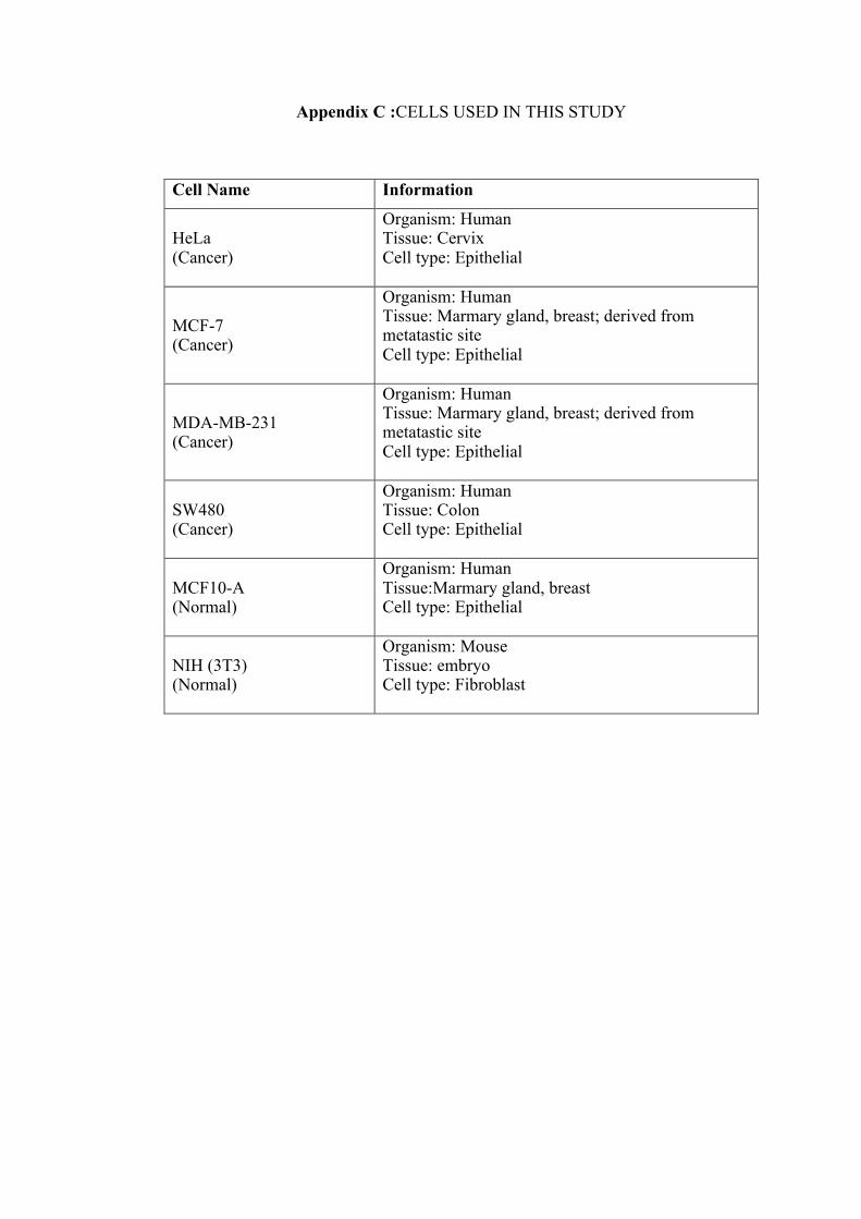
Appendix C :CELLS USED IN THIS STUDY
Cell Name Information
HeLa (Cancer)
Organism: Human Tissue: Cervix Cell type: Epithelial
MCF-7 (Cancer)
Organism: Human Tissue: Marmary gland, breast; derived from metatastic site Cell type: Epithelial
MDA-MB-231 (Cancer)
Organism: Human Tissue: Marmary gland, breast; derived from metatastic site Cell type: Epithelial
SW480 (Cancer)
Organism: Human Tissue: Colon Cell type: Epithelial
MCF10-A (Normal)
Organism: Human Tissue:Marmary gland, breast Cell type: Epithelial
NIH (3T3) (Normal)
Organism: Mouse Tissue: embryo Cell type: Fibroblast

Appendix D: CELL COUNTING USING HEMOCYTOMETER
Cells were counted using the trypan blue exclusion assay. Following trypsinization,
cells were centrifuged to obtain the cell pellet. The supernatant was discarded and the
pellet was resuspended in 1ml complete medium. Then, 10µl of the cell culture was
mixed with 10µl of 4% (w/v) trypan blue dye. 10µl of this mixture was decanted into
the gap of the edge between the hemocytometer chamber and a cover slip. The cell
suspension will diffuse into the calculating chamber as shown below.
Calculating chamber
Live cells were counted from each chamber I, II, III, and IV. Only live cells located
in each chamber (16 small grid squares) were counted. Live cells can be differentiated
from dead cells by the trypan blue colour. This dye will exclusively label only the dead
cells.
I II
III IV

Cell counting was performed under a light microscope at a 10X magnification. Total
live cells were determined using the calculation formula as below:
C =[(I + II + III + IV) x ¼ ] x df x 104
C = number of cells in 1ml (cells/ml)
df= dilution factor
¼ = to obtain the mean of the cells in one area
104 = volume of improved hemocytometer chamber
Therefore, to prepare A x 104 cells, N = [(d x 104 x V)] / C
A x 104 = number of cells needed for seeding
N = volume of cells needed from the cell suspension to obtain A x 104
V = Volume of medium + cell suspension needed for seeding
C = number of cells in 1ml (cells/ml)
Lastly, N was topped up with V of complete medium prior to seeding.

Appendix E: ETHICS APPROVAL FOR BLOOD SAMPLE COLLECTION
131" June 2018
, 1 - ;; ] > ' 'fro Dr. Norzila Ismail Department of Pharmacology School of Medical Sciences Universiti Sains Malaysia 16150 Kubang Kerian, Kelantan.
JEPeM Code : USM/JEPeM/17100566
I Jawatankuasa Etika Penyelidikan Manusia USM (JEPeM) Human Research Ethics Committee USM (HRECJ
Universiti Sains Malaysia Kampus Kesihatan. 16150 Kubang Kenan. Kelantan. Malaysia T : (6)09-767 3000/2354/2362 F : (6)09-767 2351 E : [email protected] L : www.jepem.kk.usm.my
INWW.usm.my
Protocol Title : Understanding the Cytotoxic Effect of Natural Killer Cells towards Selective Cancer Cells Modulated by Pereskia bleo, Abrus precatorius and Liposomes.
Dear Dr.,
We wish to inform you that your study protocol has been reviewed and is hereby granted approval for implementation by the Jawatankuasa Etika Penyelidikan Manusia Universiti Sains Malaysia (JEPeM-USM). Your study has been assigned study protocol code USM/JEPeM/17100566, which should be used for all communication to the JEPeM-USM related to this study. This ethical clearance is valid from 13'" June 2018 until12'" June 2019.
Study Site: Kubang Kerian, Kota Bharu, Kelantan.
The following researchers also involve in this study: 1. Prof. Dr. Armando Acosta 2. Dr. Ramlah Kadir 3. Dr. Rohimah Mohamud 4. Dr. Rahimah Rahim 5. Dr. Wan Zainira Wan Zain 6. Mrs. Mazni Yusoff
The following documents have been approved for use in the study. 1. Research Proposal
In addition to the abovementioned documents, the following technical document was included in the review on which this approval was based:
1. Patient Information Sheet and Consent Form (Malay version)
Attached document is the list of members of JEPeM-USM present during the full board meeting
reviewing your protocol.
While the study is in progress, we request you to submit to us the following documents: 1. Application for renewal of ethical approval 60 days before the expiration date of this
approval through submission of JEPeM-USM FORM 3(8) 2017: Continuing Review Application Form. Subsequently this need to be done yearly as long as the research goes on.
2. Any changes in the protocol, especially those that may adversely affect the safety of the participants during the conduct of the trial including changes in personnel, must be submitted or reported using JEPeM-USM FORM 3(A) 2017: Study Protocol Amendment
Submission Form. ~ p
CERTIFIED BY:
MALAYSIA
National Pharmaceutical Regulatory Agency (NPRA)
t. ~ ~ ~
Forum for Ethical Review Committees in Asia & Western Pacific Region

Appendix F: EXTENSION PERIOD OF THE ETHICS APPROVAL
a llra.UI UNIVERSITI . A '" Ul U1 JTI :~~~YSIA l.f'fPEJ(
71• July 2019
Dr. Norzila Ismail Department of Pharmacology School of Medical Sciences Universiti Sains Malaysia 16150, Kubang Kerian, Kelantan.
JEPeM USM Code: USM/JEPeM/17100566
Jawatankuasa Etih Penyelidihn Manusia USM (JEPeM)
Human Research Ethics Comm~ttee UsM IHRECI
Universiti Sa ins Malaysia Kampus Kesihatan 16150 Kubang Kerian. Kelaman, Malaysia Tel. : • 6 09·767 3000/2354/2362 fa<. : ·6 09·767 2311 Emel : [email protected] Laman Web : www.jepem.kk.usm.my
www.usrn.my
Study Protocol rrtle: Understanding the Cytotoxic Effect of Natural Killer Cells towards Selective Cancer Cells Modulated by Pereskia bleo, Abrus precatorius and Uposomes.
Dear Dr.:
We wish to inform you that the Jawatankuasa Etika Penyelidikan Manusia, Universiti Sains
Malaysia (JEPeM-USM) acknowledged receipt of Continuing Review Appl ication dated 261• June
2019.
Upon review of JEPeM-USM Form 3(8) 2019: Continuing Review Application Form, the
committee's decision for the EXTENSION OF APPROVAL IS APPROVED (start from 7th July 2019
till 6th July 2020). The report is noted and has been included in the protocol file.
JEPeM USM has noted that there is no research activity took place during the period of n•• June 2019 until 6'• July 2019. The report is noted and has been included in the protocol file.
Principle Investigator (PI) should aware and concern about the ethical expiration of the study
in the future.
Thank you for your continuing compliance with the requirements of the JEPeM-USM.
"ENSURING A SUSTAINABLE TOMORROW"
Very truly yours,
~ (PROF. DR. HANS AMIN VAN ROSTEN BERG HE) Chairperson Jawatankuasa Etika Penyelidikan (Manusia), JEPeM Universiti Sains Malaysia
c.c Secretary Jawatankuasa Etika Penyelidikan (Manusia). JEPeM Universiti Sa ins Malaysia
JAWATANKUASA ETIKA PENYEUDIKAN MANUSIA

Appendix G: TEMPLATE OF PARTICIPANT INFORMATION SHEET AND CONSENT FORM
JAWATANKUASA ETIKA PENYELIDIKAN (MAN USIA)- JEPeM USM UNIVERSITI SAINS MALAYSIA
TEMPLATE BORANG MAKLUMAT DAN KEIZINAN PESERTA TEMPLATE OF PARTICIPANT INFORMATION SHEET AND CONSENT FORM
(PROJEK PENYELIDIKAN) (RESEARCH PROJECT)
Borang Maklumat dan Keizinan Peserta yang digunakan dalam Projek Penyelidikan mestilah mengikuti format maklumat berikut. Namun begitu pernyataan dan ayat yang digunakan hanyalah sebagai panduan sahaja. The Participant lnfomation and Consent Form used in the Research Project must be according to these information formats. However, statements and phrases used only as a guide.
Tajuk Kajian I Topic of the Research Pengenalan I Introduction Tujuan Kajian I Purpose of the Study Kelayakan Penyertaan I Participants Criteria Prosedur-prosedur Kajian I Study Procedures Rislko I Risks Melaporkan Pengalaman Kesihatan I Reporting Health Experiences Penyertaan dalam Kajian I Participation in the Study Manfaat yang Mungkin Diperolehi I Possible Benefits Soalan I Questions Kerahsiaan I Confidentiality Tandatangan I Signatures
Sebagal CONTOH, sila rujuk Borang Maklumat dan Keizinan Peserta yang dilampirkan. As an EXAMPLE, please refer to the attached Participant lnfomation Sheet and Consent Form.
(Versl Bahasa Malaysia) I (Bahasa Malaysia Version/
1. LAMPIRAN A <Sila masukkan TAJUK KAJIAN>
2. LAMPIRAN S (Borang Kelzlnan Peserta)
3. LAMPIRAN G (Borang Keizinan Peserta- Sampel Genetik)
4. LAMPIRAN P (Borang Keizinan Penerbitan Bahan yang Berkaitan dengan Peserta)
!Versi Bahasa lnggerisl I (English Version/
1. ATTACHMENT B <Please add in the RESEARCH TITLE>
2. ATTACHMENTS (Participant Information and Consent Form)
3. ATTACHMENT G (Participant Information and Consent Form- Genetic Sample)
4. ATTACHMENT P (Participant's Material Publication Consent Form)
JEPeM US M Template for PIS and CF u pdat e d 10/05/2017 Page 1 or 12

Kajian ini akan melibatkan individu yang mempunyai ciri-ciri berikut:
Bagi Peserta sihat:
Tiada penvakit berat Tidak mengambil dadah immunosupresif Umur 18-45 tahun Tidak merokok Tidak hamil
Kajian ini tidak akan melibatkan individu yang :
Bagi peserta sihat:
resif
Bagi peserta pesakit kanser:
Disahkan mengidap kanser oleh doktor Masih bel urn mendapat apa-apa rawatan Umur 18-45 tahun Tidak merokok Tidak hamil
Bagi peserta pesakit kanser:
Tidak didiagnos mempunyai kanser Mengambil dadah immunosupresif Mempunyai kanser dengan ko-morbiditi
*Tidak mengambil dadah immunosupressif sekurang-kurangnya 3 bulan yang lepas.
la in Enggan memberi kebenaran consent
• ko-morbiditi adalah kewujudan penyakit lain oada pesakit kanser, seperti tekanan darah tinggi, penyaki t buah pinggang dan lain-lain.
PROSEDUR-PROSEDUR KAJIAN
Peserta (yang boleh berbahasa melayu) hanya perlu mengisi borang maklumat yang diperlukan untuk mengetahui sama ada peserta termasuk dalam kriteria yang diperlukan. Jika peserta memenuhi kriteria, darah peserta akan diambil oleh individu terlatih sebanyak 10 mi. Setelah selesai, peserta akan diberi honorarium. Sisa buangan saripada sampel kajian (sisa klinikal biohazard) akan dilupuskan dengan cara yang slstematlk.
RISIKO
Secara umumnya. pesakit tidak akan mengalami sebarang masalah atau risiko dalam proses pengambilan darah sebanyak 10 mi. Sila ikut arahan kakitangan yang mengambil darah dan hanya meninggalkan makmal selepas darah kering. Sila maklumkan kepada kakitangan kajian sekiranya anda menghadapi sebarang masalah atau mempunyai sebarang maklumat panting yang mungkin mengubah persetujuan anda untuk terus menyertai kajian ini.
MELAPORKAN PENGALAMAN KESIHATAN (Jika Kajian Melibatkan Kesihatan SAHAJA)
Sila hubungi kakitangan berikut pada bila-bila masa sekiranya anda mengalami sebarang masalah kesihatan, samada berkaitan atau tidak berkaitan dengan kajian ini. Dr Wan Zainira Wan Zain (No. Pendaftaran Penuh Majlis Perubatan Malaysia: 38667 ] di talian 019-9216789 atau Dr Rahimah Rahim [No. Pendaftaran Penuh Majlis Perubatan Malays ia: 38338 ] di talian 012-8330274 secepat mungkin.
PENYERT AAN DALAM KAJIAN
Penyertaan anda dalam kajian ini adalah secara sukarela. Anda berhak menolak untuk menyertai kajian ini atau menamatkan penyertaan anda pada bila-bila masa, tanpa sebarang kehilangan manfaat yang sepatutnya anda perolehi. Hasil kajian ini t idak akan dimaklumkan kepada peserta kerana ia merupakan hasil dari kombinasi set darah putih dari beberapa peserta dan bukan berorientasikan individu.
J EPeM US M Templ a te for PIS and CF upda t ed 10f 0Sf2017 Page 3 o f 12


LAMPIRAN G
Borang Keizinan Peserta untuk Pengambilan Sampel Genetik (Halaman Tandatangan)
Tajuk Kajian: Memahami kesan sitotoksik sel Pembunuh Semulajadi (Natural killer cell) ke alas sel kanser payudara dan serviks yang dimodulasi oleh ekstrak Pereskia bleo, Abrus precatorius and liposom
Nama Penyelidik: Dr. Norzila Ismail, Dr. Ramlah Kadir, Dr. Rohi mah Mohamud, Prof Armando Acosta, Dr Wan Zaini r a Wan Z ain (Surgery , No MMC 38667). Dr Rah lmah Rahim (O&G , No MMC 38338),Puan Maz ni Yusoff (Unit Perubatan lntegrat if )
Untuk menyertai kajian ini, anda atau wakil sah anda mesti menandatangani mukasurat ini. Dengan menandatangani mukasurat ini, saya mengesahkan yang berikut:
Saya Ieiah membaca semua maklumat da lam Borang Ma klumat dan Ke iz inan Pesakit ini te rmasuk apa-apa maklumat berka i tan r is iko yang ada dalam kajian dan saya te lah pun diberi masa yang mencukupi untuk mempertimbangkan maklumat tersebut. Semua soalan-soalan saya te lah dijawab dengan memuaskan . Saya, secara sukarela , bersetuju menyertai kajian penyel idikan ini , mematuhi sega la prosedur kaj ian dan memberi makl umat ya ng diperlukan kepada doktor, para jururawat dan juga kakitangan lain yang berkaitan apab il a diminta. Saya boleh menamatkan penyertaan saya dalam kajia n ini pada bila-bila mas a . Saya Ie iah pun menerima satu sa l inan Borang Maklumat dan Ke iz inan Peserta untuk simpanan peribad i saya .
Nama Peserta
No. Kad Pengenalan Peserta
Tandatangan Peserta atau Wakil Sah
Nama & Tandatangan lndividu yang Mengendalikan Perbincangan Keizinan
Nama Saksi dan Tandatangan
Tarikh (dd/MM/yy) Masa Uika perlu)
Tarikh (dd/MM/yy)
Tarikh (dd/MM/yy)
Nota: i) Lebihan sampel kajian ini akan dilupuskan dan tidak akan digunakan untuk tujuan lain kecuali setelah mendapat kebenaran daripada Jawatankuasa Etika Penyelidikan (Manusia), USM.
ii) Semua peserta yang mengambil bahagian dalam projek penyelidikan ini tidak dilindunoi insuran.
JEPe M USM Template ror PIS and CF updated 10/05/20 17 Page 6 o r 12
LAMPIRAN P
Borang Keizinan bagi Penerbitan Bahan yang berkaitan dengan Peserta Kajian (Halaman Tandatangan)
Tajuk Kajian: Memahami kesan sitotoksik sel Pembunuh Semulajadi (Natural killer cell) ke atas sel kanser payudara dan serviks yang dimodulasi oleh ekstrak Pereskia bleo, Abrus precatorius and liposom
Nama Penye/idik: Dr. Norzila Ismai l, Dr. Ramlah Kad ir, Dr. Rohimah Mohamud , Prof Armando Acosta , Dr Wan Zainira Wan Zain (Surgery, No MMC 38667) , Dr Rah imah Rahim (O&G , No MMC 38338),Puan Mazni Yusoff ( Un it Perubatan lntegratif)
Untuk menyertai kajian ini, anda atau wakil sah anda mesti menandatangani mukasurat ini.
Dengan menandatangani mukasurat ini, saya memahami yang berikut:
Bahan yang akan diterbitkan tanpa di lampirkan dengan nama saya dan set iap percubaan yang akan dibuat untuk memastikan ketanpanamaan saya . Saya memahami, walaubagaimanapun, ketanpanamaan yang sempurna ti dak dapat dijam in. Kemungkinan sesiapa yang menjaga saya di hospital atau saudara dapat mengena l i saya . Ba han yang akan d iterbitkan dalam penerb itan mingguan/bulanan/dwibulanan/suk u tahunan /dwi tahunan merupakan satu penyebaran yang luas dan tersebar ke se luruh dunia . Kebanyakan penerbitan in i akan tersebar kepada doktor-doktor dan juga bukan doktor termasuk ahli sains dan ahl i jurnal. Bahan tersebut jug a akan di lampirkan pada Ia man web jurnal d i seluruh dun ia . Sesetengah Iaman web ini bebas d ikunjung i ole h semua o rang . Bahan tersebut juga akan d igunakan sebaga i penerbitan tempatan dan disampaikan oleh rama i doktor dan ah l i sa ins di se luruh dun ia . Bahan tersebut juga akan d igunakan sebagai penerbitan buku oleh penerbit jurnal. Bahan tersebut tidak akan digunakan un tu k pengiklanan ataupun bahan untuk membungkus.
Saya juga memberi keizinan bahawa bahan tersebut boleh d igunakan sebaga i penerbitan lain yang diminta oleh penerbit dengan kriteria ber ikut :
Bahan tersebut tidak akan digunakan untuk pengiklanan atau bahan untuk membungkus . Bahan tersebut tidak akan digunakan di luar konteks - contohnya : Ga mbar tidak akan d igunakan untuk menggambarkan sesuatu artikel yang tidak berka itan dengan subjek dalam to to tersebut.
Nama Peserta
No. Kad Pengenalan Peserta T/tangan Peserta
Nama & Tandatangan lndividu yang Mengendalikan Perbincangan Keizinan
JE Pe M USM T emplate for PIS and CF updated 10/05/2017
Tarikh (dd/MM/yy)
Tarikh (dd/MM/yy)
Page 7 or 12

Appendix H: STANDARD CURVE OF INTERLEUKIN-2 FOR ELISA
0 200 400 6000
1
2
3
4
IL-2
Concentration (pg/ml)
Abs
orba
nce
(450
nm)
R2 = 0.9973

Appendix I: STANDARD CURVE OF INTERFERON GAMMA FOR ELISA
0 500 1000 15000
1
2
3
4
IFN
Concentration (pg/ml)
Abs
orba
nce
(450
nm)
R2 = 0.9974
g

Appendix J: STANDARD CURVE OF PERFORIN FOR ELISA
0 20 40 600.0
0.5
1.0
1.5
2.0
2.5
PRF-1
Concentration (ng/ml)
Abs
orba
nce
(450
nm)
R2 = 0.9891
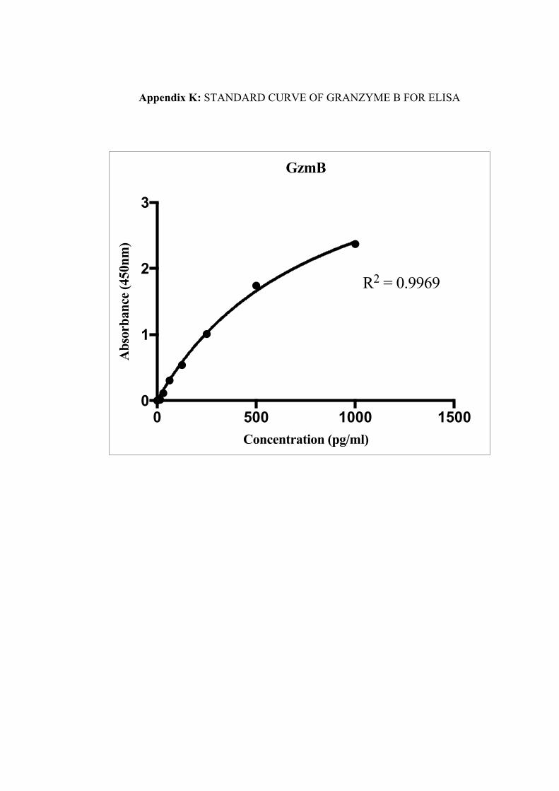
Appendix K: STANDARD CURVE OF GRANZYME B FOR ELISA
0 500 1000 15000
1
2
3
GzmB
Concentration (pg/ml)
Abs
orba
nce
(450
nm)
R2 = 0.9969

Appendix L: PUBLICATION 1
t.:~:NIKA TROPICAL AGRICULTURAL SCIENCE
GC-MS Analysis of Phytochemical Compounds in Aqueous Leaf Extract of Abrus Precatorius
\Van Slll"iyani Wan-lbrahim', Tuan Nadrah .'laiDll Tuan Ismail', Siti Famanah \lohd-Sall~h' and .'lorlila Ismail'* 1 DtpartmDJt of Plw.nJJtrolog\·. Sdroo/ of AlniKal Snmcrs Unn·t!t"SStr Saau M.u/oysw../6150 VSJt, Kubang Kenan. Kdaattm.. Jlaltn'SUJ !ScJtooJ ofDentol Sclt'ncn., Lim·IY7ih Stuns Ma/a,-sw.. 16150 m.\1~ K.ubang Kn-wn. Krlankm, Ma/a,-sw
ABSTRACT
Abrus precmOJius is a flowering plant that belongs to the l~ume family, Fabaceae. In
Malaysia. the lea,·es of Abrus fJI"<'atoriu.t are tL~ed traditionally to treat :Uiments such
as fever. ulcer and mouth cancer. These traditional practices. however. have never been
documented and usage oftbe plaru is based on popular beliefs held by the loc-.U people.
This work doeumemed the phytoeompounds that are present in the aqueous extract of Abrus prectJtoriu.r lea,·es collected from a local area. in Kota Bh&rtL Kelantan, Malaysia.
The leaves were dried and then subjected to extraction using the decoction technique. The
compounds \\ere identified by gas chromatography with mas.~ spectromeuy analysi~ and
characterised by comparison through the N1ST02 and Wiley275 library search software.
The GC-MS analysis showed that the cla~ses of compounds identified in aqueous extracts
of Abrus prt'CIIIOrius leaves \\ere phenolic compounds, terpenoids and steroids.
ARTICUI!<..fO
·"""" .._. ~~«--cd es•a.::'OI1 Aca:ptal_ 3D Jaat 2017
£.-I • .uw_._ Ubq.a1~11",.,c- (\l.•~sf)•• "•·lhnfaat\. lludniri:O. ..-..., (1a.. Nadulil !\ .... T10.a a-b. U~~_d1l~l')--.0111nP,1a f~ l.IGIIII..s..l~),. _._ .. _ • .. ...._.,. fNou:lb b.awb • C"orf'C-...-1•.1111 .a tOw
1:-ITRODUCTIO:"<
Medicinal plantsarewidelyused as food and medicine in traditional practice. In Malaysia,
such plants are consumed as an alternati\•e
treatment for illness or to maint:Un a healthy
lifestyle. A huge reservoir of bioactive
con1p0undsexiSlS in o'-er 400.000 species of
plants on Earth. but only a small percentage
of these compounds have been examined
in research. In many de\-eloped countries,

Appendix M: PUBLICATION 2
As ian Pacific Journal of Tropical Biomedicine
Origlnnl Arllclt -.:.: ~ . ~
f!J . ...
'~"' Ul410lf'..lll 1691.260397
Mcthanohc cxmct of Abms prt'tlllonus promote~ bre:t~t cancer MDA-MB-231 cell death by mduang cell cycle arrest at C./ C1 and uprcgulaung Bax
\V•n Sun)•no W•n- lbr.dmn . Numl.ll•m..U' )lu liarh;~mh Muhd- ...,lldt , A1<ly lnnon Y•Jod Modud l'•k- K.u
\V'"'I< . Muhd Nwm Md H.ubun 1(~.,,_,., •.fl'lt.,.4N/ ... ~,.... ~ \Jrot..J ..__.._I llff»>t "'MtA• .Jot..M .,/l'JijiJA.-'nJV ANN"- AI'N~ ·U.&.n ...
(~.,,_,., •.f I".IJwl.,.,. ~-JoJ/ ,.Ut<oi..J~,.,.... .... I .,,...,.,m.S..uu V.J.,,.,..J&I$0 ~ Am.M. x,t.,..,, 1rl·.t..~u• .,..,..,_.,_,, >io.ol<(\f.16rnl:v-,...t """"''~ ValouN~.l61SIIhlkllll An• ... l•l....._ llat.TIOI
i\11. riC:LE INFO
"'""''"'-"· ~M"'M.-Ja!UI9 Mo...,. ltAplll'" ''"-'IOJ-»1• ~t-.b•l1....,%1JN
............ ;. ~-l"ff:·tfo~ )~A-M8·l!l
"" .... "" Cdh . ., .. ~. a..: ..... ~.
1. lntrolilllwon
)t • Tock~lrk lbt ;arat .. 5ft~Uct=~thc Jk.1h11) d -tl..nu l""r»>·~ H . ,........,.,,., k..t
C'W'<ll."*' *liJ tb:ildk\:,UQ ~"Cfl 4kllth.-
\tciiUNI t. f~H-t1'-11t11J k.nc.~ w-e~ cAIItlo,"1CJ .;u .. -..;c ... ~~l)" ••lh bc.ull<'. c-lhyl~Kcil\tc
\lAd mtlh.llOOI b) So\bkt cxuxtro& ""Jlc.w.t Qlr.ll.'1 Y.ll): pt'~ b) dt\:'~,.on .,, '«l t i!J.lr.ll.1. ur l ,..,.,...,,... k_,.nu --~ iUW &0 tJti)t JCio;k4l QI'CC( ~ted QO.tml&l cc:lll&k' lot
72 b l·lllr'UKrmorC". ~.5-dlmcthyllhl..atbl-!-.)ll !.Uirfw'ny& tcn.r.ohitm bromkk- a~'i:ll)'
"'""-' pctr«mcd tP d.ctcrmUX" eell ~wbUu)' Arat,....._" of to-ell c,:k llntJL 11(101\tolltluuy .~
lpopt~&s (W'(*an C'~'t<ll\) v.« 0:-lCI'ftiD'Cd by C1ow 10.)1('Drll)
tc Mutullut~ cttr.IICC (II 1 ~"~"..,,.,. ka~'JobOM:'4 d'lc" kNooJIC, OD MO.\~lt.iB .. l.ll
«-ll' at (l6..at•.a$AU. ~- ACM ()"C<Ifl:k'CI)' aGll)"l" "="Caic4 th.lt "I IITOil <\.--cDac\J;aJ OJ 0 1 f'b*.tc aAd tb< a~b •qy tbor.'Gl the ~.D'cllri."C' <II: cllll) a,_-.p.-.b 111 4 h ~D MDA·
MS..!'I c:db U'C'akd tvhb mdtwlobc: c:\tr.a •. :t at: 1 ,.,.,,.,d,UH le~WC"L. Mr~lh-'IOhC <"AmK't
nt l ,. ... ~t .. n.o ICJW. •lldoca:~ ~popt·•b ~ "ffe.lub.On" ol lb\. ps~ '-'.lid <::up~l .IIJ'ki d·*ttn:p.l~n(Jr &.:l~l.
t' I II M«haoalit: ()n.,_"l cl l ,.,.~. b\'C:.( P'ClCr'lll>foc-' MI).:.\·MB-l.\1 4-dl <Ieath
byatd!.:tnJ «II <)\:ic- llnl:\1 A)d tpcp~'*" P--'N) NU&he mat«t.ltd'bl·.ellttcd pdt.-ct)'.
AP'-~&u\1~ n a due.taOt.RI.<d lonn \ll ~:dl Gucb \and n mo~ly
~tdc-d. ~ncommonty ~ p~Jnmrfti:"'J <dl dtat.. •fk1'1Mlt: 1:' ftc ~llprc, of d)IIJI«I\ atn tnlmc:Dt' thit lllt U~)' 4»~
ll:ld dltnin.l~ by Pb.C<\.")k& v.<IIKU GJturblftJ w no.:nn.al ~,.
crl wnoGI'I"nf ta~ll. Equillhrtwm bct~CO\ «II de.ttb .ud ~dl
&i.:<"•"-ft'C' :a~nrcfdh~r:l!l~tr""P..U. to tt:.tatl*~m.e) ~JaniQI
ll!or ... ollliOW!¥ ·-n~. ""~"'~'""' <JAl'< lmlilt<d 111!•11111 ,., -tf'V-"'k r-ttr-..<4)', l.ht Uaiftlk ilf' m1lt\;t-1nd.ri~l ,.m.. .. )· and l.ht
curbk <W ~th ~,..._,..._.., p:~fl'llloll) ln"Al«' truAOIIltmcftl ~'I
he&_' ha.."(lme' .. lmp'ln.' 1001 ., • lt~Jtl ttt P•'~~•J"CCP•U•Indtli:W.J ....... ..,bllnJbofl<hcmi:JIIIOdlo........,.,l
rk•l• • j~•«"-•J ... .I -.t~h-dlc,._.,J -.kflioo;. ---.clf* ~,:_.,'It: t"01oaca•• AI~......,_,.,_....._ ,,.....,., .. ~'" ~·· <&..o ......... ••'- . .......... """ . . ..... .....,..,. ....., ..... . .-••• ,. ...... .......ul) ..... ..
""'"-'"" "'"'""""" ••••.-d - .... ., ,, ..., '""""" boluoc< • ~fh*!Ptdo "'' _ _,..., ... QINIII-*..,, .. ~...., ........ ai -.... ~~l: ..... .....,.. ........... O).tN'Lt-l!rr..·• .. ,_..J.,.tT,_,...,,..,_.., .... ..aa_c.tllt "-.I"'" J..-.. ......... .u.,..._._.. ,._. ... ~w. .. ,u.·,--~ .......... ~ .. ...,., ~ "'·t-1~11'~~,, .. ,., .... Yil~ .. ,,._,_..,ti•••M~ "k**L'~ ....... "',"I .... , ,.., .. _ , ,...,_.,.._., ltw..u~ , ... ..- )CMW:G lJI ...U,.._. _, .._ •• IPC1 .._.,.._. - •(-.,G-' .._,.._,.,._ A ... t'..,J1"9'1• ..... :UW "-'" !..._~

LIST OF PUBLICATIONS
1. Wan-Ibrahim, W. S., Ismail, N., Mohd-Salleh, S. F., Yajid, A. I., Wong, M.
P.-K. & Hashim, M. N. M. (2019). Methanolic extract of Abrus precatorius promotes breast cancer MDA-MB-231 cell death by inducing cell cycle arrest at G0/G1 and upregulating Bax. Asian Pacific Journal of Tropical Biomedicine, 9(6), 249. [Web of Science][Q3]
(Anugerah Penerbitan Pascasiswazah 2019)
2. Wan-Ibrahim, W. S., Ismail, T., Naim, T. N., Mohd-Salleh, S. F. & Ismail,
N. (2018). GC-MS Analysis of Phytochemical Compounds in Aqueous Leaf Extract of Abrus Precatorius. Pertanika Journal of Tropical Agricultural Science, 41(1). [Scopus][Q3]
LIST OF PRESENTATIONS
April 2019 The 3rd International Conference on Molecular Biology and
Biotechnology (ICMBB), UCSI University, Kuala Lumpur, Malaysia (Oral- Pecha Kucha Style) Abrus precatorius methanolic leaves extract induced apoptosis in MDA-
MB-231 cells Wan Suriyani Wan Ibrahim, Siti Farhanah Mohd Salleh, Norzila Ismail Oct 2018 Monthly CME, PPSP, USM Health Campus, Malaysia (Oral) Phytochemical profiling and Anti-proliferative activity of Abrus
precatorius leaves extract Wan Suriyani Wan Ibrahim, Norzila Ismail March 2018 ICNP 2018, Penang, Malaysia (Oral) Phytochemical Profiling & Anti-proliferative Activity of Abrus
precatorius Leaves Extracts Wan Suriyani Wan Ibrahim, Siti Farhanah Mohd Salleh, Tuan Nadrah
Naim Tuan Ismail, Norzila Ismail