palpable breast lesions – cytomorphological analysis and ...
77
PALPABLE BREAST LESIONS – CYTOMORPHOLOGICAL ANALYSIS AND SCORING SYSTEM WITH HISTOPATHOLGICAL CORRELATION DISSERTATION SUBMITTED FOR M.D. DEGREE EXAMINATION BRANCH – III (PATHOLOGY) THE TAMILNADU DR.M.G.R. MEDICAL UNIVERSITY CHENNAI MARCH 2009
-
Upload
khangminh22 -
Category
Documents
-
view
0 -
download
0
Transcript of palpable breast lesions – cytomorphological analysis and ...

PALPABLE BREAST LESIONS – CYTOMORPHOLOGICAL
ANALYSIS AND SCORING SYSTEM WITH
HISTOPATHOLGICAL CORRELATION
DISSERTATION SUBMITTED FOR
M.D. DEGREE EXAMINATION
BRANCH – III (PATHOLOGY)
THE TAMILNADU DR.M.G.R. MEDICAL UNIVERSITY
CHENNAI
MARCH 2009

CERTIFICATE
This is to certify that Dr.D.SHEEBA is a bonafide student of the
Department of Pathology, Kilpauk Medical College, Chennai and this study
titled “Palpable Breast Lesions – Cytomorphological Analysis and Scoring
system with Histopathological Correlation ” is the original work done by her
for her dissertation towards the partial fulfillment of requirements for the
M.D(Pathology) Degree 2006 – 2009.
Prof.C.S.Vijayalakshmi, M.D.,
Professor and Head,
Department of Pathology,
Kilpauk Medical College Hospital
Chennai – 600 010.
Prof.M.Dhanapal., M.D., D.M
Dean,
Kilpauk Medical College Hospital,
Chennai – 600 010.

ACKNOWLEDGEMENT
I owe my sincere thanks, Prof.M.Dhanapal, Dean, Kilpauk Medical College,
Chennai for allowing me to use the resources of this institution.
I thank my Head of the Department, Prof . C.S.Vijayalakshmi, M.D.,Professor
and Head of the Department of Pathology, Kilpauk Medical College, for her
valuable guidance and encouragement during the course of the study.
Thanks are due to Prof. Ezhilvizhi Alavandar, M.D., Professor, Department of
Pathology, Kilpauk Medical College for her valuable advice.
Thanks are due to Dr.Bharathi Vidya Jayanthi, M.D., Professor of Pathology,
Kilpauk Medical College, for her valuable guidance and suggestions.
My sincere thanks to all the Assistant Professors, Colleagues and Lab
Technicians who extended their help and encouragement.

Table of Contents
Introduction…………………………………………………………… 1
Aims of the Study…………………………………………………… 3
Review of Literature……………………………………………….. 4
Materials and Methods………………………………………….. 31
Results and Observations……………………………………….. 35
Discussion………………………………………………………………. 45
Summary and Conclusions……………………………………… 49
Bibliography…………………………………………………………… 51

Introduction

1
Introduction
Fine Needle Aspiration Cytology has been used to diagnose Breast Cancer for
over 50 years. In recent years, it has gained acceptance as a diagnostic tool in
assessment and management of mammary lesions.
With the advent of mammography and Ultrasound, these investigations were
used to recognize breast lesions. Regardless of the sex of the patient and the method
used to diagnose the lesion, cytological techniques play an important role in the
diagnosis of breast lesions.
Fine Needle Aspiration Cytology is successful in identification of benign and
malignant breast lesions, but its role in proliferative breast lesions is poorly defined.
To expand the role of FNAC in diagnosing of proliferative breast lesions, the analysis
of cytomorphological features of proliferative breast lesions in conjunction with
cytological scoring system proposed by Masood et al and with histopathology was
done.
Relative Risk of developing Invasive breast cancer from carcinoma in situ, from
proliferative breast disease with atypia, proliferative breast disease without atypia
and non proliferative breast disease is of the order of 8 – 10, 4 – 5 , 1.5 – 2 , and 1
respectively1. Hence, it is very important to identify proliferative breast disease.

2
This study was undertaken to categorise the breast lesions into four
categories depending on nuclear dissociation, myoepithelial cells, pleomorphism of
cells, anisonucleosis, nuclear chromatin and nucleoli. The four categories are
1. Non-Proliferative Breast Disease,
2. Proliferative Breast Disease without Atypia
3. Proliferative Breast Disease with Atypia
4. Carcinoma.
The categorization of proliferative breast lesions by FNA remains a challenge
to the pathologist and the cytologic criteria need to be further defined and assessed.
Decreasing the number of diagnostic categories is likely to improve the correlation
between the cytologic and histologic diagnoses without compromising patient
management.

Aims of
the Study

3
Aims of the Study
The study aims to correlate the Cytomorphological Diagnosis and the Modified
Masood’s scoring system with Histopathological Diagnosis in palpable Breast Lesions.
Age distribution of case under different categories of diagnoses are also
studied.

Review of
Literature

4
Review of Literature
The use of cytology for diagnosis of breast lesions dates back to the early
1930s, when Martin and Ellis first reported their experience with FNA at the
Memorial Hospital for Cancer and Allied Diseases in New York2. This was followed in
the late 1940’s and the early 1950’s by Adair and Godwin. However, not until the
Europeans reported a large series of a number of FNAC of the breast was Aspiration
Cytology shown to be a valuable and accurate diagnostic procedure.
The sensitivites of Open Biopsy and FNAC are comparable ( 99% for the
former and 96.2 % for the latter). The specificity of FNAC is better ( 98.8 % vs 85.5%
for open biopsy).3
Aspiration biopsy of breast lesions has become a quasi-routine clinical
procedure, often replacing per-op tissue biopsy. FNAC is the least expensive method
and the most rapid and most versatile of all approaches.
FNAC can be used for palpable masses that may be either solid or cystic or
nonpalpable lesions detected by Mammography, Ultrasound or MRI.
It is the consensus of most observers that in most cases, 2 to 4 passes of
needle are required to harvest optimal diagnostic material. Most authorities define a
numerical cut-off for cellularity of atleast 6 epithelial cell groups with 5 to 10 cells per
group for adequacy1.

5
Embryology:
The Human Mammary Gland develops during the 5th
week of Gestation at
which time, thickenings of the ectoderm appear on the ventral surface of the fetus.
These mammary ridges, also known as milk lines, extend from the axilla to the groin.
Except for a small area in the pectoral region, the bulk of these ridges normally
regress as the fetus continues to develop.
After the 15th
week of gestation, the developing breast exhibits transient
sensitivity to testosterone, which acts on the mesenchyme. The mesenchyme
condenses around an epithelial stalk on the chest wall to form the breast bud, the
site of the mammary gland devlopment.
Solid epithelial columns then develop within the mesenchyme, and these give
rise to the lobes or segments of the mammary gland. Portions of the fetal papillary
dermis encase the developing epithelial cords and give rise to the vascularised
connective tissue that sorrounds the mammary ducts and lobules. The collagen-rich
reticular dermis extends into the breast to form the Suspensory Ligaments of Cooper.
Portions of the mesenchyme differentiate into fat between the 20th
and 32nd
weeks of Gestation. During the last eight weeks of Gestation, the epithelial cords
canalise and branch forming lobulo alveolar structures as a result of mesenchymal
paracrine effect.

6
A depression in the epidermis forms at the convergence of the lactiferous
ducts. The nipple forms by evagination of the mammary pit near the time of birth.
Anatomy and Histology of Breast:
The breast is a glandular tissue, surrounded by fibro-adipose tissue and
covered by epidermis. Centrally located is the nipple, surrounded by a circular
pigmented area, the areola. Tubercles of Montgomery, a specialised sebaceous gland
of the areola, enlarge during pregnancy and lactation.
The arteries of the mammary gland are branches of the Internal Mammary,
External Mammary and Intercostal Arteries. The veins are the Axillary, Internal
Mammary and Intercostal.
The breast is composed of 15 to 25 lobes that converge on the nipple in a
radial pattern. Each segment consists of a lactiferous duct, lactiferous sinus,
segmental collecting duct, sub segmental duct, terminal duct and acini. The collecting
ducts are lined by columnar cells which are multilayered in the larger ducts.
The terminal portions of the lactiferous sinus and the lactiferous ducts are
lined by stratified squamous epithelium. The secretory acini consist of a single layer
of cuboidal epithelial cells with sorrounding elongated myoepithelial cells resting on
a basement membrane.The acini are set within the loose specialised stroma that
defines the lobular unit.

7
The sorrounding lobular connective tissue contains increased number of
capillaries and few lymphocytes, histiocytes, plasma cells and mast cells . The lobular
connective tissue is sharply separated from the more dense periductal fibrous tissue
and abundant fat that make up the majority of the breast.
During pregnancy, the breast undergoes lobular and ductal proliferation, with
evidence of lactation. After menopause, the breast shows increased amount of fat,
diminished connective tissue, persistance of the mammary ducts and disappearance
of the lobules.
Cytology of the Normal Breast:
Cells derived from ducts have round to oval nuclei, 8 to 10 in diameter, with
a very small nucleolus or no visible nucleolus. The cytoplasm is scanty. The
myoepithelial cells are recognized as having a small spindly curved dark homogenous
bipolar nucleus with scant cytoplasm that may either adhere to the epithelial
fragments or appear singly.
The responsiveness of the breast epithelium to cyclic hormonal influences
have been shown in Fine Needle Aspiration specimens. Post ovulatory aspirates are
characterised by an increase in the number of acinar cells with all cells including the
ductal cells displaying more features of acinar cells. Peripheral orientation of the
nuclear chromatin with clearing is seen. The cytoplasm is lacy and fragile. The

8
epithelial fragments show a multilayered arrangement with marked superimposition
of cells.
In pre ovulatory aspirates, the cell borders are more prominent, with the
cytoplasm appearing more even in consistency and better delineated. The nucleus is
small and more compact with evenly distributed chromatin. Epithelial fragments are
arranged in single layered sheets. The stroma is composed of fat and loose or fibrous
connective tissue.
The cytological reporting categories are :
Malignant
Suspicious
Atypical
Benign specific
Benign – non-specific
Unsatisfactory sample
A palpable breast lump is a common diagnostic problem. Excisional Biopsy
was accepted practice in the past, but presently, Radiological Imaging in combination
with needle biopsy makes it possible to reduce unnecessary surgical excision of
benign breast lesions to a minimum.

9
Carter et al4
studied the relationship between benign breast disease and
subsequent breast cancer in 16,692 women. Women were classified into one of the 5
benign breast disease categories:
Atypical Hyperplasia
Proliferative Disease without Atypia
Non Proliferative Breast Disease
Fibroadenoma
Others
Relative risk estimates of breast cancer for women in the 5 benign breast disease
categories compared with screened women who did not develop recognizable breast
disease were computed using the proportional hazards model. Results indicated that
the risk was associated with the degree of epithelial atypia.
Women with nonproliferative breast disease, proliferative breast disease without
atypia and atypical hyperplasia displayed progressively increasing risk of 1.5, 1.9 and
3 respectively, compared with normal subjects with 95% confidence intervals
exceeding unity5.
Dupont et al attempt to quantitate the relative risk of breast carcinoma with
the degree of proliferation and atypia of intraductal epithelial proliferation of the
breast.

10
Table 1 : DuPont and Page : Relative Risk of Carcinoma in various categories
The cytological reporting category does not help us in assessing the relative
risk of different lesions turning malignant. In the current study, Dupont & Page’s
categorisation of breast lesions has been used.
The study showed a relative risk for cancer of 1 for Non-proliferative Breast
Disease and a relative risk of 1.9 for proliferative Breast Disease without Atypia, and
a risk of 5.3 for Proliferative Breast Disease with Atypia.
Based on this study, Breast Lesions were categorised as follows:
Study Study Design Nonproliferative Proliferative
without
atypia
Atypical
hyperplasia
Nashville6
Retrospective
Cohort
1 1.9 5.3
Nurses Health
Study7
Case-Control 1 1.6 3.9
Breast Cancer
detection and
Demonstration
Project8
Case-Control 1 1.3 4.3
Florence, Italy9
Case-Control 1 1.3 13.0

11
Categorisation of Breast Lesions according to the criteria of
Dupont, Page and Rogers:
Non Proliferative
Cysts
Papillary apocrine change
Epithelial-related Calcifications
Mild hyperplasia of the usual type
Duct Ectasia
Proliferative lesions without atypia
Moderate or Florid ductal hyperplasia of the usual type (usual ductal
hyperplasia)
Intraductal papilloma
Sclerosing adenosis
Fibroadenoma
Atypical hyperplasia
Atypical ductal hyperplasia
Atypical lobular hyperplasia

12
Carcinoma
Non-Invasive
Intraductal Carcinoma with Paget’s Disease
Lobular Carcinoma insitu
Invasive
Invasive Ductal Carcinoma with Paget’s Disease
Invasive Ductal Carcinoma with predominant Intraductal Component
Invasive Lobular Carcinoma
Medullary Carcinoma
Mucinous Carcinoma
Invasive Papillary Carcinoma
Tubular Carcinoma
Adenoid Cystic Carcinoma
Secretory Carcinoma
Apocrine Carcinoma
Metaplastic Carcinoma
Inflammatory Carcinoma
13
Clinical and Cytological Findings
Cyst:
Age > 30, Multifocal and Bilateral
Poorly defined lumpiness on palpation with a shotty feeling
Scanty, watery or fatty smear
Benign or Uncertain Mammogram
Low to Moderate cellularity
Apocrine Cells in Variable Cellularity
Foam Cells ( Macrophage or Epithelial origin)
Sheets or Fragments of Non apocrine Ductal Epithelium with bland
nuclei arranged in honey comb pattern with admixed myoepithelial
cells
Dispersed stromal bipolar nuclei
Fat or Fibrous Stroma in variable quantities
Haagensen states that the initial insult of fibrocystic diseases is
periductal mastitis, resulting in periductal scarring10
.
Proliferative lesions accompanying Fibrocystic Disease are
o Sclerosing Adenosis
o Collagenous Spherulosis
o Papillomatosis
o Ductal Hyperplasia

14
o Atypical Ductal Hyperplasia
Differential Diagnosis
o Adenoid Cystic Carcinoma
o Collagenous Spherulosis
o Signet-ring cell Carcinoma
Mastitis
Acute Mastitis or Breast Abscess:
Cytology shows Neutrophils, Foamy Macrophages, Cell debris in the
background
Atypical epithelial cells with features of regeneration and repair
including nuclear enlargement and prominent nucleoli
Microorganisms ( Infectious Mastitis )
Granulomatous Mastitis :
Differential Diagnosis of Granulomas in the breast include Infectious
Granulomas (Tuberculosis, Fungi, Leprosy, Brucella), Sarcoid, Tumour, Fat Necrosis,
Foreign Body reaction and Idiopathic Granulomatous Mastitis.
Granulomas show clusters of Epithelioid Histiocytes, with or without
Multinucleated Giant Cells, Lymphocytes and Plasma Cells.

15
Fat Necrosis :
History of Trauma with or without bruising of the skin
Tender on Palpation
Foamy Macrophages and multinucleated giant cells with foamy
cytoplasm
Fragments of Normal Adipose tissue
Variable number of other inflammatory cells.
Few epithelial cells
Free Lipid Droplets
Granular Background
Papillary Apocrine Change:
Papillary proliferation of ductal epithelial cells in which all of the cells
show Apocrine features
Epithelial Hyperplasia :
Intraductal Epithelial proliferation includes a spectrum ranging from
intraductal hyperplasia without atypia to atypical ductal hyperplasia to ductal
carcinoma in situ.
Clinical picture is usually benign but may be suspicious

16
Low or Moderate Cellularity with small epithelial groups suggest
Fibroadenosis, Sclerosing Adenosis or Other Sclerosing lesions
High Cellularity with large flat or folded sheets of cohesive regular
monolayered cells suggest Epitheliosis when there is regular spacing of
nuclei within the sheets
Adenosis lesions frequently show a microacinar appearance in smears
Nuclei may be enlarged and nucleoli are visible but inconspicuous
The Groups contain smaller, darker, ovoid nuclei of myoepithelial cells
Variable number of stromal cell bipolar nuclei are seen between the
groups. If stromal cells are frequent, a sclerosing lesion may be
suspected
If separate epithelial cells are present, they should have a fine
chromatin pattern and small nucleoli.
The Nuclear Membrane appears smooth
Macrophages and apocrine cells may be present
An absence of nuclear atypia, widespread poor cell cohesion or
necrotic debris.
Mild Hyperplasia:
Increase in the number of epithelial cells within a duct that is less than
4 epithelial cells in depth
Epithelial cells do not cross the lumen

17
Moderate or Florid Hyperplasias:
Intraductal Epithelial proliferations are more than 4 epithelial cells in
depth
They bridge and distend the space
Cytologically, the cells are benign and variable in shape, size and
orientation
Arranged in a swirling pattern
2 distinct cell populations seen
According to Sneige and Staerkel11
aspirates from Ductal Epithelial Hyperplasia
show groups of epithelial cells admixed with myoepithelial cells and stromal cells
arranged in a complex or cribriform fashion. Cell streaming with overriding nuclei or
tapered intercellular bridges is a feature of ductal hyperplasia.
Atypical Ductal hyperplasia:
Cell-rich smears, large sheets of cohesive epithelial cells, few single
cells
Focal Crowding and overlapping of nuclei, holes suggestive of
cribriform pattern
Mild to moderate Nuclear Atypia
Few Naked Bipolar and Myoepithelial nuclei

18
It is generally agreed that Fine Needle Aspiration Cytology cannot reliably
distinguish atypical Ductal Hyperplasia from a Non-Comedo type of Ductal Carcinoma
in situ. Surgical Biopsy confirmation is required whenever atypical ductal hyperplasia
or ductal carcinoma in situ, non-comedo type is suggested by the cytological findings.
Atypical Lobular Hyperplasia:
Monomorphic cells, Evenly spaced and dyshesive with round or oval
eccentric nuclei, pale cytoplasm with intracytoplasmic vacuoles
Intraductal Papillomas:
Age range of 50 – 60 years
Clinically presents with nipple discharge and a mass identifiable only
after careful palpation
Moderate to high cellularity
Epithelial cells are often dispersed or in small groups with papillary
clusters
Small number of stromal cells
Apocrine cells may be present
Small amount of debris and macrophages may be present
Differential Diagnosis : Fibroadenoma

19
Fibroadenoma:
Common in age group of 20 – 35 years
5 – 30 mm diameter, mobile lump
Benign Mammographic appearance of a round, well-defined lesion
Moderate or High Cellularity
Cohesive Sheets with an antler like appearance
Many naked bipolar cell nuclei
Apocrine or Foam cells may also be present
Fragments of fibromyxoid stroma
Bottles et al12
, using stepwise logistic regression analysis, demonstrated that
stromal fragments, antler horn clusters and marked cellularity were the three most
useful cytological variables to distinguish fibroadenoma from fibrocystic disease.
Dejmek and Lindholm 13
applied Bottle’s criteria to a series of fibroadenomas
and noted that stromal fragments were found in only 57% of the cases, antler horn
clusters in 90 % and honey-comb sheets in 81%.
Stanley et al14
state that Fine-needle aspiration cytology of fibroadenomas
with atypia could mimic carcinoma. The atypia was due to multifactorial causes
including hormonal stimulation, inflammation, metaplastic changes and pre-
neoplastic atypia.

20
Phyllodes tumour
Cellular Smear
Biphasic population of Epithelial and Stromal Cells
Hypercellular stromal fragments consisting of spindle shaped cells
present singly and enmeshed in metachromatically staining stroma.
Stromal cell atypia is a feature of Malignant Phyllodes
Epithelial hyperplasia can be present
Numerous bipolar naked nuclei
Complex Sclerosing Lesions / Radial Scars:
Features similar to fibrocystic change
Epithelial hyperplasia with or without atypia
Angular groups of epithelial cells with mild nuclear atypia
Fragments of fibrotic and elastotic stroma
Sclerosing Adenosis / Adenosis Tumour:
Age range of 20 – 67 years
Average size of 2.5 cm

21
Epithelial aggregates may show microacinar pattern
There may be some loss of cell cohesion and mild nuclear atypia, but
single bipolar nuclei are usually present
Stromal fibrosis and changes of proliferative fibrocystic disease
Differential Diagnosis : Myo-epithelioma, which shows cohesive
irregular clusters of spindle shaped cells.
Orell15
reported a significant false positve rate of 4.3 % in radial scar / complex
sclerosing lesions collected from Breast Carcinoma screening.
Adenoma:
Adenoma of Nipple
Cellular smear consisting of clusters of uniform ductal epithelial cells
and dissociative bipolar naked nuclei16
,17
.
Papillary adenoma, Eccrine Spiradenoma and Ductal Carcinoma show features
similar to proliferative Fibrocystic Change.
Lactating Adenoma:
Cell-rich smear containing an uniform population of epithelial cells,
which are dispersed with occasional cell clusters18

22
Epithelial cells with fragile, frayed, granular to foamy to vacuolated
cytoplasm
Mildly enlarged, Well Dispersed, Hyperchromatic nuclei with prominent
nucleoli
Greater numbers of stripped epithelial nuclei present
Dirty background of cytoplasmic fragments and secretory material
False positive diagnosis of malignancy is possible owing to the pattern
of dissociated epithelial cells stripped of cytoplasm coupled with larger
epithelial cells, demonstrating nuclear atypicality and prominent
irregelar nucleoli.19
Granular Cell Tumour
Cellular Aspirate
Groups of Cells with abundant granular cytoplasm and indistinct cell
borders
The nuclei are oval to round and uniform in size with an evenly
dispersed chromatin pattern
Grossly and Clinically it mimics Scirrhous Carcinoma.

23
Carcinoma of Breast:
The criteria to distinguish between benign and malignant lesions are:
1. The Cellularity of the Specimen
2. Dispersal of Cells
3. Biphasic Pattern
4. Nuclear Size and Pleomorphism
5. Nucleolar Size
6. Nuclear Membrane Irregularity
7. Nuclear Cytoplasmic Ratio
8. Chromatin texture
9. Nuclear Fragility
10. Mitotic Figures
11. Contents of the Background
Cytological Findings:
Cell-rich smears
Single population of Epithelial Cells, No Myoepithelial cells
Variable loss of Cell Cohesion
Moderate to severe Nuclear Atypia
Fibroblasts and fragments of Collagen ( stromal desmoplasia)
Intracytoplasmic neolumina
Necrosis Unusual

24
Medullary Carcinoma:
Cellular smear
Loose syncytial aggregates and single cells
Bizarre Tumour cells with prominent nucleoli and occasional stripped
tumour nuclei.
Benign lymphoid cells with occasional plasma cells
Differential Diagnosis : Metastatic Melanoma, Malignant Lymphoma,
High grade Ductal Carcinoma in situ
Mucinous Carcinoma:
Abundant pools or strands of mucin
Aggregates and cell balls of tumour cells
Occasional Signet-ring malignant cells
Moderate Nuclear Atypia
Chicken-wire blood vessels
Differential Diagnosis : Mucinous Ductal Carcinoma in situ or Atypical
Ductal Hyperplasia, Mucocele like lesions, Mucinous Fibroadenoma,
Metastatic Carcinoma
Tubular Carcinoma:
Low to moderate cellularity

25
Angulated, pointed, open tubules and glands with comma shaped
projections
Little or No cellular Atypia
Bipolar Naked nuclei occasionally seen
Fibroblastic cells; fragments of fibromyxoid or elastotic stroma
Papillary Carcinoma:
Cellular smear
Three dimensional papillary clusters of uniform atypical cells
Tall columnar cells
Naked, enlarged, Atypical epithelial nuclei
Blood and Hemosiderin laden macrophages
Lobular Carcinoma:
It accounts for 3 – 15 % of all breast carcinomas20
Low to moderate cellularity
Single cells and small clusters, cords and strands of Atypical cells
Mildly atypical cells with increased nuclear-cytoplasmic ratio
hypochromatic to Hyperchromatic, oval to irregular nuclei and small
nucleoli
Signet-ring cells and intracytoplasmic mucin
No Bipolar nuclei

26
The most common cause of false negative diagnosis if the breast malignancy is
accurately sampled is aspiration from lobular carcinoma21
Apocrine carcinoma cytologically shows both individually scattered and
syncytial fragments of cells with apocrine features.
Shinagawa et al22
state that grape-like clusters of vacuolated cells may be a
helpful cytological feature for the diagnosis of secretory carcinoma.
Sidawy classified the nonproliferative breast lesions according to
Cellularity
o low (< 15 epithelial groups per slide)
o moderate ( 15 – 30 epithelial groups)
o high (> 30 epithelial groups)
Size of Epithelial Groups
o Small (< 3000 cells)
o Large ( > 3000 cells)
Intact Lobules
Cellular Arrangement
o Non – Complex
(Simple sheets with little folding or branching)
o Complex
(More significant folding, branching, lumen formation or
three dimensionality)

27
Number of single epithelial cells
o Grade I < 10 %
o Grade II – 10 – 20 %
o Grade III – 20 – 30 %
Size of the nucleus
o Grade I - < 1.5 times the size of RBC
o Grade II - 1.5 to 2.5
o Grade III - 2.5 to 3
Sidawy et al23
state that using univariate analysis, they were able to identify 6
cytological features that differed between non-proliferative breast lesions and
proliferative breast lesions. Proliferative breast lesions show more complexity of
epithelial groups, slit like spaces, mixture of apocrine metaplasia and nuclear
pleomorphism, large epithelial groups and cell swirling streaming. The latter feature
was the only one that showed significance in both air dried and alcohol fixed smears.
The study showed the limited role of FNA in distinguishing nonproliferative
breast disease from proliferative breast disease and it demonstrated the spectrum of
cytomorphological features in nonproliferative breast disease
Masood et al24
,25
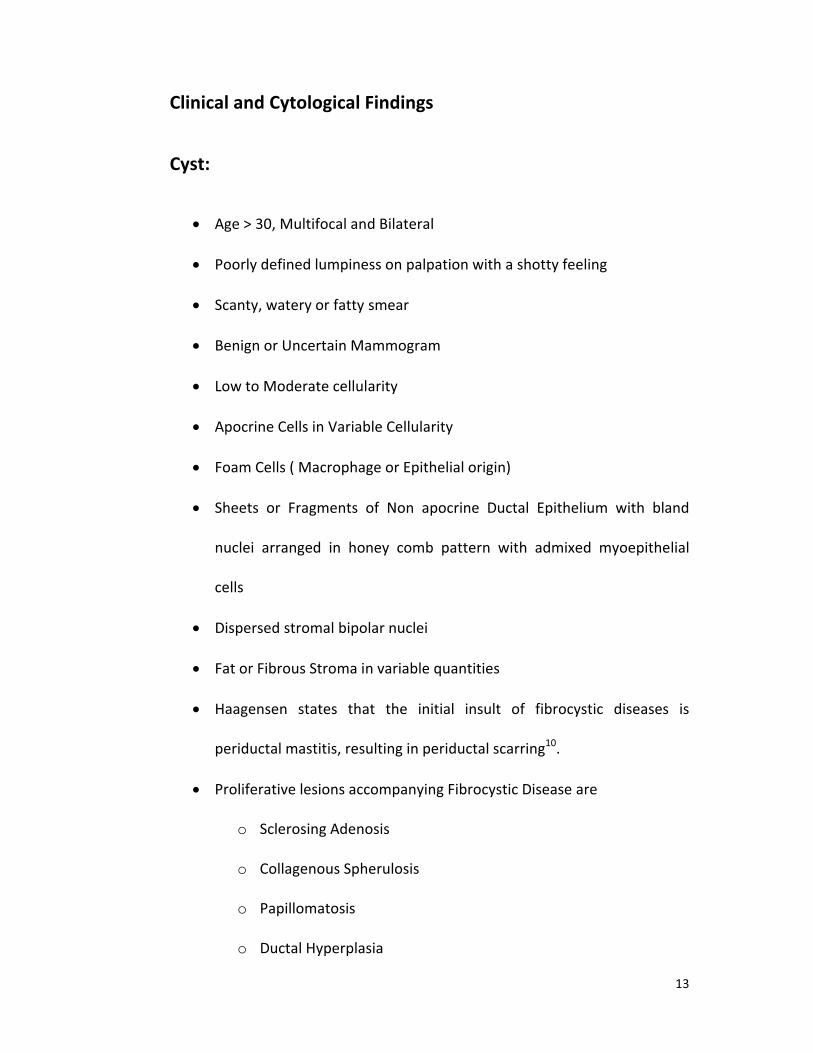
proposed a cytological scoring system in which a value of 1
to 4 was given for each of the following features:
Cellular Arrangement
Cellular Pleomorphism

28
Presence of Myoepithelial cells
Anisonucleosis
Nucleoli
Chromatin clumping
A score derived from the sum of these values was used to classify each FNA
sample, Non Proliferative Breast Disease was diagnosed with a score of 6 to 9,
Proliferative Breast Disease with a score of 10 to 14. The results showed 29 of 34
(85%) FNA specimens diagnosed as Nonproliferative Breast Disease and 15 of 17
(88%) FNA specimens diagnosed as Proliferative Breast Disease were found to
correlate with histological Diagnosis.
Maygarden et al26
evaluated the cytological features of 99 FNA specimens of
histologically proven proliferative and nonproliferative fibrocystic change. They
found no parameter that reached statistical significance in distiguishing between
these two entities.
According to McDivitt et al27
benign breast lesions can be categorised by a
modification of the Black-Chabon Grading system which differentiates hyperplasia
and atypia. When compared with women who had never had a breast biopsy,
women with benign breast disease without hyperplasia had an odds ratio of 1.5
(95% Confidence Limits 1.3 – 1.9), women with hyperplasia without atypia had an
odds ratio of 1.8 (CL =1.3, 2.4) and women with hyperplasia and atypia had an odds

29
ratio of 2.6 ( CL = 1.6, 4.1). Fibroadenoma was an independent risk factor (OR = 1.7;
CL= 1.1,2.5).
Other cytological Breast Cancer scoring systems used are the Robinson
system, the Moriquand system and Fisher’s system.
Robinson’s system :
Robinson’s system of scoring assesses 6 features, i.e, cell dissociation, nuclear
margin, cell size, cell uniformity, nucleoli and chromatin. A grade of 1 is given for a
score of 6 – 11, grade 2 for a score of 12 – 14, grade 3 for a score of 15 – 18.
Moriquand system :
Moriquand scoring assesses 4 features, i.e, cellular characters, nuclear
features, nucleoli and mitoses. A score of 0 – 3 is given for each feature and a grade 1
is given for a score 5 . Scores of 6 – 9 are Graded 2, and Scores of >10 are graded 3.
Fisher’s system :
Fisher’s Modification of Nuclear Grading depends on 4 features : nuclear size,
nuclear membrane, Chromatin and Positive or Negative nucleoli.
Giard and Hermans28
found a false positive rate of 0.15% with
cytomorphological analysis. Dupont and Page29
et al observed a strong association
between breast carcinoma risk and Atypical Hyperplasia.

30
Jersey et al state that women with proliferative breast Disease have an
increased relative risk of subsequent invasive Carcinoma. The risk is stratified
according to the degree of epithelial proliferation and is elevated in women with a
Family History of Breast Cancer.
Frost et al30
evaluated 12 cytological features in 51 benign breast aspirates
and found that only the presence of a swirling pattern reached statistical significance
in distinguishing Proliferative Breast Disease and Non-Proliferative Breast Disease.

Materials and
Methods

31
Materials and Methods
The records of the cytopathology and histopathology laboratory of Kilpauk
Medical College Chennai were analysed over a period of 2 years ( from June 2006 to
September 2008). One Hundred FNAC cases were collected and the smears were
stained with Hematoxylin & Eosin and their histological confirmation were included
in the study.
Inclusion Criteria :
All palpable breast lumps from Female patients with an adequate smear
showing 5 to 6 ductal epithelial groups were included in the study.
Exclusion Criteria:
Inflammatory smears and smears with no ductal epithelial cells were
excluded.
All aspirates were performed as an outpatient procedure without imaging or
mammographic guidance using a 23 G Needle and a 5 ml syringe. The smears were
fixed in Absolute alcohol for 20 minutes and then stained by Hematoxylin & Eosin
method. Aspirates were evaluated according to the Modified Masood Scoring
System.
Masood et al proposed a cytological scoring system in which a value of 1 to 4
was given for each of the following features :
32
1. Cellular Arrangement
2. Presence of Myoepithelial cells
3. Anisonucleosis
4. Cellular Pleomorphism
5. Nucleoli and
6. Chromatin Clumping
A score derived from the sum of these values was used to classify each FNA
sample. A score for each category was assigned as follows :
1. Non Proliferative Breast Disease – Score of 6 to 928
2. Proliferative Breast Disease without Atypia – Score of 10 to 14
3. Proliferative Breast Disease with Atypia – Score of 15 to 18
4. Carcinoma – Score of 19 to 24
Masood’s score was modified31
with a score of 6 to 10 denoting
Nonproliferative breast disease instead of the original score of 6 to 9.
Cytomorphological Diagnosis and Histopathological Diagnosis were also
categorised to four groups :
1. Non Proliferative Breast Disease
2. Proliferative Breast Disease without Atypia
3. Proliferative Breast Disease with Atypia
4. Carcinoma

33
Correlation was calculated using Spearman’s rho and Kendall’s tau coefficients.
SPSS Statistics version 17 was used for the statistical Analysis. The Tables of data
were constructed in Microsoft Office Excel 2007.
This scoring system yielded very promising results when applied prospectively by
the authors in a study evaluating 100 radiographically directed FNAs. Their results
show that 29 of 34 FNA specimens diagnosed as Non Proliferative Breast lesions
(85%) and 15 of 17 FNA specimens diagnosed as Proliferative Breast lesions were
found to correlate with the histological Diagnosis.
The scoring system requires good sampling techniques, good cellular yield and
non-fibrotic stroma for optimum results.

34

Results and
Observations

35
Results and Observations
The cases included in the study were all females with an Age Range of 15 – 65
years. The study included 100 palpable breast lesions. Results were tabulated and
analysed.
Masood’s score was modified32
with a score of 6 to 10 denoting
Nonproliferative breast disease instead of the original score of 6 to 9.
The scoring system and cytomorphology correlated with histological diagnosis
for 77 cases. In 12 cases, the cytomorphological diagnosis correlated well with
histopathological diagnosis. In 11 cases, the modified Masood scoring system
correlated well with the histological Diagnosis.
In 7 cases, the Cytomorphological Diagnosis and Histopathological Diagnosos
correlated well and categorised the patients under Proiferative Breast Disease, while
the scoring system categorised them under Non-Proliferative Breast disease.
In one case, the cytomorphological Diagnosis categorised the case under
Poliferative Breast Disease without Atypia, while the Scoring system brought it under
Proliferative breast Disease with Atypia and the Histopathological Diagnosis was
Carcinoma.

36
In 2 cases the cytomorphological Diagnosis categorised the case under
Proliferative Breast Disease without Atypia, whereas the scoring system
overdiagnosed it as Proliferative Breast Disease with Atypia.
In 3 cases, the Cytomorphological diagnosis overdiagnosed and categorised
the cases under proliferative Breast with Atypia, while the scoring system correlated
with the histopathological Diagnosis of Proliferative Breast Disease without Atypia.
In 2 cases, the Cytomorphological Diagnosis categorised the cases under
Proliferative Breast Disease without Atypia, while the Scoring system correlated with
the Histopathological Diagnosis of Non-Proliferative Breast Disease.
The study shows 2 cytomorphological positives correlated with
histopathological positivity under the category of nonproliferative breast disease,
when compared to 4 cases in the scoring system. 61 cases correlated with
histopathological diagnosis when compared to 56 cases in the scoring system under
the category of proliferative breast disease without atypia.
Out of the 5 cases of Proliferative Breast Disease with Atypia under Modified
Masood Scoring system, 3 cases were found to be carcinomas by histology and 2
cases were found to be fibroadenomas with atypia. The cytomorphological diagnosis
brought 2 cases under carcinoma, and three cases under proliferative breast disease
without atypia.

37
Under the category of proliferative breast disease with atypia, there were no
correlating cases. 25 cases correlated with the cytomorphological diagnosis when
compared to 24 cases in the scoring system under the category of Carcinoma.
Cytohistological correlation was 88% while correlation of the Modified
Masood score with Histology was 84%. The correlation was statistically significant
with a correlation coefficient of 0.832 for Cytology-Histology and 0.821 for Modified
Masood-Histology. Both the correlation coefficients were significant at the 0.01 level
(1-tailed).
Sensitivity of the scoring system was found to be 80%, Specificity 100% and
Positive Predictive Value was 100%, Negative Predictive Value was 92% for a
diagnosis of Carcinoma.
Sensitivity of FNAC was 83%, Specificity was 100%, Positive Predictive Value
was 100% and Negative Predictive Value was 93%.
The peak age of incidence of nonproliferative breast disease was found to be
the third decade. The peak age of incidence of proliferative breast disease was found
to be the third decade and carcinomas peaked at the 6th
decade.

38
Figure 1 : Age Distribution of Study Population
Figure 2 : Distribution of cases by FNAC Score
8
39
25
19
7
2
0
5
10
15
20
25
30
35
40
45
< 20 20 - 30 31 - 40 41 - 50 51 - 60 > 60
Age Distribution
4
66
5
25
0
10
20
30
40
50
60
70
Category 1 Category 2 Category 3 Category 4
FNA Score

39
Figure 3 : Distribution of cases by Masood Score
Figure 4 : Distribution of cases by Biopsy Scores
12
59
5
24
0
10
20
30
40
50
60
70
Category 1 Category 2 Category 3 Category 4
Modified Masood Score
4
66
0
30
0
10
20
30
40
50
60
70
Category 1 Category 2 Category 3 Category 4
Biopsy
Biopsy

40
Figure 5 : Concordance of FNAC and Masood Scores with Biopsy Scores
Figure 6 : Age distribution of cases according to Modified Masood Scoring
0
10
20
30
40
50
60
70
1=1 2=2 3=3 4=4
2
61
0
25
4
56
0
24
FNAC
Masood
0
7
10
5
25
2
0
4
16
1
8
1
10
0
5
21 1
11
0
5
10
15
20
25
30
1 2 3 4
Modified Masood Scoring
< 20
20 - 29
30 - 39
40 - 49
> 50

41
Biopsy
FNA1 2 3 4 Total
1 2 2 0 0 4
2 2 61 0 3 66
3 0 3 0 2 5
4 0 0 0 25 25
Total 4 66 0 30 100
Table 2 : Biopsy vs FNA Score
Biopsy
Masood1 2 2 4 Total
1 4 8 0 0 12
2 0 56 0 3 59
3 0 2 0 3 5
4 0 0 0 24 24
Total 4 66 0 30 100
Table 3 : Biopsy vs. Modified Masood Scoring

42
Figure 7 : Age Distribution of cases according to Biopsy Score
Figure 8: Age Distribution of cases according to FNAC Score
0
8
0 02
29
011
18
0
10
0
9
0
7
12
0
12
0
5
10
15
20
25
30
35
1 2 3 4
Biopsy Score
< 20
20 - 29
31 - 39
40 - 49
> 50
0
8
0 01
30
10
2
17
2
8
0
9
1
6
12
1
11
0
5
10
15
20
25
30
35
1 2 3 4
FNAC Score
<20
20 - 29
30 - 39
40 - 49
> 50

43
Correlations
FNA_Sc Masood B_Score
Kendall's tau_b FNA_Sc Correlation Coefficient 1.000 .776**
.832**
Sig. (1-tailed) . .000 .000
N 100 100 100
Masood Correlation Coefficient .776**
1.000 .821**
Sig. (1-tailed) .000 . .000
N 100 100 100
B_Score Correlation Coefficient .832**
.821**
1.000
Sig. (1-tailed) .000 .000 .
N 100 100 100
Spearman's rho FNA_Sc Correlation Coefficient 1.000 .829**
.861**
Sig. (1-tailed) . .000 .000
N 100 100 100
Masood Correlation Coefficient .829**
1.000 .856**
Sig. (1-tailed) .000 . .000
N 100 100 100
B_Score Correlation Coefficient .861**
.856**
1.000
Sig. (1-tailed) .000 .000 .
N 100 100 100
**. Correlation is significant at the 0.01 level (1-tailed).
Table 4 : Non parametric correlation coefficients

44
Figure 9 : Distribution of cases by Histopathology
4
66
30
Distribution of cases under various categories
by Histopathology
Non-Proliferative Proliferative without atypia Proliferative with atypia Carcinoma

Pic 2 : Biopsy of Phyllodes Tumour
Pic 1 : FNAC of Phyllodes tumour, H&E, 40 X with a score of 9

Pic 3 : FNAC Diagnosis of Phyllodes with a score of 5
Pic 4: Biopsy showing Fibroadenoma

Pic 5 : FNAC Diagnosis of Proliferative Breast Disease with a score of 12
Pic 6 : Biopsy Diagnosis – Infiltrating Ductal Carcinoma

Pic 7 : FNAC Diagnosis - Proliferative Breast Disease with a score of 9
Pic 8 : Biopsy - Tuberculous Mastitis
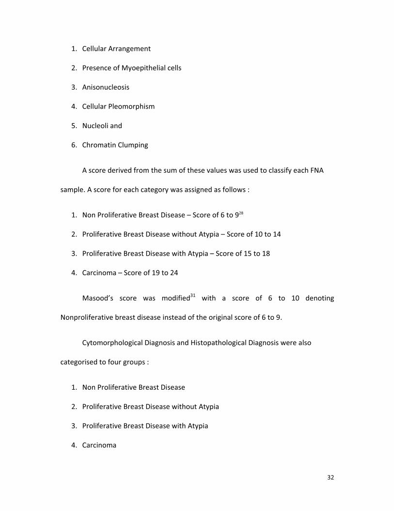
Pic 9 : FNAC - Sclerosing Adenosis with a score of 13
Pic 10 : Biopsy - Sclerosing Adenosis

Pic 11 : FNAC - Intraductal Carcinoma with a score of 20
Pic 12 : Biopsy - Intraductal Carcinoma

Pic 13 : FNAC Diagnosis of Fibroadenoma with a score of 16
Pic 14 : Biopsy showing Intraductal Carcinoma

Pic 15 : FNAC Diagnosis of Fibroadenoma with a score of 11
Pic 16 : Biopsy Diagnosis of Tubular Adenoma

Pic 17 : FNAC Diagnosis of Fibrocystic Disease with a score of 13
Inset : Cyst Macrophages
Pic 18 : Biopsy showing Infiltrating Papillary Adenocarcinoma

Discussion

45
Discussion
The study was initiated to evaluate the applicability of Modified Masood
Scoring in Cytological Diagnosis of Palpable Breast masses and to compare the
scoring system with Cytomorphological Diagnosis. The Histopathological Diagnosis
was considered to be the Gold Standard.
According to Barrows33
, the FNAC positivity for Breast Cancer varies between
48 to 88 %. To increase the diagnostic yield of the FNAC, Modified Masood scoring of
the aspirates were done. The study helps to categorise the lesion so that aspiration
of minimally suspicious lesions is helpful in initiating excisional biopsy.
Modified Masood’s scoring gives additional information by eliminating benign
cases and improves the diagnostic yield. Application of scoring in a step-wise manner
in atypical aspirates can help in selection of cases suitable for biopsy.
The risk of developing subsequent invasive breast cancer is stratified
according to the degree of epithelial proliferation and atypia. The risk is 1-fold in
women with Non-proliferative Breast disease, 1.9 fold in women with proliferative
breast disease and 5.3 in women with carcinoma insitu.
Histological criteria that allow the distinction of these various breast lesions
are established. Sneige and Staerkel7
introduced the concept of using architectural
features cytologically and concluded that the application of both cytological and

46
architectural criteria is more reliable than cytology alone in separating proliferative
breast lesions.
Dawson et al34
showed that applying both architectural and cytological criteria
enhanced diagnostic accuracy. Cytologically, the architectural features of
proliferative breast lesions may be apparent in the larger breast fragments and
recapitulate the histologic appearance of these lesions. Slit like lumens, swirling and
streaming are noted in proliferative breast lesions without atypia. Round spaces may
be seen in proliferative breast lesions without atypia and with atypia.
Rigid sublumina and a micropapillary architecture are features of DCIS.
Thomas et al35
demonstrated that experience and fine tuning of cytological criteria
increased the concordance with the histological findings.
All these studies emphasise the importance of adequate sampling to
minimise, in particular, underdiagnosis.
Criteria for the cytological diagnosis of Fibroadenoma and Carcinoma are well
established. The sensitivity and specificity of Fine Needle Aspiration Cytology for the
diagnosis of Fibroadenoma is 86.9 % and 93.8% respectively, while for Carcinomas,
Sensitivity is 89 – 98 % and Specificity 93 – 98 %.
Proliferative Breast lesions on cytology are categorised into Proliferative
Breast Disease without atypia, and Proliferative breast disease with Atypia because it
not possible to delineate all the histological entities on FNAC. However, the

47
diagnostic accuracy in this distinction is still unclear, and the cytological features of
proliferative breast disease are not well established.
In this study, I have confined myself to palpable breast lesions referred for
routine FNAC. A high degree of concordance was found between FNA Cytology and
Histological Diagnosis in cases of proliferative breast disease without atypia and
carcinoma. In these groups, the scoring system did not contribute any addition
information.
The peak age of incidence of nonproliferative breast disease was found to be
the third decade. The peak age of incidence of proliferative breast disease was found
to be the third decade and carcinomas peaked at the 6th
decade.
Use of the scoring system can reduce the number of atypical reports and
hence limit unnecessary procedures performed on patients.
The study has found that in cases showing benign ductal cells and suspected
to be proliferative breast disease without atypia, the scoring system did not add
substantially in the evaluation of FNAC. This category is likely to form the majority of
cases on FNAC and in this group, the laborious application of the scoring system can
be avoided.
The study found that the diagnosis of atypia or routine cytomorphological
assessment was influenced more by nuclear pleomorphism than by architectural

48
details. Application of the scoring system evaluated both the nuclear atypia and the
cytoarchitectural features.
So ,the scoring system should be applied in a stepwise manner after
cytomorphological assessment. In cases with cytological diagnosis of proliferative
breast disease without atypia, and carcinoma, the scoring system offers no
advantage over cytomorphology.
The scoring system is useful in aspirates with cytological diagnosis of
proliferative breast disease with atypia.

Summary and
Conclusions

49
Summary and Conclusions
100 cases during a period of two years from September 2006 to September
2008, of palpable breast lesions were studied. The cytomorphological analysis by
FNAC and Modified Masood system were taken and correlated with
histopathological diagnosis. Out of the 100 cases, 4 cases were diagnosed as Non
proliferative breast Disease and 66 cases were diagnosed as Proliferative breast
disease without atypia. There were no cases under proliferative breast disease with
atypia. 30 cases were diagnosed as carcinoma by histopathology.
With Modified Masood’s scoring, 12 cases were categorized as
Nonproliferative breast disease, 59 cases were categorized as proliferative breast
disease without atypia, 5 cases were categorized as proliferative breast disease with
atypia, and 24 cases were categorized as Carcinoma.
With FNAC, 4 cases were categorized as nonproliferative breast disease, 25
cases diagnosed as carcinoma, 66 cases categorized as proliferative breast disease
without atypia and 5 cases were categorized as proliferative breast disease with
atypia.
Sensitivity of the scoring system was found to be 80%, Specificity 100% and
Positive Predictive Value was 100%, Negative Predictive Value was 92% for a
diagnosis of Carcinoma.

50
Sensitivity of FNAC was 83%, Specificity was 100%, Positive Predictive Value
was 100% and Negative Predictive Value was 93%.
Both FNAC and Modified Masood’s scoring were found to correlate highly
with each other and with Histopathology.
In conclusion, Modified Masood’s scoring system can be done to categorize
the breast lesions into the four categories which correlate highly with FNAC and
histopathology.

Bibliography

51
Bibliography
1Fitzgibbons PL, Henson DE, Hutter RVP. Benign breast changes and the risk for subsequent breast cancer. An
Update of the 1985 Consensus Statement. Cancer Committee of the College of American Pathologists. Arch
Pathol Lab Med 1998; 122:1053-55
2Silverberg SG, deLellis RA, Frable WJ, et al in Silverberg's Principles and Practice of Surgical Pathology and
Cytopathology - 4th edition, Churchill Livingstone – Elsevier
3Koss LG, Melamed MR. Koss’ Diagnostic Cytology and its Histopathological Bases – 5
thedition – Volume One.
Lippincott Williams & Wilkins
4Carter CL, Corle DK, Micozzi MS, Schatzkin A, Taylor PR. Am J Epidemiol 1988 Sep;128(3):467-77
5Wingo PA, Ory HW, Layde PM, Lee NC. The evaluation of data collection process for a multi-center population
based case-control design. Am J Epidemiol 1988; 128:206-217
6Dupont WD, Page DL. Risk factors for breast cancer in women with proliferative breast diseases. N Engl J Med
1985; 312(3): 146-151
7London SJ, Connoly JL, Schnitt SJ, Colditz GA. A prospective study of benign breast disease and the risk of
breast cancer. JAMA 1992; 267(7): 941-944.
8Dupont WD, Parl FF, Hartmann WH, et al. Breast cancer risk associated with proliferative breast disease and
atypical hyperplasia. Cancer 1993; 71(4): 1258-1265.
9Palli D, Roselli del Turco M, Simoncini R, Bianchi S. Benign breast disease and breast cancer: a case-control
study in a cohort in Italy. Int J Cancer 1991; 479: 703 – 706.
10Haagensen CD : Diseases of the Breast. Philadelphia, WB Saunders, 1986.
11Sneige N, Staerkel GA: Fine-needle aspiration cytology of ductal hyperplasia with and without atypia and
ductal carcinoma in situ. Hum Pathol 25:485-92, 1994.
12Bottles K, Chan JS, Holly EA, et al: Cytologic criteria for fibroadenoma: A step-wise logistic regression
analysis. Am J Clin Pathol 89:707-713, 1988.
13Dejmek A, Lindholm K : Frequency of cytologic features in fine needle aspirates from histologically and
cytologically diagnosed fibroadenomas. Acta Cytol 35:695-699, 1991.
14Stanley MW, Tani EM, Skoog L : Fine-needle aspiration of fibroadenomas of the breast with atypia: A
spectrum including cases that cytologically mimic carcinoma. Diagn Cytopathol 6:375-382, 1990.
15Orell SR. Radial Scar / Complex sclerosing lesion – a problem in the diagnostic work-up of screen detected
breast lesions. Cytopathology 1999; 10:250-8
16Mulvany N, Lowhagen T, Skoog L: Fine needle aspiration cytology of tubular adenoma of the breast. A report
of two cases. Acta Cytol 38:961-964,1994.
17Stormby N, Bondeson L : Adenoma of the nipple . Acta Cytol 28:729-732, 1984.

52
18Grenko RT, Lee KP, Lee KR: Fine needle aspiration cytology of lactating adenoma of the breast: A
comparative light microscopic and morphometric study: Acta Cytol 34:21-26, 1990.
19Finley JL, Silverman JF, Lannin DR: Fine needle aspiration cytology of breast masses in pregnant and lactating
women. Diagn Cytopathol 5:255-259, 1989
20Page DL, Anderson TJ: Diagnostic Histopathology of the Breast. New York, Churchill Livingstone, 1987.
21Leach C, Howell LP: Cytodiagnosis of classic lobular carcinoma and its variants. Acta Cytol 36:199-202, 1992.
22Shinagawa T, Tadokoro M, Kitamura H, et al: Secretory Carcinomas of the breast. Correlation of Aspiration
Cytology and histology. Acta Cytol 38:909-914, 1994.
23Sidawy et al. The Spectrum of cytologic Features in Non proliferative breast lesions. Cancer Cytopathology,
2001 93(2) : 140-45
24Masood S, Frykberg ER, McLellan GL, Scalapino MC, Mitchum DG, Bullard JB. Prospective evaluation of
radiologically directed fine needle aspiration biopsy of nonpalpable breast lesions. Cancer 1990;66:1480-7
25Masood S, Frykberg ER, McLellan GL, Dee S, Mitchum DG, Bullard JB. Cytologic differentiation between
proliferative and nonproliferative breast disease in mammographically guided fine needle aspirates. Diagn
Cytopathol 1991;7:581-90
26Maygarden S, McCall JB, Frable WJ. Fine needle aspiration of breast lesions in women aged 30 and under.
Acta Cytol 1991; 35:678-94.
27McDivitt et al. Histologic types of Benign Breast Disease and the risk for Breast Cancer. Cancer, 1992; 69(6) :
1408-14
28Giard R, Hermans J. The value of aspiration cytologic examination of the breast. A statistical review of the
medical literature. Cancer 1992;69:2104-10
29Dupont WD, Page DL. Risk factors for breast cancer in women with proliferative breast disease. N Engl J Med.
1985;312:146-151
30Frost AR, Aksu A, Kurstin T, Sidawy MK. Can nonproliferative breast disease and proliferative breast disease
without atypia be distinguished by the Fine-needle aspiration cytology? Cancer(Cancer Cytopathol) 1997;
81:22-8.
31Asit Ranjan Mridha, Venkateswaran K.Iyer, Kusum Kapila, Kusum Verma. Value of scoring system in
classification of proliferative breast disease on fine needle aspiration cytology. Indian J Pathol Microbiol 2006,
Vol 49. No.3
32Asit Ranjan Mridha, Venkateswaran K.Iyer, Kusum Kapila, Kusum Verma. Value of scoring system in
classification of proliferative breast disease on fine needle aspiration cytology. Indian J Pathol Microbiol 2006,
Vol 49. No.3
33Fine Needle Aspiration of Breast Cancer – Relationship of Clinical Factors to Cytology results in 689 Primary
Malignancies. Barrows, et al. Cancer 58:1493-1498, 1986
34Dawson AE, Mulford DK, Sheils LA. The cytopathology of proliferative breast disease; comparision with
features of ductal carcinoma in situ. Am J Clin Pathol 1995;103:438-442.
35Thomas PA, Cangiarella J, Raab SS, Waisman J. Fine needle aspiration biopsy of proliferative breast tissue.
Mod Pathol 1995; 8:130-136

Annexures

PA
LPA
BLE
BR
EA
ST
LE
SIO
NS
– C
YT
OM
OR
PH
OLO
GIC
AL
AN
ALY
SIS
AN
D S
CO
RIN
G S
YS
TE
M W
ITH
HIS
TO
PA
TH
OLG
ICA
L C
OR
RE
LAT
ION
FNA_No
FNAC_Diag
FNA_Sc
Masood
B_Score
B_No
Age
Side
Score
Diagnosis
1F
22
62
/06
Fib
roa
de
no
ma
22
24
09
1 /
06
21
11
2P
roli
fera
tiv
e B
rea
st w
ith
Aty
pia
2F
21
99
/06
Fib
roa
de
no
ma
21
24
03
2/0
63
01
8F
A
3F
12
38
/06
Ca
rcin
om
a4
44
22
93
/06
55
22
1C
arc
ino
ma
4F
21
67
/06
Juv
en
ile
FA
21
23
93
2/0
63
02
9F
A
5F
21
62
/06
FA
22
2B
38
94
/06
21
21
2F
A w
ith
cy
stic
ch
an
ge
wit
h e
pit
he
lia
l h
yp
erp
lasi
a
6F
22
11
/06
Ca
rcin
om
a4
34
B3
98
3/0
63
82
18
IDG
Me
tap
last
ic C
a,
LN +
7F
21
82
/06
FA
/ M
yo
ep
ith
eli
om
a2
22
B3
95
4/0
61
71
13
Juv
en
ile
FA
8F
25
20
/06
FA
22
2B
45
13
/06
23
21
4F
A
9F
23
48
/06
FA
22
2B
43
49
/06
32
11
1F
A
10
F2
41
2/0
6C
arc
ino
ma
43
4B
44
38
/06
65
11
8C
arc
ino
ma
11
F2
31
7/0
6F
A w
ith
cy
stic
ch
an
ge
22
2B
43
43
/06
22
11
1F
A
12
F2
31
8/0
6F
A2
22
B4
26
0/0
63
41
13
FA
wit
h e
pit
he
lio
sis
an
d m
yxo
id d
eg
en
era
tio
n
13
F1
36
9/0
6F
A2
22
B2
58
7/0
61
51
10
FA
14
F1
36
5/0
6A
dn
exa
l T
um
ou
r2
22
B2
52
4/0
65
01
13
Tu
bu
lar
Ad
en
om
a
15
F6
27
/06
Ca
rcin
om
a4
44
B2
38
4/0
64
22
20
Ca
rcin
om
a
16
F1
37
6/0
6F
A w
ith
cy
stic
ch
an
ge
22
2B
24
27
/06
47
10
Ce
llu
lar
FA
wit
h c
yst
ic c
ha
ng
e
17
F1
36
8/0
6F
A2
22
B2
47
5/0
63
32
10
FA
18
F1
32
6/0
6?
Ca
rcin
om
a4
44
B2
38
7/0
63
51
20
IDC
wit
h R
ad
ial
Sca
r
19
F1
25
6/0
6F
A2
32
B2
29
5/0
62
03
17
FA
wit
h e
pit
he
lia
l h
yp
erp
lasi
a
20
F1
27
8/0
6F
A2
22
B2
21
7/0
63
01
14
Fib
roa
de
no
sis
21
F1
23
8/0
6D
uct
al
Ca
rcin
om
a4
44
B2
29
3/0
65
82
21
Du
cta
l C
a
22
F1
98
7/0
6F
A2
22
B3
66
9/0
62
52
10
FA
wit
h A
po
crin
e M
eta
pla
sia
23
F2
05
2/0
6F
A2
22
B3
65
1/0
63
52
10
FA
wit
h A
po
crin
e C
ha
ng
e
24
F2
01
4/0
6F
A2
22
B3
65
1/0
61
51
10
FA
25
F1
84
0/0
6F
ibro
cyst
ic D
ise
ase
22
4B
35
98
/06
38
21
3In
filt
rati
ve
Pa
pil
lary
ad
en
oca
rcin
om
a
26
F1
91
5/0
6F
A2
22
B3
46
2/0
62
81
10
FA
wit
h c
yst
ic c
ha
ng
e
27
F1
94
8/0
6C
arc
ino
ma
44
4B
35
42
/06
40
21
9ID
C
28
F1
97
8/0
6F
A2
22
B3
53
2/0
63
02
11
Ph
yll
oid
es
(FA
)
29
F1
71
3/0
6P
hy
llo
ide
s2
12
B3
22
8/0
65
09
Ph
yll
oid
es
Inte
rme
dia
te
30
F1
74
9/0
6C
arc
ino
ma
44
4B
31
93
/06
50
12
0ID
C
31
F1
72
6/0
6C
arc
ino
ma
44
4B
31
75
/06
32
21
9C
arc
ino
ma
32
F1
61
9/0
6F
A2
22
B3
13
1/0
64
32
13
FA
wit
h c
yst
ic c
ha
ng
es
wit
h A
po
crin
e
33
F2
09
4/0
6?
FA
/ S
cle
rosi
ng
Ad
en
osi
s2
22
B3
76
6/0
62
52
13
Scl
ero
sin
g A
de
no
sis
34
F1
74
7/0
6F
A /
Ph
yll
oid
es
22
2B
30
67
/06
30
11
0F
A w
ith
cy
stic
ch
an
ge
35
F1
63
4/0
6F
A2
22
B2
92
6/0
62
23
10
FA
wit
h c
yst
ic c
ha
ng
e
36
F1
58
5/0
6F
A w
ith
cy
stic
ch
an
ge
22
2B
28
18
/06
22
31
3F
A w
ith
cy
stic
ch
an
ge
37
F1
48
4/0
6F
ibro
ad
en
osi
s2
22
B2
79
2/0
64
72
11
FA
wit
h c
yst
ic c
ha
ng
e
38
F2
10
5/0
6F
A w
ith
cy
stic
ch
an
ge
21
2B
37
75
/06
40
18
Fib
rocy
stic
Dis
ea
se w
ith
Ep
ith
eli
al
Hy
pe
rpla
sia
39
F1
57
6/0
6F
A2
22
B2
76
6/0
62
32
12
FA
1

PA
LPA
BLE
BR
EA
ST
LE
SIO
NS
– C
YT
OM
OR
PH
OLO
GIC
AL
AN
ALY
SIS
AN
D S
CO
RIN
G S
YS
TE
M W
ITH
HIS
TO
PA
TH
OLG
ICA
L C
OR
RE
LAT
ION
40
F1
55
6/0
6P
hy
llo
ide
s2
22
B2
74
5/0
63
52
10
Be
nig
n P
hy
llo
ide
s
41
F1
36
3/0
6B
x su
gg
est
ed
12
2B
26
72
/06
35
21
0F
A
42
F1
00
8/0
7P
hy
llo
ide
s -
bio
psy
su
gg
est
ed
21
2F
17
53
/06
28
15
FA
wit
h E
pit
he
lio
sis
43
F9
22
/07
FA
22
2B
17
52
/07
41
11
1T
ub
ula
r A
de
no
ma
44
F2
09
4/0
7P
roli
fera
tiv
e B
rea
st w
ith
Aty
pia
32
2B
42
38
/07
38
11
3F
A w
ith
cy
stic
Ch
an
ge
45
F2
09
6/0
7F
A2
34
B4
18
2/0
72
71
16
IDC
46
F1
93
7/0
7ID
C4
44
B4
07
8/0
75
02
0ID
C
47
F2
03
4/0
7F
A2
22
B4
07
2/0
72
82
11
FA
wit
h f
ibro
cyst
ic c
ha
ng
e
48
F1
41
9/0
7C
ell
ula
r F
A2
32
B2
81
7/0
71
82
15
FA
wit
h E
pit
he
lio
sis
49
F1
31
0/0
7F
A2
22
B2
64
9/0
72
42
14
FA
wit
h A
de
no
sis
50
F1
31
1/0
7B
en
ign
Pro
life
rati
ve
Bre
ast
22
2B
26
48
/07
20
11
2F
A w
ith
Fib
roa
de
no
sis
51
F1
28
3/0
7F
A2
22
B2
59
4/0
72
21
11
FA
wit
h P
hy
llo
ida
l C
ha
ng
e w
ith
Ap
ocr
ine
wit
h E
pit
he
lio
sis
52
F1
26
8/0
7B
en
ign
Pro
life
rati
ve
Bre
ast
22
2B
24
70
/07
37
21
1F
A w
ith
Ad
en
osi
s
53
F1
18
2/0
8P
roli
fera
tiv
e B
rea
st D
ise
ase
22
4B
23
31
/08
45
12
Infi
ltra
tin
g D
uct
al
Ca
rcin
om
a (
CIN
)
54
F1
39
7/0
8P
roli
fera
tiv
e B
rea
st D
ise
ase
22
2B
27
01
/08
35
11
1F
A
55
F1
22
1/0
8S
me
ar
Po
siti
ve
44
4B
25
02
/08
50
22
IDC
56
F1
46
5/0
8F
A2
22
B2
79
1/0
82
52
10
FA
( ?
Ra
dia
l S
car)
57
F1
01
8/0
8S
me
ar
Po
siti
ve
44
4B
22
06
/08
50
21
9ID
C
58
F8
37
/08
FA
wit
h F
ibro
cyst
ic C
ha
ng
e2
22
B1
61
2/0
82
82
13
FA
wit
h c
yst
ic c
ha
ng
e
59
F1
85
8/0
7F
A2
22
B3
73
5/0
72
71
12
FA
60
F1
86
6/0
7S
me
ar
Po
siti
ve
44
4B
37
49
/07
33
22
0ID
C
61
F4
43
/08
FA
22
2B
93
6/0
82
21
13
FA
62
F5
05
/08
FA
22
2B
10
66
/08
24
11
1F
A
63
F4
44
/08
FA
22
2B
10
67
/08
40
12
FA
wit
h c
yst
ic c
ha
ng
e
64
54
0/0
8P
roli
fera
tiv
e B
rea
st w
ith
Aty
pia
32
2B
10
68
/08
24
13
FA
wit
h e
pit
he
lio
sis
wit
h c
yst
ic c
ha
ng
e
65
F5
53
/08
FA
22
2B
71
60
/08
25
21
2F
A
66
F5
72
/08
FA
22
2B
11
64
/08
18
21
2F
A
67
F5
90
/08
FA
21
2B
12
07
/08
28
29
FA
wit
h c
yst
ic c
ha
ng
e a
nd
ca
lcif
ica
tio
n
68
F5
42
/08
FA
22
2B
12
51
/08
24
10
FA
wit
h e
pit
he
lio
sis
69
F6
33
/08
Pro
life
rati
ve
Bre
ast
wit
h A
typ
ia3
44
B1
60
1/0
85
52
0ID
C
70
F7
16
/08
FA
21
2B
13
96
/08
20
26
FA
(P
hy
llo
ide
s)
71
F6
29
/08
Pro
life
rati
ve
Bre
ast
Dis
ea
se w
ith
At
32
2B
14
09
/08
42
11
FA
wit
h s
tro
ma
l sc
lero
sis
72
F7
41
/08
FA
22
2B
15
68
/08
17
13
FA
wit
h e
pit
he
lio
sis
wit
h c
yst
ic c
ha
ng
e
73
F8
28
/08
Sm
ea
r P
osi
tiv
e4
44
B1
68
9/0
83
72
1ID
C
74
F1
06
1/0
8S
me
ar
Po
siti
ve
44
4B
18
72
/08
69
20
Ca
rcin
om
a
75
F1
14
4/0
8S
me
ar
Po
siti
ve
44
4B
30
32
/08
45
21
9ID
C
76
F1
29
4/0
8C
oll
oid
Ca
rcin
om
a4
44
B2
57
9/0
86
02
20
Ca
rcin
om
a
77
F1
91
8/0
7S
me
ar
Po
siti
ve
44
4B
38
87
/07
43
20
Ca
rcin
om
a
78
F1
54
2/0
8F
A2
22
B2
99
6/0
82
61
2F
A
79
F1
58
0/0
7S
me
ar
Po
siti
ve
44
4B
31
33
/07
35
12
0C
arc
ino
ma
2

PA
LPA
BLE
BR
EA
ST
LE
SIO
NS
– C
YT
OM
OR
PH
OLO
GIC
AL
AN
ALY
SIS
AN
D S
CO
RIN
G S
YS
TE
M W
ITH
HIS
TO
PA
TH
OLG
ICA
L C
OR
RE
LAT
ION
80
F1
59
3/0
8S
me
ar
Po
siti
ve
44
4B
31
00
/08
46
11
9C
arc
ino
ma
81
F1
54
2/0
8F
A2
22
B2
99
6/0
82
41
2F
A w
ith
cy
stic
ch
an
ge
82
F1
58
5/0
8F
A2
22
B2
99
5/0
81
91
2F
A
83
F1
86
4/0
7F
A2
22
B3
66
7/0
71
81
12
FA
wit
h e
pit
he
lio
sis
84
F1
26
4/0
7B
en
ign
Pro
life
rati
ve
22
2B
24
69
/07
32
10
FA
85
F1
75
/07
Sm
ea
r P
osi
tiv
e4
24
B9
69
/07
48
10
IDC
86
F1
66
1/0
8P
roli
fera
tiv
e B
rea
st2
22
B3
22
0/0
84
51
0F
A
87
F1
58
2/0
8S
me
ar
Po
siti
ve
44
4B
30
85
/08
31
19
IDC
88
F1
66
/07
Sm
ea
r P
osi
tiv
e4
44
B4
89
/07
30
19
Ca
rcin
om
a
89
F1
77
0/0
6F
A2
22
B3
06
6/0
63
51
0F
A w
ith
Ep
ith
eli
osi
s
90
F9
8/0
7S
me
ar
Po
siti
ve
44
4B
23
8/0
75
62
2C
arc
ino
ma
91
F1
88
9/0
8F
A2
11
B3
62
5/0
82
89
Ad
en
osi
s w
ith
La
cta
tio
na
l C
ha
ng
es
92
F1
81
3/0
8C
yst
ic L
esi
on
11
2B
36
57
/08
33
9F
A w
ith
Cy
stic
Ch
an
ge
93
F1
85
4/0
8S
me
ar
Po
siti
ve
44
4B
35
94
/08
53
20
IDC
94
F1
82
8/0
8F
ibro
cyst
ic D
ise
ase
11
1B
35
52
/08
25
9F
ibro
cyst
ic D
ise
ase
95
F1
82
2/0
8P
roli
fera
tiv
e B
rea
st D
ise
ase
21
1B
34
63
/08
39
9T
B M
ast
itis
96
F1
88
0/0
8C
yst
ic C
ha
ng
e1
11
B3
72
7/0
85
39
Fib
rocy
stic
Ch
an
ge
wit
h P
eri
du
cta
l M
ast
itis
97
F1
91
8/0
8P
roli
fera
tiv
e B
rea
st w
ith
Aty
pia
34
4B
37
61
/08
35
19
IDC
98
F1
92
3/0
8F
A2
22
B3
75
4/0
84
11
0F
A
99
F1
70
0/0
8F
A2
22
B3
24
9/0
83
01
1F
A
10
0F
19
17
/08
FA
22
2B
38
14
/08
25
12
Ad
en
om
a
3