Cellulase Production by Pink Pigmented Facultative Methylotrophic Strains (PPFMs
Click here to load reader
Upload
independentCategory
view
2download
0
M
C
KC
a
ARRA
KCLPSMD
C
0h
Artificial Intelligence in Medicine 56 (2012) 69–90
Contents lists available at SciVerse ScienceDirect
Artificial Intelligence in Medicine
j o ur nal home page: www.elsev ier .com/ locate /a i im
ethodological review
omputerized analysis of pigmented skin lesions: A review
onstantin Korotkov ∗, Rafael Garciaomputer Vision and Robotics Research Group, University of Girona, Campus Montilivi, Edifici P-4, 17071 Girona, Spain
r t i c l e i n f o
rticle history:eceived 23 March 2012eceived in revised form 2 August 2012ccepted 19 August 2012
eywords:omputer-aided diagnosisiterature reviewigmented skin lesionskin cancer detectionelanomaermoscopy
a b s t r a c t
Objective: Computerized analysis of pigmented skin lesions (PSLs) is an active area of research that datesback over 25 years. One of its main goals is to develop reliable automatic instruments for recognizing skincancer from images acquired in vivo. This paper presents a review of this research applied to microscopic(dermoscopic) and macroscopic (clinical) images of PSLs. The review aims to: (1) provide an extensiveintroduction to and clarify ambiguities in the terminology used in the literature and (2) categorize andgroup together relevant references so as to simplify literature searches on a specific sub-topic.Methods and material: The existing literature was classified according to the nature of publication (clinicalor computer vision articles) and differentiating between individual and multiple PSL image analysis. Wealso emphasize the importance of the difference in content between dermoscopic and clinical images.Results: Various approaches for implementing PSL computer-aided diagnosis systems and their standardworkflow components are reviewed and summary tables provided. An extended categorization of PSL fea-ture descriptors is also proposed, associating them with the specific methods for diagnosing melanoma,separating images of the two modalities and discriminating references according to our classification of
the literature.Conclusions: There is a large discrepancy in the number of articles published on individual and multiplePSL image analysis and a scarcity of reported material on the automation of lesion change detection. Atpresent, computer-aided diagnosis systems based on individual PSL image analysis cannot yet be usedto provide the best diagnostic results. Furthermore, the absence of benchmark datasets for standardizedalgorithm evaluation is a barrier to a more dynamic development of this research area.© 2012 Elsevier B.V. All rights reserved.
ontents
1. Introduction . . . . . . . . . . . . . . . . . . . . . . . . . . . . . . . . . . . . . . . . . . . . . . . . . . . . . . . . . . . . . . . . . . . . . . . . . . . . . . . . . . . . . . . . . . . . . . . . . . . . . . . . . . . . . . . . . . . . . . . . . . . . . . . . . . . . . . . . . . 702. Background . . . . . . . . . . . . . . . . . . . . . . . . . . . . . . . . . . . . . . . . . . . . . . . . . . . . . . . . . . . . . . . . . . . . . . . . . . . . . . . . . . . . . . . . . . . . . . . . . . . . . . . . . . . . . . . . . . . . . . . . . . . . . . . . . . . . . . . . . . . 70
2.1. The human skin . . . . . . . . . . . . . . . . . . . . . . . . . . . . . . . . . . . . . . . . . . . . . . . . . . . . . . . . . . . . . . . . . . . . . . . . . . . . . . . . . . . . . . . . . . . . . . . . . . . . . . . . . . . . . . . . . . . . . . . . . . . . . . . 702.2. Malignant melanoma . . . . . . . . . . . . . . . . . . . . . . . . . . . . . . . . . . . . . . . . . . . . . . . . . . . . . . . . . . . . . . . . . . . . . . . . . . . . . . . . . . . . . . . . . . . . . . . . . . . . . . . . . . . . . . . . . . . . . . . . . 712.3. Pigmented skin lesions. . . . . . . . . . . . . . . . . . . . . . . . . . . . . . . . . . . . . . . . . . . . . . . . . . . . . . . . . . . . . . . . . . . . . . . . . . . . . . . . . . . . . . . . . . . . . . . . . . . . . . . . . . . . . . . . . . . . . . . . 712.4. Melanoma screening and imaging techniques . . . . . . . . . . . . . . . . . . . . . . . . . . . . . . . . . . . . . . . . . . . . . . . . . . . . . . . . . . . . . . . . . . . . . . . . . . . . . . . . . . . . . . . . . . . . . . . 72
2.4.1. Clinical images . . . . . . . . . . . . . . . . . . . . . . . . . . . . . . . . . . . . . . . . . . . . . . . . . . . . . . . . . . . . . . . . . . . . . . . . . . . . . . . . . . . . . . . . . . . . . . . . . . . . . . . . . . . . . . . . . . . . . . 722.4.2. Dermoscopy . . . . . . . . . . . . . . . . . . . . . . . . . . . . . . . . . . . . . . . . . . . . . . . . . . . . . . . . . . . . . . . . . . . . . . . . . . . . . . . . . . . . . . . . . . . . . . . . . . . . . . . . . . . . . . . . . . . . . . . . . 722.4.3. Baseline images . . . . . . . . . . . . . . . . . . . . . . . . . . . . . . . . . . . . . . . . . . . . . . . . . . . . . . . . . . . . . . . . . . . . . . . . . . . . . . . . . . . . . . . . . . . . . . . . . . . . . . . . . . . . . . . . . . . . . 73
2.5. Melanoma diagnosis methods . . . . . . . . . . . . . . . . . . . . . . . . . . . . . . . . . . . . . . . . . . . . . . . . . . . . . . . . . . . . . . . . . . . . . . . . . . . . . . . . . . . . . . . . . . . . . . . . . . . . . . . . . . . . . . . . 732.6. Automated diagnosis of melanoma . . . . . . . . . . . . . . . . . . . . . . . . . . . . . . . . . . . . . . . . . . . . . . . . . . . . . . . . . . . . . . . . . . . . . . . . . . . . . . . . . . . . . . . . . . . . . . . . . . . . . . . . . . . 73
3. Literature classification . . . . . . . . . . . . . . . . . . . . . . . . . . . . . . . . . . . . . . . . . . . . . . . . . . . . . . . . . . . . . . . . . . . . . . . . . . . . . . . . . . . . . . . . . . . . . . . . . . . . . . . . . . . . . . . . . . . . . . . . . . . . . . . 744. Single lesion analysis . . . . . . . . . . . . . . . . . . . . . . . . . . . . . . . . . . . . . . . . . . . . . . . . . . . . . . . . . . . . . . . . . . . . . . . . . . . . . . . . . . . . . . . . . . . . . . . . . . . . . . . . . . . . . . . . . . . . . . . . . . . . . . . . . 75
4.1. Image preprocessing . . . . . . . . . . . . . . . . . . . . . . . . . . . . . . . . . . . . . . . . . . . . . . . . . . . . . . . . . . . . . . . . . . . . . . . . . . . . . . . . . . . . . . . . . . . . . . . . . . . . . . . . . . . . . . . . . . . . . . . . . . 754.2. Lesion border detection . . . . . . . . . . . . . . . . . . . . . . . . . . . . . . . . . . . . . . . . . . . . . . . . . . . . . . . . . . . . . . . . . . . . . . . . . . . . . . . . . . . . . . . . . . . . . . . . . . . . . . . . . . . . . . . . . . . . . . . 76
4.2.1. PSL border detection methodology . . . . . . . . . . . . . . . . . . . . . . .4.2.2. Comparison of segmentation algorithms . . . . . . . . . . . . . . . . .
4.3. Feature extraction. . . . . . . . . . . . . . . . . . . . . . . . . . . . . . . . . . . . . . . . . . . . . . . . . . .
∗ Corresponding author. Tel.: +34 972 41 98 12.E-mail address: [email protected] (K. Korotkov).
933-3657/$ – see front matter © 2012 Elsevier B.V. All rights reserved.ttp://dx.doi.org/10.1016/j.artmed.2012.08.002
. . . . . . . . . . . . . . . . . . . . . . . . . . . . . . . . . . . . . . . . . . . . . . . . . . . . . . . . . . . . . . . . . . . . . . . . . . 76 . . . . . . . . . . . . . . . . . . . . . . . . . . . . . . . . . . . . . . . . . . . . . . . . . . . . . . . . . . . . . . . . . . . . . . . . . . 76
. . . . . . . . . . . . . . . . . . . . . . . . . . . . . . . . . . . . . . . . . . . . . . . . . . . . . . . . . . . . . . . . . . . . . . . . . . 79

70 K. Korotkov, R. Garcia / Artificial Intelligence in Medicine 56 (2012) 69–90
4.4. Registration and change detection . . . . . . . . . . . . . . . . . . . . . . . . . . . . . . . . . . . . . . . . . . . . . . . . . . . . . . . . . . . . . . . . . . . . . . . . . . . . . . . . . . . . . . . . . . . . . . . . . . . . . . . . . . . 804.4.1. Change detection . . . . . . . . . . . . . . . . . . . . . . . . . . . . . . . . . . . . . . . . . . . . . . . . . . . . . . . . . . . . . . . . . . . . . . . . . . . . . . . . . . . . . . . . . . . . . . . . . . . . . . . . . . . . . . . . . . . . 804.4.2. Registration . . . . . . . . . . . . . . . . . . . . . . . . . . . . . . . . . . . . . . . . . . . . . . . . . . . . . . . . . . . . . . . . . . . . . . . . . . . . . . . . . . . . . . . . . . . . . . . . . . . . . . . . . . . . . . . . . . . . . . . . . 80
4.5. Lesion classification . . . . . . . . . . . . . . . . . . . . . . . . . . . . . . . . . . . . . . . . . . . . . . . . . . . . . . . . . . . . . . . . . . . . . . . . . . . . . . . . . . . . . . . . . . . . . . . . . . . . . . . . . . . . . . . . . . . . . . . . . . . 804.6. CAD systems . . . . . . . . . . . . . . . . . . . . . . . . . . . . . . . . . . . . . . . . . . . . . . . . . . . . . . . . . . . . . . . . . . . . . . . . . . . . . . . . . . . . . . . . . . . . . . . . . . . . . . . . . . . . . . . . . . . . . . . . . . . . . . . . . . 814.7. 3D lesion analysis . . . . . . . . . . . . . . . . . . . . . . . . . . . . . . . . . . . . . . . . . . . . . . . . . . . . . . . . . . . . . . . . . . . . . . . . . . . . . . . . . . . . . . . . . . . . . . . . . . . . . . . . . . . . . . . . . . . . . . . . . . . . . 82
5. Multiple lesion analysis . . . . . . . . . . . . . . . . . . . . . . . . . . . . . . . . . . . . . . . . . . . . . . . . . . . . . . . . . . . . . . . . . . . . . . . . . . . . . . . . . . . . . . . . . . . . . . . . . . . . . . . . . . . . . . . . . . . . . . . . . . . . . . 825.1. Lesion localization . . . . . . . . . . . . . . . . . . . . . . . . . . . . . . . . . . . . . . . . . . . . . . . . . . . . . . . . . . . . . . . . . . . . . . . . . . . . . . . . . . . . . . . . . . . . . . . . . . . . . . . . . . . . . . . . . . . . . . . . . . . . 835.2. Lesion registration . . . . . . . . . . . . . . . . . . . . . . . . . . . . . . . . . . . . . . . . . . . . . . . . . . . . . . . . . . . . . . . . . . . . . . . . . . . . . . . . . . . . . . . . . . . . . . . . . . . . . . . . . . . . . . . . . . . . . . . . . . . . 83
6. Conclusion . . . . . . . . . . . . . . . . . . . . . . . . . . . . . . . . . . . . . . . . . . . . . . . . . . . . . . . . . . . . . . . . . . . . . . . . . . . . . . . . . . . . . . . . . . . . . . . . . . . . . . . . . . . . . . . . . . . . . . . . . . . . . . . . . . . . . . . . . . . . 83 . . . . . .
1
pTat[mbwtstchhti
pcpevootcaitei
cgtccttapywe
bSi
layer). While the former serves as a “glue” that holds the epider-
References . . . . . . . . . . . . . . . . . . . . . . . . . . . . . . . . . . . . . . . . . . . . . . . . . . . . . . . . . . . .
. Introduction
In 1992, Stoecker and Moss summarized in their editorial theotential benefits of applying digital imaging to dermatology [1].hese benefits were viewed according to the technology availablet the time, including of course the capabilities of computer visionechniques, and the results of the earlier research in the area (e.g.2,3]). Among others, these included objective non-invasive docu-
entation of skin lesions, systems for their diagnostic assistancey malignancy scoring, identifying changes, and telediagnosis. Thisas the first time a journal had dedicated an entire special issue
o methods for computerized analysis of images in dermatologypecifically applied to skin cancer. Now, almost two decades later,he 2011 publication of the second special issue—Advances in skinancer image analysis [4]—allows us to clearly see the changes thatave taken place in this field. More importantly, we are able to seeow close we are to making certain benefits real rather than poten-ial, and which ones have turned out to be even more beneficial thannitially predicted.
This paper presents a review of research done in the com-uterized analysis of dermatological images with emphasis onomputer-aided systems for skin cancer detection (melanoma, inarticular). As sometimes happens with disciplines related to twossentially different fields of study like dermatology and computerision,1 there can be certain ambiguities in overlapping terminol-gy. These ambiguities may easily mislead readers not familiar withne of the fields, thus forcing them to draw false conclusions abouthe subject. Therefore, in order to facilitate the introduction ofomputer vision researchers into the field of dermatological imagenalysis, this paper provides detailed guidance in the relevant med-cal material. Furthermore, the article is organized in such a way aso provide the reader with necessary information and relevant ref-rences on the parts of the research area he or she is interestedn.
Thus, Section 2 covers background information on the nature ofutaneous pigmented lesions and skin cancers, imaging technolo-ies and techniques, clinical diagnosis methods and systems forhe automated diagnosis of melanoma. In Section 3, we present alassification of the reviewed literature, briefly discuss quantitativeharacteristics of the categories and highlight categories dedicatedo specific parts of the workflow in typical automated diagnosis sys-ems. The next two sections summarize single and multiple lesionnalysis in accordance with this classification. Concretely, Section 4rovides a comprehensive overview of methods used in the anal-
sis of images depicting a single pigmented skin lesion. Startingith image preprocessing and finishing with lesion classification,ach subsection covers a specific step in the typical workflow of a
1 Another important discipline concerned with the analysis of skin lesions isiomedical optics [5]. This review does not cover publications in this domain, butection 2.4 provides references to the use of various related imaging technologiesn dermatology.
. . . . . . . . . . . . . . . . . . . . . . . . . . . . . . . . . . . . . . . . . . . . . . . . . . . . . . . . . . . . . . . . . . . . . . . . . . 84
computer-aided diagnosis system; information regarding 3D skinlesion analysis is provided therein. Multiple lesion analysis is dis-cussed in Section 5, and this is followed by the concluding section,which summarizes this literature review.
2. Background
2.1. The human skin
Skin is the largest organ in the human body and consists oftwo principal layers2: the epidermis and the dermis (see Fig. 1).The epidermis is a stratified squamous epithelium, a layered scale-like tissue, which serves as protection against external aggressions(injuries, infections, ultraviolet radiation and water loss). It consistsof 4 types of cells:
• Keratinocytes. These represent the majority (95%) of cells in theepidermis and are the driving force for continuous renewal ofthe skin [6]. Thanks to their abilities to divide and differentiate,they undertake a journey (which lasts around 30 days) from thebasal layer to the stratum corneum, the horny layer. During thisjourney, the daughter keratinocytes produced by division in thebasal layer (here they are called basal cells) move to the nextlayers transforming their morphology and biochemistry (differ-entiation). As the result of this movement and transformation,the flattened cells without nuclei, filled with keratin,3 come toform the outermost layer of the epidermis and are called corneo-cytes [6]. Finally, in the end of the differentiation program, thecorneocytes lose their cohesion and separate from the surface inthe desquamation process.
• Melanocytes. Dendritic cells found in the basal layer of the epider-mis. They distribute packages of melanin pigment to surroundingkeratinocytes to give skin and hair its color [6].
• Langerhans cells. Dendritic cells, like melanocytes, but their func-tion is to detect foreign bodies (antigens) that have penetratedthe epidermis and deliver them to the local lymph nodes [6].
• Merkel cells. Probably derived from keratinocytes. They act asmechanosensory receptors in response to touch [6].
The other principal skin layer, the dermis, is made of collagenand elastic fibers. Like the epidermis, it also contains sub-layers:the papillary dermis (thin layer) and the reticular dermis (thick
mis and the dermis together, the latter contains blood and lymphvessels, nerve endings, sweat glands and hair follicles. It provides
2 The sub-layers (strata) of the epidermis include (in descending order): stratumcorneum, granular layer, spinous layer, basal layer and basement membrane [6].
3 Keratin is a water-insoluble protein accounting for 95% of all proteins presentin the epidermis, which in a large part creates the protective barrier of the humanskin.

K. Korotkov, R. Garcia / Artificial Intelligence in Medicine 56 (2012) 69–90 71
is, theI
ei
2
ceamnamcismit
msmeTm
Bt1tnwf
Fig. 1. Anatomy of the skin, showing the epidermllustration used with permission, copyright 2008 by Terese Winslow.
nergy and nutrition to the epidermis and plays an important rolen thermoregulation, healing and sense of touch [7].
.2. Malignant melanoma
Although cancer can develop from almost any cell in the body,ertain cells are more cancer-prone than others. And the skin is noxception: most skin cancers develop from non-pigmented cellsnd not from pigmented melanocytes [7]. Thus, the two most com-on skin cancers are basal cell carcinoma and squamous cell carci-
oma [7,8], which develop from basal and squamous keratinocytes,ccordingly. However, an aggressive malignancy of melanocytes,alignant melanoma, is a less common but far more deadly skin
ancer. Melanoma is characterized by the most rapidly increas-ng incidence and causes the majority (75%) of deaths related tokin cancer [8,9]. In its advanced stages (with signs of metastasis)elanoma is incurable, and the treatment, being solely palliative,
ncludes surgery, immunotherapy, chemotherapy, and/or radiationherapy [10].
It is precisely due to these unfortunate statistics that the vastajority of research published in the field of computerized analy-
is of dermatological images is dedicated to developing automaticeans of melanoma diagnosis. Another reason for such research
fforts is the fact that early-stage melanoma is highly curable [9].his highlights the critical importance of timely diagnosis and treat-ent of melanoma for patient survival [11].The most valuable prognostic factor of malignant melanoma is
reslow’s depth or thickness [12]. This means of measuring the ver-ical growth of melanoma was proposed by Alexander Breslow in970. In general, the deeper the measurement (depth of invasion),
he more chances there are for metastasis and the worse the prog-osis. A comparison with an older prognostic factor, Clark’s levels,hich is less precise for thicker primary melanomas [12], can beound in [8].
dermis, and subcutaneous (hypodermic) tissue.
2.3. Pigmented skin lesions
Pigmented skin lesions (often referred to as PSLs), also knownas moles or nevi (nevus in singular), are the normal part of theskin, although they are closely related to malignant melanoma.These lesions appear when melanocytes grow in clusters along-side normal surrounding cells [7]. The most common benignPSLs are:
• Common nevus—a typical mole.• Blue nevus—a melanocytic nevus comprised of aberrant collec-
tions of pigment-producing (but benign) melanocytes, located inthe dermis rather than at the dermoepidermal junction [12]. Theoptical effects of light reflecting off melanin deep in the dermisprovides its blue or blue-black appearance.
• Atypical or dysplastic nevus—a common nevus with inconsistentcoloration, irregular or notched edges, blurry borders, scale-liketexture and a diameter of over 5 mm [7].
• Congenital nevus—a mole which appears at birth (“birthmark”).• Pigmented Spitz nevus—an uncommon benign nevus, most com-
monly seen in children; difficult to distinguish from melanoma[12].
Among these benign lesions, dysplastic nevi, congenital nevi, andsome common acquired nevi are the known precursors to malig-nant melanoma [13]. Therefore, since (1) melanoma can develop ina pre-existing skin lesion or appear as a new growth [10] and (2)the most frequently appearing group of symptoms to signal devel-oping melanoma includes lesion changes in size and color [14], itis essential for early melanoma recognition that the evolution of
pre-existing lesions be estimated and newly appearing and disap-pearing lesions be detected by means of regular skin screening. Inother words, regular skin screening procedures are the basis forearly detection of melanoma.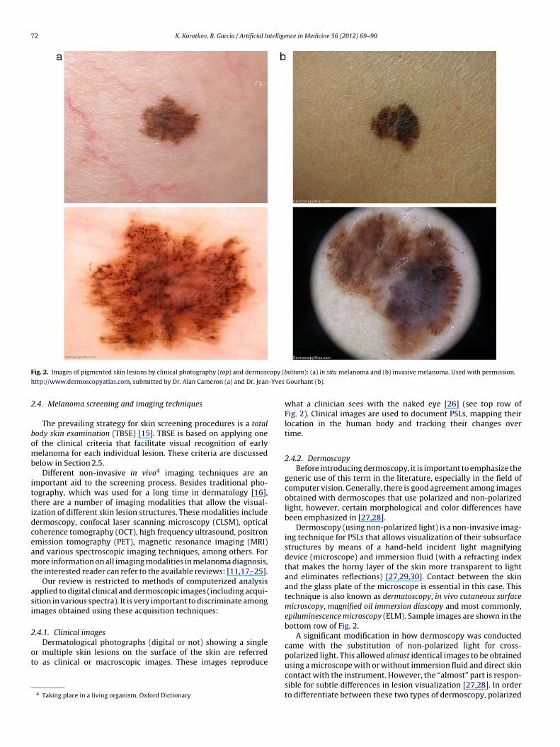
72 K. Korotkov, R. Garcia / Artificial Intelligence in Medicine 56 (2012) 69–90
F opy (b
h -Yves
2
bomb
ittidceamt
asi
2
ot
ig. 2. Images of pigmented skin lesions by clinical photography (top) and dermosc
ttp://www.dermoscopyatlas.com, submitted by Dr. Alan Cameron (a) and Dr. Jean
.4. Melanoma screening and imaging techniques
The prevailing strategy for skin screening procedures is a totalody skin examination (TBSE) [15]. TBSE is based on applying onef the clinical criteria that facilitate visual recognition of earlyelanoma for each individual lesion. These criteria are discussed
elow in Section 2.5.Different non-invasive in vivo4 imaging techniques are an
mportant aid to the screening process. Besides traditional pho-ography, which was used for a long time in dermatology [16],here are a number of imaging modalities that allow the visual-zation of different skin lesion structures. These modalities includeermoscopy, confocal laser scanning microscopy (CLSM), opticaloherence tomography (OCT), high frequency ultrasound, positronmission tomography (PET), magnetic resonance imaging (MRI)nd various spectroscopic imaging techniques, among others. Forore information on all imaging modalities in melanoma diagnosis,
he interested reader can refer to the available reviews: [11,17–25].Our review is restricted to methods of computerized analysis
pplied to digital clinical and dermoscopic images (including acqui-ition in various spectra). It is very important to discriminate amongmages obtained using these acquisition techniques:
.4.1. Clinical images
Dermatological photographs (digital or not) showing a singler multiple skin lesions on the surface of the skin are referredo as clinical or macroscopic images. These images reproduce
4 Taking place in a living organism, Oxford Dictionary
ottom): (a) In situ melanoma and (b) invasive melanoma. Used with permission.
Gourhant (b).
what a clinician sees with the naked eye [26] (see top row ofFig. 2). Clinical images are used to document PSLs, mapping theirlocation in the human body and tracking their changes overtime.
2.4.2. DermoscopyBefore introducing dermoscopy, it is important to emphasize the
generic use of this term in the literature, especially in the field ofcomputer vision. Generally, there is good agreement among imagesobtained with dermoscopes that use polarized and non-polarizedlight, however, certain morphological and color differences havebeen emphasized in [27,28].
Dermoscopy (using non-polarized light) is a non-invasive imag-ing technique for PSLs that allows visualization of their subsurfacestructures by means of a hand-held incident light magnifyingdevice (microscope) and immersion fluid (with a refracting indexthat makes the horny layer of the skin more transparent to lightand eliminates reflections) [27,29,30]. Contact between the skinand the glass plate of the microscope is essential in this case. Thistechnique is also known as dermatoscopy, in vivo cutaneous surfacemicroscopy, magnified oil immersion diascopy and most commonly,epiluminescence microscopy (ELM). Sample images are shown in thebottom row of Fig. 2.
A significant modification in how dermoscopy was conductedcame with the substitution of non-polarized light for cross-polarized light. This allowed almost identical images to be obtained
using a microscope with or without immersion fluid and direct skincontact with the instrument. However, the “almost” part is respon-sible for subtle differences in lesion visualization [27,28]. In orderto differentiate between these two types of dermoscopy, polarized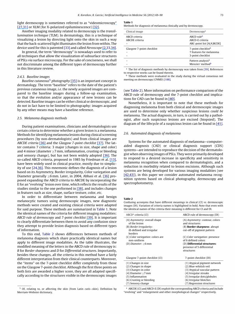
telligence in Medicine 56 (2012) 69–90 73
l[
lvtd
aoni
2
dppsddb
2
cMpAtaashibDpEsi
mmftAtto
mamBbdtobc
M
Table 1Methods for diagnosis of melanoma clinically and by dermoscopy.
Clinical image Dermoscopya
ABCD criteria ABCD ruleb
ABCDE criteria ABCD-E criteria– ABC-point list [A(A)BCDE]
Glasgow 7-point checklist 7-point checklistb
– 7 features for melanoma– 3-point checklist
– Pattern analysisb
– Menzies’ methodb
a The list of diagnosis methods by dermoscopy was taken from [39]. Referencesto respective works can be found therein.
[42,43]), in this paper we consider automated melanoma recog-nition systems based on clinical photography, dermoscopy andspectrophotometry.
Table 2Confusing acronyms that have different meanings in clinical (CI) vs. dermoscopicimages (DI). Variation of criteria names is highlighted in bold. Note that even withthe identical names of the criteria their meaning is different for CI and DI.
ABCDa criteria (CI) ABCD rule of dermoscopy (DI)
(A) Asymmetry: overall shapeof the lesion
(A) Asymmetry: contour, colorsand structures
(B) Border irregularity:ill-defined and irregularborders
(B) Border sharpness: abruptcut-off of pigment pattern
(C) Color variegation: colors arenon-uniform
(C) Color variegation: presenceof 6 defined colors
(D) Diameter: ≥6 mm (D) Differential structures:presence of 5 differentialstructures
Glasgow 7-point checklist (CI) 7-point checklist (DI)
(1) Changes in size (1) Atypical pigment network
K. Korotkov, R. Garcia / Artificial In
ight dermoscopy is sometimes referred to as “videomicroscopy”27,31] or XLM (for X-polarized epiluminescence) [32].
Another imaging modality related to dermoscopy is the transil-umination technique (TLM). In dermatology, this is a technique ofisualizing a lesion by directing light onto the skin in such a wayhat the back-scattered light illuminates the lesion from within. Theevice used for this is patented [33] and called Nevoscope [2,33,34].
In general, the term “dermoscopy” is nowadays used to refer toll techniques that allow the visualization of subsurface structuresf PSLs via surface microscopy. For the sake of conciseness, we shallot discriminate among the different types of dermoscopy further
n this literature review.
.4.3. Baseline imagesBaseline cutaneous5 photography [35] is an important concept in
ermatology. The term “baseline” refers to the date of the patient’srevious cutaneous image, i.e. the newly acquired images are com-ared to the baseline images during a follow-up examination,o that the evolution and/or appearance of new lesions can beetected. Baseline images can be either clinical or dermoscopic, ando not in fact have to be limited to photography: images acquiredy any other means may have a baseline reference.
.5. Melanoma diagnosis methods
During patient examinations, clinicians and dermatologists useertain criteria to determine whether a given lesion is a melanoma.ethods for identifying melanoma lesions during clinical screening
rocedures (by non-dermatologists) and from clinical images areBCDE criteria [36] and the Glasgow 7-point checklist [37]. The lat-er contains 7 criteria: 3 major (changes in size, shape and color)nd 4 minor (diameter ≥7 mm, inflammation, crusting or bleedingnd sensory change), but has not been widely adopted [36]. Theo-called ABCD criteria, proposed in 1985 by Friedman et al. [13],ave been widely used in clinical practice, mostly due to simplic-
ty of use [24,36]. This mnemonic defines the diagnosis of a lesionased on its Asymmetry, Border irregularity, Color variegation andiameter generally ≥6 mm. Later, in 2004, Abbasi et al. [36] pro-osed expanding the ABCD criteria to ABCDE by incorporating the
for an “evolving” lesion over time, which reflects the results of thetudies similar to the one performed in [38], and includes changesn features such as size, shape, surface texture, color, etc.
In order to differentiate between melanoma and benignelanocytic tumors using dermoscopic images, new diagnosticethods were created and existing clinical criteria were adapted
or said purpose. These methods are summarized in Table 1. Notehe identical names of the criteria for different imaging modalities:BCD rule of dermoscopy and 7-point checklist [39]. It is importanto clearly differentiate between them to avoid any confusion sincehey attempt to provide lesion diagnosis based on different typesf information.
To this end, Table 2 shows differences between methods ofelanoma diagnosis which share practically identical names but
pply to different image modalities. As the table illustrates, theodified meaning of the letters in the ABCD rule of dermoscopy is:
for Border sharpness and D for Differential structures. Importantly,esides these changes, all the criteria in this method have a fairlyifferent interpretation from their clinical counterparts. Moreover,he “items” on the 7-point checklist differ completely from those
n the Glasgow 7-point checklist. Although the first three points onoth lists are awarded a higher score, they are all adapted specifi-ally according to the structures visible in the dermoscopic images5 Of, relating to, or affecting the skin (from Latin cutis—skin). Definition byerriam-Webster dictionary.
b These methods were evaluated in the study during the virtual consensus netmeeting on dermoscopy (CNMD) [188].
(see Table 2). More information on performance comparison of theABCD rule of dermoscopy and the 7-point checklist and implica-tions for CAD can be found in [40].
Nonetheless, it is important to note that these methods fordiagnosing melanoma from both clinical and dermoscopic imagesare used to determine only whether suspicious lesions could bemelanoma. The actual diagnosis, in turn, is carried out by a pathol-ogist, after such suspicious lesions are excised (biopsied). Thediagram of the lifecycle of a suspicious lesion can be found in [41].
2.6. Automated diagnosis of melanoma
Systems for the automated diagnosis of melanoma—computer-aided diagnosis (CAD) or clinical diagnosis support (CDS)systems—are intended to reproduce the decision of the dermatolo-gist when observing images of PSLs. They were primarily developedto respond to a desired increase in specificity and sensitivity inmelanoma recognition when compared to dermatologists, and areduction in morbidity related to lesion excisions. Although suchsystems are being developed for various imaging modalities (see
(2) Changes in shape (2) Blue-whitish veil(3) Changes in color (3) Atypical vascular pattern(4) Diameter ≥7 mm (4) Irregular streaks(5) Inflammation (5) Irregular dots/globules(6) Crusting or bleeding (6) Irregular blotches(7) Sensory change (7) Regression structures
a ABCDE (CI) and ABCD-E (DI) exploit the corresponding ABCD criteria and include“evolving” and “enlargement and other morphological changes” respectively.

74 K. Korotkov, R. Garcia / Artificial Intelligence in Medicine 56 (2012) 69–90
a cor
tttbsi
f[aarmbIsf
1
Fig. 3. Literature categorization tree. The rectangles in the highlighted are
Most of these automated systems are based on the aforemen-ioned melanoma diagnosis methods. In general, image processingechniques are used to locate the lesion(s), extract image parame-ers describing the dermatological features of the lesion(s), and,ased on these parameters, perform the diagnosis. The genericteps of a CAD system for melanoma identification are highlightedn Fig. 3.
Studies have shown that the performance of automated systemsor melanoma diagnosis is sufficient under experimental conditions44]. However, the practical value of automated dermoscopic imagenalysis systems is still unclear. Although most patients wouldccept using computerized analysis for melanoma screening, cur-ently it cannot be recommended as a sole determinant of thealignancy of a lesion due to its tendency for over-diagnosis of
enign melanocytic lesions and non-melanocytic skin lesions [44].n addition, according to Day and Barbour [41], there are two mainhortcomings of the general approach to developing a CAD systemor melanoma identification:
. A CAD system is expected to reproduce the decision of pathol-ogists (a binary result like “melanoma/non-melanoma lesion”)with only the input used by dermatologists: clinical or dermo-scopic images;
respond to generic steps of the CAD systems for melanoma identification.
2. Histopathological data are not available for all lesions, only forthose considered suspicious by dermatologists.
The former is a methodological problem. It reflects the fact thata CAD system is intended to diagnose a lesion without sufficientinformation for diagnosis and without any interaction with the der-matologist. This was highlighted by Dreiseitl et al. in their study intothe acceptance of CDS systems by dermatologists [45], i.e. that thecurrently available CDS systems are designed to work “in parallelwith and not in support of” physicians, and because of this only afew systems are found in routine clinical use. Thus, an ideal CADor CDS system for melanoma identification should reproduce thedecision of dermatologists (i.e. define the level of “suspiciousness”of a lesion) [41] and provide dermatologists with comprehensiveinformation regarding the grounds of this decision [45].
3. Literature classification
The existing literature in the field of computerized image analy-
sis for melanoma identification was roughly subdivided accordingto the following two criteria (see Fig. 3):1. The nature of the publication: clinical or computer vision articles.
telligence in Medicine 56 (2012) 69–90 75
2
fllbto[agawoc
fyccrcra6aptaa
4
icr
4
hstpba
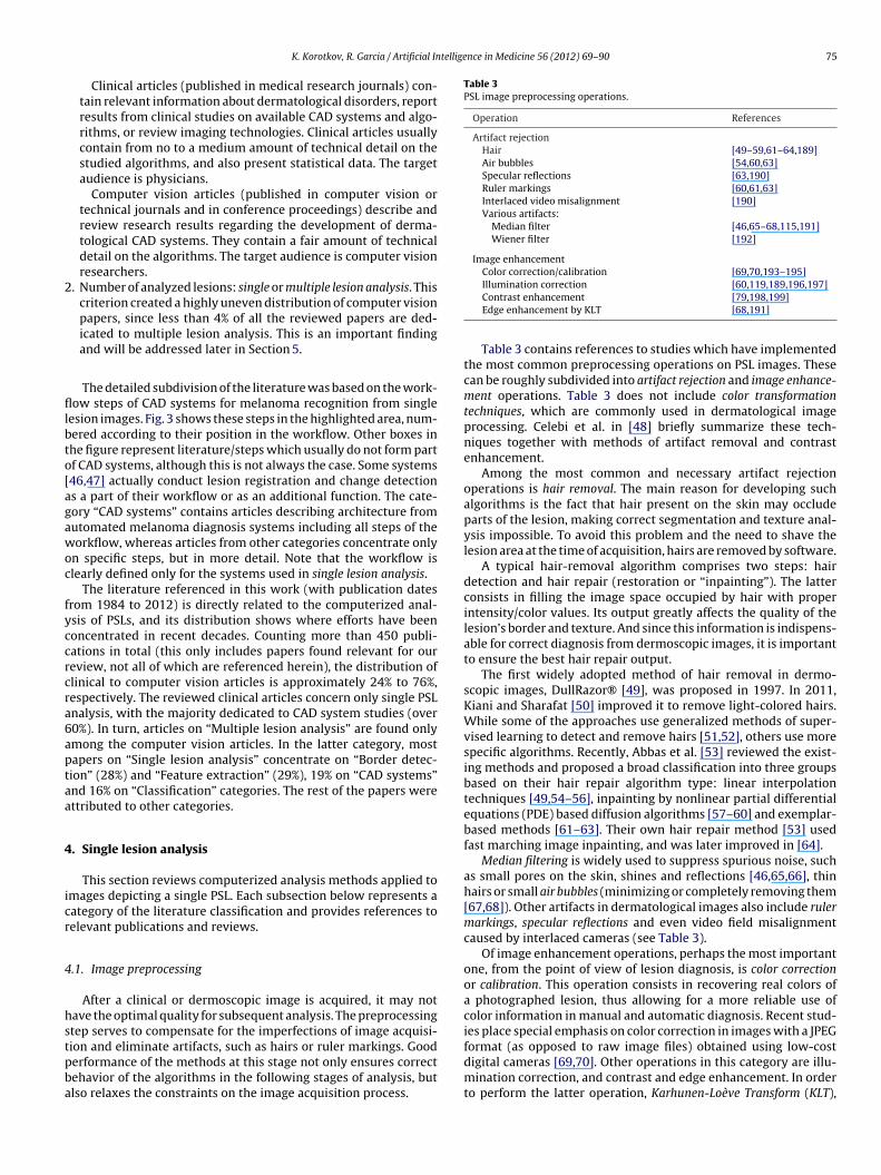
Table 3PSL image preprocessing operations.
Operation References
Artifact rejectionHair [49–59,61–64,189]Air bubbles [54,60,63]Specular reflections [63,190]Ruler markings [60,61,63]Interlaced video misalignment [190]Various artifacts:
Median filter [46,65–68,115,191]Wiener filter [192]
Image enhancementColor correction/calibration [69,70,193–195]
K. Korotkov, R. Garcia / Artificial In
Clinical articles (published in medical research journals) con-tain relevant information about dermatological disorders, reportresults from clinical studies on available CAD systems and algo-rithms, or review imaging technologies. Clinical articles usuallycontain from no to a medium amount of technical detail on thestudied algorithms, and also present statistical data. The targetaudience is physicians.
Computer vision articles (published in computer vision ortechnical journals and in conference proceedings) describe andreview research results regarding the development of derma-tological CAD systems. They contain a fair amount of technicaldetail on the algorithms. The target audience is computer visionresearchers.
. Number of analyzed lesions: single or multiple lesion analysis. Thiscriterion created a highly uneven distribution of computer visionpapers, since less than 4% of all the reviewed papers are ded-icated to multiple lesion analysis. This is an important findingand will be addressed later in Section 5.
The detailed subdivision of the literature was based on the work-ow steps of CAD systems for melanoma recognition from single
esion images. Fig. 3 shows these steps in the highlighted area, num-ered according to their position in the workflow. Other boxes inhe figure represent literature/steps which usually do not form partf CAD systems, although this is not always the case. Some systems46,47] actually conduct lesion registration and change detections a part of their workflow or as an additional function. The cate-ory “CAD systems” contains articles describing architecture fromutomated melanoma diagnosis systems including all steps of theorkflow, whereas articles from other categories concentrate only
n specific steps, but in more detail. Note that the workflow islearly defined only for the systems used in single lesion analysis.
The literature referenced in this work (with publication datesrom 1984 to 2012) is directly related to the computerized anal-sis of PSLs, and its distribution shows where efforts have beenoncentrated in recent decades. Counting more than 450 publi-ations in total (this only includes papers found relevant for oureview, not all of which are referenced herein), the distribution oflinical to computer vision articles is approximately 24% to 76%,espectively. The reviewed clinical articles concern only single PSLnalysis, with the majority dedicated to CAD system studies (over0%). In turn, articles on “Multiple lesion analysis” are found onlymong the computer vision articles. In the latter category, mostapers on “Single lesion analysis” concentrate on “Border detec-ion” (28%) and “Feature extraction” (29%), 19% on “CAD systems”nd 16% on “Classification” categories. The rest of the papers werettributed to other categories.
. Single lesion analysis
This section reviews computerized analysis methods applied tomages depicting a single PSL. Each subsection below represents aategory of the literature classification and provides references toelevant publications and reviews.
.1. Image preprocessing
After a clinical or dermoscopic image is acquired, it may notave the optimal quality for subsequent analysis. The preprocessingtep serves to compensate for the imperfections of image acquisi-
ion and eliminate artifacts, such as hairs or ruler markings. Gooderformance of the methods at this stage not only ensures correctehavior of the algorithms in the following stages of analysis, butlso relaxes the constraints on the image acquisition process.Illumination correction [60,119,189,196,197]Contrast enhancement [79,198,199]Edge enhancement by KLT [68,191]
Table 3 contains references to studies which have implementedthe most common preprocessing operations on PSL images. Thesecan be roughly subdivided into artifact rejection and image enhance-ment operations. Table 3 does not include color transformationtechniques, which are commonly used in dermatological imageprocessing. Celebi et al. in [48] briefly summarize these tech-niques together with methods of artifact removal and contrastenhancement.
Among the most common and necessary artifact rejectionoperations is hair removal. The main reason for developing suchalgorithms is the fact that hair present on the skin may occludeparts of the lesion, making correct segmentation and texture anal-ysis impossible. To avoid this problem and the need to shave thelesion area at the time of acquisition, hairs are removed by software.
A typical hair-removal algorithm comprises two steps: hairdetection and hair repair (restoration or “inpainting”). The latterconsists in filling the image space occupied by hair with properintensity/color values. Its output greatly affects the quality of thelesion’s border and texture. And since this information is indispens-able for correct diagnosis from dermoscopic images, it is importantto ensure the best hair repair output.
The first widely adopted method of hair removal in dermo-scopic images, DullRazor® [49], was proposed in 1997. In 2011,Kiani and Sharafat [50] improved it to remove light-colored hairs.While some of the approaches use generalized methods of super-vised learning to detect and remove hairs [51,52], others use morespecific algorithms. Recently, Abbas et al. [53] reviewed the exist-ing methods and proposed a broad classification into three groupsbased on their hair repair algorithm type: linear interpolationtechniques [49,54–56], inpainting by nonlinear partial differentialequations (PDE) based diffusion algorithms [57–60] and exemplar-based methods [61–63]. Their own hair repair method [53] usedfast marching image inpainting, and was later improved in [64].
Median filtering is widely used to suppress spurious noise, suchas small pores on the skin, shines and reflections [46,65,66], thinhairs or small air bubbles (minimizing or completely removing them[67,68]). Other artifacts in dermatological images also include rulermarkings, specular reflections and even video field misalignmentcaused by interlaced cameras (see Table 3).
Of image enhancement operations, perhaps the most importantone, from the point of view of lesion diagnosis, is color correctionor calibration. This operation consists in recovering real colors ofa photographed lesion, thus allowing for a more reliable use ofcolor information in manual and automatic diagnosis. Recent stud-ies place special emphasis on color correction in images with a JPEG
format (as opposed to raw image files) obtained using low-costdigital cameras [69,70]. Other operations in this category are illu-mination correction, and contrast and edge enhancement. In orderto perform the latter operation, Karhunen-Loève Transform (KLT),
7 tellige
a(
4
aiAr
iepwcpimhStcmvs
itcrac
4
afod[no
dihta
n3cottodctat[
iw
6 K. Korotkov, R. Garcia / Artificial In
lso known as Hoteling Transform or Principal Component AnalysisPCA), is widely used.
.2. Lesion border detection
An accurately detected border of a skin lesion is crucial for itsutomated diagnosis. Therefore, border detection (segmentation)s one of the most active areas in the computerized analysis of PSLs.
lot of effort has been made to improve lesion segmentation algo-ithms and come up with adequate measures of their performance.
The problem of lesion border detection is not as trivial ast may seem. Firstly, since dermatologists do not usually delin-ate lesion borders for diagnosis [41] there exists a ground truthroblem. Segmentation algorithms are intended to reproduce theay human observers, who are generally not very good at dis-
riminating between subtle variations in contrast or blur [71],erceive the boundaries of a lesion. But because of high inter- and
ntra-observer variability in PSL boundary perception among der-atologists [71–73] the ground truth often lacks definiteness and
as to be obtained as a fusion of several manual segmentations.econdly, the morphological structure of a lesion itself (depigmen-ation, low lesion-to-skin gradient, multiple lesion regions, etc.)an act as a confusion factor for both manual and automatic seg-entation. These problems have led to the development of a wide
ariety of PSL segmentation methods which span all categories ofegmentation algorithms [48].
These algorithms can be classified in many ways regarding, fornstance, their level of automation (automatic vs. semi-automatic),heir number of parameters or the required methods of postpro-essing [48]. However, the purpose of this subsection is not toeview all these methods, but to provide information regardingvailable reviews and comparisons and to emphasize the role ofertain approaches to the problem.
.2.1. PSL border detection methodologyMorphological differences in the appearance of PSLs in clinical
nd dermoscopic images directly influence the choice of methodor border detection. Moreover, various conditions, such as typef lesion, location, color conditions or angle of view, add to theiverse difficulties in segmenting using the same imaging modality48,54,74]. Therefore, the available methods aim to provide robust-ess in difficult segmentation cases adapting to specific conditionsf the image type (e.g. [75]).
Clinical images. One of the earliest works on skin lesion borderetection was published in 1989 and used the concept of spher-
cal coordinates for color space representation [76]. Since then, itas been widely adopted in the literature for lesion feature extrac-ion and color segmentation. Comparisons of different color spacespplied to segmentation were carried out in [77–79].
In 1990, Golston et al. estimated the role of several determi-ants of the lesion border, namely color, luminance, texture andD information [80]. While 3D information was mostly absent,olor and luminance appeared to be the major factors for mostf the images. Thus, the authors discussed an overall algorithmhat would take into account several border determinants based onheir level of confidence, and proposed a radial search method basedn luminance information. Similarly, in support of multifactorialescriptiveness of the lesion border, Dhawan and Sicsu proposedombining gray-level intensity and textural information [81]. Fur-her works concentrated on improving existing techniques [82] andpplying a multitude of different approaches, including edge detec-ion [74,83], active contours [57], PDE [57,58], gradient vector flow
84] and many others.Dermoscopic images. Following the trend initiated by clinicalmages, multiple segmentation algorithms and their combinations
ere investigated for dermoscopic images. Fleming et al. [54]
nce in Medicine 56 (2012) 69–90
discussed several implementations of segmentation algorithms.Though agreeing that one of the most efficient border determi-nants is color, they proposed an approach incorporating spatialand chromatic information to produce better segmentations. Afterimplementing and testing various algorithms, the final methodcombined principal component transform (PCT), stabilized inversediffusion equations (SIDE) and thresholding in the green channel.
Later thresholding approaches became more sophisticatedin comparison with the relatively simple methods of single-color-channel thresholding proposed earlier [54,85]. Iterativethresholding [86], type-2 fuzzy logic based thresholding [87],fusion of thresholds [88,89] and hybrid thresholding [90] havebeen proposed recently. Many other approaches have been appliedto the segmentation of dermoscopic images. Among them arevarious algorithms using and combining different categories oftechniques, such as clustering [91–94], soft computing (neuralnetworks [86,95,96] and evolution strategy [97]), supervised learn-ing [51,52,98], active contours [32,99], and dynamic programming[100], to name but a few.
Without doubt, all these (and even other approaches not men-tioned here for the sake of space) have their advantages anddrawbacks. However, it should be noted that most of the algo-rithms are tested on various fairly small datasets, not many ofwhich include special “difficult” cases. Consequently, the perfor-mance assessment for these algorithms is not trivial, especiallybased only on the results reported by the authors. In this respect,the comparison studies allow these algorithms to be assessed ina more uniform framework, clearly defining their strengths andweaknesses.
4.2.2. Comparison of segmentation algorithmsIn 1996, Hance et al. published a comparison of 6 methods of
PSL segmentation [101]. It included techniques such as fuzzy c-means, center split, multiresolution, split and merge, PCT/mediancut and adaptive thresholding. The latter two methods proved to bemore robust than the others based on the exclusive-OR evaluationmetric proposed therein. In another comparison of segmentationmethods implemented by Silveira et al. [102], an adaptive snakealgorithm was the best among gradient vector flow, level set, adap-tive thresholding, expectation-maximization (EM) level set andfuzzy-based split-and-merge algorithm (which had the best per-formance among fully automated methods).
Statistical region merging (SRM) was introduced and comparedin [103,104] to optimized histogram thresholding, orientation-sensitive fuzzy c-means [91], gradient vector flow snakes [105],dermatologist-like tumor extraction algorithm (DTEA) [73] andJSEG algorithm [106]. Overall results from this comparison on 90dermoscopic images determined the superiority of the SRM, fol-lowed by DTEA and JSEG. However, Zhou et al. [107] reportedthat on a considerably larger dataset of 2300 dermoscopic imagesSRM, JSEG and a clustering-based method incorporating a dermo-scopic spatial prior [75] were outperformed by a spatially smoothedexemplar-based classifier (SEBC) algorithm.
However, these studies still do not provide unified results forall the tested algorithms. Firstly, because of the differences inthe datasets employed including different ground-truth definitions,and secondly, due to different evaluation metrics. In fact, the twohighlighted factors are essentially the basis for performance com-parison between segmentation algorithms.
Almost all standard metrics for evaluation of PSL segmenta-tion algorithms, such as sensitivity, specificity, precision, bordererror and others [48,101,108,109], are based on the concepts of
true (false) positives (negatives). Recently, Garnavi et al. [109] pro-posed a weighted performance index which uses specific weightingfor these metrics and unites them under one value for easier com-parison with other methods. Alternative metrics used by different
K.
Korotkov,
R.
Garcia
/ A
rtificial Intelligence
in M
edicine 56
(2012) 69–90
77
Table 4Dermoscopic features of pigmented skin lesions according to ABCD rule of dermoscopy [39,250] and their descriptors.
Dermoscopic features Feature descriptors Clinical references Computer vision references.
Featuresa CAD systemsa Featuresb Lesion classification CAD systemsb
Asymmetry (review: [221]) Lesion’s centroid &moments of inertiac [200–204] [85,205–210] [211,212] [149,213–215] [115,191,216–220]Symmetry maps – – [222] – [55]Fourier descriptors – – [223] – –Global point signatures (GPS) – – [224] – –Other symmetry descriptors [204,225] [226,227] [228] [118] [152,192,229]
Border sharpness Lesion’s area &perimeterd [200–202] [205,208–210] [211] [118,149,213–215] [115,152,191,192,216,218–220]Convex hull descriptorse – – – – [115,192]Bounding box descriptorse – – – [215] [115,216,218]Fractal geometryf – [209] [230] [213,214] [191,192]Gradient-based descriptors [203] [85,226] [228,231,232] [118,149] [46,152,192,216–219,229]Multi-scale roughness descriptors – – [223] – –
Color variegation RGB statistical descriptorsg – [206–210,226,227] [228,234] [118,149,2137ndash;215,235,236] [192,219,115,152,220]Alternative color space descriptorsh [203] [205,209,226] [228,234] [118,149,214,215,235,236] [115,152,163,216,218–220]Munsell color space descriptors – – – – [135]Relative color statistical descriptorsi – [226] [211,237] [118] [152,218,229]Color quantizationj [201,202,221,238] [205,209,226] [228,237,239,240] [118,143,214,215] [135,152,191,217,218]
Diff. structures Multidim. receptive fields histograms – – – [143] –
Other features Wavelet-based descriptors – – [241] [141,142,215,236,242–245] [47,65]Gabor filter descriptors – [208] – [147,213,235] –Intensity distribution descriptors – [206,207,226] [228] [118] [152,216,229]Haralick descriptorsk – [206,207,226] [211,228,246] [118,149] [115,135,152,163,219,229]Local binary pattern (LBP) – – [247] [249] –Various texture descriptors – – [247] [249] –Size functions – [127] – [128,129] –SIFT and color SIFT – – – [236] –Dermoscopic interest points (DIP) – – [126] – –Bag-of-features implementation – – – [147,236] –
a Clinical papers describing studies of PSL feature descriptors and CAD systems, respectively.b Computer vision papers describing PSL feature extraction and design of CAD systems, respectively.
c This group includes all measures based on computing principal and/or symmetry axes, and centroid of the lesion. Asymmetry index/percentage [117], aspect ratio (lengthening), radial distance distribution are among otherdescriptors in the group.
d These descriptors define the relation between the area and perimeter of an object, and thus, describe its symmetrical and border characteristics. In particular, it includes a descriptor known as compactness index/ratio orroundness (I = P2/4�A or I = 4�P2/A), as well as circularity, thinness ratio or regularity (I = 4�A/P2). In essence, these ratios represent one descriptor.
e There are various descriptors based on the convex hull (CH) and bounding box (BB) of a lesion. In particular, extent is the ratio of the area of the lesion to the area of its CH (same as solidity) or BB (same as rectangularity),whereas convexity is the ratio of the perimeter of the CH to the perimeter of the lesion, and elongation is the ratio between the height and the width of the BB.
f Fractal geometry group includes Fourier (fractal) dimension [251] and lacunarity [192].g RGB descriptors encompass statistical information from the RGB channels in the form of such values as min/max, average, variance, entropy, energy, kurtosis, range and others.h Alternative color space descriptors include parameters (statistical or not) derived from non-RGB color spaces (except for normalized RGB)—HSV/HSI (hue, saturation, value/intensity), spherical coordinates [76], CIELUV and
others.i This group comprises descriptors based on relative color, such as statistics on relative difference, ratio and chromaticity [211] of separate color channels, mostly in RGB color space.j Color quantization descriptors refer to features obtained after reduction of the quantity of colors in the image. This reduction or quantization can be done using color prototypes, histograms or clustering. Typical descriptors
include number of colors and percent melanoma color.k Descriptors based on co-occurrence matrices. These contain entropy, inertia, correlation, inverse difference and other statistical parameters.

78
K.
Korotkov,
R.
Garcia
/ A
rtificial Intelligence
in M
edicine 56
(2012) 69–90
Table 5Clinical features of pigmented skin lesions according to ABCDE criteria [36,39] and their descriptors.
Clinical features Feature descriptors Clinical references Computer vision references
CAD systemsa Feature extraction Lesion classification CAD systemsb
Asymmetry Lesion’s centroid &moments of inertiac [252] [133,253–255] [140,256–259] [68,111,113,119,146,165,196,260–263]Symmetry distance (SD) – [264,265] – –
Border irregularity Lesion’s area &perimeterd [3,252,266–270] [133,145,254,255,264,271,272] [140,256–259] [68,111,113,119,146,164,165,196,260–263]Convex hulle [268,269] – – [113,119,196,262]Bounding boxe [266] – – [119,196]Fractal geometryf [252] [251,273–276] – [68,111]Gradient-based descriptors [3,267] – – [68,111,119,196]Irregularity index – [277,278] –Sigma-ratio – [279] – –Best-fit ellipse – [133] – [262]Fourier feature – [255] – –Polygonal approximation – [272] – –Conditional entropy – [280] – –Hidden Markov models – [281] – –Wavelet transform – [282] – –Centroid distance diagram – [144] – –
Color variegation RGB statistical descriptorsg [3,252,267] [114,133,145,255,283] [140,256–258,284] [146,196,260,261,263]Alternative color space descriptorsh [266,285] [255,283,286] [140,256,257,259] [146,164,260,261,263]Own channel representation – – – [119]Relative color statistical descriptorsi – [114,255,283,286,287] [256,257] [260,261,263]Color quantizationj – [114,283,287–289] [258] [164,196]Color homogeneity, photometry-geometry correlation – – – [68,111]Parametric maps – [290] – –
Diameter Semi-major axis of the best-fit ellipse – [133] – –
Evolving – – – – –
Other features Intensity distribution descriptors [252,268–270,291] – – [165,262]Skin pattern analysis – [130–134] –Haralick descriptorsk [266] [292] – [164,196]Various texture descriptors – [293] – –Wavelet-based descriptors – – [294] –Independent component analysis based descriptor – – [284] –
a Clinical papers describing studies of PSL CAD systems.b Computer vision papers describing design of PSL CAD systems.
c This group includes all measures based on computing principal and/or symmetry axes, and centroid of the lesion. Asymmetry index/percentage [117], aspect ratio (lengthening), radial distance distribution are among otherdescriptors in the group.
d These descriptors define the relation between the area and perimeter of an object, and thus, describe its symmetrical and border characteristics. In particular, it includes a descriptor known as compactness index/ratio orroundness (I = P2/4�A or I = 4�P2/A), as well as circularity, thinness ratio or regularity (I = 4�A/P2). In essence, these ratios represent one descriptor.
e There are various descriptors based on the convex hull (CH) and bounding box (BB) of a lesion. In particular, extent is the ratio of the area of the lesion to the area of its CH (same as solidity) or BB (same as rectangularity),whereas convexity is the ratio of the perimeter of the CH to the perimeter of the lesion, and elongation is the ratio between the height and the width of the BB.
f Fractal geometry group includes Fourier (fractal) dimension [251] and lacunarity [192].g RGB descriptors encompass statistical information from the RGB channels in the form of such values as min/max, average, variance, entropy, energy, kurtosis, range and others.h Alternative color space descriptors include parameters (statistical or not) derived from non-RGB color spaces (except for normalized RGB)—HSV/HSI (hue, saturation, value/intensity), spherical coordinates [76], CIELUV and
others.i This group comprises descriptors based on relative color, such as statistics on relative difference, ratio and chromaticity [211] of separate color channels, mostly in RGB color space.j Color quantization descriptors refer to features obtained after reduction of the quantity of colors in the image. This reduction or quantization can be done using color prototypes, histograms or clustering. Typical descriptors
include number of colors and percent melanoma color.k Descriptors based on co-occurrence matrices. These contain entropy, inertia, correlation, inverse difference and other statistical parameters.

telligence in Medicine 56 (2012) 69–90 79
aaciise
mpmsat
4
ndoapstpft
efUepcpcattm
atptltdde
d[otssdctt
dod
Table 6Feature extraction for pattern analysis [39].
Pattern analysis References
Global patternsReticular [295,296] [297]a
Globular [295,296,298] [297]a
Cobblestone [296,298] [297]a
Homogeneous [295,296,298] [297]a
Starburst [297]a
Parallel [296,298][297]a
Multicomponent –Non-specific –
Local featuresPigment network [52,54,246,299–304] [121–123]b [47]c
Dots/globules [51,54,305] [47]c
Streaks [121,125]b
Blue-whitish veil [211,306] [120,124,125]b
Regression structures [51,307] [120,124,125]c [308]c
Hypopigmentation [51] [308]c
Blotches [309–311] [200]d
Vascular structures [312]
a Computer vision article from the “Classification” category.b
K. Korotkov, R. Garcia / Artificial In
uthors include pixel misclassification probability [72], Hammoudend Hausdorff distances (not the most relevant metrics from alinical point of view) [102] and normalized probabilistic randndex (NPRI) [108]. The reviews of these metrics can be foundn [48,108,109]. In addition to this, [48] provides an excellentummary of 18 algorithms with their characteristics and reportedvaluation.
Equally important in this work [48] is the outline of require-ents for a systematic PSL border detection study, which, if a
ublic dermoscopy dataset is provided, can favor a rapid develop-ent of more reliable automated diagnosis systems. Therefore,
uch a dataset, with a standardized ground-truth definition, willllow researchers to immediately report performance results forheir methods, and thereby boost overall progress in the field.
.3. Feature extraction
To correctly diagnose a PSL (or classify it as “suspicious”), cli-icians rely on the so-called features of the lesion. These featuresepend on the method of diagnosis in use. For example, asymmetryf a lesion is a feature of the ABCD-rule, and pigmented network is
feature in pattern analysis (see Section 2.5 for details). In com-uterized PSL analysis, in order to classify a lesion most automatedystems aim to extract such features from the images and representhem in a way that can be understood by a computer, i.e. using imagerocessing features. In this review paper, for clarity we use the termeatures to denote clinical and dermoscopic lesion features, and theerm feature descriptors for image processing features.
Many works can be found on PSL feature extraction in the lit-rature. However, only a few of them review or summarize theeature descriptors used in CAD systems. In particular, in 1997,mbaugh et al. [110] described a computer program for automaticxtraction and analysis of PSL features. They classified the pro-osed feature descriptors into binary object features, histogram,olor, and spectral features. Binary object features included area,erimeter, and aspect ratios, among others. Histogram featuresomprised statistical measures of gray level distribution as wells features of co-occurrence matrices. Metrics obtained from colorransforms, normalized colors and color differences were usedo represent color features. Finally, spectral features represented
etrics derived from the Fourier transform of the images.Research carried out by Zagrouba and Barhoumi [111], as well
s reviewing CAD system development, provides a brief look athe feature selection algorithms. Feature selection is an importantrocedure to be carried out prior to lesion classification. It aimso reduce the number of extracted feature descriptors in order toower the computational cost of classification. However, this reduc-ion is not trivial because eliminating redundancy among featureescriptors may adversely affect their discriminatory power. Theevelopment of feature selection procedures for various sets ofxtracted feature descriptors can be found in [112–116].
Finally, a very good overview of CAD systems and featureescriptors was published in 2009 by Maglogiannis and Doukas117]. In their work, they provided information regarding meth-ds of PSL diagnosis and a list of typical feature descriptors used inhe literature. They also compared the performance of several clas-ifiers on a dataset of dermoscopic images using several featureelection algorithms on one feature set (see Section 4.5 for moreetails). The results obtained showed that the performance of thelassifiers was greatly dependent on the selected feature descrip-ors. This fact emphasizes the importance of feature descriptors inhe computerized analysis of PSL.
In this paper, we propose an extended categorization of featureescriptors (see Tables 4–6), associating them with specific meth-ds of diagnosis, separating clinical and dermoscopic images andiscriminating references according to our literature classification.
Feature extraction following the 7-Point checklist for dermoscopy.c Computer vision article from the “CAD systems” category.d Clinical article from the “Studies of lesion features” category.
Such a categorization can help the reader: (1) to gain perspectiveregarding the existing approaches in PSL feature description, (2) toclarify differences in the representation of clinical and dermoscopicfeatures, and, most importantly, (3) to obtain a complete source ofreferences on the descriptors of interest.
For the purpose of conciseness and generalization, rather thanlook at individual descriptors we attempted to cluster theminto groups with other related descriptors. Of course, takingthis approach meant determining how each group of descriptorsuniquely corresponded to the feature it aimed to describe. In otherwords, while most authors specified in their publications that adescriptor was mimicking a certain feature, others would use itto describe a different feature or not associate it with any featurein particular. A clear example of such a group is the one labeled“Lesion’s area & perimeter” (see Tables 4 and 5). We attributed thisgroup to the “Border irregularity/sharpness” feature in line withmost publications, and not to the “Asymmetry” feature, as someauthors have done [118,119]. Nevertheless, attributing this groupis not such a straightforward task, since, as a geometry or shapeparameter, it could well be used to describe both features. An iden-tical majority reasoning was applied to the other groups of featuredescriptors. Descriptors for which we could not define a specificclinical attribution were listed separately. All explanations on thegroups can be found in Table 4.
Among the diagnosis methods considered were the ABCD-ruleand pattern analysis for dermoscopic images, and the ABCDE crite-ria for clinical images. Table 6 contains references to articles aimedat computing descriptors for pattern analysis features [39]. Themajority of the papers referenced in this table belong to the“Feature extraction” category. Among these, a number were dedi-cated to feature extraction following the 7-point checklist methodfor melanoma diagnosis from dermoscopic images [120–125]. Apreliminary study on detection of some dermoscopic structures(blue-whitish veil, atypical pigmented network and irregular pig-mentation) can be found in [40].
Descriptors of features used in the ABCD-rule of dermoscopy andthe ABCDE clinical criteria are summarized in Tables 4 and 5. Thisseparate representation helps to highlight differences and similari-
ties in the computerized description of these features. As illustratedby these two tables, the largest groups of feature descriptors arepresent in both types of images (clinical and dermoscopy) anddefine the similarities. The differences, on the other hand, can be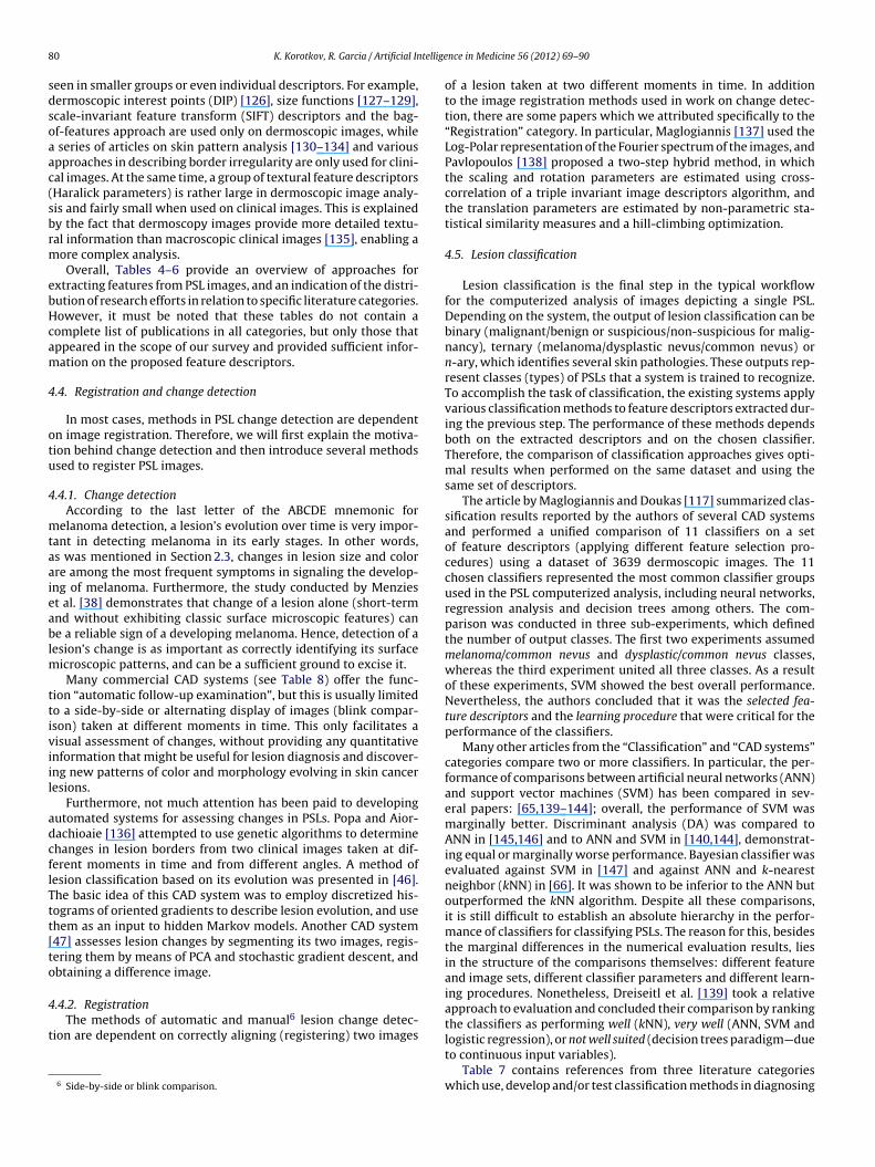
8 tellige
sdsoaac(sbrm
ebHcam
4
otu
4
mtaaieablm
ttiviil
adcflTtt[to
4
t
0 K. Korotkov, R. Garcia / Artificial In
een in smaller groups or even individual descriptors. For example,ermoscopic interest points (DIP) [126], size functions [127–129],cale-invariant feature transform (SIFT) descriptors and the bag-f-features approach are used only on dermoscopic images, while
series of articles on skin pattern analysis [130–134] and variouspproaches in describing border irregularity are only used for clini-al images. At the same time, a group of textural feature descriptorsHaralick parameters) is rather large in dermoscopic image analy-is and fairly small when used on clinical images. This is explainedy the fact that dermoscopy images provide more detailed textu-al information than macroscopic clinical images [135], enabling aore complex analysis.Overall, Tables 4–6 provide an overview of approaches for
xtracting features from PSL images, and an indication of the distri-ution of research efforts in relation to specific literature categories.owever, it must be noted that these tables do not contain aomplete list of publications in all categories, but only those thatppeared in the scope of our survey and provided sufficient infor-ation on the proposed feature descriptors.
.4. Registration and change detection
In most cases, methods in PSL change detection are dependentn image registration. Therefore, we will first explain the motiva-ion behind change detection and then introduce several methodssed to register PSL images.
.4.1. Change detectionAccording to the last letter of the ABCDE mnemonic for
elanoma detection, a lesion’s evolution over time is very impor-ant in detecting melanoma in its early stages. In other words,s was mentioned in Section 2.3, changes in lesion size and colorre among the most frequent symptoms in signaling the develop-ng of melanoma. Furthermore, the study conducted by Menziest al. [38] demonstrates that change of a lesion alone (short-termnd without exhibiting classic surface microscopic features) cane a reliable sign of a developing melanoma. Hence, detection of a
esion’s change is as important as correctly identifying its surfaceicroscopic patterns, and can be a sufficient ground to excise it.Many commercial CAD systems (see Table 8) offer the func-
ion “automatic follow-up examination”, but this is usually limitedo a side-by-side or alternating display of images (blink compar-son) taken at different moments in time. This only facilitates aisual assessment of changes, without providing any quantitativenformation that might be useful for lesion diagnosis and discover-ng new patterns of color and morphology evolving in skin canceresions.
Furthermore, not much attention has been paid to developingutomated systems for assessing changes in PSLs. Popa and Aior-achioaie [136] attempted to use genetic algorithms to determinehanges in lesion borders from two clinical images taken at dif-erent moments in time and from different angles. A method ofesion classification based on its evolution was presented in [46].he basic idea of this CAD system was to employ discretized his-ograms of oriented gradients to describe lesion evolution, and usehem as an input to hidden Markov models. Another CAD system47] assesses lesion changes by segmenting its two images, regis-ering them by means of PCA and stochastic gradient descent, andbtaining a difference image.
.4.2. RegistrationThe methods of automatic and manual6 lesion change detec-
ion are dependent on correctly aligning (registering) two images
6 Side-by-side or blink comparison.
nce in Medicine 56 (2012) 69–90
of a lesion taken at two different moments in time. In additionto the image registration methods used in work on change detec-tion, there are some papers which we attributed specifically to the“Registration” category. In particular, Maglogiannis [137] used theLog-Polar representation of the Fourier spectrum of the images, andPavlopoulos [138] proposed a two-step hybrid method, in whichthe scaling and rotation parameters are estimated using cross-correlation of a triple invariant image descriptors algorithm, andthe translation parameters are estimated by non-parametric sta-tistical similarity measures and a hill-climbing optimization.
4.5. Lesion classification
Lesion classification is the final step in the typical workflowfor the computerized analysis of images depicting a single PSL.Depending on the system, the output of lesion classification can bebinary (malignant/benign or suspicious/non-suspicious for malig-nancy), ternary (melanoma/dysplastic nevus/common nevus) orn-ary, which identifies several skin pathologies. These outputs rep-resent classes (types) of PSLs that a system is trained to recognize.To accomplish the task of classification, the existing systems applyvarious classification methods to feature descriptors extracted dur-ing the previous step. The performance of these methods dependsboth on the extracted descriptors and on the chosen classifier.Therefore, the comparison of classification approaches gives opti-mal results when performed on the same dataset and using thesame set of descriptors.
The article by Maglogiannis and Doukas [117] summarized clas-sification results reported by the authors of several CAD systemsand performed a unified comparison of 11 classifiers on a setof feature descriptors (applying different feature selection pro-cedures) using a dataset of 3639 dermoscopic images. The 11chosen classifiers represented the most common classifier groupsused in the PSL computerized analysis, including neural networks,regression analysis and decision trees among others. The com-parison was conducted in three sub-experiments, which definedthe number of output classes. The first two experiments assumedmelanoma/common nevus and dysplastic/common nevus classes,whereas the third experiment united all three classes. As a resultof these experiments, SVM showed the best overall performance.Nevertheless, the authors concluded that it was the selected fea-ture descriptors and the learning procedure that were critical for theperformance of the classifiers.
Many other articles from the “Classification” and “CAD systems”categories compare two or more classifiers. In particular, the per-formance of comparisons between artificial neural networks (ANN)and support vector machines (SVM) has been compared in sev-eral papers: [65,139–144]; overall, the performance of SVM wasmarginally better. Discriminant analysis (DA) was compared toANN in [145,146] and to ANN and SVM in [140,144], demonstrat-ing equal or marginally worse performance. Bayesian classifier wasevaluated against SVM in [147] and against ANN and k-nearestneighbor (kNN) in [66]. It was shown to be inferior to the ANN butoutperformed the kNN algorithm. Despite all these comparisons,it is still difficult to establish an absolute hierarchy in the perfor-mance of classifiers for classifying PSLs. The reason for this, besidesthe marginal differences in the numerical evaluation results, liesin the structure of the comparisons themselves: different featureand image sets, different classifier parameters and different learn-ing procedures. Nonetheless, Dreiseitl et al. [139] took a relativeapproach to evaluation and concluded their comparison by rankingthe classifiers as performing well (kNN), very well (ANN, SVM and
logistic regression), or not well suited (decision trees paradigm—dueto continuous input variables).Table 7 contains references from three literature categorieswhich use, develop and/or test classification methods in diagnosing

K. Korotkov, R. Garcia / Artificial Intelligence in Medicine 56 (2012) 69–90 81
Table 7Classification methods used in computerized analysis of clinical and dermoscopic PSL images.
Classification methods and tools References according to the categories
Classification CAD systems Studies of CAD systems
ANN [139–145,213,241,244,256–259,313–315 [65,66,68,146,152,191,261,308] [157,158,205,208,210,269,270,316–324]SVM [129,139–144,147,236,248,249,294,297] [65,115,135,219] [127,227]Decision trees [40,139,211,214,235,257,292] [119,196,260,325] [206,207]kNN [139,143,214,247,259] [66,119,135,218] [207,326]Discriminant analysis [140,144,145,214,276] [146] [3,31,148,267,268,326–331]Regression analysis [118,139,144] [325] [41,154,285,332]Multiple classifiers [214,220] [66,135] [209]Bayesian classifiers [147] [263,66] –Fuzzy logic [243,287] [192] –Attributional calculus [141,215,245] — –ADWATa [242,243] — —K-means/PDDPb [149] – –KL-PLS — [333] –Minimum distance classifier — [216] –Hidden Markov models — [46] –AdaBoost meta-classifier [40,235,334] [196] –
thod d
PspcmTt
atiimtesid
cmdeprcns
TP
a ADWAT—adaptive wavelet transform based tree-structured classification, a meb PDDP—principal direction divisive partitioning.
SL from dermoscopic and clinical images. The papers in the “Clas-ification” category tend to dwell more on details specific to theroposed approach of lesion classification. The two other categoriesontain references to studies that use one or more classificationethods to analyze, propose or improve complete CAD systems.
herefore, these papers generally provide less detail on implemen-ation, but still contain comparative performance results.
In Table 7 we included papers that classify lesions from imagescquired using either modality. The reason for this being thathe classification step in PSL CAD systems depends not on thenformation available in the image, but on interpretation of thisnformation, i.e. the extracted feature descriptors. However, one
ay argue that as these descriptors encode information specifico image types, they are thus distinct for the two modalities. Butven considering this distinction, it is almost impossible to clearlyeparate feature descriptors into two classes according to thesemage modalities, simply because of the similarity of the featureescriptor groups (see Tables 4 and 5).
Classification methods were grouped according to theirorresponding category without taking into account specific imple-entation characteristics. For example, such groups as ANN and
iscriminant analysis include various methods that can be consid-red “a type” of these larger groups of methods. Also, as severalublications compare algorithms, they can be found in one or more
ows of the table. As for the popularity of techniques used for lesionlassification, an obvious preference is given to artificial neuraletworks, followed by SVM, discriminant analysis, kNN and deci-ion trees. Other approaches, such as kernel logistic partial leastable 8roprietary CAD system software and digital dermoscopy analysis instruments.
Software/instrument Modality Developer
DANAOS expert systema Dermoscopy Visiomed AG (Bielefeld, GermaDB-Mips® Dermoscopy Biomips Engineering SRL (SiennDermoGenius System® Dermoscopy DermoScan GmbH (RegensburgMEDSb Dermoscopy ITC-irst (Trento, Italy)
MelaFind® Multispect. drmscpy MELA Sciences, Inc. (Irvington,
MoleAnalyzer expert systemc Dermoscopy FotoFinder Systems GmbH (BadMoleMateTM Siascopyd Biocompatibles (Farnham, SurrMoleMaxTM Dermoscopy Derma Medical Systems (ViennSolarScan® Dermoscopy Polartechnics Ltd (Sydney, AustSpectroShade® Spectrophotometry MHT (Verona, Italy)
a Software developed during the European multi-center diagnostic and neural analysisb MEDS—melanoma diagnosis system.c MoleAnalyser was initially developed in the University of Tuebingen (Germany). Usedd SiascopyTM is based on the spectrophotometric intracutaneous analysis (SIA) techniq
esigned for classifying epi-illumination PSL images.
square regression (KL-PLS) and hidden Markov models, are alsoexplored and adapted to the problem.
The table also shows that supervised machine learning algo-rithms are largely preferred to unsupervised approaches. Aboveall, this is related to the nature of the classification problem, andto the high diversity of clinical and dermoscopic features that canpoint to the malignant or benign nature of a lesion. Thus, thereare many sample lesions whose corresponding biopsy-establisheddiagnosis partially or completely contradicts the observed clinicaland dermoscopic features [38,148]. In this case, the training/testingparadigm for the development of classification algorithms is widelyused to teach a classifier to recognize such unusual manifestationsof malignant tumors. However, exploring unsupervised learningmethodologies also seems promising in understanding the rela-tionship between observed features and PSL malignancy [149].
4.6. CAD systems
This subsection contains an overview of the literature dedicatedto developing and studying computer-aided diagnosis systems forPSLs. Among the first papers to summarize progress in this areawere [150] and [151], both published in 1995. Later publicationsinclude [111,152] and [117] and are targeted at computer visionresearchers. However, most of the papers that compare the perfor-
mance of CAD systems are clinical study papers (“Studies of CADsystems” category). These papers often provide comparative tableswith different characteristics of the systems such as the size ofthe dataset and its distribution (e.g. malignant melanomas versusReferences
ny) [162,208,210,320,322]a, Italy) [31,148,157,158,318,319,321,326–328,330,331,335,336], Germany) [337]
[209,214]NY, USA) [159,338]
Birnbach, Germany) [162,339]ey, UK) [160,290,340]a, Austria) [139,323]ralia) [38,341]
[270,291]
of skin cancer (DANAOS) trial. Used in microDERM®.
in the FotoFinder dermoscope.ue.

8 tellige
dftso[
crwpvsisStT
tSMdaiosi[ctlidb
Mtb(htr
dimmAstw
nkcCtetctuftm
2 K. Korotkov, R. Garcia / Artificial In
ysplastic nevi), image type, classification method(s), and per-ormance metrics (e.g. sensitivity, specificity and others). Thoughhese tables do not allow for absolute comparison between CADystems, they do help to analyze and quantify different aspectsf existing approaches. Such comparative tables can be found in26,42,43,153–156].
Nowadays, a number of systems are commercially available foromputer-aided diagnosis of PSLs. The literature is abundant witheferences to studies researching and developing these systems,hich are mainly based on dermoscopy. Table 8 lists some of theroprietary CAD systems we encountered during our literature sur-ey. It includes systems based on dermoscopy as well as severalpectrophotometric systems (other imaging modalities were notncluded). Most of these CAD systems are complete setups con-isting of acquisition devices (dermoscopes) and analysis software.ome diagnosis systems serve as additional modules to acquisi-ion systems, such as DANAOS or MoleAnalyser expert systems (seeable 8).
One of the most cited CAD systems used for melanoma detec-ion is DB-Mips® (Dell’Eva-Burroni Melanoma Image Processingoftware). It is also known as DBDermo-Mips, DDA-Mips, DEM-ips, DM-Mips and DB-DM-Mips, depending on the period of
evelopment. According to Vestergaard and Menzies who surveyedutomated diagnostic instruments for cutaneous melanoma [43], its difficult to draw overall conclusions regarding the performancef this system due to the use of different classifiers in differentlytructured studies. In particular, they refer to two earlier studiesnvolving expert dermatologists: [157] and [158]. In the former157], the classifier’s accuracy was higher than that of experiencedlinicians using only the epiluminescence technique. However, inhe latter case [158], the specificity of the system was significantlyower. Importantly, the same classifier, ANN, was used in both sett-ngs. The authors of the survey suggest that these results reflectedramatic differences in the proportion of dysplastic nevi in theenign sets.
Proprietary systems based on spectrophotometry includeoleMateTM, SpectroShade® and MelaFind®. The latter uses mul-
ispectral dermoscopy to acquire images in 10 different spectralands, from blue (430 nm) to near infrared (950 nm) [159]. SiascopyMoleMateTM system) analyzes information regarding the levels ofemoglobin, melanin and collagen within the skin by interpretinghe wavelength combinations of the received light [160]. For moreeferences see Table 8.
Overviews and comparisons of the technical characteristics ofigital dermoscopy analysis (DDA) instruments can also be found
n the literature. DB-Mips, MoleMax II, Videocap, Dermogenius,icroDerm and SolarScan are summarized in [148,161], and Der-ogenius Ultra, FotoFinder and microDerm are compared in [162].ccording to the latter, the reviewed computer-aided diagnosticystems provide little to no added benefit for experienced derma-ologists/dermoscopists. A description of other systems togetherith their performance evaluation can be found in [43].
It is also worth mentioning CAD systems that attempt to diag-ose a PSL based on its visual similarity to images of lesions withnown histopathology. Systems that use this approach are calledontent-based image retrieval (CBIR) systems. The primary goal ofBIR is to search a database to find images closest in appearanceo a query image. Various metrics establishing similarities betweenxtracted lesion feature descriptors are used for this purpose: Bhat-acharyya, Euclidean or Mahalanobis distances, among others. Thehoice of the metric depends on the nature of the feature descrip-ors. Thus, Rahman and Bhattacharya [135] and Ballerini et al. [163]
se Bhattacharyya and Euclidean distances for color and textureeatures, respectively, whereas Celebi and Aslandogan [113] usehe Manhattan distance for descriptors based on the shape infor-ation of the lesion. The commonly used measure for evaluating
nce in Medicine 56 (2012) 69–90
content-based retrieval systems is the precision-recall graph [135].At the present time, results for systems of both clinical [113,164]and dermoscopic [135,163,165,166] image retrieval leave room forimprovement.
4.7. 3D lesion analysis
The first attempts to reconstruct 3D images of PSLs were madewith the introduction of the ‘Nevoscope’ device in 1984 [2]. Theprinciple of this reconstruction was based on obtaining images of atransilluminated lesion at three different angles (90◦, 180◦ and 45◦)and applying a limited-view computed tomography (CT) recon-struction algorithm [2,34,167]. As the result of several consecutivereconstructions of a lesion, its changes in thickness, size, color andstructure could be evaluated.
Similar to 2D analysis of PSLs, features extracted from the3D lesion representation are used for computer-aided diagnosis.McDonagh et al. [168] apply dense reconstruction from a stereo-pair to obtain 3D shape moment invariant features. In order toautomatically distinguish between non-melanoma lesions, theyfeed these features into a Bayesian classifier along with relativecolor brightness, relative variability, and peak and pit densityfeatures.
The latest approach to PSL characterization from 3D informationis via photometric stereo. The features for lesion classification fromphotometric 3D include skin tilt and slant patterns [169] and sta-tistical moments of enhanced principal curvatures of skin surfaces[170,171]. In [171], the performance of an ensemble classifier com-prising discriminant analysis, artificial neural network and a C4.5decision tree is tested on enhanced 3D curvature patterns and a setof 2D features: color variegation and border irregularity. Accordingto the obtained results, 3D curvature patterns did not outperformtraditional 2D features, but definitely demonstrated their effec-tiveness in melanoma diagnosis; moreover, an ensemble classifierproved to be more efficient than single classifiers in this task.
5. Multiple lesion analysis
The vital importance of detecting melanoma at the earli-est stages of development is widely recognized. Therefore, totalbody skin examination plays a primordial role in monitoring andpreventing the development of this malignancy. However, non-automated screening of patients with large numbers of lesions(more than 100) can be very tedious and time-consuming. Expertphysicians have to examine every suspicious lesion using base-line images to identify significant changes. This procedure can alsosuffer from difficulties in establishing correct body-to-image orimage-to-image lesion correspondences and even failure to rec-ognize suspicious lesions. This is a very tedious task, potentiallyleading to misdiagnosis, and yet, the automation of TBSE pro-cedures has not received as much attention as the problem ofautomated diagnosis of individual PSLs. Less than 4% of the publica-tions reviewed in this article addressed the computerized analysisof multiple skin lesions. It is possible that such a low percentagemay be the consequence of the specific nature of the problem.TBSE requires finding a trade-off between image resolution andbody coverage per image, where the resolution is governed by theneeds of change detection. In spite of the fact that this trade-off isrelatively easy to achieve with modern cameras, and despite thedevelopment of total body photographic systems (e.g. [172,173]),their automation mostly stays at the level of accessing and stor-
ing images. Such a lack of attention and the absence of researchon complete automated systems for TBSE at the present time seemunjustified considering the importance of this process in detectingearly-stage melanoma.
tellige
ctltoa“
5
ttopkacipa
dhtafSfifbfi
5
lapob[llm
[staprt
tuswabetps
K. Korotkov, R. Garcia / Artificial In
Some publications do exist on the steps essential for assessinghange in multiple skin lesion images: localization and regis-ration. However, practically the only article in which lesionocalization and registration algorithms are applied togethero automatically estimate dimensional changes in PSLs is thene by Voigt and Classen [174]. This was published in 1995nd the authors introduced the change detection technique astopodermatography”.
.1. Lesion localization
In 1989, Perednia et al. [175] used a Laplacian-of-Gaussian fil-er to detect the borders of multiple lesions. They later proposedhe concept of brightness pits, according to which multiple levelsf brightness pits are detected in the image and a number of theirarameters are extracted [176]. Based on these parameters, DA andNN algorithms learn to discriminate pits belonging to skin lesionsnd localize them in the images. In [177] and [178], the authorsombined multiresolution hierarchical segmentation, region grow-ng and neural networks. The latter served to analyze nodes of theyramid generated by the segmentation step and find the mostppropriate representation of PSLs.
Taeg et al. [179] applied an SVM algorithm to classify PSL can-idates, obtained through difference of Gaussians filtering after aair removal procedure on the detected skin regions. The recogni-ion of moles from candidates was also performed in [180], where
modified mean shift filtering algorithm is applied to the imagesollowed by region growing, which pre-selects possible candidates.ubsequently, these candidates are fed to the rule-based classi-er for definite identification. Finally, during work conducted on
ace recognition by skin detail analysis in [181], PSLs were detectedy normalized cross-correlation matching; a Laplacian-of-Gaussianlter mask was used as a template.
.2. Lesion registration
Several registration approaches have been proposed in theiterature. Among them, the 3-point geometrical transformationlgorithm based on correct identification of initial matches wasroposed by Perednia and White in [182]. The same authors devel-ped a method for automatic derivation of initial PSL matchesy means of Gabriel graph representation of lesions in an image183]. A similar initialization step is a requirement for the base-ine algorithm [184], which exploits geometrical properties of theesions with respect to the baselines derived from the two initial
atches.McGregor performs the registration of multiple lesion images in
185] by first creating lesion maps. This is done by using a centre-urround differential operator to form clusters and later thinninghem via a “centring” mask at different image scales. These mapsre then registered by detecting the 4 pairs of matching lesions thatrovide the best “global matching metric”. The registration stepequires initial lesion matches, which are obtained by minimizinghe distance and angular error of local neighborhoods.
Huang and Bergstresser treated the problem of PSL registra-ion as a bipartite graph matching problem [186]. The authorssed Voronoi cells to measure similarities between PSLs, and pre-erved their topology. Another approach using graph matchingas proposed by Mirzaalian et al. in [187]. In this study, the
uthors incorporated proximity regularization, angular agreement
etween lesion pairs and normalized spatial coordinates into thextended hyper-graph matching algorithm. Coordinate normaliza-ion was performed using the human back template, which offerserformance advantages over other methods, as well as challengesuch as defining anatomical landmarks for template creation.nce in Medicine 56 (2012) 69–90 83
6. Conclusion
Two decades ago, before digital imaging largely substitutedfilm photography in medicine, researchers envisioned the poten-tial benefits of its application in dermatology [1]. Many of thesebenefits became a reality: objective non-invasive documentationof skin lesions, digital dermatological image archives, telediagno-sis, quantitative description of clinical features of cutaneous lesionsand even their 3-dimensional reconstruction. And although auto-matic PSL diagnosis systems are not yet perfect, their most valuablefunctionality has already been achieved: the description of lesioncharacteristics.
This review presents an overview of research in the comput-erized analysis of dermatological images. Emphasis was placed onproviding thorough introduction to the field and clarifying severalaspects resulting from the fusion of the two different disciplines:dermatology and computer vision. In particular, the followingpoints were emphasized:
• The difference between dermoscopic and clinical image acqui-sition of individual PSLs, which lies in how the structuralinformation of a photographed lesion is visualized. It is essentialto take this into account when applying pre-processing, borderdetection or feature extraction algorithms to the images of skinlesions. Moreover, frequent discrepancies in terminology foundin the literature relate precisely to this fundamental differencebetween the two modes of acquisition. As a consequence, clinicaldiagnosis methods have at times been incorrectly attributed toimage types in the computer vision literature.
• Clearly separating publications that analyze images of individualand multiple pigmented skin lesions. There is a large discrep-ancy in the number of articles published on each subject. Thismay be related to the fact that total body skin imaging is notwidely adopted. Opinion is still divided regarding the trade-offbetween its usefulness for melanoma detection versus logisticconstraints and financial considerations related to its application[15]. Consequently, the demand for automated solutions to totalbody screening is not as high as that for individual lesion analysis.
• The analysis of images depicting individual PSLs generally focuseson developing computer-aided diagnosis systems aimed at auto-matically detecting skin cancer (mainly melanoma) from clinicaland dermoscopic images. Overall, these systems follow the sameworkflow: image preprocessing, detection of lesion borders,extraction of clinical feature descriptors of a lesion and, finally,classification. Various approaches have been proposed for imple-menting all of the steps in this workflow; however, the steps ofborder detection and feature extraction have the largest numberof publications dedicated to them.
• Scarcity of reported material on automating change detectionboth in individual and multiple PSL images. Despite the fact thatrapid change in lesion morphology and size is probably the onlysign of an early-stage melanoma, to the best of our knowledgecases where fully automated change assessment is implementedhave not yet been proposed.
Furthermore, we classified publications related to the comput-erized analysis of dermatological images into several categories.In the scope of this classification, we reviewed the categoriesthat comprise the workflow of typical CAD systems and providedsummary tables for those references in which the methods of pre-processing, feature extraction and classification of PSL images areimplemented.
Another important contribution of this review is the extendedcategorization of existing clinical and dermoscopic feature descrip-tors. We clustered these into groups of related descriptorsassociated with the specific diagnosis methods, separating clinical

8 tellige
atfotdc
haadNpvocs
ttabdsvnwsf
R
4 K. Korotkov, R. Garcia / Artificial In
nd dermoscopic images, and discriminating references accordingo the literature classification. Since feature descriptors are criticalor PSL classification, such a categorization is useful for a numberf reasons: providing an overview of existing methods in PSL fea-ure extraction, demonstrating the difference between clinical andermoscopic feature descriptors, and aggregating a complete list oforresponding relevant references.
Computer-aided diagnosis systems for pigmented skin lesionsave demonstrated good performance in the experimental settingnd have a high level of acceptance among patients. However,t present, such systems cannot yet be used to provide the bestiagnostic results or replace the clinicians’ skill or histopathology.onetheless, these systems are now used for educating generalractitioners, giving advanced training to expert clinicians and pro-iding second opinions during screening procedures [42,44]. Inther words, “clinical diagnosis support system” might be a moreorrect term to refer to CAD systems for skin cancer at the currenttage of their development.
Finally, an important step to improve output quality in these sys-ems and unite the efforts of different research groups working inhis area is to provide a publicly available benchmark dataset for thelgorithms being developed. Each PSL image in this dataset shoulde accompanied by the ground truth definition of the lesion’s bor-er and its diagnosis with additional dermoscopy reports [39] fromeveral dermatologists. Such a dataset has been anticipated for aery long time, and to the best of our knowledge there are stillo publicly available databases of dermoscopic or clinical imageshich would be ready for exploitation in testing PSL classification
ystems. The creation of such a dataset is of utmost importance foruture development of this field.
eferences
[1] Stoecker WV, Moss RH. Editorial: digital imaging in dermatology. Computer-ized Medical Imaging and Graphics 1992;16(3):145–50.
[2] Dhawan AP, Gordon R, Rangayyan RM. Nevoscopy: three-dimensional com-puted tomography of nevi and melanomas in situ by transillumination. IEEETransactions on Medical Imaging 1984;3(2):54–61.
[3] Green A, Martin N, McKenzie G, Pfitzner J, Quintarelli F, Thomas B, et al.Computer image analysis of pigmented skin lesions. Melanoma Research1991;1(4):231–6.
[4] Celebi ME, Stoecker WV, Moss RH. Advances in skin cancer image analysis.Computerized Medical Imaging and Graphics 2011;35(2):83–4.
[5] Boas D, Pitris C, Ramanujam N, editors. Handbook of biomedical optics. BocaRaton, FL: CRC Press; 2011.
[6] McGrath JA, Uitto J. Anatomy and organization of human skin. In: Burns T,Breathnach S, Cox N, Griffiths C, editors. Rook’s textbook of dermatology.Oxford: Wiley-Blackwell; 2010. p. 1–53 [chapter 3].
[7] Kaufman H. The melanoma book: a complete guide to prevention and treat-ment. 1st ed. New York: Gotham Books; 2005.
[8] Jerant A, Johnson J, Sheridan C, Caffrey T. Early detection and treatment ofskin cancer. American Family Physician 2000;62(2):357–86.
[9] Markovic S, Erickson L, Rao R, McWilliams R, Kottschade L, Creagan E,et al. Malignant melanoma in the 21st century. Part 1: Epidemiology,risk factors, screening, prevention, and diagnosis. Mayo Clinic Proceedings2007;82(3):364–80.
[10] American Cancer Society. Cancer facts & figures 2011. Atlanta, GA: AmericanCancer Society; 2011.
[11] Marghoob A, Swindle L, Moricz C, Sanchez Negron F, Slue B, Halpern A, et al.Instruments and new technologies for the in vivo diagnosis of melanoma.Journal of the American Academy of Dermatology 2003;49(5):777–97.
[12] Newton Bishop J. Lentigos, melanocytic naevi and melanoma. In: Burns T,Breathnach S, Cox N, Griffiths C, editors. Rook’s textbook of dermatology.Oxford: Wiley-Blackwell; 2010. p. 1–57 [chapter 54].
[13] Friedman R, Rigel D, Kopf A. Early detection of malignant melanoma: therole of physician examination and self-examination of the skin. CA: A CancerJournal for Clinicians 1985;35(3):130–51.
[14] Negin B, Riedel E, Oliveria S, Berwick M, Coit D, Brady M. Symptoms and signsof primary melanoma. Cancer 2003;98(2):344–8.
[15] Halpern A. Total body skin imaging as an aid to melanoma detection. Seminars
in Cutaneous Medicine and Surgery 2003;22(1):2–8.[16] Ratner D, Thomas C, Bickers D. The uses of digital photography in dermatol-ogy. Journal of the American Academy of Dermatology 1999;41(5):749–56.
[17] Ruocco E, Argenziano G, Pellacani G, Seidenari S. Noninvasive imaging of skintumors. Dermatologic Surgery 2004;30:301–10.
nce in Medicine 56 (2012) 69–90
[18] Wang S, Rabinovitz H, Kopf A, Oliviero M. Current technologies forthe in vivo diagnosis of cutaneous melanomas. Clinics in Dermatology2004;22(3):217–22.
[19] Patel J, Konda S, Perez O, Amini S, Elgart G, Berman B. Newer technolo-gies/techniques and tools in the diagnosis of melanoma. European Journalof Dermatology 2008;18(6):617–31.
[20] Esmaeili A, Scope A, Halpern AC, Marghoob AA. Imaging techniques for thein vivo diagnosis of melanoma. Seminars in Cutaneous Medicine and Surgery2008;27(1):2–10.
[21] Psaty E, Halpern A. Current and emerging technologies in melanoma diagno-sis: the state of the art. Clinics in Dermatology 2009;27(1):35–45.
[22] Gadeliya Goodson A, Grossman D. Strategies for early melanoma detection:approaches to the patient with nevi. Journal of the American Academy ofDermatology 2009;60(5):719–35.
[23] Wang S, Hashemi P. Noninvasive imaging technologies in the diagnosis ofmelanoma. Seminars in Cutaneous Medicine and Surgery 2010;29(3):174–84.
[24] Rigel D, Russak J, Friedman R. The evolution of melanoma diagnosis: 25 yearsbeyond the ABCDs. CA: A Cancer Journal for Clinicians 2010;60(5):301–16.
[25] Smith L, MacNeil S. State of the art in non-invasive imaging of cutaneousmelanoma. Skin Research and Technology 2011;17(3):257–69.
[26] Day G, Barbour R. Automated melanoma diagnosis: where are we at? SkinResearch and Technology 2000;6(1):1–5.
[27] Pellacani G, Seidenari S. Comparison between morphological parameters inpigmented skin lesion images acquired by means of epiluminescence sur-face microscopy and polarized-light videomicroscopy. Clinics in Dermatology2002;20(3):222–7.
[28] Benvenuto-Andrade C, Dusza S, Agero A, Rajadhyaksha M, Halpern A,Marghoob A. Differences between polarized light dermoscopy and immersioncontact dermoscopy for the evaluation of skin lesions. Archives of Dermatol-ogy 2007;143(3):329–38.
[29] Kopf AW, Elbaum M, Provost N. The use of dermoscopy and digital imag-ing in the diagnosis of cutaneous malignant melanoma. Skin Research andTechnology 1997;3(1):1–7.
[30] Menzies SW. Automated epiluminescence microscopy: human vs machine inthe diagnosis of melanoma. Archives of Dermatology 1999;135(12):1538–40.
[31] Seidenari S, Pellacani G, Pepe P. Digital videomicroscopy improves diagnosticaccuracy for melanoma. Journal of the American Academy of Dermatology1998;39(2):175–81.
[32] Yuan X, Situ N, Zouridakis G. A narrow band graph partitioning method forskin lesion segmentation. Pattern Recognition 2009;42(6):1017–28.
[33] Dhawan A. Apparatus and method for skin lesion examination; 1992.[34] Dhawan AP. Early detection of cutaneous malignant melanoma by three-
dimensional nevoscopy. Computer Methods and Programs in Biomedicine1985;21(1):59–68.
[35] Banky J, Kelly J, English D, Yeatman J, Dowling J. Incidence of new andchanged nevi and melanomas detected using baseline images and der-moscopy in patients at high risk for melanoma. Archives of Dermatology2005;141(8):998–1006.
[36] Abbasi N, Shaw H, Rigel D, Friedman R, McCarthy W, Osman I, et al. Earlydiagnosis of cutaneous melanoma: revisiting the ABCD criteria. Journal of theAmerican Medical Association 2004;292(22):2771–6.
[37] Mackie R, Doherty V. Seven-point checklist for melanoma. Clinical and Exper-imental Dermatology 1991;16(2):151–2.
[38] Menzies S, Gutenev A, Avramidis M, Batrac A, McCarthy W. Short-term digitalsurface microscopic monitoring of atypical or changing melanocytic lesions.Archives of Dermatology 2001;137(12):1583–9.
[39] Malvehy J, Puig S, Argenziano G, Marghoob AA, Soyer HP. Dermoscopy report:proposal for standardization: results of a consensus meeting of the Interna-tional Dermoscopy Society. Journal of the American Academy of Dermatology2007;57(1):84–95.
[40] Capdehourat G, Corez A, Bazzano A, Alonso R, Musé P. Toward a com-bined tool to assist dermatologists in melanoma detection from dermoscopicimages of pigmented skin lesions. Pattern Recognition Letters 2011;32(16):2187–96.
[41] Day G, Barbour R. Automated skin lesion screening—a new approach.Melanoma Research 2001;11(1):31–5.
[42] Marchesini R, Bono A, Bartoli C, Lualdi M, Tomatis S, Cascinelli N. Optical imag-ing and automated melanoma detection: questions and answers. MelanomaResearch 2002;12(3):279–86.
[43] Vestergaard M, Menzies S. Automated diagnostic instruments for cutaneousmelanoma. Seminars in Cutaneous Medicine and Surgery 2008;27(1):32–6.
[44] Frühauf J, Leinweber B, Fink-Puches R, Ahlgrimm-Siess V, Richtig E, Wolf I,et al. Patient acceptance and diagnostic utility of automated digital imageanalysis of pigmented skin lesions. Journal of the European Academy of Der-matology and Venereology 2012;26(3):368–72.
[45] Dreiseitl S, Binder M. Do physicians value decision support? A look at theeffect of decision support systems on physician opinion. Artificial Intelligencein Medicine 2005;33(1):25–30.
[46] Berenguer V, Ruiz D, Soriano A. Application of Hidden Markov Models tomelanoma diagnosis. In: Corchado J, Rodriguez S, Llinas J, Molina J, editors.Proc. Int. Symp. distributed computing and artificial intelligence 2008, vol.
50. Berlin: Springer; 2009. p. 357–65.[47] Skrovseth S, Schopf T, Thon K, Zortea M, Geilhufe M, Mollersen K, et al. Acomputer aided diagnostic system for malignant melanomas. In: Proc. 3rd Int.Symp. applied sciences in Biomed. Comm. technologies (ISABEL). Piscataway,NJ: IEEE Press; 2010. p. 1–5.
tellige
K. Korotkov, R. Garcia / Artificial In[48] Celebi M, Iyatomi H, Schaefer G, Stoecker W. Lesion border detec-tion in dermoscopy images. Computerized Medical Imaging and Graphics2009;33(2):148–53.
[49] Lee T, Ng V, Gallagher R, Coldman A, McLean D. DullRazor®: a softwareapproach to hair removal from images. Computers in Biology and Medicine1997;27(6):533–43.
[50] Kiani K, Sharafat AR. E-shaver: an improved DullRazor® for digitally removingdark and light-colored hairs in dermoscopic images. Computers in Biology andMedicine 2011;41(3):139–45.
[51] Debeir O, Decaestecker C, Pasteels J, Salmon I, Kiss R, Van Ham P. Computer-assisted analysis of epiluminescence microscopy images of pigmented skinlesions. Cytometry 1999;37(4):255–66.
[52] Wighton P, Lee T, Lui H, McLean D, Atkins M. Generalizing common tasks inautomated skin lesion diagnosis. IEEE Transactions on Information Technol-ogy in Biomedicine 2011;4:622–9.
[53] Abbas Q, Celebi M, Garcia I. Hair removal methods: a comparativestudy for dermoscopy images. Biomedical Signal Processing and Control2011;6(4):395–404.
[54] Fleming MG, Steger C, Zhang J, Gao J, Cognetta AB, llya Pollak, et al. Techniquesfor a structural analysis of dermatoscopic imagery. Computerized MedicalImaging and Graphics 1998;22(5):375–89.
[55] Schmid-Saugeon P, Guillod J, Thiran J. Towards a computer-aided diagno-sis system for pigmented skin lesions. Computerized Medical Imaging andGraphics 2003;27(1):65–78.
[56] Nguyen N, Lee T, Atkins M. Segmentation of light and dark hair in dermoscopicimages: a hybrid approach using a universal kernel. In: Dawant BM, HaynorDR, editors. Proc. SPIE, vol. 7623 of medical imaging: image processing. SanDiego, CA: SPIE; 2010.
[57] Chung DH, Sapiro G. Segmenting skin lesions with partial-differential-equations-based image processing algorithms. IEEE Transactions on MedicalImaging 2000;19(7):763–7.
[58] Barcelos C, Pires V. An automatic based nonlinear diffusion equationsscheme for skin lesion segmentation. Applied Mathematics and Computation2009;215(1):251–61.
[59] Xie FY, Qin SY, Jiang ZG, Meng RS. PDE-based unsupervised repair of hair-occluded information in dermoscopy images of melanoma. ComputerizedMedical Imaging and Graphics 2009;33(4):275–82.
[60] Abbas Q, Garcí a I, Rashid M. Automatic skin tumour border detection fordigital dermoscopy using a new digital image analysis scheme. British Journalof Biomedical Science 2010;67(4):177–83.
[61] Zhou H, Chen M, Gass R, Rehg J, Ferris L, Ho J, et al. Feature-preserving artifactremoval from dermoscopy images. In: Reinhardt JM, Pluim JPW, editors. Proc.SPIE, vol. 6914 of medical imaging: image processing. San Diego, CA: SPIE;2008.
[62] Wighton P, Lee T, Atkins M. Dermascopic hair disocclusion using inpainting.In: Reinhardt JM, Pluim JPW, editors. Proc. SPIE, vol. 6914 of medical imaging:image processing. San Diego, CA: SPIE; 2008.
[63] Abbas Q, Fondón I, Rashid M. Unsupervised skin lesions border detectionvia two-dimensional image analysis. Computer Methods and Programs inBiomedicine 2010;27(1):65–78.
[64] Abbas Q, Garcia IF, Emre Celebi M, Ahmad W. A feature-preserving hairremoval algorithm for dermoscopy images. Skin Research and Technology2011, in press.
[65] Chiem A, Al-Jumaily A, Khushaba R. A novel hybrid system for skin lesiondetection. In: Palaniswami M, Marusic S, Law YW, editors. Proc. 3rd Int. Conf.on intelligent sensors: sensor networks and information. Piscataway, NJ: IEEEPress; 2007. p. 567–72.
[66] Ruiz D, Berenguer V, Soriano A, Sánchez B. A decision support system forthe diagnosis of melanoma: a comparative approach. Expert Systems withApplications 2011;38(12):15217–23.
[67] Dhawan A, D’Alessandro B, Patwardhan S, Mullani N. Multispectral opticalimaging of skin-lesions for detection of malignant melanomas. In: Proc. Ann.Int. Conf. IEEE Eng. Med. Biol. Soc. (EMBC). Piscataway, NJ: IEEE Press; 2009.p. 5352–5.
[68] Zagrouba E, Barhoumi W. A preliminary approach for the automatedrecognition of malignant melanoma. Image Analysis and Stereology2004;23(2):121–35.
[69] Quintana J, García R, Neumann L. A novel method for color correction inepiluminescence microscopy. Computerized Medical Imaging and Graphics2011;35(7–8):646–52.
[70] Wighton P, Lee TK, Lui H, McLean D, Atkins MS. Chromatic aberration cor-rection: an enhancement to the calibration of low-cost digital dermoscopes.Skin Research and Technology 2011;17(3):339–47.
[71] Claridge E, Orun A. Modelling of edge profiles in pigmented skin lesions. In:Houston A, Zwiggelaar R, editors. Proc. medical image understanding andanalysis (MIUA02). Portsmouth, UK: BMVA; 2002. p. 53–5.
[72] Guillod J, Schmid-Saugeon P, Guggisberg D, Cerottini J, Braun R, Krischer J,et al. Validation of segmentation techniques for digital dermoscopy. SkinResearch and Technology 2002;8(4):240–9.
[73] Iyatomi H, Oka H, Saito M, Miyake A, Kimoto M, Yamagami J, et al.Quantitative assessment of tumour extraction from dermoscopy images
and evaluation of computer-based extraction methods for an auto-matic melanoma diagnostic system. Melanoma Research 2006;16(2):183–90.[74] Xu L, Jackowski M, Goshtasby A, Roseman D, Bines S, Yu C, et al. Segmentationof skin cancer images. Image and Vision Computing 1999;17(1):65–74.
nce in Medicine 56 (2012) 69–90 85
[75] Zhou H, Chen M, Gass R, Ferris L, Drogowski L, Rehg J. Spatially constrainedsegmentation of dermoscopy images. In: Proc. IEEE Int. Symp. biomedicalimaging: from nano to macro. Piscataway, NJ: IEEE Press; 2008. p. 800–3.
[76] Umbaugh S, Moss R, Stoecker W. Automatic color segmentation of imageswith application to detection of variegated coloring in skin tumors. IEEEEngineering in Medicine and Biology 1989;8(4):43–50.
[77] Umbaugh SE, Moss RH, Stoecker WV. An automatic color segmentation algo-rithm with application to identification of skin tumor borders. ComputerizedMedical Imaging and Graphics 1992;16(3):227–35.
[78] Umbaugh S, Moss R, Stoecker W, Hance G. Automatic color segmentationalgorithms-with application to skin tumor feature identification. IEEE Engi-neering in Medicine and Biology 1993;12(3):75–82.
[79] Delgado D, Butakoff C, Ersbøll B, Stoecker W. Independent histogram pursuitfor segmentation of skin lesions. IEEE Transactions on Biomedical Engineering2008;55(1):157–61.
[80] Golston J, Moss R, Stoecker W. Boundary detection in skin tumor images:an overall approach and a radial search algorithm. Pattern Recognition1990;23(11):1235–47.
[81] Dhawan AP, Sicsu A. Segmentation of images of skin lesions using color andtexture information of surface pigmentation. Computerized Medical Imagingand Graphics 1992;16(3):163–77.
[82] Zhang Z, Stoecker W, Moss R. Border detection on digitized skin tumor images.IEEE Transactions on Medical Imaging 2000;19(11):1128–43.
[83] Denton W, Duller A, Fish P. Boundary detection for skin lesions: an edge focus-ing algorithm. In: Proc. 5th Int. Conf. image processing and its applications,IEEE. 1995. p. 399–403.
[84] Tang J. A multi-direction GVF snake for the segmentation of skin cancerimages. Pattern Recognition 2009;42(6):1172–9.
[85] Gutkowicz-Krusin D, Elbaum M, Szwaykowski P, Kopf A. Can early malig-nant melanoma be differentiated from atypical melanocytic nevus by in vivotechniques? Skin Research and Technology 1997;3(1):15–22.
[86] Rajab M, Woolfson M, Morgan S. Application of region-based segmentationand neural network edge detection to skin lesions. Computerized MedicalImaging and Graphics 2004;28(1–2):61–8.
[87] Yüksel ME, Borlu M. Accurate segmentation of dermoscopic images by imagethresholding based on type-2 fuzzy logic. IEEE Transactions on Fuzzy Systems2009;17:976–82.
[88] Celebi ME, Iyatomi H, Schaefer G, Stoecker WV. Approximate lesionlocalization in dermoscopy images. Skin Research and Technology2009;15(3):314–22.
[89] Celebi M, Hwang S, Iyatomi H, Schaefer G. Robust border detection in der-moscopy images using threshold fusion. In: Proc. IEEE Int. Conf. imageprocessing (ICIP). Piscataway, NJ: IEEE Press; 2010. p. 2541–4.
[90] Garnavi R, Aldeen M, Celebi M, Varigos G, Finch S. Border detection in der-moscopy images using hybrid thresholding on optimized color channels.Computerized Medical Imaging and Graphics 2011;35(2):105–15.
[91] Schmid P. Segmentation of digitized dermatoscopic images by two-dimensional color clustering. IEEE Transactions on Medical Imaging1999;18(2):164–71.
[92] Zhou H, Schaefer G, Sadka A, Celebi M. Anisotropic mean shift based fuzzy c-means segmentation of dermoscopy images. IEEE Journal of Selected Topicsin Signal Processing 2009;3(1):26–34.
[93] Mete M, Kockara S, Aydin K. Fast density-based lesion detectionin dermoscopy images. Computerized Medical Imaging and Graphics2011;35(2):128–36.
[94] Liu Z, Sun J, Smith M, Smith L, Warr R. Unsupervised sub-segmentation forpigmented skin lesions. Skin Research and Technology 2012;18(2):77–87.
[95] Donadey T, Serruys C, Giron A, Aitken G, Vignali J, Triller R, et al. Boundarydetection of black skin tumors using an adaptive radial-based approach. In:Hanson KM, editor. Proc. SPIE, vol. 3979 of medical imaging: image processing.San Diego, CA: SPIE; 2000. p. 810–6.
[96] Schaefer G, Rajab M, Celebi M, Iyatomi H. Skin lesion segmentation using co-operative neural network edge detection and colour normalisation. In: Proc.Int. Conf. information technology and applications in biomedicine (ITAB).Piscataway, NJ: IEEE Press; 2009. p. 1–4.
[97] Yuan X, Situ N, Zouridakis G. Automatic segmentation of skin lesionimages using evolution strategies. Biomedical Signal Processing and Control2008;3(3):220–8.
[98] Wighton P, Lee T, Mori G, Lui H, McLean D, Atkins M. Conditional random fieldsand supervised learning in automated skin lesion diagnosis. InternationalJournal of Biomedical Imaging 2011;2011, 10 p.
[99] Zhou H, Schaefer G, Celebi ME, Lin F, Liu T. Gradient vector flow with meanshift for skin lesion segmentation. Computerized Medical Imaging and Graph-ics 2011;35(2):121–7.
[100] Abbas Q, Celebi ME, García IF. Skin tumor area extraction using animproved dynamic programming approach. Skin Research and Technology2012;18(2):133–42.
[101] Hance G, Umbaugh S, Moss R, Stoecker W. Unsupervised color image segmen-tation: with application to skin tumor borders. IEEE Engineering in Medicineand Biology 1996;15(1):104–11.
[102] Silveira M, Nascimento J, Marques J, Marc al A, Mendonc a T, Yamauchi S,
et al. Comparison of segmentation methods for melanoma diagnosis indermoscopy images. IEEE Journal of Selected Topics in Signal Processing2009;3(1):35–45.[103] Celebi M, Kingravi H, Iyatomi H, Lee J, Aslandogan Y, Stoecker W, et al. Fastand accurate border detection in dermoscopy images using statistical region

8 tellige
6 K. Korotkov, R. Garcia / Artificial Inmerging. In: Pluim JPW, Reinhardt JM, editors. Proc. SPIE, vol. 6512 of medicalimaging: image processing. San Diego, CA: SPIE; 2007.
[104] Celebi M, Kingravi H, Iyatomi H, Aslandogan Y, Stoecker W, Moss R, et al.Border detection in dermoscopy images using statistical region merging. SkinResearch and Technology 2008;14(3):347–53.
[105] Erkol B, Moss R, Joe Stanley R, Stoecker W, Hvatum E. Automatic lesion bound-ary detection in dermoscopy images using gradient vector flow snakes. SkinResearch and Technology 2005;11(1):17–26.
[106] Celebi M, Aslandogan Y, Stoecker W, Iyatomi H, Oka H, Chen X. Unsuper-vised border detection in dermoscopy images. Skin Research and Technology2007;13(4):454–62.
[107] Zhou H, Rehg J, Chen M. Exemplar-based segmentation of pigmented skinlesions from dermoscopy images. In: Proc. IEEE Int. Symp. biomedical imag-ing: from nano to macro. Piscataway, NJ: IEEE Press; 2010. p. 225–8.
[108] Celebi M, Schaefer G, Iyatomi H, Stoecker W, Malters J, Grichnik J. An improvedobjective evaluation measure for border detection in dermoscopy images.Skin Research and Technology 2009;15(4):444–50.
[109] Garnavi R, Aldeen M, Celebi M. Weighted performance index for objectiveevaluation of border detection methods in dermoscopy images. Skin Researchand Technology 2011;17(1):35–44.
[110] Umbaugh S, Wei YS, Zuke M. Feature extraction in image analysis. A programfor facilitating data reduction in medical image classification. IEEE Engineer-ing in Medicine and Biology 1997;16(4):62–73.
[111] Zagrouba E, Barhoumi W. An accelerated system for melanoma diagnosisbased on subset feature selection. Journal of Computing and InformationTechnology 2005;13(1):69–82.
[112] Rohrer R, Ganster H, Pinz A, Binder M. Feature selection in melanoma recog-nition. In: Proc. Int. Conf. pattern recognition (ICPR), vol. 2. Los Alamitos, CA:IEEE Computer Society Press; 1998. p. 1668–70.
[113] Celebi M, Aslandogan Y. Content-based image retrieval incorporating modelsof human perception. In: Proc. Int. Conf. information technology: coding andcomputing (ITCC), vol. 2. Los Alamitos, CA: IEEE Computer Society Press; 2004.p. 241–5.
[114] Chang Y, Stanley RJ, Moss RH, Van Stoecker W. A systematic heuristicapproach for feature selection for melanoma discrimination using clinicalimages. Skin Research and Technology 2005;11(3):165–78.
[115] Celebi ME, Kingravi HA, Uddin B, Iyatomi H, Aslandogan YA, Stoecker WV,et al. A methodological approach to the classification of dermoscopy images.Computerized Medical Imaging and Graphics 2007;31(6):362–73.
[116] Situ N, Yuan X, Wadhawan T, Zouridakis G. Computer-aided skin cancerscreening: feature selection or feature combination. In: Proc. IEEE Int. Conf.image processing (ICIP). Piscataway, NJ: IEEE Press; 2010. p. 273–6.
[117] Maglogiannis I, Doukas C. Overview of advanced computer vision systems forskin lesions characterization. IEEE Transactions on Information Technologyin Biomedicine 2009;13(5):721–33.
[118] Iyatomi H, Norton K, Celebi M, Schaefer G, Tanaka M, Ogawa K. Classificationof melanocytic skin lesions from non-melanocytic lesions. In: Proc. Ann. Int.Conf. IEEE Eng. Med. Biol. Soc. (EMBC). Piscataway, NJ: IEEE Press; 2010. p.5407–10.
[119] Cavalcanti P, Scharcanski J. Automated prescreening of pigmented skinlesions using standard cameras. Computerized Medical Imaging and Graphics2011;35:481–91.
[120] Di Leo G, Fabbrocini G, Liguori C, Pietrosanto A, Sclavenzi M. ELM imageprocessing for melanocytic skin lesion diagnosis based on 7-point checklist: apreliminary discussion. In: Kayafas E, Loumos V, editors. Proc. 13th Int. Symp.measurements for research and industry applications. Budapest, Hungary:IMEKO; 2004. p. 474–9.
[121] Betta G, Di Leo G, Fabbrocini G, Paolillo A, Scalvenzi M. Automated applicationof the “7-point checklist” diagnosis method for skin lesions: estimation ofchromatic and shape parameters. In: Proc. IEEE Conf. instrumentation andmeasurement technology (IMTC), vol. 22. Piscataway, NJ: IEEE Press; 2005. p.1818–22.
[122] Betta G, Di Leo G, Fabbrocini G, Paolillo A, Sommella P. Dermoscopicimage-analysis system: estimation of atypical pigment network and atyp-ical vascular pattern. In: Proc. IEEE Int. Work. medical measurement andapplications. Los Alamitos, CA: IEEE Computer Society Press; 2006. p. 63–7.
[123] Di Leo G, Liguori C, Paolillo A, Sommella P. An improved procedure for theautomatic detection of dermoscopic structures in digital ELM images of skinlesions. In: Proc. IEEE Conf. virtual environments: human–computer inter-faces and measurement systems. Piscataway, NJ: IEEE Press; 2008. p. 190–4.
[124] Di Leo G, Fabbrocini G, Paolillo A, Rescigno O, Sommella P. Towards an auto-matic diagnosis system for skin lesions: estimation of blue-whitish veil andregression structures. In: Proc. Int. Multi-Conf. systems, signals and devices.Piscataway, NJ: IEEE Press; 2009. p. 1–6.
[125] Fabbrocini G, Betta G, Di Leo G, Liguori C, Paolillo A, Pietrosanto A, et al. Epi-luminescence image processing for melanocytic skin lesion diagnosis basedon 7-point check-list: a preliminary discussion on three parameters. OpenDermatology Journal 2010;4:110–5.
[126] Zhou H, Chen M, Rehg J. Dermoscopic interest point detector and descriptor.In: Proc. IEEE Int. Symp. biomedical imaging: from nano to macro. Piscataway,NJ: IEEE Press; 2009. p. 1318–21.
[127] Stanganelli I, Brucale A, Calori L, Gori R, Lovato A, Magi S, et al. Computer-aideddiagnosis of melanocytic lesions. Anticancer Research 2005;25:4577–82.
[128] d’Amico M, Ferri M, Stanganelli I. Qualitative asymmetry measure formelanoma detection. In: Proc. IEEE Int. Symp. biomedical imaging: from nanoto macro. Piscataway, NJ: IEEE Press; 2004. p. 1155–8.
nce in Medicine 56 (2012) 69–90
[129] Massimo F, Ignazio S. Size functions for the morphological analysis ofmelanocytic lesions. International Journal of Biomedical Imaging 2010;2010,5 p. [Article ID 621357].
[130] Round AJ, Duller AWG, Fish PJ. Lesion classification using skin patterning. SkinResearch and Technology 2000;6(4):183–92.
[131] She Z, Fish P. Analysis of skin line pattern for lesion classification. SkinResearch and Technology 2003;9(1):73–80.
[132] Liu Y, She Z. Skin pattern analysis for lesion classification using skin line inten-sity. In: Mirmehdi M, editor. Proc. medical image understanding and analysis(MIUA05). Bristol, UK: BMVA; 2005. p. 207–10.
[133] She Z, Liu Y, Damatoa A. Combination of features from skin patternand ABCD analysis for lesion classification. Skin Research and Technology2007;13(1):25–33.
[134] She Z, Excell PS. Skin pattern analysis for lesion classification using localisotropy. Skin Research and Technology 2011;17(2):206–12.
[135] Rahman M, Bhattacharya P. An integrated and interactive decision supportsystem for automated melanoma recognition of dermoscopic images. Com-puterized Medical Imaging and Graphics 2010;34(6):479–86.
[136] Popa R, Aiordachioaie D. Genetic recognition of changes in melanocyticlesions. In: Proc. Int. Symp. automatic control and computer science (SACCS).2004.
[137] Maglogiannis I. Automated segmentation and registration of derma-tological images. Journal of Mathematical Modelling and Algorithms2003;2(3):277–94.
[138] Pavlopoulos S. New hybrid stochastic-deterministic technique for fast reg-istration of dermatological images. Medical and Biological Engineering andComputing 2004;42(6):777–86.
[139] Dreiseitl S, Ohno-Machado L, Kittler H, Vinterbo S, Billhardt H, Binder M. Acomparison of machine learning methods for the diagnosis of pigmented skinlesions. Journal of Biomedical Informatics 2001;34(1):28–36.
[140] Maglogiannis I, Zafiropoulos E. Characterization of digital medical imagesutilizing support vector machines. BMC Medical Informatics and DecisionMaking 2004;4(4).
[141] Surówka G, Grzesiak-Kopec K. Different learning paradigms for the classifi-cation of melanoid skin lesions using wavelets. In: Proc. Ann. Int. Conf. IEEEEng. Med. Biol. Soc. (EMBC). Piscataway, NJ: IEEE Press; 2007. p. 3136–9.
[142] Surówka G. Supervised learning of melanocytic skin lesion images. In: PiatekL, editor. Proc. Conf. human system interactions (HSI). Piscataway, NJ: IEEEPress; 2008. p. 121–5.
[143] La Torre E, Caputo B, Tommasi T. Learning methods for melanomarecognition. International Journal of Imaging Systems and Technology2010;20(4):316–22.
[144] Zhou Y, Smith M, Smith L, Warr R. A new method describing bor-der irregularity of pigmented lesions. Skin Research and Technology2010;16(1):66–76.
[145] Cheng YI, Swamisai R, Umbaugh SE, Moss RH, Stoecker WV, Teegala S, et al.Skin lesion classification using relative color features. Skin Research and Tech-nology 2008;14(1):53–64.
[146] Maglogiannis I, Pavlopoulos S, Koutsouris D. An integrated computer sup-ported acquisition, handling, and characterization system for pigmented skinlesions in dermatological images. IEEE Transactions on Information Technol-ogy in Biomedicine 2005;9(1):86–98.
[147] Situ N, Yuan X, Chen J, Zouridakis G. Malignant melanoma detection by bag-of-features classification. In: Proc. Ann. Int. Conf. IEEE Eng. Med. Biol. Soc.(EMBC). Piscataway, NJ: IEEE Press; 2008. p. 3110–3.
[148] Burroni M, Sbano P, Cevenini G, Risulo M, Dell’Eva G, Barbini P, et al. Dysplasticnaevus vs. in situ melanoma: digital dermoscopy analysis. British Journal ofDermatology 2005;152(4):679–84.
[149] Tasoulis S, Doukas C, Maglogiannis I, Plagianakos V. Classification of dermato-logical images using advanced clustering techniques. In: Proc. Ann. Int. Conf.IEEE Eng. Med. Biol. Soc. (EMBC). Piscataway, NJ: IEEE Press; 2010. p. 6721–4.
[150] Stoecker WV, Moss RH, Ercal F, Umbaugh SE. Nondermatoscopic digital imag-ing of pigmented lesions. Skin Research and Technology 1995;1(1):7–16.
[151] Hall P, Claridge E, Smith J. Computer screening for early detec-tion of melanoma-is there a future? British Journal of Dermatology1995;132(3):325–38.
[152] Iyatomi H, Oka H, Celebi M, Hashimoto M, Hagiwara M, Tanaka M, et al. Animproved internet-based melanoma screening system with dermatologist-like tumor area extraction algorithm. Computerized Medical Imaging andGraphics 2008;32(7):566–79.
[153] Rosado B, Menzies S, Harbauer A, Pehamberger H, Wolff K, Binder M, et al.Accuracy of computer diagnosis of melanoma: a quantitative meta-analysis.Archives of Dermatology 2003;139(3):361–7.
[154] Blum A, Luedtke H, Ellwanger U, Schwabe R, Rassner G, Garbe C. Digital imageanalysis for diagnosis of cutaneous melanoma. Development of a highly effec-tive computer algorithm based on analysis of 837 melanocytic lesions. BritishJournal of Dermatology 2004;151(5):1029–38.
[155] Blum A, Zalaudek I, Argenziano G. Digital image analysis for diagnosis of skintumors. Seminars in Cutaneous Medicine and Surgery 2008;27(1):11–5.
[156] Rajpara S, Botello A, Townend J, Ormerod A. Systematic review of dermoscopyand digital dermoscopy/artificial intelligence for the diagnosis of melanoma.
British Journal of Dermatology 2009;161(3):591–604.[157] Bauer P, Cristofolini P, Boi S, Burroni M, Dell’Eva G, Micciolo R, et al. Digi-tal epiluminescence microscopy: usefulness in the differential diagnosis ofcutaneous pigmentary lesions. A statistical comparison between visual andcomputer inspection. Melanoma Research 2000;10(4):345–9.

tellige
[
[
[
[
[
[
[
[
[
[
[
[
[
[
[
[
[
[
[
[
[
[
[
[
[
[
[
[
K. Korotkov, R. Garcia / Artificial In
158] Piccolo D, Ferrari A, Peris K, Daidone R, Ruggeri B, Chimenti S. Dermoscopicdiagnosis by a trained clinician vs. a clinician with minimal dermoscopytraining vs. computer-aided diagnosis of 341 pigmented skin lesions: a com-parative study. British Journal of Dermatology 2002;147(3):481–6.
159] Monheit G, Cognetta A, Ferris L, Rabinovitz H, Gross K, Martini M, et al. Theperformance of MelaF ind: a prospective multicenter study. Archives of Der-matology 2011;147(2):188–94.
160] Moncrieff M, Cotton S, Claridge E, Hall P. Spectrophotometric intracutaneousanalysis: a new technique for imaging pigmented skin lesions. British Journalof Dermatology 2002;146(3):448–57.
161] Rubegni P, Burroni M, Dell’Eva G, Andreassi L. Digital dermoscopy analysisfor automated diagnosis of pigmented skin lesions. Clinics in Dermatology2002;20(3):309–12.
162] Perrinaud A, Gaide O, French L, Saurat J, Marghoob A, Braun R. Can automateddermoscopy image analysis instruments provide added benefit for the der-matologist? A study comparing the results of three systems. British Journalof Dermatology 2007;157(5):926–33.
163] Ballerini L, Li X, Fisher R, Rees J. A query-by-example content-based imageretrieval system of non-melanoma skin lesions. In: Caputo B, Müller H,Syeda-Mahmood T, Duncan J, Wang F, Kalpathy-Cramer J, editors. Medicalcontent-based retrieval for clinical decision support, vol. 5853 of LNCS. Berlin:Springer; 2010. p. 31–8.
164] Larabi M, Richard N, Fernandez-Maloigne C. Using combination of color, tex-ture and shape features for image retrieval in melanomas databases. In:Beretta GB, Schettini R, editors. Proc. SPIE, vol. 4672 of internet imaging III.San Diego, CA: SPIE; 2002. p. 147–56.
165] Chung S, Wang Q. Content-based retrieval and data mining of a skin can-cer image database. In: Proc. Int. Conf. information technology: coding andcomputing. Los Alamitos, CA: IEEE Computer Society Press; 2001. p. 611–5.
166] Baldi A, Murace R, Dragonetti E, Manganaro M, Guerra O, Bizzi S, et al. Defi-nition of an automated content-based image retrieval (CBIR) system for thecomparison of dermoscopic images of pigmented skin lesions. BiomedicalEngineering Online 2009;8(1):18–28.
167] Kini P, Dhawan AP. Three-dimensional imaging and reconstruction of skinlesions. Computerized Medical Imaging and Graphics 1992;16(3):153–61.
168] McDonagh S, Fisher R, Rees J. Using 3D information for classification of non-melanoma skin lesions. In: McKenna S, Hoey J, editors. Proc. medical imageunderstanding and analysis (MIUA08), vol. 1. Dundee, UK: BMVA; 2008. p.164–8.
169] Smith L, Smith M, Farooq A, Sun J, Ding Y, Warr R. Machine vision 3Dskin texture analysis for detection of melanoma. Sensor Review 2011;31(2):111–9.
170] Zhou Y, Smith M, Smith L, Warr R. Using 3D differential forms to characterizea pigmented lesion in vivo. Skin Research and Technology 2010;16(1):77–84.
171] Zhou Y, Smith M, Smith L, Farooq A, Warr R. Enhanced 3D curvature pat-tern and melanoma diagnosis. Computerized Medical Imaging and Graphics2011;35(2):155–65.
172] Halpern AC. The use of whole body photography in a pigmented lesion clinic.Dermatologic Surgery 2000;26(12):1175–80.
173] Drugge R, Nguyen C, Gliga L, Drugge E. Clinical pathway for melanomadetection using comprehensive cutaneous analysis with Melanoscan®. Der-matology Online Journal 2010;16(8).
174] Voigt H, Claßen R. Topodermatographic image analysis for melanomascreening and the quantitative assessment of tumor dimension parametersof the skin. Cancer 1995;75(4):981–8.
175] Perednia DA, White RG, Schowengerdt RA. Localization of cutaneous lesionsin digital images. Computers and Biomedical Research 1989;22(4):374–92.
176] White RG, Perednia DA, Schowengerdt RA. Automated feature detection indigital images of skin. Computer Methods and Programs in Biomedicine1991;34(1):41–60.
177] Filiberti DP, Bellutta P, Ngan P, Perednia DA. Efficient segmentation of large-area skin images: an overview of image processing. Skin Research andTechnology 1995;1(4):200–8.
178] Filiberti D, Gaines J, Bellutta P, Ngan P, Perednia D. Efficient segmentation oflarge-area skin images: a statistical evaluation. Skin Research and Technology1997;3(1):28–35.
179] Taeg S, Freeman W, Tsao H. A reliable skin mole localization scheme. In: Proc.IEEE Int. Conf. computer vision (ICCV). Piscataway, NJ: IEEE Press; 2007. p.1–8.
180] Lee T, Atkins M, King M, Lau S, McLean D. Counting moles automati-cally from back images. IEEE Transactions on Medical Imaging 2005;52(11):1966–9.
181] Pierrard J, Vetter T. Skin detail analysis for face recognition. In: Proc. IEEE Conf.computer vision and pattern recogn (CVPR). Los Alamitos, CA: IEEE ComputerSociety Press; 2007. p. 1–8.
182] Perednia D, White R. Automatic registration of multiple skin lesions by useof point pattern matching. Computerized Medical Imaging and Graphics1992;16(3):205–16.
183] White RG, Perednia DA. Automatic derivation of initial match points forpaired digital images of skin. Computerized Medical Imaging and Graphics1992;16(3):217–25.
184] Roning J, Riech M. Registration of nevi in successive skin images for earlydetection of melanoma. In: Proc. Int. Conf. Pattern Recogn. (ICPR), vol. 1.Piscataway, NJ: IEEE Press; 1998. p. 352–7.
185] McGregor B. Automatic registration of images of pigmented skin lesions. Pat-tern Recognition 1998;31(6):805–17.
nce in Medicine 56 (2012) 69–90 87
[186] Huang H, Bergstresser P. A new hybrid technique for dermatological imageregistration. In: Proc. IEEE Int. Conf. bioinformatics and bioengineering (BIBE).Piscataway, NJ: IEEE Press; 2007. p. 1163–7.
[187] Mirzaalian H, Hamarneh G, Lee T. A graph-based approach to skin molematching incorporating template-normalized coordinates. In: Proc. IEEE Conf.computer vision and pattern recognition (CVPR). Los Alamitos, CA: IEEE Com-puter Society Press; 2009. p. 2152–9.
[188] Argenziano G, Soyer H, Chimenti S, Talamini R, Corona R, Sera F, et al.Dermoscopy of pigmented skin lesions: results of a consensus meet-ing via the Internet. Journal of the American Academy of Dermatology2003;48(5):679–93.
[189] Møllersen K, Kirchesch H, Schopf T, Godtliebsen F. Unsupervised seg-mentation for digital dermoscopic images. Skin Research and Technology2010;16(4):401–7.
[190] Gutenev A, Skladnev V, Varvel D. Acquisition-time image quality control indigital dermatoscopy of skin lesions. Computerized Medical Imaging andGraphics 2001;25(6):495–9.
[191] Messadi M, Bessaid A, Taleb-Ahmed A. Extraction of specific parameters forskin tumour classification. Journal of Medical Engineering and Technology2009;33(4):288–95.
[192] Blackledge J, Dubovitskiy D. Object detection and classification with appli-cations to skin cancer screening. ISAST Transactions on Intelligent Systems2008;1:34–45.
[193] Haeghen Y, Naeyaert J, Lemahieu I, Philips W. An imaging system with cali-brated color image acquisition for use in dermatology. IEEE Transactions onMedical Imaging 2000;19(7):722–30.
[194] Grana C, Pellacani G, Seidenari S. Practical color calibration for dermoscopy,applied to a digital epiluminescence microscope. Skin Research and Technol-ogy 2005;11(4):242–7.
[195] Iyatomi H, Celebi M, Schaefer G, Tanaka M. Automated color calibrationmethod for dermoscopy images. Computerized Medical Imaging and Graphics2011;35(2):89–98.
[196] Alcón J, Ciuhu C, ten Kate W, Heinrich A, Uzunbajakava N, Krekels G, et al.Automatic imaging system with decision support for inspection of pigmentedskin lesions and melanoma diagnosis. IEEE Journal of Selected Topics in SignalProcessing 2009;3(1):14–25.
[197] Norton KA, Iyatomi H, Celebi ME, Ishizaki S, Sawada M, Suzaki R, et al.Three-phase general border detection method for dermoscopy imagesusing non-uniform illumination correction. Skin Research and Technology2012;18:290–300.
[198] Celebi M, Iyatomi H, Schaefer G. Contrast enhancement in dermoscopy imagesby maximizing a histogram bimodality measure. In: Proc. IEEE Int. Conf. imageprocessing (ICIP). Piscataway, NJ: IEEE Press; 2009. p. 2601–4.
[199] Schaefer G, Rajab MI, Celebi ME, Iyatomi H. Colour and contrast enhancementfor improved skin lesion segmentation. Computerized Medical Imaging andGraphics 2011;35(2):99–104.
[200] Pellacani G, Grana C, Cucchiara R, Seidenari S. Automated extraction anddescription of dark areas in surface microscopy melanocytic lesion images.Dermatology 2004;208(1):21–6.
[201] Pellacani G, Grana C, Seidenari S. Automated description of colours inpolarized-light surface microscopy images of melanocytic lesions. MelanomaResearch 2004;14(2):125–30.
[202] Seidenari S, Pellacani G, Grana C. Colors in atypical nevi: a computerdescription reproducing clinical assessment. Skin Research and Technology2005;11(1):36–41.
[203] Pellacani G, Grana C, Seidenari S. Algorithmic reproduction of asym-metry and border cut-off parameters according to the ABCD rule fordermoscopy. Journal of the European Academy of Dermatology and Venere-ology 2006;20(10):1214–9.
[204] Seidenari S, Pellacani G, Grana C. Asymmetry in dermoscopic melanocyticlesion images: a computer description based on colour distribution. ActaDermato-Venereologica 2006;85(2):123–8.
[205] Binder M, Kittler H, Seeber A, Steiner A, Pehamberger H, Wolff K. Epilumi-nescence microscopy-based classification of pigmented skin lesions usingcomputerized image analysis and an artificial neural network. MelanomaResearch 1998;8(3):261–6.
[206] Kahofer P, Hofmann-Wellenhof R, Smolle J. Tissue counter analysis ofdermatoscopic images of melanocytic skin tumours: preliminary findings.Melanoma Research 2002;12(1):71–5.
[207] Gerger A, Pompl R, Smolle J. Automated epiluminescence microscopy-tissuecounter analysis using CART and 1-NN in the diagnosis of melanoma. SkinResearch and Technology 2003;9(2):105–10.
[208] Hoffmann K, Gambichler T, Rick A, Kreutz M, Anschuetz M, GrünendickT, et al. Diagnostic and neural analysis of skin cancer (DANAOS). A mul-ticentre study for collection and computer-aided analysis of data frompigmented skin lesions using digital dermoscopy. British Journal of Derma-tology 2003;149(4):801–9.
[209] Sboner A, Bauer P, Zumiani G, Eccher C, Blanzieri E, Forti S, et al. Clini-cal validation of an automated system for supporting the early diagnosis ofmelanoma. Skin Research and Technology 2004;10(3):184–92.
[210] Fikrle T, Pizinger K. Digital computer analysis of dermatoscopical images of
260 melanocytic skin lesions; perimeter/area ratio for the differentiationbetween malignant melanomas and melanocytic nevi. Journal of the Euro-pean Academy of Dermatology and Venereology 2007;21(1):48–55.[211] Celebi ME, Iyatomi H, Stoecker WV, Moss RH, Rabinovitz HS, ArgenzianoG, et al. Automatic detection of blue-white veil and related structures
8 tellige
8 K. Korotkov, R. Garcia / Artificial Inin dermoscopy images. Computerized Medical Imaging and Graphics2008;32(8):670–7.
[212] Clawson K, Morrow P, Scotney B, McKenna D, Dolan O. Computerised skinlesion surface analysis for pigment asymmetry quantification. In: Proc. Int.Conf. machine vision and image processing. Los Alamitos, CA: IEEE ComputerSociety Press; 2007. p. 75–82.
[213] Kreutz M, Anschütz M, Grünendick T, Rick A, Gehlen S, HoffmannK. Automated diagnosis of skin cancer using digital image processingand mixture-of-experts. Biomedical Engineering [Biomedizinische Technik]2001;46:376–7.
[214] Sboner A, Eccher C, Blanzieri E, Bauer P, Cristofolini M, Zumiani G, et al. Amultiple classifier system for early melanoma diagnosis. Artificial Intelligencein Medicine 2003;27(1):29–44.
[215] Surówka G. Symbolic learning supporting early diag-nosis of melanoma. In: Proc. Ann. Int. Conf. IEEE Eng.Med. Biol. Soc. (EMBC). Piscataway, NJ: IEEE Press; 2010.p. 4104–7.
[216] Ganster H, Gelautz M, Pinz A, Binder M, Pehamberger H, Bammer M, et al.Initial results of automated melanoma recognition. In: Borgefors G, editor.Selected papers from the 9th Scandinavian conf. on image analysis: theory andapplications of image analysis II. River Edge, NJ: World Scientific PublishingCo., Inc.; 1995. p. 343–53.
[217] Hintz-Madsen M, Hansen L, Larsen J, Drzewiecki K. A probabilistic neuralnetwork framework for detection of malignant melanoma. In: Naguib RNG,Sherbet GV, editors. Artificial neural networks in cancer diagnosis, prognosisand patient management, vol. 1. Boca Raton, FL: CRC Press; 2001. p. 141–83.
[218] Ganster H, Pinz P, Rohrer R, Wildling E, Binder M, Kittler H. Auto-mated melanoma recognition. IEEE Transactions on Medical Imaging2001;20(3):233–9.
[219] Maglogiannis I, Zafiropoulos E, Kyranoudis C. Intelligent segmentation andclassification of pigmented skin lesions in dermatological images. In: Anto-niou G, Potamias G, Spyropoulos C, Plexousakis D, editors. Proc. 4th HelenicConf. AI, LNSC. Berlin: Springer; 2006. p. 214–23.
[220] Ogorzałek M, Surówka G, Nowak L, Merkwirth C. Computational intelligenceand image processing methods for applications in skin cancer diagnosis. In:Fred A, Filipe J, Gamboa H, editors. Biomedical engineering systems and tech-nologies, vol. 52 of CCIS. Berlin: Springer; 2010. p. 3–20.
[221] Seidenari S, Pellacani G, Grana C. Early detection of melanoma by image anal-ysis. In: Wilhelm K, Elsner P, Berardesca E, Maibach H, editors. Bioengineeringof the skin: skin imaging and analysis, vol. 31. New York, NY: Informa Health-Care; 2006. p. 305–11 [chapter 22].
[222] Schmid-Saugeon P. Symmetry axis computation for almost-symmetrical andasymmetrical objects: application to pigmented skin lesions. Medical ImageAnalysis 2000;4(3):269–82.
[223] Clawson K, Morrow P, Scotney B, McKenna D, Dolan O. Determination of opti-mal axes for skin lesion asymmetry quantification. In: Proc. IEEE Int. Conf.image processing (ICIP), vol. 2. Piscataway, NJ: IEEE Press; 2007. p. II-453–6.
[224] Liu Z, Smith L, Sun J, Smith M, Warr R. Biological indexes based reflectionalasymmetry for classifying cutaneous lesions. In: Fichtinger G, Martel A, PetersT, editors. Proc. Int. Conf. medical image computing and computer-assistedintervention (MICCAI), vol. 6893 of LNCS. Berlin: Springer; 2011. p. 124–32.
[225] Seidenari S, Pellacani G, Grana C. Pigment distribution in melanocytic lesionimages: a digital parameter to be employed for computer-aided diagnosis.Skin Research and Technology 2005;11(4):236–41.
[226] Iyatomi H, Oka H, Celebi M, Ogawa K, Argenziano G, Soyer H, et al. Computer-based classification of dermoscopy images of melanocytic lesions on acralvolar skin. Journal of Investigative Dermatology 2008;128(8):2049–54.
[227] Gilmore S, Hofmann-Wellenhof R, Soyer H. A support vector machinefor decision support in melanoma recognition. Experimental Dermatology2010;19(9):830–5.
[228] Iyatomi H, Oka H, Celebi M, Tanaka M, Ogawa K. Parameterization of der-moscopic findings for the internet-based melanoma screening system. In:Proc. IEEE Symp. computational intelligence in image and signal processing.Piscataway, NJ: IEEE Press; 2007. p. 189–93.
[229] Iyatomi H. Computer-based diagnosis of pigmented skin lesions. In: CampoloD, editor. New developments in biomedical engineering. Vukovar, Croatia:InTech; 2010. p. 183–200.
[230] Piantanelli A, Maponi P, Scalise L, Serresi S, Cialabrini A, Basso A. Fractal char-acterisation of boundary irregularity in skin pigmented lesions. Medical andBiological Engineering and Computing 2005;43:436–42.
[231] Day GR. How blurry is that border? An investigation into algorithmic repro-duction of skin lesion border cut-off. Computerized Medical Imaging andGraphics 2000;24(2):69–72.
[232] Grana C, Pellacani G, Cucchiara R, Seidenari S. A new algorithm for borderdescription of polarized light surface microscopic images of pigmented skinlesions. IEEE Transactions on Medical Imaging 2003;22(8):959–64.
[223] Clawson K, Morrow P, Scotney B, McKenna J, Dolan O. Analysis of pigmentedskin lesion border irregularity using the harmonic wavelet transform. In:Proc. Int. Conf. machine vision and image processing. Los Alamitos, CA: IEEEComputer Society Press; 2009. p. 18–23.
[234] Przystalski K, Nowak L, Ogorzalek M, Surowka G. Semantic analysis of skin
lesions using radial basis function neural networks. In: Pardela T, WilamowskiB, editors. Proc. Conf. human system interactions (HSI). Piscataway, NJ: IEEEPress; 2010. p. 128–32.[235] Capdehourat G, Corez A, Bazzano A, Musé P. Pigmented skin lesions classi-fication using dermatoscopic images. In: Bayro-Corrochano E, Eklundh JO,
nce in Medicine 56 (2012) 69–90
editors. Progress in Pattern Recognition, Image Analysis, Computer Vision,and Applications. Berlin: Springer; 2009. p. 537–44.
[236] Situ N, Wadhawan T, Yuan X, Zouridakis G. Modeling spatial relation in skinlesion images by the graph walk kernel. In: Proc. Ann. Int. Conf. IEEE Eng. Med.Biol. Soc. (EMBC). Piscataway, NJ: IEEE Press; 2010. p. 6130–3.
[237] Stanley RJ, Stoecker WV, Moss RH. A relative color approach to color dis-crimination for malignant melanoma detection in dermoscopy images. SkinResearch and Technology 2007;13(1):62–72.
[238] Seidenari S, Pellacani G, Grana C. Computer description of colours in der-moscopic melanocytic lesion images reproducing clinical assessment. BritishJournal of Dermatology 2003;149(3):523–9.
[239] Motoyama H, Tanaka T, Tanaka M, Oka H. Feature of malignant melanomabased on color information. In: Proc. SICE Ann. Conf., vol. 1. Piscataway, NJ:IEEE Press; 2004. p. 230–3.
[240] Stanley R, Stoecker WV, Moss RH, Rabinovitz HS, Cognetta AB, ArgenzianoG, et al. A basis function feature-based approach for skin lesion discrimi-nation in dermatology dermoscopy images. Skin Research and Technology2008;14(4):425–35.
[241] Walvick R, Patel K, Patwardhan S, Dhawan A. Classification of melanoma usingwavelet-transform-based optimal feature set. In: Fitzpatrick JM, Sonka M,editors. Proc. SPIE, vol. 5370 of medical imaging: image processing. San Diego,CA: SPIE; 2004. p. 944–51.
[242] Patwardhan S, Dhawan A, Relue P. Classification of melanoma using tree struc-tured wavelet transforms. Computer Methods and Programs in Biomedicine2003;72(3):223–39.
[243] Patwardhan SV, Dai S, Dhawan AP. Multi-spectral image analysis and classifi-cation of melanoma using fuzzy membership based partitions. ComputerizedMedical Imaging and Graphics 2005;29(4):287–96.
[244] Surówka G, Merkwirth C, Zabinska-Plazak E, Graca A. Wavelet based clas-sification of skin lesion images. Bio-Algorithms and Med-Systems 2006;2:43–50.
[245] Surówka G. Inductive learning of skin lesion images for early diagnosisof melanoma. In: Proc. IEEE world congress on computational intelligence(WCCI). Piscataway, NJ: IEEE Press; 2008. p. 2623–7.
[246] Shrestha B, Bishop J, Kam K, Chen X, Moss RH, Stoecker WV, et al. Detectionof atypical texture features in early malignant melanoma. Skin Research andTechnology 2010;16(1):60–5.
[247] Kontinen J, Röning J, MacKie R. Texture features in the classification ofmelanocytic lesions. In: Del Bimbo A, editor. Image analysis and processing,vol. 1311 of LNCS. Berlin: Springer; 1997. p. 453–60.
[249] Zortea M, Skrovseth S, Godtliebsen F. Automatic learning of spatial patternsfor diagnosis of skin lesions. In: Proc. Ann. Int. Conf. IEEE Eng. Med. Biol. Soc.(EMBC). Piscataway, NJ: IEEE Press; 2010. p. 5601–4.
[249] Yuan X, Yang Z, Zouridakis G, Mullani N. SVM-based texture classificationand application to early melanoma detection. In: Proc. Ann. Int. Conf. IEEEEng. Med. Biol. Soc. (EMBC). Piscataway, NJ: IEEE Press; 2006. p. 4775–8.
[250] Braun R, Rabinovitz H, Oliviero M, Kopf A, Saurat J. Dermoscopy of pig-mented skin lesions. Journal of the American Academy of Dermatology2005;52(1):109–21.
[251] Claridge E, Hall P, Keefe M, Allen J. Shape analysis for classification of malig-nant melanoma. Journal of Biomedical Engineering 1992;14(3):229–34.
[252] Manousaki AG, Manios AG, Tsompanaki EI, Panayiotides JG, Tsiftsis DD,Kostaki AK, et al. A simple digital image processing system to aid in melanomadiagnosis in an everyday melanocytic skin lesion unit. A preliminary report.International Journal of Dermatology 2006;45(4):402–10.
[253] Stoecker WV, Li WW, Moss RH. Automatic detection of asymmetry in skintumors. Computerized Medical Imaging and Graphics 1992;16(3):191–7.
[254] Cheung K. Image processing for skin cancer detection: malignant melanomarecognition. Master’s thesis, Graduate Dept. of Electrical and Computer Engi-neering, University of Toronto; 1997.
[255] Kusumoputro B, Ariyanto A. Neural network diagnosis of malignant skin can-cers using principal component analysis as a preprocessor. In: Proc. IEEEWorld congress on computational intelligence (WCCI), vol. 1. Piscataway, NJ:IEEE Press; 1998. p. 310–5.
[256] Ercal F, Chawla A, Stoecker W, Lee H, Moss R. Neural network diagnosis ofmalignant melanoma from color images. IEEE Transactions on BiomedicalEngineering 1994;41(9):837–45.
[257] Kjoelen A, Thompson M, Umbaugh S, Moss R, Stoecker W. Performance of AImethods in detecting melanoma. IEEE Engineering in Medicine and Biology1995;14(4):411–6.
[258] Zhang Z, Moss R, Stoecker W. Neural networks skin tumor diagnostic system.In: Proc. Int. Conf. neural networks and signal processing, vol. 1. Piscataway,NJ: IEEE Press; 2003. p. 191–2.
[259] Metz M, Curnow J, Groch W, Ifeachor E, Kersey P. Classifying pigmentedskin lesions utilizing non-parametric and artificial intelligence techniques.In: Proc. computational intelligence in medicine and healthcare. 2005.
[260] Kjoelen A, Umbaugh S, Moss R, Stoecker W. Artificial intelligence appliedto detection of melanoma. In: Proc. Ann. Int. Conf. IEEE Eng. Med. Biol. Soc.(EMBC). Piscataway, NJ: IEEE Press; 1993. p. 602–3.
[261] Taouil K, Chtourou Z, Romdhane NB. A robust system for melanoma diagno-sis using heterogeneous image databases. Journal of Biomedical Science and
Engineering 2010;3(6):576–83.[262] Christensen J, Soerensen M, Linghui Z, Chen S, Jensen M. Pre-diagnosticdigital imaging prediction model to discriminate between malignantmelanoma and benign pigmented skin lesion. Skin Research and Technology2010;16(1):98–108.

tellige
[
[
[
[
[
[
[
[
[
[
[
[
[
[
[
[
[
[
[
[
[
[
[
[
[
[
[
K. Korotkov, R. Garcia / Artificial In
263] Parolin A, Herzer E, Jung C. Semi-automated diagnosis of melanoma throughthe analysis of dermatological images. In: Proc. Conf. graphics, patterns andimages (SIBGRAPI). Los Alamitos, CA: IEEE Computer Society Press; 2010. p.71–8.
264] Ng V, Cheung D. Measuring asymmetries of skin lesions. In: Proc. IEEE Int.Conf. systems, man, and cybernetics (SMC), vol. 5. Piscataway, NJ: IEEE Press;1997. p. 4211–6.
265] Ng VT, Fung BY, Lee TK. Determining the asymmetry of skin lesion with fuzzyborders. Computers in Biology and Medicine 2005;35(2):103–20.
266] Cascinelli N, Ferrario M, Bufalino R, Zurrida S, Galimberti V, MascheroniL, et al. Results obtained by using a computerized image analysis systemdesigned as an aid to diagnosis of cutaneous melanoma. Melanoma Research1992;2(3):163–70.
267] Green A, Martin N, Pfitzner J, O’Rourke M, Knight N. Computer image analysisin the diagnosis of melanoma. Journal of the American Academy of Derma-tology 1994;31(6):958–64.
268] Farina B, Bartoli C, Bono A, Colombo A, Lualdi M, Tragni G, et al. Multispectralimaging approach in the diagnosis of cutaneous melanoma: potentiality andlimits. Physics in Medicine and Biology 2000;45:1243–54.
269] Tomatis S, Bono A, Bartoli C, Carrara M, Lualdi M, Tragni G, et al. Automatedmelanoma detection: multispectral imaging and neural network approachfor classification. Medical Physics 2003;30:212–21.
270] Tomatis S, Carrara M, Bono A, Bartoli C, Lualdi M, Tragni G, et al. Automatedmelanoma detection with a novel multispectral imaging system: results of aprospective study. Physics in Medicine and Biology 2005;50:1675–87.
271] Golston JE, Stoecker WV, Moss RH, Dhillon IPS. Automatic detection ofirregular borders in melanoma and other skin tumors. Computerized MedicalImaging and Graphics 1992;16(3):199–203.
272] Holmström TB. A survey and evaluation of features for the diagnosis of malig-nant melanoma. Master’s thesis, Umea University, Computing Science Dept.;2005.
273] Ng V, Lee T. Measuring border irregularities of skin lesions using fractaldimensions. In: Li CS, Stevenson RL, Zhou L, editors. Proc. SPIE, vol. 2898 ofelectronic imaging and multimedia systems. San Diego, CA: SPIE; 1996. p.64–71.
274] Claridge E, Smith J, Hall P. Evaluation of border irregularity in pigmented skinlesions against a consensus of expert clinicians. In: Berry E, Hogg D, Mardia K,Smith M, editors. Proc. medical image understanding and analysis (MIUA98).Leeds, UK: BMVA; 1998. p. 85–8.
275] Manousaki AG, Manios AG, Tsompanaki EI, Tosca AD. Use of color texture indetermining the nature of melanocytic skin lesions—a qualitative and quan-titative approach. Computers in Biology and Medicine 2006;36(4):419–27.
276] Carbonetto S, Lew S. Characterization of border structure using fractal dimen-sion in melanomas. In: Proc. Ann. Int. Conf. IEEE Eng. Med. Biol. Soc. (EMBC).Piscataway, NJ: IEEE Press; 2010. p. 4088–91.
277] Lee T, McLean D, Stella Atkins M. Irregularity index: a new borderirregularity measure for cutaneous melanocytic lesions. Medical Image Anal-ysis 2003;7(1):47–64.
278] Lee T, Claridge E. Predictive power of irregular border shapes for malignantmelanomas. Skin Research and Technology 2005;11(1):1–8.
279] Lee T, Atkins M, Gallagher R, MacAulay C, Coldman A, McLean D. Describingthe structural shape of melanocytic lesions. In: Hanson KM, editor. Proc. SPIE,vol. 3661 of medical imaging: image processing. San Diego, CA: SPIE; 1999. p.1170–9.
280] Aribisala B, Claridge E. A border irregularity measure using a modified con-ditional entropy method as a malignant melanoma predictor. In: Kamel M,Campilho A, editors. Image analysis and recognition, vol. 3656 of LNSC. Berlin:Springer; 2005. p. 914–21.
281] Aribisala B, Claridge E. A border irregularity measure using Hidden MarkovModels as a malignant melanoma predictor. In: Proc. Int. Conf. biomedicalengineering. 2005.
282] Ma L, Qin B, Xu W, Zhu L. Multi-scale descriptors for contour irregularityof skin lesion using wavelet decomposition. In: Proc. Int. Conf. biomedicalengineering and informatics (BMEI), vol. 1. Piscataway, NJ: IEEE Press; 2010.p. 414–8.
283] Durg A, Stoecker W, Cookson J, Umbaugh S, Moss R. Identification of var-iegated coloring in skin tumors: neural network vs. rule-based inductionmethods. IEEE Engineering in Medicine and Biology 1993;12(3):71–4, 98.
284] Tabatabaie K, Esteki A. Independent component analysis as an effective toolfor automated diagnosis of melanoma. In: Proc. Cairo Int. biomedical engi-neering Conf. Piscataway, NJ: IEEE Press; 2008. p. 1–4.
285] Landau M, Matz H, Tur E, Dvir M, Brenner S. Computerized system to enhancethe clinical diagnosis of pigmented cutaneous malignancies. InternationalJournal of Dermatology 1999;38(6):443–6.
286] Umbaugh S, Moss R, Stoecker W. Applying artificial intelligence to the iden-tification of variegated coloring in skin tumors. IEEE Engineering in Medicineand Biology 1991;10(4):57–62.
287] Stanley RJ, Moss RH, Stoecker WV, Aggarwal C. A fuzzy-based histogram anal-ysis technique for skin lesion discrimination in dermatology clinical images.Computerized Medical Imaging and Graphics 2003;27(5):387–96.
288] Chen J, Stanley RJ, Moss RH, Van Stoecker W. Colour analysis of skin lesion
regions for melanoma discrimination in clinical images. Skin Research andTechnology 2003;9(2):94–104.289] Faziloglu Y, Stanley RJ, Moss RH, Van Stoecker W, McLean RP. Colourhistogram analysis for melanoma discrimination in clinical images. SkinResearch and Technology 2003;9(2):147–56.
nce in Medicine 56 (2012) 69–90 89
[290] Claridge E, Cotton S, Hall P, Moncrieff M. From colour to tissue histology:physics based interpretation of images of pigmented skin lesions. In: Dohi T,Kikinis R, editors. Proc. Int. Conf. medical image computing and computer-assisted intervention (MICCAI), vol. 2488 of LNCS. Berlin: Springer; 2003. p.730–8.
[291] Carrara M, Bono A, Bartoli C, Colombo A, Lualdi M, Moglia D, et al. Multispectralimaging and artificial neural network: mimicking the management decisionof the clinician facing pigmented skin lesions. Physics in Medicine and Biology2007;52:2599–613.
[292] Deshabhoina SV, Umbaugh SE, Stoecker WV, Moss RH, Srinivasan SK.Melanoma and seborrheic keratosis differentiation using texture features.Skin Research and Technology 2003;9(4):348–56.
[293] Stoecker WV, Chiang CS, Moss RH. Texture in skin images: Comparison ofthree methods to determine smoothness. Computerized Medical Imaging andGraphics 1992;16(3):179–90.
[294] Chaudhry M, Ashraf R, Jafri M, Akbar M. Computer aided diagnosis of skin car-cinomas based on textural characteristics. In: Proc. Int. Conf. machine vision(ICMV). Piscataway, NJ: IEEE Press; 2007. p. 125–8.
[295] Tanaka T, Torii S, Kabuta I, Shimizu K, Tanaka M, Oka H. Pattern classificationof nevus with texture analysis. In: Proc. Ann. Int. Conf. IEEE Eng. Med. Biol.Soc. (EMBC), vol. 1. Piscataway, NJ: IEEE Press; 2004. p. 1459–62.
[296] Serrano C, Acha B. Pattern analysis of dermoscopic images based on Markovrandom fields. Pattern Recognition 2009;42(6):1052–7.
[297] Abbas Q, Emre Celebi M, Fondón I. Computer-aided pattern classification sys-tem for dermoscopy images. Skin Research and Technology 2012;18:278–89.
[298] Mendoza C, Serrano C, Acha B. Scale invariant descriptors in pattern analy-sis of melanocytic lesions. In: Proc. IEEE Int. Conf. image processing (ICIP).Piscataway, NJ: IEEE Press; 2009. p. 4193–6.
[299] Fischer S, Schmid P, Guillod J. Analysis of skin lesions with pigmentednetworks. In: Proc. IEEE Int. Conf. image processing (ICIP), vol. 1. Piscataway,NJ: IEEE Press; 1996. p. 323–6.
[300] Fleming MG, Steger C, Cognetta AB, Zhang J. Analysis of the network patternin dermatoscopic images. Skin Research and Technology 1999;5(1):42–8.
[301] Caputo B, Panichelli V, Gigante G. Toward a quantitative analysis of skin lesionimages. Studies in Health Technology and Informatics 2002;90:509–13.
[302] Anantha M, Moss R, Stoecker W. Detection of pigment network in der-matoscopy images using texture analysis. Computerized Medical Imaging andGraphics 2004;28(5):225–34.
[303] Grana C, Cucchiara R, Pellacani G, Seidenari S. Line detection and texture char-acterization of network patterns. In: Proc. Int. Conf. pattern recognition (ICPR),vol. 2. Los Alamitos, CA: IEEE Computer Society Press; 2006. p. 275–8.
[304] Sadeghi M, Razmara M, Lee T, Atkins M. A novel method for detection of pig-ment network in dermoscopic images using graphs. Computerized MedicalImaging and Graphics 2011;35(2):137–43.
[305] Yoshino S, Tanaka T, Tanaka M, Oka H. Application of morphology for detec-tion of dots in tumor. In: Proc. SICE Ann. Conf., vol. 1. Piscataway, NJ: IEEEPress; 2004. p. 591–4.
[306] Celebi M, Kingravi H, Aslandogan Y, Stoecker W. Detection of blue-white veilareas in dermoscopy images using machine learning techniques. In: Rein-hardt JM, Pluim JPW, editors. Proc. SPIE, vol. 6144 of medical imaging: imageprocessing. San Diego, CA: SPIE; 2006.
[307] Stoecker W, Wronkiewiecz M, Chowdhury R, Stanley R, Xu J, Bangert A,et al. Detection of granularity in dermoscopy images of malignant melanomausing color and texture features. Computerized Medical Imaging and Graphics2011;35(2):144–7.
[308] Dalal A, Moss R, Stanley R, Stoecker W, Gupta K, Calcara D, et al. Concen-tric decile segmentation of white and hypopigmented areas in dermoscopyimages of skin lesions allows discrimination of malignant melanoma. Com-puterized Medical Imaging and Graphics 2011;35(2):148–54.
[309] Murali A, Stoecker W, Moss R. Detection of solid pigment in der-matoscopy images using texture analysis. Skin Research and Technology2000;6(4):193–8.
[310] Stoecker WV, Gupta K, Stanley RJ, Moss RH, Shrestha B. Detection of asym-metric blotches (asymmetric structureless areas) in dermoscopy images ofmalignant melanoma using relative color. Skin Research and Technology2005;11(3):179–84.
[311] Madasu V, Lovell B. Blotch detection in pigmented skin lesions using fuzzy co-clustering and texture segmentation. In: Digital image comput.: Tech. Appl.(DICTA). Los Alamitos, CA: IEEE Computer Society Press; 2009. p. 25–31.
[312] Khan A, Gupta K, Stanley R, Stoecker WV, Moss RH, Argenziano G, et al. Fuzzylogic techniques for blotch feature evaluation in dermoscopy images. Com-puterized Medical Imaging and Graphics 2009;33(1):50–7.
[313] Bostock R, Claridge E, Harget A, Hall P. Towards a neural network based systemfor skin cancer diagnosis. In: Proc. Int. Conf. artificial neural networks. 1993.p. 215–9.
[314] Hintz-Madsen M, Hansen LK, Larsen J, Olesen E, Drzewiecki KT. Detection ofmalignant melanoma using neural classifiers. In: Bulsari AB, Kallio S, Tsapsi-nos D, editors. Proc. Int. Conf. engineering applications on neural networks.London, UK: Syst. Eng. Assoc.; 1996. p. 395–8.
[315] Castiello G, Castellano G, Fanelli A. Neuro-fuzzy analysis of dermatologicalimages. In: Proc. IEEE Int. Joint Conf. neural networks, vol. 4. Piscataway, NJ:
IEEE Press; 2004. p. 3247–52.[316] Binder M, Steiner A, Schwarz M, Knollmayer S, Wolff K, Pehamberger H.Application of an artificial neural network in epiluminescence microscopypattern analysis of pigmented skin lesions: a pilot study. British Journal ofDermatology 1994;130(4):460–5.

9 tellige
0 K. Korotkov, R. Garcia / Artificial In[317] Binder M, Kittler H, Dreiseitl S, Ganster H, Wolff K, Pehamberger H.Computer-aided epiluminescence microscopy of pigmented skin lesions:the value of clinical data for the classification process. Melanoma Research2000;10(6):556–61.
[318] Rubegni P, Cevenini G, Burroni M, Perotti R, Dell’Eva G, Sbano P, et al. Auto-mated diagnosis of pigmented skin lesions. International Journal of Cancer2002;101(6):576–80.
[319] Rubegni P, Burroni M, Cevenini G, Perotti R, Dell’Eva G, Barbini P, et al. Digitaldermoscopy analysis and artificial neural network for the differentiation ofclinically atypical pigmented skin lesions: a retrospective study. Journal ofInvestigative Dermatology 2002;119(2):471–4.
[320] Barzegari M, Ghaninezhad H, Mansoori P, Taheri A, Naraghi Z, Asgari M.Computer-aided dermoscopy for diagnosis of melanoma. BMC Dermatology2005;5(8).
[321] Wollina U, Burroni M, Torricelli R, Gilardi S, Dell’Eva G, Helm C, et al. Digitaldermoscopy in clinical practise: a three-centre analysis. Skin Research andTechnology 2007;13(2):133–42.
[322] Boldrick JC, Layton CJ, Nguyen J, Swetter SM. Evaluation of digital der-moscopy in a pigmented lesion clinic: clinician versus computer assessmentof malignancy risk. Journal of the American Academy of Dermatology2007;56(3):417–21.
[323] Dreiseitl S, Binder M, Hable K, Kittler H. Computer versus human diagno-sis of melanoma: evaluation of the feasibility of an automated diagnosticsystem in a prospective clinical trial. Melanoma Research 2009;19(3):180–4.
[324] Salah B, Alshraideh M, Beidas R, Hayajneh F. Skin cancer recognition by usinga neuro-fuzzy system. Cancer Informatics 2011;10:1–11.
[325] Di Leo G, Paolillo A, Sommella P, Fabbrocini G, Rescigno O. A softwaretool for the diagnosis of melanomas. In: Proc. IEEE Conf. instrumentationand measurement technology (IMTC). Piscataway, NJ: IEEE Press; 2010.p. 886–91.
[326] Burroni M, Corona R, Dell’Eva G, Sera F, Bono R, Puddu P, et al. Melanomacomputer-aided diagnosis. Clinical Cancer Research 2004;10(6):1881–6.
[327] Seidenari S, Pellacani G, Giannetti A. Digital videomicroscopy and image anal-ysis with automatic classification for detection of thin melanomas. MelanomaResearch 1999;9(2):163–71.
[328] Andreassi L, Perotti R, Rubegni P, Burroni M, Cevenini G, Biagioli M, et al.
Digital dermoscopy analysis for the differentiation of atypical nevi andearly melanoma: a new quantitative semiology. Archives of Dermatology1999;135(12):1459–65.[329] Smolle J. Computer recognition of skin structures using discriminant andcluster analysis. Skin Research and Technology 2000;6(2):58–63.
nce in Medicine 56 (2012) 69–90
[330] Rubegni P, Ferrari A, Cevenini G, Piccolo D, Burroni M, Perotti R, et al.Differentiation between pigmented Spitz naevus and melanoma by digitaldermoscopy and stepwise logistic discriminant analysis. Melanoma Research2001;11(1):37–44.
[331] Rubegni P, Cevenini G, Burroni M, Dell’Eva G, Sbano P, Cuccia A, et al.Digital dermoscopy analysis of atypical pigmented skin lesions: a step-wise logistic discriminant analysis approach. Skin Research and Technology2002;8(4):276–81.
[332] Cristofolini M, Bauer P, Boi S, Cristofolini P, Micciolo R, Sicher MC. Diagnosisof cutaneous melanoma: accuracy of a computerized image analysis system(Skin View). Skin Research and Technology 1997;3(1):23–7.
[333] Tenenhaus A, Nkengne A, Horn J, Serruys C, Giron A, Fertil B. Detection ofmelanoma from dermoscopic images of naevi acquired under uncontrolledconditions. Skin Research and Technology 2010;16(1):85–97.
[334] Merler S, Furlanello C, Larcher B, Sboner A. Tuning cost-sensitive boostingand its application to melanoma diagnosis. In: Multiple classifier systems,vol. 2096. Berlin: Springer; 2001. p. 32–42.
[335] Pellacani G, Martini M, Seidenari S. Digital videomicroscopy with image anal-ysis and automatic classification as an aid for diagnosis of spitz nevus. SkinResearch and Technology 1999;5(4):266–72.
[336] Burroni M, Wollina U, Torricelli R, Gilardi S, Dell’Eva G, Helm C, et al. Impactof digital dermoscopy analysis on the decision to follow up or to excise apigmented skin lesion: a multicentre study. Skin Research and Technology2011;17:451–60.
[337] Jamora MJ, Wainwright BD, Meehan SA, Bystryn JC. Improved identification ofpotentially dangerous pigmented skin lesions by computerized image analy-sis. Archives of Dermatology 2003;139(2):195–8.
[338] Elbaum M, Kopf A, Rabinovitz H, Langley R, Kamino H, Mihm M, et al. Auto-matic differentiation of melanoma from melanocytic nevi with multispectraldigital dermoscopy: a feasibility study. Journal of the American Academy ofDermatology 2001;44(2):207–18.
[339] Blum A, Hofmann-Wellenhof R, Luedtke H, Ellwanger U, Steins A, Roehm S,et al. Value of the clinical history for different users of dermoscopy comparedwith results of digital image analysis. Journal of the European Academy ofDermatology and Venereology 2004;18(6):665–9.
[340] Tehrani H, Walls J, Price G, Cotton S, Sassoon E, Hall P. A novel imaging tech-nique as an adjunct to the in vivo diagnosis of nonmelanoma skin cancer.
British Journal of Dermatology 2006;155(6):1177–83.[341] Menzies S, Bischof L, Talbot H, Gutenev A, Avramidis M, Wong L, et al.The performance of SolarScan: an automated dermoscopy image analysisinstrument for the diagnosis of primary melanoma. Archives of Dermatology2005;141(11):1388–97.
Copyright © 2022 FDOKUMEN