Hepatocyte lesions and cellular immune response in yellow fever infection
Upload
independentCategory
view
1download
0
Neuroradiology (1996) 38:560-565 �9 Springer-Verlag 1996
A.P.Dagher J. Smirniotopoulos
Tumefactive demyelinating lesions
Received: 5 July 1995 Accepted: 14 November 1995
A. R Dagher ( ~ ) Thomas Jefferson University Hospital, Division of Neuroradiology, 132 S. 10 th Street, Suite 1072 Philadelphia, PA 19107, USA
J. Smirniotopoulos Department of Radiological Pathology, Armed Forces Institute of Pathology, 6825 16 th St NW, Washington, DC 20306-6000, USA
J. Smirniotopoulos Department of Radiology and Nuclear Medicine, Uniformed Services University of the Health Sciences, Bethesda, Maryland, USA
Abstract We studied 21 cases of pathologically confirmed tumefac- tire demyelinating lesions and re- viewed the spectrum of tumefactive demyelinating lesions in the litera- ture. Radiological features and clin- ical data were reviewed to charac- terize the lesions as consistent with a known demyelinating disease, most notably multiple sclerosis. Atypical clinical or radiological features (other than tumefaction) were no- ted. Most lesions were part of a clinical and/or radiological picture consistent with multiple sclerosis. No case strongly suggestive of vari- ants or related diseases, such as Schilder's disease or Balo's concen- tric sclerosis, were found. There was
one case suggestive of acute dissem- inated encephalomyelitis. Features which help distinguish the lesions from tumour are discussed.
Key words Demyelinating lesions, tumefactive. Multiple sclerosis �9 Magnetic resonance imaging
Introduction
The spectrum of primary demyelinating disease includes various pathological entities, multiple sclerosis (MS) being the most common [1, 2]. These diseases uncom- monly have histological confirmation except where the diagnosis is uncertain, such as when a single or a few large lesions with mass effect simulate glioma or other neoplasms. Large (2 cm or more) demyelinating plaques with mass-like features are well documented in MS [1- 13]. Other diseases in which the principal pathological change is primary demyelination can manifest as tume- factive lesions. They include myelinoclastic diffuse scle- rosis, also known as Schilder's disease [14-17] and acute disseminated encephalomyelitis (ADEM), also known as postinfectious and postvaccination encephalomyelitis (PPE) [18]. Progressive multifocal leukoencephalopathy (PML) may also present with large lesions, but these
have a distinct histopathological appearance, an estab- lished viral etiology, and do not usually exhibit mass ef- fect [18]. Balo's concentric sclerosis has a distinct patho- logical and radiological appearance and is considered an acute variant of MS [19]. The leukodystrophies are in- born errors of metabolism. Central pontine myelinosis has distinct radiological appearance and clinical features and is considered to be a response to electrolyte abnor- malities [20]. Kepes [18] has suggested an entity inter- mediate between MS and A D E M that presents either as a solitary lesion or a few large demyelinating lesions.
We reviewed 21 cases of large demyelinating lesions accumulated over a 15-year period at the Armed Forces Institute of Pathology (AFIP). Radiological features and clinical data were reviewed, attempting to charac- terize the lesions and to assign the cases to the one of the above-described entities. Atypical clinical or radiologi- cal features were also noted.

561
Table 1 Summary of series. Lesion size is largest diameter (m male, f female , CSF cerebrospinal fluid)
Patient Age years/ Principal lesion Other lesions Clinical data s e x
1 22f 2 cm round well-defined premotor Classic radiological lesion, minimal mass effect (Fig. 4) features of MS
2 36m 2 cm lobular well-defined peritrigonal Rim enhancing, cystic left lesion, minimal mass effect cerebellar lesion
3 43 m CT only, 4 cm frontal with poorly Smaller right frontoparietal defined border, vasogenic edema subcortical lesion
4 40 m CT only: 6 cm lobular welt-defined Occipital, frontal and middle left frontal ring-enhancing lesion cerebellar peduncle lesions
5 56 f Other lesions consistent with MS
6 31f Periventricular decreased attenuation
10
11
12
13
14
15
16
17
18
19
20
21
7 34f
8 33f
with moderate mass effect
6 cm frontal lobular well-defined mass with vasogenic edema (Fig. 2)
CT only: 11 cm poorly defined area of vasogenic edema frontal and parietal lobes, marked mass effect
2 cm lobular poorly defined temporal lesion, minimal mass effect (Fig. 3)
7 cm ill-defined peritrigonal lesion extending to corpus callosum, mini- mal mass effect
2 cm frontal ring-enhancing lesion with vasogenic edema, moderate mass effect
3 cm well-circumscribed peritrigonal cyst/mass
5 cm poorly defined peritrigonal mass with moderate mass effect, homogeneous enhancement (Fig. 5)
5 cm lobular welldefined perifrontal horn rim enhancing cyst/mass
None
Small temporal lesion
9 53 m None
n/a
22 m
41f
39f
33f
41m
20f
40f
51m
58m
2 cm lobular welldefined corona radiata mass
5 cm lobular welldefined periventri- cular mass
CT only: 6 cm lobular well-defined None rimenhancing frontoparietal mass, crosses corpus callosum
4 cm lobular welldefined perifrontal horn mass/cyst, rim enhancement, T 1-weighted bright rim [2]
3 cm poorly defined frontal lobe None mass, vasogenic edema, moderate mass effect
10 cm ill-defined area of vasogenic edema and rim enhancement front- oparietal white matter, marked mass effect (Fig. 6)
CT only: 4 cm lobular well-defined None rim-enhancing lesion around posterior lateral ventricle, minimal mass effect
5 cm lobular welldefined rim-enhanc- None ing frontoparietal lesion, vasogenic edema
4 cm oval well-defined enhancing No lesion along lateral ventricle (Fig. 1)
10f
45f
Perioccipital horn signal abnormality
Multiple coalescing lesions, some enhancing, medullary w
Contralateral frontal lesion, peritrigonal mass lesion, nodular periventricular signal abnormalities, cerebellar lesion
Periventricular and callosal signal abnormalities
None
Corona radiata and occipital lesion
Old occipital infarct
Classic waxing and waning symp- toms, oligoclonal bands in CSF
Ataxia and central facial and arm weakness, clonus i year ago
Depression, headache, weight loss
Unsteady gait 4 months, acute decreased vision
Clinically diagnosed as MS 2 years ago
Not available
Decreased vision i month prior to imaging
2 weeks numbness and weakness with acute exacerbation, aphasia
20 year history of "demyelinating disease", acute deterioration arm function
Not available
2 month history of flu-like symptoms leading to hemiparesis, stupor; improved with steroids
1987 diagnosis of Alexander's dis- ease; Rosenthal fibers not confirm- ed, chronic visual deficits, chronic seizures
2 week history of weakness
None available
3 weeks of confusion, blurred vision and memory deficits
None available
None available
"History of MS" for 20 years, confu- sion and memory loss for 2 years, recent acute paresthesia and weak- ness
None available
None available
Optic neuritis 1 year previously
562
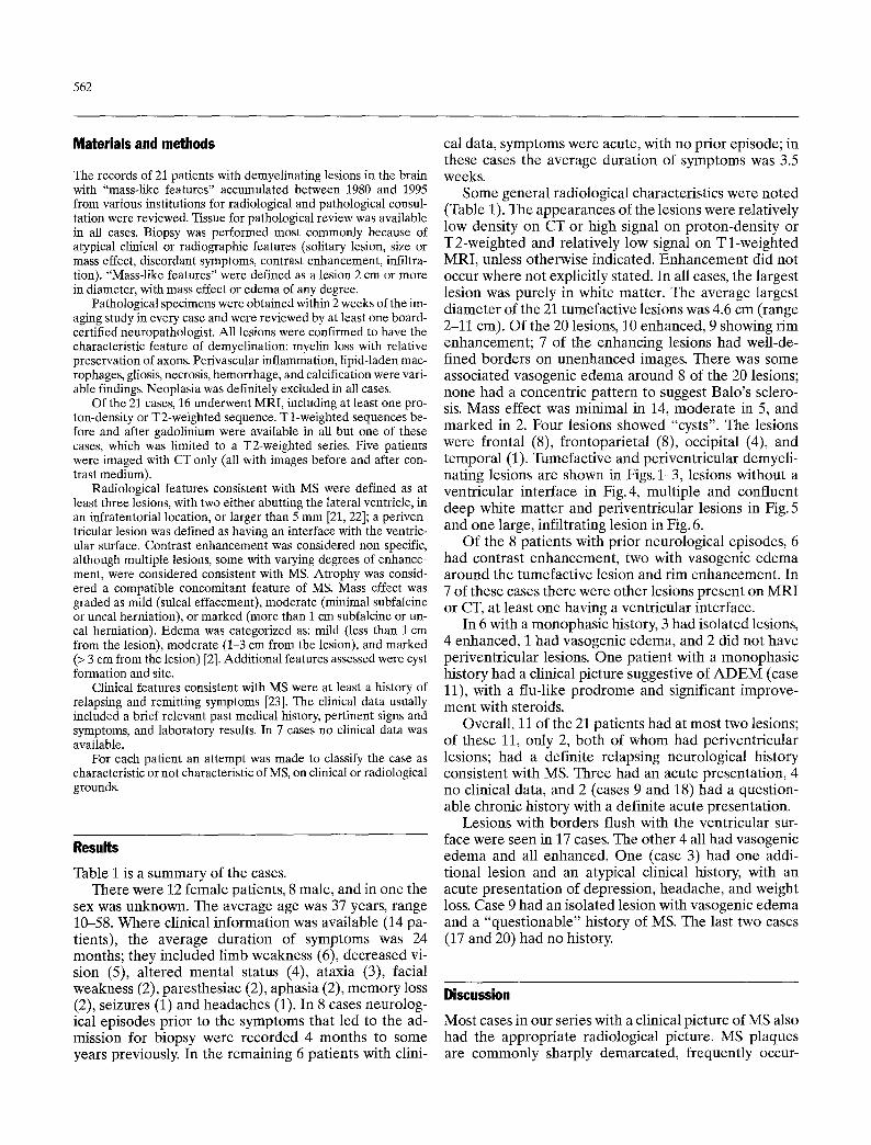
Materials and methods
The records of 21 patients with demyelinating lesions in the brain with "mass-like features" accumulated between 1980 and 1995 from various institutions for radiological and pathological consul- tation were reviewed. Tissue for pathological review was available in all cases. Biopsy was performed most commonly because of atypical clinical or radiographic features (solitary lesion, size or mass effect, discordant symptoms, contrast enhancement, infiltra- tion). "Mass-like features" were defined as a lesion 2 cm or more in diameter, with mass effect or edema of any degree.
Pathological specimens were obtained within 2 weeks of the im- aging study in every case and were reviewed by at least one board- certified neuropathologist. All lesions were confirmed to have the characteristic feature of demyelination: myelin loss with relative preservation of axons. Perivascular inflammation, lipid-laden mac- rophages, gliosis, necrosis, hemorrhage, and calcification were vari- able findings. Neoplasia was definitely excluded in all cases.
Of the 21 cases, 16 underwent MRI, including at least one pro- ton-density or T 2-weighted sequence. T 1-weighted sequences be- fore and after gadolinium were available in all but one of these cases, which was limited to a T2-weighted series. Five patients were imaged with CT only (all with images before and after con- trast medium).
Radiological features consistent with MS were defined as at least three lesions, with two either abutting the lateral ventricle, in an inffatentorial location, or larger than 5 mm [21, 22]; a periven- tricular lesion was defined as having an interface with the ventric- ular surface. Contrast enhancement was considered non specific, although multiple lesions, some with varying degrees of enhance- ment, were considered consistent with MS. Atrophy was consid- ered a compatible concomitant feature of MS. Mass effect was graded as mild (sulcal effacement), moderate (minimal subfalcine or uncal herniation), or marked (more than 1 cm subfalcine or un- cal herniation). Edema was categorized as: mild (less than 1 cm from the lesion), moderate (1-3 cm from the lesion), and marked (> 3 cm from the lesion) [2]. Additional features assessed were cyst formation and site.
Clinical features consistent with MS were at least a history of relapsing and remitting symptoms [23]. The clinical data usually included a brief relevant past medical history, pertinent signs and symptoms, and laboratory results. In 7 cases no clinical data was available.
For each patient an attempt was made to classify the case as characteristic or not characteristic of MS, on clinical or radiological grounds.
Results
Table I is a summary of the cases. There were 12 female patients, 8 male, and in one the
sex was unknown. The average age was 37 years, range 10-58. Where clinical information was available (14 pa- tients), the average duration of symptoms was 24 months; they included limb weakness (6), decreased vi- sion (5), al tered mental status (4), ataxia (3), facial weakness (2), paresthesiae (2), aphasia (2), m e m o r y loss (2), seizures (1) and headaches (1). In 8 cases neurolog- ical episodes prior to the symptoms that led to the ad- mission for biopsy were recorded 4 months to some years previously. In the remaining 6 patients with clini-
cal data, symptoms were acute, with no prior episode; in these cases the average duration of symptoms was 3.5 weeks.
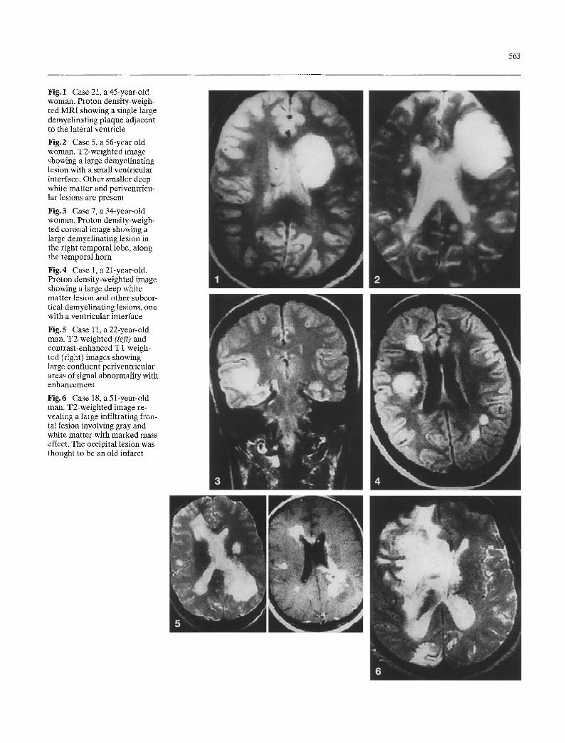
Some general radiological characteristics were noted (Table 1). The appearances of the lesions were relatively low density on CT or high signal on proton-densi ty or T2-weighted and relatively low signal on T 1-weighted MRI, unless otherwise indicated. Enhancemen t did not occur where not explicitly stated. In all cases, the largest lesion was purely in white matter . The average largest d iameter of the 21 tumefact ive lesions was 4.6 cm (range 2-11 cm). Of the 20 lesions, 10 enhanced, 9 showing rim enhancement ; 7 of the enhancing lesions had well-de- fined borders on unenhanced images. There was some associated vasogenic edema around 8 of the 20 lesions; none had a concentric pa t tern to suggest Balo 's sclero- sis. Mass effect was minimal in 14, modera t e in 5, and marked in 2. Four lesions showed "cysts". The lesions were frontal (8), f rontoparie ta l (8), occipital (4), and tempora l (1). Tumefact ive and periventr icular demyeli- nating lesions are shown in Figs. 1-3, lesions without a ventricular interface in Fig. 4, multiple and confluent deep white mat te r and periventricular lesions in Fig. 5 and one large, infiltrating lesion in Fig. 6.
Of the 8 patients with prior neurological episodes, 6 had contrast enhancement , two with vasogenic edema around the tumefact ive lesion and r im enhancement . In 7 of these cases there were other lesions present on M R I or CT, at least one having a ventricular interface.
In 6 with a monophas ic history, 3 had isolated lesions, 4 enhanced, I had vasogenic edema, and 2 did not have periventricular lesions. One pat ient with a monophas ic history had a clinical picture suggestive of A D E M (case 11), with a flu-like p rodrome and significant improve- ment with steroids.
Overall , 11 of the 21 patients had at most two lesions; of these 11, only 2, both of whom had periventricular lesions; had a definite relapsing neurological history consistent with MS. Three had an acute presentation, 4 no clinical data, and 2 (cases 9 and 18) had a question- able chronic history with a definite acute presentation.
Lesions with borders flush with the ventricular sur- face were seen in 17 cases. The other 4 all had vasogenic edema and all enhanced. One (case 3) had one addi- tional lesion and an atypical clinical history, with an acute presentat ion of depression, headache, and weight loss. Case 9 had an isolated lesion with vasogenic edema and a "quest ionable" history of MS. The last two cases (17 and 20) had no history.
Discussion
Most cases in our series with a clinical picture of MS also had the appropr ia te radioIogical picture. MS plaques are commonly sharply demarcated, frequently occur-
563
Fig. 1 Case 21, a 45-year-old woman. Proton density-weigh- ted MRI showing a single large demyelinating plaque adjacent to the lateral ventricle
Fig.2 Case 5, a 56-year-old woman. T2-weighted image showing a large demyelinating lesion with a small ventricular interface. Other smaller deep white matter and periventricu- lar lesions are present
Fig.3 Case 7, a 34-year-old woman. Proton density-weigh- ted coronal image showing a large demyelinating lesion in the right temporal lobe, along the temporal horn
Fig.4 Case 1, a 21-year-old. Proton density-weighted image showing a large deep white matter lesion and other subcor- tical demyelinating lesions, one with a ventricular interface
Fig.5 Case 11, a 22-year-old man. T2-weighted (left) and contrast-enhanced T 1 weigh- ted (right) images showing large confluent periventricular areas of signal abnormality with enhancement
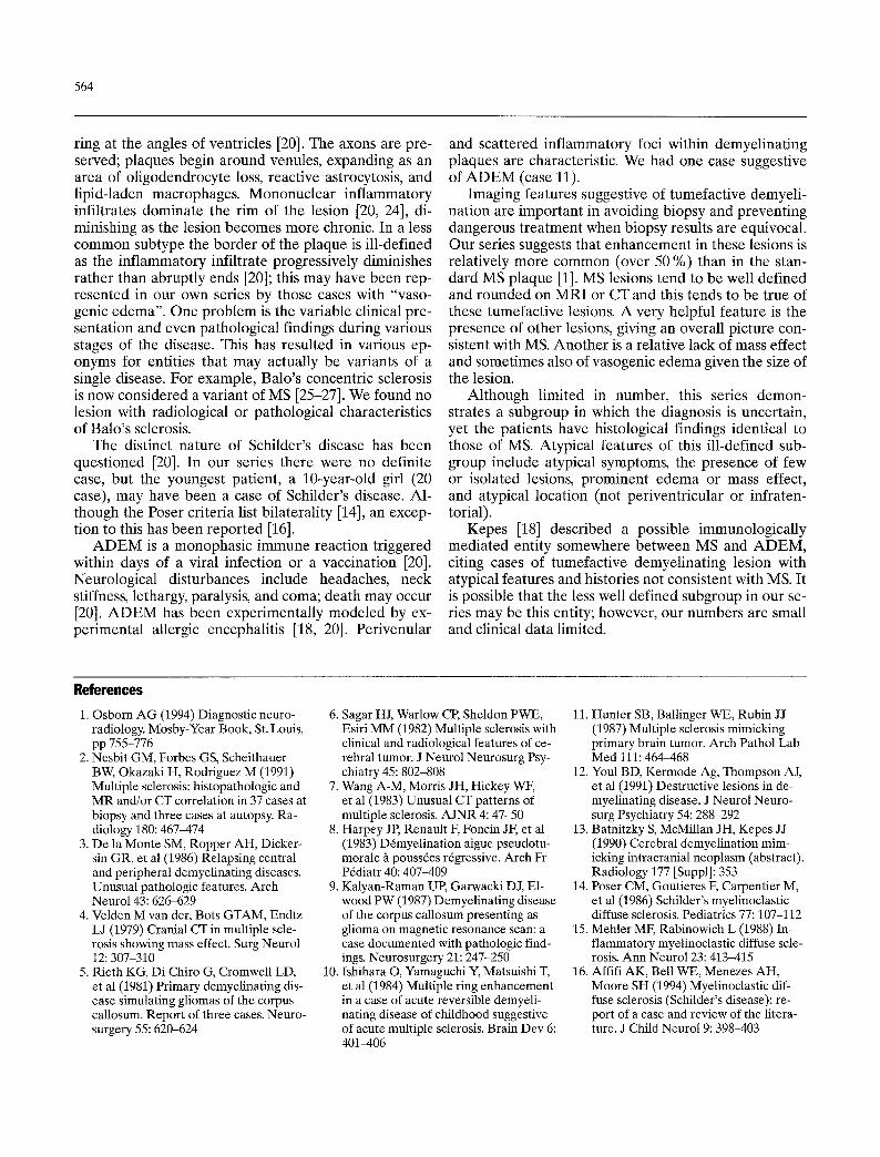
Fig.6 Case 18, a 51-year-old man. T2-weighted image re- vealing a large infiltrating fron- tal lesion involving gray and white matter with marked mass effect. The occipital lesion was thought to be an old infarct
564
ring at the angles of ventricles [20]. The axons are pre- served; plaques begin around venules, expanding as an area of oligodendrocyte loss, reactive astrocytosis, and lipid-laden macrophages. Mononuclear inflammatory infiltrates dominate the rim of the lesion [20, 24], di- minishing as the lesion becomes more chronic. In a less common subtype the border of the plaque is ill-defined as the inflammatory infiltrate progressively diminishes rather than abruptly ends [20]; this may have been rep- resented in our own series by those cases with "vaso- genic edema". One problem is the variable clinical pre- sentation and even pathological findings during various stages of the disease. This has resulted in various ep- onyms for entities that may actually be variants of a single disease. For example, Balo's concentric sclerosis is now considered a variant of MS [25-27]. We found no lesion with radiological or pathological characteristics of Balo's sclerosis.
The distinct nature of Schilder's disease has been questioned [20]. In our series there were no definite case, but the youngest patient, a 10-year-old girl (20 case), may have been a case of Schilder's disease. Al- though the Poser criteria list bilaterality [14], an excep- tion to this has been reported [16].
ADEM is a monophasic immune reaction triggered within days of a viral infection or a vaccination [20]. Neurological disturbances include headaches, neck stiffness, lethargy, paralysis, and coma; death may occur [20]. ADEM has been experimentally modeled by ex- perimental allergic encephalitis [18, 20]. Perivenular
and scattered inflammatory loci within demyelinating plaques are characteristic. We had one case suggestive of ADEM (case 11).
Imaging features suggestive of tumefactive demyeli- nation are important in avoiding biopsy and preventing dangerous treatment when biopsy results are equivocal. Our series suggests that enhancement in these lesions is relatively more common (over 50 %) than in the stan- dard MS plaque [1]. MS lesions tend to be well defined and rounded on MRI or CT and this tends to be true of these tumefactive lesions. A very helpful feature is the presence of other lesions, giving an overall picture con- sistent with MS. Another is a relative lack of mass effect and sometimes also of vasogenic edema given the size of the lesion.
Although limited in number, this series demon- strates a subgroup in which the diagnosis is uncertain, yet the patients have histological findings identical to those of MS. Atypical features of this ill-defined sub- group include atypical symptoms, the presence of few or isolated lesions, prominent edema or mass effect, and atypical location (not periventricular or infraten- torial).
Kepes [18] described a possible immunologically mediated entity somewhere between MS and ADEM, citing cases of tumefactive demyelinating lesion with atypical features and histories not consistent with MS. It is possible that the less well defined subgroup in our se- ries may be this entity; however, our numbers are small and clinical data limited.
References
1. Osborn AG (1994) Diagnostic neuro- radiology. Mosby-Year Book, St. Louis, pp 755-776
2. Nesbit GM, Forbes GS, Scheithauer BW, Okazaki H, Rodriguez M (1991) Multiple sclerosis: histopathologic and MR and/or CT correlation in 37 cases at biopsy and three cases at autopsy. Ra- diology 180:467-474
3. De la Monte SM, Ropper AH, Dicker- sin GR, et al (1986) Relapsing central and peripheral demyelinating diseases. Unusual pathologic features. Arch Neuro143:626-629
4. Velden M van der, Bots GTAM, Endtz LJ (1979) Cranial CT in multiple scle- rosis showing mass effect. Surg Neurol 12:307-310
5. Rieth KG, Di Chiro G, Cromwell LD, et al (1981) Primary demyelinating dis- ease simulating gliomas of the corpus callosum. Report of three cases. Neuro- surgery 55:620-624
6. Sagar HJ, Warlow CR Sheldon PWE, Esiri MM (1982) Multiple sclerosis with clinical and radiological features of ce- rebral tumor. J Neurol Neurosurg Psy- chiatry 45:802-808
7. Wang A-M, Morris JH, Hickey WF, et al (1983) Unusual CT patterns of multiple sclerosis. AJNR 4:47-50
8. Harpey JR Renault F, Foncin JF, et al (1983) D6myelination aigue pseudotu- morale ~t pouss6es r6gressive. Arch Fr P4diatr 40:407-409
9. Kalyan-Raman UR Garwacki D J, El- wood PW (1987) Demyelinating disease of the corpus callosum presenting as glioma on magnetic resonance scan: a case documented with pathologic find- ings. Neurosurgery 21:247-250
10. Ishihara O, Yamaguchi Y, Matsuishi T, et al (1984) Multiple ring enhancement in a case of acute reversible demyeli- nating disease of childhood suggestive of acute multiple sclerosis. Brain Dev 6: 401-406
11. Hunter SB, Ballinger WE, Rubin JJ (1987) Multiple sclerosis mimicking primary brain tumor. Arch Pathol Lab Med 111:464-468
12. Youl BD, Kermode Ag, Thompson AJ, et al (1991) Destructive lesions in de- myelinating disease. J Neurol Neuro- surg Psychiatry 54:288-292
13. Batnitzky S, McMillan JH, Kepes JJ (1990) Cerebral demyelination mim- icking intracranial neoplasm (abstract). Radiology 177 [Suppl]: 353
14. Poser CM, Goutieres F, Carpentier M, et al (1986) Schilder's myelinoclastic diffuse sclerosis. Pediatrics 77:107-112
15. Mehler MF, Rabinowich L (1988) In- flammatory myelinoclastic diffuse scle- rosis. Ann Neurol 23:413-415
16. Affifi AK, Bell WE, Menezes AH, Moore SH (1994) Myelinoclastic dif- fuse sclerosis (Schilder's disease): re- port of a case and review of the litera- ture. J Child Neurol 9:398-403

565
17. Dresser LR Tourian AY, Anthony DC (1988) A case of myelinoclastic diffuse sclerosis in a young adult. Neurology 41: 316-318
18. Kepes JJ (1993) Large focal tumor-like demyelinating lesions of the brain: in- termediate entity between multiple sclerosis and acute disseminated en- cephalomyelitis? A study of 31 patients. Ann Neuro133:18-27
19. Tersegno MM, Reich DR (1993) Balo concentric sclerosis: a rare form of mul- tiple sclerosis manifested as a dominant cerebral mass without other white mat- ter lesions on MR. A J R 160:901
20. Morris JH, Schoene WC (1984) The nervous system. In: Robbins SL, Cotran RS, Kumar V (eds) Pathologic basis of disease. Saunders, Philadelphia, pp 1410-1413
21. Offenbacher H, Fazekas F, Schmidt R, et al (1993) Assessment of MRI criteria for diagnosis of multiple sclerosis. Neu- rology 43:905-909
22. Fazekas F, Offenbacher H, Fuchs S, et al (1988) Criteria for an increased speci- ficity of MRI interpretation in elderly subjects with suspected multiple sclero- sis. Neurology 38:1822-1825
23. Sadiq SA, Miller JR (1995) Demyeli- nating disease. In: Rowland PL (ed) Merritt's textbook of neurology. Wil- liams & Wilkins, Philadelphia, p 819
24. Burger PC, Scheithauer BW (1993) Tu- mors of the central nervous system. Armed Forces Institute of Pathology, Washington, pp 391-394
25. Korte JH, Bom ER Vos LD, Breuer TJ, Wondergem JH (1994) Balo concentric sclerosis: MR diagnosis. AJNR 15: 1284-1285
26. Morioka C, Komatsu Y, Tsujio T, Araki Y, Kondo H (1994) The evolution of the concentric lesions of atypical multiple sclerosis on MRI. Radiat Med 12: 129- 133
27. Gharagozloo AM, Poe LB, Collins GH (1994) Antemortem diagnosis of Balo concentric sclerosis: correlative MR imaging and pathological features. Ra- diology 191:817-819
5th International MRI Course 27-30 October 1996, Riyadh, Saudi Arabia Information: Department of Postgraduate and Academic Affairs, Riyadh Armed For- ces Hospital, R O. Box 7897, Riyadh 11159, Saudi Arabia, Tel. 0 09 66-1-477 7714 Ext: 49 33/4937, Fax 009 66-1-476 08 53
Ostseesymposium '96 Interdisciplinary Radiologic-Neuroradiologic Symposium 8/9 November 1996, Liibeck, Germany Information: Dr. H. Twilfer/Frau M. Bi- schof, Institut ftir Radiologie, Medizinische Universitfit zu Ltibeck, Ratzeburger Allee 160, D-23538 Ltibeck, Germany, Tel. 04 51-5 00-2128/2129/64 47, Fax: 04 51-5 00- 64 97
The British Cervical Spine Society Meeting 8/9 November 1996, Newcastle upon Tyne, UK Information: Mr N Todd, Newcastle Gen- eral Hospital, Westgate Road, Newcastle upon Tyne NE4 6BE, UK, Tel. 0191 273 811, Ext: 22472, Fax: 0191 272 2641
MRI Update 1997 Brain, Spine, Head, Neck and Pediatrics New Techniques: MR Spectroscopy and Functional Imaging 23-28 February 1997, Medical College of Wisconsin, Tucson, Arizona, USA Information: Marti Carter, CME, Inc., 11011 W. North Avenue, Milwaukee, WI 53226, Phone and Fax: (414) 7 71-95 20, E-Mail: [email protected]
7th Pan-Arab Union of Neurological Sciences Conference 2-5 March 1997, Riyadh, Saudi Arabia Information: Department of Postgraduate and Academic Affairs, Riyadh Armed For- ces Hospital, P. O. Box 7897, Riyadh 11159, Saudi Arabia, Tel. 0 09 66-1-477 7714 Ext: 4933/49 37, Fax 0 09 66-1-4 76 08 53
Consensus Conference: Complex Hydrocephalus and Hydrocephalus Complications April 1997, Assisi, Italy Information: C. Di Rocco/F. Velardi, Sec- tion of Pediatric Neurosurgery, Catholic University Medical School, Policlinico Ge- melli, Largo Gemelli, 8, 1-00168 Rome, Italy, Tel. +39-6-3015-4495, Fax +39-6-305- 13-43
XXV~ Congr~s de la Soci~t(~ Franqaise de Neuroradiologie 10-11 April 1997, Strasbeurg Information: Prof. J. L. Dietemann, Service de Radiologie 2, CHU de Hautepierre, F-67098 Strasbourg Cedex, France, Tel. 88127889, Fax 88127118
Copyright © 2022 FDOKUMEN