Echographic Visualization of Lesions of the Living Intact ...
12
Echographic Visualization of Lesions of the Living Intact Human Breast* J. J. WILDJ ANDJOHNM. (Department of Electrical Engineering, University of Minnesota, and St. Barnabas Hospital, Minneapolis, Minn.) The purpose of this report is to show that the nature of lesions or tissue abnormalities of the living, intact human breast can be determined by the use of pulsed ultrasound. The study is not necessarily directed toward cancer detection and is not necessarily intended to replace existing technics of detection and diagnosis of breast le sions, but rather is aimed at the more fundamental question of whether or not the histological struc ture of tissue in general can be determined by ultrasonic technics. The breast was selected as a convenient acces sible organ for direct studies on the patient and because of the certainty of obtaining a quick microscopic diagnosis of the lesions. This study is one of the steps toward the examination of less accessible sites. HISTOHY The history of the various technics for using sound energy as a means of examination and de tection, applied mainly to industrial and military fields, is described by Carlin (2). There has been a surprising delay in similarly applying sound energy to biology. One reason for the delay has appar ently been the difference in outlook between the physicist and the biologist. The gap was partially bridged in 1947 when Dussik, Dussik, and Wyt, a medico-physicist team in Europe, reported attempts to outline the ven tricles of the brain in life (3). The Dussik method is a transmission technic in which sound is driven through the skull from one side and is picked up on the opposite side by a receiver. In this country in 1950 Ballantine, Bolt, Heutcr, and Ludwig re ported similar attempts to outline the ventricles * This investigation was supported by a research grant from the National Cancer Institute, Department of Health, Education, and Welfare, U.S. Public Health Service. t Dr. Wild and Mr. Reid are presently Director and Chief Electronic Engineer, respectively, of the Medico-Technological Research Department of St. Barnabas Hospital, Minneapolis, Minn. Received for publication October 5, 1953. (1). In 1952 Giittner reported that it is not possible to outline the ventricles by a transmission method because of the interposition of the skull (9), but modification of the existing methods may yet realize the Dussiks' objective. Another specialized application was made in 1949 by Ludwig and Struthers (12). Using com mercial ultrasonic flaw-detecting equipment (5), they showed that gallstones and foreign bodies buried in the muscles of dogs could be detected. These workers stated the opinion that the multiple reflections which they obtained from the soft tis sues were too erratic to be of practical value. They suggested, however, that refinements in technic might make the detection of tumors possible. The principle of their method was to drive sound into the dogs' tissues and to detect echoes returning from the junctions between the inserted materials and the muscles. In February, 1950, Wild reported studies di rected toward the measurement of biological tissue elements and the detection of tissue irregularities (14). The ultrasonic examination of tissues in terms of cellular composition presents formidable prob lems, both technical and biological. Because of the small dimensions of tissue elements, high fre quencies were considered necessary in order to ob tain good definition. At high frequencies, with the consequently shorter wave lengths, tissues could no longer be considered homogeneous by the physicist. Worse yet, as far as could be foreseen, the range would be limited at high frequencies. Since a suitable refined apparatus for prelimi nary exploratory tissue studies had already been developed by the U.S. Navy for purely military purposes during World War II, the senior author was able to apply biological technics without much specific knowledge of the extremely complex electronic apparatus, operating at a frequency of 15 million cycles per second and timing intervals as short as one millionth of a second. Preliminary studies by Wild (14) applying biological methods appeared sufficiently encourag- 277
-
Upload
khangminh22 -
Category
Documents
-
view
0 -
download
0
Transcript of Echographic Visualization of Lesions of the Living Intact ...

Echographic Visualization of Lesions ofthe Living Intact Human Breast*
J. J. WILDJ ANDJOHNM.
(Department of Electrical Engineering, University of Minnesota, and St. Barnabas Hospital, Minneapolis, Minn.)
The purpose of this report is to show that thenature of lesions or tissue abnormalities of theliving, intact human breast can be determined bythe use of pulsed ultrasound. The study is notnecessarily directed toward cancer detection andis not necessarily intended to replace existingtechnics of detection and diagnosis of breast lesions, but rather is aimed at the more fundamentalquestion of whether or not the histological structure of tissue in general can be determined byultrasonic technics.
The breast was selected as a convenient accessible organ for direct studies on the patient andbecause of the certainty of obtaining a quickmicroscopic diagnosis of the lesions. This study isone of the steps toward the examination of lessaccessible sites.
HISTOHYThe history of the various technics for using
sound energy as a means of examination and detection, applied mainly to industrial and militaryfields, is described by Carlin (2). There has been asurprising delay in similarly applying sound energyto biology. One reason for the delay has apparently been the difference in outlook between thephysicist and the biologist.
The gap was partially bridged in 1947 whenDussik, Dussik, and Wyt, a medico-physicist teamin Europe, reported attempts to outline the ventricles of the brain in life (3). The Dussik methodis a transmission technic in which sound is driventhrough the skull from one side and is picked upon the opposite side by a receiver. In this countryin 1950 Ballantine, Bolt, Heutcr, and Ludwig reported similar attempts to outline the ventricles
* This investigation was supported by a research grantfrom the National Cancer Institute, Department of Health,Education, and Welfare, U.S. Public Health Service.
t Dr. Wild and Mr. Reid are presently Director and ChiefElectronic Engineer, respectively, of the Medico-TechnologicalResearch Department of St. Barnabas Hospital, Minneapolis,Minn.
Received for publication October 5, 1953.
(1). In 1952 Giittner reported that it is not possibleto outline the ventricles by a transmission methodbecause of the interposition of the skull (9), butmodification of the existing methods may yetrealize the Dussiks' objective.
Another specialized application was made in1949 by Ludwig and Struthers (12). Using commercial ultrasonic flaw-detecting equipment (5),they showed that gallstones and foreign bodiesburied in the muscles of dogs could be detected.These workers stated the opinion that the multiplereflections which they obtained from the soft tissues were too erratic to be of practical value. Theysuggested, however, that refinements in technicmight make the detection of tumors possible. Theprinciple of their method was to drive sound intothe dogs' tissues and to detect echoes returning
from the junctions between the inserted materialsand the muscles.
In February, 1950, Wild reported studies directed toward the measurement of biological tissueelements and the detection of tissue irregularities(14).
The ultrasonic examination of tissues in termsof cellular composition presents formidable problems, both technical and biological. Because of thesmall dimensions of tissue elements, high frequencies were considered necessary in order to obtain good definition. At high frequencies, with theconsequently shorter wave lengths, tissues couldno longer be considered homogeneous by thephysicist. Worse yet, as far as could be foreseen,the range would be limited at high frequencies.
Since a suitable refined apparatus for preliminary exploratory tissue studies had already beendeveloped by the U.S. Navy for purely militarypurposes during World War II, the senior authorwas able to apply biological technics withoutmuch specific knowledge of the extremely complexelectronic apparatus, operating at a frequency of15 million cycles per second and timing intervalsas short as one millionth of a second.
Preliminary studies by Wild (14) applyingbiological methods appeared sufficiently encourag-
277

278 Cancer Research
ing to warrant experimental application to stomach cancer tissue. Experimental work by French,Wild, and Neal (6, 8) on fresh post mortem braintissues pointed to the possibility of detecting anddiagnosing brain lesions after operative removal ofa portion of the skull. Hannlessness of the apparatus was confirmed with animals (7) and byapplication to the senior author's arm (15). Two
cases of living, intact, human breast lesions werecorrectly forecast before biopsy (15). The application of the method to a living brain lesion was recorded (13, 18) and was followed by a series of 21cases from the living human breast (17) whichgave further evidence of the diagnostic accuracy ofthe method.
Wild and Reid (16) reported the developmentof a pictorial method of visualizing living tissueswith their original apparatus. Howry (10), using asimilar picturial method, reported visualization ofthe structural elements of the mid-forearm.
TERMINOLOGY
Wild and Reid suggested the general term,"Echography" for the whole subject of examina
tion of biological tissues by means of ultrasonicechoes (16), regardless of the methods used fordisplaying the information obtained. The basicelectronic machine was called the "Echograph,"the applicator units (probes) "Echoscopes," andthe records "Echograms." A uni-dimensional ech-
ogram (such as those in Chart 2) is the display ofecho amplitude versus depth, i.e., along one dimension of the tissue. A two-dimensional echograni(such as those in Figures 1-7) is a map of echo in
tensity over a plane, i.e., a cross section extendinginto the tissue. This terminology allows furtherexpansion into a third dimension (17).
PRINCIPLEOFTHEECHOGRAPH
The principle of the instrument is basicallysimple. The interrelationship of the components ofthe electronic system is shown in Chart 1.
An electronic clock (1) times the bursts of soundenergy (60-1,000 c.p.s.) and starts the traces onthe face of the cathode-ray tube (center). Thetransmitter (2), upon receipt of the pulses from(1), creates electrical impulses to excite the piezoelectric crystal (3) in the echoscope. The pulses ofelectrical energy, converted to pulses of soundenergy at the crystal (3), travel into the tissues in anarrow beam. The series of echoes returning fromthe tissues are received by the same crystal,quiescent between pulses, and are amplified byunit 4 to deflect the traces on the cathode ray tubeas shown in Chart 1 (center).
The construction of the echograph has beendescribed in more detail elsewhere (11, 13).
UNI-DIMENSIONALECHOORAPHY
Chart 2 is a comprehensive representation of theway in which some "meaning" has been obtained
from the echo-patterns which appeared confusingto other workers (10,12). The left half of the chartcontains a series of drawings concerned with ahypothetical lesion of the breast. The right halfshows similar data concerning normal breast tissue.
The pulsed sound beam leaves the piezo-electriccrystal O, passes through the water contained inthe echoscope, through the rubber membrane andskin, and into the breast tissue at A. The beampasses into the lesion at B and leaves the lesionatC.
CHART 1.—Diagrammatic drawing of the operation of the
echograph.
The resulting echogram No. 1 is shown with theecho from A shown at A', and similarly for the
other letters. Since the sound velocities in varioustissue elements are nearly identical, the distancesbetween echoes on the echogram are closely proportional to the distances between the sound reflecting structures in the tissue.
If the velocity of sound in tissue is known, thesize of a lesion can be calculated if recognizabledefinitive echoes are produced, such as B' and C'.
The portion of the echograms representing thewater column (O'A') is constant and is deletedfrom the subsequent echograms. The signal at D'
represents the limit of depth.As the sound pulses enter the tissue, very strong
echoes are produced (A'B') which exceed the
present linear range of the machine so that theseparate echoes from the water-membrane-skin-tissue interfaces are not distinguished. Fortunately, it is not necessary to be able to identify indi-

UNI-DIMENSIONAL ECHOGRAPHY
LESIONSkin
NORMAL CONTROL TISSUESkin
Echoscopei in
Breast tissue
Soundbeam!
Water
093
Case
Solidbenignlesion
Depth or time base Case 2
Cancerouslesion HLUW^^J?
Depth or time base Case 4
CHART2.—Representative uni-dimensional echograms
280 Cancer Research
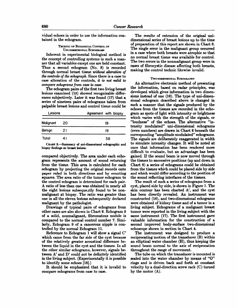
vidual echoes in order to use the information contained in the echogram.
TECHNICop BIOLOGICALCONTROLOFUNI-DIMENSIONALECHOGRAMS
Inherent in experimental biological method isthe concept of controlling systems in such a manner that all variables except one are held constant.Thus a second echogram (No. 3) is recordedthrough normal breast tissue without alteration ofthe controls of the echograph. Since there is a case tocase alteration of the controls, it is not valid tocompare echograms from case to case.
The echogram pairs of the first two living breastlesions examined (15) showed recognizable differences subjectively. Later it was found (17) that aseries of nineteen pairs of echograms taken frompalpable breast lesions and control tissue could be
Lesions Agreement with biopsy
Malignant 20 19
Benign 21 19
Total 41 38CHABTS.—Summary of uni-dimensional echographic and
biopsy findings on breast lesions.
compared objectively. The area under each echo-gram represents the amount of sound returningfrom the tissue. This area is calculated for eachechogram by projecting the original records ontopaper ruled in both directions and by countingsquares. The area ratio of the tumor echogram tothe control echogram is determined for each pair.A ratio of less than one was obtained in nearly allthe eight lesions subsequently found to be non-malignant at biopsy. The ratio was greater thanone in all the eleven lesions subsequently declaredmalignant by the pathologist.
Drawings of typical pairs of echograms fromother cases are also shown in Chart 2. Echogram 5of a solid, nonmalignant, fibromatous nodule iscompared to the normal control number 7. Similarly, Echogram 9 of a cancerous nipple is controlled by the normal Echogram 11.
Reference to Echogram 1 will show a signal C'
which came from the far side of the cyst becauseof the relatively greater acoustical difference between the liquid in the cyst and the tissues. In allthe other similar echograms, however, signals between A' and D' could not be definitely identified
in the living subject. (Experimentally it is possibleto identify some echoes [18].)
It should be emphasized that it is invalid tocompare echograms from case to case.
The results of extension of the original unidimensional series of breast lesions up to the timeof preparation of this report are shown in Chart 3.The single error in the malignant group occurredin a case where both breasts were atrophie so thatno normal breast tissue was available for control.The two errors in the nonmalignant group were incases of fibrocystic disease affecting both breasts,making the control technic likewise invalid.
TWO-DIMENSIONALECHOGRAPHY
An alternative electronic method of presentingthe information, based on radar principles, wasdeveloped which gives information in two dimensions instead of one (16). The type of uni-dimensional echogram described above is changed insuch a manner that the signals produced by theechoes from the tissues are recorded in the echo-gram as spots of light with intensity or brightnesswhich varies with the strength of the signals, or"loudness" of the echoes. The alternative "intensity modulated" uni-dimensional echograms
(even numbers) are drawn in Chart 2 beneath thecorresponding "amplitude modulated" echograms.
The signals are deliberately exaggerated in widthto simulate intensity changes. It will be noted atonce that information has been rendered moredifficult to evaluate, but an advantage has beengained. If the sound beam is now moved throughthe tissues to successive positions (up and down inChart 2) a series of echograms could be obtainedfrom the tissues whifh could be placed side by sideand which would differ according to the position ofthe sound reflecting interfaces of the tissues.
The result of such a series of echograms from acyst, placed side by side, is shown in Figure 1. Theskin contour has been charted A', and the cyst
has been directly revealed. An instrument wasconstructed (16), and two-dimensional echogramswere obtained of kidney tissue and of a tumor in aliving subject. Echograms of a malignant breasttumor were reported in the living subject with thesame instrument (17). The first instrument gavevaluable information for the construction of asecond improved body-surface two-dimensionalechoscope shown in section in Chart 4.
The instrument was designed to produce areciprocating motion of the transducer (B) withinan elliptical water chamber (H), thus keeping thesound beam normal to the axis of reciprocationthroughout the range of movement.
The tube on which the transducer is mounted issealed into the water chamber by means of "O"
rings and is driven back and forth at constantvelocity by a dual-direction screw rack (C) turnedby the motor (A).

WILD ANDREID—Echographsof Living Tissues 281
Coupled to the reciprocating mass is the rectilinear potentiometer (E) from which a signal istaken to position the scanning line on the face ofthe 12DP7 cathode-ray tube. The information received from the moving transducer causes thescanning line to brighten at points along its lengthso as to draw out the image of the tissue structure.
The 15-mc. quartz crystal is connected to amatching network (G) by a flexible coaxial cable(F). A photograph of the actual echoscope isshown in Figure 8.
The conclusions drawn from preliminary unidimensional studies were that nonmalignant tumors of the breast returned as much sound or lessthan did normal tissue at comparable depths in thetissues. Conversely malignant tumors returnedmore sound than did normal tissue at comparabledepths. Therefore, provided that the sound beamcould traverse the tumor completely from normal
CHART4.—Schematic drawing of the two-dimensionalechoscope.
tissue to normal tissue, a tumor of either typeshould be revealed by two-dimensional echogra-phy. The nonmalignant tumor would be recognizable as an area of less bright signals occuring at thesame depth as brighter normal areas on either side.The malignant tumor would appear as an area ofbrighter signals occurring at the same depth asmuch less bright normal surrounding signals. Acyst would be clearly outlined because of thestructureless liquid content. The following caseswere taken from current clinical studies.
CLINICALCASESSHOWINGTHERESULTSOFTWO-DIMENSIONALECHOOHAPHT
Case 1.—Mrs.D., age 34, presented for echogra-phy with a lump in the left breast above thenipple 2-2f cms. in diameter. The two-dimensional echogram of the lesion is shown in Figure2. An immediate diagnosis of a cyst (nonmalignant) was made. This diagnosis was confirmed atbiopsy on the following day.
Case 2.—Mrs.A., age 36, was referred for echo-graphy with a hard lump 2-3 cm. in diameterunder the areola near the left nipple. The skin wasnot involved. The two-dimensional echogram is
shown in Figure 3. The group of low-intensity signals under the broken line Y, occurring on thehigh intensity background, can be seen where thesound beam swept through the lesion. The controlechogram of normal tissue showed a uniform overall signal intensity as shown in a typical normalechogram, Figure 5. The uni-dimensional echo-graphic area ratio was 0.90, showing nonmalig-nancy (17). Biopsy revealed the lesion as non-malignant (fibroma).
Case 3.—Thepatient (Miss S., age 43) was referred on July 21, 1953, for echographic examination of a breast nodule 1 cm. in diameter, firstnoticed 2 weeks previously. The nodule wassituated in breast tissue under the margin of theareola at 10 o'clock in the left breast. The skinmoved freely over the nodule. The two-dimensional echogram of the nodule in this case is shownin Figure 4. The tumor can be recognized at (Z) asa group of high intensity signals occurring at adepth in which almost no signals are returnedfrom the normal surrounding tissue. The unidimensional ratio was 1.2, indicating malignancy(17). A cancer (scirrhus carcinoma) was found atbiopsy.
Case 4-—The patient (Mrs. J., age 70) complained of redness and soreness of the left nippleof 5 days duration. Examination revealed a slightenlargement, redness, and firmness of the leftnipple as compared to the right nipple. A clinicaldiagnosis of inflammation of the nipple was made.Echography was carried out on May 16, 1953.The two-dimensional echograms obtained bysweeping the sound beam through the affectedleft nipple and the normal right nipple are shownin Figures 6 and 7, respectively. The nipples canbe seen outlined. A group of high intensity signalscan be seen in echogram No. 6 at (V) not presentin the echogram of the normal nipple No. 7. Theuni-dimensional echograms of this case are shownin Chart 2. The ratio was 1.34. The echogramswere demonstrated and the tumor declared malignant at the centennial meeting of the MinnesotaState Medical Association, May 18,1953. On May23, 1953, the diagnosis was confirmed by biopsy(scirrhus carcinoma). The tumor was 7 mm. indiameter. The two echograms in this case areshown greatly enlarged in Figures 9 and 10.
DISCUSSIONIt should be stressed that the two types of
presentation of data, the graphic or uni-dimensional, and the pictorial or two-dimensional, aretwo methods by which the final output of theechograph is revealed to human perception. Theoutput of the echograph is represented by the

282 Cancer Research
graph of voltages, which voltages are proportionalto the "loudness" of the echoes, plotted againsttime, as seen in the uni-dimensional echogram.This voltage-time graph is the basis of all types ofpresentation. Differences in the more than sixteenpossible ways (4) of using the basic output of theechograph arise in the uses made of the information as to the position of the crystal. The "pictorial" two-dimensional echogram is dramatically
direct, and there is thus a strong temptation toignore disadvantages such as the loss of detail andto overlook the value of the uni-dimensionalechogram, less obvious though actually containingmore information. The two methods are complementary, and both types of echograms are takenroutinely in each case in the current breast studies.Uni-dimensional echography, because of its relative technical simplicity, made possible the preliminary data, which in turn justified the construction of the considerably more complex two-dimensional apparatus.
The area beneath the uni-dimensional echo-gram can be electronically determined instantaneously, and comparisons can likewise be madeso that skill in interpretation can be eliminatedonce the basic data are compiled for a given application.
Relevant echographic studies up to the presenthave been directed towards establishing whetheror not it would be possible to detect tissue ir
regularities of sufficiently small size to make earlydetection of cancer possible and further whetherthe histological nature of tissue irregularities couldbe determined in terms of malignancy and be-nignancy.
By selecting the highest available frequency(15 megacycles) as a starting point for studies inspite of apparent range limitations, the authorsbelieve that they have made possible the earlydetection and diagnosis of irregularities of tissuestructure of sufficiently small size to be of value inthe control of cancer, at common sites within thebody, such as the upper and lower gastrointestinaltract, accessible from the mouth and anus, respectively, the cervix uteri, and prostate. Instruments for preliminary studies at these siteshave already been constructed and will be reported on, together with results, in due course.Palpable tumors accessible from the skin at sitessuch as the breast and probably the thyroid can bediagnosed in situ with the echoscope describedhere. It must be stressed that no specific attempthas yet been made to detect unsuspected lesions atany site.
A great deal of acoustic and electronic development will be necessary to determine whether verysmall foci of abnormal tissue can be detected fromthe skin. Such detection of very small abnormalities will make practical examination of patients ona mass basis. Much less developmental work will
Typical two-dimensional echograms obtained from theliving breast in the current studies are shown. EchogramNumber 1 is oriented with the depth axis horizontal to facilitate comprehension of the transition from uni-dimensional(Chart 2) to two-dimensional display. The rest of the echo-grams have the depth axis vertical. The letters A'-D' superimposed upon the echograms correspond to those in Chart 2.
FIG. 1.—Echogram1 from Case 1 has been modified artificially to show how a series of intensity modulated unidimensional echograms (Chart 2, No. 2) when placed side byside reveal the structure of the lesion.
FIG. 2.—Echogram2 is the actual echogram of the cyst inCase 1 as produced by moving the intensity modulated unidimensional trace through the tissues. It will be noted thatechoes were not returned from the side walls of the cyst, X-X.
FIG. 3.—EchogramS was obtained from Case 2. The lesion(fibroma) was revealed under the broken line Y—Y.
Fio. 4.—EchogramÃfrom Case 3 shows the cancer at Z.FIG. 5.—Echogram5 is a representative echogram from
normal breast tissue.FIGS. 6 and 7.—Echograms6 and 7 show a comparison be
tween the echograms of the affected nipple (No. 6) and thenormal nipple (No. 7) in Case 4. The cancer was revealed bythe white area V within the nipple not present in the normalnipple. The echograms are approximately life size.

Crystal travel
© Water column
Depth

FIG. 8.—A photograph of the two-dimensional body-surface
eohoscope is shown opposite. The water chamber is shown at //The inset shows a view into the water chamber with the crystalin the base to the left. The crystal travel is 6.5 cm.


FIGS. 9 and 10.—Greatly magnified echograms of the nipples in Case ìare shown. The component intensity modulateduni-dimensional echograms can be seen vertically.

' Nipple
Normalnipple

WILDANDREID—Echographsof Living Tissues 283
be necessary for mass internal detection anddiagnosis. The operation of the crystal in a liquid-filled enclosure, necessary for internal studies, wasachieved at the beginning of the studies in 1949.
Biological technics have disadvantages as seenfrom the physicist's frame of reference. Physical
technics appear impossibly precise to the biologist.Standardization of sound phenomena has alwayspresented a challenge to the disciplines of physics.Sound phenomena do not seem strange to thebiologist who rarely, if ever, works to exact standards and who is keenly aware of the deficiencies ofhis technics. Can physics "measure" the esthetic
output of an orchestra as do the human ear andbrain? Ultrasound has become at least one common meeting place for the two disciplines.
SUMMARYA study is reported of the examination of lesions
of the living, intact, human breast using a pulsedultrasonic reflection technic. Terminology is basedon the word "echo," e.g., Echograph, Echogram,
and Echoscope.Two of many possible ways of revealing the
final output of the echograph to human perceptionare described: uni-dimensional echography, comparable to a needle biopsy, and two-dimensionalechography, or pictorial visualization of tissues ina plane. The relationship of the two methods isdescribed.
Examples are given of both types of echogramtaken from current clinical studies. The results ofuni-dimensional studies up to the time of preparation of the report are given, to show how theechograph can diagnose the histological nature oflesions.
The position of this study in the approach tomass examination at suspected cancer sites, suchas the breast, thyroid, upper and lower gastrointestinal tract, cervix, and prostate, is discussed.
What is believed to be the first actual visualization of a cancer within the nipple was achieved(Figs. 9, 10).
ACKNOWLEDGMENTSTo the members of the medical profession of Minneapolis
for their support and referral of suitable cases for our studies.To the Research Committee of St. Barnabas Hospital,
Minneapolis, for providing facilities for the clinical aspects ofthe study.
To many members of the Electrical Engineering Department, University of Minnesota, who watched the development
of the Echograph with interest, gave their time freely, and madehelpful suggestions, particularly to Professor Henry E. Hartig,head of the department, for his kindness in housing theproject.
To Mr. George E. Carlson of Minnesota Rubber and GasketCompany for his kindness in supplying the MolybdenumDisulphide "O" rings used in the two-dimensional body
surface echoscope.
REFERENCES1. BALLANTINE,H. T., JB.; BOLT,R. H.; HEUTER,T. F.; and
LUDWIG,G. D. On the Detection of Intracranial Pathologyby Ultrasound. Science, 112:525-28, 1950.
2. CABLIN,B. Ultrasonics. New York: McGraw-Hill, 1949.3. DUSSIK,K. T.; DUSSIK,F.; and WTT, L. Auf dem Wege
zur Hyperphonographie des Gehirnes. Wien. Med.Wchnschr., 97:425-29, 1947.
4. FINK, D. G. Radar Engineering. New York: McGraw-Hill, 1947.
5. FIRESTONE,F. A. The Supersonic Reflectoscope, an Instrument for Inspecting the Interior of Solid Parts byMeans of Sound Waves. J. Acoustical Soc. America, 17:287-99, 1946.
6. FRENCH,L. A.; WILD, J. J.; and NEAL D. Detection ofCerebral Tumors by Ultrasonic Pulses. Cancer, 3:705-8,1950.
7. . Attempts To Determine Harmful Effects ofPulsed Ultrasonic Vibrations. Ibid., 4:842-44, 1951.
8. . The Experimental Application of Ultrasonics tothe Localization of Brain Tumors. Neurosurgery, 8:198-203, 1951.
9. GÜTTNER,VON W.; FIEDLER,G.; and PATZOLD,J. ÜberUltraschallabbildungen am menschlichen Schädel.Acustica, 2:148-56, 1952.
10. HOWRY,D. H., and BLIBS,W. R. Ultrasonic Visualizationof Soft Tissue Structures of the Body. J. Lab. &Clin. Med.40:579-92, 1952.
11. LARSEN,F. J. Ultrasonic Trainer Circuits. Electronics,19:126-29, 1946.
12. LUDWIG,G. D., and STRÃœTHEHS,F. W. ConsiderationsUnderlying the Use of Ultrasound To Detect Gallstonesand Foreign Bodies in Tissue. Naval Med. Research Inst.Project, NM004:001, Report No. 4, 1949.
13. REID,J. M., and WILD,J. J. Ultrasonic Ranging for CancerDiagnosis. Electronics, 26:136-38, 1952.
14. WILD,J. J. The Use of Ultrasonic Pulses for the Measurement of Biological Tissues and the Detection of TissueDensity Changes. Surgery, 27:183-88,1950.
15. WILD, J. J., and NEAL, D. The Use of High-FrequencyUltrasonic Waves for Detecting Changes of Texture inLiving Tissues. Lancet, 1:655-57, 1951.
16. WILD, J. J., and REID, J. M. The Application of EchoRanging Techniques to the Determination of Structureof Biological Tissues. Science, 115:226-30, 1952.
17. . Further Pilot Echographic Studies on the Histological Structure of Tumors of the Living Intact HumanBreast. Am. J. Path., 28:839-61, 1952.
18. . The Effects of Biological Tissues on 15 MegacyclePuked Ultrasound. J. Acoustical Soc. America, 2C:270-80, 1953.