Radiolucent lesions of the mandible: a pattern-based ...
17
PICTORIAL REVIEW Radiolucent lesions of the mandible: a pattern-based approach to diagnosis Laurène Avril & Tommaso Lombardi & Angeliki Ailianou & Karim Burkhardt & Arthur Varoquaux & Paolo Scolozzi & Minerva Becker Received: 20 September 2013 /Revised: 1 November 2013 /Accepted: 6 November 2013 /Published online: 10 December 2013 # The Author(s) 2013. This article is published with open access at Springerlink.com Abstract Objectives Radiolucent mandibular lesions seen on panoram- ic radiographs develop from both odontogenic and non- odontogenic structures. They represent a broad spectrum of lesions with a varying degree of malignant potential. The purpose of this review is to illustrate the characteristic imaging findings—as well as the clinical and histological features—of common and uncommon radiolucent lesions of the mandible. Methods This review article is based on the retrospective evaluation of 11,725 panoramic radiographs seen in our insti- tution during the past 6 years. It provides a comprehensive, practical approach to the radiological interpretation of radio- lucent lesions of the mandible. To facilitate the diagnostic approach, we have classified radiolucent lesions into two groups: lesions with well-defined borders and those with ill- defined borders. Results Lesion prevalence, age of manifestation, location within the mandible, relationship to dental structures, effect on adjacent structures and characteristic findings at computed tomography (CT), cone beam CT (CBCT) and magnetic resonance imaging (MRI) with diffusion-weighted imaging (DWI) are discussed. Pitfalls including malignant lesions mimicking benign disease and pseudo-lesions are equally addressed. Conclusion Knowledge of the characteristic imaging features of radiolucent mandibular lesions narrows the differential diagnosis and is crucial for the identification of those lesions, where biopsy is indicated for definitive histology. Teaching points • Panoramic X-rays, CT and MRI are essential for the work- up of radiolucent mandibular lesions. • Lesion borders, location within the mandible, relationship to dental structures and tissue characteristics on cross- sectional imaging are indispensable to narrow the differen- tial diagnosis. • High-resolution CT and CBCT play a major role for the assessment of lesion margins and their relationship to im- portant anatomic structures, such as the inferior alveolar nerve. • Although most radiolucent lesions with well-defined sclerot- ic borders are benign, MRI may reveal clinically unsuspect- ed malignant disease. Keywords Mandible . Radiolucent lesions . Panoramic radiography . Computed tomography (CT) . Magnetic resonance imaging (MRI) Abbreviations ADC Apparent diffusion coefficient AAOMS American Association of Oral and Maxillofacial Surgeons BRONJ Bisphosphonate-related osteonecrosis of the jaw CBCT Cone beam computed tomography CT Computed tomography DWI Diffusion-weighted imaging EG Eospinophilic granuloma GCG Giant cell granuloma L. Avril : A. Ailianou : A. Varoquaux : M. Becker (*) Department of Radiology, University Hospital of Geneva, University of Geneva, Rue Gabrielle-Perret-Gentil 4, 1211 Geneva 14, Switzerland e-mail: [email protected] T. Lombardi : P. Scolozzi Department of Maxillo-Facial Surgery, University Hospital of Geneva, University of Geneva, Rue Gabrielle-Perret-Gentil 4, 1211 Geneva 14, Switzerland K. Burkhardt Department of Clinical Pathology, University Hospital of Geneva, University of Geneva, Rue Gabrielle-Perret-Gentil 4, 1211 Geneva 14, Switzerland Insights Imaging (2014) 5:85–101 DOI 10.1007/s13244-013-0298-9
-
Upload
khangminh22 -
Category
Documents
-
view
3 -
download
0
Transcript of Radiolucent lesions of the mandible: a pattern-based ...

PICTORIAL REVIEW
Radiolucent lesions of the mandible: a pattern-basedapproach to diagnosis
Laurène Avril & Tommaso Lombardi &Angeliki Ailianou &
Karim Burkhardt & Arthur Varoquaux & Paolo Scolozzi &Minerva Becker
Received: 20 September 2013 /Revised: 1 November 2013 /Accepted: 6 November 2013 /Published online: 10 December 2013# The Author(s) 2013. This article is published with open access at Springerlink.com
AbstractObjectives Radiolucent mandibular lesions seen on panoram-ic radiographs develop from both odontogenic and non-odontogenic structures. They represent a broad spectrum oflesions with a varying degree of malignant potential. Thepurpose of this review is to illustrate the characteristic imagingfindings—as well as the clinical and histological features—ofcommon and uncommon radiolucent lesions of the mandible.Methods This review article is based on the retrospectiveevaluation of 11,725 panoramic radiographs seen in our insti-tution during the past 6 years. It provides a comprehensive,practical approach to the radiological interpretation of radio-lucent lesions of the mandible. To facilitate the diagnosticapproach, we have classified radiolucent lesions into twogroups: lesions with well-defined borders and those with ill-defined borders.Results Lesion prevalence, age of manifestation, locationwithin the mandible, relationship to dental structures, effecton adjacent structures and characteristic findings at computedtomography (CT), cone beam CT (CBCT) and magneticresonance imaging (MRI) with diffusion-weighted imaging
(DWI) are discussed. Pitfalls including malignant lesionsmimicking benign disease and pseudo-lesions are equallyaddressed.Conclusion Knowledge of the characteristic imaging featuresof radiolucent mandibular lesions narrows the differentialdiagnosis and is crucial for the identification of those lesions,where biopsy is indicated for definitive histology.Teaching points• Panoramic X-rays, CT and MRI are essential for the work-up of radiolucent mandibular lesions.
• Lesion borders, location within the mandible, relationship todental structures and tissue characteristics on cross-sectional imaging are indispensable to narrow the differen-tial diagnosis.
• High-resolution CT and CBCT play a major role for theassessment of lesion margins and their relationship to im-portant anatomic structures, such as the inferior alveolarnerve.
• Although most radiolucent lesions with well-defined sclerot-ic borders are benign, MRI may reveal clinically unsuspect-ed malignant disease.
Keywords Mandible . Radiolucent lesions . Panoramicradiography . Computed tomography (CT) .Magneticresonance imaging (MRI)
AbbreviationsADC Apparent diffusion coefficientAAOMS American Association of Oral and Maxillofacial
SurgeonsBRONJ Bisphosphonate-related osteonecrosis of the jawCBCT Cone beam computed tomographyCT Computed tomographyDWI Diffusion-weighted imagingEG Eospinophilic granulomaGCG Giant cell granuloma
L. Avril :A. Ailianou :A. Varoquaux :M. Becker (*)Department of Radiology, University Hospital of Geneva,University of Geneva, Rue Gabrielle-Perret-Gentil 4,1211 Geneva 14, Switzerlande-mail: [email protected]
T. Lombardi : P. ScolozziDepartment of Maxillo-Facial Surgery, University Hospital ofGeneva, University of Geneva, Rue Gabrielle-Perret-Gentil 4,1211 Geneva 14, Switzerland
K. BurkhardtDepartment of Clinical Pathology, University Hospital of Geneva,University of Geneva, Rue Gabrielle-Perret-Gentil 4,1211 Geneva 14, Switzerland
Insights Imaging (2014) 5:85–101DOI 10.1007/s13244-013-0298-9

HL Hodgkin lymphomaKCOT Keratocystic odontogenic tumourLCH Langerhans cell histiocytosisMRI Magnetic resonance imagingNHL Non-Hodgkin lymphomaOPT OrthopantomographyORN OsteoradionecrosisPET/CT Positron emission tomography combined
with CTPET/MRI Positron emission tomography combined
with MRISBC Simple bone cystSCC Squamous cell carcinomaSUV Standardised uptake value
Introduction
Conventional radiography may reveal a variety of radiolucentlesions in the mandible. These represent a broad spectrum ofodontogenic and non-odontogenic lesions with a varying de-gree of malignant potential. Interpretation of radiolucent le-sions of the mandible can be challenging either because theclinical presentation may be non-specific or because the lesionmay be detected incidentally [1, 2]. In some cases, the diag-nosis will be mainly made based on clinical symptoms. Inother cases, although a thorough clinical evaluation is manda-tory, clinical findings are non-contributory, as the lesion can-not be seen or palpated and laboratory findings are not abnor-mal. Imaging is essential not only for the diagnosis of man-dibular lesions but also to guide therapy and to monitortreatment response. Although common, mandibular lesionsare not frequently imaged by radiologists. Nevertheless, theirrecognition is essential for a rapid and correct diagnosis. Thisreview provides a comprehensive, practical approach to theradiological interpretation of radiolucent lesions of the man-dible and discusses their clinical presentation and pathophys-iology. It focuses on the radiological techniques used and theirrespective role for the assessment of these lesions.
Imaging modalities
Due to the easy access and low radiation dose, conventionalradiographs such as panoramic radiographs(orthopantomographies [OPTs] or panoramix X-rays) as wellas dental intraoral radiographs traditionally form the backbonein the diagnosis of osseous changes in the mandible [1, 3]. Inaddition, the introduction of digital radiographs has lead tofurther dose reduction (up to 80 %) with the possibility ofdensitometric and subtraction techniques [4]. Conventionalradiographs of the mandible, typically OPTs, may revealradiolucent, radiodense or mixed pattern lesions [1, 3]. In
many cases, such as in radicular cysts, the diagnosis isstraightforward and no additional imaging is required fordiagnosis and treatment. As conventional radiographs aretwo-dimensional projections of three-dimensional structures,they have a limited value for the assessment of lesion size, lesionmargins, as well as extension into important anatomic structuresor soft tissues. Computed tomography (CT), cone beam CT(CBCT), magnetic resonance imaging (MRI) and positron emis-sion tomography combined with CT (PET/CT) and more re-cently positron emission tomography combined with MRI(PET/MRI) complement conventional radiographs overcomingthe above-mentioned limitations and providing more specificinformation in terms of diagnosis and therapeutic options.
Thin-slice (1-mm), high-resolution CT with bone windowsettings is mainly used pre-operatively to precisely assesslesion size, margins, destruction and expansion patterns, aswell as the relationship of the lesion to the mandibular canal.Although coronal slices are sufficient in many situations,dental CT with orthoradial and panoramic reconstructionsare superior to standard coronal reconstructions for the eval-uation of the relationship of a lesion to the dental structuresand to the mandibular canal. Intravenous contrast material ismainly used in cases of suspected jaw infection or in neoplas-tic diseases to assess the intraosseous and extraosseous in-volvement. Although CBCT has gained increasing popularityover the past years, it does not allow evaluation ofextraosseous structures; use of CBCT may therefore lead tounderestimation of disease extent.
High-resolution MRI is mainly used as a complementarytool to CT or CBCT, as it allows precise depiction ofintraosseous and extraosseous lesion components, cyst wallarchitecture (thin versus irregular walls, mural nodules, pap-illary projections), enhancement patterns after intravenousadministration of gadolinium chelates (mild to strong), andtype of soft tissue involvement (displacement versus infiltra-tion) [5]. In inflammatory and infectious lesions, MRI is moresensitive than CT or CBCT for the detection of bone marrowinvolvement [6].
Diffusion weighted imaging (DWI) is a functional MRItechnique based on the assessment of random (Brownian)motion of water molecules. Biological barriers can impairthe free displacement of water molecules, thus resulting inrestricted diffusivity. Restricted diffusivity is seen in a varietyof conditions, including stroke, tumours with increased cellu-larity, infection and inflammation, as well as abscesses.Diffusion in biological tissues can be quantified using theapparent diffusion coefficient (ADC). ADC measurements(in mm2/s) have been shown to be reproducible with excellentintraobserver and interobserver reproducibility in the head andneck [7]. MRI with DWI and ADCmeasurements helps in thedifferential diagnosis of cysts, ameloblastomas and malignanttumours (see below). Although ADC values cannot predict thehistological grade in head and neck squamous cell carcinoma
86 Insights Imaging (2014) 5:85–101

(SCC), lower values are observed in poorly differentiatedlesions whereas higher values are seen in well-differentiatedtumours [7]. In lymphomas, ADC measurements typicallyyield very low values (see below).
As a general rule, PET/CT is uncommonly used for thework-up of mandibular lesions. Nevertheless, it may beemployed as a complementary examination for the stagingof malignant tumours invading the mandible, such as SCC ofthe oral cavity with secondary mandibular invasion, in prima-ry intraosseous SCC or in mandibular lymphoma. In cases ofmetastases to the mandible, PET/CT may reveal the locationof the primary tumour—if unknown—or may effectivelyshow involvement of multiple organs. The most commonlyused radiotracer is 18F-fluorodeoxyglucose (FDG). FDG is aglucose analogue that is taken up by metabolically activetumour cells using facilitated glucose transport. In clinicalroutine, quantification of tracer uptake is performed usingthe standardised uptake value (SUV). High SUVs reflect highglucose metabolism mainly seen in aggressive tumours (typ-ically SCC and lymphoma), while lower SUV values arerather seen in slowly growing, less aggressive tumours, intumours with large areas of necrosis or in inflammatory con-ditions. The recent implementation of integrated hybrid PET/MRI systems in clinical head and neck oncology [8] holdspromise as it combines morphological, functional and molec-ular information at the same time, thereby providing addition-al diagnostic gain. However, research into the potential clin-ical role of PET/MRI in comparison with PET/CT, MRI withDWI or the combination thereof, is still ongoing.
This review article is based on the retrospective evaluation of11,725 panoramic radiographs seen during a period of 6 years atour institution. From a practical point of view, radiolucentmandibular lesions can be divided into lesions with well-defined borders and lesions with poorly defined borders.
Radiolucent lesions with well-defined borders
Radicular cyst
Odontogenic cysts are true cysts arising from the epitheliumleft over from tooth development. Radicular cysts, also calledperiapical cysts or apical periodontal cysts, are the most com-mon odontogenic cysts [1, 2]. Tooth infection may lead tonecrosis of the pulp cavity and may spread to the tooth apexwith ulterior development of a periapical granuloma orperiapical abscess. The latter may subsequently give rise to aradicular cyst. Radicular cysts arise from epithelial cell rests ofthe periodontal ligament, which are stimulated by the inflam-matory products. Most often, radicular cysts are asymptomat-ic. They may be seen in all age groups, however, more oftenbetween 30 and 60 years of age and are typically associatedwith a non-vital tooth. Imaging features are straightforward
and include a unilocular periapical lesion with well-defined,sclerotic borders in close vicinity of the apical portion of theroot of a non-vital tooth. No contrast material enhancement isseen on CT or MRI. In atypical cases with latero-dentallocation, CTor CBCT is very helpful for the correct diagnosis(Fig. 1). Treatment options include apical surgery, tooth ex-traction and endodontic treatment.
Residual cyst
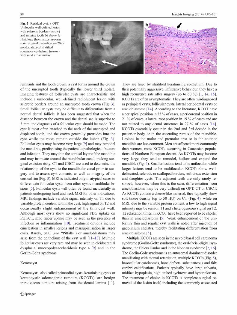
Residual cysts are periapical cysts retained in the jaw aftersurgical removal of a non-vital tooth [1, 3]. Residual cysts arecommon and have similar clinical and radiological features asradicular cysts. However, there is always a missing tooth(Fig. 2). Most residual cysts are less than 1 cm in size.Occasionally, enlarging cysts may cause displacement of theadjacent teeth, as well as bone expansion.
Dentigerous cyst
Follicular cysts, also called dentigerous cysts or pericoronalcysts, are the second most common odontogenic mandibularcysts and the most common developmental cysts ofodontogenic origin. They are typically seen in patients aged20–40. Once fluid accumulates between the enamel organ
Fig. 1 Radicular cyst. a OPT. Unilocular radiolucent lesion with well-defined borders (asterisk) displacing teeth 33 and 34. b CT, sagittaloblique 2DMPR with bone windows. Cystic lesion surrounding the root(dashed arrow) of the non-vital tooth 34. Associated large caries withpulpal necrosis (arrow). c Histology (haematoxylin-eosin stain, originalmagnification 20×): fibrous connective wall containing a dense chronicinflammatory infiltrate and congested vessels (asterisk) lined by irregularnon-keratinised stratified squamous epithelium. Spongiotic epithelium(arrows) penetrated by neutrophils
Insights Imaging (2014) 5:85–101 87
remnants and the tooth crown, a cyst forms around the crownof the unerupted tooth (typically the lower third molar).Imaging features of follicular cysts are characteristic andinclude a unilocular, well-defined radiolucent lesion withsclerotic borders around an unerupted tooth crown (Fig. 3).Small follicular cysts may be difficult to differentiate from anormal dental follicle. It has been suggested that when thedistance between the crown and the dental sac is superior to3 mm, the diagnosis of a follicular cyst should be made. Thecyst is most often attached to the neck of the unerupted anddisplaced tooth, and the crown generally protrudes into thecyst while the roots remain outside the lesion (Fig. 3).Follicular cysts may become very large [9] and may remodelthe mandible, predisposing the patient to pathological fractureand infection. They may thin the cortical layer of the mandibleand may insinuate around the mandibular canal, making sur-gical excision risky. CT and CBCT are used to determine therelationship of the cyst to the mandibular canal prior to sur-gery and to assess cyst contents, as well as integrity of thecortical rim (Fig. 3). MRI is indicated only in atypical cases todifferentiate follicular cysts from other cystic mandibular le-sions [5]. Follicular cysts will often be found incidentally inpatients undergoing head and neck MRI for other indications.MRI findings include variable signal intensity on T1 due tovariable protein content within the cyst, high signal on T2 andoccasionally slight enhancement of the thin cyst wall.Although most cysts show no significant FDG uptake onPET/CT, mild tracer uptake may be seen in the presence ofinfection or inflammation [10]. Treatment options includeenucleation in smaller lesions and marsupialisation in largercysts. Rarely, SCC (see “Pitfalls”) or ameloblastoma mayarise from the epithelium of the cyst wall [11–13]. Multiplefollicular cysts are very rare and may be seen in cleidocranialdysplasia, muccopolysaccharidosis type 4 [9] and in theGorlin-Goltz syndrome.
Keratocyst
Keratocysts, also called primordial cysts, keratinising cysts orkeratocystic odontogenic tumours (KCOTs), are benignintraosseous tumours arising from the dental lamina [11].
They are lined by stratified keratinising epithelium. Due totheir potentially aggressive, infiltrative behaviour, they have ahigh recurrence rate after surgery (up to 60 %) [1, 14, 15].KCOTs are often asymptomatic. They are often misdiagnosedas periapical cysts, follicular cysts, lateral periodontal cysts orameloblastoma [14]. According to the literature, KCOT havea periapical position in 33% of cases, a pericoronal position in21 % of cases, a lateral root position in 19 % of cases and arenot related to any dental structures in 27 % of cases [14].KCOTs essentially occur in the 2nd and 3rd decade in theposterior body or in the ascending ramus of the mandible.Lesions in the molar and premolar area or in the anteriormandible are less common. Men are affected more commonlythan women, most KCOTs occurring in Caucasian popula-tions of Northern European decent. As KCOTs may becomevery large, they tend to remodel, hollow and expand themandible (Fig. 4). Smaller lesions tend to be unilocular, whilelarger lesions tend to be multilocular. KCOTs show well-delineated, sclerotic or scalloped borders, soft-tissue extensionand daughter cysts. The adjacent teeth are only rarely re-sorbed; however, when this is the case, differentiation fromameloblastoma may be very difficult on OPT, CT or CBCT.As KCOTs contain a cheese-like material, they typically showsoft tissue density (up to 50 HU) on CT (Fig. 4), while onMRI, due to the variable protein content, a low to high signalintensity may be seen on T1 and a heterogeneous signal on T2.T2 relaxation times in KCOT have been reported to be shorterthan in ameloblastoma [5]. Weak enhancement of the uni-formly thin and regular cyst walls is noted after injection ofgadolinium chelates, thereby facilitating differentiation fromameloblastoma [5].
Multiple KCOTs are seen in the nevoid basal cell carcinomasyndrome (Gorlin-Goltz syndrome), the oral-facial-digital syn-drome, the Ehlers Danlos and in the Noonan syndrome [2, 16].The Gorlin-Golz syndrome is an autosomal dominant disordermanifesting with mental retardation, multiple KCOTs (Fig. 5),basocellular carcinomas, bone defects, subcutaneous and falxcerebri calcifications. Patients typically have large calvaria,midface hypoplasia, high-arched eyebrows and hypertelorism.The treatment of choice in KCOTs is complete surgical re-moval of the lesion itself, including the commonly associated
Fig. 2 Residual cyst. a OPT.Unilocular well-defined lesionwith sclerotic borders (arrow)and missing tooth 36 above. bHistology (haematoxylin-eosinstain, original magnification 20×):non-keratinised stratifiedsquamous epithelium (arrow)with mild inflammation
88 Insights Imaging (2014) 5:85–101

tooth. Careful clinical and radiological post-operative follow-up is essential due to the high recurrence rate.
Ameloblastoma
Ameloblastomas are benign but locally invasive, slowlygrowing odontogenic tumours arising from remnants of thedental lamina and dental organ or, less frequently, from theepithelial lining of an odontogenic cyst [12]. They represent10 % of all odontogenic tumours, most lesions being locatedin the mandible (posterior body and ramus region). Mosttumours tend to occur during the 4th–6th decade and there isno sex predilection. Clinical features are non-specific andpatients may complain of unilateral painless swelling. Veryoften, ameloblastomas are detected incidentally. Althoughclassic amelobastomas do not have distant metastases, vari-ants with metastatic behaviour despite histologically benignfeatures (so-called “malignant ameloblastoma”), as well astumours with histologically malignant features but withoutmetastatic potential (so-called “ameloblastic carcinomas”)have been described in the literature [3]. Ameloblastomasmay be subdivided into four histological types: unicystic,multicystic, extraosseous, and desmoplastic [12]. The radio-logical appearance depends on the histological type and
Fig. 3 Dentigerous cyst. a OPT, b axial CT with bone windows and cdentascan reconstruction. Unilocular well-defined radiolucent lesion(thick arrows) surrounding an unerupted tooth. Immediate vicinity ofthe mandibular canal (dashed arrow) to the impacted tooth. d Three-dimensional reconstruction, lateral view showing the relationship of thecyst (blue), the unerupted tooth (green) and the mandibular canal (red).
The cyst is attached at the level of the tooth neck and surrounds the crown.e Surgical specimen. Characteristic attachment of the cyst at the toothneck (arrows). f Histology (haematoxylin-eosin stain, original magnifi-cation 20×): non keratinising thin epithelial lining (arrow) without retepegs. Fibrous wall (asterisks) almost completely devoid of inflammatorycells
Fig. 4 Histologically proven KCOT. a OPT. Axial CT image with bonewindow (b) and soft tissue window (c). Multilocular well-defined radio-lucent lesion (asterisk) with thin, sclerotic borders, cortical scalloping(arrow) and resorption of teeth roots. Cyst contents with attenuationvalues of 40–50 HU mimicking solid tissue. d Posterior view of a 3Dreconstruction showing the relationship between the keratocyst (blue),the partially resorbed teeth (green) and the mandibular canal (red). Themandibular canal can be seen only in the posterior mandible, as it isencased by the keratocyst
Insights Imaging (2014) 5:85–101 89

includes unilocular or multilocular radiolucent lesions (“soapbubble” or “honeycomb” appearance) with sclerotic borders,displaced adjacent teeth with root resorption and/or extensivebone expansion (Figs. 6 and 7). CTandMRI are used to assesstumour contents, integrity of the mandibular cortical rim,relationship to the mandibular canal and precise intraosseousand soft tissue extension (Figs. 6 and 7). In multicysticameloblastoma, MRI reveals cysts with variable protein con-tent. Typically, after injection of gadolinium chelates, MRIshows enhancement of solid nodular components, of irregu-larly thick septations and of papillary projections (Fig. 6).Although differentiation between benign and malignantameloblastoma may be impossible with CT and MRI, a veryhigh FDG uptake on PET/CT strongly suggests malignantameloblastoma or ameloblastic carcinoma [17]. Treatmentconsists in complete surgical excision and careful post-operative follow-up with imaging is mandatory.
Simple bone cyst
The simple bone cyst (SBC), also known as solitary bone cyst,traumatic bone cyst, haemorrhagic cyst or idiopathic bonecavity is filled with serous or haemorrhagic fluid and ischaracterised by the absence of an epithelial lining.Therefore, SBC is not a true cyst but rather a pseudocyst.SBC of the mandible usually occurs secondary to trauma,typically after tooth extraction with subsequent intramedullaryhaemorrhage. Most SBCs occur before the age of 20 withfemale predominance [18]. In up to 75 % of all cases, SBCsare seen in the marrow of the posterior mandible. Most lesionsare asymptomatic and discovered incidentally on dental
radiographs. SBC occurs in close proximity to a vital toothand is not associated with bone swelling unless there is
Fig. 5 Basal cell nevussyndrome. a OPG. Multiplemandibular andmaxillary KCOTs(asterisks) associated withimpacted teeth. b Intraoperativeview showing cheese-likematerial within the angulo-mandibular lesion (arrow). cSurgical specimen showing theKCOT (asterisk) and theassociated tooth (thin longarrow). d Histology(haematoxylin-eosin stain,original magnification 40×):corrugated (dashed arrows)parakeratinised epithelium withdistinct basal columnar cells withinverted polarity (arrows) and flatconnective tissue interface
Fig. 6 Histologically proven multilocular ameloblastoma. a Axial bonewindow CT image. b Three-dimensional reconstruction, frontal view. cT1-weighted axial image. d T1-weighted axial image after injection ofgadolinium chelates. Multilocular expansile radiolucency with character-istic “soap bubble” appearance (asterisks) and major facial deformation.Cystic components with variable signal intensity on the unenhnanced T1-weighted image suggesting variable protein content. Note variable en-hancement of solid components ranging from thin enhancing walls tothick enhancing solid portions (arrows)
90 Insights Imaging (2014) 5:85–101

associated infection. Surgical exploration may be necessary inselected cases to exclude other unilocular radiolucent lesions,such as keratocysts. On imaging, most SBCs appear as uni-locular, well-defined radiolucent lesions that vary in size.Rarely, poorly defined borders are observed. Extension tothe cortical bone is rare and usually there is no tooth displace-ment [18]. The superior margins are irregular and scalloparound teeth roots. CT and MRI can provide information onthe haemorrhagic content but the density or signal intensity,respectively, may vary depending on the age of the haemor-rhage. OnMRI, the cysts display a homogeneous intermediatesignal on T1, homogeneous high signal on T2, and no en-hancement after injection of intravenous contrast media.Treatment consists of bone curettage, leading to bleeding withsubsequent scar formation.
Eosinophilic granuloma of the mandible
Eosinophilic granuloma (EG) is a benign disease related toany of the three forms of Langerhans cell histiocytosis (LCH).Depending upon the number of lesions and lesion distribution,LCH comprises the following groups: unifocal LCH (alsocalled EG), multifocal unisystem and multifocal multisystem.The combination of diabetes insipidus, lytic bone lesions andexophtalmus is known as Hand-Schüller-Christian triad,whereas the multifocal multisystem LCH is also called Abt-Letterer-Siwe disease (with typical abdominal involvementand poor prognosis) [19]. LCH is caused by clonal prolifera-tion of activated dendritic cells and macrophages [20]. Any
bone can be affected, such as the skull, mandible, ribs andlong bones, as well as any organ system (lung, skin, spleen,lymph nodes, central nervous system). Therefore, cross-sectional whole-body imaging is essential for the initialwork-up of patients with active disease and for post-therapeutic follow-up purposes.
The incidence of mandibular EG constitutes less than 10 %of all LCH cases. LCH often affects males in the 1st–3rddecade, although most cases are seen in children younger than15 years of age.Mandibular involvement is seen preferentiallyin the body or angle (Fig. 8). Clinical presentation can besilent or non-specific including pain, swelling, fever, generalmalaise, gingival hypertrophy, ulcers of the buccal mucosa,limitation of mouth opening or tooth hypermobility and loos-ening. Pathological fracture may ensue. Radiologically, EGpresents as a well-defined radiolucent lesion, with or withoutreactive sclerosis on OPT; however, an accompanying perios-teal reaction is typically seen on CT or CBCT. Occasionally,this periosteal reaction may show a sunburst appearance,suggesting a more aggressive biological behaviour. If thealveolar crest is invaded, a “scooped out” appearance is seen;when the alveolar bone is destroyed, a “floating tooth” ap-pearance is observed. EG is often associated with a soft tissuemass surrounding the mandible and invading the muscles ofmastication. Morphological MRI findings include hypointensesignal on T1, hyperintense signal on T2 and marked enhance-ment after intravenous gadolinium (Fig. 8). In our experience,ADC values in EG are usually slightly higher than in malignantlesions (≥1.2×10⁻³ mm2/s versus±1×10⁻³ mm2/s). In cases
Fig. 7 Recurrent multilocularameloblastoma. a OPT showing awell-defined, multilocularradiolucency (arrows) withsclerotic borders. b Resectedspecimen and c specimenradiograph clearly show bonyexpansion, destruction of thealveolar ridge and characteristicmultilocular appearance. dHistology (haematoxylin-eosinstain, original magnification 20×):follicular ameloblastoma withsquamous metaplasia (redasterisks) within follicles(arrows). Largely fibrous stroma(black asterisks) containingscattered lymphocytes
Insights Imaging (2014) 5:85–101 91

with atypical periosteal reaction and poorly defined margins,the differential diagnosis includes malignant tumours, such asosteosarcoma, Ewing sarcoma or metastases. Nevertheless,well-defined margins on CT, the high signal intensity on T2,the strong enhancement, the large area of inflammatory oedemaand the moderately high ADC values rather suggest a lesionwith benign histology (Fig. 8). The value of MRI in patientswith EG lies in its high sensitivity especially when used inconjunction with DWI [20]. PET/CT has been shown to pro-vide valuable information for the detection of EG lesions due totheir high FDG uptake (Fig. 8) and it has been suggested thathybrid PET/MRI may play a pivotal role for the primaryinvestigation of paediatric histiocytosis allowing accurate de-tection of multifocal multiorgan involvement and for monitor-ing treatment response [20]. Despite the relatively charac-teristic imaging findings, definitive diagnosis of EG ismade on samples obtained by biopsy. Treatment modalitiesinclude surgery, chemotherapy and intralesional injectionof corticosteroids.
Giant cell granuloma
Giant cell granuloma (GCG) is a benign but occasionallyaggressive proliferative intraosseous lesion with fibrous tis-sue, haemorrhage and haemosiderin deposits, as well as
characteristic osteoclast-like giant cells. GCG is a rare lesionoccurring preferentially in young girls or women [1, 21]. Theposterior mandible is affected more often than the anteriormandible. Multifocal involvement is seen in hyperparathy-roidism, cherubism or Noonan syndrome [21, 22]. The mostcommon clinical features are pain, swelling, facial asymmetryand paresthesia. Differential diagnosis includes giant cell tu-mour, radicular cyst, ameloblastoma, odontogenic tumour andfibrous dysplasia. The typical radiological appearance is thatof a multilocular (less often unilocular) well-defined radiolu-cent lesion. However, ill-defined lesions have also been re-ported. In some cases, bone may be expanded or teeth can bedisplaced or resorbed. CT, CBCT and MRI are useful fordescribing bony involvement and for evaluating extensioninto the adjacent soft tissues. On MRI, GCG has a homoge-neous or slightly heterogeneous intermediate signal on T1, T2and STIR (short TI inversion recovery) and shows moderateto strong contrast enhancement after administration of gado-linium. However, it is important to note that only very fewdata regarding the imaging characteristics of GCG are cur-rently available in the literature. The treatment of choice issurgery (enucleation and curettage in well-defined GCG or enbloc resection in aggressive GCG) and medical treatment(intralesional corticosteroid or calcitonin injections) [21].Recurrence may occur in up to 15 % of cases.
Fig. 8 Histologically proven eosinophilic granuloma. a OPT-like curvedthick slab reconstruction, b axial CT bone window and (c) 3D recon-struction from CT data set show a well-defined radiolucent lesion (ar-rows) located in the angle of the mandible. Sharply delineated slightlysclerotic borders. Periosteal reaction (dashed arrow). d PET/CTshowingthat high FDG uptake within the posterior mandible and perimandibularsoft tissues (arrow). SUVmean = 6, SUVmax = 9. e Axial T2 and f
sagittal T1 after intravenous gadolinium show a large extraosseous com-ponent (arrows) with invasion of the masseter muscle. Close vicinity tothe submandibulat gland. Note high signal intensity on T2 and strongenhancement after gadolinium. g Fused T2 and b 1,000 showing restrict-ed diffusion. h ADCmap showing a moderately lowADC value (ADC =1.21×10⁻³ mm2/s, arrow) within the lesion. Note inflammatory oedemaaround the lesion with high ADC values (asterisk)
92 Insights Imaging (2014) 5:85–101

Radiolucent lesions with ill-defined borders
Osteomyelitis
Despite the introduction of antibiotics and improved medicalcare, osteomyelitis of the jaws is still relatively common. Themandible is affected more often than the maxilla. Dependingon the clinical course, osteomyelitis of the mandible can beclassified into three forms: acute, secondary chronic and pri-mary chronic [23, 24]. The acute and secondary chronic formsare manifestations of the same disease entity separated by thearbitrary time limit of 1 month. These two forms are typicallycaused by bacterial infection in the setting of pulpal or peri-odontal infection, mandibular foreign bodies, sepsis or trau-ma. In dental infection, pulpal infection extends into the bonemarrow and there is compression of blood vessels fromperiapical lesions. Acute forms present with severe symptomssuch as pain, swelling, fever, lymphadenopathy or a mobiletooth sensitive to percussion. They may also present withparesthesia in the lower lip (V3 nerve), trismus or fistula withpus and—in advanced stages—osteomyelitis may also berevealed by pathological fractures. The chronic forms areclinically silent but often include painful periods.Predisposing factors are diabetes, immunosuppression, radio-therapy and bisphosphonates.
While the diagnosis of osteomyelitis can bemade clinicallyin many cases, imaging is especially helpful to define theextent and assess the severity of the lesion. The radiologicalappearance of osteomyelitis depends on the stage of disease. Itconsists of an ill-defined osteolytic lesion (Fig. 9), with bonesequestra, and—in subacute cases—is often associated scle-rosis and periosteal new bone formation. CT is helpful toanalyse the bone structure, to search for associated abscessesand to identify soft tissue extension (myositis, fasciitis, cellu-litis). MRI findings in osteomyelitis are similar to those inosteoradionecrosis (Fig. 9) and the differentiation between thetwo entities is based on the clinical context. MRI is moresensitive than CT in detecting marrow and soft tissue involve-ment [25]. In addition, MRI allows earlier diagnosis of oste-omyelitis than CT, CBCT or conventional radiographs. Ingeneral, the disease extent appears larger on MRI than onCT; therefore, a combined MRI and CT assessment is mostoften required pre-operatively. Osteomyelitis lesions showlow signal on T1, and high signal on T2 and STIR imagesdue to oedema of the bony marrow; variable degrees ofcontrast enhancement of the marrow itself and of adjacent softtissues are equally observed (Fig. 9). The signal intensity ofbone sequestrae, however, is very low on all sequences.Treatment consists of a combination of antibiotics and surgicaldebridement.
Fig. 9 Osteomyelitis after tooth extraction. a OPT. b Contrast-enhanced,axial CT image with bone window settings. c Three-dimensional recon-struction. Ill-defined osteolytic area (asterisks) extending into the ascend-ing ramus of the mandible, with cortical destruction (arrows in b and c).d Axial T1-weighted image before (d) and after (e) injection of gadolin-ium chelates. Hypointense signal of the mandible (arrows in d) due tomarrow oedema and strong enhancement (arrows in e ) due to
hyperaemia. Myositis of the masseter muscle (thick dashed arrow) andstreaky enhancement of the subcutaneous fat and platysma muscle (thindashed arrow) suggesting a phlegmon. f Histology (haematoxylin-eosinstain, original magnification 20×): non-viable trabeculae (large asterisks)with bone resorption and destroyed osteoblasts. Inflammatory infiltrate(small asterisks) with neutrophils and lymphocytes as well as increasedvascularisation within the fatty marrow
Insights Imaging (2014) 5:85–101 93

Bisphosphonate osteonecrosis
Bisphosphonate treatment, similar to radiotherapy, may causebone necrosis. Bisphosphonates are widely used for the treat-ment of cancer-related symptoms. Their benefits consist inreducing hypercalcaemia, limiting progression of bone lesionsand preventing pathological fractures in bone metastases frombreast, prostate and lung cancer, as well as multiple myeloma[26]. In short, bisphosphonates are widely indicated to im-prove the quality of life in a large number of patients.Bisphosphonate-related osteonecrosis of the jaw (BRONJ)was defined by the American Association of Oral andMaxillofacial Surgeons (AAOMS) based on three criteria:past or current bisphosphonate treatment, osteonecrosis last-ing for at least 8 weeks and no history of radiotherapy to thejaw. [26]. BRONJ most often occurs in the mandible ratherthan the maxilla, especially in the molar region, and is fre-quently triggered by a pre-existing focal lesion. Clinical fea-tures include painful swelling, signs of infection (fever, oozingpus, abscess and soft tissue inflammation) and a mobile toothsensitive to percussion. Occasionally, there is paresthesia inthe lower lip (whenever V3 is affected) or trismus. Imagingfeatures of BRONJ are the same as for osteoradionecrosis(Fig. 10). MRI can show signal changes on T1 and T2 imagesaccording to the stage of the disease. Although radiologicalfindings are easily identified, they are non-specific and the
final diagnosis is made by combining imaging findings withthe clinical context [27].
Osteoradionecrosis
Osteoradionecrosis (ORN) is a serious uncommon complica-tion (2.6-15 %) of radiotherapy in head and neck cancerpatients. It usually occurs 5-15 years after radiotherapy. Therisk of developing ORN increases with poor oral hygiene,alcohol and tobacco use after radiotherapy, tooth extractionor periodontal disease prior to radiotherapy: ORN is alsorelated to the nutritional status of the patient. ORN preferen-tially affects the molar, premolar and retromolar region.Depending on the radiation portal and the administered radi-ation dose, osteoradionecrosis may affect the mandible unilat-erally or bilaterally. Combined treatment options (radiationtherapy, surgery and chemotherapy) appear to predispose toORN [28]. Clinical manifestations include pain, limitation ofmouth opening and deep ulcerations with denuded bone. Inadvanced stages, trismus, fistulas and pathological fractureshave also been reported. CT usually reveals a patchyradiopaque-radiolucent ill-defined lesion with cortical bone de-struction, usually without periosteal reaction (Fig. 11). TypicalMRI findings are a mixture of marrow oedema (increased signalon T2) and marrow sclerosis (reduced signal on T2) withfragmentation of bone and sequestration (very low signal on
Fig. 10 Biphosphonate osteonecrosis. a OPT; b 3D reconstructionanteo-lateral view. Contrast-enhanced axial CT image with bone windowsettings (c) and soft tissue settings (d). Poorly defined osteolytic lesion ofthe left mandibular body (thick white arrows) with partly sclerotic mar-gins (thin arrows in a) and large osseous defect (red arrows in b). Bone
sequestra (dashed arrows). Associated subperiosteal phlegmon andmyo-sitis of the platysma (open arrows). e Histology (haematoxylin-eosinstain, original magnification 20×): necrotic bone (large asterisk) withinflammatory infiltration of the fatty marrow (small asterisks). Gingivalmucosa (arrows)
94 Insights Imaging (2014) 5:85–101
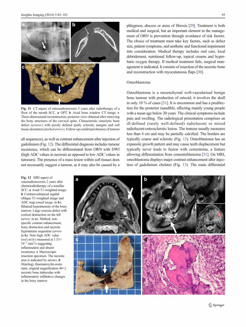
all sequences), as well as contrast enhancement after injection ofgadolinium (Fig. 12). The differential diagnosis includes tumourrecurrence, which can be differentiated from ORN with DWI(high ADC values in necrosis as opposed to low ADC values intumours). The presence of a mass lesion within soft tissues doesnot necessarily suggest a tumour, as it may also be caused by a
phlegmon, abscess or areas of fibrosis [29]. Treatment is bothmedical and surgical, but an important element in the manage-ment of ORN is prevention through avoidance of risk factors.The choice of treatment must take key factors, such as defectsize, patient symptoms, and aesthetic and functional impairmentinto consideration. Medical therapy includes oral care, localdebridement, nutritional follow-up, topical creams and hyper-baric oxygen therapy. If medical treatment fails, surgical man-agement is indicated. It consists of resection of the necrotic boneand reconstruction with myocutaneous flaps [30].
Osteoblastoma
Osteoblastoma is a mesenchymal well-vascularised benignbone tumour with production of osteoid; it involves the skullin only 10 % of cases [31]. It is uncommon and has a predilec-tion for the posterior mandible, affecting mainly young peoplewith amean age below 20 years. The clinical symptoms includepain and swelling. The radiological presentation comprises anill-defined (rarely well-defined) radiolucent or mixedradiolucent-osteosclerotic lesion. The tumour usually measuresless than 4 cm and may be partially calcified. The borders aretypically coarse and sclerotic (Fig. 13). Osteoblastoma has anexpansile growth pattern and may cause teeth displacement buttypically never leads to fusion with cementoma, a featureallowing differentiation from cementoblastoma [31]. On MRI,osteoblastoma displays major contrast enhancement after injec-tion of gadolinium chelates (Fig. 13). The main differential
Fig. 11 CT aspect of osteoradionecrosis 5 years after radiotherapy of afloor of the mouth SCC. a OPT. b Axial bone window CT image. cThree-dimensional reconstruction, posterior view obtained after removingthe bony structures of the cervical spine. Characteristic osteolytic bonedefect (arrows) with poorly defined partly sclerotic margins and softtissue ulceration (dashed arrow). Follow-up confirmed absence of tumour
Fig. 12 MRI aspect ofosteoradionecrosis 2 years afterchemoradiotherapy of a tonsillarSCC. a Axial T1-weighted image.b Contrast-enhanced sagittaloblique T1-weighted image andADC map (small image in b).Bilateral hypointensity of the bonymarrow. Large osseous defect withcortical destruction on the left(arrow in a). Marked, non-specific contrast enhancement,bony destruction and necrotichypointense sequestrae (arrowin b). Note high ADC value(red circle) measured at 1.53×10⁻³ mm2/s suggestinginflammation and absentrecurrence. c Macroscopicresection specimen. The necroticarea is indicated by arrows. dHistology (haematoxylin-eosinstain, original magnification 40×):necrotic bone trabeculae withinflammatory infiltrative changesin the bony marrow
Insights Imaging (2014) 5:85–101 95

diagnosis includes osteoid osteoma, ossifying fibroma,periapical cemental dysplasia, fibrous dysplasia, as well asother fibro-osseous lesions. Osteoid osteoma typically presentswith pain and a nidus can be identified at CT or CBCT.Ossifying fibroma most commonly occurs in the posteriormandible during the 3rd–4th decades [1]. Depending on thedegree of calcification, the radiological appearance includeswell-defined radiolucent, radiopaque or mixed opacity lesions[1]. Commonly, a radiolucent margin is seen on CT or CBCT,allowing differentiation from fibrous dysplasia. Periapicalcemental dysplasia typically occurs in women during the 4thand 5th decades. It is the consequence of connective tissueproliferation within the periodontal membrane; therefore, thelesions are typically located in vicinity of tooth apices [1].Initially, the lesions are radiolucent and with increasing matu-rity, there is advanced calcification. As opposed to mandibularosteoblastoma and osteoid osteoma,many fibro-osseous lesionsdo not cause pain despite their large size or associated teethdisplacement. In addition, the age of presentation, the multi-plicity of lesions and the relationship to tooth apices are furtherelements that help in the differential diagnosis. The treatment ofchoice in osteoblastoma is surgery and recurrence is rare [31].Some cases of spontaneous regression have been reported,especially in young patients.
SCC with mandibular invasion
SCC, also known as epidermoid carcinoma, is a malignantneoplasm arising from the mucosa of the oral cavity [32, 33].
It is the most common tumour of the oral cavity and prefer-entially affects men over the age of 50. SCCmost often occursin the mucosa overlying the posterior mandible, and it isrelated to the consumption of alcohol and tobacco. It has highmetastatic potential. Clinical symptoms include pain, swell-ing, paresthesia (due to V3 invasion) and dental disorders. Inadvanced stages, SCC may be discovered in the context of apathological fracture. The radiological appearance is that of anaggressive soft-tissue lesion with invasion of the floor of themouth, alveolar ridge or retromolar trigone [33]. In advancedstages, secondary bone invasion can occur leading to theappearance of an ill-defined radiolucent lesion of the mandibleon conventional X-rays; in advanced lesions, the “floatingteeth” sign may be equally observed due to extensive man-dibular infiltration. On MRI, SCC displays a low to interme-diate signal on T1, a moderately high signal on T2 and STIRsequences, and a moderate enhancement after injection ofcontrast media (Fig. 14). ADC values are typically low (usu-ally around 1–1.1×10⁻³ mm2/s). Whenever large areas ofnecrosis are present within the tumour, ADC values may,however, be higher. This is particularly true in necrotic lymphnode metastases (Fig. 14). Lymph node metastases in SCC ofthe oral cavity are common, especially in level I and II nodes.On FDG PET/CTor PET/MRI, SUV values are typically highdue to the high glucose metabolism (Fig. 14). The finaldiagnosis and the pre-therapeutic work-up in SCC of the oralcavity are made by a combined clinical, imaging and histo-logical work-up. The main role of imaging consists in precise-ly depicting deep tumour spread, as well as detecting lymph
Fig. 13 Osteoblastoma. a OPT.b Axial CTwith bone window.c Sagittal oblique contrast-enhanced fat-saturated T1-weighted image. Ill-definedradiolucent lesion (arrows in aand b) with coarse scleroticborders and calcifications (dashedarrow). Major contrastenhancement (arrow in c). dHistology (haematoxylin-eosinstain, original magnification 64×).Osteoid and woven bone(asterisk) with plump osteoblast-like cells (arrows), interposedfibroblasts and someinflammatory cells
96 Insights Imaging (2014) 5:85–101

node and distant metastases. Surgery with or without radio-therapy is the treatment of choice. Prognosis depends on thehistological type and on the presence or absence of lymphnode metastases.
Metastases
Metastases to the jaw are an uncommon entity, affecting themandible more often than the maxilla [34]. The most commonprimaries vary depending upon gender. Lung, prostate, kidneyand liver tumours are the most common primaries in men,whereas breast, adrenal, gynaecological and colorectal tu-mours are the most common primaries in women [34].Typical clinical symptoms include pain (Fig. 15), swelling,paresthesia, temporomandibular joint derangement, but insome cases metastases to the jaw are clinically silent andfound incidentally. The radiological appearance includes ill-defined radiolucent lesions with no periosteal reaction onconventional X-rays, CT and CBCT [35]. MRI reveals mod-erately hyperintense masses on T2-weighted and STIR im-ages, hypointense signal on T1-weighted images and variabledegrees of contrast enhancement. In general, the surroundingsoft tissues lack relevant oedema and enhancement unlesstumour extension beyond the mandible has occurred
(Fig. 15). On PET/CT, focal areas of increased FDG uptakeare typically observed and measured SUVs are high (Fig. 15).Although the imaging aspect is not specific, in the presence ofa patient with a history of cancer, the diagnosis of mandibularmetastasis should be considered in the differential diagnosisfirst, particularly when the lesion shows no relationship todental structures.
Pitfalls
Pseudolesions
Stafne cyst
Stafne cyst, also called static bone cavity or salivary glandinclusion defect, is a pseudocyst arising from bone remodel-ling caused by the adjacent submandibular gland. Therefore, itdoes not present any epithelial lining. Stafne cysts are oftenincidental findings, as patients are asymptomatic (Fig. 16).The lesions are more common in men than in women. Theradiological aspect includes ovoid, well-defined radiolucentcortical defects on the lingual surface of the posterior mandi-ble usually measuring less than 2 cm. This location is typical
Fig. 14 SCC with mandibular invasion. Precise pre-operative assess-ment with MRI and PET/CT. a OPT. Poorly defined bony destruction(arrow) of the edentulous mandible. b Contrast-enhanced axial CT.Aggressive soft-tissue lesion (arrow) with secondary bone invasion. cSagittal oblique PET/CT image. High metabolism of the tumour (blackasterisk , SUVmean = 18, SUVmax = 22) and of two metastatic level Ilymph nodes (arrows , SUVmean = 10, SUVmax = 15). d T2-weightedaxial image. Tumour (asterisk) with marrow invasion and extensive
infiltration of the floor of the mouth (dashed arrow). Note two metastaticlevel I lymph nodes (arrows). e The b 1,000 and f ADC map showrestricted diffusion within the tumour (circle , ADC = 0.98×10⁻³ mm2/s).Variable ADC values within the metastatic lymph nodes due to thepresence of necrosis (arrows in f). g Sagittal histological whole-organslice of the resected specimen. Tumour (dashed black line) invading themandible (black arrows) and the muscles (asterisk) of the floor of themouth. The histological slice has the same orientation as (c)
Insights Imaging (2014) 5:85–101 97

and the lesion contains fat or salivary gland tissue. An OPT issufficient for diagnosis but occasionally CT or MRI can beperformed in cases of atypical presentation to exclude anameloblastoma or a traumatic bone cyst [2]. No treatment isrecommended; however, a follow-up examination at 3–6 months can be performed to ascertain lesion stability [3].
Malignant lesions mimicking benign disease
Intraosseous mucoepidermoid carcinoma
The vast majority of mucoepidermoid carcinomas arise withinthe major or minor salivary glands. Primary intraosseous
mucoepidermoid carcinoma is an extremely rare tumour con-stituting less than 2 % of all mucoepidermoid carcinomas. Itarises centrally within the angle or posterior mandible [36]. Itsaetiopathogenesis is not yet completely understood. Due to thepaucity of cases reported in the literature, little is known re-garding age of presentation and sex distribution. Clinical fea-tures include swelling and pain, as well as trismus. Imagingfeatures are unilocular or multilocular, well-defined lesions withsclerotic borders, mimicking a benign cyst, a keratocyst or anameloblastoma on OPT, CT and CBCT [37]. On MRI, thecystic tumour components display a low signal on T1, a highsignal on T2 and no enhancement, whereas the nodular solidtumour components present a low signal on T2 and contrast
Fig. 15 Condylar metastasisfrom adenocarcinoma. OPT (a)with osteolytic ill-defined lesionof the mandibular condyle(arrow). b Sagittal PET/CTimage shows high metabolismwith SUV = 12 (arrow). c Axial,contrast-enhanced, fat-saturatedT1-weighted image. Theinfiltrative, bulky lesion invadesthe condyle (arrow), the internalpterygoid muscle (dashed arrow)and part of the parotid gland (thinarrow). d Intraoperative view.Extensive condylar involvement.e Histology (haematoxylin-eosinstain, original magnification 80×):large atypical polygonal cells,some with several nuclei (arrow)
Fig. 16 Stafne cyst discoveredincidentally. a OPT-likereconstruction from CBCT dataset showing a well-defined,unilocular radiolucency (arrow)with sclerotic borders in theregion of 37 and 38. Norelationship to teeth. b Axial,c coronal and d 3Dreconstruction, posterior view.Bony defect (arrows) incharacteristic location
98 Insights Imaging (2014) 5:85–101
material enhancement. Extension into the muscles of mastica-tion is common, as well as loco-regional lymph node metasta-ses. MRI essential to narrow the differential diagnosis, as OPTand CT are non-specific. The presence of a destructive,
infiltrative pattern with enhancing nodules within the cyst wall,as well as the presence of metastatic lymph nodes stronglysuggests a malignant tumour, thus making biopsy mandatory.
Primary intraosseous SCC
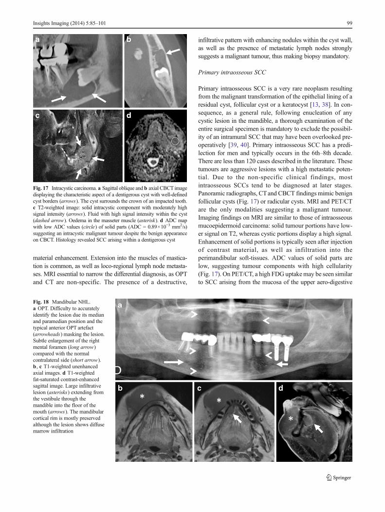
Primary intraosseous SCC is a very rare neoplasm resultingfrom the malignant transformation of the epithelial lining of aresidual cyst, follicular cyst or a keratocyst [13, 38]. In con-sequence, as a general rule, following enucleation of anycystic lesion in the mandible, a thorough examination of theentire surgical specimen is mandatory to exclude the possibil-ity of an intramural SCC that may have been overlooked pre-operatively [39, 40]. Primary intraosseous SCC has a predi-lection for men and typically occurs in the 6th–8th decade.There are less than 120 cases described in the literature. Thesetumours are aggressive lesions with a high metastatic poten-tial. Due to the non-specific clinical findings, mostintraosseous SCCs tend to be diagnosed at later stages.Panoramic radiographs, CTand CBCT findings mimic benignfollicular cysts (Fig. 17) or radicular cysts. MRI and PET/CTare the only modalities suggesting a malignant tumour.Imaging findings on MRI are similar to those of intraosseousmucoepidermoid carcinoma: solid tumour portions have low-er signal on T2, whereas cystic portions display a high signal.Enhancement of solid portions is typically seen after injectionof contrast material, as well as infiltration into theperimandibular soft-tissues. ADC values of solid parts arelow, suggesting tumour components with high cellularity(Fig. 17). On PET/CT, a high FDG uptakemay be seen similarto SCC arising from the mucosa of the upper aero-digestive
Fig. 17 Intracystic carcinoma. a Sagittal oblique and b axial CBCT imagedisplaying the characteristic aspect of a dentigerous cyst with well-definedcyst borders (arrows). The cyst surrounds the crown of an impacted tooth.c T2-weighted image: solid intracystic component with moderately highsignal intensity (arrows). Fluid with high signal intensity within the cyst(dashed arrow). Oedema in the masseter muscle (asterisk). d ADC mapwith low ADC values (circle) of solid parts (ADC = 0.89×10⁻³ mm2/s)suggesting an intracystic malignant tumour despite the benign appearanceon CBCT. Histology revealed SCC arising within a dentigerous cyst
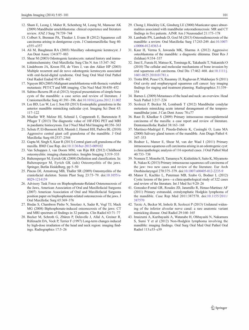
Fig. 18 Mandibular NHL.a OPT. Difficulty to accuratelyidentify the lesion due its medianand paramedian position and thetypical anterior OPT artefact(arrowheads) masking the lesion.Subtle enlargement of the rightmental foramen (long arrow)compared with the normalcontralateral side (short arrow).b , c T1-weighted unenhancedaxial images. d T1-weightedfat-saturated contrast-enhancedsagittal image. Large infiltrativelesion (asterisks) extending fromthe vestibule through themandible into the floor of themouth (arrows). The mandibularcortical rim is mostly preservedalthough the lesion shows diffusemarrow infiltration
Insights Imaging (2014) 5:85–101 99

tract. Treatment consists of surgery with and without addition-al radiotherapy.
Malignant lesions that may be missed on OPT
Mandibular lymphoma
Lymphomas are malignant tumours developed from cells ofthe lymphatic system and can therefore affect any organcontaining lymphoid tissue. While Hodgkin lymphomas(HL) most often involve lymph nodes in the head and neck,non-Hodgkin lymphomas (NHL) can equally affect extra-nodal sites [41] such as the lacrimal glands, salivary glands,muscles and soft tissues of the orbit, thyroid gland, maxillaand mandible. Mandibular NHL is very rare. As it is frequent-ly mistaken for dental infection, a prolonged delay in diagno-sis can occur. All age groups can be affected, however adultstend to be affected more often than children. There is nogender predilection. The clinical features of mandibularNHL are not specific and include pain, jaw swelling, ulcera-tion, tooth mobility and cervical lymphadenopathy. On pano-ramic views, mandibular NHLs appear as ill-defined radiolu-cent lesions. Despite their large size, the tumours can be easilymissed on conventional X-rays (Fig. 18). Radiological find-ings can be quite subtle such as teeth displacement in theocclusal direction and loss of the lamina dura with wideningof the periodontal ligament space [41]. Rarely, mandibularNHL can present with minor osteolysis or with unilateralenlargement of the mandibular canal and mental foramen(Fig. 18). Enlargement of the mandibular canal and mentalforamen is rare and has been reported in neurogenic tumoursaffecting the inferior alveolar nerve (schwannoma and neuro-fibroma), in perineural spread of disease (mainly from SCC,adenoid cystic carcinoma and lymphoma) and as an anatomicvariant mimicking pathology [42]. In our experience, in man-dibular NHL, CT and MRI typically show minor or nodestruction of the cortical bone despite extensive infiltrationof the bony marrow and of the perimandibular soft tissues(Fig. 18). As suggested in the literature, MRI providessuperior visualisation of submucosal tumour extension andimproved assessment of marrow infiltration compared withCT or CBCT [43]. Low signal on T1 and T2, homogenouscontrast enhancement after injection of gadolinium chelates,absent areas of necrosis despite the large size (Fig. 18) and lowADC values (typically 0.6–0.8×10⁻³ mm2/s) are characte-ristic findings. On PET/CT, mandibular NHL display highFDG uptake and high SUV values (usually ≥10–15), similarto lymphomas in other locations in the body. The differentialdiagnosis of mandibular NHL includes other infiltrativeprocesses such as myeloma, leukaemia and bone metastases,making biopsy mandatory for the correct histologicaldiagnosis. Treatment in mandibular NHL is done withchemo(radio)therapy.
Conclusions
The vast majority of radiolucent lesions of the mandible seenon conventional radiographs represent benign lesions thatrequire no further work-up. Nevertheless, certain radiologicalfeatures, such as large lesion size, bone scalloping, relation-ship to an impacted tooth or the mandibular canal, toothresorption, as well as ill-defined lesion borders, require furtherradiological work-up. CT, CBCT, MRI and PET/CT are ofadditional help if the nature of the lesion is unclear and for theidentification of those lesions, where biopsy is indicated fordefinitive histology.
Open Access This article is distributed under the terms of the CreativeCommons Attribution License which permits any use, distribution, andreproduction in any medium, provided the original author(s) and thesource are credited.
References
1. Dunfee BL, Sakai O, Pistey R, Gohel A (2006) Radiologic andpathologic characteristics of benign and malignant lesions of themandible. Radiographics 26:1751–1768
2. Devenney-Cakir B, Subramaniam RM, Reddy SM, Imsande H,Gohel A, Sakai O (2011) Cystic and cystic-appearing lesions of themandible: review. AJR Am J Roentgenol 196:WS66–WS77. doi:10.2214/AJR.09.7216
3. Scholl RJ, Kellett HM, Neumann DP, Lurie AG (1999) Cysts andcystic lesions of the mandible: clinical and radiologic-histopathologicreview. Radiographics 19:1107–1124
4. Cotti E, Campisi G (2004) Advanced radiographic techniques for thedetection of lesions in bone. Endod Top 7:52–72
5. Minami M, Kaneda T, Ozawa K, Yamamoto H, Itai Y, OzawaM et al(1996) Cystic lesions of the maxillomandibular region: MR imagingdistinction of odontogenic keratocysts and ameloblastomas fromother cysts. AJR Am J Roentgenol 166:943–949
6. Schuknecht BF, Carls FR, Valavanis A, Sailer HF (1997) Mandibularosteomyelitis: evaluation and staging in 18 patients, using magneticresonance imaging, computed tomography and conventional radio-graphs. J Craniomaxillofac Surg 25:24–33
7. Varoquaux A, Rager O, Lovblad KO, Masterson K, Dulguerov P,Ratib O et al (2013) Functional imaging of head and neck squamouscell carcinoma with diffusion-weighted MRI and FDG PET/CT:quantitative analysis of ADC and SUV. Eur J Nucl Med MolImaging 40:842–852
8. Varoquaux A, Rager O, Poncet A, Delattre BM, Ratib O, Becker CDet al (2013) Detection and quantification of focal uptake in head andneck tumours: F-FDGPET/MR versus PET/CT. Eur J NuclMedMolImaging. doi:10.1007/s00259-013-2580-y
9. Freitas DQ, Tempest LM, Sicoli E, Lopes-Neto FC (2006) Bilateraldentigerous cysts: review of the literature and report of an unusualcase. Dentomaxillofac Radiol 35:464–468
10. Maeda T, Tateishi U, Terauchi T et al (2007) Unsuspected bone andsoft tissue lesions identified at cancer screening using positron emis-sion tomography. Jpn J Clin Oncol 37:207–215
11. Barnes L, Eveson JW, Reichart P, Sidransky D (2005) World HealthOrganization Classification of Tumours. Pathology and genetics ofhead and neck tumours. IARC Press, Lyon
100 Insights Imaging (2014) 5:85–101
12. Sham E, Leong J, Maher R, Schenberg M, Leung M, Mansour AK(2009) Mandibular ameloblastoma: clinical experience and literaturereview. ANZ J Surg 79:739–744
13. Colbert S, Brennan PA, Theaker J, Evans B (2012) Squamous cellcarcinoma arising in dentigerous cysts. J Craniomaxillofac Surg 40:e355–e357
14. Ali M, Baughman RA (2003) Maxillary odontogenic keratocyst. JAm Dent Assoc 134:877–883
15. Shear M (2003) Odontogenic keratocysts: natural history and immu-nohistochemistry. Oral Maxillofac Surg Clin N Am 15:347–362
16. Lindeboom JA, Kroon FH, de Vires J, van den Akker HP (2003)Multiple recurrent and de novo odontogenic keratocysts associatedwith oral-facial-digital syndrome. Oral Surg Oral Med Oral PatholOral Radiol Endod 95:458–462
17. Nguyen BD (2005) Malignant ameloblastoma with thoracic vertebralmetastasis: PET/CT and MR imaging. Clin Nucl Med 30:450–452
18. Sabino-Bezerra JR et al (2012) Atypical presentations of simple bonecysts of the mandible: a case series and review of literature. JCraniomaxillofac Surg 41:391–396. doi:10.1016/j.jcms.2012.11.002
19. Lee BD, Lee W, Lee J, Son HJ (2013) Eosinophilic granuloma in theanterior mandible mimicking radicular cyst. Imaging Sci Dent 43:117–122
20. Mueller WP, Melzer HI, Schmid I, Coppenrath E, Bartenstein P,Pfluger T (2013) The diagnostic value of 18F-FDG PET and MRIin paediatric histiocytosis. Eur J Nucl MedMol Imaging 40:356–363
21. Schutz P, El-Bassuoni KH, Munish J, Hamed HH, Padwa BL (2010)Aggressive central giant cell granuloma of the mandible. J OralMaxillofac Surg 68:2537–2544
22. Gupta M, Singh S, Kaur R (2013) Central giant cell granuloma of themaxilla. BMJ Case Rep. doi:10.1136/bcr-2013-009102
23. Van Schuppen J, van Doorn MM, van Rijn RR (2012) Childhoodosteomyelitis: imaging characteristics. Insights Imaging 3:519–533
24. Baltensperger M, Eyrich GK (2008) Definition and classification. In:Baltensperger M, Eyrich GK (eds) Osteomyelitis of the jaws.Springer, Berlin Heidelberg, pp 5–50
25. Pincus DJ, Armstrong MB, Thaller SR (2009) Osteomyelitis of thecraniofacial skeleton. Semin Plast Surg 23:73–79. doi:10.1055/s-0029-1214159
26. Advisory Task Force on Bisphosphonate-Related Ostenonecrosis ofthe Jaws, American Association of Oral and Maxillofacial Surgeons(2007) American Association of Oral and Maxillofacial Surgeonsposition paper on bisphosphonate-related osteonecrosis of the jaws. JOral Maxillofac Surg 65:369–376
27. Bisdas S, Chambron Pinho N, Smolarz A, Sader R, Vogl TJ, MackMG (2008) Biphosphonate-induced osteonecrosis of the jaws: CTand MRI spectrum of findings in 32 patients. Clin Radiol 63:71–77
28. Becker M, Schroth G, Zbären P, Delavelle J, Allal A, Greiner R,Rüfenacht DA, Vock P, Terrier F (1997) Long-term changes inducedby high-dose irradiation of the head and neck region: imaging find-ings. Radiographics 17:5–26
29. Chong J, Hinckley LK, Ginsberg LE (2000) Masticator space abnor-malities associated with mandibular osteoradionecrosis: MR and CTfindings in five patients. AJNR Am J Neuroradiol 21:175–178
30. Lambade PN, Lambade D, Goel M (2013) Osteoradionecrosis of themandible: a review. Oral Maxillofac Surg 17:243-249. doi:10.1007/s10006-012-0363-4
31. Kaur H, Verma S, Jawanda MK, Sharma A (2012) Aggressiveosteoblastoma of the mandible: a diagnostic dilemma. Dent Res J(Isfahan) 9:334–337
32. Jimi E, Furuta H, Matsuo K, Tominaga K, Takahashi T, Nakanishi O(2010) The cellular and molecular mechanisms of bone invasion byoral squamous cell carcinoma. Oral Dis 17:462–468. doi:10.1111/j.1601-0825.2010.01781.x
33. Trotta BM, Pease CS, Rasamny JJ, Raghavan P, Mukherjee S (2011)Oral cavity and oropharyngeal squamous cell cancer: key imagingfindings for staging and treatment planning. Radiographics 31:339–354
34. Barnes L (2009) Metastases of the head and neck: an overview. HeadNeck Pathol 3:217–224
35. Scolozzi P, Becker M, Lombardi T (2012) Mandibular condylarmetastasis mimicking acute internal derangement of the temporo-mandibular joint. J Can Dent Assoc 78:c77
36. Raut D, Khedkar S (2009) Primary intraosseous mucoepidermoidcarcinoma of the maxilla: a case report and review of literature.Dentomaxillofac Radiol 38:163–168
37. Martínez-Madrigal F, Pineda-Daboin K, Casiraghi O, Luna MA(2000) Salivary gland tumors of the mandible. Ann Diagn Pathol 4:347–353
38. Bodner L, Manor E, Shear M, van der Waal I (2011) Primaryintraosseous squamous cell carcinoma arising in an odontogenic cyst:a clinicopathologic analysis of 116 reported cases. J Oral Pathol Med40:733–738
39. Nomura T,Monobe H, Tamaruya N,Kishishita S, Saito K,MiyamotoR, Nakao K (2013) Primary intraosseous squamous cell carcinoma ofthe jaw: two new cases and review of the literature. Eur ArchOtorhinolaryngol 270:375–379. doi:10.1007/s00405-012-2235-9
40. Manor E, Kachko L, Puterman MB, Szabo G, Bodner L (2012)Cystic lesions of the jaws—a clinicopathological study of 322 casesand review of the literature. Int J Med Sci 9:20–26
41. Gonzalez-Fontal GR, Rosales JD, Jaramillo R, Henao-Martinez AF(2011) Primary extranodal, extralymphatic Hodgkin lymphoma ofthe mandible. Case Rep Med 2011:387570. doi:10.1155/2011/387570
42. Terzic A, Becker M, Imholz B, Scolozzi P (2013) Unilateral widen-ing of the inferior alveolar nerve canal: a rare anatomic variantmimicking disease. Oral Radiol 29:160–165
43. Imaizumi A, Kuribayashi A, Watanabe H, Ohbayashi N, NakamuraS, Sumi Y et al (2012) Non-Hodgkin lymphoma involving themandible: imaging findings. Oral Surg Oral Med Oral Pathol OralRadiol 113:e33–e39
Insights Imaging (2014) 5:85–101 101