Glucocorticoid receptor mRNA in Alzheimer's diseased hippocampus
Upload
michiganstateCategory
view
2download
0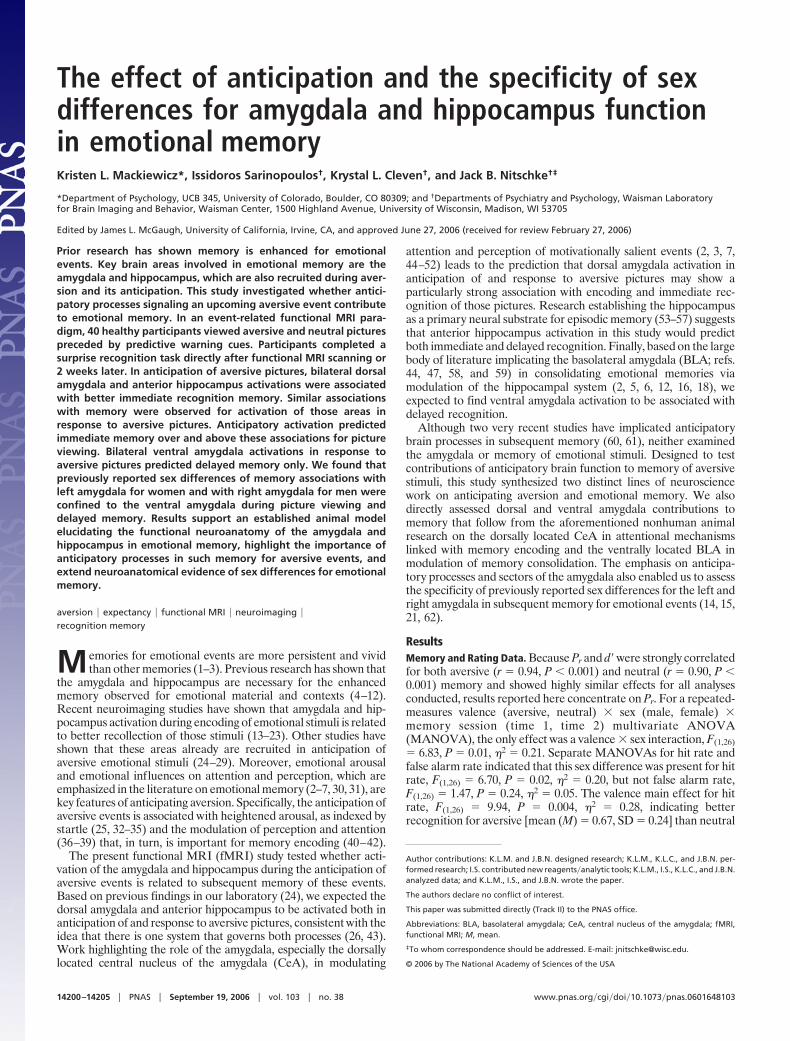
The effect of anticipation and the specificity of sexdifferences for amygdala and hippocampus functionin emotional memoryKristen L. Mackiewicz*, Issidoros Sarinopoulos†, Krystal L. Cleven†, and Jack B. Nitschke†‡
*Department of Psychology, UCB 345, University of Colorado, Boulder, CO 80309; and †Departments of Psychiatry and Psychology, Waisman Laboratoryfor Brain Imaging and Behavior, Waisman Center, 1500 Highland Avenue, University of Wisconsin, Madison, WI 53705
Edited by James L. McGaugh, University of California, Irvine, CA, and approved June 27, 2006 (received for review February 27, 2006)
Prior research has shown memory is enhanced for emotionalevents. Key brain areas involved in emotional memory are theamygdala and hippocampus, which are also recruited during aver-sion and its anticipation. This study investigated whether antici-patory processes signaling an upcoming aversive event contributeto emotional memory. In an event-related functional MRI para-digm, 40 healthy participants viewed aversive and neutral picturespreceded by predictive warning cues. Participants completed asurprise recognition task directly after functional MRI scanning or2 weeks later. In anticipation of aversive pictures, bilateral dorsalamygdala and anterior hippocampus activations were associatedwith better immediate recognition memory. Similar associationswith memory were observed for activation of those areas inresponse to aversive pictures. Anticipatory activation predictedimmediate memory over and above these associations for pictureviewing. Bilateral ventral amygdala activations in response toaversive pictures predicted delayed memory only. We found thatpreviously reported sex differences of memory associations withleft amygdala for women and with right amygdala for men wereconfined to the ventral amygdala during picture viewing anddelayed memory. Results support an established animal modelelucidating the functional neuroanatomy of the amygdala andhippocampus in emotional memory, highlight the importance ofanticipatory processes in such memory for aversive events, andextend neuroanatomical evidence of sex differences for emotionalmemory.
aversion � expectancy � functional MRI � neuroimaging �recognition memory
Memories for emotional events are more persistent and vividthan other memories (1–3). Previous research has shown that
the amygdala and hippocampus are necessary for the enhancedmemory observed for emotional material and contexts (4–12).Recent neuroimaging studies have shown that amygdala and hip-pocampus activation during encoding of emotional stimuli is relatedto better recollection of those stimuli (13–23). Other studies haveshown that these areas already are recruited in anticipation ofaversive emotional stimuli (24–29). Moreover, emotional arousaland emotional influences on attention and perception, which areemphasized in the literature on emotional memory (2–7, 30, 31), arekey features of anticipating aversion. Specifically, the anticipation ofaversive events is associated with heightened arousal, as indexed bystartle (25, 32–35) and the modulation of perception and attention(36–39) that, in turn, is important for memory encoding (40–42).
The present functional MRI (fMRI) study tested whether acti-vation of the amygdala and hippocampus during the anticipation ofaversive events is related to subsequent memory of these events.Based on previous findings in our laboratory (24), we expected thedorsal amygdala and anterior hippocampus to be activated both inanticipation of and response to aversive pictures, consistent with theidea that there is one system that governs both processes (26, 43).Work highlighting the role of the amygdala, especially the dorsallylocated central nucleus of the amygdala (CeA), in modulating
attention and perception of motivationally salient events (2, 3, 7,44–52) leads to the prediction that dorsal amygdala activation inanticipation of and response to aversive pictures may show aparticularly strong association with encoding and immediate rec-ognition of those pictures. Research establishing the hippocampusas a primary neural substrate for episodic memory (53–57) suggeststhat anterior hippocampus activation in this study would predictboth immediate and delayed recognition. Finally, based on the largebody of literature implicating the basolateral amygdala (BLA; refs.44, 47, 58, and 59) in consolidating emotional memories viamodulation of the hippocampal system (2, 5, 6, 12, 16, 18), weexpected to find ventral amygdala activation to be associated withdelayed recognition.
Although two very recent studies have implicated anticipatorybrain processes in subsequent memory (60, 61), neither examinedthe amygdala or memory of emotional stimuli. Designed to testcontributions of anticipatory brain function to memory of aversivestimuli, this study synthesized two distinct lines of neurosciencework on anticipating aversion and emotional memory. We alsodirectly assessed dorsal and ventral amygdala contributions tomemory that follow from the aforementioned nonhuman animalresearch on the dorsally located CeA in attentional mechanismslinked with memory encoding and the ventrally located BLA inmodulation of memory consolidation. The emphasis on anticipa-tory processes and sectors of the amygdala also enabled us to assessthe specificity of previously reported sex differences for the left andright amygdala in subsequent memory for emotional events (14, 15,21, 62).
ResultsMemory and Rating Data. Because Pr and d� were strongly correlatedfor both aversive (r � 0.94, P � 0.001) and neutral (r � 0.90, P �0.001) memory and showed highly similar effects for all analysesconducted, results reported here concentrate on Pr. For a repeated-measures valence (aversive, neutral) � sex (male, female) �memory session (time 1, time 2) multivariate ANOVA(MANOVA), the only effect was a valence � sex interaction, F(1,26)� 6.83, P � 0.01, �2 � 0.21. Separate MANOVAs for hit rate andfalse alarm rate indicated that this sex difference was present for hitrate, F(1,26) � 6.70, P � 0.02, �2 � 0.20, but not false alarm rate,F(1,26) � 1.47, P � 0.24, �2 � 0.05. The valence main effect for hitrate, F(1,26) � 9.94, P � 0.004, �2 � 0.28, indicating betterrecognition for aversive [mean (M) � 0.67, SD � 0.24] than neutral
Author contributions: K.L.M. and J.B.N. designed research; K.L.M., K.L.C., and J.B.N. per-formed research; I.S. contributed new reagents�analytic tools; K.L.M., I.S., K.L.C., and J.B.N.analyzed data; and K.L.M., I.S., and J.B.N. wrote the paper.
The authors declare no conflict of interest.
This paper was submitted directly (Track II) to the PNAS office.
Abbreviations: BLA, basolateral amygdala; CeA, central nucleus of the amygdala; fMRI,functional MRI; M, mean.
‡To whom correspondence should be addressed. E-mail: [email protected].
© 2006 by The National Academy of Sciences of the USA
14200–14205 � PNAS � September 19, 2006 � vol. 103 � no. 38 www.pnas.org�cgi�doi�10.1073�pnas.0601648103
(M � 0.57, SD � 0.25) pictures, was particularly pronounced forwomen, F(1,26) � 14.86, P � 0.001, �2 � 0.36. The valence maineffect for false alarm rate, F(1,26) � 16.72, P � 0.001, �2 � 0.39,indicated more false alarms for aversive (M � 0.35, SD � 0.24) thanneutral (M � 0.20, SD � 0.19) pictures. The only other effect wasfor memory session in the MANOVA on hit rate, F(1,26) � 7.41, P �0.01, �2 � 0.22, with more hits at time 1 than time 2.
A key variable contributing to emotional memory is arousal. Infact, a common strategy in previous neuroimaging research onemotional memory has been to categorize stimuli by subjects’arousal ratings of the presented stimuli (13, 14, 20, 21). A valence(aversive, neutral) � sex (male, female) � memory session (time 1,time 2) multivariate ANOVA revealed a valence main effect,F(1,27) � 133.94, P � 0.001, �2 � 0.83, with aversive pictures (M �4.09, SD � 1.11) rated as more highly arousing than neutral pictures(M � 1.40, SD � 1.02). There were no other significant main effectsor interactions.
Activation for the Anticipation of and Response to Aversive Pictures.Replicating our previous work (24), both bilateral dorsal amygdalaand anterior hippocampus demonstrated greater activation to theaversive trials than neutral trials (Fig. 1; see Table 1 and Fig. 4,which are published as supporting information on the PNAS website), as indicated by the valence main effect for a period (antici-pation, picture) � valence (aversive, neutral) whole-brain voxelwiseANOVA (P � 0.05, corrected). Post hoc t tests comparing aversiveand neutral conditions in bilateral dorsal amygdala and anteriorhippocampus for the anticipation and picture periods separatelyrevealed more activation for the aversive than neutral anticipationperiod (all P values �0.006, except for the left dorsal amygdala, P �0.09) and picture period (all P values �0.001). Additional areasactivating more for the anticipation of and response to aversivepictures were similar to those found by Nitschke et al. (24) and arenot central to present hypotheses for emotional memory.
Relationship Between Brain Activation and Memory. To determinewhether the amygdala was specifically associated with recognitionof the aversive pictures, whole-brain voxelwise regressions were run
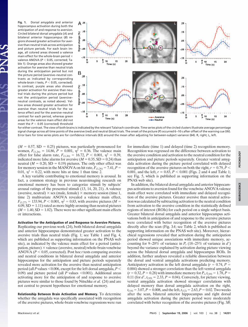
for immediate (time 1) and delayed (time 2) recognition memory.Recognition was regressed on the difference between activation tothe aversive condition and activation to the neutral condition for theanticipation and picture periods separately. Greater ventral amyg-dala activation during the picture period correlated with delayedrecognition of the aversive pictures on both the right, r � 0.79, P �0.001, and the left, r � 0.83, P � 0.001 (Figs. 2 and 4 and Table 1;see Fig. 5, which is published as supporting information on thePNAS web site).
In addition, the bilateral dorsal amygdala and anterior hippocam-pus activations to aversion found for the voxelwise ANOVA valencemain effect were correlated with immediate and delayed recogni-tion of the aversive pictures. Greater aversive than neutral activa-tion was calculated by subtracting activation to the neutral conditionfrom activation to the aversive condition in the statistically definedregions of interest (ROIs) for each period on a single subject basis.Greater bilateral dorsal amygdala and anterior hippocampus acti-vations both in anticipation of and response to the aversive pictureswere correlated with better recognition of the aversive picturesdirectly after the scan (Fig. 3A; see Table 2, which is published assupporting information on the PNAS web site). Moreover, hierar-chical regressions revealed that activation during the anticipationperiod showed unique associations with immediate memory, ac-counting for 9–20% of variance in Pr (18–25% of variance in d�)beyond the variance explained by activation during picture viewingacross the bilateral dorsal amygdala and hippocampus ROIs. Inaddition, further analyses revealed a reliable dissociation betweenthe dorsal and ventral amygdala activations predicting memory.Anticipatory activation in the left dorsal amygdala (r � 0.74, P �0.004) showed a stronger correlation than the left ventral amygdala(r � 0.32, P � 0.28) with immediate memory for Pr, t(10) � 1.78, P �0.11 (for d�, t(10) � 2.33, P � 0.04). Conversely, for picture viewing,ventral amygdala activation showed stronger correlations withdelayed memory than dorsal amygdala activation on the right,t(14) � 3.07, P � 0.008, and the left, t(12) � 2.63, P � 0.02. Two weeksafter the scan, bilateral anterior hippocampus and right dorsalamygdala activation during the picture period were moderatelycorrelated with better recognition of the aversive pictures (Fig. 3B;
Fig. 1. Dorsal amygdala and anteriorhippocampus activation during both theanticipation of and response to aversion.Circled bilateral dorsal amygdala (A) andbilateral anterior hippocampus (B) re-gions showed greater activation for aver-sive than neutral trials across anticipationand picture periods. For each brain im-age, all colored areas showed a valencemain effect for the whole-brain period �valence ANOVA (P � 0.05, corrected; Ta-ble 1). Orange areas also showed greateractivation for aversive than neutral trialsduring the anticipation period but notthe picture period (aversive–neutral con-trasts as indicated by correspondingwhole-brain t tests, P � 0.05, corrected).In contrast, purple areas also showedgreater activation for aversive than neu-tral trials during the picture period butnot the anticipation period (aversive–neutral contrasts, as noted above). Yel-low areas showed greater activation foraversive than neutral trials for the va-lence effect and for the aversive–neutralcontrast for each period, whereas greenareas for the valence main effect did notmeet the P � 0.05 (corrected) thresholdfor either contrast. The view of both brains is indicated by the relevant Talairach coordinate. Time series plots of the circled clusters illustrate average percentagesignal change across all time points of the aversive (red) and neutral (blue) trials. The onset of the picture (P) occurred 6–10 s after offset of the warning cue (W).Error bars for time series plots are for confidence intervals (63) around the mean after adjusting for between-subject variance (64). R, right; L, left.
Mackiewicz et al. PNAS � September 19, 2006 � vol. 103 � no. 38 � 14201
PSYC
HO
LOG
YN
EURO
SCIE
NCE
see Table 3, which is published as supporting information on thePNAS web site).
Sex Differences in Relationship Between Brain Activation and Mem-ory. To test for sex differences, correlations were run separately forwomen and men by using the statistically defined bilateral dorsalamygdala, ventral amygdala, and anterior hippocampus clustersfrom the aforementioned whole-brain analyses. The only sex dif-ferences observed were in the ventral amygdala for picture viewing.Delayed recognition of the aversive pictures for women was asso-ciated primarily with left ventral amygdala activation in response tothe aversive pictures, whereas delayed recognition of the aversivepictures for men was associated primarily with right ventral amyg-dala activation in response to the aversive pictures (Figs. 2 and 5).To assess laterality, t tests compared the correlations for the left andright clusters. For women, the correlation with the left ventralamygdala (r � 0.75, P � 0.03) was greater than with the right ventralamygdala (r � �0.48, P � 0.19), t(5) � 5.44, P � 0.003. For men,the correlation with the right ventral amygdala (r � 0.87, P � 0.005)was greater than with the left ventral amygdala (r � �0.50, P �0.256), t(4) � 4.76, P � 0.008. Removing extreme values does notalter the patterns observed (62). Corresponding correlations forventral amygdala activation in response to the neutral pictures werenot significant (r values between �0.22 and 0.22, all P values �0.60).
We also conducted exploratory whole-brain voxelwise regres-sions (P � 0.01, uncorrected) for recognition memory separatelyfor women and men. There was a correlation between women’sdelayed recognition of the aversive pictures and left ventralamygdala activation only (r � 0.93, P � 0.001) and betweenmen’s delayed recognition of the aversive pictures and rightventral amygdala activation only (r � 0.95, P � 0.001). Lateralitydifferences between the sexes were not observed for dorsalamygdala or anterior hippocampus associations with memory forthe anticipation or picture periods (Tables 2 and 3), althoughstatistical power for detecting sex effects was compromised bythe small cell sizes (n values range from 5 to 9).
Interrelationships Among Amygdala and Hippocampal Areas. Thebilateral dorsal amygdala and bilateral anterior hippocampus acti-vations identified in the whole-brain ANOVA and the bilateralventral amygdala activations observed in the whole-brain regres-sions were correlated with one another, similar to findings in arecent report (16). Consistently positive correlations were observedboth within and across hemisphere and both within and across theanticipation and picture periods (Table 4, which is published assupporting information on the PNAS web site).
DiscussionThis study found that the anticipation of aversion resulted inbilateral activation of the dorsal amygdala and anterior hippocam-pus that was associated with subsequent memory. The presentationof a simple warning cue instigated amygdala and hippocampusmemory-related processes that preceded exposure to the picturethat is remembered. Anticipatory activity in the dorsal amygdalaand anterior hippocampus predicted immediate but not delayedmemory of aversive pictures, whereas activity in those regions whenviewing the pictures predicted memory at both intervals. Moreover,the anticipatory activity predicted better immediate memory aboveand beyond the associations for viewing the pictures. In addition,ventral amygdala activity when viewing the pictures, but not inanticipation of them, was associated with delayed memory only.
Present results replicate our prior findings (24) that the antici-pation of and response to aversion activates the amygdala andhippocampus, consistent with the idea that the brain mechanismsrecruited upon exposure to aversion also are recruited in anticipa-tion of aversion (26, 43). A large corpus of work has pointed to thecritical role of the dorsal amygdala in threat detection and vigilance(24, 44, 65, 66). The dorsal amygdala activation found here, and inprevious work in our laboratory (24), likely reflects the adaptivebenefit of increased attention when preparing for imminent aver-sive circumstances (2, 3, 7, 24, 26, 44). In relating the dorsal�ventraldistinction to the amygdala nuclei (47, 67), the CeA is by far themost prominent nucleus in the dorsal amygdala, whereas the ventralamygdala is predominantly composed of the BLA. Rodent work has
Fig. 2. Ventral amygdala activation cor-related with subsequent memory perfor-mance. Activation in circled left and rightventral amygdala regions was correlatedwith recognition 2 weeks after scanning(time 2). Whole-brain regressions at P �0.05 (corrected) of recognition on thecontrast comparing aversive to neutralpictures revealed that increased activa-tion to the aversive pictures was associ-ated with better recognition of the aver-sive pictures. The scatter plot for eachcircled cluster shows the relationship ofrecognition memory to activation for theaversive–neutral contrast used for thewhole-brain regressions to identify theseventral amygdala areas. The scatter plotsunder the brain images for women andmen separately show the relationship ofrecognition memory to activation foraversive pictures only, the metric used inprevious neuroimaging studies showingsex differences in amygdala memoryfunction (14, 15, 21). Description forbrain images and time series plots is thesame as provided in Fig. 1 legend. Forright ventral amygdala cluster, n � 17 (9women). For left ventral amygdala clus-ter, n � 15 (8 women) because of signalloss for two subjects (see SupportingMethods, which is published as supporting information on the PNAS web site). Ventral amygdala correlations for d� were highly similar to those shownhere for Pr (see Fig. 5).
14202 � www.pnas.org�cgi�doi�10.1073�pnas.0601648103 Mackiewicz et al.

demonstrated that the CeA receives input from the basal nucleus,hippocampus, and sensory cortices (68). For the aversive picturesand the visual cues that precede them, the most likely pathway ofafferent information is into the lateral nucleus, then to the basalnucleus, and finally to the CeA, with additional information coming
from visual areas. Although both the BLA and hippocampusproject to the CeA, these connections are not reciprocal (68),whereas those with primary and secondary visual cortices are (69).Direct projections to sensory cortices likely contribute to the impactof aversion in enhancing attention and perception (2, 3, 7, 39, 51,52). In addition, prior work indicates that efferent projections fromthe CeA to the basal forebrain and eventually to the prefrontalcortex via the cholinergic system may lead to increased perceptualattention (5, 46, 70) and, in turn, influence selective attention andvigilance for threatening stimuli (25, 44). Consistent with ourfindings for immediate memory, increased recruitment of theseareas via dorsal amygdala activation both in anticipation of andresponse to aversion may reflect enhanced attention to and sub-sequent encoding of the aversive pictures.
In accord with recent neuroimaging studies implicating theventral amygdala in the consolidation of emotional memories inhumans (13–23), we found increased bilateral ventral amygdalaactivation in response to the aversive pictures was correlated withbetter delayed memory for the aversive pictures. Accruing evidenceindicates that interactions between the amygdala and hippocampusare critical for enhancing emotional memory (2–7, 12, 16, 18).McGaugh (5) proposed a modulation hypothesis stating that theBLA enhances emotional memory by modulating the activity ofother medial temporal lobe structures, especially the hippocampus,via �-adrenergic projections activated within the amygdala bynorepinephrine release from the adrenal medulla and glucocorti-coids (e.g., cortisol and corticosterone) from the adrenal cortex (31,71). An extensive and elegant body of nonhuman animal workdetailing amygdala connections has shown that the BLA, particu-larly the basal nucleus, has stronger connections with the hippocam-pal formation than any other nuclei (68). Modulation of thehippocampus by the BLA enhances consolidation of the emotionalmaterial, both at an initial stage of consolidation and continuallythrough long-term potentiation via reciprocal connections betweenthe BLA and hippocampus (5, 6, 72). Finally, evidence here thathippocampal recruitment in anticipation of and response to aver-sive pictures are associated with subsequent memory recognition ofthose pictures is consistent with prior work establishing its role inboth encoding (16, 73, 74) and consolidation (4, 5, 75).
Testing sex differences revealed that left but not right ventralamygdala activation when viewing aversive pictures was associatedwith delayed memory of those pictures in women, whereas theconverse pattern of right but not left activation was associated withdelayed memory in men. In addition to replicating prior reports ofthis sex difference in samples with both sexes (14, 15, 21), thispattern is consistent with findings from studies on female humansonly (19, 20) and male rats only (76). Addressing the specificity ofsex differences in amygdala memory function, sex differences werenot observed for the dorsal amygdala, anticipatory activation, orimmediate memory. Future research, including neuroimaging stud-ies with large samples for both men and women, is needed to furthercharacterize the boundaries of lateralized sex differences in emo-tional memory and to determine whether these sex differences arerelated to consolidation processes or other factors (2, 15).
A key conceptual issue that warrants attention is the fact thatrecent studies examining emotional memory have focused on thehighly arousing nature of emotional stimuli or experimental con-texts as the key component contributing to enhanced memory(13–23, 31, 77–79). Several studies have demonstrated that thehighly arousing nature of emotional stimuli and not their unpleasantvalence, as self-reported by subjects, promotes enhanced emotionalmemory (16, 22, 77, 80). Using established norms (81), our studyused aversive pictures selected to be highly arousing and highlyunpleasant. Subjects invariably rated the aversive pictures as morearousing than the neutral pictures, with no sex differences. Twostudies on emotional memory that reported sex differences forarousal ratings used only pictures rated as most highly arousing(15, 21).
Fig. 3. Dorsal amygdala and anterior hippocampus correlated with subse-quent memory performance. (A) Example scatter plots of correlations be-tween activation during aversive trials in the dorsal amygdala and anteriorhippocampus clusters shown in Fig. 1 and recognition of the aversive picturesdirectly after the scan (time 1, n � 13). (Left) Plots demonstrate correlationsbetween activation during the anticipation of aversive pictures and immedi-ate memory for the aversive pictures. (Right) Plots demonstrate correlationsbetween activation in response to aversive pictures and immediate memoryfor aversive pictures. Plots are, from top to bottom, right dorsal amygdala, leftdorsal amygdala, right anterior hippocampus, and left anterior hippocampus.(B) Example plots of correlations between activation in response to aversivepictures for the right and left anterior hippocampus shown in Fig. 1 andrecognition of the aversive pictures 2 weeks after scanning (Time 2, n � 17).As indicated in Tables 2 and 3, correlations for d� were highly similar to thoseshown here for Pr.
Mackiewicz et al. PNAS � September 19, 2006 � vol. 103 � no. 38 � 14203
PSYC
HO
LOG
YN
EURO
SCIE
NCE
In conclusion, anticipatory activity in the amygdala and hip-pocampus plays a central role in emotional memory. Building onprior human research on emotional memory that has often con-founded the effects of attention, encoding, and consolidation, arecent report found that postencoding emotion enhanced consol-idation of neutral memoranda (78, 79). Findings here highlight thatattention-related anticipation and encoding processes of the dorsalamygdala make a contribution to emotional memory that is distinctfrom consolidation processes modulated by the ventral amygdala.Our results also suggest that lateralized sex differences are specificto ventral amygdala and memory consolidation. Understanding thedual functioning of the amygdala and hippocampus in aversion andemotional memory may improve our clinical understanding of themultiple cognitive�affective abnormalities identified in people withanxiety and mood disorders, especially those involving emotionalmemory (e.g., traumatic memories and memory biases) and exhib-iting sex differences. Future research examining activation of theamygdala and hippocampus in people with anxiety and mooddisorders (65, 82–84) may better inform the role of emotionalmemory in the pathophysiology of these disorders.
Experimental ProceduresSubjects. Subjects were 40 right-handed healthy undergraduatestudents (18 women and 22 men; M � 20.65, SD � 1.53) whoresponded to flyers posted in campus buildings at the University ofWisconsin-Madison. They were free of any medical or neurologicalproblems, took no medications, and had no current psychiatricdiagnoses. All subjects gave informed consent in accord with studyapproval by the Human Subjects Committee of the University ofWisconsin Medical School and were paid for their participation.
Experimental Design and Stimuli. As shown in Fig. 6, which ispublished as supporting information on the PNAS web site, eachtrial consisted of a warning cue (‘‘X,’’ circle, or question mark)presented for 1 s, followed by a black screen presented for 6, 7, 8,9, or 10 s. Each trial then continued with the presentation of anaversive or neutral picture for 1 s and another black screenpresented for 6, 7, 8, 9, or 10 s. For aversive trials, an X cue alwayswas followed by an aversive picture. For neutral trials, a circle wasalways followed by a neutral picture. For ambiguous trials, aquestion mark was followed by either an aversive or neutral picture.Subjects were instructed regarding this cue-picture pairing beforescanning. All warning cues were white and presented on a blackbackground and were geometrically similar. Trial order was pseu-dorandomized, with the stipulation that no trial type (aversive,neutral, or ambiguous) be presented more than twice in a row. Triallength varied from 14 to 22 s, with an average trial length of 18 s.The intertrial interval (ITI) was randomized after both the warningcue and picture presentation. Using a variable ITI for both epochsallowed for better separation and estimation of the hemodynamicresponse for both the anticipation and response periods.
There were three functional runs, each consisting of eightaversive trials, eight neutral trials, and eight ambiguous trials. Eachfunctional scan began with a 30-s black screen, resulting in scanlengths of 8:52, 9:00, and 8:46, respectively. Using a response boxduring the fMRI experiment, subjects were instructed to push abutton after each cue and after each picture. Subjects were in-structed to press a different button if they saw a square in place ofthe cue or picture. There were two trials with a square in the firstfunctional run, three in the second run, and two in the third run.These trials were used to help maintain subjects’ attention to the cueand picture stimuli and were excluded from analyses.
During the fMRI experiment, subjects viewed 75 pictures (threeon trials with a square in place of the cue) from the InternationalAffective Picture Set (81) at a resolution of 800 � 600 pixels, withno picture being shown more than once. Of the remaining 72pictures, 36 were aversive (24 for aversive trials and 12 for ambig-uous trials) and 36 were neutral (24 for neutral trials and 12 for
ambiguous trials). Based on published norms (81), pictures with themost unpleasant valence ratings and highest arousal ratings com-prised the aversive set, which primarily included photographs ofmutilated bodies and attack scenes. Neutral pictures (e.g., house-hold items) selected had neutral valence ratings and low arousalratings.
Directly after the scan, half of the subjects completed a surpriserecognition task. All subjects completed a surprise recognition task2 weeks after the scan. Subjects completing the recognition taskdirectly after the scan were the time 1 group (n � 20, 13 men and7 women), and their data from the second recognition task were notincluded. Subjects completing the recognition task only 2 weeksafter the scan were the time 2 group (n � 20, 9 men and 11 women).Four subjects were dropped from analyses: two women (one fromtime 1 and one from time 2) because of technical difficulties withfMRI data acquisition, one man from time 1 because of excessivemovement, and one man from time 1 on account of failure to followtask instructions. Resultant sample sizes for fMRI analyses were 17(11 men and 6 women) for the time 1 group and 19 (9 men and 10women) for the time 2 group. One woman from time 2 was droppedfrom all memory analyses because of insufficient memory data.Five additional subjects (three men from time 1, one woman fromtime 1, and one man from time 2) were dropped from all memoryanalyses and arousal rating analyses because of insufficient data forthe arousal ratings described below. These subjects were droppedto make our results comparable with previous neuroimaging studieson emotional memory that have categorized pictures according tosubjects’ arousal ratings of the pictures (13–22). Resultant samplesizes for all analyses involving memory data were 13 (8 men and 5women) for the time 1 group and 17 (8 men and 9 women) for thetime 2 group.
For the recognition task, subjects were escorted to a small roomwith a computer and informed that they would be seeing a series ofpictures similar to those seen during the experimental scanningsession. Subjects were instructed to press a ‘‘Y’’ if they recognizedthe picture from the scan and an ‘‘N’’ if they did not recognize thepicture from the scan. Each picture was followed by 7-point ratingscales for valence (0, unpleasant; 3, neutral; 6, pleasant) and arousal(0, low; 6, high). Subjects responded to each scale by pressing thecorresponding number key. There was no time constraint onsubjects’ responses. Text and scales were in white font with a blackbackground, and pictures were again 800 � 600 pixels.
Subjects saw a total of 72 pictures during the recognition task,with 48 pictures from the scan (12 aversive pictures from aversivetrials, 12 aversive pictures from ambiguous trials, 12 neutral picturesfrom neutral trials, and 12 neutral pictures from ambiguous trials)and 24 novel pictures (12 aversive and 12 neutral). All subjects sawthe same pictures in the recognition task, but the conditions that thepictures came from varied across subjects to eliminate effects dueto specific pictures. Three different versions for each of the func-tional runs were created, such that each aversive picture was in anaversive trial, ambiguous trial, or shown as a novel picture in therecognition task. Similarly, each neutral picture was in a neutraltrial, ambiguous trial, or shown as a novel picture in the recognitiontask. The three versions were counterbalanced across subjects. Forthe recognition task, picture order was randomized, and pictureswere selected equally from all three runs. For the purposes of thisreport, memory and fMRI data for the ambiguous trials were notanalyzed because they address different theoretical and conceptualquestions.
fMRI Data Acquisition and Analysis. Anatomical and functional datawere collected on a General Electric (Waukesha, WI) 3.0 teslasystem by using data-acquisition parameters selected to minimizesignal loss in the amygdala and orbitofrontal cortex, areas vulner-able to the differential magnetic susceptibility coefficients of bone�air�tissue boundaries (see Supporting Methods). All fMRI data
14204 � www.pnas.org�cgi�doi�10.1073�pnas.0601648103 Mackiewicz et al.
processing was done with Analysis of Functional Neural Images(AFNI) version 2.41 software (85) (see Supporting Methods).
Initial analyses focused on identifying regions associated withboth anticipating and responding to the aversive as compared withneutral pictures. A whole-brain voxelwise period (anticipation,picture) � valence (aversive, neutral) repeated-measures ANOVAat a threshold of P � 0.005 (uncorrected) identified relevant brainregions of activation. Using AlphaSim in AFNI, we applied acorrection for multiple comparisons by using Monte Carlo simu-lations within a region of interest defined by the anatomicalboundaries of the amygdala and hippocampus, the focus of studyhypotheses (24, 85). The spatial correlation of the input data and anuncorrected P value threshold of 0.005 resulted in a minimumcluster size of 99 mm3 for this repeated-measures ANOVA toachieve a corrected mapwise P � 0.05. For statistically definedclusters meeting this threshold criterion for the valence main effect,the average percentage signal change value was extracted for eachcondition on a single-subject basis. Each subject’s average percent-age signal change values for the neutral anticipation and pictureperiods was subtracted from each subject’s values for the aversiveanticipation and picture periods. These absolute and differencevalues were subsequently correlated with recognition of the aver-sive pictures both at time 1 and time 2.
Additionally, whole-brain voxelwise regressions for recognitiondata on activation during the anticipation and picture periods wererun at a threshold of P � 0.005 (uncorrected). The aforementionedprocedure by using Monte Carlo simulations to correct for multiple
comparisons resulted in a minimum cluster size of 81 mm3 for thisregression analysis to achieve a corrected mapwise P � 0.05.Separate regressions were conducted for each period, memorymetric (Pr and d�), and memory session (time 1 and time 2).Whole-brain data were composed of the difference between acti-vation to the respective aversive and neutral condition.
Analysis of Behavioral Data. Recognition was calculated by using twoseparate metrics, Pr and d� (see Supporting Methods). Calculationswere computed for both recognition measures because althoughprevious neuroimaging studies on emotional memory have used Pr,no neuroimaging study to date has been able to calculate d� becauseof experimental restraints. Further, Cahill et al. (15) called for theuse of d� in future neuroimaging research on recognition memory.All analyses for the memory and arousal rating data were con-ducted at an � level of 0.05. Two-tailed tests were used throughout,including for the directional hypotheses that amygdala and hip-pocampus activation would predict better memory for aversivepictures.
We thank Andrew Alexander, Michael Anderle, Krista Bluske, RonFisher, John Herrington, Tom Johnstone, Roberta Koch, HillarySchaefer, Eric Steege, and Mai Lor for their contributions to this project.J.B.N. was supported by National Institute of Mental Health GrantsR01-MH74847 and K08-MH63984. The research reported in this pub-lication also was supported by National Institute of Child Health andHuman Development Grant P30-HD03352 to the Waisman Center.
1. Christianson SA (1992) The Handbook of Emotion and Memory: Research and Theory(Lawrence Earlbaum Associates, Hillsdale, NJ).
2. Phelps EA (2004) Curr Opin Neurobiol 14:198–202.3. Phelps EA (2006) Annu Rev Psychol 57:27–53.4. McGaugh JL (2000) Science 287:248–251.5. McGaugh JL (2004) Annu Rev Neurosci 27:1–28.6. McGaugh JL, Roozendaal B (2002) Curr Opin Neurobiol 12:205–210.7. Phelps EA, LeDoux JE (2005) Neuron 48:175–187.8. Packard MG, Cahill L, McGaugh JL (1994) Proc Natl Acad Sci USA 91:8477–8481.9. Adolphs R, Tranel D, Denburg N (2000) Learn Mem 7:180–186.
10. Cahill L, Babinsky R, Markowitsch HJ, McGaugh JL (1995) Nature 377:295–296.11. LaBar KS, Phelps EA (1998) Psychol Sci 9:490–493.12. Richardson MP, Strange BA, Dolan RJ (2004) Nat Neurosci 7:278–285.13. Cahill L, Haier RJ, Fallon J, Alkire MT, Tang C, Keator D, Wu J, McGaugh JL (1996) Proc
Natl Acad Sci USA 93:8016–8021.14. Cahill L, Haier RJ, White NS, Fallon J, Kilpatrick L, Lawrence C, Potkin SG, Alkire MG
(2001) Neurobiol Learn Mem 75:1–9.15. Cahill L, Uncapher M, Kilpatrick L, Alkire MT, Turner J (2004) Learn Mem 11:261–266.16. Dolcos F, LaBar KS, Cabeza R (2004) Neuron 42:855–863.17. Dolcos F, LaBar KS, Cabeza R (2005) Proc Natl Acad Sci USA 102:2626–2631.18. Strange BA, Dolan RJ (2004) Proc Natl Acad Sci USA 31:11454–11458.19. Canli T, Zhao Z, Brewer J, Gabrieli JDE, Cahill L (2000) J Neurosci 20:RC99.20. Canli T, Zhao Z, Desmond JE, Glover G, Gabrieli JDE (1999) Psychobiology 27:441–452.21. Canli T, Desmond JE, Zhao Z, Gabrieli JDE (2002) Proc Natl Acad Sci USA 99:10789–
10794.22. Hamann SB, Ely TD, Grafton ST, Kilts CD (1999) Nat Neurosci 2:289–293.23. Kensinger EA, Schacter DL (2006) J Neurosci 26:2564–2570.24. Nitschke JB, Sarinopoulos I, Mackiewicz KL, Schaefer HS, Davidson RJ (2006) NeuroImage
29:106–116.25. Davis M (1992) Annu Rev Neurosci 15:353–375.26. LeDoux JE (2002) Synaptic Self: How Our Brains Become Who We Are (Viking, New York).27. Buchel C, Morris J, Dolan RJ, Friston KJ (1998) Neuron 20:947–957.28. Buchel C, Dolan RJ, Armony JL, Friston KJ (1999) J Neurosci 19:10869–10876.29. LaBar KS, Gatenby JC, Gore JC, LeDoux JE, Phelps EA (1998) Neuron 20:937–945.30. Cahill L, McGaugh JL (1998) Trends Neurosci 21:294–299.31. Okuda S, Roozendaal B, McGaugh J (2004) Proc Natl Acad Sci USA 101:853–858.32. Davis M, Schlesinger LS, Sorenson CA (1989) J Exp Psychol Anim Behav Processes
15:295–310.33. Grillon C, Ameli R, Woods SW, Merikangas K, Davis M (1991) Psychophysiology 28:588–595.34. Grillon C, Ameli R, Merikangas K, Woods SW, Davis M (1993) Psychophysiology 30:340–346.35. Nitschke JB, Larson CL, Smoller MJ, Navin SD, Pederson AJ, Ruffalo D, Mackiewicz KL,
Gray SM, Victor E, Davidson RJ (2002) Psychophysiology 39:254–258.36. Koster EH, Crombez G, Van Damme S, Verschuere B, De Houwer J (2004) Emotion 4:312–317.37. Van Damme S, Crombez G, Eccleston C (2004) Pain 111:392–399.38. Van Damme S, Lorenz J, Eccleston C, Koster EHW, De Clercq A, Crombez G (2004)
Neurophysiol Clin 34:33–39.39. Morris JS, Buchel C, Dolan RJ (2001) NeuroImage 13:1044–1052.40. Iidaka T, Anderson ND, Kapur S, Cabeza R, Craik FI (2000) J Cognit Neurosci 12:267–280.41. Mangels JA, Picton TW, Craik FI (2001) Brain Res Cogn Brain Res 11:77–95.42. Anderson ND, Iidaka T, Cabeza R, Kapur S, McIntosh AR, Craik FI (2000) J Cognit
Neurosci 12:775–792.43. Pavlov IP (1927) Conditioned Reflexes: An Investigation of the Physiological Activity of the
Cerebral Cortex (Dover, New York).44. Davis M, Whalen PJ (2001) Mol Psychiatry 6:13–34.
45. Holland PC, Gallagher M (1999) Trends Cognit Sci 3:65–73.46. Holland PC, Han J, Gallagher M (2000) J Neurosci 20:6701–6706.47. Lee HJ, Groshek F, Petrovich GD, Cantalini JP, Gallagher M, Holland PC (2005) J Neurosci
25:3881–3888.48. Groshek F, Kerfoot E, McKenna V, Polackwich AS, Gallagher M, Holland PC (2005) Behav
Neurosci 119:202–212.49. Anderson AK, Phelps EA (2001) Nature 411:305–309.50. Amaral DG (2003) Ann NY Acad Sci 1000:337–347.51. Morris JS, Friston KJ, Buchel C, Frith CD, Young AW, Calder AJ, Dolan RJ (1998) Brain
121:47–57.52. Vuilleumier P, Richardson MP, Armony JL, Driver J, Dolan RJ (2004) Nat Neurosci
7:1271–1278.53. Eichenbaum H (2002) The Cognitive Neuroscience of Memory (Oxford Univ Press, New
York).54. Squire LR, Zola-Morgan S (1991) Science 253:1380–1386.55. Scoville WB, Milner B (1957) J Neurol Neurosurg Psychiatry 20:11–21.56. Cohen NJ, Ryan J, Hunt C, Romine L, Wczalek T, Nash C (1999) Hippocampus 9:83–98.57. Schacter DL, Wagner AD (1999) Hippocampus 9:7–24.58. Maren S (1999) Trends Neurosci 22:561–567.59. Setlow B, Holland PC, Gallagher M (2002) Behav Neurosci 116:267–275.60. Otten JJ, Quayle AH, Akram S, Ditewig TA, Rugg MD (2006) Nat Neurosci 9:489–491.61. Adcock RA, Thangavel A, Whitfield-Gabrieli S, Knutson B, Gabrieli JDE (2006) Neuron
50:507–517.62. Cahill L (2006) Nat Rev Neurosci 7:477–484.63. Cumming G, Finch S (2005) Am Psychol 60:170–180.64. Loftus GR, Masson MEJ (1994) Psychonomic Bull Rev 1:476–490.65. Nitschke JB, Heller W (2005) Int Rev Neurobiol 67:1–42.66. Pessoa L, Kastner S, Ungerleider LG (2002) Cognit Brain Res 15:31–45.67. Kim H, Somerville LH, Johnstone T, Polis S, Alexander AL, Shin LM, Whalen PJ (2004)
J Cognit Neurosci 16:1730–1745.68. Pitkanen A (2000) in The Amygdala: A Functional Analysis, ed Aggleton JP (Oxford Univ
Press, New York), pp 31–116.69. Freese JL, Amaral DG (2005) J Comp Neurol 486:295–317.70. Gallagher M (2000) in The Amygdala: A Functional Analysis, ed Aggleton JP (Oxford Univ
Press, New York), pp 311–330.71. Roozendaal B, Okuda S, de Quervain DJ, McGaugh JL (2006) Neuroscience 138:901–910.72. Chapman PF, Chattarji S (2000) in The Amygdala: A Functional Analysis, ed Aggleton JP
(Oxford Univ Press, New York), pp 117–154.73. Lepage M, Habib R, Tulving E (1998) Hippocampus 8:313–322.74. Strange BA, Fletcher PC, Henson RNA, Friston KJ, Dolan RJ (1999) Proc Natl Acad Sci
USA 96:4034–4039.75. Malin EL, McGaugh J (2006) Proc Natl Acad Sci USA 103:1959–1963.76. LaLumiere RT, McGaugh J (2005) Learn Mem 12:527–532.77. Hamann S (2001) Trends Cognit Sci 9:394–400.78. Anderson AK, Wais PE, Gabrieli DE (2006) Proc Natl Acad Sci USA 103:1599–1604.79. McGaugh JL (2006) Trends Cognit Sci 10:345–347.80. Cahill L, McGaugh JL (1990) Behav Neurosci 104:532–543.81. Lang PJ, Bradley MM, Cuthbert BN (1999) International Affective Picture System: Technical
Manual and Affective Ratings (Univ Florida, Gainesville, FL).82. Davidson RJ, Pizzagalli D, Nitschke JB, Putnam KM (2002) Annu Rev Psychol 53:545–574.83. Drevets WC (2003) Ann NY Acad Sci 985:420–444.84. Rauch SL, Shin LM, Wright CI (2003) Ann NY Acad Sci 985:389–410.85. Cox RW (1996) Comp Biomed Res 29:162–173.
Mackiewicz et al. PNAS � September 19, 2006 � vol. 103 � no. 38 � 14205
PSYC
HO
LOG
YN
EURO
SCIE
NCE
Copyright © 2022 FDOKUMEN