Visual Working Memory Impairment in Premanifest Gene-Carriers and Early Huntington's Disease
10
Journal of Huntington’s Disease 1 (2012) 105–114 DOI 10.3233/JHD-2012-120010 IOS Press 105 Visual Working Memory Impairment in Premanifest Gene-Carriers and Early Huntington’s Disease Eve M. Dumas a , Miranda J. Say b , Rebecca Jones c , Izelle Labuschagne d , Alison M. O’Regan d , Ellen P. Hart a , Simon J.A. van den Bogaard a , Sarah Queller e , Damian Justo f , Allison Coleman g , Rachelle C. dar Santos g , Alexandra D¨ urr f , Blair R. Leavitt g , Sarah J. Tabrizi b , Raymund A.C. Roos a and Julie C. Stout d,∗ a Department of Neurology, Leiden University Medical Centre, Leiden, The Netherlands b UCL Institute of Neurology, University College London, Queen Square, London, UK c Department of Medical Statistics, London School of Hygiene and Tropical Medicine, London, UK d School of Psychology and Psychiatry, Monash University, Clayton Campus, VIC, Australia e Queller Consulting, Dunedin, FL, USA f Department of Genetics and Cytogenetics, and INSERM UMR S679, APHP Hôpital de la Salpˆ etri` ere, Paris, France g Department of Medical Genetics, University of British Columbia, Vancouver, BC, Canada Abstract. Working memory deficits have been found in Huntington’s disease (HD) and in a small group of premanifest (PreHD) gene-carriers. However, the nature and extent of these deficits are unknown. In a large cross-sectional study, we aimed to determine the degree of visuospatial working memory dysfunction across multiple stages of HD. Specifically, visuospatial working memory capacity and response times across various degrees of difficulty were examined, as well as the relationship between visuospatial working memory and motor dysfunction. We examined 62 PreHD-A gene-carriers (>10.8 years from estimated disease onset), 58 PreHD-B gene-carriers (<10.8 years from estimated disease onset), 77 stage-1 HD patients (HD1), 44 stage-2 HD patients (HD2), and 122 healthy controls. Participants viewed coloured squares (in sets of 3, 5 and 7) on a screen and were to decide whether on a subsequent screen the encircled square has changed colour. Accuracy and response times were recorded. Compared to controls, significant group differences in visuospatial working memory capacity (accuracy) were seen in PreHD-B, HD1 and HD2 groups across the difficulty levels. Significant group differences on response times were found for all groups (PreHD-A to HD2) compared to controls; the most difficult level producing the only group difference in speed between PreHD-A and controls. Accuracy and speed were positively correlated only in the HD groups. These findings suggest that visuospatial working memory impairments are detectable in both premanifest and manifest HD; the manifest HD showed evidence for a “worse-worse phenomenon” whereby reductions were present in both motor speed and accuracy. Keywords: Huntington’s disease, visual working memory, cognitive dysfunction, premanifest gene-carriers Huntington’s disease (HD) is an autosomal domi- nant neurodegenerative disease, which is characterised ∗ Correspondence to: Prof. Julie C. Stout, PhD, School of Psy- chology and Psychiatry, Monash University Clayton, Victoria 3800, Australia. Tel.: +61 3 99053987; Fax: +61 3 99053948; E-mail: [email protected]. by progressive motor, psychiatric and cognitive symp- toms and signs [1–4]. The mean age of disease diagnosis is between 35 and 45 years [4]. Individuals at risk of carrying the HD gene can be tested. Those who are found to have the gene but not to have clinical disease (i.e., motor) signs are referred to as premanifest gene-carriers. Many studies investigating cognition in ISSN 1879-6397/12/$27.50 © 2012 – IOS Press and the authors. All rights reserved
-
Upload
sorbonne-fr -
Category
Documents
-
view
1 -
download
0
Transcript of Visual Working Memory Impairment in Premanifest Gene-Carriers and Early Huntington's Disease

Journal of Huntington’s Disease 1 (2012) 105–114DOI 10.3233/JHD-2012-120010IOS Press
105
Visual Working Memory Impairment inPremanifest Gene-Carriers and EarlyHuntington’s Disease
Eve M. Dumasa, Miranda J. Sayb, Rebecca Jonesc, Izelle Labuschagned, Alison M. O’Regand,Ellen P. Harta, Simon J.A. van den Bogaarda, Sarah Quellere, Damian Justof , Allison Colemang,Rachelle C. dar Santosg, Alexandra Durrf , Blair R. Leavittg, Sarah J. Tabrizib,Raymund A.C. Roosa and Julie C. Stoutd,∗aDepartment of Neurology, Leiden University Medical Centre, Leiden, The NetherlandsbUCL Institute of Neurology, University College London, Queen Square, London, UKcDepartment of Medical Statistics, London School of Hygiene and Tropical Medicine, London, UKdSchool of Psychology and Psychiatry, Monash University, Clayton Campus, VIC, AustraliaeQueller Consulting, Dunedin, FL, USAf Department of Genetics and Cytogenetics, and INSERM UMR S679, APHP Hôpital de la Salpetriere, Paris, FrancegDepartment of Medical Genetics, University of British Columbia, Vancouver, BC, Canada
Abstract. Working memory deficits have been found in Huntington’s disease (HD) and in a small group of premanifest (PreHD)gene-carriers. However, the nature and extent of these deficits are unknown. In a large cross-sectional study, we aimed todetermine the degree of visuospatial working memory dysfunction across multiple stages of HD. Specifically, visuospatialworking memory capacity and response times across various degrees of difficulty were examined, as well as the relationshipbetween visuospatial working memory and motor dysfunction. We examined 62 PreHD-A gene-carriers (>10.8 years fromestimated disease onset), 58 PreHD-B gene-carriers (<10.8 years from estimated disease onset), 77 stage-1 HD patients (HD1),44 stage-2 HD patients (HD2), and 122 healthy controls. Participants viewed coloured squares (in sets of 3, 5 and 7) on a screenand were to decide whether on a subsequent screen the encircled square has changed colour. Accuracy and response times wererecorded. Compared to controls, significant group differences in visuospatial working memory capacity (accuracy) were seenin PreHD-B, HD1 and HD2 groups across the difficulty levels. Significant group differences on response times were foundfor all groups (PreHD-A to HD2) compared to controls; the most difficult level producing the only group difference in speedbetween PreHD-A and controls. Accuracy and speed were positively correlated only in the HD groups. These findings suggestthat visuospatial working memory impairments are detectable in both premanifest and manifest HD; the manifest HD showedevidence for a “worse-worse phenomenon” whereby reductions were present in both motor speed and accuracy.
Keywords: Huntington’s disease, visual working memory, cognitive dysfunction, premanifest gene-carriers
Huntington’s disease (HD) is an autosomal domi-nant neurodegenerative disease, which is characterised
∗Correspondence to: Prof. Julie C. Stout, PhD, School of Psy-chology and Psychiatry, Monash University Clayton, Victoria 3800,Australia. Tel.: +61 3 99053987; Fax: +61 3 99053948; E-mail:[email protected].
by progressive motor, psychiatric and cognitive symp-toms and signs [1–4]. The mean age of diseasediagnosis is between 35 and 45 years [4]. Individualsat risk of carrying the HD gene can be tested. Thosewho are found to have the gene but not to have clinicaldisease (i.e., motor) signs are referred to as premanifestgene-carriers. Many studies investigating cognition in
ISSN 1879-6397/12/$27.50 © 2012 – IOS Press and the authors. All rights reserved

106 E.M. Dumas et al. / Working Memory in HD
HD have demonstrated progressive cognitive declineresulting in dementia [5, 6]. Cognitive decline is alsodetectable in the premanifest gene-carriers across anumber of domains, including executive functions,memory, emotion recognition and psychomotor func-tions [7–9].
Working memory is a topic of recent attention as apossible marker for disease state in HD [10, 11]. Manyday-to-day activities require retention, integration andmanipulation of either verbally or visually presentedinformation, referred to as verbal or visual (or visu-ospatial) working memory [12]. Poor working memoryhas been described as part of the disease course ofHD. In particular, several cross-sectional studies havedemonstrated that HD patients show poorer spatial orvisual working memory in comparison to controls [9,11, 13–15]. Visual working memory was also foundto decline in patients with HD over 12-, 24- and 42-months [2, 3, 16].
Studies of premanifest gene-carriers have identi-fied mild-to-moderate cognitive deficits in a range ofdomains, including attention, memory, psychomotorspeed and executive functioning, which are amongthe first cognitive functions to show decline in thepremanifest phase [17–23]. With regard to workingmemory, the evidence in premanifest HD is unclear.Some studies have reported impairments in both verbaland visual working memory [17, 24]. However, othershave suggested that premanifest HD do not differ fromcontrols in either visual or verbal working memory [19,25]. In addition, although evidence regarding workingmemory decline in premanifest HD is limited, it doessuggest that working memory, in particular visuospa-tial working memory, may be among the first cognitivefunctions to show decline in the premanifest phase [3,10].
Working memory is implicated in complex brainnetworks whereby integrated signals received by theparietal cortex, are then projected onto the frontos-triatal brain circuits which then subsequently drivemotor responses [26–28]. The underlying brain regionsassociated with working memory, namely the caudatenucleus and putamen which lie within the frontostri-atal brain circuits, are also among the primary regionsimplicated in the cognitive dysfunction, neurobiol-ogy and aetiology of HD [29–33]. Specifically, brainactivation studies involving working memory perfor-mances in premanifest HD reported reduced functionalconnectivity in regions of the frontostriatal brain cir-cuit such as the putamen and prefrontal cortex [34,35], as well as reduced brain activity during elec-troencephalograph measures [36], when compared to
controls. These changes were observed even in theabsence of behavioural differences in working mem-ory task performance. Structural evidence has shownthat brain atrophy in premanifest gene-carriers devel-ops prior to disease diagnosis, and it progresses duringthe disease course, with the most profound and earli-est changes found in the caudate nucleus and putamen[9, 37–39]. Given that both cognitive dysfunction andabnormal neurobiology have been observed in HDincluding in premanifest gene-carriers who are morethan ten years before estimated disease onset [9, 20,22, 39], it can be expected that deficits in visual work-ing memory would develop as the disease progresses(and the brain regions deteriorate).
In addition, motor functioning overlaps with cogni-tive functioning, in that both are implicated in brainstructures such as the basal ganglia [40], and cogni-tive performance is measured through motor outputssuch as verbal or button-based responses. The mostsensitive assessments of early cognitive changes inHD are those with a substantial psychomotor speedcomponent [8, 9, 22, 41]. Therefore, to better under-stand how HD affects cognition, it is important todistinguish, where possible, the impact of motor func-tioning on cognitive measures. Also, as we movetoward treatment-focused studies in HD, it is neces-sary to understand the progression of cognitive deficitsin relation to motor dysfunction. This is importantsince patient groups are often defined in terms oftheir level of motor deficits. The distinction betweenpremanifest and manifest HD is made based on thelevel of motor abnormalities. For premanifest groups,stringent exclusion of motor deficits can facilitate dis-tinctions between motor and cognitive disease effects,although subtle motor changes are not eliminated bythis approach. Cognitive tasks that require minimalmotor responses are also desirable in this respect.
The background presence of motor slowing alsocomplicates the interpretation of cognitive testingin HD. One approach, to disentangle the motorand cognitive effects, is to examine the relation-ship between performance accuracy and responsetimes. This relationship is often observed as a ‘speed-accuracy trade-off’, which refers to a strategy wherebyparticipants use a slower, more cautious approach toensure the accuracy of their performance. Conversely,faster responses may lead to greater inaccuracy due tobeing less careful or less cautious. We hypothesisedthat HD gene-carriers may slow their responses as acompensatory strategy in order to maintain satisfactorycognitive performance. Because we wanted to exam-ine whether speed-accuracy trade-offs would appear in

E.M. Dumas et al. / Working Memory in HD 107
relation to working memory performance in HD, weselected a task in which these two aspects of perfor-mance could be examined separately, allowing theirrelationship to be studied in the context of HD.
Using data obtained from a large multisite interna-tional and observational study known as TRACK-HD[3, 9], the current study aimed to determine, usinga more detailed analysis, the degree of visuospatialworking memory dysfunction in HD. Previous publi-cations from TRACK-HD [2, 3, 9] have reported ondata from the current working memory task; howeverthis involved reporting only a single primary outcomevariable (i.e., working memory capacity) with differ-ent group comparisons and using different statisticalanalysis techniques. Specifically, we aimed to examinevisuospatial working memory impairments relating toworking memory capacity and response times acrossmultiple disease stages including both premanifestgene-carriers and those in early stage HD. In addi-tion, and unlike the previous publications, we wantedto examine visuospatial working memory function inHD across different levels of task complexity. Further-more, we wanted to distinguish between cognitive andmotor influences in order to clarify whether workingmemory itself, rather than just the motor expression ofthis cognitive function, is affected in HD. By address-ing these aims, we can obtain evidence regarding thepossibility that a working memory task may be suit-able as a marker for cognitive deterioration in earlydiagnosed or even premanifest HD.
METHODS
Participants
Three hundred and sixty-six subjects were studiedas part of the TRACK-HD study [1, 3, 9]. Of these, 123were premanifest gene-carriers, defined as geneticallyconfirmed but without clinically evident symptoms,120 were patients with stage 1 and 2 HD; and 123were age- and sex-matched healthy controls. Partic-ipants were recruited from four study sites: London(UK), Paris (F), Vancouver (CAN), and Leiden (NL).Premanifest participants were included only if theydid not have substantial motor signs as indicated bytotal motor scores of ≤5 points on the Unified Hunt-ington’s Disease Rating Scale [UHDRS; 42], and ifthey had Disease Burden Scores of at least 250 [43].For each premanifest gene-carrier, we computed anestimate of the proximity (in years) to predicted dis-ease onset based on CAG repeat length and currentage [44]. Then, using a median split (10.8 years to
expected onset) we created a further from estimatedonset group (PreHD-A, >10.8 years to estimated onset)and a closer to estimated onset group (PreHD-B, <10.8years to estimated onset). For early stage HD partici-pants, we used Total Functional Capacity (TFC) scoresfrom the UHDRS to differentiate between those inthe HD stage 1 group (HD1, TFC scores 11–13) andHD stage 2 group (HD2, TFC scores 7–10) [45]. Forinformation on the full cognitive assessment battery,additional examinations and detailed inclusion criteria,see Tabrizi et al. [3, 9]. In the current report, we reportcross-sectional data from a visual working memorytask, Spot the Change (SPOT).
Spot the Change task
The Spot the Change task (SPOT) was based on avisual array comparison task [46, 47]. Using a LenovoVantage ThinkPad tablet PC (IBM, New York), partic-ipants viewed an array of coloured squares (250 ms) onthe screen, followed by a blank display (1000 ms). Thiswas followed by a second array of coloured squares inwhich one of the squares is encircled. The positionof the squares was unchanged between the two pre-sentations. Participants were asked to indicate if thecolour of the encircled square had changed from thefirst to second display. Using a mouse mounted ona stabilising wooden platform, the response “same”was to be made using the dominant thumb to indicatethat the encircled square had not changed colour, anda response of “different” was to be made using thenon-dominant thumb to indicate that the colour of theencircled square had changed and was therefore ‘dif-ferent’. “Same” and “Different” labels were attachedto the mouse platform to remind subjects which thumbcorresponded to which response. No feedback wasprovided following participant responses. Responsescould be made up to 8 seconds after the beginning ofthe second display. Prior to starting the task, instruc-tions and a minimum of four practice trials were givento ensure task comprehension.
In order to determine the most sensitive task con-dition, three levels of difficulty were used whichwere based on the number of coloured squares con-tained in the array. Ranging from easiest to hardest,they included three coloured squares (set size 3), fivecoloured squares (set size 5), and 7 coloured squares(set size 7). Data from the three set sizes were col-lected at two different visits separated by 12-months.Set sizes 3 and 5 were collected at visit 1 (baseline),and set size 7, along with set size 5 again, were col-lected at visit 2 (12-month follow-up). Each set size
108 E.M. Dumas et al. / Working Memory in HD
consisted of 32 trials, and stimuli were presented atrandom. At the 12-month follow-up visit, set size 3 wasexcluded because the baseline results showed ceilingeffects, and as a result we introduced a more difficultcondition, set size 7. This paper is reporting on cross-sectional data from set sizes 3 and 5 (collected at visit1) and set sizes 5 and 7 (collected at visit 2). Longitudi-nal data for set size 5 has been reported in our previousreport; see Tabrizi and colleagues [3]. Accuracy andresponse time were recorded and analysed separatelyfor each of the set sizes.
Non-response trials were recorded when a partici-pant did not respond within the given 8-second timeframe, which occurred 168 times across the groupsfor both visits and all set sizes (0.38% of the trials).In an additional seven trials, responses were givenwithin 100 ms of the stimulus; these were consideredto be ‘pre-cognitive’ or accidental responses and wereexcluded from the analysis. Accuracy measures werecorrected for guessing by the calculation of k; a mea-sure of working memory capacity as described byCowan [46]. It is computed as k = set size n ([num-ber correct hits/number of trials] + [number correctrejections/number of trials]−1). A k (working mem-ory capacity) value close to the set size (e.g., 3, 5 or 7)indicates good working memory capacity, whereas a kvalue close to or less than zero represents performancescloser to chance.
Of the subjects that attended the visits, only a smallnumber of participants failed to complete the Spot theChange task, which was nearly always due to time con-straints. The task was completed at visit 1 (baseline),visit 2 (12-month follow-up), or at both visits by atotal of 363/366 (99%) participants (with 1 control and2 HD2 participants not completing the task at any ofthe visits and were thus excluded from the analysis).Visit 1 had a total of 355 of 366 who completed the task(97%), yielding missing data for 3 controls, 5 HD1, and3 HD2. Of the 355 (92%) participants at visit 1, 325participants returned for visit 2, with an additional 8completing the task who did not do so at visit 1. There-fore, 333 out of 366 (91%) completed the task duringvisit 2, yielding missing data for 9 controls, 1 PreHD-A, 5 PreHD-B, 6 HD1 and 12 HD2 during this visit.
Statistical analysis
All analyses were performed using SAS v9.2 (StataCorporation, College Station, Texas). The workingmemory capacity (k) data were analysed in a singleregression model incorporating data from the threedifficulty levels and both visits. Working memory
capacity (k) was the outcome variable of interest.The main predictors were group (controls, PreHD-A,PreHD-B, HD1 and HD2) and set size at each visit (setsizes 3 and 5 at visit 1 and set sizes 5 and 7 at visit 2).Response times (RT) were considered separately forcorrect (correct recognitions and correct rejections) orincorrect (incorrect recognitions and incorrect rejec-tions) trials. The distributions of RTs were highlyskewed and therefore were log transformed prior toanalysis to improve normalisation of these variables forstatistical analysis. Similar to the analyses for workingmemory capacity, all RT data from both visits and fromall three set sizes were analysed in a separate singleregression model with RT as the outcome. The mainpredictors were group (controls, PreHD-A, PreHD-B,HD1 and HD2), response accuracy (correct or incor-rect) and set size (set sizes 3 and 5 at visit 1 and setsizes 5 and 7 at visit 2).
Age, gender, education level and study site wereincluded as covariates for both the working memorycapacity (k) and RT models. The regression mod-els used generalised estimating equations, which havea working assumption of exchangeability and robuststandard errors [48, 49]. This allowed for cross-sectional comparison of each gene-carrier group tocontrols for each set size. We also examined whethergroups responded differently in terms of RTs for cor-rect versus incorrect trials.
Finally, to examine the direct relationship betweenaccuracy and RT, we computed separate linear regres-sion models for each set size with the mean of the logtransformed RTs for each individual as the outcomemeasure. Because interactions require larger sampleswe collapsed the five groups into three groups whichincluded controls, a premanifest group (PreHD-A andPreHD-B combined) and an early HD group (HD1 andHD2 combined). For this analysis, k and group (con-trols, PreHD, and early HD) were the main predictors.Again, age, gender, education level and study site werecovariates. A group versus k interaction was included toallow for differences in the speed/accuracy relationshipbetween groups to be investigated.
RESULTS
To address our primary objective of examiningvisual working memory, here we first describe k (work-ing memory capacity) for each set size (3, 5 and 7) inthe five groups (controls, PreHD-A, PreHD-B, HD1and HD2). We then present RT findings per set sizeacross five groups. Finally, we describe the relationshipbetween RT and accuracy to further characterise the
E.M. Dumas et al. / Working Memory in HD 109
nature of visual working memory using three groups(controls, PreHD and early HD).
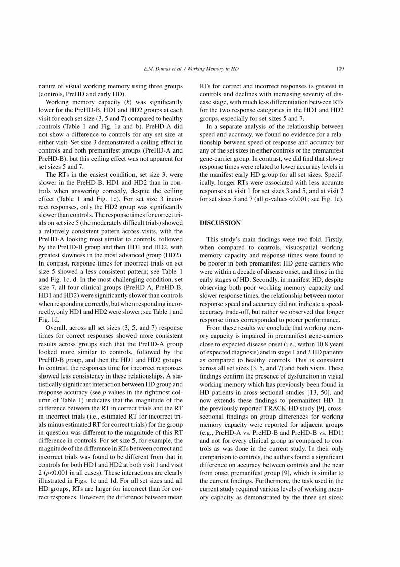
Working memory capacity (k) was significantlylower for the PreHD-B, HD1 and HD2 groups at eachvisit for each set size (3, 5 and 7) compared to healthycontrols (Table 1 and Fig. 1a and b). PreHD-A didnot show a difference to controls for any set size ateither visit. Set size 3 demonstrated a ceiling effect incontrols and both premanifest groups (PreHD-A andPreHD-B), but this ceiling effect was not apparent forset sizes 5 and 7.
The RTs in the easiest condition, set size 3, wereslower in the PreHD-B, HD1 and HD2 than in con-trols when answering correctly, despite the ceilingeffect (Table 1 and Fig. 1c). For set size 3 incor-rect responses, only the HD2 group was significantlyslower than controls. The response times for correct tri-als on set size 5 (the moderately difficult trials) showeda relatively consistent pattern across visits, with thePreHD-A looking most similar to controls, followedby the PreHD-B group and then HD1 and HD2, withgreatest slowness in the most advanced group (HD2).In contrast, response times for incorrect trials on setsize 5 showed a less consistent pattern; see Table 1and Fig. 1c, d. In the most challenging condition, setsize 7, all four clinical groups (PreHD-A, PreHD-B,HD1 and HD2) were significantly slower than controlswhen responding correctly, but when responding incor-rectly, only HD1 and HD2 were slower; see Table 1 andFig. 1d.
Overall, across all set sizes (3, 5, and 7) responsetimes for correct responses showed more consistentresults across groups such that the PreHD-A grouplooked more similar to controls, followed by thePreHD-B group, and then the HD1 and HD2 groups.In contrast, the responses time for incorrect responsesshowed less consistency in these relationships. A sta-tistically significant interaction between HD group andresponse accuracy (see p values in the rightmost col-umn of Table 1) indicates that the magnitude of thedifference between the RT in correct trials and the RTin incorrect trials (i.e., estimated RT for incorrect tri-als minus estimated RT for correct trials) for the groupin question was different to the magnitude of this RTdifference in controls. For set size 5, for example, themagnitude of the difference in RTs between correct andincorrect trials was found to be different from that incontrols for both HD1 and HD2 at both visit 1 and visit2 (p<0.001 in all cases). These interactions are clearlyillustrated in Figs. 1c and 1d. For all set sizes and allHD groups, RTs are larger for incorrect than for cor-rect responses. However, the difference between mean
RTs for correct and incorrect responses is greatest incontrols and declines with increasing severity of dis-ease stage, with much less differentiation between RTsfor the two response categories in the HD1 and HD2groups, especially for set sizes 5 and 7.
In a separate analysis of the relationship betweenspeed and accuracy, we found no evidence for a rela-tionship between speed of response and accuracy forany of the set sizes in either controls or the premanifestgene-carrier group. In contrast, we did find that slowerresponse times were related to lower accuracy levels inthe manifest early HD group for all set sizes. Specif-ically, longer RTs were associated with less accurateresponses at visit 1 for set sizes 3 and 5, and at visit 2for set sizes 5 and 7 (all p-values <0.001; see Fig. 1e).
DISCUSSION
This study’s main findings were two-fold. Firstly,when compared to controls, visuospatial workingmemory capacity and response times were found tobe poorer in both premanifest HD gene-carriers whowere within a decade of disease onset, and those in theearly stages of HD. Secondly, in manifest HD, despiteobserving both poor working memory capacity andslower response times, the relationship between motorresponse speed and accuracy did not indicate a speed-accuracy trade-off, but rather we observed that longerresponse times corresponded to poorer performance.
From these results we conclude that working mem-ory capacity is impaired in premanifest gene-carriersclose to expected disease onset (i.e., within 10.8 yearsof expected diagnosis) and in stage 1 and 2 HD patientsas compared to healthy controls. This is consistentacross all set sizes (3, 5, and 7) and both visits. Thesefindings confirm the presence of dysfunction in visualworking memory which has previously been found inHD patients in cross-sectional studies [13, 50], andnow extends these findings to premanifest HD. Inthe previously reported TRACK-HD study [9], cross-sectional findings on group differences for workingmemory capacity were reported for adjacent groups(e.g., PreHD-A vs. PreHD-B and PreHD-B vs. HD1)and not for every clinical group as compared to con-trols as was done in the current study. In their onlycomparison to controls, the authors found a significantdifference on accuracy between controls and the nearfrom onset premanifest group [9], which is similar tothe current findings. Furthermore, the task used in thecurrent study required various levels of working mem-ory capacity as demonstrated by the three set sizes;

110 E.M. Dumas et al. / Working Memory in HD
6
4
2
0
-2
-4
6
4
2
0
-2
-4
2200
6
Controls Premanifest HD Early HD
Correct 5sCorrect 7s
Incorrect 5sIncorrect 7s
Correct 3sCorrect 5s
Incorrect 3sIncorrect 5s
4
2
0
-2
-4
6
4
2
0
-2
-4
6
4
2
0
-2
-4
6
4
2
0
-2
-4
6
4
2
0
-2
-4
6
4
2
0
-2
-4
6
4
2
0
-2
-4
6
4
2
0
-2
-4
6
4
2
0
-2
-4
500 1000 1500 2000 2500 3000 3500500 1000 1500 2000 2500 3000 3500
500 1000 1500 2000 2500 3000 3500 500 1000 1500 2000 2500 3000 3500
500 1000 1500 2000 2500 3000 3500500 1000 1500 2000 2500 3000 3500
500 1000 1500 2000 2500 3000 3500
500 1000 1500 2000 2500 3000 3500
Mean response time (ms) Mean response time (ms)
Mean response time (ms)Mean response time (ms)Mean response time (ms)
Mean response time (ms)
Acc
urac
y (k
)
Acc
urac
y (k
)A
ccur
acy
(k)
Acc
urac
y (k
)
Acc
urac
y (k
)A
ccur
acy
(k)
Acc
urac
y (k
)
Vis
it 2
- S
et S
ize
7V
isit
2 -
Set
Siz
e 5
Vis
it 1
- S
et S
ize
5
Acc
urac
y (k
)A
ccur
acy
(k)
Res
pons
e tim
e (m
s)
Res
pons
e tim
e (m
s)
Wor
king
mem
ory
capa
city
(k)
Wor
king
mem
ory
capa
city
(k)
Mean response time (ms) Mean response time (ms) Mean response time (ms)
500 1000 1500 2000 2500 3000 3500
2000
1800
1600
1400
1200
Ctrl
Ctrl
PreA
PreA
PreB
PreB
HD1
HD1Visit 1–Set Size 3 Visit 1–Set Size 5 Visit 1–Set Size 7Visit 2–Set Size 5
HD2
HD2 Ctrl PreA PreB HD1 HD2 Ctrl PreA PreB HD1 HD2Ctrl PreA PreB HD1 HD2
Ctrl
2200
2000
1800
1600
1400
1200
PreA PreB HD1 HD2
a b
c d
e
Fig. 1. Working memory capacity for set sizes 3 and 5 at visit 1 (a), and set sizes 5 and 7 at visit 2 (b) for HD gene-carriers and healthycontrols. Response time for set sizes 3 and 5 at visit 1 (c) and set sizes 5 and 7 at visit 2 (d). Speed vs. accuracy for set sizes 5 and 7 (e) incontrols, premanifest gene-carriers (PreHD) and patients (set size 3 not shown due to ceiling effect in controls and premanifest gene-carriers).Ctrl = healthy controls, PreA = premanifest gene-carriers far from expected disease onset, PreB = premanifest gene-carriers close to expecteddisease onset, HD1 = patients in stage 1 of the disease, HD2 = patients in stage 2 of the disease, Visit 1 = baseline visit, Visit 2 = 12-month visit.

E.M. Dumas et al. / Working Memory in HD 111
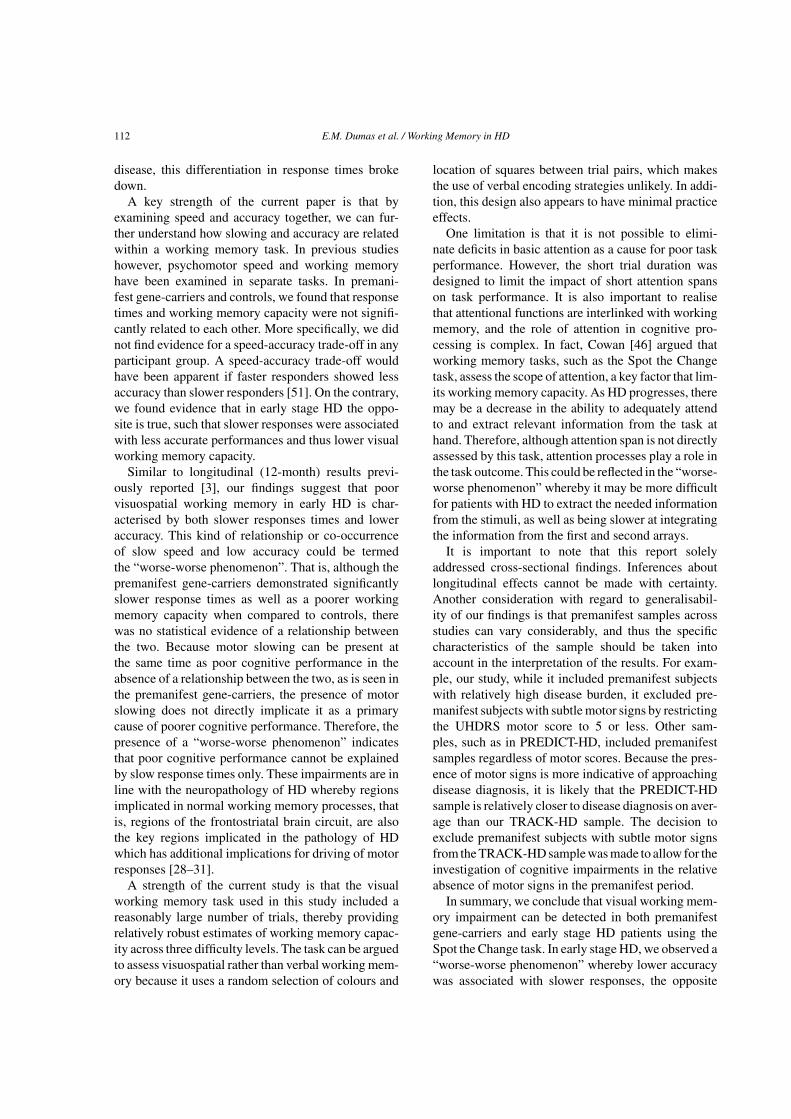
Table 1Participant characteristics and adjusted differencesa in working memory capacity and response time for set sizes 3, 5 and 7 for HD gene-carriers
compared to controls
Number of Female/male Age (years)b Education CAG repeat Expected years Disease durationparticipants levelc length to onsetb (years)b
n n mean (SD) mean (SD) mean (SD) mean (SD) mean (SD)
Controls 122 68/54 46.2 (10.1) 4.0 (1.3) – –PreHD-A 62 33/29 41.1 (8.6) 4.1 (1.1) 42.1 (1.8) 14 (3.1) –PreHD-B 58 33/25 40.6 (9.2) 3.8 (1.3) 44.2 (2.5) 9 (1.3) –HD1 77 46/31 47.2 (10.3) 3.8 (1.3) 43.8 (3.3) – 5 (5.8)HD2 44 19/25 51.0 (8.6) 3.3 (1.4) 43.5 (2.4) – 8 (4.5)
Working memory capacity (k) Response time (% increase)d
Correct Incorrect Interactione
Est. 95% CI p Est. 95% CI P Est. 95% CI p p
Visit 1 –Set size 3PreHD-A –0.04 (–0.26 to 0.18) 0.70 2.6% (–2.6 to 8.2) 0.33 0.8% (–8.7 to 11.3) 0.88 0.66PreHD-B –0.26 (–0.44 to –0.07) 0.01 7.6% (1.6 to 13.9) 0.01 8.1% (–3.8 to 21.4) 0.19 0.93f
HD1 –0.78 (–1.01 to –0.54) <0.001 18.9% (10.9 to 27.4) <0.001 7.3% (–2.8 to 18.3) 0.16 0.004HD2 –0.95 (–1.25 to –0.65) <0.001 39.4% (27.0 to 53.1) <0.001 30.9% (17.7 to 45.6) <0.001 0.09
Visit 1 – Setsize 5PreHD-A –0.26 (–0.61 to 0.09) 0.14 4.6% (–1.2 to 10.8) 0.12 4.9% (–4.3 to 15.0) 0.31 0.94f
PreHD-B –0.71 (–1.04 to –0.37) <0.001 8.3% (1.6 to 15.5) 0.02 2.5% (–7.2 to 13.2) 0.63 0.13HD1 –1.38 (–1.71 to –1.04) <0.001 14.9% (7.2 to 23.3) <0.001 1.6% (–7.4 to 11.4) 0.74 <0.001HD2 –1.45 (–1.84 to –1.06) <0.001 40.1% (27.7 to 53.6) <0.001 22.2% (8.5 to 37.5) 0.001 <0.001
Visit 2 – Setsize 5g
PreHD-A –0.03 (–0.31 to 0.26) 0.87 3.1% (–2.2 to 8.6) 0.26 10.6% (3.0 to 18.7) 0.005 0.005f
PreHD-B –0.78 (–1.19 to –0.36) <0.001 4.1% (–2.3 to 10.9) 0.21 1.2% (–8.6 to 12.1) 0.82 0.40HD1 –1.38 (–1.72 to –1.03) <0.001 21.0% (13.4 to 29.2) <0.001 10.7% (2.9 to 19.2) 0.007 <0.001HD2 –1.92 (–2.40 to –1.43) <0.001 37.1% (23.6 to 52.0) <0.001 19.4% (6.3 to 34.1) 0.003 <0.001
Visit 2 – Setsize 7PreHD-A –0.05 (–0.50 to 0.41) 0.85 5.5% (–0.1 to 11.3) 0.05 3.6% (–3.5 to 11.3) 0.33 0.40PreHD-B –1.10 (–1.62 to –0.59) <0.001 7.3% (0.2 to 14.9) 0.04 –0.9% (–9.4 to 8.4) 0.84 0.002HD1 –1.38 (–1.83 to –0.93) <0.001 24.2% (15.8 to 33.1) <0.001 10.5% (2.3 to 19.3) 0.01 <0.001HD2 –2.01 (–2.56 to –1.45) <0.001 35.1% (23.4 to 48.0) <0.001 17.4% (5.2 to 31.0) 0.004 <0.001
aAll results are adjusted for age, sex, educational level and study site; bAge, expected years to onset and disease duration as at baseline;cEducation level as a proxy for Intelligence Quotient, as based on the ISCED education classification system; dFor ease of interpretation, the logRT values were back-transformed to the original millisecond scale and these results were reported; eThe interaction represents the differencein estimated RTs for correct and incorrect responses for each HD subgroup compared with the same difference in Controls; f The difference inestimated RT for correct and incorrect responses is larger in the HD subgroup than the difference in controls. In all other cases the difference inRT for correct and incorrect responses is smaller in the HD subgroup than the difference in controls. gLongitudinal outcomes for set size 5 isreported elsewhere; see Tabrizi et al. [3].
results indicating lower working memory capacity atall levels of task difficulty.
Response time results indicated that premanifest andearly stage HD participants were slower than controlswhen responding correctly to the task trials. This isparticularly relevant as the premanifest gene-carriergroup were restricted to those who were free of clini-cally evident motor signs. Given the lack of significantmotor signs in this group, we believe that the slowerresponse times observed may indicate slowed cog-nition or information processing rather than slowedmotor processing. Our finding is consistent with
previous findings of psychomotor slowing in preman-ifest groups [8, 22]. We also note that the workingmemory task included a long response time frame (8seconds), to allow participants, even those with earlyHD who have proven motor deficits, to respond to thetrials within the time frame. The task design there-fore eliminated any potential differences in responsetimes being attributed to missing data in the earlyHD group. Not surprisingly, the differences betweenresponse times for correct and incorrect trials (at eachset size) were greatest in the control group and thePreHD-A group, whereas with greater levels of the
112 E.M. Dumas et al. / Working Memory in HD
disease, this differentiation in response times brokedown.
A key strength of the current paper is that byexamining speed and accuracy together, we can fur-ther understand how slowing and accuracy are relatedwithin a working memory task. In previous studieshowever, psychomotor speed and working memoryhave been examined in separate tasks. In premani-fest gene-carriers and controls, we found that responsetimes and working memory capacity were not signifi-cantly related to each other. More specifically, we didnot find evidence for a speed-accuracy trade-off in anyparticipant group. A speed-accuracy trade-off wouldhave been apparent if faster responders showed lessaccuracy than slower responders [51]. On the contrary,we found evidence that in early stage HD the oppo-site is true, such that slower responses were associatedwith less accurate performances and thus lower visualworking memory capacity.
Similar to longitudinal (12-month) results previ-ously reported [3], our findings suggest that poorvisuospatial working memory in early HD is char-acterised by both slower responses times and loweraccuracy. This kind of relationship or co-occurrenceof slow speed and low accuracy could be termedthe “worse-worse phenomenon”. That is, although thepremanifest gene-carriers demonstrated significantlyslower response times as well as a poorer workingmemory capacity when compared to controls, therewas no statistical evidence of a relationship betweenthe two. Because motor slowing can be present atthe same time as poor cognitive performance in theabsence of a relationship between the two, as is seen inthe premanifest gene-carriers, the presence of motorslowing does not directly implicate it as a primarycause of poorer cognitive performance. Therefore, thepresence of a “worse-worse phenomenon” indicatesthat poor cognitive performance cannot be explainedby slow response times only. These impairments are inline with the neuropathology of HD whereby regionsimplicated in normal working memory processes, thatis, regions of the frontostriatal brain circuit, are alsothe key regions implicated in the pathology of HDwhich has additional implications for driving of motorresponses [28–31].
A strength of the current study is that the visualworking memory task used in this study included areasonably large number of trials, thereby providingrelatively robust estimates of working memory capac-ity across three difficulty levels. The task can be arguedto assess visuospatial rather than verbal working mem-ory because it uses a random selection of colours and
location of squares between trial pairs, which makesthe use of verbal encoding strategies unlikely. In addi-tion, this design also appears to have minimal practiceeffects.
One limitation is that it is not possible to elimi-nate deficits in basic attention as a cause for poor taskperformance. However, the short trial duration wasdesigned to limit the impact of short attention spanson task performance. It is also important to realisethat attentional functions are interlinked with workingmemory, and the role of attention in cognitive pro-cessing is complex. In fact, Cowan [46] argued thatworking memory tasks, such as the Spot the Changetask, assess the scope of attention, a key factor that lim-its working memory capacity. As HD progresses, theremay be a decrease in the ability to adequately attendto and extract relevant information from the task athand. Therefore, although attention span is not directlyassessed by this task, attention processes play a role inthe task outcome. This could be reflected in the “worse-worse phenomenon” whereby it may be more difficultfor patients with HD to extract the needed informationfrom the stimuli, as well as being slower at integratingthe information from the first and second arrays.
It is important to note that this report solelyaddressed cross-sectional findings. Inferences aboutlongitudinal effects cannot be made with certainty.Another consideration with regard to generalisabil-ity of our findings is that premanifest samples acrossstudies can vary considerably, and thus the specificcharacteristics of the sample should be taken intoaccount in the interpretation of the results. For exam-ple, our study, while it included premanifest subjectswith relatively high disease burden, it excluded pre-manifest subjects with subtle motor signs by restrictingthe UHDRS motor score to 5 or less. Other sam-ples, such as in PREDICT-HD, included premanifestsamples regardless of motor scores. Because the pres-ence of motor signs is more indicative of approachingdisease diagnosis, it is likely that the PREDICT-HDsample is relatively closer to disease diagnosis on aver-age than our TRACK-HD sample. The decision toexclude premanifest subjects with subtle motor signsfrom the TRACK-HD sample was made to allow for theinvestigation of cognitive impairments in the relativeabsence of motor signs in the premanifest period.
In summary, we conclude that visual working mem-ory impairment can be detected in both premanifestgene-carriers and early stage HD patients using theSpot the Change task. In early stage HD, we observed a“worse-worse phenomenon” whereby lower accuracywas associated with slower responses, the opposite

E.M. Dumas et al. / Working Memory in HD 113
of a speed-accuracy trade-off. Our findings, togetherwith other reports in the literature, suggest that work-ing memory tasks are useful markers of cognitivedeterioration in HD. Such deterioration may be mostsensitively detected in early HD, especially stage2, using moderate to higher working memory loadsalong with measures of working memory capacity andresponse times for correct trials. This sort of cog-nitive task may be applicable in short term clinicaltrials (of 12 or more month duration) of disease mod-ifying or symptomatic treatments for participants instage 2 HD. Future examination of longitudinal effectsin the most difficult condition, set size 7, once suchdata become available from the TRACK-HD study,may reveal added task sensitivity for premanifest gene-carriers or stage 1 HD.
ACKNOWLEDGMENTS
TRACK-HD is supported by the CHDI/High QFoundation, Inc., a not-for-profit organisation dedi-cated to finding treatments for Huntington’s disease.Some of this work was undertaken at UCLH/UCL andthe University of Manchester, which received supportfrom the Department of Health’s NIHR BiomedicalResearch Centres. The authors offer their gratitude tothe volunteers who participated and to carers and com-panions who helped make their participation possible.
REFERENCES
[1] Tabrizi SJ, Reilmann R, Roos RAC, Durr A, Leavitt B, OwenG, Jones R, Johnson H, Craufurd D, Hicks SL, Kennard C,Landwehrmeyer B, Stout JC, Borowsky B, Scahill RI, Frost C,Langbehn DR, and the TRACK-HD investigators. Potentialendpoints for clinical trials in premanifest and early Hunting-ton’s disease in the TRACK-HD study: analysis of 24 monthobservational data. Lancet Neurol. 2012;11:42-53.
[2] Stout JC, Jones R, Labuschagne I, O’Regan A, Say MJ, DumasEM, Queller S, Justo D, dar Santos RC, Coleman A, Hart E,Durr A, Leavitt BR, Roos RAC, Langbehn DR, Tabrizi SJ,Frost C. Evaluation of longitudinal 12- and 24-month cogni-tive outcomes in premanifest and early Huntington’s disease.J Neurol Neurosurg Psychiatry. 2012; DOI:10.1136/jnnp-2011-301940.
[3] Tabrizi SJ, Scahill RI, Durr A, Roos RA, Leavitt B, JonesR, Landwehrmeyer B, Fox NC, Johnson H, Hicks SL, Ken-nard C, Craufurd D, Frost C, Langbehn D, Reilmann R, StoutJC, and the TRACK-HD investigators. Biological and clinicalchanges in premanifest and early stage Huntington’s diseasein the TRACK-HD study: the 12-month longitudinal analysis.The Lancet Neurology. 2011;10:31-42.
[4] Walker FO. Huntington’s disease. Lancet. 2007;369:218-23.[5] Moses JAJ, Golden CJ, Berger PA, Wisniewski AM. Neu-
ropsychological deficits in early, middle and late stageHuntington’s disease as measured by the Luria-Nebraska
Neuropsychological Battery. The International Journal ofNeuroscience. 1981;14:95-100.
[6] Kirkwood S, Su JL, Conneally M, Foroud T. Progression ofsymptoms in early and middle stages of Huntington disease.Arch Neurol. 2001;58:273-8.
[7] Stout JC, Paulsen J, Queller S, Solomon AC, Whitlock KB,Campbell C, Carlozzi N, Duff K, Beglinger L, Langbehn DR,Johnson S, Biglan K, Aylward E. Neurocognitive signs in pro-dromal Huntington disease. Neuropsychology. 2011;25:1-14.
[8] Solomon AC, Stout JC, Weaver M, Queller S, Tomusk A,Burr Whitlock K, Hui SL, Marshall J, Gray Jackson J,Siemers ER, Beristain X, Wojcieszek J, Foroud T. Ten-yearrate of longitudinal change in neurocognitive and motorfunction in prediagnosis Huntington disease. Mov Disord.2008;23(13):1830-6.
[9] Tabrizi SJ, Langbehn DR, Leavitt BR, Roos RAC, Durr A,Craufurd D, Kennard C, Hicks SL, Fox NC, Scahill RI,Borowsky B, Tobin AJ, Rosas HD, Johnson H, Reilmann R,Landwehrmeyer B, Stout JC, TRACK-HD-Investigators. Bio-logical and clinical manifestations of Huntington’s disease inthe longitudinal TRACK-HD study: Cross-sectional analysisof baseline data. Lancet Neurol. 2009;8(9):791-801.
[10] Lemiere J, Decruyenaere M, Evers-Kiebooms G, Vanden-bussche E, Dom R. Cognitive changes in patients withHuntington’s disease (HD) and asymptomatic carriers of theHD mutation - A longitudinal follow-up study. J Neurol.2004;251(8):935-42.
[11] Finke K, Bublak P, Dose M, Muller HJ, Schneider WX.Parameter-based assessment of spatial and non-spatial atten-tional deficits in Huntington’s disease. Brain. 2006;129:1137-51.
[12] Baddeley A. Working memory. Science. 1992;255:556-9.[13] Lawrence AD, Watkins LHA, Sahakian BJ, Hodges JR,
Robbins TW. Visual object and visuospatial cognition inHuntington’s disease: Implications for information process-ing in corticostriatal circuits. Brain. 2000;123(7):1349-64.
[14] Lemiere J, Decruyenaere M, Evers-Kiebooms G, Van-denbussche E, Dom R. Longitudinal study evaluatingneuropsychological changes in so-called asymptomatic car-riers of the Huntington’s disease mutation after 1 year. ActaNeurol Scand. 2002;106(3):131-41.
[15] Davis JD, Filoteo JV, Kesner RP. Is short-term memory fordiscrete arm movements impaired in Huntington’s disease?Cortex. 2007;43(2):255-63.
[16] Bachoud-Levi AC, Maison P, Bartolomeo P, Boisse MF, DallaBarba G, Ergis AM, Baudic S, Degos JD, Cesaro P, PeschanskiM. Retest effects and cognitive decline in longitudinal follow-up of patients with early HD. Neurology. 2001;56(8):1052-8.
[17] Verny C, Allain P, Prudean A, Malinge M, Gohier B, SchererC, Bonneau D, Dubas F, Le Gall D. Cognitive changes inasymptomatic carriers of the Huntington disease mutationgene. Eur J Neurol. 2007;14(12):1344-50.
[18] Stout JC, Weaver M, Solomon AC, Queller S, Hui S, JohnsonS, Gray J, Beristain X, Wojcieszek J, Foroud T. Are cogni-tive changes progressive in prediagnostic HD? Cogn BehavNeurol. 2007;20(4):212-8.
[19] Lawrence AD, Hodges JR, Rosser AE, Kershaw A, ffrench-Constant C, Rubinsztein DC, Robbins TW, SahakianBJ. Evidence for specific cognitive deficits in preclini-cal Huntington’s disease. Brain: A Journal of Neurology.1998;121(7):1329-41.
[20] Kirkwood SC, Siemers E, Stout JC, Hodes ME, ConneallyPM, Christian JC, Foroud T. Longitudinal cognitive and motorchanges among presymptomatic Huntington disease gene car-riers. Arch Neurol. 1999;56(5):563-8.
114 E.M. Dumas et al. / Working Memory in HD
[21] Witjes-Ane M-N, Vegter-van der Vlis M, van Vugt JP, LanserJB, Hermans J, Zwinderman A, Van Ommen G-JB, Roos RA.Cognitive and motor functioning in gene carriers for Hunt-ington’s disease: a baseline study. J Neuropsychiatry ClinNeurosci. 2003;15:7-16.
[22] Paulsen JS, Langbehn DR, Stout JC, Aylward E, Ross CA,Nance M, Guttman M, Johnson S, MacDonald M, BeglingerLJ, Duff K, Kayson E, Biglan K, Shoulson I, Oakes D, HaydenM, The Predict HDI, Coordinators of the Huntington StudyG. Detection of Huntington’s disease decades before diagno-sis: The Predict-HD study. J Neurol Neurosurg Psychiatry.2008;79(8):874-80.
[23] Robins Wahlin T-B, Larsson MU, Luszcz MA, Byrne GJ.WAIS-R features of preclinical Huntington’s disease: impli-cations for early detection. Dement Geriatr Cogn Disord.2010;29:342-50.
[24] Robins Wahlin T-B, Lundin A, Dear K. Early cognitivedeficits in Swedish gene carriers of Huntington’s disease.Neuropsychology. 2007;21(1):31-44.
[25] de Boo GM, Tibben AA, Hermans JA, Jennekens-SchinkelA, Maat-Kievit A, Roos RA. Memory and learning are notimpaired in presymptomatic individuals with an increasedrisk of Huntington’s disease. J Clin Exp Neuropsychol.1999;21:831-6.
[26] Constantinidis C, Wang XJ. A neural circuit basis for spatialworking memory. Neuroscientist. 2004;10:553-65.
[27] Heyder K, Suchan B, Daum I. Cortico-subcortical contribu-tions to executive control. Acta Psychol (Amst). 2004;115:271-89.
[28] O’Reilly RC, Frank MJ. Making working memory work: Acomputational model of learning in the prefrontal cortex andbasal ganglia. Neural Comput. 2006;18:283-328.
[29] Aylward EH, Codori AM, Barta PE, Pearlson GD, Har-ris GJ, Brandt J. Basal ganglia volume and proximity toonset in presymptomatic Huntington disease. Arch Neurol.1996;53:1293-6.
[30] Montoya A, Price BH, Menear M, Lepage M. Brain imagingand cognitive dysfunctions in Huntington’s disease. J Psychi-atry Neurosci. 2006;31(1):21-9.
[31] Bohanna I, Georgiou-Karistianis N, Hannan AJ, Egan GF.Magnetic resonance imaging as an approach towards identi-fying neuropathological biomarkers for Huntington’s disease.Brain Research Reviews. 2008;58(1):209-25.
[32] Jurgens CK, van de Wiel L, van Es ACGM, Grimbergen YM,Witjes-Ane M-N, van der Grond J, Middelkoop HAM, RoosRA. Basal ganglia volume and clinical correlates in ‘preclin-ical’ Huntington’s disease. J Neurol. 2008;255:1785-91.
[33] Rosas HD, Hevelone ND, Zaleta AK, Greve DN, Salat DH,Fischl B. Regional cortical thinning in preclinical Hunt-ington disease and its relationship to cognition. Neurology.2005;65(5):745-7.
[34] Wolf RC, Sambataro F, Vasic N, Schonfeldt-Lecuona C, EckerD, Landwehrmeyer B. Altered frontostriatal coupling in pre-manifest Huntington’s disease: Effects of increasing cognitiveload. Eur J Neurol. 2008;15(11):1180-90.
[35] Wolf RC, Sambataro F, Vasic N, Shonfeldt-Lecuona C,Ecker D, Landwehrmeyer B. Aberrant connectivity of lateral
prefrontal networks in presymptomatic Huntington’s disease.Exp Neurol. 2008;213(1):137-44.
[36] van der Hiele K, Jurgens CK, Vein AA, Reijntjes RHAM,Witjes-Ane M-NW, Roos RAC, van Dijk G, MiddelkoopHAM. Memory activation reveals abnormal EEG in preclini-cal Huntington’s disease. Mov Disord. 2007;22(5):690-5.
[37] Aylward EH, Li Q, Stine OC, Ranen N, Sherr M, Barta PE,Bylsma FW, Pearlson GD, Ross CA. Longitudinal change inbasal ganglia volume in patients with Huntington’s disease.Neurology. 1997;48:392-9.
[38] Paulsen JS, Magnotta VA, Mikos AE, Paulson HL, PenzinerE, Andreasen NC, Nopoulos PC. Brain structure in preclinicalHuntington’s disease. Biol Psychiatry. 2006;59(1):57-63.
[39] Van den Bogaard SJ, Dumas EM, Acharya TP, Johnson H,Langbehn DR, Scahill RI, Tabrizi SJ, van Buchem MA, Vander Grond J, Roos RA, Group T-HI. Early atrophy of pallidumand accumbens nucleus in Huntington’s disease. J Neurol.2011;258:412-20.
[40] Middleton FA, Strick PL. Basal ganglia and cerebellarloops: motor and cognitive circuits. Brain Research Reviews.2000;31(2-3):236-50.
[41] Witjes-Ane M-N, Mertens B, van Vugt JP, Bachoud-Levi A-C, van Ommen G-JB, Roos RA. Longitudinal evaluation of“presymptomatic” carriers of Huntington’s disease. J Neu-ropsychiatry Clin Neurosci. 2007;19(3):310-7.
[42] Huntington Study Group. Unified Huntington’s Disease Rat-ing Scale-99. Huntington Study Group. 1999.
[43] Penney JBJ, Vonsattel JP, MacDonald ME, Gusella JF, MyersRH. CAG repeat number governs the development rate ofpathology in Huntington’s disease. Annuals of Neurology.1997;41:689-92.
[44] Langbehn DR, Brinkman RR, Falush D, Paulsen JS, HaydenMR, Collaborative IHD. A new model for prediction of theage of onset and penetrance for Huntington’s disease basedon CAG length. Clin Genet. 2004;65(4):267-77.
[45] Shoulson I, Fahn S. Huntington disease: clinical care andevaluation. Neurology. 1979;29:1-3.
[46] Cowan N. The magical number 4 in short-term memory: Areconsideration of mental storage capacity. Behav Brain Sci.2001;24(1):87-185.
[47] Cowan N, Elliott EM, Saults JS, Morey CC, Mattox S, His-mjatullina A, Conway ARA. On the capacity of attention:Its estimation and its role in working memory and cognitiveaptitudes. Cogn Psychol. 2005;51(1):42-100.
[48] Liang KY, Zeger SL. Inference based on estimating functionsin the presence of nuisance parameters. Statistical Science.1995;10:158-73.
[49] Liang KY, Zeger SL. Regression analysis for correlated data.Annu Rev Public Health. 1993;14:43-68.
[50] Lange KW, Sahakian BJ, Quinn NP, Marsden CD, RobbinsTW. Comparison of executive and visuospatial memory func-tion in Huntington’s Disease and dementia of Alzheimer-typematched for degree of dementia. Neurology, Neurosurgeryand Pyschiatry. 1995;58(5):598-606.
[51] Berg C, Hertzog C, Hunt E. Age differences in the speed ofmental rotation. Dev Psychol. 1982;18:95-107.