Huntington's disease: the coming of age - Indian Academy of ...
16
Journal of Genetics, Vol. 97, No. 3, July 2018, pp. 649–664 © Indian Academy of Sciences https://doi.org/10.1007/s12041-018-0957-1 REVIEW ARTICLE Huntington’s disease: the coming of age MRITUNJAY PANDEY 1 and USHA RAJAMMA 2 ∗ 1 Metabolic Diseases Branch, National Institute of Diabetes and Digestive and Kidney Diseases, National Institutes of Health, Bethesda, MD 20892, USA 2 Inter University Center for Biomedical Research and Super Specialty Hospital, Kottayam 686 009, India *For correspondence. E-mail: [email protected]. Received 25 November 2017; revised 31 January 2018; accepted 7 February 2018 Abstract. Huntington’s disease (HD) is caused due to an abnormal expansion of polyglutamine repeats in the first exon of huntingtin gene. The mutation in huntingtin causes abnormalities in the functioning of protein, leading to deleterious effects ultimately to the demise of specific neuronal cells. The disease is inherited in an autosomal dominant manner and leads to a plethora of neuropsychiatric behaviour and neuronal cell death mainly in striatal and cortical regions of the brain, eventually leading to death of the individual. The discovery of the mutant gene led to a surge in molecular diagnostics of the disease and in making different transgenic models in different organisms to understand the function of wild-type and mutant proteins. Despite difficult challenges, there has been a significant increase in understanding the functioning of the protein in normal and other gain-of-function interactions in mutant form. However, there have been no significant improvements in treatments of the patients suffering from this ailment and most of the treatment is still symptomatic. HD warrants more attention towards better understanding and treatment as more advancement in molecular diagnostics and therapeutic interventions are available. Several different transgenic models are available in different organisms, ranging from fruit flies to primate monkeys, for studies on understanding the pathogenicity of the mutant gene. It is the right time to assess the advancement in the field and try new strategies for neuroprotection using key pathways as target. The present review highlights the key ingredients of pathology in the HD and discusses important studies for drug trials and future goals for therapeutic interventions. Keywords. Huntington’s disease; neurodegeneration; autosomal dominant disorder; huntingtin; pathophysiology; neurochemistry; therapeutic intervention. Introduction HD is a progressive neurodegenerative disorder which usually manifests in adulthood and is inherited in an autosomal-dominant manner. The disease has peculiar phenotypes with distinct motor defects, psychiatric symp- toms and cognitive decline. The motor symptoms include chorea (bizarre dance like movements), dyskinesia (diffi- culty or distortion in performing voluntary movements) and dystonia (involuntary contraction of muscles). Psychi- atric symptoms, that usually precede the motor symptoms, include depression, anxiety and sleep disorders (Spires and Hannan 2007). Cognitive decline is manifested as difficul- ties in concentration and retaining newly acquired infor- mation, decline in language skills, disorganized speech and perceptual impairments. As the disease progresses, motor rigidity and dementia predominate. Several areas in brain show signs of neuropathology in HD, with the maximum degeneration occurring in the caudate nucleus and puta- men (Vonsattel et al. 1985). The mutation in huntingtin (htt) gene was identified in 1993 as an unstable expansion of CAG (the trinucleotide coding for glutamine) repeats, which occurs within the first exon of the gene ‘IT 15’ (for ‘Interesting Transcript 15’) in HD patients (The Huntington’s Disease Collaborative Research Group 1993). This gene on chromosome 4 (4p63) encodes the protein huntingtin. The discovery of mutant htt was one of the single most major events in the history of HD and genetics. It led to a hurricane of events starting from an efficient molecular diagnosis of HD to the devel- opment of mutant htt knock-in transgenic models of the disease. The mutation in huntingtin gene produces an expanded stretch of the amino acid glutamine (denoted by the 649
-
Upload
khangminh22 -
Category
Documents
-
view
0 -
download
0
Transcript of Huntington's disease: the coming of age - Indian Academy of ...

Journal of Genetics, Vol. 97, No. 3, July 2018, pp. 649–664 © Indian Academy of Scienceshttps://doi.org/10.1007/s12041-018-0957-1
REVIEW ARTICLE
Huntington’s disease: the coming of age
MRITUNJAY PANDEY1 and USHA RAJAMMA2∗
1Metabolic Diseases Branch, National Institute of Diabetes and Digestive and Kidney Diseases, National Institutes ofHealth, Bethesda, MD 20892, USA2Inter University Center for Biomedical Research and Super Specialty Hospital, Kottayam 686 009, India*For correspondence. E-mail: [email protected].
Received 25 November 2017; revised 31 January 2018; accepted 7 February 2018
Abstract. Huntington’s disease (HD) is causeddue to an abnormal expansionof polyglutamine repeats in the first exonof huntingtingene. The mutation in huntingtin causes abnormalities in the functioning of protein, leading to deleterious effects ultimately to thedemise of specific neuronal cells. The disease is inherited in an autosomal dominantmanner and leads to a plethora of neuropsychiatricbehaviour and neuronal cell death mainly in striatal and cortical regions of the brain, eventually leading to death of the individual.The discovery of the mutant gene led to a surge in molecular diagnostics of the disease and in making different transgenic modelsin different organisms to understand the function of wild-type and mutant proteins. Despite difficult challenges, there has been asignificant increase in understanding the functioning of the protein in normal and other gain-of-function interactions in mutantform. However, there have been no significant improvements in treatments of the patients suffering from this ailment and most ofthe treatment is still symptomatic. HD warrants more attention towards better understanding and treatment as more advancementin molecular diagnostics and therapeutic interventions are available. Several different transgenic models are available in differentorganisms, ranging from fruit flies to primate monkeys, for studies on understanding the pathogenicity of the mutant gene. It is theright time to assess the advancement in the field and try new strategies for neuroprotection using key pathways as target. The presentreview highlights the key ingredients of pathology in the HD and discusses important studies for drug trials and future goals fortherapeutic interventions.
Keywords. Huntington’s disease; neurodegeneration; autosomal dominant disorder; huntingtin; pathophysiology; neurochemistry;therapeutic intervention.
Introduction
HD is a progressive neurodegenerative disorder whichusually manifests in adulthood and is inherited in anautosomal-dominant manner. The disease has peculiarphenotypes with distinct motor defects, psychiatric symp-toms and cognitive decline. The motor symptoms includechorea (bizarre dance like movements), dyskinesia (diffi-culty or distortion in performing voluntary movements)and dystonia (involuntary contraction ofmuscles). Psychi-atric symptoms, that usually precede themotor symptoms,include depression, anxiety and sleep disorders (Spires andHannan 2007). Cognitive decline is manifested as difficul-ties in concentration and retaining newly acquired infor-mation, decline in language skills, disorganized speech andperceptual impairments. As the disease progresses, motorrigidity and dementia predominate. Several areas in brain
show signs of neuropathology in HD, with the maximumdegeneration occurring in the caudate nucleus and puta-men (Vonsattel et al. 1985).The mutation in huntingtin (htt) gene was identified in
1993 as an unstable expansion of CAG (the trinucleotidecoding for glutamine) repeats, which occurs within the firstexon of the gene ‘IT 15’ (for ‘Interesting Transcript 15’)in HD patients (The Huntington’s Disease CollaborativeResearchGroup 1993). This gene on chromosome 4 (4p63)encodes the protein huntingtin. The discovery of mutanthtt was one of the single most major events in the historyof HD and genetics. It led to a hurricane of events startingfrom an efficient molecular diagnosis of HD to the devel-opment of mutant htt knock-in transgenic models of thedisease.The mutation in huntingtin gene produces an expanded
stretch of the amino acid glutamine (denoted by the
649

650 Mritunjay Pandey and Rajamma Usha
letter Q) towards the amino terminal end of the protein.This causes themutant protein to interact abnormallywithother cell proteins, leading to changes in its function. Theglutamine tract in htt is polymorphic, with 8 to 36 glu-tamine repeats in the normal population and 41 or morein HD. People with 38 or more glutamine repeats in httcan manifest HD but occasionally individual with up to41 repeats in old age with no discernible symptoms havebeen reported (Rubinsztein et al. 1996). Several studieshave pointed out a strong inverse correlation between therepeat number and the age at onset of HD (Zühlke et al.1993; Nørremølle et al. 1993; Tabrizi et al. 2013).
Historical background of HD
A vivid account of the disease by George Huntington hassubsequently borne his name and is considered to be thebest report and landmark in the study of HD (Hunting-ton 1872). All the cardinal features of HD are listed inthe description: the adult onset, progressive course of thedisease, choreiform movements, mental impairment, sui-cidal tendency and the pattern of inheritance. Such a vividdescription of the disease was made possible because ofthe clinical observation ofHDpatients byHuntington, hisfather and grandfather. The clear study and documenta-tion of the two older generations of the Huntingtons gaveGeorge Huntington a unique advantage in describing thedisease fully.In 1883, Westphal described juvenile symptoms
resembling those seen in HD but he attributed it to a causeother than HD as the patients showed a predominanceof hypokinesia and rigidity. The term ‘Westphal variant’is often used to describe the clinical picture of juvenileHD. Although, there were several reports about the neu-ropathology of HD, it was not until the 1920s that therewas an equivocal agreement that in HD the changes inbrain are primarily degenerative and atrophic, and thatthe caudate nucleus is most affected (Harper 2002).In 1993, the HD gene, huntingtin (htt) was
discovered by the 10-year collective effort of six teamsin the United States and Britain, the Huntington DiseaseCollaborative Research Group. Soon after, several geneticmodels were developed including several transgenic mod-els in Drosophila (Krench and Littleton 2013), nematodeCaenorhabditis elegans (Bates et al. 2006), minipigs (Baxaet al. 2013), primates (Yang et al. 2008), sheep (Jacobsenet al. 2010) and knock-in mouse models (Mangiarini et al.1996; Reddy et al. 1998; Hodgson et al. 1999). Increasingevidence has converged to indicate that the normal proteinfunctions in intracellular vesicular trafficking, endocyto-sis, synaptic functioning, autophagy and transcription,and that the mutant htt has been linked to disruptionof transcription, oxidative stress, autophagy, aggregateformation and several other functions in the cellular home-ostasis. The mutant htt acquires a toxic gain of function
leading to abnormal functioning but the exact pathway ofcell death is still elusive.
Epidemiology of HD
Epidemiological studies have now assumed amajor role inthe studyof various neurological disorders likeParkinson’sdisease, multiple sclerosis and stroke where the infor-mation is used in determining the hypotheses about thesocietal causes, if any, of these diseases, aswell as for simplydocumenting their frequency and variation in the society.In case of HD, the situation is different, because it is amonogenic Mendelian disorder. The route to understandthe pathogenesis of the disease is principally molecular,not epidemiological. To study the frequency of the affectedpopulation, it involves careful study of complete families,rather than surveys restricted to primary cases, while thoseat risk are also mostly family members, rather than popu-lation at large.
Prevalence of HD in different countries
The incidence of HD is variable across the globe, withJapan, South Africa and Finland having very low ratesof the disease among the population as depicted in table 1.Previous estimates of 4–10 per 100,000 people in WesternHemisphere were low (Harper 2002), as recent prevalencedata from UK suggests higher prevalence of adult HD at12.3 per 100,000 based on diagnoses recorded in generalpractice records (Evans et al. 2013). It is assumed that thedisease spread across the globedue tomigrationof affectedpeople from north-west Europe. While detailing aboutthe prevalence of HD, one cannot leave aside the pres-ence of sizeable population of HD patients in Venezuela.The remarkable concentration of HD patients living bythe shores of Lake Maracaibo, Venezuela represents thesingle largest cluster derived from a single ancestor thathas remained localized. The pedigree contains over 10,000members with over 100 living subjects. The high frequencyof the disorder in this region was first documented byNegrette (Okun and Thommi 2004). This particular pop-ulation was recognized byHereditary Disease Foundationto carry out a systematic study. It was easier to study thegenealogy, pedigree analysis and homozygotes, as severalfamilies had both the parents affected, it was a landmarkproject in the history of HD which gave a new directionto HD research. The Venezuela project helped in mappingand isolation of HD gene, which led to the beginning ofmolecular research in HD (Wexler et al. 2004).
Pathophysiology of HD
The pathophysiology of HD contributed by mutantHTT is highly complex impacting various functionaldomains, as is evident from the behavioural expression,neuroanatomical topography and neurochemical profilesof affected individuals.

Huntington’s disease: the coming of age 651Table
1.Prevalenceestimates
ofHD
indifferentpa
rtsof
theworld.
Reference
Yearof
prevalence
stud
yRegion
Prevalence(per
100,000)
Reedan
dCha
ndler(1958)
1940
Michigangeneral/
blackpo
pulation
,USA
4.1/1.5
Folsteinet
al.(1987)
1980
Marylan
dgeneral/
blackpo
pulation
,USA
5.5/6.4
Wrigh
tet
al.(1981)
1980
SouthCarolinageneral/
blackpo
pulation
,USA
4.8/0.91
Kok
men
etal.(1994)
1990
Minnesota,O
lmsted
coun
ty,U
SA2
Alm
qvistet
al.(2001)
1993
–200
0British
Colum
bia,
Can
ada
0.69
Fisheran
dHayden(2014)
2011
British
Colum
bia,
Can
ada
13.6
Shok
eir(1975)
–Man
itob
aan
dSa
skatchew
an,C
anad
a8.5
Pleyd
ell(1955)
1954
Northam
pton
shire,UK
6.5
Harperet
al.(1979)
1971
SouthWales,U
K7.61
Simpson
andJohn
ston
(1989)
1984
Grampian
region
ofScotland
,UK
10.0
Paloet
al.(1987)
1986
Finland
0.5
Con
neally
(1984)
–Tasman
ia,A
ustralia
17.4
Pridm
ore(1990)
1990
Tasman
ia,A
ustralia
12.1
McC
uskeret
al.(2000)
1996
New
SouthWales,A
ustralia
6.3
Ada
chia
ndNak
ashima(1999)
1997
Western
Japa
n0.72
Shiwachan
dLindenb
aum
(1990)
1990
Indian
subcon
tinent
1.75
Chenan
dLai
(2010)
2007
Taiwan
0.42
Peterlin
etal.(2009)
2009
Slov
enia
5.16
Morrisonet
al.(1995)
1991
NorthernIrelan
d6.4
Behavioural symptoms
Behavioural symptoms are obvious from the impairmentin motor, cognitive and psychiatric attributes. Althoughmotor problems are the major phenotypes seen in HDpatients, the psychiatric symptoms and cognitive deficitsprecede it, and they also cause significant burden on apatient’s life and lead them to complete loss of indepen-dence.
Motor abnormalities
Clinically, the motor disturbances in HD is characterizedby involuntary movements, i.e. chorea or dystonia, all ofwhich impair initiation, or execution of movements (Kirk-wood et al. 2002). Motor abnormalities like bradykinesia,chorea, dystonia and oculomotor symptoms have beenshown to be progressive in HD patients (Andrich et al.2007). HDpatients also suffer frommotor problem, whichis more like an inability to maintain a voluntary musclecontraction at a constant level. As a result, they cannotmaintain the constant pressure during a handshake andis treated as a characteristic of HD, known as milkmaid’sgrip (Walker 2007). As the disease progresses, hyperkineticmovements lessen and bradykinesia and rigidity becomemore prominent (Novak and Tabrizi 2010).
Psychiatric and cognitive changes
Cognitive decline, with progression from early changesin speed of information processing, cognitive inflexibil-ity, and memory retrieval to more severe and widespreadabnormalities later in the disease course has been demon-strated as part of the HD phenotype by researchers (Vernyet al. 2007).Many investigators have noted that the patternof cognitive deficits seen in HD is similar to neurologi-cal or psychiatric conditions that disrupt functioning inthe frontal lobe or basal ganglia, or conditions such aslesions in theprefrontal cortex (BrownandMarsden1988).Severe psychiatric and cognitive impairments have alsobeen observed in juvenile HD patients (Ribaï et al. 2007).Oneof the salient features ofGeorgeHuntington’s descrip-tion of patients suffering from the disease was ‘a tendencyto insanity and suicide’. HD patients have four to six timeshigher suicidal tendency than the general population (DiMaio et al. 1993). This rate is even higher for patientswhose age is more than 50 years (Schoenfeld et al. 1984).
HDpatients have been shown to suffer from depression,apathy and irritability when analysed on a Unified Hunt-ington’s Disease Rating Scale (UHDRS) (Kingma et al.2008). Apathy is also common, which is characterized byloss of interest and passive behaviour. Working memorydeficits havebeen found inHDpatients,whenevent-relatedfunctional magnetic resonance imaging and a parametricverbal workingmemory task were used to investigate cere-bral function (Wolf et al. 2009). Memory acquisition andability to concentrate are poor in HD patients due to

652 Mritunjay Pandey and Rajamma Usha
subcortical degeneration (Zakzanis 1998). Sleepdisturbance is a cause of significant distress inHDpatients(Videnovic et al. 2009).
Anatomical features
The most striking neuropathological feature of the HDbrain is the shrunken appearance of the neostriatum withgross atrophy of the caudate nucleus and putamen, withthe caudate nucleus reduced to a rim of tissue. Reductionin size of the caudate is accompanied by the enlargementof ventricles. HD brains weigh less than brains of age-matched controls with the weight being reduced by 10–20%. Another notable feature is the loss of white matterin the subcortical zone. As a result, the brain is smallerthan normal in the late stage of the disease (Vonsattel andDiFiglia 1998).
The striatum is composed of heterogeneouscompartments that contain distinct neurochemicals andproject to different target regions. These compartmentsare found in the form of patches termed as striosomes andmatrix (Graybiel et al. 1990). Striosomes consist of discretezones and mainly contain opioid receptors, substance P,met-enkephalin and cholecystokinin. The matrix is richin somatostatin, neuropeptide Y, nicotinamide adeninedinucleotide phosphate (NADPH) diaphorase, acetyl-cholinesterase, calbindin and cytochrome oxidase. Severalconflicting reports associated with the neuropathology ofthese two compartments inHDexist in the literature. Somedescribe that the total area of matrix is changed but thetotal area of striosomes remains the same, as determinedby the staining of acetylcholinesterase or calbindin (Fer-rante et al. 1987; Seto-Ohshima et al. 1988). Other studieshave suggested the involvement of striosomes in the earlyphases of the disease (Reiner et al. 1988; Hedreen andFolstein 1995). The atrophied striatum is characterized bymarkedneuronal loss andastrogliosis,whichhas beenusedtoqualify the neuropathology (Vonsattel et al. 1985;Myerset al. 1988; Heinsen et al. 1994). Quantitative microscopicstudies have revealed that there is a relative sparing ofthe large striatal neurons but severe loss of medium-sizedstriatal neurons. The majority of these medium-sized stri-atal neurons are gamma-aminobutyric acid (GABA)-ergicin nature. As these neurons degenerate, there is a corre-sponding loss of the neurochemicals they contain includ-ing glutamic acid decarboxylase (GAD), Substance P,enkephalin, calcineurin, calbindin and adenosine, togetherwith failure of dopaminergic receptors in the striatum.According to their neurochemical differences and differ-ential connectivity, the medium spiny neurons are of twokinds.Medium spiny neurons which express D1 dopaminereceptors and Substance P project to internal segment ofglobuspallidus (globuspallidus interna,GPi) and substan-tia nigra pars compacta (SNpc), whereas those expressingD2 dopamine receptors and enkephalin project to exter-nal segment of globus pallidus (globus pallidus externa,GPe) (Gerfen 1992). One study of 17 early and middle
grade HD cases found that the enkephalin containingneurons connecting to GPe were much more affected thanthe Substance P containing neurons projecting to GPi(Reiner et al. 1988). In two other studies, striatal neu-rons projecting to GPe showed evident loss, whereas theneurons projecting to GPi appeared relatively spared atpresymptomatic or early stages of the disease (Albin et al.1990, 1992).
In addition to the medium spiny neurons, thestriatum has various interneurons which include the largecholinergic neurons and the sparse medium spiny neuronscontaining somatostatin, neuropeptide Y and NADPHdiaphorase neurons. The striatal interneurons are rela-tively spared in HD (Ferrante et al. 1987; Cicchetti et al.2000). In a major study on neuropathology of HD, 163post-mortem HD brains were processed and evaluatedfor assigning a five-point grading scale for the severityof striatal neuropathological involvement in HD with 0indicating no abnormality and 4 signifying very severeinvolvement (Vonsattel et al. 1985). The gradingwas basedon the histopathological features of relative neuronal lossand relative gliosis in different areas of the brain, whichincluded the cortical, subcortical, and brain stem. Otherfactors like clinical records of the 163 HD cases werereviewed which included the sex, age at death and a ratingof physical disability. Grade 0 was assigned to those casesin which there was substantial evidence for the diagno-sis of HD, yet no gross or microscopic abnormalities thatcould be related to HD neuropathology. In grade 1 brains,there were no macroscopically distinguishable alterationsat the level of globus pallidus (GP) and caudate nucleus–nucleus accumbens septi-putamen (CAP). However, therewasmoderate fibrillary astrocytosis when observedmicro-scopically. Further, the extent of neuronal loss was evidentonly after cell counting and the putamen showed slightastrocytosis throughout. Grade 2 brains had atrophiedcaudate nucleus (CN) at the head region at CAP level,which was visible macroscopically. Microscopically, neu-ronal losswasobserved inCAPwith concomitant fibrillaryastrocytosis in CN and putamen. At GP level, the CNdisplayed a marked neuronal loss and astrocytosis. Theglobus pallidus showed minimal or no changes. Grade 3brains had a shrunken CN at CAP level. At GP level, theCN was reduced to a thin strip. Both the putamen andglobus pallidus were moderately decreased in size. Moder-ate neuronal loss with fibrillary astrocytosis involved thegray matter bridges between CN and putamen. Neuronalloss and gliosis is evidentmacroscopically in putamen. Thelateral segment of theGP showed slight tomoderate fibril-lary astrocytosis adjacent to putamen. In grade 4 brains atthe CAP level, the CNwas extremely shrunken and yellowbrown. The putamen was markedly atrophic with a con-cave medial outline. At GP level, the CN was reduced to athin strip. The putamen was markedly reduced in size andshowed widened perivascular spaces in its ventral portion.Microscopically, neuronal depletion and astrocytosis was

Huntington’s disease: the coming of age 653
extremely severe and diffuse throughout the CN andputamen at both levels CAP and GP. The nucleus accum-bens showed slight to moderate fibrillary astrocytosisdorsally. This grading system devised by Vonsattel et al.(1985) has been quite useful in HD research, as it remainsthe single largest study of the neuropathology of post-mortem HD brains.Although, the loss of neurons from nucleus caudatus
putamen (NCP) is the most conspicuous feature of HDbrains, there are several reports of the involvement of otherbrain areas in HD neuropathology. Atrophy of the cere-bral cortex has long been recognized as occurring in HD.InHD, themost affected regions in the cortex are layers III,V and VI. Previous studies have confirmed the thinning ofthe cerebral cortex and underlying white matter (Hedreenet al. 1991; Heinsen et al. 1994; Macdonald et al. 1997;Ciarmiello et al. 2006). Recent investigation using in vivomagnetic resonance imaging reveals that the behaviouraldeficits found in the HD patients are closely associatedwithdiscrete cortical degeneration in the brain (Rosas et al.2008). Hippocampal CA3, CA4 and granule cell layer ofdentate too show neuronal loss (Spargo et al. 1993). Neu-ronal loss has also been reported in ventrolateral thalamus(Dom et al. 1976) and in the centromedianparafascicularcomplex in thalamus in HD brains (Heinsen et al. 1996).Cerebellar atrophy has been reported in some cases of HD(Jeste et al. 1984; Rodda 1981) with loss of Purkinje cellsin some cases (McCaughey 1961).
Microscopic examination of sections after staining themwith different microglia markers also revealed reactivemicroglia is striatum, neocortex and internal capsule (Sappet al. 2001). With the discovery of mutant htt and itsstudy in transgenic models of mice helped to identify neu-ronal nuclear inclusion bodies in HD brains (Davies et al.1997; Becher et al. 1998). These inclusion bodies stainpositively for IC2 antibody raised against TATA-bindingprotein with 38 or more polyQ stretch. The inclusionbodies involve about 7%of neocortical neurons but are vir-tually absent in GP or cerebellum (Gutekunst et al. 1999).
Neurochemical alterations in HD
Neurochemical alterations may not only reflect changes intissue pathology but also give insight into the cause of celldeath in specific brain nuclei in HD.
GABA
The first neurotransmitter shown to be decreased in HDbrain was GABA. Significant decrease in level of GABAand its synthetic enzymeGADwas shown in post-mortemHD brain (Perry et al. 1973; Bird and Iversen 1974).The striatal medium spiny neurons that are GABAergicin nature are most vulnerable neurons in HD and theirdemise is considered to be the neuropathological hallmarkof HD (Vonsattel et al. 1985; DiFiglia 1990). Apart fromstriatum decreased levels of GABA have been found inhippocampus and cerebral cortex (Reynolds and Pearson
1987). Significant decrease in levels ofGADwere observedthroughout the brain, but most markedly in the striatumand GPe (Spokes 1980). GABA receptors were decreasedin striatumbut unchanged in the cortex (Lloyd et al. 1977).
Dopamine
Dopamine levels were found to be increased (Spokes 1980)or unchanged in striatum, nucleus accumbens, and SNpc(Reynolds and Garrett 1986). Treatment of primary cul-tures of striatal neurons with DA caused striatal neurode-generation with reduction in mitochondrial complex-IIactivity (Benchoua et al. 2008). No significant alterationswere found in the dopamine synthesizing enzyme tyro-sine hydroxylase in post-mortem HD striatum (Bird andIversen 1977). Deficit in vesicular monoamine transportertype 2 (VMAT2) expression was found in posterior puta-men in post-mortem HD brains (Bohnen et al. 2000). Invivo D2 receptor binding studies using positron emissiontomography (PET) with the help of the specific receptorligand, raclopiride in HD patients revealed a loss of thepostsynaptic receptor in striatum and in temporal andfrontal cortex (Pavese et al. 2003). Similar observationshave been made for both D1 and D2 receptors in HDpatient’s striata using PET analysis (Turjanski et al. 1995).
Acetylcholine
Muscarinic cholinergic receptor expression and cholineacetyltransferase enzyme activity were found to be signifi-cantly decreased in caudate nucleus of HD brain but notin the cortex (Enna et al. 1976). Vesicular acetylcholinetransporter (VAChT)has been found tobe reduced inpost-mortem striatal HD brain (Smith et al. 2006). However,large cholinergic interneurons in striatum are spared inHD.
Serotonin
Serotonin and its metabolite 5-hydroxyindoleacetic acid(5-HIAA) were found to be increased in pallidum, cortexand basal ganglia in post-mortem HD brain (Reynoldsand Pearson 1987; Kish et al. 1987).
Brain-derived neurotrophic factor
Among different trophic factors supporting neuronal cellgrowth and maintenance in the striatum, BDNF hasbeen found to be consistently decreased in HD patientsand different transgenic HD models (Ferrer et al. 2000;Baquet et al. 2004). Reduced BDNF induced TrkB recep-tor signalling has been observed in striatal cells of R6/2transgenic mice model of HD (Nguyen et al. 2016).
Cysteine
Cystathione γ-lyase, an enzyme important for synthesisof the amino acid cysteine has been found to be depletedin HD patients brain, striatal cells, and transgenic mousemodels, and cysteine supplementation in diet led to signif-icant neuroprotection in HD models (Paul et al. 2014).
654 Mritunjay Pandey and Rajamma Usha
The biology of huntingtin: cellular basis of pathology
The discovery of the HD gene in 1993 made it possibleto generate in vitro and in vivo genetic models to studythe physiopathology of the mutant gene. Studies weredesigned to investigate the normal function of htt and itsvarious interactions in normal and mutant forms to iden-tify the pathways, which lead to cell death (figure 1). TheHD gene contains 67 exons and extends across 170 kbof DNA (Baxendale et al. 1995). The CAG repeat that isexpanded on HD chromosomes lies within exon 1 and istranslated into a stretch of polyQ residues. The proteinHTT expressed from the HD gene has over 3000 aminoacids, giving a molecular mass of around 350 kDa and iswell conserved from flies to mammals (Saudou and Hum-bert 2016). However, the amino terminal region has beenextensively studied, as it contains an expandable polyQstretchwhich is consideredpathogenic. Severalmotifs havebeen identified including the polyglutamine and polypro-line tracts close to the N-terminus and the several HEAT(Htt, Elongation factor 3, the PR65/A subunit of proteinphosphatase 2A, and the lipid kinase TOR) repeats foundjust downstream of the proline-rich regions C-terminal tothe glutamine tract (Andrade and Bork 1995). A HEATrepeat is a degenerate of a generally 50-amino acid motifconsisting of two anti-parallel α-helices forming a helicalhairpin. HEAT repeat proteins generally mediate impor-tant protein–protein interactions involved in cytoplasmicand nuclear transport, microtubule dynamics, and chro-mosome segregation (Neuwald and Hirano 2000). Usingcross-species comparative analysis, Takano and Gusella(2002) predicted that vertebrate htt contains 28–36 HEATrepeats that span the entire protein. Human htt has ratand mouse homologues (Barnes et al. 1994; Schmitt et al.1995). It also has recognizable orthologues with the Pufferfish, Fugu rupribes (Baxendale et al. 1995) and the Zebrafish, Danio rerio (Karlovich et al. 1998).
HTT gene has two mRNA transcripts of 10336 bp and13711 bp (Lin et al. 1993) and the second transcript whichhas an additional 3’ UTR sequence of 3360 bp seems tobe enriched in the brain. HTT transcripts and protein arefound to be expressed at different levels throughout thehumans and murine tissue (Marques Sousa and Humbert2013). Although htt expression is not restricted to brainregions which preferentially degenerate in HD, as htt isfound to be expressed in several kinds of striatal projectionneurons and interneurons as well as cortex, hippocampusand cerebellum. The expression of htt starts in embryonicstage and its knock-out is lethal on embryonic day 7.5(Zeitlin et al. 1995) and persists in adulthood but the pref-erential toxicity in striatal cell death and the manifestationof the disease in adulthood adds a layer of complexity tothe disease.
Mutant huntingtin and pathogenesis
Expansion of polyQ tract in htt to greater than 36Qcauses the disease and it has been observed that as the
number of polyQ repeat increases the earlier is the ageof onset of symptoms of HD (Narain et al. 1999). It hasbeen observed that ectopic expression of proteins contain-ing polyQ expansion are toxic in cells, however, a loss ofhuntingtin function can also contribute towards the pro-gression of the disease.
Mutant htt and inclusion bodies
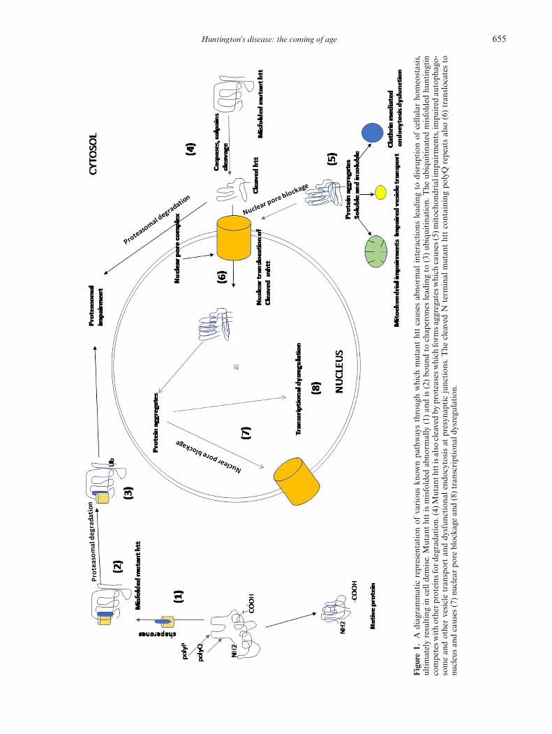
Post-mortem HD brain samples and transgenic HD micemodels expressingmutant httwith increasedpolyQ repeatsrevealed densely stained intraneuronal inclusions (Davieset al. 1997). Inclusion bodies have been reported frommultiple regions of the brain including striatum, cerebralcortex, cerebellum, brain stem and spinal cord. LongerpolyQ tracts of htt have been found to have increasedpropensity to form aggregates of mutant htt with ubiqui-tin positive proteins (Finkbeiner 2011). Immunostainingof inclusion bodies have revealed positive staining ofhuntingtin and ubiquitin proteasome degradation relatedproteins (Becher et al. 1998). Huntingtin protein is sub-jected to proteolysis by a variety of proteases. There is alsoan increase in protease activity in the brains of patients.It is possible that the disease specific enhancement of pro-teolysis is an important step, as it leads to the generationof small N terminal fragments that contain polyQ stretchand helps to translocate into the nucleus where they aretoxic as depicted in figure 1 (Graham et al. 2005). Inclusionbodies have also been reported in cytosol and neuronalprocesses (Gutekunst et al. 1999). Initial reports of inclu-sion bodies containing mutant htt in post-mortem brainsamples patients andHDmicemodels tempted researchersto conclude these are toxic to the host neurons. However,intense researchhas led toanargument aboutneuroprotec-tive mechanisms of these aggregate proteins (Zuchner andBrundin 2008; Arrasate et al. 2004). A number of studiesfound that the localization of inclusion bodies and the vul-nerability and demise of neurons in HD did not correlatewell (Miller et al. 2010; Slow et al. 2005). The paradigmstested to arrive at this juncture also led some researchersto pharmacologically promote IBs formation as a possibletherapeutic approach for HD (Chopra et al. 2007). How-ever, recent reports suggest that soluble forms of mutanthtt aggregates and insoluble formsmay have different levelof toxicities andmay regulate cell survival and death in dif-ferent ways (Xi et al. 2016). Another recent study suggeststhat although, soluble htt aggregates lead to apoptotic celldeath but insoluble aggregates of mutant htt lead to adelayed necrotic cell death (Ramdzan et al. 2017). In yetanother important study, it was found that soluble formsof mutant htt were severely toxic to cells by interactingwith larger repertoire of proteins related to ribosome bio-genesis, translation, transcription and vesicle transport ascompared to the insoluble aggregates of mutant htt whichhad a smaller interactome which were related to qualitycontrol such as chaperons and ubiquitin-protease system(Kim et al. 2016).
Huntington’s disease: the coming of age 655
Figure1.
Adiagrammatic
representation
ofvariou
skn
ownpa
thwaysthroug
hwhich
mutan
thttcauses
abno
rmal
interactions
lead
ingto
disrup
tion
ofcellu
larho
meostasis,
ultimatelyresultingin
celldemise.Mutan
thttismisfolded
abno
rmally
(1)an
dis(2)bo
undto
chap
eron
eslead
ingto
(3)ub
iquitina
tion
.The
ubiquitina
tedmisfolded
huntingtin
competesw
ithotherp
roteinsfor
degrad
ation.
(4)M
utan
thttisalso
cleavedby
proteasesw
hich
form
sagg
regatesw
hich
causes
(5)m
itocho
ndrialim
pairments,impa
ired
autoph
ago-
somean
dothervesicletran
sportan
ddy
sfun
ctiona
lendo
cytosisat
presyn
apticjunction
s.The
cleavedN
term
inal
mutan
thttcontaining
polyQ
repeatsalso
(6)tran
slocates
tonu
cleusan
dcauses
(7)nu
clearpo
reblocka
gean
d(8)tran
scriptiona
ldysregu
lation
.

656 Mritunjay Pandey and Rajamma Usha
Mutant htt and aggregate formation
Studies on mutant htt aggregation have found that itsaggregation can be affected by several intrinsic factorssuch as the length of polyQ stretch, the amino acidresidues flanking the polyQ stretch and the conformationof mutant htt. Earlier, mutant htt with increased polyQstretches showed aggregation pattern in budding yeast(KrobitschandLindquist 2000) and inprimary striatal cul-ture neurons (Miller et al. 2010). Besides the expandablepolyQ repeat, the flanking proline rich domain towardsthe C-terminus and the 17-amino acid peptide towardsthe N- terminus have been found to influence the mutanthtt’s capacity to aggregate. In immortalized striatal cellculture, it has been observed that proline rich domainis necessary to form visible aggregates of mutant htt(Steffan et al. 2004). In contrast, attaching a proline richsequence flanking the C-terminus of the polyQ domaindecreased the rate of aggregation in vitro (Bhattacharyyaet al. 2006). Similarly, mutant htt lacking the first 17amino acid residues at the N-terminus showed delayedaggregation in cells (Thakur et al. 2009). It has also beenobserved that although, the N17 residues help the cleavedmutant htt to localize in the nucleus but it is helpful inmitigating the disease pathology in HD transgenic mice(Gu et al. 2015) and a HD transgenic zebra fish (Veldmanet al. 2015). Besides, the 17N terminus aminoacids are alsosecondarily modified such as dephosphorylated (Branco-Santos et al. 2017) and phosphorylation (DiGiovanni et al.2016)which regulates its aggregation, localization and tox-icity.Mutant htt has also been found to disrupt the nuclearpore complex (figure 1), which is important for nuclear-cytoplasm transport of proteins and other molecules(Grima et al. 2017;Gasset-Rosa et al. 2017).However, pro-teolysis of wild-typeHTT has not been reported in normalindividuals. The proteolysis of wild-typeHTTmay inacti-vate some of its normal function (El-Daher et al. 2015).
Posttranslational modifications of mutant htt
HTT is subjected to multiple posttranslationalmodificationswhich include phosphorylation, acetylation,palmitoylation, ubiquitinylation and sumoylation. Mostof these modifications have been studied in mutant HTTwhich could have therapeutic implications as they modifythe interactions and functions of the protein. HTT inter-acts with HIP 14 and HIP14L that belong to the familyof palmitoyl-acyl transferases (Yanai et al. 2006). HTTis palmitoylated at C214 and its polyQ expansion leadsto a reduction in the enzymatic activity of HIP 14 andof its auto palmitoylation (Huang et al. 2011) leading tothe enzymatic activity of HIP 14 on other substrates andtherefore, regulate intracellular trafficking and synapticlocalization of other neuronal proteins.
Mutant htt and autophagy
Autophagy is an important cellular process which isresponsible for the removal of damaged organelles and
aggregated proteins by delivering them to lysosomes fordegradation. Autophagy defects have been consistentlyobserved in HD (Steffan 2010; Martin et al. 2015). Highamount of autophagosome formation a reduced capac-ity to degrade aggregated proteins and organelles hasbeen found in HD models (Martin et al. 2015). Stud-ies also suggest that wild-type htt may play a significantrole in autophagy. Reducing HTT expression decreasedoptineurin interaction in Golgi apparatus hints that HTTmay also regulate dynamics of autophagosome through itsinteraction with optineurin (Toro et al. 2009). Autophago-somes in neurons under basal conditions are generatedat distal axons and are retrogradely trafficked to the cellbody. Silencing of htt blocks the retrograde transport ofautophagosomes along the axon (Wong and Holzbaur2014). Similar defects were observed with mutant htt,which led to inefficient clearance of mutant htt, suggestingthat htt may regulate its own clearance.In human HD and transgenic HD rodent samples,
mutant htt has been found to activate autophagy bysequestering and inactivating mTOR leading to an induc-tion of autophagy (Ravikumar et al. 2004). The mechanis-tic defects in autophagy in HD are exaggerated by mutanthtt. Mutant htt results in defective autophagy mediateddegradation and aggregate formation, which leads to com-pensatory upregulation of autophagy and accumulationof mutant htt and neurotoxicity. Further due to decreasedcargomovementof autophagosome to fusewith lysosomesleading to defective autophagy (Wong and Holzbaur2014). Moreover, expression levels of autophagy geneshave been observed in HD striatum (Hodges et al. 2006).In the caudate nucleus of HD patients, mRNA expressionof LC3A, ULK2, and LAMP2 is significantly increasedwhereas; PINK1, WDFY3 and FK506 binding protein1A are significantly decreased. These findings suggest thatthe increased expression of LC3A and ULK2 correlateto early autophagy induction and autophagosome forma-tion in HD (Martinez-Vicente et al. 2010). A decrease inPINK1 expression induces mitochondrial fragmentationandmitophagy (Kamat et al. 2014). HD is associated withincreasedmitochondrial fragmentationand inefficient cor-poration of mitochondria into autophagosomes (Wongand Holzbaur 2014). Autophagy defects in HD may bea combined effect of loss of function of wild-type htt anda toxic gain of function by mutant htt.
Vesicular transport and mutant htt
HTThas been found to be involved in a variety of vesiculartrafficking in neurons including synaptic precursor vesicles(Zala et al. 2013), autophagosomes (Wong and Holzbaur2014), lysosomes (Liot et al.2013),BDNF-containingvesi-cles (Gauthier et al. 2004) and GABA-containing vesicles(Twelvetrees et al. 2010). It is not clear whether htt alsopromotes mitochondrial transport in neurons (Trushinaet al. 2004) but it has been observed that the mutant

Huntington’s disease: the coming of age 657
HTT has an association with the fusion–fission machin-ery of the organelle (Costa et al. 2010; Song et al. 2011;Pandey et al. 2010). HTT is also found to interact withseveral proteins involved in clathrin-mediated endocyto-sis (Legendre-Guillemin et al. 2002). HTT also activatedGTPase Rab11 that participates in vesicle recycling duringendocytosis (Li et al. 2008).
Evidences suggest that HTT is required for ciliogenesis(Haremaki et al. 2015).HTT is found at the base of the ciliain neurons, photoreceptor cilia, and cilia in multiciliatedcells (Keryer et al. 2011). Absence of HTT from mousecells impairs the retrograde trafficking of the pericentriolarmaterial 1 protein and the primary cilium is not formed.
Transcriptional dysregulation and mutant htt
Post-mortem samples from HD brain and transgenicmouse models show transcriptional dysregulation (Valor2015). Wild-type htt binds to several transcription fac-tors including CREB-binding protein (Steffan et al. 2000),p53 tumour suppressor, nuclear factor-κB (Takano andGusella 2002), PPAR-γ, vitamin D receptor, thyroid hor-mone receptor-α1 (Futter et al. 2009). With these interac-tions, HTT can potentiate transcription factors which canhave various outcomes (figure 1). Transcriptional studiesin rat PC12 cells expressing doxycycline induced mutanthtt with increasing polyQ repeats were done in early stage,aggregate formed stage and late toxic stage and com-pared. Twomajor clusters of gene expression changes wereobserved in aggregate and later stage which were related tomitochondrial dysfunction and developmental processesrelated to cellular homeostasis (van Hagen et al. 2017).Transcriptome analysis of HD when compared to normalbrains revealed wide spread aberrant alternative splicingin the diseased brains (Lin et al. 2016). RNA seq datafrom myeloid cells of HD patients revealed increases inproinflammatory cytokines in resting stage which indi-cates abnormal basal activation to an exaggerated immuneresponse to a stimulus (Miller et al. 2016). In anotherRNAseq study from HD brain samples, it was found that therewas upregulation of developmental homeobox genes andneuroinflammatory genes (Labadorf et al. 2015). Multiplestudies related to transcriptional dysregulation inHDsam-ples reveal changes in inflammatory responses regulatedby NF-kB, metabolic homeostasis related to mitochon-drial oxidative phosphorylation and developmental genesrelated to neuronal growth and cell cycle regulation.
Altered proteostasis and mutant htt
Mutant htt is prone to aggregate formation whosesoluble and insoluble forms are neurotoxic to cells. Suchmisfolded forms of proteins are regulated by different pro-teostasis mechanisms such as chaperone-mediated foldingor degraded by ubiquitin-mediated degradation or bylysosomal-autophagy degradation (figure 1). However,the increasing load of chaperons found in inclusion
bodies such Hsp70 and ubiquitin related protein degra-dation machinery or accumulation of autophagosomerelated target proteins lead to altered proteostasis. Theexpanded polyQ fibrils can deregulate proteostasis nodesby sequestering transcription factors (Suhr et al. 2001)or physically obstruct neuronal extensions (Gunawardenaet al. 2003). Moreover, ageing is also a risk factor foraltered proteostasis (Hartl 2016). Chaperone functions areknown to be compromised in HD (Kakkar et al. 2014).The differential ability of neurons to handle stress of
altered proteosis could also explain the vulnerability ofstriatal neurons in HD. Cerebellar neurons have beenobserved to induce Hsp70 expression upon mutant httexpression. However, striatal neurons cannot upregulatetheir chaperone expression to overcome this proteostasisstress (Tagawa et al. 2007). It is also possible that distinctnodes of proteostasis may have unique ways of handlingproteostatic stress in HD. Manipulation of different pro-teostasic nodes genetically or pharmacologically could beanother way to handle the toxicity ofmutant htt (Koyuncuet al. 2017).
Therapeutic interventions in HD
The cardinal features of HD like (i) movement disordercharacterized initially by chorea and later by dystoniaand parkinsonism, (ii) progressive cognitive decline anddisordered behaviour, and (iii) depression and agitationappear mostly during adulthood and progress relentlesslyleading to functional disability. Parkinsonism is frequentlyobserved in the rare juvenile form, slowing saccadic move-ments of the eye and possible cerebellar dysfunction helpsin differential diagnosis. Neuropsychiatric symptoms arecommon and do not show increased progression with dis-ease severity. The multifaceted phenotype of HD makes itdifficult to manage the patients. Clinical trials of drugs arebeing conducted in every possible ways for HD therapy.Both uncontrolled and open labelled treatment reports aswell as controlled trials have been carried out, where theintervention studied is compared to an active or inactive(placebo) comparator. Most of the drugs are symptomaticin nature where they are targeted to improve the clinicalfeatures of illness but do not block the progressive natureof the disease. Presently, there is no internationally rec-ognized standard care for HD. Therefore, throughout theworld the therapeutic approaches widely vary, accordingto the license of the respective country.
Symptomatic drugs
Most of the symptomatic drugs are directed againstchorea, which is the most conspicuous feature of the dis-ease. These drugs either deplete dopamine reserves orblock the dopaminergic receptors in the brain, therebyproviding temporary relief to the patient. Reserpine, tetra-benazine (a short-acting reserpine analogue), phenoth-iazines (such as chlorpromazines) and butyrophenones

658 Mritunjay Pandey and Rajamma Usha
(such as haloperidol) have been used extensively for symp-tomatic relief in HD (Marsden 1973). In a double-blindcrossover trial involving four treatments of haloperidol,haloperidol and lithium carbonate, lithium carbonate andplacebo carried out for three weeks, none of the treatmentsprovided effective relief from choreiformmovements. Psy-chological variables like levels of irritability, outbursts ofanger and depression did appear to have some effect due tothese drugs.While threeHDpatients showed some relief inhaloperidol and lithium carbonate combination, the otherthree did not show any significant relief. Haloperidol itselfincreased the levels of depression when compared to othercombinations including placebo (Leonard et al. 1975). Inanother study, 18 patients with Huntington’s chorea wereexaminedbefore andafter treatmentwith three neurolepticdrugs, pimozide, haloperidol and tiapride. Pimozide andhaloperidol gave relief from hyperkinesias but none of thedrug improved motor performance significantly (Girottiet al. 1984). In an interesting study, HD patients hav-ing gait abnormalities and choreiform movements weretreated with haloperidol. Although, there was a decreasein choreiformmovement, yet it did not correct gait abnor-malities (Koller and Trimble 1985). Long-term treatmentof haloperidol is known to cause aggravatedparkinsonism,tardive dyskinesis, difficulties in swallowing and dyspha-sia (Emerich et al. 1991). Tetrabenazine, an inhibitor ofvesicular monoamine transporter 2 (VMAT2) leading todepletion of dopamine and other monoamines like sero-toninandnorepinephrine in the central nervous systemhasbeen used extensively in hyperkinetic disorders, especiallyHD. In an open labelled study described by Kenney et al.(2007), over 400patients suffering fromhyperkineticmove-ment disorders including 98HDpatients were treatedwithtetrabenazine at the Baylor College of Medicine Parkin-son’s Disease Center and Movement Disorders Clinic.There was a marked improvement in patients with chor-eiform movements.The Huntington Study Group (2006) recently
completed a phase III study assessing the safety, efficacy,and dose-tolerability of tetrabenazine for amelioratingchorea in patientswithHD.A total of 84 patientswere ran-domly assigned to placebo (n = 30) or tetrabenazine (n =54) up to 100 mg/day for 12 weeks. Based on the choreascore of the UHDRS, tetrabenazine was found to signifi-cantly reduce chorea. GABAergic strategies have includedmuscimol, a potent GABA mimetic agonist (Shoulsonet al. 1978). In this double-blind study,muscimol treatmentdid not lead to any improvement in motor and cogni-tive deficits in 10 HD patients. However, administration ofmuscimol ameliorated chorea in themost severely affectedhyperkinetic patients. It also heightened dystonia, partic-ularly in the early onset patients who had predominantparkinsonism and dystonic features. For the treatment ofdepression in HD patients, standard antidepressant med-ication, including selective serotonin reuptake inhibitors(SSRIs) have been used. Fluoxetine, an SSRI that is widely
used as an antidepressant has been shown to be effective inHD transgenic mice (Grote et al. 2005) but when admin-istered in randomized, double-blind, placebo-controlledtrial of thismedication in depressed and nondepressedHDpatients, no differences between the treatment groups werefound in total functional capacity, neurological, or cogni-tive ratings (Como et al. 1997). Pridopidine, a dopamineD2 receptor antagonist was tested in three large multi-centre clinical trials of HD which has been published(Lundin et al. 2010; de Yebenes et al. 2011; HuntingtonStudyGroupHART Investigators 2013). TheMermaiHDstudy and the HART study found that the higher doseof 90 mg/day showed improvement in motor scores andgait balance as compared to placebo but did not reachstatistical significance. Presently, a large, global, multi-centre, double blind phase II trial with higher dose ofpridopidine—–PRIDE-HD is currently under progress toassess thedrug tolerability andmanagingmotor symptoms(Reilmann 2013).
Neuroprotective drugs
Neuroprotective therapy is targeted at the level of geneticaetiology to slow down the progressive nature of the dis-ease. Most of the drugs tested in this case have beentargeted at the level of oxidative metabolism or glutamatetransmission because of the metabolic decrease in activityor increased excitotoxic mechanisms in HD.Coenzyme Q10, a principle mitochondrial cofactor
involved in complex-I activity has been studied as a neu-roprotective agent in HD animal models (Matthews et al.1998). In a multicentre, parallel group, double blind,randomized clinical trial on coenzyme Q10 along withramacemide (NMDA antagonist) in early HD patients,conducted by Huntington Study Group (2001), neitherremacemide nor coenzymeQ10, produced significant slow-ing in functional decline in early HD even after 30months. To add to these negative data, there was anincreased frequencyofnausea, vomitinganddizzinesswithremacemide; and increased frequency of stomach upsetwith coenzyme Q10.Another drug that has been extensively used in HD
neuroprotective therapy is creatine. Creatine is a natu-rally occurring compound that, through its intermediatephosphocreatine, provides a necessary cellular reserve ofhigh energy phosphates. There is a strong evidence to sug-gest that a bioenergetic defect exists in HD as discussedearlier. Creatine supplementation is intended to augmentcerebral reserves of phosphates and thereby reduce neu-ronal metabolic and oxidative stress, and to slow downneurodegeneration. While there have been several clinicaltrials of creatine in HD, none have been powered to detectsignificant slowing of progression or improvement in clin-ical symptoms. Verbessem and colleagues treated 26 HDpatientswith 5mg/kg creatine and 15patientswith placebofor one year and found no significant differences in mea-sures of strength, neurological status, or cognitive function

Huntington’s disease: the coming of age 659
(Verbessem et al. 2003). In an interesting study,Bender andcolleagues usedmagnetic resonance spectroscopy to exam-ine the levels of glutamate in HD patients treated with 20g/day for 5 days, followed by 6 g/day for 8–10 weeks. Theydemonstrated a significant reduction in glutamate levelsin the parietooccipital cortex. This is of great significancebecause glutamate release and excitotoxicity are enhancedby energy deficiency and are considered to play an impor-tant role in the pathogenesis of HD (Bender et al. 2005).However, none of the studies are informative enough toshow that creatine is neuroprotective in HD.A small scale study of foetal transplantation with a
long term follow up in HD subjects was reported in 2014(Paganini et al. 2014). Twenty-sixHDpatients (10 subjectstransplanted with foetal striatal tissue) were followed formedian 11.2 years in transplanted subjects. Transplantedsubjects showed a slower decline in motor and cognitivemeasures (Barker et al. 2013). Another small study of fivetransplanted and 12 control subjects followed over 3–10years showed no significant benefit on clinical or cognitivefeatures. Pathological studies have shown poor graft sur-vival as well as aggregation of mutant htt in the graftedtissue (Cicchetti et al. 2014; Cisbani and Cicchetti 2014).Cell replacement therapy is considered to have great poten-tial in degenerative diseases. In this context, HDmakes aneffective disease condition for such therapy as it is markedby striatal neurodegeneration (Precious et al. 2017).
Conclusions and future study
It has been almost 25 years since the discovery of themutant gene htt responsible for causing HD, but there isstill no cure available for the disease. In spite of the intenseefforts to discover and find a cure for the disease most ofthe drugs provide palliative relief. However, the researchhas led to a better understanding of the protein’s functionsin its wild type and mutant state. The multifaceted role ofthe normal protein and its further anomalies in its gain offunctions in mutant state makes the drug targeting morechallenging. The advantage of studying this monogenicMendelian neurodegenerative disorder has been in identi-fying presymptomatic biomarkers, which also be used fortracking the progression of the disease. This helps the clin-icians to find more sensitive quantifiable biomarkers apartfrom mere behavioural phenotypes of patients, which areused for drug screening and tracking the progression ofthe disease. Identification of peripheral markers or neuro-chemical agents such as BDNF, 8-OHdG and interleukinsor other molecules that could also help in finding the effi-cacy and tracking down the neuroprotective role of drugsin clinics (Ross and Tabrizi 2011). Brain imaging is also avery reliable and quantifiable tool to measure striatal vol-ume, cortical thickness and ventricular volume to assessthe damage in brain tissue.Although, the transgenic knock-in mice models have
helped us to understand the disease better but most ofthem do not express the disease pathology similar to
humans. This has led to an increase in numbers of differ-ent mice models which correlate to different pathologicalphenotypes as seen in humans. Identifying protein targetsof mutant htt interactions which lead to disease pathol-ogy could be an important strategy. With the advent ofCRISPR-Cas9 technology, gene silencing of mutant htt inpreferred areas of neurodegenerative regions could be alsoconsidered. Better animal models which exhibit pathologycloser to humans would be helpful in understanding thepathology of the disease and very helpful for preclinicalscreening of drugs. Induced pluripotent stem cells couldalso be used for primary drug screening. A combinationtherapy of antipsychotics along with neuronal survivalagent could also an effective strategy of looking at delayingthe disease progression.
References
Adachi Y. and Nakashima K. 1999 Population genetic studyof Huntington’s disease-prevalence and founder’s effect in theSan-in area, western Japan. Nippon Rinsho. 57, 900–904.
Albin R. L., Reiner A., Anderson K. D., Penney J. B. and YoungA. B. 1990 Striatal and nigral neuron subpopulations in rigidHuntington’s disease: implications for the functional anatomyof chorea and rigidity-akinesia. Ann. Neurol. 27, 357–365.
AlbinR.L.,ReinerA.,AndersonK.D.,DureL. S. 4th,HandelinB., Balfour R. et al. 1992 Preferential loss of striato-externalpallidal projection neurons in presymptomatic Huntington’sdisease. Ann. Neurol. 31, 425–430.
Almqvist E. W., Elterman D. S., MacLeod P.M. and HaydenM.R. 2001 High incidence rate and absent family histories in onequarter of patients newly diagnosed with Huntington diseasein British Columbia. Clin. Genet. 60, 198–205.
Andrade M. A. and Bork P. 1995 HEAT repeats in the Hunting-ton’s disease protein. Nat. Genet. 11, 115–116.
Andrich J., Saft C., Ostholt N. and Müller T. 2007 Complexmovement behaviour and progression ofHuntington’s disease.Neurosci. Lett. 416, 272–274.
Arrasate M., Mitra S., Schweitzer E. S., Segal M. R. andFinkbeiner S. 2004 Inclusion body formation reduces levels ofmutant huntingtin and the risk of neuronal death.Nature 431,805–810.
Baquet Z. C., Gorski J. A. and Jones K. R. 2004 Early striataldendrite deficits followed by neuron loss with advanced age inthe absence of anterograde cortical brain-derived neurotrophicfactor. J. Neurosci. 24, 4250–4258.
Barker R. A., Mason S. L., Harrower T. P., Swain R. A., HoA. K., Sahakian B. J. et al. 2013 The long-term safety andefficacy of bilateraltransplantation of human fetal striatal tis-sue in patients with mild to moderate Huntington’s disease. J.Neurol. Neurosurg. Psychiatry 84, 657–665.
BarnesG.T.,DuyaoM.P.,AmbroseC.M.,McNeil S., PersichettiF., Srinidhi J. et al. 1994 Mouse Huntington’s disease genehomolog (Hdh). Somat. Cell Mol. Genet. 20, 87–97.
Bates E. A., Victor M., Jones A. K., Shi Y. and Hart A. C. 2006Differential contributions of Caenorhabditis elegans histonedeacetylases to huntingtin polyglutamine toxicity. J. Neurosci.26, 2830–2838.
Baxa M., Hruska-Plochan M., Juhas S., Vodicka P., Pavlok A.,Juhasova J. et al. 2013 A transgenic minipig model of Hunt-ington’s disease. J. Huntingtons Dis. 2, 47–68.

660 Mritunjay Pandey and Rajamma Usha
Baxendale S., Abdulla S., Elgar G., Buck D., BerksM., MicklemG. et al. 1995Comparative sequence analysis of the humanandpufferfish Huntington’s disease genes. Nat. Genet. 10, 67–76.
Becher M. W., Kotzuk J. A., Sharp A. H., Davies S. W., BatesG. P., Price D. L. et al. 1998 Intranuclear neuronal inclusionsin Huntington’s disease and dentatorubral and pallidoluysianatrophy: correlationbetween thedensityof inclusions and IT15CAG triplet repeat length. Neurobiol. Dis. 4, 387–397.
Benchoua A., Trioulier Y., Diguet E., Malgorn C., Gaillard M.C., Dufour N. et al. 2008 Dopamine determines the vulnera-bility of striatal neurons to theN-terminal fragment of mutanthuntingtin through the regulation of mitochondrial complexII. Hum. Mol. Genet. 17, 1446–1456.
Bender A., Auer D. P., Merl T., Reilmann R., Saemann P., Yas-souridis A. et al. 2005 Creatine supplementation lowers brainglutamate levels inHuntington’s disease. J. Neurol. 252, 36–41.
Bhattacharyya A., Thakur A. K., Chellgren V. M., ThiagarajanG., Williams A. D., Chellgren B. W. et al. 2006 Oligopro-line effects on polyglutamine conformation and aggregation.J. Mol. Biol. 355, 524–535.
Bird E. D. and Iversen L. L. 1974 Huntington’s chorea. Post-mortemmeasurement of glutamic acid decarboxylase, cholineacetyltransferase and dopamine in basal ganglia. Brain 97,457–472.
Bird E. D. and Iversen L. L. 1977 Neurochemical findings inHuntington’s chorea. Essays Neurochem. Neuropharmacol. 1,177–195.
Bohnen N. I., Koeppe R. A., Meyer P., Ficaro E., Wernette K.,Kilbourn M. R. et al. 2000 Decreased striatal monoaminergicterminals in Huntington disease. Neurology 54, 1753–1759.
Branco-Santos J., Herrera F., Poças G. M., Pires-Afonso Y.,Giorgini F., Domingos P. M. et al. 2017 Protein phosphatase1 regulates huntingtin exon 1 aggregation and toxicity. Hum.Mol. Genet. 26, 3763–3775.
BrownR. G. andMarsden C. D. 1988 Subcortical dementia’: theneuropsychological evidence. Neuroscience 25, 363–387.
Chen Y. Y. and Lai C. H. 2010 Nationwide population-basedepidemiologic study of Huntington’s disease in Taiwan. Neu-roepidemiology 35, 250–254.
Chopra V., Fox J. H., Lieberman G., Dorsey K., Matson W.,Waldmeier P. et al. 2007 A small-molecule therapeutic lead forHuntington’s disease: preclinical pharmacology and efficacyof C2-8 in the R6/2 transgenic mouse. Proc. Natl. Acad. Sci.USA 104, 16685–16689.
Ciarmiello A., Cannella M., Lastoria S., Simonelli M., Frati L.,Rubinsztein D. C. et al. 2006 Brain white-matter volume lossand glucose hypometabolism precede the clinical symptoms ofHuntington’s disease. J. Nucl. Med. 47, 215–222.
Cicchetti F., Prensa L., Wu Y. and Parent A. 2000 Chemicalanatomy of striatal interneurons in normal individuals andin patients with Huntington’s disease. Brain Res. Brain Res.Rev. 34, 80–101.
Cicchetti F., Lacroix S., Cisbani G., Vallières N., Saint-PierreM.,St-Amour I. et al. 2014 Mutanthuntingtin is present in neu-ronal grafts in Huntington disease patients. Ann. Neurol. 76,31–42.
Cisbani G. and Cicchetti F. 2014 The fate of cell grafts for thetreatment of Huntington’s disease: the post-mortem evidence.Neuropathol. Appl. Neurobiol. 40, 71–90.
Como P. G., Rubin A. J., O’Brien C. F., Lawler K., Hickey C.,Rubin A. E. et al. 1997 A controlled trial of fluoxetine in non-depressed patients with Huntington’s disease.Mov. Disord. 12,397–401.
Conneally P. M. 1984 Huntington disease: genetics and epidemi-ology. Am. J. Hum. Genet. 36, 506–526.
Costa V., Giacomello M., Hudec R., Lopreiato R., Ermak G.,LimD. et al. 2010Mitochondrial fission and cristae disruption
increase the response of cell models of Huntington’s disease toapoptotic stimuli. EMBOMol. Med. 2, 490–503.
Davies S. W., Turmaine M., Cozens B. A., DiFiglia M., SharpA. H., Ross C. A. et al. 1997 Formation of neuronal intranu-clear inclusions underlies the neurological dysfunction in micetransgenic for the HD mutation. Cell 90, 537–548.
de Yebenes J. G., Landwehrmeyer B., Squitieri F., Reilmann R.,Rosser A., Barker R. A. et al. 2011 Pridopidine for the treat-ment of motor function in patients with Huntington’sdisease(MermaiHD): a phase 3, randomised, double-blind, placebo-controlledtrial. Lancet Neurol. 10, 1049–1057.
del Toro D., Alberch J., Lázaro-Diéguez F., Martín-Ibáñez R.,Xifró X., Egea G. et al. 2009 Mutant huntingtin impairs post-Golgi trafficking to lysosomes bydelocalizing optineurin/Rab8complex from the Golgi apparatus. Mol. Biol. Cell. 20, 1478–1492.
Di Maio L., Squitieri F., Napolitano G., Campanella G. andTrofatter J. A. 1993 Conneally suicide risk in Huntington’sdisease J. Med. Genet. 30, 293–295.
DiFiglia M. 1990 Excitotoxic injury of the neostriatum: a modelfor Huntington’s disease. Trends Neurosci. 13, 286–289.
DiGiovanni L. F., Mocle A. J., Xia J. and Truant R. 2016Huntingtin N17 domain is a reactiveoxygen species sensorregulating huntingtin phosphorylation and localization.Hum.Mol. Genet. 25, 3937–3945.
Dom R., Malfroid M. and Baro F. 1976. Neuropathology ofHuntington’s chorea. Studies of the ventrobasal complex ofthe thalamus. Neurology 26, 64–68.
El-Daher M. T., Hangen E., Bruyère J., Poizat G., Al-Ramahi I.,PardoR. et al. 2015Huntingtin proteolysis releases non-polyQfragments that cause toxicity through dynamin 1 dysregula-tion. EMBO J. 34, 2255–2271.
Emerich D. F., Norman A. B. and Sanberg P. R. 1991 Nicotinepotentiates the behavioral effects of haloperidol. Psychophar-macol. Bull. 27, 385–390.
Enna S. J., Bird E. D., Bennett Jr J. P., Bylund D. B., YamamuraH. I., Iversen L. L. et al. 1976Huntington’s chorea. Changes inneurotransmitter receptors in the brain. N. Engl. J. Med. 294,1305–1309.
Evans S. J., Douglas I., Rawlins M. D., Wexler N. S., Tabrizi S. J.and Smeeth L. 2013 Prevalence of adult Huntington’s diseasein the UK based on diagnoses recorded in general practicerecords. J. Neurol. Neurosurg. Psychiatry 84, 1156–1160.
Ferrante R. J., Kowall N. W., Beal M. F., Martin J. B., Bird E. D.and Richardson Jr E. P. 1987 Morphologic and histochemicalcharacteristics of a spared subset of striatal neurons in Hunt-ington’s disease. J. Neuropathol. Exp. Neurol. 46, 12–27.
Ferrer I., Goutan E., Marín C., Rey M. J. and Ribalta T.2000Brain-derived neurotrophic factor inHuntington disease.Brain Res. 866, 257–261.
Finkbeiner S. 2011 Huntington’s disease. Cold Spring HarborLaboratory Press, Cold Spring Harbor.
Fisher E. R. and HaydenM. R. 2014Multisource ascertainmentof Huntington disease in Canada: prevalence and populationat risk.Mov. Disord. 29, 105–114.
Folstein S. E., Chase G. A., Wahl W. E., McDonnell A. M. andFolstein M. F. 1987 Huntington disease in Maryland: clinicalaspects of racial variation. Am. J. Hum. Genet. 41, 168–179.
Futter M., Diekmann H., Schoenmakers E., Sadiq O., Chatter-jee K. and Rubinsztein D. C. 2009 Wild-type but not mutanthuntingtin modulates the transcriptional activity of liver Xreceptors. J. Med. Genet. 46, 438–446.
Gasset-Rosa F., Chillon-Marinas C., Goginashvili A., AtwalR. S., Artates J. W., Tabet R et al. 2017 Polyglutamine-expanded Huntingtin exacerbates age-related disruption ofnuclear integrity and nucleocytoplasmic transport.Neuron 94,48–57.

Huntington’s disease: the coming of age 661
Gauthier L. R., Charrin B. C., Borrell-PagèsM., Dompierre J. P.,Rangone H., Cordelières F. P. et al. 2004 Huntingtin controlsneurotrophic support and survival of neurons by enhancingBDNF vesicular transport along microtubules. Cell 118, 127–138.
GerfenC.R. 1992The neostriatalmosaic:multiple levels of com-partmental organization. J. Neural. Transm. Suppl. 36, 43–59.
Girotti F., Carella F., Scigliano G., Grassi M. P., Soliveri P.,Giovannini P. et al. 1984 Effect of neuroleptic treatment oninvoluntary movements and motor performances in Hunt-ington’s disease. J. Neurol. Neurosurg. Psychiatry 47, 848–852.
Graham R. K., Slow E. J., Deng Y., Bissada N., Lu G., PearsonJ. et al. 2005 Levels of mutant huntingtin influence the pheno-typic severity ofHuntingtondisease inYAC128mousemodels.Neurobiol. Dis. 21, 444–455.
Graybiel A. M., Ohta K. and Roffler-Tarlov S. 1990 Patternsof cell and fiber vulnerability in the mesostriatal system ofthe mutant mouse weaver. I. Gradients and compartments. J.Neurosci. 10, 720–733.
Grima J. C., Daigle J. G., Arbez N., Cunningham K. C., ZhangK., Ochaba J. et al. 2017 Mutant Huntingtin disrupts thenuclear pore complex. Neuron 94, 93–107.
GroteH.E., BullN.D.,HowardM.L., vanDellenA.,BlakemoreC., Bartlett P. F. et al. 2005 Cognitive disorders and neuro-genesis deficits in Huntington’s disease mice are rescued byfluoxetine. Eur. J. Neurosci. 22, 2081–2088.
Gu X., Cantle J. P., Greiner E. R., Lee C. Y., Barth A. M., GaoF. et al. 2015 N17Modifies mutant Huntingtin nuclear patho-genesis and severity of disease in HD BAC transgenic mice.Neuron 85, 726–741.
Gunawardena S., Her L. S., Brusch R. G., Laymon R. A., Nies-man I. R., Gordesky-Gold B. et al. 2003 Disruption of axonaltransport by loss of huntingtin or expression of pathogenicpolyQ proteins in Drosophila. Neuron 40, 25–40.
Gutekunst C. A., Li S. H., Yi H., Mulroy J. S., KuemmerleS., Jones R. et al. 1999 Nuclear and neuropil aggregates inHuntington’s disease: relationship to neuropathology. J. Neu-rosci. 19, 2522–2534.
Haremaki T., Deglincerti A. and Brivanlou A. H. 2015 Hunt-ingtin is required forciliogenesis and neurogenesis during earlyXenopus development. Dev. Biol. 408, 305–315.
Harper P. S. 2002 Huntington’s disease: a historical backgroundin Huntington’s disease (ed. G. Bates, P. S. Harper and L.Jones), pp. 3–37. Oxford University Press, Oxford.
Harper P. S.,WalkerD.A., TylerA.,NewcombeR.G. andDaviesK. 1979 Huntington’s chorea. The basis for long-term preven-tion. Lancet 2, 346–349.
Hartl F. U. 2016 Cellular homeostasis and aging. Ann. Rev.Biochem. 85, 1–4.
Hedreen J. C. and Folstein S. E. 1995 Early loss of neostriatalstriosome neurons in Huntington’s disease. J. Neuropathol.Exp. Neurol. 54, 1051–1020.
Hedreen J. C., Peyser C. E., Folstein S. E. and Ross C. A. 1991Neuronal loss in layers V and VI of cerebral cortex in Hunt-ington’s disease. Neurosci. Lett. 133, 257–261.
Heinsen H., StrikM., BauerM., Luther K., Ulmar G., GangnusD. et al. 1994 Cortical and striatal neurone number in Hunt-ington’s disease Acta. Neuropathol. 88, 320–333.
HeinsenH.,RübU.,GangnusD., JungkunzG.,BauerM.,UlmarG. et al. 1996 Nerve cell loss in the thalamic centromedian-parafascicular complex in patients with Huntington’s disease.Acta. Neuropathol. 91, 161–168.
Hodges A., Strand A. D., Aragaki A. K., Kuhn A., SengstagT., Hughes G. et al. 2006 Regional and cellular gene expres-sion changes in humanHuntington’s disease brain.Hum.Mol.Genet. 15, 965–977.
Hodgson J. G., Agopyan N., Gutekunst C. A., Leavitt B. R.,LePiane F., Singaraja R. et al. 1999 A YAC mouse modelfor Huntington’s disease with full-length mutant huntingtin,cytoplasmic toxicity, and selective striatal neurodegeneration.Neuron 23, 181–192.
Huang K., Sanders S. S., Kang R., Carroll J. B., Sutton L., WanJ. et al. 2011 Wild-type HTT modulates the enzymatic activ-ity of the neuronal palmitoyl transferase HIP14. Hum. Mol.Genet. 20, 3356–3365.
Huntington G. 1872 On chorea.Med. Surg. Rep. 26, 317–321.Huntington Study Group 2001 A randomized, placebo-
controlled trial of coenzyme Q10 and remacemide in Hunt-ington’s disease. Neurology 14, 397–404.
Huntington StudyGroup 2006Tetrabenazine as antichorea ther-apy in Huntington disease: a randomized controlled trial.Neurology 66, 366 –372.
Huntington Study Group HART Investigators 2013 A random-ized, double-blind, placebo-controlled trial of pridopidine inHuntington’s disease.Mov. Disord. 28, 1407–1415.
Jacobsen J. C., Bawden C. S., Rudiger S. R., McLaughlan C.J., Reid S. J., Waldvogel H. J. et al. 2010 An ovine transgenicHuntington’s diseasemodel.Hum.Mol. Genet. 19, 1873–1882.
Jeste D. V., Barban L. and Parisi J. 1984 Reduced Purkinje celldensity in Huntington’s disease. Exp. Neurol. 85, 78–86.
Kakkar V., Meister-Broekema M., Minoia M., Carra S. andKampinga H. H. 2014 Barcoding heat shock proteins tohuman diseases: looking beyond the heat shock response.Dis.Model Mech. 7, 421–434.
Kamat P. K., Kalani A., Kyles P., Tyagi S. C. and Tyagi N. 2014Autophagy of mitochondria: a promising therapeutic targetfor neurodegenerative disease.Cell Biochem. Biophys. 70, 707–719.
Karlovich C. A., John R. M., Ramirez L., Stainier D. Y.and Myers R. M. 1998 Characterization of the Hunting-ton’s disease (HD) gene homologue in the zebrafish Daniorerio. Gene 217, 117–121.
Kenney C., Hunter C. and Jankovic J. 2007 Long-term tolerabil-ity of tetrabenazine in the treatment of hyperkinetic movementdisorders.Mov. Disord. 15, 193–197.
Keryer G., Pineda J. R., Liot G., Kim J., Dietrich P., BenstaaliC. et al. 2011 Ciliogenesis is regulated by a huntingtin-HAP1-PCM1 pathway and is altered in Huntington disease. J. Clin.Invest. 121, 4372–4382.
KimY. E., Hosp F., Frottin F., GeH.,MannM.,Hayer-HartlM.et al. 2016 Soluble oligomers of PolyQ-expanded Huntingtintarget a multiplicity of key cellular factors.Mol. Cell 63, 951–964.
Kingma E. M., van Duijn E., Timman R., van der Mast R. C.and Roos R. A. 2008 Behavioural problems in Huntington’sdisease using the problem behaviours assessment. Gen. Hosp.Psychiatry. 30, 155–161.
Kirkwood S. C., Siemers E., VikenR.,HodesM. E., Conneally P.M.,Christian J.C. et al.2002Longitudinal personality changesamong presymptomatic Huntington disease gene carriers.Neuropsychiatry Neuropsychol. Behav. Neurol. 15, 192–197.
Kish S. J., Shannak K. and Hornykiewicz O. 1987 Elevatedserotonin and reduced dopamine in subregionally dividedHuntington’s disease striatum. Ann. Neurol. 22, 386–389.
Kokmen E., Ozekmekci F. S., Beard C. M., O’Brien P. C. andKurland L. T. 1994 Incidence and prevalence of Huntington’sdisease in Olmsted County, Minnesota (1950 through 1989).Arch. Neurol. 51, 696–698.
Koller W. C. and Trimble J. 1985 Gait abnormality of Hunting-ton’s disease. Neurology 35, 1450–1454.
Koyuncu S., FatimaA.,Gutierrez-GarciaR. andVilchezD. 2017Proteostasis of huntingtin in health and disease. Int. J. Mol.Sci. 19, 18.

662 Mritunjay Pandey and Rajamma Usha
Krench M. and Littleton J. T. 2013 Modeling Huntington dis-ease in Drosophila: Insights into axonal transport defects andmodifiers of toxicity. Fly (Austin) 7, 229–236.
Krobitsch S. and Lindquist S. 2000 Aggregation of huntingtinin yeast varies with the length of the polyglutamine expansionand the expression of chaperone proteins. Proc. Natl. Acad.Sci. USA 97, 1589–1594.
Labadorf A., HossA.G., LagomarsinoV, Latourelle J. C., HadziT. C., Bregu J. 2015 RNA Sequence analysis of human Hunt-ington disease brain reveals an extensive increase in inflam-matory and developmental gene expression. PLoS One 10,e0143563.
Legendre-Guillemin V., Metzler M., Charbonneau M., Gan L.,Chopra V., Philie J. et al. 2002 HIP1 and HIP12 display differ-ential binding to F-actin, AP2, and clathrin. Identification ofa novel interaction with clathrin lightchain. J. Biol. Chem. 277,19897–19904.
Leonard D. P., Kidson M. A., Brown J. G., Shannon P. J. andTaryan S. 1975 A double blind trial of Lithium carbonateand haloperidol in Huntington’s chorea. Aust. N Z J. 9, 115–118.
Li X., Sapp E., Valencia A., Kegel K. B., Qin Z. H., AlexanderJ. et al. 2008 A function of huntingtin in guanine nucleotideexchange on Rab11. Neuroreport 19, 1643–1647.
LinB., Rommens J.M.,GrahamR.K.,KalchmanM.,MacDon-ald H., Nasir J. et al. 1993 Differential 3’ polyadenylation oftheHuntington disease gene results in twomRNAspecies withvariable tissue expression. Hum. Mol. Genet. 2, 1541–1545.
Lin L., Park J.W., Ramachandran S., ZhangY., TsengY. T., ShenS. et al. 2016 Transcriptome sequencing reveals aberrant alter-native splicing in Huntington’s disease. Hum. Mol. Genet. 25,3454–3466.
Liot G., Zala D., Pla P., Mottet G., Piel M. and Saudou F. 2013Mutant Huntingtin alters retrograde transport of TrkB recep-tors in striatal dendrites. J. Neurosci. 33, 6298–6309.
Lloyd K. G., Dreksler S. and Bird E. D. 1977 Alterations in3H-GABA binding in Huntington’s chorea. Life Sci. 21, 747–753.
Lundin A., Dietrichs E., Haghighi S., Göller M. L., Heiberg A.,Loutfi G. et al. 2010 Efficacy and safety of the dopaminergicstabilizer Pridopidine (ACR16) inpatients with Huntington’sdisease. Clin. Neuropharmacol. 33, 260–264.
Macdonald V., Halliday G. M., Trent R. J. and McCusker E.A. 1997 Significant loss of pyramidal neurons in the angulargyrusof patientswithHuntington’s disease.Neuropathol.Appl.Neurobiol. 23, 492–495.
Mangiarini L., Sathasivam K., Seller M., Cozens B., Harper A.,Hetherington C. et al. 1996 Exon 1 of the HD gene with anexpanded CAG repeat is sufficient to cause a progressive neu-rological phenotype in transgenic mice Cell 87, 493–506.
Marques Sousa C. and Humbert S. 2013 Huntingtin: here, there,everywhere! J. Huntingtons Dis. 2, 395–403.
Marsden C. D. 1973 Drug treatment of diseases characterized byabnormal movements. Proc. R. Soc. Med. 66, 871–873.
Martin D. D., Ladha S., Ehrnhoefer D. E. and Hayden M.R. 2015 Autophagy in Huntington disease and huntingtin inautophagy. Trends Neurosci. 38, 26–35.
Martinez-Vicente M, Talloczy Z, Wong E, Tang G, Koga H,Kaushik S. et al. 2010 Cargo recognition failure is respon-sible for inefficient autophagy in Huntington’s disease. Nat.Neurosci. 13, 567–576.
MatthewsR.T.,YangL., BrowneS., BaikM. andBealM.F. 1998Coenzyme Q10 administration increases brain mitochondrialconcentrations and exerts neuroprotective effects. Proc. Natl.Acad. Sci. USA 95, 8892–8897.
McCaughey W. T. E. 1961 The pathologic spectrum of Hunting-ton’s Chorea. J. Nerv. Ment. Dis. 133, 91–103.
McCusker E. A., Casse R. F., Graham S. J., Williams D. B. andLazarus R. 2000 Prevalence of Huntington disease in NewSouth Wales in 1996.Med. J. Aust. 173, 187–190.
Miller J., Arrasate M., Shaby B. A., Mitra S., Masliah E. andFinkbeiner S. 2010 Quantitative relationships between hunt-ingtin levels, polyglutamine length, inclusion body formation,and neuronal death provide novel insight into Huntington’sdisease molecular pathogenesis. J. Neurosci. 30, 10541–10550.
Miller J. R., Lo K. K., Andre R., Hensman Moss D. J., TrägerU., Stone T. C. et al. 2016 RNA-Seq of Huntington’s diseasepatient myeloid cells reveals innate transcriptional dysregu-lation associated with proinflammatory pathway activation.Hum. Mol. Genet. 25, 2893–2904.
Morrison P. J., JohnstonW. P. andNevinN.C. 1995The epidemi-ology of Huntington’s disease in Northern Ireland. J. Med.Genet. 32, 524–530.
MyersR.H.,Vonsattel J. P., StevensT. J., CupplesL.A.,Richard-son E. P., Martin J. B. et al. 1988 Clinical and neuropathologicassessment of severity in Huntington’s disease. Neurology 38,341–347.
Narain Y., Wyttenbach A., Rankin J., Furlong R. A. and Rubin-sztein D. C. 1999 Amolecular investigation of true dominancein Huntington’s disease. J. Med. Genet. 36, 739–746.
Neuwald A. F. and Hirano T. 2000 HEAT repeats associatedwith condensins, cohesins, and other complexes involved inchromosome-related functions. Genome Res. 10, 1445–1452.
NguyenK.Q.,RymarV.V. andSadikotA.F. 2016 ImpairedTrkBSignalingUnderlies reduced BDNF-mediated trophic supportof striatal neurons in the R6/2 mouse model of Huntington’sdisease. Front Cell Neurosci. 9, 37.
Nørremølle A., Riess O., Epplen J. T., Fenger K., Hasholt L.and Sørensen S. A. 1993 Trinucleotide repeat elongation inthe Huntingtin gene in Huntington disease patients from 71Danish families. Hum. Mol. Genet. 2, 1475–1476.
NovakM. J. andTabrizi S. J. 2010Huntington’s disease.BMJ 30,340.
Okun M. S. and Thommi N. 2004 Americo Negrette (1924 to2003): diagnosing Huntington disease in Venezuela. Neurol-ogy 63, 340–343.
PaganiniM.,BiggeriA.,RomoliA.M.,MechiC.,Ghelli E., BertiV. et al. 2014 Fetal striatal grafting slows motor and cognitivedecline of Huntington’s disease. J. Neurol. Neurosurg. Psychi-atry 85, 974–981.
Palo J., Somer H., Ikonen E., Karila L. and Peltonen L. 1987Low prevalence of Huntington’s disease in Finland. Lancet 2,805–806.
Pandey M., Mohanakumar K. P. and Usha R. 2010 Mitochon-drial functional alterations in relation to pathophysiology ofHuntington’s disease. J. Bioenerg. Biomembr. 42, 217–226.
Paul B. D., Sbodio J. I., Xu R., Vandiver M. S., Cha J. Y.,Snowman A. M. et al. 2014 Cystathionine γ-lyase deficiencymediates neurodegeneration in Huntington’s disease. Nature509, 96–100.
Pavese N., Andrews T. C., Brooks D. J., Ho A. K., Rosser A.E., Barker R. A. et al. 2003 Progressive striatal and corticaldopamine receptor dysfunction inHuntington’s disease: aPETstudy. Brain 126, 1127–1135.
Perry T. L., Hansen S. andKlosterM. 1973Huntington’s chorea.Deficiency of gamma-aminobutyric acid in brain. N. Engl. J.Med. 288, 337–342.
Peterlin B., Kobal J., Teran N., Flisar D. and Lovrecic L. 2009Epidemiology of Huntington’s disease in Slovenia. Acta. Neu-rol. Scand. 119, 371–375.
Pleydell M. J. 1955 Huntington’s chorea in Northamptonshire.Br. Med. J. 2, 889.
Precious S. V., Zietlow R, Dunnett S. B., Kelly C. M. and RosserA. E. 2017 Is there a place for human fetal–derived stem cells

Huntington’s disease: the coming of age 663
for cell replacement therapy in Huntington’s disease? Neu-rochem. Int. 106, 114–121.
Pridmore S. A. 1990 The prevalence of Huntington’s disease inTasmania.Med. J. Aust. 153, 133–134.
Ramdzan Y. M., Trubetskov M. M., Ormsby A. R., NewcombeE. A., Sui X., Tobin M. J. et al. 2017 Huntingtin inclusionstrigger cellular quiescence, deactivate apoptosis, and lead todelayed necrosis. Cell Rep. 19, 919–927.
Ravikumar B., Vacher C., Berger Z., Davies J. E., Luo S., OrozL. G. et al. 2004 Inhibition of mTOR induces autophagyand reduces toxicity of polyglutamine expansions in fly andmouse models of Huntington disease. Nat. Genet. 36, 585–595.
Reddy P.H.,WilliamsM., Charles V.,Garrett L., Pike-BuchananL., Whetsell W. O. Jr et al. 1998 Behavioural abnormalitiesand selective neuronal loss in HD transgenic mice express-ing mutated full–length HD cDNA. Nat. Genet. 20, 198–202.
ReedT.E. andChandler J.H. 1958Huntington’s chorea inMichi-gan. I. Demography and genetics. Am. J. Hum. Genet. 10,201–225.
Reilmann R. 2013 The pridopidine paradox in Huntington’s dis-ease. Mov. Disord. 28, 1321–1324.
ReinerA.,AlbinR.L.,AndersonK.D.,D’AmatoC. J., Penney J.B. and Young A. B. 1988 Differential loss of striatal projectionneurons in Huntington disease.Proc. Natl. Acad. Sci. USA 85,5733–5737.
Reynolds G. P. and Garrett N. J. 1986 Striatal dopamine andhomovanillic acid in Huntington’s disease. J. Neural. Transm.65, 151–155.
Reynolds G. P. and Pearson S. J. 1987 Decreased glutamic acidand increased 5-hydroxytryptamine in Huntington’s diseasebrain. Neurosci. Lett. 78, 233–238.
Ribaï P., Nguyen K., Hahn-Barma V., Gourfinkel-An I., Vidail-het M., Legout A. et al. 2007 Psychiatric and cognitivedifficulties as indicators of juvenile Huntington disease onsetin 29 patients. Arch. Neurol. 64, 813–819.
Rodda R. A. 1981 Cerebellar atrophy in Huntington’s disease. J.Neurol. Sci. 50, 147–157.
RosasH.D., SalatD.H., Lee S. Y., ZaletaA.K., PappuV., FischlB. et al. 2008 Cerebral cortex and the clinical expression ofHuntington’s disease: complexity andheterogeneity.Brain 131,1057–1068.
Ross C. A. and Tabrizi S. J. 2011 Huntington’s disease: frommolecular pathogenesis to clinical treatment. Lancet Neu-rol. 10, 83–98.
Rubinsztein D. C., Leggo J., Coles R., Almqvist E., BiancalanaV., Cassiman J. J. et al. 1996 Phenotypic characterization ofindividuals with 30–40CAG repeats in theHuntington disease(HD) gene reveals HD cases with 36 repeats and apparentlynormal elderly individuals with 36–39 repeats. Am. J. Hum.Genet. 59, 16–22.
Sapp E., Kegel K. B., Aronin N., Hashikawa T., Uchiyama Y.,Tohyama K. et al. 2001 Early and progressive accumulationof reactive microglia in the Huntington disease brain. J. Neu-ropathol. Exp. Neurol. 60, 161–172.
SaudouF. andHumbert S. 2016 The biology ofHuntingtin.Neu-ron 89, 910–926.
Schmitt I., Bächner D., Megow D., Henklein P., Hameister H.,Epplen J. T. et al. 1995 Expression of the Huntington diseasegene in rodents: cloning the rat homologue and evidence fordownregulation in non-neuronal tissues during development.Hum. Mol. Genet. 4, 1173–1182.
SchoenfeldM.,Myers R. H., Cupples L. A., Berkman B., Sax D.S. and Clark E. 1984 Increased rate of suicide among patientswithHuntington’s disease. J. Neurol. Neurosurg. Psychiatry 47,1283–1287.
Seto-Ohshima A., Emson P. C., Lawson E., Mountjoy C. Q. andCarrasco L. H. 1988 Loss of matrix calcium-binding protein-containing neurons in Huntington’s disease. Lancet 1, 1252–1255.
Shiwach R. S. and Lindenbaum R. H. 1990 Prevalence ofHuntington’s disease among UK immigrants from the Indiansubcontinent. Br. J. Psychiatry 157, 598–599.
ShokeirM.H. 1975 Investigations onHuntington’s disease in theCanadian Prairies. I. Prevalence. Clin. Genet. 7, 345–348.
Shoulson I., Goldblatt D., Charlton M. and Joynt R. J. 1978Huntington’s disease: treatment with muscimol, a GABA-mimetic drug. Ann. Neurol. 4, 279–284.
Simpson S. A. and Johnston A. W. 1989 The prevalence andpatterns of care of Huntington’s chorea in Grampian. Br. J.Psychiatry 155, 799–804.
Slow E. J., Graham R. K., Osmand A. P., Devon R. S., LuG., Deng Y. et al. 2005 Absence of behavioral abnormalitiesand neurodegeneration in vivo despite widespread neuronalhuntingtin inclusions. Proc. Natl. Acad. Sci. USA 102, 11402–11407.
Smith R., Chung H., Rundquist S., Maat-Schieman M. L., Col-gan L., Englund E. et al. 2006 Cholinergic neuronal defectwithout cell loss inHuntington’s disease.Hum.Mol. Genet. 15,3119–3131.
Song W., Chen J., Petrilli A., Liot G., Klinglmayr E., Zhou Y.et al. 2011 Mutant huntingtin binds the mitochondrial fissionGTPase dynamin-related protein-1 and increases its enzymaticactivity. Nat. Med. 17, 377–382.
Spargo E., Everall I. P. and Lantos P. L. 1993 Neuronal lossin the hippocampus in Huntington’s disease: a comparisonwith HIV infection. J. Neurol. Neurosurg. Psychiatry. 56, 487–491.
Spires T. L. and Hannan A. J. 2007 Molecular mechanismsmediating pathological plasticity in Huntington’s disease andAlzheimer’s disease. J. Neurochem. 100, 874–882.
Spokes E. G. 1980 Neurochemical alterations in Huntington’schorea: a study of post–mortem brain tissue. Brain 103, 179–210.
Steffan J. S., Kazantsev A., Spasic-Boskovic O., Greenwald M.,Zhu Y. Z., Gohler H. et al. 2000 The Huntington’s diseaseprotein interacts with p53 and CREB-binding protein andrepresses transcription. Proc. Natl. Acad. Sci. USA 97, 6763–6768.
Steffan J. S., Agrawal N., Pallos J., Rockabrand E., Trotman L.C., Slepko N. et al. 2004 SUMO modification of Huntingtinand Huntington’s disease pathology. Science 304, 100–104.
Steffan J. S. 2010 Does Huntingtin play a role in selectivemacroautophagy? Cell Cycle 9, 3401–341.
Suhr S. T., Senut M. C., Whitelegge J. P., Faull K. F., Cuizon D.B. and Gage F. H. 2001 Identities of sequestered proteins inaggregates from cells with induced polyglutamine expression.J. Cell Biol. 153, 283–294.
Tabrizi S. J., Scahill R. I., Owen G., Durr A., Leavitt B. R., RoosR. A. et al. 2013 Predictors of phenotypic progression and dis-ease onset in premanifest and early-stage Huntington’s diseasein the TRACK-HD study: analysis of 36-month observationaldata. Lancet Neurol. 12, 637–649.
Tagawa K., Marubuchi S., Qi M. L., Enokido Y., Tamura T.,InagakiR. et al. 2007The induction levels of heat shockprotein70 differentiate the vulnerabilities tomutant huntingtin amongneuronal subtypes. J. Neurosci. 27, 868–880.
TakanoH. andGusella J. F. 2002 The predominantly HEAT-likemotif structure of huntingtin and its association and coinci-dent nuclear entry with dorsal, an NF-kB/Rel/dorsal familytranscription factor. BMC Neurosci. 3, 15.
Thakur A. K., JayaramanM., Mishra R., ThakurM., ChellgrenV. M., Byeon I. J. et al. 2009 Polyglutamine disruption of the

664 Mritunjay Pandey and Rajamma Usha
huntingtin exon 1 N terminus triggers a complex aggregationmechanism. Nat. Struct. Mol. Biol. 16, 380–389.
TheHuntington’sDisease CollaborativeResearchGroup 1993Anovel gene containing a trinucleotide repeat that is expandedand unstable on Huntington’s disease chromosomes. Cell 72,971–983.
Trushina E., Dyer R. B., Badger J. D. 2nd, Ure D., Eide L., TranD. D. et al. 2004Mutant huntingtin impairs axonal traffickingin mammalian neurons in vivo and in vitro.Mol. Cell Biol. 24,8195–8209.
Turjanski N., Weeks R., Dolan R., Harding A. E. and BrooksD. J. 1995 Striatal D1 and D2 receptor binding in patientswith Huntington’s disease and other choreas. A PET study.Brain 118, 689–696.
Twelvetrees A. E., Yuen E. Y., Arancibia-Carcamo I. L.,MacAskill A. F., Rostaing P., Lumb M. J. et al. 2010 Deliv-ery of GABAARs to synapses is mediated by HAP1-KIF5and disrupted by mutant huntingtin. Neuron 65, 53–65.
Valor L. M. 2015 Transcription, epigenetics and ameliorativestrategies inHuntington’sDisease: a genome–wideperspective.Mol. Neurobiol. 51, 406–423.
van HagenM., Piebes D. G. E., de LeeuwW. C., Vuist I. M., vanRoon-MomW.M.C.,MoerlandP.D. et al. 2017The dynamicsof early-state transcriptional changes and aggregate formationin a Huntington’s disease cell model. BMC Genomics 18, 373.
VeldmanM.B.,Rios-GaldamezY.,LuX.H.,GuX.,QinW.,Li S.et al.2015TheN17domainmitigates nuclear toxicity in anovelzebrafish Huntington’s disease model.Mol. Neurodegener. 10,67.
Verbessem P., Lemiere J., Eijnde B. O., Swinnen S., Vanhees L.,Van Leemputte M. et al. 2003 Creatine supplementation inHuntington’s disease: a placebo-controlled pilot trial. Neurol-ogy 61, 925–930.
Verny C., Allain P., Prudean A., Malinge M. C., Gohier B.,SchererC. et al. 2007Cognitive changes in asymptomatic carri-ers of theHuntington diseasemutation gene.Eur. J.Neurol. 14,1344–1350.
Videnovic A., Leurgans S., FanW., Jaglin J. and Shannon K. M.2009Daytime somnolence and nocturnal sleep disturbances inHuntington disease. Parkinsonism Relat. Disord. 15, 471–474
Vonsattel J. P. and DiFiglia M. 1998 Huntington disease. J. Neu-ropathol. Exp. Neurol. 57, 369–384.
Vonsattel J. P.,MyersR.H., StevensT. J.,FerranteR. J.,BirdE.D.andRichardson E. P. Jr. 1985Neuropathological classificationof Huntington’s disease. J. Neuropathol. Exp. Neurol. 44, 559–577.
Walker F. O. 2007 Huntington’s disease. Lancet 369, 218–228.
Wexler N. S., Lorimer J., Porter J., Gomez F., Moskowitz C.,Shackell E. et al. 2004 Venezuelan kind reds reveal thatgenetic and environmental factors modulate Huntington’s dis-ease age of onset. Proc. Natl. Acad. Sci. USA 101, 3498–3503.
Wolf R. C., Vasic N., Schönfeldt-Lecuona C., Ecker D. andLandwehrmeyer G. B. 2009 Cortical dysfunction in patientswith Huntington’s disease during working memory perfor-mance. Hum. Brain Mapp. 30, 327–329.
Wong Y. C. and Holzbaur E. L. 2014 The regulation ofautophagosome dynamics by huntingtin and HAP1 is dis-ruptedby expressionofmutant huntingtin, leading todefectivecargo degradation. J. Neurosci. 34, 1293–1305.
WrightH.H., Still C.N. andAbramsonR.K. 1981Huntington’sdisease in black kindreds in South Carolina. Arch. Neurol. 38,412–414.
Xi W., Wang X., Laue T. M. and Denis C. L. 2016 Multi-ple discrete soluble aggregates influence polyglutamine tox-icity in a Huntington’s disease model system. Sci. Rep. 6,34916.
Yanai A., Huang K., Kang R., Singaraja R. R., Arstikaitis P.,Gan L. et al. 2006 Palmitoylation of huntingtin by HIP14 isessential for its trafficking and function.Nat. Neurosci. 9, 824–831.
Yang S.H., Cheng P.H., BantaH., Piotrowska-NitscheK., YangJ. J., Cheng E. C. et al. 2008 Towards a transgenic model ofHuntington’s disease in a non-human primate. Nature 453,921–924.
Zakzanis K. K. 1998 The subcortical dementia of Huntington’sdisease. J. Clin. Exp. Neuropsychol. 20, 565–578.
Zala D., Hinckelmann M. V. and Saudou F. 2013 Hunt-ingtin’s function in axonal transport is conserved inDrosophilamelanogaster. PLoS One 8, 3.
Zeitlin S., Liu J. P., Chapman D. L., Papaioannou V. E. andEfstratiadis A. 1995 Increased apoptosis and early embryoniclethality in mice nullizygous for the Huntington’s disease genehomologue. Nat. Genet. 11, 155–163.
Zuchner T. and Brundin P. 2008 Mutant huntingtin can para-doxically protect neurons from death. Cell Death Differ. 15,435–442.
Zühlke C., Riess O., Bockel B., Lange H. and Thies U. 1993Mitotic stability and meiotic variability of the (CAG)n repeatin the Huntington disease gene. Hum. Mol. Genet. 2, 2063–2067.