NTS-Polyplex: a potential nanocarrier for neurotrophic therapy of Parkinson's disease
Upload
khangminh22Category
view
2download
0
Cell therapy in Huntington's disease: Taking stock of
past studies to move the field forward
Anne-Catherine Bachoud-Lévi, Renaud Massart, Anne Rosser
Dow
nloaded from https://academ
ic.oup.com/stm
cls/article/39/2/144/6430581 by guest on 11 August 2022

CON C I S E R E V I EW
Cell therapy in Huntington's disease: Taking stock of paststudies to move the field forward
Anne-Catherine Bachoud-Lévi1,2,3,4,5 | Renaud Massart1,2,3,4 | Anne Rosser6,7,8,9
1Assistance Publique-Hôpitaux de Paris,
National Reference Center for Huntington's
Disease, Neurology Department, Henri
Mondor-Albert Chenevier Hospital, Créteil,
France
2Département d'Etudes Cognitives, École
Normale Supérieure, PSL University, Paris,
France
3Inserm U955, Institut Mondor de Recherche
Biomédicale, Equipe E01 NeuroPsychologie
Interventionnelle, Créteil, France
4NeurATRIS, Créteil, France
5Université Paris-Est Créteil, Faculté de
Médecine, Créteil, France
6Centre for Trials Research, Cardiff University,
Cardiff, UK
7Cardiff University Brain Repair Group, Life
Sciences Building, School of Biosciences,
Cardiff, UK
8Neuroscience and Mental Health Research
Institute and Division of Psychological
Medicine and Clinical Neurosciences, Hadyn
Ellis Building, Cardiff, UK
9Brain Repair And Intracranial
Neurotherapeutics (BRAIN) Unit, Cardiff
University, Cardiff, UK
Correspondence
Anne-Catherine Bachoud-Lévi, MD, PhD,
Service de neurologie, Hôpital Henri Mondor,
54 avenue du Général de Lattre de Tassigny,
94010 Créteil, France.
Email: [email protected]
Funding information
EHDN/Triplet; EHDN/MRC UK/HCRW/EC/
CARE/Jacques & Gloria Gossweiler
Foundation; Henri-Mondor Hospital National
Reference Centre for Huntington's disease
(Ministry of Health); ANR-17-EURE-0017;
NeurATRIS ANR-11-INBS-0011/Agence
Nationale de la Recherche (French National
Research Agency)
Abstract
Huntington's disease (HD) is a rare inherited neurodegenerative disease that
manifests mostly in adulthood with progressive cognitive, behavioral, and
motor dysfunction. Neuronal loss occurs predominantly in the striatum but also
extends to other brain regions, notably the cortex. Most patients die around
20 years after motor onset, although there is variability in the rate of progres-
sion and some phenotypic heterogeneity. The most advanced experimental
therapies currently are huntingtin-lowering strategies, some of which are in
stage 3 clinical trials. However, even if these approaches are successful, it is
unlikely that they will be applicable to all patients or will completely halt contin-
ued loss of neural cells in all cases. On the other hand, cellular therapies have
the potential to restore atrophied tissues and may therefore provide an impor-
tant complementary therapeutic avenue. Pilot studies of fetal cell grafts in the
2000s reported the most dramatic clinical improvements yet achieved for this
disease, but subsequent studies have so far failed to identify methodology to
reliably reproduce these results. Moving forward, a major challenge will be to
generate suitable donor cells from (nonfetal) cell sources, but in parallel there
are a host of procedural and trial design issues that will be important for
improving reliability of transplants and so urgently need attention. Here, we
consider findings that have emerged from clinical transplant studies in HD to
date, in particular new findings emerging from the recent multicenter intracere-
bral transplant HD study, and consider how these data may be used to inform
future cell therapy trials.
K E YWORD S
cell transplantation, cellular therapy, clinical trials, Huntington's disease, nervous system
Received: 19 August 2020 Accepted: 20 October 2020
DOI: 10.1002/stem.3300
This is an open access article under the terms of the Creative Commons Attribution-NonCommercial-NoDerivs License, which permits use and distribution in any
medium, provided the original work is properly cited, the use is non-commercial and no modifications or adaptations are made.
© 2020 The Authors. STEM CELLS published by Wiley Periodicals LLC on behalf of AlphaMed Press.
144 Stem Cells. 2021;39:144–155.wileyonlinelibrary.com/journal/stem
Dow
nloaded from https://academ
ic.oup.com/stm
cls/article/39/2/144/6430581 by guest on 11 August 2022

Huntington's disease (HD) is a rare inherited neurodegenerative dis-
ease associated with progressive neuronal degeneration, with a marked
and early striatal degeneration. Onset is usually in adulthood and is
associated with progressive cognitive, motor, and behavioral deteriora-
tion. Death occurs after a median of 18 years.1 The diagnosis is proven
genetically with full penetrance above 39 CAG repeats in the huntingtin
gene.1 There are no disease-modifying treatments yet available, and
among symptomatic therapies, only increased activity and a quality
health care environment have proven capacity to improve outcomes.2
Therapies aimed at slowing or stopping the disease process are the sub-
ject of intense research efforts, with several huntingtin lowering trials
in, or approaching, phase III.3 Cell transplants offer a different strategy;
being the only means of restoring the atrophied tissue by implanting
cells with the capacity to take over at least some functions of the
degenerated cells. Based on some successes in open label studies,4-6
the largest randomized study of intracerebral human fetal cell trans-
plantation to date (the multicentric intracerebral grafting trial in HD;
MIG-HD NCT00190450) was established in 2001. MIG-HD did not
demonstrate benefit of transplants in HD, but solved some major safety
issues and has illuminated the way forward.7 Beyond the choice of cells
to be injected, patient selection, transplant procedures, immunosup-
pression, and measure outcomes urgently require optimization and
standardization for future trials. Each of these aspects is discussed here
in light of these recent results (Figure 1).
1 | RATIONAL FOR INTRACEREBRALTRANSPLANT IN HD
Striatal medium spiny neurons (MSNs) are the major cell type loss in
HD. This loss starts well before symptom onset, correlates best with
clinical evolution, and is the best marker of disease progression.8 In
common with other neurodegenerative conditions, HD progresses
atrophy spreads to other brain regions, in particular the cortex,9 thus
leading to debate about the usefulness of a graft limited to the stria-
tum.10 Yet, no other approach to date has the potential to restore
cells lost to the disease process. Moreover, degeneration of
extrastriatal regions may be partly due to retrograde degeneration
after the disappearance of their striatal targets, in accordance with
the hierarchy of neuronal network disturbance in HD.11 Thus, restora-
tion of cortico-striatal afferent and efferent connections could even
reduce retrograde degeneration.12 Although we anticipate cell therapy
being a stand-alone treatment for some patients, it is fully compatible
with most potential disease-modifying treatments on the horizon13
(Figure 2), which could enable us to address both existing and ongoing
cell loss. Furthermore, in the future, engineering stem cells to deliver
disease-modifying molecules could allow grafts to directly contribute
to disease-modification as well as repairing existing damage.14-16
2 | OVERVIEW OF CELL TRANSPLANTTRIALS IN HD
2.1 | Pilot studies
Since the 1990s, 70 HD patients worldwide have been enrolled in
open-label, nonrandomized, monocentric cell transplant trials (1-16
F IGURE 1 An overview of the iterativeprocess needed to address current biologicaland clinical challenges in order to improvereliability of future cell therapy trials inHuntington's disease
Significance statement
Cell therapy is the only approach currently focused on struc-
tural and functional restoration in Huntington's disease. The
lack of benefit shown in the largest fetal cell transplant trial
to date, the multicenter intracerebral transplant in
Huntington's disease trial (MIG-HD), cautions against uncon-
trolled cell therapy treatments. However, MIG-HD resulted in
new procedures that significantly improved patient safety
and enlightened upcoming cell transplant protocols. Stem
cells should dramatically change the landscape by permitting
better control of the quantity and homogeneity of the donor
cell product. This study proposes ways to improve future tri-
als, thereby increasing their chance for success.
BACHOUD-LÉVI ET AL. 145D
ownloaded from
https://academic.oup.com
/stmcls/article/39/2/144/6430581 by guest on 11 August 2022
participants) using human fetal donor cells derived from the fetal gan-
glionic eminence (GE) (Table 1).6 Of these participants, and where reli-
able data are available, four show dramatic and long-lasting
improvements over more than 6 years. First, in the Créteil pilot trial,
three out of five participants benefited from transplantation, with far
superior results to previous studies. The correlation between different
blinded evaluations (clinical outcomes, positron emission tomography
[PET], electrophysiology, digitalized movement analysis and video
records of the total motor score [TMS]) highlighted the robustness of
those results.4,5,22 The two remaining participants followed the usual
deterioration expected in HD. It was postulated that in one of
them, the disease was too advanced to permit efficient graft
vascularisation,21 an idea supported by subsequent postmortem find-
ings.25 The graft failure in the other, a multiparous women, might now
be interpreted as graft rejection considering the alloimmunization sub-
sequently reported in MIG-HD.43 In a separate study in London, two
individuals showed increased raclopride PET signal, confirming graft
survival.32 All parameters remained relatively stable in one participant,
but the second, who was unable to cope with daily life activities pres-
urgically, was able to return to normal life after 5 years and registered
an improved Total Functional Capacity (TFC) score44 from 4 to
13 (range from 0 to 13). It is worth noting, however, that this latter
participant suffered from significant depression at the time of surgery,
and although the subsequently improvement in depressive symptoms
may be related to striatal repair, it cannot be excluded that incidental
treatment of depression contributed to clinical improvement.
2.2 | MIG-HD: A randomized, multicenter, phase IItrial
Despite improvements observed up to 18 months postoperatively in
other studies,33,40,41 most were too heterogeneous (different partici-
pant characteristics, cell sources, surgical protocols, immunosuppres-
sion) and too underpowered to be conclusive or to identify areas for
procedural improvement. For these reasons, MIG-HD was designed
as a multicenter randomized delayed-start trial of 45 HD partici-
pants. MIG-HD included a run-in period between Month (M)0 and
M12, followed by randomization to either the treatment group
(transplanted at M13-M14 with 1 month between sides), or to the
control group who received delayed transplants (transplanted at
M33-M34). Group efficacy (treatment vs control) was assessed at
M32 and pre- and postgraft TMS slopes were compared at M52 in
all patients. Participants were transplanted between 2001 and 2013
within six sites in France and Belgium. A relatively long follow-up
was planned, initially up to 2008, anticipating that implanted human
cells required around 18 months to develop, integrate and become
functional based on previous trajectories of clinical effects,5 the
appearance of tyrosine hydroxylase fibers from the graft to the
brain host,42,45-47 and the activation of the frontal cortex measured
by [18F]-fluorodeoxyglucose (FDG)-PET scans 24-months after
transplant.22
2.3 | Complications of MIG-HD
The actual study timelines in MIG-HD were more protracted than
planned, and illustrate the difficulty of undertaking a multicenter
transplant study with fetal cells, as well as the detrimental effect of a
poorly established regulatory framework (Figure 3). Fetal cells were
transplanted within 2 days of abortion, yielding 86 actual grafts but
with 163 occasions on which surgery was cancelled due to inade-
quate/insufficient donor tissue, thereby emphasizing the need to
identify nonfetal donor cells. Institutional and regulatory constraints
contributed to the delay, first by prolonged suspension of the trial
after the occurrence of a postoperative putaminal hematoma (approxi-
mately 1 year), resulting in three of the six centers being unable to
continue transplants. Obtaining authorization for patients to cross the
frontiers for surgery took years. Furthermore, regulation of donor
serology changed in France rendering grafting of the final participants
impossible. Changes in the regulation of human fetal tissue collection
also truncated the NEST-UK trial.48 One cannot easily measure the
impact of such delays on participants nor the impact of associated
stress on their performance. Future transplant trials require a more
integrated and consistent regulatory framework.
F IGURE 2 Illustration of the theoretical impacts of variousclasses of therapies on the course of brain cell loss (red lines) andclinical symptoms (blue lines) in Huntington's disease. Symptomatictherapies (ST), the only ones currently available, may modify theprogress of symptoms, but do not modify the course of the underlyingcondition. Current research is aimed at generating disease-modifyingtherapies (DMT) to slow or halt the disease process, and couldtheoretically prevent disease onset if given in the premanifest period(shown as separate lines on the schematic). Cell therapy (CT) is theonly means envisaged to date to reverse the process of cell death andrecover lost functions. The combination of CT and DMT is feasibleand could amplify the beneficial effect of both strategies
146 CELL THERAPY IN HUNTINGTON'S DISEASED
ownloaded from
https://academic.oup.com
/stmcls/article/39/2/144/6430581 by guest on 11 August 2022

2.4 | Factors influencing the graft in MIG-HD
Graft function is reliant on survival of sufficient donor cells of the
target phenotype (in this case MSNs) that can innervate the host tis-
sue, make synaptic connection and restore neural circuitry. Clearly, a
graft cannot be expected to improve function if it is too small or
unhealthy. In 82% of the grafted patients in MIG-HD, FDG binding
(indicating metabolic activity of the tissue) did not increase, suggesting
that in these grafted individuals, graft survival and/or function was
suboptimal. Raclopride binding (measuring D2 signaling, consistent
with functional MSNs) was compromised by neuroleptics intake which
rendered the data unusable; only 15% of patients were free from neu-
roleptics by the end of the trial. MRI data were available for a subset
of “first treated” individuals (N = 19) and showed striatal volume
increase of 27% by M32, and for a smaller subset (N = 12) from both
groups (first-treated patients and controls who received a delayed
transplant) in which data were available at each time point and in which
a 14% increase was seen by M52. These data should be treated with
caution, but they might indicate progressive atrophy of the graft due to
its lack of integration in the host tissue. In contrast, in the small number
of previous transplant recipients who showed unequivocal evidence of
functional improvement, there was clear evidence of increased FDG or
raclopride binding.4,32 There are a number of potential reasons for sub-
optimal grafts, which may include poor viability of transplanted cells,
donor tissue being outside of the optimal gestational window (which
would reduce the proportion of MSNs10), poor vascularization of the
graft, immune rejection and a nonpermissive host environment. An in-
depth discussion of all graft survival factors is outside the scope of this
article, but some of the key factors are briefly discussed below and graft
rejection is considered in more detail.
TABLE 1 List of Huntington's disease cell transplantation trials using human fetal cells performed worldwide
Los
Angeles Créteil Tampa Nest, UK London Florence MIG-HD
MIG-HD
German extension
References 17-20,46 4,5,9,21-24 25-28 29-31 32 33-38 7,43 40-42
Injectate Micropieces Micropieces Micropieces Suspension Suspension Suspension Micropieces Micropieces
GE part LGE; 1 sural
nerve
WGE LVE WGE WGE WGE WGE WGE
Hibernation
(N hours)
<48 <48 — 24-108 <24 4-6 <48 <12
N fetuses / age 5-8/�9-
10 W
1-2/7-9 W 1-8/8-9 W 1/8.5-12 W 2-3/9-10 W 1/9-12.4 W 8.5-12 W 7-12 W
N and tracts
location
1-2 C; 3-4
P
2 C; 2 ant-P;
1 post-P
0-2 C; 3-6 P
(max post)
4-6:2 C; 2 ant-
P; 2 post-P
1 C; 3 P ant,
mid, post
Mean 6.6
(3-9 C and
P)
2C, 2 ant-P, 2
post-P
2 C; 2 ant-P; 2
post-P
Volume/tract 80-120 μL 40 μL 16-20 μL 12 μL NA 50 μL 50 μL 40 μL
Delay between
sides
0 1 Y 4.5 W >1 Y except
Patient
5 = 0
2 M 2-7 M 2.45 ± 3.03 M 5.5 W ± 3.3
(range 1-14)
Immunosuppression Cyclo
M18-35
Cyclo, Pred,
Aziat M18
Cyclo M6 Cyclo, Pred,
Aziat>M6
Pred, Cyclo1
Y
Cyclo, Pred,
Aziat 1Y
Cyclo, Pred, Aziat
1Y then 18 M
Cyclo, Pred,
Aziat1Y then
M18
Grafted patients (N) 14 5 7 5 2 16 45 22
Autopsied cases (N) 3 — 4 1 — 1 — 1
HLA antibodies — — — — — 9/16 18/43 5/10
Abbreviations: ant, anterior; Aziat, azathioprine; C, caudate; Cyclo, cyclosporine; GE, ganglionic eminence; LGE, lateral ganglionic eminence; LVE, lateral
ventricular eminence; M, months; mid, median; P, putamen; post, posterior; Pred, prednisolone; W, weeks; WGE, whole ganglionic eminence; Y, years.
F IGURE 3 Detrimental impact of regulatory uncertainty oninclusion of participants in the surgical arm of multicenterintracerebral transplant in Huntington's disease (MIG-HD). Redportions of the curve indicate the periods where regulatory andinstitutional issues impacted on the trial (trial suspension, delays inobtaining authorizations for adapting to regulatory changes, changesin French law with respect to serological testing)
BACHOUD-LÉVI ET AL. 147D
ownloaded from
https://academic.oup.com
/stmcls/article/39/2/144/6430581 by guest on 11 August 2022
In MIG-HD, the average fetal gestation age and age range were
higher than in the Creteil pilot trial (8.5-12 weeks compared to
7.5-9 weeks) with an average delay 9 hours 55 minutes (range 1 hour
20 minutes to 23 hours 50 minutes) between collection and tissue
dissection, which was longer than that in the pilot study. Unfortu-
nately, the impact of these factors is unquantifiable as cell viability
was not assessed prior to implantation. This may be a critical issue
and it will be essential for future trials to document donor cell viability
and define viability thresholds. Likewise, fetal brain dissection is a spe-
cialist undertaking and can be highly challenging as fetal brain is rarely
fully intact post termination, but in MIG-HD local investigators were
trained by the coordination to undertake the dissections but the level
of expertise reached was not uniform. Photographs of dissected gan-
glionic eminence were checked remotely pretransplantation by coor-
dination center experts, but the efficacy of this strategy has not been
validated. Errors in dissection could result in insufficient target tissue
being transplanted and/or inclusion of nontarget tissue. We reported
one patient in whom cells from choroid plexus were transplanted. This
did not result in long term damage thanks to effective surgical cauteri-
zation of the secreting zone in the graft. Future trials of fetal tissue
transplantation need to pay careful attention to training of dissectors
and should document the dissection (eg, by video recording of the
process through the dissecting microscope rather than still images).
The host environment is also important, as the HD brain may not
provide the same level of support as a healthy striatum. There is abun-
dant evidence that immune dysregulation is important in many neuro-
degenerative conditions, including HD49 and although this is difficult
to image satisfactorily in life, postmortem analysis of nine transplant
patients suggested that long-term grafts could be subject to chronic
damage, possibly due to an inhospitable host milieu.50 There has been
little direct preclinical exploration of this question, which is a gap in
our knowledge, as it may lead to strategies to improve survival.
Another host environmental consideration in HD is “prion-like” trans-
fer of mutant HTT aggregates from diseased to healthy cells. Animal
studies have supported the notion that this can occur in HD in an
analogous way to grafts in patients with Parkinson's disease (PD).51
There is a paucity of postmortem evidence although Maxan et al29
reported mHTT aggregates in the graft region of an individual who
received fetal grafts 12 years earlier. This is consistent with aggregate
transfer although, unfortunately, the identity of the mHTT-containing
cells was not proven to be of donor, rather than host, origin (grafted
cells integrate with the host tissue over time, so not every cell in the
graft region will be of donor origin). Confirmation in human specimens
is awaited, although even if confirmed, the relevance of small percent-
ages of cells containing aggregates after many years is yet to be
determined.
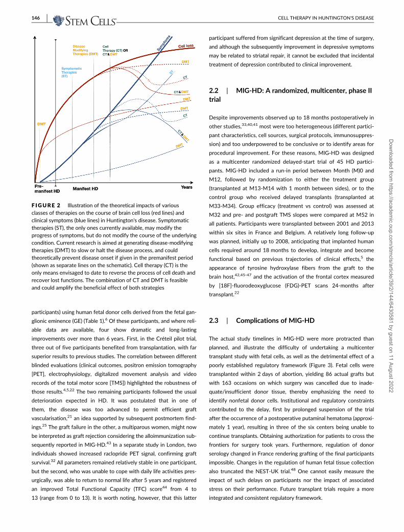
An important issue uncovered in MIG-HD was evidence of
alloimmunization despite participants undergoing a period of triple
immunosuppression.43 When cell therapy trials in HD started
(Table 1), the brain was considered to be an immune privileged site, a
concept since revised, notably with the discovery of a lymphatic sys-
tem in the central nervous system (CNS)52 which may play a role in
the pathophysiology of neurodegenerative diseases.53,54 Moreover,
cerebrovascular and blood-brain barrier impairments have been
reported in HD patients, which might facilitate the transmigration of
immune cells to the brain.55 The first case of graft rejection in the
brain was reported in MIG-HD43 where immunosuppression included
cyclosporine A, started 3 days preoperatively until 6 months after the
second transplantation, with prednisolone and azathioprine adminis-
tered from the day of surgery to 6 months after the second graft.
Graft rejection was diagnosed in the only participant not receiving
immunosuppressant as per protocol; this participant lost weight, col-
lapsed, and a hyposignal was found in the graft location on T1 mag-
netic resonance imaging (MRI). Class 1 human histocompatibility
antigen (HLA) antibodies specific to the fetal graft were detected both
in blood and cerebrospinal fluid. High doses of intravenous predniso-
lone resulted in regression of clinical and imaging signs with an almost
complete return to the presurgery state within 6 months. Following
this event, triple immunosuppression was maintained in the remaining
(20) participants for up to 1 year after the second graft. No further
graft rejection was observed, but in the 39 participants assessed dur-
ing the trial, HLA class 1 and 2 antibodies appeared in 34% of post-
transplant participants, being present in 9% of pretransplant women
(presumably due to pregnancy).7 HLA antibodies against transplant
were also detected by the Firenze group (in 37.5% of patients) with a
year immunosuppression34,35 and in the German branch of MIG-HD
(in 50% of patients) with 18 months immunosuppression,40 and differ-
ent immunosuppressive protocols (Table 1). Inflammation has been
reported in postmortems of both grafted HD and PD patients having
had immunosuppression, despite surviving grafts identified on MRI.
Within grafts, healthy regions (with internal organization similar to
normal striatum, expressing markers of MSNs and interneurons and
receiving cortical afferents) often coexisted with areas of apoptosis,47
astrocytic activation,25 and inflammation (cluster of differentiation
[CD]45) around or within grafts tissue and vasculature (CD3, CD68
and T-cell CD68 microglia, natural killers cells, CD8 T cytotoxic cells,
and CD4 T helper cells).29,40,42,46,47,56 Grafts generally lack vasculari-
zation compared to host tissue, and mutant huntingtin has been
reported in grafts, although evidence that it was located in donor,
rather than host, cells is still awaited.29,56
Additional parameters, that were not part of the MIG-HD initial
analysis plan, were sought to explain the efficacy failure and to inform
future trials. These included host characteristics, procedural data, vol-
ume injected, graft size after 20 months and presence of HLA anti-
bodies. Counterintuitively, a high number of tracks (>5 per side) was
associated with a poorer outcome. Although Paganini et al36 con-
cluded that the grafts slowed motor and cognitive decline, even in HD
patients with large ectopic tissue nodules in the frontal lobe, MIG-HD
showed that ectopic graft tissue was detrimental, calling for caution in
future trials.
It is unclear whether the methodological differences between the
pilot study and MIG-HD turned success into failure.4,5 For example,
reducing the delay between ipsilateral and contralateral transplant
surgeries to 1 month, compared to 1 year in the pilot, might have con-
stituted a more severe immunogenic challenge. HLA antibodies
searched 10 years after transplantation were not found in the pilot
148 CELL THERAPY IN HUNTINGTON'S DISEASED
ownloaded from
https://academic.oup.com
/stmcls/article/39/2/144/6430581 by guest on 11 August 2022

study but were present 3 years after transplants in MIG-HD. Although
knowledge is lacking in the field of intracerebral transplantation, in
that of solid organ transplants, the presence of HLA antibodies against
donor tissues is considered to indicate alloreactivity.57 Likewise, the
increased number of fetal samples per host and older gestational win-
dow from 7-9 to 8.5-12 weeks postconception may also have
increased the overall immunogenicity burden. Finally, moving from a
monocentric pilot study to a multicentric one in MIG-HD might have
compounded procedural variability. Notwithstanding intensive train-
ing, full standardization of surgical procedures was not achieved; the
surgeon adapting the intervention for each participant. We strongly
recommend full standardization and documentation of all procedures
in future studies and highlight suggested improvements in the next
sections.
3 | FUTURE CHALLENGES
3.1 | Donor cell product
The rationale for using fetal GE as donor tissue is the replacement of
MSNs, with the idea that these cells will make synaptic connections
with appropriate striatal targets and provide some reconstruction of
the neural circuitry. These cells have developed “naturally” and so are
the donor cell source most likely to be properly specified. There is evi-
dence of circuit reconstruction by fetal-derived cells in animal
studies,58 and also in humans where [18F]-FDG-PET signal increase in
the frontal lobes suggested that circuit reconstruction was the mecha-
nism underlying successful grafts.22
However, ultimately, the generation of donor cells from renew-
able sources, such as human pluripotent stem cells (hPSCs), will be
essential for transplantation to be an efficient therapy; fetal-derived
donor cells are a scarce resource and cannot be stored beyond a few
days, making quality control very difficult and presenting a major chal-
lenge in terms of surgical scheduling. Indeed, even if no difference in
cell processing was introduced between the successful pilot and MIG-
HD, in both trials viability assessment was not possible and may have
proved critical in ensuring graft survival in MIG-HD as discussed
above. Nevertheless, a nuanced understanding of the factors deter-
mining a successful fetal GE graft underpins our ability to produce
suitable hPSC-derived donor cells. For example, fetal grafts have tau-
ght us that the stage of precursor development at the time of trans-
plantation is critical (precursors need to be sufficiently committed to
an MSN phenotype but sufficiently immature to integrate well in the
host tissue12). The questions of whether a fetal graft should be
restricted to the lateral GE, in which the majority of MSNs develop, is
unresolved and related to consideration of optimal graft cell content;
human fetal GE contains not only MSN precursors, but a range of
neural and glial precursors, microglia and vascular cells and the contri-
bution of these none-MSN elements to fetal graft function have not
yet been determined.59 The question of whether fetal tissue should
be prepared as tissue pieces or cell suspension also remains unre-
solved; theoretically, a suspension graft may integrate better and may
be less immunogenic, as it probably contained fewer donor vascular
elements although the most promising clinical study to date employed
tissue pieces.4,5 However, this question may be less relevant for stem
cell-derived cells, which are likely to be suspension grafts and so
attract vascular elements from the host rather than donor (although
this will depend to some extent on the precise differentiation proto-
col). It is also possible that cell clusters or stem cell derived organoids
(delivered alone or associated with various biomaterials60) may prove
to be a useful delivery mechanism.
A variety of renewable cell sources are actively being explored as
donor cells for cell therapy. These include human embryonic stem
cells (hESCs), induced pluripotent stem cells (hiPSCs), fetal derived
neural stem cells, and directly induced stem cells (hiNSCs). As well as
addressing the critical issues of tissue availability and quality control,
these sources have other potential advantages over fetal cells and also
a number of drawbacks. These are outlined briefly below and a
detailed discussion of the pros and cons of specific cell types can be
found in Reference 61. There has also been considerable interest in
deriving donor cells from nonneural stem cells, such as mesenchymal
stem cells, for HD. Such cells have the capacity to secrete neuro-
protective, anti-apoptotic, immunomodulatory, and/or anti-
inflammatory molecules and may be engineered to produce and
release specific therapeutic molecules such as brain-derived neuro-
trophic factor (BDNF). These cells, and others also engineered to pro-
duce therapeutic molecules, are being explored for their
neuroprotective potential.14,15 However, there is little convincing evi-
dence that such cells differentiate into neuronal phenotypes suitable
for circuit reconstruction and so they will not be considered
further here.
hESCs are arguably the closest to clinical trial at the current time,
largely because they have been the subject of research for the longest
time and are thus underpinned by a large body of knowledge, added
to which good manufacturing practices (GMP)-grade cell lines are
already in existence, some of which have already been used in clinical
studies for other conditions. However, iPSC, and to some extent
iNSC, technology has been accelerating quickly and some iPSC lines
have entered clinical trials. iPSCs can be generated from adult somatic
tissues and so have the potential advantage of circumventing some
ethical issues associated with hESCs (namely the use of human blasto-
cysts). iNSCs are produced by directly trans-differentiating somatic
cells to a neural stem cell, rather than pluripotent, phenotype, with
the potential advantage of a lower risk of tumor formation,62 although
there have been fewer preclinical studies to date than for hESC or
hiPSCs exploring these cells for their cell therapy potential. There is
also the possibility of directly transdifferentiating host cells to another
phenotype, such as has been achieved for dopaminergic cells from
astrocytes, although this exciting technology is currently probably a
long way from clinical application.63,64
Stem cells and fetal cells have some challenges in common,
including cell viability on implantation, graft vascularization, graft
placement and distribution, the permissiveness of the host environ-
ment, and graft rejection (the latter is discussed further below). There
are also some challenges that apply to stem cell sources, but not to
BACHOUD-LÉVI ET AL. 149D
ownloaded from
https://academic.oup.com
/stmcls/article/39/2/144/6430581 by guest on 11 August 2022
fetal cells; in particular, the development of protocols to ensure the
presence of correctly specified target neural cells and the absence of
unwanted cell types. Fetal cells have the advantage of being
programmed through normal development whereas stem cell-derived
neural cells have to be artificially programmed.65,66 Indeed, preclinical
research using fetal striatal cells (which contain precursors for the full
complement of neuronal and glial components of the adult striatum)
underpins the use of stem cells by helping to define which cells are
critical for a fully functional graft.59 The elimination of cells with
tumerogenic potential is a much greater problems for stem cells com-
pared to fetal cells and can potentially be achieved through a range of
means, including adjusting the protocol and timing of transplantation
to ensuring complete differentiation and elimination of proliferating
cells,66 and cell sorting strategies.67 Through in vitro proliferation,
stem cells are also subject to quality control issues, in particular, the
acquisition of karyotype and genetic abnormalities, batch to batch
variation and between line differences in response to differentiation
factors. These challenges will need to be addressed for safe and opti-
mum clinical application of stem cell derived products.
iPSCs and iNSCs also have the potential advantage of providing a
source of autologous donor cells (with or without correction of any
genetic abnormality),68 which could circumvent the need for immuno-
suppression. However, generating GMP-grade cells on an individual
basis is currently extremely costly and thus not currently suitable for
widespread application, and the immune tolerance to autologous iPSC
grafts may not be as straightforward as originally perceived, as it has
been demonstrated that autologous primate iPSCs still induce an
immune reaction after grafting into the CNS, albeit at a lower level
than an allograft.69 A challenge for both iPSCs and iNSCs is that they
are generated through genetic manipulations, which add an additional
layer of regulatory scrutiny in terms of clinical use and additional cost.
In addition, their use in autologous grafting may require correction of
a host mutation prior to use, as is the case for HD. The extent to
which this would be necessary is currently uncertain, but it would
clearly incur additional steps that would further increase the regula-
tory burden and cost. However, putting the regulatory burden to one
side, the relative ease with which stem cells can be genetically manip-
ulated opens up possibilities that are much more difficult to achieve
with primary fetal cells; for example, the vision of producing “universal
cells” that are invisible to the immune system through CRISPR/
Cas9-mediated deletion of major histocompatibility genes.70 How-
ever, the CRISPR/Cas9 system is not without its dangers, such as its
autonomous evolution into the brain which has led to the KamiCas9
construct designed by Merienne et al71 to avoid section of additional
genes beyond the targeted one. This construct is a self-inactivating
editing system that achieved transient expression of the Cas9 protein
in mice models. It might take years to achieve safety in patients.
Several recent studies have reported some functional improve-
ments in HD animals transplanted with hPSC-derived striatal-like
precursors,61,72-76 although we believe it is important to demonstrate
in animals that these approaches are at least as effective as implanta-
tion of fetal-derived cells before progressing to clinical trials. This is
highly likely to necessitate implantation in animal models of HD.
3.2 | Animal models
Animal models of HD remain an important experimental tool, in par-
ticular for assessing donor sources that may replace fetal-derived
cells, as outlined above, but it is important to understand their limita-
tions and also to continue the quest for models that better represent
the relevant elements of the human situation. Lesion models can
reproduce the striatal, and sometimes even the cortical, cell loss seen
in HD patients, as well as mimicking the behavioral and metabolic
changes.77 Such models in rodents have been essential for under-
standing the biology of grafts, but they do not replicate the progressive
cell loss seen in HD, nor reliably replicate the extrastriatal damage.78 A
variety of genetic models exist in mice, but these have so far proven
problematic for assessing striatal grafts; they have substantially longer
CAG repeat expansions than those seen in adult onset HD and do not
accurately reflect the anatomy of changes of the CNS.39 Rat models
with CAG repeats much closer to that of adult onset HD are being opti-
mized and may provide more relevant models with which to work.79 It
is likely that these relatively cheap and accessible rodent models will
continue to provide the cornerstone for experimental work addressing
both mechanistic and procedural questions, and perhaps even
alloimmunization issues through the use of humanized mice.80
Large animal models are significantly more costly and complex,
but some translationally important issues would be better addressed
in such models.81 For example, refining cell distribution, assessing the
capacity of cells to extend processes in a brain with anatomical dis-
tances closer to a human brain, testing the efficacy of grafts in animals
with more human-like behavioral repertoires, and working out the
details of assessment tools such as in vivo imaging to track graft size,
all fall into this category. Large animal models also facilitate long-term
studies, potentially over several years, to more comprehensively
assess safety and efficacy of treatments. Both lesion and genetic
models in larger animals are becoming increasingly available.81 For
example, some transgenic minipigs show progressive neu-
rodegeneration as well as motor, cognitive and behavioral decline and
nonhuman primate lesion models, although limited by ethical concerns
and legal regulations, have been used to assess imaging strategies and
address issues of alloimmunization.69
3.3 | Protocol design
Heterogeneity is one of the hallmarks of HD, both in its clinical mani-
festations and rates of progression. Specific phenotypes are linked to
patterns of brain alterations,82,83 gender,84 and genetic factors.85-87
Variability, which results in the need to recruit large numbers of
patients and for prolonged periods of follow-up, is at the forefront of
obstacles to be lifted for future trials.8,88 The problem is heightened in
trials aimed at restoring atrophied tissues where the response to the
transplant cannot be readily predicted and might encompass improve-
ment in a wide range of symptoms, thus necessitating a broad-
spectrum evaluation of patients to meet safety and efficacy
requirements.
150 CELL THERAPY IN HUNTINGTON'S DISEASED
ownloaded from
https://academic.oup.com
/stmcls/article/39/2/144/6430581 by guest on 11 August 2022
One way to address these challenges is the refinement and auto-
mation (and/or video recording) of outcome measures to reduce rater
bias. Participants in MIG-HD were assessed using an adapted version
of the CAPIT-HD (Core Assessment Protocol for Intracerebral Trans-
plantation in HD) protocol89; which was heavily reliant on the Unified
Huntington's disease Rating Scale (UHDRS). In retrospect, these
assessments are unwieldy and lack sensitivity. Likewise, the use of the
TMS as primary endpoint was also suboptimal as the cognitive and
behavioral disturbances are now recognized as the major drivers of
poor function in HD.90 When this study commenced, no cognitive
assessments were universally accepted except the UHDRS cognitive
score, which also lacks sensitivity, and TFC was discounted consider-
ing the large numbers of participants required to detect change. The
global composite score cUHDRS, combining the TFC, TMS, Stroop
and Symbol Digit Modalities Test, is currently used in trials of anti-
sense oligonucleotides and constitutes a first step toward improve-
ment.88 Yet, objective digitized tasks, such as QMotor which has
already been used in clinical studies,91 might allow evaluation and
comparison of many functions within in a short time and eliminate
rater bias. Sensitive digitalized measures of cognition, suitable for
intracranial intervention studies including cell therapy, are under
validation in French, English and German thanks to the Repair-HD
program (eg, Selfcog).
Although electrophysiological assessments were optional in the
CAPIT-HD protocol, their inclusion in our pilot study4 allowed us, in
conjunction with clinical and PET assessments, to identify transplants
benefits; similar improvements never having been previously reported
in longitudinal assessment of either observational cohorts92 or phar-
macological trials.21 However, duration, availability, and lack of full
standardization of electrophysiology assessment might hamper its use
in multicenter trials. We would recommend using it only in the future
open label trial, in single center or after full homogenization of the
procedure by expert investigators. It will complete the panel of argu-
ments before an eventual dissemination of the transplant procedure.
So far, brain imaging has involved the use of structural MRI and
PET. [18F]-FDG-PET was used in the early days of cell transplants tri-
als22,33,40,41 but could not disentangle edema or graft rejection from
functional grafts. [11C]raclopride PET is specific for dopamine D2
receptors, which are expressed by MSNs, but is affected by neurolep-
tics which are frequently prescribed in HD for involuntary movements
and behavioral disorders. Thus, it is less suitable for participants
with moderate/advanced disease. New tracers, such as ligands to
phosphodiesterase-10A, an enzyme expression by striatal MSN whose
reduction is one of the earliest biochemical changes in HD93 may prove
to be a reliable indicator of disease progression.94 Likewise future trials
should capitalize on volumetric MRI and connectivity measures.95,96
Additional trial design measures might include enrichment strate-
gies to recruit patients predicted to have faster rates of progression
based on neuroimaging analyses, genetic polymorphisms, clinical pro-
files, or retest effects85,97-100 to increase the likelihood of demon-
strating treatment efficacy with small cohorts over shorter durations.
Identification of markers to select patients better able to tolerate
grafts (eg, with a low risk of graft rejection) or to ensure compatibility
between the host and donor cells are needed.6,101 The inclusion of
sham surgery is another important consideration, but may not be
deemed ethical in early stage trials. Moreover, failed grafts (rejected/
too small) may be more informative comparators than partial burr
holes sham cases, in which associated experimental procedures are
not undertaken.
Stage of disease is important; advanced HD patients were more
likely to develop subdural hematomas45 and to have defects of
vascularization,25 compromising transplant development.21 Improve-
ments in the surgical procedures in MIG-HD might have solved some
of these issues7 but the question of efficacy in more advanced
patients remains. The complexity of patient profiles makes the inter-
pretation of transplant response extremely complex. The use of new
outcome measures and biomarkers combined with innovative statisti-
cal methods, including machine learning approaches able to analyze
high-dimensional sources of data and enable inferences from fewer
samples, all have the potential to improve patient stratification for
inclusion in trials and/or to identify subsets of patients with differen-
tial response to treatment.102,103 They will improve the design and
evaluation of cell therapy strategies in a cost-effective and efficient
manner.
3.4 | Surgical procedures
Adverse events in transplant surgery are relatively common across all
disciplines. A key adverse event for neural transplant surgery is intra-
cranial bleeding. For example, Freeman et al45 reported 26% subdural
hematomas following cell transplants in HD; subdural hematomas
being anyway more frequent in HD than in control populations. How-
ever, reducing the number of tracts in participants with marked atro-
phy and providing hyperhydration and 48 hours bed confinement
abolished further subdural hematomas in MIG-HD.7 Furthermore,
endoscopic surgery to cauterize a choroid cyst successfully rescued
one participant, who continued to enjoy improvement associated with
a surviving graft. Nevertheless, better control of surgical and perioper-
ative procedures and the development of improved cell-delivery
devices will be critical for future transplant studies. Systematic video
records of surgery should improve homogenization of procedures.
3.5 | Alloimmunization
The first case of graft rejection43 and the identification of HLA anti-
bodies against transplants in trials using various immunosuppressive
protocols34,35,40 suggest that such responses to the graft are not
linked to inadequacy of a specific immunosuppressive protocol, but
rather related to poor understanding of graft-host responses in the
CNS. As outlined above, stem cell-derived products may provide bet-
ter methods for circumventing immune rejection, although they are
not without their own practical and regulatory problems. In MIG-HD
there was no clear association between the presence of functional
grafts and graft-directed HLA antibodies. However, since
BACHOUD-LÉVI ET AL. 151D
ownloaded from
https://academic.oup.com
/stmcls/article/39/2/144/6430581 by guest on 11 August 2022
alloimmunization was not anticipated in MIG-HD, HLA antibodies
were not systematically measured and techniques were not uniform
across centers. Thus, conclusions cannot yet be drawn as to the
importance of graft-derived antibodies nor how this should affect
future trials. Coordinated analysis of immune status will be essential
in future trials, but for now, donor/host HLA matching may provide a
partial solution in future stem cell trials.104 If fetal cells are still to be
used, crossmatching of alloantibodies and antigens could be
attempted prior to transplantation.
4 | CELL TRANSPLANTS IN THE FUTUREHD THERAPEUTIC LANDSCAPE
The results of MIG-HD, even if they shed light on the way forward,
undoubtedly constitute a disappointment for the field, and probably
underlie the extreme points of view of some researchers who con-
demn all further research in the field.105 Yet these same researchers
should remember the history of transplantation in PD, in which they
participated, and in which condition (in contrast to HD) some mean-
ingful treatments have been available for many years.
The rationale for transplantation in PD is to restore dopamine
levels in the striatum through implantation of dopamine-releasing
cells. Between the 1970s and the 2000s, open pilot studies reported
variables results with successful clinical outcome of ventral mesence-
phalic grafts in some patients that correlated with [18F]-FDG-PET
increases and postmortem evidence of long-term cell survival,106-108
whereas other studies showed small/no benefit (reviewed in Refer-
ence 109). The phase II randomized trials that followed56,110 were
negative and resulted in this area of research shutting down for
around 15 years. The reason for these trials failing was likely related
to poor cell viability linked to their storage, and alloimmunization of
transplants; the first trial did not include immunosuppressants and the
second only 6 months, and autopsies revealed inflammation around
and within the grafts.110 Endpoints were also regarded as
subjective,56,110 thereby justifying the use of shams and, as an exam-
ple, nourished a discussion on the choice of clinical vs imaging end-
points. However, reanalysis of surviving transplants in these and other
previous studies demonstrated that the transplant efficacy was often
delayed beyond the end of clinical study and thus not recognized in
the original report.109 Variability in procedures and patient selection
also contributed to the disparity of the results. Following these
observations, a European fetal cell trial (TRANSEURO -
NCT01898390) was established in 2013, attempting to respond to
the deficiencies recognized in previous trials. Transplants in HD
(albeit only in four patients) have so far produced clinical improve-
ment beyond any other therapeutic approach. Thus, to avoid repeti-
tion of the PD scenario, with a long and damaging interruption of
research activities, this review attempts to identify the critical trial
design and technical factors responsible for graft failure in order to
facilitate progress in this area. As outlined above, it is essential to
note that many of these factors are pertinent to stem cell-derived,
as well as fetal, donor cell products.
5 | CONCLUSIONS
MIG-HD failed to demonstrate a benefit associated with transplan-
tation. Nevertheless, the benefits reported in hundreds of animal
studies and in human pilot trials4,32 in addition to areas for improve-
ment identified in MIG-HD encourage further research into cell
therapy for HD. MIG-HD should be understood in the context of
having started in 2001. Since then, there have been considerable
advances in knowledge and technical development, including transplant
and immunosupression regimens, stem cell differentiation protocols,
and development of more sensitive objective outcome measures.
Disease-modifying treatments, such as huntingtin lowering therapies,
could slow or even prevent the disease from developing, although their
degree of efficacy and long term adverse impacts are yet to be deter-
mined. However, cell therapy, which could be given alongside a
disease-slowing therapy (Figure 2), is the only approach currently
focused on structural and (hopefully) functional restoration. Until we
have a cure applicable to the whole HD community, it seems reasonable
to continue exploring a broad range of therapeutic options. We there-
fore propose that the door is left open for future cell therapy research.
We anticipate progress being an iterative process requiring both
preclinical and clinical work. The availability of stem cell-derived donor
cells has the potential to dramatically change the landscape by permit-
ting better control of the quantity and homogeneity of the donor cell
product, but it is essential that their clinical translation is based on
sound evidence. Indeed, we and others have established an interna-
tional platform (Stem Cells for HD, www.sc4hd.org) to review such
evidence and produce guidance, and we encourage great caution
around the use of private transplant clinics worldwide which offer cell
therapy treatments without clear evidence of efficacy or guarantees
for patients safety.
ACKNOWLEDGMENTS
Anne-Catherine Bachoud-Lévi's team is supported by NeurATRIS
ANR-11-INBS-0011/Agence Nationale de la Recherche (French
National Research Agency), ANR-17-EURE-0017, and the Henri-
Mondor Hospital National Reference Centre for Huntington's disease
(Ministry of Health). Anne-Catherine Bachoud-Lévi: Consulting and
Advisory Board Membership with honoraria: Roche; Grants and
Research: investment for the future ANR grant (Neuratris, Front EUR),
National Center of Reference for Huntington's disease (DGOS, Minis-
try of Health), PHRCs (DRCI grants); Intellectual Property Rights:
Cognitive assessments (SelfCog, CATEX, CALAP). Anne Rosser:
Employment with Cardiff University; Consultant/Advisory role for
Roche; research funding from EHDN/MRC UK/HCRW/EC/CARE/
Jacques & Gloria Gossweiler Foundation; and other from EHDN/
Triplet. Renaud Massart declared no potential conflicts of interest.
CONFLICT OF INTEREST
The authors declared no potential conflicts of interest.
AUTHOR CONTRIBUTIONS
A.-C.B.-L., R.M., A.R.: wrote the manuscript.
152 CELL THERAPY IN HUNTINGTON'S DISEASED
ownloaded from
https://academic.oup.com
/stmcls/article/39/2/144/6430581 by guest on 11 August 2022

DATA AVAILABILITY STATEMENT
Data sharing is not applicable to this article as no new data were cre-
ated or analyzed in this study.
REFERENCES
1. Bates GP, Dorsey R, Gusella JF, et al. Huntington disease. Nat Rev
Dis Primers. 2015;1:15005.
2. Bachoud-Lévi A-C, Ferreira J, Massart R, et al. International guide-
lines for the treatment of Huntington's disease. Front Neurol. 2019;
10:710.
3. Tabrizi SJ, Ghosh R, Leavitt BR. Huntingtin lowering strategies for
disease modification in Huntington's disease. Neuron. 2019;102:899.
4. Bachoud-Lévi AC, Rémy P, Nguyen JP, et al. Motor and cognitive
improvements in patients with Huntington's disease after neural
transplantation. Lancet. 2000;356:1975-1979.
5. Bachoud-Lévi A-C, Gaura V, Brugières P, et al. Effect of fetal neural
transplants in patients with Huntington's disease 6 years after sur-
gery: a long-term follow-up study. Lancet Neurol. 2006;5:303-309.
6. Bachoud-Lévi A-C. From open to large-scale randomized cell trans-
plantation trials in Huntington's disease: lessons from the multi-
centric intracerebral grafting in Huntington's disease trial (MIG-HD)
and previous pilot studies. Prog Brain Res. 2017;230:227-261.
7. Bachoud-Lévi A-C, on behalf the Multicentric Intracerebral Grafting
in Huntington's Disease Group. Human fetal cell therapy in
Huntington's disease: a randomized, multicenter, phase II trial. Mov
Disord. 2020;35:1323-1335.
8. Tabrizi SJ, Reilmann R, Roos RAC, et al. Potential endpoints for clini-
cal trials in premanifest and early Huntington's disease in the
TRACK-HD study: analysis of 24 month observational data. Lancet
Neurol. 2012;11:42-53.
9. Douaud G, Gaura V, Ribeiro M-J, et al. Distribution of grey matter
atrophy in Huntington's disease patients: a combined ROI-based and
voxel-based morphometric study. Neuroimage. 2006;32:1562-1575.
10. Precious SV, Zietlow R, Dunnett SB, Kelly CM, Rosser AE. Is there a
place for human fetal-derived stem cells for cell replacement therapy
in Huntington's disease? Neurochem Int. 2017;106:114-121.
11. McColgan P, Gregory S, Seunarine KK, et al. Brain regions showing
white matter loss in Huntington's disease are enriched for synaptic
and metabolic genes. Biol Psychiatry. 2018;83:456-465.
12. Dunnett SB, Rosser AE. Cell-based treatments for Huntington's dis-
ease. Int Rev Neurobiol. 2011;98:483-508.
13. Caron NS, Dorsey ER, Hayden MR. Therapeutic approaches to Hun-
tington disease: from the bench to the clinic. Nat Rev Drug Discov.
2018;17:729-750.
14. Deng P, Torrest A, Pollock K, et al. Clinical trial perspective for adult
and juvenile Huntington's disease using genetically-engineered mes-
enchymal stem cells. Neural Regen Res. 2016;11:702-705.
15. Kim HS, Jeon I, Noh J-E, et al. Intracerebral transplantation of
BDNF-overexpressing human neural stem cells (HB1.F3.BDNF) pro-
motes migration, differentiation and functional recovery in a rodent
model of Huntington's disease. Exp Neurobiol. 2020;29:130-137.
16. Cho IK, Hunter CE, Ye S, Pongos AL, Chan AWS. Combination of
stem cell and gene therapy ameliorates symptoms in Huntington's
disease mice. NPJ Regen Med. 2019;4:7.
17. Philpott L, Kopyov OV, Lee AJ, Jacques S. Neuropsychological func-
tioning following fetal striatal transplantation in Huntington's cho-
rea: three case presentations. Cell Transplant. 1997;6(3):203-212.
https://doi.org/10.1016/s0963-6897(97)00028-6.
18. Kopyov OV, Jacques S, Lieberman A, Duma CM, Eagle KS. Safety of
intrastriatal neurotransplantation for Huntington's disease patients.
Exp Neurol. 1998;149(1):97-108. https://doi.org/10.1006/exnr.
1997.6685.
19. Keene CD, Chang RC, Leverenz JB, et al. A patient with
Huntington's disease and long-surviving fetal neural transplants that
developed mass lesions. Acta Neuropathol. 2009;117(3):329-338.
https://doi.org/10.1007/s00401-008-0465-0.
20. Ross BD, Hoang TQ, Stefan B, et al. In vivo magnetic resonance
spectroscopy of human fetal neural transplants. NMR Biomed. 1999;
12(4):221-236. https://doi.org/10.1002/(sici)1099-1492(199906)
12:4<221::aid-nbm582>3.0.co;2-q.
21. Bachoud-Lévi A-C, Hantraye P, Peschanski M. Fetal neural grafts for
Huntington's disease: a prospective view. Mov Disord. 2002;17:
439-444.
22. Gaura V, Bachoud-Lévi A-C, Ribeiro M-J, et al. Striatal neural
grafting improves cortical metabolism in Huntington's disease
patients. Brain. 2004;127:65-72.
23. Bachoud-Lévi A-C, Bourdet C, Brugières P, et al. Safety and tolera-
bility assessment of intrastriatal neural allografts in five patients with
Huntington's disease. Exp Neurol. 2000;161(1):194-202. https://doi.
org/10.1006/exnr.1999.7239.
24. Douaud G, Behrens TE, Poupon C, et al. In vivo evidence for the
selective subcortical degeneration in Huntington's disease.
NeuroImage. 2009;46(4):958-966. https://doi.org/10.1016/j.
neuroimage.2009.03.044.
25. Cisbani G, Freeman TB, Soulet D, et al. Striatal allografts in patients
with Huntington's disease: impact of diminished astrocytes and vas-
cularization on graft viability. Brain. 2013;136:433-443.
26. Cicchetti F, Lacroix S, Cisbani G, et al. Mutant huntingtin is present
in neuronal grafts in Huntington disease patients. Ann Neurol. 2014;
76(1):31-42. https://doi.org/10.1002/ana.24174.
27. Hauser RA, Furtado S, Cimino CR, et al. Bilateral human fetal striatal
transplantation in Huntington's disease. Neurology. 2002;58(5):687-
695. https://doi.org/10.1212/wnl.58.5.687.
28. Furtado S, Sossi V, Hauser RA, et al. Positron emission tomography
after fetal transplantation in Huntington's disease. Ann Neurol. 2005;
58(2):331-337. https://doi.org/10.1002/ana.20564.
29. Maxan A, Mason S, Saint-Pierre M, et al. Outcome of cell suspension
allografts in a patient with Huntington's disease. Ann Neurol. 2018;
84:950-956.
30. Rosser AE. Unilateral transplantation of human primary fetal tissue
in four patients with Huntington's disease: NEST-UK safety report
ISRCTN no 36485475. J Neurol Neurosurg Psychiatry. 2002;73(6):
678-685. https://doi.org/10.1136/jnnp.73.6.678.
31. Barker RA, Mason SL, Harrower TP, et al. The long-term safety and
efficacy of bilateral transplantation of human fetal striatal tissue in
patients with mild to moderate Huntington's disease. J Neurol Neuro-
surg Psychiatry. 2013;84(6):657-665. https://doi.org/10.1136/jnnp-
2012-302441.
32. Reuter I, Tai YF, Pavese N, et al. Long-term clinical and positron
emission tomography outcome of fetal striatal transplantation in
Huntington's disease. J Neurol Neurosurg Psychiatry. 2008;79:
948-951.
33. Porfirio B, Paganini M, Mazzanti B, et al. Donor-specific anti-HLA
antibodies in Huntington's disease recipients of human fetal striatal
grafts. Cell Transplant. 2015;24:811-817.
34. Gallina P, Paganini M, Lombardini L, et al. Development of human
striatal anlagen after transplantation in a patient with Huntington's
disease. Exp Neurol. 2008;213:241-244.
35. Gallina P, Paganini M, Lombardini L, et al. Human striatal neuroblasts
develop and build a striatal-like structure into the brain of
Huntington's disease patients after transplantation. Exp Neurol.
2010;222:30-41.
36. Paganini M, Biggeri A, Romoli AM, et al. Fetal striatal grafting slows
motor and cognitive decline of Huntington's disease. J Neurol Neuro-
surg Psychiatry. 2014;85:974-981.
37. Gallina P, Paganini M, Biggeri A, et al. Human striatum remodelling
after neurotransplantation in Huntington's disease. Stereotactic
Funct Neurosurg. 2014;92(4):211-217. https://doi.org/10.1159/
000360583.
BACHOUD-LÉVI ET AL. 153D
ownloaded from
https://academic.oup.com
/stmcls/article/39/2/144/6430581 by guest on 11 August 2022

38. Mascalchi M, Diciotti S, Paganini M, et al. Large-sized fetal striatal
grafts in Huntington's disease do stop growing. Long-term monitor-
ing in the florence experience. PLoS Curr. 2014;6:ecurrents.hd.
c0ad575f12106c38f9f5717a8a7d05ae. https://doi.org/10.1371/
currents.hd.c0ad575f12106c38f9f5717a8a7d05ae.
39. Rangel-Barajas C, Rebec GV. Overview of Huntington's disease
models: neuropathological, molecular, and behavioral differences.
Curr Protoc Neurosci. 2018;83:e47.
40. Krebs SS, Trippel M, Prokop T, et al. Immune response after striatal
engraftment of fetal neuronal cells in patients with Huntington's dis-
ease: consequences for cerebral transplantation programs. Clin Exp
Neuroimmunol. 2011;2:25-32.
41. Lopez WOC, Nikkhah G, Schültke E, Furlanetti L, Trippel M. Stereo-
tactic planning software for human neurotransplantation: suitability
in 22 surgical cases of Huntington's disease. Restor Neurol Neurosci.
2014;32:259-268.
42. Capetian P, Knoth R, Maciaczyk J, et al. Histological findings on fetal
striatal grafts in a Huntington's disease patient early after transplan-
tation. Neuroscience. 2009;160:661-675.
43. Krystkowiak P, Gaura V, Labalette M, et al. Alloimmunisation to
donor antigens and immune rejection following foetal neural grafts
to the brain in patients with Huntington's disease. PLoS One. 2007;2:
e166.
44. Shoulson I. Huntington disease: functional capacities in patients
treated with neuroleptic and antidepressant drugs. Neurology. 1981;
31:1333-1335.
45. Freeman TB, Cicchetti F, Bachoud-Lévi AC, Dunnett SB. Technical
factors that influence neural transplant safety in Huntington's dis-
ease. Exp Neurol. 2011;227:1-9.
46. Keene CD, Sonnen JA, Swanson PD, et al. Neural transplantation in
Huntington disease: long-term grafts in two patients. Neurology.
2007;68:2093-2098.
47. Cicchetti F, Saporta S, Hauser RA, et al. Neural transplants in
patients with Huntington's disease undergo disease-like neuronal
degeneration. Proc Natl Acad Sci U S A. 2009;106:12483-12488.
48. Rosser AE, Kelly CM, Dunnett SB. Cell transplantation for
Huntington's disease: practical and clinical considerations. Future
Neurol. 2010;6:45-62.
49. Novellino F, Saccà V, Donato A, et al. Innate immunity: a common
denominator between neurodegenerative and neuropsychiatric dis-
eases. Int J Mol Sci. 2020;21:1115.
50. Cisbani G, Cicchetti F. The fate of cell grafts for the treatment of
Huntington's disease: the post-mortem evidence. Neuropathol Appl
Neurobiol. 2014;40:71-90.
51. Gosset P, Maxan A, Alpaugh M, et al. Evidence for the spread of
human-derived mutant huntingtin protein in mice and non-human
primates. Neurobiol Dis. 2020;141:104941.
52. Louveau A, Smirnov I, Keyes TJ, et al. Structural and functional fea-
tures of central nervous system lymphatic vessels. Nature. 2015;
523:337-341.
53. Zou W, Pu T, Feng W, et al. Blocking meningeal lymphatic drainage
aggravates Parkinson's disease-like pathology in mice over-
expressing mutated α-synuclein. Transl Neurodegener. 2019;8:7.54. Da Mesquita S, Louveau A, Vaccari A, et al. Functional aspects of
meningeal lymphatics in ageing and Alzheimer's disease. Nature.
2018;560:185-191.
55. Drouin-Ouellet J, Sawiak SJ, Cisbani G, et al. Cerebrovascular and
blood-brain barrier impairments in Huntington's disease: potential
implications for its pathophysiology. Ann Neurol. 2015;78:160-177.
56. Olanow CW, Goetz CG, Kordower JH, et al. A double-blind con-
trolled trial of bilateral fetal nigral transplantation in Parkinson's dis-
ease. Ann Neurol. 2003;54:403-414.
57. McKenna RM, Takemoto SK, Terasaki PI. Anti-HLA antibodies after
solid organ transplantation. Transplantation. 2000;69:319-326.
58. Rosser AE, Dunnett SB. Neural transplantation in patients with
Huntington's disease. CNS Drugs. 2003;17:853-867.
59. Reddington AE, Rosser AE, Dunnett SB. Differentiation of pluripo-
tent stem cells into striatal projection neurons: a pure MSN fate may
not be sufficient. Front Cell Neurosci. 2014;8:398.
60. Newland B, Dowd E, Pandit A. Biomaterial approaches to gene ther-
apies for neurodegenerative disorders of the CNS. Biomater Sci.
2013;1:556-576.
61. Connor B. Concise review: the use of stem cells for understanding
and treating Huntington's disease. STEM CELLS. 2018;36:146-160.
62. Erharter A, Rizzi S, Mertens J, Edenhofer F. Take the shortcut -
direct conversion of somatic cells into induced neural stem cells and
their biomedical applications. FEBS Lett. 2019;593:3353-3369.
63. Dunnett SB, Rosser AE. Reprogramming the diseased brain. Nat Bio-
technol. 2017;35:426-428.
64. Rivetti di Val Cervo P, Romanov RA, Spigolon G, et al. Induction of
functional dopamine neurons from human astrocytes in vitro and
mouse astrocytes in a Parkinson's disease model. Nat Biotechnol.
2017;35:444-452.
65. Li M, Rosser AE. Pluripotent stem cell-derived neurons for transplan-
tation in Huntington's disease. Prog Brain Res. 2017;230:263-281.
66. Fan Y, Winanto, Ng S-Y. Replacing what's lost: a new era of stem cell
therapy for Parkinson's disease. Transl Neurodegener. 2020;9:2.
67. Precious SV, Kelly CM, Allen ND, et al. Can manipulation of differen-
tiation conditions eliminate proliferative cells from a population of
ES cell-derived forebrain cells? Neurogenesis (Austin, TX). 2016;3:
e1127311.
68. Schweitzer JS, Song B, Herrington TM, et al. Personalized iPSC-
derived dopamine progenitor cells for Parkinson's disease. N Engl J
Med. 2020;382:1926-1932.
69. Aron Badin R, Bugi A, Williams S, et al. MHC matching fails to pre-
vent long-term rejection of iPSC-derived neurons in non-human pri-
mates. Nat Commun. 2019;10:4357.
70. Deuse T, Hu X, Gravina A, et al. Hypoimmunogenic derivatives of
induced pluripotent stem cells evade immune rejection in fully
immunocompetent allogeneic recipients. Nat Biotechnol. 2019;37:
252-258.
71. Merienne N, Vachey G, de Longprez L, et al. The self-inactivating
KamiCas9 system for the editing of CNS disease genes. Cell Rep.
2017;20:2980-2991.
72. Ma L, Hu B, Liu Y, et al. Human embryonic stem cell-derived GABA
neurons correct locomotion deficits in quinolinic acid-lesioned mice.
Cell Stem Cell. 2012;10:455-464.
73. Besusso D, Schellino R, Boido M, et al. Stem cell-derived human
striatal progenitors innervate striatal targets and alleviate sensorimo-
tor deficit in a rat model of Huntington disease. Stem Cell Rep. 2020;
14:876-891.
74. Adil MM, Gaj T, Rao AT, et al. hPSC-derived striatal cells generated
using a scalable 3D hydrogel promote recovery in a Huntington dis-
ease mouse model. Stem Cell Rep. 2018;10:1481-1491.
75. Arber C, Precious SV, Cambray S, et al. Activin A directs striatal pro-
jection neuron differentiation of human pluripotent stem cells.
Development. 2015;142:1375-1386.
76. Yoon Y, Kim HS, Jeon I, et al. Implantation of the clinical-grade
human neural stem cell line, CTX0E03, rescues the behavioral and
pathological deficits in the quinolinic acid-lesioned rodent model of
Huntington's disease. STEM CELLS. 2020;38:936-947.
77. Lavisse S, Williams S, Lecourtois S, et al. Longitudinal characteriza-
tion of cognitive and motor deficits in an excitotoxic lesion model of
striatal dysfunction in non-human primates. Neurobiol Dis. 2019;130:
104484.
78. Reidling JC, Relaño-Ginés A, Holley SM, et al. Human neural stem
cell transplantation rescues functional deficits in R6/2 and Q140
Huntington's disease mice. Stem Cell Rep. 2018;10:58-72.
154 CELL THERAPY IN HUNTINGTON'S DISEASED
ownloaded from
https://academic.oup.com
/stmcls/article/39/2/144/6430581 by guest on 11 August 2022
79. Carreira JC, Jahanshahi A, Zeef D, et al. Transgenic rat models of
Huntington's disease. Curr Top Behav Neurosci. 2015;22:135-147.
80. Shultz LD, Keck J, Burzenski L, et al. Humanized mouse models of
immunological diseases and precision medicine. Mamm Genome.
2019;30:123-142.
81. Howland DS, Munoz-Sanjuan I. Mind the gap: models in multiple
species needed for therapeutic development in Huntington's dis-
ease. Mov Disord. 2014;29:1397-1403.
82. Garcia-Gorro C, Llera A, Martinez-Horta S, et al. Specific patterns of
brain alterations underlie distinct clinical profiles in Huntington's dis-
ease. Neuroimage Clin. 2019;23:101900.
83. Rosas HD, Salat DH, Lee SY, et al. Cerebral cortex and the clinical
expression of Huntington's disease: complexity and heterogeneity.
Brain. 2008;131:1057-1068.
84. Zielonka D, Marinus J, Roos RAC, et al. The influence of gender on
phenotype and disease progression in patients with Huntington's
disease. Parkinsonism Relat Disord. 2013;19:192-197.
85. de Diego-Balaguer R, Schramm C, Rebeix I, et al. COMT Val158Met
polymorphism modulates Huntington's disease progression. PLoS
One. 2016;11:e0161106.
86. Ciosi M, Maxwell A, Cumming SA, et al. A genetic association study
of glutamine-encoding DNA sequence structures, somatic CAG
expansion, and DNA repair gene variants, with Huntington disease
clinical outcomes. EBioMedicine. 2019;48:568-580.
87. Moss DJH, Pardiñas AF, Langbehn D, et al. Identification of genetic
variants associated with Huntington's disease progression: a
genome-wide association study. Lancet Neurol. 2017;16:701-711.
88. Schobel SA, Palermo G, Auinger P, et al. Motor, cognitive, and func-
tional declines contribute to a single progressive factor in early HD.
Neurology. 2017;89:2495-2502.
89. Quinn N, Brown R, Craufurd D, et al. Core assessment program for
intracerebral transplantation in Huntington's disease (CAPIT-HD).
Mov Disord. 1996;11:143-150.
90. Glidden AM, Luebbe EA, Elson MJ, et al. Patient-reported impact of
symptoms in Huntington disease: PRISM-HD. Neurology. 2020;94:
e2045-e2053.
91. Reilmann R, Schubert R. Motor outcome measures in Huntington
disease clinical trials. Handb Clin Neurol. 2017;144:209-225.
92. Lefaucheur J-P, Ménard-Lefaucheur I, Maison P, et al. Electrophysio-
logical deterioration over time in patients with Huntington's disease.
Mov Disord. 2006;21:1350-1354.
93. Wilson H, Politis M. Molecular imaging in Huntington's disease. Int
Rev Neurobiol. 2018;142:289-333.
94. Russell DS, Jennings DL, Barret O, et al. Change in PDE10 across
early Huntington disease assessed by [18F]MNI-659 and PET imag-
ing. Neurology. 2016;86:748-754.
95. Mangin J-F, Rivière D, Duchesnay E, et al. Neocortical morphometry
in Huntington's disease: indication of the coexistence of abnormal
neurodevelopmental and neurodegenerative processes. Neuroimage
Clin. 2020;26:102211.
96. Pini L, Youssov K, Sambataro F, Bachoud-Levi AC, Vallesi A,
Jacquemot C. Striatal connectivity in pre-manifest Huntington's dis-
ease is differentially affected by disease burden. Eur J Neurol. 2020;
27:2147-2157.
97. Polosecki P, Castro E, Rish I, et al. Resting-state connectivity strat-
ifies premanifest Huntington's disease by longitudinal cognitive
decline rate. Sci Rep. 2020;10:1252.
98. Kuan WL, Kasis A, Yuan Y, et al. Modelling the natural history of
Huntington's disease progression. J Neurol Neurosurg Psychiatry.
2015;86:1143-1149.
99. Schramm C, Katsahian S, Youssov K, et al. How to capitalize on the
retest effect in future trials on Huntington's disease. PLoS One.
2015;10:e0145842.
100. Andrews SC, Langbehn DR, Craufurd D, et al. Apathy predicts rate
of cognitive decline over 24 months in premanifest Huntington's
disease. Psychol Med. 2020;1-7.
101. Naesens M, Anglicheau D. Precision transplant medicine: biomarkers
to the rescue. J Am Soc Nephrol. 2018;29:24-34.
102. Myszczynska MA, Ojamies PN, Lacoste AMB, et al. Applications of
machine learning to diagnosis and treatment of neurodegenerative
diseases. Nat Rev Neurol. 2020;16:440-456.
103. Arai Y, Kondo T, Fuse K, et al. Using a machine learning algorithm to
predict acute graft-versus-host disease following allogeneic trans-
plantation. Blood Adv. 2019;3:3626-3634.
104. Gourraud P-A, Gilson L, Girard M, Peschanski M. The role of human
leukocyte antigen matching in the development of multiethnic
“haplobank” of induced pluripotent stem cell lines. STEM CELLS. 2012;
30:180-186.
105. Albin RL, Kordower JH. A failed future. Mov Disord. 2020;35:1299-
1301.
106. Freed CR, Breeze RE, Rosenberg NL, et al. Survival of implanted
fetal dopamine cells and neurologic improvement 12 to 46 months
after transplantation for Parkinson's disease. N Engl J Med. 1992;
327:1549-1555.
107. Defer GL, Geny C, Ricolfi F, et al. Long-term outcome of unilaterally
transplanted parkinsonian patients. I. Clinical approach. Brain. 1996;
119(Pt 1):41-50.
108. Spencer DD, Robbins RJ, Naftolin F, et al. Unilateral transplantation
of human fetal mesencephalic tissue into the caudate nucleus of
patients with Parkinson's disease. N Engl J Med. 1992;327:1541-
1548.
109. Barker RA, Barrett J, Mason SL, Björklund A. Fetal dopaminergic
transplantation trials and the future of neural grafting in Parkinson's
disease. Lancet Neurol. 2013;12:84-91.
110. Freed CR, Greene PE, Breeze RE, et al. Transplantation of embryonic
dopamine neurons for severe Parkinson's disease. N Engl J Med.
2001;344:710-719.
How to cite this article: Bachoud-Lévi A-C, Massart R,
Rosser A. Cell therapy in Huntington's disease: Taking stock of
past studies to move the field forward. Stem Cells. 2021;39:
144–155. https://doi.org/10.1002/stem.3300
BACHOUD-LÉVI ET AL. 155D
ownloaded from
https://academic.oup.com
/stmcls/article/39/2/144/6430581 by guest on 11 August 2022
Copyright © 2022 FDOKUMEN