Drug screen in iPSC-Neurons identifies nucleoside analogs ...
Upload
independentCategory
view
2download
0
©20
15N
atu
re A
mer
ica,
Inc.
All
rig
hts
res
erve
d.
nature CHeMICaL BIOLOGY | AdvAnce online publicAtion | www.nature.com/naturechemicalbiology 1
articlepuBLIsHed OnLIne: 6 aprIL 2015 | dOI: 10.1038/nCHeMBIO.1790
HD is a fatal, currently incurable, late-onset neurodegenerative disorder. The disease signs include involuntary and repetitive choreic movements, psychological dysfunction and cognitive
impairment, which result from progressive degeneration of cortical and striatal neurons1,2.
HD is caused by the expansion of a CAG repeat tract in exon 1 of the gene encoding huntingtin (HTT), which results in an abnormally long polyglutamine stretch in the N terminus of the protein3. Although the mechanisms are not fully understood, it is believed that the disease arises from a toxic gain of function of the mutant protein4,5. A hallmark of HD is the presence of intra-cellular aggregates, which is also a characteristic of the other ten polyglutamine-expansion disorders as well as other neurodegen-erative conditions such as Parkinson’s or Alzheimer’s disease6. The role of these aggregates in the disease is not clear, although an increasing importance of the oligomeric forms in toxicity is emerging7,8, and reducing mutant HTT aggregation with strate-gies such as pharmacological upregulation of chaperone function has been pursued as a therapeutic strategy in HD9. Mutant HTT toxicity is believed to be accentuated or possibly induced after cleavage events, resulting in the formation of short N-terminal polyglutamine-containing fragments, which can also be produced by aberrant splicing10. Hence, exon 1 models have been frequently used for disease modeling.
Here, we combined two approaches to identify modifiers of mutant HTT toxicity by first performing a cell-based screen to identify genes that, when knocked down, could suppress mutant HTT-induced toxicity, using a library of 5,623 siRNAs selected according to the potential druggability of their targets with small molecules11. We performed this screen in two different HD models. Initially, we screened the effects of siRNAs in a mammalian cell line inducibly expressing HTT with an abnormal polyglutamine expansion. In a secondary analysis, we validated primary hits in a Drosophila model of HD.
One of the strongest suppressors of mutant HTT toxicity in both mammalian cells and Drosophila was an enzyme responsible for the modification of N-terminal residues of glutamine or glutamate into an N-terminal 5-oxoproline or pyroglutamate (pE) named QPCT. QPCT not only suppressed mutant HTT induced toxicity but also greatly reduced the number of aggregates. This effect is not HTT-specific as QPCT exerted a general effect on aggregation of different aggregate-prone proteins, including other proteins contain-ing an expanded polyglutamine or polyalanine tract, which could be attributed to increased levels of the chaperone αB-crystallin upon QPCT inhibition. Furthermore, we designed small-molecule mod-ulators of QPCT activity, which effectively suppressed mutant HTT aggregation and toxicity in cells, neurons, fly and zebrafish models of the disease.
1department of Medical Genetics, university of cambridge, cambridge institute for Medical Research, cambridge, uK. 2department of Genetics, university of cambridge, cambridge, uK. 3cenix bioScience GmbH, dresden, Germany. 4department of physiology, development and neuroscience, university of cambridge, cambridge, uK. 5Siena biotech, Siena, italy. 6Structural and computational biology, eMbl, Heidelberg, Germany. 7luxembourg centre for Systems biomedicine (lcSb), university of luxembourg, luxembourg. 8present addresses: tpv GmbH, München, Germany (A.t.); department of neuroscience, psychology, drug Research and child Health, university of Florence, italy (t.e.d.); perkinelmer informatics, Madrid, Spain (e.G.-c.); GenKyotex S.A., Geneva, Switzerland (F.H.); Autifony S.r.l., verona, italy (G.M.); novartis vaccines and diagnostics, Rosia, italy (c.c.); department of neuroscience, iRbM, pomezia, Rome, italy (M.A. and A.c.). 9these authors contributed equally to this work. *e-mail: [email protected] or [email protected]
sirna screen identifies QpCt as a druggable target for Huntington’s diseaseMaria Jimenez-sanchez1, Wun Lam1,2, Michael Hannus3,9, Birte sönnichsen3,9, sara Imarisio1,2,9, angeleen Fleming1,4,9, alessia tarditi5,8, Fiona Menzies1, teresa ed dami1,4,8, Catherine Xu1,4, eduardo Gonzalez-Couto5,8, Giulia Lazzeroni5, Freddy Heitz5,8, daniela diamanti5, Luisa Massai5, Venkata p satagopam6,7, Guido Marconi5,8, Chiara Caramelli5,8, arianna nencini5, Matteo andreini5,8, Gian Luca sardone5, nicola p Caradonna5, Valentina porcari5, Carla scali5, reinhard schneider6,7, Giuseppe pollio5, Cahir J O’Kane2, andrea Caricasole5,8* & david C rubinsztein1*
Huntington’s disease (HD) is a currently incurable neurodegenerative condition caused by an abnormally expanded polyglu-tamine tract in huntingtin (HTT). We identified new modifiers of mutant HTT toxicity by performing a large-scale ‘druggable genome’ siRNA screen in human cultured cells, followed by hit validation in Drosophila. We focused on glutaminyl cyclase (QPCT), which had one of the strongest effects on mutant HTT-induced toxicity and aggregation in the cell-based siRNA screen and also rescued these phenotypes in Drosophila. We found that QPCT inhibition induced the levels of the molecular chaperone B-crystallin and reduced the aggregation of diverse proteins. We generated new QPCT inhibitors using in silico methods fol-lowed by in vitro screening, which rescued the HD-related phenotypes in cell, Drosophila and zebrafish HD models. Our data reveal a new HD druggable target affecting mutant HTT aggregation and provide proof of principle for a discovery pipeline from druggable genome screen to drug development.

©20
15N
atu
re A
mer
ica,
Inc.
All
rig
hts
res
erve
d.
2 nature CHeMICaL BIOLOGY | AdvAnce online publicAtion | www.nature.com/naturechemicalbiology
article NATuRe CHemiCAl BiOlOgy dOI: 10.1038/nCHeMBIO.1790
ReSulTSPrimary cell screen for suppressors of mutant HTT toxicityWe performed the primary screen using a stable human embryonic kidney (HEK293) T-REx cell line expressing full-length human HTT bearing 138 polyglutamines (Q138) under the control of a tetracycline-inducible promoter, which we refer to as HTT(Q138). We confirmed the expression of HTT(Q138) after inducing the cells with doxycycline using antibodies recognizing the N terminus of human HTT (Supplementary Results, Supplementary Fig. 1a and Supplementary Note 1) and quantitative RT-PCR using primers spanning different areas of the human HTT cDNA (Supplementary Fig. 1b). This cell line had reduced cell viability after expression of mutant HTT, which was reverted through treatment with a known reference compound (Y27632)12 (Supplementary Fig. 1c), suggest-ing that this model could be used to identify potential modulators of mutant HTT cellular toxicity in a large-scale screen.
For our high-throughput screen, we used a strategy consisting of an iterative siRNA screen where positive genes were selected after three consecutive rounds to compensate for the variability of the assay. We eliminated nonpositive siRNAs and added new siRNAs targeting the selected genes in consecutive passes. We assessed rescue of cellular toxicity by each siRNA by fluorescence microscopy and automated image analysis using three independent readouts, (i) number of cell nuclei (#nuclei), (ii) apoptotic index and (iii) aber-rant nuclei index, and we used rescue indices to express the effect of each individual siRNA for each parameter analyzed. In an initial screen, we tested three independent siRNAs for each of the 5,623 genes (a total of 16,869 siRNAs), from which we selected 670 primary genes (see Supplementary Note 1 for screen assay and criteria selection). As shown in Supplementary Figure 2a, the three readouts were par-tially redundant, as more than 50% of the 1,000 top-scoring siRNAs of one rescue index also ranked among the top 1,000 siRNAs of at least one of the other rescue indices. In Supplementary Figure 1b, a representation of rescue indices obtained in pass 1 shows the rela-tively large variability of the assay, with the nontargeting negative control siRNAs, negQ and negF, showing a #nuclei rescue index of 14% and 3%, respectively, whereas using siRNA targeting HTT as a positive control rendered a mean #nuclei rescue index of 81%.
After three consecutive rounds of screening, we selected 257 genes and ranked these on the basis of all three rescue indices, using #nuclei rescue index as a primary criterion (Supplementary Data Set 1).
Secondary RNAi screening in a Drosophila model of HDTo validate the hits obtained in mammalian cells and to focus on targets with potential relevance in vivo, we performed a secondary screen in a Drosophila model that expressed a construct contain-ing 48 polyglutamines (Q48) that causes eye degeneration when expressed using a GMR-GAL4 driver13. For most genes selected, we studied two upstream activating sequence (UAS)-RNAi con-structs from the Vienna Drosophila RNAi Center (VDRC): a P element (GD) and a phiC31 (KK) construct, the latter of which carries more GAL4-binding sites and should therefore express the RNAi more strongly14. Of the 257 mammalian genes previously selected, we detected 133 that had one or more gene orthologs in flies (Supplementary Data Sets 1 and 2). Of these 133 mammalian genes with fly orthologs, 74 Drosophila genes (corresponding to 66 mammalian genes) rescued the Q48-induced eye degeneration with at least one RNAi line, whereas the others showed no obvious effect (Supplementary Fig. 3a,b and Supplementary Data Sets 1 and 2). We crossed suppressor RNAi lines to transgenic flies that expressed EGFP, also driven by the same GMR-GAL4 driver. We used EGFP to test whether modifiers affected transgene protein synthesis as Q48 levels can be modified by aggregation or autophagic degrada-tion, which do not affect EGFP levels. Two of these fly RNAi lines, targeting orthologs to human cathepsin F (CTSF) and to human
ADAM8, ADAM11 and ADAM33 reduced EGFP levels on western blots (Supplementary Data Set 2), suggesting a general effect of these genes in protein expression, whereas suppression exerted by the other RNAi lines seemed to be polyglutamine specific.
Functional categorization of mutant HTT modifiersTo gain further insight into the biological relevance of the data gen-erated, we categorized the different sets of HD toxicity modulators according to their molecular function. Suppressors were enriched for certain classes of proteins such as GPCRs or transporters com-pared to the initial library, whereas the number of positive kinases in the screen was reduced, and no cytokines, growth factors or trans-lational regulators were represented. We observed similar func-tional categorizations after selection from the cell and Drosophila screen (Supplementary Fig. 4a). An Ingenuity Pathway Analysis of the hits obtained in the primary screen in cells (Supplementary Table 1a) revealed that the majority of these proteins participate not only in general processes such as GPCR- or cAMP-mediated signaling but also in canonical pathways related to neurodegenera-tion such as apoptosis, mitochondrial dysfunction, amyloid pro-cessing or protein ubiquitination. Notably, ten of these proteins have been previously related to HD signaling, including subunits of the succinate dehydrogenase complex and HTT-associated pro-tein 1 (Supplementary Table 1a). Many of the genes validated in Drosophila (Supplementary Fig. 4b and Supplementary Table 1b) are also involved in processes related to neurodegeneration but are enriched in mitochondrial metabolic pathways, especially those associated with fatty acid biosynthesis and metabolism.
Validation of QPCT in DrosophilaWe focused our attention on a gene that had one of the strongest and most consistent effects in rescuing mutant HTT-induced tox-icity in the cell-based siRNA screen. The gene product has glu-taminyl cyclase activity and is named QPCT. Two orthologs have been reported in fly15, Glutaminyl cyclase (QC) and iso Glutaminyl cyclase (isoQC), which show about 39% amino acid identity; a third fly ortholog, CG6168, shows expression restricted to male acces-sory glands (http://www.flyatlas.org/) and is not considered further here. RNAi lines targeting either QC or isoQC partially rescued eye depigmentation and mediated a substantial decrease in the number of black spots in flies expressing Q48 (Fig. 1a,b and Supplementary Fig. 5a). Data are shown for GD- and KK-RNAi lines in the case of QC, but only a KK line was available for isoQC. These effects are most likely independent of transcription and translation of the Q48 as no changes in EGFP protein levels were seen when we crossed trans-genic flies expressing EGFP driven by the same GMR-GAL4 driver as Q48 with QC or isoQC RNAi lines (Supplementary Fig. 5b). Thus, QPCT represents an interesting candidate to study in HD.
To further evaluate the benefits of downregulating QPCT on HD, we took advantage of an additional Drosophila model of neurodegeneration, HD flies that express exon 1 of HTT with 120 polyglutamines, GMR-HTT.Q120, in eye photoreceptors16. Drosophila has a compound eye consisting of many ommatidia, each of which is composed of eight photoreceptors, seven of which can be visualized by light microscopy using the pseudopupil tech-nique17. Neurodegeneration results in the loss of visible rhabdomeres of each photoreceptor and can be rescued or enhanced by genetic or chemical approaches18. Consistent with our data using the Q48 flies, the loss of visible photoreceptors in transgenic flies expressing GMR-HTT.Q120 was partially rescued when they were crossed with RNAi lines for either of the two QPCT fly orthologs, QC and isoQC (Fig. 1c). We observed no effect on the number of rhabdomeres in QPCT RNAi lines in the absence of GMR-HTT.Q120. The effects of QPCT knockdown on toxicity correlated with a reduction in HTT aggregation, which we assessed in flies expressing GFP-tagged expanded HTT exon 1, HTTex1-Q46-eGFP, in the eye19 (Fig. 1d).

©20
15N
atu
re A
mer
ica,
Inc.
All
rig
hts
res
erve
d.
nature CHeMICaL BIOLOGY | AdvAnce online publicAtion | www.nature.com/naturechemicalbiology 3
articleNATuRe CHemiCAl BiOlOgy dOI: 10.1038/nCHeMBIO.1790
QPCT modulates HTT aggregationTo further validate QPCT, we first confirmed the protective effect of its knockdown against toxicity and aggregation in HEK293 cells expressing the exon 1 of HTT (from residue 8) with a 74-polyglutamine expansion fused at its N terminal to EGFP (EGFP-HTT(Q74))20 (Fig. 2a and Supplementary Fig. 6a,b). The QPCT siRNAs used in these experiments as well as in the screen do not target QPCT-like, a paralogous protein that catalyzes a similar reaction and shares 51% sequence identity with QPCT (Supplementary Fig. 6b,c). We also validated the effect of QPCT knockdown on aggregation in HeLa cells (Supplementary Fig. 6d), which, like HEK293 cells, express QPCT21. We also con-firmed a decrease in protein aggregation of a construct that expresses full-length HTT car-rying 138 polyglutamines (similar to the one used in the initial screen; Supplementary Fig. 6e). QPCT siRNA did not have a general anti-apoptotic effect as it did not affect caspase 3 activity in response to staurosporine treat-ment (Supplementary Fig. 6f). Consistent with these data, QPCT shRNA reduced aggregation of EGFP-Q80 (80 glutamines fused to EGFP) in primary cortical neu-rons (Fig. 2b and Supplementary Fig. 6g). We could not assess the effect of QPCT knock-down on polyglutamine-mediated toxicity in these neurons, as the levels of cell death obtained in this assay were very low (Fig. 2b). While knocking down QPCT was protec-tive, overexpression of QPCT in HeLa and HEK293 cells increased the numbers of apoptotic nuclei and also led to a large accu-mulation of HTT(Q74) aggregates (Fig. 2c and Supplementary Fig. 7a), whereas QPCT overexpression did not increase cas-pase activity upon staurosporine treatment (Supplementary Fig. 7b). The effects of QPCT were activity dependent, as the catalytically inactive E201Q mutant did not increase the percentage of cells with HTT(Q74) aggregates (Fig. 2d and Supplementary Fig. 7c,d).
We measured mRNA levels of QPCT in HD mice and found that its expression was reduced compared to that in their wild-type littermates, suggesting that QPCT expression may be downregulated as a compensatory mechanism (Supplementary Fig. 8) and that increased QPCT activity may not be a prerequisite for aggregation.
QPCT catalyzes the modification of N-terminal glutamines or glutamates into a pE residue. Although the presence of an extended polyglutamine tract makes HTT a potential substrate for QPCT, this enzyme only modifies N-terminal residues, suggesting that any modification on mutant HTT would require an N-terminal cleavage to reveal a glutamine at the N terminal that could be cyclated. The formation of a pE residue may then affect its stability and propensity to aggregate, a hypothesis that was previ-ously suggested22. This cleavage model in either the polyglutamine tract or HTT exon 1 or GFP is unlikely, as QPCT modulated the aggregation of constructs consisting only of isolated polyglutamine
expansions (Q57 and Q81) C-terminally fused to EGFP (Fig. 2e,f) or of HTT exon 1 with 74 glutamines fused to hemagglutinin (HA)23 (Supplementary Fig. 9a,b), and QPCT siRNA also reduced the aggregation of an expansion of 37 alanines24 (Fig. 2f). QPCT appeared to modulate the early stages of mutant HTT oligomeriza-tion as QPCT overexpression increased the amounts of Flag-tagged monomeric mutant HTT that were coimmunoprecipitated by GFP-tagged mutant HTT (Fig. 2g)25. As QPCT did not interact with
Ave
rage
no.
of
rhab
dom
eres
per
eye
* **
elav-GAL4elav-GAL4
8
6
4
2
0
GMR-HTT.Q120
GMR-HTT.Q120
GMR-HTT.Q120
isoQC
KK101533
GMR-HTT.Q120
QCGD38277
QCGD38277
isoQC
KK101533
8
6
4
2
0
*
elav-GAL4
GMR-HTT.Q120
GMR-HTT.Q120
QCKK10
6341
QCKK10
6342
8
6
4
2
0
0
20
40
60
80
100
120
% H
ttex
1-Q
46-e
GFP
aggr
egat
es
**
*** ***
w1118 isoQCKK101533 QCKK106341 QCGD38277
Httex1-Q46-eGFP
isoQCKK101533W118
QCGD38277QCKK106341
Q48
Q48
Fw1118
isoQCKK101533
QCKK106341
w1118
isoQCKK101533
QCKK106341
M
F w1118
QCGD38277 QCGD38277
Mw1118
a
b
c
d
Figure 1 | Downregulation of QPCT in flies rescues HD toxicity. (a) the eye phenotype of flies that express Q48 crossed to w1118 (vdRc stock number 60,000) is rescued upon downregulation of Drosophila QC (QCGD38277, vdRc Gd-RnAi line 38277). Representative images of eye pigmentation rescue are shown. F, female; M, male. (b) downregulation of Qpct fly orthologs QC and isoQC using KK-RnAi lines (lines QCKK106341 and isoQCKK101533) reduced the number of black necrotic-like spots on Q48 flies (see Supplementary Fig. 5a for quantification). Fisher’s exact test was applied to statistically compare control and test genotypes. Females: isoQCKK101533P = 2.42 × 10⁻¹⁴; QCKK106341P = 3.05 × 10⁻¹²; males: isoQCKK101533P = 3.53 × 10⁻⁸; QCKK106341P = 1.72 × 10⁻⁹. (c) loss of rhabdomeres due to expression of expanded Htt exon1 (elav-Gal4; GMR-Htt.Q120) in the eye was significantly rescued upon downregulation of Qpct fly orthologs QC or isoQC (Gd- or KK-RnAi lines, as indicated). Graph shows the mean ± s.e.m. of the average number of rhabdomeres per eye from four independent experiments; one-tailed paired Student’s t-test was used to test significance. (d) the number of aggregates in the eyes of flies expressing expanded Httex1- Q46-eGFp using GMR-GAl4 was reduced by downregulating Qpct fly orthologs QC and isoQC (RnAi lines isoQCKK101533, QCKK106341 and QCGD38277). Graph shows mean ± s.e.m. of the number of aggregates from four independent crosses for each genotype, with control levels set at 100%. one-tailed paired t-test was used for comparison between control and test genotypes (n = 4). in all panels, *P < 0.05; **P < 0.01; ***P < 0.001. Scale bars, 200 μm.

©20
15N
atu
re A
mer
ica,
Inc.
All
rig
hts
res
erve
d.
4 nature CHeMICaL BIOLOGY | AdvAnce online publicAtion | www.nature.com/naturechemicalbiology
article NATuRe CHemiCAl BiOlOgy dOI: 10.1038/nCHeMBIO.1790
HTT directly by immunoprecipitation (Fig. 2g), its effect on HTT oligomer formation is most likely mediated via intermediaries.
Design and characterization of compounds that inhibit QPCTTo target QPCT pharmacologically, we tested a previously described QPCT inhibitor26 that did not rescue the HD pheno-type in mammalian cells (Supplementary Fig. 10a,b). Although this compound has been effective in Alzheimer’s disease models by reducing the formation of extracellular pE-Aβ, this effect may be due to extracellular QPCT inhibition21,27. Thus, we reasoned that the failure of this compound was most likely due to poor cell permeability. To generate new QPCT inhibitors, we used exist-ing data on its structure and known inhibitors to generate three three-dimensional pharmacophore models, two ligand-based and one structure-based (using the human QPCT X-ray structure, Protein Data Bank (PDB) code 2AFW). We used these models, along with stringently applied central nervous system filters and a solubility model developed in house, to select 10,000 compounds from both commercially available screening compounds and the SienaBiotech compound library. We screened these molecules in a functional assay assessing the conversion of the H-Glu-AMC fluorogenic substrate into pyroGlu-AMC, as previously described28,
and selected hits associated with predicted robust binding for the hit-to-lead phase. The optimization strategy was based on physi-cochemical properties and approaches based on ensemble docking models. The ensemble docking methodology29,30 was chosen to take into account the flexibility of the human QPCT catalytic site and was constructed using both X-ray structures and protein conformations coming from a 100-ns molecular dynamic study of the human QPCT 2AFW X-ray structure. The ensemble dock-ing model was evolved during project development. Initially, only four X-ray structures were used (PDB codes 2AFW, 2AFX, 2AFZ and 2AFU31), and then a set of 16 protein conformations, selected by clustering of molecular dynamic simulations, was added to improve model accuracy. Recently, two more X-ray structures were added to the model (PDB code 3PBB32 and 3SI0 (ref. 33)). All of the docking calculations were performed using CCDC Gold (versions 4 and 5)34–36 along with an ad hoc developed program to rank and select the best-scoring ligand docking pose from the pool of QPCT conformations. Along with the biochemical read-outs used during this optimization, we included a range of in vitro absorption, distribution, metabolism and elimination (ADME) assays, including solubility measurements, a central nervous sys-tem membrane permeability assay (PAMPA-BBB)37 and stability
Scra
mbl
e sh
RNA
QPC
T sh
RNA
EGFP(Q80) DAPI/EGFP(Q80)b
0
20
40
60
80
100
120
a
% EGFP-positivecells with abnormalnuclear morphology
% EGFP-positive cells with
EGFP-HTT(Q74)aggregates
% o
f cel
lsControl siRNAQPCT siRNA
*
***
c
*
**
0
50
100
150
200
250
300
% EGFP-positivecells with abnormalnuclear morphology
% EGFP-positivecells with
EGFP-Htt(Q74)aggregates
% o
f cel
ls
VectorQPCT
d
0
50
100
150
200
250
300
350
400
% E
GFP
-pos
itive
cel
ls w
ithEG
FP- H
tt(Q
74) a
ggre
gate
s
Vector
QPCTQPCT
(E201Q)
**
NS
0
5
10
15
20
25
30
35
40
45
shScra
mble
shQPCT
% E
GFP
-pos
itive
neu
rons
with
EG
FP-Q
80 a
ggre
gate
s
*
e
0
50
100
150
200
250
EGFP-
Htt(Q74)
EGFP-
Q57 EGFP-
Q81
% E
GFP
-pos
itive
cel
lsw
ith a
ggre
gate
s
VectorQPCT
***
*
f
0
20
40
60
80
100
120
140
EGFP-Q
81
EGFP-A
37
% E
GFP
-pos
itive
cel
ls w
ithag
greg
ates *
*
Control siRNAQPCT siRNA
0
50
100
150
200
250
Vector
QPCT
% F
lag-
HTT
(1–5
88)
imm
unop
reci
pita
ted
rela
tive
to to
tal l
ysat
es *
g IP: GFPTotal lysates
GFP
GFP
-HTT
Q13
8+Q
PCT
GFP
-HTT
Q13
8+V
ecto
r
GFP
GFP
-HTT
Q13
8+Q
PCT
GFP
-HTT
Q13
8+V
ecto
r
QPCT
IP: GFP
Flag -Htt(Q138)
IP: GFP -Htt(Q138)
Figure 2 | QPCT modulates HTT toxicity and aggregation in mammalian cell lines and primary neurons. (a) the percentage of cells with apoptotic nuclei or Htt(Q74) aggregates is reduced in HeK293 cells transiently expressing eGFp-Htt(Q74) and treated with Qpct siRnA. Representative images are shown in Supplementary Figure 6a. (b) Qpct shRnA significantly reduced the number of aggregates in mouse primary cortical neurons expressing Q80-eGFp. Scale bars, 10 μm. the mean of three independent experiments, performed in triplicate, is represented in the graph. Significance was analyzed by two-tailed paired Student’s t-test. (c,d) overexpression of Qpct together with eGFp-Htt(Q74) in Hela cells for 48 h increased the percentage of cells with apoptotic nuclear morphology and aggregates (c), but this effect is not observed with a catalytically inactive Qpct (Qpct(e201Q)-Flag) (d). (e) the percentage of Hela cells expressing eGFp-Htt(Q74), eGFp-Q57 or eGFp-Q81 with aggregates is enhanced upon Qpct-Flag overexpression for 48 h. (f) Qpct siRnA reduces the percentage eGFp-Q81 or eGFp-A37 with aggregates in HeK293. (g) overexpression of Qpct enhanced the amount of mutant Htt(1–548)-Flag coimmunoprecipitating with Htt(1–588)-GFp. levels of Flag-Htt(1–588) co-immunoprecipitated relative to total lysates from five independent experiments are represented in the graph. data were analyzed by two-tailed paired Student’s t-test (n = 5 experiments). Full blot images are shown in Supplementary Figure 17a. in all panels, unless indicated, graphs show mean values with control conditions set to 100, and error bars represent s.d. from a triplicate experiment representative of at least three independent experiments. Statistical analyses were performed by two-tailed unpaired Student’s t-test. ***P < 0.001; **P < 0.01; *P < 0.05; nS, not significant.

©20
15N
atu
re A
mer
ica,
Inc.
All
rig
hts
res
erve
d.
nature CHeMICaL BIOLOGY | AdvAnce online publicAtion | www.nature.com/naturechemicalbiology 5
articleNATuRe CHemiCAl BiOlOgy dOI: 10.1038/nCHeMBIO.1790
measurements in the presence of human CYP3A4, a member of the cytochrome P450 mixed-function oxidase system and a key enzyme involved in the metabolism of xenobiotics in humans.
We selected a series of compounds on the basis of these prop-erties and validated their effects on mutant HTT aggregation and toxicity in cells expressing HTT(Q74)GFP, which led to the selec-tion of three of them, SEN177 (1), SEN817 (2) and SEN180 (3) (Fig. 3a, Supplementary Note 2 and Supplementary Fig. 11a). Nontoxic concentrations of these compounds caused a dose- dependent reduction in the percentage of cells with aggregates, which correlated with suppression of mutant HTT-induced apop-tosis (Fig. 3b–d and Supplementary Fig. 11b). As seen with genetic knockdown experiments, pharmacologic inhibition of QPCT using these compounds also reduced aggregation of polyalanines (Fig. 3d) and did not affect protein levels, as assessed by measuring GFP levels by western blotting (Supplementary Fig. 11c) or by metabolic labeling of wild-type HTT followed by detection of newly synthe-sized protein in the presence of SEN177 (Supplementary Fig. 11d). Notably, the effect of these compounds was blocked when QPCT expression was suppressed by shRNA, confirming that they protect by a mechanism that requires QPCT inhibition (Fig. 3e and Supplementary Fig. 11e,f). Thus, though these compounds also inhibited QPCT-like (Supplementary Fig. 11a), and though we cannot exclude the possibility that at least some of the effects observed may be mediated by this QPCT isoenzyme, their effects on aggregation were QPCT dependent, as the shRNA used did not target QPCT-like. Consistent with these data, SEN177 greatly reduced the early stages of mutant HTT oligomerization, as it decreased the amounts of GFP-tagged monomeric HTT that were co immunoprecipitated by Flag-tagged HTT (Fig. 3f). The protective effect of these compounds was also confirmed in primary cortical
neurons (Fig. 3g), with SEN177 and SEN817 significantly reducing the percentage of neurons with Q80 aggregates.
QPCT modulates the levels of B-crystallinThe effects of QPCT inhibition on HTT aggregation appeared to be independent of effects on protein clearance pathways targeting mutant HTT (autophagy and the ubiquitin-proteasome system; Supplementary Fig. 12), changes in mRNA or protein levels (Supplementary Fig. 13a,b) or secretion of the enzyme into the medium (Supplementary Fig. 13c). QPCT is localized in the endoplasmic reticulum (ER) and secretory pathway, and its knockdown, overexpression or inhibi-tion seemed to have inconsistent and rather modest effects on dif-ferent readouts of the ER stress response, as measured by GRP78 (also known as BIP) levels or phosphorylation of eIF2α, which did not correlate with its effect on aggregation (Supplementary Fig. 14). Our data also suggested that cAMP response element bind-ing protein (CREB) or extracellular signal-regulated kinase (ERK) signaling, recently reported to be activated upon QPCT inhibition38 (Supplementary Fig. 15a,b), or JNK signaling (Supplementary Fig. 15c) were unlikely contributors to the effects we observed.
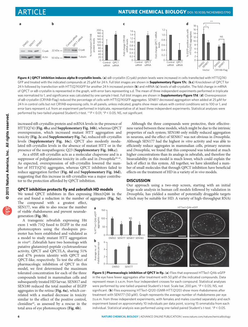
QPCT overexpression or knockdown did not modulate levels of HSP70, the main inducible stress response chaperone (Supplementary Fig. 15d). We performed transcriptional profiling to assess changes in alternative molecular chaperones induced by SEN177 in the presence of mutant HTT and observed upregulation of several small heat shock proteins (sHSPs; HSPB6 with 1.6-fold increase, HSPB3 with 1.5-fold increase HSPB7 with a 1.5-fold increase and, notably, αB-crystallin, with a >2.5-fold increase in transcript levels; Supplementary Fig. 16a and Supplementary Data Set 3). We confirmed this induction at the protein level as well as with other QPCT inhibitors (Fig. 4a). Genetic inhibition of QPCT markedly
0
20
40
60
80
100
120
DMSO SEN177
%G
FP-H
TT(1
-548
) co-
imm
unop
reci
pita
ted
rela
tive
toto
tal l
ysat
es *
**
SEN177
0
20
40
60
80
100
120
DMSO 12.5 µM 25 µM
% E
GFP
-pos
itive
cel
ls w
ithEG
FP-A
37 a
ggre
gate
s
N
(SEN177) R = 4-fluoro-3-pyridyl
(SEN180) R = 3-pyridyl
(SEN817) R = piperonyl
N
N
NN
Ra
*****
SEN177
0
20
40
60
80
100
120
DMSO 12.5 µM 25 µM
% E
GFP
-pos
itive
cel
ls w
ithEG
FP-H
TT(Q
74) a
ggre
gate
s
d
*NS
0
20
40
60
80
100
120
140
shControl shQPCT
% E
GFP
-pos
itive
cel
ls w
ithH
TT(Q
74) a
ggre
gate
s
DMSOSEN177
e
***
NS
0
20
40
60
80
100
120
DMSO SEN177 SEN817 SEN180
% E
GFP
-pos
itive
neu
rons
with
EG
FP-Q
80 a
ggre
gate
s
g
0
20
40
60
80
100
120
140
DMSO
SEN177
SEN817
SEN180
% E
GFP
-pos
itive
cel
ls w
ithEG
FP-H
TT(Q
74) a
ggre
gate
s
**
**
b
0
20
40
60
80
100
120
DMSO
SEN177
SEN817
SEN180
% E
GFP
-pos
itive
cel
ls w
ithab
norm
al n
ulce
ar m
orph
olog
y
*
**
NS
c
Flag
Flag-H
TT(Q138)+
DMSO
Flag-H
TT(Q138)+
SEN177
Flag
Flag-H
TT(Q138)+
DMSO
Flag-H
TT(Q138)+
SEN177
GFP-HTT(Q138)
IP: Flag-HTT(Q138)
IP: FlagTotal lysatesf
Figure 3 | Design of QPCT inhibitors that reduce mutant HTT aggregation. (a) chemical structure of compounds designed to inhibit Qpct activity. the activity and in vitro AdMe properties of the compounds are shown in Supplementary Figure 11a. (b,c) treatment of Hela cells expressing eGFp-Htt(Q74) with Sen177, Sen817 and Sen180 (50 μM) for 24 h reduced the percentage of cells with aggregates (b) and apoptotic nuclei (c). (d) Sen177 reduces the percentage of HeK293 cells with eGFp-Htt(Q74) or eGFp-A37 aggregates in a concentration-dependent manner. (e) Sen177 does not further reduce the percentage of eGFp-Htt(Q74) aggregates in Qpct shRnA-transfected cells. (f) Sen177 reduces the amount of Htt(1–588)-GFp coimmunoprecipitating with Htt(1–548)-Flag in Hela cells (25 μM Sen177). the amount of GFp-Htt(1–548) immunoprecipitated relative to total lysates was quantified, and the average of five independent experiments is shown in the graph. data were analyzed by two-tailed paired Student’s t-test (n = 5 experiments). Full blot images are shown in Supplementary Figure 17b. (g) primary neurons expressing eGFp-Q80 for 3 d were treated with 50 μM of the indicated compounds for a further 24 h. in all panels, unless indicated, graphs show mean values with control conditions set to 100, and error bars represent s.d. from experiments performed in triplicate, representative of at least three independent experiments. Statistical analyses were performed by two-tailed unpaired Student’s t-test. ***P < 0.001; **P < 0.01; *P < 0.05; nS, not significant.
©20
15N
atu
re A
mer
ica,
Inc.
All
rig
hts
res
erve
d.
6 nature CHeMICaL BIOLOGY | AdvAnce online publicAtion | www.nature.com/naturechemicalbiology
article NATuRe CHemiCAl BiOlOgy dOI: 10.1038/nCHeMBIO.1790
increased αB-crystallin protein and mRNA levels in the presence of HTT(Q74) (Fig. 4b,c and Supplementary Fig. 16b), whereas QPCT overexpression, which increased mutant HTT aggregation and toxicity (Fig. 2c and Supplementary Fig. 7a), reduced αB-crystallin levels (Supplementary Fig. 16c). QPCT also modestly modu-lated αB-crystallin levels in the absence of mutant HTT or in the presence of the nonpathogenic Q23 (Supplementary Fig. 16b,c).
As a sHSP, αB-crystallin acts as a molecular chaperone and is a suppressor of polyglutamine toxicity in cells and in Drosophila39–41. As expected, overexpression of αB-crystallin lowered the num-ber of HTT(Q74) aggregates, whereas QPCT inhibitors failed to reduce aggregation further (Fig. 4d and Supplementary Fig. 16d), suggesting that this increase in αB-crystallin was a major contribu-tor to the protection afforded by QPCT inhibition.
QPCT inhibition protects fly and zebrafish HD modelsWe tested QPCT inhibitors in flies expressing Httex1Q46 in the eye and found a reduction in the number of aggregates (Fig. 5a). The compound with a greatest effect, SEN177, was able to also rescue the number of visible rhabdomeres and prevent neurode-generation (Fig. 5b).
A transgenic zebrafish expressing Htt exon 1 with 71Q fused to EGFP in the rod photoreceptors using the rhodopsin pro-moter has been established and validated as a model to study mutant HTT aggregation in vivo42. Zebrafish have two homologs with putative glutaminyl-peptide cyclotransferase activity, QPCT and QPCTLA, sharing 51% and 47% protein identity with QPCT and QPCT-like, respectively. To test the effect of pharmacologic inhibition of QPCT in this model, we first determined the maximum tolerated concentration for each of the three compounds tested in mammalian cells and subsequently treated HD larvae. SEN817 and SEN180 reduced the total number of EGFP aggregates in the retina (Fig. 6a), which cor-related with a marked decrease in toxicity similar to the effect of the positive control, clonidine42, as assessed by a rescue in the total area of eye photoreceptors (Fig. 6b).
Although the three compounds were protective, their effective-ness varied between these models, which might be due to the intrinsic properties of each system; SEN180 only mildly reduced aggregation in neurons, and the effect of SEN817 was not obvious in Drosophila. Although SEN177 had the highest in vitro activity and was able to efficiently reduce aggregates in mammalian cells, primary neurons and Drosophila, we found that this compound was tolerated at much higher concentrations than its analogs in zebrafish, and therefore the bioavailability in this model is much lower, which could explain the lack of effect in this system. All together, we have identified a num-ber of small molecules that through QPCT inhibition have beneficial effects on the treatment of HD in a variety of in vivo models.
DiSCuSSiONOur approach using a two-step screen, starting with an initial large-scale analysis in human cell models followed by validation in Drosophila, has yielded a number of potentially druggable targets which may be suitable for HD. A variety of high-throughput RNAi
DMSO SEN177
Ave
rage
no.
of
rhab
dom
eres
per
eye
0
2
4
6*
SEN817 SEN180
SEN177DMSO
HTTex1-Q46-eGFP
% H
TTex
1-Q
46-e
GFP
aggr
egat
es
150
100
50
0DMSO SEN177 SEN817 SEN180
* *NS
a
b
Figure 5 | Pharmacologic inhibition of QPCT in fly. (a) Flies that expressed Httex1-Q46-eGFp in the eye have fewer aggregates after treatment with 50 μM of the indicated compounds. data represent mean ± s.e.m. from four independent crosses for each compound. Statistical analyses were performed by one-tailed unpaired Student’s t-test. Scale bar, 200 μm. *P < 0.05; nS, not significant. (b) Flies expressing Httex1-Q120 (GMR-Htt.Q120) show more rhabdomeres after treatment with Sen177 (50 μM). Graph represents the average number of rhabdomeres per eye ±s.e.m. from three independent experiments, with females and males counted separately and each experiment based on approximately 10 individuals per data point, scoring 15 ommatidia from each individual. Statistical analysis was performed using one-tailed paired Student’s t-test. *P < 0.05.
0
0.2
0.4
0.6
0.8
1.0
1.2
ControlsiRNA
QPCTsiRNA
Fold
cha
nge
QPC
T m
RNA
leve
ls
*
020406080
100120140160180200
ControlsiRNA
QPCTsiRNA
% n
orm
aliz
ed C
RYA
Bpr
otei
n le
vels
00.20.40.60.81.01.21.41.6
ControlsiRNA
QPCTsiRNA
Fold
cha
nge
CRY
AB
mRN
A le
vels
*
c
Cryab
a
DMSO
SEN817
SEN177
Actin02468
1012141618
DMSO
SEN817DM
SO
SEN817
Vector CRYAB-Flag
% E
GFP
-pos
itive
cel
ls w
ithH
TT(Q
74) a
ggre
gate
s
***
NS
d
Cryab
Tubulin
22 kDa
50 kDa
Control si
RNA
QPCT siRNA
b
Figure 4 | QPCT inhibition induces alpha B-crystallin levels. (a) αb-crystallin (cryab) protein levels were increased in cells transfected with Htt(Q74)GFp and treated with the indicated compounds at 25 μM for 24 h. Full blot images are shown in Supplementary Figure 17c. (b,c) Knockdown of Qpct for 24 h followed by transfection with Htt(Q74)GFp for another 24 h increased protein (b) and mRnA (c) levels of αb-crystallin. the fold change in mRnA of Qpct or αb-crystallin is represented in the graph, with error bars representing s.d. the mean of three independent experiments performed in triplicate was normalized to 1, and significance was calculated by one sample t-test. Full blot images are shown in Supplementary Figure 17d. (d) overexpression of αb-crystallin (cRYAb-Flag) reduced the percentage of cells with Htt(Q74)GFp aggregates. Sen817 decreased aggregation when added at 25 μM for 24 h in control cells but not cRYAb-expressing cells. in all panels, unless indicated, graphs show mean values with control conditions set to 100 or 1, and error bars represent s.d. from an experiment performed in triplicate, representative of at least three independent experiments. Statistical analyses were performed by two-tailed unpaired Student’s t-test. **P < 0.01; *P < 0.05; nS, not significant.

©20
15N
atu
re A
mer
ica,
Inc.
All
rig
hts
res
erve
d.
nature CHeMICaL BIOLOGY | AdvAnce online publicAtion | www.nature.com/naturechemicalbiology 7
articleNATuRe CHemiCAl BiOlOgy dOI: 10.1038/nCHeMBIO.1790
screens have identified genetic suppressors of phenotypes mediated by mutant HTT N-terminal fragments in Drosophila, Caenorhabditis elegans and mammalian (mouse and human) cells43–46. In most cases, aggregation was the primary readout, often measured with C-terminal GFP fusions. Differences in the nature of the previous screens (species, cellular context, HTT fragment length, length of the polyglutamine expansion, primary readout, and differences in siRNA or shRNA sequences) complicates cross-screen comparisons. Also, virtually no screens in this area have examined their false neg-ative rates owing to inefficient knockdown. Additionally, the screen presented here was biased toward the druggable component of the human genome, and a further selection was made in the course of triaging toward specific protein target classes. This most likely con-tributes to the relatively poor overlap of hits in the present and pre-vious screens. A comparison with a screen performed in HEK293T cells to identify genetic suppressors of inducibly expressed mutant HTT exon 1 toxicity45 revealed an overlap of only four genes (CPA1, GRIN2A, NR3C2 and USP21) when considering the top 257 hits (Supplementary Data Set 1). However, matrix metalloproteases, identified in HEK293T cells as modulators of fragmentation and toxicity of N-terminal portions of mutant HTT44 , were also iden-tified in our data set together with PAK1, which we previously identified as a kinase promoting mutant HTT self-association and toxicity25, thus validating the effectiveness of the screen.
Given the reproducible and clear rescue that QPCT inhibition exerts on mutant HTT toxicity in cells and in Drosophila, we focused on this target. A catalytically inactive QPCT was not able to increase the number of aggregates, suggesting that pE modifications modulate
the levels of aggregates in HD models. Although one obvious mech-anism would involve cleavage of the polyglutamine tract followed by cyclization of an N-terminal pE residue that may change properties such as stability or hydrophobicity, which would account for its change in aggregation22, our data suggest that the effect of QPCT on HTT may be indirect. We found that the effect of aggregation modulation by QPCT was not restricted to mutant HTT as it affected aggregation of other aggregate-prone proteins, and we also found that QPCT influences the formation of mutant HTT oligo-meric species. We observed an induction in several sHSPs, mostly αB-crystallin, suggesting that QPCT inhibition caused a stress response distinct from classical Hsp70 induction, which might be mediated by indirect substrates for pE modification. This molecular chaperone reduces aggregation of polyglutamine-containing proteins39,41, α-synuclein47,41or amyloid-β peptide48,49, underscor-ing QPCT inhibition as an effective target for misfolded protein disorders. As αB-crystallin is regulated at the transcriptional level, whereas QPCT resides in the secretory pathway, inhibition of QPCT may activate a signaling response that enhances αB-crystallin tran-scription. Our data suggest that this is most likely independent of an ER stress response or the involvement of ERK and CREB, which have been recently found phosphorylated upon QPCT inhibition38, as well as other stress signaling pathways such as JNK. Further work will need to clarify the QPCT substrate mediating this effect. It is important to stress that the benefits of QPCT downregulation may not be restricted to αB-crystallin as an effector, as the upregulation of other related sHSPs may also contribute beneficially.
We identified and characterized a series of compounds that efficiently reduce mutant HTT aggregation in mammalian cell lines and also in primary mouse neurons, fly eye and in zebrafish. Although the levels of rescue obtained varied between compounds depending on the model used, this may be as a result of differences in absorp-tion routes and bioavailability. Nevertheless, our data showed that pharmacologic inhibition of QPCT using this compound series can rescue HD phenotypes and provides proof of principle for QPCT as a potential therapeutic target for HD and possibly other related intracellular proteinopathies by modulating the formation of oli-gomeric forms, which have been proposed as the most toxic spe-cies in these diseases7,8. Clearly, further work, most likely including additional drug development, is required before we can consider whether this will be clinically relevant. Nevertheless, in a broader perspective, our data suggest that a discovery pipeline from druggable genome screen to drug development may be tractable for neurodegenerative diseases.
received 14 august 2014; accepted 5 March 2015; published online 6 april 2015
meTHODSMethods and any associated references are available in the online version of the paper.
references1. Imarisio, S. et al. Huntington’s disease: from pathology and genetics to
potential therapies. Biochem. J. 412, 191–209 (2008).2. Zuccato, C., Valenza, M. & Cattaneo, E. Molecular mechanisms and potential
therapeutical targets in Huntington’s disease. Physiol. Rev. 90, 905–981 (2010).
3. A novel gene containing a trinucleotide repeat that is expanded and unstable on Huntington’s disease chromosomes. The Huntington’s Disease Collaborative Research Group. Cell 72, 971–983 (1993).
4. Mangiarini, L. et al. Exon 1 of the HD gene with an expanded CAG repeat is sufficient to cause a progressive neurological phenotype in transgenic mice. Cell 87, 493–506 (1996).
5. Hodgson, J.G. et al. A YAC mouse model for Huntington’s disease with full-length mutant huntingtin, cytoplasmic toxicity, and selective striatal neurodegeneration. Neuron 23, 181–192 (1999).
6. Soto, C. & Estrada, L.D. Protein misfolding and neurodegeneration. Arch. Neurol. 65, 184–189 (2008).
DMSO
SEN180SEN817
SEN177
020406080
100120
DMSO
SEN177
SEN817
SEN180
****
% E
GFP
-Htt
71Q
aggr
egat
esSEN177
** *NSNS
EGFP-HTT(71Q) EGFP-HTT(71Q) (green)/rhodopsin (magenta)
010203040506070
Control
SEN177
SEN817
SEN180
Clonidine
Mea
n ph
otor
ecep
tor
coun
t per
eye
*
SEN180SEN817
DMSO
a b
Figure 6 | Pharmacologic inhibition of QPCT in zebrafish. (a) Representative sections through the central retina of transgenic Hd zebrafish at 7 d.p.f. treated with dMSo, Sen177 (1 mM), Sen817 (100 μM) or Sen180 (100 μM), showing aggregates (arrows) within the rod photoreceptors. Scale bar, 10 μm. treatment with Qpct inhibitors resulted in reduction in aggregates (Student’s t-test) for Sen187 and Sen810. (b) Representative sections through the central retina of transgenic Hd zebrafish at 9 d.p.f. treated with dMSo, Sen177 (1 mM), Sen817 (100 μM) or Sen180 (100 μM). to demonstrate that loss of GFp corresponds to loss of photoreceptors, sections were stained with antirhodopsin (1d1) antibody (magenta). GFp labels the whole rod photoreceptor, whereas rhodopsin is present in the rod outer segment. Merged images show colocalization of GFp with the rhodopsin (magenta). photoreceptor degeneration is ameliorated by Sen817 and Sen180. Scale bars, 10 μm. in all panels, **P < 0.01; *P < 0.05; nS, not significant.

©20
15N
atu
re A
mer
ica,
Inc.
All
rig
hts
res
erve
d.
8 nature CHeMICaL BIOLOGY | AdvAnce online publicAtion | www.nature.com/naturechemicalbiology
article NATuRe CHemiCAl BiOlOgy dOI: 10.1038/nCHeMBIO.1790
7. Takahashi, T. et al. Soluble polyglutamine oligomers formed prior to inclusion body formation are cytotoxic. Hum. Mol. Genet. 17, 345–356 (2008).
8. Lajoie, P. & Snapp, E.L. Formation and toxicity of soluble polyglutamine oligomers in living cells. PLoS ONE 5, e15245 (2010).
9. Hartl, F.U., Bracher, A. & Hayer-Hartl, M. Molecular chaperones in protein folding and proteostasis. Nature 475, 324–332 (2011).
10. Sathasivam, K. et al. Aberrant splicing of HTT generates the pathogenic exon 1 protein in Huntington disease. Proc. Natl. Acad. Sci. USA 110, 2366–2370 (2013).
11. Bleicher, K.H., Böhm, H.-J., Müller, K. & Alanine, A.I. Hit and lead generation: beyond high-throughput screening. Nat. Rev. Drug Discov. 2, 369–378 (2003).
12. Li, M., Huang, Y., Ma, A.A.K., Lin, E. & Diamond, M.I. Y-27632 improves rotarod performance and reduces huntingtin levels in R6/2 mice. Neurobiol. Dis. 36, 413–420 (2009).
13. Marsh, J.L. et al. Expanded polyglutamine peptides alone are intrinsically cytotoxic and cause neurodegeneration in Drosophila. Hum. Mol. Genet. 9, 13–25 (2000).
14. Dietzl, G. et al. A genome-wide transgenic RNAi library for conditional gene inactivation in Drosophila. Nature 448, 151–156 (2007).
15. Schilling, S. et al. Isolation and characterization of glutaminyl cyclases from Drosophila: evidence for enzyme forms with different subcellular localization. Biochemistry 46, 10921–10930 (2007).
16. Jackson, G.R. et al. Polyglutamine-expanded human huntingtin transgenes induce degeneration of Drosophila photoreceptor neurons. Neuron 21, 633–642 (1998).
17. Franceschini, N. & Kirschfeld, K. Pseudopupil phenomena in the compound eye of Drosophila. Kybernetik 9, 159–182 (1971).
18. Ravikumar, B. et al. Dynein mutations impair autophagic clearance of aggregate-prone proteins. Nat. Genet. 37, 771–776 (2005).
19. Zhang, S., Binari, R., Zhou, R. & Perrimon, N. A genomewide RNA interference screen for modifiers of aggregates formation by mutant Huntingtin in Drosophila. Genetics 184, 1165–1179 (2010).
20. Wyttenbach, A. et al. Effects of heat shock, heat shock protein 40 (HDJ-2), and proteasome inhibition on protein aggregation in cellular models of Huntington’s disease. Proc. Natl. Acad. Sci. USA 97, 2898–2903 (2000).
21. Cynis, H. et al. Inhibition of glutaminyl cyclase alters pyroglutamate formation in mammalian cells. Biochim. Biophys. Acta 1764, 1618–1625 (2006).
22. Saido, T.C. Involvement of polyglutamine endolysis followed by pyroglutamate formation in the pathogenesis of triplet repeat/polyglutamine-expansion diseases. Med. Hypotheses 54, 427–429 (2000).
23. Onodera, O. et al. Oligomerization of expanded-polyglutamine domain fluorescent fusion proteins in cultured mammalian cells. Biochem. Biophys. Res. Commun. 238, 599–605 (1997).
24. Rankin, J., Wyttenbach, A. & Rubinsztein, D.C. Intracellular green fluorescent protein–polyalanine aggregates are associated with cell death. Biochem. J. 348, 15–19 (2000).
25. Luo, S., Mizuta, H. & Rubinsztein, D.C. p21-activated kinase 1 promotes soluble mutant huntingtin self-interaction and enhances toxicity. Hum. Mol. Genet. 17, 895–905 (2008).
26. Buchholz, M. et al. The first potent inhibitors for human glutaminyl cyclase: synthesis and structure-activity relationship. J. Med. Chem. 49, 664–677 (2006).
27. Schilling, S. et al. Glutaminyl cyclase inhibition attenuates pyroglutamate Aβ and Alzheimer’s disease–like pathology. Nat. Med. 14, 1106–1111 (2008).
28. Schilling, S. et al. Continuous spectrometric assays for glutaminyl cyclase activity. Anal. Biochem. 303, 49–56 (2002).
29. Yoon, S. & Welsh, W.J. Identification of a minimal subset of receptor conformations for improved multiple conformation docking and two-step scoring. J. Chem. Inf. Comput. Sci. 44, 88–96 (2004).
30. Polgár, T. & Keserü, G.M. Ensemble docking into flexible active sites. Critical evaluation of FlexE against JNK-3 and β-secretase. J. Chem. Inf. Model. 46, 1795–1805 (2006).
31. Huang, K.-F., Liu, Y.-L., Cheng, W.-J., Ko, T.-P. & Wang, A.H.-J. Crystal structures of human glutaminyl cyclase, an enzyme responsible for protein N-terminal pyroglutamate formation. Proc. Natl. Acad. Sci. USA 102, 13117–13122 (2005).
32. Huang, K.-F. et al. Structures of human Golgi-resident glutaminyl cyclase and its complexes with inhibitors reveal a large loop movement upon inhibitor binding. J. Biol. Chem. 286, 12439–12449 (2011).
33. Ruiz-Carrillo, D. et al. Structures of glycosylated mammalian glutaminyl cyclases reveal conformational variability near the active center. Biochemistry 50, 6280–6288 (2011).
34. Jones, G., Willett, P. & Glen, R.C. Molecular recognition of receptor sites using a genetic algorithm with a description of desolvation. J. Mol. Biol. 245, 43–53 (1995).
35. Jones, G., Willett, P., Glen, R.C., Leach, A.R. & Taylor, R. Development and validation of a genetic algorithm for flexible docking. J. Mol. Biol. 267, 727–748 (1997).
36. Verdonk, M.L., Cole, J.C., Hartshorn, M.J., Murray, C.W. & Taylor, R.D. Improved protein-ligand docking using GOLD. Proteins 52, 609–623 (2003).
37. Palm, K., Luthman, K., Ros, J., Grasjo, J. & Artursson, P. Effect of molecular charge on intestinal epithelial drug transport: pH-dependent transport of cationic drugs. J. Pharmacol. Exp. Ther. 291, 435–443 (1999).
38. Song, H. et al. Inhibition of glutaminyl cyclase ameliorates amyloid pathology in an animal model of Alzheimer’s disease via the modulation of γ-secretase activity. J. Alzheimers Dis. 43, 797–807 (2014).
39. Robertson, A.L. et al. Small heat-shock proteins interact with a flanking domain to suppress polyglutamine aggregation. Proc. Natl. Acad. Sci. USA 107, 10424–10429 (2010).
40. Bilen, J. & Bonini, N.M. Drosophila as a model for human neurodegenerative disease. Annu. Rev. Genet. 39, 153–171 (2005).
41. Tue, N.T., Shimaji, K., Tanaka, N. & Yamaguchi, M. Effect of αB-crystallin on protein aggregation in Drosophila. J. Biomed. Biotechnol. 2012, 252049 (2012).
42. Williams, A. et al. Novel targets for Huntington’s disease in an mTOR-independent autophagy pathway. Nat. Chem. Biol. 4, 295–305 (2008).
43. Lejeune, F.-X. et al. Large-scale functional RNAi screen in C. elegans identifies genes that regulate the dysfunction of mutant polyglutamine neurons. BMC Genomics 13, 91 (2012).
44. Miller, J.P. et al. Matrix metalloproteinases are modifiers of huntingtin proteolysis and toxicity in Huntington’s disease. Neuron 67, 199–212 (2010).
45. Miller, J.P. et al. A genome-scale RNA-interference screen identifies RRAS signaling as a pathologic feature of Huntington’s disease. PLoS Genet. 8, e1003042 (2012).
46. Yamanaka, T. et al. Large-scale RNA interference screening in mammalian cells identifies novel regulators of mutant huntingtin aggregation. PLoS ONE 9, e93891 (2014).
47. Waudby, C.A. et al. The interaction of αB-crystallin with mature α-synuclein amyloid fibrils inhibits their elongation. Biophys. J. 98, 843–851 (2010).
48. Raman, B. et al. αB-crystallin, a small heat-shock protein, prevents the amyloid fibril growth of an amyloid β-peptide and β2-microglobulin. Biochem. J. 392, 573–581 (2005).
49. Hochberg, G.K.A. et al. The structured core domain of αB-crystallin can prevent amyloid fibrillation and associated toxicity. Proc. Natl. Acad. Sci. USA 111, E1562–E1570 (2014).
acknowledgmentsWe are grateful for funding from the UK Medical Research Council (COEN grant MR/J006904/1 to D.C.R. and C.J.O.’K.), the Wellcome Trust (Principal Fellowship 095317/Z/11/Z), to D.C.R., and strategic award 100140), the National Institute of Health Research Biomedical Research Unit in Dementia at Addenbrooke’s Hospital, the TAMAHUD project (European Community FP6 grant no. 03472 under the Thematic Call LSH-2005-2.1.3-8 “Early markers and new targets for neurodegenerative diseases”) and the NEUROMICS project (European Community Seventh Framework Programme under grant agreement no. 2012-305121). We thank J.L. Marsh, N. Perrimon and the Vienna Drosophila RNAi Center for fly stocks; M. Renna and S. Luo for helpful comments; M. Lichtenberg for help with flow cytometry assays; F. Siddiqi and M. Garcia-Arencibia for help with primary cultures; W. Fecke for advice and assistance; and S. Gotta for help in high-resolution MS analysis of compounds.
author contributionsM.J.-S. performed most post-screen cell biology experiments. W.L. and S.I. performed the Drosophila experiments. M.H. and B.S. performed the cell-based screen. A.F., T.E.D. and C.X performed the zebrafish experiments, and A.F. supervised these. A.T., E.G.-C., V.P.S. and R.S. performed the bioinformatics analyses. F.M. performed the chaperone transcription array experiments and nonradioactive pulse-chase. E.G.-C. and F.H. participated in experimental design of the screen. F.H., G.L., D.D., L.M. and G.P. generated and validated the stable cell lines for the screen. G.M., C.C. and A.N. synthesized and analyzed the compounds. M.A. performed the selection of compound for high-throughput screening and supported the hit-to-lead optimization by in silico drug design methodologies. V.P. optimized glutaminyl cyclase enzymatic assays for compound screening. G.L.S. and N.P.C. performed in vitro ADME experiments. C.S. provided support for experiments at Siena Biotech. C.O.K. supervised Drosophila experiments. G.P. also supervised molecular biology activities at Siena Biotech. A.C. supervised primary screen and chemical biology. D.C.R. supervised cell biology, Drosophila and zebrafish experiments. D.C.R and A.C. conceived the project and coordinated work between sites with assistance from G.P., M.J.-S. and D.C.R., and A.C drafted the manuscript, which was commented on by all authors.
Competing financial interestsThe authors declare competing financial interests: details accompany the online version of the paper.
additional informationSupplementary information and chemical compound information is available in the online version of the paper. Reprints and permissions information is available online at http://www.nature.com/reprints/index.html. Correspondence and requests for materials should be addressed to A.C. or D.C.R.

©20
15N
atu
re A
mer
ica,
Inc.
All
rig
hts
res
erve
d.
nature CHeMICaL BIOLOGYdoi:10.1038/nchembio.1790
ONliNe meTHODSAssays for validation polyglutamine toxicity modifiers in Drosophila. Drosophila fly stocks. As a model of polyglutamine toxicity, flies that expressed a protein with 48 glutamines encoded by P{UAS-Q48.myc/flag}31 (ref. 13) in eyes under control of the GMR-Gal4 driver P{GAL4-ninaE.GMR}12 (ref. 50) (Q48) were used. Fly orthologs to the genes identified in the cell screen were selected by performing reciprocal BLASTP and cross checking with data-bases including HomoloGene (http://www.ncbi.nlm.nih.gov/homologene/), GeneCards (http://www.genecards.org/) and Ensembl (http://www.ensembl.org/index.html/). The RNAi lines corresponding to the identified genes were obtained from Vienna Drosophila RNAi Center (VDRC; http://stockcenter.vdrc.at/control/main/).
UAS-Q48.myc/flag was a generous gift from J.L. Marsh (University California, Irvine)13, and UAS-Httex1-Q46-eGFP was a generous gift from N. Perrimon (Harvard Medical School)19. Fly lines that are not referenced here are documented in FlyBase (http://www.flybase.org/). All fly crosses and experiments were performed at 25 °C.
Drosophila RNAi screen. Five virgins of genotype w; GMR-GAL4; UAS-Q48.myc/flag (Q48) were crossed to males carrying each UAS-RNAi (GD- and KK-RNAi collections, VDRC, http://stockcenter.vdrc.at/control/main/). Genetic background was controlled by crossing w; GMR-GAL4; UAS-Q48.myc/flag females to w1118 males that share the same genetic back-ground (VDRC stock number 60000 for the GD-RNAi lines and 60100 for the KK-RNAi lines). For Glutaminyl cyclase (CG32412), the GD-RNAi line 38277 and the KK-RNAi line 106341 were used. For iso Glutaminyl cyclase (CG5976), the KK-RNAi line 101533 was used. For GD-RNAi lines, degeneration was determined by scoring the eye depigmentation in the progeny of the above crosses 4 d after eclosion, assessing modification of polyglutamine loss of pig-mentation and black necrotic-like spots. As the background in KK-RNAi lines leads to dark eye pigmentation (http://stockcenter.vdrc.at/control/protocols), toxicity was assessed by scoring the presence or absence of black necrotic-like spots in the eyes of 10-d-old flies. Fisher’s exact test was performed to compare the numbers of necrotic spot–containing flies in the KK-RNAi crosses with controls using an arbitrary P < 0.005 as a statistical cutoff for significance. Eyes were imaged using a Nikon CoolPix 990 digital camera attached to a dissecting microscope.
EGFP expression levels assessed in Drosophila RNAi lines. Western blot analysis was performed using progeny of crosses between virgins of the genotype w; GMR-GAL4; UAS-EGFP and males of each VDRC-RNAi line used or background control (VDRC stock number 60100). Fly heads were homogenized in Laemmli sample buffer. Rabbit polyclonal anti-GFP at 1:1,000 (AbCam, Ab6556) and monoclonal anti-β-tubulin at 1:10,000 (Developmental Studies Hybridoma Bank) were used. Blots were scanned using Odyssey Fc Imaging System (LI-COR Biosciences). This validation was initially performed once on each suppressor, and subsequently RNAi lines showing an apparent reduction in EGFP levels were retested using the progeny of three independent crosses. Statistical analysis was per-formed by Student’s two-tailed paired t-test between the RNAi lines and the control line.
Pseudopupil assay. Analysis was performed as previously described17. Virgins of genotype elav-GAL4C155; {GMR-HD.Q120}4.62/TM3 (elav-Gal4; GMR-HTT.Q120)16 were crossed with males carrying the RNAi construct for Glutaminyl cyclase (lines QCGD38277 or QCKK106341) or iso Glutaminyl cyclase (line isoQCKK101533) and compared to a background control line.
To evaluate the effect of QPCT inhibitors, virgins of genotype yw; {GMR-HD.Q120}2.4 (GMR-HTT.Q120) were allowed to mate with w1118 control males for 48 h on standard cornmeal food and then transferred on fly food containing the compounds.
The number of rhabdomeres per ommatidium was scored in progeny of the above crosses at 3 d (GMR-Q120) or 4 d (elav-Gal4; GMR-HTT.Q120) post eclosion. Statistical analysis was performed using one-tailed t-test on data from three or four independent experiments, each based on approxi-mately ten individuals for each genotype, scoring 15 ommatidia per eye. When compounds were tested, the analysis was done on females and males of each treatment separately.
Aggregate counting. Virgins of genotype w; GMR-GAL4; UAS-Httex1-Q46-eGFP19 were crossed with males of QPCT UAS-RNAi lines or from the back-ground KK-RNAi control line as all the background controls show similar aggregate scoring. Eye pictures of 18-d-old progeny were taken using a Leica
MZ16F microscope connected to a Leica DFC340FX digital camera. For each genotype, GFP punctae indicating aggregate formation was counted using an ImageJ ‘Cell Counter’ plugin in the eyes of 20 males, a pool of 5 males from four independent crosses. For compound testing, virgins of genotype w; GMR-GAL4; UAS-Httex1-Q46-eGFP were crossed with w1118 control males, and females of the progeny were scored 15 d post eclosion. The experiment was repeated at least three times, and for each experiment at least four female eyes were scored. An unpaired one-tailed t-test was used to deter-mine statistical significance for single comparisons between two groups using GraphPad Prism.
Compound treatment. Flies were reared on food (Instant Fly Food, Philip Harris, Ashby de la Zouch, UK) containing either QPCT inhibitor (50 μM) dissolved in DMSO or DMSO alone. The progeny were flipped every 2 d on fresh food containing the specific inhibitor or DMSO.
Bioinformatics analysis. Ingenuity Pathways Analysis (Ingenuity Systems; http://www.ingenuity.com/) was used to analyze the distribution of siRNAs tested among the different protein classes as well as to determine the canonical pathways associated to the confirmed primary actives.
Assays for validation of polyglutamine toxicity and aggregation modifiers in human cell lines. Cell culture. HEK293 (human embryonic kidney) and HeLa (human cervical carcinoma) cells and Atg5-deficient (Atg5−/−) mouse embry-onic fibroblasts (MEFs) (gift from N. Mizushima (University of Tokyo)) were grown in Dulbecco’s modified eagle medium supplemented with 10% FBS, 100 U/ml penicillin-streptomycin and 2 mM l-glutamine at 37 °C in 5% CO2. UbG76V-GFP–expressing stable HeLa cell line (a kind gift from N.P. Dantuma (Karolinska Institute) was maintained in medium containing 0.5 mg/ml G418.
Isolation and culture of mouse primary cortical neurons. Primary cortical neurons were isolated from C57BL/6 mice (Jackson Laboratories) embryos at E16.5. Briefly, brains were harvested and placed in ice-cold PBS-glucose, where the meninges were removed and the cerebral cortices were dissected. After mechanical dissociation using sterile micropipette tips, dissociated neurons were resuspended in PBS-glucose and collected by centrifugation. Viable cells were seeded on polyornithine-coated 12-multiwell plates. Cells were cultured in Neurobasal medium supplemented with 2 mM glutamine, 200 mM B27 supplement and 1% penicillin-streptomycin at 37 °C in a humidified incu-bator with 5% CO2. One-half of the culture medium was changed every 2 d until treatment. After 5 d of culturing in vitro, differentiated cortical neurons were infected with lentiviral particles bearing EGFP-Q80 and scramble or QPCT-directed shRNAs. Compounds were added 3 d after EGFP-Q80 viral infection and left for another 24 h. When EGFP-Q80 was expressed together with shRNA, 5–6 d were needed before cultures were fixed in a 2% PFA-7.5% glucose solution.
DNA constructs. Human QPCT (NM_012413) plasmid was purchased from Origene (pCMV6-XL5-QPCT). A C-terminal Flag-tagged QPCT construct was generated by PCR amplification of QPCT cDNA from pCMV6-XL5-QPCT using primers overhanging HindIII and BglII sites and insertion into the pCMV5-Flag in HindIII and BamHI restriction sites, using standard restric-tion enzyme digestion and ligation procedures. QPCT(E201Q)-Flag was gener-ated using QuikChange II Agilent Site-Directed mutagenesis kit with the following primers: forward, 5′-CTTCTTTGATGGTCAAGAGGCTTTTCTTCACTGG-3′, and reverse, 5′-CCAGTGAAGAAAA GCCTCTTGACCATCAAAGAAG-3′. pcDNA or pCMV5-Flag empty vectors were used as mock controls for pCMV6-XL5-QPCT or QPCT-Flag, respectively.
Constructs expressing the first exon of the Htt gene carrying 74 polyglutamines expressed from pEGFP-C1 (Clontech) (EGFP-HTTQ74) or pHM6 (Roche Diagnostics) (HA-HTTQ74) or with only 23 polyglutamines (EGFP-HTTQ23) were described previously51. pEGFP-N1-Q57 and pEGFP-N1-Q81 (ref. 23) and pEGFP-C1-A37 (ref. 24) have been previously described. Mutant HTT(1–588)-Flag was provided by M.R. Hayden (University of British Columbia), and mutant HTT(1–548)GFP was generated by S. Luo25. 3×Flag-CRYAB construct has been previously described52. The pGL3-BIP/GRP78-luciferase construct was kindly provided by M. Renna (University of Cambridge)53.
Reagents. Chemical compounds used in cell culture were the autophagy inhibitors bafilomycin A1 (400nM, DMSO; 4 h; Millipore) and 3MA (10 mM, 16 h; SIGMA), staurosporine (3 μM) and the proteasome inhibitor MG132 (10 μM). PBD150 was synthesized as described in ref. 26.

©20
15N
atu
re A
mer
ica,
Inc.
All
rig
hts
res
erve
d.
nature CHeMICaL BIOLOGY doi:10.1038/nchembio.1790
Transfection. Cells were transfected in six-well plates with 0.5–1.5 μg of DNA and 5 μl of Lipofectamine (Invitrogen) or TransIT-2020 (Mirus) per well for 4 h in Optimem (GIBCO-BRL) and then incubated in full medium for 48 h. Gene knockdown experiments were performed using ON-TARGETplus SMARTpool siRNA (Dharmacon) for human QPCT, consisting of four siRNAs with the following sequences, which do not target the QPCT-like sequence: CUAUGGGUCUCGACACUUA, GUACCGGUCUUUCUCAAAU, CCUUAAAGACUGUUUCAGA and GGAACUUGCUCGUGCCUUA. For siRNA treatment, either a single transfection protocol using 50 nM siRNA for 48 h or a double transfection protocol which consisted of a first 50-nM siRNA trans-fection followed by a second 50-nM siRNA transfection after 48 h was used.
Western blotting. Cells were washed once in PBS and harvested on lysis buffer (20 mM Tris-HCl, pH 6.8, 137 nM NaCl, 1 mM EGTA, 1% Triton X-100, 10% glycerol, 1× Roche cOmplete mini protease inhibitor). Equal loading was obtained by protein concentration determination using a Bio-Rad assay followed by resuspension and boiling in Laemmli buffer. Samples were subjected to 12% SDS-PAGE and transferred to a PVDF membrane (Immobilon-P, GE Healthcare). Blots were proved with the following primary antibodies: anti-LC3 (1:2,000; Novus Biologicals, NB100-2220), anti-Hsp70 (1:1,000; Enzo SPA810), anti-CRYAB (1:1,000; Cell Signaling 8851), anti-actin (1:2,000; Sigma, A2066), anti-α-tubulin (1:4,000; T9026, Sigma), anti-Flag epitope (1:2,000; SIGMA, F7425), anti-GFP (1:1,000; Clontech, Living Colors, polyclonal), eIF2α (1:1,000, Abcam 5369) and phospho-S51-eIF2α_(1:1,000, Abcam 32157), GRP78 (1:1,000, Abcam 21685), anti-phospho-ERK (1:1,000, Cell Signaling, 9101), anti-ERK (1:1,000, Cell Signaling, 9102), anti- phospho-CREB (S133) (1:1,000, Cell Signaling 9191), anti-CREB 86B10 (1:1,000, Cell Signaling, 9104), anti-phospho-JNK (1:1,000, Cell Signaling, 9255), anti-JNK (1:1,000, Cell Signaling, 9252). The appropriate anti-mouse or anti-rabbit secondary antibodies were used and visualized using an ECL detec-tion kit (Amersham) or LI-COR Biosciences infrared imager (Odyssey).
Caspase 3/7 activity assay. Cells were seeded in a 96-well plate 24 h before the assay, and 1 μM staurosporine or DMSO was added for the last 8 h. Caspase 3/7 activity was measured by using a luminogenic caspase 3/7 substrate (Caspase 3/7-Glo Assay, Promega) following manufacturer’s protocols in a Glomax luminometer (Promega). Protein concentration was determined in each cell lysate, and caspase 3/7 activity was normalized to protein levels.
Coimmunoprecipitation assays. Assays were performed as previously described25, where HTT(1–588)Flag(Q138) and HTT(1–548)GFP(Q138) were expressed in HeLa cells together with QPCT plasmid for 48 h or treated with 25 μM SEN177 for 24 h. Cells were lysed in buffer B containing 10 mM Tris, pH 7.4, 150 mM NaCl, 1 mM EDTA pH8, 1% Triton and 1× Roche complete mini protease inhibitor for 20 min on ice, followed by centrifugation at 13,000 r.p.m. for 10 min. Five hundred micrograms total protein were incubated with primary anti-Flag M2 (Sigma) or anti-GFP (Clontech, Living Colors, polyclo-nal) at 5 μg/ml overnight at 4 °C. Protein G Dynabeads (Life Technologies) were added and incubated for further 2 h. Beads were washed three times with buffer B and eluted using 0.1 M glycine, pH 2.5, followed by boiling in Laemmli buffer. Samples were subjected to western blotting and visualized using LICOR. A fraction of the total lysates was run simultaneously.
Reverse-transcriptase PCR analysis. Total RNA was isolated from cell pellets using Trizol Reagent (Invitrogen) and treated with DNase I, and cDNA synthesis was performed by SuperScript III First-Strand Synthesis System (Invitrogen). Standard conditions were used for cDNA amplification, and PCR products were analyzed by agarose gel electrophoresis and ethidium bromide staining or quantified with real-time PCR. For real-time PCR analysis, the reaction mixture containing cDNA template, primers and SYBR Green PCR Master Mix (Invitrogen) was run in a 7900 Fast Real-time PCR System (Applied Biosystems, Carlsbad, CA). Fold changes in mRNA levels were determined using a standard curve and after normalization to internal control β-actin RNA levels. Primer sequences used in this study are as follows: QPCT, 5′-CATGGCATGGATTTATTGG-3′ and 5′-GACGGTATCAGATCAAAC-3′; QPCT-like, 5′-CAGCGTCTCTGGAGCACTTA-3′ and 5′-GCCTCCA GGAACTTTCTGACT-3; GFP 5′-ACGTAAACGGCCACAAGTTC-3′ and 5′-T TCAGGGTCAGCTTGCCGTA-3′; actin, 5′-AGAAAATCTGGCCCACACC-3′ and 5′-GGGGTGTTGAAGGTCTCAAA-3′; CRYAB, 5′-TCTTGAGCTCA GTGAGTACTGG-3′ and 5′-AGCTCACCAGCAGTTCATGG-3′; and mouse QPCT, 5′-CGACTTGAGCCAATTGCTGA-3′ and 5′-CTTCC GGGTTAAGAGTGCTG-3′.
mRNA isolation from mouse brain. All mouse experiments were performed under appropriate UK Home Office licenses and following institutional procedures. We analyzed samples from N171 mutant HD mice and wild-type littermate controls at 20 weeks. mRNA was extracted from brains homogenized in Trizol (Invitrogen) using an Ultra Turrax homogenizer.
Lentivirus infection. shRNA containing pLKO.1 vectors targeting both mouse and human QPCT (TRCN032432) were obtained from the RNAi Consortium (TRC), and scramble shRNA vector was generated in D. Sabatini’s labora-tory (Whitehead Institute; available from Addgene, plasmid 1864). Lentiviral plasmids to express Q80-GFP were kindly provided by J. Uney (University of Bristol)54. Lentiviral particles were produced and transduced following the RNAi Consortium protocols.
Cell toxicity and aggregation assays. Cells were fixed for 7 min in 4% para-formaldehyde (PFA). For EGFP-tagged constructs, slides were mounted in Citifluor (Citifluor, Ltd.) containing 4′,6-diamidino-2-phenylindole (DAPI; 3 μg/ml; Sigma) and visualized using an Eclipse E600 fluorescence microscope (plan-apo 60×/1.4 oil immersion lens) (Nikon). For detection of HA-tagged constructs, immunofluorescence with an anti-HA (Covance laboratories 1:500) and anti-mouse Alexa 488 secondary antibody (Invitrogen, 1:1,000) was per-formed followed by mounting in Citifluor-DAPI. We assessed the percentage of transfected cells (EGFP- or HA-positive cells) with at least one aggregate per cell. Apoptotic cell death was determined by assessing the nuclear morphology (nuclei fragmented or condensed) in transfected cells. Slides were blinded, and at least 200 transfected cells per slide were scored; each individual experiment was performed in triplicate.
Detection of nascent protein synthesis. Protein synthesis was assessed by metabolic incorporation of AHA (l-azidohomoalanine) into cells transfected with EGFP-HTT(Q23). Briefly, 12 h after HeLa cells transfection, medium was washed and replaced with l-methionine/l-cysteine–free medium and treated with DMSO or SEN177 (50 μM) for 1 h before addition of AHA (l-azido-homoalanine) to the medium and collection of cells every 2 h. Labeled protein was detected by western blotting after performing Click-IT protein detection assay (Life Technologies) using biotin, following manufacturer protocols.
Luciferase reporter assay. Cells were transfected with 1 μg of GRP78-luciferase (firefly) reporter construct and 50 ng of Renilla luciferase (pRL-TK) as an internal transfection efficiency control. Cells were collected in Passive lysis buffer, and luciferase activity was measured using the Dual-luciferase Reporter Assay System (Promega) following the manufacturer’s protocol in a Glomax Luminometer (Promega). GRP78-luciferase relative activity was cal-culated relative to the Renilla luciferase transfection efficiency control activity for each sample; experiments were performed in triplicate.
Statistical analysis. Quantification of immunoblots was performed by densitometric analysis using the ImageJ software or the LI-COR Biosciences infrared imager software and normalized to loading control (actin or tubulin, as indicated in each figure legend). The P values were determined by two-tailed Student’s t-test.
Aggregates were counted in at least 200 cells per slide (with the observer blinded to their identity), and the percentage was calculated relative to control conditions. P values were determined by unpaired two-tailed Student’s t-test. All experiments were done at least three times in triplicate, and a representative blot or graph from a triplicate experiment is shown unless indicated.
Heat shock proteins and chaperone PCR array. The Human Heat Shock Proteins and Chaperones RT2 Profiler PCR Array (SABiosciences, Frederick, MD) was used to study the expression profile of 84 heat shock proteins according to the manufacturer’s procedure. Briefly, total RNA was extracted from cells transfected with HTT(Q74)GFP treated with DMSO or 25 μM of SEN177 inhibitor for 24 h using Trizol (Invitrogen) and was further purified using RNeasy mini kit with on-column DNAse digest (Qiagen). cDNA was then synthesized using an RT2 First Strand kit (SABiosciences), and real-time PCR was performed using a 7900HT fast real-time PCR system (Applied Biosciences). Data were analyzed with RT2 profiler PCR array data analysis software version 3.5.
Assays for validation of polyglutamine aggregation modifiers in zebrafish. Maintenance of zebrafish stocks and collection of embryos. All zebrafish hus-bandry and experiments were performed in accordance with UK legislation under a license granted by the Home Office and with local ethical approval. Zebrafish were reared under standard conditions55 on a 14 h light/10 h dark

©20
15N
atu
re A
mer
ica,
Inc.
All
rig
hts
res
erve
d.
nature CHeMICaL BIOLOGYdoi:10.1038/nchembio.1790
calculated (n = 5 fish (10 eyes)) for each treatment group. For quantification of photoreceptor number, the GFP-positive area of the central retina was quanti-fied using image thresholding and area analysis in ImageJ (n ≥ 5 fish (10 eyes) for each treatment group). To demonstrate that loss of GFP corresponds to loss of photoreceptors, sections were stained with anti-rhodopsin (1D1) anti-body (a kind gift from P. Linser, University of Florida)57 and mounted using VectaShield hard set mounting medium (Vector Laboratories). Sections were viewed and representative images were acquired using a GX Optical LED fluo-rescent microscope, GXCAM3.3 digital camera and GX Capture software.
cycle. Embryos were collected from natural spawnings, staged according to the established criteria56 and reared in embryo medium (5 mM NaCl, 0.17 mM KCl, 0.33 mM CaCl2, 0.33 mM Mg2SO4 and 5 mM HEPES).
Determination of the maximum-tolerated concentration of compounds in larval zebrafish. Compound exposure experiments were performed on wild-type larvae (TL strain) 2–3 d post-fertilization (d.p.f.). Concentration response assays were performed over log intervals, from 100 nM to 1 mM, to determine the maximum nontoxic concentration (MTC) for subsequent aggregate analysis assays (n = 10 larvae per concentration). Compound exposure experiments were performed in the dark at 28.5 °C.
Measuring aggregate number and rhodopsin protein levels in transgenic HD zebrafish. Aggregate counting and analysis of rod photoreceptor degeneration (photoreceptor number) was performed using heterozygous larvae from Tg (rho:EGFP-HTT71Q)cu5 zebrafish42 (hereafter referred to as transgenic HD zebrafish). Embryos from outcrossed transgenic HD zebrafish were raised in 0.2 mM 1-phenyl-2-thiourea (PTU) from 1–3 d.p.f. to inhibit pigment formation, screened for transgene expression using EGFP fluorescence and then washed twice in the embryo medium to remove PTU. From 3–9 d.p.f., transgenic HD zebrafish larvae were dark-reared in embryo medium alone or in embryo medium containing either DMSO, 1 mM SEN177, 100 μM SEN180 or 100 μM SEN817. Embryo medium and compounds were replen-ished daily. Larvae were anaesthetized by immersion in 0.2 mg/ml 3-amino benzoic acid ethyl ester (MS222), then fixed for aggregate counting at 7 d.p.f. or for photoreceptor analysis at 9 d.p.f. Anaesthetized larvae were fixed using 4% paraformaldehyde (PFA) in PBS at 4 °C. Larvae were washed briefly in PBS, allowed to equilibrate in 30% sucrose in PBS then embedded in OCT medium (Tissue-Tek) and frozen on dry ice for subsequent cryosectioning. Sections were cut at 10-μm thickness using a cryostat (Bright Instruments). For aggregate counting, sections were mounted in 50% glycerol in PBS, and the total number of GFP-positive aggregates were counted over 100 μm of the central retina on either side of the optic nerve head, and mean values were
50. Freeman, M. Reiterative use of the EGF receptor triggers differentiation of all cell types in the Drosophila eye. Cell 87, 651–660 (1996).
51. Narain, Y., Wyttenbach, A., Rankin, J., Furlong, R.A. & Rubinsztein, D.C. A molecular investigation of true dominance in Huntington’s disease. J. Med. Genet. 36, 739–746 (1999).
52. D’Agostino, M. et al. The cytosolic chaperone α-crystallin B rescues folding and compartmentalization of misfolded multispan transmembrane proteins. J. Cell Sci. 126, 4160–4172 (2013).
53. Renna, M., Caporaso, M.G., Bonatti, S., Kaufman, R.J. & Remondelli, P. Regulation of ERGIC-53 gene transcription in response to endoplasmic reticulum stress. J. Biol. Chem. 282, 22499–22512 (2007).
54. Howarth, J.L. et al. Hsp40 molecules that target to the ubiquitin-proteasome system decrease inclusion formation in models of polyglutamine disease. Mol. Ther. 15, 1100–1105 (2007).
55. Westerfield, M. The Zebrafish Book: A Guide for the Laboratory Use of Zebrafish (Danio rerio) (University of Oregon Press, 1995).
56. Kimmel, C.B., Ballard, W.W., Kimmel, S.R., Ullmann, B. & Schilling, T.F. Stages of embryonic development of the zebrafish. Dev. Dyn. 203, 253–310 (1995).
57. Hyatt, G.A., Schmitt, E.A., Fadool, J.M. & Dowling, J.E. Retinoic acid alters photoreceptor development in vivo. Proc. Natl. Acad. Sci. USA 93, 13298–13303 (1996).
Copyright © 2022 FDOKUMEN