Neurogenesis in the R6/1 transgenic mouse model of Huntington's disease: effects of environmental...
10
Neurogenesis in the R6 ⁄ 1 transgenic mouse model of Huntington’s disease: effects of environmental enrichment Stanley E. Lazic, 1 Helen E. Grote, 2 Colin Blakemore, 2 Anthony J. Hannan, 2,3 Anton van Dellen, 2 Wendy Phillips 1,4 and Roger A. Barker 1,4,5 1 Centre for Brain Repair, University of Cambridge, Cambridge CB2 2PY, UK 2 University Laboratory of Physiology, University of Oxford, Oxford OX1 3PT, UK 3 Howard Florey Institute, University of Melbourne, Vic. 3010, Australia 4 Department of Neurology, Addenbrooke’s Hospital, Cambridge CB2 2QQ, UK 5 Edith Cowan University, Perth, Australia Keywords: adult neurogenesis, dentate gyrus, doublecortin, hippocampus Abstract Previous work has demonstrated that the transgenic R6 ⁄ 1 mouse model of Huntington’s disease has decreased proliferation of neural precursor cells (NPCs) in the dentate gyrus of the hippocampus. This study therefore examined the survival and differentiation of NPCs in presymptomatic and symptomatic R6 ⁄ 1 mice and the effects of environmental enrichment on these variables. Here it is demonstrated that the survival of bromodeoxyuridine-positive (BrdU + ) NPCs in the dentate gyrus is decreased in the transgenic mice. In addition, the number of doublecortin-positive (DCX + ) cells is greatly reduced in these mice, as is the total number of new mature neurons, while the proportion of BrdU + cells differentiating into mature neurons was not significantly different between genotypes. Furthermore, the DCX + cells in the R6 ⁄ 1 mice had smaller and irregular-shaped somas, shorter neurites, and migrated a shorter distance into the granular cell layer compared with wild-type mice. Older symptomatic mice housed in an enriched environment had an increased number of BrdU + and DCX + cells as well as longer neurites and increased migration of DCX + cells. There was no significant difference between genotypes or environments in the number of BrdU + cells in the subventricular zone. These results suggest that decreased neurogenesis might be responsible, in part, for the hippocampal deficits observed in these mice and that environmental enrichment produces morphological changes in newborn granule neurons in both wild-type and R6 ⁄ 1 mice, which could underlie some of the beneficial effects of enrichment. Introduction Approximately 1 in 10 000 people suffer from Huntington’s disease (HD), with onset commonly occurring in the fourth decade (Barker & Dunnett, 1999). The disease is caused by an unstable CAG repeat (which encodes the amino acid glutamine) in exon 1 of the huntingtin gene. Asymptomatic individuals have fewer than 36 CAG repeats and symptomatic individuals have 36 or more (reviewed in Ho et al., 2001). The most striking clinical feature of this disorder is the abnormal movements, but cognitive impairment and psychiatric disturbances also occur and patients die of a progressive motor disorder with dementia 15–20 years after disease onset. The first reported transgenic mouse models of HD were the R6 lines developed by Mangiarini et al. (1996). These mice express exon 1 of the human huntingtin gene, with an expanded CAG repeat under control of the human huntingtin promoter. The R6 ⁄ 1 line have 112–120 CAG repeats and the transgene is expressed at 31% of endogenous levels while the R6 ⁄ 2 line have 139–148 CAG repeats that are expressed at 75% of endogenous levels (Mangiarini et al., 1996). It is now widely accepted that neurogenesis occurs in the dentate gyrus (DG) of the hippocampus and in the subventricular zone ⁄ olfac- tory bulb system (SVZ ⁄ OB) in adult mammals. These newly created cells form functional connections with existing circuits (Carlen et al., 2002; van Praag et al., 2002; Belluzzi et al., 2003) and levels of neurogenesis correlate with performance on behavioural tasks (van Praag et al., 1999a; Shors et al., 2001; Kempermann & Gage, 2002a, b; Drapeau et al., 2003). Neurogenesis can be altered by a variety of endogenous and exogenous factors (Lazic & Barker, 2005) and can be increased by rearing animals in an enriched environment (Kempermann et al., 2002). Environmental enrichment can also delay the onset of symptoms in the R6 HD mice (van Dellen et al., 2000; Hockly et al., 2002; Lazic et al., 2004; Spires et al., 2004b), although it is not clear what role neurogenesis has in the development of symptoms in these mice. Previously, we have demonstrated that the proliferation of neural precursor cells (NPCs) in the DG is decreased in R6 ⁄ 1 mice (Lazic et al., 2004), whereas there is no change in the SVZ; similar results have been found in the R6 ⁄ 2 line by others (Gil et al., 2004, 2005; Phillips et al., 2005). These results are at odds with those obtained from human post- mortem tissue, which shows increased NPC proliferation in the SVZ (Curtis et al., 2003; Curtis, Penney et al., 2005a, b). We therefore sought to establish if the survival and differentiation of NPCs in the DG and SVZ differed between R6 ⁄ 1 and wild-type control mice, and if the morphology of these newly formed neurons in the DG was similar between genotypes. In addition, the effect of environmental enrichment on the above variables was examined. Correspondence: Dr S. E. Lazic, as above. E-mail: [email protected] Received 24 November 2005, revised 4 February 2006, accepted 8 February 2006 European Journal of Neuroscience, Vol. 23, pp. 1829–1838, 2006 doi:10.1111/j.1460-9568.2006.04715.x ª The Authors (2006). Journal Compilation ª Federation of European Neuroscience Societies and Blackwell Publishing Ltd
Transcript of Neurogenesis in the R6/1 transgenic mouse model of Huntington's disease: effects of environmental...

Neurogenesis in the R6 ⁄1 transgenic mouse model ofHuntington’s disease: effects of environmental enrichment
Stanley E. Lazic,1 Helen E. Grote,2 Colin Blakemore,2 Anthony J. Hannan,2,3 Anton van Dellen,2 Wendy Phillips1,4 andRoger A. Barker1,4,5
1Centre for Brain Repair, University of Cambridge, Cambridge CB2 2PY, UK2University Laboratory of Physiology, University of Oxford, Oxford OX1 3PT, UK3Howard Florey Institute, University of Melbourne, Vic. 3010, Australia4Department of Neurology, Addenbrooke’s Hospital, Cambridge CB2 2QQ, UK5Edith Cowan University, Perth, Australia
Keywords: adult neurogenesis, dentate gyrus, doublecortin, hippocampus
Abstract
Previous work has demonstrated that the transgenic R6 ⁄ 1 mouse model of Huntington’s disease has decreased proliferation ofneural precursor cells (NPCs) in the dentate gyrus of the hippocampus. This study therefore examined the survival and differentiationof NPCs in presymptomatic and symptomatic R6 ⁄ 1 mice and the effects of environmental enrichment on these variables. Here it isdemonstrated that the survival of bromodeoxyuridine-positive (BrdU+) NPCs in the dentate gyrus is decreased in the transgenic mice.In addition, the number of doublecortin-positive (DCX+) cells is greatly reduced in these mice, as is the total number of new matureneurons, while the proportion of BrdU+ cells differentiating into mature neurons was not significantly different between genotypes.Furthermore, the DCX+ cells in the R6 ⁄ 1 mice had smaller and irregular-shaped somas, shorter neurites, and migrated a shorterdistance into the granular cell layer compared with wild-type mice. Older symptomatic mice housed in an enriched environment hadan increased number of BrdU+ and DCX+ cells as well as longer neurites and increased migration of DCX+ cells. There was nosignificant difference between genotypes or environments in the number of BrdU+ cells in the subventricular zone. These resultssuggest that decreased neurogenesis might be responsible, in part, for the hippocampal deficits observed in these mice and thatenvironmental enrichment produces morphological changes in newborn granule neurons in both wild-type and R6 ⁄ 1 mice, whichcould underlie some of the beneficial effects of enrichment.
Introduction
Approximately 1 in 10 000 people suffer from Huntington’s disease(HD), with onset commonly occurring in the fourth decade (Barker &Dunnett, 1999). The disease is caused by an unstable CAG repeat(which encodes the amino acid glutamine) in exon 1 of the huntingtingene. Asymptomatic individuals have fewer than 36 CAG repeats andsymptomatic individuals have 36 or more (reviewed in Ho et al.,2001). The most striking clinical feature of this disorder is theabnormal movements, but cognitive impairment and psychiatricdisturbances also occur and patients die of a progressive motordisorder with dementia 15–20 years after disease onset.
The first reported transgenic mouse models of HD were the R6 linesdeveloped byMangiarini et al. (1996). These mice express exon 1 of thehuman huntingtin gene, with an expanded CAG repeat under control ofthe human huntingtin promoter. The R6 ⁄ 1 line have 112–120 CAGrepeats and the transgene is expressed at 31% of endogenous levelswhile the R6 ⁄ 2 line have 139–148 CAG repeats that are expressed at75% of endogenous levels (Mangiarini et al., 1996).
It is now widely accepted that neurogenesis occurs in the dentategyrus (DG) of the hippocampus and in the subventricular zone ⁄ olfac-
tory bulb system (SVZ ⁄OB) in adult mammals. These newly createdcells form functional connections with existing circuits (Carlen et al.,2002; van Praag et al., 2002; Belluzzi et al., 2003) and levels ofneurogenesis correlate with performance on behavioural tasks (vanPraag et al., 1999a; Shors et al., 2001; Kempermann & Gage, 2002a, b;Drapeau et al., 2003). Neurogenesis can be altered by a variety ofendogenous and exogenous factors (Lazic & Barker, 2005) and can beincreased by rearing animals in an enriched environment (Kempermannet al., 2002). Environmental enrichment can also delay the onset ofsymptoms in the R6 HD mice (van Dellen et al., 2000; Hockly et al.,2002; Lazic et al., 2004; Spires et al., 2004b), although it is not clearwhat role neurogenesis has in the development of symptoms in thesemice. Previously, we have demonstrated that the proliferation of neuralprecursor cells (NPCs) in theDG is decreased in R6 ⁄ 1mice (Lazic et al.,2004), whereas there is no change in the SVZ; similar results have beenfound in the R6 ⁄ 2 line by others (Gil et al., 2004, 2005; Phillips et al.,2005). These results are at odds with those obtained from human post-mortem tissue, which shows increased NPC proliferation in the SVZ(Curtis et al., 2003; Curtis, Penney et al., 2005a, b).We therefore sought to establish if the survival and differentiation of
NPCs in the DG and SVZ differed between R6 ⁄ 1 and wild-typecontrol mice, and if the morphology of these newly formed neurons inthe DG was similar between genotypes. In addition, the effect ofenvironmental enrichment on the above variables was examined.
Correspondence: Dr S. E. Lazic, as above.E-mail: [email protected]
Received 24 November 2005, revised 4 February 2006, accepted 8 February 2006
European Journal of Neuroscience, Vol. 23, pp. 1829–1838, 2006 doi:10.1111/j.1460-9568.2006.04715.x
ª The Authors (2006). Journal Compilation ª Federation of European Neuroscience Societies and Blackwell Publishing Ltd

Methods
Animals
Wild-type CBA (n ¼ 27) and R6 ⁄ 1 transgenic mice (n ¼ 25) from aCBA background were housed in the University Laboratory ofPhysiology, Oxford University. Animal work conformed to the UKAnimals (Scientific Procedures) Act 1986 and was performed underappropriate Home Office project and personal licences. Mice werebred from a colony that originated from crossing male R6 ⁄ 1 mice(Mangiarini et al., 1996; The Jackson Laboratory, Bar Harbor, ME,USA) with female CBA mice. All mice were kept on a 12-h light ⁄ darkcycle, with food pellets and water freely available. Four to six mice ofthe same sex were housed per cage (dimensions 28 cm ·44 cm · 12 cm). Half of the mice (selected randomly) were givenenvironmental enrichment in the home cage from the age of 4 weeks,whereas the other half were reared in a standard environment. Theenriched mice had various plastic and cardboard objects in their cagethat were changed every 2 days. At 4 weeks of age, tail tissue wastaken from the mice for PCR genotyping, and a microchip (Labtrac,Uckfield, UK) for identification was inserted subcutaneously undergeneral anaesthesia (to minimize pain and discomfort) induced withhypnorm (fentanyl citrate; Janssen Pharmaceutica, Berse, Belgium)and hypnovel (midazolam; Roche) in distilled water (1 : 1 : 2;2.7 mL ⁄ kg). Both male and female mice were used in this experiment.
BrdU administration
The proliferation of endogenous neural precursor cells was detectedusing a 50 mg ⁄ kg intraperitoneal injection of bromodeoxyuridine(BrdU) dissolved in a 0.9% NaCl solution once a day for 10 days.Two cohorts of mice were used with an injection regime commencingat either 5 or 20 weeks. Mice were killed 4 weeks after the finalinjection, thus giving cohorts of mice aged 10 and 25 weeks at thetime of analysis. These groups are hereafter referred to as the 10-weekand 25-week mice, respectively. The 4-week period from finalinjection to histology was adopted to allow for the differenti-ation ⁄maturation of new neurons.
Behavioural testing
Mice were placed on a rotarod (Ugo Basile, Comerio, VA, USA;model 7650), which accelerated from 4 to 40 r.p.m. in 3 min, and thelength of time the mice remained on the rotarod was recorded. Micewere tested once at 5 weeks to acquaint them with the apparatus and toobtain baseline values. Mice were retested before they were killed ateither 10 or 25 weeks. The purpose of the rotarod testing was todetermine if the R6 ⁄ 1 mice had significantly reduced motor perform-ance. It is known that levels of physical activity can influenceneurogenesis (van Praag et al., 1999b), and a motor impairment mayindicate that the R6 ⁄ 1 mice are getting less physical activity, whichwould then affect interpretation of the results.
Histology
Mice were killed using a 0.5-mL intraperitoneal injection of Euthatal(pentobarbitone sodium, 200 mg ⁄mL; Merial, UK), which wasfollowed by dislocation of the neck once the mice were unresponsiveto stimuli. Mice were perfused transcardially by opening the thoraxand inserting a cannula into the left ventricle. The right atrium wasthen cut and approximately 50 mL phosphate-buffered saline (PBS)solution was used to flush out the blood, followed by another 50 mL
of 4% paraformaldehyde fixative. Following decapitation, the brainswere removed and placed in 4% paraformaldehyde overnight and thentransferred to a 30% sucrose solution until they sank. Brains weresectioned at 40-lm intervals in the coronal plane using a freezing stagemicrotome. Sections were then placed into 96-well plates containing aTris-buffered saline (TBS) plus 0.05% sodium azide solution and werestored at 4 �C.
Immunohistochemistry
Staining was performed on free-floating sections. Incubation andwashing solutions contained 0.2% Triton X-100 (Sigma) in TBS (Tx-TBS) unless otherwise noted. For BrdU staining, sections weretreated with 2 m HCl for 30 min at room temperature to denature theDNA, followed by a wash with PBS (3 · 10 min). Sections werethen quenched for 10 min using 10% H2O2 ⁄ 10% methanol indistilled water followed by three 10-min washes with Tx-TBS. A 1-hblock with 3% normal donkey serum (NDS) was followed byincubation with the anti-BrdU primary antibody (1 : 1000; SantaCruz Biotechnology, Santa Cruz, CA, USA) in a 1% NDS blockingsolution at room temperature and left overnight. After washing(3 · 10 min with Tx-TBS), incubation with a biotin-conjugateddonkey anti-sheep IgG secondary antibody (1 : 200; Sigma; plus 1%block) in Tx-TBS for 2 h was followed by another wash (3 · 10 minwith Tx-TBS). Next, a streptavidin–biotin-conjugated complex(1 : 200; ABC Elite kit; Vector Laboratories, Burlingame, CA,USA) in a Tx-TBS solution was applied for a further 2 h. Afteranother three 10-min washes with Tx-TBS, three drops (per 5 mL ofTx-TBS solution) of the Vector SG chromogen (Vector Laboratories)and three drops of H2O2 (per 5 mL of Tx-TBS solution) were used tovisualize the antigen and left on for approximately 2–3 min. Sectionswere then washed three times with Tris non-buffered saline (TNS) for10 min each. Sections were mounted on gelatinized glass slides anddehydrated using a series of ascending ethanol solutions (70, 95 and100%) and xylene. Slides were then coverslipped using DPX.Staining with the other antibodies followed the same procedure butwithout the HCl step. Staining for doublecortin used a goat anti-DCXprimary antibody (1 : 400; Santa Cruz Biotechnology) followed by adonkey anti-goat biotin secondary antibody (1 : 200; Sigma). Strep-tavadin–biotin treatment (ABC Elite kit) was followed by visualiza-tion with the Vector SG chromogen. Blocking solutions containedNDS. Staining for cleaved caspase-3 (CC-3) used a rabbit anti-CC-3primary antibody (1 : 100; Cell Signaling Technology, Beverly, MA,USA) followed by a sheep anti-rabbit biotin secondary antibody(1 : 200; Chemicon). Streptavadin–biotin treatment (ABC Elite kit)was followed by visualization with the Vector SG chromogen.Blocking solutions contained NGS and equal amounts of bovineserum albumin (BSA; Sigma).
Fluorescence immunohistochemistry
Fluorescent double-labelling for BrdU and NeuN involved treatmentwith 2 m HCl for 30 min to denature the DNA followed by a washwith TBS (3 · 10 min). The mouse-on-mouse (MOM) blocking kit(Vector Laboratories) was used according to the manufacturer’sinstructions to reduce background staining. A 2-h block consisted of5% NDS plus the MOM blocking solution (2 drops per 2.5 mLTx-TBS). This was followed by application of the two primaryantibodies at the same time – anti-BrdU (1 : 1000; Santa CruzBiotechnology) and NeuN (1 : 200; Chemicon) – along with 3% NDSand the MOM diluent solution (80 lL ⁄mL), and left overnight at
1830 S. E. Lazic et al.
ª The Authors (2006). Journal Compilation ª Federation of European Neuroscience Societies and Blackwell Publishing LtdEuropean Journal of Neuroscience, 23, 1829–1838

4 �C. After washing with Tx-TBS (4 · 15 min), donkey anti-sheepIgG FITC (1 : 200; Sigma) secondary antibody was added along with3% NDS and left for 3 h in the dark. After another four 15-minwashes, a goat anti-mouse Alexa 568 antibody (1 : 500; Invitrogen,Carlsbad, CA, USA) plus the MOM diluent solution was added andleft for 3 h in the dark. Sections were washed (3 · 20 min) with Tx-TBS followed by two 10-min washes with TNS and coverslippedusing Vectashield mounting medium (Vector Laboratories).
Confocal microscopy
Cells that stained for both BrdU and NeuN were examined using aLeica TCS-NT-UV confocal laser scanning system that excitedfluorophores at 488 nm (Ar laser) and 543 nm (HeNe laser). In orderto eliminate bleed-through from the green channel onto the redchannel, the power of the argon laser was kept low (�3%) and anyfurther bleed-through was eliminated by adjusting the gain and offset.Image stacks were acquired at 0.25-lm intervals (z-axis) with twoscans per slice, which were averaged to improve the image quality.Images were collected under a 25· oil-immersion objective andanalysed using Leica software (version 2.61) and edited in Photoshop5.0. The total number of double-labelled cells was determined byimaging up to six regions of the DG per section (three on each side)and determining the number of double-labelled cells. As three sectionsper animal were examined in a 1 : 12 series, the number of cellscounted was multiplied by 12, to give the total number of double-labelled cells in the DG. The percentage of cells that were double-labelled was calculated by dividing the number calculated above bythe number of BrdU+ cells as determined by the optical fractionatormethod (described below) and multiplying by 100. Owing to practicallimitations, a random subset (n ¼ 19) of the total number of micewere analysed for double labelling (n ¼ 11 at 10 weeks, n ¼ 8 at25 weeks; n ¼ 12 R6 ⁄ 1, n ¼ 7 wild-type).
Stereology and quantification
All quantification was done with the experimenter blind to thegenotype and the housing conditions of the mice. The OlympusCAST-Grid system (version 1.09; Denmark) was used for quantify-ing the number of BrdU+ and DCX+ cells using the Nv · Vref method(Gundersen et al., 1988). Sections through the structure ⁄ region ofinterest (e.g. SVZ, DG) were sampled in a systematic randommanner using a 1 : 12 series. The DG ⁄ hilus was defined fromBregma )1.22 mm to )2.80 mm, and the SVZ from Bregma1.34 mm to )0.10 mm. For the SVZ, only the side adjacent to thestriatum was quantified. On each section, the region of interest wasoutlined unilaterally under a 4· objective and the enclosed area wascalculated by the CAST-Grid software. Sections within the highligh-ted area were then sampled at random by the software, and cellcounts under the 100· (BrdU) or 20· (DCX) objective were used todetermine the total number of immunoreactive cells within thesampled region (Nv). This was then repeated on the other side of thebrain and the values combined to give the number of cells countedper unit of sampled volume. The volume was calculated by summingthe values of all of the area measurements, multiplying by thethickness of the sections (40 lm) and then multiplying by 12 toaccount for the frequency of sections (Vref). For analysis of BrdU, thesize of the counting window was 3746 lm2 and approximately 250samples from three sections were examined. For analysis of BrdU inthe SVZ, the size of the counting window was 2341 lm2 andapproximately 100 samples from three sections were examined. For
analysis of DCX staining in the DG, the size of the counting windowwas 4844 lm2 and approximately 50 samples from three sectionswere examined.Morphological measurements of DCX+ cells were performed using
a Leica (Leitz DRMB) light microscope under a 20· objective, andimages were acquired with a Nikon DXM 1200 digital camera. Luciasoftware (version 4.82; Laboratory Imaging, Czech Republic) wasused for quantification of lengths and distances. Measurements weretaken from five random areas from three different sections of each DG.The length of the longest neurite from each area was measured frommid cell body to the tip of the apical dendrite, and the resulting valuesaveraged for each mouse. Similarly, to determine the migration ofDCX+ cells into the granular cell layer (GCL), measurements weretaken from five random areas from three different sections of each DGand the distance travelled by the furthest cell from the subgranularzone (SGZ) from each area was measured.
Statistical analysis
Analysis was conducted with SPSS (version 11.5.0). Data wereanalysed with a two-way anova, with genotype and environment asfactors, and the Type I error rate (a) was set at 0.05. It can be assumedthat all assumptions of the statistical tests (normality, homogeneity ofvariance, etc.) have been met unless otherwise indicated. Somedependent variables had unequal variances between groups andtherefore these data were log10-transformed prior to analysis (Howell,1992; Crawley, 2002); all graphs display original untransformed data.Data were analysed separately for the 10-week and 25-week groups,but were combined for some analyses to increase the sample size andthus power of the tests.
Results
BrdU immunohistochemistry
At 10 weeks, the R6 ⁄ 1 mice had 64% fewer BrdU+ cells in the DGcompared with wild-type mice (Fig. 1A; F1,23 ¼ 26.6, P < 0.001).There were no significant differences between the enriched and non-enriched mice (F1,23 ¼ 0.1, P ¼ 0.806) and there was no interactioneffect (F1,23 ¼ 0.4, P ¼ 0.514). The 25-week mice had much fewerBrdU+ cells than the 10-week mice (Fig. 1B; note the different scaleon the y-axis). In addition, the older R6 ⁄ 1 mice had 31% fewer BrdU+
cells than wild-type mice, but this was not significant (Fig. 1B;F1,21 ¼ 0.07, P ¼ 0.790). The older enriched mice had almost doublethe number of BrdU+ cells compared with older mice reared instandard conditions (F1,21 ¼ 9.1, P ¼ 0.007), irrespective of geno-type, and there was no interaction effect (F1,21 ¼ 0.2, P ¼ 0.644).In the SVZ, there were no significant differences between genotypes
(Fig. 1C; F1,23 ¼ 2.2, P ¼ 0.162) or environments (F1,23 ¼ 3.0,P ¼ 0.109) in the 10-week group, and no interaction effect(F1,23 ¼ 0.3, P ¼ 0.614). In addition, there were no significantdifferences between genotypes (Fig. 1D; F1,21 ¼ 1.8, P ¼ 0.200) orenvironments (F1,21 ¼ 1.7, P ¼ 0.206) in the 25-week group, and nointeraction effect (F1,21 ¼ 0.7, P ¼ 0.416).The percentage of BrdU+ cells in the DG that also expressed the
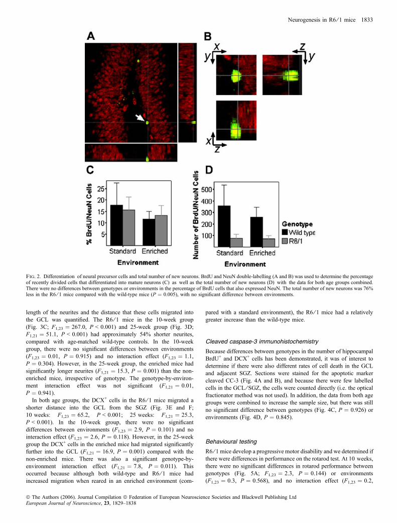
mature neuronal marker NeuN was quantified using confocal micr-oscopy (Fig. 2A and B). There was no significant difference betweengenotypes in the proportion of BrdU+ cells that also expressed NeuN(Fig. 2C; F1,15 ¼ 0.003, P ¼ 0.957), nor was there any influence ofenvironmental enrichment (F1,15 ¼ 0.6, P ¼ 0.468), and no interac-tion effect was found (F1,15 ¼ 0.1, P ¼ 0.751, both age groupscombined). There was, however, a significant difference between
Neurogenesis in R6 ⁄ 1 mice 1831
ª The Authors (2006). Journal Compilation ª Federation of European Neuroscience Societies and Blackwell Publishing LtdEuropean Journal of Neuroscience, 23, 1829–1838

genotypes in the total number of BrdU+ ⁄NeuN+ cells, with the R6 ⁄ 1mice having 76% fewer double-labelled cells than wild-type mice(Fig. 2D; F1,15 ¼ 10.8, P ¼ 0.005, both age groups combined). Therewas no significant difference between environments (F1,15 ¼ 0.5,P ¼ 0.490) and no interaction effect (F1,15 ¼ 0.4, P ¼ 0.516) for thetotal number of BrdU+ ⁄NeuN+ cells.
Doublecortin immunohistochemistry
DCX is a microtubule-associated protein that is expressed in virtuallyall migrating neural precursors in the developing central nervoussystem (Gleeson et al., 1998) and has been validated as a marker forneurogenesis in adult rodents (Brown et al., 2003; Rao & Shetty,2004; Couillard-Despres et al., 2005). In the 10-week group, thenumber of DCX+ cells was decreased in the R6 ⁄ 1 mice by 56%(Fig. 3A and I; F1,23 ¼ 85.7, P < 0.001), and by 66% in the 25-week
group (Fig. 3B; F1,21 ¼ 36.7, P < 0.001), compared with age-matched wild-type controls. In the 10-week group, there were nosignificant differences between environments (F1,23 ¼ 0.9, P ¼0.362) and no interaction effect (F1,23 ¼ 0.4, P ¼ 0.550). However,in the 25-week group, the enriched mice had twice as many DCX+
cells (F1,21 ¼ 15.6, P ¼ 0.001) as the non-enriched mice, irrespectiveof genotype. The genotype-by-environment interaction effect was notsignificant (F1,21 ¼ 0.9, P ¼ 0.360). In addition, at this time point,male mice had approximately 45% fewer DCX+ cells than femalemice (F1,19 ¼ 5.43, P ¼ 0.031). However, no significant differencesbetween sexes were observed at 10 weeks, or at either time point forother variables.The DCX+ cells in the R6 ⁄ 1 mice appeared morphologically
different compared with those found in the wild-type mice. TheR6 ⁄ 1 DCX+ cells had smaller and irregular shaped somas, fewerand smaller neurites, and appeared to cluster in the SGZ and did notmigrate as far into the GCL (Fig. 3G and H). In view of this, the
Fig. 1. BrdU staining in the hippocampus 4 weeks after BrdU injections. The number of BrdU+ cells in the dentate gyrus is decreased in the R6 ⁄ 1 mice at10 weeks (A; P < 0.001) and also at 25 weeks, but not significantly so at this later time point (B; P ¼ 0.790). The 25-week mice reared in an enriched environmenthad significantly more BrdU+ cells compared with non-enriched mice in the same age group (P ¼ 0.007), irrespective of genotype. There were no differences in thenumber of BrdU+ cells in the SVZ at 10 weeks (C) and 25 weeks (D) between genotypes or conditions. Representative histological pictures of BrdUimmunostaining in the DG (E). DG, dentate gyrus; GCL, granular cell layer; Hi, Hilus. Scale bar, 0.5 mm.
1832 S. E. Lazic et al.
ª The Authors (2006). Journal Compilation ª Federation of European Neuroscience Societies and Blackwell Publishing LtdEuropean Journal of Neuroscience, 23, 1829–1838
length of the neurites and the distance that these cells migrated intothe GCL was quantified. The R6 ⁄ 1 mice in the 10-week group(Fig. 3C; F1,23 ¼ 267.0, P < 0.001) and 25-week group (Fig. 3D;F1,21 ¼ 51.1, P < 0.001) had approximately 54% shorter neurites,compared with age-matched wild-type controls. In the 10-weekgroup, there were no significant differences between environments(F1,23 ¼ 0.01, P ¼ 0.915) and no interaction effect (F1,23 ¼ 1.1,P ¼ 0.304). However, in the 25-week group, the enriched mice hadsignificantly longer neurites (F1,21 ¼ 15.3, P ¼ 0.001) than the non-enriched mice, irrespective of genotype. The genotype-by-environ-ment interaction effect was not significant (F1,21 ¼ 0.01,P ¼ 0.941).
In both age groups, the DCX+ cells in the R6 ⁄ 1 mice migrated ashorter distance into the GCL from the SGZ (Fig. 3E and F;10 weeks: F1,23 ¼ 65.2, P < 0.001; 25 weeks: F1,21 ¼ 25.3,P < 0.001). In the 10-week group, there were no significantdifferences between environments (F1,23 ¼ 2.9, P ¼ 0.101) and nointeraction effect (F1,23 ¼ 2.6, P ¼ 0.118). However, in the 25-weekgroup the DCX+ cells in the enriched mice had migrated significantlyfurther into the GCL (F1,21 ¼ 16.9, P ¼ 0.001) compared with thenon-enriched mice. There was also a significant genotype-by-environment interaction effect (F1,21 ¼ 7.8, P ¼ 0.011). Thisoccurred because although both wild-type and R6 ⁄ 1 mice hadincreased migration when reared in an enriched environment (com-
pared with a standard environment), the R6 ⁄ 1 mice had a relativelygreater increase than the wild-type mice.
Cleaved caspase-3 immunohistochemistry
Because differences between genotypes in the number of hippocampalBrdU+ and DCX+ cells has been demonstrated, it was of interest todetermine if there were also different rates of cell death in the GCLand adjacent SGZ. Sections were stained for the apoptotic markercleaved CC-3 (Fig. 4A and B), and because there were few labelledcells in the GCL ⁄ SGZ, the cells were counted directly (i.e. the opticalfractionator method was not used). In addition, the data from both agegroups were combined to increase the sample size, but there was stillno significant difference between genotypes (Fig. 4C, P ¼ 0.926) orenvironments (Fig. 4D, P ¼ 0.845).
Behavioural testing
R6 ⁄ 1 mice develop a progressive motor disability and we determined ifthere were differences in performance on the rotarod test. At 10 weeks,there were no significant differences in rotarod performance betweengenotypes (Fig. 5A; F1,23 ¼ 2.3, P ¼ 0.144) or environments(F1,23 ¼ 0.3, P ¼ 0.568), and no interaction effect (F1,23 ¼ 0.2,
Fig. 2. Differentiation of neural precursor cells and total number of new neurons. BrdU and NeuN double-labelling (A and B) was used to determine the percentageof recently divided cells that differentiated into mature neurons (C) as well as the total number of new neurons (D) with the data for both age groups combined.There were no differences between genotypes or environments in the percentage of BrdU cells that also expressed NeuN. The total number of new neurons was 76%less in the R6 ⁄ 1 mice compared with the wild-type mice (P ¼ 0.005), with no significant difference between environments.
Neurogenesis in R6 ⁄ 1 mice 1833
ª The Authors (2006). Journal Compilation ª Federation of European Neuroscience Societies and Blackwell Publishing LtdEuropean Journal of Neuroscience, 23, 1829–1838

P ¼ 0.646). In addition, there were no significant differences betweengenotypes (Fig. 5B; F1,21 ¼ 0.9, P ¼ 0.344) or environments(F1,21 ¼ 3.3, P ¼ 0.087) in the 25-week group, and no interactioneffect (F1,21 ¼ 0.2, P ¼ 0.653); however, the R6 ⁄ 1 mice performedconsistently worse than wild-type mice under all conditions.
Discussion
The number of BrdU+ cells in the DG was decreased in both the olderand the younger R6 ⁄ 1 mice compared with wild-type litter-mates, butthis was only significant in the younger 10-week group. This indicates
Fig. 3. DCX immunohistochemistry in the dentate gyrus. The number of DCX+ cells is decreased in the R6 ⁄ 1 mice at 10 weeks (A; P < 0.001) and 25 weeks(B; P < 0.001), and at 25 weeks the enriched mice had a greater number of DCX+ cells compared with non-enriched mice (P ¼ 0.001). R6 ⁄ 1 mice also had shorterneurites at 10 weeks (C; P < 0.001) and 25 weeks (D; P < 0.001), and at 25 weeks the enriched mice had longer neurites compared with non-enriched mice(P ¼ 0.001). The DCX+ cells in the R6 ⁄ 1 mice migrated a shorter distance into the granular cell layer at 10 weeks (E; P < 0.001) and 25 weeks (F; P < 0.001),and at 25 weeks the DCX+ cells of enriched mice migrated a greater distance compared with non-enriched mice (P ¼ 0.001). Representative immunostained sectionsshowing the morphology of DCX+ cells in wild-type (G) and R6 ⁄ 1 (H) mice, and overall cell numbers in mice housed in a standard environment (I). Scale bars,0.025 mm (G and H), 0.5 mm (I).
1834 S. E. Lazic et al.
ª The Authors (2006). Journal Compilation ª Federation of European Neuroscience Societies and Blackwell Publishing LtdEuropean Journal of Neuroscience, 23, 1829–1838

that the survival of NPCs is already compromised in the youngtransgenic mice, ahead of the development of overt neurologicaldeficits. This is the exact same pattern that has been described in the
R6 ⁄ 2 transgenic line, with a greater difference between genotypes inyounger mice compared with older mice (i.e. a significant age-by-genotype interaction effect; Gil et al., 2005). Furthermore, in our
Fig. 4. Cleaved caspase-3 (CC-3) staining in the granular cell layer of the dentate gyrus under a 10· (A) and 63· (B) objective. There are no significantdifferences between genotypes (C; P ¼ 0.926) or environments (D; P ¼ 0.845), with the data for both age groups combined. Scale bars, 100 lm and 10 lm for Aand B, respectively.
Fig. 5. Performance on the rotarod test. There was no significant difference between genotypes or environments at 10 weeks (A) or 25 weeks (B), although R6 ⁄ 1mice consistently did less well than wild-type mice in all groups.
Neurogenesis in R6 ⁄ 1 mice 1835
ª The Authors (2006). Journal Compilation ª Federation of European Neuroscience Societies and Blackwell Publishing LtdEuropean Journal of Neuroscience, 23, 1829–1838

study, there was no effect of enrichment in the younger mice, whichmight reflect the limited time (6 weeks) that these mice were housedunder such conditions. In contrast to the older mice, which had beenenriched for 21 weeks, the environmentally enriched mice hadsignificantly more BrdU+ cells compared with non-enriched mice,irrespective of genotype. Whether the effect of enrichment in the oldermice was due to these mice being more responsive to enrichment, ormore likely, due to a much longer exposure to an enrichedenvironment remains unresolved, but it does indicate that bothtransgenic and wild-type mice can respond to enrichment with anincrease in the number of BrdU+ cells.The proportion of BrdU+ cells that also expressed the mature neural
marker NeuN did not differ between genotypes or environments,indicating that the differentiation of NPCs was not affected bytransgenic status or housing conditions, but the total number ofBrdU+ ⁄NeuN+ cells was 76% less in the transgenic mice. Similarresults were also found in the R6 ⁄ 2 mice by Gil et al. (2005). Thus,there is evidence that both the proliferation (Lazic et al., 2004) andnow survival of NPCs is reduced in the R6 transgenic mice, while theproportion that differentiate into mature neurons is the same as wild-type mice.In this study, we also studied DCX as a marker of newly born
neurons, and the results for the number of DCX+ cells largely reflectsthe BrdU results. R6 ⁄ 1 mice had fewer DCX+ cells in the DG at both10 and 25 weeks, and in the older 25-week group, the mice housed inan enriched environment had a greater number of labelled cells. Again,these results are in good agreement with results from the R6 ⁄ 2 mice(Gil et al., 2005). The morphology of DCX-labelled cells was alsoinvestigated and it was discovered that the R6 ⁄ 1 mice had cells withshorter neurites, smaller and irregular shaped cell bodies, and migrateda shorter distance into the GCL, as we have also recently described inthe R6 ⁄ 2 mice (Phillips et al., 2005). Environmental enrichment couldrescue these deficits in the 25-week group, suggesting that these cellsare still amenable to manipulation in the older, diseased rodent brain.It is not known if these differences in morphology persist in the moremature neurons of the GCL.As the R6 ⁄ 1 mice have decreased neurogenesis, it was of interest to
examine whether cell death was altered in the GCL, given that theinduction of apoptosis in the GCL can increase proliferation (Pawlaket al., 2002). In addition, there seems to be a balance betweenproliferation and apoptosis in wild-type mice, with higher levels ofproliferation being associated with higher levels of apoptosis and viceversa (Kim et al., 2001; Biebl et al., 2004; Heine et al., 2004). In thepresent experiment, however, there were no differences betweengenotypes or environments in the number of CC-3 immunopositivecells in the GCL ⁄ SGZ. Although the actual number of apoptotic cellsis low, our findings are consistent with other studies which have alsonot found increased apoptotic (or necrotic) cell death in the striatumand other brain regions of the R6 compared with wild-type mice(Turmaine et al., 2000).The reasons for a decrease in hippocampal NPC proliferation (Lazic
et al., 2004) and ⁄ or survival are not known, but there are severalpossibilities. First, it could be due to the absence of a positiveneurotrophic factor such as brain-derived neurotrophic factor (BDNF).BDNF is known both to increase proliferation in the hippocampus(Lee et al., 2002) and to be increased by environmental enrichment(Spires et al., 2004b). Previous work has demonstrated that BDNFprotein and mRNA levels are decreased in the cortex and striatum oftransgenic HD mice (Zuccato et al., 2001, 2005), and protein levelsare decreased in the hippocampus of R6 ⁄ 1 mice (Spires et al., 2004b).However, in a study examining post-mortem human tissue, there wasno evidence of decreased BDNF levels in the hippocampus (Ferrer
et al., 2000). A second possibility relates to deficits in dopaminergictransmission in the R6 mice (Cha et al., 1998; Bibb et al., 2000;Yohrling et al., 2003). A third, more prosaic explanation is reducedmotor activity in the R6 ⁄ 1 mice, given that physical activity is knownto increase proliferation of hippocampal NPCs (van Praag et al.,1999a, b). However, the motor ability of the mice, as determined byrotarod testing, was similar between genotypes in both the 10-weekand the 25-week groups (although worse in the transgenic mice),which suggests that the ability of mice to move about was notsignificantly compromised, although the daily activity of individualmice in their cages was not measured. Finally, a decreased number ofBrdU+ cells might be due to an increase in stress hormones, which candecrease neurogenesis (Lemaire et al., 2000; Wong & Herbert, 2004).There is evidence in humans that serum cortisol levels are higher inHD patients than controls (Heuser et al., 1991; Leblhuber et al., 1995),although it is not known if there are changes in these transgenic mousemodels. Whatever the cause, it is likely that there is not a major deficitin the NPCs themselves, as Phillips et al. (2005) have demonstratedthat NPCs from both wild-type and R6 ⁄ 2 mice have similar rates ofproliferation in vitro.Unlike the results in the DG, there was no difference in the number of
BrdU+ cells in the SVZ between R6 ⁄ 1 and wild-type mice. Gil et al.(2004, 2005) found similar histological results using R6 ⁄ 2 mice, butthere appears to be an increase in proliferative cells in this region in thehuman brain at post-mortem (Curtis et al., 2003; Curtis et al., 2005a, b).These discrepant results may reflect differences in human vs. rodentneural precursor cells, neurogenic environments or organization of theSVZ (Sanai et al., 2004). Alternatively, the difference might be due tothe large amount of cell loss that is generally found in the human HDstriatum at post-mortem, which is not seen in the R6 mouse models(Turmaine et al., 2000), as cell loss or cell death has been shown tostimulate NPCs to increase their rate of proliferation ⁄ survival (Nait-Oumesmar et al., 1999; Magavi et al., 2000; Arvidsson et al., 2002). Inparticular, mice with striatal quinolinic acid lesions have increasedproliferation of NPCs in the SVZ (Tattersfield et al., 2004; Collin et al.,2005), although such a response seems to be absent in the R6 ⁄ 2 mousemodel of HD (Phillips et al., 2005). This suggests that even if there wascell loss in the striatum of the transgenic mice, the NPCsmay not be ableto respond normally.The functional significance of these changes in neurogenesis
remains speculative, but they may help explain some of thecognitive deficits, such as impaired spatial cognition, which ischaracteristic of these mice, although deficits in synaptic plasticityand changes in existing circuits also contribute to the behaviouraldeficits (Lione et al., 1999; Murphy et al., 2000; Spires et al.,2004a; Gibson et al., 2005). Furthermore, the beneficial effects ofenvironmental enrichment on these mice (van Dellen et al., 2000;Hockly et al., 2002; Spires et al., 2004b) may be mediated, in part,by changes in neurogenesis. This is further supported by the findingthat increasing neurogenesis with fluoxetine treatment improves theperformance of R6 ⁄ 1 on a hippocampal-dependent memory task(Grote et al., 2005). In terms of patients with HD, it is possible thatsuch abnormalities in neurogenesis may underlie some of thecognitive aspects of this disease as well as the depression, given thatdecreased neurogenesis is associated with depression (Santarelliet al., 2003). Depression can be the initial presenting feature of HD,can precede the onset of motor symptoms and cognitive symptomsby many years, and occurs in individuals who are not even aware oftheir risk (reviewed in Craufurd & Snowdon, 2002). This isconsistent with the finding here that hippocampal neurogenesis isdecreased early in the disease, before mice display motorabnormalities.
1836 S. E. Lazic et al.
ª The Authors (2006). Journal Compilation ª Federation of European Neuroscience Societies and Blackwell Publishing LtdEuropean Journal of Neuroscience, 23, 1829–1838

In summary, the R6 ⁄ 1 mouse model of HD has reducedhippocampal neurogenesis, and this is mainly the result of decreasedproliferation and survival of NPCs. In addition, neural precursor cellsin this region have an altered morphology and migrate a shorterdistance into the GCL. Furthermore, these deficits can be partlyrestored by rearing the animals in an enriched environment. Becauseenvironmental enrichment is known to delay the onset of symptoms inthese mice (van Dellen et al., 2000), it is possible that neurogenesismay be manipulated to improve performance in these mice. As such,therapies aiming to increase neurogenesis may prove efficacious forsome symptoms of HD.
Acknowledgements
S.E.L. is supported by a Natural Sciences and Engineering Research Council ofCanada postgraduate scholarship and ORS Award (UK). H.G. is supported bythe Christopher Welch Trust and the Huntington’s Disease Association.R.A.B.’s work is supported by the Hereditary Disease Foundation and MRC(UK). A.J.H is supported by the NHMRC (Australia).
Abbreviations
BDNF, brain-derived neurotrophic factor; CC-3, cleaved caspase-3; BrdU,bromodeoxyuridine; DCX, doublecortin; DG, dentate gyrus; GCL, granular celllayer; HD, Huntington’s disease; OB, olfactory bulb; NPC, neural precursorcell; SGZ, subgranular zone; SVZ, subventricular zone.
References
Arvidsson, A., Collin, T., Kirik, D., Kokaia, Z. & Lindvall, O. (2002) Neuronalreplacement from endogenous precursors in the adult brain after stroke. Nat.Med., 8, 963–970.
Barker, R.A. & Dunnett, S.B. (1999) Neural Repair, Transplantation andRehabilitation. Psychology Press, East Sussex.
Belluzzi, O., Benedusi, M., Ackman, J. & LoTurco, J.J. (2003) Electro-physiological differentiation of new neurons in the olfactory bulb.J. Neurosci., 23, 10411–10418.
Bibb, J.A., Yan, Z., Svenningsson, P., Snyder, G.L., Pieribone, V.A., Horiuchi,A., Nairn, A.C., Messer, A. & Greengard, P. (2000) Severe deficiencies indopamine signaling in presymptomatic Huntington’s disease mice. Proc.Natl Acad. Sci. USA, 97, 6809–6814.
Biebl, M., Cooper, C.M., Winkler, J. & Kuhn, H.G. (2004) Analysis ofneurogenesis and programmed cell death reveals a self-renewing capacity inthe adult rat brain. Neurosci. Lett., 291, 17–20.
Brown, J.P., Couillard-Despres, S., Cooper-Kuhn, C.M., Winkler, J., Aigner, L.& Kuhn, H.G. (2003) Transient expression of doublecortin during adultneurogenesis. J. Comp. Neurol., 467, 1–10.
Carlen, M., Cassidy, M.R., Brismar, H., Smith, G.A., Enquist, L.W. & Frisen, J.(2002) Functional integration of adult-born neurons. Curr. Biol., 12, 606–608.
Cha, J.-H.J., Kosinski, C.M., Kerner, J.A., Alsdorf, S.A., Mangiarini, L.,Davies, S.W., Penny, J.B., Bates, G.P. & Young, A.B. (1998) Altered brainneurotransmitter receptors in transgenic mice expressing a portion of anabnormal human Huntington disease gene. Proc. Natl Acad. Sci. USA, 95,6480–6485.
Collin, T., Arvidsson, A., Kokaia, Z. & Lindvall, O. (2005) Quantitativeanalysis of the generation of different striatal neuronal subtypes in the adultbrain following excitotoxic injury. Exp. Neurol., 195, 71–80.
Couillard-Despres, S., Winner, B., Schaubeck, S., Aigner, R., Vroemen, M.,Weidner, N., Bogdahn, U., Winkler, J., Kuhn, H.-G. & Aigner, L. (2005)Doublecortin expression levels in adult brain reflect neurogenesis. Eur. J.Neurosci., 21, 1–14.
Craufurd, D. & Snowdon, J. (2002) Neuropsychological and neuropsychia-tric aspects of Huntington’s disease. In: Bates, G., Harper, P. & Jones, L.(Eds), Huntington’s Disease, 3rd edn. Oxford University Press, Oxford,pp. 62–95.
Crawley, M.J. (2002) Statistical Computing: an Introduction to Data AnalysisUsing S-Plus. Wiley, Hoboken.
Curtis, M.A., Penney, E.B., Pearson, J., Dragunow, M., Connor, B. & Faull,R.L.M. (2005a) The distribution of progenitor cells in the subependymal
layer of the lateral ventricle in the normal and Huntington’s disease humanbrain. Neuroscience, 132, 777–788.
Curtis, M.A., Penney, E.B., Pearson, A.G., van Roon-Mom, W.M.C.,Butterworth, N.J., Dragunow, M., Connor, B. & Faull, R.L.M. (2003)Increased cell proliferation and neurogenesis in the adult human Hunting-ton’s disease brain. Proc. Natl Acad. Sci. USA, 100, 9023–9027.
Curtis, M.A., Waldvogel, H.J., Synek, B. & Faull, R.L.M. (2005b) Ahistochemical and immunohistochemical analysis of the subependymal layerin the normal and Huntington’s disease brain. J. Chem. Neuroanat., 30,55–66.
van Dellen, A., Blakemore, C., Deacon, R., York, D. & Hannan, A.J. (2000)Delaying the onset of Huntington’s disease in mice. Nature, 404, 721–722.
Drapeau, E., Mayo, W., Aurousseau, C., Le Moal, M., Piazza, P.-V. & Abrous,D.N. (2003) Spatial memory performances of aged rats in the water mazepredict levels of hippocampal neurogenesis. Proc. Natl Acad. Sci. USA, 100,14385–14390.
Ferrer, I., Goutan, E., Marin, C., Rey, M.J. & Ribalta, T. (2000) Brain-derived neurotrophic factor in Huntington disease. Brain Res., 866, 257–261.
Gibson, H.E., Reim, K., Brose, N., Morton, A.J. & Jones, S. (2005) A similarimpairment in CA3 mossy fibre LTP in the R6 ⁄ 2 mouse model ofHuntington’s disease and in the complexin II knockout mouse. Eur. J.Neurosci., 22, 1701–1712.
Gil, J.M.A.C., Leist, M., Popovic, N., Brundin, P. & Petersen, A. (2004)Asialoerythropoetin is not effective in the R6 ⁄ 2 line of Huntington’s diseasemice. BMC Neurosci., 5, 17.
Gil, J.M.A.C., Mohapel, P., Araujo, I.M., Popovic, N., Li, J.-Y., Brundin, P. &Petersen, A. (2005) Reduced hippocampal neurogenesis in R6 ⁄ 2 transgenicHuntington’s mice. Neurobiol. Dis., 20, 744–751.
Gleeson, J.G., Allen, K.M., Fox, J.W., Lamperti, E.D., Berkovic, S., Scheffer,I., Cooper, E.C., Dobyns, W.B., Minnerath, S.R., Ross, M.E. & Walsh, C.A.(1998) Doublecortin, a brain-specific gene mutated in human X-linkedlissencephaly and double cortex syndrome, encodes a putative signalingprotein. Cell, 92, 63–72.
Grote, H.E., Bull., N.D., Howard, M.L., van Dellen, A., Blakemore, C.,Bartlett, P.F. & Hannan, A.J. (2005) Cognitive disorders and neurogenesisdeficits in Huntington’s disease mice are rescued by fluoxetine. Eur. J.Neurosci., 22, 2081–2088.
Gundersen, H.J., Bagger, P., Bendtsen, T.F., Evans, S.M., Korbo, L.,Marcussen, N., Moller, A., Nielsen, K., Nyengaard, J.R., Pakkenberg, B.,Sorensen, F.B., Vesterby, A. & West, M.J. (1988) The new stereologicaltools: disector, fractionator, nucleator and point sampled intercepts and theiruse in pathological research and diagnosis. APMIS, 96, 857–881.
Heine, V.M., Maslam, S., Joels, M. & Lucassen, P.J. (2004) Prominent declineof newborn cell proliferation, differentiation, and apoptosis in the agingdentate gyrus, in absence of and age-related hypothalamus–pituitary–adrenalaxis activation. Neurobiol. Aging, 25, 361–375.
Heuser, I.J., Chase, T.N. & Mouradian, M.M. (1991) The limbic–hypothalam-ic–pituitary–adrenal axis in Huntington’s disease. Biol. Psychiatry, 30, 943–952.
Ho, L.W., Carmichael, J., Swartz, J., Wyttenbach, A., Rankin, J. & Rubinsztein,D.C. (2001) The molecular biology of Huntington’s disease. Psychol. Med.,31, 3–14.
Hockly, E., Cordery, P.M., Woodman, B., Mahal, A., van Dellen, A.,Blakemore, C., Lewis, C.M., Hannan, A.J. & Bates, G.P. (2002)Environmental enrichment slows disease progression in R6 ⁄ 2 Huntington’sdisease mice. Ann. Neurol., 51, 235–242.
Howell, D.C. (1992) Statistical Methods for Psychology, 3rd edn. DuxburyPress, Belmont.
Kempermann, G. & Gage, F.H. (2002a) Genetic influence on phenotypicdifferentiation in adult hippocampal neurogenesis. Brain Res. Dev. BrainRes., 134, 1–12.
Kempermann, G. & Gage, F.H. (2002b) Genetic determinants of adulthippocampal neurogenesis correlate with acquisition, but not probe trialperformance, in the water maze task. Eur. J. Neurosci., 16, 129–136.
Kempermann, G., Gast, D. & Gage, F.H. (2002) Neuroplasticity in old age:sustained fivefold induction of hippocampal neurogenesis by long-termenvironmental enrichment. Ann. Neurol., 52, 135–143.
Kim, M.J., Kim, Y., Kim, S.A., Lee, H.J., Choe, B.K., Nam, M., Kim, B.S.,Kim, J.-W., Yim, S.-V., Kim, C.-J. & Chung, J.-H. (2001) Increases in cellproliferation and apoptosis in dentate gyrus of anorexia (anx ⁄ anx) mice.Neurosci. Lett., 302, 109–112.
Lazic, S.E. & Barker, R.A. (2005) Cell-based therapies for disorders of thecentral nervous system. Expert Opin. Ther. Patents, 15, 1361–1376.
Neurogenesis in R6 ⁄ 1 mice 1837
ª The Authors (2006). Journal Compilation ª Federation of European Neuroscience Societies and Blackwell Publishing LtdEuropean Journal of Neuroscience, 23, 1829–1838

Lazic, S.E., Grote, H., Armstrong, R.J.E., Blakemore, C., Hannan, A.J., vanDellen, A. & Barker, R.A. (2004) Decreased hippocampal cell proliferationin R6 ⁄ 1 Huntington’s mice. Neuroreport, 15, 811–813.
Leblhuber, F., Peichl, M., Neubauer, C., Reisecker, F., Steinparz, F.X.,Windhager, E. & Maschek, W. (1995) Serum dehydroepiandrosterone andcortisol measurements in Huntington’s chorea. J. Neurol. Sci., 132, 76–79.
Lee, J., Duan, W. & Mattson, M.P. (2002) Evidence that brain-derivedneurotrophic factor is required for basal neurogenesis and mediates, in part,the enhancement of neurogenesis by dietary restriction in the hippocampusof adult mice. J. Neurochem., 82, 1367–1375.
Lemaire, V., Koehl, M., Le Moal, M. & Abrous, D.N. (2000) Prenatal stressproduces learning deficits associated with an inhibition of neurogenesis inthe hippocampus. Proc. Natl Acad. Sci., USA, 97, 11032–11037.
Lione, L.A., Carter, R.J., Hunt, M.J., Bates, G.P., Morton, A.J. & Dunnett, S.B.(1999) Selective discrimination learning impairments in mice expressing thehuman Huntington’s disease mutation. J. Neurosci., 19, 10428–10437.
Magavi, S.S., Leavitt, B.R. & Macklis, J.D. (2000) Induction of neurogenesisin the neocortex of adult mice. Nature, 405, 951–955.
Mangiarini, L., Sathasivam, K., Seller, M., Cozens, B., Harper, A., Hethering-ton, C., Lawton, M., Trottier, Y., Lehrach, H., Davies, S.W. & Bates, G.P.(1996) Exon 1 of the HD gene with an expanded CAG repeat is sufficient tocause a progressive neurological phenotype in transgenic mice. Cell, 87,493–506.
Murphy, K.P.S.J., Carter, R.J., Lione, L.A., Mangiarini, L., Mahal, A., Bates,G.P., Dunnett, S.B. & Morton, A.J. (2000) Abnormal synaptic plasticity andimpaired spatial cognition in mice transgenic for exon 1 of the humanHuntington’s disease mutation. J. Neurosci., 20, 5115–5123.
Nait-Oumesmar, B., Decker, L., Lachaplle, F., Avellana-Adalid, V., Bachelin,C. & Baron-Van Evercooren, A. (1999) Progenitor cells of the adult mousesubventricular zone proliferate, migrate and differentiate into oligodendro-cytes after demyelination. Eur. J. Neurosci., 11, 4357–4366.
Pawlak, R., Skrzypiec, A., Sulkowski, S. & Buczko, W. (2002) Ethanol-induced neurotoxicity is counterbalanced by increased cell proliferation inmouse dentate gyrus. Neurosci. Lett., 327, 83–86.
Phillips, W., Morton, A.J. & Barker, R.A. (2005) Abnormalities ofneurogenesis in the R6 ⁄ 2 mouse model of Huntington’s disease areattributable to the in vivo microenvironment. J. Neurosci., 25, 11564–11576.
van Praag, H., Christie, B.R., Sejnowski, T.J. & Gage, F.H. (1999a) Runningenhances neurogenesis, learning, and long-term potentiation in mice. Proc.Natl Acad. Sci. USA, 96, 13427–13431.
van Praag, H., Kempermann, G. & Gage, F.H. (1999b) Running increases cellproliferation and neurogenesis in the adult mouse dentate gyrus. Nat.Neurosci., 2, 266–270.
van Praag, H., Schinder, A.F., Christie, B.R., Toni, N., Palmer, T.D. & Gage,F.H. (2002) Functional neurogenesis in the adult hippocampus. Nature, 415,1030–1034.
Rao, M.S. & Shetty, A.K. (2004) Efficacy of doublecortin as a marker toanalyse the absolute number and dendritic growth of newly generatedneurons in the adult dentate gyrus. Eur. J. Neurosci., 19, 234–246.
Sanai, N., Tramontin, A.D., Quinones-Hinojosa, A., Barbaro, N.M., Gupta, N.,Kunwar, S., Lawton, M.T., McDermott, M.W., Parsa, A.T., Verdugo, J.M.-G.,Berger, M.S. & Alvarez-Buylla, A. (2004) Unique astrocyte ribbon in adulthuman brain contains neural stem cells but lacks chain migration.Nature, 427,740–744.
Santarelli, L., Saxe, M., Gross, C., Surget, A., Battaglia, F., Dulawa, S.,Weisstaub, N., Lee, J., Duman, R., Arancio, O., Belzung, C. & Hen, R.(2003) Requirement of hippocampal neurogenesis for the behavioral effectsof antidepressants. Science, 301, 805–809.
Shors, T.J., Miesegaes, G., Beylin, A., Zhao, M., Rydel, T. & Gould, E. (2001)Neurogenesis in the adult is involved in the formation of trace memories.Nature, 410, 372–376.
Spires, T.L., Grote, H.E., Garry, S., Cordery, P.M., van Dellen, A., Blakemore,C. & Hannan, A.J. (2004a) Dendritic spine pathology and deficits inexperience-dependent dendritic plasticity in R6 ⁄ 1 Huntington’s diseasetransgenic mice. Eur. J. Neurosci., 19, 2799–2807.
Spires, T.L., Grote, H.E., Varshney, N.K., Cordery, P.M., van Dellen, A.,Blakemore, C. & Hannan, A.J. (2004b) Environmental enrichment rescuesprotein deficits in a mouse model of Huntington’s disease, indicating apossible disease mechanism. J. Neurosci., 24, 2270–2276.
Tattersfield, A.S., Croon, R.J., Liu, Y.W., Kells, A.P., Faull, R.L.M. & Connor,B. (2004) Neurogenesis in the striatum of the quinolinic acid lesion model ofHuntington’s disease. Neuroscience, 127, 319–332.
Turmaine, M., Raza, A., Mahal, A., Mangiarini, L., Bates, G.P. & Davies, S.W.(2000) Nonapoptotic neurodegeneration in a transgenic mouse model ofHuntington’s disease. Proc. Natl Acad. Sci. USA, 97, 8093–8097.
Wong, E.Y.H. & Herbert, J. (2004) The corticoid environment: a determiningfactor for neural progenitors’ survival in the adult hippocampus. Eur. J.Neurosci., 20, 2491–2498.
Yohrling, G.J., Jiang, G.C., DeJohn, M.M., Miller, D.W., Young, A.B.,Vrana, K.E. & Cha, J.H. (2003) Analysis of cellular, transgenic andhuman models of Huntington’s disease reveals tyrosine hydroxylasealterations and substantia nigra neuropathology. Brain Res. Mol. BrainRes., 119, 28–36.
Zuccato, C., Ciammola, A., Rigamonti, D., Leavitt, B.R., Goffredo, D., Conti,L., MacDonald, M.E., Friedlander, R.M., Silani, V., Hayden, M.R.,Timmusk, T., Sipione, S. & Cattaneo, E. (2001) Loss of Huntingtin-mediated BDNF gene transcription in Huntington’s disease. Science, 293,493–498.
Zuccato, C., Liber, D., Ramos, C., Tarditi, A., Rigamonti, D., Tartari, M.,Valenza, M. & Cattaneo, E. (2005) Progressive loss of BDNF in a mousemodel of Huntington’s disease and rescue by BDNF delivery. Pharmacol.Res., 52, 133–139.
1838 S. E. Lazic et al.
ª The Authors (2006). Journal Compilation ª Federation of European Neuroscience Societies and Blackwell Publishing LtdEuropean Journal of Neuroscience, 23, 1829–1838