NTS-Polyplex: a potential nanocarrier for neurotrophic therapy of Parkinson's disease
18
Review Article NTS-Polyplex: a potential nanocarrier for neurotrophic therapy of Parkinson's disease Daniel Martinez-Fong, MD, PhD a,b, ⁎ , Michael J. Bannon, PhD c , Louis-Eric Trudeau, PhD d , Juan A. Gonzalez-Barrios, MD, PhD e , Martha L. Arango-Rodriguez, PhD f , Nancy G. Hernandez-Chan, MSc a , David Reyes-Corona, MSc a , Juan Armendáriz-Borunda, PhD g , Ivan Navarro-Quiroga, PhD h a Departamento de Fisiología, Biofísica y Neurociencias, México D.F., México b Doctorado en Nanociencias y Nanotecnología; CINVESTAV-I.P.N., México Distrito Federal, México c Department of Pharmacology, Wayne State University School of Medicine, Detroit, Michigan, USA d Department of Pharmacology, Groupe de Recherche sur le Système Nerveux Central, Faculty of Medicine, Université de Montréal, Montréal, Québec, Canada e Laboratorio de Medicina Genómica, Hospital Regional “1o. de Octubre,” México Distrito Federal, México f Instituto de Ciencias, Facultad de Medicina Clínica Alemana Universidad del Desarrollo. Santiago, Chile g Institute for Molecular Biology and Gene Therapy, Department of Molecular Biology and Genomics, University of Guadalajara, Guadalajara, Mexico h KAI-Research, Rockville, Maryland, USA Received 4 October 2011; accepted 20 February 2012 Abstract Nanomedicine has focused on targeted neurotrophic gene delivery to the brain as a strategy to stop and reverse neurodegeneration in Parkinson's disease. Because of improved transfection ability, synthetic nanocarriers have become candidates for neurotrophic therapy. Neurotensin (NTS)-polyplex is a “Trojan horse” synthetic nanocarrier system that enters dopaminergic neurons through NTS receptor internalization to deliver a genetic cargo. The success of preclinical studies with different neurotrophic genes supports the possibility of using NTS-polyplex in nanomedicine. In this review, we describe the mechanism of NTS-polyplex transfection. We discuss the concept that an effective neurotrophic therapy requires a simultaneous effect on the axon terminals and soma of the remaining dopaminergic neurons. We also discuss the future of this strategy for the treatment of Parkinson's disease. From the Clinical Editor: This review paper focuses on nanomedicine-based treatment of Parkinson's disease, a neurodegenerative condition with existing symptomatic but no curative treatment. Neurotensin-polyplex is a synthetic nanocarrier system that enables delivery of genetic cargo to dopaminergic neurons via NTS receptor internalization. © 2012 Elsevier Inc. All rights reserved. Key words: Neurorestoration; Neuroprotection; Neurodegeneration; Regeneration; Survival Parkinson's Disease (PD) results from a progressive loss of dopaminergic neurons in the substantia nigra with concurrent gliosis in the midbrain. 1 These early-stage events in PD result in dopamine depletion in the caudoputamen, with the lateral nigral projections to the putamen being the most affected. 2 As the disease advances, positron emission tomography (PET) shows a progressive loss of dopamine storage in striatal, cingulated, and frontal brain regions. 3 The earliest and most striking physical disabilities resulting from these changes in the basal ganglia are motor impairments. These include paucity and slowness of movement (akinesia, bradykinesia), muscle stiffness (rigidity), tremor at rest, and postural instability. An increasing number of clinical reports POTENTIAL CLINICAL RELEVANCE Nanomedicine: Nanotechnology, Biology, and Medicine 8 (2012) 1052 – 1069 The authors have no financial, personal, or other relationships with other people or organizations within five years of beginning the submitted work that could inappropriately influence, or be perceived to influence, their work. This review article was supported by the Consejo Nacional de Ciencia y Tecnología de México Grant # 83229 (D.M-F.), Instituto Científico Pfizer (D.M-F.), Canadian Institutes of Health Research Grants OPD-79574, MOP- 49591 and MOP-106556 (L.E.T.), Parkinson Society Canada (L.E.T.), and National Institutes of Health Grant DA006470 (M.J.B.). M.L.A-R., N.G.H- C, and D. R-C were recipients of scholarships from the Consejo Nacional de Ciencia y Tecnología de México. ⁎ Corresponding author: Departamento de Fisiología, Biofísica y Neurociencias; CINVESTAV, México D.F., 07000 México. E-mail addresses: dmartine@fisio.cinvestav.mx, [email protected] (D. Martinez-Fong). nanomedjournal.com 1549-9634/$ – see front matter © 2012 Elsevier Inc. All rights reserved. doi:10.1016/j.nano.2012.02.009 Please cite this article as: D., Martinez-Fong, et al, NTS-Polyplex: a potential nanocarrier for neurotrophic therapy of Parkinson's disease. Nanomedicine: NBM 2012;8:1052-1069, doi:10.1016/j.nano.2012.02.009
Transcript of NTS-Polyplex: a potential nanocarrier for neurotrophic therapy of Parkinson's disease

POTENTIAL CLINICAL RELEVANCE
Nanomedicine: Nanotechnology, Biology, and Medicine8 (2012) 1052–1069
Review Article
NTS-Polyplex: a potential nanocarrier for neurotrophic therapy ofParkinson's disease
Daniel Martinez-Fong, MD, PhDa,b,⁎, Michael J. Bannon, PhDc, Louis-Eric Trudeau, PhDd,Juan A. Gonzalez-Barrios, MD, PhDe, Martha L. Arango-Rodriguez, PhDf,
Nancy G. Hernandez-Chan, MSca, David Reyes-Corona, MSca,Juan Armendáriz-Borunda, PhDg, Ivan Navarro-Quiroga, PhDh
aDepartamento de Fisiología, Biofísica y Neurociencias, México D.F., MéxicobDoctorado en Nanociencias y Nanotecnología; CINVESTAV-I.P.N., México Distrito Federal, MéxicocDepartment of Pharmacology, Wayne State University School of Medicine, Detroit, Michigan, USA
dDepartment of Pharmacology, Groupe de Recherche sur le Système Nerveux Central, Faculty of Medicine, Université de Montréal, Montréal,Québec, Canada
eLaboratorio de Medicina Genómica, Hospital Regional “1o. de Octubre,” México Distrito Federal, MéxicofInstituto de Ciencias, Facultad de Medicina Clínica Alemana Universidad del Desarrollo. Santiago, Chile
gInstitute for Molecular Biology and Gene Therapy, Department of Molecular Biology and Genomics, University of Guadalajara, Guadalajara, MexicohKAI-Research, Rockville, Maryland, USA
Received 4 October 2011; accepted 20 February 2012
nanomedjournal.com
Abstract
Nanomedicine has focused on targeted neurotrophic gene delivery to the brain as a strategy to stop and reverse neurodegeneration inParkinson's disease. Because of improved transfection ability, synthetic nanocarriers have become candidates for neurotrophic therapy.Neurotensin (NTS)-polyplex is a “Trojan horse” synthetic nanocarrier system that enters dopaminergic neurons through NTS receptorinternalization to deliver a genetic cargo. The success of preclinical studies with different neurotrophic genes supports the possibility of usingNTS-polyplex in nanomedicine. In this review, we describe the mechanism of NTS-polyplex transfection. We discuss the concept that aneffective neurotrophic therapy requires a simultaneous effect on the axon terminals and soma of the remaining dopaminergic neurons. Wealso discuss the future of this strategy for the treatment of Parkinson's disease.
From the Clinical Editor: This review paper focuses on nanomedicine-based treatment of Parkinson's disease, a neurodegenerativecondition with existing symptomatic but no curative treatment. Neurotensin-polyplex is a synthetic nanocarrier system that enables deliveryof genetic cargo to dopaminergic neurons via NTS receptor internalization.© 2012 Elsevier Inc. All rights reserved.
Key words: Neurorestoration; Neuroprotection; Neurodegeneration; Regeneration; Survival
The authors have no financial, personal, or other relationships with otherpeople or organizations within five years of beginning the submitted work thatcould inappropriately influence, or be perceived to influence, their work.
This review article was supported by the Consejo Nacional de Ciencia yTecnología de México Grant # 83229 (D.M-F.), Instituto Científico Pfizer(D.M-F.), Canadian Institutes of Health Research Grants OPD-79574, MOP-49591 and MOP-106556 (L.E.T.), Parkinson Society Canada (L.E.T.), andNational Institutes of Health Grant DA006470 (M.J.B.). M.L.A-R., N.G.H-C, and D. R-C were recipients of scholarships from the Consejo Nacional deCiencia y Tecnología de México.
⁎Corresponding author: Departamento de Fisiología, Biofísica yNeurociencias; CINVESTAV, México D.F., 07000 México.
E-mail addresses: [email protected],[email protected] (D. Martinez-Fong).
1549-9634/$ – see front matter © 2012 Elsevier Inc. All rights reserved.doi:10.1016/j.nano.2012.02.009
Please cite this article as: D., Martinez-Fong, et al, NTS-Polyplex: a potential nNBM 2012;8:1052-1069, doi:10.1016/j.nano.2012.02.009
Parkinson's Disease (PD) results from a progressive loss ofdopaminergic neurons in the substantia nigra with concurrentgliosis in the midbrain.1 These early-stage events in PD result indopamine depletion in the caudoputamen, with the lateral nigralprojections to the putamen being the most affected.2 As thedisease advances, positron emission tomography (PET) shows aprogressive loss of dopamine storage in striatal, cingulated, andfrontal brain regions.3
The earliest and most striking physical disabilities resultingfrom these changes in the basal ganglia are motor impairments.These include paucity and slowness of movement (akinesia,bradykinesia), muscle stiffness (rigidity), tremor at rest, andpostural instability. An increasing number of clinical reports
anocarrier for neurotrophic therapy of Parkinson's disease. Nanomedicine:
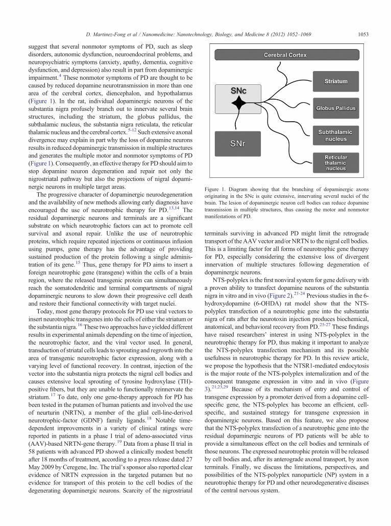
Figure 1. Diagram showing that the branching of dopaminergic axonsoriginating in the SNc is quite extensive, innervating several nuclei of thebrain. The lesion of dopaminergic neuron cell bodies can reduce dopaminetransmission in multiple structures, thus causing the motor and nonmotormanifestations of PD.
1053D. Martinez-Fong et al / Nanomedicine: Nanotechnology, Biology, and Medicine 8 (2012) 1052–1069
suggest that several nonmotor symptoms of PD, such as sleepdisorders, autonomic dysfunction, neuroendocrinal problems, andneuropsychiatric symptoms (anxiety, apathy, dementia, cognitivedysfunction, and depression) also result in part from dopaminergicimpairment.4 These nonmotor symptoms of PD are thought to becaused by reduced dopamine neurotransmission in more than onearea of the cerebral cortex, diencephalon, and hypothalamus(Figure 1). In the rat, individual dopaminergic neurons of thesubstantia nigra profusely branch out to innervate several brainstructures, including the striatum, the globus pallidus, thesubthalamic nucleus, the substantia nigra reticulata, the reticularthalamic nucleus and the cerebral cortex.5-12 Such extensive axonaldivergence may explain in part why the loss of dopamine neuronsresults in reduced dopaminergic transmission in multiple structuresand generates the multiple motor and nonmotor symptoms of PD(Figure 1). Consequently, an effective therapy for PD should aim tostop dopamine neuron degeneration and repair not only thenigrostriatal pathway but also the projections of nigral dopami-nergic neurons in multiple target areas.
The progressive character of dopaminergic neurodegenerationand the availability of new methods allowing early diagnosis haveencouraged the use of neurotrophic therapy for PD.13,14 Theresidual dopaminergic neurons and terminals are a significantsubstrate on which neurotrophic factors can act to promote cellsurvival and axonal repair. Unlike the use of neurotrophicproteins, which require repeated injections or continuous infusionusing pumps, gene therapy has the advantage of providingsustained production of the protein following a single adminis-tration of its gene.15 Thus, gene therapy for PD aims to insert aforeign neurotrophic gene (transgene) within the cells of a brainregion, where the released transgenic protein can simultaneouslyreach the somatodendritic and terminal compartments of nigraldopaminergic neurons to slow down their progressive cell deathand restore their functional connectivity with target nuclei.
Today, most gene therapy protocols for PD use viral vectors toinsert neurotrophic transgenes into the cells of either the striatum orthe substantia nigra.16 These two approaches have yielded differentresults in experimental animals depending on the time of injection,the neurotrophic factor, and the viral vector used. In general,transduction of striatal cells leads to sprouting and regrowth into thearea of transgenic neurotrophic factor expression, along with avarying level of functional recovery. In contrast, injection of thevector into the substantia nigra protects the nigral cell bodies andcauses extensive local sprouting of tyrosine hydroxylase (TH)-positive fibers, but they are unable to functionally reinnervate thestriatum.17 To date, only one gene-therapy approach for PD hasbeen tested in the putamen of human patients and involved the useof neurturin (NRTN), a member of the glial cell-line-derivedneurotrophic-factor (GDNF) family ligands.18 Notable time-dependent improvements in a variety of clinical ratings werereported in patients in a phase I trial of adeno-associated virus(AAV)-based NRTN-gene therapy.19 Data from a phase II trial in58 patients with advanced PD showed a clinically modest benefitafter 18 months of treatment, according to a press release dated 27May 2009 by Ceregene, Inc. The trial's sponsor also reported clearevidence of NRTN expression in the targeted putamen but noevidence for transport of this protein to the cell bodies of thedegenerating dopaminergic neurons. Scarcity of the nigrostriatal
terminals surviving in advanced PD might limit the retrogradetransport of the AAV vector and/or NRTN to the nigral cell bodies.This is a limiting factor for all forms of neurotrophic gene therapyfor PD, especially considering the extensive loss of divergentinnervation of multiple structures following degeneration ofdopaminergic neurons.
NTS-polyplex is the first nonviral system for gene deliverywitha proven ability to transfect dopamine neurons of the substantianigra in vitro and in vivo (Figure 2).21-24 Previous studies in the 6-hydroxydopamine (6-OHDA) rat model show that the NTS-polyplex transfection of a neurotrophic gene into the substantianigra of rats after the neurotoxin injection produces biochemical,anatomical, and behavioral recovery from PD.25-27 These findingshave raised researchers' interest in using NTS-polyplex in theneurotrophic therapy for PD, thus making it important to analyzethe NTS-polyplex transfection mechanism and its possibleusefulness in neurotrophic therapy for PD. In this review article,we propose the hypothesis that the NTSR1-mediated endocytosisis the major route of the NTS-polyplex internalization and of theconsequent transgene expression in vitro and in vivo (Figure3).21,23,29 Because of its mechanism of entry and control oftransgene expression by a promoter derived from a dopamine cell-specific gene, the NTS-polyplex has become an efficient, cell-specific, and sustained strategy for transgene expression indopaminergic neurons. Based on this feature, we also proposethat the NTS-polyplex transfection of a neurotrophic gene into theresidual dopaminergic neurons of PD patients will be able toprovide a simultaneous effect on the cell bodies and terminals ofthose neurons. The expressed neurotrophic protein will be releasedby cell bodies and, after its anterograde axonal transport, by axonterminals. Finally, we discuss the limitations, perspectives, andpossibilities of the NTS-polyplex nanoparticle (NP) system in aneurotrophic therapy for PD and other neurodegenerative diseasesof the central nervous system.
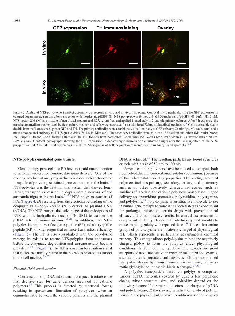
Figure 2. Ability of NTS-polyplex to transfect dopaminergic neurons in vitro and in vivo. Top panel. Confocal micrographs showing the GFP expression incultured dopaminergic neurons after transfection with the plasmid pEGFP-N1. NTS-polyplex was formed at 1:833:36 molar ratio (pEGFP-N1, 6 nM: PK, 5 μM:NTS-vector, 216 nM) in a mixture of neurobasal medium and B27, serum free, and applied immediately to 2-day-old primary cultures. After 6-h exposure, thetransfection medium was replaced by fresh culture medium and cells were incubated for an additional 72 hrs, as described previously.20 Cells were subjected todouble immunofluorescence against GFP and TH. The primary antibodies were a rabbit polyclonal antibody to GFP (Abcam; Cambridge, Massachusetts) and amouse monoclonal antibody to TH (Sigma-Aldrich, St. Louis, Missouri). The secondary antibodies were an Alexa 488 chicken anti-rabbit (Molecular ProbesInc., Eugene, Oregon) and a donkey anti-mouse TRITC (Jackson Immunoresearch Laboratories Inc., West Grove, Pennsylvania). Calibration bars = 50 μm.Bottom panel. Confocal micrographs showing the GFP expression in dopaminergic neurons of the substantia nigra after the local injection of the NTS-polyplex with pDAT-EGFP. Calibration bars = 200 μm. Micrographs of bottom panel were reproduced from Arango-Rodriguez et al.22
1054 D. Martinez-Fong et al / Nanomedicine: Nanotechnology, Biology, and Medicine 8 (2012) 1052–1069
NTS-polyplex-mediated gene transfer
Gene-therapy protocols for PD have not paid much attentionto nonviral vectors for neurotrophic gene delivery. One of thereasons may be that many researchers consider such vectors to beincapable of providing sustained gene expression in the brain.31
NTS-polyplex was the first nonviral system that showed long-lasting transgene expression in dopaminergic neurons of thesubstantia nigra in the rat brain.21-23 NTS-polyplex consists ofNPs (Figure 4, D) resulting from the electrostatic binding of theconjugate NTS–poly-L-lysine (NTS carrier) to plasmid DNA(pDNA). The NTS carrier takes advantage of the endocytosis ofNTS with its high-affinity receptor (NTSR1) to transfer thepDNA into dopamine neurons.21,22 In addition, the NTS-polyplex incorporates a fusogenic peptide (FP) and a karyophilicpeptide (KP) of viral origin that enhance transfection efficiency(Figure 5). The FP is also cross-linked with the poly-lysinemoiety; its role is to rescue NTS-polyplex from endosomesbefore the enzymatic degradation and extreme acidity becomeprevalent23,32 (Figure 5). The KP is a nuclear localization signalthat is electrostatically bound to the pDNA to promote its importto the cell nucleus.23,33
Plasmid DNA condensation
Condensation of pDNA into a small, compact structure is thefirst decisive step for gene transfer mediated by cationicpolymers.34 This process is directed by electrical forces,resulting in spontaneous formation of polyplexes when anequimolar ratio between the cationic polymer and the plasmid
DNA is achieved.35 The resulting particles are toroid structuresor rods with a size of 50 nm to 100 nm.
Several cationic polymers have been used to compact bothribonucleotides and deoxyribonucleotides (polyanionic) becauseof their electrostatic bonding properties. The reacting group ofpolymers includes primary, secondary, tertiary, and quaternaryamines or other positively charged molecules such asamidines.36 To date, the cationic polymers mostly used in genedelivery are spermidine, protamine, polyhistidine, polyarginine,and polylysine.37 Poly-L-lysine is an attractive molecule to usein human gene therapy because it has been tested as a coadjuvantfor prolonged release of certain drugs with proven clinicalefficacy and good biosafety results. Its clinical use relies on itsexceptional solubility, absence of acute toxicity, and inability tocause immunogenicity with repeated doses.38 The epsilon-aminogroups of poly-L-lysine are positively charged at physiologicalpH, which represents a particularly advantageous chemicalproperty. This charge allows poly-l-lysine to bind the negativelycharged pDNA to form the polyplex under physiologicalconditions. In addition, the epsilon-amino groups are goodacceptors of molecules active in receptor-meditated endocytosis,such as proteins, peptides, and sugars, which are incorporatedinto poly-L-lysine by using chemical cross-linkers, nonenzy-matic glycosylation, or avidin-biotin technique.37,39
A polyplex nanoparticle based on polylysine comprisesvarious pDNA molecules covered by quite a few polymericchains, whose structure, size, and solubility depend on thefollowing factors: 1) the ratio of electrostatic charges of pDNAand poly-L-lysine, 2) the size and ramification grade of poly-L-lysine, 3) the physical and chemical conditions used for polyplex
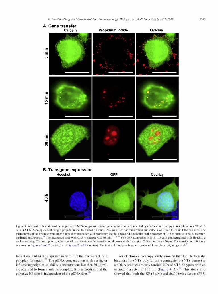
Figure 3. Schematic illustration of the sequence of NTS-polyplex-mediated gene transfection documented by confocal microscopy in neuroblastoma N1E-115cells. (A) NTS-polyplex harboring a propidium iodide-labeled plasmid DNA was used for transfection and calcein was used to delimit the cell area. Themicrographs of the first row were taken 5 min after incubation with propidium iodide-labeled NTS-polyplex in the presence of 0.45 M sucrose to block receptor-mediated endocytosis.28 The incubation time with 0.45 M sucrose was 30 min.23,29,30 (B) GFP expression in N1E-115 cells counterstained with Hoechst, anuclear staining. The microphotographs were taken at the times after transfection shown at the left margins. Calibration bars = 20 μm. The transfection efficiencyis shown in Figures 6 and 7 (in vitro) and Figures 2 and 9 (in vivo). The first and third panels were reproduced from Navarro-Quiroga et al.23
1055D. Martinez-Fong et al / Nanomedicine: Nanotechnology, Biology, and Medicine 8 (2012) 1052–1069
formation, and 4) the sequence used to mix the reactants duringpolyplex formation.22 The pDNA concentration is also a factorinfluencing polyplex solubility; concentrations less than 20 μg/mLare required to form a soluble complex. It is interesting that thepolyplex NP size is independent of the pDNA size.40
An electron-microscopy study showed that the electrostaticbinding of the NTS-poly-L-lysine conjugate (the NTS-carrier) toa pDNA produces mostly toroidal NPs of NTS-polyplex with anaverage diameter of 100 nm (Figure 4, D).22 This study alsoshowed that both the KP (6 μM) and fetal bovine serum (FBS;
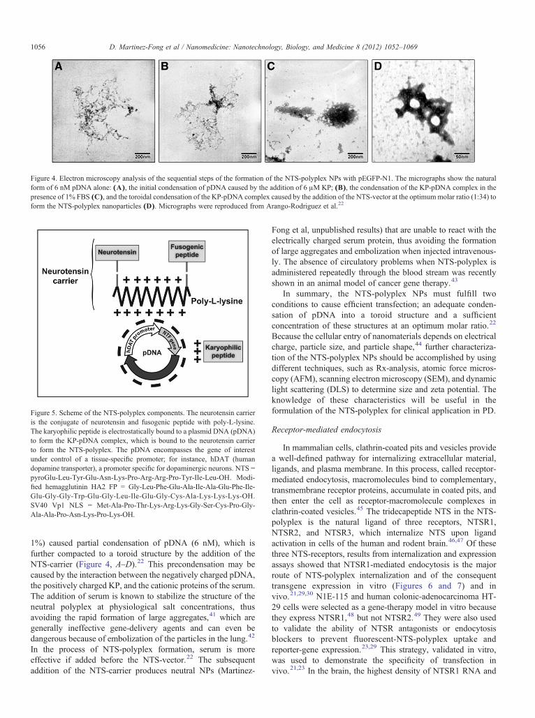
Figure 4. Electron microscopy analysis of the sequential steps of the formation of the NTS-polyplex NPs with pEGFP-N1. The micrographs show the naturalform of 6 nM pDNA alone: (A), the initial condensation of pDNA caused by the addition of 6 μM KP; (B), the condensation of the KP-pDNA complex in thepresence of 1% FBS (C), and the toroidal condensation of the KP-pDNA complex caused by the addition of the NTS-vector at the optimummolar ratio (1:34) toform the NTS-polyplex nanoparticles (D). Micrographs were reproduced from Arango-Rodriguez et al.22
Figure 5. Scheme of the NTS-polyplex components. The neurotensin carrieris the conjugate of neurotensin and fusogenic peptide with poly-L-lysine.The karyophilic peptide is electrostatically bound to a plasmid DNA (pDNA)to form the KP-pDNA complex, which is bound to the neurotensin carrierto form the NTS-polyplex. The pDNA encompasses the gene of interestunder control of a tissue-specific promoter; for instance, hDAT (humandopamine transporter), a promoter specific for dopaminergic neurons. NTS =pyroGlu-Leu-Tyr-Glu-Asn-Lys-Pro-Arg-Arg-Pro-Tyr-Ile-Leu-OH. Modi-fied hemagglutinin HA2 FP = Gly-Leu-Phe-Glu-Ala-Ile-Ala-Glu-Phe-Ile-Glu-Gly-Gly-Trp-Glu-Gly-Leu-Ile-Glu-Gly-Cys-Ala-Lys-Lys-Lys-OH.SV40 Vp1 NLS = Met-Ala-Pro-Thr-Lys-Arg-Lys-Gly-Ser-Cys-Pro-Gly-Ala-Ala-Pro-Asn-Lys-Pro-Lys-OH.
1056 D. Martinez-Fong et al / Nanomedicine: Nanotechnology, Biology, and Medicine 8 (2012) 1052–1069
1%) caused partial condensation of pDNA (6 nM), which isfurther compacted to a toroid structure by the addition of theNTS-carrier (Figure 4, A–D).22 This precondensation may becaused by the interaction between the negatively charged pDNA,the positively charged KP, and the cationic proteins of the serum.The addition of serum is known to stabilize the structure of theneutral polyplex at physiological salt concentrations, thusavoiding the rapid formation of large aggregates,41 which aregenerally ineffective gene-delivery agents and can even bedangerous because of embolization of the particles in the lung.42
In the process of NTS-polyplex formation, serum is moreeffective if added before the NTS-vector.22 The subsequentaddition of the NTS-carrier produces neutral NPs (Martinez-
Fong et al, unpublished results) that are unable to react with theelectrically charged serum protein, thus avoiding the formationof large aggregates and embolization when injected intravenous-ly. The absence of circulatory problems when NTS-polyplex isadministered repeatedly through the blood stream was recentlyshown in an animal model of cancer gene therapy.43
In summary, the NTS-polyplex NPs must fulfill twoconditions to cause efficient transfection; an adequate conden-sation of pDNA into a toroid structure and a sufficientconcentration of these structures at an optimum molar ratio.22
Because the cellular entry of nanomaterials depends on electricalcharge, particle size, and particle shape,44 further characteriza-tion of the NTS-polyplex NPs should be accomplished by usingdifferent techniques, such as Rx-analysis, atomic force micros-copy (AFM), scanning electron microscopy (SEM), and dynamiclight scattering (DLS) to determine size and zeta potential. Theknowledge of these characteristics will be useful in theformulation of the NTS-polyplex for clinical application in PD.
Receptor-mediated endocytosis
In mammalian cells, clathrin-coated pits and vesicles providea well-defined pathway for internalizing extracellular material,ligands, and plasma membrane. In this process, called receptor-mediated endocytosis, macromolecules bind to complementary,transmembrane receptor proteins, accumulate in coated pits, andthen enter the cell as receptor-macromolecule complexes inclathrin-coated vesicles.45 The tridecapeptide NTS in the NTS-polyplex is the natural ligand of three receptors, NTSR1,NTSR2, and NTSR3, which internalize NTS upon ligandactivation in cells of the human and rodent brain.46,47 Of thesethree NTS-receptors, results from internalization and expressionassays showed that NTSR1-mediated endocytosis is the majorroute of NTS-polyplex internalization and of the consequenttransgene expression in vitro (Figures 6 and 7) and invivo.21,29,30 N1E-115 and human colonic-adenocarcinoma HT-29 cells were selected as a gene-therapy model in vitro becausethey express NTSR1,48 but not NTSR2.49 They were also usedto validate the ability of NTSR antagonists or endocytosisblockers to prevent fluorescent-NTS-polyplex uptake andreporter-gene expression.23,29 This strategy, validated in vitro,was used to demonstrate the specificity of transfection invivo.21,23 In the brain, the highest density of NTSR1 RNA and
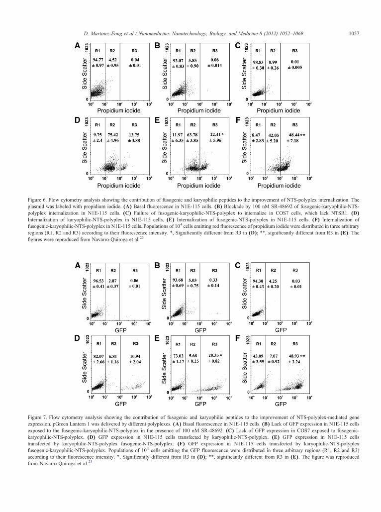
Figure 6. Flow cytometry analysis showing the contribution of fusogenic and karyophilic peptides to the improvement of NTS-polyplex internalization. Theplasmid was labeled with propidium iodide. (A) Basal fluorescence in N1E-115 cells. (B) Blockade by 100 nM SR-48692 of fusogenic-karyophilic-NTS-polyplex internalization in N1E-115 cells. (C) Failure of fusogenic-karyophilic-NTS-polyplex to internalize in COS7 cells, which lack NTSR1. (D)Internalization of karyophilic-NTS-polyplex in N1E-115 cells. (E) Internalization of fusogenic-NTS-polyplex in N1E-115 cells. (F) Internalization offusogenic-karyophilic-NTS-polyplex in N1E-115 cells. Populations of 104 cells emitting red fluorescence of propidium iodide were distributed in three arbitraryregions (R1, R2 and R3) according to their fluorescence intensity. ⁎, Significantly different from R3 in (D); ⁎⁎, significantly different from R3 in (E). Thefigures were reproduced from Navarro-Quiroga et al.23
Figure 7. Flow cytometry analysis showing the contribution of fusogenic and karyophilic peptides to the improvement of NTS-polyplex-mediated geneexpression. pGreen Lantern 1 was delivered by different polyplexes. (A) Basal fluorescence in N1E-115 cells. (B) Lack of GFP expression in N1E-115 cellsexposed to the fusogenic-karyophilic-NTS-polyplex in the presence of 100 nM SR-48692. (C) Lack of GFP expression in COS7 exposed to fusogenic-karyophilic-NTS-polyplex. (D) GFP expression in N1E-115 cells transfected by karyophilic-NTS-polyplex. (E) GFP expression in N1E-115 cellstransfected by karyophilic-NTS-polyplex fusogenic-NTS-polyplex. (F) GFP expression in N1E-115 cells transfected by karyophilic-NTS-polyplexfusogenic-karyophilic-NTS-polyplex. Populations of 104 cells emitting the GFP fluorescence were distributed in three arbitrary regions (R1, R2 and R3)according to their fluorescence intensity. ⁎, Significantly different from R3 in (D); ⁎⁎, significantly different from R3 in (E). The figure was reproducedfrom Navarro-Quiroga et al.23
1057D. Martinez-Fong et al / Nanomedicine: Nanotechnology, Biology, and Medicine 8 (2012) 1052–1069
1058 D. Martinez-Fong et al / Nanomedicine: Nanotechnology, Biology, and Medicine 8 (2012) 1052–1069
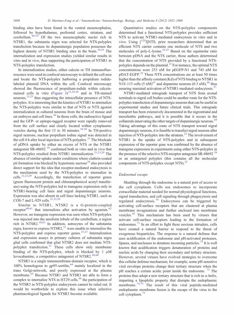
binding sites have been found in the ventral mesencephalon,followed by hypothalamus, prefrontal cortex, striatum, andcerebellum.49-53 Of the two mesencephalic nuclei rich inNTSR1, the substantia nigra was selected for NTS-polyplextransfection because its dopaminergic population possesses thehighest density of NTSR1 binding sites in the brain.54,55 Theinternalization and expression studies yielded similar results invitro and in vivo, thus supporting the participation of NTSR1 inNTS-polyplex transfection.
In internalization studies, either calcein or TH immunofluo-rescence were used in confocal microscopy to delimit the cell areaand locate the NTS-polyplex harboring a propidium iodide-labeled plasmid DNA within the cell. Confocal microscopyshowed the fluorescence of propidium-iodide within calcein-stained cells in vitro (Figure 3)23,29,30 and in TH-stainedneurons,21,23 thus suggesting the intracellular presence of NTS-polyplex. It is interesting that the kinetics of NTSR1 to internalizethe NTS-polyplex were similar to that of NTS or NTS agonistinternalization in cultured neurons from the brain of mouse andrat embryos and cell lines.56 In those cells, the radioactive ligandand the GFP- or epitope-tagged receptor were rapidly removedfrom the cell surface and clearly located within cytoplasmicvesicles during the first 15 to 30 minutes.56-58 In TH-positivenigral neurons, nuclear propidium iodine signal was detected inthe cell 4 h after local injection of NTS-polyplex.21 The blockadeof pDNA uptake by either an excess of NTS or the NTSR1antagonist SR-48692 59 confirmed both in vitro and in vivo thatNTS-polyplex resulted from NTSR1 internalization.21,23,29 Theabsence of similar uptake under conditions where clathrin-coatedpit formation was blocked by hypertonic sucrose28 also provideddirect support for the idea that receptor-mediated endocytosis isthe mechanism used by the NTS-polyplex to internalize incells.21,23,29 Accordingly, the transfection of reporter genes(green fluorescent protein and chloramphenicol acetyl transfer-ase) using the NTS-polyplex led to transgene expression only inNTSR1-bearing cell lines and nigral dopaminergic neurons.Expression was also absent in cell lines lacking NTSR1, such asCOS-7 and L-929 cells.21,23,29
Similar to NTSR1, NTSR2 is a G-protein-coupledreceptor60,61 that internalizes after activation by agonists.62
However, no transgene expression was seen when NTS-polyplexwas injected into the ansiform lobule of the cerebellum, a regionrich in NTSR2.50,63 In addition, astrocytes of the substantianigra, known to express NTSR2,51 were unable to internalize theNTS-polyplex and express reporter genes.21,23 Internalizationand expression assays in primary cultures of substantia nigraglial cells confirmed that glial NTSR2 does not mediate NTS-polyplex transfection.21 These cells show only membranebinding of the NTS-polyplex, which is blocked by 1 μMlevocabastine, a competitive antagonist of NTSR2.49,60
NTSR3 is a single transmembrane-domain receptor, which is100% homologous to gp95-sortilin,64 mainly localized in thetrans Golgi-network, and poorly expressed at the plasmamembrane.65 Because NTSR1 and NTSR3 are able to form acomplex to internalize NTS in HT29 cells,66 the participation ofthe NTSR3 in NTS-polyplex endocytosis cannot be ruled out. Itwould be worthwhile to explore this issue when selectivepharmacological ligands for NTSR3 become available.
Quantitative studies on the NTS-polyplex componentsdetermined that a functional NTS-polyplex provides sufficientNTS to activate NTSR1-mediated endocytosis in vitro and invivo. Using [125I]NTS, prior researchers determined that anefficient NTS carrier contains one molecule of NTS and twomolecules of poly-L-lysine.22,23 Based on the equimolar ratiobetween pDNA and the NTS carrier, those authors determinedthat the concentration of NTS provided by a functional NTS-polyplex depends on the plasmid.22 For instance, the optimal NTSconcentrations were 253 nM for pEGFP-N1 and 745 nM forpDAT-EGFP.22 These NTS concentrations are at least 50 timeshigher than the affinity constant (Kd) of NTS binding toNTSR1 inN1E-115 cells (5 nM)67 and dopamine neurons (0.3 nM),68 thusassuring maximal activation of NTSR1-mediated endocytosis.57
NTSR1-mediated retrograde transport of NTS from axonalterminals to nigral cell bodies could be another route for the NTS-polyplex transfection of dopaminergic neurons that can be useful inexperimental studies and future clinical trials. This retrogradetransport has been extensively documented in the nigrostriatal andmesolimbic pathways, and it is possible that it occurs in thecollaterals innervating the other targets of dopaminergic neurons.69
Taking advantage of this route of NTS transport toward nigraldopaminergic neurons, it is feasible to transfect nigral neurons afterinjection of NTS-polyplex into the striatum.22 The involvement ofNTSR1 in the uptake of NTS-polyplex and in subsequentexpression of the reporter gene was confirmed by the absence oftransgene expression in experiments using either NTS-polyplex inthe presence of the selective NTS-receptor antagonist SR-48692 59
or an untargeted polyplex (this contains all the molecularcomponents of NTS-polyplex except NTS).22
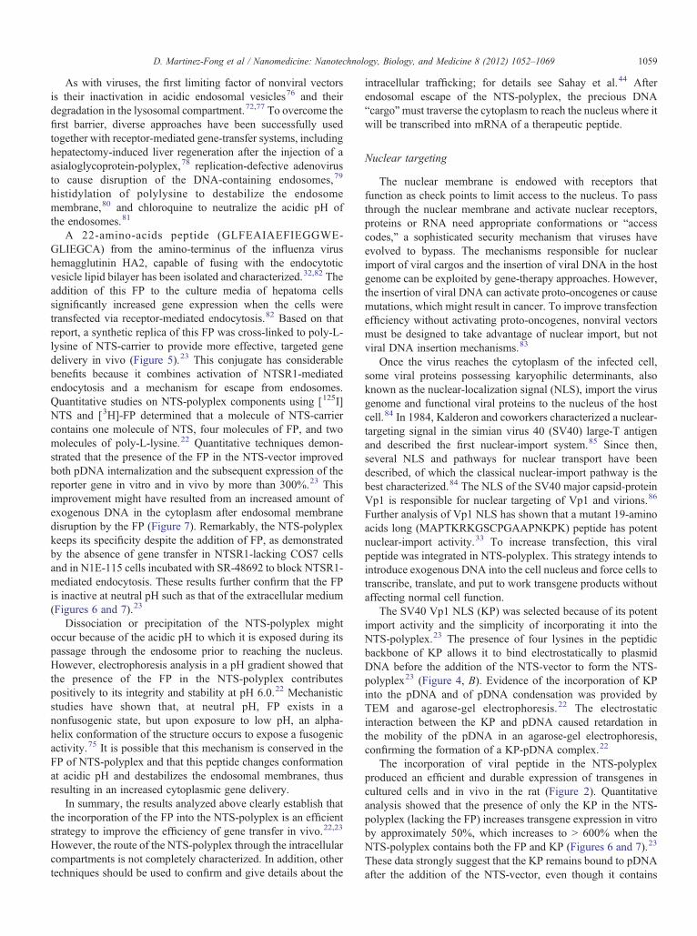
Endosomal escape
Shuttling through the endosome is a natural port of access tothe cell cytoplasm. Cells use endosomes to incorporateextracellular material needed for normal physiological functions,signal transduction, and cell signaling through phagocytosis andregulated endocytosis.70 Endocytosis can be triggered byactivating cell-surface receptors that are clustered at plasmamembrane invaginations and further enclosed into membranevesicles.45 This mechanism has been used by viruses thatactivate cell-surface receptors leading to the formation ofendosomes.71 In an effort to fight these noxious intruders, cellshave created a natural barrier to respond to the threat ofexogenous bioparticles. The response is a natural defense thatuses acidification of the endosome and pH-activated proteases,lipases, and nucleases to denature incoming particles.72 It is wellknown that acidification triggers denaturation of proteins andnucleic acids by changing their secondary and tertiary structure.However, several viruses have evolved strategies to overcomethis cellular defense mechanism; for example, some pH-sensitiveviral envelope proteins change their tertiary structure when thepH reaches a certain acidic point inside the endosome. 73 Theproteins thus adopt a new tertiary structure that is rich in α-helix,imparting a lipophilic property that disrupts the endoplasmicmembrane.74,75 The result of this viral peptide-mediatedendoplasmic membrane fusion is the escape of the virus to thecell cytoplasm.73
1059D. Martinez-Fong et al / Nanomedicine: Nanotechnology, Biology, and Medicine 8 (2012) 1052–1069
As with viruses, the first limiting factor of nonviral vectorsis their inactivation in acidic endosomal vesicles76 and theirdegradation in the lysosomal compartment.72,77 To overcome thefirst barrier, diverse approaches have been successfully usedtogether with receptor-mediated gene-transfer systems, includinghepatectomy-induced liver regeneration after the injection of aasialoglycoprotein-polyplex,78 replication-defective adenovirusto cause disruption of the DNA-containing endosomes,79
histidylation of polylysine to destabilize the endosomemembrane,80 and chloroquine to neutralize the acidic pH ofthe endosomes.81
A 22-amino-acids peptide (GLFEAIAEFIEGGWE-GLIEGCA) from the amino-terminus of the influenza virushemagglutinin HA2, capable of fusing with the endocytoticvesicle lipid bilayer has been isolated and characterized.32,82 Theaddition of this FP to the culture media of hepatoma cellssignificantly increased gene expression when the cells weretransfected via receptor-mediated endocytosis.82 Based on thatreport, a synthetic replica of this FP was cross-linked to poly-L-lysine of NTS-carrier to provide more effective, targeted genedelivery in vivo (Figure 5).23 This conjugate has considerablebenefits because it combines activation of NTSR1-mediatedendocytosis and a mechanism for escape from endosomes.Quantitative studies on NTS-polyplex components using [125I]NTS and [3H]-FP determined that a molecule of NTS-carriercontains one molecule of NTS, four molecules of FP, and twomolecules of poly-L-lysine.22 Quantitative techniques demon-strated that the presence of the FP in the NTS-vector improvedboth pDNA internalization and the subsequent expression of thereporter gene in vitro and in vivo by more than 300%.23 Thisimprovement might have resulted from an increased amount ofexogenous DNA in the cytoplasm after endosomal membranedisruption by the FP (Figure 7). Remarkably, the NTS-polyplexkeeps its specificity despite the addition of FP, as demonstratedby the absence of gene transfer in NTSR1-lacking COS7 cellsand in N1E-115 cells incubated with SR-48692 to block NTSR1-mediated endocytosis. These results further confirm that the FPis inactive at neutral pH such as that of the extracellular medium(Figures 6 and 7).23
Dissociation or precipitation of the NTS-polyplex mightoccur because of the acidic pH to which it is exposed during itspassage through the endosome prior to reaching the nucleus.However, electrophoresis analysis in a pH gradient showed thatthe presence of the FP in the NTS-polyplex contributespositively to its integrity and stability at pH 6.0.22 Mechanisticstudies have shown that, at neutral pH, FP exists in anonfusogenic state, but upon exposure to low pH, an alpha-helix conformation of the structure occurs to expose a fusogenicactivity.75 It is possible that this mechanism is conserved in theFP of NTS-polyplex and that this peptide changes conformationat acidic pH and destabilizes the endosomal membranes, thusresulting in an increased cytoplasmic gene delivery.
In summary, the results analyzed above clearly establish thatthe incorporation of the FP into the NTS-polyplex is an efficientstrategy to improve the efficiency of gene transfer in vivo.22,23
However, the route of the NTS-polyplex through the intracellularcompartments is not completely characterized. In addition, othertechniques should be used to confirm and give details about the
intracellular trafficking; for details see Sahay et al.44 Afterendosomal escape of the NTS-polyplex, the precious DNA“cargo”must traverse the cytoplasm to reach the nucleus where itwill be transcribed into mRNA of a therapeutic peptide.
Nuclear targeting
The nuclear membrane is endowed with receptors thatfunction as check points to limit access to the nucleus. To passthrough the nuclear membrane and activate nuclear receptors,proteins or RNA need appropriate conformations or “accesscodes,” a sophisticated security mechanism that viruses haveevolved to bypass. The mechanisms responsible for nuclearimport of viral cargos and the insertion of viral DNA in the hostgenome can be exploited by gene-therapy approaches. However,the insertion of viral DNA can activate proto-oncogenes or causemutations, which might result in cancer. To improve transfectionefficiency without activating proto-oncogenes, nonviral vectorsmust be designed to take advantage of nuclear import, but notviral DNA insertion mechanisms.83
Once the virus reaches the cytoplasm of the infected cell,some viral proteins possessing karyophilic determinants, alsoknown as the nuclear-localization signal (NLS), import the virusgenome and functional viral proteins to the nucleus of the hostcell.84 In 1984, Kalderon and coworkers characterized a nuclear-targeting signal in the simian virus 40 (SV40) large-T antigenand described the first nuclear-import system.85 Since then,several NLS and pathways for nuclear transport have beendescribed, of which the classical nuclear-import pathway is thebest characterized.84 The NLS of the SV40 major capsid-proteinVp1 is responsible for nuclear targeting of Vp1 and virions.86
Further analysis of Vp1 NLS has shown that a mutant 19-aminoacids long (MAPTKRKGSCPGAAPNKPK) peptide has potentnuclear-import activity.33 To increase transfection, this viralpeptide was integrated in NTS-polyplex. This strategy intends tointroduce exogenous DNA into the cell nucleus and force cells totranscribe, translate, and put to work transgene products withoutaffecting normal cell function.
The SV40 Vp1 NLS (KP) was selected because of its potentimport activity and the simplicity of incorporating it into theNTS-polyplex.23 The presence of four lysines in the peptidicbackbone of KP allows it to bind electrostatically to plasmidDNA before the addition of the NTS-vector to form the NTS-polyplex23 (Figure 4, B). Evidence of the incorporation of KPinto the pDNA and of pDNA condensation was provided byTEM and agarose-gel electrophoresis.22 The electrostaticinteraction between the KP and pDNA caused retardation inthe mobility of the pDNA in an agarose-gel electrophoresis,confirming the formation of a KP-pDNA complex.22
The incorporation of viral peptide in the NTS-polyplexproduced an efficient and durable expression of transgenes incultured cells and in vivo in the rat (Figure 2). Quantitativeanalysis showed that the presence of only the KP in the NTS-polyplex (lacking the FP) increases transgene expression in vitroby approximately 50%, which increases to N 600% when theNTS-polyplex contains both the FP and KP (Figures 6 and 7).23
These data strongly suggest that the KP remains bound to pDNAafter the addition of the NTS-vector, even though it contains
1060 D. Martinez-Fong et al / Nanomedicine: Nanotechnology, Biology, and Medicine 8 (2012) 1052–1069
polylysine. This suggestion is further supported by the findingthat a single nuclear localization signal peptide is sufficient tocarry DNA to the cell nucleus.87 In addition, there is evidence inthe gene therapy literature that NLS mediated gene transfer stillworks after irreversible chemical linkage of the NLS-peptide tocDNA.88,89
Similarly, the combined use of the endosomal neutralizingagent chloroquine and of the SV40 large-tumor-antigen NLSimproves the transfection efficiency of transferrin-polyplex.81,90
The successful use of strategies to escape from the endosome orto inhibit endosomal acidity strongly supports the idea that themajor barrier to receptor-mediated gene-transfer systems isacidification of endosomal vesicles.91 Because of its capacity forcell entry, endosomal escape, and nuclear import, the NTS-polyplex thus has the ability to provide efficient and durabletransgene expression without the requirement for complementarytreatments, such as chloroquine, which increases the transfectionefficiency by preventing endosome acidification.92
The use of a synthetic peptide in the NTS-polyplex hasconsiderable benefits in comparison with the use of mutated orchemically modified viruses. It results in smaller complexes andconsidering the lack of toxicity or of any potential infectivity, thetechnique holds great promise for therapeutic intervention,especially for PD. The most striking benefit of merging theviral strategy with the transfection specificity of NTS-polyplex isthe ability to achieve durable transgene expression in dopami-nergic neurons of the substantia nigra. The injection of the NTS-polyplex into the substantia nigra of rats produces strongtransgene expression in large numbers of dopaminergic neurons,but not in nearby glial cells (Figures 2 and 9).22-25 The blockadeof NTSR1 binding site by NTS-antagonists and the ineffective-ness of an untargeted polyplex (lacking NTS) clearly demon-strated that NTSR1-mediated endocytosis plays a key role ingene transfer in vivo.22,23 Transgene expression has beendetected for up to two months (the end of the study) afterNTS-polyplex injection, and the observation of high levels oftransgene expression at the study endpoint suggests that it canendure even longer.23-25
In summary, although the involvement of the NTSR1receptor in the NTS-polyplex transfection is solidly demonstrat-ed in vitro and in vivo, the route of the NTS-polyplex through theintracellular compartments and the import mechanism into thecell nucleus remain to be clarified by using different techniques;for details see Ref. [44]. In addition, the identification of thenuclear compartment where the transgene is lodged and themolecular mechanisms of transgene transcription and regulationare still unknown.
Tissue-specific promoters
A high degree of dopamine cell specificity of transgeneexpression has been achieved with the NTS-polyplex approach,based on the distribution of NTSR1 integral to the transfectionprocess and the ability to target NTS-polyplex injections into theimmediate vicinity of dopaminergic neurons or their terminals(see above). It might be possible to enhance cell-specificity ofexpression further by placing the transgene under the control of apromoter derived from a dopamine cell-specific gene (Figure 5).
The dopamine transporter (DAT) is a plasma membrane proteinthat functions to clear released dopamine from the extracellularspace, thereby controlling the amplitude and duration ofdopamine signaling.93 The DAT gene is most robustly expressedwithin substantia nigra dopamine neurons (those cells mostimpacted in PD), with a specificity of cell expression unrivaledby any other genes associated with the dopaminergicphenotype.94 Recently, an NTS-polyplex containing theBDNF-flag gene under control of the previously characterized95
human DAT promoter sequence was injected into the striatum, atissue known to contain high levels of NTSR1.50 As expected,the human DAT promoter prevented transgene expression inthese striatal cells.22 In contrast, BDNF-flag expression wasdetected in dopaminergic neurons of the substantia nigra due toNTSR1-mediated retrograde transport of the transgene fromstriatal terminals. The retrograde transport of NTS by NTSR1 iswell characterized in experimental animals69 and represents analternative route of transfection of nigral dopaminergic neurons.In addition, the use of tissue-specific promoters might prolongNTS-polyplex-mediated gene expression because DAT promoteractivity is not expected to be transcriptionally silenced bymethylation, as is the case for viral promoters such as CMV.96 Inthe future, it might be possible to modify the DNA sequence, orpharmacologically manipulate the functional activity of tissue-specific promoters, to provide even more refined control oftransgene expression than is possible now.
Neurotrophic therapy
Traditionally, the term neurotrophic factors (NTFs) hasreferred to a group of secreted proteins that regulate the lifeand death of specific sets of neuronal subpopulations duringdevelopment. The role of NTFs is well established in CNSdevelopment. In contrast, the function of NTFs in the survival,maintenance, and plasticity of the mature central nervous system(CNS) is less clear. Nevertheless, evidence in the adult brainshowing that NTFs promote neuronal regeneration followingdiverse types of injury has encouraged their use in the treatmentof neurodegenerative disease and neural trauma.
The recent discovery and characterization of two newevolutionarily conserved NTFs, cerebral dopamine-neurotrophicfactor (CDNF) and mesencephalic astrocyte-derived neuro-trophic factor (MANF), has augmented the families of NTFs tofour groups.97,98 The modern classification99 includes:
1) The neurotrophins: Nerve growth factor (NGF), BDNF,neurotrophin-3 (NT-3), and NT-4100
2) GDNF family ligands (GFLs): GDNF, neurturin (NRTN),artemin (ARTN), and persephin (PSPN)18,101
3) Neuropoietic cytokines (also referred to as the interleukin-6 [IL-6] family)102 and
4) The novel CDNF-MANF family.97,103
NTFs act on neurons through transmembrane receptors withintrinsic tyrosine kinase activity or via other receptor-associatedkinases.101 The receptors for CDNF and MANF are unknown.99
Dopaminergic neurons of the substantia nigra respondspecifically to GDNF, NRTN, and BDNF, through GFRα1,
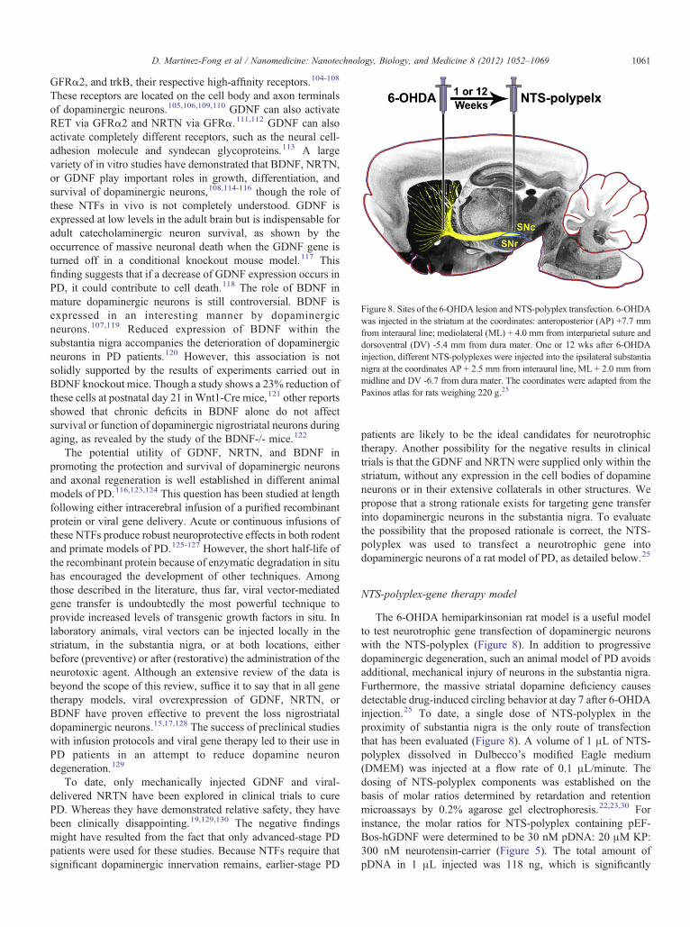
Figure 8. Sites of the 6-OHDA lesion andNTS-polyplex transfection. 6-OHDAwas injected in the striatum at the coordinates: anteroposterior (AP) +7.7 mmfrom interaural line; mediolateral (ML) + 4.0 mm from interparietal suture anddorsoventral (DV) -5.4 mm from dura mater. One or 12 wks after 6-OHDAinjection, different NTS-polyplexes were injected into the ipsilateral substantianigra at the coordinates AP + 2.5 mm from interaural line, ML + 2.0 mm frommidline and DV -6.7 from dura mater. The coordinates were adapted from thePaxinos atlas for rats weighing 220 g.25
1061D. Martinez-Fong et al / Nanomedicine: Nanotechnology, Biology, and Medicine 8 (2012) 1052–1069
GFRα2, and trkB, their respective high-affinity receptors.104-108
These receptors are located on the cell body and axon terminalsof dopaminergic neurons.105,106,109,110 GDNF can also activateRET via GFRα2 and NRTN via GFRα.111,112 GDNF can alsoactivate completely different receptors, such as the neural cell-adhesion molecule and syndecan glycoproteins.113 A largevariety of in vitro studies have demonstrated that BDNF, NRTN,or GDNF play important roles in growth, differentiation, andsurvival of dopaminergic neurons,108,114-116 though the role ofthese NTFs in vivo is not completely understood. GDNF isexpressed at low levels in the adult brain but is indispensable foradult catecholaminergic neuron survival, as shown by theoccurrence of massive neuronal death when the GDNF gene isturned off in a conditional knockout mouse model.117 Thisfinding suggests that if a decrease of GDNF expression occurs inPD, it could contribute to cell death.118 The role of BDNF inmature dopaminergic neurons is still controversial. BDNF isexpressed in an interesting manner by dopaminergicneurons.107,119 Reduced expression of BDNF within thesubstantia nigra accompanies the deterioration of dopaminergicneurons in PD patients.120 However, this association is notsolidly supported by the results of experiments carried out inBDNF knockout mice. Though a study shows a 23% reduction ofthese cells at postnatal day 21 in Wnt1-Cre mice,121 other reportsshowed that chronic deficits in BDNF alone do not affectsurvival or function of dopaminergic nigrostriatal neurons duringaging, as revealed by the study of the BDNF-/- mice.122
The potential utility of GDNF, NRTN, and BDNF inpromoting the protection and survival of dopaminergic neuronsand axonal regeneration is well established in different animalmodels of PD.116,123,124 This question has been studied at lengthfollowing either intracerebral infusion of a purified recombinantprotein or viral gene delivery. Acute or continuous infusions ofthese NTFs produce robust neuroprotective effects in both rodentand primate models of PD.125-127 However, the short half-life ofthe recombinant protein because of enzymatic degradation in situhas encouraged the development of other techniques. Amongthose described in the literature, thus far, viral vector-mediatedgene transfer is undoubtedly the most powerful technique toprovide increased levels of transgenic growth factors in situ. Inlaboratory animals, viral vectors can be injected locally in thestriatum, in the substantia nigra, or at both locations, eitherbefore (preventive) or after (restorative) the administration of theneurotoxic agent. Although an extensive review of the data isbeyond the scope of this review, suffice it to say that in all genetherapy models, viral overexpression of GDNF, NRTN, orBDNF have proven effective to prevent the loss nigrostriataldopaminergic neurons.15,17,128 The success of preclinical studieswith infusion protocols and viral gene therapy led to their use inPD patients in an attempt to reduce dopamine neurondegeneration.129
To date, only mechanically injected GDNF and viral-delivered NRTN have been explored in clinical trials to curePD. Whereas they have demonstrated relative safety, they havebeen clinically disappointing.19,129,130 The negative findingsmight have resulted from the fact that only advanced-stage PDpatients were used for these studies. Because NTFs require thatsignificant dopaminergic innervation remains, earlier-stage PD
patients are likely to be the ideal candidates for neurotrophictherapy. Another possibility for the negative results in clinicaltrials is that the GDNF and NRTN were supplied only within thestriatum, without any expression in the cell bodies of dopamineneurons or in their extensive collaterals in other structures. Wepropose that a strong rationale exists for targeting gene transferinto dopaminergic neurons in the substantia nigra. To evaluatethe possibility that the proposed rationale is correct, the NTS-polyplex was used to transfect a neurotrophic gene intodopaminergic neurons of a rat model of PD, as detailed below.25
NTS-polyplex-gene therapy model
The 6-OHDA hemiparkinsonian rat model is a useful modelto test neurotrophic gene transfection of dopaminergic neuronswith the NTS-polyplex (Figure 8). In addition to progressivedopaminergic degeneration, such an animal model of PD avoidsadditional, mechanical injury of neurons in the substantia nigra.Furthermore, the massive striatal dopamine deficiency causesdetectable drug-induced circling behavior at day 7 after 6-OHDAinjection.25 To date, a single dose of NTS-polyplex in theproximity of substantia nigra is the only route of transfectionthat has been evaluated (Figure 8). A volume of 1 μL of NTS-polyplex dissolved in Dulbecco's modified Eagle medium(DMEM) was injected at a flow rate of 0.1 μL/minute. Thedosing of NTS-polyplex components was established on thebasis of molar ratios determined by retardation and retentionmicroassays by 0.2% agarose gel electrophoresis.22,23,30 Forinstance, the molar ratios for NTS-polyplex containing pEF-Bos-hGDNF were determined to be 30 nM pDNA: 20 μM KP:300 nM neurotensin-carrier (Figure 5). The total amount ofpDNA in 1 μL injected was 118 ng, which is significantly
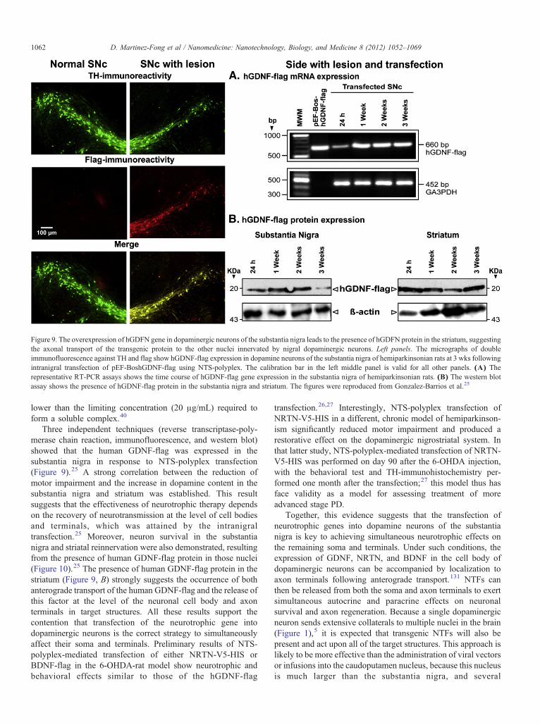
Figure 9. The overexpression of hGDFN gene in dopaminergic neurons of the substantia nigra leads to the presence of hGDFN protein in the striatum, suggestingthe axonal transport of the transgenic protein to the other nuclei innervated by nigral dopaminergic neurons. Left panels. The micrographs of doubleimmunofluorescence against TH and flag show hGDNF-flag expression in dopamine neurons of the substantia nigra of hemiparkinsonian rats at 3 wks followingintranigral transfection of pEF-BoshGDNF-flag using NTS-polyplex. The calibration bar in the left middle panel is valid for all other panels. (A) Therepresentative RT-PCR assays shows the time course of hGDNF-flag gene expression in the substantia nigra of hemiparkinsonian rats. (B) The western blotassay shows the presence of hGDNF-flag protein in the substantia nigra and striatum. The figures were reproduced from Gonzalez-Barrios et al.25
1062 D. Martinez-Fong et al / Nanomedicine: Nanotechnology, Biology, and Medicine 8 (2012) 1052–1069
lower than the limiting concentration (20 μg/mL) required toform a soluble complex.40
Three independent techniques (reverse transcriptase-poly-merase chain reaction, immunofluorescence, and western blot)showed that the human GDNF-flag was expressed in thesubstantia nigra in response to NTS-polyplex transfection(Figure 9).25 A strong correlation between the reduction ofmotor impairment and the increase in dopamine content in thesubstantia nigra and striatum was established. This resultsuggests that the effectiveness of neurotrophic therapy dependson the recovery of neurotransmission at the level of cell bodiesand terminals, which was attained by the intranigraltransfection.25 Moreover, neuron survival in the substantianigra and striatal reinnervation were also demonstrated, resultingfrom the presence of human GDNF-flag protein in those nuclei(Figure 10).25 The presence of human GDNF-flag protein in thestriatum (Figure 9, B) strongly suggests the occurrence of bothanterograde transport of the human GDNF-flag and the release ofthis factor at the level of the neuronal cell body and axonterminals in target structures. All these results support thecontention that transfection of the neurotrophic gene intodopaminergic neurons is the correct strategy to simultaneouslyaffect their soma and terminals. Preliminary results of NTS-polyplex-mediated transfection of either NRTN-V5-HIS orBDNF-flag in the 6-OHDA-rat model show neurotrophic andbehavioral effects similar to those of the hGDNF-flag
transfection.26,27 Interestingly, NTS-polyplex transfection ofNRTN-V5-HIS in a different, chronic model of hemiparkinson-ism significantly reduced motor impairment and produced arestorative effect on the dopaminergic nigrostriatal system. Inthat latter study, NTS-polyplex-mediated transfection of NRTN-V5-HIS was performed on day 90 after the 6-OHDA injection,with the behavioral test and TH-immunohistochemistry per-formed one month after the transfection;27 this model thus hasface validity as a model for assessing treatment of moreadvanced stage PD.
Together, this evidence suggests that the transfection ofneurotrophic genes into dopamine neurons of the substantianigra is key to achieving simultaneous neurotrophic effects onthe remaining soma and terminals. Under such conditions, theexpression of GDNF, NRTN, and BDNF in the cell body ofdopaminergic neurons can be accompanied by localization toaxon terminals following anterograde transport.131 NTFs canthen be released from both the soma and axon terminals to exertsimultaneous autocrine and paracrine effects on neuronalsurvival and axon regeneration. Because a single dopaminergicneuron sends extensive collaterals to multiple nuclei in the brain(Figure 1),5 it is expected that transgenic NTFs will also bepresent and act upon all of the target structures. This approach islikely to be more effective than the administration of viral vectorsor infusions into the caudoputamen nucleus, because this nucleusis much larger than the substantia nigra, and several
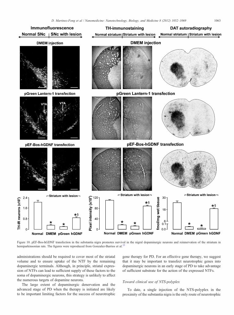
Figure 10. pEF-Bos-hGDNF transfection in the substantia nigra promotes survival in the nigral dopaminergic neurons and reinnervation of the striatum inhemiparkinsonian rats. The figures were reproduced from Gonzalez-Barrios et al.25
1063D. Martinez-Fong et al / Nanomedicine: Nanotechnology, Biology, and Medicine 8 (2012) 1052–1069
administrations should be required to cover most of the striatalvolume and to ensure uptake of the NTF by the remainingdopaminergic terminals. Although, in principle, striatal expres-sion of NTFs can lead to sufficient supply of these factors to thesoma of dopaminergic neurons, this strategy is unlikely to affectthe numerous targets of dopamine neurons.
The large extent of dopaminergic denervation and theadvanced stage of PD when the therapy is initiated are likelyto be important limiting factors for the success of neurotrophic
gene therapy for PD. For an effective gene therapy, we suggestthat it may be important to transfect neurotrophic genes intodopaminergic neurons in an early stage of PD to take advantageof sufficient substrate for the action of the expressed NTFs.
Toward clinical use of NTS-polyplex
To date, a single injection of the NTS-polyplex in theproximity of the substantia nigra is the only route of neurotrophic
1064 D. Martinez-Fong et al / Nanomedicine: Nanotechnology, Biology, and Medicine 8 (2012) 1052–1069
gene transfection that has been evaluated in the 6-OHDAhemiparkinsonian-rat model.25-27 Though studies of the poten-tial neurotoxicity of the NTS-polyplex in the rat have not beendone, the results from the local transfection of neurotrophicgenes suggest that this approach might also produce attenuationof neuroinflammation caused by 6-OHDA and the surgicalprocedure.25-27 In that case, the local administration of the NTS-polyplex in the neurotrophic gene therapy for PD would be a safeand beneficial procedure. In support of this suggestion, clinicaltrials have shown that neurotrophic factor infusion or injectionsof NRTN-AAV into the brain have been relatively safe.19,130
Further support is provided because PD is the only neurodegen-erative disorder routinely treated with neurosurgery.129
The NTS-polyplex can also deliver a neurotrophic gene fromthe striatum into nigral dopaminergic neurons through theNTSR1-mediated retrograde transport of NTS.22 This resultsuggests that the injection of the NTS-polyplex into the striatummight be another route to transfect dopaminergic neurons in thetreatment for PD patients. However, the striatal injection of theNTS-polyplex has two possible disadvantages in comparisonwith its nigral injection. The first disadvantage is that the striatumis a much larger nucleus than the substantia nigra in humans.This implies that a large dose of NTS-polyplex or repeatedinjections of small dose in different sites should be made to coverthe whole of the striatum, thus assuring an efficient transfectionof the nigral dopaminergic neurons. The second possibledisadvantage is the decrease in the NTSR1 density because ofthe scarcity of dopaminergic axon terminals in the striatum of PDpatients that might limit the retrograde transport of the NTS-polyplex to the nigral cell bodies. This is a limiting factor for allforms of neurotrophic gene therapy for PD, especially because ofthe extensive loss of divergent innervation of multiple structuresafter degeneration of dopaminergic neurons.
Recently, the NTS-polyplex harboring a suicide gene hasbeen intravenously injected in repeated doses to kill malignantcells in a model of metastatic neuroblastoma developed bysubcutaneous allograft of N1E-115 cells in nude mice.43 Thistherapeutic approach was based on the finding that neuroblas-toma cells overexpress NTSR1.48 This work showed that theNTS-polyplex was able to reach and transfect only tumorouscells and in a lesser extension the NTSR1-expressing cells ofthe intestinal tract after the injection into the blood stream.43
However, no transgene expression was seen in the brainsuggesting that the NTS-polyplex is unable to cross the bloodbrain barrier (BBB), possibly caused by the inability of theNTS to penetrate the BBB. Though the BBB hindrance protectsthe brain from the NTS-polyplex-mediated transfection ofgenes that promote cell death, the BBB is a limitation in thetargeted neurotrophic gene delivery to the brain through theblood stream. Two possible strategies have been considered toovercome the BBB. The first strategy is to replace the NTS asligand of the NTS-polyplex by its synthetic analogue NT69L,which resists degradation by plasma enzymes, binds NTSR1with higher affinity than NTS, and can cross the BBB.132 Thesecond invasive strategy involves the systemic administrationof the NTS-polyplex in conjunction with transient BBBdisruption using vasoactive amines, such as bradykinin,histamine, and the synthetic bradykinin analog RMP-7
(receptor-mediated permeabilizer).133 Other routes of theNTS-polyplex to bypass the BBB might be the intraventricularor intranasal administration, which have been effective in drugdelivery to the brain.133
Limitations, perspectives, and possibilities of the NTS-polyplexfor PD therapy
The NTS-polyplex appears to have a promising future in theneurotrophic gene therapy for PD. However, some limitationsunrelated to the nonviral gene technology but with the nature ofthe disease have raised concerns about the future of thistherapeutic approach. First, neuron loss silently accumulatesbefore clinical signs become evident.1 Symptoms developwhen approximately 50% of dopaminergic neurons are stillpresent.15 Therefore, neurotrophic therapy should start in theearly stage of PD to retain sufficient dopamine neurons that arecompromised but not dead to halt the advance of neurodegen-eration. However, the pharmacological treatment at this stage ofPD is still effective and therefore rules out gene therapy as afirst-elective approach. Although PD is a gradually developingdisease and the potential window of treatment appears to belarge, the clinical trials of neurotrophic gene therapy using viralvectors in advanced PD are not conclusive. The other majorissue that confronts neurotrophic gene therapy is that it fails toaddress the etiology and the complexity of PD.1,4 Though theneurotrophic gene transfection into the compromised dopami-nergic neurons might lead to repopulation and restoration ofmultiple brain circuits in many brain regions (those innervatedby those nigral neurons), it fails to repair the genetic alterationthat causes PD.134 Recently, interference-RNA technology hasbeen proposed for PD therapy because the overexpression ofvarious proteins is known to kill the nigral dopaminergicneurons in animal models and in familial forms of PD.44 Aswith viral vectors, the NTS-polyplex might be used in thedelivery of interference RNA into dopaminergic neurons toknock down the altered proteins, such as α-synuclein orLRRK2 (protein leucine-rich repeat kinase 2).134,135
Notwithstanding the limitations that all gene vectors sufferfrom, the NTS-polyplex NP system seems to have broadpossibilities for a clinical use for PD treatment. By takingadvantage of the potential of the NTS-polyplex to codelivertwo genes into the same cell,22 this system might be used todeliver a neurotrophic gene and interference RNA. Hypothet-ically, this approach might yield a neurotrophic effect anddecrease in the mutated protein levels. Another possiblecombined gene therapy using the NTS-polyplex might bewith a neurotrophic gene and one of the genes coding for aRedox enzyme such as catalase.136 This treatment would beintended to attenuate neuroinflammation and increase neuro-protection in patients with PD. In summary, a combined genetherapy for PD can be formulated not only with differentneurotrophic genes, but also with a great variety of geneticapproaches, as mentioned above, well beyond the limit ofimagination. However, from a theoretical point of view, theNTS-polyplex NPs have the limitation to incorporate a drug inaddition to the transgene. This incorporation would exceed theprecise stoichiometry to assemble the five components of theNTS-polyplex into functional NPs for gene delivery.
1065D. Martinez-Fong et al / Nanomedicine: Nanotechnology, Biology, and Medicine 8 (2012) 1052–1069
Potential use of neurotrophic therapy in otherneurodegenerative diseases
An attractive characteristic of the NTS-polyplex that makes ita promising candidate for PD gene therapy is its specificitythrough NTSR1-mediated endocytosis mechanism to shuttle thetransgene into surviving dopamine neurons. Other neurons of theCNS, such as those of the dopaminergic ventral tegmental areaand of the cholinergic basal forebrain, also expressNTSR1,68,137,138 so they are other putative targets for genedelivery via the NTS-polyplex. For example, NTS-polyplextransfection of neurotrophic factors into basal-forebrain cholin-ergic neurons could be useful to prevent neurodegeneration inAlzheimer's disease.139,140 Two gene therapy approaches forAlzheimer's disease that have been previously successful withviral vectors could perhaps be adapted to take advantage of NTS-polyplex transfection. The first approach aims to increase NGFconcentration to promote survival, regeneration, and protectionof basal-forebrain cholinergic neurons.140 The second approachis overexpression of the CREB-binding protein to overcome theinterference of amyloid-β accumulation, which plays a primaryrole in the cognitive deficits of Alzheimer's disease.141
Although the potential of neurotrophic gene therapy is veryextensive, the absence of NTSR1 in degenerating neurons limitsthe use of NTS-polyplex in other pathologies of the CNS andperipheral nervous system.
Benefits and limitations of NTS-polyplex system in comparisonwith viral carriers
Polyplexes have the advantage over viral gene transfer ofbeing nonimmunogenic, easy to produce, and not oncogenic.Although the immunological and safety evaluation of the NTS-polyplex has not been accomplished, published evidence, bothdirect and indirect, supports the biosafety of NTS-polyplex-mediated transfection in vitro and in vivo. A recent study reportsthat NTS-polyplex-mediated transfection does not affect cellviability in vitro.22 In all of the studies performed in vivo, theNTS-polyplex has been shown not to cause more damage tobrain parenchyma than that caused by mechanical microinjectionby itself.21-23,25-27 Moreover, repeated intravenous injections ofNTS-polyplex for targeted gene delivery of a suicide gene in ananimal model of neuroblastoma lead to an antitumoral effectwithout signs of tissue damage in other organs.43 This evidenceagrees with the demonstrated biosafety of lactoferrin-polyplex,that has been successfully used in animal models of PD.142,143 Incontrast, nondegradable polyplexes, such as those based on highmolecular weight polyethylenimines, can cause cytotoxicity as aresult of their poor clearance.144 Those latter kinds of polyplexeshave limited use in gene therapy despite their high transfectionefficiency in vitro and in vivo.
To date, only lactoferrin-polyplex and NTS-polyplex systemshave been explored to deliver the GDNF gene delivery in ratswith PD.25,145 The efficacy of those systems in promotingfunctional recovery in the rat 6-HODA lesion model is similar tothat of viral carriers.146,147 However, clinical trials with suchNPs will be required to confirm the usefulness of this strategy forthe treatment of PD.
Lack of regulation of gene expression is a current limitationfor all current protocols in neurotrophic therapy. The uncon-trolled and robust expression of a neurotrophic protein mightlead to undesirable side effects, such as aberrant innervation ofthe regenerating dopaminergic fibers148 and decrease ofdopamine synthesis.149 Furthermore, neurotrophins, for in-stance, could reach levels high enough to stimulate the p75receptor, thus leading healthy and recovering neurons toapoptosis.150 The use of inducible promoters in conjunctionwith NTS-polyplex thus needs to be explored.
Conclusions
Neurotrophic gene-therapy continues to be at the forefront ofPD research because it holds the promise of stoppingneurodegeneration, reversing neurological damage and main-taining the integrity of divergent projections of nigral dopami-nergic-neurons. All neurotrophic gene-therapy approaches areeffective and relatively safe in rodent and primate models of PD,although some of them, in particular those with GDNF, produceundesirable behavioral effects resulting from aberrant innerva-tion of the regenerating dopaminergic fibers148 and decrease ofdopamine synthesis.149 Although the results of clinical trialswith recombinant GDNF-protein infusion or NRTN genetherapy have not been particularly positive, they show thatthese approaches are safe in humans. We believe that the modestclinical benefit might be related to the selection of putamen cellsas the source of NTFs. From this region, the diffusion of NTFsconfronts physical and biochemical obstacles to reach the cellbodies and the other innervation targets of dopaminergicneurons. In particular, the long distance that separates the othertargets from the putamen in the human brain and enzymaticdegradation of NTFs could be the main causes of the poorneurotrophic effect. We propose that transfection of neurotrophicgenes in dopaminergic neurons might be a much more effectivestrategy to obtain a neurotrophic effect simultaneously in the cellbody and terminal regions of dopaminergic neurons. The NTS-polyplex has the ability to transfect dopaminergic neurons viaNTSR1-mediated endocytosis at the cell body or via retrogradetransport of NTSR1. Nigral injection of the NTS-polyplex withneurotrophic genes (GDNF, NRTN, or BDNF) has beeneffective in the treatment of acute and chronic experimentalhemiparkinsonism in the rat. The preliminary data suggestingadequate biosafety of this approach anticipates the safe use of theNTS-polyplex in clinical trials of neurotrophic gene-therapy forPD in the near future. Meanwhile, evaluation of the NTS-polyplex in nonhuman primates with PD represents the nextimportant test of this promising nonviral gene-transfer system.
References
1. Galvan A, Wichmann T. Pathophysiology of parkinsonism. ClinNeurophysiol 2008;119:1459-74.
2. Kish SJ, Shannak K, Hornykiewicz O. Uneven pattern of dopamineloss in the striatum of patients with idiopathic Parkinson's disease.Pathophysiologic and clinical implications. N Engl J Med 1988;318:876-80.
1066 D. Martinez-Fong et al / Nanomedicine: Nanotechnology, Biology, and Medicine 8 (2012) 1052–1069
3. Brooks DJ. PET studies on the function of dopamine in health andParkinson's disease. Ann N Y Acad Sci 2003;991:22-35.
4. Chaudhuri KR, Schapira AH. Non-motor symptoms of Parkinson'sdisease: dopaminergic pathophysiology and treatment. Lancet Neurol2009;8:464-74.
5. Anaya-Martinez V, Martinez-Marcos A, Martinez-Fong D, Aceves J,Erlij D. Substantia nigra compacta neurons that innervate the reticularthalamic nucleus in the rat also project to striatum or globus pallidus:implications for abnormal motor behavior. Neuroscience 2006;143:477-86.
6. Debeir T, Ginestet L, Francois C, Laurens S, Martel JC, Chopin P, et al.Effect of intrastriatal 6-OHDA lesion on dopaminergic innervation ofthe rat cortex and globus pallidus. Exp Neurol 2005;193:444-54.
7. Prensa L, Parent A. The nigrostriatal pathway in the rat: a single-axonstudy of the relationship between dorsal and ventral tier nigral neuronsand the striosome/matrix striatal compartments. J Neurosci 2001;21:7247-60.
8. Floran B, Aceves J, Sierra A, Martinez-Fong D. Activation of D1dopamine receptors stimulates the release of GABA in the basal gangliaof the rat. Neurosci Lett 1990;116:136-40.
9. Rosales MG, Flores G, Hernandez S, Martinez-Fong D, Aceves J.Activation of subthalamic neurons produces NMDA receptor-mediateddendritic dopamine release in substantia nigra pars reticulata: amicrodialysis study in the rat. Brain Res 1994;645:335-7.
10. Flores G, Liang JJ, Sierra A, Martinez-Fong D, Quirion R, Aceves J,et al. Expression of dopamine receptors in the subthalamic nucleus ofthe rat: characterization using reverse transcriptase-polymerase chainreaction and autoradiography. Neuroscience 1999;91:549-56.
11. Martinez-Fong D, Rosales MG, Gongora-Alfaro JL, Hernandez S,Aceves J. NMDA receptor mediates dopamine release in the striatum ofunanesthetized rats as measured by brain microdialysis. Brain Res1992;595:309-15.
12. Ungerstedt U. Postsynaptic supersensitivity after 6-hydroxy-dopamineinduced degeneration of the nigro-striatal dopamine system. ActaPhysiol Scand Suppl 1971;367:69-93.
13. Rolheiser TM, Fulton HG, Good KP, Fisk JD, McKelvey JR, ScherflerC, et al. Diffusion tensor imaging and olfactory identification testing inearly-stage Parkinson's disease. J Neurol 2011;258:1254-60.
14. Ascherio A, LeWitt PA, Xu K, Eberly S, Watts A, Matson WR, et al.Urate as a predictor of the rate of clinical decline in Parkinson’s disease.Arch Neurol 2009;66:1460-8.
15. Bjorklund T, Kirik D. Scientific rationale for the development of genetherapy strategies for Parkinson's disease. Biochim Biophys Acta 2009;1792:703-13.
16. Ulusoy A, Bjorklund T, Hermening S, Kirik D. In vivo gene deliveryfor development of mammalian models for Parkinson's disease. ExpNeurol 2008;209:89-100.
17. Bjorklund A, Kirik D, Rosenblad C, Georgievska B, Lundberg C,Mandel RJ. Towards a neuroprotective gene therapy for Parkinson'sdisease: use of adenovirus, AAV and lentivirus vectors for gene transferof GDNF to the nigrostriatal system in the rat Parkinson model. BrainRes 2000;886:82-98.
18. Airaksinen MS, Saarma M. The GDNF family: signalling, biologicalfunctions and therapeutic value. Nat Rev Neurosci 2002;3:383-94.
19. Marks WJ, Ostrem JL, Verhagen L, Starr PA, Larson PS, Bakay RA,et al. Safety and tolerability of intraputaminal delivery of CERE-120(adeno-associated virus serotype 2-neurturin) to patients withidiopathic Parkinson's disease: an open-label, phase I trial. Lancet Neurol2008;7:400-8.
20. Fasano C, Thibault D, Trudeau LE. Culture of postnatal mesencephalicdopamine neurons on an astrocyte monolayer. Curr Protoc Neurosci2008;44:3.21.1-3.21.19.
21. Alvarez-Maya I, Navarro-Quiroga I, Meraz-Rios MA, Aceves J,Martinez-Fong D. In vivo gene transfer to dopamine neurons of ratsubstantia nigra via the high-affinity neurotensin receptor. Mol Med2001;7:186-92.
22. Arango-Rodriguez ML, Navarro-Quiroga I, Gonzalez-Barrios JA,Martinez-Arguelles DB, Bannon MJ, Kouri J, et al. Biophysicalcharacteristics of neurotensin polyplex for in vitro and in vivo genetransfection. Biochim Biophys Acta 2006;1760:1009-20.
23. Navarro-Quiroga I, Gonzalez-Barrios JA, Barron-Moreno F, Gonzalez-Bernal V, Martinez-Arguelles DB, Martinez-Fong D. Improvedneurotensin-vector-mediated gene transfer by the coupling of hemag-glutinin HA2 fusogenic peptide and Vp1 SV40 nuclear localizationsignal. Brain Res Mol Brain Res 2002;105:86-97.
24. Orozco-Barrios CE, Battaglia-Hsu SF, Arango-Rodriguez ML, Ayala-Davila J, Chery C, Alberto JM, et al. Vitamin B12-impairedmetabolism produces apoptosis and Parkinson phenotype in ratsexpressing the transcobalamin-oleosin chimera in substantia nigra.PLoS One 2009;e8268:4.
25. Gonzalez-Barrios JA, Lindahl M, Bannon MJ, Anaya-Martinez V,Flores G, Navarro-Quiroga I, et al. Neurotensin polyplex as an efficientcarrier for delivering the human GDNF gene into nigral dopamineneurons of hemiparkinsonian rats. Mol Ther 2006;14:857-65.
26. Hernandez-Chan N, Zamudio S, Escobedo L, De la Cruz F, Gongora-Alfaro JL, Bannon MJ, et al. BDNF-Flag gene transfer by NT-Polyplex to nigral dopaminergic neurons causes morphological andfunctional recovery from hemiparkinsonism in the rat. Mol Ther2008;16:974.
27. Reyes-Corona D, Escobedo L, Ayala-Davila J, Orozco-Barrios CE,Arango-Rodriguez ML, Martinez-Fong D. NT-Polyplex-mediatedneurturin delivery to dopaminergic neurons of hemiparkinsonian rats:a new approach in the repertoire of neurotrophic therapy. Program No.643.7/U4. 2009 Neuroscience Meeting Planner. Chicago, IL: Societyfor Neuroscience; 2009 [Online 2009].
28. Heuser JE, Anderson RG. Hypertonic media inhibit receptor-mediatedendocytosis by blocking clathrin-coated pit formation. J Cell Biol1989;108:389-400.
29. Martinez-Fong D, Navarro-Quiroga I, Ochoa I, Alvarez-Maya I, MerazMA, Luna J, et al. Neurotensin-SPDP-poly-L-lysine conjugate: anonviral vector for targeted gene delivery to neural cells. Brain Res MolBrain Res 1999;69:249-62.
30. Martinez-Fong D, Navarro-Quiroga I. Synthesis of a non-viral vectorfor gene transfer via the high-affinity neurotensin receptor. Brain ResBrain Res Protoc 2000;6:13-24.
31. Fernandez CA, Rice KG. Engineered nanoscaled polyplex genedelivery systems. Mol Pharm 2009;6:1277-89.
32. Esbjorner EK, Oglecka K, Lincoln P, Graslund A, Norden B.Membrane binding of pH-sensitive influenza fusion peptides. posi-tioning, configuration, and induced leakage in a lipid vesicle model.Biochemistry 2007;46:13490-504.
33. Ishii N, Minami N, Chen EY, Medina AL, Chico MM, Kasamatsu H.Analysis of a nuclear localization signal of simian virus 40 major capsidprotein Vp1. J Virol 1996;1996:1317-22.
34. Saccardo P, Villaverde A, Gonzalez-Montalban N. Peptide-mediatedDNA condensation for non-viral gene therapy. Biotechnol Adv 2009;27:432-8.
35. Hud NV, Vilfan ID. Toroidal DNA condensates: unraveling the finestructure and the role of nucleation in determining size. Annu RevBiophys Biomol Struct 2005;34:295-318.
36. Liu L, Yang YL, Wang C, Yao Y, Ma YZ, Hou S, et al. Polymericeffects on DNA condensation by cationic polymers observed by atomicforce microscopy. Colloids Surf B Biointerfaces 2010;75:230-8.
37. Midoux P, Pichon C, Yaouanc JJ, Jaffres PA. Chemical vectors forgene delivery: a current review on polymers, peptides and lipidscontaining histidine or imidazole as nucleic acids carriers. Br JPharmacol 2009;157:166-78.
38. Ekrami HM, Shen WC. Carbamylation decreases the cytotoxicity but notthe drug-carrier properties of polylysines. J Drug Target 1995;2:469-75.
39. Martinez-Fong D, Mullersman JE, Purchio AF, Armendariz-Borunda J,Martinez-Hernandez A. Nonenzymatic glycosylation of poly-L-lysine:a new tool for targeted gene delivery. Hepatology 1994;20:1602-8.
1067D. Martinez-Fong et al / Nanomedicine: Nanotechnology, Biology, and Medicine 8 (2012) 1052–1069
40. Wagner E, Ogris M, Zauner W. Polylysine-based transfection systemsutilizing receptor-mediated delivery. Adv Drug Deliv Rev 1998;30:97-113.
41. Cotten M, Wagner E. Non-viral approaches to gene therapy. Curr OpinBiotechnol 1993;4:705-10.
42. Pack DW, Hoffman AS, Pun S, Stayton PS. Design and development ofpolymers for gene delivery. Nat Rev Drug Discov 2005;4:581-93.
43. Rubio-Zapata HA, Rembao-Bojorquez JD, Arango-Rodriguez ML,Dupouy S, Forgez P, Martinez-Fong D. NT-polyplex: a new tool fortherapeutic gene delivery to neuroblastoma tumors. Cancer Gene Ther2009;16:573-84.
44. Sahay G, Alakhova DY, Kabanov AV. Endocytosis of nanomedicines.J Control Release 2010;145:182-95.
45. Traub LM. Tickets to ride: selecting cargo for clathrin-regulatedinternalization. Nat Rev Mol Cell Biol 2009;10:583-96.
46. Mazella J, Vincent JP. Internalization and recycling properties ofneurotensin receptors. Peptides 2006;27:2488-92.
47. Souaze F, Forgez P. Molecular and cellular regulation of neurotensinreceptor under acute and chronic agonist stimulation. Peptides 2006;27:2493-501.
48. Amar S, Kitabgi P, Vincent JP. Stimulation of inositol phosphateproduction by neurotensin in neuroblastoma N1E115 cells: implicationof GTP-binding proteins and relationship with the cyclic GMPresponse. J Neurochem 1987;49:999-1006.
49. Kitabgi P, Rostene W, Dussaillant M, Schotte A, Laduron PM, VincentJP. Two populations of neurotensin binding sites in murine brain:discrimination by the antihistamine levocabastine reveals markedlydifferent radioautographic distribution. Eur J Pharmacol 1987;140:285-93.
50. Mendez M, Souaze F, Nagano M, Kelly PA, Rostene W, Forgez P.High affinity neurotensin receptor mRNA distribution in rat brain andperipheral tissues. Analysis by quantitative RT-PCR. J Mol Neurosci1997;9:93-102.
51. Nouel D, Faure MP, StPierre JA, Alonso R, Quirion R, Beaudet A.Differential binding profile and internalization process of neurotensinvia neuronal and glial receptors. J Neurosci 1997;17:1795-803.
52. Quirion R, Welner S, Gauthier S, Bedard P. Neurotensin receptorbinding sites in monkey and human brain: autoradiographic distributionand effects of 1-methyl-4-phenyl-1,2,3,6-tetrahydropyridine treatment.Synapse 1987;1:559-66.
53. Szigethy E, Beaudet A. Correspondence between high affinity 125I-neurotensin binding sites and dopaminergic neurons in the ratsubstantia nigra and ventral tegmental area: a combined radioauto-graphic and immunohistochemical light microscopic study. J CompNeurol 1989;279:128-37.
54. Palacios JM, Kuhar MJ. Neurotensin receptors are located on dopamine-containing neurones in rat midbrain. Nature 1981;294:587-9.
55. Sadoul JL, Checler F, Kitabgi P, Rostene W, Javoy-Agid F, Vincent JP.Loss of high affinity neurotensin receptors in substantia nigra fromparkinsonian subjects. Biochem Biophys Res Commun 1984;125:395-404.
56. Mazella J, Leonard K, Chabry J, Kitabgi P, Vincent JP, Beaudet A.Binding and internalization of iodinated neurotensin in neuronalcultures from embryonic mouse brain. Brain Res 1991;564:249-55.
57. Toy-Miou-Leong M, Cortes CL, Beaudet A, Rostene W, Forgez P.Receptor trafficking via the perinuclear recycling compartmentaccompanied by cell division is necessary for permanent neurotensincell sensitization and leads to chronic mitogen-activated protein kinaseactivation. J Biol Chem 2004;279:12636-46.
58. Vanisberg MA, Maloteaux JM, Octave JN, Laduron PM. Rapidagonist-induced decrease of neurotensin receptors from the cell surfacein rat cultured neurons. Biochem Pharmacol 1991;42:2265-74.
59. Gully D, Canton M, Boigegrain R, Jeanjean F, Molimard JC, PonceletM, et al. Biochemical and pharmacological profile of a potent andselective nonpeptide antagonist of the neurotensin receptor. Proc NatlAcad Sci U S A 1993;90:65-9.
60. Mazella J, Botto JM, Guillemare E, Coppola T, Sarret P, Vincent JP.Structure, functional expression, and cerebral localization of thelevocabastine-sensitive neurotensin/neuromedin N receptor frommouse brain. J Neurosci 1996;16:5613-20.
61. Vita N, Laurent P, Lefort S, Chalon P, Dumont X, Kaghad M, et al.Cloning and expression of a complementary DNA encoding a highaffinity human neurotensin receptor. FEBS Lett 1993;317:139-42.
62. Gendron L, Perron A, Payet MD, Gallo-Payet N, Sarret P, Beaudet A.Low-affinity neurotensin receptor (NTS2) signaling: internalization-dependent activation of extracellular signal-regulated kinases 1/2. MolPharmacol 2004;66:1421-30.
63. Lepee-Lorgeoux I, Betancur C, Rostene W, Pelaprat D. Differentialontogenetic patterns of levocabastine-sensitive neurotensin NT2receptors and of NT1 receptors in the rat brain revealed by in situhybridization. Brain Res Dev Brain Res 1999;113:115-31.
64. Mazella J, Zsurger N, Navarro V, Chabry J, Kaghad M, Caput D, et al.The 100-kDa neurotensin receptor is gp95/sortilin, a non-G-protein-coupled receptor. J Biol Chem 1998;273:26273-6.
65. Nielsen MS, Jacobsen C, Olivecrona G, Gliemann J, Petersen CM.Sortilin/neurotensin receptor-3 binds and mediates degradation oflipoprotein lipase. J Biol Chem 1999;274:8832-6.
66. Martin S, Navarro V, Vincent JP, Mazella J. Neurotensin receptor-1and -3 complex modulates the cellular signaling of neurotensin in theHT29 cell line. Gastroenterology 2002;123:1135-43.
67. Cusack B, Stanton T, Richelson E. Developmental regulation ofneurotensin receptor expression and function in murine neuroblastomaclone N1E-115. Eur J Pharmacol 1991;206:339-42.
68. Boudin H, Pelaprat D, Rostene W, Pickel VM, Beaudet A. Correlativeultrastructural distribution of neurotensin receptor proteins and bindingsites in the rat substantia nigra. J Neurosci 1998;18:8473-84.
69. Laduron PM. Functional consequences of retrograde axonal transportof receptor-bound neurotensin. Trends Pharmacol Sci 1995;16:338-43.
70. Sorkin A, von ZM. Endocytosis and signaling: intertwining molecularnetworks. Nat Rev Mol Cell Biol 2009;10:609-22.
71. Medina-Kauwe LK. “Alternative” endocytic mechanisms exploited bypathogens: new avenues for therapeutic delivery? Adv Drug Deliv Rev2007;59:798-809.
72. Authier F, Posner BI, Bergeron JJ. Endosomal proteolysis ofinternalized proteins. FEBS Lett 1996;389:55-60.
73. White J, Kielian M, Helenius A. Membrane fusion proteins ofenveloped animal viruses. Q Rev Biophys 1983;16:151-95.
74. Casali M, Banta S, Zambonelli C, Megeed Z, Yarmush ML. Site-directed mutagenesis of the hinge peptide from the hemagglutininprotein: enhancement of the pH-responsive conformational change.Protein Eng Des Sel 2008;21:395-404.
75. Swalley SE, Baker BM, Calder LJ, Harrison SC, Skehel JJ, WileyDC. Full-length influenza hemagglutinin HA(2) refolds into thetrimeric low-pH-induced conformation. Biochemistry 2004;43:5902-11.
76. Minchin RF, Yang S. Endosomal disruptors in non-viral gene delivery.Expert Opin Drug Deliv 2010;7:331-9.
77. Howell DP, Krieser RJ, Eastman A, Barry MA. Deoxyribonuclease II isa lysosomal barrier to transfection. Mol Ther 2003;8:957-63.
78. Wu CH, Wilson JM, Wu GY. Targeting genes: delivery and persistentexpression of a foreign gene driven by mammalian regulatory elementsin vivo. J Biol Chem 1989;264:16985-7.
79. Cristiano RJ, Smith LC, Kay MA, Brinkley BR, Woo SL. Hepaticgene therapy: efficient gene delivery and expression in primaryhepatocytes utilizing a conjugated adenovirus-DNA complex. ProcNatl Acad Sci U S A 1993;90:11548-52.
80. Midoux P, Monsigny M. Efficient gene transfer by histidylatedpolylysine/pDNA complexes. Bioconjug Chem 1999;10:406-11.
81. Chan CK, Jans DA. Enhancement of polylysine-mediated transferrin-fection by nuclear localization sequences: polylysine does not functionas a nuclear localization sequence. Hum Gene Ther 1999;10:1695-702.

1068 D. Martinez-Fong et al / Nanomedicine: Nanotechnology, Biology, and Medicine 8 (2012) 1052–1069
82. Midoux P, Mendes C, Legrand A, Raimond J, Mayer R, Monsigny M,et al. Specific gene transfer mediated by lactosylated poly-L-lysine intohepatoma cells. Nucleic Acids Res 1993;21:871-8.
83. Chowdhury EH. Nuclear targeting of viral and non-viral DNA. ExpertOpin Drug Deliv 2009;6:697-703.
84. Lange A, Mills RE, Lange CJ, Stewart M, Devine SE, Corbett AH.Classical nuclear localization signals: definition, function, andinteraction with importin alpha. J Biol Chem 2007;282:5101-5.
85. Kalderon D, Richardson WD, Markham AF, Smith AE. Sequencerequirements for nuclear location of simian virus 40 large-T antigen.Nature 1984;311:33-8.
86. Ishii N, Nakanishi A, Yamada M, Macalalad MH, Kasamatsu H.Functional complementation of nuclear targeting-defective mutants ofsimian-virus-40 structural proteins. J Virol 1994;68:8209-16.
87. Zanta MA, Belguise-Valladier P, Behr JP. Gene delivery: a singlenuclear localization signal peptide is sufficient to carry DNA to the cellnucleus. Proc Natl Acad Sci U S A 1999;96:91-6.
88. van der AM, Koning G, van der GJ, d'Oliveira C, Oosting R, HenninkWE, et al. Covalent attachment of an NLS-peptide to linear dna doesnot enhance transfection efficiency of cationic polymer based genedelivery systems. J Control Release 2005;101:395-7.
89. Roulon T, Helene C, Escude C. Coupling of a targeting peptide toplasmid DNA using a new type of padlock oligonucleotide. BioconjugChem 2002;13:1134-9.
90. Chan CK, Senden T, Jans DA. Supramolecular structure and nucleartargeting efficiency determine the enhancement of transfection bymodified polylysines. Gene Ther 2000;7:1690-7.
91. Reddy JA, Low PS. Enhanced folate receptor mediated gene therapyusing a novel pH-sensitive lipid formulation. J Control Release 2000;64:27-37.
92. Guy J, Drabek D, Antoniou M. Delivery of DNA into mammalian cellsby receptor-mediated endocytosis and gene therapy. Mol Biotechnol1995;3:237-48.
93. Bannon MJ. The dopamine transporter: role in neurotoxicity andhuman disease. Toxicol Appl Pharmacol 2005;204:355-60.
94. Bannon MJ, Whitty CJ. Age-related and regional differences indopamine transporter mRNA expression in human midbrain. Neurol-ogy 1997;48:969-77.
95. Sacchetti P, Brownschidle LA, Granneman JG, Bannon MJ. Charac-terization of the 5'-flanking region of the human dopamine transportergene. Brain Res Mol Brain Res 1999;74:167-74.
96. Brooks AR, Harkins RN, Wang P, Qian HS, Liu P, Rubanyi GM.Transcriptional silencing is associated with extensive methylation ofthe CMV promoter following adenoviral gene delivery to muscle. JGene Med 2004;6:395-404.
97. Lindholm P, Voutilainen MH, Lauren J, Peranen J, Leppanen VM,Andressoo JO, et al. Novel neurotrophic factor CDNF protects andrescues midbrain dopamine neurons in vivo. Nature 2007;448:73-7.
98. Lindholm P, Peranen J, Andressoo JO, Kalkkinen N, Kokaia Z,Lindvall O, et al. MANF is widely expressed in mammalian tissues anddifferently regulated after ischemic and epileptic insults in rodent brain.Mol Cell Neurosci 2008;39:356-71.
99. Lindholm P, Saarma M. Novel CDNF/MANF family of neurotrophicfactors. Dev Neurobiol 2010;70:360-71.
100. Lu B, Pang PT, Woo NH. The yin and yang of neurotrophin action. NatRev Neurosci 2005;6:603-14.
101. Bespalov MM, Saarma M. GDNF family receptor complexes areemerging drug targets. Trends Pharmacol Sci 2007;28:68-74.
102. Bauer S, Kerr BJ, Patterson PH. The neuropoietic cytokine family indevelopment, plasticity, disease and injury. Nat Rev Neurosci 2007;8:221-32.
103. Petrova P, Raibekas A, Pevsner J, Vigo N, Anafi M, Moore MK, et al.MANF: a new mesencephalic, astrocyte-derived neurotrophic factorwith selectivity for dopaminergic neurons. J Mol Neurosci 2003;20:173-88.
104. Yoong LF, Wan G, Too HP. GDNF-induced cell signaling and neuriteoutgrowths are differentially mediated by GFRalpha1 isoforms. MolCell Neurosci 2009;41:464-73.
105. Sarabi A, Hoffer BJ, Olson L, Morales M. GFRalpha-1 mRNA indopaminergic and nondopaminergic neurons in the substantia nigra andventral tegmental area. J Comp Neurol 2001;441:106-17.
106. Golden JP, Baloh RH, Kotzbauer PT, Lampe PA, Osborne PA,Milbrandt J, et al. Expression of neurturin, GDNF, and their receptors inthe adult mouse CNS. J Comp Neurol 1998;398:139-50.
107. Seroogy KB, Lundgren KH, Tran TM, Guthrie KM, Isackson PJ, GallCM. Dopaminergic neurons in rat ventral midbrain express brain-derived neurotrophic factor and neurotrophin-3 mRNAs. J CompNeurol 1994;342:321-34.
108. Bourque MJ, Trudeau LE. GDNF enhances the synaptic efficacy ofdopaminergic neurons in culture. Eur J Neurosci 2000;12:3172-80.
109. Kozlowski DA, Miljan EA, Bremer EG, Harrod CG, Gerin C, ConnorB, et al. Quantitative analyses of GFRalpha-1 and GFRalpha-2 mRNAsand tyrosine hydroxylase protein in the nigrostriatal system revealbilateral compensatory changes following unilateral 6-OHDA lesionsin the rat. Brain Res 2004;1016:170-81.
110. Numan S, Seroogy KB. Increased expression of trkB mRNA in ratcaudate–putamen following 6-OHDA lesions of the nigrostriatalpathway. Eur J Neurosci 1997;9:489-95.
111. Wang LC, Shih A, Hongo J, Devaux B, Hynes M. Broad specificity ofGDNF family receptors GFRalpha1 and GFRalpha2 for GDNF andNTN in neurons and transfected cells. J Neurosci Res 2000;61:1-9.
112. Creedon DJ, Tansey MG, Baloh RH, Osborne PA, Lampe PA, FahrnerTJ, et al. Neurturin shares receptors and signal transduction pathwayswith glial cell line-derived neurotrophic factor in sympathetic neurons.Proc Natl Acad Sci U S A 1997;94:7018-23.
113. Sariola H, Saarma M. Novel functions and signalling pathways forGDNF. J Cell Sci 2003;116:3855-62.
114. Knusel B, Winslow JW, Rosenthal A, Burton LE, Seid DP, Nikolics K,et al. Promotion of central cholinergic and dopaminergic neurondifferentiation by brain-derived neurotrophic factor but not neurotro-phin 3. Proc Natl Acad Sci U S A 1991;88:961-5.
115. Meyer M, Johansen J, Gramsbergen JB, Johansen TE, Zimmer J.Improved survival of embryonic porcine dopaminergic neurons incoculture with a conditionally immortalized GDNF-producing hippo-campal cell line. Exp Neurol 2000;164:82-93.
116. Horger BA, Nishimura MC, Armanini MP, Wang LC, Poulsen KT,Rosenblad C, et al. Neurturin exerts potent actions on survival and functionof midbrain dopaminergic neurons. J Neurosci 1998;18:4929-37.
117. Pascual A, Hidalgo-Figueroa M, Piruat JI, Pintado CO, Gomez-Diaz R,Lopez-Barneo J. Absolute requirement of GDNF for adult catechol-aminergic neuron survival. Nat Neurosci 2008;11:755-61.
118. Hunot S, Bernard V, Faucheux B, Boissiere F, Leguern E, Brana C,et al. Glial cell line-derived neurotrophic factor (GDNF) geneexpression in the human brain: a post mortem in situ hybridizationstudy with special reference to Parkinson's disease. J Neural Transm1996;103:1043-52.
119. Gall CM, Gold SJ, Isackson PJ, Seroogy KB. Brain-derivedneurotrophic factor and neurotrophin-3 mRNAs are expressed inventral midbrain regions containing dopaminergic neurons. Mol CellNeurosci 1992;3:56-63.
120. Parain K, Murer MG, Yan Q, Faucheux B, Agid Y, Hirsch E, et al.Reduced expression of brain-derived neurotrophic factor protein inParkinson's disease substantia nigra. Neuroreport 1999;10:557-61.
121. Baquet ZC, Bickford PC, Jones KR. Brain-derived neurotrophic factoris required for the establishment of the proper number of dopaminergicneurons in the substantia nigra pars compacta. J Neurosci 2005;25:6251-9.
122. Baker SA, Stanford LE, Brown RE, Hagg T. Maturation but notsurvival of dopaminergic nigrostriatal neurons is affected in developingand aging BDNF-deficient mice. Brain Res 2005;1039:177-88.
1069D. Martinez-Fong et al / Nanomedicine: Nanotechnology, Biology, and Medicine 8 (2012) 1052–1069
123. Beck KD, Valverde J, Alexi T, Poulsen K, Moffat B, Vandlen RA, et al.Mesencephalic dopaminergic neurons protected by GDNF fromaxotomy-induced degeneration in the adult brain. Nature 1995;373:339-41.
124. Tomac A, Lindqvist E, Lin LF, Ogren SO, Young D, Hoffer BJ, et al.Protection and repair of the nigrostriatal dopaminergic system byGDNF in vivo. Nature 1995;373:335-9.
125. Grondin R, Zhang Z, Yi A, Cass WA, Maswood N, Andersen AH, et al.Chronic, controlled GDNF infusion promotes structural and functionalrecovery in advanced parkinsonian monkeys. Brain 2002;125:2191-201.
126. Rosenblad C, Kirik D, Devaux B, Moffat B, Phillips HS, Bjorklund A.Protection and regeneration of nigral dopaminergic neurons byneurturin or GDNF in a partial lesion model of Parkinson's diseaseafter administration into the striatum or the lateral ventricle. Eur JNeurosci 1999;11:1554-66.
127. Mufson EJ, Kroin JS, Liu YT, Sobreviela T, Penn RD, Miller JA, et al.Intrastriatal and intraventricular infusion of brain-derived neurotrophicfactor in the cynomologous monkey: distribution, retrograde transportand co-localization with substantia nigra dopamine-containing neurons.Neuroscience 1996;71:179-91.
128. Bartus RT, Herzog CD, Bishop K, Ostrove JM, Tuszynski M, KordowerJH, et al. Issues regarding gene therapy products for Parkinson's disease:the development of CERE-120 (AAV-NTN) as one reference point.Parkinsonism Relat Disord 2007;13(Suppl 3):S469-77.
129. Kaplitt MG. Parkinson disease: another player in gene therapy forParkinson disease. Nat Rev Neurol 2010;6:7-8.
130. Lang AE, Gill S, Patel NK, Lozano A, Nutt JG, Penn R, et al.Randomized controlled trial of intraputamenal glial cell line-derivedneurotrophic factor infusion in Parkinson disease. Ann Neurol 2006;59:459-66.
131. von Bartheld CS, Wang X, Butowt R. Anterograde axonal transport,transcytosis, and recycling of neurotrophic factors: the concept oftrophic currencies in neural networks. Mol Neurobiol 2001;24:1-28.
132. Boules M, Fredrickson P, Richelson E. Current topics: brainpenetrating neurotensin analog. Life Sci 2003;73:2785-92.
133. Misra A, Ganesh S, Shahiwala A, Shah SP. Drug delivery to the centralnervous system: a review. J Pharm Pharm Sci 2003;6:252-73.
134. Manfredsson FP, Lewin AS, Mandel RJ. RNA knockdown as apotential therapeutic strategy in Parkinson's disease. Gene Ther 2006;13:517-24.
135. Habig K, Walter M, Poths S, Riess O, Bonin M. RNA interference ofLRRK2-microarray expression analysis of a Parkinson's disease keyplayer. Neurogenetics 2008;9:83-94.
136. Haney MJ, Zhao Y, Li S, Higginbotham SM, Booth SL, Han HY, et al.Cell-mediated transfer of catalase nanoparticles from macrophages tobrain endothelial, glial and neuronal cells. Nanomedicine (Lond) 2011;6:1215-30.
137. Boudin H, Pelaprat D, Rostene W, Beaudet A. Cellular distribution ofneurotensin receptors in rat brain: immunohistochemical study using an
antipeptide antibody against the cloned high affinity receptor. J CompNeurol 1996;373:76-89.
138. Szigethy E, Leonard K, Beaudet A. Ultrastructural localization of[125I]neurotensin binding sites to cholinergic neurons of the rat nucleusbasalis magnocellularis. Neuroscience 1990;36:377-91.
139. Cattaneo A, Capsoni S, Paoletti F. Towards non invasive nerve growthfactor therapies for Alzheimer's disease. J Alzheimers Dis 2008;15:255-83.
140. Mandel RJ. CERE-110, an adeno-associated virus-based gene deliveryvector expressing human nerve growth factor for the treatment ofAlzheimer's disease. Curr Opin Mol Ther 2010;12:240-7.
141. Caccamo A, Maldonado MA, Bokov AF, Majumder S, Oddo S. CBPgene transfer increases BDNF levels and ameliorates learning andmemory deficits in a mouse model of Alzheimer's disease. Proc NatlAcad Sci U S A 2010;107:22687-92.
142. Huang R, Ke W, Han L, Liu Y, Shao K, Jiang C, et al. Lactoferrin-modified nanoparticles could mediate efficient gene delivery to thebrain in vivo. Brain Res Bull 2010;81:600-4.
143. Huang R, Ke W, Han L, Liu Y, Shao K, Ye L, et al. Brain-targetingmechanisms of lactoferrin-modified DNA-loaded nanoparticles. JCereb Blood Flow Metab 2009;29:1914-23.
144. Halama A, Kulinski M, Librowski T, Lochynski S. Polymer-based non-viral gene delivery as a concept for the treatment of cancer. PharmacolRep 2009;61:993-9.
145. Huang R, Ke W, Liu Y, Wu D, Feng L, Jiang C, et al. Gene therapyusing lactoferrin-modified nanoparticles in a rotenone-induced chronicParkinson model. J Neurol Sci 2010;290:123-30.
146. Wang L, Muramatsu S, Lu Y, Ikeguchi K, Fujimoto K, Okada T, et al.Delayed delivery of AAV-GDNF prevents nigral neurodegenerationand promotes functional recovery in a rat model of Parkinson's disease.Gene Ther 2002;9:381-9.
147. Gasmi M, Brandon EP, Herzog CD, Wilson A, Bishop KM, Hofer EK,et al. AAV2-mediated delivery of human neurturin to the ratnigrostriatal system: long-term efficacy and tolerability of CERE-120for Parkinson's disease. Neurobiol Dis 2007;27:67-76.
148. Georgievska B, Kirik D, Bjorklund A. Aberrant sprouting anddownregulation of tyrosine hydroxylase in lesioned nigrostriataldopamine neurons induced by long-lasting overexpression of glialcell line derived neurotrophic factor in the striatum by lentiviral genetransfer. Exp Neurol 2002;177:461-74.
149. Sajadi A, Bauer M, Thony B, Aebischer P. Long-term glial cell line-derived neurotrophic factor overexpression in the intact nigrostriatalsystem in rats leads to a decrease of dopamine and increase oftetrahydrobiopterin production. J Neurochem 2005;93:1482-6.
150. Kenchappa RS, Tep C, Korade Z, Urra S, Bronfman FC, SO Yoon,et al. p75 neurotrophin receptor-mediated apoptosis in sympatheticneurons involves a biphasic activation of JNK and up-regulation oftumor necrosis factor-alpha-converting enzyme/ADAM17. J BiolChem 2010;285:20358-68.