Oral Absorption Promoters: Opportunities, Issues, and Challenges,
Upload
independentCategory
view
6download
0
Neuroscience Vol. 57, No. 4, pp. W-896, 1993 Printed in Great Britain
Letter to neuroscience
0306-4522/93 $6.00 + 0.00 Pergamon Press Ltd
c 1993 IBRO
ENTORHINAL CORTEX REGULATION OF MULTIPLE BRAIN-DERIVED NEUROTROPHIC FACTOR PROMOTERS
IN THE RAT HIPPOCAMPUS
T. FALKENBERG,*~~ M. METS&§ T. TIMMUSK~ and N. LINDEFORS~
*Department of Pharmacology, TDepartment of Clinical Neuroscience, Section of Psychiatry and Physiology. 5Department of Medical Chemistry, Laboratory of Molecular Neurobiology, Karolinska
Institutet, P.0 S-171 77 Stockholm, Sweden
Developmental or degenerative damage of the neuronal architecture in the entorhinal cortex may disintegrate a functional part of hippocampal input since the entorhinal cortex provides a major source of neocorti- cal and subcortial input to the hippocampus. These alterations, such as seen in Alzheimer’s disease,15 schizophrenia” and temporal lobe epilepsyu are likely to be associated with cognitive deficits. To understand the basis for pathological changes in the corticohippocampal loop it is important to study mechanisms involved in neuronal plasticity. Brain- derived neurotrophic factor provides a possible substrate to mediate such plasticity. We have previously provided evidence that stimulation of hippocampal afferents transynaptically increase the level of brain-derived neurotrophic factor messenger RNA within the hippocampus.*’ In the present study we have investigated whether different brain-derived neurotrophic factor messenger RNAs are specifically regulated in the hippocmapus. We provide evidence for a differential and dose-dependent regulation of the different brain-derived neurotrophic factor promoters in the hippocampus by afferents in the entorbinal cortex. Our finding of a graded regulation is in contrast to earlier evidence of an %R-oraone” type of regulation.’
Brain-derived neurotrophic factor (BDNF)’ is a member of a structurally related protein family including nerve growth factor (NGF),” neurotrophin-37~14~“,‘*~22~27 and neurotrophin-4/54~‘1~‘6 which are expressed in the hippocampus.s~‘2~26 The ability of neurotrophins to promote survival of peripheral and central neurons during development and after neuronal damage”*24~28~r’ has stimulated the search for a role of neurotrophins in neuro- degenerative diseases and pathological neurodevelop- ments. BDNF mRNA levels are decreased in
$To whom correspondence should be addressed. Abbreoiations: aCSF. artificial cerebrospinal fluid: BDNF,
brain-derived neurotrophic factor; EC, entorhinal cor- tex; NGF, nerve growth factor.
the hippocampus of individuals with Alzheimer’s disease,25 a disease mainly characterized by loss of memory. In contrast, improved spatial memory correlates with facilitated increase in BDNF mRNA levels in the rat hippocampus.’ Moreover, seizure activity induce changes in neurotrophin mRNA levels in the hippocampus in several models of epilepsy.‘,‘s’0
The recent molecular cloning of the rat BDNF gene has revealed a complex gene structure with four short 5’ exons and one 3’ exon encoding the mature BDNF protein. lo A separate promoter is present upstream of each 5’ exon, and promoters I, II and III are used predominantly in the brain, whereas promoter IV is more active in peripheral tissues. Alternative usage of these promoters and differential splicing results in four BDNF mRNAs with different 5’ untranslated exons and a
common exon encoding the mature part of the
BDNF protein. Previous studies have used probes from exon V that detects all BDNF mRNAs. It has been suggested that the 5’ untranslated exons could form secondary structures with low free energy that may affect their translatability.3a This is a common feature of mRNAs for many proto-oncogenes, growth factors and transcription factors coding for critical cellular proteins that must be present in cells in limited amountsI
In the present study we have, by using in situ
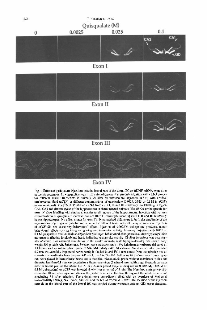
hybridization with cRNA probes encoding different BDNF transcripts, investigated whether the different mRNAs are specifically and dynamically regulated in different regions in the hippocampus by afferents in the entorhinal cortex (EC). Analysis was performed using X-ray film autoradiograms and microscopically at the cellular level using emulsion dipped slides. In sham injected animals cellular labelling was observed sparsely in all hippocampal regions with a slightly higher signal for exon II (Figs l-3). Quantitative analysis using autoradiograms showed dose-dependent increases for all BDNF transcripts except for exon IV in the
891
a92 T. FALKENHEKG et al.
Quisqualate (MI n 0.025 0.1
Fig. 1. Effects of qusiqualate injections into the lateral part of the lateral EC on BDNF mRNA expression in the hippocampus. Low magnification (X 10) autoradiogram of in situ hvbridization with cRNA probes for different BDNF transcripts in animals 3 h after an intracerebral injection (0.5 111) with artifical cerebrospinal fluid (aCSF) or different concentrations of quisqualate (0.0025. 0.025 or 0.1 M in aCSF) in awake animals. The [%]UTP-labelled cRNA from exon I, II, and III show very low labelling in region CAI! CA3 and dentate gyrus of the hippocampus in sham injected animals. The cRNA probe specific for exon IV show labelling with similar intensities in all regions of the hippocampus. Injection with various concentrations of quisqualate increase levels of BDNF transcripts encoding exon I, II and III bilaterally in the hippocampus. No effect is seen for exon IV. Note marked differences in both the amplitude of the increases and the regional distribution between the different transcripts following stimulation. Injection of aCSF did not cause any behavioural effects. Injection of 0.0025 M quisqualate produced minor behavioural effects such as increased rearing and locomotor activity. However, injection with 0.025 or 0.1 M quisqualate resulted in dose-dependent prolonged behavioural changes such as stereotypic repetitive movements affecting forelimb and head, indicating seizure-like activity. Circling behaviour was occasion- ally observed. For chemical stimulation in the awake animals, male Sprague-Dawley rats (mean body weight 300 g; Alab AB, Sollentuna, Sweden) were anaesthetised (I .5% halothaneiair mixture delivered at 1.4 i/min) and an intracerebral guide (CMA Mikrodialys AB, Stockholm, Sweden) of outer diameter 0.7 mm was carefully implanted permanently in the left lateral EC 1 mm dorsal from the injection site at stereotaxic coordinates from bregma: AP = 6.3, L = 6.6, D = 8.0. Following 48 h of recovery from surgery rats were placed in hemispheric bowls and a modified microdialysis probe without membrane with a tip diameter less than 0.1 mm was coupled to a Hamilton syringe (2 ~1) and inserted through the guide cannula into the lateral part of the lateral EC. After a 20-min period 0.5 ~1 of drug (either 0.0025 M, 0.025 M or 0.1 M quisqualate) or aCSF was injected slowly over a period of 3 min. The Hamilton syringe was dis- connected 10 min after injection whereas the probe remained in location throughout the whole experiment concluding 3 h after injection. The animals were immediately killed rith an overdose of Mebumal (intracardially 120 mg, Nord Vacc, Sweden) and the brains frozen at -20°C. The location of the injection cannula in the lateral part of the lateral EC was verified during cryostate cutting. GD, gyrus dentatus.

Regulation of multiple brain-derived neurotrophic factor promoters 893
h ippocampus 3 h after chemical s t imulat ion with the glutamate receptor agonis t quisqualate in the lateral part of the lateral EC (Fig. 2). Exon I and III m R N A s appeared to be mos t affected. A more than 50- and seven-fold bilateral increase compared to sham in- jected animals was seen for exon I in the dentate gyrus and CA3, respectively, by 0.1 M quisqualate. (Figs 1, 2; when levels o f the different m R N A s in sham-injected animals were indistinguishable f rom background they were given the arbitrary value < 1.0
Exon I 7 0 "I
GO" ~ I
SO" ~
40-
30"
Z0-
4¢ 10-
O- o o ~ o o ~ •
Exon II 20-
Q u i s u u a l a t e
I B O M [ ] 0.0025 N
[ ] O.02S U ~" [ [] O.~M. ~
, ,oo~ . o ~ o.
and thus differences between groups in these cases are mainly underest imated.) For cxon II a more than four-, two- and two-fold bilateral increase was seen in dentate gyrus, CA3 and CAI , respectively (Figs 1, 2). For exon I I I a more than 37-, six- and 17-fold bilateral increase was seen in dentate gyrus, CA3 and CA 1, respectively (Figs 1, 2). For exon IV no changes were observed with 0.1 M quisqualate (Figs 1, 2). These findings indicate bo th an exon and region specific increase o f the different B D N F transcripts in the h ippocampus . These results were suppor ted by findings obtained microscopically at the cellular level (Fig. 3). Addit ionally, cellular analysis revealed granule cells expressing exon I t ranscripts at different magni tudes following injection with 0.1 M quisqualate which was not apparen t for exon I with lower quisqualate concentrat ions or for exons II or III at any dose (Fig. 4).
Our observat ions are supported by recent findings showing that a systemic injection o f another g lutmate receptor agonist kainate, increases exon I, II and III m R N A s transiently in the h ippocampus , z3'3° The present investigation reveals a more detailed picture and intr iguing regulatory si tuat ion underlying the findings reported earlier using a probe detecting all B D N F
7
Exon III 5 0 -
40" ]
3 0 "
20- "k l qe
10"
o- o o o o m •
Exon IV 2 -
>
i 1
"4
* T * ~ [
, • , . , • , • , - , .
CA1 CA3 DG CA1 CA3 DG
ipsilateral H i p p o c a m p a l contralateral side region side
Fig. 2. Levels of exon-specific BDNF mRNAs in the hippocampus in the different treatment groups as quantified by computer assisted image analysis. Dose-dependent in- creases are seen for exon 1 in the dentate gyrus and in CA3. Dose-dependent exon II specific increases are present in all hippocampal regions with higher labelling intensity in the dentate gyrus compared to CAI and CA3 that are equally affected. Exon III specific dose-dependent changes are ob- served in all hippocampal regions examined. Pronounced' effects are seen in CA1 and in the dentate gyrus compared to CA3. In contrast, transcript IV appears unaffected by these stimulations. The following fragments were used to synthesise ~-[35S]UTP-labelled complementary RNA probes: for exon I; a 0.35 kb Sac l-Hind III genomic fragment; for exon II, a eDNA fragment covering the sequence 2067-2229 in the rat BDNF gene; for exon III, a eDNA fragment covering sequence-782-975 in the rat BDNF gene; and for exon IV, a eDNA fragment covering sequence 1790-2056 in the rat BDNF gene. 3° Coronal sections of the hippocampus (12/tin) were hybridized by in situ hybridization as previously described? ° The autoradio- grams from the in situ hybridizations were analysed with a Microcomputer Imaging Device (Imaging Research Inc., Canada). Optical density in each anatomical region was determined over the whole region in two subsequent sections from each animal. Optical density values were converted to relative levels of radioactivity using external standards exposed to the same film. Optical density values were measured in an interval with linear correlation to tissue levels of radioactive isotope. Values are mean _+ S.E.M. and presented relative to the mRNA level in the ipsilateral CAI for each transcript. • Indicates level equal to background and given the arbitrary value < 1.0. "A" Indicates significant difference with Kruskal-Wallis (P <0.05) belween the
different quisqualate treatment groups.

Exon I Exon II Exon III Fvnn TV
DG
Sham
DG
CA1
0.1 M quisqualate
Fig. 3
x94

transcripts?’ Findings from experiments using kindling to induce seizures have suggested BDNF mRNA activation to be regulated in an “all-or- none”-fashion.3 Our findings of region-specifc, dose- dependent expression of different 5’ exons
and different populations of BDNF mRNA expressive cells in the hippocampus together with dose-dependent increased behavioural activity (see legend to Fig. 1). suggest additional levels of regulation of BDNF mRNAs in this model. Furthermore, it appears likely that cells in the hippocampus have the capacity to express several transcripts considering the findings showing that more than two thirds of the granule cells express each of exons I, II and III following the highest quisqualate dose (Fig. 3).
All BDNF exon specific mRNAs seem to be associated with polysomes indicating current use for translation of the prepro-BDNF protein (Metsis and Timmusk, unpublished observations). Function- ally it is possible that the multipromoter system of the BDNF gene regulates BDNF synthesis in different cell types at the level of both transcription and translation influenced by afferents, presumably glutamaterigic, from the EC. The reason for this could be to address specific changes in BDNF mediated neuronal plasticity in specific regions in the hippocampal formation in response to functional activation of the corticohippocampal loop. Recently, BDNF has been shown to be retrogradely transported, in vivo, by neurons within the hippocampus and neurons in the EC.6 Thus, events influencing neuronal function in the EC, such as pathological changes seen in Alzheimer’s disease and schizophrenia, may have profound effects on neuronal plasticity both in the hippocampus and in the EC associated with cognitive alterations. Fig. 4. High magnification bright-field photomicrograph of
in situ hybridization to exon III (a) and I (b) mRNAs in the ipsilateral granule cell layer following injection of 0.1 M quisqulate into the ipsilateral entorhinal cortex. Note lack of heterogeneity in exon III staining over granule cells (a) compared to exon I-stained granule cells (b). As seen in b, more than 90% of the granule cells are exon I positive, [more than 10 grains per cell) and approximately one-third of these cells are more intensively labelled (more than 30
grains per cell).
Acknowledgements-This work was supported by grants from The Swedish Medical Research Council (grant no. 8653), Ake Wibergs Stiftelse, The Swedish Society of Medicine, Alzheimerfonden, The Swedish Society for Medi- cal Research and funds from the Karolinska Institute. MM was supported by Regeneron Pharmaceuticals.
Fig. 3. Effect of injection of aCSF or 0.1 M quisqualate into the lateral part of the lateral EC on the expression of exon-specific BDNF mRNAs in different ipsilateral hippocampal regions. Quantitative measures from X-ray films were verified by manually counting positive cells (cells expressing more than 10 grains) in selected regions from emulsion-dipped sections using bright-field optics viewd at x 500 magnification. Exon I and III labelled cells are present at very low levels in region CAI, CA3 and dentate gyrus of the hippocampus in sham injected animals. Using the probe detecting exon II labelling is observed sparsely in pyramidal cells and in granule cells of the dentate gyrus. The cRNA probe specific for exon IV show labelled cells in all examined regions of the hippocampus with higher intensities in the pyramidal cell layer compared to the granule cell layer. Note that unspecific labelling is significant using the exon IV probe. More than 90% of the granule cells are exon I positive, i.e. expressing more than 10 grains per cell following injection with 0.1 M quisqualate. A small increase in the number of density of positive cells is also present over the pyramidal cell layer of CA3. Exon II-positive cells are confined to approximately two thirds of the granule cells and to one third of the pyramidal cells following injection with 0.1 M quisqualate. More than 90% of the granule cells are exon III positive after 0. I M quisqualate. Labelling is notably increased in the pyramidal cell layer. No effect of quisqualate injection is observed
for exon IV compared to sham injected animals. DG, dentate gyrus.
895

896 T. FALKENBEKG ei al
REFERENCES
1. Ballarin M., Ernfors P., Lindefors N. and Persson H. (1991) Rapid induction of mRNA for brain derived neurotrophic factor after intrahippocampal injection of kainic acid. Expi Neural. 114, 3543.
2. Barde Y.-A., Edgar D. and Thoenen H. (1982) Purification of a new neurotrophic factor from mammalian brain. Eur molec. Biol. Org. J. 1, 549-553.
3. Bengzon J., Kokaia, Z., Ernfors P., Kokaia M., Leanza G., Nilsson 0. G., Persson H. and Lindvall 0. (1993) Regulation of neurotrophin and trkA, trkB and trkC tyrosine kinase receptor messenger RNA expression in kindling. Neuroscience 53, 433446.
4. Berkemeier L. R., Winslow J. W., Kaplan D. R., Nikolics K., Goeddel D. V. and Rosenthal A. (1991) Neurotrophin-5, a novel neurotrophic factor that activates trk and trkB. Neuron 7, 857-866.
5. Bogerts B. (1993) Recent advances in the neuropathology of schizophrenia. Schizophrenia Bull. 19, 43 1445. 6. DiStefano P. S., Friedman B., Radziejewski C., Alexander C., Boland P., Schick C. M., Lindsay R. M. and Wiegand
J. (1992) The neurotrophins: BDNF, NT-3, and NGF display distinct patterns of retrograde axonal transport in the peripheral and central neurons. Neuron 8, 9X3-993.
7. Ernfors P., Ibafiez C. F., Ebendal T., Olson L. and Persson H. (1990) Molecular cloning and neurotrophic activities of a protein with structural similarities to nerve growth factor; developmental and topographical expression in the brain. Proc. natn. Acad. Sci. U.S.A. 87, 54545458.
8. Ernfors P., Wetmore C., Olson L. and Persson H. (1990) Identification of cells in rat brain and peripheral tissues expressing mRNA for members of the nerve growth’ factor family. Neuron 5, 51 I-526.
_ _
9. Falkenbere. T.. Mohammed A. K., Henriksson B.. Persson H., Winblad B. and Lindefors N. (1992) Increased expression of brain-d&&d neurotrophic factor mRNA in rat hippocampus is associated with improved’ spatial memory and enriched environment. Neurosci. Lett. 138, 153-l 56.
10. Gall C. M. and Isackson P. J. (1989) Limbic seizures increase neuronal production of messenger RNA for nerve growth factor. Science 245, 75861.
11. Hallbook F., Ibanez C. F. and Persson H. (1991) Evolutionary studies of the nerve growth factor family reveal a novel member abundantly expressed in Xenopus ovary. Neuron 6, 845-858.
12. Hofer M., Pagliusi S. -R., Hohn A., -&ibrock- J. and Barde Y. A. (1990) Regional distribution of brain-derived neurotrophic factor mRNA in the adult mouse brain. Eur. molec. Biol. Ora. J. 9. 2459-2464.
13. Hofer M: M. and Barde Y.-A. (1988) Brain-derived neurotrophic factor prevents neuronal death in viuo. Nature 331, 261-262.
14. Hohn A., Leibrock J., Bailey K. and Barde Y. A. (1990) Identification and characterization of a novel member of the nerve growth factor/brain-derived neurotrophic factor family. Nature 344, 339-341.
15. Hvman B. T.. Van Hoesen G. W., Kromer L. J. and Damasio A. R. (1986) Perforant pathwav changes and the memory impairment of Alzheimer’s disease. Ann. Neural. 20, 472481.
_ . _
16. Ip N. Y., Ibanez C. F., Nye S. H., McClain J., Jones P. F., Gies D. R., Belluscio L., Le Beau M. M., Espinosa R. I., Squint0 S. P., Persson H. and Yancopoulos G. D. (1992) Mammalian neurotrophin-4: structure, chromosomal localization, tissue distribution, and receptor specificity: Proc. natn. Acad. Sci. 89, jO6&3064.
17. Jones K. R. and Reichardt L. F. (1990) Molecular cloning of a human gene that is a member of the nerve growth factor family. Proc. nafn. Acad. Sci. 87, 8060-8064.
18. Kaisho Y., Yoshimura K. and Nakahama K. (1990) Cloning and expression of a cDNA encoding a novel human neurotrophic factor. Fedn Eur. biochem. Sots Left. 226, 187-191.
19. Kozak M. (1991) An analysis of vertebrate mRNA sequences: intimations of translational control. J. Cc// Biol. 115, 887-903.
20. Levi-Montalcini R. (1987) The nerve growth factor 35 years later. Science 237, 1154-l 162. 21. Lindefors N., Ernfors P., Falkenberg T. and Persson H. (1992) Septal cholinergic afferents regulate expression of
brain-derived neurotrophic factor and b-nerve growth factor mRNA in rat hippocampus. Exdl Brain Res. 88, 78-90. 22. Maisonpierre P. C., Bdlluscio L., Squnto S., IpN. Y., Furth M. E.. Lindsay-R. M. and Yancopoulos G. D. (1990)
Neurotronhin-3: a neurotroohic factor related to NGF and BDNF. Science 247. 14461451. 23. Metsis M:, Timmusk M.: Arenas E. and Persson H. (1993) Differential usage of ‘multiple brain-derived neurotrophic
factor promoters in the rat brain following neuronal activation. Proc. natn. Acad. Sci. (in press). 24. Oppenheim D. W., Qin-Wei Y., Prevette D. and Yan Q. (1992) Brain-derived neurotrophic factor rescues developing
avian motoneurons from cell death. Nature 360, 7555757. 25. Philips H. S., Hains J. M., Armanini M.. Laramee G. R., Johnson S. A. and Winslow J. W. (1991) BDNF mRNA
is decreased in the hippocampus of individuals with Alzheimer’s disease. Neuron 7, 695-702. 26. Phillips H. S., Hains J. M., Laramee G. R., Rosenthal A. and Winslow I. W. (1990) Widespread expression of BDNF
but not NT-3 by target areas of basal forebrain cholinergic neurons. Science 250, 290-294. 27. Rosenthal A., Goeddel D. V., Nguyen T.. Lewis M., Shih A., Laramee G. R., Nikolics K. and Winslow J. W. (1990)
Primary structure and biological activity of a novel human neurotrophic factor. Neuron 4, 7677773. 28. Sendtner M., Holtman B., Kolbeck R., Thoenen H. and Barde Y.-A. (1992) Brain-derived neurotrophic factor prevents
the death of motorneurons in newborn rats after nerve section. Nature 360, 7577759. 29. Sutula T., Siao-Sian H., Cavazos J. and Grayson S. (1988) Synaptic reorganization in the hippocampus induced by
abnormal functional activity. Science 239, 1147-l 150. 30. Timmusk T., Palm K., Metsis M., Reintam T., Paalme V., Saarma M. and Persson H. (1993) Multiple promoters direct
tissue-specific expression of the rat BDNF gene. Neuron 10, 475489. 31. Yan Q., Elliott J. and Snider W. D. (1992) Brain-derived neurotrophic factor rescues spinal motor neurons from
axotomy-induced cell death. Nature 360, 753-755.
(Accepted 13 September 1993)
Copyright © 2022 FDOKUMEN