
N Gu - Ne uid 2 - eo de 01 - KIDS NTS
410
WThe est Mid G Bedsid i lands N N Gu e Clinic n assoc Neonata Ne uid 2 cal Guid ciation al Opera eo de 01 delines with the ational D on eli 19 Partner e Delivery nat in 9− rship y Netwo ta es −21 ork al s 1
-
Upload
khangminh22 -
Category
Documents
-
view
0 -
download
0
Transcript of N Gu - Ne uid 2 - eo de 01 - KIDS NTS
We
The
est Mid
G
Bedsidi
lands N
NGu
e Clinicn assoc
Neonata
Neuid2
cal Guidciation
al Opera
eode01
delines with the
ational D
oneli19
Partnere Delivery
natin9−
rship
y Netwo
taes
−21
ork
al s1
This copy belongs to: Name: ___________________________________________________ Further copies can be purchased from West Midlands Neonatal Operational Delivery Network, email: [email protected]
Published by the Bedside Clinical Guidelines Partnership and West Midlands Neonatal Operational Delivery Network
© 2019–21 Bedside Clinical Guidelines Partnership (University Hospital of North Midlands NHS Trust acting as authorised copyright owner)
All rights reserved
NOT TO BE REPRODUCED WITHOUT PERMISSION
West Midlands Neonatal Operational Delivery Network comprises:
The Bedside Clinical Guidelines Partnership comprises:
Basildon and Thurrock University Hospital NHS Foundation Trust
Circle Nottingham Ltd County Durham and Darlington NHS Foundation Trust
The Dudley Group NHS Foundation Trust East Cheshire NHS Trust
North Cumbria University Hospitals NHS Trust Surrey and Sussex Healthcare NHS Trust The Pennine Acute Hospitals NHS Trust
The Royal Wolverhampton Hospitals NHS Trust University Hospitals Birmingham NHS Foundation Trust
University Hospitals of Morecambe Bay NHS Trust University Hospitals North Midlands NHS Trust
Walsall Healthcare NHS Trust Wye Valley NHS Trust
Birmingham Women’s and Children’s NHS Trust
Heart of England NHS Foundation Trust Sandwell and West Birmingham Hospitals NHS Trust
The Dudley Group NHS Foundation Trust The Royal Wolverhampton NHS Trust
The Shrewsbury and Telford Hospital NHS Trust University Hospitals of North Midlands NHS Trust
Walsall Healthcare NHS Trust Worcestershire Acute Hospitals NHS Trust
Wye Valley NHS Trust
CONTENTS ● 1/3
Acknowledgements 6Commonly used abbreviations 7Preface 10ADMISSION AND DISCHARGE Admission to neonatal unit 15Death and seriously ill babies 82Discharge from neonatal unit 89Follow-up of babies discharged from neonatal unit 118Labour ward calls 223Transport and retrieval 364CARDIOVASCULAR Cardiac murmurs 49Congenital heart disease duct-dependent lesions [Including hypoplastic left heart syndrome (HLHS) and left-sided outflow tract obstructions] 50ECG abnormalities 99Heart failure 134Hypotension 167Patent ductus arteriosus 281Pericardiocentesis 285Supraventricular tachycardia 340Vascular spasm and thrombosis 384CRITICAL CARE Babies born at margins of viability 28Contacting a consultant NEW 77Dropped baby NEW 97Golden hour – preterm babies <28 weeks’ gestation 126Hydrops fetalis 144Hypothermia 170Massive haemorrhage 231Pain assessment and management 271Resuscitation 314Subgaleal haemorrhage (SGH) 334Sudden unexpected postnatal collapse in first week of life 337DEVELOPMENTAL CARE Developmental care 84Down syndrome − initial management 93Environment and noise 105Kangaroo care 221Non-nutritive sucking (NNS) 254Positioning 291ENDOCRINE/METABOLISM Hyperglycaemia 146Hyperkalaemia 148Hypernatraemic dehydration 151Hypocalcaemia NEW 155Hypoglycaemia 157Hypokalaemia 165Hypothyroidism 173Inherited metabolic disorders (IMD) 199Intravenous fluid therapy 207Medium-Chain Acyl-CoA Dehydrogenase Deficiency (MCADD) – early management of babies with family history
234
Metabolic bone disease 236Thyroid disease (management of babies born to mothers with thyroid disease) 354GASTROENTEROLOGY Ankyloglossia (tongue-tie) − division for breastfeeding 17Gastro-oesophageal reflux (GOR) 120Jaundice 218Liver dysfunction in preterm babies 224Nasogastric tube administration of feed, fluid or medication 243
Issue 8 rev1 jan20 Issued: December 2019 Expires: December 2021
3
CONTENTS ● 2/3 Necrotising enterocolitis (NEC) 249HAEMATOLOGY Blood group incompatibilities (including Rhesus disease) 33Coagulopathy 66Polycythaemia 289Thrombocytopenia 350Transfusion of red blood cells 360Vitamin K 406INFECTION BCG immunisation 30CMV 64Conjunctivitis 72Group B streptococcal colonisation of mother NEW 129Hepatitis B and C 137Herpes simplex virus (HSV) 140Human immunodeficiency virus (HIV) 141Immunisations 185Infection (late onset) 188Infection in first 72 hours of life 192Infection prevention NEW 195Multi drug resistant organism colonisation (MRSA, ESBL etc.) 238Palivizumab 276Syphilis – babies born to mothers with positive serology 346Tuberculosis (investigation and management following exposure in pregnancy) 367Varicella 381NEUROLOGY Abstinence syndrome 12Hypotonia (floppy baby) 175Hypoxic ischaemic encephalopathy (HIE) including preparation for active cooling 179Post haemorrhagic ventricular dilatation 294Seizures 322Upper limb birth injuries including brachial plexus injury 377NUTRITION Bottle feeding in the neonatal unit 37Breast milk expression 39Breast milk handling and storage 41Breastfeeding 43Nutrition and enteral feeding 255Parenteral nutrition 278Progression to oral feeding in preterm babies 296PRACTICAL PROCEDURES Arterial line insertion 24Arterial line sampling 26Cannulation − peripheral venous 48Chest drain insertion – Seldinger technique 54Chest drain insertion − Traditional 57Consent 74Endotracheal tube (ETT) suctioning 103Exchange transfusion 110Extravasation injuries 116Growth monitoring 130Intraosseous infusion NEW 205Intubation − difficult 212Intubation 215Long line insertion (peripherally sited) 227Nasogastric tube insertion 245Prostaglandin infusion 299Skin biopsy for inborn errors of metabolism 326Skin care 328Transillumination of the chest 363
Issue 8 rev1 jan20 Issued: December 2019 Expires: December 2021
4
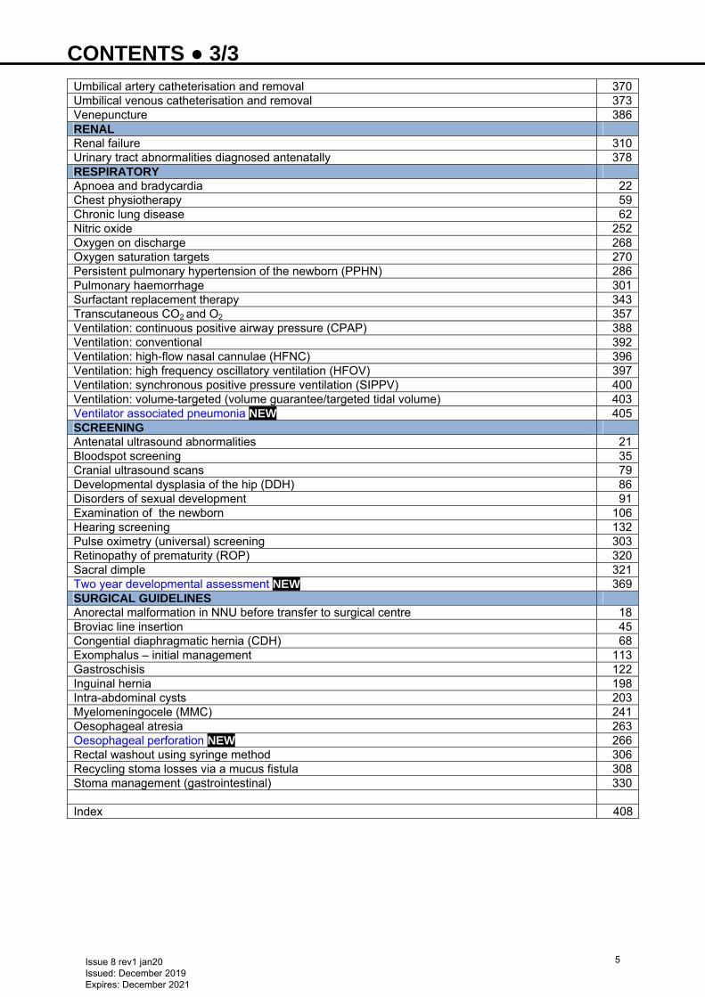
CONTENTS ● 3/3 Umbilical artery catheterisation and removal 370Umbilical venous catheterisation and removal 373Venepuncture 386RENAL Renal failure 310Urinary tract abnormalities diagnosed antenatally 378RESPIRATORY Apnoea and bradycardia 22Chest physiotherapy 59Chronic lung disease 62Nitric oxide 252Oxygen on discharge 268Oxygen saturation targets 270Persistent pulmonary hypertension of the newborn (PPHN) 286Pulmonary haemorrhage 301Surfactant replacement therapy 343Transcutaneous CO2 and O2 357Ventilation: continuous positive airway pressure (CPAP) 388Ventilation: conventional 392Ventilation: high-flow nasal cannulae (HFNC) 396Ventilation: high frequency oscillatory ventilation (HFOV) 397Ventilation: synchronous positive pressure ventilation (SIPPV) 400Ventilation: volume-targeted (volume guarantee/targeted tidal volume) 403Ventilator associated pneumonia NEW 405SCREENING Antenatal ultrasound abnormalities 21Bloodspot screening 35Cranial ultrasound scans 79Developmental dysplasia of the hip (DDH) 86Disorders of sexual development 91Examination of the newborn 106Hearing screening 132Pulse oximetry (universal) screening 303Retinopathy of prematurity (ROP) 320Sacral dimple 321Two year developmental assessment NEW 369SURGICAL GUIDELINES Anorectal malformation in NNU before transfer to surgical centre 18Broviac line insertion 45Congential diaphragmatic hernia (CDH) 68Exomphalus – initial management 113Gastroschisis 122Inguinal hernia 198Intra-abdominal cysts 203Myelomeningocele (MMC) 241Oesophageal atresia 263Oesophageal perforation NEW 266Rectal washout using syringe method 306Recycling stoma losses via a mucus fistula 308Stoma management (gastrointestinal) 330 Index 408
Issue 8 rev1 jan20 Issued: December 2019 Expires: December 2021
5

ACKNOWLEDGEMENTS ● 1/1 We would like to thank the following for their assistance in producing this edition of the Neonatal Guidelines on behalf of the Bedside Clinical Guidelines Partnership and West Midlands Neonatal Operational Delivery Network Contributors Lee Abbott Diana Aguirre Oluwaseyi Alake Suren Arul Meena Bankhakavi Alison Bedford Russell Manobi Borooah Lucilla Butler Fiona Chambers Hannah Clark Sara Clarke Richard Cole Joanne Cookson Cheryl Curson Rebecca Dack Anna Darbyshire Seema Desai Sanjeev Deshpande Andy Ewer Emma Foulerton Victoria Fradd Vidya Garikapati Sonia Goyal Harsha Gowda Jo Gregory Kalyana Gurusamy Lindsay Halpern Liza Harry Tracey Hill Louise Hirons Gemma Holder Kate Holterman Andrea Jester Sheilah Kamupira Ashok Karupaiah Anna Kotas Arthi Lakshmanan Anthony Lander Nick Makwana Katherine Matthews Paddy McMaster Rashmi Mehta Bashir Muhammad Robert Negrine Mona Noureldein Kate Palmer Katy Parnell Meghana Pearson Alex Philpott Tilly Pillay Velmurugan Ramalingam Tristan Ramcharan Shree Vishna Rasiah Sagarika Ray Kate Reynolds Sophie Reynolds Victoria Riches Desiderio Rodrigues Martin Samuels Cathryn Seagrave Shiva Shankar Asha Shenvi
Phillip Simmons Anju Singh Jaideep Singh S. Sivakumar Nicola Staton Imogen Storey Pinki Surana Julie Taylor Arumugavelu Thirumurugan Louise Thompson Julia Uffindell Hannah Vawda Daniela Vieten-Kay Vikranth Venugopalan Suresh Vijay Neonatal Editors Robert Negrine Sagarika Ray Bedside Clinical Guidelines Partnership Kathryn McCarron Naveed Mustfa Kate Palmer Mathew Stone West Midlands Neonatal Operational Delivery Network Sarah Carnwell Lynsey Clarke Harsha Gowda Ruth Moore Kate Palmer Sonia Saxon Teresa Meredith S. Sivakumar Vikranth Venugopalan The editors would like to thank the following people/organisations for allowing us to use/adapt their guidelines: Birmingham Children’s Hospital – Skin biopsy guideline Dr Carl Kuschel Auckland District Health Board Auckland, New Zealand – Ventilation guideline Birmingham Women's Hospital Neonatal Unit – Extravasation injuries guideline Guy’s and St Thomas’ NHS Trust – Transcutaneous monitoring guideline
.
Issue 8 rev1 jan20 Issued: December 2019 Expires: December 2021
6

COMMONLY USED ABBREVIATIONS ● 1/3
ACTH Adrenocorticotrophic hormone
ADH Antidiuretic hormone
aEEG Cerebral function monitoring
ALT Alanine aminotransferase
APTT Activated partial thromboplastin time
ASD Atrial septal defect
AST Aspartate aminotransferase
AVSD Atrioventricular septal defect
BAPM British Association of Perinatal Medicine
BCG Bacille Calmette-Guerin
BiPAP Biphasic CPAP
BPD Bronchopulmonary dysplasia CAH Congenital adrenal hyperplasia CAMT Congenital amegakaryocytic thrombocytopenia CCAM Congenital cystic adenomatoid malformation ccTGA Congenitally corrected transposition of the great arteries
CDH Congenital dislocation of hips or congenital diaphragmatic hernia
CFAM Cerebral function analysis monitor
CGA Corrected gestational age
CH Congenital hypothyroidism
CHD Congenital heart disease
CLD Chronic lung disease
CMPI Cow’s milk protein intolerance
CMV Cytomegalovirus
CNS Central nervous system
CoNS Coagulase-negative staphylococcus
CPAP Continuous positive airway pressure
CRP C-reactive protein
CVS Cardiovascular
DCT Direct Coombs test
DDH Developmental dysplasia of the hip
DEBM Donor expressed breast milk
DHEA Dihydroepiandrostenedione
dHT Dihydrotestosterone
DIC Disseminated intravascular coagulation
DSD Disorders of sexual development
EBM Expressed breast milk
ECF Extracellular fluid
ECG Electrocardiogram
ECMO Extracorporeal membrane oxygen
EDD Expected date of delivery
EFM Electronic fetal monitoring
ELBW Extremely-low-birth-weight
EMG Electromyography
ETT Endotracheal tube
EUT Extrauterine transfer
FFP Fresh frozen plasma
FSID Foundation for the Study of Infant Deaths
GBS Group B streptococcus
GGT Gamma-glutamyl transaminase
GLUT 1 Glucose transporter defect
GOR Gastro-oesophageal reflux
Issue 8 rev1 jan20 Issued: December 2019 Expires: December 2021
7

COMMONLY USED ABBREVIATIONS ● 2/3 hCG Human chorionic gonadotropin
Hct Haematocrit
HCV Hepatitis C virus
HFNC High-flow nasal cannulae
HFOV High frequency oscillatory ventilation
HIE Hypoxic ischaemic encephalopathy
HIV Human immunodeficiency virus
HLHS Hypoplastic left heart syndrome
HPA Human platelet antigens
HTLV Human T-cell lymphotropic virus
ICCP Integrated comfort care pathway
IMD Inherited metabolic disorders
iNO Inhaled nitric oxide
IPPV Intermittent positive pressure ventilation
ITP Immune thrombocytopenic purpura
IUGR Intrauterine growth retardation
IUT In-utero blood transfusion or in-utero transfer
IVC Inferior vena cava
IVH Intraventricular haemorrhage
IVIG Intravenous immunoglobin
LHRH Luteinizing hormone releasing hormone
LP Lumbar puncture
LRTI Lower respiratory tract infection
LSE Left sternal edge
LV Left ventricle
LVOT Left ventricular outflow tract
MAP Mean airway pressure or mean arterial pressure
MAS Meconium aspiration syndrome
MCADD Medium chain acyl co-A dehydrogenase deficiency
MDT Multidisciplinary team
MEBM Mother’s expressed breast milk
MSUD Maple syrup urine disease
NAIT Neonatal allo-immune thrombocytopenia
NEC Necrotising enterocolitis
NGT Nasogastric tube
NHSP Newborn Hearing Screening Programme
NICU Neonatal intensive care unit
NKHG Non-ketotic hyperglycinaemia
NLS Newborn life support
NNU Neonatal unit
NPSA National Patient Safety Agency
NTS Neonatal Transport Service
OI Oxygenation index
OPS Oropharyngeal secretions
PACS Picture archiving and communications system
PAT Pain assessment tool
PCOS Polycystic ovary syndrome
PCR Polymerase chain reaction
PDA Patent ductus arteriosus
PEEP Positive end expiratory pressure
PFO Patent foramen ovale
PIE Pulmonary interstitial emhysema
Issue 8 rev1 jan20 Issued: December 2019 Expires: December 2021
8

COMMONLY USED ABBREVIATIONS ● 3/3 PIH Pregnancy-induced hypertension
PICC Peripherally inserted central catheter
PIP Peak inspiratory pressure
PIPP Premature infant pain profile
PKU Phenylketonuria
PN Parenteral nutrition
PPHN Persistent pulmonary hypertension of the newborn
PROM Pre-labour rupture of membranes
PT Prothrombin time
PTV Patient triggered ventilation
PVL Periventricular leukomalacia
PVR Pulmonary venous return
RDS Respiratory distress syndrome
ROP Retinopathy of prematurity
RR Respiratory rate
RVH Right ventricular hypertrophy
SANDS Stillbirth and Neonatal Death Society
SaO2/SpO2 Arterial/peripheral oxygen saturation
SGA Small for gestational age
SIDS Sudden infant death syndrome
SIMV Simultaneous intermittent mandatory ventilation
SLE Systemic lupus erythematosus
SPA Supra-pubic aspiration
SSRI Selective serotonin reuptake inhibitor
SVC Superior vena cava
SVT Supraventricular tachycardia
TAR Thrombocytopenia absent radii
Texp Expiratory time
TEW Transepidermal water
TGA Transposition of the great arteries
THAM Trometamol
Tinsp Inspiratory time
TPN Total parenteral nutrition TTV Targeted tidal volume UAC Umbilical artery catheter
UVC Umbilical vein catheter
VLBW Very-low-birth-weight
VLCFA Very long chain fatty acids
VSD Ventricular septal defect
Vt Tidal volume
VZIG Varicella zoster immunoglobulin
VZV Varicella-zoster virus
WCC White cell count
WFI Water for injection
Issue 8 rev1 jan20 Issued: December 2019 Expires: December 2021
9
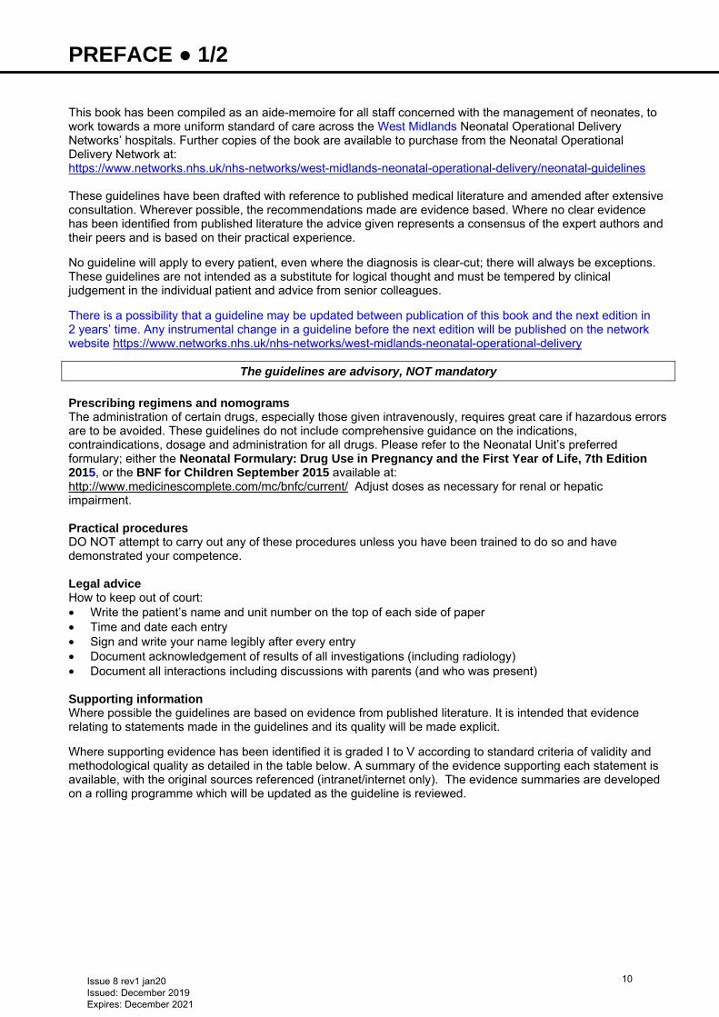
PREFACE ● 1/2 This book has been compiled as an aide-memoire for all staff concerned with the management of neonates, to work towards a more uniform standard of care across the West Midlands Neonatal Operational Delivery Networks’ hospitals. Further copies of the book are available to purchase from the Neonatal Operational Delivery Network at: https://www.networks.nhs.uk/nhs-networks/west-midlands-neonatal-operational-delivery/neonatal-guidelines
These guidelines have been drafted with reference to published medical literature and amended after extensive consultation. Wherever possible, the recommendations made are evidence based. Where no clear evidence has been identified from published literature the advice given represents a consensus of the expert authors and their peers and is based on their practical experience. No guideline will apply to every patient, even where the diagnosis is clear-cut; there will always be exceptions. These guidelines are not intended as a substitute for logical thought and must be tempered by clinical judgement in the individual patient and advice from senior colleagues. There is a possibility that a guideline may be updated between publication of this book and the next edition in 2 years’ time. Any instrumental change in a guideline before the next edition will be published on the network website https://www.networks.nhs.uk/nhs-networks/west-midlands-neonatal-operational-delivery
The guidelines are advisory, NOT mandatory
Prescribing regimens and nomograms The administration of certain drugs, especially those given intravenously, requires great care if hazardous errors are to be avoided. These guidelines do not include comprehensive guidance on the indications, contraindications, dosage and administration for all drugs. Please refer to the Neonatal Unit’s preferred formulary; either the Neonatal Formulary: Drug Use in Pregnancy and the First Year of Life, 7th Edition 2015, or the BNF for Children September 2015 available at: http://www.medicinescomplete.com/mc/bnfc/current/ Adjust doses as necessary for renal or hepatic impairment. Practical procedures DO NOT attempt to carry out any of these procedures unless you have been trained to do so and have demonstrated your competence. Legal advice How to keep out of court: • Write the patient’s name and unit number on the top of each side of paper • Time and date each entry • Sign and write your name legibly after every entry • Document acknowledgement of results of all investigations (including radiology) • Document all interactions including discussions with parents (and who was present) Supporting information Where possible the guidelines are based on evidence from published literature. It is intended that evidence relating to statements made in the guidelines and its quality will be made explicit. Where supporting evidence has been identified it is graded I to V according to standard criteria of validity and methodological quality as detailed in the table below. A summary of the evidence supporting each statement is available, with the original sources referenced (intranet/internet only). The evidence summaries are developed on a rolling programme which will be updated as the guideline is reviewed.
Issue 8 rev1 jan20 Issued: December 2019 Expires: December 2021
10
PREFACE ● 2/2
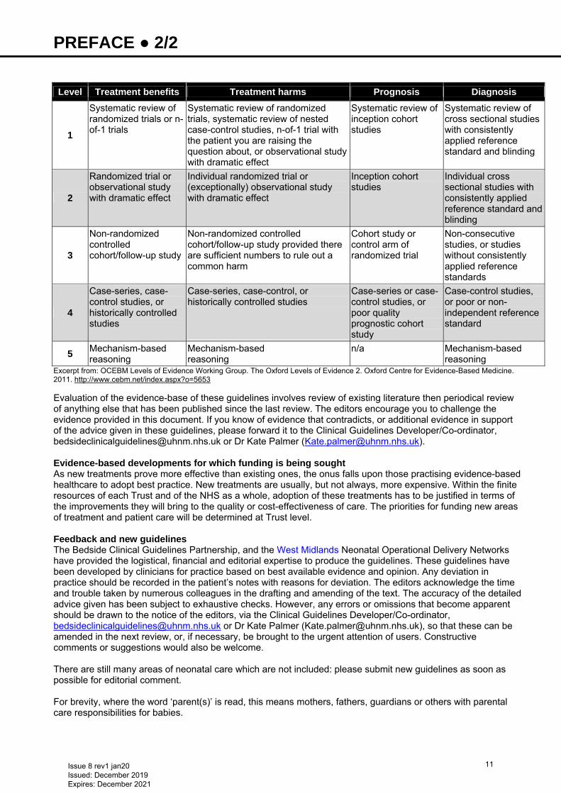
Level Treatment benefits Treatment harms Prognosis Diagnosis
1
Systematic review of randomized trials or n-of-1 trials
Systematic review of randomized trials, systematic review of nested case-control studies, n-of-1 trial with the patient you are raising the question about, or observational study with dramatic effect
Systematic review of inception cohort studies
Systematic review of cross sectional studies with consistently applied reference standard and blinding
2
Randomized trial or observational study with dramatic effect
Individual randomized trial or (exceptionally) observational study with dramatic effect
Inception cohort studies
Individual cross sectional studies with consistently applied reference standard and blinding
3
Non-randomized controlled cohort/follow-up study
Non-randomized controlled cohort/follow-up study provided there are sufficient numbers to rule out a common harm
Cohort study or control arm of randomized trial
Non-consecutive studies, or studies without consistently applied reference standards
4
Case-series, case-control studies, or historically controlled studies
Case-series, case-control, or historically controlled studies
Case-series or case-control studies, or poor quality prognostic cohort study
Case-control studies, or poor or non-independent reference standard
5 Mechanism-based reasoning
Mechanism-based reasoning
n/a Mechanism-based reasoning
Excerpt from: OCEBM Levels of Evidence Working Group. The Oxford Levels of Evidence 2. Oxford Centre for Evidence-Based Medicine. 2011. http://www.cebm.net/index.aspx?o=5653 Evaluation of the evidence-base of these guidelines involves review of existing literature then periodical review of anything else that has been published since the last review. The editors encourage you to challenge the evidence provided in this document. If you know of evidence that contradicts, or additional evidence in support of the advice given in these guidelines, please forward it to the Clinical Guidelines Developer/Co-ordinator, [email protected] or Dr Kate Palmer ([email protected]). Evidence-based developments for which funding is being sought As new treatments prove more effective than existing ones, the onus falls upon those practising evidence-based healthcare to adopt best practice. New treatments are usually, but not always, more expensive. Within the finite resources of each Trust and of the NHS as a whole, adoption of these treatments has to be justified in terms of the improvements they will bring to the quality or cost-effectiveness of care. The priorities for funding new areas of treatment and patient care will be determined at Trust level. Feedback and new guidelines The Bedside Clinical Guidelines Partnership, and the West Midlands Neonatal Operational Delivery Networks have provided the logistical, financial and editorial expertise to produce the guidelines. These guidelines have been developed by clinicians for practice based on best available evidence and opinion. Any deviation in practice should be recorded in the patient’s notes with reasons for deviation. The editors acknowledge the time and trouble taken by numerous colleagues in the drafting and amending of the text. The accuracy of the detailed advice given has been subject to exhaustive checks. However, any errors or omissions that become apparent should be drawn to the notice of the editors, via the Clinical Guidelines Developer/Co-ordinator, [email protected] or Dr Kate Palmer ([email protected]), so that these can be amended in the next review, or, if necessary, be brought to the urgent attention of users. Constructive comments or suggestions would also be welcome. There are still many areas of neonatal care which are not included: please submit new guidelines as soon as possible for editorial comment. For brevity, where the word ‘parent(s)’ is read, this means mothers, fathers, guardians or others with parental care responsibilities for babies.
Issue 8 rev1 jan20 Issued: December 2019 Expires: December 2021
11
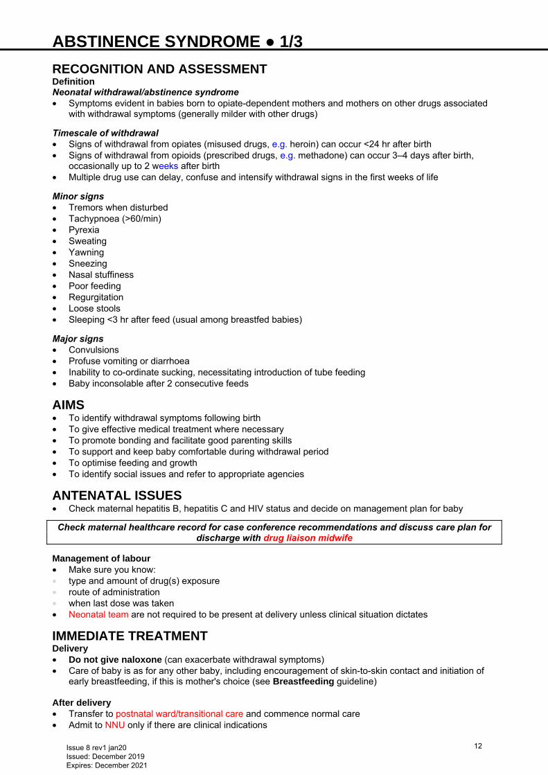
ABSTINENCE SYNDROME ● 1/3 RECOGNITION AND ASSESSMENT Definition Neonatal withdrawal/abstinence syndrome • Symptoms evident in babies born to opiate-dependent mothers and mothers on other drugs associated
with withdrawal symptoms (generally milder with other drugs)
Timescale of withdrawal • Signs of withdrawal from opiates (misused drugs, e.g. heroin) can occur <24 hr after birth • Signs of withdrawal from opioids (prescribed drugs, e.g. methadone) can occur 3–4 days after birth,
occasionally up to 2 weeks after birth • Multiple drug use can delay, confuse and intensify withdrawal signs in the first weeks of life
Minor signs • Tremors when disturbed • Tachypnoea (>60/min) • Pyrexia • Sweating • Yawning • Sneezing • Nasal stuffiness • Poor feeding • Regurgitation • Loose stools • Sleeping <3 hr after feed (usual among breastfed babies)
Major signs • Convulsions • Profuse vomiting or diarrhoea • Inability to co-ordinate sucking, necessitating introduction of tube feeding • Baby inconsolable after 2 consecutive feeds
AIMS • To identify withdrawal symptoms following birth • To give effective medical treatment where necessary • To promote bonding and facilitate good parenting skills • To support and keep baby comfortable during withdrawal period • To optimise feeding and growth • To identify social issues and refer to appropriate agencies
ANTENATAL ISSUES • Check maternal hepatitis B, hepatitis C and HIV status and decide on management plan for baby
Check maternal healthcare record for case conference recommendations and discuss care plan for discharge with drug liaison midwife
Management of labour • Make sure you know: • type and amount of drug(s) exposure • route of administration • when last dose was taken • Neonatal team are not required to be present at delivery unless clinical situation dictates
IMMEDIATE TREATMENT Delivery • Do not give naloxone (can exacerbate withdrawal symptoms) • Care of baby is as for any other baby, including encouragement of skin-to-skin contact and initiation of
early breastfeeding, if this is mother's choice (see Breastfeeding guideline) After delivery • Transfer to postnatal ward/transitional care and commence normal care • Admit to NNU only if there are clinical indications
Issue 8 rev1 jan20 Issued: December 2019 Expires: December 2021
12
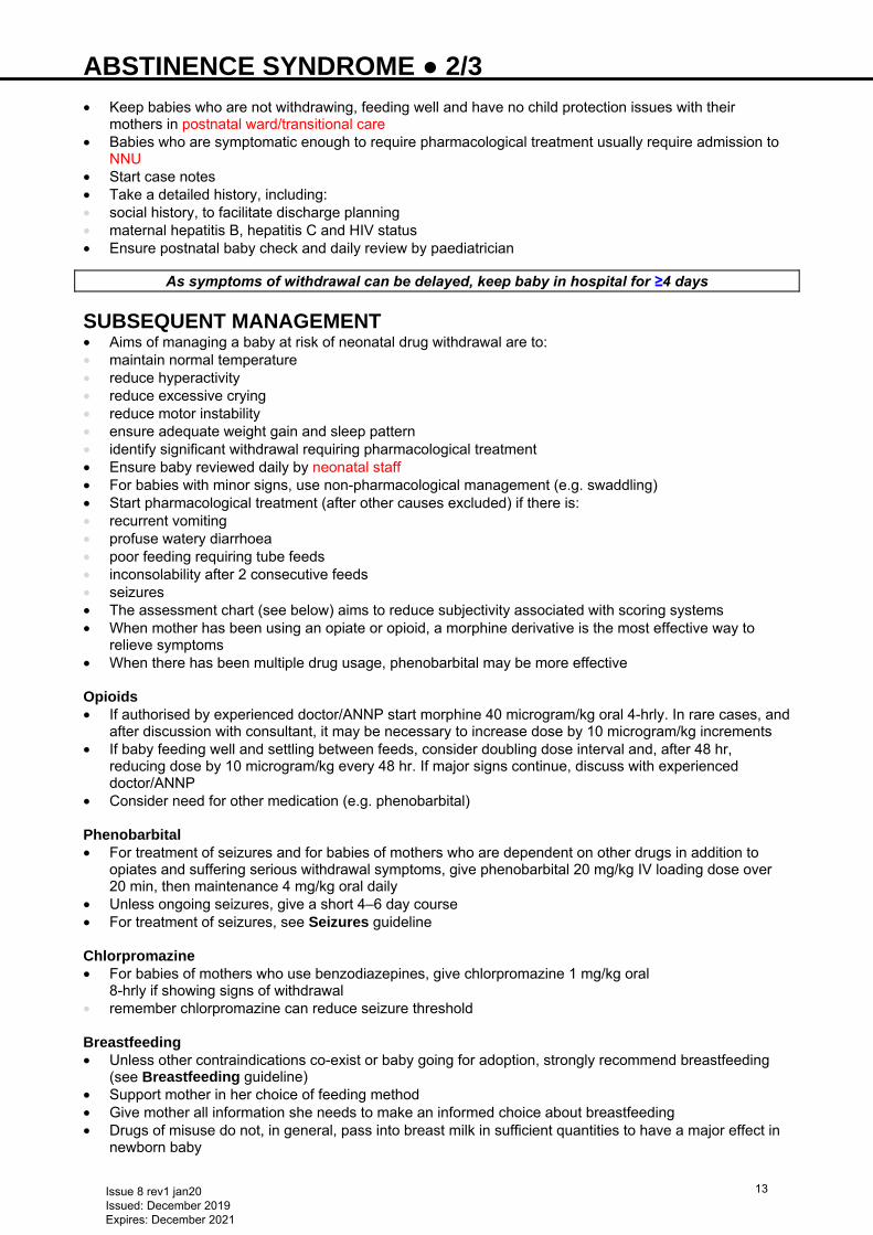
ABSTINENCE SYNDROME ● 2/3 • Keep babies who are not withdrawing, feeding well and have no child protection issues with their
mothers in postnatal ward/transitional care • Babies who are symptomatic enough to require pharmacological treatment usually require admission to
NNU • Start case notes • Take a detailed history, including: • social history, to facilitate discharge planning • maternal hepatitis B, hepatitis C and HIV status • Ensure postnatal baby check and daily review by paediatrician
As symptoms of withdrawal can be delayed, keep baby in hospital for ≥4 days
SUBSEQUENT MANAGEMENT • Aims of managing a baby at risk of neonatal drug withdrawal are to: • maintain normal temperature • reduce hyperactivity • reduce excessive crying • reduce motor instability • ensure adequate weight gain and sleep pattern • identify significant withdrawal requiring pharmacological treatment • Ensure baby reviewed daily by neonatal staff • For babies with minor signs, use non-pharmacological management (e.g. swaddling) • Start pharmacological treatment (after other causes excluded) if there is: • recurrent vomiting • profuse watery diarrhoea • poor feeding requiring tube feeds • inconsolability after 2 consecutive feeds • seizures • The assessment chart (see below) aims to reduce subjectivity associated with scoring systems • When mother has been using an opiate or opioid, a morphine derivative is the most effective way to
relieve symptoms • When there has been multiple drug usage, phenobarbital may be more effective Opioids • If authorised by experienced doctor/ANNP start morphine 40 microgram/kg oral 4-hrly. In rare cases, and
after discussion with consultant, it may be necessary to increase dose by 10 microgram/kg increments • If baby feeding well and settling between feeds, consider doubling dose interval and, after 48 hr,
reducing dose by 10 microgram/kg every 48 hr. If major signs continue, discuss with experienced doctor/ANNP
• Consider need for other medication (e.g. phenobarbital) Phenobarbital • For treatment of seizures and for babies of mothers who are dependent on other drugs in addition to
opiates and suffering serious withdrawal symptoms, give phenobarbital 20 mg/kg IV loading dose over 20 min, then maintenance 4 mg/kg oral daily
• Unless ongoing seizures, give a short 4–6 day course • For treatment of seizures, see Seizures guideline Chlorpromazine • For babies of mothers who use benzodiazepines, give chlorpromazine 1 mg/kg oral
8-hrly if showing signs of withdrawal • remember chlorpromazine can reduce seizure threshold Breastfeeding • Unless other contraindications co-exist or baby going for adoption, strongly recommend breastfeeding
(see Breastfeeding guideline) • Support mother in her choice of feeding method • Give mother all information she needs to make an informed choice about breastfeeding • Drugs of misuse do not, in general, pass into breast milk in sufficient quantities to have a major effect in
newborn baby
Issue 8 rev1 jan20 Issued: December 2019 Expires: December 2021
13
ABSTINENCE SYNDROME ● 3/3 • Breastfeeding will certainly support mother in feeling she is positively comforting her baby, should he/she
be harder to settle Infections • Follow relevant guidelines for specific situations, such as HIV, hepatitis B or hepatitis C positive mothers
[see Human immunodeficiency virus (HIV) guideline and Hepatitis B and C guideline] • Give BCG immunisation where indicated (see BCG immunisation guideline)
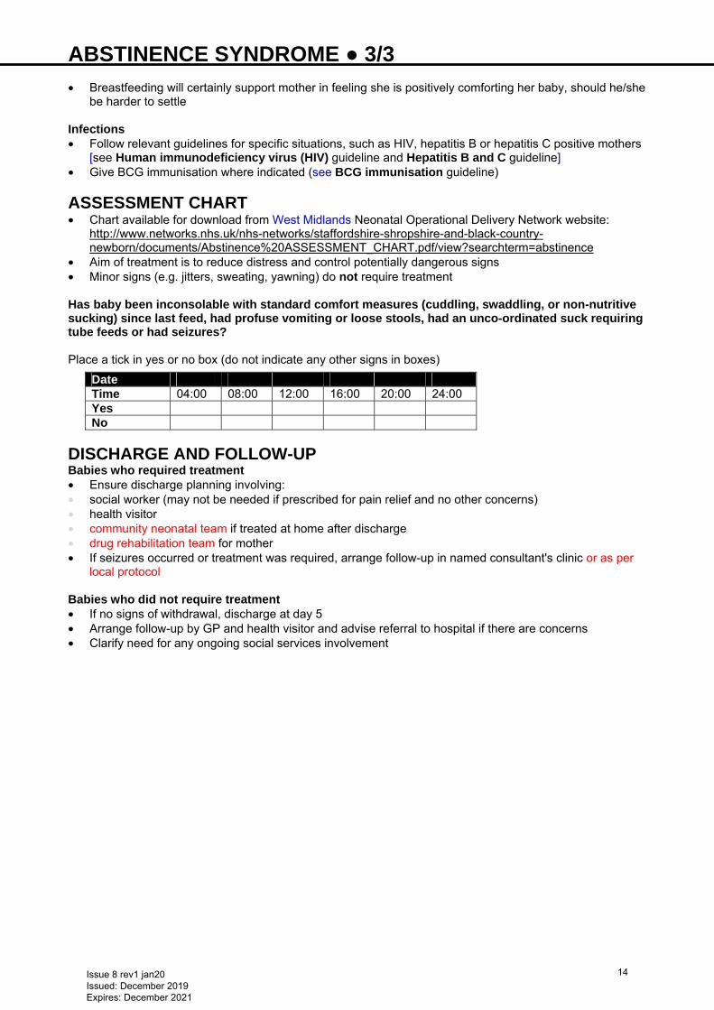
ASSESSMENT CHART • Chart available for download from West Midlands Neonatal Operational Delivery Network website:
http://www.networks.nhs.uk/nhs-networks/staffordshire-shropshire-and-black-country-newborn/documents/Abstinence%20ASSESSMENT_CHART.pdf/view?searchterm=abstinence
• Aim of treatment is to reduce distress and control potentially dangerous signs • Minor signs (e.g. jitters, sweating, yawning) do not require treatment Has baby been inconsolable with standard comfort measures (cuddling, swaddling, or non-nutritive sucking) since last feed, had profuse vomiting or loose stools, had an unco-ordinated suck requiring tube feeds or had seizures? Place a tick in yes or no box (do not indicate any other signs in boxes)
Date Time 04:00 08:00 12:00 16:00 20:00 24:00 Yes No
DISCHARGE AND FOLLOW-UP Babies who required treatment • Ensure discharge planning involving: • social worker (may not be needed if prescribed for pain relief and no other concerns) • health visitor • community neonatal team if treated at home after discharge • drug rehabilitation team for mother • If seizures occurred or treatment was required, arrange follow-up in named consultant's clinic or as per
local protocol Babies who did not require treatment • If no signs of withdrawal, discharge at day 5 • Arrange follow-up by GP and health visitor and advise referral to hospital if there are concerns • Clarify need for any ongoing social services involvement
Issue 8 rev1 jan20 Issued: December 2019 Expires: December 2021
14
ADMISSION TO NEONATAL UNIT ● 1/2 • There should be good clinical reasons for admission to NNU • Avoid unnecessary separation of mother and baby as it affects maternal bonding
Ensure that all babies born have newborn infant physical examination (NIPE) between 6−72 hr of birth
CRITERIA FOR ADMISSION FROM LABOUR WARD OR POSTNATAL WARD
Discuss need for admission with senior medical staff
• Clinical condition requiring constant monitoring, <34 weeks’ gestation or birth weight <1700 g • Unwell baby: • poor condition at birth requiring prolonged resuscitation for >10 min and/or cord pH <7.0 (a low cord pH
may not in itself necessitate admission to NNU) • respiratory distress or cyanosis • apnoeic or cyanotic attacks • signs of encephalopathy • jaundice needing intensive phototherapy or exchange transfusion • major congenital abnormality likely to threaten immediate survival • seizures • inability to tolerate enteral feeds with vomiting and/or abdominal distension and/or hypoglycaemia (blood
glucose <2.0 mmol/L for ≥37 weeks/<2.6 mmol/L for <37 weeks’ gestation) • symptomatic hypoglycaemia or hypoglycaemia not responding to treatment (see Hypoglycaemia
guideline) • Neonatal abstinence syndrome requiring treatment (see Abstinence syndrome guideline) • Short-term care while mother admitted to ITU Procedure • Manage immediate life-threatening clinical problems (e.g. airway, breathing, circulation and seizures) • Show baby to parents and explain reason for admission to NNU • Inform NNU nursing staff that you wish to admit a baby, reason for admission and clinical condition of
baby • Inform middle grade doctor and/or consultant • Ensure baby name labels present • Document relevant history and examination • Complete any local problem sheets and investigation charts • Measure and plot birth weight and head circumference on growth chart • Measure admission temperature • Measure blood pressure using non-invasive cuff • Institute appropriate monitoring and treatment in conjunction with nursing and senior medical colleagues Investigations
For babies admitted to NNU, obtain 1 bloodspot on newborn bloodspot screening (Guthrie) card Babies <32 weeks/1500 g weight/unwell/ventilated • FBC • Blood glucose • Blood gases • Clotting screen if clinically indicated (see Coagulopathy guideline) • routine clotting screen in all babies <30 weeks’ gestation is not recommended • If respiratory symptoms or support given, chest X-ray • If umbilical lines in place, abdominal X-ray • If suspicion of sepsis, blood culture and CRP before starting antibiotics and consider lumbar puncture
(see Infection in first 72 hours of life guideline) Other babies • Decision depends on initial assessment and suspected clinical problem (e.g. infection, jaundice,
hypoglycaemia etc.) see relevant guidelines
Issue 8 rev1 jan20 Issued: December 2019 Expires: December 2021
15
ADMISSION TO NEONATAL UNIT ● 2/2 IMMEDIATE MANAGEMENT • Evaluation of baby, including full clinical examination • Define appropriate management plan and procedures in consultation with middle grade doctor and
perform as efficiently as possible to ensure baby is not disturbed unnecessarily • Aim for examination and procedures to be completed within ≤1 hr of admission • If no contraindications, unless already administered, give vitamin K (see Vitamin K guideline) • If antibiotics indicated, give within 1 hr • Senior clinician to update parents as soon as possible (certainly within 24 hr) and document discussion
in notes Respiratory support • If required, this takes priority over other procedures • includes incubator oxygen, high-flow humidified oxygen, continuous positive airway pressure (CPAP) or
mechanical ventilation IV access • If required, IV cannulation and/or umbilical venous catheterisation (UVC) – see appropriate guidelines in
Practical procedures section
MONITORING Use minimal handling • Cardiorespiratory monitoring through skin electrodes. Do not use in babies <26 weeks’ gestation • Pulse oximetry. Maintain SpO2 as per gestation target values (see Oxygen saturation targets
guideline) • Transcutaneous probe for TcPO2/TcPCO2, if available (especially clinically unstable preterm) • Temperature • Blood glucose (see Hypoglycaemia guideline) • If ventilated, umbilical arterial catheterisation (UAC)/peripheral arterial line for monitoring arterial blood
pressure and arterial blood gas – see appropriate guidelines in Practical procedures section
CRITERIA FOR ADMISSION TO TRANSITIONAL CARE UNIT The following are common indications for admitting babies to transitional care unit (if available locally), refer to local guidelines for local variations • Small for gestational age, 1.7−2 kg and no other clinical concerns • Preterm 34–36 weeks’ gestation and no other clinical concerns • Minor congenital abnormalities likely to affect feeding, e.g. cleft lip and palate • Requiring support with feeding e.g. predicted to require NGT feeds • Babies of substance abusing mothers (observe for signs of withdrawal) • Receiving IV antibiotics
Issue 8 rev1 jan20 Issued: December 2019 Expires: December 2021
16
ANKYLOGLOSSIA (TONGUE-TIE) − DIVISION FOR BREASTFEEDING ● 1/1 Based on NICE IPG 149
INTRODUCTION • Breastfeeding is a complex interaction between mother and baby. Many factors can affect the ability to
breastfeed • Skilled breastfeeding support is an integral part of the management of breastfeeding difficulties • Current evidence suggests that there are no major safety concerns about division of tongue-tie, and
limited evidence suggests that it can improve breastfeeding
DEFINITION • A congenital anomaly of variable severity characterised by an abnormally short lingual frenulum, which
may restrict movement of the tongue. In severe cases the tongue is joined to the bottom of the mouth
INDICATIONS • Many tongue-ties are asymptomatic and cause no problem • Breastfeeding difficulties; conservative management includes breastfeeding advice • Assess carefully to determine if frenulum is interfering with feeding, and if division is appropriate • Symptoms may include: • difficulties with latching on • sore nipples • poor weight gain • Cochrane review 2017 • frenotomy reduces breastfeeding mothers’ nipple pain in the short-term • no consistent positive effect on infant breastfeeding • researchers reported no serious complications, but total number of infants studied was small
PROCEDURE • Division to be performed by properly trained registered health care professional only • Division in early infancy is usually performed without anaesthetic (although local anaesthetic is
sometimes used) • Little or no blood loss • Feeding may be resumed immediately
COMPLICATIONS OF PROCEDURE • Infrequent, but may include: • bleeding • infection • ulceration • pain • damage to tongue and surrounding area • recurrence of tongue-tie
KEY RESULTS • In an infant with tongue-tie and feeding difficulties, surgical release of the tongue-tie does not
consistently improve infant feeding but is likely to improve maternal nipple pain • further research required to clarify and confirm this effect
Issue 8 rev1 jan20 Issued: December 2019 Expires: December 2021
17
ANORECTAL MALFORMATION ● 1/3 INTRODUCTION Anorectal malformation (ARM) occurs in 1 in 5,000 neonates; rarely missed in boys, but can be missed in girls. ARMs are associated with other abnormalities including the VACTERL association, chromosomal abnormalities, duodenal atresia and oesophageal atresia
Missing ARM in a girl is usually a breach of duty)
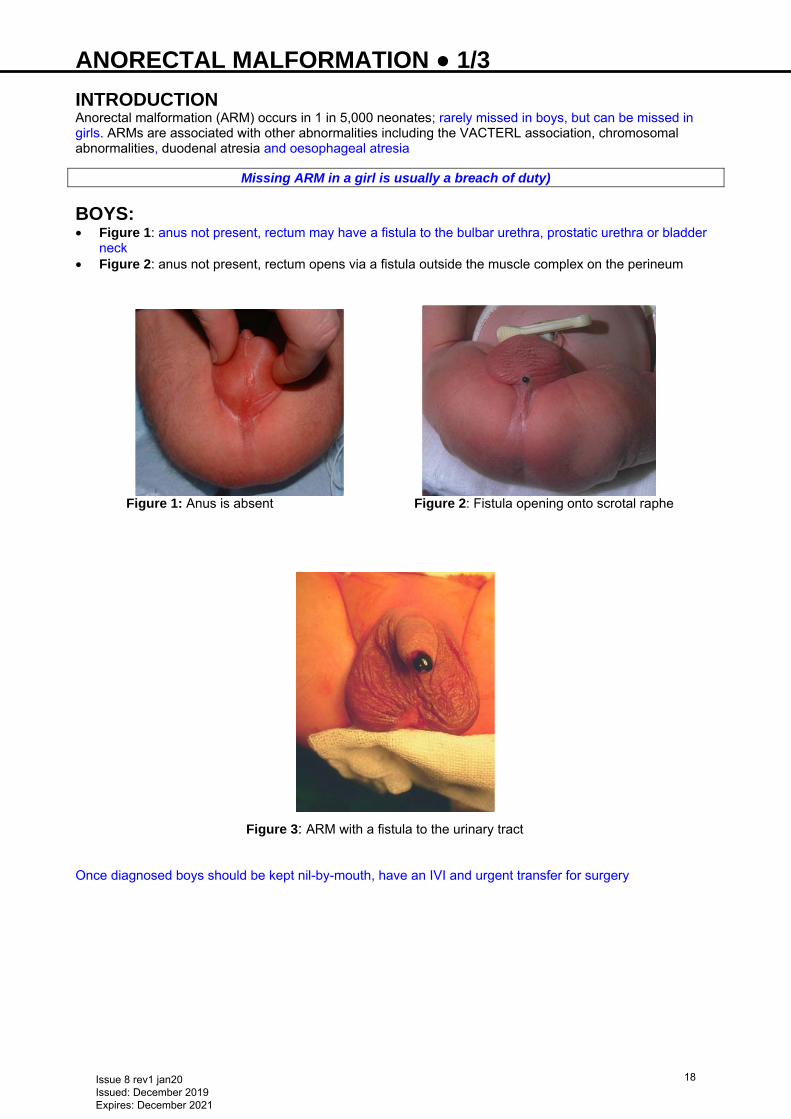
BOYS: • Figure 1: anus not present, rectum may have a fistula to the bulbar urethra, prostatic urethra or bladder
neck • Figure 2: anus not present, rectum opens via a fistula outside the muscle complex on the perineum
Figure 1: Anus is absent Figure 2: Fistula opening onto scrotal raphe
Figure 3: ARM with a fistula to the urinary tract
Once diagnosed boys should be kept nil-by-mouth, have an IVI and urgent transfer for surgery
Issue 8 rev1 jan20 Issued: December 2019 Expires: December 2021
18
ANORECTAL MALFORMATION ● 2/3 GIRLS
• More extreme but rare case of recto-vaginal fistula and cloaca should be identified at neonatal check • Always check the perineum when: • abnormal looking perineum • delayed/no passage of meconium • abdominal distension • bilious vomiting
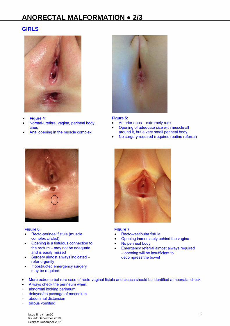
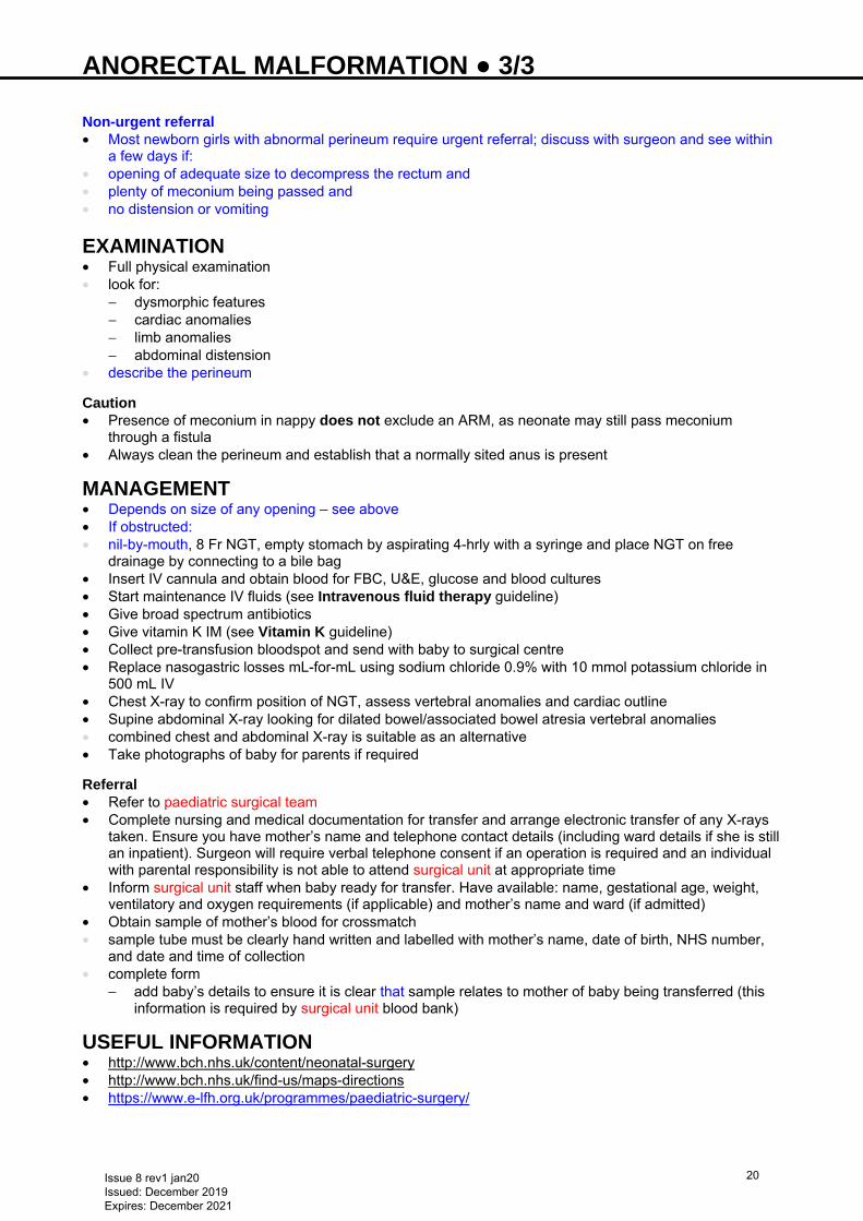
• Figure 4: • Normal-urethra, vagina, perineal body,
anus • Anal opening in the muscle complex
Figure 5: • Anterior anus − extremely rare • Opening of adequate size with muscle all
around it, but a very small perineal body • No surgery required (requires routine referral)
Figure 6: • Recto-perineal fistula (muscle
complex circled) • Opening is a fistulous connection to
the rectum − may not be adequate and is easily missed
• Surgery almost always indicated − refer urgently
• If obstructed emergency surgery may be required
Figure 7: • Recto-vestibular fistula • Opening immediately behind the vagina • No perineal body • Emergency referral almost always required
− opening will be insufficient to decompress the bowel
Issue 8 rev1 jan20 Issued: December 2019 Expires: December 2021
19
ANORECTAL MALFORMATION ● 3/3 Non-urgent referral • Most newborn girls with abnormal perineum require urgent referral; discuss with surgeon and see within
a few days if: • opening of adequate size to decompress the rectum and • plenty of meconium being passed and • no distension or vomiting
EXAMINATION • Full physical examination • look for:
− dysmorphic features − cardiac anomalies − limb anomalies − abdominal distension
• describe the perineum
Caution • Presence of meconium in nappy does not exclude an ARM, as neonate may still pass meconium
through a fistula • Always clean the perineum and establish that a normally sited anus is present
MANAGEMENT • Depends on size of any opening – see above • If obstructed: • nil-by-mouth, 8 Fr NGT, empty stomach by aspirating 4-hrly with a syringe and place NGT on free
drainage by connecting to a bile bag • Insert IV cannula and obtain blood for FBC, U&E, glucose and blood cultures • Start maintenance IV fluids (see Intravenous fluid therapy guideline) • Give broad spectrum antibiotics • Give vitamin K IM (see Vitamin K guideline) • Collect pre-transfusion bloodspot and send with baby to surgical centre • Replace nasogastric losses mL-for-mL using sodium chloride 0.9% with 10 mmol potassium chloride in
500 mL IV • Chest X-ray to confirm position of NGT, assess vertebral anomalies and cardiac outline • Supine abdominal X-ray looking for dilated bowel/associated bowel atresia vertebral anomalies • combined chest and abdominal X-ray is suitable as an alternative • Take photographs of baby for parents if required Referral • Refer to paediatric surgical team • Complete nursing and medical documentation for transfer and arrange electronic transfer of any X-rays
taken. Ensure you have mother’s name and telephone contact details (including ward details if she is still an inpatient). Surgeon will require verbal telephone consent if an operation is required and an individual with parental responsibility is not able to attend surgical unit at appropriate time
• Inform surgical unit staff when baby ready for transfer. Have available: name, gestational age, weight, ventilatory and oxygen requirements (if applicable) and mother’s name and ward (if admitted)
• Obtain sample of mother’s blood for crossmatch • sample tube must be clearly hand written and labelled with mother’s name, date of birth, NHS number,
and date and time of collection • complete form
− add baby’s details to ensure it is clear that sample relates to mother of baby being transferred (this information is required by surgical unit blood bank)
USEFUL INFORMATION • http://www.bch.nhs.uk/content/neonatal-surgery • http://www.bch.nhs.uk/find-us/maps-directions • https://www.e-lfh.org.uk/programmes/paediatric-surgery/
Issue 8 rev1 jan20 Issued: December 2019 Expires: December 2021
20
ANTENATAL ULTRASOUND ABNORMALITIES ● 1/1 DEFINITION • Any lesion identified antenatally in the fetus (e.g. renal pelvic dilatation, hypoplastic left heart) • Any maternal factor identified antenatally that could affect baby after delivery (e.g. anhydramnios from
preterm prolonged rupture of membranes)
COUNSELLING BEFORE DELIVERY • Abnormality detected in a local unit may require referral to regional fetal medicine centre • All affected pregnancies will have detailed individualised plans for management of baby by consultant
neonatologist, including place of delivery • As some lesions are progressive (e.g. hypoplastic left heart syndrome, gastroschisis), the situation can
change and information from the obstetric team can alter over time. Discuss all affected pregnancies at the combined fetomaternal meeting until delivery
• Offer neonatal counselling to all women whose pregnancy has been affected by major lesions, to discuss the impact of the identified lesion on quality of life, including possible disabilities, investigations and surgery, and post-delivery plan
Cleft lip and/or palate • Obstetric team to refer to regional multidisciplinary cleft palate team, who will counsel parents,
communicate plans for delivery and provide postnatal support for baby
Hypoplastic left heart syndrome or other presumed duct-dependent lesions • Obstetric team to refer to regional fetal cardiologist, who will counsel parents and, where appropriate,
confirm diagnosis and provide a plan of action, including most appropriate unit for delivery Congenital diaphragmatic hernia • Obstetric team to refer all cases to tertiary fetal medicine team at time of diagnosis • Amniocentesis may be performed before referral where this is offered (Birmingham or Liverpool) who will
counsel, monitor and arrange delivery in the NICU
MANAGEMENT AFTER DELIVERY • For minor lesions, such as renal pelvic dilatation, follow appropriate guideline and inform senior staff and
parents • For other lesions, follow written plan made by senior staff before delivery, including need to contact
seniors and specialist staff in regional referral centre before and after delivery • Communicate any new information obtained after birth to consultant as this may change the plan of care
required • Maintain regular contact with specialist teams as indicated by them • Arrange postnatal transfer if required when bed available • Keep parents informed of actions taken and contact from specialist teams • Consider syndrome for babies with >1 lesion, discuss with senior staff as soon as possible • When available and if not issued antenatally, provide written information from 'Contact a family' book or
www.cafamily.org.uk/
Specific lesions See Urinary tract abnormalities diagnosed antenatally, Gastroschisis, Congenital diaphragmatic hernia and Congenital heart disease: duct dependent lesions guidelines
Issue 8 rev1 jan20 Issued: December 2019 Expires: December 2021
21
APNOEA AND BRADYCARDIA ● 1/2 RECOGNITION AND ASSESSMENT Apnoea Pause(s) in breathing >20 sec (or less, when associated with bradycardia or cyanosis) Bradycardia Heart rate <100 bpm, associated with desaturation Types Central • Caused by poorly developed neurological control • Respiratory movements absent Obstructive • Caused by upper airway obstruction, usually at pharyngeal level • Respiratory movements continue initially but then stop Mixed • Initially central, followed by obstructive apnoea Significance • Most babies born <34 weeks’ gestation have primary apnoea of prematurity (PAP). Hence babies born
<34 weeks should have SpO2 monitoring until ≥34 weeks post conceptional age (PCA) • multiple aetiologic factors can exacerbate apnoea in preterm babies • sudden increase in frequency warrants immediate action • Consider causes other than apnoea of prematurity if occurs: • in term or near-term baby (>34 weeks’ gestation) • on first day after birth in preterm baby • onset of apnoea after aged 7 days in a preterm baby Causes Infection • Septicaemia • Necrotising enterocolitis • Meningitis Respiratory • Inadequate respiratory support • Upper airway obstruction • Surfactant deficiency
CNS • Intracranial haemorrhage • Seizure • Congenital malformations
CVS • Patent ductus arteriosus • Anaemia
Other • Metabolic abnormalities, especially hypoglycaemia • Haematological: anaemia • Inherited metabolic disorders e.g. non-ketotic hyperglycinaemia
MANAGEMENT Terminate episode • If apnoea not self-limiting (clinician to agree threshold to intervene), perform the following in sequence to
try to terminate episode: • ensure head in neutral position • stimulate baby by tickling feet or stroking abdomen • if aspiration or secretions in pharynx suspected, apply brief oropharyngeal suction
Issue 8 rev1 jan20 Issued: December 2019 Expires: December 2021
22
APNOEA AND BRADYCARDIA ● 2/2 • face mask ventilation • emergency intubation • Once stable, perform thorough clinical examination to confirm/evaluate cause
Screen for sepsis • If apnoea or bradycardia increasingly frequent or severe, screen for sepsis as apnoea and bradycardia can be
sole presenting sign
TREATMENT • Treat specific cause, if present • Primary apnoea of prematurity is a diagnosis of exclusion and may not require treatment unless pauses
are: • frequent (>8 in 12 hr) or • severe (>2 episodes/day requiring positive pressure ventilation) Pharmacological treatment • Caffeine citrate 20 mg/kg loading dose oral/IV (over 30 min) followed, after 24 hr, by maintenance dose of
5 mg/kg oral/IV (over 10 min) once daily, increasing to 20 mg/kg if required until 34 weeks PCA • If desaturations and bradycardias persist, may continue beyond 34 weeks PCA. If so review need for treatment
regularly Non-pharmacological treatment • CPAP, SiPAP/BiPAP [see Ventilation: continuous positive airway pressure (CPAP) guideline] • If above fails, intubate and ventilate
Issue 8 rev1 jan20 Issued: December 2019 Expires: December 2021
23
ARTERIAL LINE INSERTION ● 1/2 PERIPHERAL ARTERIAL LINES Indications • Frequent monitoring of blood gases • Direct monitoring of arterial blood pressure • Exchange transfusion (peripheral venous and arterial catheters ‛continuous’ technique) or partial
exchange transfusion Contraindications • Bleeding disorder • Inadequate patency of ulnar artery on transillumination or failed Allen’s test (if cannulating radial artery)
or vice-versa • Pre-existing evidence of circulatory insufficiency in limb • Local skin infection • Malformation of upper extremity for radial arterial cannulation Possible sites of arterial entry • Radial (most commonly used); the only procedure discussed in this guideline • Posterior tibial • Dorsalis pedis • Ulnar (usually only if ipsilateral radial artery cannulation has not been attempted)
EQUIPMENT • Gloves • Cleaning solution as per unit policy • 24 G cannula • T-connector with Luer lock • Adhesive tape • Splint • Sodium chloride 0.9% flush in 2 mL syringe, primed through T-connector • Transillumination fibre-optic light source • 3-way tap
PROCEDURE USING RADIAL ARTERY Preparation • Wash hands • Check patency of ipsilateral ulnar artery using Allen’s test and proceed only if patent • Put on gloves • Extend baby’s wrist with palm of hand upwards • Transilluminate radial artery with fibre-optic light source behind baby’s wrist or palpate pulse • Clean skin with antiseptic cleaning solution Procedure • Enter artery with 24 G cannula just proximal to wrist crease at 25–30° angle • Remove stylet from cannula and advance cannula into artery • Connect cannula to T-connector primed with sodium chloride 0.9%, and flush gently • Secure cannula with tape, ensuring fingers are visible for frequent inspection, and apply splint • Connect T-connector to infusion line (sodium chloride 0.9% with heparin 1 unit/mL), with
3-way tap in situ for blood sampling Documentation • Document clearly in notes all attempts at cannulation, including those that are unsuccessful
AFTERCARE Monitor • Inspect distal digits regularly for circulatory status; if blanching does not recover after 5 min, discuss
further management with consultant • Avoid excessive hyperextension of wrist, as this can result in occlusion of artery
Issue 8 rev1 jan20 Issued: December 2019 Expires: December 2021
24
ARTERIAL LINE INSERTION ● 2/2 • Ensure a continuous pressure waveform tracing is displayed on monitor screen at all times; if flushing
line does not restore lost tracing, change position of limb/dressing Usage • Do not administer rapid boluses of fluid as this can lead to retrograde embolisation of clot or air; use
minimal volume when flushing after sampling and inject slowly • Use cannula only for sampling or removal of blood during exchange transfusion, and infuse sodium
chloride 0.9% or 0.45% with heparin 1 unit/mL • Remove cannula as soon as no longer required Removal • Aseptic removal of arterial line: apply pressure for ≥5 min (longer if coagulopathy/low platelets), until no
bleeding or bruising • dressings do not prevent bleeding or bruising • do not send tip for culture routinely
COMPLICATIONS • Thromboembolism/vasospasm/thrombosis • Blanching and partial loss of digits (radial artery) • Necrosis • Skin ulceration • Reversible occlusion of artery • Extravasation of sodium chloride infusate • Infection (rarely associated with line infection) • Haematoma • Haemorrhage • Air embolism
Issue 8 rev1 jan20 Issued: December 2019 Expires: December 2021
25
ARTERIAL LINE SAMPLING ● 1/2 INDICATIONS • Blood gas analysis • Biochemical/and haematological investigations
CONTRAINDICATIONS • Blood drawn from an arterial line may not be suitable for clotting studies (see Coagulopathy and
Bloodspot screening guidelines)
COMPLICATIONS Haemorrhage • Ensure all connections are secure, Luer locks tight and 3-way taps appropriately adjusted Infection • Maintain sterile technique during sampling to reduce risk of infection
Artery spasm • Limb appears blanched. Stop procedure and allow time for recovery. Warming of opposite limb can elicit
reflex vasodilatation Thromboembolism • Flush catheter with sodium chloride 0.9% 0.5 mL each time sample taken. If catheter not sampling, clot
formation may be in progress. Request urgent middle grade review of arterial line for a prompt decision about removal
Inaccuracy of blood gas results • Analyse sample immediately. After blood is withdrawn from an artery, it continues to consume oxygen • Excess heparin in syringe can result in a falsely low pH and PaCO2. Remove excess heparin from
syringe before obtaining sample • Do not use if air bubbles in sample − take fresh specimen
EQUIPMENT • Gloves • Paper towel • Alcohol swabs x 2 • Syringes • 2 mL syringe (A) for clearing line • 2 mL syringe (B) for other blood samples as necessary • 1 mL syringe (C) pre-heparinised for blood gas analysis • 2 mL syringe (D) containing 0.5–1 mL of sodium chloride 0.9% • Appropriate blood sample bottles and request forms
PREPARATION AND PROCEDURE Preparation • Record SpO2 and TcCO2 at time of taking blood to allow comparison with blood gas if performed • Wash hands and put on gloves • Place paper towel beneath 3-way tap collection port (maintain asepsis by non-touch technique rather
than sterile gloves and towel) • Ensure 3-way tap closed to porthole Procedure • Remove Luer lock cap, clean with alcohol swab and allow to dry, or prepare bioconnector • Connect 2 mL syringe (A) • Turn 3-way tap so it is closed to infusion and open to syringe and arterial catheter • Withdraw 2 mL blood slowly. It must clear the dead space • If bioconnector not being used, turn 3-way tap so it is closed to arterial catheter to prevent blood loss
from baby • if bioconnector used, do not turn 3-way tap until end of procedure • Attach appropriate syringe (B/C) needed for required blood sample • If bioconnector not being used, turn 3-way tap to open to syringe and arterial catheter and withdraw
required amount of blood for blood samples. Do not withdraw more than required amount
Issue 8 rev1 jan20 Issued: December 2019 Expires: December 2021
26
ARTERIAL LINE SAMPLING ● 2/2 • If bioconnector not being used, turn 3-way tap off to arterial catheter in between syringes B and C if both
required, after taking required samples with syringes • Reattach syringe (A) • Clear the connection of air • Slowly return to baby any blood in line not required for samples • If bioconnector not being used, turn 3-way tap off to arterial catheter • Attach syringe (D) of sodium chloride 0.9% • If bioconnector not being used, turn 3-way tap so it is open to syringe and arterial line, clear line of air
and slowly flush line to clear of blood • Turn 3-way tap so it is closed to syringe, remove syringe (D), swab porthole with alcohol wipe and cover
with Luer lock cap • Record amount of blood removed and volume of flush on baby’s daily fluid record
AFTERCARE • Ensure all connections tight and 3-way tap turned off to syringe port to prevent haemorrhage • If sampling from umbilical arterial catheter, ensure lower limbs are pink and well perfused on completion
of procedure • If sampling from peripheral arterial line, check colour and perfusion of line site and limb housing arterial
line • Ensure line patency by recommencing infusion pump • Before leaving baby, ensure arterial wave form present and all alarms set
Issue 8 rev1 jan20 Issued: December 2019 Expires: December 2021
27
BABIES BORN AT MARGINS OF VIABILITY ● 1/2 INTRODUCTION • Outcomes for premature babies at borderline viability improve with each additional week of gestational
age. See EPICure studies http://www.epicure.ac.uk/ • Ultrasound estimated fetal weight within a week before delivery of <500 g at any gestation between 22+0
and 25+6 weeks is associated with a very poor outcome; see Draper charts http://pediatrics.aappublications.org/content/pediatrics/131/2/e425/F1.large.jpg.
• Ultrasound carried out in first trimester of pregnancy is the most reliable method of estimating gestational age
• If fetal heart heard during labour, call neonatal team to attend delivery, unless decision not to intervene and rationale already agreed with parents and documented
• once baby delivered, further resuscitation and management decisions should be made in baby’s best interests, taking into account clinical condition at birth, e.g. heart rate, breathing, weight, severity of bruising to skin etc.; obtain urgent senior advice
• Discussion with parents before birth, if possible, should precede any action, preferably by obstetric and paediatric teams jointly
• Document all discussions in case records
MANAGEMENT • An experienced neonatologist ideally to be present at delivery of extremely premature babies (<27
completed weeks’ gestation) and make confirmatory assessment of gestational age and condition of baby
≥24 weeks’ gestation • Unless baby has a severe abnormality incompatible with any significant period of survival, initiate
intensive care and admit to NICU <24 weeks’ gestation • Discuss with parents national and local statistical evidence for survival in babies with range of disabilities
found in this age group • explain that statistics indicate most babies born <24 weeks’ gestation are likely to die and a significant
proportion of survivors are likely to have some form of neurological impairment
MANAGEMENT AT SPECIFIC GESTATIONS 24+0−24+6 weeks’ gestation • Be prepared to provide full, invasive, intensive care and support from birth and admit to NICU, unless
parents and clinicians agree that in view of baby’s condition (or likely condition), or response to initial resuscitation, intensive care is not in his/her best interests
23+0−23+6 weeks’ gestation • Give consideration to parents’ wishes regarding resuscitation and invasive intensive care
treatment. However, when condition at birth indicates that baby will not survive for long, clinicians are not legally obliged to proceed with treatment that is wholly contrary to their clinical judgement, if they consider treatment would be futile
• as a first step, determine whether baby is suffering, whether any suffering can be alleviated, and likely burden placed on baby by intensive care treatment
• where parents would prefer clinical team to make decision about initiation of intensive care, clinicians must determine what constitutes appropriate care
• where it has not been possible to discuss a baby’s treatment with mother and, where appropriate, her partner, before the birth, clinical team should consider offering full invasive intensive care until baby’s condition and treatment can be discussed with parents
• If baby is born in good condition, initiate resuscitation using IPPV (via ETT or face mask if good chest movement obtained)
• if baby does not improve and heart rate remains low at 10 min after effective ventilatory support, withhold further resuscitation
• response of heart rate to ventilation is critical in deciding whether to continue or stop. Counsel parents with sensitivity that further interventions are futile
22+0−22+6 weeks’ gestation • Standard practice should be not to resuscitate a baby and this would normally not be considered or
proposed
Issue 8 rev1 jan20 Issued: December 2019 Expires: December 2021
28
BABIES BORN AT MARGINS OF VIABILITY ● 2/2 • If parents request resuscitation, and reiterate this request, discuss risks and long-term outcomes with an
experienced neonatologist before attempting resuscitation and offering intensive care • Treating clinicians must all agree that this is an exceptional case where resuscitation is in baby’s best
interests <22 weeks’ gestation • Resuscitation should not occur in routine clinical practice • any attempt to resuscitate babies born at this gestational age should take place only within the context of
an approved research study
When intensive care not given, clinical team must provide palliative care until baby dies. Refer to BAPM guidelines for counselling
PARENT INFORMATION • ‘Information for parents of extremely premature babies’ leaflet available to download from
www.epicure.ac.uk/index.php/download_file/view/150/
Issue 8 rev1 jan20 Issued: December 2019 Expires: December 2021
29
BCG IMMUNISATION ● 1/3
See also Tuberculosis (investigation and management following exposure in pregnancy) guideline
INDICATIONS • All infants (aged ≤12 months) with a parent or grandparent who was born in a country where the annual
incidence of TB is ≥40/100,000 • All infants (aged ≤12 months) living in areas of the UK where the annual incidence of TB is ≥40/100,000
Countries with incidence of TB ≥40/100,000
Afghanistan Ecuador Korea DPR Niger Tajikistan Algeria El Salvador Korea (Rep. of) Nigeria Thailand Angola Equatorial Guinea Kyrgyzstan Niue Timor-Leste Azerbaijan Eritrea Lao PDR Northern Mariana Islands Togo Bangladesh Eswatini Lesotho Pakistan Turkmenistan Benin Ethiopia Liberia Palau Tuvalu Bhutan Fiji Libya Panama Uganda Bolivia Gabon Lithuania Papua New Guinea Ukraine Botswana Gambia Madagascar Paraguay Uzbekistan Brazil Georgia Malawi Peru Vanuatu Brunei Ghana Malaysia Philippines Venezuela Burkina Faso Greenland Mali Romania Vietnam Burundi Guam Marshall Islands Russia Yemen Cambodia Guinea Mauritania Rwanda Zambia Cameroon Guinea-Bissau Micronesia Sao Tome and Principe Zimbabwe Cape Verde Guyana Moldova Senegal Central African Republic Haiti Mongolia Sierra Leone Chad Hong Kong Morocco Singapore China India Mozambique Solomon Islands Congo Indonesia Myanmar Somalia Congo DR Iraq Namibia South Africa Côte d'Ivoire Kazakhstan Nauru South Sudan Djibouti Kenya Nepal Sri Lanka Dominican Republic Kiribati Nicaragua Sudan
https://www.gov.uk/government/publications/tuberculosis-tb-by-country-rates-per-100000-people
Parts of UK with universal BCG policy London (Newham, Brent, Hounslow, Ealing and Redbridge), and Slough Tuberculin testing not necessary aged <6 yr unless baby has been in recent contact with TB or has resided in high-incidence country for >3 months
CONTRAINDICATIONS • Temperature >38°C or acutely unwell • Severe eczema (give at suitable lesion-free site) • Baby in household where an active TB case suspected or confirmed, see Tuberculosis (investigation
and management following exposure in pregnancy) guideline • Immunodeficient or on high-dose corticosteroids • defer BCG until 3 months after stopping corticosteroids if given prednisolone 1 mg/kg/day for >2 weeks,
2 mg/kg/day for 1 week, (or equivalent doses of another corticosteroid, e.g. dexamethasone 150 micrograms = prednisolone 1 mg)
• Maternal immunosuppressive treatment during pregnancy or breastfeeding • biologicals e.g. TNFα, postpone BCG until aged 6 months • HIV positive, living in UK • if mother HIV positive and high risk of HIV transmission [see Human immunodeficiency virus (HIV)
guideline] and exclusively formula feeding, give vaccine only after baby is confirmed HIV uninfected at aged 12–14 weeks
• if mother HIV positive and very low risk or low risk of HIV transmission [see Human immunodeficiency virus (HIV) guideline] BCG can be given to baby when indicated
• if high risk of TB exposure and maternal HIV viral load <50 copies/mL after 36 weeks’ gestation, BCG can be given at birth
• encourage maternal HIV testing but do not withhold BCG if mother declines testing unless mother from sub-Saharan Africa, in which case refer to HIV team for counselling about testing
Issue 8 rev1 jan20 Issued: December 2019 Expires: December 2021
30
BCG IMMUNISATION ● 2/3 SPECIAL CASES • No need to delay routine vaccinations • BCG can be given simultaneously with other vaccines [including rotavirus vaccine oral or palivizumab
(Synagis®) IM but not in same arm] • no further immunisation should be given in arm used for BCG immunisation for ≥3 months due to risk of
regional lymphadenitis • if not given at same time, leave 4 weeks before giving other injectable live vaccines
PROCEDURE • Dose: 0.05 mL (Note: vial contains 20 doses) • Only to be given by health professional trained in giving BCG vaccine Consent • Midwife to record at booking if risk factor present • Postnatal check for risk factor • Ensure baby within inclusion group • Give mother information on vaccine • Give appropriate language leaflet TB, BCG vaccine and your baby, available from
https://www.gov.uk/government/publications/tb-bcg-and-your-baby-leaflet order line: 0300 123 1002 or https://www.orderline.dh.gov.uk/ecom_dh/public/home.jsf
• Department of Health guidelines state written consent is not required but follow local practice Injection
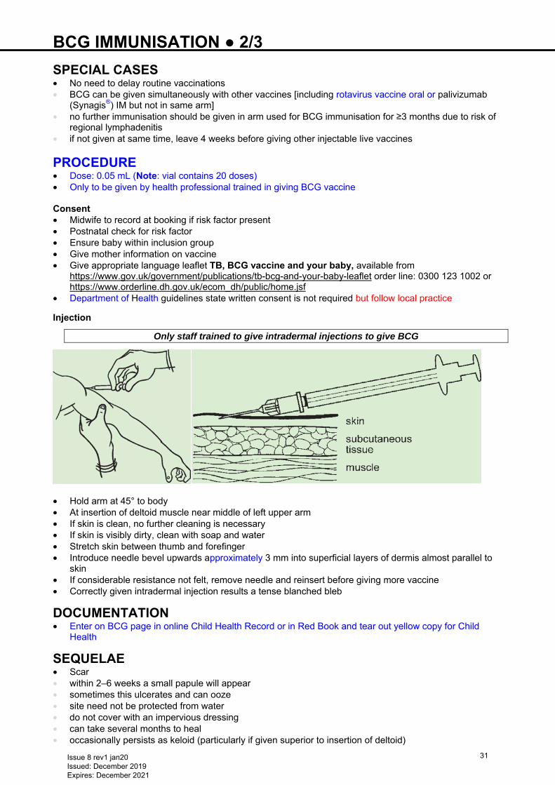
Only staff trained to give intradermal injections to give BCG
• Hold arm at 45° to body • At insertion of deltoid muscle near middle of left upper arm • If skin is clean, no further cleaning is necessary • If skin is visibly dirty, clean with soap and water • Stretch skin between thumb and forefinger • Introduce needle bevel upwards approximately 3 mm into superficial layers of dermis almost parallel to
skin • If considerable resistance not felt, remove needle and reinsert before giving more vaccine • Correctly given intradermal injection results a tense blanched bleb
DOCUMENTATION • Enter on BCG page in online Child Health Record or in Red Book and tear out yellow copy for Child
Health
SEQUELAE • Scar • within 2–6 weeks a small papule will appear • sometimes this ulcerates and can ooze • site need not be protected from water • do not cover with an impervious dressing • can take several months to heal • occasionally persists as keloid (particularly if given superior to insertion of deltoid)
Issue 8 rev1 jan20 Issued: December 2019 Expires: December 2021
31
BCG IMMUNISATION ● 3/3 • Adenitis: • a minor degree of adenitis can occur in the weeks following BCG • no treatment indicated • Rare sequelae: • local abscess • chronic suppurative lymphadenopathy • disseminated disease, if immunocompromised • osteitis, refer to infectious diseases specialist Refer to paediatric TB team if: • Severe local reactions • abscesses or drainage at the injection site or • regional suppurative lymphadenitis with draining sinuses
Refer disseminated BCG infection to paediatric TB specialist
Issue 8 rev1 jan20 Issued: December 2019 Expires: December 2021
32
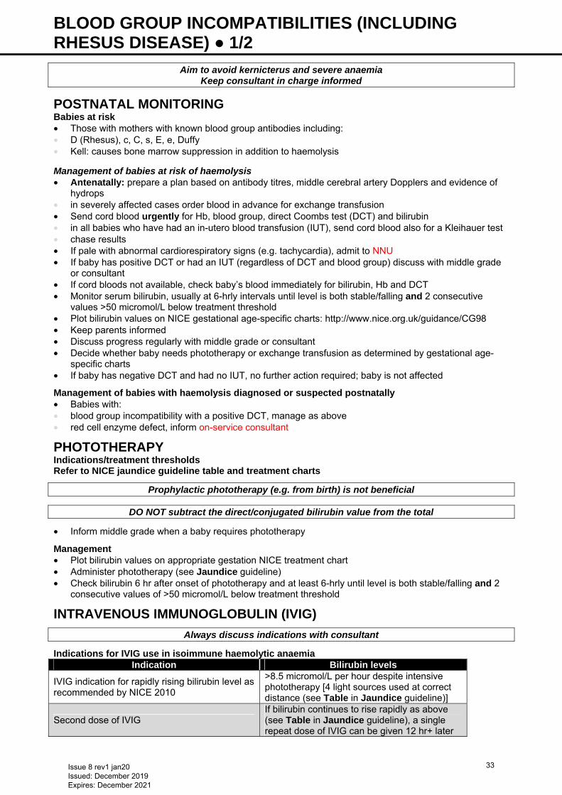
BLOOD GROUP INCOMPATIBILITIES (INCLUDING RHESUS DISEASE) ● 1/2
Aim to avoid kernicterus and severe anaemia Keep consultant in charge informed
POSTNATAL MONITORING Babies at risk • Those with mothers with known blood group antibodies including: • D (Rhesus), c, C, s, E, e, Duffy • Kell: causes bone marrow suppression in addition to haemolysis
Management of babies at risk of haemolysis • Antenatally: prepare a plan based on antibody titres, middle cerebral artery Dopplers and evidence of
hydrops • in severely affected cases order blood in advance for exchange transfusion • Send cord blood urgently for Hb, blood group, direct Coombs test (DCT) and bilirubin • in all babies who have had an in-utero blood transfusion (IUT), send cord blood also for a Kleihauer test • chase results • If pale with abnormal cardiorespiratory signs (e.g. tachycardia), admit to NNU • If baby has positive DCT or had an IUT (regardless of DCT and blood group) discuss with middle grade
or consultant • If cord bloods not available, check baby’s blood immediately for bilirubin, Hb and DCT • Monitor serum bilirubin, usually at 6-hrly intervals until level is both stable/falling and 2 consecutive
values >50 micromol/L below treatment threshold • Plot bilirubin values on NICE gestational age-specific charts: http://www.nice.org.uk/guidance/CG98 • Keep parents informed • Discuss progress regularly with middle grade or consultant • Decide whether baby needs phototherapy or exchange transfusion as determined by gestational age-
specific charts • If baby has negative DCT and had no IUT, no further action required; baby is not affected
Management of babies with haemolysis diagnosed or suspected postnatally • Babies with: • blood group incompatibility with a positive DCT, manage as above • red cell enzyme defect, inform on-service consultant
PHOTOTHERAPY Indications/treatment thresholds Refer to NICE jaundice guideline table and treatment charts
Prophylactic phototherapy (e.g. from birth) is not beneficial
DO NOT subtract the direct/conjugated bilirubin value from the total
• Inform middle grade when a baby requires phototherapy
Management • Plot bilirubin values on appropriate gestation NICE treatment chart • Administer phototherapy (see Jaundice guideline) • Check bilirubin 6 hr after onset of phototherapy and at least 6-hrly until level is both stable/falling and 2
consecutive values of >50 micromol/L below treatment threshold
INTRAVENOUS IMMUNOGLOBULIN (IVIG)
Always discuss indications with consultant
Indications for IVIG use in isoimmune haemolytic anaemia Indication Bilirubin levels
IVIG indication for rapidly rising bilirubin level as recommended by NICE 2010
>8.5 micromol/L per hour despite intensive phototherapy [4 light sources used at correct distance (see Table in Jaundice guideline)]
Second dose of IVIG If bilirubin continues to rise rapidly as above (see Table in Jaundice guideline), a single repeat dose of IVIG can be given 12 hr+ later
Issue 8 rev1 jan20 Issued: December 2019 Expires: December 2021
33
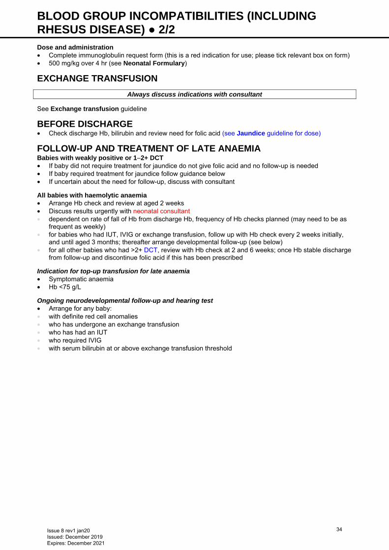
BLOOD GROUP INCOMPATIBILITIES (INCLUDING RHESUS DISEASE) ● 2/2 Dose and administration • Complete immunoglobulin request form (this is a red indication for use; please tick relevant box on form) • 500 mg/kg over 4 hr (see Neonatal Formulary)
EXCHANGE TRANSFUSION
Always discuss indications with consultant See Exchange transfusion guideline
BEFORE DISCHARGE • Check discharge Hb, bilirubin and review need for folic acid (see Jaundice guideline for dose)
FOLLOW-UP AND TREATMENT OF LATE ANAEMIA Babies with weakly positive or 1−2+ DCT • If baby did not require treatment for jaundice do not give folic acid and no follow-up is needed • If baby required treatment for jaundice follow guidance below • If uncertain about the need for follow-up, discuss with consultant
All babies with haemolytic anaemia • Arrange Hb check and review at aged 2 weeks • Discuss results urgently with neonatal consultant • dependent on rate of fall of Hb from discharge Hb, frequency of Hb checks planned (may need to be as
frequent as weekly) • for babies who had IUT, IVIG or exchange transfusion, follow up with Hb check every 2 weeks initially,
and until aged 3 months; thereafter arrange developmental follow-up (see below) • for all other babies who had >2+ DCT, review with Hb check at 2 and 6 weeks; once Hb stable discharge
from follow-up and discontinue folic acid if this has been prescribed
Indication for top-up transfusion for late anaemia • Symptomatic anaemia • Hb <75 g/L
Ongoing neurodevelopmental follow-up and hearing test • Arrange for any baby: • with definite red cell anomalies • who has undergone an exchange transfusion • who has had an IUT • who required IVIG • with serum bilirubin at or above exchange transfusion threshold
Issue 8 rev1 jan20 Issued: December 2019 Expires: December 2021
34
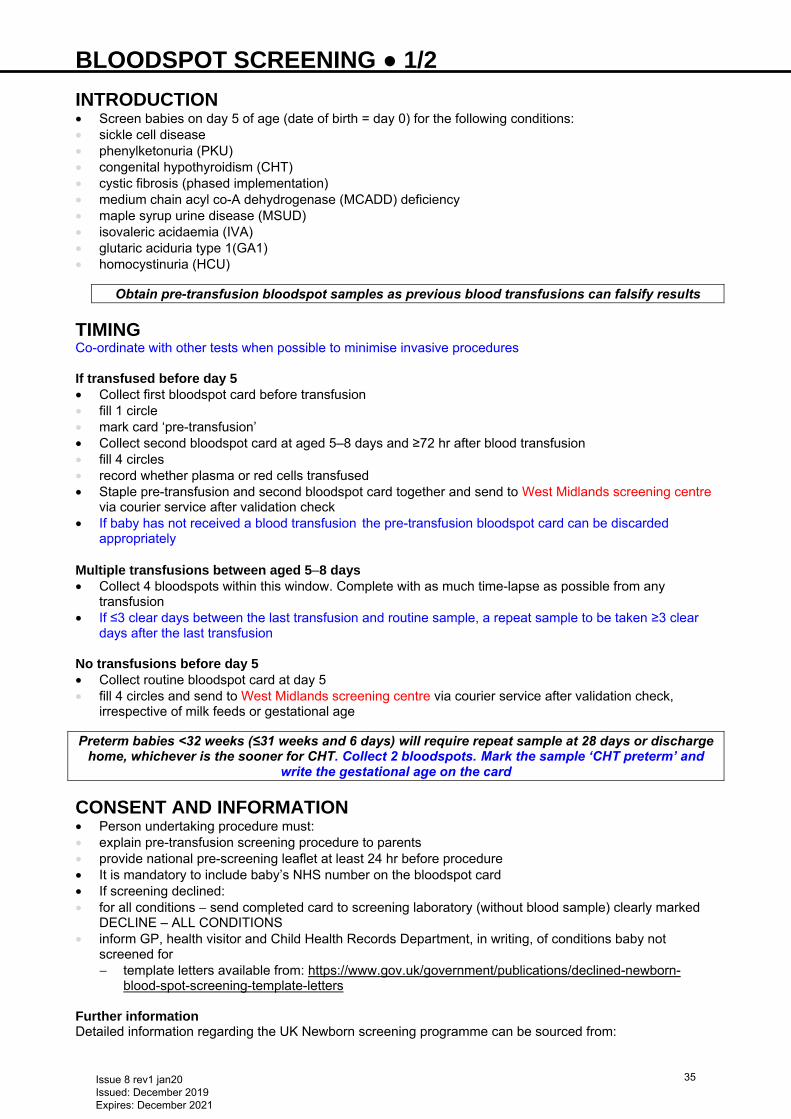
BLOODSPOT SCREENING ● 1/2 INTRODUCTION • Screen babies on day 5 of age (date of birth = day 0) for the following conditions: • sickle cell disease • phenylketonuria (PKU) • congenital hypothyroidism (CHT) • cystic fibrosis (phased implementation) • medium chain acyl co-A dehydrogenase (MCADD) deficiency • maple syrup urine disease (MSUD) • isovaleric acidaemia (IVA) • glutaric aciduria type 1(GA1) • homocystinuria (HCU)
Obtain pre-transfusion bloodspot samples as previous blood transfusions can falsify results
TIMING Co-ordinate with other tests when possible to minimise invasive procedures If transfused before day 5 • Collect first bloodspot card before transfusion • fill 1 circle • mark card ‘pre-transfusion’ • Collect second bloodspot card at aged 5–8 days and ≥72 hr after blood transfusion • fill 4 circles • record whether plasma or red cells transfused • Staple pre-transfusion and second bloodspot card together and send to West Midlands screening centre
via courier service after validation check • If baby has not received a blood transfusion the pre-transfusion bloodspot card can be discarded
appropriately Multiple transfusions between aged 5−8 days • Collect 4 bloodspots within this window. Complete with as much time-lapse as possible from any
transfusion • If ≤3 clear days between the last transfusion and routine sample, a repeat sample to be taken ≥3 clear
days after the last transfusion No transfusions before day 5 • Collect routine bloodspot card at day 5 • fill 4 circles and send to West Midlands screening centre via courier service after validation check,
irrespective of milk feeds or gestational age
Preterm babies <32 weeks (≤31 weeks and 6 days) will require repeat sample at 28 days or discharge home, whichever is the sooner for CHT. Collect 2 bloodspots. Mark the sample ‘CHT preterm’ and
write the gestational age on the card
CONSENT AND INFORMATION • Person undertaking procedure must: • explain pre-transfusion screening procedure to parents • provide national pre-screening leaflet at least 24 hr before procedure • It is mandatory to include baby’s NHS number on the bloodspot card • If screening declined: • for all conditions − send completed card to screening laboratory (without blood sample) clearly marked
DECLINE – ALL CONDITIONS • inform GP, health visitor and Child Health Records Department, in writing, of conditions baby not
screened for − template letters available from: https://www.gov.uk/government/publications/declined-newborn-
blood-spot-screening-template-letters Further information Detailed information regarding the UK Newborn screening programme can be sourced from:
Issue 8 rev1 jan20 Issued: December 2019 Expires: December 2021
35
BLOODSPOT SCREENING ● 2/2 • Newborn bloodspot screening programme handbook:
https://www.gov.uk/government/publications/health-professional-handbook-newborn-blood-spot-screening
• Standards for NHS newborn bloodspot screening: https://www.gov.uk/government/publications/standards-for-nhs-newborn-blood-spot-screening
• Newborn bloodspot screening sampling guidelines: https://www.gov.uk/government/publications/newborn-blood-spot-screening-sampling-guidelines
Issue 8 rev1 jan20 Issued: December 2019 Expires: December 2021
36
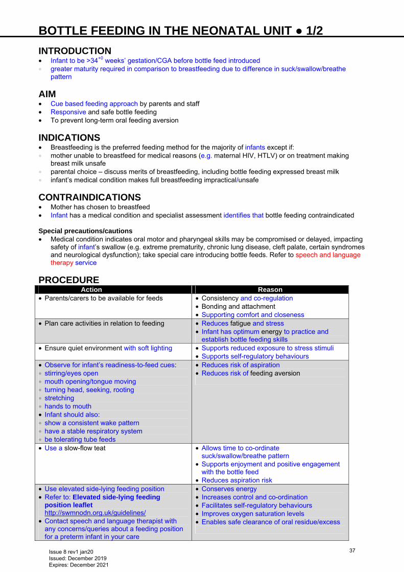
BOTTLE FEEDING IN THE NEONATAL UNIT ● 1/2 INTRODUCTION • Infant to be >34+0 weeks’ gestation/CGA before bottle feed introduced • greater maturity required in comparison to breastfeeding due to difference in suck/swallow/breathe
pattern
AIM • Cue based feeding approach by parents and staff • Responsive and safe bottle feeding • To prevent long-term oral feeding aversion
INDICATIONS • Breastfeeding is the preferred feeding method for the majority of infants except if: • mother unable to breastfeed for medical reasons (e.g. maternal HIV, HTLV) or on treatment making
breast milk unsafe • parental choice – discuss merits of breastfeeding, including bottle feeding expressed breast milk • infant’s medical condition makes full breastfeeding impractical/unsafe
CONTRAINDICATIONS • Mother has chosen to breastfeed • Infant has a medical condition and specialist assessment identifies that bottle feeding contraindicated Special precautions/cautions • Medical condition indicates oral motor and pharyngeal skills may be compromised or delayed, impacting
safety of infant’s swallow (e.g. extreme prematurity, chronic lung disease, cleft palate, certain syndromes and neurological dysfunction); take special care introducing bottle feeds. Refer to speech and language therapy service
PROCEDURE Action Reason • Parents/carers to be available for feeds • Consistency and co-regulation
• Bonding and attachment • Supporting comfort and closeness
• Plan care activities in relation to feeding • Reduces fatigue and stress • Infant has optimum energy to practice and
establish bottle feeding skills • Ensure quiet environment with soft lighting • Supports reduced exposure to stress stimuli
• Supports self-regulatory behaviours • Observe for infant’s readiness-to-feed cues: • stirring/eyes open • mouth opening/tongue moving • turning head, seeking, rooting • stretching • hands to mouth • Infant should also: • show a consistent wake pattern • have a stable respiratory system • be tolerating tube feeds
• Reduces risk of aspiration • Reduces risk of feeding aversion
• Use a slow-flow teat • Allows time to co-ordinate suck/swallow/breathe pattern
• Supports enjoyment and positive engagement with the bottle feed
• Reduces aspiration risk • Use elevated side-lying feeding position • Refer to: Elevated side-lying feeding
position leaflet http://swmnodn.org.uk/guidelines/
• Contact speech and language therapist with any concerns/queries about a feeding position for a preterm infant in your care
• Conserves energy • Increases control and co-ordination • Facilitates self-regulatory behaviours • Improves oxygen saturation levels • Enables safe clearance of oral residue/excess
Issue 8 rev1 jan20 Issued: December 2019 Expires: December 2021
37
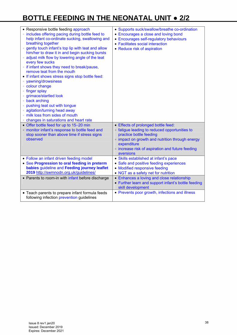
BOTTLE FEEDING IN THE NEONATAL UNIT ● 2/2 • Responsive bottle feeding approach • includes offering pacing during bottle feed to
help infant co-ordinate sucking, swallowing and breathing together
• gently touch infant’s top lip with teat and allow him/her to draw it in and begin sucking bursts
• adjust milk flow by lowering angle of the teat every few sucks
• if infant shows they need to break/pause, remove teat from the mouth
• If infant shows stress signs stop bottle feed: • yawning/drowsiness • colour change • finger splay • grimace/startled look • back arching • pushing teat out with tongue • agitation/turning head away • milk loss from sides of mouth • changes in saturations and heart rate
• Supports suck/swallow/breathe co-ordination • Encourages a close and loving bond • Encourages self-regulatory behaviours • Facilitates social interaction • Reduce risk of aspiration
• Offer bottle feed for up to 15−20 min • monitor infant’s response to bottle feed and
stop sooner than above time if stress signs observed
• Effects of prolonged bottle feed: • fatigue leading to reduced opportunities to
practice bottle feeding • impact on growth and nutrition through energy
expenditure • increase risk of aspiration and future feeding
aversions • Follow an infant driven feeding model • See Progression to oral feeding in preterm
babies guideline and Feeding journey leaflet 2019 http://swmnodn.org.uk/guidelines/
• Skills established at infant’s pace • Safe and positive feeding experiences • Modified responsive feeding • NGT as a safety net for nutrition
• Parents to room-in with infant before discharge
• Enhances a loving and close relationship • Further learn and support infant’s bottle feeding
skill development • Teach parents to prepare infant formula feeds
following infection prevention guidelines • Prevents poor growth, infections and illness
Issue 8 rev1 jan20 Issued: December 2019 Expires: December 2021
38
BREAST MILK EXPRESSION ● 1/2 RATIONALE • Breast milk feeding, even partial, reduces risk of necrotising enterocolitis (NEC) and improves cognitive
outcomes in preterm babies • Human milk is important in establishing enteral nutrition • Any amount of mother’s fresh breast milk is better than none • Physician advocacy has a strong influence on intention to feed
GENERAL • Early initiation (within first hour of birth) is associated with adequate milk supply at 2 weeks • Recognise that building a milk supply is a challenge for mothers who are separated from their babies
and they will require consistent face-to-face support • Electric breast pumps used in hospital should have the following characteristics: • easy to assemble and disassemble with all parts able to withstand sterilisation methods • fully automatic, with a cyclic suction rhythm that mimics baby suckling • vacuum strength ≤250 mmHg, and easily regulated • separate drive and suction system to ensure no contamination from milk spillage can enter pump • collection system enabling milk to be pumped directly into storage container with universal thread, to
avoid need to transfer milk to another container for storage or administration • Advise mothers to: • bath or shower daily • wash hands thoroughly with soap and running water before expressing • gently massage breast and stimulate nipple to trigger milk ejection reflex before milk expression • complete expressing log
MILK COLLECTION • Sterilise milk collection utensils before use • Commence milk collection as soon as possible following delivery (preferably within 2 hr) • Frequency of expression: 8–10 times/24 hr (not leaving a gap >6 hr overnight) • Teach all mothers hand expression • Use hand expression to express colostrum and collect milk obtained via a syringe (see Nutrition and
enteral feeding guideline for advice on colostrum collection) • When milk obtained is sufficient to flow easily into storage container, teach mother to use electric breast
pump • Encourage simultaneous (double) pumping of both breasts • Ensure mother has a properly fitting breast shield (funnel), size is determined by comfort
TECHNIQUE • Ensure mother seated in comfortable straight-backed chair and keep clothing away from breast while
expressing milk • Support breast from underneath with fingers flat on ribs and index finger at junction of breast and ribs
with nipple positioned centrally in shield (funnel) • Adjust suction control for comfort • Use gentle pressure to obtain patent seal between breast and shield. Firm pressure will inhibit milk flow
by compressing ducts • Use gentle breast compression during expressing to increase efficacy of electric pump • Empty breasts as thoroughly as possible since fat content increases as breast is drained • If using a single pump, switch to second breast when milk flow slows • Use a new bottle for each expression • Leave a space of 1–2 cm at the top of each bottle to allow for expansion during freezing • Following expression, wash equipment in hot soapy water with a bottle brush and rinse before
sterilisation • Encourage mothers to practice ‘kangaroo care’ also known as skin-to-skin holding (see Kangaroo care
guideline) • Encourage mothers to express where they feel most comfortable; either close to baby or with baby
picture/memento • Complete expressing assessments at least once within the first 12 hr following delivery and ≥4 formal
expressing assessments in the first 2 weeks (optimise milk production and address any issues related to expressing)
Issue 8 rev1 jan20 Issued: December 2019 Expires: December 2021
39
BREAST MILK EXPRESSION ● 2/2 Problems related to milk expression Sore nipples • Centre milk expression shield • Try a variety of shield sizes • Check pump vacuum • Stop pump before removing shields • Do not use plastic-backed breast pads • Change breast pads frequently Too little milk • Increase kangaroo care (skin-to-skin) • Express close to baby’s cot • Check frequency and duration of pumping • Check shield (funnel) size • Encourage breast compression during expression • Increase frequency of expression sessions • Consider cluster expressing – mothers express 2 or 3 times in 4 hr period • Consider enhancing prolactin secretion using domperidone • Praise provision of expressed milk, no matter how small
PARENT INFORMATION • See www.unicef.org.uk/babyfriendly • Small Wonders DVD
Issue 8 rev1 jan20 Issued: December 2019 Expires: December 2021
40
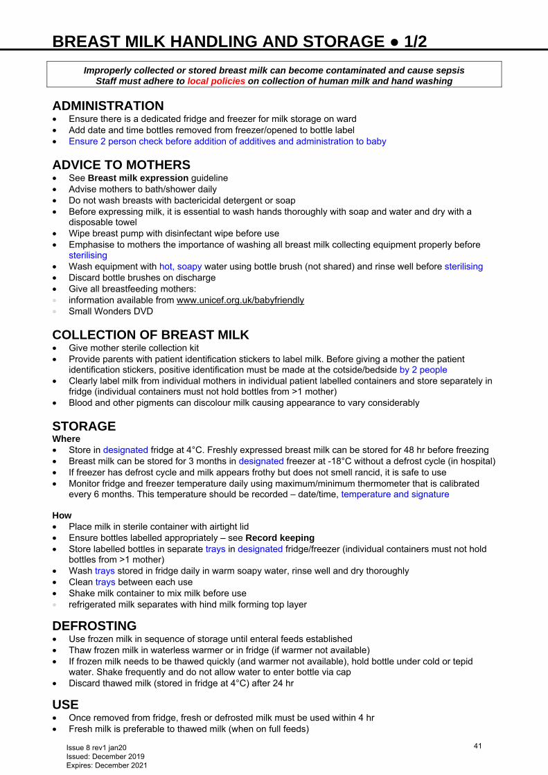
BREAST MILK HANDLING AND STORAGE ● 1/2
Improperly collected or stored breast milk can become contaminated and cause sepsis Staff must adhere to local policies on collection of human milk and hand washing
ADMINISTRATION • Ensure there is a dedicated fridge and freezer for milk storage on ward • Add date and time bottles removed from freezer/opened to bottle label • Ensure 2 person check before addition of additives and administration to baby
ADVICE TO MOTHERS • See Breast milk expression guideline • Advise mothers to bath/shower daily • Do not wash breasts with bactericidal detergent or soap • Before expressing milk, it is essential to wash hands thoroughly with soap and water and dry with a
disposable towel • Wipe breast pump with disinfectant wipe before use • Emphasise to mothers the importance of washing all breast milk collecting equipment properly before
sterilising • Wash equipment with hot, soapy water using bottle brush (not shared) and rinse well before sterilising • Discard bottle brushes on discharge • Give all breastfeeding mothers: • information available from www.unicef.org.uk/babyfriendly • Small Wonders DVD
COLLECTION OF BREAST MILK • Give mother sterile collection kit • Provide parents with patient identification stickers to label milk. Before giving a mother the patient
identification stickers, positive identification must be made at the cotside/bedside by 2 people • Clearly label milk from individual mothers in individual patient labelled containers and store separately in
fridge (individual containers must not hold bottles from >1 mother) • Blood and other pigments can discolour milk causing appearance to vary considerably STORAGE Where • Store in designated fridge at 4°C. Freshly expressed breast milk can be stored for 48 hr before freezing • Breast milk can be stored for 3 months in designated freezer at -18°C without a defrost cycle (in hospital) • If freezer has defrost cycle and milk appears frothy but does not smell rancid, it is safe to use • Monitor fridge and freezer temperature daily using maximum/minimum thermometer that is calibrated
every 6 months. This temperature should be recorded – date/time, temperature and signature How
• Place milk in sterile container with airtight lid • Ensure bottles labelled appropriately – see Record keeping • Store labelled bottles in separate trays in designated fridge/freezer (individual containers must not hold
bottles from >1 mother) • Wash trays stored in fridge daily in warm soapy water, rinse well and dry thoroughly • Clean trays between each use • Shake milk container to mix milk before use • refrigerated milk separates with hind milk forming top layer
DEFROSTING • Use frozen milk in sequence of storage until enteral feeds established • Thaw frozen milk in waterless warmer or in fridge (if warmer not available) • If frozen milk needs to be thawed quickly (and warmer not available), hold bottle under cold or tepid
water. Shake frequently and do not allow water to enter bottle via cap • Discard thawed milk (stored in fridge at 4°C) after 24 hr
USE • Once removed from fridge, fresh or defrosted milk must be used within 4 hr • Fresh milk is preferable to thawed milk (when on full feeds)
Issue 8 rev1 jan20 Issued: December 2019 Expires: December 2021
41

BREAST MILK HANDLING AND STORAGE ● 2/2 • Change continuous tube feeding (tubing between nasogastric tube and pump) every 4 hr • To minimise fat loss, position syringe delivering feed in semi-upright position • Bolus feeds − warm milk before giving using waterless warmer if available (to minimise fat loss) • Additives to be added to breast milk as close to feed time as possible • Only warm volume of milk required for feed. Store remainder in designated fridge TRANSPORTATION OF MILK Milk is often transported from: • Mother's home to hospital • transport in insulated container that can be easily cleaned • encourage mother to use coolant block to maintain stable temperature • Hospital-to-hospital • use rigid container for easy cleaning (e.g. cool box) and fill empty space with bubble wrap • use coolant block to maintain temperature and transfer to fridge as soon as possible on arrival in
NNU/ward
PRECAUTIONS • Wash hands thoroughly • Cover cuts and abrasions and wear gloves if necessary
RECORD KEEPING • Label all bottles/syringes with baby’s printed hospital label containing: • name and hospital number • date and time of expression • If mother expressing milk at home, provide her with a supply of printed hospital labels • Before giving breast milk, carry out a 2 person check of the label and cross-reference with baby’s identity
bracelet to ensure milk is not given to wrong child • If freezing MEBM label date and time frozen, and date and time of defrosting • See Breastfeeding guideline
STORAGE FOLLOWING DISCHARGE • Ensure parents take home all EBM in the fridge or freezer. If MEBM remains on unit and is in date,
transfer from fridge to freezer immediately – inform parents to collect as soon as possible • Give parents all EBM on discharge and ensure all milk is checked for correct baby details: • crosscheck labels on bottles with patient ID (second checker ideally mother) • if parents not present carry out 2 nurse check and document in patient notes • Discard milk stored in NNU freezer 1 month after discharge
Issue 8 rev1 jan20 Issued: December 2019 Expires: December 2021
42
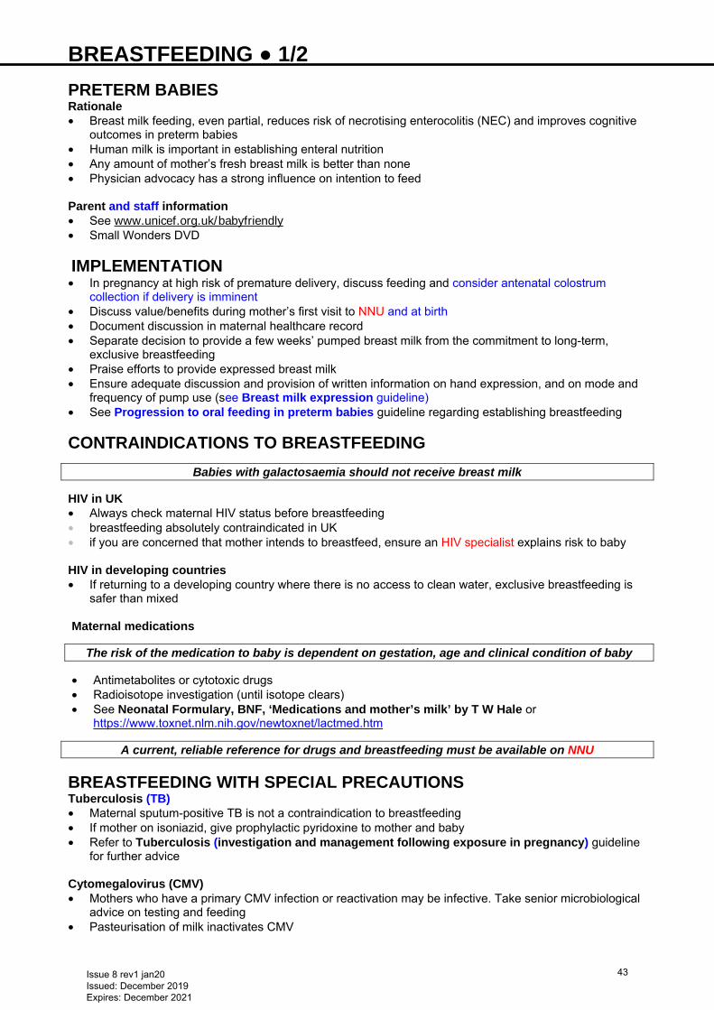
BREASTFEEDING ● 1/2 PRETERM BABIES Rationale • Breast milk feeding, even partial, reduces risk of necrotising enterocolitis (NEC) and improves cognitive
outcomes in preterm babies • Human milk is important in establishing enteral nutrition • Any amount of mother’s fresh breast milk is better than none • Physician advocacy has a strong influence on intention to feed Parent and staff information • See www.unicef.org.uk/babyfriendly • Small Wonders DVD
IMPLEMENTATION • In pregnancy at high risk of premature delivery, discuss feeding and consider antenatal colostrum
collection if delivery is imminent • Discuss value/benefits during mother’s first visit to NNU and at birth • Document discussion in maternal healthcare record • Separate decision to provide a few weeks’ pumped breast milk from the commitment to long-term,
exclusive breastfeeding • Praise efforts to provide expressed breast milk • Ensure adequate discussion and provision of written information on hand expression, and on mode and
frequency of pump use (see Breast milk expression guideline) • See Progression to oral feeding in preterm babies guideline regarding establishing breastfeeding
CONTRAINDICATIONS TO BREASTFEEDING
Babies with galactosaemia should not receive breast milk
HIV in UK • Always check maternal HIV status before breastfeeding • breastfeeding absolutely contraindicated in UK • if you are concerned that mother intends to breastfeed, ensure an HIV specialist explains risk to baby
HIV in developing countries • If returning to a developing country where there is no access to clean water, exclusive breastfeeding is
safer than mixed Maternal medications
The risk of the medication to baby is dependent on gestation, age and clinical condition of baby
• Antimetabolites or cytotoxic drugs • Radioisotope investigation (until isotope clears) • See Neonatal Formulary, BNF, ‘Medications and mother’s milk’ by T W Hale or
https://www.toxnet.nlm.nih.gov/newtoxnet/lactmed.htm
A current, reliable reference for drugs and breastfeeding must be available on NNU
BREASTFEEDING WITH SPECIAL PRECAUTIONS Tuberculosis (TB) • Maternal sputum-positive TB is not a contraindication to breastfeeding • If mother on isoniazid, give prophylactic pyridoxine to mother and baby • Refer to Tuberculosis (investigation and management following exposure in pregnancy) guideline
for further advice Cytomegalovirus (CMV) • Mothers who have a primary CMV infection or reactivation may be infective. Take senior microbiological
advice on testing and feeding • Pasteurisation of milk inactivates CMV
Issue 8 rev1 jan20 Issued: December 2019 Expires: December 2021
43
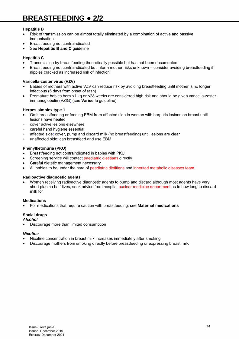
BREASTFEEDING ● 2/2 Hepatitis B • Risk of transmission can be almost totally eliminated by a combination of active and passive
immunisation • Breastfeeding not contraindicated • See Hepatitis B and C guideline
Hepatitis C • Transmission by breastfeeding theoretically possible but has not been documented • Breastfeeding not contraindicated but inform mother risks unknown – consider avoiding breastfeeding if
nipples cracked as increased risk of infection Varicella-zoster virus (VZV) • Babies of mothers with active VZV can reduce risk by avoiding breastfeeding until mother is no longer
infectious (5 days from onset of rash) • Premature babies born <1 kg or <28 weeks are considered high risk and should be given varicella-zoster
immunoglobulin (VZIG) (see Varicella guideline) Herpes simplex type 1 • Omit breastfeeding or feeding EBM from affected side in women with herpetic lesions on breast until
lesions have healed • cover active lesions elsewhere • careful hand hygiene essential • affected side: cover, pump and discard milk (no breastfeeding) until lesions are clear • unaffected side: can breastfeed and use EBM Phenylketonuria (PKU) • Breastfeeding not contraindicated in babies with PKU • Screening service will contact paediatric dietitians directly • Careful dietetic management necessary • All babies to be under the care of paediatric dietitians and inherited metabolic diseases team Radioactive diagnostic agents • Women receiving radioactive diagnostic agents to pump and discard although most agents have very
short plasma half-lives, seek advice from hospital nuclear medicine department as to how long to discard milk for
Medications • For medications that require caution with breastfeeding, see Maternal medications Social drugs Alcohol • Discourage more than limited consumption Nicotine • Nicotine concentration in breast milk increases immediately after smoking • Discourage mothers from smoking directly before breastfeeding or expressing breast milk
Issue 8 rev1 jan20 Issued: December 2019 Expires: December 2021
44
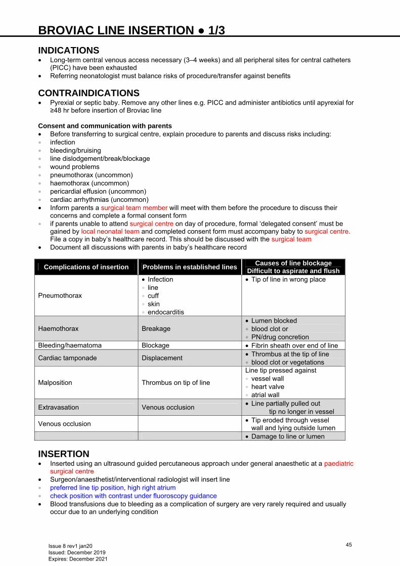
BROVIAC LINE INSERTION ● 1/3 INDICATIONS • Long-term central venous access necessary (3–4 weeks) and all peripheral sites for central catheters
(PICC) have been exhausted • Referring neonatologist must balance risks of procedure/transfer against benefits
CONTRAINDICATIONS • Pyrexial or septic baby. Remove any other lines e.g. PICC and administer antibiotics until apyrexial for
≥48 hr before insertion of Broviac line Consent and communication with parents • Before transferring to surgical centre, explain procedure to parents and discuss risks including: • infection • bleeding/bruising • line dislodgement/break/blockage • wound problems • pneumothorax (uncommon) • haemothorax (uncommon) • pericardial effusion (uncommon) • cardiac arrhythmias (uncommon) • Inform parents a surgical team member will meet with them before the procedure to discuss their
concerns and complete a formal consent form • if parents unable to attend surgical centre on day of procedure, formal ‘delegated consent’ must be
gained by local neonatal team and completed consent form must accompany baby to surgical centre. File a copy in baby’s healthcare record. This should be discussed with the surgical team
• Document all discussions with parents in baby’s healthcare record
Complications of insertion Problems in established lines Causes of line blockage
Difficult to aspirate and flush
Pneumothorax
• Infection • line • cuff • skin • endocarditis
• Tip of line in wrong place
Haemothorax Breakage • Lumen blocked • blood clot or • PN/drug concretion
Bleeding/haematoma Blockage • Fibrin sheath over end of line
Cardiac tamponade Displacement • Thrombus at the tip of line • blood clot or vegetations
Malposition Thrombus on tip of line
Line tip pressed against • vessel wall • heart valve • atrial wall
Extravasation Venous occlusion • Line partially pulled out • tip no longer in vessel
Venous occlusion • Tip eroded through vessel wall and lying outside lumen
• Damage to line or lumen
INSERTION • Inserted using an ultrasound guided percutaneous approach under general anaesthetic at a paediatric
surgical centre • Surgeon/anaesthetist/interventional radiologist will insert line • preferred line tip position, high right atrium • check position with contrast under fluoroscopy guidance • Blood transfusions due to bleeding as a complication of surgery are very rarely required and usually
occur due to an underlying condition
Issue 8 rev1 jan20 Issued: December 2019 Expires: December 2021
45
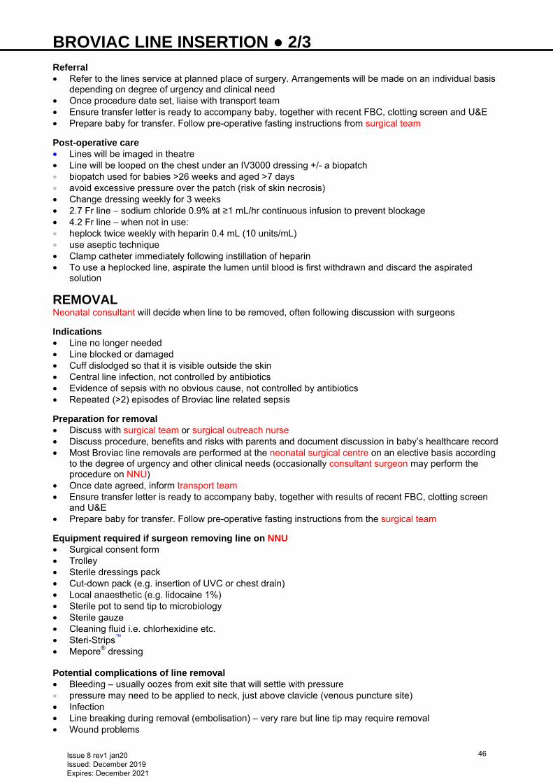
BROVIAC LINE INSERTION ● 2/3 Referral • Refer to the lines service at planned place of surgery. Arrangements will be made on an individual basis
depending on degree of urgency and clinical need • Once procedure date set, liaise with transport team • Ensure transfer letter is ready to accompany baby, together with recent FBC, clotting screen and U&E • Prepare baby for transfer. Follow pre-operative fasting instructions from surgical team Post-operative care • Lines will be imaged in theatre • Line will be looped on the chest under an IV3000 dressing +/- a biopatch • biopatch used for babies >26 weeks and aged >7 days • avoid excessive pressure over the patch (risk of skin necrosis) • Change dressing weekly for 3 weeks • 2.7 Fr line − sodium chloride 0.9% at ≥1 mL/hr continuous infusion to prevent blockage • 4.2 Fr line − when not in use: • heplock twice weekly with heparin 0.4 mL (10 units/mL) • use aseptic technique • Clamp catheter immediately following instillation of heparin • To use a heplocked line, aspirate the lumen until blood is first withdrawn and discard the aspirated
solution
REMOVAL Neonatal consultant will decide when line to be removed, often following discussion with surgeons
Indications • Line no longer needed • Line blocked or damaged • Cuff dislodged so that it is visible outside the skin • Central line infection, not controlled by antibiotics • Evidence of sepsis with no obvious cause, not controlled by antibiotics • Repeated (>2) episodes of Broviac line related sepsis Preparation for removal • Discuss with surgical team or surgical outreach nurse • Discuss procedure, benefits and risks with parents and document discussion in baby’s healthcare record • Most Broviac line removals are performed at the neonatal surgical centre on an elective basis according
to the degree of urgency and other clinical needs (occasionally consultant surgeon may perform the procedure on NNU)
• Once date agreed, inform transport team • Ensure transfer letter is ready to accompany baby, together with results of recent FBC, clotting screen
and U&E • Prepare baby for transfer. Follow pre-operative fasting instructions from the surgical team Equipment required if surgeon removing line on NNU • Surgical consent form • Trolley • Sterile dressings pack • Cut-down pack (e.g. insertion of UVC or chest drain) • Local anaesthetic (e.g. lidocaine 1%) • Sterile pot to send tip to microbiology • Sterile gauze • Cleaning fluid i.e. chlorhexidine etc. • Steri-Strips™ • Mepore® dressing Potential complications of line removal • Bleeding – usually oozes from exit site that will settle with pressure • pressure may need to be applied to neck, just above clavicle (venous puncture site) • Infection • Line breaking during removal (embolisation) – very rare but line tip may require removal • Wound problems
Issue 8 rev1 jan20 Issued: December 2019 Expires: December 2021
46
BROVIAC LINE INSERTION ● 3/3 Embolised fractured line • Very rare but occasionally line will break causing the tip to embolise into the right atrium or pulmonary
artery • If line stops working, perform chest X-ray • Requires retrieval by interventional cardiologist at paediatric surgical centre. Liaise with either on-call
paediatric surgeon, cardiologist, or vascular access team (line service) at planned place of surgery
USEFUL INFORMATION • http://www.bch.nhs.uk/content/neonatal-surgery • http://www.bch.nhs.uk/find-us/maps-directions • http://www.e-lfh.org.uk/programmes/paediatric-surgery/
Issue 8 rev1 jan20 Issued: December 2019 Expires: December 2021
47
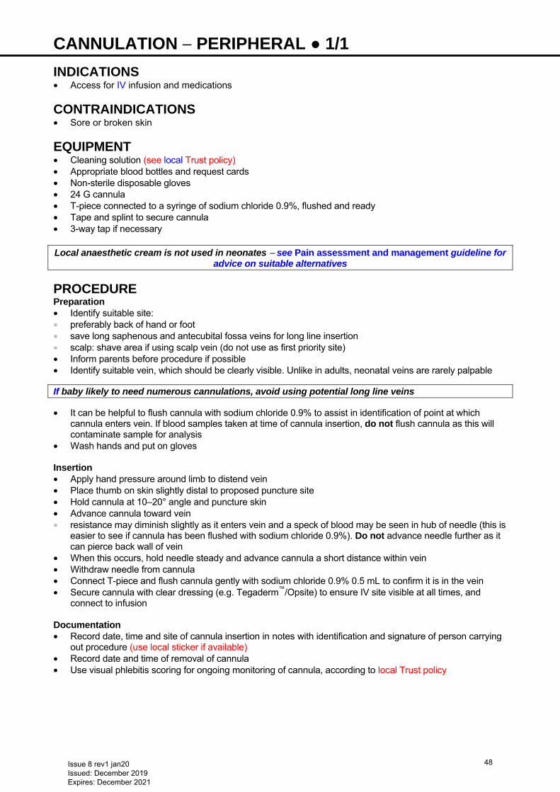
CANNULATION − PERIPHERAL ● 1/1 INDICATIONS • Access for IV infusion and medications
CONTRAINDICATIONS • Sore or broken skin
EQUIPMENT • Cleaning solution (see local Trust policy) • Appropriate blood bottles and request cards • Non-sterile disposable gloves • 24 G cannula • T-piece connected to a syringe of sodium chloride 0.9%, flushed and ready • Tape and splint to secure cannula • 3-way tap if necessary Local anaesthetic cream is not used in neonates − see Pain assessment and management guideline for
advice on suitable alternatives
PROCEDURE Preparation • Identify suitable site: • preferably back of hand or foot • save long saphenous and antecubital fossa veins for long line insertion • scalp: shave area if using scalp vein (do not use as first priority site) • Inform parents before procedure if possible • Identify suitable vein, which should be clearly visible. Unlike in adults, neonatal veins are rarely palpable
If baby likely to need numerous cannulations, avoid using potential long line veins
• It can be helpful to flush cannula with sodium chloride 0.9% to assist in identification of point at which cannula enters vein. If blood samples taken at time of cannula insertion, do not flush cannula as this will contaminate sample for analysis
• Wash hands and put on gloves Insertion • Apply hand pressure around limb to distend vein • Place thumb on skin slightly distal to proposed puncture site • Hold cannula at 10–20° angle and puncture skin • Advance cannula toward vein • resistance may diminish slightly as it enters vein and a speck of blood may be seen in hub of needle (this is
easier to see if cannula has been flushed with sodium chloride 0.9%). Do not advance needle further as it can pierce back wall of vein
• When this occurs, hold needle steady and advance cannula a short distance within vein • Withdraw needle from cannula • Connect T-piece and flush cannula gently with sodium chloride 0.9% 0.5 mL to confirm it is in the vein • Secure cannula with clear dressing (e.g. Tegaderm™/Opsite) to ensure IV site visible at all times, and
connect to infusion Documentation • Record date, time and site of cannula insertion in notes with identification and signature of person carrying
out procedure (use local sticker if available) • Record date and time of removal of cannula • Use visual phlebitis scoring for ongoing monitoring of cannula, according to local Trust policy
Issue 8 rev1 jan20 Issued: December 2019 Expires: December 2021
48
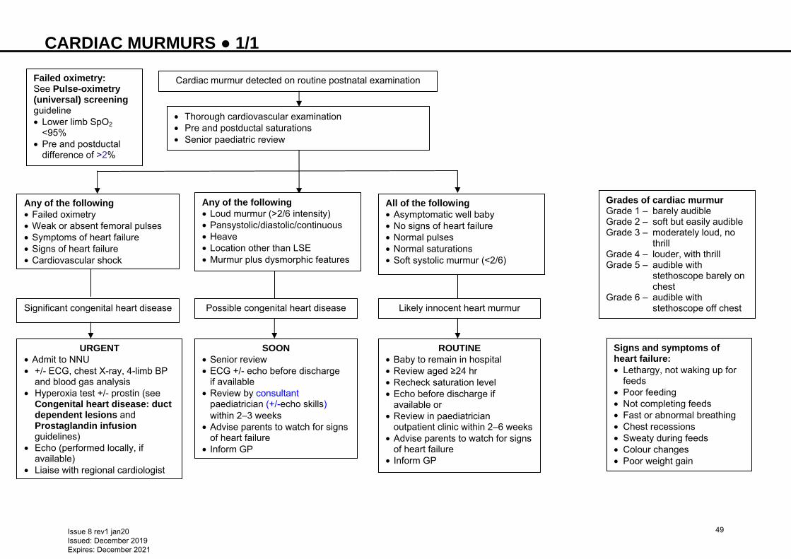
CARDIAC MURMURS ● 1/1
S w3ns
Cardiac murmur detected on routine postnatal examination
Any of the following • Failed oximetry • Weak or absent femoral pulses • Symptoms of heart failure • Signs of heart failure • Cardiovascular shock
• Thorough cardiovascular examination • Pre and postductal saturations • Senior paediatric review
Significant congenital heart disease
Failed oximetry: See Pulse-oximetry (universal) screening guideline • Lower limb SpO2
<95% • Pre and postductal
difference of >2%
Any of the following • Loud murmur (>2/6 intensity) • Pansystolic/diastolic/continuous • Heave • Location other than LSE • Murmur plus dysmorphic features
All of the following • Asymptomatic well baby • No signs of heart failure • Normal pulses • Normal saturations • Soft systolic murmur (<2/6)
Possible congenital heart disease Likely innocent heart murmur
URGENT • Admit to NNU • +/- ECG, chest X-ray, 4-limb BP
and blood gas analysis • Hyperoxia test +/- prostin (see
Congenital heart disease: duct dependent lesions and Prostaglandin infusion guidelines)
• Echo (performed locally, if available)
• Liaise with regional cardiologist
SOON • Senior review • ECG +/- echo before discharge
if available • Review by consultant
paediatrician (+/-echo skills) within 2−3 weeks
• Advise parents to watch for signs of heart failure
• Inform GP
ROUTINE • Baby to remain in hospital • Review aged ≥24 hr • Recheck saturation level • Echo before discharge if
available or • Review in paediatrician
outpatient clinic within 2–6 weeks • Advise parents to watch for signs
of heart failure • Inform GP
Grades of cardiac murmur Grade 1 – barely audible Grade 2 – soft but easily audible Grade 3 – moderately loud, no thrill Grade 4 – louder, with thrill Grade 5 – audible with stethoscope barely on chest Grade 6 – audible with stethoscope off chest
Signs and symptoms of heart failure: • Lethargy, not waking up for
feeds • Poor feeding • Not completing feeds • Fast or abnormal breathing • Chest recessions • Sweaty during feeds • Colour changes • Poor weight gain
Issue 8 rev1 jan20 Issued: December 2019 Expires: December 2021
49
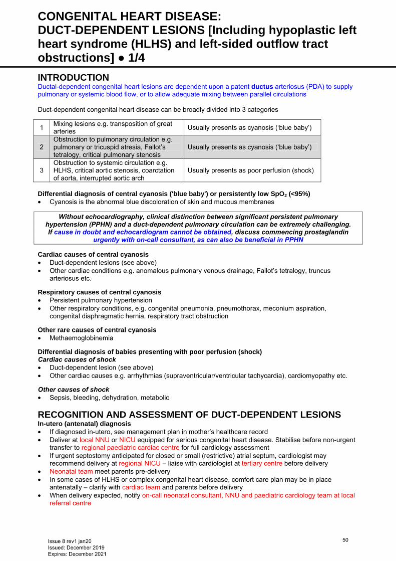
CONGENITAL HEART DISEASE: DUCT-DEPENDENT LESIONS [Including hypoplastic left heart syndrome (HLHS) and left-sided outflow tract obstructions] ● 1/4 INTRODUCTION Ductal-dependent congenital heart lesions are dependent upon a patent ductus arteriosus (PDA) to supply pulmonary or systemic blood flow, or to allow adequate mixing between parallel circulations Duct-dependent congenital heart disease can be broadly divided into 3 categories
1 Mixing lesions e.g. transposition of great arteries
Usually presents as cyanosis (‘blue baby’)
2 Obstruction to pulmonary circulation e.g. pulmonary or tricuspid atresia, Fallot’s tetralogy, critical pulmonary stenosis
Usually presents as cyanosis (‘blue baby’)
3 Obstruction to systemic circulation e.g. HLHS, critical aortic stenosis, coarctation of aorta, interrupted aortic arch
Usually presents as poor perfusion (shock)
Differential diagnosis of central cyanosis ('blue baby') or persistently low SpO2 (<95%) • Cyanosis is the abnormal blue discoloration of skin and mucous membranes
Without echocardiography, clinical distinction between significant persistent pulmonary hypertension (PPHN) and a duct-dependent pulmonary circulation can be extremely challenging. If cause in doubt and echocardiogram cannot be obtained, discuss commencing prostaglandin
urgently with on-call consultant, as can also be beneficial in PPHN
Cardiac causes of central cyanosis • Duct-dependent lesions (see above) • Other cardiac conditions e.g. anomalous pulmonary venous drainage, Fallot’s tetralogy, truncus
arteriosus etc.
Respiratory causes of central cyanosis • Persistent pulmonary hypertension • Other respiratory conditions, e.g. congenital pneumonia, pneumothorax, meconium aspiration,
congenital diaphragmatic hernia, respiratory tract obstruction Other rare causes of central cyanosis • Methaemoglobinemia Differential diagnosis of babies presenting with poor perfusion (shock) Cardiac causes of shock • Duct-dependent lesion (see above) • Other cardiac causes e.g. arrhythmias (supraventricular/ventricular tachycardia), cardiomyopathy etc.
Other causes of shock • Sepsis, bleeding, dehydration, metabolic
RECOGNITION AND ASSESSMENT OF DUCT-DEPENDENT LESIONS In-utero (antenatal) diagnosis • If diagnosed in-utero, see management plan in mother’s healthcare record • Deliver at local NNU or NICU equipped for serious congenital heart disease. Stabilise before non-urgent
transfer to regional paediatric cardiac centre for full cardiology assessment • If urgent septostomy anticipated for closed or small (restrictive) atrial septum, cardiologist may
recommend delivery at regional NICU – liaise with cardiologist at tertiary centre before delivery • Neonatal team meet parents pre-delivery • In some cases of HLHS or complex congenital heart disease, comfort care plan may be in place
antenatally – clarify with cardiac team and parents before delivery • When delivery expected, notify on-call neonatal consultant, NNU and paediatric cardiology team at local
referral centre
Issue 8 rev1 jan20 Issued: December 2019 Expires: December 2021
50
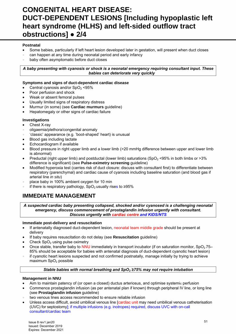
CONGENITAL HEART DISEASE: DUCT-DEPENDENT LESIONS [Including hypoplastic left heart syndrome (HLHS) and left-sided outflow tract obstructions] ● 2/4 Postnatal • Some babies, particularly if left heart lesion developed later in gestation, will present when duct closes • can happen at any time during neonatal period and early infancy • baby often asymptomatic before duct closes
A baby presenting with cyanosis or shock is a neonatal emergency requiring consultant input. These babies can deteriorate very quickly
Symptoms and signs of duct-dependent cardiac disease • Central cyanosis and/or SpO2 <95% • Poor perfusion and shock • Weak or absent femoral pulses • Usually limited signs of respiratory distress • Murmur (in some) (see Cardiac murmurs guideline) • Hepatomegaly or other signs of cardiac failure Investigations • Chest X-ray • oligaemia/plethora/congenital anomaly • ‘classic’ appearance (e.g. ‛boot-shaped’ heart) is unusual • Blood gas including lactate • Echocardiogram if available • Blood pressure in right upper limb and a lower limb (>20 mmHg difference between upper and lower limb
is abnormal) • Preductal (right upper limb) and postductal (lower limb) saturations (SpO2 <95% in both limbs or >3%
difference is significant) (see Pulse-oximetry screening guideline) • Modified hyperoxia test (carries risk of duct closure: discuss with consultant first) to differentiate between
respiratory (parenchymal) and cardiac cause of cyanosis including baseline saturation (and blood gas if arterial line in situ)
• place baby in 100% ambient oxygen for 10 min • if there is respiratory pathology, SpO2 usually rises to ≥95%
IMMEDIATE MANAGEMENT
A suspected cardiac baby presenting collapsed, shocked and/or cyanosed is a challenging neonatal emergency, discuss commencement of prostaglandin infusion urgently with consultant.
Discuss urgently with cardiac centre and KIDS/NTS
Immediate post-delivery and resuscitation • If antenatally diagnosed duct-dependent lesion, neonatal team middle grade should be present at
delivery • If baby requires resuscitation do not delay (see Resuscitation guideline) • Check SpO2 using pulse oximetry • Once stable, transfer baby to NNU immediately in transport incubator (if on saturation monitor, SpO2 75–
85% should be acceptable for babies with antenatal diagnosis of duct-dependent cyanotic heart lesion) • if cyanotic heart lesions suspected and not confirmed postnatally, manage initially by trying to achieve
maximum SpO2 possible
Stable babies with normal breathing and SpO2 ≥75% may not require intubation Management in NNU • Aim to maintain patency of (or open a closed) ductus arteriosus, and optimise systemic perfusion • Commence prostaglandin infusion (as per antenatal plan if known) through peripheral IV line, or long line
(see Prostaglandin infusion guideline) • two venous lines access recommended to ensure reliable infusion • Unless access difficult, avoid umbilical venous line [cardiac unit may need umbilical venous catheterisation
(UVC) for septostomy]; if multiple infusions (e.g. inotropes) required, discuss UVC with on-call consultant/cardiac team
Issue 8 rev1 jan20 Issued: December 2019 Expires: December 2021
51

CONGENITAL HEART DISEASE: DUCT-DEPENDENT LESIONS [Including hypoplastic left heart syndrome (HLHS) and left-sided outflow tract obstructions] ● 3/4 • Use dinoprostone (prostaglandin E2, prostin E2) (see Prostaglandin infusion guideline) • start IV infusion at 5−15 nanogram/kg/min as indicated; dose may be increased up to
50 nanogram/kg/min if no response within 1 hr • oral dinoprostone used temporarily on very rare occasions when IV access is extremely difficult (see
Neonatal Formulary) • if dinoprostone not available, use prostaglandin E1 (Alprostadil) (see Neonatal Formulary for dose) • make fresh solution every 24 hr • Be vigilant: if apnoea occurs secondary to a prostaglandin infusion, intubate baby but do not reduce
infusion dose (see Intubation guideline) • Discuss management with cardiac team at regional paediatric cardiac centre • Echocardiogram if available • If evidence of hypoperfusion (e.g. base deficit >5 or lactate >3, or hypotension or cool peripheries), give
sodium chloride 0.9% 10 mL/kg IV fluid bolus Monitor • SpO2 • Heart rate and ECG • Blood gases (including lactate) and avoid acidosis • Blood pressure (preferably using a peripheral arterial cannula – avoid umbilical lines − if UAC required,
discuss with on-call consultant) • Avoid hypothermia Ventilation (see also Ventilation guidelines) Indications • If intubation not needed as emergency, discuss with paediatric intensive care unit (PICU)/cardiac team • Severe hypoxaemia, acidosis and cardiorespiratory failure • Apnoea after starting prostaglandin infusion • dose >20 nanogram/kg/min (review need for such a high dosage in stable baby) • Features of high pulmonary flow in case of HLHS • Elective ventilation, if preferred by paediatric cardiologist or retrieval team lead
Technique • Use sedation/muscle relaxants as needed • Avoid hyperventilation − can increase pulmonary blood flow • Use supplemental oxygen judiciously if SpO2 <75% • Initial settings: PEEP 4–5 cm H2O, low mean airway pressure, tidal volume 4–6 mL/kg and FiO2 0.21,
adjusted accordingly • Aim for: • PaCO2 5–7 kPa • PaO2 4–6 kPa • pH 7.30–7.40 • SpO2 75–85% (although many will run higher in room air)
Inotropes • If signs of peripheral under-perfusion, discuss using fluid boluses and inotropes (e.g. dobutamine,
milrinone etc.) with cardiac centre • Arrange local echocardiography (if available) to assess contractility Restrictive atrial septum • Signs: • severe cyanosis • cool peripheries • pallor • respiratory distress • X-ray signs of pulmonary oedema with relatively normal heart size. In contrast, if atrial septum is non-
restrictive, pulmonary congestion with cardiomegaly and prominent right heart border is likely • May require balloon atrial septostomy as an urgent procedure. If too unstable for transfer or no beds at
cardiac centre, cardiac team may perform as emergency outreach procedure in NNU
Issue 8 rev1 jan20 Issued: December 2019 Expires: December 2021
52
CONGENITAL HEART DISEASE: DUCT-DEPENDENT LESIONS [Including hypoplastic left heart syndrome (HLHS) and left-sided outflow tract obstructions] ● 4/4 High pulmonary blood flow (especially in left-sided lesions such as HLHS) Presentation • If there is too much pulmonary blood flow due to pulmonary ‘steal’ phenomenon, baby may have: • high or near normal saturations • metabolic acidosis with a rising lactate • low blood pressure (especially low diastolic) • cool peripheries • tachycardia Management • Aim is to improve perfusion and acidosis by balancing systemic versus pulmonary circulation • Discuss urgently with cardiac centre • Intubate and ventilate (technique as above) • Fluid boluses and inotropes as needed
PARENT COMMUNICATION • It is important that parents are kept informed and updated regularly during management • Parent leaflets for specific heart conditions are available from British Heart Foundation website
www.bhf.org.uk/
Issue 8 rev1 jan20 Issued: December 2019 Expires: December 2021
53
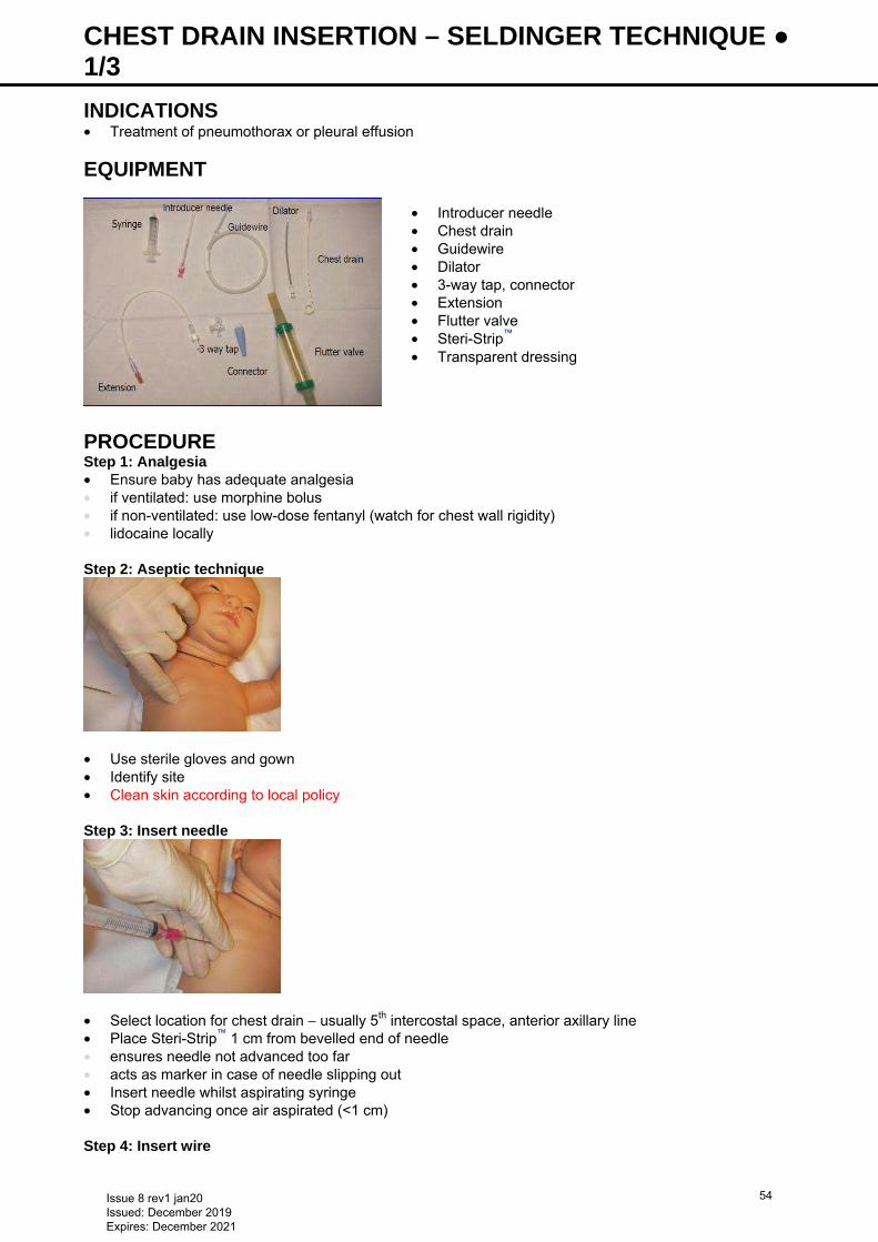
CHEST DRAIN INSERTION – SELDINGER TECHNIQUE ● 1/3 INDICATIONS • Treatment of pneumothorax or pleural effusion
EQUIPMENT
PROCEDURE Step 1: Analgesia • Ensure baby has adequate analgesia • if ventilated: use morphine bolus • if non-ventilated: use low-dose fentanyl (watch for chest wall rigidity) • lidocaine locally Step 2: Aseptic technique
• Use sterile gloves and gown • Identify site • Clean skin according to local policy Step 3: Insert needle
• Select location for chest drain − usually 5th intercostal space, anterior axillary line • Place Steri-Strip™ 1 cm from bevelled end of needle • ensures needle not advanced too far • acts as marker in case of needle slipping out • Insert needle whilst aspirating syringe • Stop advancing once air aspirated (<1 cm) Step 4: Insert wire
• Introducer needle • Chest drain • Guidewire • Dilator • 3-way tap, connector • Extension • Flutter valve • Steri-Strip™ • Transparent dressing
Issue 8 rev1 jan20 Issued: December 2019 Expires: December 2021
54
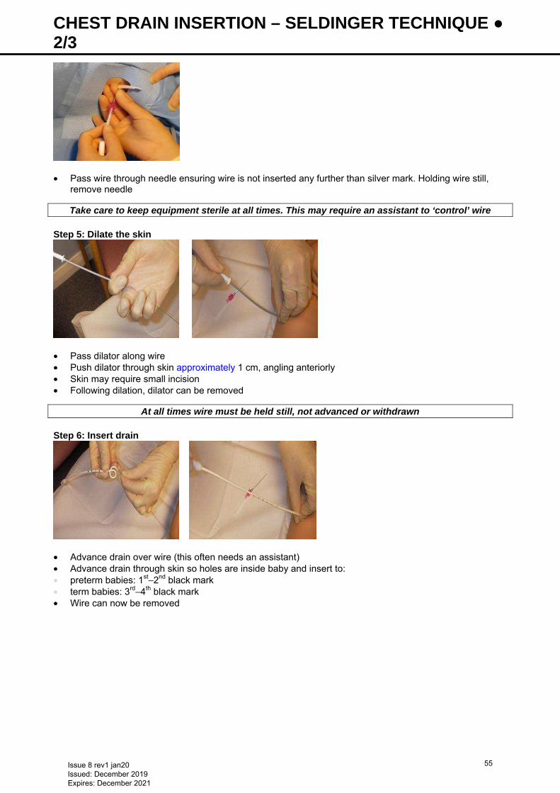
CHEST DRAIN INSERTION – SELDINGER TECHNIQUE ● 2/3
• Pass wire through needle ensuring wire is not inserted any further than silver mark. Holding wire still,
remove needle
Take care to keep equipment sterile at all times. This may require an assistant to ‘control’ wire Step 5: Dilate the skin
• Pass dilator along wire • Push dilator through skin approximately 1 cm, angling anteriorly • Skin may require small incision • Following dilation, dilator can be removed
At all times wire must be held still, not advanced or withdrawn Step 6: Insert drain
• Advance drain over wire (this often needs an assistant) • Advance drain through skin so holes are inside baby and insert to: • preterm babies: 1st−2nd black mark • term babies: 3rd−4th black mark • Wire can now be removed
Issue 8 rev1 jan20 Issued: December 2019 Expires: December 2021
55
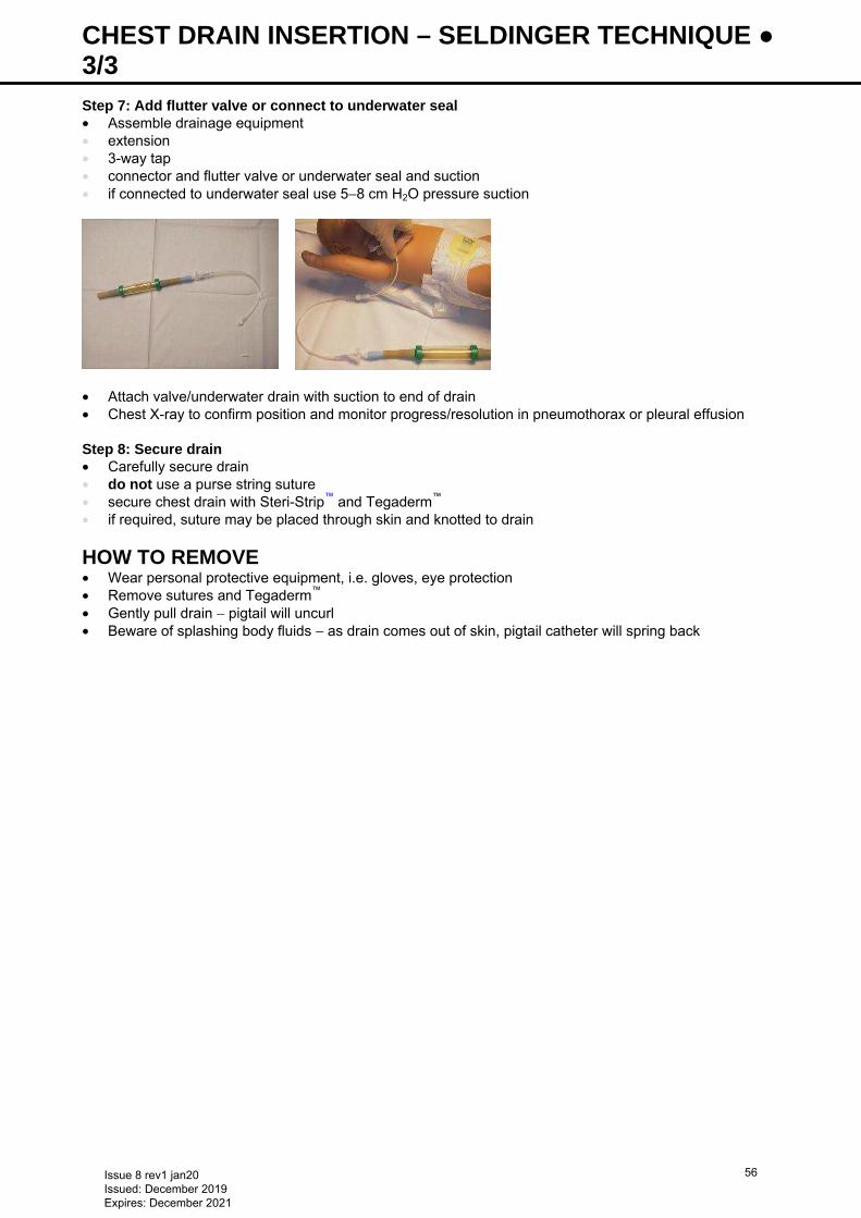
CHEST DRAIN INSERTION – SELDINGER TECHNIQUE ● 3/3 Step 7: Add flutter valve or connect to underwater seal • Assemble drainage equipment • extension • 3-way tap • connector and flutter valve or underwater seal and suction • if connected to underwater seal use 5−8 cm H2O pressure suction
• Attach valve/underwater drain with suction to end of drain • Chest X-ray to confirm position and monitor progress/resolution in pneumothorax or pleural effusion Step 8: Secure drain • Carefully secure drain • do not use a purse string suture • secure chest drain with Steri-Strip™ and Tegaderm™ • if required, suture may be placed through skin and knotted to drain
HOW TO REMOVE • Wear personal protective equipment, i.e. gloves, eye protection • Remove sutures and Tegaderm™ • Gently pull drain − pigtail will uncurl • Beware of splashing body fluids − as drain comes out of skin, pigtail catheter will spring back
Issue 8 rev1 jan20 Issued: December 2019 Expires: December 2021
56
CHEST DRAIN INSERTION − TRADITIONAL ● 1/2 INDICATIONS • Treatment of pneumothorax or pleural effusion
EQUIPMENT • Sterile dressing pack • Cleaning solution as per unit policy and wash off with sodium chloride 0.9% once dried for babies <26
weeks’ gestation • Lidocaine 1%, with syringe and needle for preparation and injection • Chest drains size FG 8,10,12 (use largest possible depending on size of baby) • Low pressure suction unit • Scalpel and fine straight blade (size 11) • Fine blunt forceps • Underwater seal chest drainage bottle and tubing or flutter (Heimlich) valve • Steri-Strip™ and transparent dressing (e.g. Opsite/Tegaderm™)
SITES • Site of insertion depends on position of pneumothorax • preferred site is in anterior axillary line, between 4th and 6th intercostal space, to conceal subsequent
scarring and avoid interference with breast development • alternative site is just lateral to midclavicular line, in 2nd or 3rd intercostal space • if pneumothorax does not drain satisfactorily, it may be necessary to insert >1 drain • for pleural effusion, use midaxillary line between 4th and 5th intercostal spaces, and direct drain
posteriorly
PROCEDURE Preparation and position of baby • Inform parents and obtain verbal consent as recommended by BAPM (unless emergency procedure) • Use 10–12 FG pleural catheter (small babies may need 8 FG) • Position baby supine and flat with affected side slightly tilted up (e.g. by using a folded blanket) • Prepare skin with full aseptic technique • Infiltrate with lidocaine 1%, even in babies being given systemic analgesia Insertion of tube • Make small incision in skin with scalpel at lower edge of intercostal space to avoid injury to intercostal
vessels • Dissect bluntly with fine forceps through intercostal muscle and pleura • Use fine forceps to gently advance tip of catheter • Push and twist tube gently through incision into pleural space • Insert chest drain 2–3 cm for small preterm and 3 cm for term babies • Use of trocar not generally recommended. If used (in bigger baby), protect lung by clamping artery
forceps onto trocar 1 cm from the tip • Connect tube to prepared underwater seal or flutter (Heimlich) valve • Manipulate tube gently so that tip lies anteriorly in thoracic cavity for pneumothorax, and posteriorly for
effusion • Secure tube with Steri-Strip™, and cover with gauze dressing. A suture may be required; do not use
purse-string suture • Secure tube to chest wall using suitable tape (Opsite/Tegaderm™)
AFTERCARE • Check bubbling or oscillation of water column seen with every inspiration • Check tube position with chest X-ray (consider lateral X-ray to confirm position) Suction • If bubbling poor and X-ray confirms drain is in correct position but pneumothorax not fully draining on X-
ray or cold light, apply continuous suction of 5–10 cm H2O. Thoracic suction is better suited for this purpose than routine wall suction. Occasionally, a second drain may be necessary
Flutter valve • As an alternative to underwater chest drain system, especially during transport, a flutter valve can be
used
Issue 8 rev1 jan20 Issued: December 2019 Expires: December 2021
57
CHEST DRAIN INSERTION − TRADITIONAL ● 2/2 Document • Record presence of bubbling (continuous/intermittent/none) on nursing care chart • Record with nursing observations, bubbling and/or oscillation of water column, or fluttering of valve seen
with every inspiration
REMOVAL OF CHEST DRAIN • Remove when no bubbling or oscillation of water column has occurred for 24 hr • Clamp chest drain for 12 hr and repeat chest X-ray before removal. While removing drain, ask an
assistant to hold wound edges close together • After removing drain, close wound with Steri-Strip™; a suture is seldom necessary • Close clinical observation after removal of drain is sufficient to diagnose reaccumulation of the air leak,
routine chest X-ray not generally warranted
Issue 8 rev1 jan20 Issued: December 2019 Expires: December 2021
58
CHEST PHYSIOTHERAPY ● 1/3 INTRODUCTION • The neonatal toolkit recommends that all units caring for babies requiring intensive and high dependency
care who provide chest clearance should have access to a paediatric respiratory physiotherapist/specialist neonatal physiotherapist
• All staff undertaking percussion must be competent and seek advice where required • Before considering percussion ensure all other airway clearance options optimised, including: • variety and frequency of position changes • adequate hydration to prevent tenacious secretions • Ensure best practice suction technique • Contact respiratory physiotherapist to review babies with difficulties clearing secretions
PERCUSSION Definition • Rhythmic patting over chest wall using a palm cup percussor to generate pressure changes stimulating
mucus clearance by ciliary stimulation Indications • Tenacious secretions not cleared effectively with suction • Signs of respiratory compromise • changes in ventilation suggestive of secretion retention (e.g. tidal volumes, peak pressures) • decreased SpO2/PaO2 • increased PaCO2 • Auscultation findings • Chest X-ray changes e.g. focal collapse/consolidation • Consider neuromuscular pathologies resulting in poor airway protection, and respiratory conditions such
as cystic fibrosis. These conditions may require prophylactic physiotherapy and parental training before discharge – refer to physiotherapist
Contraindications • Cardiovascular instability • Undrained pneumothorax/bullae • Pulmonary interstitial emphysema (PIE) • Acute pulmonary haemorrhage • Metabolic bone disease/fractured ribs • Intraventricular haemorrhage (IVH) within 48 hr • Extreme prematurity (<1500 g/<26 weeks’ gestation) in first week of life • Platelet count <50 × 109/L and/or prolonged clotting and/or active bleeding Precautions • Poor skin integrity • Platelet count <100 × 109/L • Avoid chest drain sites and Broviac lines/proximity of wounds/stomas • Effectiveness reduced in chest wall oedema • Distended abdomen • Recent cranial or eye surgery (including ROP laser surgery) • Acute necrotising enterocolitis • Recent surfactant therapy • HFOV (percussion unlikely to be effective)
PROCEDURE • Always assess cardiorespiratory status before intervention • Perform with continuous monitoring • Pre-oxygenation with increased pressure/rate where necessary • Ensure nesting and developmental care support throughout procedure (see Developmental care and
Positioning guidelines) • Plan treatment episodes pre-feed or >30 min post-feed • Preterm babies should not receive routine physiotherapy treatment • Minimise stress responses throughout procedure
Issue 8 rev1 jan20 Issued: December 2019 Expires: December 2021
59
CHEST PHYSIOTHERAPY ● 2/3 Positioning • See Positioning guideline • Do not disconnect baby from the ventilator for a turn • A variety of positions is the most important intervention for optimising ventilation distribution and
mobilising secretions. Unless contraindicated use: • kangaroo care • supine • prone • semi-prone • full side-lying • side tilts/cot tilts • If secretion retention problematic increase frequency of turns • Do not leave baby in same position for prolonged periods – risk of collapse of dependant (lower) lung • Different positions can be used to target specific areas of collapse and/or consolidation • Ventilation/perfusion mismatch may necessitate increasing oxygen delivery • Never use head-down tilt due to risk of IVH/reflux/respiratory compromise Percussion • Stabilise head with 1 hand at all times • Ensure whole circumference of the percussor makes contact with baby’s chest, ideally directly on baby’s
skin. If not practical, a layer of vest is acceptable. The pressure should not cause any movement of baby/skin reaction
• Ideal rate approximately 3/sec • Use short percussion episodes according to baby’s stability/tolerance/gestational age • generally maximum of 1–2 min (up to 2–3 min for more robust self-ventilating babies) • Address signs of stress by pacing baby or giving time-out/comfort holding • Treat only when clinically indicated and maximum of 4-hrly, except when acute deterioration
necessitates additional treatments • Use maximum of 2 positions • avoid using excessive force by moving just the wrist and fingers, not the whole forearm • Suction following percussion • Keep percussor in incubator. Wash with soap and warm water and alcohol wipe Risks of percussion
Vigorous percussion without stabilisation of the head in vulnerable extremely preterm babies and poor use of supportive developmental care techniques have previously been linked with IVH and
encephaloclastic porencephaly Suction • ETT suctioning (see Endotracheal tube (ETT) suctioning guideline) • Suction only when indicated, not routinely • Maintain normal saturation range for gestational age by titrated pre/post-oxygenation. Avoid hyperoxia
(see Oxygen saturation targets guideline) • Catheter for open suction must be graduated and have a Mülly tip (larger end hole and 2 opposite
pressure relieving side-eyes) and no larger than two-thirds diameter of ETT • Use measured suction to minimise cardiovascular instability and trauma • Suction pressures • ≤100 mmHg/13 kPa • apply on withdrawal only • Apply suction continuously on withdrawal • Oral suction must follow to clear secretions from around ETT – use ≤10 FG catheter • When not in use, turn suction off to reduce noise
Other considerations
• Do not routinely use sodium chloride 0.9% to mobilise tenacious secretions/mucus plug(s); risk of dislodging bacterial colonies from ETT into lower respiratory tract
• if concerned ETT blocked, instil 0.2−0.3 mL (up to 0.5 mL in term baby) via ETT before suction to lubricate catheter (this should be exception, not routine practice) warm unopened ampoules in incubator
• High frequency oscillatory ventilation (HFOV)
• after suction, increase mean airway pressure by 1 cm H2O to recruit lung at discretion of medical staff
Issue 8 rev1 jan20 Issued: December 2019 Expires: December 2021
60

CHEST PHYSIOTHERAPY ● 3/3 • Mucoactives • may be helpful for viscous secretions with persistent collapse/consolidation. Discuss with medical team • Non–ventilated babies • oral suction with size 8 or 10 catheter. Always position side-lying for suction − reduces risk of aspiration
if baby vomits
AFTERCARE • Assess and document effectiveness of interventions • If baby shows no improvement, or is worse, seek advice from MDT and refer to physiotherapist • Assess indication and contraindication for percussion at each episode and discontinue when desired
outcomes achieved • Ensure timely and detailed documentation including time, indications, intervention and outcomes
FURTHER INFORMATION • For babies with difficulty clearing secretions and for individual/group training, contact a neonatal
respiratory physiotherapist
Issue 8 rev1 jan20 Issued: December 2019 Expires: December 2021
61
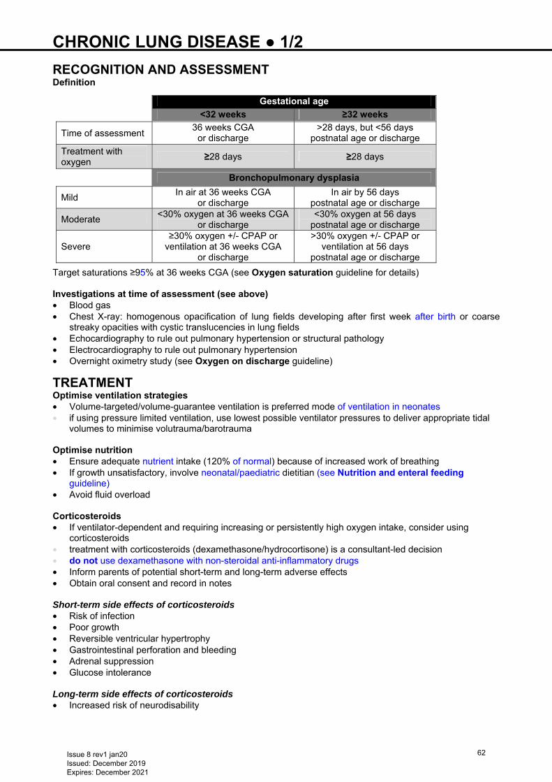
CHRONIC LUNG DISEASE ● 1/2 RECOGNITION AND ASSESSMENT Definition
Target saturations ≥95% at 36 weeks CGA (see Oxygen saturation guideline for details) Investigations at time of assessment (see above) • Blood gas • Chest X-ray: homogenous opacification of lung fields developing after first week after birth or coarse
streaky opacities with cystic translucencies in lung fields • Echocardiography to rule out pulmonary hypertension or structural pathology • Electrocardiography to rule out pulmonary hypertension
• Overnight oximetry study (see Oxygen on discharge guideline)
TREATMENT Optimise ventilation strategies • Volume-targeted/volume-guarantee ventilation is preferred mode of ventilation in neonates • if using pressure limited ventilation, use lowest possible ventilator pressures to deliver appropriate tidal
volumes to minimise volutrauma/barotrauma
Optimise nutrition • Ensure adequate nutrient intake (120% of normal) because of increased work of breathing • If growth unsatisfactory, involve neonatal/paediatric dietitian (see Nutrition and enteral feeding
guideline) • Avoid fluid overload Corticosteroids • If ventilator-dependent and requiring increasing or persistently high oxygen intake, consider using
corticosteroids • treatment with corticosteroids (dexamethasone/hydrocortisone) is a consultant-led decision • do not use dexamethasone with non-steroidal anti-inflammatory drugs • Inform parents of potential short-term and long-term adverse effects • Obtain oral consent and record in notes Short-term side effects of corticosteroids • Risk of infection • Poor growth • Reversible ventricular hypertrophy • Gastrointestinal perforation and bleeding • Adrenal suppression • Glucose intolerance Long-term side effects of corticosteroids • Increased risk of neurodisability
Gestational age
<32 weeks ≥32 weeks
Time of assessment 36 weeks CGA
or discharge >28 days, but <56 days
postnatal age or discharge
Treatment with oxygen
≥28 days ≥28 days
Bronchopulmonary dysplasia
Mild In air at 36 weeks CGA
or discharge In air by 56 days
postnatal age or discharge
Moderate <30% oxygen at 36 weeks CGA
or discharge <30% oxygen at 56 days
postnatal age or discharge
Severe ≥30% oxygen +/- CPAP or
ventilation at 36 weeks CGA or discharge
>30% oxygen +/- CPAP or ventilation at 56 days
postnatal age or discharge
Issue 8 rev1 jan20 Issued: December 2019 Expires: December 2021
62
CHRONIC LUNG DISEASE ● 2/2 Doses • Use Neonatal Formulary for dexamethasone dosage regimen (consultant decision on DART versus
Minidex regimen) • If respiratory status worsens after initial improvement consider repeating course of corticosteroids
(consultant decision) Monitoring while on corticosteroids • Daily BP and urinary glucose
Diuretics • Use of diuretics to improve lung function (consultant decision). Diuretics of choice are chlorothiazide and
spironolactone (use of spironolactone can be guided by serum potassium). Avoid amiloride due to its lung fluid retaining properties
• Side effects include hyponatraemia, hypo/hyperkalaemia, hypercalciuria (leading to nephrocalcinosis) and metabolic alkalosis
• If no improvement on diuretics stop after 1 week
SUBSEQUENT MANAGEMENT Monitoring treatment Continuous • Aim for SpO2 90–94% until 36 weeks CGA • After 36 weeks CGA, maintain SpO2 ≥95% to prevent pulmonary hypertension • Warm and humidify supplemental oxygen unless on low-flow oxygen • Monitor weight and head growth • Assess for gastro-oesophageal reflux [see Gastro-oesophageal reflux (GOR) guideline)] • Aim to stop diuretic therapy before discharge (consultant decision)
DISCHARGE AND FOLLOW-UP • If still oxygen-dependent at time of discharge (see Oxygen at discharge guideline) • Long-term neurodevelopmental and respiratory follow-up
Issue 8 rev1 jan20 Issued: December 2019 Expires: December 2021
63
CMV ● 1/2 In-utero transmission of CMV can occur during primary maternal infection, reactivation, or reinfection of seropositive mothers
MATERNAL TESTS CMV serology (IgG and IgM) and viral loads • Both IgG and IgM negative: unlikely to be CMV infection • IgG positive, IgM negative: past maternal infection • IgG positive, IgM positive: check CMV IgG avidity • if low likely to be maternal CMV infection within last 3−4 months Antenatal ultrasound Features include: • IUGR • Intracranial ventriculomegaly/calcification, microcephaly • Ascites, hydrops fetalis • Pleural or pericardial effusions • Oligo- or polyhydramnios • Hepatomegaly • Abdominal calcification • Pseudomeconium ileus • Thickened placenta
NEONATAL FEATURES Indications for testing • Evidence of maternal primary CMV infection in pregnancy • Antenatal ultrasound suggestive of congenital CMV (cCMV): ventriculomegaly, calcifications,
periventricular cysts, echogenic bowel, pericardial effusion, ascites and fetal hydrops • Petechiae/purpura • Hepatosplenomegaly • Prolonged or conjugated hyperbilirubinaemia with transaminitis • Unexplained thrombocytopenia • Microcephaly • Intracranial calcification or ventriculomegaly • Chorioretinitis • Seizures with no other explanation • Severe pneumonia • Cataract • Failed hearing screen Investigation results • CMV PCR urine or mouth swab • soak mouth swab in saliva for 1 min; send in viral transport medium to regional laboratory • if negative and high-risk CMV also send urine
Other congenital infection screen depending on features (not exclusive): • Toxoplasma (hydrocephalus, microcephaly, convulsions, generalised infection) • Syphilis (rash, rhinitis, hepatosplenomegaly, jaundice, thrombocytopenia) • Rubella (cataract, deafness, microcephaly) • Zika (maternal/paternal travel, microcephaly)
CMV POSITIVE Further investigations • Full blood count, liver enzymes, bilirubin, renal function • Blood CMV viral load • if unknown whether infection is congenital request initial bloodspot card to be tested for CMV PCR • Ophthalmic assessment • Audiology: brainstem-evoked response • Head ultrasound • if ultrasound head abnormal or seizures, MRI head
Issue 8 rev1 jan20 Issued: December 2019 Expires: December 2021
64
CMV ● 2/2 TREATMENT • Postnatal acquired CMV – no treatment Mild cCMV • Asymptomatic – no CNS involvement, including sensorineural hearing loss • isolated IUGR • hepatomegaly with normal liver enzymes • isolated raised ALT/AST • mild thrombocytopenia • No treatment Moderate cCMV • Discuss with infectious diseases specialist if: • >2 weeks mild features • >2 mild features Severe cCMV • Significant organ involvement: • significant liver enzyme abnormalities • marked hepatomegaly • Any CNS disease • isolated sensorineural hearing loss • retinitis • microcephaly • cranial ultrasound or MRI brain abnormalities • Treat: valganciclovir 16 mg/kg oral 12-hrly for 6 months • if not tolerating oral feeds, ganciclovir 6 mg/kg IV [prepared by pharmacy (cytotoxic)] over 1 hr, 12-hrly
for 6 weeks • Discuss side effects vs benefits with parents: • advantages: potential reduced risk of deafness and developmental delay • disadvantages: during treatment reversible blood dyscrasia; long-term unknown risk to fertility and
malignancy • Start treatment as soon as possible • if diagnosis delayed can be started aged ≤1 month
FEEDING • Do not discourage infected women from breastfeeding their own uninfected, term babies (CMV can be
transmitted via breastfeeding, but benefits of feeding outweigh risks posed by breastfeeding as a source of transmission)
• Avoid breastfeeding of premature baby if mother is positive and baby asymptomatic
FOLLOW-UP • Enter on CMV surveillance register (discuss with paediatric infectious disease specialist) • Ganciclovir IV: FBC, LFT, U&E at least twice weekly • Valganciclovir oral: FBC, LFT, U&E weekly for first 4 weeks, then monthly until completion • CMV viral load monthly on antiviral therapy • Therapeutic drug monitoring if: • viral load increases >1 log on treatment • toxicity suspected • abnormal renal function • <36 weeks’ gestation • Audiology: 3 monthly for first year, then 6 monthly for 3 yr, then annually until aged 6 yr for both
asymptomatic and symptomatic congenitally infected babies • Paediatric infectious diseases specialist: as soon as possible in first month, then annually until aged 2 yr • Ophthalmology: at least annually until aged 5 yr if symptomatic/signs at birth • Neurodevelopmental assessment: aged 1 yr • if delayed development discuss MRI brain with radiology
Issue 8 rev1 jan20 Issued: December 2019 Expires: December 2021
65
COAGULOPATHY ● 1/2 • Haemostasis is immature during the neonatal period and does not attain full function until aged 6 months • prolonged prothrombin time (PT) and activated partial thromboplastin time (APTT) are associated with
intraventricular haemorrhage (IVH) in unstable (e.g. hypotensive or hypoxic) or bruised extremely preterm babies
• 75% of cases of IVH occur within the first 24 hr of life and 90% within the first 7 days • IVH can occur in the absence of a coagulopathy • prophylactic fresh frozen plasma (FFP) does not prevent IVH in preterm baby without evidence of
coagulopathy
INVESTIGATIONS Check clotting in: • Any bruised or bleeding baby (e.g. IVH, pulmonary haemorrhage, gastrointestinal bleeding, suspected
haemorrhagic disease of newborn etc.) • Preterm <30 weeks’ gestation (due to IVH risk) if clinical concerns about bleeding • Moderate-to-severe encephalopathy (e.g. babies who are being cooled) • Septicaemia • Necrotising enterocolitis (NEC) • Sick or unstable baby (e.g. ventilated, inotropic support etc.) • Metabolic disease: urea cycle disorder, galactosaemia, tyrosinaemia, organic acidaemia • Liver dysfunction or conjugated jaundice • Babies undergoing surgery or tissue biopsy who have had previous bleeding problems • Family history of inherited bleeding disorder (after discussion with consultant haematologist) • Thrombocytopenia (see Thrombocytopenia guideline) Sampling • Ensure sample from a free-flowing vein (peripheral or umbilical) or from an arterial line before
heparinising • Fill exactly to black mark on tube (usually 1.3 mL) • If sample clots (this does not confirm normal coagulation), repeat • If sampling from arterial line with heparin infusion, take larger volume (e.g. 2.5 mL) from dead-space
(see Arterial line sampling guideline) Request • INR (measure of PT) • APTT • Fibrinogen • If features of DIC (e.g. bruising, bleeding, sepsis), request fibrin degradation products and D-dimer (if
available) • If concerned/unsure about initial results, seek senior advice IMMEDIATE TREATMENT • If INR alone is prolonged, and clotting samples were performed before first dose of vitamin K, repeat
clotting screen • If prolonged INR and normal APTT in stable term baby (e.g. clotting screen performed as part of
conjugated jaundice screen), give repeat dose of vitamin K 100 microgram/kg (maximum 1 mg per dose) IV. If repeat INR not improving after 6 hr, discuss with senior/haematologist to explore other causes and the need for FFP or regular vitamin K
• In preterm baby <30 weeks (with risk of IVH) or unwell with prolonged INR, repeat vitamin K 100 microgram/kg (maximum 1 mg per dose) IV with FFP
• If APTT beyond upper limit of reference range, give FFP (see below) • In case of persistently prolonged INR or liver disorder/conjugated jaundice, give regular doses of vitamin
K • In persistently prolonged APTT, give further doses of FFP (or cryoprecipitate – see below) Use of FFP and cryoprecipitate
Do not use FFP or cryoprecipitate purely for volume replacement or polycythaemia without coagulopathy
Treatment thresholds for use of FFP • If APTT above treatment thresholds give FFP 10–20 mL/kg over 30–60 min
Issue 8 rev1 jan20 Issued: December 2019 Expires: December 2021
66
COAGULOPATHY ● 2/2
Clotting parameter Gestation Stable baby
Unstable*, significant
bleeding† or invasive procedure‡
INR Term ≥1.6 ≥1.5 Preterm (<37 weeks) ≥2 ≥1.8
APTT Term APTT ratio ≥1.6 or
APTT value ≥55 secAPTT ratio ≥1.5 or
APTT value ≥45 sec Preterm (<37 weeks)
APTT ratio ≥2 orAPTT value ≥70 sec
APTT ratio ≥1.8 or APTT value ≥60 sec
*Unstable (e.g. DIC, significant sepsis, NEC, ventilated, hypotensive etc.) †Significant bleeding (e.g. significant bruising, IVH, gastrointestinaI bleeding, pulmonary haemorrhage etc.)
‡Invasive procedures (e.g. lumbar puncture, umbilical lines, long lines, chest drain, exchange transfusion etc.)
• In inherited clotting factor deficiencies, use FFP only when pathogen inactivated factor unavailable.
Discuss with consultant haematologist before giving FFP • If APTT ratio still ≥1.8 after giving FFP (especially if fibrinogen <1.2), consider cryoprecipitate (5–10
mL/kg over 30–60 min) after discussion with on-call consultant and haematologist
MONITORING • Repeat coagulation profile 2–4 hr after FFP/cryoprecipitate or every 12–24 hr as indicated • Look for and treat causes of abnormal coagulation: • sepsis • shock • haemorrhage • severe hypothermia • hypoxia • If abnormal coagulation persists for >24 hr in the absence of any precipitating factors, seek advice from
paediatric haematologist about factor assays and 50:50 mixture correction test
Issue 8 rev1 jan20 Issued: December 2019 Expires: December 2021
67
CONGENITAL DIAPHRAGMATIC HERNIA (CDH) ● 1/4 INTRODUCTION CDH is a congenital defect in the diaphragm (usually detected antenatally) resulting in herniation of abdominal contents into the thoracic cavity; associated with a high risk of mortality and morbidity. A combination of pulmonary hypoplasia and abnormal morphology of the pulmonary vasculature leads to severe respiratory insufficiency and increased risk of developing persistent pulmonary hypertension (PPHN)
RECOGNITION AND ASSESSMENT Antenatal diagnosis • Delivery to be planned at regional neonatal intensive care unit (NICU) • Fetal medicine team and paediatric surgeon to provide antenatal counseling • Neonatal team to meet parents before delivery • Neonatal consultant, middle grade, junior tier and NICU nurse to attend delivery Postnatal diagnosis • In some babies the lesion develops later in gestation; these babies tend to have a better prognosis • Postnatal presentation can be with clinical features ranging from inability to resuscitate baby at birth to
incidental finding on chest X-ray
In cases diagnosed postnatally there may be early respiratory distress in association with a scaphoid abdomen and heart sounds shifted usually to the right. Mask inflation will often cause deterioration
as air is delivered into herniated gut resulting in cardiorespiratory embarrassment
INVESTIGATIONS • Pre and postductal SpO2 • Chest and abdominal X-ray • Arterial blood gas • Echocardiogram
IMMEDIATE MANAGEMENT AT DELIVERY Key principles • Intubate all antenatally diagnosed babies promptly (intubation to be carried out by most experienced and
reliable operator present) • Optimise ETT position and size, aiming for little or no leak, with largest size tube feasible • confirm tube position by end tidal CO2 monitoring • Do not give mask ventilation − will introduce air into the GI tract and compromise ventilation • Maintain low peak pressure <25 cm H2O and positive end expiratory pressures of 5 cm H2O to avoid
lung damage • Avoid high airway pressures • Establish adequate perfusion and oxygenation • aim for preductal SpO2 80−95% after first 10 min • avoid hyperoxia, reduce FiO2 when preductal saturation >95% • Insert large gauge 8−10 Fr NGT • aspirate at least every 5 min to decompress stomach until baby established on ventilation, then place on
free drainage • Check temperature before transfer to NNU, maintain normothermia • Examine baby for other associated abnormalities e.g.: • cardiac (present in 20%) • trisomy 18/21 • urogenital • musculoskeletal
MANAGEMENT ON NNU
Undertake management PROMPTLY Babies with CDH fare better with minimal handling − handle baby as little and as gently as possible
• Weigh baby • Start on conventional ventilation with low tidal volume strategy of 3−4 mL/kg • Sedation: morphine 20 micrograms/kg/hr
Issue 8 rev1 jan20 Issued: December 2019 Expires: December 2021
68
CONGENITAL DIAPHRAGMATIC HERNIA (CDH) ● 2/4 • Umbilical venous and arterial catheters • to be sited by experienced operator (initial management is time critical) • if not possible to site umbilical arterial catheter (UAC), insert peripheral arterial line • Monitor pre and postductal SpO2 • first 2 hr only: if pH >7.2, PaCO2 <8.6 kPa and saturations improving, aim for preductal saturations of
>70% • after first 2 hr: if lactate and pH acceptable (pH >7.2, lactate <5 mmol/L) on arterial blood gas, aim for
preductal SpO2 80−95% (UAC measures postductal PaO2) and postductal SpO2 >70% • an abnormal lactate is an indicator of poor perfusion and must be corrected before the interpretation of
acceptable levels of SpO2 • Maintain arterial blood pressure at normal level for gestational age • Surfactant NOT routinely recommended, only to be administered after discussion with regional centre as
risk of over-distension and pneumothorax • Cardiac echocardiogram (ideally within 6 hr of birth) to: • exclude associated congenital cardiac disease • assess right ventricular function • look for evidence of persistent pulmonary hypertension [see Persistent pulmonary hypertension of
the newborn (PPHN) guideline] • identify patent ductus arteriosus and assess shunting (see Patent ductus arteriosus guideline)
Ventilation Gentle conventional (see Ventilation: conventional guideline) • Avoid peak pressures >25 cm H2O, maintain PEEP 3−5 cm H2O • if greater peak pressures required to maintain preductal SpO2 >80% and postductal SpO2 >70%, discuss
HFOV with consultant − if HFOV not available discuss with specialist centre e.g. KIDS/BWCH to expedite retrieval
HFOV [see Ventilation: high frequency oscillatory (HFOV) guideline] • Initial setting: • MAP: 12 cm H2O (do not increase >16 cm H2O) • rate/frequency: 10 Hz, delta P 25 • Chest X-ray 1 hr after commencing HFOV • if >8 rib spaces visible, lungs are hyper-inflated − reduce MAP
Target O2 Saturations • Aim for preductal SpO2 of 80−95% • if MAP >12 cm H2O and FiO2 >0.6 to maintain preductal SpO2 >80%, commence inhaled nitric oxide
(iNO) at 20 ppm (see Nitric oxide guideline)
Permissive hypercapnia • If pH >7.2, lactate <5 and urine output >1 mL/kg/hr: target PaCO2 6.9−9.3 kPa
Systemic blood pressure support • Invasive blood pressure monitoring required • If preductal SpO2 80−95%, aim for mean arterial blood pressure corresponding to gestation • Maintenance fluid volume: 60 mL/kg/day • Treat hypotension or poor tissue perfusion (rising lactate, urine output <1 mL/kg/hr) with fluid boluses
sodium chloride 0.9% 10 mL/kg (maximum 30 mL/kg) • If heart rate normal, urine output >1 mL/kg/hr, lactate <3 − do not give inotropes • If persistent hypotension or hypoperfusion and difficulty maintaining preductal saturation 80–95%, give
inotropes Term infants • Start adrenaline 100−1000 nanogram/kg/min as first line (starting dose usually 300 nanogram/kg/min) • If right ventricular failure on echocardiogram discuss adrenaline with specialist centre e.g. KIDS/NTS • If right ventricular failure add milrinone 35−45 microgram/kg/hr (starting dose 35 micogram/kg/hr; DO
NOT give loading dose) and noradrenaline 20−100 nanogram/kg/min (starting dose 20 nanogram/kg/min)
Preterm infant • Start dopamine 10 microgram/kg/min and increase to 20 microgram/kg/min • if excessive tachycardia (heart rate >200 bpm) secondary to dopamine, discuss with specialist centre
e.g. KIDS/NTS and add noradrenaline
Issue 8 rev1 jan20 Issued: December 2019 Expires: December 2021
69
CONGENITAL DIAPHRAGMATIC HERNIA (CDH) ● 3/4 • Monitor lactate – rise in lactic acidosis suggests excessive vasoconstriction by inotropes Metabolic acidosis • Accept pH >7.2 • Review vasoconstrictor effects versus benefits of inotropes • Correct metabolic acidosis with sodium bicarbonate 4.2%; give full correction over 6 hr titrating against
pH/base excess every 1−2 hr
MANAGEMENT OF PPHN • Anticipate PPHN in babies with CDH • Monitor pre and postductal SpO2 • Calculate oxygenation index (OI) (UAC is a measure of postductal saturation) • if OI >20 and/or pre:postductal saturation difference >10% discuss with tertiary centre e.g. KIDS/NTS to
expedite retrieval • Initiate trial of iNO, 20 ppm for 1 hr • MgSO4, is an effective pulmonary vasodilator commence an infusion of MgSO4 to achieve a serum [Mg]
above the normal range (>1 mmol/L) • can give rise to profound systemic hypotension, use only in conjunction with active management of
systemic blood pressure support • Maintain arterial PaO2 8−10 kPa • See Persistent pulmonary hypertension of the newborn (PPHN) guideline
GENERAL SUPPORT • Fluid: restrict to 60 mL/kg/day • Keep large bore NGT on free drainage and regular aspiration, and nil-by-mouth • Commence parenteral nutrition • Send blood culture and commence first line antibiotics • Send clotting screen and correct any abnormalities • Correct hypocalcaemia • If antenatal diagnosis of a duct dependent congenital cardiac lesion, or any uncertainty about the
presence of cardiac anomaly, commence prostaglandin E2 5 nanogram/kg/min • Maintain magnesium >1 mmol/L • Maintain normothermia • Monitor for pneumothorax. See Chest drain insertion − Seldinger technique and Chest drain
insertion − Traditional guidelines • Crossmatch 1 unit of blood • Cranial ultrasound scan • Send blood for chromosomes with parental consent (if not done antenatally) • Sedation: morphine 10−20 microgram/kg/hr, but avoid deep sedation • Avoid neuromuscular blocking agents − use is associated with hypoxaemia • Minimal handling, developmental care with swaddling/cocooning • Keep area around baby quiet and lights dimmed
COMMUNICATION WITH SPECIALIST CENTRE • Neonatal consultant to inform planned paediatric specialist centre e.g. Birmingham Children’s
Hospital/Birmingham Women’s Hospital once baby stabilised. This will require conference call with referring consultant, on-call surgeon at specialist centre, neonatologist, PICU intensivist and transport consultant e.g. KIDS/NTS, to discuss urgency of transfer and ongoing management
• Undertake transport of babies for surgery only when: • mean arterial blood pressure normal for gestation • lactate <3 mmol/L and urine output >1 mL/kg/hr • ventilation reduced to low pressure settings • FiO2 on conventional ventilation 0.5, with preductal saturations 85−95% • baby fit for surgery and stable for ≥24 hr (may take ≥3−10 days)
EXTRACORPOREAL MEMBRANE OXYGENGATION (ECMO) • See Persistent pulmonary hypertension of the newborn (PPHN) guideline • If ECMO considered refer to specialist centre (e.g. via KIDS/NTS team)
Issue 8 rev1 jan20 Issued: December 2019 Expires: December 2021
70
CONGENITAL DIAPHRAGMATIC HERNIA (CDH) ● 4/4 USEFUL INFORMATION • http://www.bch.nhs.uk/parents-and-carers/parent-info-leaflets/neonatal-surgical-ward • http://kids.bch.nhs.uk/ • https://www.e-lfh.org.uk/programmes/paediatric-surgery/
Issue 8 rev1 jan20 Issued: December 2019 Expires: December 2021
71
CONJUNCTIVITIS ● 1/2
Conjunctivitis is a potentially blinding condition with associated systemic manifestations
RECOGNITION AND ASSESSMENT • Conjunctival redness • Swelling of conjunctiva and eyelids • Purulent or mucopurulent discharge • Vesicles on lids or adjacent skin (herpes simplex)
Differential diagnosis • Sticky eye with blocked tear duct in which there is no inflammation of conjunctiva • Congenital glaucoma in which there is corneal opacity • Swelling of conjunctiva and eyelids as part of preseptal or orbital cellulitis
AETIOLOGY Bacterial • Staphylococcus aureus • Haemophilus influenzae • Streptococcus pneumoniae • Serratia spp, E. coli, Pseudomonas spp • Neisseria gonorrhoeae – typical onset aged 0–5 days: mild inflammation with sero-sanguineous
discharge to thick, purulent discharge with tense oedema of eyelids • Chlamydia trachomatis – typical onset aged 5–14 days: mild-to-severe swelling with purulent discharge
(may be blood-stained) Viral • Herpes simplex virus (HSV)
MANAGEMENT • 4–6 hrly eye toilet using sodium chloride 0.9%
• cooled, boiled tap water acceptable for home use Conjunctivitis (see signs above) • Swab all for: • Gram stain and bacterial culture and sensitivities • if other suspicions of HSV (e.g. vesicles etc.), swab for HSV PCR
− use dry swab/moistened with viral transport media − place in dry tube/pot
• swab using dry swab/moistened with viral transport media, and place in dry tube/pot (check for Chlamydia trachomatis and Neisseria gonorrhoeae PCR)
• Treat both eyes with: • frequent eye toilet as necessary • chloramphenicol 0.5% eye drops • fusidic acid 1% eye drops for Staphylococcus • Presentation ≤24 hr of birth suggests gonococcal infection – inform consultant paediatrician • If herpes suspected begin treatment with aciclovir IV and topical while awaiting results
SUBSEQUENT MANAGEMENT In severe non-resolving cases • Take throat and eye swabs for viral PCR • If herpes suspected, look for other signs of herpetic infection • Treat suspected herpes with aciclovir IV and topical for 14 days • Refer to ophthalmology Neisseria gonorrhoeae suspected • Request urgent Gram stain and culture • Assess baby for sepsis • Swab for PCR
Issue 8 rev1 jan20 Issued: December 2019 Expires: December 2021
72
CONJUNCTIVITIS ● 2/2 Neisseria gonorrhoeae confirmed • Give single dose cefotaxime 100 mg/kg IV • For severe cases, frequent sodium chloride 0.9% irrigation of the eyes and continue treatment with
cefotaxime IV for up to 5 days (consultant decision) • Refer to ophthalmology Chlamydia result positive • Treat with azithromycin 20 mg/kg IV single dose and azithromycin eye drops twice daily for 3 days • If maternal chlamydia treated successfully in pregnancy, baby does not require prophylactic treatment • if unsure swab baby’s conjunctiva using nucleic acid amplification test (NAAT) swab • if high risk send bacterial swab for gonococcus and HIV antibody, HBsAg and HCV antibody from mother
or baby Gonococcal or chlamydia infection detected • Refer mother and partner to genitourinary medicine for immediate treatment
Issue 8 rev1 jan20 Issued: December 2019 Expires: December 2021
73
CONSENT ● 1/3 FOR COMMON NEONATAL INVESTIGATIONS, INTERVENTIONS AND TREATMENTS The following guidance is taken from 'Good practice framework for consent in neonatal clinical care' produced by the British Association of Perinatal Medicine (BAPM) • It is a legal and ethical requirement to gain valid consent before examining and initiating any
investigation or treatment for any patient • Consent is obtained from someone with parental responsibilities: • if married, parents • if not married, mother but not father, unless father has acquired parental responsibility via a court order,
being registered on birth certificate or parental responsibility agreement • a legally appointed guardian • a local authority designated in a care order or holding an emergency protection order • Consent is valid only when information has been understood by the parents and explains why the
intervention is recommended, its risks and implications, and other options should consent be withheld
Documentation of information given and parents’ understanding and agreement to proceed is the most important validation of consent. A signature does not in itself confirm informed
consent • Witness consent wherever possible, and record name of witness • In neonatal practice, there are frequent occasions when no one is available to provide valid consent and
treatment is initiated in its absence (e.g. emergency ABC resuscitation, stabilisation, chest drainage or exchange transfusion when delayed treatment would not be in baby’s best interests, or following maternal general anaesthetic when mother is unmarried to baby’s father). It should always be possible later to justify the action to the parents and to reassure them that it was in the baby’s best interests
GOOD PRACTICE • Give parents of babies admitted to NNU written information (BLISS booklet
http://www.bliss.org.uk/information-for-parents/) describing low-risk procedures such as venesection, for which explicit consent is not normally sought
• Give parents information leaflet for data collection, allowing them to opt out Written explicit consent Purpose and risks of an intervention are formally explained and consent obtained and recorded before the intervention Table 1: Explicit consent (recorded in patient notes, and supported by a signature) is required for:
Investigation/intervention
Clinical photographs and video-recordings
Use consent form specific for this purpose
Any biopsy or aspiration E.g.: skin, liver, bone marrow
Exchange transfusion
Treatment for retinopathy Obtained by ophthalmologist
Surgical procedures Consent taken by surgical team. If telephone consent required and mother still an inpatient, midwife on postnatal ward or neonatal team to act as witness
Post-mortem See Death guideline and use specific form. Usually obtained through consultant or senior middle grade staff trained in obtaining this consent
MRI/CT If anaesthetic or sedation needed
Issue 8 rev1 jan20 Issued: December 2019 Expires: December 2021
74
CONSENT ● 2/3 Table 2: Explicit oral consent Explicit consent as defined above, documented, but not supported by a signature, required for the following:
Explicit oral consent Investigations • Screening baby and/or mother in high-risk situations with
no knowledge of maternal status (e.g. HIV, substance misuse)
• Genetic testing • Gut imaging involving contrast • MR/CT imaging • Newborn bloodspot screening
Practical procedures • Therapeutic lumbar puncture (LP) or ventricular tap in absence of reservoir*
• Peripherally-placed long lines* • Brachial or femoral arterial line • Chest drain insertion/replacement* • Abdominal drainage for perforation or ascites* • Irrigation following extravasation* • Hearing screening
Immunisations See Immunisations guideline
Treatments • Vitamin K for normal term babies • Nitric oxide • Postnatal steroids for chronic lung disease • Use of donor breast milk
Transport • Emergency transfers • Routine transfers for outpatients or back-transfers • Note: Initiation of cooling for neuroprotection does not
require explicit consent, but transfer to another unit for formal cooling does
Treatments: blood products
• Blood transfusion • Use of pooled blood products e.g. FFP • Partial exchange transfusion
* It is accepted that, in some circumstances, these procedures are performed in an emergency in
baby’s best interests and may be performed without oral consent; owing to risks associated with procedures or conditions in which they are necessary, it is considered best practice to inform parents as soon as possible and to document this in baby’s notes
Others: Implicit consent • Where the nature and risk of the procedure is such that a less formal transfer of information is
considered sufficient, and is often retrospective • List of investigations, procedures and treatments is long, see Table 3 • If unsure, seek senior advice
Explain all investigations, procedures and treatments to parents at earliest opportunity
Issue 8 rev1 jan20 Issued: December 2019 Expires: December 2021
75
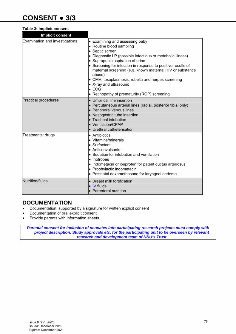
CONSENT ● 3/3 Table 3: Implicit consent
Implicit consent Examination and investigations • Examining and assessing baby
• Routine blood sampling • Septic screen • Diagnostic LP (possible infectious or metabolic illness) • Suprapubic aspiration of urine • Screening for infection in response to positive results of
maternal screening (e.g. known maternal HIV or substance abuse)
• CMV, toxoplasmosis, rubella and herpes screening • X-ray and ultrasound • ECG • Retinopathy of prematurity (ROP) screening
Practical procedures • Umbilical line insertion • Percutaneous arterial lines (radial, posterior tibial only) • Peripheral venous lines • Nasogastric tube insertion • Tracheal intubation • Ventilation/CPAP • Urethral catheterisation
Treatments: drugs
• Antibiotics • Vitamins/minerals • Surfactant • Anticonvulsants • Sedation for intubation and ventilation • Inotropes • Indometacin or ibuprofen for patent ductus arteriosus • Prophylactic indometacin • Postnatal dexamethasone for laryngeal oedema
Nutrition/fluids
• Breast milk fortification • IV fluids • Parenteral nutrition
DOCUMENTATION • Documentation, supported by a signature for written explicit consent • Documentation of oral explicit consent • Provide parents with information sheets
Parental consent for inclusion of neonates into participating research projects must comply with project description. Study approvals etc. for the participating unit to be overseen by relevant
research and development team of NNU’s Trust
Issue 8 rev1 jan20 Issued: December 2019 Expires: December 2021
76
CONTACTING A CONSULTANT ● 1/2 The need to call for consultant support may vary with the experience of the staff involved. This guideline suggests scenarios where advice of a consultant should normally be sought, however the list is not exhaustive, consultant advice should be sought anytime that the junior medical team feel the need for support
Initiate life saving measures, e.g. intubation, before informing consultant * Consultant normally expected to attend in person Before birth • Delivery <27 weeks’ gestation* • Unexpected birth of baby with congenital diaphragmatic hernia* • If stated in neonatal alert form/antenatal MDT plan
During resuscitation • No heart beat at 5 min/continuing resuscitation at 10 min* • Request of consultant obstetrician Admission • <28 weeks’ gestation • Ex-utero intensive care transfer • Cord pH <7.0 and/or 10 min Apgar score <6 • Suspected subgaleal haemorrhage Inpatient • FiO2 >0.6 with/without respiratory support • Baby who has required endotracheal ventilation (do not wait for consultant before intubating and
initiating ventilation) • Anticipated need for HFOV • PPHN likely to need nitric oxide* • Continuing hypotension despite volume expansion and dobutamine and dopamine* • Seizures • Neonatal encephalopathy requiring therapeutic hypothermia • Severe jaundice • bilirubin above exchange level • bilirubin rising >8.5 micromol/L/hr despite intensive phototherapy • Major deterioration • Baby with ambiguous genitalia/disorder of sexual development • Major congenital anomaly without antenatal plan • Renal failure with serum K+ >7 mmol/L or Na+ <120 mmol/L • Known subgaleal haemorrhage with haemodynamic instability needing volume or blood products
replacement* • Unexpected death* • Initiation of, or unexpected withdrawal of, intensive care* • Escalation level requiring in-utero or ex-utero transfers out or refusals from other units • Middle grade doctor and nursing team cannot agree management plan for baby • Inability immediately to site essential line*
COMMUNICATING WITH CONSULTANT • If immediate attendance required, state this first • Communicate essential details using Situation-Background-Assessment-Recommendation (SBAR)
communication tool Situation • State who you are and where you are calling from • State patient’s name and reason for your call • Describe your concern Background • Collect information from patient’s chart and notes before calling • Give overview of patient’s background • admission diagnosis
Issue 8 rev1 jan20 Issued: December 2019 Expires: December 2021
77
CONTACTING A CONSULTANT ● 2/2 • date of admission • previous procedures • current medications • pertinent laboratory results • other relevant diagnostic results Assessment • Vital signs • Clinical impressions/concerns • think critically, considering what might be the underlying reason for patient’s condition • it may be “I’m not sure what the problem is, but I am worried” Recommendation • State your recommendation (in an urgent situation you may start with this) • Explain what you need. Be specific about request and time frame • Make suggestions • Clarify expectations • Repeat any plans agreed to ensure accuracy
Issue 8 rev1 jan20 Issued: December 2019 Expires: December 2021
78
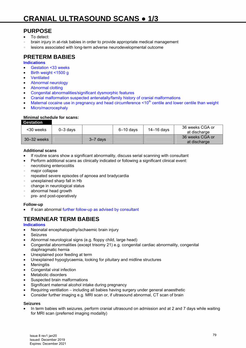
CRANIAL ULTRASOUND SCANS ● 1/3 PURPOSE • To detect: • brain injury in at-risk babies in order to provide appropriate medical management • lesions associated with long-term adverse neurodevelopmental outcome
PRETERM BABIES Indications • Gestation <33 weeks • Birth weight <1500 g • Ventilated • Abnormal neurology • Abnormal clotting • Congenital abnormalities/significant dysmorphic features • Cranial malformation suspected antenatally/family history of cranial malformations • Maternal cocaine use in pregnancy and head circumference <10th centile and lower centile than weight • Micro/macrocephaly
Minimal schedule for scans: Gestation
<30 weeks 0–3 days 6–10 days 14–16 days 36 weeks CGA or
at discharge
30–32 weeks 3–7 days 36 weeks CGA or
at discharge Additional scans • If routine scans show a significant abnormality, discuss serial scanning with consultant • Perform additional scans as clinically indicated or following a significant clinical event: • necrotising enterocolitis • major collapse • repeated severe episodes of apnoea and bradycardia • unexplained sharp fall in Hb • change in neurological status • abnormal head growth • pre- and post-operatively Follow-up • If scan abnormal further follow-up as advised by consultant
TERM/NEAR TERM BABIES Indications • Neonatal encephalopathy/ischaemic brain injury • Seizures • Abnormal neurological signs (e.g. floppy child, large head) • Congenital abnormalities (except trisomy 21) e.g. congenital cardiac abnormality, congenital
diaphragmatic hernia • Unexplained poor feeding at term • Unexplained hypoglycaemia, looking for pituitary and midline structures • Meningitis • Congenital viral infection • Metabolic disorders • Suspected brain malformations • Significant maternal alcohol intake during pregnancy • Requiring ventilation − including all babies having surgery under general anaesthetic • Consider further imaging e.g. MRI scan or, if ultrasound abnormal, CT scan of brain
Seizures • In term babies with seizures, perform cranial ultrasound on admission and at 2 and 7 days while waiting
for MRI scan (preferred imaging modality)
Issue 8 rev1 jan20 Issued: December 2019 Expires: December 2021
79
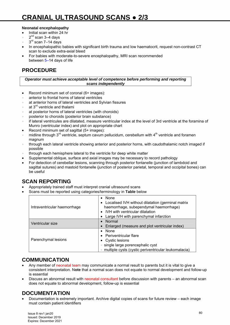
CRANIAL ULTRASOUND SCANS ● 2/3 Neonatal encephalopathy • Initial scan within 24 hr • 2nd scan 3–4 days • 3rd scan 7–14 days • In encephalopathic babies with significant birth trauma and low haematocrit, request non-contrast CT
scan to exclude extra-axial bleed • For babies with moderate-to-severe encephalopathy, MRI scan recommended
between 5–14 days of life
PROCEDURE
Operator must achieve acceptable level of competence before performing and reporting scans independently
• Record minimum set of coronal (6+ images): • anterior to frontal horns of lateral ventricles • at anterior horns of lateral ventricles and Sylvian fissures • at 3rd ventricle and thalami • at posterior horns of lateral ventricles (with choroids) • posterior to choroids (posterior brain substance) • if lateral ventricules are dilatated, measure ventricular index at the level of 3rd ventricle at the foramina of
Munro (ventricular index) and plot on appropriate chart • Record minimum set of sagittal (5+ images): • midline through 3rd ventricle, septum cavum pellucidum, cerebellum with 4th ventricle and foramen
magnum • through each lateral ventricle showing anterior and posterior horns, with caudothalamic notch imaged if
possible • through each hemisphere lateral to the ventricle for deep white matter • Supplemental oblique, surface and axial images may be necessary to record pathology • For detection of cerebellar lesions, scanning through posterior fontanelle (junction of lambdoid and
sagittal sutures) and mastoid fontanelle (junction of posterior parietal, temporal and occipital bones) can be useful
SCAN REPORTING • Appropriately trained staff must interpret cranial ultrasound scans • Scans must be reported using categories/terminology in Table below
Intraventricular haemorrhage
• None • Localised IVH without dilatation (germinal matrix
haemorrhage, subependymal haemorrhage) • IVH with ventricular dilatation • Large IVH with parenchymal infarction
Ventricular size • Normal • Enlarged (measure and plot ventricular index)
Parenchymal lesions
• None • Periventricular flare • Cystic lesions • single large porencephalic cyst • multiple cysts (cystic periventricular leukomalacia)
COMMUNICATION • Any member of neonatal team may communicate a normal result to parents but it is vital to give a
consistent interpretation. Note that a normal scan does not equate to normal development and follow-up is essential
• Discuss an abnormal result with neonatal consultant before discussion with parents – an abnormal scan does not equate to abnormal development, follow-up is essential
DOCUMENTATION • Documentation is extremely important. Archive digital copies of scans for future review – each image
must contain patient identifiers
Issue 8 rev1 jan20 Issued: December 2019 Expires: December 2021
80
CRANIAL ULTRASOUND SCANS ● 3/3 • Record following information on investigation chart: • date scan requested • date scan carried out • Record ultrasound result (or file a written report) in baby’s notes (neonatal staff) • Complete cranial ultrasound ad hoc form in BadgerNet • Record plan for performing future scans • Record in notes any discussion with parents, especially of abnormal scans • Include results of all scans in discharge summary, even if normal • If eligible baby transferred to another hospital before scanning, communicate need for scan in transfer
summary
Issue 8 rev1 jan20 Issued: December 2019 Expires: December 2021
81
DEATH AND SERIOUSLY ILL BABIES ● 1/2
Consultant must be involved immediately in the care of a seriously ill baby
GUIDANCE Preparation • Most neonatal deaths are anticipated and often occur following withdrawal of intensive care. The
neonatal staff in conjunction with the parents should plan the care of the baby around death • If baby’s condition deteriorates seriously, discuss immediately with on-call consultant. • On-call consultant will assess the situation with nursing and medical team, ensuring thorough
documentation Discussion with parents • If death is inevitable, consultant will discuss with parents • ensure baby’s nurse is present and document discussion • Use Royal College of Paediatrics and Child Health Making decisions to limit treatment in life-limiting
and life-threatening conditions in children: a framework for practice as appropriate − see https://www.rcpch.ac.uk/resources/making-decisions-limit-treatment-life-limiting-life-threatening-conditions-children
• If appropriate and local policy, review baby for organ donation • discuss with organ donation team before approaching parents • further guidance available via www.odt.nhs.uk/odt-structures-and-standards/clinical-leadership/national-
organ-donation-committee-paediatric-and-neonatal-sub-group/ • If organ donation not appropriate or considered, then proceed to ask parents if they wish a religious or
spiritual person to be involved • Complete the West Midlands Neonatal Operational Delivery Network Integrated Comfort Care Pathway
(ICCP). This document: • acts as a record of events and a guide for palliative care • contains useful links for further information • If transfer home or to a hospice, complete Advanced Care Plan, as dictated by local team/hospice
Second opinion • If there is disagreement amongst the MDT or between the team and the parents, consultant to seek
second opinion from a colleague Further support • If parents do not accept second clinical assessment: • discuss with medical director or deputy • discuss with parents the option of a further opinion from consultant neonatologist from another unit in
neonatal network • Consultant may wish to seek advice from Trust’s legal advisers via medico-legal department or on-call
manager • Timescale for events in individual babies may vary from <24 hr to >1–2 weeks
Good documentation is essential
Saying goodbye • Consider an appropriate place of care for baby, including transfer to a hospice if available/appropriate
and parents desire • if local transport facility unavailable, contact regional transport team to facilitate this • Parents may request a blessing or naming ceremony by a religious representative • Ensure all family members are allowed time and privacy with baby • Offer parents an opportunity to take photographs of baby if they wish • Provide a keep-sake box that can include photos, hand and foot prints, lock of hair, cot card, etc. • Offer parents opportunity to wash, dress and prepare baby • A small toy or other memento may accompany baby to mortuary
DEATH • When a baby dies there are formalities to be completed. These should be handled as sensitively as
possible to minimise emotional trauma to parents, whose wishes should be respected and who should be guided carefully through the necessary procedures
• Following notification of baby’s death from attending nurse, a doctor or ANNP should confirm the death and make a suitable entry in the case notes with date and time of confirmation of death
Issue 8 rev1 jan20 Issued: December 2019 Expires: December 2021
82
DEATH AND SERIOUSLY ILL BABIES ● 2/2 • If the death was sudden and unexpected (e.g. resuscitation failure in delivery suite or in the A&E soon
after arrival): • if no radiological confirmation of position of ETT, another practitioner must verify position on direct
laryngoscopy before removal, and the depth of insertion (from lips or nostril) should be recorded. A post-mortem X-ray is not necessary for such confirmation
• similarly, leave all central vascular catheters and drains in situ after cutting short and covered with dressing
Ensure baby’s correct registered name appears on all documentation
Formal arrangements • Neonatal staff will offer advice about registration and funeral arrangements with back-up support from
hospital general office/bereavement office • Involve bereavement midwife early if available • In some areas, all deaths must be discussed with Coroner’s officer. Check the requirements of your local
Coroner before issuing death certificate and requesting post-mortem consent • if you are unable to issue death certificate, a senior clinician must report the death to the Coroner for a
Coroner’s post-mortem • If death certificate can be issued: • parents make an appointment with Registrar of births and deaths to deliver death certificate, unless
Coroner’s officer recommends otherwise • Registrar of births and deaths will issue certificate of authority for burial or cremation, which should be
given to: • hospital general office, if hospital is burying baby • funeral director handling burial, if parents are making their own arrangements Post-mortem • Offer a post-mortem in all babies not requiring investigation by the coroner. It is the parents’ right to have
this choice • give parents an information leaflet to assist their choice • if case required Coroner investigation, Coroner determines need for post-mortem and parents cannot
choose • Post-mortem request must come from a middle grade doctor/consultant and a witness must sign the fully
completed consent form • send original form to mortuary with baby, place copies in baby’s hospital notes together with copy of
death certificate • death summary must be completed by middle grade doctor/consultant within ≤24 hr of death • copy of death summary must be sent to mortuary to accompany baby having a post-mortem
Baby transfer • Special arrangements will be made to transport baby to mortuary according to local hospital policy; allow
parents to accompany baby if they wish • some may prefer to see baby on the NNU if possible or chapel of rest • Parents may take baby’s body directly from the NNU, once appropriate documentation has been
completed (see SANDS website − www.sands.org.uk/). Where babies are taken will depend upon religious belief of parents or designated funeral director. In all cases strict adherence of local hospital policy must apply
Parent support • Offer bereavement support information (e.g. SANDS, Child bereavement UK, ACT) or counsellor • Consultant will offer bereavement counselling at 6–8 weeks, or following final post-mortem result • Arrange appointment with trained bereavement nurse/midwife specialist if available Communication • Inform named obstetrician and neonatology consultants at referring hospital (if appropriate), GP, health
visitor, and community midwife that death has occurred. (See Death guideline checklist www.networks.nhs.uk/nhs-networks/staffordshire-shropshire-and-black-country-newborn/neonatal-guidelines)
• Document this in notes or on local checklists • Ensure any pending appointments or referrals are cancelled • Follow local guidelines for notifying child death and completion of form A and B for death reviews (legal
requirement)
Issue 8 rev1 jan20 Issued: December 2019 Expires: December 2021
83
DEVELOPMENTAL CARE ● 1/2 INTRODUCTION • Developmental needs are an integral part of care planning; these differ according to gestational age,
postnatal age and health status. Assess developmental needs and plan care responsive to baby’s stress threshold and sleep/wake pattern
Key concepts • Promoting organised neurobehavioural and physiological function • Altering the physical environment to protect vulnerable developing sensory systems • Family-centred care Goals • Improved physiological stability • Reduced stress and pain • Appropriate sensory experience • Protection of postural development • Improved sleep patterns • Improved feeding • Confident parenting and attachment • Staff satisfaction • Improved neurodevelopmental outcomes
OBSERVATION AND RECOGNISING BEHAVIOURAL CUES • Recognition of signs that baby may be experiencing stress is vital. Babies will display different cues at
different stages of development according to their behavioural state (wake/sleep state)
Defensive/avoidance behaviour Coping/approach behaviour • Any of the following indicate baby may need help
or some time-out: • respiratory pauses, tachypnoea, gasping • yawning, sighing • gagging, posseting • hiccoughing • sneezing • coughing • straining • flaccidity (limp posture) trunk, limbs, face, mouth • hypertonicity with hyperextension (stiff posture) • arching • finger splays, ‘high guard hands’, ‘saluting’ • hand-on-face, fisting • facial grimace • Frantic diffuse motor activity: • squirming • disorganised transition between and rapid changes
of behavioural state • fussing or irritability • staring or gaze averting • hyper alertness • crying/whimpering
• The following may indicate how well baby is able to settle itself, cope with interventions and to interact
• able to regulate colour and breathing pattern
• reduction of tremors, twitches and autonomic stress cues
• smooth well-modulated posture and normal tone
• smooth movements • hand and foot clasping • grasping • hand-to-mouth activity • hand holding • hands to midline • rooting/sucking • defined sleep states • focused, shiny-eyed alertness or
animated facial expression • ‘ooh’ face • cooing • attentional smiling • easily consoled
CARE-GIVING AND INTERVENTIONS • Handling and invasive procedures may cause: • destabilisation of blood flow, cardiac regulation, oxygenation and digestive functions • discomfort, pain and iatrogenic injury • poor thermoregulation • disrupted growth • altered sleep patterns with disordered transition between states • delay in development of normal movement and posture • diminished parental confidence and competence
Issue 8 rev1 jan20 Issued: December 2019 Expires: December 2021
84
DEVELOPMENTAL CARE ● 2/2
Whenever possible all care-giving and intervention should be carried out by 2 people, 1 person performs the intervention; the other provides the baby with comfort and support
Aim Method • Plan and deliver individualised care and
interventions (nursing and medical), in accordance with baby’s cues, promoting physiological stability and self-calming behaviours
• Protect baby’s sleep and ability to self-regulate
• Avoid pain, distress and iatrogenic injury • Protect developing musculoskeletal
systems by promoting midline postures and symmetry
• Increase parents’ confidence and competence
• Closely observe baby’s physiological, motor and behavioural cues. Plan, adapt and pace care-giving and interventions in response
• Have all necessary equipment ready before starting
• Approach baby carefully, using soft voice and gentle touch, allowing time to adjust before beginning
• Keep lighting and noise levels low • Support and comfort baby throughout: • administer appropriate analgesia including
sucrose and MEBM • avoid totally exposing baby • facilitate baby’s self-calming strategies
according to behavioural cues e.g. non-nutritive sucking, grasping, hand-to-mouth and foot bracing
• use swaddling and containment (hands/nest/soft blanket or clothing) to provide support during care or procedure
• allow baby ‘time-out’ to recover if cues indicate stress. Recommence when baby is calm
• Use side-lying position for cares, including nappy changes. Promote a flexed position with limbs tucked in. Do not lift baby’s legs, place soles of feet together and roll side-to-side instead
• Use containment and swaddling for transfers into/out of incubator/cot, weighing, and bathing. Move baby slowly, in flexed, side-lying position, close to carer’s body
• Promote positive touch and active parental role
• Promote kangaroo care as soon as possible (see Kangaroo care guideline)
• Ensure baby settled, comfortable and stable before leaving bedside
Issue 8 rev1 jan20 Issued: December 2019 Expires: December 2021
85
DEVELOPMENTAL DYSPLASIA OF THE HIP (DDH) ● 1/3 INTRODUCTION • DDH ranges from mild acetabular dysplasia with a stable hip through more severe forms of dysplasia,
often associated with neonatal hip instability, to established hip dysplasia with/without later subluxation or dislocation
• Delayed diagnosis requires more complex treatment and has a less successful outcome than dysplasia diagnosed early
• Screening for DDH is part of the newborn and infant physical examination (NIPE)
MORE COMMON IN BABIES WITH: • Family history of first degree relative with DDH • Breech presentation during pregnancy • Hip abnormality on clinical examination • Structural foot abnormality − congenital calcaneovalgus, fixed talipes equinovarus
• Significant intrauterine moulding − congenital torticollis, congenital plagiocephaly
• Birth weight >5 kg • Oligohydramnios • Multiple pregnancy • Prematurity • Neuromuscular disorders
SCREENING FOR DDH • All babies are offered a NIPE to be completed by aged 72 hr, to include: • questions to the parents to identify risk factors for DDH and a thorough examination for hip abnormalities
− ask parents: “Is there anyone in the baby’s close family, i.e. mother, father, brother or sister, who has had a hip problem that started when they were a baby or young child and that needed treatment with a splint, harness or operation?”
• Ortolani and Barlow tests, to detect an unstable hip, or hip that is dislocated or subluxed but reducible − will not detect an irreducible hip, which is best detected by identifying limited abduction of the flexed
hip
HIP EXAMINATION Observe for • Symmetry of leg length • Level of knees when hips and knees are both flexed • Symmetry of skin folds in the groin when baby is in ventral suspension Manipulation • Barlow test (left) and Ortolani test (right) (see Figure 1) • Can legs be fully abducted Barlow test (right hip) • Hip adducted and flexed to 90° • Hold distal thigh and push posteriorly on hip joint • Test is positive when the femoral head felt to slide posteriorly as it dislocates Ortolani test (left hip) • Stabilise pelvis and examine each hip separately • In a baby with limited hip abduction in flexion, hip is flexed to 90° and gently abducted while examiner’s
finger lifts the greater trochanter • Test is positive when the femoral head is felt to locate into the acetabulum
Issue 8 rev1 jan20 Issued: December 2019 Expires: December 2021
86
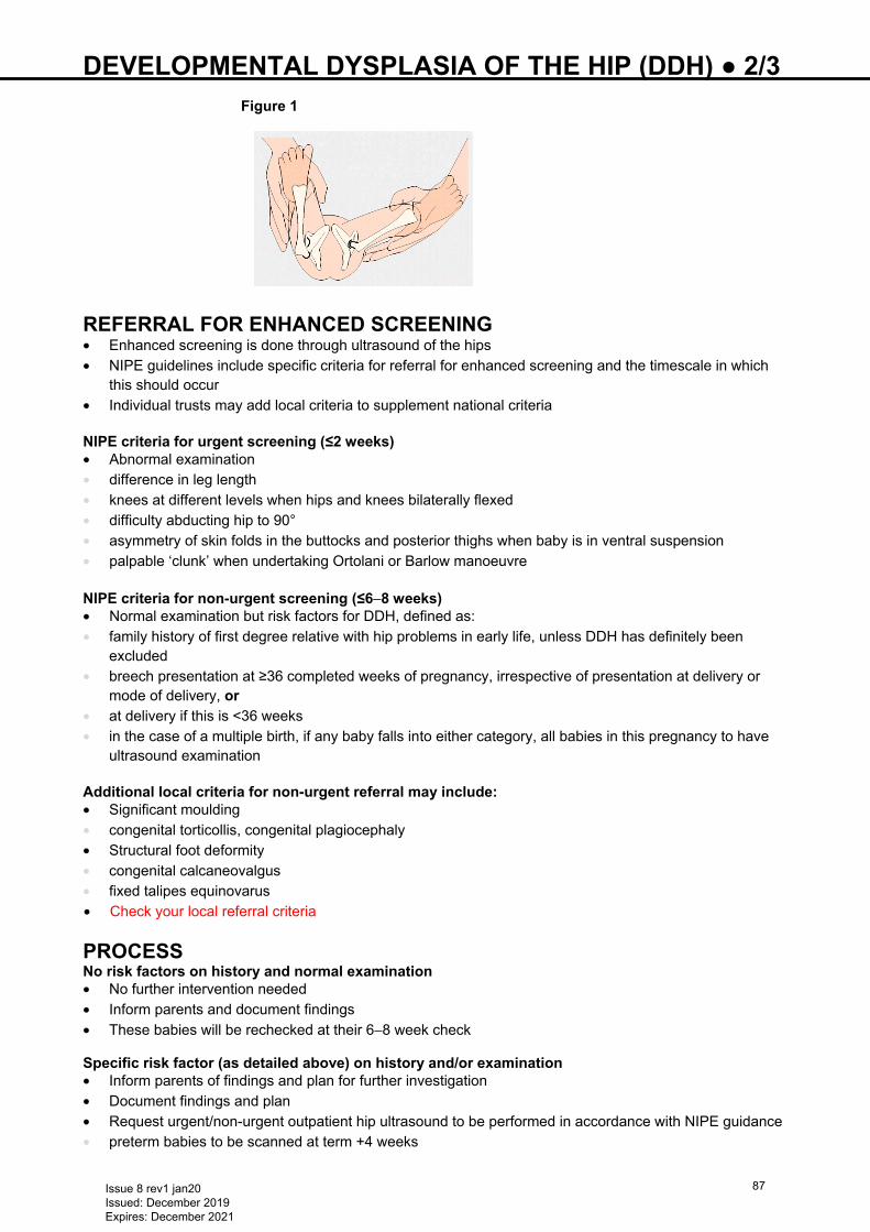
DEVELOPMENTAL DYSPLASIA OF THE HIP (DDH) ● 2/3
Figure 1
REFERRAL FOR ENHANCED SCREENING • Enhanced screening is done through ultrasound of the hips • NIPE guidelines include specific criteria for referral for enhanced screening and the timescale in which
this should occur • Individual trusts may add local criteria to supplement national criteria NIPE criteria for urgent screening (≤2 weeks) • Abnormal examination • difference in leg length • knees at different levels when hips and knees bilaterally flexed • difficulty abducting hip to 90° • asymmetry of skin folds in the buttocks and posterior thighs when baby is in ventral suspension • palpable ‘clunk’ when undertaking Ortolani or Barlow manoeuvre NIPE criteria for non-urgent screening (≤6−8 weeks) • Normal examination but risk factors for DDH, defined as: • family history of first degree relative with hip problems in early life, unless DDH has definitely been
excluded • breech presentation at ≥36 completed weeks of pregnancy, irrespective of presentation at delivery or
mode of delivery, or • at delivery if this is <36 weeks • in the case of a multiple birth, if any baby falls into either category, all babies in this pregnancy to have
ultrasound examination Additional local criteria for non-urgent referral may include: • Significant moulding • congenital torticollis, congenital plagiocephaly • Structural foot deformity • congenital calcaneovalgus • fixed talipes equinovarus • Check your local referral criteria
PROCESS No risk factors on history and normal examination • No further intervention needed • Inform parents and document findings • These babies will be rechecked at their 6−8 week check
Specific risk factor (as detailed above) on history and/or examination • Inform parents of findings and plan for further investigation • Document findings and plan • Request urgent/non-urgent outpatient hip ultrasound to be performed in accordance with NIPE guidance • preterm babies to be scanned at term +4 weeks
Issue 8 rev1 jan20 Issued: December 2019 Expires: December 2021
87
DEVELOPMENTAL DYSPLASIA OF THE HIP (DDH) ● 3/3 • Departments to have system in place to review all hip scan results and inform parents as they are
reported • babies with normal hip scan require no further action and will be re-examined at their
6−8 week check • babies with abnormal hip scan require an expert consultation aged ≤8 weeks Dislocated/dislocatable/unstable hip – positive Ortolani or Barlow test or limited hip abduction • Review by middle grade or consultant to confirm diagnosis • Inform parents of findings and plan for further investigation and management • Document findings and plan • Urgent referral required • Check local policy regarding referral to physiotherapy/orthopaedic team and urgent ultrasound. Service
may be provided locally or referral to a tertiary centre paediatric orthopaedic team may be required
Issue 8 rev1 jan20 Issued: December 2019 Expires: December 2021
88
DISCHARGE FROM NEONATAL UNIT ● 1/2 DECISION TO DISCHARGE • Only consultant or middle grade should discharge • Medical and nursing staff to agree discharge date with parents or persons with parental responsibility • Nursing team perform majority of discharge requirements
DISCHARGE CHECKLIST Where appropriate, the following must be achieved before discharge:
Parental competencies • Administration of medications when required • Baby cares (e.g. nappy changes, top and tailing, bathing etc.) • Feeding • Nasogastric tube feeding where necessary • Stoma care (surgical babies) • Home oxygen where necessary Parent education • In addition to above, it is best practice to offer parents education on: • basic neonatal resuscitation (practical demonstration or leaflet/DVD etc.) • common infectious illnesses (see Bliss.org.uk/parents/about-your-baby/common-infectious-illnesses) • immunisations, if not already received (give national leaflet)
Parent communication • Check home and discharge addresses and confirm name of GP with parents • Complete Red Book (include immunisations given and dates) and give to parents • Give parents copy of discharge summary and time to ask questions after they have read it • Follow local policy for breast pump loan and/or return • Ensure parents have information regarding local breastfeeding groups for ongoing support, and BLISS
support group meeting • Ensure parents have up-to-date safety information • if transporting in a car, use suitable car seat • If transferring to another unit, ensure parents understand reason for transfer. Provide information about
receiving unit • Ensure remaining breast milk in hospital fridge/freezer given to take home Parent information Local unit discharge pack Procedures/investigations • Newborn bloodspot (see Bloodspot screening guideline) • for babies <32 weeks’ gestation, repeat on day 28 or day of discharge if sooner • When immunisation (2, 3 and 4 month) not complete in preterm babies, inform GP and health visitor • Give BCG immunisation if required (see BCG immunisation guideline) • Complete audiology screening (see Hearing screening guideline) • Where required, confirm ophthalmology appointment date [see Retinopathy of prematurity (ROP)
screening guideline] • If going home on oxygen, follow appropriate guidelines
Professional communication • Complete admission book entries • Inform: • health visitor of discharge • community midwife if baby aged <10 days • if safeguarding concerns and baby aged <28 days, notify community midwife • GP • community neonatal or paediatric team as required locally Multidisciplinary (MDT) review/discharge planning meeting • Babies with safeguarding concerns (to formulate child protection plan) • Babies with complex needs • Other appropriate babies
Issue 8 rev1 jan20 Issued: December 2019 Expires: December 2021
89
DISCHARGE FROM NEONATAL UNIT ● 2/2 Medical team • Complete discharge summary by date of discharge • Complete neonatal dataset by date of discharge • Answer parents’ questions after they have read discharge summary • Ensure all follow-up appointments made (see Follow-up) • Perform and record discharge examination
FOLLOW-UP Appointments • Parents to have single point of contact following discharge • Ensure appointments are written on discharge summary and in Red Book. Likely appointments could
include: • neonatal/paediatric consultant outpatient clinic • ophthalmology screening • audiology referral • cranial ultrasound • brain US/MRI scan • physiotherapy • hip or renal ultrasound • dietitian • community paediatrician • child development centre • BCG immunisation or palivizumab • planned future admission (e.g. for immunisations) • planned future review for blood taking, wound review • tertiary consultant outpatients • Open access to children’s wards where available and appropriate • See also Follow-up of babies discharged from the neonatal unit guideline
Issue 8 rev1 jan20 Issued: December 2019 Expires: December 2021
90
DISORDERS OF SEXUAL DEVELOPMENT ● 1/2 RECOGNITION AND ASSESSMENT Definition • New nomenclature: disorders of sexual development (DSD) known formerly as ambiguous genitalia • Congenital conditions in which development of chromosomal, gonadal or anatomical sex is atypical,
most commonly: • congenital adrenal hyperplasia • gonadal dysgenesis • partial androgen insensitivity • For DSD classification, see Supporting information Factors suggesting DSD • Overt genital ambiguity (e.g. cloacal extrophy) • Apparent female genitalia with enlarged clitoris, posterior labial fusion or inguinal/labial masses • Apparent male genitalia with bilateral undescended testes, isolated perineal hypospadias or mild
hypospadias with undescended testis • Family history of DSD e.g. complete androgen insensitivity syndrome (CAIS) • Discordance between genital appearance and antenatal karyotype • Pseudo-ambiguity (atrophic vulva and clitoral oedema) in growth-restricted or preterm female babies
PRINCIPLES OF MANAGEMENT
This is a medical emergency; involve consultant immediately
• Avoid gender assignment before expert evaluation • Consultant to discuss with parents • always use the term ‘baby’ and avoid using ‘he’, ‘she’ or, most importantly, ‘it’ • advise parents about delaying registration and informing wider family and friends until gender
assignment complete • liaise with laboratory to enable evaluation without indicating gender in laboratory request forms • Link with expert centre for appropriate evaluation • Communicate openly with family • Respect family concerns and culture • DSD is not shameful • potential for well-adjusted individual and a functioning member of society • best course of action may not be clear initially • parents need time to understand sexual development First line investigations • Blood pressure • Karyotype of QF-PCR (urgent) • Imaging • abdominal and pelvic ultrasound by an experienced paediatric sonographer • assess presence and nature of internal genitalia, including gonads • Blood tests • 17-OHP (delay until day 3–5 to allow maternal hormonal effects to decline) • testosterone and oestradiol • LH, FSH • U&E and glucose • cortisol
Further investigations (locally and/or in conjunction with specialist advice) • dHT (dihydrotestosterone) • DHEA (dihydroepiandrosterone) • Androstenedione • ACTH • LHRH and hCG stimulation • ACTH stimulation test • AMH (anti-mullerian hormone) imaging studies • Molecular genetic studies [e.g. for complete androgen insensitivity syndrome (CAIS)] • Urine: steroid profile
Issue 8 rev1 jan20 Issued: December 2019 Expires: December 2021
91
DISORDERS OF SEXUAL DEVELOPMENT ● 2/2 • Biopsy of gonad
TREATMENT • Avoid unnecessary admission to NNU • Check serum electrolytes and plasma glucose • Involves a multidisciplinary team with an identified person (usually consultant neonatologist) acting as
primary contact with family • Specific treatment dependent on many factors and diagnosis • discuss with specialists
Issue 8 rev1 jan20 Issued: December 2019 Expires: December 2021
92
DOWN SYNDROME – INITIAL MANAGEMENT ● 1/4 INTRODUCTION • Congenital disorder arising from a chromosome defect • Majority due to trisomy of chromosome 21 • 4% translocations • 1% mosaics • Antenatal screening and subsequent termination of pregnancies results in incidence at birth of 0.8/1000 • Incidence increases with increasing age of mother from 1:1500 at aged 20 yr to 1:100 aged 40 yr
DIAGNOSIS Antenatal • Confirm cases identified through antenatal screening/high-risk women by amniocentesis/chorionic villi
sampling (CVS) • Arrange for parents to be seen by neonatal/paediatric consultant • Complete local paediatric alert register for postnatal care • Give parents opportunity to visit NNU Postnatal • Approximately 30% of cases are not identified before birth – mainly due to screening declined/not
undertaken • If suspected on newborn and infant physical examination (NIPE), request immediate detailed clinical
examination by paediatrician/advanced neonatal nurse practitioner • Identify any urgent medical needs (e.g. feeding, cardiac or respiratory problems) • Consultant paediatrician to discuss testing with parents • Send EDTA blood sample to regional genetic laboratory for confirmation by testing QF-PCR • referral laboratory will request lithium heparin sample for karyotyping if necessary Parent consultation • Parents may have conflicting emotions (e.g. grief, anger) and may initially reject baby • Parents to be seen by consultant: • antenatally diagnosed: ≤24 hr of birth • postnatally diagnosed: ≤24 hr of suspicion • use interpreter for non-English speaking parent • if possible/appropriate both parents to be present during consultation • deliver explanation of baby’s features and diagnosis sensitively • give parents time to absorb information • avoid multiple consultations with different doctors • Repeat visits may be necessary to deal with questions and distress • If possible, same consultant to continue to see baby and parents until discharge • if not possible, named/follow-up consultant must have clear handover
Issue 8 rev1 jan20 Issued: December 2019 Expires: December 2021
93
DOWN SYNDROME – INITIAL MANAGEMENT ● 2/4 INITIAL MANAGEMENT
Age Professional TasksBirth
Consultant paediatrician/ neonatologist
• Neonatal examination • QF-PCR genetic testing +/- karyotyping to confirm Down
syndrome • Blood for chromosomes, FBC and manual blood film for
peripheral blasts • if blasts present discuss with paediatric haematologist • Counselling of parents by consultant (see Parent
consultation) • Give written information to parents (e.g. Down Syndrome
Association pack with new parent leaflet − available from http://www.downs-syndrome.org.uk/for-new-parents/new-parent-pack/)
• Notify midwife, obstetrician, GP, and health visitor • Cardiac assessment including: • pre and postductal pulse oximetry • ECG (if available locally) • if cardiac symptoms/signs or abnormal ECG, detailed clinical
cardiac examination including echocardiogram within 2 weeks • if no cardiac symptoms and normal ECG, cardiac review and
echocardiogram within 4−6 weeks • Gastrointestinal atresia − observe for vomiting (bile stained) • Hirschsprung’s disease − ensure meconium passed ≤24 hr of
birth • Visual assessment: • check visual behaviour and red reflexes for congenital cataract
and nystagmus • if concerns refer to ophthalmologist • Follow-up with a paediatrician/neonatologist • Refer to community paediatric team with detailed summary and
copies of all other referrals (e.g. ophthalmology, cardiology) • Discuss referral to early support services (ESS) • Nurse specialist/dietician to provide feeding advice • Speech and language assessment/therapy referral where
necessary • Provide parents with information and/or additional sources of
help and advice • Replace growth charts in personal child health record (PCHR)
and notes with specific Down syndrome insert/chart and plot growth parameters
• Check automatic referral to audiology has been made ≤5 days Midwife • Risk of congenital hypothyroidism − ensure heel prick test
performed 2−4 weeks Consultant
paediatrician Follow-up appointment• Review parental concerns and medical history, particularly
cardiac symptoms, feeding and bowel habit • Ensure Down syndrome insert in PCHR and growth parameters
plotted on Down syndrome growth chart • Cardiac examination • check seen in cardiology clinic • Examine eyes for cataract and nystagmus • Verify results of TSH screen • Check referral to child development centre • If concerns refer to dietician and community speech and
language therapy (SALT) 6 weeks Health visitor/GP Routine Child Health Service − primary birth visit
• Plot growth on Down syndrome chart • Issue Down syndrome specific pages and growth chart for
PCHR if not already issued
Issue 8 rev1 jan20 Issued: December 2019 Expires: December 2021
94
DOWN SYNDROME – INITIAL MANAGEMENT ● 3/4
Age Professional Tasks8 weeks Health visitor/GP • Primary immunisations3–4 months
Paediatrician/ community paediatrician/child development centre
Initial assessment• Developmental assessment • Refer to physiotherapy as appropriate • Ensure referred to ophthalmologist and SALT • Review newborn hearing screening programme results (in
PCHR) • Hearing screening • if no clear bilateral or unilateral response: refer for audiological
assessment • if bilateral clear response: ensure referral for targeted follow-up
aged 7−9 months • Refer to ESS
• Complete neonatal checklist for management of babies with Down syndrome (if available locally)
LATER REVIEWS • At all stages review/discuss: • parental concern • developmental progress • monitor growth using local Down syndrome chart • hearing and visual problems • Formal ophthalmological and audiology assessment every 2−3 yr • more often if abnormal • Copy clinic letters to parents and all professionals involved
Age Review/action9 months • Follow surveillance check list, if available locally
• Exclude squint • Audiology assessment • Developmental progress
12 months • TSH, FT4 and thyroid antibodies, then every 2nd year • if TSH levels elevated/positive antibodies present, discuss with endocrine
team 18 months • Developmental progress review
• discuss schools/nurseries as appropriate • Growth monitoring and plotting on Down syndrome chart every visit • Check dental health and refer to specialist community paediatric dentist • Check gastrointestinal symptoms – constipation/diarrhoea, increased risk
of coeliac disease • If symptoms of obstructive sleep apnoea present refer to ENT team • Assess gait, bowel and bladder function • risk of atlanto-axial subluxation − suspect if new symptoms of gait
disturbance, abnormal neck posture and/or deterioration in bladder/bowel function
• Increased incidences of: • type 1 diabetes (10 x normal) • autism • leukaemia • Advise parents about relevant benefits e.g. disability living allowance
(DLA) • DLA has 2 parts − care and mobility
− all children with Down syndrome will eventually receive DLA − advise parents to consider application when child requires more help
than children of a similar age (local authorities have denied applications on the basis that a baby with Down syndrome has the same needs as any other baby)
− application for mobility element of DLA can only be made when child aged ≥3 yr
• Give information about local and national Down syndrome support groups
Issue 8 rev1 jan20 Issued: December 2019 Expires: December 2021
95
DOWN SYNDROME – INITIAL MANAGEMENT ● 4/4 FURTHER USEFUL INFORMATION • Downs Syndrome Association: www.downs-syndrome.org.uk • Downs Syndrome Medical Interest Group: www.dsmig.org.uk
Issue 8 rev1 jan20 Issued: December 2019 Expires: December 2021
96
DROPPED BABY ● 1/2 ASSESSMENT • Ensure baby placed on a safe surface, ideally resuscitaire • Assess: • heart rate • respiratory rate • responsiveness • If urgent clinical concerns call neonatal crash team, otherwise request medical review by neonatal
middle grade • Take full history and examine baby • detailed documentation of all injuries • review vitamin K history/administration • Record occipital frontal circumference • Update parents • Move baby to NNU/special care baby unit and admit for ≥24 hr for observations
MONITOR • Perform: • continuous ECG • saturation monitoring • neurological examination: − hourly for first 12 hr, then − 2-hrly for 24 hr
INVESTIGATIONS
Urgent CT head scan Indicated if injury is suspected or if the baby has/subsequently develops abnormal clinical signs • If baby sustained head injury and clinical concerns of skull fracture or cranial ultrasound abnormalities,
arrange urgent CT scan of head • Provisional written radiology report should be made available within 1 hr of scan
Vomiting is not a reliable sign in infants
• If any of the following risk factors, CT head scan within ≤1 hr • suspicion of non-accidental injury • seizure • altered state of consciousness on initial assessment or at 2 hr after injury • suspected open/depressed skull fracture • tense fontanelle • any sign of basal skull fracture − haemotympanum − 'panda' eyes − cerebrospinal fluid leakage from ear or nose − Battle's sign (bruising over mastoid process) • focal neurological deficit • bruise, swelling or laceration >5 cm on head • recurrent vomiting • altered state of consciousness
DOCUMENTATION • Complete incident form • Consider possibility of non-accidental injury
SUBSEQUENT MANAGEMENT • If CT abnormal discuss with neurosurgical centre
Issue 8 rev1 jan20 Issued: December 2019 Expires: December 2021
97
DROPPED BABY ● 2/2 • If CT normal/not indicated continue to monitor baby as described above until 24 hr of normal
observations have been documented
Issue 8 rev1 jan20 Issued: December 2019 Expires: December 2021
98
ECG ABNORMALITIES ● 1/4 INTRODUCTION • 1−5% of normal newborn infants have irregularities in heart rhythm within the first 10 days of life. These
are largely self-limiting and benign • Need to distinguish between: • normal sinus arrhythmia (including pauses up to 1.5 sec) • benign arrhythmias (including premature atrial and premature ventricular contractions) • pathological arrhythmias [can be divided into tachyarrhythmia’s (SVT and VT) and bradyarrhythmias
(congenital heart block)]
PREMATURE ATRIAL BEAT Recognition and assessment • Most common form of arrhythmia • In a regular sinus rhythm at a normal rate, a P wave occurring before next expected P wave is a
premature atrial beat • Usually has a different morphology (P wave different in shape and size to normal P wave) • Most premature atrial beats are benign
Management • 12-lead ECG – ensure rate, rhythm and QTc are calculated • Follow-up ECG aged 1 month (small risk of SVT) • if premature atrial contractions persist, seek cardiology advice
PREMATURE VENTRICULAR BEAT Recognition and assessment • Premature abnormal QRS complex not preceded by premature P wave
Investigations • 12-lead ECG – ensure rate, rhythm and QTc are calculated • Echocardiogram to rule out structural abnormality of heart
Immediate treatment • Seek advice from paediatric cardiologist
SINUS TACHYCARDIA Recognition and assessment • Sinus rhythm (P wave precedes every QRS complex) with a heart rate above normal limit for age and
gestation Causes • Fever • Infection • Low haemoglobin • Pain • Prematurity • Hypovolaemia • Hyperthyroidism • Myocarditis • Drugs (e.g. caffeine and salbutamol)
Management • Treat the cause • If myocarditis suspected − echocardiogram
SINUS BRADYCARDIA Recognition and assessment • Sinus rhythm (P wave precedes every QRS complex) with a heart rate below normal limit for age and
gestation
Issue 8 rev1 jan20 Issued: December 2019 Expires: December 2021
99
ECG ABNORMALITIES ● 2/4 Differential diagnosis • Hypoxia (most likely cause) • Vagal stimulation • Post-intubation • Hypovolaemia • Hypothermia • Metabolic derangement • Hypopituitarism • Obstructive jaundice • Drugs passed from mother to baby (labetalol) • Maternal SLE
Immediate management • Manage airway and breathing • If intubation required, optimise ETT position • If bradycardia occurs post-intubation, use atropine (see Neonatal Formulary) • Correct hypovolaemia • Correct metabolic derangement • If persistent, obtain 12-lead ECG • Evaluate and treat underlying cause
SUPRAVENTRICULAR TACHYCARDIA Recognition and assessment • Rapid regular tachyarrhythmia • Heart rate >230 bpm • ECG: • P waves commonly absent. When present they almost always have an abnormal morphology • narrow QRS complex • in fast sinus tachycardia, P waves can be very difficult to see • look for delta waves consistent with Wolff-Parkinson-White syndrome as this can affect the choice of
anti-arrhythmic agent used • For further information see Supraventricular tachycardia guideline
VENTRICULAR TACHYCARDIA Recognition and assessment • Heart rate >200 bpm • Wide QRS complexes • ≥3 repetitive complexes
Immediate management • Manage airway and breathing • Correct hypoxia • Correct electrolyte disturbance • Discuss with paediatric cardiology centre • Consider synchronised cardioversion (in very fast heart rates, defibrillators cannot synchronise with the
patient and unsynchronised will be required) if intubated, with analgesia • Amiodarone 5 mg/kg over 30 min IV (repeat if necessary) • If no response, lidocaine 0.5–1 mg/kg IV. May be repeated after 5 min. Maximum cumulative dose 3
mg/kg
TACHYARRHYTHMIA • True heart rate? • Is baby crying/in pain? • Check airway and breathing • Check saturation • Consider arterial/capillary gas • Check perfusion • Check blood pressure • Manage airway and breathing
Issue 8 rev1 jan20 Issued: December 2019 Expires: December 2021
100

ECG ABNORMALITIES ● 3/4 • Correct hypoxia • Correct electrolyte disturbance
Premature atrial complexes with pauses
Premature ventricular complex
12-lead ECG
• Narrow QRS complex • Absent/abnormal P wave • vagal manoeuvres • adenosine • discuss with paediatric cardiologist • consider synchronised cardioversion
• Broad QRS complex • Abnormal P wave • discuss with paediatric cardiologist • synchronised cardioversion
Issue 8 rev1 jan20 Issued: December 2019 Expires: December 2021
101

ECG ABNORMALITIES ● 4/4 Supraventricular tachycardia
Issue 8 rev1 jan20 Issued: December 2019 Expires: December 2021
102
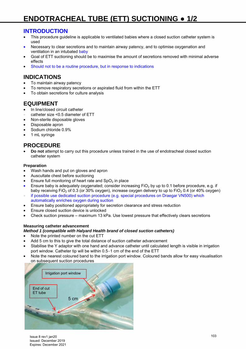
ENDOTRACHEAL TUBE (ETT) SUCTIONING ● 1/2 INTRODUCTION • This procedure guideline is applicable to ventilated babies where a closed suction catheter system is
used • Necessary to clear secretions and to maintain airway patency, and to optimise oxygenation and
ventilation in an intubated baby • Goal of ETT suctioning should be to maximise the amount of secretions removed with minimal adverse
effects • Should not to be a routine procedure, but in response to indications
INDICATIONS • To maintain airway patency • To remove respiratory secretions or aspirated fluid from within the ETT • To obtain secretions for culture analysis
EQUIPMENT • In line/closed circuit catheter • catheter size <0.5 diameter of ETT • Non-sterile disposable gloves • Disposable apron • Sodium chloride 0.9% • 1 mL syringe
PROCEDURE • Do not attempt to carry out this procedure unless trained in the use of endotracheal closed suction
catheter system Preparation • Wash hands and put on gloves and apron • Auscultate chest before suctioning • Ensure full monitoring of heart rate and SpO2 in place • Ensure baby is adequately oxygenated; consider increasing FiO2 by up to 0.1 before procedure, e.g. if
baby receiving FiO2 of 0.3 (or 30% oxygen), increase oxygen delivery to up to FiO2 0.4 (or 40% oxygen) • if possible use dedicated suction procedure (e.g. special procedures on Draegar VN500) which
automatically enriches oxygen during suction • Ensure baby positioned appropriately for secretion clearance and stress reduction • Ensure closed suction device is unlocked • Check suction pressure – maximum 13 kPa. Use lowest pressure that effectively clears secretions Measuring catheter advancement Method 1 (compatible with Halyard Health brand of closed suction catheters) • Note the printed number on the cut ETT • Add 5 cm to this to give the total distance of suction catheter advancement • Stabilise the Y adaptor with one hand and advance catheter until calculated length is visible in irrigation
port window. Catheter tip will be within 0.5−1 cm of the end of the ETT • Note the nearest coloured band to the irrigation port window. Coloured bands allow for easy visualisation
on subsequent suction procedures
Irrigation port window
5 cm
End of cut ET tube
Issue 8 rev1 jan20 Issued: December 2019 Expires: December 2021
103
ENDOTRACHEAL TUBE (ETT) SUCTIONING ● 2/2 Method 2 • Stabilise the Y adaptor with one hand • Advance catheter until printed depth numbers on catheter align with the same numbers printed on the
ETT • Catheter tip will be within 0.5−1 cm of the end of the ETT
Performing suctioning Ensure suction catheter correctly advanced using either methods 1 or 2 (above) • Depress thumb control valve and hold while withdrawing catheter slowly • When tip of suction catheter reaches dome, release thumb control valve and stop withdrawing • Procedure should take ≤10 sec and the duration of negative pressure should be ≤5 sec • Repeat procedure if necessary • Do not use sodium chloride 0.9% instillation routinely. Sodium chloride 0.9% ≤0.5 mL may be instilled
before suctioning if secretions are thick and tenacious and cannot be extracted by suctioning alone • After each suctioning episode ensure the closed circuit is flushed with sodium chloride 0.9% according to
manufacturer’s instructions
DOCUMENTATION • Record procedure in nursing documentation, noting distance tube was passed and colour of band on
catheter tube closest to this measured distance
AFTERCARE Equipment • Leave thumb valve in locked position when not in use to prevent inadvertent activation • Leave catheter tip in dome between use • Device is single use only and replace every 24 hr as per manufacturer’s guidance Monitoring • Ensure monitoring of heart rate and SpO2 continues after procedure • Auscultate baby’s chest after procedure and document any changes observed • If FiO2 was adjusted before procedure, return to original settings, or ensure baby’s target FiO2 is
maintained Reporting adverse events • Report adverse incidents using local risk management procedure
COMPLICATIONS • Hypoxaemia • Atelectasis • Bradycardia • Tachycardia • Blood pressure fluctuations • Decreased tidal volume • Airway mucosal trauma • Dislodgement of ETT • Extubation • Pneumothorax • Pneumomediastinum • Bacteraemia • Pneumonia • Fluctuations in intracranial pressure and cerebral blood flow velocity
FURTHER INFORMATION • Further details on ETT closed suction can be found in the manufacturer’s guidance
Issue 8 rev1 jan20 Issued: December 2019 Expires: December 2021
104
ENVIRONMENT AND NOISE ● 1/1
ENVIRONMENT Lighting Excessive and rapid changes in light levels may cause physiological instability, disturbed sleep and interfere with visual development. The thin eyelids of preterm babies may allow significant light to penetrate even if eyes closed
Aim Method • Provide flexible lighting
to meet individual developmental needs and caregiver’s needs
• Ensure sufficient lighting for observation and care delivery
• Promote optimal extra-uterine development and physiological stability
• Reduce stress • Protect sleep • Development of normal
circadian rhythms
• Keep lighting levels around 200–300 lux (moderate room lighting) • Monitor and audit light levels in nursery and baby’s immediate
environment regularly • Daylight preferable to artificial lighting. Protect babies from direct
sunlight • Avoid direct bright light during feeding • Use dimmer switches and avoid sudden changes in light levels • Use incubator covers or canopies for preterm, sick or neurologically
compromised babies • keep a corner/flap up to allow safe observation • Protect babies in open cots from bright light until near term (37–40 weeks) • Use night lights for development of day–night cycle • Use individual task lighting for care and procedures. Shade baby’s eyes
throughout • Protect babies from phototherapy and bright lights in other bed spaces • Promote appropriate visual interactions with parents/carers • Protect babies from bright light for ≥18 hr following ROP screening
NOISE • High levels of sound may cause: • baby distress • sleep disturbance • damage to hearing • impaired language and speech development • A noisy environment affects behaviour and wellbeing of adults present, with impact on confidentiality,
communication, stress levels and ability to concentrate, make decisions and perform fine motor tasks
Aim Method • Promote optimal extra-
uterine development and physiological stability
• Protect sleep • Maintain confidentiality
and privacy • Promote normal
speech and language development
• Provide appropriate working environment
• Monitor noise levels in nursery and baby’s immediate environment • Maintain ambient noise levels at 45 dB, with occasional peaks of 70 dB • Observe baby’s cues to ensure noise levels do not indicate stress • Open packaging outside incubator • Keep monitor alarms and telephone ring tones at quiet but safe audible
levels (silence alarms quickly) • Empty ‘rainout’ from ventilator tubing as soon as possible • Turn off suction when not in use • Close incubator doors and bins gently • Use incubator covers or canopies for preterm, sick or neurologically
compromised babies • keep a corner/flap up to allow safe observation • Keep conversations away from babies and speak quietly • Encourage parents/carers to speak softly to baby • Maintain quiet environment during oral feeding • Only use radios, portable music devices, musical toys etc. when
clinically indicated and ensure other babies are not disturbed • Promote ≥1 ‘rest time’ per day. Lower light and noise levels and
suspend all routine procedures/ward rounds. Leave babies undisturbed to facilitate sleep. Encourage parents to view this as a quiet time to spend with baby
• Reduce noise level as much as possible • Educate staff and parents regarding benefits of a quiet environment
Issue 8 rev1 jan20 Issued: December 2019 Expires: December 2021
105
EXAMINATION OF THE NEWBORN ● 1/4 INDICATIONS • Comprehensive physical examination performed within <72 hr of life • See − http://www.e-lfh.org.uk/programmes/screening • Includes screening for: • developmental dysplasia of the hip • congenital cataracts • cryptorchidism • Assessment of the heart • General physical examination • Examination has limitations and cannot identify all abnormalities that may be present in the newborn
period • Provides reassurance to parents and opportunity for discussion
EQUIPMENT • Maternal and baby notes • Stethoscope • Ophthalmoscope • Measuring tape
AIMS • Identify congenital malformations • Identify common neonatal problems and initiate management • Continue with screening, begun antenatally, to identify need for specific interventions (e.g. immunisation)
PRE-PROCEDURE • Before undertaking clinical examination, familiarise yourself with maternal history and pregnancy
records, including: • maternal medical, obstetric and social history • paternal medical history, if appropriate • family health, history of congenital diseases • identify drugs mother may have taken during pregnancy and in labour • health of siblings • identify pregnancy complications, blood tests, ultrasound scans, admissions to hospital • identify maternal blood group, presence of antibodies, serology results for sexually transmitted diseases • duration of labour, type of delivery, duration of rupture of membranes, condition of liquor • Apgar scores and whether resuscitation required • birth weight, gestational age, head circumference Consent and preparation • Introduce yourself to mother and gain oral consent. Ask about particular concerns • Keep baby warm and examine in quiet environment
PROCEDURE Skin examination • Hydration • Rashes: including erythema toxicum, milia, miliaria, staphylococcal skin infection, candida • Pigmented lesions: naevi, Mongolian blue spots, birth marks, café au lait spots • Bruises: traumatic lesions, petechiae • Cutis aplasia • Tufts of hair not on head • Vascular lesions: haemangioma, port wine stain, simple naevus • Colour: pink/cyanosis/jaundice/pallor/plethora • Acrocyanosis • Cutis marmorata Facial examination • General facial appearance to identify common syndromes
Issue 8 rev1 jan20 Issued: December 2019 Expires: December 2021
106
EXAMINATION OF THE NEWBORN ● 2/4 Eyes • Shape • Slant • Size • Position • Strabismus • Nystagmus • Red reflex • Presence of colobomata • Discharges
Nose • Nasal flaring • Patency
Ears • Shape • Position • Tags or pits
Mouth • Size • Cleft lip • Symmetry of movement • Swellings, Epstein’s pearls, ranula, tongue-tie (for parental reassurance) • Teeth • Cleft palate, hard/soft palate, [by both inspection (using tongue depressor) and palpation] • Sucking
Skull • Palpate: • skull for sutures and shape/cranio-synostosis • swellings on scalp, especially crossing suture lines, cephalhaematoma • signs of trauma associated with birth (e.g. chignon from vacuum extraction) • subgalaeal haemorrhage [see Subgaleal haemorrhage (SGH) guideline] • sutures for ridging or undue separation
Neck • Swellings • Movement • Webbing • Traumatic lesions from forceps delivery Clavicles • For fracture Arms and legs • Position and symmetry of movement • Swelling and bruising Hands and feet • Extra digits (polydactyly) • Syndactyly, clinodactyly • Palmer creases • Skin tags • Position and configuration of feet looking for fixed/positional talipes • Overlapping toes Hips • Developmental dysplasia using Ortolani’s and Barlow’s manoeuvres [see Developmental dysplasia of
the hip (DDH) guideline]
Issue 8 rev1 jan20 Issued: December 2019 Expires: December 2021
107
EXAMINATION OF THE NEWBORN ● 3/4 Spine • Curvatures • Dimples • Sacrococcygeal pits • Hairy patches/naevi • Hairy tuft on spine Systems
• Examine (inspection, palpation, auscultation) each system Respiratory • Respiratory rate • grunting • nasal flaring • Chest shape, asymmetry of rib cage, swellings • nipple position, swelling/discharge/extra nipples • Chest movement • presence/absence of recession • Auscultate for breath sounds Cardiovascular • Skin colour/cyanosis • Palpate: • precordium for thrills • peripheral and femoral pulses for rate and volume • central perfusion • Auscultate for heart sounds, murmur(s), rate, rhythm • pulse oximetry of right arm and either leg (<3% difference in SpO2 normal) Gastrointestinal tract
Ask mother how well baby is feeding, whether baby has vomited and, if so, colour of vomit Bilious vomiting may have a surgical cause and needs prompt stabilisation and referral
• Abdominal shape • Presence of distension • Cord stump for discharge or inflammation/umbilical hernia • Presence and position of anus and patency • Stools passed • Palpate abdomen for tenderness, masses and palpable liver • Auscultation is not routinely undertaken unless there are abdominal concerns
Genito-urinary system
Ask mother if baby has passed urine, and how frequently
• Inspect appearance of genitalia: ambiguous? Male genito-urinary system • Penis size (>1 cm) • Position of urethral meatus. Look for hypospadias • Inguinal hernia • Chordee • Urinary stream • Scrotum for colour • Palpate scrotum for presence of 2 testes and presence of hydrocoele Female genito-urinary system • Presence of vaginal discharge (reassure parents about pseudomenstruation) • Skin tags • Inguinal hernia
Issue 8 rev1 jan20 Issued: December 2019 Expires: December 2021
108
EXAMINATION OF THE NEWBORN ● 4/4 • Proximity of genitalia to anal sphincter • Routine palpation of kidneys is not always necessary as antenatal scans will have assessed presence Neurological system • Before beginning examination, observe baby’s posture • Assess: • muscle tone, grasp, responses to stimulation • behaviour • ability to suck • limb movements • cry • head size in relation to body weight • spine, presence of sacral pits, midline spinal skin lesions/tufts of hair • If neurological concerns, initiate Moro and stepping reflexes • Responses to passive movements: • pull-to-sit • ventral suspension • Palpate anterior fontanelle size (<3 cm × 3 cm) and tone
OUTCOME Documentation • Complete neonatal examination record in medical notes and sign and date it. Also complete Child Health
Record (Red Book) and/or in NIPE Smart if used • Record any discussion or advice given to parents Normal examination • If no concerns raised, reassure parents of apparent normality and advise to seek advice if concerns arise
at home • GP will re-examine baby aged 6−8 weeks Abnormal examination • In first instance, seek advice from neonatal registrar/consultant • Refer to postnatal ward guidelines for ongoing management • Refer abnormalities to relevant senior doctor
Issue 8 rev1 jan20 Issued: December 2019 Expires: December 2021
109
EXCHANGE TRANSFUSION ● 1/3 Exchange transfusion replaces withdrawn baby blood with an equal volume of donor blood
Discuss all cases with neonatal consultant
INDICATIONS Haemolytic anaemia • A newborn who has not had an in-utero transfusion (IUT) with a cord Hb <120 g/L and is haemolysing,
may require urgent exchange transfusion to remove antibodies and correct anaemia: • if Hb <100 g/L: discuss urgently with consultant and proceed to exchange transfusion; avoid simple
packed cell transfusions • if Hb 100–120 g/L: obtain 6-hrly bilirubin values and, if rapidly rising or close to exchange transfusion
level (see Table in Jaundice guideline), use intravenous immunoglobin (IVIG) • A newborn who has had IUTs and whose Kleihauer test (this test may not be available in your hospital)
demonstrates a predominance of adult Hb, anaemia can be managed using a top-up transfusion of irradiated, CMV-negative blood
Hyperbilirubinaemia • Discuss promptly with consultant. If bilirubin values approaching guidance below; senior decision
required: • guidance as determined by exchange transfusion line on gestation-specific NICE jaundice chart (see
Table in Jaundice guideline) • if bilirubin rises faster than 8.5 micromol/L/hr despite phototherapy, anticipate need for exchange
transfusion
Other indications • Chronic feto-maternal transfusion
• Disseminated intravascular coagulation (DIC)
COMPLICATIONS • Cardiac arrhythmias • Air embolism • Necrotising enterocolitis • Coagulopathy • Apnoeas and bradycardia • Sepsis • Electrolyte disturbances • Acidosis owing to non-fresh blood • Thrombocytopenia • Late hyporegenerative anaemia
PROCEDURE Prepare • Ensure full intensive care space and equipment available and ready • Allocate 1 doctor/practitioner and 1 other member of nursing staff, both experienced in exchange
transfusion, to care for each baby during procedure; document their names in baby’s notes • Obtain written consent and document in baby’s notes • Phototherapy to be continued during exchange • Calculate volume of blood to be exchanged: double volume exchange removes 90% of baby’s red cells
and 50% of available intravascular bilirubin. Use: • term babies: 160 mL/kg • preterm babies: 200 mL/kg • Order appropriate volume (usually 2 units) of blood from blood bank, stipulating that it must be: • crossmatched against mother’s blood group and antibody status, and (if requested by your blood bank)
baby’s blood group • CMV-negative • irradiated (shelf-life 24 hr) for any baby who has had an in-utero blood transfusion • as fresh as possible, and certainly ≤4 days old • plasma reduced red cells for ‛exchange transfusion’ (haematocrit 0.5–0.6), not SAG-M blood and not
packed cells
Issue 8 rev1 jan20 Issued: December 2019 Expires: December 2021
110
EXCHANGE TRANSFUSION ● 2/3 Prepare baby • Empty stomach using nasogastric tube (see Nasogastric tube insertion guideline) and keep baby nil-
by-mouth • Start IV infusion • Pay attention to thermoregulation, particularly if procedure to be performed under radiant heater • Commence continuous cardiac, temperature and saturation monitoring Document • Blood pressure, respiratory rate, temperature, SpO2 and heart rate every 15 min throughout exchange • Volume of blood in and out with each cycle, and keep a running total
If any change in baby’s cardiorespiratory status, pause exchange by priming catheter with donor blood that will not clot. Discuss with consultant
Prepare blood • Set up blood warmer early (aim for 37°C) • do not use if:
− intermittent bolus infusion i.e. single catheter exchange − blood is exposed to a radiant heater (risk of haemolysis)
• Check blood units as per hospital policy • Connect donor blood to filter and prime blood giving set • Connect to 4-way (if using UVC) or 3-way tap (outside the warmer) as indicated • Ensure donor blood well mixed before and throughout exchange
Technique • Ensure working area sterile
Either • Single catheter push-pull technique • Sequential withdrawal of baby’s blood and infusion of donor blood via a UVC (see Umbilical venous
catheterisation and removal guideline) Or • Isovolumetric or continuous technique • Continuous infusion of donor blood via a venous line with intermittent removal of baby’s blood via an
arterial line • Use umbilical venous or peripheral venous line for infusion and umbilical arterial or peripheral arterial line
for removal of blood (see Umbilical arterial catheterisation and removal, Umbilical venous catheterisation and removal and Arterial line insertion guidelines)
Single catheter or ‛push-pull’ technique • Connect catheter bag (using Vygon connector) and donor blood to 4-way tap and 4-way tap to UVC • Remove 10 mL baby blood from UVC using syringe • Send first sample for serum bilirubin, full blood count, blood culture, blood glucose, calcium, electrolytes,
coagulation and liver function tests • when exchange performed for reasons other than known blood group antibodies, send blood for G6PD
screening and viral serology • Replace precise volume removed with donor blood, slowly using a syringe • Each out-in cycle should replace ≤8.5 mL/kg and take ≥5 min; start with smaller aliquots (10 mL) and
increase to 20 mL (if baby stable and weight allows) only after 30 min. As a guide: • birth weight <1000 g: use 5 mL aliquots • birth weight 1000–2000 g: use 10 mL aliquots • birth weight >2000 g: use 20 mL aliquots • Discard ‛out’ baby blood into catheter bag • Continue out-in cycles every 5 min (maximum aliquot with each cycle) until complete • Send last ‛out’ baby blood sample for serum bilirubin, full blood count, blood culture, blood glucose,
calcium and electrolytes
Isovolumetric or continuous technique • Connect catheter bag, using Vygon connector, to 3-way tap attached to arterial line extension
Never leave arterial line open to catheter bag
Issue 8 rev1 jan20 Issued: December 2019 Expires: December 2021
111
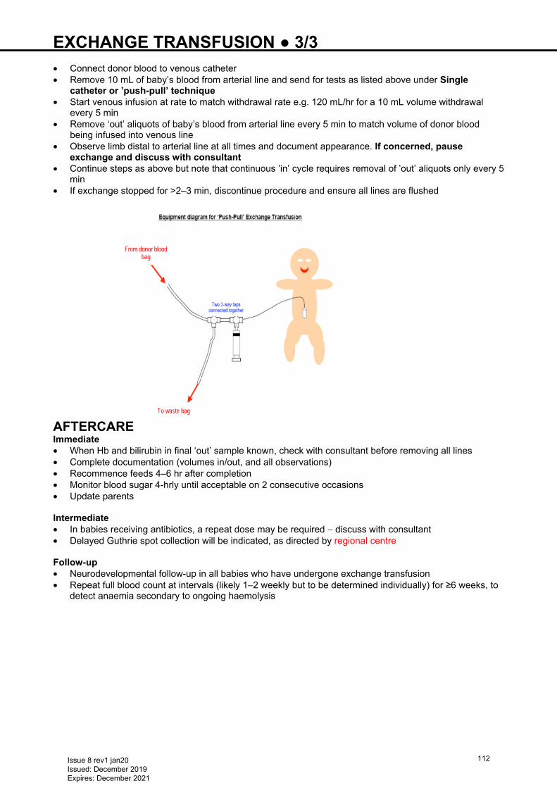
EXCHANGE TRANSFUSION ● 3/3 • Connect donor blood to venous catheter • Remove 10 mL of baby’s blood from arterial line and send for tests as listed above under Single
catheter or ‛push-pull’ technique • Start venous infusion at rate to match withdrawal rate e.g. 120 mL/hr for a 10 mL volume withdrawal
every 5 min • Remove ‘out’ aliquots of baby’s blood from arterial line every 5 min to match volume of donor blood
being infused into venous line • Observe limb distal to arterial line at all times and document appearance. If concerned, pause
exchange and discuss with consultant • Continue steps as above but note that continuous ‛in’ cycle requires removal of ‛out’ aliquots only every 5
min • If exchange stopped for >2–3 min, discontinue procedure and ensure all lines are flushed
AFTERCARE Immediate • When Hb and bilirubin in final ‘out’ sample known, check with consultant before removing all lines • Complete documentation (volumes in/out, and all observations) • Recommence feeds 4–6 hr after completion • Monitor blood sugar 4-hrly until acceptable on 2 consecutive occasions • Update parents Intermediate • In babies receiving antibiotics, a repeat dose may be required − discuss with consultant • Delayed Guthrie spot collection will be indicated, as directed by regional centre Follow-up • Neurodevelopmental follow-up in all babies who have undergone exchange transfusion • Repeat full blood count at intervals (likely 1–2 weekly but to be determined individually) for ≥6 weeks, to
detect anaemia secondary to ongoing haemolysis
From donor blood bag
To waste bag
Issue 8 rev1 jan20 Issued: December 2019 Expires: December 2021
112
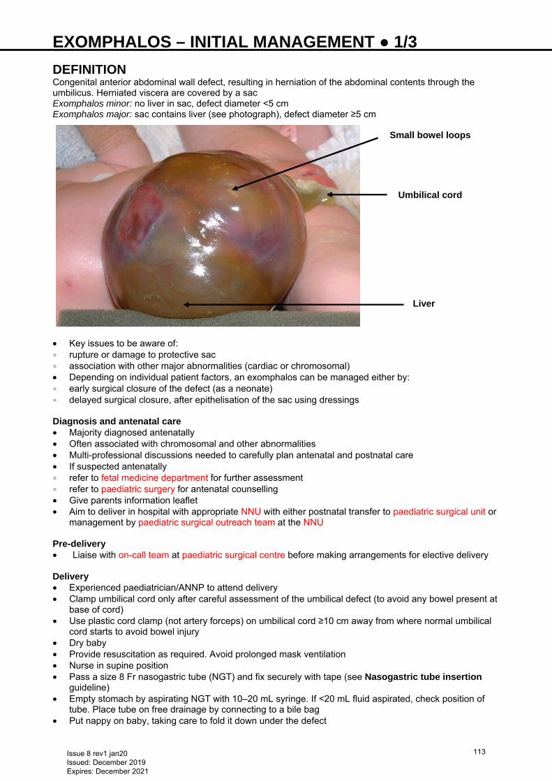
EXOMPHALOS – INITIAL MANAGEMENT ● 1/3 DEFINITION Congenital anterior abdominal wall defect, resulting in herniation of the abdominal contents through the umbilicus. Herniated viscera are covered by a sac Exomphalos minor: no liver in sac, defect diameter <5 cm Exomphalos major: sac contains liver (see photograph), defect diameter ≥5 cm
• Key issues to be aware of: • rupture or damage to protective sac • association with other major abnormalities (cardiac or chromosomal) • Depending on individual patient factors, an exomphalos can be managed either by: • early surgical closure of the defect (as a neonate) • delayed surgical closure, after epithelisation of the sac using dressings
Diagnosis and antenatal care • Majority diagnosed antenatally • Often associated with chromosomal and other abnormalities • Multi-professional discussions needed to carefully plan antenatal and postnatal care • If suspected antenatally • refer to fetal medicine department for further assessment • refer to paediatric surgery for antenatal counselling • Give parents information leaflet • Aim to deliver in hospital with appropriate NNU with either postnatal transfer to paediatric surgical unit or
management by paediatric surgical outreach team at the NNU Pre-delivery • Liaise with on-call team at paediatric surgical centre before making arrangements for elective delivery Delivery • Experienced paediatrician/ANNP to attend delivery • Clamp umbilical cord only after careful assessment of the umbilical defect (to avoid any bowel present at
base of cord) • Use plastic cord clamp (not artery forceps) on umbilical cord ≥10 cm away from where normal umbilical
cord starts to avoid bowel injury • Dry baby • Provide resuscitation as required. Avoid prolonged mask ventilation • Nurse in supine position • Pass a size 8 Fr nasogastric tube (NGT) and fix securely with tape (see Nasogastric tube insertion
guideline) • Empty stomach by aspirating NGT with 10–20 mL syringe. If <20 mL fluid aspirated, check position of
tube. Place tube on free drainage by connecting to a bile bag • Put nappy on baby, taking care to fold it down under the defect
Small bowel loops
Umbilical cord
Liver
Issue 8 rev1 jan20 Issued: December 2019 Expires: December 2021
113
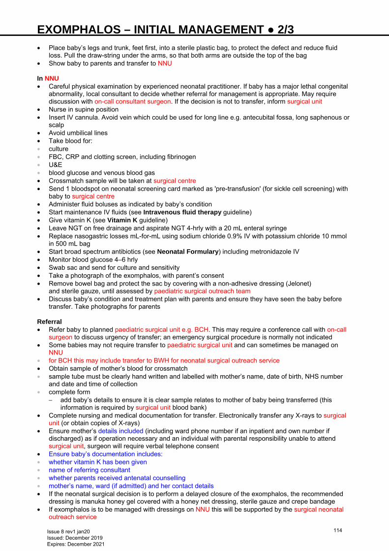
EXOMPHALOS – INITIAL MANAGEMENT ● 2/3 • Place baby’s legs and trunk, feet first, into a sterile plastic bag, to protect the defect and reduce fluid
loss. Pull the draw-string under the arms, so that both arms are outside the top of the bag • Show baby to parents and transfer to NNU In NNU • Careful physical examination by experienced neonatal practitioner. If baby has a major lethal congenital
abnormality, local consultant to decide whether referral for management is appropriate. May require discussion with on-call consultant surgeon. If the decision is not to transfer, inform surgical unit
• Nurse in supine position • Insert IV cannula. Avoid vein which could be used for long line e.g. antecubital fossa, long saphenous or
scalp • Avoid umbilical lines • Take blood for: • culture • FBC, CRP and clotting screen, including fibrinogen • U&E • blood glucose and venous blood gas • Crossmatch sample will be taken at surgical centre • Send 1 bloodspot on neonatal screening card marked as 'pre-transfusion' (for sickle cell screening) with
baby to surgical centre • Administer fluid boluses as indicated by baby’s condition • Start maintenance IV fluids (see Intravenous fluid therapy guideline) • Give vitamin K (see Vitamin K guideline) • Leave NGT on free drainage and aspirate NGT 4-hrly with a 20 mL enteral syringe • Replace nasogastric losses mL-for-mL using sodium chloride 0.9% IV with potassium chloride 10 mmol
in 500 mL bag • Start broad spectrum antibiotics (see Neonatal Formulary) including metronidazole IV • Monitor blood glucose 4–6 hrly • Swab sac and send for culture and sensitivity • Take a photograph of the exomphalos, with parent’s consent • Remove bowel bag and protect the sac by covering with a non-adhesive dressing (Jelonet)
and sterile gauze, until assessed by paediatric surgical outreach team • Discuss baby’s condition and treatment plan with parents and ensure they have seen the baby before
transfer. Take photographs for parents Referral • Refer baby to planned paediatric surgical unit e.g. BCH. This may require a conference call with on-call
surgeon to discuss urgency of transfer; an emergency surgical procedure is normally not indicated • Some babies may not require transfer to paediatric surgical unit and can sometimes be managed on
NNU • for BCH this may include transfer to BWH for neonatal surgical outreach service • Obtain sample of mother’s blood for crossmatch • sample tube must be clearly hand written and labelled with mother’s name, date of birth, NHS number
and date and time of collection • complete form
− add baby’s details to ensure it is clear sample relates to mother of baby being transferred (this information is required by surgical unit blood bank)
• Complete nursing and medical documentation for transfer. Electronically transfer any X-rays to surgical unit (or obtain copies of X-rays)
• Ensure mother’s details included (including ward phone number if an inpatient and own number if discharged) as if operation necessary and an individual with parental responsibility unable to attend surgical unit, surgeon will require verbal telephone consent
• Ensure baby’s documentation includes: • whether vitamin K has been given • name of referring consultant • whether parents received antenatal counselling • mother’s name, ward (if admitted) and her contact details • If the neonatal surgical decision is to perform a delayed closure of the exomphalos, the recommended
dressing is manuka honey gel covered with a honey net dressing, sterile gauze and crepe bandage • If exomphalos is to be managed with dressings on NNU this will be supported by the surgical neonatal
outreach service
Issue 8 rev1 jan20 Issued: December 2019 Expires: December 2021
114
EXOMPHALOS – INITIAL MANAGEMENT ● 3/3 While awaiting transfer • Reassess hourly for further fluid boluses and, if necessary, give either sodium chloride 0.9% or human
albumin solution (HAS) 4.5% 10 mL/kg • If evidence of a coagulopathy, treat appropriately (see Coagulopathy guideline) • Aspirate NGT 4-hrly • Replace nasogastric losses mL-for-mL with sodium chloride 0.9% IV with potassium chloride 10 mmol in
500 mL bag. Leave NGT on free drainage Transfer to surgical unit • Place baby in transport incubator • Take baby to parents (if not yet seen) in transport incubator, en-route to the ambulance • Ensure mother’s blood, baby’s pre-transfusion bloodspots, letters for surgical team and all
documentation accompany baby • Make and document all usual observations during transport and on arrival at surgical unit Useful Information • http://www.bch.nhs.uk/content/neonatal-surgery • http://www.bch.nhs.uk/find-us/maps-directions • Parent information: https://bwc.nhs.uk/download.cfm?doc=docm93jijm4n2517
Issue 8 rev1 jan20 Issued: December 2019 Expires: December 2021
115
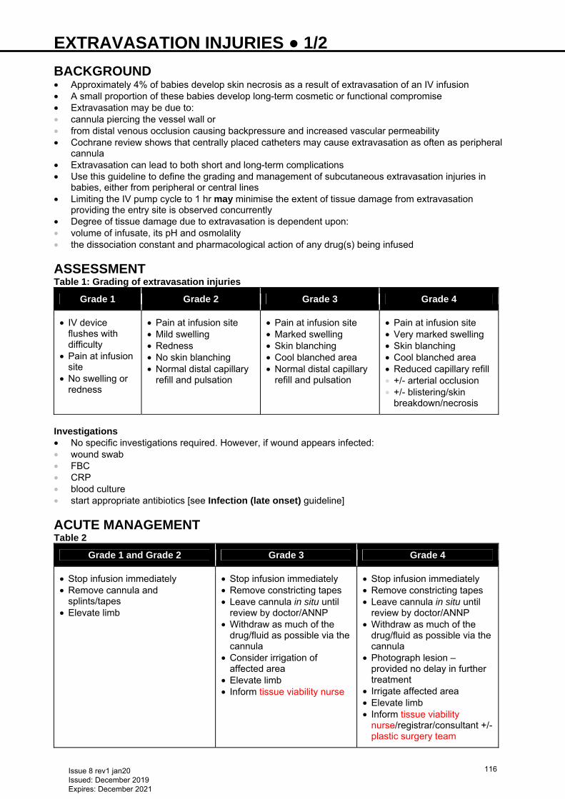
EXTRAVASATION INJURIES ● 1/2 BACKGROUND • Approximately 4% of babies develop skin necrosis as a result of extravasation of an IV infusion • A small proportion of these babies develop long-term cosmetic or functional compromise • Extravasation may be due to: • cannula piercing the vessel wall or • from distal venous occlusion causing backpressure and increased vascular permeability • Cochrane review shows that centrally placed catheters may cause extravasation as often as peripheral
cannula • Extravasation can lead to both short and long-term complications • Use this guideline to define the grading and management of subcutaneous extravasation injuries in
babies, either from peripheral or central lines • Limiting the IV pump cycle to 1 hr may minimise the extent of tissue damage from extravasation
providing the entry site is observed concurrently • Degree of tissue damage due to extravasation is dependent upon: • volume of infusate, its pH and osmolality • the dissociation constant and pharmacological action of any drug(s) being infused
ASSESSMENT Table 1: Grading of extravasation injuries
Grade 1 Grade 2 Grade 3 Grade 4
• IV device flushes with difficulty
• Pain at infusion site
• No swelling or redness
• Pain at infusion site • Mild swelling • Redness • No skin blanching • Normal distal capillary
refill and pulsation
• Pain at infusion site • Marked swelling • Skin blanching • Cool blanched area • Normal distal capillary
refill and pulsation
• Pain at infusion site • Very marked swelling • Skin blanching • Cool blanched area • Reduced capillary refill • +/- arterial occlusion • +/- blistering/skin
breakdown/necrosis
Investigations • No specific investigations required. However, if wound appears infected: • wound swab • FBC • CRP • blood culture • start appropriate antibiotics [see Infection (late onset) guideline]
ACUTE MANAGEMENT Table 2
Grade 1 and Grade 2 Grade 3 Grade 4
• Stop infusion immediately • Remove cannula and
splints/tapes • Elevate limb
• Stop infusion immediately • Remove constricting tapes • Leave cannula in situ until
review by doctor/ANNP • Withdraw as much of the
drug/fluid as possible via the cannula
• Consider irrigation of affected area
• Elevate limb • Inform tissue viability nurse
• Stop infusion immediately • Remove constricting tapes • Leave cannula in situ until
review by doctor/ANNP • Withdraw as much of the
drug/fluid as possible via the cannula
• Photograph lesion – provided no delay in further treatment
• Irrigate affected area • Elevate limb • Inform tissue viability
nurse/registrar/consultant +/- plastic surgery team
Issue 8 rev1 jan20 Issued: December 2019 Expires: December 2021
116
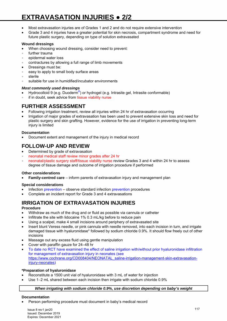
EXTRAVASATION INJURIES ● 2/2 • Most extravasation injuries are of Grades 1 and 2 and do not require extensive intervention • Grade 3 and 4 injuries have a greater potential for skin necrosis, compartment syndrome and need for
future plastic surgery, depending on type of solution extravasated
Wound dressings • When choosing wound dressing, consider need to prevent: • further trauma • epidermal water loss • contractures by allowing a full range of limb movements • Dressings must be: • easy to apply to small body surface areas • sterile • suitable for use in humidified/incubator environments
Most commonly used dressings • Hydrocolloid 9 (e.g. Duoderm®) or hydrogel (e.g. Intrasite gel, Intrasite conformable) • if in doubt, seek advice from tissue viability nurse
FURTHER ASSESSMENT • Following irrigation treatment, review all injuries within 24 hr of extravasation occurring • Irrigation of major grades of extravasation has been used to prevent extensive skin loss and need for
plastic surgery and skin grafting. However, evidence for the use of irrigation in preventing long-term injury is limited
Documentation • Document extent and management of the injury in medical record
FOLLOW-UP AND REVIEW • Determined by grade of extravasation • neonatal medical staff review minor grades after 24 hr • neonatal/plastic surgery staff/tissue viability nurse review Grades 3 and 4 within 24 hr to assess
degree of tissue damage and outcome of irrigation procedure if performed Other considerations • Family-centred care – inform parents of extravasation injury and management plan
Special considerations • Infection prevention – observe standard infection prevention procedures • Complete an incident report for Grade 3 and 4 extravasations
IRRIGATION OF EXTRAVASATION INJURIES Procedure • Withdraw as much of the drug and or fluid as possible via cannula or catheter • Infiltrate the site with lidocaine 1% 0.3 mL/kg before to reduce pain • Using a scalpel, make 4 small incisions around periphery of extravasated site • Insert blunt Veress needle, or pink cannula with needle removed, into each incision in turn, and irrigate
damaged tissue with hyaluronidase* followed by sodium chloride 0.9%. It should flow freely out of other incisions
• Massage out any excess fluid using gentle manipulation • Cover with paraffin gauze for 24–48 hr • To date no RCT have examined the effect of saline irrigation with/without prior hyaluronidase infiltration
for management of extravasation injury in neonates (see https://www.cochrane.org/CD008404/NEONATAL_saline-irrigation-management-skin-extravasation-injury-neonates)
*Preparation of hyaluronidase • Reconstitute a 1500 unit vial of hyaluronidase with 3 mL of water for injection • Use 1−2 mL shared between each incision then irrigate with sodium chloride 0.9%
When irrigating with sodium chloride 0.9%, use discretion depending on baby’s weight Documentation • Person performing procedure must document in baby’s medical record
Issue 8 rev1 jan20 Issued: December 2019 Expires: December 2021
117
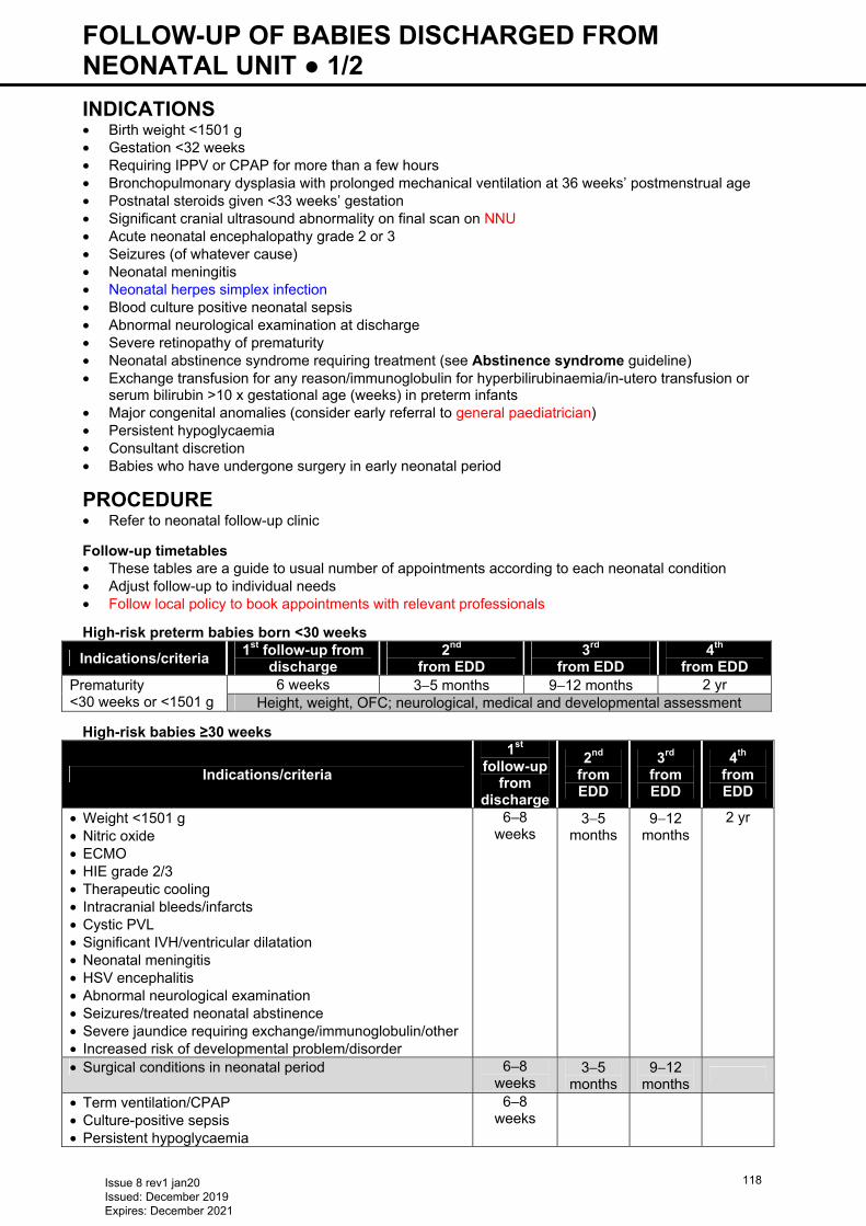
FOLLOW-UP OF BABIES DISCHARGED FROM NEONATAL UNIT ● 1/2 INDICATIONS • Birth weight <1501 g • Gestation <32 weeks • Requiring IPPV or CPAP for more than a few hours • Bronchopulmonary dysplasia with prolonged mechanical ventilation at 36 weeks’ postmenstrual age • Postnatal steroids given <33 weeks’ gestation • Significant cranial ultrasound abnormality on final scan on NNU • Acute neonatal encephalopathy grade 2 or 3 • Seizures (of whatever cause) • Neonatal meningitis • Neonatal herpes simplex infection • Blood culture positive neonatal sepsis • Abnormal neurological examination at discharge • Severe retinopathy of prematurity • Neonatal abstinence syndrome requiring treatment (see Abstinence syndrome guideline) • Exchange transfusion for any reason/immunoglobulin for hyperbilirubinaemia/in-utero transfusion or
serum bilirubin >10 x gestational age (weeks) in preterm infants • Major congenital anomalies (consider early referral to general paediatrician) • Persistent hypoglycaemia • Consultant discretion • Babies who have undergone surgery in early neonatal period
PROCEDURE • Refer to neonatal follow-up clinic
Follow-up timetables • These tables are a guide to usual number of appointments according to each neonatal condition • Adjust follow-up to individual needs • Follow local policy to book appointments with relevant professionals
High-risk preterm babies born <30 weeks
High-risk babies ≥30 weeks
Indications/criteria
1st follow-up
from discharge
2nd
from EDD
3rd
from EDD
4th from EDD
• Weight <1501 g • Nitric oxide • ECMO • HIE grade 2/3 • Therapeutic cooling • Intracranial bleeds/infarcts • Cystic PVL • Significant IVH/ventricular dilatation • Neonatal meningitis • HSV encephalitis • Abnormal neurological examination • Seizures/treated neonatal abstinence • Severe jaundice requiring exchange/immunoglobulin/other • Increased risk of developmental problem/disorder
6–8 weeks
3−5 months
9−12 months
2 yr
• Surgical conditions in neonatal period 6–8 weeks
3−5 months
9−12 months
• Term ventilation/CPAP • Culture-positive sepsis • Persistent hypoglycaemia
6–8 weeks
Indications/criteria 1st follow-up from
discharge 2nd
from EDD 3rd
from EDD 4th
from EDD Prematurity <30 weeks or <1501 g
6 weeks 3−5 months 9−12 months 2 yr Height, weight, OFC; neurological, medical and developmental assessment
Issue 8 rev1 jan20 Issued: December 2019 Expires: December 2021
118
FOLLOW-UP OF BABIES DISCHARGED FROM NEONATAL UNIT ● 2/2 • See NICE addition www.nice.org.uk/guidance/ng72 Babies ≥34 weeks with transient problems (e.g. mild jaundice, feeding problems, hypoglycaemia, culture-negative sepsis etc.) • May require specific advice to community team/GP about monitoring/follow-up, but usually do not need
neonatal follow-up • See relevant guideline for follow-up for other conditions e.g. syphilis, HIV, hepatitis, cardiac murmurs etc.
FURTHER MANAGEMENT AT CLINIC Neurodevelopmental problems identified • Refer to child development centre and/or specialist services e.g. physiotherapist, speech and language
therapist and dietitian according to baby’s individual needs • Refer to patch consultant community paediatrician • referral may be made at time problem identified or later if more appropriate for the family • For complex medical problems, e.g. ongoing cardiac or respiratory disease, shared neonatal follow-up Babies with problems identifiable early • For babies with Down syndrome, severe hypoxic ischaemic encephalopathy or at consultant discretion,
involve patch consultant community paediatrician and pre-school therapy team early, before discharge if appropriate
• For babies with concurrent medical problems (e.g. cardiac problem, chronic lung disease), arrange co-ordinated follow-up (decided on individual basis following discussion between community and neonatal consultants)
• Refer children with impaired vision and/or hearing to consultant community paediatrician
Issue 8 rev1 jan20 Issued: December 2019 Expires: December 2021
119
GASTRO-OESOPHAGEAL REFLUX (GOR) ● 1/2 INTRODUCTION There is very little evidence to support a causal relationship between GOR and its assumed consequences e.g. apnoeas, respiratory distress and failure to thrive, especially in preterm babies. There is also limited evidence for use of anti-reflux medications, which should therefore be avoided. There is increasing evidence for the association of GOR with cow’s milk protein sensitisation
RECOGNITION AND ASSESSMENT Symptoms which could suggest GOR: • Frequent vomiting after feeds in an otherwise healthy baby • Recurrent desaturation and/or apnoea • Recurrent desaturations in ventilated babies [exclude bronchopulmonary dysplasia (BPD) spells] • Chronic lung disease of prematurity may be worsened by recurrent aspiration caused by GOR Risk factors • Immaturity of the lower oesophageal sphincter • Chronic relaxation of the sphincter • Increased abdominal pressure • Gastric distension • Hiatus hernia • Malrotation • Oesophageal dysmotility • Neurodevelopmental abnormalities Differential diagnosis • Suspect cow’s milk protein intolerance (CMPI) in babies who are formula milk fed or have fortifier added
to maternal breast milk, and have recurrent vomiting/irritability/apnoeas despite appropriate management of GOR
• platelet count may be raised and is consistent with, though not diagnostic of, CMPI
INVESTIGATIONS • 24 hr pH monitoring is of limited value in preterm babies. Consider in cases where repeated
apnoea/bradycardia is resistant to other measures • Following investigations to be considered after discussion with consultant: • if repeated apnoea/bradycardia, consider 24 hr pulse oximetry recordings to assess extent of problem
and relationship to feeding • if apnoeas/bradycardia persist at term-equivalent, consider video fluoroscopic assessment of sucking-
swallowing co-ordination and GOR
MANAGEMENT Position • Head upwards, at an angle of 30° • Also if monitored, nurse baby prone or in left lateral position Feeding • Frequent low volume or continuous feeds
Caution: Gaviscon Infant® contains 0.92 mmol of sodium per dose
• Babies ≥34 weeks: consider Instant Carobel® (will thicken with cold or hand-warm milk). Add 2 scoops to
100 mL, shake well and leave for 3–4 min to thicken. Shake feed again and give immediately. Take care that thickened liquid does not block fine bore NGT
• Babies >34 weeks’ gestation: if no improvement with feed thickener consider Gaviscon Infant® (1 dose = half dual sachet):
• breastfed: give during or after a feed (add 5 mL sterile water/milk to make a paste, then add another 5–10 mL and give with a spoon)
• bottle fed: add to ≥115 mL milk • NGT fed: make up with 5 mL water and give 1 mL per 25 mL of feed
Do not give Gaviscon Infant® and Carobel® together as this will cause the milk to become too thick
Issue 8 rev1 jan20 Issued: December 2019 Expires: December 2021
120
GASTRO-OESOPHAGEAL REFLUX (GOR) ● 2/2 Other measures • If symptoms persist, consider other measures after discussion with consultant e.g: • dairy free diet for a breastfeeding mother or trial of cow’s milk protein-free formula (in artificially fed
babies) • some babies with suspected CMPI are also allergic to hydrolysate and will respond to an amino acid-
based formula. Some can also be allergic to the lipid in Neocate® • if trial commenced, continue for ≥2 weeks with careful symptom monitoring • assessment by speech and language therapy team as poor suck-swallow co-ordination can result in
aspiration during feeds if unable to protect airway; can also occur following an episode of GOR
There is no evidence to support use of drugs in GOR H2 receptor antagonists e.g. ranitidine may increase risk of sepsis, perforation or necrotising
enterocolitis Erythromycin may facilitate bacterial resistance and has been associated with pyloric stenosis, and
is not recommended
PARENT INFORMATION Offer parents the following information, available from: https://www.bliss.org.uk/parents/about-your-baby/feeding/reflux
Issue 8 rev1 jan20 Issued: December 2019 Expires: December 2021
121
GASTROSCHISIS ● 1/4 DEFINITION Congenital defect of the anterior abdominal wall resulting in herniation of bowel. The herniated viscera are not covered by any surrounding membranes and are exposed to amniotic fluid during pregnancy and air following delivery DIAGNOSIS • Majority of cases diagnosed on antenatal ultrasound scan • Refer mother to fetal medicine department • Refer parents to paediatric surgery for antenatal counselling • Give parents gastroschisis information leaflet. Offer opportunity to visit NICU where baby will be admitted
following delivery PRE-DELIVERY • Gastroschisis is a surgical emergency, delivery should be planned in a hospital with an appropriate level
NNU aiming to transfer to paediatric surgical unit within 4 hr of birth • Antenatal and postnatal care must be carefully planned. Communication between groups of
professionals and the parents is essential • Before delivery case to be discussed with local paediatric surgery unit • If no surgical cot is available there and delivery cannot be postponed, then neonatal team will need to
identify potential cot at nearest alternative paediatric surgical centre • Once baby induced or mother is in labour, inform transport team or retrieval team as appropriate
DELIVERY • Neonatal middle grade and junior grade or ANNP attend delivery • Take a size 8 Fr nasogastric tube (NGT) and a gastroschisis bag (often labelled as a bowel bag). This is
a large sterile bag which can be closed around baby’s chest with a draw-string • Babies become cold very quickly and experience fluid loss from the exposed bowel. Perform the
following as rapidly as possible: • clamp cord with plastic clamp (not artery forceps) placed approximately 5 cm from baby’s abdomen,
checking cord clamp is securely fastened. If in doubt, apply second plastic cord clamp adjacent to the first
• dry upper part of baby quickly • initiate resuscitation as required. Avoid prolonged mask ventilation, if resuscitation prolonged, intubate • pass NGT (see Nasogastric tube insertion guideline) and fix securely • empty baby’s stomach by aspirating NGT with a 10 or 20 mL syringe. If <20 mL of fluid aspirated, check
position of tube • place tube on free drainage by connecting to a bile bag • If stomach protruding through defect (Image 1), ensure it is decompressed
Issue 8 rev1 jan20 Issued: December 2019 Expires: December 2021
122
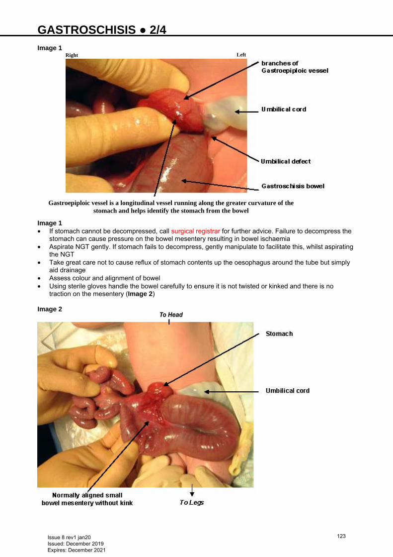
GASTROSCHISIS ● 2/4 Image 1 Image 1 • If stomach cannot be decompressed, call surgical registrar for further advice. Failure to decompress the
stomach can cause pressure on the bowel mesentery resulting in bowel ischaemia • Aspirate NGT gently. If stomach fails to decompress, gently manipulate to facilitate this, whilst aspirating
the NGT • Take great care not to cause reflux of stomach contents up the oesophagus around the tube but simply
aid drainage • Assess colour and alignment of bowel • Using sterile gloves handle the bowel carefully to ensure it is not twisted or kinked and there is no
traction on the mesentery (Image 2) Image 2
To Head
Right Left
Gastroepiploic vessel is a longitudinal vessel running along the greater curvature of the stomach and helps identify the stomach from the bowel
Issue 8 rev1 jan20 Issued: December 2019 Expires: December 2021
123
GASTROSCHISIS ● 3/4 • Place baby onto the same side as the defect (usually right) and support bowel on a folded nappy placed
slightly under baby • Check perfusion of bowel. If vascular compromise suspected, call consultant neonatologist • if compromise persists, inform surgical team immediately • Place baby’s legs and trunk into gastroschisis bag, feet first, and pull draw-string under baby’s arms, so
both arms are outside of the bag • Alternatively, cover and support intestines with cling film from upper chest to lower abdomen, holding
intestines in central position • ensure intestines are visible • do not wrap cling film tightly as this will reduce perfusion • Show baby to parents and transfer to NNU • Check global perfusion using central capillary refill time (CRT) • Check perfusion of bowel again immediately before transfer to NNU and at least every 15 min thereafter
IN NNU • Inform transport co-ordination team immediately as this is a time critical transfer (aim <4 hr from delivery) • Monitor perfusion and alignment of bowel at least every 15 min • Insert IV cannula, avoid potential long line veins • Avoid umbilical lines • Infuse either sodium chloride 0.9% or human albumin solution (HAS) 4.5% 20 mL/kg over 1 hr and start
routine IV maintenance fluids (see Intravenous fluid therapy guideline) • Aspirate NGT again and record volume. Replace NG losses mL-for-mL with sodium chloride 0.9% + 10
mmol potassium chloride/500 mL IV • Monitor central perfusion, using central CRT at least every 15 min. Give further fluid boluses as required
to maintain a normal CRT <2 secs. Babies with gastroschisis have a high fluid requirement until the herniated bowel is replaced in the abdomen
• Start IV antibiotics (benzyl penicillin, gentamicin and metronidazole) − see Neonatal Formulary • Give vitamin K IM (see Vitamin K guideline) • Discuss baby's condition and treatment plan with parents and ensure they have seen baby before
transfer. Take photographs for parents • Inform staff at surgical unit baby is ready for transfer. Have available: • name • gestational age • weight • ventilatory and oxygen requirements • mother’s name and ward (if mother admitted), including contact number if possible (for consent) Blood samples Baby • Blood culture • FBC and clotting studies, including fibrinogen • U&E • Blood glucose • Capillary/venous blood gas • Check with surgical unit if sample from baby for group and save, Coombs or crossmatch required (e.g.
Birmingham Children’s Hospital do not need these before transfer as these are done at surgical unit) • Send 1 bloodspot on neonatal screening card marked as 'pre-transfusion' (for sickle cell screening) with
baby to surgical unit Mother • Obtain sample of mother’s blood for crossmatch • sample tube must be clearly hand written and labelled with mother’s name, date of birth, NHS number,
and date and time of collection • complete form
− add baby’s details to ensure it is clear sample relates to mother of baby being transferred (this information is required by surgical unit blood bank)
Issue 8 rev1 jan20 Issued: December 2019 Expires: December 2021
124
GASTROSCHISIS ● 4/4 AWAITING TRANSFER TO SURGICAL UNIT • Continue to assess bowel perfusion and alignment every 15 min • Reassess baby’s fluid requirements hourly. If fluid boluses required, give sodium chloride 0.9% 10 mL/kg
IV • If evidence of a coagulopathy, treat with fresh frozen plasma (FFP) or cryoprecipitate, as appropriate
(see Coagulopathy guideline) • Aspirate NGT hourly and replace aspirate volume, mL-for-mL with sodium chloride 0.9% with 10 mmol
potassium chloride/500 mL IV • Leave NGT on free drainage
DOCUMENTATION • Complete nursing and medical documentation for transfer. Electronically transfer any X-rays to surgical
unit (or obtain copies of X-rays) • Ensure mother’s details included (including ward phone number if an inpatient and own number if
discharged) as if operation necessary and an individual with parental responsibility unable to attend surgical unit, surgeon will require verbal telephone consent
• Ensure baby’s documentation includes: • whether vitamin K has been given • name of referring consultant • whether parents received antenatal counselling • mother’s name, ward (if admitted) and her contact details
TRANSFER TO SURGICAL UNIT • Inform surgical unit that transfer is underway • Place baby in transport incubator, taking care to transfer bowel and mesentery in a supported, non-
kinked position. Keep stomach empty • place baby on side of defect and support bowel on a folded nappy just slightly under baby. Check bowel
perfusion immediately and at least every 15 min • ensure mother’s blood, baby’s pre-transfusion bloodspots, letters for surgical team and all
documentation accompany baby During transport • Carry out and document usual observations, include bowel perfusion and alter its position if necessary Arrival at surgical unit • Record bowel perfusion and alignment Useful information • http://www.bch.nhs.uk/content/neonatal-surgery • http://www.bch.nhs.uk/find-us/maps-directions • Parent information: https://bwc.nhs.uk/download.cfm?doc=docm93jijm4n2151 • NHS Fetal Anomaly Screening Programme gastroschisis guideline
Issue 8 rev1 jan20 Issued: December 2019 Expires: December 2021
125
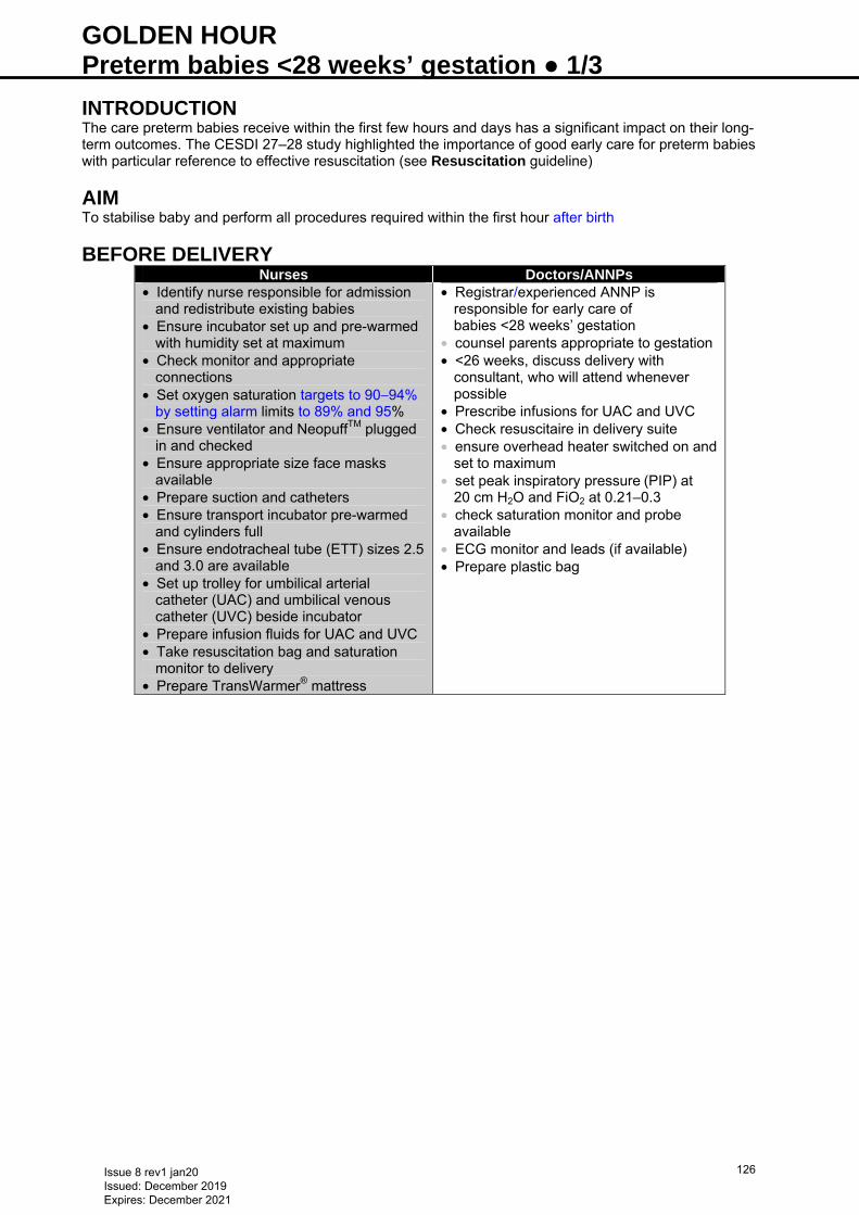
GOLDEN HOUR Preterm babies <28 weeks’ gestation ● 1/3 INTRODUCTION The care preterm babies receive within the first few hours and days has a significant impact on their long-term outcomes. The CESDI 27–28 study highlighted the importance of good early care for preterm babies with particular reference to effective resuscitation (see Resuscitation guideline)
AIM To stabilise baby and perform all procedures required within the first hour after birth
BEFORE DELIVERY Nurses Doctors/ANNPs
• Identify nurse responsible for admission and redistribute existing babies
• Ensure incubator set up and pre-warmed with humidity set at maximum
• Check monitor and appropriate connections
• Set oxygen saturation targets to 90−94% by setting alarm limits to 89% and 95%
• Ensure ventilator and NeopuffTM plugged in and checked
• Ensure appropriate size face masks available
• Prepare suction and catheters • Ensure transport incubator pre-warmed
and cylinders full • Ensure endotracheal tube (ETT) sizes 2.5
and 3.0 are available • Set up trolley for umbilical arterial
catheter (UAC) and umbilical venous catheter (UVC) beside incubator
• Prepare infusion fluids for UAC and UVC • Take resuscitation bag and saturation
monitor to delivery • Prepare TransWarmer® mattress
• Registrar/experienced ANNP is responsible for early care of babies <28 weeks’ gestation
• counsel parents appropriate to gestation • <26 weeks, discuss delivery with
consultant, who will attend whenever possible
• Prescribe infusions for UAC and UVC • Check resuscitaire in delivery suite • ensure overhead heater switched on and
set to maximum • set peak inspiratory pressure (PIP) at
20 cm H2O and FiO2 at 0.21–0.3 • check saturation monitor and probe
available • ECG monitor and leads (if available) • Prepare plastic bag
Issue 8 rev1 jan20 Issued: December 2019 Expires: December 2021
126
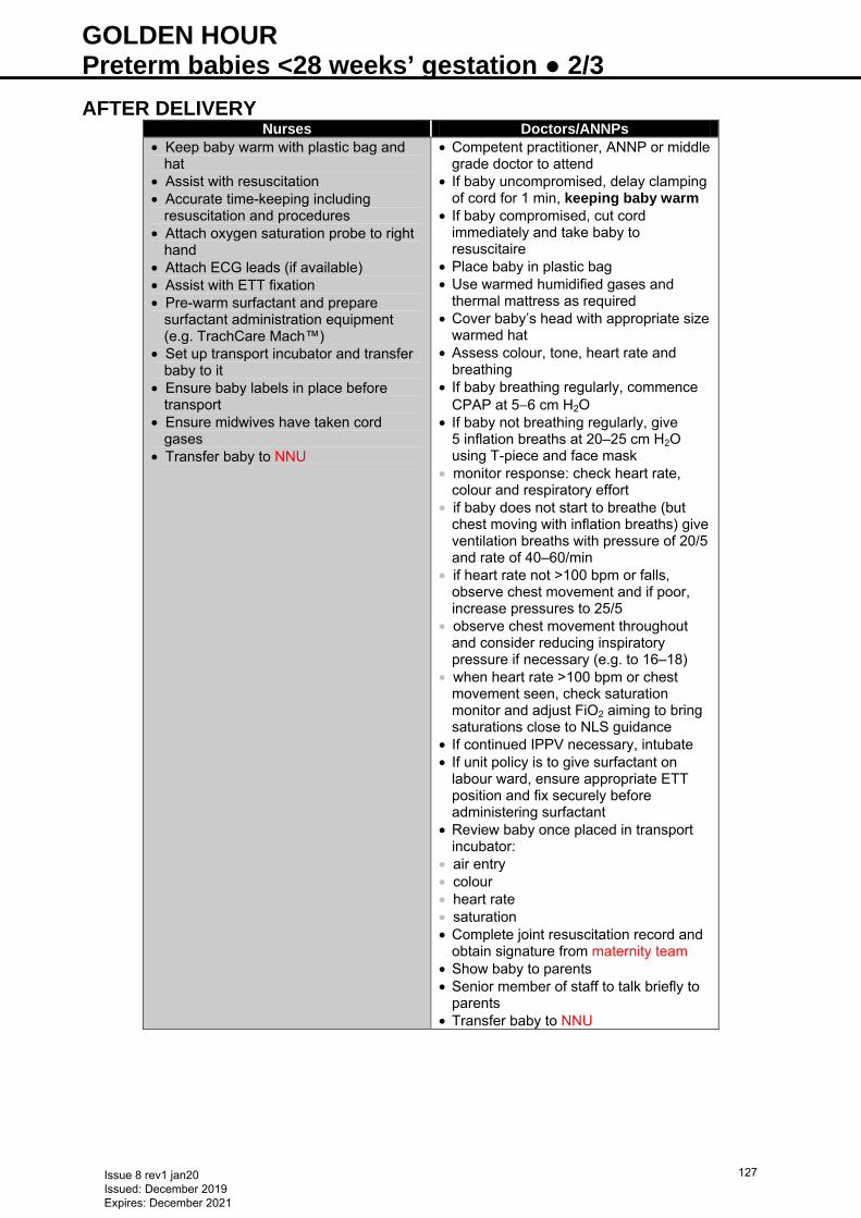
GOLDEN HOUR Preterm babies <28 weeks’ gestation ● 2/3 AFTER DELIVERY
Nurses Doctors/ANNPs • Keep baby warm with plastic bag and
hat • Assist with resuscitation • Accurate time-keeping including
resuscitation and procedures • Attach oxygen saturation probe to right
hand • Attach ECG leads (if available) • Assist with ETT fixation • Pre-warm surfactant and prepare
surfactant administration equipment (e.g. TrachCare Mach™)
• Set up transport incubator and transfer baby to it
• Ensure baby labels in place before transport
• Ensure midwives have taken cord gases
• Transfer baby to NNU
• Competent practitioner, ANNP or middle grade doctor to attend
• If baby uncompromised, delay clamping of cord for 1 min, keeping baby warm
• If baby compromised, cut cord immediately and take baby to resuscitaire
• Place baby in plastic bag • Use warmed humidified gases and
thermal mattress as required • Cover baby’s head with appropriate size
warmed hat • Assess colour, tone, heart rate and
breathing • If baby breathing regularly, commence
CPAP at 5−6 cm H2O • If baby not breathing regularly, give
5 inflation breaths at 20–25 cm H2O using T-piece and face mask
• monitor response: check heart rate, colour and respiratory effort
• if baby does not start to breathe (but chest moving with inflation breaths) give ventilation breaths with pressure of 20/5 and rate of 40–60/min
• if heart rate not >100 bpm or falls, observe chest movement and if poor, increase pressures to 25/5
• observe chest movement throughout and consider reducing inspiratory pressure if necessary (e.g. to 16–18)
• when heart rate >100 bpm or chest movement seen, check saturation monitor and adjust FiO2 aiming to bring saturations close to NLS guidance
• If continued IPPV necessary, intubate • If unit policy is to give surfactant on
labour ward, ensure appropriate ETT position and fix securely before administering surfactant
• Review baby once placed in transport incubator:
• air entry • colour • heart rate • saturation • Complete joint resuscitation record and
obtain signature from maternity team • Show baby to parents • Senior member of staff to talk briefly to
parents • Transfer baby to NNU
Issue 8 rev1 jan20 Issued: December 2019 Expires: December 2021
127
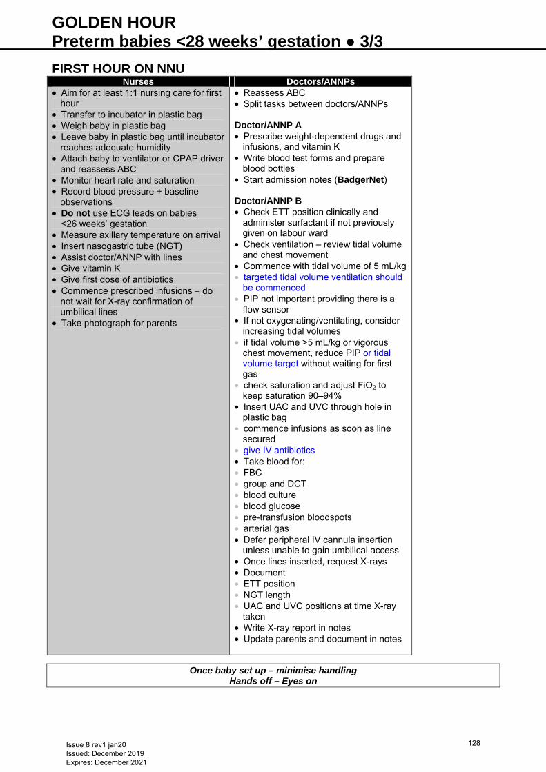
GOLDEN HOUR Preterm babies <28 weeks’ gestation ● 3/3 FIRST HOUR ON NNU
Nurses Doctors/ANNPs • Aim for at least 1:1 nursing care for first
hour • Transfer to incubator in plastic bag • Weigh baby in plastic bag • Leave baby in plastic bag until incubator
reaches adequate humidity • Attach baby to ventilator or CPAP driver
and reassess ABC • Monitor heart rate and saturation • Record blood pressure + baseline
observations • Do not use ECG leads on babies
<26 weeks’ gestation • Measure axillary temperature on arrival • Insert nasogastric tube (NGT) • Assist doctor/ANNP with lines • Give vitamin K • Give first dose of antibiotics • Commence prescribed infusions − do
not wait for X-ray confirmation of umbilical lines
• Take photograph for parents
• Reassess ABC • Split tasks between doctors/ANNPs Doctor/ANNP A • Prescribe weight-dependent drugs and
infusions, and vitamin K • Write blood test forms and prepare
blood bottles • Start admission notes (BadgerNet) Doctor/ANNP B • Check ETT position clinically and
administer surfactant if not previously given on labour ward
• Check ventilation – review tidal volume and chest movement
• Commence with tidal volume of 5 mL/kg • targeted tidal volume ventilation should
be commenced • PIP not important providing there is a
flow sensor • If not oxygenating/ventilating, consider
increasing tidal volumes • if tidal volume >5 mL/kg or vigorous
chest movement, reduce PIP or tidal volume target without waiting for first gas
• check saturation and adjust FiO2 to keep saturation 90–94%
• Insert UAC and UVC through hole in plastic bag
• commence infusions as soon as line secured
• give IV antibiotics • Take blood for: • FBC • group and DCT • blood culture • blood glucose • pre-transfusion bloodspots • arterial gas • Defer peripheral IV cannula insertion
unless unable to gain umbilical access • Once lines inserted, request X-rays • Document • ETT position • NGT length • UAC and UVC positions at time X-ray
taken • Write X-ray report in notes • Update parents and document in notes
Once baby set up – minimise handling Hands off – Eyes on
Issue 8 rev1 jan20 Issued: December 2019 Expires: December 2021
128
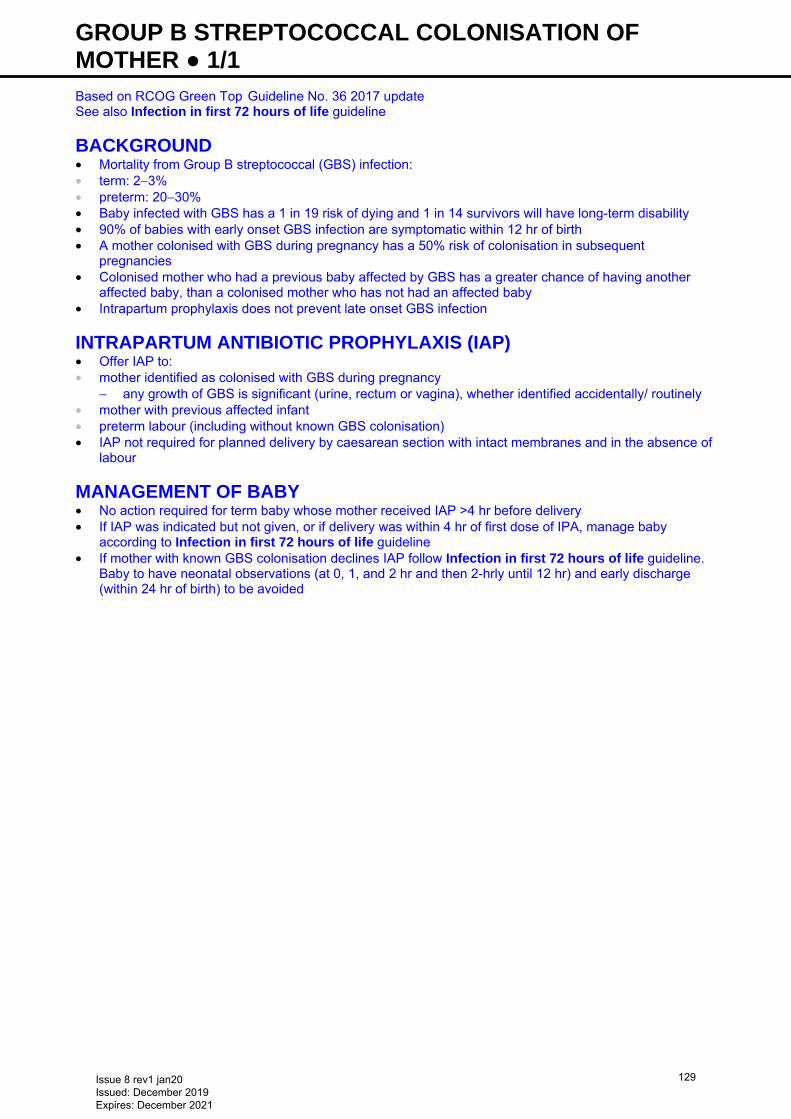
GROUP B STREPTOCOCCAL COLONISATION OF MOTHER ● 1/1 Based on RCOG Green Top Guideline No. 36 2017 update See also Infection in first 72 hours of life guideline
BACKGROUND • Mortality from Group B streptococcal (GBS) infection: • term: 2−3% • preterm: 20−30% • Baby infected with GBS has a 1 in 19 risk of dying and 1 in 14 survivors will have long-term disability • 90% of babies with early onset GBS infection are symptomatic within 12 hr of birth • A mother colonised with GBS during pregnancy has a 50% risk of colonisation in subsequent
pregnancies • Colonised mother who had a previous baby affected by GBS has a greater chance of having another
affected baby, than a colonised mother who has not had an affected baby • Intrapartum prophylaxis does not prevent late onset GBS infection
INTRAPARTUM ANTIBIOTIC PROPHYLAXIS (IAP) • Offer IAP to: • mother identified as colonised with GBS during pregnancy
− any growth of GBS is significant (urine, rectum or vagina), whether identified accidentally/ routinely • mother with previous affected infant • preterm labour (including without known GBS colonisation) • IAP not required for planned delivery by caesarean section with intact membranes and in the absence of
labour
MANAGEMENT OF BABY • No action required for term baby whose mother received IAP >4 hr before delivery • If IAP was indicated but not given, or if delivery was within 4 hr of first dose of IPA, manage baby
according to Infection in first 72 hours of life guideline • If mother with known GBS colonisation declines IAP follow Infection in first 72 hours of life guideline.
Baby to have neonatal observations (at 0, 1, and 2 hr and then 2-hrly until 12 hr) and early discharge (within 24 hr of birth) to be avoided
Issue 8 rev1 jan20 Issued: December 2019 Expires: December 2021
129
GROWTH MONITORING ● 1/2 DEFINITION • Routine accurate measurement and documentation of weight, length and occipitofrontal circumference
(OFC)
AIM • To detect any abnormal growth patterns, including faltering growth
INTRODUCTION • Neonatal nutrition and resulting postnatal growth are major determinants in the short and long-term
outcomes of preterm neonates • Optimal postnatal nutrition and growth associated with more positive later health and developmental
outcomes • Preterm infants who demonstrate low weight gain in the early years have a higher probability of poorer
cognitive developmental outcomes, while those with excessive weight gain have an increased risk of childhood and adult obesity, cardiovascular disease and diabetes
• Consider both quantity and quality of growth • Plot measurements of weight, length and OFC on appropriate and gender specific growth chart to allow
assessment of adequate and proportionate growth • measurements to be undertaken by qualified member of staff trained in the use of the equipment • Involve parents/carers with all growth monitoring procedures
WEIGHT Frequency • Weigh all infants on admission to NNU • Weigh ≥3 times/week while an inpatient • Plan weighing schedules taking into account developmental care needs • If baby too unstable to be weighed for >5 consecutive days, and incubator does not have in line scales: • calculate weight-for-age from appropriate growth chart • use as working weight (assuming baby is following their previous centile line) to ensure adequate fluids,
enteral and parenteral nutrition, and drugs administered • reinstate routine weighing once baby stable • If baby unstable, assess for fluid overload − impacts on accuracy of weighing for growth monitoring Equipment • Class III electronic baby scales or incubator with inbuilt scales (if available) − accurate to 5 g • All scales to be: • tested and recalibrated annually • cleaned between patients in accordance with local infection prevention policy Method • Wash and sanitise hands and equipment as per local infection prevention policy • Use swaddled weighing for optimal developmental care • Weigh baby naked (no nappy/clothing) • Swaddle weighing • wrap baby in a warm, pre-weighed blanket, or use a thick, soft, warm pre-weighed blanket/sheet to line
scales • deduct weight of swaddle blanket • Record actual calculated weight on unit documentation/BadgerNet • ≤999 g: to nearest 5 g • ≥1 kg: to nearest 10 g • Plot weight at least weekly on BadgerNet or gender appropriate WHO Neonatal and infant close
monitoring growth chart [see chart or refer to RCPCH website (www.growthcharts.rcpch.ac.uk) for instructions on use]
• In infants <2 kg: calculate velocity of weight gain in g/kg/day at least weekly • aim 16 g/kg/day as steady weight gain • If parent is present baby will benefit from skin-to-skin contact before returning to incubator/cot
LENGTH Frequency • Measure all infants on admission to NNU and weekly thereafter coinciding with a weigh day whilst
inpatient
Issue 8 rev1 jan20 Issued: December 2019 Expires: December 2021
130
GROWTH MONITORING ● 2/2 Equipment • ≤33 weeks or <45 cm: use Leicester Incubator Measure • ≥33+1 weeks: use length mat • Requires 2 people to obtain an accurate measurement (1 may be parent/carer alongside trained member
of staff)
Never use a tape measure to measure length Method • Wash and sanitise hands and equipment as per local infection prevention policy • Measure baby supine, lying flat, ensuring no clothing or nests restrict extension • Remove hat or ventilation/non-invasive ventilation hat ties • Preterm babies do not need to be naked • Term infants to be measured naked, no nappy • Operator 1: place fixed headpiece against crown of baby’s head, stabilising head by gently cupping
palms of hands over baby’s ears • Operator 2: gently place palm of hand over baby’s knee encouraging extension, sliding base plate up to
meet the soles of the feet • If baby settled and relaxed, take 3 measurements to ensure consistency • Record length in cm to nearest 0.1 cm • Plot length weekly on BadgerNet or gender appropriate WHO Neonatal and infant close monitoring
growth chart [see chart or refer to RCPCH website (www.growthcharts.rcpch.ac.uk) for instructions on use]
• Calculate velocity of linear growth in cm/week monthly • aim 1.4 cm/week as steady linear growth in preterm baby
OFC Frequency • Measure on admission to NNU and weekly thereafter coinciding with a weigh day while inpatient
Equipment • Disposable paper tape measure Method • Wash and sanitise hands as per local infection prevention policy • Remove or fold down hat or head gear that may obstruct measurement • Using disposable paper tape measure, take measurement at the widest part of baby’s head − above
ears, midway between eyebrows and hairline at the front, and to occipital prominence at back of head • Record in cm to nearest 0.1 cm on NNU documentation • Plot OFC weekly on BadgerNet or gender appropriate WHO Neonatal and infant close monitoring
growth chart [see chart or refer to RCPCH website (www.growthcharts.rcpch.ac.uk) for instructions on use]
• Calculate velocity of OFC growth in cm/week monthly • aim 0.9 cm/week as steady OFC growth in preterm baby
INTERPRETATION • Growth charts are a tool to monitor growth and growth velocity • All babies lose weight after birth and may cross down 2−3 marked centiles with an expectation they will
return to their birth centile • Stable preterm babies with adequate nutritional intake are expected to grow along/parallel to centiles
from aged 2−3 weeks • Babies with slow growth velocity (less than expected over 1 week period), growth failure or whose
growth parameters continue to fall across centiles into week 3 of life, to have a full nutritional review • include calculation of any parenteral nutrition received (not only prescribed), and enteral nutrition intake • If combined nutritional intake falls short of recommended requirements: optimise nutritional intake (see
Nutrition and enteral feeding guideline) • if growth remains suboptimal, see Nutrition and enteral feeding guideline − Inadequate growth • If baby exhibiting suboptimal growth, refer to NNU nutrition team or neonatal/paediatric dietitian
DISCHARGE • Transfer key information regarding growth to personal child health record (PCHR) or Red Book • Must include birth and discharge weight, length and OFC
Issue 8 rev1 jan20 Issued: December 2019 Expires: December 2021
131
HEARING SCREENING ● 1/2 INTRODUCTION • Early intervention improves the outcome for babies with a congenital hearing deficit • Screening for congenital deafness is undertaken through the NHS Newborn Hearing Screening
Programme (NHSP) by trained screeners according to national guidelines. They are automatically informed of all births and will ensure babies are screened
• Neonatal staff must understand how their local programme is organised, the risk factors for congenital deafness and know how to work with the screeners
INDICATIONS Who • All babies are eligible for screening; excluding: • microtia/external ear canal atresia • neonatal bacterial meningitis or meningococcal septicemia • confirmed congenital cytomegalovirus (cCMV) • programmable ventriculo-peritoneal shunts • Neonatal staff must refer babies with meningitis to audiology for an urgent assessment (NHSP referral
to be completed and handed to the screeners who will book a diagnostic appointment) • Screeners will refer babies with non-patent canal for urgent diagnostic assessment
PROCEDURE Consent •• Screening can only be performed with parental consent • screeners will obtain verbal consent from parents (if present) before screening • if baby on NNU and parents absent, screeners will leave an explanatory leaflet and gain verbal consent
from parents during their visit to NNU or over the telephone How •• Oto-Acoustic Emissions (OAE) +/- Automated Auditory Brainstem Response (AABR) according to
national ‘Well baby’ or ‘NICU’ protocols • neonatal staff must inform screeners if baby has ever spent >48 hr on NNU so that NICU protocol can be
used • babies on transitional care are screened using the ‘Well baby’ protocol (unless previously on NNU for
>48 hr)
When •• Screen only when baby has reached 34 weeks (corrected age) Where Well babies •• Screening is performed as an inpatient before discharge or in the community (see Table for local details)
NNU babies • Arrange screening as close to discharge as possible, when baby is well enough to test and preferably
once major medical treatment, ototoxic or other drug treatment complete • Do not screen babies transferring to another NNU • Complete screening of babies on NNU >48 hr by 44 weeks (corrected age)
FOLLOW-UP • Neonatal staff must ensure all babies diagnosed with bacterial meningitis are referred for an urgent
audiology assessment and are not screened • Screeners will arrange routine follow-up according to screening results and presence of other specific
risk factors • Following must be seen within 4 weeks of decision that screening not appropriate/recovery from acute
episode/≤44 weeks’ gestational age: • microtia/external ear canal atresia • neonatal bacterial meningitis or meningococcal septicaemia • programmable ventriculo-peritoneal shunts • Babies with cCMV may need to be seen earlier [timescale agreed with paediatrician to allow early
antiviral treatment to commence (if required)]
Issue 8 rev1 jan20 Issued: December 2019 Expires: December 2021
132
HEARING SCREENING ● 2/2 Risk factors • Neonatal staff must inform the screener of the following risk factors in order that appropriate follow-up at
aged 7−9 months can be arranged: • confirmed or possible congenital infection (rubella, toxoplasmosis) • cranio-facial anomalies, cleft palate, deformed pinnae (not simple ear tags) • syndromes associated with hearing loss (Down, Waardenburg, Alport, Usher etc.) • baby has been treated with ECMO • Babies with the following risk factors are not followed up by audiology, but data is collected for audit
purposes: • severe jaundice (at exchange level) • multiple abnormalities with neurodegenerative/neurodevelopmental disorder • mechanical ventilation >5 days • Screener will determine presence of other risk factors before screening: • family history of permanent hearing loss in childhood
− those with first-degree relative will be followed up in audiology
FURTHER INFORMATION • Detailed information available from NHSP website: https://www.gov.uk/topic/population-screening-
programmes/newborn-hearing Table 1: Local details
• Alexandra Hospital, Redditch • Russells Hall Hospital, Dudley • Shrewsbury and Telford Hospitals • New Cross Hospital • Worcestershire Royal Hospital • Hereford County Hospital
Usually performed by trained staff in the community Babies on NNU usually screened before discharge
• Birmingham City Hospital • Birmingham Heartlands Hospital • Birmingham Women’s Hospital • Good Hope Hospital • Manor Hospital • Royal Stoke University Hospital
Screening for all babies usually performed while still an inpatient, usually at bedside
• Sandwell Hospital MLU • Solihull Hospital MLU
Screening performed as outpatient unless baby transferred into a main maternity/NNU
Issue 8 rev1 jan20 Issued: December 2019 Expires: December 2021
133
HEART FAILURE ● 1/3 DEFINITION • Congestive cardiac failure occurs when the heart is unable to pump sufficient blood to meet metabolic
demands of body tissues • underlying cause may be cardiac or non-cardiac Causes Cardiac • Left-to-right shunt (see Increased left-to-right shunt) • Arrhythmia • Hypoplastic left heart syndrome • Critical aortic stenosis • Coarctation • Interrupted aortic arch Non-cardiac • Sepsis • Hypoxia • Anaemia • Polycythaemia • Fluid overload • AV malformation • Pulmonary hypertension
Clinical differentiation between an obstructed systemic circulation and severe sepsis is extremely difficult as a murmur and weak pulses can be common to both.
For baby in extremis, presence of abnormal pulses alone is sufficient indication to start a prostaglandin infusion until a cardiac lesion has been excluded by echocardiography (see
Prostaglandin infusion guideline)
SYMPTOMS AND SIGNS OF CARDIAC FAILURE • Tachycardia • Tachypnoea • Hepatomegaly • Excessive weight gain • Hypotension • Murmur • Abnormal femoral pulses • weak femoral pulses (in obstructive left heart lesions − femoral pulses may not be absent if duct still
patent)
INVESTIGATIONS • Blood gas including lactate • Baseline bloods including FBC, U&E, LFT • Chest X-ray − look for cardiomegaly and pulmonary oedema • Pre and postductal saturations • postductal saturations can be considerably lower than preductal in aortic arch defects (a difference of
>2% is significant) • ECG • Echocardiogram
TREATMENT OF CARDIAC FAILURE DUE TO OBSTRUCTIVE HEART DISEASE
If left-sided obstructive lesion suspected, treat with inotropes and use diuretics cautiously Resuscitation Airway • Routine intubation not indicated
Issue 8 rev1 jan20 Issued: December 2019 Expires: December 2021
134
HEART FAILURE ● 2/3 • Intubate and ventilate babies presenting collapsed or with obvious cyanosis in association with cardiac
failure • If apnoea occurs secondary to a prostaglandin infusion, intubate baby but do not alter infusion Breathing • See Ventilation: conventional guideline • Ventilate with PEEP 5–6 cm • Adjust ventilation to maintain: • PaCO2 5–6 kPa • pH >7.25 Circulation • Vascular access with 2 IV cannulae or umbilical venous catheter (UVC) (see Umbilical venous
catheterisation and removal guideline)
Presence of cyanosis and a murmur suggest baby likely to respond to prostaglandin infusion
• Prostaglandin infusion to maintain ductal patency (see Prostaglandin infusion guideline) • open duct with dinoprostone (prostaglandin E2, prostin E2), see Neonatal Formulary. Start at 5–10
nanogram/kg/min, may be increased to 50 nanogram/kg/min, but only on cardiologist advice • Monitor blood pressure invasively [ideally using a peripheral arterial cannula rather than an umbilical
arterial catheter (UAC)] Cardiac output • Signs of poor cardiac output include: • tachycardia • low BP • acidosis • high lactate • poor peripheral perfusion • When cardiac output low: • ensure adequate intravascular volume • correct anaemia • dobutamine may be required for poor perfusion – discuss with regional cardiac centre for choice of
inotropes
SUBSEQUENT MANAGEMENT – TRANSFER
Baby must be kept warm and normoglycaemic
• Discuss further management and transfer with regional cardiac centre • Babies who respond to a prostaglandin infusion may not need transferring out-of-hours • Appropriately skilled medical and nursing staff are necessary for transfer Intubation
An intubated baby requires a cardiac centre ITU bed; do not intubate routinely for transfer
• Intubate if: • continuing metabolic acidosis and poor perfusion • long-distance transfer necessary • inotropic support needed • apnoea • recommended by cardiac team
DISCHARGE FROM CARDIAC CENTRE Baby may go home or return to a paediatric ward or NNU, possibly on a prostaglandin infusion whilst awaiting surgery or for continuing care after a palliative procedure (e.g. septostomy) Management plan • Regardless of outcome, obtain a management plan from cardiac centre, defining: • acceptable vital signs (e.g. saturations)
Issue 8 rev1 jan20 Issued: December 2019 Expires: December 2021
135
HEART FAILURE ● 3/3 • medication, including dosage • follow-up arrangements
INCREASED LEFT-TO-RIGHT SHUNT
RECOGNITION AND ASSESSMENT Definition • Any lesion causing increased pulmonary blood flow • Usually presents when pulmonary resistance falls after 48 hr • Size and type of lesion will influence time of presentation Differential diagnosis • AVSD • Partial AVSD • VSD • Truncus arteriosus • PDA Investigations • Chest X-ray looking for fluid overload • Echocardiogram
MANAGEMENT • If in cardiac failure, give immediate dose of diuretic • May require maintenance diuretics (discuss with cardiologist) • usually furosemide 1 mg/kg twice daily and amiloride 100 microgram/kg twice daily oral • Discuss with cardiac centre for definitive management and follow-up
Issue 8 rev1 jan20 Issued: December 2019 Expires: December 2021
136
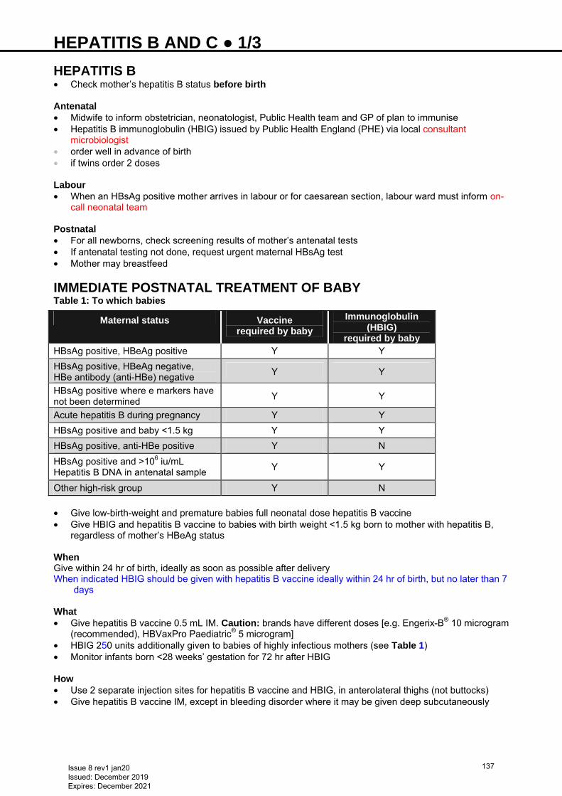
HEPATITIS B AND C ● 1/3 HEPATITIS B • Check mother’s hepatitis B status before birth Antenatal • Midwife to inform obstetrician, neonatologist, Public Health team and GP of plan to immunise • Hepatitis B immunoglobulin (HBIG) issued by Public Health England (PHE) via local consultant
microbiologist • order well in advance of birth • if twins order 2 doses Labour • When an HBsAg positive mother arrives in labour or for caesarean section, labour ward must inform on-
call neonatal team Postnatal • For all newborns, check screening results of mother’s antenatal tests • If antenatal testing not done, request urgent maternal HBsAg test • Mother may breastfeed
IMMEDIATE POSTNATAL TREATMENT OF BABY Table 1: To which babies
Maternal status Vaccine required by baby
Immunoglobulin (HBIG)
required by baby HBsAg positive, HBeAg positive Y Y
HBsAg positive, HBeAg negative, HBe antibody (anti-HBe) negative
Y Y
HBsAg positive where e markers have not been determined
Y Y
Acute hepatitis B during pregnancy Y Y
HBsAg positive and baby <1.5 kg Y Y
HBsAg positive, anti-HBe positive Y N
HBsAg positive and >106 iu/mL Hepatitis B DNA in antenatal sample
Y Y
Other high-risk group Y N
• Give low-birth-weight and premature babies full neonatal dose hepatitis B vaccine • Give HBIG and hepatitis B vaccine to babies with birth weight <1.5 kg born to mother with hepatitis B,
regardless of mother’s HBeAg status When Give within 24 hr of birth, ideally as soon as possible after delivery When indicated HBIG should be given with hepatitis B vaccine ideally within 24 hr of birth, but no later than 7
days What • Give hepatitis B vaccine 0.5 mL IM. Caution: brands have different doses [e.g. Engerix-B® 10 microgram
(recommended), HBVaxPro Paediatric® 5 microgram] • HBIG 250 units additionally given to babies of highly infectious mothers (see Table 1) • Monitor infants born <28 weeks’ gestation for 72 hr after HBIG How • Use 2 separate injection sites for hepatitis B vaccine and HBIG, in anterolateral thighs (not buttocks) • Give hepatitis B vaccine IM, except in bleeding disorder where it may be given deep subcutaneously
Issue 8 rev1 jan20 Issued: December 2019 Expires: December 2021
137
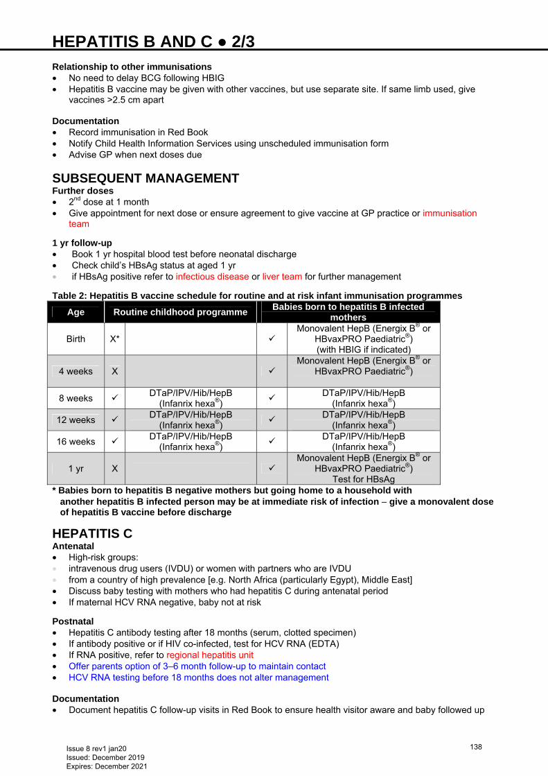
HEPATITIS B AND C ● 2/3 Relationship to other immunisations • No need to delay BCG following HBIG • Hepatitis B vaccine may be given with other vaccines, but use separate site. If same limb used, give
vaccines >2.5 cm apart Documentation • Record immunisation in Red Book • Notify Child Health Information Services using unscheduled immunisation form • Advise GP when next doses due
SUBSEQUENT MANAGEMENT Further doses • 2nd dose at 1 month • Give appointment for next dose or ensure agreement to give vaccine at GP practice or immunisation
team
1 yr follow-up • Book 1 yr hospital blood test before neonatal discharge • Check child’s HBsAg status at aged 1 yr • if HBsAg positive refer to infectious disease or liver team for further management Table 2: Hepatitis B vaccine schedule for routine and at risk infant immunisation programmes
Age Routine childhood programme Babies born to hepatitis B infected
mothers
Birth X*
Monovalent HepB (Energix B® or
HBvaxPRO Paediatric®) (with HBIG if indicated)
4 weeks X
Monovalent HepB (Energix B® or
HBvaxPRO Paediatric®)
8 weeks DTaP/IPV/Hib/HepB
(Infanrix hexa®)
DTaP/IPV/Hib/HepB (Infanrix hexa®)
12 weeks DTaP/IPV/Hib/HepB
(Infanrix hexa®)
DTaP/IPV/Hib/HepB (Infanrix hexa®)
16 weeks DTaP/IPV/Hib/HepB
(Infanrix hexa®)
DTaP/IPV/Hib/HepB (Infanrix hexa®)
1 yr X
Monovalent HepB (Energix B® or
HBvaxPRO Paediatric®) Test for HBsAg
* Babies born to hepatitis B negative mothers but going home to a household with another hepatitis B infected person may be at immediate risk of infection − give a monovalent dose of hepatitis B vaccine before discharge
HEPATITIS C Antenatal • High-risk groups: • intravenous drug users (IVDU) or women with partners who are IVDU • from a country of high prevalence [e.g. North Africa (particularly Egypt), Middle East] • Discuss baby testing with mothers who had hepatitis C during antenatal period • If maternal HCV RNA negative, baby not at risk Postnatal • Hepatitis C antibody testing after 18 months (serum, clotted specimen) • If antibody positive or if HIV co-infected, test for HCV RNA (EDTA) • If RNA positive, refer to regional hepatitis unit • Offer parents option of 3–6 month follow-up to maintain contact • HCV RNA testing before 18 months does not alter management Documentation • Document hepatitis C follow-up visits in Red Book to ensure health visitor aware and baby followed up
Issue 8 rev1 jan20 Issued: December 2019 Expires: December 2021
138
HEPATITIS B AND C ● 3/3 Breastfeeding • Mother may breastfeed
ADOPTION AND FOSTERING • If risk factor for HCV (e.g. IV drug use) and maternal status not known, HCV antibody: • if positive, retest aged 18 months
− if still positive, refer to paediatric infectious diseases or liver specialist
Issue 8 rev1 jan20 Issued: December 2019 Expires: December 2021
139
HERPES SIMPLEX VIRUS (HSV) ● 1/1 CAESAREAN SECTION • See Recurrent HSV
RECURRENT HSV (History of genital herpes before third trimester) • No swabs or treatment • Educate parents on good hand hygiene to prevent transmission • Observe on postnatal ward, discharge after neonatal examination at 24 hr • Advise to seek medical help if skin, eye or mucous membrane lesions, lethargy/irritability, poor feeding • If clinical evidence of sepsis: • surface swabs and blood for HSV PCR • aciclovir 20 mg/kg 8-hrly IVI over 1 hr
PRIMARY HSV (Genital herpes ≤6 weeks before vaginal delivery) • Strict infection control • Swab infant’s nasopharynx, conjunctiva, mouth and rectum in viral transport medium for HSV PCR • Check infant’s ALT and send blood for HSV PCR • Start aciclovir 20 mg/kg IVI (over 1 hr) 8-hrly • If ALT abnormal or other signs of infection (including skin lesions) send CSF for HSV PCR • Recommend breastfeeding unless herpetic lesions around nipple
TREATMENT Duration aciclovir IV • If neonatal HSV PCR negative: stop aciclovir • If active infection ruled out: stop aciclovir • If skin, eye or mouth lesions: lumbar puncture • if CSF HSV negative and ALT normal: aciclovir IV for 10 days • if ALT raised and CSF negative: aciclovir IV for 14 days • if CSF HSV positive: repeat LP at 14 days and if negative stop at 21 days • If any confirmed HSV disease: give suppressive therapy with aciclovir 300 mg/m2 oral 8-hrly for 6 months
Issue 8 rev1 jan20 Issued: December 2019 Expires: December 2021
140
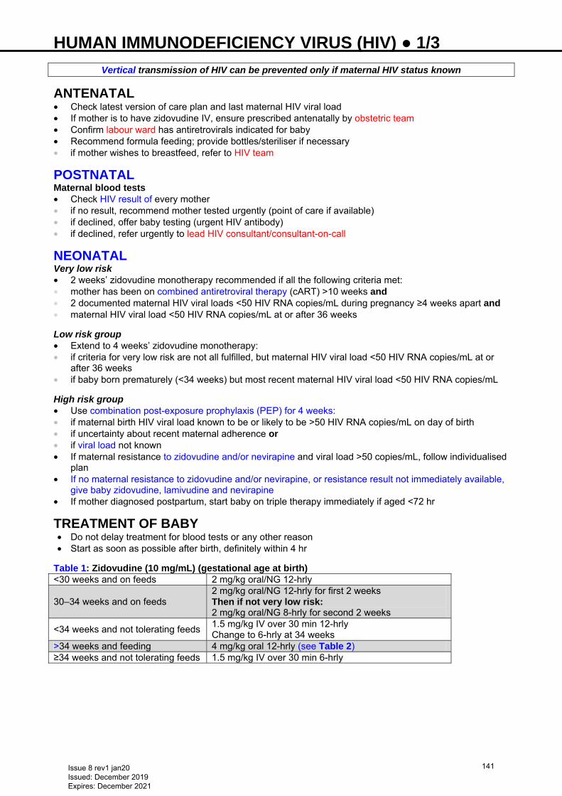
HUMAN IMMUNODEFICIENCY VIRUS (HIV) ● 1/3
Vertical transmission of HIV can be prevented only if maternal HIV status known
ANTENATAL • Check latest version of care plan and last maternal HIV viral load • If mother is to have zidovudine IV, ensure prescribed antenatally by obstetric team • Confirm labour ward has antiretrovirals indicated for baby • Recommend formula feeding; provide bottles/steriliser if necessary • if mother wishes to breastfeed, refer to HIV team
POSTNATAL Maternal blood tests • Check HIV result of every mother • if no result, recommend mother tested urgently (point of care if available) • if declined, offer baby testing (urgent HIV antibody) • if declined, refer urgently to lead HIV consultant/consultant-on-call
NEONATAL Very low risk • 2 weeks’ zidovudine monotherapy recommended if all the following criteria met: • mother has been on combined antiretroviral therapy (cART) >10 weeks and • 2 documented maternal HIV viral loads <50 HIV RNA copies/mL during pregnancy ≥4 weeks apart and • maternal HIV viral load <50 HIV RNA copies/mL at or after 36 weeks
Low risk group • Extend to 4 weeks’ zidovudine monotherapy: • if criteria for very low risk are not all fulfilled, but maternal HIV viral load <50 HIV RNA copies/mL at or
after 36 weeks • if baby born prematurely (<34 weeks) but most recent maternal HIV viral load <50 HIV RNA copies/mL
High risk group • Use combination post-exposure prophylaxis (PEP) for 4 weeks: • if maternal birth HIV viral load known to be or likely to be >50 HIV RNA copies/mL on day of birth • if uncertainty about recent maternal adherence or • if viral load not known • If maternal resistance to zidovudine and/or nevirapine and viral load >50 copies/mL, follow individualised
plan • If no maternal resistance to zidovudine and/or nevirapine, or resistance result not immediately available,
give baby zidovudine, lamivudine and nevirapine • If mother diagnosed postpartum, start baby on triple therapy immediately if aged <72 hr
TREATMENT OF BABY • Do not delay treatment for blood tests or any other reason • Start as soon as possible after birth, definitely within 4 hr
Table 1: Zidovudine (10 mg/mL) (gestational age at birth) <30 weeks and on feeds 2 mg/kg oral/NG 12-hrly
30–34 weeks and on feeds 2 mg/kg oral/NG 12-hrly for first 2 weeks Then if not very low risk: 2 mg/kg oral/NG 8-hrly for second 2 weeks
<34 weeks and not tolerating feeds 1.5 mg/kg IV over 30 min 12-hrly Change to 6-hrly at 34 weeks
>34 weeks and feeding 4 mg/kg oral 12-hrly (see Table 2) ≥34 weeks and not tolerating feeds 1.5 mg/kg IV over 30 min 6-hrly
Issue 8 rev1 jan20 Issued: December 2019 Expires: December 2021
141
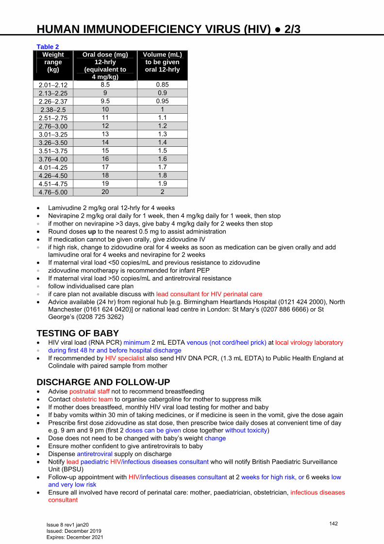
HUMAN IMMUNODEFICIENCY VIRUS (HIV) ● 2/3 Table 2
Weight range (kg)
Oral dose (mg) 12-hrly
(equivalent to 4 mg/kg)
Volume (mL) to be given oral 12-hrly
2.01−2.12 8.5 0.85 2.13−2.25 9 0.9 2.26−2.37 9.5 0.95 2.38−2.5 10 1 2.51−2.75 11 1.1 2.76−3.00 12 1.2 3.01−3.25 13 1.3 3.26−3.50 14 1.4 3.51−3.75 15 1.5 3.76−4.00 16 1.6 4.01−4.25 17 1.7 4.26−4.50 18 1.8 4.51−4.75 19 1.9 4.76−5.00 20 2
• Lamivudine 2 mg/kg oral 12-hrly for 4 weeks • Nevirapine 2 mg/kg oral daily for 1 week, then 4 mg/kg daily for 1 week, then stop • if mother on nevirapine >3 days, give baby 4 mg/kg daily for 2 weeks then stop • Round doses up to the nearest 0.5 mg to assist administration • If medication cannot be given orally, give zidovudine IV • if high risk, change to zidovudine oral for 4 weeks as soon as medication can be given orally and add
lamivudine oral for 4 weeks and nevirapine for 2 weeks • If maternal viral load <50 copies/mL and previous resistance to zidovudine • zidovudine monotherapy is recommended for infant PEP • If maternal viral load >50 copies/mL and antiretroviral resistance • follow individualised care plan • if care plan not available discuss with lead consultant for HIV perinatal care • Advice available (24 hr) from regional hub [e.g. Birmingham Heartlands Hospital (0121 424 2000), North
Manchester (0161 624 0420)] or national lead centre in London: St Mary’s (0207 886 6666) or St George’s (0208 725 3262)
TESTING OF BABY • HIV viral load (RNA PCR) minimum 2 mL EDTA venous (not cord/heel prick) at local virology laboratory • during first 48 hr and before hospital discharge • If recommended by HIV specialist also send HIV DNA PCR, (1.3 mL EDTA) to Public Health England at
Colindale with paired sample from mother
DISCHARGE AND FOLLOW-UP • Advise postnatal staff not to recommend breastfeeding • Contact obstetric team to organise cabergoline for mother to suppress milk • If mother does breastfeed, monthly HIV viral load testing for mother and baby • If baby vomits within 30 min of taking medicines, or if medicine is seen in the vomit, give the dose again • Prescribe first dose zidovudine as stat dose, then prescribe twice daily doses at convenient time of day
e.g. 9 am and 9 pm (first 2 doses can be given close together without toxicity) • Dose does not need to be changed with baby’s weight change • Ensure mother confident to give antiretrovirals to baby • Dispense antiretroviral supply on discharge • Notify lead paediatric HIV/infectious diseases consultant who will notify British Paediatric Surveillance
Unit (BPSU) • Follow-up appointment with HIV/infectious diseases consultant at 2 weeks for high risk, or 6 weeks low
and very low risk • Ensure all involved have record of perinatal care: mother, paediatrician, obstetrician, infectious diseases
consultant
Issue 8 rev1 jan20 Issued: December 2019 Expires: December 2021
142
HUMAN IMMUNODEFICIENCY VIRUS (HIV) ● 3/3
SUBSEQUENT MANAGEMENT Investigations • Exclusively non-breastfed infants: • if high risk at aged 2 weeks • all at 6 weeks (at least 2 weeks post cessation of infant prophylaxis) and • at 12 weeks (at least 8 weeks post cessation of infant prophylaxis) • on other occasions if additional risk • HIV antibody testing at aged 2 yr if laboratory only using combined antibody/antigen test • Breastfed infants: • HIV viral load at 2 weeks then every 4 weeks for as long as any breastfeeds, and 1 and 2 months after
stopping breastfeeding • then as above PCP prophylaxis • From aged 4 weeks if HIV positive Immunisations • Recommend all other vaccinations as per routine schedule (including rotavirus and MMR) • Do not delay BCG if low or very low risk of HIV transmission and BCG indicated
Issue 8 rev1 jan20 Issued: December 2019 Expires: December 2021
143
HYDROPS FETALIS ● 1/2 DEFINITION • Abnormal accumulation of fluid in ≥2 compartments of the fetus (pleural and pericardial effusions, ascites
and/or subcutaneous oedema) • Often associated with polycythaemia and placental thickening • High but variable mortality rates dependent on underlying cause
TYPES • Traditionally classified into 2 types:
• non-immune hydrops fetalis occurs in the absence of maternal antibodies; accounts for 90% of fetal hydrops in Western countries
• immune hydrops fetalis occurs when maternal allo-immune antibodies are produced against fetal red cells causing haemolysis; rare since introduction of anti-D immunoglobulins
AETIOLOGY • Imbalance of fluid movement between fetal intravascular and interstitial spaces • Multiple causes including cardiac abnormalities (structural or arrhythmias), chromosomal/ genetic,
infection, haematological, metabolic and non-cardiac structural anomalies • No identifiable cause found in 15−31% of babies
ANTENATAL MANAGEMENT • Hydrops fetalis is diagnosed antenatally via ultrasound • Refer to fetal medicine team [important as confirmed antenatal diagnosis aids appropriate counselling of
families, and further intensive monitoring required throughout pregnancy (discussion of this is beyond the scope of this guideline)]
• Possible antenatal interventions include intra-uterine blood transfusion and in-utero procedures e.g. paracentesis/thoracentesis
• High risk of premature delivery
Refer all antenatally diagnosed hydrops fetalis to a regional fetal medicine centre for further assessment and management
NEONATAL MANAGEMENT Resuscitation • Resuscitation and stabilisation can be difficult • An expert team including a neonatal consultant should be present at delivery • Manage according to Neonatal Life Support (NLS)
Consider concurrent pleural/ascitic drains to facilitate resuscitation
• In cases of severe anaemia, give urgent Group O RhD negative blood transfusions • Baby may need further grouped and crossmatched blood transfusions in NNU
Give only CMV negative and irradiated blood Ventilation • Ensure adequate oxygenation and ventilation • May require high frequency oscillatory ventilation [see Ventilation: high frequency oscillatory
ventilation (HFOV) guideline] and muscle relaxation • If pulmonary hypertension present may require nitric oxide (see Nitric oxide guideline)
Cardiovascular system • Use inotropes to support heart and blood pressure • If intravascular fluid depletion give colloid • Strict fluid balance • If severe compromise may require further pleural and ascitic taps • Immune hydrops may require exchange transfusion. See Jaundice and Exchange transfusion
guidelines
Issue 8 rev1 jan20 Issued: December 2019 Expires: December 2021
144
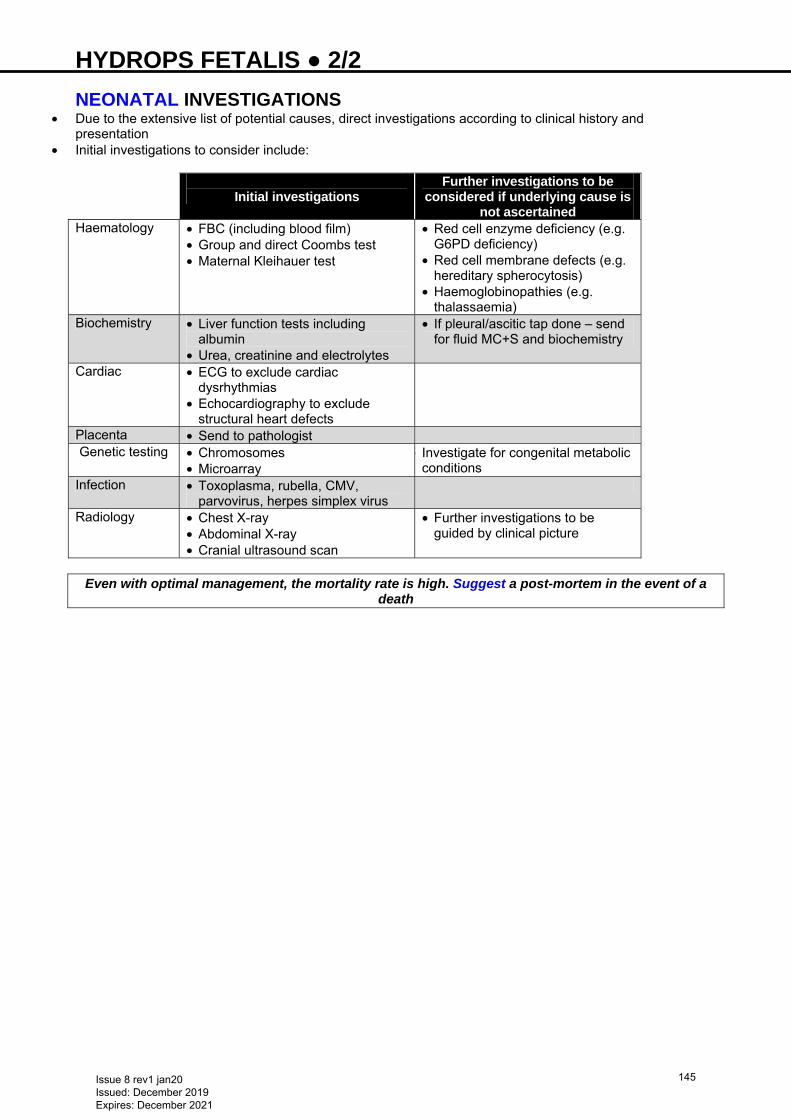
HYDROPS FETALIS ● 2/2 NEONATAL INVESTIGATIONS
• Due to the extensive list of potential causes, direct investigations according to clinical history and presentation
• Initial investigations to consider include:
Initial investigations
Further investigations to be considered if underlying cause is
not ascertained Haematology • FBC (including blood film)
• Group and direct Coombs test • Maternal Kleihauer test
• Red cell enzyme deficiency (e.g. G6PD deficiency)
• Red cell membrane defects (e.g. hereditary spherocytosis)
• Haemoglobinopathies (e.g. thalassaemia)
Biochemistry • Liver function tests including albumin
• Urea, creatinine and electrolytes
• If pleural/ascitic tap done – send for fluid MC+S and biochemistry
Cardiac • ECG to exclude cardiac dysrhythmias
• Echocardiography to exclude structural heart defects
Placenta • Send to pathologist Genetic testing • Chromosomes
• Microarray • Investigate for congenital metabolic
conditions Infection • Toxoplasma, rubella, CMV,
parvovirus, herpes simplex virus
Radiology • Chest X-ray • Abdominal X-ray • Cranial ultrasound scan
• Further investigations to be guided by clinical picture
Even with optimal management, the mortality rate is high. Suggest a post-mortem in the event of a
death
Issue 8 rev1 jan20 Issued: December 2019 Expires: December 2021
145
HYPERGLYCAEMIA ● 1/2 DEFINITION • There is no established definition of hyperglycaemia. However, treat if: • 2 blood sugars ≥14 on 2 occasions measured ≥2 hr apart or • blood sugars ≥12 on 2 occasions measured ≥2 hr apart with evidence of significant glycosuria (2+ on
urine dipstick)
Do not take sample from an infusion line that has glucose running through it
CLINICAL FEATURES • Osmotic diuresis leading to dehydration • Poor weight gain
Risk factors • Immaturity of pancreatic function (e.g. extremely premature infants and small-for-gestational-age) • Insulin resistance • Glucose overload (e.g. equipment failure, administrator error) • Stress (e.g. infection, pain) • Side effects of a medication (e.g. glucocorticoid treatment)
MONITORING • Most blood gas machines provide glucose measurements • Check blood glucose at least 6–8 hrly in: • unstable or acutely ill babies [respiratory distress syndrome, septicaemia, necrotising enterocolitis
(NEC)] • Check blood glucose at least once a day in stable babies: • <32 weeks’ gestation for first week • receiving parenteral nutrition (PN) • with severe unexpected dehydration or metabolic acidosis • with poor weight gain while receiving >120 kcal/kg/day
Babies treated with corticosteroids • Check urine for glycosuria daily • Check blood glucose if ≥2+ glucose in urine
TREATMENT • If possible, discontinue or decrease medications that worsen hyperglycaemia • Lipid component of PN may contribute to worsening hyperglycaemia. If on PN discuss stopping lipid with
consultant/pharmacist
Suspected infection/NEC • Hyperglycaemia in baby with previously stable blood glucose may be an early indicator of infection or
NEC • Assess baby clinically • After taking appropriate cultures, treat empirically
Fluids • If blood glucose ≥12 mmol/L, check urine for glycosuria (of ≥2+) and assess clinical hydration and fluid
input/output. Check for fluid administration errors • Calculate amount of glucose baby is receiving (as mg/kg/min) using the formula:
Glucose infusion rate (mg/kg/min) =
% glucose × fluid rate (mL/kg/day) 144
• If glucose delivery rate >10 mg/kg/min, decrease glucose in decrements to 6–10 mg/kg/min. If on PN, 8–10 mg/kg/min is acceptable
• If glycosuria and hyperglycaemia >12 mmol/L persists despite an appropriate glucose infusion rate, consider treating with insulin
Insulin • Commence insulin therapy at 0.05 units/kg/hr and titrate according to response – see Administration of
Actrapid® insulin (soluble insulin) • Check blood glucose 1 hr from starting and hourly until target reached
Issue 8 rev1 jan20 Issued: December 2019 Expires: December 2021
146
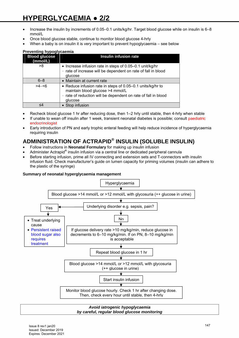
HYPERGLYCAEMIA ● 2/2 • Increase the insulin by increments of 0.05–0.1 units/kg/hr. Target blood glucose while on insulin is 6–8
mmol/L • Once blood glucose stable, continue to monitor blood glucose 4-hrly • When a baby is on insulin it is very important to prevent hypoglycaemia – see below
Preventing hypoglycaemia Blood glucose
(mmol/L) Insulin infusion rate
>8 • Increase infusion rate in steps of 0.05–0.1 unit/kg/hr • rate of increase will be dependent on rate of fall in blood
glucose 6–8 • Maintain at current rate
>4−<6 • Reduce infusion rate in steps of 0.05–0.1 units/kg/hr to maintain blood glucose >4 mmol/L
• rate of reduction will be dependent on rate of fall in blood glucose
≤4 • Stop infusion
• Recheck blood glucose 1 hr after reducing dose, then 1–2 hrly until stable, then 4-hrly when stable • If unable to wean off insulin after 1 week, transient neonatal diabetes is possible; consult paediatric
endocrinologist • Early introduction of PN and early trophic enteral feeding will help reduce incidence of hyperglycaemia
requiring insulin
ADMINISTRATION OF ACTRAPID® INSULIN (SOLUBLE INSULIN) • Follow instructions in Neonatal Formulary for making up insulin infusion • Administer Actrapid® insulin infusion via a central line or dedicated peripheral cannula • Before starting infusion, prime all IV connecting and extension sets and T-connectors with insulin
infusion fluid. Check manufacturer’s guide on lumen capacity for priming volumes (insulin can adhere to the plastic of the syringe)
Summary of neonatal hyperglycaemia management
Avoid iatrogenic hypoglycaemia by careful, regular blood glucose monitoring
Hyperglycaemia
Blood glucose >14 mmol/L or >12 mmol/L with glycosuria (++ glucose in urine)
Underlying disorder e.g. sepsis, pain?
No
Yes
• Treat underlying cause
• Persistent raised blood sugar also requires treatment
If glucose delivery rate >10 mg/kg/min, reduce glucose in decrements to 6–10 mg/kg/min. If on PN, 8–10 mg/kg/min
is acceptable
Repeat blood glucose in 1 hr
Blood glucose >14 mmol/L or >12 mmol/L with glycosuria (++ glucose in urine)
Start insulin infusion
Monitor blood glucose hourly. Check 1 hr after changing dose. Then, check every hour until stable, then 4-hrly
Issue 8 rev1 jan20 Issued: December 2019 Expires: December 2021
147
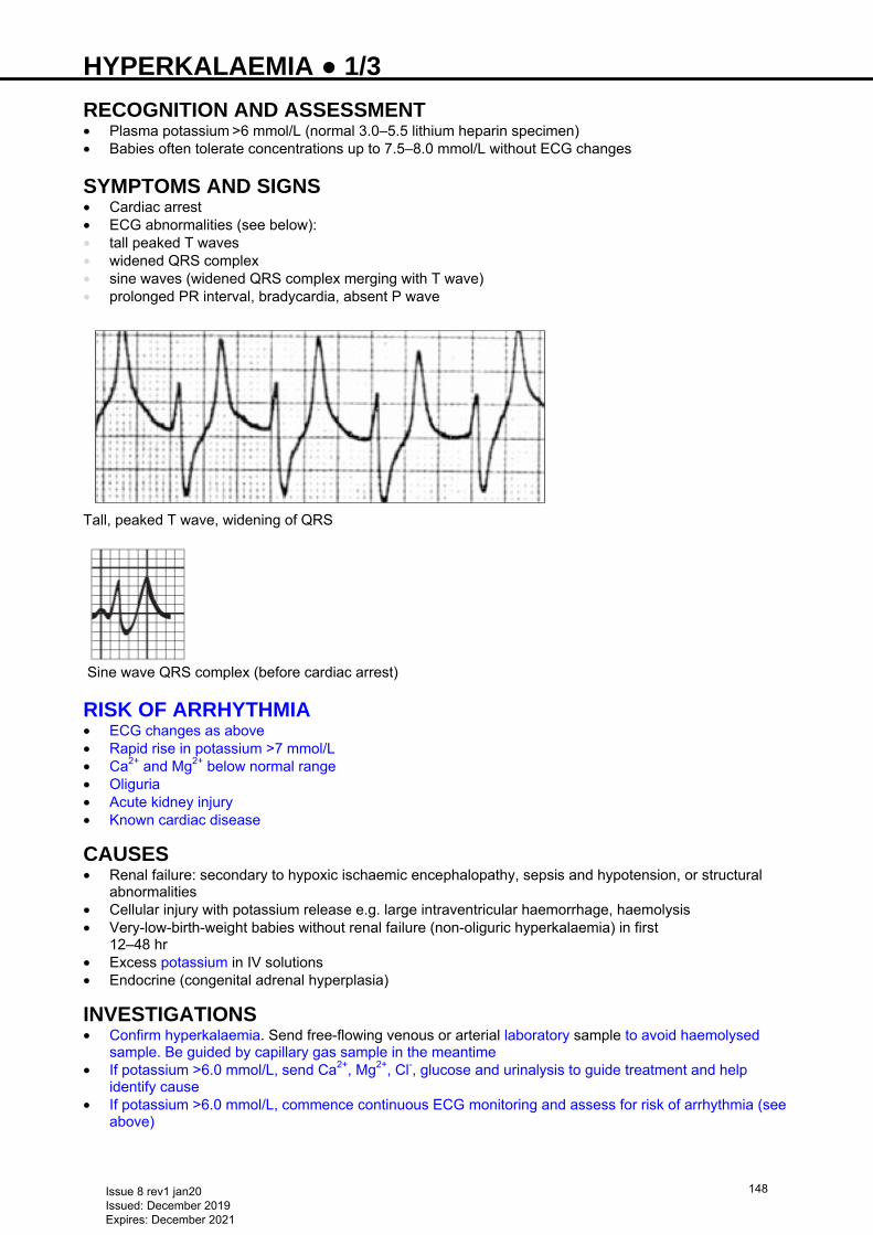
HYPERKALAEMIA ● 1/3 RECOGNITION AND ASSESSMENT • Plasma potassium >6 mmol/L (normal 3.0–5.5 lithium heparin specimen) • Babies often tolerate concentrations up to 7.5–8.0 mmol/L without ECG changes
SYMPTOMS AND SIGNS • Cardiac arrest • ECG abnormalities (see below): • tall peaked T waves • widened QRS complex • sine waves (widened QRS complex merging with T wave) • prolonged PR interval, bradycardia, absent P wave
Tall, peaked T wave, widening of QRS
Sine wave QRS complex (before cardiac arrest)
RISK OF ARRHYTHMIA • ECG changes as above • Rapid rise in potassium >7 mmol/L • Ca2+ and Mg2+ below normal range • Oliguria • Acute kidney injury • Known cardiac disease
CAUSES • Renal failure: secondary to hypoxic ischaemic encephalopathy, sepsis and hypotension, or structural
abnormalities • Cellular injury with potassium release e.g. large intraventricular haemorrhage, haemolysis • Very-low-birth-weight babies without renal failure (non-oliguric hyperkalaemia) in first
12–48 hr • Excess potassium in IV solutions • Endocrine (congenital adrenal hyperplasia)
INVESTIGATIONS • Confirm hyperkalaemia. Send free-flowing venous or arterial laboratory sample to avoid haemolysed
sample. Be guided by capillary gas sample in the meantime • If potassium >6.0 mmol/L, send Ca2+, Mg2+, Cl-, glucose and urinalysis to guide treatment and help
identify cause • If potassium >6.0 mmol/L, commence continuous ECG monitoring and assess for risk of arrhythmia (see
above)
Issue 8 rev1 jan20 Issued: December 2019 Expires: December 2021
148
HYPERKALAEMIA ● 2/3 IMMEDIATE TREATMENT Serum potassium >6.0 mmol/L (stable with normal ECG) • Stop all sources of potassium including IV solutions (check PN), oral supplements • Stop all potassium-retaining drugs and potassium-sparing diuretics e.g. spironolactone • Avoid suxamethonium • Review and withhold nephrotoxic drugs e.g. gentamicin • Recheck U&E 4−6 hrly
Serum potassium >7.0 mmol/L without ECG changes • As above • Inform consultant • Give salbutamol 4 microgram/kg IV in glucose 10% over 5–10 min; effect evident within 30 min but
sustained benefit may require repeat infusion after at least 2 hr • Give furosemide 1 mg/kg IV • If serum potassium still >7.0 mmol/L, give soluble insulin 0.5 units/kg IV in glucose 10% (made up to 2.5
mL and given over 30 min); very effective and has an additive effect with salbutamol • Repeat U&E 2−3 hrly • Repeat insulin infusion as necessary until potassium <7.0 mmol/L • Monitor blood glucose every 15 min for first 2 hr during and after infusion • aim for blood glucose 4.0–7.0 mmol/L • Give sodium bicarbonate 1 mmol/kg (2 mL of sodium bicarbonate 4.2% = 1 mmol) if: • pH <7.23 or • BE more negative than -8 or • bicarbonate <14 mmol/L • Correct other electrolyte abnormalities • Maintain ionised Ca2+ >1 mmol/L
Serum potassium >7.5 mmol/L with ECG changes • As above, but first institute emergency measures below: • give calcium gluconate 10% 0.5 mL/kg IV over 5–10 min • flush line with sodium chloride 0.9% or preferably use a different line • give sodium bicarbonate (1 mmol/kg IV over 2 min). Effective even in babies who are not acidotic (2 mL
of sodium bicarbonate 4.2% = 1 mmol) • Repeat U&E hourly
Further treatments: discuss with consultant • A cation-exchange resin, such as calcium resonium (500 mg/kg rectally, with removal by colonic
irrigation after 8–12 hr, repeat every 12 hr. Dose can be doubled at least once to 1 g/kg in severe hyperkalaemia). Useful for sustained reduction in serum potassium but takes many hours to act and is best avoided in sick preterms at risk of necrotising enterocolitis
• If severe hyperkalaemia persists despite above measures in term babies with otherwise good prognosis, contact renal team for consideration of dialysis
• Exchange transfusion using fresh blood or washed red blood cells is another strategy for sustained and reliable reduction in serum potassium concentration (see Exchange transfusion guideline)
SUBSEQUENT MANAGEMENT • Recheck serum potassium after each intervention or: • 4–6 hrly in stable/well baby with potassium <7 mmol/L and no ECG changes • 2−3 hrly in unwell baby and/or potassium >7 mmol/L with no ECG changes • hourly when arrhythmias or ECG changes present with/without renal failure • Monitor urine output and maintain good fluid balance • If urine output <1 mL/kg/hr, unless baby volume depleted, give furosemide 1 mg/kg IV until volume
corrected • Treat any underlying cause (e.g. renal failure)
Issue 8 rev1 jan20 Issued: December 2019 Expires: December 2021
149
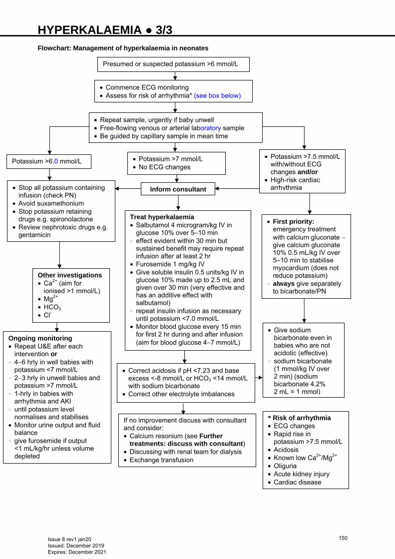
HYPERKALAEMIA ● 3/3 Flowchart: Management of hyperkalaemia in neonates
• Commence ECG monitoring • Assess for risk of arrhythmia* (see box below)
• Repeat sample, urgently if baby unwell • Free-flowing venous or arterial laboratory sample • Be guided by capillary sample in mean time
Potassium >6.0 mmol/L
• Stop all potassium containing infusion (check PN)
• Avoid suxamethonium • Stop potassium retaining
drugs e.g. spironolactone • Review nephrotoxic drugs e.g.
gentamicin
• Potassium >7 mmol/L • No ECG changes
Inform consultant
Treat hyperkalaemia • Salbutamol 4 microgram/kg IV in
glucose 10% over 5–10 min • effect evident within 30 min but
sustained benefit may require repeat infusion after at least 2 hr
• Furosemide 1 mg/kg IV • Give soluble insulin 0.5 units/kg IV in
glucose 10% made up to 2.5 mL and given over 30 min (very effective and has an additive effect with salbutamol)
• repeat insulin infusion as necessary until potassium <7.0 mmol/L
• Monitor blood glucose every 15 min for first 2 hr during and after infusion (aim for blood glucose 4−7 mmol/L)
Other investigations • Ca2+ (aim for
ionised >1 mmol/L) • Mg2+ • HCO3 • Cl-
• Potassium >7.5 mmol/L with/without ECG changes and/or
• High-risk cardiac arrhythmia
• Correct acidosis if pH <7.23 and base excess <-8 mmol/L or HCO3 <14 mmol/L with sodium bicarbonate
• Correct other electrolyte imbalances
Ongoing monitoring • Repeat U&E after each
intervention or • 4−6 hrly in well babies with
potassium <7 mmol/L • 2−3 hrly in unwell babies and
potassium >7 mmol/L • 1-hrly in babies with
arrhythmia and AKI • until potassium level
normalises and stabilises • Monitor urine output and fluid
balance • give furosemide if output
<1 mL/kg/hr unless volume depleted
Presumed or suspected potassium >6 mmol/L
• First priority: emergency treatment with calcium gluconate − give calcium gluconate 10% 0.5 mL/kg IV over 5–10 min to stabilise myocardium (does not reduce potassium)
• always give separately to bicarbonate/PN
• Give sodium bicarbonate even in babies who are not acidotic (effective)
• sodium bicarbonate (1 mmol/kg IV over 2 min) (sodium bicarbonate 4.2% 2 mL = 1 mmol)
If no improvement discuss with consultant and consider: • Calcium resonium (see Further
treatments: discuss with consultant) • Discussing with renal team for dialysis • Exchange transfusion
* Risk of arrhythmia • ECG changes • Rapid rise in
potassium >7.5 mmol/L • Acidosis • Known low Ca2+/Mg2+ • Oliguria • Acute kidney injury • Cardiac disease
Issue 8 rev1 jan20 Issued: December 2019 Expires: December 2021
150
HYPERNATRAEMIC DEHYDRATION ● 1/4 DEFINITION • Serum sodium >145 mmol/L • mild: 146–149 mmol/L • moderate: 150–160 mmol/L • severe: >160 mmol/L
Most common cause is failure to establish adequate oral intake while attempting breastfeeding
AIM To prevent/treat hypernatraemic dehydration while encouraging breastfeeding Other causes of hypernatraemia • Diarrhoea/vomiting • Infection and poor feeding • Renal dysplasia • Obstructive uropathy • Diuretic phase following acute kidney injury • Osmotic diuresis • Diabetes insipidus • Idiopathic causes • Sodium bicarbonate or sodium chloride administration • Excessive insensible losses in extremely premature babies • Improperly prepared formula
PREVENTION Babies at high risk • Preterm <37 weeks • Born to primiparous women • Maternal prolonged second stage of labour >1 hr • Use of labour medications • Caesarean section with delayed initiation of feeding • Cleft lip and/or palate • Maternal breast abnormalities (flat, inverted nipples)/surgery • Maternal illness, haemorrhage • Maternal obesity • Maternal diabetes • Polycystic ovary syndrome • Skin conditions that increase insensible water loss Action • Identify babies at risk • Immediate skin-to-skin contact at birth and breastfeed within 1 hr of life • Offer breastfeeding assistance within 6 hr of life • Assess baby to ensure feeding adequate • Ensure baby feeds ≥6 times within 24 hr • If baby reluctant to feed, express breast milk (see Breast milk expression guideline) and offer by cup or
syringe • Calculate required volume of feeds (see Nutrition and enteral feeding guideline) • Avoid bottle feeding as far as possible and avoid dummies • Assess feeding, number of wet nappies and stools using Table • Avoid early discharge of at-risk babies • Early reweighing of at-risk babies (at 72–96 hr) with breastfeeding support can reduce severity of
hypernatremic dehydration
Issue 8 rev1 jan20 Issued: December 2019 Expires: December 2021
151
HYPERNATRAEMIC DEHYDRATION ● 2/4 Day Wet nappies Stool 1–2 ≥2/day >1/day 3–4 ≥3/day ≥2/day, changing in colour and consistency 5–6 ≥5/day ≥2/day, yellow in colour • Weigh between 72 and 96 hr • Refer all who have lost >10% weight • weight loss % = weight loss (g)/birth weight (g) × 100 Symptoms and signs • Irritability/high pitched cry: unsettled during breastfeeding • Prolonged feeding >45 min • Demanding <6 feeds in 24 hr • Reduced urinary frequency • Delayed change from meconium to transitional stools • Weight loss • Fever • Jaundice • Lethargy/altered level of consciousness • Tremor • Increased tone • Doughy skin • Seizures (usually during rehydration) • Physical examination may be unremarkable • Usual signs of dehydration (sunken fontanelle, dry mucous membrane and reduced skin turgor) may be
absent Complications • Venous and arterial thrombosis • Subdural and cerebral haemorrhage • Cerebral oedema (especially during rehydration) • Seizures (especially following rehydration) • Apnoea and bradycardia • Cognitive and motor deficits • Hearing impairment − may be transient • Hypertension • Cerebral infarction • Renal failure • Death • Long-term developmental delay Investigations • U&E • Calcium • Total bilirubin • Blood glucose • CRP • Blood culture • Paired urinary electrolytes • If severe, cranial ultrasound
Issue 8 rev1 jan20 Issued: December 2019 Expires: December 2021
152
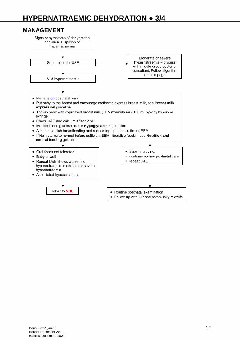
HYPERNATRAEMIC DEHYDRATION ● 3/4 MANAGEMENT
Signs or symptoms of dehydration or clinical suspicion of
hypernatraemia
Send blood for U&E
Mild hypernatraemia
• Manage on postnatal ward • Put baby to the breast and encourage mother to express breast milk, see Breast milk
expression guideline • Top-up baby with expressed breast milk (EBM)/formula milk 100 mL/kg/day by cup or
syringe • Check U&E and calcium after 12 hr • Monitor blood glucose as per Hypoglycaemia guideline • Aim to establish breastfeeding and reduce top-up once sufficient EBM • If Na+ returns to normal before sufficient EBM, liberalise feeds − see Nutrition and
enteral feeding guideline
• Oral feeds not tolerated • Baby unwell • Repeat U&E shows worsening
hypernatraemia, moderate or severe hypernatraemia
• Associated hypocalcaemia
• Baby improving: • continue routine postnatal care • repeat U&E
• Routine postnatal examination • Follow-up with GP and community midwife
Admit to NNU
Moderate or severe hypernatraemia – discuss
with middle grade doctor or consultant. Follow algorithm
on next page
Issue 8 rev1 jan20 Issued: December 2019 Expires: December 2021
153
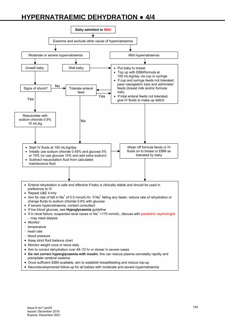
HYPERNATRAEMIC DEHYDRATION ● 4/4
Yes
Yes
Baby admitted to NNU
Examine and exclude other cause of hypernatraemia
Moderate or severe hypernatraemia Mild hypernatraemia
Unwell baby • Put baby to breast • Top up with EBM/formula at
100 mL/kg/day via cup or syringe • If cup and syringe feeds not tolerated,
pass nasogastric tube and administer feeds (breast milk and/or formula milk)
• If total enteral feeds not tolerated, give IV fluids to make up deficit
Well baby
Signs of shock?
Resuscitate with sodium chloride 0.9%
10 mL/kg
Tolerate enteral feed
• Start IV fluids at 100 mL/kg/day • Initially use sodium chloride 0.45% and glucose 5%
or 10% (or use glucose 10% and add extra sodium) • Subtract resuscitation fluid from calculated
maintenance fluid
Wean off formula feeds or IV fluids on to breast or EBM as
tolerated by baby
• Enteral rehydration is safe and effective if baby is clinically stable and should be used in preference to IV
• Repeat U&E 4-hrly • Aim for rate of fall in Na+ of 0.5 mmol/L/hr. If Na+ falling any faster, reduce rate of rehydration or
change fluids to sodium chloride 0.9% with glucose • If severe hypernatraemia, contact consultant • If low blood glucose, see Hypoglycaemia guideline • If in renal failure, suspected renal cause or Na+ >170 mmol/L, discuss with paediatric nephrologist
− may need dialysis • Monitor: • temperature • heart rate • blood pressure • Keep strict fluid balance chart • Monitor weight once or twice daily • Aim to correct dehydration over 48–72 hr or slower in severe cases • Do not correct hyperglycaemia with insulin, this can reduce plasma osmolality rapidly and
precipitate cerebral oedema • Once sufficient EBM available, aim to establish breastfeeding and reduce top-up • Neurodevelopmental follow-up for all babies with moderate and severe hypernatraemia
No
No
Issue 8 rev1 jan20 Issued: December 2019 Expires: December 2021
154
HYPOCALCAEMIA ● 1/2 RECOGNITION AND ASSESSMENT
• Term or preterm infants birth weight ≥1500 g, total serum calcium <2 mmol/L or ionised fraction <1.1 mmo/L
• Preterm infant, birth weight <1500 g, total serum calcium <1.75 mmol/L or ionised fraction <1 mmol/L
SYMPTOMS AND SIGNS • Early onset occurs in first 2−3 days of life and is usually asymptomatic • Late onset develops after first 2−3 days of life and typically occurs at the end of the first week • Most infants are asymptomatic and identified on screening • Characteristic sign is increased neuromuscular irritability including: • jitteriness and irritability • generalised/focal seizures • non-specific symptoms e.g.:
− poor feeding − lethargy − apnoea
• prolonged QTc on ECG • rare presentations:
− stridor − bronchospasm − pylorospasm
CAUSES • Early onset: • prematurity • intrauterine growth restriction • infants of diabetic mother • hypoxic ischaemic encephalopathy • hypomagnesaemia • hypoparathyroidism • syndromes e.g. DiGeorge syndrome • maternal hyperparathyroidism • Late onset: • high phosphate load – cow’s milk, renal failure • hypomagnesaemia • parenteral nutrition • exchange transfusion • alkalosis • maternal hypercalcemia • maternal vitamin D deficiency • transient hypoparathyroidism • syndromes and genetic mutations e.g. DiGeorge and Kenny-Caffey syndromes
INVESTIGATIONS • Serum calcium • only monitor if risk factors, most infants with hypocalcaemia are asymptomatic • well preterm infant with birth weight >1500 g and well term infants of diabetic mothers receiving milk
feedings on day 1 of life do not need testing routinely • ionised calcium preferred • if using total calcium, measure albumin and correct for hypoalbuminemia • Phosphate • Magnesium • Persistent hypocalcaemia or severe hypocalcaemia despite adequate calcium therapy: • 25-hydroxyvitamin D level • renal function tests • liver function test • alkaline phosphatase • parathyroid hormone level • urinary calcium:creatinine ratio
Issue 8 rev1 jan20 Issued: December 2019 Expires: December 2021
155
HYPOCALCAEMIA ● 2/2 • ECG for prolonged QTc interval • if pseudohyperparathyroidism suspected, X-ray hand • chest X-ray for thymic shadow • if hypoparathyroidism suspected, renal ultrasound • if DiGeorge syndrome suspected, echocardiography • genetic test for gene mutations or suspected syndrome e.g. DiGeorge syndrome
MANAGEMENT Asymptomatic infants • Most infants with early onset hypocalcaemia recover with nutritional support; so early feeding provides
adequate calcium • Infants requiring IV fluid: add calcium gluconate 10% 0.46 mmol/kg/day (= 2 mL/kg/day) to IV fluid and
give as continuous infusion • infant tolerating oral feeds: give 0.25 mmol/kg oral 6-hrly Symptomatic hypocalcaemia • If seizures, prolonged QT interval, apnoea, unstable or poor feeding give IV calcium gluconate 10% 0.11
mmol/kg (= 0.5 mL/kg) over 5−10 min followed by maintenance • dilute with sodium chloride 0.9% or glucose 5% 4 mL to each 1 mL calcium gluconate 10% to give a
concentration of 45 micromol/mL. Can be given undiluted via central line in an emergency • doses up to 0.46 mmol/kg (= 2 mL/kg calcium gluconate 10%) have been used • maximum rate of administration 22 micromol/kg/hr • Stable baby or following acute treatment • oral calcium dose 0.25 mmol/kg 6-hrly • if enteral feeds not tolerated add calcium gluconate 10% 0.46 mmol/kg/day to IV fluid as above • If symptomatic hypocalcaemia: hypomagnesaemia − magnesium sulphate 100 mg/kg IV/IM 12-hrly for
2−3 doses • Vitamin D deficiency give 1000 units daily and adjust dose according to response • Hyperphosphataemia • high calcium, low phosphate diet • human milk is preferable, if not available, use formula with low phosphate 60/40 and oral calcium
IV calcium precautions and considerations
• Extravasation can cause skin and subcutaneous tissue necrosis (see Extravasation guideline). Monitor IV site closely
• Continuous infusion preferred to bolus, but use bolus for initial management in symptomatic hypocalcaemia
• Bolus IV calcium can cause dysrhythmias − administer slowly over 5−10 min with cardiac monitoring • Calcium can be given via UVC provided catheter tip is in vena cava • inadvertent administration into portal vein can cause hepatic necrosis • Do not mix calcium solutions with those containing phosphorus or bicarbonate as this can cause
precipitation
SUBSEQUENT MANAGEMENT • Monitor bone profile and phosphate levels according to clinical need • If calcium normal after 48 hr treatment, halve maintenance dose • If calcium fails to normalise investigate for underlying cause • Hyperphosphataemia – calcium and phosphate normalise in 3−5 days. Stop calcium after 1 week and
switch to normal formula in 2−4 weeks
Issue 8 rev1 jan20 Issued: December 2019 Expires: December 2021
156
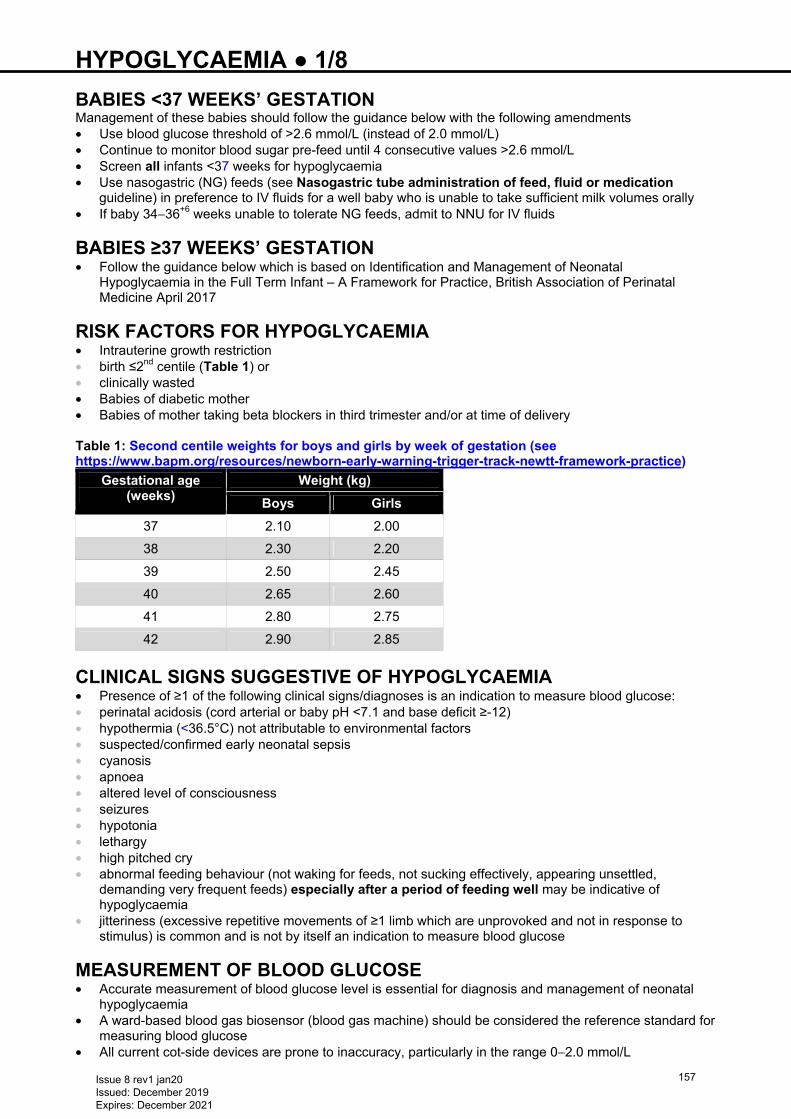
HYPOGLYCAEMIA ● 1/8 BABIES <37 WEEKS’ GESTATION Management of these babies should follow the guidance below with the following amendments • Use blood glucose threshold of >2.6 mmol/L (instead of 2.0 mmol/L) • Continue to monitor blood sugar pre-feed until 4 consecutive values >2.6 mmol/L • Screen all infants <37 weeks for hypoglycaemia • Use nasogastric (NG) feeds (see Nasogastric tube administration of feed, fluid or medication
guideline) in preference to IV fluids for a well baby who is unable to take sufficient milk volumes orally • If baby 34−36+6 weeks unable to tolerate NG feeds, admit to NNU for IV fluids
BABIES ≥37 WEEKS’ GESTATION • Follow the guidance below which is based on Identification and Management of Neonatal
Hypoglycaemia in the Full Term Infant – A Framework for Practice, British Association of Perinatal Medicine April 2017
RISK FACTORS FOR HYPOGLYCAEMIA • Intrauterine growth restriction • birth ≤2nd centile (Table 1) or • clinically wasted • Babies of diabetic mother • Babies of mother taking beta blockers in third trimester and/or at time of delivery Table 1: Second centile weights for boys and girls by week of gestation (see https://www.bapm.org/resources/newborn-early-warning-trigger-track-newtt-framework-practice)
Gestational age (weeks)
Weight (kg)
Boys Girls
37 2.10 2.00
38 2.30 2.20
39 2.50 2.45
40 2.65 2.60
41 2.80 2.75
42 2.90 2.85
CLINICAL SIGNS SUGGESTIVE OF HYPOGLYCAEMIA • Presence of ≥1 of the following clinical signs/diagnoses is an indication to measure blood glucose: • perinatal acidosis (cord arterial or baby pH <7.1 and base deficit ≥-12) • hypothermia (<36.5°C) not attributable to environmental factors • suspected/confirmed early neonatal sepsis • cyanosis • apnoea • altered level of consciousness • seizures • hypotonia • lethargy • high pitched cry • abnormal feeding behaviour (not waking for feeds, not sucking effectively, appearing unsettled,
demanding very frequent feeds) especially after a period of feeding well may be indicative of hypoglycaemia
• jitteriness (excessive repetitive movements of ≥1 limb which are unprovoked and not in response to stimulus) is common and is not by itself an indication to measure blood glucose
MEASUREMENT OF BLOOD GLUCOSE • Accurate measurement of blood glucose level is essential for diagnosis and management of neonatal
hypoglycaemia • A ward-based blood gas biosensor (blood gas machine) should be considered the reference standard for
measuring blood glucose • All current cot-side devices are prone to inaccuracy, particularly in the range 0−2.0 mmol/L
Issue 8 rev1 jan20 Issued: December 2019 Expires: December 2021
157
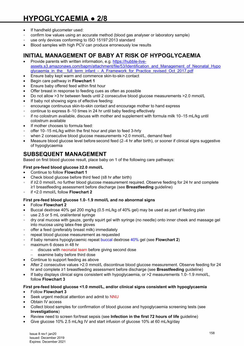
HYPOGLYCAEMIA ● 2/8 • If handheld glucometer used: • confirm low values using an accurate method (blood gas analyser or laboratory sample) • use only devices conforming to ISO 15197:2013 standard • Blood samples with high PCV can produce erroneously low results
INITIAL MANAGEMENT OF BABY AT RISK OF HYPOGLYCAEMIA • Provide parents with written information, e.g. https://hubble-live-
assets.s3.amazonaws.com/bapm/attachment/file/53/Identification_and_Management_of_Neonatal_Hypoglycaemia_in_the__full_term_infant_-_A_Framework_for_Practice_revised_Oct_2017.pdf
• Ensure baby kept warm and commence skin-to-skin contact • Begin care pathway in Flowchart 1 • Ensure baby offered feed within first hour • Offer breast in response to feeding cues as often as possible • Do not allow >3 hr between feeds until 2 consecutive blood glucose measurements >2.0 mmol/L • If baby not showing signs of effective feeding: • encourage continuous skin-to-skin contact and encourage mother to hand express • continue to express 8−10 times in 24 hr until baby feeding effectively • if no colostrum available, discuss with mother and supplement with formula milk 10−15 mL/kg until
colostrum available • If mother chooses to formula feed: • offer 10−15 mL/kg within the first hour and plan to feed 3-hrly • when 2 consecutive blood glucose measurements >2.0 mmol/L, demand feed • Measure blood glucose level before second feed (2−4 hr after birth), or sooner if clinical signs suggestive
of hypoglycaemia
SUBSEQUENT MANAGEMENT Based on first blood glucose result, place baby on 1 of the following care pathways:
First pre-feed blood glucose ≥2.0 mmol/L • Continue to follow Flowchart 1 • Check blood glucose before third feed (≤8 hr after birth) • if ≥2.0 mmol/L no further blood glucose measurement required. Observe feeding for 24 hr and complete
≥1 breastfeeding assessment before discharge (see Breastfeeding guideline) • if <2.0 mmol/L follow Flowchart 2
First pre-feed blood glucose 1.0−1.9 mmol/L and no abnormal signs • Follow Flowchart 2 • Buccal dextrose 40% gel 200 mg/kg (0.5 mL/kg of 40% gel) may be used as part of feeding plan • use 2.5 or 5 mL oral/enteral syringe • dry oral mucosa with gauze, gently squirt gel with syringe (no needle) onto inner cheek and massage gel
into mucosa using latex-free gloves • offer a feed (preferably breast milk) immediately • repeat blood glucose measurement as requested • if baby remains hypoglycaemic repeat buccal dextrose 40% gel (see Flowchart 2) • maximum 6 doses in 48 hr
− discuss with neonatal team before giving second dose − examine baby before third dose
• Continue to support feeding as above • After 2 consecutive values >2.0 mmol/L discontinue blood glucose measurement. Observe feeding for 24
hr and complete ≥1 breastfeeding assessment before discharge (see Breastfeeding guideline) • If baby displays clinical signs consistent with hypoglycaemia, or >2 measurements 1.0−1.9 mmol/L,
follow Flowchart 3
First pre-feed blood glucose <1.0 mmol/L, and/or clinical signs consistent with hypoglycaemia • Follow Flowchart 3 • Seek urgent medical attention and admit to NNU • Obtain IV access • Collect blood samples for confirmation of blood glucose and hypoglycaemia screening tests (see
Investigations) • Review need to screen for/treat sepsis (see Infection in the first 72 hours of life guideline) • Give glucose 10% 2.5 mL/kg IV and start infusion of glucose 10% at 60 mL/kg/day
Issue 8 rev1 jan20 Issued: December 2019 Expires: December 2021
158
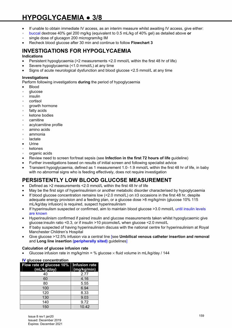
HYPOGLYCAEMIA ● 3/8 • If unable to obtain immediate IV access, as an interim measure whilst awaiting IV access, give either: • buccal dextrose 40% gel 200 mg/kg (equivalent to 0.5 mL/kg of 40% gel) as detailed above or • single dose of glucagon 200 microgram/kg IM • Recheck blood glucose after 30 min and continue to follow Flowchart 3
INVESTIGATIONS FOR HYPOGLYCAEMIA Indications • Persistent hypoglycaemia (>2 measurements <2.0 mmol/L within the first 48 hr of life) • Severe hypoglycaemia (<1.0 mmol/L) at any time • Signs of acute neurological dysfunction and blood glucose <2.5 mmol/L at any time
Investigations Perform following investigations during the period of hypoglycaemia • Blood • glucose • insulin • cortisol • growth hormone • fatty acids • ketone bodies • carnitine • acylcarnitine profile • amino acids • ammonia • lactate • Urine • ketones • organic acids • Review need to screen for/treat sepsis (see Infection in the first 72 hours of life guideline) • Further investigations based on results of initial screen and following specialist advice • Transient hypoglycaemia, defined as 1 measurement 1.0−1.9 mmol/L within the first 48 hr of life, in baby
with no abnormal signs who is feeding effectively, does not require investigation
PERSISTENTLY LOW BLOOD GLUCOSE MEASUREMENT • Defined as >2 measurements <2.0 mmol/L within the first 48 hr of life • May be the first sign of hyperinsulinism or another metabolic disorder characterised by hypoglycaemia • If blood glucose concentration remains low (<2.0 mmol/L) on ≥3 occasions in the first 48 hr, despite
adequate energy provision and a feeding plan, or a glucose dose >8 mg/kg/min (glucose 10% 115 mL/kg/day infusion) is required, suspect hyperinsulinism
• If hyperinsulism suspected or confirmed, aim to maintain blood glucose >3.0 mmol/L until insulin levels are known
• Hyperinsulinism confirmed if paired insulin and glucose measurements taken whilst hypoglycaemic give glucose:insulin ratio <0.3, or if insulin >10 picomole/L when glucose <2.0 mmol/L
• If baby suspected of having hyperinsulinism discuss with the national centre for hyperinsulinism at Royal Manchester Children’s Hospital
• Give glucose >12.5% infusion via a central line [see Umbilical venous catheter insertion and removal and Long line insertion (peripherally sited) guidelines]
Calculation of glucose infusion rate • Glucose infusion rate in mg/kg/min = % glucose × fluid volume in mL/kg/day / 144
IV glucose concentration Flow rate of glucose 10%
(mL/kg/day) Infusion rate(mg/kg/min)
40 2.77 60 4.16 80 5.55 100 6.94 120 8.33 130 9.03 140 9.72 150 10.42
Issue 8 rev1 jan20 Issued: December 2019 Expires: December 2021
159
HYPOGLYCAEMIA ● 4/8 To make up any concentration of glucose in any volume • Desired volume = V mL • Desired concentration of glucose = D% • Lower concentration of glucose = L% • Volume of lower concentration of glucose to add = LV mL • Higher concentration of glucose = H% • Volume of higher concentration of glucose to add = HV mL Formula: HV = V (D−L) / (H−L) LV = V−HV
HV mL + LV mL = V mL of D% • If >12.5% glucose required, give via a central line [see Umbilical venous catheter insertion and
removal and Long line insertion (peripherally sited) guidelines]
Issue 8 rev1 jan20 Issued: December 2019 Expires: December 2021
160
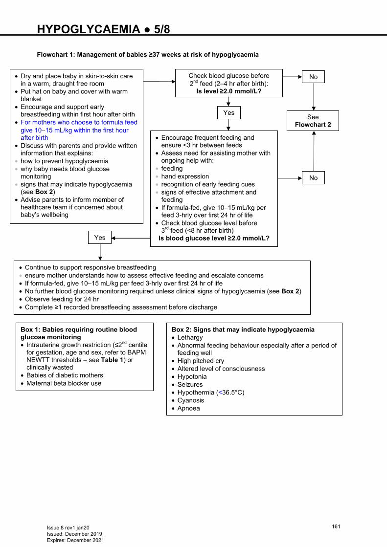
HYPOGLYCAEMIA ● 5/8 Flowchart 1: Management of babies ≥37 weeks at risk of hypoglycaemia Is level ≥ 2.0 mmol/
Box 1: Babies requiring routine blood glucose monitoring • Intrauterine growth restriction (≤2nd centile
for gestation, age and sex, refer to BAPM NEWTT thresholds – see Table 1) or clinically wasted
• Babies of diabetic mothers • Maternal beta blocker use
Box 2: Signs that may indicate hypoglycaemia • Lethargy • Abnormal feeding behaviour especially after a period of
feeding well • High pitched cry • Altered level of consciousness • Hypotonia • Seizures • Hypothermia (<36.5°C) • Cyanosis • Apnoea
• Dry and place baby in skin-to-skin care in a warm, draught free room
• Put hat on baby and cover with warm blanket
• Encourage and support early breastfeeding within first hour after birth
• For mothers who choose to formula feed give 10−15 mL/kg within the first hour after birth
• Discuss with parents and provide written information that explains:
• how to prevent hypoglycaemia • why baby needs blood glucose
monitoring • signs that may indicate hypoglycaemia
(see Box 2) • Advise parents to inform member of
healthcare team if concerned about baby’s wellbeing
Check blood glucose before 2nd feed (2−4 hr after birth):
Is level ≥2.0 mmol/L?
Yes
• Encourage frequent feeding and ensure <3 hr between feeds
• Assess need for assisting mother with ongoing help with:
• feeding • hand expression • recognition of early feeding cues • signs of effective attachment and
feeding • If formula-fed, give 10−15 mL/kg per
feed 3-hrly over first 24 hr of life • Check blood glucose level before
3rd feed (<8 hr after birth) Is blood glucose level ≥2.0 mmol/L? Yes
• Continue to support responsive breastfeeding • ensure mother understands how to assess effective feeding and escalate concerns • If formula-fed, give 10−15 mL/kg per feed 3-hrly over first 24 hr of life • No further blood glucose monitoring required unless clinical signs of hypoglycaemia (see Box 2) • Observe feeding for 24 hr • Complete ≥1 recorded breastfeeding assessment before discharge
No
See Flowchart 2
No
Issue 8 rev1 jan20 Issued: December 2019 Expires: December 2021
161
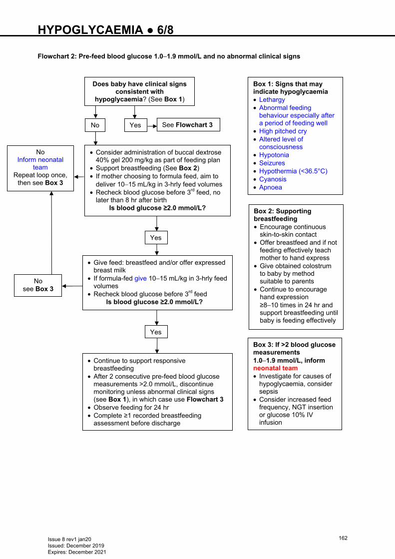
HYPOGLYCAEMIA ● 6/8 Flowchart 2: Pre-feed blood glucose 1.0−1.9 mmol/L and no abnormal clinical signs
• Consider administration of buccal dextrose 40% gel 200 mg/kg as part of feeding plan
• Support breastfeeding (See Box 2) • If mother choosing to formula feed, aim to
deliver 10−15 mL/kg in 3-hrly feed volumes • Recheck blood glucose before 3rd feed, no
later than 8 hr after birth Is blood glucose ≥2.0 mmol/L?
Yes
• Give feed: breastfeed and/or offer expressed breast milk
• If formula-fed give 10−15 mL/kg in 3-hrly feed volumes
• Recheck blood glucose before 3rd feed Is blood glucose ≥2.0 mmol/L?
Yes
• Continue to support responsive breastfeeding
• After 2 consecutive pre-feed blood glucose measurements >2.0 mmol/L, discontinue monitoring unless abnormal clinical signs (see Box 1), in which case use Flowchart 3
• Observe feeding for 24 hr • Complete ≥1 recorded breastfeeding
assessment before discharge
No Inform neonatal
team Repeat loop once,
then see Box 3
No see Box 3
Box 1: Signs that may indicate hypoglycaemia • Lethargy • Abnormal feeding
behaviour especially after a period of feeding well
• High pitched cry • Altered level of
consciousness • Hypotonia • Seizures • Hypothermia (<36.5°C) • Cyanosis • Apnoea
Box 3: If >2 blood glucose measurements 1.0−1.9 mmol/L, inform neonatal team • Investigate for causes of
hypoglycaemia, consider sepsis
• Consider increased feed frequency, NGT insertion or glucose 10% IV infusion
Does baby have clinical signs consistent with
hypoglycaemia? (See Box 1)
Yes No See Flowchart 3
Box 2: Supporting breastfeeding • Encourage continuous
skin-to-skin contact • Offer breastfeed and if not
feeding effectively teach mother to hand express
• Give obtained colostrum to baby by method suitable to parents
• Continue to encourage hand expression ≥8−10 times in 24 hr and support breastfeeding until baby is feeding effectively
Issue 8 rev1 jan20 Issued: December 2019 Expires: December 2021
162
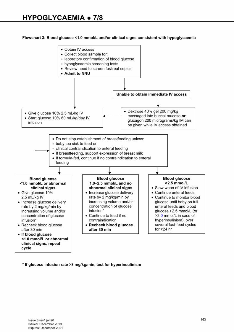
HYPOGLYCAEMIA ● 7/8 Flowchart 3: Blood glucose <1.0 mmol/L and/or clinical signs consistent with hypoglycaemia
* If glucose infusion rate >8 mg/kg/min, test for hyperinsulinism
• Obtain IV access • Collect blood sample for: • laboratory confirmation of blood glucose • hypoglycaemia screening tests • Review need to screen for/treat sepsis • Admit to NNU
Blood glucose <1.0 mmol/L or abnormal
clinical signs • Give glucose 10%
2.5 mL/kg IV • Increase glucose delivery
rate by 2 mg/kg/min by increasing volume and/or concentration of glucose infusion*
• Recheck blood glucose after 30 min
• If blood glucose <1.0 mmol/L or abnormal clinical signs, repeat cycle
Blood glucose 1.0−2.5 mmol/L and no abnormal clinical signs
• Increase glucose delivery rate by 2 mg/kg/min by increasing volume and/or concentration of glucose infusion*
• Continue to feed if no contraindication
• Recheck blood glucose after 30 min
• Give glucose 10% 2.5 mL/kg IV • Start glucose 10% 60 mL/kg/day IV
infusion
Unable to obtain immediate IV access
• Dextrose 40% gel 200 mg/kg massaged into buccal mucosa or glucagon 200 micrograms/kg IM can be given while IV access obtained
• Do not stop establishment of breastfeeding unless: • baby too sick to feed or • clinical contraindication to enteral feeding • If breastfeeding, support expression of breast milk • If formula-fed, continue if no contraindication to enteral
feeding
Blood glucose >2.5 mmol/L
• Slow wean of IV infusion • Continue enteral feeds • Continue to monitor blood
glucose until baby on full enteral feeds and blood glucose >2.5 mmol/L (or >3.0 mmol/L in case of hyperinsulinism), over several fast-feed cycles for ≥24 hr
Issue 8 rev1 jan20 Issued: December 2019 Expires: December 2021
163
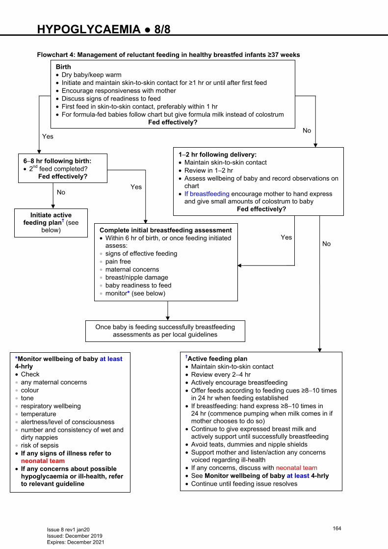
HYPOGLYCAEMIA ● 8/8
No
Flowchart 4: Management of reluctant feeding in healthy breastfed infants ≥37 weeks •
Yes No
No Yes
Yes
Birth • Dry baby/keep warm • Initiate and maintain skin-to-skin contact for ≥1 hr or until after first feed • Encourage responsiveness with mother • Discuss signs of readiness to feed • First feed in skin-to-skin contact, preferably within 1 hr • For formula-fed babies follow chart but give formula milk instead of colostrum
Fed effectively?
Once baby is feeding successfully breastfeeding assessments as per local guidelines
6−8 hr following birth: • 2nd feed completed?
Fed effectively?
1−2 hr following delivery: • Maintain skin-to-skin contact • Review in 1−2 hr • Assess wellbeing of baby and record observations on
chart • If breastfeeding encourage mother to hand express
and give small amounts of colostrum to baby Fed effectively?
Initiate active feeding plan† (see
below) Complete initial breastfeeding assessment • Within 6 hr of birth, or once feeding initiated
assess: • signs of effective feeding • pain free • maternal concerns • breast/nipple damage • baby readiness to feed • monitor* (see below)
†Active feeding plan • Maintain skin-to-skin contact • Review every 2−4 hr • Actively encourage breastfeeding • Offer feeds according to feeding cues ≥8−10 times
in 24 hr when feeding established • If breastfeeding: hand express ≥8−10 times in
24 hr (commence pumping when milk comes in if mother chooses to do so)
• Continue to give expressed breast milk and actively support until successfully breastfeeding
• Avoid teats, dummies and nipple shields • Support mother and listen/action any concerns
voiced regarding ill-health • If any concerns, discuss with neonatal team • See Monitor wellbeing of baby at least 4-hrly • Continue until feeding issue resolves
*Monitor wellbeing of baby at least 4-hrly • Check • any maternal concerns • colour • tone • respiratory wellbeing • temperature • alertness/level of consciousness • number and consistency of wet and
dirty nappies • risk of sepsis • If any signs of illness refer to
neonatal team • If any concerns about possible
hypoglycaemia or ill-health, refer to relevant guideline
Issue 8 rev1 jan20 Issued: December 2019 Expires: December 2021
164
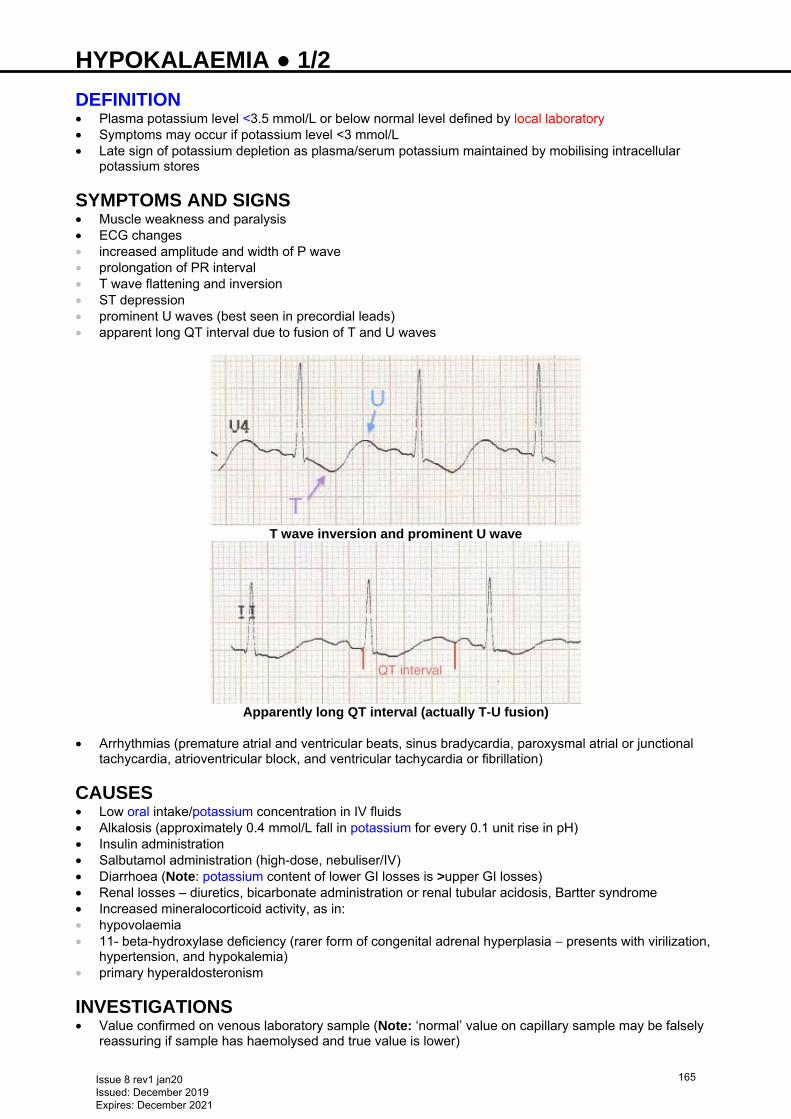
HYPOKALAEMIA ● 1/2 DEFINITION • Plasma potassium level <3.5 mmol/L or below normal level defined by local laboratory • Symptoms may occur if potassium level <3 mmol/L • Late sign of potassium depletion as plasma/serum potassium maintained by mobilising intracellular
potassium stores
SYMPTOMS AND SIGNS • Muscle weakness and paralysis • ECG changes • increased amplitude and width of P wave • prolongation of PR interval • T wave flattening and inversion • ST depression • prominent U waves (best seen in precordial leads) • apparent long QT interval due to fusion of T and U waves
T wave inversion and prominent U wave
Apparently long QT interval (actually T-U fusion)
• Arrhythmias (premature atrial and ventricular beats, sinus bradycardia, paroxysmal atrial or junctional
tachycardia, atrioventricular block, and ventricular tachycardia or fibrillation)
CAUSES • Low oral intake/potassium concentration in IV fluids • Alkalosis (approximately 0.4 mmol/L fall in potassium for every 0.1 unit rise in pH) • Insulin administration • Salbutamol administration (high-dose, nebuliser/IV) • Diarrhoea (Note: potassium content of lower GI losses is >upper GI losses) • Renal losses – diuretics, bicarbonate administration or renal tubular acidosis, Bartter syndrome • Increased mineralocorticoid activity, as in: • hypovolaemia • 11- beta-hydroxylase deficiency (rarer form of congenital adrenal hyperplasia − presents with virilization,
hypertension, and hypokalemia) • primary hyperaldosteronism
INVESTIGATIONS • Value confirmed on venous laboratory sample (Note: ‘normal’ value on capillary sample may be falsely
reassuring if sample has haemolysed and true value is lower)
Issue 8 rev1 jan20 Issued: December 2019 Expires: December 2021
165
HYPOKALAEMIA ● 2/2 • ECG • Cardiac monitor if ECG changes present • No investigations needed if hypokalaemia is mild (serum level 3−3.5 mmol/L) and there is a reason for
baby to be hypokalaemic • Significant hypokalaemia (serum level <3 mmol/L) and no obvious cause check: • acid/base balance and bicarbonate level on blood gas • urinary potassium level. Level >20 mmol/L suggests excess renal potassium losses • if baby is hypertensive plasma renin and aldosterone • If hypokalaemia not responding well to replacement check magnesium level
IMMEDIATE MANAGEMENT • Normal maintenance potassium requirement is 2 mmol/kg/day. Higher amounts will be needed to correct
hypokalaemia • If baby is on insulin infusion, consider stopping
Symptomatic babies • Give rapid potassium supplementation • Strong potassium • contains 20 mmol/10 mL • must be diluted at least 50-fold with sodium chloride 0.9% or a mixture of sodium chloride 0.9% in
glucose before administration • maximal peripheral concentration 40 mmol/L (1 mmol in 25 mL) • maximal central concentration 80 mmol/L (1 mmol in 12.5 mL) • rate 0.2 mmol/kg/hr (maximum 0.5 mmol/kg/hr if severe potassium depletion) • Monitor potassium levels and cardiac monitoring necessary • Recheck potassium at 2−4 hr and assess continuing need for infusion
Asymptomatic babies • Potassium replacement given according to how baby is being fed • orally fed babies
− oral supplementation should be given e.g. potassium chloride 1 mmol/kg 12-hrly − dose increased/titrated according to response
• babies on IV fluids − potassium chloride 3−5 mmol/kg/day, depending on electrolyte levels, may be added to IV fluid
• babies receiving parenteral nutrition (PN) − increase potassium concentration in the PN to 3−5 mmol/kg/day − if modified PN not available run potassium infusion 3−5 mmol/kg/day to run alongside current PN
SUBSEQUENT MANAGEMENT • Monitor potassium levels according to clinical need • Well babies receiving oral potassium check level 1−2 weekly • Babies on IV fluids or PN with mild hypokalaemia (potassium 3−3.5 mmol/L) check daily • Check more frequently in significant hypokalaemia (serum level <3 mmol/L), symptomatic hypokalaemia
or if concentrations of potassium >5 mmol/kg/day are being given • Once plasma/serum potassium level normal, continue potassium supplementation for a further week if
baby is orally fed, to allow replenishment of total body potassium (intracellular) stores, or reduce potassium to 2 mmol/kg/day if baby is on IV fluids/TPN
• Recheck potassium level following this to ensure hypokalaemia does not recur
Issue 8 rev1 jan20 Issued: December 2019 Expires: December 2021
166
HYPOTENSION ● 1/3
Hypovolaemia is an uncommon cause of hypotension in the preterm newborn. Excessive volume expansion can increase mortality
DEFINITION Thresholds for intervention • Aim to maintain mean arterial BP (MABP) ≥ gestational age in weeks • Aim for even higher MABP in case of persistent pulmonary hypertension of the newborn [see Persistent
pulmonary hypertension of the newborn (PPHN) guideline]
RECOGNITION AND ASSESSMENT Assessment of BP • Measure MABP: • by direct intra-arterial BP [umbilical arterial catheter (see Umbilical artery catheterisation and removal
guideline) or peripheral arterial line (see Arterial line insertion guideline)] • automated oscillometry (Dinamap) has limited accuracy in hypotensive preterm babies; usually over-
reads BP in the lower ranges • Identification of hypotension should not be based solely upon BP thresholds. Assess as many of the
following indices of tissue perfusion as possible (thresholds for abnormality in brackets). Assess cardiovascular status clinically
• capillary refill time (>3 sec) • toe-core temperature difference (>2°C) • urine output (<1 mL/kg/hr) • rising lactate Causes of hypotension • Sepsis • Extreme prematurity • Tension pneumothorax • Blood loss • Large patent ductus arteriosus (PDA) (see Patent ductus arteriosus guideline) • Poor myocardial contractility (very-low-birth-weight, hypoxia, cardiomyopathy or hypocalcaemia) • Polyuria secondary to glucosuria • Third spacing (surgical causes – NEC/perforation/malrotation/obstruction) • High positive intrathoracic pressure (high MAP on conventional/HFOV) • Severe acidosis (pH <7) • Drugs (morphine, muscle relaxants and anti-hypertensives)
IMMEDIATE TREATMENT
Aim is to treat cause and improve organ perfusion, not to correct a ‘BP reading’ Seek senior advice throughout
Transilluminate chest to exclude pneumothorax (see Transillumination of the chest guideline)
Fluid • Give if hypovolaemic (not >10 mL/kg) unless there is evidence of fluid/blood loss/sepsis (late onset
sepsis/term babies), when it may be necessary to give more than this volume, depending on condition of baby. Otherwise, start inotropes first (see Inotropes)
• If clinical condition poor, BP very low, or mother has been treated with IV anti-hypertensive agent, give inotrope after fluid bolus
Which fluid? • Use sodium chloride 0.9% 10 mL/kg over 10–15 min except when there is: • coagulopathy with bruising: give fresh frozen plasma 10 mL/kg over 30 min (see Coagulopathy
guideline) • Acute blood loss: give packed cells 10 mL/kg over 30 min
Reassess clinically within 10 min of bolus
• If hypotension persists, start inotropes – seek senior advice
Issue 8 rev1 jan20 Issued: December 2019 Expires: December 2021
167
HYPOTENSION ● 2/3 Inotropes
Evidence for the best choice of inotropes is lacking and thus this guideline is suggested from the best possible evidence and the safety of the commonly used inotropes
• Start dopamine at 5 microgram/kg/min • Reassess every 15–20 min • If still hypotensive, increase dopamine to 10 microgram/kg/min • if still hypotensive, add dobutamine at 10 microgram/kg/min • if still hypotensive, increase dobutamine up to 20 microgram/kg/min • if still hypotensive, increase dopamine up to 20 microgram/kg/min • give hydrocortisone 2.5 mg/kg IV (over 3–4 min) followed by 2.5 mg/kg IV 6–8 hrly for 2−3 days as
necessary
Do not use >20 microgram/kg/min of dopamine (alpha effect causes vasoconstriction)
• In babies with poor cardiac function, consider starting dobutamine first (also discuss with cardiologist) • In term babies requiring inotropes for pulmonary hypertension an infusion of noradrenaline or adrenaline
may be required [see Persistent pulmonary hypertension of the newborn (PPHN) guideline] • consider milrinone in PPHN after evaluation of cardiac function and discussion with cardiologist
Caution: see BNFc for further information on use and side effects of inotropes (sympathomimetics)
How • Inotropes ideally given via central line • When peripheral line used during emergency (see BNFc for dilutions), monitor site carefully for
extravasation injury (see Extravasation injuries guideline)
Continuing hypotension • Echocardiogram where possible to assess myocardial dysfunction/congenital heart disease
Refractory hypotension
Seek senior advice before starting adrenaline infusion. Depending on individual circumstances, discuss alternative agents (e.g. noradrenaline, vasopressin)
Use of adrenaline in <26 weeks’ gestation should only occur after discussion with consultant and
used only as a temporary measure and withdrawn as quickly as possible
If acidotic with severe hypotension, but not hypovolaemic • Give adrenaline 100–1000 nanogram/kg/min (see BNFc for instructions on making up solution). If baby
requires >1000 nanogram/kg/min, consider other inotropes • Monitor limb perfusion and urine output
If cooling for hypoxic ischaemic encephalopathy (HIE) – refer to Hypoxic ischaemic encephalopathy (HIE) including preparation for active cooling guideline. Vasoconstrictive agents can
reduce peripheral perfusion
MONITORING • BP via arterial line (peripheral or UAC) (see Umbilical artery catheterisation and removal or Arterial
line insertion guidelines) • Check effective delivery of drugs: • record volume in syringe hourly • check for leaks • ensure correct position of UVC or long line delivering inotropes • Chest X-ray: • if intubated • urgent, if respiratory status worsening • look for air leak or over-inflation • Signs of tissue perfusion: • blood gases including lactate • urine output
Issue 8 rev1 jan20 Issued: December 2019 Expires: December 2021
168
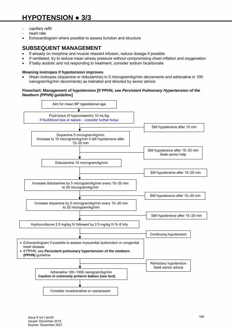
HYPOTENSION ● 3/3 • capillary refill • heart rate • Echocardiogram where possible to assess function and structure
SUBSEQUENT MANAGEMENT • If already on morphine and muscle relaxant infusion, reduce dosage if possible • If ventilated, try to reduce mean airway pressure without compromising chest inflation and oxygenation • If baby acidotic and not responding to treatment, consider sodium bicarbonate Weaning inotropes if hypotension improves • Wean inotropes (dopamine or dobutamine) in 5 microgram/kg/min decrements and adrenaline in 100
nanogram/kg/min decrements) as tolerated and directed by senior advice Flowchart: Management of hypotension [If PPHN, see Persistent Pulmonary Hypertension of the Newborn (PPHN) guideline]
Aim for mean BP ≥gestational age
Fluid bolus (if hypovolaemic) 10 mL/kg If fluid/blood loss or sepsis − consider further bolus
Dopamine 5 microgram/kg/min Increase to 10 microgram/kg/min if still hypotensive after
15–20 min
Dobutamine 10 microgram/kg/min
Increase dobutamine by 5 microgram/kg/min every 15–20 min to 20 microgram/kg/min
Continuing hypotension
Hydrocortisone 2.5 mg/kg IV followed by 2.5 mg/kg IV 6–8 hrly
Refractory hypotension Seek senior advice
Adrenaline 100–1000 nanogram/kg/min Caution in extremely preterm babies (see text)
Consider noradrenaline or vasopressin
Still hypotensive after 10 min
Still hypotensive after 15–20 min Seek senior help
Still hypotensive after 15–20 min
Still hypotensive after 15–20 min
• Echocardiogram if possible to assess myocardial dysfunction or congenital heart disease
• If PPHN, see Persistent pulmonary hypertension of the newborn (PPHN) guideline
Still hypotensive after 15–20 min
Increase dopamine by 5 microgram/kg/min every 15–20 min to 20 microgram/kg/min
Issue 8 rev1 jan20 Issued: December 2019 Expires: December 2021
169
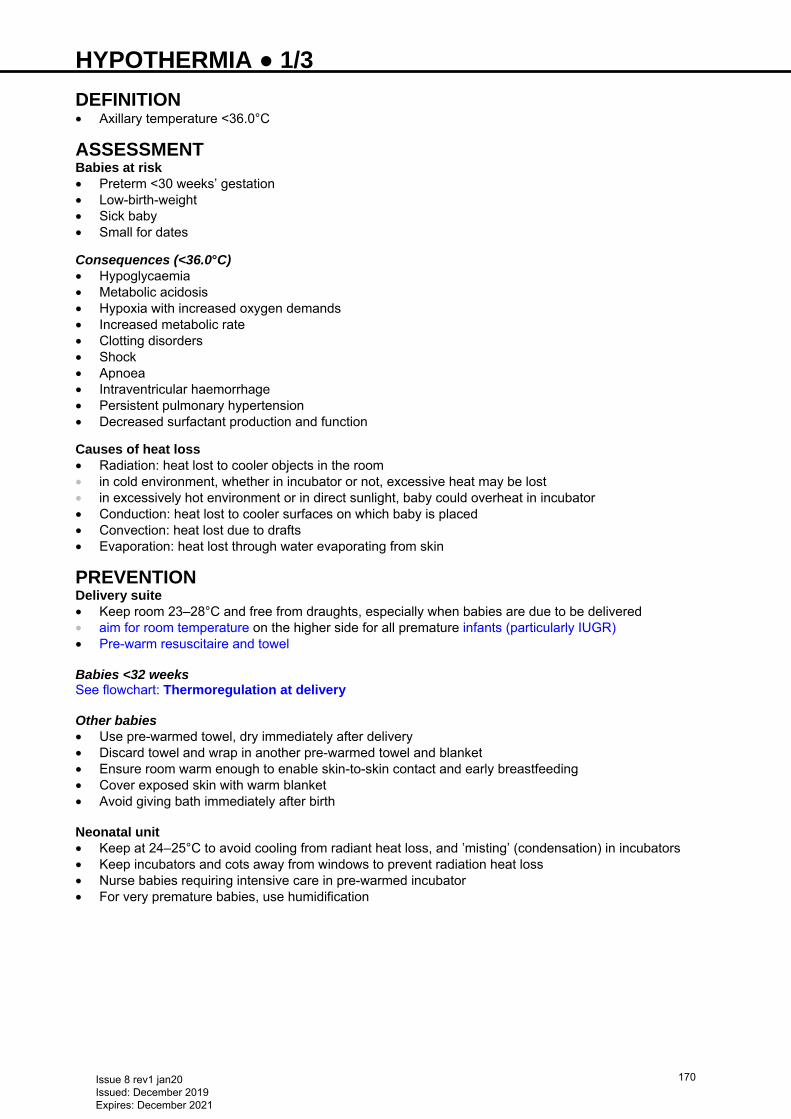
HYPOTHERMIA ● 1/3 DEFINITION • Axillary temperature <36.0°C
ASSESSMENT Babies at risk • Preterm <30 weeks’ gestation • Low-birth-weight • Sick baby • Small for dates
Consequences (<36.0°C) • Hypoglycaemia • Metabolic acidosis • Hypoxia with increased oxygen demands • Increased metabolic rate • Clotting disorders • Shock • Apnoea • Intraventricular haemorrhage • Persistent pulmonary hypertension • Decreased surfactant production and function Causes of heat loss • Radiation: heat lost to cooler objects in the room • in cold environment, whether in incubator or not, excessive heat may be lost • in excessively hot environment or in direct sunlight, baby could overheat in incubator • Conduction: heat lost to cooler surfaces on which baby is placed • Convection: heat lost due to drafts • Evaporation: heat lost through water evaporating from skin
PREVENTION Delivery suite • Keep room 23–28°C and free from draughts, especially when babies are due to be delivered • aim for room temperature on the higher side for all premature infants (particularly IUGR) • Pre-warm resuscitaire and towel Babies <32 weeks See flowchart: Thermoregulation at delivery Other babies • Use pre-warmed towel, dry immediately after delivery • Discard towel and wrap in another pre-warmed towel and blanket • Ensure room warm enough to enable skin-to-skin contact and early breastfeeding • Cover exposed skin with warm blanket • Avoid giving bath immediately after birth Neonatal unit • Keep at 24–25°C to avoid cooling from radiant heat loss, and ‛misting’ (condensation) in incubators • Keep incubators and cots away from windows to prevent radiation heat loss • Nurse babies requiring intensive care in pre-warmed incubator • For very premature babies, use humidification
Issue 8 rev1 jan20 Issued: December 2019 Expires: December 2021
170

HYPOTHERMIA ● 2/3 Flowchart: Thermoregulation at delivery Incubator temperature during first 3 days
Birth weight (g) Incubator temperature (°C) 1000 35 1500 34 2000 33.5 2500 33.2 3000 33 4000 32.5
• Babies <1000 g may require even higher temperatures, occasionally >37°C • If baby’s temperature remains within normal limits for 24 hr, reduce incubator temperature according to
baby’s needs • When baby’s weight reaches approximately 1600 g, transfer to open cot
Rainout may occur if the difference between temperature in incubator and room temperature is >5°C: ensure room temperature kept at locally agreed level
Babies not at risk of hypothermia • If not requiring observation of respiratory status or excessive invasive procedures, babies may be: • dressed • kept wrapped • placed in a cot • Mild hypothermia can be managed with the addition of: • hats • cot lids (if available) • heated mattresses
Pre-warm the resuscitaire and towels with heater at 100%
Ensure room warm and free from draughts
Prepare plastic bag and hat
Place baby on resuscitaire, dry head only, place rest of body in plastic bag
Put on hat
Take temperature before moving to NNU (if possible)
Show baby to mother before moving to NNU
If baby <30 weeks use TransWarmer® if available
Transfer to NNU with suitable thermal support
Babies <32 weeks’ gestation
Issue 8 rev1 jan20 Issued: December 2019 Expires: December 2021
171
HYPOTHERMIA ● 3/3 • If baby’s temperature <36.0°C consider: • use of incubator, if available • increasing humidity, if appropriate for gestational age • bubble wrap • skin-to-skin • Recheck temperature in 1 hr • Baby to be reviewed by medical team
REWARMING OF HYPOTHERMIC BABIES • Rewarm in incubator • ≥1200 g, rewarm at 1°C/hr • <1200 g, rewarm more slowly
Take care not to overheat babies. Aim for 36.5–37.5°C
Issue 8 rev1 jan20 Issued: December 2019 Expires: December 2021
172
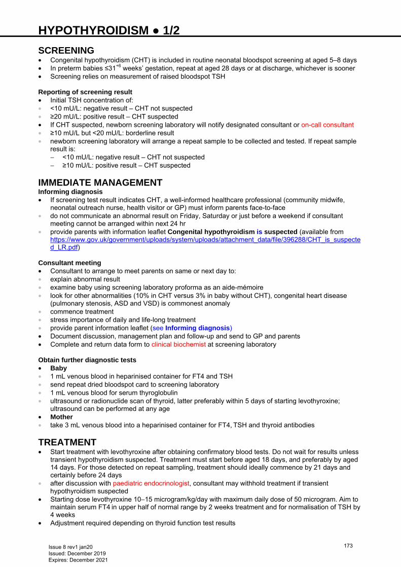
HYPOTHYROIDISM ● 1/2 SCREENING • Congenital hypothyroidism (CHT) is included in routine neonatal bloodspot screening at aged 5–8 days • In preterm babies ≤31+6 weeks’ gestation, repeat at aged 28 days or at discharge, whichever is sooner • Screening relies on measurement of raised bloodspot TSH
Reporting of screening result • Initial TSH concentration of: • <10 mU/L: negative result – CHT not suspected • ≥20 mU/L: positive result – CHT suspected • If CHT suspected, newborn screening laboratory will notify designated consultant or on-call consultant • ≥10 mU/L but <20 mU/L: borderline result • newborn screening laboratory will arrange a repeat sample to be collected and tested. If repeat sample
result is: − <10 mU/L: negative result – CHT not suspected − ≥10 mU/L: positive result – CHT suspected
IMMEDIATE MANAGEMENT Informing diagnosis • If screening test result indicates CHT, a well-informed healthcare professional (community midwife,
neonatal outreach nurse, health visitor or GP) must inform parents face-to-face • do not communicate an abnormal result on Friday, Saturday or just before a weekend if consultant
meeting cannot be arranged within next 24 hr • provide parents with information leaflet Congenital hypothyroidism is suspected (available from
https://www.gov.uk/government/uploads/system/uploads/attachment_data/file/396288/CHT_is_suspected_LR.pdf)
Consultant meeting • Consultant to arrange to meet parents on same or next day to: • explain abnormal result • examine baby using screening laboratory proforma as an aide-mémoire • look for other abnormalities (10% in CHT versus 3% in baby without CHT), congenital heart disease
(pulmonary stenosis, ASD and VSD) is commonest anomaly • commence treatment • stress importance of daily and life-long treatment • provide parent information leaflet (see Informing diagnosis) • Document discussion, management plan and follow-up and send to GP and parents • Complete and return data form to clinical biochemist at screening laboratory Obtain further diagnostic tests • Baby • 1 mL venous blood in heparinised container for FT4 and TSH • send repeat dried bloodspot card to screening laboratory • 1 mL venous blood for serum thyroglobulin • ultrasound or radionuclide scan of thyroid, latter preferably within 5 days of starting levothyroxine;
ultrasound can be performed at any age • Mother • take 3 mL venous blood into a heparinised container for FT4, TSH and thyroid antibodies
TREATMENT • Start treatment with levothyroxine after obtaining confirmatory blood tests. Do not wait for results unless
transient hypothyroidism suspected. Treatment must start before aged 18 days, and preferably by aged 14 days. For those detected on repeat sampling, treatment should ideally commence by 21 days and certainly before 24 days
• after discussion with paediatric endocrinologist, consultant may withhold treatment if transient hypothyroidism suspected
• Starting dose levothyroxine 10−15 microgram/kg/day with maximum daily dose of 50 microgram. Aim to maintain serum FT4 in upper half of normal range by 2 weeks treatment and for normalisation of TSH by 4 weeks
• Adjustment required depending on thyroid function test results
Issue 8 rev1 jan20 Issued: December 2019 Expires: December 2021
173
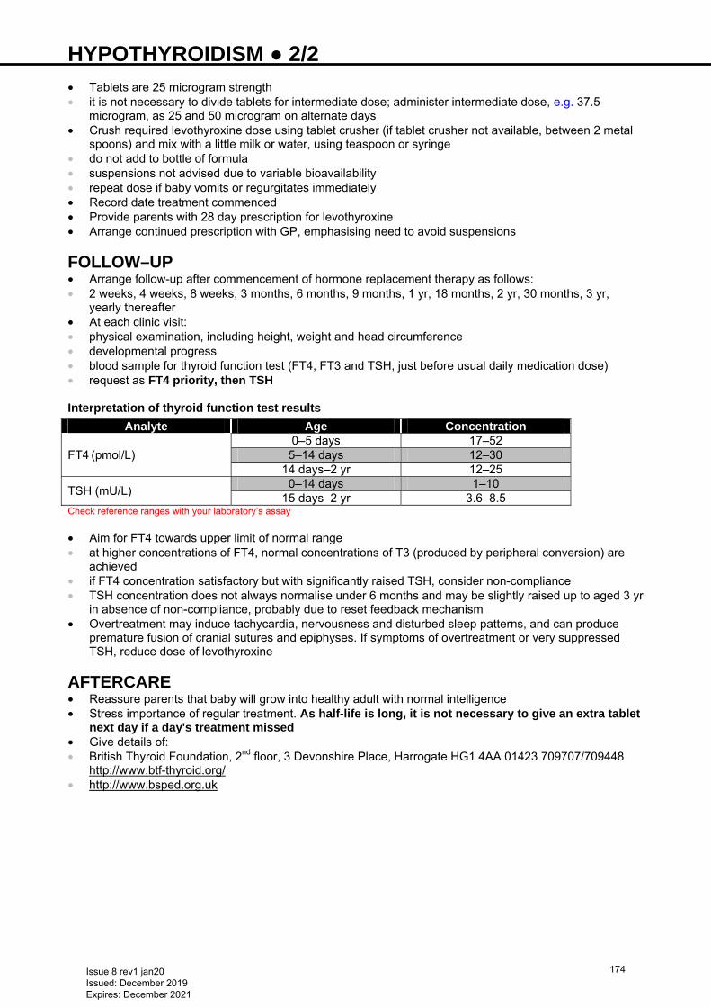
HYPOTHYROIDISM ● 2/2 • Tablets are 25 microgram strength • it is not necessary to divide tablets for intermediate dose; administer intermediate dose, e.g. 37.5
microgram, as 25 and 50 microgram on alternate days • Crush required levothyroxine dose using tablet crusher (if tablet crusher not available, between 2 metal
spoons) and mix with a little milk or water, using teaspoon or syringe • do not add to bottle of formula • suspensions not advised due to variable bioavailability • repeat dose if baby vomits or regurgitates immediately • Record date treatment commenced • Provide parents with 28 day prescription for levothyroxine • Arrange continued prescription with GP, emphasising need to avoid suspensions
FOLLOW–UP • Arrange follow-up after commencement of hormone replacement therapy as follows: • 2 weeks, 4 weeks, 8 weeks, 3 months, 6 months, 9 months, 1 yr, 18 months, 2 yr, 30 months, 3 yr,
yearly thereafter • At each clinic visit: • physical examination, including height, weight and head circumference • developmental progress • blood sample for thyroid function test (FT4, FT3 and TSH, just before usual daily medication dose) • request as FT4 priority, then TSH Interpretation of thyroid function test results
Analyte Age Concentration 0–5 days 17–52 FT4 (pmol/L) 5–14 days 12–30 14 days–2 yr 12–25
TSH (mU/L) 0–14 days 1–10
15 days–2 yr 3.6–8.5 Check reference ranges with your laboratory’s assay • Aim for FT4 towards upper limit of normal range • at higher concentrations of FT4, normal concentrations of T3 (produced by peripheral conversion) are
achieved • if FT4 concentration satisfactory but with significantly raised TSH, consider non-compliance • TSH concentration does not always normalise under 6 months and may be slightly raised up to aged 3 yr
in absence of non-compliance, probably due to reset feedback mechanism • Overtreatment may induce tachycardia, nervousness and disturbed sleep patterns, and can produce
premature fusion of cranial sutures and epiphyses. If symptoms of overtreatment or very suppressed TSH, reduce dose of levothyroxine
AFTERCARE • Reassure parents that baby will grow into healthy adult with normal intelligence • Stress importance of regular treatment. As half-life is long, it is not necessary to give an extra tablet
next day if a day's treatment missed • Give details of: • British Thyroid Foundation, 2nd floor, 3 Devonshire Place, Harrogate HG1 4AA 01423 709707/709448
http://www.btf-thyroid.org/ • http://www.bsped.org.uk
Issue 8 rev1 jan20 Issued: December 2019 Expires: December 2021
174
HYPOTONIA (FLOPPY BABY) ● 1/4 RECOGNITION AND ASSESSMENT Definition • Subjective decrease in resistance to passive range of movement • Separate from weakness, which refers to lack of muscle strength • Important to differentiate between central (upper motor neurone), and peripheral (lower motor neurone)
hypotonia – may be a mixed picture. See Table 1 • central hypotonia is most common (70–80%) • Hypotonia • relatively common finding in newborn period • transient in majority of cases • if severe/persistent investigate further Symptoms and signs • Reduced activity/movement • Reduced level of consciousness/alertness • Dysmorphic features • High pitched, weak or fatigable cry • Increased or reduced respiratory effort • Feeding difficulties/choking/pooling of secretions • Seizures/abnormal movements
DIFFERENTIAL DIAGNOSIS • Causes of hypotonia in the newborn baby are numerous, not all are listed here • Benign congenital hypotonia is a diagnosis of exclusion
Central • Hypoxic ischaemic encephalopathy (HIE) • Intracranial haemorrhage • Structural brain malformation • Chromosomal abnormalities e.g. trisomy 21, Prader-Willi syndrome • Congenital infection e.g. TORCH • Acquired infection e.g. group B streptococcus • Endocrine e.g. congenital hypothyroidism • Metabolic disorders e.g. acid maltase deficiency (Pompe's disease), carnitine deficiency,
mucopolysacharidosis, peroxisome biogenesis disorders e.g. Zellweger syndrome • Drug effects e.g. benzodiazepines Peripheral • Spinal cord e.g. birth trauma (especially breech delivery), syringomyelia • Anterior horn cell e.g. spinal muscular atrophy (SMA) • Neuromuscular junction e.g. myasthenia gravis, transitory myasthenia • Peripheral nerves e.g. hereditary motor and sensory neuropathies e.g. Charcot Marie-Tooth disease • Muscle disorders e.g. muscular dystrophy, congenital myopathy
HISTORY Family • Affected parents/siblings • Consanguinity • Previous miscarriage/stillbirth • Metabolic/genetic disease • Premature death Maternal • Diabetes • Infection • Medications • Myotonic dystrophy • Myasthenia gravis
Antenatal • TORCH infections
Issue 8 rev1 jan20 Issued: December 2019 Expires: December 2021
175
HYPOTONIA (FLOPPY BABY) ● 2/4 • Drug/alcohol exposure • Fetal movements • Liquor volume Birth • Gestational age • Delivery complications • Malpresentation • Instrumental delivery • APGAR score/resuscitation at birth • Cord gases
Neonatal • Respiratory distress • Feeding issues • Level of alertness • Level of spontaneous movement • Seizures • Hypoglycaemia • Weak cry
PHYSICAL EXAMINATION Mother • Examine for signs of myotonic dystrophy Baby • Full neurological assessment • Level of alertness • Abnormal posture • Degree of hypotonia • pull to sit • scarf sign • shoulder suspension • ventral suspension • Asymmetry • Strength • Deep tendon reflexes • Primitive reflexes • Gag and suck • Fasciculations (including tongue) • Abnormal eye movements • Ptosis • Cataracts • Dysmorphic features/abnormal facies • Respiratory effort • Hepatosplenomegaly • Undescended testicles • Contractures • Arthrogryposis
Issue 8 rev1 jan20 Issued: December 2019 Expires: December 2021
176
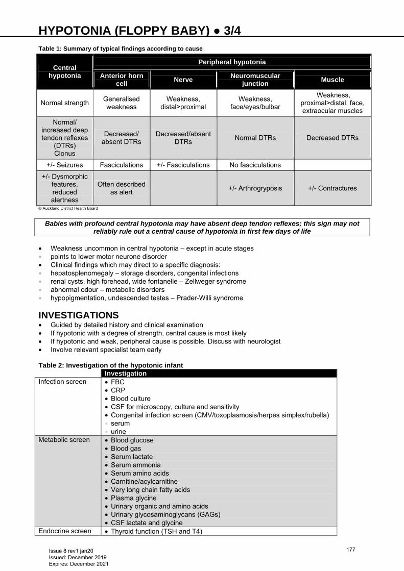
HYPOTONIA (FLOPPY BABY) ● 3/4 Table 1: Summary of typical findings according to cause
© Auckland District Health Board
Babies with profound central hypotonia may have absent deep tendon reflexes; this sign may not
reliably rule out a central cause of hypotonia in first few days of life
• Weakness uncommon in central hypotonia – except in acute stages • points to lower motor neurone disorder • Clinical findings which may direct to a specific diagnosis: • hepatosplenomegaly – storage disorders, congenital infections • renal cysts, high forehead, wide fontanelle – Zellweger syndrome • abnormal odour – metabolic disorders • hypopigmentation, undescended testes – Prader-Willi syndrome
INVESTIGATIONS • Guided by detailed history and clinical examination • If hypotonic with a degree of strength, central cause is most likely • If hypotonic and weak, peripheral cause is possible. Discuss with neurologist • Involve relevant specialist team early Table 2: Investigation of the hypotonic infant Investigation Infection screen • FBC
• CRP • Blood culture • CSF for microscopy, culture and sensitivity • Congenital infection screen (CMV/toxoplasmosis/herpes simplex/rubella) • serum • urine
Metabolic screen • Blood glucose • Blood gas • Serum lactate • Serum ammonia • Serum amino acids • Carnitine/acylcarnitine • Very long chain fatty acids • Plasma glycine • Urinary organic and amino acids • Urinary glycosaminoglycans (GAGs) • CSF lactate and glycine
Endocrine screen • Thyroid function (TSH and T4)
Central hypotonia
Peripheral hypotonia
Anterior horn cell
Nerve Neuromuscular
junction Muscle
Normal strength Generalised weakness
Weakness, distal>proximal
Weakness, face/eyes/bulbar
Weakness, proximal>distal, face, extraocular muscles
Normal/ increased deep tendon reflexes
(DTRs)
Clonus
Decreased/ absent DTRs
Decreased/absent DTRs
Normal DTRs Decreased DTRs
+/- Seizures Fasciculations +/- Fasciculations No fasciculations
+/- Dysmorphic features, reduced alertness
Often described as alert
+/- Arthrogryposis +/- Contractures
Issue 8 rev1 jan20 Issued: December 2019 Expires: December 2021
177
HYPOTONIA (FLOPPY BABY) ● 4/4
• U&Es • Calcium • Magnesium (e.g hypermagneseamia after treatment for maternal
eclampsia) Genetic screen • Karyotype and microarray
• DNA for Prader-Willi, Zellweger syndrome • SMA gene (SMA-RD – if respiratory weakness) • Dystrophia myotonica protein kinase (DMPK gene for myotonic
dystrophy) • Other specific genetic test guided by family history/phenotype
Other • Cranial ultrasound scan • MRI brain +/- spinal cord • EEG (especially if seizures) • CFM (if features of encephalopathy) • Creatinine kinase (muscular dystrophy) • may be elevated in first few days after birth • if abnormal repeat after aged 72 hr • if persistently elevated refer to neurologist and consider muscle biopsy • Nerve conduction studies • If features of maternal myasthenia gravis • acetylcholine receptor antibodies • tensilon test • EMG • If cardiomyopathy suspected • ECG • chest X-ray • echocardiography
Muscle biopsy may be delayed until aged 6 months, as neonatal results are difficult to interpret
MANAGEMENT • Specific management determined by individual condition and presentation • Airway and breathing • may need resuscitation at birth • airway positioning/Guedel airway • intubation and ongoing respiratory support • suction of respiratory secretions • Feeding • specialised bottles/teats • nasogastric tube feeds • Skin and developmental care • regular position changes to avoid pressure sores, reduce risk of contractures and optimise
neurodevelopment (see Developmental care guideline) • Physiotherapy • refer to neonatal/paediatric physiotherapy (while inpatient) • physiotherapist will:
− advise on specific handling and positioning to optimise neurodevelopmental outcomes − assess for symmetry and risk of joint contractures/positional deformity and advise on management
• on discharge refer to community paediatric physiotherapy services
Issue 8 rev1 jan20 Issued: December 2019 Expires: December 2021
178
HYPOXIC ISCHAEMIC ENCEPHALOPATHY (HIE) INCLUDING PREPARATION FOR ACTIVE COOLING ● 1/6 RECOGNITION AND ASSESSMENT Risk factors • History of non-reassuring cardiotocography (CTG) • Fetal heart rate abnormalities during labour • Low Apgar score • Acidotic umbilical arterial or venous gas • Need for prolonged resuscitation
SYMPTOMS AND SIGNS Acute neonatal encephalopathy • Altered state of consciousness (irritability, unresponsiveness to stimulation) • Abnormal tone (hypo/hypertonia, abnormal posturing, decerebrate rigidity, extensor response to painful
stimulus) • Seizures • Weak/absent suck • Hypo/hyperventilation
IMMEDIATE TREATMENT • Prompt and effective resuscitation • Maintain body temperature, avoid hyperthermia • IV access • Isotonic glucose-containing IV fluids at 40 mL/kg/day (see Intravenous fluid therapy guideline) Cerebral function monitoring (aEEG) • Start cerebral function monitoring (if available) • normal trace upper margin >10 microvolts and lower margin >5 microvolts • moderately abnormal trace upper margin >10 microvolts and lower margin <5 microvolts • severely abnormal upper margin <10 microvolts and lower margin <5 microvolts
WHEN TO CONSIDER TREATMENT WITH TOTAL BODY COOLING Treatment criteria • Babies ≥36 weeks’ gestation, meeting criteria A and B and aged ≤6 hr are eligible for treatment with
cooling • Infants 35+0–35+6 weeks’ gestation but meeting criteria A and B and aged ≤6 hr, discuss with cooling
centre, as may be suitable for treatment • If in doubt about the suitability of any baby for cooling, discuss with cooling centre • Babies meeting criteria A and B for treatment with cooling Criterion A ≥1 of: • Apgar score ≤5 at 10 min after birth • Continued need for resuscitation, including endotracheal or mask ventilation at 10 min after birth • Acidosis within 60 min of birth (defined as umbilical cord, arterial or capillary pH <7.0) • Base deficit ≥16 mmol/L in umbilical cord or any blood sample (arterial, venous or capillary) within 60 min
of birth Criterion B • Seizures OR moderate-to-severe encephalopathy, consisting of: • altered state of consciousness (reduced or absent response to stimulation) and • abnormal tone (focal or general hypotonia, or flaccid) and • abnormal primitive reflexes (weak or absent suck or Moro response)
REFERRAL Consent • Discuss option of cooling treatment with parents as soon as practically possible. It is not necessary to
wait for formal consent before starting passive cooling • Document discussions in baby’s notes
Issue 8 rev1 jan20 Issued: December 2019 Expires: December 2021
179
HYPOXIC ISCHAEMIC ENCEPHALOPATHY (HIE) INCLUDING PREPARATION FOR ACTIVE COOLING ● 2/6 In addition Request cord gases (if not already obtained) Request midwives save placenta for histological examination Passive cooling • As soon as decision made to refer for cooling, referring unit to telephone cooling centre and to begin
passive cooling • document this time as ‛age when passive cooling commenced’. • document baby’s temperature at this time • begin passive cooling by switching off overhead heater and active heating in a transport incubator • Nurse baby in an open Babytherm® cot with heater switched off • If baby nursed in an incubator, open portholes • Nurse baby naked apart from a nappy Continuous rectal temperature monitoring • Insert a rectal thermometer to 6 cm and commence continuous rectal temperature monitoring. If rectal
temperature monitoring unavailable, perform axillary temperature monitoring every 15 min • Target rectal temperature 33–34°C
Regular communication between referring unit and cooling centre is vital
• Once baby accepted by a cooling centre, contact neonatal transport team to arrange transport of baby • Discuss methods of cooling with cooling centre, before arrival of neonatal transport team. Use fans or
gloves filled with cold water only if continuous rectal temperature monitoring is in place
Never use ice filled gloves to cool a baby as this can bring the temperature down to dangerously low and uncontrolled levels
STABILISATION PHASE Passive cooling Use the referral form from the website: http://www.networks.nhs.uk/nhs-networks/staffordshire-shropshire-and-black-country-newborn/care-pathways • Ensure baby’s temperature does not fall below 33ºC. Document every 15 min • Follow Passive cooling protocol flowchart • Care continues in referring unit with advice from cooling centre • If not already intubated at delivery [most babies will need to be intubated for transfer (see Intubation
guideline)] discuss with receiving consultant and newborn transfer service • If possible, insert umbilical arterial and venous catheters and monitor arterial blood pressure (see
Umbilical artery catheterisation and Umbilical venous catheterisation guidelines). Check position of lines on X-ray
• Aim to maintain arterial PaCO2 of 6–8 kPa • Document neurology before commencing sedation or anticonvulsants, including size and reactivity of
pupils • Sedate baby using morphine at an infusion rate of 20 microgram/kg/hr. Aim for heart rate of 100 bpm.
Faster rates may be a sign of distress, in which case increase sedation • Maintain mean arterial blood pressure at >45 mmHg (see Hypotension guideline) • Restrict total fluids to 40 mL/kg/day initially • Keep glucose within normal range – use higher glucose concentration infusion if necessary (see
Hypoglycaemia guideline) • Take blood for blood culture, FBC, arterial blood gas, lactate, electrolytes, urea and creatinine, calcium,
magnesium, prothrombin time, APTT, glucose and LFT • Babies ≥37 weeks gestation with severe brain injury diagnosed in the first 7 days of life meet the
Healthcare Safety Investigation Branch criteria for a maternity investigation if he/she was : • actively cooled • diagnosed with Grade III HIE • had reduced central tone, was comatose and had seizures of any kind • notify obstetric team before transfer if baby meets above criteria
Issue 8 rev1 jan20 Issued: December 2019 Expires: December 2021
180
HYPOXIC ISCHAEMIC ENCEPHALOPATHY (HIE) INCLUDING PREPARATION FOR ACTIVE COOLING ● 3/6 SUBSEQUENT MANAGEMENT Continue with management below if baby not transferred to cooling centre, or in cooling centre without local guideline for active cooling. NOTE that some of the target values are different to those recommended if a baby is being actively cooled Oxygen • Avoid hypoxaemia. Maintain PaO2 10–12 kPa and SpO2 >94% • Episodes of hypoxaemia (possibly associated with convulsions) are an indication for IPPV Carbon dioxide • Maintain PaCO2 5.0–7.0 kPa • Hypoventilation leading to hypercapnia (>7.0 kPa) is an indication for IPPV • Hyperventilation is contraindicated but, if baby spontaneously hyperventilating, mechanical ventilation,
with/without paralysis, may be necessary to control PaCO2 Circulatory support • Maintain mean arterial blood pressure at ≥40 mmHg for term babies • If cardiac output poor (e.g. poor perfusion: blood pressure is a poor predictor of cardiac output) use
inotropes • Avoid volume replacement unless evidence of hypovolaemia Fluid balance and renal function • Start fluids at 40 mL/kg/day (see Intravenous fluid therapy guideline) • Some babies develop inappropriate ADH secretion at 3–4 days (suggested by hypo-osmolar serum with
low serum sodium, associated with an inappropriately high urine sodium and osmolality) • Further fluid restriction if serum sodium falls and weight gain/failure to lose weight • If in renal failure, follow Renal failure guideline Acidosis • Will normally correct itself once adequate respiratory and circulatory support provided (correction
occasionally required during initial resuscitation) • Sodium bicarbonate correction is rarely required post resuscitation and it is better to allow spontaneous
correction
Glucose • Regular blood glucose monitoring • Target >2.6 mmol/L • Fluid restriction may require use of higher concentrations of glucose to maintain satisfactory blood
glucose • Avoid hyperglycaemia (>8 mmol/L)
Calcium • Asphyxiated babies are at increased risk of hypocalcaemia • Treat with calcium gluconate when serum corrected calcium <1.7 mmol/L or if ionized calcium <0.8
Seizures • Prophylactic anticonvulsants not indicated • In muscle-relaxed baby, abrupt changes in blood pressure, SpO2 and heart rate can indicate seizures • Treat persistent (>3/hr) or prolonged seizures (>3 min, recur >3 times/hr) (see Seizures guideline) • give phenobarbital
− if ineffective or contraindicated, give phenytoin. If no response, give clonazepam or midazolam (see Seizures guideline)
• Seizures associated with HIE can be notoriously difficult to control (preventing every twitch is unrealistic) • Regular seizures causing respiratory insufficiency are an indication for IPPV • Once baby stable for 2–3 days, anticonvulsants can usually be withdrawn, although phenobarbital can
be continued for a little longer (duration can vary depending on individual practice and clinical severity of seizures)
• Avoid corticosteroids and mannitol
Issue 8 rev1 jan20 Issued: December 2019 Expires: December 2021
181
HYPOXIC ISCHAEMIC ENCEPHALOPATHY (HIE) INCLUDING PREPARATION FOR ACTIVE COOLING ● 4/6 Thermal control • Maintain normal body temperature (36.5–37.2°C). Avoid hyperthermia
Gastrointestinal system • Term babies who suffer a severe asphyxial insult are at risk of developing NEC [see Necrotising
enterocolitis (NEC) guideline] • In other babies, gastric motility can be reduced: introduce enteral feeds slowly Cranial ultrasound • Generalised increase in echogenicity, indistinct sulci and narrow ventricles • After aged 2–3 days, increased echogenicity of thalami and parenchymal echodensities • After 1 week, parenchymal cysts, ventriculomegaly and cortical atrophy may develop • Cerebral Doppler used early, but does not affect management • relative increase of end-diastolic blood flow velocity compared to peak systolic blood flow velocity
(Resistive Index <0.55) in anterior cerebral artery predicts poor outcome (repeat after 24 hr) MRI scan of brain between days 5–14 of life For baby with moderate and severe encephalopathy (see Table) and in baby with seizures due to encephalopathy • Areas of altered signal in thalamus, basal ganglia and posterior limb of the internal capsule indicate poor
prognosis PROGNOSIS Criteria for defining moderate and severe encephalopathy
Parameter Moderate encephalopathy Severe encephalopathy
Level of consciousness Reduced response to stimulation
Absent response to stimulation
Spontaneous activity Decreased activity No activity
Posture Distal flexion, complete extension
Decerebrate
Tone Hypotonia (focal or general) Flaccid Suck Weak Absent Moro Incomplete Absent Pupils Constricted Constricted Heart rate Bradycardia Variable Respiration Periodic breathing Apnoea • Risk of long-term problems increases with the degree of encephalopathy • Normal EEG during first 3 days has good prognosis • Overall risk of death or significant handicap is negligible for mild HIE, 26% for moderate and almost
100% for severe HIE • Prolonged encephalopathy (e.g. moderate HIE lasting >6 days) also associated with poor outcome • Persistent oliguria is associated with poor outcome in 90% • Prognostic factors indicative of worse outcome: • prolonged duration of ventilation • prolonged need for anticonvulsants • time taken to establish oral feeding • Lack of normal background activity on EEG
• Areas of altered signal in thalamus, basal ganglia and posterior limb of the internal capsule
DISCONTINUING INTENSIVE CARE • When prognosis very poor, discuss withdrawing intensive care support and palliative care • Very poor prognostic factors include: • need for prolonged resuscitation at birth • evidence of severe asphyxia • multi-organ failure • intractable seizures • coma
Issue 8 rev1 jan20 Issued: December 2019 Expires: December 2021
182
HYPOXIC ISCHAEMIC ENCEPHALOPATHY (HIE) INCLUDING PREPARATION FOR ACTIVE COOLING ● 5/6 • very abnormal cranial ultrasound scan • abnormal Doppler cerebral blood flow velocities • persistent burst suppression pattern on cerebral function monitoring and/or EEG • Decision to withdraw care requires discussion with parents, and other nursing and medical staff. Such
decisions are frequently reached by baby’s consultant after a series of discussions • It helps if the same staff speak to parents on each occasion • The best interests of the child are paramount • Record a summary of discussion in notes
DISCHARGE AND FOLLOW-UP • Arrange clinic follow-up in 4–6 weeks for babies discharged • Repeat cranial ultrasound scan before discharge • Arrange hearing screen (see Hearing screening guideline) • For babies with moderate and severe encephalopathy (see Table) and in those with seizures due to
encephalopathy, arrange MRI scan as an outpatient (if not already performed as an inpatient), preferably 5−14 days of life
Information for parents Offer parents information on HIE, available from: https://www.bliss.org.uk/parents/about-your-baby/medical-conditions/hypoxic-ischaemic-encephalopathy-hie
Issue 8 rev1 jan20 Issued: December 2019 Expires: December 2021
183
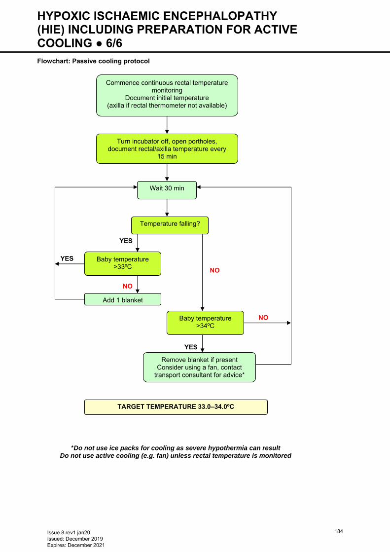
HYPOXIC ISCHAEMIC ENCEPHALOPATHY (HIE) INCLUDING PREPARATION FOR ACTIVE COOLING ● 6/6 Flowchart: Passive cooling protocol
YES
YES
NO
YES
Commence continuous rectal temperature monitoring
Document initial temperature (axilla if rectal thermometer not available)
Turn incubator off, open portholes, document rectal/axilla temperature every
15 min
Wait 30 min
Temperature falling?
Baby temperature >33ºC
Add 1 blanket
Baby temperature >34ºC
Remove blanket if present Consider using a fan, contact
transport consultant for advice*
TARGET TEMPERATURE 33.0–34.0ºC
NO
*Do not use ice packs for cooling as severe hypothermia can result Do not use active cooling (e.g. fan) unless rectal temperature is monitored
NO
Issue 8 rev1 jan20 Issued: December 2019 Expires: December 2021
184
IMMUNISATIONS ● 1/3 ROUTINE IMMUNISATIONS FOR ALL BABIES • Plan to achieve immunity to diphtheria, tetanus, pertussis, (DTaP), polio, haemophilus (Hib),
meningococcus B, pneumococcus, rotavirus and hepatitis B within 4 months of birth (see also BCG immunisation and Hepatitis B and C guidelines)
• See Department of Health Immunisation against Infectious Diseases ‘Green Book’ for national policy and for current schedule see https://www.gov.uk/government/collections/immunisation
Do not delay immunisation in preterm babies because of prematurity or low body weight CONTRAINDICATIONS • Cardiorespiratory events (apnoeas, bradycardia and desaturations) are not contraindications to
immunisation, but continue to monitor for a further 72 hr following immunisation • See Precautions with rotavirus vaccine PROCEDURE Consent • Inform parents of process, benefits and risks • For further information refer parents to www.nhs.uk/conditions/vaccinations • Offer parents opportunity to ask questions • Informed consent (can be written or oral) must be obtained and recorded in notes at time of each
immunisation • Complete ‛unscheduled immunisation form’ before immunisation and send to local Child Health
Information Prescription • Use immunisation listed in ‘Green Book’ − see Routine immunisations for all babies • Keep strictly to schedule to avoid delay • Order vaccines in advance unless held as stock on NNU • Prescribe on treatment sheet Administration • DTaP/IPV/Hib/HepB (Infanrix hexa®) is a 6-in-1 preparation • Administer by IM injection into thigh give ≥2.5 cm apart from other vaccines • Dose for all primary immunisations (DTaP/IPV/Hib/HepB), meningococcal B, pneumococcal) is 0.5 mL • Give meningococcal B (Bexsero®) and pneumococcal (Prevenar 13®) vaccine into separate injection
sites in other thigh • Rotavirus vaccine must not be injected and preferably not given via an NGT • assess ability to tolerate oral administration DOCUMENTATION • After immunisation, document the following in case notes as well as in Child Health Record (Red Book): • consent gained from parents • vaccine given and reasons for any omissions • site of injection(s) in case of reactions • batch number of product(s) • expiry date of product(s) • legible signature of doctor administering immunisations • adverse reactions • Sign treatment sheet • Complete immunisation form in BadgerNet system. Document all information on discharge summary
and medical case notes, including recommendations for future immunisations and need for any special vaccinations, e.g. influenza, palivizumab, etc.
• Notify Child Health Information System (CHIS) MONITORING • Babies born <28 weeks may have an impaired immune response. Check functional antibodies 1 month
after booster at aged 1 yr, if needed
Issue 8 rev1 jan20 Issued: December 2019 Expires: December 2021
185

IMMUNISATIONS ● 2/3 • Babies <28 weeks’ gestation at birth, who are in hospital − respiratory monitoring for
48–72 hr when given first routine immunisations • If baby has apnoea, bradycardias or desaturations after first routine immunisations, second immunisation
should ideally be given in hospital with respiratory monitoring for 48–72 hr ADVERSE REACTIONS • Local: • extensive area of redness or swelling • General: • fever >39.5°C within 48 hr • anaphylaxis • bronchospasm • laryngeal oedema • generalised collapse • episodes of severe apnoea • diarrhoea • irritability • vomiting • flatulence • loss of appetite • regurgitation Specific notes for rotavirus vaccination • Do not give Rotarix® to infants aged <6 weeks • minimum age for first dose of Rotarix® is 6+0 weeks • maximum age for first dose is 14+6 weeks • Do not give first dose of Rotarix® to infants aged ≥15+0 weeks. Infants who have received their first dose
of vaccine aged <15+0 weeks should receive their second dose of Rotarix® after a minimum interval of 4 weeks and by aged 23+6 weeks
• Do not give Rotarix® vaccine to infants aged ≥24+0 weeks Precautions with rotavirus vaccination • Postpone administration of rotavirus vaccine in infants suffering from: • acute severe febrile illness • acute diarrhoea or vomiting • 1st dose must be given aged ≤15 weeks • Do not administer Rotarix® to infants with: • confirmed anaphylactic reaction to a previous dose of rotavirus vaccine • confirmed anaphylactic reaction to any components of the vaccine • history of intussusception • aged ≥24+0 weeks • severe combined immunodeficiency disorder (SCID) • malformation of the gastrointestinal tract that could predispose them to intussusception • rare hereditary problems of fructose intolerance, glucose-galactose malabsorption or sucrase-isomaltase
insufficiency
ADDITIONAL IMMUNISATIONS Influenza (in autumn and winter only) Indications • Chronic lung disease (on, or recently had, oxygen) • Congenital heart disease, renal, liver or neurological disease • Immunodeficiency Recommendations • Recommend vaccination to close family members of these babies • Give babies aged >6 months–2 yr, 2 doses 4–6 weeks apart, IM injection • Note: intranasal flu vaccine is now routinely recommended for children aged ≥2 yr
Issue 8 rev1 jan20 Issued: December 2019 Expires: December 2021
186
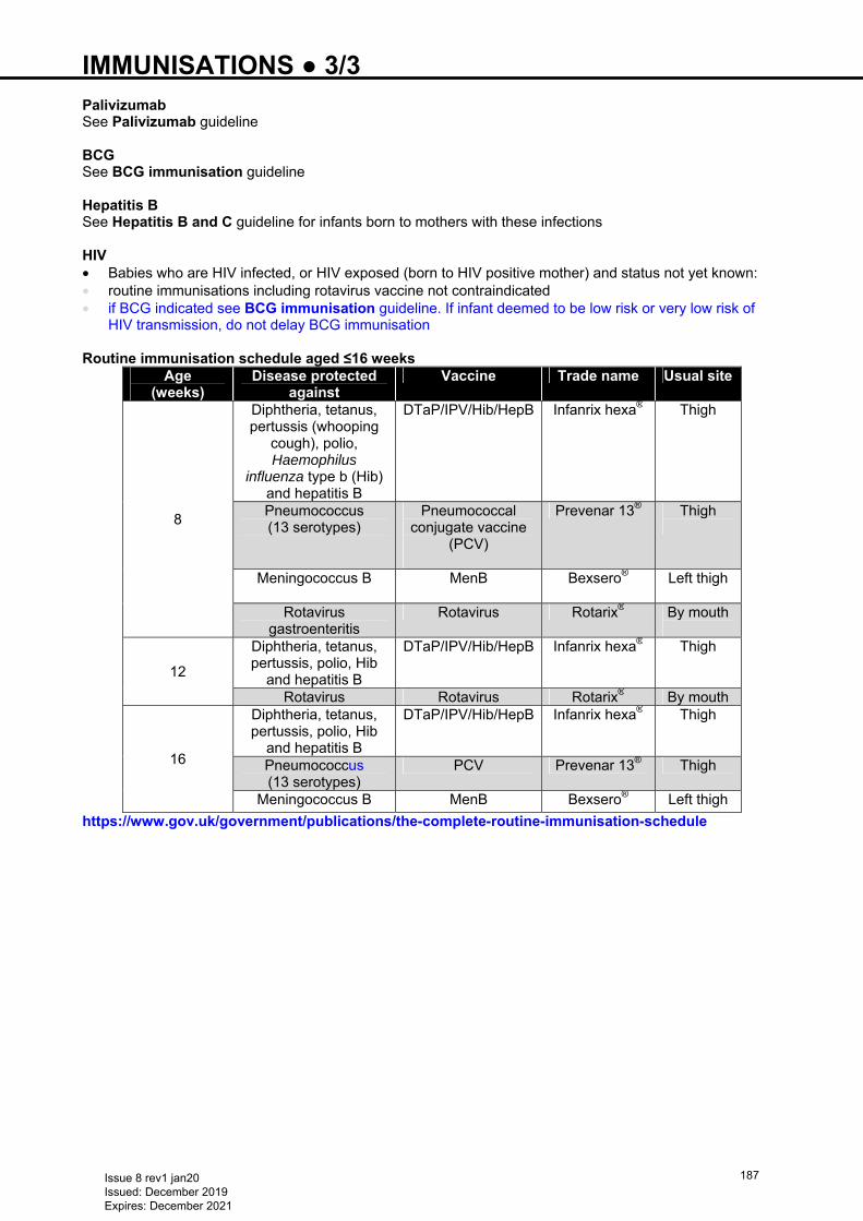
IMMUNISATIONS ● 3/3 Palivizumab See Palivizumab guideline BCG See BCG immunisation guideline Hepatitis B See Hepatitis B and C guideline for infants born to mothers with these infections HIV • Babies who are HIV infected, or HIV exposed (born to HIV positive mother) and status not yet known: • routine immunisations including rotavirus vaccine not contraindicated • if BCG indicated see BCG immunisation guideline. If infant deemed to be low risk or very low risk of
HIV transmission, do not delay BCG immunisation Routine immunisation schedule aged ≤16 weeks
Age (weeks)
Disease protected against
Vaccine Trade name Usual site
8
Diphtheria, tetanus, pertussis (whooping
cough), polio, Haemophilus
influenza type b (Hib) and hepatitis B
DTaP/IPV/Hib/HepB Infanrix hexa® Thigh
Pneumococcus (13 serotypes)
Pneumococcal conjugate vaccine
(PCV)
Prevenar 13® Thigh
Meningococcus B
MenB
Bexsero® Left thigh
Rotavirus gastroenteritis
Rotavirus Rotarix® By mouth
12
Diphtheria, tetanus, pertussis, polio, Hib
and hepatitis B
DTaP/IPV/Hib/HepB Infanrix hexa® Thigh
Rotavirus Rotavirus Rotarix® By mouth
16
Diphtheria, tetanus, pertussis, polio, Hib
and hepatitis B
DTaP/IPV/Hib/HepB
Infanrix hexa® Thigh
Pneumococcus (13 serotypes)
PCV Prevenar 13® Thigh
Meningococcus B MenB Bexsero® Left thigh
https://www.gov.uk/government/publications/the-complete-routine-immunisation-schedule
Issue 8 rev1 jan20 Issued: December 2019 Expires: December 2021
187
INFECTION (LATE ONSET) ● 1/4 DEFINITION • Infection after first 72 hr of life • Late onset group B streptococcus (GBS) infection: after first 6 days of life • When acquired in hospital – most commonly Gram-positive organisms. Coagulase-negative
staphylococci (CoNS) account for approximately 50% of all late onset infections • Gram-negative bacteria accounts for 20–40% and these are increasingly resistant to gentamicin
(Klebsiella>Serratia>Enterobacter>Pseudomonas>E.coli and Acinetobacter)
Risk factors • Risk of infection is inversely related to gestational age and birth weight, and directly related to severity of
illness at birth, reflecting need for invasive interventions e.g. prolonged ventilation, central venous access and parenteral nutrition
• Delayed introduction of enteral feeds is associated with higher infection rates • Increased risk of sepsis after gut surgery especially if enteral feeds slow to establish e.g. post-gastroschisis
or necrotising enterocolitis (NEC) with stoma
PREVENTION • Bare below elbow • no jewellery except wedding band • Strict hand washing and alcohol hand rubs • Follow WHO 5 moments of hand hygiene recommendations • Meticulous regimen for changing IV fluid administration sets and 3-way taps • Initiate enteral feeds with maternal breast milk within 24 hr of birth
PRESENTATION • Can be vague and non-specific Symptoms • Respiratory distress – increase in oxygen requirement/respiratory support • Apnoea/bradycardia • Cyanosis or poor colour • Poor perfusion (CRT >3 sec; toe-core temperature gap >2°C; mottling) • Hypotension • Tachycardia • Temperature instability (high or low) • Glucose instability • Hypotonia • Irritability • Lethargy/inactivity • Poor feeding and poor suck • Jaundice • Seizures • Vomiting • Abdominal distension • Nursing staff may describe babies with a mixture of these symptoms as having ‘gone off’
Signs Look for • Systemic signs of sepsis such as tachycardia, poor perfusion, reduced tone, quiet, lethargy, unsettled and
crying/moaning • Tachypnoea and intercostal and/or subcostal recession • Bulging of the fontanelle suggesting raised intracranial pressure • rarely detectable in babies with neonatal meningitis • Abdominal distension and tenderness • auscultate for bowel sounds; reduced or absent with infection (as a result of septic ileus) or NEC • inspect stool for visible blood • petechiae, bleeding diathesis • Septic spots in eyes, umbilicus, nails and skin • Reluctance to move or tenderness in joints and limbs suggestive of osteomyelitis or septic arthritis
Issue 8 rev1 jan20 Issued: December 2019 Expires: December 2021
188
INFECTION (LATE ONSET) ● 2/4 INVESTIGATIONS (perform before starting antibiotics) Swabs or ETT secretions for culture • Swab any suspicious lesion (e.g. skin, umbilicus or nails) • Refer to recent swabs or ETT secretion cultures to guide antibiotic therapy Blood cultures • From a peripheral vein, using a closed system, non-touch, aseptic technique • If blood collected from cannula hub risk of culturing CoNS skin contaminants Full blood count • A neutrophil count <2 or >15 × 109/L (supportive but not diagnostic, and marginally more sensitive than a
total white cell count) • Platelet count of <100 × 109/L • Toxic granulation in neutrophils [or if measured, an immature:total (I:T) neutrophil ratio >0.2] Clotting profile • If evidence of bleeding diathesis or in severe infection/septicaemia
CRP • Acute phase protein synthesised in the liver in response to inflammatory cytokines • Generally a delay of 24 hr between onset of symptoms and rise in serum CRP • Take sample at presentation and further sample 18–24 hr after first CRP sample • a rise may support diagnosis of infection but failure to rise does not exclude it where other findings are
supportive • if blood culture negative and clinical condition satisfactory, failure of CRP to rise during first 36 hr is a useful
indicator that antibiotics may be safely stopped Urine microscopy, culture and sensitivity • Clean-catch or supra-pubic aspiration (SPA). Use ultrasound scan to check urine in bladder before SPA • do not send urine collected in a bag or on cotton wool balls for bacterial culture Lumbar puncture (LP) • If baby unstable, deranged clotting or thrombocytopenia, discuss advisability with consultant • Send CSF for urgent Gram-stain and culture (MC&S), protein and glucose • PCR for bacteria and viruses where indicated • In critically ill baby, consider PCR for HSV, especially term babies
Others • Chest X-ray • If abdominal distension noted, abdominal X-ray Documentation • Always contemporaneously document symptoms and signs of infection at the time of taking blood
culture and all blood and CSF cultures (and abdominal radiographs) on BadgerNet ad-hoc reporting field
EMPIRICAL TREATMENT
Do not use oral antibiotics to treat infection in babies Consult local microbiology department for current recommendations. These may differ
between units according to local resident flora Late onset sepsis Antibiotics • If decision made to give antibiotics, aim to start within <30 min and always within ≤1 hr of decision • First line: empirical flucloxacillin and gentamicin unless microbiology isolates dictate otherwise (see
Neonatal Formulary for dose intervals) • Second line: vancomycin + gentamicin • Third line or if cultures dictate: meropenem +/- vancomycin • tazobactam + piperacillin alternative for Gram-negative infection
Issue 8 rev1 jan20 Issued: December 2019 Expires: December 2021
189
INFECTION (LATE ONSET) ● 3/4 • When course of antibiotic prolonged >1 week, babies are very preterm or post-gut surgery, consider
commencing fungal prophylaxis with either oral and topical nystatin or fluconazole IV/oral. Steroid therapy also associated with increased risk of fungal infection
• Do not use vancomycin routinely: (consult local policy) • for babies with indwelling catheters and on parenteral nutrition, unless they are very unwell • to treat endotracheal secretion colonisation with CoNS • If using intermittent vancomycin maintain trough levels between 10–15 μg/mL, as bactericidal activity is
related to trough concentration (or, if using continuous infusion vancomycin, as per local guidance) • When culture results available, always change to narrowest spectrum antibiotic, or stop antibiotics if
negative cultures, inflammatory markers not raised and no clinical signs of infection • Remove indwelling catheters for all infections (unless access is a major issue). Line removal should be a
considered decision • If line ‘precious’ and baby responding to treatment, consider infusing vancomycin down long line and
leaving it to dwell for 1 hr before flushing. Ensure therapeutic trough levels • If meningitis diagnosed or strongly suspected clinically, treat with high-dose cefotaxime 50 mg/kg/dose
(see Neonatal Formulary) • If baby has improved clinically and bacteriological cultures are negative so far, stop antibiotics after 36 hr
SPECIFIC INFECTIONS Discharging eyes • See Conjunctivitis guideline Umbilicus sepsis (omphalitis) • Systemic antibiotics required only if local induration or surrounding reddening of the skin Meningitis
For all babies with a positive blood culture, other than CoNS, discuss the need for an LP with an experienced clinician. Organisms such as group B streptococcus and E. coli penetrate the CSF readily
Empirical treatment whilst CSF results pending • CSF visually clear, give first line antibiotics as per guidance for late onset sepsis • CSF cloudy or high clinical suspicion of meningitis, give high dose cefotaxime Table of normal CSF values
Gestation White cell count
(count/mm3) Protein (g/L) Glucose (mmol/L)
Preterm <28 days 9 (0−30) 1.0 (0.5−2.5) 3.0 (1.5−5.5) Term <28 days 6 (0−21) 0.6 (0.3−2.0) 3.0 (1.5−5.5)
• Values are mean (range) • Note: protein levels are higher in first week of life and depend on RBC count. WBC of >21/mm3
with a protein of >1.0 g/L with <1000 RBC is suspicious of meningitis
• If traumatic LP and strong suspicion of meningitis, repeat LP after 24−48 hr • Manage baby as if he/she has meningitis. None of the ‘correcting’ formulae are reliable
Subsequent management • Meningitis confirmed when organisms seen on urgent Gram stain and/or grown from subsequent culture • Cultures often negative, especially if CSF taken after baby given antibiotics or if mother given intrapartum
antibiotics • No other single CSF value can reliably diagnose meningitis. However, meningitis is suggested by: • low CSF glucose: <2/3 simultaneous blood glucose • high CSF protein: >1 g/L • Send CSF for herpes PCR and start empiric treatment with aciclovir IV (see Neonatal Formulary) until
results available if: • neonatal herpes encephalitis suggested by symptoms and signs of infection, which may or may not include
seizures and CSF showing monocytosis or lymphocytosis, increased protein and decreased glucose • If bacterial meningitis confirmed on culture or high clinical suspicion of meningitis, treat with high dose
cefotaxime (see Neonatal Formulary) for 14–21 days, depending on organism. Seek advice from microbiologist
• If low clinical suspicion of meningitis, stop antibiotics after 36 hr if: • CSF glucose >2/3 simultaneous blood glucose and
Issue 8 rev1 jan20 Issued: December 2019 Expires: December 2021
190
INFECTION (LATE ONSET) ● 4/4 • CSF protein <1 g/L • cultures negative and baby remains well Urinary tract infection (UTI) • Usually occurs as late onset infection, though rare • Start IV empiric antibiotic treatment, as above, immediately after appropriate urine collection (not bag urine
− limited value due to contamination by perineal organisms) • Continue IV empiric antibiotics until culture results available • once stable, treat with oral antibiotics according to sensitivities • Exclude obstruction by renal ultrasound scan as soon as available
Subsequent management • Prophylaxis: once daily night-time dose trimethoprim 2 mg/kg/dose oral for all babies with confirmed UTI,
while completing investigations to identify predisposing factors • For further information on management of UTI in babies (see Urinary tract infection guideline in Partners
in Paediatrics guidelines) Malaria • If mother has malaria in pregnancy send EDTA sample from baby for malaria blood film at birth and weekly
for 28 days • Send cord blood and maternal peripheral blood for malaria film at time of birth Necrotising enterocolitis • See Necrotising enterocolitis (NEC) guideline Fungal infection • Mostly late onset • Incidence in UK up to 1.2% in very-low-birth-weight babies and 2.6% in extremely-low-birth-weight
babies (versus up to 28% in the USA), hence no routine prophylaxis in the UK
Risk factors • <1500 g • Parenteral nutrition • Indwelling catheter • No enteral feeds • Ventilation • H2 antagonists • Exposure to broad spectrum antibiotics, especially cephalosporins • Abdominal surgery • Peritoneal dialysis
Symptoms and signs • Non-specific • as for late onset infection
Additional investigations • If fungal infection suspected or diagnosed, end-organ evaluation to include: • abdominal ultrasound • cerebral ultrasound • lumbar puncture • fundoscopy • echocardiogram • blood cultures 24–48 hrly to confirm clearance • suprapubic or catheter specimen of urine
Treatment First choice • Standard amphotericin starting at 1 mg/kg. Can increase dose as tolerated to 1.5 mg/kg. In renal failure
can use liposomal amphotericin 1 mg/kg, increasing to a maximum of 5 mg/kg (see Neonatal Formulary for doses and intervals)
• Alternatives fluconazole and micafungin – see local formulary
ADJUNCTIVE THERAPY • No substantive trials to date show benefit of immunoglobulin IV, recombinant cytokines etc.
Issue 8 rev1 jan20 Issued: December 2019 Expires: December 2021
191
INFECTION IN FIRST 72 HOURS OF LIFE ● 1/3 Based on NICE CG149 Antibiotics for early onset neonatal infection Updated December 2016 and NICE Early onset neonatal infection pathway updated March 2019. NICE plan to update this guideline in August 2020. An updated version of this guideline will be uploaded to the West Midlands Neonatal Operational Delivery Network website − https://www.networks.nhs.uk/nhs-networks/west-midlands-neonatal-operational-delivery
RISK FACTORS FOR INFECTION •• Invasive Group B streptococcal infection in a previous baby •• Maternal Group B streptococcal colonisation, bacteriuria or infection in the current pregnancy •• Pre-labour rupture of membranes •• Preterm birth (<37 weeks) following spontaneous labour •• Suspected or confirmed rupture of membranes for >18 hr in a preterm birth •• Intrapartum fever >38°C, or confirmed or suspected chorioamnionitis •• Mother given parenteral antibiotics for confirmed or suspected invasive bacterial infection (such as
septicaemia) at any time during labour, or in the 24 hr periods before and after the birth (this does not refer to intrapartum antibiotic prophylaxis) RED FLAG
•• Suspected or confirmed infection in a co-twin RED FLAG
CLINICAL INDICATORS SUGGESTIVE OF INFECTION •• Altered behaviour or responsiveness •• Altered muscle tone •• Feeding difficulties (e.g. feed refusal) •• Feed intolerance (e.g. abdominal distension, vomiting, excessive gastric aspirates) •• Altered heart rate (bradycardia or tachycardia) •• Signs of respiratory distress •• Respiratory distress commencing >4 hr after birth RED FLAG •• Hypoxia (e.g. central cyanosis or reduced oxygen level) •• Jaundice in first 24 hr of life •• Apnoea •• Signs of neonatal encephalopathy •• Seizures RED FLAG •• Need for cardiopulmonary resuscitation •• Need for mechanical ventilation: • in a preterm baby • in a term baby RED FLAG •• PPHN •• Temperature <36°C or >38°C, not explained by environmental factors •• Signs of shock RED FLAG •• Unexplained excessive bleeding, thrombocytopenia or abnormal coagulation (INR >2) •• Oliguria persisting aged >24 hr •• Hypo/hyperglycaemia •• Metabolic acidosis (BE ≥10) •• Local signs of infection (e.g. skin or eye)
ACTIONS •• Any red flags or no red flags but ≥2 risk factors or clinical indicators • perform investigations, including blood cultures, and start antibiotics • No red flag or clinical indicators but 1 risk factor, or no red flag or risk factors but 1 clinical indicator • use clinical judgement and consider withholding antibiotics • monitor baby for clinical indicators of possible infection, including vital signs
Red flag signs and clinical indicators suggestive of neonatal infection • Systemic antibiotics given to mother for suspected bacterial infection during labour or
within 24 hr either side of birth • Suspected or confirmed infection in a co-twin • Respiratory distress starting >4 hr after birth • Seizures • Signs of shock • Need for mechanical ventilation in a term baby
Issue 8 rev1 jan20 Issued: December 2019 Expires: December 2021
192
INFECTION IN FIRST 72 HOURS OF LIFE ● 2/3 • monitor for at least 12 hr from birth (at 1 hr, 2 hr and then 2-hrly for 10 hr) • If further clinical concerns, perform investigations including blood cultures and start antibiotics • Whenever decision made to give antibiotics, start as soon as possible and always within 1 hr of decision
INVESTIGATIONS BEFORE STARTING ANTIBIOTICS • Blood culture (in all) • Measure CRP at presentation and 18–24 hr after • If strong clinical suspicion of infection or signs/symptoms of meningitis, perform lumbar puncture (LP), if
thought safe to do • if performing LP will delay antibiotics, give antibiotics first •• Do not carry out routine urine MC&S •• Take skin swabs only if clinical signs of localised infection •• If purulent eye discharge (may indicate serious infection e.g. chlamydia or gonococcus): • collect eye swabs for urgent MC&S and swabs in viral transport media for viral PCR, especially if looking
for chlamydia or gonococcus (see Conjunctivitis guideline) • start systemic antibiotics while awaiting results •• If signs of umbilical infection, including purulent discharge or periumbilical cellulitis, perform blood
culture, take swab for MC&S and start flucloxacillin and gentamicin IV • if microbiology results indicate infection not due to Gram-negative infection stop gentamicin Choice of antibiotics •• Use benzylpenicillin and gentamicin as first choice for empirical treatment •• If microbiological evidence of Gram-negative bacterial sepsis, add a third antibiotic that is active against
Gram-negative bacteria e.g. cefotaxime. If Gram-negative infection subsequently confirmed, stop benzylpenicillin
Benzylpenicillin • 25 mg/kg 12-hrly • If baby appears very ill, give 25 mg/kg 8-hrly Gentamicin • Follow local guideline or: • 5 mg/kg • if a second dose to be given (see below), give 36 hr after first dose • interval may be shortened based on clinical judgement e.g. for Gram-negative infection or if baby
appears very ill • Monitoring of gentamicin – see below
INVESTIGATIONS DURING ANTIBIOTIC TREATMENT •• CRP: measure before starting antibiotics and 18–24 hr after presentation •• Consider LP if: • CRP >10 mg/L • positive blood culture (LP not routinely indicated if CoNS on blood culture) • baby does not respond satisfactorily to antibiotics •• Asymptomatic babies on postnatal ward/transitional care unit with CRP ≤60 do not require routine LP,
but should be reviewed by middle grade doctor Review treatment at 36 hr • Stop antibiotics if: • initial clinical suspicion of infection was not strong and • negative blood culture and • baby is well with no clinical indicators of possible infection and • levels and trends of CRP are reassuring i.e. CRP <15 mg/L on both tests Usual duration of treatment • If blood culture negative and baby is well with no strong clinical suspicion of infection and neither CRP
>60, antibiotics can be stopped after 5 days • If blood culture positive or strong clinical suspicion of infection or either CRP >60, treat for 7 days • Continue treatment beyond 7 days if: • baby is not fully recovered or
Issue 8 rev1 jan20 Issued: December 2019 Expires: December 2021
193
INFECTION IN FIRST 72 HOURS OF LIFE ● 3/3 • this is advisable based on blood culture result and expert microbiological advice if necessary • If any doubt about duration of treatment, discuss with consultant
Meningitis • If meningitis suspected but Gram stain is uninformative, use amoxicillin and cefotaxime • Review treatment decisions taking CSF results into account • If CSF Gram stain suggests GBS, give benzylpenicillin 50 mg/kg 12-hrly and gentamicin 5 mg/kg every
36 hr • If CSF culture confirms GBS, continue benzylpenicillin for ≥14 days and gentamicin for 5 days • If CSF culture or Gram stain confirms Gram-negative infection, stop amoxicillin and treat with cefotaxime
alone • If blood culture or CSF culture positive for listeria, consider stopping cefotaxime and treating with
amoxicillin and gentamicin • If CSF Gram stain or culture suggests any organism other than GBS, use an antibiotic regimen based on
local expert microbiological advice Therapeutic monitoring of gentamicin • Follow local guidelines or: • Trough concentrations: • if second dose to be given, measure before administering • review level before giving third dose • monitor before every third dose, or more frequently if necessary (e.g. concern about previous level or
renal impairment) • adjust dose interval aiming to achieve level of <2 mg/L • if course lasts >3 doses, level of <1 mg/L is advisable • if a trough level is not available, do not withhold next dose of gentamicin unless there is evidence of
renal dysfunction (raised serum urea, creatinine or anuria) • Peak concentrations: • measure in selected babies e.g.:
− with oedema − with macrosomia (birth weight >4.5 kg) − unsatisfactory response to treatment − proven Gram-negative infection
• Measure 1 hr after starting gentamicin infusion • If peak <8 mg/L, increase dose
DISCHARGE FOLLOWING GROUP B STREPTOCOCCAL INFECTION • Advise mother that if she becomes pregnant again: • increased risk of early onset neonatal infection • to inform her maternity team that a previous baby had GBS infection • intrapartum antibiotics will be recommended • Inform mother’s GP in writing risk of: • recurrence of GBS infection in this baby • GBS infection in subsequent pregnancies • If mother had GBS colonisation in this pregnancy but no infection in baby, this will not affect
management of any further births
Issue 8 rev1 jan20 Issued: December 2019 Expires: December 2021
194
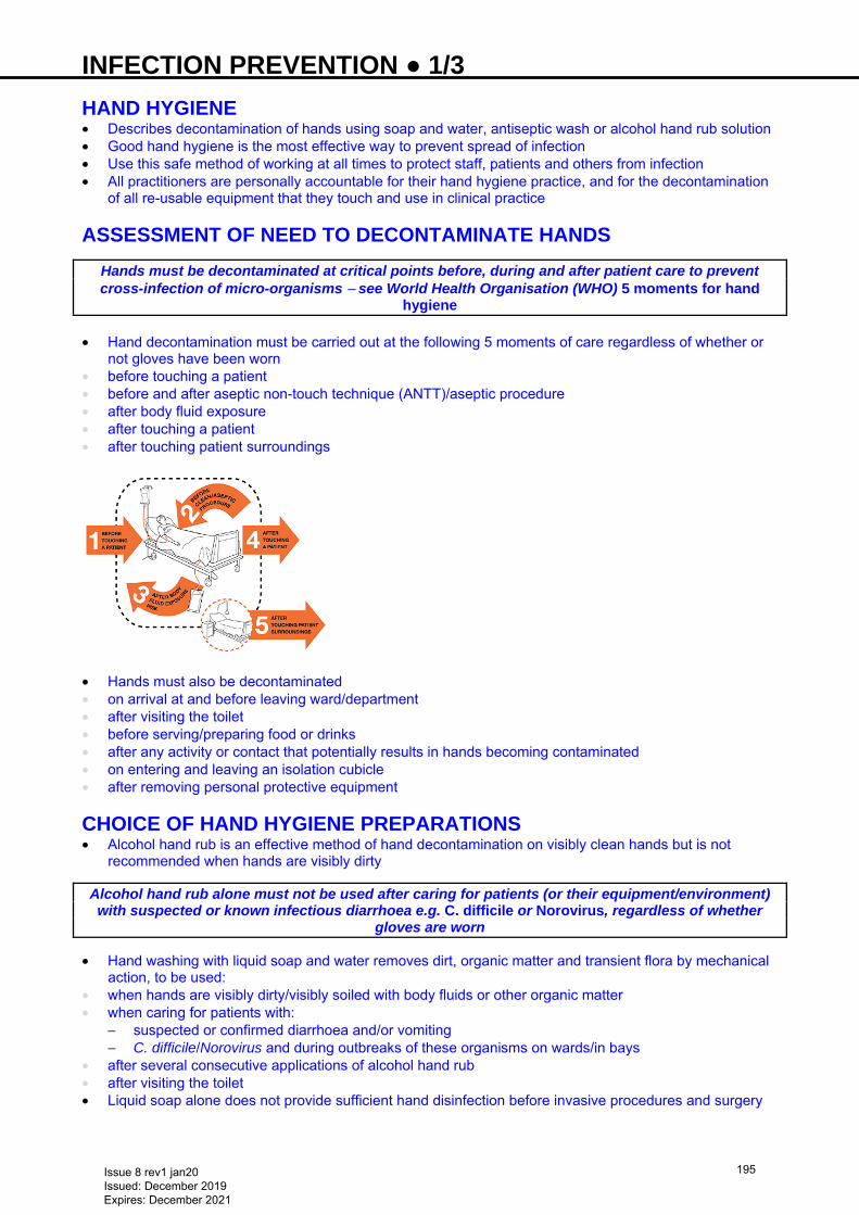
INFECTION PREVENTION ● 1/3 HAND HYGIENE • Describes decontamination of hands using soap and water, antiseptic wash or alcohol hand rub solution • Good hand hygiene is the most effective way to prevent spread of infection • Use this safe method of working at all times to protect staff, patients and others from infection • All practitioners are personally accountable for their hand hygiene practice, and for the decontamination
of all re-usable equipment that they touch and use in clinical practice
ASSESSMENT OF NEED TO DECONTAMINATE HANDS
Hands must be decontaminated at critical points before, during and after patient care to prevent cross-infection of micro-organisms − see World Health Organisation (WHO) 5 moments for hand
hygiene • Hand decontamination must be carried out at the following 5 moments of care regardless of whether or
not gloves have been worn • before touching a patient • before and after aseptic non-touch technique (ANTT)/aseptic procedure • after body fluid exposure • after touching a patient • after touching patient surroundings
• Hands must also be decontaminated • on arrival at and before leaving ward/department • after visiting the toilet • before serving/preparing food or drinks • after any activity or contact that potentially results in hands becoming contaminated • on entering and leaving an isolation cubicle • after removing personal protective equipment
CHOICE OF HAND HYGIENE PREPARATIONS • Alcohol hand rub is an effective method of hand decontamination on visibly clean hands but is not
recommended when hands are visibly dirty
Alcohol hand rub alone must not be used after caring for patients (or their equipment/environment) with suspected or known infectious diarrhoea e.g. C. difficile or Norovirus, regardless of whether
gloves are worn
• Hand washing with liquid soap and water removes dirt, organic matter and transient flora by mechanical action, to be used:
• when hands are visibly dirty/visibly soiled with body fluids or other organic matter • when caring for patients with:
− suspected or confirmed diarrhoea and/or vomiting − C. difficile/Norovirus and during outbreaks of these organisms on wards/in bays
• after several consecutive applications of alcohol hand rub • after visiting the toilet • Liquid soap alone does not provide sufficient hand disinfection before invasive procedures and surgery
Issue 8 rev1 jan20 Issued: December 2019 Expires: December 2021
195
INFECTION PREVENTION ● 2/3 DRESS CODE • Bare below elbow for all staff working within clinical areas (e.g. no sleeves below elbow, no wrist
watches, wrist jewellery or plaster casts/wrist splints) • Do not wear false nails, nail extensions, gel nails or nail varnish • Keep nails short and clean • No stoned rings (acceptable to wear a plain wedding band) • Long hair tied back
PERSONAL PROTECTIVE EQUIPMENT (PPE) Aprons • <2 metres of child with respiratory tract infection • Contact with infectious materials or equipment anticipated • Using hazardous chemicals • ANTT − see below Gloves (non–sterile) • Contact with respiratory secretions or other infectious material of contaminated surfaces • Single patient use; new gloves and apron for every procedure • Take gloves and apron off at point of use and clean hands • Do not carry gloves in your pocket • Do not use alcohol hand rub on gloves • Standard ANTT if not touching key parts/key sites directly
Remove gloves and aprons as soon as clinical activity completed,
before touching pens, notes, phone, computer etc. Sterile gloves and gown • For central venous line (CVL) including peripheral long line (PICC) • Sterile gloves for ANTT if touching key parts/key sites Masks • Surgical face mask • <2 metres of child with respiratory tract infection • FFP3 mask (fit-tested) aerosol generating procedure (e.g. intubation, CPAP) with respiratory tract
infection and when advised by infection prevention team • in conjunction with eye protection when increased risk of splashing of body fluids into eyes/nose/mouth Eye protection • <2 metres of child with persistent coughing or sneezing • When increased risk of splashing of body fluids in to eyes
SURGICAL ANTT • See local ANTT guidelines Definition • Essential procedure aimed at protecting patients from infection during invasive procedures • Achieved by minimising presence of pathogenic micro-organisms as is practically possible • Specific type of aseptic technique with a unique theory and practice framework, providing core principles
for safe aseptic technique and a standardised approach to assessing and applying safe aseptic technique to any invasive clinical procedure
• Do not touch and protect ‘key parts’ or ‘key sites’ e.g. use caps and covers for end of syringes/needles
Preparation phase • Decontaminate hands • Decontaminate tray or trolley choice using Trust approved disinfectant • Clean hands • PPE (as above) • Prepare and assemble equipment using a non-touch technique protecting key parts at all times by not
touching them • Remove gloves and decontaminate hands
Issue 8 rev1 jan20 Issued: December 2019 Expires: December 2021
196
INFECTION PREVENTION ● 3/3 Patient phase • Decontaminate hands at point of care • Apply appropriate PPE non-sterile gloves not touching key parts (e.g. IV drug administration,
venepuncture/cannulation) sterile gloves if touching key parts (e.g. urinary catheterisation, central line/PICC insertion)
• Prepare all equipment using a non-touch technique, protecting key parts at all times by not touching them
• Decontaminate key parts/key sites using single use chlorhexidine 2% in alcohol 70% (SEPP/FREPP or ChloraPrep® 3 mL) and allow drying for 30 sec
• Perform procedure, ensuring protection of key parts/sites at all times Decontamination phase • Dispose of sharps into sharps box immediately at point of use • Remove PPE at patient’s bedside • Dispose of all equipment as clinical waste in nearest clinical waste bin, return equipment to clinical room
ensuring it is cleaned with detergent wipes • Decontaminate hands
Issue 8 rev1 jan20 Issued: December 2019 Expires: December 2021
197
INGUINAL HERNIA ● 1/1 INTRODUCTION • Incidence: 0.5–1% in term babies and 5–10% in premature babies • Right-sided in 50% of cases, left-sided in 10% and both sides in 40% • Most cases can be managed with elective surgery before or shortly after discharge from NNU • Manage incarcerated hernia as a surgical emergency
CLINICAL FEATURES • Visible swelling or bulge in inguino-scrotal region in boys, inguino-labial region in girls. May be constant
or intermittent, becoming more prominent with crying or straining Simple inguinal hernia • Often painless, but many babies happier after repair • Oxygen requirements may fall after repair Incarcerated inguinal hernia • Generally presents with a tender firm mass in the inguinal canal or scrotum • Swelling can be surprisingly small • Baby may be fussy, unwilling to feed and crying inconsolably • Overlying skin may be oedematous, erythematous and discoloured • May be associated abdominal distension, with/without bilious vomiting • Arrange emergency surgical referral
MANAGEMENT AND REFERRAL Reducible inguinal hernia • If asymptomatic, refer by letter to surgeon. Include likely date of discharge and parents’ contact details • Inform parents of the risk of hernia becoming incarcerated • if baby develops a tense, painful swelling and is in obvious pain, parents should seek immediate
medical advice • if swelling not reduced ≤2 hr, complications may arise (bowel compromise − later testicular atrophy) Incarcerated inguinal hernia • Stabilise baby • Administer analgesia (morphine IV), then gently try to reduce hernia • If fully reduced, arrange elective inguinal hernia repair before discharge. Refer to paediatric surgical
team for elective review • If not reducible, request urgent help from on-call paediatrician/neonatologist • Keep child nil-by-mouth • Insert large bore nasogastric tube (NGT), empty stomach and leave on free drainage (see Nasogastric
tube insertion guideline) • Obtain IV access and send blood for FBC and U&E • Start maintenance IV fluids • Aspirate NGT 4-hrly in addition to free drainage and replace aspirate volume, mL-for-mL with sodium
chloride 0.9% with 10 mmol potassium chloride in 500 mL IV. Leave NGT on free drainage • If hernia remains irreducible, refer urgently for surgical assessment • Complete detailed transfer letter using BadgerNet system. Ensure parental details and telephone
contact numbers included • If possible, ask parents to travel to planned place of surgery to meet with surgical team
WHILE AWAITING TRANSFER TO SURGICAL UNIT • Reassess baby regularly • Monitor fluid balance, blood gases, glucose and consider need for fluid resuscitation
USEFUL INFORMATION • http://www.bch.nhs.uk/content/neonatal-surgery • http://www.bch.nhs.uk/find-us/maps-directions • http://www.e-lfh.org.uk/programmes/paediatric-surgery/
Issue 8 rev1 jan20 Issued: December 2019 Expires: December 2021
198
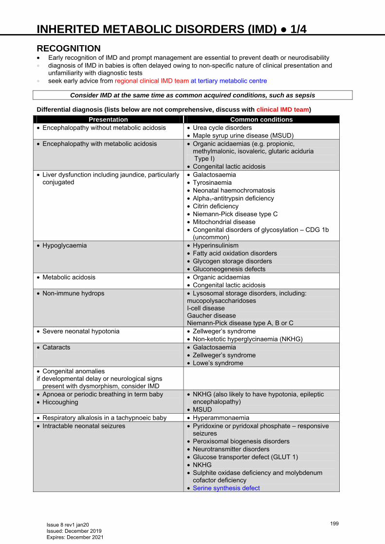
INHERITED METABOLIC DISORDERS (IMD) ● 1/4
RECOGNITION • Early recognition of IMD and prompt management are essential to prevent death or neurodisability • diagnosis of IMD in babies is often delayed owing to non-specific nature of clinical presentation and
unfamiliarity with diagnostic tests • seek early advice from regional clinical IMD team at tertiary metabolic centre
Consider IMD at the same time as common acquired conditions, such as sepsis Differential diagnosis (lists below are not comprehensive, discuss with clinical IMD team)
Presentation Common conditions • Encephalopathy without metabolic acidosis • Urea cycle disorders
• Maple syrup urine disease (MSUD) • Encephalopathy with metabolic acidosis • Organic acidaemias (e.g. propionic,
methylmalonic, isovaleric, glutaric aciduria Type I)
• Congenital lactic acidosis • Liver dysfunction including jaundice, particularly
conjugated • Galactosaemia • Tyrosinaemia • Neonatal haemochromatosis • Alpha1-antitrypsin deficiency • Citrin deficiency • Niemann-Pick disease type C • Mitochondrial disease • Congenital disorders of glycosylation – CDG 1b
(uncommon) • Hypoglycaemia • Hyperinsulinism
• Fatty acid oxidation disorders • Glycogen storage disorders • Gluconeogenesis defects
• Metabolic acidosis • Organic acidaemias • Congenital lactic acidosis
• Non-immune hydrops • Lysosomal storage disorders, including: mucopolysaccharidoses I-cell disease Gaucher disease Niemann-Pick disease type A, B or C
• Severe neonatal hypotonia • Zellweger’s syndrome • Non-ketotic hyperglycinaemia (NKHG)
• Cataracts
• Galactosaemia • Zellweger’s syndrome • Lowe’s syndrome
• Congenital anomalies if developmental delay or neurological signs
present with dysmorphism, consider IMD
• Apnoea or periodic breathing in term baby • Hiccoughing
• NKHG (also likely to have hypotonia, epileptic encephalopathy)
• MSUD • Respiratory alkalosis in a tachypnoeic baby • Hyperammonaemia • Intractable neonatal seizures • Pyridoxine or pyridoxal phosphate – responsive
seizures • Peroxisomal biogenesis disorders • Neurotransmitter disorders • Glucose transporter defect (GLUT 1) • NKHG • Sulphite oxidase deficiency and molybdenum
cofactor deficiency • Serine synthesis defect
Issue 8 rev1 jan20 Issued: December 2019 Expires: December 2021
199
INHERITED METABOLIC DISORDERS (IMD) ● 2/4
Specific indicators Clinical context • Unexplained and mysterious deterioration of baby (can be as short as 12 hr but more commonly after a
symptom-free interval of 24 hr–14 days) Family history • Known metabolic disorders • Unexplained neonatal or infant deaths • Parental consanguinity Obstetric history • Acute fatty liver of pregnancy and HELLP syndrome in index pregnancy may point towards long chain
fatty acid oxidation defect in baby Non-specific indicators suggestive of metabolic disorder in an encephalopathic baby • Encephalopathy in low-risk baby, or onset after period of normality • Fluctuating consciousness and muscle tone • Changes in muscle tone: • axial hypotonia with limb hypertonia • ‘normal’ tone in comatose baby • Abnormal movements: • myoclonic or boxing movements • tongue thrusting • lip smacking • unexplained seizures/burst suppression/hypsarrythmia • seizures are uncommon or occur late in babies with metabolic encephalopathy compared to hypoxic-
ischaemic encephalopathy
INITIAL INVESTIGATIONS • Whenever IMD suspected, perform required investigations without delay • in a sick child request urgent processing of investigations by metabolic biochemistry laboratory • Seek early advice about appropriate investigations and management from IMD team at tertiary metabolic
centre
Urine • Smell • Ketostix: presence of large amounts of urinary ketones is usually abnormal in babies and could suggest
IMD, especially organic acidaemias • Freeze 15–20 mL urine for amino and organic acid analysis • Metabolic screen (amino acids, organic acids, ketones, sugars) Blood • FBC, U&E, infection screen • Glucose • Blood gas (calculate anion gap) • Ammonia • Lactate • Acylcarnitines, including free and total carnitine (bloodspot on Guthrie card/2 mL Li-Hep) • Plasma amino acids (lithium heparin 2 mL) Imaging • Cranial ultrasound scan • Ophthalmic examination
SPECIFIC INVESTIGATIONS
*Discuss with clinical IMD team at tertiary metabolic centre before initiating specific investigations as not all tests may be indicated in all babies with similar presentation
Issue 8 rev1 jan20 Issued: December 2019 Expires: December 2021
200
INHERITED METABOLIC DISORDERS (IMD) ● 3/4
Unexplained/prolonged jaundice or liver synthetic dysfunction Blood • Galactosaemia screen [galactose-1-phosphate uridytransferase (GALIPUT)/Beutler test] (urinary
reducing substances can be negative after short period of galactose exclusion) • cannot be performed reliably if transfused ≤90 days: measure galactose-1 phosphate (Gal-1-P) and urine
galactitol (urine organic acids) • Total and conjugated bilirubin, liver function tests, including clotting studies • Blood spot − succinyl acetone (tyrosinaemia I) • Ferritin • Plasma − very long chain fatty acids only if dysmorphic and hypotonic* • Plasma quantitative amino acids • Alpha1-antitrypsin (quantitative) • Transferrin isoelectric focusing* (CDG) • Consider Niemann-Pick disease type C (chitotriosidase, DNA- mutation analysis)* Urine • Organic acids (succinylacetone in tyrosinaemia I) • Reducing substances: use Clinitest™ • urinary dipsticks are glucose specific and miss galactose in babies with galactosaemia • negative Clinitest™ does not exclude galactosaemia Encephalopathy/epileptic encephalopathy/neonatal intractable seizures Discuss with IMD team – some of the following investigations may be advised and a trial of treatment with pyridoxine/pyridoxal phosphate may be required in certain cases • Urgent quantitative plasma amino acids and urine amino acids • Paired blood and CSF amino acids (glycine, serine), (NKHG, serine synthesis deficiency) • CSF glucose, lactate (GLUT I, mitochondrial disorder). Paired blood and CSF lactate with blood sample
taken before CSF • Plasma − very long chain fatty acids (peroxisomal disorder) • Urine: • dipstick for ketones • sulphite test for sulphite oxidase deficiency • Plasma − uric acid (low in molybdenum cofactor deficiency) • Pyridoxine responsive epilepsy (antiquitin deficiency) • Pyridoxal phosphate responsive seizures • consider CSF amino acids, CSF neurotransmitters, urine organic acid analysis • urine alpha-aminoadipic semialdehyde (AASA) (freeze urine and CSF samples immediately) Hypoglycaemia (most informative when obtained at time of hypoglycaemia) • Plasma non-esterified free fatty acids (FFA) • Beta-hydroxybutyrate (ketones) • Insulin and C-peptide • Acylcarnitine profile, free and total carnitine • Cortisol, growth hormone • Urine for organic acids and ketones Post-mortem (plan how best to use these precious samples in consultation with IMD team) • Plasma (2–5 mL), urine (10–20 mL) and CSF (1 mL) frozen at −20°C • Red cells: blood (5 mL) in lithium heparin stored at 4°C (fridge) • Blood (5 mL) in EDTA: stored at 4°C (fridge) for DNA analysis • Tissue biopsies • skin: store in viral culture medium or sodium chloride 0.9% at 4°C (fridge) (see Skin biopsy guideline) • muscle and liver: take within 1 hr of death, snap freeze in liquid nitrogen • Post-mortem examination • Bile for acylcarnitine analysis – stable for longer than other body fluids
IMMEDIATE MANAGEMENT
Commence emergency management of suspected IMD while awaiting results of initial investigations and discuss with IMD team as early as possible
Issue 8 rev1 jan20 Issued: December 2019 Expires: December 2021
201
INHERITED METABOLIC DISORDERS (IMD) ● 4/4
• Attend to Airway, Breathing and Circulation; ventilate if necessary • Omit all protein, fat and galactose/lactose (milk) intake, including PN and lipid • Commence glucose 10% IV infusion to provide 6−8 mg glucose/kg/min • if hyperglycaemic (>15 mmol/L) or catabolic, start insulin infusion, under guidance from IMD team • if hypertonic (concentration of glucose >10%) infusion necessary, insert central line • Correct dehydration, acid-base and electrolyte disturbances • Cover for infection • Control seizures (avoid sodium valproate) • When stable and appropriate, consider early transfer to tertiary metabolic centre
SPECIFIC MANAGEMENT • Must be led by IMD team • Use following as guide to general principles of management • Check regularly that metabolic emergency medications mentioned below are in stock and available for
emergency use
Neonatal hyperammonaemia Medical emergency requiring prompt intervention to lower ammonia concentration • Renal replacement therapy (haemofiltration more efficient than peritoneal dialysis) • Sodium benzoate • Sodium phenylbutyrate • L-arginine • Carglumic acid (Carbaglu®)
Organic acidaemia • Reduce/stop protein intake • Glucose 10% infusion +/- insulin • L-carnitine • Carglumic acid (Carbaglu®)
Fatty acid oxidation disorders • Avoid prolonged fast • Specific management guided by IMD team
Lactic acidosis • Dichloroacetate • Biotin • L-carnitine • Thiamine
Galactosaemia • Dietary exclusion of galactose
For further information on IMD, www.bimdg.org.uk/guidelines.asp, Emergency protocols and follow through
LOCAL CONTACT • Birmingham Children’s Hospital metabolic team (0121 333 9999)
Issue 8 rev1 jan20 Issued: December 2019 Expires: December 2021
202
INTRA-ABDOMINAL CYSTS ● 1/2 INTRODUCTION This guideline does not apply to cystic structures which may be arising from the urinary tract • Antenatally detected intra-abdominal cysts include: • ovarian • intestinal duplication • mesenteric • vitello-intestinal
SYMPTOMS AND SIGNS • Most cysts will be asymptomatic but the following can be present: • abdominal pain • vomiting • abdominal distension • respiratory compromise • rectal bleeding • Meconium pseudocyst may also be detected on an antenatal ultrasound. They will nearly always cause
vomiting and abdominal distension and may be associated with underlying diagnosis of cystic fibrosis
MANAGEMENT Antenatal • Refer to/discuss appropriate place for delivery with fetal medicine unit • Refer to paediatric surgeon for antenatal counselling Delivery • In the majority of cases, obstetric management will not alter Postnatal • Resuscitate baby as normal • Once stable, perform full postnatal physical examination (see Examination of the newborn guideline) Meconium pseudocyst • If suspected antenatally, do not feed baby at birth • Insert size 8 Fr nasogastric tube (NGT) immediately after birth and fix securely with tape (see
Nasogastric tube insertion guideline) • Empty stomach by aspirating NGT with a 10 or 20 mL syringe • if <20 mL aspirated, check position of tube • Place NGT on free drainage by connecting to a bile bag • Once stabilised, admit baby to NNU • Replace nasogastric losses, mL-for-mL, using sodium chloride 0.9% with potassium chloride
10 mmol/500 mL IV • Commence IV maintenance fluids (see Intravenous fluid therapy guideline) • On day of birth, refer to on-call surgical team at planned place of surgery Other types of intra-abdominal cyst • Unless significant abdominal distension present following birth, allow baby to feed normally and observe
in postnatal ward for ≥48 hr • If baby well after 48 hr with no abdominal symptoms and feeding normally then discharge • Arrange outpatient abdominal ultrasound scan ≤1 week of birth • Consider sacrococcygeal teratoma (SCT) − could be entirely intra-abdominal and arising from pelvis, in
which case discuss with paediatric surgeon
SURGICAL REFERRAL • Urgency will depend on clinical situation • Meconium pseudocyst: • manage as above and refer to surgeon on day of birth • Symptomatic cyst: • stabilise on NNU and refer to on-call surgical team on day of presentation • Asymptomatic cyst: • abdominal ultrasound ≤1 week of birth
Issue 8 rev1 jan20 Issued: December 2019 Expires: December 2021
203
INTRA-ABDOMINAL CYSTS ● 2/2 • when result known, written outpatient referral to consultant paediatric surgeon • if arising from pelvis consider SCT and discuss with paediatric surgeon • Resolved cyst: • ultrasound ≤1 week of birth, even if cyst appears to have resolved during pregnancy. Arrange outpatient
surgical referral
USEFUL INFORMATION • http://www.bch.nhs.uk/story/whats-it/take-look-around/neonatal-surgical-ward • http://www.bch.nhs.uk/find-us/maps-directions • http://www.e-lfh.org.uk/programmes/paediatric-surgery/
Issue 8 rev1 jan20 Issued: December 2019 Expires: December 2021
204
INTRAOSSEOUS INFUSION ● 1/2
INDICATIONS • Severely ill baby when immediate vascular access needed, a UVC is not feasible and peripheral access
not possible (maximum 2 attempts) • Cardiac arrest • allows rapid expansion of circulating volume • gives time to obtain IV access and facilitates procedure by increasing venous filling
CONTRAINDICATIONS • Fractures in target bone • Previous orthopaedic surgery near insertion site • Previous IO insertion in target bone within the preceding 48 hr • At insertion site: • infection • loss of skin integrity • inability to locate landmarks or excessive tissue • Osteogenesis imperfecta (if using a manual Cooks needle only)
EQUIPMENT • EZ-IO drill and needles (3−39 kg: 15 mm pink) or Cooks needle • <3 kg use 18 G butterfly needle • 5 mL syringe with extension and 3-way tap to aspirate and confirm correct position • 10 mL sodium chloride 0.9% flush • 20 mL syringe to administer fluid boluses • Infusion fluid
For manual insertion, infiltrate skin with lidocaine 1% (preservative free) up to 3 mg/kg (0.3 mL/kg) if patient responds to pain
PROCEDURE
Never place your/assistant’s hand under tibia during insertion to avoid staff injury
EZ-IO 1. Locate landmarks 2. Aseptic non-touch technique: clean site 3. If conscious administer local anaesthetic, lidocaine 1% (preservative free) subcutaneously 4. Choose short pink hub needle and attach to drill magnetically 5. Hold drill and needle at 90° to skin surface and push through skin without drilling, until bone is felt 6. Push drill button and drill continuously and push until there is loss of resistance – there is a palpable give
as needle breaches the cortex 7. Remove drill and unscrew trocar 8. If possible aspirate the marrow 9. Attach pre-prepared connection tube 10. Secure needle (with EZ-IO fixator if available) 11. If awake, give lidocaine 1% (preservative free) 0.5 mg/kg (0.05 mL/kg) over 2 min through IO, leave 1
min then flush with sodium chloride 0.9% 2 mL 12. Proceed with required therapy Cooks needle 1. Locate landmarks 2. Aseptic non-touch technique: clean site 3. If conscious administer local anaesthetic, lidocaine 1% (preservative free) 4. Stabilise the lower limb laterally, insert needle at 90° to skin surface. Direct needle caudally from the
epiphyseal plate at an angle of approximately 60° to the long axis of the tibia 5. Advance needle firmly; needle entry into the marrow cavity is accompanied by a loss of resistance,
sustained erect posture of the needle without support and free fluid infusion 6. Attach 5 mL needle and confirm correct position by aspirating marrow (may be omitted in patients with
circulatory arrest), take any bloods required 7. Infuse fluid using 20 mL syringe and IV cannula extension set (with Leur-lock ends)
Issue 8 rev1 jan20 Issued: December 2019 Expires: December 2021
205
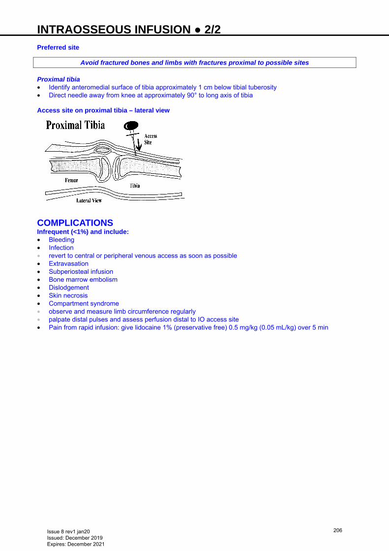
INTRAOSSEOUS INFUSION ● 2/2
Preferred site
Avoid fractured bones and limbs with fractures proximal to possible sites Proximal tibia • Identify anteromedial surface of tibia approximately 1 cm below tibial tuberosity • Direct needle away from knee at approximately 90° to long axis of tibia Access site on proximal tibia – lateral view
COMPLICATIONS Infrequent (<1%) and include: • Bleeding • Infection • revert to central or peripheral venous access as soon as possible • Extravasation • Subperiosteal infusion • Bone marrow embolism • Dislodgement • Skin necrosis • Compartment syndrome • observe and measure limb circumference regularly • palpate distal pulses and assess perfusion distal to IO access site • Pain from rapid infusion: give lidocaine 1% (preservative free) 0.5 mg/kg (0.05 mL/kg) over 5 min
Issue 8 rev1 jan20 Issued: December 2019 Expires: December 2021
206
INTRAVENOUS FLUID THERAPY ● 1/5
PRINCIPLES • Postnatal physiological weight loss is approximately 5–10% in first week after birth • Preterm babies have more total body water and may lose 10–15% of their weight in first week after birth • Postnatal diuresis is delayed in respiratory distress syndrome and in babies who had significant
intrapartum stress • Preterm babies have limited capacity to excrete sodium in first 48 hr • Sodium chloride 0.9% contributes a significant chloride (Cl-) load which can exacerbate metabolic
acidosis • Liberal sodium and water intake before onset of natural diuresis is associated with increased incidence
of patent ductus arteriosus, necrotising enterocolitis and chronic lung disease • After diuresis, a positive sodium balance is necessary for tissue growth • Preterm babies, especially if born <29 weeks’ gestation, lose excessive sodium through immature
kidneys • Babies <28 weeks have significant transepidermal water (TEW) loss • TEW loss leads to hypothermia, loss of calories and dehydration, and causes excessive weight loss and
hypernatraemia
MONITORING Weigh • On admission • Daily for intensive care babies: twice daily if fluid balance is a problem • use in-line scales if available Serum sodium • Daily for intensive care babies • If electrolyte problems or ≤26 weeks, measure twice daily • admission electrolytes reflect maternal status: need not be acted upon but help to interpret trends • serum urea not useful in monitoring fluid balance: reflects nutritional status and nitrogen load Serum creatinine • Daily for intensive care babies • Reflects renal function over longer term • trend is most useful • tends to rise over first 2–3 days • gradually falls over subsequent weeks • absence of postnatal drop is significant Urine output • Review 8-hrly for intensive care babies • 2–4 mL/kg/hr normal hydration • <1 mL/kg/hr requires investigation except in first 24 hr after birth • >6–7 mL/kg/hr suggests impaired concentrating ability or excess fluids
NORMAL REQUIREMENTS Humidification • If <29 weeks, humidify incubator to ≥60% • If ventilated or on CPAP ventilator, set humidifier at 39°C negative 2 to ensure maximal humidification of
inspired gas Normal fluid volume requirements
Fluid volume (mL/kg/day)
Day of life
<1000 g
≥1000 g
1 90 60 2 120 90 3 150 120 4 150 150
Issue 8 rev1 jan20 Issued: December 2019 Expires: December 2021
207
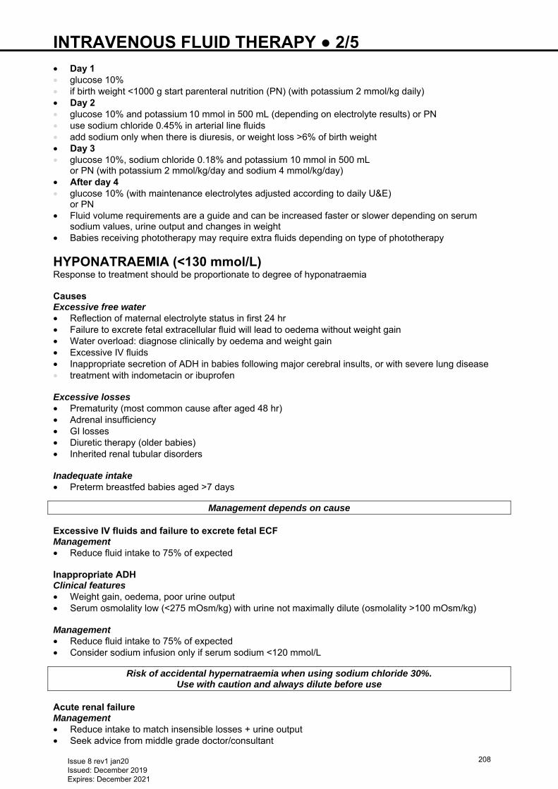
INTRAVENOUS FLUID THERAPY ● 2/5
• Day 1 • glucose 10% • if birth weight <1000 g start parenteral nutrition (PN) (with potassium 2 mmol/kg daily) • Day 2 • glucose 10% and potassium 10 mmol in 500 mL (depending on electrolyte results) or PN • use sodium chloride 0.45% in arterial line fluids • add sodium only when there is diuresis, or weight loss >6% of birth weight • Day 3 • glucose 10%, sodium chloride 0.18% and potassium 10 mmol in 500 mL
or PN (with potassium 2 mmol/kg/day and sodium 4 mmol/kg/day) • After day 4 • glucose 10% (with maintenance electrolytes adjusted according to daily U&E)
or PN • Fluid volume requirements are a guide and can be increased faster or slower depending on serum
sodium values, urine output and changes in weight • Babies receiving phototherapy may require extra fluids depending on type of phototherapy
HYPONATRAEMIA (<130 mmol/L) Response to treatment should be proportionate to degree of hyponatraemia Causes Excessive free water • Reflection of maternal electrolyte status in first 24 hr • Failure to excrete fetal extracellular fluid will lead to oedema without weight gain • Water overload: diagnose clinically by oedema and weight gain • Excessive IV fluids • Inappropriate secretion of ADH in babies following major cerebral insults, or with severe lung disease • treatment with indometacin or ibuprofen Excessive losses • Prematurity (most common cause after aged 48 hr) • Adrenal insufficiency • GI losses • Diuretic therapy (older babies) • Inherited renal tubular disorders Inadequate intake • Preterm breastfed babies aged >7 days
Management depends on cause Excessive IV fluids and failure to excrete fetal ECF Management • Reduce fluid intake to 75% of expected Inappropriate ADH Clinical features • Weight gain, oedema, poor urine output • Serum osmolality low (<275 mOsm/kg) with urine not maximally dilute (osmolality >100 mOsm/kg) Management • Reduce fluid intake to 75% of expected • Consider sodium infusion only if serum sodium <120 mmol/L
Risk of accidental hypernatraemia when using sodium chloride 30%. Use with caution and always dilute before use
Acute renal failure Management • Reduce intake to match insensible losses + urine output • Seek advice from middle grade doctor/consultant
Issue 8 rev1 jan20 Issued: December 2019 Expires: December 2021
208
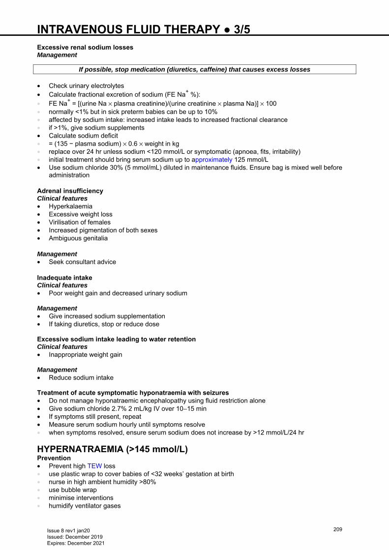
INTRAVENOUS FLUID THERAPY ● 3/5
Excessive renal sodium losses Management
If possible, stop medication (diuretics, caffeine) that causes excess losses • Check urinary electrolytes • Calculate fractional excretion of sodium (FE Na+ %): • FE Na+ = [(urine Na × plasma creatinine)/(urine creatinine × plasma Na)] × 100 • normally <1% but in sick preterm babies can be up to 10% • affected by sodium intake: increased intake leads to increased fractional clearance • if >1%, give sodium supplements • Calculate sodium deficit • = (135 − plasma sodium) × 0.6 × weight in kg • replace over 24 hr unless sodium <120 mmol/L or symptomatic (apnoea, fits, irritability) • initial treatment should bring serum sodium up to approximately 125 mmol/L • Use sodium chloride 30% (5 mmol/mL) diluted in maintenance fluids. Ensure bag is mixed well before
administration Adrenal insufficiency Clinical features • Hyperkalaemia • Excessive weight loss • Virilisation of females • Increased pigmentation of both sexes • Ambiguous genitalia Management • Seek consultant advice Inadequate intake Clinical features • Poor weight gain and decreased urinary sodium Management • Give increased sodium supplementation • If taking diuretics, stop or reduce dose Excessive sodium intake leading to water retention Clinical features • Inappropriate weight gain Management • Reduce sodium intake Treatment of acute symptomatic hyponatraemia with seizures • Do not manage hyponatraemic encephalopathy using fluid restriction alone • Give sodium chloride 2.7% 2 mL/kg IV over 10−15 min • If symptoms still present, repeat • Measure serum sodium hourly until symptoms resolve • when symptoms resolved, ensure serum sodium does not increase by >12 mmol/L/24 hr
HYPERNATRAEMIA (>145 mmol/L) Prevention • Prevent high TEW loss • use plastic wrap to cover babies of <32 weeks’ gestation at birth • nurse in high ambient humidity >80% • use bubble wrap • minimise interventions • humidify ventilator gases
Issue 8 rev1 jan20 Issued: December 2019 Expires: December 2021
209
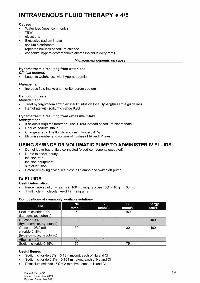
INTRAVENOUS FLUID THERAPY ● 4/5
Causes • Water loss (most commonly) • TEW • glycosuria • Excessive sodium intake • sodium bicarbonate • repeated boluses of sodium chloride • congenital hyperaldosteronism/diabetes insipidus (very rare)
Management depends on cause Hypernatraemia resulting from water loss Clinical features • Leads to weight loss with hypernatraemia Management • Increase fluid intake and monitor serum sodium Osmotic diuresis Management • Treat hyperglycaemia with an insulin infusion (see Hyperglycaemia guideline) • Rehydrate with sodium chloride 0.9% Hypernatraemia resulting from excessive intake Management • If acidosis requires treatment, use THAM instead of sodium bicarbonate • Reduce sodium intake • Change arterial line fluid to sodium chloride 0.45% • Minimise number and volume of flushes of IA and IV lines
USING SYRINGE OR VOLUMATIC PUMP TO ADMINISTER IV FLUIDS • Do not leave bag of fluid connected (blood components excepted) • Nurse to check hourly: • infusion rate • infusion equipment • site of infusion • Before removing giving set, close all clamps and switch off pump
IV FLUIDS Useful information • Percentage solution = grams in 100 mL (e.g. glucose 10% = 10 g in 100 mL) • 1 millimole = molecular weight in milligrams Compositions of commonly available solutions
Fluid Na
mmol/L K
mmol/L Cl
mmol/L Energy kcal/L
Sodium chloride 0.9% (iso-osmolar, isotonic)
150 - 150 -
Glucose 10% (hyperosmolar, hypotonic)
- - - 400
Glucose 10%/sodium chloride 0.18% (hyperosmolar, hypotonic)
30 - 30 400
Albumin 4.5% 150 1 - - Sodium chloride 0.45% 75 - 75 - Useful figures • Sodium chloride 30% = 5.13 mmol/mL each of Na and Cl • Sodium chloride 0.9% = 0.154 mmol/mL each of Na and Cl • Potassium chloride 15% = 2 mmol/mL each of K and Cl
Issue 8 rev1 jan20 Issued: December 2019 Expires: December 2021
210
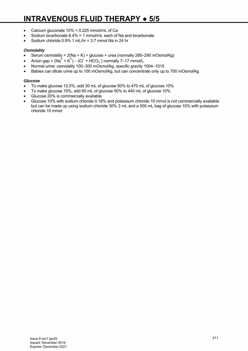
INTRAVENOUS FLUID THERAPY ● 5/5
• Calcium gluconate 10% = 0.225 mmol/mL of Ca • Sodium bicarbonate 8.4% = 1 mmol/mL each of Na and bicarbonate • Sodium chloride 0.9% 1 mL/hr = 3.7 mmol Na in 24 hr Osmolality • Serum osmolality = 2(Na + K) + glucose + urea (normally 285–295 mOsmol/kg) • Anion gap = (Na+ + K+) − (Cl– + HCO3
–) normally 7–17 mmol/L • Normal urine: osmolality 100–300 mOsmol/kg, specific gravity 1004–1015 • Babies can dilute urine up to 100 mOsmol/kg, but can concentrate only up to 700 mOsmol/kg Glucose • To make glucose 12.5%, add 30 mL of glucose 50% to 470 mL of glucose 10% • To make glucose 15%, add 60 mL of glucose 50% to 440 mL of glucose 10% • Glucose 20% is commercially available • Glucose 10% with sodium chloride 0.18% and potassium chloride 10 mmol is not commercially available
but can be made up using sodium chloride 30% 3 mL and a 500 mL bag of glucose 10% with potassium chloride 10 mmol
Issue 8 rev1 jan20 Issued: December 2019 Expires: December 2021
211
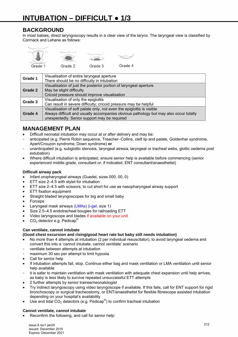
INTUBATION – DIFFICULT ● 1/3 BACKGROUND In most babies, direct laryngoscopy results in a clear view of the larynx. The laryngeal view is classified by Cormack and Lehane as follows:
Grade 1 Visualisation of entire laryngeal aperture There should be no difficulty in intubation
Grade 2 Visualisation of just the posterior portion of laryngeal aperture May be slight difficulty Cricoid pressure should improve visualisation
Grade 3 Visualisation of only the epiglottis Can result in severe difficulty; cricoid pressure may be helpful
Grade 4 Visualisation of soft palate only, not even the epiglottis is visible Always difficult and usually accompanies obvious pathology but may also occur totally unexpectedly. Senior support may be required
MANAGEMENT PLAN • Difficult neonatal intubation may occur at or after delivery and may be: • anticipated (e.g. Pierre Robin sequence, Treacher–Collins, cleft lip and palate, Goldenhar syndrome,
Apert/Crouzon syndrome, Down syndrome) or • unanticipated (e.g. subglottic stenosis, laryngeal atresia, laryngeal or tracheal webs, glottic oedema post
extubation) • Where difficult intubation is anticipated, ensure senior help is available before commencing (senior
experienced middle grade, consultant or, if indicated, ENT consultant/anaesthetist) Difficult airway pack • Infant oropharyngeal airways (Guedel, sizes 000, 00, 0) • ETT size 2–4.5 with stylet for intubation • ETT size 2–4.5 with scissors, to cut short for use as nasopharyngeal airway support • ETT fixation equipment • Straight bladed laryngoscopes for big and small baby • Forceps • Laryngeal mask airways (LMAs) (i-gel, size 1) • Size 2.5–4.5 endotracheal bougies for railroading ETT • Video laryngoscope and blades if available on your unit • CO2 detector e.g. Pedicap® Can ventilate, cannot intubate (Good chest excursion and rising/good heart rate but baby still needs intubation) • No more than 4 attempts at intubation (2 per individual resuscitator), to avoid laryngeal oedema and
convert this into a ‘cannot intubate, cannot ventilate’ scenario • ventilate between attempts at intubation • maximum 30 sec per attempt to limit hypoxia • Call for senior help • If intubation attempts fail, stop. Continue either bag and mask ventilation or LMA ventilation until senior
help available • it is safer to maintain ventilation with mask ventilation with adequate chest expansion until help arrives,
as baby is less likely to survive repeated unsuccessful ETT attempts • 2 further attempts by senior trainee/neonatologist • Try indirect laryngoscopy using video laryngoscope if available. If this fails, call for ENT support for rigid
bronchoscopy or surgical tracheostomy, or ENT/anaesthetist for flexible fibrescope assisted intubation depending on your hospital’s availability
• Use end tidal CO2 detectors (e.g. Pedicap®) to confirm tracheal intubation Cannot ventilate, cannot intubate • Reconfirm the following, and call for senior help:
Issue 8 rev1 jan20 Issued: December 2019 Expires: December 2021
212
INTUBATION – DIFFICULT ● 2/3 • neutral head position (overextension can limit vision) • correct size face mask being used, create a tight seal • use correct size oropharyngeal airway (Guedel airway): too big may cause laryngospasm and too small
may worsen obstruction. (Tip of Guedel airway should reach angle of jaw when aligned with lip on side of face)
• For specific conditions (e.g. Pierre Robin sequence, micrognathia) nasopharyngeal airway may be useful. To make, take an ETT and shorten it by measuring distance between nasal tip and ear tragus. Choose a size that does not blanch the nares completely when inserted
• LMA ventilation (i-gel, smallest size = size 1, suitable for babies >2 kg) • When senior help arrives: • reattempt intubation • use a small towel roll under baby’s shoulder to improve vision • use indirect laryngoscopy with video laryngoscope if available • Call ENT or anaesthetist for support (ENT for rigid bronchoscopy or surgical tracheostomy, or
anaesthetist for flexible fibrescope assisted intubation as above, depending on your hospital’s availability)
• Use end tidal CO2 detector (e.g. Pedicap®) to confirm tracheal intubation Prevent/anticipate difficult intubation/reintubation • For ventilated babies due for extubation, risk of difficult reintubation can be reduced by pre-extubation
dexamethasone to reduce cord oedema, especially in babies who had difficult initial intubations or chronic ventilatory course
• if ETT leak <10–15%, consider dexamethasone Common problems with intubation
Problem Action Oesophageal intubation – blade placed too deep, cords not visualised
Retry with shallow blade insertion and use cricoid pressure
Tongue obscures vision Sweep tongue to left side using blade Use a more anterior lift Use straight blade (Miller)
Cannot see cords Ensure head not hyper-extended Use small towel roll under baby’s shoulders
Cannot intubate
Do not panic Calmly maintain chest excursions through
bag or T-piece/face or laryngeal mask ventilation until help arrives
Use Guedel oral airway if necessary Call for senior help
Seek senior support in the following situations: • Blind intubation: in small baby where poor visualisation due to size • LMA (size 1): can be inserted by juniors while awaiting senior support if trained (see below for details) • Video laryngoscope: if available, to guide intubation through the cords • Railroad technique: if laryngeal aperture narrow, insertion of stylet through cords, and railroading ETT
over it • usually a 2-person procedure and can be carried out under direct vision/blind, depending on visual field
and equipment • carefully insert a bougie through vocal cords, ≤2 cm beyond aperture opening • keep bougie steady while colleague threads ETT over top end of stylet and into trachea. Note: using a
stylet from the ETT pack carries risk of oesophageal/tracheal perforation • Ultra-small fibre-optic bronchoscopy (if available locally): with railroading via bronchoscope • Surgical tracheostomy: not undertaken by neonatal consultants – seek ENT support • Note: Prolonged procedure − additional dose of muscle relaxant can be used under senior guidance • ensure venous access obtained • support cardiac system with IV fluid boluses as required • use inotropic agents as required, based on perfusion and blood pressure • Keep baby warm using techniques supported by your local unit e.g. transwarmer, bubble wrap • Empty stomach contents regularly while on face mask/T-piece ventilation
Issue 8 rev1 jan20 Issued: December 2019 Expires: December 2021
213
INTUBATION – DIFFICULT ● 3/3 I-GEL LMA • Supraglottic mask with soft non-inflatable cuff attached to an airway tube • Once inserted anterior tip enters upper oesophagus while cuff conforms to contours of perilaryngeal
structures, with lumen facing laryngeal opening • Once successfully inserted can be connected to a positive pressure inflation device i.e. ambu
bag/Neopuff™ • Size 1 suitable for babies 2−5 kg, usually >33 weeks’ gestation • ETT can be passed (size 3 in size 1 i-gel) through central port within i-gel
Indications • Babies >2 kg, >33 weeks’ gestation when: • bag and mask ventilation ineffective/problematic and endotracheal intubation unsuccessful • bag and mask and endotracheal intubation not immediately feasible due to facial and/or airway
deformities • may be beneficial as a more stable airway during transport of sick neonate (needing positive pressure
ventilation by mask) by paramedics/midwifery staff from home or midwifery-led unit to NNU
I-gel LMA should not be used: • In the setting of meconium-stained amniotic fluid • When chest compressions required • For delivery of drugs into the trachea
Advantages • Ease of insertion • Minimal risk of tissue compression and instability after insertion • Easily learned skill and easily replicated • Less invasive than endotracheal intubation • Insertion does not require neuromuscular blockers and sedation
Insertion technique • Place head centrally in ‘air-sniffing’ position • Remove i-gel from sterile protective cage • Grasp i-gel close to the connector; lubricate back, sides and front of cuff with a thin layer of lubricating
gel, ensuring no lubricant remains in the bowl of the cuff (oral secretions alone may be sufficient to aid movement)
• Depress baby's chin, thereby enlarging oral cavity • Grasp i-gel close to cuff, holding it like a pen • Point i-gel tip cranially against hard palate • Glide device downwards and backwards along the hard palate with a continuous but gentle push until
definitive resistance felt • I-gel will advance along the posterior pharyngeal wall, with tip locating itself in the upper oesophagus • Attach to Neopuff™/bag and mask ventilation at pre-set pressures • Observe for signs of clinical improvement, confirmed by chest movement, increasing heart rate,
improving colour and saturations • Listen for air leaks (may not be in correct position) • Ensure device remains centrally placed (black i-gel logo visible in central position) • Once position confirmed and clinically effective, tape from maxilla to maxilla to avoid loss of airway
Following i-gel LMA insertion • Do not allow peak pressure of ventilation to exceed 40 cm H2O • Do not use excessive force to insert device • Excessive air leak during manual ventilation is due to suboptimal depth of i-gel insertion, malposition or
excessive peak pressure use • Do not leave device in situ for >4 hr without senior advice • Do not reuse the i-gel • No more than 3 attempts should be attempted in one patient
Complications/side effects • Malposition • Vagal response • Gastric ventilation, distension and aspiration • Laryngospasm or trauma to the pharyngolaryngeal framework
Issue 8 rev1 jan20 Issued: December 2019 Expires: December 2021
214
INTUBATION ● 1/3 See also Intubation − difficult guideline
This procedure must be undertaken or supervised by an experienced person Do not attempt to carry out procedure unsupervised unless you have demonstrated your
competence
ELECTIVE INTUBATION • Use premedication as appropriate for your unit Equipment • Suction • Oxygen with pressure limiting device and T-piece or 500 mL bag and appropriate size face mask • Endotracheal tube (ETT); non cuffed; 3 sizes (diameter in mm):
Weight of baby (g) ETT <1000–1250 2.5 >1250–3000 3.0
>3000 3.5−4.0
• ETT introducer/stylet • Syringe and needles for drawing up premedication • Neonatal stethoscope • Hat for baby to secure tube, ETT fixing device, forceps and scissors • Laryngoscope handle and Miller blades sizes 0 and 00, stethoscope, oropharyngeal airway • Pedicap® end tidal CO2 detector • Oxygen blender
Preparation • Ensure cannula in place and working • Ensure laryngoscope is working, correct sized blades are available and T-piece system is working. Set
pressure limits: 30 cm H2O for term babies and 20−25 cm H2O in preterm babies • Check you have the correct ETT size and attachments to secure ETT • Insert ETT introducer into ETT ensuring it does not protrude past end of ETT • Ensure all drugs drawn up, checked, labelled and ready to give • Check no contraindications to drugs • Ensure monitoring equipment attached and working reliably • If nasogastric tube (NGT) in place, aspirate stomach (particularly important if baby has been given
enteral feeds) • Check IV line working • Ensure back-up plan in case intubation does not work (see Intubation − difficult guideline)
Premedication • Use blended oxygen to pre-oxygenate for 2 min before drug administration • start with room air and increase FiO2 to get SpO2 to target range appropriate for gestational age (see
Oxygen saturation targets guideline). Avoid hyperoxia in preterm baby • Continue to pre-oxygenate until laryngoscopy and between attempts if >1 attempt necessary Drugs
Choice of drugs depends on local practice Analgesia and muscle relaxation can improve likelihood of successful intubation
Muscle relaxants
Administer muscle relaxants only if you are confident that the team can intubate baby quickly Do not use a muscle relaxant unless adequate analgesia has been given
Do not use muscle relaxant for in-and-out surfactant replacement (INSURE)
PROCEDURE • Give premedication • Use mask ventilation in neutral position, a shoulder roll may help
Issue 8 rev1 jan20 Issued: December 2019 Expires: December 2021
215
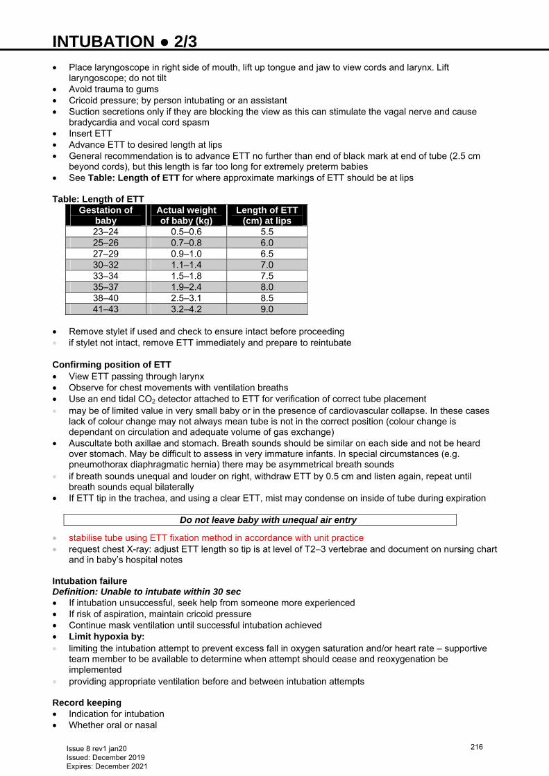
INTUBATION ● 2/3 • Place laryngoscope in right side of mouth, lift up tongue and jaw to view cords and larynx. Lift
laryngoscope; do not tilt • Avoid trauma to gums • Cricoid pressure; by person intubating or an assistant • Suction secretions only if they are blocking the view as this can stimulate the vagal nerve and cause
bradycardia and vocal cord spasm • Insert ETT • Advance ETT to desired length at lips • General recommendation is to advance ETT no further than end of black mark at end of tube (2.5 cm
beyond cords), but this length is far too long for extremely preterm babies • See Table: Length of ETT for where approximate markings of ETT should be at lips Table: Length of ETT
Gestation of baby
Actual weight of baby (kg)
Length of ETT (cm) at lips
23–24 0.5–0.6 5.5 25–26 0.7–0.8 6.0 27–29 0.9–1.0 6.5 30–32 1.1–1.4 7.0 33–34 1.5–1.8 7.5 35–37 1.9–2.4 8.0 38–40 2.5–3.1 8.5 41–43 3.2–4.2 9.0
• Remove stylet if used and check to ensure intact before proceeding • if stylet not intact, remove ETT immediately and prepare to reintubate Confirming position of ETT • View ETT passing through larynx • Observe for chest movements with ventilation breaths • Use an end tidal CO2 detector attached to ETT for verification of correct tube placement • may be of limited value in very small baby or in the presence of cardiovascular collapse. In these cases
lack of colour change may not always mean tube is not in the correct position (colour change is dependant on circulation and adequate volume of gas exchange)
• Auscultate both axillae and stomach. Breath sounds should be similar on each side and not be heard over stomach. May be difficult to assess in very immature infants. In special circumstances (e.g. pneumothorax diaphragmatic hernia) there may be asymmetrical breath sounds
• if breath sounds unequal and louder on right, withdraw ETT by 0.5 cm and listen again, repeat until breath sounds equal bilaterally
• If ETT tip in the trachea, and using a clear ETT, mist may condense on inside of tube during expiration
Do not leave baby with unequal air entry
• stabilise tube using ETT fixation method in accordance with unit practice • request chest X-ray: adjust ETT length so tip is at level of T2−3 vertebrae and document on nursing chart
and in baby’s hospital notes Intubation failure Definition: Unable to intubate within 30 sec • If intubation unsuccessful, seek help from someone more experienced • If risk of aspiration, maintain cricoid pressure • Continue mask ventilation until successful intubation achieved • Limit hypoxia by: • limiting the intubation attempt to prevent excess fall in oxygen saturation and/or heart rate – supportive
team member to be available to determine when attempt should cease and reoxygenation be implemented
• providing appropriate ventilation before and between intubation attempts Record keeping • Indication for intubation • Whether oral or nasal
Issue 8 rev1 jan20 Issued: December 2019 Expires: December 2021
216
INTUBATION ● 3/3 • ETT size and position at cords and nares/lips • Radiological position of tip of ETT and any adjustments following X-ray • Medication chart completed • Baby’s tolerance of procedure and any adverse events
Issue 8 rev1 jan20 Issued: December 2019 Expires: December 2021
217
JAUNDICE ● 1/3 Based on NICE CG98 Jaundice in newborn babies under 28 days
RECOGNITION AND ASSESSMENT Risk factors for hyperbilirubinaemia • <38 weeks’ gestation • Previous sibling required treatment for jaundice • Mother intends to exclusively breastfeed • Visible jaundice in baby aged <24 hr
Risk factors for kernicterus • High bilirubin level (>340 micromol/L in term baby) • Rapidly rising bilirubin level (>8.5 micromol/L/hr) • Clinical features of bilirubin encephalopathy
Symptoms and signs • When looking for jaundice (visual inspection): • check naked baby in bright and preferably natural light • examine the sclerae and gums, and press lightly on skin to check for signs of jaundice in 'blanched' skin Assess • Pallor (haemolysis) • Poor feeding, drowsiness (neurotoxicity) • Hepatosplenomegaly (blood group incompatibility or cytomegalovirus) • Splenomegaly (spherocytosis)
Causes • Physiological • Prematurity • Increased bilirubin load: • blood group incompatibility (Rhesus or ABO) • G6PD deficiency and other red cell enzyme deficiencies • congenital spherocytosis • cephalhaematoma, bruising • Rarely infection (e.g. UTI, congenital infection) • Metabolic disorder
Persistent jaundice after aged 14 days (see Liver dysfunction guideline) • Breast milk jaundice • Hypothyroidism • Liver disease (e.g. extra hepatic biliary atresia and neonatal hepatitis) • Alpha-1-antitrypsin deficiency • Galactosaemia • TPN-induced cholestasis
Investigations Assessment of jaundice • Babies aged <72 hr, at every opportunity (risk factors and visual inspection) • Babies with suspected or obvious jaundice, measure and record bilirubin level urgently • <24 hr: within 2 hr • ≥24 hr: within 6 hr • If serum bilirubin >100 micromol/L in first 24 hr • repeat measurement in 6–12 hr • interpret result in accordance with baby’s age and gestation – see Table • urgent medical review as soon as possible (and within 6 hr) • Interpret bilirubin result in accordance with baby’s gestational and postnatal age according to Table
Jaundice approaching treatment level • If baby well, ≥38 weeks, aged >24 hr and • serum bilirubin ≤50 micromol/L below treatment threshold, repeat measurement in 18 hr if risk factors
and 24 hr if no risk factors • serum bilirubin >50 micromol/L below treatment threshold, no further routine measurements required
Issue 8 rev1 jan20 Issued: December 2019 Expires: December 2021
218
JAUNDICE ● 2/3 Jaundice requiring treatment • Total bilirubin • Baby’s blood group and direct Coombs test (interpret result taking into account strength of reaction and
whether mother received prophylactic anti-D immunoglobulin during pregnancy) • Mother’s blood group and antibody status (should be available from maternal healthcare record) • PCV
Plus (if clinically indicated) • Full infection screen (in an ill baby) • G6PD level and activity (if indicated by ethnic origin: Mediterranean, Middle Eastern, South East Asian) • FBC and film
Persistent jaundice >14 days term baby; >21 days preterm baby (see Liver dysfunction guideline) • Total and conjugated bilirubin • Examine stool colour • FBC • Baby’s blood group and direct Coombs test (interpret result taking into account strength of reaction and
whether mother received prophylactic anti-D immunoglobulin during pregnancy) • Ensure routine metabolic screening performed (including screening for hypothyroidism) • Urine culture
Baby with conjugated bilirubin >25 micromol/L, refer urgently to a specialist centre
Second line investigations (not in NICE guideline) • Liver function tests (ALT, AST, albumin, GGT) • Coagulation profile • G6PD screen in African, Asian or Mediterranean babies • Thyroid function tests: ask for ‘FT4 priority and then TSH’ • Congenital infection screen • Urine for CMV PCR, toxoplasma ISAGA-IgM and throat swab for HSV culture/PCR • Metabolic investigations e.g: • blood galactose-1-phosphate • urine for reducing substances • alpha-1-antitrypsin
TREATMENT <7 DAYS
Do not start treatment if serum bilirubin is below treatment threshold Babies ≥38 weeks’ gestation • Use conventional blue light phototherapy (not fibre optic) as treatment of choice • Use continuous multiple phototherapy for babies who: • fail to respond to conventional phototherapy (bilirubin does not fall within 6 hr of starting treatment) • have a rapid rise in bilirubin (>8.5 micromol/L/hr) • have a bilirubin level within 50 micromol/L of exchange transfusion threshold • when level falls to >50 micromol/L below threshold reduce intensity of phototherapy
Babies <38 weeks’ gestation • Use fibre optic or conventional blue light as first line treatment • based on gestational age and postnatal age, use Threshold graphs
(http://www.nice.org.uk/guidance/CG98 under ‘Tools and resources’ then ‛CG98 Neonatal Jaundice: treatment threshold graphs’) to determine threshold for phototherapy
• Indications for multiple phototherapy as term babies Management during phototherapy • Offer parents information on procedure (www.nice.org.uk/guidance/cg98/resources/jaundice-in-newborn-
babies-318006690757) • Unless other clinical conditions prevent, place baby in supine position • Ensure treatment applied to maximum area of skin • Monitor baby’s temperature
Issue 8 rev1 jan20 Issued: December 2019 Expires: December 2021
219
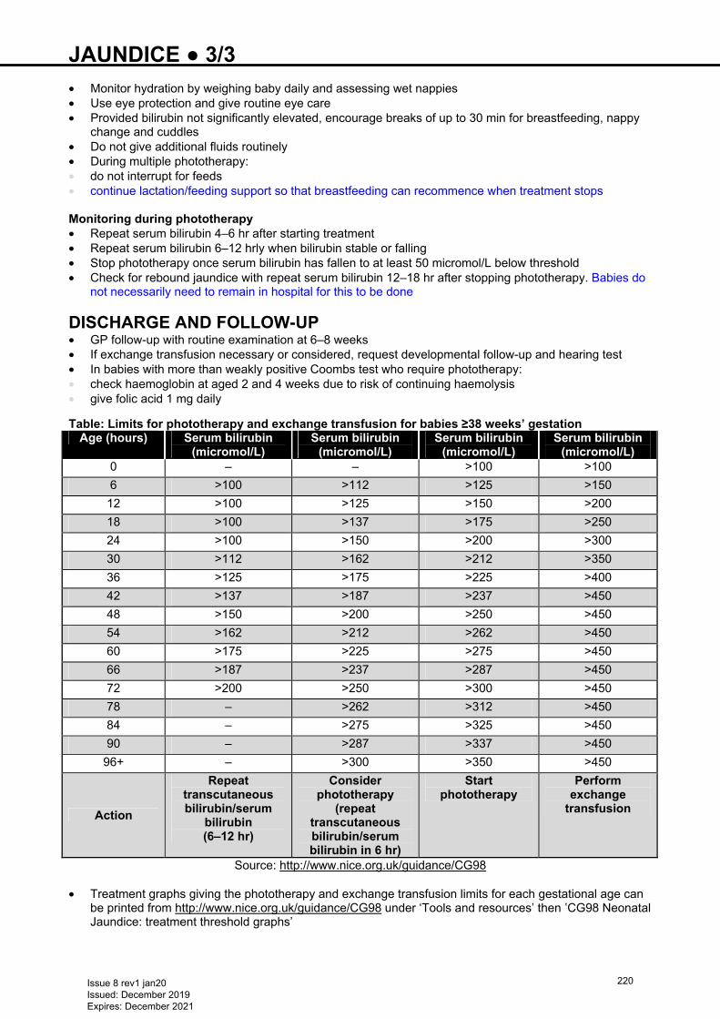
JAUNDICE ● 3/3 • Monitor hydration by weighing baby daily and assessing wet nappies • Use eye protection and give routine eye care • Provided bilirubin not significantly elevated, encourage breaks of up to 30 min for breastfeeding, nappy
change and cuddles • Do not give additional fluids routinely • During multiple phototherapy: • do not interrupt for feeds • continue lactation/feeding support so that breastfeeding can recommence when treatment stops
Monitoring during phototherapy • Repeat serum bilirubin 4–6 hr after starting treatment • Repeat serum bilirubin 6–12 hrly when bilirubin stable or falling • Stop phototherapy once serum bilirubin has fallen to at least 50 micromol/L below threshold • Check for rebound jaundice with repeat serum bilirubin 12–18 hr after stopping phototherapy. Babies do
not necessarily need to remain in hospital for this to be done
DISCHARGE AND FOLLOW-UP • GP follow-up with routine examination at 6–8 weeks • If exchange transfusion necessary or considered, request developmental follow-up and hearing test • In babies with more than weakly positive Coombs test who require phototherapy: • check haemoglobin at aged 2 and 4 weeks due to risk of continuing haemolysis • give folic acid 1 mg daily Table: Limits for phototherapy and exchange transfusion for babies ≥38 weeks’ gestation
Age (hours) Serum bilirubin (micromol/L)
Serum bilirubin (micromol/L)
Serum bilirubin (micromol/L)
Serum bilirubin (micromol/L)
0 – – >100 >100
6 >100 >112 >125 >150
12 >100 >125 >150 >200
18 >100 >137 >175 >250
24 >100 >150 >200 >300
30 >112 >162 >212 >350
36 >125 >175 >225 >400
42 >137 >187 >237 >450
48 >150 >200 >250 >450
54 >162 >212 >262 >450
60 >175 >225 >275 >450
66 >187 >237 >287 >450
72 >200 >250 >300 >450
78 – >262 >312 >450
84 – >275 >325 >450
90 – >287 >337 >450
96+ – >300 >350 >450
Action
Repeat transcutaneous bilirubin/serum
bilirubin (6–12 hr)
Consider phototherapy
(repeat transcutaneous bilirubin/serum bilirubin in 6 hr)
Start phototherapy
Perform exchange
transfusion
Source: http://www.nice.org.uk/guidance/CG98 • Treatment graphs giving the phototherapy and exchange transfusion limits for each gestational age can
be printed from http://www.nice.org.uk/guidance/CG98 under ‘Tools and resources’ then ‛CG98 Neonatal Jaundice: treatment threshold graphs’
Issue 8 rev1 jan20 Issued: December 2019 Expires: December 2021
220
KANGAROO CARE ● 1/2 DEFINITION • Method of holding preterm and/or sick baby skin-to-skin in an upright position between mother’s breasts
or against carer’s chest (fathers and siblings can also be kangaroo carers) • Kangaroo care (KC) can be offered to parents of medically stable babies Benefits of KC • Inform parents about the benefits of KC (use ‘BLISS Skin-to-skin and Kangaroo Care’ information
https://www.bliss.org.uk/parents/in-hospital/looking-after-your-baby-on-the-neonatal-unit/skin-to-skin-and-kangaroo-care or locally approved information leaflets):
• helps promote physiological stability: regulates baby’s temperature, heart rate, breathing and oxygen saturation
• associated with fewer episodes of apnoea and bradycardia • increases time in quiet sleep • longer alert states and less crying • analgesic effect during painful procedures • promotes growth and earlier discharge • improves lactation and breastfeeding success – duration and exclusivity • promotes parent–baby attachment and family-centred care • positive effect on parenting – reduces stress and depression, triggers healing process, increases
confidence • helps reduce risk of mortality among preterm and low-birth-weight babies
INDICATIONS • Medically stable baby – including those on CPAP with a stable oxygen requirement • Medically stable ventilated babies after discussion with MDT • Ventilated babies receiving palliative care
If concerns regarding stability of baby, discuss with senior member of medical and nursing team
CONTRAINDICATIONS • Umbilical lines in situ
Consider • Baby’s condition and dependency • Maintenance of neutral thermal environment and humidity • Activity in the room − quiet, calm environment is preferable • Support available from colleagues Ensure • Access to oxygen and suction
PARENT PREPARATION • Ensure parents are aware that baby may be briefly unstable during transfer from/to incubator/cot • Suggest parents do not smoke immediately before KC time • Choose a mutually convenient time for parents and baby • Provide privacy for parents to prepare clothing – suggest parents wear a clean loose fitting, front
fastening shirt • Provide comfortable chair and foot rest if appropriate • Offer a hand-held mirror – to enable parent to see baby’s face • Advise parents to bring a drink and go to toilet before KC time
Nurse transfer
Recommended initial transfer method. Use this method until parents feel confident • Parent to sit slightly reclined in a comfortable chair. Ensure clothing open and ready to receive baby • Contain baby’s limbs and move gently – use ‘snuggle up’ nest if appropriate • Place baby on parent’s chest, prone with head to parent’s sternum • Parent to support baby’s head and body with baby’s legs flexed • Turn baby’s head to side to protect airway • Use parent’s clothing and a wrap/blanket for warmth and support • If appropriate, place hat on baby
Issue 8 rev1 jan20 Issued: December 2019 Expires: December 2021
221
KANGAROO CARE ● 2/2 Parent transfer • Parent to stand at side of incubator • Place forearm gently under ‘snuggle up’ nest or sheet, cup baby’s head with other hand • Gently lift baby from incubator and onto chest, resting baby’s head against sternum while supporting
baby’s back and bottom with forearm • Parent gently moves back to sit in chair, guided by nurse • Nurse to check baby’s position as before
Duration of KC • When baby settled, remove screens/curtains – be guided by parental preference • Aim to provide KC for ≥1 hr • Monitor baby’s position and vital signs • Babies may have nasogastric tube feeds during KC time • Discontinue KC if: • baby shows signs of distress • has a prolonged increase in oxygen requirement of 10–20% • at parent’s request Breast milk • Encourage mother to express breast milk following KC time. See Breast milk expression guideline
Issue 8 rev1 jan20 Issued: December 2019 Expires: December 2021
222
LABOUR WARD CALLS ● 1/1 • Encourage obstetric team to warn neonatal team of expected problems well in advance • Decide who should attend (e.g. first on-call, middle grade or consultant), and degree of urgency Neonatal team should attend the following deliveries: • Non-reassuring electronic fetal monitoring trace, as assessed by obstetric team • Significant fresh meconium in liquor • Caesarean section under general anaesthesia (see below) • Major congenital abnormalities (minor abnormalities will wait until working hours) • Vacuum extraction or instrumental deliveries performed for fetal reasons (see below) • Preterm delivery <36 weeks’ gestation • Severe pre-eclampsia with seizures • Antepartum haemorrhage • Moderate-to-severe Rhesus disease • Unexpected breech delivery It is not necessary for neonatal team to attend the following deliveries: • Elective caesarean section under regional anaesthesia • Meconium staining of liquor • Breech delivery (including caesarean section under regional anaesthesia) • Twins (>36 weeks) • Pre-eclampsia without seizures The following factors may require neonatal team to attend birth or assess baby soon after birth (see antenatal plan in maternal notes) • Maternal illness likely to affect baby: • diabetes mellitus • thyroid disease • systemic lupus erythematosus • myasthenia gravis • myotonic dystrophy • hepatitis B carriage • intrapartum antibiotics for GBS indicated but not given, or given <4 hr before delivery • HIV • HELLP syndrome • suspected sepsis treated with IV antibiotics • Maternal medications that may affect baby e.g. antidepressants • Neonatal alerts: • abnormal antenatal scans • low-birth-weight baby <2.5 kg • Pregnancy and past history • prolonged rupture of membranes • polyhydramnios • previous baby/perinatal death • family history of genetic or metabolic abnormalities
Issue 8 rev1 jan20 Issued: December 2019 Expires: December 2021
223
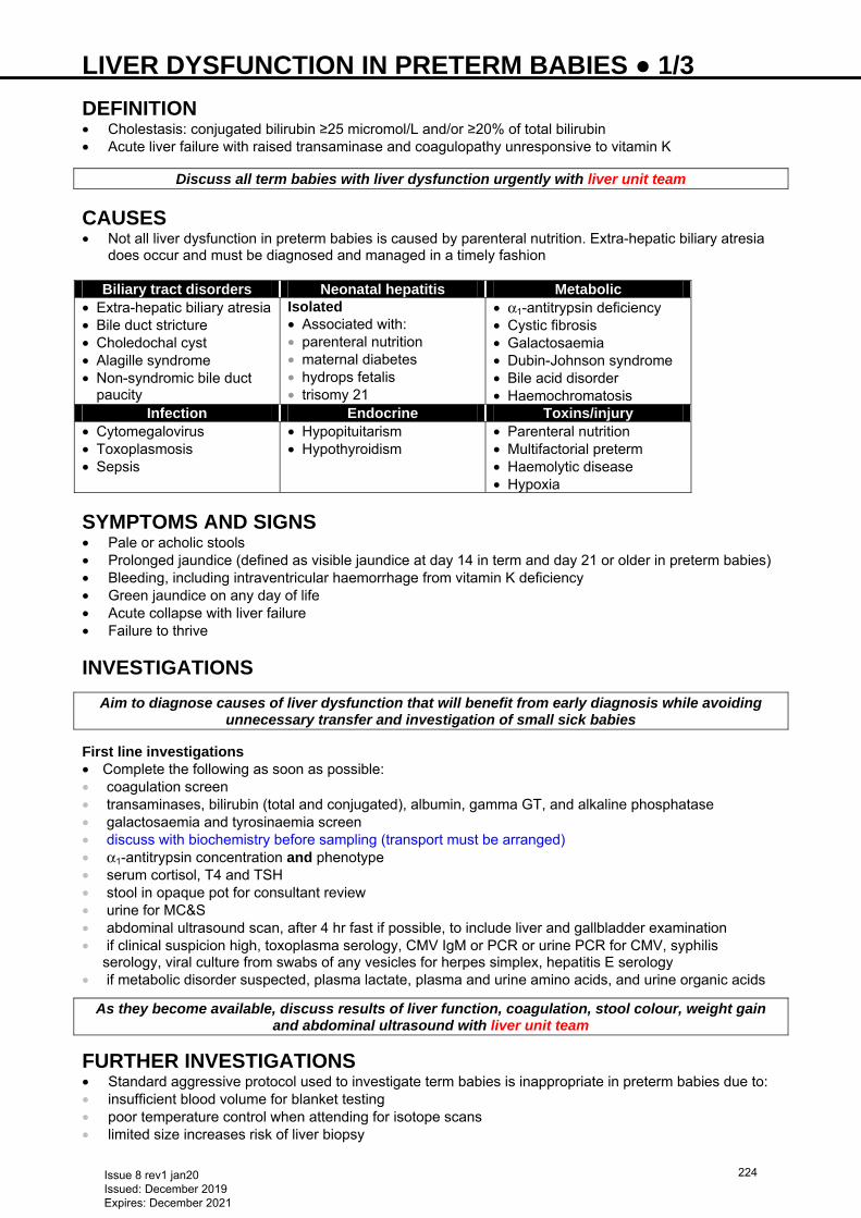
LIVER DYSFUNCTION IN PRETERM BABIES ● 1/3 DEFINITION • Cholestasis: conjugated bilirubin ≥25 micromol/L and/or ≥20% of total bilirubin • Acute liver failure with raised transaminase and coagulopathy unresponsive to vitamin K
Discuss all term babies with liver dysfunction urgently with liver unit team
CAUSES • Not all liver dysfunction in preterm babies is caused by parenteral nutrition. Extra-hepatic biliary atresia
does occur and must be diagnosed and managed in a timely fashion
Biliary tract disorders Neonatal hepatitis Metabolic • Extra-hepatic biliary atresia • Bile duct stricture • Choledochal cyst • Alagille syndrome • Non-syndromic bile duct
paucity
Isolated • Associated with: • parenteral nutrition • maternal diabetes • hydrops fetalis • trisomy 21
• α1-antitrypsin deficiency • Cystic fibrosis • Galactosaemia • Dubin-Johnson syndrome • Bile acid disorder • Haemochromatosis
Infection Endocrine Toxins/injury • Cytomegalovirus • Toxoplasmosis • Sepsis
• Hypopituitarism • Hypothyroidism
• Parenteral nutrition • Multifactorial preterm • Haemolytic disease • Hypoxia
SYMPTOMS AND SIGNS • Pale or acholic stools • Prolonged jaundice (defined as visible jaundice at day 14 in term and day 21 or older in preterm babies) • Bleeding, including intraventricular haemorrhage from vitamin K deficiency • Green jaundice on any day of life • Acute collapse with liver failure • Failure to thrive
INVESTIGATIONS
Aim to diagnose causes of liver dysfunction that will benefit from early diagnosis while avoiding unnecessary transfer and investigation of small sick babies
First line investigations • Complete the following as soon as possible: • coagulation screen • transaminases, bilirubin (total and conjugated), albumin, gamma GT, and alkaline phosphatase • galactosaemia and tyrosinaemia screen • discuss with biochemistry before sampling (transport must be arranged) • α1-antitrypsin concentration and phenotype • serum cortisol, T4 and TSH • stool in opaque pot for consultant review • urine for MC&S • abdominal ultrasound scan, after 4 hr fast if possible, to include liver and gallbladder examination • if clinical suspicion high, toxoplasma serology, CMV IgM or PCR or urine PCR for CMV, syphilis
serology, viral culture from swabs of any vesicles for herpes simplex, hepatitis E serology • if metabolic disorder suspected, plasma lactate, plasma and urine amino acids, and urine organic acids
As they become available, discuss results of liver function, coagulation, stool colour, weight gain and abdominal ultrasound with liver unit team
FURTHER INVESTIGATIONS • Standard aggressive protocol used to investigate term babies is inappropriate in preterm babies due to: • insufficient blood volume for blanket testing • poor temperature control when attending for isotope scans • limited size increases risk of liver biopsy
Issue 8 rev1 jan20 Issued: December 2019 Expires: December 2021
224
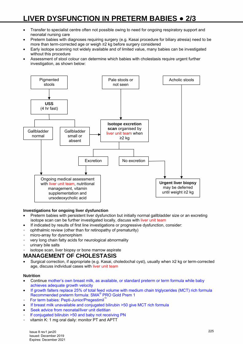
LIVER DYSFUNCTION IN PRETERM BABIES ● 2/3 • Transfer to specialist centre often not possible owing to need for ongoing respiratory support and
neonatal nursing care • Preterm babies with diagnoses requiring surgery (e.g. Kasai procedure for biliary atresia) need to be
more than term-corrected age or weigh ≥2 kg before surgery considered • Early isotope scanning not widely available and of limited value, many babies can be investigated
without this procedure • Assessment of stool colour can determine which babies with cholestasis require urgent further
investigation, as shown below:
Investigations for ongoing liver dysfunction • Preterm babies with persistent liver dysfunction but initially normal gallbladder size or an excreting
isotope scan can be further investigated locally, discuss with liver unit team • If indicated by results of first line investigations or progressive dysfunction, consider: • ophthalmic review (other than for retinopathy of prematurity) • micro-array for dysmorphism • very long chain fatty acids for neurological abnormality • urinary bile salts • isotope scan, liver biopsy or bone marrow aspirate
MANAGEMENT OF CHOLESTASIS • Surgical correction, if appropriate (e.g. Kasai, choledochal cyst), usually when ≥2 kg or term-corrected
age, discuss individual cases with liver unit team Nutrition • Continue mother’s own breast milk, as available, or standard preterm or term formula while baby
achieves adequate growth velocity • If growth falters replace 25% of total feed volume with medium chain triglycerides (MCT) rich formula
Recommended preterm formula: SMA® PRO Gold Prem 1 • For term babies: Pepti-Junior/Pregestimil™ • If breast milk unavailable and conjugated bilirubin >50 give MCT rich formula • Seek advice from neonatal/liver unit dietitian • If conjugated bilirubin >50 and baby not receiving PN • vitamin K: 1 mg oral daily: monitor PT and APTT
USS (4 hr fast)
Isotope excretion scan organised by
liver unit team when ≥2 kg
Urgent liver biopsy may be deferred until weight ≥2 kg
Gallbladder normal
Gallbladder small or absent
Acholic stools Pale stools or not seen
Excretion No excretion
Ongoing medical assessment with liver unit team, nutritional
management, vitamin supplementation and ursodeoxycholic acid
Pigmented stools
Issue 8 rev1 jan20 Issued: December 2019 Expires: December 2021
225
LIVER DYSFUNCTION IN PRETERM BABIES ● 3/3 • vitamin A: 1000 units/kg (rounded to nearest 1000 units) • vitamin D: alfacalcidol 50 nanogram/kg/day rounded to nearest 100 nanogram/drop
− aged >3 months: ergocalciferol 400−800 units/kg/day • vitamin E: alpha tocopherol 10 mg/kg/day Ursodeoxycholic acid • BNFc dose 5−10 mg/kg 3 times daily, but liver team normally recommend
20–30 mg/kg/day in divided doses for most preterm babies until jaundice resolves, and to stimulate bile flow in babies and children with cystic fibrosis
Parenteral nutrition • Wherever possible, feed enterally, as even small amounts have trophic effects on gut, reduce bacterial
colonisation and promote bile flow • Bolus feeds promote bile flow more readily than continuous feeds, but the latter may be better absorbed • Discontinue PN as soon as possible in all preterm babies with cholestasis Specific treatments • Babies with cystic fibrosis, galactosaemia, tyrosinaemia type 1, hypopituitarism, hypothyroidism or bile
acid disorders require additional targeted management and life-long follow-up shared by local teams and appropriate specialists
FOLLOW-UP • For babies with persistent cholestasis, arrange outpatient follow-up with liver team after discharge from
NNU • If liver dysfunction has resolved, no follow-up with liver team necessary • For all others with a specific diagnosis, follow-up will be directed by liver team, appropriate specialists
and local consultant • Long-term hepatic outcome for multifactorial preterm or neonatal hepatitis excellent, majority resolve
within first year
Issue 8 rev1 jan20 Issued: December 2019 Expires: December 2021
226
LONG LINE INSERTION (PERIPHERALLY SITED) ● 1/4
See also Use of central venous catheters in neonates − https://www.bapm.org/sites/default/files/files/BAPM%20CVC%20final%20Jan16%20%28addition%20Aug%202018%29.pdf
A peripherally sited central venous catheter (PICC) allows administration of infusions that, if given peripherally, may cause damage to the vein and surrounding skin, or be less effective. These benefits must be weighed against the risks of line sepsis, thrombosis, embolism, and pleural and pericardial effusion. Units which use central line catheters should have a formal training package for insertion of catheters which should include assessment of technical competence and awareness of potential complications
INDICATIONS • Total/partial parenteral nutrition • Concentrated (>12.5%) glucose infusions • Infusions of glucose >5% + calcium gluconate • Inotrope infusions • Prolonged drug or fluid administration where peripheral access difficult
CONTRAINDICATIONS • Infection at proposed insertion site • Systemic sepsis: defer until sepsis treatment commenced and blood cultures negative • Tissue perfusion concerns
EQUIPMENT • Sterile gown and gloves • Cleaning solution as per unit policy • Sodium chloride 0.9% for injection • Tape measure • Overhead light • Neonatal long line – appropriate for size of baby and expected rate of infusion • Decide whether double or single lumen line required • Long line insertion pack or, if not available, individual items to include: • dressing pack with swabs and plastic dish • sterile towels/sheets • non-toothed forceps • 5–10 mL syringe • Steri-Strip™ • sterile scissors • clear dressing (e.g. Tegaderm™/Opsite)
PROCEDURE
Must be performed or directly supervised by an individual competent in the insertion of these devices
Consent and preparation • Inform parents and obtain verbal consent as recommended by BAPM • Discuss timing of procedure with nurses • Keep baby warm. Work through portholes • Identify site of insertion • typically long saphenous at ankle or medial/lateral antecubital vein at elbow • where access difficult, other large peripheral veins or scalp veins anterior to ear may be used • right long saphenous vein is preferable to the left − latter more commonly associated with malposition in
the left ascending lumbar vein leading to risk of extravasation of PN into CSF • Measure distance, aiming to insert tip of catheter into superior or inferior vena cava (to xiphisternum for
lower limb insertion, to upper sternum for upper limb insertion)
Developmental care • Unless contraindicated, give sucrose or breast milk and non-nutritive sucking [see Non-nutritive
sucking (NNS) guideline] • Shield baby’s eyes from bright light • Second person to provide containment holding (see Pain assessment and management guideline)
Issue 8 rev1 jan20 Issued: December 2019 Expires: December 2021
227
LONG LINE INSERTION (PERIPHERALLY SITED) ● 2/4
Aseptic insertion • Maintain strict asepsis throughout • Prime catheter and cut small piece of gauze for under hub • Clean site and allow to dry. Ensure that cleaning fluid does not pool beneath baby • Puncture site with needle from pack and follow instructions for that catheter • Avoid use of cannulae for long line insertion • When blood flows back through the needle, insert line using non-toothed forceps • If appropriately placed, the line will pass easily beyond the tip of the needle • Release tourniquet if used • There may be some resistance when the line passes joints, such as knee, and gentle repositioning of
baby’s limb may help • Should catheter advancement become difficult, infuse a little fluid whilst simultaneously advancing
catheter • Never withdraw catheter back through needle • When in place, withdraw needle as stated in catheter instructions • Catheter should allow free aspiration of blood in the final position Securing catheter • When haemostasis achieved, fix with Steri-Strips™. Place small piece of gauze under hub, and cover
with Tegaderm™/Opsite, making sure that all dressing and site is covered, but not encircling the limb tightly. Ensure line insertion site is visible through clear dressing
• Connect a sterile 5 mL syringe containing sodium chloride 0.9% and infuse at 0.5 mL/hr while awaiting X-ray, to ensure that the line does not clot off
Determining catheter position • X-ray to determine position • aim is to view full length of line • request chest/abdominal X-ray (including upper/lower limb requests as per local policy) • If inserted in upper limb, ensure arm is at 90° angle to thorax during X-ray • Small gauge neonatal long lines can be difficult to see on plain X-ray • use X-ray magnification, contrast adjustment and inversion to aid process • if using contrast medium, refer to local policy • Upper limb catheter • tip should preferably be in superior vena cava but other large veins e.g. innominate, subclavian are
acceptable • ensure tip of catheter crosses shoulder joint • if any concern regarding possibility of malposition of catheter continue to infuse with sodium chloride
0.9% 0.5 mL/hr and seek consultant review • Lower limb catheter • left saphenous PICC should cross the midline at L4/5 and run to the right side of the vertebral column in
the IVC with the tip outside the heart • if catheter does not cross the midline or has a tortuous course the line may be in a small vessel. Lateral
X-ray will demonstrate posterior deviation of the line towards the vertebral column • if malposition suspected continue to infuse with sodium chloride 0.9% 0.5 mL/hr and seek consultant
review of X-ray • Catheter tips in axillary, cephalic and femoral veins are acceptable if the benefit outweighs increased
risks of reinsertion. Discuss with consultant before connecting PN to line • Monitor site closely • If catheter tip beyond desired location, using aseptic technique, remove dressing and withdraw catheter
the measured distance. Redress with new sterile dressing and confirm new position by X-ray
Catheter tip must not lie within heart (risk of perforation and tamponade) Failure of insertion • If second operator is required following an unsuccessful attempt at placement, use fresh equipment
DOCUMENTATION • Record in case notes: • date and time of insertion • success of insertion and number of attempts • type and gauge of catheter
Issue 8 rev1 jan20 Issued: December 2019 Expires: December 2021
228
LONG LINE INSERTION (PERIPHERALLY SITED) ● 3/4
• site and length of insertion • X-ray position and alterations • Insert tracking stickers from all packs
AFTERCARE Dressings and site care • Routine dressing changes are unnecessary • Replace aseptically only if dressings lift or catheter visibly kinked or becomes insecure • Observe site every shift for bleeding, leaking of infusate and signs of infection (redness, swelling) and
document in daily care summary Line management and medication • Minimise number of line breaks • Intermittent medications only given via this route in extreme circumstances. (This is a senior medical
decision). Plan timing to match infusion changes • When breaking into line, observe hand hygiene, wear sterile gloves and clean connection as per local
infection control policy • Change tubing used to give blood products immediately after transfusion (use to give blood product only
if it is difficult to insert alternative IV line) Position maintenance • Repeat X-ray weekly to detect line migration • Never routinely resite a line • Review continued need on daily ward rounds and remove as soon as possible • Comment on line position: • when bedside ECHO undertaken or • if X-ray taken for any other reason
COMPLICATIONS Clinical deterioration of a baby in whom a central venous catheter is present should raise the question of catheter related complications; particularly infection, extravasation and tamponade Extravasation of PN to CSF can cause neurological symptoms (e.g. irritability, seizures, hypotonia). It
is confirmed by production of lipaemic fluid from CSF Prevention • Do not give blood products and medications routinely through long line • Avoid the use of small syringes (<2 mL) for bolus injections − generate high pressures which may result
in catheter damage • Avoid the use of alcohol or acetone to clean the catheter − may result in catheter damage • Limit line breaks as above • Do not exceed pressure limits given by manufacturer − risk of damage to the line Catheter-related sepsis • Commonest complication • See Infection (late onset) guideline Extravasation of fluids • Into pleural, peritoneal, pericardial (above) and subcutaneous compartments • Seek immediate advice from senior colleagues and follow Extravasation injuries guideline
Extravasation to CSF • Most commonly associated with catheters placed in long saphenous vein • Can cause varying neurological signs including irritability, abnormal movements, seizures and hypotonia • Production of lipaemic fluid (either white “milky” coloured fluid or fluid with a high triglyceride content) is
pathological and suggests this complication Suspected/proven pericardial tamponade • Suspect if any of the following symptoms: • acute or refractory hypotension • acute respiratory deterioration • arrhythmias
Issue 8 rev1 jan20 Issued: December 2019 Expires: December 2021
229
LONG LINE INSERTION (PERIPHERALLY SITED) ● 4/4
• tachycardia/persistent bradycardia • unexplained metabolic acidosis • Have high suspicion in baby with line and acute deterioration; act quickly − associated with high
morbidity and mortality • Confirm by X-ray (widened mediastinum, enlarged cardiac shadow) or by presence of pericardial fluid on
echocardiogram • Drain pericardial fluid (see Pericardiocentesis guideline) and remove catheter
Embolisation of catheter fragments • Lines can snap if anchored within a thrombus • If undue resistance encountered during removal, do not force • Inform consultant; if accessible it may need surgical removal
REMOVAL Indications • Clinical use is no longer justified • Currently no evidence of early elective removal of the line and replacing it to prevent infection • Remove 24 hr after stopping parenteral nutrition to ensure tolerance to full enteral feeds, running
glucose 10% through line at 0.5 mL/hr to maintain patency • Complications – see Complications Technique • Using aseptic technique: • remove adhesive dressing very carefully • pull line out slowly, using gentle traction in the direction of the vein, grasping line not hub • ensure catheter complete • if clinical suspicion of line infection, send tip for culture and sensitivity • apply pressure to achieve haemostasis • document removal in notes
Issue 8 rev1 jan20 Issued: December 2019 Expires: December 2021
230
MASSIVE HAEMORRHAGE ● 1/3
RECOGNITION AND ASSESSMENT • Rare but potentially fatal neonatal event • Can occur in the following situations: • during cord damage before clamping • massive placental abruption • massive acute feto-maternal haemorrhage • subgaleal haemorrhage • unintended scalpel injury during caesarean section
DEFINITION • Actual/suspected blood loss with haemodynamic instability or • Blood loss 2−3 mL/kg/hr
SYMPTOMS AND SIGNS Hypovolaemia • High/increasing heart rate (>160 bpm) • Low/falling Hb or haematocrit • Poor peripheral perfusion with slow central capillary refill (>3 sec) • Low or falling blood pressure [mean blood pressure (MBP) <40 mmHg in a term baby] • Presence of, or worsening, metabolic acidosis • Echocardiography (if available) to assess volume status • small systemic veins and low ventricular filling volumes can indicate hypovolaemia
INVESTIGATIONS • Crossmatch • FBC • PT • APTT • Fibrinogen • U&E • Ionised calcium • Blood gas
Hb can be normal due to lack of dilutional effect – do not view as reassuring
IMMEDIATE TREATMENT • Follow Major haemorrhage pathway (MHP) − see below
Group O RhD negative blood can be used whilst awaiting massive haemorrhage protocol blood products −
ALWAYS available on labour suite/obstetric theatres Table 1: Products
Product Unit RBC
(20 mL/kg) Paediatric (<100 mL)
Plasma (20 mL/kg)
Neonatal fresh frozen plasma (100 mL)
Platelets (20 mL/kg)
Paediatric platelets (50 mL)
Cryoprecipitate (10 mL/kg)
Single donor (40 mL)
Issue 8 rev1 jan20 Issued: December 2019 Expires: December 2021
231
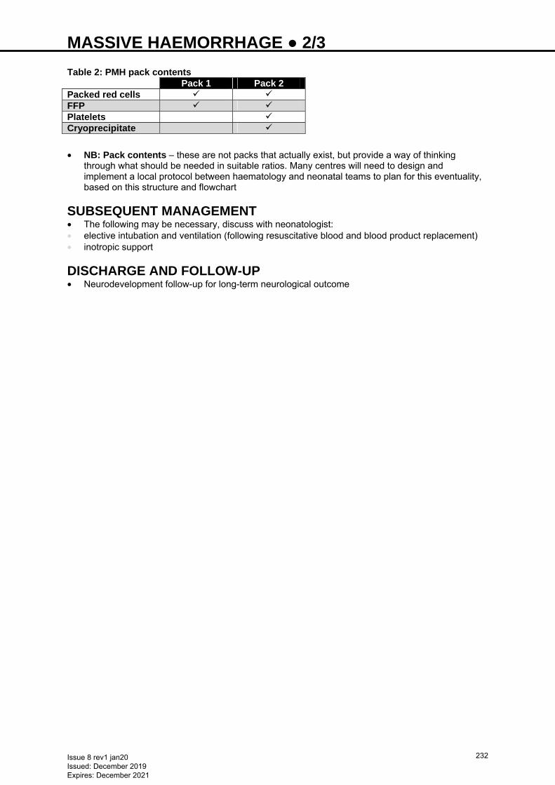
MASSIVE HAEMORRHAGE ● 2/3
Table 2: PMH pack contents Pack 1 Pack 2 Packed red cells FFP Platelets Cryoprecipitate
• NB: Pack contents − these are not packs that actually exist, but provide a way of thinking
through what should be needed in suitable ratios. Many centres will need to design and implement a local protocol between haematology and neonatal teams to plan for this eventuality, based on this structure and flowchart
SUBSEQUENT MANAGEMENT • The following may be necessary, discuss with neonatologist: • elective intubation and ventilation (following resuscitative blood and blood product replacement) • inotropic support
DISCHARGE AND FOLLOW-UP • Neurodevelopment follow-up for long-term neurological outcome
Issue 8 rev1 jan20 Issued: December 2019 Expires: December 2021
232
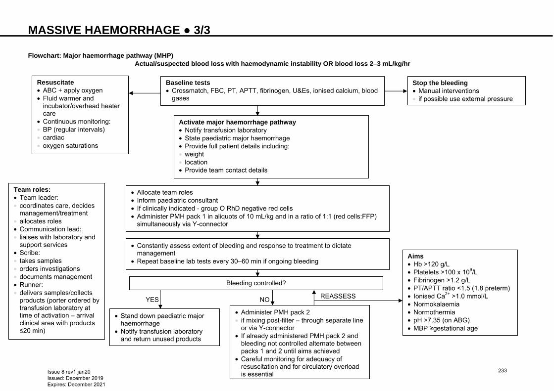
MASSIVE HAEMORRHAGE ● 3/3 Flowchart: Major haemorrhage pathway (MHP)
Actual/suspected blood loss with haemodynamic instability OR blood loss 2−3 mL/kg/hr
NO REASSESS
Baseline tests • Crossmatch, FBC, PT, APTT, fibrinogen, U&Es, ionised calcium, blood
gases
Resuscitate • ABC + apply oxygen • Fluid warmer and
incubator/overhead heater care
• Continuous monitoring: • BP (regular intervals) • cardiac • oxygen saturations
Stop the bleeding • Manual interventions • if possible use external pressure
Activate major haemorrhage pathway • Notify transfusion laboratory • State paediatric major haemorrhage • Provide full patient details including: • weight • location • Provide team contact details
• Allocate team roles • Inform paediatric consultant • If clinically indicated - group O RhD negative red cells • Administer PMH pack 1 in aliquots of 10 mL/kg and in a ratio of 1:1 (red cells:FFP)
simultaneously via Y-connector
• Constantly assess extent of bleeding and response to treatment to dictate management
• Repeat baseline lab tests every 30−60 min if ongoing bleeding
Bleeding controlled?
• Stand down paediatric major haemorrhage
• Notify transfusion laboratory and return unused products
• Administer PMH pack 2 • if mixing post-filter − through separate line
or via Y-connector • If already administered PMH pack 2 and
bleeding not controlled alternate between packs 1 and 2 until aims achieved
• Careful monitoring for adequacy of resuscitation and for circulatory overload is essential
Aims • Hb >120 g/L • Platelets >100 x 109/L • Fibrinogen >1.2 g/L • PT/APTT ratio <1.5 (1.8 preterm) • Ionised Ca2+ >1.0 mmol/L • Normokalaemia • Normothermia • pH >7.35 (on ABG) • MBP ≥gestational age
YES
Team roles: • Team leader: • coordinates care, decides
management/treatment • allocates roles • Communication lead: • liaises with laboratory and
support services • Scribe: • takes samples • orders investigations • documents management • Runner: • delivers samples/collects
products (porter ordered by transfusion laboratory at time of activation – arrival clinical area with products ≤20 min)
Issue 8 rev1 jan20 Issued: December 2019 Expires: December 2021
233
MEDIUM-CHAIN ACYL-COA DEHYDROGENASE DEFICIENCY (MCADD) − EARLY MANAGEMENT OF BABIES WITH FAMILY HISTORY ● 1/2
Based on British Inherited Metabolic Disease Group Protocol
DEFINITION • A rare autosomal recessive inherited metabolic disease where the body cannot metabolise fat properly • With regular intake of food, individuals can lead a normal healthy life but prolonged fasting or illness with
vomiting can lead to encephalopathy, coma or sudden death • Affects 1:10,000 babies in UK. 1:80 healthy people are carriers • Bloodspot screening at day 5 includes MCADD (see Bloodspot screening guideline) • Newborn babies with MCADD are especially vulnerable in first few days of life before breast milk supply
and regular feeding pattern established • Babies with a family history of MCADD require a special feeding regimen and observation from birth
SYMPTOMS • Often non-specific • hypothermia • jitteriness • irritability • drowsiness • reluctance to feed • lethargy • rapid breathing • seizures • coma • sudden death • Hypoglycaemia occurs late
DIAGNOSIS • When mother admitted in labour, inform neonatal team • Test baby aged 24−48 hr • bloodspot acylcarnitines • urine organic acids • DNA mutation analysis (in most cases, genotype will be known for the index case) • Discuss testing with metabolic laboratory at Birmingham Children’s Hospital and mark request ‘family
history of MCADD’ • Continue special feeding regimen until results available
MANAGEMENT • High index of suspicion antenatally • Refer those with family history of MCADD for genetic counselling antenatally • Advise parents baby will require specialist feeding regimen from birth and rapid testing at aged 24–48 hr • Institute specialist feeding regimen from birth • Ensure regular milk intake • term baby: 4-hrly feeds • preterm baby: 3-hrly feeds • Breastfed babies are at particular risk in first 72 hr. Give formula top-ups until good maternal milk supply
established • day 1: 25 mL/kg • day 2: 40 mL/kg • day 3: 60 mL/kg • if baby not taking adequate oral feeds, start nasogastric tube feeding • If enteral feeds not tolerated, commence IV fluid – glucose 10%, sodium chloride 0.18% • Complete bloodspot screening as normal on day 5
PROBLEMS • If baby drowsy or unwell in any way, admit to NNU urgently • give 2 mL/kg glucose 10% as IV bolus, then commence infusion of glucose 10% at 100 mL/kg/day • if no oral intake increase IV infusion to 150 mL/kg/day over 3 days
Issue 8 rev1 jan20 Issued: December 2019 Expires: December 2021
234
MEDIUM-CHAIN ACYL-COA DEHYDROGENASE DEFICIENCY (MCADD) − EARLY MANAGEMENT OF BABIES WITH FAMILY HISTORY ● 2/2
• monitor blood glucose and electrolytes, but base treatment on clinical state as hypoglycaemia occurs late
• Seek advice from specialist metabolic centre
LOCAL CONTACT • For specialist advice, consult Birmingham Children’s Hospital metabolic on-call consultant (0121 333
9999)
FURTHER INFORMATION http://www.bimdg.org.uk/guidelines.asp
Issue 8 rev1 jan20 Issued: December 2019 Expires: December 2021
235
METABOLIC BONE DISEASE ● 1/2
RECOGNITION AND ASSESSMENT Definition • Decreased mineralisation of bones due to deficient phosphate (PO4), calcium (Ca) or vitamin D in
preterm babies • Also known as osteopenia of prematurity Causes • Inadequate postnatal intake or absorption to support intrauterine mineral accretion rate Risk factors • <32 weeks’ gestation • <1500 g birth weight • Male gender • Inadequate nutrition • suboptimal intake • enteral feeds with low mineral content/bioavailability [unfortified expressed breast milk (EBM), term
formula] • Phosphorus deficiency (primary nutritional reason) • Vitamin D deficiency • Prolonged parenteral nutrition • Chronic use of drugs that increase mineral excretion (diuretics, dexamethasone, sodium bicarbonate) • Lack of mechanical stimulation e.g. sedation/paralysis • Bronchopulmonary dysplasia • Cholestatic jaundice • Short gut syndrome (malabsorption of vitamin D and Ca) Symptoms and signs • ≤6 weeks, most babies are asymptomatic and normal on examination • Usually presents aged 6–12 weeks • Poor weight gain or faltering growth • Respiratory difficulties • failure to wean off ventilator due to excessive chest wall compliance • Fractures with minor or no trauma; may manifest as pain on handling • If hypocalcaemic − jitteriness • Craniotabes (softening of skull bones) • Low bone density on X-rays (rachitic changes, cortical thinning, periosteal elevation) Later clinical consequences • Marked dolicocephaly (long and narrow skull) • Myopia of prematurity • Reduced linear growth
INVESTIGATIONS • Measure serum Ca, PO4 and alkaline phosphatase (ALP) levels weekly from third week of life in high-risk
babies • low serum PO4 (<1.8 mmol/L) with elevated ALP (>900 IU/L) is 100% sensitive and 70% specific for
diagnosing low bone mineral density. Low serum PO4 concentrations (<1.8 mmol/L) have 96% specificity but only 50% sensitivity
• serum Ca levels may remain normal until late in the disease • Measure urinary Ca and PO4. Urinary excretion of Ca >1.2 mmol/L and PO4 >0.4 mmol/L signifies slight
surplus of supply and correlates with highest bone mineral accretion rate • phosphaturia can occur due to aminoglycoside, indomethacin and dexamethasone therapy • calciuria can occur due to diuretics, dexamethasone and theophylline • Tubular reabsorption percent (TRP) of PO4 is also a guide to adequacy of PO4 supplementation. TRP of
>95% indicates inadequate supplementation • TRP (%TRP) = [1 − (urine PO4/urine creatinine) (plasma creatinine/plasma PO4)] × 100 • Babies on unfortified human milk are relatively phosphate deficient and have: • normal serum Ca, low serum PO4 and high serum ALP • very low or absent urinary PO4 (urinary Ca excretion increases as serum PO4 concentration decreases) • normal serum vitamin D and parathormone levels
Issue 8 rev1 jan20 Issued: December 2019 Expires: December 2021
236
METABOLIC BONE DISEASE ● 2/2
• Formula fed preterm babies have a low calcium absorption rate and therefore very low urinary Ca and PO4 concentrations
• If biomarkers (ALP and PO4) do not normalise despite appropriate Ca and PO4 supplementation, measure serum 25(OH)D
• X-rays can demonstrate demineralised, thin bones, signs of rickets and thoracic cage and extremity fractures
• Dual-energy X-ray absorptiometry (DXA)/qualitative ultrasound (QUS)
PREVENTION • Optimal nutritional care of preterm babies • initiate early parenteral nutrition with optimised Ca and PO4 content [≥12 mmol/L each of Ca and PO4 (=
1.8 mmol/kg/day of Ca and PO4 at 150 mL/kg/day)] • early enteral feeds • use of breast milk fortifier or preterm formula • Early phosphate supplementation in high-risk babies • Ensure appropriate handling and position using deep boundaries to promote active bone loading
TREATMENT • Ensure adequate intake of Ca (2.5−4.0 mmol/kg/day) and PO4 (1.9−2.9 mmol/kg/day) by using fortified
breast milk or preterm formula • Ensure daily intake of 600−800 units vitamin D per day • If PO4 deficient (<1.8 mmol/L): supplement PO4 at 1−2 mmol/kg/day in divided doses • If Ca deficient (<1.6 mmol/L): supplement Ca at 1−3 mmol/kg/day in divided doses • do not give Ca and PO4 at the same time as they may precipitate; give at alternate feeds • Ca supplementation can cause intestinal obstruction and hypercalcinosis • If no increase in PO4 and ALP continues to rise: • prescribe either vitamin D supplements ≤1000 units/day or initiate ergocalciferol/alphacalcidol therapy
− in addition, encourage daily passive exercises and medication review to consider stopping diuretics and steroids when appropriate
• regular monitoring of urinary Ca/urinary creatinine ratio is necessary to detect hypercalciuria (urinary Ca/urinary creatinine) >0.6
• Consider other nutritional deficiencies e.g. zinc, in a baby with faltering growth with evidence of significant bone disease
MONITORING AND FOLLOW-UP • Weekly monitoring of serum Ca, PO4 and ALP along with urinary Ca and PO4 • Continue treatment until biochemical indices are normal and radiographic evidence of healing, usually
until term corrected gestation
Issue 8 rev1 jan20 Issued: December 2019 Expires: December 2021
237
MULTI DRUG RESISTANT ORGANISM COLONISATION (MRSA, ESBL etc.) ● 1/3
Use this guideline in conjunction with your local Trust policy This guideline describes the screening and follow-up action for the following organisms: • Methicillin-resistant Staphylococcus aureus (MRSA) • Multi-resistant Gram-negative bacilli (MGNB) including: • extended spectrum beta lactamase (ESBL)-producing Enterobacterales • carbapenemase-producing Enterobacterales (CPE) • other carbapenemase-producing multi-drug resistant GNB
SCREENING Babies transferred from other hospitals • Screen on arrival. Include babies who attend other hospitals for invasive day case procedures (e.g. PDA
ligation) • MRSA: • swab nose and perineum plus umbilicus if still moist, and swab any skin lesion (e.g. indwelling vascular
line) • urine if long-term urinary catheter present • MGNB: • obtain rectal swab or swab from stool sample; swab must contain visible faecal material to ensure a
reliable screening result • urine if long-term urinary catheter present • Barrier nurse until all swabs confirmed negative at 48 hr Routine screening on unit • MGNB: monthly • MRSA: monthly • Frequency of screening may be increased on advice of lead infection prevention doctor/ infection
prevention team if unexplained acquisition of MRSA and/or MGNB occurred on ward Infection control alerts • Infection control alert to be triggered for 2 yr after last positive result, irrespective of any negative follow-
up screens • Babies colonised with CPE and other carbapenemase-producing GNB require an infection control alert
to be displayed for 5 yr after the last positive result, irrespective of any negative follow-up screens • Babies identified by infection prevention team as a close contact of a baby with CPE will require a patient
infection control alert up to 5 yr; can be removed when 3 follow-up screens for MGNB, submitted since the creation of the alert, reported as ‘MGNB not isolated’
MANAGEMENT OF INCIDENTAL FINDINGS If new case MRSA reported in patient, offer screening for MRSA: Mother • Screen mother with nasal, perineal, wound and skin lesion swabs, if: • delivery by caesarean section • mother had recent admission to hospital before delivery • mother has chronic health problem (e.g. diabetes mellitus, asthma) • mother has other risk factor, high BMI or is a healthcare worker with patient contact • mother or household member has a history of skin/soft tissue infection abscess or recurrent skin
infections in the last 12 months • If none of these risk factors present, screening contacts is not necessary unless advised by consultant
microbiologist Contacts on NICU (patients only): • Those who have been in close proximity of the index case (i.e. in the same room) • Potentially all babies on the ward following a risk assessment and discussion with consultant of the
week, co-ordinator, infection prevention team and consultant microbiologist • Healthy babies about to be discharged home do not require screening unless advised by consultant
microbiologist
Decolonisation of MRSA carriers • Discharge term healthy babies without decolonisation treatment • Smaller babies with indwelling lines or CPAP probes are more at risk of infection and should be treated
Issue 8 rev1 jan20 Issued: December 2019 Expires: December 2021
238
MULTI DRUG RESISTANT ORGANISM COLONISATION (MRSA, ESBL etc.) ● 2/3 • Decolonisation may fail due to presence of indwelling lines/foreign body material; repeat once all
indwelling lines/foreign bodies removed • mupirocin (Bactroban Nasal®) ointment applied to inner surface of each nostril 3 times daily for 5 days; if
MRSA reported as high level resistant to mupirocin discuss with consultant microbiologist • wash daily with antimicrobial wash, e.g. chlorhexidine or octenidine, for 5 days • Repeat screening swabs 48 hr after all antibiotic treatment has finished and if baby not about to be
discharged • Successful eradication can be assumed if 3 consecutive swabs taken at 3–7 day intervals are negative.
Do not attempt to decolonise more than twice during any 1 admission
MGNB • Do not attempt decolonisation. Do not treat asymptomatic rectal carriage. Colonisation is in the gut.
Drugs are ineffective − may severely damage gut flora and encourage development of resistant organisms
• MGNB: gut carriage not permanent, however may last for several months to years • barrier nurse until discharge
MANAGEMENT OF OUTBREAK MRSA • ≥2 babies with same strain of MRSA constitutes an outbreak • considered ‘the same’ if they have been sent by microbiology to a reference laboratory for typing and
have been reported by reference laboratory as ‘indistinguishable’ Action on advice of infection prevention team • Screen all babies in NNU (swabs as above) • Optimise infection control measures: see local infection control policy • If further cases of the same strain occur: • arrange incident meeting to discuss further measures, e.g. swabs from all clinical staff on unit • If contact screening of clinical staff for MRSA recommended by lead infection prevention
doctor/consultant microbiologist, to be co-ordinated by infection prevention team in collaboration with occupational health (OH)
• results sent to OH and infection prevention team • contact screening for MRSA of healthcare workers must follow local infection prevention guidance MGNB • ≥2 babies with same type of MGNB/CPE constitutes an outbreak • considered ‘the same’ if sent by microbiology to reference laboratory for typing, and reported as
‘indistinguishable’ • For CPE ≥2 babies with the same carbapenemase gene (OXA-48, KPC, VIM, NDM-1 etc.) irrespective
of organism if associated in time and space constitutes an outbreak Action • Screen all babies in NNU on advice of infection prevention team • Optimise infection control measures: see local infection control policy • If further cases of same strain occur arrange incident meeting to discuss further measures e.g.
environmental screening etc. CPE • Screen all contacts (should be alerted on local hospital system) • 3 rectal swabs/swab from stool sample, ≥24 hr apart • if baby on antibiotics: take ≥1 swab >48 hr after stopping antibiotics • if all 3 swabs negative: clear of CPE contact status • if any swab positive, following required:
− strict isolation − long sleeved gowns − gloves − barrier nursing − barrier cleans
• Barrier nurse all colonised babies until discharge • Ensure strict infection prevention measures in place for all babies identified as CPE contacts/with close
contact alert
Issue 8 rev1 jan20 Issued: December 2019 Expires: December 2021
239
MULTI DRUG RESISTANT ORGANISM COLONISATION (MRSA, ESBL etc.) ● 3/3 • If CPE reported during current hospital admission: • strict infection prevention measures to remain in place (irrespective of any negative follow-up screens) • follow-up screens not required • if baby discharged whilst being investigated as contact, follow-up rectal swabs in the community are not
required • if readmitted whilst having a close contact alert, commence/continue follow-up MGNB screening and
repeat on different days until 3 follow-up screens have been reported as ‘MGNB not isolated’ • close contact alert will remain on hospital system for 5 yr, unless 3 follow-up screens reported as ‘MGNB
not isolated’
Issue 8 rev1 jan20 Issued: December 2019 Expires: December 2021
240
MYELOMENINGOCELE (MMC) ● 1/2 DEFINITION • Defect of the backbone and spinal cord • MMC is the most serious type of spina bifida; spinal cord and meninges push out and create a sac in
baby’s back • Associated with significant damage to spinal cord • Can leave nervous system vulnerable to life-threatening infection
MANAGEMENT Antenatal diagnosis • Refer to neurosurgery team • Offer mother appointment with neurosurgeon before the birth Post-delivery Neonatal management in local unit: • Systemic management: see Infection in first 72 hours of life guideline • First line antibiotics: see Infection in first 72 hours of life guideline • Give vitamin K • Nurse prone/lateral, irrespective of gestation and ventilator status • Baseline cranial ultrasound • Occipital frontal circumference (OFC) daily before transfer Specific MMC management • Open MMC • surgical closure recommended in first 24−48 hr • transfer to appropriate surgical unit ≤24 hr (providing condition stable) • if flap closure required neurosurgeon to refer to plastic surgeon • Closed MMC • treat as elective surgery • Protect exposed meninges until surgical closure performed. Immediately after delivery cover lesion with
non-adherent silicone dressing e.g. Mepitel®, followed by sodium chloride soaked gauze. Cover with cling film
• do not place gauze in direct contact with exposed meninges − can cause tearing and leaking of CSF as gauze dries out and sticks to meninges
• if gauze becomes dry, moisten with sodium chloride, keeping Mepitel® in place • if baby nursed in incubator, adequately soak gauze and check 4-hrly • if gauze becomes soiled with faeces or urine, change immediately • nurse baby prone/lateral • do not dress baby − may cause injury to lesion • If evidence of hydrocephalus, cerebral spinal fluid (CSF) diversion will be considered at time of closure • Avoid contact with products containing latex; high risk (25−65%) of developing latex sensitisation and
allergy • complete red allergy band with ‘latex precautions’ and place sign above bed • inform theatres of latex precautions at time of booking • Risk of hydrocephalus, daily monitoring of: • OFC • depth and softness of anterior fontanelle • Document daily on centile chart: • head circumference • weight • Document pre-operative administration of vitamin K and completed screening tests on neonatal checklist
PRE-OPERATIVE INVESTIGATIONS AND MANAGEMENT • Protect lesion from soiling and contamination • Nurse baby prone/lateral • Apply minimal tape to skin due to sensitivity to tapes, and to prevent epidermal stripping • Bloods for: • FBC • U&E • clotting • group and save
Issue 8 rev1 jan20 Issued: December 2019 Expires: December 2021
241
MYELOMENINGOCELE (MMC) ● 2/2 • Ultrasound of renal system • MRI of head and spine at earliest opportunity as baseline (if possible pre-operatively, but do not delay
surgery for imaging) • Consider clinical photography before and after repair • obtain consent at time of consenting for surgery
DISCHARGE • Provide parents with wound care advice • Advise first bath 7−10 days post-operatively (unless advised otherwise) • if no concerns regarding wound, baby bath solution to be used • Neurosurgical clinical nurse specialist to provide information regarding shunt malfunction • if no shunt present, ensure parents made aware of signs and symptoms of hydrocephalus • Liaise with health visiting team to: • arrange regular OFC measurement • share contact information • ensure safe infant sleeping/SIDS guidelines taught • Arrange follow-up appointments • neurosurgery ward clinic: 1 week post-discharge • named consultant clinic: ≤6−8 weeks post-discharge • urology/urodynamics: book before discharge (including ultrasound appointment) • Refer to community paediatrician • Provide parents with contact details of neurosurgical clinical nurse specialist • clinical nurse specialist to provide copy of SHINE charity booklet, with additional team names completed • Detailed discharge summary made and given to parents • Refer to physiotherapy service
Issue 8 rev1 jan20 Issued: December 2019 Expires: December 2021
242
NASOGASTRIC TUBE ADMINISTRATION OF FEED, FLUID OR MEDICATION ● 1/2
Procedure is the same for nasogastric and orogastric tubes. As nasogastric tubes (NGT) are more commonly used in babies, the term nasogastric will be used throughout this guideline
INDICATIONS • Contraindications to oral feeding, or baby unable to take full requirements orally • Nasogastric or orogastric tube in place
EQUIPMENT • Enteral syringes (see NPSA Alert 19 http://www.nrls.npsa.nhs.uk/resources/?entryid45=59808) • pH testing strips • Gravity/bolus feeding set • Feed/fluids/medication according to prescription • Prescription chart (for medication)
PROCEDURE Preparation • See Nasogastric tube insertion guideline • Discuss procedure with parents/carer • Wash hands and prepare equipment • Bring milk to room temperature by removing from fridge. Never deliver fridge-cold milk directly via
nasogastric or orogastric tube (see Nutrition and enteral feeding guideline)
Position of baby for feeding • Baby need not be lying down. It is acceptable to feed if baby receiving kangaroo care or positioned in
baby chair • If lying flat in a cot: • elevate mattress to 30° before feeding and return to flat position within 1 hr
Checking pH • Check pH before every feed/use of tube according to NPSA guidelines (see Nasogastric tube
insertion guideline) • if pH 0–5.5, commence feed and document pH • if pH 5.0−5.5 confirm pH interpretation with a second person before commencing feed • if pH ≥6, do not commence feed. Repeat aspiration and retest • If repeated test ≥6, seek advice from senior clinician and undertake risk assessment following NPSA
algorithm (see Nasogastric tube insertion guideline). Document decision made and rationale • If no aspirate obtained, do not feed. Follow procedure outlined in NPSA guideline
Feeding • Avoid rigid feeding patterns (e.g. 1 bottle/2 tube, alternate bottle/tube etc.) • When handling tubes, ensure clean technique. Pay careful attention to feed preparation and
administration • Administer feed by gravity • Remove plunger, connect to tube, pour small volume of feed into barrel, raise level of barrel above
baby’s stomach. Control speed of administration by raising or lowering barrel • Do not plunge feed • Ensure tube feed takes approximately the same time as a suckling feed e.g.: • 20 min for 3-hrly full feed volume requirement • 10 min for 50% volume • 5 min for 25% volume
Monitoring • Observe baby throughout feed for signs of deterioration or distress (change in colour, cyanosis, apnoea,
bradycardia, vomiting, straining, squirming, grimacing and other avoidance behaviour) • Observe for abdominal distension following a feed • If appropriate developmental stage/capabilities, offer small drops of milk to mouth to taste, but avoid in
babies with no swallow mechanism • Consider offering baby mother’s breast for nuzzling or non-nutritive sucking during tube feed [see Non-
nutritive sucking (NNS) guideline] • On completion of feed, instil small amount of air into tube (0.5–1 mL)
Issue 8 rev1 jan20 Issued: December 2019 Expires: December 2021
243
NASOGASTRIC TUBE ADMINISTRATION OF FEED, FLUID OR MEDICATION ● 2/2
DOCUMENTATION • Document feed details: • pH • type • volume • time • behaviour/response during feed • adverse reactions (vomiting etc.) • Ensure medication chart is signed
FURTHER MANAGEMENT • For administration of medication, remember to check baby identity and prescription. Follow Trust policy
for administration of medicines and British Association of Parenteral and Enteral Nutrition (BAPEN) guidance
• Flushing of NGT is not routine in babies. To avoid medication remaining in NGT try to give medications pre-feed. Where this is not possible 1 mL of feed can be used to flush tube after inserting medication
FURTHER INFORMATION • Nasogastric tube insertion guideline • Further details available from www.npsa.nhs.uk/nrls/alerts-and-directives/alerts/feedingtubes
Issue 8 rev1 jan20 Issued: December 2019 Expires: December 2021
244
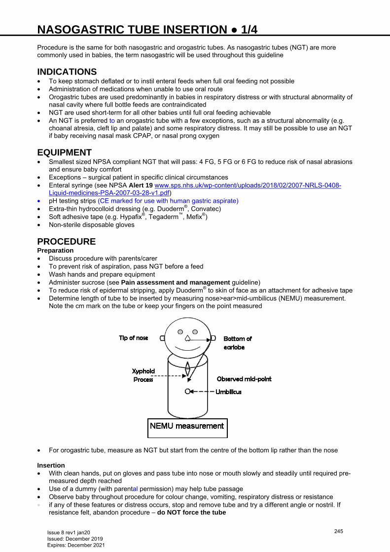
NASOGASTRIC TUBE INSERTION ● 1/4 Procedure is the same for both nasogastric and orogastric tubes. As nasogastric tubes (NGT) are more commonly used in babies, the term nasogastric will be used throughout this guideline
INDICATIONS • To keep stomach deflated or to instil enteral feeds when full oral feeding not possible • Administration of medications when unable to use oral route • Orogastric tubes are used predominantly in babies in respiratory distress or with structural abnormality of
nasal cavity where full bottle feeds are contraindicated • NGT are used short-term for all other babies until full oral feeding achievable • An NGT is preferred to an orogastric tube with a few exceptions, such as a structural abnormality (e.g.
choanal atresia, cleft lip and palate) and some respiratory distress. It may still be possible to use an NGT if baby receiving nasal mask CPAP, or nasal prong oxygen
EQUIPMENT • Smallest sized NPSA compliant NGT that will pass: 4 FG, 5 FG or 6 FG to reduce risk of nasal abrasions
and ensure baby comfort • Exceptions – surgical patient in specific clinical circumstances • Enteral syringe (see NPSA Alert 19 www.sps.nhs.uk/wp-content/uploads/2018/02/2007-NRLS-0408-
Liquid-medicines-PSA-2007-03-28-v1.pdf) • pH testing strips (CE marked for use with human gastric aspirate) • Extra-thin hydrocolloid dressing (e.g. Duoderm®, Convatec) • Soft adhesive tape (e.g. Hypafix®, Tegaderm™, Mefix®) • Non-sterile disposable gloves
PROCEDURE Preparation • Discuss procedure with parents/carer • To prevent risk of aspiration, pass NGT before a feed • Wash hands and prepare equipment • Administer sucrose (see Pain assessment and management guideline) • To reduce risk of epidermal stripping, apply Duoderm® to skin of face as an attachment for adhesive tape • Determine length of tube to be inserted by measuring nose>ear>mid-umbilicus (NEMU) measurement.
Note the cm mark on the tube or keep your fingers on the point measured
• For orogastric tube, measure as NGT but start from the centre of the bottom lip rather than the nose Insertion • With clean hands, put on gloves and pass tube into nose or mouth slowly and steadily until required pre-
measured depth reached • Use of a dummy (with parental permission) may help tube passage • Observe baby throughout procedure for colour change, vomiting, respiratory distress or resistance • if any of these features or distress occurs, stop and remove tube and try a different angle or nostril. If
resistance felt, abandon procedure – do NOT force the tube
Issue 8 rev1 jan20 Issued: December 2019 Expires: December 2021
245
NASOGASTRIC TUBE INSERTION ● 2/4 Checking position of nasogastric feeding tube • NNUs and carers in the community should use pH indicator strips or paper • Do not use radiography ‛routinely’ but, if baby being X-rayed for another reason, use X-ray to confirm
position is satisfactory by noting position of tube on film • Do not use ‛Whoosh test’ (auscultation of injected air entering the stomach) to determine position of
NGT as it is unreliable Checking position using pH • Aspirate stomach contents with enteral syringe and test for acid response using pH testing strips • pH ≤5.5 indicates correct gastric placement. If pH 5 or 5.5, confirm pH interpretation with a second
person before commencing feed • if pH ≥6, do not commence feed. Repeat aspiration and retest • if repeated test ≥6, seek advice from senior clinician and undertake risk assessment • Following factors can contribute to high gastric pH ≥6 • presence of amniotic fluid in baby <48 hr • milk in baby’s stomach, particularly if 1–2 hrly feeds • use of medication to reduce stomach acid • tube positioned in jejunum or duodenum • tube positioned in lungs • Multidisciplinary care team to then discuss possible actions, balancing risk of feeding (with possibility of
tube being in the lungs) and not feeding baby in the short-term, and record how decision reached • Ensure you work through NPSA flowchart and record findings before making any decisions
Issue 8 rev1 jan20 Issued: December 2019 Expires: December 2021
246
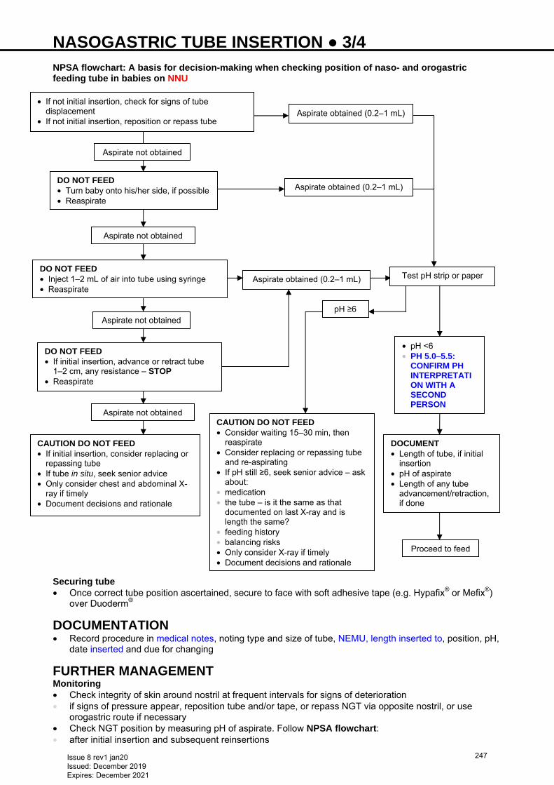
NASOGASTRIC TUBE INSERTION ● 3/4 NPSA flowchart: A basis for decision-making when checking position of naso- and orogastric feeding tube in babies on NNU
Securing tube • Once correct tube position ascertained, secure to face with soft adhesive tape (e.g. Hypafix® or Mefix®)
over Duoderm®
DOCUMENTATION • Record procedure in medical notes, noting type and size of tube, NEMU, length inserted to, position, pH,
date inserted and due for changing
FURTHER MANAGEMENT Monitoring • Check integrity of skin around nostril at frequent intervals for signs of deterioration • if signs of pressure appear, reposition tube and/or tape, or repass NGT via opposite nostril, or use
orogastric route if necessary • Check NGT position by measuring pH of aspirate. Follow NPSA flowchart: • after initial insertion and subsequent reinsertions
• If not initial insertion, check for signs of tube displacement
• If not initial insertion, reposition or repass tube Aspirate obtained (0.2–1 mL)
Aspirate not obtained
DO NOT FEED • Turn baby onto his/her side, if possible • Reaspirate
Aspirate not obtained
DO NOT FEED • Inject 1–2 mL of air into tube using syringe • Reaspirate
Aspirate not obtained
DO NOT FEED • If initial insertion, advance or retract tube
1–2 cm, any resistance – STOP • Reaspirate
Aspirate obtained (0.2–1 mL)
Aspirate obtained (0.2–1 mL) Test pH strip or paper
pH ≥6
Aspirate not obtained
CAUTION DO NOT FEED • If initial insertion, consider replacing or
repassing tube • If tube in situ, seek senior advice • Only consider chest and abdominal X-
ray if timely • Document decisions and rationale
CAUTION DO NOT FEED • Consider waiting 15–30 min, then
reaspirate • Consider replacing or repassing tube
and re-aspirating • If pH still ≥6, seek senior advice – ask
about: • medication • the tube – is it the same as that
documented on last X-ray and is length the same?
• feeding history • balancing risks • Only consider X-ray if timely • Document decisions and rationale
• pH <6 • PH 5.0−5.5:
CONFIRM PH INTERPRETATION WITH A SECOND PERSON
DOCUMENT • Length of tube, if initial
insertion • pH of aspirate • Length of any tube
advancement/retraction, if done
Proceed to feed
Issue 8 rev1 jan20 Issued: December 2019 Expires: December 2021
247

NUTRITION AND ENTERAL FEEDING ● 8/8 • <2 kg preterm formula • ≥2 kg high energy term formula • Breastfeeding/MEBM: use BMF as a concentrated solution (known as fortified breast milk supplement,
give 1 sachet dissolved in 3 mL MEBM via syringe/teat before 4 breastfeeds, equally spread throughout 24 hr period. Reduce BMF by 1 sachet/day every 2 weeks until 6 weeks post-term or 3.5 kg, whichever soonest, then stop fortified breast milk supplements
• Refer to neonatal/paediatric dietitian for assessment and advice
PROGRESSION TO ORAL FEEDING Aim Safe progression to oral feeding (see Progression to oral feeding in preterm infants guideline) Exclusive breast milk feeding • Encourage modified responsive breast/bottle feeding of MEBM Exclusive/partial formula feeding • Encourage modified responsive bottle feeding of post-discharge formula or term formula • Infants born <34 weeks, prescribe post-discharge preterm formula ≤6 months’ CGA (if required)
Department of Health Guidelines state all children aged 6 months−5 yr receive vitamin supplementation containing vitamins A C D unless receiving formula milk >500 mL/day
Exclusively breastfed infants should receive vitamin D supplementation from birth
Issue 8 rev1 jan20 Issued: December 2019 Expires: December 2021
248
NECROTISING ENTEROCOLITIS (NEC) ● 1/3 RECOGNITION AND ASSESSMENT Definition Acute inflammatory disease in newborn intestine characterised by haemorrhagic necrosis, which may lead to perforation and destruction of the gut. Clinical presentation usually comprises triad of abdominal distension, gastrointestinal bleeding and pneumatosis intestinalis (air in bowel wall on abdominal X-ray) Modified Bell’s criteria Stage 1: Suspected NEC − clinical signs suggestive but X-ray non-diagnostic • Systemic signs: • temperature instability • apnoea • bradycardia • lethargy • Intestinal signs: • increased gastric residuals • abdominal distension • vomiting • blood in stools • Radiological signs: • normal/mild intestinal dilatation • thickened bowel loops Stage 2: Definite NEC − mild-to-moderately ill, abdominal X-ray demonstrates pneumatosis intestinalis and/or gas in biliary tract • Systemic signs: see Stage 1 +/- mild metabolic acidosis, mild thrombocytopenia, raised CRP • Intestinal signs: see Stage 1 + absent bowel sounds, +/- localised abdominal tenderness, abdominal
cellulitis or right lower quadrant mass, bright red blood and/or mucus from rectum (exclude local pathology)
• Radiological signs: significant intestinal dilatation, pneumatosis intestinalis, portal vein gas, +/- ascites, persistently abnormal gas pattern (e.g. localised dilated loop of bowel seen on serial X-rays or gasless abdomen)
Stage 3: Advanced NEC − severely ill, bowel intact or perforated • Systemic signs: see Stage 2 + hypotension, bradycardia, severe apnoea, combined respiratory and
metabolic acidosis, DIC, neutropenia • Intestinal signs: see Stage 2 + signs of generalised peritonitis, marked tenderness, distension of
abdomen • Radiological signs: see Stage 2 + pneumoperitoneum +/- ascites Risk factors • Prematurity • Intrauterine growth restriction • Absent or reversed end-diastolic flow on umbilical arterial Doppler antenatally • Perinatal asphyxia • Low systemic blood flow during neonatal period (including duct-dependent congenital heart disease) • Significant patent ductus arteriosus • Exchange transfusion • Formula milk • No antenatal corticosteroids • Infections with: Klebsiella, Enterobacter, anaerobes Differential diagnosis • Sepsis with ileus • Bowel obstruction • Volvulus • Malrotation • Spontaneous intestinal perforation: • associated with early postnatal corticosteroids or indomethacin • abdominal X-ray demonstrates pneumoperitoneum but does not show evidence of pneumatosis
intestinalis
Issue 8 rev1 jan20 Issued: December 2019 Expires: December 2021
249
NECROTISING ENTEROCOLITIS (NEC) ● 2/3 • Systemic candidiasis: • clinical signs can mimic NEC with abdominal distension, metabolic disturbances, hypotension and
thrombocytopenia • Food protein-induced enterocolitis syndrome (FPIES) • usually preceded by thrombocytosis in association with formula milk • take thorough feeding history, and establish any temporal relationships with type of feed
INVESTIGATIONS Abdominal X-ray • Supine antero-posterior view • If perforation suspected but not clear on supine view, left lateral view
Not all babies will have radiological findings associated with NEC (Stage 1) Blood tests • FBC: anaemia, neutropenia and thrombocytopenia often present; early return to normal carries good
prognosis • Blood film: evidence of haemolysis and toxic changes (e.g. spherocytes, vacuolation and toxic
granulation of neutrophils, cell fragments, polychromatic cells) • CRP, but a normal value not informative in initial phase • U&E • Blood gas: evidence of metabolic acidosis (base deficit worse than -10), raised lactate • Coagulation screen • Blood cultures
IMMEDIATE TREATMENT
Always discuss management with consultant neonatologist
In all stages • Nil-by-mouth • Transfer baby to neonatal intensive care and nurse in incubator • If respiratory failure and worsening acidosis, intubate and ventilate • Gastric decompression • Free drainage with large NGT (size 8) • NEC often associated with significant third space fluid loss into peritoneum • Triple antibiotics: flucloxacillin, gentamicin and metronidazole • IV fluids/PN: total volume ≤150 mL/kg • Long line when stable and bacteraemia/septicaemia excluded • Pain relief, consider morphine/diamorphine infusion (see Pain assessment and management
guideline) Stage 2: Proven NEC (confirmed radiologically) • If breathing supported by nasal CPAP, elective intubation to provide bowel decompression (see
Intubation guideline) • Give IV fluid resuscitation sodium chloride 0.9% 10 mL/kg for shock and repeat as necessary. Shock is
most common cause of hypotension in babies with NEC (see Hypotension guideline) • If coagulation abnormal, give FFP (see Coagulopathy guideline) • If thrombocytopenia and/or anaemia occur, transfuse (see Thrombocytopenia guideline) • Discuss with surgical team: may need transfer to surgical centre
Stage 3 : Advanced NEC (fulminant NEC with/without intestinal perforation) • Treat as for Stage 2 and refer to surgical team: may need laparotomy or resection of bowel in surgical
centre
SUBSEQUENT MANAGEMENT In recovery phase • In Stage 1: if improvement after 48 hr, consider restarting feeds slowly (see Nutrition and enteral
feeding guideline) and stopping antibiotics • Take into account type of milk in the context of baby’s feeding history before episode
Issue 8 rev1 jan20 Issued: December 2019 Expires: December 2021
250
NECROTISING ENTEROCOLITIS (NEC) ● 3/3 • In Stage 2: if abdominal examination normal after 7–10 days, consider restarting feeds • some may need longer period of total gut rest • stop antibiotics after 7–10 days • In Stage 3: discuss with surgeon and dietitian before restarting feeds Late complications • Recurrence (in about 10%) • Strictures (in about 10% non-surgical cases) • Short bowel syndrome and problems related to gut resection • Neurodevelopmental delay
MONITORING TREATMENT • Observe general condition closely and review at least 12-hrly • Daily: • acid-base status • fluid balance (twice daily if condition unstable) • electrolytes (twice daily if condition unstable) • FBC and coagulation (twice daily if condition unstable) • repeat X-ray daily or twice daily until condition stable. Discuss with consultant/surgeon
LONG-TERM MANAGEMENT • Advise parents about signs of bowel obstruction • Medical +/- surgical follow-up after discharge • Contrast studies if clinically indicated for strictures • Appropriate developmental follow-up
PARENT INFORMATION Offer parents information on NEC, available from https://www.bliss.org.uk/parents/about-your-baby/medical-conditions/necrotising-enterocolitis-nec-a-guide-for-parents
Issue 8 rev1 jan20 Issued: December 2019 Expires: December 2021
251

NITRIC OXIDE ● 1/2
INDICATIONS • Persistent pulmonary hypertension of the newborn in term and near term (>34 weeks) babies, proven on
clinical grounds or by echocardiography [see Persistent pulmonary hypertension of the newborn (PPHN) guideline]
• Oxygen index >20 • Initiate treatment with nitric oxide (NO) only after discussion with on-call consultant • Babies requiring NO should be referred to a NICU for ongoing management, in accordance with Toolkit
principles
CAUTIONS • Preterm baby • not effective as rescue therapy for very ill preterm babies (Cochrane review 2017) • early routine use does not prevent serious brain injury or survival without BPD (Cochrane review 2017) • may be beneficial in preterm babies with preterm prolonged rupture of membranes – discuss with
consultant • Grade 4 intraventricular haemorrhage (IVH) • Recent pulmonary haemorrhage • Platelets <50 × 109/L
CONTRAINDICATIONS • Congenital heart disease (especially circulations dependent on right-to-left shunting)
DOSE AND ADMINISTRATION Starting NO Preparation • Ensure ventilation optimal and that other aspects of the Persistent pulmonary hypertension of the
newborn (PPHN) guideline have been followed • A sustained inflation immediately before starting NO can enhance response
Administration • Document FiO2 and SpO2 immediately before starting NO • Start NO at 10 ppm • If no response (see below), increase to maximum of 20 ppm • If still no response at 20 ppm, discontinue after trial of 1 hr • NO can be stopped abruptly without weaning if given for <4 hr • Once responding, wean to 5 ppm as soon as possible, and within 2–24 hr of starting treatment Definition of response to NO • Either increase in postductal SpO2 >20%, or increase in postductal PaO2 >3 kPa occurring within 15 min
of starting NO and while ventilator settings constant • Approximately 30% of babies with PPHN do not respond to NO
Weaning • If NO has been administered for ≥4 hr, wean gradually to prevent rebound • in ‘responders’, once FiO2 <0.5, attempt to reduce dose • reduce NO to 5 ppm in decrements of 5 ppm every 1–2 hr. Then reduce by 1 ppm every 1–2 hr, and finally
to 0.5 ppm for ≥1 hr before stopping. Reverse any reduction that causes SpO2 to drop persistently by >5% • some babies will require low dose (<0.5 ppm) for some time (up to 24 hr) during weaning
− may be necessary to temporarily increase FiO2 by 0.1−0.2 to facilitate weaning • If sustained and significant fall in SpO2 occurs following reduction in dosage, increase dosage to previous
level and continue to wean at half previous rate • Once discontinued, wait ≥6 hr before removing NO circuit from ventilator
MONITORING • Use SpO2 to monitor response • Blood gases 4-hrly • Monitor methaemoglobin before starting NO, 1 hr after starting and then 12-hrly. Maximum proportion of
total haemoglobin is reached after 8 hr • normal <1% • 2–3% is acceptable
Issue 8 rev1 jan20 Issued: December 2019 Expires: December 2021
252
NITRIC OXIDE ● 2/2
• 4% requires action: reduce NO and repeat in 1 hr − if still >4%, stop NO − if >6%, treat with methylthioninium chloride (methylene blue) 1 mg/kg IV over 1 hr
• NO inhibits platelet function and can trigger bleeding if baby has bleeding problem or thrombocytopenia. Check FBC daily while baby receiving NO
Issue 8 rev1 jan20 Issued: December 2019 Expires: December 2021
253
NON-NUTRITIVE SUCKING (NNS) ● 1/1 DEFINITION • Includes: • sucking of fingers • use of dummies with/without sucrose
INDICATIONS • Actively promoted for: • comfort • pain relief • maximising nasal CPAP delivery. Can be used for short period to assist in acquisition of an effective seal • developing the sucking reflex and assisting transition from tube to full breast or bottle feeding • normal peristalsis helping to alleviate gastro-oesophageal reflux • Encourage preterm babies not mature enough to suck at feed times to suck on a non-nutritive device
during a tube feed • Form of non-pharmacological pain relief during painful procedures • Decreases: • stress • risk of SIDS (with appropriate sleeping positions)
CAUTIONS • As baby begins to take more enteral feeds (at around 33 weeks), NNS no longer appropriate as may
mask feeding cues
CONSENT • Before commencing, ensure parents receive written information on suitable use of NNS on NNU • A signed informed consent form must be held in baby’s medical record
Issue 8 rev1 jan20 Issued: December 2019 Expires: December 2021
254
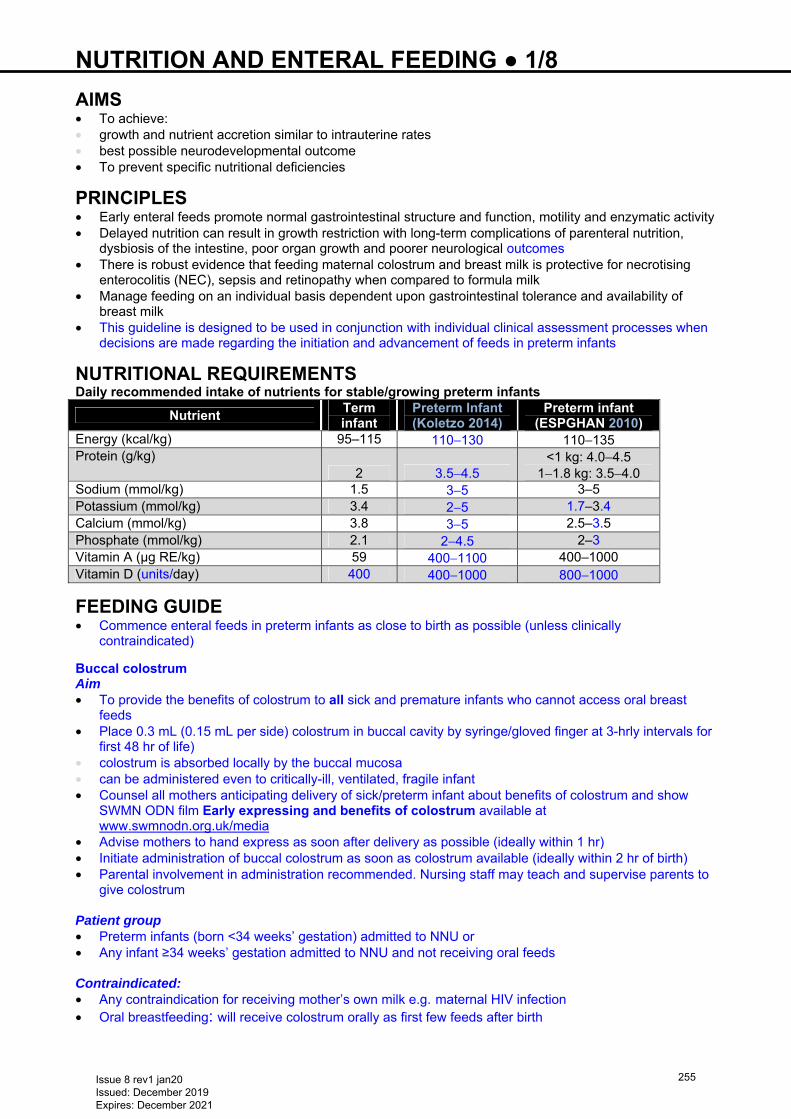
NUTRITION AND ENTERAL FEEDING ● 1/8 AIMS • To achieve: • growth and nutrient accretion similar to intrauterine rates • best possible neurodevelopmental outcome • To prevent specific nutritional deficiencies
PRINCIPLES • Early enteral feeds promote normal gastrointestinal structure and function, motility and enzymatic activity • Delayed nutrition can result in growth restriction with long-term complications of parenteral nutrition,
dysbiosis of the intestine, poor organ growth and poorer neurological outcomes • There is robust evidence that feeding maternal colostrum and breast milk is protective for necrotising
enterocolitis (NEC), sepsis and retinopathy when compared to formula milk • Manage feeding on an individual basis dependent upon gastrointestinal tolerance and availability of
breast milk • This guideline is designed to be used in conjunction with individual clinical assessment processes when
decisions are made regarding the initiation and advancement of feeds in preterm infants
NUTRITIONAL REQUIREMENTS Daily recommended intake of nutrients for stable/growing preterm infants
Nutrient Term infant
Preterm Infant (Koletzo 2014)
Preterm infant (ESPGHAN 2010)
Energy (kcal/kg) 95–115 110−130 110−135 Protein (g/kg)
2 3.5−4.5 <1 kg: 4.0−4.5
1−1.8 kg: 3.5−4.0 Sodium (mmol/kg) 1.5 3−5 3–5 Potassium (mmol/kg) 3.4 2−5 1.7–3.4 Calcium (mmol/kg) 3.8 3−5 2.5–3.5 Phosphate (mmol/kg) 2.1 2−4.5 2–3 Vitamin A (µg RE/kg) 59 400−1100 400–1000 Vitamin D (units/day) 400 400−1000 800−1000
FEEDING GUIDE • Commence enteral feeds in preterm infants as close to birth as possible (unless clinically
contraindicated)
Buccal colostrum Aim • To provide the benefits of colostrum to all sick and premature infants who cannot access oral breast
feeds • Place 0.3 mL (0.15 mL per side) colostrum in buccal cavity by syringe/gloved finger at 3-hrly intervals for
first 48 hr of life) • colostrum is absorbed locally by the buccal mucosa • can be administered even to critically-ill, ventilated, fragile infant • Counsel all mothers anticipating delivery of sick/preterm infant about benefits of colostrum and show
SWMN ODN film Early expressing and benefits of colostrum available at www.swmnodn.org.uk/media
• Advise mothers to hand express as soon after delivery as possible (ideally within 1 hr) • Initiate administration of buccal colostrum as soon as colostrum available (ideally within 2 hr of birth) • Parental involvement in administration recommended. Nursing staff may teach and supervise parents to
give colostrum Patient group • Preterm infants (born <34 weeks’ gestation) admitted to NNU or • Any infant ≥34 weeks’ gestation admitted to NNU and not receiving oral feeds Contraindicated: • Any contraindication for receiving mother’s own milk e.g. maternal HIV infection • Oral breastfeeding: will receive colostrum orally as first few feeds after birth
Issue 8 rev1 jan20 Issued: December 2019 Expires: December 2021
255
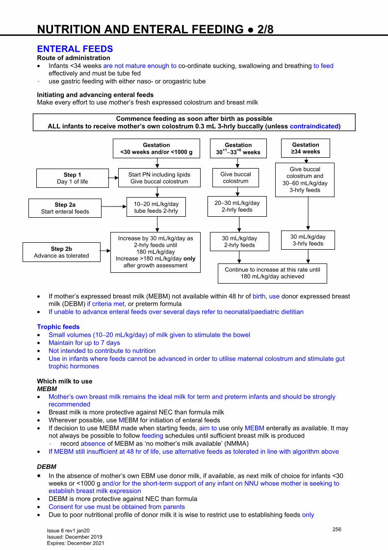
NUTRITION AND ENTERAL FEEDING ● 2/8 ENTERAL FEEDS Route of administration • Infants <34 weeks are not mature enough to co-ordinate sucking, swallowing and breathing to feed
effectively and must be tube fed • use gastric feeding with either naso- or orogastric tube Initiating and advancing enteral feeds Make every effort to use mother’s fresh expressed colostrum and breast milk
Commence feeding as soon after birth as possible ALL infants to receive mother’s own colostrum 0.3 mL 3-hrly buccally (unless contraindicated)
• If mother’s expressed breast milk (MEBM) not available within 48 hr of birth, use donor expressed breast
milk (DEBM) if criteria met, or preterm formula • If unable to advance enteral feeds over several days refer to neonatal/paediatric dietitian Trophic feeds • Small volumes (10−20 mL/kg/day) of milk given to stimulate the bowel • Maintain for up to 7 days • Not intended to contribute to nutrition • Use in infants where feeds cannot be advanced in order to utilise maternal colostrum and stimulate gut
trophic hormones Which milk to use MEBM • Mother’s own breast milk remains the ideal milk for term and preterm infants and should be strongly
recommended • Breast milk is more protective against NEC than formula milk • Wherever possible, use MEBM for initiation of enteral feeds • If decision to use MEBM made when starting feeds, aim to use only MEBM enterally as available. It may
not always be possible to follow feeding schedules until sufficient breast milk is produced • record absence of MEBM as ‘no mother’s milk available’ (NMMA)
• If MEBM still insufficient at 48 hr of life, use alternative feeds as tolerated in line with algorithm above DEBM
• In the absence of mother’s own EBM use donor milk, if available, as next milk of choice for infants <30 weeks or <1000 g and/or for the short-term support of any infant on NNU whose mother is seeking to establish breast milk expression
• DEBM is more protective against NEC than formula • Consent for use must be obtained from parents • Due to poor nutritional profile of donor milk it is wise to restrict use to establishing feeds only
Step 1 Day 1 of life
Step 2a Start enteral feeds
Step 2b Advance as tolerated
Gestation <30 weeks and/or <1000 g
Gestation 30+1−33+6 weeks
Gestation ≥34 weeks
Start PN including lipids Give buccal colostrum
Give buccal colostrum
Give buccal colostrum and
30−60 mL/kg/day 3-hrly feeds
10−20 mL/kg/day tube feeds 2-hrly
20−30 mL/kg/day 2-hrly feeds
Increase by 30 mL/kg/day as 2-hrly feeds until 180 mL/kg/day
Increase >180 mL/kg/day only after growth assessment
30 mL/kg/day 2-hrly feeds
30 mL/kg/day 3-hrly feeds
Continue to increase at this rate until 180 mL/kg/day achieved
Issue 8 rev1 jan20 Issued: December 2019 Expires: December 2021
256
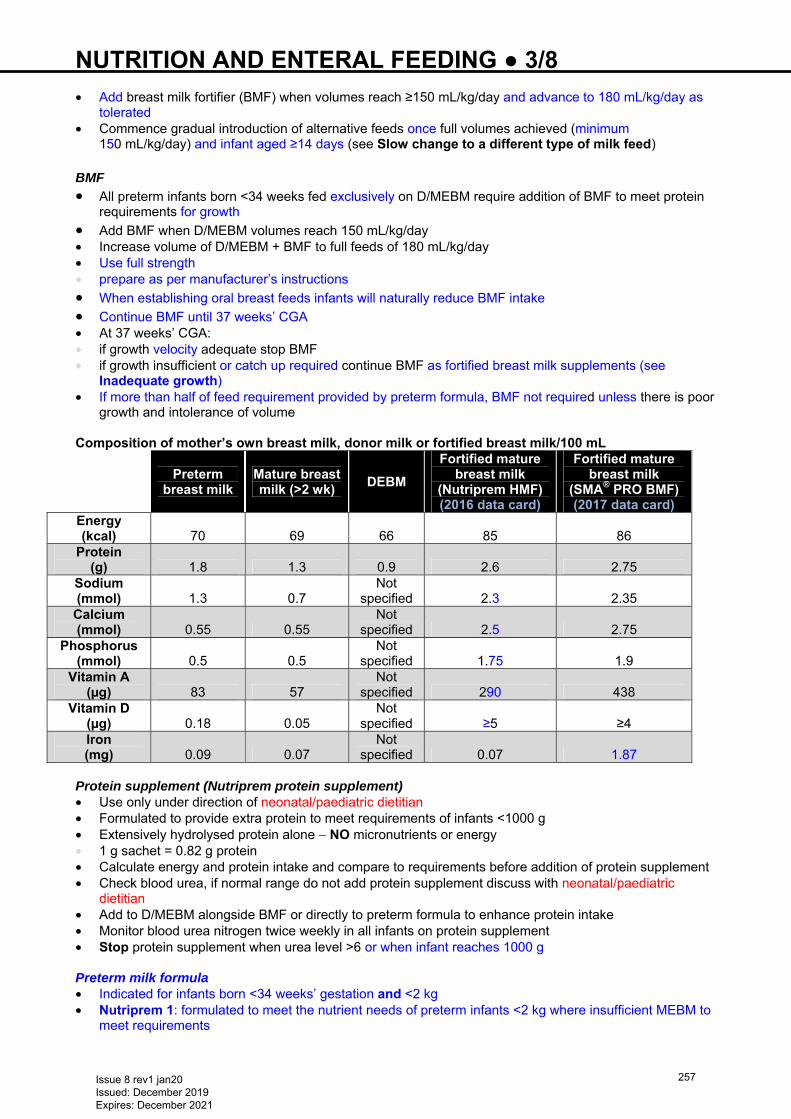
NUTRITION AND ENTERAL FEEDING ● 3/8 • Add breast milk fortifier (BMF) when volumes reach ≥150 mL/kg/day and advance to 180 mL/kg/day as
tolerated • Commence gradual introduction of alternative feeds once full volumes achieved (minimum
150 mL/kg/day) and infant aged ≥14 days (see Slow change to a different type of milk feed) BMF
• All preterm infants born <34 weeks fed exclusively on D/MEBM require addition of BMF to meet protein requirements for growth
• Add BMF when D/MEBM volumes reach 150 mL/kg/day • Increase volume of D/MEBM + BMF to full feeds of 180 mL/kg/day • Use full strength • prepare as per manufacturer’s instructions
• When establishing oral breast feeds infants will naturally reduce BMF intake • Continue BMF until 37 weeks’ CGA • At 37 weeks’ CGA: • if growth velocity adequate stop BMF • if growth insufficient or catch up required continue BMF as fortified breast milk supplements (see
Inadequate growth) • If more than half of feed requirement provided by preterm formula, BMF not required unless there is poor
growth and intolerance of volume Composition of mother’s own breast milk, donor milk or fortified breast milk/100 mL
Preterm
breast milk Mature breast milk (>2 wk)
DEBM
Fortified mature breast milk
(Nutriprem HMF) (2016 data card)
Fortified mature breast milk
(SMA® PRO BMF) (2017 data card)
Energy (kcal) 70 69 66 85 86
Protein (g) 1.8 1.3 0.9 2.6 2.75
Sodium (mmol) 1.3 0.7
Not specified 2.3 2.35
Calcium (mmol) 0.55 0.55
Not specified 2.5 2.75
Phosphorus (mmol) 0.5 0.5
Not specified 1.75 1.9
Vitamin A (µg) 83 57
Not specified 290 438
Vitamin D (µg) 0.18 0.05
Not specified ≥5 ≥4
Iron (mg) 0.09 0.07
Not specified 0.07 1.87
Protein supplement (Nutriprem protein supplement) • Use only under direction of neonatal/paediatric dietitian • Formulated to provide extra protein to meet requirements of infants <1000 g • Extensively hydrolysed protein alone − NO micronutrients or energy • 1 g sachet = 0.82 g protein • Calculate energy and protein intake and compare to requirements before addition of protein supplement • Check blood urea, if normal range do not add protein supplement discuss with neonatal/paediatric
dietitian • Add to D/MEBM alongside BMF or directly to preterm formula to enhance protein intake • Monitor blood urea nitrogen twice weekly in all infants on protein supplement • Stop protein supplement when urea level >6 or when infant reaches 1000 g Preterm milk formula • Indicated for infants born <34 weeks’ gestation and <2 kg • Nutriprem 1: formulated to meet the nutrient needs of preterm infants <2 kg where insufficient MEBM to
meet requirements
Issue 8 rev1 jan20 Issued: December 2019 Expires: December 2021
257
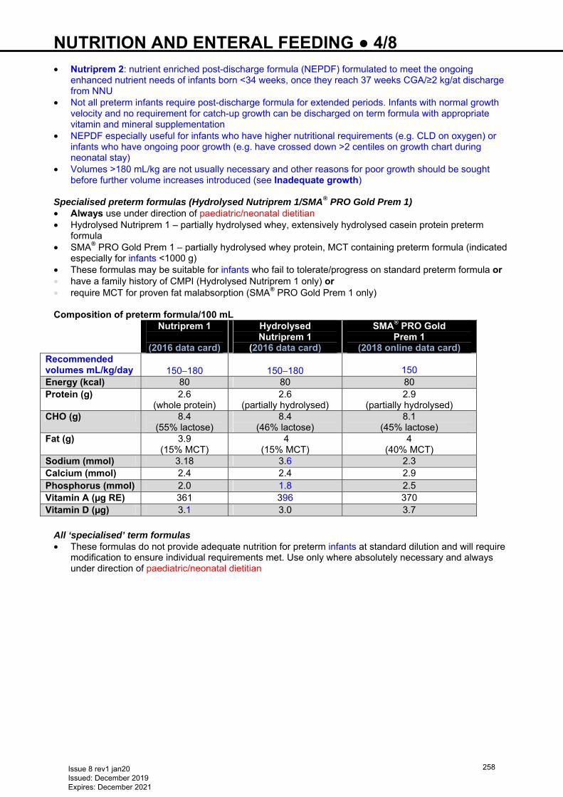
NUTRITION AND ENTERAL FEEDING ● 4/8 • Nutriprem 2: nutrient enriched post-discharge formula (NEPDF) formulated to meet the ongoing
enhanced nutrient needs of infants born <34 weeks, once they reach 37 weeks CGA/≥2 kg/at discharge from NNU
• Not all preterm infants require post-discharge formula for extended periods. Infants with normal growth velocity and no requirement for catch-up growth can be discharged on term formula with appropriate vitamin and mineral supplementation
• NEPDF especially useful for infants who have higher nutritional requirements (e.g. CLD on oxygen) or infants who have ongoing poor growth (e.g. have crossed down >2 centiles on growth chart during neonatal stay)
• Volumes >180 mL/kg are not usually necessary and other reasons for poor growth should be sought before further volume increases introduced (see Inadequate growth)
Specialised preterm formulas (Hydrolysed Nutriprem 1/SMA® PRO Gold Prem 1) • Always use under direction of paediatric/neonatal dietitian • Hydrolysed Nutriprem 1 – partially hydrolysed whey, extensively hydrolysed casein protein preterm
formula • SMA® PRO Gold Prem 1 – partially hydrolysed whey protein, MCT containing preterm formula (indicated
especially for infants <1000 g) • These formulas may be suitable for infants who fail to tolerate/progress on standard preterm formula or • have a family history of CMPI (Hydrolysed Nutriprem 1 only) or • require MCT for proven fat malabsorption (SMA® PRO Gold Prem 1 only) Composition of preterm formula/100 mL
Nutriprem 1
(2016 data card)
Hydrolysed Nutriprem 1
(2016 data card)
SMA® PRO Gold Prem 1
(2018 online data card) Recommended volumes mL/kg/day
150−180
150−180
150
Energy (kcal) 80 80 80 Protein (g) 2.6
(whole protein) 2.6
(partially hydrolysed) 2.9
(partially hydrolysed) CHO (g) 8.4
(55% lactose) 8.4
(46% lactose) 8.1
(45% lactose) Fat (g) 3.9
(15% MCT) 4
(15% MCT) 4
(40% MCT) Sodium (mmol) 3.18 3.6 2.3 Calcium (mmol) 2.4 2.4 2.9 Phosphorus (mmol) 2.0 1.8 2.5 Vitamin A (µg RE) 361 396 370 Vitamin D (µg) 3.1 3.0 3.7
All ‘specialised’ term formulas • These formulas do not provide adequate nutrition for preterm infants at standard dilution and will require
modification to ensure individual requirements met. Use only where absolutely necessary and always under direction of paediatric/neonatal dietitian
Issue 8 rev1 jan20 Issued: December 2019 Expires: December 2021
258
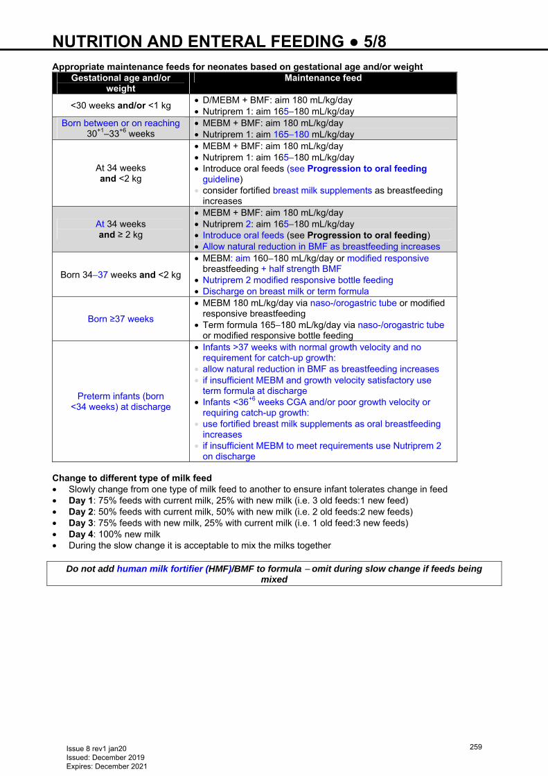
NUTRITION AND ENTERAL FEEDING ● 5/8 Appropriate maintenance feeds for neonates based on gestational age and/or weight
Gestational age and/or weight
Maintenance feed
<30 weeks and/or <1 kg • D/MEBM + BMF: aim 180 mL/kg/day • Nutriprem 1: aim 165−180 mL/kg/day
Born between or on reaching 30+1–33+6 weeks
• MEBM + BMF: aim 180 mL/kg/day • Nutriprem 1: aim 165−180 mL/kg/day
At 34 weeks and <2 kg
• MEBM + BMF: aim 180 mL/kg/day • Nutriprem 1: aim 165−180 mL/kg/day • Introduce oral feeds (see Progression to oral feeding
guideline) • consider fortified breast milk supplements as breastfeeding
increases
At 34 weeks and ≥ 2 kg
• MEBM + BMF: aim 180 mL/kg/day • Nutriprem 2: aim 165−180 mL/kg/day • Introduce oral feeds (see Progression to oral feeding) • Allow natural reduction in BMF as breastfeeding increases
Born 34−37 weeks and <2 kg
• MEBM: aim 160−180 mL/kg/day or modified responsive breastfeeding + half strength BMF
• Nutriprem 2 modified responsive bottle feeding • Discharge on breast milk or term formula
Born ≥37 weeks
• MEBM 180 mL/kg/day via naso-/orogastric tube or modified responsive breastfeeding
• Term formula 165−180 mL/kg/day via naso-/orogastric tube or modified responsive bottle feeding
Preterm infants (born <34 weeks) at discharge
• Infants >37 weeks with normal growth velocity and no requirement for catch-up growth:
• allow natural reduction in BMF as breastfeeding increases • if insufficient MEBM and growth velocity satisfactory use
term formula at discharge • Infants <36+6 weeks CGA and/or poor growth velocity or
requiring catch-up growth: • use fortified breast milk supplements as oral breastfeeding
increases • if insufficient MEBM to meet requirements use Nutriprem 2
on discharge Change to different type of milk feed • Slowly change from one type of milk feed to another to ensure infant tolerates change in feed • Day 1: 75% feeds with current milk, 25% with new milk (i.e. 3 old feeds:1 new feed) • Day 2: 50% feeds with current milk, 50% with new milk (i.e. 2 old feeds:2 new feeds) • Day 3: 75% feeds with new milk, 25% with current milk (i.e. 1 old feed:3 new feeds) • Day 4: 100% new milk • During the slow change it is acceptable to mix the milks together
Do not add human milk fortifier (HMF)/BMF to formula − omit during slow change if feeds being mixed
Issue 8 rev1 jan20 Issued: December 2019 Expires: December 2021
259
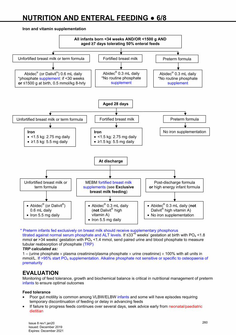
NUTRITION AND ENTERAL FEEDING ● 6/8 Iron and vitamin supplementation
* Preterm infants fed exclusively on breast milk should receive supplementary phosphorus titrated against normal serum phosphate and ALT levels. If ≤33+6 weeks’ gestation at birth with PO4 <1.8 mmol or >34 weeks’ gestation with PO4 <1.4 mmol, send paired urine and blood phosphate to measure tubular reabsorption of phosphate (TRP) TRP calculated as: 1 – (urine phosphate × plasma creatinine/plasma phosphate × urine creatinine) × 100% with all units in mmol/L. If >95% start PO4 supplementation. Alkaline phosphate not sensitive or specific to osteopaenia of prematurity
EVALUATION Monitoring of feed tolerance, growth and biochemical balance is critical in nutritional management of preterm infants to ensure optimal outcomes Feed tolerance • Poor gut motility is common among VLBW/ELBW infants and some will have episodes requiring
temporary discontinuation of feeding or delay in advancing feeds • If failure to progress feeds continues over several days, seek advice early from neonatal/paediatric
dietitian
All infants born <34 weeks AND/OR <1500 g AND aged ≥7 days tolerating 50% enteral feeds
Unfortified breast milk or term formula Fortified breast milk Preterm formula
Abidec® (or Dalivit®) 0.6 mL daily *phosphate supplement: if <30 weeks or ≤1500 g at birth, 0.5 mmol/kg 8-hrly
Abidec® 0.3 mL daily *No routine phosphate
supplement
Abidec® 0.3 mL daily *No routine phosphate
supplement
Aged 28 days
Iron • <1.5 kg: 2.75 mg daily • ≥1.5 kg: 5.5 mg daily
No iron supplementation
At discharge
Unfortified breast milk or term formula
Post-discharge formula or high energy infant formula
• Abidec® (or Dalivit®) 0.6 mL daily
• Iron 5.5 mg daily
• Abidec® 0.3 mL daily (not Dalivit® high vitamin A)
• No iron supplementation
Iron • <1.5 kg: 2.75 mg daily • ≥1.5 kg: 5.5 mg daily
MEBM fortified breast milk supplements (see Exclusive
breast milk feeding)
• Abidec® 0.3 mL daily (not Dalivit® high vitamin A)
• Iron 5.5 mg daily
Unfortified breast milk or term formula Fortified breast milk Preterm formula
Issue 8 rev1 jan20 Issued: December 2019 Expires: December 2021
260
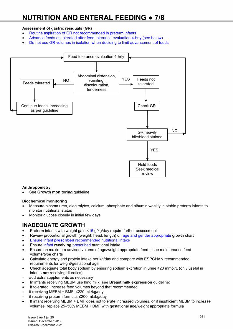
NUTRITION AND ENTERAL FEEDING ● 7/8 Assessment of gastric residuals (GR) • Routine aspiration of GR not recommended in preterm infants • Advance feeds as tolerated after feed tolerance evaluation 4-hrly (see below) • Do not use GR volumes in isolation when deciding to limit advancement of feeds Anthropometry • See Growth monitoring guideline Biochemical monitoring • Measure plasma urea, electrolytes, calcium, phosphate and albumin weekly in stable preterm infants to
monitor nutritional status • Monitor glucose closely in initial few days
INADEQUATE GROWTH • Preterm infants with weight gain <16 g/kg/day require further assessment • Review proportional growth (weight, head, length) on age and gender appropriate growth chart • Ensure infant prescribed recommended nutritional intake • Ensure infant receiving prescribed nutritional intake • Ensure on maximum advised volume of age/weight appropriate feed – see maintenance feed
volume/type charts • Calculate energy and protein intake per kg/day and compare with ESPGHAN recommended
requirements for weight/gestational age • Check adequate total body sodium by ensuring sodium excretion in urine ≥20 mmol/L (only useful in
infants not receiving diuretics) • add extra supplements as necessary • In infants receiving MEBM use hind milk (see Breast milk expression guideline) • If tolerated, increase feed volumes beyond that recommended • if receiving MEBM + BMF: ≤220 mL/kg/day • if receiving preterm formula: ≤200 mL/kg/day • If infant receiving MEBM + BMF does not tolerate increased volumes, or if insufficient MEBM to increase
volumes, replace 25−50% MEBM + BMF with gestational age/weight appropriate formula
YES
NO
YES NO
Feed tolerance evaluation 4-hrly
Abdominal distension, vomiting,
discolouration, tenderness
Feeds tolerated
Continue feeds, increasing as per guideline
Feeds not tolerated
GR heavily bile/blood stained
Hold feeds Seek medical
review
Check GR
Issue 8 rev1 jan20 Issued: December 2019 Expires: December 2021
261
NUTRITION AND ENTERAL FEEDING ● 8/8 • <2 kg preterm formula • ≥2 kg high energy term formula • Breastfeeding/MEBM: use BMF as a concentrated solution (known as fortified breast milk supplement,
give 1 sachet dissolved in 3 mL MEBM via syringe/teat before 4 breastfeeds, equally spread throughout 24 hr period. Reduce BMF by 1 sachet/day every 2 weeks until 6 weeks post-term or 3.5 kg, whichever soonest, then stop fortified breast milk supplements
• Refer to neonatal/paediatric dietitian for assessment and advice
PROGRESSION TO ORAL FEEDING Aim Safe progression to oral feeding (see Progression to oral feeding in preterm infants guideline) Exclusive breast milk feeding • Encourage modified responsive breast/bottle feeding of MEBM Exclusive/partial formula feeding • Encourage modified responsive bottle feeding of post-discharge formula or term formula • Infants born <34 weeks, prescribe post-discharge preterm formula ≤6 months’ CGA (if required)
Department of Health Guidelines state all children aged 6 months−5 yr receive vitamin supplementation containing vitamins A C D unless receiving formula milk >500 mL/day
Exclusively breastfed infants should receive vitamin D supplementation from birth
Issue 8 rev1 jan20 Issued: December 2019 Expires: December 2021
262
OESOPHAGEAL ATRESIA ● 1/3 DEFINITION Congenital anomaly with blind ending oesophagus which may be associated with a fistula between the abnormal oesophagus and the trachea
DIAGNOSIS • Suspect antenatally if scans show polyhydramnios +/- absent stomach bubble • refer to fetal medicine specialist • plan appropriate place of delivery • parents should meet paediatric surgeon antenatally • Most cases present shortly after birth. Suspect if: • history of polyhydramnios +/- absent stomach bubble • frothing at mouth • respiratory symptoms on feeding • difficulty in passing nasogastric tube (NGT) • anorectal malformation (see Anorectal malformation guideline)
DELIVERY • If diagnosis suspected antenatally, avoid: • any positive pressure ventilation [including mask ventilation, HFNC, CPAP and endotracheal tube
(ETT)]; pouch distension may lead to respiratory compromise and/or aspiration via a distal pouch fistula • If intubation indicated, ETT tip as close to carina as possible to minimise gas flow through a fistula.
Ventilatory pressures should be as low as possible • If any significant respiratory compromise, instigate a time critical transfer to surgical unit Confirmation of diagnosis • Experienced operator to place radio-opaque 8 Fr NGT. Typically resistance is felt 10–12 cm from nostril
in term baby • do not use force (may lead to oesophageal perforation) • AP X-ray of whole chest and abdomen • diagnosis confirmed if NGT curled in upper oesophagus • gastric air bubble/bowel gas confirms presence of fistula between trachea and distal oesophagus • Do not attempt a contrast oesophagogram
MANAGEMENT ON NNU • If respiratory support required or abdominal distension, contact surgical unit and transfer team
immediately (time critical transfer) • Nurse 30° head-up with head turned to side to facilitate drainage of secretions • Pass 10 Fr Replogle tube into oesophageal pouch (see Insertion and management of Replogle tube) • if Replogle tube unavailable, place 10 Fr NGT into pouch, aspirating every 15 min • an NGT cannot be placed on suction so needs regular, intermittent aspiration • Insert until resistance is met, then withdraw by 1 cm • Tape securely to face. Usually 10–12 cm at nostril in a term baby • Place mittens on baby to prevent tube being pulled out • Attach tapered end of tube to continuous suction. Start pressure at 5 kPa aiming for continuous flow of
secretions from upper oesophagus. Maximum pressure 10 kPa • do not share suction with other drains e.g. chest drain • Baby should be relaxed and pink with no respiratory distress or secretions in the mouth • Keep nil-by-mouth • Flush Replogle tube with sodium chloride 0.9% 0.5 mL via the sidearm every 15 min. More frequently if
visible oral secretions • If using an enteral tube to drain saliva, aspirate every 15 min, more frequently if visible oral secretions or
respiratory difficulty evident • If no movement of secretions in Replogle tube after flushing with sodium chloride 0.9% 0.5 mL via the
sidearm, change tube • Do not leave syringe attached to sidearm as this will prevent the tube working effectively • change tube every 10 days, or daily if viscous secretions
Issue 8 rev1 jan20 Issued: December 2019 Expires: December 2021
263
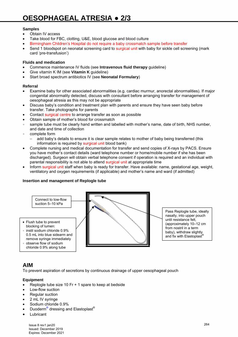
OESOPHAGEAL ATRESIA ● 2/3 Samples • Obtain IV access • Take blood for FBC, clotting, U&E, blood glucose and blood culture • Birmingham Children’s Hospital do not require a baby crossmatch sample before transfer • Send 1 bloodspot on neonatal screening card to surgical unit with baby for sickle cell screening (mark
card ‘pre-transfusion’) Fluids and medication • Commence maintenance IV fluids (see Intravenous fluid therapy guideline) • Give vitamin K IM (see Vitamin K guideline) • Start broad spectrum antibiotics IV (see Neonatal Formulary)
Referral • Examine baby for other associated abnormalities (e.g. cardiac murmur, anorectal abnormalities). If major
congenital abnormality detected, discuss with consultant before arranging transfer for management of oesophageal atresia as this may not be appropriate
• Discuss baby’s condition and treatment plan with parents and ensure they have seen baby before transfer. Take photographs for parents
• Contact surgical centre to arrange transfer as soon as possible • Obtain sample of mother’s blood for crossmatch • sample tube must be clearly hand written and labelled with mother’s name, date of birth, NHS number,
and date and time of collection • complete form
− add baby’s details to ensure it is clear sample relates to mother of baby being transferred (this information is required by surgical unit blood bank)
• Complete nursing and medical documentation for transfer and send copies of X-rays by PACS. Ensure you have mother’s contact details (ward telephone number or home/mobile number if she has been discharged). Surgeon will obtain verbal telephone consent if operation is required and an individual with parental responsibility is not able to attend surgical unit at appropriate time
• Inform surgical unit staff when baby is ready for transfer. Have available: name, gestational age, weight, ventilatory and oxygen requirements (if applicable) and mother’s name and ward (if admitted)
Insertion and management of Replogle tube
AIM To prevent aspiration of secretions by continuous drainage of upper oesophageal pouch Equipment • Replogle tube size 10 Fr + 1 spare to keep at bedside • Low-flow suction • Regular suction • 2 mL IV syringe • Sodium chloride 0.9% • Duoderm® dressing and Elastoplast® • Lubricant
Connect to low-flow suction 5–10 kPa
Pass Replogle tube, ideally nasally, into upper pouch until resistance felt, (approximately 10–12 cm from nostril in a term baby), withdraw slightly and fix with Elastoplast®
• Flush tube to prevent blocking of lumen:
• instil sodium chloride 0.9% 0.5 mL into blue sidearm and remove syringe immediately
• observe flow of sodium chloride 0.9% along tube
Issue 8 rev1 jan20 Issued: December 2019 Expires: December 2021
264
OESOPHAGEAL ATRESIA ● 3/3 Monitoring • Check Replogle tube several times an hour and flush to prevent blocking of lumen by instilling sodium
chloride 0.9% 0.5 mL into blue sidearm, removing syringe immediately and observing the flow of secretions along the tube. Monitor oxygen saturation, respiratory status and heart rate continuously
• For long-term Replogle use, monitor electrolytes and consider replacement therapy Blocked tube • Suspect if: • no continuous flow of secretions along tube • visible oral secretions • baby in distress • Clear airway with high-flow oropharyngeal suction • Increase low-flow suction and flush Replogle tube with air, observing flow of saliva along tube • If patency not restored, replace with new Replogle tube and return low-flow suction to previous level • If Replogle tube replaced, alternate nostrils to avoid long-term stretching of nares USEFUL INFORMATION • https://bwc.nhs.uk/paediatric-surgery-treatments • https://bwc.nhs.uk/travel-information • http://www.tofs.org.uk • http://www.e-lfh.org.uk/programmes/paediatric-surgery/
Issue 8 rev1 jan20 Issued: December 2019 Expires: December 2021
265
OESOPHAGEAL PERFORATION ● 1/2
INTRODUCTION • Relatively infrequent complication in neonates and early infancy • most common cause in neonates is iatrogenic • Neonates (especially preterm) requiring endotracheal intubation, nasogastric tube (NGT) insertion and
oropharyngeal suction are at increased risk of trauma to: • pharynx • upper airway • oesophagus • Site of injury is often at pharyngoesophageal junction where lumen is narrowed by cricopharyngeal
muscle • Contrast oesophagram and direct visualisation (ultrathin flexible endoscopy) are gold standards for
diagnosis Iatrogenic oesophageal perforation Complications • Pneumothorax • Pneumomediastinum with associated infection • Pseudo-diverticulum formation • Surgical/subcutaneous emphysema • Delayed initiation of feeding • Upper GI bleeding • Oesophageal obstruction
AT RISK • Preterm babies (especially <1500 g) • Babies requiring multiple intubation attempts • Difficulty passing or forceful attempts at NGT insertion
RECOGNITION
Clinical • Difficulty in passing NGT • NGT bouncing back • Blood stained aspirates • Bloody secretions in oropharynx • Deterioration in clinical condition Radiological • Discuss with radiologist urgently if NGT appears: • displaced • being to the right of the midline/vertebral spine • not in to the stomach • not following normal anatomical curvature towards the stomach • follows a straight line in the midline towards abdomen
High index of suspicion required; above findings in isolation are common in day-to-day neonatal care
Suspect oesophageal perforation if: • Pneumomediastinum • Pneumothorax • Cervical crepitus • Subcutaneous emphysema • Retropharyngeal gas Difficulty passing NGT • Do not make any further attempts to pass NGT • Request a water soluble contrast study (discuss with radiologist)
Issue 8 rev1 jan20 Issued: December 2019 Expires: December 2021
266
OESOPHAGEAL PERFORATION ● 2/2
MANAGEMENT • Early recognition is important (most important prognostic factor is the time between injury and initiation of
therapy) • Stop feeds • Prescribe TPN • Remove NGT • do not re-insert or manipulate NGT • If requiring ventilatory support, not for non-invasive ventilation • Discuss with local paediatric surgical team • send images by PACS • Maintain close liaison with surgical team regarding: • antibiotics • duration of nil-by-mouth • progress • Consider transfer to tertiary/surgical centre • Keep parents updated regularly • Document completion of duty of candour in medical records
Issue 8 rev1 jan20 Issued: December 2019 Expires: December 2021
267
OXYGEN ON DISCHARGE ● 1/2 OBJECTIVE • To put an effective plan in place to allow oxygen-dependent babies to be cared for safely at home
INDICATIONS FOR HOME OXYGEN THERAPY • Chronic lung disease with ongoing demand for additional inspired oxygen [see British Thoracic Society
(BTS) guidance] Criteria • Clinically stable on oxygen therapy via nasal cannulae for ≥2 weeks • SpO2 ≥95% after 36 weeks’ gestation on <0.5 L/min oxygen (if ≥0.5 L/min oxygen requirement at term
then refer to paediatric respiratory team) • Cyanotic congenital heart disease: a lower value may be appropriate, set threshold on an individual
basis (liaise with paediatric cardiologists) • Overnight pulse oximetry study when on stable oxygen for 1 week before discharge (see BTS
guidelines): • mean SpO2 ≥93% without frequent periods of desaturations • SpO2 ≥90% for >95% of the artefact-free recording period • If using <0.5 L/min ensure baby able to cope with short periods in air in case nasal cannulae become
dislodged • Routine continuous oxygen monitoring discontinued • Thermo-control well established • Feeding orally 3−4 hrly and gaining weight • some babies may require tube feeding, if all other criteria are met, this should not hinder discharge • Final decision on suitability for discharge lies with consultant
PREPARATION FOR DISCHARGE Make arrangements with parents • Discuss need for home oxygen with parents • Obtain consent for home oxygen supply and for sharing information with oxygen supplier. This is
obligatory before supplier can be contacted with patient details • Arrange multidisciplinary meeting 1 week before discharge with parents/carers, community nurse, health
visitor and member of NNU • Car seat challenge • Arrange discharge plan (see Discharge guideline) Parent training • Resuscitation techniques (2 adults) • No smoking in the house or anywhere in baby’s environment • Recognition of baby’s breathing pattern, colour and movements • Use of oxygen equipment (2 adults) • Competence in tape application for nasal prongs and skin care (water based emollients) • What to do in case of emergency: • contact numbers • direct admission policy • fire safety and insurance advice (car and home) • Discuss DLA/blue badge advantage Organise oxygen • Prescribing clinician to complete Home Oxygen Order Form (HOOF) on OxyShop (www.oxyshop.org) • Do not send home on <0.1 L (even if on <0.1 L in NNU. See BTS guidelines) Discharge checklist • Discharge plan implemented (see Discharge guideline) • Plan discharge for beginning of week to ensure community staff available in event of problems at home • Oxygen supply and equipment installed in the home • Baby will go home on prescribed amount of oxygen; this may be altered on direction of medical or
nursing staff, or in event of emergency • GP and other relevant professionals (also fire and electricity companies, although oxygen supplier
usually does this) informed of date and time of discharge
Issue 8 rev1 jan20 Issued: December 2019 Expires: December 2021
268
OXYGEN ON DISCHARGE ● 2/2 • Community team briefed to arrange home visit well in advance of discharge to ensure conditions suitable
and equipment correctly installed • Parents/carers trained to care for baby safely at home and have support contact numbers • Open access to paediatric ward
AFTERCARE • As oxygen dependent babies (e.g. chronic lung disease) are at increased risk of contracting respiratory
syncytial virus (RSV), give palivizumab and influenza vaccine (see Immunisations and Palivizumab guidelines)
• Refer to local guidelines for follow-up
Issue 8 rev1 jan20 Issued: December 2019 Expires: December 2021
269
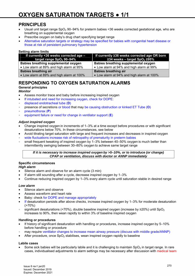
OXYGEN SATURATION TARGETS ● 1/1 PRINCIPLES • Usual unit target range SpO2 90−94% for preterm babies <36 weeks corrected gestational age, who are
breathing on supplemental oxygen • Prescribe oxygen on baby’s drug chart specifying target range • Alternative saturation targets or strategy may be specified for babies with congenital heart disease or
those at risk of persistent pulmonary hypertension Setting alarm limits
If currently <36 weeks corrected age – target range SpO2 90–94%
If currently ≥36 weeks corrected age OR born ≥34 weeks – target SpO2 ≥95%
Babies breathing supplemental oxygen • Low alarm at 89% and high alarm at 95%
Babies breathing supplemental oxygen • Low alarm at 94% and high alarm at 99%
Babies breathing air • Low alarm at 89% and high alarm at 100%
Babies breathing air • Low alarm at 94% and high alarm at 100%
RESPONDING TO OXYGEN SATURATION ALARMS General principles Monitor • Assess monitor trace and baby before increasing inspired oxygen • If intubated and need for increasing oxygen, check for DOPE: • displaced endotracheal tube (D) • presence of secretions or blood that may be causing obstruction or kinked ET Tube (O) • pneumothorax (P) • equipment failure or need for change in ventilator support (E)
Adjust inspired oxygen • Change inspired oxygen in increments of 1–3% at a time except before procedures or with significant
desaturations below 70%. In these circumstances, see below • Avoid titrating target saturation with large and frequent increases and decreases in inspired oxygen • wide fluctuations increase risk of retinopathy of prematurity in preterm babies • small frequent tweaking of inspired oxygen by 1–3% between 40–50% oxygen is much better than
intermittently swinging between 30–80% oxygen to achieve same target range
If it is necessary to increase inspired oxygen by >5–10%, or to introduce (or change) CPAP or ventilation, discuss with doctor or ANNP immediately
Specific circumstances High alarm • Silence alarm and observe for an alarm cycle (3 min) • If alarm still sounding after a cycle, decrease inspired oxygen by 1–3% • Continue reducing inspired oxygen by 1–3% every alarm cycle until saturation stable in desired range
Low alarm • Silence alarm and observe • Assess waveform and heart rate • Baby: check for DOPE and manage appropriately • If desaturation persists after above checks, increase inspired oxygen by 1–3% for moderate desaturation
(>70%) • significant desaturations (<70%), double baseline inspired oxygen (increase by ≥20%) until SpO2
increases to 90%, then wean rapidly to within 3% of baseline inspired oxygen Handling or procedures • If history of significant desaturation with handling or procedures, increase inspired oxygen by 5–10%
before handling or procedure • may require ventilator changes to increase mean airway pressure (discuss with middle grade/ANNP) • After procedure, once SpO2 stabilises, wean inspired oxygen rapidly to baseline
Labile cases • Some sick babies will be particularly labile and it is challenging to maintain SpO2 in target range. In rare
cases, individualised adjustments to alarm settings may be necessary after discussion with medical team
Issue 8 rev1 jan20 Issued: December 2019 Expires: December 2021
270
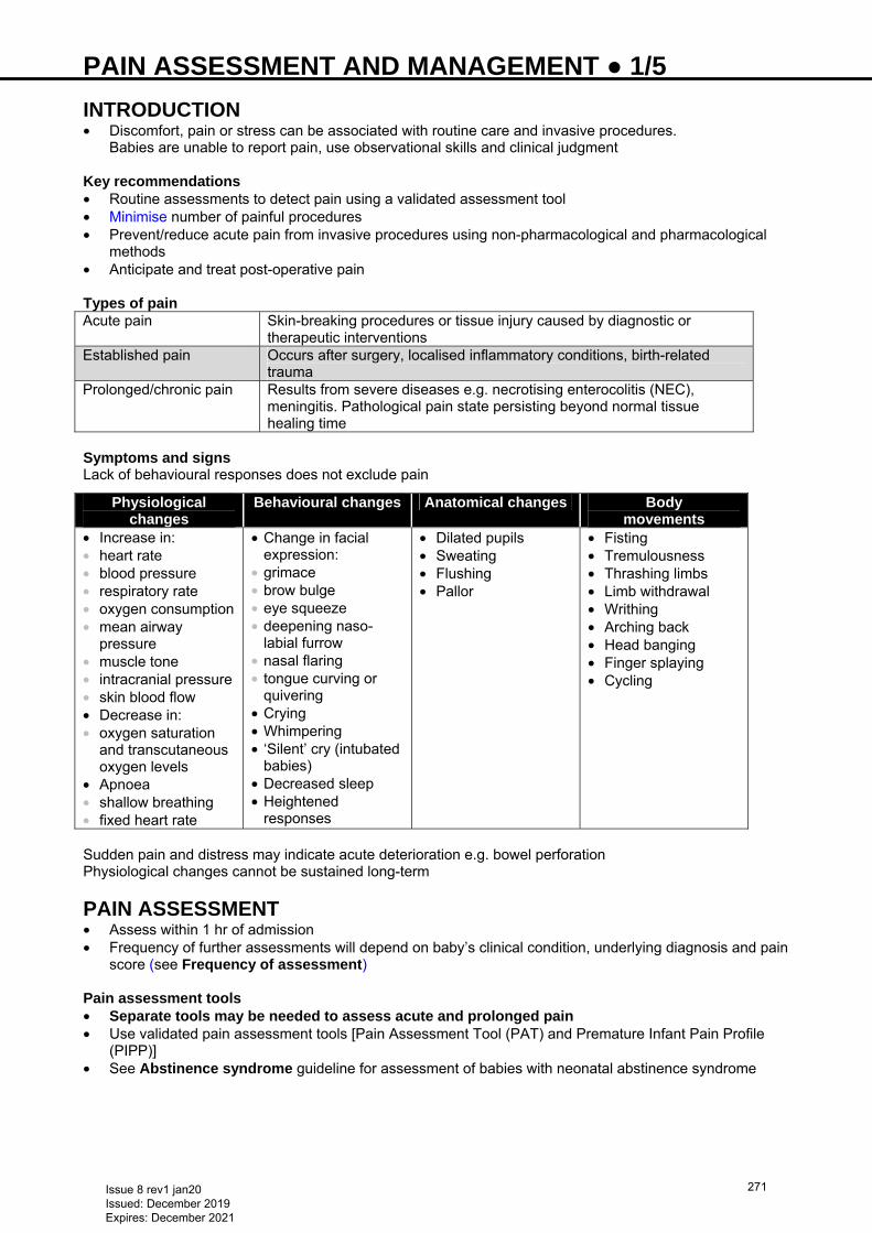
PAIN ASSESSMENT AND MANAGEMENT ● 1/5
INTRODUCTION • Discomfort, pain or stress can be associated with routine care and invasive procedures.
Babies are unable to report pain, use observational skills and clinical judgment Key recommendations • Routine assessments to detect pain using a validated assessment tool • Minimise number of painful procedures • Prevent/reduce acute pain from invasive procedures using non-pharmacological and pharmacological
methods • Anticipate and treat post-operative pain Types of pain Acute pain Skin-breaking procedures or tissue injury caused by diagnostic or
therapeutic interventions Established pain Occurs after surgery, localised inflammatory conditions, birth-related
trauma Prolonged/chronic pain Results from severe diseases e.g. necrotising enterocolitis (NEC),
meningitis. Pathological pain state persisting beyond normal tissue healing time
Symptoms and signs Lack of behavioural responses does not exclude pain
Physiological changes
Behavioural changes Anatomical changes Body movements
• Increase in: • heart rate • blood pressure • respiratory rate • oxygen consumption • mean airway
pressure • muscle tone • intracranial pressure • skin blood flow • Decrease in: • oxygen saturation
and transcutaneous oxygen levels
• Apnoea • shallow breathing • fixed heart rate
• Change in facial expression:
• grimace • brow bulge • eye squeeze • deepening naso-
labial furrow • nasal flaring • tongue curving or
quivering • Crying • Whimpering • ‘Silent’ cry (intubated
babies) • Decreased sleep • Heightened
responses
• Dilated pupils • Sweating • Flushing • Pallor
• Fisting • Tremulousness • Thrashing limbs • Limb withdrawal • Writhing • Arching back • Head banging • Finger splaying • Cycling
Sudden pain and distress may indicate acute deterioration e.g. bowel perforation Physiological changes cannot be sustained long-term
PAIN ASSESSMENT • Assess within 1 hr of admission • Frequency of further assessments will depend on baby’s clinical condition, underlying diagnosis and pain
score (see Frequency of assessment) Pain assessment tools • Separate tools may be needed to assess acute and prolonged pain • Use validated pain assessment tools [Pain Assessment Tool (PAT) and Premature Infant Pain Profile
(PIPP)] • See Abstinence syndrome guideline for assessment of babies with neonatal abstinence syndrome
Issue 8 rev1 jan20 Issued: December 2019 Expires: December 2021
271
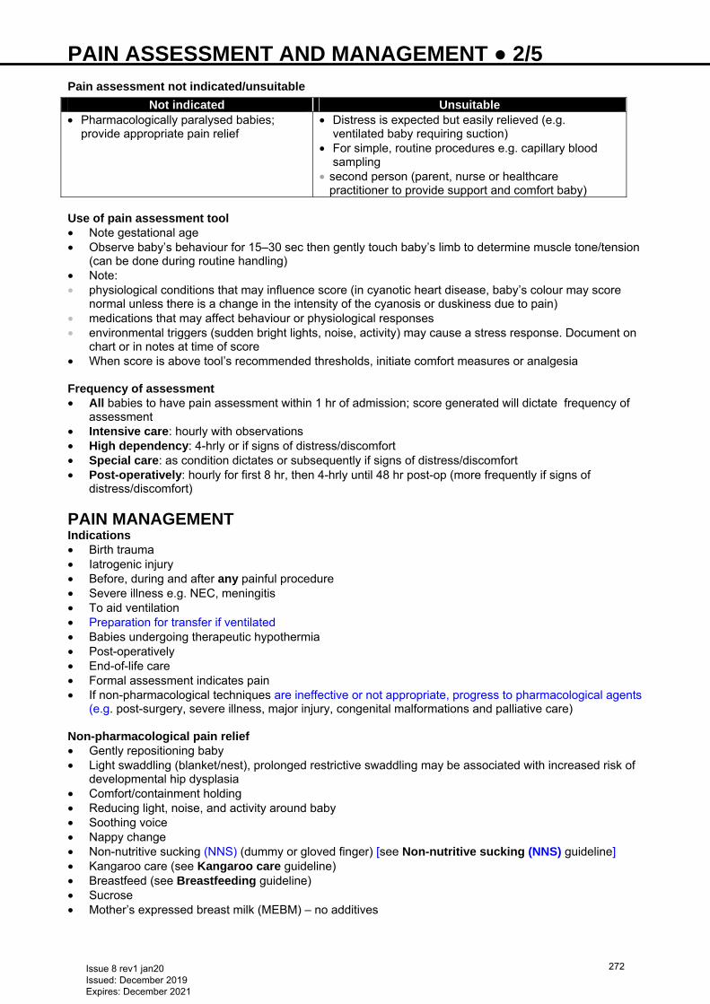
PAIN ASSESSMENT AND MANAGEMENT ● 2/5
Pain assessment not indicated/unsuitable
Not indicated Unsuitable• Pharmacologically paralysed babies;
provide appropriate pain relief • Distress is expected but easily relieved (e.g.
ventilated baby requiring suction) • For simple, routine procedures e.g. capillary blood
sampling • second person (parent, nurse or healthcare
practitioner to provide support and comfort baby)
Use of pain assessment tool • Note gestational age • Observe baby’s behaviour for 15–30 sec then gently touch baby’s limb to determine muscle tone/tension
(can be done during routine handling) • Note: • physiological conditions that may influence score (in cyanotic heart disease, baby’s colour may score
normal unless there is a change in the intensity of the cyanosis or duskiness due to pain) • medications that may affect behaviour or physiological responses • environmental triggers (sudden bright lights, noise, activity) may cause a stress response. Document on
chart or in notes at time of score • When score is above tool’s recommended thresholds, initiate comfort measures or analgesia
Frequency of assessment • All babies to have pain assessment within 1 hr of admission; score generated will dictate frequency of
assessment • Intensive care: hourly with observations • High dependency: 4-hrly or if signs of distress/discomfort • Special care: as condition dictates or subsequently if signs of distress/discomfort • Post-operatively: hourly for first 8 hr, then 4-hrly until 48 hr post-op (more frequently if signs of
distress/discomfort)
PAIN MANAGEMENT Indications • Birth trauma • Iatrogenic injury • Before, during and after any painful procedure • Severe illness e.g. NEC, meningitis • To aid ventilation • Preparation for transfer if ventilated • Babies undergoing therapeutic hypothermia • Post-operatively • End-of-life care • Formal assessment indicates pain • If non-pharmacological techniques are ineffective or not appropriate, progress to pharmacological agents
(e.g. post-surgery, severe illness, major injury, congenital malformations and palliative care)
Non-pharmacological pain relief • Gently repositioning baby • Light swaddling (blanket/nest), prolonged restrictive swaddling may be associated with increased risk of
developmental hip dysplasia • Comfort/containment holding • Reducing light, noise, and activity around baby • Soothing voice • Nappy change • Non-nutritive sucking (NNS) (dummy or gloved finger) [see Non-nutritive sucking (NNS) guideline] • Kangaroo care (see Kangaroo care guideline) • Breastfeed (see Breastfeeding guideline) • Sucrose • Mother’s expressed breast milk (MEBM) – no additives
Issue 8 rev1 jan20 Issued: December 2019 Expires: December 2021
272
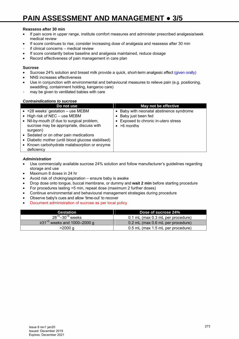
PAIN ASSESSMENT AND MANAGEMENT ● 3/5
Reassess after 30 min • If pain score in upper range, institute comfort measures and administer prescribed analgesia/seek
medical review • If score continues to rise, consider increasing dose of analgesia and reassess after 30 min • if clinical concerns – medical review • If score constantly below baseline and analgesia maintained, reduce dosage • Record effectiveness of pain management in care plan Sucrose • Sucrose 24% solution and breast milk provide a quick, short-term analgesic effect (given orally) • NNS increases effectiveness • Use in conjunction with environmental and behavioural measures to relieve pain (e.g. positioning,
swaddling, containment holding, kangaroo care) • may be given to ventilated babies with care Contraindications to sucrose
Do not use May not be effective • <28 weeks’ gestation – use MEBM • High risk of NEC – use MEBM • Nil-by-mouth (if due to surgical problem,
sucrose may be appropriate, discuss with surgeon)
• Sedated or on other pain medications • Diabetic mother (until blood glucose stabilised) • Known carbohydrate malabsorption or enzyme
deficiency
• Baby with neonatal abstinence syndrome • Baby just been fed • Exposed to chronic in-utero stress • >6 months
Administration • Use commercially available sucrose 24% solution and follow manufacturer’s guidelines regarding
storage and use • Maximum 8 doses in 24 hr • Avoid risk of choking/aspiration – ensure baby is awake • Drop dose onto tongue, buccal membrane, or dummy and wait 2 min before starting procedure • For procedures lasting >5 min, repeat dose (maximum 2 further doses) • Continue environmental and behavioural management strategies during procedure • Observe baby's cues and allow ‘time-out’ to recover • Document administration of sucrose as per local policy
Gestation Dose of sucrose 24% 28+0–30+6 weeks 0.1 mL (max 0.3 mL per procedure)
≥31+0 weeks and 1000–2000 g 0.2 mL (max 0.6 mL per procedure) >2000 g 0.5 mL (max 1.5 mL per procedure)
Issue 8 rev1 jan20 Issued: December 2019 Expires: December 2021
273
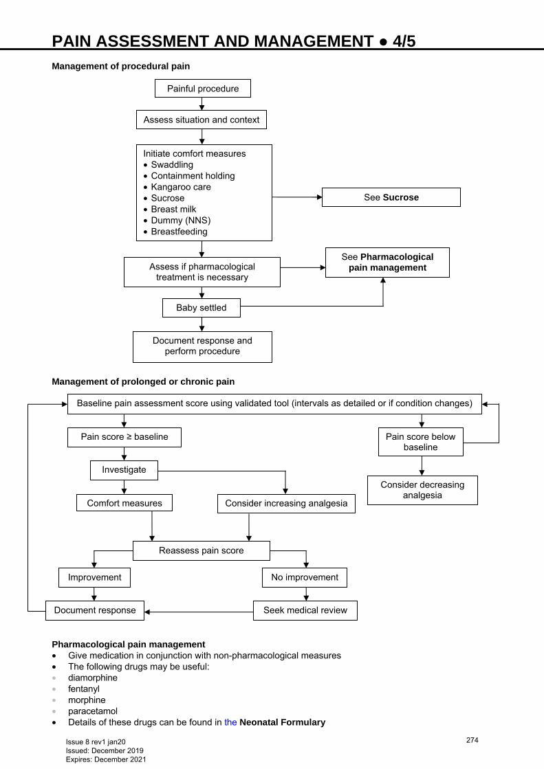
PAIN ASSESSMENT AND MANAGEMENT ● 4/5
Management of procedural pain Management of prolonged or chronic pain Pharmacological pain management • Give medication in conjunction with non-pharmacological measures • The following drugs may be useful: • diamorphine • fentanyl • morphine • paracetamol • Details of these drugs can be found in the Neonatal Formulary
Painful procedure
Assess situation and context
Initiate comfort measures • Swaddling • Containment holding • Kangaroo care • Sucrose • Breast milk • Dummy (NNS) • Breastfeeding
See Sucrose
Assess if pharmacological treatment is necessary
See Pharmacological pain management
Baby settled
Document response and perform procedure
Baseline pain assessment score using validated tool (intervals as detailed or if condition changes)
Pain score ≥ baseline Pain score below baseline
Investigate
Comfort measures
Improvement
Reassess pain score
Consider increasing analgesia
No improvement
Document response Seek medical review
Consider decreasing analgesia
Issue 8 rev1 jan20 Issued: December 2019 Expires: December 2021
274
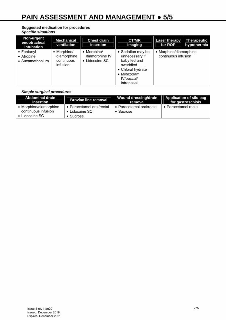
PAIN ASSESSMENT AND MANAGEMENT ● 5/5
Suggested medication for procedures Specific situations
Non-urgent endotracheal
intubation Mechanical ventilation
Chest drain insertion
CT/MR imaging
Laser therapy for ROP
Therapeutic hypothermia
• Fentanyl • Atropine • Suxamethonium
• Morphine/ diamorphine continuous infusion
• Morphine/ diamorphine IV
• Lidocaine SC
• Sedation may be unnecessary if baby fed and swaddled
• Chloral hydrate • Midazolam
IV/buccal/ intranasal
• Morphine/diamorphine continuous infusion
Simple surgical procedures
Abdominal drain insertion
Broviac line removal Wound dressing/drain
removal Application of silo bag
for gastroschisis • Morphine/diamorphine
continuous infusion • Lidocaine SC
• Paracetamol oral/rectal • Lidocaine SC • Sucrose
• Paracetamol oral/rectal • Sucrose
• Paracetamol rectal
Issue 8 rev1 jan20 Issued: December 2019 Expires: December 2021
275
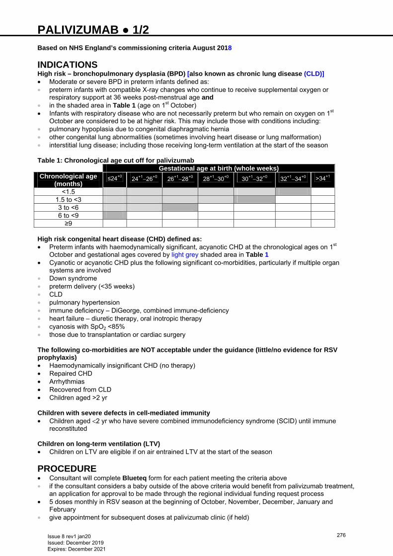
PALIVIZUMAB ● 1/2
Based on NHS England’s commissioning criteria August 2018 INDICATIONS High risk – bronchopulmonary dysplasia (BPD) [also known as chronic lung disease (CLD)] • Moderate or severe BPD in preterm infants defined as: • preterm infants with compatible X-ray changes who continue to receive supplemental oxygen or
respiratory support at 36 weeks post-menstrual age and • in the shaded area in Table 1 (age on 1st October) • Infants with respiratory disease who are not necessarily preterm but who remain on oxygen on 1st
October are considered to be at higher risk. This may include those with conditions including: • pulmonary hypoplasia due to congenital diaphragmatic hernia • other congenital lung abnormalities (sometimes involving heart disease or lung malformation) • interstitial lung disease; including those receiving long-term ventilation at the start of the season Table 1: Chronological age cut off for palivizumab
Gestational age at birth (whole weeks) Chronological age
(months) ≤24+0 24+1−26+0 26+1−28+0 28+1−30+0 30+1−32+0 32+1−34+0 >34+1
<1.5
1.5 to <3
3 to <6
6 to <9
≥9
High risk congenital heart disease (CHD) defined as: • Preterm infants with haemodynamically significant, acyanotic CHD at the chronological ages on 1st
October and gestational ages covered by light grey shaded area in Table 1 • Cyanotic or acyanotic CHD plus the following significant co-morbidities, particularly if multiple organ
systems are involved • Down syndrome • preterm delivery (<35 weeks) • CLD • pulmonary hypertension • immune deficiency – DiGeorge, combined immune-deficiency • heart failure – diuretic therapy, oral inotropic therapy • cyanosis with SpO2 <85% • those due to transplantation or cardiac surgery
The following co-morbidities are NOT acceptable under the guidance (little/no evidence for RSV prophylaxis) • Haemodynamically insignificant CHD (no therapy) • Repaired CHD • Arrhythmias • Recovered from CLD • Children aged >2 yr Children with severe defects in cell-mediated immunity • Children aged <2 yr who have severe combined immunodeficiency syndrome (SCID) until immune
reconstituted Children on long-term ventilation (LTV) • Children on LTV are eligible if on air entrained LTV at the start of the season
PROCEDURE • Consultant will complete Blueteq form for each patient meeting the criteria above • if the consultant considers a baby outside of the above criteria would benefit from palivizumab treatment,
an application for approval to be made through the regional individual funding request process • 5 doses monthly in RSV season at the beginning of October, November, December, January and
February • give appointment for subsequent doses at palivizumab clinic (if held)
Issue 8 rev1 jan20 Issued: December 2019 Expires: December 2021
276
PALIVIZUMAB ● 2/2
• where possible, administer first dose before start of RSV season • 15 mg/kg by IM injection into antero-lateral aspect of thigh • Order palivizumab injection from local community or hospital pharmacy (this can take some days) • Palivizumab must be stored at 2–8°C. Full administration instructions are provided in the ‛Summary of
product characteristics’ (SPC) • Split between 2 sites if >1 mL (final concentration when reconstituted 100 mg/mL)
DOCUMENTATION • After immunisation, document the following in case notes as well as in Child Health Record (Red Book): • consent gained from parents • vaccine given and reasons for any omissions • site of injection(s) in case of any reactions • batch number of product(s) • expiry date of product(s) • legible signature of person administering immunisations • adverse reactions • Sign treatment sheet • Update problem sheet with date and immunisations given • Document all information on discharge summary and medical case notes including recommendations for
future immunisations and need for any special vaccinations, e.g. influenza, palivizumab, etc.
Issue 8 rev1 jan20 Issued: December 2019 Expires: December 2021
277
PARENTERAL NUTRITION ● 1/3 DEFINITION Parenteral nutrition (PN) is the IV infusion of nutrients for the purpose of tissue maintenance, metabolic requirements and growth promotion in babies unable to tolerate full enteral feeds
Seek advice from your local PN pharmacist
INDICATIONS FOR PN • ≤30 weeks’ gestation and/or ≤1000 g: commence PN as soon as possible after birth, and within 24 hr of
birth/admission • 1001−1500 g: if anticipated baby will not achieve enteral feeds ≥100 mL/kg/day by aged 5 days,
commence PN • Receiving conservative treatment for necrotising enterocolitis (NEC) • If undergone surgery for congenital abnormality/acquired gut anomaly: to receive PN while establishing
enteral feeds • All babies likely to be fasted for ≥5 days
MODE OF DELIVERY Peripheral PN • PN should be ideally delivered centrally (high glucose and electrolyte concentrations result in a high
osmolarity − limiting nutrition given peripherally) • Depending on aqueous feed e.g. Vamin® composition, local policy may permit peripheral administration
of certain products in certain circumstances − check local policy before prescribing • Running lipid peripherally in addition to aqueous phase may prolong the life of the peripheral cannula Central PN • Requires placement of a central catheter [see Long line insertion (peripherally sited) guideline] with
tip in either superior vena cava or inferior vena cava • Infuse PN via a dedicated lumen • continuous vancomycin/sodium/potassium chloride infusion may be administered simultaneously with
PN, providing maximum total concentration ≤200 mmol/L • If access difficult, discuss PN drug compatibilities with pharmacist
Never administer calcium, magnesium and phosphate containing fluid simultaneously with PN
Central PN [long lines and umbilical venous catheters (UVC)] can introduce infection and septicaemia
CONSTITUTION OF PN • For practical and safety reasons standard bags are preferred as neonatal nutritional requirements are
largely predictable (see Nutrition and enteral feeding guideline)
Additions to PN to be made within an aseptic pharmacy only • If required, additional electrolytes can be infused alongside PN (see Central PN) Volume • PN provided primarily for nutrition • although fluid and nutrition are closely linked and volume needs to be considered carefully, the concepts
are not interchangeable e.g. providing 150 mL/kg/day fluid does not guarantee provision of adequate nutrition
• may be beneficial to give concentrated aqueous phase solutions to enable administration of additional drugs without compromising nutritional intake
• 30–40 mL/kg feed to be established before weaning of PN Protein/amino acid • Initial PN bag to contain 2–2.5 g/kg/day • Target protein intake, by day 5 of life, (regardless when PN was commenced): • preterm babies: 3.5–4 g/kg/day • term babies: 3 g/kg/day • Administer sufficient carbohydrate to facilitate the accretion of protein (approximately 25 kcal/g protein)
Issue 8 rev1 jan20 Issued: December 2019 Expires: December 2021
278
PARENTERAL NUTRITION ● 2/3 Glucose • ≥5.8 g/kg/day in first 24 hr − take PN and additional fluids into consideration • Increase glucose intake as tolerated to optimise calorie intake to maximum of 15–17.3 g/kg/day • If severe or persistent hyperglycaemia develops, commence insulin infusion – see Administration of
Actrapid® insulin (soluble insulin) in Hyperglycaemia guideline Electrolytes • Sodium: ≥3 mmol/kg/day in preterm babies who have commenced natriuresis • Potassium: ≥2 mmol/kg/day from day 2−3 • Babies given electrolytes solely as chloride salts can develop hyperchloraemic metabolic acidosis
(consider adding acetate to PN, where available) • Monitor serum phosphate twice weekly; aim to maintain at around 2 mmol/L
Micronutrients • Calcium and phosphate: 1:1 ratio (higher phosphate doses may be required) • if possible use organic phosphate compounds • Magnesium: 0.18–0.2 mmol/L Trace elements Peditrace® • Addition of trace element admixture (Peditrace®) shortens the shelf-life of standard bags considerably to
7 days • Zinc and selenium will be contained within standard aqueous feed bags • If baby on short-term PN and receiving some milk feeds, trace elements may not be required • If baby on PN >2 weeks and not receiving Peditrace®, discuss with PN pharmacist/dietitian Fat • 2 lipid emulsions used routinely on NNU: Intralipid® (soya bean origin) and SMOF lipid (blend of soya
bean, MCT fat, olive oil and fish oils) • some units use SMOF lipid routinely for all babies (current evidence does not support this as routine
practice; significantly different lipid profile to breast milk) • If Intralipid® is being administered, consider SMOF lipid for babies with conjugated bilirubin >50, or if
likely to receive PN for >1 month • Commence lipid 2 g/kg/day IV when commencing aqueous phase • increase by 0.5–1 g/kg/day to maximum 3.5 g/kg/day • all lipid to be infused over 24 hr Vitamins • Fat and water soluble vitamins are added to the lipid component of PN
MONITORING
Daily • Fluid input • Fluid output • U&E for first 7 days; then consultant discretion • Blood glucose • if blood glucose >11 mmol/L, urine glucose
3 times/week • Weight Weekly • LFT
• Length • Head circumference • Ca • PO4 • Magnesium
4-weekly • Serum triglycerides • Fat soluble vitamins A, D, E • Zinc • Copper • Manganese • Selenium • B12 and folate • Ferritin
Issue 8 rev1 jan20 Issued: December 2019 Expires: December 2021
279
PARENTERAL NUTRITION ● 3/3 COMPLICATIONS Catheter-related: [see Long line insertion (peripherally sited) guideline] • Peripheral catheters: extravasations and skin sloughs • Septicaemia Electrolyte abnormalities • Electrolyte and acid-base disturbances Metabolic • Hyper/hypoglycaemia, osmotic diuresis • Metabolic bone disease: mineral abnormalities (Ca/PO4/Mg) • Hyperlipidaemia and hypercholesterolaemia • Conjugated hyperbilirubinaemia PN-associated cholestatic hepatitis • Can occur with prolonged PN (>10–14 days) • probably due to combination of PN hepato-toxicity, sepsis and reduced oral feeding • often transient • usually manifests as rising serum bilirubin (with increased conjugated component >50 micromol/L) and
mildly elevated transaminases • leads to deficiencies of fatty acids and trace minerals in enterally fed babies • even small enteral feeds will limit or prevent this problem and therefore trophic feeds should be given to
all babies on PN unless there are contraindications such as acute clinical instability or NEC • consider other causes of hyperbilirubinaemia (PN-induced cholestasis is diagnosis of exclusion) e.g.
CMV, hypothyroidism • if failure to progress with enteral feeding in a timely fashion, seek advice from paediatric
gastroenterologist
WEANING PN • Commence enteral feeds as soon as possible • see Nutrition and enteral feeding guideline for increasing enteral feeds • Do not reduce PN until total volume of 180 mL/kg/day reached (unless fluid restricted) • When advancing enteral feedings, reduce rate of PN administration to achieve desired total fluid volume • Decrease aqueous and fat portions equally. Ratio dependent on total volume of aqueous phase + lipid,
e.g. if increasing feed by 1 mL, decrease aqueous phase by 1 × (aqueous phase mL/hr/aqueous phase + lipid mL/hr), and decrease lipid by 1 × (lipid mL/hr/aqueous phase + lipid mL/hr)
• Assess nutrient intake from both PN and enteral feed in relation to overall nutrition goals • If enteral vitamins required, commence when lipid infusion <10 mL/kg/day
Issue 8 rev1 jan20 Issued: December 2019 Expires: December 2021
280
PATENT DUCTUS ARTERIOSUS ● 1/4 RECOGNITION AND ASSESSMENT Definition • Persistent patency of the ductus arteriosus (PDA) is a failure of functional ductal closure by 48 hr or
anatomical closure by aged 3 weeks Factors associated with delayed closure • Prematurity (significant PDA affects approximately 30% of very-low-birth-weight babies) • Lack of antenatal corticosteroid prophylaxis • Surfactant-deficient lung disease • Hypoxaemia • Volume overload Adverse effects of PDA • Haemodynamic consequences of left-to-right shunt in preterm babies can prolong ventilatory support
and are associated with mortality and morbidity [chronic lung disease, pulmonary haemorrhage, intraventricular haemorrhage, necrotising enterocolitis (NEC) and retinopathy of prematurity]
• Increased pulmonary blood flow (leading to increased work of breathing and respiratory deterioration) • Reduced systemic blood flow (leading to acidosis and hypotension) Symptoms and signs • Can be absent even in the presence of a significant duct in first 7 days of life • A significant left-to-right shunt is suggested by: • bounding pulses and wide pulse pressure (i.e. >25 mmHg) • hyperdynamic precordium (excessive movement of precordium) • low-pitched systolic or continuous murmur over left upper sternal edge (absence of a murmur does not
exclude significant PDA) • signs of cardiac failure (tachypnoea, tachycardia, hepatomegaly, pulmonary oedema, generalised
oedema etc.) • poor perfusion (hypotension, poor capillary refill, mottled skin and persistent acidosis) • increased or persistent ventilatory requirements Differential diagnosis • Other cardiac pathology (e.g. congenital heart disease, including duct-dependent lesions, arrhythmias or
cardiomyopathy) • Sepsis
INVESTIGATIONS • SpO2 monitoring • Chest X-ray (cardiomegaly? pulmonary plethora?) • Echocardiography • to detect duct-dependent cardiac lesions and other cardiac pathologies that are difficult to exclude
clinically • if considering treatment with prostaglandin inhibitor • echocardiographic assessment of significant PDA includes:
− size of PDA (>1.5 mm) − volume loading of left atrium (LA/aorta ratio >1.5) − volume loading of left ventricle − velocity and flow pattern of ductal flow
IMMEDIATE TREATMENT General measures • Optimise oxygenation by appropriate ventilatory management • Use of a higher PEEP (i.e. ≥5 cm H2O) can help minimise effects of pulmonary oedema and risk of
pulmonary haemorrhage • Treat anaemia – maintain Hb ≥100 g/L with blood transfusion (consider concurrent dose of furosemide
IV) • Before starting medication, restrict fluid intake to 60–80% (e.g. from 150 mL/kg/day to
90−120 mL/kg/day) • If fluid overload or pulmonary oedema, give 1 dose of furosemide IV in accordance with Neonatal
Formulary
Issue 8 rev1 jan20 Issued: December 2019 Expires: December 2021
281
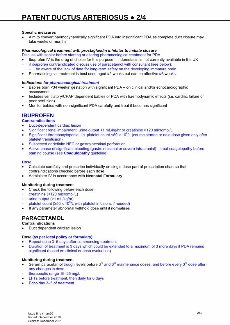
PATENT DUCTUS ARTERIOSUS ● 2/4 Specific measures • Aim to convert haemodynamically significant PDA into insignificant PDA as complete duct closure may
take weeks or months Pharmacological treatment with prostaglandin inhibitor to initiate closure Discuss with senior before starting or altering pharmacological treatment for PDA • Ibuprofen IV is the drug of choice for this purpose − indometacin is not currently available in the UK • if ibuprofen contraindicated discuss use of paracetamol with consultant (see below):
− be aware of the lack of data for long-term safety on the developing immature brain • Pharmacological treatment is best used aged ≤2 weeks but can be effective ≤6 weeks Indications for pharmacological treatment • Babies born <34 weeks’ gestation with significant PDA – on clinical and/or echocardiographic
assessment • Includes ventilatory/CPAP dependent babies or PDA with haemodynamic effects (i.e. cardiac failure or
poor perfusion) • Monitor babies with non-significant PDA carefully and treat if becomes significant
IBUPROFEN Contraindications • Duct-dependent cardiac lesion • Significant renal impairment: urine output <1 mL/kg/hr or creatinine >120 micromol/L • Significant thrombocytopenia, i.e. platelet count <50 × 109/L (course started or next dose given only after
platelet transfusion) • Suspected or definite NEC or gastrointestinal perforation • Active phase of significant bleeding (gastrointestinal or severe intracranial) – treat coagulopathy before
starting course (see Coagulopathy guideline) Dose • Calculate carefully and prescribe individually on single dose part of prescription chart so that
contraindications checked before each dose • Administer IV in accordance with Neonatal Formulary Monitoring during treatment • Check the following before each dose: • creatinine (<120 micromol/L) • urine output (>1 mL/kg/hr) • platelet count (≥50 × 109/L with platelet infusions if needed) • If any parameter abnormal withhold dose until it normalises
PARACETAMOL Contraindications • Duct dependent cardiac lesion
Dose (as per local policy or formulary) • Repeat echo 3−5 days after commencing treatment • Duration of treatment is 3 days which could be extended to a maximum of 3 more days if PDA remains
significant (based on clinical or echo evaluation) Monitoring during treatment • Serum paracetamol trough levels before 3rd and 6th maintenance doses, and before every 3rd dose after
any changes in dose • therapeutic range 15−25 mg/L • LFTs before treatment, then daily for 6 days • Echo day 3−5 of treatment
Issue 8 rev1 jan20 Issued: December 2019 Expires: December 2021
282
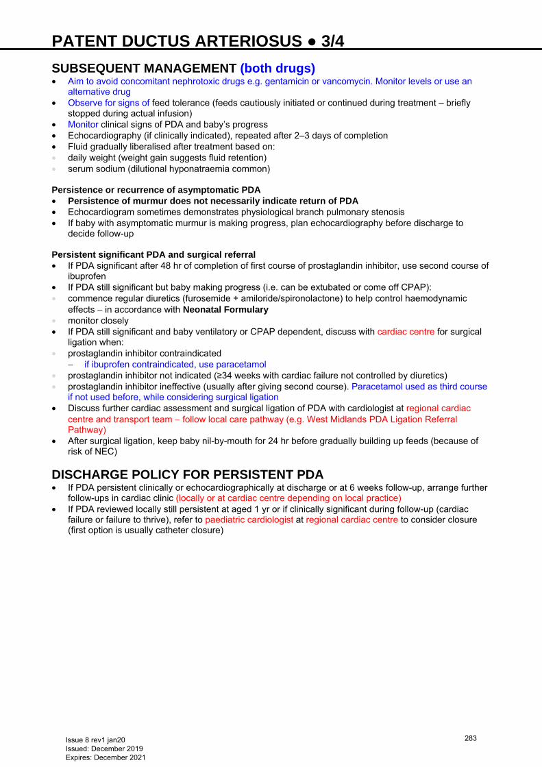
PATENT DUCTUS ARTERIOSUS ● 3/4 SUBSEQUENT MANAGEMENT (both drugs) • Aim to avoid concomitant nephrotoxic drugs e.g. gentamicin or vancomycin. Monitor levels or use an
alternative drug • Observe for signs of feed tolerance (feeds cautiously initiated or continued during treatment – briefly
stopped during actual infusion) • Monitor clinical signs of PDA and baby’s progress • Echocardiography (if clinically indicated), repeated after 2–3 days of completion • Fluid gradually liberalised after treatment based on: • daily weight (weight gain suggests fluid retention) • serum sodium (dilutional hyponatraemia common) Persistence or recurrence of asymptomatic PDA • Persistence of murmur does not necessarily indicate return of PDA • Echocardiogram sometimes demonstrates physiological branch pulmonary stenosis • If baby with asymptomatic murmur is making progress, plan echocardiography before discharge to
decide follow-up Persistent significant PDA and surgical referral • If PDA significant after 48 hr of completion of first course of prostaglandin inhibitor, use second course of
ibuprofen • If PDA still significant but baby making progress (i.e. can be extubated or come off CPAP): • commence regular diuretics (furosemide + amiloride/spironolactone) to help control haemodynamic
effects − in accordance with Neonatal Formulary • monitor closely • If PDA still significant and baby ventilatory or CPAP dependent, discuss with cardiac centre for surgical
ligation when: • prostaglandin inhibitor contraindicated
− if ibuprofen contraindicated, use paracetamol • prostaglandin inhibitor not indicated (≥34 weeks with cardiac failure not controlled by diuretics) • prostaglandin inhibitor ineffective (usually after giving second course). Paracetamol used as third course
if not used before, while considering surgical ligation • Discuss further cardiac assessment and surgical ligation of PDA with cardiologist at regional cardiac
centre and transport team − follow local care pathway (e.g. West Midlands PDA Ligation Referral Pathway)
• After surgical ligation, keep baby nil-by-mouth for 24 hr before gradually building up feeds (because of risk of NEC)
DISCHARGE POLICY FOR PERSISTENT PDA • If PDA persistent clinically or echocardiographically at discharge or at 6 weeks follow-up, arrange further
follow-ups in cardiac clinic (locally or at cardiac centre depending on local practice) • If PDA reviewed locally still persistent at aged 1 yr or if clinically significant during follow-up (cardiac
failure or failure to thrive), refer to paediatric cardiologist at regional cardiac centre to consider closure (first option is usually catheter closure)
Issue 8 rev1 jan20 Issued: December 2019 Expires: December 2021
283
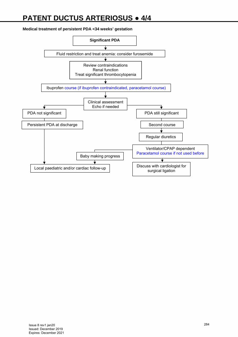
PATENT DUCTUS ARTERIOSUS ● 4/4
Fluid restriction and treat anemia; consider furosemide
Medical treatment of persistent PDA <34 weeks’ gestation
PDA not significant
Persistent PDA at discharge
Local paediatric and/or cardiac follow-up
PDA still significant
Second course
Regular diuretics
Ibuprofen course (if ibuprofen contraindicated, paracetamol course)
Review contraindications Renal function
Treat significant thrombocytopenia
Clinical assessment Echo if needed
Discuss with cardiologist for surgical ligation
Baby making progress
Ventilator/CPAP dependent Paracetamol course if not used before
Significant PDA
Issue 8 rev1 jan20 Issued: December 2019 Expires: December 2021
284
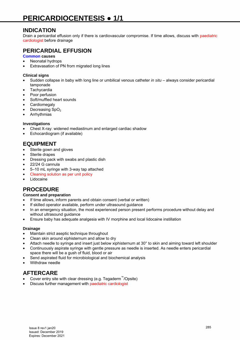
PERICARDIOCENTESIS ● 1/1 INDICATION Drain a pericardial effusion only if there is cardiovascular compromise. If time allows, discuss with paediatric cardiologist before drainage
PERICARDIAL EFFUSION Common causes • Neonatal hydrops • Extravasation of PN from migrated long lines Clinical signs • Sudden collapse in baby with long line or umbilical venous catheter in situ – always consider pericardial
tamponade • Tachycardia • Poor perfusion • Soft/muffled heart sounds • Cardiomegaly • Decreasing SpO2 • Arrhythmias
Investigations • Chest X-ray: widened mediastinum and enlarged cardiac shadow • Echocardiogram (if available) EQUIPMENT • Sterile gown and gloves • Sterile drapes • Dressing pack with swabs and plastic dish • 22/24 G cannula • 5–10 mL syringe with 3-way tap attached • Cleaning solution as per unit policy • Lidocaine
PROCEDURE Consent and preparation • If time allows, inform parents and obtain consent (verbal or written) • If skilled operator available, perform under ultrasound guidance • In an emergency situation, the most experienced person present performs procedure without delay and
without ultrasound guidance • Ensure baby has adequate analgesia with IV morphine and local lidocaine instillation
Drainage • Maintain strict aseptic technique throughout • Clean skin around xiphisternum and allow to dry • Attach needle to syringe and insert just below xiphisternum at 30° to skin and aiming toward left shoulder • Continuously aspirate syringe with gentle pressure as needle is inserted. As needle enters pericardial
space there will be a gush of fluid, blood or air • Send aspirated fluid for microbiological and biochemical analysis • Withdraw needle AFTERCARE • Cover entry site with clear dressing (e.g. Tegaderm™/Opsite) • Discuss further management with paediatric cardiologist
Issue 8 rev1 jan20 Issued: December 2019 Expires: December 2021
285
PERSISTENT PULMONARY HYPERTENSION OF THE NEWBORN (PPHN) ● 1/3 RECOGNITION AND ASSESSMENT Definition • Failure of normal postnatal fall in pulmonary vascular resistance • Leads to right-to-left shunting and subsequent hypoxia • Can be primary (idiopathic) or secondary • Severe hypoxaemia (PaO2 <6 kPa) in 100% oxygen • Complex condition with varied causes and degrees of severity • Echocardiogram: structurally normal heart (may show right ventricular hypertrophy), right-to-left or
bidirectional shunt at PFO and/or patent ductus arteriosus (PDA) Idiopathic • Degree of hypoxia disproportionate to degree of hypercarbia • Mild lung disease (in primary/idiopathic PPHN)
Secondary: • May be associated with • severe lung disease [e.g. meconium aspiration (MAS), surfactant deficiency] • perinatal asphyxia • infection [e.g. Group B streptococcal (GBS) pneumonia] • structural abnormalities: pulmonary hypoplasia, congenital diaphragmatic hernia, A-V malformations,
congenital cystic adenomatoid malformation (CCAM) • maternal drugs: aspirin, non-steroidal anti-inflammatory drugs, SSRIs
CLINICAL FEATURES Usually present in first 12 hr of life • SpO2 <95% or hypoxia (PaO2 <6 kPa) in 100% oxygen • Mimics cyanotic heart disease • CVS: tricuspid regurgitant murmur, right ventricular heave, loud second heart sound and systemic
hypotension • Idiopathic PPHN: respiratory signs mild or absent • Secondary PPHN: features of underlying disease
INVESTIGATIONS • Blood gas shows hypoxaemia (PaO2 <6 kPa) with oxygenation index >20 (underlying disease will
produce a mixed picture) • SpO2 >5% difference in preductal (right hand) and postductal saturations (either feet) (pre > post) • Hyperoxia test (100% oxygen for 5 min) • SpO2 may improve to ≥95% in early stage or may not respond, i.e. staying <95% in established PPHN
(as in cyanotic heart disease) • Chest X-ray: variable findings depending on underling diagnosis (normal or minimal changes in
idiopathic PPHN) • Electrocardiograph − often normal. Can sometimes show tall P waves in lead 2/V1/V2 or features of RVH
(i.e. tall R waves V1/V2, right axis deviation or upright T waves in V1/V2) • Echocardiogram (although not mandatory for initial diagnosis and management) is useful: • to exclude cyanotic heart disease • to assess pulmonary pressure • to evaluate ventricular function • ≥1 of the following confirm PPHN in presence of normal cardiac structures:
− significant tricuspid regurgitation − dilatation of right side of heart and/or hypertrophy of right ventricle − right-to-left shunting across PFO and/or PDA − pulmonary regurgitation − flattening or bowing of interventricular septum towards right ventricle
• Pulmonary pressure is estimated from echocardiogram using either:
Tricuspid regurgitation (systolic pulmonary pressure = 4 × (VmaxTR)2 + usual right atrial pressure of 5)
Pulmonary Regurgitation (mean pulmonary pressure = 4 × (VmaxPR)2 + usual right ventricular diastolic pressure of 5)
Issue 8 rev1 jan20 Issued: December 2019 Expires: December 2021
286
PERSISTENT PULMONARY HYPERTENSION OF THE NEWBORN (PPHN) ● 2/3 MANAGEMENT • If saturations continue at <95% despite 100% oxygen, and echocardiography not available immediately
to rule out duct dependent heart disease, start prostaglandin infusion IV • Once PPHN suspected, inform and involve consultant neonatologist immediately • Aims of management are to: • decrease pulmonary vascular resistance • increase systemic blood pressure • treat any underlying condition General measures • Minimal handling, nurse in quiet environment • Secure arterial and central venous access, see Arterial line insertion guideline or Umbilical artery
catheterisation and removal, and Umbilical venous catheterisation and removal guidelines • Maintain normal temperature, biochemistry and fluid balance • Keep Hb ≥120 g/L • Give antibiotics (sepsis, particularly GBS, is difficult to exclude) • Surfactant may be beneficial in MAS or GBS sepsis − discuss with consultant • If perfusion poor, fluid bolus [sodium chloride 0.9% 10 mL/kg or if coagulopathy, fresh frozen plasma
(see Coagulopathy guideline)] • Once PaCO2 in acceptable range (i.e. <6 kPa), correct metabolic acidosis to maintain pH 7.35–7.45
using full correction with sodium bicarbonate over 1 hr. If repeat correction necessary, slow bicarbonate infusion of calculated dose can be given over 6−12 hr (see Neonatal Formulary)
Ventilation • Use conventional ventilation to start with (targeted tidal volume 5−6 mL/kg) • Use sedation and muscle relaxation in babies with high ventilatory and oxygen requirements and/or
ventilator asynchrony • PaCO2 4.5−5.5 kPa (accept up to 6 kPa in parenchymal lung disease). Avoid hypocarbia • start in 100% oxygen and reduce as tolerated. Maintain SpO2 at 96–100% and PaO2 at 10−12 kPa • High frequency oscillatory ventilation (HFOV) may further improve oxygenation [see Ventilation: high
frequency oscillatory ventilation (HFOV) guideline] • Monitor oxygenation index (OI)
OI = mean airway pressure (cm H2O) × % of oxygen
postductal PaO2 (kPa) × 7.5 Inotropes (see Hypotension guideline) • Use inotropes early • In significant PPHN, adrenaline or noradrenaline can be useful in increasing systemic blood pressure
without increasing pulmonary vascular resistance • Maintain systemic mean BP 50−60 mmHg in term baby and systemic systolic
BP 60–70 mmHg or above estimated pulmonary pressures (if available by echo)
Pulmonary vasodilatation • If OI >20 or needs 100% oxygen, or significant PPHN on echo, use inhaled nitric oxide (NO) as a
selective pulmonary vasodilator (see Nitric oxide guideline) • Babies with PPHN requiring NO should be referred to a NICU for ongoing management Severe and resistant PPHN not responding to conventional management Baby born ≥34 weeks or ≥2 kg with PPHN • Not responding or OI >30 despite NO, inotropes and/or HFOV or • Unable to maintain BP with inotropes or persistent need for adrenaline/noradrenaline infusion or • No significant progression after 3 days • May benefit from ECMO or other specialist treatment • Discuss with KIDS team in West Midlands or nearest ECMO centre • Liaise with PICU/ECMO team regarding other medical management Criteria for ECMO • Baby born ≥34 weeks or ≥2 kg with PPHN • Oxygenation index >40 • Reversible lung disease (<10 days high pressure ventilation) • No lethal congenital malformation
Issue 8 rev1 jan20 Issued: December 2019 Expires: December 2021
287

PERSISTENT PULMONARY HYPERTENSION OF THE NEWBORN (PPHN) ● 3/3 Exclusion criteria (if in doubt, discuss with ECMO team) • Major intracranial haemorrhage • Irreversible lung injury or mechanical ventilation >10 days • Lethal congenital or chromosomal anomalies • Severe encephalopathy • Major cardiac malformation
A baby accepted for transfer to ECMO centre will be retrieved by ECMO or PICU team
• ECMO centre will need: • cranial ultrasound scan • maternal blood for group and crossmatching (check with ECMO centre) • referral letter • copies of hospital notes/chest X-rays • Outreach ECMO • ECMO team may decide to start outreach ECMO in NNU before transfer to ECMO unit. Check with
ECMO team regarding diathermy unit and number of packed cell units needed for procedure Referral for ECMO • For West Midlands contact KIDS team on 0300 200 1100, or • ECMO co-ordinator/fellow at nearest ECMO centre: • Glenfield Hospital, Leicester 0116 287 1471 • Great Ormond Street Hospital, London 0207 829 8652 • Freeman Hospital, Newcastle 0191 223 1016 • Yorkhill Hospital, Glasgow 0141 201 0000
Issue 8 rev1 jan20 Issued: December 2019 Expires: December 2021
288
POLYCYTHAEMIA ● 1/2 RECOGNITION AND ASSESSMENT Definition • Peripheral venous haematocrit (Hct) >65% • Symptoms rarely occur with peripheral Hct of <70% • Hct peaks at 2 hr after birth and then decreases with significant changes occurring by 6 hr Clinical consequences • Hyperviscosity • Decreased blood flow and impaired tissue perfusion • Thrombus formation
Complications • Cerebral micro-infarction and adverse neurodevelopmental outcome • Renal vein thrombosis • Necrotising enterocolitis (NEC) Causes
Intra-uterine increased erythropoiesis Erythrocyte transfusion • Placental insufficiency (SGA) • Postmaturity • Maternal diabetes • Maternal smoking • Chromosomal abnormalities: trisomy 21, 18, 13 • Beckwith–Wiedemann syndrome • Congenital adrenal hyperplasia • Neonatal thyrotoxicosis • Congenital hypothyroidism
• Maternal-fetal • Twin-to-twin transfusion • Delayed cord clamping • Unattended delivery
Symptoms and signs • Commonly plethoric but asymptomatic Cardiorespiratory • Respiratory distress
• Persistent pulmonary hypertension of the newborn • Congestive cardiac failure
CNS • Lethargy, hypotonia within 6 hr • Difficult arousal, irritability • Jittery • Easily startled • Seizures
GIT • Poor feeding • Vomiting • NEC
Metabolic • Hypoglycaemia • Hypocalcaemia • Jaundice
Haematological • Thrombocytopenia Renal • Renal vein thrombosis
• Renal failure
INVESTIGATIONS In all unwell babies and at-risk babies who look plethoric (as mentioned above) • FBC/Hct • If Hct >65%, repeat a free-flowing venous sample or obtain arterial Hct • If polycythaemic, check blood glucose and serum calcium
IMMEDIATE TREATMENT • Ensure babies at risk have liberal fluid intake 1 day ahead (see Intravenous fluid therapy guideline) Asymptomatic babies with Hct >70% • Repeat venous Hct after 6 hr • if still high, discuss with consultant
Issue 8 rev1 jan20 Issued: December 2019 Expires: December 2021
289
POLYCYTHAEMIA ● 2/2 Symptomatic babies with Hct >65% • Possible symptoms: fits and excessive jitteriness, with neurological signs and refractory hypoglycaemia Treatment • Dilutional exchange transfusion. Discuss with consultant • use of haemodilution (partial exchange transfusion) for treatment of polycythaemia is not supported by
evidence and treatment of asymptomatic babies is not recommended • explain to parents need for exchange and possible risks before performing dilutional exchange
transfusion. Partial exchange transfusion increases risk of NEC • use sodium chloride 0.9% (see Exchange transfusion guideline) • Volume to be exchanged = 20 mL/kg • Perform exchange via peripheral arterial and IV lines or via umbilical venous catheter • Take 5–10 mL aliquots and complete procedure over 15–20 min
SUBSEQUENT MANAGEMENT • Babies who required dilutional exchange transfusion require long-term neurodevelopmental follow-up • Otherwise, follow-up will be dependent on background problem
Issue 8 rev1 jan20 Issued: December 2019 Expires: December 2021
290
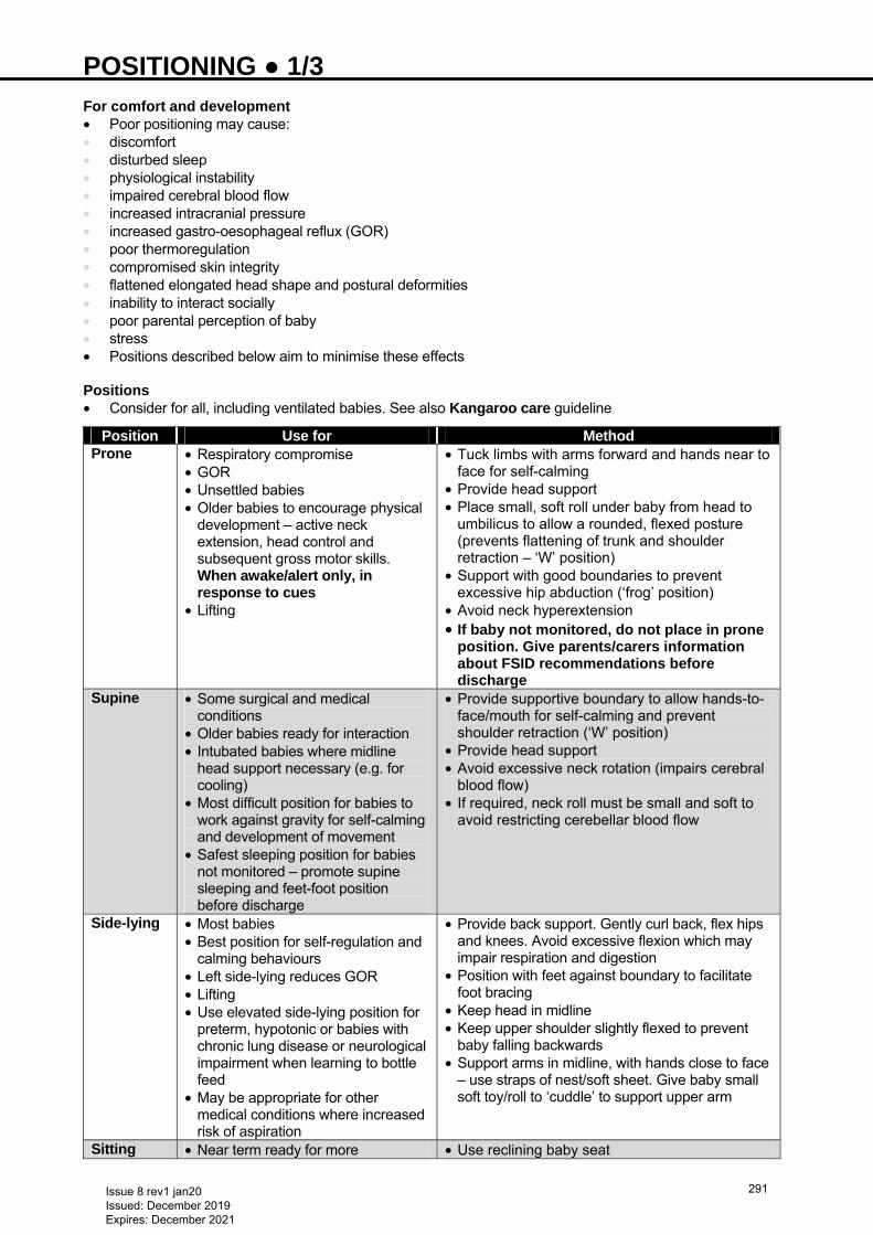
POSITIONING ● 1/3 For comfort and development • Poor positioning may cause: • discomfort • disturbed sleep • physiological instability • impaired cerebral blood flow • increased intracranial pressure • increased gastro-oesophageal reflux (GOR) • poor thermoregulation • compromised skin integrity • flattened elongated head shape and postural deformities • inability to interact socially • poor parental perception of baby • stress • Positions described below aim to minimise these effects Positions • Consider for all, including ventilated babies. See also Kangaroo care guideline
Position Use for Method Prone • Respiratory compromise
• GOR • Unsettled babies • Older babies to encourage physical
development – active neck extension, head control and subsequent gross motor skills. When awake/alert only, in response to cues
• Lifting
• Tuck limbs with arms forward and hands near to face for self-calming
• Provide head support • Place small, soft roll under baby from head to
umbilicus to allow a rounded, flexed posture (prevents flattening of trunk and shoulder retraction – ‘W’ position)
• Support with good boundaries to prevent excessive hip abduction (‘frog’ position)
• Avoid neck hyperextension • If baby not monitored, do not place in prone
position. Give parents/carers information about FSID recommendations before discharge
Supine • Some surgical and medical conditions
• Older babies ready for interaction • Intubated babies where midline
head support necessary (e.g. for cooling)
• Most difficult position for babies to work against gravity for self-calming and development of movement
• Safest sleeping position for babies not monitored – promote supine sleeping and feet-foot position before discharge
• Provide supportive boundary to allow hands-to-face/mouth for self-calming and prevent shoulder retraction (‘W’ position)
• Provide head support • Avoid excessive neck rotation (impairs cerebral
blood flow) • If required, neck roll must be small and soft to
avoid restricting cerebellar blood flow
Side-lying • Most babies • Best position for self-regulation and
calming behaviours • Left side-lying reduces GOR • Lifting • Use elevated side-lying position for
preterm, hypotonic or babies with chronic lung disease or neurological impairment when learning to bottle feed
• May be appropriate for other medical conditions where increased risk of aspiration
• Provide back support. Gently curl back, flex hips and knees. Avoid excessive flexion which may impair respiration and digestion
• Position with feet against boundary to facilitate foot bracing
• Keep head in midline • Keep upper shoulder slightly flexed to prevent
baby falling backwards • Support arms in midline, with hands close to face
– use straps of nest/soft sheet. Give baby small soft toy/roll to ‘cuddle’ to support upper arm
Sitting • Near term ready for more • Use reclining baby seat
Issue 8 rev1 jan20 Issued: December 2019 Expires: December 2021
291
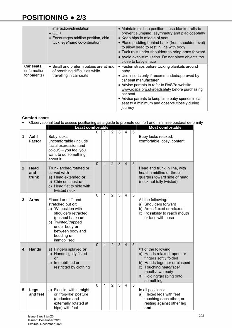
POSITIONING ● 2/3
interaction/stimulation • GOR • Encourages midline position, chin
tuck, eye/hand co-ordination
• Maintain midline position – use blanket rolls to prevent slumping, asymmetry and plagiocephaly
• Keep hips in middle of seat • Place padding behind back (from shoulder level)
to allow head to rest in line with body • Tuck rolls under shoulders to bring arms forward• Avoid over-stimulation. Do not place objects too
close to baby’s faceCar seats (information for parents)
• Small and preterm babies are at risk of breathing difficulties while travelling in car seats
• Fasten straps before tucking blankets around baby
• Use inserts only if recommended/approved by car seat manufacturer
• Advise parents to refer to RoSPa website www.rospa.org.uk/roadsafety before purchasing car seat
• Advise parents to keep time baby spends in car seat to a minimum and observe closely during journey
Comfort score • Observational tool to assess positioning as a guide to promote comfort and minimise postural deformity Least comfortable Most comfortable 1
Aah! Factor
Baby looks uncomfortable (include facial expression and colour) – you feel you want to do something about it
0 1 2 3 4 5 Baby looks relaxed, comfortable, cosy, content
2
Head and trunk
Trunk arched/rotated or curved with a) Head extended or b) Chin on chest or c) Head flat to side with
twisted neck
0 1 2 3 4 5 Head and trunk in line, with head in midline or three-quarters toward side of head (neck not fully twisted)
3
Arms
Flaccid or stiff, and stretched out or: a) ‘W’ position with
shoulders retracted (pushed back) or
b) Twisted/trapped under body or between body and bedding or immobilised
0 1 2 3 4 5 All the following: a) Shoulders forward b) Arms flexed or relaxed c) Possibility to reach mouth
or face with ease
4
Hands
a) Fingers splayed or b) Hands tightly fisted
or c) Immobilised or
restricted by clothing
0 1 2 3 4 5 ≥1 of the following: a) Hands relaxed, open, or
fingers softly folded b) Hands together or clasped c) Touching head/face/
mouth/own body d) Holding/grasping onto
something 5
Legs and feet
a) Flaccid, with straight
or ‘frog-like’ posture (abducted and externally rotated at hips) with feet
0 1 2 3 4 5 In all positions: a) Flexed legs with feet
touching each other, or resting against other leg and
Issue 8 rev1 jan20 Issued: December 2019 Expires: December 2021
292

POSITIONING ● 3/3
pointing outwards or b) Stiff, straight legs
with toes splayed or curled tight, and/or pushing hard on bedding, turning outwards
b) Able to reach boundary to brace feet
In prone position, knees should be tucked under body, feet angled towards each other (not turning out)
6
Arousal
a) Agitated, jerky, jittery
movements and/or b) Fussing or crying c) Unconscious
0 1 2 3 4 5 a) Sleeping restfully or quietly
awake b) Minimal or smooth
movement Total (Max score = 30) Reproduced with permission (Inga Warren, consultant occupational therapist, Winnicott Baby Unit)
Issue 8 rev1 jan20 Issued: December 2019 Expires: December 2021
293
POST HAEMORRHAGIC VENTRICULAR DILATATION ● 1/2 INTRODUCTION • Post haemorrhagic ventricular dilatation (PHVD) develops in 20−30% of babies with severe
intraventricular haemorrhage (IVH). Approximately 20−40% of these babies with severe germinal matrix haemorrhage (GMH)-IVH will go on to require a permanent ventriculo-peritoneal (VP) shunt
• PHVD presents as: • acute: evident within days as ballooning of the ventricles • subacute/chronic: evident within weeks • Current accepted treatment: repeated therapeutic lumbar punctures (LPs) or CSF tapping from
temporising ventricular access device (VAD), with aim of reducing the pressure effect caused by progressive ventricular enlargement, and removing red cells and protein from CSF once standard threshold for treatment reached
RECOGNITION AND ASSESSMENT Risk factors • Prematurity • Severe GMH-IVH with ventricular dilatation (>50% ventricular dilatation with ballooning of ventricle) • Acute process due to impairment of CSF absorption and circulation associated with blood clots • subacute-chronic form with obliterative arachnoiditis in the posterior fossa
SYMPTOMS AND SIGNS • Increase in lateral ventricular dimensions on serial cranial ultrasounds − measured as ventricular index
(VI), and/or increase in anterior horn width (AHW) of the ventricle at the level of the 3rd ventricle in coronal views
• Rapid increase in occipitofrontal circumference (OFC) • Symptoms of increased intracranial pressure (ICP) lag by 1−3 weeks and consist of: • full fontanelle • separated sutures • apnoea • poor feeding • irritability • increased/altered neurological tone • seizures
MONITORING AND INVESTIGATIONS Cranial ultrasound • Perform twice weekly following large GMH-IVH to monitor evolution of PHVD • Assess lateral ventricular size with 2 standard measurements taken at the level of the 3rd ventricle in the
coronal view • VI: distance between falx and lateral wall of anterior horn of lateral ventricle (plot on Levene’s chart) • AHW (to measure ballooning of ventricle)
− AHW >4 mm indicative of enlarged ventricles in keeping with VI >97th centile + 4 mm • Measure resistive index (RI) of anterior cerebral artery to assess raised ICP • end diastolic velocity decreases as ICP increases, causing RI to increase >0.85 • RI >1.0 indicates impaired perfusion in absence of PDA • Repeat cranial ultrasound scan after therapeutic LP to assess VI, with aim to reduce VI below threshold
limit of treatment (<97th centile + 4 mm on Levene’s chart) Head circumference/OFC • Measure OFC twice weekly and before and after intervention with LP • Normal OFC growth: • 26−32 weeks: 1 mm/day • ≥33 weeks−term: 0.7 mm/day • Head circumference growth accelerates with elevated CSF (OFC growth lags behind ventricular
enlargement by 1−3 weeks) • increase of >2 mm/day over 2 days, or 14 mm over 7 days, is excessive Cerebral function monitoring (CFM) and EEG • Use CFM to monitor for suspected seizures • confirm with full EEG • Lack of normal background activity associated with poorer outcome
Issue 8 rev1 jan20 Issued: December 2019 Expires: December 2021
294
POST HAEMORRHAGIC VENTRICULAR DILATATION ● 2/2
MRI • Before insertion of VP shunt • Neurosurgeons may use MRI before insertion of VAD in selective cases
TREATMENT • Threshold for intervention: • VI >97th centile + 4 mm on Levene’s chart for appropriate gestational age and/or • OFC increase >4 mm over 2 days/>14 mm in 7 days and/or • increase in AHW >4 mm • Therapeutic LP to reduce CSF pressure through drainage of CSF • before LP maintain:
− platelet count >50 − clotting profile in normal range
• Aseptic LP to remove ≥10 mL/kg CSF at rate of 1 mL/kg/min • If rapid increase in OFC despite initial LP, repeat • Do not exceed >15 mL/kg of CSF volume at one time • removal of larger volumes of CSF faster than 1 mL/kg/min can result in apnoea, bradycardia and
desaturation • Send CSF for biochemical, microscopy and culture analysis each time LP performed • Following therapeutic LP, repeat cranial ultrasound scan to assess VI; aim to reduce to below threshold
limit • Discuss with neurosurgical team while carrying out above intervention • Refer to neurosurgical team for consideration of insertion of CSF reservoir/VAD if: • LPs unsuccessful in draining CSF due to non-communication between ventricles and spinal canal in 2
consecutive attempts and • VIs remain above threshold for intervention and/or • OFC continues to increase • Consider ventricular tap under ultrasound guidance as a bridge to surgery for insertion of VAD • avoid many ventricular taps − high risk of causing needle tract intra-parenchymal injury and infection • If repeated prolonged tapping via VAD required to maintain normal head growth/ persistent rapid rise in
OFC/baby remains symptomatic − discuss with neurosurgeon for consideration of VP shunts • CSF protein to be <1.5 g m/L and weight to be >2 kg (in most cases)
SUBSEQUENT MANAGEMENT • Monitor serum sodium levels − increased risk of hyponatraemia with repeated CSF drainage • Maintain sodium >140 mmol/L; supplement intake as necessary • If therapeutic tap from VAD >12−15 mL/kg/tap, replace CSF volumes with sodium chloride 0.9% IV fluid
bolus to avoid hypovolaemia and decreased cerebral perfusion • if CSF volumes <12 mL/kg/tap, and done at 1 mL/kg/min, fluid bolus not required unless baby
haemodynamically unstable • Treat seizures (see Seizures guideline) • Refer to physiotherapy service
INFORMATION FOR PARENTS • On diagnosis of PHVD, most senior clinician available to fully update parents
PROGNOSIS • Marked cognitive impairment (mental developmental quotient <70) seen in approximately 45−60% of
babies with PHVD, along with impaired motor outcomes • Need for VP shunt worsens long-term neurodevelopmental outcomes
Issue 8 rev1 jan20 Issued: December 2019 Expires: December 2021
295
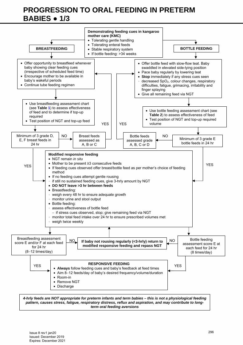
PROGRESSION TO ORAL FEEDING IN PRETERM BABIES ● 1/3
YES
YES
NO
YES YES
NO NO
YES
NO
YES
BREASTFEEDING
• Offer opportunity to breastfeed whenever baby showing clear feeding cues (irrespective of scheduled feed time)
• Encourage mother to be available in baby’s wakeful periods
• Continue tube feeding regimen
• Offer bottle feed with slow-flow teat. Baby swaddled in elevated side-lying position
• Pace baby regularly by lowering teat • Stop immediately if any stress cues seen • decreased SpO2, colour changes, respiratory
difficulties, fatigue, grimacing, irritability and finger splaying
• Give all remaining feed via NGT
• Use breastfeeding assessment chart (see Table 1) to assess effectiveness of feed and to determine if top-up required
• Test position of NGT and top-up feed
Minimum of 3 grade D, E, F breast feeds in
24 hr
Breast feeds assessed as
A, B or C
Bottle feeds assessed grade
A, B, C or D
• Use bottle feeding assessment chart (see Table 2) to assess effectiveness of feed
• Test position of NGT and top-up required volume
Minimum of 3 grade E bottle feeds in 24 hr
Breastfeeding assessment score E and/or F at each feed
for 24 hr (8−12 times/day)
If baby not rousing regularly (<3-hrly) return to modified responsive feeding and repass NGT
Bottle feeding assessment score E at
each feed for 24 hr (8 times/day)
RESPONSIVE FEEDING• Always follow feeding cues and baby’s feedback at feed times • Aim 8−12 feeds/day of baby’s desired frequency/volume/duration • Room-in • Remove NGT • Discharge
4-hrly feeds are NOT appropriate for preterm infants and term babies – this is not a physiological feeding pattern, causes stress, fatigue, respiratory distress, reflux and aspiration, and may contribute to long-
term oral feeding aversions
Demonstrating feeding cues in kangaroo mother care (KMC) • Tolerating gentle handling • Tolerating enteral feeds • Stable respiratory system • If bottle feeding: >34 weeks
BOTTLE FEEDING
Modified responsive feeding • NGT remain in situ • Mother to be present ≥3 consecutive feeds • If feeding cues observed offer breast/bottle feed as per mother’s choice of feeding
method • If no feeding cues attempt gentle rousing • if still no sustained feeding cues, give 3-hrly amount by NGT • DO NOT leave >3 hr between feeds • Breastfeeding: • weigh every 48 hr to ensure adequate growth • monitor urine and stool output • Bottle feeding: • assess effectiveness of bottle feed
− if stress cues observed, stop; give remaining feed via NGT • monitor total feed intake over 24 hr to ensure prescribed volumes met • weigh twice weekly
Issue 8 rev1 jan20 Issued: December 2019 Expires: December 2021
296
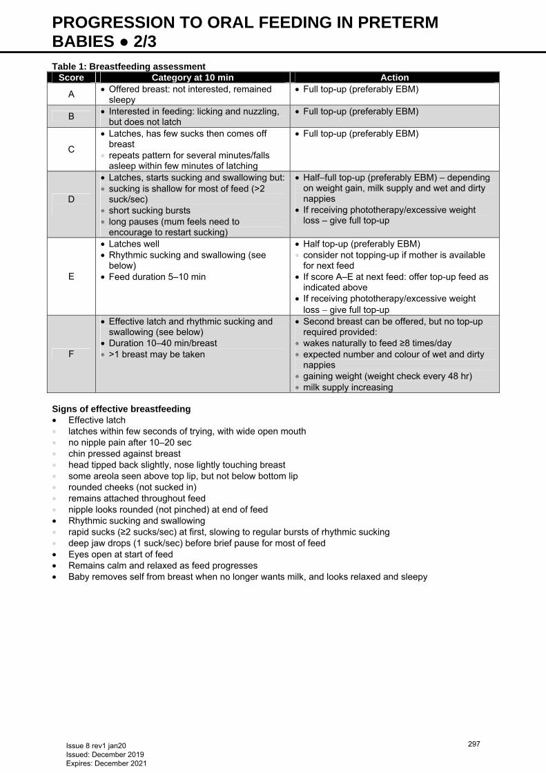
PROGRESSION TO ORAL FEEDING IN PRETERM BABIES ● 2/3 Table 1: Breastfeeding assessment
Score Category at 10 min Action
A • Offered breast: not interested, remained sleepy
• Full top-up (preferably EBM)
B • Interested in feeding: licking and nuzzling, but does not latch
• Full top-up (preferably EBM)
C
• Latches, has few sucks then comes off breast
• repeats pattern for several minutes/falls asleep within few minutes of latching
• Full top-up (preferably EBM)
D
• Latches, starts sucking and swallowing but:• sucking is shallow for most of feed (>2
suck/sec) • short sucking bursts • long pauses (mum feels need to
encourage to restart sucking)
• Half–full top-up (preferably EBM) – depending on weight gain, milk supply and wet and dirty nappies
• If receiving phototherapy/excessive weight loss – give full top-up
E
• Latches well • Rhythmic sucking and swallowing (see
below) • Feed duration 5–10 min
• Half top-up (preferably EBM) • consider not topping-up if mother is available
for next feed • If score A–E at next feed: offer top-up feed as
indicated above • If receiving phototherapy/excessive weight
loss − give full top-up
F
• Effective latch and rhythmic sucking and swallowing (see below)
• Duration 10–40 min/breast • >1 breast may be taken
• Second breast can be offered, but no top-up required provided:
• wakes naturally to feed ≥8 times/day • expected number and colour of wet and dirty
nappies • gaining weight (weight check every 48 hr) • milk supply increasing
Signs of effective breastfeeding • Effective latch • latches within few seconds of trying, with wide open mouth • no nipple pain after 10–20 sec • chin pressed against breast • head tipped back slightly, nose lightly touching breast • some areola seen above top lip, but not below bottom lip • rounded cheeks (not sucked in) • remains attached throughout feed • nipple looks rounded (not pinched) at end of feed • Rhythmic sucking and swallowing • rapid sucks (≥2 sucks/sec) at first, slowing to regular bursts of rhythmic sucking • deep jaw drops (1 suck/sec) before brief pause for most of feed • Eyes open at start of feed • Remains calm and relaxed as feed progresses • Baby removes self from breast when no longer wants milk, and looks relaxed and sleepy
Issue 8 rev1 jan20 Issued: December 2019 Expires: December 2021
297
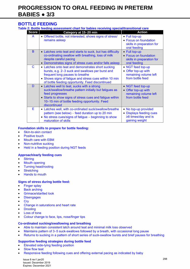
PROGRESSION TO ORAL FEEDING IN PRETERM BABIES ● 3/3 BOTTLE FEEDING Table 2: Bottle feeding assessment chart for babies receiving special/transitional care
Score Category at 15−20 min Action A • Offered bottle, not interested, shows signs of stress/
remains asleep • Full top-up • Focus on foundation
skills in preparation for oral feeding
B • Latches onto teat and starts to suck, but has difficulty co-ordinating swallow with breathing, loss of milk despite careful pacing
• Demonstrates signs of stress cues and/or falls asleep
• Full top-up • Focus on foundation
skills in preparation for oral feeding
C • Latches onto teat and demonstrates short sucking bursts, e.g. 2−3 suck and swallows per burst and frequent long pauses to breathe
• Shows signs of fatigue and stress cues within 10 min of bottle feeding opportunity. Feed discontinued
• NGT feed top-up • Offer top-up with
remaining volume left from bottle feed
D • Latches well to teat, sucks with a strong suck/swallow/breathe pattern initially but fatigues as feed progresses
• Starts to show signs of stress cues and fatigue within 10−15 min of bottle feeding opportunity. Feed discontinued
• NGT feed top-up • Offer top-up with
remaining volume left from bottle feed
E • Latches well, with co-ordinated suck/swallow/breathe pattern (see below) − feed duration up to 20 min
• No stress cues/signs of fatigue − beginning to show maturation of skills
• No top-up provided • Displays feeding cues ≥8 times/day and is gaining weight
Foundation skills to prepare for bottle feeding: • Skin-to-skin contact • Positive touch • Mouth care with EBM • Non-nutritive sucking • Held in a feeding position during NGT feeds Approach/early feeding cues • Stirring • Mouth opening • Turning head/rooting • Stretching • Hands to mouth Signs of stress during bottle feed: • Finger splay • Back arching • Grimace/startled look • Disengages • Cry • Change in saturations and heart rate • Drooling • Loss of tone • Colour change to face, lips, nose/finger tips Co-ordinated sucking/swallowing and breathing • Able to maintain consistent latch around teat and minimal milk loss observed • Maintains pattern of 3−5 suck-swallows followed by a breath, with occasional long pause • Returns to sucking in a pattern of short series of suck-swallow bursts and brief pauses for breathing
Supportive feeding strategies during bottle feed • Elevated side-lying feeding position • Slow flow teat • Responsive feeding following cues and offering external pacing as indicated by baby
Issue 8 rev1 jan20 Issued: December 2019 Expires: December 2021
298
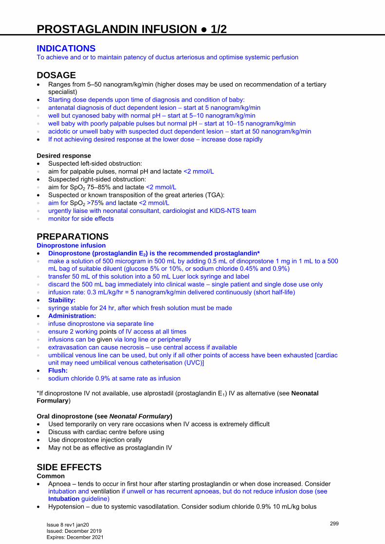
PROSTAGLANDIN INFUSION ● 1/2 INDICATIONS To achieve and or to maintain patency of ductus arteriosus and optimise systemic perfusion DOSAGE • Ranges from 5–50 nanogram/kg/min (higher doses may be used on recommendation of a tertiary
specialist) • Starting dose depends upon time of diagnosis and condition of baby: • antenatal diagnosis of duct dependent lesion − start at 5 nanogram/kg/min • well but cyanosed baby with normal pH – start at 5−10 nanogram/kg/min • well baby with poorly palpable pulses but normal pH − start at 10−15 nanogram/kg/min • acidotic or unwell baby with suspected duct dependent lesion − start at 50 nanogram/kg/min • If not achieving desired response at the lower dose − increase dose rapidly Desired response • Suspected left-sided obstruction: • aim for palpable pulses, normal pH and lactate <2 mmol/L • Suspected right-sided obstruction: • aim for SpO2 75–85% and lactate <2 mmol/L • Suspected or known transposition of the great arteries (TGA): • aim for SpO2 >75% and lactate <2 mmol/L • urgently liaise with neonatal consultant, cardiologist and KIDS-NTS team • monitor for side effects PREPARATIONS Dinoprostone infusion • Dinoprostone (prostaglandin E2) is the recommended prostaglandin* • make a solution of 500 microgram in 500 mL by adding 0.5 mL of dinoprostone 1 mg in 1 mL to a 500
mL bag of suitable diluent (glucose 5% or 10%, or sodium chloride 0.45% and 0.9%) • transfer 50 mL of this solution into a 50 mL Luer lock syringe and label • discard the 500 mL bag immediately into clinical waste – single patient and single dose use only • infusion rate: 0.3 mL/kg/hr = 5 nanogram/kg/min delivered continuously (short half-life) • Stability: • syringe stable for 24 hr, after which fresh solution must be made • Administration: • infuse dinoprostone via separate line • ensure 2 working points of IV access at all times • infusions can be given via long line or peripherally • extravasation can cause necrosis – use central access if available • umbilical venous line can be used, but only if all other points of access have been exhausted [cardiac
unit may need umbilical venous catheterisation (UVC)] • Flush: • sodium chloride 0.9% at same rate as infusion *If dinoprostone IV not available, use alprostadil (prostaglandin E1) IV as alternative (see Neonatal Formulary) Oral dinoprostone (see Neonatal Formulary) • Used temporarily on very rare occasions when IV access is extremely difficult • Discuss with cardiac centre before using • Use dinoprostone injection orally • May not be as effective as prostaglandin IV
SIDE EFFECTS Common • Apnoea – tends to occur in first hour after starting prostaglandin or when dose increased. Consider
intubation and ventilation if unwell or has recurrent apnoeas, but do not reduce infusion dose (see Intubation guideline)
• Hypotension – due to systemic vasodilatation. Consider sodium chloride 0.9% 10 mL/kg bolus
Issue 8 rev1 jan20 Issued: December 2019 Expires: December 2021
299
PROSTAGLANDIN INFUSION ● 2/2 • Fever • Tachycardia • Hypoglycaemia Uncommon • Hypothermia • Bradycardia • Convulsions • Cardiac arrest • Diarrhoea • Disseminated intravascular coagulation (DIC) • Gastric outlet obstruction • Cortical hyperostosis • Gastric hyperplasia (prolonged use)
MONITOR • Heart rate • Blood pressure • Respiratory rate • Temperature • Oxygen saturations • Blood gases • Blood glucose and lactate
TRANSFER OF BABY RECEIVING PROSTAGLANDIN INFUSION • Contact local retrieval team for transport of baby to cardiac centre (e.g. for Birmingham Children’s
Hospital − contact KIDS-NTS team on 0300 200 1100) • Keep baby nil-by-mouth for transfer • In a well baby on prostaglandin ≤10 nanogram/kg/min, risk of apnoea is low
Issue 8 rev1 jan20 Issued: December 2019 Expires: December 2021
300
PULMONARY HAEMORRHAGE ● 1/2 RECOGNITION AND ASSESSMENT Definition • Acute onset of bleeding from ororpharynx or ETT associated with cardiorespiratory deterioration and
changes on chest X-ray • Significant pulmonary haemorrhage is most likely to represent haemorrhagic pulmonary oedema.
Differentiate from minor traumatic haemorrhage following endotracheal suction Risk factors • Prematurity • Respiratory distress syndrome (RDS) • Large patent ductus arteriosus (PDA) • Excessive use of volume (>20 mL/kg) in first 24–48 hr in babies ≤28 weeks’ gestation • Coagulopathy • Sepsis • IUGR • Grade 3 hypoxic ischaemic encephalopathy (HIE) Symptoms and signs • Apnoeas, gasping respirations, desaturations • Tachycardia >160 bpm, bradycardia, hypotension, shock, PDA, signs of heart failure • Widespread crepitations, reduced air entry • Pink/red frothy expectorate, or frank blood from oropharynx or ETT if intubated Investigations • Blood gas (expect hypoxia and hypercapnia with mixed acidosis) • FBC, clotting • Chest X-ray (usually shows classic white-out with only air bronchogram visible but may be less striking
and resemble RDS)
IMMEDIATE TREATMENT • Basic resuscitation, ABC Respiratory • Intubate and ventilate • if already intubated be cautious removing ETT – may be very difficult to reintubate • Sedate and give muscle relaxant • PEEP 6–8 cm, even higher PEEP of 10–12 cm H2O sometimes required to control haemorrhage • PIP to be guided by chest expansion and blood gases • Long inspiratory times (0.5 sec may be needed) • Endotracheal suction (try to avoid but consider in extreme cases to reduce risk of ETT blockage) • Ensure adequate humidification • Chest physiotherapy contraindicated until active bleeding stopped and platelets >50 (see Chest
physiotherapy guideline) • Establish arterial access Fluid management • If hypovolaemic, restore circulating volume over 30 min with 10 mL/kg sodium chloride 0.9% or Group O
RhD negative packed cells if crystalloid bolus already given. Beware of overloading (added volume can be detrimental to LV failure)
• If not hypovolaemic and evidence of LV failure, give furosemide 1 mg/kg IV • Correct acidosis (see Neonatal Formulary) • If PDA present, restrict fluids to 60–80 mL/kg/day in acute phase • Further blood transfusion, vitamin K administration and FFP to be guided by Hb concentration, PT and
APTT (see Transfusion of red blood cells and Coagulopathy guidelines) Hypotension/cardiac dysfunction • If still hypotensive or evidence of cardiac dysfunction after fluid resuscitation, treat hypotension with
inotropes (see Hypotension guideline) Infection • If infection suspected, request septic screen and start antibiotics
Issue 8 rev1 jan20 Issued: December 2019 Expires: December 2021
301
PULMONARY HAEMORRHAGE ● 2/2 SUBSEQUENT MANAGEMENT Once baby stable • Inform on-call consultant • Speak to parents • Document event in case notes • Consider single extra dose of surfactant in babies with severe hypoxaemia or oxygenation index >20 • If PDA suspected, arrange echocardiogram (see Patent ductus arteriosus guideline) • Perform cranial ultrasound scan to exclude intracranial haemorrhage as this may influence management
(see Cranial ultrasound scans guideline)
Issue 8 rev1 jan20 Issued: December 2019 Expires: December 2021
302
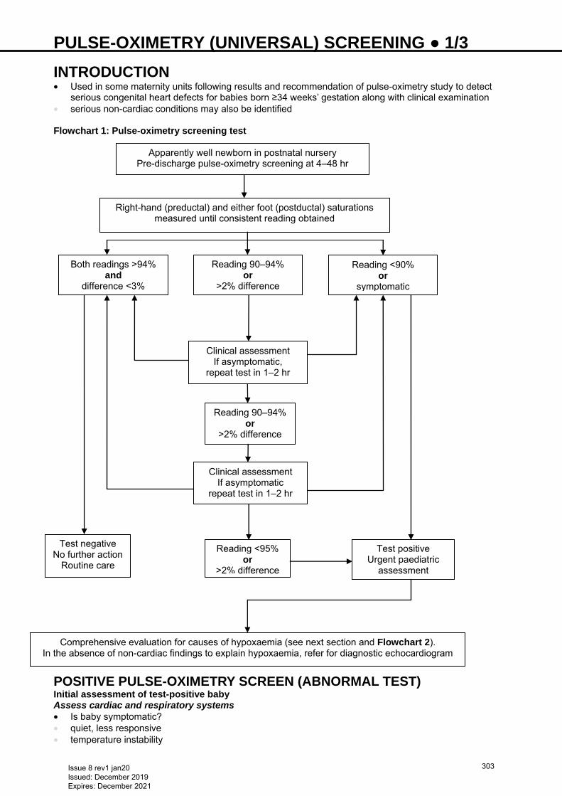
PULSE-OXIMETRY (UNIVERSAL) SCREENING ● 1/3
INTRODUCTION • Used in some maternity units following results and recommendation of pulse-oximetry study to detect
serious congenital heart defects for babies born ≥34 weeks’ gestation along with clinical examination • serious non-cardiac conditions may also be identified Flowchart 1: Pulse-oximetry screening test
POSITIVE PULSE-OXIMETRY SCREEN (ABNORMAL TEST) Initial assessment of test-positive baby Assess cardiac and respiratory systems • Is baby symptomatic? • quiet, less responsive • temperature instability
Apparently well newborn in postnatal nursery Pre-discharge pulse-oximetry screening at 4–48 hr
Right-hand (preductal) and either foot (postductal) saturations measured until consistent reading obtained
Both readings >94% and
difference <3%
Reading 90–94% or
>2% difference
Reading <90% or
symptomatic
Clinical assessment If asymptomatic,
repeat test in 1–2 hr
Reading 90–94% or
>2% difference
Clinical assessment If asymptomatic
repeat test in 1–2 hr
Test negative No further action
Routine care
Test positive Urgent paediatric
assessment
Reading <95% or
>2% difference
Comprehensive evaluation for causes of hypoxaemia (see next section and Flowchart 2). In the absence of non-cardiac findings to explain hypoxaemia, refer for diagnostic echocardiogram
Issue 8 rev1 jan20 Issued: December 2019 Expires: December 2021
303
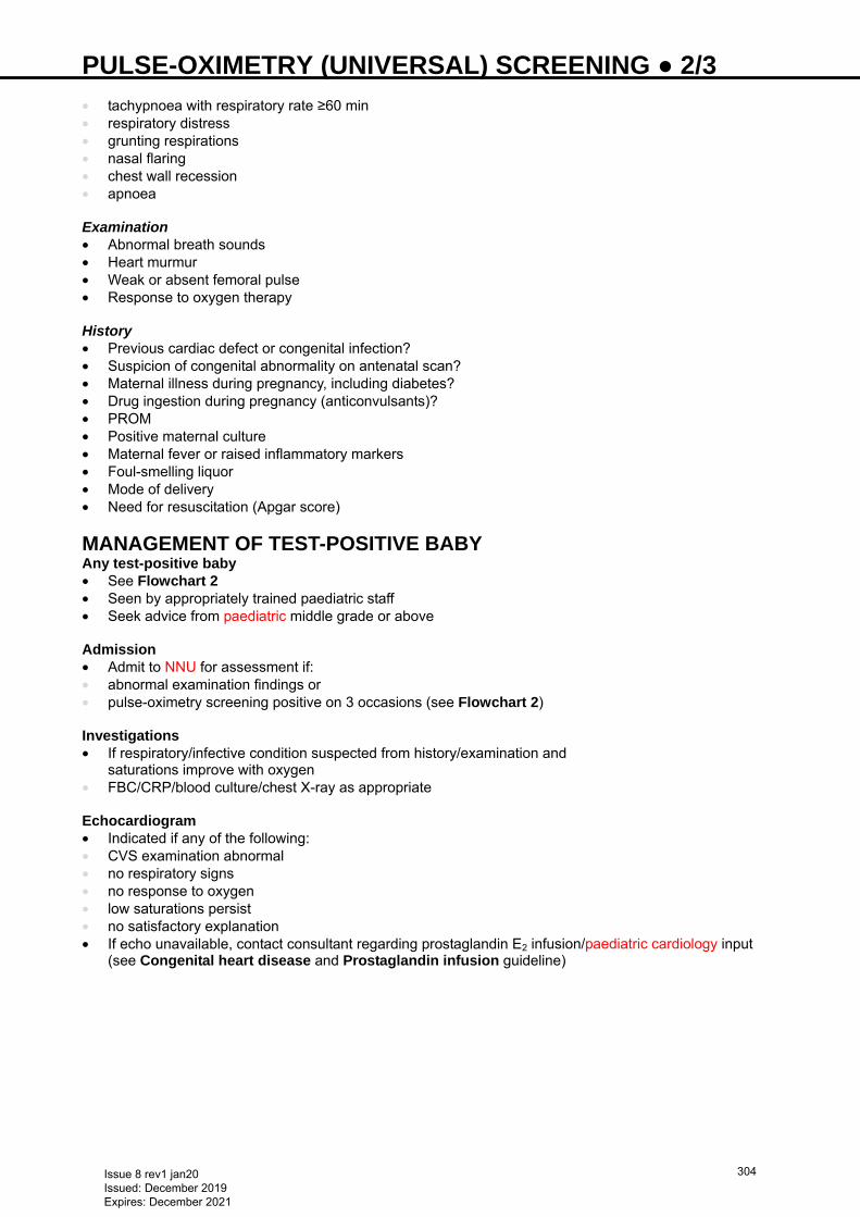
PULSE-OXIMETRY (UNIVERSAL) SCREENING ● 2/3 • tachypnoea with respiratory rate ≥60 min • respiratory distress • grunting respirations • nasal flaring • chest wall recession • apnoea Examination • Abnormal breath sounds • Heart murmur • Weak or absent femoral pulse • Response to oxygen therapy History • Previous cardiac defect or congenital infection? • Suspicion of congenital abnormality on antenatal scan? • Maternal illness during pregnancy, including diabetes? • Drug ingestion during pregnancy (anticonvulsants)? • PROM • Positive maternal culture • Maternal fever or raised inflammatory markers • Foul-smelling liquor • Mode of delivery • Need for resuscitation (Apgar score)
MANAGEMENT OF TEST-POSITIVE BABY Any test-positive baby • See Flowchart 2 • Seen by appropriately trained paediatric staff • Seek advice from paediatric middle grade or above Admission • Admit to NNU for assessment if: • abnormal examination findings or • pulse-oximetry screening positive on 3 occasions (see Flowchart 2) Investigations • If respiratory/infective condition suspected from history/examination and
saturations improve with oxygen • FBC/CRP/blood culture/chest X-ray as appropriate Echocardiogram • Indicated if any of the following: • CVS examination abnormal • no respiratory signs • no response to oxygen • low saturations persist • no satisfactory explanation • If echo unavailable, contact consultant regarding prostaglandin E2 infusion/paediatric cardiology input
(see Congenital heart disease and Prostaglandin infusion guideline)
Issue 8 rev1 jan20 Issued: December 2019 Expires: December 2021
304
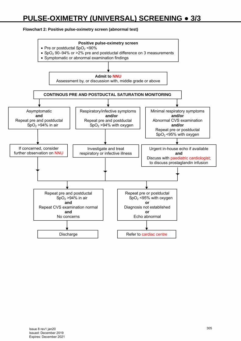
PULSE-OXIMETRY (UNIVERSAL) SCREENING ● 3/3 Flowchart 2: Positive pulse-oximetry screen (abnormal test) Positive pulse-oximetry screen
• Pre or postductal SpO2 <90% • SpO2 90–94% or >2% pre and postductal difference on 3 measurements • Symptomatic or abnormal examination findings
Admit to NNU Assessment by, or discussion with, middle grade or above
Asymptomatic and
Repeat pre and postductal SpO2 >94% in air
Respiratory/infective symptoms and/or
Repeat pre and postductal SpO2 >94% with oxygen
Minimal respiratory symptoms and/or
Abnormal CVS examination and/or
Repeat pre or postductal SpO2 <95% with oxygen
If concerned, consider further observation on NNU
Investigate and treat respiratory or infective illness
Urgent in-house echo if available and
Discuss with paediatric cardiologist; to discuss prostaglandin infusion
Repeat pre and postductal SpO2 >94% in air
and Repeat CVS examination normal
and No concerns
Repeat pre or postductal SpO2 <95% with oxygen
or Diagnosis not established
or Echo abnormal
Refer to cardiac centre Discharge
CONTINOUS PRE AND POSTDUCTAL SATURATION MONITORING
Issue 8 rev1 jan20 Issued: December 2019 Expires: December 2021
305
RECTAL WASHOUT USING SYRINGE METHOD ● 1/2 INDICATIONS • Suspected or confirmed Hirschsprung’s disease • Suspected meconium plugs
BENEFITS • Bowel decompression • Establishment of feeding • Weight gain • Reduced risk of colitis
CONTRAINDICATIONS • Rectal biopsies taken in preceding 24 hr • Rectal bleeding (relative contraindication) • Severe anal stenosis • Anus not clearly identified • Known surgical patient (without discussion with surgical team)
ADVERSE REACTIONS • Bleeding from anus or rectum • Perforation of bowel (this is very rare) • Electrolyte imbalance if inappropriate fluid used or retained • Vomiting • Hypothermia • Distress to baby and parent Consent • Explain procedure to parents/carer and obtain verbal consent Equipment • Tube size 6–10 Fr (recommended: Conveen® Easicath pre-lubricated catheter) • Lubricating gel (if catheter not lubricated) • Bladder tip syringe no smaller than 60 mL • Rectal washout solution (sodium chloride 0.9%) warmed to room temperature • Plastic apron • Gloves • Protective sheet • Receptacle to collect effluent • Container for clean rectal washout solution • Blanket to wrap baby Preparation • Place all equipment at cot side • Sedation is not necessary • Second person to comfort baby using dummy and breast milk/sucrose [see Non-nutritive sucking
(NNS) guideline] • Wash hands, put on gloves and apron • Position baby supine with legs raised • Keep baby warm
PROCEDURE • Inspect and palpate abdomen – note distension or presence of lumps • Draw up sodium chloride 0.9% 60 mL into syringe and keep on one side • Insert lubricated catheter into rectum [up to approximately 10 cm (in a term baby) or until resistance felt]
noting any flatus or faecal fluid drained • Massage abdomen in a clockwise direction to release flatus • Attach syringe containing sodium chloride to tube in rectum and gently instil fluid:
Weight <2 kg 5–10 mL Weight ≥2 kg 20 mL
Issue 8 rev1 jan20 Issued: December 2019 Expires: December 2021
306
RECTAL WASHOUT USING SYRINGE METHOD ● 2/2 • Disconnect syringe from tube and drain effluent into receptacle • Repeat procedure until drained solution becomes clearer, up to a maximum of 3 times • If solution does not drain out, manipulate tube in and out and massage abdomen • If no faeces are passed or all the solution is retained, seek medical help • Re-examine abdomen and note any differences • Wash, dress and comfort baby Preparation for discharge • For discharge, baby should require ≤2 rectal washouts a day • Order equipment via paediatric community nurse • Ward will supply 5 days’ equipment • Discharge letter for GP detailing equipment required • Arrange home visit with clinical nurse specialist in stoma care if available locally • Ensure parents competent to perform rectal washout and can describe signs of colitis • complete rectal washout parent competency sheet if available locally
Issue 8 rev1 jan20 Issued: December 2019 Expires: December 2021
307
RE-CYCLING OF STOMA LOSSES VIA A MUCUS FISTULA ● 1/2 INDICATIONS • Stoma output >30 mL/kg/day term baby (>20 mL/kg/day for preterm baby) • Discrepancy in proximal and distal bowel calibre • Inability to absorb increasing enteral feeds • Failure to thrive • Developing cholestasis
BENEFITS • Maximise nutrition for sustained weight gain and decrease in parenteral nutrition • Stimulation of gut hormones and enzymes • Increases absorption of water, electrolytes and nutrients by utilising distal bowel • Digestive tract matures and increases in length and diameter with use • Adaptation is driven by enteral feed in distal bowel • Preparation of distal bowel for closure • Baby can, in some circumstances, be managed at home
CONTRAINDICATIONS • Diseased or compromised distal bowel • Rectal bleeding (not absolute but discuss with surgical team) • Anal stenosis or imperforate anus • Signs of systemic infection • Effluent too thick to infuse
ADVERSE REACTIONS • Bleeding from distal stoma • Perforation of bowel by catheter (rare) • Leakage of stoma effluent onto peri-stomal skin may result in excoriation of the skin • Distress to baby and parent • Sepsis due to translocation Before commencing • Discuss with surgical team • confirm they agree with procedure • whether distal contrast study is required before re-cycling Consent • Explain procedure and potential adverse reactions to parents and obtain verbal consent Equipment • Tube (enteral or Foley catheter) size 6 or 8 Fr • Lubricating gel (if catheter not lubricated) • Enteral syringe (60 mL) • Stoma pot to collect stoma effluent • Extension tubing • Syringe pump (enteral pump if available) • Plastic apron and gloves • Tape and dressing
Documentation • Record name of surgeon requesting procedure in baby’s notes (when commencing) • Record condition of peri-stomal skin pre-procedure
Preparation • Place all necessary equipment at cot side • Wash hands and put on gloves and apron • Position baby in supine position and keep warm
PROCEDURE • Confirm which visible stoma is the mucus fistula – operation note or surgical team • Pass lubricated catheter into mucus fistula up to 2 cm past end holes
Issue 8 rev1 jan20 Issued: December 2019 Expires: December 2021
308
RE-CYCLING OF STOMA LOSSES VIA A MUCUS FISTULA ● 2/2 • If using a Foley catheter put only 0.5 mL water into balloon • Secure catheter to the abdomen with Duoderm®, tape and leave in situ • Cover mucus fistula with paraffin gauze dressing (e.g. Jelonet) • Collect stoma fluid from acting stoma into enteral syringe, connect to catheter via extension tube and
start re-cycling using syringe pump • Aim to infuse stoma loss over a few hours, but ≤4 hr. Discard any effluent older than 4 hr • If stoma loss <5 mL, re-cycle by syringe as a slow bolus over a few minutes • Re-cycling should result in bowel actions per rectum of a consistency thicker than the stoma loss • If bowel actions per rectum are watery and/or frequent, send samples for culture and sensitivity, virology
and detection of fat globules and reducing substances. Discuss with surgical team • If baby develops signs suggestive of sepsis, stop procedure and perform septic screen as per unit policy.
Discuss with surgical team Preparation for home • Liaise with neonatal surgical nurse • Teach parents the procedure • Order equipment via paediatric community nurse • Ward will supply 5 days’ equipment • Discharge letter for GP detailing equipment required • Arrange home visit with clinical nurse specialist in stoma care • Inform surgical team before discharge
Issue 8 rev1 jan20 Issued: December 2019 Expires: December 2021
309
RENAL FAILURE ● 1/4
DEFINITION • Failure of the kidneys to maintain metabolic stability in relation to fluid balance, electrolyte balance and
excretion of nitrogenous waste • Serum creatinine shortly after birth is a reflection of maternal renal function
MAIN CAUSES Prenatal injury/vascular damage • Maternal use of: • ACE inhibitors, angiotensin 2 receptor antagonist • NSAID Congenital renal disorders • Renal agenesis • Renal dysplasia/hypoplasia • Polycystic kidney disease • Congenital nephrotic syndrome (Finnish type) Postnatal renal disease Pre-renal • Decreased intravascular volume/tissue perfusion • Perinatal haemorrhage • Dehydration • Hypotension • Third space losses (sepsis, NEC) • Congestive cardiac failure • Pericardial tamponade Intrinsic renal • Acute tubular necrosis • Perinatal asphyxia • Drug induced • aminoglycosides • amphoteresin B • IV contrast media • NSAID • ACE inhibitors • Sepsis • Renal artery/vein thrombosis Post-renal/obstructive • Posterior urethral valve • Obstruction in a single kidney • Neurogenic bladder due to meningomyelocele • Inappropriate ADH in ventilated babies causes transient oliguria • will correct spontaneously as lung compliance improves
HISTORY AND EXAMINATION • Evaluate to differentiate between pre-renal, intrinsic or post-renal problem • Detailed clinical history • assessment of gestational age • antenatal ultrasound scans • maternal medications (nephrotoxic) • birth history
− fetal heart rate monitoring − resuscitation
• postnatal events (e.g. hypotension, nephrotoxic medications) • Clinical assessment for volume status • signs of depletion/hypovolaemia
− cold peripheries
Issue 8 rev1 jan20 Issued: December 2019 Expires: December 2021
310
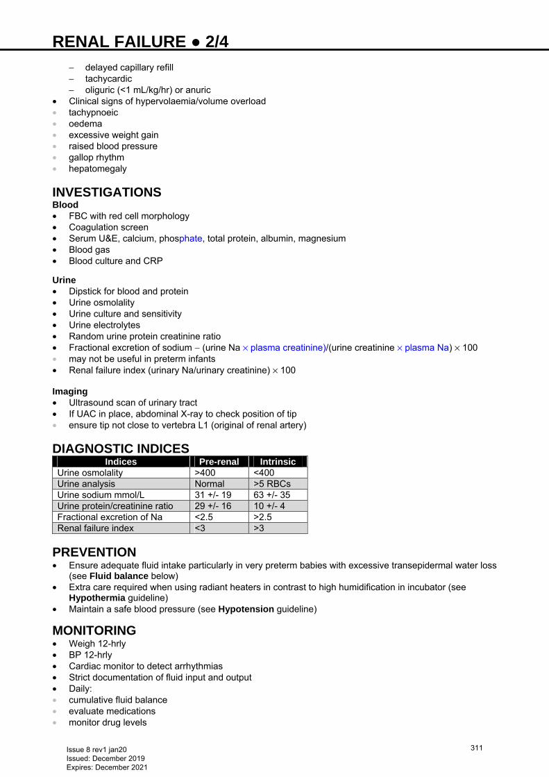
RENAL FAILURE ● 2/4
− delayed capillary refill − tachycardic − oliguric (<1 mL/kg/hr) or anuric
• Clinical signs of hypervolaemia/volume overload • tachypnoeic • oedema • excessive weight gain • raised blood pressure • gallop rhythm • hepatomegaly
INVESTIGATIONS Blood • FBC with red cell morphology • Coagulation screen • Serum U&E, calcium, phosphate, total protein, albumin, magnesium • Blood gas • Blood culture and CRP Urine • Dipstick for blood and protein • Urine osmolality • Urine culture and sensitivity • Urine electrolytes • Random urine protein creatinine ratio • Fractional excretion of sodium − (urine Na × plasma creatinine)/(urine creatinine × plasma Na) × 100 • may not be useful in preterm infants • Renal failure index (urinary Na/urinary creatinine) × 100 Imaging • Ultrasound scan of urinary tract • If UAC in place, abdominal X-ray to check position of tip • ensure tip not close to vertebra L1 (original of renal artery)
DIAGNOSTIC INDICES Indices Pre-renal Intrinsic
Urine osmolality >400 <400 Urine analysis Normal >5 RBCs Urine sodium mmol/L 31 +/- 19 63 +/- 35 Urine protein/creatinine ratio 29 +/- 16 10 +/- 4 Fractional excretion of Na <2.5 >2.5 Renal failure index <3 >3
PREVENTION • Ensure adequate fluid intake particularly in very preterm babies with excessive transepidermal water loss
(see Fluid balance below) • Extra care required when using radiant heaters in contrast to high humidification in incubator (see
Hypothermia guideline) • Maintain a safe blood pressure (see Hypotension guideline)
MONITORING • Weigh 12-hrly • BP 12-hrly • Cardiac monitor to detect arrhythmias • Strict documentation of fluid input and output • Daily: • cumulative fluid balance • evaluate medications • monitor drug levels
Issue 8 rev1 jan20 Issued: December 2019 Expires: December 2021
311
RENAL FAILURE ● 3/4
Urine • Dipstick (proteinuria; sediment, e.g. blood, casts, tubular debris, indicate intrinsic problem; WBC and
nitrites suggest infection) • Microscopy and culture • Electrolytes, urea, creatinine, osmolality • Blood • U&E, creatinine 12-hrly (monitor Na and K on blood gas when possible) • Blood gas, pH 4–8 hrly • Glucose 4-hrly • Daily: • calcium • phosphate • magnesium • albumin • FBC
Typical biochemical changes in acute renal failure (ARF) Increased urea, creatinine, K+, PO4
2- Reduced Na+, Ca2+, HCO3
-, pH
• Increasing urine output generally first sign of recovery • Monitor serum electrolyte levels during polyuric phase • Creatinine estimation often misleading in first few days (in-utero creatinine is cleared by placenta) • after delivery creatinine production by muscles is not stable and can be influenced heavily by muscle
damage resulting from delivery/hypoxia/sepsis • >48–72 hr, it can be used, but trend much more valuable than absolute concentration • Urea estimation • can be influenced by tissue breakdown (e.g. bruises/swallowed blood) • little produced when protein intake compromised
TREAMENT Correct underlying cause Pre-renal failure • Correct hypovolaemia − avoid over-hydration in established renal failure • sodium chloride 0.9% 10−20 mL/kg IV • if blood loss known/suspected: give 10−20 mL/kg packed red cells • if hypotensive in absence of fluid depletion: start inotrope infusion (see Hypotension guideline) Intrinsic renal failure • Goal is to limit further renal damage • Management of fluid and electrolyte imbalance and hypertension • In majority of cases kidneys will recover in 24–48 hr
Post-renal failure • Surgical approach to obstructive uropathy unless prognosis hopeless (e.g. Potter’s syndrome) • post-renal obstruction (e.g. posterior urethral valve) can be temporarily relieved by indwelling catheter
until definitive surgical treatment considered • Open duct in duct-dependent circulation in congenital heart disease (see Cardiovascular guidelines) • Antibiotics for sepsis
Supportive • If possible stop all nephrotoxic drugs (e.g. aminoglycosides, vancomycin, furosemide), or monitor levels
if need to continue • Assess fluid status regularly Fluid balance • If baby hypovolaemic/hypotensive it is important to correct this before instituting fluid restriction (see
above) • If signs of fluid overload consider trial of furosemide • Restrict fluid intake to minimal maintenance fluids
Issue 8 rev1 jan20 Issued: December 2019 Expires: December 2021
312
RENAL FAILURE ● 4/4
• Calculate maintenance fluid: • maintenance fluid = insensible losses + urine output + GIT losses • insensible losses (if nursed in incubator):
− <1500 g (at birth) = 50–80 mL/kg/day − >1500 g (at birth) = 15–35 mL/kg/day − for babies in well-humidified incubator or receiving humidified respiratory support, use lower figure
• Replace maintenance fluid as glucose 10–20% (electrolyte-free) • If electrolyte losses ongoing (e.g. diarrhoea, fistula), electrolytes required • Weigh twice daily • change in body weight best guide to change in hydration • stable weight indicates over-hydration and need to reduce fluid intake further • aim to achieve 1% loss of body weight daily Hyperkalaemia • See Hyperkalaemia guideline Acidosis • Monitor pH 4−8 hrly • If metabolic acidosis present with pH <7.2 or HCO3 <12 mmol/L, give sodium bicarbonate • monitor ionised calcium levels to prevent seizures/tetany
Hyponatraemia • Low sodium more likely to indicate fluid overload than deficit in body sodium • Unless evidence of dehydration, treatment should be fluid restriction with maintenance sodium intake of
2–3 mmol/kg/day • If severe (Na <120 mmol/L) and associated with neurological symptoms, e.g. seizures, can use
hypertonic saline (sodium chloride 3%) 4 mL/kg over a minimum of 15 min: check serum sodium immediately after completion of infusion
• If baby still fitting, dose can be repeated after assessing serum sodium concentration • Amount of Na required = (desired Na – actual Na) × 0.6 × weight • sodium chloride 3% contains 0.5 mmol/mL of sodium • Correct serum Na concentration cautiously (maximum daily correction 8–10 mmol/L) to avoid
development of neurological sequelae • During recovery phase, babies rarely become polyuric, when sodium chloride 0.45% is typically required,
although this will depend on measurement of urinary sodium concentration Calcium and phosphate imbalances • Hyperphosphataemia and hypocalcemia are known complications in neonates • Correct symptomatic hypocalcemia using calcium gluconate 10% 0.5–1 mL/kg IV over 5 min under ECG
monitoring • Correct hyperphophataemia by restricting phosphate in TPN or milk formulas Nutrition • Attention to nutrition is essential to prevent excessive tissue breakdown • If baby tolerating oral feeds: give EBM or renal formula to give low renal solute load and low phosphate • If oral feeds not tolerated: parenteral nutrition 50 kcal/kg/day and protein 1–2 mg/kg/day Dialysis • Hardly ever used in neonates due to technical difficulty and poor prognosis • Only applicable to term babies with treatable renal problem • Indications: • severe metabolic acidosis • persistent metabolic abnormalities e.g. hyperkalemia • intractable fluid overload • Discuss with paediatric nephrology team
CONCLUSION • Outcome dependent on cause and extent of renal damage • Vast majority of cases of renal failure will recover if the underlying cause is addressed and supportive
management provided to maintain fluid and electrolyte balance until recovery takes place, normally over 24–48 hr
• If there is no improvement, discuss with paediatric nephrologist
Issue 8 rev1 jan20 Issued: December 2019 Expires: December 2021
313
RESUSCITATION ● 1/6 • Check equipment daily, and before resuscitation • Follow Resuscitation Council UK Guidelines https://www.resus.org.uk/resuscitation-guidelines/ • Ensure delivery room is warm (23−25°C), windows and doors closed and fans switched off • raising temperature to 26°C for babies <28 weeks is helpful
CORD CLAMPING • Uncompromised term and preterm infants delay cord clamping for ≥1 min from complete delivery of baby • If immediate resuscitation required, clamp cord as soon as possible
DRY AND COVER • ≥32 weeks’ gestation: dry baby, remove wet towels and cover baby with warm, dry towels • <32 weeks’ gestation: do not dry body but place baby in plastic bag feet first, and tuck in sides at the
neck to fully enclose baby’s torso. Dry head only and put on hat • Aim to maintain body temperature 36.5−37.5°C (unless decision taken to start therapeutic hypothermia) • Preterm <32 weeks’ gestation may require additional interventions to maintain target temperature (see
Hypothermia guideline)
ASSESS • Assess colour, tone, breathing and heart rate
If baby very floppy and heart rate slow, assist breathing immediately
• Reassess heart rate, breathing, colour, tone and chest movement every 30 sec throughout resuscitation process
• If help required, request immediately
If baby not breathing adequately by 90 sec, assist breathing
CHECK AIRWAY
For baby to breathe effectively, airway must be open
• To open airway, place baby supine with head in ‛neutral position’ • If very floppy, give chin support or jaw thrust while maintaining the neutral position
IMMEDIATE TREATMENT Airway • Keep head in neutral position • Use T-piece and soft round face mask, covering nose and mouth but not overhanging chin or
encroaching on orbits • Give 5 inflation breaths, sustaining inflation pressure (Table 1) for 2–3 sec for each breath • Give PEEP of 5 cm H2O • Inflation breaths: • term: start in air • preterm: start in low oxygen concentration (air to 30%) • Look for chest movement Table 1: Inflation pressure (avoid using pressure higher than recommended)
Term baby 30 cm H2O
Preterm baby 20–25 cm H2O No chest movement Ask yourself: • Is head in neutral position? • Is a jaw thrust required? • Do you need a second person to help with airway to perform 2-handed jaw thrust? • Is there an obstruction and do you need to look with a laryngoscope and suck with a large-bore device? • Consider placing oropharyngeal (Guedel) airway under direct vision using laryngoscope
Issue 8 rev1 jan20 Issued: December 2019 Expires: December 2021
314
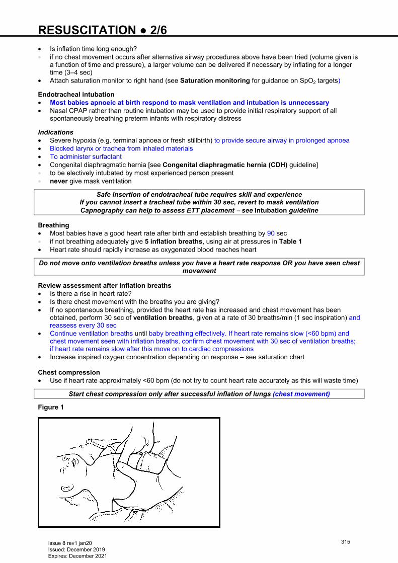
RESUSCITATION ● 2/6 • Is inflation time long enough? • if no chest movement occurs after alternative airway procedures above have been tried (volume given is
a function of time and pressure), a larger volume can be delivered if necessary by inflating for a longer time (3–4 sec)
• Attach saturation monitor to right hand (see Saturation monitoring for guidance on SpO2 targets)
Endotracheal intubation • Most babies apnoeic at birth respond to mask ventilation and intubation is unnecessary • Nasal CPAP rather than routine intubation may be used to provide initial respiratory support of all
spontaneously breathing preterm infants with respiratory distress
Indications • Severe hypoxia (e.g. terminal apnoea or fresh stillbirth) to provide secure airway in prolonged apnoea • Blocked larynx or trachea from inhaled materials • To administer surfactant • Congenital diaphragmatic hernia [see Congenital diaphragmatic hernia (CDH) guideline] • to be electively intubated by most experienced person present • never give mask ventilation
Safe insertion of endotracheal tube requires skill and experience If you cannot insert a tracheal tube within 30 sec, revert to mask ventilation Capnography can help to assess ETT placement − see Intubation guideline
Breathing • Most babies have a good heart rate after birth and establish breathing by 90 sec • if not breathing adequately give 5 inflation breaths, using air at pressures in Table 1 • Heart rate should rapidly increase as oxygenated blood reaches heart
Do not move onto ventilation breaths unless you have a heart rate response OR you have seen chest movement
Review assessment after inflation breaths • Is there a rise in heart rate? • Is there chest movement with the breaths you are giving? • If no spontaneous breathing, provided the heart rate has increased and chest movement has been
obtained, perform 30 sec of ventilation breaths, given at a rate of 30 breaths/min (1 sec inspiration) and reassess every 30 sec
• Continue ventilation breaths until baby breathing effectively. If heart rate remains slow (<60 bpm) and chest movement seen with inflation breaths, confirm chest movement with 30 sec of ventilation breaths; if heart rate remains slow after this move on to cardiac compressions
• Increase inspired oxygen concentration depending on response – see saturation chart Chest compression • Use if heart rate approximately <60 bpm (do not try to count heart rate accurately as this will waste time)
Start chest compression only after successful inflation of lungs (chest movement)
Figure 1
Issue 8 rev1 jan20 Issued: December 2019 Expires: December 2021
315
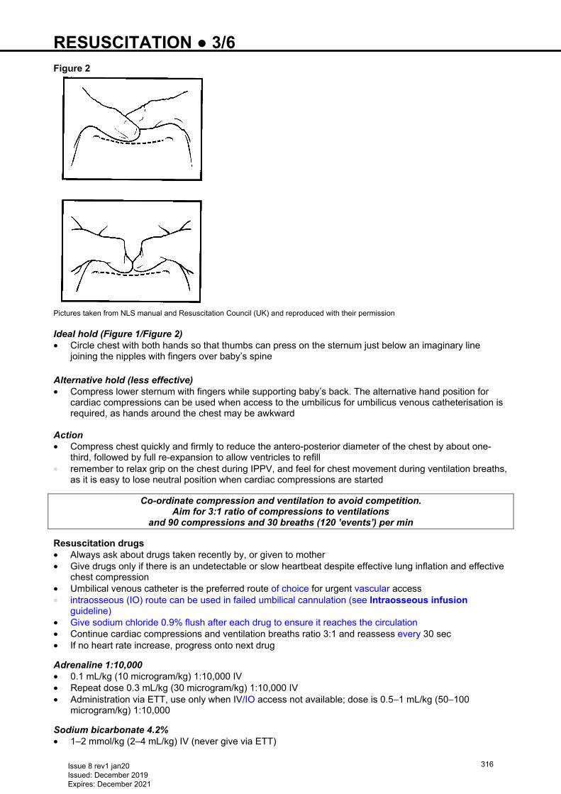
RESUSCITATION ● 3/6 Figure 2
Pictures taken from NLS manual and Resuscitation Council (UK) and reproduced with their permission Ideal hold (Figure 1/Figure 2) • Circle chest with both hands so that thumbs can press on the sternum just below an imaginary line
joining the nipples with fingers over baby’s spine Alternative hold (less effective) • Compress lower sternum with fingers while supporting baby’s back. The alternative hand position for
cardiac compressions can be used when access to the umbilicus for umbilicus venous catheterisation is required, as hands around the chest may be awkward
Action • Compress chest quickly and firmly to reduce the antero-posterior diameter of the chest by about one-
third, followed by full re-expansion to allow ventricles to refill • remember to relax grip on the chest during IPPV, and feel for chest movement during ventilation breaths,
as it is easy to lose neutral position when cardiac compressions are started
Co-ordinate compression and ventilation to avoid competition. Aim for 3:1 ratio of compressions to ventilations
and 90 compressions and 30 breaths (120 ‛events’) per min Resuscitation drugs • Always ask about drugs taken recently by, or given to mother • Give drugs only if there is an undetectable or slow heartbeat despite effective lung inflation and effective
chest compression • Umbilical venous catheter is the preferred route of choice for urgent vascular access • intraosseous (IO) route can be used in failed umbilical cannulation (see Intraosseous infusion
guideline) • Give sodium chloride 0.9% flush after each drug to ensure it reaches the circulation • Continue cardiac compressions and ventilation breaths ratio 3:1 and reassess every 30 sec • If no heart rate increase, progress onto next drug
Adrenaline 1:10,000 • 0.1 mL/kg (10 microgram/kg) 1:10,000 IV • Repeat dose 0.3 mL/kg (30 microgram/kg) 1:10,000 IV • Administration via ETT, use only when IV/IO access not available; dose is 0.5−1 mL/kg (50−100
microgram/kg) 1:10,000
Sodium bicarbonate 4.2% • 1–2 mmol/kg (2–4 mL/kg) IV (never give via ETT)
Issue 8 rev1 jan20 Issued: December 2019 Expires: December 2021
316
RESUSCITATION ● 4/6 Glucose 10% • 2.5 mL/kg IV
Sodium chloride 0.9% • 10 mL/kg IV initially Blood • If there is evidence of fetal haemorrhage and hypovolaemia Group O RhD negative emergency blood
may be required
Naloxone • Give only after ventilation by mask or ETT has been established with chest movement seen and heart
beat >100 bpm • If mother has been given pethidine within 2–4 hr of delivery, give naloxone IM: • 100 microgram (0.25 mL) for small preterm babies • 200 microgram (0.5 mL) for all other babies
WHEN TO STOP • If no sign of life after 10 min (Apgar 0 at 10 mins) despite resuscitation, consider stopping resuscitation
as outcome is poor; decision should be individualised and involve senior advice
Continue resuscitation until a senior member of staff advises stopping
MONITORING Saturation monitoring • Oxygen monitoring is activated when paediatrician/2nd pair of hands arrives. In the meantime, the person
initiating resuscitation carries out all the usual steps in resuscitation • Do not stop resuscitation for a saturation probe to be attached • Attach saturation probe to the right hand and connect to the monitor once 5 inflation breaths have been
given • SpO2 should spontaneously improve as Table 2 Table 2
Time (min) Acceptable preductal SpO2 (%)
2 60 3 70 4 80 5 85 10 90
Heart rate monitoring • Best by listening with stethoscope • Pulse-oximetry • ECG monitoring, if available, can give rapid accurate and continuous heart rate reading. However it does
not indicate the presence of a cardiac output and should not be the sole means of monitoring Air to oxygen • If resuscitation prolonged or failure of heart rate to respond to lung inflation increase inspired oxygen
concentration • If inflation breaths produce a response and SpO2 monitoring is available with a reliable trace target,
saturations as in Table 2 • If SpO2 above levels in Table 2 or >95% at 10 min of life, reduce oxygen Meconium deliveries • Do not attempt to suction nose and mouth whilst head is on perineum • If thick particulate meconium and baby not breathing and floppy inspect airway under direct vision and
suction any particulate matter before delivering inflation breaths but aim to inflate lungs within 1st minute
• Only intubate if suspected tracheal obstruction, routine intubation is not necessary
Issue 8 rev1 jan20 Issued: December 2019 Expires: December 2021
317
RESUSCITATION ● 5/6 Preterm deliveries • Nasal CPAP rather than routine intubation may be used to provide initial respiratory support of all
spontaneously breathing preterm babies with respiratory distress. Give PEEP at 5 cm H2O via mask ventilation with oxygen supplementation as appropriate on the resuscitaire continuing PEEP support on transfer to NICU
• If respiratory effort is poor at any point, or baby’s condition deteriorates, intubate and ventilate
DOCUMENTATION • Make accurate written record of facts (not opinions) as soon as possible after the event • Record: • when you were called, by whom and why • condition of baby on arrival • what you did and when you did it • timing and detail of any response by baby • date and time of writing your entry • a legible signature
COMMUNICATION • Inform parents what has happened (the facts)
Issue 8 rev1 jan20 Issued: December 2019 Expires: December 2021
318
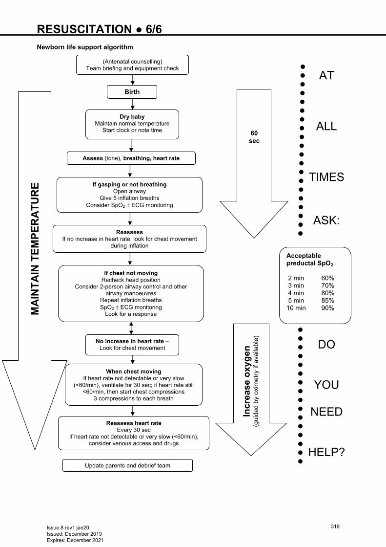
RESUSCITATION ● 6/6 Newborn life support algorithm
60 sec
AT
ALL
TIMES
ASK:
DO
YOU
NEED
HELP?
Acceptable preductal SpO2 2 min 60% 3 min 70% 4 min 80% 5 min 85% 10 min 90%
Dry baby Maintain normal temperature
Start clock or note time
Assess (tone), breathing, heart rate
If gasping or not breathingOpen airway
Give 5 inflation breaths Consider SpO2 ± ECG monitoring
Reassess If no increase in heart rate, look for chest movement
during inflation
If chest not moving Recheck head position
Consider 2-person airway control and other airway manoeuvres
Repeat inflation breaths SpO2 ± ECG monitoring
Look for a response
No increase in heart rate –Look for chest movement
When chest movingIf heart rate not detectable or very slow
(<60/min), ventilate for 30 sec; if heart rate still <60/min, then start chest compressions
3 compressions to each breath
Reassess heart rateEvery 30 sec
If heart rate not detectable or very slow (<60/min), consider venous access and drugs
Update parents and debrief team
Incr
ease
oxy
gen
(g
uide
d by
oxi
met
ry if
ava
ilabl
e)
Birth
(Antenatal counselling) Team briefing and equipment check
MA
INT
AIN
TE
MP
ER
AT
UR
E
Issue 8 rev1 jan20 Issued: December 2019 Expires: December 2021
319
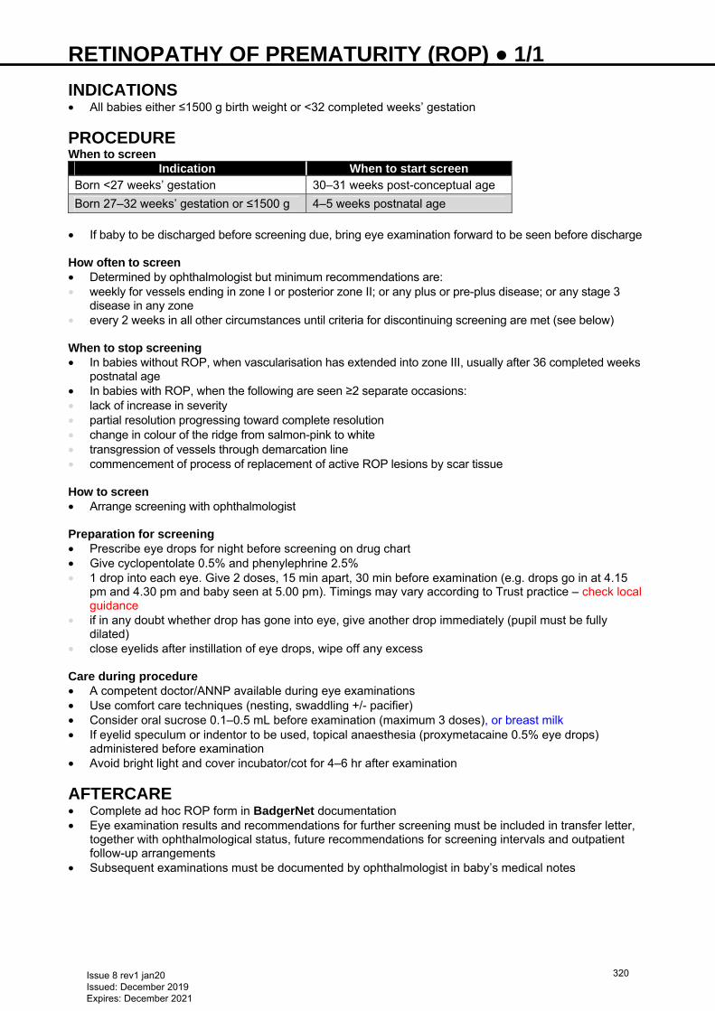
RETINOPATHY OF PREMATURITY (ROP) ● 1/1 INDICATIONS • All babies either ≤1500 g birth weight or <32 completed weeks’ gestation
PROCEDURE When to screen
Indication When to start screen Born <27 weeks’ gestation 30–31 weeks post-conceptual age
Born 27–32 weeks’ gestation or ≤1500 g 4–5 weeks postnatal age • If baby to be discharged before screening due, bring eye examination forward to be seen before discharge How often to screen • Determined by ophthalmologist but minimum recommendations are: • weekly for vessels ending in zone I or posterior zone II; or any plus or pre-plus disease; or any stage 3
disease in any zone • every 2 weeks in all other circumstances until criteria for discontinuing screening are met (see below) When to stop screening • In babies without ROP, when vascularisation has extended into zone III, usually after 36 completed weeks
postnatal age • In babies with ROP, when the following are seen ≥2 separate occasions: • lack of increase in severity • partial resolution progressing toward complete resolution • change in colour of the ridge from salmon-pink to white • transgression of vessels through demarcation line • commencement of process of replacement of active ROP lesions by scar tissue How to screen • Arrange screening with ophthalmologist Preparation for screening • Prescribe eye drops for night before screening on drug chart • Give cyclopentolate 0.5% and phenylephrine 2.5% • 1 drop into each eye. Give 2 doses, 15 min apart, 30 min before examination (e.g. drops go in at 4.15
pm and 4.30 pm and baby seen at 5.00 pm). Timings may vary according to Trust practice – check local guidance
• if in any doubt whether drop has gone into eye, give another drop immediately (pupil must be fully dilated)
• close eyelids after instillation of eye drops, wipe off any excess Care during procedure • A competent doctor/ANNP available during eye examinations • Use comfort care techniques (nesting, swaddling +/- pacifier) • Consider oral sucrose 0.1–0.5 mL before examination (maximum 3 doses), or breast milk • If eyelid speculum or indentor to be used, topical anaesthesia (proxymetacaine 0.5% eye drops)
administered before examination • Avoid bright light and cover incubator/cot for 4–6 hr after examination
AFTERCARE • Complete ad hoc ROP form in BadgerNet documentation • Eye examination results and recommendations for further screening must be included in transfer letter,
together with ophthalmological status, future recommendations for screening intervals and outpatient follow-up arrangements
• Subsequent examinations must be documented by ophthalmologist in baby’s medical notes
Issue 8 rev1 jan20 Issued: December 2019 Expires: December 2021
320
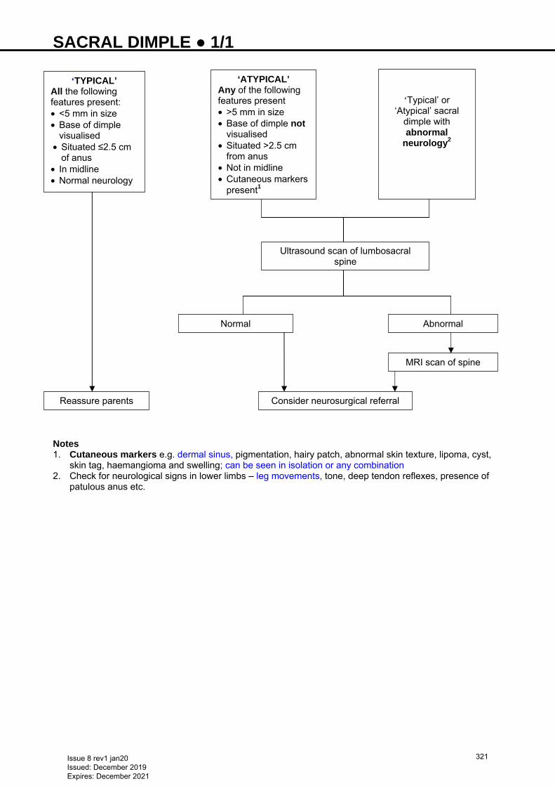
SACRAL DIMPLE ● 1/1
Notes 1. Cutaneous markers e.g. dermal sinus, pigmentation, hairy patch, abnormal skin texture, lipoma, cyst,
skin tag, haemangioma and swelling; can be seen in isolation or any combination 2. Check for neurological signs in lower limbs – leg movements, tone, deep tendon reflexes, presence of
patulous anus etc.
‘TYPICAL’ All the following features present: • <5 mm in size • Base of dimple
visualised • Situated ≤2.5 cm
of anus • In midline • Normal neurology
‘ATYPICAL’ Any of the following features present • >5 mm in size • Base of dimple not
visualised • Situated >2.5 cm
from anus • Not in midline • Cutaneous markers
present1
‘Typical’ or ‘Atypical’ sacral
dimple with abnormal
neurology2
Reassure parents
Ultrasound scan of lumbosacral spine
Normal Abnormal
MRI scan of spine
Consider neurosurgical referral
Issue 8 rev1 jan20 Issued: December 2019 Expires: December 2021
321
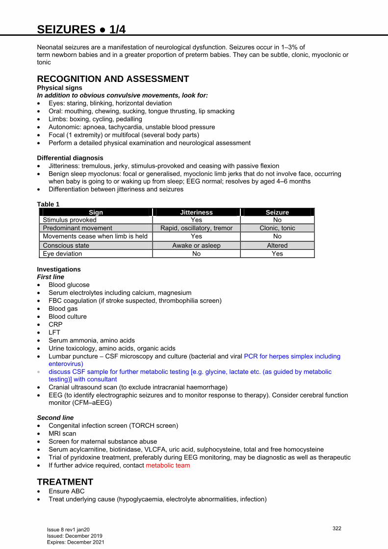
SEIZURES ● 1/4 Neonatal seizures are a manifestation of neurological dysfunction. Seizures occur in 1–3% of term newborn babies and in a greater proportion of preterm babies. They can be subtle, clonic, myoclonic or tonic
RECOGNITION AND ASSESSMENT Physical signs In addition to obvious convulsive movements, look for: • Eyes: staring, blinking, horizontal deviation • Oral: mouthing, chewing, sucking, tongue thrusting, lip smacking • Limbs: boxing, cycling, pedalling • Autonomic: apnoea, tachycardia, unstable blood pressure • Focal (1 extremity) or multifocal (several body parts) • Perform a detailed physical examination and neurological assessment Differential diagnosis • Jitteriness: tremulous, jerky, stimulus-provoked and ceasing with passive flexion • Benign sleep myoclonus: focal or generalised, myoclonic limb jerks that do not involve face, occurring
when baby is going to or waking up from sleep; EEG normal; resolves by aged 4–6 months • Differentiation between jitteriness and seizures Table 1
Sign Jitteriness Seizure
Stimulus provoked Yes No Predominant movement Rapid, oscillatory, tremor Clonic, tonic Movements cease when limb is held Yes No
Conscious state Awake or asleep Altered Eye deviation No Yes
Investigations First line • Blood glucose • Serum electrolytes including calcium, magnesium • FBC coagulation (if stroke suspected, thrombophilia screen) • Blood gas • Blood culture • CRP • LFT • Serum ammonia, amino acids • Urine toxicology, amino acids, organic acids • Lumbar puncture – CSF microscopy and culture (bacterial and viral PCR for herpes simplex including
enterovirus) • discuss CSF sample for further metabolic testing [e.g. glycine, lactate etc. (as guided by metabolic
testing)] with consultant • Cranial ultrasound scan (to exclude intracranial haemorrhage) • EEG (to identify electrographic seizures and to monitor response to therapy). Consider cerebral function
monitor (CFM–aEEG) Second line • Congenital infection screen (TORCH screen) • MRI scan • Screen for maternal substance abuse • Serum acylcarnitine, biotinidase, VLCFA, uric acid, sulphocysteine, total and free homocysteine • Trial of pyridoxine treatment, preferably during EEG monitoring, may be diagnostic as well as therapeutic • If further advice required, contact metabolic team
TREATMENT • Ensure ABC • Treat underlying cause (hypoglycaemia, electrolyte abnormalities, infection)
Issue 8 rev1 jan20 Issued: December 2019 Expires: December 2021
322
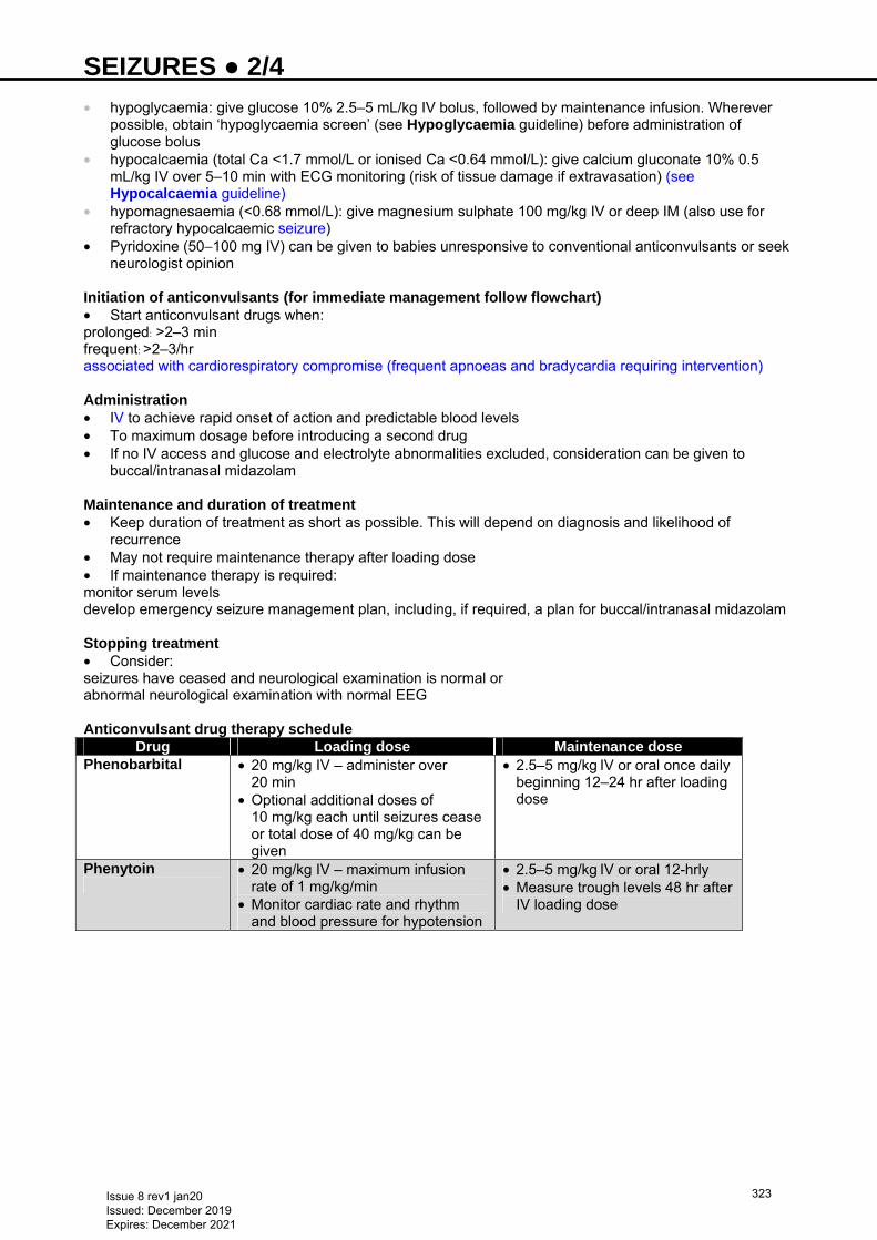
SEIZURES ● 2/4 • hypoglycaemia: give glucose 10% 2.5–5 mL/kg IV bolus, followed by maintenance infusion. Wherever
possible, obtain ‘hypoglycaemia screen’ (see Hypoglycaemia guideline) before administration of glucose bolus
• hypocalcaemia (total Ca <1.7 mmol/L or ionised Ca <0.64 mmol/L): give calcium gluconate 10% 0.5 mL/kg IV over 5–10 min with ECG monitoring (risk of tissue damage if extravasation) (see Hypocalcaemia guideline)
• hypomagnesaemia (<0.68 mmol/L): give magnesium sulphate 100 mg/kg IV or deep IM (also use for refractory hypocalcaemic seizure)
• Pyridoxine (50−100 mg IV) can be given to babies unresponsive to conventional anticonvulsants or seek neurologist opinion
Initiation of anticonvulsants (for immediate management follow flowchart) • Start anticonvulsant drugs when: prolonged: >2–3 min frequent: >2–3/hr associated with cardiorespiratory compromise (frequent apnoeas and bradycardia requiring intervention) Administration • IV to achieve rapid onset of action and predictable blood levels • To maximum dosage before introducing a second drug • If no IV access and glucose and electrolyte abnormalities excluded, consideration can be given to
buccal/intranasal midazolam Maintenance and duration of treatment • Keep duration of treatment as short as possible. This will depend on diagnosis and likelihood of
recurrence • May not require maintenance therapy after loading dose • If maintenance therapy is required: monitor serum levels develop emergency seizure management plan, including, if required, a plan for buccal/intranasal midazolam Stopping treatment • Consider: seizures have ceased and neurological examination is normal or abnormal neurological examination with normal EEG Anticonvulsant drug therapy schedule
Drug Loading dose Maintenance dose Phenobarbital
• 20 mg/kg IV – administer over 20 min
• Optional additional doses of 10 mg/kg each until seizures cease or total dose of 40 mg/kg can be given
• 2.5–5 mg/kg IV or oral once daily beginning 12–24 hr after loading dose
Phenytoin
• 20 mg/kg IV – maximum infusion rate of 1 mg/kg/min
• Monitor cardiac rate and rhythm and blood pressure for hypotension
• 2.5–5 mg/kg IV or oral 12-hrly • Measure trough levels 48 hr after
IV loading dose
Issue 8 rev1 jan20 Issued: December 2019 Expires: December 2021
323
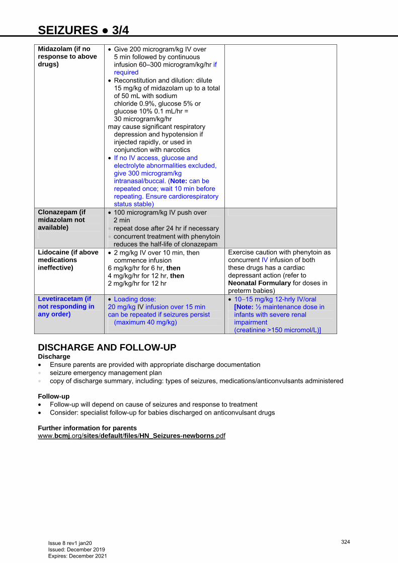
SEIZURES ● 3/4 Midazolam (if no response to above drugs)
• Give 200 microgram/kg IV over 5 min followed by continuous infusion 60–300 microgram/kg/hr if required
• Reconstitution and dilution: dilute 15 mg/kg of midazolam up to a total of 50 mL with sodium chloride 0.9%, glucose 5% or glucose 10% 0.1 mL/hr = 30 microgram/kg/hr
may cause significant respiratory depression and hypotension if injected rapidly, or used in conjunction with narcotics
• If no IV access, glucose and electrolyte abnormalities excluded, give 300 microgram/kg intranasal/buccal. (Note: can be repeated once; wait 10 min before repeating. Ensure cardiorespiratory status stable)
Clonazepam (if midazolam not available)
• 100 microgram/kg IV push over 2 min
• repeat dose after 24 hr if necessary • concurrent treatment with phenytoin
reduces the half-life of clonazepam
Lidocaine (if above medications ineffective)
• 2 mg/kg IV over 10 min, then commence infusion
6 mg/kg/hr for 6 hr, then 4 mg/kg/hr for 12 hr, then 2 mg/kg/hr for 12 hr
Exercise caution with phenytoin as concurrent IV infusion of both these drugs has a cardiac depressant action (refer to Neonatal Formulary for doses in preterm babies)
Levetiracetam (if not responding in any order)
• Loading dose: 20 mg/kg IV infusion over 15 min can be repeated if seizures persist
(maximum 40 mg/kg)
• 10−15 mg/kg 12-hrly IV/oral [Note: ½ maintenance dose in infants with severe renal impairment (creatinine >150 micromol/L)]
DISCHARGE AND FOLLOW-UP Discharge • Ensure parents are provided with appropriate discharge documentation • seizure emergency management plan • copy of discharge summary, including: types of seizures, medications/anticonvulsants administered Follow-up • Follow-up will depend on cause of seizures and response to treatment • Consider: specialist follow-up for babies discharged on anticonvulsant drugs Further information for parents www.bcmj.org/sites/default/files/HN_Seizures-newborns.pdf
Issue 8 rev1 jan20 Issued: December 2019 Expires: December 2021
324
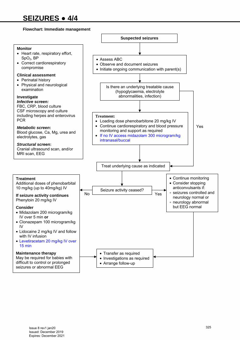
SEIZURES ● 4/4 Flowchart: Immediate management
Yes No
No
Yes
Suspected seizures
• Assess ABC • Observe and document seizures • Initiate ongoing communication with parent(s)
Is there an underlying treatable cause (hypoglycaemia, electrolyte
abnormalities, infection)
Treatment: • Loading dose phenobarbitone 20 mg/kg IV • Continue cardiorespiratory and blood pressure
monitoring and support as required • If no IV access midazolam 300 microgram/kg
intranasal/buccal
Monitor • Heart rate, respiratory effort,
SpO2, BP • Correct cardiorespiratory
compromise
Clinical assessment • Perinatal history • Physical and neurological
examination
Investigate Infective screen: FBC, CRP, blood culture CSF microscopy and culture including herpes and enterovirus PCR
Metabolic screen: Blood glucose, Ca, Mg, urea and electrolytes, gas
Structural screen: Cranial ultrasound scan, and/or MRI scan, EEG
Treat underlying cause as indicated
Seizure activity ceased?
• Continue monitoring • Consider stopping
anticonvulsants if: • seizures controlled and
neurology normal or • neurology abnormal
but EEG normal
• Transfer as required • Investigations as required • Arrange follow-up
Treatment Additional doses of phenobarbital 10 mg/kg (up to 40mg/kg) IV
If seizure activity continues Phenytoin 20 mg/kg IV
Consider • Midazolam 200 microgram/kg
IV over 5 min or • Clonazepam 100 microgram/kg
IV • Lidocaine 2 mg/kg IV and follow
with IV infusion • Levetiracetam 20 mg/kg IV over
15 min
Maintenance therapy May be required for babies with difficult to control or prolonged seizures or abnormal EEG
Issue 8 rev1 jan20 Issued: December 2019 Expires: December 2021
325
SKIN BIOPSY FOR INBORN ERRORS OF METABOLISM ● 1/2 INDICATIONS • Diagnosis of inherited metabolic disorders • Wherever possible, discuss biopsy and arrangements with Department of Newborn Screening and
Biochemical Genetics, Birmingham Children's Hospital 0121 333 9942 • this should include discussion about which specimen bottles and transport medium to use • confirm instructions for storage and transport with your local laboratory
Skin biopsy is often collected for histological analysis. Contact your local histopathology department for advice on sample handling
EQUIPMENT • Forceps: fine dissecting forceps • Cotton wool balls and gallipots • Dressing towel • Plastic apron • Size 15 scalpel blade and no. 3 handle • 25 G needle (orange top) • 23 G needle (blue top) • 21 G needle (green top) for drawing up lidocaine • 2 mL syringe • Cleaning solution as per unit policy • Lidocaine 1% • Bottles of culture medium • Sterile gloves • Steri-Strip™ • Dressings: • 1 small transparent dressing (e.g. Tegaderm™/Opsite) • gauze swabs • elasticated cotton or other bandage
SAMPLE REQUIREMENTS • ≥1 mm × 1 mm of skin (ideally 2 mm × 2 mm) from preferred site (e.g. inner side of forearm or posterior
aspect just above elbow) • choose site carefully as even a small scar on coloured skin will be very obvious • if post-mortem, take skin from over scapula as this leaves less obvious damage (see Post-mortem
specimens)
PROCEDURE Consent • Inform parents of reason for biopsy, explain procedure and risks including: • healing and scarring • possibility of contamination • poor growth • Obtain and record consent Technique
Maintain strict asepsis using ‛non-touch’ technique
• Wash hands and put on apron and sterile gloves • Cleanse site • ensure cleaning fluid does not pool beneath baby • Sedation if appropriate • Inject lidocaine 1%, a little intradermally and remainder subcutaneously to anaesthetise an area 1.5 × 1
cm • Wait 5 min to ensure site anaesthetised • Cleanse again, wipe off and dry using sterile cotton wool or gauze swabs
Issue 8 rev1 jan20 Issued: December 2019 Expires: December 2021
326
SKIN BIOPSY FOR INBORN ERRORS OF METABOLISM ● 2/2 Method A • Using fine forceps, grip a fold of skin between blades so that a length of skin 3 mm × 2 mm protrudes • slice off in 1 stroke by running scalpel blade along upper edge of forceps blades • if skin too thick or oedematous to grip, proceed to Method B Method B • Pierce skin with 23 or 21 G needle and lift to produce ‛tenting’ • cut off tip of tent to produce a round ‛O’ shaped piece of skin approximately 2 mm • Place into culture medium bottle immediately (lid of bottle removed by assistant for shortest possible
time) • Complete request form with: • clinical details • date and time of sampling Dressing wound • Although it may bleed freely, wound is usually partial thickness and should not require stitching • apply pressure to stanch bleeding • apply Steri-Strip™ and sterile dressing, bandage if necessary • Remove bandage after a few hours, but leave dressing for several days • Reassure parents that scar, when visible, will be seen as a fine line Transport • Once sample taken, send to inherited metabolic diseases laboratory as soon as possible • if unable to arrange transport immediately, store sample at +4°C for maximum of 12 hr before despatch,
do not freeze sample
POST-MORTEM SPECIMENS • In accordance with Human Tissue Act, post-mortem samples must be taken only on licensed premises
(or satellites thereof). Check with your pathology laboratory manager
Specimens taken after death present a high risk of infection and possible failure of culture. Follow strict aseptic technique
• Full written consent required for post-mortem biopsy samples • Take 2 biopsies from over scapula (as this leaves less obvious damage) as soon as possible after death,
ideally before 48 hr have elapsed • Send sample to inherited metabolic diseases laboratory immediately, or store at +4°C before dispatch for
maximum of 12 hr, do not freeze • Include clinical details, date and time of sampling, and date and time of death on request form
Issue 8 rev1 jan20 Issued: December 2019 Expires: December 2021
327
SKIN CARE ● 1/2 INTRODUCTION Neonatal skin care is very important, especially if baby is premature and/or in a critical condition. Special emphasis is placed on skin barrier properties, transcutaneous absorption, transepidermal water loss and maintaining skin integrity
PURPOSE • To maintain integrity of the skin • Prevent/minimise skin damage • Minimise water loss and heat loss • Protect against absorption of toxic materials and drugs • Treat skin damage • Ensure optimal healing of wounds
RISK FACTORS • Prematurity • Birth weight <1000 g • Oedema • Immobility • Congenital skin problems • Invasive procedures Birth weight <1250 g Careful handling • Most serious injuries can occur in first hours and days after birth when baby often requires intensive care
monitoring
Frequent bathing changes skin pH, disrupts protective acid mantle and is not recommended Preventing/minimising risk of skin injury/infection in all babies • Ensure adequate hand hygiene to protect baby’s skin from cutaneous infection e.g. Staphylococcus
aureus • Change baby’s position 4–6 hrly as condition dictates and place IV lines and monitoring leads away from
skin • Check all substances that come into contact with baby’s skin. Avoid using those with potential
percutaneous absorption • Protect areas of skin from friction injury with soft bedding and supporting blanket rolls • Use pressure-relief mattresses (e.g. Spenco®) • Change nappy 4–6 hrly as condition dictates. Wash nappy area with warm water and dry well • Nurse baby, especially extremely-low-birth-weight, in humidity of 60–90% to protect skin, maintain body
temperature and prevent water loss. Wean humidity as appropriate with increasing gestational age/day of life
• Use ECG leads with caution on babies <26 weeks’ gestation Disinfectants • Disinfect skin surfaces before invasive procedures, e.g. IV cannulation, umbilical vessel catheterisation,
chest drain insertion, IV puncture or heel pricks for laboratory samples • Use disinfectant pre-injection as per unit policy
Adhesives • In all newborns, use adhesives sparingly to secure life support, monitoring and other devices • Wherever possible, use Duoderm® under adhesive tape; adheres to skin without the use of adhesive and
will prevent epidermal stripping • Remove adhesives carefully with warm water on a cotton wool ball. Alcohol is very drying, easily
absorbed and should be avoided
CORD CARE Immediate • Clean cord and surrounding skin surface as needed with cleanser used for initial or routine bathing and
rinse thoroughly or cleanse with sterile water • Clean umbilical cord with warm water and cotton wool and keep dry
Issue 8 rev1 jan20 Issued: December 2019 Expires: December 2021
328
SKIN CARE ● 2/2 Ongoing • Keep cord area clean and dry. If cord becomes soiled with urine or stool, cleanse area with water • Educate staff and families about normal mechanism of cord healing • Teach parents or care-givers to keep area clean and dry, avoid contamination with urine and stool, keep
nappy folded away from area and wash hands before handling baby’s umbilical cord area
NAPPY DERMATITIS To maintain optimal skin environment • Change nappy frequently • Use nappy made from absorbent gel materials • Use cotton wool and warm water. Do not use commercially available baby wipes • Encourage/support breastfeeding throughout infancy Prevention strategies for babies at risk • Use petrolatum-based lubricants or barriers containing zinc oxide • Avoid use of products not currently recommended for newborns (e.g. polymer barrier films) Treat significant skin excoriation • Identify and treat underlying cause • Protect injured skin with thick application of barrier containing zinc oxide
Presence of red satellite lesions/culture indicates Candida albicans nappy rash • Rash will become more intense if covered by occlusive ointments. Treatment includes antifungal
ointments or cream and exposure to air and light • Do not use powders in treatment of nappy dermatitis • Avoid use of antibiotic ointments
Issue 8 rev1 jan20 Issued: December 2019 Expires: December 2021
329
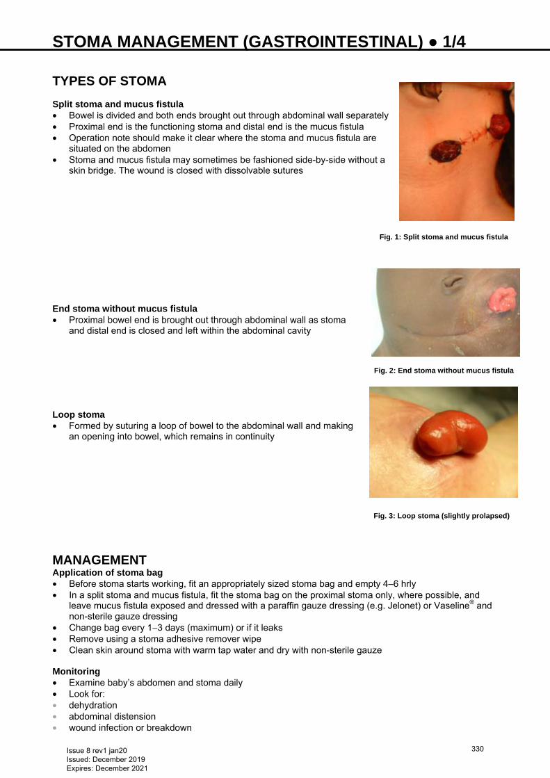
STOMA MANAGEMENT (GASTROINTESTINAL) ● 1/4
TYPES OF STOMA Split stoma and mucus fistula • Bowel is divided and both ends brought out through abdominal wall separately • Proximal end is the functioning stoma and distal end is the mucus fistula • Operation note should make it clear where the stoma and mucus fistula are
situated on the abdomen • Stoma and mucus fistula may sometimes be fashioned side-by-side without a
skin bridge. The wound is closed with dissolvable sutures End stoma without mucus fistula • Proximal bowel end is brought out through abdominal wall as stoma
and distal end is closed and left within the abdominal cavity
Loop stoma • Formed by suturing a loop of bowel to the abdominal wall and making
an opening into bowel, which remains in continuity
MANAGEMENT Application of stoma bag • Before stoma starts working, fit an appropriately sized stoma bag and empty 4–6 hrly • In a split stoma and mucus fistula, fit the stoma bag on the proximal stoma only, where possible, and
leave mucus fistula exposed and dressed with a paraffin gauze dressing (e.g. Jelonet) or Vaseline® and non-sterile gauze dressing
• Change bag every 1−3 days (maximum) or if it leaks • Remove using a stoma adhesive remover wipe • Clean skin around stoma with warm tap water and dry with non-sterile gauze Monitoring • Examine baby’s abdomen and stoma daily • Look for: • dehydration • abdominal distension • wound infection or breakdown
Fig. 1: Split stoma and mucus fistula
Fig. 2: End stoma without mucus fistula
Fig. 3: Loop stoma (slightly prolapsed)
Issue 8 rev1 jan20 Issued: December 2019 Expires: December 2021
330
STOMA MANAGEMENT (GASTROINTESTINAL) ● 2/4 • peri-stomal skin excoriation • granulation tissue formation • stomal bleeding • discolouration of stoma or mucus fistula • stomal prolapse or retraction • stoma bag leakage • rectal discharge • If stoma becomes dusky or black, call the surgical team • If skin surrounding the stoma is excoriated, identify cause and treat Weight • Babies with small bowel stoma: measure and record weight daily. Inadequate weight gain or weight loss
may be secondary to: • insufficient calorie intake • malabsorption • dehydration (high stoma output) • electrolyte abnormalities (high stoma output) Stoma effluent • Maintain a regularly updated fluid balance chart and record: • fluid intake and stoma losses • colour and consistency of stoma effluent Serum electrolytes • Measure at least every 2 days in the first 7 post-operative days Urinary electrolytes (sodium and potassium) • Monitoring is extremely important for nutrition and growth • Measure weekly • Babies with stomata (especially small bowel stomata) are at risk of losing a significant amount of sodium
into the effluent. They will often fail to gain weight if total body sodium is depleted. Serum sodium is an unreliable indicator of total body sodium
• Urinary sodium and Na+:K+ ratio are better indicators • Sodium supplements usually required in babies with a small bowel stoma until the stoma closed • If urinary sodium is <20 mmol/L or ratio of concentration of urinary sodium to potassium is <3:1, increase
sodium intake
NUTRITION Total parenteral nutrition and no enteral feeds • Check surgical discharge letter and operation notes for instructions on starting enteral feeds • Introduce enteral feeds slowly and increase gradually (see Nutrition and enteral feeding guideline) • Useful indicators of potential feed intolerance are: • vomiting and abdominal distension • bile in nasogastric aspirates • large nasogastric losses • low stoma losses – indicating dysmotility/obstruction • high stoma losses – indicating malabsorption • reducing substances or fat globules in the stool/stoma effluent Combination of parenteral nutrition and enteral feeds • Increase enteral feeds gradually (see Nutrition and enteral feeding guideline) • It is not possible to predict how much enteral feed baby will be able to tolerate. As a general rule, the
more distal the stoma, the better the absorption of feeds • The amount of stoma effluent and presence/absence of reducing substances or fat in the stoma effluent
should guide the advancement of enteral feeds • Do not automatically increase enteral feed in response to weight gain, but rather in response to stoma
output volume Full enteral feeds
Issue 8 rev1 jan20 Issued: December 2019 Expires: December 2021
331
STOMA MANAGEMENT (GASTROINTESTINAL) ● 3/4 • Tolerance of enteral feeds can fluctuate with time and babies with stomata are at high risk of life-
threatening dehydration and electrolyte abnormalities as a result of gastroenteritis. There should be a low threshold for readmission to hospital and appropriate resuscitation
COMPLICATIONS High stoma output • Daily output >20 mL/kg/day in premature or low-birth-weight babies and 30 mL/kg/day in term babies • Measure serum and urinary electrolytes • Replace stoma losses (when >20 mL/kg/day) mL-for-mL using sodium chloride 0.9% with potassium
chloride 10 mmol in 500 mL IV • Consider either reducing or stopping enteral feeds until losses decrease, liaison with surgical team is
encouraged • Test stoma effluent for reducing substances and fat globules • If reducing substances are positive or fat globules present, consider reduction of enteral feed or
changing type of enteral feed after consultation with a surgeon, specialist surgical outreach nurse or dietitian
• Perform blood gas; (stoma effluent may be rich in bicarbonate and metabolic acidosis may be present; consider sodium bicarbonate supplementation)
Mucus fistula • If present, consider recycling of stoma effluent (see Recycling stoma losses via a mucus fistula
guideline). Before recycling, consult surgical team to decide whether a contrast study through the mucus fistula is required
• If contrast study advised, make arrangements with surgical unit and inform surgical team when the study will take place
• Surgical team will review and advise if recycling may start • If baby not thriving, consider parenteral nutrition (see Parenteral nutrition guideline)
Increasing enteral feeds in a baby with poor weight gain and a high output stoma will worsen the situation
• If none of the above measures are effective, stop enteral feeds, start parenteral nutrition and consult surgical team to discuss surgical options
Stomal stenosis • May be present if: • stomal output reduces or stoma stops functioning • stoma effluent becomes watery • Call surgical team for advice Prolapse • Call surgical team for advice. If stoma is discoloured, emergency action required
STOMA CLOSURE • Often aimed to be performed when baby is well and thriving, which may be after discharge from hospital • Indications for early closure are: • failure to achieve full enteral feeds • recurrent stomal prolapse with/without stomal discolouration • stomal stenosis • high stoma output not responding to measures outlined above
DISCHARGE PLANNING AND PARENTAL TEACHING • Discharge when baby well, tolerating feeds and thriving • It is the responsibility of the ward/unit nurse to teach parents stoma care • When discharge planned, inform: • secretary of surgical consultant who fashioned the stoma to arrange outpatient follow-up • local stoma care specialist to order stoma supplies for home and support family • neonatal surgical outreach service (if involved in care)
Issue 8 rev1 jan20 Issued: December 2019 Expires: December 2021
332
STOMA MANAGEMENT (GASTROINTESTINAL) ● 4/4 Who to call when you need help? Surgical team • Call team of consultant surgeon who performed the surgery • In an emergency out-of-hours, contact on-call surgical registrar • Stoma care specialist [e.g. Gail Fitzpatrick at BCH (mobile 07557 001653)] for management of stoma-
related complications, and parent and staff training • Neonatal surgical outreach service [e.g. Louise Lawrence (mobile 07769 367483)] for advice, support
and training on surgical management Useful Information • http://www.bch.nhs.uk/content/neonatal-surgery • http://www.bch.nhs.uk/find-us/maps-directions • http://www.e-lfh.org.uk/programmes/paediatric-surgery/
Issue 8 rev1 jan20 Issued: December 2019 Expires: December 2021
333
SUBGALEAL HAEMORRHAGE (SGH) ● 1/3 RECOGNITION AND ASSESSMENT Definition • Accumulation of blood in the loose connective tissue of subgaleal space • Damaged emissary veins connecting subgaleal space to the intracranial venous sinuses can lead to
significant blood loss • up to two-thirds of circulating volume with significant morbidity and mortality (≥50% in severely affected
cases) Risk factors Vacuum extraction • Incorrect positioning of cup • cup marks on sagittal suture • leading edge of cup <3 cm from anterior fontanelle • Prolonged extraction time (>20 min) • >3 pulls or >2 cup detachments • Failed vacuum extraction Maternal factors • Nulliparity • PROM >12 hr • Maternal exhaustion • Prolonged second stage • High or mid cavity forceps delivery Neonatal factors • Macrosomia • Coagulopathy (vitamin K deficiency, Factor VIII or Factor IX deficiency) • Low-birth-weight • Male sex • Low Apgar scores • Resuscitation at birth • Cord blood acidosis • Fetal malpresentation • Can occur in unassisted deliveries Symptoms and signs • Local signs • generalised swelling or boggy consistency of scalp
− not limited by sutures − especially at the cup site − fluidic or leather-like pouch filled with fluid
• elevation and displacement of ear lobes and periorbital oedema • irritability and pain on handling • Systemic signs • hypovolemic shock
− tachycardia − tachypnoea − dropping haematocrit − increasing lactate or worsening acidosis − poor activity − pallor − hypotension − acidosis
• neurological dysfunction and seizures (late sign) • ischaemic end organ damage to liver or kidneys
− can manifest as worsening liver and renal function − poor prognostic indicator
Profound shock can occur rapidly with blood loss into subgaleal space – the blood loss may not be
apparent
Issue 8 rev1 jan20 Issued: December 2019 Expires: December 2021
334
SUBGALEAL HAEMORRHAGE (SGH) ● 2/3 Investigations • FBC and coagulation on admission • repeat at clinical team’s discretion • Group and blood crossmatch (notify blood bank). See Massive haemorrhage guideline • Venous/capillary gas including lactate and base excess, electrolytes (2−4 hrly) • Maintain blood glucose level >2.6 mmol/L
DIFFERENTIAL DIAGNOSIS • Cephalohematoma: subperiostial bleeding limited by suture lines • SGH: crosses suture lines • Caput succedaneum: oedematous collection of serosanguinous fluid in the subcutaneous layer of the
scalp • has distinct borders • does not enlarge • not fluctuant • Chignon: artificial caput succedaneum limited to suction cap application site
IMMEDIATE TREATMENT Initial management • Follow local guidelines for monitoring of newborns following vaginal operative delivery • Alert paediatric team • Urgent review by registrar/consultant • If SGH confirmed, admit to NNU immediately • inform consultant (if not involved in assessment) • Apply pressure bandage to head • Peripheral IV access • leave indwelling for 12 hr • Continuously monitor: • heart rate • respiration • oxygen saturation • blood pressure (non-invasively if no arterial line) ≥24 hr • Continue to assess capillary refill and peripheral perfusion • Regularly observe and palpate scalp swelling to assess for: • continuing blood loss • change in head shape or circumference
− measure head circumference hourly for the first 6−8 hr after birth − 1 cm increase in circumference = 40 mL blood loss
• change in colour • displacement of ears • Volume replacement: • inform consultant • see Massive haemorrhage guideline, and Recognition of hypovolaemia below • Group O RhD negative blood is immediately available on labour suite/obstetric theatres • Monitor urine output • Repeat FBC and coagulation studies (4−6 hr after initial assessment) • Inotropes, vasopressors, multiple packed red cell transfusions and clotting products may be required for
severe cases of shock [using packs 1 and 2 (see Massive haemorrhage guideline)] • Ongoing assessment for jaundice
RECOGNITION OF HYPOVOLAEMIA Signs of significant volume loss • High/increasing heart rate (>160 bpm) • Low/falling Hb or haematocrit • Poor peripheral perfusion with slow central capillary refill (>3 sec) • Low/falling blood pressure (mean arterial blood pressure <40 mmHg in term baby) • Presence of, or worsening of, metabolic acidosis • Consider echocardiography to assess volume status • small systemic veins and low ventricular filling volumes can indicate hypovolaemia
Issue 8 rev1 jan20 Issued: December 2019 Expires: December 2021
335
SUBGALEAL HAEMORRHAGE (SGH) ● 3/3 • If any of above present, or concern of ongoing haemorrhage from scalp assessment/neurological
dysfunction/evidence of renal or hepatic impairment − follow Massive haemorrhage guideline
Consider elective intubation and ventilation for worsening shock – blood is the priority over airway and breathing
CONCOMITANT INJURIES • Hypoxic ischaemic encephalopathy [see Hypoxic ischaemic encephalopathy (HIE) guideline] • Brain trauma resulting in cerebral oedema and/or intracranial haemorrhage • Subdural haematoma • Dural tear with herniation • Superior sagittal sinus rupture • Pseudomeningocoele and encephalocoele • Subconjunctival and retinal haemorrhage • Elevated intracranial pressure from SGH mass effect • Skull fractures
SUBSEQUENT MANAGEMENT • If any of the intracranial concomitant injuries above suspected, neuroimaging to be undertaken once
baby stabilised following discussion with radiologist to establish best modality • Monitor on NNU for ≥24 hr • Discuss with neurosurgical team
Issue 8 rev1 jan20 Issued: December 2019 Expires: December 2021
336
SUDDEN UNEXPECTED POSTNATAL COLLAPSE IN FIRST WEEK OF LIFE ● 1/3 Sudden unexpected postnatal collapse (SUPC) in apparently well term babies, in the first week of life is rare Summary of BAPM SUPC recommendations • Increased risk of congenital anomaly or metabolic disease • Need comprehensive investigation to determine underlying cause • Involve interdisciplinary liaison to maximise diagnostic yield • Senior doctor to obtain detailed family history and situational events • Notify coroner of all babies who die from such collapse • For all babies who die, post-mortem to be performed by a perinatal pathologist • If collapse happened after baby left hospital safeguarding issues must be considered • Detailed multiprofessional case review should follow investigation of unexpected baby death Information after the event • Collect the following as soon as possible after presentation Parental medical history • Full parental drug, alcohol and nicotine history • 3-generation family tree noting egg donation, sperm donation (where available) Obstetric history (from consultant obstetrician or senior trainee) • Infection • Fetal growth • Suspected fetal anomalies • Fetal movements • Liquor volume Labour and birth (from consultant obstetrician or senior trainee) • Maternal medication • Markers of fetal wellbeing • scalp pH • cord pH • electronic fetal monitoring • passage of meconium • requirement for resuscitation Health of baby until collapse • Growth and feeding Other information • Circumstances surrounding collapse • who was present? • was baby feeding? • position of baby (from staff and family present at time of collapse) • It is also important to collect information from other agencies who may have been involved with the
family e.g. primary care, social care and police • Full resuscitation details
Issue 8 rev1 jan20 Issued: December 2019 Expires: December 2021
337
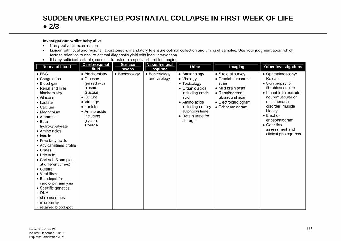
SUDDEN UNEXPECTED POSTNATAL COLLAPSE IN FIRST WEEK OF LIFE ● 2/3 Investigations whilst baby alive • Carry out a full examination • Liaison with local and regional laboratories is mandatory to ensure optimal collection and timing of samples. Use your judgment about which
tests to prioritise to ensure optimal diagnostic yield with least intervention • If baby sufficiently stable, consider transfer to a specialist unit for imaging
Neonatal blood Cerebrospinal
fluid Surface swabs
Nasophyngeal aspirate
Urine Imaging Other investigations
• FBC • Coagulation • Blood gas • Renal and liver
biochemistry • Glucose • Lactate • Calcium • Magnesium • Ammonia • Beta-
hydroxybutyrate • Amino acids • Insulin • Free fatty acids • Acylcarnitines profile • Urates • Uric acid • Cortisol (3 samples
at different times) • Culture • Viral titres • Bloodspot for
cardiolipin analysis • Specific genetics: • DNA • chromosomes • microarray • retained bloodspot
• Biochemistry • Glucose
(paired with plasma glucose)
• Culture • Virology • Lactate • Amino acids
including glycine, storage
• Bacteriology
• Bacteriology and virology
• Bacteriology • Virology • Toxicology • Organic acids
including orotic acid
• Amino acids including urinary sulphocysteine
• Retain urine for storage
• Skeletal survey • Cranial ultrasound
scan • MRI brain scan • Renal/adrenal
ultrasound scan • Electrocardiogram • Echocardiogram
• Ophthalmoscopy/ Retcam
• Skin biopsy for fibroblast culture
• If unable to exclude neuromuscular or mitochondrial disorder, muscle biopsy
• Electro-encephalogram
• Genetics assessment and clinical photographs
Issue 8 rev1 jan20 Issued: December 2019 Expires: December 2021
338
SUDDEN UNEXPECTED POSTNATAL COLLAPSE IN FIRST WEEK OF LIFE ● 3/3 • If there is suspicion that the event may have been due to unrecognised hypoventilation/apnoea, send
DNA sample for phox2b gene abnormalities (commonly implicated in congenital central hypoventilation syndrome)
• Consider testing for mutations and copy number variation in mecp2 gene. This may present as newborn encephalopathy and/or apnoeas and respiratory collapse
• Array-based comparative genomic hybridisation is a useful investigation (will replace conventional karyotyping for detecting causative chromosomal deletions and duplications)
Investigations before post-mortem • If it has not been possible to take samples during life, take samples (where feasible) while awaiting post-
mortem to prevent degradation of material and loss of important diagnostic information. Where possible, discuss and agree baseline samples with a pathologist and, where indicated, a biochemist
• if difficulty obtaining necessary kit for investigations, most labour wards have a ‘stillbirth kit’ which will contain much, if not all, of what is needed. Discuss with laboratory before beginning procedure if a full kit cannot be collected
• Throat and nose swabs for bacterial and viral culture • Blood culture • Blood and urine for metabolic studies • glucose, acylcarnitine, organic and amino acids including orotic acid and sulphocysteine, freeze urine for
storage • Blood for DNA, chromosomes and dried bloodspots on several cards • CSF obtained by lumbar puncture or ventricular tap – biochemistry • glucose • culture • virology • lactate • amino acids including glycine, freeze and store • Skin biopsy (if possible locally) for culture and storage of fibroblasts: 3 × 2 mm full thickness using
aseptic technique into culture or viral transport medium or gauze soaked in sodium chloride 0.9%. Send promptly to cytogenetics laboratory (see Skin biopsy guideline)
• Muscle biopsy (if possible locally) for electron microscopy, histopathology and enzymology. Wrap in aluminium foil, snap freeze and store at −70°C. Contact metabolic physician or pathologist before sample collection
Safeguarding issues • Must be considered in all cases of out of hospital collapse • The process of investigation for unexpected child deaths sometimes needs following even if baby
survives • Involves the rapid response team from the district who need to undertake a home visit to gather
additional information regarding the critical event For documentation and investigation check list for SUPC, use appendices from full BAPM guidelines − www.bapm.org/sites/default/files/files/SUPC_Booklet.pdf
Issue 8 rev1 jan20 Issued: December 2019 Expires: December 2021
339
SUPRAVENTRICULAR TACHYCARDIA ● 1/3 INTRODUCTION • Supraventricular tachycardia is the most common pathological tachycardia in newborns − can be new
presentation or commenced in fetal life
RECOGNITION AND ASSESSMENT • Sustained, accelerated non-sinus rhythm, regular and narrow-complex, originating above the level of the
atrioventricular (AV) junction • Heart rate >200 bpm • May be 1 of 3 tachycardias: • atrial • atrioventricular nodal re-entry (AVNRT) • atrioventricular re-entrant (AVRT) − most common supraventricular tachycardia (SVT) in fetal and
neonatal life • Can be presenting feature of a congenital heart defect − do not wait to exclude this before commencing
treatment
SYMPTOMS AND SIGNS • Can be variable with some common presentations: • acute onset in a baby in heart failure/shock with no previous signs and symptoms • fetal tachycardia during pregnancy • baby with irritability, poor feeding, sweating and breathlessness for hours/days before presentation • SVT can cause reduced cardiac output due to reduced diastolic filling time • many babies tolerate SVT well, however if tachycardia is sustained for >6 hr signs of congestive heart
failure may develop, with irritability, tachypnoea and pallor
CAUSES • No known cause in majority of babies • Idiopathic SVT is more common in neonates than older children • Wolf-Parkinson-White pre-excitation − only becomes visible after conversion to sinus rhythm • Congenital heart defect, including Ebstein’s and TGA
TRIGGERS • Co-existing infections e.g. LRTI • Manage all triggers appropriately
EXAMINATION • Heart rate: >200 bpm • Capillary refill • Blood pressure • Respiratory rate, may be normal/abnormal depending on: • signs of heart failure • co-existing respiratory conditions • infections • SpO2 may be normal, low, or of poor signal in haemodynamic compromise • Cardiovascular and respiratory examination; may be normal aside from fast heart rate • Examine baby for other reasons of tachycardia, including pain and environmental factors e.g. pyrexia
(particularly in premature baby in incubator)
INVESTIGATIONS • 12-lead ECG to confirm SVT diagnosis in haemodynamically stable cases • if baby haemodynamically unstable, or if ECG not available, defibrillator can record and print rhythm
strips from 3 different leads • Once SVT terminated, perform repeat ECG to assist with identification of pre-excitation and any other
underlying rhythm abnormality • Blood gas for: • acid-base balance • electrolytes • ionised calcium • Echocardiogram to assess structural anatomy and cardiac function
Issue 8 rev1 jan20 Issued: December 2019 Expires: December 2021
340
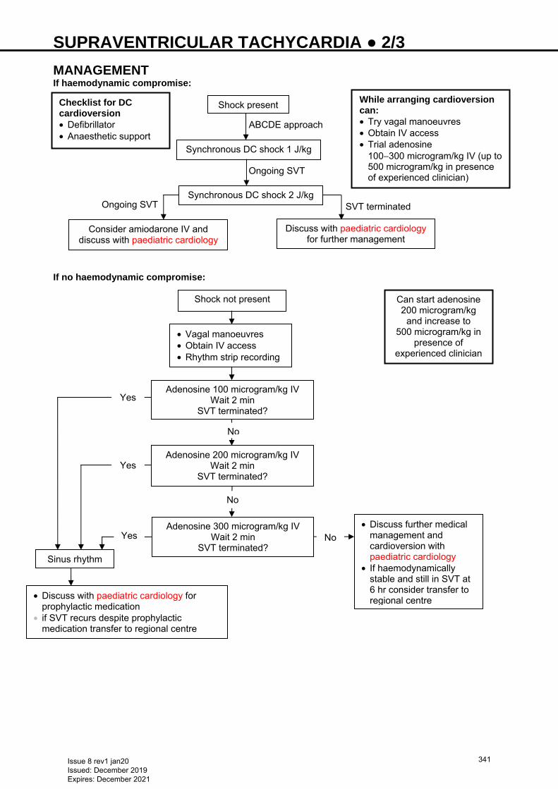
SUPRAVENTRICULAR TACHYCARDIA ● 2/3 MANAGEMENT If haemodynamic compromise: If no haemodynamic compromise:
Shock present
Synchronous DC shock 1 J/kg
Synchronous DC shock 2 J/kg
Discuss with paediatric cardiology for further management
Consider amiodarone IV and discuss with paediatric cardiology
ABCDE approach
Ongoing SVT
SVT terminated Ongoing SVT
While arranging cardioversion can: • Try vagal manoeuvres • Obtain IV access • Trial adenosine
100−300 microgram/kg IV (up to 500 microgram/kg in presence of experienced clinician)
Checklist for DC cardioversion • Defibrillator • Anaesthetic support
Shock not present
• Vagal manoeuvres • Obtain IV access • Rhythm strip recording
Adenosine 100 microgram/kg IV Wait 2 min
SVT terminated?
Adenosine 200 microgram/kg IV Wait 2 min
SVT terminated?
Adenosine 300 microgram/kg IV Wait 2 min
SVT terminated?
• Discuss further medical management and cardioversion with paediatric cardiology
• If haemodynamically stable and still in SVT at 6 hr consider transfer to regional centre
Sinus rhythm
• Discuss with paediatric cardiology for prophylactic medication
• if SVT recurs despite prophylactic medication transfer to regional centre
Can start adenosine 200 microgram/kg
and increase to 500 microgram/kg in
presence of experienced clinician
Yes
No
Yes
Yes
No
No
Issue 8 rev1 jan20 Issued: December 2019 Expires: December 2021
341
SUPRAVENTRICULAR TACHYCARDIA ● 3/3 ADDITIONAL INFORMATION: Adenosine • Give via cannula into large vein in upper limb, followed by rapid sodium chloride 0.9% flush; very short
half-life of 10−30 sec − must get to the heart as quickly as possible • Acts by slowing conduction time through the AV node • Intraosseous access ineffective due to time taken for venous return • Use 3-way tap with Luer-lock syringes; 1 syringe for adenosine and 1 for sodium chloride 0.9% flush
Never test cannula by aspirating blood into syringe with adenosine before injection − will lead to breakdown of adenosine
Major route of elimination via active take-up by red blood cells and vascular endothelial cells where it is metabolised
• Keep defibrillator nearby • Capture and print rhythm while adenosine given via defibrillator rhythm strip or ECG recording • Starting dose 100 micrograms/kg, repeat after 2 min, if no effect increase to maximum dose of 300
micrograms/kg • if experienced clinician present, maximum dose 500 micrograms/kg Vagal manoeuvres • Cold stimulation of the trigeminal nerve (afferent branches) instigates stimulation of the vagal nerve
(efferent branches); slows AV node conduction • wrap bag of ice in towel and apply to baby’s face or • wrap baby in towel and immerse entire head in ice-cold water for 5 sec • Unilateral carotid massage not recommended − difficult to perform in neonates and has limited effect
DC cardioversion • Applies direct current of electricity to the heart, synchronised to R wave of QRS complex on ECG • reduces risk of inducing ventricular fibrillation • Ideally carry out under general anaesthetic, or at least sedation • If performed outside NNU, will require anaesthetic support • Synchronised shock starting at 1 J/kg, if no response increase to 2 J/kg Chemical cardioversion: • Discuss with paediatric cardiology if: • haemodynamically unstable and unresponsive to adenosine IV or DC cardioversion • haemodynamically stable and unresponsive to adenosine IV • If SVT occurred in-utero consult perinatal plan and discuss with paediatric cardiology Prophylactic medication • When SVT has terminated, it is vital to commence medication to prevent further episodes • Choice of prophylactic medication based on: • previous history of SVT (including in fetal life) • assessment of ECG, both in SVT and once terminated • cardiac function • Discuss with paediatric cardiology centre and send ECG/echocardiogram for review
FOLLOW-UP • Any episode of SVT − follow-up with paediatrician with expertise in cardiology/paediatric cardiologist • Arrange: • baseline echocardiogram in outpatient clinic (if not already done) • holter monitor
Issue 8 rev1 jan20 Issued: December 2019 Expires: December 2021
342
SURFACTANT REPLACEMENT THERAPY − INCLUDING LESS INVASIVE SURFACTANT ADMINISTRATION (LISA) TECHNIQUEONSENT ● 1/3 • Early administration of natural surfactant decreases the risk of acute pulmonary injury and neonatal
mortality • Early CPAP and selective administration of surfactant is preferable to routine intubation and prophylactic
surfactant • Natural surfactant preparations are superior to protein-free synthetic preparations containing only
phospholipids for reducing mortality and air leaks • Poractant alfa (Curosurf®) at 200 mg/kg shows survival advantage compared to beractant or poractant
alpha in a dose of 100 mg/kg • Multiple rescue doses result in greater improvements in oxygenation and ventilatory requirements, a
decreased risk of pneumothorax and a trend toward improved survival • Use of Intubate–Surfactant–Extubate (INSURE) to CPAP or LISA (less invasive surfactant
administration) or minimally invasive surfactant treatment (MIST) techniques for early surfactant administration reduce the need for ventilation and improve survival
• see LISA section below
INDICATIONS Prophylaxis (administration ≤15 min of birth) Babies born <28 weeks’ gestation • Routine intubation of these babies solely for the purpose of administration of surfactant is not
necessary, and a policy of early CPAP with selective surfactant administration is preferred • If requiring intubation for respiratory support during resuscitation or if mother has not had antenatal
steroids, give surfactant as prophylaxis • Otherwise, institute early CPAP and administer surfactant selectively as per Early rescue treatment Early rescue treatment Preterm babies who require invasive ventilation for stabilisation should be given surfactant Babies born ≤33 weeks’ gestation who are not ventilated • If FiO2 >0.30, give surfactant using minimally/less invasive technique or after invasive ventilation Other babies that can be considered for surfactant therapy (after discussion with consultant) • Ventilated babies with meconium aspiration syndrome (may need repeat dose after 6–8 hr) • Term babies with pneumonia and less compliant lungs
EQUIPMENT • Natural surfactant, poractant alfa (Curosurf®) 200 mg/kg (2.5 mL/kg) round to the nearest whole vial
(prophylaxis and rescue doses can differ) • Sterile gloves • TrachCare Mac™ catheter (do not cut NGT) or specific surfactant administration set
PROCEDURE Preparation • Calculate dose of surfactant required and warm to room temperature • Ensure correct endotracheal tube (ETT) position • check ETT length at lips • listen for bilateral air entry and look for chest movement • if in doubt, ensure ETT in trachea using laryngoscope and adjust to ensure bilateral equal air entry • chest X-ray not necessary before first dose • Refer to manufacturer’s guidelines and Neonatal Formulary • Invert surfactant vial gently several times, without shaking, to resuspend the material • Draw up required dose • Administer via TrachCare Mac™ device or specific surfactant administration pack
Instillation • With baby supine, instil prescribed dose down ETT • Wait for recovery of air entry/chest movement and oxygenation between boluses Post-instillation care • Do not suction ETT for 8 hr following instillation of surfactant
Issue 8 rev1 jan20 Issued: December 2019 Expires: December 2021
343
SURFACTANT REPLACEMENT THERAPY − INCLUDING LESS INVASIVE SURFACTANT ADMINISTRATION (LISA) TECHNIQUEONSENT ● 2/3 • Be ready to adjust ventilator/oxygen settings in response to changes in chest movement, tidal volume
and oxygen saturation. Use of volume-target ventilation can facilitate responsiveness to rapid changes in lung compliance following surfactant instillation. Be ready to reduce FiO2 soon after administration of surfactant to avoid hyperoxia
• Take an arterial/capillary blood gas within 30 min
SUBSEQUENT MANAGEMENT • If baby remains ventilated at FiO2 >0.3 with mean airway pressure >7 cm H2O, give further dose of
surfactant 6−12 hr after first dose • Third dose should be given only at request of attending consultant
DOCUMENTATION • For every dose given, document in case notes: • indication for surfactant use • time of administration • dose given • condition of baby pre-administration, including measurement of blood gas unless on labour ward when
saturations should be noted • response to surfactant, including measurement of post-administration blood gas and saturations • reason(s) why second dose not given, if applicable • reason(s) for giving third dose if administered • Prescribe surfactant on drug chart
LISA Definition • Method using a thin catheter to deliver surfactant in spontaneously breathing preterm infant with
respiratory distress syndrome receiving non-invasive ventilator support • continue non-invasive ventilator support during procedure Indication • Suspected surfactant deficiency leading to respiratory distress syndrome on non-invasive respiratory
support as evidenced by: • rapidly increasing oxygen requirements • FiO2 >0.3 • increased work of breathing (exclude pneumothorax by transillumination of chest) • <33 weeks’ gestation • aged <48 hr
Exclusion • Persistent/worsening respiratory acidosis despite optimal non-invasive ventilation Equipment • Laryngoscope/video laryngoscope • Suction • Sterile gloves • LISA catheter (LISAcath®) • Surfactant, and syringe and needle to draw up surfactant
Drugs • Fentanyl 0.7 microgram/kg (awake sedation) • Atropine 20 microgram/kg • Naloxone 100 microgram/kg (if poor respiratory effort after procedure) Emergency equipment • Bag/valve/mask/T-piece • Oxygen and air • Stethoscope • ETTs
Issue 8 rev1 jan20 Issued: December 2019 Expires: December 2021
344
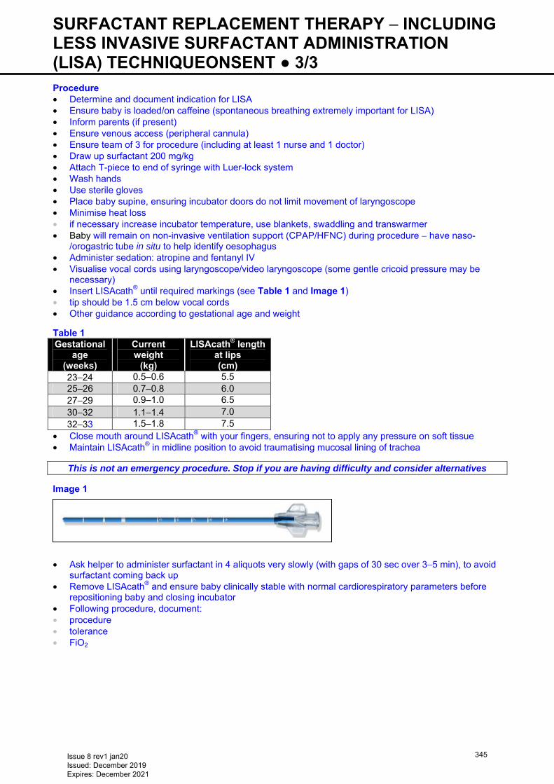
SURFACTANT REPLACEMENT THERAPY − INCLUDING LESS INVASIVE SURFACTANT ADMINISTRATION (LISA) TECHNIQUEONSENT ● 3/3 Procedure • Determine and document indication for LISA • Ensure baby is loaded/on caffeine (spontaneous breathing extremely important for LISA) • Inform parents (if present) • Ensure venous access (peripheral cannula) • Ensure team of 3 for procedure (including at least 1 nurse and 1 doctor) • Draw up surfactant 200 mg/kg • Attach T-piece to end of syringe with Luer-lock system • Wash hands • Use sterile gloves • Place baby supine, ensuring incubator doors do not limit movement of laryngoscope • Minimise heat loss • if necessary increase incubator temperature, use blankets, swaddling and transwarmer • Baby will remain on non-invasive ventilation support (CPAP/HFNC) during procedure − have naso-
/orogastric tube in situ to help identify oesophagus • Administer sedation: atropine and fentanyl IV • Visualise vocal cords using laryngoscope/video laryngoscope (some gentle cricoid pressure may be
necessary) • Insert LISAcath® until required markings (see Table 1 and Image 1) • tip should be 1.5 cm below vocal cords • Other guidance according to gestational age and weight Table 1 Gestational
age (weeks)
Current weight
(kg)
LISAcath® length at lips (cm)
23−24 0.5–0.6 5.5 25–26 0.7–0.8 6.0
27−29 0.9–1.0 6.5 30−32 1.1−1.4 7.0 32−33 1.5–1.8 7.5
• Close mouth around LISAcath® with your fingers, ensuring not to apply any pressure on soft tissue • Maintain LISAcath® in midline position to avoid traumatising mucosal lining of trachea
This is not an emergency procedure. Stop if you are having difficulty and consider alternatives
Image 1 • Ask helper to administer surfactant in 4 aliquots very slowly (with gaps of 30 sec over 3−5 min), to avoid
surfactant coming back up • Remove LISAcath® and ensure baby clinically stable with normal cardiorespiratory parameters before
repositioning baby and closing incubator • Following procedure, document: • procedure • tolerance • FiO2
Issue 8 rev1 jan20 Issued: December 2019 Expires: December 2021
345
SYPHILIS − BABIES BORN TO MOTHERS WITH POSITIVE SEROLOGY ● 1/4
INTRODUCTION • 40% of untreated early syphilis results in stillbirth/spontaneous abortion/perinatal loss, depending upon
maternal stage of infection and spirochete blood load • Untreated babies aged >2 yr may present with: • CNS disease (VIII nerve deafness) • bone and joint (frontal bossing, saddle nose and Clutton joints) • teeth (Hutchinson incisors and mulberry molars) • eye (interstitial keratitis 5–20 yr) involvement • Babies born to mothers diagnosed and treated during current pregnancy require serological testing after
birth
RECOGNITION AND ASSESSMENT • Clarify maternal treatment and post-treatment titres • Discuss management plan with parents before birth • All parents to be seen by specialist midwife antenatally to discuss management of baby • Follow Management flowchart
CLINICAL FEATURES Clinical evidence of early congenital syphilis • Rash • Infectious snuffles (copious nasal secretions) • Haemorrhagic rhinitis • Osteochondritis • Periostitis • Pseudo-paralysis • Mucocutaneous patches • Peri-oral fissures • Hepatosplenomegaly • Lymphadenopthy • Oedema • Glomerulonephritis • Ocular or neurological involvement • Haemolysis • Thrombocytopenia
ASSESSMENT OF MATERNAL TREATMENT • Local GUM team to advise if maternal serology confirms previous adequately treated syphilis, or
suggests new/reinfection • All babies born to mothers with positive treponemal serology require clinical evaluation and syphilis
serology tests except babies born to mothers who had syphilis cured before current pregnancy • mothers of babies who do not require testing will be IgM negative, rapid plasma reagin (RPR)/venereal
disease research laboratory (VDRL) test negative, Treponema pallidum particle agglutination (TPPA) test positive
INVESTIGATIONS Diagnostic serology • Baby may have positive serology depending on timing of maternal infection, therefore mother must be
screened simultaneously for titre comparison. Serological tests must be performed on baby’s blood, DO NOT USE CORD BLOOD
Non-treponemal tests • VDRL test/RPR: • 4 x decrease in titre = effective treatment • 4 x increase after treatment = relapse or reinfection • May be false negative in babies who acquire congenital syphilis in late pregnancy or have extremely
high antibody titres before dilution (prozone phenomenon) • May be false positive in viral infections (Epstein-Barr, varicella zoster, hepatitis, measles), tuberculosis,
endocarditis, malaria, lymphoma, connective tissue disease, pregnancy, intravenous drug use
Issue 8 rev1 jan20 Issued: December 2019 Expires: December 2021
346
SYPHILIS − BABIES BORN TO MOTHERS WITH POSITIVE SEROLOGY ● 2/4 Treponemal tests • IgM • TPPA • Treponema pallidum haemagglutination assay (TPHA) • Fluorescent treponemal antibody absorption test (FTA-ABS) • Tests may also be positive in other spirochetal disease e.g. leptospirosis, and Lyme disease. There is
poor correlation of titres with disease activity
Interpretation of syphilis serology of baby • Following indicate congenital infection: • positive IgM • positive RPR/VDRL test on CSF • ≥4 x difference of RPR/VDRL titre or TPPA titre above that of the mother • ≥4 x increase in RPR/VDRL or TPPA titre within 3 months of birth • positive treponemal tests in child aged >18 months
Example of positive TPPA • Maternal titres 1:1024 • Baby serology 1:4096 (i.e. baby 4 x greater than mother)
Example of positive VDRL/RPR • Maternal titres 1:64 • Baby serology 1:256 (i.e. baby 4 x greater than mother)
CSF • CSF investigations require ≥0.5 mL of CSF. A CSF is classed as positive if: • increased WCC and protein • reactive TPPA and VDRL (a negative VDRL does not exclude neurosyphillis) • Remember to suspect other causes of elevated values when evaluating baby for congenital syphilis
TREATMENT • Possible congenital syphilis: benzylpenicillin 30 mg/kg IV 12-hrly for 7 days and 8-hrly for next 3 days • †Alternative regimen (if, for example, difficulty with IV access): procaine penicillin 50,000 units/kg daily IM
for 10 days (painful). Reconstitute vial with 4.2 mL (as displacement volume = 0.8 mL) of solvent provided (WFI) to produce solution containing 240,000 units/mL
• If treatment interrupted for >1 day course must be restarted • *If delay in results, offer single dose benzathine penicillin IM while awaiting results: • 50,000 units/kg as a single dose by IM injection within 24 hr of decision to treat. Reconstitute vial with
3.2 mL (as displacement volume = 0.8 mL) of solvent provided (WFI) to produce a solution containing 300,000 units/mL
FOLLOW-UP • If IgM test is negative, other tests are reactive with titres <4-fold higher than mother’s, with no signs of
congenital syphilis, repeat reactive tests at 3, 6 and 12 months or until all tests (VDRL/RPR, TPPA and IgM) become negative (usually by 6 months)
• If baby’s serum negative on screening, and no signs of congenital infection, no further testing necessary • If any doubt regarding test interpretation/follow-up, discuss with local expert in neonatal
infection/microbiology • If Neurosyphilis, repeat CSF at 6 months
Issue 8 rev1 jan20 Issued: December 2019 Expires: December 2021
347
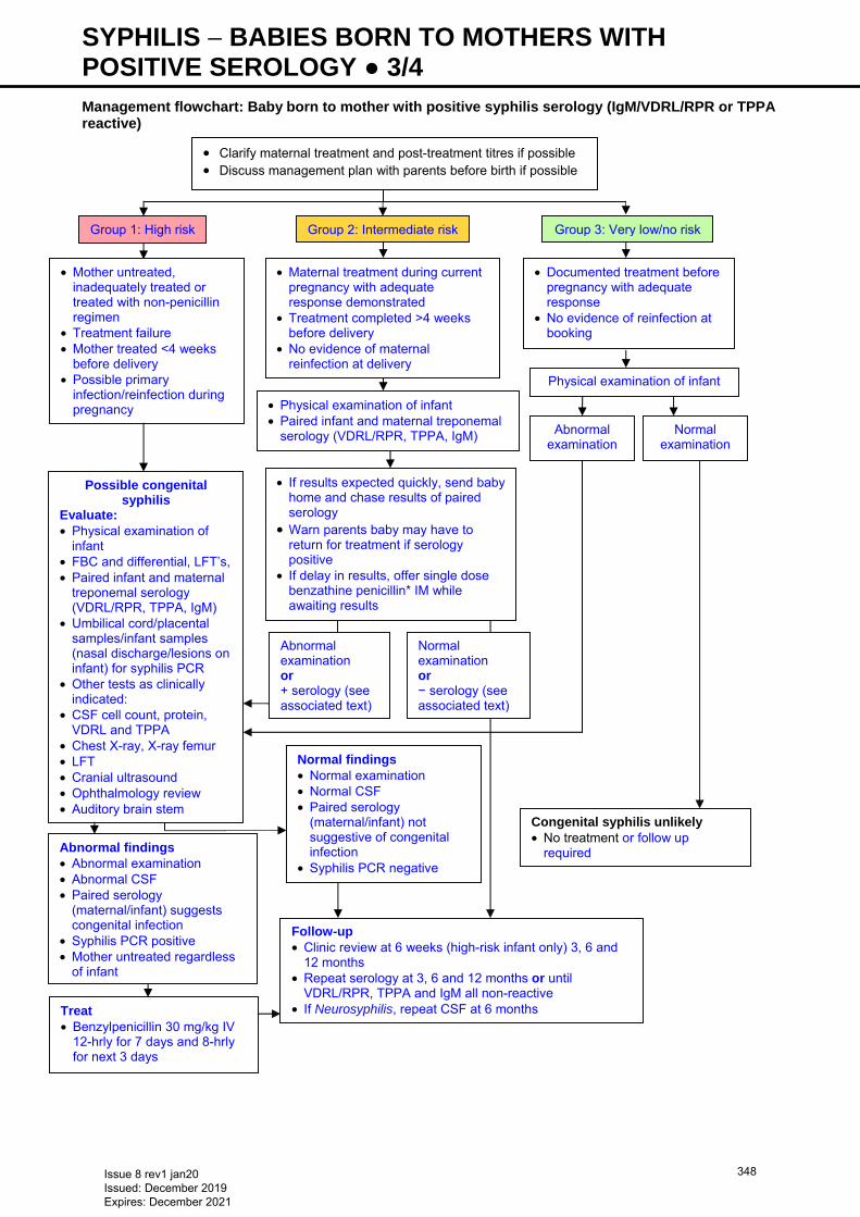
SYPHILIS − BABIES BORN TO MOTHERS WITH POSITIVE SEROLOGY ● 3/4 Management flowchart: Baby born to mother with positive syphilis serology (IgM/VDRL/RPR or TPPA reactive)
Treat • Benzylpenicillin 30 mg/kg IV
12-hrly for 7 days and 8-hrly for next 3 days
Follow-up • Clinic review at 6 weeks (high-risk infant only) 3, 6 and
12 months • Repeat serology at 3, 6 and 12 months or until
VDRL/RPR, TPPA and IgM all non-reactive • If Neurosyphilis, repeat CSF at 6 months
• Mother untreated, inadequately treated or treated with non-penicillin regimen
• Treatment failure • Mother treated <4 weeks
before delivery • Possible primary
infection/reinfection during pregnancy
• Maternal treatment during current pregnancy with adequate response demonstrated
• Treatment completed >4 weeks before delivery
• No evidence of maternal reinfection at delivery
• If results expected quickly, send baby home and chase results of paired serology
• Warn parents baby may have to return for treatment if serology positive
• If delay in results, offer single dose benzathine penicillin* IM while awaiting results
• Documented treatment before pregnancy with adequate response
• No evidence of reinfection at booking
Possible congenital syphilis
Evaluate: • Physical examination of
infant • FBC and differential, LFT’s, • Paired infant and maternal
treponemal serology (VDRL/RPR, TPPA, IgM)
• Umbilical cord/placental samples/infant samples (nasal discharge/lesions on infant) for syphilis PCR
• Other tests as clinically indicated:
• CSF cell count, protein, VDRL and TPPA
• Chest X-ray, X-ray femur • LFT • Cranial ultrasound • Ophthalmology review • Auditory brain stem
Congenital syphilis unlikely • No treatment or follow up
required
• Clarify maternal treatment and post-treatment titres if possible • Discuss management plan with parents before birth if possible
Group 1: High risk Group 3: Very low/no risk Group 2: Intermediate risk
Abnormal findings • Abnormal examination • Abnormal CSF • Paired serology
(maternal/infant) suggests congenital infection
• Syphilis PCR positive • Mother untreated regardless
of infant
• Physical examination of infant • Paired infant and maternal treponemal
serology (VDRL/RPR, TPPA, IgM)
Abnormal examination or + serology (see associated text)
Normal examination or − serology (see associated text)
Normal findings• Normal examination • Normal CSF • Paired serology
(maternal/infant) not suggestive of congenital infection
• Syphilis PCR negative
Physical examination of infant
Normal examination
Abnormal examination
Issue 8 rev1 jan20 Issued: December 2019 Expires: December 2021
348
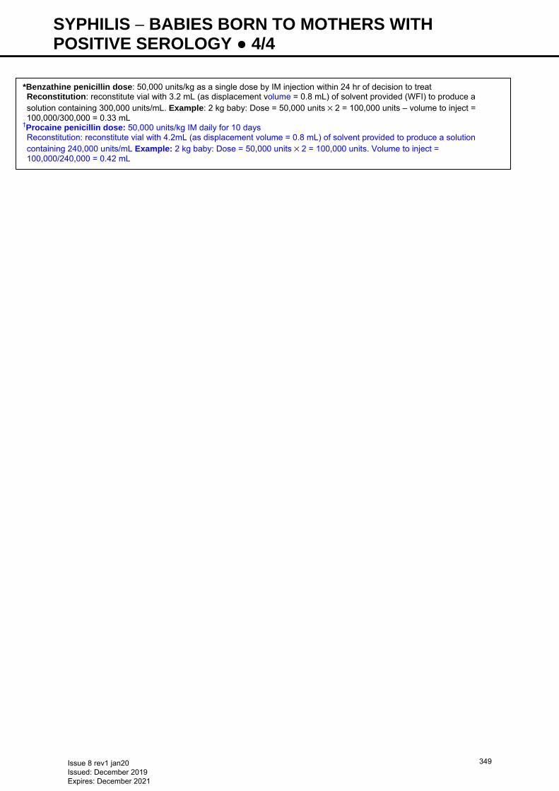
SYPHILIS − BABIES BORN TO MOTHERS WITH POSITIVE SEROLOGY ● 4/4
*Benzathine penicillin dose: 50,000 units/kg as a single dose by IM injection within 24 hr of decision to treat Reconstitution: reconstitute vial with 3.2 mL (as displacement volume = 0.8 mL) of solvent provided (WFI) to produce a solution containing 300,000 units/mL. Example: 2 kg baby: Dose = 50,000 units × 2 = 100,000 units – volume to inject = 100,000/300,000 = 0.33 mL
†Procaine penicillin dose: 50,000 units/kg IM daily for 10 days Reconstitution: reconstitute vial with 4.2mL (as displacement volume = 0.8 mL) of solvent provided to produce a solution containing 240,000 units/mL Example: 2 kg baby: Dose = 50,000 units × 2 = 100,000 units. Volume to inject = 100,000/240,000 = 0.42 mL
Issue 8 rev1 jan20 Issued: December 2019 Expires: December 2021
349
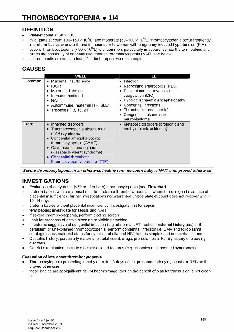
THROMBOCYTOPENIA ● 1/4 DEFINITION • Platelet count <150 × 109/L • mild (platelet count 100–150 × 109/L) and moderate (50–100 × 109/L) thrombocytopenia occur frequently
in preterm babies who are ill, and in those born to women with pregnancy-induced hypertension (PIH) • severe thrombocytopenia (<50 × 109/L) is uncommon, particularly in apparently healthy term babies and
raises the possibility of neonatal allo-immune thrombocytopenia (NAIT; see below) • ensure results are not spurious, if in doubt repeat venous sample
CAUSES
WELL ILLCommon • Placental insufficiency
• IUGR • Maternal diabetes • Immune mediated • NAIT • Autoimmune (maternal ITP, SLE) • Trisomies (13, 18, 21)
• Infection • Necrotising enterocolitis (NEC) • Disseminated intravascular
coagulation (DIC) • Hypoxic ischaemic encephalopathy • Congenital infections • Thrombosis (renal, aortic) • Congenital leukaemia or
neuroblastomaRare • Inherited disorders
• Thrombocytopenia absent radii (TAR) syndrome
• Congenital amegakaryocytic thrombocytopenia (CAMT)
• Cavernous haemangioma (Kasabach-Merritt syndrome)
• Congenital thrombotic thrombocytopenia purpura (TTP)
• Metabolic disorders (propionic and methylmalonic acidemia)
Severe thrombocytopenia in an otherwise healthy term newborn baby is NAIT until proved otherwise
INVESTIGATIONS • Evaluation of early-onset (<72 hr after birth) thrombocytopenia (see Flowchart) • preterm babies with early-onset mild-to-moderate thrombocytopenia in whom there is good evidence of
placental insufficiency: further investigations not warranted unless platelet count does not recover within 10–14 days
• preterm babies without placental insufficiency: investigate first for sepsis • term babies: investigate for sepsis and NAIT • If severe thrombocytopenia, perform clotting screen • Look for presence of active bleeding or visible petechiae • If features suggestive of congenital infection (e.g. abnormal LFT, rashes, maternal history etc.) or if
persistent or unexplained thrombocytopenia, perform congenital infection i.e. CMV and toxoplasma serology; check maternal status for syphilis, rubella and HIV; herpes simplex and enteroviral screen
• Obstetric history, particularly maternal platelet count, drugs, pre-eclampsia. Family history of bleeding disorders
• Careful examination, include other associated features (e.g. trisomies and inherited syndromes) Evaluation of late onset thrombocytopenia • Thrombocytopenia presenting in baby after first 3 days of life, presume underlying sepsis or NEC until
proved otherwise • these babies are at significant risk of haemorrhage, though the benefit of platelet transfusion is not clear-
cut
Issue 8 rev1 jan20 Issued: December 2019 Expires: December 2021
350
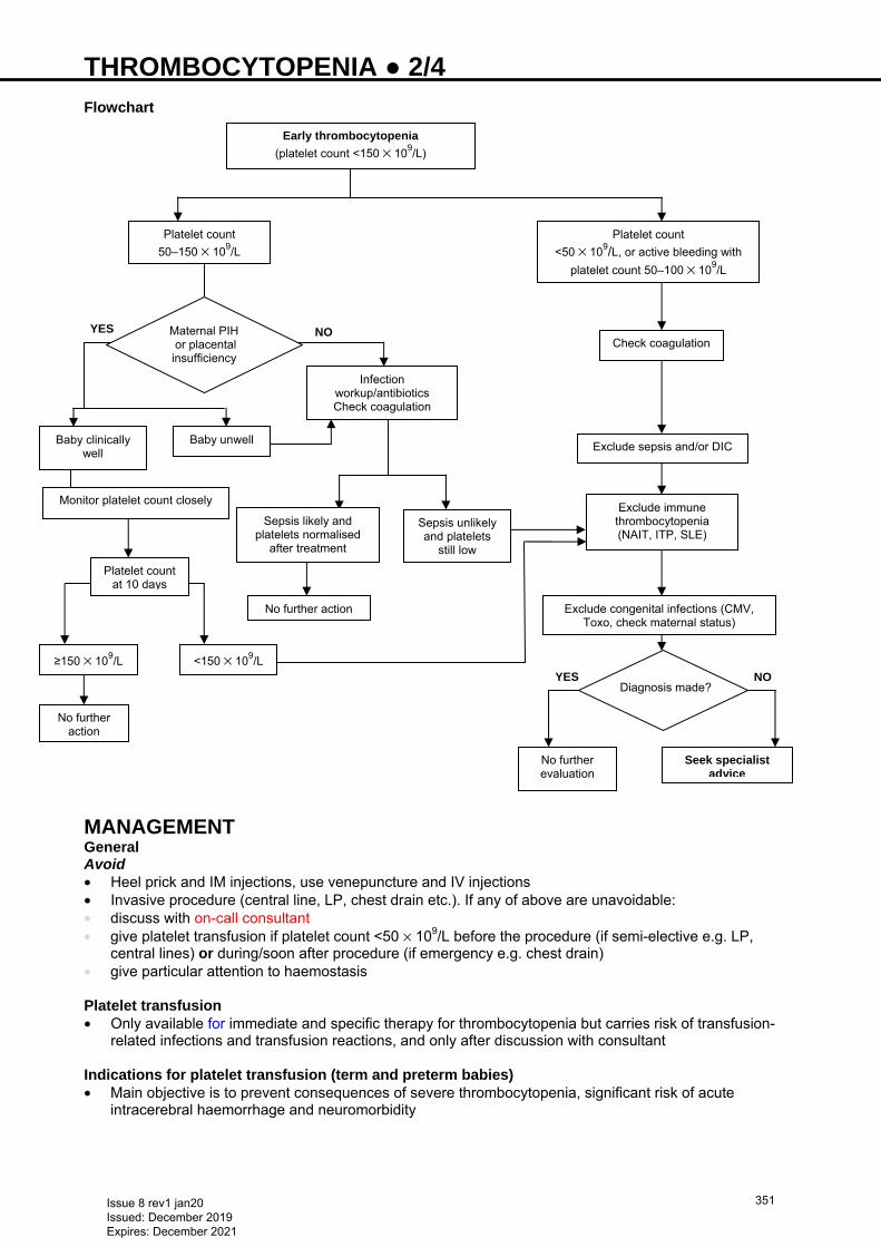
THROMBOCYTOPENIA ● 2/4 Flowchart
MANAGEMENT General Avoid • Heel prick and IM injections, use venepuncture and IV injections • Invasive procedure (central line, LP, chest drain etc.). If any of above are unavoidable: • discuss with on-call consultant • give platelet transfusion if platelet count <50 × 109/L before the procedure (if semi-elective e.g. LP,
central lines) or during/soon after procedure (if emergency e.g. chest drain) • give particular attention to haemostasis Platelet transfusion • Only available for immediate and specific therapy for thrombocytopenia but carries risk of transfusion-
related infections and transfusion reactions, and only after discussion with consultant Indications for platelet transfusion (term and preterm babies) • Main objective is to prevent consequences of severe thrombocytopenia, significant risk of acute
intracerebral haemorrhage and neuromorbidity
NOYES
YES NO
Baby clinically well
Monitor platelet count closely
≥150 × 109/L
Early thrombocytopenia
(platelet count <150 × 109/L)
Platelet count
50–150 × 109/L
Platelet count
<50 × 109/L, or active bleeding with
platelet count 50–100 × 109/L
Check coagulation
Exclude sepsis and/or DIC
Exclude immune thrombocytopenia (NAIT, ITP, SLE)
Exclude congenital infections (CMV, Toxo, check maternal status)
Maternal PIH or placental insufficiency
Infection workup/antibiotics Check coagulation
Diagnosis made?
Baby unwell
Sepsis likely and platelets normalised
after treatment
Sepsis unlikely and platelets
still low
No further evaluation
Seek specialist advice
Platelet count at 10 days
<150 × 109/L
No further action
No further action
Issue 8 rev1 jan20 Issued: December 2019 Expires: December 2021
351
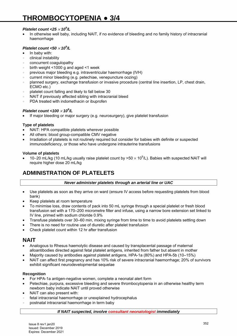
THROMBOCYTOPENIA ● 3/4 Platelet count <25 × 109/L • In otherwise well baby, including NAIT, if no evidence of bleeding and no family history of intracranial
haemorrhage Platelet count <50 × 109/L • In baby with: • clinical instability • concurrent coagulopathy • birth weight <1000 g and aged <1 week • previous major bleeding e.g. intraventricular haemorrhage (IVH) • current minor bleeding (e.g. petechiae, venepuncture oozing) • planned surgery, exchange transfusion or invasive procedure (central line insertion, LP, chest drain,
ECMO etc.) • platelet count falling and likely to fall below 30 • NAIT if previously affected sibling with intracranial bleed • PDA treated with indomethacin or ibuprofen Platelet count <100 × 109/L • If major bleeding or major surgery (e.g. neurosurgery), give platelet transfusion Type of platelets • NAIT: HPA compatible platelets wherever possible • All others: blood group-compatible CMV negative • Irradiation of platelets is not routinely required but consider for babies with definite or suspected
immunodeficiency, or those who have undergone intrauterine transfusions Volume of platelets • 10−20 mL/kg (10 mL/kg usually raise platelet count by >50 × 109/L). Babies with suspected NAIT will
require higher dose 20 mL/kg
ADMINISTRATION OF PLATELETS
Never administer platelets through an arterial line or UAC
• Use platelets as soon as they arrive on ward (ensure IV access before requesting platelets from blood bank)
• Keep platelets at room temperature • To minimise loss, draw contents of pack into 50 mL syringe through a special platelet or fresh blood
transfusion set with a 170–200 micrometre filter and infuse, using a narrow bore extension set linked to IV line, primed with sodium chloride 0.9%
• Transfuse platelets over 30–60 min, mixing syringe from time to time to avoid platelets settling down • There is no need for routine use of diuretic after platelet transfusion • Check platelet count within 12 hr after transfusion
NAIT • Analogous to Rhesus haemolytic disease and caused by transplacental passage of maternal
alloantibodies directed against fetal platelet antigens, inherited from father but absent in mother • Majority caused by antibodies against platelet antigens, HPA-1a (80%) and HPA-5b (10–15%) • NAIT can affect first pregnancy and has 10% risk of severe intracranial haemorrhage; 20% of survivors
exhibit significant neurodevelopmental sequelae Recognition • For HPA-1a antigen-negative women, complete a neonatal alert form • Petechiae, purpura, excessive bleeding and severe thrombocytopenia in an otherwise healthy term
newborn baby indicate NAIT until proved otherwise • NAIT can also present with: • fetal intracranial haemorrhage or unexplained hydrocephalus • postnatal intracranial haemorrhage in term baby
If NAIT suspected, involve consultant neonatologist immediately
Issue 8 rev1 jan20 Issued: December 2019 Expires: December 2021
352
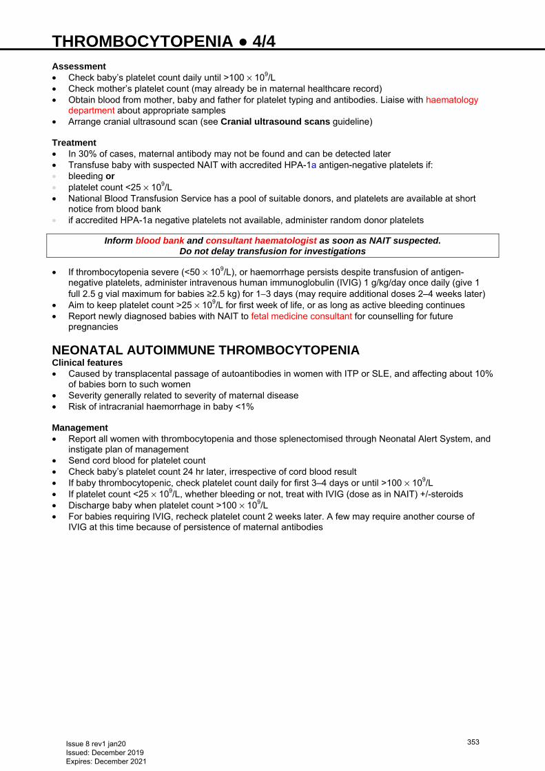
THROMBOCYTOPENIA ● 4/4 Assessment • Check baby’s platelet count daily until >100 × 109/L • Check mother’s platelet count (may already be in maternal healthcare record) • Obtain blood from mother, baby and father for platelet typing and antibodies. Liaise with haematology
department about appropriate samples • Arrange cranial ultrasound scan (see Cranial ultrasound scans guideline) Treatment • In 30% of cases, maternal antibody may not be found and can be detected later • Transfuse baby with suspected NAIT with accredited HPA-1a antigen-negative platelets if: • bleeding or • platelet count <25 × 109/L • National Blood Transfusion Service has a pool of suitable donors, and platelets are available at short
notice from blood bank • if accredited HPA-1a negative platelets not available, administer random donor platelets
Inform blood bank and consultant haematologist as soon as NAIT suspected. Do not delay transfusion for investigations
• If thrombocytopenia severe (<50 × 109/L), or haemorrhage persists despite transfusion of antigen-negative platelets, administer intravenous human immunoglobulin (IVIG) 1 g/kg/day once daily (give 1 full 2.5 g vial maximum for babies ≥2.5 kg) for 1−3 days (may require additional doses 2–4 weeks later)
• Aim to keep platelet count >25 × 109/L for first week of life, or as long as active bleeding continues • Report newly diagnosed babies with NAIT to fetal medicine consultant for counselling for future
pregnancies
NEONATAL AUTOIMMUNE THROMBOCYTOPENIA Clinical features • Caused by transplacental passage of autoantibodies in women with ITP or SLE, and affecting about 10%
of babies born to such women • Severity generally related to severity of maternal disease • Risk of intracranial haemorrhage in baby <1% Management • Report all women with thrombocytopenia and those splenectomised through Neonatal Alert System, and
instigate plan of management • Send cord blood for platelet count • Check baby’s platelet count 24 hr later, irrespective of cord blood result • If baby thrombocytopenic, check platelet count daily for first 3–4 days or until >100 × 109/L • If platelet count <25 × 109/L, whether bleeding or not, treat with IVIG (dose as in NAIT) +/-steroids • Discharge baby when platelet count >100 × 109/L • For babies requiring IVIG, recheck platelet count 2 weeks later. A few may require another course of
IVIG at this time because of persistence of maternal antibodies
Issue 8 rev1 jan20 Issued: December 2019 Expires: December 2021
353
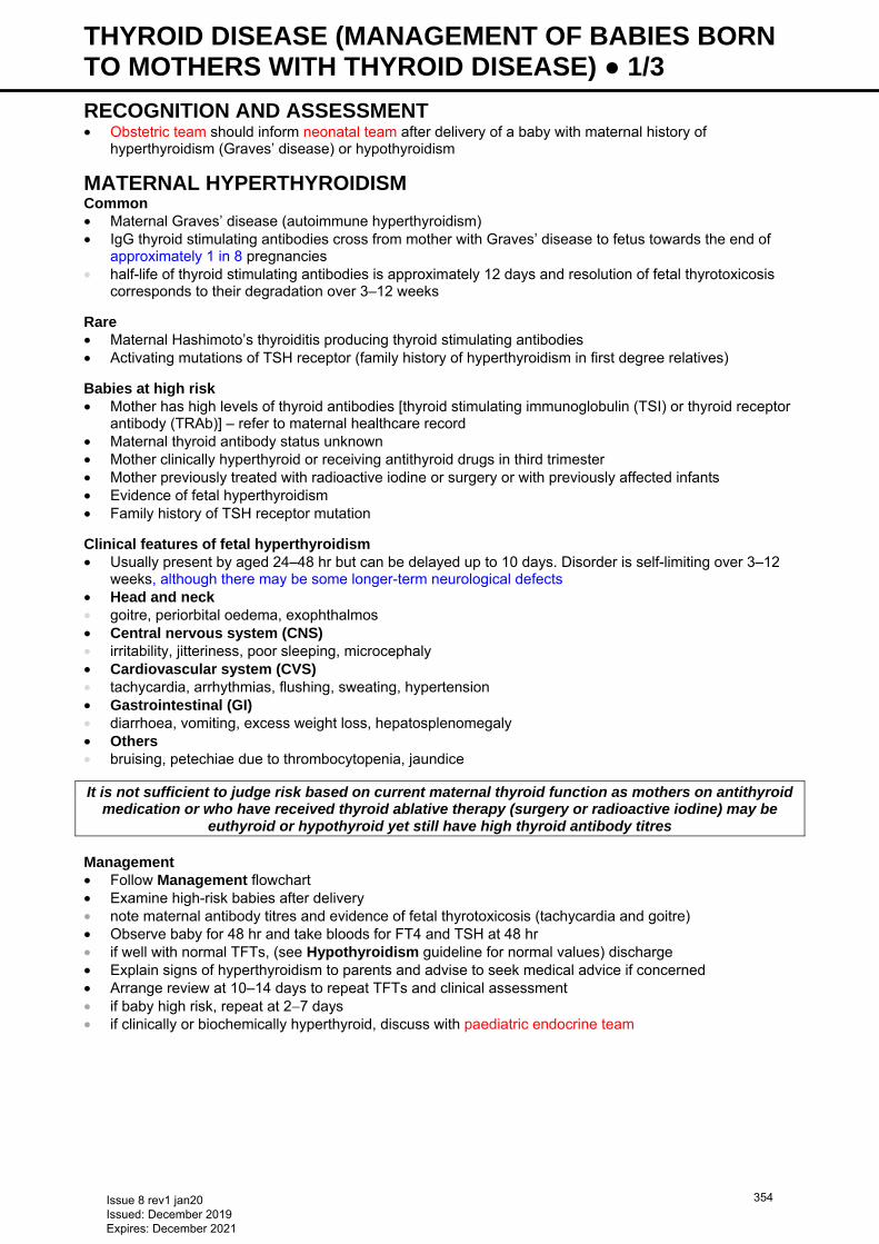
THYROID DISEASE (MANAGEMENT OF BABIES BORN TO MOTHERS WITH THYROID DISEASE) ● 1/3 RECOGNITION AND ASSESSMENT • Obstetric team should inform neonatal team after delivery of a baby with maternal history of
hyperthyroidism (Graves’ disease) or hypothyroidism
MATERNAL HYPERTHYROIDISM Common • Maternal Graves’ disease (autoimmune hyperthyroidism) • IgG thyroid stimulating antibodies cross from mother with Graves’ disease to fetus towards the end of
approximately 1 in 8 pregnancies • half-life of thyroid stimulating antibodies is approximately 12 days and resolution of fetal thyrotoxicosis
corresponds to their degradation over 3–12 weeks
Rare • Maternal Hashimoto’s thyroiditis producing thyroid stimulating antibodies • Activating mutations of TSH receptor (family history of hyperthyroidism in first degree relatives) Babies at high risk • Mother has high levels of thyroid antibodies [thyroid stimulating immunoglobulin (TSI) or thyroid receptor
antibody (TRAb)] – refer to maternal healthcare record • Maternal thyroid antibody status unknown • Mother clinically hyperthyroid or receiving antithyroid drugs in third trimester • Mother previously treated with radioactive iodine or surgery or with previously affected infants • Evidence of fetal hyperthyroidism • Family history of TSH receptor mutation
Clinical features of fetal hyperthyroidism • Usually present by aged 24–48 hr but can be delayed up to 10 days. Disorder is self-limiting over 3–12
weeks, although there may be some longer-term neurological defects • Head and neck • goitre, periorbital oedema, exophthalmos • Central nervous system (CNS) • irritability, jitteriness, poor sleeping, microcephaly • Cardiovascular system (CVS) • tachycardia, arrhythmias, flushing, sweating, hypertension • Gastrointestinal (GI) • diarrhoea, vomiting, excess weight loss, hepatosplenomegaly • Others • bruising, petechiae due to thrombocytopenia, jaundice
It is not sufficient to judge risk based on current maternal thyroid function as mothers on antithyroid medication or who have received thyroid ablative therapy (surgery or radioactive iodine) may be
euthyroid or hypothyroid yet still have high thyroid antibody titres
Management • Follow Management flowchart • Examine high-risk babies after delivery • note maternal antibody titres and evidence of fetal thyrotoxicosis (tachycardia and goitre) • Observe baby for 48 hr and take bloods for FT4 and TSH at 48 hr • if well with normal TFTs, (see Hypothyroidism guideline for normal values) discharge • Explain signs of hyperthyroidism to parents and advise to seek medical advice if concerned • Arrange review at 10–14 days to repeat TFTs and clinical assessment • if baby high risk, repeat at 2−7 days • if clinically or biochemically hyperthyroid, discuss with paediatric endocrine team
Issue 8 rev1 jan20 Issued: December 2019 Expires: December 2021
354
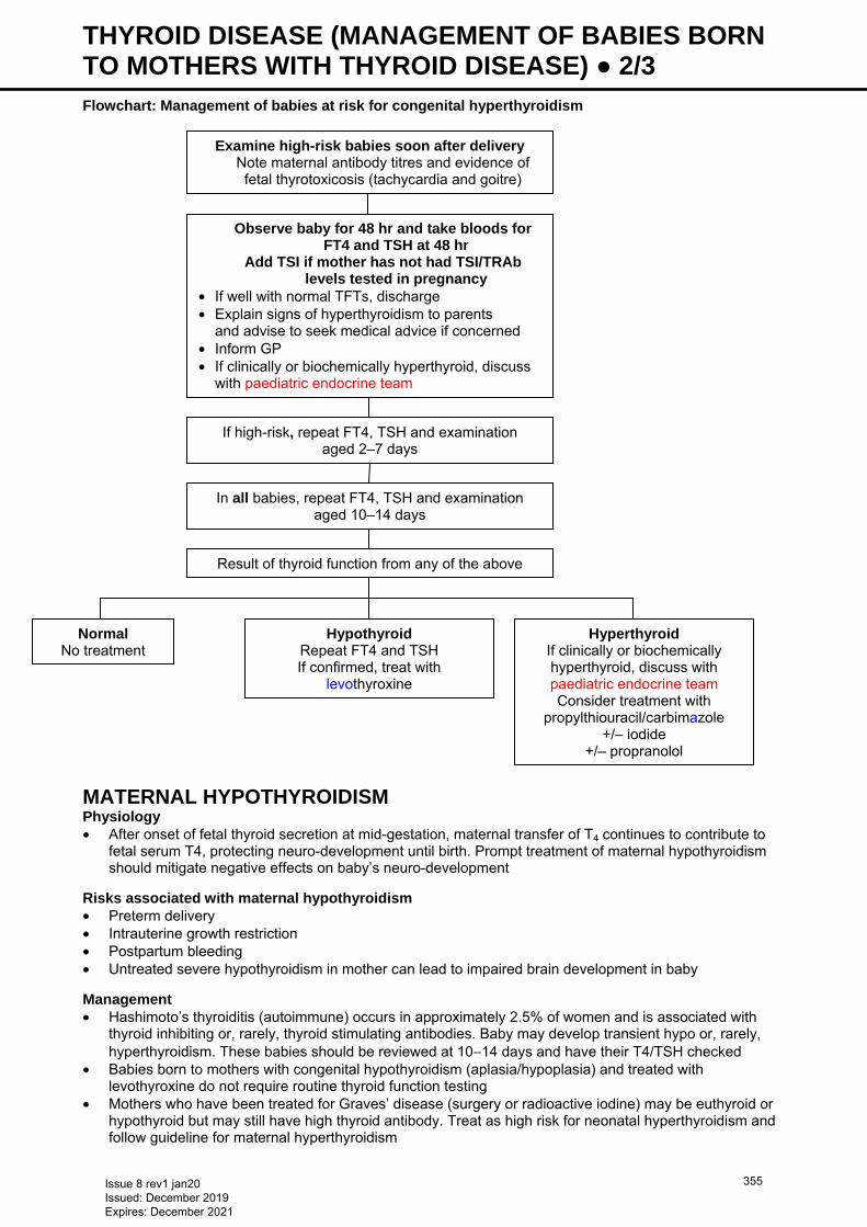
THYROID DISEASE (MANAGEMENT OF BABIES BORN TO MOTHERS WITH THYROID DISEASE) ● 2/3 Flowchart: Management of babies at risk for congenital hyperthyroidism
MATERNAL HYPOTHYROIDISM Physiology • After onset of fetal thyroid secretion at mid-gestation, maternal transfer of T4 continues to contribute to
fetal serum T4, protecting neuro-development until birth. Prompt treatment of maternal hypothyroidism should mitigate negative effects on baby’s neuro-development
Risks associated with maternal hypothyroidism • Preterm delivery • Intrauterine growth restriction • Postpartum bleeding • Untreated severe hypothyroidism in mother can lead to impaired brain development in baby Management • Hashimoto’s thyroiditis (autoimmune) occurs in approximately 2.5% of women and is associated with
thyroid inhibiting or, rarely, thyroid stimulating antibodies. Baby may develop transient hypo or, rarely, hyperthyroidism. These babies should be reviewed at 10−14 days and have their T4/TSH checked
• Babies born to mothers with congenital hypothyroidism (aplasia/hypoplasia) and treated with levothyroxine do not require routine thyroid function testing
• Mothers who have been treated for Graves’ disease (surgery or radioactive iodine) may be euthyroid or hypothyroid but may still have high thyroid antibody. Treat as high risk for neonatal hyperthyroidism and follow guideline for maternal hyperthyroidism
If high-risk, repeat FT4, TSH and examination aged 2–7 days
In all babies, repeat FT4, TSH and examination aged 10–14 days
Result of thyroid function from any of the above
Normal No treatment
Hypothyroid Repeat FT4 and TSH If confirmed, treat with
levothyroxine
Hyperthyroid If clinically or biochemically hyperthyroid, discuss with paediatric endocrine team Consider treatment with
propylthiouracil/carbimazole +/– iodide
+/– propranolol
Examine high-risk babies soon after delivery Note maternal antibody titres and evidence of
fetal thyrotoxicosis (tachycardia and goitre)
Observe baby for 48 hr and take bloods for FT4 and TSH at 48 hr
Add TSI if mother has not had TSI/TRAb levels tested in pregnancy
• If well with normal TFTs, discharge • Explain signs of hyperthyroidism to parents
and advise to seek medical advice if concerned • Inform GP • If clinically or biochemically hyperthyroid, discuss
with paediatric endocrine team
Issue 8 rev1 jan20 Issued: December 2019 Expires: December 2021
355
THYROID DISEASE (MANAGEMENT OF BABIES BORN TO MOTHERS WITH THYROID DISEASE) ● 3/3 Breastfeeding • Encourage for all babies even if mother currently taking carbimazole, propylthiouracil or levothyroxine
Contraindication • Radioactive iodine treatment
Issue 8 rev1 jan20 Issued: December 2019 Expires: December 2021
356
TRANSCUTANEOUS CO2 AND O2 ● 1/3 Adapted with permission, Guy’s and St Thomas’ NHS Trust nursing guideline
INTRODUCTION • In babies requiring assisted ventilation, it is essential to monitor arterial partial pressure of oxygen (PaO2)
and carbon dioxide (PaCO2) to ensure adequate gas exchange • Transcutaneous monitoring allows continuous measurement (TcCO2 and TcO2) • Use this guideline to set up and safely use transcutaneous monitoring equipment Clinical indications • Monitoring adequacy of arterial oxygenation and/or ventilation • Nursing critically ill or unstable baby Advantages • Reduction in number of blood gas measurements • Immediate recognition of need for ventilation adjustment Potential problems • Tissue injury (e.g. erythema, blisters, burns, and skin tears) as a result of failure to change site
frequently enough (2–3 hrly) • Inadequate measurement resulting from incorrect set-up
EQUIPMENT • Transducer: insert at end position of rack for easy accessibility • Membranes • Electrolyte solution • Adhesive fixation rings • Recalibration machine Probe placement and application of fixation rings • Avoid bony surfaces: use soft tissues (e.g. abdomen, buttock, thigh) and avoid placing over liver as this
can prevent accurate clinical assessment of liver size • Ensure chosen site is clean and dry • Peel adhesive protection layer off ring • Place ring on chosen site pressing gently on centre of ring before running finger around outside. Ensure
effective seal as this will affect accuracy of measurement • Place 3 drops of contact fluid in centre of ring • Remove transducer from module into ring and turn 1-quarter clockwise to secure
CARE AND MONITORING Temperature setting • Keep transducer setting at 44°C for all babies. There is good correlation of TcO2 with heat settings of
44°C, but lower settings will result with under-reading of TcO2 and difference is larger with increasing TcO2
Alarm settings PPHN • Exact limits will depend on specific pathology but, for guidance, in term babies with PPHN: • TcO2 upper 10.0 lower 5.5 • TcCO2 upper 7.0 lower 5.0 Blood gas sampling • Take blood gas 20 min after commencing transcutaneous monitoring to allow comparison between
transcutaneous values and arterial partial pressures of O2 and CO2 levels, as discrepancy can occur • If transcutaneous monitoring values change suddenly, check contact is in place before making ventilator
changes. If any doubt about accuracy of values, check blood gas before making ventilator changes Changing measurement site • Babies <29 weeks: change 2-hrly • Babies ≥29 weeks: change 3-hrly • Unscrew transducer before removing fixation rings
Issue 8 rev1 jan20 Issued: December 2019 Expires: December 2021
357

TRANSCUTANEOUS CO2 AND O2 ● 2/3 • Remove fixation rings when repositioning baby from supine to prone and vice-versa to avoid pressure
sore from lying on rings • Remove rings 12-hrly on babies <29 weeks and 24-hrly on babies ≥29 weeks
Calibration of membrane • See Figure 1–5 Indications • Transducer membrane has been replaced • Monitor displays ‘calibration required’ • Measurement values in doubt • Applying to a new baby • Changing measurement site
Ensure calibrator turned off after use. Do not dispose of connecting tube. Contact technicians when calibrating gas empty
Changing transducer membranes – see Figure 6–10 • All staff responsible for ventilated babies can change transducer membranes Indications • When using a new transducer or if transducer has dried out • For each new baby • When membrane crinkled, scratched or damaged • After 5 days continuous use Procedure • Wash and dry hands • To remove O-rings, unscrew protective cap from transducer and hook O-ring remover under them • Remove both clear plastic membranes with your fingers • To ensure correct values, clean transducer head, including groove and rim, with absorbent paper to
remove all old electrolyte solution • Apply approximately 2 drops of electrolyte solution to transducer head • Press transducer head downward into an unused membrane replacer until replacer reacts as far as it
can and a click is heard
Issue 8 rev1 jan20 Issued: December 2019 Expires: December 2021
358
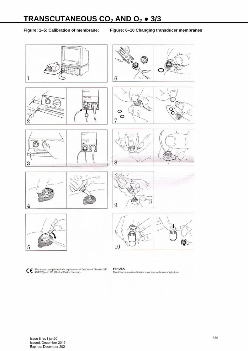
TRANSCUTANEOUS CO2 AND O2 ● 3/3 Figure: 1–5: Calibration of membrane; Figure: 6–10 Changing transducer membranes
Issue 8 rev1 jan20 Issued: December 2019 Expires: December 2021
359
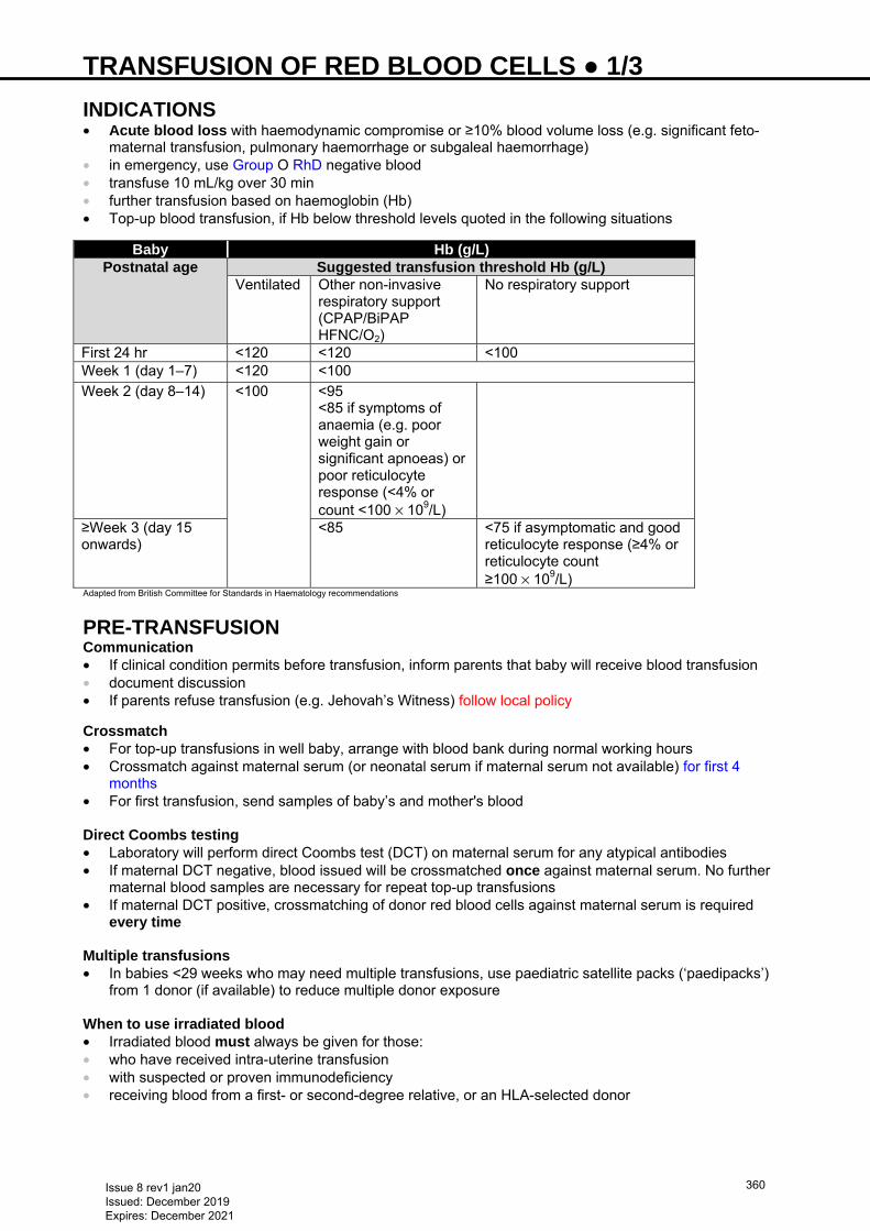
TRANSFUSION OF RED BLOOD CELLS ● 1/3 INDICATIONS • Acute blood loss with haemodynamic compromise or ≥10% blood volume loss (e.g. significant feto-
maternal transfusion, pulmonary haemorrhage or subgaleal haemorrhage) • in emergency, use Group O RhD negative blood • transfuse 10 mL/kg over 30 min • further transfusion based on haemoglobin (Hb) • Top-up blood transfusion, if Hb below threshold levels quoted in the following situations
Baby Hb (g/L)Postnatal age Suggested transfusion threshold Hb (g/L)
Ventilated Other non-invasive respiratory support (CPAP/BiPAP HFNC/O2)
No respiratory support
First 24 hr <120 <120 <100 Week 1 (day 1–7) <120 <100 Week 2 (day 8–14) <100 <95
<85 if symptoms of anaemia (e.g. poor weight gain or significant apnoeas) or poor reticulocyte response (<4% or count <100 × 109/L)
≥Week 3 (day 15 onwards)
<85 <75 if asymptomatic and good reticulocyte response (≥4% or reticulocyte count ≥100 × 109/L)
Adapted from British Committee for Standards in Haematology recommendations
PRE-TRANSFUSION Communication • If clinical condition permits before transfusion, inform parents that baby will receive blood transfusion • document discussion • If parents refuse transfusion (e.g. Jehovah’s Witness) follow local policy
Crossmatch • For top-up transfusions in well baby, arrange with blood bank during normal working hours • Crossmatch against maternal serum (or neonatal serum if maternal serum not available) for first 4
months • For first transfusion, send samples of baby’s and mother's blood Direct Coombs testing • Laboratory will perform direct Coombs test (DCT) on maternal serum for any atypical antibodies • If maternal DCT negative, blood issued will be crossmatched once against maternal serum. No further
maternal blood samples are necessary for repeat top-up transfusions • If maternal DCT positive, crossmatching of donor red blood cells against maternal serum is required
every time Multiple transfusions • In babies <29 weeks who may need multiple transfusions, use paediatric satellite packs (‘paedipacks’)
from 1 donor (if available) to reduce multiple donor exposure When to use irradiated blood • Irradiated blood must always be given for those: • who have received intra-uterine transfusion • with suspected or proven immunodeficiency • receiving blood from a first- or second-degree relative, or an HLA-selected donor
Issue 8 rev1 jan20 Issued: December 2019 Expires: December 2021
360
TRANSFUSION OF RED BLOOD CELLS ● 2/3 When to use CMV-free blood • As CMV seronegativity cannot be guaranteed in untested blood, use only CMV-seronegative blood for
neonatal transfusions • blood products in use in the UK are leucodepleted to <5 × 10⁶ leucocytes/unit at point of manufacture Special considerations Iron supplements • Premature babies receiving breast milk or with Hb <100 g/L − commence oral iron supplementation at
aged 4 weeks (see Nutrition and enteral feeding guideline)
Withholding feeds during transfusion • Some units withhold enteral feeds during transfusion whilst others continue − there is insufficient
evidence for clear recommendation
Babies with necrotising enterocolitis (NEC) • Transfuse using red cells in sodium chloride 0.9%, adenine, glucose and mannitol (SAG-M), preferably,
as it is relatively plasma-free. This may not be available in all units • Any unexpected haemolysis associated with transfusion in a baby with NEC should be investigated for
T-cell activation in consultation with local haematology department and with close involvement of consultant neonatologist
Exchange transfusion • See Exchange transfusion guideline
TRANSFUSION Volume of transfusion • Give 15 mL/kg of red cell transfusion for non-bleeding neonates irrespective of pre-transfusion Hb
• Give 20 mL/kg of red cell transfusion in case of massive haemorrhage (see Massive haemorrhage guideline)
A paediatric pack contains approximately 50 mL blood. Use 1 pack if possible
Rate of administration • Administer blood at 15 mL/kg over 3−4 hr • Increase rate in presence of active haemorrhage with shock (see Massive haemorrhage and
Subgaleal haemorrhage guidelines) • Via peripheral venous or umbilical venous line (not via long line/arterial line)
Use of furosemide • Routine use not recommended • Consider soon after blood transfusion for babies: • with chronic lung disease • with haemodynamically significant PDA • in heart failure • with oedema or fluid overload
DOCUMENTATION AND GOOD PRACTICE • Clearly document indication for transfusion and consent in the notes • Ensure positive identification of baby using accessible identification • Ensure blood transfusion volume and rate is prescribed in appropriate infusion chart • Observations, including: • continuous ECG • SpO2 • hourly temperature and BP (recorded before, during and after transfusion) • Appropriate labelling of syringes to ensure compliance with current best practice • Unless clinically urgent, avoid transfusion out-of-hours • To reduce need for blood transfusion, minimise blood sampling in babies (micro-techniques, non-
invasive monitoring) and avoid unnecessary testing • If resuscitation not required, delay cord clamping at birth • Ensure donor exposure is minimised by using satellite packs from same donor • After transfusion, record benefit (or lack thereof) • Document pre- and post-transfusion Hb levels
Issue 8 rev1 jan20 Issued: December 2019 Expires: December 2021
361

TRANSFUSION OF RED BLOOD CELLS ● 3/3 Hazards of transfusion • Most important are: • infections – bacterial/viral • hypocalcaemia • volume overload • citrate toxicity • rebound hypoglycaemia (following high glucose levels in additive solutions) • thrombocytopenia after exchange transfusion
Issue 8 rev1 jan20 Issued: December 2019 Expires: December 2021
362
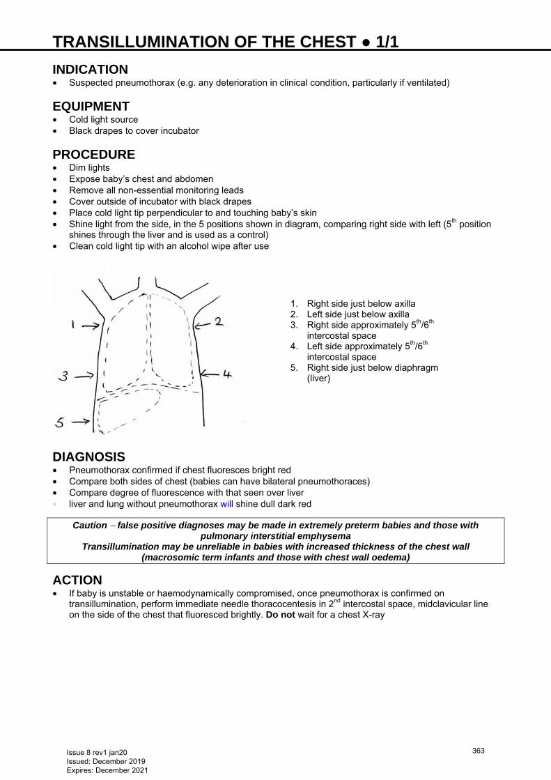
TRANSILLUMINATION OF THE CHEST ● 1/1 INDICATION • Suspected pneumothorax (e.g. any deterioration in clinical condition, particularly if ventilated)
EQUIPMENT • Cold light source • Black drapes to cover incubator
PROCEDURE • Dim lights • Expose baby’s chest and abdomen • Remove all non-essential monitoring leads • Cover outside of incubator with black drapes • Place cold light tip perpendicular to and touching baby’s skin • Shine light from the side, in the 5 positions shown in diagram, comparing right side with left (5th position
shines through the liver and is used as a control) • Clean cold light tip with an alcohol wipe after use
DIAGNOSIS • Pneumothorax confirmed if chest fluoresces bright red • Compare both sides of chest (babies can have bilateral pneumothoraces) • Compare degree of fluorescence with that seen over liver • liver and lung without pneumothorax will shine dull dark red
Caution − false positive diagnoses may be made in extremely preterm babies and those with pulmonary interstitial emphysema
Transillumination may be unreliable in babies with increased thickness of the chest wall (macrosomic term infants and those with chest wall oedema)
ACTION • If baby is unstable or haemodynamically compromised, once pneumothorax is confirmed on
transillumination, perform immediate needle thoracocentesis in 2nd intercostal space, midclavicular line on the side of the chest that fluoresced brightly. Do not wait for a chest X-ray
1. Right side just below axilla 2. Left side just below axilla 3. Right side approximately 5th/6th
intercostal space 4. Left side approximately 5th/6th
intercostal space 5. Right side just below diaphragm
(liver)
Issue 8 rev1 jan20 Issued: December 2019 Expires: December 2021
363
TRANSPORT AND RETRIEVAL ● 1/3
[West Midlands Neonatal Transport Service (NTS) guideline]
INTRODUCTION The aim of a safe transfer policy is to ensure the highest standard, streamlined care. In the majority of cases transfer will be performed by a dedicated transfer team, but in certain cases the referring team may perform the transfer. In all cases the ACCEPT model (Table 1) can be used
INDICATIONS FOR TRANSFER • Uplift for services not provided at referring unit (including diagnostic and drive-through transfers) • Repatriation • Resources/capacity Table 1: ACCEPT model A Assessment C Control C Communication E Evaluation P Preparation and packaging T Transportation
ASSESSMENT • Key questions are: • what is the problem? • what is being done? • what effect is it having? • what is needed now?
CONTROL • Following initial assessment control the situation: • who is the team leader? • what tasks need to be done (clinical care/equipment and resources)? • who will do them (allocated by team leader)? • who will transfer baby (if relevant)?
CLINICAL CARE • Preparation for transport begins with the referring team as soon as decision is made to transfer baby,
even if being performed by another team
Airway/breathing • If baby unstable or on CPAP with FiO2 >0.4, intubate and ventilate • Adjust ETT and lines depending on chest X-ray position; document all positions and adjustments and
consider if repeat X-ray required; secure all lines and tubes • If indicated, give surfactant [see Surfactant replacement therapy − including less invasive
surfactant administration (LISA) technique guideline] • If present, connect chest drains to a flutter valve • Check appropriate type of ventilator support is available for transfer (e.g. high-flow/BiPAP/SiPAP/volume
guarantee/oscillation may not be provided in transport) − if not, discuss other options • If ventilated perform blood gas and adjust ventilation settings as necessary • if non-invasive ventilation support, have recent (<6 hr) gas result available
Circulation • If baby dependent on drug infusions (e.g. inotropes, prostaglandin), 2 reliable points of venous access
must be inserted • Check whether receiving unit will accept central lines • if baby receiving bicarbonate, insulin or inotropes insert double lumen UVC • ensure catheters secured with suture and tape (see Umbilical artery catheterisation and removal and
Umbilical venous catheterisation and removal guidelines) • check all access is patent and visible • optimise blood pressure (see Hypotension guideline) • ensure recent lactate result available
Issue 8 rev1 jan20 Issued: December 2019 Expires: December 2021
364
TRANSPORT AND RETRIEVAL ● 2/3 Drugs • Antibiotics [see Infection in first 72 hours of life guideline and Infection (late onset) guideline] • Decide whether infusions need to be concentrated • Check vitamin K IM has been given • Decide whether sedation/paralysis needed for transfer
Environment • Monitor temperature throughout stabilisation – in the extreme preterm baby chemical gel mattress may
be required • Cooling babies [see Cooling in non-cooling centres (referral and preparation of babies eligible for
active cooling) guideline]
Fluids • Ensure all fluids and infusions are in 50 mL syringes and are labelled • If requested, change PN to maintenance fluids • Volume as per Intravenous fluid therapy guideline • Monitor intake and output Infection • Check if any colonisation issues and inform receiving unit Parents • Update with plan of care • Discuss how parents will get to receiving unit – may be appropriate to travel with team • Clarify method of feeding • Document any safeguarding issues
COMMUNICATION Referring centre • Make decision to transfer with parents’ agreement; in exceptional circumstances this may not be
achievable • For neonatal uplift transfers: • locate NICU/paediatric intensive care unit (PICU)/speciality bed • for Birmingham Children’s Hospital PICU beds call 0300 200 1100 to refer for neonatal transfer and cot
location • available Monday−Friday, 0900−1700 hr (outside these hours cot availability of regional units can be
accessed via this number) • for speciality or other PICU bed, call receiving clinician • all other transfers, including transfer into regional children’s hospital, confirm cot is available and ring
0300 200 1100 for neonatal transfer • All transfers, provide: • demographics to administrator • clinical details to transfer team
− history and clinical details − urgency of transfer − interventions, investigations and results − medications
• Document advice given/received • Prepare transfer information/discharge summary and arrange for images to be reviewed at receiving
hospital • Obtain sample of mother’s blood (if required) • Identify whether appropriate for parent to transfer with baby (see NTS policy for details)
Receiving centre • Ensure consultant and nurse co-ordinator accept referral and agree with advice given
EVALUATION • Referring clinician, transfer team and receiving team evaluate urgency of transfer and decide who will do
it
Issue 8 rev1 jan20 Issued: December 2019 Expires: December 2021
365
TRANSPORT AND RETRIEVAL ● 3/3 • Neonatal transfers are classified as: • time critical (e.g. gastroschisis, ventilated tracheoesophageal fistula, intestinal perforation, duct-
dependent cardiac lesion not responding to prostaglandin infusion and other unstable conditions) • to be performed within 1 hr • to be performed within 24 hr • to be performed after 24 hr • In the event of transfer team being unable to respond within an appropriate time period, referring unit
may decide to perform transfer themselves in the best interests of the baby
PREPARATION AND PACKAGING • Three components: • clinical care (see above) • location and checking of equipment • allocation of team • Transport equipment must not be used for any other purpose • Team undertaking transfer must be trained in use of all equipment and drugs and be competent to
perform any necessary procedures en-route • Ensure air and oxygen cylinders are full before departure • ETT and lines must be secured before transferring baby to transport incubator • Baby must be secured in transport incubator
TRANSPORT Before leaving referring unit • Change to transport incubator gases (check cylinders are full) • Check blood gas 10 min after changing to transport ventilator. Make any necessary changes • Check lines and tubes are not tangled; check infusions are running • Record vital signs • Allow parents to see baby • Contact receiving hospital to confirm cot still available and handover clinical details (including infusions
and ventilator settings)
Only leave referring unit when team leader is confident that baby is stable for transfer On arrival at ambulance • Ensure incubator and equipment are securely fastened/stowed in accordance with CEN standards • Plug in gases and electrical connections • Ensure temperature in ambulance is suitable • Check all staff are aware of destination • Discuss mode of progression to hospital (e.g. category of transfer) • Ensure all staff are wearing seatbelts before vehicle moves During road transit • Record vital signs • If baby requires clinical intervention, stop ambulance in a safe place before staff leave their seats • Make receiving team aware of any major changes in clinical condition On arrival at receiving hospital • Follow the ACCEPT structure • Handover to receiving team then transfer baby to the unit’s equipment • transfer and receiving teams to agree order in which transfer happens • After transfer, dispose of any partially used drugs and infusions before returning to ambulance
Issue 8 rev1 jan20 Issued: December 2019 Expires: December 2021
366
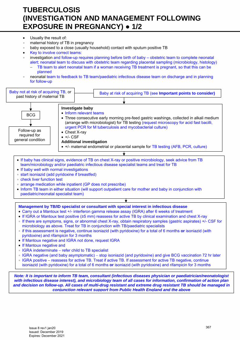
TUBERCULOSIS (INVESTIGATION AND MANAGEMENT FOLLOWING EXPOSURE IN PREGNANCY) ● 1/2 • Usually the result of: • maternal history of TB in pregnancy • baby exposed to a close (usually household) contact with sputum positive TB • Key to involve correct teams: • investigation and follow-up requires planning before birth of baby – obstetric team to complete neonatal
alert; neonatal team to discuss with obstetric team regarding placental sampling (microbiology, histology) − TB team to alert neonatal team if a woman receiving TB treatment is pregnant, so that this can be
planned • neonatal team to feedback to TB team/paediatric infectious disease team on discharge and in planning
for follow-up
Baby at risk of acquiring TB (see Important points to consider)
BCG
Investigate baby • Inform relevant teams • Three consecutive early morning pre-feed gastric washings, collected in alkali medium
(arrange with microbiologist) for TB testing (request microscopy for acid fast bacilli, urgent PCR for M tuberculosis and mycobacterial culture)
• Chest X-ray • +/- CSF Additional investigation • +/- maternal endometrial or placental sample for TB testing (AFB, PCR, culture)
Follow-up as required for
general condition
Baby not at risk of acquiring TB, or past history of maternal TB
• If baby has clinical signs, evidence of TB on chest X-ray or positive microbiology, seek advice from TB team/microbiology and/or paediatric infectious disease specialist teams and treat for TB
• If baby well with normal investigations • start isoniazid (add pyridoxine if breastfed) • check liver function test • arrange medication while inpatient (GP does not prescribe) • Inform TB team in either situation (will support outpatient care for mother and baby in conjunction with
paediatric/neonatal specialist team)
Management by TB/ID specialist or consultant with special interest in infectious disease • Carry out a Mantoux test +/- interferon gamma release assay (IGRA) after 6 weeks of treatment • If IGRA or Mantoux test positive (≥5 mm) reassess for active TB by clinical examination and chest X-ray • If there are symptoms, signs, or abnormal chest X-ray, obtain respiratory samples (gastric aspirates) +/- CSF for
microbiology as above. Treat for TB in conjunction with TB/paediatric specialists • if this assessment is negative, continue isoniazid (with pyridoxine) for a total of 6 months or isoniazid (with
pyridoxine) and rifampicin for 3 months • If Mantoux negative and IGRA not done, request IGRA • If Mantoux negative and • IGRA indeterminate − refer child to TB specialist • IGRA negative (and baby asymptomatic) − stop isoniazid (and pyridoxine) and give BCG vaccination 72 hr later • IGRA positive − reassess for active TB. Treat if active TB. If assessment for active TB negative, continue
isoniazid (with pyridoxine) for a total of 6 months or isoniazid (with pyridoxine) and rifampicin for 3 months
Note: It is important to inform TB team, consultant (infectious diseases physician or paediatrician/neonatologist with infectious disease interest), and microbiology team of all cases for information, confirmation of action plan and decision on follow-up. All cases of multi-drug resistant and extreme drug resistant TB should be managed in
conjunction relevant support from Public Health England and the above
Issue 8 rev1 jan20 Issued: December 2019 Expires: December 2021
367
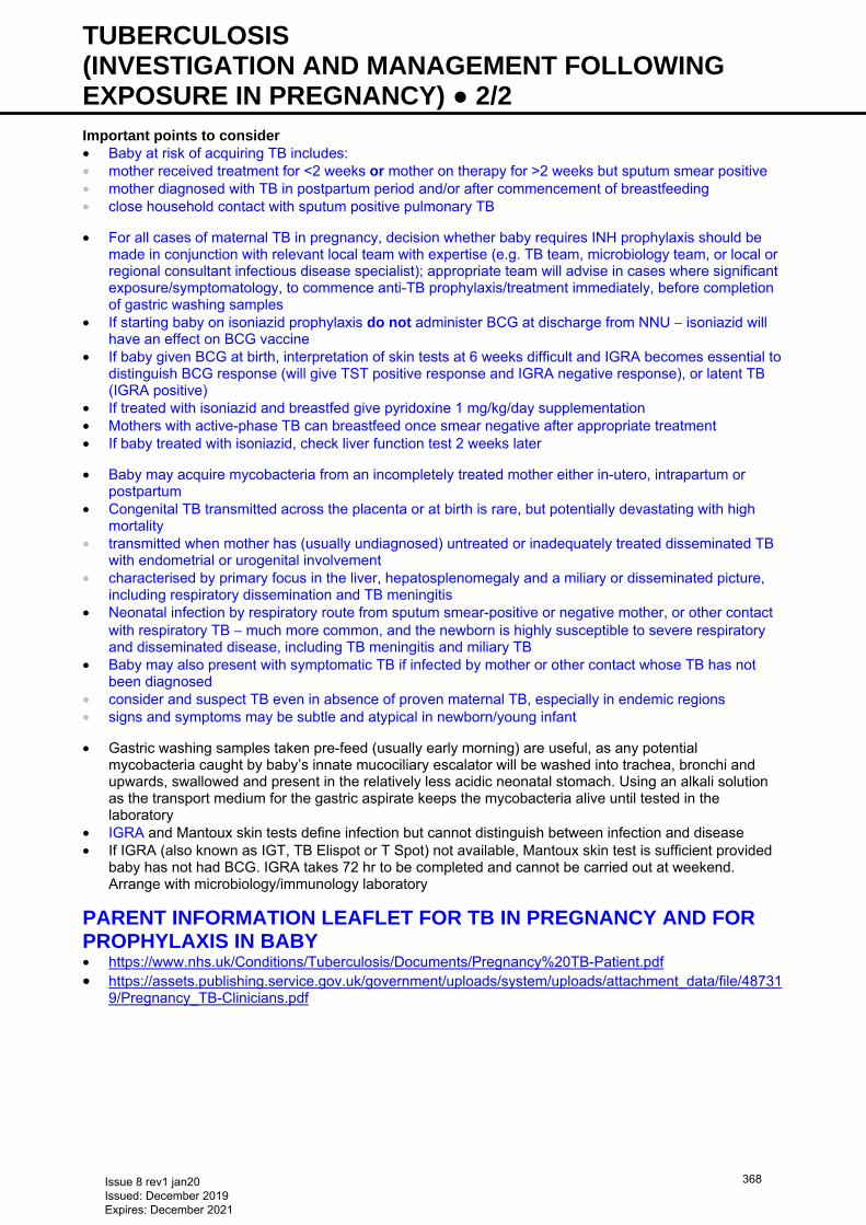
TUBERCULOSIS (INVESTIGATION AND MANAGEMENT FOLLOWING EXPOSURE IN PREGNANCY) ● 2/2 Important points to consider • Baby at risk of acquiring TB includes: • mother received treatment for <2 weeks or mother on therapy for >2 weeks but sputum smear positive • mother diagnosed with TB in postpartum period and/or after commencement of breastfeeding • close household contact with sputum positive pulmonary TB
• For all cases of maternal TB in pregnancy, decision whether baby requires INH prophylaxis should be made in conjunction with relevant local team with expertise (e.g. TB team, microbiology team, or local or regional consultant infectious disease specialist); appropriate team will advise in cases where significant exposure/symptomatology, to commence anti-TB prophylaxis/treatment immediately, before completion of gastric washing samples
• If starting baby on isoniazid prophylaxis do not administer BCG at discharge from NNU − isoniazid will have an effect on BCG vaccine
• If baby given BCG at birth, interpretation of skin tests at 6 weeks difficult and IGRA becomes essential to distinguish BCG response (will give TST positive response and IGRA negative response), or latent TB (IGRA positive)
• If treated with isoniazid and breastfed give pyridoxine 1 mg/kg/day supplementation • Mothers with active-phase TB can breastfeed once smear negative after appropriate treatment • If baby treated with isoniazid, check liver function test 2 weeks later
• Baby may acquire mycobacteria from an incompletely treated mother either in-utero, intrapartum or postpartum
• Congenital TB transmitted across the placenta or at birth is rare, but potentially devastating with high mortality
• transmitted when mother has (usually undiagnosed) untreated or inadequately treated disseminated TB with endometrial or urogenital involvement
• characterised by primary focus in the liver, hepatosplenomegaly and a miliary or disseminated picture, including respiratory dissemination and TB meningitis
• Neonatal infection by respiratory route from sputum smear-positive or negative mother, or other contact with respiratory TB − much more common, and the newborn is highly susceptible to severe respiratory and disseminated disease, including TB meningitis and miliary TB
• Baby may also present with symptomatic TB if infected by mother or other contact whose TB has not been diagnosed
• consider and suspect TB even in absence of proven maternal TB, especially in endemic regions • signs and symptoms may be subtle and atypical in newborn/young infant
• Gastric washing samples taken pre-feed (usually early morning) are useful, as any potential mycobacteria caught by baby’s innate mucociliary escalator will be washed into trachea, bronchi and upwards, swallowed and present in the relatively less acidic neonatal stomach. Using an alkali solution as the transport medium for the gastric aspirate keeps the mycobacteria alive until tested in the laboratory
• IGRA and Mantoux skin tests define infection but cannot distinguish between infection and disease • If IGRA (also known as IGT, TB Elispot or T Spot) not available, Mantoux skin test is sufficient provided
baby has not had BCG. IGRA takes 72 hr to be completed and cannot be carried out at weekend. Arrange with microbiology/immunology laboratory
PARENT INFORMATION LEAFLET FOR TB IN PREGNANCY AND FOR PROPHYLAXIS IN BABY • https://www.nhs.uk/Conditions/Tuberculosis/Documents/Pregnancy%20TB-Patient.pdf • https://assets.publishing.service.gov.uk/government/uploads/system/uploads/attachment_data/file/48731
9/Pregnancy_TB-Clinicians.pdf
Issue 8 rev1 jan20 Issued: December 2019 Expires: December 2021
368
TWO YEAR DEVELOPMENTAL ASSESSMENT ● 1/3
Based on NICE guideline NG 72
INDICATIONS • Gestation <30 weeks • gestation ≤31+6 weeks if capacity available locally to do so • Gestation 30−36+6 weeks and 1 of the following: • grade 3 or 4 IVH, cystic PVL or other brain lesion likely to be associated with developmental disorders • grade 2 or 3 HIE • neonatal bacterial meningitis • neonatal herpes simplex infection
IDENTIFICATION OF ELIGIBLE BABIES • Initial unit of booking is responsible for performing assessment • NNU discharging baby will document unit responsible for follow-up in BadgerNet discharge summary • NNU to use BadgerNet to confirm which babies are their responsibility for assessment • if baby listed incorrectly use standard network proforma to contact follow-up lead in appropriate unit (see
network website https://www.networks.nhs.uk/nhs-networks/staffordshire-shropshire-and-black-country-newborn/groups/follow-up-group) to ensure baby is seen by appropriate NNU
ASSESSMENT • Assess at 2 yr corrected age using Bayley III assessment • Send parental questionnaire before assessment and request parents complete and bring to assessment • Complete 2 yr follow-up form in BadgerNet • Send copy of assessment outcome summary to GP, health visitor and parents • include copy of network booklet Explanation of assessment scores: Information for parents and
carers (available at www.networks.nhs.uk/nhs-networks/staffordshire-shropshire-and-black-country-newborn/documents/documents/follow-up-assessment-explanation-of-scores-leaflet-2017/view) with parents’ summary
• If parents do not respond to invitation for Bayley III assessment, request copy of Ages and Stages (ASQ) 24 or 30 months questionnaire from health visitor and use this to complete BadgerNet 2 yr follow-up form
SUBSEQUENT FOLLOW-UP • Children born <28 weeks should be seen again aged 4 yr (uncorrected) • Children born ≥28 weeks: • if development is normal and no physical health issues, discharge • if continuing physical health problems, follow-up with general paediatrician or appropriate specialist • if neurodevelopmental problems identified, refer to relevant allied professionals and community
paediatrician
Issue 8 rev1 jan20 Issued: December 2019 Expires: December 2021
369
UMBILICAL ARTERY CATHETERISATION AND REMOVAL ● 1/3 Do not attempt to carry out this procedure unsupervised unless you have been trained to do so and
have demonstrated your competence under appropriate supervision
INDICATIONS • Frequent blood gas analysis: • ventilated babies (most babies treated with CPAP can be managed with capillary gases) • Continuous monitoring of arterial blood pressure (if poor circulation or need for accurate BP) • Exchange transfusion
CONTRAINDICATIONS • Umbilical sepsis • Necrotising enterocolitis (NEC) • Evidence of vascular compromise in legs or buttocks • Congenital abnormality of the umbilicus (e.g. exomphalos or gastroschisis)
EQUIPMENT • Umbilical artery catheterisation pack • Umbilical catheter: <2 kg use size 3.5 FG, ≥2 kg use size up to 5 FG • 3-way tap • Sterile gown, gloves and drape • Infusion pump • Sodium chloride 0.9% or 0.45% infusion containing heparin 1 unit/mL • Umbilical tape • Cleaning solution as per unit policy • Zinc oxide tape or Elastoplast®
PROCEDURE Consent • Wherever possible inform parents of need and associated risks before procedure; if an emergency,
delay explanation until after insertion • Risks include sepsis and thrombosis • See Consent guideline Pre-sterile preparation • Monitor baby’s vital signs during procedure • Estimate length of catheter to be inserted using formula: (weight in kg × 3) + 9 cm • alternative method for umbilical artery catheter (UAC) length is twice distance from umbilicus to mid-
inguinal point, plus distance from umbilicus to xiphisternum • add length of cord stump to give final length • prefer high catheter position i.e. tip above diaphragm (T6–T10 vertebral bodies) • Inspect lower limbs and buttocks for discolouration • Tie an umbilical tape loosely around base of cord Sterile preparation • Scrub up, put on gown and gloves using aseptic technique • Ask assistant (if available) to gently hold baby’s legs and arms away from umbilical site • Clean cord stump and surrounding skin with cleaning solution • Attach 3-way tap to catheter and flush all parts with sodium chloride 0.9% leaving syringe attached • Place all equipment to be used on a sterile towel covering a sterile trolley • Place sterile drape with a hole in the centre over the umbilical stump. Pull the stump through the hole
ready for catheter insertion Insertion of arterial catheter • Clamp across cord with artery forceps • Apply gentle upward traction • Cut along underside of forceps with a scalpel blade to reveal either the cut surface of the whole cord, or
use a side-on approach cut part way through the artery at a 45° angle • Leave a 2–3 cm stump; remember to measure length of cord stump and add to calculated placement to
give final advancement distance
Issue 8 rev1 jan20 Issued: December 2019 Expires: December 2021
370
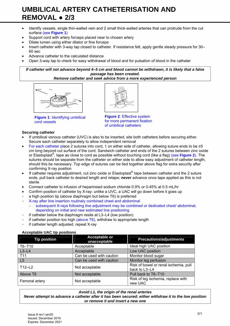
UMBILICAL ARTERY CATHETERISATION AND REMOVAL ● 2/3 • Identify vessels, single thin-walled vein and 2 small thick-walled arteries that can protrude from the cut
surface (see Figure 1) • Support cord with artery forceps placed near to chosen artery • Dilate lumen using either dilator or fine forceps • Insert catheter with 3-way tap closed to catheter. If resistance felt, apply gentle steady pressure for 30–
60 sec • Advance catheter to the calculated distance • Open 3-way tap to check for easy withdrawal of blood and for pulsation of blood in the catheter
If catheter will not advance beyond 4–5 cm and blood cannot be withdrawn, it is likely that a false passage has been created.
Remove catheter and seek advice from a more experienced person Securing catheter • If umbilical venous catheter (UVC) is also to be inserted, site both catheters before securing either.
Secure each catheter separately to allow independent removal • For each catheter place 2 sutures into cord, 1 on either side of catheter, allowing suture ends to be ≥5
cm long beyond cut surface of the cord. Sandwich catheter and ends of the 2 sutures between zinc oxide or Elastoplast®, tape as close to cord as possible without touching cord (like a flag) (see Figure 2). The sutures should be separate from the catheter on either side to allow easy adjustment of catheter length, should this be necessary. Top edge of sutures can be tied together above flag for extra security after confirming X-ray position
• If catheter requires adjustment, cut zinc oxide or Elastoplast® tape between catheter and the 2 suture ends, pull back catheter to desired length and retape; never advance once tape applied as this is not sterile
• Connect catheter to infusion of heparinised sodium chloride 0.9% or 0.45% at 0.5 mL/hr • Confirm position of catheter by X-ray: unlike a UVC, a UAC will go down before it goes up • a high position tip (above diaphragm but below T6) is preferred • X-ray after line insertion routinely combined chest and abdominal
− subsequent X-rays following line adjustment may be combined or dedicated chest/ abdominal, depending on initial and new estimated line positioning
• if catheter below the diaphragm resite at L3–L4 (low position) • if catheter position too high (above T6), withdraw to appropriate length • if catheter length adjusted, repeat X-ray
Acceptable UAC tip positions
Tip position Acceptable or unacceptable
Precautions/adjustments
T6−T10 Acceptable Ideal high UAC position L3–L4 Acceptable Low UAC position T11 Can be used with caution Monitor blood sugar L5 Can be used with caution Monitor leg perfusion
T12–L2 Not acceptable Risk of bowel or renal ischemia, pull back to L3–L4
Above T6 Not acceptable Pull back to T6–T10
Femoral artery Not acceptable Risk of leg ischemia, replace with new UAC
Avoid L1, the origin of the renal arteries Never attempt to advance a catheter after it has been secured; either withdraw it to the low position
or remove it and insert a new one
Figure 2: Effective system for more permanent fixation of umbilical catheters
Figure 1: Identifying umbilical cord vessels
Head Vein
Issue 8 rev1 jan20 Issued: December 2019 Expires: December 2021
371
UMBILICAL ARTERY CATHETERISATION AND REMOVAL ● 3/3 DOCUMENTATION • Record details of procedure in baby’s notes, including catheter position on X-ray and whether any
adjustments were made • Always label umbilical arterial and venous catheters using the appropriately coloured and labelled
stickers • Place traceability sticker from catheter/insertion pack into notes
AFTERCARE • Nurse baby in a position where UAC can be observed • Monitor circulation in lower limbs and buttocks while catheter is in situ • Leave cord stump exposed to air • Infuse heparinised sodium chloride 0.9% or 0.45% 0.5 mL/hr (1 unit heparin/mL) • Do not infuse any other solution through UAC. Glucose or drugs may be administered through UAC only
in exceptional situations, on the authority of a consultant
COMPLICATIONS • Bleeding following accidental disconnection • Vasospasm: if blanching of the lower limb occurs and does not resolve, remove catheter • Embolisation from blood clot or air in the infusion system • Thrombosis involving: • femoral artery, resulting in limb ischaemia • renal artery, resulting in haematuria, renal failure and hypertension • mesenteric artery, resulting in NEC • Infection: prophylactic antibiotics are not required
REMOVAL
Do not attempt to carry out this procedure unsupervised unless you have been trained to do so and have demonstrated your competence
INDICATIONS • Catheter no longer required • No longer patent • Suspected infection • Complications (e.g. NEC, vascular compromise to the lower limbs)
EQUIPMENT • Sterile stitch cutter • Sterile blade • Umbilical tape • Cleaning solution − Sterexidine 200 solution 0.5%
PROCEDURE • Wash hands and put on sterile gloves • Clean cord stump with cleaning solution • if umbilical tissue adherent to catheter, loosen by soaking cord stump with gauze swab soaked in sodium
chloride 0.9% • Ensure an umbilical tape is loosely secured around base of umbilicus • Turn infusion pump off and clamp infusion line • Withdraw catheter slowly over 2–3 min, taking particular care with last 2–3 cm • If bleeding noted, tighten umbilical tape • Do not cover umbilicus with large absorbent pad, a small piece of cotton gauze should suffice • Inspect catheter after removal: if any part missing, contact consultant immediately
AFTERCARE • Nurse baby supine for 4 hr following removal, and observe for bleeding
COMPLICATIONS • Bleeding • Catheter tip inadvertently left in blood vessel
Issue 8 rev1 jan20 Issued: December 2019 Expires: December 2021
372
UMBILICAL VENOUS CATHETERISATION AND REMOVAL ● 1/4 Do not attempt to carry out this procedure unsupervised unless you have been trained to do so and
have demonstrated your competence under appropriate supervision
INDICATIONS • All babies <1000 g • Babies ≥1000 g ventilated or unwell (e.g. HIE) (a double lumen catheter may be indicated if baby
requires significant support) • Exchange transfusion • Administration of hypertonic solutions (e.g. glucose >12.5%, parenteral nutrition or inotropes)
CONTRAINDICATIONS • Umbilical sepsis • Necrotising enterocolitis (NEC) • Gastroschisis/exomphalos
EQUIPMENT • Umbilical vein catheterisation pack • suture (if not included in above pack) • Umbilical venous catheter (UVC) • 3-way tap • Gown and gloves • Sterile drape • Infusion pump • Sodium chloride 0.9% infusion • Umbilical tape • Cleaning solution as per unit policy • Zinc oxide tape or Elastoplast®
PROCEDURE • See http://www.bapm.org/publications/documents/guidelines/BAPM • Wherever possible inform parents of need and associated risks before procedure; if an emergency,
delay explanation until after insertion • Risks include sepsis and thrombosis • See Consent guideline Pre-sterile preparation • Monitor all vital signs during procedure • Estimate length of catheter to be inserted: use distance between xiphisternum and umbilicus +1, plus
stump length or a formula [(3 × weight in kg + 9)/2 + 1 (be aware that formulae tend to overestimate length)
• Remember to add length of cord stump to give final distance catheter needs to be advanced • Tie umbilical tape loosely around base of cord Sterile preparation • Scrub up, and put on gown and gloves • Use sterile technique • Clean cord stump and surrounding skin with cleaning solution • Attach 3-way tap to catheter and flush all parts with sodium chloride 0.9%. Leave syringe attached • Place all equipment to be used on sterile towel covering sterile trolley • Drape umbilical stump with sterile towels • Place sterile sheet with a hole in the centre over the cord. Pull the cord through the hole Insertion of umbilical catheter • Clamp across cord with artery forceps • Apply gentle upward traction • Cut along underside of forceps with scalpel blade cleanly to leave 2–3 cm stump or, if also placing an
umbilical arterial catheter (UAC) and you have been trained in this procedure, consider using side-on technique (see Umbilical artery catheterisation guideline)
Issue 8 rev1 jan20 Issued: December 2019 Expires: December 2021
373
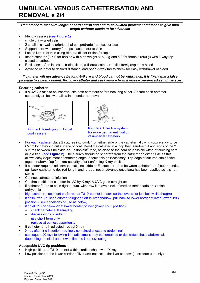
UMBILICAL VENOUS CATHETERISATION AND REMOVAL ● 2/4
Remember to measure length of cord stump and add to calculated placement distance to give final length catheter needs to be advanced
• Identify vessels (see Figure 1): • single thin-walled vein • 2 small thick-walled arteries that can protrude from cut surface • Support cord with artery forceps placed near to vein • Locate lumen of vein using either a dilator or fine forceps • Insert catheter (3.5 F for babies with birth weight <1500 g and 5 F for those ≥1500 g) with 3-way tap
closed to catheter • Resistance often indicates malposition; withdraw catheter until it freely aspirates blood • Advance catheter to desired distance, and open 3-way tap to check for easy withdrawal of blood
If catheter will not advance beyond 4–5 cm and blood cannot be withdrawn, it is likely that a false passage has been created. Remove catheter and seek advice from a more experienced senior person
Securing catheter • If a UAC is also to be inserted, site both catheters before securing either. Secure each catheter
separately as below to allow independent removal • For each catheter place 2 sutures into cord, 1 on either side of the catheter, allowing suture ends to be
≥5 cm long beyond cut surface of cord. Bend the catheter in a loop then sandwich it and ends of the 2 sutures between zinc oxide or Elastoplast® tape, as close to the cord as possible without touching cord (like a flag) (see Figure 2). The sutures should be separate from the catheter on either side as this allows easy adjustment of catheter length, should this be necessary. Top edge of sutures can be tied together above flag for extra security after confirming X-ray position
• If catheter requires adjustment, cut zinc oxide or Elastoplast® tape between catheter and 2 suture ends, pull back catheter to desired length and retape; never advance once tape has been applied as it is not sterile
• Connect catheter to infusion • Confirm position of catheter in IVC by X-ray. A UVC goes straight up • if catheter found to be in right atrium, withdraw it to avoid risk of cardiac tamponade or cardiac
arrhythmia • high catheter placement preferred: at T8−9 but not in heart (at the level of or just below diaphragm) • if tip in liver, i.e. seen curved to right or left in liver shadow, pull back to lower border of liver (lower UVC
position − see conditions of use as below) • if tip at T10 or below or at lower border of liver (lower UVC position):
− check catheter still sampling − discuss with consultant − use short-term only − replace at earliest opportunity
• If catheter length adjusted, repeat X-ray • X-ray after line insertion, routinely combined chest and abdominal • subsequent X-rays following line adjustment may be combined or dedicated chest/ abdominal,
depending on initial and new estimated line positioning
Acceptable UVC tip positions • High position: at T8−9 but not within cardiac shadow on X-ray • Low position: at the lower border of liver and not inside the liver shadow (short-term use only)
Figure 2: Effective system for more permanent fixation of umbilical catheters
Figure 1: Identifying umbilical cord vessels
Head Vein
Issue 8 rev1 jan20 Issued: December 2019 Expires: December 2021
374
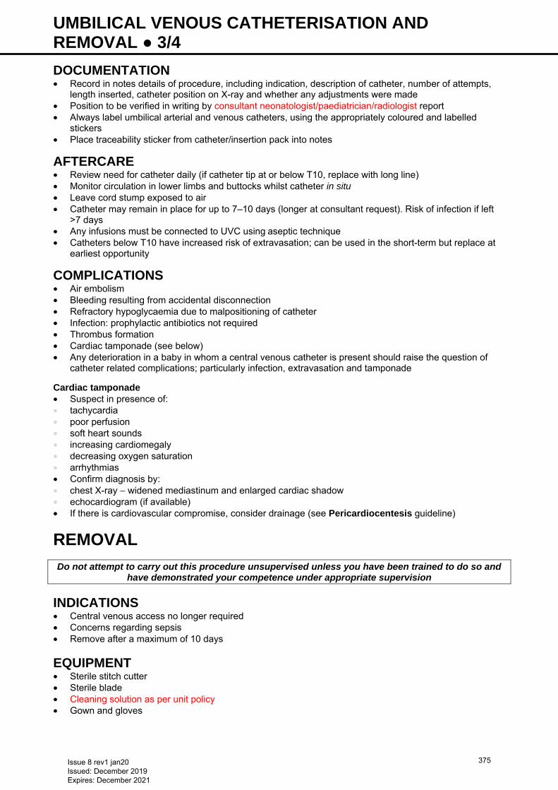
UMBILICAL VENOUS CATHETERISATION AND REMOVAL ● 3/4 DOCUMENTATION • Record in notes details of procedure, including indication, description of catheter, number of attempts,
length inserted, catheter position on X-ray and whether any adjustments were made • Position to be verified in writing by consultant neonatologist/paediatrician/radiologist report • Always label umbilical arterial and venous catheters, using the appropriately coloured and labelled
stickers • Place traceability sticker from catheter/insertion pack into notes
AFTERCARE • Review need for catheter daily (if catheter tip at or below T10, replace with long line) • Monitor circulation in lower limbs and buttocks whilst catheter in situ • Leave cord stump exposed to air • Catheter may remain in place for up to 7–10 days (longer at consultant request). Risk of infection if left
>7 days • Any infusions must be connected to UVC using aseptic technique • Catheters below T10 have increased risk of extravasation; can be used in the short-term but replace at
earliest opportunity
COMPLICATIONS • Air embolism • Bleeding resulting from accidental disconnection • Refractory hypoglycaemia due to malpositioning of catheter • Infection: prophylactic antibiotics not required • Thrombus formation • Cardiac tamponade (see below) • Any deterioration in a baby in whom a central venous catheter is present should raise the question of
catheter related complications; particularly infection, extravasation and tamponade Cardiac tamponade • Suspect in presence of: • tachycardia • poor perfusion • soft heart sounds • increasing cardiomegaly • decreasing oxygen saturation • arrhythmias • Confirm diagnosis by: • chest X-ray − widened mediastinum and enlarged cardiac shadow • echocardiogram (if available) • If there is cardiovascular compromise, consider drainage (see Pericardiocentesis guideline)
REMOVAL
Do not attempt to carry out this procedure unsupervised unless you have been trained to do so and have demonstrated your competence under appropriate supervision
INDICATIONS • Central venous access no longer required • Concerns regarding sepsis • Remove after a maximum of 10 days
EQUIPMENT • Sterile stitch cutter • Sterile blade • Cleaning solution as per unit policy • Gown and gloves
Issue 8 rev1 jan20 Issued: December 2019 Expires: December 2021
375
UMBILICAL VENOUS CATHETERISATION AND REMOVAL ● 4/4 PROCEDURE • Wash hands and put on gown and gloves • Clean cord stump with cleaning solution • Turn infusion pump off and clamp infusion line • Ensure umbilical tape secured loosely around base of umbilicus • Withdraw catheter slowly • If any bleeding noted, tighten umbilical tape • Confirm catheter intact • If unit policy, send catheter tip to microbiology
AFTERCARE • Nurse baby supine for 4 hr following removal and observe for bleeding
COMPLICATIONS • Bleeding • Loss of UVC tip • Infection
Issue 8 rev1 jan20 Issued: December 2019 Expires: December 2021
376
UPPER LIMB BIRTH INJURIES INCLUDING BRACHIAL PLEXUS PALSY ● 1/1 DEFINITION • Brachial plexus palsy may be congenital occurring in-utero or acquired due to injury to brachial plexus
nerves sustained due to stretching of nerves during delivery • Fractures to humerus or clavicle • Isolated radial nerve palsy of the newborn
ASSESSMENT OF ALL BABIES WITH REDUCED UPPER LIMB MOVEMENT • Examine the arm and neck for swelling, bruising, tone, posture and degree of movement • Assess for breathing difficulties and Horner’s syndrome • Document findings clearly in case notes • Explain to parents that recovery probable but may not be complete • Inform consultant obstetrician and paediatrician
MANAGEMENT • X-ray humerus/clavicle to exclude fracture • if fracture of clavicle clearly seen, reassure parents and review baby at 3 weeks when movement should
be returning • if fracture of humerus is clearly seen, offer strapping of arm to chest for comfort and contact Birmingham
Children’s Hospital (BCH) orthopaedic team to arrange follow-up • if uncertain, refer to Children’s Hand and Upper Limb Service at BCH • Classical ‘Waiter’s tip position’: • refer to Children’s Hand and Upper Limb Service at BCH as soon as possible • initiate referral to local physiotherapists • Paralysis of the arm, which is completely resolved within a few days does not need to be referred but if
there is any doubt, all babies will be seen in the regular 4 times weekly hand trauma clinic so that a specialist assessment can be made and the parents can be given appropriate information
BIRMINGHAM CHILDREN’S HAND AND UPPER LIMB SERVICE: • Fax referral proforma to: 0121 333 8131. Form available to download from https://bwc.nhs.uk/hand-
surgery-information-for-professionals • Email secretary: [email protected] or [email protected] • Tel: 0121 333 8136/8285 • Email for advice: [email protected] • Write to Mrs Andrea Jester, Consultant Plastic/Hand Surgeon,
Hand and Upper Limb Service, Birmingham Children’s Hospital, Steelhouse Lane, Birmingham B4 6NH
Issue 8 rev1 jan20 Issued: December 2019 Expires: December 2021
377
URINARY TRACT ABNORMALITIES DIAGNOSED ANTENATALLY ● 1/3 ANTENATAL ASSESSMENT Fetal diagnostic scans are undertaken at 18–20 weeks and may be repeated at 32–34 weeks 18–20 week scan Possible urinary tract abnormalities include: Kidneys • Renal agenesis +/- oligohydramnios – Potter sequence • Multi-cystic dysplastic kidney (MCDK), check other kidney for normal appearance • Solitary kidney • Abnormal position (e.g. pelvic) or shape (e.g. horseshoe) • Kidneys with echo-bright parenchyma (suspect cystic diseases) Collecting system/tubes • Unilateral or bilateral renal pelvic dilatation (RPD)/pelviectasis • Measured in antero-posterior diameter (APD) • mild: RPD 5–9 mm • moderate: RPD 10–14 mm • severe: RPD ≥15 mm • Unilateral or bilateral dilated calyces or ureter Bladder (dilated or thick-walled; ureterocoele in bladder) 32–34 week scan • To clarify urinary tract abnormalities found in early fetal scans • Assess severity of RPD/pelviectasis: • normal: RPD <7 mm • mild: RPD 7–9 mm • moderate: RPD 10−14 mm. If bilateral, suspect critical obstruction • severe: RPD ≥15 mm. Suspect critical obstruction • calyceal dilatation: often indicates severity; may suggest obstruction • Unilateral/bilateral dilated ureter(s) − suspect obstruction or vesico-ureteric reflux (VUR) • Thick-walled bladder, suspect outlet obstruction • Dilated bladder, suspect poor emptying • Ureterocoele, suspect duplex system on that side Communication • Provide mother with an information leaflet, if available in your hospital, about this antenatal anomaly and
proposed plan of management after birth
POSTNATAL MANAGEMENT Indications for intervention Urgent • Bilateral RPD ≥10 mm +/- thick-walled bladder: suspect posterior urethral valve (boys) • Unilateral RPD ≥15 mm, suspect pelvi-ureteric junction (PUJ) obstruction • Significant abnormalities of kidney(s)/urinary tract – if risk of renal insufficiency • check serum potassium, blood gas for metabolic acidosis and serum creatinine Non-urgent • All other abnormalities of urinary tract in the antenatal scan
IMMEDIATE MANAGEMENT For urgent indications • If posterior urethral valve (PUV)/PUJ obstruction suspected, check urine output/stream and monitor
weight trend • Arrange urgent KUB ultrasound scan within 24–48 hr (minimal milk intake may underestimate the size
of renal pelvis, but do not delay if there is gross dilatation) • If postnatal scan raises suspicion of posterior urethral valve (dilated ureters + thick walled bladder) • check serum creatinine • arrange urgent micturating cysto-urethrogram (MCUG) • after confirmation by MCUG, refer baby urgently to paediatric urologist • If unilateral RPD ≥20 mm (suggestive of PUJ obstruction) discuss with urologist and arrange MAG3
renogram as soon as possible/as advised by urologist • Significant abnormalities of kidney(s)/urinary tract – if risk of renal insufficiency:
Issue 8 rev1 jan20 Issued: December 2019 Expires: December 2021
378
URINARY TRACT ABNORMALITIES DIAGNOSED ANTENATALLY ● 2/3 • check serum potassium, blood gas for metabolic acidosis and serum creatinine • start trimethoprim 2 mg/kg as single night-time dose • Discuss with consultant before discharge For non-urgent indications • Renal ultrasound scan at aged 2–6 weeks • Consultant review with results Antibiotic prophylaxis • For RPD ≥10 mm, give trimethoprim 2 mg/kg as single night-time dose until criteria for stopping are met
(see below)
SUBSEQUENT MANAGEMENT • Subsequent management depends on findings of ultrasound scan at 2–6 weeks Severe pelviectasis (RPD ≥15 mm) • Arrange MAG3 scan – timing depends on severity of obstruction – as soon as possible if RPD ≥20 mm • if MAG3 scan shows obstructed pattern, discuss with paediatric urologist • Repeat ultrasound scan at aged 3–6 months (depending on cause of dilatation, a complete obstruction
requires closer monitoring) • Continue antibiotic prophylaxis until advised otherwise by urologist Moderate unilateral pelviectasis (RPD 10–14 mm) and/or ureteric dilatation • Presumed mild obstruction or VUR • If RPD increases beyond 15 mm, arrange MAG3 scan • Continue prophylaxis for VUR ≥grade 4 (marked dilatation of ureter and calyces) until child is continent
(out of nappies) • Repeat scan every 6 months until RPD <10 mm, then follow advice below Normal or mild isolated pelviectasis (RPD <10 mm) • Stop antibiotic prophylaxis • Repeat scan after 6 months • if 6 month scan normal or shows no change and there have been no urinary tract infections (UTIs),
discharge • If unwell, especially pyrexial without obvious cause, advise urine MC&S MCDK • DMSA to clarify nil function of MCDK and normal uptake pattern of other kidney • Repeat ultrasound scan 6–12 monthly to observe involution of kidney (may take several years) • Beware of 20% risk of VUR in ‘normal’ kidney, advise parents to recognise UTI/pyelonephritis (especially
if fever is without obvious focus) • MCUG or prophylaxis until continent ONLY if dilated pelvis or ureter in good kidney • Annual blood pressure check until kidney involuted • If cysts persist >5 yr, enlarge or hypertension, refer to urology Ureterocoele (often occurs with duplex kidney) • MCUG (if VUR or PUV suspected) • MAG3 to check function and drainage from both moieties of the duplex system • Prophylaxis until problem resolved • Urology referral – sooner if obstruction suspected Solitary kidney/unilateral renal agenesis • Kidney ultrasound at 6 weeks to confirm antenatal findings and rule out other urogenital structure
abnormalities • DMSA to confirm absence of 1 kidney + normal uptake pattern by the single kidney Renal parenchymal problem requiring nephrology review • Bright kidneys • Multiple cysts
Issue 8 rev1 jan20 Issued: December 2019 Expires: December 2021
379
URINARY TRACT ABNORMALITIES DIAGNOSED ANTENATALLY ● 3/3 Other conditions • Single renal artery in cord • increased risk of renal abnormality but postnatal ultrasound scan only if antenatal scan missed or
abnormal • Ear abnormalities: ultrasound examination only if associated with: • syndrome • other malformations • maternal/gestational diabetes • family history of deafness
Issue 8 rev1 jan20 Issued: December 2019 Expires: December 2021
380
VARICELLA ● 1/3 RECOGNITION AND ASSESSMENT Definition • There are 2 separate presentations depending on timing of infection: • fetal varicella syndrome (FVS): maternal chickenpox infection before 20 weeks’ gestation • neonatal varicella (NV): maternal infection in perinatal period or close contact with chickenpox or
shingles in first 7 days after birth
FETAL VARICELLA SYNDROME Symptoms and signs • Limb hypoplasia • Scarring of skin in a dermatomal distribution • Cortical atrophy, microcephaly, bowel and bladder sphincter dysfunction, vocal cord paralysis • Chorioretinitis, cataracts and microphthalmia • Intra-uterine growth restriction
Investigations Maternal • If no history of chickenpox, check maternal varicella zoster (VZ) IgG at time of contact • If mother develops chickenpox rash, send swab from base of vesicle in viral transport media for varicella
zoster PCR
Neonatal • ≤7 days VZ IgM (can be done on cord blood), or • >7 days VZ IgG (even if VZ IgM negative at birth) • If vesicles present send swab from base of vesicle in viral transport media for varicella zoster PCR
Management • Management is supportive and requires long-term multidisciplinary follow-up • Neither varicella zoster immunoglobulin (VZIG) nor aciclovir has any role in the management of these
babies
NEONATAL VARICELLA Baby born to mother who develops chickenpox rash (but not shingles) within 7 days before birth, or 7 days after birth • Give VZIG 250 mg (1 vial approximately 1.7 mL) IM (not IV) • antenatal chickenpox: give as soon as possible after delivery (must be within 72 hr) • postnatal chickenpox: give as soon as possible and within 10 days after initial exposure • consider giving in different sites in small babies • can be given without antibody testing of baby • of no benefit once neonatal chickenpox has developed • not needed for babies born after 7 days of appearance of maternal chickenpox, or where mother has
zoster; these babies should have transplacental antibodies • may not prevent NV, but can make the illness milder • If VZIG not available or IM injection contraindicated, give 0.2 g/kg IVIG (less effective)
Baby exposed to chickenpox or shingles (other than in the mother)(see Decision pathway for VZV contact) • Significant exposure: household, face-to-face for 5 min, in same room for >15 min. Case of chickenpox
or disseminated shingles is infectious from 48 hr before onset of rash until crusting of lesions or day of onset of rash until crusting for those exposed to localised shingles
• Give VZIG in the following cases of postnatal exposure to varicella: • VZ IgG-negative babies (determined by testing mother for varicella antibodies) exposed to chickenpox or
shingles from any other contact other than mother, in first 7 days of life (see Decision pathway for VZV contact)
• VZ IgG-negative babies of any age, exposed to chickenpox or herpes zoster while still requiring intensive or prolonged special care nursing
• for babies exposed postnatally, regardless of maternal chickenpox history, who: − weighed <1 kg at birth, or − were ≤28 weeks’ gestation at birth, or − are aged >60 days, or − have had repeated blood sampling with replacement by packed cell infusions perform VZ IgG assay
and, if negative, give VZIG (at risk of not having received/retained sufficient maternal VZ IgG)
Issue 8 rev1 jan20 Issued: December 2019 Expires: December 2021
381
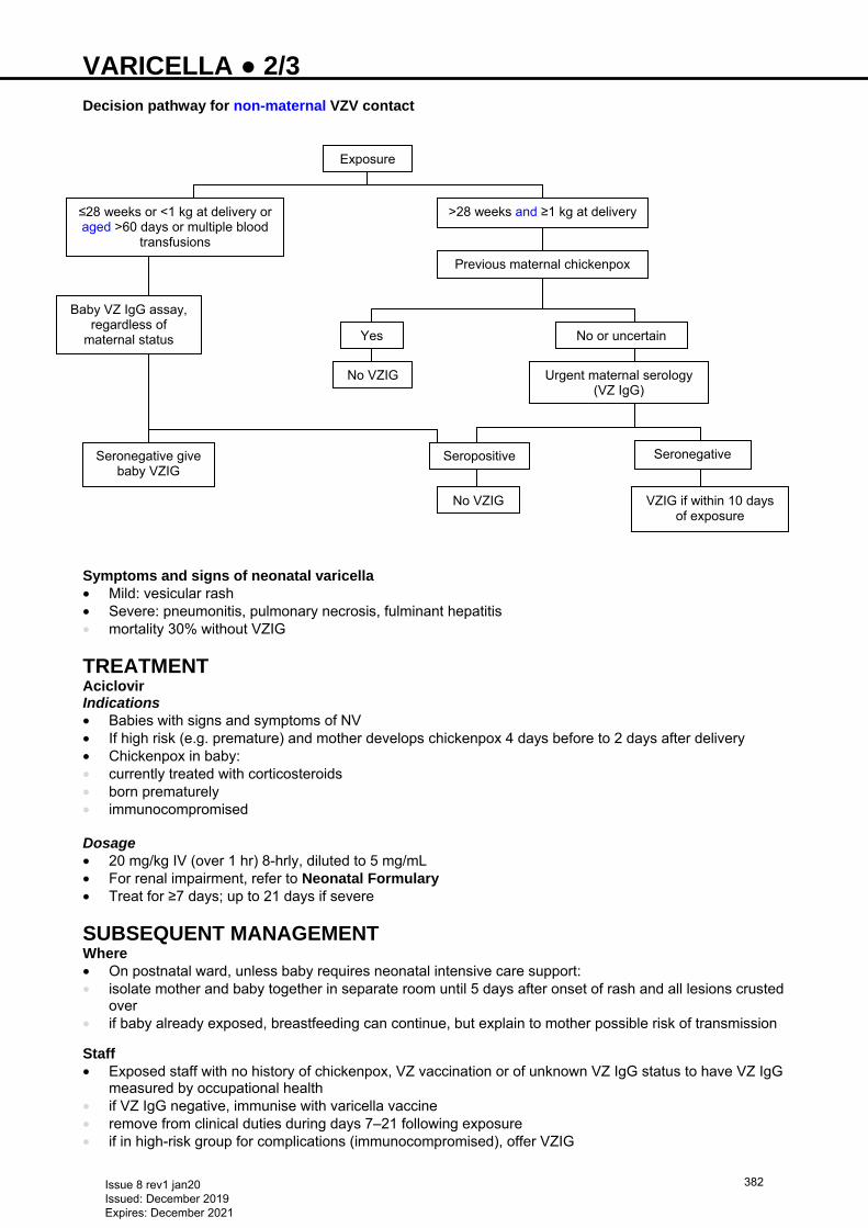
VARICELLA ● 2/3 Decision pathway for non-maternal VZV contact
Symptoms and signs of neonatal varicella • Mild: vesicular rash • Severe: pneumonitis, pulmonary necrosis, fulminant hepatitis • mortality 30% without VZIG
TREATMENT Aciclovir Indications • Babies with signs and symptoms of NV • If high risk (e.g. premature) and mother develops chickenpox 4 days before to 2 days after delivery • Chickenpox in baby: • currently treated with corticosteroids • born prematurely • immunocompromised Dosage • 20 mg/kg IV (over 1 hr) 8-hrly, diluted to 5 mg/mL • For renal impairment, refer to Neonatal Formulary • Treat for ≥7 days; up to 21 days if severe
SUBSEQUENT MANAGEMENT Where • On postnatal ward, unless baby requires neonatal intensive care support: • isolate mother and baby together in separate room until 5 days after onset of rash and all lesions crusted
over • if baby already exposed, breastfeeding can continue, but explain to mother possible risk of transmission Staff • Exposed staff with no history of chickenpox, VZ vaccination or of unknown VZ IgG status to have VZ IgG
measured by occupational health • if VZ IgG negative, immunise with varicella vaccine • remove from clinical duties during days 7–21 following exposure • if in high-risk group for complications (immunocompromised), offer VZIG
Exposure
≤28 weeks or <1 kg at delivery or aged >60 days or multiple blood
transfusions
>28 weeks and ≥1 kg at delivery
Baby VZ IgG assay, regardless of
maternal status
Previous maternal chickenpox
Yes No or uncertain
No VZIG Urgent maternal serology (VZ IgG)
Seronegative Seropositive
No VZIG VZIG if within 10 days of exposure
Seronegative give baby VZIG
Issue 8 rev1 jan20 Issued: December 2019 Expires: December 2021
382
VARICELLA ● 3/3 MONITORING TREATMENT • Aciclovir • ensure good hydration • stop once clinical improvement occurs, or when all lesions crusted
DISCHARGE AND FOLLOW-UP Maternal infection • After baby has had VZIG, discharge • Advise mother to seek medical help if baby develops chickenpox, preferably via an open-access policy
where available • Advise GP and midwife to recommend admission to isolation cubicle if rash develops Fetal infection • Diagnosed with positive VZ IgM or positive VZV PCR • ophthalmic examination • cranial ultrasound • developmental follow-up
Issue 8 rev1 jan20 Issued: December 2019 Expires: December 2021
383

VASCULAR SPASM AND THROMBOSIS ● 1/2 VASCULAR SPASM Blanching or cyanosis of extremity following insertion or manipulation of peripheral or umbilical arterial catheter (UAC) • Remove catheter • unless absolutely essential • Elicit reflex vasodilation • reflex vasospasm on insertion of UAC can occasionally be corrected by reflex vasodilation by warming
contralateral limb • Volume expansion • if appropriate, give sodium chloride 0.9% 10 mL/kg as volume expander • GTN patch • use can be considered to improve perfusion but not trialled or licensed for use in babies. Discuss with
consultant • Liaise with plastic surgeons, haematologists and other specialists as needed
VASCULAR THROMBOSIS Clinical features suggesting vascular thrombosis
Site Clinical signs Diagnostic imaging
Peripheral or central (aorta or iliac) arterial thrombosis
• Pallor • Cold arm/foot • Weak or absent peripheral
pulse • Discolouration • Gangrene • Difficulty establishing a
proper pulse oximetry trace • Delayed capillary refill time
on affected limb
• Doppler scan for large vessel thrombus (sensitivity and specificity uncertain in the neonatal period)
• Real-time 2-dimensional ultrasound
• CT scan with contrast • Contrast angiography (at
specialised centre)
Renal artery/aortic thrombosis
• Systemic hypertension • Haematuria • Oliguria • Renal failure
Renal vein thrombosis
• Flank mass • Haematuria • Hypertension • Thrombocytopenia
Inferior vena cava thrombosis • Cool lower limbs • Cyanosis • Oedema
Superior vena cava thrombosis • Swelling of upper limbs and
head • Chylothorax
Central venous line thrombus
• High pressures on long line • SVC obstruction • Chylothorax • Swelling • Discolouration of extremity
Right atrial thrombus • Heart failure • Embolic phenomenon
• Echo
Pulmonary thromboembolism • Respiratory failure • Lung perfusion scan (at
specialised centre)
MANAGEMENT OF THROMBOEMBOLISM • Controversial • Inadequate controlled trials • Inform consultant • Liaise with plastic surgeons, haematologists and other specialists as required
Issue 8 rev1 jan20 Issued: December 2019 Expires: December 2021
384

VASCULAR SPASM AND THROMBOSIS ● 2/2 Treatment options Conservative • Observe closely with no intervention e.g. unilateral renal vein thrombosis Anticoagulation and thrombolysis • No controlled neonatal trials • Use only under guidance from haematologist and/or plastic surgeon
Issue 8 rev1 jan20 Issued: December 2019 Expires: December 2021
385
VENEPUNCTURE ● 1/2
Venepuncture is the preferred method of blood sampling for term neonates and causes less pain than heel prick
INDICATIONS • Blood sampling in a baby without indwelling arterial line, or when sampling from arterial line or capillary
sampling is inappropriate
EQUIPMENT • Cleaning solution or cleaning swab – follow local infection control policy • Appropriately labelled blood bottles and request cards • Non-sterile gloves • Adhesive dressing • 23 G blood sampling needle or needle-safe cannula • Do not use a broken needle • Sterile gauze/cotton wool to apply to wound post-procedure • Sharps container
PROCEDURE Preparation • Wash hands and wear gloves (see Infection prevention guideline) • Second person employs containment holding and gives sucrose • immobilisation is crucial to baby’s safety whilst undergoing phlebotomy, and to success of procedure • Identify suitable vein (typically back of hand or foot) • Place paper towels under limb to avoid blood dripping onto bed linen Insertion and sampling • Apply hand pressure around limb to distend vein • Clean the puncture site then do not touch again • Place thumb on skin slightly distal to proposed puncture site • Hold needle at 10–20° angle and puncture skin • Advance needle toward vein. Resistance may diminish slightly as needle enters vein and blood will be
seen to flow • Collect required volume taking care to mix but not shake blood • When sampling complete, place gauze/cotton wool over insertion point and withdraw needle • Maintain pressure on site until bleeding ceases
Complications • Inability to obtain specimen due to: • inappropriate choice of vein • thrombosed vein (due to previous/repeated attempts) • inexperienced operator • baby shocked, cold or dehydrated causing vasoconstriction
Unsuccessful attempts • Adhere strictly to a limit on number of attempts • If no satisfactory sample collected after 2 attempts, seek second opinion as to whether to make a further
attempt or cancel procedure • Defer to a more experienced operator • Venous distension: • use warm pack to encourage venodilatation and venous filling • Transillumination of limb can help identify suitable vein
Avoid: • Veins close to an infection, bruising and phlebitis • Thrombosed veins • Oedematous limbs − danger of stasis of lymph, predisposing to complications e.g. phlebitis and cellulitis • Areas of previous venepuncture − build-up of scar tissue can cause difficulty accessing vein and result in
pain • Sampling from potential IV infusion site or long line vein (e.g. cubital fossa or long saphenous) whenever
possible
Issue 8 rev1 jan20 Issued: December 2019 Expires: December 2021
386
VENEPUNCTURE ● 2/2
Haemolysis risk factors • Use of <23 G needle, or too large a gauge for vessel • Drawing blood specimens from IV or central line • Under-filling tube − ratio of anticoagulant to blood >1:9 • Reusing tubes that have been refilled by hand with inappropriate amounts of anticoagulants • Mixing tube too vigorously • Failing to let alcohol/disinfectant dry • Using too great a vacuum, e.g. using too large a tube or syringe • Squeezing can cause haemolysis and elevate serum potassium Completion and organisation • Keep track of all needles used and dispose of them in sharps container • do not recap needle using both hands • Dispose of rubbish and clean tray • Remove gloves and wash hands • Label all samples and investigation forms at cot side • Arrange for transfer of samples to laboratory • Document in patient notes
SAFETY OF PRACTITIONER • Wear well-fitting gloves during procedure to prevent contamination from potential blood spills • gloves will not prevent needle stick injury, but the wiping effect of glove on needle may reduce volume of
blood to which hand exposed • Discard used needles directly into sharps container − do not re-sheath • Report any incident/accident linked to needle or sharp injury immediately, and seek assistance; start PEP
as soon as possible, following protocols (>72 hr, PEP not effective)
Issue 8 rev1 jan20 Issued: December 2019 Expires: December 2021
387
VENTILATION: CONTINUOUS POSITIVE AIRWAY PRESSURE (CPAP) ● 1/4
See Ventilation: high-flow nasal cannulae (HFNC) respiratory support guideline
DEFINITION • Non-invasive respiratory support utilising continuous distending pressure during inspiration and
expiration in spontaneously breathing babies Benefits • Improves oxygenation • Reduces work of breathing • Maintains lung volume • Lowers upper airway resistance • Conserves surfactant
INDICATIONS • Early onset respiratory distress in preterm babies • Respiratory support following extubation • Respiratory support in preterm babies with evolving chronic lung disease • Recurrent apnoea (in preterm babies) • Atelectasis • Tracheomalacia
CPAP following extubation • Consider in babies <32 weeks’ gestation
CONTRAINDICATIONS • Any baby fulfilling the criteria for ventilation • Irregular respirations • Pneumothorax without chest drain • Nasal trauma/deformity that might be exacerbated by use of nasal prongs • Larger, more mature babies often do not tolerate application of CPAP devices well • Congenital anomalies: • diaphragmatic hernia • choanal atresia • tracheo-oesophageal fistula • gastroschisis
When in doubt about CPAP indications or contraindications, discuss with consultant
TYPES OF CPAP (exact CPAP device will vary from unit to unit)
1. Standard CPAP 2. Two-level CPAP 3. Bubble CPAP
1. STANDARD CPAP
Equipment • Short binasal prongs and/or nasal mask • Circuit • Humidification • CPAP generating device with gas mixing and pressure monitoring • All require high gas flow (usual starting rate 8 L/min)
Fixing nasal CPAP device: short binasal prongs (preferred) • To avoid loss of pressure, use largest prongs that fit nostrils comfortably • Ensure device is straight and not pressed hard against nasal septum or lateral walls of nostrils.
Excessive pressure can cause tissue damage
Nasal mask • Fit securely over nose • consider alternating mask with prongs, particularly if baby developing excoriation or erosion of nasal
septum. Masks can also result in trauma, usually at the junction between the nasal septum and philtrum • Masks can give a poor seal and can obstruct
Issue 8 rev1 jan20 Issued: December 2019 Expires: December 2021
388
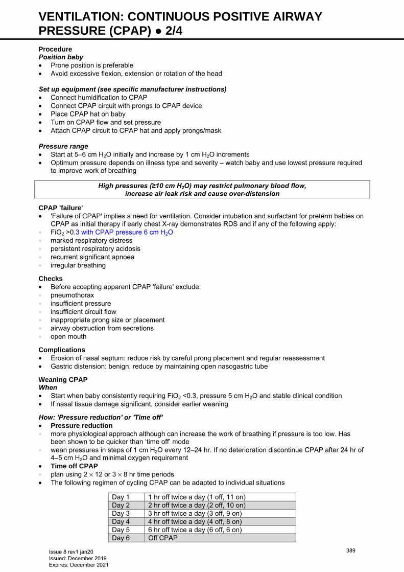
VENTILATION: CONTINUOUS POSITIVE AIRWAY PRESSURE (CPAP) ● 2/4
Procedure Position baby • Prone position is preferable • Avoid excessive flexion, extension or rotation of the head Set up equipment (see specific manufacturer instructions) • Connect humidification to CPAP • Connect CPAP circuit with prongs to CPAP device • Place CPAP hat on baby • Turn on CPAP flow and set pressure • Attach CPAP circuit to CPAP hat and apply prongs/mask Pressure range • Start at 5–6 cm H2O initially and increase by 1 cm H2O increments • Optimum pressure depends on illness type and severity – watch baby and use lowest pressure required
to improve work of breathing
High pressures (≥10 cm H2O) may restrict pulmonary blood flow, increase air leak risk and cause over-distension
CPAP 'failure' • 'Failure of CPAP' implies a need for ventilation. Consider intubation and surfactant for preterm babies on
CPAP as initial therapy if early chest X-ray demonstrates RDS and if any of the following apply: • FiO2 >0.3 with CPAP pressure 6 cm H2O • marked respiratory distress • persistent respiratory acidosis • recurrent significant apnoea • irregular breathing
Checks • Before accepting apparent CPAP 'failure' exclude: • pneumothorax • insufficient pressure • insufficient circuit flow • inappropriate prong size or placement • airway obstruction from secretions • open mouth
Complications • Erosion of nasal septum: reduce risk by careful prong placement and regular reassessment • Gastric distension: benign, reduce by maintaining open nasogastric tube
Weaning CPAP When • Start when baby consistently requiring FiO2 <0.3, pressure 5 cm H2O and stable clinical condition • If nasal tissue damage significant, consider earlier weaning
How: 'Pressure reduction' or 'Time off' • Pressure reduction • more physiological approach although can increase the work of breathing if pressure is too low. Has
been shown to be quicker than ‘time off’ mode • wean pressures in steps of 1 cm H2O every 12–24 hr. If no deterioration discontinue CPAP after 24 hr of
4–5 cm H2O and minimal oxygen requirement • Time off CPAP • plan using 2 × 12 or 3 × 8 hr time periods • The following regimen of cycling CPAP can be adapted to individual situations
Day 1 1 hr off twice a day (1 off, 11 on) Day 2 2 hr off twice a day (2 off, 10 on) Day 3 3 hr off twice a day (3 off, 9 on) Day 4 4 hr off twice a day (4 off, 8 on) Day 5 6 hr off twice a day (6 off, 6 on) Day 6 Off CPAP
Issue 8 rev1 jan20 Issued: December 2019 Expires: December 2021
389
VENTILATION: CONTINUOUS POSITIVE AIRWAY PRESSURE (CPAP) ● 3/4
Note: High-flow humidified oxygen therapy • Increasingly used as non-invasive respiratory support • Offers theoretical advantages over CPAP in ventilating upper airway spaces and producing less nasal
tissue damage • When weaning CPAP, consider using 5–6 L/min of high-flow humidified oxygen (e.g. Vapotherm® or
Optiflow™) rather than low-flow nasal cannulae oxygen or lower pressure CPAP
Failure of weaning • Increased oxygen requirement, increasing frequency of apnoeas associated with bradycardias and
cyanosis, increasing respiratory distress and/or worsening respiratory acidosis during weaning should necessitate a review and consider escalation of support
2. TWO-LEVEL CPAP • Two-level CPAP at a rate set by clinician (biphasic) or triggered by baby using an abdominal sensor
(biphasic trigger or Infant Flow® SiPAP) • Inspiratory time, pressures and apnoea alarm limit set by clinician • Indications/contraindications as CPAP and can be used when baby’s clinical condition is not improving
despite CPAP Theoretical advantages over CPAP • Improved thoraco-abdominal synchrony • Better chest wall stabilisation • Reduced upper airway resistance • Reduced work of breathing Specific modes of two-level CPAP (specific names vary with manufacturer)
CPAP and apnoea • CPAP with added advantage of apnoea monitoring via sensor attached to abdomen • Apnoea alarm triggered when no breaths detected within set time-out period Biphasic • Bi-level pressure respiratory support with/without apnoea monitoring • Higher level pressure above baseline CPAP delivered intermittently at pressure, rate and inspiratory
time set by clinician • Not synchronised with respiratory effort Biphasic trigger (tr) • Bi-level pressure respiratory support with inbuilt apnoea monitoring • Higher level pressure above baseline CPAP at rate determined by, and in synchrony with, baby’s
respiratory effort sensed through abdominal sensor • Pressure, inspiratory time and back-up rate set by clinician
Clinical use Biphasic • Begin with CPAP pressure of 5–6 cm H2O • Set peak inspiratory pressure (PIP) at 3–4 cm H2O above CPAP and rate 30 breaths/min • Keep Tinsp and apnoea alarm delay at default setting • If CO2 retention occurs, review baby and consider increase in rate and/or PIP • Avoid over-distension and keep PIP to minimum for optimum chest expansion
Weaning • By rate and pressure • If rate >30 breaths/min, wean to 30 breaths/min • Reduce MAP, by reducing PIP by 1 cm H2O every 12–24 hr • When baby breathing above 30 breaths/min change to biphasic tr mode • When MAP 5–6 cm H2O, change to CPAP
Biphasic tr • Begin with CPAP pressure of 5–6 cm H2O with PIP at 3–4 cm H2O above CPAP • Keep Tinsp and apnoea alarm delay at default setting • Set back-up rate at 30 bpm
Issue 8 rev1 jan20 Issued: December 2019 Expires: December 2021
390
VENTILATION: CONTINUOUS POSITIVE AIRWAY PRESSURE (CPAP) ● 4/4
Weaning • Reduce MAP by reducing PIP by 1 cm H2O every 12–24 hr • Once MAP 5–6 cm H2O, change to standard CPAP • If deterioration occurs during weaning process, assess baby and consider returning to biphasic mode
3. BUBBLE CPAP Alternative method of CPAP that may reduce work of breathing through facilitated diffusion Equipment • Fisher & Paykel bubble CPAP system: • delivery system: humidifier chamber, pressure manifold, heated circuit, CPAP generator • patient interface: nasal tubing, nasal prongs, baby bonnet, chin strap Procedure • Connect bubble CPAP system to baby as per manufacturer’s instructions • Ensure appropriate size nasal prongs used • Bubble CPAP nasal prongs are designed not to rest on nasal septum. Ensure prongs not resting on
the philtrum nor twisted to cause lateral pressure on septum, and allow small gap between septum and prongs
• Commence at pressures of 5 cm H2O
Bubble CPAP failure
• See CPAP failure in 1. STANDARD CPAP
Before inferring bubble CPAP failure • Ensure baby has been receiving bubble CPAP appropriately by checking for continuous bubbling in
CPAP generator, lack of bubbling can result from pressure leaks in the circuit or baby
Issue 8 rev1 jan20 Issued: December 2019 Expires: December 2021
391
VENTILATION: CONVENTIONAL ● 1/4
INTRODUCTION Oxygenation • Increase oxygenation by increasing: • FiO2 • peak end expiratory pressure (PEEP) • peak inspiratory pressure (PIP) • inspiratory time (Tinsp) CO2 • Reduced by: • increased PIP • increased rate • occasionally by reducing excessive PEEP (beware of effect on oxygenation)
VENTILATOR PARAMETERS PIP • Use lowest possible PIP to achieve visible chest expansion and adequate gas exchange on blood gas
analysis • To minimise lung injury from barotrauma and inadvertent over-distension, avoid excessive PIP • Need for higher pressures [e.g. mean airway pressure (MAP) >12 cm] could lead to consideration of high
frequency oscillatory ventilation (HFOV) [see Ventilation: high frequency oscillatory ventilation (HFOV) guideline]
PEEP • Use a PEEP ≥4 cm and increase incrementally up to 8 cm for improving oxygenation but when PEEP >6
cm necessary, take senior advice Tinsp • Usually between 0.3–0.4 sec • Avoid Tinsp >0.5 sec except in term babies with parenchymal lung disease where a Tinsp up to 1 sec may
be used Rate • Fast-rate (≥60/min) ventilation is associated with fewer air leaks and less asynchrony compared to slow
(20–40/min) rates • If rate >70/min required, HFOV may be a more appropriate option [see Ventilation: high frequency
oscillatory ventilation (HFOV) guideline] Flow • Flow 5–8 L/min is generally sufficient • Consider higher flows at faster ventilatory rates or shorter inspiratory times • SLE ventilator has a fixed flow (5 L/min) that cannot be altered Tidal volume (Vt) • Target is 4–6 mL/kg
SETTING UP VENTILATOR • Switch on humidifier and follow manufacturer’s recommended settings for optimum temperature and
humidity Setting 1 • When an admission of a preterm baby requiring ventilatory support (for recurrent apnoea, see Setting 2) • rate 60/min • PIP 16–18 cm H2O • PEEP 5 cm H2O • Tinsp 0.3–0.4 sec • FiO2 as required • flow 6–8 L/min (not applicable to SLE) • Adjust ventilatory settings depending on chest movement, SpO2, and measured Vt • Sample blood gas within 30 min of commencing ventilatory support
Issue 8 rev1 jan20 Issued: December 2019 Expires: December 2021
392
VENTILATION: CONVENTIONAL ● 2/4
Setting 2 • For babies with normal lungs requiring supportive ventilation such as term babies with respiratory
depression (asphyxia or drugs), babies with neuromuscular disorders or, in the post-operative period, and preterm babies with recurrent apnoea, set ventilator at following settings:
• rate 40/min • PIP/PEEP 14−16/4 cm H2O • Tinsp 0.35–0.4 sec • FiO2 as required (often 0.21–0.3)
ADJUSTING VENTILATORY SETTINGS Adjusting FiO2 • Oxygen is a drug and should be prescribed as with other medications. This should be done by specifying
intended target range of SpO2 on baby’s drug chart • Suggested target SpO2 ranges (see Oxygen saturation guideline) • preterm babies: 91−95% • term babies with PPHN: 96–100% Target pCO2
• Day 1−3: 4.5−8.5 kPa • Day 4 onwards: 4.5−10 kPa • If low PCO2 wean ventilation without delay and recheck within 1 hr of low measurement Altering ventilatory settings according to blood gases If blood gases are outside the targets, first check the following: • Reliability of blood gas: • is the blood gas result reliable? • has there been a sudden unexpected change from previous blood gas values? • did sample contain an air bubble? • was it obtained from a poorly perfused site? • Baby’s status: • is baby’s chest moving adequately? • how is the air entry? • Ventilator and tubing • is there an air leak? [transilluminate to exclude (see Transillumination of the chest guideline)] • what is the Vt? • are the measured ventilatory values markedly different to the set ones? • is there a large (>40%) endotracheal tube (ETT) leak?
Remember to exclude airway problems (blocked/displaced ETT) and air leaks in case of deterioration of blood gases. If available, use pedi-cap or end-tidal CO2 monitoring to exclude ETT malposition
• Small frequent changes are more appropriate than large infrequent ones
Issue 8 rev1 jan20 Issued: December 2019 Expires: December 2021
393
VENTILATION: CONVENTIONAL ● 3/4
Blood gas scenario Recommended action in order of preference
Low PaO2/SpO2
• Exclude airleak/displaced ETT/overinflation • Increase FiO2 • Increase PEEP • Increase PIP (but be aware of effect on PaCO2) • Increase Tinsp [but ensure adequate expiratory time
(Texp), especially at fast rates] • Consider further surfactant [see Surfactant
replacement therapy − including less invasive surfactant administration (LISA) technique guideline]
• If above measures unsuccessful, discuss with consultant (may need HFOV/iNO)
High PaO2 • Decrease FiO2 (unless already in air) • Decrease PEEP (if >5 cm) • Decrease PIP (especially if PaCO2 is also low)
High PaCO2
• Exclude airleak/displaced or blocked ETT • Increase PIP • Increase rate • Decrease PEEP (only if oxygenation adequate and
PEEP >6 cm) after taking senior advice
Low PaCO2 • Decrease PIP • Decrease rate
Low PaO2/SpO2 and high PaCO2
• Exclude displaced/blocked ETT • Exclude air leak • Increase PIP • Consider further surfactant • If no response, consider HFOV [see Ventilation: high
frequency oscillatory ventilation (HFOV) guideline]
All ventilator changes must be prescribed and signed for on the intensive care chart
Load all babies <30 weeks’ gestation with caffeine on day 0 with maintenance doses thereafter. Do not delay loading until the weaning stage
WEANING • While weaning baby off ventilator: • reduce PIP (usually by 1–2 cm) until MAP 7−8 cm reached • thereafter, reduce rate to 20/min, usually in decrements of 5–10 breaths/min Extubation • Extubate babies <30 weeks’ gestation onto nasal CPAP – for mode, see Continuous positive airway
pressure (CPAP) guideline • more mature babies with no significant chest recessions can be extubated directly into incubator oxygen
BABIES FIGHTING VENTILATOR If baby in asynchrony with the ventilator (fighting) • Ensure baby is not hypoxic or under-ventilated • Exclude blocked ETT • Look for obvious pain e.g. necrotising enterocolitis • If possible, change to synchronised form of ventilation (SIPPV/PTV/Assist Control/SIMV) • Ensure adequate sedation. Usually IV infusion of morphine (10−20 microgram/kg/hr). Muscle relaxation
seldom necessary and used only if morphine infusion already commenced
CARE OF VENTILATED BABY Ventilated babies to have: • Continuous electronic monitoring of heart rate, ECG, respiratory rate, SpO2 and temperature • Transcutaneous monitoring can be useful in preterm infants on invasive ventilation who are clinically
unstable. Discuss with consultant
Issue 8 rev1 jan20 Issued: December 2019 Expires: December 2021
394
VENTILATION: CONVENTIONAL ● 4/4
• Blood pressure • continuous measurement of arterial blood pressure in babies ≤28 weeks’ gestation, and those >28
weeks needing FiO2 >0.6 • cuff measurement 4-hrly in acute phase • ≥6-hrly blood gas (arterial or capillary) measurement during acute phase of disease • Hourly measurement of colour, and measured ventilatory parameters. If sudden drop in Vt, check air
entry • Daily monitoring of intake, output and weight
PARENT INFORMATION Offer parents the following information, available from: https://www.bliss.org.uk/parents/in-hospital/about-neonatal-care/equipment-on-the-unit-1
Issue 8 rev1 jan20 Issued: December 2019 Expires: December 2021
395
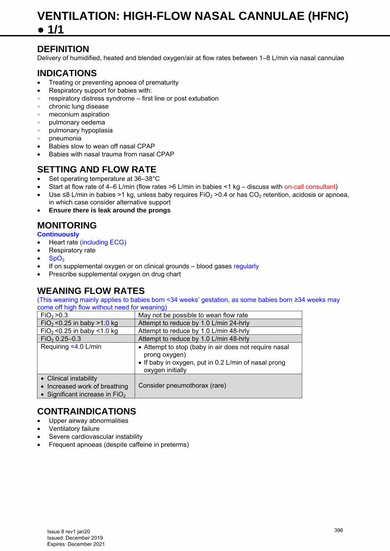
VENTILATION: HIGH-FLOW NASAL CANNULAE (HFNC) ● 1/1 DEFINITION Delivery of humidified, heated and blended oxygen/air at flow rates between 1–8 L/min via nasal cannulae
INDICATIONS • Treating or preventing apnoea of prematurity • Respiratory support for babies with: • respiratory distress syndrome – first line or post extubation • chronic lung disease • meconium aspiration • pulmonary oedema • pulmonary hypoplasia • pneumonia • Babies slow to wean off nasal CPAP • Babies with nasal trauma from nasal CPAP
SETTING AND FLOW RATE • Set operating temperature at 36–38°C • Start at flow rate of 4–6 L/min (flow rates >6 L/min in babies <1 kg – discuss with on-call consultant) • Use ≤8 L/min in babies >1 kg, unless baby requires FiO2 >0.4 or has CO2 retention, acidosis or apnoea,
in which case consider alternative support • Ensure there is leak around the prongs
MONITORING Continuously • Heart rate (including ECG) • Respiratory rate • SpO2 • If on supplemental oxygen or on clinical grounds – blood gases regularly • Prescribe supplemental oxygen on drug chart
WEANING FLOW RATES (This weaning mainly applies to babies born <34 weeks’ gestation, as some babies born ≥34 weeks may come off high flow without need for weaning) FiO2 >0.3 May not be possible to wean flow rate FiO2 <0.25 in baby >1.0 kg Attempt to reduce by 1.0 L/min 24-hrly FiO2 <0.25 in baby <1.0 kg Attempt to reduce by 1.0 L/min 48-hrly FiO2 0.25–0.3 Attempt to reduce by 1.0 L/min 48-hrly Requiring <4.0 L/min • Attempt to stop (baby in air does not require nasal
prong oxygen) • If baby in oxygen, put in 0.2 L/min of nasal prong
oxygen initially • Clinical instability • Increased work of breathing • Significant increase in FiO2
Consider pneumothorax (rare)
CONTRAINDICATIONS • Upper airway abnormalities • Ventilatory failure • Severe cardiovascular instability • Frequent apnoeas (despite caffeine in preterms)
Issue 8 rev1 jan20 Issued: December 2019 Expires: December 2021
396
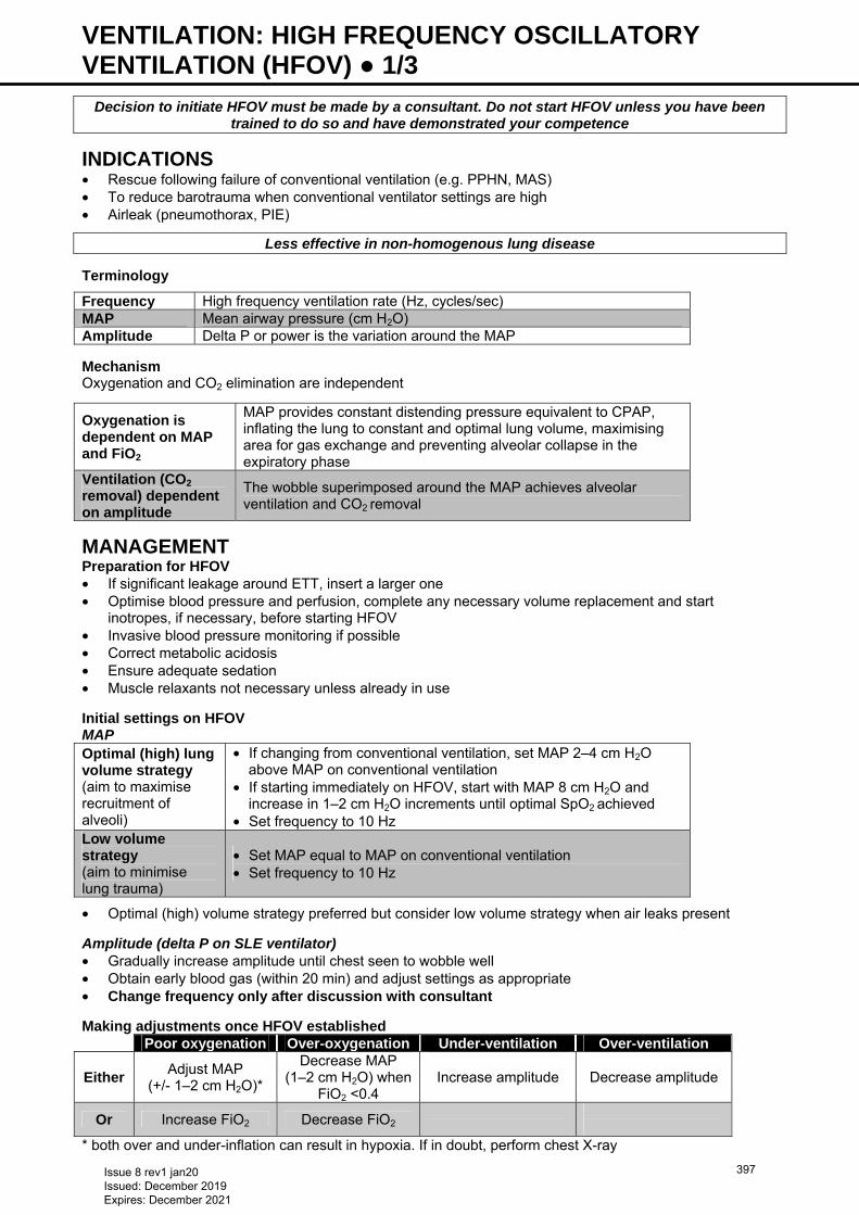
VENTILATION: HIGH FREQUENCY OSCILLATORY VENTILATION (HFOV) ● 1/3
Decision to initiate HFOV must be made by a consultant. Do not start HFOV unless you have been trained to do so and have demonstrated your competence
INDICATIONS • Rescue following failure of conventional ventilation (e.g. PPHN, MAS) • To reduce barotrauma when conventional ventilator settings are high • Airleak (pneumothorax, PIE)
Less effective in non-homogenous lung disease
Terminology
Frequency High frequency ventilation rate (Hz, cycles/sec) MAP Mean airway pressure (cm H2O) Amplitude Delta P or power is the variation around the MAP Mechanism Oxygenation and CO2 elimination are independent
Oxygenation is dependent on MAP and FiO2
MAP provides constant distending pressure equivalent to CPAP, inflating the lung to constant and optimal lung volume, maximising area for gas exchange and preventing alveolar collapse in the expiratory phase
Ventilation (CO2 removal) dependent on amplitude
The wobble superimposed around the MAP achieves alveolar ventilation and CO2 removal
MANAGEMENT Preparation for HFOV • If significant leakage around ETT, insert a larger one • Optimise blood pressure and perfusion, complete any necessary volume replacement and start
inotropes, if necessary, before starting HFOV • Invasive blood pressure monitoring if possible • Correct metabolic acidosis • Ensure adequate sedation • Muscle relaxants not necessary unless already in use Initial settings on HFOV MAP Optimal (high) lung volume strategy (aim to maximise recruitment of alveoli)
• If changing from conventional ventilation, set MAP 2–4 cm H2O above MAP on conventional ventilation
• If starting immediately on HFOV, start with MAP 8 cm H2O and increase in 1–2 cm H2O increments until optimal SpO2 achieved
• Set frequency to 10 Hz Low volume strategy (aim to minimise lung trauma)
• Set MAP equal to MAP on conventional ventilation • Set frequency to 10 Hz
• Optimal (high) volume strategy preferred but consider low volume strategy when air leaks present
Amplitude (delta P on SLE ventilator) • Gradually increase amplitude until chest seen to wobble well • Obtain early blood gas (within 20 min) and adjust settings as appropriate • Change frequency only after discussion with consultant
Making adjustments once HFOV established Poor oxygenation Over-oxygenation Under-ventilation Over-ventilation
Either Adjust MAP
(+/- 1–2 cm H2O)*
Decrease MAP (1–2 cm H2O) when
FiO2 <0.4 Increase amplitude Decrease amplitude
Or Increase FiO2 Decrease FiO2
* both over and under-inflation can result in hypoxia. If in doubt, perform chest X-ray
Issue 8 rev1 jan20 Issued: December 2019 Expires: December 2021
397
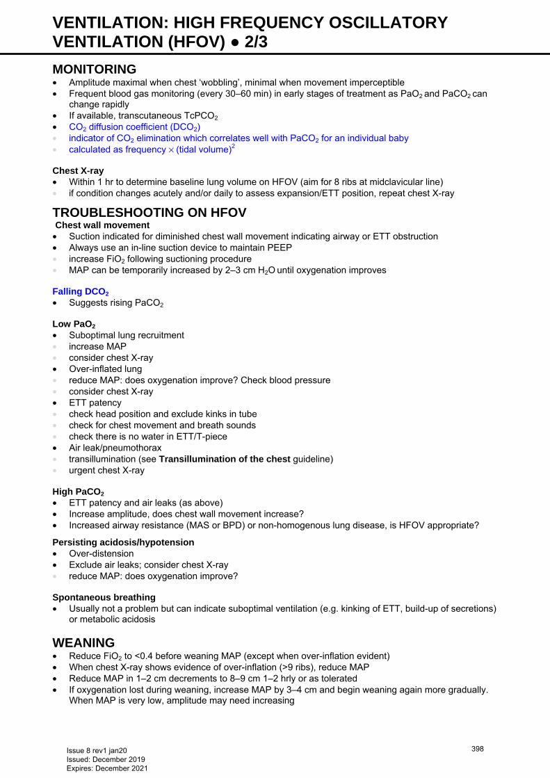
VENTILATION: HIGH FREQUENCY OSCILLATORY VENTILATION (HFOV) ● 2/3 MONITORING • Amplitude maximal when chest ‘wobbling’, minimal when movement imperceptible • Frequent blood gas monitoring (every 30–60 min) in early stages of treatment as PaO2 and PaCO2 can
change rapidly • If available, transcutaneous TcPCO2 • CO2 diffusion coefficient (DCO2) • indicator of CO2 elimination which correlates well with PaCO2 for an individual baby • calculated as frequency × (tidal volume)2 Chest X-ray • Within 1 hr to determine baseline lung volume on HFOV (aim for 8 ribs at midclavicular line) • if condition changes acutely and/or daily to assess expansion/ETT position, repeat chest X-ray
TROUBLESHOOTING ON HFOV Chest wall movement
• Suction indicated for diminished chest wall movement indicating airway or ETT obstruction • Always use an in-line suction device to maintain PEEP • increase FiO2 following suctioning procedure • MAP can be temporarily increased by 2–3 cm H2O until oxygenation improves Falling DCO2
• Suggests rising PaCO2 Low PaO2 • Suboptimal lung recruitment • increase MAP • consider chest X-ray • Over-inflated lung • reduce MAP: does oxygenation improve? Check blood pressure • consider chest X-ray • ETT patency • check head position and exclude kinks in tube • check for chest movement and breath sounds • check there is no water in ETT/T-piece • Air leak/pneumothorax • transillumination (see Transillumination of the chest guideline) • urgent chest X-ray High PaCO2 • ETT patency and air leaks (as above) • Increase amplitude, does chest wall movement increase? • Increased airway resistance (MAS or BPD) or non-homogenous lung disease, is HFOV appropriate?
Persisting acidosis/hypotension • Over-distension • Exclude air leaks; consider chest X-ray • reduce MAP: does oxygenation improve? Spontaneous breathing • Usually not a problem but can indicate suboptimal ventilation (e.g. kinking of ETT, build-up of secretions)
or metabolic acidosis
WEANING • Reduce FiO2 to <0.4 before weaning MAP (except when over-inflation evident) • When chest X-ray shows evidence of over-inflation (>9 ribs), reduce MAP • Reduce MAP in 1–2 cm decrements to 8–9 cm 1–2 hrly or as tolerated • If oxygenation lost during weaning, increase MAP by 3–4 cm and begin weaning again more gradually.
When MAP is very low, amplitude may need increasing
Issue 8 rev1 jan20 Issued: December 2019 Expires: December 2021
398
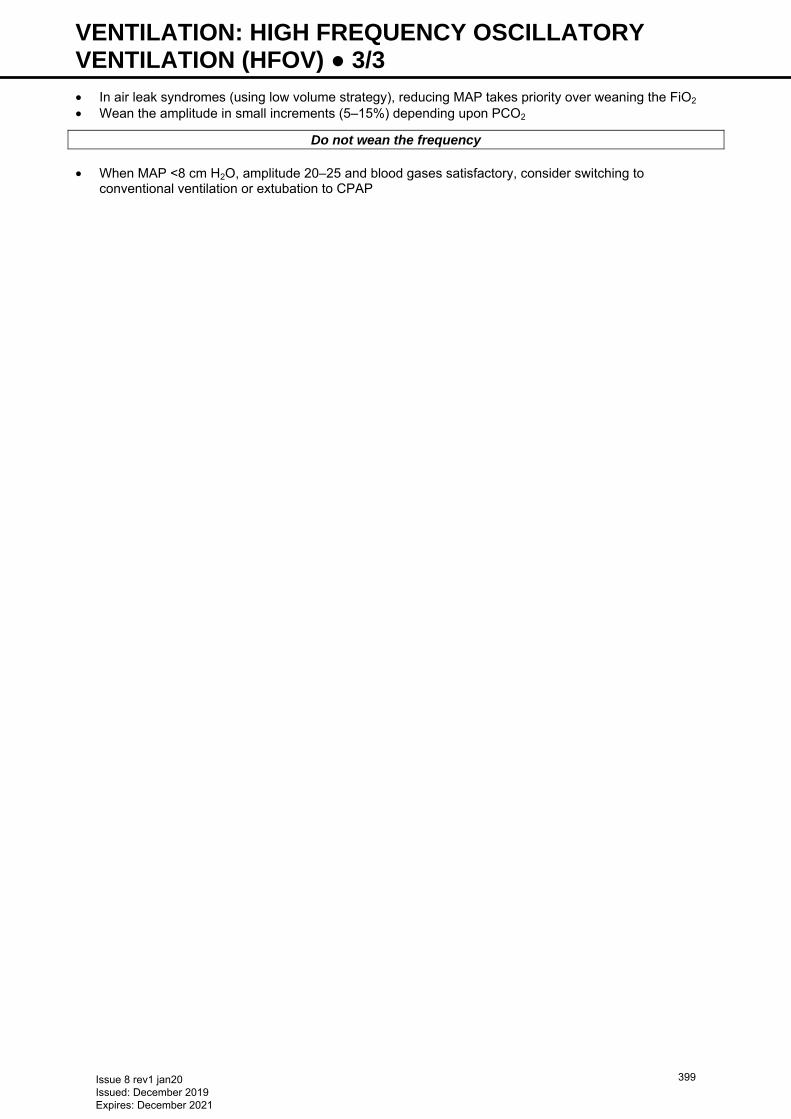
VENTILATION: HIGH FREQUENCY OSCILLATORY VENTILATION (HFOV) ● 3/3 • In air leak syndromes (using low volume strategy), reducing MAP takes priority over weaning the FiO2 • Wean the amplitude in small increments (5–15%) depending upon PCO2
Do not wean the frequency • When MAP <8 cm H2O, amplitude 20–25 and blood gases satisfactory, consider switching to
conventional ventilation or extubation to CPAP
Issue 8 rev1 jan20 Issued: December 2019 Expires: December 2021
399
VENTILATION: SYNCHRONOUS POSITIVE PRESSURE VENTILATION (SIPPV) ● 1/3 DEFINITION A form of synchronous ventilation in which baby triggers/initiates the breath while ventilator does the work of breathing. In other words, rate of ventilation is determined by baby while pressures are determined by operator via ventilator
SETTING UP TRIGGER VENTILATION • Set humidifier temperature at 39°C (negative 2) to achieve airway temperature of 37°C
Set up Babylog® (Drager) • Flow 6–10 L/min • Select SIPPV mode • Select highest trigger sensitivity (1: bar is all unshaded) • Select Tinsp (inspiratory time) between 0.3–0.4 sec • Adjust Texp (expiratory time) to achieve back-up rate of 35–40/min • Peak inspiratory pressure (PIP) 16–18 cm H2O • Peak end expiratory pressure (PEEP) 5 cm H2O • FiO2: 0.4–0.6 Set up SLE 5000 • Flow is fixed in SLE at 5 L/min • Select patient triggered ventilation (PTV) mode • Select highest trigger sensitivity (0.2 L/min for ≤28 weeks’ gestation, 0.4–0.6 L/min for >28 weeks’
gestation). Look at baby to confirm triggering adequately by observing baby generated breaths are triggering ventilator support
• Select Tinsp for back-up breaths between 0.3–0.4 sec • Set back-up rate of 35–40/min • PIP 16–18 cm H2O • PEEP 5 cm H2O • FiO2 0.4–0.6 • Software allows compensation for a leak of 10–50% • Observe tidal volume (Vt) settings to confirm between 4–6 mL/kg Baby • If gestation <34 weeks, consider loading baby with IV caffeine citrate (20 mg/kg) • Discontinue sedation
INITIATING TRIGGER VENTILATION • Once baby connected to ventilator: • check SpO2 (see Oxygen saturation targets guideline) and adjust FiO2 accordingly • check baby’s chest moving adequately, and measured Vt. Chest expansion should be just visible, and Vt
should be between 4–6 mL/kg. If not, adjust PIP/PEEP to maintain adequate oxygenation and ventilation • check ventilator triggering in synchrony with baby. Assess by listening to ventilator while watching
baby’s respiratory effort
Most likely cause of baby ‘fighting’ ventilator is ASYNCHRONY (see Management of asynchrony)
SUBSEQUENT ADJUSTMENTS ON SIPPV • Check blood gas within 30 min of initiation of SIPPV • Aim for: • PaO2: 6–10 kPa • PaCO2: 4.5−8.5 kPa day 1−3, 4.5−10 kPa day 4 onwards • pH >7.25 To improve oxygenation • Increase FiO2 • Rule out pneumothorax • Increase PIP and/or PEEP • Increase Tinsp (not more than 0.4 sec)
Issue 8 rev1 jan20 Issued: December 2019 Expires: December 2021
400
VENTILATION: SYNCHRONOUS POSITIVE PRESSURE VENTILATION (SIPPV) ● 2/3 To decrease PaCO2 • Rule out pneumothorax • Increase PIP • Check if baby triggering adequately. If not, try shortening Tinsp, or increasing back-up rate Low PaCO2 • Decrease PIP • Decrease back-up rate if >35/min • In a vigorous hypocapnic baby, transfer to synchronised intermittent mandatory ventilation (SIMV) at a
rate of at least 20/min
GENERAL SUPPORT • Monitor SpO2 continuously • Check arterial blood gases at least 4–6 hrly depending on stage of disease • In babies successfully ventilated in SIPPV mode, sedation is unnecessary • Remember, most common cause of baby fighting ventilator is ASYNCHRONY. Always carry out checks
and adjustments (see Management of asynchrony) • If baby still ‛fights’ ventilator, consider morphine bolus (100 microgram/kg) • If baby continues to ‛fight’ ventilator, use continuous sedation and change to conventional ventilation
(SIMV) mode (see Ventilation: conventional guideline) Do not use muscle relaxants at any stage unless, despite carrying out above checks, baby cannot be
ventilated. If muscle relaxants necessary, revert to conventional ventilation (see Ventilation: conventional
guideline)
NURSING OBSERVATIONS While baby on SIPPV, hourly observations • Back-up rate set • Baby’s own respiratory rate • Vt (in mL) • Minute ventilation [MV (in 1/min)] If alarm goes off, check • Synchrony between baby and ventilator • Excessive water droplets in ventilator tubing • Flow graph for evidence of blocked tube or excessive Tinsp • Disconnection
MANAGEMENT OF ASYNCHRONY Checklist • Is endotracheal tube (ETT) patent (look at flow graph and Vt) • Is Tinsp too long? (is baby exhaling against ventilator?), if so shorten Tinsp to 0.24–0.3 sec • Is back-up rate too high? If so, consider dropping to 30–35 breaths/min • Is there water condensation in ventilator tubing? • If all above fails, consider morphine bolus (100 microgram/kg) over 3–5 min • If baby still continues to ‛fight’ ventilator, use continuous sedation and revert to SIMV
AUTOCYCLING (FALSE TRIGGERING) • False triggering occurs when ventilator delivers a mechanical breath artifactually when baby not actually
initiating a spontaneous respiration • Usually results from presence of water droplets in ventilatory circuit, or an excessive ETT leak • If baby’s trigger rate appears to be in excess of 80/min, ensure this is actual rate by observing baby’s
own respiratory movements. If not: • check ventilatory circuit for excessive water condensation and empty if necessary • decrease trigger sensitivity by increasing trigger threshold e.g. from 0.4 to 0.6 L/min • Look for amount of ETT leak on Babylog display. If in excess of 50%, consider changing to slightly wider
ETT
Issue 8 rev1 jan20 Issued: December 2019 Expires: December 2021
401
VENTILATION: SYNCHRONOUS POSITIVE PRESSURE VENTILATION (SIPPV) ● 3/3 WEANING FROM SIPPV • Once baby stable (triggering above set rate, saturating in FiO2 <0.3), wean by: • decreasing PIP by 1–2 cm H2O each time (in SIPPV/PTV mode, weaning rate in a baby who is already
triggering above it is useless) • check baby breathing regularly and effortlessly (no chest recessions), and blood gases and oximetry are
acceptable • once PIP between 14–16 cm H2O (depending on size of baby), consider extubation • assess need for nasal CPAP/high-flow by checking for chest recessions, spontaneous minute ventilation,
and regularity of breathing • During weaning PaCO2 can rise above 7 kPa and Vt may fall below 4 mL/kg • provided baby triggering well, is not visibly tired, and pH >7.25, no action required • if poor triggering, visibly tired or abnormal pH, increase PIP, and later back-up rate
Issue 8 rev1 jan20 Issued: December 2019 Expires: December 2021
402
VENTILATION: VOLUME-TARGETED (VOLUME GUARANTEE/TARGETED TIDAL VOLUME) ● 1/2 DEFINITION In volume-targeted ventilation (VTV) gas delivery is targeted to deliver a pre-set tidal volume. Inspiratory pressure varies with each breath, depending on resistance and underlying lung compliance. The ventilator measures expired tidal volume (Vte) and calculates the pressure required to deliver this volume for the next breath. Available as volume guarantee (VG) on Draeger Babylog® and targeted tidal volume (TTV) on SLE 5000 Benefits • Compared with pressure-controlled ventilation, VTV can reduce: • mortality • bronchopulmonary dysplasia • pneumothorax • hypocarbia • severe intraventricular haemorrhage and periventricular leukomalacia
INDICATION • Primarily used in preterm babies with surfactant-deficient lung disease requiring ventilation • May be useful in other situations requiring ventilation
CONTRAINDICATION • ETT leak >50% • Caution to be used in situations such as pneumothorax, trachea-oesophageal/bronchopleural fistula;
leak may be increased and affect ventilation
TIDAL VOLUMES TO USE • Vte used as less influenced by ETT leaks • Vt 4–6 mL/kg • 5 mL/kg reasonable starting volume • Acute respiratory distress syndrome (RDS) 4–6 mL/kg • baby <750 g: 5−6 mL/kg (minimum starting volume 3 mL if 6 mL/kg is <3 mL) • baby 750−999 g: 4.5−5 mL/kg • baby >1000 g: 4−4.5 mL/kg • Chronic lung disease: 5−8 mL/kg • Avoid Vte >8 mL/kg (associated with volutrauma) • Avoid Vte <3.5 mL/kg (associated with atelectotrauma) • Change Vte in 0.5 mL/kg increments
MODE • VG/TTV combined with SIMV, SIPPV, assist control (PTV) or pressure-support ventilation (PSV) • VG also available for PC-CMV and HFO modes on Draeger VN-500 ventilator • In SIMV mode, set rate of ≥40/min (baby breaths are unsupported) • PSV has additional advantage of synchronising inspiration termination.
PEAK PRESSURES • Start PIP limit (Pmax) of ~25–30 cm H2O • Once baby stable and gases satisfactory adjust Pmax to 5–6 cm H2O above average PIP needed to
deliver set tidal volume • usually set ≤30 cm H2O in preterm babies • If PIP progressively increases or is persistently high, or if set Vt not delivered, reassess baby • PEEP set at 4–6 cm H2O
VENTILATOR RATE • In baby with poor respiratory drive, use rates of 50–60 bpm • Lower back-up rates of 30–40 bpm can be used with good respiratory drive • Use Tinsp (inspiratory time) of 0.3–0.4 sec; in PSV mode, set maximum Tinsp at 0.5−0.6 sec – actual Tinsp
is adjusted by the ventilator • Set flow trigger sensitivity at 0.2−0.4 L/min
Issue 8 rev1 jan20 Issued: December 2019 Expires: December 2021
403
VENTILATION: VOLUME-TARGETED (VOLUME GUARANTEE/TARGETED TIDAL VOLUME) ● 2/2 WEANING • Pressure weans automatically as lung compliance improves • Avoid tidal volumes <3.5 mL/kg as increases work of breathing in small babies • In SIMV, rate reduced to 40 breaths/min. VG/TTV is unhelpful with SIMV rates <40/min as baby breaths
are unsupported. Attempt extubation when: • FiO2 <0.3 • MAP falls consistently <8 cm H2O • baby has good respiratory drive and satisfactory gases
TROUBLESHOOTING AND PREVENTING PROBLEMS High CO2 • Review baby • Is set Vte being delivered? • Is chest expansion adequate? • Has leak increased? Change baby’s position before increasing Pmax • If ETT displaced/obstructed, or pneumothorax suspected, perform chest X-ray Low CO2 • Decrease Vte by 0.5 mL/kg but maintain ≥4 mL/kg (≥2.5 mL total volume) • Change to SIMV • Lower trigger sensitivity • Check for water in circuit (auto-triggering) • Decrease rate by 5−10 bpm (in SIMV mode only) • Increase PEEP (maximum 8 cm H2O) Low SpO2 • Review baby • Exclude air leaks • Worsening RDS: may require additional surfactant dose • Evidence of PPHN [see Persistent pulmonary hypertension of the newborn (PPHN) guideline] • Increase FiO2 • If Vte not delivered, increase Pmax • Baby may benefit from change to high frequency [see Ventilation: high frequency oscillatory
ventilation (HFOV) guideline] • Exclude congenital heart disease
Low Vte alarm • ETT leak >50% • Pneumothorax • Poor compliance/high resistance: increase Pmax Baby persistently tachypnoeic • Increase Vte by 0.5−1.0 mL/kg even if gases normal • Review sedation
Issue 8 rev1 jan20 Issued: December 2019 Expires: December 2021
404
VENTILATOR ASSOCIATED PNEUMONIA (PREVENTION) ● 1/1 HAND HYGIENE • Meticulous hand hygiene before and after patient contact and handling respiratory equipment or
condensate • wear gloves and apron when handling ventilator condensate and other respiratory/oral secretions
INTUBATION • Use a new, sterile ETT for each intubation attempt • use sedation +/- video laryngoscopy for planned procedure to reduce need for multiple attempts • Ensure ETT does not contact environmental surfaces before insertion • Use a sterilised laryngoscope • Have ≥2 NICU staff members present for ETT retaping/repositioning
SUCTIONING PRACTICES • Clear secretions from the oropharynx before: • ETT manipulation • patient repositioning • extubation • re-intubation
ETT SUCTION • Only when indicated • Do not use sodium chloride instillation routinely • For open suction, use ‘suction function’ on ventilator (if available) to prevent spray of condensate
FEEDING • Adjust enteral feeding (See Nutrition and enteral feeding guideline). Abdominal distension and
vomiting increase risk of reflux and aspiration
POSITIONING • Keep head of bed elevated 15−30° • Ensure no condensate in ventilator tubing before turning
ORAL CARE • Use colostrum/breast milk, or sterile water if no breast milk available (See Nutrition and enteral feeding
guideline) • Carry out oral care: • ≤24 hr after intubation • every 3−4 hr • before re-intubation as time allows • before orogastric tube insertion
RESPIRATORY EQUIPMENT • Use a separate suction catheter for oral and tracheal suction • Drain ventilator condensate away from baby every 2−4 hr and before repositioning • Avoid disconnection of ventilator circuit − only disconnect if: • emergency use of Neopuff™ or ETT re-intubation • change of circuit • unable to remove condensate by manipulation of tubing • Do not routinely disconnect ventilator circuit when turning baby • Change ventilator circuit when visibly soiled or mechanically malfunctioning in addition to manufacturer’s
instructions • Clean Neopuff™ connector with alcohol wipe after each manual contact with the ETT adapter • Use heated ventilator circuits
Issue 8 rev1 jan20 Issued: December 2019 Expires: December 2021
405
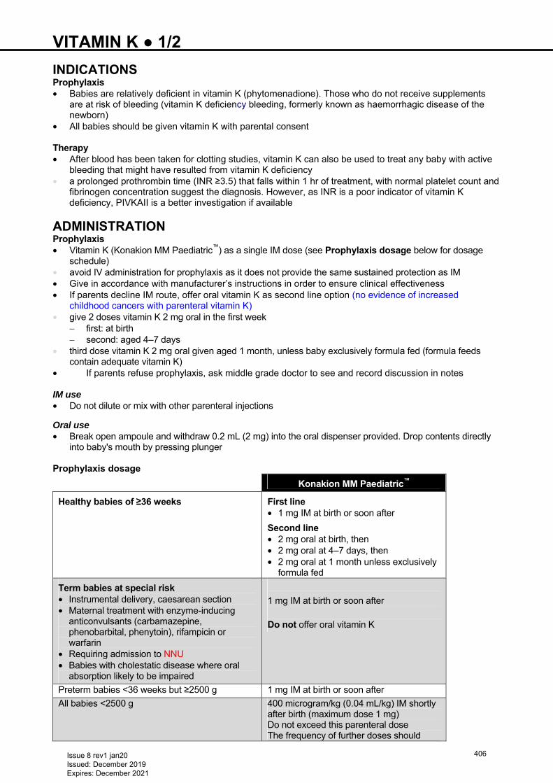
VITAMIN K ● 1/2 INDICATIONS Prophylaxis • Babies are relatively deficient in vitamin K (phytomenadione). Those who do not receive supplements
are at risk of bleeding (vitamin K deficiency bleeding, formerly known as haemorrhagic disease of the newborn)
• All babies should be given vitamin K with parental consent Therapy • After blood has been taken for clotting studies, vitamin K can also be used to treat any baby with active
bleeding that might have resulted from vitamin K deficiency • a prolonged prothrombin time (INR ≥3.5) that falls within 1 hr of treatment, with normal platelet count and
fibrinogen concentration suggest the diagnosis. However, as INR is a poor indicator of vitamin K deficiency, PIVKAII is a better investigation if available
ADMINISTRATION Prophylaxis • Vitamin K (Konakion MM Paediatric™) as a single IM dose (see Prophylaxis dosage below for dosage
schedule) • avoid IV administration for prophylaxis as it does not provide the same sustained protection as IM • Give in accordance with manufacturer’s instructions in order to ensure clinical effectiveness • If parents decline IM route, offer oral vitamin K as second line option (no evidence of increased
childhood cancers with parenteral vitamin K) • give 2 doses vitamin K 2 mg oral in the first week
− first: at birth − second: aged 4–7 days
• third dose vitamin K 2 mg oral given aged 1 month, unless baby exclusively formula fed (formula feeds contain adequate vitamin K)
• If parents refuse prophylaxis, ask middle grade doctor to see and record discussion in notes IM use • Do not dilute or mix with other parenteral injections
Oral use • Break open ampoule and withdraw 0.2 mL (2 mg) into the oral dispenser provided. Drop contents directly
into baby's mouth by pressing plunger Prophylaxis dosage
Konakion MM Paediatric™
Healthy babies of ≥36 weeks First line • 1 mg IM at birth or soon after
Second line • 2 mg oral at birth, then • 2 mg oral at 4–7 days, then • 2 mg oral at 1 month unless exclusively
formula fed
Term babies at special risk • Instrumental delivery, caesarean section • Maternal treatment with enzyme-inducing
anticonvulsants (carbamazepine, phenobarbital, phenytoin), rifampicin or warfarin
• Requiring admission to NNU • Babies with cholestatic disease where oral
absorption likely to be impaired
1 mg IM at birth or soon after
Do not offer oral vitamin K
Preterm babies <36 weeks but ≥2500 g 1 mg IM at birth or soon after
All babies <2500 g 400 microgram/kg (0.04 mL/kg) IM shortly after birth (maximum dose 1 mg) Do not exceed this parenteral dose The frequency of further doses should
Issue 8 rev1 jan20 Issued: December 2019 Expires: December 2021
406
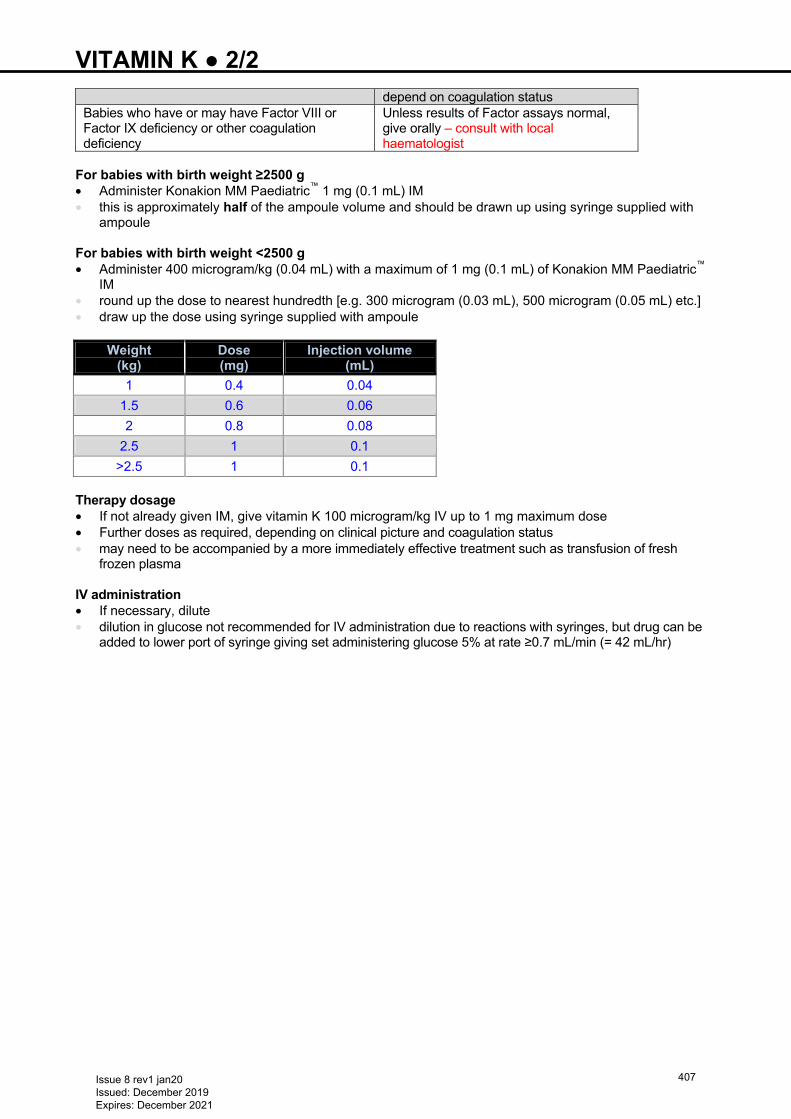
VITAMIN K ● 2/2
depend on coagulation status Babies who have or may have Factor VIII or Factor IX deficiency or other coagulation deficiency
Unless results of Factor assays normal, give orally – consult with local haematologist
For babies with birth weight ≥2500 g • Administer Konakion MM Paediatric™ 1 mg (0.1 mL) IM • this is approximately half of the ampoule volume and should be drawn up using syringe supplied with
ampoule For babies with birth weight <2500 g • Administer 400 microgram/kg (0.04 mL) with a maximum of 1 mg (0.1 mL) of Konakion MM Paediatric™
IM • round up the dose to nearest hundredth [e.g. 300 microgram (0.03 mL), 500 microgram (0.05 mL) etc.] • draw up the dose using syringe supplied with ampoule
Weight (kg)
Dose (mg)
Injection volume (mL)
1 0.4 0.04
1.5 0.6 0.06
2 0.8 0.08
2.5 1 0.1
>2.5 1 0.1 Therapy dosage • If not already given IM, give vitamin K 100 microgram/kg IV up to 1 mg maximum dose • Further doses as required, depending on clinical picture and coagulation status • may need to be accompanied by a more immediately effective treatment such as transfusion of fresh
frozen plasma IV administration • If necessary, dilute • dilution in glucose not recommended for IV administration due to reactions with syringes, but drug can be
added to lower port of syringe giving set administering glucose 5% at rate ≥0.7 mL/min (= 42 mL/hr)
Issue 8 rev1 jan20 Issued: December 2019 Expires: December 2021
407
INDEX A
Abstinence syndrome 12
Aciclovir 72, 140, 190, 381−383Activated Partial Thromboplastin Time (APTT) 66−67, 180, 225, 231
233, 301Actrapid 146−147, 279Admission to neonatal unit 15
Ambiguous genitalia 77Anaphylaxis 186Ankyloglossia (tongue-tie) − division for breastfeeding 17Ano-rectal malformation 18
Antenatal ultrasound abnormalities 21
Apgar score Apnoea and bradycardia 22
APTT 66−67, 180, 225, 231233, 301
Arterial line insertion 24
Arterial line sampling 26
Atelectasis 10 4, 388
B
Babies born at margins of viability 28Bag and mask ventilation 212, 214BCG immunisation 30
Blood gas analysis 26, 49, 370, 392Blood group incompatibilities 33
Bloodspot screening 35
Blood transfusion (Transfusion of red blood cells) 360
Blue baby 50Bottle feeding 37
Brachial plexus injury 377Breast milk expression 39
Breast milk handling and storage 41
Breastfeeding 43
Broviac line insertion 45
C
Calcium resonium 149−150Cannulation − peripheral venous 48
Cardiac arrhythmias 45, 110Cardiac murmurs 49
Cephalhaematoma 10 7, 218Chest drain insertion – Seldinger technique 54
Chest drain insertion − Traditional 57
Chest physiotherapy 59
Chickenpox 381−383Chlamydia 72−73, 193Chloral hydrate 275Chloramphenicol 72Chlorpromazine 13Chronic lung disease 62
Cleft lip/palate 16, 21, 107, 151, 212245
Clonazepam 181, 324−325CMV 64
CO2 and O2 transcutaneous monitoring 357Coagulopathy 66
Collapse (Sudden unexpected postnatal collapse in first week of life) 337Congenital diaphragmatic hernia (CDH) 68
Congenital heart disease: duct dependent lesions [Including hypoplastic left heart syndrome (HLHS) and left-sided outflow tract obstructions] 50Congenital spherocytosis Conjunctivitis 72
Consent 74
Conventional ventilation 392
Convulsions 12, 64, 181, 300Cooling 179
Coombs positive babies 33, 220Cord care 328Cranial ultrasound scans 79
Curosurf® 34 3
D
Death and seriously ill babies 82
Decolonisation 238−239Dehydration (hypernatraemic) 151
Developmental care 84Developmental dysplasia of the hip (DDH) 86
Dexamethasone 30, 62−63, 76, 213, 236Dialysis 149−150, 154, 191, 202,
313Diamorphine 250, 274−275Difficult intubation 215
Direct Coombs test 33, 145, 219, 360Discharge from neonatal unit 89
Disorders of sexual development 91
Domperidone 40
Down syndrome − initial management 93
Drug withdrawal 13
E
EBM 42, 44, 85, 153−154, 236, 248, 256−257, 259,
260−262, 272−273, 297−298, 313
ECG abnormalities 99
ECMO 70, 118, 133, 287−288352
Eczema 3 0Endotracheal tube (ETT) suctioning 103
Enteral feeding 39, 62, 131 147, 151153, 163, 243, 248, 250255, 278,280, 331, 361
405Environment and noise 105
Erythromycin 12 1ESBL 23 8Examination of the newborn 106
Exchange transfusion 110
Exomphalus − initial management 113
Extravasation injuries 116
Extreme prematurity 28
F
Feeding – Enteral – PN
255278
Fluid restriction 181, 209, 284, 312−313Fluid therapy IV 207Follow up of babies discharged from neonatal unit 118
G Gastro-oesophageal reflux (GOR) 120
Gastroschisis 122
Glucosuria 16 7
Golden hour − preterm babies <24 weeks’ gestation 126
Gonococcus 73 , 193Gram-negative organisms 188−194Gram stain 72, 189−190, 194Growth monitoring 130
H
HCV 73, 138−139Hearing screening 132
Heart failure 134
Heart murmur 49, 304Heel prick 94, 142, 328, 351, 386Hepatitis B and C 137
Hepatosplenomegaly 64, 176−177, 218, 346354, 368
Herpes simplex virus (HSV) 140
High flow nasal cannulae (HFNC) 396
High frequency oscillatory ventilation (HFOV) 397
Human immunodeficiency virus (HIV) 141−143
HLHS 5 0Hydrolysate 12 1Hydrops fetalis 144
Hyperglycaemia 146
Hyperinsulinism 15 9, 163, 199Hyperkalaemia 148
Hypernatraemic dehydration 151
Hyperoxia 49, 51, 60, 68, 215, 286344
Hypocalcaemia 155
Hypoglycaemia 157
Hypokalaemia 165
Hyponatraemia 63, 208−209, 283, 295313
Issue 8 rev1 jan20 Issued: December 2019 Expires: December 2021
408
Hypoplastic left heart syndrome (HLHS) 50
Hypotension 167
Hypothermia 170
Hypothyroidism 173
Hypotonia (floppy baby) 175
Hypoxic-ischaemic encephalopathy (HIE) 179
I
Immunisations 185
Inborn errors of metabolism 325Infection (late onset) 188
Infection in first 72 hours of life 192
Inguinal hernia 198
Inherited metabolic disorders (IMD) 199
Insertion of arterial lines 24Insertion of chest drain 54Insertion of long lines 227Intra-abdominal cysts 203
Intubation 212
Intubation – difficult 215
Isoniazid 43, 367−368IUT 33−34, 110Intravenous fluid therapy 207
J
Jaundice 218
K
Kangaroo care 221
Kleihauer test 33, 110, 145Konakion 406−407
L
Labour ward calls 223
Liver disease 218Liver dysfunction in preterm babies 224
Long line insertion (peripherally sited) 227
Lumbar puncture 15, 67, 75, 140, 189 191, 193, 294, 322, 339
Lung disease (chronic) 62
M
Massive haemorrhage 231
Maternal diabetes 151, 224, 289, 350Maternal thyroid disease 354
Meconium aspiration syndrome 343Meconium staining 223Medium-chain Acyl-coa Dehydrogenase Deficiency (MCADD) − early management of babies with family history 234Metabolic bone disease 236
Meningococcus B 185, 187Metabolic disorders 22 79 175, 177,199−202
326, 350Morphine sedation 13, 54, 68, 70, 167, 169,
180, 198, 250, 274−275, 285, 394, 401
MRSA 2 38Multi drug resistant organism colonisation (MRSA, ESBL etc.) 238Multiple transfusions 35Myasthenia gravis 175, 178, 223Myelomeningocele (MMC) 241
N
Nappy dermatitis 329Nasogastric tube administration of feed, fluid or medication 243Nasogastric tube insertion 245
Necrotising enterocolitis (NEC) 249
Neonatal abstinence 15, 118, 271, 273Newborn examination 106Nitric oxide 252
Non-nutritive sucking 254
Nutrition and enteral feeding 255
O
Oesophageal atresia/replogle tubes 263
O2 and CO2 transcutaneous monitoring 357Oxygen on discharge 268
Oxygen saturation targets 270
P
Pain assessment and management 271
Palivizumab 276
Paracetamol 274−275, 282−284Parenteral nutrition 278
Patent ductus arteriosus (PDA) 281
Pelviectasis 378−379Pericardial tamponade 229, 285, 310Pericardiocentesis 285
Persistent pulmonary hypertension (PPHN) 286
Phenytoin 181, 323−325, 406Phytomenadione 40 6PKU 35 , 44Polycythaemia 289
Poractant 34 3Positioning 291
Post haemorrhagic ventricular dilatation 294
Preterm care 126Progression to oral feeding in preterm babies 296
Prostaglandin infusion 299
Prothrombin time (PT) 66, 406Pulmonary haemorrhage 301
Pulse-oximetry (universal) screening 303
Q
Quiet time 105
R
Radioisotope 4 3Rectal washout using syringe method 306
Recycling stoma losses 308
Renal abnormalities on ultrasound scan 378Renal failure 310
Replogle tubes 263−265Respiratory distress syndrome 146, 207, 301, 344, 396
403Resuscitation 314
Retinopathy of prematurity (ROP) 320
Rhesus disease 33−34, 223 SSacral dimple 321
Salbutamol 99, 149−150, 165Seizures 322
Sexual development (disorders of) 91Synchronous positive pressure ventilation (SIPPV) 400Skin biopsy for inborn errors of metabolism 325
Skin care 328
Skin excoriation 329, 331Stoma management (gastrointestinal) 330
Sucrose 85, 227, 245, 254, 272−275, 306, 320, 386
Subgaleal haemorrhage (SGH) 334
Sudden unexpected postnatal collapse in first week of life 337Supraventricular tachycardia 340
Surfactant replacement therapy 343
Synagis® (palivizumab) 3 1Synchronous intermittent positive pressure ventilation (SIPPV) 400
Syphilis − babies born to mothers with positive serology 346
Systemic lupus erythematosus 223
T Tuberculosis (investigation and management following exposure in pregnancy) 367Tetanus pertussis 185 187, 223Thrombocytopenia 350
Thromboembolism 25−26, 223,384Thyroid disease (management of babies born to mothers with thyroid disease) 354Transcutaneous CO2 and O2 357
Transfusion of red blood cells 360
Transillumination of the chest 363
Transport and retrieval 364
Trimethoprim 1 91, 379Tuberculin 30
U
Issue 8 rev1 jan20 Issued: December 2019 Expires: December 2021
409
Umbilical artery catheterisation and removal 370
Umbilical venous catheterisation and removal 373
Universal pulse-oximetry screening 303Upper limb birth injuries including brachial plexus injury 377Urinary tract abnormalities on antenatal scan 378
V
Vaccination (BCG) 30Varicella 381
Vascular spasm and thrombosis 384
Vasospasm 25 , 372, 384VDRL 346−348Venepuncture 386
Ventilation: continuous positive airway pressure (CPAP) 388
Ventilation: conventional 392
Ventilation: high-flow nasal cannulae (HFNC) 396
Ventilation: high frequency oscillatory (HFOV) 397
Ventilation: synchronous positive pressure (SIPPV) 400Ventilation: volume targeted (volume guarantee/targeted tidal volume) 403Vitamin K 406
VZV
W
Warfarin Z Zidovudine
Issue 8 rev1 jan20 Issued: December 2019 Expires: December 2021
410


![TotalMarks [Out Of 200] - ESED KPK - NTS](https://static.fdokumen.com/doc/165x107/631a88785d5809cabd0f70e8/totalmarks-out-of-200-esed-kpk-nts.jpg)