5-Methyltetrahydrofolic Acid and Folic Acid Pregnancy Diets ...
Upload
khangminh22Category
view
2download
0
Materials Science and Engineering C 58 (2016) 629–638
Contents lists available at ScienceDirect
Materials Science and Engineering C
j ourna l homepage: www.e lsev ie r .com/ locate /msec
Delivery system for mefenamic acid based on the nanocarrier layereddouble hydroxide: Physicochemical characterization and evaluation ofanti-inflammatory and antinociceptive potential
Vanessa R.R. Cunha a, Viviane A. Guilherme b, Eneida de Paula b, Daniele R. de Araujo b,c, Renan O. Silva d,Jand V.R. Medeiros d, José R.S.A. Leite d, Philippe A.D. Petersen e, Marianna Foldvari f,Helena M. Petrilli e, Vera R.L. Constantino a,⁎a Departamento de Química Fundamental, Instituto de Química, Universidade de São Paulo, USP, Av. Prof. Lineu Prestes 748, 05508-000 São Paulo, São Paulo, Brazilb Departamento de Bioquímica, Instituto de Biologia, Universidade Estadual de Campinas, 13083-970, Campinas, São Paulo, Brazilc Centro de Ciências Naturais e Humanas, Universidade Federal do ABC, 09210-170 Santo André, São Paulo, Brazild Núcleo de Pesquisa em Biodiversidade e Biotecnologia, BIOTEC, Campus de Parnaíba, Universidade Federal do Piauí, UFPI, 64202020 Parnaíba, Piauí, Brazile Departamento de Física dos Materiais e Mecânica, Instituto de Física, Universidade de São Paulo, USP, 05315-970 São Paulo, São Paulo, Brazilf School of Pharmacy, University of Waterloo, N2L 3G1 Waterloo, Ontario, Canada
Abbreviations: LDH-Mef, Layered double hydroxid(mefenamate anion) of the drug mefenamic acid; LDMefH, Mefenamic acid; Mef, Mefenamate anion; PGE2,Divalent and trivalent cations; An-, An exchangeable anioL-132, Lung epithelial cells; HeLa, Adenocarcinoma of thNSAIDs, Non-steroidal anti-inflammatory drugs; PXRD, PElemental chemical analyses of C, H, and N; TGA-DTG, Ttransform infrared spectroscopy; FT-Raman, Fourier trNaMef, Sodiummefenamate salt; DFT, Density functional tterial containing chloride anions; TGA-DSC, Thermograviscanning calorimetry; IR, Infrared spectroscopy; RamaPhosphate buffered saline; HTAB, HexadecyltrimethMyeloperoxidase; Sal, Saline; SEM, Standard error of theance; FTIC, Fluorescein 5′-isothiocyanate dye; Conset, Conmolysis; PGF2α, Prostaglandin F2α.⁎ Corresponding author.
http://dx.doi.org/10.1016/j.msec.2015.08.0370928-4931/© 2015 Elsevier B.V. All rights reserved.
a b s t r a c t
a r t i c l e i n f oArticle history:Received 7 June 2015Received in revised form 7 August 2015Accepted 21 August 2015Available online 1 September 2015
Keywords:Layered double hydroxideMefenamic acidDensity functional theoryAnti-inflammatoryAntinociceptive
Purpose: The anionic formof the drugmefenamic acid intercalated into the nanocarrier layered double hydroxide(LDH-Mef) was evaluated by anti-inflammatory and antinociceptive assays.Methods: The LDH-Mefmaterial was characterized by a set of physicochemical techniques, which was supportedby Density Functional Theory calculations. The pharmacological effects of LDH-Mef (40 wt% of drug) were eval-uated by hemolytic, anti-inflammatory activity and antinociceptive assays.Results: In vivo assays were conducted for the first time in order to assess the LDH-Mef potential. The hemolyticeffects decreased for the intercalated Mef as demonstrated by the higher tolerated hemolytic concentration(1.83 mM) compared to mefenamic acid (MefH), 0.48 mM. Pretreatment of animals with MefH or LDH-Mef re-duced carrageenan-, dextran sulfate- and PGE2-induced paw edema. MefH or LDH-Mef also decrease totalleucocytes and neutrophil counts of the peritoneal cavity after inflammation induction with carrageenan. Inthe nociception model, oral pretreatment with LDH-Mef reduced mechanical hypernociception carrageenan-induced after 3–4 h and also the number of writhings induced by acetic acid.Conclusions: This work shows the increase of the anti-inflammatory and antinociceptive potential of the drugconfined into the LDH, as well as, its hemolytic effect.
© 2015 Elsevier B.V. All rights reserved.
e containing the anionic formH, Layered double hydroxide;Prostaglandin E2; MII and MIII,n; Carcinoma A549 and normale cervix; HOS, Osteosarcoma;owder X-ray diffraction; CHN,hermal analysis; FT-IR, Fourieransform Raman spectroscopy;heory; LDH-Cl, Mg, Al-LDHma-metric analysis and differentialn, Raman spectroscopy; PBS,ylammonium bromide; MPO,mean; ANOVA, Analysis of vari-centrations for the onset of he-
1. Introduction
The entrapment of drugs into layered inorganic nanoparticles canprovide sustained release minimizing the usual side effects [1]. In addi-tion, the inorganic framework can protect bioactive molecules againstdegradation processes promoted by light, heat, molecular oxygen etc.and extend their shelf life. Layered double hydroxides (LDHs), alsoknown as hydrotalcite like-compounds, are potential candidates tocarry pharmaceutical substances, [2,3,4] since one LDH composition inparticular (specifically [Mg6Al2(OH)16]CO3·4H2O) is already commer-cialized as the antacid Talcid™ and also used as excipient [5]. In the1990s several works have shown the efficacy of Talcid in comparisonwith some already established antacids like for example Omeprazol[6,7]. So, the low toxicity of LDH after oral administration is wellestablished.
LDHs present positively charged sheets (or layers) in their structuresas shown in Fig. 1, A [8]. To maintain the material's electroneutrality it

Fig. 1. Schematic representation of (A) layered double hydroxide structure, and(B) mefenamate ion structure (dimensions achieved by DFT calculations).
630 V.R.R. Cunha et al. / Materials Science and Engineering C 58 (2016) 629–638
required the presence of anions that, together with water molecules,promote the LDHs layers stacking. The general chemical formula of thisclass of inorganic materials is [MII
1 − xMIIIx(OH)2]x+[An−]x/n·mH2O,
where MII and MIII are divalent and trivalent cations, and An- is an ex-changeable anion.
In the pharmaceutical andmedical context, LDH is a very interestingmatrix to be explored as a drug nanocarrier since it is biocompatible andthe anionic form of bioactive drugs can be intercalated between its layers[1,9]. Therefore, since the first publication in 2001 by O'Hare groupabout the intercalation of commercial drugs into LDHs, [10] a growinginterest in this layered material for nanotechnological purposes is no-ticed [1,11,12,13,14].
In vitro and in vivo assays have been conducted to evaluate the LDHs'mechanisms of actionwhen administered by enteral (oral), [15] topical,[16] or parenteral (injection) routes [17,18]. For the wide application ofLDHs in the pharmaceutical and medical fields, it required the enlight-enment of the cytotoxicity of these materials to provide not only infor-mation about biological applications, but also to prevent undesirableeffects [19]. Choy et al. [20] have shown that these nanoparticlespresented low cytotoxicity through tests in vitro with cells. The non-steroidal anti-inflammatory drugs (NSAIDs) are suitable candidates forintercalation into LDHs due to the presence of anionic groups in theirstructure. Moreover, the NSAIDs present limited solubility, low bioavail-ability and serious gastrointestinal problems [21,22]. A recent reviewpublished by Rives et al. [23] reporting studies about anti-inflammatorydrugs intercalated into LDHs has pointed out the sustained release ofNSAID drugs by in vitro assays and also their pharmacological activityby in vivo tests.
Mefenamic acid (MefH, Fig. 1B), an NSAID marketed in the US asPonstel, is indicated for inflammatory diseases and also as an analgesicfor the treatment of rheumatoid arthritis, menstrual symptoms andheadaches. However, due to its low solubility and side effects, mainlyrelated to gastrointestinal adverse consequences, including bleeding,ulceration or perforation of the stomach or intestines, which can besometimes fatal, the use of mefenamic acid is compromised [24]. Riveset al.[25,26] have reported the intercalation of mefenamic acid intoLDH and studied the release profile in different pH values. The releaseprofile is slower and sustained for the intercalated mefenamate intoLDHwhen compared to the free drug or the physicalmixture containingthe drug and LDH.
In this paper, we have developed LDH carriers containingmagnesiumand aluminum in the layers (Mg, Al-LDH) and the anionic form ofmefenamic acid (LDH-Mef). LDH-Mef nanoparticles were characterizedin detail by chemical analysis, powder X-ray diffraction (PXRD), thermalanalysis (TGA-DTG), vibrational spectroscopy (FT-IR and FT-Raman),and particle size and zeta potential measurements. To support the exper-imental vibrational assignments and discuss the spectral differencesbetween the intercalatedmefenamate anion and the free anion, the calcu-lations of vibrational frequencies of sodium mefenamate salt (NaMef)were performed in the framework of the Density Functional Theory(DFT).
In order to assess the cytotoxic effects, hemolytic assays wereperformed with MefH, LDH-Mef and LDH matrix without drug(i.e., Mg,Al-LDH intercalated with chloride anions). We report here thefirst studies on the anti-inflammatory and antinociceptive activity onsystemically administered LDH-Mef nanoparticles by in vivo assays inthree different inflammatory disease models, i.e. carrageenan, dex-tran sulfate and PGE2 induced paw inflammation models, and orallyadministered LDH-Mef nanoparticles in acetic acid-induced writhingsmodel.
2. Experimental
2.1. Reagents
All reagents used in this work can be found in the SupplementaryMaterial.
2.2. Synthesis of LDH-Mef hybrid material
The inorganic carrier with the drug (LDH-Mef) was isolated inorder to evaluate the biological activity of the mefenamate anionentrapped into the LDH framework. Besides it was also prepared as asimilar LDH structure but without a bioactive species, i.e. a Mg,Al-LDHmaterial containing chloride anions between the layers (abbreviatedLDH-Cl).
The LDHs materials (LDH-Mef and LDH-Cl) were synthesized by co-precipitation method as described in the literature [27,28]. The hybridLDH-Mef sample was obtained using the Mg2+/Al3+ molar ratio equalto 2 and mefenamate/Al3+ equal to 1. LDH-Cl was obtained using thesame conditions but in the absence of the drug.
Initially, the pH of the aqueous solution of 0.1 mol/L of mefenamicacidwas adjusted to 10 by addition of 0.2mol/L of NaOH. Under nitrogenatmosphere (to avoid carbonate ion formation by solubilization ofcarbon dioxide), a 0.1 mol/L mixed solution of the divalent (Mg2+)and trivalent (Al3+) metal cations (in the chloride form) was added tothe mefenamate solution. The pH value was kept at about 10 duringthe synthesis by the NaOH solution addition. The resulting slurry wasaged with vigorous stirring for 1 h under N2 atmosphere at 25 °C. Thesolid was separated and washed with deionized water by filtration (toremove ions as sodium and chloride for example). Then the isolatedsolid was dried under reduced pressured at room temperature.
631V.R.R. Cunha et al. / Materials Science and Engineering C 58 (2016) 629–638
2.3. Physicochemical characterization
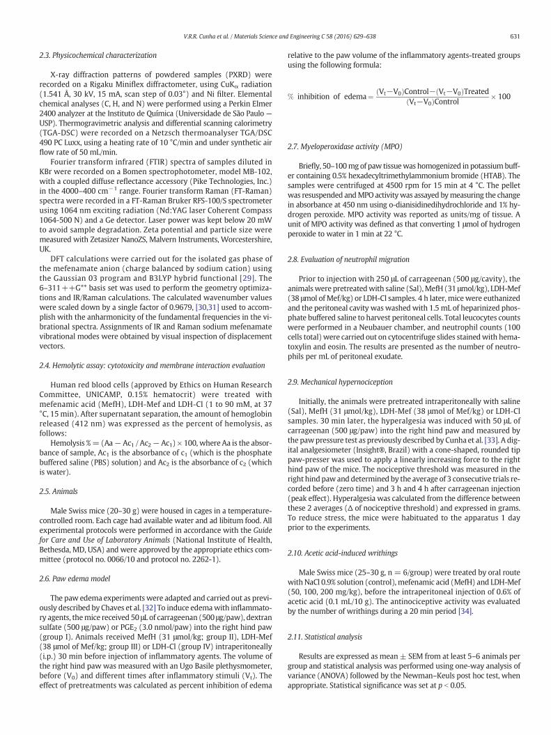
X-ray diffraction patterns of powdered samples (PXRD) wererecorded on a Rigaku Miniflex diffractometer, using CuKα radiation(1.541 Å, 30 kV, 15 mA, scan step of 0.03°) and Ni filter. Elementalchemical analyses (C, H, and N) were performed using a Perkin Elmer2400 analyzer at the Instituto de Química (Universidade de São Paulo —USP). Thermogravimetric analysis and differential scanning calorimetry(TGA-DSC) were recorded on a Netzsch thermoanalyser TGA/DSC490 PC Luxx, using a heating rate of 10 °C/min and under synthetic airflow rate of 50 mL/min.
Fourier transform infrared (FTIR) spectra of samples diluted inKBr were recorded on a Bomen spectrophotometer, model MB-102,with a coupled diffuse reflectance accessory (Pike Technologies, Inc.)in the 4000–400 cm−1 range. Fourier transform Raman (FT-Raman)spectra were recorded in a FT-Raman Bruker RFS-100/S spectrometerusing 1064 nm exciting radiation (Nd:YAG laser Coherent Compass1064-500 N) and a Ge detector. Laser power was kept below 20 mWto avoid sample degradation. Zeta potential and particle size weremeasured with Zetasizer NanoZS, Malvern Instruments, Worcestershire,UK.
DFT calculations were carried out for the isolated gas phase ofthe mefenamate anion (charge balanced by sodium cation) usingthe Gaussian 03 program and B3LYP hybrid functional [29]. The6–311++G** basis set was used to perform the geometry optimiza-tions and IR/Raman calculations. The calculated wavenumber valueswere scaled down by a single factor of 0.9679, [30,31] used to accom-plish with the anharmonicity of the fundamental frequencies in the vi-brational spectra. Assignments of IR and Raman sodium mefenamatevibrational modes were obtained by visual inspection of displacementvectors.
2.4. Hemolytic assay: cytotoxicity and membrane interaction evaluation
Human red blood cells (approved by Ethics on Human ResearchCommittee, UNICAMP, 0.15% hematocrit) were treated withmefenamic acid (MefH), LDH-Mef and LDH-Cl (1 to 90 mM, at 37°C, 15 min). After supernatant separation, the amount of hemoglobinreleased (412 nm) was expressed as the percent of hemolysis, asfollows:
Hemolysis %= (Aa− Ac1 / Ac2− Ac1) × 100, where Aa is the absor-bance of sample, Ac1 is the absorbance of c1 (which is the phosphatebuffered saline (PBS) solution) and Ac2 is the absorbance of c2 (whichis water).
2.5. Animals
Male Swiss mice (20–30 g) were housed in cages in a temperature-controlled room. Each cage had available water and ad libitum food. Allexperimental protocols were performed in accordance with the Guidefor Care and Use of Laboratory Animals (National Institute of Health,Bethesda, MD, USA) and were approved by the appropriate ethics com-mittee (protocol no. 0066/10 and protocol no. 2262-1).
2.6. Paw edema model
The paw edema experiments were adapted and carried out as previ-ously described by Chaves et al. [32] To induce edemawith inflammato-ry agents, themice received 50 μL of carrageenan (500 μg/paw), dextransulfate (500 μg/paw) or PGE2 (3.0 nmol/paw) into the right hind paw(group I). Animals received MefH (31 μmol/kg; group II), LDH-Mef(38 μmol of Mef/kg; group III) or LDH-Cl (group IV) intraperitoneally(i.p.) 30 min before injection of inflammatory agents. The volume ofthe right hind paw was measured with an Ugo Basile plethysmometer,before (V0) and different times after inflammatory stimuli (Vt). Theeffect of pretreatments was calculated as percent inhibition of edema
relative to the paw volume of the inflammatory agents-treated groupsusing the following formula:
% inhibition of edema¼ Vt−V0ð ÞControl− Vt−V0ð ÞTreatedVt−V0ð ÞControl � 100
2.7. Myeloperoxidase activity (MPO)
Briefly, 50–100mgof paw tissuewas homogenized in potassiumbuff-er containing 0.5% hexadecyltrimethylammonium bromide (HTAB). Thesamples were centrifuged at 4500 rpm for 15 min at 4 °C. The pelletwas resuspended andMPO activitywas assayed bymeasuring the changein absorbance at 450 nm using o-dianisidinedihydrochloride and 1% hy-drogen peroxide. MPO activity was reported as units/mg of tissue. Aunit of MPO activity was defined as that converting 1 μmol of hydrogenperoxide to water in 1 min at 22 °C.
2.8. Evaluation of neutrophil migration
Prior to injection with 250 μL of carrageenan (500 μg/cavity), theanimalswere pretreatedwith saline (Sal),MefH (31 μmol/kg), LDH-Mef(38 μmol of Mef/kg) or LDH-Cl samples. 4 h later, micewere euthanizedand the peritoneal cavity was washed with 1.5 mL of heparinized phos-phate buffered saline to harvest peritoneal cells. Total leucocytes countswere performed in a Neubauer chamber, and neutrophil counts (100cells total)were carried out on cytocentrifuge slides stainedwith hema-toxylin and eosin. The results are presented as the number of neutro-phils per mL of peritoneal exudate.
2.9. Mechanical hypernociception
Initially, the animals were pretreated intraperitoneally with saline(Sal), MefH (31 μmol/kg), LDH-Mef (38 μmol of Mef/kg) or LDH-Clsamples. 30 min later, the hyperalgesia was induced with 50 μL ofcarrageenan (500 μg/paw) into the right hind paw and measured bythe pawpressure test as previously described by Cunha et al. [33]. A dig-ital analgesiometer (Insight®, Brazil) with a cone-shaped, rounded tippaw-presser was used to apply a linearly increasing force to the righthind paw of the mice. The nociceptive threshold was measured in theright hindpawand determined by the average of 3 consecutive trials re-corded before (zero time) and 3 h and 4 h after carrageenan injection(peak effect). Hyperalgesia was calculated from the difference betweenthese 2 averages (Δ of nociceptive threshold) and expressed in grams.To reduce stress, the mice were habituated to the apparatus 1 dayprior to the experiments.
2.10. Acetic acid-induced writhings
Male Swiss mice (25–30 g, n = 6/group) were treated by oral routewith NaCl 0.9% solution (control), mefenamic acid (MefH) and LDH-Mef(50, 100, 200 mg/kg), before the intraperitoneal injection of 0.6% ofacetic acid (0.1 mL/10 g). The antinociceptive activity was evaluatedby the number of writhings during a 20 min period [34].
2.11. Statistical analysis
Results are expressed as mean± SEM from at least 5–6 animals pergroup and statistical analysis was performed using one-way analysis ofvariance (ANOVA) followed by the Newman–Keuls post hoc test, whenappropriate. Statistical significance was set at p b 0.05.

Fig. 2. PXRD patterns of LDH-Mef and LDH-Cl.
632 V.R.R. Cunha et al. / Materials Science and Engineering C 58 (2016) 629–638
3. Results
3.1. Physicochemical characterization of LDH-Mef
The process of the carrier system formation can be classified as aself-assembling phenomenon, in which the soluble metal cations andthe hydroxide and bioactive anions interact by electrostatic forcesforming organized 2D structures at nanometer scale. A general chemicalequation can be written for the synthetic reaction applied in this work:
2MgCl2 aqð Þ þ AlCl3 aqð Þ þ 6NaOH aqð Þ þNaMefenamate aqð Þ→Mg2Al OHð Þ6� �
Mefenamate½ � sð Þ þ 7NaCl aqð Þ:
According to the elemental analysis (CHN) data, metal contents, andthe percentage of H2O (obtained from TGA curve) shown in Supplemen-taryMaterial (Table SI), the chemical composition of LDH-Mefmaterial is
Fig. 3. Schematic representation of (A) the area occupied bymefenamate and chloride anions (C38] and (C) the proposed bilayer arrangement of the mefenamate anions between the layers.
[Mg2Al(OH)6](C15H14NO2)0.6Cl0.4·2H2O, indicating a 40.3% (w/w) drugconcentration in the carrier system. The co-intercalation of chlorideanions (coming from metal cation salts) is proposed to also neutralizethe positive LDH layers (see discussion below).
PXRD patterns of LDH-Mef and LDH-Cl samples are shown in Fig. 2.The displacement of the (003) LDH-Mef diffraction peak (related to thebasal spacing of the layeredmaterial in Fig. 1A) to a low angle (2θ) regionwhen compared to LDH-Cl, indicating the drug intercalation, can be no-ticed. The d003 value increases from 7.7 Å, for the LDH-Cl, to 20.6 Å, forthe hybrid LDH-Mef.
The 15.8 Å interlayer distance, which is obtained by the difference ofthe d003 basal spacing (20.6 Å) and the thickness of the inorganic layer(4.8 Å), suggests a bilayer disposition of the drug in the interlayer region.If a monolayer is formed, a maximum basal spacing of around 14.6 Å canbe expected (since mefenamate anion dimensions are estimated byDFT calculations to be of the order of 9.80 Å × 9.00 Å × 6.65 Å inFig. 1B). Previous reports concerning the intercalation of mefenamateanion into LDHs also have proposed a bilayer arrangement (d003 valuevaried from 21.3 to 22.5 Å) [25,26,35].
The layer charge density of LDH with Mg/Al molar ratio equal 2 isaround 25 Å2/electric charge, [27] whereas mefenamate monoaniondisplays an area of 60 Å2 (9.00 Å × 6.65 Å), projected on the inorganicsheet, when intercalated perpendicularly, to maximize the interactionsbetween the carboxylate group and the positive layers. Thus, it can beseen that mefenamate monoanion is too large and unable to compensateone LDH positive charge. Therefore the presence of chloride anions in theinterlayer region is necessary, in order to balance the layer's charge. It isworth to notice that a monovalent anion neutralizes a positive chargeon one layer side; hence there is a free area on the other side of thelayer [36]. Considering the area per electric charge of mefenamate ion(60 Å2) and the chloride anion area (10 Å2), [37] a drug orientation asshown in Fig. 3 may be proposed . In this schematic representation, tenpositive charges per ca. 250 Å2 area of LDH layer are shown. The neutral-ization of these ten positive charges can be obtainedwith sixmefenamateions in a double layer arrangement, as mentioned above, surrounded byfour chloride ions. The chemical analysis corroborated the arrangement
l-ionic radius= 1.80 Å), (B) themefenamic acid dimer packing in its crystal structure [ref.

Fig. 4. (A) FT-IR spectra of calculated and experimental NaMef and LDH-Mef in the1700–1100 cm−1 region. (B) FT-Raman spectra of calculated and experimental NaMefand LDH-Mef. Related to the region at (B1) 3200–2800 cm−1 and (B2) 1700–500 cm−1.For comparison purposes, spectrum of LDH-Cl was inserted.
633V.R.R. Cunha et al. / Materials Science and Engineering C 58 (2016) 629–638
of the interlayer content, i.e., each layer's positive charge is balanced by0.6Mef ion and 0.4 Cl− ion. Moreover, the arrangement of the organicions between the layers, should be interdigitated since the higher di-mension of mefenamate is 9.8 Å.
In order to suggest an arrangement for themefenamate interdigitatedbilayer, the disposition of the free mefenamic acid molecule in its crystalwas taken into account, sincewe have observed that the pravastatin drugorientation between LDH layers (determined by 1D plot) is in close rela-tion to its packing in the pravastatin tert-octyl ammonium salt [27].Mefenamic acid exists as a dimer in the solid state and adjacent pairs ofmolecules interact through the dimethylphenyl rings, Fig. 3B [38]. It isassumed that, even inside the layers, the drug is present in a similarstate to that observed inMefH crystal (which provides the lowest energypacking), i.e., an interdigitated arrangement as proposed in Fig. 3, C.
Considering thatmefenamic acid is intercalated in the anionic form, itssodium salt was prepared (see Supplementary Material) for comparisonpurposes. First the IR and Raman vibrational spectra of sodiummefenamate (NaMef)were calculated and compared to the experimentalones (Fig. 4A andB, respectively). The theoretical (species in vacuum) andthe experimental (ions in solid state) spectra of NaMef are in good agree-ment, in spite of the different conditions. The vibrational wavenumberscalculated in the DFT framework are shown in Table 1. The bands assign-mentsweremadebyvisual inspectionbased on the experimental and cal-culated spectral data, obtained for mefenamic acid [39]. Comments aboutvibrational spectra of NaMef are made in the Supplementary Material, aswell as, the analysis of FT-IR and FT-Raman spectra of LDH-Mef carrier.According to the spectral data, the chemical integrity of the guest drugwithin the LDH layers is kept after the immobilization process.
The aqueous suspension of the nanocarrier LDH-Mef presents lowtendency towards agglomeration (zeta potential equal to +36.3 mV)and a hydrodynamic average diameter of 132 nm. Del Arco et al. [25] ob-tained amean diameter of 350 nm for a similar system. The LDH-Cl aque-ous suspension is highly stable (zeta potential equal to+41.7mV)with ahydrodynamic average diameter of 85 nm. Particle size, aswell as, the sta-bility against agglomeration of the nanomaterial LDH is directly connect-ed with its bioavailability, more specifically intracellular uptake [20]. Ohet al. [18] showed that the uptake of the LDH-carbonate materials coatedwith fluorescein 5′-isothiocyanate dye (FTIC) was dependent on particlesize. Particles with size in the 50 and 200 nm range were taken up byclathrin-mediated endocytotic mechanism [18]. It is postulated thatboth LDH-Cl and LDH-Mef nanomaterials in this study are in the idealsize interval for optimum intracellular delivery.
3.2. Hemolysis Assay
In order to evaluate the cytotoxic effect of MefH and LDH-Mef, redblood cells were used as amodel for monitoring themembrane interac-tion expressed as the percentage of hemoglobin released in a concentra-tion range from0.5 to 100mM(Fig. 5) in all formulations. The hemolyticconcentration curve for MefH was observed up to 7.5 mM with maxi-mum 20% of hemolysis due to its limited aqueous solubility. However,for LDH-Cl and LDH-Mef, hemolytic effects were observed at higherconcentrations, such as 72 and 92 mM. The concentrations for theonset of hemolysis (Conset) in the cases of MefH, LDH and LDH-Mefwere, respectively, 0.5 mM, 1.66 mM and 1.83 mM. Similar resultswere also observed at 20% of hemolysis with 7.5, 23.3 and 11 mM forMefH, LDH-Cl and LDH-Mef. Our results indicated that the entrapmentof themefenamate anion in LDH nanoparticles decreased the hemolyticeffects, as observed by the higher hemolytic concentrations measuredfor LDH-Mef, when compared to MefH (1.46 times, p b 0.01) at thesame point (Fig. 5).
3.3. Effect of LDH-Mef nanoparticles in the paw edema model
Fig. 6 shows that the injection of the inflammatory agents carrageenan(paw volume: 0.066 ± 0.006 mL; Fig. 6A), dextran sulfate (0.068 ±
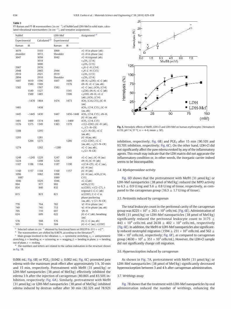
Fig. 5. Hemolytic effects of MefH, LDH-Cl and LDH-Mef on human erythrocytes (Hematocrit0.15%, pH 7.4, 37 °C, n = 4–6, mean ± SD).
Table 1FT-Raman and FT-IR wavenumbers (in cm−1) of NaMef and LDH-Mef in solid state, calcu-lated vibrational wavenumbers (in cm−1), and tentative assignments.
NaMef LDH-Mef Assignmentc,d
Experimental Calculateda,b Experimental
Raman IR Raman IR
3079 3103 3069 νC–H in phase (øb)shoulder 3072 Shoulder νC–H in phase (øa)3047 3058 3042 νC–H trigonal (øb)
3018 νaCH3 (C14)3000 νaCH3 (C15)
2967 2976 νaH–C–H (C14)2948 2965 2944 νaH–C–H (C15)2919 2921 2919 νsCH3 (C15)2894 2910 Shoulder νsCH3 (C14)1607 1610 1596 1607 1609 δN–H, νaCOO, νC–C (øb)
1580 1569 1576 δN–H, νC–C (øa, øb)1582 1567 1582 νC–C (øa), βCH3 (C14)
1549 1527 1541 νaCOO, δN–H, νC–C (øb)1500 1485 1506 νaCOO, δN–H, νC–C
(øb), βCH3 (C14)~1470 1464 1474 1473 δCH3 (C14, C15), βC–H
(øa)1465 1458 δCH3 (C14, C15), βC–H
(øa, øb)1445 ~1445 1430 1447 1456–1448 δCH3 (C14, C15), δN–H,
βC–H (øa, øb)1401 1400 1374 1405 ~1400 δCH3 (C14, C15)1376 1375 1349 1383 1379 ν(C2–COO), βC–H (øb),
νa (C1–N–C8)1308 1295 νa(C1–N–C8), νC–C
(øa, øb),1293 1281 βC–H(øa, øb)
1284 1275 1286 ν(C2–COO), νC–C(øa, øb), νa(C1–N–C8)
1274 1262 ~1280 νC–C (øa, øb),νa(C1–N–C8)
1248 ~1245 1229 1247 1248 νC–C (øa), βC–H (øa)1224 1208 1224 δN–H, βC–H (øb)1187 1188 1164 1187 1190 ν(C14–C9), νC–C (øa),
βC–H (øa)1160 1157 1144 1160 1157 βC–H (øb)10961087
1082 10981089
βC–H (øa), δCH3(C14,C15)
1046 1029 1046 νC–C(øb)994 977 992 wCH3(C15)834 840 832 sc(COO), ν(C2–C7), δ
trigonal C–C–C (øb)815 823 821 sc(COO), C–C–C in
phase puckering(øa, øb), γ (C1–N–C8)
778 764 782 γC–H in phase (øa)749 745 753 γC–H in phase (øa, øb)705 710 697 γN–H624 609 622 βC–C–C (øb), breathing
(øa)578 568 576 βC–C–C (øa, øb)505 516 504 βC–C–C (øa), r(COO)
a Selected values in cm−1 obtained by functional/basis set B3LYP/6–311++G**.b The wavenumbers are shifted by 0.9679, according to the literature30.c Main groups involved in the vibration. νs = symmetric stretching, νa = antisymmetric
stretching, δ=bending, sc = scissoring, w=wagging, β=bending in plane, γ=bendingout of plane, r = rocking.
d The numbers and letters are related to the carbon indexation in the structure shownin Fig. 1B.
634 V.R.R. Cunha et al. / Materials Science and Engineering C 58 (2016) 629–638
0.006 mL; Fig. 6B) or PGE2 (0.042 ± 0.002 mL; Fig. 6C) promoted pawedema with the maximum peak effect after approximately 3 h, 30 minand 15 min, respectively. Pretreatment with MefH (31 μmol/kg) orLDH-Mef nanoparticles (38 μmol of Mef/kg) effectively inhibited theedema 3 h after the injection of carrageenan (80.86% and 83.50% in-hibition, respectively; Fig. 6A). Similarly, pretreatment with MefH(31 μmol/kg) or LDH-Mef nanoparticles (38 μmol of Mef/kg) inhibitededema induced by dextran sulfate after 30 min (82.32% and 78.92%
inhibition, respectively; Fig. 6B) and PGE2 after 15 min (90.10% and93.70% inhibition, respectively; Fig. 6C). On the other hand, LDH-Cl didnot significantly affect the pawedema evoked by any of the inflammatoryagents. This resultmay indicate that the LDHmatrix did not aggravate theinflammatory condition or, in other words, the inorganic carrier indeedseems to be biocompatible.
3.4. Myeloperoxidase activity
Fig. 6D shows that the pretreatment with MefH (31 μmol/kg) orLDH-Mef nanoparticles (38 μmol of Mef/kg) reduced the MPO activityto 6.3 ± 0.9 U/mg and 5.6 ± 0.8 U/mg of tissue, respectively, as com-pared to the carrageenan group (16.3 ± 1.7 U/mg of tissue).
3.5. Peritonitis induced by carrageenan
The total leukocyte count in the peritoneal cavity of the carrageenangroupwas 8225 × 103± 263 × 103 cells/mL (Fig. 6E). Administration ofMefH (31 μmol/kg) or LDH-Mef nanoparticles (38 μmol of Mef/kg)significantly reduced the peritoneal leukocyte count to 3175 ±443 × 103 cells/mL and 2638 ± 402 × 103 cells/mL, respectively(Fig. 6E). In addition, theMefH or LDH-Mef nanoparticles also significant-ly reduced neutrophil migration (1594 ± 231 × 103 cells/mL and 562 ±104 × 103 cells/mL, respectively; Fig. 6F), as compared to carrageenangroup (4630 × 103 ± 351 × 103 cells/mL). However, the LDH-Cl sampledid not significantly change cell migration.
3.6. Hypernociception induced by carrageenan
As shown in Fig. 7A, pretreatment with MefH (31 μmol/kg) orLDH-Mef nanoparticles (38 μmol of Mef/kg) significantly decreasedhypernociception between 3 and 4 h after carrageenan administration.
3.7. Writhings assay
Fig. 7B shows that the treatmentwith LDH-Mef nanoparticles by oraladministration reduced the number of writhings, enhancing the

Fig. 6. Anti-inflammatory effect of MefH or LDH-Mef in mice. Paw inflammation was induced by carrageenan (A); dextran sulfate (B), and PGE2 (C). *p b 0.05 vs control group (only in-flammatory agent). Paw tissuemyeloperoxidase (MPO) activity induced by carrageenan (D). Total leukocyte (E) and neutrophil (F) counts in carrageenan-induced peritonitismodel. Micewere pretreated with MefH (31 μmol/kg) or LDH-Mef (38 μmol of Mef/kg). *p b 0.05 vs carrageenan group. Results are expressed as the mean ± SEM for at least 5–6 animals per group.
635V.R.R. Cunha et al. / Materials Science and Engineering C 58 (2016) 629–638
antinociceptive effect of MefH when compared to the non-intercalatedMefH, at three different doses (p b 0.05).
4. Discussion
The present study was conducted to investigate the in vitro cytotox-icity using a membrane interaction model in red blood cells and thepharmacological potential actions of LDH-Mef nanoparticles. Severalexperimental models of inflammation (i.e. carrageenan-, dextransulfate- and PGE2-induced paw edema, MPO activity and peritonitis)and nociception (i.e. mechanical hypernociception and acetic acid-induced writhing) were employed and LDH-Mef was compared withfree MefH and LDH matrix without intercalated drug.
Reduced hemolytic effects after incorporation in nanocarriers havebeen reported in the literature for cyclodextrin inclusion complexes,[40] polymeric nanocapsules [41] and also for inorganic hydroxidenanoparticles. The mechanism of hemolysis involves the membraneinteraction with a new drug or carrier candidates, and uptake of lipidcomponents with changes on membrane fluidity [40]. In general, LDHnanoparticles did not induce pronounced cytotoxicity. For this reason,
it was possible to observe that the intercalation of MefH in LDH reducedthe hemolytic effects when compared to the non-intercalated MefH.
The carrageenan-induced paw edema is a model, widely used tocharacterize the mechanisms of action of anti-inflammatory com-pounds, including NSAIDs [42]. It is characterized by a biphasic process:initially the release of inflammatory mediators, such as histamine andserotonin, occurs followed by an increase in prostaglandin synthesis(mainly prostaglandin E2 (PGE2)) and, subsequently, the rise in nitricoxide levels and the neutrophils infiltration is observed. On the otherhand, the dextran sulfate promotes an osmotic edema characterizedby an increase in the vascular permeability, induced by the releaseof vasoactive amines derived from mast cell degranulation [43].Prostaglandins, particularly PGE2, are important pro-inflammatorymediators, which produce their edematogenic effect through the changeof vascular permeability [44].
Pre-treatment with LDH-Mef nanoparticles produced an anti-inflammatory efficacy, which is similar to the action observed forMefH, in three different pawedemamodels used in thiswork. However,no significant efficacy difference between LDH-Mef nanoparticle andMefH was observed in acute inflammation model. The carrier without

Fig. 7. Antinociceptive effect of MefH or LDH-Mef in mice. (A) Mechanical hypernociception induced by carrageenan. Mice were pretreated with MefH (31 μmol/kg) or LDH-Mef(38 μmol of Mef/kg). *p b 0.05 vs carrageenan group. (B) Writhing response induced by acetic acid. *p b 0.05 vs acetic acid group; #p b 0.05 compared to the acetic acid group andMefH 200 mg/kg. Data are expressed as mean ± S.E.M. of 5–6 animals for each group.
636 V.R.R. Cunha et al. / Materials Science and Engineering C 58 (2016) 629–638
the drug (sample LDH-Cl) did not promote changes in edema inducedby any inflammatory agents, applied in the tests.
Literature data have shown that treatment with anti-inflammatorydrugs, such as dexamethasone, indomethacin and nimesulide inhibitscell migration to the inflammation site [45]. Thus, in view of the impor-tance of cellmigration in the inflammatory process, such as arthritis andsepsis, [46] we investigated whether the anti-inflammatory effects ofMefH and LDH-Mef nanoparticle in carrageenan-induced paw edemamodel were associated with changes in cell migration. This mechanismwas evaluated through the MPO activity and carrageenan-inducedperitonitis.
MPO is a characteristic enzyme of neutrophil granules, released byinflammatory cells that act as an important indicator of the neutrophils'accumulation [47]. In the present study, MefH and LDH-Mef nanoparticlepromoted an effective inhibitory effect on cell migration (MPO activityand peritonitis). The observed inhibition did not reveal any significantdifference in the effect of the tested formulations. Moreover, comparedto the carrageenan group, no changes were observed in these parametersfor the animals treated with LDH-Cl.
Studies have shown that leukocyte migration to the inflamed site andrelease of several mediators are important characteristics of the inflam-mation, and are essential for the onset of tenderness and inflammatorypain [48]. Considering that NSAIDs also have antinociceptive effects, andbased on the intimate relationship between inflammation and develop-ment of a painful sensation, we investigated the antinociceptive effect ofMefH and LDH-Mef nanoparticles. For this purpose, carrageenan-induced mechanical hypernociception model and acetic acid-inducedwrithing were used.
Carrageenan is an inflammatory agent that is widely used for theinvestigation of the hypernociception in animal models. When ad-ministrated intraplantarly, this substance promotes rapid release ofseveral inflammatory mediators, such as mast cells products, neutrophilchemoattractant-1 responsible for stimulating the synthesis of prosta-glandins, increasing the local flow and vascular permeability, culminating
in an exacerbated sensitivity to mechanical and thermal stimuli as a re-sponse of nociceptive neurons [32].
In the present study it was demonstrated that MefH administrationdecreases the mechanical inflammatory hypernociception. However,this experimental model revealed that LDH-Mef nanoparticles present-ed a significantly higher efficacy in comparison toMefH. This effect maybe, at least partly, due to the inhibition of the inflammatory mediators'release and the inhibition of the interaction among neutrophils andthe cellular surface. Based on these results and on the literature data,it is also possible to infer that vascular permeability changes are pro-moted by the augmented synthesis of prostaglandins in inflammatoryprocesses, which facilitates the targeting of the nanocapsules to the in-flamed site, increasing the pain threshold and the drug efficiency.
In the model of acetic acid-induced writhing, the injection of aceticacid in the peritoneal cavity causes a stretch or writhing viscera motorreaction, with alternating pushups and abdominal extensions. Thiscomplex behavior involves different mediators in the development ofinflammatory hyperalgesia, as the release of endogenous substancesthat excite nerve endings, prostaglandins (PGE2 e PGF2α) and bradyki-nin derived macrophages and mast cells residing in the peritoneal.This test showed that all groups treated with LDH-Mef nanoparticlespresented an analgesic effect greater than the non-intercalated MefH,which is clearly evident in the dose of LDH-Mef 200mg/kg. The analgesicresponse of the intercalated compoundwas enhancedwhen compared tothe free compound at the 200 mg/kg dose level. Probably, the improvedanalgesic effect can be attributed to the increase in aqueous solubilityand controlled release of Mef from LDH nanoparticles, as described byRives' group [23,25,26].
5. Conclusions
A set of physicochemical techniques supported by Density Function-al Theory calculations confirmed the immobilization of the mefenamicacid into the nanocarrier Layered Double Hydroxide in its anionic

637V.R.R. Cunha et al. / Materials Science and Engineering C 58 (2016) 629–638
form. The anti-inflammatory activity, hemolytic and antinociceptiveassays were used to evaluate the pharmacological effects of LDH-Mef(40 wt% of drug). The higher tolerated hemolytic concentration forLDH-Mef (1.83 mM) compared to MefH (0.48 mM) showed the im-proved hemolytic effect of the nanocarrier with Mef. Pretreatment ofanimals with LDH-Mef reduced carrageenan-, dextran sulfate- andPGE2-induced paw edema. LDH-Mef also decreases total leucocytesand neutrophil counts of the peritoneal cavity after inflammation in-duction with carrageenan. In nociception models, pretreatment withLDH-Mef reduced mechanical hypernociception carrageenan-inducedafter 3–4 h and also the number of writhings induced by acetic acid.
Acknowledgments
The authors are grateful to the Fundação de Amparo à Pesquisa doEstado de São Paulo (FAPESP), Conselho Nacional de DesenvolvimentoCientífico e Tecnológico (CNPq), and Nanobiomed (NanomedicineNetwork/CAPES) for the financial support and scholarships. VanessaCunha is thankful for the PhD fellowship (Programa Institucional deDoutorado Sanduíche no Exterior) provided by the Coordenação deAperfeiçoamento de Pessoal de Nível Superior (CAPES). We are alsothankful to the Laboratório de Espectroscopia Molecular (LEM, Institutode Química— USP) for the Raman spectra recording.
Appendix A. Supplementary data
Supplementary data to this article can be found online at http://dx.doi.org/10.1016/j.msec.2015.08.037.
References
[1] J.M. Oh, D.H. Park, S.J. Choi, J.H. Choy, LDH nanocontainers as bio-reservoirs and drugdelivery carriers, Recent Pat. Nanotechnol. 6 (2012) 200–217.
[2] B. Saifullah, P. Arulselvan, M.E.E. Zowalaty, S. Fakurazi, T.J. Webster, B.M. Geilich,M.Z. Hussein, Development of a biocompatible nanodelivery system for tuberculosisdrugs based on isoniazid-Mg/Al layered double hydroxide, Int. J. Nanomedicine 9(2014) 4749–4762.
[3] L. Qin, M. Wang, R. Zhu, S. You, P. Zhou, S. Wang, The in vitro sustained release pro-file and antitumor effect of etoposide-layered double hydroxide nanohybrids, Int. J.Nanomedicine 8 (2013) 2053–2064.
[4] J. Chakraborty, S. Roychowdhury, S. Sengupta, S. Ghosh, Mg–Al layered doublehydroxide–methotrexate nanohybrid drug delivery system: evaluation of efficacy,Mater. Sci. Eng. C 33 (2013) 2168–2174.
[5] C.J. Serna, J.L. White, S.L. Hem, Structural survey of carbonate-containing antacids, J.Pharm. Sci. 67 (1978) 324–327.
[6] K.H. Holtermüller, M. Liszkay, I. Bernard, W. Haase, Talcivent study group: treatmentfor benign gastric ulcer with low-dose antacid versus ranitidine: results of a double-blind, randomly allocated, multicentre trial, Eur. J. Gastroenterol. 5 (1993) 139–144.
[7] A. Tarnawski, K. Tanoue, T.G. Douglas, F.L. Irwin Jr., J.J. Sarfeh, Cellular mechanismsinvolved in duodenal ulcer healing and the quality of mucosal scar. Effect of Talcidand Omeprazole treatment, Gastroenterology 108 (1995) A236.
[8] C. Forano, T. Hibino, F. Leroux, C. Taviot-Guého, Handbook of clay science, in: F. Bergaya,T. BKG, G. Lagaly (Eds.), Developments of clay science, 1, Elsevier, Amsterdam 2006,pp. 1021–1095.
[9] X.F. Wang, S.Q. Liu, S.P. Li, Methotrexatum intercalated layered double hydroxides:statistical design, mechanism explore and bioassay study, Mater. Sci. Eng. 49(2015) 330–337.
[10] A.I. Khan, L.X. Lei, A.J. Norquist, D. O'Hare, Intercalation and controlled release ofpharmaceutically active compounds from a layered double hydroxide, Chem. Comm.22 (2001) 2342–2343.
[11] C.R. Gordijo, C.A.S. Barbosa, A.M.C. Ferreira, V.R.L. Constantino, D.O. Silva, Immobili-zation of ibuprofen and copper-ibuprofen drugs on layered double hydroxides, J.Pharm. Sci. 94 (2005) 1135–1148.
[12] K. Zhao, G. Rong, G. Guo, X. Luo, H. Kang, Y. Sun, C. Dai, X. Wang, X. Wang, Z. Jin, S.Cui, Q. Sun, Synthesis, characterization, and immune efficacy of layered doublehydroxide@SiO2 nanoparticles with shell-core structure as a delivery carrier forNewcastle disease virus DNA vaccine, Int. J. Nanomedicine 10 (2015) 2895–2911.
[13] D.Y. Tian, Z.L. Liu, S.P. Li, X.D. Li, Facile synthesis of methotrexate intercalated layereddouble hydroxides: particle control, structure and bioassay explore, Mater. Sci. Eng.C 45 (2014) 297–305.
[14] V.R.R. Cunha, J. Tronto, J.B. Valim, A.M.C. Ferreira, V.R.L. Constantino, Layered doublehydroxide: inorganic nanoparticles for storage and release of species of biologicaland therapeutic interest, Quím. Nova 33 (2010) 159–171.
[15] J.H. Choy, J.S. Jung, J.M. Oh, M. Park, J. Jeong, Y.K. Kang, et al., Layered double hydroxideas an efficient drug reservoir for folate derivatives, Biomaterials 25 (2004) 3059–3064.
[16] L. Tammaro, U. Costantino, A. Bolognese, G. Sammartino, G. Marenzi, A. Calignano,et al., Nanohybrids for controlled antibiotic release in topical applications, Int. J.Antimicrob. Agents 29 (2007) 417–423.
[17] J.H. Choy, S.Y. Kwak, J.S. Park, Y.J. Jeong, Cellular uptake behavior of [gamma-P-32]labeled ATP-LDH nanohybrids, J. Mater. Chem. 11 (2001) 1671–1674.
[18] J.M. Oh, S.J. Choi, G.E. Lee, J.E. Kim, J.H. Choy, Inorganic metal hydroxide nanoparti-cles for targeted cellular uptake through clathrin-mediated endocytosis, Chem.Asian. J. 4 (2009) 67–73.
[19] J. Chen, R. Shao, L. Li, Z.P. Xu, W. Gu, Effective inhibition of colon cancer cellgrowth with MgAl-layered double hydroxide (LDH) loaded 5-FU and PI3K/mTOR dual inhibitor BEZ-235 through apoptotic pathways, Int. J. Nanomedicine 9(2014) 3403–3411.
[20] S.J. Choi, J.M. Oh, J.H. Choy, Toxicological effects of inorganic nanoparticles on humanlung cancer A549 cells, J. Inorg. Biochem. 103 (2009) 463–471.
[21] S.C. Smolinske, A.H. Hall, S.A. Vandenberg, D.G. Spoerke, P.V. McBride, Toxiceffects of nonsteroidal anti-inflammatory drugs in overdose: an overview ofrecent evidence on clinical effects and dose response relationships, Drug Saf. 5(1990) 252–274.
[22] O. Catanzano, S. Acierno, P. Russo, M. Cervasio, M.D.B. De Caro, A. Bolognese, G.Sammartino, L. Califano, G.Marenzi, A. Calignano, D. Acierno, F. Quaglia,Melt-spun bio-active sutures containing nanohybrids for local delivery of anti-inflammatory drugs,Mater. Sci. Eng. 43 (2014) 300–309.
[23] V. Rives, M. Del Arco, C. Martín, Layered double hydroxide as drug carriers and forcontrolled release of non-steroidal anti-inflammatory drugs (NSAIDs): a review, J.Control. Release 169 (2013) 28–39.
[24] S. Fiorucci, L. Santucci, E. Distrutti, NSAIDs, coxibs, CINOD andH2S-releasing NSAIDs:what lies beyond the horizon, Dig. Liver Dis. 12 (2007) 1043–1051.
[25] M. Del Arco, A. Fernández, C. Martín, M.L. Sayalero, V. Rives, Solubility and release offenamates intercalated in layered double hydroxides, Clay Miner. 43 (2008)255–265.
[26] M. Del Arco, A. Fernandez, C.Martin, V. Rives, Release studies of different NSAIDs en-capsulated in Mg, Al, Fe-hydrotalcites, Appl. Clay Sci. 42 (2009) 538–544.
[27] V.R.R. Cunha, P.A.D. Petersen, M.B. Gonçalves, H.M. Petrilli, C. Taviot-Gueho, F.Leroux, et al., Structural, spectroscopic (NMR, IR and Raman) and DFT investigationof the self-assembled nanostructure of pravastatin-LDH (layered double hydrox-ides) systems, Chem. Mater. 24 (2012) 1415–1425.
[28] C.A.S. Barbosa, A.M.C. Ferreira, A.C.V. Coelho, V.R.L. Constantino, Characterization ofCu(II) phthalocyanine tetrasulfonate intercalated and supported on layered doublehydroxides, J. Incl. Phenom. Macrocycl. Chem. 42 (2002) 15–23.
[29] M.J. Frisch, G.W. Trucks, H.B. Schlegel, G.E. Scuseria, M.A. Robb, J.R. Cheeseman, et al.,Gaussian 03, Revision C.02. 2003.
[30] M.P. Andersson, P. Uvdal, New scale factors for harmonic vibrational frequenciesusing the B3LYP density functional method with the triple-ζ basis set 6–311 +G(d,p), J. Phys. Chem. A 109 (2005) 2937–2941.
[31] A.P. Scott, L. Radom, Harmonic vibrational frequencies: an evaluation of hartree-fock, moller-plesset, quadratic configuration interaction, density functional theory,and semiempirical scale factors, J. Phys. Chem. 100 (1996) 16502–16513.
[32] L.S. Chaves, L.A.D. Nicolau, R.O. Silva, F.C. Barros, A.L. Freitas, K.S. Aragão, et al., Anti-inflammatory and antinociceptive effects in mice of a sulfated polysaccharide frac-tion extracted from the marine red algae Gracilaria caudata, Immunopharmacol.Immunotoxicol. 35 (2013) 93–100.
[33] T.M. Cunha, W.A. Verri Jr., G.G. Vivancos, I.F. Moreira, S. Reis, C.A. Parada, et al., Anelectronic pressuremeter nociception paw test for mice, Braz. J. Med. Biol. Res. 37(2004) 401–407.
[34] R. Koster, M. Anderson, E.J. Beer, Acetic acid for analgesic screening, Federation Pro-ceeds. 18 (1959) 412–416.
[35] M. Del Arco, A. Fernández, C. Martín, V. Rives, Intercalation of mefenamic andmeclofenamic acid anions in hydrotalcite-like matrixes, Appl. Clay Sci. 36 (2007)133–140.
[36] U. Costantino, N. Coletti, M. Nocchetti, G.G. Aloisi, F. Elisei, Anion exchange ofmethylorange into Zn-Al synthetic hydrotalcite and photophysical characterization of theintercalates obtained, Langmuir 15 (1999) 4454–4460.
[37] P. Atkins, T. Overton, J. Rourke, M. Weller, F. Armstrong, M. Hagerman, Shriver &Atkins' Inorganic Chemistry, 5th Ed. W. H. Freeman and Company, New York,2010 25 Part 1.
[38] L. Fang, S. Numajiri, D. Kobayashi, H. Ueda, K. Nakayama, H. Miyamae, Y. Morimoto,Physicochemical and crystallographic characterization of mefenamic acid com-plexes with alkanolamines, J. Pharm. Sci. 93 (2004) 144–154.
[39] V.R.R. Cunha, C.M.S. Izumi, P.A.D. Petersen, A. Magalhães, M.L.A. Temperini, H.M.Petrilli, V.R.L. Constantino, Mefenamic acid anti-inflammatory drug: probing itspolymorphs by vibrational (IR and Raman) and solid-state NMR spectroscopies, J.Phys. Chem. B 118 (2014) 4333–4344.
[40] D.R. De Araujo, S.S. Tsuneda, C.M. Cereda, G.F. Del, F. Carvalho, O.S. Preté, et al.,Development and pharmacological evaluation of ropivacaine-2-hydroxypropyl-beta-cyclodextrin inclusion complex, Eur. J. Pharm. Sci. 33 (2008) 60–71.
[41] K. Seibert, Y. Zhang, K. Leahy, S. Hauser, J. Masferrer, W. Perkins, et al., Pharmacolog-ical and biochemical demonstration of the role of cyclooxygenase 2 in inflammationand pain, Proc. Natl. Acad. Sci. 91 (1994) 12013–12017.
[42] N.L. Quintão, R. Medeiros, A.R. Santos, M.M. Campos, J.B. Calixto, The effects ofdiacerhein on mechanical allodynia in inflammatory and neuropathic models ofnociception in mice, Anesth. Analg. 6 (2005) 1763–1769.
[43] D.A. Rowley, E.P. Benditt, 5-hydroxytryptamine and histamine as mediators of thevascular injury produced by agents which damage mast cells in rats, J. Exp. Med. 103(1956) 399–415.
[44] L.M. Solomon, L. Juhlin, M.B. Kirschenbaum, Prostaglandin on cutaneous vasculature, J.Invest. Dermatol. 51 (1968) 280–282.

638 V.R.R. Cunha et al. / Materials Science and Engineering C 58 (2016) 629–638
[45] M. Hamza, Role of neutrophils in the anti-inflammatory and antinociceptive activity ofthe cyclooxygenase inhibitors indomethacin and nimesulide, J. Egypt Soc. Pharmacol.Exp. Ther. 29 (2008) 507–526.
[46] F.J.C. Alves, M.B.M. Tavares, F.C. Barja, C.F. Benjamim, F.A. Basile, S.M. Arraes, et al.,Neutrophil function in severe sepsis, Endocr. Metab. Immune Disord. Drug Targets2 (2006) 151–158.
[47] S.J. Klebanoff, Myeloperoxidase, Proc. Assoc. Am. Physicians 111 (1999) 383–389.[48] T.M. Cunha, J.W.A. Verri, I.R. Schivo, M.H. Napimoga, C.A. Parada, S. Poole, et al., Crucial
role of neutrophils in the development of mechanical inflammatory hypernociception,J. Leukoc. Biol. 83 (2008) 824–832.
Copyright © 2022 FDOKUMEN