Antioxidant Therapy in Alzheimer's Disease: Theory and Practice
12
Mini-Reviews in Medicinal Chemistry, 2008, 8, 1395-1406 1395 1389-5575/08 $55.00+.00 © 2008 Bentham Science Publishers Ltd. Antioxidant Therapy in Alzheimer’s Disease: Theory and Practice Gjumrakch Aliev 1,3 , Mark E. Obrenovich 4 , V. Prakash Reddy 7 , Justin C. Shenk 1,3 , Paula I. Moreira 5 , Akihiko Nunomura 6 , Xiongwei Zhu 4 , Mark A. Smith 4 and George Perry 2,*,# 1 Department of Biology, 2 College of Sciences and 3 Electron Microscopy Research Center, University of Texas at San Antonio, San Antonio, TX 78249, USA; 4 Department of Pathology, Case Western Reserve University, Cleveland, OH 44106, USA; 5 Center for Neuroscience and Cell Biology of Coimbra, Uni- versity of Coimbra, Coimbra, Portugal; 6 Department of Psychiatry and Neurology, Asahikawa Medical College, Asahikawa, Japan; 7 Department of Chemistry, Missouri University of Science and Technol- ogy, Rolla, MO 65409, USA Abstract: Alzheimer disease treatment has yet to yield a successful therapy that addresses the source of the damage found in brains. Of the varied proposed theories of AD etiology, reactive oxygen species (ROS) generation is cited as a common factor. Efforts to reduce the pathology associated with ROS via antioxidants therefore offer new hope to patients suffering from this devastative disease. Key Words: Antioxidant, calorie restriction, estrogen, free radical, glutathione, oxidation, oxidative stress, therapy. # Author’s Profile: George Perry is currently the Dean of the College of Sciences at The University of Texas at San Antonio. Perry’s studies are focused on the mechanism of formation and physiological consequences of the cytopathology of Alzheimer disease. Dr. Perry’s group has shown that oxidative damage is the initial cytopathological abnormality in Alzheimer disease. GENERAL OVERVIEW OF ALZHEIMER DISEASE Alzheimer disease (AD) is the leading cause of dementia. The burden of AD increases with aging of the population: an estimated 5 million people in the US suffer with over $70 billion in expenses annually [1]. A century since its first classification by Alois Alzheimer, research has yielded nei- ther a cure nor an effective prevention strategy. Current clinical therapies consist of cholinesterase inhibitors/NMDA receptor antagonists such as Reminyl, galantamine, Aricept (donepezil hydrochloride), Exelon (rivastigmine) and Me- mantine (Namenda), which offer little more than short-term palliative effects. *Address correspondence to this author at the College of Sciences, Univer- sity of Texas at San Antonio, One UTSA Circle, San Antonio, Texas, 78249, USA; Tel: +1 210 458 4450; Fax: +1 210 458 4445; E-mail: [email protected] AD is manifested as a distinguished impairment of thought, memory, and language abilities. Research over the past 20 years has yielded much information about the factors involved in the pathogenesis, but a cause has not yet been elucidated. Several major theories of AD, such as amyloid- beta (A ) toxicity [2], tauopathy [3], inflammation [4,5], oxidative stress [6-11], have all been represented broadly and argued intensely in the literature. CLINICAL FACTS OF ALZHEIMER DISEASE The risk of AD varies from 12% to 19% for women over the age of 65 years and 6% to 10% for men [12] and rises exponentially with age, such that up to 47% of individuals over the age of 80 develop AD [13]. On average, AD pa- tients live about 8 years after initial diagnosis, although the disease can last for as long as 20 years. The areas of the brain that control memory and thinking skills are affected first but, as the disease progresses, neurons in other regions of the brain are also affected. Eventually, the patient with AD will need complete care, adding further emotional, physical, and financial costs to the family. ALZHEIMER DISEASE - PATHOLOGY Since its early classification, two distinctive hallmark lesions found in the brain of patients with AD are senile N S N N O HO H 3 C F Reminyl N O OCH 3 HO H 3 C H Galantamine N Ph H 3 CO H 3 CO O Aricept H Cl - + CH 3 O N H 3 C N(CH 3 ) 2 O CH 3 Exelon CH 3 H 3 C NH 2 Memantine
-
Upload
independent -
Category
Documents
-
view
0 -
download
0
Transcript of Antioxidant Therapy in Alzheimer's Disease: Theory and Practice

Mini-Reviews in Medicinal Chemistry, 2008, 8, 1395-1406 1395
1389-5575/08 $55.00+.00 © 2008 Bentham Science Publishers Ltd.
Antioxidant Therapy in Alzheimer’s Disease: Theory and Practice
Gjumrakch Aliev1,3, Mark E. Obrenovich4, V. Prakash Reddy7, Justin C. Shenk1,3, Paula I. Moreira5,Akihiko Nunomura6, Xiongwei Zhu4, Mark A. Smith4 and George Perry2,*,#
1Department of Biology, 2College of Sciences and 3Electron Microscopy Research Center, University of Texas at San Antonio, San Antonio, TX 78249, USA; 4 Department of Pathology, Case Western Reserve University, Cleveland, OH 44106, USA; 5Center for Neuroscience and Cell Biology of Coimbra, Uni-versity of Coimbra, Coimbra, Portugal; 6Department of Psychiatry and Neurology, Asahikawa Medical College, Asahikawa, Japan; 7Department of Chemistry, Missouri University of Science and Technol-ogy, Rolla, MO 65409, USA
Abstract: Alzheimer disease treatment has yet to yield a successful therapy that addresses the source of
the damage found in brains. Of the varied proposed theories of AD etiology, reactive oxygen species
(ROS) generation is cited as a common factor. Efforts to reduce the pathology associated with ROS viaantioxidants therefore offer new hope to patients suffering from this devastative disease.
Key Words: Antioxidant, calorie restriction, estrogen, free radical, glutathione, oxidation, oxidative stress, therapy.
#Author’s Profile: George Perry is currently the Dean of the College of Sciences at The University of Texas at San Antonio.
Perry’s studies are focused on the mechanism of formation and physiological consequences of the cytopathology of Alzheimer disease. Dr. Perry’s group has shown that oxidative damage is the initial cytopathological abnormality in Alzheimer disease.
GENERAL OVERVIEW OF ALZHEIMER DISEASE
Alzheimer disease (AD) is the leading cause of dementia. The burden of AD increases with aging of the population: an estimated 5 million people in the US suffer with over $70 billion in expenses annually [1]. A century since its first classification by Alois Alzheimer, research has yielded nei-ther a cure nor an effective prevention strategy. Current clinical therapies consist of cholinesterase inhibitors/NMDA receptor antagonists such as Reminyl, galantamine, Aricept (donepezil hydrochloride), Exelon (rivastigmine) and Me-mantine (Namenda), which offer little more than short-term palliative effects.
*Address correspondence to this author at the College of Sciences, Univer-sity of Texas at San Antonio, One UTSA Circle, San Antonio, Texas, 78249, USA; Tel: +1 210 458 4450; Fax: +1 210 458 4445; E-mail: [email protected]
AD is manifested as a distinguished impairment of thought, memory, and language abilities. Research over the past 20 years has yielded much information about the factors involved in the pathogenesis, but a cause has not yet been elucidated. Several major theories of AD, such as amyloid-beta (A ) toxicity [2], tauopathy [3], inflammation [4,5], oxidative stress [6-11], have all been represented broadly and argued intensely in the literature.
CLINICAL FACTS OF ALZHEIMER DISEASE
The risk of AD varies from 12% to 19% for women over the age of 65 years and 6% to 10% for men [12] and rises exponentially with age, such that up to 47% of individuals over the age of 80 develop AD [13]. On average, AD pa-tients live about 8 years after initial diagnosis, although the disease can last for as long as 20 years. The areas of the brain that control memory and thinking skills are affected first but, as the disease progresses, neurons in other regions of the brain are also affected. Eventually, the patient with AD will need complete care, adding further emotional, physical, and financial costs to the family.
ALZHEIMER DISEASE - PATHOLOGY
Since its early classification, two distinctive hallmark lesions found in the brain of patients with AD are senile
N
S NN
O
HOH3C
F
Reminyl
N
O
OCH3
HO
H3C
H
Galantamine
N
Ph
H3CO
H3CO
O
Aricept
H
Cl-+
CH3
ONH3C
N(CH3)2
O
CH3
Exelon
CH3
H3CNH2
Memantine

1396 Mini-Reviews in Medicinal Chemistry, 2008, Vol. 8, No. 13 Aliev et al.
plaques and neurofibrillary tangles (NFTs) (reviewed else-where [14]), the latter were identified by the use of silver-staining techniques [15]. In addition, other associated mark-ers include neuronal and dendritic loss, neuropil threads, dystrophic neurites, granulovacuolar degeneration, Hirano bodies, cerebrovascular amyloid, and atrophy of the brain [14].
Extracellular senile plaque formation remains a hallmark of AD. Bundles of A , 10-200 m in diameter, form the cen-tral core of the plaques and are surrounded by A , amyloid-protein precursor (A PP), tau, and neurofilament proteins are reported [16].
NFTs are recognized as the major intracellular protein aggregation in AD brains, and are located primarily in the cerebral cortex, especially in the large pyramidal neurons in the hippocampal and frontotemporal regions [17]. Tau, a microtubule-associated protein found predominantly on ax-ons, is the major component of the bundled paired helical filaments (PHF) that compose NFTs. Moreover, neurofila-ment proteins are also reported in NFTs [16,18]. In PHF, tau is abnormally hyperphosphorylated [3,19,20], ubiquinated [21-23], oxidized [6,24-26], truncated [27] and aggregated into filaments [3,28,29]. The hyperphosphorylation of tau is thought to render it unable to bind to microtubules and there-fore unable to promote or maintain microtubule assembly [30], although in vivo, while microtubules are disrupted, this has no relation to NFT [31]. The resistance to proteolytic degradation of hyperphosphorylated tau may play a key role in neurofibrillary degeneration in AD patients [32,33].
While the pathological hallmarks provide a baseline for current diagnostic standards, their role as initiators versus byproducts of disease is hotly contested [34-36].
GENETIC ETIOLOGY OF ALZHEIMER DISEASE
Only about 5% of all AD cases have an early onset and are related to genetic mutations of presenilin 1, presenilin 2 [37] or the A PP genes. Indeed, the majority, approximately 95%, of all AD patients are sporadic, while the late-onset cases, where the major risk factors are aging and apolipopro-tein E4 (ApoE4) polymorphisms [38,39]. While APOE4 al-lele is not a determinant for AD, since a large number of individuals with APOE4 alleles never develop frank AD, in both familial and sporadic cases of disease there is accumu-lating evidence indicating a major role for free radicals and oxidative stress in disease pathogenesis and pathophysiology [9,10].
The greatest correlated factor for the development of AD are polymorphisms of the ApoE gene, such that 50% of pa-tients with AD patients have at least one ApoE4 allele [38,39]. Further, although familial AD only accounts for a small percentage of AD cases, many have argued that muta-tions in A PP and presenilin 1 and 2 are critical genetic fac-tors in AD pathogenesis and in A production [40,41].
Apolipoprotein E Gene Polymorphism
Polymorphisms of the ApoE gene are found to correlate with onset and risk of developing AD, such that 50% of AD patients have at least one ApoE4 allele [38,39]. ApoE is an abundant 34-kDa glycoprotein that is synthesized and se-
creted mainly by astrocytes and microglia in the central nervous system (CNS). It is well established that ApoE, and especially the E4 allele of ApoE, is a major genetic risk fac-tor of the more common, late onset form of AD [42,43]. The influence of ApoE genotype on AD seems to operate viamultiple mechanisms. For example, polymorphisms are a determinant of brain A burden in individuals affected with AD [44,45]. Additionally, apolipoproteins have been sug-gested to act as antioxidants, with the ApoE4 allele being less effective in this role [46] so that increased oxidative damage is found in specific brain regions of AD patients with the ApoE E4 genotype [47].
Amyloid- Protein Precursor
A protein is the major component of senile plaque cores and is derived from the precursor protein, A PP. A PP is encoded on chromosome 21 (21q11-22) [48,49]. The normal function of A PP is unknown, but it is involved in several broad physiological functions in neurons. Mutations in A PP appear to change A PP processing and while initially this was thought to lead to increases in A , thus increasing the extracellular protein aggregation [50,51], more recent reports actually show decreases in A [52]. Transgenic mice that overexpress mutant A PP show overproduction of A pro-tein, senile plaque formation and synaptic deficits without NFTs pathology, indicating a key pathological role for mu-tant A PP protein [53,54]. The current data finds that A PP mutation only accounts for a very small percentage of AD cases, 0.1-0.15% of total AD cases.
Presenilins 1 and 2
The majority (~70%) of early-onset familial AD cases are associated with mutations in two genes, presenilin 1 and presenilin 2, located on chromosomes 14 and 1, respectively [55]. Over 80 different pathogenic mutations in presenilin 1gene and 9 mutations in presenilin 2 gene have been de-scribed [56]. Presenilin 1 and 2 are remarkably homologous transmembrane proteins of 463 and 448 amino acids respec-tively, with six and nine hydrophobic membrane-spanning domains. The physiological functions of these two proteins are unknown but may be involved in the Notch receptor pathway [57]. Other possible roles include ion channel, pro-tein processing, or cellular trafficking functions [58]. In AD, it is thought that mutations in these proteins are associated with AD by affecting the processing of A PP [59].
NON-GENETIC ETIOLOGY OF ALZHEIMER DIS-
EASE
Age
Age is the single greatest risk factor for AD, and the dis-ease rarely occurs in people under 60 years. Thereafter, AD affects 10-15% of individuals over 65 years old and up to 47% of individuals over the age of 80 [13]. This predomi-nance of age as a major cause in AD etiology indicates that age-related events are closely involved in the development of the disease. While the processes of aging that are involved in AD pathogenesis are not fully understood, two likely candi-dates are altered cholinergic function and oxidative stress. Decrease in cholinergic neurons with age and disease [60] is the basis for therapy for three drugs currently on the market

Antioxidant Therapy in Alzheimer Disease Mini-Reviews in Medicinal Chemistry, 2008, Vol. 8, No. 13 1397
that stabilize acetylcholine levels in neurons. Oxidative stress factors in aging are discussed below.
Tauopathy
Hyperphosphorylation of tau makes it more resistant to proteolytic degradation, which may play a key role in neu-rofibrillary degeneration in AD patients [32,33]. Tau aggre-gation was, until quite recently, viewed as being deleterious. However, more recent evidence indicates it is a consequence of neurodegeneration. In fact, tau aggregation may be an adaptive change for the neurons to absorb oxidative stress [18,35,61,62]. Consistent with this notion, tau phosphoryla-tion and aggregation and NFT epitopes have been shown experimentally to be consequences of both oxidative stress and post-translational oxidation of tau [26,28,63-66].
Other Risk Factors
Vascular risk factors, including hyperlipidemia, hyper-tension, diabetes, and related factors of heart disease or stroke have been identified as putative antecedents to AD [67].
With similar cardiovascular pathology, smokers have a 2-4 fold increase in risk of AD, particularly those individu-als without an ApoE4 allele [68,69].
Traumatic head injury is associated with increased risk of AD [70].
Adults with Down syndrome develop the neuropa-thological changes of AD by age 40, but not all patients become demented. The risk of AD in families with a his-tory of Down syndrome is increased 2-3 fold [71].
Several studies show that the risk of AD among poorly educated individuals or individuals in poor living condi-tion is significantly higher than that among well-educated persons [72,73].
Preventative Factors
AD was found to be less frequent among postmenopausal women who used estrogen replacement therapy [74,75]. Women using hormone replacement had about a 50% re-duction in disease risk with benefit only to those taking estrogen in the peri-menopausal period. While the exact mechanism for this is unclear, recent evidence points to the feedback effects of estrogen on luteinizing hormone [76-83]. While the strength in estrogen’s apparent ability to reduce AD risk is interpreted from retrospective stud-ies, to be discussed in greater detail below, nevertheless this underlies the importance of further studies and non-retrospective data to explore mechanisms of estrogen protection against AD.
AD was also found to be less frequent among individuals who used anti-inflammatory drugs [84].
Individuals who drink red wine in moderate amounts daily are less likely to develop AD than either heavier drinkers or abstainers [67]. The risk reduction with alco-hol is possibly related to its anti-inflammatory and anti-oxidant properties or its effects on lipid metabolism by components such as resveratrol [85].
OXIDATIVE STRESS IN ALZHEIMER DISEASE
A leading theory of the cause of aging [86], free radical damage and oxidative stress are also thought to play a major role in the pathogenesis of AD. Oxidative stress is a potential source of damage to DNA, lipids, sugars and proteins within cells; any imbalance between the intracellular production of free radicals/reactive oxygen species (ROS) and antioxidant defense mechanisms results in oxidative stress [6,9,10]. Since neurons have an age-related decrease in the capacity to compensate for redox imbalance, even minor cellular stresses have the ability to lead to irreversible injury and, as such, contribute to the pathogenesis of neurodegenerative diseases. ROS are the primary mediators of oxidative injury and damage to lipids, sugars, DNA/RNA and amino acid side-chains [11,87]. Markers for oxidative damage (carbon-yls, HNE, MDA and more) may increase in neurodegenera-tive diseases and aging, but their use as quantifiable markers for disease condition is not clear.
Free Radical Production
A molecule carrying an unpaired electron, which makes it extremely reactive and ready to acquire an electron in any way possible, is termed a free radical. In the process of ac-quiring an electron, the free radical will attach itself to an-other molecule, thereby modifying it biochemically [88]. However, as free radicals acquire an electron from the other molecules, they either convert these molecules into other free radicals, or break down or alter their chemical structure. Thus, free radicals are capable of damaging virtually any biomolecule, including proteins, sugars, fatty acids and nu-cleic acids [89]. Free radical damage to long-lived bio-molecules such as collagen, elastin, DNA, polysaccharides, lipids that make up the membranes of cells and organelles, blood vessel walls and lipofuscins is thought of as a major contributor to cell death [90].
The most common free radicals include superoxide, hy-droxyl, alkoxyl, peroxyl and nitric oxide radical. Other non-free radical molecules, such as singlet oxygen, hydrogen peroxide (H2O2), and hypochlorous acid (HOCl), are similar but not real free radicals. Together, the free radicals and free radical mimics are called ROS.
Free radicals have extremely short half-lives ranging from nanosecond to seconds. The shortest is only one nano-second, (10-9 sec) for hydroxyl radical, and the longest half-life is 1-10 seconds, for nitric oxide radical [91]. The half-life dictates the intrinsic properties of the damaging effects of the free radicals, whether they can travel far enough to reach other cellular compartments or just attack the neigh-boring molecules. The further they can travel, the broader the range of molecules and organelles they can damage.
Sources of Free Radicals
There are more than six primary sources of free radicals formed endogenously within living organisms: the primary source of free radicals and oxidants is through the respiratory generation of ATP using oxygen [92-94]; peroxisomal oxida-tion of fatty acids, which generates H2O2 as a byproduct [93,94]; cytochrome P450 enzymes [94]; chronic inflamma-tory cells, which use a mixture of oxidants to overcome in-
1398 Mini-Reviews in Medicinal Chemistry, 2008, Vol. 8, No. 13 Aliev et al.
fection by phagocytosis [89,93,94]; the fifth source is from other enzymes capable of generating oxidants under normal or pathological conditions [95]; various biomolecules includ-ing thiols, hydroquinones, flavins, catecholamines, pterins and hemoglobin, which may spontaneously auto-oxidize and produce superoxide radicals [89].
Many exogenous sources, such as environmental radia-tion (sunlight), polluted urban air, cigarette smoke, iron and copper salts, some phenolic compounds found in many plant foods, and various drugs [89,93] could also contribute to free radical production.
Antioxidant Systems
Endogenous Antioxidants
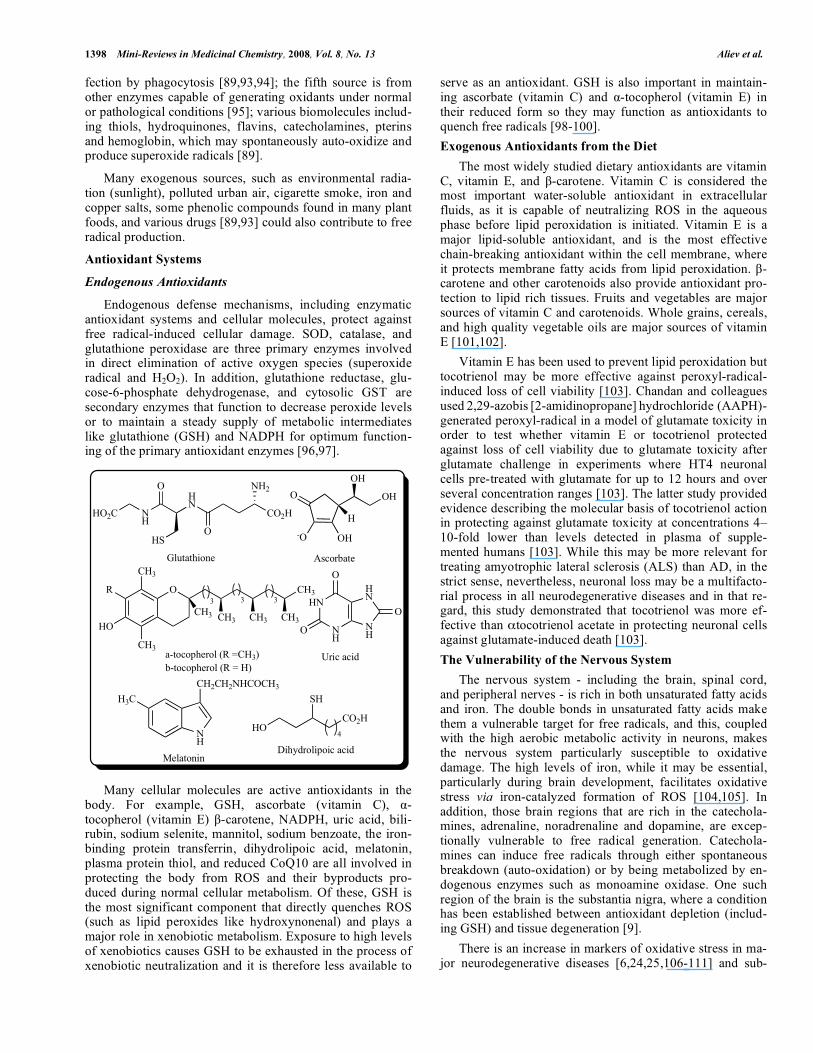
Endogenous defense mechanisms, including enzymatic antioxidant systems and cellular molecules, protect against free radical-induced cellular damage. SOD, catalase, and glutathione peroxidase are three primary enzymes involved in direct elimination of active oxygen species (superoxide radical and H2O2). In addition, glutathione reductase, glu-cose-6-phosphate dehydrogenase, and cytosolic GST are secondary enzymes that function to decrease peroxide levels or to maintain a steady supply of metabolic intermediates like glutathione (GSH) and NADPH for optimum function-ing of the primary antioxidant enzymes [96,97].
Many cellular molecules are active antioxidants in the body. For example, GSH, ascorbate (vitamin C), -tocopherol (vitamin E) -carotene, NADPH, uric acid, bili-rubin, sodium selenite, mannitol, sodium benzoate, the iron-binding protein transferrin, dihydrolipoic acid, melatonin, plasma protein thiol, and reduced CoQ10 are all involved in protecting the body from ROS and their byproducts pro-duced during normal cellular metabolism. Of these, GSH is the most significant component that directly quenches ROS (such as lipid peroxides like hydroxynonenal) and plays a major role in xenobiotic metabolism. Exposure to high levels of xenobiotics causes GSH to be exhausted in the process of xenobiotic neutralization and it is therefore less available to
serve as an antioxidant. GSH is also important in maintain-ing ascorbate (vitamin C) and -tocopherol (vitamin E) in their reduced form so they may function as antioxidants to quench free radicals [98-100].
Exogenous Antioxidants from the Diet
The most widely studied dietary antioxidants are vitamin C, vitamin E, and -carotene. Vitamin C is considered the most important water-soluble antioxidant in extracellular fluids, as it is capable of neutralizing ROS in the aqueous phase before lipid peroxidation is initiated. Vitamin E is a major lipid-soluble antioxidant, and is the most effective chain-breaking antioxidant within the cell membrane, where it protects membrane fatty acids from lipid peroxidation. -carotene and other carotenoids also provide antioxidant pro-tection to lipid rich tissues. Fruits and vegetables are major sources of vitamin C and carotenoids. Whole grains, cereals, and high quality vegetable oils are major sources of vitamin E [101,102].
Vitamin E has been used to prevent lipid peroxidation but tocotrienol may be more effective against peroxyl-radical-induced loss of cell viability [103]. Chandan and colleagues used 2,29-azobis [2-amidinopropane] hydrochloride (AAPH)-generated peroxyl-radical in a model of glutamate toxicity in order to test whether vitamin E or tocotrienol protected against loss of cell viability due to glutamate toxicity after glutamate challenge in experiments where HT4 neuronal cells pre-treated with glutamate for up to 12 hours and over several concentration ranges [103]. The latter study provided evidence describing the molecular basis of tocotrienol action in protecting against glutamate toxicity at concentrations 4–10-fold lower than levels detected in plasma of supple-mented humans [103]. While this may be more relevant for treating amyotrophic lateral sclerosis (ALS) than AD, in the strict sense, nevertheless, neuronal loss may be a multifacto-rial process in all neurodegenerative diseases and in that re-gard, this study demonstrated that tocotrienol was more ef-fective than tocotrienol acetate in protecting neuronal cells against glutamate-induced death [103].
The Vulnerability of the Nervous System
The nervous system - including the brain, spinal cord, and peripheral nerves - is rich in both unsaturated fatty acids and iron. The double bonds in unsaturated fatty acids make them a vulnerable target for free radicals, and this, coupled with the high aerobic metabolic activity in neurons, makes the nervous system particularly susceptible to oxidative damage. The high levels of iron, while it may be essential, particularly during brain development, facilitates oxidative stress via iron-catalyzed formation of ROS [104,105]. In addition, those brain regions that are rich in the catechola-mines, adrenaline, noradrenaline and dopamine, are excep-tionally vulnerable to free radical generation. Catechola-mines can induce free radicals through either spontaneous breakdown (auto-oxidation) or by being metabolized by en-dogenous enzymes such as monoamine oxidase. One such region of the brain is the substantia nigra, where a condition has been established between antioxidant depletion (includ-ing GSH) and tissue degeneration [9].
There is an increase in markers of oxidative stress in ma-jor neurodegenerative diseases [6,24,25,106-111] and sub-
O CH3
CH3 CH3 CH3CH3
CH3
HO
CH3
3 3 3
a-tocopherol (R =CH3)
b-tocopherol (R = H)
HO2C NH
HN
CO2H
O
HSO
NH2
Glutathione
OHO
OH-O
OH
H
Ascorbate
R
HN
NH
NH
HN
O
O
O
Uric acid
NH
H3C
CH2CH2NHCOCH3
Melatonin
HOCO2H
SH
4
Dihydrolipoic acid

Antioxidant Therapy in Alzheimer Disease Mini-Reviews in Medicinal Chemistry, 2008, Vol. 8, No. 13 1399
stantial evidence that oxidative stress is a cause, or at least the initial change, in the pathogenesis of AD [112].
CLINICAL EVIDENCE OF ANTIOXIDANT AME-
LIORATION
Current Clinical Drugs in Use
Therapy with acetylcholinesterase inhibitors, including drugs such as Reminyl, Aricept and Exelon, which aim to stabilize acetylcholine levels in the synaptic cleft to maintain neurotransmission, is based on the hypothesis that choliner-gic dysfunction in the process of aging contributes to the development of AD. Memantine, an NMDA receptor an-tagonist, blocks glutamate-mediated excitotoxicity. A com-plete review of drugs currently in clinical usage can be found elsewhere [113-115].
Antioxidant Therapy Development
The stages of free radical production may be arbitrarily divided into 1) conditions prior to their formation, 2) free radical formation and 3) adduction. The different types of antioxidant therapy are based on their intervention at differ-ent points in the stages of free radical formation. Summa-rized below are current strategies for developing antioxidant therapy for AD.
Prevention/Reduction of Free Radicals
Modulation of SOD, Peroxidase and Catalase or Using
Their Mimics
Transgenic mice overexpressing Cu-Zn SOD showed reduced oxidative damage in brain tissue [116] and improved cognitive function in aged rodents [117]. Overexpression of glutathione peroxidase in transgenic mice also showed anti-oxidative function and prevented homocysteine-induced en-dothelial dysfunction [118]. Moreover, the mimics of SOD and catalase have cytoprotective effects in AD model sys-tems [119] and prolong life span in C. elegans [120]. It is reported that SOD, peroxidase and catalase activity is re-duced with age and in some pathological conditions [121]. Therapy aimed at compensating for loss of activity of these enzymes is a promising approach to AD therapy.
Iron Chelators
A PP transgenic mice treated with a Cu/Zn chelator showed improvement in general parameters and a reduction of brain A deposition [122]. Because copper and zinc play a major role in A toxicity and nerve cell death via ROS gen-eration, chelator therapy is, in effect, antioxidant [123-125]. In one study, 48 presumed AD patients treated with desferri-oxamine, a transition metal chelator, (250 mg per day), showed this class of compounds to be effective in preventing AD progression [126]. Recently, desferrioxamine and others, as FDA-preapproved drugs, were shown to limit A protein secretion in cell culture [127]. More iron chelators are under investigation and show beneficial effect in AD treatment [128].
Caloric Restriction
Studies of caloric restriction in rodents show an attenua-tion of age-related deficits in learning and memory [129] and dramatically extends the life-span and reduces the incidence
of age-related disease in rodents and monkeys [130,131]. The mechanism of the beneficial effect of caloric restriction is not clearly understood but is most likely via overall reduc-tion in levels of oxidative stress, including in the brain [132]. In humans, a low daily caloric intake is associated with a reduced risk for AD [133]. In addition, the incidence of AD is lower in countries with low per capita food consumption compared to countries with high per capita food consump-tion [134,135]. Reducing caloric intake as a preventative measure in populations at high risk for AD could be com-bined with other AD treatments. The development of chemi-cal mimics for caloric restriction, such as resveratrol and sirtuins, the nicotinamide adenine dinucleotide dependent protein deacetylases [136,137], may make caloric restriction for normal people more easily attainable in the future.
Free Radicals Scavengers
Various compounds have the ability to quench free radi-cals by reacting with them directly. These compounds in-clude tocopherols (vitamin E) and other monophenols, ascor-bate (vitamin C), carotene, flavonoids and other polyphenols, glutathione (GSH/GSSH), retinol and other polyenes, vari-ous arylamines and indole derivatives such as melatonin, ebselen and other selenium-containing compounds, mimics of catalase/SOD, etc. The major antioxidants in the group of direct antioxidants are the free-radical trapping chain-breaking antioxidants, such as phenols and carotenes.
HN N
HN
NN
O
OO
HO
O
OH
OH
5
H2N5
Desferrioxamine
O CH3
5
N
Se
Ph
O
Ebselen
OH
CH3 CH3CH3
CH3
CH3
Retinol
NH
H3C
CH2CH2NHCOCH3
Melatonin
OH
HO
OH
Resveratrol

1400 Mini-Reviews in Medicinal Chemistry, 2008, Vol. 8, No. 13 Aliev et al.
Vitamin E has been found in rats to prevent neuronal cell death induced by hypoxia followed by oxygen reperfusion and to prevent neuronal damage from reactive nitrogen spe-cies [138]. Both vitamin E and -carotene, by reducing oxi-dative stress, protect rat neurons from exposure to ethanol [139]. In an experimental model of diabetes-caused neurovas-cular dysfunction, -carotene, followed by vitamins E and C, was found to effectively protect cells [140]. Vitamin E can rescue the neuronal cytotoxicity induced by aluminum in A PP transgenic mice and reduce A deposition in the brain by reducing isoprostane levels [141].
A significant amount of research with dietary vitamins E and C has been done in humans. In a multicenter, double blind, placebo-controlled study on 341 patients with moder-ately severe AD, a daily dose of approximately 1350 mg (2000 IU) vitamin E led to a slight delay of AD progression, providing the first evidence for vitamin E as a prophylaxis and treatment for AD [142]. In keeping with these results, other studies with vitamins C and E in AD patients have shown that antioxidants might have a protective effect against AD [143,144].
In one later study, 815 non-demented individuals were evaluated based on their intake of the antioxidants vitamins C and E and -carotene. Results showed that there is a sig-nificant difference in the incidence of AD between those taking vitamin E and those who are not [145]. In another large-scale study involving 5396 non-demented individuals, it was reported that high intake of vitamins C and E signifi-cantly reduced the risk of AD [146]. Similarly, in a 5-year follow-up with 1367 non-demented individuals over 65 years of age in France, Commenges and colleagues [147] found that an intake of flavonoids significantly reduced the risk of dementia.
While promising, many studies have also reported nega-tive results on the effectiveness of vitamin E and vitamin C intake as detailed in other reviews [148].
Estrogen
The general neuroprotective effects of estrogen (17 -estradiol) have been the subject of much research. Generally, estrogen acts as a trophic factor in the nervous system by altering gene transcription through interaction with its recep-tors ER and ER [149]. At the same time, in a manner more closely related to direct antioxidants, estrogen can have anti-oxidant activity independent of estrogen receptors. The structure of estrogen is similar to -tocopherol in that both molecules contain a phenolic radical scavenging moiety and a lipophilic hydrocarbon moiety. In general, phenolic A ring estrogens have been shown to be powerful inhibitors of lipid peroxidation in various cell-free test-tube experiments [150-152]. By modifying the phenolic character of estrogen 17 -estradiol, its antioxidant activities are lost, supporting the theory that estrogens are direct antioxidants because of their phenolic ring structure [153,154]. Furthermore, this antioxi-dant activity of estrogens and other phenols is strictly related to the structural prerequisites and not dependent on the inter-action of these compounds with cellular estrogen receptors [151]. Meanwhile it has been shown that estrogens are strong antioxidants in different oxidative stress-induced cell degen-eration models [153-156].
The optimum concentration of estrogen needed to achieve the antioxidant effect is worth consideration. Normally, es-trogen is present in nanomolar concentrations in vivo and, in most in vitro studies, estrogen’s antioxidant effect is achieved at a significantly higher concentration range of 1-10 moles. Therefore, it remains to be determined how the antioxidant effect of estrogen can be achieved therapeutically with a safe pharmacologic dose.
Hormone replacement therapy in the form of estrogen plus progestin, administered as a therapeutic agent in a Women’s Health Initiative (WHI) clinical trial, was recently shown to increase the risk for probable dementia in post-menopausal women aged 65 years or older [157]. The inves-tigators responsible for this study hypothesize that the nega-tive effect of estrogen and progestin may be linked to the increased risk of stroke that was also reported in the estro-gen/progestin treatment group, as the relationship between microinfarcts in the brain and susceptibility to AD is likely related, yet currently not well characterized. While this may indeed partially explain the results of the WHI clinical trial, only when the role of the other hormones of the hypotha-lamic-pituitary-gonadal axis during the climacteric years and beyond is taken into account that the results can be fully and accurately explained. It is important to recognize that the hormones of the hypothalamic-pituitary-gonadal axis have been in disequilibrium for decades in all of the women who participated in the WHI clinical trial, so if a lack of estrogen does indeed play a role in AD pathogenesis, these women have been exposed to this disease-promoting hormonal envi-ronment for years if not decades by the time the estro-gen/progestin treatment was administered. This is evidenced by the fact that reports of probable dementia appeared within the first year of the study in both the treatment and placebo groups. Therefore, it is likely predictable that the administra-tion of estrogen/progestin in these aged women was not only unable to restore the proper functioning of the hypothalamic-pituitary-gonadal axis, but that the influx of exogenous hor-mones actually served to exacerbate the disease process.
Glutathione
GSH, the most abundant intracellular non-protein thiol, is the main factor which directly quenches free radicals in vivo.It has been shown that the level of GSH is decreased in cor-tical areas and in the hippocampus of patients with AD as compared with controls [158-160]. The level of GSH in red blood cells decreases with aging and in patients with AD [161]. In the healthy cell, oxidized glutathione (GSSG) rarely exceeds 10% of total cellular GSH. Therefore, the ratio of GSH/GSSG can be used as a useful indicator for oxidative status in vivo [162]. GSH depletion may be the ultimate fac-tor determining vulnerability to oxidant attack. N-acetyl-
OHCH3
H
H H
HO
17 -Estradiol

Antioxidant Therapy in Alzheimer Disease Mini-Reviews in Medicinal Chemistry, 2008, Vol. 8, No. 13 1401
cysteine (NAC), a precursor of GSH which has already been approved by the U.S. Food and Drug Administration for treatment of acetaminophen toxicity, may be an effective strategy to increase GSH and spare brain degeneration in AD patients, although this remains to be tested.
Other Direct Antioxidants
Compounds such as serotonin (5-hydroxytryptamine), flavonoids, quercetin, and simple alkylphenols have been shown to prevent membrane lipid peroxidation and protect neuronal cells against oxidative cell death in vitro [163,164].
2,4,6-trimethylphenol (TMP) is also a potent antioxidant [151]. Additionally, being a small compound, TMP would readily cross the blood-brain barrier, thus meeting the most critical requirement for drugs used in the treatment of neu-rodegenerative disease. The protective potential of this com-pound is currently being tested experimentally in various animal models of acute neurodegeneration.
In two large clinical studies, administration of idebenone, a compound structurally similar to ubiquinone, has been re-ported to significantly reduce disease progression in a dose-dependent fashion [165,166]. Some in vivo studies in ani-mals as well as in vitro studies have demonstrated a protec-tive effect of idebenone in neuronal death [167]. More recent studies argued the effectiveness of idebenone in AD treat-ment [168,169], but another study showed that idebenone is better than tacrine in benefit-risk ratio in AD treatment [170]. Whether idebenone acts by modulating mitochondrial metabolic function or directly as a radical scavenger is still an open question. At the same time, while idebenone is mainly used to treat Friedreich ataxia, among other drugs, many compounds with multiple mechanisms of action may also be effective in treating a wide variety of neurodegenera-tive diseases.
Uric acid, an endogenous antioxidant, was also found to prevent ischemia-induced oxidative neuronal damage in rats [138]. In addition, cannabidiol is more effective than either vitamin C or E in protecting against glutamate neurotoxicity [171]. It has been demonstrated that the antioxidant activity of cannabinoid compounds is, similar to estrogens, exclu-sively dependent on the presence of a phenolic group and is independent of the cannabinoid receptor [172].
Other aromatic amino and imino compounds (e.g., phe-nothiazine, phenoxazine, iminostilbene) represent another group of direct antioxidants. Aromatic amines and imines are effective against oxidative glutamate toxicity, GSH deple-tion, and H2O2 toxicity in different cell culture systems [173]. With half-maximal effective concentrations ranging from 20-75 nM, these compounds were experimentally proven to be more effective than common standard phenolic antioxidants. These results provide a structural basis and rationale for the development of new antioxidant drugs [173,174].
Mitigating, Detoxifying or Preventing the Formation of
ROS Adducts
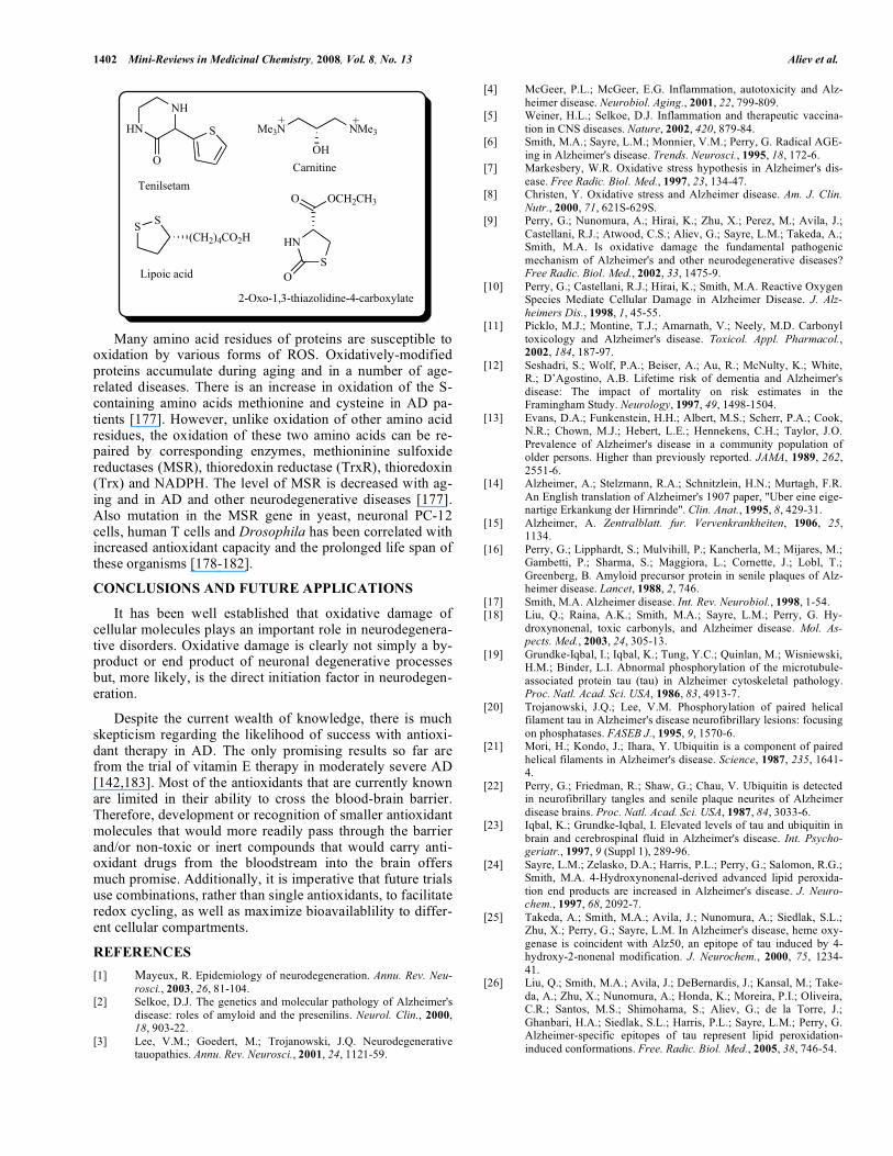
There are yet another group of compounds that can de-toxify the formed ROS adducts and repair the damage caused. These include, for example, NAC, GSH, 2-oxo-1,3-thiazoli-dine-4-carboxylate, carnitine, creatine, lipoic acid (thioctic acid), ubiquinone and idebenone.
The compound tenilsetam, a cognition-enhancing drug, is sometimes used to treat AD patients. Its mechanism of action is unclear but, based on in vitro and in vivo evidence, it is believed to inhibit protein glycation [175,176]. Since genera-tion of advanced glycation endproducts is a major manifesta-tion of oxidative stress in AD [6,108,111] glycation inhibi-tion is thought to be neuroprotective.
HSCO2H
NHCOCH3
N-Acetylcysteine
NH
NH2
HO
Serotonin
O
O
HO
OH
OH
OH
OH
Quercetin
OH
CH3
CH3
H3C
2,4,6-Trimethylphenol
O
O
(CH2)10OH
CH3
H3CO
H3CO
Idebenone
N
NH2
Tacrine
S
HN
Phenothiazine
O
HN
Phenoxazine
NH
Iminostilbene
CH3
OH
OHC5H11
Cannabidiol
1402 Mini-Reviews in Medicinal Chemistry, 2008, Vol. 8, No. 13 Aliev et al.
Many amino acid residues of proteins are susceptible to oxidation by various forms of ROS. Oxidatively-modified proteins accumulate during aging and in a number of age-related diseases. There is an increase in oxidation of the S-containing amino acids methionine and cysteine in AD pa-tients [177]. However, unlike oxidation of other amino acid residues, the oxidation of these two amino acids can be re-paired by corresponding enzymes, methioninine sulfoxide reductases (MSR), thioredoxin reductase (TrxR), thioredoxin (Trx) and NADPH. The level of MSR is decreased with ag-ing and in AD and other neurodegenerative diseases [177]. Also mutation in the MSR gene in yeast, neuronal PC-12 cells, human T cells and Drosophila has been correlated with increased antioxidant capacity and the prolonged life span of these organisms [178-182].
CONCLUSIONS AND FUTURE APPLICATIONS
It has been well established that oxidative damage of cellular molecules plays an important role in neurodegenera-tive disorders. Oxidative damage is clearly not simply a by-product or end product of neuronal degenerative processes but, more likely, is the direct initiation factor in neurodegen-eration.
Despite the current wealth of knowledge, there is much skepticism regarding the likelihood of success with antioxi-dant therapy in AD. The only promising results so far are from the trial of vitamin E therapy in moderately severe AD [142,183]. Most of the antioxidants that are currently known are limited in their ability to cross the blood-brain barrier. Therefore, development or recognition of smaller antioxidant molecules that would more readily pass through the barrier and/or non-toxic or inert compounds that would carry anti-oxidant drugs from the bloodstream into the brain offers much promise. Additionally, it is imperative that future trials use combinations, rather than single antioxidants, to facilitate redox cycling, as well as maximize bioavailablility to differ-ent cellular compartments.
REFERENCES
[1] Mayeux, R. Epidemiology of neurodegeneration. Annu. Rev. Neu-rosci., 2003, 26, 81-104.
[2] Selkoe, D.J. The genetics and molecular pathology of Alzheimer's disease: roles of amyloid and the presenilins. Neurol. Clin., 2000,18, 903-22.
[3] Lee, V.M.; Goedert, M.; Trojanowski, J.Q. Neurodegenerative tauopathies. Annu. Rev. Neurosci., 2001, 24, 1121-59.
[4] McGeer, P.L.; McGeer, E.G. Inflammation, autotoxicity and Alz-heimer disease. Neurobiol. Aging., 2001, 22, 799-809.
[5] Weiner, H.L.; Selkoe, D.J. Inflammation and therapeutic vaccina-tion in CNS diseases. Nature, 2002, 420, 879-84.
[6] Smith, M.A.; Sayre, L.M.; Monnier, V.M.; Perry, G. Radical AGE-ing in Alzheimer's disease. Trends. Neurosci., 1995, 18, 172-6.
[7] Markesbery, W.R. Oxidative stress hypothesis in Alzheimer's dis-ease. Free Radic. Biol. Med., 1997, 23, 134-47.
[8] Christen, Y. Oxidative stress and Alzheimer disease. Am. J. Clin. Nutr., 2000, 71, 621S-629S.
[9] Perry, G.; Nunomura, A.; Hirai, K.; Zhu, X.; Perez, M.; Avila, J.; Castellani, R.J.; Atwood, C.S.; Aliev, G.; Sayre, L.M.; Takeda, A.; Smith, M.A. Is oxidative damage the fundamental pathogenic mechanism of Alzheimer's and other neurodegenerative diseases? Free Radic. Biol. Med., 2002, 33, 1475-9.
[10] Perry, G.; Castellani, R.J.; Hirai, K.; Smith, M.A. Reactive Oxygen Species Mediate Cellular Damage in Alzheimer Disease. J. Alz-heimers Dis., 1998, 1, 45-55.
[11] Picklo, M.J.; Montine, T.J.; Amarnath, V.; Neely, M.D. Carbonyl toxicology and Alzheimer's disease. Toxicol. Appl. Pharmacol.,2002, 184, 187-97.
[12] Seshadri, S.; Wolf, P.A.; Beiser, A.; Au, R.; McNulty, K.; White, R.; D’Agostino, A.B. Lifetime risk of dementia and Alzheimer's disease: The impact of mortality on risk estimates in the Framingham Study. Neurology, 1997, 49, 1498-1504.
[13] Evans, D.A.; Funkenstein, H.H.; Albert, M.S.; Scherr, P.A.; Cook, N.R.; Chown, M.J.; Hebert, L.E.; Hennekens, C.H.; Taylor, J.O. Prevalence of Alzheimer's disease in a community population of older persons. Higher than previously reported. JAMA, 1989, 262,2551-6.
[14] Alzheimer, A.; Stelzmann, R.A.; Schnitzlein, H.N.; Murtagh, F.R. An English translation of Alzheimer's 1907 paper, "Uber eine eige-nartige Erkankung der Hirnrinde". Clin. Anat., 1995, 8, 429-31.
[15] Alzheimer, A. Zentralblatt. fur. Vervenkrankheiten, 1906, 25,1134.
[16] Perry, G.; Lipphardt, S.; Mulvihill, P.; Kancherla, M.; Mijares, M.; Gambetti, P.; Sharma, S.; Maggiora, L.; Cornette, J.; Lobl, T.; Greenberg, B. Amyloid precursor protein in senile plaques of Alz-heimer disease. Lancet, 1988, 2, 746.
[17] Smith, M.A. Alzheimer disease. Int. Rev. Neurobiol., 1998, 1-54. [18] Liu, Q.; Raina, A.K.; Smith, M.A.; Sayre, L.M.; Perry, G. Hy-
droxynonenal, toxic carbonyls, and Alzheimer disease. Mol. As-pects. Med., 2003, 24, 305-13.
[19] Grundke-Iqbal, I.; Iqbal, K.; Tung, Y.C.; Quinlan, M.; Wisniewski, H.M.; Binder, L.I. Abnormal phosphorylation of the microtubule-associated protein tau (tau) in Alzheimer cytoskeletal pathology. Proc. Natl. Acad. Sci. USA, 1986, 83, 4913-7.
[20] Trojanowski, J.Q.; Lee, V.M. Phosphorylation of paired helical filament tau in Alzheimer's disease neurofibrillary lesions: focusing on phosphatases. FASEB J., 1995, 9, 1570-6.
[21] Mori, H.; Kondo, J.; Ihara, Y. Ubiquitin is a component of paired helical filaments in Alzheimer's disease. Science, 1987, 235, 1641-4.
[22] Perry, G.; Friedman, R.; Shaw, G.; Chau, V. Ubiquitin is detected in neurofibrillary tangles and senile plaque neurites of Alzheimer disease brains. Proc. Natl. Acad. Sci. USA, 1987, 84, 3033-6.
[23] Iqbal, K.; Grundke-Iqbal, I. Elevated levels of tau and ubiquitin in brain and cerebrospinal fluid in Alzheimer's disease. Int. Psycho-geriatr., 1997, 9 (Suppl 1), 289-96.
[24] Sayre, L.M.; Zelasko, D.A.; Harris, P.L.; Perry, G.; Salomon, R.G.; Smith, M.A. 4-Hydroxynonenal-derived advanced lipid peroxida-tion end products are increased in Alzheimer's disease. J. Neuro-chem., 1997, 68, 2092-7.
[25] Takeda, A.; Smith, M.A.; Avila, J.; Nunomura, A.; Siedlak, S.L.; Zhu, X.; Perry, G.; Sayre, L.M. In Alzheimer's disease, heme oxy-genase is coincident with Alz50, an epitope of tau induced by 4-hydroxy-2-nonenal modification. J. Neurochem., 2000, 75, 1234-41.
[26] Liu, Q.; Smith, M.A.; Avila, J.; DeBernardis, J.; Kansal, M.; Take-da, A.; Zhu, X.; Nunomura, A.; Honda, K.; Moreira, P.I.; Oliveira, C.R.; Santos, M.S.; Shimohama, S.; Aliev, G.; de la Torre, J.; Ghanbari, H.A.; Siedlak, S.L.; Harris, P.L.; Sayre, L.M.; Perry, G. Alzheimer-specific epitopes of tau represent lipid peroxidation-induced conformations. Free. Radic. Biol. Med., 2005, 38, 746-54.
HN
NH
S
O
Tenilsetam
Me3N
OH
NMe3
++
Carnitine
SS
(CH2)4CO2H
Lipoic acidS
HN
O OCH2CH3
O
2-Oxo-1,3-thiazolidine-4-carboxylate

Antioxidant Therapy in Alzheimer Disease Mini-Reviews in Medicinal Chemistry, 2008, Vol. 8, No. 13 1403
[27] Gamblin, T.C.; Chen, F.; Zambrano, A.; Abraha, A.; Lagalwar, S.; Guillozet, A.L.; Lu, M.; Fu, Y.; Garcia-Sierra, F.; LaPointe, N.; Miller, R.; Berry, R.W.; Binder, L.I.; Cryns, V.L. Caspase cleavage of tau: linking amyloid and neurofibrillary tangles in Alzheimer's disease. Proc. Natl. Acad. Sci. USA, 2003, 100, 10032-7.
[28] Perez, M.; Hernandez, F.; Gomez-Ramos, A.; Smith, M.; Perry, G.; Avila, J. Formation of aberrant phosphotau fibrillar polymers in neural cultured cells. Eur. J. Biochem., 2002, 269, 1484-9.
[29] Lovestone, S.; McLoughlin, D.M. Protein aggregates and dementia: is there a common toxicity? J. Neurol. Neurosurg. Psychiatry,2002, 72, 152-61.
[30] Baudier, J.; Cole, D.R. In Advances in Behavioral Biology; G. Perry, Ed.; Plenum Press: New York, 1987; Vol. 34, pp. 25.
[31] Cash, A.D.; Aliev, G.; Siedlak, S.L.; Nunomura, A.; Fujioka, H.; Zhu, X.; Raina, A.K.; Vinters, H.V.; Tabaton, M.; Johnson, A.B.; Paula-Barbosa, M.; Avila, J.; Jones, P.K.; Castellani, R.J.; Smith, M.A.; Perry, G. Microtubule reduction in Alzheimer's disease and aging is independent of tau filament formation. Am. J. Pathol.,2003, 162, 1623-7.
[32] Eidenmuller, J.; Fath, T.; Hellwig, A.; Reed, J.; Sontag, E.; Brandt, R. Structural and functional implications of tau hyperphosphoryla-tion: information from phosphorylation-mimicking mutated tau proteins. Biochemistry, 2000, 39, 13166-75.
[33] Cras, P.; Smith, M.A.; Richey, P.L.; Siedlak, S.L.; Mulvihill, P.; Perry, G. Extracellular neurofibrillary tangles reflect neuronal loss and provide further evidence of extensive protein cross-linking in Alzheimer disease. Acta Neuropathol. (Berl.), 1995, 89, 291-5.
[34] Smith, M.A.; Casadesus, G.; Joseph, J.A.; Perry, G. Amyloid-beta and tau serve antioxidant functions in the aging and Alzheimer brain. Free Radic. Biol. Med., 2002, 33, 1194-9.
[35] Lee, H.G.; Perry, G.; Moreira, P.I.; Garrett, M.R.; Liu, Q.; Zhu, X.; Takeda, A.; Nunomura, A.; Smith, M.A. Tau phosphorylation in Alzheimer's disease: pathogen or protector? Trends. Mol. Med.,2005, 11, 164-9.
[36] Lee, H.G.; Zhu, X.; Nunomura, A.; Perry, G.; Smith, M.A. Curr. Alzheimer Res., 2006, 3, 75.
[37] Selkoe, D.J. Alzheimer's disease: genes, proteins, and therapy. Physiol. Rev., 2001, 81, 741-66.
[38] Ashford, J.W. APOE genotype effects on Alzheimer's disease onset and epidemiology. J. Mol. Neurosci., 2004, 23, 157-65.
[39] Teter, B. ApoE-dependent plasticity in Alzheimer's disease. J. Mol. Neurosci., 2004, 23, 167-79.
[40] Hardy, J.A.; Higgins, G.A. Alzheimer's disease: the amyloid cas-cade hypothesis. Science, 1992, 256, 184-5.
[41] Hardy, J.; Selkoe, D.J. The amyloid hypothesis of Alzheimer's disease: progress and problems on the road to therapeutics. Science,2002, 297, 353-6.
[42] Saunders, A.M.; Schmader, K.; Breitner, J.C.; Benson, M.D.; Brown, W.T.; Goldfarb, L.; Goldgaber, D.; Manwaring, M.G.; Szymanski, M.H.; McCown, N.; Roses, A. Apolipoprotein E epsi-lon 4 allele distributions in late-onset Alzheimer's disease and in other amyloid-forming diseases. Lancet, 1993, 342, 710-1.
[43] Rebeck, G.W.; Reiter, J.S.; Strickland, D.K.; Hyman, B.T. Apol-ipoprotein E in sporadic Alzheimer's disease: allelic variation and receptor interactions. Neuron, 1993, 11, 575-80.
[44] Schmechel, D.E.; Saunders, A.M.; Strittmatter, W.J.; Crain, B.J.; Hulette, C.M.; Joo, S.H.; Pericak-Vance, M.A.; Goldgaber, D.; Roses, A.D. Increased amyloid beta-peptide deposition in cerebral cortex as a consequence of apolipoprotein E genotype in late-onset Alzheimer disease. Proc Natl. Acad. Sci. U S A, 1993, 90, 9649-53.
[45] Strittmatter, W.J.; Weisgraber, K.H.; Huang, D.Y.; Dong, L.M.; Salvesen, G.S.; Pericak-Vance, M.; Schmechel, D.; Saunders, A.M.; Goldgaber, D.; Roses, A.D. Binding of human apolipopro-tein E to synthetic amyloid beta peptide: isoform-specific effects and implications for late-onset Alzheimer disease. Proc. Natl. Acad. Sci. USA, 1993, 90, 8098-102.
[46] Lauderback, C.M.; Kanski, J.; Hackett, J.M.; Maeda, N.; Kindy, M.S.; Butterfield, D.A. Apolipoprotein E modulates Alzheimer's Abeta(1-42)-induced oxidative damage to synaptosomes in an al-lele-specific manner. Brain Res., 2002, 924, 90-7.
[47] Ramassamy, C.; Averill, D.; Beffert, U.; Theroux, L.; Lussier-Cacan, S.; Cohn, J.S.; Christen, Y.; Schoofs, A.; Davignon, J.; Poirier, J. Oxidative insults are associated with apolipoprotein E genotype in Alzheimer's disease brain. Neurobiol. Dis., 2000, 7,23-37.
[48] Tanzi, R.E.; Gusella, J.F.; Watkins, P.C.; Bruns, G.A.; St George-Hyslop, P.; Van Keuren, M.L.; Patterson, D.; Pagan, S.; Kurnit, D.M.; Neve, R.L. Amyloid beta protein gene: cDNA, mRNA dis-tribution, and genetic linkage near the Alzheimer locus. Science,1987, 235, 880-4.
[49] St George-Hyslop, P.H.; Tanzi, R.E.; Polinsky R.J.; Haines J.L.; Nee L.; Watkins P.C.; Myers R.H.; Feldman R.G.; Pollen D.; Drachman D.; Growdon J.; Bruni A.; Foncin J.F.; Salmon D.; Frommelt P.; Amaducci L.; Sorbi S.; Piacenti S.; Stewart G.D.; Hobbs W.J.; Conneally P.M.; Gusella J.F. The genetic defect caus-ing familial Alzheimer's disease maps on chromosome 21. Science1987, 235, 885-90.
[50] Selkoe, D.J. Alzheimer's disease results from the cerebral accumu-lation and cytotoxicity of amyloid beta-protein. J Alzheimers Dis.,2001, 3, 75-80.
[51] Selkoe, D.J. Alzheimer's disease: genotypes, phenotypes, and treatments. Science, 1997, 275, 630-1.
[52] Bentahir, M.; Nyabi, O.; Verhamme, J.; Tolia, A.; Horre, K.; Wilt-fang, J.; Esselmann, H.; De Strooper, B. Presenilin clinical muta-tions can affect gamma-secretase activity by different mechanisms. J. Neurochem., 2006, 96, 732-42.
[53] Games, D.; Adams, D.; Alessandrini, R.; Barbour, R.; Berthelette, P.; Blackwell, C; Carr, T.; Clemens, J.; Donaldson, T.; Gillespie, F.; Guido, T.; Hagopian, S; Johnson-Wood, K.; Khan, K.; Lee, M.; Leibowitz, P.; Lieberburg, I.; Little, S.; Masliah, E.; McConlogue, L.; Montoya-Zavala, M.; Mucke, L.; Paganini, L.; Penniman, E.; Power, M.; Schenk, D.; Seubert, P.; Snyder, B.; Soriano, F.; Tan, H.; Vitale, J.; Wadsworth, S.; Wolozin, B.; Zhao, J. Alzheimer-type neuropathology in transgenic mice overexpressing V717F ß-amyloid precursor protein. Nature, 1995, 373, 523-7.
[54] Hsiao, K.; Chapman, P.; Nilsen, S.; Eckman, C.; Harigaya, Y.; Younkin, S.; Yang, F.; Cole, G. Correlative memory deficits, Abeta elevation, and amyloid plaques in transgenic mice. Science, 1996,274, 99-102.
[55] Rogaev, E.I; Sherrington, R.; Rogaeva, E.A; Levesque, G.; Ikeda, M.; Liang, Y.; Chi, H.; Lin, C.; Holman, K.; Tsuda, T.; Mar, L.; Sorbi, S.; Nacmias, B.; Piacentini, S.; Chumakov, I.; Cohen, D.; Lannfelt, L.; Fraser, P.E.; Rommens, J.M; St. George-Hyslop, P.H. Familial Alzheimer's disease in kindreds with missense mutations in a gene on chromosome 1 related to the Alzheimer's disease type 3 gene. Nature, 1995, 376, 775-8.
[56] Hardy, J. Presenilin Mutations Directory, Alzforum 2007, http:// www.alzforum.org/res/com/mut/pre/default.asp.
[57] Sherrington, R.; Rogaev, E.I.; Liang, Y.; Rogaeva, E.A.; Levesque, G.; Ikeda, M.; Chi, H.; Lin, C.; Li, G.; Holman, K.; Tsuda, T.; Mar, L.; Foncin, J.-F.; Bruni, A.C.; Montesi, M.P.; Sorbi, S.; Rainero, I.; Pinessi, L.; Nee, L.; Chumakov, I.; Pollen, D.; Brookes, A.; San-seau, P.; Polinsky, R.J.; Wasco, W.; Da Silva, H.A.R.; Haines, J.L; Pericak-Vance, M.A.; Tanzi, R.E.; Roses, A.D; Fraser, P.E.; Rom-mens, J.M.; St. George-Hyslop, P.H. Cloning of a gene bearing missense mutations in early-onset familial Alzheimer's disease. Na-ture, 1995, 375, 754-60.
[58] Czech, C.; Tremp, G.; Pradier, L. Presenilins and Alzheimer's disease: biological functions and pathogenic mechanisms. Prog. Neurobiol., 2000, 60, 363-84.
[59] Scheuner, D.; Eckman, C.; Jensen, M.; Song, X.; Citron, M.; Su-zuki, N.; Bird, T.D.; Hardy, J.; Hutton, M.; Kukull, W.; Larson, E.; Levy-Lahad, E.; Viitanen, M.; Peskind, E.; Poorkaj, P.; Schellen-berg, G.; Tanzi, R.; Wasco, W.; Lannfelt, L.; Selkoe, D.; Younkin, S. Secreted amyloid beta-protein similar to that in the senile plaques of Alzheimer's disease is increased in vivo by the presenilin 1 and 2 and APP mutations linked to familial Alzheimer's disease. Nat. Med., 1996, 2, 864-70.
[60] Whitehouse, P.J.; Price, D.L.; Clark, A.W.; Coyle, J.T.; DeLong, M.R. Alzheimer disease: evidence for selective loss of cholinergic neurons in the nucleus basalis. Ann. Neurol., 1981, 10, 122-6.
[61] Liu, Q.; Lee, H.G.; Honda, K.; Siedlak, S.L.; Harris, P.L.; Cash, A.D.; Zhu, X.; Avila, J.; Nunomura, A.; Takeda, A.; Smith, M.A.; Perry, G. Tau modifiers as therapeutic targets for Alzheimer's dis-ease. Biochem. Biophys. Acta, 2005, 1739, 211-5.
[62] Takeda, A.; Perry, G.; Abraham, N.G.; Dwyer, B.E.; Kutty, R.K.; Laitinen, J.T.; Petersen, R.B.; Smith, M.A. Overexpression of heme oxygenase in neuronal cells, the possible interaction with Tau. J. Biol. Chem., 2000, 275, 5395-9.

1404 Mini-Reviews in Medicinal Chemistry, 2008, Vol. 8, No. 13 Aliev et al.
[63] Perez, M.; Cuadros, R.; Smith, M.A.; Perry, G.; Avila, J. Phos-phorylated, but not native, tau protein assembles following reaction with the lipid peroxidation product, 4-hydroxy-2-nonenal. FEBS Lett., 2000, 486, 270-4.
[64] Zhu, X.; Rottkamp, C.A.; Boux, H.; Takeda, A.; Perry, G.; Smith, M.A. Activation of p38 kinase links tau phosphorylation, oxidative stress, and cell cycle-related events in Alzheimer disease. J. Neuro-pathol. Exp. Neurol., 2000, 59, 880-8.
[65] Zhu, X.; Castellani, R.J.; Takeda, A.; Nunomura, A.; Atwood, C.S.; Perry, G.; Smith, M.A. Differential activation of neuronal ERK, JNK/SAPK and p38 in Alzheimer disease: the 'two hit' hypothesis. Mech. Ageing. Dev., 2001, 123, 39-46.
[66] Zhu, X.; Raina, A.K.; Rottkamp, C.A.; Aliev, G.; Perry, G.; Boux, H.; Smith, M.A. Activation and redistribution of c-jun N-terminal kinase/stress activated protein kinase in degenerating neurons in Alzheimer's disease. J. Neurochem., 2001, 76, 435-41.
[67] Breteler, M.M. Vascular risk factors for Alzheimer's disease: an epidemiologic perspective. Neurobiol. Aging, 2000, 21, 153-60.
[68] Merchant, C.; Tang, M.X.; Albert, S.; Manly, J.; Stern, Y.; Mayeux, R. The influence of smoking on the risk of Alzheimer's disease. Neurology, 1999, 52, 1408-12.
[69] Ott, A.; Slooter, A.J.; Hofman, A.; van Harskamp, F.; Witteman, J.C.; Van Broeckhoven, C.; van Duijn, C.M.; Breteler, M.M. Smoking and risk of dementia and Alzheimer's disease in a popula-tion-based cohort study: the Rotterdam Study. Lancet, 1998, 351,1840-3.
[70] Plassman, B.L.; Havlik, R.J.; Steffens, D.C.; Helms, M.J.; New-man, T.N.; Drosdick, D.; Phillips, C.; Gau, B.A.; Welsh-Bohmer, K.A.; Burke, J.R.; Guralnik, J.M.; Breitner, J.C. Documented head injury in early adulthood and risk of Alzheimer's disease and other dementias. Neurology, 2000, 55, 1158-66.
[71] Nunomura, A.; Perry, G.; Pappolla, M.A.; Friedland, R.P.; Hirai, K.; Chiba, S.; Smith, M.A. Neuronal oxidative stress precedes amyloid-beta deposition in Down syndrome. J. Neuropathol. Exp. Neurol., 2000, 59, 1011-7.
[72] Stern, Y.; Gurland, B.; Tatemichi, T.K.; Tang, M.X.; Wilder, D.; Mayeux, R. Influence of education and occupation on the incidence of Alzheimer's disease. JAMA, 1994, 271, 1004-10.
[73] Hall, K.S.; Gao, S.; Unverzagt, F.W.; Hendrie, H.C. Low education and childhood rural residence: risk for Alzheimer's disease in Afri-can Americans. Neurology, 2000, 54, 95-9.
[74] Baldereschi, M.; Di Carlo, A.; Lepore, V.; Bracco, L.; Maggi, S.; Grigoletto, F.; Scarlato, G.; Amaducci, L. Estrogen-replacement therapy and Alzheimer's disease in the Italian Longitudinal Study on Aging. Neurology, 1998, 50, 996-1002.
[75] Waring, S.C.; Rocca, W.A.; Petersen, R.C.; O'Brien, P.C.; Tanga-los, E.G.; Kokmen, E. Postmenopausal estrogen replacement ther-apy and risk of AD: a population-based study. Neurology, 1999, 52,965-70.
[76] Casadesus, G.; Smith, M.A.; Zhu, X.; Aliev, G.; Cash, A.D.; Honda, K.; Petersen, R.B.; Perry, G. Alzheimer disease: evidence for a central pathogenic role of iron-mediated reactive oxygen spe-cies. J. Alzheimers. Dis., 2004, 6, 165-9.
[77] Casadesus, G.; Zhu, X.; Atwood, C.S.; Webber, K.M.; Perry, G.; Bowen, R.L.; Smith, M.A. Beyond estrogen: targeting gonadotro-pin hormones in the treatment of Alzheimer's disease. Curr. Drug Targets CNS Neurol. Disord., 2004, 3, 281-5.
[78] Casadesus, G.; Atwood, C.S.; Zhu, X.; Hartzler, A.W.; Webber, K.M.; Perry, G.; Bowen, R.L.; Smith, M.A. Evidence for the role of gonadotropin hormones in the development of Alzheimer dis-ease. Cell Mol. Life Sci., 2005, 62, 293-8.
[79] Casadesus, G.; Webber, K.M.; Atwood, C.S.; Pappolla, M.A.; Perry, G.; Bowen, R.L.; Smith, M.A. Luteinizing hormone modu-lates cognition and amyloid-beta deposition in Alzheimer APP transgenic mice. Biochim. Biophys. Acta, 2006, 1762, 447-52.
[80] Webber, K.M.; Bowen, R.; Casadesus, G.; Perry, G.; Atwood, C.S.; Smith, M.A. Gonadotropins and Alzheimer's disease: the link be-tween estrogen replacement therapy and neuroprotection. Acta Neurobiol. Exp. (Wars), 2004, 64, 113-8.
[81] Webber, K.M.; Casadesus, G.; Perry, G.; Atwood, C.S.; Bowen, R.; Smith, M.A. Gender differences in Alzheimer disease: the role of luteinizing hormone in disease pathogenesis. Alzheimer Dis. Assoc. Disord., 2005, 19, 95-9.
[82] Webber, K.M.; Casadesus, G.; Marlatt, M.W.; Perry, G.; Hamlin, C.R.; Atwood, C.S.; Bowen, R.L.; Smith, M.A. Estrogen bows to a
new master: the role of gonadotropins in Alzheimer pathogenesis. Ann. N Y Acad. Sci., 2005, 1052, 201-9.
[83] Webber, K.M.; Casadesus, G.; Zhu, X.; Obrenovich, M.E.; At-wood, C.S.; Perry, G.; Bowen, R.L.; Smith, M.A. The cell cycle and hormonal fluxes in Alzheimer disease: a novel therapeutic tar-get. Curr. Pharm. Des., 2006, 12, 691-7.
[84] Weggen, S.; Eriksen, J.L.; Das, P.; Sagi, S.A.; Wang, R.; Pietrzik, C.U.; Findlay, K.A.; Smith, T.E.; Murphy, M.P.; Bulter, T.; Kang, D.E.; Marquez-Sterling, N.; Golde, T.E.; Koo, E.H. A subset of NSAIDs lower amyloidogenic Abeta42 independently of cy-clooxygenase activity. Nature, 2001, 414, 212-6.
[85] de la Lastra, C.A.; Villegas, I. Resveratrol as an anti-inflammatory and anti-aging agent: mechanisms and clinical implications. Mol. Nutr. Food Res., 2005, 49, 405-30.
[86] Harman, D. Aging: a theory based on free radical and radiation chemistry. J. Gerontol., 1956, 11, 298.
[87] Honda, K.; Smith, M.A.; Zhu, X.; Baus, D.; Merrick, W.C.; Tar-takoff, A.M.; Hattier, T.; Harris, P.L.; Siedlak, S.L.; Fujioka, H.; Liu, Q.; Moreira, P.I.; Miller, F.P.; Nunomura, A.; Shimohama, S.; Perry, G. Ribosomal RNA in Alzheimer disease is oxidized by bound redox-active iron. J. Biol. Chem., 2005, 280, 20978-86.
[88] Allen, P.C. Production of free radical species during Eimeria maxima infections in chickens. Poult. Sci., 1997, 76, 814-21.
[89] Leibovitz, B.E.; Siegel, B.V. Aspects of free radical reactions in biological systems: aging. J. Gerontol., 1980, 35, 45-56.
[90] Harman, L.S.; Mottley, C.; Mason, R.P. Free radical metabolites of L-cysteine oxidation. J. Biol. Chem., 1984, 259, 5606-11.
[91] Priyadarsini, K.I.; Khopde, S.M.; Kumar, S.S.; Mohan, H. Free radical studies of ellagic acid, a natural phenolic antioxidant. J. Ag-ric. Food Chem., 2002, 50, 2200-6.
[92] Pierrefiche, G.; Laborit, H. Oxygen free radicals, melatonin, and aging. Exp. Gerontol., 1995, 30, 213-27.
[93] Ames, B.N.; Shigenaga, M.K.; Hagen, T.M. Oxidants, antioxidants, and the degenerative diseases of aging. Proc. Natl. Acad. Sci. USA,1993, 90, 7915-22.
[94] Beckman, K.B.; Ames, B.N. The free radical theory of aging ma-tures. Physiol. Rev., 1998, 78, 547-81.
[95] Beal, M.F. Mitochondria, oxidative damage, and inflammation in Parkinson's disease. Ann. N Y Acad. Sci., 2003, 991, 120-31.
[96] Vendemiale, G.; Grattagliano, I.; Altomare, E. An update on the role of free radicals and antioxidant defense in human disease. Int. J. Clin. Lab. Res., 1999, 29, 49-55.
[97] Singh, M.; Saini, H.K. Resident cardiac mast cells and ischemia-reperfusion injury. J. Cardiovasc. Pharmacol. Ther., 2003, 8, 135-48.
[98] Meister, A. Glutathione, ascorbate, and cellular protection. Cancer Res., 1994, 54, 1969s-1975s.
[99] Sies, H.; Stahl, W. Vitamins E and C, beta-carotene, and other carotenoids as antioxidants. Am. J. Clin. Nutr., 1995, 62, 1315S-1321S.
[100] Anderson, R.; Ramafi, G.; Theron, A.J. Membrane stabilizing, anti-oxidative interactions of propranolol and dexpropranolol with neu-trophils. Biochem. Pharmacol., 1996, 52, 341-9.
[101] Halliwell, B. Free radicals, antioxidants, and human disease: curi-osity, cause, or consequence? Lancet, 1994, 344, 721-4.
[102] Block, G. Vitamin C and cancer prevention: the epidemiologic evidence. Am. J. Clin. Nutr., 1991, 53, 270S-282S.
[103] Sen, C.K.; Khanna, S.; Roy, S.; Packer, L. Molecular basis of vita-min E action. Tocotrienol potently inhibits glutamate-induced pp60(c-Src) kinase activation and death of HT4 neuronal cells. J. Biol. Chem., 2000, 275, 13049-55.
[104] Bauer, C.; Walcher, F.; Holanda, M.; Mertzlufft, F.; Larsen, R.; Marzi, I. Antioxidative resuscitation solution prevents leukocyte adhesion in the liver after hemorrhagic shock. J. Trauma, 1999, 46,886-93.
[105] Andorn, N.; Kaufman, J.B.; Clem, T.R.; Fass, R.; Shiloach, J. Large-scale growth of Bordetella pertussis for production of ex-tracellular toxin. Ann. N Y Acad. Sci., 1990, 589, 363-71.
[106] Smith, M.A.; Richey Harris, P.L.; Sayre, L.M.; Beckman, J.S.; Perry, G. Widespread peroxynitrite-mediated damage in Alz-heimer's disease. J. Neurosci., 1997, 17, 2653-7.
[107] Smith, M.A.; Rudnicka-Nawrot, M.; Richey, P.L.; Praprotnik, D.; Mulvihill, P.; Miller, C.A.; Sayre, L.M.; Perry, G. Carbonyl-related posttranslational modification of neurofilament protein in the neu-

Antioxidant Therapy in Alzheimer Disease Mini-Reviews in Medicinal Chemistry, 2008, Vol. 8, No. 13 1405
rofibrillary pathology of Alzheimer's disease. J. Neurochem., 1995,64, 2660-6.
[108] Smith, M.A.; Taneda, S.; Richey, P.L.; Miyata, S.; Yan, S.D.; Stern, D.; Sayre, L.M.; Monnier, V.M.; Perry, G. Advanced Mail-lard reaction end products are associated with Alzheimer disease pathology. Proc. Natl. Acad. Sci. USA, 1994, 91, 5710-4.
[109] Smith, M.A.; Kutty, R.K.; Richey, P.L.; Yan, S.D.; Stern, D.; Chader, G.J.; Wiggert, B.; Petersen, R.B.; Perry, G. Heme oxy-genase-1 is associated with the neurofibrillary pathology of Alz-heimer's disease. Am. J. Pathol., 1994, 145, 42-7.
[110] Nunomura, A.; Perry, G.; Pappolla, M.A.; Wade, R.; Hirai, K.; Chiba, S.; Smith, M.A. RNA oxidation is a prominent feature of vulnerable neurons in Alzheimer's disease. J. Neurosci., 1999, 19,1959-64.
[111] Castellani, R.J.; Harris, P.L.; Sayre, L.M.; Fujii, J.; Taniguchi, N.; Vitek, M.P.; Founds, H.; Atwood, C.S.; Perry, G.; Smith, M.A. Ac-tive glycation in neurofibrillary pathology of Alzheimer disease: N(epsilon)-(carboxymethyl) lysine and hexitol-lysine. Free Radic. Biol. Med., 2001, 31, 175-80.
[112] Nunomura, A.; Perry, G.; Aliev, G.; Hirai, K.; Takeda, A.; Balraj, E.K.; Jones, P.K.; Ghanbari, H.; Wataya, T.; Shimohama, S.; Chiba, S.; Atwood, C.S.; Petersen, R.B.; Smith, M.A. Oxidative damage is the earliest event in Alzheimer disease. J. Neuropathol. Exp. Neurol., 2001, 60, 759-67.
[113] Marlatt, M.W.; Webber, K.M.; Moreira, P.I.; Lee, H.G.; Casadesus, G.; Honda, K.; Zhu, X.; Perry, G.; Smith, M.A. Therapeutic oppor-tunities in Alzheimer disease: one for all or all for one? Curr. Med. Chem., 2005, 12, 1137-47.
[114] Pietrzik, C.; Behl, C. Concepts for the treatment of Alzheimer's disease: molecular mechanisms and clinical application. Int. J. Exp. Pathol., 2005, 86, 173-85.
[115] Doody, R.S. Refining treatment guidelines in Alzheimer's disease. Geriatrics, 2005, Suppl, 14-20.
[116] Cardozo-Pelaez, F.; Song, S.; Parthasarathy, A.; Epstein, C.J.; Sanchez-Ramos, J. Attenuation of age-dependent oxidative damage to DNA and protein in brainstem of Tg Cu/Zn SOD mice. Neuro-biol. Aging, 1998, 19, 311-6.
[117] Socci, D.J.; Crandall, B.M.; Arendash, G.W. Chronic antioxidant treatment improves the cognitive performance of aged rats. Brain Res., 1995, 693, 88-94.
[118] Weiss, N.; Zhang, Y.Y.; Heydrick, S.; Bierl, C.; Loscalzo, J. Over-expression of cellular glutathione peroxidase rescues homo-cyst(e)ine-induced endothelial dysfunction. Proc. Natl. Acad. Sci. U S A, 2001, 98, 12503-8.
[119] Bruce, A.J.; Boling, W.; Kindy, M.S.; Peschon, J.; Kraemer, P.J.; Carpenter, M.K.; Holtsberg, F.W.; Mattson, M.P. Altered neuronal and microglial responses to excitotoxic and ischemic brain injury in mice lacking TNF receptors. Nat. Med., 1996, 2, 788-94.
[120] Melov, S.; Ravenscroft, J.; Malik, S.; Gill, M.S.; Walker, D.W.; Clayton, P.E.; Wallace, D.C.; Malfroy, B.; Doctrow, S.R.; Lithgow, G.J. Extension of life-span with superoxide dismutase/catalase mi-metics. Science, 2000, 289, 1567-9.
[121] Andersen, H.R.; Jeune, B.; Nybo, H.; Nielsen, J.B.; Andersen-Ranberg, K.; Grandjean, P. Low activity of superoxide dismutase and high activity of glutathione reductase in erythrocytes from cen-tenarians. Age Ageing, 1998, 27, 643-8.
[122] Cherny, R.A.; Atwood, C.S.; Xilinas, M.E.; Gray, D.N.; Jones, W.D.; McLean, C.A.; Barnham, K.J.; Volitakis, I.; Fraser, F.W.; Kim, Y.; Huang, X.; Goldstein, L.E.; Moir, R.D.; Lim, J.T.; Beyreuther, K.; Zheng, H.; Tanzi, R.E.; Masters, C.L.; Bush, A.I. Treatment with a copper-zinc chelator markedly and rapidly inhib-its beta-amyloid accumulation in Alzheimer's disease transgenic mice. Neuron, 2001, 30, 665-76.
[123] Smith, M.A.; Harris, P.L.; Sayre, L.M.; Perry, G. Iron accumula-tion in Alzheimer disease is a source of redox-generated free radi-cals. Proc. Natl. Acad. Sci. U S A, 1997, 94, 9866-8.
[124] Huang, X.; Cuajungco, M.P.; Atwood, C.S.; Hartshorn, M.A.; Tyndall, J.D.; Hanson, G.R.; Stokes, K.C.; Leopold, M.; Multhaup, G.; Goldstein, L.E.; Scarpa, R.C.; Saunders, A.J.; Lim, J.; Moir, R.D.; Glabe, C.; Bowden, E.F.; Masters, C.L.; Fairlie, D.P.; Tanzi, R.E.; Bush, A.I. Cu(II) potentiation of alzheimer abeta neurotoxic-ity. Correlation with cell-free hydrogen peroxide production and metal reduction. J. Biol. Chem., 1999, 274, 37111-6.
[125] Sayre, L.M.; Smith, M.A.; Perry, G. Chemistry and biochemistry of oxidative stress in neurodegenerative disease. Curr. Med. Chem.,2001, 8, 721-38.
[126] Crapper McLachlan, D.R.; Dalton, A.J.; Kruck, T.P.; Bell, M.Y.; Smith, W.L.; Kalow, W.; Andrews, D.F. Intramuscular desferriox-amine in patients with Alzheimer's disease. Lancet, 1991, 337,1304-8.
[127] Morse, L.J.; Payton, S.M.; Cuny, G.D.; Rogers, J.T. FDA-preapproved drugs targeted to the translational regulation and proc-essing of the amyloid precursor protein. J. Mol. Neurosci., 2004,24, 129-36.
[128] Liu, G.; Garrett, M.R.; Men, P.; Zhu, X.; Perry, G.; Smith, M.A. Nanoparticle and other metal chelation therapeutics in Alzheimer disease. Biochem. Biophys. Acta, 2005, 1741, 246-52.
[129] Ingram, D.K.; Weindruch, R.; Spangler, E.L.; Freeman, J.R.; Wal-ford, R.L. Dietary restriction benefits learning and motor perform-ance of aged mice. J. Gerontol., 1987, 42, 78-81.
[130] Sohal, R.S.; Weindruch, R. Oxidative stress, caloric restriction, and aging. Science, 1996, 273, 59-63.
[131] Weindruch, R.; Lane, M.A.; Ingram, D.K.; Ershler, W.B.; Roth, G.S. Dietary restriction in rhesus monkeys: lymphopenia and re-duced mitogen-induced proliferation in peripheral blood mononu-clear cells. Aging (Milano), 1997, 9, 304-8.
[132] Dubey, A.; Forster, M.J.; Lal, H.; Sohal, R.S. Effect of age and caloric intake on protein oxidation in different brain regions and on behavioral functions of the mouse. Arch. Biochem. Biophys., 1996,333, 189-97.
[133] Mayeux, R.; Sano, M. Treatment of Alzheimer's disease. N. Engl. J. Med., 1999, 341, 1670-9.
[134] Smith, M.A.; Petot, G.J.; Perry, G. J. Alzheimers Dis., 1999, 1, 201. [135] Grant, W.B. J. Alzheimers Dis., 1999, 1, 197. [136] Guarente, L.; Picard, F. Calorie restriction--the SIR2 connection.
Cell, 2005, 120, 473-82. [137] Delmas, D.; Jannin, B.; Latruffe, N. Resveratrol: preventing prop-
erties against vascular alterations and ageing. Mol. Nutr. Food Res.,2005, 49, 377-95.
[138] Yu, Z.F.; Bruce-Keller, A.J.; Goodman, Y.; Mattson, M.P. Uric acid protects neurons against excitotoxic and metabolic insults in cell culture, and against focal ischemic brain injury in vivo. J. Neu-rosci. Res., 1998, 53, 613-25.
[139] Copp, R.P.; Wisniewski, T.; Hentati, F.; Larnaout, A.; Ben Hamida, M.; Kayden, H.J. Localization of alpha-tocopherol trans-fer protein in the brains of patients with ataxia with vitamin E defi-ciency and other oxidative stress related neurodegenerative disor-ders. Brain Res., 1999, 822, 80-7.
[140] Mitchell, J.H.; Collins, A.R. Effects of a soy milk supplement on plasma cholesterol levels and oxidative DNA damage in men--a pi-lot study. Eur. J. Nutr., 1999, 38, 143-8.
[141] Pratico, D.; Clark, C.M.; Liun, F.; Rokach, J.; Lee, V.Y.; Troja-nowski, J.Q. Increase of brain oxidative stress in mild cognitive impairment: a possible predictor of Alzheimer disease. Arch. Neu-rol., 2002, 59, 972-6.
[142] Sano, M.; Ernesto, C.; Thomas, R.G.; Klauber, M.R.; Schafer, K.; Grundman, M.; Woodbury, P.; Growdon, J.; Cotman, C.W.; Pfeif-fer, E.; Schneider, L.S.; Thal, L.J. A controlled trial of selegiline, alpha-tocopherol, or both as treatment for Alzheimer's disease. The Alzheimer's Disease Cooperative Study. N. Engl. J. Med., 1997,336, 1216-22.
[143] Pitchumoni, S.S.; Doraiswamy, P.M. Current status of antioxidant therapy for Alzheimer's Disease. J. Am. Geriatr. Soc., 1998, 46,1566-72.
[144] Flynn, B.L.; Ranno, A.E. Pharmacologic management of Alz-heimer disease, Part II: Antioxidants, antihypertensives, and ergol-oid derivatives. Ann. Pharmacother., 1999, 33, 188-97.
[145] Morris, M.C.; Evans, D.A.; Bienias, J.L.; Tangney, C.C.; Bennett, D.A.; Aggarwal, N.; Wilson, R.S.; Scherr, P.A. Dietary intake of antioxidant nutrients and the risk of incident Alzheimer disease in a biracial community study. JAMA, 2002, 287, 3230-7.
[146] Engelhart, M.J.; Geerlings, M.I.; Ruitenberg, A.; van Swieten, J.C.; Hofman, A.; Witteman, J.C.; Breteler, M.M. Dietary intake of anti-oxidants and risk of Alzheimer disease. JAMA, 2002, 287, 3223-9.
[147] Commenges, D.; Scotet, V.; Renaud, S.; Jacqmin-Gadda, H.; Bar-berger-Gateau, P.; Dartigues, J.F. Intake of flavonoids and risk of dementia. Eur. J. Epidemiol., 2000, 16, 357-63.

1406 Mini-Reviews in Medicinal Chemistry, 2008, Vol. 8, No. 13 Aliev et al.
[148] Boothby, L.A.; Doering, P.L. Vitamin C and vitamin E for Alz-heimer's disease. Ann. Pharmacother., 2005, 39, 2073-80.
[149] Koehler, K.F.; Helguero, L.A.; Haldosen, L.A.; Warner, M.; Gustafsson, J.A. Reflections on the discovery and significance of estrogen receptor beta. Endocr. Rev., 2005, 26, 465-78.
[150] Sugioka, K.; Shimosegawa, Y.; Nakano, M. Estrogens as natural antioxidants of membrane phospholipid peroxidation. FEBS Lett.,1987, 210, 37-9.
[151] Moosmann, B.; Behl, C. The antioxidant neuroprotective effects of estrogens and phenolic compounds are independent from their es-trogenic properties. Proc. Natl. Acad. Sci. U S A, 1999, 96, 8867-72.
[152] Behl, C., [Estrogens against Alzheimer disease? The female sexual hormone as a protective neurohormone]. MMW Fortschr. Med.,2001, 143 (Suppl 2), 33-5.
[153] Behl, C.; Skutella, T.; Lezoualc'h, F.; Post, A.; Widmann, M.; Newton, C.J.; Holsboer, F. Neuroprotection against oxidative stress by estrogens: structure-activity relationship. Mol. Pharmacol.,1997, 51, 535-41.
[154] Behl, C.; Widmann, M.; Trapp, T.; Holsboer, F., 17-beta estradiol protects neurons from oxidative stress-induced cell death in vitro.Biochem. Biophys. Res. Commun., 1995, 216, 473-82.
[155] Dubal, D.B.; Kashon, M.L.; Pettigrew, L.C.; Ren, J.M.; Fin-klestein, S.P.; Rau, S.W.; Wise, P.M. Estradiol protects against ischemic injury. J. Cereb. Blood. Flow. Metab., 1998, 18, 1253-8.
[156] Goodman, Y.; Bruce, A.J.; Cheng, B.; Mattson, M.P. Estrogens attenuate and corticosterone exacerbates excitotoxicity, oxidative injury, and amyloid beta-peptide toxicity in hippocampal neurons. J. Neurochem., 1996, 66, 1836-44.
[157] Shumaker, S.A.; Legault, C.; Rapp, S.R.; Thal, L.; Wallace, R.B.; Ockene, J.K.; Hendrix, S.L.; Jones, B.N., 3rd; Assaf, A.R.; Jack-son, R.D.; Kotchen, J.M.; Wassertheil-Smoller, S.; Wactawski-Wende, J. Estrogen plus progestin and the incidence of dementia and mild cognitive impairment in postmenopausal women: the Women's Health Initiative Memory Study: a randomized controlled trial. JAMA, 2003, 289, 2651-62.
[158] Adams, J.D. Jr.; Klaidman, L.K.; Odunze, I.N.; Shen, H.C.; Miller, C.A. Alzheimer's and Parkinson's disease. Brain levels of glu-tathione, glutathione disulfide, and vitamin E. Mol. Chem. Neuro-pathol., 1991, 14, 213-26.
[159] Jenner, P. Oxidative damage in neurodegenerative disease. Lancet,1994, 344, 796-8.
[160] Lohr, J.B.; Browning, J.A. Free radical involvement in neuropsy-chiatric illnesses. Psychopharmacol. Bull., 1995, 31, 159-65.
[161] Liu, H.; Wang, H.; Shenvi, S.; Hagen, T.M.; Liu, R.M. Glutathione metabolism during aging and in Alzheimer disease. Ann. N Y Acad. Sci., 2004, 1019, 346-9.
[162] Asensi, M.; Sastre, J.; Pallardo, F.V.; Lloret, A.; Lehner, M.; Gar-cia-de-la Asuncion, J.; Vina, J. Ratio of reduced to oxidized glu-tathione as indicator of oxidative stress status and DNA damage. Methods Enzymol., 1999, 299, 267-76.
[163] Moosmann, B.; Uhr, M.; Behl, C. Neuroprotective potential of aromatic alcohols against oxidative cell death. FEBS Lett., 1997,413, 467-72.
[164] Skaper, S.D.; Fabris, M.; Ferrari, V.; Dalle Carbonare, M.; Leon, A. Quercetin protects cutaneous tissue-associated cell types includ-ing sensory neurons from oxidative stress induced by glutathione depletion: cooperative effects of ascorbic acid. Free Radic. Biol. Med., 1997, 22, 669-78.
[165] Gutzmann, H.; Hadler, D. Sustained efficacy and safety of idebe-none in the treatment of Alzheimer's disease: update on a 2-year double-blind multicentre study. J. Neural Transm. Suppl., 1998, 54,301-10.
[166] Weyer, G.; Babej-Dolle, R.M.; Hadler, D.; Hofmann, S.; Herrmann, W.M. A controlled study of 2 doses of idebenone in the
treatment of Alzheimer's disease. Neuropsychobiology, 1997, 36,73-82.
[167] Yamada, K.; Tanaka, T.; Han, D.; Senzaki, K.; Kameyama, T.; Nabeshima, T. Protective effects of idebenone and alpha-tocopherol on beta-amyloid-(1-42)-induced learning and memory deficits in rats: implication of oxidative stress in beta-amyloid-induced neurotoxicity in vivo. Eur. J. Neurosci., 1999, 11, 83-90.
[168] Pluta, R. No effect of anti-oxidative therapy on cerebral amyloido-sis following ischemia-reperfusion brain injury. Folia Neuropa-thol., 2000, 38, 188-90.
[169] Thal, L.J.; Grundman, M.; Berg, J.; Ernstrom, K.; Margolin, R.; Pfeiffer, E.; Weiner, M.F.; Zamrini, E.; Thomas, R.G. Idebenone treatment fails to slow cognitive decline in Alzheimer's disease. Neurology, 2003, 61, 1498-502.
[170] Gutzmann, H.; Kuhl, K.P.; Hadler, D.; Rapp, M.A. Safety and efficacy of idebenone versus tacrine in patients with Alzheimer's disease: results of a randomized, double-blind, parallel-group mul-ticenter study. Pharmacopsychiatry, 2002, 35, 12-8.
[171] Hampson, A.J.; Grimaldi, M.; Axelrod, J.; Wink, D. Cannabidiol and (-)Delta9-tetrahydrocannabinol are neuroprotective antioxi-dants. Proc. Natl. Acad. Sci. U S A, 1998, 95, 8268-73.
[172] Marsicano, G.; Wotjak, C.T.; Azad, S.C.; Bisogno, T.; Rammes, G.; Cascio, M.G.; Hermann, H.; Tang, J.; Hofmann, C.; Zieglgans-berger, W.; Di Marzo, V.; Lutz, B. The endogenous cannabinoid system controls extinction of aversive memories. Nature, 2002,418, 530-4.
[173] Moosmann, B.; Skutella, T.; Beyer, K.; Behl, C. Protective activity of aromatic amines and imines against oxidative nerve cell death. Biol. Chem., 2001, 382, 1601-12.
[174] Moosmann, B.; Behl, C. Cytoprotective antioxidant function of tyrosine and tryptophan residues in transmembrane proteins. Eur. J. Biochem., 2000, 267, 5687-92.
[175] Munch, G.; Taneli, Y.; Schraven, E.; Schindler, U.; Schinzel, R.; Palm, D.; Riederer, P. The cognition-enhancing drug tenilsetam is an inhibitor of protein crosslinking by advanced glycosylation. J. Neural Transm. Park Dis. Dement. Sect., 1994, 8, 193-208.
[176] Shoda, H.; Miyata, S.; Liu, B.F.; Yamada, H.; Ohara, T.; Suzuki, K.; Oimomi, M.; Kasuga, M. Inhibitory effects of tenilsetam on the Maillard reaction. Endocrinology, 1997, 138, 1886-92.
[177] Stadtman, E.R. Cyclic oxidation and reduction of methionine resi-dues of proteins in antioxidant defense and cellular regulation. Arch. Biochem. Biophys., 2004, 423, 2-5.
[178] Moskovitz, J.; Berlett, B.S.; Poston, J.M.; Stadtman, E.R. The yeast peptide-methionine sulfoxide reductase functions as an antioxidant in vivo. Proc. Natl. Acad. Sci. USA, 1997, 94, 9585-9.
[179] Moskovitz, J.; Bar-Noy, S.; Williams, W.M.; Requena, J.; Berlett, B.S.; Stadtman, E.R. Methionine sulfoxide reductase (MsrA) is a regulator of antioxidant defense and lifespan in mammals. Proc. Natl. Acad. Sci. USA, 2001, 98, 12920-5.
[180] Moskovitz, J.; Flescher, E.; Berlett, B.S.; Azare, J.; Poston, J.M.; Stadtman, E.R. Overexpression of peptide-methionine sulfoxide reductase in Saccharomyces cerevisiae and human T cells provides them with high resistance to oxidative stress. Proc. Natl. Acad. Sci. USA, 1998, 95, 14071-5.
[181] Yermolaieva, O.; Xu, R.; Schinstock, C.; Brot, N.; Weissbach, H.; Heinemann, S.H.; Hoshi, T. Methionine sulfoxide reductase A pro-tects neuronal cells against brief hypoxia/reoxygenation. Proc. Natl. Acad. Sci. USA, 2004, 101, 1159-64.
[182] Ruan, H.; Tang, X.D.; Chen, M.L.; Joiner, M.L.; Sun, G.; Brot, N.; Weissbach, H.; Heinemann, S.H.; Iverson, L.; Wu, C.F.; Hoshi, T. High-quality life extension by the enzyme peptide methionine sul-foxide reductase. Proc. Natl. Acad. Sci. U S A, 2002, 99, 2748-53.
[183] Grundman, M. Vitamin E and Alzheimer disease: the basis for additional clinical trials. Am. J. Clin. Nutr., 2000, 71, 630S-636S.
Received: 23 April, 2008 Revised: 30 September, 2008 Accepted: 08 October, 2008