
PHYSICAL THERAPY PRACTICE
68
2018 / volume 30 / number 3 ORTHOPAEDIC PHYSICAL THERAPY PRACTICE The magazine of the Orthopaedic Section, APTA
-
Upload
khangminh22 -
Category
Documents
-
view
1 -
download
0
Transcript of PHYSICAL THERAPY PRACTICE

2018 / volume 30 / number 3
ORTHOPAEDICPHYSICAL THERAPY PRACTICE
The magazine of the Orthopaedic Section, APTA

Advance Your Clinical Practice with Evidence In Motion
Teammates: ISPI | KinetaCore | NeuroRTI| www.evidenceinmotion.com | [email protected] | 1.888.709.7096
Rolling admission | Apply now!Learn more and register at www.evidenceinmotion.com
Certification in Pediatric Practice
EIM’s certificate for advanced practice in pediatric physical therapy was created for the therapist passionate about advancing their skill and evidence-based knowledge in this specialized area of physical therapy. You will learn how to effectively translate the knowledge you learn into optimal critical reasoning and application into practice for families and their children of all abilities.
Curriculum: 10-15 months online + two 2-day on-site labs
Tuition: $6,725
PTA Specialty Certification in Orthopaedics
Physical therapist assistants provide support for both the patient and the therapist as an essential member of the healthcare team. The completion of EIM’s Physical Therapy Assistant Specialty Certification in Orthopaedics will give you a detailed understanding of orthopaedic-specific knowledge and evidence-based practices, giving you another tool to guide patients toward recovery.
Curriculum: 5-7 months online + one 2-day on-site lab
Tuition: $2,675
New Program Offerings
Use code OPTP2018for discount.See below for
details.
Management Course OfferingsEIM’s management courses are designed to enhance your knowledge and skill level in the evidence-based management of individuals with lower extremity, upper extremity, lumbopelvic, and cervical and thoracic disorders. EIM’s hybrid model allows you to complete the majority of your coursework in an interactive, media-rich online format, while gaining necessary hands-on skills through a lab intensive weekend on-site. Earn your CEU hours while engaging with some of EIM’s most popular and practical content.
Management of Lower Extremity Disorders•September29-30,2018 Bakersfield,CA•September29-30,2018 SanAntonio,TX•October6-7,2018 Manahawkin,NJ•October6-7,2018 SiouxFalls,SD
Management of Upper Extremity Disorders•September29-30,2018 Bolingbrook,IL•September29-20,2018 OklahomaCity,OK•October6-7,2018 Bellevue,WI•October13-14,2018 FallsChurch,VA
Management of Lumbopelvic Disorders•August11-12,2018 Manahawkin,NJ•August11-12,2018 SiouxFalls,SD•August18-19,2018 Kissimmee,FL•October11-12,2018CedarPark,TX
Management of Cervical and Thoracic Disorders •August4-5,2018 Roseville,CA•August18-19,2018Ashburn,VA•August18-19,2018 Bellevue,WI•August18-19,2018 Ortonville,MN
Use coupon code OPTP2018 for 10% off Continuing Education Courses or a Free Application. Exclusions Apply**OPTP2018isvalidforallprogramapplications.ValidforContinuingEducationcoursesexcluding:TestPrepCourses,AlignConferenceandKinetaCorecourses.Discountisnotretroactiveandcannotbecombinedwithotherdiscounts.

®
®
135Orthopaedic Practice volume 30 / number 3 / 2018
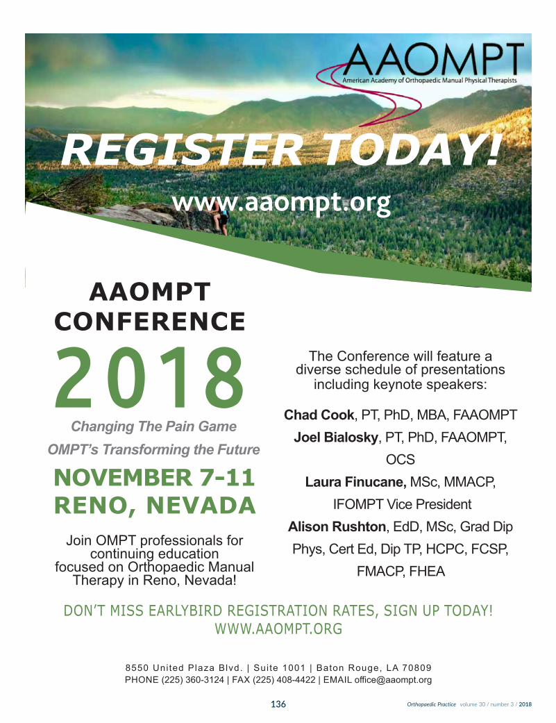
AAOMPTCONFERENCE
2 0 1 8NOVEMBER 7-11RENO, NEVADA
The Conference will feature a diverse schedule of presentations
including keynote speakers:
Chad Cook, PT, PhD, MBA, FAAOMPTJoel Bialosky, PT, PhD, FAAOMPT,
OCSLaura Finucane, MSc, MMACP,
IFOMPT Vice PresidentAlison Rushton, EdD, MSc, Grad DipPhys, Cert Ed, Dip TP, HCPC, FCSP,
FMACP, FHEA
DON’T MISS EARLYBIRD REGISTRATION RATES, SIGN UP TODAY!WWW.AAOMPT.ORG
8550 United Plaza Blvd. | Sui te 1001 | Baton Rouge, LA 70809PHONE (225) 360-3124 | FAX (225) 408-4422 | EMAIL [email protected]
Changing The Pain GameOMPT’s Transforming the Future
REGISTER TODAY! www.aaompt.org
Join OMPT professionals for continuing education
focused on Orthopaedic Manual Therapy in Reno, Nevada!
136 Orthopaedic Practice volume 30 / number 3 / 2018

In this issue142 Making a Difference: The Kenya Program Richard Jackson, Shala Cunningham, Daniel Kangutu, Martin Otieno Ong’wen, Anna Jackson
148 Pilot Study: Influence of Immobilization Period Prior to the Start of Rehabilitation on Foot Function and Foot Muscle Atrophy
Marcey Keefer Hutchison, Jeff Houck
156 Decreased Balance and Injury Risk in Adolescent Baseball Pitchers Stacia Schroeder, Sharon L. Gorman
160 Activity Assessment in Adults with Amputation Cassie M. Mitchell, Derek Crawford, Joshua A. New, Carol P. Dionne,
Susan B. Sisson
170 Complex Regional Pain Syndrome in a Female High School Athlete Jonathan E. Gallas
176 Congratulations 2018 Awardees
177 Leaders, Innovators, Changemakers: Merging Pain Science with Movement Science A Recap of AOM 2018
Regular features139 Editor’s Note
174 Wooden Book Reviews
178 Occupational Health SIG Newsletter
180 Performing Arts SIG Newsletter
184 Foot & Ankle SIG Newsletter
186 Pain Management SIG Newsletter
194 Imaging SIG Newsletter
195 Animal Rehabilitation SIG Newsletter
C3 Index to Advertisers
Orthopaedic Physical Therapy Practice (ISSN 1532-0871) is the official magazine of the Orthopaedic Section, APTA, Inc. Copyright 2018 by the Or tho paedic Sec tion, APTA. Non mem ber sub scrip tions are avail able for $50 per year (4 is sues). Opin ions ex pressed by the au thors are their own and do not nec es sar i ly re flect the views of the Or tho paedic Sec tion. The Editor re serves the right to edit manu scripts as nec es sary for pub li ca tion. All re quests for change of ad dress should be di rect ed to the Orthopaedic Section office in La Crosse.
All advertisements that ap pear in or ac com pa ny Or tho paedic Physical Therapy Prac tice are ac cept ed on the ba sis of conformation to ethical physical therapy stan dards, but acceptance does not imply endorsement by the Or tho paedic Section.
Orthopaedic Physical Therapy Practice is indexed by Cu mu la tive Index to Nursing & Allied Health Literature (CINAHL) and EBSCO Publishing, Inc.
Publication Title: Orthopaedic Physical Therapy Practice Statement of Frequency: Quarterly; January, April, July, and OctoberAuthorized Organization’s Name and Address: Orthopaedic Section, APTA, Inc., 2920 East Avenue South, Suite 200, La Crosse, WI 54601-7202
OPTP Mission
To serve as an advocate and resource for the practice of Orthopaedic Physical Therapy by fostering quality patient/client care and promoting professional growth.
Publication StaffManaging Editor & Advertising
Sharon L. KlinskiOrthopaedic Section, APTA2920 East Ave So, Suite 200La Crosse, Wisconsin 54601800-444-3982 x 2020608-788-3965 FAXEmail: [email protected]
EditorChristopher Hughes, PT, PhD, OCS
Associate EditorRita Shapiro, PT, MA, DPT
Book Review EditorRita Shapiro, PT, MA, DPT
ORTHOPAEDICPHYSICAL THERAPY PRACTICE
The magazine of the Orthopaedic Section, APTA
137Orthopaedic Practice volume 30 / number 3 / 2018

President:Stephen McDavitt, PT, DPT, MS,
FAAOMPT, FAPTA207-396-5165 • [email protected]
2nd Term: 2016-2019
Vice Pres i dent:Lori Michener, PT, PhD, SCS, ATC, FAPTA
804-828-0234 • [email protected] Term: 2017-2020
Treasurer:Kimberly Wellborn, PT, MBA
615-465-7145 • [email protected] Term: 2018-2021
Director 1:Aimee Klein, PT, DPT, DSc, OCS
813-974-6202 • [email protected] Term: 2018-2021
Director 2:Duane “Scott” Davis, PT, MS, EdD, OCS
304-293-0264 • [email protected] Term: 2016-2019
(608) 788-3982 or (800) 444-3982
Terri DeFlorian, Executive Director
x2040 ..................................................tdeflorian@orthopt.orgTara Fredrickson, Executive Associate
x2030 ......................................................... [email protected] Vogt, Executive Assistant
x2090 ......................................................... [email protected] Klinski, Managing Editor
x2020 ..................................................... [email protected] Eichmann, Publishing Assistant
x2050 ................................................ [email protected] Brenda Johnson, ICF-based CPG Coordinator
x2130 .................................................. [email protected]
AWARDSLori Michener, PT, PhD, ATC, FAPTA, SCS
(See Vice President)
Members: Kevin Gard, Marie Corkery, Murray Maitland
JOSPTGuy Simoneau, PT, PhD, FAPTA
Executive Director/Publisher: Edith Holmes
877-766-3450 • [email protected]
NOMINATIONSCarol Courtney, PT, PhD, ATC
[email protected] Term: 2018-2021
Members: Brian Eckenrode, Michael Bade
APTA BOARD LIAISON –Robert Rowe, PT, DPT, DMT, MHS
2018 House of Delegates Representative –Kathy Cieslak, PT, DSc, OCS
ICF-based CPG Editor – Guy Simoneau, PT, PhD, FAPTA
414-288-3380 • [email protected] Term: 2017-2020
ICF-based CPG Editor – Christine McDonough, PT, PhD603-653-3565 • [email protected]
2nd Term: 2016-2019
Special Interest Groups
OCCUPATIONAL HEALTH SIGLorena Pettet Payne, PT, MPA, OCS
406-581-3147 • [email protected] Term: 2016-2019
FOOT AND ANKLE SIGChristopher Neville, PT, PhD
315-464-6888 • [email protected] Term: 2016-2019
PERFORMING ARTS SIGAnnette Karim, PT, DPT, OCS, FAAOMPT
626-815-5020 ext 5072 • [email protected] Term: 2017-2020
PAIN MAN AGE MENT SIGCarolyn McManus, MSPT, MA
206-215-3176 • [email protected] Term: 2017-2020
IMAGING SIGCharles Hazle, PT, PhD
606-439-3557 • [email protected] Term: 2016-2019
ORTHOPAEDIC RESIDENCY/FELLOWSHIP SIGMatthew Haberl, PT, DPT, OCS, ATC, FAAOMPT
608-406-6335 • [email protected] Term: 2018-2021
ANIMAL REHABILITATION SIGKirk Peck, PT, PhD, CSCS, CCRT
402-280-5633 • [email protected] Term: 2016-2019
Education Interest Groups
PTAJason Oliver, PTA
OFFICERS CHAIRS
Office Personnel
MEMBERSHIPMegan Poll, PT, DPT, OCS
908-208-2321 • [email protected] Term: 2018-2021
Members: Christine Becks (student), Thomas Fliss, Molly Baker O'Rourke, Nathaniel Mosher
EDUCATION PRO GRAMNancy Bloom, PT, DPT, MSOT
314-286-1400 • [email protected] Term: 2016-2019
Vice Chair:Emmanuel “Manny” Yung, PT, MA, DPT, OCS
203-416-3953 • [email protected] Term: 2016-2019
Members: Neena Sharma, Valerie Spees, Cuong Pho, John Heick, Kate Spencer
INDEPENDENT STUDY COURSE &ORTHOPAEDIC PRACTICE
Editor:Christopher Hughes, PT, PhD, OCS, CSCS724-738-2757 • [email protected]
Term ISC: 2007-2019Term OP: 2004-2019
ISC Associate Editor:Gordon Riddle, PT, DPT, ATC, OCS, SCS
[email protected] Term: 2017-2020
OP Associate Editor:Rita Shapiro, PT, MA, DPT
[email protected] Term: 2017-2020
PUBLIC RELATIONS/MARKETINGJared Burch, PT
908-839-5506 • [email protected] Term: 2018-2021
Members: Tyler Schultz, Carol Courtney, Jared Burch (student), Adrian Miranda, William Stokes, Derek Charles, Ryan Maddrey,
Kelsea Weber (student)
RESEARCHDan White, PT, ScD, MSc, NCS
302-831-7607 • [email protected] Term: 2016-2019
Vice Chair:Amee Seitz PT, PhD, DPT, OCS
1st Term: 2016-2019
Members: Joshua Stefanik, Marcie Harris-Hayes, Sean Rundell, Arie Van Duijn, Alison Chang, Louise Thoma
ORTHOPAEDIC SPE CIAL TY COUNCILPam Kikillus, PT, DHSc, OCS, CHT, FAAOMPT
253-709-8684 • [email protected]: Expires 2019
Members: Hilary Greenberger, Grace Johnson, Judith Geller
PRACTICEKathy Cieslak, PT, DScPT, MSEd, OCS507-293-0885 • [email protected]
2nd Term: 2018-2021
Members: James Spencer, Marcia Spoto, Mary Fran Delaune, Mike Connors, Molly Malloy,
Elizabeth Bergman, Jim Dauber
FINANCEKimberly Wellborn, PT, MBA
(See Treasurer)
Members: Doug Bardugon, Penny Schulken,Judith Hess
Explore theOrthopaedic Section
website at:orthopt.org
Explore all the indepdendent study courses (ISCs) the
Orthopaedic Section has to offer at:
orthoptlearn.org
DIRECTORY
2018 / volume 30 / number 3

Our lead article this month is a first for Orthopaedic Practice. The article by Rich-ard Jackson and his colleagues highlights some work that I feel is worthy of publica-tion. Many physical therapists give unself-ishly by participating in various types of local, national, and international activities. I think it comes from within. The field draws compassionate people who not only love what they do but who are also very caring in making a difference. Often it just takes a small spark. In the lead author’s case, it was his first exposure years ago as a Peace Corps volunteer. As many private practice owners can attest, it takes an enormous amount of energy to run a business today. In Richard’s case, I thought it was very unique the way he tied in staff development to his passion for helping Kenya establish an educational program that would not only be sustainable but also of high quality. I was intrigued by his Memorandum of Understanding agree-ment that he clearly admits is built on trust and has no “legal teeth.” A shared vision and commitment along with clearly defined roles keeps things moving. In business, success tends to be measured by a tangible bottom line metric. The same can be true for this program. All one needs to know about the outcome is expressed in the words of one its
graduates, Martin Otieno Ong’wen, Physio-therapist. He expressed, “Joining the OMT program was the best thing that ever hap-pened to me as a Physiotherapist.”
There are many parts to this story that epitomize perseverance, creativity, and just a genuine need to make it work. Add in a few events of destiny, politics, and teamwork and it makes for quite a good bit of reading. The intriguing part of the whole project or idea to me was how it all progressed with just an interest to ultimately what it currently has become. The whole process replicates what we do with our patients every day. We are given a challenge to get someone better, we show a sincere desire, mobilize our skills, create a treatment plan, advocate patient compliance, and then execute toward suc-cess. What a great skill set physical therapists have. These types of “crossover” skills are what make us well-suited for service and also for being recruited as leaders in many other areas that can often be unrelated to the field of physical therapy.
So, instead of the lead article being a “how to treat” article I wanted to highlight a “how to make a difference” article. As mentioned, I applaud the many physical therapists who dedicate enormous amounts of time to serve others outside of their day jobs. Their efforts
Editor’s Note The CallingChristopher Hughes, PT, PhD, OCS, CSCS
are nothing short of heroic. Ironically, the majority who give of their time and talents will tell you they get more out of it than they feel they put into it. In a normal day of chaos, challenges, and obstacles, service work some-times can be the defining difference in under-standing why we do what we do even when our own working days grind on us. With service the commitment comes through, the need never ends, and the fulfillment only gets bigger. I would like to take this opportunity to “tip my hat” to all of you who go the extra mile in the service for others, not only with extended mission trips but even efforts on a smaller but nonetheless important scale by providing care to those who are often not even on the health care radar.
For those who have not found a cause, I encourage you to put your ear to the ground and listen. That cause may be right in front of you. Who knows what will happen once you answer it! The Jacksons planted a seed and look what happened!
Call for Candidates 2019 Election
Three positions are available for service within the Orthopaedic Section beginning February 2019. If you wish to nominate yourself or someone you know, please visit www.orthopt.org, then fill out the nomination form, and submit it to the Orthopaedic Section office: [email protected]. Deadline for nominations is September 1, 2018. Elections will be conducted during the month of November.
OPEN SECTION OFFICESPresident: Nominations are now being accepted for election to a 3-year term beginning at the close of the Orthopaedic Section Membership Meeting at CSM 2019.
Director: Nominations are now being accepted for election to a 3-year term beginning at the close of the Orthopaedic Section Membership Meeting at CSM 2019.
Nominating Committee Member: Nominations are now being accepted for election to a 3-year term beginning at the close of the Orthopaedic Section Membership Meeting at CSM 2019.
139Orthopaedic Practice volume 30 / number 3 / 2018

Learning Objectives1. Describe anatomy of the hip joint and how structure can re-
late to pathological conditions.2. Describe indications for surgical intervention and select
surgical procedures of the hip.3. Describe postoperative rehabilitation intervention tech-
niques following hip surgery.4. Know the common structures and pathomechanics involved
in knee injury.5. Describe the physical therapy guidelines, phases, and goals
for a patient who has undergone knee surgery.6. Understand the etiology of a calcaneal fracture, Lisfranc
fracture/dislocation, and an Achilles tendon rupture.7. Identify the advantages and disadvantages of surgical fi xa-
tion versus closed treatment for calcaneal fractures.8. Develop appropriate treatment plans for patients who have
sustained a calcaneal fracture, Lisfranc fracture/dislocation, or an Achilles tendon rupture.
9. Synthesize the current evidence comparing conservative care versus early surgery in different subgroups of patients with cervical and lumbar spine pain.
10. Identify the clinical fi ndings that identify patients who are most likely to benefi t from cervical or lumbar surgical inter-vention.
11. Screen and appropriately manage postoperative complica-tions for presented pathologies.
12. Develop an evidence-based rehabilitation program for pa-tients who have undergone different cervical and lumbar surgeries.
13. Integrate biomechanics and pathomechanics of the shoul-der to evaluation and treatment.
14. Implement evidence-based nonoperative treatment strate-gies for shoulder pathology.
15. Describe evidence-based rehabilitation guidelines follow-ing shoulder surgery.
16. Understand the anatomy and biomechanics of the elbow complex and how it relates to surgical interventions, tissue healing, and treatment.
17. Understand postoperative guidelines and treatment pro-gression for the elbow complex.
18. Apply appropriate patient-reported outcome measures for select surgical procedures of the hip, knee, ankle/foot, spine, shoulder, and elbow.
Editorial StaffChristopher Hughes, PT, PhD, OCS, CSCS—EditorGordon Riddle, PT, DPT, ATC, OCS, SCS, CSCS—Associate EditorSharon Klinski—Managing Editor
DescriptionThis 6-monograph course covers postoperative management for injuries and pathology of the hip, knee, ankle/foot, cervical/lum-bar spine, shoulder, and elbow. Each monograph addresses the anatomy and biomechanics of the structure, a review of select or common injuries, and nonsurgical and surgical management. Emphasis is placed on rehabilitation guidelines, precautions and contraindications to care, and also expected outcomes.
Topics and AuthorsHip—Keelan Enseki, PT, MS, OCS, SCS, ATC, CSCS; Dave Kohlrieser, PT, DPT, OCS, SCS, CSCS; Craig Mauro, MD; Michaela Kopka, MD, FRCSC; Tom Ellis, MDKnee—Michael J. Axe, MD; Lynn Synder-Mackler, PT, ScD, FAPTA; Anna Shovestul Grieder, PT, DPT, OCS; Jeff Miller, PT, DPT, OCS, SCS; Melissa Dreger, PT, DPT; Michael Palmer, PT, DPT, OCS; Tara Jo Manal, PT, DPT, OCS, SCS, FAPTAAnkle and Foot—Stephanie Albin, DPT, OCS, FAAOMPT; Mark Cornwall, PT, PhD, FAPTA; Drew H. VanBoerum, MDCervical and Lumbar Spine—Paul Reuteman, PT, DPT, MHS, OCS, ATCShoulder—Brittany Lynch, PT, DPT, SCS, OCS; Heather Christain, PT, DPT, SCS, OCS, CSCS; Christopher L. McCrum, MD; Dharmesh Vyas, MD, PhDElbow—Julia L. Burlette, PT, DPT, OCS; Amy B. Pomrantz, PT, DPT, OCS, ATC; Chris A. Sebelski, PT, PhD, OCS, CSCS; Justin M. Lantz, PT, DPT, OCS, FAAOMPT; John M. Itamura, MD
Continuing Education CreditThirty contact hours will be awarded to registrants who successfully complete the fi nal examination. The Orthopaedic Section pursues CEU approval from the following states: Nevada, Ohio, Oklahoma, California, and Texas. Registrants from other states must apply to their individual State Licensure Boards for approval of continuing education credit.
Course content is not intended for use by participants outside the scope of their license or regulation.
POSTOPERATIVE MANAGEMENT OF ORTHOPAEDIC SURGERIES
Independent Study Course 27.1
For Registration and Fees, visit orthoptlearn.org
Additional Questions—Call toll free 800/444-3982
140 Orthopaedic Practice volume 30 / number 3 / 2018
Why Are My Nerves So Sensitive?Neuroscience Education for Patients with CRPS or RSD
Help your patients with Complex Regional Pain Syndrome (CRPS) or Refl ex Sympathetic Disorder (RSD) prepare for movement of painful body parts. Using simple language and memorable metaphors, your patients will learn how the nervous system and brain work together to produce pain. By Adriaan Louw, Colleen Louw and Kory Zimney.
Educa
tion i
s The
rapy
Why Do I Hurt? A Patient Book About the Neuroscience of Pain
This neuroscience education book will help your patients understand how pain works throughout the body and how to lessen the pain by turning down nerve sensitivity. By Adriaan Louw, PT, PhD.
eBOOK
800.367.7393optp.com/pain-booksLearn more:
141Orthopaedic Practice volume 30 / number 3 / 2018

THE BEGINNING: RICHARD JACKSON, PT, OCS
It is hard to say just when the Kenya proj-ect started. Maybe 40 years ago when I spent two years there as a Peace Corps volunteer. I taught physical therapy at Kenya Medical Training College (KMTC) in Nairobi. The relationships that were formed at that time continue to have a positive influence on today’s activities.
Anna Jackson, my wife and business part-ner, and I have spent a number of years build-ing a multi-office practice. When we arrived at a point where we could think of more than business survival, we decided to look at cre-ative ways to develop our staff. What experi-ence could we create that would give them a vehicle to serve others and simultaneously make significant changes in their view of the world and their role in it? We decided to create an international volunteer experience in a developing country for our clinical staff. We wanted to help them develop and flourish in this world and in the profession while serv-ing others internationally. We also wanted to do something of long term, sustainable benefit for physical therapists in a develop-ing country and through them to affect posi-tive change in that country’s health care. We believe strongly that “when you find your way in this world, help others to find theirs.”
There was a bit of serendipity regarding the Kenya project. It began with a chance meeting at the World Congress of Physical Therapy (WCPT) conference in Amster-dam in 2011. I met a physiotherapist who was one of my students at KMTC almost 35 years earlier. He invited us to Nairobi to see if we could help in the development of a Bachelor of Science (BSc) upgrade for the diploma holders in Kenya. Kenyan physio-therapists go to school for 3 years right out of high school and receive a diploma, not a bachelor’s degree. The WCPT has deemed that the minimum standard of education for a physical therapist is the BSc degree.
During our first meeting in Nairobi with administrators at KMTC, we determined that the process of upgrading a Kenyan diploma through an American University was expensive and laborious and did not nec-essarily teach physiotherapists to be better clinicians. The Kenyans emphasized that if they could not get an American BSc, they would like a program that upgraded their skills and education. We decided to work on the BSc upgrade later and within the Kenyan educational system. Before the first meeting concluded, we developed the construct for an 18-month Orthopedic Residency Program with an emphasis on Orthopedic Manual Therapy (OMT). Classes would be two weeks long taught by American educators. We would send a lead teacher and a teaching assistant for each class. Classes would be held every 3 months and cover 6 areas of ortho-pedic study: Clinical Reasoning, Cervical/thoracic, Lumbopelvic, Shoulder/arm/hand, Knee/hip, Foot/ankle. KMTC agreed to grant a Higher (Advanced) Diploma to our OMT graduates. In addition, we would send clinical mentors to teach our students during their regular patient care. Each cohort con-sisted of approximately 20 Kenyan physio-therapists. We decided to have 2 to 3 cohorts running each year. The program launched in 2012. Finally, during the first two years we identified Kenyans who would begin as teaching assistants; they were projected to take over teaching duties by 2018. They would need to become faculty members at KMTC so discussions were started early in how to embed this program into KMTC and how budgeting would be handled.
We accomplished a lot in those first meet-ings. Normally it would take a lot longer to get so much done but the structure of The Jackson Clinics Foundation allowed us to make decisions without consulting an administrative board or committee. We are essentially self-funded and Anna and I are the only two officers in the Foundation. Our
experience in Kenya and Ethiopia points out important lessons that we have learned in developing foreign-based programs. First, time with decision makers in the country is limited. This fact required us to be able to make decisions and even improvise quickly. It is nearly impossible to get effective com-munication when you are separated by 8,000 miles. Second, it is essential to have your resources in order. Financial strength is a critical component to program development. Developing countries do not have resources to spend without going through the next budget cycle. A primary advantage we have had is that our programs have had minimal cost to the schools we have worked in. We do expect some contribution on their end, such as housing, but the primary costs have been shouldered by our Foundation.
It was essential at the outset to know who
Making A Difference: The Kenya Program
Richard Jackson, PT, OCS1
Shala Cunningham, PT, DPT, PhD, OCS, FAAOMPT2
Daniel Kangutu, PT, MSc, HDOMT3 Martin Otieno Ong’wen, PT, HDOMT4
Anna Jackson5
1President Jackson Clinics Foundation Inc., President Jackson Clinics, LP, Middleburg, VA2Assistant Professor, Doctor of Physical Therapy Program, Radford University, Roanoke, VA3HOD Physiotherapy Department, Kenya Medical Training College4Mentor, Jackson Clinics Foundation5Vice President Jackson Clinics Foundation, Vice President Jackson Clinics, LP, Middleburg, VA
Richard Jackson and students.
142 Orthopaedic Practice volume 30 / number 3 / 2018

all of the stakeholders were. It took subse-quent trips to meet with everyone but it was necessary. This took us to various levels of administration from faculty to ministry governance.
Once the project was outlined, a Mem-orandum of Understanding (MOU) was drafted and signed by all involved. This document clearly outlined who was respon-sible for what. In Kenya, KMTC agreed to supply housing, teaching space, and class recruitment. The Head of the Physiotherapy Department (HOD) would be the coordi-nator on the ground. KMTC also agreed to grant a Higher Diploma to graduates. They agreed to supply in-country transportation. The Jackson Clinics Foundation (TJCF) agreed to recruit and transport teachers, assistant teachers, and mentors. TJCF was responsible for curriculum development, tracking attendance and grades, and all nec-essary testing. An MOU is a guideline and is built on trust. There are no legal “teeth” in an MOU.
When we established our foundation, it was necessary to determine our mission, vision, philosophy, and goals. Goals needed to be specific and clearly indicate the objec-tives of each goal. The completion require-ments of participants had to be defined. The “endgame” had to be determined. Projects need to have a beginning and an end. We feel strongly that the end must be clearly sustainable or there is no point in begin-ning. We planned months in advance and realized that good outcomes can take years.
We were prepared for the long run, which meant clear expectations of our own financial commitments.
When we launched in 2012, we planned forward a number of years in advance. That included our own trips, at least 4 per year, to meet with stakeholders and students to ensure that everything was running smoothly. Programs fail when organizers are not on the ground tending to details. In addition to the above, curriculums needed to be developed and teachers needed to be recruited. There was a lot of up front work to be completed and Ben Keeton, PT, DPT, our Director of Clinic Operations, did most of this. Classes needed to be scheduled, mentors needed to be scheduled, and volunteers needed to be oriented to the country and program.
We learned something important at first contact with our Kenyan and Ethiopian stu-dents. They have consistently demonstrated a high degree of intellect and an impressive enthusiasm for learning. We realized that what was lacking was access to new ideas. The internet is still hard to come by and the inter-net does not teach an organized, programmed didactic and manual skills education. This was true for Kenya and Ethiopia. Access was all they needed to soar in their profession. They do spend their lean resources on tuition to participate in the program. Student tuition pays the teacher’s accommodations. This is a hardship for them but it also makes them personally invested. Some students are spon-sored by their employers. All students have to commit 8 weeks a year to attend courses.
This effectively eliminates any personal leave they may accrue. For at least half of the stu-dents this involves long journeys to Nairobi, finding housing, and being away from family. The program is expensive for them and requires numerous sacrifices. They gladly do this for the education they receive. I cannot express how grateful our students are for any-thing that is done for them. It is safe to say that their enthusiasm and commitment has a profound effect on our volunteers.
A key component to success has been the volunteers who have travelled on their own time to freely give of their education and skills. Without volunteers, there would not be a project. Many people have gone multiple times, often to both countries. The number of heroes in our program is too long to list here. The personal effect on our staff mem-bers has been transformational, giving them a new view on their role in this world, and new understandings regarding wealth and poverty. This has had a very positive effect on our core business. A special thanks goes to Shala Cunningham who has repeatedly travelled to Kenya to study our program out-comes. Finally, the Head of Department at the Physiotherapy school at KMTC, Daniel Kangutu Muli, has shouldered the adminis-trative burden of starting this new program without reimbursement and without com-plaint. He is committed to the advancement of the profession in Kenya. I doubt that any project like this can survive without a person “on the ground” who is committed to the success of the program.
We have just started our seventh cohort, and this year, 100% of the courses are taught by Kenyans. TJCF is sending university pro-fessors from the United States to monitor the classes and to help educate the Kenyan teach-ers in teaching methodology. The curriculum and course syllabus and schedules are pre-programmed. KMTC has hired one full time teacher for the program and will hire a second soon. We have hired a Kenyan full-time to mentor students and graduates who do not live in Nairobi. Mentorship is a key to the success of any residency and it seemed that the only way to get the people who live in the countryside mentored was to hire someone to do it. This is something that will need to be taken over by KMTC in 2019. Finally, we have recently launched a program to teach all of the other core subjects in physical therapy that are now being taught by University pro-fessors from the United States. The future of our foundation in Nairobi will be outlined by Anna Jackson later in this paper.
Daniel Kangutu and Richard Jackson.
143Orthopaedic Practice volume 30 / number 3 / 2018

PROGRAM OUTCOMES: SHALA CUNNINGHAM, PT, DPT, PHD, OCS, FAAOMPT
In order to assess if the goals of the pro-gram were being met, evidence-based out-come measurement became essential. A combination of quantitative and qualitative measures were used to perform the educa-tional program evaluation. The assessment included both learning outcomes and the participants’ perceptions. Three participant goals were chosen for the primary assessment. The goals included higher diploma graduates will (1) demonstrate the ability to examine and evaluate patients, arrive at a physical therapy diagnosis, determine prognosis, and develop interventions for patients according to the APTA Description of Specialty Prac-tice Orthopaedics, (2) report professional development and career advancement, and (3) achieve improvements in patient out-comes. In addition, barriers and facilita-tors for participation in the program were explored to allow for ongoing adaptations to the provision of the education to support stu-dent success.
To examine the OMT program’s influence on clinical reasoning development, a pre- and post-test design was used to compare the clin-ical reasoning skills of residents at the initia-tion of the residency program and following completion of the residency program. Similar to residency programs in the United States, a live patient examination was used as a clinical performance evaluation. Furthermore, inter-views of the participants explored the clinical reasoning process used during the live patient examination. The use of both an objective assessment and interview allowed for the evaluation of perceptible and imperceptible elements of clinical reasoning. Perceptible elements of clinical reasoning include the interaction of the therapist with the patient and the performance of tests and measures to determine a patient’s impairments. Impercep-tible elements include the therapist’s rationale for the choice of objective measurements and the development of hypothetical diagnoses.
To examine the perceptible elements, a live patient examination assessment tool was developed using two rounds of data col-lection for the determination of the tool’s validity and 3 rounds of data collection for determination of interrater reliability. The assessment tool was derived from the practice dimensions of the DSP Orthopaedics and used the format of the Assessment Tool for Physical Therapists: Orthopaedics provided by the American Board of Physical Therapy Specialties. The final tool included 64 items
with 3 levels of scoring: unsatisfactory per-formance, satisfactory performance, and not applicable. Not applicable was used when the assessment or treatment was not appropriate for the patient presentation. Categories on the assessment tool included examination, evaluation, diagnosis, prognosis, and inter-vention. Following the live patient examina-tion, semi-structured individual interviews with the residents explored how the resident determined the hypothetical diagnosis and the reasoning for the intervention provided.
Residents also completed a professional development and career advancement survey. The survey was based on a questionnaire cre-ated by Smith et al and Jones et al to deter-mine the impact of residency education on the professional development of residents in the United States. The survey was adapted for the Kenya residency program to assist with the interpretation of items by the resi-dents. The adapted survey included demo-graphic information and 19 items related to the residents’ professional development and career advancement. The survey used a 5-point Likert scale ranging from major posi-tive to major negative. Cronbach’s alpha for the questions regarding professional develop-ment was 0.864 and 0.712 for the questions regarding career advancement.
Interviews with the residents sought to explore barriers and facilitators for comple-tion of the program. Furthermore, past grad-uates and their employers were interviewed to explore the influence of residency training on clinical practice. The semi-structured inter-views were performed by an investigator not associated with the program’s administration to limit bias. The phenomenology approach was used to analyze the data. Information from the interviews was coded and general themes identified by the primary investiga-tor. NVivo for Mac was used to arrange codes. Thick descriptions and narratives of the participants were provided to inform the themes. To ensure credibility of the themes, member checks were also performed.
The results of each of the outcomes were triangulated to provide an overall assessment of the program. There has been a signifi-cant improvement in the residents’ clinical reasoning development from the initiation of the program until the completion of the program. During the final live patient examination, residents collected additional cues (subjective and objective) to guide their hypothetical diagnosis and interven-tions based on the patients impairments. Residents collected an average of 36.1% of the available cues in the history and exami-
nation at the initiation of the program. At graduation, the residents assessed 81.3% of available cues. Furthermore, residents dem-onstrated a significant improvement in the ability to identify relevant data, prioritization of limitations, development of a hypotheti-cal diagnosis, screening for medical referral, and selection of the intervention approach. This is directly related to cue interpretation and hypothesis generation within the hypo-thetical-deductive reasoning model. During the interviews, the residents discussed using a combination of hypothetical deductive reasoning and narrative reasoning to deter-mine a hypothetical diagnosis and develop a treatment plan that is consistent with the patient’s explanatory model for the pathol-ogy. This enabled the resident to value all of the patient’s symptoms and not the tissue response to testing alone.
On the surveys, residents noted improve-ments in professional development and career advancement similar to residents in the United States with a positive influence of the residency program on the ability to use a logical clinical reasoning process, perform a systematic clinical examination, determine the nature of the patient problem, diagnose complex patients, treat effectively to achieve projected outcomes, treat in a time efficient manner, and perform overall patient manage-ment. Dissimilarly to residents in the United States, the Kenyan residents have not con-sistently seen an improvement in salary and contributions to research have been limited to the use of evidence-based practice. During the interviews, residents noted that the resi-dency training was not accepted as formal advanced training, therefore, promotions and increases in salary were not available within the current pay structure based on highest degree earned.
Past graduates and their employers were interviewed about the impact of the program on patient care. Past graduates have noticed a decrease in patient visits per episode of care and improved patient outcomes. Graduates and employers also reported an increase in number of referral sources. Furthermore, as patient outcomes improved, residents became mentors to their colleagues by pro-viding consults for difficult patient cases. Graduates have also offered continuing edu-cation to colleagues and peers within their institutions to assist with the development of the profession within their communities.
Individual interviews with the residents sought to explore barriers and facilitators for completing the program. The residents faced multiple barriers for participation including
144 Orthopaedic Practice volume 30 / number 3 / 2018

travel to the residency site, financial costs associated with the residency, and securing time off of work in two-week increments. To offset these barriers, the residents found support through social networks including family, employers, fellow residents, and the residency program itself. The program has been able to increase the support of residents by using an onsite administrator and provid-ing additional mentoring throughout the program and following graduation.
As the program has progressed, adapta-tions based on outcome measurement has allowed for improved provision of the edu-cation and increased resident support. Con-tinued program assessment will be crucial to ensure that positive outcomes continue when the program is taught solely by Kenyan instructors associated with KMTC in 2019. Sustainability is achieved by ensuring the continued quality of the education through objective feedback to Kenyan teachers by United States based university instructors, and by supplying detailed curriculums, syl-labi, tests, course schedules, and ongoing support to the Kenyan instructors. Dedicated instructors have been hired by KMTC and ongoing mentorship of students and gradu-ates will be ensured.
THE REVOLUTION OF PHYSIO-THERAPY PRACTICE IN KENYA: DANIEL KANGUTU, PT, MSC, HDOMT, HOD PHYSIOTHERAPY DEPARTMENT KMTC
The Kenya Medical Training College (KMTC) established under an act of par-liament, is a State Corporation under the Ministry of Health entrusted with the role of training various middle level health disci-plines including Physiotherapy in the health sector.
Physiotherapy in Kenya started in 1942 in an informal apprenticeship arrangement under a renowned orthopaedic surgeon, Wil-liam H. Kirkaldy Willis. This was to mitigate the injuries and disabilities that came about as a result of the two world wars of 1914 – 1918 and 1939 – 1945. It was not until 1966 that the first formal 3-year diploma course was started with assistance of the Brit-ish Government.
The clamor to upgrade our training from diploma level to bachelor’s degree started in mid 1990s with no success. It was not until the middle of 2011 when I met Rich-ard Jackson on his way from Ethiopia and we discussed informally about upgrading Physiotherapy training in Kenya. After this first encounter my feeling was two-fold—
excitement and skepticism. First, excitement because I met someone who shared the same vision and action plan for improving Phys-iotherapy training in Kenya. Second, skep-ticism because of the challenges I imagined we would face from education administra-tors who previously had shown not one iota of willingness in supporting previous such initiatives.
When we discussed the proposal with the KMTC administration, it was taken posi-tively. The challenge was that the institution is not mandated by law to offer bachelor programs unless it collaborates with a cred-ited university. This proved to be unattain-able because of the logistics and expenses involved. Instead we opted to start Ortho-paedic Manual Therapy (OMT) as a higher diploma program that is aimed at improv-ing knowledge and clinical skills of the Physiotherapist.
Starting off, the program faced many challenges. Many Physiotherapists adopted a wait-and-see kind of attitude. This was due in part because this was an unknown program to them and secondly, they expected a Bach-elor’s degree and not another diploma. I had to use several tactics to get the first cohort to start the training.
The training brought a paradigm shift in the management of musculoskeletal condi-tions. The effects were noticeable and swept across our health care facilities leading to an increased demand for the training. Now the program attracts not only those with diploma training but also those with a Bachelor’s degree as well as a Masters. Currently there
is a clamor to decentralize the training from Nairobi and establish other centers across the country.
This program has achieved a lot in terms of the number of graduates and the out-comes of the training has been noticed by the patients/clients, employers, and the com-munity. The graduates of this program are demanding advanced training but my biggest challenge is what kind of certification would it be? Which institution would offer the cer-tification? There is a need for the law to be changed so that KMTC can offer trainings for higher qualifications other than diplomas so that such programs can progress to the highest level possible.
A GRADUATE'S PERSPECTIVE: MARTIN OTIENO ONG’WEN, PHYSIOTHERAPIST
I was almost a year out of physical therapy school and I had been practicing in a private clinic where I had some mentoring from one of the most skilled clinicians we have in Kenya. My goal at the time was to be a better clinician, to gain all the knowledge I could get so that I could be in a position to be of some help to the people who looked up to me as a medical professional. In Kenya, the autonomy of practice comes by the mere fact that one is in the health field. In short you are called a doctor regardless of the position you have in a facility. I had to always humbly decline that name at the time since I did not feel deserving.
Joining the Orthopedic Manual Therapy program gave me the courage, the meaning,
Martin Otieno, student teaching.
145Orthopaedic Practice volume 30 / number 3 / 2018

joy, and pride of calling myself a Physio-therapist. My practice as a Physio has grown tremendously, from making decisions that turned out to save people’s lives, to making their saved life worth living. I would say that has humbled me not just as a Physio but also as a person who people in need come to for help.
“I have learnt that I still have a lot to learn” Maya Angelou once said and this to me is a continuous realization. I joined the program because I wanted to learn more. I was hungry for knowledge, and looking for avenues that would help me change how I treated my patients. Physiotherapy for me at the time was getting boring and getting into a routine that did not have any tangi-ble changes for my patients. The short-term relief and feel good effect after modality based therapy was exciting, but there was little or no problem solving. I could not answer the hard question from my patients “daktari will I ever get better?” My frustration always kept me asking questions, reading, looking things up, but I still did not get that far. I did hear of the program from Mr. Daniel Kangutu, from my mum (who is a physio), and a few colleagues. I was at the time contemplating doing a Physiotherapy degree and hearing the stories about the OMT program did get me excited to join.
When I joined the program, I had no idea what was going to happen. I had been read-ing a lot of textbooks at the time on Phys-iotherapy that talked about certain processes of evaluating, assessing, and treating patients, processes that we had not quite been taught how to go about in college. I was for the most part excited to join since I had looked up the lecturers coming to teach the program and had come across some of their names on the textbooks that I did read. I was at the time really fed up of administering treatments that did not show any form of tangible improve-ment. During my clinical rotations at the outpatient department, I had wondered why patients with low back pain had such huge files. I had wondered how the treat-ment method was just the same, why nobody was questioning the process despite the lack of improvements in patient’s lives and why we still did the things we did that did not actually change anything. I had wondered why nobody was asking questions. I needed answers and my expectation was that I would get these answers in the Orthopedic Manual Therapy classroom.
I realized that this was going to be a life changing moment for me on the first day of class when I met Dr. Joe Godges and Dr.
Kevin Pozzi. They asked the exact same ques-tions that I had in my mind at the time, “why don’t our patients get better? What can we do about it?”
Joining the OMT program was the best thing that ever happened to me as a Phys-iotherapist. Meeting all sorts of experienced trainers from different parts of the United States, therapists from both academia and the clinical world, as well as those involved in research was invaluable. I got the chance to meet up to almost 50 therapists with a great deal of knowledge in the Physiother-apy world. My clinical skills shot through the roof. I became more aware of things, I started to listen more, to understand what my patients were going through. I would say I became more aware. Every moment with my patients became exciting experiences not just for myself but for them as well. They got better faster, they got educated, and I had learned the answers to their questions. Not only that, I learned different ways of answer-ing them depending on what they needed to know and how to guide them through the information. The most fulfilling aspect of it were the short texts of gratitude, the small gift cards and small gifts that I did get from them. They appreciated my care.
I developed a strong relationship with people, my communication skills got better, and I could now explain to people what a Physiotherapist is and what we do. I got con-fident and I started to teach the skills I had to other therapists I came across. I came to understand that teaching was the best way to learn. At first it was a challenge but with the continuous classes and mentorship it started looking like something I could get good at. So, I kept going to classes and felt that I could not miss any OMT offering.
At the beginning of the program (OMT) we were met with resistance, lots of resistance, from our peers. It was too much and at times I did feel like giving up. We kept pushing. Gradually, we started to gain respect from our peers and other medical professionals. I attribute this success to our patients. They spread the word that things were changing in Physiotherapy. Consequently, the OMT therapists started to get noticed. Other phys-iotherapists started to ask questions. They wanted to know where we received the train-ing. They wanted to join the team. That was when we realized that we did get more than we had bargained for. Our expectations had not only been met but exceeded. We had answers that were valuable to our patients, but also were able to earn respect of our peers and even teach them what we knew.
A year ago I got employed by the Jack-son Clinics Foundation to do mentoring for the Physiotherapists who had graduated from OMT and the OMT students working in the rural areas of Kenya. This was the turning point in my life as a Physiotherapist, the bap-tism by fire of being the first therapist to do a mentoring program in rural Kenya. It was a new step in the education system in Kenya. It was a humbling moment in my life, from the challenges it came with to the numerous joys that came later. Going to the rural areas made me realize how huge an impact the program had on patients. Our therapists could pick up potential red flags, do evaluations, and dem-onstrate sound clinical reasoning. They can design comprehensive treatment plans, all due to OMT education.
The conversations with doctors changed. There were times when Doctors did not have a clue what Physiotherapists did. Space allo-cation for Physiotherapy clinics in the hos-pitals was not a much-needed priority. That has since changed. We are starting to see interactions between therapists and doctors becoming synchronous and jointly aimed at our patients’ welfare. The impact is massive. The gap is being bridged, there is progress. Sometimes we feel it is slow, but the impact is a big one. I would say, as Professor Rob Landel told us in class, “FIGHT ON.”
The program is viewed in two ways. (1) As one of the best things that ever hap-pened to Physiotherapy in Kenya and East Africa with a little bias of my own opinion. It is appreciated so much that employers of Physiotherapists recommend that those who apply for a job be OMT trained. (2) On the other hand, it is viewed as a threat to other on-going programs related to higher learn-ing centers that offer Physiotherapy services. Regarding this I would say that “there is no positive growth that does not come without some healthy competition.”
I do not think there would have been a better program that could have changed the scope of Physiotherapy practice in Kenya. The changes are tremendous. The patients can attest to that. For example, it is getting very difficult for a therapist who is not OMT-trained to see patients who have been treated by an Orthopedic Manual Therapist. The doctors and orthopedic surgeons are starting look for OMT-trained therapists to assist in seeing their patients. The number of referrals from other specialists has grown as has the number of patient-patient referral.
146 Orthopaedic Practice volume 30 / number 3 / 2018

KENYA’S FUTURE IN PHYSIOTHERAPY: ANNA JACKSON
It is hard to discuss the future of the Kenyan program without taking a moment to reflect on how we got there. Our original vision and mission, nearly 7 years ago was to find a course of action to upgrade diploma physiotherapists in Kenya to a Bachelor’s degree. Simple, right? No, wrong! The strug-gle to get where we are and the path to where we are headed is truly a testament to our flex-ibility, perseverance, and patience.
Our vision began with a simple invitation to come to Kenya, visit the Kenya Medical Training College (KMTC), meet with a few key personnel and begin the discussion of upgrading the current 3-year physiotherapy diploma to a bachelor’s degree. It became clear that the vision of the upgrade would be met with a variety of barriers, seemingly impos-sible to overcome. So, we flexed our brains and decided to add a program for diploma graduates to learn and grow practical skills. The Higher Diploma in Orthopedic Manual Therapy (OMT) was born, consisting of 6 key courses over 18 months. Curriculums were written and volunteers from American Universities and The Jackson Clinics came to teach. An impact was and still is being made! Skills have markedly improved. Patients see and know the difference. From one to two and now 6 cohorts totaling more than 100 dedicated students have graduated from KMTC with their OMT. All the while, we have held on to the original vision, to upgrade the diploma graduates to bachelor’s degrees. Each time we have travelled to Kenya, we have continued to meet with various doc-tors, administrators, and educators to discuss some course of action to move towards a path to this upgrade. I am so pleased that this path has finally been revealed.
Last fall, Richard Jackson was on a teach-ing trip to Kenya and was told by Daniel Kangutu, KMTC’s Physiotherapy depart-ment head, that there was potential for a meeting with the acting Vice Chancellor of an organization called African Medical Research and Educational Foundation (AMREF). This meeting was intriguing because AMREF had just been granted the credentials as an International University. That said, we have been to a lot of meetings that were intriguing but have ended with empty results. Richard told Daniel, “if the guy shows up” which in Kenya is like a 50/50 chance, “come to the classroom and give me a sign, I’ll break from class and come out to meet him.” Well, the gentleman did show up, Richard entered the
room and the formalities of introduction began. The acting Vice Chancellor, intro-duced himself as Peter Ngatia. Richard gave the typical Kenyan greeting, squinted his eyes a bit and said “Peter Ngatia?” (Richard recalled that when he was a Peace Corp vol-unteer in Kenya back in the late 70s he had worked with a teacher by the same name. This individual actually worked with Richard for a year to take over the course material that Richard had written and was teaching at the time. This teacher was and is Peter Ngatia.) Peter looked back at Richard, with a thought-ful expression and replied, one matured man looking at another, “You are Richard Jackson! I wasn’t sure, so many years ago.” Of course, it is a wonderful thing to realize that this path to a bachelor's upgrade had seeds sown nearly 40 years ago.
The AMREF supports Kenya’s move-ment towards sustainable development goals under 3 pillars: training, health service deliv-ery, and health care financing. The AMREF has been sending students to various Kenyan educational institutions for years and made a decision that by opening their own university they could better control costs and the level of education provided to its students. That afternoon Peter, Richard, and Daniel began the most fruitful discussions to date. Indeed a path has been paved via AMREF’s Inter-national University (AMIU) for the KMTC physiotherapy diploma students to upgrade to a bachelor’s degree. Patience has paid off.
All of the time and energy students of the OMT program have dedicated will be considered. Graduates that have taken the
course work from our advance diploma pro-gram will have their course work accepted towards the bachelor’s degree. This degree is focused on professional development and not so much on the general education of the stu-dents. We have our work cut out for us with this wonderful development. Curriculums have been outlined (many thanks for Dr. Cheryl Footer), volunteer teachers are being called to service, and so this new direction begins! Now that we see a bachelor’s degree is possible, we believe our vision now extends to finding the path for Master’s and Doctoral degrees.
The KMTC Higher Diploma in OMT will continue to be offered. KMTC has hired one of our OMT graduates, Erastus Osewe to manage and teach in this program. This valuable program has been fully integrated into the KMTC physiotherapy program. In addition, we have lunched an educational program in Rehabilitation at KMTC that will cover the non-orthopedic areas of physi-cal therapy (neurology, pediatrics, geriatrics, cardiopulmonary, integumentary). We have also set in motion mentoring for the OMT graduates that live in the rural areas of Kenya. Martin Ong’wen, a passionate physiothera-pist and graduate of this program, is travel-ing to mentor and ensure skills taught are not forgotten.
Another truly unique program was started last year. Shenandoah University is sending rising third year DPT students to Kenya through our Foundation and KMTC to be mentored by our OMT graduates. Normally students are sent to developing countries to teach and to treat. In this case, they are being sent to be mentored and to learn. This is very exciting to our Kenyan counterparts. The tables have been turned and they are quite proud of that fact.
In July 2018, the First Annual Meeting of OMT graduates is to take place at the beauti-ful AMREF conference center. These 100 + OMTs will gather try to answer the next big question, “What is the Future of OMT?”
Truly our deepest gratitude lies with the many volunteers and students who continue to make this journey with us. Their ability to ebb and flow with the tides of challenge and change is admirable. We will continue to be flexible, patient, and persevere. Our stu-dents are our inspiration. It is through these amazing individuals that we see the differ-ence being made with patients, it is working! Teach One, Treat Many. Their future is so bright!
Peter Ngatia and Richard Jackson.
147Orthopaedic Practice volume 30 / number 3 / 2018

ABSTRACTBackground and Purpose: Ankle frac-
tures are the most common fracture and are often treated with surgery and immobiliza-tion. Severity of fractures and the course of recovery are both highly variable. Up to 36% of patients report a suboptimal outcome in function after ankle fracture. The purpose of this study was to examine if the length of immobilization or the duration of wait time prior to beginning rehabilitation was associ-ated with atrophy of the foot muscles or to the patient’s perceived function. Methods: Fifteen patients with ankle fractures com-pleted an outcome measure after terminating immobilization. Diagnostic ultrasound was used to examine the foot and leg muscles. Findings: Muscle atrophy was significant. Neither length of immobilization nor a delay in initiating treatment were strongly cor-related to foot muscle atrophy or perceived function. Clinical Relevance: A foot specific exercise program can address muscle atrophy found after ankle fracture. Conclusion: Frac-ture type and other variables are likely influ-encing muscle size and perceived function.
Key Words: ankle, fracture, intrinsic, ultrasound
BACKGROUND AND PURPOSEAnkle fractures are the most common
fracture of the foot and ankle.1 Often ankle fractures are treated with surgery followed by a period of immobilization.2,3 Their prog-nosis is mixed with some people experienc-ing a quick recovery and others resulting in long-term chronic problems.4-6 For example, at 10-year follow-up one study found that 36% of patients had a fair or poor outcome reporting impairments in overall function, pain, gait, and range of motion.5 Other stud-ies document more positive outcomes.4,6
A recent systematic review of 30 studies documented average expected recovery is 86% at 1 year.6 At the start of rehabilita-tion, usually the point when immobilization is terminated, patients average functional
ability is extremely low, 20% of normal.6
This remarkably low functional status drives many patients to seek guidance from physi-cal therapy and is thought to be connected to the length of immobilization and severity of trauma (unimalleolar, bimalleolar, or trimal-leolar fracture).6 However, a uniform presen-tation of patients relative to functional status, ankle range of motion (ROM), or strength post immobilization after an ankle fracture is not typical.7
The specific effects of immobilization are well documented,7-11 leading to expectations of greater impairments and length of recovery as a consequence of longer immobilization. In fact, reducing or minimizing immobilization is currently a research priority for patients with ankle fracture.2,3 Protocols mirror this focus varying immobilization length and weight bearing during immobilization post ankle fracture.2,3 Painter et al7 showed wide variation in immobilization length, perceived function, and dorsiflexion ROM after ankle fracture. This study was consistent with other larger studies on limitations in ROM post ankle fracture.12 While ROM is important to ankle function,13 current data suggests pain and dorsiflexion ROM may only account for a small percentage of the variance in activity limitations.14
The calf and foot musculature are also affected by the length of immobilization after ankle fracture.9-11,15 Several studies document muscle atrophy during immobilization.9-11,15 Length of immobilization, whether weight bearing is allowed or not, and fracture type (ie, severity of soft tissue damage) may influ-ence muscle atrophy. Psatha et al9 studied muscle girth during cast immobilization using magnetic resonance imaging (MRI). An MRI demonstrated total muscle volume decreases of 17% and changes in muscle fiber directions (ie, pennation angle). It is notable that atrophy slowed when partial weight bearing was initiated. Shaffer et al10 found subjects immobilized for 8 weeks demon-strated decreased plantar flexion peak torque on cast removal. In another study, muscle
hypertrophy after immobilization resembled controls following a period of rehabilita-tion; however, plantar flexion torque defi-cits remained. Unexplored in these studies is the influence of fracture type and fixation methods. Surprisingly, a high level study, suggests no benefit to supervised exercise programs for uncomplicated ankle fractures over advice alone.16 Improved understanding of the influence of fracture (type) and treat-ment variables (length of immobilization) may improve understanding of when reha-bilitation holds strong benefits for patients. For example, fractures of the fibula may have preferential effects on the fibularis longus and brevis muscles. And, longer periods of immo-bilization may affect more distal foot muscles (intrinsic/extrinsic) as well as ankle plantar flexors. Foot muscle atrophy is of interest because new studies note foot muscle func-tion influences ankle plantar flexion func-tion, a major muscle group for walking.17-19
While ankle fractures constitute a wide range of injuries and prognosis, if impair-ments such as ROM and muscle atrophy are primarily the result of immobilization, their severity at initiation of physical therapy would be related to the length of immobiliza-tion. Further, rehabilitation after ankle frac-ture is not always recommended despite the significant loss of function.16 The influence of a delay in initiating rehabilitation is unclear. While advice is as effective as supervised therapy for some patients, delays in starting rehabilitation may prolong disability in other patients.16 Understanding how the length of immobilization and delay in initiating treat-ment after immobilization influence patient status would be useful from a clinical view point as variables separate from trauma (ie, fracture type).
The purpose of this study was to exam-ine if the length of immobilization or the delay after termination of immobilization in initiating rehabilitation was associated with muscle atrophy specific to foot muscles and perceived foot function (ie, Foot and Ankle Ability Measure [FAAM]). The ini-
Pilot Study: Influence of Immobilization Period Prior to the Start of Rehabilitation on Foot Function and Foot Muscle Atrophy
1Associate Professor, School of Physical Therapy, George Fox University, Newberg, OR2Director of Research, School of Physical Therapy, George Fox University, Newberg, OR
Marcey Keefer Hutchison, DPT, SCS, ATC, CMP1
Jeff Houck, PT, PhD2
148 Orthopaedic Practice volume 30 / number 3 / 2018

tial hypothesis were (1) that muscle size and foot function would decrease as the length of immobilization increased (length of immo-bilization would be negatively correlated to foot muscle atrophy and FAAM); (2) that muscle atrophy and foot function would be negatively influenced by delay in initiat-ing rehabilitation (delay initiating physical therapy would be negatively correlated to foot muscle atrophy and FAAM); (3) that foot muscle size would be significantly lower in the involved versus the uninvolved side at the start of rehabilitation; and, (4) that fibu-laris muscle size would be significantly lower specific to patients that had ankle fractures involving the fibula.
METHODSSubjects
A convenience sample of 15 patients (4 male, 11 female) with ankle fractures were recruited using flyers placed in orthopedic clinics and by word of mouth. The general inclusion criteria were: (1) 18 to 65 years of age, (2) diagnosis of an ankle fracture (any type), and (3) as soon as possible follow-ing discontinuation of their immobilization device. Patients were screened by a physical therapist for exclusion criteria consisting of (1) musculoskeletal or nervous system inju-ries of the uninvolved limb over the last year that would affect either lower extremity during walking, and (2) diabetic neuropathy affecting the foot.
Foot and Ankle Ability MeasurePatients were asked to rate their current
function using the Foot and Ankle Ability Measure (FAAM). The FAAM is a region-specific, patient-reported outcome measure used to assess physical performance among individuals with a range of leg, foot, and ankle musculoskeletal disorders.20,21 It is comprised of a 21-item activities of daily living subscale. Patients’ score the FAAM using a Likert scale. Scores are tallied with a higher score repre-senting a higher level of ability. The FAAM has been determined to be a reliable and valid measure of physical function in patients with impairments of the leg, ankle, and foot.20,21
Muscle Size Measures Diagnostic ultrasound is a validated
approach to documenting changes in foot muscle size.22,23 A diagnostic ultrasound unit (Philips Affinity model 50G, Phillips, Inc. N.V) with a 50-millimeter linear array probe was used to collect abductor hallucis cross sectional area (CSA) and fibularis longus/brevis muscles (Figure 1). Consistent with
published data,22,23 the cross section of the abductor hallucis muscle was taken perpen-dicular to the anterior/posterior axis of the foot just anterior to the medial malleolus. For the fibularis muscles a cross section was obtained midway from the fibula head to the medial malleoli similar to other studies.22,23
Although this does not distinguish between fibularis longus and brevis, this is a valid measure for their combined CSA.22 Previous studies have used ultrasound (US) images of these muscles in controls and subjects with flatfoot.22-25 After adjusting for magni-fication (NIH ImageJ), circumference was traced manually and CSA recorded. Despite preliminary testing on normal subjects, in 5 patients the US examiner was unable to cap-ture an adequate image of abductor halluces muscle. And, for practical reasons, inclusion of the fibularis longus/brevis muscle was only included in 5 patients.
Statistical Analysis
Descriptive analysis of the patient data, length of immobilization, delay from when immobilization ended and the initial physi-cal therapy appointment (delay in initiat-ing physical therapy), FAAM and muscle size variables were tabulated. To examine relationships between length immobiliza-
tion (hypothesis #1) and delay in initiating physical therapy (hypothesis #2) Pearson correlation coefficients were first considered. When there was no relationship, scatter plots were examined visually to examine the indi-vidual data points. Even though the sample size was small (n=15), there was sufficient variability in the data to find strong correla-tions (r>0.65) if one existed. To evaluate the degree of muscle atrophy, the uninvolved and involved side abductor hallucis CSA was compared using a paired t-test.
FINDINGSFifteen consecutive patients with ankle
fractures were included in this study (Table 1). Four were male and 11 female; ages 18-63 years with a mean age of 36.3 years. The mean body mass index (BMI) was 27.1 with a range of 22.9-40.6. Five patients were classified as having a normal BMI, 8 were classified as overweight, and 2 were classified as obese. Patients were enrolled in the study a mean of 17.5 days after discontinuing use of an immo-bilization device. The range of days after dis-continuing immobilization was 1-121. None of the patients had received physical therapy or other restorative interventions at the time of their enrollment into the study.
All patients reported a traumatic event
* *
Figure 1. A, Cross sectional area fibularis uninvolved limb subject 2. Arrows indicate margins of muscle, *fibula. B, Cross sectional area fibularis involved limb subject 2. Arrows indicate margins of muscle, *fibula. C, Cross sectional area of abductor hallucis muscle uninvolved side, subject 1. Arrows indicate margins of muscle. D, Cross sectional area of abductor hallucis muscle involved side, subject 1. Arrows indicate margins of muscle.
149Orthopaedic Practice volume 30 / number 3 / 2018

resulting in a fracture (see Table 1). The majority of the mechanisms of injury involved sports (8) and inclines/stairs (4). The fibula was the most frequently injured bone. All patients were immobilized after sustaining their injury. Open reduction with internal fixation was used to treat fractures in 10 patients; 1 patient had a reconstruction of the lateral retinaculum; 4 had casting without surgery. The total length of immobilization (Table 2) ranged from 35 to 189 days with a mean of 69.5 days. Excluding one patient with an exceptionally long immobilization (189 days), the mean length of immobiliza-tion was 61 days. The mean duration of non-weight-bearing time was 45.5 days (35-56 days). The mean duration of partial weight-bearing time was 20.9 days (1-77 days).
The FAAM was completed by all patients and was used to assess their self-perceived functional capacity related to their ankle fracture episode. Scores ranged from 16% to 82% with the mean score being 51.1% (see Table 2). Excluding the outlier previously mentioned, the range was 20% to 82% with a mean score of 53.6%.
Delay in initiation of physical therapy but not length of immobilization showed a moderate correlation to perceived function (Figure 2). There was no strong correlation between immobilization length and per-ceived function (r=-0.2, 0.47). The scatter plot showed no clear trend (r=0.37, p=0.19) even with the outlier removed (length of
immobilization = 189) (Figure 2). There was no correlation between delay in initiation of physical therapy and perceived function (r=-0.32, p=0.24). However, the scatter plot showed one outlier (delay in initiation of rehabilitation = 121 days) (Figure 2). When this value was removed, there was a moder-ate relationship between delay in initiation of physical therapy and perceived function (r=0.51, p = 0.06).
The CSA of the abductor hallucis and fibularis muscles (brevis and longus in com-bination) were significantly lower on the involved side (Table 3, Figure 1). There was a significant difference in abductor muscle size (n=9) between the uninvolved and involved limb (p < 0.01) (Figure 3). Expressed as a percent, the abductor hallucis (n=9) dem-onstrated a mean of 28.5% less CSA on the involved side compared to the uninvolved with a range of 3.1% to 49.9%. However, abductor halluces muscle atrophy (n=9) was not correlated with the length of immobili-zation (r=-0.24, p=0.54) or the delay in initi-ation of mobilization (r=0.49, p=0.18). The scatter plots showed no outliers (Figure 4). A post hoc analysis showed that to determine a significant correlation of r=0.49 a sample of 30 subjects would be needed. Fibularis muscle CSA (n=5) was also significantly different from uninvolved to involved side (p<0.01) (Figure 5). Expressed as a percent, the fibularis (longus and brevis combined) had a mean score of 33.1% less CSA on
the involved side with a range of 17.3% to 47.3%.
CLINICAL RELEVANCE
The findings of this pilot data are that length of immobilization is likely not strongly correlated to perceived function or foot muscle atrophy. This study examines a diverse set of patient with a diagnosis of ankle fracture similar to a previous study.7
Although length of immobilization varied (35 to 189 days), it did not correlate strongly with perceived function or muscle atrophy (see Figures 2 and 3). Although this data is not powered to detect more moderate cor-relations, this data suggests that the correla-tions may not be sufficiently strong enough to be of use clinically. Other features such as trauma and type of fracture may also be relevant as well as patient behaviors (ie, activ-ity levels) (Table 1). Somewhat unexpectedly, there was conflicting data on the influence of a delay in initiating physical therapy. Patients that delayed initiating rehabilitation showed a trend toward improved perceived foot func-tion (Figure 2). However, muscle atrophy showed a trend toward increasing with longer delays to initiation of rehabilitation (Figure 3). Although this pilot data is not conclusive, it highlights the likely weak association of length of immobilization and delays of ini-tiating treatment on perceived function and muscle atrophy in a pragmatic sample. It also raises the possibility that the more important
Patient Age Sex Height Weight Mechanism of Injury Fracture Description (Year) (cm) (kg)
1 18 F 157.48 56.70 horse fell on top of her distal fibular fracture
2 43 M 177.80 86.18 waterskiing fracture talus, peroneal retinaculum tear
3 63 F 154.94 97.52 slipped on step ankle dislocation, fibular fracture, deltoid sprain
4 54 F 167.64 72.57 twisted on stairs tibia/fibula fracture
5 38 F 162.56 77.11 slipped on ramp Maisonneuve fracture
6 50 F 167.64 68.03 stepped into hole talar fracture
7 26 M 180.34 74.84 landing from high bar deltoid ligament sprain grade 3
8 17 F 160.02 61.23 slid into 3rd base distal fibular fracture
9 20 F 182.88 92.99 stepped off bike fibular fracture
10 35 F 165.10 70.31 fainted & fell down distal fibular fracture
11 46 M 180.34 83.91 jumped in river Lis Franc fracture
12 21 F 157.48 81.19 fell off truck bed trimalleolar fracture w/dislocation
13 63 F 160.02 62.60 slipped while hiking fibular fracture, deltoid sprain
14 47 M 180.34 89.81 cutting wood fibula fracture
15 48 F 170.18 72.57 fell from horse open tibia/fibula fracture
Abbreviations: cm, centimeters; F, female; kg, kilograms; M, male
Table 1. Patient Demographics and Injury Descriptors
150 Orthopaedic Practice volume 30 / number 3 / 2018

factor may be ankle fracture type (ie, severity of injury) or other management issues.
The immobilization length varied more than in previous studies, however, it was not associated with perceived function or muscle atrophy. Six of our 15 patients were immo-bilized for a period of 63 days or greater (63-189 day range, mean 105 days) (Table 2). The longest immobilization period (189 days) was an open fracture associated with osteomyelitis (outlier). This data was consis-tent with marked deficits in perceived func-tion.6 Length of immobilization, although varied did not influence perceived function in this small sample (see Figure 2). Similarly, muscle atrophy did not show a correlation with length of immobilization (see Figure 3). Other studies addressing muscle responses after fracture examined patients immobilized for shorter periods of time (42-56 days). What is not mentioned in these studies is the weight-bearing status of the patients, which may mitigate muscle atrophy.9 Overall, the 15 patients had a mean of 66.4 total days of modified weight bearing (45.5 days non-weight-bearing, 20.9 partial weight-bearing) (Table 2). The 20.9 days of partial weight-bearing in these patients may have decreased the degree of muscle atrophy.
The delay in initiating treatment showed no strong correlation with perceived func-tion or muscle atrophy. To our knowledge,
no studies examined the delay in initiating treatment after an ankle fracture. Likely, this variable only applies to the select patients who attended physical therapy and not other patients who do NOT attend rehabilita-tion.16 Nevertheless, 14 patients showed a trend toward improved perceived function with a longer delay (Figure 2). In an interest-ing finding, foot muscle atrophy showed the opposite trend toward greater atrophy with the longer delay (Figure 3). This finding is interesting because of the new focus on the potential impact of foot muscle atrophy on ankle function. However, delays in initiat-ing rehabilitation did not strongly influence either muscle atrophy or perceived function.
Foot muscle atrophy was significant (see Figure 1), approximating the degree of atro-phy observed in previous studies of the calf muscles. This is the first study to our knowl-edge to directly examine foot muscle atrophy associated with ankle fracture. The differ-ence between involved to uninvolved side in muscle size (25%) for the abductor halluces (n=9) (see Figure 4, Table 3) were approxi-mately equal or greater than previous studies of calf muscle atrophy due to cast immobi-lization after ankle fracture.9,11,15 Although a small sample (n=5), fibularis muscle size was similarly different side to side (33%) (Figure 5, Table 3). It is interesting to consider the potential association of fibularis muscle
atrophy with fibularis fractures. Because the fibularis bone is commonly involved in ankle fractures both distal (malleoli) and proximal (Maisonneuve), this data raises the possibil-ity of selective atrophy of the fibularis muscle group and could be examined in future stud-ies. The potential importance of foot muscle atrophy (fibularis and abductor halluces) is underscored by new studies identifying effec-tive rehabilitation programs for patients with foot problems.
Recommendations for rehabilitation of the ankle following fracture include the use of exercises specific to the foot and barefoot weight bearing (Table 4). It has been dem-onstrated that increases in abductor hal-luces muscle CSA of 12.9% to 16.7% and increases in first metatarsophalangeal (MTP) flexion force (34.5%-44.4%) were obtained with an 8-week, 5 days per week exercise pro-gram along with 2 hours of barefoot weight bearing.19 In this study, participants with asymptomatic flatfoot performed exercises such as toe spreading, foot doming (short foot) seated and in standing, and posterior tibialis activation (ankle inversion) against a resistance band. Participants also spent 2 hours per day (5 days per week) barefoot performing a variety of daily tasks such as cooking, walking inside the home and house work. In addition to a positive training effect on the foot intrinsic muscles as evidenced by
Patient Total Duration of NWB type (weeks) PWB type (weeks) # of days post- FAAM (%) Immobilization immobilization to time of study
1 5 boot (5) WBAT aircast (3) 21 80
2 12 cast (7) boot (5) 24 70
3 6 cast (6) WBAT (1) 4 47
4 7 cast (1) boot (6) WBAT 5 51
5 14 boot (7) boot (7) 18 51
6 16 cast (3) boot (2) brace (11) 11 75
7 9 boot (8) boot (1) 7 35
8 9 boot (9) WBAT 2 60
9 8 boot (8) unsure 29 61
10 6.5 cast (6.5) crutches (1.5 days) 1 20
11 8 cast (7) boot (1) 2 32
12 6 cast (6) boot TTWB (3 days) 3 43
13 12 boot (6) boot (6) 5 82
14 7 cast (7) WBAT 10 45
15 27 cast (3) boot (20) splint (2) 121 16
Abbreviations: FAAM, Foot and Ankle Ability Measure; NWB, non-weight bearing; PWB, partial weight bearing; TTWB, toe touch weight bearing;WBAT, weight bearing as tolerated
Table 2. Immobilization and Function
151Orthopaedic Practice volume 30 / number 3 / 2018

increased CSA and first metatarsophalangeal flexion force, ankle function improved with-out training of the calf muscles.19 Participants achieved a significant increase in heel rise height and repetitions without ever perform-ing exercises for the calf. A foot specific exer-cise routine as outlined above would directly address the deficits in muscle size observed in this study of patients with ankle fractures. An additional benefit of foot intrinsic exercise is that they may be performed while in an immobilization device whenever the healing process permits muscle activation to occur.
LimitationsThis pilot study is not conclusive, how-
ever, was powered to detect stronger cor-relations (r>0.65). The sample size and variability of ankle fracture type contributed to significant heterogeneity consistent with other studies.7 Future studies may consider other variables to stratify patients after ankle fracture into variables important to prog-nosis (ie, fracture type and performance variables).12,18,26-28 Although a majority of patients do well post ankle fracture, reha-bilitation is long and a significant number of patients suffer long-term problems.5,6
Identifying what prognostic factors influence recovery is valuable to clinicians and patients.
CONCLUSIONS This small pragmatic sample showed
little utility in clinicians noting the length of immobilization or delay in initiating physi-cal therapy after immobilization. Fracture type and other variables are likely influencing perceived function and muscle size. However, perceived function and foot muscle atrophy at the start of rehabilitation demonstrated marked deficits. Whether providing patient education or participating in a supervised exercise program, clinicians should consider remediating the perceived function and foot muscle atrophy as part of their integrated approach to rehabilitation after an ankle fracture.
REFERENCES
1. Court-Brown CM, Caesar B. Epidemiol-ogy of adult fractures: A review. Injury. 2006;37(8):691-697.
2. Lin CW, Donkers NA, Refshauge KM, Beckenkamp PR, Khera K, Moseley AM. Rehabilitation for ankle fractures in adults. Cochrane Database Syst Rev. 2012;11:CD005595.
3. Smeeing DP, Houwert RM, Briet JP, et al. Weight-bearing and mobilization in the postoperative care of ankle fractures: a systematic review and meta-analysis of randomized controlled trials and cohort studies. PloS One. 2015;10(2):e0118320.
4. Egol KA, Tejwani NC, Walsh MG, Capla EL, Koval KJ. Predictors of short-term functional outcome following ankle fracture surgery. J Bone Joint Surg Am. 2006;88-A(5):974-979.
5. Day GA, Swanson CE, Hulcombe BG. Operative treatment of ankle fractures: a minimum ten-year follow-up. Foot Ankle Int. 2001;22(2):102-106.
6. Beckenkamp PR, Lin CW, Chagpar S, Herbert RD, van der Ploeg HP, Moseley AM. Prognosis of physical function fol-lowing ankle fracture: a systematic review with meta-analysis. J Orthop Sports Phys Ther. 2014;44(11):841-851.
7. Painter EE, Deyle GD, Allen C, Petersen EJ, Croy T, Rivera KP. Manual physical therapy following immobi-lization for stable ankle fracture: a case series. J Orthop Sports Phys Ther. 2015;45(9):665-674.
8. Beckenkamp PR, Lin CC, Herbert RD, et al. EXACT: exercise or advice after
0
10
20
30
40
50
60
70
80
90
100
0 50 100 150 200
Foot
and
Ank
le A
bilit
y M
easu
re (%
)
Days
Influence of Immobilization on Perceived Foot Function
Delay in Initating PT
Days of Immobilization
Figure 2. Scatter plot showing the relationship between number of days after immobilization that the evaluation occurred and foot function (blue) and days of immobilization and foot function (orange).
Table 3. Muscle Size
Patient Muscle
Abductor Hallucis Fibularis*
CSA In/Un cm2 % difference CSA In/Un cm2 % difference 1 1.67/2.66 37.28 3.19/3.85 17.27 2 1.34/1.82 26.39 2.39/4.53 47.29 3 1.24/1.53 18.53 2.76/4.11 32.87 4 0.92/1.67 45.06 3.43/4.97 30.88 5 1.09/1.99 45.14 2.98/4.73 37.03 6 1.23/1.31 5.89 7 1.72/1.77 3.10 8 1.74/2.33 25.55 9 1.14/2.28 49.93 Abbreviations: cm2, centimeters squared; CSA, cross sectional area; In, involved; Un, uninvolved*Fibularis, longus and brevis
152 Orthopaedic Practice volume 30 / number 3 / 2018

1.93
1.44
0.25
0.00
0.50
1.00
1.50
2.00
2.50
Uninvolved Involved Ratio(Involved/Uninvolved)
Cros
s Sec
tiona
l Are
a (c
m2 )
Abductor Hallucis Atrophy
Figure 3. Abductor hallucis muscle girth measures (cm2) of the involved side, uninvolved side, and ratio (involved/uninvolved).
0
0.1
0.2
0.3
0.4
0.5
0.6
0.7
0.8
0.9
1
0 50 100 150
Perc
ent D
iffer
ence
in M
uscl
e Ci
rcum
fran
ce co
mpa
red
to
Non
-Invo
lved
Sid
e
Days
Effects of Immobilization on Abductor Hallucis Muscle
Delay in Initating PT
Days of Immoblization
Figure 4. Scatter plot showing the relationship between days after immobilization that the evaluation occurred and abductor hallucis muscle circumference (blue) and days of immobilization and abductor hallucis muscle circumference (yellow).
ankle fracture. Design of a randomised controlled trial. BMC Musculoskelet Disord. 2011;12:148.
9. Psatha M, Wu Z, Gammie FM, et al. A longitudinal MRI study of muscle atrophy during lower leg immobilization following ankle fracture. J Magn Reson
Imaging. 2012;35(3):686-695.10. Shaffer MA, Okereke E, Esterhai JL
Jr, et al. Effects of immobilization on plantar-flexion torque, fatigue resistance, and functional ability following an ankle fracture. Phys Ther. 2000;80(8):769-780.
11. Stevens JE, Walter GA, Okereke E,
et al. Muscle adaptations with immo-bilization and rehabilitation after ankle fracture. Med Sci Sports Exerc. 2004;36(10):1695-1701.
12. Nightingale EJ, Moseley AM, Herbert RD. Passive dorsiflexion flexibility after cast immobilization for ankle fracture. Clin Orthop Relat Res. 2007;456:65-69.
13. Mueller MJ, Minor SD, Schaaf Ja, Strube MJ, Sahrmann SA. Relation-ship of plantar-flexor peak torque and dorsiflexion range of motion to kinetic variables during walking. Phys Ther. 1995;75(8):684-693.
14. Lin CW, Moseley AM, Herbert RD, Ref-shauge KM. Pain and dorsiflexion range of motion predict short- and medium-term activity limitation in people receiving physiotherapy intervention after ankle fracture: an observational study. Aust J Physiother. 2009;55(1):31-37.
15. Stevens JE, Pathare NC, Tillman SM, et al. Relative contributions of muscle activation and muscle size to plan-tarflexor torque during rehabilitation after immobilization. J Orthop Res. 2006;24(8):1729-1736.
16. Moseley AM, Beckenkamp PR, Haas M, Herbert RD, Lin CW, EXACT Team. Rehabilitation After immobilization for ankle fracture: the EXACT randomized clinical trial. JAMA. 2015;314(13):1376-1385.
17. DiLiberto FE, Nawoczenski DA, Houck J. Ankle and midfoot power during walking and stair ascent in healthy adults. J Appl Biomech. 2018:1-28.
18. Chimenti RL, Tome J, Hillin CD, Flemister AS, Houck J. Adult-acquired flatfoot deformity and age-related differences in foot and ankle kinematics during the single-limb heel-rise test. J Orthop Sports Phys Ther. 2014;44(4):283-290.
19. Keefer-Hutchison M, Whited T, Howland S, et al. Can foot exercises and barefoot weightbearing improve foot function in participants with flat feet? Orthopedic Research Online Journal. 2018;In Press.
20. Martin RL, Irrgang JJ. A survey of self-reported outcome instruments for the foot and ankle. J Orthop Sports Phys Ther. 2007;37(2):72-84.
21. Martin RL, Irrgang JJ, Burdett RG, Conti SF, Van Swearingen JM. Evidence of validity for the Foot and Ankle
153Orthopaedic Practice volume 30 / number 3 / 2018

Ability Measure (FAAM). Foot Ankle Int. 2005;26(11):968-983.
22. Crofts G, Angin S, Mickle KJ, Hill S, Nester CJ. Reliability of ultrasound for measurement of selected foot structures. Gait Posture. 2014;39(1):35-39.
23. Mickle KJ, Nester CJ, Crofts G, Steele JR. Reliability of ultrasound to measure morphology of the toe flexor muscles. J Foot Ankle Res. 2013;6(1):12.
24. Angin S, Crofts G, Mickle KJ, Nester CJ. Ultrasound evaluation of foot muscles and plantar fascia in pes planus. Gait Posture. 2014;40(1):48-52.
25. Mickle KJ, Angin S, Crofts G, Nester CJ. Effects of age on strength and morphol-ogy of toe flexor muscles. J Orthop Sports Phys Ther. 2016;46(12):1065-1070.
26. Nilsson G, Ageberg E, Ekdahl C, Eneroth M. Balance in single-limb stance after surgically treated ankle fractures: a 14-month follow-up. BMC Musculoskelet Disord. 2006;7:35.
27. Nilsson G, Jonsson K, Ekdahl C, Eneroth M. Outcome and quality of life after surgically treated ankle fractures in patients 65 years or older. BMC Musculo-skelet Disord. 2007;8:127.
28. Nilsson G, Nyberg P, Ekdahl C, Eneroth M. Performance after surgical treatment of patients with ankle fractures—14-month follow-up. Physiother Res Int. 2003;8:69-82.
Table 4. Recommendations for Ankle Fracture Rehabilitation
Impairment Intervention Evidence to inform clinical decisions References
Decreased plantar Uphill treadmill walking, 24.4% - 32% atrophy of plantar 9-12,15flexion torque resistance training for flexors post immobilization gastrocsoleus complex
Decreased ankle Joint mobilization, static Significant decrease in ankle 7,12,28dorsiflexion stretching dorsiflexion immediately following immobilization and up to 14 months post immobilization
Difficulty with Joint mobilization, Decreased plantar flexion torque 7,10stair climbing, passive stretching, and fatigue post immobilizationwalking proprioceptive training resisted plantar flexion, walking
Decreased balance Proprioceptive training Significant loss of balance control 26 14 months post fracture
Difficulty with Foot specific exercises Significant loss of heel rise height 19,28heel rise and reps post immobilization; significant increase in heel rise performance with foot specific exercises
Strength and Resistance training for Up to 47% decrease in cross section 19mobility related fibularis area of fibularis seen in fracture ofto ankle eversion fibula
Figure 5. Fibularis muscle girth measures (cm2) of the involved side, uninvolved side, and ratio (involved/uninvolved).
4.44
2.95
0.33
0.00
1.00
2.00
3.00
4.00
5.00
6.00
Uninvolved Involved Ratio (Involved/Uninvolved)
Cros
s Sec
tiona
l Are
a (c
m2 )
Fibularis Muscle Atrophy
Figure 5. Fibularis muscle girth measures (cm2) of the involved side, uninvolved side, and ratio (involved/uninvolved).
154 Orthopaedic Practice volume 30 / number 3 / 2018

155Orthopaedic Practice volume 30 / number 3 / 2018

ABSTRACTBackground and Purpose: Deficits
in pitchers dynamic balance is associated with declines in pitch mechanics and inju-ries like ulnar collateral ligament tears. Little evidence correlates pitching fatigue and dynamic balance. This study describes injury risk classification in baseball pitch-ers as it relates to their declined dynamic balance. Methods: Thirty male, adolescent baseball pitchers with no recent injuries par-ticipated. The Y Balance Test was adminis-tered pre- and post-pitching and injury risk classification was calculated. Findings: Y Balance Test composite scores demonstrated statistically significant decreases pre- and post-pitching using paired t-tests on stance limb (mean difference = 3.146, 95% CI .393 – 4.898, p>.001) and lead limb (mean difference = 4.340, 95% CI 2.555 – 6.125, p>.000). Clinical Relevance: Youth baseball pitchers show declines in functional balance. Medical and conditioning personnel may need to provide injury prevention screening and training. Conclusion: Youth pitching leads to significant balance deficits. Nearly all participants’ pre-pitch balance indicated potential benefits from injury prevention programs.
Key Words: injury prevention, Y Balance Test, youth athletes
INTRODUCTIONBaseball pitching injuries in the young
athlete are a serious concern for parents, coaches, and medical professionals.1-3 Among high school players, the incidence of elbow pain reported is 58% and the incidence of shoulder pain is between 29% and 35%.1
In order to prevent injuries, coaches rely on assessing the following risk factors: pitch counts, pitch mechanics, and the athlete’s overall fitness level.1-6 Currently the only objectively measurable injury prevention mechanism in place for coaches to follow during the season is pitch counts, which includes counting the number of pitches in a game, season, and year and referring to age charts to see how many pitches a pitcher of that age should be allowed to pitch.7,8
The kinetics of a baseball pitch requires energy from ground reaction forces to trans-
fer energy up the kinetic chain from the legs, through the body, culminating in energy transferred to the baseball as it is released.3-6
The goal of the kinetic chain is to transfer maximum force through ground reaction forces and distal segment to the baseball.9
Lower extremity force production is vital for increased ball velocity. If there are altera-tions in the force production of the lower extremities, shoulder and elbow injuries can occur.3,9 Therefore the ability to use the lower extremities properly during a pitch is vital to keeping the shoulder and elbow of a baseball pitcher injury free.
It has been demonstrated that deficits in an athlete’s dynamic balance in a pre-season screening are a good predictor of lower extremity injury during the season.10,11
Additional research demonstrated differ-ences in decreased dynamic balance scores in collegiate-level baseball players when tested during the pre-season. When compar-ing those who did and did not suffer ulnar collateral ligament tears, dynamic balance may have played a role.12 However, there is a paucity of evidence on how dynamic balance is affected as an athlete fatigues during the course of a practice or game. Hence the pur-pose of this study is to determine if a base-ball pitcher’s dynamic balance declined with pitching in games and/or practice bullpens. A secondary purpose was to see if pitching and possible balance declines affected injury risk classification.
METHODSThis study was approved by the John
Muir Health Institutional Review Commit-tee and all participants gave informed con-sent prior to any data collection or analysis.
ParticipantsYouth league and high school baseball
pitchers involved in organized baseball teams/leagues were recruited to participate. Inclusion criteria required participant to (1) pitch for a youth/high school organized baseball team, (2) speak and read English, and (3) complete an informed consent pro-cedure (one parent signature, student par-ticipant assent). Any active injury at time of consent was the only exclusion criteria. Par-ticipants were assigned a study ID number
to maintain confidentiality on the data col-lection instruments.
Demographic dataDemographic data was collected after
informed consent, and consisted of age, height, weight, hand dominance, leg length, and years pitching. A soft tape measure was used to measure leg length. Pitch counts were recorded by the researcher and verified with the coach for each participant during each measurement session.
Y Balance TestThe primary measure in this pilot study
was the Y Balance Test.10,13 This is a dynamic test of standing balance performed in single limb stance bilaterally. Participants complete 3 directional reaches with each limb: ante-rior, posterior-lateral, and posterior-medial. The excursion of each limb is recorded in centimeters with 3 attempts in each direc-tion recorded. The longest distance in each of the 3 directions is combined for a composite score. Studies have found excellent test-retest reliability10 and inter- and intrarater reliabil-ity13,14 in high school athletes. Immediately prior to pitching, instruction in the Y Bal-ance Test was given by the primary researcher to ensure participants’ understanding of the test, and used as practice. The Y Balance Test was then administered to participants and recorded prior to a practice bullpen or game and again immediately at its conclusion.
Injury risk classificationThe Move2Perform software15 (Move-
2Perform, Evansville, IN) used the direc-tional composite scores to determine injury risk classification specific to baseball pitchers. This software uses an injury prediction algo-rithm composed of the composite Y Balance Test scores, leg length, age, height, and sport to determine an injury risk classification. Risk classifications were defined as optimal, slight deficit, moderate deficit, or substantial deficit.
Data AnalysisSample size was calculated a priori to find
moderate correlation (0.05) and was found to be 30 participants. Descriptive statistics were calculated for demographic data of par-
Decreased Balance and Injury Risk in Adolescent Baseball Pitchers
Stacia Schroeder, PT, DPT1
Sharon L. Gorman, PT, DPTSc, GCS2
1John Muir Health, Walnut Creek, CA2Samuel Merritt University, Oakland, CA
156 Orthopaedic Practice volume 30 / number 3 / 2018

ticipants. Pitch counts were compared to age-matched norms.7,8 Paired t-tests were used to compare changes in pre- and post-pitching Y Balance Test composite scores for each lower extremity. Analyses were conducted using SPSS (IBM Corp, Armonk, NY.). Statistical significance was determined using confidence intervals (95%) and p < 0.05.
RESULTSThirty male adolescent baseball pitchers
aged 10-18 years (mean=13.70, SD=3.03) consented and participated in this study. The mean pitch count for the participants was 30.03 (SD = 11.30) with a pitch count range of 15 to 62 pitches per game or bullpen. No participants exceeded the established pitch count thresholds based on age.
Y Balance Test composite scores dem-onstrated statistically significant decreases pre- and post-pitching using paired t-tests on stance limb (mean difference = 3.146, 95% CI .393 – 4.898, p > .001) and lead limb (mean difference = 4.340, 95% CI 2.555 – 6.125, p > .000) (Table 1). To account for right vs left handedness, the stance limb was defined as ipsilateral to handedness and lead limb was contralateral to handedness.
Determination of injury risk based on the Move2Perform results showed 96.7% (n=29) of participants were classified as sub-optimal before pitching with 40% at slight deficit, 50% with moderate deficit, and 6.7% in the substantial deficit category (Figure 1). After pitching 28.6% of those who could move to a higher injury risk classification (n=28) showed further decline, while 10.3% improved to a lower risk category and 63.3% stayed in the same injury risk classification (Figure 2). Of note, only one participant pre-pitching and two participants post-pitching were classified as optimal.
DISCUSSIONEven in a small, otherwise healthy popu-
lation of youth baseball pitchers a significant
decrease in pre- and post-pitching dynamic balance deficits were noted, possibly putting players at risk for injury. Garrison et al12 in a study of baseball players noted Y balance test anterior reach differences between sides of greater than 4 cm were 2.5 times more likely to experience a lower extremity injury. Given the lower extremity requirements for normal pitching mechanics, differences such as those seen in this pilot study raise concern for injury, especially considering these differ-ences were noted within a game or pitching bullpen. When considering the potential for cumulative changes over the length of the season, these one-time changes are signifi-cant and should be considered when devis-ing programs for pre-season conditioning or maintaining healthy players over the course of a season. Additionally, dynamic balance control might be an impairment to be moni-tored during rehabilitation of injured players to determine readiness and safety for return to play.
Another significant finding of this pilot study was that these changes in dynamic bal-ance were noted in youth pitchers who stayed within established pitch counts based on age.7,8 Even with relatively low pitch counts in participants, these youth pitchers demon-strated significant decreases in balance in both lower extremities. Coaches use pitch counts to help prevent injuries but impairment-level changes in balance were still present, which may increase a player’s risk for injury.
Using the testing protocol in this study nearly all participants’ (n=29) pre-pitch balance scores classified them as at risk for injury. Consideration for pre-season testing and condition programs are aimed at assess-ment and improvement of dynamic balance. Its improvement may be beneficial for youth pitchers. Especially concerning was the find-ing that 28.6% of participants increased their injury risk after limited pitching. Clearly endurance of dynamic balance could be improved in this subset of participants.
The interesting finding that 3 participants (10.3%) improved their injury risk profile after pitching may warrant that more study is needed to explore potential causes. These data imply that many youth baseball pitchers could potentially benefit from an injury pre-vention program aimed at dynamic balance and balance endurance.
Study LimitationsThis was a pilot study with a small
number of participants; replication with a larger sample may be beneficial. Move2Per-form software injury risk profiling has only been validated in subjects with non-contact lower extremity injury, which may be a limi-tation to this study.16 Also a more rigorous study design with varied measures is needed, especially for investigating the potential ben-efit of prevention and interventions aimed at improving dynamic standing balance for pre- and in-season baseball performance. Also the cut-off values (risk classifications) used in the software and any sampling limitations when comparing to other study samples may need to be further validated.
CLINICAL APPLICATIONSWithin the design of this study, healthy
youth baseball pitchers demonstrated decreased dynamic balance bilaterally post-pitching. Declines in balance scores were seen despite pitching within age-established pitch counts. Increased classification risk for injury before a game/bullpen with potential to worsen after pitching within age-established pitch count limits was seen in the major-ity of participants in this pilot study. These changes may increase injury risk during prac-tice or games or over the course of the season. Health care providers may be uniquely suited to address both functional balance deficits and injury risk. Injury prevention testing and interventions may benefit youth baseball pitchers. Physical therapists should be part of providing these services.
Paired Differences
95% Confidence Interval of the Difference Significance Mean SD SEM Lower Upper t df (2-tailed)
Stance Limb 3.146 4.692 .875 1.393 4.898 3.672 29 .001
Lead Limb 4.340 4.781 .873 2.555 6.125 4.973 29 .000
Abbreviations: SD, standard deviation: SEM, standard error of means; t, t-test; df, difference
Table 1. Y Balance Test Paired t-test Results
157Orthopaedic Practice volume 30 / number 3 / 2018
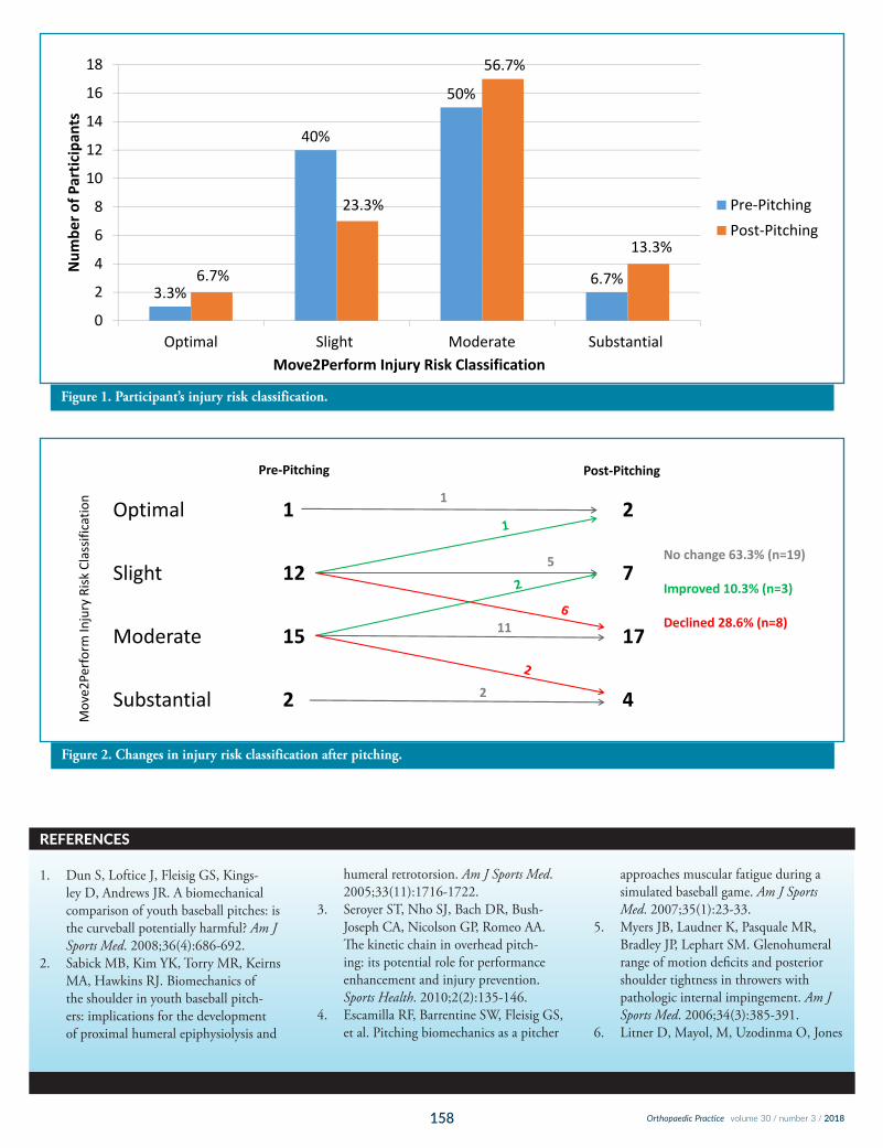
REFERENCES
1. Dun S, Loftice J, Fleisig GS, Kings-ley D, Andrews JR. A biomechanical comparison of youth baseball pitches: is the curveball potentially harmful? Am J Sports Med. 2008;36(4):686-692.
2. Sabick MB, Kim YK, Torry MR, Keirns MA, Hawkins RJ. Biomechanics of the shoulder in youth baseball pitch-ers: implications for the development of proximal humeral epiphysiolysis and
humeral retrotorsion. Am J Sports Med. 2005;33(11):1716-1722.
3. Seroyer ST, Nho SJ, Bach DR, Bush-Joseph CA, Nicolson GP, Romeo AA. The kinetic chain in overhead pitch-ing: its potential role for performance enhancement and injury prevention. Sports Health. 2010;2(2):135-146.
4. Escamilla RF, Barrentine SW, Fleisig GS, et al. Pitching biomechanics as a pitcher
approaches muscular fatigue during a simulated baseball game. Am J Sports Med. 2007;35(1):23-33.
5. Myers JB, Laudner K, Pasquale MR, Bradley JP, Lephart SM. Glenohumeral range of motion deficits and posterior shoulder tightness in throwers with pathologic internal impingement. Am J Sports Med. 2006;34(3):385-391.
6. Litner D, Mayol, M, Uzodinma O, Jones
Figure 1. Participants injury risk classification (n=30).
3.3%
40%
50%
6.7%
0
2
4
6
8
10
12
14
16
18
Optimal Slight Moderate Substantial
Num
ber o
f Partic
ipan
ts
Move2Perform Injury Risk Classification
Pre‐PitchingPost‐Pitching
6.7%
23.3%
56.7%
13.3%
Figure 1. Participant’s injury risk classification.
Optimal 1 2
Slight 12 7
Moderate 15 17
Substantial 2 4
Move2
Perform In
jury Risk
Classificatio
n
Pre‐Pitching Post‐Pitching
1
2
5
11
No change 63.3% (n=19)
Improved 10.3% (n=3)
Declined 28.6% (n=8)
Figure 2. Changes in injury risk classification after pitching.
158 Orthopaedic Practice volume 30 / number 3 / 2018

R, Labossiere D. Glenohumeral internal rotation deficits in professional pitchers enrolled in an internal rotation stretch-ing program. Am J Sports Med. 2007;35(4):617-621.
7. American Sports Medicine Institute. Position statement for adolescent baseball pitchers. www.asmi.org/research.php?page=research§ion=positionStatement. Accessed May 23, 2017.
8. Little League Baseball and Softball. Regular season pitching rules-baseball and softball.www.littleleague.org/learn/rules/pitch-count.htm. Accessed June 3, 2017.
9. Sgroi TA, Zajac JM. Return to throwing after should or elbow injury. Curr Rev Musculoskelet Med. 2018:11(1):12-18. doi: 10.1007/s12178-018-9454-7.
10. Plisky PJ, Rauh M.J, Kaminski TW, Underwood FB. Star Excursion Balance Test as a predictor of lower extremity injury in high school basketball players. J Orthop Sports Phys Ther. 2006;36(12):911-919.
11. Butler RJ, Lehr ME, Fink ML, Kiesel KB, Plinsky PJ. Dynamic balance performance and noncontact lower extremity injury in college football players: an initial study. Sports Health. 2013;5(5):417-422. doi: 10.1177/1941738113498703.
12. Garrison JC, Arnold A, Macko MJ, Conway JE. Baseball players diagnosed with ulnar collateral ligament tears dem-onstrate decreased balance compared to healthy controls. J Orthop Sports Phys Ther. 2013;43(10):752-758. doi: 10.2519/jospt.2013.4680. Epub 2013 Sep 9.
13. Plisky PJ, Gorman PP, Butler RJ, Kiesel KB, Underwood FB, Elkins B. The reliability of an instrumented device for measuring components of the Star Excursion Balance Test. N Am J Sports Phys Ther. 2009;4(2):92-99.
14. Smith CA, Chimera NJ, Warren M. Association of Y balance test reach asymmetry and injury in division I athletes. Med Sci Sports Exer. 2015:47(1);136-141. Jan;47(1):136-41. doi: 10.1249/MSS.0000000000000380.
15. Move2Perform. What is Move2Perform? www.move2per-form.com. Accessed May 23, 2017.
16. Lehr ME, Plinsky PJ, Kiesel KB, Butler RJ, Fink M, Under-wood FB. Field expedient screening and injury risk algorithm categories as predictors of noncontact lower extremity injury. Scand J Med Sci Sport. 2013;23(4):e225-232. doi: 10.1111/sms.12062. Epub 2013 Mar 20.
159Orthopaedic Practice volume 30 / number 3 / 2018

ABSTRACTBackground: Physical activity (PA) can
lead to improved balance, strength, and ambulatory function in adults who have undergone lower limb amputation (LLA). Purpose: The purpose of this study was to determine the accuracy of 5 PA monitors for posture, step count, and PA in adults with unilateral transtibial amputation (TTA). Methods: Adults (N=7) with unilateral TTA wore PA monitors concurrently. Four 6-minute counter-balanced activity phases were completed. Findings: For posture, no differences were found across monitors. For steps, only one PA monitor was valid across phases. For minutes of PA, no monitors were consistently accurate across phases. Clini-cal Relevance: Determination of a valid PA monitor will provide clinicians with an objective measure of PA engagement. Assess-ment of PA engagement may help guide cli-nician recommendations when working with adults with LLA, or more specifically TTA. Conclusion: This study provides important framework for assessment of PA accumula-tion and time in sedentary behaviors.
Key Words: accelerometer, pedometer, validation
INTRODUCTION
Physical activity (PA) is known to prevent onset of chronic disease and obesity.1,2 Cur-rently, 150 minutes/week of moderate PA or 75 minutes/week of vigorous PA is recom-mended for otherwise healthy adults.3 Physi-cal activity recommendations for adults with disability or chronic conditions state that 150 minutes/week, or as much activity as the disability or condition will permit, is ideal.4
Individuals with lower limb amputation (LLA) may be at increased risk for chronic disease due to physical inactivity or decreased levels of PA following amputation surgery; therefore, it is particularly important to accu-
mulate as much PA as possible.5-7 Emerging research has shown that a sedentary lifestyle in which extensive sitting is typical is delete-rious to health.8-10
In addition to risk reduction for chronic disease, PA can lead to independent living and enhanced levels of function, notably in overall balance and strength, in those with LLA.11-13 Lower limb amputation can affect neuromuscular control and coordination. In addition, LLA may affect routine activities of daily living and general function, which may explain the decrement or alteration in PA behaviors for those with LLA.14 Thus, it is imperative for health care providers and cli-nicians to better understand actual PA levels in adults with LLA. To gain insight into PA behaviors in an adult population with ampu-tation, researchers must bridge the current gaps in the existing body of evidence. To date, few studies have used PA monitors to assess PA in adults with LLA.15
Physical activity monitors provide an objective mode to measure the amount and nature of PA, physical inactivity, or posture (ie, sitting, standing, walking, etc). They may also be used to substantiate self-reported PA engagement to determine habitual PA levels. While self-reported PA data is both inexpensive and feasible, it is subjective and usually results in over- or under-reporting of PA time, intensity, and frequency.16 A more objective method of PA assessment is needed to actively address any disease disparities related to physical mobility, or lack thereof, associated with amputation. Researchers must determine whether a PA monitor is accurate in a controlled setting prior to their use in a free-living environment. The monitor must first be demonstrated to be accurate in a controlled environment in the population of interest. Many objective PA monitors have feedback mechanisms that allow the user to track PA behaviors. Studies have shown that ability to quantitatively track PA and having
a goal for daily PA volume are associated with beneficial health outcomes, such as reduced body mass index (BMI).17
As previously stated, few PA monitors have been used to assess movement in adults with traumatic LLA.17-20 However, a large body of research has evaluated PA monitors in children, adolescents, and adults of various weight statuses and health conditions.21-26
Further, PA monitors that have been deter-mined valid for use in some populations; however, have not been valid for use in other populations.25,27-29 When considering a pop-ulation that may have altered gait patterns, it is reasonable to assume that PA monitors and their respective metrics should be evalu-ated in a controlled environment before these devices are adapted to other scenarios.29
The purpose of this study was to deter-mine convergent validity of 5 PA monitors in ambulatory men and women between the ages 21 and 64 with traumatic LLA, specifically, adults with unilateral trans-tibial amputation (TTA). The following PA monitors were included: the Yamax Digi-Walker SW-200 (DG; Yamax Corporation, Tokyo, Japan), the ActiGraph GT3X (AG; ActiGraph, LLC, Pensacola, FL), ActivPAL (AP; PAL Technologies Ltd, Glasgow, UK), the SenseWear Armband (SWA; BodyMe-dia, Pittsburgh, PA), and the Intelligent Device for Energy Expenditure and Activ-ity (IDEEA; Minisun, Fresno, CA). Specific outcomes were posture (sitting and stand-ing), steps, and time (minutes) in sedentary, light physical activity (LPA), and moderate physical activity (MPA).
METHODSParticipants
Eligibility criteria included ambulatory adults ages 21 to 64 years with (1) TTA who self-reported that they could independently perform routine activities of daily living without assistive devices (other than prosthe-
Activity Assessment in Adults with Amputation
Cassie M. Mitchell, PhD1,3 Derek Crawford, PhD, MS, MEd2,4 Joshua A. New, MS, RD1 Carol P. Dionne, PT, DPT, PhD, OCS, Cert MDT2 Susan B. Sisson, PhD, RDN, CHES, FACSM1
1 Behavioral Nutrition and Physical Activity Laboratory, Department of Nutritional Sciences, University of Oklahoma Health Science Center, Oklahoma City, OK
2 Mechanical Therapy Research Laboratory, Department of Rehabilitation Sciences, University of Oklahoma Health Sciences Center, Oklahoma City, OK
3Laboratory for Eating Behaviors and Weight Management, Department of Human Nutrition, Foods and Exercise, Virginia Tech, Blacksburg, VA4Department of Health, Human Performance, & Recreation, Pittsburg State University, Pittsburg, KS
160 Orthopaedic Practice volume 30 / number 3 / 2018

sis), (2) no history of smoking, cardiovascular disease, hypertension, stroke, or neuromus-cular disorders, and (3) a BMI <30.0 kg/m2. Participants were excluded if they had a medical history of any of the abovemen-tioned conditions, had a BMI ≥30.0, or had an amputation due to chronic disease, which may indicate cardiovascular disease, diabetes, or congenital defect, rather than trauma.
Study DesignThe university institutional review board
approved this cross-sectional pilot study. All participants visited the research laboratory once to complete informed consent and HIPAA forms to be familiarized with the mea-sures and protocol before baseline values were obtained. Validity was determined during 4 counterbalanced 6-minute phases: sitting, standing, a light self-paced walk that repre-sented LPA, and a brisk self-paced walk that represented MPA. A random number genera-tor was used to determine the order of phases for each participant. A minimum 2-minute washout period was provided between each phase. This period allowed monitors to reg-ister no activity and the participants’ heart rates to return to baseline before beginning the subsequent phase. Reference criteria for comparing the sitting, standing, and walking phases were stopwatch-recorded time, direct observation of standing and hand-tallied steps, respectively. The sit and stand phases were conducted inside the laboratory. Self-selected walking phases took place in an inte-rior, climate-controlled, continuous hallway (Figure 1).
Intensity range for the LPA and MPA phases was determined using the heart rate reserve (HRR) calculation, target heart rate (HR) range = (HRmax - HRrest) x % inten-sity/100) + HRrest,30 based on resting HR. For each participant, target HR was calcu-lated for the LPA and MPA phases (20-39% and 40-59% of HRR, respectively).31 Beats per minute (BPM) were calculated for the high and low end for both LPA and MPA for each participant. Participants were famil-iarized with the order of the phases and the measurements prior to beginning. For refer-ence, a flow chart of procedures and matrix of assessed metrics is provided in Table 1.
MeasuresDirect observation of posture and hand-tally of steps
Participant posture was directly observed during each phase. The observer noted devia-tions in posture, and recorded all postures and hand-tallied steps. Direct observation
was used as the reference criterion for the sitting and standing phases. Actual time spent sitting and standing was compared to the 6-minute assigned sitting and stand-ing phases. A hand-tally was used as the step count reference criterion for each phase. The same researcher monitored participants throughout all phases and used a clicker to obtain hand-tallied steps.
Yamax DigiWalker SW-200
The Yamax DigiWalker SW-200 (DG) is a waist-mounted pedometer that mea-sures steps. In the present study, the DG was clipped to an elastic belt at the participants’ right hip. For accurate recording of steps at
each phase, the PA monitor was reset to zero prior to beginning each phase. The DG has been evaluated in both controlled and free-living environments in adult population ages 20 to 91 without amputation, disabil-ity, or chronic conditions.32-36 In controlled environments, the DG has been assessed via treadmill at varying speeds, in both stan-dardized protocols and protocols with self-selected walking paces, as well as in a travel setting,32,33 and in free-living environments at self-selected walking speeds.34 In addition, the DG has been tested in an adult population with Parkinson's disease.36 Findings from the above cited studies have cohesively reflected increased accuracy at increased speeds, with
Participants arrived to the Mechanical Therapy Research Laboratory
Informed consent and questionnaires completed
Inclusion criteria met?
Excluded Participant instructed to rest in supine position
for 10 minutes
Measure RHR and BP
Is hypertension detected?
Apply IDEEA, AG, AP, SWA, and DG to the participant.
• Participants were familiarized with protocol and RPE scale• Randomized protocol began• Research personnel provided instructions at the beginning of each phase• During walking phases, research personnel used clicker to manually count
number of steps taken• HR and RPE were be recorded at the end of all phases• Number of steps were recorded for the DG and then cleared between each phase• Two-minute washout phase and HR returned to baseline between each phase• Return to laboratory after completion of each phase• Upon PA monitor removal all data was coded, downloaded and saved onto an
encrypted computer
no yes
yes
no
Figure 1. Study design schematic.
161Orthopaedic Practice volume 30 / number 3 / 2018

a tendency toward under-counting at slower speeds. The DG is generally deemed an appropriate monitor in populations without disability or chronic conditions.
ActiGraph GT3x
The ActiGraph GT3x (AG) is a waist-mounted, triaxial accelerometer, which measures time in PA, classifies intensity, and estimates steps. The AG was worn directly next to the DG on an elastic belt. The belt was fitted to each participant based upon each PA monitor’s user instructions. Move-ment was recorded in 15-second time inter-vals, operationally defined as epochs. The AG is unique among the PA monitors used in the current study due to its ability to tailor cut-points for PA estimates based on a given population. The majority of other available PA monitors have proprietary cut-points that are manufacturer-owned and, generally, non-disclosed. The Freedson cut-points for activ-ity intensity are frequently used to analyze data collected in adults. However, these cut-points have not yet been validated in an adult population with amputation.37 The AG has been tested in various controlled and free-living environments, and has consistently been found to be a valid and reliable monitor for PA assessment in healthy, non-amputee populations.28,38-41
Data treatmentFor the recorded time filter for each
6-minute PA phase per participant, AG data were analyzed using the Freedson equa-tion in ActiLife Software Version 6. ActiLife cut-points were defined as: "sedentary"<100 counts per minute (cpm), "lifestyle" 100-760 cpm, and "light" 100-1961 cpm.42 Sedentary and lifestyle categories were collapsed and analyzed into sedentary, light, moderate, and vigorous categories.
SenseWear ArmbandThe SenseWear Armband (SWA) was
worn on the right triceps. An elastic band was used to keep the monitor in place. Although the SWA contains an accelerometer and thus is capable of step estimation and esti-mation of time in moderate to vigorous PA, most studies have focused on the validity of the other data outputs, such as sleep length and quality, and energy expenditure.43-45 To date, no studies have tested the validity of the accelerometer functions for time (minutes) in moderate to vigorous PA, and only one study has tested the validity of step count in the SWA.24 In the controlled environment designed to mimic activities of daily living, SWA underestimated steps in an adult popu-lation with rheumatoid arthritis.24
Data treatmentThe SWA records data each second. Data
for the SWA were downloaded and analyzed using SenseWear Professional 7.0 Software with the provided proprietary equations. The SWA data output is in metabolic equivalents (METs); this is used to determine time in various intensities. Metabolic equivalents are a metric that permits researchers to obtain the desired outcome (intensity). The SWA does not classify intensity according to the MET values presented in the compendium.46 However, since raw MET values are provided in the SWA output, the minutes could be reclassified using ‘My Data Properties’ to be consistent with the MET classifications of sedentary < 1.5 METs, light 1.5 to ≤ 2.9 METs, moderate 3.0 to ≤ 5.9 METs, and vig-orous ≥6.0,46 thus allowing direct compari-son of congruent minutes in intensity across PA monitors.
ActivPALThe ActivPAL (AP) is an inclinometry
(ie, can detect sitting vs standing) moni-
tor and accelerometer (speed of movement across various axes), which measures time in PA, classifies intensity and posture, and esti-mates steps. This PA monitor was placed on the right thigh directly above the Intelligent Device for Energy Expenditure and Activity (IDEEA) using the monitor stickie provided with the unit. The AP is frequently used in both free-living and controlled environment research to determine breaks from seden-tary behaviors via posture (sitting, standing, or stepping) in healthy populations.47-51 In addition, this PA monitor has been used in populations with impaired physical mobility, such as hip fractures, neuromuscular impair-ment from stroke, and adults in an inpatient status.52,53 Overall, the activPAL appears to be an ideal monitor, if sedentary activity is the measure of interest for all populations.
Data treatmentData for the AP were downloaded to Activ-
PAL Software Version 7.1.18 and exported to an Excel sheet in which minute-by-minute analyses of step count were recorded. The AP does not classify intensity according to the compendium values presented for METs.46
However, the AP provides an Excel output for raw data, including the MET values. This output allowed us to obtain the METs data for each 15-second epoch and average those values to classify each minute’s intensity. Each minute was then equated to the compen-dium-defined PA intensity46 and was consis-tently defined the same as the SWA, to allow comparison of equivalent units. Minutes for each registered intensity by type of PA moni-tor were recorded by phase for each 6-minute phase per participant, to permit comparisons of the desired end-point, intensity classifica-tion. Postures were coded (0=Sedentary/Sit-ting, 1=Standing, 2=Stepping) and minutes per phase for each PA intensity were recorded in the data set.
Intelligent Device for Energy Expenditure and Activity
The IDEEA, system is a 5-sensor biaxial accelerometry monitor, that counts steps, detects PA modalities (sitting, standing, running, or jumping), measures PA inten-sity and duration and other parameters of gait patterns. These activities are recorded on a second-to-second basis (ie, 1-second epochs), and has been validated in a variety of populations without amputation.51,54-57 While the large number of sensors and their accompanying wires make this PA monitor impractical for free-living PA assessment, its precision and accuracy provide a high level
Table 1. Matrix of Analyzed Metrics and Respective Criterions for Physical Activity Monitors
Instrument Steps Minutes of Physical Activity Posture
Yamax DigiWalker SW-200 X N/A N/ASenseWear Armband X X N/AActiGraph GT3x X X N/AActivPAL X X XIDEEA X X XCriterion Hand-tally Stopwatch Direct ObservationThis table presents metrics measured by each physical activity monitor used.Not Applicable (N/A): device or tool does not measure in the listed metric.X-mark: metric was measured by respective monitor(s).
162 Orthopaedic Practice volume 30 / number 3 / 2018

of versatility and accuracy. These features and the IDEEA's ability to discriminate between a wide variety of activities (like walking vs. stair climbing) has resulted in its use as a criterion measure for PA assessment. Since the IDEEA assesses a wide variety of specific activities (such as stair climbing), this moni-tor has often been selected as a criterion mea-sure for PA assessment. With the participant in supine, the IDEEA was placed directly below the AP sensor. All sensors were placed according to manufacturer instructions.
Data treatmentProprietary data for the IDEEA were
downloaded and analyzed using the accom-panying Minisun ActiView 2 software. All data were obtained from the user software. “Sitting” and “standing” were the only two categories used for the sit and stand phases. “Stepping” was considered the light self-selected pace walk. “Walking” and “jogging” were categorized as the moderate self-selected pace walk. No other output terminologies for the LPA and MPA phases were applicable.
Analyses Frequencies, means, and standard devia-
tions were calculated for the participant descriptive data. Under- and over-counting were calculated for each participant for each phase. Mean absolute error (MAE) and mean absolute percent error (APE) were calculated for steps and minutes of LPA and MPA to further examine error. The MAE was cal-culated by subtracting the variable of inter-est (steps or minutes) from the criterion (objective monitoring instrument– criterion = error). The value was then converted to an absolute (ie, the direction of error was removed) to circumvent compression and underestimation of error.32 For example, if the DG registered 15 steps and 20 steps were manually counted, the error is -5, but the absolute error is 5. The absolute error for each PA monitor on each participant was cal-culated and averaged across all participants to create the MAE. Absolute percent error was calculated by dividing the absolute error into the criterion value, then multiplying by 100 [(error/criterion)*100].58 Continuing with the example, if the MAE was 5, and the crite-rion was 20 [(5/20)*100] the APE would be 25%. The absolute percent error for each PA monitor on each participant was calculated and averaged across all participants to create the mean APE.
Repeated measures analysis of variance (RMANOVA) tests were used to compare all metrics (steps, LPA, MPA) from applicable
PA monitors to the appropriate criterion. Post-hoc pairwise comparisons were used to determine whether significant differences were attributed to inter-PA monitor differ-ences or differences between PA monitor and criterion measures. For posture, chi-square and confidence intervals were used to deter-mine any significant differences between observer-recorded time and PA monitor-determined time in a given posture (sitting or standing). For each test, statistical signifi-cance was determined at α = 0.05.
RESULTSDescriptive Data
Means and standard deviations were cal-culated for age, weight, height, BMI, resting blood pressure, resting heart rate, and HRR (Table 2).
Posture
A paired t-test revealed no difference between observer-recorded times for sit-ting and standing compared with the AP and IDEEA, the only tested PA monitors that measure posture. For the AP time spent sitting and standing when compared to observed time sitting and standing, results are t = 2.036, p = 0.097 and t = 1.181, p = 0.291, respectively. For the IDEEA time spent sitting compared against observed time sitting, results are t = 1.181, p = 0.291. For
IDEEA time spent standing, the standard error of the difference was zero. Therefore, no data could be computed due to perfect agreement.
StepsIn Table 3 means ± standard deviations,
MAE, and mean absolute percent error (MAPE) for steps measured by each moni-tor are recorded. A RMANOVA found no statistically significant differences between PA monitors for steps in the sitting and standing phases compared with the hand-tally step criterion, F(5) = 1.096, p = 0.354 and F(5) = 5.714, p = 0.075, respectively. There were differences between PA moni-tor-recorded steps versus step criterion for LPA (F(5) = 424.957, p < 0.001) and MPA (F(5) = 2716.568, p = 0.007) phases. Pair-wise comparisons for LPA determined that only the SWA significantly differed from the criterion (t = 3.276, p = 0.017), due to under-counting. Pairwise comparisons for moderate PA determined that the AG, AP, SWA, and IDEEA significantly differed from the criterion (AG: t = 4.058, p = 0.007; AP: t = 2.979, p = 0.038; SWA: t = 3.971, p = 0.007; IDEEA: t = 2.845, p = 0.036) due to under-counting. The DG did not differ from the criterion.
Table 2. Descriptive Characteristics (n=7)
Gender
Male 86%
Age (years) 29.7 ± 6.0
Weight (lbs) 178.13 ± 15.31
Height (in) 83.5 ± 14.1
BMI 26.3 ± 2.1
Resting Blood Pressure (mmHg)
Systolic 113 ± 4
Diastolic 75 ± 4
Resting Heart Rate (bpm) 60 ± 15
Calculated Heart Rate Reserve (bpm)*
Light Intensity Range 20-39% 92-114 ± 5-8
Calculated Heart Rate Reserve (bpm)§
Moderate Intensity Range 40-59% 115-137 ± 5-6
This table presents descriptive characteristics and baseline values (mean ± standard deviation)obtained from all participants (n=7).
* Light intensity range is the percent at which all participants’ heart rate reserve was calculated; means and standard deviations for the range are presented.
§Moderate intensity range is the percent at which all participants’ heart rate reserve was calculated;means and standard deviations for the range are presented.
163Orthopaedic Practice volume 30 / number 3 / 2018

MinutesTable 4 presents means ± standard devia-
tions, MAE, and MAPE for minutes in LPA and MPA for all monitors compared with the stopwatch-recorded criterion. There were significant differences between stopwatch-recorded LPA (minute criterion) and PA monitor-recorded LPA, F(4) = 3106.864, p < 0.001. Pairwise comparisons showed that all PA monitors differed from the criterion and under-counted minutes (AG: t = 23.486, p < 0.001; AP: t = 24.597, p < 0.001; SWA: t = 16.202, p < 0.001; IDEEA t = 364.405, p < 0.001). Stopwatch-recorded MPA and PA monitor-recorded time in MPA significantly differed (F(4) = 149.498, p < 0.001). Pairwise comparisons showed that the AG, SWA, and IDEEA significantly under-counted com-pared with the criterion (AG: t = 3.164, p = 0.019; SWA: t = 3.873, p = 0.008; IDEEA: t = 3.297, p = 0.022). ActivPAL did not differ from the criterion.
DISCUSSIONThe purpose of this pilot study was to
examine validity of 5 commonly used PA monitors that have not been validated in adults with unilateral TTA. The narrow body of available evidence for valid PA assessment in adults with amputation demonstrates a gap in the literature and represents an interdis-
ciplinary problem for researchers and clini-cians alike. Physical activity after amputation is essential for functional recovery, preven-tion of atrophy and disease, and maintenance of residual limb volume. Currently, little objective information is available to provide insight into PA habits following amputa-tion surgery.59 While some PA monitors (Patient Activity Monitor and DigiWalker SW-700, StepWatch3 Activity Monitor) have been used to assess PA, the available evidence for valid PA monitors results in both narrow selection and limited measure-ment of PA metric (ie, steps vs minutes vs posture). The present study is a step toward bridging PA monitor availability because it (1) used PA monitors that have not yet been tested in adults with TTA, (2) addressed new and remaining questions from this study and others, and (3) selected monitors that are commonly used in research but are also translatable to a clinical setting. Our findings provide a foundation for future directions and insights for researchers and clinicians by identifying PA monitors that accurately mea-sure various intensities, metrics, and postures in adults with TTA.
Various modes of PA, such as steps, pos-ture, and minutes in a given PA intensity, were tested for validity in one pedometer and 4 different accelerometers. Overall, accu-
racy varied based upon the outcome/metric selected to quantify PA. For accelerometry, more research is needed, as no monitor was valid across all intensities. These findings demonstrate that monitor validity in a popu-lation without amputation is not transfer-rable to a population with amputation. For posture, the IDEEA and AP were both valid. This information may prove useful in research and clinical practice to help reduce sedentary and/or physically inactive behaviors, as these behaviors have their own unique subsets of disease risk. From a clinical perspective, the AP represents a cost-effective user-friendly PA monitor for patients and clinicians and would allow clinicians to objectively quan-tify sedentary behaviors. For steps, the DG was valid across all phases. The DG provides a cost-effective PA monitor and is accurate for determination of daily step count. Deter-mination of step count would be beneficial from a clinical perspective as daily steps are considered reflective of total daily PA. The use of the AP and DG would provide further insight into the lifestyle behaviors and PA accumulation in a population with amputation.
For accuracy of PA monitor-recorded time in sitting and standing postures versus direct observation, the IDEEA had greater accuracy (83-100%) than the AP (50-
Sit Stand
Device Steps MAE Mean APE Steps MAE Mean APE
Hand-tally 1 ± 2 * * 0 ± 1 * *
DG 1 ± 2 0.14 ± 0.38 0% 0 ± 0 0 ± 0 0%
AG 3 ± 3 2 ± 2 0% 1 ± 1 1 ± 1 0%
AP 5 ± 11 5 ± 10 216% 1 ± 2 1 ± 2 17%
SWA 5 ± 12 4 ± 10 80% 1 ± 2 2 ± 2 14%
IDEEA 0 ± 0 1 ± 2 17% 0 ± 0 0 ± 1 17%
LPA MPA
Device Steps MAE Mean APE Steps MAE Mean APE
Hand-Tally 648 ± 51 * * 762 ± 46 * *
DG 611 ± 61 40 ± 51 6% 741 ± 87 31 ± 57 4%
AG 583 ± 99 63 ± 75 10% 665 ± 58 97 ± 63 12%
AP 636 ± 54 103 ± 226 17% 738 ± 42 138 ± 270 18%
SWA 542 ± 93 103 ± 78 16% 635 ± 57 127 ± 85 16%
IDEEA 587 ± 87 62 ± 60 10% 656 ± 81 110 ± 94 14%
Abbreviations: MAE, mean absolute error; APE, absolute percent error; LPA, light physical activity; MPA, moderate physical activity
This table presents the mean ± standard deviation, MAE and mean APE for steps across all phases for all participants when compared to the hand-tallycriterion for the DigiWalker (DG), ActiGraph GT3X (AG), ActivPAL (AP), SenseWear Armband (SWA), and IDEEA.
*Asterisk is filled in where the standard error of the difference was zero.
Table 3. Means ± Standard Deviation, Mean Absolute Error, and Mean Absolute Percent Error for Steps Across All Phases
164 Orthopaedic Practice volume 30 / number 3 / 2018

LPA MPA
Device Minutes MAE Mean APE Minutes MAE Mean APE
Stopwatch 6.00 ± 0 * * 6.00 ± 0 * *
AG 0.44 ± 0.60 5.56 ± 0.60 93% 3.38 ± 2.19 2.62 ± 2.19 44%
AP 0.50 ± 0.55 5.50 ± 0.55 92% 5.50 ± 0.55 0.50 ± 0.55 8%
SWA 1.00 ± .82 5.43 ± 0.79 83% 3.86 ± 1.46 2.14 ± 1.46 36%
IDEEA 0.02 ± 0.04 5.98 ± 0.04 100% 5.75 ± 0.19 0 ± 0 18%
Abbreviations: MAE, mean absolute error; APE, absolute percent error; LPA, light physical activity; MPA, moderate physical activity
This table presents the mean ± standard deviation, MAE and mean APE for minutes in LPA and MPA when compared to the stopwatch criterion for the ActiGraph GT3X (AG), ActivPAL (AP), SenseWear Armband (SWA), and IDEEA
*Asterisk is filled in where the standard error of the difference was zero.
Table 4. Means ± Standard Deviation, Mean Absolute Error, and Mean Absolute Percent Error for Minutes in Light Physical Activity and Moderate Physical Activity
100%). However, the AP was still accurate for posture, as it correctly detected time spent sitting and standing for all participants for the full 6-minute phase. The greater accu-racy observed in the IDEEA can likely be attributed to the greater number of sensors thus, greater PA monitor sensitivity. This PA monitor sensitivity allowed for detection of more erratic movements, such as fidgeting or swaying, while participants were seated or standing. While these movements were cap-tured on the IDEEA, they did not contrib-ute to a disruption in posture. For example, a participant could stand with both feet in the same position for the entire 6-minute phase, but shift their weight from one foot to the other. While no steps were taken, these types of movements were registered on the IDEEA more frequently than on the AP. We did not objectively account for these erratic movements because there is no research indi-cating how to record this type of movement and compare the results using these specific PA monitors. Posture is an area of amputa-tion research that has not yet been studied using the AP. While much of the emphasis in adults with amputation is on rehabilitation and return to an individual’s normal activities of daily living, newer research has found that exercise and PA may not be enough to com-pensate for an otherwise sedentary lifestyle.60 Therefore, future studies should consider assessing posture in addition to PA levels in adults with amputation.
For step count, the DG had the least amount of absolute error (17.8 ± 27) and percent error (54%), and did not significantly differ from the criterion in any of the phases. The DG is also the most affordable ($15 per monitor) and focused PA monitor used here, as it measures only steps. All remaining PA monitors had greater absolute error and per-
cent error, and significantly differed from the criterion in the LPA and MPA phases. All PA monitors had a tendency to under-count steps. The consistency of the DG in our study agrees with previous findings on the DG in adults with amputation, albeit using a differ-ent model.19 This confirmation will enable researchers and clinicians to make recom-mendations on an accurate PA monitor for a population with LLA.
No PA monitor was valid for minutes across all 4 phases. However, it should be noted that many PA studies show that moni-tor validity decreases as speed decreases.27 For the LPA phase, no PA monitor correctly cap-tured and identified minutes and intensity. This is not entirely unexpected, as this find-ing is prevalent in PA monitor research when slower speeds are examined.61,62 For MPA, only the AP correctly captured and identi-fied minutes and intensity. All PA monitors had a tendency to under-classify minutes in a given intensity, which is consistent with their tendency to undercount steps. Our findings did not result in identification of an accurate accelerometer, unlike studies that used the Patient Activity Monitor29 and Stepwatch3 Activity Monitor,20 an accurate accelerom-eter. However, this discrepancy may be to differing study designs or PA monitor algo-rithms. One way to address accelerometer validity between the differing results of the Patient Activity Monitor and the Stepwatch3 Activity Monitor may be to compare the accelerometers used here against the Patient Activity Monitor or Stepwatch3 Activity Monitor.
User feasibility and outcomes of interest are also important to consider. For example, the IDEEA costs over $5,000 and involves precise placement of multiple sensors with connecting wires. Use of the IDEEA is feasi-
ble in a controlled environment with an expe-rienced researcher, but is highly impractical in a free-living setting, due to the financial expense should equipment be damaged and participant burden. The SWA, while easy to wear around the bicep and affordable ($120 per monitor), was not valid in determining steps or PA intensity. The AG ($250 per mon-itor) was not valid for steps, LPA, or MPA for this population. However, the user friendli-ness of the PA monitor and the software (ie, adjustable cut points rather than proprietary formulae) could allow for improvements and development of specific intensity cut-points in future research in adults with TTA. The AP was valid for 3 of 4 phases (sitting, stand-ing, and MPA). Its ability to correctly classify posture and time within that posture makes it an appropriate PA monitor to assess sedentary time or physical inactivity. Further, the AP was accurate for MPA. However, its inability to capture LPA poses a barrier, as this inten-sity may most accurately reflect a substantial proportion of this population’s time.
The DG was valid for steps across all phases, even though the DG’s technology is less advanced than the other PA monitors used. Benefits of the DG include its cost-effectiveness, user-friendliness, and simple output. Steps per day have been linked to minutes of PA accumulated, which may make the DG a suitable proxy for PA in this population. Behrens et al63 found that approximately 10,000 steps/day was equiva-lent to the 150 of MPA/week recommended in the national PA guidelines.5,63 The DG was valid across all phases and is a feasible PA monitor. Since steps are equated to MPA, this finding provides an excellent starting place for PA assessment in adults with TTA.
165Orthopaedic Practice volume 30 / number 3 / 2018

Guidelines for Physical Activity. In Ainsworth BE, Macera CA, eds. Physical Activity and Public Health Practice. Boca Raton, FL: CRC Press; 2012:195-209.
6. Health.gov Office of Disease Prevention and Health Promotion. 2008 Physi-cal Activity Guidelines for Americans: Chapter 7. http://www.health.gov/paguidelines/guidelines/chapter7.aspx. Accessed May 9, 2018
7. Naschitz JE, Lenger R. Why trau-matic leg amputees are at increased risk for cardiovascular diseases. QJM. 2008;101(4):251-259. doi: 10.1093/qjmed/hcm131. Epub 2008 Feb 16.
8. Barlow CE, Shuval K, Balasubrama-nian BA, Kendzor DE, Gabriel KP. Sitting time, physical activity and cardiorespiratory fitness: Cooper Center Longitudinal Study Cohort. J Phys Act Health. 2016;13(1):17-23. doi: 10.1123/jpah.2014-0430. Epub 2015 Apr 1.
9. Lee IM, Shiroma EJ, Lobelo F, et al. Effect of physical inactivity on major non-communicable diseases worldwide: an analysis of burden of disease and life expectancy. Lancet. 2012;380(9838):219-229. doi: 10.1016/S0140-6736(12)61031-9.
10. Matthews CE, Moore SC, Sampson J, et al. Mortality benefits for replacing sitting time with different physical activities. Med Sci Sports Exerc. 2015;47(9):1833-1840. doi: 10.1249/MSS.0000000000000621.
11. Couture M, Caron CD, Desrosiers J. Leisure activities following a lower limb amputation. Disabil Rehabil. 2010;32(1):57-64. doi: 10.3109/09638280902998797.
12. Raya MA, Gailey RS, Fiebert IM, Roach KE. Impairment variables predicting activity limitation in individuals with lower limb amputation. Prosthet Orthot Int. 2010;34(1):73-84. doi: 10.3109/03093640903585008.
13. van Velzen JM, van Bennekom CA, Polomski W, Slootman JR, van der Woude LH, Houdijk H. Physical capacity and walking ability after lower limb amputation: a systematic review. Clin Rehabil. 2006;20(11):999-1016.
14. Jones SF, Twigg PC, Scally AJ, Buckley JG. The gait initiation process in unilateral lower-limb amputees when stepping up and stepping down to a
StrengthsStrengths of this study included the mea-
surement of posture, intensity classification of performed activities, and the range of sophistication of PA monitors (ie, DG vs IDEEA).9 The counterbalanced phase design allowed the researchers to determine that the validation, or lack thereof, was a result of the PA monitor, rather than due to the order of phases. We focused specifically on non-obese, ambulatory adults with unilateral LLA with no history of chronic disease condi-tions. Since this study was tightly controlled and phases counterbalanced, our findings are more easily attributed to the PA monitors in this population, rather than intra-participant variance or PA monitor error.
LimitationsThe small sample size may be viewed as
a limitation. However, the literature is lim-ited in content related to the population under study, and especially in the area of PA assessment. Therefore, the purpose of this study was to expand the current baseline of acceptable PA monitors for future research in PA assessment for adults with amputation. Further, since the related body of literature is small, many studies involving amputation expand their criteria to include the following: amputation as a result of trauma, chronic disease or congenital defect; Syme’s, trans-femoral, and TTA; unilateral and bilateral amputations; and various disease and weight statuses. We sought a sub-population (pre-viously described) in an attempt to control for any potential confounders, such as gait differences between unilateral or bilateral amputation, or transfemoral amputation versus TTA. Even with many of the above not controlled for in other research studies, many sample sizes are still relatively modest. With this context in mind, we propose that the sample size be viewed not as a limiting factor, but as a strength due to the amount of control for confounding variables yielded by our approach. Additionally, this study is not generalizable to a free-living setting as all the PA modes were tested in a controlled labo-ratory environment; however, this research may be used in future studies to determine PA monitor selection, PA modes to mea-sure, and validity of available and valid PA monitors.
Future DirectionsIncluding PA monitors not yet tested,
expanding the type of amputation (both site and cause), other physical impairments resulting from musculoskeletal trauma, com-
paring valid PA monitors in children versus adults, and the incorporation of common work activity tracking monitors (ie, Garmin, FitBit) in future work would provide more insight into this population. Expanding the protocol to include simulated activities of daily living or using valid PA monitors in a free-living environment should be consid-ered. Our findings serve as a preliminary basis for future studies.
CONCLUSIONSAdults with amputation are at an increased
risk for various chronic diseases that develop due to excessively sedentary behavior. Physi-cal activity is one way to diminish disease risk. However, this population has reported barriers for PA engagement, which may lead to low accumulation of PA.11 The purpose of the present study was to examine the valid-ity of PA monitors for the measurement of various PA metrics, including steps, pos-ture, and minutes in various PA intensities, in an amputation population. Key findings included (1) the IDEEA and AP were both valid for posture assessment, (2) the DG was best for step measurement, and (3) the AP was best for measuring minutes in PA intensities. No other PA monitors were valid for LPA or MPA, but all 4 accelerometry monitors were valid for sitting and standing phases. Future research should include a broader selection of PA monitors, expanded recruitment criteria, and incorporation of free-living PA.
REFERENCES
1. Jakicic JM, Otto AD. Physical activity considerations for the treatment and prevention of obesity. Am J Clin Nutr. 2005;82(1 Suppl):226S-229S.
2. Kruk J. Physical activity in the pre-vention of the most frequent chronic diseases: an analysis of the recent evidence. Asian Pac J Cancer Prev. 2007;8(3):325-338.
3. Health.gov Office of Disease Prevention and Health Promotion. 2008 Physical Activity Guidelines for Americans. http://health.gov/paguidelines/guidelines/. Accessed May 9, 2018
4. Health.gov Office of Disease Prevention and Health Promotion 2008 Physical Activity Guidelines for Americans: Chapter 4: Active Adults. http://www.health.gov/paguidelines/guidelines/chapter4.aspx. Accessed May 8, 2018
5. Troiano RP, Buchner DM. National
166 Orthopaedic Practice volume 30 / number 3 / 2018

new level. Clin Biomech (Bristol, Avon). 2005;20(4):405-413.
15. Gallagher P, O'Donovan MA, Doyle A, Desmond D. Environmental barriers, activity limitations and participation restrictions experienced by people with major limb amputation. Prosthet Orthot Int. 2011;35(3):278-284. doi: 10.1177/0309364611407108.
16. Baumgartner, TA, Hensley L. Conducting & Reading Research in Health & Human Performance, 4th ed. New York, NY: The McGraw-Hill Companies, Inc.; 2006.
17. Bravata DM, Smith-Spangler C, Sun-daram V, et al. Using pedometers to increase physical activity and improve health: a systematic review. JAMA. 2007;298(19):2296-2304.
18. Ramstrand N, Nilsson KA. Valida-tion of a patient activity monitor to quantify ambulatory activity in an amputee population. Prosthet Orthot Int. 2007;31(2):157-166.
19. Dudek NL, Khan OD, Lemaire ED, Marks MB, Saville L. Ambulation monitoring of transtibial amputation subjects with patient activity monitor versus pedometer. J Rehabil Res Develop. 2008;45(4):577-585.
20. Coleman KL, Smith DG, Boone DA, Joseph AW, del Aguila MA. Step activity monitor: long-term, continuous record-ing of ambulatory function. J Rehabil Res Develop. 1999;36(1):8-18.
21. Schneider PL, Crouter S, Bassett DR. Pedometer measures of free-living physical activity: comparison of 13 models. Med Sci Sports Exerc. 2004;36(2):331-335.
22. Santos-Lozano A, Santín-Medeiros F, Cardon G, et al. Actigraph GT3X: validation and determination of physi-cal activity intensity cut points. Int J Sports Med. 2013;34(11):975-982. doi: 10.1055/s-0033-1337945. Epub 2013 May 22.
23. Pate RR, O'Neill JR, Mitchell J. Measurement of physical activity in preschool children. Med Sci Sports Exerc. 2010;42(3):508-512. doi: 10.1249/MSS.0b013e3181cea116.
24. Tierney M, Fraser A, Purtill H, Ken-nedy N. Study to determine the criterion validity of the SenseWear Armband as a measure of physical activity in people with rheumatoid arthritis. Arthritis Care Res (Hoboken). 2013;65(6):888-895. doi: 10.1002/acr.21914.
25. Crouter SE, Schneider PL, Bassett DR Jr. Spring-levered versus piezo-electric pedometer accuracy in overweight and obese adults. Med Sci Sports Exerc. 2005;37(10):1673-1679.
26. Barreira TV, Tudor-Locke C, Cham-pagne CM, Broyles ST, Johnson WD, Katzmarzyk PT. Comparison of GT3X accelerometer and YAMAX pedometer steps/day in a free-living sample of overweight and obese adults. J Phys Act Health. 2013;10(2):263-270.
27. Tyo BM, Fitzhugh EC, Bassett DR Jr, John D, Feito Y, Thompson DL. Effects of body mass index and step rate on pedometer error in a free-living environment. Med Sci Sports Exerc. 2011;43(2):350-356.
28. Connolly CP, Coe DP, Kendrick JM, Bassett DR Jr, Thompson DL. Accu-racy of physical activity monitors in pregnant women. Med Sci Sports Exerc. 2011;43(6):1100-1105. doi: 10.1249/MSS.0b013e3182058883.
29. Bussmann HB, Reuvekamp PJ, Veltink PH, Martens WL, Stam HJ. Validity and reliability of measurements obtained with an "activity monitor" in people with and without a transtibial amputation. Phys Ther. 1998;78(9):989-998.
30. American College of Sports Medicine. ACSM's Guidelines for Exercise Testing and Prescription. 9th ed. Philadelphia, PA: Lippincott Williams & Wilkins; 2014.
31. American College of Sports Medi-cine. General principles of exercise prescription. In: Pescatello LS, ed. ACSM's Guidelines for Exercise Testing and Prescription. 9th ed. Philadelphia, PA: Lippincott, Williams & Wilkins; 2014:162.
32. Le Masurier GC, Tudor-Locke C. Comparison of pedometer and accel-erometer accuracy under controlled conditions. Med Sci Sports Exercise. 2003;35(5):867-871.
33. Melanson EL, Knoll JR, Bell ML, et al. Commercially available pedometers: considerations for accurate step counting. Prev Med. 2004;39(2):361-368.
34. Bergman RJ, Bassett DR Jr, Muthukrish-nan S, Klein DA. Validity of 2 devices for measuring steps taken by older adults in assisted-living facilities. J Phys Act Health. 2008;5 Suppl 1:S166-175.
35. Le Masurier GC, Lee SM, Tudor-Locke C. Motion sensor accuracy under con-
trolled and free-living conditions. Med Sci Sports Exerc. 2004;36(5):905-910.
36. Dijkstra B, Zijlstra W, Scherder E, Kamsma Y. Detection of walking periods and number of steps in older adults and patients with Parkinson's disease: accuracy of a pedometer and an accelerometry-based method. Age Ageing. 2008;37(4):436-441. doi: 10.1093/ageing/afn097. Epub 2008 May 16.
37. Freedson PS, Melanson E, Sirard J. Calibration of the Computer Science and Applications, Inc. accelerometer. Med Sci Sports Exercise. 1998;30(5):777-781.
38. Kelly LA, McMillan DG, Anderson A, Fippinger M, Fillerup G, Rider J. Valid-ity of actigraphs uniaxial and triaxial accelerometers for assessment of physical activity in adults in laboratory condi-tions. BMC Med Phys. 2013;13(1):5. doi: 10.1186/1756-6649-13-5.
39. Sirard JR, Forsyth A, Oakes JM, Schmitz KH. Accelerometer test-retest reliability by data processing algorithms: results from the Twin Cities Walking Study. J Phys Act Health. 2011;8(5):668-674.
40. McClain JJ, Sisson SB, Tudor-Locke C. Actigraph accelerometer interinstrument reliability during free-living in adults. Med Sci Sports Exerc. 2007;39(9):1509-1514.
41. Wood AC, Kuntsi J, Asherson P, Saudino KJ. Actigraph data are reliable, with functional reliability increasing with aggregation. Behav Res Methods. 2008;40(3):873-878.
42. Freedson PS, Melanson E, Sirard J. Calibration of the Computer Science and Applications, Inc. accelerometer. Med Sci Sports Exerc. 1998;30(5):777-781.
43. Arvidsson D, Slinde F, Larsson S, Hulthen L. Energy cost of physical activities in children: validation of Sense-Wear Armband. Med Sci Sports Exerc. 2007;39(11):2076-2084.
44. Benito PJ, Neiva C, Gonzalez-Quijano PS, Cupeiro R, Morencos E, Pei-nado AB. Validation of the SenseWear armband in circuit resistance training with different loads. Eur J Appl Physiol. 2012;112(8):3155-3159. doi: 10.1007/s00421-011-2269-5. Epub 2011 Dec 6.
45. Smith KM, Lanningham-Foster LM, Welk GJ, Campbell CG. Valid-ity of the SenseWear(R) Armband to predict energy expenditure in preg-nant women. Med Sci Sports Exerc.
167Orthopaedic Practice volume 30 / number 3 / 2018

2012;44(10):2001-2008. doi: 10.1249/MSS.0b013e31825ce76f.
46. Jette M, Sidney K, Blumchen G. Meta-bolic equivalents (METS) in exercise testing, exercise prescription, and evalua-tion of functional capacity. Clin Cardiol. 1990;13(8):555-565.
47. Dowd KP, Harrington DM, Donnelly AE. Criterion and concurrent validity of the ActivPAL professional physical activ-ity monitor in adolescent females. PloS One. 2012;7(10):e47633. doi: 10.1371/journal.pone.0047633. Epub 2012 Oct 19.
48. Harrington DM, Welk GJ, Donnelly AE. Validation of MET estimates and step measurement using the ActivPAL physical activity logger. J Sports Sci. 2011;29(6):627-633. doi: 10.1080/02640414.2010.549499.
49. Ryan CG, Grant PM, Tigbe WW, Granat MH. The validity and reli-ability of a novel activity monitor as a measure of walking. Br J Sports Med. 2006;40(9):779-784.
50. Kozey-Keadle S, Libertine A, Lyden K, Staudenmayer J, Freedson PS. Valida-tion of wearable monitors for assessing sedentary behavior. Med Sci Sports Exerc. 2011;43(8):1561-1567. doi: 10.1249/MSS.0b013e31820ce174.
51. Hart TL, McClain JJ, Tudor-Locke C. Controlled and free-living evalua-tion of objective measures of sedentary and active behaviors. J Phys Act Health. 2011;8(6):848-857.
52. Taraldsen K, Askim T, Sletvold O, et al. Evaluation of a body-worn sensor system to measure physical activity in older people with impaired function. Phys Ther. 2011;91(2):277-285. doi: 10.2522/ptj.20100159. Epub 2011 Jan 6.
53. Cindy Ng LW, Jenkins S, Hill K. Accuracy and responsiveness of the stepwatch activity monitor and ActivPAL in patients with COPD when walk-ing with and without a rollator. Disabil Rehabil. 2012;34(15):1317-1322. doi: 10.3109/09638288.2011.641666. Epub 2011 Dec 26.
54. Zhang K, Werner P, Sun M, Pi-Sunyer FX, Boozer CN. Measurement of human daily physical activity. Obes Res. 2003;11(1):33-40.
55. Kwon S, Jamal M, Zamba GK, Stumbo P, Samuel I. Validation of a novel physical activity assessment device in morbidly
obese females. J Obes. 2010;2010. pii: 856376. doi: 10.1155/2010/856376. Epub 2010 Feb 9.
56. Marsh AP, Vance RM, Frederick TL, Hesselmann SA, Rejeski WJ. Objective assessment of activity in older adults at risk for mobility disability. Med Sci Sports Exerc. 2007;39(6):1020-1026.
57. Jiang Y, Larson JL. IDEEA activity monitor: validity of activity recognition for lying, reclining, sitting and stand-ing. Front Med. 2013;7(1):126-131. doi: 10.1007/s11684-012-0236-0. Epub 2012 Dec 22.
58. Barreira TV, Brouillette RM, Foil HC, Keller JN, Tudor-Locke C. Compari-son of older adults' steps per day using NL-1000 pedometer and two GT3X+ accelerometer filters. J Aging Phys Act. 2013;21(4):402-416.
59. Deans S, Burns D, McGarry A, Murray K, Mutrie N. Motivations and barri-ers to prosthesis users participation in physical activity, exercise and sport: a review of the literature. Prosthet Orthot Int. 2012;36(3):260-269. doi: 10.1177/0309364612437905.
60. Nicholas JA, Lo Siou G, Lynch BM, Robson PJ, Friedenreich CM, Csizmadi I. Leisure-time physical activity does not attenuate the association between occu-pational sedentary behaviour and obesity: results from Alberta's Tomorrow Project. J Phys Act Health. 2015;12(12):1586-1600. doi: 10.1123/jpah.2014-0370. Epub 2015 Apr 1.
61. Stansfield B, Hajarnis M, Sudarshan R. Characteristics of very slow stepping in healthy adults and validity of the activ-PAL3 activity monitor in detecting these steps. Med Eng Phys. 2015;37(1):42-47. doi: 10.1016/j.medengphy.2014.10.003. Epub 2014 Nov 1.
62. Storti KL, Pettee KK, Brach JS, Talkowski JB, Richardson CR, Kriska AM. Gait speed and step-count moni-tor accuracy in community-dwelling older adults. Med Sci Sports Exerc. 2008;40(1):59-64.
63. Behrens TK, Hawkins SB, Dinger MK. Relationship between objectively measured steps and time spent in physical activity among free-living col-lege students. Meas Phys Educ Exerc Sci. 2005;9(2):67-77.
168 Orthopaedic Practice volume 30 / number 3 / 2018


ABSTRACTBackground and Purpose: Chronic
regional pain syndrome is a painful and dis-abling condition that often occurs following surgery or trauma. Chronic regional pain syn-drome has been reported in <7% of clavicle injury cases. Case Description: The patient is a 15-year-old female high school athlete who presented with an insidious onset of left clav-icle pain since 2011. She underwent surgery in 2011 for excision of a lesion in the middle one-third of her left clavicle. Four years later, in 2015, she presented with symptoms of chronic regional pain syndrome. Interven-tions: Interventions included diagnostic testing, blood work, surgery, conservative physical therapy, trigger point dry needling, total motion release therapy, and iron sup-plementation. Outcomes: Outcomes were assessed using the Disabilities of the Arm, Shoulder, and Hand (DASH) questionnaire. Clinical Relevance: This case study describes complications of complex regional pain syn-drome and use of less traditional physical therapy methods to improve shoulder active range of motion and strength.
Key Words: clavicle, total motion release, trigger point dry needling
BACKGROUNDChronic regional pain syndrome (CRPS)
is a painful and disabling condition that often occurs in response to surgery or trauma.1-3
Patients with CRPS display an increased ner-vous system response to an injury or surgery, resulting in pain and an autonomic nervous system response.4 Clinical diagnosis of CRPS involves pain, sensitivity issues, including hyperesthesia or allodynia, vasomotor and/or pseudomotor changes, loss of motor con-trol or range of motion (ROM), and trophic changes.1 The pathology of CRPS is not well understood. It is thought to be related to a disturbance in “sympathetically mediated vasomotor control, together with maladaptive peripheral and central neuronal plasticity.”5-8
Sandroni et al9 reported an incidence of 21 cases per 100,000. Chronic regional pain syn-drome has been shown to be more common in females vs males.2
Clavicle fractures have been estimated
to account for approximately 34% to 45% of shoulder injuries.10,11 Research suggests that 69% to 81% of clavicle fractures occur in the middle one-third of the clavicle while only 17% occur in the lateral one-third.12
Left clavicle injuries are more common than right.13 Complex regional pain syndrome has been only documented in approximately 7% of cases associated with clavicle injuries.14
Guidelines have recommended various physical therapy treatments for CRPS. These include but are not limited to joint mobili-zation/manipulation, desensitization, ROM, hydrotherapy, and electrotherapy.15-17 A review of best practice guidelines recommend the use of desensitization therapy, a treatment that emphasizes the “graded introduction of noxious stimulus” as a means of decreas-ing allodynia.4 Studies have demonstrated that desensitization techniques can result in decreased pain, allodynia, and improved pres-sure tolerance.18
Trigger point dry needling (TDN) is a new type of conservative treatment gaining attention in the field of physical therapy. It has been shown to be an effective treatment in treating musculoskeletal injuries involv-ing muscular trigger points and or nerve pain or allodynia. Trigger point dry needling is a manual therapy procedure where thin mono-filament needles are inserted into the skin and muscle in areas of myofascial trigger points to manage neuromusculoskeletal pain and movement impairments. Prior research has shown that TDN relieves pain and decreases muscle tightness. Trigger point dry needling can be used as a form of desensitization by targeting affected nerves.19,20 Trigger point dry needling has been shown to be effective in treatment of neck and shoulder myofascial pain.3,21 Trigger point dry needling has been shown to provide pain relief via stimulation of “sensitized muscle nociceptors, decrease in metabolic mediators, and increase in microcirculation.”22
Total motion release (TMR) therapy is a form of rehabilitation focusing on using the contralateral limb to improve ROM in the ipsilateral limb. Prior research has shown that neural coupling occurs with movement between upper and lower limbs.23,24 Total motion release therapy is based on concepts
similar to neural coupling. Total motion release therapy emphasizes Isaac Newton’s 3rd law of motion, “to every action there is an equal and opposite reaction.” The screen-ing process involves 6 key movements. The goal is to identify those movements that are restricted and on which side. Tightness and weakness on one side of the body eventu-ally lead to restrictions in other areas of the body.25 Through these concepts, TMR ther-apy teaches patients the basic principles of movement to help them treat their own pain by using their good side vs their painful side. Total motion release has been shown to be an effective way to increase shoulder ROM. One study demonstrated increased shoulder inter-nal rotation and external rotation following a TMR specific warm-up routine in athletes.26
CASE DESCRIPTIONHistory
The patient is a 15-year-old high school female track and soccer athlete with medically diagnosed CRPS in her left clavicle region following an insidious onset of symptoms in her left shoulder in May 2011. The patient demonstrated limited left shoulder flexion to 140° and abduction active ROM to 120°. The magnetic resonance imaging (MRI) of the left clavicle and sternoclavicular joint revealed a small nidus at the upper cortex of the left clavicle. The patient underwent surgery 5 months later in November 2011 for excision of a lesion in the middle third of her left clavi-cle. Following the procedure, the patient wore a shoulder sling for 2 weeks. The patient did not undergo formal physical therapy follow-ing surgery but gradually weaned herself from the sling and resumed normal physical activi-ties as tolerated. Symptoms in her left clavicle continued periodically over the next 2-½ years with a return to sports and gym activities. By December 2014 the patient’s symptoms had worsened in the left clavicle area. The lab tests, x-rays, blood tests, and electromyogra-phy were all negative for any anomaly. At this time, there was no indication that the patient’s iron levels were abnormal. The patient had 160° of shoulder flexion and 120° of shoulder abduction active ROM. Physical therapy was ordered by her physician in August 2015 due to continued left clavicle pain.
Complex Regional Pain Syndrome in a Female High School Athlete
OrthoIllinois, Rockford, IL
Jonathan E. Gallas, PT, PhD, DPT, CSCS
170 Orthopaedic Practice volume 30 / number 3 / 2018

Examination-initial evaluationPain
Pain was assessed using a verbal 0 to 10 pain scale (0/10 was considered no pain and 10/10 was considered emergency room pain). Upon initial evaluation, the patient reported her pain as 2/10 at rest and 6/10 with activ-ity. The patient described her pain as achy and throbbing in nature that gradually increased with activity including push-ups, running, and throwing activities. She further reported that her left clavicle was so sore that she could not touch it.
Range of MotionThe ROM measurements were taken
using standard goniometric techniques25; in standing for shoulder flexion and abduction, and the internal and external rotation were measured in the supine position. The ROM results obtained at initial evaluation can be found in Table 1.
Muscle StrengthManual muscle testing revealed 4/5
strength in the patient’s left shoulder for shoulder flexion, extension, internal rotation, external rotation, and abduction. Middle and lower trapezius strength was limited to 3+/5 on the left. Left elbow and wrist strength was 5/5.
PalpationPalpation of the patient’s anterior-
medial, central, and anterior-lateral clavi-cle was extremely sensitive to light touch. The patient’s clavicle was swiped with the tip of the therapist’s hand, and the patient “flinched” with pain.
PostureThe patient demonstrated a slouched sit-
ting posture with rounded shoulders and a forward head position. The patient’s left scap-ula was in a protracted and upwardly rotated position.
Special TestsThe patient demonstrated positive Neer’s,
O’Brien’s, crossover, and Hawkins-Kennedy shoulder tests in sitting. The Disabilities of the Arm, Shoulder and Hand (DASH) was also administered at initial evaluation so as to assess outcomes at discharge.
Neurological Examination Sensation
The patient demonstrated decreased light touch sensation on the middle one-third of her left clavicle. Light touch sensation testing
resulted in 5/10 pain. The patient was unable to tolerate 2-point discrimination testing.
DIAGNOSISDiagnosis was made according to the
Guide to Physical Therapist Practice27 Patterns 4D: Impaired Joint Mobility, Motor Func-tion, Muscle Performance, and Range of Motion Associated with Connective Tissue Dysfunction, 4E: Impaired Joint Mobility, Motor Function, Muscle Performance, and Range of Motion Associated with Localized Inflammation, and 4I: Impaired Joint Mobil-ity, Motor Function, Muscle Performance, and Range of Motion Associated With Bony or Soft Tissue Surgery.
PROGNOSISTrigger point dry needling and TMR ther-
apy were integrative in restoring active ROM and strength to the patient’s left shoulder. Posture education was an important part of recovering left scapular control and strength. The patient’s visit to Mayo Clinic was inte-gral in identifying that the patient’s blood iron levels were abnormal. The injection of corticosteroids in the soft tissue surrounding patient’s middle one-third of her left clavicle was recommended by the specialists at Mayo Clinic and imperative in her recovery.
GOALSIn accordance with patient’s desires, the
goals established at initial evaluation included return to recreational sports and gym class, restore left shoulder active ROM to normal values, improve left shoulder and scapular muscles strength for return to sports, and decrease left clavicle pain and decrease depen-dence on pain medications for daily activities.
INTERVENTIONConservative physical therapy manage-
ment included TDN, TMR therapy, scapular strengthening, desensitization therapy, and posture education. To facilitate desensitiza-tion, the procedure used was rubbing a soft cotton ball on the patient’s left clavicle start-ing laterally and moving medially as tolerated for 5 to 10 minutes 3 times a day. As desen-sitization occurred, the patient progressed to soft cotton materials and ultimately progress-ing to rougher objects.
When TDN was initiated, the patient did not tolerate any needles in her soft tissue around the left clavicle. Half inch mono-filament dry needles were initially placed in the patient’s left C3/4 and C5/6 left cervical paraspinals with an in/out technique. Further 0.5" and 1" needles were placed in her left
upper trapezius and levator scapula muscles as indicated by tissue depth. Other areas where TDN was administered included anterior, middle, and posterior deltoid muscles using 1" needles. The dry needles were inserted into the patient’s tissue until resistance was felt and left in until the skin area around each needle became red and demonstrated a positive “flare” response. See Figures 1, 2, and 3. As TDN sessions progressed, dry needles were inserted in the patient’s left clavicle sub-clavius and pectoralis major muscles starting laterally and moving medially as desensitiza-tion occurred. By the end of physical therapy treatment, the patient was able to tolerate .5" dry needles inserted directly into the soft tissue surrounding the middle one-third of her left clavicle.
Total motion release therapy was inte-grative in regaining the patient’s left shoul-der active ROM. A combination movement approach of shoulder flexion, abduction and trunk rotation was used to facilitate increase in left shoulder ROM. The patient performed 2 to 3 sets of 5 repetitions of each exercise 2 to 3x/day as instructed. As the patient’s left shoulder pain decreased, scapulothoracic strengthening was added to her home exer-cise program (HEP). These exercises included seated press ups, prone I, W, T, and Y exer-cises on a Swiss ball, wall push-ups with a plus motion, and bilateral shoulder active ROM in the scapular plane with weights ranging from 1 to 3 lbs.
OUTCOMESPhysical therapy outcomes were measured
using the DASH questionnaire. The DASH is a 30-item (10-item for Quick DASH) scale examining the degree of difficulty of physi-cal activities related to an arm, shoulder, or hand injury. The DASH examines the degree of difficulty of each activity related to pain, numbness, stiffness, and its impact on social activities. A 10-point change in the DASH score has previously been identified as a mini-mally important change.28 At initial evalu-ation, the patient scored 64 on the Quick DASH outcomes measure demonstrating 64% impairment in use of her left shoulder.
Table 1. Active Shoulder Range of Motion at Initial Evaluation
Flexion 140°
Abduction 140°
Internal Rotation 45°
External Rotation 65°
171Orthopaedic Practice volume 30 / number 3 / 2018

ResultsAt discharge from physical therapy treat-
ment in December 2016, the patient dem-onstrated improvements in active ROM, strength, sensitivity, sitting posture, and pain in her left shoulder and clavicle. The active ROM values for the patient’s left shoulder can be found in Table 2. The patient’s left shoul-der strength improved to 5/5 for all motions except external rotation which was limited to 4+/5 and the scapular muscle strength improved to 4+/5, with the exception of her lower trapezius strength, which was lim-ited to 4/5. Special tests to the left shoulder that were negative included the Neers, cross-over, Hawkins-Kennedy, and O’Briens. The patient demonstrated no sensitivity to light touch in her lateral left clavicle. Decreased sensitivity was noted to light touch in the middle one-third of her left clavicle, but firm pressure reproduced a pain rating of 5/10. The patient’s pain level at rest was 2/10 and 5/10 with sports activities including run-ning and soccer. The patient’s DASH score improved from 60% to 20% impairment. Upon discharge, the patient was able to lift 5 lb overhead with her left shoulder, lie on her left side for sleeping, and resume athletic activities and gym class.
DISCUSSIONThis case study described a unique way
to manage CRPS in a female high school athlete. Complex regional pain syndrome has been documented in approximately 7% of cases associated with clavicle injuries and included emerging physical therapy inter-ventions such as TDN and TMR therapy.14
Trigger point dry needling was important in providing the desensitization that this patient needed to manage the long-standing CRPS. It is not yet clear how TDN can be effective in treating symptoms of CRPS, but it is known that TDN can be used as a form of desensiti-zation by targeting affected nerves.19,20
Total motion release therapy was integra-tive in restoring the patient’s active ROM in her left shoulder. A combination of right shoulder active ROM and trunk rotation were instrumental in improved left shoulder active ROM. The most restrictive motions on the left shoulder were repeated on the right shoulder (flexion, abduction, external/inter-nal rotation). Each motion was performed 5 repetitions, 2-3 sets, 2-3x/day. Trunk rota-tion exercises were performed to the least restricted side for 5 seconds, 5 repetitions 2-3x/day. As the movements became easier, resistance bands and weights were added for an increased challenge. The improved left
Figure 1. Trigger point dry needling to upper trapezius and levator scapula muscles.
Figure 2. Trigger point dry needling to anterior, middle, posterior deltoid muscles, deep radial and lateral antebrachial cutaneous nerves.
Figure 3. Trigger point dry needling to left clavicle.
Table 2. Active Shoulder Range of Motion at Discharge
Flexion 185°
Abduction 180°
Internal Rotation 92°
External Rotation 90°
shoulder active ROM with TMR therapy allowed for the ability to begin strengthening scapular and shoulder muscles.
A noteworthy collaborative effort by a team of physicians at the Mayo Clinic in August 2016 included: Lab (blood) stud-ies, EMG, and other diagnostic testing. The blood tests were positive for decreased Fer-ritin levels of 8 ng/ml with normal ranging from 12-300 ng/ml for males and 12-150 ng/ml for females. Based on these findings, the team at the Mayo Clinic placed her on iron supplementation and also administered iron with infusion, resulting in the Ferritin levels rising to 62 ng/ml after one week. She was also given a cortisone injection in soft tissue surrounding the middle one-third of her clav-icle. She continued physical therapy to regain strength in her left shoulder.
The collaboration of many health care professionals is what made this patient’s case a success. This collaboration was integral in determining the root of this patient’s problem and achieving desired outcomes of increased ROM and strength in her left shoulder. It is unclear what caused her blood Ferritin levels to drop so low, but the infusion was necessary to restore her levels to within normal limits. Trigger point dry needling helped manage her pain and desensitize her nerves, and TMR helped keep her from getting a frozen shoulder.
REFERENCES
1. Goebel A. Complex regional pain syn-drome in adults. Rheumatology (Oxford). 2011;50(10):1739-1750. doi: 10.1093/rheumatology/ker202. Epub 2011 Jun 28.
2. Shipton E. Complex regional pain syndrome: mechanisms, diagnosis, and management. Curr Anaesth Crit Care. 2009;20:209-214.
3. Kietrys DM, Palombaro KM, Azaretto E, et al. Effectiveness of dry needling for upper-quarter myofascial pain: a system-atic review and meta-analysis. J Orthop
172 Orthopaedic Practice volume 30 / number 3 / 2018

Sports Phys Ther. 2013;43(9):620-634. doi: 10.2519/jospt.2013.4668.
4. Donnelly KL, Helmers LM, Verberne OM, Allen R. The effectiveness of desensitization therapy for individuals with complex regional pain syndrome: a systematic review. Paper presented at: Annual Physical Therapy Research Sym-posium; October 24, 2015; University of Puget Sound, Tacoma, WA.
5. Marinus J, Moseley GL, Birklein F, et al. Clinical features and pathophysiol-ogy of complex regional pain syndrome. Lancet Neurol. 2011;10(7):637-648. doi: 10.1016/S1474-4422(11)70106-5.
6. Parkitny L, McAuley JH, Di Pietro F, et al. Inflammation in complex regional pain syndrome: a systematic review and meta-analysis. Neurology. 2013;80(1):106-117. doi: 10.1212/WNL.0b013e31827b1aa1.
7. Bruehl S. An update on the patho-physiology of complex regional pain syndrome. Anesthesiology. 2010;113(3):713-725. doi: 10.1097/ALN.0b013e3181e3db38.
8. Bruehl S. Complex regional pain syndrome. BMJ. 2015;351:h2730. doi: 10.1136/bmj.h2730.
9. Sandroni P, Benrud-Larson LM, McClelland RL, Low PA. Complex regional pain syndrome type I: inci-dence and prevalence in Olmsted county, a population-based study. Pain. 2003;103(1-2):199-207.
10. Neer CS 2nd. Fractures of the distal third of the clavicle. Clin Orthop Relat Res. 1968;58:43-50.
11. Neer CS 2nd. Fractures of the clavicle. In: Rockwood CA Jr, Green DP, eds. Fractures in Adults. 2nd ed. Philadelphia, PA: JB Lippincott; 1984: 707-713.
12. van der Meijden OA, Gaskill TR, Millett P. Treatment of clavicle fractures: current concepts review. J Shoulder Elbow Surg. 2012;21(3):423-429. doi: 10.1016/j.jse.2011.08.053. Epub 2011 Nov 6.
13. Postacchini F, Gumina S, De Santis P, Albo F. Epidemiology of clavicle fractures. J Shoulder Elbow Surg. 2002;11(5):452-456.
14. McKee MD, Pedersen EM, Jones C, et al. Deficits following nonoperative treatment of displaced midshaft cla-vicular fractures. J Bone Joint Surg Am. 2006;88(1):35-40.
15. Goebel A, Barker CH, Turner-Stokes L,
et al. Complex Regional Pain Syndrome in Adults: UK Guidelines for Diagnosis, Referral and Management in Primary and Secondary Care. London: Royal College of Physicians; 2012.
16. Perez RS, Zollinger PE, Dijkstra PU, et al. Evidence based guidelines for complex regional pain syndrome type 1. BMC Neurology. 2010;10:20. doi: 10.1186/1471-2377-10-20.
17. Stanton-Hicks MD, Burton AW, Bruehl SP, et al. An updated interdisciplinary clinical pathway for CRPS: report of an expert panel. Pain Pract. 2002;2(1):1-16.
18. Lewis JS, Kersten P, McCabe CS, McPherson KM, Blake D. Body percep-tion disturbance: a contribution to pain in complex regional pain syndrome. Pain. 2007;133(1-3):111-119.
19. Painful and tender muscles: dry nee-dling can reduce myofascial pain related to trigger points. J Orthop Sports Phys Ther. 2013;43(9):635. doi: 10.2519/jospt.2013.0505.
20. American Physical Therpy Associa-tion APTA. Physical therapists and the performance of dry needling. http://www.apta.org/StateIssues/DryNeedling/. Accessed May 8, 2018.
21. Mejuto-Vazquez M, Salom-Moreno J, Ortega-Santiago R, Truyols-Dominguez S, Fernandez-De-Las-Pena C. Short-term changes in neck pain, widespread pres-sure pain sensitivity, and cervical range of motion after the application of trigger point dry needling in patients with acute mechanical neck pain: a randomized clinical trial. J Orthop Sports Phys Ther. 2014;44(4):252-260. doi: 10.2519/jospt.2014.5108. Epub 2014 Feb 25.
22. Cagnie B, Barbe T, De Ridder E, Van Oosterwijck J, Coots A, Danneels L. The influence of dry needling of the trape-zius muscle on muscle blood flow and oxygenation. J Manipulative Physiol Ther. 2012;35(9):685-691. doi: 10.1016/j.jmpt.2012.10.005.
23. Huang HJ, Ferris D. Neural coupling between upper and lower limbs during recumbent stepping. J Appl Physiol (1985). 2004;97(4):1299-1308.
24. Huang HJ, Ferris D. Upper and lower limb muscle activation is bidirectionally and ipsilaterally coupled. Med Sci Sports Exerc. 2009;41(9):1778–1789.
25. Norkin C, White J. Measurement of Joint Motion: A Guide to Goniometry. 2nd ed.
Philadelphia, PA: F.A. Davis Compay; 1985.
26. Gamma S, Baker R, Iorio S, Nasypay A, Seegmiller J. A total motion release warm-up improves dominant arm shoulder internal and external rotation in baseball players. Int J Sports Phys Ther. 2014;9(4):509-517.
27. American Physical Therapy Asso-ciation. Guide to Physical Therapist Practice. Second Edition. American Physical Therapy Association. Phys Ther. 2001;81(1):9-746.
28. Gummesson C, Atroshi I, Ekdahl C. The disbilities of the arm, shoulder, and hand (DASH) outcome questionnaire: longi-tudinal construct validity and measuring self-rated health change after surgery. BMC Musculoskelet Disord. 2003;4:11.
173Orthopaedic Practice volume 30 / number 3 / 2018

Wooden Book ReviewsRita Shapiro, PT, MA, DPTBook Review Editor
Book reviews are coordinated in collaboration with Doody Enterprises, Inc.
Sacroiliac Pain: Understanding the Pelvic Girdle Musculoskeletal Model, Riczo Health Education, 2018, $12.95ISBN: 9781942798132, 36 pages, Soft Cover
Author: Riczo, Deborah B., PT, DPT, MEd
Description: This book covers sacroiliac pain, a medical diagnosis and functional impairment that physical therapists in orthopedic out-patient clinics encounter virtually on a daily basis. It offers outstanding online videos of basic pelvic balancing exercises that patients can easily follow. Purpose: The purpose is to provide a good understanding of sac-roiliac dysfunction (SD), empowering patients with self-management of the pain while encouraging a whole-body wellness. At the same time, it offers an educational tool that healthcare providers can use for the care of their patients with sacroiliac pain. Audience: This is a resource for both physical therapists who want to provide their patients with simple yet detailed understanding of SD and patients who want to take control of their condition to improve their function while embodying the wellness concept that is encompassed in movement based on anatomical science. Features: The first of the book's three sections provides basic education in structural and functional anatomy that healthcare providers can use to teach their patients. Section 2 describes the pelvic girdle musculoskel-etal method of self-screening and offers simple yet specific exercises that are also available to view in the video demonstration. It also provides a step-by-step progression founded on evidence-based practice. Section 3 anticipates many questions that patients have and offers answers in a simple and upbeat manner that empowers individuals' self-awareness and total body engagement for overall well-being while managing SD. Assessment: In this book, Dr. Riczo connects very well with patients, offering them both a comprehensive and a simple understanding of their symptoms and anatomical correlation to function. This user-friendly book is a gem that succeeds in clarity yet provides scientific accuracy from other books, which are outstanding but somewhat complicated for patients.
Rita B Shapiro, PT, MA, DPTNaval Health Clinic Annapolis
Exercises for Perfect Posture: The Stand Tall Program for Better Health Through Good Posture, Hatherleigh Press, 2017, $15ISBN: 9781578266951, 121 pages, Soft Cover
Author: Smith, William, MS, NSCA-CSCS, MEPD; Burns, Keith, MS, CSCS; Volgraf, Christopher, CSCS
Description: This book addresses posture, evaluation, screen-ing, related maladies, success stories as a result of improving postural awareness, exercises, and sample postural exercise programs. Purpose: The purpose is to provide comprehensive information about poor pos-ture, how it is attained, and interventions. Treatment of poor posture is described using foam rolling, self myofascial release techniques, stretch-ing exercises, and strengthening exercises. The book successfully meets its objectives using diagrams and descriptions of these exercises/tech-
niques. Audience: An appropriate audience is exercise physiologists, personal trainers, or new graduate clinicians, but lay people also will be able to understand this material. The authors are all exercise physi-ologists and certified strength and conditioning specialists from the National Strength and Conditioning Association. Features: The book details what good and bad posture is and how it is attained anatomi-cally. A section describes common medical conditions resulting from poor posture as well as success stories from postural modifications. These stories discuss results of the programs which were implemented for gain-ing proper posture as well as lessening pain. A brief section on posture assessment discusses screening techniques, but it lacks some necessary detail. The book uses text descriptions and pictures of various exercises (self myofascial release using a foam roller, stretching, and strengthening exercises). Assessment: The authors accomplish their main objective in providing education and an exercise regimen to promote postural aware-ness. The book is appropriate for anyone who performs postural assess-ments. It lacks some detail regarding the anatomical basis and assessment of proper posture, although the book should suffice for readers in the general population.
Christopher D. Blessing, MS, MPT, OCS, CSCSUniversity Medical Center of Princeton at Plainsboro
Perturbation Treadmill Training in Parkinson's Disease: A Novel Approach for Neurological Rehabilitation, Springer, 2018, $59.99ISBN: 9783658205423, 34 pages, Soft Cover
Author: Klamroth, Sarah
Description: This is a published master's thesis based on the author's research evaluating Parkinson's disease patients' postural control and gait adaptations following a single session of treadmill training with perturba-tions. Purpose: The author wants to establish any predictors that might explain differences in motor adaptations by the study's participants. Audience: The author appropriately targets an audience of scientists, professionals, and students in the fields of rehabilitation, neurology, and sports science. Research scientists might find this article worthwhile as a stepping-stone for future research. Therapists treating Parkinson's dis-ease patients in related fields may find the article about the development of a future novel treatment approach interesting. Features: Except for an informative preface, this book is in standard medical journal article format. The introduction provides a general overview of recent treadmill training research on patients with Parkinson's disease. The method and results section are thorough and contain relevant tables and graphs. The book concludes with a comprehensible discussion section that determines there are positive effects using this treatment approach for Parkinson's disease patients, along with identifiable predictors across the participants to explain motor adaptations. Assessment: This book is especially useful for medical researchers specializing in Parkinson's disease and treatment to use as a stepping-stone for future treatment inquiries. Practitioners in the field of neurology, rehabilitation, and sports science will find this article useful as a general overview of recent Parkinson's treatments using treadmills and possible future treatment initiatives to come.
Jennifer Hoffman, PT, DPT, OCSSelect Rehabilitation
174 Orthopaedic Practice volume 30 / number 3 / 2018174 Orthopaedic Practice volume 30 / number 3 / 2018

175Orthopaedic Practice volume 30 / number 3 / 2018

Congratulations 2018 AwardeesThe American Physical Therapy Association (APTA) has announced the 2018 Honors and Awards Program
recipients. The following members of the Orthopaedics Section have been selected by APTA’s Board of Directors to receive the following awards:
Catherine Worthingham Fellow of APTAMark D. Bishop, PT, PhD, FAPTAJosh A. Cleland, PT, PhD, FAPTAJames M. Elliott, PT, PhD, FAPTAGregory E. Hicks, PT, PhD, FAPTA
David Levine, PT, DPT, PhD, FAPTASusan L. Michlovitz, PT, PhD, FAPTAKim A. Nixon-Cave, PT, PhD, FAPTA
Samuel R. Ward, PT, PhD, FAPTA
Lucy Blair Service AwardMeryl J. Alappattu, PT, DPT, PhD
Secili DeStefano, PT, DPTMartha E. Eastlack, PT, PhDSteven W. Forbush, PT, PhD
Kathleen “Jake” Jakubiak Kovacek, PTRobert F. Landel, PT, DPT, FAPTA
Peter J, McMenamin, PT, MS
Societal Impact AwardMichael T. Eisenhart, PT
Richard Jackson, PT, OCS
Humanitarian AwardJune E. Hanks, PT, DPT, PhD
Theresa M. Spitznagle, PT, DPT
Eugene Michels New Investigator AwardDeepak Kumar, PT, PhD, OCS
Jack Walker AwardSonja Karin Bareiss, PT, PhD
Mary Kent Hastings, PT, DPT, ATCRobin L. Marcus, PT, PhD, OCS
Michael Jeffrey Mueller, PT, PhD, FAPTA
Helen J. Hislop Award for Outstanding Contributions to Professional Literature
Steven Z. George, PT, PhD, FAPTA
Dorothy E. Baethke-Eleanor J. Carlin Award for Excellence in Academic Teaching
Josh Alan Cleland, PT, PhD
Signe Brunnström Award for Excellence in Clinical Teaching
Dyanna Marie Haley-Rezac, PT, DPT, OCS
Federal Government Affairs Leadership AwardLinda John, PT
Minority Faculty Development Scholarship AwardJeremy D. Houser, PT, DPT, OCS
Mary McMillan Scholarship Award for Physical Therapist Students
Ryan Maddrey, SPT Leah Huber Wright, SPTA
Mary McMillan Scholarship Award for Physical Therapist Assistant Students
Manoah J. Carrel, SPTA
Minority Scholarship Award for Physical Therapist Students
Aaron Bess, SPTKyongho Pak, SPTA
Award recipients were recognized during the Honors & Awards Ceremony held at the NEXT 2018 Conference and Exposition in Orlando, Florida, this past June.
176 Orthopaedic Practice volume 30 / number 3 / 2018

Leaders, Innovators, Changemakers: Merging Pain Science with Movement Science
A Recap of AOM 2018
See what our Baltimore attendees had to say…
“Best continuing ed I have taken in 14 years of practice. Ground-breaking, life-changing evidence I could take directly into the clinic on Monday. Minimal "clinical evangelism" about the latest and greatest "new" approach to treatment, as is seen in many continuing ed courses. Firmly rooted in reality, and the evidence, and pitched perfectly at the intelligent practitioner, whose time is precious.”
“The quality of the information was superb. It was just enough mate-rial to absorb and integrate into practice. The two days - and attempt to integrate the material - was commendable.”
“I really loved being in the presence of so many committed and excel-lent professionals. I forget sometimes the number of exceptional research-ers and clinicians there are in the field of physical therapy. I am proud and humbled that I get to sit in the same room as them.”
The Orthopaedic Section hosted the 6th Annual Meeting at the beautiful Renaissance Baltimore Harborplace Hotel on April 26-28, 2018. The primary theme was merging pain science with movement science in the treatment of chronic pain problems. On both days, the format of the meeting started with a 2½-hour general session, followed by a series of 90-minute breakout sessions. New this year was a panel discussion at the end of both days dedicated to the dis-cussion and integration of the content delivered at the course.
On the first day, the general session featured pain experts Kath-leen Sluka who spoke about mechanisms of pain, Steve George who discussed psychologically informed practice, and Stephen Wegener who presented on patient engagement skills. Each of the speakers and their lab instructors (including Jason Beneciuk, Tara Jo Manal, and Carolyn McManus) went into more detail regarding the "how to" skills acquisition during their respective breakout sessions.
On the second day, the general session featured movement experts Skulpan Asavasopon who discussed the cognitive-biomechanical approach to knee and low back pain by focusing on hip movement impairment; Linda Van Dillen who spoke about motor skill training to improve long-term outcomes for low back pain; Marcie Harris Hayes who presented on the intraarticular pre-arthritic hip disorder relationship with movement system impairments; and Phil McClure who addressed shoulder pain classification and its relationships to pain and movement. Similar to Day 1, the Day 2 speakers and their lab instructors (including Jason Tonley, Nancy Bloom, Vanessa Lanier, and Brian Eckenrode) focused on more detail regarding the "how to" skills acquisition during their respective breakout sessions.
Many participants (including experts, seasoned clinicians, and early career clinicians) commented on why this is now their "go-to" conference due to the intimate setting that allows attendees to interact with experts face-to-face to learn clinical pearls and practical nuances and apply cutting edge research findings to practice. These personal interactions are unique in bringing the evidence alive and allowing quick application to the clinic.
We are excited to announce that next year, the meeting will be held on April 5-6, 2019 at the Omni Interlocken Resort in Denver, Colorado. The theme will be “Performance Enhancement Across the Lifespan.” Plan to attend by adding it to your calendar now.
177Orthopaedic Practice volume 30 / number 3 / 2018

GREETING MEMBERS!Respectfully submitted by,Caroline Furtak, PT, MSPT, CEAS, CWcHP
SAVE THE DATEThe OHSIG is happy to announce two one-hour Occupational
Health webinars have been scheduled for Quarters 3 and 4. These exclusive webinars are free to OHSIG members. Specifics on sign up and call in information will be emailed to OHSIG members.
Thursday, September 20th at 11:00 CST Topic: Functional Capacity Evaluation CoursePresenter: Steve Allison, PT, DPT, OCSTo provide FCE examiners with an advanced understanding of best practice guidelines for determining an individual’s effort level during functional capacity testing.
Thursday, October 25th at 11:00 CST Topic: Advanced Concepts in Job Analysis: Physical Demands ValidationPresenter: Steve Allison, PT, DPT, OCSTo provide occupational health physical therapists with a best practices approach for conducting job analyses and validating the physical demand requirements.
Dr. Steve Allison has over 20 years of experience in the specialty practice of functional capacity testing and job analysis consulting. He is recognized as an expert witness in the areas of functional capacity evaluation testing and orthopedic physical therapy in Federal, District, and workers’ compensation courts in Louisiana. Dr. Allison is a service disabled Gulf War veteran. He is the CEO of Functional Capacity Experts, LLC and Disability Management Group, LLC located in Bossier City, Louisiana.
The OHSIG BOD held its quarterly meeting during the CSM in New Orleans on Wednesday, February 21, 2018.
TOPICS OF DISCUSSIONAmerican College of Occupational and Environmental Medi-
cine (ACOEM) has requested comments on several guidelines one of which is “Initial Approaches to Treatment” with an effective date of June 30, 2018. ACOEM is initiating a work group related to development and promotion of functional outcomes in value-based care. We hope to be represented on that working group. National Occupational Research Agenda (NORA) requested com-ment related to traumatic injury prevention in service industries.
Defining documents in Occupational Health PT practice can be found on the OHSIG webpage and are now titled “Current Concepts in Occupational Health.” Link: https://www.orthopt.org/content/special-interest-groups/occupational-health/current-concepts-in-occ-health. These documents define fundamental interventions for practice within the context of work. Each topic has been or is currently being reviewed and updated. The topics and their progress to date:
Functional Capacity Evaluation: update completed
Injury Prevention and Ergonomics: update completed and published in the last edition of the Orthopaedic Physical Therapy Practice magazine
Work Rehabilitation (continues to be updated and refine its concepts): The group expects to finalize the document in Q2.
Legal and Risk Management: we are currently seeking task force lead and members. Please contact a Board member if you are interested in spearheading this group.
OPPORTUNITIES FOR MENTORINGAt our OHSIG Meeting at CSM this year student PTs voiced
interest in accessing mentoring opportunities with our members. As a direct result of this request, we will initiate a casual mentoring program.
For Prospective Mentors: please register your intent by con-tacting Caroline Furtak at [email protected].
We anticipate a longer-range plan developed around our goals of establishing an entry-level curriculum for the physical therapist in the occupational health environment. Francis Kistner (Research Chair) is working to define this curriculum. Developing this cur-riculum will assist PT schools with effectively introducing this topic into their programs.
CHANGING OF THE GUARDOutgoing Nomination Chairperson is Katie McBee, Incom-
ing Nominating Committee Chair is Lori Deal. Katie McBee will return as a Nominating Committee member.
PREVENTION AND WELLNESSPrevention and wellness is a key component of working in
the occupational health setting. Businesses and corporations of all sizes seek help to reduce costs associated with work-related injuries, chronic diseases, and unhealthy habits. The Council on Prevention, Health Promotion, and Wellness in Physical Therapy was established in January 2018. The council is a community for physical therapists (PTs), physical therapist assistants (PTAs), and students who are interested in incorporating prevention, health promotion, and wellness as an integral aspect of physical therapist practice, as well as in promoting and advocating for healthy life-styles to reduce the burden of disease and disability on individu-als and society. If you want to participate in the council, start by subscribing to the APTA Hub Community (http://communities.apta.org/p/co/ly/gid=182). You will connect with council mem-bers, stay up-to-date on resources and opportunities, and join in the conversation about prevention, health promotion, and wellness in physical therapist practice.
ARE YOU A MEMBER OF OUR CLOSED FACEBOOK GROUP??
If not, come and join the conversations!! Here is the link to the Facebook group:https://www.orthopt.org/content/special-interest-groups/
occupational-health/become-an-ohsig-memberSharing some of what has been discussed on the OHSIG closed
Facebook page:
178 Orthopaedic Practice volume 30 / number 3 / 2018
OC
CU
PA
TIO
NA
L H
EA
LTH
Effectiveness of Workplace Interventions in Return-to-Work for Musculoskeletal, Pain-Related and Mental Health Conditions: An Update of the Evidence and Messages for PractitionersCullen KL, Irvin A, Collie F, et al
ABSTRACTPurpose: The objective of this systematic review was to syn-
thesize evidence on the effectiveness of workplace-based return-to-work (RTW) interventions and work disability management (DM) interventions that assist workers with musculoskeletal (MSK) and pain-related conditions and mental health (MH) conditions with RTW. Methods: We followed a systematic review process devel-oped by the Institute for Work & Health and an adapted best evi-dence synthesis that ranked evidence as strong, moderate, limited, or insufficient. Results: Seven electronic databases were searched from January 1990 until April 2015, yielding 8898 non-duplicate references. Evidence from 36 medium and high quality studies were synthesized on 12 different intervention categories across 3 broad domains: health-focused, service coordination, and work modification interventions. There was strong evidence that dura-tion away from work from both MSK or pain-related conditions and MH conditions were significantly reduced by multi-domain interventions encompassing at least 2 of the 3 domains. There was moderate evidence that these multi-domain interventions had a positive impact on cost outcomes. There was strong evidence that cognitive behavioral therapy interventions that do not also include workplace modifications or service coordination components are not effective in helping workers with MH conditions in RTW. Evidence for the effectiveness of other single-domain interven-tions was mixed, with some studies reporting positive effects and others reporting no effects on lost time and work functioning. Conclusions: While there is substantial research literature focused on RTW, there are only a small number of quality workplace-based RTW intervention studies that involve workers with MSK or pain-related conditions and MH conditions. We recommend implementing multi-domain interventions (ie, with health care
provision, service coordination, and work accommodation com-ponents) to help reduce lost time for MSK or pain-related con-ditions and MH conditions. Practitioners should also consider implementing these programs to help improve work functioning and reduce costs associated with work disability.
Open access article link: https://link.springer.com/article/10.1007%2Fs10926-016-9690-x
THE IMPACT OF OPIOID PRESCRIPTIONS ON DURATION OF TEMPORARY DISABILITYBy David Neumark, Bogdan Savych, Randall Lea, MD
Study link: https://www.wcrinet.org/reports/the-impact-of-opioid-prescriptions-on-duration-of-temporary-disability
PEARLS FROM THE STUDY Patients with multiple opioid scripts are out of work 3 times
longer than patients with no opioid scripts.Patients can be weaned off opioids within two years and suc-
cessfully return to work.
Don’t forget to bookmark the Occupational Health Special Interest Group Website:
https://www.orthopt.org/content/special-interest-groups/occupational-health
Lorena Pettet Payne, President 2016-2019 [email protected]
Brian Murphy, Vice President/ Education Chair 2017-2020 [email protected]
Frances Kistner, Research Chair 2014-2017 [email protected]
Michelle Despres, Communications Co-Chair 2017-2020 [email protected]
Caroline Furtak, Communications Co-Chair 2017-2020 [email protected]
Lori Deal, Nominating Committee Chair 2016-2019 [email protected]
Trisha Perry, Nominating Committee Member 2017-2020 [email protected]
Katie McBee, Nominating Committee Member 2018-2021 [email protected]
OCCUPATIONAL HEALTH LEADERSHIP
Learning Objectives1. Describe the signs, symptoms, biomechanics, and pathophys-
iology of a concussion.2. Cite key risk factors for sustaining a concussion and indicators
leading to prolonged recovery following concussion.3. Describe common clinical profi les seen following concussion.4. Discuss the role of biomarkers in the evaluation and manage-
ment of concussion.5. Understand negative consequences of poor concussion
management. 6. Describe important guidelines for return to play following
sport-related concussion.7. Discuss the advantages and disadvantages of various concus-
sion prevention strategies.8. Select evidence-based tools and outcome measures for
clinical evaluation and treatment of concussion.9. Apply key examination and assessment methods for cervical/
thoracic spine, vestibular/oculomotor system, and exertion following concussion.
10. Appreciate the role of neurocognitive testing in concussion evaluation and management.
11. Identify clinical profi les and treatment strategies for each concussion subtype: cervical, vestibular, ocular, mood, mi-graine, and cognitive/fatigue.
12. Describe important indicators for return to activity following concussion.
13. Discuss the role of sleep in concussion management, and employ interventions that can be used to modify sleep dysregulation.
14. Appreciate the infl uence of psychogenic factors in concussion management.
15. Describe common pharmacologic and non-pharmacologic treatment options for specifi c symptoms following concussion.
DescriptionThis monograph series provides in-depth coverage for the eval-uation and treatment of concussion by a physical therapist. The authors are recognized clinical experts in the fi eld of concussion management. The basic pathophysiology underlying concussion is presented and then coupled with essential and advanced exam-ination techniques. Special emphasis is placed on examination of the cervical and thoracic spine as part of concussion assessment and treatment.
Editorial StaffChristopher Hughes, PT, PhD, OCS, CSCS—EditorGordon Riddle, PT, DPT, ATC, OCS, SCS, CSCS—Associate EditorSharon Klinski—Managing Editor
Topics and AuthorsThe Basics of ConcussionAnne Mucha, PT, DPT, MS, NCSCara Troutman-Enseki, PT, DPT, OCS, SCS
Physical Therapy Evaluation of ConcussionAnne Mucha, PT, DPT, MS, NCSCara Troutman-Enseki, PT, DPT, OCS, SCSTim Ryland, PT, EdD(c), MPT, OCS, CBIS
Advanced Concussion ManagementAnne Mucha, PT, DPT, MS, NCSCara Troutman-Enseki, PT, DPT, OCS, SCSTim Ryland, PT, EdD(c), MPT, OCS, CBIS
Continuing Education CreditFifteen contact hours will be awarded to registrants who successfully complete the fi nal examination. The Orthopaedic Section pursues CEU approval from the following states: Nevada, Ohio, Oklahoma, California, and Texas. Registrants from other states must apply to their individual State Licensure Boards for approval of continuing education credit.
Course content is not intended for use by participants outside the scope of their license or regulation.
PHYSICAL THERAPYMANAGEMENT OF
CONCUSSIONIndependent Study Course 28.1
For Registration and Fees, visit orthoptlearn.org
Additional Questions—Call toll free 800/444-3982
The Injured WorkerWork Injury Prevention andManagement: DeterminingPhysical Job DemandsIndependent Study Course 24.1
For Registration and Fees, visit orthoptlearn.org
179Orthopaedic Practice volume 30 / number 3 / 2018
OC
CU
PA
TIO
NA
L H
EA
LTH

President’s LetterAnnette Karim, PT, DPT, PhDBoard-Certified Orthopaedic Clinical SpecialistFellow of the American Academy of Orthopaedic Manual Physical Therapists
Summer is here! This season is a great time for planning. The Performing Arts SIG leadership will be meeting this summer for strategic planning. Stay tuned for upcoming news as a result of this work together.
We had a very good Emergency Medical Responder (EMR) course, led by Tara Jo Manal. We netted over $750 towards our encumbered funds from this course. Thank you, Tara Jo, for teach-ing and collaborating with the PASIG. We hope to do this again. If you are interested in a future EMR course, contact Rosie Canizares.
Please remember to renew your APTA and Orthopaedic Sec-tion membership, and when you do, renew your PASIG member-ship too. PASIG membership is free to all members. We need you to help move our profession forward!
https://www.orthopt.org/content/special-interest-groups/performing-arts/become-a-pasig-member
One of the great joys we have experienced in the PASIG is the validation of the Description of Fellowship Practice (DFP) from the American Board of Physical Therapy Fellowship and Residency Education (ABPTRFE). Please enjoy the following reflection and case report by the first Performing Arts Fellow in the United States, Tessa Kasmar.
From the Inside of the Performing Arts FellowshipTessa Kasmar, PT, DPTPerforming Arts Fellow-in-TrainingThe Ohio State University Wexner Medical Center
My name is Tessa Kasmar and I am the country’s first Perform-ing Arts Physical Therapy Fellow-in-Training at The Ohio State University (OSU) Wexner Medical Center. This program is one that very much aligns with my passion for working with dancers in combination with the guided mentorship from experts in the field. For those of you interested in a similar path, I will give you a background on how I got here and my experience in the Perform-ing Arts Fellowship.
My interest in working with dancers stems from my long his-tory as a pre-professional dancer – the initial spark that led me to the physical therapy profession. Throughout my education and professional training at Marquette University, I sought experiences and opportunities that allowed me to incorporate my interest and passion for dance into my physical therapy practice. As a physical therapy student, dance medicine experiences were both rare and competitive. This inspired me to establish the Marquette Univer-sity Dance Medicine Clinic, which allowed me to work under the
mentorship of a faculty member with 20 years of dance medicine experience. I was able to perform injury evaluations, biomechan-ics assessments, workshops that underlined injury prevention and strengthening, develop a pointe readiness assessment for young dancers, and create a journal club interest group. This helped me secure a spot as a Sports Medicine Intern with the Milwaukee Ballet for further hands-on experience in treating dancers. For my final clinical affiliation I secured placement at Body Dynamics, Inc. in Falls Church, VA allowing me exposure to the Washington Ballet, an integrative approach to patient management, and the incorporation of Pilates-based methods.
While all of these experiences reinforced my area of interest, I was not really sure where to go from there. I wanted to learn from experienced clinicians and have guidance in working with this unique population. I pursued an Orthopaedic Residency Pro-gram at OSU due to my desire to further refine my orthopedic clinical skills and become a well-rounded clinician with improved clinical reasoning and diversified experiences. I was intrigued by OSU’s residency program due to the incorporation of teaching and research into the curriculum, as well as the presence of an estab-lished Performing Arts Medicine Team. Although time to incor-porate my interest in working with dancers was scarce throughout my residency, the team presented opportunities for me to become involved.
Fortunately, the Performing Arts Fellowship at OSU was granted candidacy status and looking to start their first fellow around the time that I would be completing my residency. I was attracted to the fellowship because it was the exact experience I had been searching for. It would allow me the mentorship I wanted while I managed the needs of a dancer while also incorporating the management of other performing artists, including figure skaters, gymnasts, and musicians. I applied to the program without hesita-tion and was thrilled when I was offered the position as the first Performing Arts Fellow-in-Training.
The Performing Arts Fellowship is a 12-month program incor-porating mentored clinical practice, weekly didactic curriculum in the form of lecture and lab, and outreach participation for vari-ous performing artists. These 3 components make up the heart of the fellowship and foster a more intentional pathway to pursu-ing a position in the field. Tiffany Marulli, PT, DPT, OCS, is the Fellowship Director as well as my primary mentor in the clinic. Tiffany has a strong background in classical ballet herself and has had the opportunity as a clinician to work with Broadway dancers among others in New York City.
As my primary mentor, Tiffany typically spends 1 to 3 hours with me in the clinic each week mentoring me while I manage the care of performing artists. She fosters and challenges my clinical reasoning and motion analysis skills required for the performing artist. Her job is to provide constructive feedback and stimulating discussion on movement impairments and treatment techniques specific to the unique needs of dancers and how best to return them to performance. We typically share a caseload, which presents us with more opportunities to discuss the management and pro-gression of these patients. I have learned other important consider-ations for the dancer throughout this process, including nutrition,
180 Orthopaedic Practice volume 30 / number 3 / 2018
PE
RF
OR
MIN
G A
RT
S

sports psychology programs, and management of the adolescent dancer. With Tiffany’s mentorship, my evaluation and treatment of the dancer has evolved significantly since beginning the program.
While my clinical work is primarily dance-focused, the didactic curriculum helps to fill in the gaps of the performing artists I do not see as frequently in the clinic. Faculty members assist in lead-ing labs and lectures with video and written supplements from the Harkness Center for Dance Injuries, APTA, and other evidence-based material crucial for the treatment of the performing artist. Didactic material is offered during 2-hour weekly lectures provid-ing information on different types of dancers, musicians, figure skaters, and gymnasts. I am also expected to present two of these didactic lectures. My recent presentation on the evaluation and management of the instrumental musician’s elbow, wrist, and hand was a great learning experience to better understand the specific demands of their art, challenging me to understand topics with which I am less comfortable.
One component of the fellowship which I am most excited about is the ability to perform outreach for performing artists. For dancers, this includes weekly coverage with BalletMet at the company and the theater for performances, weekly injury checks at OSU’s Department of Dance for the collegiate dancers, and presentations on injury prevention and self-care for other Colum-bus-area groups. Acute management at BalletMet has been an eye-opening way to learn that as prepared as you may think you are for an acute injury, there are always things you wish to improve upon moving forward. This outreach is also what best helps me to famil-iarize myself with the demands and needs of our musicians, figure skaters, and gymnasts. I am expected to find and market to at least one group for each of these performing artist subsets and present a lecture or workshop to the artists based on what each program’s director and I decide is most beneficial for the group. I am still in the beginning phases for most of these presentations, but I can not think of a better way to introduce these artists to what physical therapists have to offer than to go out into the community and educate them.
As the first soon-to-be-accredited Performing Arts Physi-cal Therapy Fellowship in the country, this has been a learning experience for everyone involved in the process. It has been very collaborative in nature in that my feedback is important for the program’s growth and to further strengthen areas of improvement for our future fellows. The faculty consists of expert, dedicated, and passionate clinicians who strive for the growth of this fellowship program as well as performing arts physical therapy, and I am hon-ored to be a part of the process. I hope to carry forward everything I have learned and continue my growth as a sub-specialized clini-cian with a professional ballet company or in an area with a strong performing arts prevalence.
A Unique Cause of Lisfranc Instability: A Descriptive Case ReportTessa Kasmar, PT, DPTPerforming Arts Fellow-in-TrainingThe Ohio State University Wexner Medical Center
This report describes the progression of a dancer’s stress fracture into a Lisfranc injury. The goal of this report is to present conserva-tive treatment and indications for potential surgery for this dancer.
CASE HISTORYThe patient is a 19-year-old female professional ballet dancer.
She started having pain between the 1st and 2nd metatarsals of her right foot that was aggravated with jumping, relevé, and full pointe. The pain was sharp at times with jumping and a dull ache or soreness following dancing. The dancer tried Tylenol, ice, and massage to manage her symptoms without significant relief. Believ-ing that her pain was muscular in nature, she continued to dance and experienced increased foot pain and difficulty weightbearing following the landing of a jump. The dancer was evaluated by a physician who diagnosed her with a minimally displaced fracture at the base of the 2nd metatarsal following imaging. She was taken out of dance and placed in a CAM boot for 8 weeks. She was also referred for a nutritional consult, DEXA scan, and physical ther-apy. The dancer was released to barre two weeks prior to initiating physical therapy and denied pain during or after dance or with other weight-bearing activities. Continued limitations included jumping, pointe-work, and partnering. The patient’s medical his-tory included secondary amenorrhea, female athlete triad syn-drome, and history of stress fracture.
IMAGINGX-ray report: “There is a stable minimally displaced second
metatarsal base fracture” (Figure 1).MRI report: “Mildly displaced intraarticular fracture of
the second metatarsal base consistent with Lisfranc type injury although the ligament fibers are grossly intact. No evidence of Lis-franc subluxation” (Figure 2).
PHYSICAL THERAPY EVALUATION
At evaluation, the patient demonstrated 5/5 hip and knee strength bilaterally and full ankle ROM and strength, with excep-tion of right ankle plantar flexion 4/5. The patient demonstrated im-paired proprioception of the right foot and ankle in relevé position with eversion and increased medial and lateral ankle movements noted. No tenderness to palpation throughout the foot or midfoot region was noted, including the 2nd metatarsal. The pa-tient denied numb-ness and tingling. Eversion-abduction test and squeeze test were negative. On observation, the pa-tient demonstrated increased interdigital space between the 1st and 2nd digits of the right foot compared to the left in weight-bearing (Figure 3), which she reported was not present prior to injury. The pa-tient’s goal was to return to dance par-ticipation and perfor-mance fully.
Figure 1. Nonweight-bearing anterior-to-posterior oblique radiograph of the right foot with minimally displaced fracture at base of the 2nd metatarsal (arrow).
181Orthopaedic Practice volume 30 / number 3 / 2018
PE
RF
OR
MIN
G A
RT
S

RETURN TO DANCEThe patient attended 9 visits for 9 weeks to address ankle weak-
ness and proprioceptive deficits in end-range positions (relevé), hip and core strengthening, and return to dance with plyometric and pointe progressions. A general outline of physical therapy progres-sion is highlighted in Table 1. At visit 8, the dancer reported point tenderness at the base of the 2nd metatarsal following auditions but no pain with weightbearing. Her physician and physical thera-pist recommended returning to the CAM boot outside of dance and wearing supportive shoes when not dancing. Dance partici-pation was not restricted unless the patient experienced pain or soreness during or following dance. Repeat x-rays indicated normal healing. The patient met all physical therapy goals and returned to
Figure 2. T1-weighted sagittal MRI of right foot with minimally displaced fracture at base of the 2nd metatarsal (arrow).
Figure 3. Visible widening of the interdigital space of the right foot between the 1st and 2nd digits in weight bearing.
full dance participation and performance without midfoot pain or bony tenderness. The patient continued to wear the CAM boot intermittently to offload the foot secondary to dancing 7 days/week due to auditions. This was preventative in nature and the dancer was instructed in weaning out of the boot fully by the attending physical therapist and physician.
NEW-ONSET SYMPTOMSSix weeks after discharge from physical therapy, the patient
reported new onset symptoms of pain in the distal 1st metatarsal of her right foot that she reported “spiraled up” toward the base of the 1st metatarsal. This pain started following a weekend of auditions and she reported having pain with weightbearing after
Weeks 1-2 Weeks 3-4 Weeks 5-6 Weeks 7-9
Manual Therapy STM; TCJ distraction STM FHL
Balance Airex, Bosu (flat shoes, Jumps to Airex Dynadisc; preps to passé Bosu: with rond de jambe relevé): with devéloppé en pointe en l’air, développé to 90° to 45°
Proprioception BAPs board; PWB Bosu rocks (flat shoes); Bosu rocks (flat shoes Leg press with Dynadiscs perturbations in relevé 4-way resisted relevé and relevé); unstable surface in pointe shoes
Foot & Ankle Strength Resisted ankle 4-way; Turning progression Progression of previous Progression of previous foot intrinsics (technique shoes)
Hip & Core Strength Hip abductors; external Resisted hip 4-way rotators Resisted hip 4-way on Standing resisted clamshell rotators Bosu on Bosu
Plyometrics PWB jumps; FWB DL jumps; FWB SL/DL jumps FWB DL jumps FWB DL jumps FWB SL jumps (increased height) (increased height an contacts); hop testing
Pointe-work PWB, barre Turning, jumps
Dance Participation Centerwork; no jumping Warm-up jumps, barre Turning en pointe, Full: progress to full grande en pointe; petite allegro, modified grande allegro; allegro height mark rehearsal partnering rehearsal
Outcomes DFOS 66% DFOS 95% DFOS 100% LEFS 82% LEFS 100% LEFS 100%
Abbreviations: STM, soft tissue mobilization; TCJ, talocrural joint; PWB, partial weight bearing; FWB, full weight bearing; DL, double-leg; SL, single-leg; DFOS, Dance Functional Outcome System; LEFS, Lower Extremity Functional Scale; FHL, flexor hallucis longus
Table 1. Physical Therapy Progression
182 Orthopaedic Practice volume 30 / number 3 / 2018
PE
RF
OR
MIN
G A
RT
S

Learning Objectives1. Describe the signs, symptoms, biomechanics, and pathophys-
iology of a concussion.2. Cite key risk factors for sustaining a concussion and indicators
leading to prolonged recovery following concussion.3. Describe common clinical profi les seen following concussion.4. Discuss the role of biomarkers in the evaluation and manage-
ment of concussion.5. Understand negative consequences of poor concussion
management. 6. Describe important guidelines for return to play following
sport-related concussion.7. Discuss the advantages and disadvantages of various concus-
sion prevention strategies.8. Select evidence-based tools and outcome measures for
clinical evaluation and treatment of concussion.9. Apply key examination and assessment methods for cervical/
thoracic spine, vestibular/oculomotor system, and exertion following concussion.
10. Appreciate the role of neurocognitive testing in concussion evaluation and management.
11. Identify clinical profi les and treatment strategies for each concussion subtype: cervical, vestibular, ocular, mood, mi-graine, and cognitive/fatigue.
12. Describe important indicators for return to activity following concussion.
13. Discuss the role of sleep in concussion management, and employ interventions that can be used to modify sleep dysregulation.
14. Appreciate the infl uence of psychogenic factors in concussion management.
15. Describe common pharmacologic and non-pharmacologic treatment options for specifi c symptoms following concussion.
DescriptionThis monograph series provides in-depth coverage for the eval-uation and treatment of concussion by a physical therapist. The authors are recognized clinical experts in the fi eld of concussion management. The basic pathophysiology underlying concussion is presented and then coupled with essential and advanced exam-ination techniques. Special emphasis is placed on examination of the cervical and thoracic spine as part of concussion assessment and treatment.
Editorial StaffChristopher Hughes, PT, PhD, OCS, CSCS—EditorGordon Riddle, PT, DPT, ATC, OCS, SCS, CSCS—Associate EditorSharon Klinski—Managing Editor
Topics and AuthorsThe Basics of ConcussionAnne Mucha, PT, DPT, MS, NCSCara Troutman-Enseki, PT, DPT, OCS, SCS
Physical Therapy Evaluation of ConcussionAnne Mucha, PT, DPT, MS, NCSCara Troutman-Enseki, PT, DPT, OCS, SCSTim Ryland, PT, EdD(c), MPT, OCS, CBIS
Advanced Concussion ManagementAnne Mucha, PT, DPT, MS, NCSCara Troutman-Enseki, PT, DPT, OCS, SCSTim Ryland, PT, EdD(c), MPT, OCS, CBIS
Continuing Education CreditFifteen contact hours will be awarded to registrants who successfully complete the fi nal examination. The Orthopaedic Section pursues CEU approval from the following states: Nevada, Ohio, Oklahoma, California, and Texas. Registrants from other states must apply to their individual State Licensure Boards for approval of continuing education credit.
Course content is not intended for use by participants outside the scope of their license or regulation.
PHYSICAL THERAPYMANAGEMENT OF
CONCUSSIONIndependent Study Course 28.1
For Registration and Fees, visit orthoptlearn.org
Additional Questions—Call toll free 800/444-3982
.
returning home. The dancer said her symptoms resolved following acupuncture but was sent to be evaluated by her referring physi-cian. Repeat imaging was performed and a referral was made to the foot and ankle specialist for further evaluation of the Lisfranc joint due to continued reports of mild pain with dancing. The imaging findings are reported below. An avulsion from the Lisfranc liga-mentous insertion was suspected and the patient was referred for repeat MRI.
X-ray report: “3 weight-bearing views of bilateral feet were independently reviewed and compared to prior MRI right foot and demonstrate healed 2nd metatarsal base fracture with interval widening between medial and middle columns of the midfoot” (Figure 4).
CONCLUSION
This case describes a unique source of Lisfranc instability and the progression of a stress fracture at the base of the 2nd metatarsal into a Lisfranc injury based on the insertion point of the Lisfranc ligament at the displaced fracture segment. The dancer will be con-tinuing her professional career and is currently undergoing surgical consult and decision-making to best address her current deficits and prolong her professional career.
Figure 4. Weight-bearing anterior-to-posterior radiograph of bilateral feet with interval widening between the medial and middle columns of the right midfoot.
Annette Karim, President 2017-2020 [email protected] Michener, Orthopaedic Board Liaison 2017-2020 [email protected] Canizares, Vice President/ Education Chair 2016-2019 [email protected] Fulton, Nominating Committee Chair 2016-2019 [email protected] Winder, Nominating Committee 2017-2020 [email protected] Hentis, Nominating Committee 2018-2021 [email protected] Chesarek, Membership Chair 2018-2020 [email protected] Edery-Atlas, Research Chair 2018-2020 [email protected] Abbruzzese, Fellowship Taskforce Chair 2018-2020 [email protected] Muci, Public Relations Chair 2018-2020 [email protected] Blackmon, Dancer Screening Chair 2018-2020 [email protected] Saunders, Scholarship Chair 2017-2019 [email protected] Ying, ISC Chair 2017-2019 [email protected] Poll, Secretary 2017-2019 [email protected] Lasner, Practice Chair 2017-2019 [email protected]
PERFORMING ARTS LEADERSHIP
THE PASIG HAS ARCHIVED COURSES AVAILABLE AT www.orthoptlearn.org
ISC 20.3, Physical Therapy for the Performing Artist
ISC 18.3, Dance Medicine: Strategies for the Prevention and Care of Injuries to Dancers
183Orthopaedic Practice volume 30 / number 3 / 2018
PE
RF
OR
MIN
G A
RT
S

The Foot and Ankle Special Interest Group (FASIG) receives inquires throughout the year from various groups ranging from journalists to industry representatives to provide input on foot and ankle related topics. A recent inquiry questioned the impact of foot and ankle function on mediating fall risk for older adults. This is an interesting question and has a growing focus from government related agencies, industry, and the scientific community. Many cli-nicians can likely concur that complaints of foot pain are common in our older patients and may be associated with impaired balance and falls.
Perhaps a place to start is the Center for Disease Control and Prevention (CDC) program called Stopping Elderly Accidents, Deaths, and Injuries (STEADI) (www.cdc.gov/steadi/). In fact, the program logo includes a footprint perhaps underscoring the rel-evance this might have. In the STEADI risk factor document, gait and balance problems and muscle weakness are listed as intrinsic risk factors but not specific to the foot and ankle. However, foot and ankle disorders are specifically listed as having effective clinical interventions to modify fall risk. Next, in the brochure outlining what you can do to prevent falls there are 4 steps: (1) talk with your health care provider about fall risk and prevention, (2) exercise to improve your balance and strength, (3) have your eyes and feet checked, and (4) make your home safer. From this it seems that the STEADI guideline does seem to identify and promote screening for foot problems and intervening with referrals to a foot specialist.
These guidelines and public advocacy documents are supported with literature that reports changes in tissue biomechanics, age-related changes in joint range of motion, strength, and dynamic foot function. Aging has been shown to cause changes in the heel pad including greater stiffness, dissipation of more energy, and reduced recovery of height after a load has been removed.1,2 It has been hypothesized that these changes may lead to symptoms in the older foot while younger feet can withstand loading thousands of times a day without pain. Studies have shown that ankle and subtalar motion may be reduced by 12% to 30% in older people and more recently older people were found to have 32% less dorsi-flexion range of motion at the first metatarsophalangeal joint than younger people.3-5 One of the most characteristic features of aging is reduced muscle mass with reports between 20% and 40% loss in individuals between the ages of 30 and 80 years.6 Muscle weakness tends to be in the lower extremities more than the upper extremi-ties and progresses from distal to proximal making the feet and ankle particularly susceptible to atrophy.7 Loss of strength may be associated with difficulties rising onto the toes, and toe weakness may impair the grasping functions of the toes to aid in balance. Also, atrophy of toe muscle may play a role in the development of toe deformities. However, emerging evidence indicates that age-related reductions in foot and ankle range of motion and strength may be at least partly ameliorated by targeted foot and ankle exer-cise programs in older people.8
Evidence of a lower medial longitudinal arch in older individu-als may also have implications for dynamic foot function. Data across 619 people show higher values on the foot posture index (FPI) indicating a more pronated foot and lower arch as individuals age.9 Coupled with a lower arch structure kinematic studies have
shown an increase in midfoot and metatarsal stiffness. Together with a plantar flexed calcaneus these changes are consistent with a reduction in foot function for pushing off while walking in older adults.10 Using a pull-off hip strategy in favor of a push-off ankle strategy may be a function of aging consistent with changes seen in the distal lower extremity including loss of range of motion and weakness.11 In a recent study of 80 older adults over the age of 75, the presence of a lower arch in males was associated with measures of postural deterioration explaining up to 30% of variance.12
Age-related changes in foot structure and mechanics is linked to postural instability but these changes may be further exacer-bated in the presence of certain diseases. Most clearly the evidence for diabetes and foot-related changes in structure and sensation have been linked to falls. The prospective study by Richardson et al13 suggests that loss of ankle proprioception is the greatest pre-dictor of falls when coupled with hip weakness in subjects with diabetes. Other conditions that cause foot deformity such as hallux valgus, tibialis posterior tendinopathy leading to flatfoot, or ankle arthritis have also been associated with balance impairment.
Physical therapists, as well as other medical professionals, who commonly treat foot-related pain and dysfunction are likely to see patients with impaired balance and perhaps a history of falls that can be addressed through management of the foot and ankle problems. The CDC and other groups are encouraging the use of medical professionals that can address foot and ankle-related pain and dysfunction to minimize falls. Physical therapists may be well positioned to manage these patients using a range of intervention strategies to address vision, vestibular, proprioceptive, and other specific foot and ankle related pathologies. Patient education on “foot checks” and good footwear are encouraged. Additionally, some evidence suggests improved balance with custom ankle foot orthoses are thought to provide additional mechanical and pro-prioceptive stability to the ankle and foot.14 Ankle orthoses are commonly used for select patient populations but there is some evidence they may benefit many aging adults to improve their balance.
REFERENCES1. Hsu TC, Wang CL, Tsai WC, Kuo JK, Tang FT. Comparison
of the mechanical properties of the heel pad between young and elderly adults. Arch Phys Med Rehabil. 1998;79(9):1101-1104.
2. Kwan RL, Zheng YP, Cheing GL. The effect of aging on the biomechanical properties of plantar soft tissues. Clin Biomech (Bristol, Avon). 2010;25(6):601-605.
3. Nigg BM, Fisher V, Allinger TL, Ronsky JR, Engsberg JR. Range of motion of the foot as a function of age. Foot Ankle. 1992;13(6):336-343.
4. Nitz JC, Choy NL, Isles RC. Medial-lateral postural stability in community-dwelling women over 40 years of age. Clin Rehabil. 2003;17(7):765-767.
5. Scott G, Menz HB, Newcombe L. Age-related differences in foot structure and function. Gait Posture. 2007;26(1):68-75.
6. Vandervoort AA. Aging of the human neuromuscular system. Muscle Nerve. 2002;25(1):17-25.
7. Endo M, Ashton-Miller JA, Alexander NB. Effects of age and
184 Orthopaedic Practice volume 30 / number 3 / 2018
FO
OT
& A
NK
LE

gender on toe flexor muscle strength. J Gerontol A Biol S Med Sci. 2002;57(6):M392-397.
8. Schwenk M, Jordan ED, Honarvararaghi B, Mohler J, Arm-strong DG, Najafi B. Effectiveness of foot and ankle exercise programs on reducing the risk of falling in older adults: a systematic review and meta-analysis of randomized controlled trials. J Am Podiatr Med Assoc. 2013;103(6):534-547.
9. Redmond AC, Crane YZ, Menz HB. Normative values for the Foot Posture Index. J Foot Ankle Res. 2008;1(1):6.
10. Arnold JB, Mackintosh S, Jones S, Thewlis D. Differences in foot kinematics between young and older adults during walk-ing. Gait Posture. 2014;39(2):689-694.
11. Menz HB. Biomechanics of the ageing foot and ankle: a mini-review. Gerontology. 2015;61(4):381-388.
12. Puszczalowska-Lizis E, Bujas P, Omorczyk J, Jandzis S, Zak M. Feet deformities are correlated with impaired bal-ance and postural stability in seniors over 75. PloS One. 2017;12(9):e0183227.
13. Richardson JK, Demott T, Allet L, Kim H, Ashton-Miller JA. Hip strength: ankle proprioceptive threshold ratio pre-dicts falls and injury in diabetic neuropathy. Muscle Nerve. 2014;50(3):437-442.
14. Yalla SV, Crews RT, Fleischer AE, Grewal G, Ortiz J, Najafi B. An immediate effect of custom-made ankle foot orthoses on postural stability in older adults. Clin Biomech (Bristol, Avon). 2014;29(10):1081-1088.
Learning Objectives1. Describe the signs, symptoms, biomechanics, and pathophys-
iology of a concussion.2. Cite key risk factors for sustaining a concussion and indicators
leading to prolonged recovery following concussion.3. Describe common clinical profi les seen following concussion.4. Discuss the role of biomarkers in the evaluation and manage-
ment of concussion.5. Understand negative consequences of poor concussion
management. 6. Describe important guidelines for return to play following
sport-related concussion.7. Discuss the advantages and disadvantages of various concus-
sion prevention strategies.8. Select evidence-based tools and outcome measures for
clinical evaluation and treatment of concussion.9. Apply key examination and assessment methods for cervical/
thoracic spine, vestibular/oculomotor system, and exertion following concussion.
10. Appreciate the role of neurocognitive testing in concussion evaluation and management.
11. Identify clinical profi les and treatment strategies for each concussion subtype: cervical, vestibular, ocular, mood, mi-graine, and cognitive/fatigue.
12. Describe important indicators for return to activity following concussion.
13. Discuss the role of sleep in concussion management, and employ interventions that can be used to modify sleep dysregulation.
14. Appreciate the infl uence of psychogenic factors in concussion management.
15. Describe common pharmacologic and non-pharmacologic treatment options for specifi c symptoms following concussion.
DescriptionThis monograph series provides in-depth coverage for the eval-uation and treatment of concussion by a physical therapist. The authors are recognized clinical experts in the fi eld of concussion management. The basic pathophysiology underlying concussion is presented and then coupled with essential and advanced exam-ination techniques. Special emphasis is placed on examination of the cervical and thoracic spine as part of concussion assessment and treatment.
Editorial StaffChristopher Hughes, PT, PhD, OCS, CSCS—EditorGordon Riddle, PT, DPT, ATC, OCS, SCS, CSCS—Associate EditorSharon Klinski—Managing Editor
Topics and AuthorsThe Basics of ConcussionAnne Mucha, PT, DPT, MS, NCSCara Troutman-Enseki, PT, DPT, OCS, SCS
Physical Therapy Evaluation of ConcussionAnne Mucha, PT, DPT, MS, NCSCara Troutman-Enseki, PT, DPT, OCS, SCSTim Ryland, PT, EdD(c), MPT, OCS, CBIS
Advanced Concussion ManagementAnne Mucha, PT, DPT, MS, NCSCara Troutman-Enseki, PT, DPT, OCS, SCSTim Ryland, PT, EdD(c), MPT, OCS, CBIS
Continuing Education CreditFifteen contact hours will be awarded to registrants who successfully complete the fi nal examination. The Orthopaedic Section pursues CEU approval from the following states: Nevada, Ohio, Oklahoma, California, and Texas. Registrants from other states must apply to their individual State Licensure Boards for approval of continuing education credit.
Course content is not intended for use by participants outside the scope of their license or regulation.
PHYSICAL THERAPYMANAGEMENT OF
CONCUSSIONIndependent Study Course 28.1
For Registration and Fees, visit orthoptlearn.org
Additional Questions—Call toll free 800/444-3982
For Registration and Fees, visit orthoptlearn.org
185Orthopaedic Practice volume 30 / number 3 / 2018
FO
OT
& A
NK
LE

President’s MessageCarolyn McManus, MSPT, MA
Despite the central role pain plays in the lives of patients seek-ing our services, pain curriculum across DPT programs in the United States remains highly variable in both time devoted to the subject and specific content. To address the need for curriculum standards, an interprofessional work group developed pain compe-tencies that were endorsed by ACAPT in 2014. The competencies are in the following areas: Multidimensional Nature of Pain, Pain Assessment and Measurement, Management of Pain, and Clinical Conditions. In tandem with these competencies, the International Association for the Study of Pain (IASP), an international multi-disciplinary organization dedicated to the study of pain, updated physical therapy curriculum guidelines for pain education. These guidelines, available since 1991, are periodically updated, with the most recent update in 2018 by a Task Force of physical therapists from the United States, Australia, and Sweden. This group included PMSIG members Kathleen Sluka, PT, PhD, Steve George, PT, PhD, and Joel Bialosky, PT, PhD. Guidelines can be found at:
https://www.iasp-pain.org/Education/CurriculumDetail.aspx?ItemNumber=2055
To move forward with establishing consistent guidelines in the pain curriculum in DPT programs that are essential to meet the needs for effective pain management in the United States, the Florida Chapter of the APTA proposed a motion (RC 43-18) to the House of Delegates recommending that the APTA endorse and promote the integration of the interprofessional pain com-petencies and IASP physical therapy curriculum guidelines into education, practice, and research initiatives, where feasible. APTA members may view the motion at: http://communities.apta.org/p/do/sd/sid=4561. The PMSIG Board reviewed both the interpro-fessional pain competencies and IASP Guidelines and made the recommendation to the Orthopaedic Section Board of Directors to co-sponsor Florida’s motion to the House of Delegates. The House of Delegates debated and discussed the motion at their annual meeting in June. The outcome of the PMSIG recommendation to the Orthopaedic Section Board of Directors and the vote on the motion by the House of Delegates was not known at the time of this writing. Independent of the outcomes, the PMSIG Board endorses and encourages the integration of the pain competencies and IASP curriculum guidelines into DPT curriculum.
In addition to the PMSIG Board identifying the IASP as a scholarly resource for physical therapy curriculum guidelines for pain, the PMSIG will have several members represented in pro-gramming at the upcoming IASP 17th World Congress to be held in Boston, September 11 – 19, 2018. This conference brings together experts from around the globe practicing in multiple dis-ciplines to share new developments in pain research, treatment, and education. The PMSIG presenters include:• Kathleen Sluka, PT, PhD, will present a World Congress
plenary session, “Does Exercise Increase or Decrease Pain? Underlying Mechanisms and Clinical Implications.”
• Adriaan Louw, PT, PhD, will present a key note at the Pain, Mind and Movement SIG (PMM SIG) Satellite Symposia
“Pain Neuroscience Education in Clinical Practice: State of the Art and Future Avenues.”
• I will present a workshop session, “Mindfulness and Pain: Relearning to Move with Awareness,” at the PMM SIG Satel-lite Symposia.
• Katie McBee, PT, DPT, OCS, will present two poster abstracts: (1) Prevalence of High Risk Patients and Data Trends in Orebro Change Scores for Musculoskeletal Condi-tions in a National Outpatient Physical Therapy Setting and (2) Assessment of Core Health Professional Curriculum and Elective Pain Neuroscience on Student Pain Neurophysiology Knowledge.
• Nora Stern, PT, MS, will present a poster abstract, Changing the Conversation About Pain: Development and Testing of a Novel Online Pain Continuing Medical Education Program.
It is an honor to have PMSIG members contributing to this international program. I hope you will consider attending. You can learn more about the IASP World Congress and preconference Sat-ellite Symposia at: https://www.iaspworldcongressonpain.org.
In April, the Orthopaedic Section sponsored their Annual Meeting in Baltimore that brought together experts from the fields of pain and movement to examine the integration of the current knowledge of pain science with movement science as applied to the low back, hip, knee, and shoulder. PMSIG members Kathleen Sluka and Steve George were among the presenters. The program was very well received. The common feedback I heard was (1) how much participants enjoyed the afternoon hands-on sessions that focused on translating scientific insights into the practice of effec-tive treatment strategies and (2) how the group size made for an intimate experience that provided participants multiple opportu-nities to interact with presenters. I hope you will join us next year!
The PMSIG Board welcomes your participation in PMSIG activities. If you have suggestions, interest in contributing to PMSIG activities, would like to write an article for the PMSIG section in OPTP, or contribute a clinical pearl or research topic to our monthly emails, please contact any PMSG Board member. I can be reached at [email protected].
I would now like to introduce you to PMSIG member, Leslie Russek, PT, DPT, PhD, OCS. Leslie is an Associate Professor at Clarkson University in New York. She has published and presented in the areas of chronic pain and hypermobility spectrum disor-der. As a practicing clinician, she has specialized in fibromyalgia and hypermobility for more than 20 years, and, more recently, expanded her practice to include patients with postural orthostatic tachycardia syndrome (POTS). I want to thank Leslie for contrib-uting the following article examining fibromyalgia, mast cell acti-vation, POTS, and hypermobility.
186 Orthopaedic Practice volume 30 / number 3 / 2018
PA
IN M
AN
AG
EM
EN
T

Is It Really Fibromyalgia? Recognizing Mast Cell Activation, Orthostatic Tachycardia, and HypermobilityLeslie N Russek, PT, DPT, PhD, OCSAssociate Professor, Clarkson University &Staff Physical Therapist, specializing in fibromyalgia, hypermobility, and chronic pain Canton-Potsdam Hospital, Potsdam, NY
PATIENT SCENARIOSScenario 1: 38-year-old male presents with widespread severe
pain not improved by anything other than Oxycontin, which “only takes the edge off.” He also reports severe irritable bowel with fre-quent diarrhea, gluten intolerance, flushing, itching and hives, and chronic sinus congestion with frequent infections. He does not sleep well and is constantly fatigued. When he tries to exercise or participate in social activities, his hives will flare and he ‘crashes’ for several days due to flare of all symptoms. Pain is rated 8-9/10 most of the time. Using the 2016 diagnostic criteria for fibromyal-gia (FM), his Widespread Pain Index (WPI)=19/19 and Symptom Severity Score (SSS) =10/12 (see Figure 1 for items in WPI and SSS).
Scenario 2: 14-year-old female presents with widespread pain, anxiety, and fatigue that prevents her from attending school. Symptoms began 2 years ago after forced bedrest due to a severe flu. Her primary complaint are the debilitating fatigue and anxiety attacks, but she also reports headaches, brain-fog, episodes of syn-cope, tachycardia, difficulty sleeping, activity/exercise intolerance, and cold hands and feet. Pain 4/10 most times, 8/10 during flares; FM WPI=5/19 and SSS=11/12.
Scenario 3: 42-year-old female presents with widespread, migrating pain, headaches, and fatigue that interferes with her ability to work as an administrative assistant. As a child, she was clumsy and frequently bumped into things or fell, twisting ankles or causing bruises. She has difficulty sleeping because it is difficult to get comfortable in bed. Previous physical therapy attempts to get her to exercise have increased pain. Pain is generally 6/10. Her WPI=14/19 and SSS=7/12.
INTRODUCTIONFibromyalgia is a disorder characterized by chronic, wide-
spread musculoskeletal pain and associated with severe fatigue, sleep disturbance, depression, gastrointestinal disorders, anxiety, paresthesias, headaches, and other signs and symptoms listed in Table 1.1 Management of people with fibromyalgia is challeng-ing, involves high utilization of heath care resources, and is not successful in reducing the huge costs in money, productivity, and quality of life.1,2 People with FM still report poorer quality of life and are more likely to be disabled than people with other chronic widespread pain conditions.2 Fibromyalgia affects 2% to 6% of the population in the United States; 10% of those patients access physical therapy in any given year1 and 30% access some physical treatment (physical therapy, acupuncture, massage) in a 3-month period.2 Though a number of interventions show some benefit, studies show that patients are often dissatisfied with their health care management.3
The pathophysiology of FM is not fully understood. For sev-eral decades, FM has been considered the prototypical ‘central sen-
sitization’ condition, where the central nervous system increases sensitivity of many organ systems. Increased sensitivity results in hyperalgesia and allodynia of a variety of tissues, resulting in increased pain sensitivity (eg, headaches or migraines, pelvic pain, temporomandibular pain, myofascial trigger points, arthralgias, interstitial cystitis, etc) as well as nonpain complaints such as brain fog, irritable bowel, chronic fatigue, poor quality sleep, chemical sensitivity, anxiety, etc. See Fleming and Volcheck4 for discussion of central sensitization.
More recently, research has hypothesized that the pathophysi-ology of FM is driven by neurogenic inflammation, where affer-ent nociceptive neurons are activated antidromically (towards the periphery), causing release of inflammatory mediators at the peripheral nerve endings. While neurogenic inflammation nor-mally contributes to the healing response by initiating inflam-mation, excessive inflammation leads to peripheral and central sensitization, which creates a positive feedback loop of inflam-mation and nociception. Peripheral neurogenic inflammation contributes to the diffuse edema and neurogenic flare often seen in FM. Peripheral neurogenic inflammation involving the sym-pathetic nervous system could contribute to other organ system involvement, such as irritable bowel, migraine, or anxiety. Central neurogenic inflammation is also present in FM and may contrib-ute to central sensitization and involvement of the hypothalamic pituitary adrenal axis, which would lead to sleep disturbance and reactivity to emotional stress. See Littlejohn et al,5 and Chiu et al,6 for detailed discussions of neurogenic inflammation in FM.
One of the challenges with FM is the lack of a definitive diag-nostic test. In 1990, the American College of Rheumatology adopted criteria requiring at least 11 of 18 specified tender points to demonstrate widespread pain7; however, these criteria were neither reliable nor specific. In 2010, the diagnostic criteria were revised to reflect the belief that FM was due to central sensitization, and was often associated with other symptoms such as fatigue, poor sleep, cognitive complaints, headaches and depression; other diagnoses that could explain the signs and symptoms had to be ruled out.8 In 2016, the diagnostic criteria were modified again, and are shown in Figure 1; they include a Widespread Pain Index (WPI) score that must include pain in 4/5 body regions, a SSS, and the presence of symptoms for at least 3 months. There is no longer a requirement to rule out other conditions that may explain the findings.9
Signs and symptoms of FM overlap with those of many other conditions that have widespread, multisystem involvement.10
Three such conditions will be discussed here: Mast Cell Activation Syndrome (MCAS), Postural Orthostatic Tachycardia Syndrome (POTS), and Hypermobility Spectrum Disorder ([HSD], previ-ously known as Joint Hypermobility Syndrome) which, constitute a common triad of comorbidities.11 Table 1 shows the substantial overlap in signs and symptoms for these 3 conditions with FM.8,12-15
MAST CELL ACTIVATION SYNDROMEMast cells are a part of the body’s inflammatory and allergic
response and are found in almost every tissue in the body. In MCAS, mast cells are overactive but not overabundant (as in mas-tocytosis, which is a rare condition).16,17 The mediators released trigger inflammatory responses in various systems, leading to signs and symptoms that overlap extensively with FM (see Table 1).18
Studies show that MCAS is common in conditions associated with central sensitization, such as FM, chronic fatigue, irritable bowel, pelvic pain, vulvodynia, and migraine.18,19 Neurogenic inflammation may provide the link between central sensitization
187Orthopaedic Practice volume 30 / number 3 / 2018
PA
IN M
AN
AG
EM
EN
T

and mast cell activation, as the inflammatory mediators released peripherally by nociceptive neurons in neurogenic inflammation cause histamine release from mast cells, which leads to peripheral sensitization in a variety of tissues. Mast cell activation has also been reported in the central nervous system in FM, contributing to central sensitization.19,20 Mast cells are prevalent in the gut and skin, explaining common complaints of MCAS. Mast cells are also very sensitive to activation of the hypothalamic pituitary-adrenal axis, hence are responsive to emotional stress, helping to explain
why stress exacerbates so many symptoms.21,22
The current diagnostic criteria for MCAS are (1) typical clini-cal symptoms (see Table 1), (2) ≥ 20% increase in serum tryptase during or after a symptomatic period, and (3) response of clini-cal symptoms to histamine receptor blockers. Mast Cell Activa-tion Syndrome is not yet well recognized in the United States and tryptase measurements are technically difficult, making definitive diagnosis challenging and frustrating for patients.17
2016 Fibromyalgia Diagnostic Criteria Widespread pain index (WPI) Where has the patient had pain in the past week,? (check all that apply)
Left upper region (1) o L jaw o L shoulder girdle o L upper arm o L lower arm
Right upper region (2) o R jaw o R shoulder girdle o R upper arm o R lower arm
Axial region (5) o Neck o Upper back o Lower back o Chest o Abdomen
Left lower region (3) o L hip (buttock/trochanter) o L upper leg o L lower leg
Right lower region (4) o R hip (buttock/trochanter) o R upper leg o R lower leg
Total: ______ WPI score (add up boxes checked, 0-19) _______Number of regions checked (excluding items in italics); use this for criterion #2.
Symptoms Severity Score (SSS) Rate the severity for each of the following symptoms for the past week: 0=No
problem 1=slight or mild
problem, often mild or intermittent
2=moderate, considerable
problem, often present
3=severe, pervasive,
continuous, life-disturbing
Fatigue o o o o Waking unrefreshed o o o o Cognitive symptoms o o o o Has the patient been bothered by any of the following in the past 6 months? 0=No problem 1=Problem Headaches o o Pain or cramps in lower abdomen o o Depression o o Total SSS: _______(0-12) Fibromyalgia Severity (FS) scale is the sum of WPI and SSS: ______ A patient satisfies the 2016 fibromyalgia diagnostic criteria if the following 3 criteria are all met, independent of whether other diagnoses contribute to these symptoms. o 1. Criterion 1 is met if EITHER: ☐WPI ≥ 7 and SSS ≥ 5 OR ☐ WPI 4-6 and SSS ≥ 9 o 2. Generalized pain: met if pain is in 4/5 regions (not including items in italics) o 3. Symptoms have been generally present ≥ 3 months Wolfe F, Clauw DJ, Fitzcharles MA, et al. 2016 Revisions to the 2010/2011 fibromyalgia diagnostic criteria. Semin Arthritis Rheum. 2016;46(3):319-329.
Figure 1. 2016 Diagnostic criteria for fibromyalgia.9
188 Orthopaedic Practice volume 30 / number 3 / 2018
PA
IN M
AN
AG
EM
EN
T

Table 1. Comparison of common signs and symptoms in Fibromyalgia, Mast Cell Activation Syndrome, Postural Orthostatic Tachycardia Syndrome and Hypermobility Spectrum Disorder. Compiled from multiple references.8,12-15
System Health Issues FM MCAS POTS HSD
Neurological • Central sensitization, hyperalgesia X X X X • Headaches, migraines, dizziness X X X X • Syncope or presyncope X X X X • Paresthesias and nerve compression disorders X X X X • Restless leg syndrome X X X X • Motor delay (in children) X • Proprioceptive and motor control deficits, clumsiness, X frequent falls, trips or bumping into things Cognitive • Anxiety and panic disorder X X X X • Memory or concentration problems X X X X • Depression X X X XImmune • Excessive inflammatory response X X X X • Chemical and environmental sensitivities (including meds) X X X X • Frequent infections X XAutonomic • Dysautonomia, palpitations, tachycardia X X X X • Sweats X X X • Raynaud syndrome X X X XCardiopulmonary • Varicose veins X X X • Diffuse or migratory edema X X X X • Deconditioning X X X X • Short of breath X X X X Musculoskeletal • Frequent sprains, subluxations and dislocations X • Chronic joint pain X X X X Soft tissues • Tendinitis, bursitis, synovitis, tenosynovitis, fasciitis or X X X tendon ruptures • Trigger points, muscle spasm X X X • Muscle strain X Gastrointestinal • Irritable bowel syndrome, diarrhea, food sensitivities X X X X • Abdominal pain, bloating X X • Gastroparesis/constipation X X • Gastroesophageal reflux, chronic gastritis, heartburn X X X • Hernias (all types) and organ prolapse XDermatologic • Hyperextensible and fragile skin X • Slow healing or scarring, poor wound healing X X • Easy bruising X X X • Dermatographia X X • Flushing, pruritis, rashes X X X X Urogenital • Urinary incontinence X X X • Prolapsed bladder or uterus X X • Urinary tract infections X X X • Dysmenorrhea, endometriosis, vulvodynia, pelvic pain, X X X painful intercourseNonsystem • Female predominance X X X • Insomnia, sleep disturbance X X X X • Chronic fatigue X X X X • Exercise intolerance X X X X • Symptoms worse in the morning X X • Symptoms aggravated by stress X X X
Abbreviation: FM, Fibromyalgia; MCAS, Mast Cell Activation Syndrome; POTS, Postural Orthostatic Tachycardia Syndrome; HSD, Hypermobility Spectrum Disorder
189Orthopaedic Practice volume 30 / number 3 / 2018
PA
IN M
AN
AG
EM
EN
T

Management of Mast Cell Activation SyndromeManagement of MCAS emphasizes patient education regard-
ing triggers and trigger avoidance. Key triggers include rapid tem-perature changes (including during exercise), ultraviolet radiation, histamine-rich or histamine-releasing foods, mechanical irritation, ethanol, narcotics, some nonsteroidal anti-inflammatory drugs (NSAIDs), ACE inhibitors, and beta-blockers.23 Stress is also a common trigger that should be decreased; interestingly, mindful-ness meditation is shown to decrease neurogenic inflammation.24 Although there is no research yet supporting low histamine diets for reducing inflammatory response in MCAS, some patients report that dietary control is helpful.22 Pharmacological manage-ment emphasizes histamine blockers; medications that exacerbate mast cell activation should be avoided.18,23
Scenario 1This patient presented with signs of widespread inflammation
involving peripheral and central nervous systems, gut and skin typical of MCAS; because of the challenge in confirming a diag-nosis of MCAS, his diagnosis is presumed. Neurogenic inflamma-tion triggers peripheral and central sensitization and key signs and symptoms of FM. In this patient, mast cell activation may have exacerbated the systemic inflammatory response, and contributed to flares in response to exercise, stress, and certain foods. Further-more, opiates and NSAIDs taken to reduce pain may, in fact, have aggravated his inflammatory response.
Because his gastrointestinal symptoms were so intense, the patient chose to start by trying a ‘low histamine’ diet, and when he returned in 2 weeks, reported feeling “90% better” with minimal pain, improved energy, and tolerance to activity. He had volun-tarily discontinued all opiate medications. Fibromyalgia SSS 4/12; WPI 5/19. His physical therapy program continued with pain neuroscience education emphasizing self-management with cog-nitive behavioral approaches, and a gradually progressed aerobic exercise program to address deconditioning, enhance descending inhibition pathways,25 and potentially stabilize mast cell activity in muscles.26
POSTURAL ORTHOSTATIC TACHYCARDIA SYNDROME
Postural orthostatic tachycardia syndrome is a type of dysau-tonomia in which inadequate venous return to the heart evokes a strong sympathetic tachycardia response. Prevalence of POTS is reported to be up to 1%, but it is likely underdiagnosed.27 Postural orthostatic tachycardia syndrome affects women 5 times more often than men and most often affects women during adolescence and childbearing age. Although there is little research, experts suggest that it may be common in FM.28 Postural orthostatic tachycardia syndrome is formally diagnosed using a tilt-table test monitoring both heart rate and blood pressure during 5 minutes of resting supine and for 10 minutes after the patient transitions to upright; a Stand Test can be performed by having the patient stand quickly and keep still, without fidgeting.27,29 Sustained heart rate increases ≥ 30 bpm (≥40 bpm in children) within 10 minutes of moving from supine to standing, without BP drop of 20/10 mmHg indicate POTS. Other signs and symptoms of POTS overlap substantially with FM (see Table 1). Multiple pathophysi-ological processes lead to the presentation: decreased venous return causes tachycardia, which leads to anxiety; decreased circulation to the brain results in lightheadedness, anxiety, brain fog, and syn-
cope; frequent sympathetic response causes fatigue.27,30 Inappropri-ate autonomic response to exercise leads to exercise intolerance, or severe fatigue as a result of low levels of exercise.31 Symptoms are often initially triggered by a physical stressor that results in forced rest, such as surgery, pregnancy, illness, or injury. See Arnold et al,27 for more detail about POTS diagnosis and pathophysiology.
Management of Postural Orthostatic Tachycardia SyndromePhysical therapy plays a key role in management of POTS.
Education about POTS self-management should address fatigue, sleep disturbance, exercise intolerance, anxiety, and other symp-toms. Patients with POTS may benefit from increasing fluid and electrolyte consumption, compression stockings, strategies such as ankle pumps to improve venous return, and may need to lie supine with feet elevated to manage symptoms of syncope or anxiety. Individuals with low exercise tolerance may need to begin exer-cising with recumbent lower extremity strengthening exercises to facilitate venous return and gradual progression to aerobic exer-cise in a recumbent position with gradual progression to upright. Psychological and behavioral approaches such as stress manage-ment, pacing, and cognitive behavioral therapy are often also beneficial.32-34 Although no medications have been approved for POTS, some are recommended off-label; medications that aggra-vate symptoms should be avoided. See Strassheim et al,32 and Raj,35
for excellent overviews of POTS management.
Scenario 2This is a patient with POTS, which often presents initially
in adolescent females, especially after forced bedrest as with this patient whose symptoms began after an illness. The stand test showed heart rate change from 72 bpm to 125 bmp with standing, and provoked symptoms of anxiety, pre-syncope, and headache. Fatigue results from inappropriate autonomic responses aggravated by deconditioning and sleep disturbance. Activity and exercise intolerance is common, especially in response to exercise in the upright position, which places additional stress on venous return.
This patient’s program emphasized education regarding POTS, the effect of position on venous return and how this can contrib-ute to syncope and anxiety, the importance of hydration, and salt intake to maintain blood volume. She was advised to lie supine with the feet elevated when she experienced pre-syncope or anxi-ety. She was put on a graded exercise program starting with supine lower extremity strengthening (to improve venous return) and very low-level aerobic exercise using the recumbent bike. After 6 months, she had returned to full days at school, was participating in gym, and had no episodes of syncope or anxiety. She contin-ued to have episodes of severe fatigue after increased activity or emotional stress. She no longer met the diagnostic criteria for FM. She reported pain typically 0/10 except for intermittent headaches, WPI = 0/19 and SSS = 4/12. She was discharged to an ongoing graded exercise program.
HYPERMOBILITY SPECTRUM DISORDERResearch suggests that HSD is a common comorbidity of FM,
with 47%36 to 65%37 of patients with FM demonstrating HSD; some have proposed that HSD may often be incorrectly diag-nosed as FM.10,38 Recently, HSD has been recognized as the most common heritable connective tissue disorder, possibly affecting up to 10% of the United States population, affecting children equally but women past puberty 2 to 8 times more often than men.39 The
190 Orthopaedic Practice volume 30 / number 3 / 2018
PA
IN M
AN
AG
EM
EN
T

primary signs and symptoms of HSD, beyond widespread hyper-mobility, are listed in Table 1, and clearly overlap extensively with FM. See Tinkle et al39 for a detailed discussion of HSD. Multi-system involvement reflects the fact that most connective tissues can be affected and not just joints. Symptoms may be triggered by musculoskeletal trauma or overuse, often by minor stress-ors. Symptoms may flare after forced rest due to surgery, illness, pregnancy, or injury; a possible mechanism is through decreased muscle tone increasing joint instability.38,40 The pathophysiology is not yet understood; unlike other forms of Ehlers Danlos Syndrome (EDS), no genetic marker has yet been identified41 and some sug-gest that connective tissue integrity is compromised by excessive mast cell activity.11,12
The diagnostic criteria of HSD have also evolved in the past few years. In an effort to identify a genetic marker, diagnostic criteria for hypermobile EDS (hEDS, available at https://ehlers-danlos.com/wp-content/uploads/hEDS-Dx-Criteria-checklist-1.pdf )42
were made deliberately restrictive and exclude many patients who were previously diagnosed with Joint Hypermobility Syndrome. Consequently, HSD has been defined as generalized joint laxity associated with some of the other diagnostic criteria for hEDS, but not enough to meet the new, stricter criteria for hEDs.42 Joint hypermobility is assessed using the 9-point Beighton score: hyper-extension of elbows or knees ≥ 10º, touching the thumb to fore-arm, hyperextension of 5th MCP ≥ 90º, and trunk flexion placing palms on the floor with knees extended. Prepubescent children must score ≥ 6/9, puberty to 50 years must score ≥ 5/9, and people over 50 years must score ≥ 4/9. Because injuries my compromise mobility in some joints, people who score 1 point below the cut-off can add 1 point if they answer Yes to ≥ 2 items on the 5-item questionnaire about historical hypermobility.41-42
Management of Hypermobility Spectrum DisorderPhysical therapy is key to management of HSD. Patient edu-
cation should emphasize injury prevention through proper body mechanics, joint protection, posture, and ergonomics. Identifica-tion of aggravating activities, positions, or muscle length-strength imbalance is critical; gravity, alone, may cause joint subluxation. Migrating pain is due to overload of fragile connective tissue, tight muscles pulling on loose joints, trigger points, poor body mechan-ics, and motor control. Exercise should include proprioceptive and motor control training, followed by strengthening of stabilization muscles and graded aerobic exercise. Exercises need to be pro-gressed more gradually in patients with HSD due to connective tissue weakness and increased time to gain strength.39 Orthotics are often helpful for improving alignment of the lower extremities, but evidence for bracing is weak and experts recommend bracing only during flares.40 Sleep hygiene can help manage sleep disrup-tion; patients may be hypersensitive to pressure from the bed and may benefit from experimenting with bed surfaces. Cognitive behavioral approaches and pain self-management are also helpful.34
There are no medications, currently, that address the pathophysiol-ogy of HSD; medications can be used to address symptoms of pain and inflammation.38
Scenario 3This is a patient with HSD. Clumsiness and frequent injury
reflect the proprioceptive and motor control deficits common with HSD. Headaches are typical of cervical instability and secondary trigger points in muscles overworked trying to stabilize hypermo-
bile joints. Fatigue and brain fog are likely due to sleep distur-bance, which is commonly due to discomfort from whatever body part contacts the bed. Her poor response to exercise in the past reflects fragile tissues that are unable to tolerate standard exercise programs and progressions.
Her program emphasized patient education regarding HSD, posture, body mechanics, self-management of trigger points, and the fact that minor physical stressors could cause pain. She began gentle proprioception and motor control exercises to improve sta-bilization of her cervical spine. She also began sleeping on a feath-erbed to distribute body weight while sleeping. After 3 months, her headaches were gone. She was sleeping better, but still not waking refreshed. She had intermittent migrating pain that she was typi-cally able to attribute to specific postures or activities. Pain was generally 2/10 with occasional flares up to 5/10. She no longer met the diagnostic criteria for FM: WPI=3/19 and SSS=3/12. She was discharged with an ongoing exercise and pain self-management program.
CONCLUSIONThese patient scenarios demonstrate research that shows HSD
co-exists with FM or is misdiagnosed as FM36,37 and that MCAS presents with FM-type symptoms.12 Little research has been done to measure the co-morbidity of POTS with FM. Mast cell activa-tion syndrome, POTS, and HSD often present as a triad and have many overlapping signs and symptoms.11 However, MCAS has a dominant inflammatory presentation, POTS has primarily auto-nomic signs and symptoms, and HSD leads to mechanical stresses on fragile tissues. While the additional diagnoses do not cure FM, patients are often reassured to understand factors underlying what can seem like random and unrelated symptoms. Patients with HSD are often relieved to learn that there is a reason for their hypersensitivity and frequent injuries.43 Patients with POTS are often relieved to have an explanation for anxiety attacks, syncope, and extreme intolerance to (upright) exercise and activity (personal observation). Patients with MCAS are often relieved to make sense of all of their various symptoms affecting multiple tissues and organs (personal observation).
Physical therapy is key to managing HSD34,38,40 and POTS,44
recognition and management of these conditions is critical. Look-ing for and managing MCAS, POTS, and HSD is not a magical solution for treating patients with FM. However, addressing these other conditions can help to decrease the overall symptom load, inflammatory state, and neural sensitization that contribute to high levels of morbidity in FM.
PATIENT AND CLINICIAN RESOURCES• The National Fibromyalgia & Chronic Pain Association, at
https://www.fmcpaware.org/ • The American Fibromyalgia Syndrome Association, Inc, at
http://www.afsafund.org/ • Mast Cell Action, at https://www.mastcellaction.org/about-
mcas • The Mastocytosis Society, at https://tmsforacure.org/ • PoTS UK, at www.potsuk.com• Standing Up To POTS, at http://standinguptopots.org• The Ehlers-Danlos Society, at www.ehlersdanlos.com• HMSA: Hypermobility Syndrome Association, at www.hyper-
mobility.org
191Orthopaedic Practice volume 30 / number 3 / 2018
PA
IN M
AN
AG
EM
EN
T

REFERENCES1. Margolis JM, Masters ET, Cappelleri JC, Smith DM,
Faulkner S. Evaluating increased resource use in fibromyal-gia using electronic health records. Clinicoecon Outcomes Res. 2016;8:675-683.
2. Schaefer C, Mann R, Masters ET, et al. The comparative burden of chronic widespread pain and fibromyalgia in the United States. Pain Pract. 2016;16(5):565-579.
3. Arnold LM, Gebke KB, Choy EH. Fibromyalgia: manage-ment strategies for primary care providers. Int J Clin Pract. 2016;70(2):99-112.
4. Fleming KC, Volcheck MM. Central sensitization syndrome and the initial evaluation of a patient with fibromyalgia: a review. Rambam Maimonides Med J. 2015;6(2):e0020.
5. Littlejohn G, Guymer E. Neurogenic inflammation in fibromy-algia. Semin Immunopathol. 2018.
6. Chiu IM, von Hehn CA, Woolf CJ. Neurogenic inflammation and the peripheral nervous system in host defense and immu-nopathology. Nat Neurosci. 2012;15(8):1063-1067.
7. Wolfe F, Smythe HA, Yunus MB, et al. The American Col-lege of Rheumatology 1990 Criteria for the Classification of Fibromyalgia. Report of the Multicenter Criteria Committee. Arthritis Rheum. 1990;33(2):160-172.
8. Wolfe F, Clauw DJ, Fitzcharles MA, et al. The American College of Rheumatology preliminary diagnostic criteria for fibromyalgia and measurement of symptom severity. Arthritis Care Res (Hoboken). 2010;62(5):600-610.
9. Wolfe F, Clauw DJ, Fitzcharles MA, et al. 2016 Revisions to the 2010/2011 fibromyalgia diagnostic criteria. Semin Arthritis Rheum. 2016;46(3):319-329.
10. Hauser W, Perrot S, Sommer C, Shir Y, Fitzcharles MA. Diagnostic confounders of chronic widespread pain: not always fibromyalgia. Pain Rep. 2017;2(3):e598.
11. Seneviratne SL, Maitland A, Afrin L. Mast cell disorders in Ehlers-Danlos syndrome. Am J Med Genet C Semin Med Genet. 2017;175(1):226-236.
12. Afrin LB, Self S, Menk J, Lazarchick J. Characteriza-tion of Mast Cell Activation Syndrome. Am J Med Sci. 2017;353(3):207-215.
13. Wolfe F, Walitt BT, Katz RS, Hauser W. Symptoms, the nature of fibromyalgia, and diagnostic and statistical manual 5 (DSM-5) defined mental illness in patients with rheumatoid arthritis and fibromyalgia. PLoS One. 2014;9(2):e88740.
14. Lodahl M, Treister R, Oaklander AL. Specific symptoms may discriminate between fibromyalgia patients with vs without objective test evidence of small-fiber polyneuropathy. Pain Rep. 2018;3(1):e633.
15. Deb A, Morgenshtern K, Culbertson CJ, Wang LB, Hohler AD. A survey-based analysis of symptoms in patients with postural orthostatic tachycardia syndrome. Proc (Bayl Univ Med Cent). 2015;28(2):157-159.
16. Molderings GJ, Brettner S, Homann J, Afrin LB. Mast cell acti-vation disease: a concise practical guide for diagnostic workup and therapeutic options. J Hematol Oncol. 2011;4:10.
17. Valent P, Akin C, Arock M, et al. Definitions, criteria and global classification of mast cell disorders with special reference to mast cell activation syndromes: a consensus proposal. Int Arch Allergy Immunol. 2012;157(3):215-225.
18. Afrin LB. Mast cell activation disease and the modern epidemic of chronic inflammatory disease. Transl Res. 2016;174:33-59.
19. Chatterjea D, Martinov T. Mast cells: versatile gatekeepers of pain. Mol Immunol. 2015;63(1):38-44.
20. Rosa AC, Fantozzi R. The role of histamine in neurogenic inflammation. Br J Pharmacol. 2013;170(1):38-45.
21. Eller-Smith OC, Nicol AL, Christianson JA. Potential mecha-nisms underlying centralized pain and emerging therapeutic interventions. Front Cell Neurosci. 2018;12:35.
22. Akin C. Mast cell activation syndromes. J Allergy Clin Immu-nol. 2017;140(2):349-355.
23. Afrin LB, Butterfield JH, Raithel M, Molderings GJ. Often seen, rarely recognized: mast cell activation disease—a guide to diagnosis and therapeutic options. Ann Med. 2016;48(3):190-201.
24. Rosenkranz MA, Davidson RJ, Maccoon DG, Sheridan JF, Kalin NH, Lutz A. A comparison of mindfulness-based stress reduction and an active control in modulation of neurogenic inflammation. Brain Behav Immun. 2013;27(1):174-184.
25. Hoeger Bement MK, Sluka KA. Exercise-induced hypoalgesia: an evidence-based review. In: Sluka KA, ed. Mechanisms and Management of Pain for the Physical Therapist, 2nd ed. Balti-more, MD: Wolters-Kluwer; 2016:177-201.
26. Leung A, Gregory NS, Allen LA, Sluka KA. Regular physical activity prevents chronic pain by altering resident muscle mac-rophage phenotype and increasing interleukin-10 in mice. Pain. 2016;157(1):70-79.
27. Arnold AC, Ng J, Raj SR. Postural tachycardia syndrome—Diagnosis, physiology, and prognosis. Auton Neurosci. 2018;pii: S1566-0702(17)30354-5.
28. Staud R. Autonomic dysfunction in fibromyalgia syndrome: postural orthostatic tachycardia. Curr Rheumatol Rep. 2008;10(6):463-466.
29. Sheldon RS, Grubb BP 2nd, Olshansky B, et al. 2015 heart rhythm society expert consensus statement on the diagnosis and treatment of postural tachycardia syndrome, inappropri-ate sinus tachycardia, and vasovagal syncope. Heart Rhythm. 2015;12(6):e41-63.
30. Raj V, Opie M, Arnold AC. Cognitive and psychological issues in postural tachycardia syndrome. Auton Neurosci. 2018; pii: S1566-0702(17)30282-5.
31. Fu Q, Levine BD. Exercise in the postural orthostatic tachycar-dia syndrome. Auton Neurosci. 2015;188:86-89.
32. Strassheim V, Welford J, Ballantine R, Newton JL. Man-aging fatigue in postural tachycardia syndrome (PoTS): The Newcastle approach. Auton Neurosci. 2018; pii: S1566-0702(17)30328-4.
33. Kizilbash SJ, Ahrens SP, Bruce BK, et al. Adolescent fatigue, POTS, and recovery: a guide for clinicians. Curr Probl Pediatr Adolesc Health Care. 2014;44(5):108-133.
34. Russek L. Diagnosing and Managing Hypermobility Syn-drome. Today in PT. In press.
35. Raj SR. Postural tachycardia syndrome (POTS). Circulation. 2013;127(23):2336-2342.
36. Russek L, Gardner S, Maguire K, et al. A cross-sectional survey assessing sources of movement-related fear among people with fibromyalgia syndrome. Clin Rheumatol. 2015;34(6):1109-1119.
37. Goldman JA. Fibromyalgia and hypermobility. J Rheumatol. 2001;28(4):920-921.
38. Chopra P, Tinkle B, Hamonet C, et al. Pain management in the Ehlers-Danlos syndromes. Am J Med Genet C Semin Med Genet. 2017;175(1):212-219.
192 Orthopaedic Practice volume 30 / number 3 / 2018
PA
IN M
AN
AG
EM
EN
T

39. Tinkle B, Castori M, Berglund B, et al. Hypermobile Ehlers-Danlos syndrome (a.k.a. Ehlers-Danlos syndrome Type III and Ehlers-Danlos syndrome hypermobility type): Clinical descrip-tion and natural history. Am J Med Genet C Semin Med Genet. 2017;175(1):48-69.
40. Engelbert RH, Juul-Kristensen B, Pacey V, et al. The evidence-based rationale for physical therapy treatment of children, adolescents and adults diagnosed with joint hypermobility syndrome/hypermobile Ehlers Danlos Syndrome. Am J Med Genet C Semin Med Genet. 2017;175(1):158-167.
41. Malfait F, Francomano C, Byers P, et al. The 2017 international classification of the Ehlers-Danlos syndromes. Am J Med Genet C Semin Med Genet. 2017;175(1):8-26.
42. Ehlers-Danlos Society. Diagnostic criteria for hypermobile Ehlers-Danlos Syndrome (hEDS). https://ehlers-danlos.com/wp-content/uploads/hEDS-Dx-Criteria-checklist-1.pdf. Accessed April 30, 2018.
43. Knight I. The role of narrative medicine in the management of joint hypermobility syndrome/Ehlers–Danlos syndrome, hypermobility type. Am J Med Genet C: Semin Med Genet. 2015;169C(1):123-129.
44. Richardson MV, Nordon-Craft A, Carrothers L. Using an exercise program to improve activity tolerance in a female with postural orthostatic tachycardia syndrome: A case report. Phys-iother Theory Pract. 2017;33(8):670-679.
GOT PAIN?Learn From One of the
Best Resources
Mechanism and Managementof Pain for the Physical
Therapist, 2nd ed(2016), by Dr. Kathleen Sluka
Read the Book, Take the Quiz, Get Credithttp://www.orthopt.org/content/education/independent-study-courses/read2learn
GOT PAIN?Learn From One of the
Best Resources
Mechanism and Managementof Pain for the Physical
Therapist, 2nd ed(2016), by Dr. Kathleen Sluka
Read the Book, Take the Quiz, Get Credithttp://www.orthopt.org/content/education/independent-study-courses/read2learn
193Orthopaedic Practice volume 30 / number 3 / 2018
PA
IN M
AN
AG
EM
EN
T

US WEBINARSAs you may be aware from past announcements and corre-
spondence, we have been working with the American Institute for Ultrasound in Medicine recently. As a result, we have had two webinars presented by physical therapists in 2018 with 2 more scheduled. The first was on April 19 is by Mohini Rawat, DPT, ECS, OCS, RMSK, presenting “Neuromuscular Ultrasound for Peripheral Nerve Entrapment and Nerve Injuries.” Then, Gregory E. Fritz, PT, RMSK, and Colin Rigney, PT, DPT, OCS, RMSK, presented “Musculoskeletal Ultrasound Assessment of Tendinop-athy” on June 11. Past webinars are archived on AIUM’s website and on their YouTube channel, both of which can be easily located with a simple search. To emphasize, these are free to view.
The next scheduled event is “Ultrasound-Guided Dry Nee-dling” by Scott Epsley on July 23rd. Please go to aium.org to regis-ter for these webinars or view those from the past. Later in the year, Chuck Thigpen will present on the use of ultrasound to enhance shoulder examination and assessment.
IMAGING SIG ELECTIONSThis fall, SIG elections will again be conducted. The Nomi-
nating Committee will soon be active in soliciting potential candidates. The positions available this year are President and Nominating Committee member. Each is for a 3-year term. If you are interested or would like to nominate someone else, please con-tact the Nominating Committee members: Paul Beattie (Chair) at [email protected], Megan Poll at [email protected], or Mohini Rawat at [email protected].
IMAGING SIG SCHOLARSHIPBy the time this column appears, notices for acceptance for
programming at CSM 2019 will have been sent. Please remem-ber the availability of the Imaging SIG scholarship for CSM and spread the word to interested parties. We began this last year and plan to continue this as an on-going effort to support those doing research related to imaging in physical therapist practice as well as promoting the SIG overall. The application process and selection criteria are explained on the Imaging SIG’s webpage on the Ortho-paedic Section’s website at www.orthopt.org.
The Scholarship Committee is comprised of Lena Volland (Chair), Murray Maitland, Bryon Smith, Becky Rodda, Meg Sions, and Andrew Smith. If you have any questions about the scholarship, please contact Lena Volland at [email protected].
STRATEGIC PLANSeveral teams have begun work on the SIG’s strategic plan. We
have 3 main domains of focus: research, education, and practice. Special thanks goes to all those volunteering in this effort, includ-ing Jen Reft, Lisa Hoglund, Katie O'Bright, Bryon Smith, Kimiko Yamada, Lena Volland, Murray Maitland, Matthew Wyland, Greg Dedrick, Meg Sions, Steve Kareha, Marie Corkery, Dale Gerke, Christa Nelson, and Todd Telemeco. Those efforts under the Research heading are being coordinated by George Beneck. Jim Elliott is working with those in Education, and Chuck Hazle is
managing those under Practice. We plan to have some accomplish-ments to report by year’s end.
TELEPHONE/WEB-BASED MEMBER MEETINGAs we have done in the past, a member meeting will be held on
a date to be determined during October-November. We typically poll for a time slot for greatest attendance. The specific time is then announced and the meeting is managed by the Section office. Recordings of past meetings are accessible on the Orthopaedic Sec-tion’s website for those unable to attend live.
CLINICAL IMAGINGIndependent Study Course 27.3
Learning Objectives1. Describe the importance of having physical therapists
study imaging.2. Identify relevant anatomy on diagnostic images.3. Defi ne the diff erent types of musculoskeletal imaging and
the distinguishing information gathered from each.4. Understand basic radiographic terminology and the basic
principles of radiography.5. Discuss factors that infl uence resolution, quality, and
interpretation of imaging.6. Discuss basic viewing strategies for plain fi lm, computed
tomography, and magnetic resonance images.7. Discuss diff erences among and indications for various
imaging methods and modalities.8. Understand the clinical decision making model for
determining image sequence using American College of Radiology- Appropriateness Criteria and other clinical prediction rules.
9. Understand the use of evidence-based guidelines for application of imaging modalities for musculoskeletal disorders of the extremities.
10. Discuss the imaging fi ndings for musculoskeletal disorders of the spine and extremities in the context of clinical presentations of patients.
11. Identify signs and symptoms of red fl ags and specifi c causes of spine pain that require emergent referral and/or immediate imaging.
12. Appropriately refer patients with acute and chronic spinal disorders for diagnostic imaging based on clinical practice guidelines.
13. Synthesize available patient examination fi ndings with imaging evidence to develop more eff ective intervention strategies.
Topics and AuthorsBasic Diagnostic Imaging PrinciplesIra Gorman, PT, PhD
Imaging of the ExtremitiesDeepak Kumar, PT, PhD, OCS Amee L. Seitz, PT, PhD, DPT, OCS
Spinal Imaging: Update for the Treating Physical � erapistJ. Megan Sions, DPT, PhD, OCSJames Elliott, PT, PhDGeorge J. Beneck, PT, PhD, OCS, KEMGCharles Hazle, PT, PhD
Description� is monograph series covers an introduction to the basic principles underlying the science and diagnostic utility of imaging for the physical therapist. � e fi rst monograph is a primer that discusses principles of conventional plain fi lm radiographs (x-rays); computed tomography (CT) scans, magnetic resonance imaging, ultrasound imaging; diagnostic ultrasound and rehabilitative ultrasound imaging; and nuclear imaging. � e second and third monographs cover imaging for the extremities and spine and its role in the evaluation of select musculoskeletal injuries. Application of the material is enhanced through the presentation of case studies.
Continuing Education CreditFifteen contact hours will be awarded to registrants who suc-cessfully complete the fi nal examination. � e Orthopaedic Sec-tion pursues CEU approval from the following states: Nevada, Ohio, Oklahoma, California, and Texas. Registrants from other states must apply to their individual State Licensure Boards for approval of continuing education credit.
Course content is not intended for use by participants outside the scope of their license or regulation.
Editorial StaffChristopher Hughes, PT, PhD, OCS, CSCS—EditorGordon Riddle, PT, DPT, ATC, OCS, SCS, CSCS—Associate EditorSharon Klinski—Managing Editor
CLINICAL IMAGINGIndependent Study Course 27.3
For Registration and Fees, visit orthoptlearn.org
Additional Questions—Call toll free 800/444-3982
For Registration and Fees, visit orthoptlearn.org
194 Orthopaedic Practice volume 30 / number 3 / 2018
IMA
GIN
G

President’s MessageKirk Peck, PT, PhD, CSCS, CCRT, CERP
NEW BEGINNINGSI hope all of you are now enjoying the transition from spring to
summer activities. In my home state of Nebraska, the unpredict-ability of weather forecasting is what people find most perplexing this time of year. On any typical day in April or early May our inhabitants may enjoy a lovely afternoon basking in the hot sun while the next day might turn into a 6-inch snow fall. In less than 24-hours, temperatures may soar to 80° again without any rhyme or reason. It is all part of daily life living in the Midwest and comes without pomp and circumstance to those who were born and raised in “middle earth.” But again, I emphasize the importance of transi-tion. Winter gives way to flowers in the spring, which ultimately leaves blame to weeks of drought in the hot summer months when rain is nowhere to be found. Life transitions can be both fruitful and productive or tainted with negative outcomes, but either way, change is an inevitable part of progress.
So, why drone on about the change in seasons you might ask? Simple truth, this is my final year serving as ARSIG President as I will have completed two full terms at the end of CSM 2019. Therefore, the time has come for a fresh beginning with new lead-ership at the helm. Luckily, transitioning a new officer into SIG functions will be much easier with a recently developed compre-hensive strategic plan to pave the way for current and future activi-ties. All that remains now is for individuals to step-up to the plate and be nominated for ARSIG President. To help sway potential candidates, I urge you to review all the new and exciting initia-tives planned for future growth in animal rehabilitation. The com-plete ARSIG Strategic Plan is publically accessible at https://www.orthopt.org/content/special-interest-groups/animal-rehabilitation
APTA POSITION STATEMENTDuring the 2018 APTA House of Delegates (HOD) held in
Orlando, Florida, this past June an RC motion to revise the posi-tion statement related to collaborations between physical therapists and veterinarians was introduced. The APTA currently has position statements related to Veterinarian/Physical Therapist relationships, but updated language was needed to better reflect current state of practice for physical therapists treating animals. Although I was not present during the HOD this year, I was involved in reviewing the draft language for the proposed motion. Final language for the new Position Statement should be available shortly.
SILENTLY THEY SPEAKThere is an echo of unfortunate criticism often directed at
physical therapists who treat animals that needs to be respectfully dispelled. The argument implies that physical therapists are out of their educational boundaries when it comes to treating animals since “real” animals are unlike those portrayed in Charlotte’s Web.
For Pete’s sake the argument continues…animals are inca-pable of articulating past medical history, explaining mechanisms of injury, or vocalizing levels of pain on a scale of 1 to 10 using
human dialect. So how can physical therapists competently and effectively treat critters of non-human origin? After all, academic degrees in physical therapy do not include courses on how to “read” the ears of a horse signifying either relaxation or aggression while mobilizing a thoracic rib, or recognize the need to restrain a dog, even those with Snoopy-like personas, prior to physically pal-pating a strained iliopsoas muscle. So until the day comes to pass when animal rehabilitation becomes a standard elective in physi-cal therapy programs, or possibly its own academic degree, read-ing clinical signs of pain, anxiety, and depression in dogs, horses, Mongolian rodents, or whatever animal you prefer, seems to be an elusive competency that no physical therapist can acquire…or so the argument goes. To that assumption I reply, “Really…Is that honestly a proven fact withstanding the rigor of a double-blinded controlled study?”
I imagine that most physical therapists who expand their pro-fessional horizons into treating animals are most likely the same people who grew up with pets of all sizes, shapes, and species, and learned to recognize a thing or two regarding certain animal behaviors as a natural part of life. No, I am not suggesting that pet owners are automatic “experts” in learning to detect key signals of animal language, especially when giving a physical examination to specifically localize pain. However, people who spend a good deal of time around animals for any given reason certainly do learn a trick or two. These tricks include passive observation and general experience in the language of animals through use of body lan-guage, vocalizations, and change in personal behaviors.
The illogical argument against physical therapists treating ani-mals leads into a baseless assumption that possession of an innate ability to understand the language of animals is germane to only the veterinary profession. However, this argument fails the test of reality. Understanding and communicating with animals on a non-verbal level is not specific to any one profession. For the most part, it is a skill that cannot be effectively taught in formal edu-cation aside from recognizing positive and negative signs to look for during a physical examination. Physical therapists are certainly capable of learning to read various animal postures and body lan-guage indicating potential areas of pain and discomfort, much like they do with human clientele. In addition, physical therapists are well-versed in how to communicate with humans as part of prac-tice, and therefore, should have little difficulty asking appropriate questions of animal owners to facilitate knowledge acquisition.
It should also be noted that the more experience one gains handling animals, the more natural it becomes from a standpoint of recognizing questionable signs and behaviors. Developing per-sonal abilities to detect animal expressions of pain, reading body language, palpating irritable tissues, and observing clear indicators of discomfort, fatigue, and malaise, are all obtainable skills that develop over time. The reader should keep in mind, however, that like any new skill to be learned or acquired, variability in indi-vidual competency will exist.
Finally, we have all learned since we were children that animals speak their own language using mechanisms that do not incorpo-rate the human equivalent of Broca’s area of speech production. If animals could actually speak, then Planet of the Apes just moved
195Orthopaedic Practice volume 30 / number 3 / 2018
AN
IMA
L R
EH
AB
ILIT
AT
ION

beyond fantasy entertainment, and life just took on a whole new meaning for many of us. The fact is physical therapists are perfectly capable of delivering the same level of skill used in human rehabili-tation to effectively treat language deficit organisms. So if you ever become the recipient of someone claiming that physical therapists should not be treating animals simply because they cannot speak using common human vocalizations…well, let’s just say, I now hope you get the picture!
Contributory AcknowledgmentIn this edition of OPTP, Thomas Maybury and Kirk Peck offer
a brief review of medial shoulder instability followed by a personal case study of Tucker. This case is a great example of how the incor-poration of using a model of conservative care for a canine with mild shoulder instability can have very positive results.
“Yep, The World Still LooksThe Same Upside Down, & No Change In Blood Pressure”
Rizzo & Frenchy enjoying the day from a different perspective!
Photo Courtesy of Kirk Peck
Contact: Kirk Peck, President ARSIG Office: (402) 280-5633 Email: [email protected]
A Conservative Approach to Treat-ing Medial Shoulder InstabilityKirk Peck PT, PhD, CSCS, CCRT, CERPTom Maybury, PT, DPT
Medial shoulder instability (MSI) in the canine client is a relatively common condition, especially in sporting dogs where the integrity of anatomic structures that restrict end range shoul-der abduction are repetitively stressed or acutely traumatized.1
For example, tensile stress applied to the shoulder joint capsule, medial glenohumeral ligaments, and tendons of the subscapularis and supraspinatus can become problematic as a dog maneuvers between agility weave poles.
Without the benefit of possessing an anatomic fibrocartilagi-nous labrum to deepen the glenoid fossa and add stability to the glenohumeral joint, the canine client must rely on soft tissue and ligamentous structures to retain functional joint integrity of the shoulder complex.2 Medial dynamic stability of the shoulder is par-ticularly reliant on muscle and tendon strength of the subscapu-laris, supraspinatus, biceps brachii, latissimus dorsi, and superficial and deep pectoral muscles.3-5 Therefore, a key component to pre-
venting MSI is proper conditioning of contractile tissues through strengthening, core stabilization, and muscle endurance activities to prevent fatigue during both training and competition.
The most accurate method of diagnosing canine MSI is arthroscopic exploration performed by a veterinarian.4 Radio-graphic and magnetic resonance imaging may confirm injury to various anatomic structures of the shoulder complex, but hold minimal value in specifically diagnosing MSI. The most validated clinical examination for MSI is to lie the client in a laterally recum-bent position, stabilize the scapula, extend the shoulder, and then abduct the glenohumeral joint in isolation of scapular movement. Experts generally agree that abduction of the glenohumeral joint beyond 40° while performing the examination indicates a poten-tial MSI with the severity of injury increasing as abduction moves beyond 65° to 80°.6 The evaluator must compare both shoulder joints for asymmetries to confirm the clinical diagnosis.
Treatment for MSI may require surgery in severe cases to sta-bilize the glenohumeral joint to more conservative care to manage mild to moderate instability.7 If surgery is used for anatomic stabili-zation, then a comprehensive postoperative rehabilitation program will be needed to maximize successful client outcomes. Duration for return to sport may range from 6 to 9 months depending on the case. Nonsurgical treatment for MSI will also require a well-structured plan of care that adheres to the rules of specific tissue healing rates, and physical abilities attributed to individual dog breeds, home/life situations, and client/owner functional goals.8-9
Below is a case example of a dog that was diagnosed with MSI through clinical signs and symptoms, and treated conservatively primarily by the owner after instruction from a physical therapist certified in canine rehabilitation.
CASE STUDYTucker is a 6-year-old Vizsla/Bulldog Mix. Between the ages
of 12 and 24 months, he sustained an undisclosed injury to the right shoulder. The primary veterinarian caring for Tucker at the time of injury was unable to determine the exact pathology result-ing in right forelimb lameness. The injury remained untreated for approximately 6 months secondary to a lack of direction for an undiagnosed condition.
In the spring of 2016, Tucker was evaluated by a physical therapist certified in canine rehabilitation. Evaluation revealed the following: notable lameness on the right forelimb with excessive internal rotation of the glenohumeral joint during stance phase, general muscle atrophy surrounding the right shoulder complex, loss of muscle mass and tone of the pectoralis muscles, and a posi-tive sign of increased range of motion (~ 50°) with shoulder abduc-tion from an extended position with manual stabilization of the scapula. The MSI abduction test on the left forelimb was ~ 35°. Tucker expressed no signs of pain during gait or with any physical assessment of forelimb mobility on the right.
A home-based individualized plan of care (POC) was developed for Tucker and his owner using an 8-week rehabilitation protocol. See MSI Table on the following page. Tucker’s POC was particu-larly unique in that his owner happened to be a student enrolled in a doctor of physical therapy program during the time of rehabilita-tion so he was capable of performing higher level treatment tech-niques, including joint mobilization. In addition, Figures 1-3 are 3 examples of strengthening and balance exercises Tucker completed with assistance from his owner.
196 Orthopaedic Practice volume 30 / number 3 / 2018
AN
IMA
L R
EH
AB
ILIT
AT
ION

Medial Shoulder Instability Conservative Rehabilitation Plan of Care
Time Frame Daily Exercise (2x/day) Therapeutic Exercise (2x/day) Restrictions
0-7 days - ¼ mile walk - Weight bearing on single leg (3x30 sec) - No agility - Walking in clockwise/counter-clockwise circles - No sprinting (x10 each direction) - No lateral movements - Physiological passive ROM of R anterior leg (20 min) - No toys - Joint mobilizations of anterior R shoulder – grade 2 (5 min) - No roughhousing with - Sit to lie (2x10) other dogs
7-14 days - ½ mile walk - Same as above (increase reps as tolerated) - No agility - Slow jog in straight line - Horizontal adduction in weight bearing (2x10) - No sprinting (5x 300 ft) - Physical therapist assisted manual weight shift - No lateral movements front to back/side to side (3x30 sec) - No toys - Static anterior legs weight bearing (3x30 sec) - No roughhousing with - Physiological passive ROM of R anterior leg other dogs (20 min) - Joint mobilizations of anterior R shoulder – grade 3 (5 min) 14-28 days - 1-2 mile walk - Same as above (increase reps as tolerated) - No sprinting - Moderated jog (50%) in straight line (10x300 ft) - No lateral movements - Dynamic anterior legs weight bearing (wheel-barrel) (5x25 ft) - No roughhousing with - Push-ups (army crawl) (2x10) other dogs - Mild agility with toys (limit lateral movement) - Ascend/descend stairs (3x1 flight) - Joint mobilizations of anterior R shoulder – grade 4 (5-10 min)
28-42 days - 2-4 mile walk/jog - Same as above (increase reps as tolerated) - Watch for signs of fatigue - Fetch with toys (5-10 min) - agility with toys (slowly progress lateral movement) - controlled interaction with other dogs (5-10 min bouts)
42-60 days - 3-5 mile walk/jog - Same as above (increase reps as tolerated) - Resume regular fetch activities - Resume normal play with dogs as tolerated by fatigue Abbreviations: ROM, range of motion; R, right
Table. Medial Shoulder Instability: Conservative Rehabilitation Program
Figure 1. Progressive weight bearing. Figure 2. Dynamic shoulder adduction strengthening.
Figure 3. Single leg stance for stabilization.
Sample Strengthening and Stabilization Exercise (Figures 1-3)
197Orthopaedic Practice volume 30 / number 3 / 2018
AN
IMA
L R
EH
AB
ILIT
AT
ION

Explore opportunities in this exciting field at the Canine Rehabilitation Institute.Take advantage of our:• World-renowned faculty • Certification programs for physical therapy and
veterinary professionals• Small classes and hands-on learning• Continuing education“Thank you to all of the instructors, TAs, and supportive staff for makingthis experience so great! My brain is full, and I can’t wait to transitionfrom human physical therapy to canine.” – Sunny Rubin, MSPT, CCRT, Seattle, Washington
ARE YOU READY TO ADDCANINE REHABILITATION
TO YOUR PHYSICAL THERAPY SKILLS?
The physicaltherapists in our classes tellus that working with four-legged companions isboth fun and rewarding.
LEARN FROM THE BEST IN THE BUSINESS.www.caninerehabinstitute.com
OUTCOMESFollowing 8 weeks of structured rehabilitation, Tucker success-
fully returned to long-distance running with his owner and play-ing with age-related levels of canine enthusiasm. His owner was unequivocally pleased witnessing his dog return to a high level of function with improved quality of life.
Medial shoulder instability is a medical condition that can occur with any dog, but is most prevalent in the sporting popula-tions. The pathology involves some degree of disruption of ana-tomic structures on the medial aspect of the glenohumeral joint. The result of injury often leads to shoulder instability causing various levels of lameness during gait, and loss of physical function with daily activities or specific to sport play. However, with care-fully structured rehabilitation, either postoperatively or conserva-tive care, most dogs can restore shoulder joint stability and return to desired levels of function.
REFERENCES1. Cannapp S, Acciani R. Rotator cuff injury in performance
dogs. Clean Run. 2007.2. Evans H, de Lahunta A. Miller’s Anatomy of the Dog. Saunders.
4th ed. St. Louis, MO: Elsevier Health Sciences; 2012.3. Bardet JF. Diagnosis of shoulder instability in dogs and cats:
retrospective study. J Am Anim Hosp Assoc. 1998;34(1):42-54.4. Beale B. Shoulder instability - tips to diagnosis and methods of
treatment. Presented at: World Small Animal Veterinary Asso-ciation World Congress Proceedings; March 2013; Auckland, New Zealand.
5. Marcellin-Little D, Levine D, Canapp S. The canine shoulder: selected disorders and their management with physical therapy. Clin Tech Small Anim Pract. 2007;22(4):171-182.
6. Cook JL, Renfro DC, Tomlinson JL, Sorensen JE. Measure-ment of angles of abduction for diagnosis of shoulder instability in dogs using goniometry and digital image analysis. Vet Surg. 2005;34(5):463-468.
7. Cook JL, Tomlinson JL, Fox DB, Kenter K, Cook CR. Treat-ment of dogs diagnosed with medial shoulder instability using radiofrequency-induced thermal capsulorrhaphy. Vet Surg. 2005;34(5):469-475.
8. Millis D, Levine D. Canine Rehabilitation and Physical Therapy. 2nd ed. Philadelphia, PA: Elsevier-Saunders; 2014.
9. Zink C, Van Dyke J. Canine Sports Medicine and Rehabilitation. Ames, IA: Wiley-Blackwell; 2013.
Have you checked out the Animal Rehabilitation
Independent Study Courses?
23.3, PT Evaluation of the Animal Rehab Patient
(Canine)
23.4, PT Examination of the Animal Rehab Patient
(Equine)
Visit orthoptlearn.org today
198 Orthopaedic Practice volume 30 / number 3 / 2018
AN
IMA
L R
EH
AB
ILIT
AT
ION

Index to AdvertisersAAOMPT ................................................................................. 136Ph: 225/360-3124www.aaompt.org
Barral Institute ....................................................................... 175Ph: 866/522-7725www.Barralinstitute.com
Canine Rehab Institute ........................................................ 198www.caninerehabinstitute.com
D’Ambrogio Institute ............................................................ 175www.DAmbrogioInstitute.com
Evidence in Motion................................................................. C2Ph: 888/709-7096www.EvidenceInMotion.com
Joint Active Systems ............................................................ 199Ph: 800/[email protected]
MEDICORDZ ......................................................................... 199Ph: 800/866-6621www.medicordz.com
Motivations, Inc..................................................................... 155Ph: 800/791-0262www.motivationsceu.com
OPTP ........................................................................................ 141Ph: 800/367-7393Fax: 763/553-9355www.optp.com
Phoenix Core Solutions/Phoenix Publishing .................. 159Ph: 800/549-8371www.phoenixcore.com
PrePak Products .................................................................... 169Ph: 800/544-7257 x287
Serola Biomechanics .............................................................. C4Ph: 815/636-2780Fax: 815/636-2781www.Serola.net
圀愀氀氀 䴀漀甀渀琀 䬀椀琀 䴀䄀圀匀䬀䤀吀
∠ 吀栀攀 愀搀樀甀猀琀愀戀氀攀 搀攀猀椀最渀 愀氀氀漀眀猀 昀漀爀 甀瀀瀀攀爀 愀渀搀 氀漀眀攀爀 攀砀琀爀攀洀椀琀礀 甀猀攀∠ 䔀砀瀀愀渀搀 礀漀甀爀 欀椀琀 眀椀琀栀 䴀攀搀椀䌀漀爀搀稀글 愀挀挀攀猀猀漀爀椀攀猀∠ 䜀∠ 䜀爀攀愀琀 昀漀爀 栀漀洀攀 愀猀 眀攀氀氀 愀猀 挀氀椀渀椀挀 甀猀攀
㠀 ⸀㠀㘀㘀⸀㘀㘀㈀ 簀 洀攀搀椀挀漀爀搀稀⸀挀漀洀
䔀渀最椀渀攀攀爀攀搀 爀攀猀椀猀琀愀渀挀攀 琀漀漀氀猀 爀攀焀甀攀猀琀攀搀 戀礀 瀀栀礀猀椀挀愀氀 琀栀攀爀愀瀀椀猀琀猀 愀渀搀 琀栀攀椀爀 瀀愀琀椀攀渀琀猀⸀
匀䄀嘀䔀 ㈀ ─ 伀䘀䘀 夀伀唀刀 一䔀堀吀 伀刀䐀䔀刀唀匀䔀 倀刀伀䴀伀 䌀伀䐀䔀㨀 ㈀ 倀吀㠀
199Orthopaedic Practice volume 30 / number 3 / 2018 199Orthopaedic Practice volume 30 / number 3 / 2018

www.Serola.net | 800.624.0008
THE SEROLA® BELT Recommended by top health clinics Sold in over 40 countries Made in USA
Improves core strength
& Increases mobility
Relieves lower back,
hip & leg pain
Normalizes function
of the Sacroiliac Joint
Scan for Video
ORDER NOW!Call 800.624.0008
or order online www.Serola.net
Billing Code: L0621
Serola Belt Positioning Placement
& Sizing
Orthopaedic Physical Therapy Practice2920 East Avenue South, Suite 200La Crosse, WI 54601


![[Ovulation induction by pulsatile GnRH therapy in 2014: literature review and synthesis of current practice]](https://static.fdokumen.com/doc/165x107/6333c99c28cb31ef600d6b7b/ovulation-induction-by-pulsatile-gnrh-therapy-in-2014-literature-review-and-synthesis.jpg)


