Impairment Assessment Guidelines, Second Edition
202
RETURN TO WORK SCHEME Impairment Assessment Guidelines SECOND EDITION
-
Upload
khangminh22 -
Category
Documents
-
view
0 -
download
0
Transcript of Impairment Assessment Guidelines, Second Edition

RETURN TO WORK SCHEME
Impairment Assessment Guidelines
SECOND EDITION

August 2021 www.rtwsa.com

IMPAIRMENT ASSESSMENT GUIDELINES Return to Work scheme
Contents
Foreword 4
Glossary/Definitions 5
1 Introduction 9
2 Upper extremity 21
3 Lower extremity 33
4 Spine 57
5 Nervous system 69
6 Ear, nose, throat and related structures 75
7 Urinary and reproductive systems 83
8 Respiratory system 89
9 Hearing 93
10 Visual system 109
11 Haematopoietic system 111
12 Endocrine system 115
13 Skin 117
14 Cardiovascular system 127
15 Digestive system 131
16 Psychiatric disorders 135
17 Assessor selection process 151
Note: Assessment of permanent impairment arising from chronic pain 153
Appendix 1 – Notes for the requestor 155
Appendix 2 – GEPIC worksheet 161

Impairment Assessment Guidelines4
FOREWORD
The Impairment Assessment Guidelines (the Guidelines) are published under subsection 22(3) of the Return to Work Act 2014 (the Act) for the purpose of assessing the degree of whole person impairment arising from a work injury that results in permanent impairment. The purpose of the Guidelines is to provide a standardised objective approach to evaluating medical impairments, to promote precision, certainty and consistency in estimating impairment by reference to sufficient medical and non-medical information to justify the assessment.
As the Act provides for and requires determinations of impairment to be made in accordance with the Guidelines, the Guidelines have the status of subordinate legislation. When interpreting and applying the Guidelines, it is of paramount importance to be faithful to the Guidelines’ plain words.
The Guidelines are based mainly on the American Medical Association Guides to the evaluation of permanent impairment, 5th edition (AMA5). They make specific provision where features of the AMA5 are deemed not applicable to the South Australian Return to Work Scheme.
The methodologies, processes and criteria set out in the Guidelines for the relevant condition, body part or system must be applied and assessors must adhere to any minimum or maximum values set out in the Guidelines for that condition, body part or system. Where the Guidelines contain a table that is applicable to that condition, body part or system, an assessment based on that table will not be in accordance with the Guidelines unless the categories, descriptions, criteria, ranges, adjustments and other elements of the table that are relevant to the condition, body part or system are adhered to and complied with. Further, once a particular methodology is selected, its requirements, including any limitations, must be applied in a manner set out by the Guidelines.
Where there are requirements or prerequisites to take into consideration before an assessment is undertaken those requirements or prerequisites must be considered and addressed before the assessment is undertaken.
The Guidelines make clear that the protocols and methodologies it sets are irrespective of which impairment assessor conducts the assessment. As the law stands, the Guidelines must be applied regardless of any personal view of the assessor. While the interpretation of medical matters referred to in the Guidelines and the exercise of clinical judgement must be left to the assessor who is applying them, it is incumbent on assessors to comply with any express direction contained in the Guidelines as to how a particular objective fact is to be treated in making an assessment.
This edition of the Guidelines is applicable from 24 August 2021.

Impairment Assessment Guidelines 5
GLOSSARY/DEFINITIONS
Act The Return to Work Act 2014
ADL Activities of Daily Living
Allodynia A painful response to what would be considered non-painful skin stimulation.
AMA4 American Medical Association Guides to the evaluation of permanent impairment, Fourth Edition
AMA5 American Medical Association Guides to the evaluation of permanent impairment, Fifth Edition
Assessable body systems
The systems relate to the chapters of the Guidelines i.e. the upper extremities, the lower extremities, the spine, the nervous system, the ear, nose and throat related structures, the urinary and reproductive systems, the respiratory system, hearing, the visual system, the haematopoietic system, the endocrine system, the skin, the cardiovascular system, the digestive system and psychiatric disorders.
Assessed separately Separate whole person impairment assessments must be made.
Assessed together or combined
The impairment for each injury included in the assessment request must be included in the final whole person impairment assessment. The combined values chart will be used to combine the impairments.
Assessor A medical practitioner who is currently accredited by the Minister to provide permanent impairment assessment services with respect to the relevant body system being assessed, according to the Impairment Assessor Accreditation Scheme. Accredited assessors are listed on ReturnToWorkSA’s website (www.rtwsa.com).
DBE Diagnosis-based Estimates (AMA5)
Deducted One assessment is subtracted from another assessment.
Disregard / Disregarded (para 1.25 and 1.26)
The permanent impairment attributable to the injury/condition which is to be disregarded must be assessed and deducted in the overall assessment.
Distal That furthest from the torso. Opposite of Proximal.

Impairment Assessment Guidelines6
DRE Diagnosis Related Estimates (AMA5)
Dysaesthesia A painful sensation of prickling, tingling or creeping on the skin, associated with injury or irritation of a sensory nerve or nerve root (painful paraesthesia).
Extension Lag Loss of full active extension but in the presence of greater passive extension. Usually due to a defective extensor mechanism.
Extension Loss Active incomplete extension from a flexed position towards the neutral starting point.
Flexion Contracture Loss of full passive extension. Usually due to either a soft tissue contracture or a mechanical block.
GEPIC Guide to the Evaluation of Psychiatric Impairment for Clinicians, as referenced in the Impairment Assessment Guidelines.
The Guidelines The Impairment Assessment Guidelines for the Return to Work Scheme, Second Edition.
Hypoaesthesia Decreased sensory perception – a decrease in normal sensations, e.g. response to touch, temperature, painful stimuli.
IMA Independent Medical Adviser appointed under section 118 of the Act.
Injury Section 4 of the Act* defines ‘injury’ as follows. injury, in relation to a worker means –
(a) any physical or mental injury including –
(i) loss, deterioration or impairment of a limb, organ or part of the body, or of a physical, mental or sensory faculty; or
(ii) a disease; or
(iii) disfigurement; or
(b) where the context admits – the death of a worker,
and includes an injury that is, or results from, the aggravation, acceleration, exacerbation, deterioration or recurrence of a prior injury.
Impairment A loss, loss of use or derangement of any body part, organ system or organ function (AMA5).

Impairment Assessment Guidelines 7
Lead Assessor (para 1.10)
An assessor who has been asked to consolidate multiple assessments by separate assessors for an injured worker and provide a collated report.
MMI Maximum medical improvement
NAL National Acoustics Laboratory
Neurogenic pain Pain originating as a result of injury or disease of the central or peripheral nervous system.
No regard The impairment is not to be included in assessing whole person impairment.
Pantalar Includes 4 joints; tibiotalar, subtalar, talonavicular, calcaneocuboid.
Permanent The meaning given to the word ‘permanent’ in various decisions of the courts includes:
a) for a long and indeterminate time but not necessarily forever
b) more likely than not to persist for the foreseeable future.
Proximal Situated nearer to the centre of the body. Opposite of Distal.
Requestor Claims agent, self-insured employer or ReturnToWorkSA, and in the case of a referral by the South Australian Employment Tribunal, the Tribunal.
TEMSKI Table for the Evaluation of Minor Skin Impairments (Skin chapter 13)
Tribunal The South Australian Employment Tribunal or Court
TSANZ The Thoracic Society of Australia and New Zealand
Unrelated injury/condition
Any injury or cause that is not the work injury or relevant to that injury. This could occur before or after the work injury.
Varus Increased angulation inward towards the body’s midline of the distal bone of a joint. (e.g. bow-legged).
Valgus Increased angulation outward from the body midline of the distal bone of a joint. (e.g. knock-kneed).
WPI Whole Person Impairment
*where a change is made to a definition under section 4 of the Return to Work Act, that change is also effective here.

Impairment Assessment Guidelines8
This page has been intentionally left blank.

1 INTRODUCTION 1 In
trod
uctio
n

This page has been intentionally left blank.

Impairment Assessment Guidelines 9
1 INTRODUCTION
1.1 The Impairment Assessment Guidelines (the Guidelines) are published under subsection 22(3) of the Return to Work Act 2014 (the Act).
1.2 The Guidelines are based mainly on the American Medical Association Guides to the Evaluation of Permanent Impairment, 5th edition (AMA5). The chapter on Psychiatric Disorders is based on the Guide to the Evaluation of Psychiatric Impairment by Clinicians (GEPIC).
1.3 The Guidelines adopt AMA5 in most cases. Where there is any deviation, the difference is defined in the Guidelines. Where differences exist, the Guidelines are to be used as the modifying document. The procedures contained in the Guidelines are to prevail if there is any inconsistency with, or difference from, AMA5 (or AMA4/NAL Guide, where relevant).
1.4 The Guidelines are to be used when there is a need to establish the degree of whole person impairment that results from a work injury. The assessment of whole person impairment is conducted for the purpose of assessing permanent impairment in a consistent and medically objective manner.
1.5 Before undertaking an assessment of whole person impairment, users of the Guidelines must be familiar with the introductory section of the Guidelines and chapters 1 and 2 of AMA5 regarding the purpose of, applications and methods for performing and reporting impairment assessments.
1.6 These Guidelines only apply to assessments for injuries sustained on or after 24 August 2021 as mandated by Section 22(6) of the Act.
1.7 Evaluating permanent impairment involves clinical assessment on the day of assessment, determining:
• whether the worker’s work injury or condition has resulted in impairment
• whether the resultant impairment is permanent
• whether the work injury or condition has reached maximum medical improvement (MMI)
• the degree of permanent impairment that results from the work injury or condition
• the degree of whole person impairment, and
• if relevant, the proportion of permanent impairment resulting from any previous or subsequent injury or condition (work-related or otherwise) to the same part of the body or region.
The assessment of whole person impairment should be in accordance with diagnostic and other objective criteria as detailed in the Guidelines.

Impairment Assessment Guidelines10
1.8 The Guidelines are designed to direct assessors in the assessment of whole person impairment. By the time a whole person impairment assessment is required, the question of liability for the work injury(ies) must have been determined. The person who makes the request for an assessment of whole person impairment (the requestor) is to confirm the work injury or condition for which compensability has been accepted or the determination is the subject of an Application for Review.
1.9 If an assessor identifies an additional injury or condition that is not identified in the assessment request letter, the assessor must make reasonable efforts to contact the requestor to advise of the new condition/injury and to ascertain if the assessment should proceed or be deferred to a later date. In the event that the assessor is unable to contact the requestor, the assessor is to describe the history of the onset of the newly identified injury/condition in the report but not proceed with the %WPI calculation for any of the injuries/conditions until they have approval from the requestor (i.e. both the requested injuries and newly identified injuries are not to be assessed).
1.10 In the case of a complex work injury, where different assessors are required to assess different body systems, the relevant compensating authority will appoint a Lead Assessor. This will usually be the assessor for the worker’s primary or main injury. The Lead Assessor will provide a report that summarises the other assessments and calculates the final percentage of whole person impairment (%WPI) resulting from the individual permanent impairment assessments.
The Lead Assessor is not required to review compliance of the other assessors’ reports and should refrain from providing comments in this regard.
Body systems covered by the Guidelines
1.11 The Guidelines refer to the assessable body systems. The Pain chapter in AMA5 (chapter 18) is excluded. The Mental and Behavioural Disorders chapter (chapter 14) is excluded and replaced by chapter 16 of the Guidelines, which incorporates the Guide to the Evaluation of Psychiatric Impairment for Clinicians (GEPIC), as amended for this jurisdiction.
The visual system assessment adopts the relevant chapter from AMA4, not AMA5. Assessment of whole person impairment due to hearing loss adopts the methodology indicated in the Guidelines (chapter 9) with some reference to chapter 11, AMA5 (pp245–251), but uses National Acoustic Laboratory (NAL) tables from the NAL Report No 118, Improved procedure for determining percentage loss of hearing, January 1988.

Impairment Assessment Guidelines 11
1.12 As the Pain chapter in AMA5 (chapter 18) is excluded, no separate assessment can or should be made for pain except in the specific circumstances described for diagnosed Complex Regional Pain Syndrome and in the assessment of peripheral nerve injuries as described in the upper and lower extremity chapters of the Guidelines. Impairments that may be accompanied by pain are assessable as described in chapters 3–17, AMA5, as modified by the Guidelines in the upper and lower extremities chapters. The impairment ratings in the relevant chapters of AMA5 make allowance for expected accompanying pain (refer 2.5e, p20, AMA5 and Errata).
Legislative requirements
1.13 The Act outlines specific requirements when assessing whole person impairment, which are explained in the Guidelines. The requestor has the responsibility to provide clear guidance to the assessor to meet those requirements.
It should be noted that the Guidelines are subordinate legislation and must be adhered to.
Permanent impairment – maximum medical improvement
1.14 Assessments are only to be conducted when the injury has stabilised and the assessor considers that the degree of whole person impairment of the worker is fully ascertainable. Whole person impairment is fully ascertainable where the assessor believes the worker has attained maximum medical improvement (MMI). MMI occurs when the worker’s condition has well stabilised and is unlikely to change substantially in the next year with or without medical treatment, and further recovery or deterioration is not anticipated, but can include temporary fluctuations. The report must address how specific findings relate to the conclusion of MMI status. For example, if the assessor identifies that the worker’s condition has changed substantially (either improved or deteriorated) but they consider that the worker is still at MMI, the report must provide a detailed explanation as to why.
1.15 If, in the assessor’s opinion, MMI has not been reached, the assessment must be deferred, an explanation provided as to why MMI has not been reached and, if possible, an indication provided as to when the assessor considers it is likely to be reached.
1.16 In the case of an accepted work injury for a terminal condition, a WPI assessment may be undertaken where the treating physician considers current treatment, as accepted by the worker, to be optimal and the condition to be stable in the short to medium term. An assessment under this section is not subject to the requirements of 1.14.

Impairment Assessment Guidelines12
Psychiatric impairments
1.17 The Act requires psychiatric injuries to be assessed separately from physical injuries (refer to subsection 22(8)(d) of the Act). This means they are not combined to determine one whole person impairment assessment (% WPI). A psychiatric injury (pure mental harm) is distinguished from a psychiatric injury which arises as a consequence of, or secondary to, a work related condition e.g. depression associated with a back injury (consequential mental harm).
1.18 The requestor will identify the psychiatric injury to be assessed. The requestor will consider whether workers with a brain injury require assessments for psychiatric impairment and neurological impairment.
1.19 No whole person impairment assessment is to be made for consequential mental harm, as required by subsection 22(8)(e) of the Act.
Multiple impairments
1.20 The Act requires that impairments arising from injuries which occurred on different dates are to be assessed chronologically by the date of injury (refer to subsection 22(8)(a) of the Act) and are not to be combined. Note: This subsection of the Act does not relate to the natural progression of a work injury (i.e. where there is no further triggering event). For example, if a worker suffers a work injury comprising an injury to a lower lumbar disc and subsequently develops sciatica as a normal progression of the disc injury, the latter is treated as part of the disc injury.
1.21 The requestor will indicate the injuries that are to be assessed, the relevant dates of injury and assessment of which injuries must be combined.
1.22 Impairments resulting from more than one injury caused by the same trauma are to be assessed together and combined to arrive at the degree of permanent impairment of the worker (refer to subsection 22(8)(c) of the Act).
1.23 Where the requestor has indicated that impairments are to be assessed together, the Combined Values Chart, AMA5 (pp604–606), is used to calculate the degree of whole person impairment of the worker. An explanation of its use is found on pp9–10, AMA5. The exception to this rule is detailed in 1.20 in this chapter. Please note that there is an error in the chart combining 95 and 34 – this should be 97 rather than 96.
1.24 When combining more than two impairments, the assessor must commence with the highest impairment and combine with the next highest and so on. Impairment ratings within the same body system are combined before combining with those from another body system.

Impairment Assessment Guidelines 13
Unrelated Injuries or conditions
1.25 The Act requires that injuries are assessed, not assessed or deducted, depending on specific requirements. For example:
Subsection 22(8)(b) of the Act states “Impairments from unrelated injuries or causes are to be disregarded in making an assessment”.
Subsection 22(8)(g) of the Act states “any portion of an impairment that is due to a previous injury (whether or not a work injury or whether because of a pre-existing condition) that caused the worker to suffer an impairment before the relevant work injury is to be deducted for the purposes of an assessment…”.
1.26 If the unrelated injury is to the same body part (which includes but is not limited to, for example, the shoulder, knee or hip) as the work injury and is not related to the work injury, the requestor will ask the assessor to disregard the unrelated injury or condition, which means that the permanent impairment attributable to each injury is assessed and the degree of impairment attributable to the unrelated injury or condition is then deducted. The same body part, as above, is not divisible for the purpose of assessing unrelated injuries. For example, the knee is treated as a whole and is not divisible into its three compartments.
If, at the time of the request, the requestor is uncertain as to whether there are any previous injuries, they may ask the assessor to identify and disregard any previous injuries. This should be appropriately documented in the assessment report.
1.27 If the requestor asks for unrelated injuries to a body part to be ‘deducted’, the assessor assesses the %WPI of the affected part of the body by applying the methodology in the Guidelines then deducts the %WPI attributable to the unrelated injury/condition. Regardless of whether the unrelated injury or condition was asymptomatic, where there is objective evidence for an assessment of an unrelated injury/condition it must be assessed and deducted. If there is no impairment from the previous unrelated injury or condition then there is nothing to deduct and this should be appropriately documented in the assessment report.
1.28 When an unrelated injury needs to be considered, there should be objective evidence to support the assessment of impairment caused by that injury (e.g. clinical evidence including previous findings, medical records and reports, the worker’s history, etc.) and this must be carefully documented in the report, including sound rationale.. The impairment rating of the unrelated injury is determined by applying the methodology in the Guidelines. If there is objective evidence but it is not complete, it should still be used for deduction, where possible e.g. only range of motion measurements for flexion and extension of the shoulder are available but not the other planes of motion.
The impairment from the unrelated injury is then subtracted from the overall impairment rating for that body part. There cannot be a negative rating, that is, below 0%.

Impairment Assessment Guidelines14
1.29 If a worker suffers an impairment caused by a pre-existing unrelated injury which has already been assessed in accordance with the Guidelines or previous Guidelines, the assessor can deduct that impairment from the overall impairment which reflects the effect of both injuries.
1.30 In some cases the requestor will ask that the assessor provide a whole person impairment assessment for all specified injuries as well as a whole person impairment assessment specifically relating to the work injury only. If a relevant whole person impairment assessment for the worker has been completed previously and is to be included in the assessment, the requestor will provide the results of that previous assessment to the assessor and indicate that the assessment should be deducted. The assessor should then include that assessment in their report and deduct that assessment as instructed. This allows the case manager to determine the correct entitlement(s) for the worker.
Refusal of treatment
1.31 If the worker has been offered, but has refused or not undertaken, additional or alternative medical treatment that the assessor considers is likely to improve the worker’s condition, the assessor should evaluate the current condition and treat it as ‘stable’, without consideration of potential changes associated with the proposed treatment. The assessor must note the potential for improvement in the worker’s condition in the assessment report, and the reasons for refusal by the worker, but should not adjust the degree of impairment on the basis of the worker’s decision.
Future deterioration of a condition
1.32 If an assessor forms the opinion the worker’s condition is stable for the purpose of 1.14, but it is expected to deteriorate in the long term, the assessor should make no allowance for this deterioration, but note its likelihood in the report.
Information required for assessments
1.33 The assessor should be provided with all relevant medical and allied health information, including results of all clinical investigations and previous assessments related to the work injury in question, with the assessment request. The exception to this is radiological imaging. Due to reducing availability of imaging in hard copy and on portable storage devices, assessors are required to access imaging through online subscription where a written radiological report has been provided but not the images. Alternatively, or if online subscription is not available, assessors must seek information, measurements, etc. required for the purpose of rating impairment directly from the relevant Radiologist or radiology group. Radiological expenses incurred will be met by the compensating authority.

Impairment Assessment Guidelines 15
1.34 The assessor must not undertake a whole person impairment assessment unless all relevant information is provided by a claims agent, self-insured employer or ReturnToWorkSA, and in the case of a referral by the South Australian Employment Tribunal (the Tribunal), by the Tribunal. If the worker has relevant information to include, they must provide it to the requestor. In that event, or if in doubt, the assessor must contact the requestor to ensure they have or are provided with all relevant information.
1.35 The requestor will, if known, provide instruction to the assessor identifying:
• which injury impairment(s) should be included in the assessment
• which injury impairment(s) should not be included in the assessment
• which injury impairment(s) should be combined in a whole person impairment
• which injury impairment(s) should be assessed separately
• which injury impairment(s) should be deducted
• any information from previous assessments of relevance to calculating the %WPI.
1.36 If the assessor is unclear about the assessment of unrelated injuries in a particular case, the requestor should be asked to provide clear instructions before the assessment is undertaken. Notes for the requestor can be found in Appendix 1 of the Guidelines.
1.37 The degree of permanent impairment that results from the work injury must be determined using the tables, graphs and methodology provided in the Guidelines and AMA5 (or AMA4 for the Visual system or The NAL Report, No 118 for Hearing). Most importantly, assessors must have relevant information about the onset of the injury, subsequent treatment, relevant diagnostic tests and functional assessments, if any, of the worker. The absence of required information should result in an assessment being discontinued or deferred. Section 1.5 of chapter 1 of AMA5 (p10) applies to the conduct of assessments and expands on this concept.
1.38 The Guidelines and AMA5 (or AMA4 for the Visual system or the NAL report, No 188 for Hearing) set out the information and investigations necessary to diagnose and measure whole person impairment. Assessors must apply the approach outlined in the Guidelines. Requestors must read these documents to understand the information that they need to provide for the assessor to be able to conduct a comprehensive assessment.
Adjustment for the effects of orthoses and prostheses
1.39 Assessments of whole person impairment must be conducted without orthoses and/or prostheses, unless these cannot reasonably be removed for examination purposes (e.g. as with a cochlear implant and dental implants). Further details can be found in the relevant chapters of the Guidelines and AMA5.

Impairment Assessment Guidelines16
1.40 In some cases, there may need to be allowance for a pre-existing use of an orthosis or prosthesis. For example, impairment of vision should be measured with the worker wearing their prescribed corrective spectacles and/or contact lenses, if this was usual for the worker before the work injury occurred. If, as a result of the work injury, the worker has been prescribed corrective spectacles and/or contact lenses for the first time, or different spectacles and/or contact lenses than those prescribed previously, the difference should be accounted for in the assessment of whole person impairment.
Adjustment for the effects of treatment
1.41 Where the effective long-term treatment of a work injury results in apparent substantial reduction or total elimination of the worker’s whole person impairment, but the worker is likely to revert to a higher degree of impairment if treatment is withdrawn, the assessor may increase the percentage of whole person impairment by 1, 2 or 3% WPI for the impairment to which the treatment relates. This does not apply to the use of:
• analgesics and other medication for pain relief
• anti-inflammatory, or
• other symptom-relieving therapies, such as physiotherapy treatment and massage.
The assessor should document the %WPI increase, if applied, and document the reasoning in the report.
The increase cannot be applied where the use of medication is a criterion for the assigned rating.
Impairment due to side effects of pain medication, which are reversible upon ceasing, is not considered permanent or at MMI and therefore does not qualify for an impairment rating.
Assessment and Reports
1.42 Impairment assessments and rationale must be thorough, medically accurate and evidence-based, to ensure the most appropriate impairment rating is determined.

Impairment Assessment Guidelines 17
1.43 A whole person impairment assessment report must be accurate, comprehensive and in accordance with the Guidelines, AMA5 section 2.6, pp21–22 and the applicable Court Rules. It should clearly address the question(s) being asked of the assessor. The assessor is required to address issues including:
• current clinical status and diagnosis, including the basis and evidence used for determining the diagnosis and maximum medical improvement
• whether there is impairment arising from the work injury/condition
• reasoning as to how the assessor decided to allocate an injury to a particular class and selected a percentage point value within a percentage range, if applicable
• the degree of whole person impairment that results from the injury, and
• the proportion of whole person impairment due to any unrelated injury/condition (see definition), if any, relevant to the injury being assessed.
1.44 The report must contain factual information based on the assessor’s own history-taking and clinical examination. The relevant history is obtained by a review of medical records reflecting past medical history and the worker’s presentation of the current history. It is important to review the medical records before performing an impairment assessment, as this will enable the assessor, among other things, to:
Clarify and document inconsistencies, if any, between the history provided by the worker and the history contained in the medical records.
Reconcile inconsistencies, if any, between the worker‘s history during the examination and other previous medical records. It is necessary to clarify historical inconsistencies because several issues are determined by the history.
Focus on the portions of the history pertinent to the impairment assessment.
1.45 Examination findings must be compared with those otherwise observed. Informal observation forms a part of the assessment and includes any behaviour and/or activities observed before, during and after the assessment. Observations must be documented in the report.
If the assessor considers, on the basis of their informal observations of the worker, that the worker is not co-operating to the best of their ability during the formal assessment process, the worker should be reminded that, in order to obtain an accurate assessment, it is necessary for them to co-operate to the best of their ability.
1.46 The report must provide a rationale consistent with the methodology and content of the Guidelines. It must include a comparison of the assessment’s key findings with the impairment criteria in the Guidelines. In rare circumstances, where the assessment is conducted in the absence of pertinent data or information, the assessor must indicate how the degree of impairment was determined with the limited data and justify this in detail in the report.

Impairment Assessment Guidelines18
1.47 A standard report format including summary tables, which must be used by an assessor, is available on ReturnToWorkSA’s website.
1.48 The Guidelines and AMA5 may allow for more than one equally valid and specific method that assessors can use to establish the degree of an injured person’s permanent impairment. When choosing between these equally valid and specific methods (e.g. muscle strength or atrophy), assessors should use the method(s) that results in the highest degree of permanent impairment.
1.49 When using range of motion (ROM) for lower extremity and/or upper extremity for assessment, after recording the actual goniometric values, the assessor must find the listed values and interpolate, if necessary, for the actual measurements obtained on the day of examination. Example 16-15 in AMA5 on page 453 illustrates the interpolation process.
1.50 The assessed degree of impairment is to be expressed ultimately as a percentage of whole person impairment (% WPI). Body system impairments, such as percentage of digit, hand, upper extremity, foot, lower extremity, visual or hearing impairments, are to be indicated in the report and then converted to %WPI in the summary table.
1.51 The report must include the assessor’s conclusion and the final %WPI. This is to be included in the final paragraph in the body of the report, and not as a separate report.
1.52 Reports are to be provided within 10 working days of the assessment being completed, or as agreed and documented between the requestor and the assessor. This should be noted in the report.
Compliance
1.53 Other than reports prepared by an IMA under Division 3, Part 8 of the Act, reports must be provided to ReturnToWorkSA or the self-insured employer requesting the report (as appropriate) for review of compliance. If, as part of the compliance process, it is not clear that the report has been completed in accordance with the Guidelines, clarification may be sought from the assessor who prepared the report by ReturnToWorkSA or the self-insured employer (as appropriate). ReturnToWorkSA or the self-insured employer may obtain independent medical advice as part of the compliance review process. However, the requestor must not direct an assessor to alter their medical opinion. If clarification is sought from an assessor, a response is required within 5 business days unless otherwise agreed. Any amended report should be marked as such with the amended date included.

Impairment Assessment Guidelines 19
1.54 Where the impairment assessment has been requested by ReturnToWorkSA or its claims agents:
• Workers and their representatives must promptly be provided with copies of correspondence between ReturnToWorkSA and the assessor in the course of ReturnToWorkSA’s function of reviewing the assessor’s assessment report for compliance with the Guidelines.
• Arrangements for payment of an assessor’s report fee must commence as soon as the assessor’s initial report is received.
Reports that have been compliance reviewed by ReturnToWorkSA will be forwarded to the requestor once this process is complete.
1.55 Only impairment assessments that have been completed in accordance with the Guidelines may be used to determine worker entitlements.
Ordering of additional investigations
1.56 Requestors are responsible for providing all the relevant information to the assessor for the whole person impairment assessment to be undertaken. The assessor must not order additional radiographic or other investigations purely for the purpose of assessing the degree of impairment.
1.57 If, however, the investigations previously undertaken are not as required by the Guidelines or AMA5 (or AMA4 in the case of visual etc.) or are inadequate for a proper assessment to be made, the assessor should consider whether to proceed with the assessment without adequate investigations and advise the requestor accordingly.
1.58 Additional investigations can only be ordered where the assessor considers that further investigation is essential for a complete assessment to be undertaken and no other specific methods of assessment for the work injury/condition are available. Before proceeding, the assessor must obtain approval from the requestor and the investigation must be performed independent of the nominated assessor where available.
1.59 If deferral of the assessment, whilst approval is sought, would considerably inconvenience the worker (e.g. when the worker has travelled from a country region specifically for the assessment), the assessor may proceed to order the appropriate investigations, provided there is no undue risk to the worker in carrying out these investigations.

Impairment Assessment Guidelines20
Conditions which are not covered by the Impairment Assessment Guidelines/AMA5 – equivalent or analogous conditions
1.60 AMA5 (p11) states: “Given the range, evolution and discovery of new medical conditions, the Guides cannot provide an impairment rating for all impairments.” In situations where impairment ratings are not provided, the Guides suggest that physicians use clinical judgement, comparing measurable impairment resulting from the unlisted condition to measurable impairment resulting from similar conditions with similar impairment of function in performing activities of daily living. Such a comparative process is referred to as carrying out an assessment using analogy.
1.61 The assessor must stay within the body part/region when using analogy.
1.62 Assessors applying clause 1.60 and 1.61 must refer to AMA5, section 1.5 (pp10–11). The assessor’s “judgment, based upon experience, training, skill, thoroughness in clinical evaluation, and ability to apply the Guides criteria as intended, will enable an appropriate and reproducible assessment to be made of clinical impairment.” (AMA5, p11). Rationale must be documented as per clause 1.46.
Inconsistent presentation
1.63 Consistency tests are designed to ensure reproducibility and greater accuracy. These measurements, such as one that checks the individual’s lumbosacral spine range of motion, are good but imperfect indicators of people’s efforts. The physician must use the entire range of clinical skill and judgement when assessing whether or not the measurements or test results are plausible and consistent with the impairment being evaluated. If, in spite of an observation or test result, the medical evidence appears insufficient to verify that an impairment of a certain magnitude exists, the physician should modify the impairment rating accordingly and then describe and explain the reason for the modification in writing.
Rounding
1.64 Occasionally the methods of the Guidelines will result in an impairment value which is not a whole number (e.g. an assessment of joint impairment in the upper extremity). All such values must be rounded to the nearest whole number before moving from one joint degree of impairment to the next (e.g. from DIP to PIP) or from a regional impairment to a WPI. Figures should also be rounded before using the Combined Values Chart, AMA5 (pp604–606). This will ensure that the final WPI will always be a whole number. The usual mathematical convention is followed where rounding occurs – values of less than 0.5 are rounded down to the nearest whole number and values of 0.5 and above are rounded up to the next whole number. Individual chapters of the Guidelines may have specific provisions for rounding and these should be applied.

2 UPPER EXTREMITY
2 U
pper
ext
rem
ity

This page has been intentionally left blank.

Impairment Assessment Guidelines 21
2 UPPER EXTREMITY
Chapter 16, AMA5 (p433) applies to the assessment of permanent impairment of the upper extremities, subject to the modifications set out below.
Before undertaking an impairment assessment, users of the Guidelines must be familiar with the following:
• the Introduction in the Guidelines
• chapters 1 and 2 of AMA5
• the appropriate chapter/s of the Guidelines for the body system they are assessing
• the appropriate chapter/s of AMA5 for the body system they are assessing.
In the event of any inconsistency, the Guidelines take precedence over AMA5. Refer to paragraph 1.3.
Introduction
2.1 This chapter is used to assess whole person impairment involving the upper extremities. The upper extremities are also discussed in chapter 16, AMA5 (pp433–521). It is a complex chapter that requires an organised approach with careful documentation of findings.
2.2 When calculating impairment using loss of range of motion (ROM), it is most important always to compare measurements of the relevant joint(s) in both extremities. If a contralateral “normal/uninjured” joint has less than average mobility, the impairment value(s) obtained for the uninvolved joint serves as a baseline (‘normal’) and is subtracted from the calculated impairment for the involved joint. The rationale for this decision should be explained in the report (AMA5, p453, 16.4c).

Impairment Assessment Guidelines22
The approach to assessment of the upper extremity and hand
2.3 The impairment must be permanent and the work injury must be at MMI. The injured person will have a defined diagnosis that can be confirmed by clinical assessment.
2.4 The assessed impairment of a part or region can never exceed the impairment due to amputation of that part or region. For an upper limb, therefore, the maximum assessment is 60% WPI (the value for amputation through the shoulder). An exception to this is where there is a forequarter amputation, which is 70% WPI (chapter 16, AMA5, Table 16-4, p440). Where there is an impairment of another body system (e.g. skin/scarring) from the same injury, then each impairment should be rated and combined.
2.5 Although ROM appears to be a suitable method for evaluating impairment, it can be subject to variation because of pain during motion at different times of examination and/or possible lack of co-operation by the person being assessed. Where there are alternate methods of assessment, these must be considered and an explanation must be provided as to the method used. Assessment of impairment from loss of ROM of a joint should be done by measuring active ROM, as follows:
• A goniometer or inclinometer must be used.
• Passive ROM is part of the clinical examination to ascertain clinical status of the joint. As per page 451 AMA5, active ROM is evaluated first. In the event that full active motion is found, passive motion values need not be taken, however if active ROM is incomplete, it is necessary to report any difference between passive and active ROM in the report. Nevertheless, impairment due to reduced range of motion must be calculated using active ROM measurements.
• Active ROM should be measured with several consistent repetitions. The highest of the consistent measurements obtained is then used. If there is inconsistency in ROM then it must not be used as a valid parameter of impairment assessment. Refer to section 1.63 of the Guidelines.
• Impairment values for degree measurements falling between those listed must be adjusted or interpolated proportionately in the corresponding interval.
2.6 To achieve an accurate and comprehensive assessment of the upper extremity, findings should be documented on a standard form. Figures 16-1a and 16-1b, AMA5 (pp436–437) are extremely useful, both to document findings and to guide the assessment process.
2.7 The hand and upper extremity are divided into thumb, fingers, wrist, elbow, shoulder and forequarter. Close attention needs to be paid to the instructions in Figures 16-1a and 16-1b, AMA5 (pp436–437) regarding adding or combining impairments.

Impairment Assessment Guidelines 23
2.8 Table 16-3, AMA5 (p439) is used to convert upper extremity impairment to WPI. When the Combined Values Chart is used, the assessor must ensure that all values combined are in the same category of impairment (that is WPI with WPI, Upper extremity impairment % with Upper extremity impairment %, Hand impairment % with Hand impairment % and so on). Impairments of the same limb (e.g. several upper extremity impairments), must be combined before converting to percentage WPI. (Note that impairments relating to the joints of the thumb are added rather than combined as clearly indicated in AMA5 (p10) and in Figure 16-1a, AMA5 (p436)).
Specific interpretation of AMA5 – The hand and upper extremity
Impairment of the upper extremity due to peripheral nerve disorders
2.9 Peripheral nerve injuries must not be assessed until symptoms have persisted for at least 12 months.
2.10 If upper extremity impairment results solely from a peripheral nerve injury, clauses 16.5a to 16.5d of AMA5 are to be used. The assessor should not evaluate impairment(s) of abnormal motion for that upper extremity when the abnormal ROM is caused by the peripheral nerve injury.
2.11 Normal two point discrimination is defined as ≤6mm.
2.12 Grade 4 Description of Table 16-10 is replaced with ‘Distorted superficial tactile sensibility (diminished light touch OR two-point discrimination), with or without minimal abnormal sensations or pain, that is forgotten during activity.
Accordingly, the text on page 483 referring to Grade 4 definition is replaced with ‘Individuals in Grade 4 have diminished light touch OR two point discrimination (7 – 10mm), localisation of sensory stimuli, and good protective sensibility.’
2.13 Decreased protective sensibility is defined as no ability to discern between the sharp and dull sensations in pin prick testing and two point discrimination >15mm.
2.14 For loss of use of the nerve to a trapezius and/or sternomastoid muscle, the assessor should refer to 5.17 of the Nervous System Chapter in the Guidelines.
2.15 Table 2.1 below is to be used in conjunction with section 16.5d, AMA5, and encompasses all types of nerve compression injuries, including median nerve (carpal tunnel syndrome). Where there is variation from AMA5, this table prevails. Where surgical decompression has occurred, only electromyography (EMG) and/or nerve conduction studies performed after an optimal recovery time will be valid.

Impairment Assessment Guidelines24
Table 2.1 Rating nerve compression injuries
Is the clinical history supportive of a compression nerve injury?
Is there physical exam evidence of muscle weakness and/or of diminished sensation by either 2 point discrimination (>6mm) or monofilament testing?
Have reliable EMG and/or Nerve Conduction Tests confirmed the diagnosis?
� � � No objective basis for rating - 0% UEI
� � �Rate impairment between 0 – 5 % UEI by considering impact of symptoms on the performance of ADL� � �
� � �
Rate impairment by the method utilised for peripheral nerve injuries using Table 16-15, identifying the maximum loss and grading for sensory deficit, using 16-10 and motor deficit using 16-11
2.16 Median nerve (below mid-forearm), Ulnar Nerve (below mid-forearm): In using Table 16-15 (AMA5, p492) for the sensory deficits, use only the digital branches that are involved as the multiplier. 39% UEI (median nerve) and 7% UEI (ulnar nerve) are only applied if all relevant digital branches are affected equally.
2.17 When applying Tables 16-10, AMA5 (p482) and Table 16-11, AMA5 (p484) and the above, the assessor must use clinical judgement to estimate the appropriate percentage within the range of values shown for each severity grade. Rationale for the value selected must be provided in the report. The maximum value is NOT applied automatically. If not all symptoms in the grade are present, a rating at the lower end of the grade should be selected and the ADL specifically affected by the peripheral nerve injury must be described.

Impairment Assessment Guidelines 25
Impairment due to other disorders of the upper extremity
2.18 Section 16.7, AMA5, Impairment of the Upper Extremities Due to Other Disorders (pp498–507), should be used only when other criteria, as presented in sections 16.2–16.6, AMA5 (pp 441–498), have not adequately encompassed the extent of the impairments. Impairments from the disorders considered in section 16.7 are usually estimated using other criteria. The assessor must avoid duplication of impairments.
2.19 Section 16.7, AMA5, Impairment of the Upper Extremities Due to Other Disorders (p498), notes “The severity of impairment due to these disorders is rated separately according to Table 16-19 through 16-30 (pp500–507) and then multiplied by the relative maximum value of the unit involved as specified in Table 16-18 (p499)”. This statement does not include Tables 16-25 (Carpal instability, p503), 16-26 (Shoulder instability, p505) and 16-27 (Arthroplasty, p506). These tables are already expressed in terms of upper extremity impairment.
2.20 Strength evaluation, as a method of upper extremity impairment assessment, must only be used in exceptional circumstances. Its use must be justified when loss of strength represents an impairing factor not adequately considered by more objective rating methods. If chosen as a method, the caveats (detailed in AMA5, p484 and pp507–510) under the headings ‘16.8a Principles’, ‘16.8b Grip and Pinch strength’ and ‘16.8c Manual Muscle Testing’, must be observed, i.e. decreased strength cannot be rated in the presence of decreased motion, painful conditions on clinical history and at the time of clinical examination, deformities and absence of parts (e.g. thumb amputation) that prevent effective application of maximal force being evaluated.

Impairment Assessment Guidelines26
Conditions affecting the shoulder region
2.21 All shoulder assessments must relate to a diagnosed shoulder disorder and be clearly distinguished from symptoms due to referred pain from the neck or other structures.
• Most shoulder disorders with an abnormal ROM are assessed according to AMA5 section 16.4 - Evaluating Abnormal Motion (pp450–479). Please note that AMA5 indicates that internal and external rotation of the shoulder are to be measured with the arm abducted in the coronal plane to 90 degrees. If this is not possible, symmetrical measurement of rotation is be carried out at the point of maximal abduction. If a shoulder cannot be abducted to 90 degrees, a modified method can be applied to the injured and contralateral shoulder and described.
• In cases of rotator cuff injury, where the loss of shoulder motion does not reflect the severity of the tear and there is no associated pain, this may be assessed according to section 16.8c, AMA5 - Strength evaluation. The caveats set out in paragraph 2.20 apply.
• In Table 16-27, AMA5 (p506), the figure for resection arthroplasty of the distal clavicle (isolated) has been changed to 5% upper extremity impairment, and the figure for resection arthroplasty of the proximal clavicle (isolated) has been changed to 8% upper extremity impairment.
• If a resection arthroplasty is done as a part of another shoulder procedure and results in an anatomical loss evident on clinical examination or x-ray, then it can be combined with other impairment.
• In Table 16-18, AMA5 (p499) the maximum impairment values for the sternoclavicular joint have been changed from 5% UEI to 25% UEI and 3% WPI to 15% WPI.
• Adhesive capsulitis cannot be rated until at least 18 months after an initial diagnosis by an appropriate musculoskeletal physician.
2.22 Ruptured long head of biceps shall be assessed as 3% UEI or 2% WPI where it exists in isolation from other rotator cuff pathology. Impairment for ruptured long head of biceps cannot be combined with any other rotator cuff impairment or with loss of ROM.
2.23 Impingement: Diagnosis of impingement is made on the basis of positive findings on appropriate provocative testing at the time of examination and is only to apply where there is no loss of ROM. Symptoms must have been present for at least 12 months. An impairment rating of 3% UEI or 2% WPI shall apply.

Impairment Assessment Guidelines 27
Fractures involving joints
2.24 Displaced fractures involving joint surfaces are generally to be rated by ROM. If, however, this loss of ROM is not sufficient to give an impairment rating; movement is accompanied by pain; and there is 2mm or more of displacement; allow 2% UEI (1% WPI).
Epicondylitis of the elbow
2.25 Symptoms must have been present for at least 18 months. Localised tenderness at the epicondyle must be present and provocative tests must also be positive.
2.26 This condition is rated as 2% UEI (1% WPI) where there has been no surgery.
2.27 Section 16.7d, AMA5 (p507) refers to tendon rupture or surgical procedures. If there has been surgery then the procedure outlined on p507 can only be used if there is no other rateable condition applicable to the elbow. If there is an associated loss of ROM, these figures are not combined, but the method giving the highest rating is used. When strength is not a suitable method, and normal ROM is present, then the condition is rated as 2% UEI (1% WPI).
2.28 2% UEI can be applied for lateral and medial epicondylitis where they are both present in the same limb (i.e. 4% UEI) and the criteria in 2.25 are met.
Resurfacing procedures
2.29 No additional impairment is to be assessed for resurfacing procedures used in the treatment of localised cartilage lesions and defects in major joints.

Impairment Assessment Guidelines28
Complex Regional Pain Syndrome
2.30 Assessment for CRPS is not to proceed unless the following criteria have been met:
• the diagnosis is to be confirmed by criteria in Table 2.2 below – each of the four boxes must be addressed; and
• the initial diagnosis must have been present for at least 18 months immediately preceding the assessment (to ensure accuracy of the diagnosis and to permit adequate time to achieve MMI); and
• the diagnosis must have been made, prior to the assessment, by at least two examining specialists, with at least one of these being a Fellow of the Faculty of Pain Medicine or a Rheumatologist; and
• other possible diagnoses must have been excluded.
Note: The diagnosis of CRPS is a clinical one, based on history and physical signs at the time of the assessment. Although changes such as Sudek’s atrophy may be detectable on x-ray, such changes are adjunctive evidence and not a necessary part of the diagnostic criteria for CRPS. The assessor must ensure that previous diagnoses confirmed have been for complex regional pain syndrome and not for chronic regional pain.

Impairment Assessment Guidelines 29
Table 2.2: Diagnostic criteria for Complex Regional Pain Syndrome (CRPS) types I and II in the upper extremity
1 Continuing pain as defined in section 16.5e, Paragraph 1, AMA5 (p495)
2 Must report at least one symptom relating to the affected part in each of the following four categories:
Sensory (usually persistent):
• Persistent hyperaesthesia (to include hyperalgesia)
• Mechanical allodynia
Motor/trophic (usually persistent):
• Decreased range of joint motion
• Motor changes – weakness, wasting
• Trophic changes – hair, nails, skin
Vasomotor (often intermittent):
• Temperature asymmetry
• Skin colour changes
• Skin colour asymmetry
Sudomotor (often intermittent):
• Diffuse oedema in the region affected by CRPS
• Sweating increase or decrease
• Sweating asymmetry
3 At the time of assessment at least one physical sign must be elicited in the affected part in each of the following four categories:
Sensory:
• Hyperaesthesia to sensory stimulus (to include hyperalgesia)
• Mechanical allodynia
Motor/trophic:
• Joint stiffness and decreased passive motion
• Motor weakness
• Wasting
• Motor dysfunction – tremor, dystonia
• Trophic changes – hair, nails, skin
Vasomotor:
• Temperature asymmetry >2 degrees
• Asymmetric skin colour changes
Sudomotor:
• Diffuse oedema in the region affected by CRPS
• Sweating asymmetry
4 There is no other diagnosis that better explains the signs and symptoms.

Impairment Assessment Guidelines30
2.31 CRPS I and II are to be assessed as follows:
• Apply the diagnostic criteria for CRPS (Table 2.2).
• If the criteria in each of the sections 1, 2, 3 and 4 in Table 2.2 are satisfied, the diagnosis of CRPS may be made.
• To rate the impairment, allocate 1 point to each physical sign present and observed at the time of the assessment from section 3 of Table 2.2. Total the points allocated and apply Table 2.3 below to determine the class.
Table 2.3 – Rating CRPS I and II
CLASS 1 1% – 25% UEI
≥4 points
CLASS 2 26% – 50% UEI
≥6 points
CLASS 3 51% – 100% UEI
≥8 points
Median UEI% Median UEI% Median UEI%
1 1 – 5 1 26 – 30 1 51 – 60
2 6 – 10 2 31 – 35 2 61 – 70
3 11 – 15 3 36 – 40 3 71 – 80
4 16 – 20 4 41 – 45 4 81 – 90
5 21 – 25 5 46 – 50 5 91 – 100
• Allocation within the class range is to be based on the impact of the condition on ADL. Impact of the condition on ADL is to be assessed using Table 2.4 below. A value of 0 – 5 is assigned to each ADL. Rationale for the application of each value is to be documented in the report. The median value, obtained from Table 2.4, is used to assign a value within the applicable class in Table 2.3. Values are assigned as follows:
» Independent – 0
» Independent with difficulty – 1
» Able to perform independently with aids – 2
» Able to perform with assistance – 3
» Able to perform with aids AND assistance – 4
» Unable to perform – 5
If, prior to the injury, the worker did not participate in any of the below ADL, that activity is not rated and the median is obtained from the rated activities only.

Impairment Assessment Guidelines 31
Table 2.4 – Allocation within the class range for CRPS I and II
Self-
care
Clea
ning
Mea
l Pr
epar
atio
n
Gard
enin
g
Tran
spor
t
Shop
ping
Soci
al A
ctiv
ity
Rating
Example
On the day of assessment, worker presents with observed and measured:
• mechanical allodynia
• mottled skin colour
• temperature difference >2°
• oedema
• hair growth changes
There is one sign present in each of the four categories of Section 3 of Table 2.2 to satisfy a diagnosis of CRPS and qualify for an impairment rating.
One point is allocated to each of the physical signs present resulting in 5 points which puts the worker in Class 1.
The ADL are assessed as follows:
Self-
care
Clea
ning
Mea
l Pr
epar
atio
n
Gard
enin
g
Tran
spor
t
Shop
ping
Soci
al A
ctiv
ity
Rating 1 3 3 4 1 3 1
To select the median, arrange the values from lowest to highest and select the middle value as below:
1, 1, 1, 3, 3, 3, 4
The median value of 3 is then applied to select a value in Class 1 between 11 and 15% UEI using the assessor’s clinical judgement to select within that range.

Impairment Assessment Guidelines32
This page has been intentionally left blank.

3 LOWER EXTREMITY
3 L
ower
ext
rem
ity

This page has been intentionally left blank.

Impairment Assessment Guidelines 33
Chapter 17, AMA5 (p523) applies to the assessment of permanent impairment of the lower extremities, subject to the modifications set out below.
Before undertaking an impairment assessment, users of the Guidelines must be familiar with the following (in this order):
• the Introduction in the Guidelines
• chapters 1 and 2 of AMA5
• the appropriate chapter/s of the Guidelines for the body system they are assessing, and
• the appropriate chapter/s of AMA5 for the body system they are assessing.
In the event of any inconsistency, the Guidelines take precedence over AMA5. Refer to paragraph 1.3.
Introduction
3.1 The lower extremities are discussed in Chapter 17, AMA5 (pp523–564). This section is complex and provides a number of methods for assessing whole person impairment in the lower extremities. An organised approach is essential and findings should be carefully documented on a worksheet.
3.2 When calculating impairment for loss of range of motion (ROM), it is most important always to compare measurements of the relevant joint(s) in both extremities. If a contralateral ‘normal/uninjured’ joint has less than average mobility, the impairment value(s) corresponding to the uninvolved joint serves as a baseline (‘normal’) and is subtracted from the calculated impairment for the involved joint. The rationale for this decision must be explained in the report (AMA5, p2, 1.2a). Passive ROM is part of the clinical examination to ascertain clinical status of the joint, but motion impairment must be calculated using active ROM measurements.
The approach to assessment of the lower extremity
3.3 Assessment of the lower extremity involves clinical assessment and selection of a valid methodology. It is imperative that the most specific methods relating to the impairment are used and the reason for the chosen method is explained in the report.
3 LOWER EXTREMITY

Impairment Assessment Guidelines34
3.4 There are several different forms of assessment that can be used, as indicated in sections 17.2b to 17.2n, AMA5 (pp528–554). Table 17-2, AMA5 (p526) indicates which assessment methods can be combined and which cannot. It may be possible to perform several different assessments as long as they are reproducible and meet the conditions specified below and in AMA5. The most specific method of impairment assessment must be used. If several equally specific methods can be used and a variety of combinations are possible, then 3.6 below indicates which value is to be used. For example, where a DBE assessment is applicable this must be used rather than ROM. 1.48 does not apply to a less specific method. But if two equally valid specific methods are applicable, then 1.48 does apply. Reasons must be provided for this decision.
3.5 It is possible to use an algorithm to aid in the assessment of lower extremity impairment. Use of the worksheet (Table 3.64 (p45–46)) is advised.
3.6 In the assessment process, having used the most appropriate and specific methods, the assessment giving the highest impairment rating is selected. That may be a combined impairment in some cases, in accordance with the Table 17-2, AMA5 (p526) – Guide to the Appropriate Combination of Evaluation Methods, using the Combined Values Chart (AMA5, pp604–606). Please note, with regard to “ROM Ankylosis” in Table 17-2, this refers to range of motion or ankylosis.
3.7 When the Combined Values Chart is used, the assessor must ensure that all values combined are in the same category of impairment rating (i.e. %WPI, LEI, or FI). To convert from FI to LEI, multiply the FI by 0.7, in accordance with Section 17.2a, AMA5 (p527). Impairments of the same limb (e.g. several lower extremity impairments) should be combined before converting to %WPI. When assessing ankles/feet/toes, calculate and combine the impairment at the foot impairment level first, then convert to lower extremity impairment, then finally to %WPI.
3.8 Refer to Table 17-2, AMA5 (p526) to determine which impairments can be combined and which cannot. This table allows the assessor to assess impairment accurately without ‘double dipping’. The assessed impairment of a part or region can never exceed the impairment due to amputation of that part or region. For the lower limb, therefore, the maximum assessment is 40% WPI, the value for hip disarticulation. An exception to this is where there is a hemipelvectomy, which is 50% WPI. Where there is an impairment assessed under another body system (e.g. skin) from the same injury then each impairment should be rated and combined at the %WPI level.
Impairment Assessment Guidelines 35
Specific interpretation of AMA5 – the lower extremity
Limb length discrepancy
3.9 When true limb length discrepancy is determined clinically (section 17.2b, AMA5, p528), the method used must be indicated (e.g. tape measure from anterior superior iliac spine to the medial malleolus). Clinical assessment of limb length discrepancy is an acceptable method, but if full length computerised tomography films are available they should be used in preference. Such an examination should not be ordered solely for determining leg lengths.
3.10 When applying Table 17-4, AMA5 (p528), the element of choice has been removed. Refer Table 17-4 below.
Table 17-4 Impairment due to limb length discrepancy
Discrepancy (cm)
Lower extremity [% LEI] Whole Person Impairment (% WPI)
0 – 1.9 [0] (0)
2 – 2.9 [8] (3)
3 – 3.9 [13] (5)
4 – 4.9 [18] (7)
5+ [19] (8)
Gait derangement
3.11 Assessment of gait derangement is only to be used as a method of last resort. Methods of impairment assessment most fitting the nature of the disorder must be used in preference. If gait derangement (section 17.2c, AMA5, p529) is used, it encompasses all impairments in that lower limb and other potentially assessable impairments in the same lower limb are not assessed separately and cannot be combined with any other assessment in the lower extremity section of AMA5.
For unrelated impairments, the assessor will still need to calculate the impairment in the foot/ankle/knee/hip for the purpose of making a deduction (refer 1.25 – 1.30 in the Introduction).
3.12 Any walking aid used by the subject must be a permanent requirement and not temporary.
3.13 In the application of Table 17-5, AMA5 (p529), delete item ‘b’, as the Trendelenburg sign is not sufficiently reliable.

Impairment Assessment Guidelines36
Muscle atrophy (unilateral)
3.14 Section 17.2d, AMA5 (p530) is not applicable if the limb other than that being assessed is abnormal (e.g. if varicose veins cause swelling, or if there is another injury or condition which has contributed to the disparity in size).
3.15 In the use of Table 17-6, AMA5 (p530), the element of choice is removed in the impairment rating and only the higher figure used as outlined in the Table below.
Note that the figures for lower limb impairment in Table 17-6, AMA5 (p530) are incorrect and the correct figures are shown below.
Table 17-6 Impairment due to unilateral leg muscle atrophy
Difference in circumference (cm)
Impairment degree
Lower extremity [% LEI] Whole person Impairment (% WPI)
a. Thigh: The circumference is measured 10cm above the patella with the knee fully extended and the muscles relaxed.
0 – 0.9 None [0] (0)
1 – 1.9 Mild [6] (2)
2 – 2.9 Moderate [11] (4)
3+ Severe [12] (5)
b. Calf: The maximum circumference on the normal side is compared with the circumference at the same level on the affected side.
0 – 0.9 None [0] (0)
1 – 1.9 Mild [6] (2)
2 – 2.9 Moderate [11] (4)
3+ Severe [12] (5)
Manual muscle strength testing
3.16 The Medical Research Council (MRC) gradings for muscle strength are universally accepted. They are not linear in their application, but ordinal. Only the six grades (0–5) should be used, as they are reproducible among experienced assessors. The descriptions in Table 17-7, AMA5 (p531) are correct. The results of electrodiagnostic methods and tests are not to be considered in the evaluation of muscle testing which is to be performed manually. Table 17-8, AMA5 (p532) is to be used for this method of assessment. The testing should be repeated with consistent results demonstrated on each occasion (17.2e, p531, AMA5), but it is not expected that the injured worker will require multiple examinations or assessments for this purpose. Where there is inconsistency, this method should not be used.

Impairment Assessment Guidelines 37
Range of motion (ROM)
3.17 Although ROM, section 17.2f, AMA5 (pp533–538) appears to be a suitable method for evaluating impairment, it may be subject to variation because of pain during motion at different times of examination, possible lack of cooperation by the person being assessed and inconsistency. If there is such variation then ROM cannot be used as a valid parameter of impairment assessment.
3.18 If ROM is used as an assessment measure, then Tables 17-9 to 17-14, AMA5 (p537) are selected for the joint or joints being tested. If a joint has more than one plane of motion, the impairment assessments for the different planes should be added. For example, any impairments of the six principal directions of motion of the hip joint are added (AMA5, p533) and the impairments of the four planes of motion of the ankle/hindfoot are also added.
3.19 Varus and valgus deformities are to be measured in a weight-bearing position using a goniometer and must be combined with any ROM for the knee or the ankle.
It is important to bear in mind that varus and/or valgus alignments of the knee may be constitutional. It is also important always to compare with the contralateral knee in the same way as described in 3.2 in this chapter.
3.20 In Table 17-10, Knee Impairment, the sentence should read “Deformity measured by femoral-tibial angle; 3° to 9° valgus is considered normal”.
Measurement of selected joint motion
3.21 When measuring dorsiflexion at the ankle, the test is carried out initially with the knee in extension and then repeated with the knee flexed to 45°. The average of the maximum angles represents the dorsiflexion [extension] ROM (Figure 17-5, AMA5, p535) to be used in Table 17-11, AMA5 (p537). These measurements must be provided in the report.
The same process is used for measuring plantar flexion.
3.22 Please note that in Table 17-11, AMA5 (p537), Ankle motion impairment estimates the range for mild flexion contracture should be 1° to 10°, for moderate flexion contracture should be 11° to 19°, and the figure for severe flexion contracture should be 20° plus.
Ankylosis
3.23 Ankylosis is the equivalent to arthrodesis in impairment terms only. For the assessment of impairment when a joint is ankylosed (section 17.2g, AMA5, pp538–543), the calculation to be applied is to select the impairment if the joint is ankylosed in optimum position (see Table 3.1 below), and then if not ankylosed in the optimum position by adding (not combining) the values of %WPI using Tables 17-15 to 17-30, AMA5 (pp538–543).

Impairment Assessment Guidelines38
Table 3.1 Impairment for ankylosis in the optimum position
Joint Whole person Lower extremity Ankle or foot
Hip 20% 50% –
Knee 27% 67% –
Pantalar 19% 47% 67%
Ankle 15% 37% 53%
Triple 6% 15% 21%
Subtalar 4% 10% 14%
Note that the figures in Table 3.1 suggested for ankle impairment are greater than those suggested in AMA5.
Impairment for ankylosis in variation from the optimum position of the ankle
Ankylosis of the ankle in the optimum position equates with 15 (37) [53] % impairment as per Table 3.1. Table 3.1(a) is provided below as guidance to evaluate additional impairment owing to variation from the optimum position. The additional amounts at the top of each column are added to the figure for impairment in the optimum position. In keeping with AMA5 (p541), the maximum impairment for ankylosis of the ankle remains at 25 (62) [88] % impairment.
Table 3.1(a) Impairment for ankylosis in variation from the optimum position of the ankle
WPI % (LEI %) [foot %] impairment
2 (5) [7] 4 (10) [14] 7 (17) [24] 10 (25) [35]
Position
Dorsiflexion 5 – 9° 10 – 19° 20 – 29° 30° +
Plantar flexion 10 – 19° 20 – 29° 30° +
Varus 5 – 9° 10 – 19° 20 – 29° 30° +
Valgus 10 – 19° 20 – 29° 30° +
Internal rotation
0 – 9° 10 – 19° 20 – 29° 30° +
External rotation
15 – 19° 20 – 29° 30 – 39° 40° +

Impairment Assessment Guidelines 39
Arthritis
3.24 Impairment due to arthritis (section 17.2h, AMA5, pp544–545) following a work injury is uncommon, but may occur in isolated cases. The presence of arthritis may indicate a pre-existing condition and this should be assessed as noted in Chapter 1 of the Guidelines.
3.25 The presence of osteoarthritis is defined as cartilage loss. Cartilage loss can be measured by a properly aligned plain x-ray or by direct vision (arthroscopy), but impairment can only be assessed by the radiologically determined cartilage loss intervals in Table 17-31, AMA5 (p544).
When assessing impairment of the knee joint, which has three compartments, only the compartment with the major impairment is used in the assessment. That is, measured impairments in the different compartments cannot be added or combined.
3.26 Detecting the subtle changes of cartilage loss on plain radiography requires comparison with the normal side. All joints should be imaged directly through the joint space, with no overlapping of bones. If comparison views are not available, Table 17-31, AMA5 (p544) is used as a guide to joint space narrowing.
3.27 Assessors should be cautious in making a diagnosis of cartilage loss on plain radiography if secondary features of osteoarthritis, such as osteophytes, subarticular cysts or subchondral sclerosis are lacking, unless the other side is available for comparison. The presence of an intra-articular fracture with a step in the articular margin in the weight-bearing area implies cartilage loss.
3.28 The accurate radiographic assessment of joints always requires at least two views. In some cases, further supplementary views will optimise the detection of joint space narrowing or the secondary signs of osteoarthritis.
Sacro-iliac joints: Being a complex joint, modest alterations are not detected on radiographs, and cross-sectional imaging may be required. Radiographic manifestations accompany pathological alterations. The joint space cartilage loss intervals are measured in accordance with Table 17-31, AMA5 (p544). Osteophyte formation is a prominent characteristic of osteoarthritis of the sacro-iliac joint.
Hip: An anteroposterior view of the pelvis and a lateral view of the affected hip are ideal. If the affected hip joint space is narrower than the asymptomatic side, cartilage loss is regarded as being present. If the anteroposterior view of pelvis has been obtained with the patient supine, it is important to compare the medial joint space of each hip as well as superior joint space, as this may be the only site of apparent change. If both sides are symmetrical, then other features, such as osteophytes, subarticular cyst formation, and calcar thickening should be taken into account to make a diagnosis of osteoarthritis.

Impairment Assessment Guidelines40
Knee:
• Tibio-femoral joint: The best view for assessment of cartilage loss in the knee is usually the erect intercondylar projection, as this profiles and stresses the major weight-bearing area of the joint which lies posterior to the centre of the long axis. The ideal x-ray is a posteroanterior view with the patient standing, knees slightly flexed, and the x-ray beam angled parallel to the tibial plateau. Both knees can readily be assessed with the one exposure. In the knee it should be recognised that joint space narrowing does not necessarily equate with articular cartilage loss, as deficiency or displacement of the menisci can also have this effect. Secondary features, such as subchondral bone change and the past surgical history, must also be taken into account.
• Patello-femoral joint: Should be assessed in the ‘skyline’ view, again preferably with the other side for comparison. The x-ray should be taken with 30 degrees of knee flexion to ensure that the patella is load-bearing and has engaged the articular surface femoral groove.
Footnote to Table 17-31, AMA5 (p544) regarding patello-femoral pain and crepitation:
This item is only to be used if there is a history of direct injury to the front of the knee or, in cases of patellar translocation/dislocation, without there being external direct anterior trauma. This item cannot be used as an additional impairment when assessing arthritis of the knee joint itself, of which it forms a component. If patello-femoral crepitus occurs in isolation (i.e. no other signs of arthritis) following anterior knee trauma, then it can be combined with other diagnosis based estimates (Table 17-33, AMA5, p546). Signs of crepitus need to be present at least one year post injury.
Note: Osteoarthritis of the patello-femoral joint cannot be used as an additional impairment when assessing arthritis of the knee joint itself, of which it forms a component.
Ankle: The ankle should be assessed in the mortice view (preferably weight-bearing), with comparison views of the other side, although this is not as necessary as with the hip and knee.
Subtalar: This joint is better assessed by CT (in the coronal plane) than by plain radiography. The complex nature of the joint does not lend itself to accurate and easy plain x-ray assessment of osteoarthritis.
Talonavicular and calcaneocuboid: Anteroposterior and lateral views are necessary. Osteophytes may assist in making the diagnosis.
Intercuneiform and other intertarsal joints: Joint space narrowing may be difficult to assess on plain radiography. CT (in the axial plane) may be required. Associated osteophytes and subarticular cysts are useful adjuncts to making the diagnosis of osteoarthritis in these small joints.

Impairment Assessment Guidelines 41
Great toe metatarsophalangeal: Anteroposterior and lateral views are required. Comparison with the other side may be necessary. Secondary signs may be useful.
Interphalangeal: It is difficult to assess small joints without taking secondary signs into account. In a foot with flexed toes, the plantar–dorsal view may be required to get through the joints.
3.29 If arthritis is used as the basis for assessing impairment, the rating cannot be combined with gait disturbance, muscle atrophy, muscle strength or ROM assessments. It can be combined with a diagnosis-based estimate (Table 17-2, AMA5, p526).
Amputation
3.30 Where there has been amputation of part of a lower extremity, Table 17-32, AMA5 (p545) applies. In that table, the references to 3 inches for below-the-knee amputation should be converted to 7.5cm.
3.31 There is an error in AMA5 Table 17-32 (AMA5, p545). For Syme (hindfoot) the figures should read 28% WPI (70% LEI) as 100% FI converts to these ratings.
Diagnosis-based estimates (lower extremity)
3.32 Section 17.2j, AMA5 (pp545–549) lists a number of conditions that fit a category of diagnosis-based estimates (DBE). They are listed in Tables 17-33, 17-34 and 17-35, AMA5 (pp546–549). When using these tables it is essential to read the footnotes carefully.
The category of mild cruciate and collateral ligament laxity has inadvertently been omitted in Table 17-33. The appropriate rating is 5% WPI (12% LEI).
3.33 It is possible to combine impairments from Tables 17-33, 17-34 and 17-35 for diagnosis-based estimates with other components (e.g. nerve injury) using the Combined Values Chart (AMA5, pp604–606) after first referring to Table 17-2, AMA5 (p526) – Guide to the appropriate combination of evaluation methods table.
3.34 Pelvic fractures: Pelvic fractures are to be assessed as per Table 4.3 in the Spine chapter of the Guidelines (p54) and not by using the references to the pelvis in Table 17-33, AMA5 (p546).
3.35 Hip replacement: Table 17-34, rating hip replacement results (p548, AMA5) is replaced by the table below. Table 17-34 uses a point score system, and then the total of points calculated for the hip joint is converted to an impairment rating from Table 17-33 (AMA5, pp546–547). Note that all the points are added in Table 17-34.

Impairment Assessment Guidelines42
Table 17-34 – Rating hip replacement results
No of Points
a Pain
None 25
Occasional Mild 20
Moderate 15
Severe 10
Continual Mild 15
Moderate 10
Severe 5
b Function
Limp None 11
Slight 8
Moderate 5
Severe 0
Supportive Device (required due to THR)
None 11
One cane or one crutch for long walks 7
Cane/crutch 5
Two canes 2
Two crutches/walker 0
Distance Walked (inclusive of aids)
Unlimited 11
1 – 5 km 8
250m – 1km 5
Indoors home and/or office only 2
Transfers only 0
c Activities
Stair climbing Unlimited 10
Rail required – one foot per step 8
Rail required – two feet per step 5
Unable to climb 0
Putting on shoes and socks
With ease 10
With difficulty 5
Unable to do 0

Impairment Assessment Guidelines 43
No of Points
c Activities (cont.)
Sitting Any chair, min 1 hour 10
Raised chair 7
Unable to sit comfortably 4
Unable to sit 0
d Deformity
Fixed adduction < 10° 1
≥10° 0
Fixed internal rotation
<10° 1
≥10° 0
Fixed external rotation
<10° 1
≥10° 0
Flexion contracture <15° 1
≥15° 0
Leg length discrepancy
<1.5cm 2
1.5 – 2.5cm 1
>2.5cm 0
e Range of Motion
Flexion >90° 1
≤90° 0
Abduction >15° 1
≤15° 0
Adduction >15° 1
≤15° 0
External rotation >30° 1
≤30° 0
Internal rotation >15° 1
≤15° 0
3.36 Femoral osteotomy:
Good result: 25% LEI (10% WPI) Poor result: Estimate according to examination and arthritic degeneration
This is based on the rating for proximal tibial osteotomy as described in Table 17-33 of AMA5 (p547).

Impairment Assessment Guidelines44
3.37 Patello-femoral joint replacement: The DBE for patello-femoral joint replacement is 9% WPI (22% LEI) for isolated patello-femoral joint replacement. If other knee assessments are rateable, make sure their use is allowable by referring to Table 17-2, AMA5 (p526).
3.38 Total ankle replacement:
Table 3.1(b) rating ankle replacement results
The point system for rating total ankle replacement is similar to methods used for total hip and total knee replacements, with the following impairment ratings:
(LEI) WPI %
Good result: 85 – 100 points (30) 12
Fair result: 50 – 84 points (40) 16
Poor result: <50 points (50) 20
No of Points
a Pain
None 25
Occasional Mild 20
Moderate 15
Severe 10
Continual Mild 15
Moderate 10
Severe 5
b Range of Motion
Flexion >20° 15
11 – 20° 10
5 – 10° 5
<5° 0
Extension >10° 10
5 – 10° 5
<5° 0

Impairment Assessment Guidelines 45
No of Points
c Function
Limp None 15
Slight 11
Moderate 8
Severe 0
Supportive Device (Required due to TAR)
None 10
One cane or one crutch for long walks 8
Cane/crutch 6
Two canes 3
Two crutches/walker 0
Distance Walked (inclusive of aids)
Unlimited 15
1 – 5 km 12
250m – 1km 8
Indoors home and/or office only 4
Transfers only 0
Stair climbing Unlimited 10
Rail required – one foot per step 8
Rail required – two feet per step 5
Unable to climb 0
Sub total
Deductions (minus) d, e
d Varus*
<5° 0
5° – 10° 10
>10° 15
e Valgus*
<5° 0
5° – 10° 10
>10° 15
Sub total
*Can only be rated based on post-operative x-rays. If x-rays are not available then rating should be 0.

Impairment Assessment Guidelines46
3.39 Tibia-os calcis angle: The table given below for the impairment of loss of the tibia-os calcis angle is to replace Table 17-29, AMA5 (p542) and the section in Table 17-33, AMA5 (p546) dealing with loss of tibia-os calcis angle. These two sections are contradictory and neither gives a full range of loss of angle.
Table 3.2: Impairment for the loss of the tibia-os calcis angle
Angle (degree)
Foot (lower extremity) [whole person] impairment (%)
110–100 17 (12) [5]
99–90 28 (20) [8]
<90 +3 (2) [1] per ° up to 54 (37) [15]
3.40 Hindfoot Intra-articular fractures: In the interpretation of Table 17-33, AMA5 (p547), reference to the hindfoot, intra-articular fractures, the words subtalar bone, talonavicular bone and calcaneocuboid bone imply that the bone is displaced on one or both sides of the joint mentioned. To avoid the risk of double-assessment, if avascular necrosis with collapse is used as the basis of impairment assessment, it cannot be combined with the relevant intra-articular fracture in Table 17-33, column 2. In Table 17-33, column 2, metatarsal fracture with loss of weight transfer means dorsal displacement of the metatarsal head.
3.41 Plantar fasciitis: If there are persistent symptoms and clinical findings after 18 months from diagnosis, this is rated as 2% lower extremity impairment (1% WPI).
3.42 Resurfacing procedures: No additional impairment is to be awarded for resurfacing procedures used in the treatment of localised cartilage lesions and defects in major joints.
3.43 Table 17-35 uses a point score system, and then the total of points calculated for the knee joint is converted to an impairment rating from Table 17-33 (AMA5, pp546–547). Note that, while all the points are added in Table 17-34, some points are deducted when Table 17-35 is used.
3.44 Table 17-35, AMA5 (p549) is replaced by the table below.
Table 17-35 Rating knee replacement results
No of Points
a Pain
None 25
Occasional Mild 20
Moderate 15
Severe 10

Impairment Assessment Guidelines 47
No of Points
Continual Mild 15
Moderate 10
Severe 5
b Function
Supportive Device (Required due to TKR)
None 5
One cane or one crutch for long walks 4
Cane/crutch 3
Two canes 1
Two crutches/walker 0
Distance Walked (inclusive of aids)
Unlimited 10
1–5 km 9
250m – 1km 7
Indoors home and/or office only 5
Transfers only 0
Stair climbing Unlimited 10
Rail required – one foot per step 8
Rail required – two feet per step 5
Unable to climb 0
c Range of Motion
Add 1 point for every 5 degrees of flexion up to 125° 25 (maximum)
d Stability
(maximum movement in any position)
Anteroposterior <5mm 10
5–9mm 5
>9 mm 0
Mediolateral 5° 15
6–9° 10
10–14° 5
>14° 0
Sub total

Impairment Assessment Guidelines48
No of Points
Deductions (minus) e, f, g
e Flexion Contracture
0–4° 0
5–9° 2
10–15° 5
16–20° 10
>20° 20
f Extension Lag 0° 0
1–9° 5
10–20° 10
>20° 15
g Tibio-femoral alignment*
>15° valgus 20
10–15° valgus 3 points per degree of difference from normal
3–9° Valgus 0 (normal)
0–2° valgus 3 points per degree of difference from normal
Any varus 9 points + 3 points per degree of varus above 0 to a max of 21
Deductions subtotal
*Can only be rated based on post-operative x-rays. If x-rays are not available then rating should be 0.

Impairment Assessment Guidelines 49
Skin loss (lower extremity)
3.45 Skin loss (AMA5, p550) can only be included in the calculation of impairment if it is in certain sites and meets the criteria listed in Table 17-36, AMA5 (p550).
Peripheral nerve injuries (lower extremity)
3.46 Peripheral nerve injuries must not be assessed until symptoms have persisted for at least 12 months.
3.47 When assessing the impairment due to peripheral nerve injury (AMA5, pp550–552), assessors should read the text in this section. Note that the separate impairments for the motor, sensory and dysaesthetic components of nerve dysfunction in Table 17-37, AMA5 (p552) are to be combined. This table is for complete motor or sensory loss, but if the loss is partial, use methods outlined in the upper extremity chapter with Tables 16-10 and 16-11, AMA5 (pp482–484). Table 5.1 in the Nervous System chapter of these Guidelines may be used by assessors accredited in the lower extremity when assessing miscellaneous peripheral nerves, where appropriate.
3.48 When applying Tables 16-10 and 16-11, the assessor must use clinical judgement to estimate the appropriate percentage within the range of values shown for each severity grade. Rationale for the value selected must be provided in the report. The maximum value is not applied automatically. If all symptoms in the grade are not present, a rating at the lower end of the grade should be selected and the ADL specifically affected by the peripheral nerve injury must be described.
3.49 If a lower extremity impairment results solely from the peripheral nerve injury, the assessor must not evaluate impairment(s) of abnormal motion for that lower extremity when the abnormal ROM is caused by the peripheral nerve injury. Note the (posterior) tibial nerve is not included in Table 17-37, but its contribution can be calculated by subtracting ratings of common peroneal nerve from sciatic nerve ratings. There is an error in AMA5 Table 17-37. The motor rating for common peroneal nerve should read 17% WPI as this is the conversion from 42% LEI.
3.50 Peripheral nerve injury impairments can be combined with other impairments, but not those for gait derangement, muscle atrophy, muscle strength or complex regional pain syndrome, as shown in Table 17-2, AMA5 (p526).

Impairment Assessment Guidelines50
Complex regional pain syndrome (lower extremity)
3.51 Section 17.2m, AMA5 (p553) – Causalgia and complex regional pain syndrome (reflex sympathetic dystrophy) should not be used. Instead the methodology outlined in paragraphs 3.52 and 3.53 below should be followed. Use of the same methods of impairment assessment for CRPS involving either the upper or lower extremity also improves the consistency of the Guidelines.
3.52 Assessment for CRPS is not to proceed unless the following criteria have been met:
• the diagnosis is to be confirmed by criteria in Table 3.3 below – each of the four boxes must be addressed; and
• the initial diagnosis must have been present for at least 18 months immediately preceding the assessment (to ensure accuracy of the diagnosis and to permit adequate time to achieve MMI); and
• the diagnosis must have been made, prior to the assessment, by at least two examining specialists, with at least one of these being a Fellow of the Faculty of Pain Medicine or a Rheumatologist; and
• other possible diagnoses must have been excluded.
Note: The diagnosis of CRPS is a clinical one, based on history and physical signs at the time of the assessment. Although changes such as Sudek’s atrophy may be detectable on x-ray, such changes are adjunctive evidence and not a necessary part of the diagnostic criteria for CRPS. The assessor must ensure that previous diagnoses confirmed have been for complex regional pain syndrome and not for chronic regional pain.

Impairment Assessment Guidelines 51
Table 3.3: Diagnostic criteria for complex regional pain syndrome (CRPS) types I and II in the lower limb
1 Continuing pain as defined in section 16.5e, Paragraph 1, AMA5 (p495)
2 Must report at least one symptom relating to the affected part in each of the following four categories:Sensory (usually persistent):
• Persistent hyperaesthesia (to include hyperalgesia)• Mechanical allodynia
Motor/trophic (usually persistent):• Decreased range of joint motion• Motor changes – weakness, wasting• Trophic changes – hair, nails, skin
Vasomotor (often intermittent):• Temperature asymmetry• Skin colour changes• Skin colour asymmetry
Sudomotor (often intermittent):• Diffuse oedema in the region affected by CRPS• Sweating increase or decrease• Sweating asymmetry
3 At the time of assessment at least one physical sign must be elicited in the affected part in each of the following four categories:Sensory:
• Hyperaesthesia to sensory stimulus (to include hyperalgesia)• Mechanical allodynia
Motor/trophic:• Joint stiffness and decreased passive motion• Motor weakness• Wasting• Motor dysfunction – tremor, dystonia• Trophic changes – hair, nails, skin
Vasomotor:• Temperature asymmetry >2 degrees• Asymmetric skin colour changes
Sudomotor: • Diffuse oedema in the region affected by CRPS• Sweating asymmetry
4 There is no other diagnosis that better explains the signs and symptoms.

Impairment Assessment Guidelines52
3.53 CRPS I and II are to be assessed as follows:
• Apply the diagnostic criteria for CRPS (Table 3.3).
• If the criteria in each of the sections 1, 2, 3 and 4 in Table 3-3 are satisfied, the diagnosis of CRPS may be made.
• To rate the impairment, allocate 1 point to each physical sign present and observed at the time of assessment from section 3 of Table 3.3. Total the points allocated and apply Table 3.4 below to determine the class.
Table 3.4 – Rating CRPS I and II
CLASS 1 1% – 25% LEI
≥4 points
CLASS 2 26% – 50% LEI
≥6 points
CLASS 3 51% – 100% LEI
≥8 points
Median LEI% Median LEI% Median LEI%
1 1 – 5 1 26 – 30 1 51 – 60
2 6 – 10 2 31 – 35 2 61 – 70
3 11 – 15 3 36 – 40 3 71 – 80
4 16 – 20 4 41 – 45 4 81 – 90
5 21 – 25 5 46 – 50 5 91 – 100
• Allocation within the class range is to be based on the impact of the condition on ADL. Impact of the condition on ADL is to be assessed using Table 3.5 below. A value of 0 – 5 is assigned to each ADL. Rationale for the application of each value is to be documented in the report. The median value, obtained from Table 3.5, is used to assign a value within the applicable class in Table 3.4. Values are assigned as follows:
» Independent – 0
» Independent with difficulty – 1
» Able to perform independently with aids – 2
» Able to perform with assistance – 3
» Able to perform with aids AND assistance – 4
» Unable to perform – 5
If, prior to the injury, the worker did not participate in any of the below ADL, that activity is not rated and the median is obtained from the rated activities only.

Impairment Assessment Guidelines 53
Table 3.5 – Allocation within the class range for CRPS I and II
Self-
care
Clea
ning
Mea
l Pr
epar
atio
n
Gard
enin
g
Tran
spor
t
Shop
ping
Soci
al A
ctiv
ity
Rating
Example
On the day of assessment, worker presents with observed and measured:
• mechanical allodynia
• mottled skin colour
• temperature difference >2°
• oedema
• hair growth changes
There is one sign present in each of the four categories of Section 3 of Table 3.3 to satisfy a diagnosis of CRPS and qualify for an impairment rating.
One point is allocated to each of the physical signs present resulting in 5 points which puts the worker in Class 1.
The ADL are assessed as follows:
Self-
care
Clea
ning
Mea
l Pr
epar
atio
n
Gard
enin
g
Tran
spor
t
Shop
ping
Soci
al A
ctiv
ity
Rating 1 3 3 4 1 3 1
To select the median, arrange the values from lowest to highest and select the middle value as below:
1, 1, 1, 3, 3, 3, 4
The median value of 3 is then applied to select a value in Class 1 between 11 and 15% LEI using the assessor’s clinical judgement to select within that range.

Impairment Assessment Guidelines54
Peripheral vascular disease (lower extremity)
3.54 Lower extremity impairment due to vascular disorders (AMA5, pp553–554) is evaluated using Table 17-38, AMA5 (p554). Note that Table 17-38 gives values for lower extremity impairment, not whole person impairment. In that table there is a range of lower extremity impairments within each of the classes 1 to 5. As there is a clinical description of which conditions place a person’s lower extremity in a particular class, the assessor has a choice of impairment rating within a class, the value of which is left to the clinical judgement of the assessor and must be explained in the report.
Table 3.6: Lower extremity worksheet
Item Impairment TableAMA5 Page; Guidelines ref.
Potential impairment(s)
Selected impairment(s)
1 Limb length discrepancy
17–4, AMA5 528; 3.9–3.10 Guidelines
2 Gait derangement
17–5, AMA5 529; 3.11–3.13 Guidelines
3 Unilateral muscle atrophy
17–6, AMA5 530; 3.14–3.15 Guidelines
4 Muscle weakness
17–8, AMA5 532; 3.16 Guidelines
5 Range of motion
17–9 to 17–14, AMA5
537; 3.17–3.22 Guidelines
6 Joint ankylosis
17–15 to 17–30, AMA5
538-543; 3.23 Guidelines
7 Arthritis 17–31, AMA5 544; 3.24–3.29 Guidelines
8 Amputation 17–32, AMA5 545; 3.30–3.31 Guidelines
9 Diagnosis-based estimates
17–33 to 17–35, AMA5 3.2, Tibia-os calcis angle, Guidelines (p33), TKR (p32)
546–549; Tibia-os calcis angle 3.39 Guidelines; Rating hip replacement 3.35 Guidelines; Rating ankle replacement 3.38 Guidelines; Rating knee replacement 3.43–3.44 Guidelines

Impairment Assessment Guidelines 55
Item Impairment TableAMA5 Page; Guidelines ref.
Potential impairment(s)
Selected impairment(s)
10 Skin loss 17–36, AMA5 550; 3.45 Guidelines
11 Peripheral nerve deficit
17–37, AMA5 550; 3.46–3.50 Guidelines
12 Complex regional pain syndrome
3.3 Guidelines (p41)
3.51–3.53 Guidelines
13 Vascular disorders
17–38, AMA5 554; 3.54 Guidelines
Combined impairment rating (refer to Table 17-2, AMA5, p526 for permissible combinations)
Potential impairment is the impairment percentage for that method of assessment. Selected impairment is the impairment or impairments selected that can be legitimately combined with other lower extremity impairments to give a final lower extremity impairment rating. There are many options available but only the specific and appropriate methods must be used.

Impairment Assessment Guidelines56
This page has been intentionally left blank.

4 S
pine
4 SPINE

This page has been intentionally left blank.

Impairment Assessment Guidelines 57
Chapter 15, AMA5 (p373) applies to the assessment of permanent impairment of the spine, subject to the modifications set out below.
Before undertaking an impairment assessment, users of the Guidelines must be familiar with the following:
• the Introduction in the Guidelines
• chapters 1 and 2 of AMA5
• the appropriate chapter/s of the Guidelines for the body system they are assessing, and
• the appropriate chapter/s of AMA5 for the body system they are assessing.
In the event of any inconsistency, the Guidelines take precedence over AMA5. Refer to paragraph 1.3.
Introduction
4.1 The spine is discussed in Chapter 15, AMA5 (pp373–431). That chapter presents two methods of assessment, the diagnosis-related estimates method and the range of motion (ROM) method. Evaluation of impairment of the spine is only to be done using diagnosis-related estimates (DREs) (AMA5 sections 15.3-15.6, pp381–395). This chapter also includes evaluation of impairment related to spinal cord or cauda equina damage under section 15.7, AMA5 (p395). AMA5 refers to pelvic injuries under section 15.14, AMA5 (pp427–428). Traumatic pelvic injuries and fractures are to be assessed under Table 4.3 of the Guidelines and not AMA5.
4.2 The DRE method relies especially on evidence of neurological deficits and less common adverse structural changes such as fractures and dislocations. Using this method, DREs are differentiated according to clinical findings that can be verified by standard medical procedures.
4.3 Impairments of different regions of the spine (e.g. cervical, thoracic, lumbar) must be combined before combining with other body part impairments (AMA5, p10, Fig 15-4, p380, Section 15.2a, Part 7, Table 15-20, p429, Errata).
4 SPINE

Impairment Assessment Guidelines58
Assessment of the spine
4.4 The assessment should include:
• a comprehensive, accurate history
• a review of all pertinent records available at the assessment
• a comprehensive description of the individual’s current symptoms and their relationship to daily activities
• a careful and thorough physical examination, and
• all findings of relevant laboratory, imaging, diagnostic and ancillary tests available at the assessment.
Imaging findings that are used to support the impairment rating should be concordant with symptoms and findings on examination. The assessor should record whether diagnostic tests and radiographs were seen or whether they relied solely on reports. All assessors should be familiar with section 15.1a, AMA5 (pp374–377), which is a valuable summary of history and physical examination.
4.5 Box 15-1, AMA5 (pp382–383) provides definitions of clinical findings used to place an individual in a DRE category. The Guidelines provide further clarification of DREII and radiculopathy.
4.6 The DRE model for assessment of spinal impairment must be used.
4.7 The ROM method (sections 15.8–15.13 inclusive, AMA5, pp398–427) must not be used.
4.8 Common developmental findings such as spondylolysis, spondylolisthesis and disc protrusions without radiculopathy occur in 7%, 3%, and up to 30% respectively in individuals up to the age of 40 (AMA5, p383). Their presence does not in itself mean that the individual has an impairment due to injury.
4.9 Cortico-spinal tract damage and cauda equina syndrome must have been diagnosed prior to the assessment by a Neurosurgeon, Neurologist, Rehabilitation Physician or Orthopaedic Surgeon. The assessor must be accredited in both the central and peripheral nervous system and the spine to undertake this assessment.
Cauda equina syndrome is defined in chapter 15, Box 15.1, AMA5 (p383) as “manifested by bowel or bladder dysfunction, saddle anaesthesia and variable loss of motor and sensory function in the lower extremities.” For cauda equina syndrome to be present, there must be neurological signs in the lower limbs and sacral region. Additionally, there must be a radiological study which demonstrates a lesion in the spinal canal causing a mass effect on the cauda equina with compression of multiple nerve roots. The mass effect would be expected to be large and significant. A lumbar MRI scan is the diagnostic investigation of choice for this condition.

Impairment Assessment Guidelines 59
If a person has spinal cord or cauda equina damage, including bowel, bladder and/or sexual dysfunction, he or she is assessed according to the method described in section 15.7 and Table 15.6 (a) to (g), AMA5 (pp395–397). For an assessment of neurological impairment of bowel or bladder, there must be objective evidence of spinal cord or cauda equina injury.
A cauda equina syndrome may occasionally be a complication of lumbar spine surgery. In this situation, a mass lesion may not be present in the spinal canal on radiological investigation but neurological signs in the lower limbs and sacral region that are consistent with cauda equina syndrome need to be present.
4.10 Loss of sexual function must only be assessed where there is other objective evidence of spinal cord, cauda equina or bilateral nerve root dysfunction. The ratings are described in Table 15-6, AMA5 (pp396–397). Loss of sexual function is not assessed as an activity of daily living.
4.11 All spinal impairments are only to be expressed as a percentage of WPI.
4.12 The assessor must include in the report a description of how the impairment rating was calculated, with reference to the relevant tables and/or figures used.
4.13 The optimal method to measure the percentage compression of a vertebral body is a well-centred plain x-ray. Assessors must state the method they have used. The loss of vertebral height should be measured at the most compressed part and must be documented in the impairment assessment report. The estimated normal height of the compressed vertebra should be determined where possible by averaging the heights of the two adjacent (unaffected and normal) vertebrae. The assessment of a vertebral fracture is to be based upon a report of trauma resulting in an acquired injury, and not on developmental or degenerative changes. Justification must be provided in the report.
Specific interpretation of AMA5
4.14 Motion segment integrity alteration can be either increased translational or angular motion, or decreased motion resulting from developmental changes, fusion, fracture healing, healed infection or surgical arthrodesis. Motion of the individual spine segments cannot be determined by a physical examination, but is evaluated with flexion and extension radiography.
4.15 The assessment of altered motion segment integrity is to be based upon a report of trauma resulting in an injury, and not on developmental or degenerative changes.
4.16 When routine imaging is normal and severe trauma is absent, motion segment disturbance is rare. Thus, flexion and extension imaging is indicated only when a history of trauma or other imaging leads the physician to suspect alteration of motion segment integrity.

Impairment Assessment Guidelines60
DRE definitions of clinical findings
4.17 DRE II is a clinical diagnosis based upon the features of the history of the injury and clinical features. Clinical features which are consistent with DRE II and which are present at the time of assessment include significant muscle guarding or spasm, asymmetric loss of range of movement or non-verifiable radicular complaints. Localised (not generalised) tenderness may be present. In the lumbar spine additional features include a reversal of the lumbosacral rhythm when straightening from the flexed position and compensatory movement for an immobile spine such as all flexion occurring from the hips. In assigning category DRE II, the assessor must provide detailed reasons why the category was chosen.
While imaging and other studies may assist assessors in making a diagnosis, the presence of a morphological variation from ‘normal’ in an imaging study does not make the diagnosis. Approximately 30% of people who have never had back pain will have an imaging study that can be interpreted as ‘positive’ for a herniated disc, and 50% or more will have bulging discs. The prevalence of degenerative changes, bulges and herniations increases with advancing age. To be of diagnostic value, imaging findings must be concordant with clinical symptoms and signs. In other words, an imaging test is useful to confirm a diagnosis, but an imaging result alone is insufficient to qualify for a DRE category.
4.18 The clinical findings used to place an individual in a DRE category are described in Box 15-1, AMA5 (pp382–383). The reference to ‘electrodiagnostic verification of radiculopathy’ is not to be taken into account.
Applying the DRE method
4.19 Table 4.1 is a simplified version of section 15.3, AMA5 (p381) indicating the steps that should be followed to evaluate impairment of the spine. The selection within the range for a DRE category is determined by the impact on ADL, as per 4.25. Select the lowest value in the ranges given for the DRE category and then consider the impact on ADL.

Impairment Assessment Guidelines 61
Table 4.1 Procedures in evaluating impairment of the spine by the DRE method
History
Physical examination
�Diagnosis
�Use clinical findings to place an individual’s condition in a DRE
category according to Box 15.1, AMA5 (pp382–383)
�Choose the category that determines the percentage impairment:
Lumbar region Table 15-3, AMA5 (p384) Thoracic region Table 15-4, AMA5 (p389) Cervical region Table 15-5, AMA5 (p392)
�0, 1, 2 or 3% can be added to the bottom of the DRE category
range based on the impact of the spinal condition on ADL
�Consider modifiers and combine, if applicable, as per Table 4.2 of these Guidelines
4.20 Radiculopathy is the impairment caused by malfunction of a spinal nerve root or nerve roots. In order to conclude that radiculopathy is present, two or more of the following criteria must be present, one of which must be major (major criteria in bold):
• Loss or marked and clinically significant asymmetry of tendon reflexes anatomically related to injury.
• Muscle weakness that is anatomically localised to the appropriate spinal nerve root distribution. Significant long standing weakness is usually accompanied by atrophy.
• Reproducible impairment of sensation must be in strict anatomic distribution localised to the appropriate spinal nerve root.
• Positive nerve root tension (Box 15-1, AMA5, p382).
• Muscle wasting – atrophy (Box 15-1, AMA5, p382). Atrophy, for the purposes of assessing radiculopathy, is measured differently from the lower extremity method.

Impairment Assessment Guidelines62
• Findings on an imaging study consistent with the clinical signs (Box 15-1, AMA5, p382).
4.21 Note that radicular complaints of pain or sensory features that follow anatomical pathways but cannot be verified by neurological findings (somatic pain, non-verifiable radicular pain) do not alone constitute radiculopathy.
4.22 Global weakness of a limb related to pain or inhibition or other factors does not constitute weakness due to spinal nerve malfunction.
4.23 Vertebral body fractures and/or dislocations at more than one vertebral level are to be assessed as follows:
• Measure the percentage loss of vertebral height at the most compressed part for each vertebra
• Add the percentage loss at each level:
» Total loss of more than 50% = DRE IV
» Total loss of 25% to 50% = DRE III
» Total loss of less than 25% = DRE II
• If radiculopathy is present then the person is assigned one DRE category higher.
• If there are adjacent vertebral fractures at the transition zones (C7/T1, T12/L1), the methodology in 4.24 is to be adopted. For fractures of C7 and T1, use the WPI ratings for the cervical spine (Chapter 15, Table 15.5, AMA5, p392). For fractures of T12 and L1 use the WPI rating for the thoracic spine (Chapter 15, Table 15.4, p389, AMA5).
One or more end plate fractures in a single spinal region without measurable compression of the vertebral body are assessed as DRE category II.
Posterior element (i.e. lamina, pars and pedicle) fractures at a single level are assessed as DRE II and at multiple levels are assessed as DRE III.
Displaced fractures of transverse or spinous processes at one or more levels are assessed as DRE Category II because the fracture does not disrupt the spinal canal (AMA5, p385) and does not cause multilevel structural compromise.
4.24 Within a spinal region (cervical, thoracic or lumbar), separate spinal impairments are not combined. The highest DRE category that includes any unrelated impairment (to be deducted as per paragraph 1.25–1.29) is chosen. Impairments in different spinal regions are combined using the Combined Values Chart (pp604–606, AMA5) in accordance with 4.3:
• Disc lesions at the transition zones C7/T1 are rated in the cervical spine.
• Disc lesions at the transition zones T12/L1 are rated in the thoracic spine.
• Disc lesions at the transition zones L5/S1 are rated in the lumbar spine.

Impairment Assessment Guidelines 63
4.25 Impact of Activities of Daily Living (ADL). Tables 15-3, 15-4 and 15-5, AMA5 give an impairment range for DREs II-V. Within the range 0, 1, 2 or 3% WPI may be assessed using 4.26, 4.27 and 4.28 below. Hence, for example, for an injury which is rated DRE Category II, the impairment is 5%, to which may be added an amount of up to 3% for the effect of the injury on the worker’s ADL. The determination of the impact on ADL is not solely dependent on self-reporting, but is an assessment based on all clinical findings and other reports.
4.26 The following diagram should be used as a guide to determine whether 0, 1, 2, or 3% WPI should be added to the bottom of the appropriate impairment range. This is only to be added if there is a difference in activity level as recorded and compared to the worker’s status prior to the injury.
4.27 The diagram is to be interpreted as follows:
Increase base impairment by:
• 3% WPI if worker’s capacity to undertake personal care activities such as dressing, washing, toileting and shaving has been restricted
• 2% WPI if the worker can manage personal care, but is restricted with usual household tasks such as cooking, vacuuming, making beds or tasks of equal magnitude such as shopping, climbing stairs or walking reasonable distances
• 1% WPI for those able to cope with the above, but unable to get back to previous sporting or recreational activities such as gardening, running and active hobbies.
Yard/garden sports/recreation 1%
Home-care 2%
Self-care 3%

Impairment Assessment Guidelines64
4.28 Impact on ADL can increase the base impairment caused by spinal injury by a maximum of 3% WPI. For a single injury, where there has been more than one spinal region injured, the effect of the injury on ADL is assessed once only.
For injuries to one spinal region on different dates, the effect of the injury on ADL is assessed for the first injury. If, following the second injury, there is a worsening in the ability to perform ADL, the appropriate adjustments are made within the range. For example, if 1% WPI for ADL is assessed following the first injury and 3% after the second injury, then 2% WPI is assessed for the ADL for the second injury.
For injuries to different spinal regions on different dates where there is a worsening of ADL after the second injury, additional impairment may be assessed. For example, if, for an injury to the cervical spine, 1% for ADL was assessed, and, following a subsequent injury to the lumbar spine, 3% WPI was assessed, then 2% WPI is assessed for the lumbar injury.
Where there are impairments to other body parts, only the portion of the activities of daily living resulting from the spine impairment are rateable, to avoid duplication of ratings, and this must be recorded.
Effect of spinal surgery
4.29 Tables 15-3, 15-4 and 15-5, AMA5 (pp384, 389 and 392), do not adequately account for the effect of surgery upon the impairment rating for certain disorders of the spine.
• Surgical decompression for spinal stenosis is DRE category III.
• Operations resulting in the resolution of the radiculopathy are considered under the DRE category III (AMA5, Tables 15-3, 15-4, 15-5).
• Operations with surgical arthrodesis (fusion) are considered under DRE category IV (AMA5, Tables 15-3, 15-4, 15-5).
• DRE category V is not to be used following spinal fusion where there is a persisting radiculopathy. Instead, use Table 4.2 in the Guidelines.
• Radiculopathy present after spinal surgery is not adequately accounted for in category III of each of those tables and therefore Table 4.2 was developed to rectify this anomaly.
Table 4.2 indicates the additional ratings which should be combined with the rating determined under DRE III or DRE IV, using the DRE method where a further operation for an intervertebral disc prolapse, spinal canal stenosis or spinal fusion has been performed.
Example 15-4, AMA5 (p386) should therefore be ignored.

Impairment Assessment Guidelines 65
4.30 In summary, to calculate WPI for radiculopathy (as per definition) following spinal surgery:
• select the appropriate DRE category from Table 15-3, 15-4 or 15-5
• select the base WPI value from Table 15-3, 15-4 or 15-5 and add the impact on the worker’s ADL (1–3% WPI)
• if DRE category III or IV, select the modifiers from Table 4.2 below. If there are multiple applicable modifiers within Table 4.2, these are added together
• combine this value from Table 4.2 with the determined DRE plus ADL category to determine the final WPI.
The first row in the modifier table requires residual symptoms and radiculopathy to be present but the second, third and fourth rows do not require residual symptoms and radiculopathy to be present.
Cortico-spinal damage is dealt with under section 15.7, AMA5 (pp395–398).
Table 4.2: Modifiers for DRE III and IV following surgery
Procedures Cervical Thoracic Lumbar
Spinal surgery with residual radicular signs and symptoms (refer to 4.20 in this chapter)
3% WPI 2% WPI 3% WPI
Second and further levels operated on
1% WPI each additional level
1% WPI each additional level
1% WPI each additional level
A second operation at the same level
2% WPI 2% WPI 2% WPI
Third and subsequent operations
1% WPI each 1% WPI each 1% WPI each
4.31 Disc replacement surgery: The impairment resulting from this procedure is to be equated to that from a spinal fusion.
4.32 Posterior spacing or stabilisation devices: The insertion of such devices does not warrant any addition to WPI.
4.33 Spinal cord stimulator or similar device: The insertion of such devices, including any associated surgery e.g. laminectomy, does not warrant any addition to %WPI.
4.34 Impairment due to pelvic fractures should be evaluated with reference to the following table which replaces Table 15-19, AMA5 (p428).

Impairment Assessment Guidelines66
Table 4.3: Pelvic fractures
Disorder % WPI
1. Non-displaced, healed fractures 0
2. Fractures of the pelvic bones (including sacrum)
• maximum residual displacement <1cm 2
• maximum residual displacement 1 to 2 cm 5
• maximum residual displacement >2cm 8
• bilateral pubic rami fractures, as determined by the most displaced fragment
• maximum residual displacement ≤2cm 5
• maximum residual displacement >2cm 8
3. Traumatic separation of the pubic symphysis
• <1cm 5
• 1 to 2 cm 8
• >2cm 12
• Internal fixation/ankyloses 5
4. Sacro-Iliac joint dislocations or fracture dislocations
• maximum residual displacement ≤1cm 8
• maximum residual displacement >1cm 12
• Internal fixation/ankyloses 5
5. If two out of three joints are internally fixed/ankylosed 8
If all three joints are internally fixed/ankylosed 10
6. Fractures of the coccyx
• healed, (and truly) displaced fracture 1
• excision of the coccyx 5
7. Fractures of the acetabulum
Evaluate based on restricted range of hip motion

Impairment Assessment Guidelines 67
The rating of WPI is evaluated based on radiological appearance at maximum medical improvement, whether or not surgery has been performed. Multiple injuries of the pelvis should be assessed separately and combined. The maximum WPI for pelvic fractures is 20%.
4.35 Arthritis: See sections 3.24–3.29 of chapter 3 of the Guidelines.
4.36 Rib fractures are not rateable. Only the impact, if any, on the respiratory or other systems can be rated.

Impairment Assessment Guidelines68
This page has been intentionally left blank.

5 N
ervo
us s
yste
m
5 NERVOUS SYSTEM

This page has been intentionally left blank.

Impairment Assessment Guidelines 69
Chapter 13, AMA5 (p305) applies to the assessment of permanent impairment of the central and peripheral nervous system, subject to the modifications set out below.
Before undertaking an impairment assessment, users of the Guidelines must be familiar with the following (in this order):
• the Introduction in the Guidelines
• chapters 1 and 2 of AMA5
• the appropriate chapter/s of the Guidelines for the body system they are assessing, and
• the appropriate chapter/s of AMA5 for the body system they are assessing.
In the event of any inconsistency, the Guidelines take precedence over AMA5. Refer to paragraph 1.3.
Introduction
5.1 Chapter 13, AMA5 (pp305–356) on the central and peripheral nervous system provides guidelines on methods of assessing whole person impairment involving the central nervous system. It is logically structured and consistent with the usual sequence of examination of the nervous system. Cerebral functions are discussed first, followed by the cranial nerves, station, gait and movement disorders, the upper extremities related to central impairment, the brain stem, the spinal cord and the peripheral nervous system, including neuromuscular junction and muscular system. A summary concludes the chapter.
5.2 If a person has spinal injury with spinal cord or cauda equina, bilateral nerve root or lumbosacral plexus injury causing bowel, bladder and/or sexual dysfunction, they are assessed according to the method described in section 15.7 and Table 15.6 (a) to (g), AMA5 (p395–398).
5.3 Section 15.7 of AMA5 deals with rating corticospinal tract damage. Table 15.6 in chapter 15, AMA5 (pp396–397) is to be used for rating spinal cord injuries. The impairments, once selected, are then combined with the corresponding additional spinal impairment from DRE Categories II-V for cervical and lumbar impairment and Categories II–IV for thoracic impairment to obtain a final total value. The assessor must be accredited in both the central and peripheral nervous system and the spine to undertake this assessment.
5.4 The relevant parts of the upper extremity, lower extremity and spine sections of chapter 13, AMA5 must be used to evaluate impairments of the peripheral nervous system.
5 NERVOUS SYSTEM

Impairment Assessment Guidelines70
The approach to assessment of permanent neurological impairment
5.5 Chapter 13, AMA5 disallows combination of cerebral impairments. However, for the purpose of the Guidelines, cerebral impairments should be evaluated and combined as follows:
• consciousness and awareness
• mental status, cognition and highest integrative function
• aphasia and communication disorders, and
• emotional and behavioural impairments relating to a verifiable neurological impairment.
The assessor should take care to be as specific as possible and not to double-rate the same impairment, particularly in the mental status and behavioural categories.
These impairments are to be combined using the Combined Values Chart, AMA5 (pp604–606). The resultant impairment should then be combined with any or multiple distinct neurological impairments listed in Table 13-1, AMA5 (p308).
5.6 AMA5 sections 13.5 and 13.6 (pp 336–340) should be used for cerebral, basal ganglia, cerebellar or brain stem impairments. This section covers hemiplegia, monoplegia (arm or leg) and upper or lower limb impairment arising from incoordination or movement disorder due to brain injury.
5.7 Complex regional pain syndromes are to be assessed using the methods indicated in the upper and lower extremities chapters of the Guidelines. The assessor must be accredited for the relevant system (upper or lower extremity) to undertake assessment for complex regional pain syndrome.
5.8 Chapter 13, AMA5 on the nervous system lists many impairments where the range for the associated WPI is 0–9% or 0–14%. Where there is a range of impairment percentages listed, the assessor must nominate an impairment percentage value within the range based on the complete clinical circumstances revealed during the consultation and in relation to all other available information and provide rationale for this decision in the report.
Impairment Assessment Guidelines 71
Specific interpretation of AMA5
5.9 In assessing disturbances in the level of consciousness and/or awareness, arousal and sleep disorders, mental status, cognition and highest integrative function, communication impairments (dysphasia and aphasia) and emotional or behavioural impairments (sections 13.3a, 13.3c, 13.3d, 13.3e, 13.3f, AMA5 pp309–311, 317–327), the assessor ratings are based on clinical assessment and the results of neuropsychological testing, unless contra-indicated.
Neuropsychological testing must be conducted by a registered Psychologist who specialises in clinical neuropsychology. Neuropsychological tests are to be considered in the context of the overall clinical history, examination and radiological findings, not in isolation.
Where the injured worker is able to undertake neuropsychological testing, this should have been undertaken within the last 12 months.
5.10 For traumatic brain injury (including post-concussion syndrome), there must be evidence of the mechanism of injury, such as a severe impact to the head or that the injury involved a high energy impact.
In order to qualify for an assessment of brain injury, at least one of the following must be confirmed:
• clinically documented abnormalities in initial post injury Glasgow Coma Scale score of nine or below
• significant duration of post traumatic amnesia, greater than 12 hours, or
• significant intracranial pathology on CT scan or MRI.
5.11 For acquired brain injury, there must be evidence of the mechanism of injury, such as a disease, hypoxia or thrombus. In order to qualify for an assessment of brain injury, at least one of the following must be confirmed:
• pathology or ancillary testing such as EEG indicating brain disease
• significant intracranial pathology on CT scan or MRI.
5.12 Assessment of arousal and sleep disorders (section 13.3c, AMA5, pp317–319) refers to assessment of sleep disorders due to neurological injury. The assessor should make ratings of arousal and sleep disorders based on the clinical assessment that would normally have been done for clinically significant disorders of this type (i.e. sleep studies or similar tests). For sleep apnoea, the cause needs to have been confirmed prior to assessment and a sleep study must have been conducted by a Respiratory Physician within the past two years.

Impairment Assessment Guidelines72
5.13 Olfaction and taste: The assessor should use Chapter 11, section 11.4c, AMA5 (p262) and Table 11-10 (pp272–275) to assess olfaction and taste, for which a maximum of 5% WPI is allowable for total loss of each sense. The effect on activities of daily living should be considered in allocating the degree of impairment within the range and detailed in the report. The assessor should also consider the information provided in Table 6.4 of the Ear, Nose and Throat Related Structures chapter of the Guidelines, which is a partial reproduction of Table 11-10.
5.14 Visual impairment assessment using Chapter 10 of the Guidelines:
An Ophthalmologist must assess all impairments of visual acuity, visual fields, extra-ocular movements or diplopia.
5.15 Trigeminal nerve assessment using AMA5 (p331): Sensory impairments of the trigeminal nerve should be assessed with reference to Table 13-11, AMA5 (p331). The words ‘sensory loss or dysaesthesia’ should be added to the table after the words ‘neuralgic pain’ in each instance. Impairment percentages for the three divisions of the trigeminal nerve should be apportioned with extra weighting for the first division (e.g. VI 40%, VII 30%, VIII 30% applied against Table 13-11). If present, motor loss for the trigeminal nerve should be assessed in terms of its impact on mastication and deglutition (AMA5, p262).
For bilateral injury to the trigeminal nerves, assess each side separately and combine the assessed whole person impairments.
5.16 Vestibulocochlear nerve assessment using AMA5 (p333): Tinnitus in the absence of hearing loss resulting from a traumatic brain injury, where it adversely affects activities of daily living, can be rated as 1% WPI.
5.17 Spinal accessory nerve: AMA5 provides insufficient reference to the spinal accessory nerve (cranial nerve XI). This nerve supplies the sternomastoid and partial motor supply to trapezius. For loss of use of the spinal accessory nerve, the assessor can rate up to a maximum of 8% WPI. This can be combined with any effects on swallowing and speech.
5.18 Impairment of sexual function caused by severe traumatic brain injury is to be assessed by using Table 13.21, AMA5 (p342). For spinal cord or cauda equina, bilateral nerve root or lumbosacral plexus injury causing bowel, bladder and/or sexual dysfunction, sexual impairment should only be assessed using Table 15.6(f), AMA5 (p397) provided there is appropriate objective evidence of neurological damage (e.g. spinal cord, cauda equina or bilateral nerve root dysfunction).
5.19 Impairment due to miscellaneous peripheral nerve injury should be evaluated with reference to Table 5.1 below.
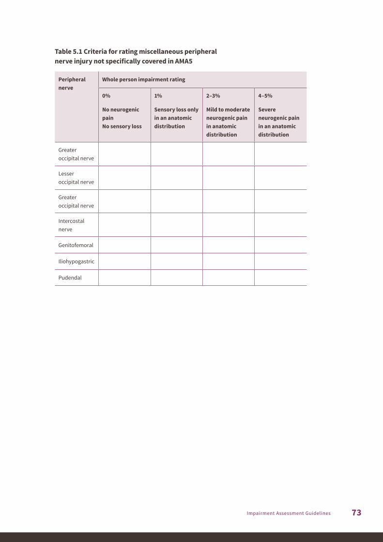
Impairment Assessment Guidelines 73
Table 5.1 Criteria for rating miscellaneous peripheral nerve injury not specifically covered in AMA5
Peripheral nerve
Whole person impairment rating
0%
No neurogenic pain No sensory loss
1%
Sensory loss only in an anatomic distribution
2–3%
Mild to moderate neurogenic pain in anatomic distribution
4–5%
Severe neurogenic pain in an anatomic distribution
Greater occipital nerve
Lesser occipital nerve
Greater occipital nerve
Intercostal nerve
Genitofemoral
Iliohypogastric
Pudendal

Impairment Assessment Guidelines74
This page has been intentionally left blank.

6 E
ar, n
ose,
thro
at
and
rela
ted
stru
ctur
es
6 EAR, NOSE, THROAT AND RELATED STRUCTURES

This page has been intentionally left blank.

Impairment Assessment Guidelines 75
Chapter 11, AMA5 (p245) applies to the assessment of permanent impairment of the ear (with the exception of hearing impairment), nose, throat and related structures, subject to the modifications set out below.
Before undertaking an impairment assessment, users of the Guidelines must be familiar with the following:
• the Introduction in the Guidelines
• chapters 1 and 2 of AMA5
• the appropriate chapter/s of the Guidelines for the body system they are assessing, and
• the appropriate chapter/s of AMA5 for the body system they are assessing.
In the event of any inconsistency, the Guidelines take precedence over AMA5. Refer to paragraph 1.3.
Introduction
6.1 Chapter 11, AMA5 (pp245–275) details the assessment of the ear, nose, throat and related structures. With the exception of hearing impairment, which is dealt with in Chapter 9 of the Guidelines, Chapter 11, AMA5 should be followed in assessing whole person impairment, with the variations included below.
6.2 The degree of impairment arising from conditions that are not caused by a work injury must be assessed and considered when determining the degree of whole person impairment. The degree to which pre-existing conditions and lifestyle activities such as smoking contribute to the degree of permanent impairment requires judgement on the part of the clinician undertaking the impairment assessment. Any deductions for these conditions must be recorded and reasoning for the degree of impairment assigned provided in the assessor’s report.
The ear
6.3 Hearing is assessed under Chapter 9 in these Guidelines.
6.4 Before undertaking a hearing assessment, consider the information in Table 11-10, AMA5 (pp272–275) under Hearing Impairment, noting that only the last column is not relevant.
6 EAR, NOSE, THROAT AND RELATED STRUCTURES

Impairment Assessment Guidelines76
The face
6.5 AMA5 (pp255–259) relates to the face. Table 11-5, AMA5 (p256) should be replaced with Table 6.1 when assessing whole person impairment due to facial disorders and/ or disfigurement.
Table 6.1: Criteria for rating permanent impairment due to facial disorders and/or disfigurement
CLASS 1
0%–5% impairment of the whole person
CLASS 2
6%–10% impairment of the whole person
CLASS 3
11%–15% impairment of the whole person
CLASS 4
16%–50% impairment of the whole person
Facial abnormality limited to disorder of cutaneous structures, such as visible simple scars (not hypertrophic or atrophic) or abnormal pigmentation or mild, unilateral, facial paralysis affecting most branches or nasal distortion that affects physical appearance or partial loss or deformity of the outer ear
Facial abnormality involves loss of supporting structure of part of face, with or without cutaneous disorder (e.g. depressed cheek, nasal, or frontal bones) or near complete loss of definition of the outer ear or hypertrophic or atrophic scar
Facial abnormality involves absence of normal anatomic part or area of face, such as loss of eye or loss of part of nose, with resulting cosmetic deformity, combine with any functional loss, e.g. vision (Chapter 8, AMA4) or severe unilateral facial paralysis affecting most branches or mild, bilateral, facial paralysis affecting most branches
Massive or total distortion of normal facial anatomy with disfigurement so severe that it precludes social acceptance, or severe, bilateral, facial paralysis affecting most branches or loss of a major portion of or entire nose
Note 1: Tables used to classify the examples in section 11.3, AMA5 (pp256–259) should also be ignored and assessors should refer to the modified table above for classification.
Note 2: For cases of facial disfigurement (which can include scarring), the assessor may alternatively refer to the TEMSKI table, if that is considered more appropriate, given the nature of the disfigurement.
6.6 Visual impairment related to eye disorders causing disfigurement, such as enophthalmos, must be assessed by an Ophthalmologist.

Impairment Assessment Guidelines 77
The nose, throat and related structures
Respiration (section 11.4a, AMA5, pp259–261)
6.7 Assessments for obstructive sleep apnoea can only be undertaken by a Respiratory Physician or Ear, Nose and Throat Physician. The type of sleep apnoea must have been confirmed prior to rating.
6.8 Before impairment can be assessed for obstructive sleep apnoea (3rd paragraph, section 11.4a, AMA5, p259), the person must have had appropriate assessment and treatment by an Ear, Nose and Throat Physician and a sleep study by a Respiratory Physician undertaken within the past two years.
6.9 The assessment of sleep apnoea is addressed in section 5.6, AMA5 (p105) and assessors should refer to this chapter, as well as paragraphs 8.10–8.13 in the Guidelines for rating.
6.10 Table 11-6, AMA5 (p260), Criteria for rating impairment due to air passage defects: This table should be replaced with Table 6.2, below, when assessing whole person impairment due to air passage defects.

Impairment Assessment Guidelines78
Tabl
e 6.
2: C
rite
ria fo
r rat
ing
perm
anen
t im
pair
men
t due
to a
ir pa
ssag
e de
fect
s
Perc
enta
ge im
pair
men
t of t
he w
hole
per
son
CLAS
S 1A
0%–5
%
CLAS
S 1
0%–1
0%
CLAS
S 2
11%
–29%
CLAS
S 3
30%
–49%
CLAS
S 4
50%
–89%
CLAS
S 5
90%
+
Ther
e ar
e sy
mpt
oms
of si
gnifi
cant
diff
icul
ty
in b
reat
hing
thro
ugh
the
nose
. Exa
min
atio
n re
veal
s sig
nific
ant p
artia
l ob
stru
ctio
n of
the
right
an
d/or
left
nasa
l cav
ity o
r na
soph
aryn
x or s
igni
fican
t se
ptal
per
fora
tion
Dysp
noea
doe
s not
occ
ur
at re
st a
nd d
yspn
oea
is
not p
rodu
ced
by w
alki
ng
free
ly o
n a
leve
l sur
face
, cl
imbi
ng st
airs
free
ly, o
r pe
rfor
man
ce o
f oth
er u
sual
ac
tiviti
es o
f dai
ly li
ving
and
dy
spno
ea is
not
pro
duce
d by
stre
ss, p
rolo
nged
ex
ertio
n, h
urry
ing,
hill
-cl
imbi
ng, o
r rec
reat
iona
l or
sim
ilar a
ctiv
ities
re
quiri
ng in
tens
ive
effor
t*
and
exam
inat
ion
reve
als
part
ial o
bstr
uctio
n of
the
orop
hary
nx,
lary
ngop
hary
nx, l
aryn
x,
uppe
r tra
chea
(to
the
four
th c
artil
agin
ous r
ing)
, lo
wer
trac
hea,
bro
nchi
, or
com
plet
e (b
ilate
ral)
obst
ruct
ion
of th
e no
se
or n
asop
hary
nx
Dysp
noea
doe
s not
occ
ur
at re
st a
nd d
yspn
oea
is n
ot
prod
uced
by
wal
king
free
ly
on a
leve
l sur
face
, clim
bing
on
e fli
ght o
f sta
irs, o
r pe
rfor
man
ce o
f oth
er u
sual
ac
tiviti
es o
f dai
ly li
ving
but
dy
spno
ea is
pro
duce
d by
st
ress
, pro
long
ed e
xert
ion,
hu
rryi
ng, h
ill-c
limbi
ng,
or re
crea
tiona
l or s
imila
r ac
tiviti
es (e
xcep
t sed
enta
ry
form
s) a
nd e
xam
inat
ion
reve
als p
artia
l obs
truc
tion
of th
e or
opha
rynx
, la
ryng
opha
rynx
, lar
ynx,
up
per t
rach
ea (t
o th
e fo
urth
car
tilag
inou
s rin
g),
low
er tr
ache
a, b
ronc
hi,
or co
mpl
ete
(bila
tera
l) ob
stru
ctio
n of
the
nose
or
nas
opha
rynx
Dysp
noea
doe
s not
occ
ur
at re
st b
ut d
yspn
oea
is
prod
uced
by
wal
king
fr
eely
mor
e th
an o
ne o
r tw
o le
vel b
lock
s, c
limbi
ng
one
fligh
t of s
tairs
eve
n w
ith p
erio
ds o
f res
t, or
pe
rfor
man
ce o
f oth
er u
sual
ac
tiviti
es o
f dai
ly li
ving
and
dy
spno
ea is
pro
duce
d by
st
ress
, pro
long
ed e
xert
ion,
hu
rryi
ng, h
ill-c
limbi
ng,
or re
crea
tiona
l or s
imila
r ac
tiviti
es a
nd e
xam
inat
ion
reve
als p
artia
l obs
truc
tion
of th
e or
opha
rynx
, la
ryng
opha
rynx
, lar
ynx,
up
per t
rach
ea (t
o th
e fo
urth
car
tilag
inou
s rin
g),
low
er tr
ache
a or
bro
nchi
Dysp
noea
occ
urs a
t res
t, al
thou
gh in
divi
dual
is n
ot
nece
ssar
ily b
edrid
den
and
dysp
noea
is a
ggra
vate
d by
th
e pe
rfor
man
ce o
f any
of
the
usua
l act
iviti
es o
f dai
ly
livin
g (b
eyon
d pe
rson
al
clea
nsin
g, d
ress
ing
or
groo
min
g) a
nd e
xam
inat
ion
reve
als p
artia
l obs
truc
tion
of th
e or
opha
rynx
, la
ryng
opha
ryx,
lary
nx,
uppe
r tra
chea
(to
the
four
th
cart
ilagi
nous
ring
), lo
wer
tr
ache
a, a
nd/o
r bro
nchi
Seve
re d
yspn
oea
occu
rs
at re
st a
nd sp
onta
neou
s re
spira
tion
is in
adeq
uate
an
d re
spira
tory
ven
tilat
ion
is re
quire
d an
d ex
amin
atio
n re
veal
s par
tial o
bstr
uctio
n of
the
orop
hary
nx,
lary
ngop
hary
nx, l
aryn
x,
uppe
r tra
chea
(to
the
four
th c
artil
agin
ous r
ing)
, lo
wer
trac
hea
or b
ronc
hi
*Pro
phyl
actic
rest
rictio
n of
act
ivity
, suc
h as
stre
nuou
s com
petit
ive
spor
t, do
es n
ot e
xclu
de su
bjec
t fro
m c
lass
1.
Not
e: In
divi
dual
s with
succ
essf
ul p
erm
anen
t tra
cheo
stom
y or
stom
a sh
ould
be
rate
d at
25%
impa
irmen
t of t
he w
hole
per
son.
Ex
ampl
e 11
-16,
AM
A5 (p
261)
: Par
tial o
bstr
uctio
n of
the
lary
nx a
ffect
ing
only
one
voc
al c
ord
is b
ette
r lin
ked
to v
oice
(sec
tion
11.4
e, A
MA5
).

Impairment Assessment Guidelines 79
6.11 When using Table 11-7, AMA5 ‘Relationship of dietary restrictions to permanent impairment’ (p262), first category is to be 0–19%, not 5–19%. The selection within class 1 for mastication and deglutition is made in accordance with Table 6.3 below, which is an extension of Table 11-7 in AMA5 (p262). Table 6.3 divides class 1 of permanent impairment into 4 groupings of impairment.
Table 6.3 – Class 1 rating for Mastication and deglutition
%WPI Criteria
0 No interference. Food of any desired type can be eaten without difficulty
1 – 4 Very tough or hard food has to be avoided but diet is otherwise as desired
5 – 9 Diet is permanently limited to soft foods
10 – 14 Diet is permanently limited to soft and pureed foods
15 – 19 Diet is permanently limited to pureed foods
6.12 A treating Dentist or relevant specialist report confirming the presence of a diagnosis that impacts directly on mastication and deglutition is required.
Speech (AMA5, pp262–264)
6.13 With regard to the first sentence of the ‘Examining procedure’ subsection (pp263–264), the examiner should have sufficient hearing for the purpose – disregard “normal hearing as defined in the earlier section of this chapter on hearing”.
6.14 Examining procedure (pp263–264), second paragraph: “The examiner should base judgements of impairment on two kinds of evidence: (1) attention to and observation of the individual’s speech in the office (e.g. during conversation, during the interview, and while reading and counting aloud) and (2) reports pertaining to the individual’s performance in everyday living situations”. Disregard the next sentence: “The reports or the evidence should be supplied by reliable observers who know the person well.”
6.15 Examining procedure (pp263–264): where the word ‘American’ appears as a reference, substitute ‘Australian’, and change measurements to the metric system (e.g. 8.5 inch = 21.6cm).

Impairment Assessment Guidelines80
The voice (section 11.4e, AMA5, pp264–267)
6.16 Substitute the word ‘laryngopharyngeal’ for ‘gastroesophageal’ in all examples where it appears.
6.17 Example 11.25 (Impairment Rating, p269), second sentence: add the underlined phrase “Combine with appropriate ratings due to other impairments including respiratory impairment to determine whole person impairment.”
Ear, nose, throat and related structures impairment evaluation summary
6.18 Table 11-10, AMA5 (pp272–275): Do not use this table, except for impairment of olfaction and/or taste, and hearing impairment as determined in the Guidelines.
Olfaction and taste
6.19 Before undertaking impairment of olfaction and/or taste, consider the information in Table 11-10, AMA5 (pp274) under Impairment of Olfaction and/or Taste or refer partial Table 6.4 below. A maximum of 5% WPI is allowable for total loss of each of these senses.

Impairment Assessment Guidelines 81
Tabl
e 6.
4 –
Impa
irm
ent e
valu
atio
n su
mm
ary
of e
ar, n
ose
and
thro
at a
nd o
lfact
ion
and
tast
e
Diso
rder
His
tory
, inc
ludi
ng se
lect
ed
rele
vant
sym
ptom
sEx
amin
atio
n
reco
rdAs
sess
men
t of
phys
ical
func
tion
Phys
ical
fin
ding
sDi
agno
sis
Degr
ee o
f im
pair
men
t
Gene
ral
Ear,
nose
and
thro
at
sym
ptom
s (e.
g. h
earin
g lo
ss,
dizz
ines
s or v
ertig
o) a
nd
gene
ral s
ympt
oms;
impa
ct
of sy
mpt
oms o
n fu
nctio
n an
d ab
ility
to d
o da
ily
activ
ities
; pro
gnos
is if
cha
nge
antic
ipat
ed; r
evie
w m
edic
al
hist
ory
and
any
resu
lting
lim
itatio
n of
phy
sica
l fun
ctio
n
Com
preh
ensi
ve
phys
ical
exa
min
atio
n;
deta
iled
rele
vant
sy
stem
ass
essm
ent
Data
der
ived
from
rele
vant
st
udie
s (e.
g. a
udio
met
ry)
Asse
ssm
ent o
f seq
uela
e in
clud
ing
end-
orga
n da
mag
e an
d im
pairm
ent
Reco
rd a
ll pe
rtin
ent
diag
nose
s; n
ote
if th
ey
are
at m
axim
um m
edic
al
impr
ovem
ent;
if no
t, di
scus
s un
der w
hat c
ondi
tions
and
w
hen
stab
ility
is e
xpec
ted
Crite
ria
outli
ned
in c
hapt
er
11 A
MA5
Hea
ring
impa
irmen
tCo
mpr
ehen
sive
his
tory
in
clud
ing
fam
ily h
isto
ry,
deve
lopm
enta
l his
tory
of
trau
ma,
noi
se a
nd d
rug
expo
sure
; sur
gica
l pro
cedu
res;
sy
mpt
oms o
f im
bala
nce
(e.g
. un
stea
dine
ss o
r ver
tigo)
; ear
-po
ppin
g; h
isto
ry o
f tin
nitu
s;
age;
ass
ocia
ted
met
abol
ic
and/
or e
ndoc
rine
diso
rder
s
Gene
ral p
hysi
cal
exam
inat
ion;
ear
, no
se a
nd th
roat
ex
amin
atio
n;
findi
ngs f
rom
pn
eum
onot
osco
py,
tuni
ng-fo
rk te
sts,
he
arin
g te
sts,
bal
ance
fu
nctio
n te
sts a
nd
radi
ogra
phic
test
s;
met
abol
ic e
valu
atio
n
Oto
logi
c ex
amin
atio
n on
tuni
ng-fo
rk te
sts;
ty
mpa
nom
etry
; be
havi
oura
l, au
diom
etry
an
d au
dito
ry b
rain
(e
voke
d) re
spon
se te
sts;
el
ectr
ococ
hlea
ogra
phy
test
s;
elec
troy
stag
mog
raph
y;
met
abol
ic a
nd e
ndoc
rine
stud
ies a
s nec
essa
ry
Asse
ss re
leva
nt o
rgan
s;
exte
rnal
ear
and
m
iddl
e ea
r fun
ctio
ns;
Eust
achi
an tu
be fu
nctio
n;
stat
us o
f hea
ring
by
audi
omet
ry; s
tatu
s of
ele
ctro
phys
iolo
gic
test
s as a
pplic
able
Cond
uctiv
e, se
nsor
ineu
ral,
mix
ed a
nd fu
nctio
nal
hear
ing
loss
; tin
nitu
s;
Men
iere
’s di
seas
e
Asse
ssed
as
per
the
Hea
ring
chap
ter o
f the
Gu
idel
ines
Impa
irmen
t of
Olfa
ctio
n an
d Ta
ste
Ear,
nose
and
thro
at
infe
ctio
ns; h
ead
trau
ma;
st
ruct
ural
or f
orei
gn b
ody
nasa
l obs
truc
tion;
nas
al
alle
rgy;
infe
ctio
ns o
f nos
e an
d si
nuse
s; h
isto
ry o
f hea
d an
d ne
ck tu
mou
rs, d
rug
use
Test
s for
odo
ur
iden
tifica
tion;
test
s for
ta
ste
iden
tifica
tion;
re
sults
of x
-ray
s an
d he
ad a
nd n
eck;
re
sults
of M
RI a
nd C
T st
udie
s of h
ead
and
neck
; alle
rgy
test
s
Subj
ectiv
e te
sts f
or o
dour
id
entifi
catio
n; su
bjec
tive
test
s for
tast
e id
entifi
catio
n;
elec
tric
al ta
ste
test
s; x-
rays
of
hea
d an
d ne
ck; M
RI
and
CT st
udie
s of h
ead;
cr
ania
l ner
ve fu
nctio
n te
sts;
te
st fo
r nas
al a
llerg
ens
Nas
al o
bstr
uctio
n du
e to
m
ucos
al o
edem
a, n
asal
po
lyps
, sep
tal o
r tur
bina
te
occl
usio
n of
airw
ay o
r na
sal t
umou
r; ph
ysic
al
findi
ngs m
ay b
e no
rmal
ex
cept
for p
rese
ntin
g sy
mpt
om; s
urge
ry se
quel
Nas
al se
ptic
al d
evia
tion;
nas
al
airw
ay o
cclu
sion
by
turb
inat
e bo
ne; a
llerg
ic rh
initi
s; n
asal
po
lyps
; sin
usiti
s; fo
reig
n bo
dy
in n
ose;
trau
mat
ic a
nosm
ia;
drug
toxi
city
; der
moi
d ex
ceph
aloc
ele;
men
ingo
cele
; in
trac
rani
al o
r oth
er tu
mou
r
See
Olfa
ctio
n an
d ta
ste
(sec
tion
11.4
c AM
A5)

Impairment Assessment Guidelines82
This page has been intentionally left blank.

7 U
rinar
y an
d re
prod
uctiv
e sy
stem
s
7 URINARY AND REPRODUCTIVE SYSTEMS

This page has been intentionally left blank.

Impairment Assessment Guidelines 83
Chapter 7, AMA5 (p143) applies to the assessment of permanent impairment of the urinary and reproductive systems, subject to the modifications set out below.
Before undertaking an impairment assessment, users of the Guidelines must be familiar with the following (in this order):
• the Introduction in the Guidelines
• chapters 1 and 2 of AMA5
• the appropriate chapter/s of the Guidelines for the body system they are assessing, and
• the appropriate chapter/s of AMA5 for the body system they are assessing.
In the event of any inconsistency, the Guidelines take precedence over AMA5. Refer to paragraph 1.3.
Introduction
7.1 Chapter 7, AMA5 (pp143–171) provides clear details for assessment of the urinary and reproductive systems. Overall the chapter should be followed in assessing whole person impairment, with the variations included below.
7.2 Neurogenic bladder and cauda equina syndrome are assessed as indicated in the Spine chapter of the Guidelines, paragraph 4.9.
7.3 The assessor needs to be quite clear as to the cause of the urological dysfunction. If due to primary dysfunction of the urinary system, this chapter applies, but if due to a spinal cord injury, the Spine chapter would apply, or if due to a neurological disorder, the Neurological chapter would apply.
7.4 For both male and female sexual dysfunction, identifiable pathology must be present for an impairment percentage to be given.
7.5 For all assessments under this chapter, appropriate investigation, patho-anatomical diagnosis and treatment options must have been provided by a Urologist or Gynaecologist prior to the assessment.
Urinary diversion
7.6 Table 7-2, AMA5 (p150) should be replaced with Table 7.1, below, when assessing whole person impairment due to urinary diversion disorders. This table includes ratings for neobladder and continent urinary diversion.
7 URINARY AND REPRODUCTIVE SYSTEMS

Impairment Assessment Guidelines84
7.7 Continent urinary diversion is defined as a continent urinary reservoir constructed of small or large bowel with a narrow catheterisable cutaneous stoma through which it must be emptied several times a day.
Table 7.1: Criteria for rating permanent impairment due to urinary diversion disorders
Diversion type % Impairment of the whole person
Ureterointestinal 10%
Cutaneous ureterostomy 10%
Nephrostomy 15%
Neobladder/replacement cystoplast 15%
Continent urinary diversion 20%
Bladder
7.8 Table 7-3, AMA5 (p151) should be replaced with Table 7.2, below, when assessing impairment due to bladder disease. This table includes ratings involving urge and total incontinence. Urge urinary incontinence is the involuntary loss of urine associated with a strong desire to void. This table also should be used for examples of mixed urge and stress incontinence, examples of nocturnal enuresis or wetting bed, or examples of total incontinence.
Table 7.2: Criteria for rating permanent impairment due to bladder disease
CLASS 1
0%–15% WPI
CLASS 2
16%–40% WPI
CLASS 3
41%–70% WPI
Symptoms and signs of bladder disorder and requires intermittent treatment and normal functioning between malfunctioning episodes
Symptoms and signs of bladder disorder e.g. urinary frequency (urinating more than every two hours); severe nocturia (urinating more than three times a night); urge incontinence more than once a week and requires continuous treatment
Abnormal (i.e. under or over) reflex activity (e.g. intermittent urine dribbling, loss of control, urinary urgency and urge incontinence once or more each day) and/or no voluntary control of micturition; reflex or areflexic bladder on urodynamics and/or total incontinence (e.g. fistula)

Impairment Assessment Guidelines 85
7.9 Example 7-16, AMA5 (p151) should be reclassified as an example of Class 2, as the urinary frequency is more than every two hours and continuous treatment would be expected.
7.10 Examples 7-18, 7-19, 7-20, AMA5 (pp152–153) are all examples of bladder dysfunction secondary to neurological disease. In the case of example 7-18, the impairment of bladder function should be assessed using Table 13-19, AMA5 (p341). In the case of examples 7-19 and 7-20, the impairment of bladder function should be assessed using Table 15-6d, AMA5 (p397).
Urethra
7.11 Table 7-4, AMA5 (p153) should be replaced with Table 7.3, below, when assessing impairment due to urethral disease. This table includes ratings involving stress incontinence. Stress urinary incontinence is the involuntary loss of urine occurring with clinically demonstrable raised intra-abdominal pressure. It is expected that urinary incontinence should be of a regular or severe nature (necessitating the use of protective pads or appliances).
Table 7.3: Criteria for rating permanent impairment due to urethral disease
CLASS 1
0%–10% WPI
CLASS 2
11%–20% WPI
CLASS 3
21%–40% WPI
Symptoms and signs of urethral disorder and requires intermittent therapy for control
Symptoms and signs of urethral disorder; stress urinary incontinence more than three times a week and cannot effectively be controlled by treatment
Urethral dysfunction resulting in intermittent urine dribbling, or stress urinary incontinence at least daily
Male reproductive organs
Penis
7.12 In AMA5, p157, the box labelled ‘Class 3, 21–35%’ should read ‘Class 3, 20% impairment of the whole person’ as the descriptor ‘No sexual function possible’ does not allow a range (the correct value is shown in AMA5 Table 7-5, p156). Note, however, that there is a loading for age, so a rating higher than 20% is possible (AMA5, section 7.7, p156).
Testicles, epididymides and spermatic cords
7.13 Table 7-7, AMA5 (p159) should be replaced with Table 7.4, below, when assessing impairment due to testicular, epididymal and spermatic cord disease. This table includes rating for infertility and equates impairment with female infertility (see Table 7.6 in this chapter of the Guidelines).

Impairment Assessment Guidelines86
7.14 Male infertility is defined as azoospermia or other cause of inability to cause impregnation even with assisted conception techniques.
7.15 Loss of sexual function related to spinal injury should only be assessed as an impairment where there is other objective evidence of spinal cord, cauda equina or bilateral nerve root dysfunction. The ratings described in Table 13-21, AMA5 (p342) are used in this instance. There is no additional impairment rating system for loss of sexual function in the absence of objective clinical findings.
Table 7.4: Criteria for rating permanent impairment due to testicular, epididymal and spermatic cord disease
CLASS 1
0%–10% WPI
CLASS 2
11%–15% WPI
CLASS 3
16%–35% WPI
Testicular, epididymal or spermatic cord disease symptoms and signs and anatomic alteration and no continuous treatment required and no seminal or hormonal function or abnormalities or solitary testicle*
Testicular, epididymal or spermatic cord disease symptoms and signs and anatomic alteration and cannot effectively be controlled by treatment and detectable seminal or hormonal abnormalities
Trauma or disease produces bilateral anatomic loss of the primary sex organs or no detectable seminal or hormonal function or infertility
*Loss of one testicle should be assessed as class 1, 10% WPI
Female reproductive organs
Fallopian tubes and ovaries
7.16 Table 7-11, AMA5 (p167) should be replaced with Table 7.6, below, when assessing impairment due to fallopian tube and ovarian disease. This table includes rating for infertility and equates impairment with male infertility (see Table 7.4, above).
7.17 Female infertility: a woman in the childbearing age is infertile when she is unable to conceive naturally. This may be due to anovulation, tubal blockage, cervical or vaginal blockage or an impairment of the uterus.
7.18 Table 7.5 below replaces AMA5 Table 7-10 (p165) for the assessment of cervical and uterine disease.

Impairment Assessment Guidelines 87
Table 7.5: Criteria for rating permanent impairment due to cervical and uterine disease
CLASS 1
0%–10% WPI
CLASS 2
11%–15% WPI
CLASS 3
16%–35% WPI
Cervical or uterine disease or deformity symptoms and signs do not require continuous treatment; or cervical stenosis, if present, requires no treatment or anatomic cervical or uterine loss in the postmenopausal period
Cervical or uterine disease or deformity symptoms and signs require continuous treatment; or cervical stenosis, if present, requires periodic treatment
Cervical or uterine disease or deformity symptoms and signs are not controlled by treatment; or complete cervical stenosis or anatomic or complete functional cervical or uterine loss in the premenopausal period
Table 7.6: Criteria for rating permanent impairment due to fallopian tube and ovarian disease
CLASS 1
0%–10% WPI
CLASS 2
11%–15% WPI
CLASS 3
16%–35% WPI
Fallopian tube or ovarian disease or deformity symptoms and signs do not require continuous treatment or only one functioning fallopian tube and/or ovary in the premenopausal period* or bilateral fallopian tube or ovarian functional loss in the postmenopausal period
Fallopian tube or ovarian disease or deformity symptoms and signs require continuous treatment, but tubal patency persists and ovulation is possible
Fallopian tube or ovarian disease or deformity symptoms and signs and total tubal patency loss or failure to produce ova in the premenopausal period or bilateral fallopian tube or bilateral ovarian loss in the premenopausal period; infertility
*the loss of an ovary and/or fallopian tube should be assessed as class 1, 10% WPI.

Impairment Assessment Guidelines88
This page has been intentionally left blank.

8 R
espi
rato
ry
syst
em
8 RESPIRATORY SYSTEM

This page has been intentionally left blank.

Impairment Assessment Guidelines 89
Chapter 5, AMA5 (p87) applies to the assessment of permanent impairment of the respiratory system, subject to the modifications set out below.
Before undertaking an impairment assessment, users of the Guidelines must be familiar with the following:
• the Introduction in the Guidelines
• chapters 1 and 2 of AMA5
• the appropriate chapter/s of the Guidelines for the body system they are assessing, and
• the appropriate chapter/s of AMA5 for the body system they are assessing.
In the event of any inconsistency, the Guidelines take precedence over AMA5. Refer to paragraph 1.3.
Introduction
8.1 Chapter 5, AMA5 (pp87–115) provides a useful summary of the methods for assessing whole person impairment arising from respiratory disorders.
8.2 The degree of impairment arising from conditions not caused by a work injury must be assessed and considered in determining the degree of permanent impairment, and recorded in the report. The degree to which pre-existing conditions and lifestyle activities such as smoking contribute to the degree of permanent impairment requires judgement on the part of the assessor. The manner in which any deduction for these is applied needs to be recorded in the assessor’s report.
Examinations, clinical studies and other tests for evaluating respiratory disease (section 5.4, AMA5)
8.3 The predicted lower limit values provided in the laboratory tests (to Thoracic Society of Australia and NZ (TSANZ) standards) are applied in Table 5-12, AMA5 (p107), to determine the impairment classification for respiratory disorders. AMA5 Tables 5-2b, 5-3b, 5-4b, 5-5b, 5-6b and 5-7b should not be used.
8.4 Table 5-12, AMA5 (p107) should be used to assess whole person impairment for respiratory disorders other than occupational asthma. The pulmonary function tests listed in Table 5-12 must be performed to TSANZ standards by a pulmonary function laboratory. Exercise testing is not required.
8 RESPIRATORY SYSTEM

Impairment Assessment Guidelines90
8.5 Classes 2, 3 and 4 in Table 5-12, AMA5 (p107) list ranges of whole person impairment. The assessor should nominate the nearest whole percentage based on the complete clinical circumstances when selecting within the range, giving reasons to support the %WPI selected in the report.
8.6 An isolated abnormal diffusing capacity for carbon monoxide (DLCO) in the presence of otherwise normal results of lung function testing should be interpreted with caution and its aetiology should be clarified. Where the DLCO is the key parameter used to rate impairment, its relationship to the work injury must be explained.
Asthma (section 5.5, AMA5, p102-104)
8.7 In assessing whole person impairment arising from occupational asthma, the assessor will require evidence from the treating physician that:
• an appropriate diagnosis has been established by a Respiratory Physician based on clinical history, physical examination and spirometry with at least one appropriate lung function test performed to TSANZ standards by a pulmonary function laboratory within the last 12 months. In rare cases where the person is unable to undertake the test for medical reasons, an opinion from a second Respiratory Physician is required.
• the clinical status has been confirmed over time with repeated spirometry, and
• the worker has received optimal treatment, has an Asthma Plan in place, and is compliant with their medication regimen.
8.8 Bronchial challenge testing should not be performed as part of the impairment assessment. In Table 5-9, AMA5 (p104) ignore column 4 (PC20 mg/mL or equivalent, etc.).
8.9 Permanent impairment due to asthma is rated by the score for the best postbronchodilator forced expiratory volume in one second (FEV1) (score in Table 5–9, AMA5, column 2) plus % of FEV1 (score in column 3) plus minimum medication required (score in column 5). The total score derived is then used to assess the % impairment in Table 5-10, AMA5 (p104). The same approach to determining the actual impairment within the range of %WPI discussed in 8.5 should be adopted.

Impairment Assessment Guidelines 91
Obstructive sleep apnoea (section 5.6, AMA5, p105)
8.10 Assessments for obstructive sleep apnoea can only be undertaken by a Respiratory or Ear, Nose and Throat Physician. The cause must have been confirmed prior to rating.
8.11 This section needs to be read in conjunction with section 11.4, AMA5 (p259) and section 13.3c, AMA5 (p317).
8.12 Before permanent impairment can be assessed, the person must have had appropriate assessment and treatment by an Ear, Nose and Throat Physician and a sleep study by a Respiratory Physician undertaken within the past two years.
8.13 The degree of permanent impairment due to obstructive sleep apnoea should be calculated with reference to Table 13-4, AMA5 (p317).
Hypersensitivity pneumonitis (section 5.7, AMA5, p105)
8.14 Whole person impairment arising from disorders included in this section is assessed according to the impairment classification in Table 5-12, AMA5 (p107).
Lung cancer (section 5.9, AMA5, p106)
8.15 Whole person impairment due to lung cancer should be assessed using Table 5-12, AMA5 (p107) (not Table 5-11). Table 5-11 is used to help select the rating within the class. Where surgery has occurred, assessment should not be undertaken until at least six months after the procedure.
8.16 Persons with residual lung cancer after treatment are classified in Impairment Class 4 (Table 5-12).

Impairment Assessment Guidelines92
This page has been intentionally left blank.

9 H
earin
g
9 HEARING

This page has been intentionally left blank.

Impairment Assessment Guidelines 93
Chapter 11, AMA5 (p245) applies to the assessment of permanent impairment of hearing, subject to the modifications set out below.
Before undertaking an impairment assessment, users of the Guidelines must be familiar with the following:
• the Introduction in the Guidelines
• the appropriate chapter/s of the Guidelines for the body system they are assessing, and
• the National Acoustic Laboratory (NAL) Guide.
In the event of any inconsistency, the Guidelines take precedence over the NAL Guide and AMA5. Refer to paragraph 1.3.
Assessment of hearing impairment (hearing loss)
9.1 A worker may present for hearing loss assessment before having undergone all or any of the health investigations that generally occur before assessment of whole person impairment. For this reason and to ensure that conditions other than ‘occupational hearing impairment’ are precluded, the medical assessment should be undertaken by an Ear, Nose and Throat Physician or other appropriately qualified specialist. The medical assessment needs to be undertaken in accordance with Table 9.1 below. The assessor performing the assessment must examine the worker. The assessment must be based on medical history and ear, nose and throat examination, evaluation of relevant audiological tests and evaluation of other relevant investigations available to the assessor. Only an Ear, Nose and Throat Physician or other appropriately qualified specialist can issue permanent impairment reports for assessment of hearing impairment.
Some of the relevant tests are discussed in the hearing impairment assessment summary below.
9 HEARING

Impairment Assessment Guidelines94
Tabl
e 9.
1 Im
pair
men
t Ass
essm
ent S
umm
ary
for H
eari
ng
Diso
rder
His
tory
, inc
ludi
ng se
lect
ed
rele
vant
sym
ptom
sEx
amin
atio
n
reco
rdAs
sess
men
t of
phys
ical
func
tion
Phys
ical
fin
ding
sDi
agno
sis
Degr
ee o
f im
pair
men
t
Hea
ring
impa
irmen
tCo
mpr
ehen
sive
his
tory
in
clud
ing
fam
ily h
isto
ry,
deve
lopm
enta
l his
tory
of
trau
ma,
noi
se a
nd d
rug
expo
sure
; sur
gica
l pro
cedu
res;
sy
mpt
oms o
f im
bala
nce
(e.g
. un
stea
dine
ss o
r ver
tigo)
; ea
rpop
ping
; his
tory
of t
inni
tus;
ag
e; a
ssoc
iate
d m
etab
olic
an
d/or
end
ocrin
e di
sord
ers
Gene
ral p
hysi
cal
exam
inat
ion;
ear
, no
se a
nd th
roat
ex
amin
atio
n;
findi
ngs f
rom
pn
eum
onot
osco
py,
tuni
ng-fo
rk te
sts,
he
arin
g te
sts,
bal
ance
fu
nctio
n te
sts a
nd
radi
ogra
phic
test
s;
met
abol
ic e
valu
atio
n
Oto
logi
c ex
amin
atio
n on
tuni
ng-fo
rk te
sts;
ty
mpa
nom
etry
; be
havi
oura
l, au
diom
etry
an
d au
dito
ry b
rain
(e
voke
d) re
spon
se te
sts;
el
ectr
ococ
hlea
ogra
phy
test
s;
elec
troy
stag
mog
raph
y;
met
abol
ic a
nd e
ndoc
rine
stud
ies a
s nec
essa
ry
Asse
ss re
leva
nt o
rgan
s;
exte
rnal
ear
and
m
iddl
e ea
r fun
ctio
ns;
Eust
achi
an tu
be fu
nctio
n;
stat
us o
f hea
ring
by
audi
omet
ry; s
tatu
s of
ele
ctro
phys
iolo
gic
test
s as a
pplic
able
Cond
uctiv
e, se
nsor
ineu
ral,
mix
ed a
nd fu
nctio
nal
hear
ing
loss
; tin
nitu
s;
Men
iere
’s di
seas
e
Asse
ssed
as
per
the
Guid
elin
es

Impairment Assessment Guidelines 95
9.2 Cortical Evoked Response Audiometry (CERA) can be requested by the assessor in the event that standard audiology testing is inconsistent or there is a discrepancy between audiology test results and observed function. The rationale for requiring the test must be documented in the report.
9.3 The degree of hearing impairment not caused by exposure to noise is assessed and considered when determining the degree of noise induced/work-related hearing impairment. While this requires medical judgement on the part of the examining assessor, any non-work-related impairment should be recorded in the report.
9.4 Do not use Tables 11–1, 11–2, 11–3, AMA5 (pp247–250). For the purposes of the Guidelines, National Acoustic Laboratory (NAL) tables from the NAL Report No. 118, Improved procedure for determining percentage loss of hearing (January 1988) are adopted as follows:
• Tables RB 500–4000 (pp11–16)
• Tables RM 500–4000 (pp18–23)
• Appendix 1 and 2 (pp8–9)
• Appendix 5 and 6 (pp24–26)
• Tables EB 4000–8000 (pp28–30) (the extension tables)
• Tables EM 4000–8000 (pp32–34) (the extension tables)
When an assessor uses the extension tables, they must provide an explanation of the worker’s special requirement to be able to hear at frequencies above 4000Hz.
In the presence of significant conduction hearing loss, the extension tables do not apply.
Table 11–3, AMA5 is replaced by Table 9.2 in this chapter.
9.5 It is noted that there are some arithmetical errors in the NAL tables, however, the impact of these errors is minimal and assessors should use these tables, rather than any other programs, for consistency.
Impairment Assessment Guidelines96
Hearing impairment
9.6 Impairment of a worker’s hearing is determined according to assessment of the individual’s binaural hearing impairment.
9.7 Permanent hearing impairment should be assessed when the condition is stable. Prosthetic devices (such as hearing aids) must not be worn (or must be switched off) during the assessment of hearing acuity.
9.8 Hearing threshold level for pure tones is defined as the number of decibels above standard audiometric zero for a given frequency at which the listener’s threshold of hearing lies when tested in a suitable sound attenuated environment. It is the reading on the hearing level dial of an audiometer that is calibrated according to Australian Standard AS IEC 60645.1-2002.
9.9 Assessment of binaural hearing impairment: Binaural hearing impairment is determined by using the tables in the 1988 NAL publication with allowance for presbyacusis according to the presbyacusis correction table, if applicable, in the same publication.
The Binaural Tables RB 500–4000 (NAL report no. 118, pp11–16) are to be used. The extension Tables EB 4000-8000 (pp28–30) may be used when the worker has ‘a special requirement to be able to hear above frequencies above 4000Hz’ (NAL report no. 118, p6). Where an assessor uses the extension tables, they must provide an explanation of the worker’s special requirement to be able to hear at frequencies above 4000Hz.
Where it is necessary to use the monaural tables, the binaural hearing impairment (BHI) is determined by the formula:
BHI = [4 x (better ear hearing loss)] + worse ear hearing loss
5
9.10 Presbyacusis correction table (Appendix 5, NAL publication, p24) only applies to occupational hearing loss contracted by gradual process – for example, occupational noise induced hearing loss and/or occupational solvent induced hearing loss. Please note when calculating by formula for presbyacusis correction (e.g. when the worker is above 81 years) the formula is correct as long as the correct numerator is used, that is b=-1.79059*(age) (page 26, NAL) and not (b) 1.79509 (page 25, NAL). Note: Recent reprintings of this NAL guide have been corrected.

Impairment Assessment Guidelines 97
9.11 Binaural hearing impairment and severe tinnitus: Up to 5% BHI may be added to the work-related binaural hearing impairment for severe tinnitus caused by a work injury:
• after presbyacusis correction, if applicable, and
• before determining WPI.
The severity of tinnitus is determined by the assessor with consideration given as to its impact on ADL. The value assigned must be supported by clear rationale. Refer examples 9.1–9.5 in this chapter.
9.12 Vestibulocochlear nerve assessment using AMA5 (p333): Tinnitus in the absence of hearing loss resulting from a traumatic brain injury, where it adversely affects ADL, can be rated as 1% WPI.
9.13 Only hearing ear: A worker has an ‘only hearing ear’ if he or she has suffered a non-work-related severe or profound sensorineural hearing loss in the other ear. If a worker suffers a work injury causing a hearing loss in the only hearing ear of x dBHL at a relevant frequency, the worker’s work-related binaural hearing impairment at that frequency is calculated from the binaural tables using x dB as the hearing threshold level in both ears. Deduction for presbyacusis if applicable and addition for severe tinnitus is undertaken according to this guide.
9.14 When necessary, binaural hearing impairment figures should be rounded to the nearest 0.1%. Rounding up should occur if equal to or greater than .05%, and rounding down should occur if equal to or less than .04%.
9.15 Table 9.2 is used to convert binaural hearing impairment, after deduction for presbyacusis if applicable and after addition for severe tinnitus, to WPI.

Impairment Assessment Guidelines98
Noise Induced Hearing Loss
9.16 The assessment of permanent impairment and %WPI in respect of noise induced hearing loss needs to be assessed consistently with the particular requirements of subsections 188(2) and (3) of the Act which provide:
“(2) Subject to this section, where a claim is made under this Act in respect of noise induced hearing loss by a worker (not being a person who has retired from employment on account of age or ill health), the whole of the loss will be taken to have occurred immediately before notice of the injury was given and, subject to any proof to the contrary, to have arisen out of employment in which the worker was last exposed to noise capable of causing noise induced hearing loss.
(3) If a claim is made under this Act in respect of noise induced hearing loss by a person who has retired from employment on account of age or ill-health, the whole of the loss will be taken to have occurred immediately before the person retired and, subject to any proof to the contrary, to have arisen out of employment in which the person was last exposed to noise capable of causing noise induced hearing loss.”
Notwithstanding section 22(7)(b) of the Act, regard must be had to any audiogram(s) undertaken post retirement and prior to the assessment in determining any non-work related component of the worker’s current impairment.
9.17 For the purpose of rating impairment, use the better of the air and bone conduction thresholds at 2000Hz and below. Above 2000Hz use the air conduction thresholds.
9.18 Impairment due to noise induced hearing loss is to be calculated on the assessed hearing thresholds between 2000Hz and 4000Hz.
9.19 If noise exposure has been prolonged, 1500Hz can be included in the impairment assessment, provided a detailed explanation is given as to frequency, duration and source of noise exposure, whether it was constant or intermittent and, if known, decibels.
9.20 The following thresholds apply when rating for noise induced hearing loss. Any readings above these are to be rated as per the following limits:
1500Hz – 45dB
2000Hz – 65dB
3000Hz – 90dB
4000Hz – 90dB

Impairment Assessment Guidelines 99
Table 9.2: Relationship of binaural hearing impairment to whole person impairment
% Binaural hearing
impairment
% Whole person
impairment
% Binaural hearing
impairment
% Whole person
impairment
0.0 – 5.9 0 51.1 – 53.0 26
6.0 – 6.7 3 53.1 – 55.0 27
6.8 – 8.7 4 55.1 – 57.0 28
8.8 – 10.6 5 57.1 – 59.0 29
10.7 – 12.5 6 59.1 – 61.0 30
12.6 – 14.4 7 61.1 – 63.0 31
14.5 – 16.3 8 63.1 – 65.0 32
16.4 – 18.3 9 65.1 – 67.0 33
18.4 – 20.4 10 67.1 – 69.0 34
20.5 – 22.7 11 69.1 – 71.0 35
22.8 – 25.0 12 71.1 – 73.0 36
25.1 – 27.0 13 73.1 – 75.0 37
27.1 – 29.0 14 75.1 – 77.0 38
29.1 – 31.0 15 77.1 – 79.0 39
31.1 – 33.0 16 79.1 – 81.0 40
33.1 – 35.0 17 81.1 – 83.0 41
35.1 – 37.0 18 83.1 – 85.0 42
37.1 – 39.0 19 85.1 – 87.0 43
39.1 – 41.0 20 87.1 – 89.0 44
41.1 – 43.0 21 89.1 – 91.0 45
43.1 – 45.0 22 91.1 – 93.0 46
45.1 – 47.0 23 93.1 – 95.0 47
47.1 – 49.0 24 95.1 – 97.0 48
49.1 – 51.0 25 97.1 – 99.0 49
99.1 – 100 50

Impairment Assessment Guidelines100
9.21 Examples 11.1, 11.2, 11.3, AMA5 (pp250–251) are replaced by examples 9.1–9.7, below.
Table 9.3: Medical assessment elements in examples
Element Example No.
General use of binaural table – NAL 1988 1,2
‘Better ear’ – ’worse ear’ crossover 1,2
Assessable audiometric frequencies 7 – also 1,2,4,5,6
Tinnitus 1,2,3,4,5
Presbyacusis All examples
Binaural hearing impairment All examples
Conversion to whole person impairment All examples
Gradual process injury 3
Noise-induced hearing loss 1,2,3,5,6,7
Solvent-induced hearing loss 3
Acute occupational hearing loss 4,5
Acute acoustic trauma 5
Pre-existing non-occupational hearing loss
6
Only hearing ear 6
NAL 1988 Extension Table Use 7
Multiple causes of hearing loss 3,5,6
Head injury 4

Impairment Assessment Guidelines 101
Example 9.1: Occupational noise-induced hearing loss and severe tinnitus
A 55 year-old man, a boilermaker for 30 years, gave a history of progressive hearing loss and tinnitus. The tinnitus was present most days, interfering with concentration and regularly interfering with sleep when it could not be dampened with extraneous noise. The assessor has assessed the tinnitus as severe. The external auditory canals and tympanic membranes were normal. Rinne test was positive (air conduction better than bone conduction) bilaterally and the Weber test result was central. Clinical assessment of hearing was consistent with results of pure tone audiometry, which showed a bilateral sensorineural hearing loss consistent with the dose and duration of significant noise. The assessor diagnosed noise induced hearing loss (NIHL) with severe tinnitus. The assessor included the 1500Hz frequency in this assessment due to long-term constant noise exposure likely to be greater than 90dB. Presbyacusis correction does not apply because the worker is less than 56 years of age.
Pure tone audiometry
Frequency (Hz)
Left (dB HL)
Right (dB HL)
Binaural hearing impairment (% BHI)
500 15 10 0
1000 20 20 0
1500 25 25 1.4
2000 35 35 3.4
3000 60 60 6.3
4000 75 75 8.2
6000 30 30 -
8000 20 20 -
Total % BHI 19.3
No Presbyacusis correction 0
Add 4.0% BHI for severe tinnitus 4
Adjusted total % BHI 23.3
Resultant total BHI of 23.3% = 12% WPI (Table 9.2 in the Guidelines)

Impairment Assessment Guidelines102
Example 9.2: Occupational noise-induced hearing loss and mild tinnitus
A 55-year-old man, a steelworker for 30 years, gave a history of increasing difficulties with hearing and tinnitus. In the first 20 years of his career little attention was paid to hearing protection. There was no family history of deafness and no past history of recreational noise, illness or medication that could impact upon hearing. The assessor diagnosed occupational noise-induced hearing loss with intermittent mild tinnitus that had no impact on ADL and was only mildly irritating during the day and night. The assessor considered the loss at 1500Hz should be included due to the reported constant noise exposure likely to be greater than 90dB given the occupational history.
Pure tone audiometry
Frequency (Hz)
Left (dB HL)
Right (dB HL)
Binaural hearing impairment (% BHI)
500 15 15 0.0
1000 15 15 0.0
1500 20 25 1.0
2000 30 35 2.5
3000 50 45 4.2
4000 55 55 5.2
6000 30 30 -
8000 20 20 -
Total % BHI 12.9
Less Presbyacusis correction 0
Adjusted total % BHI 12.9
Resultant total BHI of 12.9% = 7% WPI (Table 9.2 in the Guidelines)
Comment: The assessor’s opinion is that the tinnitus suffered by the worker is not severe and thus no addition to the binaural hearing impairment was made for tinnitus.

Impairment Assessment Guidelines 103
Example 9.3: Multiple gradual process occupational hearing loss
A 63-year-old male boat builder and printer gave a history of hearing difficulty and tinnitus. There had been marked chronic exposure to both noise and solvents in these occupations for 35 years altogether. The assessor diagnosed bilateral noise-induced hearing loss and bilateral solvent-induced hearing loss with severe tinnitus. The tinnitus was rated in the lowest range of severity as it only occasionally interfered with sleep for one or two nights of the week and only mildly affects him during the day.
The assessor’s opinion is that the solvent exposure contributed to the hearing impairment as a gradual process injury. The total noise-induced and solvent-induced BHI was 17.5%. The assessor did not identify any factors in the family or personal health profile of the worker to account for the loss at 1500Hz and considered the long-term exposure, whilst intermittent, warranted inclusion of this frequency in the assessment.
The appropriate presbyacusis deduction was applied. Then, the assessor added 2% BHI to the after-presbyacusis binaural hearing impairment for severe tinnitus at the lower end of the range with occasional sleep disturbance and no impact on other ADL.
Pure tone audiometry
Frequency (Hz)
Left (dB HL)
Right (dB HL)
Binaural hearing impairment (% BHI)
500 15 15 0.0
1000 15 15 0.0
1500 25 25 1.4
2000 35 40 3.8
3000 60 60 6.3
4000 60 60 6.0
6000 45 50 -
8000 40 40 -
Total noise-induced and solvent-induced % BHI
17.5
Presbyacusis correction of 1.7% -1.7
1% BHI addition for medically assessed severe tinnitus
1
Adjusted total % BHI 16.8
Resultant total BHI of 16.8% = 9% WPI (Table 9.2 in the Guidelines)

Impairment Assessment Guidelines104
Example 9.4: Occupational noise-induced hearing loss from head injury
A 62-year-old male worker sustained a head injury after falling from a ladder. He suffered left hearing loss and tinnitus unaccompanied by vertigo. The assessor assesses his tinnitus in the lower range of severity as the injury has resulted in sleep disturbance two or three nights per week and some interference with ADL in the day. External auditory canals and tympanic membranes are normal. Rinne test is positive bilaterally and Weber test lateralises to the right. CT scan of the temporal bones shows a fracture on the left. Clinical assessment of hearing is consistent with pure tone audiometry, which shows a flat left sensorineural hearing loss and mild right sensorineural hearing loss. Presbyacusis correction does not apply because the worker sustained a head injury. The assessor used all frequencies in the assessment due to the effect of fracture trauma being non-selective for a particular frequency.
Pure tone audiometry
Frequency (Hz)
Left (dB HL)
Right (dB HL)
Binaural hearing impairment (% BHI)
500 50 15 2.3
1000 55 15 3.1
1500 60 20 3.4
2000 65 20 2.6
3000 65 25 2.2
4000 65 30 2.1
6000 65 20 -
8000 65 20 -
Total % BHI 15.7
No correction for presbyacusis applies 0
Adjusted 2.0% BID for severe tinnitus 2
Adjusted total % BHI 17.7
Resultant total BHI of 17.7% = 9% WPI (Table 9.2 in the Guidelines)

Impairment Assessment Guidelines 105
Example 9.5: Acute unilateral occupational hearing loss in the presence of pre-existing bilateral noise-induced hearing loss
A 62-year-old man who has been a production worker for 10 years in a noisy workplace was injured in an explosion that occurred on his left side while at work. He reported immediate post-injury otalgia and acute hearing loss in the left ear. The assessor noted, at examination, hearing loss in the right ear consistent with noise exposure. For the purposes of the impairment assessment, it was clinically determined that this NIHL effect would, more probably than not, have been present in the left ear at the time of the explosion. The hearing loss was greater on the left side, consistent with the explosion. The assessor diagnosed left acoustic trauma in the presence of bilateral occupational noise-induced hearing, as there was no evidence that hearing in the left ear was different to the right, prior to the explosion. Severe tinnitus is present and assessed at the highest range due to major sleep disturbance every night with ADL impacted during every day. The tinnitus was attributed to the explosion trauma as this is clinically more likely to be the cause rather than the mild chronic noise effect. All the frequencies were used to assess the left ear but only the frequencies of 3000 and 4000HZ were used to calculate the NIHL given its short duration and low exposure.
Pure tone audiometry
Frequency (Hz)
Left (dB HL)
Right (dB HL)
Binaural hearing impairment (% BHI)
BHI due to NIHL (% BHI)
500 30 15 1.0 0.0
1000 45 15 2.5 0.0
1500 55 15 2.5 0.0
2000 70 15 2.2 0.0
3000 80 25 2.4 0.7
4000 80 30 2.3 0.8
6000 >80 30 n/a in NIHL n/a in NIHL
8000 >80 25 n/a in NIHL n/a in NIHL
Total % BHI Presbyacusis correction for NIHL
12.9 1.5-1.3
Adjusted NIHL BHI (%) 0.2
Acute acoustic trauma BHI (%) 12.9
Presbyacusis does not apply to acute acoustic trauma Tinnitus - 5% BHI allocated to the acoustic trauma
05
Totals 17.9 0.2
Resultant total BHI due to acute acoustic trauma of 17.9% - 0.2 = 17.7% BHI = 9% WPI (Table 9.2 in the Guidelines)

Impairment Assessment Guidelines106
Example 9.6: Occupational noise-induced hearing loss in an only hearing ear
A 66-year-old woman has been a textile worker for 30 years. Childhood mumps had left her with profound hearing loss in the left ear. She gave a history of progressive hearing loss in her only hearing ear unaccompanied by tinnitus or vertigo. External auditory canals and tympanic membranes appeared normal. Rinne test was positive on the right and was false negative (the signal was picked up in the other ear) on the left. Weber test lateralised to the right. Clinical assessment of hearing is consistent with pure tone audiogram showing a profound left sensorineural hearing loss and a partial right sensorineural hearing loss. The assessor diagnosed NIHL in the right ear consistent with noise dose and duration. For the purposes of the assessment of NIHL (column 5), the assessor assumes that the hearing in the left ear is identical to that in the right ear due to the noise exposure at work. The assessor used the frequencies of 1500 and 2000Hz in this assessment due the dose and duration of noise in an only hearing ear.
Pure tone audiometry
Frequency (Hz)
Left (dB HL)
Right (dB HL)
Binaural hearing impairment (% BHI)
BHI due to noise-induced hearing loss
500 >95 10 3.4 0
1000 >95 15 4.3 0
1500 >95 20 4.2 0.6
2000 >95 25 3.8 1.1
3000 >95 50 5.4 4.8
4000 >95 70 8.0 7.5
6000 >95 50 n/a in NIHL n/a in NIHL
8000 >95 40 n/a in NIHL n/a in NIHL
Total % BHI 29.1
Total occupational % BHI 14.0
Presbyacusis correction does not apply to a 66 year old woman
0
No addition tinnitus 0
Adjusted total occupational % BHI n/a 14.0
Total occupational BHI of 14% = 7% WPI (Table 9.2 in the Guidelines)

Impairment Assessment Guidelines 107
Example 9.7: Occupational noise-induced hearing loss where there is a special requirement for ability to hear at frequencies above 4000 Hz
A 56-year-old female process worker who worked in a noisy factory for 20 years had increasing hearing difficulty. The diagnosis made was bilateral occupational noise-induced hearing loss extending to 6000 Hz or 8000 Hz. The assessor was of the opinion that there was a special requirement for hearing above 4000 Hz as the worker is a musical writer for violins and violas in a recreational opera company, so the extension tables were used as there is a significant effect on her ADL. There was no conductive hearing loss, or other factor identified to account for this loss at 6000 and 8000Hz.
Pure tone audiometry
Binaural hearing impairment (% BHI)
Frequency (Hz)
Left (dB HL)
Right (dB HL)
Using extension table – 4000, 6000 and 8000 Hz (p28-29 NAL)
Not using extension table
500 10 10 0.0 0.0
1000 15 15 0.0 0.0
1500 20 25 0.0 0.0
2000 30 32 2.5 2.5
3000 45 45 4.1 4.1
4000 45 50 2.2 3.6
6000 60 55 1.6 -
8000 50 20 0.2 -
Total BHI (%) using extension table 10.6
Total BHI (%) not using extension table 10.2
Presbyacusis correction 0 0
The assessor is of the opinion that the binaural hearing impairment in the matter is 10.6% rather than 10.2%
0
Adjusted total % BHI 10.6
Resultant Total BHI of 10.6% = 5% WPI (Table 9.2 in the Guidelines)

Impairment Assessment Guidelines108
This page has been intentionally left blank.

10 V
isua
l sys
tem
10 VISUAL SYSTEM

This page has been intentionally left blank.

Impairment Assessment Guidelines 109
Chapter 8, AMA4 (p209) applies to the assessment of permanent impairment of the visual system, subject to the modifications set out below.
Before undertaking an impairment assessment, users of the Guidelines must be familiar with the following (in this order):
• the Introduction in the Guidelines
• chapters 1 and 2 of AMA5
• the appropriate chapter/s of the Guidelines for the body system they are assessing, and
• the appropriate chapter/s of AMA4 for the body system they are assessing.
In the event of any inconsistency, the Guidelines take precedence over AMA4 and AMA5. Refer to paragraph 1.3.
Introduction and approach to assessment
10.1 The visual system must be assessed by an Ophthalmologist.
10.2 Chapter 8, AMA4 (pp209–222) is adopted for the Guidelines without significant change. The exception is Table 3, AMA4, as below.
10.3 AMA4 is used rather than AMA5 for the assessment of whole person impairment of the visual system because:
• there is little emphasis on diplopia in AMA5, yet this is a relatively frequent problem
• many Ophthalmologists are familiar with the Royal Australian College of Ophthalmologists’ impairment guide, which is similar to AMA4.
10.4 Impairment of vision should be measured with the worker wearing their prescribed corrective spectacles and/or contact lenses, if that was normal for the injured worker before the work injury or condition. If, as a result of the work injury or condition, the injured worker has been prescribed corrective spectacles and/or contact lenses for the first time, or different spectacles and/or contact lenses than those prescribed before the injury or condition, the difference should be accounted for in the assessment of permanent impairment.
10.5 An Ophthalmologist should assess visual field impairment in all cases.
10.6 The Ophthalmologist should perform or review all tests necessary for the assessment of whole person impairment rather than relying on the interpretations of tests done by the Orthoptist or Optometrist.
10 VISUAL SYSTEM

Impairment Assessment Guidelines110
10.7 In section 8.5, AMA4 (p222) on other conditions, the ‘additional 10% impairment’ referred to means 10% WPI, not 10% impairment of the visual system.
10.8 If disfigurement is limited to the immediate periorbital area, being the orbital contents plus the eyelids, then it is to be assessed by the Ophthalmologist. However, if it extends to involve more of the face, it is to be undertaken in accordance with the ear, nose and throat chapter by an assessor accredited in that system.
10.9 For impairment assessment for monocular aphakia or monocular pseudophakia, AMA4 directs that the lower numbers are used in Table 3 (p212, AMA4). The separate scales are no longer required. Only the top numbers are to be used.
10.10 AMA4 allows an additional 5% to 10% visual impairment to be combined with the impaired visual function of the involved eye for abnormalities, such as media opacities, corneal or lens opacities and abnormalities resulting from such symptoms as epiphora, photophobia or metamorphopsia, if it interferes with the visual function and is not reflected in visual acuity, decreased visual fields or ocular mobility with diplopia (p209, AMA4). This impairment can be applied even where the visual function impairment is 0%.

11 H
aem
atop
oiet
ic
syst
em
11 HAEMATOPOIETIC SYSTEM

This page has been intentionally left blank.

Impairment Assessment Guidelines 111
Chapter 9, AMA5 (p191) applies to the assessment of permanent impairment of the haematopoietic system, subject to the modifications set out below.
Before undertaking an impairment assessment, users of the Guidelines must be familiar with the following (in this order):
• the Introduction in the Guidelines
• chapters 1 and 2 of AMA5
• the appropriate chapter/s of the Guidelines for the body system they are assessing, and
• the appropriate chapter/s of AMA5 for the body system they are assessing.
In the event of any inconsistency, the Guidelines take precedence over AMA5. Refer to paragraph 1.3.
Introduction
11.1 Chapter 9, AMA5 (pp191–210) provides guidelines on the method of assessing whole person impairment of the haematopoietic system. Overall, that chapter should be followed when conducting the assessment, with variations indicated below. The diagnosis being rated must have been made by a Haematologist, Oncologist, Immunologist or other Specialist Internal Medicine Physician prior to the assessment.
11.2 Impairment of end organ function due to haematopoietic disorder should be assessed separately, using the relevant chapter of the Guidelines. The percentage WPI due to end organ impairment should be combined with any percentage WPI due to haematopoietic disorder, using the Combined Values Table, AMA5 (pp604–606).
11 HAEMATOPOIETIC SYSTEM

Impairment Assessment Guidelines112
Anaemia
11.3 Table 11.1 (below) replaces Table 9-2, AMA5 (p193).
Table 11.1: Classes of anaemia and percentage whole person impairment (WPI)
CLASS 1
0%–10% WPI
CLASS 2
11%–30% WPI
CLASS 3
31%–70% WPI
CLASS 4
71–100% WPI
No symptoms and haemoglobin 100–120g/L and no transfusion required
Minimal symptoms and haemoglobin 80–100g/L and no transfusion required
Moderate to marked symptoms and haemoglobin 50–80g/L before transfusion and transfusion of 2 to 3 units required, every 4 to 6 weeks
Moderate to marked symptoms and haemoglobin 50–80g/L before transfusion and transfusion of 2 to 3 units required, every 2 weeks
11.4 The assessor must exercise clinical judgement in determining WPI, using the criteria in Table 11.1. For example, if comorbidities exist which preclude transfusion, the assessor may assign Class 3 or Class 4, on the understanding that transfusion would under other circumstances be indicated. Similarly, there may be some workers with Class 2 impairment who, because of comorbidity, may undergo transfusion.
11.5 Pre-transfusion haemoglobin levels in Table 11.1 are to be used as indications only. It is acknowledged that, for some workers, it would not be medically advisable to permit the worker’s haemoglobin levels to be as low as indicated in the criteria of Table 11.1.
11.6 The assessor should indicate a %WPI, as well as the class.

Impairment Assessment Guidelines 113
Polycythaemia and myelofibrosis
11.7 The level of symptoms (as in Table 11.1) should be used a guide for the assessor in cases where non-anaemic tissue iron deficiency results from venesection.
Functional asplenia
11.8 In cases of functional or post traumatic asplenia, the assessor should assign 3% WPI. This should be combined with any other impairment rating, using the Combined Values Table, AMA5 (pp604–606).
White blood cell diseases
11.9 Table 9-3, AMA5 (p200) should be used for rating impairment due to HIV infection or auto immune deficiency disease.
Haemorrhagic and platelet disorders
11.10 Table 9-4, AMA5 (p203) is to be used as the basis for assessing haemorrhagic and platelet disorders.
11.11 For the purposes of the Guidelines, the criteria for inclusion in Class 3 of Table 9-4, AMA5 (p203) are:
• symptoms and signs of haemorrhagic and platelet abnormality
• requires continuous treatment, and
• interference with daily activities, with occasional assistance required.
11.12 For the purposes of the Guidelines, the criteria for inclusion in Class 4 of Table 9-4, AMA5 (p203) is:
• symptoms and signs of haemorrhagic and platelet abnormality
• requires continuous treatment, and
• difficulty performing daily activities, with continuous care required.
Deep-vein thrombosis
11.13 A single deep-vein thrombosis should not be assessed under the haematopoietic system. It is assessed under either the cardiovascular system or upper or lower extremity system.
Table 9-4, AMA5 (p203) is used as the basis for determining impairment due to a persistent or recurring thrombotic disorder.

Impairment Assessment Guidelines114
This page has been intentionally left blank.

12 E
ndoc
rine
sy
stem
12 ENDOCRINE SYSTEM

This page has been intentionally left blank.

Impairment Assessment Guidelines 115
Chapter 10, AMA5 (p211) applies to the assessment of permanent impairment of the endocrine system, subject to the modifications set out below.
Before undertaking an impairment assessment, users of the Guidelines must be familiar with the following (in this order):
• the Introduction in the Guidelines
• chapters 1 and 2 of AMA5
• the appropriate chapter/s of the Guidelines for the body system they are assessing, and
• the appropriate chapter/s of AMA5 for the body system they are assessing.
In the event of any inconsistency, the Guidelines take precedence over AMA5. Refer to paragraph 1.3.
Introduction
12.1 Chapter 10, AMA5 provides a useful summary of the methods for assessing whole person impairment arising from disorders of the endocrine system. The diagnosis being rated must have been made by an Endocrinologist with supporting objective evidence prior to the assessment.
12.2 Refer to other appropriate chapters for related structural changes – the visual system (chapter 8 of AMA4), the skin (e.g. pigmentation, chapter 8, AMA5), the central and peripheral nervous system (memory, chapter 13, AMA3), the urinary and reproductive system (infertility, renal impairment, chapter 7, AMA5), the digestive system (dyspepsia, chapter 6, AMA5), the cardiovascular system (chapters 3 and 4, AMA5).
12.3 The clinical findings to support the impairment assessment are to be reported in the units recommended by the Royal College of Pathologists of Australia. Assessors should use the current RCPA Manual to assist with interpretation of pathology tests, which can be found at www.rcpamanual.edu.au.
Adrenal cortex
12.4 First paragraph of 10.5, AMA5 (p222): No regard is to be had to the last sentence: “They also affect inflammatory response, cell membrane permeability, and immunologic responses, and they play a role in the development and maintenance of secondary sexual characteristics.” Replace with: “Immunological and inflammatory responses are reduced by these hormones and they play a role in the development and maintenance of secondary sexual characteristics.”
12 ENDOCRINE SYSTEM

Impairment Assessment Guidelines116
12.5 Example 10-18, AMA5 (pp224–225): Westergren erythrocyte sedimentation rate (WSR) is equivalent to ESR.
12.6 Example 10-20, AMA5 (p225) – History: For “hypnotic bladder” read “hypotonic bladder”.
Diabetes mellitus
12.7 AMA5 (p231): Refer to the current Australian Diabetes Association Guidelines with regard to levels of fasting glucose. For the purposes of Table 10-8 (p231, AMA5), satisfactory control is a haemoglobin A1c level of ≤ 7%.
Mammary Glands
12.8 In AMA5 example 10-45 regarding current symptoms (p239), the last sentence is replaced with ‘Routine use of bromocriptine and cabergoline is normal in Australia. It is rare that nausea precludes their use’.
Criteria for rating permanent impairment due to metabolic bone disease
12.9 AMA5 (p240): Impairment due to a metabolic bone disease itself is unlikely to be associated with a work injury and would usually represent a pre-existing condition.
12.10 Impairment from fracture, spinal collapse or other complications may arise as a result of a work injury associated with these underlying conditions (as noted in section 10.10c, AMA5) and would be assessed using the other chapters indicated, with the exception of chapter 18 on pain which is excluded from the Guidelines.

13 S
kin
13 SKIN

This page has been intentionally left blank.

Impairment Assessment Guidelines 117
Chapter 8, AMA5 (p173) applies to the assessment of permanent impairment of the skin, subject to the modifications set out below.
Before undertaking an impairment assessment, users of the Guidelines must be familiar with the following (in this order):
• the Introduction in the Guidelines
• chapters 1 and 2 of AMA5
• the appropriate chapter/s of the Guidelines for the body system they are assessing, and
• the appropriate chapter/s of AMA5 for the body system they are assessing.
In the event of any inconsistency, the Guidelines take precedence over AMA5. Refer to paragraph 1.3.
Introduction
13.1 Chapter 8, AMA5 (pp173–190) refers to skin diseases generally rather than work-related skin diseases alone. In the Guidelines, this chapter has been adopted for measuring impairment of the skin system, with the variations listed in the subsequent sections of this chapter.
13.2 Disfigurement, scars and skin grafts may be assessed as causing significant permanent impairment when the skin condition causes limitation in the performance of activities of daily living (ADL).
13.3 Table 8-2, AMA5 (p178) provides the method of classification of impairment due to skin disorders. Three components – signs and symptoms of skin disorder, limitations in activities of daily living and requirements for treatment – define five classes of permanent impairment. The assessor should allocate a specific percentage impairment within the range for the class that best describes the clinical status of the worker and provide detailed reasons for their selection in the report.
13.4 The skin is regarded as a single organ and all non-facial scarring, including any compensable and non-compensable scarring, is measured together as one overall impairment rather than assessing individual scars separately and combining the results. If there is any unrelated component, then this is deducted from the total. As the skin is treated as a whole (except for the face), the location of the unrelated component does not need to be in the vicinity of the work injury to be deducted.
13 SKIN

Impairment Assessment Guidelines118
13.5 If there are multiple claims being assessed at the same time, then the scars that relate to each claim must be assessed chronologically and any scarring resulting from the previous claim must be deducted as pre-existing e.g. assess scars from claim 1, as in 13.4, and then assess scarring from claim 1 and claim 2 together, then deduct the impairment as assessed from claim 1 as pre-existing (refer example).
Example: Claim 1 shoulder injury – Claim 2 knee injury
Assess pre-existing scar from abdomen 1%
Assess compensable shoulder scar plus abdomen 2%
Assess compensable knee scar plus shoulder plus abdomen 3%
Table 1 – Shoulder injury
2%–1% = 1%
Table 2 – Knee injury
3%–2% = 1%
13.6 The Table for the Evaluation of Minor Skin Impairment (TEMSKI – 13.1) is an extension of Table 8-2 in AMA5. The TEMSKI divides Class 1 of permanent impairment (0-9%) due to skin disorders into five groupings of impairment. The TEMSKI may be used by assessors (who are not trained in the skin body system but who are trained in the use of TEMSKI) for determining skin impairment from 0 – 4% WPI associated with the injury which they are rating. Skin impairment from the TEMSKI greater than 4% must be assessed by an assessor who has undertaken the requisite training in the assessment of the skin body system.
13.7 The TEMSKI is to be used in accordance with the principle of ‘best fit’. The assessor must be satisfied that the criteria within the chosen category of impairment best reflects the skin disorder being assessed. The assessor must provide detailed reasons as to why this category has been chosen over other categories.
13.8 For cases of facial disfigurement (which can include scarring), refer to Table 6.1 in the Ear, Nose and Throat Related Structures chapter of the Guidelines or alternatively to the TEMSKI table (up to 4% WPI unless accredited in skin), whichever is considered most appropriate given the nature of the disfigurement. The face is rated separately and then combined where appropriate.

Impairment Assessment Guidelines 119
13.9 In cases of inflammatory conditions involving both the face and the skin of other areas of the body, assessors are advised to assess by both skin (Table 8-2 AMA5) and by face (Table 6.1, Ear, Nose and Throat chapter) and then allocate whichever is the higher impairment.
13.10 Where there is a range of values in the TEMSKI categories, the assessor must use clinical judgement to determine the specific degree of impairment and provide the rationale for choosing that value in the report.
13.11 A scar may be present and rated as 0% WPI. For example, minimal uncomplicated scars for standard surgical procedures may not, of themselves, rate an impairment.
13.12 The case examples provided in chapter 8, AMA5 do not, in most cases, relate to permanent impairment that results from a work injury. The following examples are provided for information.
13.13 Work-related case study examples 13.1, 13.2, 13.3, 13.4, 13.5, 13.6 are included below, in addition to AMA5 examples 8.1–8.22 (pp178–187).

Impairment Assessment Guidelines120
Tabl
e 13
.1 –
For
The
Eva
luat
ion
of M
inor
Ski
n Im
pair
men
t (TE
MSK
I)
Crit
eria
0% W
PI1%
WPI
2% W
PI3–
4% W
PI5–
9% W
PI (S
kin
asse
ssor
s onl
y)
Desc
riptio
n of
the
scar
(s) a
nd/o
r ski
n co
nditi
on(s
) (sh
ape,
te
xtur
e, c
olou
r)
Wor
ker i
s not
con
scio
us o
r is
bar
ely
cons
ciou
s of t
he
scar
(s) o
r ski
n co
nditi
on
Good
col
our m
atch
with
su
rrou
ndin
g sk
in a
nd th
e sc
ar(s
) or s
kin
cond
ition
is
bare
ly d
istin
guis
habl
e. W
orke
r is
una
ble
to e
asily
loca
te th
e sc
ar(s
) or s
kin
cond
ition
No
trop
hic
chan
ges
Any
stap
le o
r sut
ure
mar
ks a
re b
arel
y vi
sibl
e
Wor
ker i
s con
scio
us o
f the
sc
ar(s
) or s
kin
cond
ition
Som
e pa
rts o
f the
scar
(s) o
r ski
n co
nditi
on c
olou
r con
tras
t with
th
e su
rrou
ndin
g sk
in a
s a re
sult
of p
igm
enta
ry o
r oth
er c
hang
es
Wor
ker i
s abl
e to
loca
te th
e sc
ar(s
) or s
kin
cond
ition
Min
imal
trop
hic
chan
ges
Any
stap
le o
r sut
ure
mar
ks a
re v
isib
le
Wor
ker i
s con
scio
us o
f the
sc
ar(s
) or s
kin
cond
ition
Not
icea
ble
colo
ur c
ontr
ast o
f sc
ar(s
) or s
kin
cond
ition
with
su
rrou
ndin
g sk
in a
s a re
sult
of
pigm
enta
ry o
r oth
er c
hang
es
Wor
ker i
s abl
e to
eas
ily lo
cate
th
e sc
ar(s
) or s
kin
cond
ition
Trop
hic
chan
ges
evid
ent t
o to
uch
Any
stap
le o
r sut
ure
mar
ks
are
clea
rly v
isib
le
Wor
ker i
s con
scio
us o
f the
sc
ar(s
) or s
kin
cond
ition
Easi
ly id
entifi
able
col
our
cont
rast
of s
car(
s) o
r ski
n co
nditi
on w
ith su
rrou
ndin
g sk
in a
s a re
sult
of p
igm
enta
ry
or o
ther
cha
nges
Wor
ker i
s abl
e to
eas
ily lo
cate
th
e sc
ar(s
) or s
kin
cond
ition
.
Trop
hic c
hang
es e
vide
nt to
touc
h
Any
stap
le o
r sut
ure
mar
ks
are
clea
rly v
isib
le
Wor
ker i
s con
scio
us o
f the
sc
ar(s
) or s
kin
cond
ition
Dist
inct
col
our c
ontr
ast o
f sc
ar(s
) or s
kin
cond
ition
with
su
rrou
ndin
g sk
in a
s a re
sult
of
pigm
enta
ry o
r oth
er c
hang
es
Wor
ker i
s abl
e to
eas
ily lo
cate
th
e sc
ar(s
) or s
kin
cond
ition
Trop
hic
chan
ges a
re v
isib
le
Any
stap
le o
r sut
ure
mar
ks
are
clea
rly v
isib
le
Loca
tion
Anat
omic
loca
tion
of th
e sc
ar(s
) or s
kin
cond
ition
no
t cle
arly
vis
ible
with
us
ual c
loth
ing/
hairs
tyle
Anat
omic
loca
tion
of th
e sc
ar(s
) or s
kin
cond
ition
is
not
usu
ally
vis
ible
with
us
ual c
loth
ing/
hairs
tyle
Anat
omic
loca
tion
of th
e sc
ar(s
) or s
kin
cond
ition
is
usua
lly v
isib
le w
ith u
sual
cl
othi
ng/h
airs
tyle
Anat
omic
loca
tion
of th
e sc
ar(s
) or
skin
con
ditio
n is
vis
ible
with
us
ual c
loth
ing/
hairs
tyle
Anat
omic
loca
tion
of th
e sc
ar(s
) or s
kin
cond
ition
is
usua
lly a
nd c
lear
ly v
isib
le w
ith
usua
l clo
thin
g/ha
irsty
le
Cont
our
No
cont
our d
efec
tM
inor
con
tour
def
ect
Cont
our d
efec
t vis
ible
Cont
our d
efec
t eas
ily v
isib
leCo
ntou
r def
ect e
asily
vis
ible
ADL
/ Tre
atm
ent
No
effec
t on
any
ADL
No
trea
tmen
t, or
inte
rmitt
ent
trea
tmen
t onl
y, re
quire
d
Neg
ligib
le e
ffect
on
any
ADL
No
trea
tmen
t, or
inte
rmitt
ent
trea
tmen
t onl
y, re
quire
d
Min
or li
mita
tion
in th
e pe
rfor
man
ce o
f few
ADL
No
trea
tmen
t, or
inte
rmitt
ent
trea
tmen
t onl
y, re
quire
d
Min
or li
mita
tion
in th
e pe
rfor
man
ce o
f few
ADL
AN
D ex
posu
re to
che
mic
al
or p
hysi
cal a
gent
s (e.
g.
sunl
ight
, hea
t, co
ld e
tc.)
may
te
mpo
raril
y in
crea
se li
mita
tion
No
trea
tmen
t, or
inte
rmitt
ent
trea
tmen
t onl
y, re
quire
d
Lim
itatio
n in
the
perf
orm
ance
of
few
ADL
(IN
CLU
DIN
G re
stric
tion
in g
room
ing
or d
ress
ing)
AN
D ex
posu
re to
che
mic
al o
r phy
sica
l ag
ents
(e.g
. sun
light
, hea
t, co
ld
etc.
) may
tem
pora
rily
incr
ease
lim
itatio
n or
rest
rictio
n
No
trea
tmen
t, or
inte
rmitt
ent
trea
tmen
t onl
y, re
quire
d
Adhe
renc
e to
un
derly
ing
stru
ctur
esN
o ad
here
nce
No
adhe
renc
eN
o ad
here
nce
Som
e ad
here
nce
Som
e ad
here
nce
This
tabl
e us
es th
e pr
inci
ple
of ‘b
est fi
t’. Yo
u sh
ould
ass
ess t
he im
pairm
ent t
o th
e w
hole
skin
syst
em a
gain
st e
ach
crite
ria a
nd th
en d
eter
min
e w
hich
impa
irmen
t cat
egor
y be
st fi
ts (o
r des
crib
es) t
he im
pairm
ent.
A sk
in im
pairm
ent w
ill u
sual
ly m
eet m
ost,
but d
oes n
ot n
eed
to m
eet a
ll, c
riter
ia to
‘bes
t fit’
a pa
rtic
ular
impa
irmen
t cat
egor
y. T
he
asse
ssor
mus
t pro
vide
det
aile
d re
ason
s as t
o w
hy th
is c
ateg
ory
has b
een
chos
en o
ver o
ther
cat
egor
ies.
Ref
er to
13.
6–13
.11
rega
rdin
g ap
plic
atio
n of
this
tabl
e.

Impairment Assessment Guidelines 121
Example 13.1: Cumulative irritant dermatitis
Subject: 42-year-old man
History: The worker is a spray painter working on ships in dry dock who has presented with a rash on both hands. Not required to prepare surface but required to mix paints (including epoxy and polyurethane) with ‘thinners’ (solvents) and spray metal ship’s surface. At end of each session, the worker was required to clean equipment with solvents and was not supplied with gloves or other personal protective equipment until after the onset of symptoms. Off work two months leading to clearance of the rash, but frequent recurrence, especially if the worker attempted prolonged work wearing latex or PVC gloves or wet work without gloves. Treatment by GP with topical steroid creams showed improvement.
Current: Returned to dry duties only at work. Mostly clear of dermatitis now, but flares.
Physical examination: Varies between ‘no abnormality detected’ to ‘mild self-limiting dermatitis of the dorsum of hands’. On the day of the assessment there was no identifiable skin condition.
Investigations: Patch test standard + epoxy + isocyanates (polyurethanes). No reactions.
Impairment: 3% WPI as deemed to be at the lower third of the range in Class 1 from Table 8.2 in AMA5 (p178).
Comment: Intermittently present and minimal interference with activities of daily living (ADL) and occasional intermittent treatment, perhaps once per year.

Impairment Assessment Guidelines122
Example 13.2 Burns
Subject: 32 year old man
History: The worker is an electrician. Twelve months ago he was involved in an accident in which a meter board suddenly exploded and his neck and chest were burnt. He was taken to the hospital and a second degree burn to his neck and chest was diagnosed.
Treatment: He was treated in hospital. He remained for 2 days and, following discharge, he attended outpatients for several weeks until the burn eventually healed leaving a rather poorly defined, abnormally pigmented linear keloid scar across his neck and chest. The scar measured approximately 6cm in length and 5cm in width.
Current: This is currently being treated with a silicone gel which he is applying once daily. The scar is painful when touched and when exposed to temperature. His shirt also irritates the scar and he cannot do up a collar. He also complains of pruritus in the scar which is present most of the time.
Investigation: Clinical examination reveals a prominent erythematous keloidal scar with the above dimensions. The scar is visible from 3 metres. He is uncomfortable in his clothes due to the irritation that this causes the scar. He is extremely embarrassed by the cosmetic appearance of this scar and has become somewhat socially withdrawn.
Impairment: 10% WPI from Table 8-2 Class 2 (p178, AMA5) at the lower end of the range.
Comment: There is a skin disorder and signs and symptoms are consistently present. There is limited performance of some of the activities of daily living (mainly social) because of his embarrassment regarding this problem. Itching is a problem and pain frequently occurs within the scar. He is always conscious of the problem and requires constant treatment in an effort to soothe this scar.

Impairment Assessment Guidelines 123
Example 13.3: ‘Cement dermatitis’ due to chromate in cement
Subject: 43 year-old man
History: Concreter since age 16. Eighteen-month history of increasing hand dermatitis eventually on dorsal and palmar surface of hands and fingers. Off work and treatment led to limited improvement only. Referred to Dermatologist and prescribed strong steroid ointment and cleansing lotion in lieu of soap.
Physical examination: Fissured skin, hyperkeratotic chronic dermatitis.
Investigation: Patch test. Positive reaction to dichromate.
Current: Intractable, chronic, fissured dermatitis.
Impairment: Mid-range from Class 2 in Table 8.2 (p178, AMA5) selected at 17% WPI.
Comment: Unable to obtain any employment because has chronic dermatitis and on disability support pension. Difficulty gripping items including steering wheel, hammer and other tools. Unable to do any wet work, (e.g. painting). Former home handyman, now calls in tradesman to do any repairs and maintenance. Limited performance in some ADL and requires intermittent treatment.

Impairment Assessment Guidelines124
Example 13.4: Latex contact urticaria/ angioedema with cross reactions
Subject: Female nurse, age 40
History: Six-month history of itchy hands minutes after applying latex gloves at work. Later swelling and redness associated with itchy hands and wrists and subsequently widespread urticaria. One week off led to immediate clearance. On return to work wearing PVC gloves developed anaphylaxis on first day back.
Physical examination: No abnormality detected or generalised urticaria/angioedema.
Investigation: Latex radioallergosorbent test, strong positive response.
Current: The subject experiences urticaria and anaphylaxis if she enters a hospital, some supermarkets or other stores (especially if latex items are stocked), at children’s parties or in other situations where balloons are present, or on inadvertent contact with latex items including sports goods handles, some clothing, and many shoes (latex based glues). Also has restricted diet (must avoid bananas, avocados and kiwi fruit).
Impairment: 22% WPI. At the higher end of the range within Class 2 selected from Table 8.2 (p178, AMA5).
Comment: Severe limitation in some ADL and uncertainty of when she could next experience an anaphylactic reaction.

Impairment Assessment Guidelines 125
Example 13.5: Non-melanoma skin cancer
Subject: 53-year-old married man
History: ‘Road worker’ since 17 years of age. Has had a basal cell carcinoma on the left forehead, squamous cell carcinoma on the right forehead (graft), basal cell carcinoma on the left ear (wedge resection) and squamous cell carcinoma on the lower lip (wedge resection) excised since 45 years of age. No history of locoregional recurrences. Multiple actinic keratoses treated with cryotherapy or Efudix (fluorouracil) cream over 20 years (forearms, dorsum of hands, head and neck).
Current: New lesion right preauricular area. Concerned over appearance “I look a mess.”
Physical examination: Multiple actinic keratoses forearms, dorsum of hands, head and neck. Five millimetre diameter nodular basal cell carcinoma right preauricular area, hypertrophic red scar 3cm length left forehead, 2cm diameter graft site (hypopigmented with 2mm contour deformity) right temple, non-hypertrophic scar left lower lip (vermilion) with slight step deformity and non-hypertrophic pale wedge resection scar left pinna leading to 30% reduction in size of the pinna. Graft sites taken from right post auricular area. No regional lymphadenopathy.
Impairment rating: 9% WPI
Comment: 6% WPI for facial disfigurement. This facial disfigurement was selected at the lowest range within this Class 2 (Table 6.1 in these Guidelines) and combined with 3% WPI for non-facial scarring of the upper extremities from Table 8.2 in AMA5. This non-facial scarring was clinically determined to be in the lower third percentile within Class 1 from Table 8-2. Total is 6% WPI combined with 3% WPI.

Impairment Assessment Guidelines126
Example 13.6: Non-melanoma skin cancer
Subject: 35-year-old single female professional surf life-saver
History: Occupational outdoor exposure since 19 years of age. Basal cell carcinoma on tip of nose excised three years ago with full thickness graft following failed intralesional interferon treatment.
Current: Poor self-esteem because of cosmetic result of surgery and facial disfigurement.
Physical examination: 1cm diameter graft site on the tip of nose (hypopigmented with 2mm depth contour deformity, cartilage not involved). Graft site taken from right post-auricular area.
Impairment rating: 10% WPI was selected at the highest range in Class 2 (Table 6.1 in these Guidelines) as it involved structural change in the nose and impact on her hair-line around the right ear.
Comment: Refer to Table 6.1 (facial disfigurement).

14 C
ardi
ovas
cula
r sy
stem
14 CARDIOVASCULAR SYSTEM

This page has been intentionally left blank.

Impairment Assessment Guidelines 127
Chapters 3 and 4, AMA5 (p25 and p65) apply to the assessment of permanent impairment of the cardiovascular system, subject to the modifications set out below.
Before undertaking an impairment assessment, users of the Guidelines must be familiar with the following:
• the Introduction in the Guidelines
• chapters 1 and 2 of AMA5
• the appropriate chapter/s of the Guidelines for the body system they are assessing, and
• the appropriate chapter/s of AMA5 for the body system they are assessing.
In the event of any inconsistency, the Guidelines take precedence over AMA5. Refer to paragraph 1.3.
Introduction
14.1 The cardiovascular system is discussed in chapter 3, AMA5 (Heart and Aorta) and 4, AMA5 (Systemic and Pulmonary Arteries) (pp25–85). These chapters can be used to assess whole person impairment of the cardiovascular system with the following minor modifications.
14.2 The impairment being evaluated/rated must be diagnosed by a Cardiologist with evidence to support the diagnosis prior to the assessment. The exception is thoracic outlet syndrome (14.8).
14.3 It is noted that in this chapter there are wide ranges for the impairment values in each category. When conducting a whole person impairment assessment, assessors should use their clinical judgement to express a specific percentage within the range suggested and provide justification for their choice in the report.
14 CARDIOVASCULAR SYSTEM

Impairment Assessment Guidelines128
Exercise stress testing
14.4 As with any other investigations not provided, it is not the role of an assessor to order exercise stress tests purely for the purpose of evaluating the extent of whole person impairment.
14.5 If the result of exercise stress testing is available, then it is a useful piece of information in arriving at the overall percentage impairment.
14.6 If investigations provided are inadequate for a proper assessment to be made, the assessor must consider the value of proceeding with the assessment of whole person impairment without the adequate investigations and data. Refer chapter 1 in the Guidelines, Information required for assessment (1.33–1.38) and ordering of additional investigations (1.56–1.59).
Vascular diseases affecting the extremities
14.7 Note that in this section, Table 4-4 and Table 4-5, AMA5 (p74 and p76) refer to percentage impairment of the upper or lower extremity. Therefore, an assessment of impairment concerning vascular impairment of the arm or leg requires that the percentages identified in Tables 4-4 and 4-5 be converted to whole person impairment. The table for conversion of the upper extremity is Table 16-3, AMA5 (p439) and the table for conversion of the lower extremity is Table 17-3, AMA5 (p527).
Thoracic outlet syndrome
14.8 Impairment due to thoracic outlet syndrome is assessed according to chapter 16, AMA5 on the upper extremities, and chapter 2 of the Guidelines.
Pulmonary embolism
14.9 Pulmonary embolism is assessed as per section 4.4, AMA5 (pp79–81).
Pulmonary hypertension
14.10 In Table 4-6 of AMA5 ‘any degree of pulmonary hypertension’ is defined as a PAP >40mmHg (p79).
14.11 The classes (2, 3 and 4) referred to in the criteria in class 3 and 4 of Table 4-6, AMA5, relate to Table 3-1 – Functional Classification of Cardiac Disease (p26, AMA5) where these classes are written as Class II, III and IV.

Impairment Assessment Guidelines 129
Effect of medical treatment
14.12 If the worker has been offered, but refused, additional or alternative medical treatment which the assessor considers is likely to improve the worker’s condition, the assessor must evaluate the current condition, without consideration for potential changes associated with the proposed treatment. The assessor must note the potential for improvement in the worker’s condition in the assessment report, and the reason for refusal by the worker, but must not adjust the degree of impairment on the basis of the worker’s decision (refer paragraph 1.31).
Pre-existing condition
14.13 If the assessor is unable to find any objective evidence of pre-existing functionally significant coronary artery disease, no rating can be applied for pre-existing disease and the assessor must explain this in the report.

Impairment Assessment Guidelines130
This page has been intentionally left blank.

15 D
iges
tive
syst
em
15 DIGESTIVE SYSTEM

This page has been intentionally left blank.

Impairment Assessment Guidelines 131
Chapter 6, AMA5 (p117) applies to the management of permanent impairment of the digestive system.
Before undertaking an impairment assessment, users of the Guidelines must be familiar with the following (in this order):
• the Introduction in the Guidelines
• chapters 1 and 2 of AMA5
• the appropriate chapter/s of the Guidelines for the body system they are assessing, and
• the appropriate chapter/s of AMA5 for the body system they are assessing.
In the event of any inconsistency, the Guidelines take precedence over AMA5. Refer to paragraph 1.3.
Introduction
15.1 The digestive system is discussed in chapter 6, AMA5 (pp117–142). This chapter is used to assess whole person impairment of the digestive system.
15.2 AMA5 Table 6-3 (p121) Class 1 is to be amended to read ‘there are symptoms and objective evidence of upper digestive tract disease’.
15.3 AMA5 Table 6-4 (p128) Class 1 is to be amended to read ‘there are symptoms (infrequent and of brief duration) and objective evidence of either colonic and/or rectal disease.
Effects of medication on the digestive tract
15.4 Some medications may cause symptoms in the digestive tract:
• In the absence of reproducible objective evidence of upper digestive tract disease, anatomic loss or alteration, a 0% WPI is to be assessed. Occasional minor dyspepsia, gas and belching are within the experience of all individuals (AMA5, p118).
• Constipation is a symptom, not a sign, and is generally reversible. A WPI assessment of 0% applies to constipation.
• Irritable bowel syndrome without objective evidence of colon or rectal disease is to be assessed at 0% WPI.
15 DIGESTIVE SYSTEM

Impairment Assessment Guidelines132
15.5 For medication-related impairments to be assessed, the following must have occurred:
• Appropriate investigation and tests have been undertaken, which may include but are not limited to, endoscopy or colonoscopy, confirming the disorder. All other possible causes for the condition have been excluded. Self-reporting of symptoms alone is insufficient.
• Treatment options have been identified and discussed.
• ADL have been impacted that are not elsewhere rated.
Herniae
15.6 Section 6.6, AMA5 (p136) deals with herniae. This section may be used by assessors accredited in the digestive system for herniae only, for determining impairment from 0 to 5% WPI. Impairments greater than 5% must be assessed by an assessor who has full accreditation in the assessment of the digestive body system.
15.7 A diagnosis of a hernia should not be made on the findings of an ultrasound examination alone - there must be a palpable defect in the supporting structures of the abdominal wall and either a palpable lump or a history of a lump when straining. The first two criteria in Table 6-9 (AMA5, p136) need to be met (within each class) and the third point regarding ADL will assist the assessor in finding a percentage within the class. Explanation for how the assessor arrived at the selection within that range must be provided in the report.
15.8 A divarication of the rectus muscles in the upper abdomen is not considered to be a hernia.
15.9 Occasionally, with regard to inguinal hernias, there is damage to the ilio-inguinal nerve following surgical repair. Refer to Table 15.1 below.

Impairment Assessment Guidelines 133
Table 15.1 Table for the assessment of the ilio-inguinal nerve following hernia surgery
Whole person impairment rating
Ilio-inguinal nerve
0%
No neurogenic pain
No sensory loss
1%
Sensory loss only in an anatomic distribution
2%
Mild neurogenic pain* in an anatomic distribution
3%
Moderate neurogenic pain* in an anatomic distribution
4%
Severe neurogenic pain* in an anatomic distribution without dysaesthesia**
5%
Severe neurogenic pain* in an anatomic distribution with dysaesthesia**
* Sensory loss must be present in order to confirm the presence of neurogenic pain.
** Dysaesthesia is a painful sensation of prickling, tingling or creeping on the skin associated with injury or irritation of a sensory nerve or nerve root (painful paraesthesiae).

Impairment Assessment Guidelines134
15.10 Where a work related hernia at the same site has recurred and the worker has a limitation of ADL (e.g. lifting) this should be assessed as herniation class 1 (Table 6-9, AMA5, p136).
15.11 Splenectomy: In cases of functional or post traumatic asplenia following abdominal trauma, the assessor should assign 3% WPI (refer 11.8 in the Haematopoietic chapter of the Guidelines).
15.12 Abdominal adhesions: In addition to the information in Table 6-3 (AMA5, p121):
• adhesions post laparotomy for abdominal trauma can give rise to intermittent symptoms including change in bowel habit and can be assessed as a 3% WPI, and
• intra-abdominal adhesions following trauma requiring further surgery should be assessed under Table 6-3 (p121) or 6-4 (p128), AMA5.

16 P
sych
iatr
ic
diso
rder
s
16 PSYCHIATRIC DISORDERS

This page has been intentionally left blank.

Impairment Assessment Guidelines 135
AMA5 chapter 14 is excluded and replaced by this chapter. This chapter is based on the Guide to the Evaluation of Psychiatric Impairment for Clinicians (GEPIC) written by Dr Michael Epstein, Dr George Mendelson and Dr Nigel Strauss, assisted by members of the Victorian Medical Panel.
Before undertaking an impairment assessment, users of the Guidelines must be familiar with the following (in this order)
• the Introduction in the Guidelines
• chapters 1 and 2 of AMA5, and
• the appropriate chapter/s of the Guidelines for the body system they are assessing.
Introduction
16.1 This chapter sets out the method for assessing psychiatric impairment. The assessment of impairment requires a medical examination.
16.2 Assessment of psychiatric impairment is conducted by a Psychiatrist who has undergone appropriate training in the assessment method and is accredited under the Act.
16.3 A psychiatric disorder (the term is synonymous with a mental disorder or a psychological disorder) is a syndrome characterised by clinically significant disturbance in an individual’s cognition, emotion regulation or behaviour that reflects a dysfunction in the psychological, biological or developmental processes underlying mental functioning. Mental disorders are usually associated with significant distress in social, occupational or other important activities. An expected or culturally approved response to a common stressor or loss, such as the death of a loved one, is not a mental disorder. Socially deviant behaviour (e.g. political, religious, or sexual) and conflicts that are primarily between the individual and society are not mental disorders unless the deviance or conflict results from a dysfunction in the individual, as described above (adapted from DSM5).
16.4 Prior to assessment, the worker must have had a psychiatric diagnosis, made by the treating Psychiatrist, based on the Diagnostic and Statistical Manual of Mental Disorders, Fifth Edition (DSM-5) and the condition must have reached maximum medical improvement (MMI - refer introduction 1.14–1.16).
16 PSYCHIATRIC DISORDERS

Impairment Assessment Guidelines136
16.5 Permanent impairment assessments for psychiatric disorders are only required where the primary injury is a psychiatric one. The Psychiatrist needs to confirm that the psychiatric diagnosis is the injured worker’s primary diagnosis.
16.6 Impairment resulting from physical injury is to be assessed separately from impairment relating to psychiatric injury.
16.7 In assessing the degree of impairment resulting from physical injury or psychiatric injury, no regard is to be had to impairment that results from consequential mental harm.
16.8 In making a determination of impairment for each domain of mental function, it must be referenced to the description in the Guidelines.
The following flowchart sets out the assessment framework:
Request received for psychiatric impairment assessment
Worker is interviewed and mental state examination carried out
Clinical assessment made
Intelligence Thinking Perception Judgement Mood Behaviour
Class of impairment
Class of impairment
Class of impairment
Class of impairment
Class of impairment
Class of impairment
Overall impairment class (median)
Assessment of range within class
Rating percentage impairment range/class
Final rating (deduct pre-existing or non-relevant impairment)

Impairment Assessment Guidelines 137
Introduction and background to the Scale
16.9 The Guide to the Evaluation of Psychiatric Impairment for Clinicians (GEPIC) and its precursor were developed from the American Medical Association Guides to the Evaluation of Permanent Impairment, 2nd Edition. Subsequent editions of the AMA Guides have failed to provide a workable method of rating psychiatric impairment. The GEPIC and its precursor have been in use since 1997 and have been used to evaluate more than 100,000 claimants and have a good degree of reliability.
The GEPIC method involves assessment of six mental functions (that is, Intelligence, Thinking, Perception, Judgement, Mood, and Behaviour) into five classes of increasing severity and provides a method of combining these. Descriptors associated with each class for a particular mental function are intended to be indicative of the type of symptoms one could expect to see in that class range. The list of descriptors is not intended to be all-encompassing, as the GEPIC is designed to be used only by qualified Psychiatrists who have completed the required training. To provide an exhaustive list of descriptors would be an impossible and ultimately unnecessary task. Furthermore, such a document would be so voluminous as to be practically useless as a handy guide for the clinician, and would amount to a textbook of psychiatry.
The GEPIC must be considered in the context of the philosophy and principles of AMA5 (Chapters 1 and 2), and any explanatory or other information provided in that edition of the AMA Guides is applicable to the GEPIC.
Use of the GEPIC
16.10 The presence and extent of impairment is a medical issue, and is assessed by medical means.
The GEPIC has been designed for use by medical practitioners. In evaluating psychiatric impairment in accordance with this chapter, clinical information has to be obtained and assessed, together with an examination of the individual’s mental state.
16.11 The assessment of psychiatric impairment in accordance with the GEPIC is meant to be informed by clinical judgement, based on appropriate training and experience, and the specific rating criteria are not meant to be used in a ‘recipe book’ fashion.
16.12 The descriptors associated with particular classes for each mental function are intended to be indicative only. They are intended to provide an overview of the type and severity of symptoms expected for each particular class. It would be futile to attempt to list all relevant symptoms and would be onerous for the assessor. The absence of a particular symptom in the list of descriptors does not mean that that symptom is to be ignored. The assessor is required to explain why that/those symptom(s) is/are associated with a particular class of severity.

Impairment Assessment Guidelines138
16.13 It is ultimately for the clinician, and no one else, to make the clinical judgement whether a specific rating criterion is present. If the clinician doubts that a particular symptom or abnormality of mental function is present, even after hearing the patient describe it, the item should be rated as not present. This convention is advocated in the Structured Clinical Interview for DSM-5 Personality Disorders (SCID-5), and it is important to emphasise that the assessment of psychiatric impairment, like diagnosis, is based on ‘ratings of criterion items, not of answers to questions’.
Psychiatric impairment assessment
16.14 The assessment of psychiatric impairment is based on the systematic application of empirical criteria, and takes into consideration both the diagnosis and other factors unique to the individual.
It is also relevant to consider motivation, and to review the history of the illness, as well as the treatment and rehabilitation methods. These considerations can be summarised in the following five principles:
Principle 1:
In assessing the impairment that results from any psychiatric or physical disorder, readily observable empirical criteria must be applied accurately. The mental state examination, as used by Consultant Psychiatrists, is the prime method of evaluating psychiatric impairment.
Principle 2:
Diagnosis is among the factors to be considered in assessing the severity and possible duration of the impairment, but is by no means the sole criterion.
Principle 3:
The assessment of psychiatric impairment requires that consideration be also given to a number of other factors including, but not limited to, level of functioning, educational, financial, social and family situation.
Principle 4:
The underlying character and value system of the individual is of considerable importance in the outcome of the disorder, be it mental or physical. Motivation for improvement is a key factor in the outcome.
Principle 5:
A careful review must be made of the treatment and rehabilitation methods that have been applied or are being used. No final judgement can be made until the whole history of the illness, the treatment, the rehabilitation phase, and the individual’s current mental and physical status and behaviour have been considered.

Impairment Assessment Guidelines 139
The procedure for assessing whole person impairment
16.15 The following process should be used to arrive at the whole person impairment related to the work injury:
1. Take a comprehensive history.
2. Do a mental state examination. This must be consistent with your scores in the table.
3. Write your opinion, incorporating a summary of the data leading to a diagnosis or diagnoses. Relate the diagnosis or diagnoses to the workplace injury or incident and comment on any diagnoses for which the employment was not the significant contributing cause.
4. Write an impairment formulation, explaining your rationale for your impairment scores with sufficient detail describing how the worker’s presentation aligns with the class criteria.
5. Complete Worksheet Table 1 (the GEPIC table) including scoring both for the class and severity within the class.
6. Follow the instructions for determining the median class and median level of severity.
7. Use Worksheet Table 2 to refine the percentage range within the median class.
8. Determine the whole person impairment as a percentage.
9. Determine pre-existing, continuing impairments and unrelated impairments. The assessing Psychiatrist must use all available information to rate the injured worker’s pre-injury level of functioning in each area. The percentage impairment is calculated and subtracted from the current WPI to obtain the percentage of impairment attributable to the work-related injury.
10. Determine impairment due to consequential mental harm, and deduct.
11. The final figure is the impairment due to pure mental harm relevant to the work injury.
A copy of the GEPIC Worksheet can be found at Appendix 2 and on the ReturnToWorkSA website or on request from ReturnToWorkSA.

Impairment Assessment Guidelines140
Table 16.1 Assessment of Psychiatric Impairment
Class of impairment 1 2 3 4 5
Percentage of impairment
0 – 5% 10 – 20% 25 – 50% 55 – 75% Over 75%
MENTAL FUNCTION
Intelligence (Capacity for understanding)
Normal to Slight
Mild ModerateModerately
SevereSevere
Thinking (The ability to form or conceive in the mind)
Normal to Slight
Mild ModerateModerately
SevereSevere
Perception (The brain’s interpretation of internal and external stimuli)
Normal to Slight
Mild ModerateModerately
SevereSevere
Judgement (Ability to assess a given situation and act appropriately)
Normal to Slight
Mild ModerateModerately
SevereSevere
Mood (Emotional tone underlying all behaviours)
Normal to Slight
Mild ModerateModerately
SevereSevere
Behaviour (Behaviour that is disruptive, distressing or aggressive)
Normal to Slight
Mild ModerateModerately
SevereSevere

Impairment Assessment Guidelines 141
Whole person psychiatric impairment
16.16 The second edition of the American Medical Association Guides to the Evaluation of Permanent Impairment stated that “the overall rating of a patient [is] based upon the mental status and upon the current condition as observed by the evaluator. The rating is based upon observed attributes and phenomena that are somewhat interrelated, and it necessarily must be considered to be somewhat subjective”.
In developing the GEPIC, the authors have taken this comment into consideration.
The authors considered that the median method is the most appropriate and fairest of the three statistical methods available by which the overall level of the whole person psychiatric impairment can be calculated, based on each of the six items reflecting mental functions. The three methods are the ‘mean’ (or average), the ‘median’, and the ‘mode’. The advantage of using the median is that it is not influenced by extreme scores (as is the ‘mean’ or averaging method), yet it is significantly more sensitive to variability of scores than the mode, especially with the modification implemented in the GEPIC.
Because each of the six aspects of mental functioning that constitute the GEPIC is rated on what is essentially an ordinal scale, the median method is technically the most appropriate method of determining the overall rating. For that reason, the determination of the ‘class’ of the overall collective whole person psychiatric impairment assessed in accordance with the GEPIC is to be undertaken in accordance with the median method. The median is the middle number of a series; for example, a typical result of scores for the six individual aspects of mental function may be 112233, and thus the middle number is 2.
‘Class 2’ is therefore the correct class for the ‘whole person psychiatric impairment’ in this example.
The overall collective percentage impairment is within the percentage range of the median class.
The final figure is determined by taking into account the person’s level of functioning, on the basis of clinical judgement.
Each median class includes descriptors which indicate a range of symptoms within that class.
Each class has a low range, a mid-range, and a high range.

Impairment Assessment Guidelines142
The indicative ranges for each class are as follows:
Low range Mid-range High range
Class 1 0 – 1% 2 – 3% 4 – 5%
Class 2 10 – 12% 14 – 16% 18 – 20%
Class 3 25 – 30% 35 – 40% 45 – 50%
Class 4 55 – 60% 65 – 70% 70 – 75%
Class 5 75 – 80% 85 – 90% 95 – 100%
In coming to the final rating of the whole person psychiatric impairment, the assessor should consider the range of descriptors and/or equivalent symptoms that emerged during the interview, as well as the findings on mental state examination.
The assessor should consider both the descriptors for each class and equivalent symptoms that might not be listed amongst the descriptors. The assessor should assess the severity of each symptom or descriptor and/or the number of symptoms or descriptors present. As a result of this clinical assessment the assessor should use clinical judgement to determine where the final figure lies.
The assessor should consider in which part of the median class these descriptors and/ or equivalent symptoms would fall, e.g. if the individual assessed has symptoms which lie within Median Class 2, and these symptoms were relatively minimal in severity or there were only a few symptoms, this indicates a final value in the low range for Class 2 (10–12%). If the descriptors and/or equivalent symptoms were more numerous and/or more severe, the final value is likely to be mid-range (14–16%). If the individual has most of the descriptors and/or equivalent symptoms for median class 2 or fewer but more severe descriptors and/or equivalent symptoms, the final value would be in the upper range (18–20%). These indicative ranges are to provide guidance to clinicians and do not preclude the use of final values lying between them (e.g. 13%).
It may be the case that the median of a series is not a whole number (e.g. 111233: the median of this series is 1.5); similarly, a series such as 222334 has a median of 2.5. There are problems of legality, equity and simplicity with a number of proposed solutions to this dilemma.
An appropriate and simple solution is to promote the median figure to the next highest class and allow only the lowest percentage in that class. This practice should be followed when using this Guide.
Using the examples given therefore:
• Series 111233, median 1.5 becomes 2, and therefore the whole person psychiatric impairment is 10% (Class 2).

Impairment Assessment Guidelines 143
• Series 222334, median 2.5 becomes 3, and therefore the whole person psychiatric impairment is 25% (Class 3).
If the distribution of scores is skewed, with four or more scores in the Class 1 range and one or two significantly higher scores, the highest possible whole person psychiatric impairment rating is 10%.
When selecting a percentage within a class (except where the median is not a whole number), the assessor should consider the overall severity of impairment, not just the median functions.
Rating Intelligence
16.17 This relates to the individual’s capacity for understanding and for other forms of adaptive behaviour. Impairments of intelligence are a consequence of brain injury or disease. Generally, before impairment of intelligence is confirmed, neuropsychological assessment should be undertaken. Care has to be exercised to ensure that there is no overlap between an assessment of impairment of intelligence made during a psychiatric evaluation and an assessment of impairment of higher cerebral functions made by an assessor in accordance with chapter 13 of AMA5. In the absence of any evidence of brain injury, disability or disease, the rating for intelligence would be expected to be class 1.
Table 16.2: Guide for the rating of impairment of intelligence
Class Impairment Description
1 0 – 5% Normal to Slight• There is no evidence of cognitive impairment on
mental state examination, and the individual does not report any difficulties in everyday functioning that can be attributed to cognitive difficulties.
2 10 – 20% Mild• Some interference with everyday functioning.
3 25 – 50% Moderate• A reduction in intelligence that significantly
interferes with everyday functioning.
4 55 – 75% Moderately Severe• A reduction in intelligence which makes
independent living impossible.
5 Over 75% Severe• Needs constant supervision and care.

Impairment Assessment Guidelines144
Rating Thinking
16.18 This relates to the ability to form thoughts and conceptualise. Impairment is both a matter of degree and type of disturbance, which may involve stream, form and content.
Table 16.3: Guide for the rating of impairment of thinking
Class Impairment Description
1 0 – 5% Normal to Slight• Includes mild transient disturbances that are not disruptive and are not
noticed by others.
2 10 – 20% MildMild symptoms that usually cause subjective distress, for example:• thinking may be muddled or slow;
• may be unable to think clearly;
• mild disruption of the stream of thought due to some forgetfulness or diminished concentration;
• may have some obsessional thinking which is mildly disruptive;
• may be preoccupied with distressing fears, worries or experiences, and by inability to stop ruminating;
• an increased sense of self-awareness or a persistent sense of guilt;
• some other thought disorder that is minimally disruptive, such as overvalued ideas or delusions;
• some formal thought disorder that does not interfere with effective communication.
3 25 – 50% ModerateManifestations of thought disorder, to the extent that most clinicians would consider psychiatric treatment indicated, for example:• severe problems with concentration due to intrusive thoughts or
obsessional ruminations;
• marked disruption of the stream of thought due to significant memory problems or diminished concentration;
• persistent delusional ideas interfering with capacity to cope with everyday activities (e.g. severe pathological guilt);
• formal thought disorder that interferes with verbal and other forms of communication.
4 55 – 75% Moderately Severe• Disorders of thinking that cause difficulty in functioning independently
and usually require some external assistance.
5 Over 75% Severe• Disorders of thinking that cause such a severe disturbance that
independent living is impossible.

Impairment Assessment Guidelines 145
Rating Perception
16.19 This relates to the individual’s interpretation of internal and external experience received through the senses.
Stimuli arise from the five senses – the form is relevant, not necessarily the content (refer to discussion above of the concept of perception in clinical psychiatry).
Definitions:
Hallucinations: Abnormalities of sensory perception in the absence of external stimuli.
Illusions: Distortions of real sensory stimuli – illusions can be a normal phenomenon as well as indicating psychopathology.
Pseudohallucinations: Hallucinations that are recognised by the person as being imaginary (not real, lacking an external source or stimulus).
Table 16.4: Guide to the rating of impairment of perception
Class Impairment Description
1 0 – 5% Normal to Slight• Transient heightened, dulled or blunted perceptions of the internal and
external world, but with no or little interference with function.
2 10 – 20% Mild• Persistent heightened, dulled or blunted perceptions of the internal
and external world, with mild but noticeable interference with function;
• Pseudohallucinations.
3 25 – 50% Moderate• Presence of hallucinations (other than hypnagogic or hypnopompic)
that cannot be attributed to a transitory drug-induced state;
• Obvious illusions (when associated with a diagnosable mental disorder).
4 55 – 75% Moderately Severe• Hallucinations and/or illusions (as above) cause subjective distress and
disturbed behaviour.
5 Over 75% Severe• Hallucinations and/or illusions (as above) cause disturbed behaviour to
the extent that constant supervision is required.
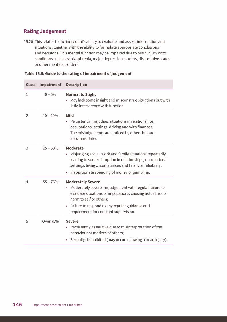
Impairment Assessment Guidelines146
Rating Judgement
16.20 This relates to the individual’s ability to evaluate and assess information and situations, together with the ability to formulate appropriate conclusions and decisions. This mental function may be impaired due to brain injury or to conditions such as schizophrenia, major depression, anxiety, dissociative states or other mental disorders.
Table 16.5: Guide to the rating of impairment of judgement
Class Impairment Description
1 0 – 5% Normal to Slight• May lack some insight and misconstrue situations but with
little interference with function.
2 10 – 20% Mild• Persistently misjudges situations in relationships,
occupational settings, driving and with finances. The misjudgements are noticed by others but are accommodated.
3 25 – 50% Moderate• Misjudging social, work and family situations repeatedly
leading to some disruption in relationships, occupational settings, living circumstances and financial reliability;
• Inappropriate spending of money or gambling.
4 55 – 75% Moderately Severe• Moderately severe misjudgement with regular failure to
evaluate situations or implications, causing actual risk or harm to self or others;
• Failure to respond to any regular guidance and requirement for constant supervision.
5 Over 75% Severe• Persistently assaultive due to misinterpretation of the
behaviour or motives of others;
• Sexually disinhibited (may occur following a head injury).

Impairment Assessment Guidelines 147
Rating Mood
16.21 Mood is a pervasive lasting emotional state. Affect is the prevailing and conscious emotional feeling during the period of the mental state examination.
Affect observed during the mental state examination is a reflection of the subject’s mood, and has a number of features, including:
Range: Variability of emotional expression over a period of time, i.e. if only one mood is expressed over a period of time, the affective range is restricted.
Amplitude: Amount of energy expended in expressing a mood, i.e. a mild amplitude of anger is manifested by annoyance and irritability.
Stability: Slow shifts of mood are normal. Rapid shifts (affective lability) may be pathological.
Appropriateness: The ‘fit’ (or congruency) between the affect and the situation.
Quality of Affect: Suspicious, sad, happy, anxious, angry, apathetic.
Relatedness: Ability to express warmth, to interact emotionally and to establish rapport.
Table 16.6: Guide for the rating of impairment of mood
Class Impairment Description
1 0 – 5% Normal to Slight• Relatively transient expressions of sadness, happiness, anxiety, anger
and apathy;
• Normal variation of mood associated with upsetting life events.
2 10 – 20% Mild• Mild symptoms: some or all of the below:
• mild depression;
• subjective distress leading to some mild interference with function;
• reduced interest in usual activities;
• some time off work;
• reduced social activities;
• fleeting suicidal thoughts;
• some panic attacks;
• heightened mood;
• may experience feelings of derealisation or depersonalisation.

Impairment Assessment Guidelines148
Class Impairment Description
3 25 – 50% ModerateModerate symptoms: some or all of the below:• frequent anxiety attacks with somatic concomitants;
• inappropriate self-blame and/or guilt;
• persistent suicidal ideation or suicide attempts;
• marked lability of affect;
• significant lethargy;
• social withdrawal leading to major problems in interpersonal relationships;
• anhedonia;
• appetite disturbance with significant weight change;
• psychomotor retardation/agitation;
• hypomania;
• severe depersonalisation.
4 55 – 75% Moderately SevereCannot function in most areas:• constant agitation;
• violent manic excitement;
• repeated suicide attempts;
• remains in bed all day;
• extreme self-neglect;
• extreme anger/hypersensitivity;
• requires supervision to prevent injury to self or others.
5 Over 75% Severe• Severe depression, with regression requiring attention and assistance
in all aspects of self-care;
• Constantly suicidal;
• Manic excitement requiring restraint.

Impairment Assessment Guidelines 149
Rating Behaviour
16.22 Behaviour is one’s manner of acting. It is considered with regard to its appropriateness in the overall situation. Disturbances vary in kind and degree. Behaviour may be destructive either to self and/or others and may lead to withdrawal and isolation. Behaviour may be odd or eccentric. Particular mental disorders may be manifested by particular forms of behaviour (e.g. compulsive rituals associated with Obsessive Compulsive Disorder).
Table 16.7: Guide for the rating of impairment of behaviour
Class Impairment Description
1 0 – 5% Normal to Slight• Transient disturbances in behaviour that are understandable in the
context of this person’s situation, excessive fatigue, intoxication, family or work disruption.
2 10 – 20% Mild• Persons who generally function well, but regularly manifest disturbed
behaviour under little extra pressure that nevertheless is able to be accommodated by others;
• Persistent behaviour that has some adverse effect on relationships or employment.
3 25 – 50% Moderate• Occasional aggressive, disruptive or withdrawn behaviour requiring
attention or treatment;
• Obsessional rituals interfering with but not preventing goal-directed activity;
• Repeated antisocial behaviour leading to conflict with authority.
4 55 – 75% Moderately Severe• Persistently aggressive, disruptive or withdrawn behaviour requiring
attention or treatment;
• Behaviour significantly influenced by delusions or hallucinations;
• Behaviour associated with risk of self-harm outside the hospital setting, but not requiring constant supervision;
• Manic overactivity associated with inappropriate behaviour;
• Significantly regressed behaviour (e.g. extreme neglect of hygiene, inability to attend to own bodily needs).
5 Over 75% Severe• Requiring constant supervision to prevent harming self or others
(repeated suicide attempts, frequently violent, manic excitement);
• Catatonic excitement or rigidity;
• Incessant rituals or compulsive behaviour preventing goal-directed activity.

Impairment Assessment Guidelines150

17 A
sses
sor
sele
ctio
n pr
oces
s
17 ASSESSOR SELECTION PROCESS

This page has been intentionally left blank.

Impairment Assessment Guidelines 151
17.1 The Act requires assessments to be “made by an accredited medical practitioner selected in accordance with the Impairment Assessment Guidelines” (subsection 22(7)(c)).
17.2 For the purposes of the Guidelines:
• an assessor is a medical practitioner who is accredited to perform permanent impairment assessments under the accreditation scheme provided for in subsection 22(17) of the Act
• the ‘selection process’ referred to in subsection 22(7)(c) of the Act refers to the selection of an assessor to perform the whole person impairment assessment and is outlined in this chapter.
• The ‘requestor’ is the claims agent, self-insured employer or ReturnToWorkSA.
17.3 1Once there is medical evidence (e.g. from the treating doctor(s) or specialist(s)) that the work injury has stabilised/reached MMI and a permanent impairment assessment is required, the worker will be given the opportunity to choose the assessor who will assess their whole person impairment caused by their work injury from a list of assessors provided by the requestor, compiled with reference to the factors in order of priority. If there are no assessors that meet all the criteria, the requestor should seek to identify assessors who meet the criteria in the order of priority set out below. For the avoidance of doubt, this means the first criteria takes priority over the second, and so on.
1. The body system to which the injury/assessment relates – the assessor selected must be accredited for the relevant body system(s).
2. If multiple body systems are to be assessed, multiple assessors must not be used where there is an assessor available who is accredited in all the required body systems.
3. Possible conflicts of interest.
4. Availability of assessors – if an appropriately accredited assessor has available appointments, they must be selected over an alternative assessor with a waiting time in excess of 6 weeks (the time period stipulated by the Impairment Assessor Accreditation Scheme).
17 ASSESSOR SELECTION PROCESS
1. Unless the relevant permanent impairment assessment is requested by the South Australian Employment Tribunal

Impairment Assessment Guidelines152
The requestor must ensure that the worker is aware of all the assessors that best satisfy the above factors. If there are no assessors that meet all the above criteria, the requestor should seek to identify assessors who meet the criteria in the order of priority. Where there are multiple assessors meeting the same level of priority, the assessor who meets the most criteria is to be selected. The requestor may not direct a worker to choose a particular assessor. Section 17.4 provides for circumstances where the worker is unable or unwilling to choose an assessor.
The worker must inform the requestor of their choice of assessor as soon as practicable.
17.4 If the worker does not wish to select the assessor, or does not make a selection within 15 business days of being provided the list of applicable assessors, or as otherwise agreed, then the requestor should select the assessor, in consultation with the worker, taking into consideration the factors outlined in 17.3 – informing the worker of the chosen assessor(s) as soon as is practicable after the selection is made.
17.5 The requestor must ensure that workers are provided with the draft report request prior to it being sent to the assessor. The requestor must give the worker at least ten business days to consider the request and provide them with an opportunity to raise any issues, errors or omissions. Assessments must not be booked until this process is finalised and all supporting documents obtained. Subject to 17.3, the requestor may not delay the booking of the appointment unless agreed with the worker.
Notes for the requestor can be found at Appendix 1 of the Guidelines.

Not
e
NOTE

This page has been intentionally left blank.

Impairment Assessment Guidelines 153
The International Association for the Study of Pain (IASP) has defined pain as:
‘An unpleasant sensory and emotional experience associated with actual or potential tissue damage or described in terms of such damage’.
For chronic pain assessment using AMA5 and the Guidelines, chapter 18 of AMA5, Pain (pp565–591) is excluded.
The reasons for excluding chronic pain as a separate condition from the Guidelines are:
• It is subjective experience and is therefore open to exaggeration and fabrication in the compensation setting. Assessment depends on the credibility of the subject being assessed. In order to provide reliability, workers undergoing pain assessments require more than one examiner at different times, concordance with the established conditions, consistency over time, anatomical and physiological consistency, agreement between the examiners and exclusion of inappropriate illness behaviour.
• Tools to measure pain are based on self-reports and may be inherently unreliable.
• Some impairment ratings take symptoms into account and some of the ranges of impairment (e.g. WPI spine, may reflect the effect of injury and pain on ADL). This is not so for impairment assessment of the upper and lower limb, which is based on range of motion (ROM) and diagnosis-based estimates, other than for peripheral nerve injury and diagnosed complex regional pain.
Where there is a peripheral nerve injury and there is sensory loss, some of the sensory nerve impairment categories permit pain to be included (Categories 1-5, Table 16-10, p482, AMA5).
The section 17.2m (AMA5, p553), ‘Causalgia and complex regional pain syndrome (reflex sympathetic dystrophy)’ should not be used. Refer to paragraph 1.12 in the Introduction of the Guidelines for information regarding Complex Regional Pain Syndrome.
NOTE: ASSESSMENT OF PERMANENT IMPAIRMENT ARISING FROM CHRONIC PAIN(exclusion of chapter 18, AMA5)

Impairment Assessment Guidelines154
This page has been intentionally left blank.

APPENDICES Appe
ndic
es

This page has been intentionally left blank.

Impairment Assessment Guidelines 155
It is the responsibility of the person requesting the report (the ‘requestor’) to advise the assessor what injuries to assess, what not to assess and what unrelated injuries may need to be assessed and deducted in accordance with subsection 22(8)(g) of the Act.
The requestor must provide clear guidance to the assessor regarding the injuries to be included in the assessment. The Act requires specific assessment approaches, such as:
• “impairments from unrelated injuries or causes are to be disregarded in making an assessment” (subsection 22(8)(b) of the Act)
• “impairments from the same injury or cause are to be assessed together or combined to determine the degree of impairment of the worker” (subsection 22(8)(c) of the Act)
• “impairment resulting from physical injury is to be assessed separately from impairment resulting from psychiatric injury” (subsection 22(8)(d) of the Act)
• “in assessing the degree of permanent impairment resulting from physical injury, no regard is to be had to impairment that results from a psychiatric injury or consequential mental harm” (subsection 22(8)(f) of the Act)
• “any portion of an impairment that is due to a previous injury (whether or not a work injury or whether because of a pre-existing condition) that caused the worker to suffer an impairment before the relevant work injury is to be deducted for the purposes of an assessment” (subsection 22(8)(g) of the Act).
Assessor Selection Process
The process for the selection of the assessor is included in Chapter 17 of the Guidelines.
The requestor must ensure workers are provided with the report request prior to it being sent to the assessor. The requestor must give the worker at least ten days to consider the request and have an opportunity to raise any issues, errors or omissions before the request is sent to the assessor.
APPENDIX 1 NOTES FOR THE REQUESTOR

Impairment Assessment Guidelines156
Request Letter
Clear instructions must be provided to the assessor before the assessment is undertaken. The assessor must be provided with all relevant medical and allied health information, including results of all clinical investigations and previous assessments related to the work injury or condition in question. Assessors should contact the requestor if they consider additional information is required.
If known, the requestor must provide instruction to the assessor identifying:
• which injury impairment(s) should be included in the assessment
• which injury impairment(s) should not be included in the assessment
• which injury impairment(s) should be combined to create a whole person impairment
• which injury impairment(s) should be assessed separately
• which injury impairment(s) should be deducted
and provide any information from previous assessments of relevance to calculating the %WPI.
Additional information to be provided
• The requestor must identify if there are any unrelated injuries/conditions (which can be ascertained, for example, from previous medical or claims records) relevant to the work injury/condition(s) to be assessed. They must ensure that they have directly asked the worker or the worker’s representative if there is such a condition and liaised with them to ensure that all appropriate information/documentation is included.
• Where there are unrelated injuries/conditions that are relevant to the work injury that need to be considered, the requestor should request a whole person impairment assessment for the total impairment encompassing both the work injury and the unrelated injury/condition, and then ask the assessor to deduct the degree of impairment attributable to the unrelated injury/condition.
• This is done to satisfy various requirements of the Act, such as determining access to statutory lump sums and determining dollar amounts, as well as access to serious injury support and common law.
Origin of impairment
An impairment often involves more than one body system and the same condition may be covered in more than one chapter. Usually the system where the impairment presents is used for evaluating the impairment, however if an impairment is related to an injury to another area e.g. the brain or spinal cord, the assessment may need to be undertaken by an assessor accredited in the system where the impairment originates.

Impairment Assessment Guidelines 157
Clinical studies and other tests
The requestor should ensure that, prior to requesting an assessment, any relevant clinical studies, radiological investigations and tests have been completed and the results forwarded to the assessor with the request for assessment and report. For example:
Sleep apnoea
For sleep apnoea assessment, a sleep study must have been conducted by a Respiratory Physician within the past two years.
For obstructive sleep apnoea assessment, the worker must also have been examined by an Ear, Nose and Throat Physician.
Central sleep apnoea is rated by an assessor accredited in the Nervous System.
Asthma
The requestor should ensure that a diagnosis has been made for asthma by a Respiratory Physician and the diagnosis has been confirmed over time with repeated testing, before requesting an assessment. At least one lung function test must have been performed to TSANZ standards by a pulmonary function laboratory and it would be expected that spirometry has been conducted within the previous six months. The requestor should provide details of any available Asthma Plan(s), to assist in the impairment assessment process.
Other respiratory disorders
The requestor is required to provide an appropriate set of respiratory function tests performed to TSANZ standards by a pulmonary function laboratory.
Hearing impairment
Standards apply to the required tests for audiology assessment. The requestor needs to ensure that all available audiograms are sent to the assessor, who will establish whether the tests have been performed according to the required standards.
Arthritis
Arthritis, as measured by cartilage interval, can only be assessed with the appropriate x-rays. Due to reducing availability of imaging in hard copy, and on portable storage devices, requestors can direct assessors to access the relevant imaging via online subscription or direct from the Radiologist or radiology group (refer 1.33).
Operation notes
When surgery has occurred, it is important that the requestor obtains all relevant operation notes and imaging for provision to the assessor.

Impairment Assessment Guidelines158
Adhesive capsulitis (frozen shoulder)
Adhesive capsulitis can be rated 18 months after the initial diagnosis by an appropriate musculoskeletal physician. The requestor must ensure that this timeframe is met prior to the assessment.
Brain Injury
The requestor should ensure that any emergency or first responder notes, hospital clinical notes and all relevant radiology are forwarded to the assessor.
Neuropsychological testing for brain injury is required to be undertaken within the 12 month period before the assessment. If the injured worker is unable to undertake that testing, the requestor must explain this in the request.
Complex regional pain syndrome
The diagnosis of complex regional pain syndrome (CRPS) must have been present for at least 18 months immediately preceding the assessment to ensure accuracy of the diagnosis and to permit adequate time to achieve MMI. The diagnosis must have been made prior to the assessment by at least two examining specialists; with at least one being made by a Fellow of the Faculty of Pain Medicine or a Rheumatologist.
Care should be taken to ensure that any previous diagnoses have been for Complex Regional Pain Syndrome as opposed to Chronic Regional Pain.
Cortico-spinal tract and cauda equina syndrome
Cortico-spinal tract damage and cauda equina syndrome must have been diagnosed prior to the assessment by a Neurosurgeon, Neurologist, Rehabilitation Physician or Orthopaedic Surgeon.
The assessor must be accredited in both the Nervous System and the Spine.
If impairment is caused by an injury to the brain and/or spinal cord, such as bladder, bowel, sexual dysfunction, etc., the request should be made to an assessor accredited in the relevant body system (e.g. spine or nervous system)
A request to an assessor accredited in the affected body system would usually only be made where the impairment is due to an injury directly to the affected body system.

Impairment Assessment Guidelines 159
Dental
Assessment for dental injuries and conditions is conducted by an assessor accredited in the Ear, Nose and Throat system and is assessed in relation to the impact on mastication and deglutition. To assist the assessment process, the requestor should obtain and provide prior dental records.
Epicondylitis
The requestor must ensure that symptoms have been present for at least 18 months prior to arranging for assessment of epicondylitis.
Lung Cancer
Impairment due to lung cancer that has been treated surgically should be assessed at least six months after surgery.
Noise Induced Hearing Loss (NIHL)
Requests for an assessment of permanent impairment and %WPI in respect of noise induced hearing loss will consider, in addition to section 22 of the Act, the requirements of subsections 188(2) and 188(3) of the Act. The requestor will consider these requirements and include relevant instructions and information (e.g. date of retirement, if relevant) in the request.
Peripheral nerve injuries
The requestor must ensure that symptoms have persisted for at least 12 months prior to arranging an assessment for a peripheral nerve injury.
In the case of compression and entrapment nerve injuries (such as carpal tunnel syndrome and cubital tunnel syndrome), copies of nerve conduction study results must be provided to the assessor. Where surgery has been undertaken, and the worker continues to report ongoing symptoms, updated nerve conduction studies undertaken post-surgery (following an optimal recovery time) will need to be obtained prior to the assessment.
Whilst still useful, nerve conduction studies are not a requirement for traumatic injuries to the peripheral nerves such as in the case of crush injuries and lacerations.

Impairment Assessment Guidelines160
Plantar fasciitis
The requestor must ensure that symptoms have persisted for at least 18 months prior to arranging an assessment for plantar fasciitis.
Psychiatric disorders
The worker must have a psychiatric disorder with a diagnosis made by the treating Psychiatrist using the Diagnostic and Statistical Manual of Mental Disorders, Fifth Edition (DSM-5) in order to be assessed for whole person impairment.
Terminal disease
In the case of an accepted work injury of a progressive nature such as silicosis and other terminal disease, a WPI assessment may be requested where a worker’s treating physician considers the condition to be stable in the short to medium term and treatment is optimised, as outlined in paragraph 1.16. In these circumstances the assessor will be asked to assess the degree of impairment as if the worker’s condition has reached MMI. MMI in diseases of long term progressive decline needs to be considered on a case by case basis.

Impairment Assessment Guidelines 161
This worksheet must be used in conjunction with Impairment Assessment Guidelines chapter 16 – Psychiatric and psychological disorders. The worksheet can be downloaded from ReturnToWorkSA’s website.
Table 1
Class of impairment 1 2 3 4 5
Percentage of impairment
0 – 5% 10 – 20% 25 – 50% 55 – 75% Over 75%
MENTAL FUNCTION
Intelligence (Capacity for understanding)
Normal to Slight
Mild ModerateModerately
SevereSevere
Thinking (The ability to form or conceive in the mind)
Normal to Slight
Mild ModerateModerately
SevereSevere
Perception (The brain’s interpretation of internal and external stimuli)
Normal to Slight
Mild ModerateModerately
SevereSevere
Judgement (Ability to assess a given situation and act appropriately)
Normal to Slight
Mild ModerateModerately
SevereSevere
Mood (Emotional tone underlying all behaviours)
Normal to Slight
Mild ModerateModerately
SevereSevere
Behaviour (Behaviour that is disruptive, distressing or aggressive)
Normal to Slight
Mild ModerateModerately
SevereSevere
Reasons for selection of classes
Assessors must write a brief paragraph justifying their selection of each class that is consistent with the findings of the Mental State Examination (see 16.12). This paragraph should be intelligible to an intelligent lay person.
APPENDIX 2 GEPIC WORKSHEET

Impairment Assessment Guidelines162
Table 2
The indicative ranges for each class are as follows:
Low range Mid-range High range
Class 1 0 – 1% 2 – 3% 4 – 5%
Class 2 10 – 12% 14 – 16% 18 – 20%
Class 3 25 – 30% 35 – 40% 45 – 50%
Class 4 55 – 60% 65 – 70% 70 – 75%
Class 5 75 – 80% 85 – 90% 95 – 100%
Determining compensable psychiatric impairment
Determine the median class (the median number is the middle number in a series e.g. 12345, the middle number is 3).
Classes
Classes in order
Median Class
Assessment Outcome
1. The Median Class is
2. The Median Severity Rating is
3. The Total Psychiatric Impairment is %
4. Impairments not related to the work injury = %
5. Impairment from consequential mental harm = %
6. The compensable psychiatric impairment is the total psychiatric impairment – unrelated impairment and impairment from consequential mental harm = %
Equals: Compensable impairment from ‘pure mental harm’ (i.e. impairment that is not secondary to a physical work injury) %

Impairment Assessment Guidelines 163
This page has been intentionally left blank.

OFFICIAL
RETURN TO WORK SCHEME
Enquiries: 13 18 55 400 King William Street, Adelaide South Australia 5000 [email protected] www.rtwsa.com
Free information support services:
TTY (deaf or have hearing / speech impairment): Phone 13 36 77 then ask for 13 18 55
Speak & Listen (speech-to-speech): Phone 1300 555 727 then ask for 13 18 55
Languages other than English: Please ring the Interpreting and Translating Centre on 1800 280 203 and ask them to contact us on 13 18 55
Braille, audio, or e-text: Call 13 18 55 and ask for required format.
Printed August 2021
RTW
SA.F
VC.2
287.
V5.0
6.09
.202
1


![Oxford English for Information Technology [Second Edition]](https://static.fdokumen.com/doc/165x107/63278e22e491bcb36c0b56c6/oxford-english-for-information-technology-second-edition.jpg)
![[Second Edition,.]](https://static.fdokumen.com/doc/165x107/6322fad1887d24588e04752c/second-edition.jpg)