NLRX1 is a regulator of mitochondrial antiviral immunity
7
LETTERS NLRX1 is a regulator of mitochondrial antiviral immunity Chris B. Moore 1,2 , Daniel T. Bergstralh 2 , Joseph A. Duncan 3 , Yu Lei 1,2 , Thomas E. Morrison 4,5 , Albert G. Zimmermann 1,2 , Mary A. Accavitti-Loper 7 , Victoria J. Madden 6 , Lijun Sun 8 , Zhengmao Ye 1,2 , John D. Lich 1,2 , Mark T. Heise 4,5 , Zhijian Chen 8 & Jenny P-Y. Ting 1,2 The RIG-like helicase (RLH) family of intracellular receptors detect viral nucleic acid and signal through the mitochondrial antiviral signalling adaptor MAVS (also known as Cardif, VISA and IPS-1) during a viral infection 1–6 . MAVS activation leads to the rapid production of antiviral cytokines, including type 1 inter- ferons. Although MAVS is vital to antiviral immunity, its regu- lation from within the mitochondria remains unknown. Here we describe human NLRX1, a highly conserved nucleotide-binding domain (NBD)- and leucine-rich-repeat (LRR)-containing family member (known as NLR) that localizes to the mitochondrial outer membrane and interacts with MAVS. Expression of NLRX1 results in the potent inhibition of RLH- and MAVS-mediated interferon-b promoter activity and in the disruption of virus-induced RLH– MAVS interactions. Depletion of NLRX1 with small interference RNA promotes virus-induced type I interferon production and decreases viral replication. This work identifies NLRX1 as a check against mitochondrial antiviral responses and represents an inter- section of three ancient cellular processes: NLR signalling, intra- cellular virus detection and the use of mitochondria as a platform for anti-pathogen signalling. This represents a conceptual ad- vance, in that NLRX1 is a modulator of pathogen-associated molecular pattern receptors rather than a receptor, and identifies a key therapeutic target for enhancing antiviral responses. Mammalian members of the nucleotide-binding domain (NBD) and leucine-rich-repeat-containing (LRR) (known as NLR, see http://www.genenames.org/genefamily/nacht.html) family of pro- teins are indispensable for cellular responses to pathogens. This NBD-LRR protein structure is ancient and highly conserved, as shown by its initial identification among plant disease-resistance proteins 7–12 . Current dogma posits that NLRs function as cytoplasmic surveillance molecules that sense intracellular pathogen-associated molecular patterns (PAMPs), or as regulators of pathogen-initiated signalling cascades 13,14 . Viral PAMPs are detected by the cytoplasmic RLH recep- tors RIG-I (also known as DDX58) and MDA-5 (also known as IFIH1), which signal through the mitochondrial protein MAVS, resulting in the activation of interferon regulatory factor 3 (IRF3) and NF-kB and type-1 interferon transcription 1–6 . Abrogation of MAVS expression or function leads to reduced type 1 interferon pro- duction and antiviral protection 15 . To study the potential role of NLR proteins in regulating mito- chondrial antiviral signalling, we used bioinformatics to identify NLRs localized to the mitochondria. We identified one putative mitochondrial NLR called NLRX1 (previously known as CLR11.3 and NOD9) 9,16 (Fig. 1a). The predicted peptide sequence and distinct domains of NLRX1 are shown in Supplementary Fig. 1. Consistent with the conserved motif structure of the NLR family, NLRX1 con- tains a central putative NBD and carboxy-terminal LRRs. The assign- ment of the amino-terminal effector domain to a subclass is less clear. Instead, the N terminus has evolved to include a mitochondrial- targeting sequence (Supplementary Fig. 1). On the basis of hydro- phobicity analyses, we identified two putative transmembrane regions (Supplementary Fig. 2). NLRX1 homologues were identified in all vertebrates examined, with a remarkably high (92.4%) degree of conservation between human and mouse (Supplementary Fig. 3). To investigate NLRX1 function, NLRX1 was isolated and the com- plementary DNAs encoding N and C termini were verified by rapid amplification of cDNA ends (RACE). The 39 RACE produced two products, identifying a splice variant lacking a portion of the LRR region encoded by exon 9 (Supplementary Fig. 4). NLRX1 messenger RNA is broadly expressed, suggesting a ubiquitous role (Supplemen- tary Fig. 5). To examine protein expression, we generated an anti- NLRX1 monoclonal antibody. Antibody specificity was verified by immunoblotting against transfected, epitope-tagged haemagglutinin (HA)–NLRX1 or another NLR protein HA–NLRP12 (also known as Monarch1; ref. 17). NLRX1 antibody detected a strong band corres- ponding to the exogenous HA-tagged NLRX1 (Supplementary Fig. 6) and a weaker endogenous product of similar size in cells transfected with pcDNA or with HA-tagged NLRP12 (Supplementary Fig. 6). Short interfering RNA (siRNA)-mediated knockdown of NLRX1 greatly reduced the detection of this endogenous product, whereas control siRNA oligonucleotides had no effect (Supplementary Fig. 7). NLRX1 protein was also detected in all cell lines analysed, further suggesting ubiquitous expression (Supplementary Fig. 8). Additional clues to NLRX1 function are revealed by its cellular localization. Both endogenous and overexpressed NLRX1 revealed a punctate cytoplasmic distribution (Fig. 1b and Supplementary Fig. 9a). Biochemical fractionation verified that endogenous NLRX1 pro- tein resided in the non-nuclear fraction (Fig. 1c, middle panel). This fraction contains both cytosol and cytoplasmic organelles, the latter of which include the mitochondria. Separation of this cytoplasmic/ organellar fraction into three fractions (small membranes, soluble cytosol and mitochondria) revealed that NLRX1 was found exclu- sively in the mitochondrial fraction, which also contained the mito- chondrial protein TOM20 (translocase of outer mitochondrial membrane 20 homologue; also known as TOMM20) (Fig. 1c, right panel). A merged confocal image of endogenous NLRX1 and the mitochondrial stain Mitotracker indicated mitochondrial locali- zation of NLRX1 (Supplementary Fig. 9b). Antibody staining by immunogold-bead electron microscopy was used as the ultimate method for visualizing NLRX1; this localized NLRX1 to the 1 Department of Microbiology-Immunology, 2 Lineberger Comprehensive Cancer Center, 3 Department of Medicine, Division of Infectious Diseases, 4 Department of Genetics, 5 Carolina Vaccine Institute, 6 Department of Pathology, University of North Carolina, Chapel Hill, North Carolina 27599, USA. 7 University of Alabama at Birmingham, Birmingham, Alabama 35294, USA. 8 Howard Hughes Medical Institute, Department of Molecular Biology, University of Texas Southwestern Medical Center, Dallas, Texas 75390, USA. doi:10.1038/nature06501 1 Nature Publishing Group ©2008
Transcript of NLRX1 is a regulator of mitochondrial antiviral immunity
LETTERS
NLRX1 is a regulator of mitochondrial antiviralimmunityChris B. Moore1,2, Daniel T. Bergstralh2, Joseph A. Duncan3, Yu Lei1,2, Thomas E. Morrison4,5,Albert G. Zimmermann1,2, Mary A. Accavitti-Loper7, Victoria J. Madden6, Lijun Sun8, Zhengmao Ye1,2,John D. Lich1,2, Mark T. Heise4,5, Zhijian Chen8 & Jenny P-Y. Ting1,2
The RIG-like helicase (RLH) family of intracellular receptorsdetect viral nucleic acid and signal through the mitochondrialantiviral signalling adaptor MAVS (also known as Cardif, VISAand IPS-1) during a viral infection1–6. MAVS activation leads to therapid production of antiviral cytokines, including type 1 inter-ferons. Although MAVS is vital to antiviral immunity, its regu-lation from within the mitochondria remains unknown. Here wedescribe human NLRX1, a highly conserved nucleotide-bindingdomain (NBD)- and leucine-rich-repeat (LRR)-containing familymember (known as NLR) that localizes to the mitochondrial outermembrane and interacts with MAVS. Expression of NLRX1 resultsin the potent inhibition of RLH- and MAVS-mediated interferon-bpromoter activity and in the disruption of virus-induced RLH–MAVS interactions. Depletion of NLRX1 with small interferenceRNA promotes virus-induced type I interferon production anddecreases viral replication. This work identifies NLRX1 as a checkagainst mitochondrial antiviral responses and represents an inter-section of three ancient cellular processes: NLR signalling, intra-cellular virus detection and the use of mitochondria as a platformfor anti-pathogen signalling. This represents a conceptual ad-vance, in that NLRX1 is a modulator of pathogen-associatedmolecular pattern receptors rather than a receptor, and identifiesa key therapeutic target for enhancing antiviral responses.
Mammalian members of the nucleotide-binding domain (NBD)and leucine-rich-repeat-containing (LRR) (known as NLR, seehttp://www.genenames.org/genefamily/nacht.html) family of pro-teins are indispensable for cellular responses to pathogens. ThisNBD-LRR protein structure is ancient and highly conserved, as shownby its initial identification among plant disease-resistance proteins7–12.Current dogma posits that NLRs function as cytoplasmic surveillancemolecules that sense intracellular pathogen-associated molecularpatterns (PAMPs), or as regulators of pathogen-initiated signallingcascades13,14. Viral PAMPs are detected by the cytoplasmic RLH recep-tors RIG-I (also known as DDX58) and MDA-5 (also known asIFIH1), which signal through the mitochondrial protein MAVS,resulting in the activation of interferon regulatory factor 3 (IRF3)and NF-kB and type-1 interferon transcription1–6. Abrogation ofMAVS expression or function leads to reduced type 1 interferon pro-duction and antiviral protection15.
To study the potential role of NLR proteins in regulating mito-chondrial antiviral signalling, we used bioinformatics to identifyNLRs localized to the mitochondria. We identified one putativemitochondrial NLR called NLRX1 (previously known as CLR11.3and NOD9)9,16 (Fig. 1a). The predicted peptide sequence and distinctdomains of NLRX1 are shown in Supplementary Fig. 1. Consistent
with the conserved motif structure of the NLR family, NLRX1 con-tains a central putative NBD and carboxy-terminal LRRs. The assign-ment of the amino-terminal effector domain to a subclass is less clear.Instead, the N terminus has evolved to include a mitochondrial-targeting sequence (Supplementary Fig. 1). On the basis of hydro-phobicity analyses, we identified two putative transmembraneregions (Supplementary Fig. 2). NLRX1 homologues were identifiedin all vertebrates examined, with a remarkably high (92.4%) degree ofconservation between human and mouse (Supplementary Fig. 3).
To investigate NLRX1 function, NLRX1 was isolated and the com-plementary DNAs encoding N and C termini were verified by rapidamplification of cDNA ends (RACE). The 39 RACE produced twoproducts, identifying a splice variant lacking a portion of the LRRregion encoded by exon 9 (Supplementary Fig. 4). NLRX1 messengerRNA is broadly expressed, suggesting a ubiquitous role (Supplemen-tary Fig. 5). To examine protein expression, we generated an anti-NLRX1 monoclonal antibody. Antibody specificity was verified byimmunoblotting against transfected, epitope-tagged haemagglutinin(HA)–NLRX1 or another NLR protein HA–NLRP12 (also known asMonarch1; ref. 17). NLRX1 antibody detected a strong band corres-ponding to the exogenous HA-tagged NLRX1 (Supplementary Fig. 6)and a weaker endogenous product of similar size in cells transfectedwith pcDNA or with HA-tagged NLRP12 (Supplementary Fig. 6).Short interfering RNA (siRNA)-mediated knockdown of NLRX1greatly reduced the detection of this endogenous product, whereascontrol siRNA oligonucleotides had no effect (Supplementary Fig. 7).NLRX1 protein was also detected in all cell lines analysed, furthersuggesting ubiquitous expression (Supplementary Fig. 8).
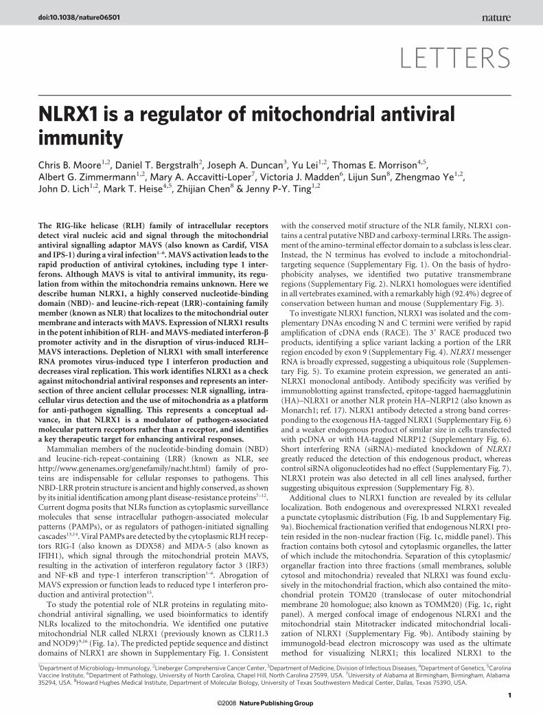
Additional clues to NLRX1 function are revealed by its cellularlocalization. Both endogenous and overexpressed NLRX1 revealeda punctate cytoplasmic distribution (Fig. 1b and Supplementary Fig.9a). Biochemical fractionation verified that endogenous NLRX1 pro-tein resided in the non-nuclear fraction (Fig. 1c, middle panel). Thisfraction contains both cytosol and cytoplasmic organelles, the latterof which include the mitochondria. Separation of this cytoplasmic/organellar fraction into three fractions (small membranes, solublecytosol and mitochondria) revealed that NLRX1 was found exclu-sively in the mitochondrial fraction, which also contained the mito-chondrial protein TOM20 (translocase of outer mitochondrialmembrane 20 homologue; also known as TOMM20) (Fig. 1c, rightpanel). A merged confocal image of endogenous NLRX1 and themitochondrial stain Mitotracker indicated mitochondrial locali-zation of NLRX1 (Supplementary Fig. 9b). Antibody staining byimmunogold-bead electron microscopy was used as the ultimatemethod for visualizing NLRX1; this localized NLRX1 to the
1Department of Microbiology-Immunology, 2Lineberger Comprehensive Cancer Center, 3Department of Medicine, Division of Infectious Diseases, 4Department of Genetics, 5CarolinaVaccine Institute, 6Department of Pathology, University of North Carolina, Chapel Hill, North Carolina 27599, USA. 7University of Alabama at Birmingham, Birmingham, Alabama35294, USA. 8Howard Hughes Medical Institute, Department of Molecular Biology, University of Texas Southwestern Medical Center, Dallas, Texas 75390, USA.
doi:10.1038/nature06501
1Nature Publishing Group©2008

mitochondria with little staining elsewhere (Fig. 1d, top panels). Co-staining for NLRX1 (small beads) and MAVS (large beads) identifiedseveral mitochondria containing both proteins (Fig. 1d, middlepanels). Isotype control antibody showed no nonspecific staining(Fig. 1d, bottom left panel), whereas the positive control TOM20showed mitochondrial staining (Fig. 1d, bottom right panel). Asobserved for MAVS and TOM20, many of the NLRX1-specificimmunogold beads were proximal to the mitochondrial membrane.To study this further, we separated mitochondrial membrane fractionsby a sucrose gradient (Fig. 1e). Voltage-dependent anion channel 1(VDAC1) and MAVS are proteins known to reside in the mitochon-drial outer membrane and served as positive controls1,18. VDAC1,MAVS and NLRX1 were the only proteins detected in fractions 1–5,which includes primarily mitochondrial outer membrane proteins(Fig. 1e). COXIV (cytochrome c oxidase subunit IV isoform 1; alsoknown as COX4I1) is an inner membrane mitochondrial protein andwas only found in fractions 6–10. To confirm this finding, a trypsinprotection assay was performed on prepared mitochondrial fractions.
NLRX1 and the known outer membrane proteins MAVS and BCL2L1were all sensitive to proteolytic cleavage by trypsin, whereas the mito-chondrial inner membrane protein COXIV was completely protected(Fig. 1f). Furthermore, deletion of the N-terminal mitochondrial tar-geting sequence resulted in a loss of mitochondrial localization(Supplementary Fig. 10). These results indicate that NLRX1 residesin the outer mitochondrial membrane.We next examined the inter-action of NLRX1 and MAVS. Co-immunoprecipitation studiesdemonstrate that HA–NLRX1 interacts with MAVS (Fig. 2a) but notwith other known mitochondrial outer membrane proteins (BCL2 andBCL2L1), indicating specificity of the NLRX1–MAVS interaction(Fig. 2b). Finally, endogenous NLRX1 associates strongly with endo-genous MAVS after immunoprecipitation with two different MAVSantibodies (Fig. 2c). Consistent with these results, MAVS and NLRX1show a remarkably similar expression level in many cell types(Supplementary Fig. 11).
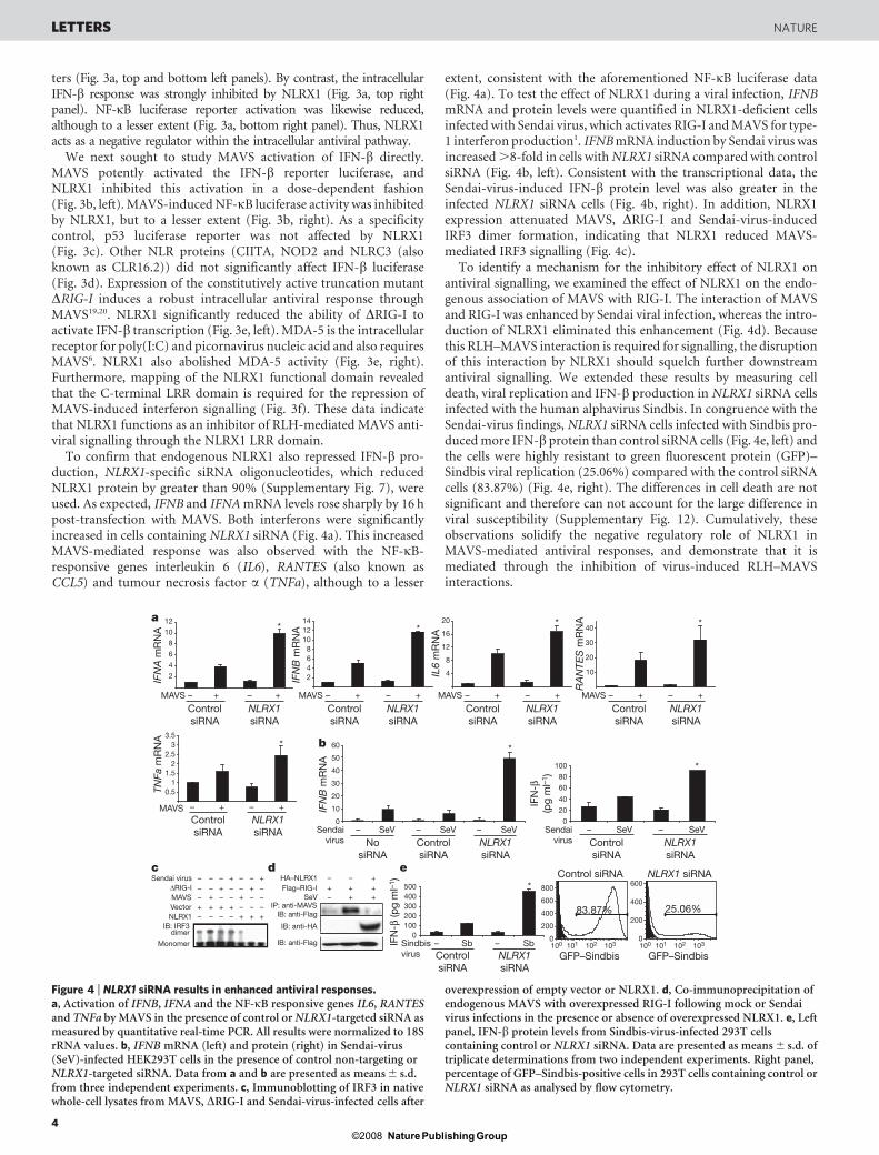
NLRX1 and MAVS are both modular proteins; therefore, we soughtto investigate the specific domains required for this interaction. The
Gene Mitochondria Cytosol PredictionNLRX1 (CLR11.3/NOD9) 52% 22% Mito.NLRP12 (Monarch1/CLR19.3/PYPAF7/NALP12/PAN6) 26% 35% CytosolNLRP2 (CLR19.9/NBS1/PYPAF2/NALP2) 17% 48% CytosolNLRP3 (CIAS1/NALP3/Pypaf1) 17% 39% CytosolNLRP4 (CLR19.5/RNH2/PYPAF4/NALP4/PAN2) 17% 35% CytosolNLRP6 (CLR11.4/PAN3/PYPAF5/NALP6) 17% 35% CytosolNLRP13 (CLR19.7/NOD14/PAN13/NALP13) 17% 48% CytosolNAIP (BIRC1/CLR5.1/NLRB1) 17% 52% Cytosol
13% 39% CytosolCIITA (NLRA/MHCC2TA) 13% 26% NuclearNLRP9 (CLR19.1/NOD6/NALP9/PAN12) 9% 91% CytosolNOD2 (CARD15/CLR16.3/NLRC2) 9% 52% CytosolNLRP7 (CLR19.4/Nod12/NALP7/PAN7) 4% 52% CytosolNLRP10 (CLR11.1/NOD8/NALP10/PAN5/PYNOD) 4% 61% CytosolNLRC3 (CLR16.2/NOD3) 4% 48% CytosolNLRC4 (IPAF/CARD12/CLR2.1) 0% 52% Cytosol
NLRP14 (CLR11.2/NOD5/NALP14/PAN8)
Ant
i-N
LRX
1
FITC DIC Merge
Isot
ype
cont
rol
15,000g
Cell homogenate
Nucleus (P1) Cytoplasm +organelles (S1)
Mitochondria(P5)
S5
Membranes(P15)
Solublecytosol (S15)
500g
5,000g
NLRX1
TFIID
Nuc
leus
(P1)
Cyt
opla
sm +
orga
nelle
s (S
1)
RIP
Mito
chon
dria
(P5)
Sol
uble
cyt
osol
(S15
)M
emb
rane
s (P
15)
NLRX1
GAPDH
TOM20
0.4–1.8 M sucrose
Fractionnumber
1 2 3 4 5 6 7 8 9 10
NLRX1
MAVS
COXIV
VDAC1NS
MAVS
NLRX1
MAVS
BCL2L1
COXIV
Cyt
opla
sm
Mito
chon
dria
Trypsin – +
f
a
cb
d e
Figure 1 | NLRX1 is an outermembrane mitochondrial protein.a, Prediction of subcellularlocalizations for NLRs. This wascalculated using K-nearestneighbours (k-NN) classifierpredictions of representative NLRlocalizations. Listed are the officialgene symbols followed by aliases inparentheses. b, Cytoplasmiclocalization of endogenous NLRX1.FITC, fluorescein isothiocyanate;DIC, differential interferencecontrast. c, Left, fractionationscheme used. Right, immunoblot ofNLRX1 in various cell fractions.d, Immunogold-bead electronmicrographs of NLRX1 alone (toppanels), NLRX1 (small bead, arrow)and MAVS (large bead, arrowhead)(middle panels), isotype antibodycontrol (bottom left panel), andTOM20 (bottom right panel).e, Immunoblotting of NLRX1 andknown outer (VDAC1 and MAVS)and inner (COXIV) mitochondrialproteins in membrane fractions. NSdenotes nonspecific protein band.f, Trypsin treatments ofmitochondria followed byimmunoblotting with NLRX1,MAVS and BCL2L1(outermembrane), as well as COXIV(inner membrane).
LETTERS NATURE
2Nature Publishing Group©2008

N-terminal CARD domain of MAVS is required for MAVS signallingand interactions with RLH family members1. It is also required for theinteraction with NLRX1 (Fig. 2d). In NLRX1, deletion of the LRR hadno effect on the NLRX1–MAVS interaction. A further deletion of theputative NBD disrupted this interaction, indicating that this domainis required for maintaining the interaction with MAVS (Fig. 2e).Collectively, these data demonstrate that NLRX1 is a MAVS-assoc-iated mitochondrial outer membrane protein and raise the intriguingpossibility that it participates in antiviral signalling.
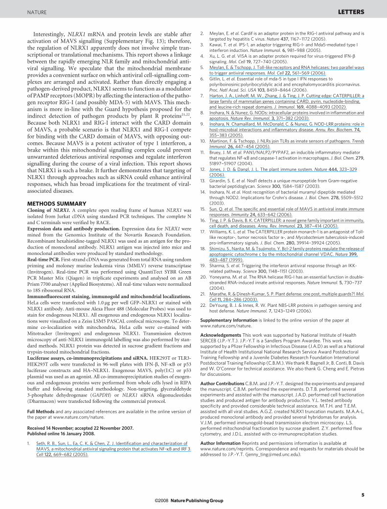
Signalling in response to the synthetic viral double-stranded RNA(dsRNA) analogue poly(I:C) is mediated both at the cell membranethrough Toll-like receptor 3 (TLR3) and within the cytoplasm by
means of direct binding to the RLH molecules to activate MAVS.Both pathways activate NF-kB and IRF3, leading to interferon b(IFN-b) transcription5. Poly(I:C), cannot activate IFN-b expressionin TLR3-deficient 293T cells when applied extracellularly. However,when delivered into the cytoplasm by transfection, poly(I:C) stimu-lates IFN-b through the MAVS pathway. Conversely, stable TLR3-expressing 293T cells have a robust response to extracellularpoly(I:C), which does not require MAVS signalling. We used thesetwo cell lines in poly(I:C)-induced IFN-b luciferase experiments todelineate a role for NLRX1 in intracellular (RLH) versus extracellular(TLR) antiviral responses. NLRX1 had no effect on the TLR3-mediated extracellular activation of IFN-b or NF-kB luciferase repor-
Lysa
te
MAVS
Flag–MAVS+++
–
MAVS
NLRX1
HA–NLRX1
Isot
ype
MA
VS
ant
ibod
y 1
MA
VS
ant
ibod
y 2
Isot
ype
HA–NLRX1
Flag–BCL2Flag–BCL2L1
Flag–MAVS+ + + +
++
+
–––
–––
–––
IP: anti-HAIB: anti-Flag
IB: anti-Flag
IB: anti-HALy
sate
IB: anti-Flag
IP: anti-HAIB: anti-Flag
IB: anti-HA
Flag–MAVS
HA–WT NLRX1HA–∆LRR NLRX1
HA–∆NBD∆LRR NLRX1+
++
+
+ +–––
–––
+–––
IB: anti-HA (15%)
HA1
X∆NBD∆LRR NLRX1
HA
1X∆LRR NLRX1
HA1
XWT NLRX1
NBD
NBD LRR556
75
75
556
97475
IB: anti-Flag
IP: anti-HAIB: anti-Flag
IB: anti-HA
Flag–WT MAVSFlag–∆CARD MAVS
Flag–∆PRO MAVS ++
+
–––
–––
Flag CARD-like Proline-rich1 10 77103 173 540
WT MAVS
Flag Proline-rich1 10 77103 173 540
Flag CARD-like1 10 77103 173 540
∆CARD MAVS
∆PRO MAVS
IB: anti-Flag
IB: anti-HA
IP: anti-HAIB: anti-Flag
b ca
d e
IP: MAVS
IB: NLRX1
HA–NLRX1 ++ +
Figure 2 | NLRX1 interacts with MAVS.a, Interaction of overexpressed NLRX1 andMAVS. IP and IB denote immunoprecipitationand immunoblotting, respectively. b, Interactionsof NLRX1 with other known mitochondrial outermembrane proteins. c, Interaction of endogenousNLRX1 with endogenous MAVS. d, Interactionof MAVS truncation mutants with wild-type(WT) NLRX1. e, Interaction of NLRX1truncation mutants with wild-type MAVS.Deletion constructs are shown above; 15%denotes 15% polyacrylamide gel necessary toresolve the smallest protein. The X domain iscurrently undefined.
a
e
f
b
c d
0
10
20
30
IFN
B-l
uc
IFN
B-l
uc
– +++ +
NLRX1
02468
10
NLRX1
Intracellular poly(I:C)Extracellular poly(I:C)
0
2
4
6
NFK
B-l
uc
NLRX1
0
4
8
12
16
NFK
B-l
uc
NLRX1
0
50
100
150
IFN
B-l
uc
NLRX1 CIIT
A
MAVS – +++++ +0
5
10
15
20
NFK
B-l
uc
NLRX1 CIIT
A
MAVS – +++++ +
0
5
10
15
MDA-5 – +++–+ + +poly(I:C) – ++++– + +
CIIT
ACIIT
A
∆RIG-I – +++++0
50100150200250
IFN
B-l
uc
NLRX1
IFN
B-l
uc
NLRX1
HA1
X∆NBD∆LRR NLRX1
HA1
X∆LRR NLRX1
HA1
XWT NLRX1
NBD
NBD LRR
0
15
30
45
MAVS
IFN
B-l
uc
– + +++++++ + +
WT ∆LRR ∆NBD∆LRRNLRX1 mutant:
556
75
75
556
97475
050
100150200
MAVS – +++++
NLR–
IFN
B-l
uc
0
10
20
30
p53
-luc
NLRX1
– + + + +p53
poly(I:C) – +++ +poly(I:C)
– +++ +poly(I:C)– +++ +poly(I:C)
Bla
nk
pcD
NA
3
pcD
NA
3
NLR
X1
NLR
C3
NO
D2
CIIT
A
Figure 3 | NLRX1 inhibits MAVS-mediated activation of IFN-b andNF-kB. a–c, Effect of NLRX1 onIFNB- and NFKB-luciferase (luc)activation by extracellular andintracellular poly(I:C) (a); IFNB-luc(left) and NFKB-luc (right)activation by MAVS (b); and p53activity (c). d, IFNB-luc is inhibitedby NLRX1 but not by other NLRproteins. e, Effect of NLRX1 onDRIG-I- (left) and MDA-5- (right)mediated activation of IFNB-lucactivity. f, Repression of MAVS-mediated IFNB-luc by wild-type(WT) NLRX1 compared with itstruncation mutants. Data froma–f are presented as mean 6 s.d.from three independentexperiments.
NATURE LETTERS
3Nature Publishing Group©2008
ters (Fig. 3a, top and bottom left panels). By contrast, the intracellularIFN-b response was strongly inhibited by NLRX1 (Fig. 3a, top rightpanel). NF-kB luciferase reporter activation was likewise reduced,although to a lesser extent (Fig. 3a, bottom right panel). Thus, NLRX1acts as a negative regulator within the intracellular antiviral pathway.
We next sought to study MAVS activation of IFN-b directly.MAVS potently activated the IFN-b reporter luciferase, andNLRX1 inhibited this activation in a dose-dependent fashion(Fig. 3b, left). MAVS-induced NF-kB luciferase activity was inhibitedby NLRX1, but to a lesser extent (Fig. 3b, right). As a specificitycontrol, p53 luciferase reporter was not affected by NLRX1(Fig. 3c). Other NLR proteins (CIITA, NOD2 and NLRC3 (alsoknown as CLR16.2)) did not significantly affect IFN-b luciferase(Fig. 3d). Expression of the constitutively active truncation mutantDRIG-I induces a robust intracellular antiviral response throughMAVS19,20. NLRX1 significantly reduced the ability of DRIG-I toactivate IFN-b transcription (Fig. 3e, left). MDA-5 is the intracellularreceptor for poly(I:C) and picornavirus nucleic acid and also requiresMAVS6. NLRX1 also abolished MDA-5 activity (Fig. 3e, right).Furthermore, mapping of the NLRX1 functional domain revealedthat the C-terminal LRR domain is required for the repression ofMAVS-induced interferon signalling (Fig. 3f). These data indicatethat NLRX1 functions as an inhibitor of RLH-mediated MAVS anti-viral signalling through the NLRX1 LRR domain.
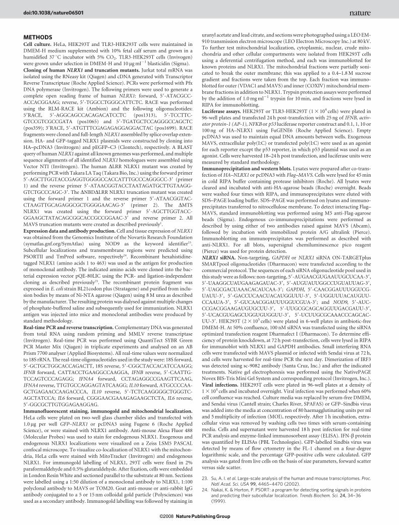
To confirm that endogenous NLRX1 also repressed IFN-b pro-duction, NLRX1-specific siRNA oligonucleotides, which reducedNLRX1 protein by greater than 90% (Supplementary Fig. 7), wereused. As expected, IFNB and IFNA mRNA levels rose sharply by 16 hpost-transfection with MAVS. Both interferons were significantlyincreased in cells containing NLRX1 siRNA (Fig. 4a). This increasedMAVS-mediated response was also observed with the NF-kB-responsive genes interleukin 6 (IL6), RANTES (also known asCCL5) and tumour necrosis factor a (TNFa), although to a lesser
extent, consistent with the aforementioned NF-kB luciferase data(Fig. 4a). To test the effect of NLRX1 during a viral infection, IFNBmRNA and protein levels were quantified in NLRX1-deficient cellsinfected with Sendai virus, which activates RIG-I and MAVS for type-1 interferon production1. IFNB mRNA induction by Sendai virus wasincreased .8-fold in cells with NLRX1 siRNA compared with controlsiRNA (Fig. 4b, left). Consistent with the transcriptional data, theSendai-virus-induced IFN-b protein level was also greater in theinfected NLRX1 siRNA cells (Fig. 4b, right). In addition, NLRX1expression attenuated MAVS, DRIG-I and Sendai-virus-inducedIRF3 dimer formation, indicating that NLRX1 reduced MAVS-mediated IRF3 signalling (Fig. 4c).
To identify a mechanism for the inhibitory effect of NLRX1 onantiviral signalling, we examined the effect of NLRX1 on the endo-genous association of MAVS with RIG-I. The interaction of MAVSand RIG-I was enhanced by Sendai viral infection, whereas the intro-duction of NLRX1 eliminated this enhancement (Fig. 4d). Becausethis RLH–MAVS interaction is required for signalling, the disruptionof this interaction by NLRX1 should squelch further downstreamantiviral signalling. We extended these results by measuring celldeath, viral replication and IFN-b production in NLRX1 siRNA cellsinfected with the human alphavirus Sindbis. In congruence with theSendai-virus findings, NLRX1 siRNA cells infected with Sindbis pro-duced more IFN-b protein than control siRNA cells (Fig. 4e, left) andthe cells were highly resistant to green fluorescent protein (GFP)–Sindbis viral replication (25.06%) compared with the control siRNAcells (83.87%) (Fig. 4e, right). The differences in cell death are notsignificant and therefore can not account for the large difference inviral susceptibility (Supplementary Fig. 12). Cumulatively, theseobservations solidify the negative regulatory role of NLRX1 inMAVS-mediated antiviral responses, and demonstrate that it ismediated through the inhibition of virus-induced RLH–MAVSinteractions.
a
b
Sendaivirus
– – – – –SeV SeV SeV
ControlsiRNA
NLRX1siRNA
NosiRNA
IFN
B m
RN
A
*
0
20
40
60
80
100
IFN
-β(p
g m
l–1)
Sendaivirus
*
SeV SeV
ControlsiRNA
NLRX1siRNA
4
8
12
16
20
ControlsiRNA
MAVS
NLRX1siRNA
0.51
1.52
2.53
3.5
ControlsiRNA
MAVS – + – +
NLRX1siRNA
*
*
IL6
mR
NA
10
20
30
40
ControlsiRNA
MAVS
NLRX1siRNA
*
RA
NTE
S m
RN
A
TNFa
mR
NA
2468
101214
ControlsiRNA
MAVS
NLRX1siRNA
*
IFN
B m
RN
A
ControlsiRNA
MAVS – ++ – +– +– +– +– +–+–
NLRX1siRNA
*
IFN
A m
RN
A
SeVFlag–RIG-IHA–NLRX1
+ +–+ + +
+––
IP: anti-MAVS IB: anti-Flag
IB: anti-HA
IB: anti-Flag
c d
NLRX1
Sendai virus∆RIG-IMAVSVector
++++
+++
+–
––– – –
–
–
––
–
– – –
––
–++
+
IB: IRF3dimer
Monomer
+
0100200300400500
IFN
-β (p
g m
l–1)
Sindbisvirus
*
– –Sb SbControlsiRNA
NLRX1siRNA
100 101 102 103 100 101 102 103 0
200
400
600
800
83.87%
0
200
400
600
25.06%
GFP–SindbisGFP–Sindbis
Control siRNA NLRX1 siRNAe
2
4
6
8
10
12
0
10
20
30
40
50
60
–––––
+
Figure 4 | NLRX1 siRNA results in enhanced antiviral responses.a, Activation of IFNB, IFNA and the NF-kB responsive genes IL6, RANTESand TNFa by MAVS in the presence of control or NLRX1-targeted siRNA asmeasured by quantitative real-time PCR. All results were normalized to 18SrRNA values. b, IFNB mRNA (left) and protein (right) in Sendai-virus(SeV)-infected HEK293T cells in the presence of control non-targeting orNLRX1-targeted siRNA. Data from a and b are presented as means 6 s.d.from three independent experiments. c, Immunoblotting of IRF3 in nativewhole-cell lysates from MAVS, DRIG-I and Sendai-virus-infected cells after
overexpression of empty vector or NLRX1. d, Co-immunoprecipitation ofendogenous MAVS with overexpressed RIG-I following mock or Sendaivirus infections in the presence or absence of overexpressed NLRX1. e, Leftpanel, IFN-b protein levels from Sindbis-virus-infected 293T cellscontaining control or NLRX1 siRNA. Data are presented as means 6 s.d. oftriplicate determinations from two independent experiments. Right panel,percentage of GFP–Sindbis-positive cells in 293T cells containing control orNLRX1 siRNA as analysed by flow cytometry.
LETTERS NATURE
4Nature Publishing Group©2008
Interestingly, NLRX1 mRNA and protein levels are stable afteractivation of MAVS signalling (Supplementary Fig. 13); therefore,the regulation of NLRX1 apparently does not involve simple tran-scriptional or translational mechanisms. This report shows a linkagebetween the rapidly emerging NLR family and mitochondrial anti-viral signalling. We speculate that the mitochondrial membraneprovides a convenient surface on which antiviral cell-signalling com-plexes are arranged and activated. Rather than directly engaging apathogen-derived product, NLRX1 seems to function as a modulatorof PAMP receptors (MOPR) by affecting the interaction of the patho-gen receptor RIG-I (and possibly MDA-5) with MAVS. This mech-anism is more in-line with the Guard hypothesis proposed for theindirect detection of pathogen products by plant R proteins21,22.Because both NLRX1 and RIG-I interact with the CARD domainof MAVS, a probable scenario is that NLRX1 and RIG-I competefor binding with the CARD domain of MAVS, with opposing out-comes. Because MAVS is a potent activator of type 1 interferon, abrake within this mitochondrial signalling complex could preventunwarranted deleterious antiviral responses and regulate interferonsignalling during the course of a viral infection. This report showsthat NLRX1 is such a brake. It further demonstrates that targeting ofNLRX1 through approaches such as siRNA could enhance antiviralresponses, which has broad implications for the treatment of viral-associated diseases.
METHODS SUMMARYCloning of NLRX1. A complete open reading frame of human NLRX1 was
isolated from Jurkat cDNA using standard PCR techniques. The complete N
and C terminals were verified by RACE.
Expression data and antibody production. Expression data for NLRX1 were
mined from the Genomics Institute of the Novartis Research Foundation.
Recombinant hexahistidine-tagged NLRX1 was used as an antigen for the pro-
duction of monoclonal antibody. NLRX1 antigen was injected into mice and
monoclonal antibodies were produced by standard methodology.
Real-time PCR. First-strand cDNA was generated from total RNA using random
priming and moloney murine leukemia virus (MMLV) reverse transcriptase
(Invitrogen). Real-time PCR was performed using QuantiTect SYBR Green
PCR Master Mix (Qiagen) in triplicate experiments and analysed on an AB
Prism 7700 analyser (Applied Biosystems). All real-time values were normalized
to 18S ribosomal RNA.
Immunofluorescent staining, immunogold and mitochondrial localizations.HeLa cells were transfected with 1.0mg per well GFP–NLRX1 or stained withNLRX1 antibody. Anti-mouse Alexa Fluor 488 (Molecular Probes) was used to
stain for endogenous NLRX1. All exogenous and endogenous NLRX1 localiza-
tions were visualized on a Zeiss LSM5 PASCAL confocal microscope. To deter-
mine co-localization with mitochondria, HeLa cells were co-stained with
Mitotracker (Invitrogen) and endogenous NLRX1. Transmission electron
microscopy of anti-NLRX1 immunogold labelling was also performed by stan-
dard methods. NLRX1 protein was detected in sucrose gradient fractions and
trypsin-treated mitochondrial fractions.
Luciferase assays, co-immunoprecipitations and siRNA. HEK293T or TLR3-
HEK293T cells were transfected in 96-well plates with IFN-b, NF-kB or p53
luciferase constructs and HA–NLRX1. Exogenous MAVS, poly(I:C) or p53
plasmid was used as an agonist. All co-immunoprecipitation studies of exogen-
ous and endogenous proteins were performed from whole cells lysed in RIPA
buffer and following standard methodology. Non-targeting, glyceraldehyde
3-phosphate dehydrogenase (GAPDH) or NLRX1 siRNA oligonucleotides
(Dharmacon) were transfected following the commercial protocol.
Full Methods and any associated references are available in the online version ofthe paper at www.nature.com/nature.
Received 14 November; accepted 22 November 2007.Published online 16 January 2008.
1. Seth, R. B., Sun, L., Ea, C. K. & Chen, Z. J. Identification and characterization ofMAVS, a mitochondrial antiviral signaling protein that activates NF-kB and IRF 3.Cell 122, 669–682 (2005).
2. Meylan, E. et al. Cardif is an adaptor protein in the RIG-I antiviral pathway and istargeted by hepatitis C virus. Nature 437, 1167–1172 (2005).
3. Kawai, T. et al. IPS-1, an adaptor triggering RIG-I- and Mda5-mediated type Iinterferon induction. Nature Immunol. 6, 981–988 (2005).
4. Xu, L. G. et al. VISA is an adapter protein required for virus-triggered IFN-bsignaling. Mol. Cell 19, 727–740 (2005).
5. Meylan, E. & Tschopp, J. Toll-like receptors and RNA helicases: two parallel waysto trigger antiviral responses. Mol. Cell 22, 561–569 (2006).
6. Gitlin, L. et al. Essential role of mda-5 in type I IFN responses topolyriboinosinic:polyribocytidylic acid and encephalomyocarditis picornavirus.Proc. Natl Acad. Sci. USA 103, 8459–8464 (2006).
7. Harton, J. A., Linhoff, M. W., Zhang, J. & Ting, J. P. Cutting edge: CATERPILLER: alarge family of mammalian genes containing CARD, pyrin, nucleotide-binding,and leucine-rich repeat domains. J. Immunol. 169, 4088–4093 (2002).
8. Inohara, N. & Nunez, G. NODs: intracellular proteins involved in inflammation andapoptosis. Nature Rev. Immunol. 3, 371–382 (2003).
9. Inohara, N. Chamaillard, M. McDonald, C. & Nunez, G. NOD-LRR proteins: role inhost-microbial interactions and inflammatory disease. Annu. Rev. Biochem. 74,355–383 (2005).
10. Martinon, F. & Tschopp, J. NLRs join TLRs as innate sensors of pathogens. TrendsImmunol. 26, 447–454 (2005).
11. Bruey, J. M. et al. PAN1/NALP2/PYPAF2, an inducible inflammatory mediatorthat regulates NF-kB and caspase-1 activation in macrophages. J. Biol. Chem. 279,51897–51907 (2004).
12. Jones, J. D. & Dangl, J. L. The plant immune system. Nature 444, 323–329(2006).
13. Girardin, S. E. et al. Nod1 detects a unique muropeptide from Gram-negativebacterial peptidoglycan. Science 300, 1584–1587 (2003).
14. Inohara, N. et al. Host recognition of bacterial muramyl dipeptide mediatedthrough NOD2. Implications for Crohn’s disease. J. Biol. Chem. 278, 5509–5512(2003).
15. Sun, Q. et al. The specific and essential role of MAVS in antiviral innate immuneresponses. Immunity 24, 633–642 (2006).
16. Ting, J. P. & Davis, B. K. CATERPILLER: a novel gene family important in immunity,cell death, and diseases. Annu. Rev. Immunol. 23, 387–414 (2005).
17. Williams, K. L. et al. The CATERPILLER protein monarch-1 is an antagonist of Toll-like receptor-, tumor necrosis factor a-, and Mycobacterium tuberculosis-inducedpro-inflammatory signals. J. Biol. Chem. 280, 39914–39924 (2005).
18. Shimizu, S., Narita, M. & Tsujimoto, Y. Bcl-2 family proteins regulate the release ofapoptogenic cytochrome c by the mitochondrial channel VDAC. Nature 399,483–487 (1999).
19. Sharma, S. et al. Triggering the interferon antiviral response through an IKK-related pathway. Science 300, 1148–1151 (2003).
20. Yoneyama, M. et al. The RNA helicase RIG-I has an essential function in double-stranded RNA-induced innate antiviral responses. Nature Immunol. 5, 730–737(2004).
21. Marathe, R. & Dinesh-Kumar, S. P. Plant defense: one post, multiple guards?! Mol.Cell 11, 284–286 (2003).
22. DeYoung, B. J. & Innes, R. W. Plant NBS-LRR proteins in pathogen sensing andhost defense. Nature Immunol. 7, 1243–1249 (2006).
Supplementary Information is linked to the online version of the paper atwww.nature.com/nature.
Acknowledgements This work was supported by National Institute of HealthSERCEB (J.P.-Y.T.). J.P.-Y.T is a Sandlers Program Awardee. This work wassupported by a Pfizer Fellowship in Infectious Disease (J.A.D) as well as a NationalInstitute of Health Institutional National Research Service Award PostdoctoralTraining Fellowship and a Juvenile Diabetes Research Foundation InternationalPostdoctoral Training Fellowship (C.B.M.). We thank R. Bagnell Jr, B. Conti, B. Davisand W. O’Connor for technical assistance. We also thank G. Cheng and E. Pietrasfor discussions.
Author Contributions C.B.M. and J.P.-Y.T. designed the experiments and preparedthe manuscript. C.B.M. performed the experiments. D.T.B. performed severalexperiments and assisted with the manuscript. J.A.D. performed cell fractionationstudies and produced antigen for antibody production. Y.L. tested antibodyspecificity and provided considerable technical assistance. M.T.H. and T.E.M.assisted with all viral studies. A.G.Z. created NLRX1 truncation mutants. M.A.A-L.produced monoclonal antibody and provided several hybridomas for analysis.V.J.M. performed immunogold-bead transmission electron microscopy. L.S.performed mitochondrial fractionation by sucrose gradient. Z.Y. performed flowcytometry, and J.D.L. assisted with co-immunoprecipitation studies.
Author Information Reprints and permissions information is available atwww.nature.com/reprints. Correspondence and requests for materials should beaddressed to J.P.-Y.T. ([email protected]).
NATURE LETTERS
5Nature Publishing Group©2008
METHODSCell culture. HeLa, HEK293T and TLR3-HEK293T cells were maintained in
DMEM-H medium supplemented with 10% fetal calf serum and grown in a
humidified 37 uC incubator with 5% CO2. TLR3-HEK293T cells (Invivogen)
were grown under selection in DMEM-H and 10 mg ml21 blasticidin (Sigma).
Cloning of human NLRX1 and truncation mutants. Jurkat total mRNA was
isolated using the RNeasy kit (Qiagen) and cDNA generated with Transcriptor
Reverse Transcriptase (Roche Applied Science). PCRs were performed with Pfx
DNA polymerase (Invitrogen). The following primers were used to generate a
complete open reading frame of human NLRX1: forward, 59-ATACGCC-
ACCACGGAAG; reverse, 59-TGGCCTGGGCATTCTC. RACE was performed
using the RLM-RACE kit (Ambion) and the following oligonucleotides:
59RACE, 59-AGGCAGCCACAGACATCCTC (pos1313), 59-TCCTTC-
GTCCGTCGCCGATA (pos1065) and 59-TGATGCTCCAGGGCCAGCTC
(pos359); 39RACE, 59-ATGTTTCGAGAGAGGAGGACTAC (pos1699). RACE
fragments were cloned and full-length NLRX1 assembled by splice overlap exten-
sion. HA- and GFP-tagged NLRX1 plasmids were constructed by cloning into
HA–pcDNA3 (Invitrogen) and pEGFP–C3 (Clontech), respectively. A BLAST
query of human NLRX1 against all known genomes was performed, and multiple
sequence alignments of all identified NLRX1 homologues were assembled using
Vector NTI (Invitrogen). The human DLRR NLRX1 mutant was created by
performing PCR with Takara LA Taq (Takara Bio, Inc.) using the forward primer
59-AGCTTGGTACCGAGGTGGGGCCACCATTTGCCCAGGGCC-39 (primer
1) and the reverse primer 59-ATAACGGTACCTAATAGATGCTTGTAAGG-
GTCTGCCCAGC-39. The DNBDDLRR NLRX1 truncation mutant was created
using the forward primer 1 and the reverse primer 59-ATAACGGTAC-
CTAAGTTGCAGAGGCGCTGGGGAACAG-39 (primer 2). The DMTS
NLRX1 was created using the forward primer 59-AGCTTGGTACC-
GGAAGCTATACAGCGGCACCGCCGGAAC-39 and reverse primer 2. All
MAVS truncation mutants were created as described previously1.
Expression data and antibody production. Cell and tissue expression of NLRX1
was obtained from the Genomics Institute of the Novartis Research Foundation
(symatlas.gnf.org/SymAtlas) using NOD9 as the keyword identifier23.
Subcellular localizations and transmembrane regions were predicted using
PSORTII and TmPred software, respectively24. Recombinant hexahistidine-
tagged NLRX1 (amino acids 1 to 463) was used as the antigen for production
of monoclonal antibody. The indicated amino acids were cloned into the bac-
terial expression vector pQE-80LIC using the PCR- and ligation-independent
cloning as described previously25. The recombinant protein fragment was
expressed in E. coli strain BL21codon plus (Stratagene) and purified from inclu-
sion bodies by means of Ni-NTA agarose (Qiagen) using 8 M urea as described
by the manufacturer. The resulting protein was dialysed against multiple changes
of phosphate-buffered saline and subsequently used for immunization. NLRX1
antigen was injected into mice and monoclonal antibodies were produced by
standard methodology.
Real-time PCR and reverse transcription. Complementary DNA was generated
from total RNA using random priming and MMLV reverse transcriptase
(Invitrogen). Real-time PCR was performed using QuantiTect SYBR Green
PCR Master Mix (Qiagen) in triplicate experiments and analysed on an AB
Prism 7700 analyser (Applied Biosystems). All real-time values were normalized
to 18S rRNA. The real-time oligonucleotides used in the study were: 18S forward,
59-GCTGCTGGCACCAGACTT, 18S reverse, 59-CGGCTACCACATCCAAGG;
IFNB forward, CATTACCTGAAGGCCAAGGA, IFNB reverse, 59-CAATTG-
TCCAGTCCCAGAGG; IFNA4 forward, CCTAGAGGCCGAAGTTCAAG,
IFNA4 reverse, TTGTGCCAGGAGTATCAAGG; IL10 forward, ATGCCCCAA-
GCTGAGAACCAAGACCCA, IL10 reverse, 59-TCTCAAGGGGCTGGGTC-
AGCTATCCA; IL6 forward, CGGGAACGAAAGAGAAGCTCTA, IL6 reverse,
59-GGCGCTTGTGGAGAAGGAG.
Immunofluorescent staining, immunogold and mitochondrial localization.HeLa cells were plated on two-well glass chamber slides and transfected with
1.0mg per well GFP–NLRX1 or pcDNA3 using Fugene 6 (Roche Applied
Science), or were stained with NLRX1 antibody. Anti-mouse Alexa Fluor 488
(Molecular Probes) was used to stain for endogenous NLRX1. Exogenous and
endogenous NLRX1 localizations were visualized on a Zeiss LSM5 PASCAL
confocal microscope. To visualize co-localization of NLRX1 with the mitochon-
dria, HeLa cells were stained with MitoTracker (Invitrogen) and endogenous
NLRX1. For immunogold labelling of NLRX1, 293T cells were fixed in 2%
paraformaldehyde and 0.5% glutaraldehyde. After fixation, cells were embedded
in London Resin White and sectioned parallel to the substrate at 80 nm. Sections
were labelled using a 1:50 dilution of a monoclonal antibody to NLRX1, 1:100
polyclonal antibody to MAVS or TOM20. Goat anti-mouse or anti-rabbit IgG
antibody conjugated to a 5 or 15 nm colloidal gold particle (Polysciences) was
used as a secondary antibody. Immunogold labelling was followed by staining in
uranyl acetate and lead citrate, and sections were photographed using a LEO EM-
910 transmission electron microscope (LEO Electron Microscopy Inc.) at 80 kV.
To further test mitochondrial localization, cytoplasmic, nuclear, crude mito-
chondria and other cellular compartments were isolated from HEK293T cells
using a deferential centrifugation method, and each was immunoblotted for
known proteins and NLRX1. The mitochondrial fractions were partially soni-
cated to break the outer membrane; this was applied to a 0.4–1.8 M sucrosegradient and fractions were taken from the top. Each fraction was immuno-
blotted for outer (VDAC1 and MAVS) and inner (COXIV) mitochondrial mem-
brane fractions in addition to NLRX1. Trypsin protection assays were performed
by the addition of 1.0 mg ml21 trypsin for 10 min, and fractions were lysed in
RIPA for immunoblotting.
Luciferase assays. HEK293T or TLR3-HEK293T (1 3 104 cells) were plated in
96-well plates and transfected 24 h post-transfection with 25 ng of IFNB, activ-
ator protein-1 (AP-1), NFKB or p53 luciferase reporter construct and 0.1, 1, 10 or
100 ng of HA–NLRX1 using FuGENE6 (Roche Applied Science). Empty
pcDNA3 was used to maintain equal DNA amounts between wells. Exogenous
MAVS, extracellular poly(I:C) or transfected poly(I:C) were used as an agonist
for each reporter except the p53 reporter, in which p53 plasmid was used as anagonist. Cells were harvested 18–24 h post transfection, and luciferase units were
measured by standard methodology.
Immunoprecipitation and western blots. Lysates were prepared after co-trans-
fection of HA–NLRX1 or pcDNA3 with Flag–MAVS. Cells were lysed for 45 min
in cold RIPA buffer containing protease inhibitors (Roche). All lysates were
cleared and incubated with anti-HA-agarose beads (Roche) overnight. Beads
were washed four times with RIPA, and immunoprecipitates were eluted with
SDS–PAGE loading buffer. SDS–PAGE was performed on lysates and immuno-
precipitates transferred to nitrocellulose membrane. To detect interacting Flag–
MAVS, standard immunoblotting was performed using M5 anti-Flag-agarose
beads (Sigma). Endogenous co-immunoprecipitations were performed as
described by using either of two antibodies raised against MAVS (Abcam),
followed by incubation with immobilized protein A/G ultralink (Pierce).Immunoblotting on immunoprecipitates was performed as described with
anti-NLRX1. For all blots, supersignal chemiluminescence pico reagent
(Pierce) was used for protein detection.
NLRX1 siRNA. Non-targeting, GAPDH or NLRX1 siRNA ON-TARGETplus
SMARTpool oligonucleotides (Dharmacon) were transfected according to the
commercial protocol. The sequences of each siRNA oligonucleotide pool used in
this study were as follows: non-targeting, 59-AUGAACGUGAAUUGCUCAA-39,
59-UAAGGCUAUGAAGAGAUAC-39, 59-AUGUAUUGGCCUGUAUUAG-39,
59-UAGCGACUAAACACAUCAA-39; GAPDH, 59-CAACGGAUUUGGUCG-
UAUU-39, 59-GACCUCAACUACAUGGUUU-39, 59-UGGUUUACAUGUU-
CCAAUA-39, 59-GUCAACGGAUUUGGUCGUA-39; and NOD9, 59-AUC-
CCGACGGAAGAUGUGCUU-39, 59-UUGCGCAGCAGGUUGACGAUU-39,59-UCACGUGAGCUGGUGUGGUU-39, 59-UCUUGCGCAAACCCAGCAC-
UU-39. HEK293T (2 3 105 cells) were plated in 6-well plates in antibiotic-free
DMEM-H. At 50% confluence, 100 nM siRNA was transfected using the siRNA
optimized transfection reagent Dharmafect I (Dharmacon). To determine effi-
ciency of protein knockdown, at 72 h post-transfection, cells were lysed in RIPA
for immunoblot with NLRX1 and GAPDH antibodies. Small interfering RNA
cells were transfected with MAVS plasmid or infected with Sendai virus at 72 h,
and cells were harvested for real-time PCR the next day. Dimerization of IRF3
was detected using sc-9082 antibody (Santa Cruz, Inc.) and after the indicated
treatments. Native gel electrophoresis was performed using the NativePAGE
Novex BIS-Tris Mini Gel System and corresponding protocol (Invitrogen, Inc.).
Viral infections. HEK293T cells were plated in 96-well plates at a density of1 3 104 cells and incubated overnight. Viral infection was performed when 60%
cell confluence was reached. Culture media was replaced by serum-free DMEM,
and Sendai virus (Cantell strain; Charles River, SPAFAS) or GFP–Sindbis virus
was added into the media at concentration of 80 haemagglutinating units per ml
and 5 multiplicity of infection (MOI), respectively. After 1 h incubation, extra-
cellular virus was removed by washing cells two times with serum-containing
media. Cells and supernatant were harvested 18 h post infection for real-time
PCR analysis and enzyme-linked immunosorbent assay (ELISA). IFN-b protein
was quantified by ELISAs (PBL Technologies). GFP-labelled Sindbis virus was
detected by means of flow cytometry in the FL-1 channel on a four-degree
logarithmic scale, and the percentage GFP-positive cells were calculated. GFP
analysis was gated from live cells on the basis of size parameters, forward scatter
versus side scatter.
23. Su, A. I. et al. Large-scale analysis of the human and mouse transcriptomes. Proc.Natl Acad. Sci. USA 99, 4465–4470 (2002).
24. Nakai, K. & Horton, P. PSORT: a program for detecting sorting signals in proteinsand predicting their subcellular localization. Trends Biochem. Sci. 24, 34–36(1999).
doi:10.1038/nature06501
Nature Publishing Group©2008

25. Duncan, J. A. et al. Cryopyrin/NALP3 binds ATP/dATP, is an ATPase, andrequires ATP binding to mediate inflammatory signaling. Proc. Natl Acad. Sci. USA104, 8041–8046 (2007).
doi:10.1038/nature06501
Nature Publishing Group©2008