Can Evidence Based Management Contribute To Increasing Management Quality?
Upload
independentCategory
view
4download
0
Published Ahead of Print 18 June 2008. 2008, 82(17):8560. DOI: 10.1128/JVI.00699-08. J. Virol.
Milligan, Roberto P. Garofalo and Antonella CasolaGuerrero-Plata, Alan M. Jewell, Pedro A. Piedra, Gregg N. Deepthi Kolli, Efthalia L. Bataki, LeAnne Spetch, Antonieta InfectionExperimental Human MetapneumovirusImmunity and Pathogenesis in T Lymphocytes Contribute to Antiviral
http://jvi.asm.org/content/82/17/8560Updated information and services can be found at:
These include:
REFERENCEShttp://jvi.asm.org/content/82/17/8560#ref-list-1at:
This article cites 34 articles, 16 of which can be accessed free
CONTENT ALERTS more»articles cite this article),
Receive: RSS Feeds, eTOCs, free email alerts (when new
http://journals.asm.org/site/misc/reprints.xhtmlInformation about commercial reprint orders: http://journals.asm.org/site/subscriptions/To subscribe to to another ASM Journal go to:
on Novem
ber 14, 2014 by guesthttp://jvi.asm
.org/D
ownloaded from
on N
ovember 14, 2014 by guest
http://jvi.asm.org/
Dow
nloaded from

JOURNAL OF VIROLOGY, Sept. 2008, p. 8560–8569 Vol. 82, No. 170022-538X/08/$08.00�0 doi:10.1128/JVI.00699-08Copyright © 2008, American Society for Microbiology. All Rights Reserved.
T Lymphocytes Contribute to Antiviral Immunity and Pathogenesis inExperimental Human Metapneumovirus Infection�
Deepthi Kolli,1† Efthalia L. Bataki,1† LeAnne Spetch,1 Antonieta Guerrero-Plata,1 Alan M. Jewell,4Pedro A. Piedra,4 Gregg N. Milligan,1,2,3 Roberto P. Garofalo,1,2,3 and Antonella Casola1,2,3*
Departments of Pediatrics1 and Microbiology and Immunology2 and Sealy Center for Vaccine Development,3 University ofTexas Medical Branch, Galveston, and Department of Molecular Virology and Microbiology,
Baylor College of Medicine, Houston,4 Texas
Received 28 March 2008/Accepted 10 June 2008
Human metapneumovirus (hMPV), a member of the family Paramyxoviridae, is a leading cause of lowerrespiratory tract infections in children, the elderly, and immunocompromised patients. Virus- and host-specificmechanisms of pathogenesis and immune protection are not fully understood. By an intranasal inoculationmodel, we show that hMPV-infected BALB/c mice developed clinical disease, including airway obstruction andhyperresponsiveness (AHR), along with histopathologic evidence of lung inflammation and viral replication.hMPV infection protected mice against subsequent viral challenge, as demonstrated by undetectable viraltiters, lack of body weight loss, and a significant reduction in the level of lung inflammation. No cross-protection with other paramyxoviruses, such as respiratory syncytial virus, was observed. T-lymphocyte de-pletion studies showed that CD4� and CD8� T cells cooperate synergistically in hMPV eradication duringprimary infection, but CD4� more than CD8� T cells also enhanced clinical disease and lung pathology.Concurrent depletion of CD4� and CD8� T cells completely blocked airway obstruction as well as AHR.Despite impaired generation of neutralizing anti-hMPV antibodies in the absence of CD4� T cells, mice hadundetectable viral replication after hMPV challenge and were protected from clinical disease, suggesting thatprotection can be provided by an intact CD8� T-cell compartment. Whether these findings have implicationsfor naturally acquired human infections remains to be determined.
Human metapneumovirus (hMPV) is a recently identified hu-man respiratory viral pathogen belonging to the family Paramyxo-viridae, which includes two subfamilies, the Paramyxovirinae andthe Pneumovirinae. The Pneumovirinae are taxonomically dividedinto the Pneumovirus and Metapneumovirus genera, based primar-ily on their gene constellations. Respiratory syncytial virus (RSV),an important cause of pediatric respiratory illness, belongs to thePneumovirus genus, while hMPV has been assigned to the Meta-pneumovirus genus, based on genomic sequence analysis (29).Metapneumoviruses lack the nonstructural proteins NS1 andNS2, and the gene order is different from that of pneumovi-ruses. hMPV is the first human virus of this group; the onlyother member is the avian pneumovirus, an etiological agent ofrespiratory infections in turkeys. Since the discovery of hMPVin The Netherlands (30), the virus has been identified in manydifferent countries, including Australia (24), Canada (3), theUnited Kingdom (28), Finland (28), France (9), South Amer-ica, and the United States (7, 8, 32). hMPV has a seasonaldistribution (it is usually isolated during the winter) and isassociated with both upper and lower respiratory tract infec-tions in children and adults (7, 8, 32). A number of childrenwith proven hMPV infections have a clinical syndrome consis-tent with bronchiolitis: they present with wheezing, hypoxia,and other typical radiological findings (7, 18, 32). According todifferent reports, 5 to 12% of all respiratory tract infections in
younger infants are caused by hMPV, a proportion second onlyto that of RSV. hMPV is also responsible for 10% of allhospitalizations of elderly patients with respiratory tract infec-tions (7, 8, 32).
The pathophysiology of hMPV infection and the possiblecontribution of the host immune response to the pathogenesisof hMPV-induced lower airway disease are largely unknown.In particular, whether T lymphocytes may be involved in anti-viral immunity against hMPV, as well as contributing to lungdisease, is not fully understood. Studies of experimental RSVinfection in mice have shown that the T-cell response helps toresolve RSV infection but also contributes to the pathogenesisof disease. In particular, depletion of CD4� or CD8� T cellsreduced disease, and depletion of both subsets resulted inlong-term infection without clinical illness (11).
Experimental animal models of hMPV infection have beenreported, including primates and rodents (1, 14, 22, 33, 34).BALB/c mice have been shown to be permissive to hMPVreplication (1, 14). Therefore, in this study, we used an exper-imental BALB/c mouse model to determine the roles of T-lymphocyte subsets in immunity against primary and secondaryhMPV infection in mice as well as their contributions to clin-ical illness, pulmonary inflammation, airway obstruction, andairway hyperresponsiveness (AHR).
MATERIALS AND METHODS
Viral preparation and titer. The hMPV strain CAN97-83 was obtained fromthe Centers for Disease Control and Prevention, Atlanta, GA, with permissionfrom Guy Boivin. Virus was propagated in LLC-MK2 cells in minimal essentialmedium (without serum) containing 1.0 �g trypsin/ml (crude virus). To increasethe viral titer for infection, filtered hMPV was prepared using Millipore filters
* Corresponding author. Mailing address: Department of Pediatrics,301 University Blvd., Galveston, TX 77555-0366. Phone: (409) 747-0581. Fax: (409) 772-1761. E-mail: [email protected].
† D.K. and E.L.B. contributed equally to this work.� Published ahead of print on 18 June 2008.
8560
on Novem
ber 14, 2014 by guesthttp://jvi.asm
.org/D
ownloaded from

with a 100,000 molecular weight cutoff. Viral and cellular preparations wereroutinely tested for mycoplasma contamination by PCR and were used if theyhad �0.125 endotoxin unit/ml (by a Limulus assay). Viral titers were determinedby a 50% tissue culture infective dose (TCID50) assay. A 48-well plate of con-fluent LLC-MK2 cells was infected with serial 1/3 dilutions of the virus in a totalvolume of 250 �l of medium without serum. The plate was incubated overnightin a humidified incubator (37°C, 5% CO2), and on the next day the inoculum wasremoved, and cells were washed with serum-free medium and observed for signsof cytopathic effect (CPE) between 7 and 14 days. The dilution at which 50%CPE was observed was determined to be the TCID50 for the virus. For titrationof viruses isolated from the lungs of mice, mice were sacrificed on days 1, 2, 3, 5,7, 9, 14, and 21, the thoracic cavity was opened, and the heart and lungs wereremoved en bloc. After blood was rinsed through the right ventricle, the lungswere separated from the heart, weighed, and homogenized in minimal essentialmedium in a 10% (wt/vol) ratio. Homogenized samples were centrifuged, andviral titers in supernatants were determined using serial twofold dilutions.
Mouse infection protocol. Six- to 7-week-old female BALB/c mice (Harlan,Houston, TX) were inoculated intranasally (i.n.) with 107 TCID50s of filteredhMPV in a total volume of 100 �l. Control mice were inoculated with the samevolume of virus-free medium (referred to herein as mock infection). At theindicated time points after infection, lungs were isolated and processed for viraltitration and histopathological studies (12). Bronchoalveolar lavage (BAL) wasperformed to determine total-cell counts and counts of different types of cells asdescribed elsewhere (12).
Flow cytometry of lung cells. For flow cytometry analysis, lungs were collectedand digested with collagenase, and cells were passed through nylon mesh. Cellswere incubated with an Fc block (anti-mouse CD16/CD32) to reduce nonspecificbinding for 30 min before the addition of antibodies (BD Pharmingen) againstsurface markers: anti-CD3ε, anti-CD4, anti-CD8�, and anti-CD25 for T cells,and anti-Ly6G and anti-Ly6C (Gr-1) for neutrophils. Relevant isotype controlantibodies were used throughout. Data were analyzed using FlowJo software(Tree Star).
Depletion of T lymphocytes. Mice were treated with 240 �g of an anti-CD8antibody (clone 2.43; a generous gift of S. Peebles, Vanderbilt University), 150�g of anti-CD4 (clone GK1.5), or 240 �g of control immunoglobulin G (IgG). Inthe acute depletion protocol (protocol 1), antibodies were given intraperitoneally(i.p.) on three consecutive days before infection and also on day 1 postinfection.In the chronic depletion protocol (protocol 2), antibodies were given on threeconsecutive days before infection and at days 1, 7, 14, 21, and 28 postinfection.Both these treatment protocols have been shown to be effective in depletingmore than 90% of both CD4� and CD8� T-cells (11, 23).
Clinical disease. The severity of illness in mice was scored daily by twoinvestigators using a standardized 0-to-5 grading system (12). In addition, dailydetermination of body weight was used to monitor the progression of diseaseover the experimental period.
Lung histopathology. Lungs were perfused, fixed in 10% buffered formalin,and embedded in paraffin. Multiple 4-�m-thick sections were stained with he-matoxylin and eosin (H&E) (5, 12). Briefly, inflammatory infiltrates were scoredby enumerating the layers of inflammatory cells surrounding the vessels andbronchioles. Zero to three layers of inflammatory cells were considered “nor-mal.” Moderate to abundant infiltrates (�3 layers of inflammatory cells sur-rounding 50% or more of the circumference of the vessel or bronchioles) wereconsidered “abnormal.” The number of abnormal perivascular and peribronchialspaces divided by the total perivascular and peribronchial spaces is the percent-age reported as the pathology score. A total of �15 perivascular and peribron-chial spaces per lung were counted for each animal. Slides were analyzed andscored for cellular inflammation under light microscopy in a blinded manner bya trained pathologist. Lung sections were also stained with periodic acid-Schiffstain (PAS) to identify mucus-producing cells.
Microneutralization assay for measurement of serum anti-hMPV antibodies.Heat-inactivated sera were tested for neutralizing antibodies to hMPV as pre-viously described (34). The neutralizing antibody titers were defined as the log2
of the reciprocal of the highest serum dilution at which a �50% reduction inCPE was observed. The lowest detectable titer was 2.5 log2. Samples with un-detectable titers were assigned a value of 2 log2.
AHR. AHR was assessed in unrestrained mice using whole-body barometricplethysmography (Buxco, Troy, NY) to record enhanced pause (Penh), as pre-viously described (5). Penh is a dimensionless value that represents a function ofthe ratio of peak expiratory flow to peak inspiratory flow and a function of thetiming of expiration. Penh has previously been validated in animal models ofAHR (13, 15, 16, 27) and infection-associated airway obstruction (31). Respira-tory activity was recorded for 4 min in order to establish baseline Penh values.
Mice were subsequently exposed to increasing doses of nebulized methacholine(3, 12, 25, and 50 mg/ml) for 1.5 min, and data were recorded for another 3 min.
Respiratory mechanics. Invasive analysis of lung function was performed onanesthetized mice using the Flexivent system (Scireq, Montreal, Quebec, Can-ada), which integrates a computer-controlled small-animal ventilator with mea-surements of respiratory mechanics (4). Mice were anesthetized with xylazine (7mg/kg) and pentobarbital sodium (50 mg/kg of body weight), and the tracheaewere cannulated with an 18G tubing adaptor for the delivery of methacholine.Mice were artificially ventilated at 150 breaths/min with a tidal volume of 0.3 mland a positive end expiratory pressure of 3 cm H2O. Mice were allowed tostabilize on the ventilator for 5 min before measurements commenced. Mea-surements of baseline pulmonary mechanics and responses to aerosolized metha-choline (0 to 50 mg/ml saline; delivered by ultrasonic nebulizer) were thenobtained by using the forced-oscillation technique (19). Aerosols were deliveredfor 10 s without altering the ventilatory pattern, after which the 8-s forced-oscillation perturbation was applied every 30 s for 5 min. Peak responses duringeach 5-min period were determined for resistance. Responsiveness to methacho-line was assessed at 5, 8, and 14 days post-hMPV infection for four mice pergroup.
Statistical analysis. When two groups were compared, the values were ana-lyzed using an unpaired, two-tailed Student t test. When multiple groups werecompared, analysis of variance was used (GraphPad Instat Software, Inc., SanDiego, CA). The inflammation scores were analyzed using SAS. Results areexpressed as means � standard errors of the means (SEM) unless otherwisestated.
RESULTS
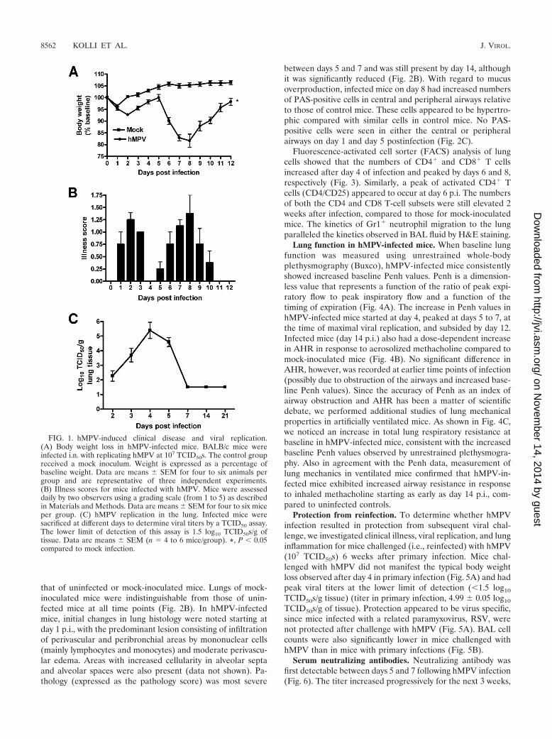
hMPV-induced clinical disease and viral replication in thelung. BALB/c mice were inoculated i.n. with either hMPV,UV-inactivated hMPV, or mock medium. Following an initialphase (days 1 to 3) with modest body weight loss and rapidrecovery, hMPV-infected mice consistently showed significantbody weight loss (�20% of initial weight) starting around day5, with a peak at days 7 to 9, and a slower recovery to baselineweight by day 12 to 15 (Fig. 1A). Mice inoculated with UV-inactivated hMPV did not show significant body weight lossand behaved similarly to the mock-infected control group(data not shown). Signs of illness, including ruffled fur,hunched appearance, and reduced movement, closely paral-leled the curve of body weight loss (Fig. 1B). To determinehMPV replication in the lung, viral titers were assessed dailyover 21 days (Fig. 1C). After an initial eclipse phase of about1 to 2 days, viral replication could be detected in the lungstarting around day 2 or 3 postinfection (p.i.), with a peak viraltiter of �105 TCID50s/g of lung tissue by day 4 to 5 p.i. Noreplicating virus was detected in the lungs of infected miceafter day 7 or 8 p.i.
BAL and lung cells, pathology, and mucus production. Com-pared to mock-inoculated mice, hMPV-infected mice showed arapid increase in the number of total cells recovered from BALfluid. In hMPV-infected mice, the number of total BAL cellsincreased at day 1 p.i. from (1.5 � 0.7) � 105 to (4.9 � 0.7) �105, with a peak of (8.5 � 0.7) � 105 at day 7 p.i. The numberof BAL cells returned to near-normal values by day 21 [(2.3 �0.5) � 105] (data not shown). While the cells in BAL fluid fromuninfected or mock-inoculated mice consisted mainly of mac-rophages, a remarkable neutrophilia was observed in hMPV-infected mice within the first 3 days (Fig. 2A). Neutrophilswere still present until day 14 p.i. The numbers of mononuclearcells, including monocytes and lymphocytes, started to increaseby day 3, and they represented the majority of BAL cells fromday 5 to day 14 p.i. (Fig. 2A).
The lung histopathology of infected mice was compared to
VOL. 82, 2008 IMMUNOPATHOGENESIS OF hMPV INFECTION 8561
on Novem
ber 14, 2014 by guesthttp://jvi.asm
.org/D
ownloaded from
that of uninfected or mock-inoculated mice. Lungs of mock-inoculated mice were indistinguishable from those of unin-fected mice at all time points (Fig. 2B). In hMPV-infectedmice, initial changes in lung histology were noted starting atday 1 p.i., with the predominant lesion consisting of infiltrationof perivascular and peribronchial areas by mononuclear cells(mainly lymphocytes and monocytes) and moderate perivascu-lar edema. Areas with increased cellularity in alveolar septaand alveolar spaces were also present (data not shown). Pa-thology (expressed as the pathology score) was most severe
between days 5 and 7 and was still present by day 14, althoughit was significantly reduced (Fig. 2B). With regard to mucusoverproduction, infected mice on day 8 had increased numbersof PAS-positive cells in central and peripheral airways relativeto those of control mice. These cells appeared to be hypertro-phic compared with similar cells in control mice. No PAS-positive cells were seen in either the central or peripheralairways on day 1 and day 5 postinfection (Fig. 2C).
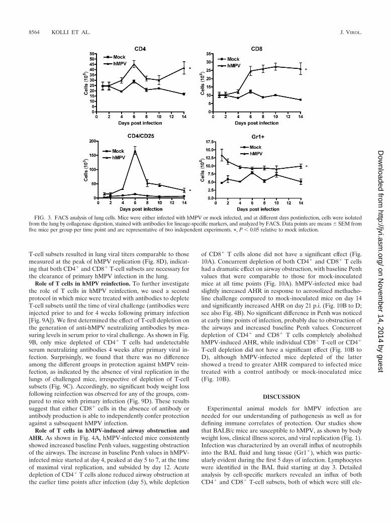
Fluorescence-activated cell sorter (FACS) analysis of lungcells showed that the numbers of CD4� and CD8� T cellsincreased after day 4 of infection and peaked by days 6 and 8,respectively (Fig. 3). Similarly, a peak of activated CD4� Tcells (CD4/CD25) appeared to occur at day 6 p.i. The numbersof both the CD4 and CD8 T-cell subsets were still elevated 2weeks after infection, compared to those for mock-inoculatedmice. The kinetics of Gr1� neutrophil migration to the lungparalleled the kinetics observed in BAL fluid by H&E staining.
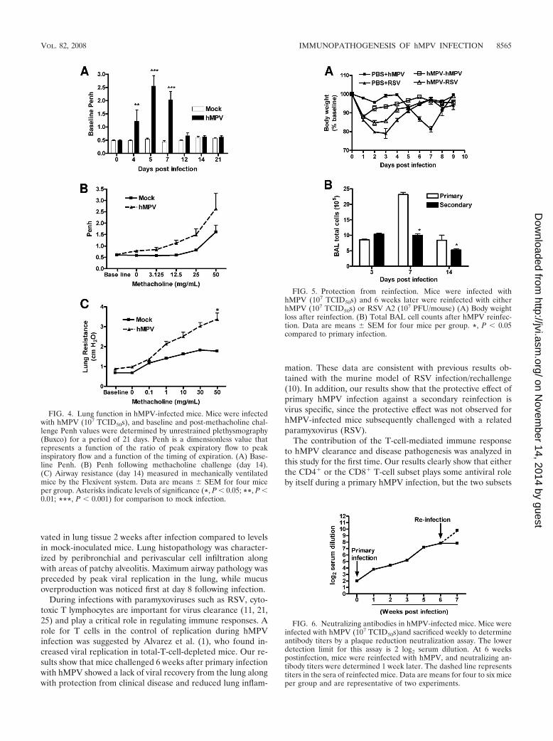
Lung function in hMPV-infected mice. When baseline lungfunction was measured using unrestrained whole-bodyplethysmography (Buxco), hMPV-infected mice consistentlyshowed increased baseline Penh values. Penh is a dimension-less value that represents a function of the ratio of peak expi-ratory flow to peak inspiratory flow and a function of thetiming of expiration (Fig. 4A). The increase in Penh values inhMPV-infected mice started at day 4, peaked at days 5 to 7, atthe time of maximal viral replication, and subsided by day 12.Infected mice (day 14 p.i.) also had a dose-dependent increasein AHR in response to aerosolized methacholine compared tomock-inoculated mice (Fig. 4B). No significant difference inAHR, however, was recorded at earlier time points of infection(possibly due to obstruction of the airways and increased base-line Penh values). Since the accuracy of Penh as an index ofairway obstruction and AHR has been a matter of scientificdebate, we performed additional studies of lung mechanicalproperties in artificially ventilated mice. As shown in Fig. 4C,we noticed an increase in total lung respiratory resistance atbaseline in hMPV-infected mice, consistent with the increasedbaseline Penh values observed by unrestrained plethysmogra-phy. Also in agreement with the Penh data, measurement oflung mechanics in ventilated mice confirmed that hMPV-in-fected mice exhibited increased airway resistance in responseto inhaled methacholine starting as early as day 14 p.i., com-pared to uninfected controls.
Protection from reinfection. To determine whether hMPVinfection resulted in protection from subsequent viral chal-lenge, we investigated clinical illness, viral replication, and lunginflammation for mice challenged (i.e., reinfected) with hMPV(107 TCID50s) 6 weeks after primary infection. Mice chal-lenged with hMPV did not manifest the typical body weightloss observed after day 4 in primary infection (Fig. 5A) and hadpeak viral titers at the lower limit of detection (�1.5 log10
TCID50s/g tissue) (titer in primary infection, 4.99 � 0.05 log10
TCID50s/g of tissue). Protection appeared to be virus specific,since mice infected with a related paramyxovirus, RSV, werenot protected after challenge with hMPV (Fig. 5A). BAL cellcounts were also significantly lower in mice challenged withhMPV than in mice with primary infections (Fig. 5B).
Serum neutralizing antibodies. Neutralizing antibody wasfirst detectable between days 5 and 7 following hMPV infection(Fig. 6). The titer increased progressively for the next 3 weeks,
FIG. 1. hMPV-induced clinical disease and viral replication.(A) Body weight loss in hMPV-infected mice. BALB/c mice wereinfected i.n. with replicating hMPV at 107 TCID50s. The control groupreceived a mock inoculum. Weight is expressed as a percentage ofbaseline weight. Data are means � SEM for four to six animals pergroup and are representative of three independent experiments.(B) Illness scores for mice infected with hMPV. Mice were assesseddaily by two observers using a grading scale (from 1 to 5) as describedin Materials and Methods. Data are means � SEM for four to six miceper group. (C) hMPV replication in the lung. Infected mice weresacrificed at different days to determine viral titers by a TCID50 assay.The lower limit of detection of this assay is 1.5 log10 TCID50s/g oftissue. Data are means � SEM (n 4 to 6 mice/group). *, P � 0.05compared to mock infection.
8562 KOLLI ET AL. J. VIROL.
on Novem
ber 14, 2014 by guesthttp://jvi.asm
.org/D
ownloaded from

peaking between 4 and 6 weeks p.i., with a mean peak titer of7.6 � 0.4 log2. To determine whether reinfection resulted in arecall response, mice were either challenged with hMPV (107
TCID50s) or sham inoculated 6 weeks after primary infection,and serum was collected 1 week p.i. for the determination ofantibody titers. There was a fourfold increase in neutralizingantibody titers for mice challenged with hMPV, compared tothe level of antibody present in the mice 6 weeks after primaryinfection (9.5 � 0.2 log2 versus 7.6 � 0.4 log2). There was nochange in the antibody titer in previously infected mice thatwere sham challenged.
Role of T cells in hMPV primary infection. Previous studieswith a murine model of experimental RSV infection have sug-gested that virus-induced illness is partially mediated by thehost immune response (11). Alvarez et al. have recently re-ported that NK cells and T lymphocytes are necessary to con-trol hMPV replication in the lung (1). However, the role ofimmune cells, specifically T lymphocytes, in the pathogenesisof hMPV-induced acute illness and in protection against rein-fection has not been reported. To address these questions,BALB/c mice were depleted of either CD4� or CD8� T cells,or both, by antibodies, using two different protocols. In the firstprotocol, mice were treated with antibodies to deplete T-cell
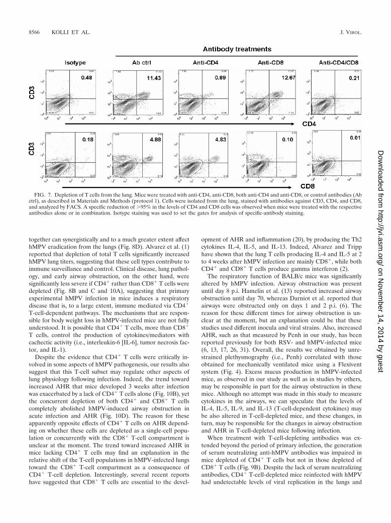
subsets only during the initial period of the infection (see Fig.8A). This protocol results in depletion of both CD4� andCD8� T cells in the lung (up to 95% depletion [Fig. 7]) but hasno effect on other cells, such as macrophages and dendriticcells (data not shown). As shown in Fig. 8B, mice depleted ofeither CD4� or CD8� T cells lost significantly less body weightthan control Ig-treated or untreated mice following hMPVinfection. The effect of CD4� T-cell depletion, however, wasmore robust than that of CD8� T-cell depletion in terms ofbody weight reduction. Simultaneous depletion of both T-cellsubsets did not provide further protection beyond that affordedby depletion of CD4� cells alone. In agreement with the bodyweight loss, histopathology studies showed that depletion ofCD4� cells caused a dramatic reduction in the lung pathologyscore, while depletion of CD8� cells was less effective (Fig.8C). Since T lymphocytes are important for viral clearance, weassessed viral titers in the lungs of mice depleted of eitherCD4� or CD8� T cells, or both. Mice infected with hMPVwere sacrificed at day 7 p.i., when virus is usually no longerdetectable in the lung (see Fig. 1C). Mice depleted of eitherCD4� or CD8� T cells had detectable viral titers in the lungs,similar to those for nondepleted mice (treated with a controlantibody). On the other hand, concurrent depletion of both
FIG. 2. Lung inflammation in hMPV-infected mice. Mice were infected with hMPV (107 TCID50s) and sacrificed at day 1, 3, 5, 7, or 14.(A) Total and differential cell counts in BAL fluid from mock-inoculated and infected mice were determined. (B) Peribronchial and perivascularinflammation was assessed in lung sections stained with H&E. Data are means � SEM for four to six mice per group. Experiments were performedin triplicate. Asterisks indicate levels of significance (*, P � 0.05; **, P � 0.01) for comparisons to mock infection. (C) Lung sections stained withPAS. Original magnification, �40. Arrow indicates PAS-positive cells.
VOL. 82, 2008 IMMUNOPATHOGENESIS OF hMPV INFECTION 8563
on Novem
ber 14, 2014 by guesthttp://jvi.asm
.org/D
ownloaded from
T-cell subsets resulted in lung viral titers comparable to thosemeasured at the peak of hMPV replication (Fig. 8D), indicat-ing that both CD4� and CD8� T-cell subsets are necessary forthe clearance of primary hMPV infection in the lung.
Role of T cells in hMPV reinfection. To further investigatethe role of T cells in hMPV reinfection, we used a secondprotocol in which mice were treated with antibodies to depleteT-cell subsets until the time of viral challenge (antibodies wereinjected prior to and for 4 weeks following primary infection[Fig. 9A]). We first determined the effect of T-cell depletion onthe generation of anti-hMPV neutralizing antibodies by mea-suring levels in serum prior to viral challenge. As shown in Fig.9B, only mice depleted of CD4� T cells had undetectableserum neutralizing antibodies 4 weeks after primary viral in-fection. Surprisingly, we found that there was no differenceamong the different groups in protection against hMPV rein-fection, as indicated by the absence of viral replication in thelungs of challenged mice, irrespective of depletion of T-cellsubsets (Fig. 9C). Accordingly, no significant body weight lossfollowing reinfection was observed for any of the groups, com-pared to mice with primary infection (Fig. 9D). These resultssuggest that either CD8� cells in the absence of antibody orantibody production is able to independently confer protectionagainst a subsequent hMPV infection.
Role of T cells in hMPV-induced airway obstruction andAHR. As shown in Fig. 4A, hMPV-infected mice consistentlyshowed increased baseline Penh values, suggesting obstructionof the airways. The increase in baseline Penh values in hMPV-infected mice started at day 4, peaked at day 5 to 7, at the timeof maximal viral replication, and subsided by day 12. Acutedepletion of CD4� T cells alone reduced airway obstruction atthe earlier time points after infection (day 5), while depletion
of CD8� T cells alone did not have a significant effect (Fig.10A). Concurrent depletion of both CD4� and CD8� T cellshad a dramatic effect on airway obstruction, with baseline Penhvalues that were comparable to those for mock-inoculatedmice at all time points (Fig. 10A). hMPV-infected mice hadslightly increased AHR in response to aerosolized methacho-line challenge compared to mock-inoculated mice on day 14and significantly increased AHR on day 21 p.i. (Fig. 10B to D;see also Fig. 4B). No significant difference in Penh was noticedat early time points of infection, probably due to obstruction ofthe airways and increased baseline Penh values. Concurrentdepletion of CD4� and CD8� T cells completely abolishedhMPV-induced AHR, while individual CD8� T-cell or CD4�
T-cell depletion did not have a significant effect (Fig. 10B toD), although hMPV-infected mice depleted of the lattershowed a trend to greater AHR compared to infected micetreated with a control antibody or mock-inoculated mice(Fig. 10B).
DISCUSSION
Experimental animal models for hMPV infection areneeded for our understanding of pathogenesis as well as fordefining immune correlates of protection. Our studies showthat BALB/c mice are susceptible to hMPV, as shown by bodyweight loss, clinical illness scores, and viral replication (Fig. 1).Infection was characterized by an overall influx of neutrophilsinto the BAL fluid and lung tissue (Gr1�), which was partic-ularly evident during the first 5 days of infection. Lymphocyteswere identified in the BAL fluid starting at day 3. Detailedanalysis by cell-specific markers revealed an influx of bothCD4� and CD8� T-cell subsets, both of which were still ele-
FIG. 3. FACS analysis of lung cells. Mice were either infected with hMPV or mock infected, and at different days postinfection, cells were isolatedfrom the lung by collagenase digestion, stained with antibodies for lineage-specific markers, and analyzed by FACS. Data points are means � SEM fromfive mice per group per time point and are representative of two independent experiments. *, P � 0.05 relative to mock infection.
8564 KOLLI ET AL. J. VIROL.
on Novem
ber 14, 2014 by guesthttp://jvi.asm
.org/D
ownloaded from
vated in lung tissue 2 weeks after infection compared to levelsin mock-inoculated mice. Lung histopathology was character-ized by peribronchial and perivascular cell infiltration alongwith areas of patchy alveolitis. Maximum airway pathology waspreceded by peak viral replication in the lung, while mucusoverproduction was noticed first at day 8 following infection.
During infections with paramyxoviruses such as RSV, cyto-toxic T lymphocytes are important for virus clearance (11, 21,25) and play a critical role in regulating immune responses. Arole for T cells in the control of replication during hMPVinfection was suggested by Alvarez et al. (1), who found in-creased viral replication in total-T-cell-depleted mice. Our re-sults show that mice challenged 6 weeks after primary infectionwith hMPV showed a lack of viral recovery from the lung alongwith protection from clinical disease and reduced lung inflam-
mation. These data are consistent with previous results ob-tained with the murine model of RSV infection/rechallenge(10). In addition, our results show that the protective effect ofprimary hMPV infection against a secondary reinfection isvirus specific, since the protective effect was not observed forhMPV-infected mice subsequently challenged with a relatedparamyxovirus (RSV).
The contribution of the T-cell-mediated immune responseto hMPV clearance and disease pathogenesis was analyzed inthis study for the first time. Our results clearly show that eitherthe CD4� or the CD8� T-cell subset plays some antiviral roleby itself during a primary hMPV infection, but the two subsets
FIG. 4. Lung function in hMPV-infected mice. Mice were infectedwith hMPV (107 TCID50s), and baseline and post-methacholine chal-lenge Penh values were determined by unrestrained plethysmography(Buxco) for a period of 21 days. Penh is a dimensionless value thatrepresents a function of the ratio of peak expiratory flow to peakinspiratory flow and a function of the timing of expiration. (A) Base-line Penh. (B) Penh following methacholine challenge (day 14).(C) Airway resistance (day 14) measured in mechanically ventilatedmice by the Flexivent system. Data are means � SEM for four miceper group. Asterisks indicate levels of significance (*, P � 0.05; **, P �0.01; ***, P � 0.001) for comparison to mock infection.
FIG. 5. Protection from reinfection. Mice were infected withhMPV (107 TCID50s) and 6 weeks later were reinfected with eitherhMPV (107 TCID50s) or RSV A2 (107 PFU/mouse) (A) Body weightloss after reinfection. (B) Total BAL cell counts after hMPV reinfec-tion. Data are means � SEM for four mice per group. *, P � 0.05compared to primary infection.
FIG. 6. Neutralizing antibodies in hMPV-infected mice. Mice wereinfected with hMPV (107 TCID50s)and sacrificed weekly to determineantibody titers by a plaque reduction neutralization assay. The lowerdetection limit for this assay is 2 log2 serum dilution. At 6 weekspostinfection, mice were reinfected with hMPV, and neutralizing an-tibody titers were determined 1 week later. The dashed line representstiters in the sera of reinfected mice. Data are means for four to six miceper group and are representative of two experiments.
VOL. 82, 2008 IMMUNOPATHOGENESIS OF hMPV INFECTION 8565
on Novem
ber 14, 2014 by guesthttp://jvi.asm
.org/D
ownloaded from
together can synergistically and to a much greater extent affecthMPV eradication from the lungs (Fig. 8D). Alvarez et al. (1)reported that depletion of total T cells significantly increasedhMPV lung titers, suggesting that these cell types contribute toimmune surveillance and control. Clinical disease, lung pathol-ogy, and early airway obstruction, on the other hand, weresignificantly less severe if CD4� rather than CD8� T cells weredepleted (Fig. 8B and C and 10A), suggesting that primaryexperimental hMPV infection in mice induces a respiratorydisease that is, to a large extent, immune mediated via CD4�
T-cell-dependent pathways. The mechanisms that are respon-sible for body weight loss in hMPV-infected mice are not fullyunderstood. It is possible that CD4� T cells, more than CD8�
T cells, control the production of cytokines/mediators withcachectic activity (i.e., interleukin-6 [IL-6], tumor necrosis fac-tor, and IL-1).
Despite the evidence that CD4� T cells were critically in-volved in some aspects of hMPV pathogenesis, our results alsosuggest that this T-cell subset may regulate other aspects oflung physiology following infection. Indeed, the trend towardincreased AHR that mice developed 3 weeks after infectionwas exacerbated by a lack of CD4� T cells alone (Fig. 10B), yetthe concurrent depletion of both CD4� and CD8� T cellscompletely abolished hMPV-induced airway obstruction inacute infection and AHR (Fig. 10D). The reason for theseapparently opposite effects of CD4� T cells on AHR depend-ing on whether these cells are depleted as a single-cell popu-lation or concurrently with the CD8� T-cell compartment isunclear at the moment. The trend toward increased AHR inmice lacking CD4� T cells may find an explanation in therelative shift of the T-cell populations in hMPV-infected lungstoward the CD8� T-cell compartment as a consequence ofCD4� T-cell depletion. Interestingly, several recent reportshave suggested that CD8� T cells are essential to the devel-
opment of AHR and inflammation (20), by producing the Th2cytokines IL-4, IL-5, and IL-13. Indeed, Alvarez and Tripphave shown that the lung T cells producing IL-4 and IL-5 at 2to 4 weeks after hMPV infection are mainly CD8�, while bothCD4� and CD8� T cells produce gamma interferon (2).
The respiratory function of BALB/c mice was significantlyaltered by hMPV infection. Airway obstruction was presentuntil day 8 p.i. Hamelin et al. (13) reported increased airwayobstruction until day 70, whereas Darniot et al. reported thatairways were obstructed only on days 1 and 2 p.i. (6). Thereason for these different times for airway obstruction is un-clear at the moment, but an explanation could be that thesestudies used different inocula and viral strains. Also, increasedAHR, such as that measured by Penh in our study, has beenreported previously for both RSV- and hMPV-infected mice(6, 13, 17, 26, 31). Overall, the results we obtained by unre-strained plethysmography (i.e., Penh) correlated with thoseobtained for mechanically ventilated mice using a Flexiventsystem (Fig. 4). Excess mucus production in hMPV-infectedmice, as observed in our study as well as in studies by others,may be responsible in part for the airway obstruction in thesemice. Although no attempt was made in this study to measurecytokines in the airways, we can speculate that the levels ofIL-4, IL-5, IL-9, and IL-13 (T-cell-dependent cytokines) maybe also altered in T-cell-depleted mice, and these changes, inturn, may be responsible for the changes in airway obstructionand AHR in T-cell-depleted mice following infection.
When treatment with T-cell-depleting antibodies was ex-tended beyond the period of primary infection, the generationof serum neutralizing anti-hMPV antibodies was impaired inmice depleted of CD4� T cells but not in those depleted ofCD8� T cells (Fig. 9B). Despite the lack of serum neutralizingantibodies, CD4� T-cell-depleted mice reinfected with hMPVhad undetectable levels of viral replication in the lungs and
FIG. 7. Depletion of T cells from the lung. Mice were treated with anti-CD4, anti-CD8, both anti-CD4 and anti-CD8, or control antibodies (Abctrl), as described in Materials and Methods (protocol 1). Cells were isolated from the lung, stained with antibodies against CD3, CD4, and CD8,and analyzed by FACS. A specific reduction of �95% in the levels of CD4 and CD8 cells was observed when mice were treated with the respectiveantibodies alone or in combination. Isotype staining was used to set the gates for analysis of specific-antibody staining.
8566 KOLLI ET AL. J. VIROL.
on Novem
ber 14, 2014 by guesthttp://jvi.asm
.org/D
ownloaded from

were protected from clinical disease, suggesting that, in theabsence of neutralizing antibodies, protection from hMPV re-infection can be provided by an intact CD8� T-cell compart-ment (Fig. 9C and D). These findings are different from whathas been reported previously for RSV infection of mice, inwhich the lack of CD8� T cells was characterized by a greater
protective effect against disease than that conferred by the lackof CD4� T cells during primary infection (11). Also, micelacking CD4� T cells and, as a consequence, lacking specificanti-RSV antibodies had significant viral replication in thelungs at the time of reinfection, while mice lacking CD8� T
FIG. 8. Role of T lymphocytes in primary hMPV infection. Mice weretreated with anti-CD4, anti-CD8, or both anti-CD4 and anti-CD8 anti-bodies, or with control antibodies (Ab ctrl), as described in Materials andMethods (protocol 1). Mice were either infected with hMPV or mockinfected. Data are means � SEM for four to six mice per group. Threeindependent experiments were performed. Asterisks indicate levels ofsignificance (*, P � 0.05; **, P � 0.01) relative to results for hMPV-infected mice. (A) Schematic representation of the T-cell-depletion pro-tocol. (B) Body weight loss in hMPV infection. (C) Lung peribronchialand perivascular inflammation assessed at day 7 p.i. (D) Viral replicationin the lungs of BALB/c mice at day 7 p.i. by a TCID50 assay.
FIG. 9. Role of T lymphocytes in hMPV reinfection. Mice weretreated with T-cell-depleting antibodies according to protocol 2 (seeMaterials and Methods) and infected with hMPV (107 TCID50s). Fiveweeks after primary infection, mice were reinfected with the same doseof hMPV. Data are means � SEM for four to six mice per group. Twoindependent experiments were performed. **, P � 0.05. (A) Sche-matic representation of the T-cell-depletion protocol. (B) Serum anti-hMPV neutralizing antibody titers were determined 4 weeks afterprimary infection (prior to viral challenge). (C) Viral replication in thelungs of BALB/c mice in primary and secondary infection was assessedby a TCID50 assay. The lower limit of detection of this assay is 1.5 log10TCID50s (g of tissue)1. (D) Body weight loss following hMPV rein-fection.
VOL. 82, 2008 IMMUNOPATHOGENESIS OF hMPV INFECTION 8567
on Novem
ber 14, 2014 by guesthttp://jvi.asm
.org/D
ownloaded from

cells or immunocompetent controls had no detectable virus(11). Those CD4� T-cell-depleted mice also had more-severeclinical disease after RSV reinfection. Thus, compared toCD4� T cells, the CD8� T-cell compartment appears to be lessinvolved in the immunopathogenesis of disease in hMPV thanin RSV primary infection. In addition, the CD8� T-cell com-partment appears to be sufficient to control viral replication inhMPV reinfections, while CD4� T cells (and antibodies) areclearly required for antiviral activity in the course of RSVreinfection.
While mouse models have clearly been important for under-standing the immunopathogenesis of disease in the course ofparamyxovirus infections, certain limitations exist when datahave been extrapolated to human infections. The nature of theimmune response to hMPV and its pathological features inhumans are still largely unknown, and therefore our findingswith mice related to the role of T cells, with regard to boththeir antiviral function and their contribution to clinical dis-ease, remain to be elucidated in naturally acquired humaninfections.
ACKNOWLEDGMENTS
This work was supported by grants from NIAID (P01 AI062885 toA.C. and N01 AI30039 to R.P.G.) and NHLBI (N01 HV28184 to A.C.and R.P.G.) and by the Sealy Center for Vaccine Development, Uni-versity of Texas Medical Branch (UTMB). D.K. was supported by theJeane B. Kempner fellowship, UTMB.
We thank Richard Johnston for helpful suggestions on the analysisof lung mechanics.
REFERENCES
1. Alvarez, R., K. S. Harrod, W. J. Shieh, S. Zaki, and R. A. Tripp. 2004.Human metapneumovirus persists in BALB/c mice despite the presence ofneutralizing antibodies. J. Virol. 78:14003–14011.
2. Alvarez, R., and R. A. Tripp. 2005. The immune response to human meta-
pneumovirus is associated with aberrant immunity and impaired virus clear-ance in BALB/c mice. J. Virol. 79:5971–5978.
3. Boivin, G., Y. Abed, G. Pelletier, L. Ruel, D. Moisan, S. Cote, T. C. Peret,D. D. Erdman, and L. J. Anderson. 2002. Virological features and clinicalmanifestations associated with human metapneumovirus: a new paramyxo-virus responsible for acute respiratory-tract infections in all age groups.J. Infect. Dis. 186:1330–1334.
4. Carey, M. A., J. W. Card, J. A. Bradbury, M. P. Moorman, N. Haykal-Coates,S. H. Gavett, J. P. Graves, V. R. Walker, G. P. Flake, J. W. Voltz, D. Zhu,E. R. Jacobs, A. Dakhama, G. L. Larsen, J. E. Loader, E. W. Gelfand, D. R.Germolec, K. S. Korach, and D. C. Zeldin. 2007. Spontaneous airway hy-perresponsiveness in estrogen receptor-alpha-deficient mice. Am. J. Respir.Crit. Care Med. 175:126–135.
5. Castro, S. M., A. Guerrero-Plata, G. Suarez-Real, P. A. Adegboyega, G. N.Colasurdo, A. M. Khan, R. P. Garofalo, and A. Casola. 2006. Antioxidanttreatment ameliorates respiratory syncytial virus-induced disease and lunginflammation. Am. J. Respir. Crit. Care Med. 174:1361–1369.
6. Darniot, M., T. Petrella, S. Aho, P. Pothier, and C. Manoha. 2005. Immuneresponse and alteration of pulmonary function after primary human meta-pneumovirus (hMPV) infection of BALB/c mice. Vaccine 23:4473–4480.
7. Esper, F., D. Boucher, C. Weibel, R. A. Martinello, and J. S. Kahn. 2003.Human metapneumovirus infection in the United States: clinical manifesta-tions associated with a newly emerging respiratory infection in children.Pediatrics 111:1407–1410.
8. Falsey, A. R., D. Erdman, L. J. Anderson, and E. E. Walsh. 2003. Humanmetapneumovirus infections in young and elderly adults. J. Infect. Dis. 187:785–790.
9. Freymouth, F., A. Vabret, L. Legrand, N. Eterradossi, F. Lafay-Delaire, J.Brouard, and B. Guillois. 2003. Presence of the new human metapneumo-virus in French children with bronchiolitis. Pediatr. Infect. Dis. J. 22:92–94.
10. Graham, B. S., L. A. Bunton, P. F. Wright, and D. T. Karzon. 1991. Rein-fection of mice with respiratory syncytial virus. J. Med. Virol. 34:7–13.
11. Graham, B. S., L. A. Bunton, P. F. Wright, and D. T. Karzon. 1991. Role ofT lymphocyte subsets in the pathogenesis of primary infection and rechal-lenge with respiratory syncytial virus in mice. J. Clin. Investig. 88:1026–1033.
12. Guerrero-Plata, A., S. Baron, J. S. Poast, P. A. Adegboyega, A. Casola, andR. P. Garofalo. 2005. Activity and regulation of alpha interferon in respira-tory syncytial virus and human metapneumovirus experimental infections.J. Virol. 79:10190–10199.
13. Hamelin, M. E., G. A. Prince, A. M. Gomez, R. Kinkead, and G. Boivin. 2006.Human metapneumovirus infection induces long-term pulmonary inflamma-tion associated with airway obstruction and hyperresponsiveness in mice.J. Infect. Dis. 193:1634–1642.
14. Hamelin, M. E., K. Yim, K. H. Kuhn, R. P. Cragin, M. Boukhvalova, J. C.Blanco, G. A. Prince, and G. Boivin. 2005. Pathogenesis of human meta-
FIG. 10. Effect of T-cell depletion on lung function. Mice were treated with T-cell-depleting antibodies or control IgG (Ab ctrl), according toprotocol 1 (Fig. 8A), and were infected with hMPV. Baseline Penh and post-methacholine challenge Penh (AHR) were measured for a period of21 days following infection. (A) Baseline Penh following hMPV infection. (B through D) Effects of depletion of CD4� T cells (B), CD8� T cells(C), or both (D) on AHR (day 21). Data are means � SEM for six mice per group. *, P � 0.05 compared to mock infection.
8568 KOLLI ET AL. J. VIROL.
on Novem
ber 14, 2014 by guesthttp://jvi.asm
.org/D
ownloaded from

pneumovirus lung infection in BALB/c mice and cotton rats. J. Virol. 79:8894–8903.
15. Hamelmann, E., J. Schwarze, K. Takeda, A. Oshiba, G. L. Larsen, C. G.Irvin, and E. W. Gelfand. 1997. Noninvasive measurement of airway respon-siveness in allergic mice using barometric plethysmography. Am. J. Respir.Crit. Care Med. 156:766–775.
16. Huck, B., D. Neumann-Haefelin, A. Schmitt-Graeff, M. Weckmann, J.Mattes, S. Ehl, and V. Falcone. 2007. Human metapneumovirus inducesmore severe disease and stronger innate immune response in BALB/c miceas compared with respiratory syncytial virus. Respir. Res. 8:6.
17. Jafri, H. S., S. Chavez-Bueno, A. Mejias, A. M. Gomez, A. M. Rios, S. S.Nassi, M. Yusuf, P. Kapur, R. D. Hardy, J. Hatfield, B. B. Rogers, K.Krisher, and O. Ramilo. 2004. Respiratory syncytial virus induces pneumo-nia, cytokine response, airway obstruction, and chronic inflammatory infil-trates associated with long-term airway hyperresponsiveness in mice. J. In-fect. Dis. 189:1856–1865.
18. Jartti, T., B. G. van den Hoogen, R. P. Garofalo, A. D. Osterhaus, and O.Ruuskanen. 2002. Metapneumovirus and acute wheezing in children. Lancet360:1393–1394.
19. Johnston, R. A., T. A. Theman, F. L. Lu, R. D. Terry, E. S. Williams, andS. A. Shore. 2008. Diet-induced obesity causes innate airway hyperrespon-siveness to methacholine and enhances ozone-induced pulmonary inflam-mation. J. Appl. Physiol. 104:1727–1735.
20. Koya, T., N. Miyahara, K. Takeda, S. Matsubara, H. Matsuda, C. Swasey, A.Balhorn, A. Dakhama, and E. W. Gelfand. 2007. CD8� T cell-mediatedairway hyperresponsiveness and inflammation is dependent on CD4� IL-4�
T cells. J. Immunol. 179:2787–2796.21. Kulkarni, A. B., M. Connors, C. Y. Firestone, H. C. Morse III, and B. R.
Murphy. 1993. The cytolytic activity of pulmonary CD8� lymphocytes, in-duced by infection with a vaccinia virus recombinant expressing the M2protein of respiratory syncytial virus (RSV), correlates with resistance toRSV infection in mice. J. Virol. 67:1044–1049.
22. MacPhail, M., J. H. Schickli, R. S. Tang, J. Kaur, C. Robinson, R. A.Fouchier, A. D. Osterhaus, R. R. Spaete, and A. A. Haller. 2004. Identifica-tion of small-animal and primate models for evaluation of vaccine candidatesfor human metapneumovirus (hMPV) and implications for hMPV vaccinedesign. J. Gen. Virol. 85:1655–1663.
23. Milligan, G. N., D. I. Bernstein, and N. Bourne. 1998. T lymphocytes arerequired for protection of the vaginal mucosae and sensory ganglia of im-mune mice against reinfection with herpes simplex virus type 2. J. Immunol.160:6093–6100.
24. Nissen, M. D., D. J. Siebert, I. M. Mackay, T. P. Sloots, and S. J. Withers.2002. Evidence of human metapneumovirus in Australian children. Med. J.Aust. 176:188.
25. Rutigliano, J. A., M. T. Rock, A. K. Johnson, J. E. Crowe, Jr., and B. S.Graham. 2005. Identification of an H-2Db-restricted CD8� cytotoxic T lym-phocyte epitope in the matrix protein of respiratory syncytial virus. Virology337:335–343.
26. Schwarze, J., G. Cieslewicz, E. Hamelmann, A. Joetham, L. D. Shultz, M. C.Lamers, and E. W. Gelfand. 1999. IL-5 and eosinophils are essential for thedevelopment of airway hyperresponsiveness following acute respiratory syn-cytial virus infection. J. Immunol. 162:2997–3004.
27. Schwarze, J., E. Hamelmann, K. L. Bradley, K. Takeda, and E. W. Gelfand.1997. Respiratory syncytial virus infection results in airway hyperresponsive-ness and enhanced airway sensitization to allergen. J. Clin. Investig. 100:226–233.
28. Stockton, J., I. Stephenson, D. Fleming, and M. Zambon. 2002. Humanmetapneumovirus as a cause of community-acquired respiratory illness.Emerg. Infect. Dis. 8:897–901.
29. van den Hoogen, B. G., T. M. Bestebroer, A. D. Osterhaus, and R. A.Fouchier. 2002. Analysis of the genomic sequence of a human metapneu-movirus. Virology 295:119–132.
30. van den Hoogen, B. G., J. C. de Jong, J. Groen, T. Kuiken, R. de Groot, R. A.Fouchier, and A. D. Osterhaus. 2001. A newly discovered human pneumo-virus isolated from young children with respiratory tract disease. Nat. Med.7:719–724.
31. van Schaik, S. M., G. Enhorning, I. Vargas, and R. C. Welliver. 1998.Respiratory syncytial virus affects pulmonary function in BALB/c mice. J. In-fect. Dis. 177:269–276.
32. Williams, J. V., P. A. Harris, S. J. Tollefson, L. L. Halburnt-Rush, J. M.Pingsterhaus, K. M. Edwards, P. F. Wright, and J. E. Crowe, Jr. 2004.Human metapneumovirus and lower respiratory tract disease in otherwisehealthy infants and children. N. Engl. J. Med. 350:443–450.
33. Williams, J. V., S. J. Tollefson, J. E. Johnson, and J. E. Crowe, Jr. 2005. Thecotton rat (Sigmodon hispidus) is a permissive small animal model of humanmetapneumovirus infection, pathogenesis, and protective immunity. J. Virol.79:10944–10951.
34. Wyde, P. R., S. N. Chetty, A. M. Jewell, S. L. Schoonover, and P. A. Piedra.2005. Development of a cotton rat-human metapneumovirus (hMPV) modelfor identifying and evaluating potential hMPV antivirals and vaccines. An-tivir. Res. 66:57–66.
VOL. 82, 2008 IMMUNOPATHOGENESIS OF hMPV INFECTION 8569
on Novem
ber 14, 2014 by guesthttp://jvi.asm
.org/D
ownloaded from
Copyright © 2022 FDOKUMEN