PACSINs Bind to the TRPV4 Cation Channel: PACSIN 3 MODULATES THE SUBCELLULAR LOCALIZATION OF TRPV4
Upload
independentCategory
view
0download
0
TRPV4 channels mediate the infrared laser-evoked response in sensory neurons
E. S. Albert,1 J. M. Bec,2 G. Desmadryl,1 K. Chekroud,1 C. Travo,1 S. Gaboyard,1 F. Bardin,2,3
I. Marc,2,4 M. Dumas,2 G. Lenaers,1 C. Hamel,1 A. Muller,1 and C. Chabbert1
1U-1051, INSERM Institut des Neurosciences de Montpellier, Montpellier, France; 2Université Montpellier 1 et 2,Montpellier, France; 3Université de Nîmes, Nîmes, France; and 4Ecole des Mines d’Alès, Alès, France
Submitted 9 May 2011; accepted in final form 6 March 2012
Albert ES, Bec JM, Desmadryl G, Chekroud K, Travo C,Gaboyard S, Bardin F, Marc I, Dumas M, Lenaers G, Hamel C,Muller A, Chabbert C. TRPV4 channels mediate the infrared laser-evoked response in sensory neurons. J Neurophysiol 107: 3227–3234,2012. First published March 21, 2012; doi:10.1152/jn.00424.2011.—Infrared laser irradiation has been established as an appropriatestimulus for primary sensory neurons under conditions where sensoryreceptor cells are impaired or lost. Yet, development of clinicalapplications has been impeded by lack of information about themolecular mechanisms underlying the laser-induced neural response.Here, we directly address this question through pharmacologicalcharacterization of the biological response evoked by midinfraredirradiation of isolated retinal and vestibular ganglion cells fromrodents. Whole cell patch-clamp recordings reveal that both voltage-gated calcium and sodium channels contribute to the laser-evokedneuronal voltage variations (LEVV). In addition, selective blockadeof the LEVV by micromolar concentrations of ruthenium red and RN1734 identifies thermosensitive transient receptor potential vanilloidchannels as the primary effectors of the chain reaction triggered bymidinfrared laser irradiation. These results have the potential tofacilitate greatly the design of future prosthetic devices aimed atrestoring neurosensory capacities in disabled patients.
transient receptor potential vanilloid 4; prosthesis
SENSORY PERCEPTION RELIES on the function of highly specializedreceptors, which detect a wide range of stimuli arising from ourenvironment and convert them into bioelectrical signals even-tually conveyed to the brain. Although highly specialized, thesensory receptors remain fragile, and the loss of these cellsthrough progressive degeneration (Saihan et al. 2009), suddencell damage, or age-related processes (Chen et al. 2010) cannotbe compensated. This has been overcome by the developmentof sensory prostheses aiming to substitute lacking sensoryreceptors. Such an approach is only possible if the centralsensory neuronal pathway is preserved. In past decades, directelectrical stimulation of the sensory neuronal pathways provedthat central sensory input could be generated without stimula-tion of the sensory receptors. Cochlear implants, for example,were developed as treatment of sensorineural hearing loss,using electrical stimulation of the auditory nerve (Haynes et al.2009). Through the years, limitations and constraints such aselectrode deterioration and poor resolution of environmentalsound or visual perception of the existing prostheses have beenrevealed. Prolonged use of electrodes turned out to be noxious,leading to neural tissue damage and possible toxicity of elec-trode materials (Wells et al. 2007a). Graded electrical currentspread is also observed well beyond the electrode location
(Popovic et al. 1991). In addition, investigations into the safetyof retinal electrical stimulation showed that direct tissue con-tact led to important retinal damage (Colodetti et al. 2007).
Over the years, infrared (IR) laser focal irradiation, whichdoes not require direct contact between the stimulus source andthe tissue, has been considered as an alternative approach tostimulate primary sensory neurons under conditions with sen-sory receptor cell deficits. In the 1970s, Fork (1971) firstdemonstrated that laser emissions in the blue and green spec-trum as well as near-IR wavelengths could be used selectivelyto stimulate photosensitive neurons from the abdominal gan-glion of marine molluscs. Later, it was demonstrated thattransient variations of the membrane potential could be evokedthrough IR laser stimulations on nonphotosensitive neuronssuch as those of the auditory nerves (Izzo et al. 2006, 2008),cavernous nerve of the prostate (Fried et al. 2008), and facialnerve (Teudt et al. 2007) without tissue damage. High spatialresolution of the stimulus is a further advantage of opticalradiation over electrical stimulation.
Although promising, clinical developments of such ap-proaches have been impeded by the lack of information on themolecular mechanisms supporting the laser-evoked neural re-sponse. According to Wells et al. (2005), photochemical ef-fects are not involved in mid-IR optical stimulation, whereasphotomechanical effects like pressure and stretching cannotoccur when operating with short laser pulses at high energies.More likely, transient thermal variations at the cell membranewould be sufficient to trigger laser-induced biological re-sponses. To address this question directly, we undertook apharmacological characterization of the neural responseevoked by mid-IR (1,875 mm) irradiation in retinal and ves-tibular ganglion cells (VGCs) isolated from rodents usingwhole cell patch-clamp recording. Here, we show that bothtypes of neurons can be stimulated reproducibly and thatneuronal responses are driven through transient receptor po-tential vanilloid 4 (TRPV4) channels.
MATERIALS AND METHODS
Animals. Mice of the C57BL/6 strain and Wistar rats (Centred’Elevage Janvier, Le Genest-Saint-Isle, France) were used in thestudy. All procedures were carried out in accordance with the French/European Communities Council Directive 86/609/EEC.
Cell preparations. Retinal ganglion cells (RGCs) were isolatedfrom postnatal days 2-4 (P2–P4) mice using the Thy-1 antibody-mediated plate adhesion immunopanning technique, and their speci-ficity was confirmed using RGC Brn3a immunoassay according topreviously described procedures (Kamei et al. 2005). VGCs wereexplanted from P5–P9 rats and dissociated according to previouslydescribed procedures (Dayanithi et al. 2007).
Address for reprint requests and other correspondence: C. Chabbert,INSERM U-1051, Hopital St. Eloi, 80 rue A. Fliche, Montpellier 34090,France (e-mail: [email protected]).
J Neurophysiol 107: 3227–3234, 2012.First published March 21, 2012; doi:10.1152/jn.00424.2011.
32270022-3077/12 Copyright © 2012 the American Physiological Societywww.jn.org
Electrophysiological recordings. Whole cell patch-clamp record-ings (current-clamp configuration) were performed at room tempera-ture (22–24°C) using a computer-controlled Axopatch 200B amplifier(Axon Instruments, Molecular Devices, Sunnyvale, CA) and digitizedusing a Digidata 1322A as previously described (Chabbert et al.2001). Patch electrodes (5–6 M�; microhematocrit tubes; Bris VitrexMedical) were filled with an intracellular solution (140 mM KCl, l5mM NaC, 10 mM glucose, 10 mM HEPES, 5 mM EGTA, 3 mM ATP,1 mM GTP; pH 7.4, 300 mosM). For whole cell recordings, neuronswere bathed in extracellular solution: 135 mM NaCl, 5 mM KCl, 1mM MgCl2, 2 mM CaCl2, 10 mM glucose, 10 mM HEPES, and 15mM Na2HCO3, pH 7.4, 300 mosM. Removal of calcium from the bathwas performed through local and transient exchange of the bathingsolution by calcium-free external solution (0 Ca2� and 2 mM EGTA)through a homemade gravity perfusion system. Studied neurons werefirst selected on morphological criteria (refringence), resting mem-brane potential (�60.0 � 5 mV, n � 30 VGCs; �35.9 � 5 mV, n �18 RGCs), and excitability [ability to fire action potentials (APs) inresponse to depolarizing current injections of 200 pA for 1 s]. Thelack of cell damage following the IR laser irradiation was determinedby the lack of sustained variations of the resting membrane potential,the ability of the cell to exhibit repetitive laser-evoked neuronalvoltage variations (LEVV) over minutes, and the ability of therecorded cells to fire APs on injection of depolarizing currentsfollowing the pharmacological assessments. For evaluation of eachLEVV component, amplitude of the humplike component was deter-mined as the difference between resting membrane potential (mean of5 ms before irradiation) and amplitude of the voltage 5 ms after theonset of the response. Mean peak amplitude of the spikelike compo-nent was determined relative to resting membrane potential. In thefigures, each trace represents individual record rather than averagedtraces.
IR laser stimulation. Sensory neurons were stimulated with a diodelaser (Sheaumann Laser, Marlborough, MA) emitting at a wavelength of1,875 nm. The stimulation system was mounted on an air-cooling blockand operated at room temperature. The laser beam was guided through amultimode fiber of 105-�m diameter and mounted on a micromanipula-tor to control fine positioning during the experiments. For better precisionand alignment (165 � 10 �m) to the target cells, visible light was coupledbefore laser stimulation. A control board (DAQ; PCI-6221; NationalInstruments) was used to tune all laser-stimulation parameters (radiantexposure, pulse duration, and repetition rate) and to record simultane-ously the electrophysiological signals with a LabVIEW program. Duringall experiments, the optical power at the fiber output was maintained at230 mW, and the duration of single pulses was increased from 1 to 15 msuntil obtaining of the LEVV. In the two types of sensory neurons,complete LEVV response (including the humplike and the spikelikecomponent; see RESULTS) was always obtained for pulse durations from 7to 10 ms. This corresponds to a radiant exposure at the output optical fiberranging from 20 to 60 J/cm2 (on a circular surface obtained from a radius�50 �m).
Estimation of temperature changes yield by the IR laser irradiation.To estimate the temperature changes of the bath, a fluorescent tem-perature-sensitive dye (LDS 698; Exciton, Dayton, OH) was used aspreviously described (Coppeta and Rogers 1998; Kim et al. 2009).LDS 698 is active in the range 20–100°C and was used at aconcentration of 10 �M. To calibrate the fluorescence intensitychanges of the LDS 698 dye on temperature fluxes, the bath withoutany cells was maintained within a temperature range of 20–50°C bya heater controller (Harvard Apparatus, Les Ulis, France). Tempera-ture measurements were carried out in similar conditions as occurduring experiments (same fiber position, initial bath temperature,saline bath, and radiant exposure range).
Drugs. TTX, Ni/Cd, Gd3�, and 2-aminoethoxydiphenyl borate(2-ABP) were obtained from Sigma-Aldrich (Lyon, France). Ammo-niated ruthenium oxychloride 1430 (ruthenium red), 2,4-dichloro-N-isopropyl-N-(2-isoopylaminoethyl)benzenesulfonamide (RN 1734),
and capsazepine were obtained from Tocris Bioscience (Bristol,United Kingdom). Drugs were applied to the bathing medium in thevicinity of the cell by puff or by using a fast gravity perfusion system.
Real-time PCR assay. Total RNA from isolated cell cultures (RGCand VPN) was extracted by RNeasy Mini Kit (Qiagen, Courtaboeuf,France) according to the manufacturer’s protocol. The same amounts oftotal RNA were employed for first-strand cDNA synthesis by using theVerso cDNA Kit (Thermo Fisher Scientific, Illkirch-Graffenstaden,France). Real-time PCR was performed using LightCycler FastStartDNA MasterPLUS SYBR Green I Kit (Roche Diagnostics, Meylan,France). Amplifications from RGCs (mouse Trpv1, 5=-CATGTCTG-GAGCTGTTCAAGTTC-3= and 5=-CGTTGGTGTTCCAGGTAG-TCCA-3=; Trpv2, 5=-AGCTGACTGGACTGCTAGAGTAC-3= and 5=-TGGATGTGAACATTCGCTCCAT-3=; Trpv3, 5=-TAGGCTCCAG-CAATGAATGCCC-3= and 5=-ACACAGCCGCGAAGATGCGCT-3=;TRPV4, 5=-CTATCTGTGTGCCATGGTCATC-3= and 5=-AGAGA-CAACCACCAGCACAGAG-3=; Actin, 5=-CTGGGCCTCGTCAC-CCACATA-3= and 5=-GACCCAGATCATGTTTGAGACCTT-3=) andVGCs (rat Trpv1, 5=-CATGTCTGGAGCTGTTCAAGTTC-3= and 5=-CATTGGTGTTCCAGGTAGTCCA-3=; Trpv2, 5=-AACTGACTG-GACTGCTAGAATAC-3= and 5=-GCTCCTCTTCTCTATGGC-GATGT-3=; Trpv3, 5=-GCTCCAGCAATGAAAGCCCAC-3= and 5=-ATACAGCCGCGAAGATGCACT-3=; Trpv4, 5=-CTATCTGTGTG-CCATGGTCATC-3= and 5=-AGACACAACCACCAGCACTGA G-3=;Actin, 5=-CTCTGAACCCTAAGGCCAACC-3= and 5=-GAGTCCAT-CACAATGCCAGTG-3=) were normalized to 18s ribosomal RNA lev-els. During the experiments, each sample was investigated in triplicate.The primers have been tested on control tissues (Kunert-Keil et al. 2006)before experiments.
Immunohistochemistry. For in situ experiments, eyes and vestibularganglions were explanted from adult mice and rats, respectively, andthen fixed. For both in situ and in vitro immunostaining, tissues werefixed by immersion for 1 h in 4% paraformaldehyde in PBS (0.1 mM,pH 7.4). Then, in situ samples were embedded in 4% agarose, and40-�m thick sections were cut using a vibrating blade microtome(Vibratome Series 1000; Technical Products International, St. Louis,MO). Free-floating sections and cultures were then permeabilized inTriton X-100 for 1 h, 4% in PBS for adult tissue, and 1% in PBS forin vitro samples. A following preincubation in blocking solution(0.5% fish gelatin, 0.5 and 0.1% Triton X-100 for adult and culture,respectively, in PBS) prevented nonspecific binding. Samples werethen incubated with the following primary antibodies: anti-Brn3a(goat polyclonal; dilution 1:200; Sigma-Aldrich), anti-TRPV1 (rabbitpolyclonal IgG, 2233; dilution 1:100; Tocris Bioscience), anti-TRPV2 (rabbit anti-vanilloid receptor-like protein 1; dilution1:200; AB5398P; Chemicon International, Millipore, Molsheim,France), anti-TRPV3 (mouse monoclonal; ab85022; dilution 1:100;Abcam), and anti-TRPV4 (rabbit polyclonal; ab39260; dilution 1:200;Abcam). Mouse monoclonal anti-neurofilament (200 kDa, dilution1:350; clone N52; Sigma-Aldrich, St. Louis, MO), rabbit polyclonalserum anti-TRPV4, and goat polyclonal serum anti-Brn3a were ap-plied for 48 h rotating at 4°C in the corresponding blocking solution.For control, investigated primary antibody was omitted. Secondaryantibodies revealed specific labeling with Alexa-conjugated donkeyanti-mouse, -rabbit, or -goat sera (1:200; Molecular Probes, Eugene,OR). Samples mounted on slides were observed with a Zeiss 5 LIVEDUO laser-scanning confocal microscope (Zeiss, Le Pecq, France).Human eye-cups obtained from Laboratoire Biologie Ingénierie etImagerie de la Greffe de Cornée (Saint Etienne, France) were embed-ded in paraffin and cut into 5-�m sagittal sections. Sections weredeparaffinized, hydrated, and incubated for 20 min in a saturationbuffer (PBS, 0.3% Triton X-100, 30% Semliki Forest virus) andovernight with the primary antibody, anti-TRPV4 (rabbit polyclonal,ab39260, 1:200; Abcam). The secondary antibody, Alexa-conjugateddonkey anti-rabbit (1:1,000; Molecular Probes), was diluted in PBSand Hoechst and incubated 1 h at room temperature. Sections werethen mounted on slides and observed with a Zeiss ApoTome.
3228 TRPV4 CHANNELS AND IR LASER STIMULATION
J Neurophysiol • doi:10.1152/jn.00424.2011 • www.jn.org
RESULTS
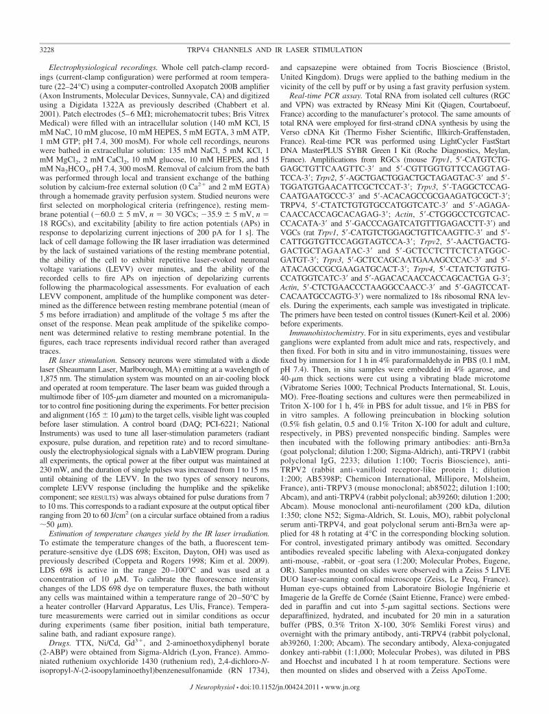
Mid-IR laser stimulations trigger transient membrane po-tential variations in sensory neurons. Isolated RGCs and ves-tibular primary neurons (VGCs) were irradiated using an1,875-nm IR laser. LEVVs were obtained in RGCs and VGCsfor pulse durations from 7 to 10 ms at laser power of 230 mW.As illustrated in Fig. 1, either single (phasic) or repetitive(tonic) spike responses could be evoked in RGCs (Fig. 1A) andVGCs (Fig. 1B) depending on the duration of the irradiation.LEVVs usually displayed an initial humplike depolarization(mean maximum amplitude: 10 � 4 mV, n � 12 RGCs and10 � 5 mV, n � 25 VGCs), on top of which a spikelikecomponent (mean peak amplitude: 30 � 5 mV, n � 12 RGCsand 35 mV � 5 mV, n � 25 VGCs) occurred (Fig. 1C). Thehumplike depolarization phase always started within the 1st msafter the onset of the irradiation. Conversely, occurrence of thespike component could be delayed up to 10 ms under ourirradiation conditions. LEVVs could be evoked repetitively forseveral minutes in the two cell types without cell damage.Visual control of the preparation did not reveal any movementof recorded cells nor of the external solution during theirradiation pulses. In a fraction of recorded VGCs (16%, n �4) a small hyperpolarization of 3 ms preceded the spike
component of the LEVV (Fig. 5A). Such hyperpolarizationresponse was never observed in the RGCs.
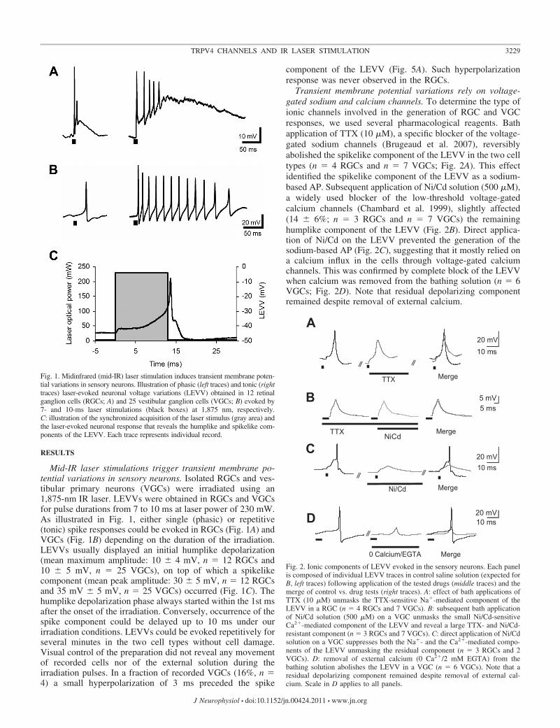
Transient membrane potential variations rely on voltage-gated sodium and calcium channels. To determine the type ofionic channels involved in the generation of RGC and VGCresponses, we used several pharmacological reagents. Bathapplication of TTX (10 �M), a specific blocker of the voltage-gated sodium channels (Brugeaud et al. 2007), reversiblyabolished the spikelike component of the LEVV in the two celltypes (n � 4 RGCs and n � 7 VGCs; Fig. 2A). This effectidentified the spikelike component of the LEVV as a sodium-based AP. Subsequent application of Ni/Cd solution (500 �M),a widely used blocker of the low-threshold voltage-gatedcalcium channels (Chambard et al. 1999), slightly affected(14 � 6%; n � 3 RGCs and n � 7 VGCs) the remaininghumplike component of the LEVV (Fig. 2B). Direct applica-tion of Ni/Cd on the LEVV prevented the generation of thesodium-based AP (Fig. 2C), suggesting that it mostly relied ona calcium influx in the cells through voltage-gated calciumchannels. This was confirmed by complete block of the LEVVwhen calcium was removed from the bathing solution (n � 6VGCs; Fig. 2D). Note that residual depolarizing componentremained despite removal of external calcium.
Fig. 2. Ionic components of LEVV evoked in the sensory neurons. Each panelis composed of individual LEVV traces in control saline solution (expected forB, left traces) following application of the tested drugs (middle traces) and themerge of control vs. drug tests (right traces). A: effect of bath applications ofTTX (10 �M) unmasks the TTX-sensitive Na�-mediated component of theLEVV in a RGC (n � 4 RGCs and 7 VGCs). B: subsequent bath applicationof Ni/Cd solution (500 �M) on a VGC unmasks the small Ni/Cd-sensitiveCa2�-mediated component of the LEVV and reveal a large TTX- and Ni/Cd-resistant component (n � 3 RGCs and 7 VGCs). C: direct application of Ni/Cdsolution on a VGC suppresses both the Na�- and the Ca2�-mediated compo-nents of the LEVV unmasking the residual component (n � 3 RGCs and 2VGCs). D: removal of external calcium (0 Ca2�/2 mM EGTA) from thebathing solution abolishes the LEVV in a VGC (n � 6 VGCs). Note that aresidual depolarizing component remained despite removal of external cal-cium. Scale in D applies to all panels.
Fig. 1. Midinfrared (mid-IR) laser stimulation induces transient membrane poten-tial variations in sensory neurons. Illustration of phasic (left traces) and tonic (righttraces) laser-evoked neuronal voltage variations (LEVV) obtained in 12 retinalganglion cells (RGCs; A) and 25 vestibular ganglion cells (VGCs; B) evoked by7- and 10-ms laser stimulations (black boxes) at 1,875 nm, respectively.C: illustration of the synchronized acquisition of the laser stimulus (gray area) andthe laser-evoked neuronal response that reveals the humplike and spikelike com-ponents of the LEVV. Each trace represents individual record.
3229TRPV4 CHANNELS AND IR LASER STIMULATION
J Neurophysiol • doi:10.1152/jn.00424.2011 • www.jn.org

TRPC channels are not involved in the IR laser-evoked re-sponse in sensory neurons. Transient receptor potential canonical(TRPC) channels were previously reported to mediate light-evoked calcium responses in intrinsically photosensitive RGCs(ipRGCs; Hartwick et al. 2007). Bath applications of either2-APB (100 �M; n � 2 RGCs and n � 2 VGCs) or Gd3�
(10–100 �M; n � 3 RGCs and n � 2 VGCs), two widely usedblockers of the TRPC channels, did not affected the LEVVevoked in both cell types (Fig. 3, A and B). This observationindicated that the TRPC channels were not required for the gen-eration of the LEVV in these sensory neurons.
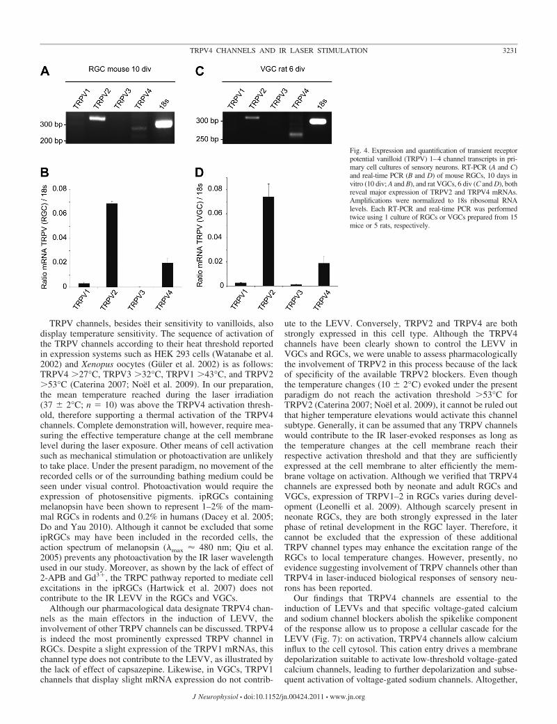
TRPV4 channels mediate the IR laser-evoked response in sen-sory neurons. It has been recently proposed that AP generation bypulsed laser stimulation of nerve tissue or neurons is thermallymediated either through thermoradiating or thermomechanicalprocesses (Wells et al. 2007a,b). To test this hypothesis, weinvestigated whether thermosensitive TRPV channels were in-volved in the LEVVs evoked in sensory neurons. We firstsearched for the presence of TRPV1–4 channels mRNA in ourpreparations of RGC and VGC neurons (Fig. 4). RT-PCR inves-tigations using specific primers (see MATERIALS AND METHODS)revealed that TRPV channel mRNAs were differentially ex-pressed with similar patterns in the two types of sensory neurons.In the RGCs, TRPV2 and TRPV4 mRNAs displayed the majorexpression, whereas neither the expression of TRPV1 nor TRPV3was significant (Fig. 4A and B). Similarly, in VGCs, both TRPV4and TRPV2 mRNAs displayed high levels of expression, whereasthe expression of neither TRPV1 nor TRPV3 mRNAs was sig-nificant (Fig. 4, C and D).
Pharmacological investigations were subsequently con-ducted to test for the direct involvement in the LEVV of theTRPV channel subtypes expressed in the RGCs and VGCs.Bath applications of ruthenium red (10 �M), a general TRPVchannel blocker, completely abolished the LEVV evoked inboth the RGCs (n � 3) and VGCs (n � 5; Fig. 5A). Thisindicated that TRPV channels mediated the LEVV in sensoryneurons. Capsazepine (10 �M), a specific TRPV1 channelblocker, did not affect the LEVV evoked in the RGCs (n � 2)and in the VGCs (n � 4; Fig. 5B), consistent with thenonsignificant expression levels of TRPV1 in both neurontypes (Fig. 4). Conversely, RN 1734 (3.2 �M in VGC and 5.2�M in RGCs), a specific TRPV4 channel blocker (Angelicoand Testa 2010; Vincent et al. 2009), prevented the inductionof LEVV in both RGCs (n � 4; Fig. 5C) and VGCs (n � 7;Fig. 5D). Altogether, these results indicated that the TRPV4channels mediate the LEVV in the two types of sensory neurons.The electrically evoked neural responses were not affected onapplication of ruthenium red or RN 1734 (data not shown). In ourhands, SKF-96365 given as a potent TRPV2 blocker (Vriens et al.2009) significantly altered the voltage-induced excitability of theVGCs, preventing any investigation of the involvement of TRPV2into the LEVV in this neuronal type.
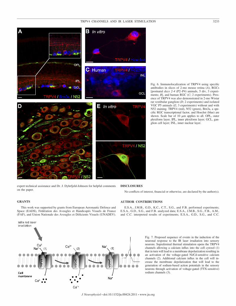
Tissue and cell expression of TRPV4. Tissue and cell ex-pressions of the TRPV4 protein were studied using specificantibodies in transverse sections of adult mouse retinas and ratvestibular ganglions in RGCs and VGCs isolated from neonaterodent tissues as well as in adult human retina (Fig. 6). In theadult mouse retina, TRPV4 immunostaining was found in theRGCs and the inner plexiform layers (Fig. 6A). RGCs dis-played similar TRPV4 protein expression at membrane level inthe cell cytosol and neurites in isolated neonate RGCs (Fig.6B). In adult human retina (Fig. 6C), RGCs also exhibitedTRPV4 immunostaining, suggesting a conserved expressionof TRPV4 in RGCs across species. In the vestibular ganglion,TRPV4 immunostaining was observed in the cytosol and in theperiphery of the soma, suggesting membrane expression of theTRPV4 channels (Fig. 6D). On isolated neonate VGCs,TRPV4 expression seems to be restricted to the cell bodies, asneuritis were not immunolabeled (Fig. 6E).
Local temperature changes evoked by the laser irradiation.To assess the ability of the laser irradiations to alter significantlythe temperature of the bath we measured, in a bath withoutneurons, the fluorescence variations of LDS 698, a temperature-sensitive dye, on laser irradiations in the range of those thattriggered LEVV in sensory neurons. Laser irradiations of 20 J/cm2
(i.e., 7-ms pulse duration) evoked mean temperature variations of10 � 2°C (n � 10). This confirmed that the local temperaturejumps evoked by the IR laser irradiation were within the activa-tion range of the TRPV4 channels (Güler et al. 2002; Watanabe etal. 2002).
DISCUSSION
Present data directly demonstrate that the neuronal responsesobserved in the RGCs and VGCs on IR laser irradiationoriginates from TRPV4 channel activation. In addition, weshow that TRPV4 channels are expressed in the two types ofsensory neurons in our culture preparation as well as in adulttissues. Finally, we confirm that the local temperature jumpsevoked by the IR laser irradiation are suitable for the activationof the TRPV4 channels.
Fig. 3. The transient receptor potential canonical (TRPC) channels are notinvolved in the generation of the LEVV in the RGCs and the VGCs. Eachpanel is composed of LEVV traces in control saline solution (left traces)following application of the tested drugs (middle traces) and the merge ofTTX-resistant vs. 2-aminoethoxydiphenyl borate (2-ABP) or Gd3� tests,respectively (right traces). A: application of 2-ABP (100 �M), a selectiveTRPC channel antagonist, subsequently to TTX (10 �M) does not affect theresidual, RGC component of the LEVV evoked in a RGC (n � 2 RGCs and2 VGCs). B: gadolinium (10 and 100 �M) does not affect the residualcomponent of the LEVV in a VGC (n � 3 RGCs and 2 VGCs). Note that inthat specific recording, the remaining spikelike deflection despite the presenceof TTX may result from a noncomplete block of the voltage-gated sodiumchannel population.
3230 TRPV4 CHANNELS AND IR LASER STIMULATION
J Neurophysiol • doi:10.1152/jn.00424.2011 • www.jn.org
TRPV channels, besides their sensitivity to vanilloids, alsodisplay temperature sensitivity. The sequence of activation ofthe TRPV channels according to their heat threshold reportedin expression systems such as HEK 293 cells (Watanabe et al.2002) and Xenopus oocytes (Güler et al. 2002) is as follows:TRPV4 �27°C, TRPV3 �32°C, TRPV1 �43°C, and TRPV2�53°C (Caterina 2007; Noël et al. 2009). In our preparation,the mean temperature reached during the laser irradiation(37 � 2°C; n � 10) was above the TRPV4 activation thresh-old, therefore supporting a thermal activation of the TRPV4channels. Complete demonstration will, however, require mea-suring the effective temperature change at the cell membranelevel during the laser exposure. Other means of cell activationsuch as mechanical stimulation or photoactivation are unlikelyto take place. Under the present paradigm, no movement of therecorded cells or of the surrounding bathing medium could beseen under visual control. Photoactivation would require theexpression of photosensitive pigments. ipRGCs containingmelanopsin have been shown to represent 1–2% of the mam-mal RGCs in rodents and 0.2% in humans (Dacey et al. 2005;Do and Yau 2010). Although it cannot be excluded that someipRGCs may have been included in the recorded cells, theaction spectrum of melanopsin (�max � 480 nm; Qiu et al.2005) prevents any photoactivation by the IR laser wavelengthused in our study. Moreover, as shown by the lack of effect of2-APB and Gd3�, the TRPC pathway reported to mediate cellexcitations in the ipRGCs (Hartwick et al. 2007) does notcontribute to the IR LEVV in the RGCs and VGCs.
Although our pharmacological data designate TRPV4 chan-nels as the main effectors in the induction of LEVV, theinvolvement of other TRPV channels can be discussed. TRPV4is indeed the most prominently expressed TRPV channel inRGCs. Despite a slight expression of the TRPV1 mRNAs, thischannel type does not contribute to the LEVV, as illustrated bythe lack of effect of capsazepine. Likewise, in VGCs, TRPV1channels that display slight mRNA expression do not contrib-
ute to the LEVV. Conversely, TRPV2 and TRPV4 are bothstrongly expressed in this cell type. Although the TRPV4channels have been clearly shown to control the LEVV inVGCs and RGCs, we were unable to assess pharmacologicallythe involvement of TRPV2 in this process because of the lackof specificity of the available TRPV2 blockers. Even thoughthe temperature changes (10 � 2°C) evoked under the presentparadigm do not reach the activation threshold �53°C forTRPV2 (Caterina 2007; Noël et al. 2009), it cannot be ruled outthat higher temperature elevations would activate this channelsubtype. Generally, it can be assumed that any TRPV channelswould contribute to the IR laser-evoked responses as long asthe temperature changes at the cell membrane reach theirrespective activation threshold and that they are sufficientlyexpressed at the cell membrane to alter efficiently the mem-brane voltage on activation. Although we verified that TRPV4channels are expressed both by neonate and adult RGCs andVGCs, expression of TRPV1–2 in RGCs varies during devel-opment (Leonelli et al. 2009). Although scarcely present inneonate RGCs, they are both strongly expressed in the laterphase of retinal development in the RGC layer. Therefore, itcannot be excluded that the expression of these additionalTRPV channel types may enhance the excitation range of theRGCs to local temperature changes. However, presently, noevidence suggesting involvement of TRPV channels other thanTRPV4 in laser-induced biological responses of sensory neu-rons has been reported.
Our findings that TRPV4 channels are essential to theinduction of LEVVs and that specific voltage-gated calciumand sodium channel blockers abolish the spikelike componentof the response allow us to propose a cellular cascade for theLEVV (Fig. 7): on activation, TRPV4 channels allow calciuminflux to the cell cytosol. This cation entry drives a membranedepolarization suitable to activate low-threshold voltage-gatedcalcium channels, leading to further depolarization and subse-quent activation of voltage-gated sodium channels. Altogether,
Fig. 4. Expression and quantification of transient receptorpotential vanilloid (TRPV) 1–4 channel transcripts in pri-mary cell cultures of sensory neurons. RT-PCR (A and C)and real-time PCR (B and D) of mouse RGCs, 10 days invitro (10 div; A and B), and rat VGCs, 6 div (C and D), bothreveal major expression of TRPV2 and TRPV4 mRNAs.Amplifications were normalized to 18s ribosomal RNAlevels. Each RT-PCR and real-time PCR was performedtwice using 1 culture of RGCs or VGCs prepared from 15mice or 5 rats, respectively.
3231TRPV4 CHANNELS AND IR LASER STIMULATION
J Neurophysiol • doi:10.1152/jn.00424.2011 • www.jn.org

this sequence of events allows generation of sodium-based APsin the cells forming the optic and vestibular nerves followingIR laser stimulation. Given that expression of ion channels inVGCs is heterogeneous (Chabbert et al. 2001; Kalluri et al.2010), the small prespike hyperpolarization in a fraction ofrecorded VGCs likely reflects a different ion channel compo-sition in this subpopulation. Because of minimal species dif-ferences in TRPV4 amino acid sequences (Nilius et al. 2004)and the similar TRPV4 protein expression in rodent and adulthuman RGCs, it is likely that the TRPV4-based process oflaser-evoked cell response in these cells could apply to humantissue. The apparent innocuousness of IR lasers for the stimu-lation of primary sensory neurons in culture suggests that IRlaser irradiation, at least in range of the wavelengths and laserpower used in the present study, may be a convenient way tostimulate properly these sensory ganglion cells in a prosthesisapproach. However, although the present study indicates that
sensory neurons can be repetitively irradiated without altera-tion of their passive membrane properties, the long-lastingconsequences of repetitive high-rate stimulations will need tobe explored further. Demonstration that excitable cells express-ing TRPV channels may be targeted by local and controlledtemperature changes also opens novel technical possibilities,specifically regarding the development of nanotechnology.Recent elegant experiments have indeed demonstrated thatchanges in the membrane potential of hippocampal neuronsthat express TRPV1 could be achieved through magnetic-fieldheating of nanoparticles (Huang et al. 2010). Bringing the invivo proof of concept of the feasibility to excite neurons usingIR laser excitation will require solving some important ques-tions such as how to define the optimal location for irradiationof target cells. The irregular expression of TRPV channelsalong the neuron soma (illustrated by the selective expressionof TRPV4 at the soma of adult rat VGCs) as well as the factthat some sensory neurons (vestibular and auditory ganglioncells) are myelinated can also influence the sensitivity of thesensory neurons to IR laser stimulation. In any case, the presentdemonstration should open novel therapeutic opportunities,particularly in the field of the sensory neuroscience, as TRPV4channels have now been demonstrated in retinal, vestibular,and auditory ganglion cells (Shen et al. 2006).
In previous experiments on cardiomyocytes, Dittami andcolleagues (2011) brought evidence that IR irradiation (in thesame energy range of that used in present study) may alsomodulate the calcium mitochondrial uniporter (mCU). Briefand repetitive IR irradiations significantly increased cytosoliccalcium in these cells. Subsequently, the same research groupproposed that a similar mechanism may support the IR stimu-lation of vestibular hair cells that led to excitation of thevestibular afferents in the Toadfish crista ampullaris (Rajguruet al. 2011). Following this interesting observation, it cannot bediscarded that the mitochondrial pathway may also be activatedin the sensory neurons on IR irradiation. However, it has to benoticed that under present experimental conditions, the re-moval of the external calcium was sufficient to abolish theLEVV, although a residual depolarizing component remained.This suggests that any IR activation of the mCU may modulatebut not be sufficient to drive the LEVV of sensory neurons.Moreover, IR irradiations that activated the hair cells in theexperiments of Rajguru et al. (2011) were inefficient on thevestibular afferents. This also suggests that the mechanismssupporting the biological responses to the IR irradiation maydiffer depending on the cell type. Additional investigationsusing calcium microfluorimetry technique should allow assess-ment of the respective weight of these two cellular pathways inthe induction of the LEVV. In the same way, investigationsusing the voltage-clamp configuration of the patch-clamp tech-nique should allow further investigation of the ionic nature ofthe current that flows through the TRPV4 channel during thelaser irradiation. This will help to test whether different chan-nel types participate to the neuron response or to gatherindications on the voltage sensitivity of the TRPV4 channel insensory neurons.
ACKNOWLEDGMENTS
We thank Dr. M. Pequignot for supplying human retinas (obtained from Dr.Gilles Thuret, Laboratoire Biologie Ingénierie et Imagerie de la Greffe deCornée). We also thank Drs. C. Delettre, O. Payet, and H. Boukhaddaoui for
Fig. 5. TRPV4 channels mediate the LEVV in sensory neurons. Each panel iscomposed of LEVV traces in control saline solution (left traces) and followingapplication of the indicated tested drugs (right traces). A: illustration of thecomplete block of the LEVV by ruthenium red (10 �M), a general TRPVchannel blocker in a VGC (n � 3 RGCs and 5 VGCs). B: capsazepine (10�M), a TRPV1 channel blocker, does not affect LEVV in a RGC (n � 2 RGCsand 4 VGCs). C: RN 1734 (5.2 �M), a specific TRPV4 channel blocker,completely abolishes the LEVV in RGCs (n � 4). D: RN 1734 (3.2 �M)prevents the LEVV in VGCs (n � 7). Scale in D applies to B–D.
3232 TRPV4 CHANNELS AND IR LASER STIMULATION
J Neurophysiol • doi:10.1152/jn.00424.2011 • www.jn.org
expert technical assistance and Dr. J. Dyhrfjeld-Johnsen for helpful commentson the paper.
GRANTS
This work was supported by grants from European Aeronautic Defence andSpace (EADS), Fédération des Aveugles et Handicapés Visuels de France(FAF), and Union Nationale des Aveugles et Déficients Visuels (UNADEV).
DISCLOSURES
No conflicts of interest, financial or otherwise, are declared by the author(s).
AUTHOR CONTRIBUTIONS
E.S.A., J.M.B., G.D., K.C., C.T., S.G., and F.B. performed experiments;E.S.A., G.D., S.G., and F.B. analyzed data; E.S.A., J.M.B., S.G., F.B., A.M.,and C.C. interpreted results of experiments; E.S.A., G.D., S.G., and C.C.
Fig. 6. Immunolocalization of TRPV4 using specificantibodies in slices of 2-mo mouse retina (A), RGCs[postnatal days 2-4 (P2–P4) animals, 5 div, 3 experi-ments; B], and human RGC (C; 2 experiments). Pres-ence of TRPV4 was also demonstrated in 2-mo Wistarrat vestibular ganglion (D; 2 experiments) and isolatedVGC P5 animals (E; 3 experiments) without and withN52 staining. TRPV4 (red), N52 (green), Brn3a, a spe-cific RGC transcriptional factor, and Hoechst (blue) areshown. Scale bar of 10 �m applies to all. OPL, outerplexiform layer; IPL, inner plexiform layer; GCL, gan-glion cell layer; INL, inner nuclear layer.
Fig. 7. Proposed sequence of events in the induction of theneuronal response to the IR laser irradiation into sensoryneurons. Supraliminal thermal stimulation opens the TRPV4channels allowing a calcium influx into the cell cytosol (1)that in turn will lead to a membrane depolarization resulting inan activation of the voltage-gated Ni/Cd-sensitive calciumchannels (2). Additional calcium influx in the cell will in-crease the membrane depolarization that will lead to thegeneration of sodium-based action potentials in the sensoryneurons through activation of voltage-gated (TTX-sensitive)sodium channels (3).
3233TRPV4 CHANNELS AND IR LASER STIMULATION
J Neurophysiol • doi:10.1152/jn.00424.2011 • www.jn.org
prepared figures; E.S.A., I.M., M.D., G.L., C.H., A.M., and C.C. draftedmanuscript; E.S.A. and C.C. approved final version of manuscript; C.C.conception and design of research; C.C. edited and revised manuscript.
REFERENCES
Angelico P, Testa R. TRPV4 as a target for bladder overactivity. F1000 BiolRep 2: 12, 2010.
Brugeaud A, Travo C, Dememes D, Lenoir M, Llorens J, Puel JL,Chabbert C. Control of hair cell excitability by vestibular primary sensoryneurons. J Neurosci 27: 3503–3511, 2007.
Caterina MJ. Transient receptor potential ion channels as participants inthermosensation and thermoregulation. Am J Physiol Regul Integr CompPhysiol 292: R64–R76, 2007.
Chabbert C, Chambard JM, Valmier J, Sans A, Desmadryl G. Hyperpo-larization-activated (Ih) current in mouse vestibular primary neurons. Neu-roreport 12: 2701–2704, 2001.
Chambard JM, Chabbert C, Sans A, Desmadryl G. Developmental changesin low and high voltage-activated calcium currents in acutely isolated mousevestibular neurons. J Physiol 518: 141–149, 1999.
Chen Y, Bedell M, Zhang K. Age-related macular degeneration: genetic andenvironmental factors of disease. Mol Interv 10: 271–281, 2010.
Colodetti L, Weiland JD, Colodetti S, Ray A, Seiler MJ, Hinton DR,Humayun MS. Pathology of damaging electrical stimulation in the retina.Exp Eye Res 85: 23–33, 2007.
Coppeta J, Rogers C. Dual emission laser-induced fluorescence for directplanar scalar behavior measurements. Exp Fluids 25: 1–15, 1998.
Dacey DM, Liao HW, Peterson BB, Robinson FR, Smith VC, Pokorny J,Yau KW, Gamlin PD. Melanopsin-expressing ganglion cells in primateretina signal colour and irradiance and project to the LGN. Nature 433:749–754, 2005.
Dayanithi G, Desmadryl G, Travo C, Chabbert C, Sans A. Trimetazidinemodulates AMPA/kainate receptors in rat vestibular ganglion neurons. EurJ Pharmacol 574: 8–14, 2007.
Dittami GM, Rajguru SM, Lasher RA, Hitchcock RW, Rabbitt RD.Intracellular calcium transients evoked by pulsed infrared radiation inneonatal cardiomyocytes. J Physiol 589: 1295–1306, 2011.
Do MT, Yau KW. Intrinsically photosensitive retinal ganglion cells. PhysiolRev 90: 1547–1581, 2010.
Fork RL. Laser stimulation of nerve cells in Aplysia. Science 171: 907–908,1971.
Fried NM, Lagoda GA, Scott NJ, Su LM, Burnett AL. Laser stimulation ofthe cavernous nerves in the rat prostate, in vivo: optimization of wavelength,pulse energy, and pulse repetition rate. Conf Proc IEEE Eng Med Biol Soc2008: 2777–2780, 2008.
Güler AD, Lee H, Iida T, Shimizu I, Tominaga M, Caterina M. Heat-evoked activation of the ion channel, TRPV4. J Neurosci 22: 6408–6414,2002.
Hartwick AT, Bramley JR, Yu J, Stevens KT, Allen CN, Baldridge WH,Sollars PJ, Pickard GE. Light-evoked calcium responses of isolatedmelanopsin-expressing retinal ganglion cells. J Neurosci 27: 13468–13480,2007.
Haynes DS, Young JA, Wanna GB, Glasscock ME 3rd. Middle earimplantable hearing devices: an overview. Trends Amplif 13: 206–214,2009.
Huang H, Delikanli S, Zeng H, Ferkey DM, Pralle A. Remote control of ionchannels and neurons through magnetic-field heating of nanoparticles. NatNanotechnol 5: 602–606, 2010.
Izzo AD, Richter CP, Jansen ED, Walsh JT Jr. Laser stimulation of theauditory nerve. Lasers Surg Med 38: 745–753, 2006.
Izzo AD, Walsh JT Jr, Ralph H, Webb J, Bendett M, Wells J, Richter CP.Laser stimulation of auditory neurons: effect of shorter pulse duration andpenetration depth. Biophys J 94: 3159–3166, 2008.
Kalluri R, Xue J, Eatock RA. Ion channels set spike timing regularity ofmammalian vestibular afferent neurons. J Neurophysiol 104: 2034–2051,2010.
Kamei S, Chen-Kuo-Chang M, Cazevieille C, Lenaers G, Olichon A,Bélenguer P, Roussignol G, Renard N, Eybalin M, Michelin A, DelettreC, Brabet P, Hamel CP. Expression of the Opa1 mitochondrial protein inretinal ganglion cells: its downregulation causes aggregation of the mito-chondrial network. Invest Ophthalmol Vis Sci 46: 4288–4294, 2005.
Kim H, Dixit S, Green CJ, Faris GW. Nanodroplet real-time PCR systemwith laser assisted heating. Opt Express 17: 218–227, 2009.
Kunert-Keil C, Bisping F, Kruger J, Brinkmeier H. Tissue-specific expres-sion of TRP channel genes in the mouse and its variation in three differentmouse strains. BMC Genomics 7: 159, 2006.
Leonelli M, Martins DO, Kihara AH, Britto LR. Ontogenetic expression ofthe vanilloid receptors TRPV1 and TRPV2 in the rat retina. Int J DevNeurosci 27: 709–718, 2009.
Nilius B, VJ, Prenen J, Droogmans G, Voets T. TRPV4 calcium entrychannel: a paradigm for gating diversity. Am J Physiol Cell Physiol 286:C195–C205, 2004.
Noël J, Zimmermann K, Busserolles J, Deval E, Alloui A, Diochot S, GuyN, Borsotto M, Reeh P, Eschalier A, Lazdunski M. The mechano-activated K� channels TRAAK and TREK-1 control both warm and coldperception. EMBO J 28: 1308–1318, 2009.
Popovic D, Gordon T, Rafuse VF, Prochazka A. Properties of implantedelectrodes for functional electrical stimulation. Ann Biomed Eng 19: 303–316, 1991.
Qiu X, Kumbalasiri T, Carlson SM, Wong KY, Krishna V, Provencio I,Berson DM. Induction of photosensitivity by heterologous expression ofmelanopsin. Nature 433: 745–749, 2005.
Rajguru SM, Richter CP, Matic AI, Holstein GR, Highstein SM, DittamiGM, Rabbitt RD. Infrared photostimulation of the crista ampullaris. JPhysiol 589: 1283–1294, 2011.
Saihan Z, Webster AR, Luxon L, Bitner-Glindzicz M. Update on Ushersyndrome. Curr Opin Neurol 22: 19–27, 2009.
Shen J, Harada N, Kubo N, Liu B, Mizuno A, Suzuki M, Yamashita T.Functional expression of transient receptor potential vanilloid 4 in the mousecochlea. Neuroreport 17: 135–139, 2006.
Teudt IU, Nevel AE, Izzo AD, Walsh JT Jr, Richter CP. Optical stimulationof the facial nerve: a new monitoring technique? Laryngoscope 117: 1641–1647, 2007.
Vincent F, Acevedo A, Nguyen MT, Dourado M, DeFalco J, Gustafson A,Spiro P, Emerling DE, Kelly MG, Duncton MA. Identification andcharacterization of novel TRPV4 modulators. Biochem Biophys Res Com-mun 389: 490–494, 2009.
Vriens J, Appendino G, Nilius B. Pharmacology of vanilloid transientreceptor potential cation channels. Mol Pharmacol 75: 1262–1279, 2009.
Watanabe H, Vriens J, Suh SH, Benham, Droogmans G, Nilius B. Heatactivation of TRPV4 channels in HEK 293 cell expression system and innative mouse aorta endothelial cells. J Biol Chem 277: 47044–47051, 2002.
Wells J, Konrad P, Kao C, Jansen ED, Mahadevan-Jansen A. Pulsed laserversus electrical energy for peripheral nerve stimulation. J Neurosci Meth-ods 163: 326–337, 2007a.
Wells J, Kao C, Konrad P, Milner T, Kim J, Mahadevan-Jansen A, JansenED. Biophysical mechanisms of transient optical stimulation of peripheralnerve. Biophys J 93: 2567–2580, 2007b.
Wells J, Kao C, Mariappan K, Albea J, Jansen ED, Konrad P, Mahade-van-Jansen A. Optical stimulation of neural tissue in vivo. Opt Lett 30:504–506, 2005.
3234 TRPV4 CHANNELS AND IR LASER STIMULATION
J Neurophysiol • doi:10.1152/jn.00424.2011 • www.jn.org
Copyright © 2022 FDOKUMEN