Contact heat evoked potentials using simultaneous EEG and fMRI and their correlation with evoked...
12
BioMed Central Page 1 of 12 (page number not for citation purposes) BMC Anesthesiology Open Access Technical advance Contact heat evoked potentials using simultaneous EEG and fMRI and their correlation with evoked pain Katherine Roberts 1 , Anastasia Papadaki 1 , Carla Gonçalves 2 , Mary Tighe 3 , Duncan Atherton 1 , Ravikiran Shenoy 1 , Donald McRobbie 1 and Praveen Anand* 1 Address: 1 Imperial College Healthcare NHS Trust, London, UK, 2 East Kent Hospitals NHS Trust, Canterbury, UK and 3 Kings College London, London, UK Email: Katherine Roberts - [email protected]; Anastasia Papadaki - [email protected]; Carla Gonçalves - [email protected]; Mary Tighe - [email protected]; Duncan Atherton - [email protected]; Ravikiran Shenoy - [email protected]; Donald McRobbie - [email protected]; Praveen Anand* - [email protected] * Corresponding author Abstract Background: The Contact Heat Evoked Potential Stimulator (CHEPS) utilises rapidly delivered heat pulses with adjustable peak temperatures to stimulate the differential warm/heat thresholds of receptors expressed by Aδ and C fibres. The resulting evoked potentials can be recorded and measured, providing a useful clinical tool for the study of thermal and nociceptive pathways. Concurrent recording of contact heat evoked potentials using electroencephalogram (EEG) and functional magnetic resonance imaging (fMRI) has not previously been reported with CHEPS. Developing simultaneous EEG and fMRI with CHEPS is highly desirable, as it provides an opportunity to exploit the high temporal resolution of EEG and the high spatial resolution of fMRI to study the reaction of the human brain to thermal and nociceptive stimuli. Methods: In this study we have recorded evoked potentials stimulated by 51°C contact heat pulses from CHEPS using EEG, under normal conditions (baseline), and during continuous and simultaneous acquisition of fMRI images in ten healthy volunteers, during two sessions. The pain evoked by CHEPS was recorded on a Visual Analogue Scale (VAS). Results: Analysis of EEG data revealed that the latencies and amplitudes of evoked potentials recorded during continuous fMRI did not differ significantly from baseline recordings. fMRI results were consistent with previous thermal pain studies, and showed Blood Oxygen Level Dependent (BOLD) changes in the insula, post-central gyrus, supplementary motor area (SMA), middle cingulate cortex and pre-central gyrus. There was a significant positive correlation between the evoked potential amplitude (EEG) and the psychophysical perception of pain on the VAS. Conclusion: The results of this study demonstrate the feasibility of recording contact heat evoked potentials with EEG during continuous and simultaneous fMRI. The combined use of the two methods can lead to identification of distinct patterns of brain activity indicative of pain and pro- nociceptive sensitisation in healthy subjects and chronic pain patients. Further studies are required for the technique to progress as a useful tool in clinical trials of novel analgesics. Published: 17 December 2008 BMC Anesthesiology 2008, 8:8 doi:10.1186/1471-2253-8-8 Received: 7 February 2008 Accepted: 17 December 2008 This article is available from: http://www.biomedcentral.com/1471-2253/8/8 © 2008 Roberts et al; licensee BioMed Central Ltd. This is an Open Access article distributed under the terms of the Creative Commons Attribution License (http://creativecommons.org/licenses/by/2.0 ), which permits unrestricted use, distribution, and reproduction in any medium, provided the original work is properly cited.
-
Upload
independent -
Category
Documents
-
view
4 -
download
0
Transcript of Contact heat evoked potentials using simultaneous EEG and fMRI and their correlation with evoked...
BioMed CentralBMC Anesthesiology
ss
Open AcceTechnical advanceContact heat evoked potentials using simultaneous EEG and fMRI and their correlation with evoked painKatherine Roberts1, Anastasia Papadaki1, Carla Gonçalves2, Mary Tighe3, Duncan Atherton1, Ravikiran Shenoy1, Donald McRobbie1 and Praveen Anand*1Address: 1Imperial College Healthcare NHS Trust, London, UK, 2East Kent Hospitals NHS Trust, Canterbury, UK and 3Kings College London, London, UK
Email: Katherine Roberts - [email protected]; Anastasia Papadaki - [email protected]; Carla Gonçalves - [email protected]; Mary Tighe - [email protected]; Duncan Atherton - [email protected]; Ravikiran Shenoy - [email protected]; Donald McRobbie - [email protected]; Praveen Anand* - [email protected]
* Corresponding author
AbstractBackground: The Contact Heat Evoked Potential Stimulator (CHEPS) utilises rapidly deliveredheat pulses with adjustable peak temperatures to stimulate the differential warm/heat thresholdsof receptors expressed by Aδ and C fibres. The resulting evoked potentials can be recorded andmeasured, providing a useful clinical tool for the study of thermal and nociceptive pathways.Concurrent recording of contact heat evoked potentials using electroencephalogram (EEG) andfunctional magnetic resonance imaging (fMRI) has not previously been reported with CHEPS.Developing simultaneous EEG and fMRI with CHEPS is highly desirable, as it provides anopportunity to exploit the high temporal resolution of EEG and the high spatial resolution of fMRIto study the reaction of the human brain to thermal and nociceptive stimuli.
Methods: In this study we have recorded evoked potentials stimulated by 51°C contact heatpulses from CHEPS using EEG, under normal conditions (baseline), and during continuous andsimultaneous acquisition of fMRI images in ten healthy volunteers, during two sessions. The painevoked by CHEPS was recorded on a Visual Analogue Scale (VAS).
Results: Analysis of EEG data revealed that the latencies and amplitudes of evoked potentialsrecorded during continuous fMRI did not differ significantly from baseline recordings. fMRI resultswere consistent with previous thermal pain studies, and showed Blood Oxygen Level Dependent(BOLD) changes in the insula, post-central gyrus, supplementary motor area (SMA), middlecingulate cortex and pre-central gyrus. There was a significant positive correlation between theevoked potential amplitude (EEG) and the psychophysical perception of pain on the VAS.
Conclusion: The results of this study demonstrate the feasibility of recording contact heat evokedpotentials with EEG during continuous and simultaneous fMRI. The combined use of the twomethods can lead to identification of distinct patterns of brain activity indicative of pain and pro-nociceptive sensitisation in healthy subjects and chronic pain patients. Further studies are requiredfor the technique to progress as a useful tool in clinical trials of novel analgesics.
Published: 17 December 2008
BMC Anesthesiology 2008, 8:8 doi:10.1186/1471-2253-8-8
Received: 7 February 2008Accepted: 17 December 2008
This article is available from: http://www.biomedcentral.com/1471-2253/8/8
© 2008 Roberts et al; licensee BioMed Central Ltd. This is an Open Access article distributed under the terms of the Creative Commons Attribution License (http://creativecommons.org/licenses/by/2.0), which permits unrestricted use, distribution, and reproduction in any medium, provided the original work is properly cited.
Page 1 of 12(page number not for citation purposes)
BMC Anesthesiology 2008, 8:8 http://www.biomedcentral.com/1471-2253/8/8
BackgroundFunctional magnetic resonance imaging (fMRI) has devel-oped into a tool that is extensively used in non-invasivebrain imaging. It provides information about cerebro-vas-cular activity throughout the whole brain with excellentspatial localisation, yet it is limited by the poor temporalresolution it offers, which is in the order of seconds. Onthe other hand, electroencephalogram (EEG) is recordeddirectly from the scalp of the subject and can provideinformation about neurophysiological activity with a veryprecise temporal resolution, in the order of milliseconds.The disadvantage of EEG however, is that localisation ofthe source of electrical activity within the brain is quitedifficult.
Therefore the integration of these two techniques is highlydesirable, and would allow the exploitation of the advan-tages of both techniques – high spatial resolution of fMRIand high temporal resolution of EEG. The techniquewould be widely applicable in all areas of neurophysio-logical research, but particularly in pain studies, wherecombined use of the two techniques could lead to theidentification of distinct patterns of brain activity indica-tive of pain and pro-nociceptive sensitisation in healthysubjects and chronic pain patients. These patterns couldprove useful in the assessment of the analgesic efficacy ofnovel analgesic compounds, adding to the desirability ofco-registration of pain evoked potentials (such as thosestimulated by contact heat) with EEG and fMRI.
Objective markers of pain could be less variable and/ormore sensitive to analgesic treatments (i.e. EPs, fMRI).The present study enables the use of these markers tosimultaneously assess pharmacodynamic-pharmacoki-netic relationships and antihyperalgesic activity in singledose studies in experimental pain models in humans, andin pain patients (e.g. of novel agents that block TRPV1, theheat and capsaicin receptor). Pain biomarkers are neededto provide early pivotal information on efficacy, dose-response and time-course of TRPV1 antagonists, for strate-gic rapid and cost-effective drug development. Further,neuropathic pain patients would be expected to havereduced pain-evoked potential amplitudes but increasedfMRI activation, different from volunteer or inflammatorymodels/conditions.
The use of simultaneous EEG and fMRI in pharmacologi-cal studies of novel analgesics would allow (i) measure-ment of the combined response to the samepharmacological intervention when the plasma concen-tration of the analgesic is at its peak (i.e. at the same time-course); (ii) recordings can be analysed at the single sub-ject level, reducing inter-session variability, which is wellrecognised with regard to pain scores, and (iii) selection of
the parameter which enables possible reduction in dose inclinical trials.
Simultaneous EEG and fMRI has already been utilised instudies of epilepsy [1], sleep [2], studies of human alphaactivity [3,4] and the study of auditory [5,6], visual [7,8],and motor activity [9]. In the field of pain research, laserand electrically stimulated pain evoked potentials havebeen successfully co-registered using EEG and fMRI[10,11], but CHEPS has the advantages of a larger area ofstimulation, the ability to apply repetitive stimuli to thesame cutaneous area without inducing erythema, andsimplicity of use in the clinic (i.e. no eye protectionrequired, easy to move location); however, one disadvan-tage of CHEPS is that in order to avoid habituation thethermode must be physically moved between stimuli.
Simultaneous EEG/fMRI is a readily feasible applicationyet the MR environment poses a number of technical chal-lenges, limitations and safety issues (for a review of tech-nical and safety issues see [12,13]). The gradient switchingfields and radiofrequency (RF) pulses of the MR scannercan create currents in conducting loops that can poten-tially cause heating of the electrodes and burns at thepoint of contact with the scalp of the subject. Therefore itis recommended to avoid loops and crossing over of elec-trode wires, and introduce current limiting resistors toelectrode wires.
The gradient fields and RF pulses of the MR scanner canlead to artefacts that can obscure the signal recorded fromthe EEG, as can movements within the static magneticfield; movement of the head, talking, swallowing andeven the pulsatile motion of the heart (ballistocardiogramartefact). The magnet's helium cold pump and gradientcoils also produce mechanical vibrations that can bepicked up by the EEG wires in the static magnetic field andconverted to electrical noise. All these issues need to beaddressed and overcome during experimental set-up andprocessing of EEG and fMRI data, in terms of patientsafety and also preserving the quality of the data collected.
Until recently, quantifiable contact heat evoked potentialsin EEG had been hard to elicit, due to technical limita-tions i.e. slow temperature rise and fall times. However,the contact heat evoked potential stimulator (CHEPS) hasbeen designed with a maximum (adjustable) temperaturerise time of 70°C/s. CHEPS can stimulate the differentialthermal thresholds of receptors innervated by Aδ and Cnociceptive nerve fibres, and has been shown to selec-tively excite these fibre subtypes in human hairy and gla-brous skin [14]. The latencies and amplitudes of heatevoked Aδ potentials stimulated by CHEPS have beenshown to be robust and reproducible [15,16], despite thedisadvantage posed by the slow temperature rise time of
Page 2 of 12(page number not for citation purposes)
BMC Anesthesiology 2008, 8:8 http://www.biomedcentral.com/1471-2253/8/8
the heat stimuli produced by CHEPS (70°C/s comparedto a rise time of greater than 1000°C/s reported withlasers) which may lead to a reduction in temporal summa-tion and thus a less synchronous afferent volley [17].
The compatibility of CHEPS with fMRI further widens itsscope of application both in healthy volunteers forresearch purposes and in chronic pain patients. Reproduc-ibility of blood oxygen level dependent (BOLD)responses and pain scores to CHEPS stimulation havealready been illustrated in healthy volunteers [18].
In this human volunteer study we have assessed the feasi-bility of monitoring contact heat evoked Aδ potentialswith simultaneous EEG and fMRI, and determined theirrelationship to the ratings of evoked pain.
MethodsSubjectsTen healthy volunteers were recruited to take part in thisfeasibility study (6 female, 4 male). The average age ofparticipants was 27.7 years (range 22 – 35 years).Informed consent was taken from all subjects prior tocommencement of the study and the study itself wasapproved by Hammersmith and Queen Charlotte's &Chelsea Research Ethics Committee (REC reference 06/Q0404/9).
Heat stimuliContact heat stimulation was performed using CHEPS(Medoc Ltd, Ramat Yishai, Israel) with a thermode area of572.5 mm2, and a heating thermo-foil (Minco Products,Inc., Minneapolis, MN) covered with a 25 μm layer ofthermo conductive plastic (Kapton®, thermal conductivityat 23°C of 0.1 – 0.35 W/m/K). The thermode heating ratewas up to 70°C/s, the cooling rate up to 40°C/s and thebaseline temperature was 32°C.
Experimental setupBaseline protocolBaseline recording of evoked potentials was undertakenwith the subject lying on the scanner table (outside themagnet) without any scanning taking place. Ten 51°Cstimuli of approximately 800 ms duration (approximately271 ms to reach peak temperature of 51°C and approxi-mately 475 ms to return to baseline [total 746 ms]) and 7s inter-stimulus interval were applied to the left volar fore-arm of the subject, and the thermode moved after eachstimulus to avoid habituation.
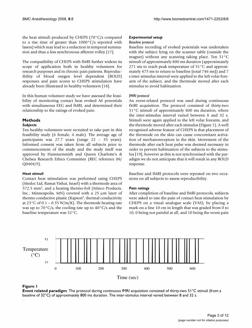
fMRI protocolAn event-related protocol was used during continuousfMRI acquisition. The protocol consisted of thirty-two51°C stimuli of approximately 800 ms duration, wherethe inter-stimulus interval varied between 8 and 32 s.Stimuli were again applied to the left volar forearm, andthe thermode moved after each stimulus (Figure 1). A wellrecognized adverse feature of CHEPS is that placement ofthe thermode on the skin can cause concomitant activa-tion of mechanoreceptors in the skin. Movement of thethermode after each heat pulse was deemed necessary inorder to prevent habituation of the subjects to the stimu-lus [19], however as this is not synchronised with the par-adigm we do not anticipate that it will result in any BOLDresponse.
Baseline and fMRI protocols were repeated on two occa-sions on all subjects to assess reproducibility.
Pain ratingsAfter completion of baseline and fMRI protocols, subjectswere asked to rate the pain of contact heat stimulation byCHEPS on a visual analogue scale (VAS), by placing amark on a line 10 cm in length that was graded from 0 to10, 0 being not painful at all, and 10 being the worst pain
Event related paradigmFigure 1Event related paradigm. The protocol during continuous fMRI acquisition consisted of thirty-two 51°C stimuli (from a baseline of 32°C) of approximately 800 ms duration. The inter-stimulus interval varied between 8 and 32 s.
0 100 200 300 400 500 600
32
51
Temperature (°C)
Time (sec)
Page 3 of 12(page number not for citation purposes)
BMC Anesthesiology 2008, 8:8 http://www.biomedcentral.com/1471-2253/8/8
imaginable. The distance of the mark from 0 (in cm) wasmeasured and noted as the pain score.
Electroencephalogram (EEG) recordingEEG was acquired from a 32 electrode cap using MRI com-patible equipment (BrainCap MR, BrainAmp MR Plus 32Amplifier, BrainProducts GmbH, Munich, Germany) withthe subject relaxed and eyes opened. Electrodes were con-tacted with Abralyt 2000 electrode gel and impedancemaintained below 5 k Ω (when the value of the 5 k Ω cur-rent limiting safety resistors [in place as standard on eachelectrode lead, close to the electrode itself] was sub-tracted). The data was acquired in a mono-polar fashion,which avoids the disadvantages of average referencerecordings and allows for re-montaging of data afteracquisition. The reference electrode was located in the FCzposition, and the ground electrode in the Iz position. Ver-tical electro-oculogram (EOG) and electro-cardiogram(ECG) were monitored to allow exclusion of traces con-taminated by eye blinks and to enable pulse artefact sub-traction. EEG signals were digitised and transmitted toacquisition equipment outside the scanner room by opti-cal fibre. For the baseline recording (outside the scanner)a sampling rate of 250 Hz was applied, and for fMRIrecording (inside the scanner) a sampling rate of 5000 Hzwas applied. Online low and high pass filters (10 s and250 Hz respectively) were also applied to the EEG datarecorded during baseline and fMRI protocols. CHEPSstimulation and scan events were registered on a triggerchannel connected to the acquisition equipment.
EEG data was analysed using a dedicated software package(BrainVision Analyser Version 1.05.0002, BrainProductsGmbH, Munich, Germany). The baseline EEG data wasfiltered (low pass 0.5305 Hz, high pass 40 Hz), segmentedaround the trigger input from CHEPS, corrected for blinksusing the Gratton and Coles correction algorithm, aver-aged (10 segments), and re-referenced to an average refer-ence. A baseline correction was applied from -200 to 0 mspre-stimulus.
The EEG data recorded during fMRI was corrected for scanartefacts using an MRI correction solution provided withthe analysis software, using subtraction algorithms basedon methods originally developed by Allen and colleagues[20]. Scan start points were detected and marked (usingaverage gradient and criterion for continuous scans) andcorrected (using template drift correction). Pulse artefactswere also removed using a solution provided with theanalysis software (correction by R peaks – demeanedamplitude algorithm used for R peak detection). Inde-pendent component analysis (ICA) was carried out on alldata sets, and trials contaminated by subject movement orocular artefacts were identified visually and removed inthe ICA back transform. The data was segmented, aver-
aged (33 segments), re-referenced and a baseline correc-tion applied (as above for the baseline recorded data).
All EEG data is reported from the Cz electrode, with anaverage reference. The latency of heat evoked Aδ poten-tials was measured from the first definitive negative peak(N2) and the amplitude measured peak to peak – N2 toP2 (the N2 to P2 component was defined as the peakwithin a time window of 250 – 550 ms).
Graphs were created and statistical tests performed onEEG data using GraphPad Prism (Version 3.02 for Win-dows, GraphPad Software, San Diego California USA).
fMRI acquisitionIn each scanning session, 275 fMRI scans were acquiredon a 1.5 T Siemens Avanto magnetic resonance scanner(Siemens Medical Systems, Erlangen, Germany) with astandard head array coil. 19 slices parallel to the anteriorcommisure and the posterior commisure (AC/PC line)were acquired using a gradient echo EPI (Echo PlanarImaging) sequence (Repetition time/Echo time (TR/TE) =2.3 s/53 ms, flip angle = 90°, field-of-view (FOV) = 23 cm,matrix = 64 × 64, voxel size = 3.6 mm × 3.6 mm × 5 mm).A high resolution T1-MP-RAGE anatomical scan was alsoobtained (TR/TE = 11 ms/5.2 ms, flip angle = 15°, FOV =23 cm, matrix = 256 × 256, voxel size = 0.9 mm × 0.9 mm× 1 mm).
To achieve synchronization, the trigger output of the scan-ner was used to initialize the fMRI paradigm and triggersfrom CHEPS stimulation and the scanner were recordedtogether with the EEG signals.
fMRI processingProcessing of fMRI images was performed using SPM5(Functional Imaging Laboratory, London, UK). Due to T1saturation effects, the first 5 volumes of each acquisitionwere discarded, leaving 270 volumes. Each dataset wasrealigned to correct for any motion during the acquisition,time corrected due to differences in image acquisitiontime between slices, followed by normalisation to trans-form the data to match the SPM template. The templateimages supplied with SPM conform to the space definedby the ICBM, NIH P-20 project, and approximate that ofthe space described in the atlas of [21]. Finally, imageswere smoothed using an isotropic Gaussian Kernel (8 mmfull-width-at-half-maximum).
Statistical analysis was based on the General Linear Model(GLM). For within subject analysis the scanning paradigmwas specified in SPM and a first level model estimationwas performed. The event responses were modelled ontoa design matrix by specifying their onset times and theirduration. For the group analysis of the ten subjects, a
Page 4 of 12(page number not for citation purposes)
BMC Anesthesiology 2008, 8:8 http://www.biomedcentral.com/1471-2253/8/8
canonical, random effects model (RFX) was used to makea population inference (corrected p < 0.05). Spatial extendthresholding (voxel threshold = 135 mm3) was carried outto exclude isolated voxels or small groups and only showclusters of activation. An average brain, representative ofall 10 subjects was determined by averaging the normal-ised structural brain volumes. Significantly activatedregions resulting from the group analysis were superim-posed on the average brain. The location of the activatedregions was assessed using SPM Anatomy toolbox.
To assess correlation between fMRI activation and evokedpotential amplitude (or VAS scores), values collected foreach subject were entered as covariates using simpleregression analysis in SPM5.
ResultsContact heat evoked potentialsEvoked potentials were successfully recorded using EEG inall subjects during baseline recordings and nine subjectsduring fMRI acquisition (the data of one subject could notbe used due to technical problems). The waveformrecorded was similar in both protocols (Figure 2a, b).
The average evoked potential latencies for the first base-line protocol were (mean ± SEM) N2: 0.316 ± 0.009 s, P2:0.429 ± 0.015 s, and for the second baseline protocolrepeated on a separate occasion were N2: 0.322 ± 0.010 s,P2: 0.432 ± 0.013 s. The latencies for the first fMRI proto-col were N2: 0.314 ± 0.008 s, P2 0.446 ± 0.017 s, and forthe second, N2: 0.317 ± 0.009 s, P2: 0.443 ± 0.015 s. Theselatencies were approximately 0.100 s longer than thosereported in LEP studies, and this is most likely due to theslower temperature rise time of CHEPS (and thus longerstimulus duration) in comparison to laser stimuli, whichmay lead to slower activation of nociceptors and a reduc-tion of temporal summation [17,22].
The average amplitudes for baseline protocols 1 and 2were 24.01 ± 2.95 μV and 25.82 ± 3.30 μV respectively.The amplitudes for fMRI protocols were 24.87 ± 3.73 μVfor fMRI 1 and 24.14 ± 4.26 μV for fMRI 2. Comparisonsof evoked potential latencies and amplitudes revealed nosignificant differences between the two baseline and fMRIsessions (Figure 3).
Comparisons of pooled baseline and fMRI protocol datadid not reveal any difference between latencies and ampli-tudes (Table 1). There was also no difference in the base-line to peak amplitudes of the baseline and fMRI data (N2baseline vs. fMRI p = 0.3748; P2 baseline vs. fMRI p =0.4456).
Differences in overall averages for evoked responses werenoted between males and females, with evoked potentials
recorded from females having a faster latency (F 0.302 ±0.004 s vs. M 0.345 ± 0.009 s, p = 0.0003) and a largeramplitude (F 27.62 ± 2.87 μV vs. M 18.47 ± 1.78 μV, p =0.0286).
No significant correlation between subject age and overallevoked potential latency or amplitude was observed (rs =0.2298, p = 0.2567; rs = 0.1043, p = 0.3925 respectively).
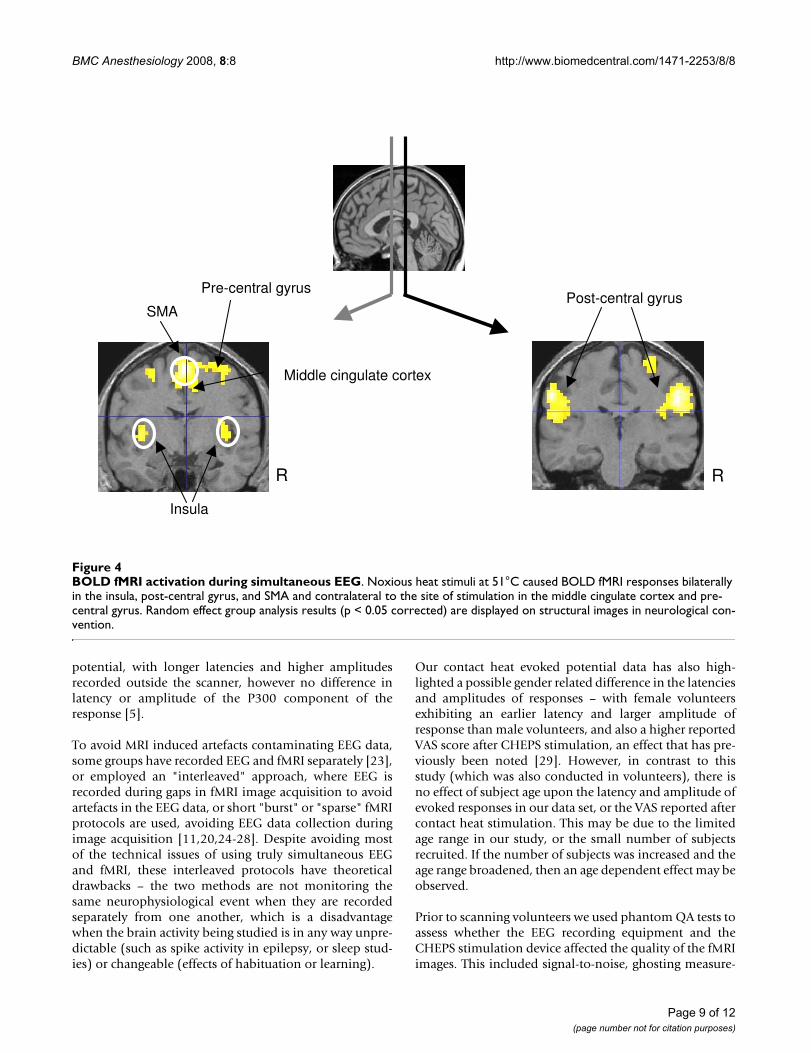
fMRIGroup analysis of fMRI data for all ten subjects, collectedcontinuously throughout the fMRI protocol and CHEPSrecording revealed areas of Blood Oxygen Level Depend-ent (BOLD) activation upon CHEPS stimulation bilater-ally in the insula, post-central gyrus, and SMA, andcontralateral to the site of stimulation (i.e. right) in themiddle cingulate cortex and pre-central gyrus (Figure 4,Table 2).
FMRI responses to CHEPS heat stimulation are reproduc-ible across multiple sessions and this is demonstrated inFigure 5 showing group results of each session
Although the thermode was moved randomly, it is possi-ble that there could be some contribution of the touchsensation to the measured BOLD response. To avoid this,the movement of the thermode should be modelled as anuisance covariate in future experiments.
Pain ratingsStimulation at 51°C with the CHEPS probe elicited apainful sensation in all ten subjects. There was no signifi-cant difference between reported VAS ratings for baselineand fMRI protocols (Table 1).
Female subjects were noted to have a significantly higherVAS than males for CHEPS stimulation (p = 0.0416). Nocorrelation between age and average reported VAS wasobserved (rs = 0.1534, p = 0.3410).
CorrelationsCorrelation of evoked potential amplitude with VAS (forall data, baseline and fMRI protocols repeated twice)revealed a significant, positive relationship, rs = 0.5956, p< 0.0001 (Figure 6a). A significant, positive correlationwas also seen when the baseline and fMRI data waspooled to give an overall evoked potential amplitude andVAS score for each of the ten subjects, rs = 0.7697, p =0.0063 (Figure 6b).
The regression analysis in SPM showed no correlationbetween BOLD signal and evoked potential amplitudes orVAS scores.
Page 5 of 12(page number not for citation purposes)
BMC Anesthesiology 2008, 8:8 http://www.biomedcentral.com/1471-2253/8/8
Page 6 of 12(page number not for citation purposes)
Evoked potential waveformsFigure 2Evoked potential waveforms. (a) Evoked potentials recorded during baseline (black traces) and fMRI (red traces) protocols in subject # 5. (b) Evoked potentials (grand average for all subjects) recorded at baseline (1 plus 2) (black trace) and during fMRI (1 plus 2) protocols (red trace). The deflections on the trace occurring at 0 and 250 ms are artefacts produced by the stimulator.
�����������������������
40
35
30
25
20
15
10
5
0
-5
-10
-15
-20
-25
-30
-35
-100 0 100 200 300 400 500 600 700
Cz-Av erage Ref
���
�
��
�����
15
10
5
0
-5
-10
-15
-100 0 100 200 300 400 500 600 700
Cz-Av erage Ref
��
�
��
BMC Anesthesiology 2008, 8:8 http://www.biomedcentral.com/1471-2253/8/8
DiscussionIn this study we have demonstrated that it is feasible torecord brain activity in response to noxious thermal stim-ulation (51°C contact heat) with CHEPS, using simulta-neous EEG and fMRI. The EEG recorded during fMRIacquisition was unaffected by the additional processingsteps required to remove MRI related artefacts. These arte-
facts can contaminate the raw data and their removal cancompromise the quality of the EEG signal. However, oursimultaneously acquired EEG revealed heat evoked Aδpotentials that were reproducible across the two separatefMRI sessions. Although the aim of this feasibility studywas not to directly compare the measurements inside andoutside the scanner, the amplitudes and latencies
Latency and amplitude of evoked potentials in baseline and fMRI protocolsFigure 3Latency and amplitude of evoked potentials in baseline and fMRI protocols. Graphical representation of the N2 and P2 latencies (s) and amplitudes (μV) recorded in the two separate baseline and fMRI sessions. There were no differences in either parameter when recorded under baseline conditions or during continuous fMRI.
Baseline 1 Baseline 2 Scan 1 Scan 20.0
0.1
0.2
0.3
0.4
Baseline 1 Baseline 2 Scan 1 Scan 20.0
0.1
0.2
0.3
0.4
0.5
p 0.4211 p 0.4813
p 0.5000
p 0.3981
N2
Lat
ency
(s)
0.5p 0.4849 p 0.4824
p 0.2832
p 0.2700
P2
Lat
enc
y (s
)
Baseline 1 Baseline 2 Scan 1 Scan 20
10
20
30
40
p 0.3304 p 0.4074
p 0.4827
p 0.2729
Am
plitu
de (
V)
Table 1: Evoked potentials and pain scores recorded during baseline and fMRI protocols in healthy volunteers.
Protocol N2 latency (s) Amplitude (μV) VAS (0–10)
Baseline 0.319 ± 0.006 24.87 ± 2.15 5.30 ± 0.53
FMRI 0.317 ± 0.006 24.49 ± 2.77 5.97 ± 0.57
Difference ns (p = 0.4183) ns (p = 0.3402) ns (p = 0.1918)
Mean ± SEMns = not significant
Average data (n = 10) for evoked potential latency, amplitude and pain scores during baseline and fMRI protocols. There were no significant differences between the results obtained in the baseline and fMRI sessions.
Page 7 of 12(page number not for citation purposes)
BMC Anesthesiology 2008, 8:8 http://www.biomedcentral.com/1471-2253/8/8
recorded inside the scanner were consistent with thoserecorded in two baseline (control) sessions (Figure 3,Table 1), suggesting there was no signal degradation orreduction of signal to noise ratio caused by conditions inthe MRI environment or additional processing require-ments. This is in agreement with a similar study using
laser-evoked pain conducted by Iannetti and colleagues in2005, which also showed no variation in amplitude orlatency of Aδ evoked potentials inside and outside thescanner. A study of auditory evoked potentials with con-current fMRI has shown a difference in the latency andamplitude of the N1 component of the event related
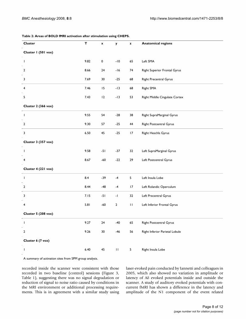
Table 2: Areas of BOLD fMRI activation after stimulation using CHEPS.
Cluster T x y z Anatomical regions
Cluster 1 (501 vox)
1 9.82 0 -10 65 Left SMA
2 8.66 24 -16 74 Right Superior Frontal Gyrus
3 7.69 30 -25 68 Right Precentral Gyrus
4 7.46 15 -13 68 Right SMA
5 7.43 12 -13 53 Right Middle Cingulate Cortex
Cluster 2 (366 vox)
1 9.55 54 -28 38 Right SupraMarginal Gyrus
2 9.30 57 -25 44 Right Postcentral Gyrus
3 6.50 45 -25 17 Right Heschls Gyrus
Cluster 3 (357 vox)
1 9.58 -51 -37 32 Left SupraMarginal Gyrus
4 8.67 -60 -22 29 Left Postcentral Gyrus
Cluster 4 (221 vox)
1 8.4 -39 -4 5 Left Insula Lobe
2 8.44 -48 -4 17 Left Rolandic Operculum
3 7.15 -51 -1 32 Left Precentral Gyrus
4 5.81 -60 2 11 Left Inferior Frontal Gyrus
Cluster 5 (208 vox)
1 9.27 24 -40 65 Right Postcentral Gyrus
2 9.26 30 -46 56 Right Inferior Parietal Lobule
Cluster 6 (7 vox)
1 6.40 45 11 5 Right Insula Lobe
A summary of activation sites from SPM group analysis.
Page 8 of 12(page number not for citation purposes)
BMC Anesthesiology 2008, 8:8 http://www.biomedcentral.com/1471-2253/8/8
potential, with longer latencies and higher amplitudesrecorded outside the scanner, however no difference inlatency or amplitude of the P300 component of theresponse [5].
To avoid MRI induced artefacts contaminating EEG data,some groups have recorded EEG and fMRI separately [23],or employed an "interleaved" approach, where EEG isrecorded during gaps in fMRI image acquisition to avoidartefacts in the EEG data, or short "burst" or "sparse" fMRIprotocols are used, avoiding EEG data collection duringimage acquisition [11,20,24-28]. Despite avoiding mostof the technical issues of using truly simultaneous EEGand fMRI, these interleaved protocols have theoreticaldrawbacks – the two methods are not monitoring thesame neurophysiological event when they are recordedseparately from one another, which is a disadvantagewhen the brain activity being studied is in any way unpre-dictable (such as spike activity in epilepsy, or sleep stud-ies) or changeable (effects of habituation or learning).
Our contact heat evoked potential data has also high-lighted a possible gender related difference in the latenciesand amplitudes of responses – with female volunteersexhibiting an earlier latency and larger amplitude ofresponse than male volunteers, and also a higher reportedVAS score after CHEPS stimulation, an effect that has pre-viously been noted [29]. However, in contrast to thisstudy (which was also conducted in volunteers), there isno effect of subject age upon the latency and amplitude ofevoked responses in our data set, or the VAS reported aftercontact heat stimulation. This may be due to the limitedage range in our study, or the small number of subjectsrecruited. If the number of subjects was increased and theage range broadened, then an age dependent effect may beobserved.
Prior to scanning volunteers we used phantom QA tests toassess whether the EEG recording equipment and theCHEPS stimulation device affected the quality of the fMRIimages. This included signal-to-noise, ghosting measure-
BOLD fMRI activation during simultaneous EEGFigure 4BOLD fMRI activation during simultaneous EEG. Noxious heat stimuli at 51°C caused BOLD fMRI responses bilaterally in the insula, post-central gyrus, and SMA and contralateral to the site of stimulation in the middle cingulate cortex and pre-central gyrus. Random effect group analysis results (p < 0.05 corrected) are displayed on structural images in neurological con-vention.
Middle cingulate cortex
SMA
Pre-central gyrus
Insula
R
Post-central gyrus
R
Page 9 of 12(page number not for citation purposes)

BMC Anesthesiology 2008, 8:8 http://www.biomedcentral.com/1471-2253/8/8
ments and visual inspection of any artefacts. Our resultsshowed that the fMRI images acquired alongside evokedpotential recordings were unaffected by the simultaneousEEG acquisition, and were not impaired by the presenceof the EEG recording and CHEPS equipment inside thescanner.
The results we obtained agree with previously publisheddata showing patterns of brain activation after nociceptiveand noxious thermal stimulation [10,11,18,30-34], withBOLD changes apparent in the insula, post-central gyrus,SMA, middle cingulate cortex and pre-central gyrus (Fig-ure 4). Cooling of the CHEPS thermode back to baselinetemperature will activate cool sensitive-fibres and thiscould potentially affect the BOLD response; however, thefMRI changes we observed were similar to those obtainedwith other methods of stimulation with noxious stimuli(these would be primarily related to the initial upstroke ofthe heat stimulus).
In accord with other studies of pain-evoked potentials,our data has revealed a significant positive correlationbetween evoked potential amplitude and reported painscores or pain intensity [15,29,32,35,36] (Figure 6a, b).However, this simple positive relationship with subjectivepain scores was not present in the fMRI signal changedata, even when regions of interest were analysed sepa-rately. This is unlike a recent study of painful electricalstimulation, which showed a positive correlation betweenstimulus correlated BOLD responses in a network of cor-
tical and cerebellar areas and reported pain intensity [11],and other studies using fMRI that have shown signalchanges to correlate with pain intensity, or subjectiveexperience of pain [37,38]. However these studies haveeither grouped subjects according to their sensitivity toheat stimuli [38], set stimuli according to individualthresholds [11], or used various stimulus intensities [37].Our fMRI protocol used a stimulus at only one intensityfor all subjects in the study, which may explain why nocorrelation with pain scores could be seen in our fMRIdata. In protocols that lead on from this feasibility studystimuli at or just above the pain threshold for each indi-vidual will be used. While correlations were made with
Reproducibility of fMRI activation over two sessionsFigure 5Reproducibility of fMRI activation over two sessions. Group results from each fMRI session are superimposed on the average high resolution structural scan. Session 1 is shown in RED, session 2 in BLUE and areas that overlap in PURPLE. There is an overlap in the insula, post-central and pre-central gyrus.
Correlation between evoked potential amplitude and pain scoreFigure 6Correlation between evoked potential amplitude and pain score. (a) Correlation of amplitude and pain score data for all baseline and fMRI sessions (n = 40) revealed a signifi-cant positive relationship (rs = 0.5956, p < 0.0001). (b) A sig-nificant, positive correlation (rs = 0.7697, p = 0.0063) was also seen when the baseline and fMRI data was pooled to give an overall evoked potential amplitude and VAS score for each subject (n = 10).
0.0 2.5 5.0 7.5 10.00
10
20
30
40
50
60
VAS
Am
plit
ude
(V
)
0.0 2.5 5.0 7.5 10.00
10
20
30
40
50
VAS
Am
plit
ude
(V
)
(a)
(b)
Page 10 of 12(page number not for citation purposes)
BMC Anesthesiology 2008, 8:8 http://www.biomedcentral.com/1471-2253/8/8
VAS scores as they are widely used in clinical trials of anal-gesics, these must be regarded with caution, as VAS scoresare ordinal and not linear measures of pain perception –further studies and analyses using an interval scale (Raschmodel) would be of interest [39]).
ConclusionThis study demonstrates the feasibility of recording con-tact heat evoked potentials with simultaneous EEG andfMRI. Evoked potentials monitored inside the MRI scan-ner were similar to those recorded under baseline condi-tions and were highly reproducible on two occasions. Wehave also demonstrated a linear relationship betweenevoked potential amplitude and VAS score, as shown inprevious volunteer studies. fMRI group analysis showedBOLD activation in areas shown to be associated withnociception in previous publications, but there was nosimple correlation of regional fMRI signal changes withindividual pain scores – suggesting changes in BOLD sig-nal may reflect later processing in cerebral pathwaysrather than encoding pain intensity in a linear fashion.Further studies are in progress to demonstrate that simul-taneous CHEPS-evoked potentials and fMRI is a usefultool in clinical trials of novel analgesics.
Competing interestsThe authors declare that they have no competing interests.
Authors' contributionsKR was involved in setting up and carrying out the study,data analysis and writing the manuscript. AP was involvedin setting up and carrying out the study, data analysis andwriting the manuscript. CG was involved in carrying outthe study and data analysis. MT was involved in setting upand carrying out the study and data analysis. DA wasinvolved in setting up the study. RS was involved in carry-ing out the study. DM helped conceive the study and par-ticipated in its design and co-ordination, interpretation,and completion of the manuscript. PA conceived thestudy and participated in its design and co-ordination,interpretation, and completion of the manuscript. Allauthors read and approved the manuscript.
AcknowledgementsThe authors would like to thank the BBSRC and also GlaxoSmithKline PLC for financial assistance, and in particular Dr Boris Chizh, Dr Zahid Ali, and Dr David Stephens of GlaxoSmithKline for helpful discussions.
References1. Stern JM: Simultaneous electroencephalography and func-
tional magnetic resonance imaging applied to epilepsy. Epi-lepsy Behav 2006, 8(4):683-692.
2. Czisch M, Wetter TC, Kaufmann C, Pollmacher T, Holsboer F, AuerDP: Altered processing of acoustic stimuli during sleep:reduced auditory activation and visual deactivation detectedby a combined fMRI/EEG study. Neuroimage 2002,16(1):251-258.
3. Goncalves SI, de Munck JC, Pouwels PJ, Schoonhoven R, Kuijer JP,Maurits NM, Hoogduin JM, Van Someren EJ, Heethaar RM, Lopes daSilva FH: Correlating the alpha rhythm to BOLD using simul-taneous EEG/fMRI: inter-subject variability. Neuroimage 2006,30(1):203-213.
4. Laufs H, Kleinschmidt A, Beyerle A, Eger E, Salek-Haddadi A, Prei-bisch C, Krakow K: EEG-correlated fMRI of human alpha activ-ity. Neuroimage 2003, 19(4):1463-1476.
5. Mulert C, Jager L, Schmitt R, Bussfeld P, Pogarell O, Moller HJ, JuckelG, Hegerl U: Integration of fMRI and simultaneous EEG:towards a comprehensive understanding of localization andtime-course of brain activity in target detection. Neuroimage2004, 22(1):83-94.
6. Scarff CJ, Reynolds A, Goodyear BG, Ponton CW, Dort JC, Egger-mont JJ: Simultaneous 3-T fMRI and high-density recording ofhuman auditory evoked potentials. Neuroimage 2004,23(3):1129-1142.
7. Comi E, Annovazzi P, Silva AM, Cursi M, Blasi V, Cadioli M, Inuggi A,Falini A, Comi G, Leocani L: Visual evoked potentials may berecorded simultaneously with fMRI scanning: A validationstudy. Hum Brain Mapp 2005, 24(4):291-298.
8. Henning S, Merboldt KD, Frahm J: Simultaneous recordings ofvisual evoked potentials and BOLD MRI activations inresponse to visual motion processing. NMR Biomed 2005,18(8):543-552.
9. Lazeyras F, Zimine I, Blanke O, Perrig SH, Seeck M: Functional MRIwith simultaneous EEG recording: feasibility and applicationto motor and visual activation. J Magn Reson Imaging 2001,13(6):943-948.
10. Iannetti GD, Niazy RK, Wise RG, Jezzard P, Brooks JC, Zambreanu L,Vennart W, Matthews PM, Tracey I: Simultaneous recording oflaser-evoked brain potentials and continuous, high-field func-tional magnetic resonance imaging in humans. Neuroimage2005, 28(3):708-719.
11. Christmann C, Koeppe C, Braus DF, Ruf M, Flor H: A simultaneousEEG-fMRI study of painful electric stimulation. Neuroimage2007, 34(4):1428-1437.
12. Lemieux L, Allen PJ, Franconi F, Symms MR, Fish DR: Recording ofEEG during fMRI experiments: patient safety. Magn Reson Med1997, 38(6):943-952.
13. Lemieux L, Allen P, Krakow K, Symms MR, Fish DR: Methodologi-cal Issues in EEG-correlated Functional MRI Experiments.IJBEM 1999, 1(1):87-95.
14. Granovsky Y, Matre D, Sokolik A, Lorenz J, Casey KL: Thermore-ceptive innervation of human glabrous and hairy skin: a con-tact heat evoked potential analysis. Pain 2005, 115(3):238-247.
15. Chen AC, Niddam DM, Arendt-Nielsen L: Contact heat evokedpotentials as a valid means to study nociceptive pathways inhuman subjects. Neurosci Lett 2001, 316(2):79-82.
16. Le Pera D, Valeriani M, Niddam D, Chen AC, Arendt-Nielsen L: Con-tact heat evoked potentials to painful and non-painful stim-uli: effect of attention towards stimulus properties. BrainTopogr 2002, 15(2):115-123.
17. Iannetti GD, Zambreanu L, Tracey I: Similar nociceptive afferentsmediate psychophysical and electrophysiological responsesto heat stimulation of glabrous and hairy skin in humans. JPhysiol 2006, 577(Pt 1):235-248.
18. Howard M, Coen S, Buchanan T, Smart T, Gregory S, Williams S,Huggins J, Hanna M: Test-retest Reproducibility of Cerebraland Subjective Responses to Painful and Non-painful Con-tact-Heat Evoked Potential Stimulation (CHEPS)[abstract]. Eur J Pain 2006, 10(S1):S82.
19. Greffrath W, Baumgartner U, Treede RD: Peripheral and centralcomponents of habituation of heat pain perception andevoked potentials in humans. Pain 2007.
20. Portas CM, Krakow K, Allen P, Josephs O, Armony JL, Frith CD:Auditory processing across the sleep-wake cycle: simultane-ous EEG and fMRI monitoring in humans. Neuron 2000,28(3):991-999.
21. Talairach J, Tournoux P: Co-planar stereotaxic atlas of thehuman brain. Thieme Publishing Group, New York; 1988.
22. Truini A, Galeotti F, Romaniello A, Virtuoso M, Iannetti GD, CruccuG: Laser-evoked potentials: normative values. Clin Neurophysiol2005, 116(4):821-826.
23. Ball T, Schreiber A, Feige B, Wagner M, Lucking CH, Kristeva-FeigeR: The role of higher-order motor areas in voluntary move-
Page 11 of 12(page number not for citation purposes)
http://www.ncbi.nlm.nih.gov/entrez/query.fcgi?cmd=Retrieve&db=PubMed&dopt=Abstract&list_uids=9402196
http://www.ncbi.nlm.nih.gov/entrez/query.fcgi?cmd=Retrieve&db=PubMed&dopt=Abstract&list_uids=9402196
http://www.ncbi.nlm.nih.gov/entrez/query.fcgi?cmd=Retrieve&db=PubMed&dopt=Abstract&list_uids=3129671
BMC Anesthesiology 2008, 8:8 http://www.biomedcentral.com/1471-2253/8/8
Publish with BioMed Central and every scientist can read your work free of charge
"BioMed Central will be the most significant development for disseminating the results of biomedical research in our lifetime."
Sir Paul Nurse, Cancer Research UK
Your research papers will be:
available free of charge to the entire biomedical community
peer reviewed and published immediately upon acceptance
cited in PubMed and archived on PubMed Central
yours — you keep the copyright
Submit your manuscript here:http://www.biomedcentral.com/info/publishing_adv.asp
BioMedcentral
ment as revealed by high-resolution EEG and fMRI. Neuroim-age 1999, 10(6):682-694.
24. Bonmassar G, Schwartz DP, Liu AK, Kwong KK, Dale AM, BelliveauJW: Spatiotemporal brain imaging of visual-evoked activityusing interleaved EEG and fMRI recordings. Neuroimage 2001,13(6 Pt 1):1035-1043.
25. Garreffa G, Bianciardi M, Hagberg GE, Macaluso E, Marciani MG, Mar-aviglia B, Abbafati M, Carni M, Bruni I, Bianchi L: SimultaneousEEG-fMRI acquisition: how far is it from being a standardizedtechnique? Magn Reson Imaging 2004, 22(10):1445-1455.
26. Kruggel F, Herrmann CS, Wiggins CJ, von Cramon DY: Hemody-namic and electroencephalographic responses to illusory fig-ures: recording of the evoked potentials during functionalMRI. Neuroimage 2001, 14(6):1327-1336.
27. Muller BW, Stude P, Nebel K, Wiese H, Ladd ME, Forsting M, Juept-ner M: Sparse imaging of the auditory oddball task with func-tional MRI. Neuroreport 2003, 14(12):1597-1601.
28. Nebel K, Stude P, Wiese H, Muller B, de Greiff A, Forsting M, DienerHC, Keidel M: Sparse imaging and continuous event-relatedfMRI in the visual domain: a systematic comparison. HumBrain Mapp 2005, 24(2):130-143.
29. Cheng SS, Liu JY, Hsui YR, Chang ST: Chemical polymorphismand antifungal activity of essential oils from leaves of differ-ent provenances of indigenous cinnamon (Cinnamomumosmophloeum). Bioresour Technol 2006, 97(2):306-312.
30. Peyron R, Laurent B, Garcia-Larrea L: Functional imaging of brainresponses to pain. A review and meta-analysis (2000). Neuro-physiol Clin 2000, 30(5):263-288.
31. Ingvar M: Pain and functional imaging. Philos Trans R Soc Lond BBiol Sci 1999, 354(1387):1347-1358.
32. Qiu Y, Noguchi Y, Honda M, Nakata H, Tamura Y, Tanaka S, SadatoN, Wang X, Inui K, Kakigi R: Brain Processing of the SignalsAscending Through Unmyelinated C Fibers in Humans: AnEvent-Related Functional Magnetic Resonance ImagingStudy. Cereb Cortex 2006, 16(9):1289-1295.
33. Brooks JC, Nurmikko TJ, Bimson WE, Singh KD, Roberts N: fMRI ofthermal pain: effects of stimulus laterality and attention.Neuroimage 2002, 15(2):293-301.
34. Moulton EA, Keaser ML, Gullapalli RP, Greenspan JD: Regionalintensive and temporal patterns of functional MRI activationdistinguishing noxious and innocuous contact heat. J Neuro-physiol 2005, 93(4):2183-2193.
35. Ohara S, Crone NE, Weiss N, Treede RD, Lenz FA: Amplitudes oflaser evoked potential recorded from primary somatosen-sory, parasylvian and medial frontal cortex are graded withstimulus intensity. Pain 2004, 110(1–2):318-328.
36. Granovsky Y, Granot M, Nir RR, Yarnitsky D: Objective Correlateof Subjective Pain Perception by Contact Heat-EvokedPotentials. J Pain 2007.
37. Bornhovd K, Quante M, Glauche V, Bromm B, Weiller C, Buchel C:Painful stimuli evoke different stimulus-response functionsin the amygdala, prefrontal, insula and somatosensory cor-tex: a single-trial fMRI study. Brain 2002, 125(Pt 6):1326-1336.
38. Coghill RC, McHaffie JG, Yen YF: Neural correlates of interindi-vidual differences in the subjective experience of pain. ProcNatl Acad Sci USA 2003, 100(14):8538-8542.
39. Decruynaere C, Thonnard JL, Plaghki L: Measure of experimentalpain using Rasch analysis. Eur J Pain 2007, 11(4):469-474.
Pre-publication historyThe pre-publication history for this paper can be accessedhere:
http://www.biomedcentral.com/1471-2253/8/8/prepub
Page 12 of 12(page number not for citation purposes)