Cell Transplantation for the Treatment of Acute Myocardial Infarction Using Vascular Endothelial...
7
Cell Transplantation for the Treatment of Acute Myocardial Infarction Using Vascular Endothelial Growth Factor–Expressing Skeletal Myoblasts Ken Suzuki, MD, PhD; Bari Murtuza, MA, FRCS; Ryszard T. Smolenski, MD, PhD; Ivan A. Sammut, PhD; Noriko Suzuki, MD; Yasufumi Kaneda, MD, PhD; Magdi H. Yacoub, FRS Background—Vascular endothelial growth factor (VEGF) is a promising reagent for inducing myocardial angiogenesis. Skeletal myoblast transplantation has been shown to improve cardiac function in chronic heart failure models by regenerating muscle. We hypothesized that transplantation of VEGF-expressing myoblasts could effectively treat acute myocardial infarction by providing VEGF-induced cardioprotection through vasodilatation in the early phase, followed by angiogenesis effects in salvaging ischemic host myocardium combined with the functional benefits of newly formed, skeletal myoblast-derived muscle in the later phase. Methods and Results—Primary rat skeletal myoblasts were transfected with the human VEGF 165 gene using hemagglutinating virus of Japan-liposome with .95% transfection efficiency. Four million of these myoblasts (VEGF group), control- transfected myoblasts (control group), or medium only (medium group) was injected into syngeneic rat hearts 1 hour after left coronary artery occlusion. Myocardial VEGF-expression increased for 2 weeks in the VEGF group, resulting in enhanced angiogenesis without the formation of tumors. Grafted myoblasts had differentiated into multinucleated myotubes within host myocardium. Infarct size (33.361.4%, 38.161.4%, and 43.761.6% for VEGF, control, and medium groups, respectively; P50.0005) was significantly reduced with VEGF treatment, and cardiac function improved in the VEGF group (maximum dP/dt: 4072.0693.6, 3772.56101.1, and 3482.5690.6 mm Hg/s in the 3 groups, respectively; P50.0011; minimum dP/dt: 2504.2668.5, 22311.3657.0, and 22124.0657.9 mm Hg/s, respectively; P50.0008). Conclusions—This combined strategy of cell transplantation with gene therapy could be of importance for the treatment of acute myocardial infarction. (Circulation. 2001;104[suppl I]:I-207-I-212.) Key Words: cells n transplantation n gene therapy n angiogenesis n myocardial infarction V ascular endothelial growth factor (VEGF) is a promising therapeutic reagent for treating myocardial infarction by inducing angiogenesis. 1 However, sustained, localized high levels of VEGF expression in myocardium are needed for its clinical use 2 ; such levels may be achieved by genetic manip- ulation. It has been reported that direct intramyocardial gene transfer results in localized enhancement of VEGF levels and successful angiogenesis in animal models of myocardial infarction. 3 Further, preliminary data from recent human trials of angiogenesis gene therapy using naked plasmid DNA or an adenoviral vector coding for VEGF have shown favorable results. 4,5 Cell-mediated gene transfer may also be useful for sustained, local protein delivery. 6 In addition to its angiogenesis effect, VEGF reportedly provides myocardial protection against ischemic injury via vasodilatation medi- ated by an increase in nitric oxide. 7,8 It has been shown that skeletal myoblasts (satellite cells) survive within myocardium, thus regenerating functional muscle and improving cardiac function when transplanted in experimental heart failure models. 9 –11 Thus, skeletal myo- blast transplantation is promising for treating chronic end- stage heart failure; however, the efficiency of this strategy when applied in the acute phase of myocardial infarction remains unknown. In the present study, we investigated the feasibility and efficiency of transplanting skeletal myoblasts that are transiently expressing VEGF to treat acute myocar- dial infarction. We hypothesized that this strategy could provide cardioprotective effects for ischemic host myocardi- um by VEGF-induced vasodilatation in the early phase of infarction, followed by angiogenesis effects in salvaging host myocardium in combination with functional benefits of newly formed, skeletal myoblast– derived muscle by cellular cardiomyoplasty in the late phase. Methods Isolation of Primary Skeletal Myoblasts All studies were performed with the approval of the institutional ethics committee. The investigation conformed to the Principles of From the Department of Cardiothoracic Surgery, Imperial College School of Medicine, Heart Science Centre, Harefield Hospital, Middlesex, UK, and the Division of Gene Therapy Science, Osaka University, Osaka, Japan (Y.K.). Correspondence to Professor Sir Magdi H. Yacoub, Department of Cardiothoracic Surgery, Harefield Hospital, Harefield, Middlesex, UB9 6JH, UK. E-mail [email protected] © 2001 American Heart Association, Inc. Circulation is available at http://www.circulationaha.org I-207 by guest on February 19, 2016 http://circ.ahajournals.org/ Downloaded from
Transcript of Cell Transplantation for the Treatment of Acute Myocardial Infarction Using Vascular Endothelial...
Cell Transplantation for the Treatment of Acute MyocardialInfarction Using Vascular Endothelial Growth
Factor–Expressing Skeletal MyoblastsKen Suzuki, MD, PhD; Bari Murtuza, MA, FRCS; Ryszard T. Smolenski, MD, PhD;
Ivan A. Sammut, PhD; Noriko Suzuki, MD; Yasufumi Kaneda, MD, PhD; Magdi H. Yacoub, FRS
Background—Vascular endothelial growth factor (VEGF) is a promising reagent for inducing myocardial angiogenesis.Skeletal myoblast transplantation has been shown to improve cardiac function in chronic heart failure models byregenerating muscle. We hypothesized that transplantation of VEGF-expressing myoblasts could effectively treat acutemyocardial infarction by providing VEGF-induced cardioprotection through vasodilatation in the early phase, followedby angiogenesis effects in salvaging ischemic host myocardium combined with the functional benefits of newly formed,skeletal myoblast-derived muscle in the later phase.
Methods and Results—Primary rat skeletal myoblasts were transfected with the human VEGF165 gene using hemagglutinatingvirus of Japan-liposome with.95% transfection efficiency. Four million of these myoblasts (VEGF group), control-transfected myoblasts (control group), or medium only (medium group) was injected into syngeneic rat hearts 1 hour after leftcoronary artery occlusion. Myocardial VEGF-expression increased for 2 weeks in the VEGF group, resulting in enhancedangiogenesis without the formation of tumors. Grafted myoblasts had differentiated into multinucleated myotubes within hostmyocardium. Infarct size (33.361.4%, 38.161.4%, and 43.761.6% for VEGF, control, and medium groups, respectively;P50.0005) was significantly reduced with VEGF treatment, and cardiac function improved in the VEGF group (maximumdP/dt: 4072.0693.6, 3772.56101.1, and 3482.5690.6 mm Hg/s in the 3 groups, respectively;P50.0011; minimum dP/dt:2504.2668.5,22311.3657.0, and22124.0657.9 mm Hg/s, respectively;P50.0008).
Conclusions—This combined strategy of cell transplantation with gene therapy could be of importance for the treatmentof acute myocardial infarction.(Circulation. 2001;104[suppl I]:I-207-I-212.)
Key Words: cells n transplantationn gene therapyn angiogenesisn myocardial infarction
Vascular endothelial growth factor (VEGF) is a promisingtherapeutic reagent for treating myocardial infarction by
inducing angiogenesis.1 However, sustained, localized highlevels of VEGF expression in myocardium are needed for itsclinical use2; such levels may be achieved by genetic manip-ulation. It has been reported that direct intramyocardial genetransfer results in localized enhancement of VEGF levels andsuccessful angiogenesis in animal models of myocardialinfarction.3 Further, preliminary data from recent humantrials of angiogenesis gene therapy using naked plasmid DNAor an adenoviral vector coding for VEGF have shownfavorable results.4,5 Cell-mediated gene transfer may also beuseful for sustained, local protein delivery.6 In addition to itsangiogenesis effect, VEGF reportedly provides myocardialprotection against ischemic injury via vasodilatation medi-ated by an increase in nitric oxide.7,8
It has been shown that skeletal myoblasts (satellite cells)survive within myocardium, thus regenerating functionalmuscle and improving cardiac function when transplanted in
experimental heart failure models.9–11 Thus, skeletal myo-blast transplantation is promising for treating chronic end-stage heart failure; however, the efficiency of this strategywhen applied in the acute phase of myocardial infarctionremains unknown. In the present study, we investigated thefeasibility and efficiency of transplanting skeletal myoblaststhat are transiently expressing VEGF to treat acute myocar-dial infarction. We hypothesized that this strategy couldprovide cardioprotective effects for ischemic host myocardi-um by VEGF-induced vasodilatation in the early phase ofinfarction, followed by angiogenesis effects in salvaging hostmyocardium in combination with functional benefits ofnewly formed, skeletal myoblast–derived muscle by cellularcardiomyoplasty in the late phase.
MethodsIsolation of Primary Skeletal MyoblastsAll studies were performed with the approval of the institutionalethics committee. The investigation conformed to the Principles of
From the Department of Cardiothoracic Surgery, Imperial College School of Medicine, Heart Science Centre, Harefield Hospital, Middlesex, UK, andthe Division of Gene Therapy Science, Osaka University, Osaka, Japan (Y.K.).
Correspondence to Professor Sir Magdi H. Yacoub, Department of Cardiothoracic Surgery, Harefield Hospital, Harefield, Middlesex, UB9 6JH, UK.E-mail [email protected]
© 2001 American Heart Association, Inc.
Circulation is available at http://www.circulationaha.org
I-207 by guest on February 19, 2016http://circ.ahajournals.org/Downloaded from
Laboratory Animal Care formulated by the National Society forMedical Research and the Guide for the Care and Use of LaboratoryAnimals published by the US National Institutes of Health.
Primary skeletal myoblasts were isolated using the single-fiberculture system.12 The extensor digitorum longus was removed froma male Lewis rat (80 g) and was digested with 0.2% type Icollagenase (Sigma). Single, intact muscle fibers liberated by tritu-ration were placed into each well of a 24-well culture plate. Putativeskeletal myoblasts (satellite cells) dissociated from the fiber, ap-peared 12 to 24 hours after plating, and proliferated (Figures 1A and1B). The fiber was then removed and the myoblasts were fed withproliferation medium (20% fetal calf serum, 10% horse serum, and0.5% chicken embryo extract in Dulbecco’s Modified Eagle Medium[DMEM]). The culture was passaged before reaching 75% conflu-ence to maintain the undifferentiated state. To confirm the purity ofthe culture, cells were seeded into 4-well chamber slides (Labteks),incubated to subconfluence, and fixed with220°C methanol/ace-tone. After blocking with 5% horse serum, the samples wereincubated in a 1:5 dilution of anti-a-sarcomeric actin monoclonalantibody (Dako) at 4°C overnight, followed by 1.5 hours of incuba-tion in a 1:200 dilution of FITC-conjugated secondary antibody(Dako). Nuclei were counterstained with 4,6-diamino-2-phenylindole. The myogenicity of the single fiber–derived cells wasfurther confirmed by inducing differentiation: cells grown to sub-confluence in proliferation medium were switched to low-serummedium (DMEM supplemented with 2% horse serum, 2 mmol/Lglutamine, 50 IU/mL penicillin, and 50mg/mL streptomycin12).
Gene Construction and Gene TransfectionFull-length cDNA for human VEGF165,13 which was generouslydonated by Dr Y. Yonemitsu (Kyushu University, Japan), wasdirectionally cloned into the EcoRI site of pcDNA3.11 vector(Invitrogen Corporation). Hemagglutinating Virus of Japan (HVJ)-cationic-liposome was prepared as described previously.14,15Briefly,200 mg DNA was mixed with a lipid film composed of 10 mg of alipid mixture (phosphatidylserine, dimethylaminoethane-carbamoylcholesterol, and cholesterol) to generate a liposome-DNA complexsuspension. The suspension was then incubated with 30 000 hemag-glutinating units of inactivated HVJ by ultraviolet light, resulting in1 mL of HVJ-liposome. Skeletal myoblasts at 50% confluence in a
175 cm2 flask were incubated with 450mL of the HVJ-liposomecontaining the human VEGF165 gene in pcDNA3.11 in 20 mL ofproliferation medium for 2 hours at 37°C. Control cells weretransfected with pcDNA3.11 without the VEGF gene using the sameprotocol.
In Vitro VEGF ExpressionAt day 3 after gene transfection, the culture medium was collectedfrom VEGF- or control-transfected myoblasts (n56 in each group).VEGF levels in the medium were quantified by an ELISA kit forhuman VEGF (Chemicon International) by following the company’sinstructions using a microplate reader (Labosystems). After collect-ing medium, myoblasts were harvested by scraping confluent,175-cm2 plates in 2 mL of 1% sodium dodecyl sulfate (SDS)containing 10mg/mL leupeptin, 1 mmol/L phenylmethyl sulfonylfluoride, and 5mg/mL aprotinin. After homogenization and centrif-ugation at 35 000gfor 15 minutes, the protein concentration of thesupernatant was measured using the Bradford protein assay method(Bio-Rad). A total of 25mg of protein from each sample was loadedonto a SDS 10% polyacrylamide gel for electrophoresis and trans-ferred onto a nitrocellulose membrane. The membrane was blockedand incubated with a 1:1000 dilution of anti-human VEGF mono-clonal antibody (Santa Cruz Biotechnology) at 4°C overnight,followed by 1-hour incubation with a 1:1000 dilution of horseradishperoxidase–conjugated secondary antibody (Sigma). The blot wasvisualized with an enhanced chemiluminescent detection system(Amersham). The films were scanned using a Molecular Dynamics300A laser densitometer to determine VEGF levels using QuantityOne software (PDI). Data are shown as the relative density of bandsversus day 2 samples from medium-injected hearts.
For immunocytochemical analysis, myoblasts were seeded into2-well chamber slides (Nunc) at day 2 after gene transfection. Thenext day, samples were fixed with ice-cold ethanol. After incubationin 0.1% Triton X-100 and 1% bovine serum albumin, the sampleswere incubated in a 1:50 dilution of anti-human VEGF antibody(Santa Cruz Biotechnology) at 4°C overnight, followed by incuba-tion in a 1:20 dilution of FITC-conjugated secondary antibody(Dako). Nuclei were counterstained with 0.1 mg/mL propidiumiodide.
Myocardial Infarction Followed byCell TransplantationMale Lewis rats (250 g) were anesthetized with sodium pentobarbital(50 mg/kg IP) and mechanically ventilated. After the heart wasexposed through a lateral thoracotomy, a 6-0 polypropylene threadwas passed around the left coronary artery,'3 mm distal to itsorigin, and the artery was occluded.16 At day 3 after gene transfec-tion, myoblasts were harvested using trypsin and resuspended inserum-free DMEM just before grafting to the heart. At 1 hour afterleft coronary artery occlusion, the VEGF- or control-transfectedskeletal myoblasts (VEGF and control groups, respectively) orserum-free DMEM only (medium group) was injected into theborder zone surrounding the infarct (4 injections of 13106 cells in100mL) with a 27G needle. The surgical wounds were repaired, andthe rats were returned to their cages to recover. Aseptic surgicaltechniques were used throughout.
Myocardial VEGF Level afterCell TransplantationAt 2, 4, 7, 14, and 28 days after cell transplantation, 5 hearts fromeach group were collected to assess the VEGF level of the heart. Theisolated left ventricle was cut into small pieces, immediately frozenin liquid nitrogen, and homogenized with a Polytron homogenizer at10 000 rpm for 30 s in ice-cold PBS. After 30 s of sonication, thehomogenate was centrifuged at 35 000gfor 15 minutes. The super-natant (100mg of protein) was applied to Western blotting forVEGF, and the band density on the film was determined bydensitometry using analysis-software as described above. To enablecomparison, the band density at each time point was determinedrelative to the value obtained at day 2 in the medium-injected hearts.
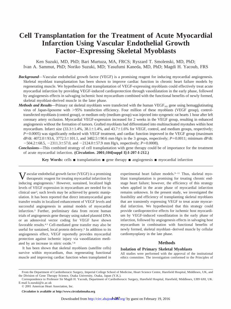
Figure 1. Primary skeletal myoblasts were isolated from singlefibers of rat skeletal muscle. Skeletal myoblasts dissociatedfrom the fiber 12 to 24 hours after plating (A and B; reverse-phase contrast microscopic observation). All cells demonstrateda-sarcomeric actin expression (FITC; green) by immunocy-tostaining. Nuclei were counterstained blue with 4,6-diamino-2-phenylindole (C). When cultured in differentiation medium, themyoblasts fused and differentiated into multinucleated myotubes(D). Scale bar, 250 mm for A and 50 mm for B, C, and D.
I-208 Circulation September 18, 2001
by guest on February 19, 2016http://circ.ahajournals.org/Downloaded from
Functional AssessmentAt 28 days after cell transplantation, rats from 3 groups (n58 in eachgroup) were anticoagulated by an intravenous injection of heparin. Thehearts were quickly excised and perfused at a pressure of 1 m H2O withmodified Krebs-Henseleit buffer (120.0 mmol/L NaCl, 4.5 mmol/LKCl, 20.0 mmol/L NaHCO3, 1.2 mmol/L KH2PO4, 1.2 mmol/L MgCl2,1.25 mmol/L CaCl2, and 10.0 mmol/L glucose; gassed with 95% O2 plus5% CO2 at 37°C) using a Langendorff apparatus.9 After 20 minutes ofstabilization, heart rate, ventricular function, and coronary flow weremeasured after left ventricular end-diastolic pressure was stabilized at10 mm Hg using a thin-walled balloon. Coronary flow was recordedwith an electromagnetic flowmeter (Scalar).
Infarct Size, Grafted Myoblasts, and AngiogenesisAfter perfusion, the left ventricle was sectioned into 5 segmentsparallel to the apex-base axis and frozen in an embedding medium.A 10-mm section was cut from each segment, fixed in 3.7%formaldehyde, and stained with Masson trichrome. Sections from allslices were projected onto a screen for computer-assisted planimetry.The ratio of scar length to left ventricular circumferences of theendocardium and epicardium was expressed as a percentage to defineinfarct size.16
The frozen left ventricular samples were cut into 6-mm sectionsand fixed with220°C methanol. The sections were incubated with0.6% H2O2, and immersed in 0.1% Triton X-100. After blocking with1% bovine serum albumin, the sections were incubated with a 1:40dilution of anti-skeletal myosin heavy chain antibody (ZymedLaboratories). This was followed by incubation with a 1:100 dilutionof biotinylated secondary antibody (Dako). The sections werecolored with a Dako streptABComplex/horseradish peroxidase Duetkit and counterstained with hematoxylin and eosin. The number ofcapillary vessels was counted in the peri-infarct area using a lightmicroscope at a magnification of3400.17 Five high-power fields ineach section were randomly selected, and the number of capillariesin each field was averaged and expressed as the number of capillaryvessels per high-power field (0.2 mm2).
Statistical AnalysisAll values are expressed as mean6SEM. The differences in the databetween 2 groups were determined with a Student’st test. Mortality(survival rate) was analyzed using the Kaplan-Meier method fol-lowed by the log rank test. Statistical comparison of the dataregarding myocardial VEGF expression was performed using 2-wayrepeated-measures ANOVA followed by Bonferroni/Dunn post hoctesting. Analysis of infarct size, cardiac function, and capillarydensity was performed with one-way ANOVA followed by Bonfer-roni/Dunn post-hoc testing.P,0.05 was considered significant.
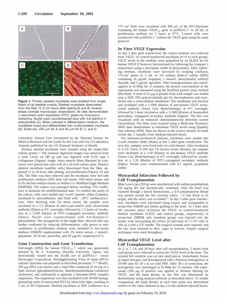
ResultsIn Vitro VEGF-Gene Transfection of PrimarySkeletal MyoblastsDuring culture in proliferation medium, the myoblasts iso-lated from the extensor digitorum longus of Lewis rats grewto confluence and, within a week of switching to low-serummedium, they fused and differentiated into multinucleatedmyotubes (Figure 1D). Immunocytostaining fora-sarcomericactin demonstrated that all the cells were positively stained,suggesting the high purity of the culture (Figure 1C). On day3 after gene transfection with human VEGF165 using HVJ-liposome,.95% of transfected myoblasts were positivelystained for VEGF (Figure 2C). The level of secreted VEGF inthe culture medium of VEGF-transfected myoblasts wassignificantly higher compared with control-transfected myo-blasts, as measured by ELISA (2776.06200.7 versus122.4610.9 pg/mL,P,0.0001; Figure 2A). Similarly, ahigher (20.364.5-fold by densitometric quantification)
VEGF level in the cell lysate of VEGF-transfected myoblastswas confirmed by Western blotting (Figure 2B).
Mortality and VEGF Expression in MyocardiumAfter Cell TransplantationAt 1 hour after left coronary artery occlusion, the borderzones of infarcts were injected with VEGF- or control-transfected skeletal myoblasts or serum-free DMEM medium.The mortality over 28 days after cell transplantation wasreduced in the VEGF group (5 deaths in 48 rats, 10.4%;P50.0087 versus medium group;P50.0120 versus controlgroup) compared with the medium group (14 deaths in 55rats, 25.5%) and control group (11 deaths in 52 rats, 21.2%;P50.0812 versus medium group). The majority of the deathswere observed within 24 hours after treatment. Western blotanalysis demonstrated that myocardial VEGF levels in theVEGF group increased significantly (P50.0008) betweendays 2 and 14 after transplantation, peaking at day 4 (9-foldover the day 2 sample for medium group), compared with theother groups (P50.0012 versus medium group;P50.0081versus control group; Figure 3). The VEGF level of thecontrol group tended to be higher (P50.0521) than that of themedium group over the period studied.
Infarct Size and Cardiac Function AfterCell TransplantationAt day 28 after cell transplantation, infarct size was signifi-cantly reduced in the VEGF group compared with othergroups, and the control group demonstrated a reduced infarctsize in comparison with the medium group (Table). Thesefindings correlated inversely with coronary flow measured
Figure 2. In vitro VEGF expression in primary skeletal myo-blasts. Rat primary skeletal myoblasts were transfected withVEGF165- or control-vector mediated by HVJ-liposome. VEGFlevels in culture medium, as detected with ELISA, were higher inVEGF-transfected myoblasts than in control-vector–transfectedmyoblasts (A). Data are presented as mean6SEM. *P,0.0001;n56 in each group. VEGF levels in the cell lysate, as confirmedby Western blotting, were also higher in VEGF-transfected myo-blasts (B). Representative blots are shown from each group(n56). Immunocytochemistry for VEGF demonstrated that themajority of VEGF-transfected myoblasts were stained with FITC(C), but no cells stained after control transfection (D). Nucleiwere counterstained with propidium iodide (orange). Scale bar,250 mm.
Suzuki et al VEGF-Myoblast Transplantation I-209
by guest on February 19, 2016http://circ.ahajournals.org/Downloaded from

using Langendorff perfusion. Both maximum and minimumdP/dt at 10 mm Hg of left ventricular end-diastolic pressurewere best in the VEGF group, indicating that both systolicand diastolic functions were best preserved in the VEGFgroup after myocardial infarction. Further, these indicatorswere better in the control group than in the medium group,suggesting that skeletal myoblast transplantation alone couldpreserve cardiac function after myocardial infarction.
Grafted Myoblasts and AngiogenesisGroups of cells that were positively stained for skeletalmyosin heavy chain were observed locally in the peri-infarctarea at 28 days after the transplantation of control- andVEGF-transfected myoblasts (Figures 4B and 4C). Becausethis antibody reacts with skeletal myosin heavy chain but notwith cardiac or smooth muscle myosin, these positivelystained cells were presumed to be of skeletal myoblast origin.It was demonstrated that surviving myoblasts had differenti-ated into multinucleated myotubes that had aligned with thecardiac fiber axis within the native myocardium (Figure 4D).Positively stained myotubes were observed at almost allinjection sites studied in the VEGF and control groups.Although not accurately quantified in this study, examinationof random high-power fields on light microscopy indicatedthat the number of positively stained myotubes was larger inthe VEGF group than in the control group. Angiogenesis, asdefined by capillary number, was found in the area surround-
ing grafted skeletal myoblasts of the control group (5.060.4capillaries per high-power field;P50.0046 versus mediumgroup; Figure 5A), but obvious angiogenesis was not found inthe Medium group (2.560.2 capillaries per high-power field).Further, enhanced angiogenesis with a larger number ofmature capillaries was observed in the VEGF group (8.761.4capillaries per high-power field;P,0.0001 versus mediumgroup,P50.0055 versus control group; Figures 5B and 5C).There was no tumor formation in any heart studied.
DiscussionWe demonstrated that the transplantation of control-transfected skeletal myoblasts can reduce infarct size andimprove cardiac function after left coronary artery occlusioncompared with medium injection. Moreover, it has beenshown that the grafting of skeletal myoblasts expressingVEGF provides further advanced benefits in reducing infarctsize and preserving cardiac function; these effects correlatedwith the enhanced angiogenesis observed in comparison withcontrol-myoblast transplanted hearts. These data suggest that
Figure 3. Myocardial VEGF expression after cell transplantation.VEGF levels in myocardium after skeletal myoblast transplanta-tion or medium injection were measured with Western blotting.The VEGF group showed a significantly higher expression ofVEGF compared with the other groups. Data are shown as therelative density of bands versus day 2 samples of medium-injected hearts (†). Data are presented as mean6SEM. *P,0.05vs both medium and control groups; n55 at each point.
Infarct Size and Cardiac Function After Cell Transplantation
GroupInfarct Size,
%Heart Rate,
bpmLVDP,
mm HgMaximum dP/dt,
mm Hg/sMinimum dP/dt,
mm Hg/sBalloon Size,
mLCoronary Flow,
mL/min
Medium 43.761.6 252.4612.5 114.363.4 3482.5690.6 22124.0657.9 172.965.8 10.160.3
Control 38.161.4* 242.8613.0 129.863.8* 3772.56101.1* 22311.3657.0* 198.666.1* 11.260.4*
VEGF 33.361.4† 257.8611.8 142.264.1† 4072.0693.6† 22504.2668.5† 221.566.7† 12.360.4†
P 0.0005 0.7211 0.0032 0.0011 0.0008 0.0023 0.0017
Data were measured at day 28 after cell transplantation. Functional data were measured at 10 mm Hg of left ventricular end-diastolic pressure.Data are presented as mean6SEM. LVDP indicates left ventricular developed pressure. n58 in each group.
*P,0.05 vs medium group; †P,0.05 vs both medium and control groups.
Figure 4. Grafted myotubes after cell transplantation. Immuno-histostaining for skeletal myosin heavy chain was performed atday 28 after left coronary artery occlusion followed by cell trans-plantation. Infarct and intact areas are clearly shown, withoutpositively stained cells in the medium group (A). Positive cellscolored brown (arrows) were observed in the infarct border zoneof the control (B) and VEGF groups (C). Surviving myoblasts dif-ferentiated into multinucleated myotubes (stained brown;arrows) that had aligned with the cardiac fiber axis within hostmyocardium (stained pink) in the VEGF group (D). Scale bars,100 mm (A, B, and C) and 50 mm (D).
I-210 Circulation September 18, 2001
by guest on February 19, 2016http://circ.ahajournals.org/Downloaded from

transplantation of VEGF-expressing myoblasts could be ofsignificant value in treating acute myocardial infarction.
We speculate that this strategy could, in the early phase ofinfarction, provide cardioprotective effects through VEGF-induced vasodilatation as mediated by an increase in nitricoxide production, independently of angiogenesis.7,8 To clarifyif locally expressed VEGF effectively contributes to myocar-dial protection, further studies, such as an assessment ofcardiac function and infarct size in the early phase after theinsult, are needed. In the late phase, in turn, the angiogenesiseffect in salvaging ischemic host myocardium, combinedwith cellular cardiomyoplasty effects in regenerating musclecould play an additional role in improving function. Further,the cellular cardiomyoplasty effect could be reinforced byimproved graft survival resulting from an improved bloodsupply to grafted myoblasts through both vasodilatation andenhanced angiogenesis. This would be particularly beneficialin the early stage after cell transplantation, when grafted cellsare subjected to various pathological processes caused byenvironmental stress, such as ischemic and mechanical inju-ry.18 Such stress is known to result in both the necrosis andapoptosis of grafted myoblasts.10,19 Although it has beenreported that skeletal myoblast transplantation improves car-diac function in heart failure models,10,11 we think that theimprovement of graft survival by protecting from such stressis of significance for further refining this treatment.
The duration and level of VEGF expression is critical toachieve successful angiogenesis without tumor formation.2 Itwas recently reported that cell-mediated gene transfer withskeletal myoblasts stably expressing VEGF by means ofretroviral transduction results in the formation of vasculartumors in intact myocardium of immunodeficient mice due topersistent, superfluous delivery of VEGF.20 Other articles, incontrast, suggested that a period of 1 to 2 weeks of VEGFoverexpression mediated by direct intramyocardial genetransfer might be sufficient to induce collateral vessels in
ischemic myocardium but not enough to produce tumors.3–5
The present strategy could achieve an adequate magnitudeand duration ('2 weeks) of VEGF expression in the ischemicmyocardium as a result of implanting VEGF-expressingmyoblasts by transient gene transfection mediated by HVJ-liposome, which is more likely to induce functional collateralvessels without angioma-genesis for up to 28 days aftertreatment.
To exclude the possibility of tumorigenesis using thecurrent strategy, further study with longer-term incubationmight be necessary. In addition, by using myoblasts as aplatform for VEGF delivery, one would anticipate a contin-ued functional benefit from newly formed skeletal myotubes.However, further investigations are needed to clarify exactlyhow long and how much VEGF expression is optimal to treatmyocardial infarction. We have also demonstrated that an-giogenesis is induced in infarcted hearts after control-myoblast transplantation and that this is associated withincreased myocardial VEGF levels. This finding has beenobserved in previous reports using direct intramyocardialinjection of skeletal myoblasts.21 In addition, other studieshave shown that a nonspecific reaction caused by mechanicalinjury results in angiogenesis induction in the heart.22 Onemight speculate that mechanical injury by cell injection andthe presence of foreign cells in myocardium might stimulateendogenous expression of some growth factors, includingVEGF.
The optimal graft cell number is that required to achievethe best attenuation of adverse remodeling or improvement incardiac function after skeletal myoblast transplantation. Inthis study, we showed that the injection of 4 million skeletalmyoblasts results in reduced infarct size and improvedfunction after acute myocardial infarction. Taylor et al10
demonstrated that a direct intramuscular injection of 107
skeletal myoblasts improved the function of cryoinjuredrabbit hearts, whereas Scorsin and coworkers11 showed that 5million myoblasts improve function in rats after infarction.However, the overall functional result may be affected byother factors, such as graft survival, differentiation capacity,ability in forming gap junctions with host myocardium, andthe condition of host myocardium. In this study, we did notaccurately measure the graft survival rate; however, it islikely that VEGF expression would be useful in improvinggraft survival according to histological observations. Furtherstudy of the quantitative assessment of survival by measuringb-galactosidase (b-gal) activity (transplantation ofb-gal–expressing skeletal myoblasts) or detecting the Y chromo-some (transplantation of male grafts into female hearts)would be needed to clarify this issue. We recently demon-strated that prior heat shock treatment of grafted myoblastsenhances self-preservation systems against environmentalstress and improves their survival after engraftment to theheart usingb-gal–expressing skeletal myoblasts.19 To com-pare the effect on graft survival between heat shock treat-ment, VEGF expression, and the combination of both strate-gies would be interesting as future work.
Gap junction formation between grafted myoblasts andhost cardiomyocytes is an important but controversial issueconcerning skeletal myoblast transplantation to the heart. We
Figure 5. Angiogenesis at day 28 after left coronary arteryocclusion following cell transplantation. Angiogenesis withmature capillaries was observed in the area surrounding graftedskeletal myoblasts in the control group (A). Further enhancedangiogenesis was observed in the VEGF group (B). Scale bar,50 mm. Capillary density in each group is expressed as thenumber of capillary vessels per high-power field in C. Data arepresented as mean6SEM. n58 in each group; *P,0.05 vsmedium group; †P,0.05 vs both medium and control groups.
Suzuki et al VEGF-Myoblast Transplantation I-211
by guest on February 19, 2016http://circ.ahajournals.org/Downloaded from

did not investigate this point in the present study because oftechnical difficulty in assessing functional gap junctions invivo; however, we have reported that gap junctions, asidentified by connexin 43 expression, are formed betweengrafted skeletal myoblasts and host cardiomyocytes after theintracoronary infusion of skeletal myoblast.9 Future study isneeded to clarify such gap junction formation after directmyocardial injection.
The timing and site of cell grafting is an interesting issue ofconsiderable debate. We injected cells 1 hour after leftcoronary artery ligation, simulating the treatment of acutecoronary occlusion. Early injection of VEGF-overexpressingcells could be useful in salvaging more myocardium due tothe vasodilatation effect of secreted VEGF. However, it islikely that grafted cells suffer greater adverse conditions iftransplanted at this time point, in comparison with delayedinjection after the myocardium has been stabilized. In addi-tion, in the present study, we grafted myoblasts in the borderarea of infarcts because salvage of stunned or hibernatingmyocardium in this area could be of the most significance.Investigating the effects of varying injection times (in theacute, subacute, and chronic phase) or injection sites (into thecenter or border of infarct) would provide new and interestingdata on cell transplantation to the heart.
The use of the HVJ-liposome has been shown to result ina transient and high-efficiency transfection, with little celltoxicity.14–15Further, repetitive transfection using this vectoris successful in vivo, suggesting a low immunogenicity ofHVJ-liposome.23 In addition, our method requires the use ofonly inactivated HVJ in vitro, minimizing the safety concernsrelated to other viral gene therapy protocols such as viralreplication, aberrant expression of viral genes, and alterationsof host genomic structure. These features would be ofsignificance in clinical applications of this strategy.
In summary, we have demonstrated that the transplantationof VEGF-expressing skeletal myoblasts by HVJ-liposome–mediated gene transfection results in transient, high-levelVEGF expression within rat myocardium suffering fromacute infarction. This expression leads to successful angio-genesis, which is associated with reduction in infarct size andan improvement in cardiac function, without tumor forma-tion. This clinically relevant, combined strategy could be ofimportance for treating patients with acute myocardialinfarction.
AcknowledgmentsWe would like to thank Dr Yoshikazu Yonemitsu, Department ofPathology, Kyusyu University, Japan, for the kind donation of cDNAfor human VEGF165. We also gratefully acknowledge the experttechnical support of Professor Terence A. Partridge, Dr LouiseHeslop, and Dr Jennifer E. Morgan, Muscle Cell Biology Group,Hammersmith Hospital, Imperial College School of Medicine, Lon-don, UK, in establishing rat skeletal myoblast isolation.
References1. Banai S, Jaklitsch MT, Shou M, et al. Angiogenic-induced enhancement
of collateral blood flow to ischemic myocardium by vascular endothelialgrowth factor in dogs.Circulation. 1994;89:2183–2189.
2. Carmeliet P. VEGF gene therapy: stimulating angiogenesis or angioma-genesis?Nat Med. 2000;6:1102–1103.
3. Muhlhauser J, Merrill MJ, Pili R, et al. VEGF165 expressed by areplication-deficient recombinant adenovirus vector induces angiogenesisin vivo. Circ Res. 1995;77:1077–1086.
4. Vale PR, Losordo DW, Milliken CE, et al. Left ventricular electrome-chanical mapping to assess efficacy of phVEGF165 gene transfer fortherapeutic angiogenesis in chronic myocardial ischemia.Circulation.2000;102:965–974.
5. Rosengart TK, Lee LY, Patel SR, et al. Angiogenesis gene therapy: phaseI assessment of direct intramyocardial administration of an adenovirusvector expressing VEGF121 cDNA to individuals with clinically sig-nificant severe coronary artery disease.Circulation. 1999;100:468–474.
6. Koh GY, Kim SJ, Klug MG, et al. Targeted expression of transforminggrowth factor-b1 in intracardiac grafts promotes vascular endothelial cellDNA synthesis.J Clin Invest.1995;95:114–121.
7. Ziche M, Morbidelli L, Choudhuri R, et al. Nitric oxide synthase liesdownstream from vascular endothelial growth factor-induced but notbasic fibroblast growth factor-induced angiogenesis.J Clin Invest. 1997;99:2625–2634.
8. Luo Z, Diaco M, Murohara T, et al. Vascular endothelial growth factorattenuates myocardial ischemia-reperfusion injury.Ann Thorac Surg.1997;64:993–998.
9. Suzuki K, Brand NJ, Smolenski RT, et al. Development of a novelmethod for cell transplantation through the coronary artery.Circulation.2000;102(suppl):III-359–III-364.
10. Taylor DA, Atkins BZ, Hungspreugs P, et al. Regenerating functionalmyocardium: improved performance after skeletal myoblast transplan-tation.Nat Med. 1998;4:929–933.
11. Scorsin M, Hagege A, Vilquin JT, et al. Comparison of the effects of fetalcardiomyocyte and skeletal myoblast transplantation on postinfarctionleft ventricular function. J Thorac Cardiovasc Surg. 2000;119:1169–1175.
12. Rosenblatt JD, Lunt AI, Parry J, et al. Culturing satellite cells from livingsingle muscle fiber explants.In Vitro Cell Dev Biol Anim.1995;35:773–779.
13. Yonemitsu Y, Kaneda Y, Morishita R, et al. Characterization of in vivogene transfer into the arterial wall mediated by the Sendai virus (hemag-glutinating virus of Japan) liposomes: an effective tool for the in vivostudy of arterial diseases.Lab Invest. 1996;75:313–323.
14. Nishikawa T, Edelstein D, Du XL, et al. Normalizing mitochondrialsuperoxide production blocks three pathways of hyperglycaemic damage.Nature. 2000;404:787–790.
15. Saeki Y, Matsumoto N, Nakano Y, et al. Development and character-ization of cationic liposomes conjugated with HVJ (Sendai virus):reciprocal effect of cationic lipid for in vitro and in vivo gene transfer.Hum Gene Ther. 1997;8:2133–2141.
16. Liu YH, Yang XP, Nass O, et al. Chronic heart failure induced bycoronary artery ligation in Lewis inbred rats.Am J Physiol.1997;272:H722–H727.
17. Tomita S, Li RK, Weisel RD, et al. Autologous transplantation of bonemarrow cells improves damaged heart function.Circulation. 1999;100(suppl):II-247–II-256.
18. Qu Z, Balkir L, van Deutekom JC, et al. Development of approaches toimprove cell survival in myoblast transfer therapy.J Cell Biol. 1998;142:1257–1267.
19. Suzuki K, Smolenski RT, Jayakumar J, et al. Heat shock treatmentenhances graft cell survival in skeletal myoblast transplantation to theheart.Circulation. 2000;102(suppl):III-216–III-221.
20. Lee RJ, Springer ML, Blanco-Bose WE, et al. VEGF gene delivery tomyocardium: deleterious effects of unregulated expression.Circulation.2000;102:898–901.
21. Van Meter CH, Claycomb WC, Delcarpio JB, et al. Myoblast transplan-tation in the porcine model: a potential technique for myocardial repair.J Thorac Cardiovasc Surg.1995;110:1442–1448.
22. Malekan R, Reynolds C, Narula N, et al. Angiogenesis in transmyocardiallaser revascularization. A non-specific response to injury.Circulation.1998;98(suppl):II-62–II-65.
23. Hirano T, Fujimoto J, Ueki T, et al. Persistent gene expression in rat liverin vivo by repetitive transfections using HVJ-liposome.Gene Ther. 1998;5:459–464.
I-212 Circulation September 18, 2001
by guest on February 19, 2016http://circ.ahajournals.org/Downloaded from
Kaneda and Magdi H. YacoubKen Suzuki, Bari Murtuza, Ryszard T. Smolenski, Ivan A. Sammut, Noriko Suzuki, Yasufumi
Expressing Skeletal Myoblasts−Endothelial Growth Factor Cell Transplantation for the Treatment of Acute Myocardial Infarction Using Vascular
Print ISSN: 0009-7322. Online ISSN: 1524-4539 Copyright © 2001 American Heart Association, Inc. All rights reserved.
is published by the American Heart Association, 7272 Greenville Avenue, Dallas, TX 75231Circulation doi: 10.1161/hc37t1.094524
2001;104:I-207-I-212Circulation.
http://circ.ahajournals.org/content/104/suppl_1/I-207World Wide Web at:
The online version of this article, along with updated information and services, is located on the
http://circ.ahajournals.org//subscriptions/
is online at: Circulation Information about subscribing to Subscriptions:
http://www.lww.com/reprints Information about reprints can be found online at: Reprints:
document. Permissions and Rights Question and Answer this process is available in the
click Request Permissions in the middle column of the Web page under Services. Further information aboutOffice. Once the online version of the published article for which permission is being requested is located,
can be obtained via RightsLink, a service of the Copyright Clearance Center, not the EditorialCirculationin Requests for permissions to reproduce figures, tables, or portions of articles originally publishedPermissions:
by guest on February 19, 2016http://circ.ahajournals.org/Downloaded from