Fourth Universal Definition of Myocardial Infarction (2018)
34
November 13, 2018 Circulation. 2018;138:e618–e651. DOI: 10.1161/CIR.0000000000000617 e618 Key Words: AHA Scientific Statements ◼ myocardial infarction ◼ Type 1 MI ◼ Type 2 MI ◼ Type 3 MI ◼ Type 4a MI ◼ Type 4b MI ◼ Type 4c MI ◼ Type 5 MI ◼ cardiac troponin ◼ high sensitivity cardiac troponin ◼ myocardial injury ◼ prior myocardial infarction ◼ silent myocardial infarction ◼ recurrent myocardial infarction ◼ reinfarction ◼ cardiac procedural myocardial injury ◼ Takotsubo syndrome ◼ myocardial infarction with nonobstructive coronary arteries (MINOCA) © 2018 The European Society of Cardiology, American College of Cardiology Foundation, American Heart Association, Inc., and the World Heart Federation. ESC/ACC/AHA/WHF EXPERT CONSENSUS DOCUMENT Fourth Universal Definition of Myocardial Infarction (2018) Circulation Kristian Thygesen Joseph S. Alpert Allan S. Jaffe Bernard R. Chaitman Jeroen J. Bax David A. Morrow Harvey D. White The Executive Group on behalf of the Joint European Society of Cardiology (ESC)/American College of Cardiology (ACC)/American Heart Association (AHA)/World Heart Federation (WHF) Task Force for the Universal Defini- tion of Myocardial Infarction Authors/Task Force Members/Chairpersons: Kristian Thygesen* (Denmark), Joseph S. Alpert* (USA), Allan S. Jaffe (USA), Bernard R. Chaitman (USA), Jeroen J. Bax (The Netherlands), David A. Morrow (USA), Harvey D. White* (New Zealand), Hans Mickley (Denmark), Filippo Crea (Italy), Frans Van de Werf (Belgium), Chiara Bucciarelli-Ducci (UK), Hugo A. Katus (Germany), Fausto J. Pinto (Portugal), Elliott M. Antman (USA), Christian W. Hamm (Ger- many), Raffaele De Caterina (Italy), James L. Januzzi Jr (USA), Fred S. Apple (USA), Maria Angeles Alonso Garcia (Spain), S. Richard Underwood (UK), John M. Canty Jr (USA), Alexander R. Lyon (UK), P.J. Devereaux (Canada), Jose Luis Zamorano (Spain), Bertil Lindahl (Sweden), William S. Weintraub (USA), L. Kristin Newby (USA), Renu Virmani (USA), Pascal Vranckx (Bel- gium), Don Cutlip (USA), Raymond J. Gibbons (USA), Sidney C. Smith (USA), Dan Atar (Norway), Russell V. Luepker (USA), Rose Marie Robertson (USA), Robert O. Bonow (USA), P. Gabriel Steg (France), Patrick T. O’Gara (USA), Keith A.A. Fox (UK) https://www.ahajournals.org/journal/circ *Corresponding authors. Kristian Thygesen, Department of Cardiology, Aarhus University Hospital, Palle Juul-Jensens Boulevard, DK-8200 Aarhus N, Denmark. Tel: +45 78452262, Fax: +45 78452260, Email: [email protected]; [email protected]. Joseph S. Alpert, Department of Medicine, University of Arizona College of Medicine, 1501 N. Campbell Ave., P.O. Box 245037, Tucson AZ 85724-5037, USA. Tel: +1 5206262763, Email: jalpert@email. arizona.edu. Harvey D. White, Green Lane Cardiovascular Service, Auckland City Hospital, Private Bag 92024, 1030 Auckland, New Zealand. Tel: +64 96309992, Fax: 00 64 9 6309915, Email: [email protected]. The American Heart Association requests that this document be cited as follows: Thygesen K, Alpert JS, Jaffe AS, Chaitman BR, Bax JJ, Morrow DA, White HD: the Executive Group on behalf of the Joint European Society of Cardiology (ESC)/American College of Cardiology (ACC)/American Heart Association (AHA)/World Heart Federation (WHF) Task Force for the Universal Definition of Myocardial Infarction. Fourth universal definition of myocardial infarction (2018). Circulation. 2018;138:e618–e651. DOI: 10.1161/CIR.0000000000000617. The disclosure forms of all experts involved in the development of this Expert Consensus Document are available on the ESC website www.escardio.org/guidelines. Document Reviewers, see page e645 Downloaded from http://ahajournals.org by on January 6, 2022
-
Upload
khangminh22 -
Category
Documents
-
view
0 -
download
0
Transcript of Fourth Universal Definition of Myocardial Infarction (2018)

November 13, 2018 Circulation. 2018;138:e618–e651. DOI: 10.1161/CIR.0000000000000617e618
Key Words: AHA Scientific Statements ◼ myocardial infarction ◼ Type 1 MI ◼ Type 2 MI ◼ Type 3 MI ◼ Type 4a MI ◼ Type 4b MI ◼ Type 4c MI ◼ Type 5 MI ◼ cardiac troponin ◼ high sensitivity cardiac troponin ◼ myocardial injury ◼ prior myocardial infarction ◼ silent myocardial infarction ◼ recurrent myocardial infarction ◼ reinfarction ◼ cardiac procedural myocardial injury ◼ Takotsubo syndrome ◼ myocardial infarction with nonobstructive coronary arteries (MINOCA)
© 2018 The European Society of Cardiology, American College of Cardiology Foundation, American Heart Association, Inc., and the World Heart Federation.
ESC/ACC/AHA/WHF EXPERT CONSENSUS DOCUMENT
Fourth Universal Definition of Myocardial Infarction (2018)
Circulation
Kristian Thygesen Joseph S. Alpert Allan S. Jaffe Bernard R. Chaitman Jeroen J. Bax David A. Morrow Harvey D. White The Executive Group on behalf of the Joint European Society of Cardiology
(ESC)/American College of Cardiology (ACC)/American Heart Association (AHA)/World Heart Federation (WHF) Task Force for the Universal Defini-tion of Myocardial Infarction
Authors/Task Force Members/Chairpersons: Kristian Thygesen* (Denmark), Joseph S. Alpert* (USA), Allan S. Jaffe (USA), Bernard R. Chaitman (USA), Jeroen J. Bax (The Netherlands), David A. Morrow (USA), Harvey D. White* (New Zealand), Hans Mickley (Denmark), Filippo Crea (Italy), Frans Van de Werf (Belgium), Chiara Bucciarelli-Ducci (UK), Hugo A. Katus (Germany), Fausto J. Pinto (Portugal), Elliott M. Antman (USA), Christian W. Hamm (Ger-many), Raffaele De Caterina (Italy), James L. Januzzi Jr (USA), Fred S. Apple (USA), Maria Angeles Alonso Garcia (Spain), S. Richard Underwood (UK), John M. Canty Jr (USA), Alexander R. Lyon (UK), P.J. Devereaux (Canada), Jose Luis Zamorano (Spain), Bertil Lindahl (Sweden), William S. Weintraub (USA), L. Kristin Newby (USA), Renu Virmani (USA), Pascal Vranckx (Bel-gium), Don Cutlip (USA), Raymond J. Gibbons (USA), Sidney C. Smith (USA), Dan Atar (Norway), Russell V. Luepker (USA), Rose Marie Robertson (USA), Robert O. Bonow (USA), P. Gabriel Steg (France), Patrick T. O’Gara (USA), Keith A.A. Fox (UK)
https://www.ahajournals.org/journal/circ
*Corresponding authors. Kristian Thygesen, Department of Cardiology, Aarhus University Hospital, Palle Juul-Jensens Boulevard, DK-8200 Aarhus N, Denmark. Tel: +45 78452262, Fax: +45 78452260, Email: [email protected]; [email protected]. Joseph S. Alpert, Department of Medicine, University of Arizona College of Medicine, 1501 N. Campbell Ave., P.O. Box 245037, Tucson AZ 85724-5037, USA. Tel: +1 5206262763, Email: [email protected]. Harvey D. White, Green Lane Cardiovascular Service, Auckland City Hospital, Private Bag 92024, 1030 Auckland, New Zealand. Tel: +64 96309992, Fax: 00 64 9 6309915, Email: [email protected].
The American Heart Association requests that this document be cited as follows: Thygesen K, Alpert JS, Jaffe AS, Chaitman BR, Bax JJ, Morrow DA, White HD: the Executive Group on behalf of the Joint European Society of Cardiology (ESC)/American College of Cardiology (ACC)/American Heart Association (AHA)/World Heart Federation (WHF) Task Force for the Universal Definition of Myocardial Infarction. Fourth universal definition of myocardial infarction (2018). Circulation. 2018;138:e618–e651. DOI: 10.1161/CIR.0000000000000617.
The disclosure forms of all experts involved in the development of this Expert Consensus Document are available on the ESC website www.escardio.org/guidelines.
Document Reviewers, see page e645
Dow
nloaded from http://ahajournals.org by on January 6, 2022

Thygesen et al 2018 ESC/ACC/AHA/WHF Fourth Universal Definition of MI
Circulation. 2018;138:e618–e651. DOI: 10.1161/CIR.0000000000000617 November 13, 2018 e619
CLINICAL STATEMENTS
AND GUIDELINES
TABLE OF CONTENTSAbbreviations and Acronyms . . . . . . . . . . . . . . . . . . . . e619 1. What Is New in the Universal Definition
of Myocardial Infarction? . . . . . . . . . . . . . . . . . . . . e620 2. Universal Definitions of Myocardial Injury
and Myocardial Infarction: Summary . . . . . . . . . . . e621 3. Introduction. . . . . . . . . . . . . . . . . . . . . . . . . . . . . . e621 4. Pathological Characteristics of
Myocardial Ischemia and Infarction . . . . . . . . . . . . e622 5. Biomarker Detection of Myocardial
Injury and Infarction. . . . . . . . . . . . . . . . . . . . . . . . e623 6. Clinical Presentations of Myocardial Infarction . . . . e623 7. Clinical Classification of Myocardial Infarction . . . . e624 7.1. Myocardial Infarction Type 1 . . . . . . . . . . . . . . e624 7.2. Myocardial Infarction Type 2 . . . . . . . . . . . . . . e625 7.3. Myocardial Infarction Type 2 and
Myocardial Injury . . . . . . . . . . . . . . . . . . . . . . e626 7.4. Myocardial Infarction Type 3 . . . . . . . . . . . . . . e626 8. Coronary Procedure–Related Myocardial Injury. . . . e628 9. Myocardial Infarction Associated With
Percutaneous Coronary Intervention (Type 4a Myocardial Infarction). . . . . . . . . . . . . . . . e629
10. Stent/Scaffold Thrombosis Associated With Percutaneous Coronary Intervention (Type 4b Myocardial Infarction). . . . . . . . . . . . . . . . e629
11. Restenosis Associated With Percutaneous Coronary Intervention (Type 4c Myocardial Infarction) . . . . . . . . . . . . . . . . . . . . . . . . . . . . . . . e630
12. Myocardial Infarction Associated With Coronary Artery Bypass Grafting (Type 5 Myocardial Infarction) . . . . . . . . . . . . . . . . . . . . . . . . . . . . . . . e630
13. Other Definitions of Myocardial Infarction Related to Percutaneous Coronary Intervention or Coronary Artery Bypass Grafting . . . . . . . . . . . . . . e631
14. Recurrent Myocardial Infarction . . . . . . . . . . . . . . . e63115. Reinfarction . . . . . . . . . . . . . . . . . . . . . . . . . . . . . . e63116. Myocardial Injury and Infarction Associated
With Cardiac Procedures Other Than Revascularization . . . . . . . . . . . . . . . . . . . . . . . . . . e631
17. Myocardial Injury and Infarction Associated With Noncardiac Procedures . . . . . . . . . . . . . . . . . e631
18. Myocardial Injury or Infarction Associated With Heart Failure . . . . . . . . . . . . . . . . . . . . . . . . . e632
19. Takotsubo Syndrome . . . . . . . . . . . . . . . . . . . . . . . e63220. Myocardial Infarction With Nonobstructive
Coronary Arteries. . . . . . . . . . . . . . . . . . . . . . . . . . e63321. Myocardial Injury and/or Infarction Associated
With Kidney Disease . . . . . . . . . . . . . . . . . . . . . . . e63322. Myocardial Injury and/or Infarction in Critically
Ill Patients . . . . . . . . . . . . . . . . . . . . . . . . . . . . . . . e63423. Biochemical Approach for Diagnosing Myocardial
Injury and Infarction. . . . . . . . . . . . . . . . . . . . . . . . e63424. Analytical Issues of Cardiac Troponins . . . . . . . . . . e63525. The 99th Percentile Upper Reference Limit . . . . . . . e63626. Operationalizing Criteria for Myocardial Injury
and Infarction . . . . . . . . . . . . . . . . . . . . . . . . . . . . e63627. Electrocardiographic Detection of Myocardial
Infarction . . . . . . . . . . . . . . . . . . . . . . . . . . . . . . . . e63728. Application of Supplemental
Electrocardiogram Leads . . . . . . . . . . . . . . . . . . . . e638
Abbreviations and Acronyms
ACC American College of Cardiology
ACS Acute coronary syndrome
AHA American Heart Association
ARC-2 Academic Research Consortium-2
AUC Area under the curve
CAD Coronary artery disease
CABG Coronary artery bypass grafting
CKD Chronic kidney disease
CK-MB Creatine kinase MB isoform
CMR Cardiac magnetic resonance
CTCA Computed tomographic coronary angiography
cTn Cardiac troponin
cTnI Cardiac troponin I
cTnT Cardiac troponin T
CT Computed tomography
CV Coefficient of variation
EF Ejection fraction
ECG Electrocardiogram or electrocardiographic
HF Heart failure
hs-cTn High-sensitivity cardiac troponin
IFCC International Federation of Clinical Chemistry and Laboratory Medicine
ISFC International Society and Federation of Cardiology
LAD Left anterior descending artery
LBBB Left bundle branch block
LoD Limit of detection
LGE Late gadolinium enhancement
LGE-CMR Late gadolinium enhancement cardiac magnetic resonance
LV Left ventricular
LVH Left ventricular hypertrophy
MI Myocardial infarction
MINOCA Myocardial infarction with nonobstructive coronary arteries
MONICA MONItoring of trends and determinants in CArdiovascular disease
MPS Myocardial perfusion scintigraphy
NHLBI National Heart, Lung, and Blood Institute
NSTEMI Non–ST-elevation myocardial infarction
PET Positron emission tomography
PCI Percutaneous coronary intervention
POC Point of care
RBBB Right bundle branch block
SPECT Single photon emission computed tomography
STEMI ST-elevation myocardial infarction
ST-T ST-segment–T wave
TIMI Thrombolysis in Myocardial Infarction
TTS Takotsubo syndrome
UDMI Universal Definition of Myocardial Infarction
URL Upper reference limit
WHF World Heart Federation
WHO World Health Organization
Dow
nloaded from http://ahajournals.org by on January 6, 2022

Thygesen et al 2018 ESC/ACC/AHA/WHF Fourth Universal Definition of MI
November 13, 2018 Circulation. 2018;138:e618–e651. DOI: 10.1161/CIR.0000000000000617e620
CLIN
ICAL
STA
TEM
ENTS
AN
D GU
IDEL
INES
1. WHAT IS NEW IN THE UNIVERSAL DEFINITION OF MYOCARDIAL INFARCTION?What’s new in the universal definition of myocardial infarction?
New concepts
• Differentiation of myocardial infarction from myocardial injury.
• Highlighting periprocedural myocardial injury after cardiac and noncardiac procedures as discrete from myocardial infarction.
• Consideration of electrical remodeling (cardiac memory) in assessing repolarization abnormalities with tachyarrhythmia, pacing, and rate-related conduction disturbances.
• Use of cardiovascular magnetic resonance to define etiology of myocardial injury.
• Use of computed tomographic coronary angiography in suspected myocardial infarction.
Updated concepts
• Type 1 myocardial infarction: Emphasis on the causal relationship of plaque disruption with coronary atherothrombosis; new Figure 3.
• Type 2 myocardial infarction: Settings with oxygen demand and supply imbalance unrelated to acute coronary atherothrombosis; new Figures 4 and 5.
• Type 2 myocardial infarction: Relevance of presence or absence of coronary artery disease to prognosis and therapy.
• Differentiation of myocardial injury from type 2 myocardial infarction; new Figure 6.
• Type 3 myocardial infarction: Clarify why type 3 myocardial infarction is a useful category to differentiate from sudden cardiac death.
• Types 4–5 myocardial infarction: Emphasis on distinction between procedure-related myocardial injury and procedure-related myocardial infarction.
• Cardiac troponin: Analytical issues for cardiac troponins; new Figure 7.
• Emphasis on the benefits of high-sensitivity cardiac troponin assays.
• Considerations relevant to the use of rapid rule-out and rule-in protocols for myocardial injury and myocardial infarction.
• Issues related to specific diagnostic change (“delta”) criteria for the use of cardiac troponins to detect or exclude acute myocardial injury.
• Consideration of new non–rate-related right bundle branch block with specific repolarization patterns.
• ST-segment elevation in lead aVR with specific repolarization patterns, as a STEMI equivalent.
• ECG detection of myocardial ischemia in patients with an implantable cardiac defibrillator or a pacemaker.
• Enhanced role of imaging including cardiac magnetic resonance imaging for the diagnosis of myocardial infarction; new Figure 8.
New sections
• Takotsubo syndrome.
• MINOCA.
• Chronic kidney disease.
• Atrial fibrillation.
• Regulatory perspective on myocardial infarction.
• Silent or unrecognized myocardial infarction.
ECG indicates electrocardiogram; MINOCA, myocardial infarction with nonobstructive coronary arteries; STEMI, ST-elevation myocardial infarction.
29. Electrocardiographic Detection of Myocardial Injury. . .e63930. Prior or Silent/Unrecognized Myocardial Infarction . . e63931. Conditions That Confound the Electrocardiographic
Diagnosis of Myocardial Infarction . . . . . . . . . . . . . e63932. Conduction Disturbances and Pacemakers . . . . . . . e64033. Atrial Fibrillation. . . . . . . . . . . . . . . . . . . . . . . . . . . e64034. Imaging Techniques . . . . . . . . . . . . . . . . . . . . . . . . e640 34.1. Echocardiography . . . . . . . . . . . . . . . . . . . . . e640 34.2. Radionuclide Imaging . . . . . . . . . . . . . . . . . . e641 34.3. Cardiac Magnetic Resonance Imaging. . . . . . e641 34.4. Computed Tomographic Coronary Angiography e64135. Applying Imaging in Acute Myocardial Infarction . . e64136. Applying Imaging in Late Presentation
of Myocardial Infarction . . . . . . . . . . . . . . . . . . . . . e642
37. Regulatory Perspective on Myocardial Infarction in Clinical Trials . . . . . . . . . . . . . . . . . . . . . . . . . . . e643
38. Silent/Unrecognized Myocardial Infarction in Epidemiological Studies and Quality Programs . . . . . . . . . . . . . . . . . . . . . . . . . . . . . .e643
39. Individual and Public Implications of the Myocardial Infarction Definition . . . . . . . . . . . . . . . . . . . . . . . . e643
40. Global Perspectives of the Definition of Myocardial Infarction . . . . . . . . . . . . . . . . . . . . . . . . . . . . . . . . e644
41. Using the Universal Definition of Myocardial Infarction in the Healthcare System . . . . . . . . . . . . e644
Appendix . . . . . . . . . . . . . . . . . . . . . . . . . . . . . . . . . . . e645Acknowledgment . . . . . . . . . . . . . . . . . . . . . . . . . . . . e645References . . . . . . . . . . . . . . . . . . . . . . . . . . . . . . . . . . e645
Dow
nloaded from http://ahajournals.org by on January 6, 2022

Thygesen et al 2018 ESC/ACC/AHA/WHF Fourth Universal Definition of MI
Circulation. 2018;138:e618–e651. DOI: 10.1161/CIR.0000000000000617 November 13, 2018 e621
CLINICAL STATEMENTS
AND GUIDELINES
2. UNIVERSAL DEFINITIONS OF MYOCARDIAL INJURY AND MYOCARDIAL INFARCTION: SUMMARY
Universal definitions of myocardial injury and myocardial infarction
Criteria for myocardial injury
The term myocardial injury should be used when there is evidence of elevated cardiac troponin values (cTn) with at least 1 value above the 99th percentile upper reference limit (URL). The myocardial injury is considered acute if there is a rise and/or fall of cTn values.
Criteria for acute myocardial infarction (types 1, 2 and 3 MI)
The term acute myocardial infarction should be used when there is acute myocardial injury with clinical evidence of acute myocardial ischemia and with detection of a rise and/or fall of cTn values with at least 1 value above the 99th percentile URL and at least 1 of the following:
• Symptoms of myocardial ischemia;
• New ischemic ECG changes;
• Development of pathological Q waves;
• Imaging evidence of new loss of viable myocardium or new regional wall motion abnormality in a pattern consistent with an ischemic etiology;
• Identification of a coronary thrombus by angiography or autopsy (not for types 2 or 3 MIs).
Postmortem demonstration of acute atherothrombosis in the artery supplying the infarcted myocardium meets criteria for type 1 MI. Evidence of an imbalance between myocardial oxygen supply and demand unrelated to acute atherothrombosis meets criteria for type 2 MI. Cardiac death in patients with symptoms suggestive of myocardial ischemia and presumed new ischemic ECG changes before cTn values become available or abnormal meets criteria for type 3 MI.
Criteria for coronary procedure–related myocardial infarction (types 4 and 5 MI)
Percutaneous coronary intervention (PCI)–related MI is termed type 4a MI.
Coronary artery bypass grafting (CABG)–related MI is termed type 5 MI.
Coronary procedure–related MI ≤48 hours after the index procedure is arbitrarily defined by an elevation of cTn values >5 times for type 4a MI and >10 times for type 5 MI of the 99th percentile URL in patients with normal baseline values. Patients with elevated preprocedural cTn values, in whom the preprocedural cTn level are stable (≤20% variation) or falling, must meet the criteria for a >5 or >10 fold increase and manifest a change from the baseline value of >20%. In addition with at least 1 of the following:
• New ischemic ECG changes (this criterion is related to type 4a MI only);
• Development of new pathological Q waves;
• Imaging evidence of loss of viable myocardium that is presumed to be new and in a pattern consistent with an ischemic etiology;
• Angiographic findings consistent with a procedural flow-limiting complication such as coronary dissection, occlusion of a major epicardial artery or graft, side-branch occlusion-thrombus, disruption of collateral flow or distal embolization.
Isolated development of new pathological Q waves meets the type 4a MI or type 5 MI criteria with either revascularization procedure if cTn values are elevated and rising but less than the prespecified thresholds for PCI and CABG.
Other types of 4 MI include type 4b MI stent thrombosis and type 4c MI restenosis that both meet type 1 MI criteria.
Postmortem demonstration of a procedure-related thrombus meets the type 4a MI criteria or type 4b MI criteria if associated with a stent.
Criteria for prior or silent/unrecognized myocardial infarction
Any 1 of the following criteria meets the diagnosis for prior or silent/unrecognized MI:
• Abnormal Q waves with or without symptoms in the absence of nonischemic causes.
• Imaging evidence of loss of viable myocardium in a pattern consistent with ischemic etiology.
• Patho-anatomical findings of a prior MI.
CABG indicates coronary artery bypass grafting; cTn, cardiac troponin; ECG, electrocardiogram; MI, myocardial infarction; PCI, percutaneous coronary intervention; URL, upper reference limit.
3. INTRODUCTIONIn the late 19th century, postmortem examinations demonstrated a possible relationship between throm-botic occlusion of a coronary artery and myocardial infarction (MI).1 However, it was not until the begin-ning of the 20th century that the first clinical descrip-tions appeared describing a connection between the formation of a thrombus in a coronary artery and its associated clinical features.2,3 Despite these landmark observations, considerable time elapsed before general clinical acceptance of this entity was achieved, in part due to 1 autopsy study that showed no thrombi in the coronary arteries of 31% of deceased patients with an MI.4 The clinical entity was referred to as coronary thrombosis, although use of the term “MI” ultimately
prevailed. Over the years, several different definitions of MI have been used, leading to controversy and con-fusion. Hence, a general and worldwide definition for MI was needed. This occurred for the first time in the 1950–1970s, when working groups from the World Health Organization (WHO) established a primarily elec-trocardiographic (ECG)–based definition of MI intended for epidemiological use.5 The original description, with minor modifications, is still used in epidemiological sur-veys (Figure 1).6–8
With the introduction of more sensitive cardiac bio-markers, the European Society of Cardiology (ESC) and the American College of Cardiology (ACC) collaborat-ed to redefine MI using a biochemical and clinical ap-proach, and reported that myocardial injury detected by abnormal biomarkers in the setting of acute myocardial
Dow
nloaded from http://ahajournals.org by on January 6, 2022

November 13, 2018 Circulation. 2018;138:e618–e651. DOI: 10.1161/CIR.0000000000000617e622
CLIN
ICAL
STA
TEM
ENTS
AN
D GU
IDEL
INES
Thygesen et al 2018 ESC/ACC/AHA/WHF Fourth Universal Definition of MI
ischemia should be labeled as MI.9 The principle was fur-ther refined by the Global MI Task Force, leading to the Universal Definition of Myocardial Infarction Consensus Document in 2007, introducing a novel MI classifica-tion system with 5 subcategories.10 This document, en-dorsed by the ESC, the ACC, the American Heart Asso-ciation (AHA), and the World Heart Federation (WHF), was adopted by the WHO.11 The development of even more sensitive assays for markers of myocardial injury made further revision of the document necessary, par-ticularly for patients who undergo coronary procedures or cardiac surgery. As a result, the Joint ESC/ACC/AHA/WHF Task Force produced the Third Universal Defini-tion of Myocardial Infarction Consensus Document in 2012.12
Studies have shown that myocardial injury, defined by an elevated cardiac troponin (cTn) value, is frequent-ly encountered clinically and is associated with an ad-verse prognosis.13,14 Although myocardial injury is a pre-requisite for the diagnosis of MI, it is also an entity in itself. To establish a diagnosis of MI, criteria in addition to abnormal biomarkers are required. Nonischemic myocardial injury may arise secondary to many cardiac conditions such as myocarditis, or may be associated with noncardiac conditions such as renal failure.15 Therefore, for patients with increased cTn values, clini-cians must distinguish whether patients have suffered a nonischemic myocardial injury or one of the MI sub-types. If there is no evidence to support the presence of myocardial ischemia, a diagnosis of myocardial injury should be made. This diagnosis can be changed if sub-sequent evaluation indicates criteria for MI. The current Fourth Universal Definition of Myocardial Infarction Consensus Document reflects these considerations
through adhering to the clinical approach of the defini-tion of MI.
4. PATHOLOGICAL CHARACTERISTICS OF MYOCARDIAL ISCHEMIA AND INFARCTIONMI is defined pathologically as myocardial cell death due to prolonged ischemia. Diminished cellular glyco-gen, and relaxed myofibrils and sarcolemmal disrup-tion, are the first ultrastructural changes and are seen as early as 10–15 minutes after the onset of ischemia.16 Mitochondrial abnormalities are observed as early as 10 minutes after coronary occlusion by electron microsco-py and are progressive.17 It can take hours before myo-cyte necrosis can be identified by postmortem exami-nation in humans; this is in contrast to animal models, in which biochemical evidence of myocardial cell death due to apoptosis can be detected within 10 minutes of induced myocardial ischemia in association with myo-cyte death.15 Experimentally, necrosis progresses from the subendocardium to the subepicardium over several hours. The time course may be prolonged by increased collateral flow, reduced determinants of myocardial ox-ygen consumption, and intermittent occlusion/reperfu-sion, which can precondition the heart.18 Timely imple-
Clinical Criteria for MI
The clinical definition of MI denotes the presence of acute myocardial injury detected by abnormal cardiac biomarkers in the setting of evidence of acute myocardial ischemia.
Figure 1. History of documents on the definition of myocardial infarction. ACC indicates American College of Cardiology; AHA, American Heart Association; ESC, European Society of Cardiology; ISFC, International Society and Federation of Cardiology; MONICA, MONItoring of trends and determinants in CArdiovascular disease; NHLBI, National Heart, Lung, and Blood Institute; UDMI, Universal Definition of Myocardial Infarction; WHF, World Heart Federation; WHO, World Health Organization.
Dow
nloaded from http://ahajournals.org by on January 6, 2022

Thygesen et al 2018 ESC/ACC/AHA/WHF Fourth Universal Definition of MI
Circulation. 2018;138:e618–e651. DOI: 10.1161/CIR.0000000000000617 November 13, 2018 e623
CLINICAL STATEMENTS
AND GUIDELINES
mentation of reperfusion therapy, when appropriate, reduces ischemic injury of the myocardium.19,20
5. BIOMARKER DETECTION OF MYOCARDIAL INJURY AND INFARCTIONCardiac troponin I (cTnI) and T (cTnT) are components of the contractile apparatus of myocardial cells and are expressed almost exclusively in the heart.21,22 Increases in cTnI values have not been reported to occur follow-ing injury to noncardiac tissues. The situation is more complex for cTnT. Biochemical data indicate that in-jured skeletal muscle expresses proteins that are detect-ed by the cTnT assay, leading to some situations where elevations of cTnT could emanate from skeletal mus-cle.23–27 Recent data suggest that the frequency of such elevations in the absence of ischemic heart disease may be higher than originally thought.28,29 cTnI and cTnT are the preferred biomarkers for the evaluation of myocar-dial injury,12,21,22,30 and high-sensitivity (hs)–cTn assays are recommended for routine clinical use.22 Other bio-markers, for example, wwm (CK-MB), are less sensitive and less specific.31 Myocardial injury is defined as being present when blood levels of cTn are increased above the 99th percentile upper reference limit (URL).12,21,22,30 The injury may be acute, as evidenced by a newly de-tected dynamic rising and/or falling pattern of cTn val-ues above the 99th percentile URL, or chronic, in the setting of persistently elevated cTn levels.
Although elevated cTn values reflect injury to myo-cardial cells, they do not indicate the underlying patho-physiological mechanisms, and can arise following preload-induced mechanical stretch or physiological stresses in otherwise normal hearts.32–34 Various causes have been suggested for the release of structural pro-teins from the myocardium, including normal turnover of myocardial cells, apoptosis, cellular release of cTn degradation products, increased cellular wall permea-bility, the formation and release of membranous blebs, and myocyte necrosis.27,35 Yet, it is not clinically pos-sible to distinguish which increases of cTn levels are due to which mechanisms.36 However, regardless of the mechanism, acute myocardial injury, when associ-ated with a rising and/or falling pattern of cTn values with at least 1 value above the 99th percentile URL and
caused by myocardial ischemia, is designated as an acute MI.12,21,22,30 Histological evidence of myocardial injury with myocyte death can be detected in clinical conditions associated with nonischemic mechanisms of myocardial injury as well37,38 (Figure 2).
Myocardial ischemic or nonischemic conditions as-sociated with increased cTn values are presented in Table 1. The complexity of clinical circumstances may sometimes make it difficult to discriminate specific individual mechanism(s) of myocardial injury. In this situation, the multifactorial contributions resulting in myocardial injury should be described in the patient record.
6. CLINICAL PRESENTATIONS OF MYOCARDIAL INFARCTIONOnset of myocardial ischemia is the initial step in the development of MI and results from an imbalance be-tween oxygen supply and demand. Myocardial ischemia in a clinical setting can most often be identified from the patient’s history and from the ECG. Possible ischemic symptoms include various combinations of chest, upper extremity, mandibular, or epigastric discomfort during exertion or at rest, or an ischemic equivalent such as dyspnea or fatigue. Often, the discomfort is diffuse; not localized, nor positional, nor affected by movement of the region. However, these symptoms are not specific for myocardial ischemia and can be observed in other conditions such as gastrointestinal, neurological, pul-monary, or musculoskeletal complaints. MI may occur with atypical symptoms such as palpitations or cardiac arrest, or even without symptoms.12 Very brief episodes of ischemia too short to cause necrosis can also cause cTn release and elevations. The involved myocytes can subsequently die due to apoptosis.42
If myocardial ischemia is present clinically or detect-ed by ECG changes together with myocardial injury, manifested by a rising and/or falling pattern of cTn val-ues, a diagnosis of acute MI is appropriate. If myocar-dial ischemia is not present clinically, then elevated cTn levels may be indicative of acute myocardial injury if the pattern of values is rising and/or falling, or related to more chronic ongoing injury if the pattern is unchang-ing.14 Similar considerations are relevant when evalu-ating events that are potentially related to procedures that may cause myocardial injury and/or MI. Additional evaluations may lead to a need for the initial diagnosis to be revised.
Patients with suspected acute coronary syndrome (ACS) that are ruled out for MI with normal cardiac bio-marker values (≤99th percentile URL) may have unsta-ble angina or an alternative diagnosis. These patients should be evaluated and treated accordingly.11,43
Criteria for Myocardial Injury
Detection of an elevated cTn value above the 99th percentile URL is defined as myocardial in-jury. The injury is considered acute if there is a rise and/or fall of cTn values.
Dow
nloaded from http://ahajournals.org by on January 6, 2022

November 13, 2018 Circulation. 2018;138:e618–e651. DOI: 10.1161/CIR.0000000000000617e624
CLIN
ICAL
STA
TEM
ENTS
AN
D GU
IDEL
INES
Thygesen et al 2018 ESC/ACC/AHA/WHF Fourth Universal Definition of MI
7. CLINICAL CLASSIFICATION OF MYOCARDIAL INFARCTIONFor the sake of immediate treatment strategies such as reperfusion therapy, it is usual practice to designate MI in patients with chest discomfort or other ischemic symptoms, who develop new ST-segment elevations in 2 contiguous leads or new bundle branch blocks with ischemic repolarization patterns as an ST-elevation MI (STEMI) (see section 27). In contrast, patients without ST-segment elevation at presentation are usually des-ignated non–ST-elevation MI (NSTEMI). The categories of patients with STEMI, NSTEMI, or unstable angina are customarily included in the concept of ACS. In addition to these categories, MI may be classified into various types based on pathological, clinical, and prognostic differences, along with different treatment strategies.
7.1. Myocardial Infarction Type 1MI caused by atherothrombotic coronary artery dis-ease (CAD) and usually precipitated by atheroscle-rotic plaque disruption (rupture or erosion) is desig-nated as a type 1 MI. The relative burden of atherosclerosis and thrombosis in the culprit lesion varies greatly, and the dynamic thrombotic compo-
nent may lead to distal coronary embolization result-ing in myocyte necrosis.44,45 Plaque rupture may not only be complicated by intraluminal thrombosis but also by hemorrhage into the plaque through the dis-rupted surface (Figure 3).44,45
Criteria for Type 1 MI
Detection of a rise and/or fall of cTn values with at least 1 value above the 99th percentile URL and with at least 1 of the following:
• Symptoms of acute myocardial ischemia;• New ischemic ECG changes;• Development of pathological Q waves;• Imaging evidence of new loss of viable myocar-
dium or new regional wall motion abnormality in a pattern consistent with an ischemic etiology;
• Identification of a coronary thrombus by angi-ography including intracoronary imaging or by autopsy.*
cTn indicates cardiac troponin; ECG, electrocardiogram; URL, upper reference limit.*Postmortem demonstration of an atherothrombus in the artery supplying the infarcted myocardium, or a macroscopically large circumscribed area of necrosis with or without intramyocardial hemorrhage, meets the type 1 MI criteria regardless of cTn values.
Figure 2. Spectrum of myocardial injury, ranging from no injury to myocardial infarction. Various clinical entities may involve these myocardial categories (eg, ventricular tachyarrhythmia, heart failure, kidney disease, hypotension/shock, hypoxemia and anemia). cTn indicates cardiac troponin; and URL upper reference limit.*No myocardial injury, cTn values ≤99th percentile URL or not detectable.†Myocardial injury, cTn values >99th percentile URL.‡Myocardial infarction, clinical evidence of myocardial ischemia and a rise and/or fall of cTn values >99th percentile URL.D
ownloaded from
http://ahajournals.org by on January 6, 2022

Thygesen et al 2018 ESC/ACC/AHA/WHF Fourth Universal Definition of MI
Circulation. 2018;138:e618–e651. DOI: 10.1161/CIR.0000000000000617 November 13, 2018 e625
CLINICAL STATEMENTS
AND GUIDELINES
It is essential to integrate the ECG findings with the aim of classifying type 1 MI into STEMI or NSTEMI in order to establish the appropriate treatment according to current Guidelines.46,47
7.2. Myocardial Infarction Type 2The pathophysiological mechanism leading to ischemic myocardial injury in the context of a mismatch between oxygen supply and demand has been classified as type 2 MI.10,12 By definition, acute atherothrombotic plaque disruption is not a feature of type 2 MI. In patients with stable known or presumed CAD, an acute stressor such
as an acute gastrointestinal bleed with a precipitous drop in hemoglobin or a sustained tachyarrhythmia with clinical manifestations of myocardial ischemia, may result in myocardial injury and a type 2 MI. These effects are due to insufficient blood flow to the ischemic myocardium to meet the increased myocardial oxygen demand of the stressor. Ischemic thresholds may vary substantially in individual patients depending on the magnitude of the stressor, the presence of noncardiac comorbidities, and the extent of underlying CAD and cardiac structural abnormalities.
Studies have shown variable occurrences of type 2 MI depending on criteria used for diagnosis. Some reports rely on specific predetermined oxygen mismatch crite-ria,48,49 whereas others apply more liberal criteria. Most studies show a higher frequency of type 2 MI in women. The short- and long-term mortality rates for patients with type 2 MI are generally higher than for type 1 MI patients in most but not all studies due to an increased preva-lence of comorbid conditions.49–57 Coronary atheroscle-rosis is a common finding in type 2 MI patients selected for coronary angiography. In general, these patients have a worse prognosis than those without CAD.54–57 Prospec-tive evaluations of the importance of CAD with type 2 MI using consistent definitions and approaches are needed.
It has been shown that the frequency of ST-segment elevation in type 2 MI varies from 3% to 24%.53 In some cases, coronary embolism caused by thrombi, calcium or vegetation from the atria or ventricles, or acute aortic dis-section may result in a type 2 MI. Spontaneous coronary artery dissection with or without intramural hematoma is another non-atherosclerotic condition that may occur, especially in young women. It is defined as spontaneous dissection of the coronary artery wall with accumulation of blood within the false lumen, which can compress the true lumen to varying degrees (Figure 4).58
All of the clinical information available should be con-sidered in distinguishing type 1 MI from type 2 MI. The context and mechanisms of type 2 MI should be consid-ered when establishing this diagnosis (Figure 5). The myo-cardial oxygen supply/demand imbalance attributable to acute myocardial ischemia may be multifactorial, related either to: reduced myocardial perfusion due to fixed coro-nary atherosclerosis without plaque rupture, coronary ar-tery spasm, coronary microvascular dysfunction (which includes endothelial dysfunction, smooth muscle cell dys-function, and the dysregulation of sympathetic innerva-tion), coronary embolism, coronary artery dissection with or without intramural hematoma or other mechanisms that reduce oxygen supply such as severe bradyarrhyth-mia, respiratory failure with severe hypoxemia severe ane-mia, and hypotension/shock; or to increased myocardial oxygen demand due to sustained tachyarrhythmia or se-vere hypertension with or without left ventricular hyper-trophy. In patients who undergo timely coronary angiog-raphy, description of a ruptured plaque with thrombus in
Table 1. Reasons for the Elevation of Cardiac Troponin Values Because of Myocardial Injury
Myocardial injury related to acute myocardial ischemia
Atherosclerotic plaque disruption with thrombosis
Myocardial injury related to acute myocardial ischemia because of oxygen supply/demand imbalance
Reduced myocardial perfusion, eg,
• Coronary artery spasm, microvascular dysfunction
• Coronary embolism
• Coronary artery dissection
• Sustained bradyarrhythmia
• Hypotension or shock
• Respiratory failure
• Severe anemia
Increased myocardial oxygen demand, eg,
• Sustained tachyarrhythmia
• Severe hypertension with or without left ventricular hypertrophy
Other causes of myocardial injury
Cardiac conditions, eg,
• Heart failure
• Myocarditis
• Cardiomyopathy (any type)
• Takotsubo syndrome
• Coronary revascularization procedure
• Cardiac procedure other than revascularization
• Catheter ablation
• Defibrillator shocks
• Cardiac contusion
Systemic conditions, eg,
• Sepsis, infectious disease
• Chronic kidney disease
• Stroke, subarachnoid hemorrhage
• Pulmonary embolism, pulmonary hypertension
• Infiltrative diseases, eg, amyloidosis, sarcoidosis
• Chemotherapeutic agents
• Critically ill patients
• Strenuous exercise
For a more comprehensive listing, see references 39-41.D
ownloaded from
http://ahajournals.org by on January 6, 2022

November 13, 2018 Circulation. 2018;138:e618–e651. DOI: 10.1161/CIR.0000000000000617e626
CLIN
ICAL
STA
TEM
ENTS
AN
D GU
IDEL
INES
Thygesen et al 2018 ESC/ACC/AHA/WHF Fourth Universal Definition of MI
the infarct-related artery may be helpful in making the distinction between type 2 MI vs. type 1 MI, but angi-ography is not always definitive, clinically indicated, or required to establish the diagnosis of type 2 MI.
It appears advisable in the acute setting to treat the underlying ischemic imbalance of oxygen supply and de-mand. This treatment may include volume adjustment, blood pressure management, administration of blood products, heart-rate control, and respiratory support.47,48 Depending on the clinical situation, coronary evaluations may be indicated to assess the likelihood of CAD. If it is present, the MI Guidelines may be applied in accordance with the ECG findings of STEMI or NSTEMI.46,47 However, if CAD is absent, the benefits of cardiovascular risk re-duction strategies with type 2 MI remain uncertain.
7.3. Myocardial Infarction Type 2 and Myocardial InjuryType 2 MI and myocardial injury are frequently en-countered in clinical practice and both are related to
a poor outcome.13,14,49,51,56 A conceptual model to fa-cilitate the clinical distinction between acute ischemic myocardial injury with or without an acute athero-thrombotic event (type 1 or type 2 MI) vs. conditions without acute ischemic myocardial injury is displayed in Figure 6. Acute MI requires a rising and/or falling pattern of cTn values. Acute myocardial injury may also manifest such a pattern but if the injury is relat-ed to structural heart disease, the cTn values may be stable and unchanging. Type 2 MI and nonischemic myocardial injury may coexist. It should be recognized that some disease entities may be on both sides of the diagram (eg, acute heart failure that may occur in the context of acute myocardial ischemia). Neverthe-less, abnormal cTn values in the setting of acute and/or chronic heart failure are often better categorized as a myocardial injury condition. Few studies have compared the incidence and clinical features of type 2 MI versus myocardial injury without acute myocar-dial ischemia.
7.4. Myocardial Infarction Type 3The detection of cardiac biomarkers in the blood is fundamental for establishing the diagnosis of MI.10,12 However, patients can manifest a typical presentation of myocardial ischemia/infarction, including pre-sumed new ischemic ECG changes or ventricular fi-brillation, and die before it is possible to obtain blood for cardiac biomarker determination; or the patient may succumb soon after the onset of symptoms be-fore an elevation of biomarker values has occurred. Such patients are designated as having a type 3 MI, when suspicion for an acute myocardial ischemic event is high, even when cardiac biomarker evidence of MI is lacking.10,12 This category allows the separa-tion of fatal MI events from the much larger group of
Criteria for Type 2 MI
Detection of a rise and/or fall of cTn values with at least 1 value above the 99th percentile URL, and evi-dence of an imbalance between myocardial oxygen supply and demand unrelated to acute coronary ath-erothrombosis, requiring at least 1 of the following:
• Symptoms of acute myocardial ischemia;• New ischemic ECG changes;• Development of pathological Q waves;• Imaging evidence of new loss of viable myocar-
dium or new regional wall motion abnormality in a pattern consistent with an ischemic etiology
Figure 3. Myocardial infarction type 1.
Dow
nloaded from http://ahajournals.org by on January 6, 2022

Thygesen et al 2018 ESC/ACC/AHA/WHF Fourth Universal Definition of MI
Circulation. 2018;138:e618–e651. DOI: 10.1161/CIR.0000000000000617 November 13, 2018 e627
CLINICAL STATEMENTS
AND GUIDELINES
sudden death episodes that may be cardiac (nonisch-emic) or noncardiac in origin. When a type 3 MI is diagnosed and a subsequent autopsy reveals recent evidence of an MI, with a fresh or recent thrombus in the infarct-related artery, the type 3 MI should be re-classified to a type 1 MI. Original investigations ad-dressing the incidence of type 3 MI are sparse, but a study showed an annual incidence below 10/100 000 person-years and a frequency of 3% to 4% among all types of MI.60
Criteria for Type 3 MI
Patients who suffer cardiac death, with symptoms suggestive of myocardial ischemia accompanied by presumed new ischemic ECG changes or ventricu-lar fibrillation, but die before blood samples for biomarkers can be obtained, or before increases in cardiac biomarkers can be identified, or MI is detected by autopsy examination.
Figure 4. Myocardial infarction type 2.
Figure 5. Framework for type 2 myocardial infarction considering the clinical context and pathophysiological mechanisms attributable to acute myocardial ischemia. The illustration above is modified from Januzzi and Sandoval.59
Dow
nloaded from http://ahajournals.org by on January 6, 2022

November 13, 2018 Circulation. 2018;138:e618–e651. DOI: 10.1161/CIR.0000000000000617e628
CLIN
ICAL
STA
TEM
ENTS
AN
D GU
IDEL
INES
Thygesen et al 2018 ESC/ACC/AHA/WHF Fourth Universal Definition of MI
8. CORONARY PROCEDURE–RELATED MYOCARDIAL INJURYCardiac procedural myocardial injury related to coro-nary revascularization procedures, whether percutane-ous coronary intervention (PCI) or coronary artery by-pass grafting (CABG), may be temporally related to the procedure itself, reflecting periprocedural issues, or may occur later reflecting complications of a device, such as early or late stent thrombosis or in-stent reste-nosis for PCI, or graft occlusion or stenosis with CABG. Late gadolinium enhancement (LGE) cardiac magnetic resonance (CMR) allows assessment of procedural myo-cardial injury61–63 When quantifying procedural injury using LGE-CMR before and shortly after PCI or CABG, it was found that 32% of patients had evidence of pro-cedural myocardial injury.63 Furthermore, it has been shown that patients with elevation of cTnI values after PCI or after CABG have evidence of procedural myocar-dial injury on CMR imaging.61,62 For that reason, in-creased cTn values detected following a coronary revas-cularization procedure may reflect procedural myocardial injury. Of importance, if the baseline value before the procedure is above the 99th percentile URL, it is essen-tial that cTn levels are stable prior to the evaluation in
order to reliably establish the presence of acute proce-dural myocardial injury. It is not possible to determine, when intervening in a patient with an acute MI event resulting in an increased cTn level, how much of any given increase is related to the MI and how much is due to the procedure.
A large proportion of patients have abnor-mal values of cTn after PCI, ranging from ~20% to 40% in stable CAD to 40% to 50% in MI.64 The occurrence of procedural myocardial injury can be detected by the measurement of cTn be-fore the procedure and repeated 3–6 hours later.
Criteria for Cardiac Procedural Myocardial Injury
Cardiac procedural myocardial injury is arbitrarily defined by increases of cTn values (>99th per-centile URL) in patients with normal baseline val-ues (≤99th percentile URL) or a rise of cTn values >20% of the baseline value when it is above the 99th percentile URL but it is stable or falling.
Figure 6. A model for interpreting myocardial injury. Ischemic thresholds vary substantially in relation to the magnitude of the stressor and the extent of underlying cardiac disease. MI indicates myocardial infarction; URL, upper reference limit.*Stable denotes ≤20% variation of troponin values in the appropriate clinical context.†Ischemia denotes signs and/or symptoms of clinical myocardial ischemia.
Dow
nloaded from http://ahajournals.org by on January 6, 2022

Thygesen et al 2018 ESC/ACC/AHA/WHF Fourth Universal Definition of MI
Circulation. 2018;138:e618–e651. DOI: 10.1161/CIR.0000000000000617 November 13, 2018 e629
CLINICAL STATEMENTS
AND GUIDELINES
Where the second value is rising, further sampling should be performed to document the peak cTn val-ue. Increasing levels after the procedure can only be attributed with certainty to procedural myocardial in-jury when the preprocedural cTn values are normal (≤99th percentile URL), or if they are stable or falling. For patients that present with an ACS and undergo a prompt coronary revascularization procedure result-ing in only a single preprocedural baseline value that is normal or mildly elevated, followed by subsequent postprocedural values that continue to increase, the postprocedural increase should be attributed to the index event. Recent data corroborate the importance of elevated preprocedure cTn values as a prognostic marker in patients that have values that rise after the procedure.65 To diagnose procedural myocardial injury in the clinical setting of only a single prepro-cedural cTn value, the cardiac Tn values would need to be stable or falling postprocedure, followed by a subsequent increase that exceeds the 99th percentile URL, and if the value has not returned to baseline, the increase should be >20% with an absolute value greater than the 99th percentile URL.
9. MYOCARDIAL INFARCTION ASSOCIATED WITH PERCUTANEOUS CORONARY INTERVENTION (TYPE 4A MYOCARDIAL INFARCTION)Stand-alone postprocedural increases of cTn values are sufficient to establish a diagnosis of procedural myocardial injury but not for the diagnosis of type 4a MI. Type 4a MI requires an elevation of cTn values >5 times the 99th percentile URL in patients with normal baseline values or, in patients with elevated preproce-dure cTn in whom the cTn levels are stable (≤20% variation) or falling, the postprocedure cTn must rise >20% to an absolute value >5 times the 99th percen-tile URL. In addition, there should be evidence of new myocardial ischemia, either from ECG changes, imag-ing evidence, or from procedure-related complica-tions associated with reduced coronary blood flow such as coronary dissection, occlusion of a major epi-cardial artery or a side branch occlusion/thrombus, disruption of collateral flow, slow flow or no-reflow, or distal embolization. The use of hs-cTn assays to di-agnose type 4a MI (and type 5 MI) is an area of active research. Many hs-cTn assays are available, which have wide dynamic ranges. Different criteria may be required for different assays. However, it has recently been shown that the optimal hs-cTnT thresholds to predict cardiovascular events at 30 days and 1 year were very close to the 5-fold increase suggested by the Third Universal Definition of Myocardial infarc-tion.12,66,67 These criteria are therefore retained be-
cause of a lack of new scientific evidence that identi-fies superior criteria for defining this MI subtype. Other criteria that meet the definition of type 4a MI, regardless of hs-cTn or cTn values, are the develop-ment of new pathological Q waves or autopsy evi-dence of recent procedure-related thrombus in the culprit artery.
10. STENT/SCAFFOLD THROMBOSIS ASSOCIATED WITH PERCUTANEOUS CORONARY INTERVENTION (TYPE 4B MYOCARDIAL INFARCTION)A subcategory of PCI-related MI is stent/scaffold throm-bosis, type 4b MI, as documented by angiography or autopsy using the same criteria utilized for type 1 MI. It is important to indicate the time of the occurrence of the stent/scaffold thrombosis in relation to the timing of the PCI procedure. The following temporal catego-ries are suggested: acute, 0 to 24 hours; subacute, >24 hours to 30 days; late, >30 days to 1 year; and very late >1 year after stent/scaffold implantation.68
Criteria for PCI-Related MI ≤48 Hours After the Index Procedure (Type 4a MI)
Coronary intervention–related MI is arbitrarily defined by an elevation of cTn values >5 times the 99th percentile URL in patients with normal base-line values. In patients with elevated preprocedure cTn in whom the cTn level are stable (≤20% varia-tion) or falling, the postprocedure cTn must rise by >20%. However, the absolute postprocedural value must still be at least 5 times the 99th percen-tile URL. In addition, 1 of the following elements is required:• New ischemic ECG changes;• Development of new pathological Q waves*;• Imaging evidence of new loss of viable myo-
cardium or new regional wall motion abnor-mality in a pattern consistent with an ischemic etiology;
• Angiographic findings consistent with a proce-dural flow-limiting complication such as coronary dissection, occlusion of a major epicardial artery or a side branch occlusion/thrombus, disruption of collateral flow, or distal embolization.†
* Isolated development of new pathological Q waves meets the type 4a MI criteria if cTn values are elevated and rising but <5 times the 99th percentile URL.
† Postmortem demonstration of a procedure-related thrombus in the culprit artery, or a macroscopically large circumscribed area of necrosis with or without intra-myocardial hemorrhage meets the type 4a MI criteria.
Dow
nloaded from http://ahajournals.org by on January 6, 2022

Thygesen et al 2018 ESC/ACC/AHA/WHF Fourth Universal Definition of MI
November 13, 2018 Circulation. 2018;138:e618–e651. DOI: 10.1161/CIR.0000000000000617e630
CLIN
ICAL
STA
TEM
ENTS
AN
D GU
IDEL
INES
11. RESTENOSIS ASSOCIATED WITH PERCUTANEOUS CORONARY INTERVENTION (TYPE 4C MYOCARDIAL INFARCTION)Occasionally MI occurs and—at angiography, in-stent restenosis, or restenosis following balloon angioplasty in the infarct territory—is the only angiographic expla-nation since no other culprit lesion or thrombus can be identified. This PCI-related MI type is designated as type 4c MI, defined as focal or diffuse restenosis, or a com-plex lesion associated with a rise and/or fall of cTn val-ues above the 99th percentile URL applying, the same criteria utilized for type 1 MI.
12. MYOCARDIAL INFARCTION ASSOCIATED WITH CORONARY ARTERY BYPASS GRAFTING (TYPE 5 MYOCARDIAL INFARCTION)Numerous factors can lead to procedural myocardial injury during a CABG procedure. Many of them are related to the details of the cardiac preservation, the extent of the direct traumatic injury to the myocar-dium, as well as any potential ischemic injury. For that reason, increases in cTn values should be expected af-ter all CABG procedures,69,70 which need to be taken into account when comparing the extent of procedur-al myocardial injury after cardiac surgery with that as-sociated with less invasive approaches. Depending on whether it is off-pump or on-pump surgery, procedural myocardial injury is observed among 32% to 44% of CABG patients when quantified by LGE-CMR.61,63 The area under the curve (AUC) and routine cTn sampling has demonstrated an excellent linear relationship with the mass of the new injury as defined by LGE-CMR. AUC for CK-MB is also good, although clearly inferior to cTnI.69 However, these relationships vary depending on the nature of the procedure, the nature of the car-dioplegia, and the specific assay used to measure cTn. Very high cTn values are most often associated with coronary artery–related events.61,63,69 Thus, although cardiac biomarkers and especially cTn appear robust for the detection of procedural myocardial injury and also, in the presence of new myocardial ischemia, for the detection of type 5 MI, a specific cut-off value for all procedures and all cTn assays is difficult to define. However, in order to ensure consistency with the anal-ogous standards of the preceding definition of type 5 MI12 and because of the lack of new scientific evi-dence that identifies superior criteria for defining this MI subtype, it is suggested that a cTn value >10 times the 99th percentile URL is applied as the cut-off point during the first 48 hours following CABG, occurring
from a normal baseline cTn value (≤99th percentile URL), for diagnosing type 5 MI. It is important that the postprocedural elevation of cTn values is accompanied by ECG, angiographic, or imaging evidence of new myocardial ischemia/new loss of myocardial viability.71 The higher cut-off of MI after CABG than after PCI (10 times versus 5 times the 99th percentile URL) has been arbitrarily selected due to the occurrence of more un-avoidable myocardial injury during surgery than dur-ing PCI.
It should be recognized that ST-segment deviation and T wave changes are common after CABG due to epicardial injury, and are not reliable indicators of myo-cardial ischemia in this setting. However, ST-segment elevation with reciprocal ST-segment depression or oth-er specific ECG patterns may be a more reliable finding of a potential ischemic event.
Marked isolated elevation of cTn values within the 48 hour postoperative period, even in the absence of ECG/angiographic or other imaging evidence of MI, indicates prognostically significant cardiac procedural myocardial injury.72 The presence of significant pro-cedural myocardial injury in patients with operative problems (eg, difficulty coming off bypass, technically difficult anastomoses in a heavily calcified aorta, of perioperative evidence of myocardial ischemia, etc) should prompt clinical review of the procedure and/or consideration of additional diagnostic testing for pos-sible type 5 MI.
Criteria for CABG-Related MI ≤48 Hours After the Index Procedure (Type 5 MI)
CABG-related MI is arbitrarily defined as eleva-tion of cTn values >10 times the 99th percentile URL in patients with normal baseline cTn val-ues. In patients with elevated preprocedure cTn in whom cTn levels are stable (≤20% variation) or falling, the postprocedure cTn must rise by >20%. However, the absolute postprocedural value still must be >10 times the 99th percentile URL. In addition, 1 of the following elements is required:
• Development of new pathological Q waves*;• Angiographic documented new graft occlu-
sion or new native coronary artery occlusion;• Imaging evidence of new loss of viable myo-
cardium or new regional wall motion abnor-mality in a pattern consistent with an ischemic etiology.
*Isolated development of new pathological Q waves meets the type 5 MI criteria if cTn values are elevated and rising but <10 times the 99th percentile URL.
Dow
nloaded from http://ahajournals.org by on January 6, 2022

Thygesen et al 2018 ESC/ACC/AHA/WHF Fourth Universal Definition of MI
Circulation. 2018;138:e618–e651. DOI: 10.1161/CIR.0000000000000617 November 13, 2018 e631
CLINICAL STATEMENTS
AND GUIDELINES
13. OTHER DEFINITIONS OF MYOCARDIAL INFARCTION RELATED TO PERCUTANEOUS CORONARY INTERVENTION OR CORONARY ARTERY BYPASS GRAFTINGThere is no universal consensus on the cTn or hs-cTn cut-off points that clearly distinguish cardiac procedural myocardial injury from MI. The distinction is made on the basis of an injury created by a flow-limiting com-plication during the procedure that results in sufficient myocardial ischemia to generate a procedure-related MI. The size of the insult will determine the magnitude of the cTn release. Various groups have used multiples of the 99th percentile URL and set thresholds to diag-nose periprocedural MIs for clinical trials.68,73 Unless a standard assay is used for all analyses, given the hetero-geneity of cTn assays, this approach could lead to very different values depending on the assay used locally. The Academic Research Consortium-2 (ARC-2) sug-gests a postprocedural cTn value ≥35 times the 99th percentile URL for both PCI and CABG in patients that have a normal baseline cTn value or in patients with el-evated preprocedure cTn values in whom the cTn levels are stable or falling. ARC-2 proposes that 1 ancillary criterion be required in addition to the ≥35 cTn rise to fulfill the definition of periprocedural MI. The ancillary criteria are ≥1 of the following: new significant Q waves (or equivalent), flow-limiting angiographic complica-tions in a major epicardial vessel or >1.5 mm diameter branch, or a substantial new loss of viable myocardium on echocardiography related to the procedure.68 Fur-thermore, ARC-2 has defined stand-alone criteria for significant procedural myocardial injury if the rise in cTn is ≥70 times the 99th percentile URL (where the baseline is lower than the URL, elevated and stable, or falling).68
14. RECURRENT MYOCARDIAL INFARCTIONIncident MI is defined as the individual’s first MI. When features of MI occur in the first 28 days after an inci-dent event, the second event is not counted as a new MI for epidemiological purposes. If characteristics of MI occur after 28 days following an incident MI, it is con-sidered to be a recurrent MI.11
15. REINFARCTIONThe term reinfarction is used clinically for an acute MI that occurs within 28 days of an incident or recurrent MI.11 The ECG diagnosis of suspected reinfarction fol-lowing the initial MI may be confounded by the ini-
tial evolutionary ECG changes. Reinfarction should be considered when ST-elevation ≥1 mm recurs or new pathognomonic Q waves appear in at least 2 contigu-ous leads, particularly when associated with ischemic symptoms. However, reelevation of the ST-segment can also be seen in threatened myocardial rupture or in cases of pericarditis, and should lead to additional diagnostic evaluation.
In patients where reinfarction is suspected from clini-cal signs or symptoms following the initial MI, an imme-diate measurement of cTn is recommended. A second sample should be obtained 3 to 6 hours later or earlier with more sensitive cTn assays. If the cTn concentra-tion is elevated, but stable or decreasing at the time of suspected reinfarction, the diagnosis of reinfarction requires a >20% increase of the cTn value in the second sample.74 If the initial cTn concentration is normal, the criteria for new acute MI apply.12
16. MYOCARDIAL INJURY AND INFARCTION ASSOCIATED WITH CARDIAC PROCEDURES OTHER THAN REVASCULARIZATIONCardiac procedures such as transcatheter valve inter-ventions may cause myocardial injury, both by direct trauma to the myocardium and by creating regional ischemia secondary to coronary obstruction or embo-lization. Ablation of arrhythmias involves controlled procedural myocardial injury by application of warming or cooling of the tissue. The extent of procedural myo-cardial injury can be assessed by serial cTn measure-ments. Increases of cTn values in this context should be considered as a procedural myocardial injury and not labeled as an MI unless the biomarker criteria and 1 of the ancillary criteria for acute myocardial ischemia listed for type 5 MI are present.75,76
17. MYOCARDIAL INJURY AND INFARCTION ASSOCIATED WITH NONCARDIAC PROCEDURESPerioperative MI is one of the most important compli-cations in major noncardiac surgery and it is associated with a poor prognosis.77,78 Most patients who have a perioperative MI will not experience ischemic symptoms due to anesthesia sedation, or pain relieving medica-tions. Nevertheless, asymptomatic perioperative MI is as strongly associated with 30 day mortality as symptom-atic MI.77,78 Knowledge about hs-cTn values at baseline can help to identify patients having chronic cTn eleva-tion before surgery, as well as those at increased risk during and after the procedure.79,80 Measurement of hs-cTn in postoperative samples reveals that as many
Dow
nloaded from http://ahajournals.org by on January 6, 2022

Thygesen et al 2018 ESC/ACC/AHA/WHF Fourth Universal Definition of MI
November 13, 2018 Circulation. 2018;138:e618–e651. DOI: 10.1161/CIR.0000000000000617e632
CLIN
ICAL
STA
TEM
ENTS
AN
D GU
IDEL
INES
as 35% of patients have levels above the 99th percen-tile URL, and 17% have an elevation and a rising pat-tern of values indicative of evolving myocar-dial injury.81 Those with a rising pattern of elevated hs-cTn values are at particular risk; the greater the rise, the greater the risk.82,83
The pathophysiological mechanism of perioperative MI is subject to debate. It is recognized that the peri-operative period is characterized by increased cardiac metabolic demand that may lead to MI in patients with otherwise stable CAD.84,85 Thus, an angiographic in-vestigation has identified demand myocardial ischemia as the predominant etiology of perioperative MI,84,85 which together with a rise and/or fall of cTn values in-dicates type 2 MI. However, other angiographic studies have detected coronary plaque rupture in 50% to 60% of patients with perioperative MI,86,87 which qualifies as type 1 MI. On the other hand, perioperative myocardial injury without ancillary ischemic evidence indicative of MI is a common complication after noncardiac surgery that is associated with substantial short- and long-term mortality on a level with perioperative MI.83
Postoperative cTn surveillance is recommended for high-risk individuals. In order to properly interpret the etiology of elevated postoperative values, a baseline preoperative value is necessary to determine whether the increase is acute or more chronic. However, a diag-nosis of MI still requires, in addition to an increase of cTn values, evidence of myocardial ischemia that may be evident from the peri- and postoperative period (eg, ST-segment changes on telemetry/ECG, repeated epi-sodes of hypoxia, hypotension, tachycardia, or imaging evidence of MI). In the absence of evidence for acute myocardial ischemia, a diagnosis of acute myocardial injury is more appropriate. Ongoing research suggests the possibility that interventions may be helpful in this clinical situation.
18. MYOCARDIAL INJURY OR INFARCTION ASSOCIATED WITH HEART FAILUREDepending on the assay used, detectable to clearly el-evated cTn values being indicative of myocardial injury may be seen in patients with heart failure (HF), both with reduced ejection fraction (EF) and with preserved EF.88 Using hs-cTn assays, measurable hs-cTn concen-trations may be present in nearly all patients with HF, with a significant percentage exceeding the 99th per-centile URL, particularly in those patients with more severe HF syndromes, such as in acutely decompen-sated HF.87
Beyond type 1 MI, multiple mechanisms have been proposed to explain measurable to pathologically ele-vated cTn concentrations in patients with HF.88,89 For ex-
ample, type 2 MI may result from increased transmural pressure, small-vessel coronary obstruction, endothelial dysfunction, anemia, or hypotension. Besides type 1 MI or type 2 MI, cardiomyocyte apoptosis and autophagy due to wall stretch have been experimentally demon-strated. Direct cellular toxicity related to inflammation, circulating neurohormones, and infiltrative processes may present with HF and abnormal cTn measurements indicating myocardial injury. Finally, exocytosis of the early releasable cytosolic troponin pool into the blood stream from stressed cardiomyocytes has also been suggested as a cause of elevated cTn values.89
In the context of an acutely decompensated HF pre-sentation, cTn should always be promptly measured and the ECG recorded, with the goal of identifying or excluding myocardial ischemia as the precipitant. In this setting, elevated cTn values should be interpreted with a high level of suspicion for type 1 MI if a significant rise and/or fall of the marker is seen, especially if it is accompanied by chest discomfort or other symptoms suggestive of myocardial ischemia, and/ or if new isch-emic ECG changes or loss of myocardial function on noninvasive testing are found. Shortness of breath, the cardinal symptom of acutely decompensated HF, may be an ischemic equivalent, but in the absence of cor-roborating evidence for a coronary mechanism, caution is advised in its interpretation. Coronary artery anato-my may be known and this knowledge may be used to interpret abnormal cTn results. However, further information—such as renal function, myocardial perfu-sion studies, coronary angiography, or CMR—is often required to better understand the cause of deviant cTn values.
19. TAKOTSUBO SYNDROMETakotsubo syndrome (TTS) can mimic MI and is found in 1% to 2% of patients presenting with suspected STEMI.90 The onset of TTS is often triggered by intense emotional or physical stresses, such as bereavement. Over 90% of patients are postmenopausal women. Car-diovascular complications occur in 50% of patients pre-senting with TTS, and the inpatient mortality is similar to STEMI (4% to 5%) due to cardiogenic shock, ventricular rupture, or malignant arrhythmias.90 TTS usually pres-ents similar to ACS. ST-segment elevation is frequent (44%), but the extent of the ST-segment elevation is usually widespread across the lateral and precordial leads, beyond that of a single coronary artery distribu-tion. ST-segment depression occurs in <10% of patients and after 12 to 24 hours, deep, symmetric T wave inver-sion and QTc prolongation are typically observed.91,92
There are usually transient elevations in cTn levels (>95% of cases), but the peak cTn values observed are modest, and contrast with the large territory of ECG changes or left ventricular (LV) dysfunction. The rise
Dow
nloaded from http://ahajournals.org by on January 6, 2022

Thygesen et al 2018 ESC/ACC/AHA/WHF Fourth Universal Definition of MI
Circulation. 2018;138:e618–e651. DOI: 10.1161/CIR.0000000000000617 November 13, 2018 e633
CLINICAL STATEMENTS
AND GUIDELINES
and fall in cTn levels support an acute myocardial in-jury, secondary to the high catecholamine surges that are known to trigger cTn release from cardiomyocytes. Coronary vasospasm, high myocardial strain hypercon-tractility, or high ventricular afterload may also contrib-ute to myocardial ischemia. The diagnosis of TTS should be suspected when the clinical manifestations and ECG abnormalities are out of proportion to the degree of el-evation of cTn values, and when the distribution of the LV wall motion abnormalities does not correlate with a single coronary artery distribution. However, coronary angiography and ventriculography are often needed to secure the diagnosis.
In most cases, the coronary arteries are angiographi-cally normal, and where CAD is present (15% cases) it is not sufficient to explain the observed pattern of regional wall motion abnormalities. Left ventriculog-raphy during catheterization and/or echocardiography may show a variety of LV regional wall motion abnor-malities including apical (82% of patients), mid-ventricular (14.6%), basal (2.2%), or focal (1.5%) akinesis or hy-pokinesis in a circumferential pattern involving >1 cor-onary artery territory. Evidence of myocardial edema is often seen on CMR imaging during the acute phase but LGE is usually absent. The recovery time for LV function varies from hours to several weeks.93 Cardiac function may not return to normal, with persisting abnormalities of diastolic function, myocardial reserve during exercise, or rhythm disturbances at long-term follow-up in 10% to 15% of patients. In the absence of recovery of regional wall motion abnormalities, LGE-CMR is recommended to exclude MI with sponta-neous recanalization.
The distinction between MI and TTS can be chal-lenging, particularly when concurrent CAD is present (15% in the International Takotsubo Registry).91 Two additional features that are helpful in distinguishing TTS from acute MI are QTc prolongation >500 ms during the acute phase and the recovery of LV function over 2 to 4 weeks. There are rare cases described where MI and TTS coexist (eg, MI-induced TTS or TTS with sec-ondary plaque rupture) but this occurs where the acute regional wall motion abnormalities are more extensive than the culprit coronary artery territory, and fulfil the pattern and definition of TTS.94
20. MYOCARDIAL INFARCTION WITH NONOBSTRUCTIVE CORONARY ARTERIESIt is increasingly recognized that there is a group of MI patients with no angiographic obstructive CAD (≥50% diameter stenosis in a major epicardial vessel), and the term myocardial infarction with nonobstructive coro-nary arteries (MINOCA) has been coined for this en-
tity.95,96 The diagnosis of MINOCA, like the diagnosis of MI, indicates that there is an ischemic mechanism re-sponsible for the myocyte injury (ie, nonischemic causes such as myocarditis have been excluded). Furthermore, the diagnosis of MINOCA necessitates that obstructive CAD has not been inadvertently overlooked (eg, spon-taneous coronary artery dissection). The prevalence of MINOCA is estimated to be 6% to 8% among patients diagnosed with MI and more common in women than men, as well as in patients presenting with NSTEMI compared with those presenting with STEMI.96–98 Ath-erosclerotic plaque disruption and coronary thrombosis may be a cause of MINOCA (ie, type 1 MI). However, coronary spasm and spontaneous coronary dissection may be involved as well (ie, type 2 MI). along with other possible causes. Additional coronary imaging and func-tional testing methods may be useful to elucidate the mechanisms of ischemia in MINOCA.46
21. MYOCARDIAL INJURY AND/OR INFARCTION ASSOCIATED WITH KIDNEY DISEASEMany patients with chronic kidney disease (CKD) have elevation of cTn values.99,100 With hs-cTn assays, the majority of patients with end-stage renal disease will have elevation of hs-cTn values above the 99th per-centile URL.99,101 This is particularly the case for hs-cTnT, which is more often elevated compared with hs-cTnI.99,102 It has been shown using hs-cTn assays that renal dysfunction is commonly associated with cardiovascular abnormalities.102–104 In autopsy studies, elevation of cTn values was invariably associated with evidence of myocardial injury.15 Recently, a minor effect on renal clearance of cTn has been shown when lev-els are low, but not in response to acute episodes of myocardial injury.105 The mechanisms include increased ventricular pressure, small-vessel coronary obstruction, anemia, hypotension, and possibly direct toxic effects on the myocardium associated with the uremic state.89 Cardiomyocyte apoptosis and autophagy due to acute wall stretch have been demonstrated experimentally.18 Thus, baseline elevation of cTn values is common, and because they reflect myocardial injury, such elevation is highly prognostic over time.99
Diagnosing MI in patients with CKD and elevated cTn levels may be difficult if symptoms or ECG changes indicating myocardial ischemia are absent. However, studies suggest that serial changes in cTn levels are equally effective in diagnosing MI in patients with CKD and in those with normal renal function.106 If the level of elevated cTn values is unchanging, and the timing of the event makes a rising and/or falling pattern unlikely, the elevated level, even if substantial, is likely a reflec-tion of chronic myocardial injury. This does not imply
Dow
nloaded from http://ahajournals.org by on January 6, 2022

Thygesen et al 2018 ESC/ACC/AHA/WHF Fourth Universal Definition of MI
November 13, 2018 Circulation. 2018;138:e618–e651. DOI: 10.1161/CIR.0000000000000617e634
CLIN
ICAL
STA
TEM
ENTS
AN
D GU
IDEL
INES
that these patients are free of CAD, since renal dys-function and CAD are correlated. However, if a rising and/or falling pattern is present then the etiology of the abnormal cTn values could be acute volume over-load, congestive HF, or MI. If a rising and falling pattern is seen, and it is accompanied by ischemic symptoms, new ischemic ECG changes, or loss of viable myocardi-um on imaging, a diagnosis of acute MI is likely. There are no data to suggest that different criteria for the cTn decision levels are needed for these patients. At times, additional imaging studies may be necessary to deter-mine the appropriate diagnosis. It should be noted that if CKD patients present late after the onset of chest pain, it may be difficult to observe a rise and/or fall of cTn values in the short term, particularly when the baseline value is elevated. Such a situation should not obviate the diagnosis of MI when the clinical evidence is strong.
22. MYOCARDIAL INJURY AND/OR INFARCTION IN CRITICALLY ILL PATIENTSElevations of cTn values are common in patients in the intensive care unit and are associated with ad-verse prognosis regardless of the underlying disease state.107,108 Some elevation of cTn values may reflect type 2 MI due to underlying CAD and increased myo-cardial oxygen demand,109 whereas in other patients, type 1 MI may occur because of plaque disruption lead-
ing to thrombosis in a coronary artery. However, other patients may have elevated cTn values and marked de-creases in EF due to sepsis caused by endotoxin, with myocardial function recovering completely with normal EF once the sepsis is treated. It is frequently challenging for the clinician caring for a critically ill patient with a severe single organ or multiorgan pathological condi-tion to decide on a plan of action when the patient has elevated cTn values. If and when the patient re-covers from the critical illness, clinical judgment should be employed to decide whether, and to what extent, further evaluation for CAD or structural heart disease is indicated.110
23. BIOCHEMICAL APPROACH FOR DIAGNOSING MYOCARDIAL INJURY AND INFARCTIONcTnI and cTnT are the preferred biomarkers recom-mended to both rule-in and rule-out myocardial in-jury, and thus to define MI and each specific subtype of MI.12,22,23,31 Detection of a rise and/or fall of cTn val-ues is essential, and a key early component along with other elements of the clinical evaluation to establish the diagnosis of acute MI. Criteria for determining a pathological rise between 2 serial cTn values are assay-dependent and continue to evolve. An idealized view of troponin kinetics in patients with acute MI is shown in Figure 7.
Figure 7. Illustration of early cardiac troponin kinetics in patients after acute myocardial injury including acute myocardial infarction. The timing of biomarker release into the circulation is dependent on blood flow and how soon after the onset of symptoms samples are obtained. Thus, the ability to consider small changes as diagnostic can be problematic. In addition, many comorbidities increase cTn values and, in particular, hs-cTn values, so that elevations can be present at baseline even in those with myocardial infarction who present early after the onset of symptoms. Changes in cTn values or deltas can be used to define acute compared with chronic events, and the ability to detect these is indicated in the figure. Increased cTn values can often be detected for days after an acute event. cTn indicates cardiac troponin; URL, upper reference limit.
Dow
nloaded from http://ahajournals.org by on January 6, 2022
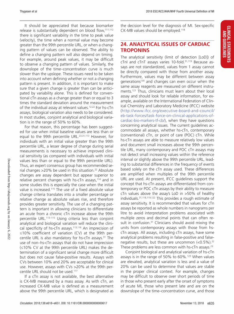
Thygesen et al 2018 ESC/ACC/AHA/WHF Fourth Universal Definition of MI
Circulation. 2018;138:e618–e651. DOI: 10.1161/CIR.0000000000000617 November 13, 2018 e635
CLINICAL STATEMENTS
AND GUIDELINES
It should be appreciated that because biomarker release is substantially dependent on blood flow,111,112 there is significant variability in the time to peak value (velocity), the time when a normal value may become greater than the 99th percentile URL, or when a chang-ing pattern of values can be observed. The ability to define a changing pattern will also depend on timing. For example, around peak values, it may be difficult to observe a changing pattern of values. Similarly, the downslope of the time–concentration curve is much slower than the upslope. These issues need to be taken into account when defining whether or not a changing pattern is present. In addition, it is important to make sure that a given change is greater than can be antici-pated by variability alone. This is defined for conven-tional cTn assays as a change greater than or equal to 3 times the standard deviation around the measurement of the individual assay at relevant values.12,22 For hs-cTn assays, biological variation also needs to be considered. In most studies, conjoint analytical and biological varia-tion is in the range of 50% to 60%.
For that reason, this percentage has been suggest-ed for use when initial baseline values are less than or equal to the 99th percentile URL.23,31,113 However, for individuals with an initial value greater than the 99th percentile URL, a lesser degree of change during serial measurements is necessary to achieve improved clini-cal sensitivity (as compared with individuals with initial values less than or equal to the 99th percentile URL). Thus, an expert consensus group has recommended se-rial changes >20% be used in this situation.22 Absolute changes are assay dependent but appear superior to relative percent changes with hs-cTn assays,114 and in some studies this is especially the case when the initial value is increased.115 The use of a fixed absolute value change criteria translates into a smaller percentage or relative change as absolute values rise, and therefore provides greater sensitivity. The use of a changing pat-tern is important in allowing clinicians to differentiate an acute from a chronic cTn increase above the 99th percentile URL.113–115 Using criteria less than conjoint analytical and biological variation will reduce the clini-cal specificity of hs-cTn assays.113,116 An imprecision of ≤10% coefficient of variation (CV) at the 99th per-centile URL is also mandatory for hs-cTn assays.31 The use of non–hs-cTn assays that do not have imprecision (≤10% CV at the 99th percentile URL) makes the de-termination of a significant serial change more difficult but does not cause false-positive results. Assays with CVs between 10% and 20% are acceptable for clinical use. However, assays with CVs >20% at the 99th per-centile URL should not be used.117
If a cTn assay is not available, the best alternative is CK-MB measured by a mass assay. As with cTn, an increased CK-MB value is defined as a measurement above the 99th percentile URL, which is designated as
the decision level for the diagnosis of MI. Sex-specific CK-MB values should be employed.118
24. ANALYTICAL ISSUES OF CARDIAC TROPONINSThe analytical sensitivity (limit of detection [LoD]) of cTnI and cTnT assays varies 10-fold.31,119 Because as-says are not standardized, values from 1 assay cannot be directly compared with those from another assay. Furthermore, values may be different between assay generations120 and changes can even occur when the same assay reagents are measured on different instru-ments.121 Thus, clinicians must learn about their local assay and should look for reliable information, for ex-ample, available on the International Federation of Clin-ical Chemistry and Laboratory Medicine (IFCC) website (http://www.ifcc.org/executive-board-and-council/ eb-task-forces/task-force-on-clinical-applications-of-cardiac-bio-markers-tf-cb/), when they have questions concerning analytical issues. The current guidelines ac-commodate all assays, whether hs-cTn, contemporary (conventional) cTn, or point of care (POC) cTn. While hs-cTn assays are able to measure relatively low values and document small increases above the 99th percen-tile URL, many contemporary and POC cTn assays may not detect small increasing values within the reference interval or slightly above the 99th percentile URL, lead-ing to substantial differences in the frequency of events based solely on the cTn assay used. These differences are amplified when multiples of the 99th percentile URL are used. At present, IFCC guidelines support the concept that hs-cTn assays are differentiated from con-temporary or POC cTn assays by their ability to measure cTn values above the assay’s LoD in >50% of healthy individuals.31,118,119,122 This provides a rough estimate of assay sensitivity. It is recommended that values for cTn assays be reported as whole numbers in nanograms per litre to avoid interpretation problems associated with multiple zeros and decimal points that can often re-sult in confusion.31 Clinicians should avoid mixing the units from contemporary assays with those from hs-cTn assays. All assays, including cTn assays, have some analytical problems resulting in false-positive and false-negative results, but these are uncommon (<0.5%).22 These problems are less common with hs-cTn assays.23
Conjoint biological and analytical variation of hs-cTn assays is in the range of 50% to 60%.123 When values are elevated, analytical variation is less and a value of 20% can be used to determine that values are stable in the proper clinical context. For example, changes may be difficult to observe over short periods of time in those who present early after the onset of symptoms of acute MI, those who present late and are on the downslope of the time–concentration curve, and those
Dow
nloaded from http://ahajournals.org by on January 6, 2022

Thygesen et al 2018 ESC/ACC/AHA/WHF Fourth Universal Definition of MI
November 13, 2018 Circulation. 2018;138:e618–e651. DOI: 10.1161/CIR.0000000000000617e636
CLIN
ICAL
STA
TEM
ENTS
AN
D GU
IDEL
INES
who have values near peak where they may be transi-tioning from a rising to a falling pattern.113,123
25. THE 99TH PERCENTILE UPPER REFERENCE LIMITThe 99th percentile URL is designated as the decision level for the presence of myocardial injury and must be determined for each specific assay with quality control materials used at the URL to validate appropriate assay imprecision. The cTn assay 99th percentile URL values used in clinical practice and research can be found both in manufacturers’ package inserts, in peer-reviewed publications, and on the IFCC website.118–120 Clinicians should be aware that for all cTn assays, including hs-cTn assays, there is still no expert opinion or consensus about specific criteria for how the 99th percentile URL should be defined.124 We endorse IFCC guidelines on the technical issues related to hs-cTn assays, including how studies should be configured to determine 99th percentile URLs.119 The guidelines include the clinical or surrogate biomarker screening that may be needed to better define the 99th percentile URL and the statisti-cal methods that can be applied, but do not include a requirement for cardiac imaging.119 Screening of appar-ently healthy subjects with imaging has been shown to lower the observed 99th percentile URL value, but is not a practical standard for the in vitro diagnostic in-dustry to use.124,125 Thus, there is the possibility of false-negative values using the manufacturer’s reported 99th percentile URL values. hs-cTn assays demonstrate shifts to higher values for the 99th percentile URL in associa-tion with comorbidities and age over >60 years.101,125–127 However, at present, age-dependent cut-off points are not recommended for clinical use. Clinicians should rely instead on changing values during serial measurements of cTn for the diagnosis of acute myocardial injury, including MI. Significantly lower values are observed among women compared with men, and therefore sex-specific 99th percentile URLs are recommended for hs-cTn assays.31,118–120 For some hs-cTn assays, sex-specific cut-off values have been reported to improve diagnostic and prognostic information in patients with possible acute MI.128,129 However, there is controversy as to whether this approach provides valuable additional information for all hs-cTn assays.130
26. OPERATIONALIZING CRITERIA FOR MYOCARDIAL INJURY AND INFARCTIONBlood samples for the measurement of cTn should be drawn on first assessment (designated as 0 hour) and repeated 3 to 6 hours later, or earlier with hs-cTn assays.
The sampling interval will impact the clinical cut-off at baseline and what is determined to be a pathological rise and/or fall of the biomarker. Sampling beyond 6 hours may be required if further ischemic episodes oc-cur, or in high-risk patients. To establish the diagnosis of an acute MI, a rise and/or fall in cTn values with at least 1 value above the 99th percentile URL is required, coupled with a high clinical and/or ECG likelihood of myocardial ischemia. hs-cTn assays shorten the time to diagnosis in many patients to within 3 hours of onset of symptoms, but there are still some patients who may rule-in late (at 6 hours).131 Furthermore, some patients with acute myocardial injury presenting late after the onset of acute MI (>12–18 hours) and who are on the downslope of the time–concentration curve may require longer periods of time for a changing pattern to be detected.131 In addition, it should be noted that with the implementation of cTn and hs-cTn assays, the frequency of unstable angina will decrease and the di-agnosis of NSTEMI will increase.132,133 The magnitude of these changes using hs-cTn assays have been report-ed in the range of 18% to 30%.134 Assuming proper timing of symptoms, acute ischemia should result in a change in hs-cTn; however, there may be patients in whom it is difficult to ascertain the timing of symptom onset. Thus, despite typical chest discomfort, these pa-tients may have hs-cTn values that are not elevated. Other patients with symptoms suggestive of unstable angina may have increased hs-cTn values as a result of structural heart disease with or without acute myocar-dial ischemia. This latter group may be particularly dif-ficult to distinguish from patients presenting with late NSTEMI with a slow decline in troponin values that can be observed in late presenters.131 Finally, some patients may manifest a changing pattern of troponin values with a magnitude that does not exceed the delta sug-gested for diagnosis or who fail to manifest a value greater than the 99th percentile URL. This is a group of patients that deserves close scrutiny because they may be at high risk. The triage of these patients can only be accomplished based on clinical evaluation.
Strategies employing either very low levels of hs-cTn on presentation or the lack of any change and persistently normal hs-cTn values over a 1–2 hour period after presentation have been advocated to ex-clude acute myocardial injury, and MI as well. A single sample rule-out strategy using a very low value (in many cases the LoD of the assay) has high sensitiv-ity for myocardial injury and therefore high negative predictive value to exclude MI.135 This strategy should not be used in those who present early (ie, <2 hours after the onset of chest discomfort. Some studies in-dicate that the single sample approach provides op-timal sensitivity and negative predictive accuracy in patients otherwise at low risk and those with a nor-mal ECG.136–138 However, one concern about very short
Dow
nloaded from http://ahajournals.org by on January 6, 2022

Thygesen et al 2018 ESC/ACC/AHA/WHF Fourth Universal Definition of MI
Circulation. 2018;138:e618–e651. DOI: 10.1161/CIR.0000000000000617 November 13, 2018 e637
CLINICAL STATEMENTS
AND GUIDELINES
rule-out periods is that the precision of the assays may not permit small differences to be distinguished.139–142 These criteria have not, and should not, be applied to patients with hs-cTn elevations.
The clinical specificity and positive predictive value of such 1–2 hour sampling approaches for ruling in MI are limited by the substantial proportion of individuals who meet the proposed biomarker criteria with diagnoses other than MI.136,141 Thus, the use of a rapid rule-in/-out MI protocol does not absolve the clinician from consid-ering other causes of acute myocardial injury.142 In ad-dition, considering a broader population of patients—inclusive of those who present atypically, those with end-stage renal disease, and the critically ill—the cut-off points to be used will likely need to be altered.139 Such patients have been excluded from the majority of emergency department evaluation studies.108,136,142
The demonstration of a rising and/or falling pattern is needed to distinguish acute injury from chronic con-ditions associated with structural heart disease that can have chronic increases of cTn values. For example, pa-tients with renal failure99,143,144 or LV hypertrophy145 can have significant chronic increases in cTn values. These increases can be marked but do not change acutely during serial sampling. However, a falling pattern may take longer to be observed in patients with a high pre-test risk of MI who present late after symptom onset.146 These patients who have cTn values on the downslope of the time–concentration curve have a slow decline in values (Figure 7). Thus, detecting a changing pattern over short periods of time may be difficult.117 Depend-ing on the extent of myocardial injury, cTn values may remain above the 99th percentile URL for a longer pe-riod of time.22,23 An increased cTn value above the 99th percentile URL, with or without a dynamic change of values, or in the absence of clini-cal evidence of isch-emia, should prompt a search for other diagnoses as-sociated with myocardial injury, as shown in Table 1.
27. ELECTROCARDIOGRAPHIC DETECTION OF MYOCARDIAL INFARCTIONThe ECG is an integral part of the diagnostic workup of patients with suspected MI, and should be acquired and interpreted promptly (ie, target within 10 min-utes) after first medical contact.47,147 Prehospital ECGs reduce the time to diagnosis and treatment, and can facilitate the triage of STEMI patients to hospitals with PCI capability if within the recommended time inter-val (120 minutes from STEMI diagnosis).46,148 Acute myocardial ischemia is often associated with dynamic changes in ECG waveform and serial ECG acquisition can provide critical information, particularly if the ECG at initial presentation is nondiagnostic. Recording sev-
eral standard ECGs with fixed electrode positions at 15–30 minute intervals for the initial 1–2 hours, or the use of continuous computer-assisted 12-lead ECG re-cording (if available) to detect dynamic ECG changes, is reasonable for patients with persistent or recurrent symptoms or an initial nondiagnostic ECG.149 Serial or continuous ECG recordings may be helpful in deter-mining reperfusion or reocclusion status. Reperfusion is usually associated with a large and prompt reduction in ST-segment elevation.
More profound ST-segment shifts or T wave inver-sions involving multiple leads/territories are associated with a greater degree of myocardial ischemia, and a worse prognosis. For example, ST-segment depression ≥1 mm in 6 leads, which may be associated with ST-segment elevation in leads aVR or lead V1 and hemo-dynamic compromise, is suggestive evidence of multi-vessel disease or left main disease. Pathologic Q waves increase the prognostic risk. Other ECG signs associ-ated with acute myocardial ischemia include cardiac ar-rhythmias, intraventricular bundle branch blocks, atrio-ventricular conduction delays, and loss of precordial R wave amplitude, a less specific finding. The ECG by itself is often insufficient to diagnose acute myocardial ischemia or infarction, since ST deviation may be ob-served in other conditions, such as acute pericarditis, LV hypertrophy (LVH), left bundle branch block (LBBB), Brugada syndrome, TTS, and early repolarization pat-terns.150 A prior ECG is often helpful in distinguishing a new from a chronic finding but should not delay the decision for treatment.
Prolonged new convex ST-segment elevation, par-ticularly when associated with reciprocal ST-segment depression, usually reflects acute coronary occlu-sion and results in myocardial injury with necrosis. Reciprocal changes can help to differentiate STEMI from pericarditis or early repolarization changes. As in cardiomyopathy, Q waves may also occur due to myocardial fibrosis in the absence of CAD. Some of the earlier manifestations of myocardial ischemia are typical T wave and ST-segment changes. Increased hyperacute T wave amplitude, with prominent sym-metrical T waves in at least 2 contiguous leads, is an early sign that may precede the elevation of the ST-segment. In general, the development of new Q waves indicates myocardial necrosis, which starts minutes/hours after the myocardial insult. Transient Q waves may be observed during an episode of acute ischemia or (rarely) during acute MI with successful reperfusion. Table 2 lists ST-segment–T wave (ST-T) criteria suggestive of acute myocardial ischemia that may or may not lead to MI. The J-point (junction between QRS termination and ST-segment onset) is used to determine the magnitude of the ST-segment shift with the onset of the QRS serving as the ref-erence point. In patients with a stable baseline, the
Dow
nloaded from http://ahajournals.org by on January 6, 2022
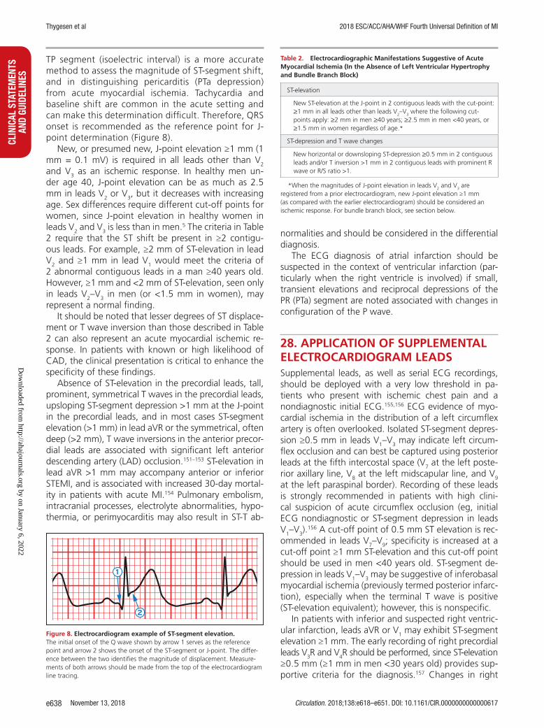
Thygesen et al 2018 ESC/ACC/AHA/WHF Fourth Universal Definition of MI
November 13, 2018 Circulation. 2018;138:e618–e651. DOI: 10.1161/CIR.0000000000000617e638
CLIN
ICAL
STA
TEM
ENTS
AN
D GU
IDEL
INES
TP segment (isoelectric interval) is a more accurate method to assess the magnitude of ST-segment shift, and in distinguishing pericarditis (PTa depression) from acute myocardial ischemia. Tachycardia and baseline shift are common in the acute setting and can make this determination difficult. Therefore, QRS onset is recommended as the reference point for J-point determination (Figure 8).
New, or presumed new, J-point elevation ≥1 mm (1 mm = 0.1 mV) is required in all leads other than V2 and V3 as an ischemic response. In healthy men un-der age 40, J-point elevation can be as much as 2.5 mm in leads V2 or V3, but it decreases with increasing age. Sex differences require different cut-off points for women, since J-point elevation in healthy women in leads V2 and V3 is less than in men.5 The criteria in Table 2 require that the ST shift be present in ≥2 contigu-ous leads. For example, ≥2 mm of ST-elevation in lead V2 and ≥1 mm in lead V1 would meet the criteria of 2 abnormal contiguous leads in a man ≥40 years old. However, ≥1 mm and <2 mm of ST-elevation, seen only in leads V2–V3 in men (or <1.5 mm in women), may represent a normal finding.
It should be noted that lesser degrees of ST displace-ment or T wave inversion than those described in Table 2 can also represent an acute myocardial ischemic re-sponse. In patients with known or high likelihood of CAD, the clinical presentation is critical to enhance the specificity of these findings.
Absence of ST-elevation in the precordial leads, tall, prominent, symmetrical T waves in the precordial leads, upsloping ST-segment depression >1 mm at the J-point in the precordial leads, and in most cases ST-segment elevation (>1 mm) in lead aVR or the symmetrical, often deep (>2 mm), T wave inversions in the anterior precor-dial leads are associated with significant left anterior descending artery (LAD) occlusion.151–153 ST-elevation in lead aVR >1 mm may accompany anterior or inferior STEMI, and is associated with increased 30-day mortal-ity in patients with acute MI.154 Pulmonary embolism, intracranial processes, electrolyte abnormalities, hypo-thermia, or perimyocarditis may also result in ST-T ab-
normalities and should be considered in the differential diagnosis.
The ECG diagnosis of atrial infarction should be suspected in the context of ventricular infarction (par-ticularly when the right ventricle is involved) if small, transient elevations and reciprocal depressions of the PR (PTa) segment are noted associated with changes in configuration of the P wave.
28. APPLICATION OF SUPPLEMENTAL ELECTROCARDIOGRAM LEADSSupplemental leads, as well as serial ECG recordings, should be deployed with a very low threshold in pa-tients who present with ischemic chest pain and a nondiagnostic initial ECG.155,156 ECG evidence of myo-cardial ischemia in the distribution of a left circumflex artery is often overlooked. Isolated ST-segment depres-sion ≥0.5 mm in leads V1–V3 may indicate left circum-flex occlusion and can best be captured using posterior leads at the fifth intercostal space (V7 at the left poste-rior axillary line, V8 at the left midscapular line, and V9 at the left paraspinal border). Recording of these leads is strongly recommended in patients with high clini-cal suspicion of acute circumflex occlusion (eg, initial ECG nondiagnostic or ST-segment depression in leads V1–V3).
156 A cut-off point of 0.5 mm ST elevation is rec-ommended in leads V7–V9; specificity is increased at a cut-off point ≥1 mm ST-elevation and this cut-off point should be used in men <40 years old. ST-segment de-pression in leads V1–V3 may be suggestive of inferobasal myocardial ischemia (previously termed posterior infarc-tion), especially when the terminal T wave is positive (ST-elevation equivalent); however, this is nonspecific.
In patients with inferior and suspected right ventric-ular infarction, leads aVR or V1 may exhibit ST-segment elevation ≥1 mm. The early recording of right precordial leads V3R and V4R should be performed, since ST-elevation ≥0.5 mm (≥1 mm in men <30 years old) provides sup-portive criteria for the diagnosis.157 Changes in right
Figure 8. Electrocardiogram example of ST-segment elevation. The initial onset of the Q wave shown by arrow 1 serves as the reference point and arrow 2 shows the onset of the ST-segment or J-point. The differ-ence between the two identifies the magnitude of displacement. Measure-ments of both arrows should be made from the top of the electrocardiogram line tracing.
Table 2. Electrocardiographic Manifestations Suggestive of Acute Myocardial Ischemia (In the Absence of Left Ventricular Hypertrophy and Bundle Branch Block)
ST-elevation
New ST-elevation at the J-point in 2 contiguous leads with the cut-point: ≥1 mm in all leads other than leads V2–V3 where the following cut-points apply: ≥2 mm in men ≥40 years; ≥2.5 mm in men <40 years, or ≥1.5 mm in women regardless of age.*
ST-depression and T wave changes
New horizontal or downsloping ST-depression ≥0.5 mm in 2 contiguous leads and/or T inversion >1 mm in 2 contiguous leads with prominent R wave or R/S ratio >1.
*When the magnitudes of J-point elevation in leads V2 and V3 are registered from a prior electrocardiogram, new J-point elevation ≥1 mm (as compared with the earlier electrocardiogram) should be considered an ischemic response. For bundle branch block, see section below.
Dow
nloaded from http://ahajournals.org by on January 6, 2022

Thygesen et al 2018 ESC/ACC/AHA/WHF Fourth Universal Definition of MI
Circulation. 2018;138:e618–e651. DOI: 10.1161/CIR.0000000000000617 November 13, 2018 e639
CLINICAL STATEMENTS
AND GUIDELINES
precordial leads may be transient, and an absence of ECG changes in leads V3R and V4R does not exclude right ventricular infarction. Myocardial imaging can be helpful in this clinical setting.
29. ELECTROCARDIOGRAPHIC DETECTION OF MYOCARDIAL INJURYIt is not possible to initially distinguish ECG manifesta-tions of acute or chronic myocardial injury from acute myocardial ischemia. Rapidly developing dynamic ECG changes that temporally match the clinical presentation may be helpful in diagnosing a symptomatic patient with elevated cTn values as having acute myocardial ischemia resulting in MI. However, ECG abnormalities are also common in patients who have myocardial in-jury (eg, myocarditis or TTS).158–160
30. PRIOR OR SILENT/UNRECOGNIZED MYOCARDIAL INFARCTIONQ wave criteria associated with MI and an increased relative risk of death are illustrated in Table 3, and are contained in Q wave coding algorithms such as the Minnesota Code and the WHO MONItoring of trends and determinants in CArdiovascular disease (MONICA) code11,161,162
The specificity of the ECG diagnosis for MI is greatest when Q waves occur in several leads or lead group-ings, or are >0.04 s. When the Q waves are associated with ST deviations or T wave changes in the same leads, the likelihood of MI is increased; for example, minor Q waves ≥0.02 s and <0.03 s that are ≥1 mm deep are suggestive of prior MI if accompanied by inverted T waves in the same lead group. Noninvasive imaging techniques also provide important supportive evidence of prior MI. In the absence of nonischemic causes, re-gional myocardial thinning, scar or reduced wall motion shown by echocardiography, myocardial perfusion scin-tigraphy (MPS) with single photon emission computed tomography (SPECT) or positron emission tomography (PET), or magnetic resonance imaging provide strong evidence for prior MI, particularly when ECG criteria are equivocal.
Asymptomatic patients who develop new Q wave criteria for MI detected during routine ECG follow-up, or reveal evidence of MI by cardiac imaging that can-not be directly attributed to an interim coronary revas-cularization procedure or an ACS admission, should be termed “silent or unrecognized MI.” In studies where serial ECG analysis was applied, silent or un-recognized Q wave MI accounted for 9% to 37% of all non-fatal MI events and was associated with a sig-nifi-cantly increased mortality risk.163,164 Improper lead placement, QRS abnormalities, or technical errors (eg, lead reversal) may result in the appearance of new Q waves or QS complexes, as compared with a prior tracing. Thus, the diagnosis of a new silent Q wave MI should be confirmed by a repeat ECG recording with correct lead placement, focused questioning about potential interim ischemic symptoms, or by an imag-ing study. Imaging techniques are useful if there is ab-normal myocardial motion, thickening, or thinning in the region of interest, but the absence of these does not exclude MI.165
31. CONDITIONS THAT CONFOUND THE ELECTROCARDIOGRAPHIC DIAGNOSIS OF MYOCARDIAL INFARCTIONA QS complex in lead V1 is normal. A Q wave <0.03 s and <0.25 of the R wave amplitude in lead III is nor-mal if the frontal QRS axis is between −30o and 0o. A Q wave may also be normal in aVL if the frontal QRS axis is between 60o and 90o. Septal Q waves are small, nonpathological Q waves <0.03 s and <0.25 of the R-wave amplitude in leads I, aVL, aVF, and V4–V6. Pre-excitation, cardiomyopathy, TTS, cardiac amyloidosis, LBBB, left anterior hemiblock, LVH, right ventricular hypertrophy, myocarditis, acute cor pulmonale, or hyperkalemia may be associated with Q waves or QS complexes in the absence of MI. Clinicians should be aware of confounders to the ECG diagnosis of myo-cardial ischemia, since ST-T wave abnormalities are commonly observed with different pathological cardi-
Criteria for Prior or Silent/Unrecognized MI
Any 1 of the following criteria meets the diagnosis for prior or silent/unrecognized MI:
• Pathological Q waves as described in Table 3, with or without symptoms, in the absence of nonischemic causes;
• Imaging evidence of loss of viable myocardium in a pattern consistent with ischemic etiology;
• Pathological findings of a prior MI.
Table 3. Electrocardiographic Changes Associated With Prior Myocardial Infarction (In the Absence of Left Ventricular Hypertrophy and Left Bundle Branch Block)
Any Q wave in leads V2–V3 >0.02 s or QS complex in leads V2–V3.
Q wave ≥0.03 s and ≥1 mm deep or QS complex in leads I, II, aVL, aVF or V4–V6 in any 2 leads of a contiguous lead grouping (I, aVL; V1–V6; II, III, aVF).*
R wave >0.04 s in V1–V2 and R/S >1 with a concordant positive T wave in absence of conduction defect.
*The same criteria are used for supplemental leads V7–V9. s indicates seconds.
Dow
nloaded from http://ahajournals.org by on January 6, 2022

Thygesen et al 2018 ESC/ACC/AHA/WHF Fourth Universal Definition of MI
November 13, 2018 Circulation. 2018;138:e618–e651. DOI: 10.1161/CIR.0000000000000617e640
CLIN
ICAL
STA
TEM
ENTS
AN
D GU
IDEL
INES
ac conditions, such as preexcitation, pericarditis, and cardiomyopathy.
32. CONDUCTION DISTURBANCES AND PACEMAKERSThe diagnosis of MI is more difficult in the presence of conduction disturbances, related in part to ST-T wave changes caused by the conduction disturbance and the fact that the conduction disturbance itself may be heart-rate dependent.166,167 Comparison to a preadmis-sion ECG may be helpful in determining if the conduc-tion defect or ST-T wave changes are new, as long as it does not delay time to treatment. Ischemic symptoms, and presumed new LBBB or right bundle branch block (RBBB) that is not rate-related, are associated with an adverse prognosis. In patients with LBBB, ST-segment elevation ≥1 mm concordant with the QRS complex in any lead may be an indicator of acute myocardial isch-emia. Similar findings can be useful in detecting ECG evidence for acute myocardial ischemia in patients with right ventricular paced rhythms.167 Recording an ECG trace with the pacemaker temporarily switched off may also be useful in patients who are not pacemaker de-pendent, but careful interpretation of repolarization is needed due to the possible presence of stimulation-induced changes (electrical memory). The ECG diag-nosis of acute myocardial ischemia in patients with biventricular pacing is more difficult. In patients with RBBB, new or presumed new ST-segment elevation ≥1 mm, or ST-segment or T wave abnormalities (excluding leads V1–V4) (Table 2), may indicate acute myocardial ischemia. New, or presumed new, RBBB without associ-ated ST-segment or T wave changes is associated with thrombolysis in myocardial infarction (TIMI) 0–2 flow in as many as 66% of patients (compared with >90% in those with ST-segment or T wave changes).168
33. ATRIAL FIBRILLATIONIn patients with atrial fibrillation and rapid ventricular rate or paroxysmal supraventricular tachycardia, ST-seg-ment depression or T wave inversion may occur in the absence of CAD.169,170 The causes are not completely understood. Cardiac memory, an electrical remodeling phenomenon characterized by marked diffuse T wave inversions following periods of abnormal ventricular activation, which may also be caused by transient rate-related conduction disturbances or pacing, may explain these findings. In some patients, the tachycardia may result in an insufficient increase in coronary flow to match myocardial oxygen demand, resulting in cellu-lar hypoxia and abnormal repolarization.171,172 For these reasons, a patient with new-onset atrial fibrillation, ele-vated baseline cTn concentration, and new ST-segment
depression should not automatically be classified as type 2 MI without additional information. In this clinical setting, signs of overt ischemic symptoms, the timing of symptoms relative to atrial fibrillation onset, a chang-ing pattern of cTn, and imaging and/or angiographic findings may be helpful in establishing the diagnosis. However, in the absence of evidence for myocardial ischemia, the etiology of the elevated cTn values should be attributed to myocardial injury.
34. IMAGING TECHNIQUESNoninvasive imaging plays many roles in patients with known or suspected MI, but this section concerns only its role in the diagnosis and characterization of myo-cardial injury and MI. The underlying rationale is that regional myocardial hypoperfusion and ischemia lead to a cascade of events including myocardial dysfunc-tion, cell death, and healing by fibrosis. Important im-aging parameters are therefore myocardial perfusion, myocyte viability, myocardial thickness, thickening and motion, and the effects of myocyte loss on the kinetics of paramagnetic or radio-opaque contrast agents indi-cating myocardial fibrosis or scar.
Commonly used imaging techniques in acute and prior MI are echocardiography, MPS using SPECT or PET, CMR, and possibly computed tomography (CT).173 There is considerable overlap in their capabilities and each of the techniques can assess myocardial viability, perfusion, and function to a greater or lesser extent. Only the radionuclide techniques provide a direct as-sessment of myocyte viability because of the inherent properties of the tracers used. Other techniques pro-vide indirect assessments of myocardial viability, such as the contractile response to dobutamine by echocar-diography, or increased extracellular space secondary to myocyte loss by CMR or CT.
34.1. EchocardiographyThe strength of echocardiography is the combined as-sessment of cardiac structure and function, in particu-lar myocardial thickness, thickening/thinning, and mo-tion. Regional wall motion abnormalities induced by ischemia can be detected by echocardiography almost immediately after onset when >20% transmural myo-cardial thickness is affected.174–176 These abnormalities, when new and without alternative etiology, support the diagnosis of MI when cTn values show a rising and/or falling pattern. Echocardiography also allows detection of non-coronary cardiac pathologies known to cause chest pain (eg, acute pericarditis, severe aortic stenosis, and hypertrophic cardiomyopathy among others). The technique is useful in diagnosing mechanical complica-tions in patients with MI and hemodynamic compro-mise (shock), or other potentially fatal entities such as
Dow
nloaded from http://ahajournals.org by on January 6, 2022

Thygesen et al 2018 ESC/ACC/AHA/WHF Fourth Universal Definition of MI
Circulation. 2018;138:e618–e651. DOI: 10.1161/CIR.0000000000000617 November 13, 2018 e641
CLINICAL STATEMENTS
AND GUIDELINES
acute aortic dissection or massive pulmonary embolism where the clinical presentation might be similar to that seen with acute MI.
Intravenous echocardiographic contrast agents can improve visualization of the endocardial border, and can be used to assess myocardial perfusion and micro-vascular obstruction. Tissue Doppler and strain imag-ing permit the quantification of global and regional function.177,178 Intravascular echocardiographic contrast agents that are targeted at specific molecular processes have been developed, but these techniques have not yet been applied in the setting of MI.179
34.2. Radionuclide ImagingSeveral radionuclide tracers allow viable myocytes to be imaged directly, including the SPECT tracers 201TI chloride, 99mTc sestamibi, and tetrofosmin, and the PET tracers 18F 2-fluorodeoxyglucose and 82Rb.173 A strength of the radionuclide techniques is that they are the only commonly available methods for assess-ing viability directly, although the relatively low res-olution of the images limits them for detecting the smallest areas of MI. Phantom studies suggest that myocyte loss as little as 4% of the myocardium can be detected, corresponding to 5 to 10 g of muscle.180 ECG-gated imaging provides a reliable assessment of myocardial motion, thickening, and global func-tion. Evolving radionuclide techniques relevant to the assessment of MI include imaging of sympathetic innervation using 123I-labeled meta-iodobenzylgua-nidine,181 imaging of matrix metalloproteinase activa-tion in ventricular remodeling,182,183 and the assess-ment of myocardial metabolism.184
34.3. Cardiac Magnetic Resonance ImagingThe high tissue contrast and resolution of CMR pro-vides an accurate assessment of myocardial structure and function. Although less commonly used in the acute setting, it has similar capabilities to echocardiog-raphy in suspected MI. Paramagnetic contrast agents can be used to assess myocardial perfusion and the in-crease in extracellular space that is associated with the fibrosis of prior MI (detected by LGE-CMR). These tech-niques have been used in the setting of acute MI185,186 and localized delay in contrast enhancement is able to detect even small areas of subendocardial MI, thought to be as little as 1 g.187 CMR also has the ability to identify the presence and extent of myocardial edema/inflammation, allowing the distinction of acute versus chronic myocardial injury. The patterns of LGE when reflecting ischemic and nonischemic myocardial injury are shown in Figure 9.
The gadolinium-based contrasts wash out slowly from myocardium with increased extracellular space such as fibrosis, thus enhancing areas of scarring (white arrows). The different patterns of scarring are divided into ischemic and nonischemic. Typically, an ischemic scar/fibrosis (upper panel) extends from the subendocardium to the epicardium (subendocardial, nontransmural scar versus transmural scar). Conversely, a nonischemic fibrosis/scar can be encountered at the epicardium, in the midwall, or at the insertion points of the right ventricle (lower panel).
34.4. Computed Tomographic Coronary AngiographyInfarcted myocardium is initially visible as a focal area of decreased LV myocardial enhancement, but later imaging shows hyperenhancement as with LGE-CMR.188 This finding is clinically relevant because con-trast-enhanced CT may be performed for suspected pulmonary embolism and aortic dissection, conditions with clinical features that overlap with those of acute MI, but the technique is not used routinely. Similarly, CT assessment of myocardial perfusion is technically feasible but not widely applied.189 CT coronary an-giography (CTCA) may be used to diagnose CAD in patients with an ACS in the emergency department or chest pain unit, particularly in low- to intermediate-risk patients with normal cTn at presentation.189–193 The only randomized trial in these patients that in-cluded both hs-cTn and CTCA found that imaging did not reduce the length of stay in hospital, but it did decrease subsequent outpatient testing and costs.189 A diagnosis of MI cannot be established based on a CTCA scan alone.
35. APPLYING IMAGING IN ACUTE MYOCARDIAL INFARCTIONImaging techniques can be useful in the diagnosis of acute MI because of the ability to detect wall motion abnormalities or loss of viable myocardium in the pres-ence of elevated cardiac biomarker values. Demonstra-tion of new loss of myocardial viability in the absence of nonischemic causes supports the diagnosis of MI. Normal function practically excludes significant MI, but a small MI cannot be ruled out.194 Thus, imaging tech-niques are useful for early triage and discharge of pa-tients with suspected MI. However, if biomarkers have been measured at appropriate times and are normal, this excludes acute MI and takes precedence over the imaging criteria.
Abnormal regional myocardial motion and thicken-ing may be caused by acute MI, or by ≥1 of several other conditions including prior infarction, acute isch-
Dow
nloaded from http://ahajournals.org by on January 6, 2022
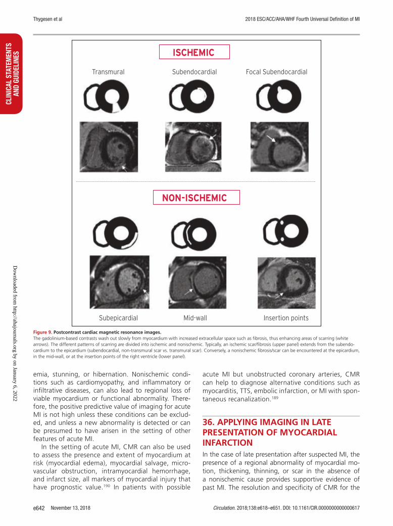
November 13, 2018 Circulation. 2018;138:e618–e651. DOI: 10.1161/CIR.0000000000000617e642
CLIN
ICAL
STA
TEM
ENTS
AN
D GU
IDEL
INES
Thygesen et al 2018 ESC/ACC/AHA/WHF Fourth Universal Definition of MI
emia, stunning, or hibernation. Nonischemic condi-tions such as cardiomyopathy, and inflammatory or infiltrative diseases, can also lead to regional loss of viable myocardium or functional abnormality. There-fore, the positive predictive value of imaging for acute MI is not high unless these conditions can be exclud-ed, and unless a new abnormality is detected or can be presumed to have arisen in the setting of other features of acute MI.
In the setting of acute MI, CMR can also be used to assess the presence and extent of myocardium at risk (myocardial edema), myocardial salvage, micro-vascular obstruction, intramyocardial hemorrhage, and infarct size, all markers of myocardial injury that have prognostic value.190 In patients with possible
acute MI but unobstructed coronary arteries, CMR can help to diagnose alternative conditions such as myocarditis, TTS, embolic infarction, or MI with spon-taneous recanalization.189
36. APPLYING IMAGING IN LATE PRESENTATION OF MYOCARDIAL INFARCTIONIn the case of late presentation after suspected MI, the presence of a regional abnormality of myocardial mo-tion, thickening, thinning, or scar in the absence of a nonischemic cause provides supportive evidence of past MI. The resolution and specificity of CMR for the
Figure 9. Postcontrast cardiac magnetic resonance images. The gadolinium-based contrasts wash out slowly from myocardium with increased extracellular space such as fibrosis, thus enhancing areas of scarring (white arrows). The different patterns of scarring are divided into ischemic and nonischemic. Typically, an ischemic scar/fibrosis (upper panel) extends from the subendo-cardium to the epicardium (subendocardial, non-transmural scar vs. transmural scar). Conversely, a nonischemic fibrosis/scar can be encountered at the epicardium, in the mid-wall, or at the insertion points of the right ventricle (lower panel).
Dow
nloaded from http://ahajournals.org by on January 6, 2022

Thygesen et al 2018 ESC/ACC/AHA/WHF Fourth Universal Definition of MI
Circulation. 2018;138:e618–e651. DOI: 10.1161/CIR.0000000000000617 November 13, 2018 e643
CLINICAL STATEMENTS
AND GUIDELINES
detection of myocardial scarring has made this a valu-able technique. In particular, the ability to distinguish between subendocardial and other patterns of scars helps to differentiate between ischemic heart disease and other myocardial pathologies. Imaging techniques are also useful for risk stratification after a definitive diagnosis of MI.
37. REGULATORY PERSPECTIVE ON MYOCARDIAL INFARCTION IN CLINICAL TRIALSIn drug and device development programs, MI may be an entry criterion or be used as an efficacy end point, commonly as a component of the primary end point, as well as a safety end point of interest in drug devel-opment programs.195,196 A universal definition of MI is of great benefit for clinical studies, since it will allow a standardized approach for meaningful interpretation and comparison across different trials, or the pooling of results for the detection of safety signals. For the harmonization of the MI definition it is important to standardize the reporting of MI events by clinical events committees. This would allow a more optimal compari-son of MI rates among drug and device trials.
One cannot presume that values from 1 cTn assay are equivalent to those of another. These differences are amplified when multiples of the values are used. This could affect results, especially in trials that compare strategies such as PCI and CABG. The use of a single as-say and/or a central core laboratory within a trial could help to decrease this variability, and might be particu-larly relevant in decreasing variability in trials of a drug or intervention in which cTn concentration is a principal safety end point. However, the uniform use of a single assay is generally not feasible in trials with follow-up postdischarge, since recurrent ischemic events may oc-cur in different hospitals using different cTn assays. In clinical trials, a standardized approach to establish the 99th percentile URL for a particular assay should be es-tablished. One approach in large multicenter trials is to use the manufacturer’s recommended 99th percentile URL for a particular assay to reduce site-to-site variabil-ity in the selection of the MI decision cut-off point.
Multiples for hs-cTn versus conventional cTn could have markedly different prognostic implications. The assay types should be reported when possible. Multi-ples of the 99th percentile URL should be indicated and reported, both for those with cardiac procedural myo-cardial injury and those diagnosed with types 4a and 5 MI. Cumulative frequency distribution of peak cTn measurements for MI end point assessments by treat-ment group should also be provided. This will facilitate the comparison of trials and meta-analyses.
38. SILENT/UNRECOGNIZED MYOCARDIAL INFARCTION IN EPIDEMIOLOGICAL STUDIES AND QUALITY PROGRAMSECG monitoring for unrecognized or silent Q wave MI is usually acquired annually in epidemiological studies and clinical trials that assess cardiovascular end points. These events are associated with adverse outcomes.197 There is no firm consensus on how frequently to monitor for ECG evidence of silent Q wave MI or whether surveillance for silent MI events should be routinely implemented. Serial monitoring of patients who have had a symptomatic Q wave MI event revealed Q wave regression in a substan-tial number of patients.198 An annual ECG is reasonable in clinical trials to monitor for silent Q wave MI events if the study population is expected to have an accelerated rate of atherosclerotic events. The review should con-sider the baseline tracing, interim event ECG tracings, and protocol-mandated annual tracings, along with the review of imaging studies if available.
39. INDIVIDUAL AND PUBLIC IMPLICATIONS OF THE MYOCARDIAL INFARCTION DEFINITIONRevision of the definition of MI has a number of implica-tions for individuals, health professionals, and society at large. A tentative or final diagnosis is the basis for ad-vice about further diagnostic testing, lifestyle changes, treatment, and prognosis for the patient. The aggregate of patients with a particular diagnosis is the basis for healthcare planning, and policy and resource allocation.
One of the goals of good clinical practice is to reach a definitive and specific diagnosis, which is supported by current scientific knowledge. The approach to the definition of myocardial injury and MI outlined in this document meets this goal. In general, the conceptu-al meaning of the term myocardial infarction has not changed, although new sensitive methods have been developed to diagnose this entity. Thus, the diagnosis of an acute MI is a clinical diagnosis based on patient symptoms, ECG changes, and highly sensitive biochem-ical markers, as well as information gleaned from vari-ous imaging techniques.
It should be appreciated that the universal definition of MI may be associated with consequences for patients and their families with respect to psychological status, life and health insurance, and professional career, as well as driving and pilot licenses. The diagnosis is also associated with societal implications with regards to diagnosis-related coding, hospital reimbursement, pub-lic health statistics, sick leave, and disability attestation. In order to meet these challenges, physicians must be adequately informed of the diagnostic criteria. Hence,
Dow
nloaded from http://ahajournals.org by on January 6, 2022

Thygesen et al 2018 ESC/ACC/AHA/WHF Fourth Universal Definition of MI
November 13, 2018 Circulation. 2018;138:e618–e651. DOI: 10.1161/CIR.0000000000000617e644
CLIN
ICAL
STA
TEM
ENTS
AN
D GU
IDEL
INES
educational materials will need to be created and treat-ment guidelines must be appropriately adapted.
40. GLOBAL PERSPECTIVES OF THE DEFINITION OF MYOCARDIAL INFARCTIONCardiovascular disease is a global health problem and prevalence is increasing in the developing world. Un-derstanding the burden and effects of CAD in popula-tions is of critical importance. Changing clinical defini-tions, criteria, and biomarkers add challenges to our understanding and ability to improve the health of the public. For clinicians, the definition of MI has im-portant and immediate therapeutic implications. For epidemiologists, the data are often retrospective, so consistent case definitions are critical for comparisons and trend analysis. The standards described in this report are suitable for epidemiology studies and for international classification of diseases.199 However, to analyze trends over time, it is important to have con-sistent definitions and to quantify adjustments when biomarkers or other diagnostic methods change,200 considering that the advent of cTn has dramatically increased the number of diagnosable MIs for epide-miologists.11,201
In countries with limited economic resources, cardiac biomarkers and imaging techniques may not be avail-able except in a few centers and even the option of ECG recordings may be lacking. The WHO recommends the use of the ESC/ACC/AHA/WHF Universal Defini-tion of MI in countries without resource constraints, but recommends more flexible standards in resource-constrained locations. Thus, when the only information available is the clinical history and ECG, and when data on cardiac biomarkers are not available or incomplete, the diagnosis of MI can be confirmed by the develop-ment of pathological Q waves.11
41. USING THE UNIVERSAL DEFINITION OF MYOCARDIAL INFARCTION IN THE HEALTHCARE SYSTEMArriving at a diagnosis of MI using the criteria set forth in this document requires the integration of clinical findings, patterns on the ECG, laboratory data, ob-servations from imaging procedures, and on occasion pathological findings, all viewed in the context of the time horizon over which the suspected event unfolds. Contemporary healthcare systems are increasingly us-ing electronic medical records where medical informa-tion is entered, curated, and available for retrieval at a later date. This evolution offers the advantages of a modern electronic database that is useful for a variety
of purposes, including scientific discovery and quality improvement in clinical care, but carries with it the chal-lenges of sifting through variable locations and formats where key data elements for confirming a diagnosis of MI are located. Also, use of the electronic medical record as an epidemiological and research tool of the future is likely to require efforts to verify the accuracy of an acute MI diagnosis, rather than accepting the coded diagnoses used for administrative and billing purposes. Such an effort to create a computable phenotype of MI (further categorized as types 1–5 MI) will require in-put from informaticians and experts in implementation science to translate the recommendations from this Universal Definition of MI into the routine practice of healthcare delivery and documentation.
Given the evolution of biomarker assays used to support the diagnosis of MI, it is important that a con-sistent approach be used in the construction of the computable phenotype of MI so as to reliably make comparisons across institutions and track epidemiologi-cal trends. Ideally, the information provided should in-clude the assay used to make the diagnosis of MI, the 99th percentile of the URL, and the full sequence of values obtained to discern a rise and fall in biomarker levels.196
ARTICLE INFORMATIONDisclaimer. The ESC/ACC/AHA/WHF Expert Consensus Document represents the views of the ESC, ACC, AHA, and WHF and was produced after careful consideration of the scientific and medical knowledge and the evidence avail-able at the time of their publication. The ESC, ACC, AHA, and WHF are not responsible in the event of any contradiction, discrepancy, and/or ambiguity between the ESC/ACC/AHA/WHF Expert Consensus Document and any other official recommendations or Expert Consensus Document issued by the relevant public health authorities, in particular in relation to good use of healthcare or therapeutic strategies. Health professionals are encouraged to take the ESC/ ACC/AHA/WHF Expert Consensus Document fully into account when exercis-ing their clinical judgment, as well as in the determination and the implementa-tion of preventive, diagnostic, or therapeutic medical strategies; however, the ESC/ACC/AHA/WHF Expert Consensus Document does not override, in any way whatsoever, the individual responsibility of health professionals to make appropriate and accurate decisions in consideration of each patient’s health condition and in consultation with that patient and, where appropriate and/or necessary, the patient’s caregiver. Nor does the ESC/ACC/AHA/WHF Expert Consensus Document exempt health professionals from taking into full and careful consideration the relevant official updated recommendations or Expert Consensus Documents issued by the competent public health authorities, in order to manage each patient’s case in light of the scientifically accepted data pursuant to their respective ethical and professional obligations. It is also the health professional’s responsibility to verify the applicable rules and regulations relating to drugs and medical devices at the time of prescription.
This article has been copublished in the European Heart Journal, the Journal of the American College of Cardiology, and Global Heart.
Copies: This document is available on the websites of the European Society of Cardiology (www.escardio.org.), American College of Cardiology (www.acc.org), American Heart Association (professional.heart.org), and the World Heart Federation (www.world-heart-federation.org). A copy of the document is also available at https://professional.heart.org/statements by selecting the “Guide-lines & Statements” button. To purchase additional reprints, call 843-216-2533 or e-mail [email protected].
The expert peer review of AHA-commissioned documents (eg, scientific statements, clinical practice guidelines, systematic reviews) is conducted by the AHA Office of Science Operations. For more on AHA statements and guide-lines development, visit https://professional.heart.org/statements. Select the
Dow
nloaded from http://ahajournals.org by on January 6, 2022

Thygesen et al 2018 ESC/ACC/AHA/WHF Fourth Universal Definition of MI
Circulation. 2018;138:e618–e651. DOI: 10.1161/CIR.0000000000000617 November 13, 2018 e645
CLINICAL STATEMENTS
AND GUIDELINES
“Guidelines & Statements” drop-down menu near the top of the webpage, then click “Publication Development.”
Permissions: The content of this ESC/ACC/AHA/WHF Expert Consensus Document has been published for personal and educational use only. No com-mercial use is authorized. No part of the ESC/ACC/AHA/WHF Expert Consen-sus Document may be translated or reproduced in any form without written permission from the ESC or ACC or AHA or WHF. Multiple copies, modifica-tion, alteration, enhancement, and/or distribution of this document are not permitted without the express permission of the American Heart Association. Instructions for obtaining permission are located at https://www.heart.org/permissions. A link to the “Copyright Permissions Request Form” appears in the second paragraph (https://www.heart.org/en/about-us/statements-and-policies/copyright-request-form).
AcknowledgmentWe are indebted to Karen A. Hicks for valuable advice.
Document ReviewersDavid Hasdai (CPG Review Coordinator) (Israel), Victor Aboyans (France), Stephan Achenbach (Germany), Stefan Agewall (Norway), Thomas Alexander (India), Alvaro Avezum (Brazil), Emanuele Barbato (Italy), Jean-Pierre Bassand (France), Eric Bates (USA), John A. Bittl (USA), Güenter Breithardt (Germany), Héctor Bueno (Spain), Raffaele Bugiardini (Italy), Mauricio G. Cohen (USA), George Dangas (USA), James A. de Lemos (USA), Victoria Delgado (Neth-erlands), Gerasimos Filippatos (Greece), Edward Fry (USA), Christopher B. Granger (USA), Sigrun Halvorsen (Norway), Mark A. Hlatky (USA), Borja Ibanez (Spain), Stefan James (Sweden), Adnan Kastrati (Germany), Christophe Leclercq (France), Kenneth W. Mahaffey (USA), Laxmi Mehta (USA), Christian Müller (Switzerland), Carlo Patrono (Italy), Massimo Francesco Piepoli (Italy), Daniel Piñeiro (Argentina), Marco Roffi (Switzerland), Andrea Rubboli (Italy), Marc Ruel (Canada), Samin Sharma (USA), Iain A. Simpson (UK), Michael Tendera (Poland), Marco Valgimigli (Switzerland), Allard C. van der Wal (Netherlands), Stephan Windecker (Switzerland).
APPENDIXApproved by the ESC Committee for Practice Guidelines (CPG) on behalf of the ESC Board 2016–2018.
ESC National Cardiac Societies actively involved in the review process of the Fourth Universal Definition of Myocardial Infarction:
Algeria: Algerian Society of Cardiology, Mohamed Chettibi; Armenia: Armenian Cardiologists Association, Hamlet Hayrapetyan; Austria: Austrian Society of Cardiology, Franz Xaver Roithinger; Azerbaijan: Azerbaijan Society of Cardiology, Farid Aliyev; Belarus: Belorussian Scientific Society of Cardiolo-gists, Volha Sujayeva; Belgium: Belgian Society of Cardiology, Marc J. Claeys; Bosnia and Herzegovina: Association of Cardiologists of Bosnia and Herze-govina, Elnur Smajić; Czech Republic: Czech Society of Cardiology, Petr Kala; Denmark: Danish Society of Cardiology, Kasper Karmak Iversen; Egypt: Egyp-tian Society of Cardiology, Ehab El Hefny; Estonia: Estonian Society of Cardi-ology, Toomas Marandi; Finland: Finnish Cardiac Society, Pekka Porela; The Former Yugoslav Republic of Macedonia: Macedonian FYR Society of Cardiol-ogy, Slobodan Antov; France: French Society of Cardiology, Martine Gilard; Germany: German Cardiac Society, Stefan Blankenberg; Greece: Hellenic So-ciety of Cardiology, Periklis Davlouros; Iceland: Icelandic Society of Cardiology, Thorarinn Gudnason; Israel: Israel Heart Society, Ronny Alcalai; Italy: Italian Federation of Cardiology, Furio Colivicchi; Kosovo: Kosovo Society of Cardiol-ogy, Shpend Elezi; Kyrgyzstan: Kyrgyz Society of Cardiology, Gulmira Baitova, Latvia: Latvian Society of Cardiology, Ilja Zakke; Lithuania: Lithuanian Society of Cardiology, Olivija Gustiene; Luxembourg: Luxembourg Society of Cardi-ology, Jean Beissel; Malta: Maltese Cardiac Society, Philip Dingli; Moldova: Moldavian Society of Cardiology, Aurel Grosu; The Netherlands: Netherlands Society of Cardiology, Peter Damman; Norway: Norwegian Society of Cardiol-ogy, Vibeke Juliebø; Poland: Polish Cardiac Society, Jacek Legutko; Portugal: Portuguese Society of Cardiology, Joäo Morais; Romania: Romanian Society of Cardiology, Gabriel Tatu-Chitoiu; Russian Federation: Russian Society of Cardiology, Alexey Yakovlev; San Marino: San Marino Society of Cardiology, Marco Zavatta; Serbia: Cardiology Society of Serbia, Milan Nedeljkovic; Slove-nia: Slovenian Society of Cardiology, Peter Radsel; Spain: Spanish Society of Cardiology, Alessandro Sionis; Sweden: Swedish Society of Cardiology, Tomas Jemberg; Switzerland: Swiss Society of Cardiology, Christian Müller; Tunisia: Tunisian Society of Cardiology and Cardio-Vascular Surgery, Leila Abid; Tur-
key: Turkish Society of Cardiology, Adnan Abaci; Ukraine: Ukrainian Associa-tion of Cardiology, Alexandr Parkhomenko; United Kingdom: British Cardio-vascular Society, Simon Corbett.
Approved by the ACC Clinical Policy Approval Committee.Approved by the AHA Science Advisory and Coordinating Committee.Approved by the WHF Board.
REFERENCES 1. Hammer A. Ein Fall von thrombotischem Verschlusse einer der Kranzarter-
ien des Herzens. Wien Med Wschr. 1878;28:97–102. 2. Obraztzow VP, Straschesko ND. Zur Kenntnis der Thrombose der Koro-
nararterien des Herzens. Z Klin Med. 1910;71:116–132. 3. Herrick JB. Clinical features of sudden obstruction of the coronary arteries.
JAMA. 1912;59:2015–2022. 4. Friedberg CK, Horn H. Acute myocardial infarction not due to coronary
artery occlusion. JAMA. 1939;112:1675–1679. 5. World Health Organization. Working Group on the Establishment of Isch-
emic Heart Disease Registers. Report of the Fifth Working Group, Copen-hagen. In: Report No. Eur 8201 (5). Geneva, Switzerland: World Health Organization; 1971.
6. Report of the Joint International Society and Federation of Cardiology/World Health Organization task force on standardization of clinical no-menclature. Nomenclature and criteria for diagnosis of ischemic heart disease. Circulation. 1979;59:607–609.
7. Tunstall-Pedoe H, Kuulasmaa K, Amouyel P, Arveiler D, Rajakangas AM, Pajak A. Myocardial infarction and coronary deaths in the World Health Organization MONICA Project. Registration procedures, event rates, and case-fatality rates in 38 populations from 21 countries in four continents. Circulation. 1994;90:583–612.
8. Luepker RV, Apple FS, Christenson RH, Crow RS, Fortmann SP, Goff D, Goldberg RJ, Hand MM, Jaffe AS, Julian DG, Levy D, Manolio T, Men-dis S, Mensah G, Pajak A, Prineas RJ, Reddy KS, Roger VL, Rosamond WD, Shahar E, Sharrett AR, Sorlie P, Tunstall-Pedoe H. Case definitions for acute coronary heart disease in epidemiology and clinical research stud-ies: a statement from the AHA Council on Epidemiology and Prevention; AHA Statistics Committee; World Heart Federation Council on Epidemiol-ogy and Prevention; the European Society of Cardiology Working Group on Epidemiology and Prevention; Centers for Disease Control and Pre-vention; and the National Heart, Lung, and Blood Institute. Circulation. 2003;108:2543–2549.
9. The Joint European Society of Cardiology/American College of Cardiology Committee. Myocardial infarction redefined—a consensus document of the Joint European Society of Cardiology/American College of Cardiol-ogy Committee for the Redefinition of Myocardial Infarction. Eur Heart J. 2000;21:1502–1513; J Am Coll Cardiol. 2000;36:959–969.
10. Thygesen K, Alpert JS, White HD; Joint ESC/ACC/AHA/WHF Task Force for the Redefinition of Myocardial Infarction. Universal definition of myocardial infarction. Eur Heart J. 2007;28:2525–2538; Circulation. 2007;116:2634–2653; J Am Coll Cardiol. 2007;50:2173–2195.
11. Mendis S, Thygesen K, Kuulasmaa K, Giampaoli S, Mähönen M, Ngu Blackett K, Lisheng L; Writing group on behalf of the participating experts of the WHO consultation for revision of WHO definition of myocardial infarction. World Health Organization definition of myocardial infarction: 2008-09 revision. Int J Epidemiol. 2011;40:139–146.
12. Thygesen K, Alpert JS, Jaffe AS, Simoons ML, Chaitman BR, White HD; Writing Group on the Joint ESC/ACC/AHA/WHF Task Force for the Uni-versal Definition of Myocardial Infarction. Third universal definition of myocardial infarction. Eur Heart J. 2012;33:2551–2567; Circulation. 2012;126:2020–2035; J Am Coll Cardiol. 2012;60:1581–1598.
13. Sarkisian L, Saaby L, Poulsen TS, Gerke O, Jangaard N, Hosbond S, Diederichsen ACP, Thygesen K, Mickley H. Clinical characteristics and outcomes of patients with myocardial infarction, myocardial injury, and nonelevated troponins. Am J Med. 2016;129:446e.5–446e.21.
14. Sarkisian L, Saaby L, Poulsen TS, Gerke O, Hosbond S, Jangaard N, Die-derichsen ACP, Thygesen K, Mickley H. Prognostic impact of myocardial injury related to various cardiac and noncardiac conditions. Am J Med. 2016;129:506–514.
15. Ooi DS, Isotalo PA, Veinot JP. Correlation of antemortem serum creatine kinase, creatine kinase-MB, troponin I, and troponin T with cardiac pathol-ogy. Clin Chem. 2000;46:338–344.
16. Jennings RB, Ganote CE. Structural changes in myocardium during acute ischemia. Circ Res. 1974;35:156–172.
Dow
nloaded from http://ahajournals.org by on January 6, 2022

Thygesen et al 2018 ESC/ACC/AHA/WHF Fourth Universal Definition of MI
November 13, 2018 Circulation. 2018;138:e618–e651. DOI: 10.1161/CIR.0000000000000617e646
CLIN
ICAL
STA
TEM
ENTS
AN
D GU
IDEL
INES
17. Virmani R, Forman MB, Kolodgie FD. Myocardial reperfusion injury. Histo-pathological effects of perfluorochemical. Circulation. 1990;81:IV57–IV68.
18. Reimer KA, Jennings RB, Tatum AH. Pathobiology of acute myocardial ischemia: metabolic, functional and ultrastructural studies. Am J Cardiol. 1983;52:72A–81A.
19. Ibáñez B, Heusch G, Ovize M, Van de Werf F. Evolving therapies for myocardial ischemia/reperfusion injury. J Am Coll Cardiol. 2015;65: 1454–1471.
20. Montecucco F, Carbone F, Schindler TH. Pathophysiology of ST-segment elevation myocardial infarction: novel mechanisms and treatments. Eur Heart J. 2016;37:1268–1283.
21. Thygesen K, Mair J, Katus H, Plebani M, Venge P, Collinson P, Lindahl B, Giannitsis E, Hasin Y, Galvani M, Tubaro M, Alpert JS, Biasucci LM, Koenig W, Mueller CH, Huber K, Hamm C, Jaffe AS; The Study Group on Bio-markers in Cardiology of the ESC Working Group on Acute Cardiac Care. Recommendations for the use of cardiac troponin measurement in acute cardiac care. Eur Heart J. 2010;31:2197–2204.
22. Thygesen K, Mair J, Giannitsis E, Mueller C, Lindahl B, Blankenberg S, Huber K, Plebani M, Biasucci LM. Tubaro M, Collinson P, Venge P, Hasin Y, Galvani M, Koenig W, Hamm C, Alpert JS, Katus H, Jaffe AS; Study Group on Biomarkers in Cardiology of the ESC Working Group on Acute Cardiac Care. How to use high-sensitivity cardiac troponins in acute cardiac care. Eur Heart J. 2012;33:2252–2257.
23. Rittoo D, Jones A, Lecky B, Neithercut D. Elevation of cardiac troponin T, but not cardiac troponin I, in patients with neuromuscular diseases: implications for the diagnosis of myocardial infarction. J Am Coll Cardiol. 2014;63:2411–2420.
24. Jaffe AS, Vasile VC, Milone M, Saenger AK, Olson KN, Apple FS. Diseased skeletal muscle: a noncardiac source of increased circulating concentra-tions of cardiac troponin T. J Am Coll Cardiol. 2011;58:1819–1824.
25. Wens SCA, Schaaf GJ, Michels M, Kruijshaar ME, van Gestel TJM, In ‘t Groen S, Pijnenburg J, Dekkers DHW, Demmers JAA, Verdijk LB, Brusse E, van Schaik RHN, van der Ploeg AT, van Doorn PA, Pijnappel WWMP. Elevated plasma cardiac troponin T levels caused by skeletal muscle dam-age in Pompe disease. Circ Cardiovasc Genet. 2016;9:6–13.
26. Mair J, Lindahl B, Müller C, Giannitsis E, Huber K, Möckel M, Plebani M, Thygesen K, Jaffe AS. What to do when you question cardiac troponin values. Eur Heart J Acute Cardiovasc Care. 2018;7:577–586.
27. Mair J, Lindahl B, Hammarsten O, Müller C, Giannitsis E, Huber K, Möckel M, Plebani M, Thygesen K, Jaffe AS; European Society of Cardiology (ESC) Study Group on Biomarkers in Cardiology of the Acute Cardiovascular Care Association (ACCA). How is cardiac troponin released from injured myocar-dium? Eur Heart J Acute Cardiovasc Care. 2018;7:553–560.
28. Vestergaard KR, Jespersen CB, Arnadottir A, Soletormos G, Schou M, Steffensen R, Goetze JP, Kjoller E, Iversen KK. Prevalence and significance of troponin elevations in patients without acute coronary disease. Int J Cardiol. 2016;222:819–825.
29. Schmid J, Liesinger L, Birner-Gruenberger R, Stojakovic T, Scharnagl H, Dieplinger B, Asslaber M, Radl R, Beer M, Polacin M, Mair J, Szolar D, Berghold A, Quasthoff S, Binder JS. Rainer PP. Elevated cardiac troponin T in skeletal myopathies. J Am Cardiol Coll. 2018;71:1540–1549.
30. Apple FS, Jaffe AS, Collinson P, Mockel M, Ordonez-Llanos J, Lindahl B, Hollander J, Plebani M, Than M, Chan MH; on behalf of the International Federation of Clinical Chemistry (IFCC) Task Force on Clinical Applications of Cardiac Bio-Markers. IFCC educational materials on selected analytical and clinical applications of high sensitivity cardiac troponin assays. Clin Biochem. 2015;48:201–203.
31. Goodman SG, Steg PG, Eagle KA, Fox KA, López-Sendón J, Montalescot G, Budaj A, Kennelly BM, Gore JM, Allegrone J, Granger CB, Gurfinkel EP; GRACE Investigators. The diagnostic and prognostic impact of the redefi-nition of acute myocardial infarction: lessons from the Global Registry of Acute Coronary Events (GRACE). Am Heart J. 2006;151:654–660.
32. Weil BR, Suzuki G, Young RF, Iyer V, Canty JM Jr. Troponin release and re-versible left ventricular dysfunction following transient pressure overload: stress-induced myocardial stunning. J Am Cardiol Coll. 2018;71:2906–2916.
33. Turer AT, Addo TA, Martin JL, Sabatine MS, Lewis GD, Gerszten RE, Keeley EC, Cigarroa JE, Lange RA, Hillis LD, de Lemos JA. Myocardial ischemia induced by rapid atrial pacing causes troponin T release detectable by a highly sensitive assay: insights from a coronary sinus sampling study. J Am Coll Cardiol. 2011;57:2398–2405.
34. Siriwardena M, Campbell V, Richards AM, Pemberton CJ. Cardiac bio-marker responses to dobutamine stress echocardiography in healthy volunteers and patients with coronary artery disease. Clin Chem. 2012;58:1492–1494.
35. White HD. Pathobiology of troponin elevations: do elevations occur with myocardial ischemia as well as necrosis? J Am Coll Cardiol. 2011;57:2406–2408.
36. Jaffe AS, Wu AH. Troponin release—reversible or irreversible injury? Should we care? Clin Chem. 2012;58:148–150.
37. Eggers KM, Lindahl B. Application of cardiac troponin in cardiovascular diseases other than acute coronary syndrome. Clin Chem. 2017;63:223–235.
38. Giannitsis E, Katus HA. Cardiac troponin level elevations not related to acute coronary syndromes. Nat Rev Cardiol. 2013;10:623–634.
39. Agewall S, Giannitsis E, Jernberg T, Katus H. Troponin elevation in coro-nary vs. non-coronary disease. Eur Heart J. 2011;32:404–411.
40. Kelley WE, Januzzi JL, Christenson RH. Increases of cardiac troponin in conditions other than acute coronary syndrome and heart failure. Clin Chem. 2009;55:2098–2112.
41. Jeremias A, Gibson CM. Alternative causes for elevated cardiac troponin levels when acute coronary syndromes are excluded. Ann Intern Med. 2005;142:786–791.
42. Weil BR, Young RF, Shen X, Suzuki G, Qu J, Malhotra S, Canty JM Jr. Brief myocardial ischemia produces cardiac troponin I release and focal myocyte apoptosis in the absence of pathological infarction in swine. JACC Basic Transl Sci. 2017;2:105–114.
43. Braunwald E, Morrow DA. Unstable angina: is it time for a requiem? Cir-culation. 2013;127:2452–2457.
44. Bentzon JF, Otsuka F, Virmani R, Falk E. Mechanisms of plaque formation and rupture. Circ Res. 2014;114:1852–1866.
45. Falk E, Nakano M, Bentzon JF, Finn AV, Virmani R. Update on acute coronary syndromes: the pathologists’ view. Eur Heart J. 2013;34: 719–728.
46. Ibanez B, James S, Agewall S, Antunes MJ, Bucciarelli-Ducci C, Bueno H, Caforio ALP, Crea F, Goudevenos JA, Halvorsen S, Hindricks G, Kastrati A, Lenzen MJ, Prescott E, Roffi M, Valgimigli M, Varenhorst C, Vranckx P, Widimský P. 2017 ESC Guidelines for the management of acute myo-cardial infarction in patients presenting with ST-segment elevation. Eur Heart J. 2018;39:119–177.
47. Roffi M, Patrono C, Collet JP, Mueller C, Valgimigli M, Andreotti F, Bax JJ, Borger MA, Brotons C, Chew DP, Gencer B, Hasenfuss G, Kjeldsen K, Lan-cellotti P, Landmesser U, da Mehilli J, Mukherjee D, Storey RF, Windecker S. 2015 ESC Guidelines for the management of acute coronary syndromes in patients presenting without persistent ST-segment elevation. Eur Heart J. 2016;37:267–315.
48. Saaby L, Poulsen TS, Hosbond S, Larsen TB, Pyndt Diederichsen AC, Hallas J, Thygesen K, Mickley H. Classification of myocardial infarction: frequency and features of type 2 myocardial infarction. Am J Med. 2013;126:789–797.
49. Cediel G, Gonzalez-del-Hoyo M, Carrasquer A, Sanchez R, Boqué C, Bardají A. Outcomes with type 2 myocardial infarction compared with non-ischemic myocardial injury. Heart. 2017;103:616–622.
50. Baron T, Hambraeus K, Sundström J, Erlinge D, Jernberg T, Lindahl B; TOTAL-AMI study group. Type 2 myocardial infarction in clinical practice. Heart. 2015;101:101–106.
51. Shah AS, McAllister DA, Mills R, Lee KK, Churchhouse AM, Fleming KM, Layden E, Anand A, Fersia O, Joshi NV, Walker S, Jaffe AS, Fox KA, Newby DE, Mills NL. Sensitive troponin assay and the classification of myocardial infarction. Am J Med. 2015;128:493–501.
52. Gupta S, Vaidya SR, Arora S, Bahekar A, Devarapally SR. Type 2 versus type 1 myocardial infarction: a comparison of clinical characteristics and outcomes with a meta-analysis of observational studies. Cardiovasc Diagn Ther. 2017;7:348–358.
53. Sandoval Y, Thygesen K. Myocardial infarction type 2 and myocardial in-jury. Clin Chem. 2017;63:101–107.
54. Saaby L, Poulsen TS, Diederichsen ACP, Hosbond S, Larsen TB, Schmidt H, Gerke O, Hallas J, Thygesen K, Mickley H. Mortality rate in type 2 myo-cardial infarction: observations from an unselected hospital cohort. Am J Med. 2014;127:295–302.
55. Lambrecht S, Sarkisian L, Saaby L, Poulsen TS, Gerke O, Hosbond S, Died-erichsen ACP, Thygesen K, Mickley H. Different causes of death in patients with myocardial infarction type 1, type 2 and myocardial injury. Am J Med. 2018;131:548–554.
56. Chapman AR, Shah ASV, Lee KK, Anand A, Francis O, Adamson P, McAl-lister DA, Strachan F, Newby DE, Mills NL. Long term outcomes in pa-tients with type 2 myocardial infarction and myocardial injury. Circulation. 2018;137:1236–1245.
57. Neumann JT, Sörensen NA, Rübsamen N, Ojeda F, Renne T, Qaderi V, Tel-trop E, Kramer S, Quantius L, Zeller T, Karakas M, Blankenberg S, Wester-
Dow
nloaded from http://ahajournals.org by on January 6, 2022

Thygesen et al 2018 ESC/ACC/AHA/WHF Fourth Universal Definition of MI
Circulation. 2018;138:e618–e651. DOI: 10.1161/CIR.0000000000000617 November 13, 2018 e647
CLINICAL STATEMENTS
AND GUIDELINES
mann D. Discrimination of patients with type 2 myocardial infarction. Eur Heart J. 2017;38:3514–3520.
58. Saw J, Mancini GB, Humphries KH. Contemporary review on spontaneous coronary artery dissection. J Am Coll Cardiol. 2016;68:297–312.
59. Januzzi JL, Sandoval Y. The many faces of type 2 myocardial infarction. J Am Cardiol Coll. 2017;70:1569–1572.
60. Jangaard N, Sarkisian L, Saaby L, Mikkelsen S, Lassen AM, Marcussen N, Thomsen JL, Diederichsen A, Thygesen K, Mickley H. Incidence, frequency and clinical characteristics of type 3 myocardial infarction in clinical prac-tice. Am J Med. 2017;130:862.e9–862.e14.
61. Selvanayagam JB, Petersen SE, Francis JM, Robson MD, Kardos A, Neu-bauer S, Taggart DP. Effects of off-pump versus on-pump coronary sur-gery on reversible and irreversible myocardial injury: a randomized trial using cardiovascular magnetic resonance imaging and biochemical mark-ers. Circulation. 2004;109:345–350.
62. Selvanayagam JB, Porto I, Channon K, Petersen SE, Francis JM, Neubauer S, Banning AP. Troponin elevation after percutaneous coronary inter-vention directly represents the extent of irreversible myocardial injury: insights from cardiovascular magnetic resonance imaging. Circulation. 2005;111:1027–1032.
63. Rahimi K, Banning AP, Cheng AS, Pegg TJ, Karamitsos TD, Channon KM, Darby S, Taggart DP, Neubauer S, Selvanayagam JB. Prognostic value of coronary revascularisation-related myocardial injury: a cardiac magnetic resonance imaging study. Heart. 2009;95:1937–1943.
64. Tricoci P. Consensus or controversy?: evolution of criteria for myocar-dial infarction after percutaneous coronary intervention. Clin Chem. 2017;63:82–90.
65. Ndrepepa G, Colleran R, Braun S, Cassese S, Hieber J, Fusaro M, Kufner S, Ott I, Byrne RA, Husser O, Hengstenberg C, Laugwitz KL, Schun-kert H, Kastrati A. High-sensitivity troponin T and mortality after elec-tive percutaneous coronary intervention. J Am Coll Cardiol. 2016;68: 2259–2268.
66. Zeitouni M, Silvain J, Guedeney P, Kerneis M, Yan Y, Overtchouk P, Barthe-lemy O, Hauguel-Moreau M, Choussat R, Helft G, Le Feuvre C, Collet JP, Montalescot G; ACTION Study Group. Periprocedural myocardial infarc-tion and injury in elective coronary stenting. Eur Heart J. 2018;39:1100–1109.
67. Thygesen K, Jaffe AS. The prognostic impact of periprocedural myocardial infarction and injury. Eur Heart J. 2018;39:1110–1112.
68. Garcia-Garcia HM, McFadden EP, Farb A, Mehran R, Stone GW, Spertus J, Onuma Y, Morel MA, van Es GA, Zuckerman B, Fearon WF, Taggart D, Kappetein AP, Krucoff MW, Vranckx P, Windecker S, Cutlip D, Serruys PW. Standardized endpoint definitions for coronary intervention trials: The Academic Research Consortium-2 Consensus Document. Eur Heart J. 2018;39:2192–2207; Circulation. 2018;137:2635–2650.
69. Pegg TJ, Maunsell Z, Karamitsos TD, Taylor RP, James T, Francis JM, Tag-gart DP, White H, Neubauer S, Selvanayagam JB. Utility of cardiac bio-markers for the diagnosis of type V myocardial infarction after coronary artery bypass grafting: insights from serial cardiac MRI. Heart. 2011;97: 810–816.
70. Jørgensen PH, Nybo M, Jensen MK, Mortensen PE, Poulsen TS, Died-erichsen ACP, Mickley H. Optimal cut-off value for cardiac troponin I in ruling out type 5 myocardial infarction. Interact Cardiovasc Thorac Surg. 2014;18:544–550.
71. Wang TK, Stewart RA, Ramanathan T, Kang N, Gamble G, White HD. Diagno-sis of MI after CABG with high-sensitivity troponin T and new ECG or echo-cardiogram changes: relationship with mortality and validation of the univer-sal definition of MI. Eur Heart J Acute Cardiovasc Care. 2013;2:323–333.
72. Thielmann M, Sharma V, Al-Attar N, Bulluck H, Bisleri G, Bunge JJH, Cz-erny M, Ferdinandy P, Frey UH, Heusch G, Holfeld J, Kleinbongard P, Kunst G, Lang I, Lentini S, Madonna R, Meybohm P, Muneretto C, Obadia JF, Perrino C, Prunier F, Sluijter JPG, Van Laake LW, Sousa-Uva M, Hausenloy DJ. ESC Joint Working Groups on Cardiovascular Surgery and the Cellular Biology of the Heart Position Paper: peri-operative myocardial injury and infarction in patients undergoing coronary artery bypass graft surgery. Eur Heart J. 2017;38:2392–2411.
73. Moussa ID, Klein LW, Shah B, Mehran R, Mack MJ, Brilakis ES, Reilly JP, Zoghbi G, Holper E, Stone GW. Consideration of a new definition of clini-cally relevant myocardial infarction after coronary revascularization: an expert consensus document from the Society for Cardiovascular Angiog-raphy and Interventions (SCAI). J Am Coll Cardiol. 2013;62:1563–1570.
74. Apple FS, Murakami MM. Cardiac troponin and creatine kinase MB monitoring during in-hospital myocardial reinfarction. Clin Chem. 2005;51:460–463.
75. Sinning JM, Hammerstingl C, Schueler R, Neugebauer A, Keul S, Ghanem A, Mellert F, Schiller W, Müller C, Vasa-Nicotera M, Zur B, Welz A, Grube E, Nickenig G, Werner N. The prognostic value of acute and chronic tropo-nin elevation after transcatheter aortic valve implantation. EuroInterven-tion. 2016;11:1522–1529.
76. Wang TKM, Stewart RAH, Ramanathan T, Choi D, Gamble G, Ruygrok PN, White HD. Diagnosis of myocardial infarction and prognostic utility of highsensitivity troponin T after isolated aortic valve replacement. Clin Trials Regul Sci Cardiol. 2016;16:1–5.
77. Devereaux PJ, Xavier D, Pogue J, Guyatt G, Sigamani A, Garutti I, Leslie K, RaoMelacini P, Chrolavicius S, Yang H, Macdonald C, Avezum A, Lanthier L, Hu W, Yusuf S; POISE (PeriOperative ISchemic Evaluation) Investigators. Characteristics and short-term prognosis of perioperative myocardial in-farction in patients undergoing noncardiac surgery: a cohort study. Ann Intern Med. 2011;154:523–528.
78. The Vascular Events in Noncardiac Surgery Patients Cohort Evaluation (VI-SION) Study Investigators. Association between postoperative troponin levels and 30-day mortality among patients undergoing noncardiac sur-gery. JAMA. 2012;307:2295–2304.
79. Nagele P, Brown F, Gage BF, Gibson DW, Miller JP, Jaffe AS, Apple FS, Scott MG. High-sensitivity cardiac troponin T in prediction and diagnosis of myocardial infarction and long-term mortality after noncardiac surgery. Am Heart J. 2013;166:325–332.
80. Weber M, Luchner A, Manfred S, Mueller C, Liebetrau C, Schlitt A, Apos-tolovic S, Jankovic R, Bankovic D, Jovic M, Mitrovic V, Nef H, Mollmann H, Hamm CW. Incremental value of high-sensitive troponin T in addition to the revised cardiac index for perioperative risk stratification in non-cardiac surgery. Eur Heart J. 2013;34:853–862.
81. Kavsak PA, Walsh M, Srinathan S, Thorlacius L, Buse GL, Botto F, Pet-tit S, McQueen MJ, Hill SA, Thomas S, Mrkobrada M, Alonso-Coello P, Berwanger O, Biccard BM, Cembrowski G, Chan MT, Chow CK, de Miguel A, Garcia M, Graham MM, Jacka MJ, Kueh JH, Li SC, Lit LC, Martínez-Brú C, Naidoo P, Nagele P, Pearse RM, Rodseth RN, Sessler DI, Sigamani A, Szczeklik W, Tiboni M, Villar JC, Wang CY, Xavier D, Devereaux PJ. High sensitivity troponin T concentrations in patients un-dergoing noncardiac surgery: a prospective cohort study. Clin Biochem. 2011;44:1021–1024.
82. Devereaux PJ, Biccard BM, Sigamani A, Xavier D, Chan MTV, Srinathan SK, Walsh M, Abraham V, Pearse R, Wang CY, Sessler DI, Kurz A, Szcze-klik W, Berwanger O, Villar JC, Malaga G, Garg AX, Chow CK, Ackland G, Patel A, Borges FK, Belley-Cote EP, Duceppe E, Spence J, Tandon V, Williams C, Sapsford RJ, Polanczyk CA, Tiboni M, Alonso-Coello P, Fa-ruqui A, Heels-Ansdell D, Lamy A, Whitlock R, LeManach Y, Roshanov PS, McGillion M, Kavsak P, McQueen MJ, Thabane L, Rodseth RN, Buse GAL, Bhandari M, Garutti I, Jacka MJ, Schünemann HJ, Cortes OL, Co-riat P, Dvirnik N, Botto F, Pettit S, Jaffe AS, Guyatt GH. Association of postoperative high-sensitivity troponin levels with myocardial injury and 30-day mortality among patients undergoing noncardiac surgery. JAMA. 2017;317:1642–1651.
83. Puelacher C, Lurati Buse G, Seeberger D, Sazgary L, Marbot S, Lampart A, Espinola J, Kindler C, Hammerer A, Seeberger E, Strebel I, Wildi K, Tweren-bold R, du Fay de Lavallaz J, Steiner L, Gurke L, Breidthardt T, Rentsch K, Buser A, Gualandro DM, Osswald S, Mueller C. Perioperative myocardial injury after non-cardiac surgery: incidence, mortality, and characteriza-tion. Circulation. 2018;137:1221–1232.
84. Duvall WL, Sealove B, Pungoti C, Katz D, Moreno P, Kim M. Angiographic inves-tigation of the pathophysiology of perioperative myocardial infarc-tion. Catheter Cardiovasc Interv. 2012;80:768–776.
85. Landesberg G, Beattie WS, Mosseri M, Jaffe AS, Alpert JS. Perioperative myocardial infarction. Circulation. 2009;119:2936–2944.
86. Hanson I, Kahn J, Dixon S, Goldstein J. Angiographic and clinical charac-teristics of type 1 versus type 2 perioperative myocardial infarction. Cath-eter Cardiovasc Interv. 2013;82:622–628.
87. Gualandro DM, Campos CA, Calderaro D, Yu PC, Marques AC, Pastana AF, Lemos PA, Caramelli B. Coronary plaque rupture in patients with myo-cardial infarction after noncardiac surgery: frequent and dangerous. Ath-erosclerosis. 2012;222:191–195.
88. Kociol RD, Pang PS, Gheorghiade M, Fonarow GC, O’Connor CM, Felker GM. Troponin elevation in heart failure prevalence, mechanisms, and clini-cal implications. J Am Coll Cardiol. 2010;56:1071–1078.
89. Januzzi JL Jr, Filippatos G, Nieminen M, Gheorghiade M. Troponin eleva-tion in patients with heart failure: on behalf of the third Universal Defini-tion of Myocardial Infarction Global Task Force: Heart Failure Section. Eur Heart J. 2012;33:2265–2271.
Dow
nloaded from http://ahajournals.org by on January 6, 2022

Thygesen et al 2018 ESC/ACC/AHA/WHF Fourth Universal Definition of MI
November 13, 2018 Circulation. 2018;138:e618–e651. DOI: 10.1161/CIR.0000000000000617e648
CLIN
ICAL
STA
TEM
ENTS
AN
D GU
IDEL
INES
90. Lyon AR, Bossone E, Schneider B, Sechtem U, Citro R, Underwood SR, Sheppard MN, Figtree GA, Parodi G, Akashi YJ, Ruschitzka F, Filippatos G, Mebazaa A, Omerovic E. Current state of knowledge on Takotsubo syn-drome: a Position Statement from the Task Force on Takotsubo Syndrome of the Heart Failure Association of the European Society of Cardiology. Eur J Heart Fail. 2016;18:8–27.
91. Templin C, Ghadri JR, Diekmann J, Napp LC, Bataiosu DR, Jaguszewski M, Cammann VL, Sarcon A, Geyer V, Neumann CA, Seifert B, Hellermann J, Schwyzer M, Eisenhardt K, Jenewein J, Franke J, Katus HA, Burgdorf C, Schunkert H, Moeller C, Thiele H, Bauersachs J, Tschöpe C, Schultheiss HP, Laney CA, Rajan L, Michels G, Pfister R, Ukena C, Böhm M, Erbel R, Cuneo A, Kuck KH, Jacobshagen C, Hasenfuss G, Karakas M, Koenig W, Rottbauer W, Said SM, Braun-Dullaeus RC, Cuculi F, Banning A, Fischer TA, Vasankari T, Airaksinen KE, Fijalkowski M, Rynkiewicz A, Pawlak M, Opolski G, Dworakowski R, MacCarthy P, Kaiser C, Osswald S, Galiuto L, Crea F, Dichtl W, Franz WM, Empen K, Felix SB, Delmas C, Lairez O, Erne P, Bax JJ, Ford I, Ruschitzka F, Prasad A, Lüscher TF. Clinical fea-tures and outcomes of takotsubo (stress) cardiomyopathy. N Engl J Med. 2015;373:929–938.
92. Medeiros K, O’Connor MJ, Baicu CF, Fitzgibbons TP, Shaw P, Tighe DA, Zile MR, Aurigemma GP. Systolic and diastolic mechanics in stress cardio-myopathy. Circulation. 2014;129:1659–1667.
93. Sharkey SW, Lesser JR, Zenovich AG, Maron MS, Lindberg J, Longe TF, Maron BJ. Acute and reversible cardiomyopathy provoked by stress in women from the United States. Circulation. 2005;111:472–479.
94. Redfors B, Råmunddal T, Shao Y, Omerovic E. Takotsubo triggered by acute myocardial infarction: a common but overlooked syndrome? J Geriatr Cardiol. 2014;11:171–173.
95. Agewall S, Beltrame JF, Reynolds HR, Niessner A, Rosano G, Caforio AL, De Caterina R, Zimarino M, Roffi M, Kjeldsen K, Atar D, Kaski JC, Sechtem U, Tornvall P; Working Group on Cardiovascular Pharmacotherapy. ESC Working Group position paper on myocardial infarction with non-ob-structive coronary arteries. Eur Heart J. 2017;38:143–153.
96. Lindahl B, Baron T, Erlinge D, Hadziosmanovic N, Nordenskjöld AM, Gard A, Jernberg T. Medical therapy for secondary prevention and long-term outcome in patients with myocardial infarction with nonobstructive coro-nary artery disease. Circulation. 2017;135:1481–1489.
97. Pasupathy S, Air T, Dreyer RP, Tavella R, Beltrame JF. Systematic review of patients presenting with suspected myocardial infarction and nonob-structive coronary arteries. Circulation. 2015;131:861–870.
98. Smilowitz NR, Mahajan AM, Roe MT, Hellkamp AS, Chiswell K, Gulati M, Reynolds HR. Mortality of myocardial infarction by sex, age, and ob-structive coronary artery disease status in the ACTION Registry-GWTG (Acute Coronary Treatment and Intervention Outcomes Network Registry-Get With the Guidelines). Circ Cardiovasc Qual Outcomes. 2017;10:e003443.
99. Jacobs LH, van de Kerkhof J, Mingels AM, Kleijnen VW, van der Sande FM, Wodzig WK, Kooman JP, van Dieijen-Visser MP. Haemodialysis pa-tients longitudinally assessed by highly sensitive cardiac troponin T and commercial cardiac troponin T and cardiac troponin I assays. Ann Clin Biochem. 2009;46:283–290.
100. Unger ED, Dubin RF, Deo R, Daruwalla V, Friedman JL, Medina C, Beussink L, Freed BH, Shah SJ. Association of chronic kidney disease with abnormal cardiac mechanics and adverse outcomes in patients with heart failure and preserved ejection fraction. Eur J Heart Fail. 2016;18:103–112.
101. Twerenbold R, Wildi K, Jaeger C, Gimenez MR, Reiter M, Reichlin T, Walukiewicz A, Gugala M, Krivoshei L, Marti N, Moreno Weidmann Z, Hillinger P, Puelacher C, Rentsch K, Honegger U, Schumacher C, Zurbriggen F, Freese M, Stelzig C, Campodarve I, Bassetti S, Osswald S, Mueller C. Optimal cutoff levels of more sensitive cardiac troponin as-says for the early diagnosis of myocardial infarction in patients with renal dysfunction. Circulation. 2015;131:2041–2050.
102. deFilippi C, Seliger SL, Kelley W, Duh SH, Hise M, Christenson RH, Wolf M, Gaggin H, Januzzi J. Interpreting cardiac troponin results from high-sensitivity assays in chronic kidney disease without acute coronary syn-drome. Clin Chem. 2012;58:1342–1351.
103. Michos ED, Wilson LM, Yeh HC, Berger Z, Suarez-Cuervo C, Stacy SR, Bass EB. Prognostic value of cardiac troponin in patients with chronic kidney disease without suspected acute coronary syndrome: a systematic review and metaanalysis. Ann Intern Med. 2014;161:491–501.
104. Parikh RH, Seliger SL, deFilippi CR. Use and interpretation of high sensi-tivity cardiac troponins in patients with chronic kidney disease with and without acute myocardial infarction. Clin Biochem. 2015;48:247–253.
105. Friden V, Starnberg K, Muslimovic A, Ricksten SE, Bjurman C, Forsgard N, Wickman A, Hammarsten O. Clearance of cardiac troponin T with and without kidney function. Clin Biochem. 2017;50:468–474.
106. Stacy SR, Suarez-Cuervo C, Berger Z, Wilson LM, Yeh HC, Bass EB, Michos ED. Role of troponin in patients with chronic kidney disease and suspected acute coronary syndrome: a systematic review. Ann Intern Med. 2014;161:502–512.
107. Guest TM, Ramanathan AV, Tuteur PG, Schechtman KB, Ladenson JH, Jaffe AS. Myocardial injury in critically ill medical patients: a surprisingly frequent complication. JAMA. 1995;273:1945–1949.
108. Babuin L, Vasile VC, Rio Perez JA, Alegria JR, Chai HS, Afessa B, Jaffe AS. Elevated cardiac troponin is an independent risk factor for shortand long-term mortality in medical intensive care unit patients. Crit Care Med. 2008;36:759–765.
109. Landesberg G, Vesselov Y, Einav S, Goodman S, Sprung CL, Weissman C. Myocardial ischemia, cardiac troponin, and long-term survival of high-cardiac risk critically ill intensive care unit patients. Crit Care Med. 2005;33:1281–1287.
110. Thygesen K, Alpert JS, Jaffe AS, White HD. Diagnostic application of the universal definition of myocardial infarction in the intensive care unit. Curr Opin Crit Care. 2008;14:543–548.
111. Vatner SF, Baig H, Manders WT, Maroko PR. The effects of coronary artery reperfusion on myocardial infarct size calculated from creatine kinase. J Clin Invest. 1978;61:1048–1056.
112. Starnberg K, Jeppsson A, Lindahl B, Hammarsten O. Revision of the troponin T release mechanism from damaged human myocardium. Clin Chem. 2014;60:1098–1104.
113. Jaffe AS, Moeckel M, Giannitsis E, Huber K, Mair J, Mueller C, Plebani M, Thygesen K, Lindahl B. In search for the Holy Grail: suggestions for studies to define delta changes to diagnose or exclude acute myocar-dial infarction: a position paper from the study group on biomarkers of the Acute Cardiovascular Care Association. Eur Heart J Acute Cardiovasc Care. 2014;3:313–316.
114. Reichlin T, Irfan A, Twerenbold R, Reiter M, Hochholzer W, Burkhalter H, Bassetti S, Steuer S, Winkler K, Peter F, Meissner J, Haaf P, Potocki M, Drexler B, Osswald S, Mueller C. Utility of absolute and relative changes in cardiac troponin concentrations in the early diagnosis of acute myocar-dial infarction. Circulation. 2011;124:136–145.
115. Mueller M, Biener M, Vafaie M, Doerr S, Keller T, Blankenberg S, Katus HA, Giannitsis E. Absolute and relative kinetic changes of high-sensitivity cardiac troponin T in acute coronary syndrome and in patients with in-creased troponin in the absence of acute coronary syndrome. Clin Chem. 2012;58:209–218.
116. Keller T, Zeller T, Ojeda F, Tzikas S, Lillpopp L, Sinning C, Wild P, Genth-Zotz S, Warnholtz A, Giannitsis E, Mockel M, Bickel C, Peetz D, Lackner K, Baldus S, Munzel T, Blankenberg S. Serial changes in highly sensi-tive troponin I assay and early diagnosis of myocardial infarction. JAMA. 2011;306:2684–2693.
117. Jaffe AS, Apple FS, Morrow DA, Lindahl B, Katus HA. Being rational about (im)precision: a statement from the Biochemistry Subcommittee of the Joint European Society of Cardiology/American College of Cardiology Foundation/American Heart Association/World Heart Federation Task force for the definition of myocardial infarction. Clin Chem. 2010;56:941–943.
118. Sandoval Y, Apple FS. The global need to define normality: the 99th percentile value of cardiac troponin. Clin Chem. 2013;60:455–462.
119. Apple FS, Sandoval Y, Jaffe AS, Ordonez-Llanos J; IFCC Task Force on Clinical Applications of Cardiac Bio-Markers. Cardiac troponin assays: guide to understanding analytical characteristics and their impact on clinical care. Clin Chem. 2017;63:73–81.
120. Giannitsis E, Kurz K, Hallermayer K, Jarausch J, Jaffe AS, Katus HA. Analytical validation of a high-sensitivity cardiac troponin T assay. Clin Chem. 2010;56:254–261.
121. Frankenstein L, Wu AHB, Hallermayer K, Wians FH, Giannitsis E, Katus HA. Biological variation and reference change value of high-sensitivity troponin T in healthy individuals during short and intermediate follow-up periods. Clin Chem. 2011;57:1068–1071.
122. Apple FS, Ler R, Murakami MM. Determination of 19 cardiac troponin I and T assay 99th percentile values from a common presumably healthy population. Clin Chem. 2012;58:1574–1581.
123. Wu AHB, Christenson RH, Greene DN, Jaffe AS, Kavsak PA, Ordonez-Llanos J, Apple FS. Clinical laboratory practice recommendations for the use of cardiac troponin in acute coronary syndrome: expert opinion from the Academy of the American Association for Clinical Chemistry and
Dow
nloaded from http://ahajournals.org by on January 6, 2022

Thygesen et al 2018 ESC/ACC/AHA/WHF Fourth Universal Definition of MI
Circulation. 2018;138:e618–e651. DOI: 10.1161/CIR.0000000000000617 November 13, 2018 e649
CLINICAL STATEMENTS
AND GUIDELINES
the Task Force on Clinical Applications of Cardiac Bio-Markers of the International Federation of Clinical Chemistry and Laboratory Medicine. Clin Chem. 2018;64:645–655.
124. Collinson PO, Heung YM, Gaze D, Boa F, Senior R, Christenson R, Apple FS. Influence of population selection on the 99th percentile reference value for cardiac troponin assays. Clin Chem. 2012;58:219–225.
125. McKie PM, Heublein DM, Scott CG, Gantzer ML, Mehta RA, Rodeheffer RJ, Redfield MM, Burnett JC Jr, Jaffe AS. Defining high-sensitivity cardiac tropo-nin concentrations in the community. Clin Chem. 2013;59:1099–1107.
126. Olivieri F, Galeazzi R, Giavarina D, Testa R, Abbatecola AM, Ceka A, Tamburrini P, Busco F, Lazzarini R, Monti D, Franceschi C, Procopio AD, Antonicelli R. Aged-related increase of high sensitive troponin T and its implication in acute myocardial infarction diagnosis of elderly patients. Mech Ageing Dev. 2012;133:300–305.
127. Reiter M, Twerenbold R, Reichlin T, Haaf P, Peter F, Meissner J, Hochholzer W, Stelzig C, Freese M, Heinisch C, Breidthardt T, Freidank H, Winkler K, Campodarve I, Gea J, Mueller C. Early diagnosis of acute myocardial infarction in the elderly using more sensitive cardiac troponin assays. Eur Heart J. 2011;32:1379–1389.
128. Shah AS, Griffiths M, Lee KK, McAllister DA, Hunter AL, Ferry AV, Cruikshank A, Reid A, Stoddart M, Strachan F, Walker S, Collinson PO, Apple FS, Gray AJ, Fox KA, Newby DE, Mills NL. High sensitivity cardiac troponin and the under-diagnosis of myocardial infarction in women: prospective cohort study. BMJ. 2015;350:g7873.
129. Eggers KM, Johnston N, James S, Lindahl B, Venge P. Cardiac troponin I levels in patients with non-ST-elevation acute coronary syndrome-the importance of gender. Am Heart J. 2014;168:317.e1–324.e1.
130. Balmelli C, Meune C, Twerenbold R, Reichlin T, Rieder S, Drexler B, Rubini MG, Mosimann T, Reiter M, Haaf P, Mueller M, Ernst S, Ballarino P, Alafify AA, Zellweger C, Wildi K, Moehring B, Vilaplana C, Bernhard D, Merk S, Ebmeyer S, Freidank H, Osswald S, Mueller C. Comparison of the per-formances of cardiac troponins, including sensitive assays, and copeptin in the diagnostic of acute myocardial infarction and long-term prognosis between women and men. Am Heart J. 2013;166:30–37.
131. Bjurman C, Larsson M, Johanson P, Petzold M, Lindahl B, Fu ML, Hammarsten O. Small changes in troponin T levels are common in patients with non-ST segment elevation myocardial infarction and are linked to higher mortality. J Am Coll Cardiol. 2013;62:1231–1238.
132. D’Souza M, Sarkisian L, Saaby L, Poulsen TS, Gerke O, Larsen TB, Diederichsen ACP, Jangaard N, Diederichsen SZ, Hosbond S, Hove J, Thygesen K, Mickley H. Diagnosis of unstable angina pectoris has de-clined markedly with the advent of more sensitive troponin assays. Am J Med. 2015;128:852–860.
133. Reichlin T, Twerenbold R, Reiter M, Steuer S, Bassetti S, Balmelli C, Winkler K, Kurz S, Stelzig C, Freese M, Drexler B, Haaf P, Zellweger C, Osswald S, Mueller C. Introduction of high-sensitivity troponin assays: impact on myocardial infarction incidence and prognosis. Am J Med. 2012;125:1205–1213.
134. Sandoval Y, Apple FS, Smith SW. High-sensitivity cardiac troponin assays and unstable angina. Eur Heart J Acute Cardiovasc Care. 2018;7:120–128.
135. Morrow DA. Clinician’s guide to early rule-out strategies with high-sensi-tivity cardiac troponin. Circulation. 2017;135:1612–1616.
136. Twerenbold R, Boeddinghaus J, Nestelberger T, Wildi K, Rubini Gimenez M, Badertscher P, Mueller C. Clinical use of high-sensitivity cardiac tropo-nin in patients with suspected myocardial infarction. J Am Coll Cardiol. 2017;70:996–1012.
137. Cullen L, Mueller C, Parsonage WA, Wildi K, Greenslade JH, Twerenbold R, Aldous S, Meller B, Tate JR, Reichlin T, Hammett CJ, Zellweger C, Ungerer JPJ, Rubini Gimenez M, Troughton R, Murray K, Brown AFT, Mueller M, George P, Mosimann T, Flaws DF, Reiter M, Lamanna A, Haaf P, Pemberton CJ, Richards AM, Chu K, Reid CM, Peacock WF, Jaffe AS, Florkowski C, Deely JM, Than M. Validation of high-sensitivity troponin I in a 2-hour diagnostic strategy to assess 30-day outcomes in emergency department patients with possible acute coronary syndrome. J Am Coll Cardiol. 2013;62:1242–1249.
138. Pickering JW, Than MP, Cullen L, Aldous S, Ter Avest E, Body R, Carlton EW, Collinson P, Dupuy AM, Ekelund U, Eggers KM, Florkowski CM, Freund Y, George P, Goodacre S, Greenslade JH, Jaffe AS, Lord SJ, Mokhtari A, Mueller C, Munro A, Mustapha S, Parsonage W, Peacock WF, Pemberton C, Richards AM, Sanchis J, Staub LP, Troughton R, Twerenbold R, Wildi K, Young J. Rapid ruleout of acute myocardial in-farction with a single high-sensitivity cardiac troponin T measurement below the limit of detection: a collaborative meta-analysis. Ann Intern Med. 2017;166:715–724.
139. Mueller C, Giannitsis E, Möckel M, Huber K, Mair J, Plebani M, Thygesen K, Jaffe AS, Lindahl B; Biomarker Study Group of the ESC Acute Cardiovascular Care Association. Rapid rule out of acute myocardial in-farction: novel biomarker-based strategies. Eur Heart J Acute Cardiovasc Care. 2017;6:218–222.
140. Boeddinghaus J, Nestelberger T, Twerenbold R, Wildi K, Badertscher P, Cupa J, Burge T, Machler P, Corbiere S, Grimm K, Rubini Gimenez M, Puelacher C, Shrestha S, Flores Widmer D, Fuhrmann J, Hillinger P, Sabti Z, Honegger U, Schaerli N, Kozhuharov N, Rentsch K, Miro O, Lopez Barbeito B, MartinSanchez FJ, Rodriguez-Adrada E, Morawiec B, Kawecki D, Ganovska E, Parenica J, Lohrmann J, Kloos W, Buser A, Geigy N, Keller DI, Osswald S, Reichlin T, Muller C. Direct comparison of 4 very early rule-out strategies for acute myocardial infarction using high-sensitivity cardiac troponin I. Circulation. 2017;135:1597–1611.
141. Möckel M, Giannitsis E, Mueller C, Huber K, Jaffe AS, Mair J, Plebani M, Thygesen K, Lindahl B; Biomarker Study Group of the European Society of Cardiology Acute Cardiovascular Care Association. Rule-in of acute myocardial infarction: focus on troponin. Eur Heart J Acute Cardiovasc Care. 2017;6:212–217.
142. Jaffe AS, White H. Ruling-in myocardial injury and ruling-out myocardial infarction with the European Society of Cardiology (ESC) 1-hour algo-rithm. Circulation. 2016;134:1542–1545.
143. Sandoval Y, Herzog CA, Love SA, Cao J, Hu Y, Wu AHB, Gilbertson D, Brunelli SM, Young A, Ler R, Apple FS. Prognostic value of serial changes in high-sensitivity cardiac troponin I and T over 3 months using reference change values in hemodialysis patients. Clin Chem. 2016;62:631–638.
144. DeFilippi CF, Herzog CA. Interpreting cardiac biomarkers in the setting of chronic kidney disease. Clin Chem. 2017;63:59–65.
145. Neeland IJ, Drazner MH, Berry JD, Ayers CR, deFilippi C, Seliger SL, Nambi V, McGuire DK, Omland T, de Lemos JA. Biomarkers of chronic cardiac injury and hemodynamic stress identify a malignant phenotype of left ventricular hypertrophy in the general population. J Am Coll Cardiol. 2013;61:187–195.
146. Biner M, Mueller M, Vafaie M, Jaffe AS, Widera C, Katus HA, Giannitsis E. Diagnostic performance of rising, falling, or rising and falling kinetic changes of high-sensitivity cardiac troponin T in an unselected emer-gency department population. Eur Heart J Acute Cardiovasc Care. 2013;2:314–322.
147. Amsterdam EA, Wenger NK, Brindis RG, Casey DE Jr, Ganiats TG, Holmes DR Jr, Jaffe AS, Jneid H, Kelly RF, Kontos MC, Levine GN, Liebson PR, Mukherjee D, Peterson ED, Sabatine MS, Smalling RW, Zieman SJ. 2014 AHA/ACC guideline for the management of patients with non-ST-elevation acute coronary syndromes. Circulation. 2014;130: e344–e426.
148. Bagai A, Jollis JG, Dauerman HL, Peng SA, Rokos IC, Bates ER, French WJ, Granger CB, Roe MT. Emergency department bypass for ST-segment-elevation myocardial infarction patients identified with a prehospital electrocardiogram. Circulation. 2013;128:352–359.
149. Scirica BM, Morrow DA, Budaj A, Dalby AJ, Mohanavelu S, Qin J, Aroesty J, Hedgepeth CM, Stone PH, Braunwald E. Ischemia detected on con-tinuous elec-trocardiography after acute coronary syndrome. J Am Coll Cardiol. 2009;53:1411–1421.
150. Wang K, Asinger RW, Marriott HJ. ST-segment elevation in condi-tions other than acute myocardial infarction. N Engl J Med. 2003;349: 2128–2135.
151. de Winter RJ, Verouden NJW, Wellens HJJ, Wilde AAM. A new ECG sign of proximal LAD occlusion. N Engl J Med. 2008;359:2071–2073.
152. de Winter RW, Adams R, Verouden NJW, de Winter RJ. Precordial junc-tional ST-segment depression with tall symmetric T-waves signifying prox-imal LAD occlusion, case reports of STEMI equivalence. J Electrocardiol. 2016;49:76–80.
153. de Zwaan C, Bär FWHM, Wellens HJJ. Characteristic electrocardiographic pattern indicating a critical stenosis high in left anterior descending coro-nary artery in patients admitted because of impending myocardial infarc-tion. Am Heart J. 1982;103:730–736.
154. Wong CK, Gao W, Stewart RA, Benatar J, French JK, Aylward PE, White HD; HERO-2 Investigators. aVR ST elevation: an important but neglected sign in ST elevation acute myocardial infarction. Eur Heart J. 2010;31:1845–1853.
155. Matetzky S, Freimark D, Feinberg MS, Novikov I, Rath S, Rabinowitz B, Kaplinsky E, Hod H. Acute myocardial infarction with isolated ST-segment ele-vation in posterior chest leads V7-9: “hidden” ST-segment elevations revealing acute posterior infarction. J Am Coll Cardiol. 1999;34:748–753.
Dow
nloaded from http://ahajournals.org by on January 6, 2022

Thygesen et al 2018 ESC/ACC/AHA/WHF Fourth Universal Definition of MI
November 13, 2018 Circulation. 2018;138:e618–e651. DOI: 10.1161/CIR.0000000000000617e650
CLIN
ICAL
STA
TEM
ENTS
AN
D GU
IDEL
INES
156. Wong CK, White HD. Patients with circumflex occlusions miss out on reperfusion: how to recognize and manage them. Curr Opin Cardiol. 2012;27:327–330.
157. Lopez-Sendon J, Coma-Canella I, Alcasena S, Seoane J, Gamallo C. Electrocardiographic findings in acute right ventricular infarc-tion: sensitivity and specificity of electrocardiographic alterations in right precordial leads V4R, V3R, V1, V2 and V3. J Am Coll Cardiol. 1985;6:1273–1279.
158. Deluigi CC, Ong P, Hill S, Wagner A, Kispert E, Klingel K, Kandolf R, Sechtem U, Mahrholdt H. ECG findings in comparison to cardiovascular MR imaging in viral myocarditis. Int J Cardiol. 2013;165:100–106.
159. Biagini E, Pazzi C, Olivotto I, Musumeci B, Limongelli G, Boriani G, Pacileo G, Mastromarino V, Reggiani MLB, Lorenzini M, Lai F, Berardini A, Mingardi F, Rosmini S, Resciniti E, Borghi C, Autore C, Cecchi F, Rapezzi C. Usefulness of electrocardiographic patterns at presentation to predict long-term risk of cardiac death in patients with hypertrophic cardiomy-opathy. Am J Cardiol. 2016;118:432–439.
160. Guerra F, Rrapaj E, Pongetti G, Fabbrizioli A, Pelizzoni V, Giannini I, Aschieri D, Costantini C, Capucci A. Differences and similarities of repo-larization patterns during hospitalization for takotsubo cardiomyopathy and acute coronary syndrome. Am J Cardiol. 2013;112:1720–1724.
161. Savage RM, Wagner GS, Ideker RE, Podolsky SA, Hackel DB. Correlation of postmortem anatomic findings with electrocardiographic changes in patients with myocardial infarction: retrospective study of patients with typical anterior and posterior infarcts. Circulation. 1977;55:279–285.
162. Horan LG, Flowers NC, Johnson JC. Significance of the diagnostic Q wave of myocardial infarction. Circulation. 1971;43:428–436.
163. Chaitman BR, Hardison RM, Adler D, Gebhart S, Grogan M, Ocampo S, Sopko G, Ramires JA, Schneider D, Frye RL; Bypass Angioplasty Revascularization Investigation 2 Diabetes (BARI 2D) Study Group. The Bypass Angioplasty Revascularization Investigation 2 Diabetes randomized trial of different treatment strategies in type 2 diabetes mellitus with stable ischemic heart disease. Circulation. 2009;120: 2529–2540.
164. Burgess DC, Hunt D, Zannino D, Williamson E, Davis TME, Laakso M, Kesaniemi YA, Zhang J, Sy RW, Lehto S, Mann S, Keech AC. Incidence and predictors of silent myocardial infarction in type 2 dia-betes and the effect of fenofibrate: an analysis from the Fenofibrate Intervention and Event Lowering in Diabetes (FIELD) study. Eur Heart J. 2010;31:92–99.
165. Kwong RY, Sattar H, Wu H, Vorobiof G, Gandla V, Steel K, Siu S, Brown KA. Incidence and prognostic implication of unrecognized myocar-dial scar characterized by cardiac magnetic resonance in diabetic pa-tients without clinical evidence of myocardial infarction. Circulation. 2008;118:1011–1020.
166. Sgarbossa EB, Pinsky SL, Barbagelata A, Underwood DA, Gates KB, Topol EJ, Califf RM, Wagner GS; GUSTO-1 (Global Utilization of Streptokinase and Tissue Plasminogen Activator for Occluded Coronary Arteries) Investigators. Electrocardiographic diagnosis of evolving acute myocar-dial infarction in the presence of left bundle branch block. N Engl J Med. 1996;334:481–487.
167. Cai Q, Mehta N, Sgarbossa EB, Pinski SL, Wagner GS, Califf RM, Barbagelata A. The left bundle-branch block puzzle in the 2013 ST-elevation myocardial infarction guideline: from falsely declaring emer-gency to denying reperfusion in a high-risk population. Are the Sgarbossa Criteria ready for prime time? Am Heart J. 2013;166:409–413.
168. Widimsky P, Rohác F, Stásek J, Kala P, Rokyta R, Kuzmanov B, Jakl M, Poloczek M, Kanovsky J, Bernat I, Hlinomaz O, Belohlávek J, Král A, Mrázek V, Grigorov V, Djambazov S, Petr R, Knot J, Bílková D, Fischerová M, Vondrák K, Maly M, Lorencová A. Primary angioplasty in acute myo-cardial infarction with right bundle branch block: should new onset right bundle branch block be added to future guidelines as an indication for reperfusion therapy? Eur Heart J. 2012;33:86–95.
169. Brandt RR, Hammill SC, Higano ST. Electrocardiographic diagnosis of acute myocardial infarction during ventricular pacing. Circulation. 1998;97:2274–2275.
170. Pradhan R, Chaudhary A, Donato AA. Predictive accuracy of ST depres-sion during rapid atrial fibrillation on the presence of obstructive coro-nary artery disease. Am J Emerg Med. 2012;30:1042–1047.
171. Androulakis A, Aznaouridis KA, Aggeli CJ, Roussakis GN, Michaelides AP, Kartalis AN, Stougiannos PN, Dilaveris PE, Misovoulos PI, Stefanadis CI, Kallikazaros IE. Transient ST-segment depression during paroxysms of atrial fibrillation in otherwise normal individuals. J Am Coll Cardiol. 2007;50:1909–1911.
172. Vakil K, Gandhi S, Abidi KS, Tholakanahalli V, Sharma A, Zaharova M, MadlonKay R. Deep T-wave inversions: cardiac ischemia or memory? J Cardiovasc Dis. 2014;2:116–119.
173. Stillman AE, Oudkerk M, Bluemke D, Bremerich J, Esteves FP, Garcia EV, Gutberlet M, Hundley WG, Jerosch-Herold M, Kuijpers D, Kwong RK, Nagel E, Lerakis S, Oshinski J, Paul JF, Underwood R, Wintersperger BJ, Rees MR. Assessment of acute myocardial infarction: current status and recommendations from the North American Society for Cardiovascular Imaging and the European Society of Cardiac Radiology. Int J Cardiovasc Imaging. 2011;27:7–24.
174. Scirica BM. Acute coronary syndrome: emerging tools for diagnosis and risk assessment. J Am Coll Cardiol. 2010;55:1403–1415.
175. Kontos MC, Diercks DB, Kirk JD. Emergency department and of-fice-based evaluation of patients with chest pain. Mayo Clin Proc. 2010;85:284–299.
176. Lewis WR. Echocardiography in the evaluation of patients in chest pain units. Cardiol Clin. 2005;23:531–539.
177. Flachskampf FA, Schmid M, Rost C, Achenbach S, de Maria AN, Daniel WG. Cardiac imaging after myocardial infarction. Eur Heart J. 2011;32:272–283.
178. Zamorano J, Wallbridge DR, Ge J, Drozd J, Nesser J, Erbel R. Non-invasive assessment of cardiac physiology by tissue Doppler echocardiography. Eur Heart J. 1997;18:330–339.
179. Kaul S, Miller JG, Grayburn PA, Hashimoto S, Hibberd M, Holland MR, Houle HC, Klein AL, Knoll P, Lang RM, Lindner JR, McCulloch ML, Metz S, Mor-Avi V, Pearlman AS, Pellikka PA, DeMars Plambeck N, Prater D, Porter TR, Sahn DJ, Thomas JD, Thomenius KE, Weissman NJ. A sug-gested roadmap for cardiovascular ultrasound research for the future. J Am Soc Echocardiogr. 2011;24:455–464.
180. O’Connor MK, Hammell T, Gibbons RJ. In vitro validation of a simple tomographic technique for estimation of percentage myocardium at risk using methoxyisobutyl isonitrile technetium 99m (sestamibi). Eur J Nucl Med. 1990;17:69–76.
181. Carrio I, Cowie MR, Yamazaki J, Udelson J, Camici PG. Cardiac sym-pathetic imaging with mIBG in heart failure. JACC Cardiovasc Imaging. 2010;3:92–100.
182. Nahrendorf M, Sosnovik DE, French BA, Swirski FK, Bengel F, Sadeghi MM, Lindner JR, Wu JC, Kraitchman DL, Fayad ZA, Sinusas AJ. Multimodality cardiovascular molecular imaging, Part II. Circ Cardiovasc Imaging. 2009;2:56–70.
183. Kramer CM, Sinusas AJ, Sosnovik DE, French BA, Bengel FM. Multimodality imaging of myocardial injury and remodelling. J Nucl Med. 2010;51:107S–121S.
184. Taegtmeyer H. Tracing cardiac metabolism in vivo: one substrate at a time. J Nucl Med. 2010;51:80S–87S.
185. Kim HW, Faraneh-Far A, Kim RJ. Cardiovascular magnetic resonance in patients with myocardial infarction. J Am Coll Cardiol. 2009;55:1–16.
186. Beek AM, van Rossum AC. Cardiovascular magnetic resonance im-aging in patients with acute myocardial infarction. Heart. 2010;96: 237–243.
187. Locca D, Bucciarelli-Ducci C, Ferrante G, La Manna A, Keenan NG, Grasso A, Barlis P, del Furia F, Prasad SK, Kaski JC, Pennell DJ, di Mario C. New universal definition of myocardial infarction applicable after complex percutaneous coronary interventions? JACC Cardiovasc Interv. 2010;3:950–958.
188. Schuleri KH, George RT, Lardo AC. Assessment of coronary blood flow with computed tomography and magnetic resonance imaging. J Nucl Cardiol. 2010;17:582–590.
189. Dedic A, Lubbers MM, Schaap J, Lammers J, Lamfers EJ, Rensing BJ, Braam RL, Nathoe HM, Post JC, Nielen T, Beelen D, le Cocq d’Armandville MC, Rood PP, Schultz CJ, Moelker A, Ouhlous M, Boersma E, Nieman K. Coronary CT angiography for suspected ACS in the era of high-sensitivity troponins: randomized multicenter study. J Am Coll Cardiol. 2016;67:16–26.
190. Eitel I, de Waha S, Wöhrle J, Fuernau G, Lurz P, Pauschinger M, Desch S, Schuler G, Thiele H. Comprehensive prognosis assessment by CMR imag-ing after ST-segment elevation myocardial infarction. J Am Coll Cardiol. 2014;64:1217–1226.
191. Hoffmann U, Truong QA, Schoenfeld DA, Chou ET, Woodard PK, Nagurney JT, Pope JH, Hauser TH, White CS, Weiner SG, Kalanjian S, Mullins ME, Mikati I, Peacock WF, Zakroysky P, Hayden D, Goehler A, Lee H, Gazelle GS, Wiviott SD, Fleg JL, Udelson JE; ROMICAT-II Investigators. Coronary CT angiography versus standard evaluation in acute chest pain. N Engl J Med. 2012;367:299–308.
Dow
nloaded from http://ahajournals.org by on January 6, 2022

Thygesen et al 2018 ESC/ACC/AHA/WHF Fourth Universal Definition of MI
Circulation. 2018;138:e618–e651. DOI: 10.1161/CIR.0000000000000617 November 13, 2018 e651
CLINICAL STATEMENTS
AND GUIDELINES
192. Puchner SB, Liu T, Mayrhofer T, Truong QA, Lee H, Fleg JL, Nagurney JT, Udelson JE, Hoffmann U, Ferencik M. High-risk plaque detected on coro-nary CT angiography predicts acute coronary syndromes independent of significant stenosis in acute chest pain: results from the ROMICAT-II trial. J Am Coll Cardiol. 2014;64:684–692.
193. Ferencik M, Liu T, Mayrhofer T, Puchner SB, Lu MT, Maurovich-Horvat P, Pope JH, Truong QA, Udelson JE, Peacock WF, White CS, Woodard PK, Fleg JL, Nagurney JT, Januzzi JL, Hoffmann U. hs-Troponin I followed by CT angiography improves acute coronary syndrome risk stratification ac-curacy and work-up in acute chest pain patients: results from ROMICAT II Trial. JACC Cardiovasc Imaging. 2015;8:1272–1281.
194. Amsterdam EA, Kirk JD, Bluemke DA, Diercks D, Farkouh ME, Garvey JL, Kontos MC, McCord J, Miller TD, Morise A, Newby LK, Ruberg FL, Scordo KA, Thompson PD. Testing of low-risk patients present-ing to the emergency department with chest pain: a scientific state-ment from the American Heart Association. Circulation. 2010;122: 1756–1776.
195. European Medicines Agency/Committee for Medicinal Products for Human Use (CHMP). Reflection paper on assessment of cardiovascu-lar safety profile of medical products. EMA/CHMP/50549/2015. http://www.ema.europa.eu/docs/en_GB/document_library/Scientific_guide-line/2016/03/WC500203804.pdf (25 Feb 2016).
196. Hicks KA, Mahaffey KW, Mehran R, Nissen SE, Wiviott SD, Dunn B, Solomon SD, Marler JR, Teerlink JR, Farb A, Morrow DA, Targum SL, Sila CA, Hai MTT, Jaff MR, Joffe HV, Cutlip DE, Desai AS, Lewis EF, Gibson CM,
Landray MJ, Lincoff AM, White CJ, Brooks SS, Rosenfield K, Domanski MJ, Lansky AJ, McMurray JJV, Tcheng JE, Steinhubl SR, Burton P, Mauri L, O’Connor CM, Pfeffer MA, Hung HMJ, Stockbridge NL, Chaitman BR, Temple RJ; Standardized Data Collection for Cardiovascular Trials Initiative (SCTI). 2017 Cardiovascular and stroke endpoint definitions for clinical trials. Circulation. 2018;137:961–972; J Am Coll Cardiol. 2018;71:1021–1034.
197. Leening MJ, Elias-Smale SE, Felix JF, Kors JA, Deckers JW, Hofman A, Stricker BH, Witteman JC. Unrecognised myocardial infarction and long-term risk of heart failure in the elderly: the Rotterdam Study. Heart. 2010;96:1458–1462.
198. Karnegis JN, Matts J, Tuna N. Development and evolution of electro-cardiographic Minnesota Q-QS codes in patients with acute myocardial infarction. Am Heart J. 1985;110:452–459.
199. Goyal A, Gluckman TJ, Tcheng JE. What’s in a Name? The New ICD-10 (10th Revision of the International Statistical Classification of Diseases and Related Health Problems) codes and type 2 myocardial infarction. Circulation. 2017;136:1180–1182.
200. Rosamond W, Chambless L, Heiss G, Mosley T, Coresh J, Whitsel E, Wagenknecht L, Ni H, Folsom A. Twenty-two year trends in incidence of myocardial infarction, CHD mortality, and case-fatality in 4 US communi-ties, 1987-2008. Circulation. 2012;125:1848–1857.
201. Luepker R, Duval S, Jacobs D, Smith L, Berger A. The effect of chang-ing diagnostic algorithms on acute myocardial infarction rates. Ann Epidemiol. 2011;21:824–829.
Dow
nloaded from http://ahajournals.org by on January 6, 2022