058 Effect of Cyclosporine on Left Ventricle Remodeling after Reperfused Myocardial Infarction
Upload
independentCategory
view
0download
0
Myocardial Infarction Redefined—A ConsensusDocument of The Joint European Society ofCardiology/American College of CardiologyCommittee for the Redefinition of Myocardial InfarctionThe Joint European Society of Cardiology/American College of Cardiology Committee**
TABLE OF CONTENTSPreamble................................................................................................959
I. Introduction: Concept and Definition ofMyocardial Infarction..................................................................959
II. Clinical Presentation ...................................................................960
III. Detection of Necrosis of Myocardial Cells ...........................9601. Pathology ...............................................................................9602. Biochemical markers of myocardial necrosis .................9613. Electrocardiography .............................................................9624. Imaging...................................................................................963
a. Acute ischemia and acute or evolving MI ...............963b. Established MI ...............................................................963
IV. MI in Specific Clinical Settings .............................................9641. Percutaneous coronary artery
intervention............................................................................9642. Cardiac surgery .....................................................................964
V. Implications of Different Definitions of MI .......................9641. Epidemiology ........................................................................9642. Clinical trials .........................................................................965
VI. Implications of MI in the Evolution of Coronary Diseasein an Individual Patient............................................................965
VII. Social and Public Policy Implications of Redefining MI......966
Summary ...............................................................................................967Definition of MI ............................................................................967
Bibliography .........................................................................................967Pathology..........................................................................................967Biochemistry ....................................................................................967Electrocardiography .......................................................................968Imaging.............................................................................................968Epidemiology...................................................................................968
Appendix A..........................................................................................968Appendix B ..........................................................................................969Appendix C..........................................................................................969
PREAMBLE
This document was developed by a consensus conferenceinitiated by Kristian Thygesen, MD, and Joseph S. Alpert,MD, after formal approval by Lars Ryden, MD, Presidentof the European Society of Cardiology (ESC), and ArthurGarson, MD, President of the American College of Cardi-ology (ACC). All of the participants were selected for theirexpertise in the field they represented, with approximately
one-half of the participants selected from each organization.Participants were instructed to review the scientific evidencein their area of expertise and to attend the consensusconference with prepared remarks. The first draft of thedocument was prepared during the consensus conferenceitself. Sources of funding appear in Appendix A. Therecommendations made in this document represent theattitudes and opinions of the participants at the time of theconference, and these recommendations were revised sub-sequently. The conclusions reached will undoubtedly needto be revised as new scientific evidence becomes available.This document has been reviewed by members of the ESCCommittee for Scientific and Clinical Initiatives and bymembers of the Board of the ESC who approved thedocument on April 15, 2000.*
I. INTRODUCTION: CONCEPT ANDDEFINITION OF MYOCARDIAL INFARCTION
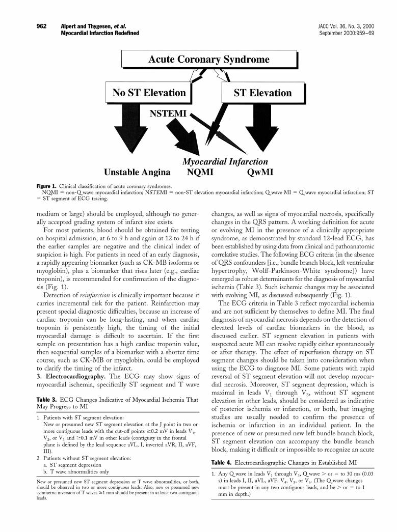
Myocardial infarction (MI) can be defined from a numberof different perspectives related to clinical, electrocardio-graphic (ECG), biochemical and pathologic characteristics.The term MI also has social and psychological implications,both as an indicator of a major health problem and as ameasure of disease prevalence in population statistics andoutcomes of clinical trials (Fig. 1).
In the distant past, a general consensus existed for theclinical entity designated as MI. In studies of diseaseprevalence by the World Health Organization (WHO), MIwas defined by a combination of two of three characteristics:typical symptoms (i.e., chest discomfort), enzyme rise and atypical ECG pattern involving the development of Q waves.However, current clinical practice, health care deliverysystems, as well as epidemiologic studies and clinical trials,all require a more precise definition of MI. Furthermore, theadvent of sensitive and specific serologic biomarkers and
*The recommendations set forth in this report are those of the conferenceparticipants and do not necessarily reflect the official position of the American Collegeof Cardiology. The full text of the document will be published simultaneously in theEuropean Heart Journal and the Journal of the American College of Cardiology. Thisdocument is available on the World Wide Web sites of the American College ofCardiology (www.acc.org) and the European Society of Cardiology (www.esc.org).Reprints of this document are available for $5.00 each by calling 800-253-4636 (U.S.only) or by writing the Resource Center, American College of Cardiology, 9111 OldGeorgetown Road, Bethesda, Maryland 20814.
**A list of contributors to this ESC/ACC Consensus Document is provided inAppendix B.
Journal of the American College of Cardiology Vol. 36, No. 3, 2000© 2000 by the American College of Cardiology and the European Society of Cardiology ISSN 0735-1097/00/$20.00Published by Elsevier Science Inc. PII S0735-1097(00)00804-4
precise imaging techniques necessitate reevaluation of es-tablished definitions of MI. The latter technologic advanceshave high sensitivity to detect very small infarcts that wouldnot have been considered an MI in an earlier era. Currenttechnology can identify patients with small areas of myo-cardial necrosis weighing ,1.0 g. Thus, if we accept theconcept that any amount of myocardial necrosis caused byischemia should be labeled as an infarct (as proposed by thisconsensus conference), then an individual who was formerlydiagnosed as having severe, stable or unstable angina pec-toris might be diagnosed today as having had a small MI.The resulting increase in the sensitivity of the definingcriteria for MI would mean more cases identified; incontrast, an increase in specificity would lessen the numberof false positive MIs. Such changes in definition might havea profound effect on the traditional monitoring of diseaserates and outcomes.
In response to the issues posed by an alteration in ourability to identify MI, the ESC and the ACC convened aconsensus conference during July 1999 to reexamine jointlythe definition of MI. The scientific and societal implicationsof a new definition for MI were examined from seven pointsof view: pathology, biochemistry, electrocardiography, im-aging, clinical trials, epidemiology and public policy. Itbecame apparent from the deliberations of the consensuscommittee that the term MI should not be used withoutfurther qualifications, whether in clinical practice, in thedescription of patient cohorts or in population studies. Suchqualifications should refer to the amount of myocardial cellloss (infarct size), to the circumstances leading to the infarct(spontaneous or in the setting of a coronary artery diagnosticor therapeutic procedure) and to the timing of the myocar-dial necrosis relative to the time of the observation (evolv-ing, healing or healed MI).
II. CLINICAL PRESENTATION
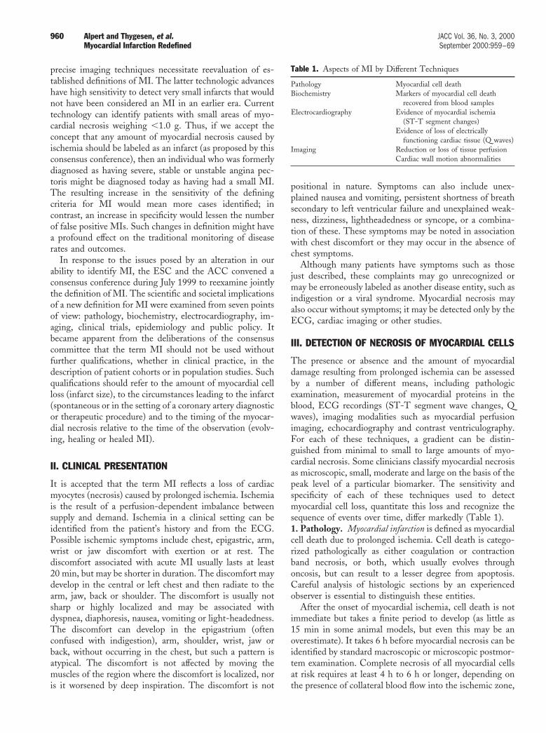
It is accepted that the term MI reflects a loss of cardiacmyocytes (necrosis) caused by prolonged ischemia. Ischemiais the result of a perfusion-dependent imbalance betweensupply and demand. Ischemia in a clinical setting can beidentified from the patient’s history and from the ECG.Possible ischemic symptoms include chest, epigastric, arm,wrist or jaw discomfort with exertion or at rest. Thediscomfort associated with acute MI usually lasts at least20 min, but may be shorter in duration. The discomfort maydevelop in the central or left chest and then radiate to thearm, jaw, back or shoulder. The discomfort is usually notsharp or highly localized and may be associated withdyspnea, diaphoresis, nausea, vomiting or light-headedness.The discomfort can develop in the epigastrium (oftenconfused with indigestion), arm, shoulder, wrist, jaw orback, without occurring in the chest, but such a pattern isatypical. The discomfort is not affected by moving themuscles of the region where the discomfort is localized, noris it worsened by deep inspiration. The discomfort is not
positional in nature. Symptoms can also include unex-plained nausea and vomiting, persistent shortness of breathsecondary to left ventricular failure and unexplained weak-ness, dizziness, lightheadedness or syncope, or a combina-tion of these. These symptoms may be noted in associationwith chest discomfort or they may occur in the absence ofchest symptoms.
Although many patients have symptoms such as thosejust described, these complaints may go unrecognized ormay be erroneously labeled as another disease entity, such asindigestion or a viral syndrome. Myocardial necrosis mayalso occur without symptoms; it may be detected only by theECG, cardiac imaging or other studies.
III. DETECTION OF NECROSIS OF MYOCARDIAL CELLS
The presence or absence and the amount of myocardialdamage resulting from prolonged ischemia can be assessedby a number of different means, including pathologicexamination, measurement of myocardial proteins in theblood, ECG recordings (ST-T segment wave changes, Qwaves), imaging modalities such as myocardial perfusionimaging, echocardiography and contrast ventriculography.For each of these techniques, a gradient can be distin-guished from minimal to small to large amounts of myo-cardial necrosis. Some clinicians classify myocardial necrosisas microscopic, small, moderate and large on the basis of thepeak level of a particular biomarker. The sensitivity andspecificity of each of these techniques used to detectmyocardial cell loss, quantitate this loss and recognize thesequence of events over time, differ markedly (Table 1).1. Pathology. Myocardial infarction is defined as myocardialcell death due to prolonged ischemia. Cell death is catego-rized pathologically as either coagulation or contractionband necrosis, or both, which usually evolves throughoncosis, but can result to a lesser degree from apoptosis.Careful analysis of histologic sections by an experiencedobserver is essential to distinguish these entities.
After the onset of myocardial ischemia, cell death is notimmediate but takes a finite period to develop (as little as15 min in some animal models, but even this may be anoverestimate). It takes 6 h before myocardial necrosis can beidentified by standard macroscopic or microscopic postmor-tem examination. Complete necrosis of all myocardial cellsat risk requires at least 4 h to 6 h or longer, depending onthe presence of collateral blood flow into the ischemic zone,
Table 1. Aspects of MI by Different Techniques
Pathology Myocardial cell deathBiochemistry Markers of myocardial cell death
recovered from blood samplesElectrocardiography Evidence of myocardial ischemia
(ST-T segment changes)Evidence of loss of electrically
functioning cardiac tissue (Q waves)Imaging Reduction or loss of tissue perfusion
Cardiac wall motion abnormalities
960 Alpert and Thygesen, et al. JACC Vol. 36, No. 3, 2000Myocardial Infarction Redefined September 2000:959–69

persistent or intermittent coronary artery occlusion and thesensitivity of the myocytes.
Infarcts are usually classified by size—microscopic (focalnecrosis), small (,10% of the left ventricle), medium (10%to 30% of the left ventricle) or large (.30% of the leftventricle)—as well as by location (anterior, lateral, inferior,posterior or septal or a combination of locations). Thepathologic identification of myocardial necrosis is madewithout reference to morphologic changes in the epicardialcoronary artery tree or to the clinical history.
The term MI in a pathologic context should be precededby the words “acute, healing or healed.” An acute orevolving infarction is characterized by the presence ofpolymorphonuclear leukocytes. If the interval between theonset of infarction and death is brief (e.g., 6 h), minimal orno polymorphonuclear leukocytes may be seen. The pres-ence of mononuclear cells and fibroblasts and the absence ofpolymorphonuclear leukocytes characterize a healing infarc-tion. A healed infarction is manifested as scar tissue withoutcellular infiltration. The entire process leading to a healedinfarction usually requires five to six weeks or more. Fur-thermore, reperfusion alters the gross and microscopicappearance of the necrotic zone by producing myocytes withcontraction bands and large quantities of extravasated eryth-rocytes.
Infarcts are classified temporally according to the patho-logic appearance as follows: acute (6 h to 7 days); healing (7to 28 days), healed (29 days or more). It should beemphasized that the clinical and ECG timing of an acuteischemic event may not be the same as the pathologictiming of an acute infarction. For example, the ECG maystill demonstrate evolving ST-T segment changes, andcardiac troponin may still be elevated (implying a recentinfarct) at a time when, pathologically, the infarct is in thehealing phase.2. Biochemical markers of myocardial necrosis. Myocar-dial necrosis results in and can be recognized by theappearance in the blood of different proteins released intothe circulation due to the damaged myocytes: myoglobin,cardiac troponins T and I, creatine kinase, lactate dehydro-genase, as well as many others (Fig. 2). Myocardial infarc-tion is diagnosed when blood levels of sensitive and specificbiomarkers, such as cardiac troponin and the MB fraction ofcreatine kinase (CK-MB), are increased in the clinicalsetting of acute ischemia. These biomarkers reflect myocar-dial damage but do not indicate its mechanism. Thus, anelevated value in the absence of clinical evidence of ischemia
should prompt a search for other causes of cardiac damage,such as myocarditis.
The most recently described and preferred biomarker formyocardial damage is cardiac troponin (I or T), which hasnearly absolute myocardial tissue specificity, as well as highsensitivity, thereby reflecting even microscopic zones ofmyocardial necrosis. An increased value for cardiac troponinshould be defined as a measurement exceeding the 99thpercentile of a reference control group. Reference valuesmust be determined in each laboratory by studies usingspecific assays with appropriate quality control, as reportedin peer-reviewed journals. Acceptable imprecision (coeffi-cient of variation) at the 99th percentile for each assayshould be defined as #10%. Each individual laboratoryshould confirm the range of reference values in their specificsetting. In addition, meticulous laboratory practice must bemaintained. Because cardiac troponin values may remainelevated for 7 to 10 days or longer after myocardial necrosis,care should be exercised in attribution of elevated cardiactroponin levels to very recent clinical events (Table 2).
If cardiac troponin assays are not available, the bestalternative is CK-MB (measured by mass assay). This is lesstissue-specific than cardiac troponin, but the data docu-menting its clinical specificity for irreversible injury are morerobust. As with cardiac troponin, an increased CK-MBvalue (i.e., above the decision limit for MI) is defined as onethat exceeds the 99th percentile of CK-MB values in areference control group. In most situations, elevated valuesfor biomarkers should be recorded from two successiveblood samples to diagnose MI.
Measurement of total CK is not recommended for theroutine diagnosis of acute MI, because of the wide tissuedistribution of this enzyme. Nevertheless, total CK has along history, and some physicians may opt to continue toemploy it for epidemiologic or scientific purposes. In such asetting, total CK should be combined with a more sensitivebiomarker, such as cardiac troponin or CK-MB, for moreaccurate clinical diagnosis of acute MI. The cut-off limitsfor total CK should be relatively higher than those forcardiac troponin or CK-MB (at least twice the upperreference limit for CK). Glutamic-oxaloacetic transaminase(ASAT [aspartate amino transferase]), lactate dehydroge-nase and lactate dehydrogenase isoenzymes should not beused to diagnose cardiac damage. Along with other clinicalfactors (e.g., residual left ventricular function), the degree ofbiomarker elevation is related to clinical risk. A classificationfor the extent of myocardial damage (microscopic, small,
Table 2. Biochemical Markers for Detecting Myocardial Necrosis
The following are biochemical indicators for detecting myocardial necrosis: 1) Maximal concentration of troponin T or I exceeding the decision limit(99th percentile of the values for a reference control group) on at least one occasion during the first 24 h after the index clinical event, 2) Maximalvalue of CK-MB (preferably CK-MB mass) exceeding the 99th percentile of the values for a reference control group on two successive samples, ormaximal value exceeding twice the upper limit of normal for the specific institution on one occasion during the first hours after the index clinicalevent. Values for CK-MB should rise and fall; values that remain elevated without change are almost never due to MI. In the absence of availabilityof a troponin or CK-MB assay, total CK (greater than two times the upper reference limit) or the B fraction of CK may be employed, but theselast two biomarkers are considerably less satisfactory than CK-MB.
961JACC Vol. 36, No. 3, 2000 Alpert and Thygesen, et al.September 2000:959–69 Myocardial Infarction Redefined
medium or large) should be employed, although no gener-ally accepted grading system of infarct size exists.
For most patients, blood should be obtained for testingon hospital admission, at 6 to 9 h and again at 12 to 24 h ifthe earlier samples are negative and the clinical index ofsuspicion is high. For patients in need of an early diagnosis,a rapidly appearing biomarker (such as CK-MB isoforms ormyoglobin), plus a biomarker that rises later (e.g., cardiactroponin), is recommended for confirmation of the diagno-sis (Fig. 1).
Detection of reinfarction is clinically important because itcarries incremental risk for the patient. Reinfarction maypresent special diagnostic difficulties, because an increase ofcardiac troponin can be long-lasting, and when cardiactroponin is persistently high, the timing of the initialmyocardial damage is difficult to ascertain. If the firstsample on presentation has a high cardiac troponin value,then sequential samples of a biomarker with a shorter timecourse, such as CK-MB or myoglobin, could be employedto clarify the timing of the infarct.3. Electrocardiography. The ECG may show signs ofmyocardial ischemia, specifically ST segment and T wave
changes, as well as signs of myocardial necrosis, specificallychanges in the QRS pattern. A working definition for acuteor evolving MI in the presence of a clinically appropriatesyndrome, as demonstrated by standard 12-lead ECG, hasbeen established by using data from clinical and pathoanatomiccorrelative studies. The following ECG criteria (in the absenceof QRS confounders [i.e., bundle branch block, left ventricularhypertrophy, Wolff-Parkinson-White syndrome]) haveemerged as robust determinants for the diagnosis of myocardialischemia (Table 3). Such ischemic changes may be associatedwith evolving MI, as discussed subsequently (Fig. 1).
The ECG criteria in Table 3 reflect myocardial ischemiaand are not sufficient by themselves to define MI. The finaldiagnosis of myocardial necrosis depends on the detection ofelevated levels of cardiac biomarkers in the blood, asdiscussed earlier. ST segment elevation in patients withsuspected acute MI can resolve rapidly either spontaneouslyor after therapy. The effect of reperfusion therapy on STsegment changes should be taken into consideration whenusing the ECG to diagnose MI. Some patients with rapidreversal of ST segment elevation will not develop myocar-dial necrosis. Moreover, ST segment depression, which ismaximal in leads V1 through V3, without ST segmentelevation in other leads, should be considered as indicativeof posterior ischemia or infarction, or both, but imagingstudies are usually needed to confirm the presence ofischemia or infarction in an individual patient. In thepresence of new or presumed new left bundle branch block,ST segment elevation can accompany the bundle branchblock, making it difficult or impossible to recognize an acute
Figure 1. Clinical classification of acute coronary syndromes.NQMI 5 non-Q wave myocardial infarction; NSTEMI 5 non-ST elevation myocardial infarction; Q wave MI 5 Q wave myocardial infarction; ST
5 ST segment of ECG tracing.
Table 3. ECG Changes Indicative of Myocardial Ischemia ThatMay Progress to MI
1. Patients with ST segment elevation:New or presumed new ST segment elevation at the J point in two ormore contiguous leads with the cut-off points $0.2 mV in leads V1,V2, or V3 and $0.1 mV in other leads (contiguity in the frontalplane is defined by the lead sequence aVL, I, inverted aVR, II, aVF,III).
2. Patients without ST segment elevation:a. ST segment depressionb. T wave abnormalities only
New or presumed new ST segment depression or T wave abnormalities, or both,should be observed in two or more contiguous leads. Also, new or presumed newsymmetric inversion of T waves $1 mm should be present in at least two contiguousleads.
Table 4. Electrocardiographic Changes in Established MI
1. Any Q wave in leads V1 through V3, Q wave . or 5 to 30 ms (0.03s) in leads I, II, aVL, aVF, V4, V5, or V6. (The Q wave changesmust be present in any two contiguous leads, and be . or 5 to 1mm in depth.)
962 Alpert and Thygesen, et al. JACC Vol. 36, No. 3, 2000Myocardial Infarction Redefined September 2000:959–69
infarction, and criteria indicative of acute MI need to bedefined by further research. Tall and peaked T waves(hyperacute T waves) have been noted during the very earlyphases of acute MI.
New or presumed new ST segment depression or T waveabnormalities, or both, should be observed in two or morecontiguous leads on two consecutive ECGs at least severalhours apart.
Myocardial necrosis or clinically established MI may bedefined from standard 12-lead ECG criteria in the absenceof QRS confounders (e.g., bundle branch block, left ven-tricular hypertrophy, Wolff-Parkinson-White syndrome) orimmediately after coronary artery bypass graft surgery,utilizing the QRS changes presented in Table 4. A singleECG that meets the Q wave criteria in Table 4 is indicativeof a previous MI. Q waves ,30 ms in duration associatedwith ST-T segment depression may represent infarction,but these findings require more research and confirmation.When three or more ECG recordings are obtained, then atleast two consecutive ECGs should demonstrate the abnor-mality in question. Criteria for Q wave depth require moreresearch, as do QRS criteria to establish the diagnosis ofposterior MI. Bundle branch block with additional Q wavesis included in these descriptions. Right bundle branch blockwill not interfere with the ability to diagnose Q waves; leftbundle branch block usually obscures Q waves; new Q wavesin the presence of left bundle branch block should beconsidered as pathologic.
Not all patients who develop myocardial necrosis exhibitECG changes. Thus, a normal ECG does not rule out thediagnosis of MI. Since new sensitive biochemical markersenable detection of myocardial necrosis too small to beassociated with QRS abnormalities, some patients will havetheir peak values in the subrange of any QRS changes. Suchpatients might be considered to have only a microinfarction,but these aspects need further clarification.4. Imaging. Imaging techniques have been used to assist in1) ruling out or confirming the presence of acute infarctionor ischemia in the Emergency Department; 2) identifyingnonischemic conditions causing chest pain; 3) definingshort- and long-term prognoses; and 4) identifying me-chanical complications of acute infarction. The rationale ofacute imaging using echocardiographic or nuclear tech-niques in patients suspected of having acute ischemia is thatischemia results in regional myocardial hypoperfusion, lead-ing to a cascade of events that can include myocardialdysfunction and ultimately cell death. Only conventionalmethods such as cross-sectional echocardiography, radionu-clide angiography and myocardial single-photon emissioncomputed tomographic (SPECT) perfusion imaging arediscussed in this document, and not those that are presentlybeing tested in clinical research studies.
One of the major advantages of echocardiography is thatit allows assessment of most nonischemic causes of acutechest pain, such as perimyocarditis, valvular heart disease
(aortic stenosis), pulmonary embolism and aortopathies(aortic dissection).
Radionuclide techniques enable the physician to assessperfusion at the time of patient presentation; this can beperformed with immediate tracer injection, because imageacquisition can be delayed for 60 to 90 min. Quantitativeanalysis is an advantage of this technique. The accuracy ofthe studies is high when interpreted by skilled observers.These studies also provide simultaneous information onmyocardial perfusion and function.
Biomarkers are more sensitive, more specific and lesscostly than imaging techniques for the diagnosis of myo-cardial necrosis. Injury involving .20% of myocardial wallthickness is required before a segmental wall motion abnor-mality can be detected by echocardiography. In general,.10 g of myocardial tissue must be injured before aradionuclide perfusion defect can be resolved. Neithertechnique can distinguish ischemia from infarction.
a) Acute ischemia and acute or evolving MI. By its ability todetect regional wall motion abnormalities within minutes ofan ischemic injury, two-dimensional echocardiography maybe useful in the diagnosis of acute MI. Both the localizationand extent of infarction can be determined. An echocardio-graphic or radionuclide image early after the onset ofsymptoms is of great help in the assessment of patients withsuspected acute MI and a nondiagnostic or noninterpretableECG. With acute imaging in such patients, a normalechocardiogram or a normal rest gated technetium-99mSPECT study is useful for excluding acute infarction,because of a 95% to 98% negative predictive value whenCK-MB is used as the gold standard. However, it isunknown whether these techniques have the same negativepredictive value in patients with elevated troponin and anormal CK-MB value.
A wall motion abnormality on echocardiographic orradionuclide imaging may be caused by acute MI or one ofa number of several myocardial ischemic conditions, includ-ing an old MI, acute ischemia, stunning or hibernation, ora combination. The positive predictive value of echocardi-ography is ;50% for the diagnosis of acute MI, because ofthe aforementioned conditions and other non–infarct-related etiologies of wall motion abnormalities (e.g., dilatedcardiomyopathy). The positive predictive value for gatedSPECT is also limited, because abnormal regional perfusionand/or an old MI, acute ischemia, stunning and/or hiber-nation may cause regional dysfunction. Attenuation artifactsand inexperienced interpreters may also lead to false positivescan interpretation.
b) Established MI. Echocardiography is useful after asudden event for analysis of residual left ventricular func-tion. Determination of left ventricular function has prog-nostic value. Left ventricular function can be evaluatedduring exercise or dobutamine stress; the results of suchtesting conveys information on myocardial viability. Thenumber of segments involved allows one to calculate a wallmotion score as a measure of residual left ventricular
963JACC Vol. 36, No. 3, 2000 Alpert and Thygesen, et al.September 2000:959–69 Myocardial Infarction Redefined
function, which has early and late prognostic value inpredicting complications and survival. Coexisting mitralvalve dysfunction, infarct expansion, mural thrombus andmechanical complications of infarction are easily identified.Echocardiography is the diagnostic procedure of choice foridentification of mechanical complications of MI.
Radionuclide techniques can also be used in the healingor healed phases of infarction for prognostication. In con-junction with exercise or vasodilator stress, measuring theextent of defect reversibility can identify the extent ofischemia. Detecting defects in more than one coronaryartery zone can identify multivessel disease. A variety ofprognostic findings can be identified (e.g., lung uptake ofthe tracer thallium-201, ischemic left ventricular cavitydilation, defect size that corresponds to infarct size). Finally,the extent of myocardial viability can be estimated byquantitative perfusion imaging with either thallium-201 ortechnetium-99m perfusion tracers.
IV. MI IN SPECIFIC CLINICAL SETTINGS
1. Percutaneous coronary artery intervention. An in-crease of cardiac biomarkers after coronary angioplasty orimplantation of coronary artery stents, or both, is indicativeof cell death. Because this necrosis occurs as a result ofmyocardial ischemia, it should be labeled as an MI accord-ing to the new criteria. Large infarcts in this setting may becaused by a complicated procedure and can usually berecognized clinically. In contrast, small or tiny infarcts aremore frequent and are probably the result of microembolifrom the atherosclerotic lesion that has been disruptedduring angioplasty or from the particulate thrombus at thesite of the culprit lesion.
In the setting of percutaneous coronary artery interven-tions, small infarcts may, and should, be detected by serialblood sampling and analysis before and after the procedure(6 to 8 h and 24 h, respectively). The peak level of themyocardial biomarkers may be pronounced and of relativelygreater magnitude because of the reperfusion associatedwith the procedure. Myocardial cell injury occurring afterangioplasty may be a one-time event, as compared with theoften repetitive nature of spontaneously occurring episodesof myocardial ischemia and necrosis. However, it is likelythat patients who develop a coronary embolism and smallinfarcts have atherosclerotic lesions that are apparentlyunstable, and hence represent a subgroup at risk for futureevents. Indeed, it has been convincingly demonstrated thatthe risk of subsequent ischemic heart disease events (deathor MI) is related to the extent of cardiac troponin orCK-MB increase, and the prognosis for these individuals isusually worse than that for patients who do not developthese small increases in biomarkers after interventionalprocedures. Accordingly, patients with elevated biomarkersafter an otherwise uncomplicated procedure may requireparticularly careful instructions to respond appropriately torecurrent symptoms.
2. Cardiac surgery. Myocardial damage in association withcardiac surgery can be caused by different mechanisms,including direct trauma by sewing needles; focal traumafrom surgical manipulation of the heart; global ischemiafrom inadequate perfusion, myocardial cell protection oranoxia; coronary artery or venous graft embolism; and othercomplications of the procedure. A portion of this damagemay be unavoidable. Moreover, no biomarker is capable ofdistinguishing damage due to an acute infarction from theusually small quantity of myocardial cell damage associatedwith the procedure itself. Nevertheless, the higher the valuefor the cardiac biomarker after the procedure, the greater theamount of damage to the myocardium, irrespective of themechanism of injury.
V. IMPLICATIONS OF DIFFERENT DEFINITIONS OF MI
The recent introduction of cardiac troponins T and I intoroutine daily clinical practice allows for highly accurate,sensitive and specific determination of myocardial injury. Inthe setting of myocardial ischemia, it is now possible todefine infarcts of minimal size as well as larger infarcts. It isnow clear that any amount of myocardial damage, asdetected by cardiac troponins, implies an impaired clinicaloutcome for the patient. This is apparently true for individ-uals with spontaneous events, as well as for patients whoundergo coronary artery interventions. A review of currentlyavailable data demonstrates no discernible threshold belowwhich an elevated value for cardiac troponin would bedeemed harmless. All elevated values are associated with aworsened prognosis. It should be emphasized that there is acontinuous relation between minimal myocardial damage,characterized by elevation of cardiac troponin withoutelevation of other cardiac biomarkers (e.g., CK-MB) andlarge infarcts, characterized by complications such as heartfailure or shock. Thus, any amount of myocardial necrosiscaused by ischemia should be labeled as MI. Additionaldescriptors are needed to describe the state of residual leftventricular function, the extent and severity of coronaryartery disease and the stability or instability of the patient’sclinical course.1. Epidemiology. Monitoring of cardiovascular disease in apopulation is of utmost importance, because it enables theinvestigator to analyze possible causal factors and to assessthe effect of various preventive measures, such as changes indiet or life-style, as well as the effect of medications. Theincidence of a new MI and the prevalence of establishedinfarcts represent important epidemiologic variables. Theapplication of the new, more sensitive diagnostic criteria forMI will cause the recorded incidence of MI to rise and thecase fatality rate to fall. Thus, a new definition of MI willconfuse efforts to follow trends in disease rates and out-comes that are now being used to monitor the impact ofpublic health measures and treatments. However, thiswould not be a valid reason to hold onto old definitions ofMI which no longer reflect current scientific thinking. In
964 Alpert and Thygesen, et al. JACC Vol. 36, No. 3, 2000Myocardial Infarction Redefined September 2000:959–69

fact, changes in definitions have already occurred, albeitunnoticed—for example, through substitution of newerbiomarkers (CK-MB, troponin) for older ones (ASAT,CK). Continued tracking of these trends will require meth-ods for adjusting the new criteria to the old; for example,specific surveillance centers will be needed to measure totalCK and CK-MB, together with the newer biomarkers.
Established definitions of MI (e.g., Minnesota code,WHO MONICA) should be retained by specific epidemi-ologic centers for comparison with previously collected data.At the same time, these centers should use the currentbiomarker-based definition of acute MI to compare earlierdata with subsequent data collected at research centersemploying more recent standards for defining acute MI.2. Clinical trials. Myocardial infarction can be used eitheras an entry criterion or as an end point in a clinical trial.Entry criteria in clinical investigations dealing with sus-pected acute or evolving MI or unstable angina reflect theinitial working diagnosis at the time the patient is enrolledin the trial. This will not necessarily correspond to the finaldiagnosis of MI, because spontaneous or therapeuticallyinduced changes may alter the likelihood of developingmyocardial necrosis. Usually, a combination of chest dis-comfort of a determined length of time and ECG abnor-malities (ST segment elevation or a new bundle branchblock) is required for the initial working diagnosis of MI.Trials of long-term management may require a definite orhospital discharge diagnosis of MI, usually on the basis ofECG Q waves and biochemical markers. Independent ofthe precise entry criteria chosen, the randomization processwill ensure balanced groups of patients for comparison ofdifferent treatment modalities. Modification of the defini-tion of MI may impact patient selection or the generaliz-ability of the trial outcome, or both.
In many trials of MI or unstable angina, as well as ofprimary and secondary prevention of coronary heart diseaseevents, MI is one of the trial end points, usually incombination with total mortality or cardiovascular mortal-ity. In recent trials, different definitions of MI as an endpoint have been employed, thereby hampering comparisonof trial results and meta-analyses.
Myocardial damage may occur in different clinical set-tings: spontaneous, during percutaneous intervention, dur-ing coronary artery bypass graft surgery or with trauma ormyocarditis. It has been uncertain whether a similar amountof damage in different settings has the same prognosticimplications. There have been different thresholds for iden-tifying an infarct in trials undertaken up to this time in theU.S.: CK-MB .2 times the upper limit of normal (ULN)for spontaneous MI; CK-MB .3 times the ULN withcoronary artery interventions; and CK-MB .5 to 10 timesthe ULN for bypass surgery. It is important that thebackground for such choices be subjected to research andverified or rejected from different databases. However, theJoint ESC/ACC Expert Committee reached the consensusopinion that, irrespective of the clinical circumstances (ex-
cept for coronary artery surgery or intended myocardiallesions such as radiofrequency ablation of arrhythmias orseptal ablation for hypertrophic cardiomyopathy), the samediscriminative value for each biochemical marker should beapplied, as they reflect the same amount of myocardial necrosis.
In clinical trials, as in clinical practice, measurement ofcardiac troponin T or I is preferred over measurement ofCK-MB, as well as total CK and other biomarkers, for thediagnosis of MI. Assessment of the quantity of myocardialdamage (infarct size) is also an important trial end point.The use of cardiac troponin will undoubtedly increase thenumber of events recorded in a particular trial because of itsincreased sensitivity for detecting MI. Ideally, data shouldbe presented so that trialists can translate the MI end pointchosen in one trial into the end point of another trial. Thus,measurements should be presented in a continuous manner(distribution) to allow independent judgment of the clinicalend points.
In the design of a trial, investigators should specify theexpected effect of the new treatment under investigation.Factors that must be considered include:
1) Reduction in the incidence of spontaneous myocardialdamage (events) in treated patients versus control sub-jects.
2) Reduction in the incidence of angioplasty- or bypasssurgery–induced myocardial damage.
3) Reduction in the amount of myocardial damage (infarctsize) under any circumstance. Analysis of the actualdistribution of infarct sizes observed (area under thecurve of a biomarker or peak values) is more appropriatethan analysis of the presence or absence of events only.
VI. IMPLICATIONS OF MI IN THE EVOLUTION OFCORONARY DISEASE IN AN INDIVIDUAL PATIENT
Until recently, MI was recognized as a major event, oftenfatal, and with major implications for survivors. This para-
Figure 2. Timing of release of various biomarkers following acute, isch-emic myocardial infarction.
Peak A, early release of myoglobin or CK-MB isoforms after AMI;peak B, cardiac troponin after AMI; peak C, CK-MB after AMI; peak D,cardiac troponin after unstable angina. Data are plotted on a relative scale,where 1.0 is set at the AMI cutoff concentration.
AMI 5 acute myocardial infarction; CAD 5 coronary artery disease;CK 5 creatine kinase.
Reproduced with permission, WU AH, et al. Clin Chem 1999;45:1104–1121.
965JACC Vol. 36, No. 3, 2000 Alpert and Thygesen, et al.September 2000:959–69 Myocardial Infarction Redefined

digm has changed as a result of better management strate-gies for patients with coronary artery disease, as well asbetter methods for detecting or excluding myocardial ne-crosis. The introduction of techniques for measuring cardiactroponin allows for very sensitive and very specific detectionof minimal quantities of myocardial necrosis. This newtechnology serves as the cornerstone of the new definition ofMI outlined in this document. It is appreciated that thisnew definition will attach the label of MI to more patients.Similarly, it will identify more infarcts and more episodes ofreinfarction in patients with progressive coronary arterydisease. This change in the definition of MI seems reason-able, because it has been definitively shown that any amountof myocardial damage, as detected by cardiac troponins,implies a worsened long-term outcome for the patient. Thisappears to be true both for spontaneous events and forevents associated with coronary procedures. Currently avail-able analyses demonstrate no threshold below which eleva-tions of troponins are harmless and without negative impli-cations for prognosis. Thus, any other definition of MIwould involve an arbitrary setting of limits for an abnormaltroponin and would be open to criticism and considerabledebate. It should be emphasized that there is continuityfrom “minimal myocardial damage,” characterized by eleva-tion of cardiac troponin without apparent elevation of otherbiomarkers (also termed “infarctlet” or “necrosette”), to theclassic “large myocardial infarction,” often complicated byheart failure, shock or life-threatening arrhythmia. In ap-plying the proposed new diagnostic criteria to clinicalpractice, patients should not be labeled primarily as “myo-cardial infarction” but rather as patients with coronary arterydisease with MI. In addition, it is essential that otherdescriptors of the patient’s cardiac status be included, suchas current left ventricular function, the extent and severity ofcoronary artery lesions and an estimate of the evolution ofthe disease over recent months (i.e., stable or unstable). Thecrucial elements in this descriptive process can be obtainedfrom invasive diagnostic studies, but may also be reliablyestimated from a number of noninvasive studies.
Patients who undergo coronary artery revascularization(coronary angioplasty or bypass surgery) are at risk formyocardial damage, or rather MI according to the newdefinition. These risks have always been present in thesetting of coronary artery interventions, but they have beenhighlighted by the new, more sensitive biomarkers. Detec-tion of a very small myocardial infarct in this setting augursa worse prognosis for the patient, rather than if biomarkershad been normal. However, it should be appreciated thatlong-term patient outcome and prognosis may be improvedsignificantly by the revascularization procedure. For exam-ple, a patient with unstable angina and severe left anteriordescending coronary artery stenosis will benefit from coro-nary artery stenting, despite a small elevation in bloodtroponin levels. The benefit far outweighs the negativeimpact of the small, procedure-related infarct. It goeswithout saying that every measure should be taken to
prevent even such small infarcts in the setting of coronaryartery interventional procedures.
VII. SOCIAL AND PUBLIC POLICYIMPLICATIONS OF REDEFINING MI
Modification of the definition of a specific diagnosis such asMI has a number of implications for individual citizens aswell as for society. The process of assigning a specificdiagnosis to a patient should be associated with a specificvalue to the patient. The resources spent on recording andtracking a particular diagnosis must also have a specific valueto society to justify the effort. A tentative or final diagnosisis the basis for advice about further diagnostic testing,treatment, life-style changes and prognosis for the patient.The aggregate of patients with a particular diagnosis is thebasis for health care planning and policy and resourceallocation.
One of the goals of good clinical practice is to reach adefinitive and specific diagnosis that is supported by currentscientific knowledge. The approach to the definition of MIoutlined in this document meets this goal. In general, theconceptual meaning of the term “acute MI” has notchanged, although new sensitive diagnostic methods havebeen developed to diagnose this entity. Thus, the currentdiagnosis of acute MI is a clinical diagnosis based on patientsymptoms, ECG changes and highly sensitive biochemicalmarkers, as well as information gleaned from various imag-ing techniques. However, it is important to characterize theextent of the patient’s myocardial injury and residual leftventricular function, as well as the severity of coronary arterydisease, rather than merely making a diagnosis of MI.
Many patients with coronary artery thrombosis leading toMI die suddenly. Difficulties in the definitions of suddenand out-of-hospital death make attribution of the cause ofdeath variable among physicians, regions and countries. Forexample, out-of-hospital death is generally ascribed toischemic heart disease in the U.S. but to stroke in Japan.These arbitrary and cultural criteria need reexamination.
It is important that any revised criteria for the definitionof MI involve comparability of this definition over time sothat adequate trend data can be obtained. Furthermore, it isessential to ensure widespread availability and standardapplication of measures generating these criteria to ensurecomparability of data from various geographic regions.Shifts in criteria resulting in substantial increases or de-creases in case identification will have significant healthresource and cost implications. Moreover, an increase insensitivity of the criteria for acute MI might entail negativeconsequences for some patients who are not currentlylabeled as having had an MI. In contrast, increasingdiagnostic sensitivity for MI can have a positive impact onsociety:
● Increasing the sensitivity of diagnostic criteria for MI willresult in more cases identified, thereby allowing appro-
966 Alpert and Thygesen, et al. JACC Vol. 36, No. 3, 2000Myocardial Infarction Redefined September 2000:959–69
priate secondary prevention and hopefully reduced healthcare costs in the future,
● Increasing the specificity of diagnostic criteria for MI willresult in elimination of noncases, thereby leading toreduced costs for hospital stays and secondary prevention.
Finally, it should be appreciated that the proposed mod-ification of the definition of MI may be associated withconsequences for the patient with respect to psychologicalstatus, life insurance, professional career, as well as drivingand pilot licenses. The diagnosis is associated with societalimplications as well: diagnosis-related grouping (DRG),hospital reimbursement, mortality statistics, sick leave anddisability applications and clinical guideline preparation willall be affected.
To meet this challenge, physicians must be adequatelyinformed of the changing diagnostic criteria. Educationalmaterials will need to be created, and treatment guidelinesmust be appropriately adapted. Professional societies, par-ticularly the ACC, the American Heart Association and theESC, should take steps to facilitate the rapid disseminationof the revised definition to physicians, other health careprofessionals, administrators and the general public.
SUMMARY
Definition of MI. Criteria for acute, evolving or recent MI.Either one of the following criteria satisfies the diagnosis foran acute, evolving or recent MI:
1) Typical rise and gradual fall (troponin) or more rapid riseand fall (CK-MB) of biochemical markers of myocardialnecrosis with at least one of the following:
a) ischemic symptoms;b) development of pathologic Q waves on the ECG;c) ECG changes indicative of ischemia (ST segment
elevation or depression); ord) coronary artery intervention (e.g., coronary angio-
plasty).2) Pathologic findings of an acute MI.
Criteria for established MI.
Any one of the following criteria satisfies the diagnosisfor established MI:
1) Development of new pathologic Q waves on serialECGs. The patient may or may not remember previoussymptoms. Biochemical markers of myocardial necrosismay have normalized, depending on the length of timethat has passed since the infarct developed.
2) Pathologic findings of a healed or healing MI.
BIBLIOGRAPHY
Pathology
1. Bardales RH, Hailey LS, Xie SS, Schaefer RF, Hsu S-U. In situapoptosis assay for the detection of early acute myocardial infarction.Am J Pathol 1996;149:821–9.
2. Buja LM. Modulation of the myocardial response to injury. Lab Invest1998;78:1345–73.
3. Fishbein MC, Maclean D, Maroko PR. The histopathologic evolutionof myocardial infarction. Chest 1978;73:743–9.
4. Jennings RB, Reimer KA. Factors involved in salvaging ischemicmyocardium: effect of reperfusion of arterial blood. Circulation 1983;68Suppl I:I-25–36.
5. Jennings RB, Schaper J, Hill ML, Steenberger CJ, Reimer KA. Effectof reperfusion late in the phase of reversible ischemic injury: changes incell volume, electrolytes, metabolites, and ultrastructure. Circ Res1985;56:262–78.
6. Kajstura J, Cheng W, Reiss K, et al. Apoptotic and necrotic myocytecell deaths are independent contributing variables of infarct size in rats.Lab Invest 1996;74:86–107.
7. Mallory GK, White PD, Salcedo-Salga J. The speed of healing ofmyocardial infarction: a study of the pathologic anatomy in 72 cases.Am Heart J 1939;18:647–71.
8. Saraste A, Pulkki K, Kallajoki M, Hendriksen K, Parvinen M, Voipio-Pulkki L-M. Apoptosis in human acute myocardial infarction. Circu-lation 1997;95:320–3.
Biochemistry
1. Alexander JH, Sparapani RA, Mahaffey KW, et al., for the PURSUITInvestigators. Association between minor elevations of creatine-kinase-MB and mortality in patients with acute coronary syndromeswithout ST-segment elevation. JAMA 2000;283:347–53.
2. Antman EM, Grudzien C, Mitchell RN, Sacks DB. Detection ofunsuspected myocardial necrosis by rapid bedside assay for cardiactroponin T. Am Heart J 1997;133:596–8.
3. Antman EM, Tanasijevic MJ, Thompson B, et al. Cardiac-specifictroponin I levels to predict the risk of mortality in patients with acutecoronary syndromes. N Engl J Med 1996;335:1342–9.
4. Apple FS, Falahati A, Paulson PR, Miller E, Sharkey SW. Improveddetection of minor ischemic myocardial injury with measurement ofserum cardiac troponin I. Clin Chem 1997;43:2047–51.
5. Apple FS. Clinical and analytical standardization issues confrontingcardiac troponin I. Clin Chem 1999;45:18–20.
6. Apple FS. Tissue specificity of cardiac troponin I, cardiac troponin T,and creatine kinase MB. Clin Chim Acta 1999;284:151–8.
7. Califf RM, Abdelmeguid AE, Kuntz RE, et al. Myonecrosis afterrevascularization procedures. J Am Coll Cardiol 1998;31:241–51.
8. Collinson PO. Troponin T or troponin I or CK-MB (or none?). EurHeart J 1998;19 Suppl N:N16–24.
9. Gerhardt W, Nordin G, Ljungdahl L. Can troponin T replaceCK-MB mass as “gold standard” for acute myocardial infarction(“AMI”)? Scand J Clin Lab Invest 1999;59 Suppl 230:83–9.
10. Hamm CW, Goldmann BU, Heeschen C, Kreymann G, Berger J,Meinertz T. Emergency room triage of patients with acute chest painby means of rapid testing for cardiac troponin T or troponin I. N EnglJ Med 1997;337:1648–53.
11. Hamm CW, Heeschen C, Goldman B, et al., for the Capture StudyInvestigators. Benefit of abciximab in patients with refractory unstableangina in relation to serum troponin T levels. N Engl J Med1999;340:1623–9.
12. Hamm CW, Ravkilde J, Gerhardt W, et al. The prognostic value ofserum troponin T in unstable angina. N Engl J Med 1992;327:146–50.
13. Harrington RA, Lincoff AM, Califf RM, et al., for the CAVEATInvestigators. Characteristics and consequences of myocardial infarc-tion following percutaneous coronary intervention: insights from theCoronary Angioplasty Versus Excisional Atherectomomy Trial (CA-VEAT). J Am Coll Cardiol 1995;25:1693–9.
14. Ishikawa Y, Saffitz JE, Mealman TL, Grace AM, Roberts R. Revers-ible myocardial ischemic injury is not associated with increasedcreatine kinase activity in plasma. Clin Chem 1997;43:467–75.
15. Lindahl B, Venge P, Wallentin L, for the FRISC Study Group.Relation between troponin T and the risk of subsequent cardiac eventsin unstable coronary artery disease. Circulation 1996;93:1651–7.
16. Ohman EM, Armstrong PW, Christenson RH, et al., for theGUSTO-IIa Investigators. Cardiac troponin T levels for risk stratifi-cation in acute myocardial ischemia. N Engl J Med 1996;335:1333–41.
17. Ottani F, Galvani M, Ferrini D, et al. Direct comparison of early
967JACC Vol. 36, No. 3, 2000 Alpert and Thygesen, et al.September 2000:959–69 Myocardial Infarction Redefined
elevations of cardiac troponin T and I in patients with clinical unstableangina. Am Heart J 1999;137:284–91.
18. Ravkilde J, Horder M, Gerhardt W, et al. Diagnostic performance andprognostic values of serum troponin T in suspected acute myocardialinfarction. Scand J Clin Lab Invest 1993;53:677–85.
19. Tardiff BE, Califf RM, Tcheng JE, et al., for the IMPACT-IIInvestigators. Clinical outcomes after detection of elevated enzymes inpatients undergoing percutaneous intervention: IMPACT-II trial(Integrilin [eptifibatide] to Minimize Platelet Aggregation and Cor-onary Thrombosis-II). J Am Coll Cardiol 1999;33:88–96.
20. Wu AHB, Apple FS, Gibler WB, Jesse RL, Warshaw MM, Valdes RJr. National Academy of Clinical Biochemistry Standards of Labora-tory Practice: recommendations for use of cardiac markers in coronaryartery disease. Clin Chem 1999;45:1104–21.
21. Zimmerman J, Fromm R, Meyer D, et al. Diagnostic marker coop-erative study for the diagnosis of myocardial infarction. Circulation1999;99:1671–7.
Electrocardiography
1. Anderson WD, Wagner NB, Lee KL, et al. Evaluation of a QRSscoring system for estimating myocardial infarct size. VI: Identificationof screening criteria for non-acute myocardial infarcts. Am J Cardiol1988;61:729–33.
2. Chou T, Knilans T. Myocardial infarction: myocardial injury, andmyocardial ischemia. In: Chou TC, Knilans TK, editors. Electrocardi-ography in Clinical Practice: Adult and Pediatric, 4th ed. Philadelphia:W.B. Saunders, 1996:121.
3. Crow RS, Prineas RJ, Jacobs DR, Blackburn H. A new epidemiologicclassification system for interim myocardial infarction from serialelectrocardiographic changes. Am J Cardiol 1989;64:454–61.
4. Menown IBA, MacKenzie G, Adgey AA. Optimizing the initial12-lead electrocardiographic diagnosis of acute myocardial infarction.Eur Heart J 2000;21:275–83.
5. Pahlm US, Chaitman BR, Rautaharju PM, Selvester RH, Wagner GS.Comparison of the various electrocardiographic scoring codes forestimating anatomically documented sizes of single and multiple infarctsof the left ventricle. Am J Cardiol 1998;81:809–15.
6. Prineas RJ, Crow RJ, Blackburn H. The Minnesota Code Manual ofElectrocardiographic Findings: Standards and Procedures for Measure-ment and Classification. Wright J, editor. Boston: John Wright, PSGInc, 1982:1–229.
7. Rautaharju PM, Park LP, Chaitman BR, Rautaharju F, Zhang ZM.The Novacode criteria for classification of ECG abnormalities and theirclinically significant progression and regression. J Electrocardiol 1998;31:157–87.
8. Selvester RH, Wagner GS, Hindman NB. The Selvester QRS scoringsystem for estimating myocardial infarct size: the development andapplication of the system. Arch Intern Med 1985;145:1877–81.
Imaging
1. Beller GA. Assessment of myocardial viability. Curr Opin Cardiol1997;12:459–67.
2. Buda AJ. The role of echocardiography in the evaluation of mechanicalcomplications of acute myocardial infarction Circulation 1991;84Suppl I:I-109–121.
3. Duca MD, Giri S, Wu AHB, et al. Comparison of acute restmyocardial perfusion imaging and serum markers of myocardial injuryin patients with chest pain syndromes. J Nucl Cardiol 1999;6:570–6.
4. Gibler WB, Runyon JP, Levy RC, et al. A rapid diagnostic andtreatment center for patients with chest pain in the emergencydepartment. Ann Emerg Med 1995;25:1–8.
5. Lieberman AN, Weiss JL, Jugdutt BI, et al. Two-dimensionalechocardiography and infarct size: relationship of regional wall motionand thickening to the extent of myocardial function in the dog.Circulation 1981;63:739–46.
6. Machecourt J, Longere P, Fagret D, et al. Prognostic value ofthallium-201 single-photon emission computed tomographic myocar-dial perfusion imaging according to extent of myocardial defect. J AmColl Cardiol 1994;23:1096–106.
7. Manoury C, Chen CC, Chua KB, Thompson CJ. Quantification ofleft ventricular function with thallium-201 and technetium-99m ses-tamibi myocardial gated SPECT. J Nucl Med 1997;38:958–61.
8. Peels, C, Visser CA, Funkekupper AJ, Visser FC, Roos JP. Usefulness
of two-dimensional echocardiography for immediate detection ofmyocardial ischemia in the emergency room. Am J Cardiol 1990;65:687–91.
9. Sabia P, Abbott RD, Afrookteh A, Keller MW, Touchtone DA, KaulS. Importance to two-dimensional echocardiographic assessment ofleft ventricular systolic function in patients presenting to the emer-gency room with cardiac-related symptoms. Circulation 1991;84:1615–24.
10. Saeian K, Rhyne TL, Sagar KB. Ultrasonic tissue characterization fordiagnosis of acute myocardial infarction in the coronary care unit. Am JCardiol 1994;74:1211–5.
11. Tatum JL, Jesse RL, Kontos MC, et al. Comprehensive strategy forthe evaluation and triage of the chest pain patients. Ann Emerg Med1997;29:116–25.
Epidemiology
1. Burke GL, Edlavitch SA, Crow RS. The effects of diagnostic criteria ontrends in coronary heart disease morbidity: the Minnesota Heart Survey.J Clin Epidemiol 1989;42:17–24.
2. Gillum RF, Fortmann SP, Prineas RJ, Kottke TE. Internationaldiagnostic criteria for acute myocardial infarction and acute stroke. AmHeart J 1984;108:150–8.
3. McGovern PG, Pankow JS, Shahar E, for the Minnesota Heart SurveyInvestigators. Recent trends in acute coronary heart disease—mortality,morbidity, medical care and risk factors. N Engl J Med 1966;334:884–90.
4. Rose GA, Blackburn H. Cardiovascular survey methods. World HeartOrganization, Geneva, Switzerland, 1968.
5. Tunstall-Pedoe H, Kuulasmaa K, Amouyel P, Arveiler D, RajakangasAM, Pajak A. Myocardial infarction and coronary deaths in the WorldHealth Organization MONICA Project: registration procedures, eventrates, and case-fatality rates in 38 populations from 21 countries in fourcontinents. Circulation 1994;90:583–612.
6. Weinstein BJ, Epstein FH, and the Working Subcommittee on Criteriaand Methods, Committee on Epidemiological Studies, American HeartAssociation. Comparability of criteria and methods in the epidemiologyof cardiovascular disease: report of a survey. Circulation 1964;30:643.
7. World Health Organization, Working Group on the Establishment ofIschemic Heart Disease Registers. Report of the Fifth Working Group,Copenhagen. In: Report no. Eur 8201 (5). World Health Organization,Geneva, 1971.
8. Report of the Joint International Society and Federation of Cardiology/World Health Organization Task Force on Standardization of ClinicalNomenclature. Nomenclature and criteria for diagnosis of ischemicheart disease. Circulation 1979;59:607–9.
9. Porela P, Helenius H, Pulkki K, Voipio-Pulkki L-M. Epidemiologicalclassification of acute myocardial infarction: time for a change? EurHeart J 1999;20:1459–64.
APPENDIX A
List of Sponsors of the ESC/ACC ConsensusConference at the European Heart House, July 4 – 6, 1999
Astra Hassle AB, Molndal, SwedenBoehringer Mannheim, Global Business DevelopmentCardiac Markers, Mannheim, GermanyBristol-Myers Squibb Company, Cardiovascular BusinessUnit, Princeton, New JerseyEli Lilly and Company, Gems Services S.A., Bruxelles,BelgiumHoechst Marion Roussel Inc., Global Marketing, Bridge-water, New JerseyMerck & Co. Inc., Hypertension/Heart Failure and Cho-lesterol Reducers Marketing Group, Whitehouse Station,New JerseyPfizer Pharmaceuticals Group, New York, New YorkPharmacia Upjohn, Copenhagen, Denmark
968 Alpert and Thygesen, et al. JACC Vol. 36, No. 3, 2000Myocardial Infarction Redefined September 2000:959–69
Rhone-Poulenc Rorer S.A., Global Marketing, AntonyCedex, FranceSanofi Pharma, Thrombosis Business Unit, Paris, FranceSchering-Plough Global Marketing, Kenilworth, NewJerseyZeneca Pharmaceuticals, Macclesfield, Great Britain
APPENDIX B
List of Contributors to theESC/ACC Consensus Document
Joseph S. Alpert, Tucson, Arizona; Elliott Antman, Boston,Massachusetts; Fred Apple, Minneapolis, Minnesota; PaulW. Armstrong, Edmonton, Canada; Jean-Pierre Bassand,Besancon, France; Antoni Bayes de Luna, Barcelona, Spain;George Beller, Charlottesville, Virginia; Gunter Breithardt,Munster, Germany; Bernard R. Chaitman, Saint Louis,Missouri; Peter Clemmensen, Copenhagen, Denmark; Er-ling Falk, Aarhus, Denmark; Michael C. Fishbein, LosAngeles, California; Marcello Galvani, Forli, Italy; ArthurGarson, Jr., Houston, Texas; Cindy Grines, Royal Oaks,Michigan; Christian Hamm, Bad Nauheim, Germany;Ursula Hoppe, Berlin, Germany; Alan Jaffe, Rochester,Minnesota; Hugo Katus, Lubeck, Germany; John Kjekshus,Oslo, Norway; Werner Klein, Graz, Austria; PeterKlootwijk, Rotterdam, The Netherlands; Claude Lenfant,Bethesda, Maryland; Daniel Levy, Framingham, Massa-chusetts; Robert I. Levy, Philadelphia, Pennsylvania; Rus-sell Luepker, Minneapolis, Minnesota; Frank Marcus, Tuc-
son, Arizona; Ulf Naslund, Umeaa, Sweden; MagnusOhman, Durham, North Carolina; Olle Pahlm, Lund,Sweden; Philip Poole-Wilson, London, Great Britain;Richard Popp, Palo Alto, California; Kalevi Pyorala, Kuo-pio, Finland; Jan Ravkilde, Aalborg, Denmark; Nina Rehn-quist, Stockholm, Sweden; William Roberts, Dallas, Texas;Robert Roberts, Houston, Texas; Jos Roelandt, Rotterdam,The Netherlands; Lars Ryden, Stockholm, Sweden; SusanaSans, Barcelona, Spain; Maarten L. Simoons, Rotterdam,The Netherlands; Kristian Thygesen, Aarhus, Denmark;Hugh Tunstall-Pedoe, Dundee, Great Britain; RichardUnderwood, London, Great Britain; Barry F. Uretsky,Galveston, Texas; Frans Van de Werf, Leuven, Belgium;Lisa-Maria Voipio-Pulkki, Turku, Finland; Galen Wagner,Durham, North Carolina; Lars Wallentin, Uppsala, Swe-den; William Wijns, Aalst, Belgium; and David Wood,London, Great Britain.
APPENDIX C
Reviewers of the Document
Elliott Antman, MDJean-Pierre Bassand, MDWerner Klein, MDMagnus Ohman, MDJose Luis Lopez Sendon, MDLars Ryden, MDMaarten Simoons, MDMichal Tendera, MD
969JACC Vol. 36, No. 3, 2000 Alpert and Thygesen, et al.September 2000:959–69 Myocardial Infarction Redefined
Copyright © 2022 FDOKUMEN