Toll-interacting protein contributes to mortality following myocardial infarction through promoting...
28
1 This article is protected by copyright. All rights reserved. Toll-interacting protein contributes to mortality following myocardial infarction through promoting inflammation and apoptosis 1 Nian Wan 1,2 ; Xiaoxiong Liu 1,2 ; Xiao-Jing Zhang 3 ; Yichao Zhao 4 ; Gangying Hu 1,2 ; Fengwei Wan 5 ; Rui Zhang 2 ; Xueyong Zhu 2 ; Hao Xia 1,2 ; Hongliang Li 1,2 1 Department of Cardiology, Renmin Hospital of Wuhan University, Wuhan 430060, China; 2 Cardiovascular Research Institute of Wuhan University, Wuhan 430060, China; 3 State Key Laboratory of Quality Research in Chinese Medicine, Institute of Chinese Medical Sciences, University of Macau, Macao 999078, China; 4 Department of Cardiology, Shanghai Renji Hospital, School of Medicine, Shanghai J iaotong University, Shanghai 200127, China; 5 Department of Emergency, The Second Artillery General Hospital of Chinese People's Liberation Army Qinghe Clinic, Beijing, China. N. Wan, X. Liu and X.-J. Zhang contributed equally to this study. Running Title: Tollip promotes MI injury Correspondence to Hao Xia, MD Professor and Director Department of Cardiology Renmin Hospital of Wuhan University , Jiefang Road 238, Wuhan 430060, PR China Tel/Fax: 86-27-88041911(2148) E-mail: [email protected] Hongliang Li, MD, PhD Professor and Director Department of Cardiology, Renmin Hospital of Wuhan University; Cardiovascular Research Institute, Wuhan University, Jiefang Road 238, Wuhan 430060, PR China Tel/Fax: 86-27-88076990 E-mail: [email protected] Author Contributions Nian Wan, Xiaoxiong Liu and Xiao-Jing Zhang performed experiments and data analysis, and wrote the manuscript; Yichao Zhao, Gangying Hu and Fengwei Wan performed data analysis and wrote the manuscript; Rui Zhang and Xueyong Zhu This article has been accepted for publication and undergone full peer review but has not been through the copyediting, typesetting, pagination and proofreading process, which may lead to differences between this version and the Version of Record. Please cite this article as doi: 10.1111/bph.13130 Accepted Article
Transcript of Toll-interacting protein contributes to mortality following myocardial infarction through promoting...

1 This article is protected by copyright. All rights reserved.
Toll-interacting protein contributes to mortality following myocardial infarction
through promoting inflammation and apoptosis1
Nian Wan1,2
; Xiaoxiong Liu1,2
; Xiao-Jing Zhang3; Yichao Zhao
4; Gangying Hu
1,2;
Fengwei Wan5; Rui Zhang
2; Xueyong Zhu
2; Hao Xia
1,2; Hongliang Li
1,2
1Department of Cardiology, Renmin Hospital of Wuhan University, Wuhan 430060,
China;
2Cardiovascular Research Institute of Wuhan University, Wuhan 430060, China;
3State Key Laboratory of Quality Research in Chinese Medicine, Institute of Chinese
Medical Sciences, University of Macau, Macao 999078, China; 4Department of Cardiology, Shanghai Renji Hospital, School of Medicine, Shanghai J
iaotong University, Shanghai 200127, China; 5Department of Emergency, The Second Artillery General Hospital of Chinese
People's Liberation Army Qinghe Clinic, Beijing, China.
N. Wan, X. Liu and X.-J. Zhang contributed equally to this study.
Running Title: Tollip promotes MI injury
Correspondence to
Hao Xia, MD
Professor and Director
Department of Cardiology
Renmin Hospital of Wuhan University ,
Jiefang Road 238, Wuhan 430060, PR China
Tel/Fax: 86-27-88041911(2148)
E-mail: [email protected]
Hongliang Li, MD, PhD
Professor and Director
Department of Cardiology, Renmin Hospital of Wuhan University;
Cardiovascular Research Institute, Wuhan University,
Jiefang Road 238, Wuhan 430060, PR China
Tel/Fax: 86-27-88076990
E-mail: [email protected]
Author Contributions
Nian Wan, Xiaoxiong Liu and Xiao-Jing Zhang performed experiments and data
analysis, and wrote the manuscript; Yichao Zhao, Gangying Hu and Fengwei Wan
performed data analysis and wrote the manuscript; Rui Zhang and Xueyong Zhu
This article has been accepted for publication and undergone full peer review but has not been through
the copyediting, typesetting, pagination and proofreading process, which may lead to differences
between this version and the Version of Record. Please cite this article as doi: 10.1111/bph.13130
Acc
epte
d A
rticl
e

2 This article is protected by copyright. All rights reserved.
performed experiments; Hao Xia and Hongliang Li overall designed the research,
analyzed data and wrote the manuscript.
A
ccep
ted
Arti
cle

3 This article is protected by copyright. All rights reserved.
Abstract
Background and purpose
Toll-interacting protein (Tollip) is an endogenous inhibitor of Toll-like receptors
(TLRs), a superfamily that play pivotal roles in various pathological conditions,
including myocardial infarction (MI); however, the role of Tollip in MI remains
unknown.
Experimental approach
MI models was established in Tollip knockout (KO) mice, mice with cardiac-specific
overexpression of human Tollip gene, as well as their Tollip+/+
and non-transgenic
(NTG) controls, respectively. The effects of Tollip on MI were evaluated by mortality,
infract size and cardiac function. In vitro hypoxia-induced cardiomyocyte damage
was also performed to confirm the function of Tollip on heart damage.
Key results
Tollip expression was dramatically upregulated in human ischemic hearts and
infarcted mice hearts. Tollip KO significantly decreased MI-induced mortality, infarct
size, and sustained cardiac function compared with Tollip+/+
controls. The ischemic
hearts from Tollip-KO mice exhibited decreased inflammatory cell infiltration and
reduced NF-κB activation. Tollip depletion also alleviated myocardial apoptosis
through down-regulating pro-apoptotic levels and upregulating anti-apoptotic protein
expressions in infarct border zone. Conversely, mice with cardiac-specific Tollip
overexpression displayed dramatically exacerbated MI outcomes. The aggravated MI
injury by Tollip in vivo was confirmed with in vitro assays. Inhibiting Akt signalling
is intimately related to the detrimental effects of Tollip on MI injury. Activation of
Akt could largely reverse the deleterious function of Tollip on MI-induced
cardiomyocytes death.
Conclusions and implications
Tollip promotes inflammatory and apoptotic responses after MI, leading to increased
mortality and aggravated cardiac dysfunction. These findings suggest that Tollip may
serve as a novel therapeutic target for the treatment of MI.
Abbreviations
Tollip Toll-interacting protein; TLRs Toll-like receptors; MI myocardial infarction;
KO knockout; NTG non-transgenic; IL-1 interleukin-1; TG transgenic; IHD ischemic
heart disease; LAD left anterior descending; LVEDD left ventricular end-diastolic
diameter; LVESD left ventricular end-systolic diameter; LVEF left ventricular
ejection fraction; FS fractional shortening; HE hematoxylin and eosin; NRCMs
neonatal rat cardiomyocytes; FCS fetal calf serum; LDH lactate dehydrogenase;
CCK8 Cell Counting Kit-8; SDS-PAGE sodium dodecyl sulfate-polyacrylamide gel
electrophoresis; ANOVA analysis of variance; PI propidium iodide; LPS
lipopolysaccharide; SR-A class A scavenger receptor.
Acc
epte
d A
rticl
e

4 This article is protected by copyright. All rights reserved.
Introduction
Myocardial infarction (MI) caused by coronary artery blockade is a major cause of
death worldwide (Rafiq et al., 2014). The continuous inflammatory response and
necrosis of ischemic tissue are two of the most marked characteristics that could
mutually enhance during process of MI-induced heart damage and eventually lead to
heart failure (Jones et al., 2014). After MI, inflammatory response could be triggered
immediately after MI. Although the infiltration of inflammatory cells into the
myocardium could scavenge necrotic myocytes and ECM debris to promote the
healing process (Frangogiannis, 2012), sustained inflammatory and immune infiltrate
is directly linked to myocardial apoptosis and impairs cardiac function (Kin et al.,
2006; Schofield et al., 2013). MI-induced myocardial apoptosis persists months
beyond the acute phase after MI (Baldi et al., 2002). The loss of cardiomyocyte in
both the infarct border zone and remote areas causes persistently structural and
functional impairments in the heart, thus accounting for the progression of heart
failure (Lal et al., 2012; Wencker et al., 2003). The essential roles of myocardial
apoptosis and inflammation in the pathological progression of MI-related cardiac
impairments underscore the importance of anti-apoptotic and anti-inflammatory
strategies and are of great clinical relevance. A better understanding of the regulators
involved in myocardial apoptosis and inflammation is important for the exploitation
of novel therapeutic targets for MI.
Toll-interacting protein (Tollip), initially identified as an adaptor protein in
interleukin-1 (IL-1) receptor signaling, contains a C2 domain, a coupling ubiquitin to
the endoplasmic reticulum degradation domain and a Tom1 binding domain (Burns et
al., 2000). This molecule is ubiquitously expressed, with particularly high expression
levels in the heart (Liu et al., 2014b). Studies from our group and others recently
reported that Tollip negatively regulates pathological cardiac hypertrophy in vivo and
in vitro (Hu et al., 2009; Liu et al., 2014b), suggesting the critical role of Tollip in
heart disease. However, despite pressure overload and myocardial ischemia are major
contributors to heart failure, distinct pathological processes participate in cardiac
hypertrophy and MI (Heineke et al., 2006; Heusch et al., 2014). Thus, it is worth
investigating whether Tollip plays a role in the pathogenesis of myocardial
ischemia-induced cardiac dysfunction. Notably, previous studies provide extensive
evidence that Tollip plays a pivotal role in the immune response. Tollip can regulate
inflammatory cytokine production and apoptotic response in several cell types by
suppressing the kinase activity of IRAK-1 and Toll-like receptor signaling (TLR)
(Burns et al., 2000; Zhang et al., 2002), which are profoundly involved in the
pathogenesis of MI (Riad et al., 2008; Shishido et al., 2003; Timmers et al., 2008).
The regulatory effects of Tollip on inflammatory and apoptosis provide clues for its
involvement in MI-induced heart failure. However, no report on this topic is currently
available.
In the current study, to determine the role of Tollip in MI, we examined the
post-infarction outcomes in both global Tollip knockout (KO) mice and transgenic
(TG) mice with cardiac-specific overexpression of human Tollip gene, and found that
Tollip-TG mice have a higher mortality rate and develop more severe cardiac Acc
epte
d A
rticl
e

5 This article is protected by copyright. All rights reserved.
dysfunction after MI compared with their NTG controls, whereas Tollip depletion
ameliorates this pathological process. Furthermore, we provide evidence that the
effect of Tollip on MI is mediated, at least in part, by a blockade of Akt signaling. A
ccep
ted
Arti
cle

6 This article is protected by copyright. All rights reserved.
Methods and materials
Reagents
Antibodies against the following proteins were purchased from Cell Signaling
Technology (Danvers, MA, USA) (1:1000 dilution): Bax (#2772), Bcl-2 (#2870),
cleaved caspase-3 (#9661), caspase3 (#9662), P-MEK (#9154), MEK (#9122),
P-ERK (#4370), ERK (#4695), P-JNK (#4668), JNK (#9258), P-p38 (#4511), p38
(#9212), P-Akt (#4060), Akt (#4691), P-mTOR (#2971), mTOR (#2983), P-S6
(#2215), S6 (#2217), P-FOXO1 (#9461), FOXO1 (#2880), P-GSK3β (#9322),
GSK3β (#9315), P-p65 (#3033), p65 (#4764), P-IκBα (#9246), IκBα (#4814), and
GAPDH (#2118). The Tollip (SC27315) antibody was purchased from Santa Cruz
Biotechnology (Santa Cruz, USA). The MAC1 (ab75476) antibody was purchased
from Abcam (Cambridge, UK). CD3 antibody (GA045204) was obtained from
GeneTech (Shanghai, China) The LY6G (551459) antibody was purchased from BD
Biosciences (New Jersey, USA). The AKT specific inhibitor MK-2206 (S1078) was
purchased from Selleck Chemicals LLC (Houston, USA).
Human heart samples
All procedures in the current study were approved by the Ethics Committee of
Renmin Hospital of Wuhan University (Wuhan, China) and conformed to the
principles outlined in the Declaration of Helsinki. Patients with ischemic heart disease
(IHD) who intended to undergo heart transplantation were the main donors of left
ventricular heart samples (Zhang et al., 2014). The normal left ventricular samples
were obtained from donor hearts that failed donor-recipient matching (Chen et al.,
2013; Jiang et al., 2014b; Jiang et al., 2014c). Additionally, written informed consent
was obtained from all patients and donors.
Experimental animals
All of the animal protocols in this study were approved by the Animal Care and Use
Committee of Renmin Hospital of Wuhan University, and conformed to the
Guidelines for the Care and Use of Laboratory Animals prepared by the National
Academy of Sciences and published by the National Institutes of Health. Experiments
involving animals consulted the ARRIVE guidelines (Kilkenny et al., 2010). A total
of 226 male mice with C57BL/6J background aged 8 to 10 weeks (24-27 g) were used
in the present study. Mice were group-housed with 4-5 mice in a cage and maintained
under a 12/12 h light/dark cycle (lights on 07:00 a.m.) with the ambient humidity at
50-80% and the temperature at 21 ± 2 °C. Food and water were provided ad libitem.
The Tollip KO mice were purchased from the European Mouse Mutant Archive
(EMMA; B6.Cg-Tolliptm1Kbns/Cnrm
). The α-MHC-Tollip TG mice were established
through microinjecting α-MHC-Tollip construct into fertilized mouse embryos as
previously described (Liu et al., 2014b). The KO mice and their Tollip+/+
littermates
as well as TG and non-transgenic (NTG) mice were randomized for the subsequent
experiments. Experimental subjects/preparations were randomized to groups and
randomized double blind study was employed.
Acc
epte
d A
rticl
e

7 This article is protected by copyright. All rights reserved.
Mouse MI model
The MI model was established by ligating the left anterior descending (LAD)
coronary artery, as previously described (Sun et al., 2013; Xiao et al., 2012). Briefly,
mice (aged 8 to 10 weeks, 23 to 28 g body weight) were intraperitoneally injected
with 0.3% pentobarbital sodium (90 mg/kg; P3761, Sigma-Aldrich, St. Louis, MO,
USA) and ventilated with room air using a small animal ventilator (model
VFA-23-BV, Kent Scientific, USA). Adequate anesthesia was characterized by slow
but regular breathing and no pedal withdrawal reflex. The hearts were exposed
through the third or fourth intercostal spaces after thoracotomy, and the LAD was
ligated under the tip of the left atrial appendage with a 7-0 silk suture. Paleness
around and below the ligation point was recognized as a sign of successful ligation.
For the sham operation, a silk suture was placed around the LAD, but no ligation was
performed. After the surgery, the chest of the mouse was closed, and the animal
received buprenorphine (0.1 mg/kg, SC) for post-operative analgesia. Finally, the
animals were placed back into independently ventilated cages after they had fully
recovered. During the four-week follow-up, animal deaths were recorded. All of the
operators and the personnel involved in the subsequent data analysis were blinded to
the study groups.
Echocardiography and cardiac catheterization
The echocardiography and hemodynamic evaluations have been described in details
elsewhere (Jiang et al., 2013; Jiang et al., 2014a; Jiang et al., 2014b). Briefly, the
mice were anesthetized by inhalation of isoflurane at a concentration of 1.5-2.0%, and
echocardiography was performed using a MyLab30CV ultrasound (Biosound Esaote,
Inc., Indianapolis, IN, USA) with a 15-MHz linear array ultrasound transducer. The
left ventricular end-diastolic diameter (LVEDD) and left ventricular end-systolic
diameter (LVESD) were obtained and analyzed to obtain the left ventricular ejection
fraction (LVEF) and fractional shortening (FS).
Cardiac catheterization was conducted using a catheter conducer (SPR-839,
Millar Instruments, Houston, TX, USA) to evaluate the hemodynamic status of the
left ventricle. The catheter was inserted into the left ventricular cavity via the right
carotid artery under pressure control. Pressure signals and dP/dT were recorded
continuously using an ARIA pressure-volume conductance system coupled with a
Powerlab/4SP A/D converter after stabilization for 15 min. For subsequent analysis of
pressure-volume loops, the PVAN software (Millar Instruments, Houston, Texas, USA)
was used.
Morphological analysis and infarct size measurement
Mice were sacrificed at the indicated time points via an overdose of anesthesia to
access the morphology and infraction area of hearts. To obtain pathological samples,
the excised hearts were immediately placed in 10% KCl solution to induce cardiac
arrest during the diastolic phase and then injected with 10% neutral formalin solution
improved fixation to prevent the collapse of the infarct site. Subsequently, the left and
right atria of the heart were cut off, leaving the double ventricle for serial dehydration Acc
epte
d A
rticl
e

8 This article is protected by copyright. All rights reserved.
and embedding. The heart was then continuously sectioned from the bottom to the
apex along the short axis. Slices, stained with hematoxylin and eosin (HE), were
collected with an interval of 300 μm between each section to determine the infarct
size. Image-Pro Plus 6.0 was used to calculate the length of the midline measured as
midline circumference, while midline infarct length was taken as the midline of the
length of infarct. Infarct size derived from midline length measurement was calculated
by dividing the sum of midline infarct lengths by the sum of midline circumferences
from all sections and multiplying by 100%. All measurements were made in a blinded
fashion.
Immunofluorescence staining
To determine the presence of immune cell infiltration and quantify its magnitude, the
paraffin sections were stained using standard immunofluorescence staining techniques.
In brief, the sections were prepared, dried, dewaxed, hydrated, and repaired.
Subsequently, the sections were washed in PBS, sealed with 10% sheep serum for 1 h,
and incubated with primary antibodies at 4 C overnight. And then, the sections were
washed in PBS and incubated with the corresponding secondary antibodies for 1 h at
37 C, followed by DAPI (S36939, Invitrogen, Carlsbad, CA, USA) staining to mark
the nuclei. The qualitative expression of CD3, MAC1, and LY6G was assessed using
a special OLYMPUS DP72 fluorescence microscope (model BX51TRF), whereas
quantitative assessment and analysis were performed by counting the number of
positive cells in at least 5 randomly selected fields of the infarct border zone for each
heart using Image-Pro Plus 6.0.
Neonatal rat cardiomyocytes (NRCMs) culture and infection with recombinant
adenoviral vectors
Primary cultured NRCMs were prepared as previously described (Jiang et al., 2013;
Jiang et al., 2014a; Jiang et al., 2014b). In brief, the hearts of 1- to 2-day-old
Sprague-Dawley rats were excised and digested with PBS containing 0.03% trypsin
and 0.04% collagenase type II to isolate the cardiomyocytes from the fibroblasts. The
cardiomyocytes were then seeded at a density of 1×10⁶ cells/well in six-well culture
plates coated with fibronectin in plating medium, which consisted of F10 medium
supplemented with 10% fetal calf serum (FCS) and penicillin/streptomycin. To silence
or overexpression Tollip, we infected the NRCMs with adenoviral short hairpin Tollip
(AdshTollip) or adenoviral Tollip (AdTollip) as described previously (Liu et al.,
2014b). Additionally, to examine the role of Akt in the regulatory effects of Tollip on
cardiomyocytes damage, the AdshTollip- or AdTollip-infected NRCMs were
co-infected with AddnAkt (a dominant negative mutant of Akt) or AdcaAkt
(constitutively activation of Akt), respectively. The AdshRNA- or AdGFP-infected
NRCMs cells were performed as control.
NRCMs hypoxia treatment and viability assay
For the hypoxia treatment of NRCMs, after 24 h of normal culture, the medium was
replaced with F10 medium containing 0.1% FCS and 0.1 mM BrdU (to inhibit the Acc
epte
d A
rticl
e

9 This article is protected by copyright. All rights reserved.
proliferation of fibroblasts). The cultured cardiomyocytes were exposed to hypoxia
stimulation in a BioSpherix C-Chamber (model C-274, BioSpherix, Redfield, New
York, USA) with a standard cell culture chamber. Using a ProOx 110 oxygen
controller and a ProCO2 CO2 controller, the oxygen (O2) concentration was
maintained at 5% and the carbon dioxide concentration (CO2) was maintained at 5%
inside the C-Chamber by injecting N2 and CO2. For the controls, the C-Chamber was
maintained at 37 °C and filled with 5% CO2 and 95% air. Cell viability was assessed
by colorimetric lactate dehydrogenase (LDH) cytotoxicity assay (G1782, Promega,
Madison, WI) and Cell Counting Kit-8 (CCK8, CK04, Dojindo, Tokyo, Japan) assay
according to the manufacturer’s instruction.
Determination of cell death
As previously described (Wang et al., 2013; Xiao et al., 2012), terminal
deoxynucleotidyl transferase dUTP nick-end labeling (TUNEL) was used to
determine cell death due to apoptosis using the ApopTag® Plus In Situ Apoptosis
Fluorescein Detection Kit (#S7111, Millipore, MIT, USA). The TUNEL-positive cells
(%) in the infarcted border zone were assessed using the quantitative software
Image-Pro Plus 6.0 under high magnification (×400).
Hoechst 33258/PI double staining was used to detect cell death as previously
described (Zhang et al., 2014). Briefly, the NRCMs were exposed to hypoxia
stimulation for 24 h and fixed with 4% paraformaldehyde for 15 min at 37 C. After
washing in PBS, the cells were successively stained with Hoechst 33258 (H3569,
Invitrogen) and PI (P4864, Sigma-Aldrich) for 10 min, respectively. Finally, cells
were washed in PBS, and the slides were sealed with glycerin. To observe the extent
of cardiomyocyte apoptosis, the cells were photographed using a fluorescence
microscope.
Western blot analysis
Total protein was extracted from the left ventricular samples and primary cultured
cells. The protein concentration was determined using the Pierce BCA Protein Assay
kit (23225, Thermo Scientific, MIT, USA). Protein extracts (30 μg) were separated
using sodium dodecyl sulfate-polyacrylamide gel electrophoresis (SDS-PAGE),
transferred to PVDF membrane (IPVH00010, Millipore, MIT, USA) and incubated
with indicated primary antibodies at 4 C overnight. After incubation with a
peroxidase-conjugated secondary antibody, i.e., Peroxidase-affinipure goat anti-mouse
IgG (H+L) (115-035-003) or Peroxidase-affinipure goat anti-rabbit IgG (H+L)
(111-035-003) (Jackson ImmunoResearch, West Grove, PA, USA), the signals were
visualized using a Bio-Rad ChemiDocTM
XRS+ (Bio-Rad, Hercules, CA, USA). The
specific protein expression levels, expressed as the gray values of the bands, were
normalized to the GAPDH (loading control) values of the corresponding samples on
the same nitrocellulose membrane.
Design and statistical analysis
The data are presented as the mean ± standard deviation (SD). Survival analysis was Acc
epte
d A
rticl
e

10 This article is protected by copyright. All rights reserved.
performed employing Kaplan-Meier curves and the survival rates of indicated groups
were compared by log-rank test. Independent samples t-tests and one-way analysis of
variance (ANOVA) were used to respectively compare the data between two groups
and among multiple groups using Statistical Package for the Social Sciences (SPSS)
13.0. P<0.05 was considered statistically significant.
Acc
epte
d A
rticl
e

11 This article is protected by copyright. All rights reserved.
Results
Tollip expression is elevated in human and murine ischemic hearts
Considering that the alteration on expression of target proteins might provide initial
evidence for the involvement of certain factors in diseases, the protein expressions
Tollip were first tested in the left ventricles of IHD patients undergoing heart
transplantation due to end-stage heart failure. The western blotting results indicated
that the Tollip protein levels were increased by 40.14% in failing human hearts
compared with that of donors (Figure 1A). Consistently, in murine hearts subjected to
MI, the Tollip expression levels significantly increased in the myocardium derived
from the infarct border zone after 1 week of LAD coronary artery ligation, and this
increase progressively achieved to a higher level in 4 week post-MI injury (Figure
1B). The dramatic alterations in Tollip protein levels suggest that Tollip might be
involved in and exert crucial roles in the progression of MI-induced heart failure.
Tollip exacerbates mortality, infarct size and cardiac dysfunction following MI
The elevated Tollip expression in ischemic hearts promotes us to investigate the
potential mediatory functions of Tollip in MI. Thus, the in vivo MI model were
established in Tollip-KO and cardiac-specific Tollip-TG mice as well as their Tollip+/+
and NTG controls. Post MI operations, survival rates were monitored in the indicated
groups and a significantly higher survival was observed in Tollip-KO mice compared
with Tollip+/+
controls. As Figure 2A shows, only 19 out of 35 Tollip+/+
mice (54.29%)
survived up to 4 weeks post-MI, while in the Tollip-/-
group 19 out of 24 mice
(79.17%) survived. Additionally, after MI injury, obvious infarction was observed in
cross-sections of heart tissue from Tollip+/+
mice at 3 d and 4 W, which was
dramatically reduced by Tollip depletion at 3 d (6.41±0.12% in Tollip-/-
/MI group vs.
13.78±0.6% in Tollip+/+
/MI group) and at 4 week (18.03±3.75% in Tollip-/-
/MI group
vs. 31.55±4.61% in Tollip+/+
/MI group, Figure 2B and 2C). Concomitant with the
diminished infract size, Tollip deficiency greatly ameliorated MI-induced cardiac
dysfunction, as evidenced by the promoted FS percentage (FS%), ejection fraction
percentage (EF%), dP/dtmax, and dP/dtmin in Tollip-/-
mice compared with those of
Tollip+/+
controls post-MI (Figure 2D).
To provide further support for the potential roles of Tollip in MI-induced heart
damage, the influence of Tollip upregulation on MI outcomes were assayed and
compared in Tollip-TG mice and their NTG controls subjected to MI. In contrast to
Tollip deficient mice, the Tollip-TG mice presented a significantly increased mortality
rate (66.07% in Tollip-TG/MI group vs. 45.71% in NTG/MI group), while the infarct
size in Tollip-TG heart sections was strikingly increased from 11.07% to 24.03% at 3
d, and from 32.29% to 45.71% at 4 week after MI in NTG controls (Figure 2E to 2G).
Additionally, compared with NTG mice, the reduced FS%, EF%, dP/dtmax and
dP/dtmin in Tollip-TG further confirmed the aggravated effect of Tollip overexpression
on MI injury (Figure 2H). However, interestingly, at baseline, neither global KO nor
cardiac overexpression of Tollip manifested abnormalities in cardiac structure or
function compared with WT controls. Collectively, these results obtained from
Tollip-KO and Tollip-TG mice indicate that during MI-induced heart damage, Tollip
could deteriorate mortality rate, infarct size and cardiac dysfunction.
Tollip augments immune cell infiltration following MI Given that inflammatory response is a key component of ischemic cardiac injury
(Schofield et al., 2013), we determined the effects of Tollip on inflammatory and
immune cell infiltration in the infarct border zone of different experimental groups. Acc
epte
d A
rticl
e

12 This article is protected by copyright. All rights reserved.
As shown in Figure 3A, the Mac-1-positive macrophages, LY6G-positive neutrophiles,
and CD3-positive T cells accumulated in the infarct border zone at 3 d and 4 W after
MI, while Tollip depletion led to profoundly reduced inflammatory cell infiltration
(Figure 3A). In contrast, overexpression of Tollip in the heart significantly promoted
inflammatory and immune cells infiltration to infarct border zone at 3 d and 4 W after
MI (Figure 3B), indicating the potently exacerbated effects of Tollip on MI-related
migration and accumulation of inflammatory and immune cells.
NF-κB signalling is one of the most critical molecular programs that modulate
the initiation and persistence of inflammatory response. Thus, to explore the
mechanism underlying Tollip-induced alterations in inflammatory and immune cell
infiltrations, we examined the regulatory effect of Tollip on NF-κB pathways. In the
ischemic heart samples (infarct border zone), NF-κB signalling was significantly
activated, which was indicated by the downregulated IκBα expression and
upregulated protein expression of phosphorylated p65 and IκBα in the heart from
Tollip+/+
and NTG mice (Figure 3C and 3D). As expected, the degradation of IκBα in
the myocardium was attenuated in Tollip-KO mice compared with Tollip+/+
mice after
MI; consistently, the phosphorylation levels of the NF-κB family member p65 and
IκBα were significantly lower in Tollip-KO mice (Figure 3C). Conversely, the hearts
of TG mice exhibited increased IκBα degradation and p65 phosphorylation when
compared to their NTG controls (Figure 3D). Taken together, these data imply that
Tollip could promote inflammatory response in the context of MI through enhancing
the activation of the NF-κB pathway.
Tollip mediates cardiomyocyte apoptosis following MI
During MI-induced heart damage, cardiomyocyte apoptosis contributes to the
formation of infract size, and thus lead to cardiac dysfunction or even heart rupture
after MI (Pu et al., 2013). Therefore, the possible role of Tollip in myocardial
apoptosis was investigated applying TUNEL staining assay in the infarct border zone.
After MI, significantly lower percentage of TUNEL-positive apoptotic
cardiomyocytes was observed in Tollip-KO hearts (9.98% at 3 d, and 4.89% at 4 W)
compared with that of Tollip+/+
hearts (19.38% at 3 d, and 10.98% at 4 W) (Figure 4A
and 4B). In contrast, Tollip overexpression dramatically increased the percentage of
TUNEL-positive cardiomyocytes to 32.99% at 3 d and to 19.94% at 4 W, respectively
(Figure 4A and 4C). To gain a deeper insight into the mechanisms underlying the
pro-apoptotic property of Tollip, we evaluated pro- and anti-apoptotic protein levels.
After MI injury, the anti-apoptotic protein Bcl-2 and pro-apoptotic protein Bax were
significantly decreased and increased respectively in Tollip+/+
and NTG mice (Figure
4D and 4E). The results of western blotting showed that Tollip knockout maintained
the high protein expression level of Bcl-2 and downregulated the expression of Bax
compared with Tollip+/+
controls (Figure 4D). However, hearts from the Tollip-TG
mice showed up-regulation of Bax and downregulation of Bcl-2 compared with the
hearts from NTG controls (Figure 4E). Correspondingly, a significant decrease in
caspase3 cleavage was detected in Tollip-KO hearts compared with Tollip+/+
controls
(Figure 4D), whereas Tollip-TG mice showed a significantly increased caspase3
cleavage level compared with those of NTG controls (Figure 4E).
To further confirm that Tollip is directly linked to cardiomyocyte apoptosis, we
tested the effect of Tollip in hypoxia-stimulated cytotoxicity of primary
cardiomyocytes. Cultured NRCMs were infected with AdshTollip or AdTollip to
silence or overexpress Tollip, respectively. The hypoxia-induced cell death was
visualized by Hoechst/PI double staining. Consistent with the in vivo results, Tollip Acc
epte
d A
rticl
e

13 This article is protected by copyright. All rights reserved.
promoted hypoxia-elicited cell death in cardiomyocytes, as AdshTollip infection led
to fewer propidium iodide (PI)-positive cells, whereas much abundant PI-positive
cells were observed in the AdTollip-infected cardiomyocytes (Figure 5A and 5C).
Additionally, the exacerbated cytotoxicity induced by Tollip overexpression was
evidenced by the increased cell viability and reduced LDH release in
AdshTollip-infected cardiomyocytes, while AdTollip infection cause the opposite
results (Figure 5B and 5D). Based on these results, we further tested the effect of
Tollip on apoptosis-related protein expression in NRCMs exposed to hypoxia.
AdshTollip-infected cells presented significantly higher levels of the anti-apoptotic
protein Bcl-2 and markedly lower levels of the pro-apoptotic protein Bax and
cleaved-caspase3 compared with AdshRNA-infected cells exposed to hypoxia (Figure
5E). In contrast, AdTollip-infected NRCMs exhibited significantly increased protein
levels of Bax and cleaved-caspase3 as well as lower expression of Bcl-2 compared
with AdGFP-infected NRCMs (Figure 5F). Taken together, these results suggest that
Tollip mediates apoptosis-related protein expression and thus promotes
cardiomyocytes apoptosis.
Tollip regulates MI outcomes in an Akt-dependent manner
In an attempt to characterize the downstream effectors responsible for the deleterious
effects of Tollip after MI, we first examined whether the MAPK cascade, a signalling
that could greatly regulate MI-induced inflammatory and apoptotic responses (Xuan
et al., 2011), was disrupted by Tollip in hearts subjected to MI. As shown in Figure
6A and 6B, the phosphorylation of MEK1/2, ERK1/2, JNK, and p38 was increased in
response to ischemic stress. Nevertheless, no significant difference was observed
between the Tollip-KO or Tollip-TG mice and their littermate controls in the infarct
border zone (Figure 6A and 6B). These data indicate that the MAPK cascade might be
not involved in Tollip-regulated MI injury. Therefore, we further examined the effect
of Tollip on another important molecular pathway, the Akt signalling, in MI
progression. Interestingly, the phosphorylation levels of proteins of Akt signalling
including Akt, mTOR, GSK3β, S6 and FOXO1 were significantly higher in the
Tollip-KO mice than in the Tollip+/+
controls, whereas the phosphorylation
expressions of these proteins were dramatically reduced in Tollip-TG hearts compared
with the NTG controls (Figure 6C to 6E).
To further support the regulatory role of Tollip in Akt phosphorylation, we
performed gain- and loss-of-function studies in cultured NRCMs. As shown in Figure
6F to 6H, consistent with the in vivo data, obvious increased expressions of P-Akt,
P-mTOR, P-GSK3β, P-S6 and P-FOXO1 were detected in the AdshTollip-infected
NRCMs compared with AdshRNA-transfected NRCMs; nevertheless, the
AdTollip-infected strikingly suppressed the phosphorylation of Akt signalling
compared with the AdGFP-transfected cells. Moreover, to evaluate the influence of
Akt signalling in the Tollip-regulated MI progression, the AdshTollip- and
AdTollip-infected cardiomyocytes were co-infected with AddnAkt and AdcaAkt,
respectively, and exposed to hypoxia for 24 h. Notably, the ameliorated and
aggravated cell damage induced by artificial downregulation and upregulation of
Tollip were largely abolished by AddnAkt and AdcaAkt, respectively (Figure 6I and
6J), which suggest the Akt-dependent manner of Tollip-mediated cardiomyocyte
damage in vitro. Furthermore, Tollip+/+
and Tollip-/-
mice were treated with the Akt
specific inhibitor MK-2206 (120mg/kg, orally, three times a week) for 1 week to
evaluate whether the detrimental role of Tollip on MI-induced injury is mediated by
AKT in vivo. Our results showed that compared with vehicle (DMSO treatment), Acc
epte
d A
rticl
e

14 This article is protected by copyright. All rights reserved.
MK-2206 treatment significantly reduces the phosphorylation levels of Akt, increases
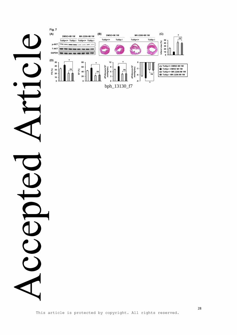
infarct size, and aggravates cardiac dysfunction (Figure 7A to 7D). Notably,
Tollip-/-
+MK-2206 mice has the comparable infarct size and cardiac function with
Tollip+/+
+MK-2206 mice, which indicated that the deteriorating effects of Tollip on
cardiac injury in response to MI is majorly dependent on Akt inhibition (Figure 7B to
7D).
Acc
epte
d A
rticl
e

15 This article is protected by copyright. All rights reserved.
Discussion
In the current study, we identified a novel role for Tollip in mediating the pathological
progression of MI-induced heart damage. Using gain- and loss-of-function approaches,
an obvious elevation of survival, a diminished infarct size, and a sustained cardiac
function were found in Tollip-KO mice. In contrast, the forced expression of Tollip
exacerbated myocardial apoptosis and immune cell infiltration, leading to an
increased mortality rate and marked loss of cardiac function. Mechanistically, these
deleterious effects of Tollip were largely dependent on the inhibition of Akt signaling.
Examining the expression alterations of target factor is one of easily achieved
approaches for auxiliary prediction of its involvement in certain pharmacological
conditions (Cravatt et al., 2007). In the present study, Tollip was dramatically
upregulated in human ischemic heart and animal MI models. Interestingly, this
finding is contrary to our previous finding that Tollip was downregulated in pressure
overload-induced cardiac hypertrophy (Liu et al., 2014b). This disparity may be
attributable to the different corresponding disease models. In fact, similar to the
divergence observed in the heart, the expression of Tollip varies widely in the colon in
response to different stress stimuli (Pimentel-Nunes et al., 2012; Xu et al., 2013). In
the context of colitis, the expression of Tollip is elevated, while in a model of colon
cancer, the Tollip level is decreased. Correspondingly, the different alterations of
Tollip expressions might be also related to its diverse functions in disease models.
Tollip preserved pressure overload-induced cardiac hypertrophy and delayed heart
failure by inhibiting cardiomyocyte enlargement and cardiac fibrosis (Liu et al.,
2014b), whereas during MI injury, as shown in the current study, Tollip aggravated
cardiac dysfunction and increased mortality through promoting inflammatory and
apoptotic responses.
In the early stage of MI injury, inflammatory response is triggered rapidly to
confront the interrupted blood supply-induced cellular damage, which is characterized
by the inflammatory and immune cells infiltrating into the myocardium and
scavenging ECM debris (Frangogiannis, 2012). However, sustained immune cell
infiltration is directly linked to myocardial apoptosis and impairs cardiac function
(Kin et al., 2006; Schofield et al., 2013). Our results demonstrated that Tollip
overexpression promoted the migration and accumulation of macrophages,
neutrophiles and CD3+-T lymphocytes at 3 d and 4 week after MI. Further, our
analysis revealed that Tollip enhanced MI-induced activation of NF-κB signaling at 4
week post-infarction, which profoundly contributes to inflammatory and immune cell
infiltration (Ma et al., 2012). Thus, the increased acute and chronic inflammatory
response may, at least partially, explain the detrimental effect of Tollip on apoptosis
and the subsequent cardiac dysfunction after MI. However, inconsistent with our
study, previous reports suggest that in RAW264.7 cells and human dermal endothelial
cells, Tollip up-regulation inhibited NF-κB activation in response to
lipopolysaccharide (LPS) (Bulut et al., 2001; Zhang et al., 2002). The discrepancy
between our study and others may stem from the different cell types and models used,
reflecting the complex nature of Tollip-dependent regulation of NF-κB activity. In fact,
similar disparities exist for other proteins that are involved in regulating inflammation.
For example, class A scavenger receptor (SR-A) alleviated the inflammatory response
in an endotoxic shock model (Amiel et al., 2009) but exacerbated inflammation
during pulmonary infection (Usui et al., 2007).
Concomitant with the chronic inflammatory response, myocardial apoptosis
occurs during the acute phase of MI and persists for a long term after MI, which
directly affects cardiac function and is associated with a higher mortality rate (Lal et
Acc
epte
d A
rticl
e

16 This article is protected by copyright. All rights reserved.
al., 2012; Zhang et al., 2011). Thus apoptosis inhibition is linked to a reduced infarct
size after MI (Zhao et al., 2003). In the current study, our findings demonstrated that
Tollip mediates apoptosis of cardiomyocytes: Tollip depletion reduced the number of
apoptotic cells, whereas overexpression of Tollip augmented cell death. Additionally,
the imbalance between pro-apoptotic protein (such as Bax and cleaved caspase 3) and
anti-apoptotic protein (Bcl-2) has long been established as a key determinant of
myocardial apoptosis (Chandrashekhar et al., 2004; Pu et al., 2013). In support of the
pro-apoptotic function of Tollip, we observed that Tollip downregulated the protein
expression of Bcl-2, but elevated the levels of Bax and cleaved caspase 3. Taken
together, these results indicate that Tollip exacerbates cardiac dysfunction and
increases infarct size after MI by promoting cell death through regulating
apoptosis-related proteins.
In the pathogenesis of MI, the MAPK signalling is greatly involved in and
regulate the inflammatory and apoptotic responses based on the activation and/or
inactivation the three critical subfamily, ERK, JNK and p38, upon extracellular
stimuli (Xuan et al., 2011). However, unexpectedly, Tollip exhibited no significant
effect on MI-activated MAPK signaling in the current study. Consistent with our
results, Dedierlaurent et al. (Didierlaurent et al., 2006) has demonstrated that
LPS-induced MAPK activation in mouse embryonic fibroblasts are failed to be
disrupted by Tollip. Except for MAPK cascades, the Akt signalling is a
well-established molecular program involved in MI progression that could dominant
cardiomyocyte fate via influencing cell survival and other cellular events (Hausenloy
et al., 2004; Jonassen et al., 2001; Suarez et al., 2014; Yamashita et al., 2001). Our
findings suggest that the function of Tollip in the ischemic heart is associated with the
manipulation of Akt signaling, as the results demonstrated that Tollip deficiency
dramatically increased the phosphorylated levels of Akt, mTOR, GSK3β, S6 and
FOXO1 proteins in both mice heart tissue post-MI injury in vivo and
hypoxia-stimulated cardiomyocytes in vitro. Importantly, the exacerbated
cardiomyocytes damage by Tollip overexpression could be almost completely
abolished by Akt up-regulation, suggesting an Akt-dependent manner of
Tollip-regulated MI injury. The inactivation of Akt signalling by Tollip also provides
support for the discrepancy of Tollip on MI- and hypertrophy-induced heart damage,
since that Akt promotes cardiac hypertrophy and cardiomyocyte survival
concomitantly (Fujio et al., 2000; Yan et al., 2011). Taken together, these data suggest
that the effect of Tollip on signaling pathways during MI-induced cardiac dysfunction
is relatively selective.
During cardiac remodeling post-MI, myocyte apoptosis in the infarct zone and
border zone directly affects cardiac function and is associated with a higher mortality
rate (Lal et al., 2012; Zhang et al., 2011). Thus, strategies to suppress cardiomyocyte
death are important for mitigating post ischemic heart injury. Using the
cardiac-specific Tollip overexpression mice and isolated neonatal myocytes, our
present study clearly demonstrated that Tollip significantly promotes cardiomyocytes
apoptosis. However, Tollip is also expressed in other cell types, including fibroblasts
and endothelial cells (Byun et al., 2014; Lee et al., 2012). It is possible that Tollip
may have effects in these cells contributing to the cardiac remodeling process post-MI,
which needs further investigation. Aside from cell death, inflammation as another
critical response greatly involved in the progression of post-MI remodeling (Schofield Acc
epte
d A
rticl
e

17 This article is protected by copyright. All rights reserved.
et al., 2013). Though the initial inflammatory response is indispensable for wound
healing, sustained immune cell infiltration is directly linked to myocardial apoptosis
and impairs cardiac function. Our current study demonstrated a promoted function of
Tollip on immune cell infiltration, which partly explained the detrimental effect of
Tollip on post-MI remodeling. Additionally, our previous study showed that Tollip
also plays a critical role in pressure overload-induced cardiac fibrosis and hypertrophy,
important contributors to cardiac remodeling (Liu et al., 2014a). Notably, post-MI
angiogenesis is not a process to be ignored that affects cardiomyocytes death via
improving blood supply (Cochain et al., 2013), but the extent of this process has not
been found to be influenced by the Tollip gene disturbance in our present study.
Taken together, multiple capacities of Tollip to regulate post-MI apoptotic and
inflammatory permits its profound function in MI-induced cardiac remodeling.
Although genetically engineered mice are robust tools for determining the role of
endogenous Tollip in the ischemic heart, genetic intervention as a clinical treatment
remains challenging (Dykxhoorn et al., 2005). Thus, from a clinical point of view,
studies exploring the exogenous blockade of Tollip or activation of Akt through
pharmacological approaches are needed. Furthermore, considering that Tollip and Akt
signalling exert opposing effects in MI and cardiac hypertrophy, future studies on
exogenous inhibition or activation certain proteins should focus on the counterbalance
of their expression to rescues ischemic injury with little or no augment on cardiac
hypertrophy.
In conclusion, our study is the first to demonstrate that Tollip promotes
inflammatory response and cell apoptosis, leading to aggravated cardiac dysfunction
and increased mortality after MI, and that these effects are largely dependent on the
inhibition of Akt signaling. Our findings provide novel insight into the pathogenesis
of MI and highlighted the potential therapeutic value of Tollip inhibition in the
treatment of MI.
Acknowledgments
This work was supported by grants from the National Science Fund for Distinguished
Young Scholars (NO. 81425005), the National Natural Science Foundation of China
(NO. 81170086, NO. 81270184), National Science and Technology Support Project
(NO. 2011BAI15B02, NO. 2012BAI39B05, NO. 2013YQ030923-05, 2014BAI02B01,
and 2015BAI08B01), the Key Project of the National Natural Science Foundation
(NO. 81330005), the National Basic Research Program China (NO. 2011CB503902),
and Natural Science Foundation of Hubei Province (2013CFB259).
Conflict of interest: None declared.
Acc
epte
d A
rticl
e

18 This article is protected by copyright. All rights reserved.
References
Amiel E, Acker JL, Collins RM, Berwin B (2009). Uncoupling scavenger receptor A-mediated
phagocytosis of bacteria from endotoxic shock resistance. Infect Immun 77(10): 4567-4573.
Baldi A, Abbate A, Bussani R, Patti G, Melfi R, Angelini A, et al. (2002). Apoptosis and
post-infarction left ventricular remodeling. J Mol Cell Cardiol 34(2): 165-174.
Bulut Y, Faure E, Thomas L, Equils O, Arditi M (2001). Cooperation of Toll-like receptor 2 and 6 for
cellular activation by soluble tuberculosis factor and Borrelia burgdorferi outer surface protein A
lipoprotein: role of Toll-interacting protein and IL-1 receptor signaling molecules in Toll-like receptor
2 signaling. J Immunol 167(2): 987-994.
Burns K, Clatworthy J, Martin L, Martinon F, Plumpton C, Maschera B, et al. (2000). Tollip, a new
component of the IL-1RI pathway, links IRAK to the IL-1 receptor. Nat Cell Biol 2(6): 346-351.
Byun EB, Mi S, Kim JH, Song DS, Lee BS, Park JN, et al. (2014). Epigallocatechin-3-gallate-mediated
Tollip induction through the 67-kDa laminin receptor negatively regulating TLR4 signaling in
endothelial cells. Immunobiology 219(11): 866-872.
Chandrashekhar Y, Sen S, Anway R, Shuros A, Anand I (2004). Long-term caspase inhibition
ameliorates apoptosis, reduces myocardial troponin-I cleavage, protects left ventricular function, and
attenuates remodeling in rats with myocardial infarction. J Am Coll Cardiol 43(2): 295-301.
Chen K, Gao L, Liu Y, Zhang Y, Jiang DS, Wei X, et al. (2013). Vinexin-beta protects against cardiac
hypertrophy by blocking the Akt-dependent signalling pathway. Basic Res Cardiol 108(2): 338.
Cochain C, Channon KM, Silvestre JS (2013). Angiogenesis in the infarcted myocardium. Antioxid
Redox Signal 18(9): 1100-1113.
Cravatt BF, Simon GM, Yates JR, 3rd (2007). The biological impact of mass-spectrometry-based
proteomics. Nature 450(7172): 991-1000.
Didierlaurent A, Brissoni B, Velin D, Aebi N, Tardivel A, Kaslin E, et al. (2006). Tollip regulates
proinflammatory responses to interleukin-1 and lipopolysaccharide. Mol Cell Biol 26(3): 735-742.
Dykxhoorn DM, Lieberman J (2005). The silent revolution: RNA interference as basic biology,
research tool, and therapeutic. Annu Rev Med 56: 401-423.
Frangogiannis NG (2012). Regulation of the inflammatory response in cardiac repair. Circ Res 110(1):
159-173.
Fujio Y, Nguyen T, Wencker D, Kitsis RN, Walsh K (2000). Akt promotes survival of cardiomyocytes
in vitro and protects against ischemia-reperfusion injury in mouse heart. Circulation 101(6): 660-667.
Acc
epte
d A
rticl
e

19 This article is protected by copyright. All rights reserved.
Hausenloy DJ, Yellon DM (2004). New directions for protecting the heart against
ischaemia-reperfusion injury: targeting the Reperfusion Injury Salvage Kinase (RISK)-pathway.
Cardiovasc Res 61(3): 448-460.
Heineke J, Molkentin JD (2006). Regulation of cardiac hypertrophy by intracellular signalling
pathways. Nat Rev Mol Cell Biol 7(8): 589-600.
Heusch G, Libby P, Gersh B, Yellon D, Bohm M, Lopaschuk G, et al. (2014). Cardiovascular
remodelling in coronary artery disease and heart failure. Lancet 383(9932): 1933-1943.
Hu Y, Li T, Wang Y, Li J, Guo L, Wu M, et al. (2009). Tollip attenuated the hypertrophic response of
cardiomyocytes induced by IL-1beta. Front Biosci (Landmark Ed) 14: 2747-2756.
Jiang DS, Bian ZY, Zhang Y, Zhang SM, Liu Y, Zhang R, et al. (2013). Role of interferon regulatory
factor 4 in the regulation of pathological cardiac hypertrophy. Hypertension 61(6): 1193-1202.
Jiang DS, Li L, Huang L, Gong J, Xia H, Liu X, et al. (2014a). Interferon regulatory factor 1 is
required for cardiac remodeling in response to pressure overload. Hypertension 64(1): 77-86.
Jiang DS, Wei X, Zhang XF, Liu Y, Zhang Y, Chen K, et al. (2014b). IRF8 suppresses pathological
cardiac remodelling by inhibiting calcineurin signalling. Nat Commun 5: 3303.
Jiang DS, Zhang XF, Gao L, Zong J, Zhou H, Liu Y, et al. (2014c). Signal regulatory protein-alpha
protects against cardiac hypertrophy via the disruption of toll-like receptor 4 signaling. Hypertension
63(1): 96-104.
Jonassen AK, Sack MN, Mjos OD, Yellon DM (2001). Myocardial protection by insulin at reperfusion
requires early administration and is mediated via Akt and p70s6 kinase cell-survival signaling. Circ Res
89(12): 1191-1198.
Jones BM, Kapadia SR, Smedira NG, Robich M, Tuzcu EM, Menon V, et al. (2014). Ventricular septal
rupture complicating acute myocardial infarction: a contemporary review. Eur Heart J 35(31):
2060-2068.
Kilkenny C, Browne W, Cuthill IC, Emerson M, Altman DG, Group NCRRGW (2010). Animal
research: reporting in vivo experiments: the ARRIVE guidelines. Br J Pharmacol 160(7): 1577-1579.
Kin H, Wang NP, Halkos ME, Kerendi F, Guyton RA, Zhao ZQ (2006). Neutrophil depletion reduces
myocardial apoptosis and attenuates NFkappaB activation/TNFalpha release after ischemia and
reperfusion. J Surg Res 135(1): 170-178.
Lal H, Zhou J, Ahmad F, Zaka R, Vagnozzi RJ, Decaul M, et al. (2012). Glycogen synthase
kinase-3alpha limits ischemic injury, cardiac rupture, post-myocardial infarction remodeling and death.
Circulation 125(1): 65-75. Acc
epte
d A
rticl
e

20 This article is protected by copyright. All rights reserved.
Lee HJ, Chung KC (2012). PINK1 positively regulates IL-1beta-mediated signaling through Tollip and
IRAK1 modulation. J Neuroinflammation 9: 271.
Liu Y, Jiang XL, Jiang DS, Zhang Y, Zhang R, Chen Y, et al. (2014a). Toll-interacting protein (Tollip)
negatively regulates pressure overload-induced ventricular hypertrophy in mice. Cardiovasc Res 101(1):
87-96.
Liu Y, Jiang XL, Liu Y, Jiang DS, Zhang Y, Zhang R, et al. (2014b). Toll-interacting protein (Tollip)
negatively regulates pressure overload-induced ventricular hypertrophy in mice. Cardiovasc Res 101(1):
87-96.
Ma J, Wei M, Wang Q, Li J, Wang H, Liu W, et al. (2012). Deficiency of Capn4 gene inhibits nuclear
factor-kappaB (NF-kappaB) protein signaling/inflammation and reduces remodeling after myocardial
infarction. J Biol Chem 287(33): 27480-27489.
Pimentel-Nunes P, Goncalves N, Boal-Carvalho I, Afonso L, Lopes P, Roncon-Albuquerque R, Jr., et
al. (2012). Decreased Toll-interacting protein and peroxisome proliferator-activated receptor gamma
are associated with increased expression of Toll-like receptors in colon carcinogenesis. J Clin Pathol
65(4): 302-308.
Pu J, Yuan A, Shan P, Gao E, Wang X, Wang Y, et al. (2013). Cardiomyocyte-expressed
farnesoid-X-receptor is a novel apoptosis mediator and contributes to myocardial
ischaemia/reperfusion injury. Eur Heart J 34(24): 1834-1845.
Rafiq K, Kolpakov MA, Seqqat R, Guo J, Guo X, Qi Z, et al. (2014). c-Cbl Inhibition Improves
Cardiac Function and Survival in Response to Myocardial Ischemia. Circulation 129(20): 2031-2043.
Riad A, Jager S, Sobirey M, Escher F, Yaulema-Riss A, Westermann D, et al. (2008). Toll-like
receptor-4 modulates survival by induction of left ventricular remodeling after myocardial infarction in
mice. J Immunol 180(10): 6954-6961.
Schofield ZV, Woodruff TM, Halai R, Wu MC, Cooper MA (2013). Neutrophils--a key component of
ischemia-reperfusion injury. Shock 40(6): 463-470.
Shishido T, Nozaki N, Yamaguchi S, Shibata Y, Nitobe J, Miyamoto T, et al. (2003). Toll-like
receptor-2 modulates ventricular remodeling after myocardial infarction. Circulation 108(23):
2905-2910.
Suarez J, Wang H, Scott BT, Ling H, Makino A, Swanson E, et al. (2014). In vivo selective expression
of thyroid hormone receptor alpha1 in endothelial cells attenuates myocardial injury in experimental
myocardial infarction in mice. Am J Physiol Regul Integr Comp Physiol 307(3): R340-346.
Sun Y, Yi W, Yuan Y, Lau WB, Yi D, Wang X, et al. (2013). C1q/tumor necrosis factor-related Acc
epte
d A
rticl
e

21 This article is protected by copyright. All rights reserved.
protein-9, a novel adipocyte-derived cytokine, attenuates adverse remodeling in the ischemic mouse
heart via protein kinase A activation. Circulation 128(11 Suppl 1): S113-120.
Timmers L, Sluijter JP, van Keulen JK, Hoefer IE, Nederhoff MG, Goumans MJ, et al. (2008).
Toll-like receptor 4 mediates maladaptive left ventricular remodeling and impairs cardiac function after
myocardial infarction. Circ Res 102(2): 257-264.
Usui HK, Shikata K, Sasaki M, Okada S, Matsuda M, Shikata Y, et al. (2007). Macrophage scavenger
receptor-a-deficient mice are resistant against diabetic nephropathy through amelioration of
microinflammation. Diabetes 56(2): 363-372.
Wang L, Lu Y, Guan H, Jiang D, Guan Y, Zhang X, et al. (2013). Tumor necrosis factor
receptor-associated factor 5 is an essential mediator of ischemic brain infarction. J Neurochem 126(3):
400-414.
Wencker D, Chandra M, Nguyen K, Miao W, Garantziotis S, Factor SM, et al. (2003). A mechanistic
role for cardiac myocyte apoptosis in heart failure. J Clin Invest 111(10): 1497-1504.
Xiao J, Moon M, Yan L, Nian M, Zhang Y, Liu C, et al. (2012). Cellular FLICE-inhibitory protein
protects against cardiac remodelling after myocardial infarction. Basic Res Cardiol 107(1): 239.
Xu N, Yu ZH, Yao QS, Wang ZQ, Qu HL, Sun Y, et al. (2013). PPAR-gamma and tollip are
associated with toll-like receptors in colitis rats. J Immunoassay Immunochem 34(3): 219-231.
Xuan W, Liao Y, Chen B, Huang Q, Xu D, Liu Y, et al. (2011). Detrimental effect of fractalkine on
myocardial ischaemia and heart failure. Cardiovasc Res 92(3): 385-393.
Yamashita K, Kajstura J, Discher DJ, Wasserlauf BJ, Bishopric NH, Anversa P, et al. (2001).
Reperfusion-activated Akt kinase prevents apoptosis in transgenic mouse hearts overexpressing
insulin-like growth factor-1. Circ Res 88(6): 609-614.
Yan L, Wei X, Tang QZ, Feng J, Zhang Y, Liu C, et al. (2011). Cardiac-specific mindin
overexpression attenuates cardiac hypertrophy via blocking AKT/GSK3beta and TGF-beta1-Smad
signalling. Cardiovasc Res 92(1): 85-94.
Zhang G, Ghosh S (2002). Negative regulation of toll-like receptor-mediated signaling by Tollip. J Biol
Chem 277(9): 7059-7065.
Zhang Y, Kohler K, Xu J, Lu D, Braun T, Schlitt A, et al. (2011). Inhibition of p53 after acute
myocardial infarction: reduction of apoptosis is counteracted by disturbed scar formation and cardiac
rupture. J Mol Cell Cardiol 50(3): 471-478.
Zhang Y, Liu X, She ZG, Jiang DS, Wan N, Xia H, et al. (2014). Interferon regulatory factor 9 is an
essential mediator of heart dysfunction and cell death following myocardial ischemia/reperfusion injury. Acc
epte
d A
rticl
e

22 This article is protected by copyright. All rights reserved.
Basic Res Cardiol 109(5): 434.
Zhao ZQ, Morris CD, Budde JM, Wang NP, Muraki S, Sun HY, et al. (2003). Inhibition of myocardial
apoptosis reduces infarct size and improves regional contractile dysfunction during reperfusion.
Cardiovasc Res 59(1): 132-142.
Acc
epte
d A
rticl
e

23 This article is protected by copyright. All rights reserved.
Figure legends
Figure 1 Up-regulation of Tollip in hearts subjected to acute MI. A Representative
western blotting of Tollip in the heart samples from normal donors (n=6) and patients
with ischemic heart disease (IHD; n=7). B Western blotting results for Tollip in the
infarcted border zone of C57BL/6J mice at the indicated time points post-MI (n=5).
Left, Representative western blotting. Right, Quantitative results. *P<0.05 vs. normal
donors or sham hearts.
Figure 2 Tollip increases MI-induced mortality, infarct size, and cardiac dysfunction
after MI. A Kaplan-Meier survival curves for Tollip+/+
and Tollip knockout (KO)
mice after MI (*P<0.05 vs. Tollip+/+
/MI group). B Representative images of
hematoxylin and eosin (HE) staining in heart cross-sections of Tollip+/+
and Tollip-/-
mice at 3 d and 4 week after MI surgery (n=6 mice per experimental group). C
Measurement of infarct size in the heart tissue of mice from Tollip+/+
and Tollip-/-
groups. *P<0.05 vs. Tollip+/+
/MI group at 3 d; #P<0.05 vs. Tollip+/+
/MI group at 4 W.
D Echocardiographic and hemodynamic assessment of cardiac function in Tollip+/+
and Tollip-/-
mice after MI (n=8 mice per experimental group). *P<0.05 vs.
Tollip+/+
/sham; #P<0.05 vs. Tollip+/+
/MI group. E Comparison of mortality between
non-transgenic (NTG) mice and Tollip-TG mice at 4 week post-MI injury (*P<0.05
vs. NTG/MI group). F and G Representative images and evaluation of infarct size in
NTG and Tollip-TG mice at 3 d and 4 week after MI surgery (n=6 mice per
experimental group). *P<0.05 vs. NTG/MI group at 3 d; #P<0.05 vs. NTG/MI group
at 4 W. H The parameters of cardiac function in NTG and Tollip-TG mice after 4
weeks of MI surgery (n=8 for each group). *P<0.05 vs. NTG/sham group; # P<0.05
vs. NTG/MI group.
Figure 3 Tollip increases inflammatory and immune cell infiltration and NF-κB
activation after MI. A and B Representative immunofluorescence (IF) staining
images of macrophage (Mac-1), Neutrophil (LY6G), and T cell (CD3) accumulation
in the infarct border zone of Tollip+/+
and Tollip-KO mice (A) and in NTG and
Tollip-TG mice (B) at 3 d and 4 W after MI. C and D Representative western blotting
and quantitative results showing the phosphorylation levels of p65 and IκBα in the
infarct border zone of hearts from (C) Tollip+/+
and Tollip-KO mice and from (D)
NTG and Tollip-TG mice at 4 week after MI (n=13 mice per experimental group).
Left, Representative western blotting. Right, Quantitative results. *P<0.05 vs.
Tollip+/+
/sham or NTG/sham group; #P<0.05 vs. Tollip+/+
/MI or NTG/MI group.
Figure 4 Tollip promotes apoptosis and regulates the expression of apoptosis-related
genes in response to MI. A to C Representative TUNEL staining images (A) and
quantitative results (B and C) TUNEL-positive cells in heart sections from (B)
Tollip+/+
and Tollip-/-
mice and from (C) NTG and Tollip-TG mice at 3 d and 4 week
after MI (n=6 mice per experimental group). D and E Representative western blotting
and quantitative analysis showing the protein levels of Bax, Bcl-2, and cleaved
caspase3 in (D) Tollip+/+
and Tollip-KO mice and in (E) NTG and Tollip-TG mice at
4 week after MI induction (n=13 mice per experimental group). Left, Representative
western blotting. Right, Quantitative results. *P<0.05 vs. Tollip+/+
/sham or
NTG/sham group; #P<0.05 vs. Tollip+/+
/MI or NTG/MI group.
Figure 5 Tollip enhances hypoxia-induced cardiomyocytes death and regulates the
expression of apoptosis-related genes in vitro. A and C Representative Acc
epte
d A
rticl
e

24 This article is protected by copyright. All rights reserved.
photomicrographs of (A) AdshTollip- or (C) AdTollip-infected primary
cardiomyocytes double-labeled with Hoechst 33258 for nuclei (blue) and propidium
iodide (PI) for dead cells (red) after 24 h of hypoxia stimulation (n=5). B and D
Determination of cell viability and cells toxicity by CCK8 and LDH release assay in
NRCMs with (B) Tollip knockdown or (D) overexpression after exposure to hypoxia
for 24 hours (n=5). *P<0.05 vs. AdshRNA/normoxia or AdGFP/normoxia; #P<0.05
vs. AdshRNA/hypoxia or AdGFP/hypoxia. E and F Western blotting results for the
protein levels of Bax, Bcl-2, and cleaved caspase3 in NRCMs infected with (E)
AdshTollip or (F) AdTollip after hypoxia treatment for 24 h (n=5 samples). Top,
Representative western blotting. Bottom, quantitative results. *P<0.05 vs.
AdshRNA/normoxia or AdGFP/normoxia; #P<0.05 vs. AdshRNA/hypoxia or
AdGFP/hypoxia
Figure 6 Tollip exacerbates cardiac injury post-MI by inactivating the Akt signaling
pathway. A and B Representative western blotting showing phosphorylated and total
MEK1/2, ERK1/2, JNK1/2 and p38 protein levels from (A) Tollip+/+
and Tollip-/-
mice and from (B) NTG and Tollip-TG mice at 4 week after MI (n=13 mice per
experimental group). C to E The (C) representative western blotting and (D and E)
quantitative results showing the levels of phosphorylated and total Akt, mTOR, S6,
FOXO1 and GSK3β protein from heart tissue of mice in the indicated groups at 4
week after MI (n=13 mice per experimental group). *P<0.05 vs. Tollip+/+
/MI or
NTG/MI group. F to H Representative western blotting results showing the
expression levels of proteins in Akt signalling in NRCMs infected with AdshTollip or
AdTollip after hypoxia treatment for 24 h (n=6 samples). *P<0.05 vs.
AdshRNA/hypoxia or AdGFP/hypoxia. I The cell viability and LDH release of
cardiomyocytes infected with AdshRNA, AdshTollip, AddnAkt or
AdshTollip+AddnAkt after exposure to hypoxia for 24 h (n=6 samples). J The cell
viability and LDH release of cardiomyocytes in the indicated groups after treatment of
hypoxia for 24 h (n=6 samples). ns: no significant difference.
Figure 7 Akt inhibition was responsible for the detrimental role of Tollip on
MI-induced cardiac damage. A Representative western blotting showing
phosphorylated and total Akt protein levels from Tollip+/+
and Tollip-/-
mice treated
with Akt inhibitor MK-2206 at 1 week after MI (n=8 mice per experimental group). B
Representative images of hematoxylin and eosin (HE) staining in heart cross-sections
of Tollip+/+
and Tollip-/-
mice treated with Akt inhibitor MK-2206 at 1 week after MI
surgery (n=6 mice per experimental group). C Measurement of infarct size in the
heart tissue of mice from Tollip+/+
and Tollip-/-
groups treated with Akt inhibitor
MK-2206. D Echocardiographic and hemodynamic assessment of cardiac function in
Tollip+/+
and Tollip-/-
mice treated with Akt inhibitor MK-2206 after MI (n=6 mice
per experimental group). ns: no significant difference.
Acc
epte
d A
rticl
e

25 This article is protected by copyright. All rights reserved.
bph_13130_f1
bph_13130_f2
Acc
epte
d A
rticl
e

26 This article is protected by copyright. All rights reserved.
bph_13130_f3
bph_13130_f4
Acc
epte
d A
rticl
e

27 This article is protected by copyright. All rights reserved.
bph_13130_f5
bph_13130_f6
Acc
epte
d A
rticl
e
28 This article is protected by copyright. All rights reserved.
bph_13130_f7
Acc
epte
d A
rticl
e