Prognosis of angina and myocardial infarction in South Asian ...
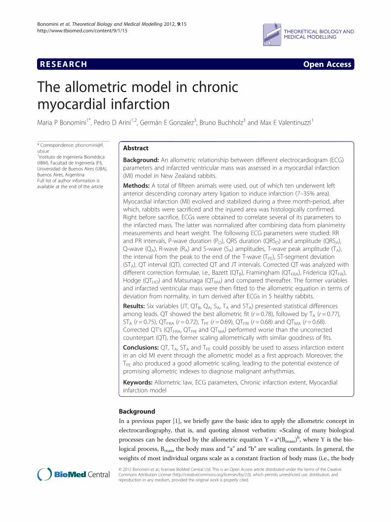
Bonomini et al. Theoretical Biology and Medical Modelling 2012, 9:15http://www.tbiomed.com/content/9/1/15
RESEARCH Open Access
The allometric model in chronicmyocardial infarctionMaria P Bonomini1*, Pedro D Arini1,2, Germán E Gonzalez3, Bruno Buchholz3 and Max E Valentinuzzi1
* Correspondence: [email protected] de Ingeniería Biomédica(IIBM), Facultad de Ingeniería (FI),Universidad de Buenos Aires (UBA),Buenos Aires, ArgentinaFull list of author information isavailable at the end of the article
Abstract
Background: An allometric relationship between different electrocardiogram (ECG)parameters and infarcted ventricular mass was assessed in a myocardial infarction(MI) model in New Zealand rabbits.
Methods: A total of fifteen animals were used, out of which ten underwent leftanterior descending coronary artery ligation to induce infarction (7–35% area).Myocardial infarction (MI) evolved and stabilized during a three month-period, afterwhich, rabbits were sacrificed and the injured area was histologically confirmed.Right before sacrifice, ECGs were obtained to correlate several of its parameters tothe infarcted mass. The latter was normalized after combining data from planimetrymeasurements and heart weight. The following ECG parameters were studied: RRand PR intervals, P-wave duration (PD), QRS duration (QRSD) and amplitude (QRSA),Q-wave (QA), R-wave (RA) and S-wave (SA) amplitudes, T-wave peak amplitude (TA),the interval from the peak to the end of the T-wave (TPE), ST-segment deviation(STA), QT interval (QT), corrected QT and JT intervals. Corrected QT was analyzed withdifferent correction formulae, i.e., Bazett (QTB), Framingham (QTFRA), Fridericia (QTFRI),Hodge (QTHO) and Matsunaga (QTMA) and compared thereafter. The former variablesand infarcted ventricular mass were then fitted to the allometric equation in terms ofdeviation from normality, in turn derived after ECGs in 5 healthy rabbits.
Results: Six variables (JT, QTB, QA, SA, TA and STA) presented statistical differencesamong leads. QT showed the best allometric fit (r= 0.78), followed by TA (r= 0.77),STA (r= 0.75), QTFRA (r= 0.72), TPE (r= 0.69), QTFRI (r= 0.68) and QTMA (r= 0.68).Corrected QT’s (QTFRA, QTFRI and QTMA) performed worse than the uncorrectedcounterpart (QT), the former scaling allometrically with similar goodness of fits.
Conclusions: QT, TA, STA and TPE could possibly be used to assess infarction extentin an old MI event through the allometric model as a first approach. Moreover, theTPE also produced a good allometric scaling, leading to the potential existence ofpromising allometric indexes to diagnose malignant arrhythmias.
Keywords: Allometric law, ECG parameters, Chronic infarction extent, Myocardialinfarction model
BackgroundIn a previous paper [1], we briefly gave the basic idea to apply the allometric concept in
electrocardiography, that is, and quoting almost verbatim: «Scaling of many biological
processes can be described by the allometric equation Y= a*(Bmass)b, where Y is the bio-
logical process, Bmass the body mass and “a” and “b” are scaling constants. In general, the
weights of most individual organs scale as a constant fraction of body mass (i.e., the body
© 2012 Bonomini et al.; licensee BioMed Central Ltd. This is an Open Access article distributed under the terms of the CreativeCommons Attribution License (http://creativecommons.org/licenses/by/2.0), which permits unrestricted use, distribution, andreproduction in any medium, provided the original work is properly cited.

Bonomini et al. Theoretical Biology and Medical Modelling 2012, 9:15 Page 2 of 13http://www.tbiomed.com/content/9/1/15
mass exponent, “b” equals 1.0). Volume rates, instead (the product of volume and rate),
such as cardiac output, ventilation and oxygen uptake, vary with “b” around 0.75. Finally,
rates (heart and respiratory rate) scale as “b” close to 0.25. These emergent patterns pro-
vide insights into body-size dependent principles that seem to dictate several aspects of
design and function across species among all mammals [2,3] » .
Noujaim et al. [4] assumed that the heart behaves as a set of “fractal-like” networks
tending to minimize propagation time across the conducting system while ensuring a
hemodynamically optimal atrioventricular activation sequence. With the mathematical
relationship given above and, subsequently, based on previously published values of PR
interval, heart rate, and body masses of 541 mammals, they reported as best fit the
equation PR= 53 × (Bmass)0.24.
Inspired in the latter report, the following question seems pertinent:
Would a relationship similar to the allometric equation be conceivable, say, between
the number of cardiac diseased fibers and any of several ECG parameters when a
myocardial infarct (MI) affected the heart?
The objective of this paper tries to find an answer to such question. The Q-wave
growth appears as a good candidate because well-known is the fact that normal
depolarization suffers with compromised myocardial mass, augmenting the Q-wave
and shrinking the R-wave within the QRS complex. Also, additional MI changes in-
clude alterations in the T-wave morphology and QRS complex voltages and duration,
as well as distortion in intervals such as JT and QT. Many reports confirm this concept,
such as Klootwijk, in 1998 [5], Kléber, in 2000 [6], or Balian et al., in 2006 [7], among
others. All of these variables and their behaviour on a MI model deserve to be looked
at under the allometric light, which seems to maintain interest, especially in general
mammalian biology [3,8].
MethodsExperimental protocol and procedure to myocardial infarction
An overall of fifteen animals were used in this study. Ten New Zealand rabbits (7 male
and 3 female, 1.5–2.5 kg) underwent left anterior descendent (LAD) coronary artery
ligation to provoke infarction on a varying extent of myocardial tissue. MI evolved and
stabilized during a three month-period after which, rabbits were sacrificed and MI his-
tologically confirmed.
Ventricular infarcted mass in grams was calculated after combining data from plani-
metric measurements and heart weights. The infarcted mass was then normalized to
heart weight in order to compare among different rabbit sizes. Normalized ventricular
infarcted mass (VIMn) and the ECG parameters mentioned above were, thereafter, fit-
ted to the allometric equation.
This study conformed to the Guide for the Care and Use of Laboratory Animals pub-
lished by the US National Institutes of Health (NIH Publication No. 85–23, revised 1996).
After anaesthesia with ketamine (75 mg/kg) and Rompun (0,75 mg/kg xylazine) was
administered subcutaneously, an endotracheal tube (3 mm inner diameter) was placed
to mechanically ventilate the rabbits with room air using a Harvard respirator (25 ml,
34–38 cycles/min). Immediately after, 5% dextrose solution (3 ml/min) was infused

Bonomini et al. Theoretical Biology and Medical Modelling 2012, 9:15 Page 3 of 13http://www.tbiomed.com/content/9/1/15
through a flexible catheter placed at the marginal vein of one of the ears. Using this
venous access throughout the surgical procedure, anaesthesia was maintained by apply-
ing additional doses of ketamine and sodium thyopental as needed during the surgical
procedure. A left thoracotomy on the fourth intercostal space and a pericardiectomy
were performed to expose the heart. Thereafter, a lateral or posterolateral coronary ar-
tery branch of the LAD coronary artery was ligated using a curved needle and 6.0 silk
thread [9]. The appearance of regional paleness confirmed ventricular wall ischemia. Fi-
nally, the thorax was closed with linen thread keeping the order of the different ana-
tomical parts. At the end of the surgical procedure, the animals were placed in a quiet
environment for their recovery. Antiobiotic therapy was administered and animals were
closely observed during the first 24 hours. Afterwards, animals were placed in individ-
ual cages up to the finalization of a three month period.
For the control group, 5 healthy rabbits underwent the above mentioned anaesthetic
protocol and ECG recordings were obtained. Analogously to the MI group, the three
standard limb leads were recorded.
Histology and planimetry
To assess infarct size, Masson’s Trichrome (MT) staining was carried out, while infarc-
tion was corroborated by Hematoxiline-Eosine (HE). Once excised, hearts were per-
fused with 10% formaldehyde during 5 min for fixation, remaining in that solution for
at least 72 hs. Four millimeter slices were cut out from apex to base and then processed
for paraffin embedding and staining. After the latter, fibrotic tissue turned blue, so dif-
ferentiating itself from healthy tissue, while HE allowed morphological recognition of
necrotic tissue.
The stained slices were scanned and planimetric measurements were performed
using an image software (Image Pro-Plus 4.5). The area mean percent occupied by the
scar tissue at each ventricular level estimated the infarct size [10,11]; these values were
expressed as percentage of the total ventricular area while normalized ventricular
infarcted mass (VIMn) was calculated as follows:
VIMn¼ PIM %ð Þ � VM grð Þ=HW grð Þ
where PIM: planimetric infarcted area (% of the total ventricular area); VM: ventricular
mass and HW: heart weight.
ECG acquisition and preprocessing
Rabbits were heparinized (500 U/Kg IV) 10 minutes before anesthesia by intramuscular
injection of ketamine (35 mg/Kg) and lidocaine (5 mg/Kg). All animals were in sinus
rhythm at the time of ECG recordings (frontal leads I, II, and III).
ECG data were recorded using instrumentation amplifiers with a gain factor of 1000
and a bandwidth of 0.05–150 Hz. The signals were digitalized at a sampling rate
Fs = 500 Hz and 12-bit resolution using a digital acquisition board (Lab PC+, National
Instruments, Austin, TX, USA). When necessary, a band-stop filter was used to remove
50-Hz. All of the data were acquired and monitored using customized software made
in C++.

Bonomini et al. Theoretical Biology and Medical Modelling 2012, 9:15 Page 4 of 13http://www.tbiomed.com/content/9/1/15
QRS detection was based on methods described by Hamilton and Tompkins in 1986
[12]. All the ectopic or aberrant beats were automatically rejected by the computer. If
needed, baseline corrections were performed by cubic spline interpolation [13].
We computed the running signal average of 30 beats. The template for signal aver-
aging in each ECG channel were created, thereafter, by aligning 30 beats with 98%
cross-correlated QRS waveforms using the predefined window and the R-wave peak as
the trigger point while, at the same time, correcting for fiducial point jitter. Two
rejected beats were the accepted limit on the computation of each final template beat.
ECG delineation
ECG delineation was accomplished on the template obtained as explained in section
“ECG acquisition and preprocessing”. The start and end of the QRS complex were
determined by searches on each side of the R-wave for regions where the slope (dV/dt)
fell to sufficiently low values. Isoelectric level was taken as the median of all data values
preceding the start of the QRS. For the P-wave, we used a modified version of the algo-
rithm described by [14], which is based on finding the peaks and valleys of a 9-point
derivative signal. In case of the T-wave delineation, a search was made for the first sig-
nificant peak of either sign, starting from a point after the end of the QRS. If a suitable
peak was found, a straight line was adjusted by least squares from the peak to the tail
of the T-wave. The intersection of this line with the isoelectric line was accepted as the
T-wave end. ST-segment deviation was defined as the ECG amplitude 50 ms after QRS
onset [15].
For all ECG templates, the PR interval, P-wave (PD) and QRS duration (QRSD), QRS amp-
litude (QRSA), T-wave peak amplitude (TA), ST-segment deviation (STA), the time from the
peak to the end of the T-wave (TPE), QT interval, corrected QT interval (QTC) and JT inter-
vals (JT) were calculated. Below the details of the QT corrections are given according to dif-
ferent formulae. All parameters are graphically represented in Figure 1.
QTC Formulae
The following QT correction formulae were tested:
-Bazett: QTB ¼ QTffiffiffiffiRR
p [16]
-Framingham: QTFra ¼ QT þ 0:154 � 1� RRð Þ [17]-Fridericia: QTFri ¼ QTffiffiffiffiffi
QT3p [18]
-Hodge: QTHo ¼ QT þ 0:00175 � HR� 60ð Þ [19]-Matsunaga: QTMa ¼ QT � log 600ð Þ
log 1000�RRð Þ [20]
where HR=heart rate in bpm, RR=RR interval in s, QT=QT duration in s, QTC=
corrected QT interval in s.
Allometric model – Mathematical setting
Allometry, in general biology, measures the relative growth of a part in relation to the
whole living organism. The term was first used by Snell, in 1891 [9], to express the
mass of a mammal’s brain as a function of the body mass. The growth velocity of a
component y is related to the growth velocity of another component (or the whole or-
ganism) x in a constant way. This was clearly described by von Bertalanffy in 1957 [10].

Figure 1 ECG measurements. Schematic representation of ECG waves, intervals and segments analyzed inthis work.
Bonomini et al. Theoretical Biology and Medical Modelling 2012, 9:15 Page 5 of 13http://www.tbiomed.com/content/9/1/15
Thus, the relative rate of change of a given event y is proportional to the relative rate
of change of body mass or body weight x, i.e.,
dy=dty
¼ B� dx=dtx
ð1Þ
After integration and some easy algebraic manipulation, equation (1) becomes
lny ¼ Aþ B� lnx;
or
y ¼ A�xB ð2Þ
Originally, y was the weight of an organ (heart, stomach, other) and x was body
weight or mass. The parameters A and B require numerical estimation by an appropri-
ate procedure usually using empirical information. By the same token, let us
hypothesize that the electrocardiographic parameters follow a relationship with the
number of ventricular fibers (NF), formally equal to (2), i.e.,
ECGparam ¼ a� NFð ÞB ð3Þ
Moreover, in a pathology context, let us relate the “deviation from normality” in
terms of ECG with the “deviation from normality” in terms of diseased fibers, modify-
ing the model as in (4),
ECGD � ECGN ¼ a� NDFð Þβ ð4Þ
Hence, y in equation (2) is replaced by (ECGD – ECGN) in (3), and NDF in the latter
takes the place of x in the former. After all, the number of diseased cardiac fibers

Bonomini et al. Theoretical Biology and Medical Modelling 2012, 9:15 Page 6 of 13http://www.tbiomed.com/content/9/1/15
(ischemic or infarcted or both) are part of the cardiac mass. Besides, since the relation-
ship between diseased fibers and infarcted mass is straightforward, it sounds sensible to
state that,
NDF ¼ γ�VIMn ð5Þ
Hence,
ECGD � ECGN γ�VIMn� �β ð6Þ
After taking logarithms of both sides, the latter equation becomes
log ECGD � ECGNð Þ ¼ logaþ β� logγð Þ þ β� logVIMnð Þ; which can be reduced to
log ECGD � ECGNð Þ ¼ δ þ β� logVIMnð Þ ð7Þ
where δ = logα + β*logγ, VIMn: log(normalized ventricular infarcted mass) and ECGD
and ECGN are the diseased and healthy counterparts of the following ECG parameters;
STA: log(|ST deviation|) in mV; QA: log(|Q-wave amplitude|) in mV; SA: log(|S-wave
amplitude|) in mV; RA: log(|R-wave amplitude|) in mV; QRSA: log(|Q| + |R| + |S|) in
mV; TA: log(T-wave peak amplitude) in mV, TPE: log(T-wave time from peak to end);
QRSD: log(QRS width);.QT: log(QT interval); QTc: log(QT interval corrected by differ-
ent formulae); RR: log(RR interval); PR: log(PR interval); PD: log(P duration). Linear
regressions were carried out to obtain numerical values for the intercept δ and the
slope β in the log-log plot.
Statistical analysis
Data are expressed as mean ± standard deviation (Mean± SD). To assess statistical sig-
nificance, leads I, II and III were compared among them. When data did not pass the
D’Agostino & Pearson normality test, a non-parametric Kruskall-Wallis test was applied
and the Dunn’s post test carried out. Otherwise, one-way ANOVA and Bonferroni
post-test were calculated.
ResultsECG parameters
ECG parameters were consistent with MI, showing wider and morphologically diverse
QRS complexes. QRS voltages suffered in MI, presenting a significant decrease in leads II
and III. Due to flattening of the T-wave, STA resulted very small in Leads II and III, but
presented a rounded pattern in Lead I. QRSD was similar in every lead. Neither significant
RR nor PR interval changes were seen. PD was longer in lead II, even though no statistical
significance was found. Similarly, TPE increased in Lead II but failed to be statistically sig-
nificant. Lengthening of the JT interval was originated by a flattening of the T-wave, which
is much more rounded in the physiological state and offered statistical significance in lead
II with respect to the other two leads. The T-wave peak voltage significantly decreased to
even become negative in leads II and III, representing the typical T-wave inversion
described in chronic infarction [21,22]. Table 1 displays the values for the ECG time-
related parameters described above along with the statistical significance values (p< 0.05);
so does Table 2 for the ECG amplitude-related parameters.
QT interval did not show significant differences among leads. On the other hand,
among all the correction formulae tested (Bazett, Framingham, Friderica, Hodges and

Table 1 Electrocardiographic time-related parameters (Mean± SD) from a 3 month-MIrabbit evolution
RR (ms) PR (ms) PD (ms) QRSD (ms) JT (ms) TPE (ms)
L1 365 ± 92 71 ± 8 29± 7 45± 6 120 ± 21 59 ± 23
L2 397 ± 99 76 ± 8 32± 6 45± 4 161 ± 35* 74 ± 25
L3 394 ± 108 72 ± 9 24± 6 41± 4 127 ± 26 59 ± 24
L1: lead I, L2: lead II, L3: lead III. Statistical significance: *p< 0.05, Lead II against Leads I and III.
Bonomini et al. Theoretical Biology and Medical Modelling 2012, 9:15 Page 7 of 13http://www.tbiomed.com/content/9/1/15
Matsunaga), the only one that led to statistical significance was Bazett’s formula, with a
clear differentiation from Leads II and III. Table 3 summarizes the QT and QT cor-
rected values for every lead.
Allometric fits
In general terms, 7 out of 18 measures presented a clear allometric scaling with VIMn.
Lead I contained 4 out of 7 such parameters (STA, QTFRA, QTFRI and QTMA), followed
by Lead III with 2 (QT and TPE) and Lead II (TA) with only one. This lead preference
for each parameter explains the way data are presented, on a lead separated-basis. The
variable that best fitted the allometric equation was QT (r= 0.78) followed by TA
(r= 0.77), STA (r= 0.75), QTFRA (r = 0.72), TPE (r= 0.69), QTFRI (r= 0.68) and QTMA
(r= 0.68), all of these with statistical significance (p< 0.05). It is worth noting that TA
presented a negative allometric fit with VIMn (β = −2.474 ± 0.7736), meaning that the
value of T-voltage decreased as VIMn increased. A similar fit was found for the Q-wave
amplitude (β = −0.6352 ± 0.2849), even though it failed to account for statistical signifi-
cance (p = 0.0610). On the contrary, SA increased along with VIMn producing a positive
slope (β = 1.202 ± 0.6668) in the allometric equation. This opposite trend would explain
the change in morphology of QRS complexes, shifting from a QR to an RS pattern.
STA deviation in Lead I displayed a good linear regression with VIMn in a log-log repre-
sentation. The slope for this regression turned up to be positive, leading to an STA in-
crease with infarct size. On the other hand, TPE in Lead III also increased with VIMn
reflecting important changes in T-wave morphology, mostly due to the flattening of the
T-wave, which is normally rounded in rabbits. Figure 2 displays the allometric scaling
of the mentioned variables QT, TA, TPE and STA. Adjustments are shown as well as
95% confidence limits (broken lines).
QT corrections presented allometric fits with VIMn as well, and were analyzed in a
different graph (Figure 3) for clarity sake. In lead I, the Framingham, Matsunaga and
Fridericia formulae corrections for heart rate allometrically scaled to VIMn. Accord-
ingly, all the corrected QT intervals displayed a slope smaller than one, near 0.25, as
Table 2 Electrocardiographic amplitude-related parameters (Mean± SD) from a 3 month-MI rabbit evolution
QA (mV) RA (mV) SA (mV) QRSA (mV) STA (mV) TA (mV)
L1 −0.50 ± 0.33* 0.21 ± 0.23 −0.12 ± 0.36* 0.83 ± 0.92 0.08 ± 0.04* 0.10 ± 0.12*
L2 −0.29 ± 0.23 0.22 ± 0.16 0.01 ± 0.05 0.52 ± 0.44 0.01 ± 0.05 −0.03 ± 0.09
L3 −0.12 ± 0.15 0.37 ± 0.24 −0.02 ± 0.03 0.51 ± 0.42 −0.02 ± 0.03 −0.05 ± 0.07
L1: lead I, L2: lead II, L3: lead III. Statistical significance: *p< 0.05, Lead I against Lead III.

Table 3 Corrected and uncorrected QT intervals (Mean± SD) from a 3 month-MI rabbitevolution
QT (ms) QTB (ms) QTFRA (ms) QTFRI (ms) QTHO (ms) QTMA (ms)
L1 182 ± 24 296 ± 25 272± 16 242 ± 23 359 ± 50 189± 20
L2 187 ± 35 293 ± 42 265± 19 235 ± 28 350 ± 54 184± 25
L3 169 ± 29 272 ± 28*2 262± 20 231 ± 25 347 ± 57 182± 25
L1: lead I, L2: lead II, L3: lead III. QTFRA: Framingham correction, QTFRI: Fridericia correction, QTMA: Matsunaga correction,QTHO: Hodge correction. Statistical significance: *p< 0.05, Lead III versus Lead II.
Bonomini et al. Theoretical Biology and Medical Modelling 2012, 9:15 Page 8 of 13http://www.tbiomed.com/content/9/1/15
cited in the bibliography [2-4], being the uncorrected QT the only one that deviated
from this pattern (β = 3.464 ± 1.072).
The remaining variables, RR, PR, PD, QRSD, JT, QTHO, QTB, QRSA and QA, RA and
SA did not present a fit with a good statistical significance (p> 0.05). Moreover, they
kept almost constant for all MI extents (data not shown).
A complete description of the allometric fits can be found in Table 4, where all para-
meters together with the slope β, the origin intercept δ, the goodness of fit r and the
statistical significance p-value are shown. Each parameter was chosen from the lead at
which the best allometric fit was produced.
DiscussionFindings of the present study
An allometric study was presented here in order to analyze the allometric scaling of dif-
ferent ECG parameters with respect to the infarction extent in a chronic experimental
Figure 2 Allometric scaling of ECG parameters. Allometric scaling of STA, TA, QT and TPE versusinfarction extent. Notice the negative slope of TA with infarction extent.

Figure 3 Allometric scaling of QT and QTc parameters. Corrected QT allometric fits. QTfra: Framinghamcorrection formula, QTfri: Fridericia correction formula, QTma: Matsunaga correction formula.
Bonomini et al. Theoretical Biology and Medical Modelling 2012, 9:15 Page 9 of 13http://www.tbiomed.com/content/9/1/15
MI rabbit model. The idea behind this work was to find ECG data to estimate myocar-
dial injured area as a first, simple and inexpensive step before resorting to more com-
plex or invasive technologies, such as magnetic resonance imaging or radioscopic
procedures. The chief findings of this study were as follows:
Table 4 Linear regressions of all ECG parameters for a 3 month-MI rabbit evolutiongrouped by leads
Lead Parameter β (Mean± SD) δ (Mean± SD) r p-value
L3 QT 3.464± 1.072 3.494 ± 0.8387 0.78 0.0145
L2 TA −2.474± 0.7736 −3.593 ± 0.5926 0.77 0.0151
L1 STA 1.832± 0.5997 0.06115 ± 0.4994 0.75 0.0185
L1 QTFRA 0.1042 ± 0.03840 2.462 ± 0.03198 0.72 0.0300
L3 TPE 2.303± 0.9132 3.087 ± 0.7142 0.69 0.0397
L1 QTFRI 0.1691 ± 0.06926 2.456 ± 0.05769 0.68 0.0447
L1 QTMA 0.1918 ± 0.07858 2.346 ± 0.06545 0.68 0.0447
L1 QTB 0.1384 ± 0.0617 2.531 ± 0.05142 0.64 0.0600
L3 QA −0.6352 ± 0.2849 −0.8878 ± 0.2228 0.64 0.0610
L3 SA 1.202± 0.6668 0.03635 ± 0.5215 0.55 0.1145
L2 PD 1.011± 0.5893 1.510 ± 0.4514 0.54 0.1299
L1 QRSA 1.210± 0.7190 0.2376 ± 0.5988 0.53 0.1362
L3 QRSD 0.5699 ± 0.3995 1.566 ± 0.3124 0.47 0.1968
L1 RR 0.5025 ± 0.3669 2.675 ± 0.3056 0.45 0.2131
L2 PR −0.7553 ± 0.7338 0.3425 ± 0.5621 0.36 0.3376
L1 RA 0.7621 ± 0.7972 0.09869 ± 0.6639 0.34 0.3709
L3 JT 0.5296 ± 0.6854 1.821 ± 0.5360 0.26 0.4650
L3 QTHO 0.1448 ± 0.3730 2.350 ± 0.2917 0.14 0.7095
β: slope of linear regression, δ: Y-intercept when X= 0 of linear regression, r: correlation coefficient, p-value: statisticalsignificance of linear fit. L1: Lead I, L2: Lead II, L3: Lead III, QTFRA: Framingham correction formula, QTMA: Matsunagacorrection formula, QTB: Bazett correction formula, QTFRI: Fridericia correction formula, QTHO: Hodges correction formula,QT: QT interval, TA: amplitude of T-wave, STA: amplitude of ST segment deviation, TPE: time from peak to end of the T-wave, QA: amplitude of Q-wave, SA: amplitude of S-wave, RA: amplitude of R-wave, QRSA: amplitude of QRS complex,QRSD: duration of QRS complex, RR: RR interval, PR: PR interval, JT: JT interval.

Bonomini et al. Theoretical Biology and Medical Modelling 2012, 9:15 Page 10 of 13http://www.tbiomed.com/content/9/1/15
1- Six variables (JT, QTB, QA, SA, TA and STA) showed statistical differences among
leads. Lead I versus Lead III contained 5 out of 6 of these statistical differences
(QTB, QA, SA, TA and STA), while JT presented differences in Lead II with respect
to Lead I. Most of these variables turned out to be amplitude-related (QA, SA,
TA and STA) and, probably, the lead with the most evident changes was related to
infarct localization.
2- Seven out of eighteen parameters presented a significant allometric fit (p< 0.05).
Lead I accounted for the majority of them (STA, QTFRA, QTFRI, QTMA), followed
by Lead III (QT, TPE) and Lead II (TA). See Table 4, where it can also be seen that
QTB and QA showed borderline significances and still with a good fit.
3- The variables that best adjusted to the allometric equation were QT, TA and STAwith a goodness of fit enough to assess infarction size within a sensitive range
(r= 0.78, r= 0.77, r= 0.75, respectively.
Significance and interpretation of the data
Ventricular changes found during the chronic MI state can be split into ventricular
depolarization and repolarization changes.
Ventricular Depolarization Changes
In chronic MI hearts, QRS morphologies changed [21,22]. Even though not statistical
significant, a negative Q-voltage slope and a positive S-voltage slope in the allometric
equation suggest a shift from the QR to the RS morphology.
Surprisingly, QRSD did not fit well the allometric equation, as it would have been
expected. We cannot find a good explanation for such behaviour. Evidence refers to the
prognostic importance of QRS duration in acute and chronic myocardial infarction
[23,24]; hence, further investigation would be needed.
Ventricular Repolarization Changes
The T-wave showed a change in morphology, too, becoming flat instead of rounded.
Moreover, negative T-wave morphologies appeared, mostly in lead II and III. These
changes forced the JT interval to be longer and TPE to be longer as well as a smaller
TA. On the other hand, QT interval and TPE presented a good allometric scaling. It is
tempting to link this observation with the fact that increased arrhythmogenesis is
related to MI hearts, as reported elsewhere [25-32].
The JT-interval could be considered as a total ventricular repolarization process. In
our study, such interval was increased in MI, which might indicate a differential length-
ening or shortening of the action potential duration (APD) in some myocardial areas,
so reflecting the range of times at which action potentials end and, therefore, it would
be an expression of APD heterogeneity [33]. However, JT showed a bad allometric scal-
ing and, at least based on this study, we could not establish the allometric behaviour of
this parameter.
Uncorrected QT offered the best fit, in agreement with the literature on QT and is-
chaemia. With transmural ischaemia, the T-wave axis shifts in the opposite direction
towards the region of epicardial involvement, being often a sign of infarction. Such T-
waves are tall and peaked, frequently with long QT intervals [34]. These changes may
assist in the localization of ischaemia and also in the quantification of the injury size. It

Bonomini et al. Theoretical Biology and Medical Modelling 2012, 9:15 Page 11 of 13http://www.tbiomed.com/content/9/1/15
is worth noting that the slope of all corrected QT where about 0.25, similar to those
found in the literature related to allometry; the uncorrected QT was the only one out
of this pattern (β = 3.464 ± 1.072) [2,4].
Surprisingly, STA deviation in Lead I displayed a good linear regression with VIMn in
a log-log representation. This fact was opposite to our expectations since in humans,
ST deviation tends to normalize in chronic MI. Nevertheless, ST-segment in rodents
(especially in rats, and less markedly in rabbits) shows a different behaviour in the
physiological case [35]. Thus it is plausible to think that these differences will still hold
for the pathological cases.
In addition, someone may think that a multilead approach would be a good method-
ology to measure the parameters. However, in our case, the number of leads was only
three and we think that is not enough number to guarantee that kind of analysis. On
top of that, we did not have a criterion to select one or the other lead.
Finally, it should be underlined that the reference set of normal data, even though
they appear at first sight as a good idea (see equation #4), did not significantly changed
the results when using a direct relationship such as
ECGparam ¼ a � NDFð Þβ
from which numerical results are not shown in this report (see also above, Findings of
the present study, item 3).
Study implications and future work
The first half of the T-wave is mainly related to epicardial APD, whereas the second
half is to the endocardial and mid-myocardial APD [36,37]. As a result, at the peak of
the T-wave the transmural voltage gradient reaches the maximum and, therefore, the
descending limb represents the extent of transmural dispersion of ventricular repolari-
zation. Therefore, great is the importance of TPE producing a good allometric scaling,
leading to a new and attractive future work, which is the search of allometric indexes
for diagnosing malignant arrhythmias.
QT, TA and STA showed the strongest allometric behavior. This implies that chronic
MI extents could be roughly assessed by feeding the allometric equation with any of
these parameters. Whether one outstands from the others or not and hence, whether
the allometric equation should be fed with any of these parameters individually or col-
lectively with any combination of them, remains open.
Another consideration is pertinent regarding MI localization. Data presented in this
work is valid for anteroseptal injury only. Thus, the search of electrocardiographic allo-
metric scaling on different MI localizations would complete this study.
Study limitations
In the analysis of several electrocardiographic parameters, most of the ECG time-related
and amplitude-related measurements have been taken into account. Nevertheless, other
approaches for QT correction should be explored, like the individual correction sug-
gested by Malik [38] or the correction for animals carried out by Valentinuzzi [39]. On
the other hand, the relations found here are strictly related to infarcts originated from
LAD coronary artery ligation, not covering other MI localizations. Besides, a wider

Bonomini et al. Theoretical Biology and Medical Modelling 2012, 9:15 Page 12 of 13http://www.tbiomed.com/content/9/1/15
range of infarction extent would be desirable, allometrically speaking, but not possible
due to the extremely low survival rates at MI extents greater than 40%.
ConclusionsThe objectives of this paper were met, since ECG parameters related to depolarization
and repolarization phases showed a certain degree of adjustment to the allometric
equation, some of them rather good. The parameters that best allometrically scaled
were QT, TA and STA, with a goodness of fit enough to assess infarction size within an
acceptable range. Besides, the TPE produced a good allometric scaling, leading to the
potential existence of promising indexes to diagnose malignant arrhythmias.
Competing interestsThe authors declare that they have no competing interests.
AcknowledgementsPartially supported by PIP #538, from CONICET (Consejo Nacional de Investigaciones Científicas y Técnicas). All threeauthors are CONICET Career Investigators.
Author details1Instituto de Ingeniería Biomédica (IIBM), Facultad de Ingeniería (FI), Universidad de Buenos Aires (UBA), Buenos Aires,Argentina. 2Instituto Argentino de Matemática, “Alberto P. Calderón”, CONICET, Buenos Aires, Argentina. 3Instituto deFisiopatología Cardiovascular, Facultad de Medicina, Universidad de Buenos Aires (UBA), Buenos Aires, Argentina.
Authors’ contributionsMPB carried out the animal experiments and drafted the manuscript. MPB, PDA and MEV made key contributions tothe conception and design, analysis and interpretation of data, and drafting of the manuscript. GEG and BB run thereference series. All authors read and approved the final manuscript.
Received: 15 February 2012 Accepted: 11 May 2012Published: 11 May 2012
References
1. Bonomini MP, Arini PD, Valentinuzzi ME: Probability of ventricular fibrillation: Allometric model based on theST deviation. Biomed Eng Online 2011, 10:8.2. Lindstedt SL, Schaeffer PJ: Use of allometry in predicting anatomical and physiological parameters of
mammals. Lab Anim 2002, 36:1–19.3. Martin RD, Genoud M, Hemelrijk CK: Problems of allometric scaling analysis: examples from mammalian
reproductive biology. J Experiment Biol 2005, 208:1731–1747.4. Noujaim SF, Lucca E, Muñoz V, Persaud D, Berenfeld O, Meijler FL, Jalife J: From mouse to whale: A universal
scaling relation for the PR interval of the electrocardiogram of mammals. Circulation 2004, 110:2801–2808.5. Klootwijk APJ: Dynamic computer-assisted ST segment monitoring in patients with acute coronary syndromes: Thesis
Dissertation. Rottërdam: Erasmus Universiteit; 1998.6. Kléber AG: ST-segment elevation in the electrocardiogram: a sign of myocardial ischemia. Cardiovasc Res 2000,
45:111–118.7. Balian V, Galli M, Marcassa C, Cecchin G, Child M, Barlocco F, Petrucci E, Filippini G, Michi R, Onofri M:
Intracoronary ST-segment shift soon after elective percutaneous coronary intervention accurately predictsperiprocedural myocardial injury. Circulation 2006, 114:1948–1954.
8. Adler CP, Costabel U: Cell number in human heart in atrophy, hypertrophy, and under the influence ofcytostatics. Recent Adv Studies Cardiac Struc Metab 1975, 6:12.
9. Podesser B, Wollenek G, Seitelberger R, Siegel H, Wolner E, Firbas W, Tschabitscher M: Epicardial branches of thecoronary arteries and their distribution in the rabbit heart: the rabbit heart as a model of regional ischemia.Anat Rec 1997, 247:521–527.
10. Fletcher PJ, Pfeffer JM, Pfeffer MA, Braunwald E: Left ventricular diastolic pressure-volume relations in rats withhealed myocardial infarction. Effects on systolic function. Circ Res 1981, 49:618–626.
11. Pfeffer MA, Pfeffer JM, Fishbein MC, Fletcher PJ, Spadaro J, Kloner RA, Braunwald E: Myocardial infarct size andventricular function in rats. Circ Res 1979, 44:503–512.
12. Hamilton PS, Tompkins WJ: Quantitative investigation of QRS detection rules using the MIT/BIH arrhythmiadatabase. IEEE Trans Biomed Eng BME 1986, 33:1157–1165.
13. Meyer CR, Keiser HN: Electrocardiogram baseline noise estimation and removal using cubic splines and state-space computation techniques. Comput Biomed Res 1977, 10:459–470.
14. Hee-Kyo J, Kwang-Keun K, Sun-Chul H, Lee M-H: A New Algorithm for P-Wave Detection in the ECG Signal. InBook A New Algorithm for P-Wave Detection in the ECG Signal. 1989. (Editor ed.^eds.), vol. 1. pp. 42–43. City;1989:42–43.
15. Xue Q, Reddy S: Algorithms for computerized QT analysis. J Electrocardiol 1997, 30:181–186.16. Bazett H: An analysis of time-relations of electrocardiograms. Heart 1920, vii:17.

Bonomini et al. Theoretical Biology and Medical Modelling 2012, 9:15 Page 13 of 13http://www.tbiomed.com/content/9/1/15
17. Sagie A, et al: An improved method for adjusting the QT interval for heart rate (the Framingham study). Am JCardiol 1992, 70:4.
18. Fridericia L: Die Systolendauer im Elektrokardiogramm bei normalen Menschen und bei Herzkranken. ActaMedica Scandinavica 1920, 53:17.
19. Hodge M: Rate correction of the QT interval. Card Electrophysiol Rev 1997, 3:3.20. Matsunaga T, Mitsui T, Harada T, Inokuma M, Murano H, Shibutani Y: QT corrected for heart rate and relation
between QT and RR intervals in beagle dogs. J Pharmacol Toxicol Methods 1997, 38:201–209.21. Hu N, Straub CM, Garzarelli AA, Sabey KH, Yockman JW, Bull DA: Ligation of the left circumflex coronary artery
with subsequent MRI and histopathology in rabbits. J Am Assoc Lab Animal Sci: JAALAS 2010, 49:838–844.22. Yu M, Bozek J, Guaraldi M, Kagan M, Azure M, Robinson SP: Cardiac imaging and safety evaluation of BMS747158,
a novel PET myocardial perfusion imaging agent, in chronic myocardial compromised rabbits. J Nucl Cardiol:Official Publication of the American Society of Nuclear Cardiology 2010, 17:631–636.
23. Fosbøl EL, Seibaek M, Brendorp B, Møller DV, Ersbøll M, Torp-Pedersen C, Køber L: Differential prognosticimportance of QRS duration in heart failure and acute myocardial infarction associated with left ventriculardysfunction. Eur J Heart Fail 2007, 9:814–823.
24. Brenyo A, Zaręba W: Prognostic significance of QRS duration and morphology. Cardiol J 2011, 18:8–17.25. Briasoulis A, Agarwal V, Pierce WJ: QT prolongation and torsade de pointes induced by fluoroquinolones:
infrequent side effects from commonly used medications. Cardiology 2011, 120:103–110.26. Han J, Moe GK: Non uniform recovery of excitability in ventricular muscle. Circ Res 1964, 14:44–60.27. Lubinski A, Kornacewicz-Jach Z, Wnuk-Wojnar AM, Adamus J, Kempa M, Krolak T, Lewicka-Nowak E, Radomski M,
Swiatecka G: The terminal portion of the T wave: a new electrocardiographic marker of risk of ventriculararrhythmias. Pacing Clin Electrophysiol 2000, 23:1957–1959.
28. Taggart P, Sutton PMI, Opthof T, Coronel R, Trimlett R, Pugsley W, Kallis P: Transmural repolarization in the leftventricle in humans during normoxia and ischemia. Cardiovasc Res 2001, 50:454–462.
29. Vos MA, Jungschleger GM: Transmural repolarization gradients in vivo. The flukes and falls of theendocardium. Cardiovasc Res 2001, 50:423–425.
30. Nakagawa M, Takahashi N, Watanabe M, Ichinose M, Nobe S, Yonemochi H, Ito M, Saikawa T: Gender differencesin ventricular repolarization: Terminal T wave interval was shorter in women than in men. Pacing ClinElectrophysiol 2003, 26:59–64.
31. Kuo CS, Munakata K, Reddy PS, Surawicz B: Characteristics and possible mechanisms of ventricular arrhythmiasdependent on dispersion of action potentials. Circulation 1983, 67:1356–1367.
32. Deshmukh A, Ulveling K, Alla V, Abuissa H, Airey K: Prolonged QTc Interval and Torsades de Pointes Induced byCitalopram. Tex Heart Inst J 2012, 39:68–70.
33. Arini PD, Bertrán GC, Valverde ER, Laguna P: T-wave width as an index for quantification of ventricularrepolarization dispersion: Evaluation in an isolated rabbit heart model. Biomed Signal Process Contr 2008, 3:67–77.
34. Vandyck-Acquah MPS: Electrocardiographic Background. In Dynamic Electrocardiography. Edited by Malik M,Camm AJ. New York: Blackwell, Futura Publishing; 2004:217–232.
35. Nervonne J, Kass R: Molecular Physiology of Cardiac Repolarization. Physiol Rev 2005, 85:1205–1253.36. Sicouri S, Antzelevitch C: A subpopulation of cells with unique electrophysiological properties in the deep
subepicardium of the canine ventricle. The M Cell Circ Res 1991, 68:1729–1741.37. Antzelevitch C, Shimizu M, Yan GX, Sicouri S, Weissenburger J, Nesterenko VV, Burashnikov A, Di Diego J, Saffitz J,
Thomas GP: The Mcell: its contribution to theECGand to normal and abnormal electrical function of the heart.J Cardiovasc Electrophysiol 1999, 10:1124–1152.
38. Malik M, Färbom P, Batchvarov V, Hnatkova K, Camm AJ: Relation between QT and RR intervals is highlyindividual among healthy subjects: Implications for heart rate correction on the QT interval. Heart 2002,87:220–228.
39. Valentinuzzi ME: A mathematical model of the electrocardiographic QT-RR relationship. Med Biol Eng Comput1971, 9:255–261.
doi:10.1186/1742-4682-9-15Cite this article as: Bonomini et al.: The allometric model in chronic myocardial infarction. Theoretical Biology andMedical Modelling 2012 9:15.
Submit your next manuscript to BioMed Centraland take full advantage of:
• Convenient online submission
• Thorough peer review
• No space constraints or color figure charges
• Immediate publication on acceptance
• Inclusion in PubMed, CAS, Scopus and Google Scholar
• Research which is freely available for redistribution
Submit your manuscript at www.biomedcentral.com/submit
Copyright © 2022 FDOKUMEN