Studier av antidepressiva i kyllinglillehjerne og PC12-celler
Upload
independentCategory
view
0download
0
Thyroid Hormone Receptor/c-erbA: Control of Commitment and Differentiation in the Neuronal/Chromatfin Progenitor Line PC12 Alberto Mufioz,* Chr i s topher Wrighton,~ Barbara Seliger,§ J u a n Bernal,* and H a r t m u t Beug*
*Instituto de Investigaciones Biom&licas, Consejo Superior de Investigaciones Cientificas (CSIC) 28029 Madrid, Spain; ~Institute of Molecular Pathology, Dr. Bohrgasse 7, A 1030 Vienna, Austria; and §lII Medizinische Klinik, Abt. Hhmatologie/Onkologie D 6500 Mainz, Germany
Abstract. The c-erbA proto-oncogenes encode nuclear receptors for thyroid hormone (T3), a hormone inti- mately involved in mammalian brain maturation. To study thyroid hormone receptor (TR) action on neu- ronal cells in vitro, we expressed the chicken c-erbA/ TRot-1 as well as its oncogenic variant v-erbA in the adrenal medulla progenitor cell line PC12. In the ab- sence of T3, exogenous TRc~-I inhibits NGF-induced neuronal differentiation and represses neuron-specific gene expression. In contrast, TRo~-I allows normal differentiation and neuronal gene expression to occur in the presence of T3. Finally, TRc~-l-expressing cells become NGF-responsive for proliferation when T3 is absent, but NGF-dependent for survival in presence of
T3. A similar differentiation induction by NGF plus T3 was observed in a central nervous system-derived neu- ronal cell line (E 18) expressing exogenous TRot-1. Together with the finding that TRo~-I constitutively blocked dexamethasone-induced differentiation of PC12 cells into the chromaffin pathway, these results suggest that TRet-1 plays an important role in regulating com- mitment and maturation of neuronal progenitors.
In contrast, the v-erbA oncogene, a mutated, onco- genic version of TRc~-I, partially but constitutively in- hibited NGF-induced neuronal differentiation of PC12 cells and potentiated dexamethasone-induced chromaflin differentiation, giving rise to an aberrant "intedineage" cell phenotype.
T iaYaoio hormone (3,5,3'-triiodo-L-thyronine, T3) t is an essential regulator of physiological and develop- mental processes in many cells and tissues. In partic-
ular, T3 plays a crucial role in late embryonic and neonatal development of the brain (for reviews see Hamburg, 1969; Morreale de Escobar et al., 1983; Legrand, 1984; Dussault and Ruel, 1987). Deprivation of thyroid hormone during rat brain development causes diverse histological and cellular abnormalities such as changes in cell populations, decreases in axon densities, dendrite formation and myelin deposition, and alterations of cell migration, leading to severe impair- ment of brain maturation and function. These alterations are reversible by T3 replacement until a certain age and might reflect aberrant or even absent differentiation of both neurons and glial cells. In humans, permanent mental deficiency due to insufficient T3 levels can only be avoided if replacement therapy is started early after birth. In spite of the crucial im- portance of thyroid hormone for proper development of the central nervous system (CNS), very little is known about the molecular basis of thyroid hormone receptor action in neu- ronal ceils, most likely due to the complexity of the CNS and
1. Abbreviations used in this paper: CNS, central nervous system; DEX, dexamethasone; RA, retionic acid; RAR, retionic acid receptor; TH, tyro- sine hydroxylase; TR, thyroid receptor; T3 triiodothyronine.
the asynchronic patterns of development among their differ- ent parts and cell groups. Only recently, a few genes were found to be regulated at the pretranslational level by thyroid hormone in vivo (Mufioz et al., 1991).
After identification of the c-erbA proto-oncogene as the thyroid hormone receptor (TR) a-1 (Sap et al., 1986; Wein- berger et al., 1986) and discovery of two erbA genes in higher organisms (or and /3) encoding a whole family of different receptor proteins, several groups detected erbA ex- pression in the brain. CNS neurons express the thyroid hor- mone receptor, and an increase in Trbinding sites corre- lates with the period of neuroblast proliferation in human fetuses (Bernal and Pekonen, 1984). TRot RNA has been de- tected very early (day 4) in the embryonal chicken brain, in- creasing progressively thereafter during the embryonai period (Forrest et al., 1990, 1991). TR/3 appears later in em- bryogenesis, becoming predominant in certain areas of both the CNS and the eye (Forrest et al., 1991). During rat brain maturation, both ot and/3 receptors are expressed in a dis- tinct fashion characterized by tight temporal and spatial regulation (Bradley et al., 1989; Strait et al., 1991; Mell- strtm et al., 1991). In situ hybridization studies indicate that TR mRNAs are present mainly in neurons from those brain areas known to be targets of thyroid hormone, such as the cerebral cortex, hippocampus, cerebellum, and basal gan- glia. In particular, cholinergic cells of the basal forebrain are
© The Rockefeller University Press, 0021-9525/93/04/423/16 $2.00 The Journal of Cell Biology, Volume 121, Number 2, April 1993 423-438 423
on Novem
ber 23, 2014jcb.rupress.org
Dow
nloaded from
Published April 15, 1993

sensitive to thyroid hormones (Gould and Butcher, 1989). These cells also express maximum levels of the high affinity (gpl4(~ rk) NGF receptor (Holtzman et al., 1992) and thy- roid hormone and NGF were found to cooperate in the devel- opment of specific cholinergic markers in basal ganglia (Patel et al., 1988) and other CNS areas (Clos and Legrand, 1990). These findings strongly suggest a key role of thyroid hormone receptors in neural tissue formation and matura- tion, probably involving cooperation with neurotrophins.
We have addressed this question by studying the effects of % on neuronal cells differentiating in culture. Unfortu- nately, appropriate neuronal cell lines from the CNS capable of in vitro differentiation in response to neurotrophic factors are missing and primary cells obtained from brain do not grow in culture for extended time periods, hampering long- term studies. We therefore employed the PC12 cell line that has been extensively used as a model system for neuronal differentiation since its establishment from a rat pheochro- mocytoma (Greene and Tischler, 1976). PC12 cells show characteristics of common precursor cells for sympathetic adrenergic neurons and neurosecretory epithelioid chromaf- fin cells. NGF induces PC12 cells to differentiate along the neuronal pathway, whereas glucocorticoid hormones pro- mote their differentiation into chromaffin cells (Greene and Tischler, 1982; Levi et al., 1988). Thus, PC12 differentia- tion in vitro at least partially mirrors differentiation of respective neural crest-derived progenitor ceils in the rat adrenal medulla in vivo (Doupe et al., 1985a,b; Anderson and Axel, 1986).
Because of their origin from a tumor, it was not surprising that PC12 ceils behave aberrantly in several ways when com- pared to normal precursor cells in vivo. They continuously grow in the absence of neurotrophic factors, show an altered response to various exogenous stimuli and differentiate only partially into either neuronal or chromaffin cells. Also, they show no known response to thyroid hormone, a result also obtained in our own pilot experiments. This could be due to expression of low levels of c-erbA/TRot-1 together with high levels of c-erbA/TRot-2 RNA, which encodes a variant form of the receptor generated by differential splicing. TRot-2 is unable to bind hormone but efficiently blocks transcriptional activation by the active c-erbA/TRot-1 form (Koenig et al., 1989; Lazar et al., 1989; Rentoumis et al., 1990).
For these reasons, we decided to stably express an exoge- nous TRot-1 gene in PC12 cells using suitable retroviral vec- tors (Mufioz et al., 1990). The rationale behind this study was to restore TRot-1 expression in a neuronal progenitor cell line that express only low levels of TRot-I, most likely as a consequence of the immortalization process, and then analyze neuronal differentiation in response to thyroid hor- mone and neurotrophic factors. We show in this paper that exogenous TRot-1 expressed at physiological levels regulates NGF-dependent differentiation and gene expression of PC12 cells in a T3 dependent fashion and constitutively blocks their entry into the chromaffin differentiation pathway. In ad- dition, ligand-activated TRot-1 renders PC12 cells dependent on NGF as a neurotrophic factor. In contrast to TRot-l, v-erbA, a mutated, oncogenic version of TRot-1 that seems to act as a constitutive repressor of genes regulated by c-erbA/ TRot-1 (Danun et al., 1989; Sap et al., 1989; Disela et al., 1991), constitutively inhibited both neuronal and chromaffin differentiation of PC12 cells.
Materials and Methods
Cells and Cell Cultures
A clonal subline of rat PCI2 cells (referred to as 6c) was used throughout. This clone responded particularly well to differentiation induction by NGF in terms of neurite outgrowth on standard plastic dishes. 0Vrighton, C. W., and M. Busslinger, unpublished observations). PC12 6c cells were grown in DME supplemented with 10% horse serum, 5% FCS, and 1 mM gluta- mine (standard growth medium) (all from GIBCO, U.K.). Establishment and properties of the E 18 cell line derived by spontaneous immortalization from 17-d embryonic rat brain will be described elsewhere (Seliger, B., un- published observations). Cells were grown in Ham's F12 medium sup- plemented with 10% FCS.
Retrovirus Vectors and Generation of erbA-overexpressing Cells The recombinant retroviruses encoding the chicken c-erbA/TRct-1 and v-erbA genes used to infect PC12 6c cells have been already described (Mu- fioz et ai., 1990). Both are derived from the Moloney murine leukemia virus and encode the neomycin-resistant gene providing resistance to the antibi- otic G418. For infection, 5 x 10⁵ cells were plated in 60-ram dishes and incubated overnight with fibroblast-grown virus plus 8/~g/ml polybrene (Sigma Chem. Co., St. Louis, MO). Medium was then changed and selec- tion with 800 #g/ml (3418 (GIBCO BRL, Gaithersburg, MD) started 48 h after infection (Mufioz et al., 1990). Six clones of resistant TR~l-infected cells could be expanded separately (cAI-cA6). cA4, the clone exhibiting the highest specific T3-specific binding was used in all experiments. In con- trast, no individual v-erbA-infected clones could be expanded. Therefore, a pool of G418 resistant, v-erbA-expressing clones was used.
Infection of E 18 cells with TRot-1 was done exactly as described for PC12 6c. Ten TRot-l-expressing clones were obtained, one of which show- ing reproducible, high TRa-1 expression as determined by T3 binding (E 18 cA9) was used in the experiments.
Induction of Neural and Chroma~in Differentiation For induction of optimal neuronal differentiation, well suspended PC12 6c cells were seeded at 104 cells per cm 2 in standard growth medium. Murine 7 S NGF (Sigma Chem. Co.) was added at a concentration of 50 ng/ml every 2 d. Triiodothyronine (Sigma Chem. Co.) was added daily at a final concen- tration of 150 nM. Medium was changed partially or totally every 48 h. Chromaitin differentiation was induced by treatment with dexamethasone (5 t~g/ml; Serva, Heidelberg) every 2 d or when the medium was changed.
Embryonic CNS neuroblasts (E 18 cA9) were induced to differentiate by seeding them in serum-free Dulbecco's plus Ham's F12 (1:1) medium sup- plemented with the additions of N2 differentiation medium (Bottenstein and Sato, 1979). NGF (50 ng/ml) was added every 2 d while T3 (150 nM) was added daily.
pH]Thymidine Incorporation Assay Cells were seeded into 35-ram dishes in 2 nd standard growth medium at a density of 8 x 10⁴ cells per dish and treated with NGF and/or 1"3 as de- scribed above. Cell monolayers were labeled for 3.5 h with 1 /~Ci [6-3H] thymidine (25 Ci/mmol: Amersham Intl., Buckinghamshire, U.K.), washed twice with PBS, precipitated with 10% trichloroacetic acid for 10 min, air-dried, and dissolved in 400 #1 of 0.1% NaOH and I% SDS. Radio- activity in 200/zl was estimated in a/3 counter.
Mitomycin-C Treatment
Cells were treated for 2 h with mitomycin-C (Sigma Chem. Co.) at a final concentration of 20/~g/ml. In pilot experiments, this concentration inhibited thymidine incorporation to <1% of respective controls without producing toxic effects. Monolayers were then washed three times with PBS, incubated for 30 rain with fresh medium, washed again, and then trypsinized and reseeded for differentiation induction. Except for different mitomycin-C concentrations, the same procedure was previously used to demonstrate that differentiation occurs in absence of cell division in myoblasts (Falcone et al., 1984), macrophages (Beug et al., 1987), and erythroid cells (Beug et al., 1992).
The Journal of Cell Biology, Volume 121, 1993 424
on Novem
ber 23, 2014jcb.rupress.org
Dow
nloaded from
Published April 15, 1993

Immunoprecipitation Detection of erbA protein (p46 c'erbA and p75SaS-v-e,~A) by [3SS]methionine labeling and immunoprecipitation analysis was done as described earlier (Goldberg et al., 1988; Glinenr et al., 1990) using an anti-v-erbA antibody generously donated by Dr. J. Ghysdael.
Hormone Binding Assay T3-specific binding assays in vivo were performed as previously described (Sap et al., 1986; Mu5oz et al., 1990). Specific binding is defined as the ratio of iodinated T3 bound in absence and presence of a 1,000-fold molar excess unlabeled T3.
Detection of Neuronal and Chromaffin Differentiation Antigens by Immunofluorescence Fixed and permeabilized cells were analyzed by indirect immunofluores- cence using monoclonal antibodies to synaptophysin, tyrosine hydroxylase, glial fibrillary acidic protein (all from Boehringer Mannheim Corp., Ger- many), and N-CAM (Sigma Chem. Co.) according to standard procedures. Rabbit antiserum to mouse E-eadherin (uvomorulin; generously provided by Dr. R. Kemler) and mouse 46-kd cytokeratin (provided by Dr. E. Reich- mann) were used similarly. Basically, ceils were fixed with 3.7% paraformaldehyde plus 0.01% glutaraldehyde at 4°C for 15 rain, and then permeabilized with 0.1% NP-40 in medium for an additional 15 min at room temperature. Antibodies were used at the dilutions recommended by the manufacturers in a final volume of 50 t~l. After a l-h incubation at room temperature, the plates were washed with PBS and stained with second anti- body (FITC-conjugated anti-mouse or anti-rabbit IgG, Amersham, diluted 1:50) for 1.5 h. After staining, cells were washed three times with PBS, mounted in Mowiol (HOECHST), and viewed using a Zeiss Axiophot fluo- rescence microscope with epiillumination.
RNA Preparation and Northern Analysis Total RNA was prepared using the guanidinium isothiocyanate-phenol- chloroform procedure (Chomczynski and Sacehl, 1987). Purification of poly(A) + RNA was done as described (Vermstrtm and Bishop, 1982). Northern blots were performed on Gene Screen membranes (New England Nuclear, Boston, MA) according to standard protocols (Maniatis et al., 1982). All probes were labeled by the random priming method (Feinberg and Vngelstein, 1983) using commercial kits (Pharmaeia LKB Bioteehnol- ogy Inc., Piscataway, NJ). Source of probes: rat c-erbA/TR~l from Dr. R. M. Evans, rat TR-/3 from Dr. H. C. Towle, mouse c-jun,junB, andjunD and integrin-fll from Dr. R. Bravo, ratfra-1 from Dr. T. Curran, rat NGFI-A and NGFI-B from Dr. J. Milbrandt, v-fos from Dr. E. Wagner, rat transin from Dr. R. Breathnaeh, mouse collagen cd (I) from Dr. Z. Q. Wang, rat SCG10 from Dr. D. J. Anderson, rat GAP-43 from Dr. M. C. Fishman, rat tyrosine hydroxylase from Dr. E. Ziff, rat chromogranin B from Drs. S. Forss-Petter/J.G. Sutcliffe, mouse N-CAM from Dr. C. Goridis, mouse cytokeratin (endoB/K18) from Dr. M. Busslinger, human trk from Dr. M. Barbaeid, mouse trk from Dr. L. Parada, rat LNGFR from Dr. E. M. Shooter, human RARer and mouse RARfl from Dr. H. Stunnenberg, and rat NF68 from Dr. E Grosveld. Rat MASH-I and MASH-2 genes were cloned from poly (A) + RNA prepared from dense cultures of PC12 6c cells by eDNA synthesis and PCR amplification using primers specific for the extremes of the published sequences (Johnson et al., 1990) obtained from the EMBL data bank. The oligonueleotides used were 5'AATATAATG- A A G C ~ G C G T T T C T T C C C T T r T A A C 3' and 5'TATGA'IV_.AAGAA- TTCCATCC'IV_~'VICCAAAGTCCAT 3' for MASH-1 and 5'AATATAATG- AAGCTTGTCGAGGACGCATTAAGC 3' and 5'TATGATCAAGAATTC- C A A G T C C T G G T T T C ~ 3' for MASH 2. The amplified products were cloned into the HindIII and EcoRI sites of the pRK7 plasmid. Frag- ments covering the nonhomologous 3' ends were prepared as specific probes for both genes. A 308-bp PvulI fragment was prepared for MASH-1 and a 397-bp PstI-ScaI fragment for MASH-2. Hybridizations were carried out overnight at 65"C in 7% SDS, 500 mM sodium phosphate buffer, pH 7.2, and 1 mM EDTA according to Church and Gilbert (1984). Filters were washed twice for 30 rain each in 1% SDS and 40 mM sodium phosphate buffer, pH 7.2, at 65"C. Before rehybridizing the nylon membranes with probes for other genes, the radioactive probe was stripped off the membrane by placing it in a 750C water bath for 5 min.
Results
Stable Expression of Exogenous erbA Genes in PC12 Cells To analyze the possible role of T3 and its receptor on PC12 differentiation in vitro, we sought to stably express the avian c-erbA/TR~-I protein as well as its oncogenic variant, v-erbA, in these cells. Cultures of a clonal PC12 subline (6c, see Materials and Methods) were infected with retroviruses ex- pressing c-erbA/TRa-1 or v-erbA together with the neo r gene as a selectable marker. Several neoresistant clones of c-erbA/TR,-I infected cells were obtained. In contrast, we only succeeded to expand a pool of v-erbA-infected cell clones due to their poor clonal proliferation ability.
The expression of the respective TR~-I and v-erbA pro- teins was monitored by immunoprecipitation using anti- erbA antisera, c-erbA/TR~-I protein was just detectable, whereas v-erbA was expressed at high levels, comparable to those found in avian erythroleukemic cells (Fig. 1 A). Syn- thesis of active c-erbA/TRa-1 protein was confirmed by a hormone binding assay after incubation with ~25I-labeled %. Specific binding was one order of magnitude higher in c-erbA/TRcel-expressing cells than in uninfected or v-erbA- expressing cells (Fig. 1 B), and of the same order of magni- tude as that of rat GH1 pituitary cells containing a few thou- sand active receptor molecules per cell (Samuels et al., 1976, 1977).
Endogenous Hormone Receptor Expression in Normal and erbA-expressing PC12 Cells Since several cell lines of neural origin express the thyroid hormone receptor variant TRot-2 in high excess over TRot-1 (Mufioz, A., J. Bernal, and A. Rodriquez-Pefia, unpublished observations), we tested if this was also true for PC12. As shown by Northern blot analyses, normal PCI2 cells ex- pressed much higher levels of the TRot-2 form than of the TRot-1 and TRfl receptors (Fig. 1 C). In contrast, the retrovi- rus transduced exogenous c-erbA/TRot-1 and v-erbA genes were expressed at levels comparable to those of the endoge- nous TRtx-2 receptor. Thus, the nonresponsiveness of PC12 cells to T3 may in fact be due to functional suppression of the low TRa-I levels by an excess of TRt~-2.
To rule out that the c-erbA/TRct-1- and v-erbA-expressing PC12 sublines were altered in their expression of other im- portant receptors, the same blots were hybridized with cDNA probes encoding the low (gp80 ENG.) and high (gpl40 ~k) affinity NGF receptors as well as the retinoic acid receptors (RARs) ot and/~. All these receptors were expressed in the parental cells (distinct signal from 10 #g total RNA after 5-d exposure) and no changes in expression levels were detected in the sublines expressing exogenous erbA proteins (data not shown).
Ligand-activated TRa-1 Renders PC12 Cells NGF-dependent for Proliferation and Survival Neurotrophic factors such as NGF are considered to be necessary for survival of differentiating neuronal cells, in- cluding primary adrenal medulla sympathetic neurons in culture. (Doupe et al., 1985b; Anderson and Axel, 1986). In contrast, PC12 cells grow in the absence of NGF, and re- vert to an undifferentiated state if the factor is withdrawn af-
Mufioz et al. erbA Regulates PC12 Cell Differentiation 425
on Novem
ber 23, 2014jcb.rupress.org
Dow
nloaded from
Published April 15, 1993

Figure L ErbA expression in PC12 cells. (.4) Unin- fected, TRot-1/c-erbA- and v-erbA-infected PC12 cells were labeled with [35S]methionine and ly- sates from equal numbers of cells were immuno- precipitated with an antibody against v-erbA. The immunoprecipitates were analyzed on 10% SDS- polyacrylamide gels and fluorographed. Films were exposed for 6 d. Bands corresponding to the chicken p46 ¢-e~bA and the viral p75 ~-v-e'~/' are indi- cated by arrows. The faint p46 c'erbA band is also indicated by a solid triangle to distinguish it from two bands precipitated nonspecifically (open tri- angles). (N) preimmune serum; (I) immune se- rum. (B) Uninfected and infected PC12 cells were analyzed for T3-specific binding as described (Sap et al., 1986; Mufioz et al., 1990), in presence (+T3) or absence (-T3) of a 1,000-fold molar ex- cess (0.3 #M) of cold T3 as competitor. Rat pitui- tary GH1 cells expressing high levels of endog- enous T3 receptors were used as a control. (C)
Analysis ofTR RNA expression by Northern blots in uninfected, TRot-I- and v-erbA-infected PC12 cells. 10 #g of total RNA were analyzed per lane. Exposure times: rat c-erbA/TR-ot, 6 d; rat c-erbA-TR/[3, 14 d. Mature endogenous TRot-1 RNA (lower band, 5 kb) and its precur- sor (upper band, 6.5 kb) are indicated with asterisks (*). The band corresponding to the endogenous TRot-2 RNA (2.6 kb) is marked with an (x). It coincides with that of the viral subgenomic RNA encoded by the internal promoter. This RNA, as well as the large unspliced viral genomic RNAs (indicated by arrows) crosshybridize with both rat c-erbA probes, ot and/3. Hybridization with the/3 probe gives two very weak signals corresponding to the precursor (6.5 kb, arrowhead) and mature (4 kb) RNAs.
ter induction of differentiation. To determine whether TRc~-I expression would affect PC12 cells responsiveness to NGF, we first studied the effects of T3, NGF, or their combination on the capacity of uninfected and erbA expressing cells to proliferate and to synthesize DNA.
As shown in Fig. 2, treatment with N G F alone or NGF plus T3 did not grossly affect proliferation of uninfected cells (panel A, left). In cells expressing exogenous TRot-1 however, T3 and N G F had clear but opposite effects on sur- vival and proliferation. N G F alone led to a drastic increase
Figure 2. Effects of T3 and NGF on the proliferation and survival of uninfected, TRot-l- and v-erbA-PC12 cells. (A) Proliferation capacity of the three cell types was analyzed after 5 d of treatment with NGF, T3, or both hormones (NGF + T3) by measuring the incorporation of tritiated thymidine into trichloroacetic acid-precipitable material (see Materials and Methods). Hori- zontal bar indicates the incor- poration level (100%) of the corresponding untreated con- trol cells. (B) TRot-l- and v-erbA-PC12 cells were treated for 7 d with NGF plus T3, and then incubated with T3 alone (circles), NGF alone (trian- gles), or no hormone (inverted triangles). Surviving cells were counted 2 and 3 d later after extensive washing of the mono- layers. Hundred percent val- ues correspond to cell num- bers obtained with each cell type after the initial 7-d NGF plus T3 treatment. (C) TRa-
1-PC12 cells were incubated with mitomycin C (Mit. C, see Materials and Methods) or left untreated (Control). They were then incubated with NGF for 6 d (+ NGF) or with NGF-free medium (no NGF). Phase micrographs of these cells (middle and lower panels) as well as of respective uninfected PC12 cells induced to differentiate with NGF are shown (upper panels). Bar, 50 #m.
The Journal of Cell Biology, Volume 121, 1993 426
on Novem
ber 23, 2014jcb.rupress.org
Dow
nloaded from
Published April 15, 1993

in the level of DNA synthesis (Fig. 2, panel A, middle) which was reflected by a 30 % increase in cell number after 5 d (not shown). T3 had an opposite effect: DNA synthesis ceased, and cells no longer divided and degenerated progressively. None of these effects were observed upon combined treat- ment with NGF and T3.
These observations suggested that ligand-activated TRot-1 indeed rendered PC12 cells dependent for survival and proliferation on neurotrophic factors like NGE To confirm this, cells were pretreated for one week with NGF + T3, and then cultured in medium containing no additions of ei- ther T3 or NGF separately. When switched to T3 containing medium, more than 80% of TRot-1 cells degenerated within 3 d (Fig. 2 B), while they survived as differentiated cells in control cultures containing NGF + T3 (see below). A significant proportion of TRot-1 cells died even in plain medium and only NGF was able to prevent cell death. V-erbA- expressing cells were completely refractory to any of these treatments (Fig. 2 B), while normal, uninfected cells showed weak, intermediate responses (data not shown). Whether or not the cells died by programmed cell death (apoptosis) after removal of NGF as shown to take place during normal CNS development in vivo (for review see Oppenheim, 1991) is still under investigation.
C-erbA/TRa4 and v-erbA Affect NGF-induced Neuronal Differentiation in PC12 cells
To determine whether TRot-1 or v-erbA expression affects the ability of PC12 cells to differentiate into neurons in re- sponse to NGF, uninfected cells, and cells expressing c-erbA/ TRot-1 or v-erbA were treated with NGF, T3, or both for 5-7 d. The extent of neuronal differentiation was estimated by monitoring neurite outgrowth and expression of two nell-
ronal markers, synaptophysin and N-CAM (Prentice et al., 1987; Mann et al., 1989), on neurites and cell bodies by im- munofluorescence. As controls, cells were stained with anti- bodies to tyrosine hydroxylase (TH), an enzyme which is downregulated by NGF but induced by dexamethasone dur- ing chromaffin differentiation (Lewis et al., 1983; Leonard et al., 1987; Stein et al., 1988b), and to glial fibrillary acidic protein, an astrocyte marker.
As expected, NGF treatment of uninfected cells induced cell flattening followed by appearance and progressive elon- gation of neurites and rounding of cell bodies (Fig. 3). The neurites expressed high levels of synaptophysin and lower levels of N-CAM, while TH expression decreased (from 55 to 16% fluorescence positive cells; Fig. 3). As mentioned above, T3 alone had no major effects on normal PC12 cells and did not detectably alter NGF-induced differentiation, when applied together with NGF (data not shown).
In contrast, expression of exogenous TRoe-1 strongly ar- rested NGF-induced differentiation. When treated for 6 d with NGF alone, the cells developed only short processes that failed to elongate even after prolonged NGF application (Fig. 4). TRot-l-cells also failed to upregulate synaptophysin and N-CAM expression in response to NGF (Fig. 4 and data not shown). TH content was low already in the uninduced stage (20% positive cells) and was further reduced after NGF (2.5% positive cells, not shown).
Interestingly, the differentiation block could be completely reversed by addition of T3 together with NGE After 6 d of exposure to both agents, a high proportion of TRot-1 cells produced long processes expressing synaptophysin and N-CAM at similar or even higher levels as uninfected cells in the pres- ence of NGF alone (compare Figs. 3 and 4, data not shown). In the absence of NGF, T3 induced progressive cell death which affected 30-80 % of the population after 5 d depending
Figure 3. NGF-induced neu- ronal differentiation of clonal PC12 6c ceils. Phase micro- graphs of uninfected PC12 6c cells treated with NGF for 7 d (+ NGF) see Materials and Methods) or left untreated (Control) are shown (left pan- els). Parallel cultures were stained with antibodies against synaptophysin, tyrosine hy- droxylase, N-CAM, and glial fibdllary acidic protein (GFAP, negative control), and photo- graphed (see Materials and Methods). Bar, 50/zm.
Mufioz et al. erbA Regulates PC12 Cell Differentiation 427
on Novem
ber 23, 2014jcb.rupress.org
Dow
nloaded from
Published April 15, 1993

on the clone analyzed (Fig. 4, lower panels and data not shown).
Cells expressing v-erbA showed a distinctly different re- sponse. They were arrested in neuronal differentiation regardless of whether the cells were treated with NGF alone or in combination with T3 (Fig. 4). In both cases, they formed only short processes that expressed no synaptophysin with NGF alone, while some expression was seen at the neu- rite tips with NGF + T3 (Fig. 4). This latter result agrees with the assumed role of v-erbA as a constitutive repressor of TR-regulated genes (Sap et al., 1989; Damm et al., 1989). T3 alone had no effect on these cells.
Since cells expressing TRa-1 were induced by NGF to grow at an increased rate we had to rule out that their differentiation arrest was a consequence of an increased proliferation and cell density. To rule out this possibility cell differentiation was studied after inhibition of cell prolifera- tion with mitomycin-C. Uninfected PC12 cells differentiated normally in response to NGF in presence or absence of mitomycin-C (Fig. 2 C, upper panels). In contrast, both mitomycin-treated and control TRc~-I cells formed only
short processes in response to NGF (Fig. 2 C). This suggests that the stimulation of proliferation and the differentiation ar- rest caused by TRu-1 in NGF-treated PC12 cells are un- related phenomena.
Retinoic Acid Receptors Do Not Play a Major Role in TRu4 Regulated Neuronal Differentiation of PC12 Cells
Since PC12 cells express retinoic acid receptors (RARol and RAR/~) and cooperation of TRs and RARs is thought to be crucial for thyroid hormone action in several systems (Hud- son et al., 1990; Schroeder et al., 1992b), we determined whether retinoic acid (RA) would affect Trdependent regu- lation of differentiation in normal and TRol-l-expressing PC12 cells. In either cell type, RA did not grossly affect neu- ronal differentiation induced by NGF or NGF + T3. Also, RA did not alter the T3-induced NGF dependence of TRot-1 cells. However, if used alone, RA seemed to induce cell rounding and formation of aggregates similar to those seen during the initial stages of dexamethasone-induced chro-
Figure 4. Arrest of NGF-in- duced neuronal differentiation by TRa-I and v-erbA. Phase micrographs of TRot-1 and v-erbA-expressing PCI2 cells treated with NGE NGF plus T3 (7 d) or T3 alone (5 d), or left untreated (Control) are shown. Middle panels (TRc~-I) and insets to right panels (v-erbA) show the correspond- ing immunofluorescence anal- ysis using synaptophysin anti- body (see Materials and Methods). Bar, 50 #m.
The Journal of Cell Biology, Volume 121, 1993 428
on Novem
ber 23, 2014jcb.rupress.org
Dow
nloaded from
Published April 15, 1993

marlin differentiation (data not shown). Again, this response was not affected by the presence or absence of exogenous TRot-I, suggesting that the RARs do not play a major role in the regulation of neuronal differentiation by TRot-1.
TRa4 Does Not Affect the Induction of Early Genes by NGF
Several dozen genes are known to be induced by NGF in PC12 cells (for a review see Halegoua et al., 1991). These genes can be distributed in two groups: early response genes whose induction begin a few minutes after NGF addition and peak one or a few hours later, and late response genes which are induced with slower kinetics and reach their maximum level of expression two, four, or more days after continuous NGF treatment.
First, we analyzed possible effects of TRot-1 on the expres-
sion of NGF-inducible early genes, such as c-jun, jun-B, jun-D, c-fos, fra-1, NGFI-A (Krox 24, egr-1, zif1268), and NGFI-B (nur/77, N10) which have been reported to be in- duced by NGF to a variable extent (Greenberg et al., 1985; Kruijer et al., 1985; Morgan and Curran, 1986; Milbrandt, 1987, 1988; Bartel et al., 1989; Szebertnyi et al., 1990). Thefra-1 gene was particularly interesting, since its constitu- tive expression apparently blocks induction of many of the other early genes (Ito et al., 1990). Uninfected and TRot-l- expressing cells were treated for 1 h with NGF, T3 or both hormones, and subjected to Northern blot analysis, using poly(A) + RNA (Fig. 5).
NGF caused a similar transient upregulation of all these genes in uninfected and TRot-I-expressing cells with strong increments of mRNA levels after 1 h, and much less after 24 h. Furthermore, T3 neither superinduced nor suppressed the induction of any of these genes by NGF in either cell type
Figure 5. TRot-1 does not af- fect expression of NGF-induced early genes. Northern blot anal- yses for expression of NGF- induced early genes were per- formed with uninfected (PC12) and TRot-l-expressing PC12 cells (PC12 + TRa-1) treated for 1 (1 h) or 24 h (24 h) with NGF, T3 or both (NGF + T3), or left untreated (-) . Expres- sion of cyclophilin mRNA was used as control. 2.5 /zg poly (A) + RNA were loaded per lane. Exposure times: fra-l, 3 d; c-fos, 3 d; c-jun, 1 d;jun B; 16h;jun D, 16h; NGFI-A, 4 h; NGFI-B, 16 h; and Cyclo- philin, 16 h.
Mufioz et al. erbA Regulates PC12 Cell Differentiation 429
on Novem
ber 23, 2014jcb.rupress.org
Dow
nloaded from
Published April 15, 1993

(Fig, 5 and data not shown). Similar patterns were obtained for ornithine decarboxylase (not shown). These results show that TRot-1 does not interfere at all with NGF-regulated early gene expression, indicating that NGF is able to induce all its normal intracellular signals in TR~l-expressing PC12 cells.
TRa-I Causes Hormone-dependent Repression of NGF-induced Late Genes
Having ruled out that TRot-1 affects NGF-dependent signal transduction, we checked the alternative hypothesis that
TRa,-1 might block the expression of neuronal-specific genes in absence of ligand, but allow or even enhance their NGF- regulated expression in presence of T3. Such a result would be expected from a similar behavior of TRt~-I in transient transfection experiments (Damm et al., 1989; Sap et al., •989) as well as in avian erythroblasts (Zenke et al., 1988, 1990). Consequently, cells were treated with NGF alone, NGF + T3, or left untreated, and tested for late gene ex- pression in Northern blots.
TRa-1 strongly affected expression of a large series of NGF-inducible late genes (Fig. 6). This group consisted of
Figure 6. T3-dependent regu- lation of NGFqnduced late genes by TR~x-1. Expression analysis of NGF-induced late genes (upper pane/s), MASH-I (middle panels), and of two chromaflin marker genes (low- er panels) by Northern blot hybridization vats performed in uninfected, TRa-1- and v-erbA-expressing PC12 cells after treatment with NGF, NGF + T3, or no hormone ( - ) for 1 and 24 h as weU as for 3 (3 d) or 6 d (6 d) as described in the legend to Fig. 5. l0/~g of total RNA vcere loaded per lane. Exposure times: MASH-I, 1 a_nd 24 h, 20d; 6d, 14d; Tmn- sin, 3 d; SCGIO, 3 d; NF68, 6 d; Integrin/3, 6 d; Collagen cd(I), 4 d; N-CAM, 6 d; GAP- 43, 6 d; TH, 16 h; (~romogra- n/n B, 7 h; and Cyc~philin, 2 d.
The Journal of Cell Biology, Volume 121, 1993 430
on Novem
ber 23, 2014jcb.rupress.org
Dow
nloaded from
Published April 15, 1993

four neuron-specific genes (SCG10, neurofilament 68, N-CAM, and GAP-43; Prentice et al., 1987; Stein et al., 1988a,b; Lindenbaum et al., 1988; Federoffet al., 1988), the protease transin probably involved in neurite outgrowth (Machida et al., 1989) and extracellular matrix proteins and their recep- tors (collagen-otl [1], integrin-fll; Mann et al., 1989; Van Hoof et al., 1989; Costello et al., 1990). In absence of T3,
TRot-1 efficiently repressed upregulation of all these genes by NGF. Exposure of the cells to NGF plus T3 stimulated ex- pression of all genes to similar or even higher levels than ob- served in normal PC12 cells induced to differentiate by NGF alone or by NGF + T3. In addition, several of these genes (e.g., SCG-10, integrin-fll) appeared to be repressed in PC12 cells expressing unliganded TRot-1 (Fig. 6). A similar pat- tern was found for two additional genes (peripherin and MAP-2) that were only weakly induced by NGF (data not shown). Thus, TRot-1 regulates neuronal gene expression in a fashion faithfully reflecting its effect on morphological differentiation.
TRt~4 Does Not Affect NGF-induced Repression of the Early Neuroblast Marker MASH-1
The MASH-1 and MASH-2 genes, rat homologues of the Drosophila achaete-scute genes probably involved in neu- ronal development, were recently isolated as candidates for neuronal "master" genes (Johnson et al., 1990). Later work suggested that the MASH-1 gene may be a marker for early neuronal progenitors and that NGF causes the loss of this early marker in PC12 cells (Lo et al., 1991). Because of their possible significance for neuronal differentiation, it was of interest to study if MASH gene expression was regulated by TRot-1.
Northern blot analysis revealed no detectable expression of the MASH-2 gene (not shown). MASH-1 mRNA levels remained unchanged in normal and TRot-1 cells after 1 h treatment with NGF or NGF + T3 (Fig. 6). Treatment for 24 or 144 h with these agents caused a significant decrease in MASH-1 mRNA levels in both cell types, although down- regulation of MASH-1 message by NGF alone occurred more slowly in the TRot-l-expressing cells. These findings agree with those from Lo et al. (1991) and confirm our results on the early NGF-induced genes, suggesting that TRot-1 does not simply block NGF action, but causes a late arrest in PC12 differentiation via an NGF-independent pathway.
TRot4 Prevents Entry of PC12 Cells into the CbromaJfin Differentiation Pathway
Having shown that TRot-1 regulated neuronal differentiation and gene expression in a hormone-dependent fashion, we next investigated whether TRot-1 might affect the alternative differentiation pathway open to PC12 cells, i,e., chromaffin differentiation induced by dexamethasone (DEX). DEX was reported to cause morphological changes and expression of some chromaftin markers in PC12 cells (Greene and Tisch- ler, 1982) and to antagonize the action of NGF at the molec- ular level (Leonard et al., 1987; Stein et al., 1988b).
After prolonged DEX treatment (20 d), uninfected PC12 cells adopted a rounded or polygonal shape, strongly adher- ing to each other, and formed cords and clumps which even- tually fused into compacted epithelioid islands. This pheno- type was not altered by the presence of T3. In contrast,
DEX-treated TRot-1 cells showed a delay in the formation of cell clumps which failed to develop into dense, compact is- lands. In presence of T3, the cultures contained even more individual cells and few loose clumps, cells displaying a po- lygonal shape and short neurite-like spikes (data not shown).
These pilot studies raised the possibility that TRot-1 in- hibited DEX-induced differentiation of PC12 ceils into epithelioid, chromaffin-like cells. This was confirmed by the pattern of expression of epithelial markers such as cytokera- tins and E-cadherin (uvomorulin). Strong expression of cytokeratins and weak, but distinct expression of membrane- bound uvomorulin was seen in the epithelioid islands ob- tained from DEX-treated uninfected cells. Expression of both markers was essentially missing in the loose groups of TRot-l-expressing cells subjected to the same treatment (data not shown, see below). We also studied the expression of the chromaffin marker TH. As expected, TH was strongly up- regulated by DEX in uninfected cells (55->98% strongly positive cells, Fig. 7 A). In contrast, TRot-1 cells expressed only low levels of TH (<10% weakly positive cells), which were only weakly upregulated by DEX (<30% positive cells; Fig. 7 A).
Differentiation of uninfected PC12 cells induced by prolonged DEX treatment was irreversible (Fig. 7 B). Even after DEX removal and subsequent treatment with NGF or NGF + T3 most cells remained forming epithelioid islands expressing chromaffin markers (cytokeratin and uvomoru- lin). In contrast, TRot-1 expression prevented irreversible chromaffin differentiation. TRot-1 cells treated sequentially with DEX and NGF appeared immature with numerous short neurites and failed to express uvomorulin or cytokera- tin (except for a low percentage of cytokeratin-positive cells; Fig. 7 B). As expected, if T3 was administered together with NGF the cells differentiated into neurons. These results indicate that TRot-l-expressing PC12 ceils respond abnor- mally to DEX induction. Instead of differentiating into chromaffin cells, they retain an undifferentiated phenotype and remain inducible by NGF + T3 to form neurons. Thus TRot-1 constitutively prevents entry of PC12 ceils into the chromaffin pathway, raising the possibility that TRot-1 ex- pression commits the cells to the neuronal pathway of differentiation.
The effect of TRot-1 in blocking entry into the chromaffin pathway was also confirmed by studying the expression of two genes, TH and chromogranin B which although ex- pressed in neurons are induced by DEX during chromaffin differentiation of PC12 cells (Lewis et al., 1983; Leonard et al., 1987; Stein et al., 1988b). Both basal and DEX-induced expression of TH was blocked in cells expressing TRot-1 (Figs. 6 and 8). Neither NGF nor T3 or both could release the suppression of TH mRNA levels (Fig. 6). A much weaker suppressive effect of TRot-1 was observed for the chromogranin B and cytokeratin genes.
V-erbA Constitutively Induces an Aberrant Phenotype Combining Neuronal and Chromaffin Characteristics
The v-erbA oncogene acts as a constitutive repressor of red cell differentiation and erythrocyte gene expression in avian erythroblasts (Zenke et al., 1988, 1990; Disela et al., 1991). It also interferes with transactivation of synthetic TR/RAR
Mufioz et al. erbA Regulates PCI2 Cell Differentiation 431
on Novem
ber 23, 2014jcb.rupress.org
Dow
nloaded from
Published April 15, 1993
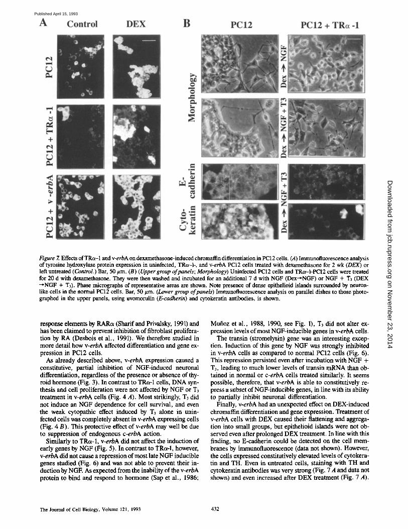
Figure 7. Effects of TRc~-I and v-erbA on dexamethasone-induced chromaflin differentiation in PC12 cells. (.4) Immunotluorescence analysis of tyrosine hydroxylase protein expression in uninfected, TRc~-I-, and v-erbA PCI2 cells treated with dexamethasone for 2 wk (DEX) or left untreated (Control.) Bar, 50 #m. (B) (Upper group ofpanels; Morphology) Uninfected PC12 cells and TRct-I-PCI2 cells were treated for 20 d with dexamethasone. They were then washed and incubated for an additional 7 d with NGF (Dex~NGF) or NGF + T3 (DEX ~NGF + T3). Phase micrographs of representative areas are shown. Note presence of dense epithelioid islands surrounded by neuron- like cells in the normal PC12 cells. Bar, 50 #m. (Lower group of panels) Immunofluorescence analysis on parallel dishes to those photo- graphed in the upper panels, using uvomorulin (E-cadherin) and cytokeratin antibodies, is shown.
response elements by RARot (Sharif and Pfivalsky, 1991) and has been claimed to prevent inhibition of fibroblast prolifera- tion by RA (Desbois et al., 1991). We therefore studied in more detail how v-erbA affected differentiation and gene ex- pression in PC12 cells.
As already described above, v-erbA expression caused a constitutive, partial inhibition of NGF-induced neuronal differentiation, regardless of the presence or absence of thy- roid hormone (Fig. 3). In contrast to TRot-1 cells, DNA syn- thesis and cell proliferation were not affected by NGF or T3 treatment in v-erbA cells (Fig. 4 A). Most strikingly, T3 did not induce an NGF dependence for cell survival, and even the weak cytopathic effect induced by T3 alone in unin- fected cells was completely absent in v-erbA expressing cells (Fig. 4 B). This protective effect of v-erbA may well be due to suppression of endogenous c-erbA action.
Similarly to TRot-I, v-erbA did not affect the induction of early genes by NGF (Fig. 5). In contrast to TRot-I, however, v-erbA did not cause a repression of most late NGF inducible genes studied (Fig. 6) and was not able to prevent their in- duction by NGE As expected from the inability of the v-erbA protein to bind and respond to hormone (Sap et al., 1986;
Mufioz et al., 1988, 1990, see Fig. 1), T3 did not alter ex- pression levels of most NGF-inducible genes in v-erbA ceils.
The transin (stromelysin) gene was an interesting excep- tion. Induction of this gene by NGF was strongly inhibited in v-erbA ceils as compared to normal PC12 cells (Fig. 6). This repression persisted even after incubation with NGF + T3, leading to much lower levels of transin mRNA than ob- tained in normal or c-erbA cells treated similarly. It seems possible, therefore, that v-erbA is able to constitutively re- press a subset of NGF-inducible genes, in line with its ability to partially inhibit neuronal differentiation.
Finally, v-erbA had an unexpected effect on DEX-induced chromatfin differentiation and gene expression. Treatment of v-erbA cells with DEX caused their flattening and aggrega- tion into small groups, but epithelioid islands were not ob- served even after prolonged DEX treatment. In line with this finding, no E-cadherin could be detected on the cell mem- branes by immunofluorescence (data not shown). However, the cells expressed constitutively elevated levels of cytokera- tin and TH. Even in untreated cells, staining with TH and cytokeratin antibodies was very strong (Fig. 7 A and data not shown) and even increased after DEX treatment (Fig. 7 A).
The Journal of Cell Biology, Volume 121, 1993 432
on Novem
ber 23, 2014jcb.rupress.org
Dow
nloaded from
Published April 15, 1993

Figure 8. Induction of chro- marlin and epithelial genes in erbA-expressing PC12 cells. Northern blot analyses of tyrosine hydroxylase (TH), chromogranin B, and cyto- keratin mRNA expression was performed using uninfected, TRcz-1, and v-erbA-PC12 cells. Cells were treated with DEX, DEX + T3 for 6 (6 d) or 14 d (14 d), or left untreated (-) . As controls, cyclophilin expression was determined. 10 #g of total RNA were loaded per lane. Exposure times: TH, 7 h; Chromogranin B, 4 h; 46 kd Cytokemtin, 3 d; and Cyclophilin, 2 d.
The results obtained for chromaffin marker gene expres- sion were in line with these observations. Untreated or NGF- treated v-erbA cells expressed significantly higher levels of TH, chromogranin B, and cytokeratin messages than the respective uninfected ceils (Fig. 8). However, DEX was still able to further induce these genes to similar levels in both normal and v-erbA-expressing ceils. This suggests that v-erbA
constitutively upregulates chromaffin marker gene expres- sion, but does not prevent their regulation by DEX (Fig. 8). A summary of the effects of c-erbA/TRot-1 and v-erbA on PC12 cell differentiation and survival is represented in Fig. 9.
To rule out the trivial possibility that our v-erbA-PC12 cells fail to differentiate because of irreversible cell line progression changes, we tried to induce differentiation in
Figure 9. Schematic representation of how TRot-1 and v-erbA regulate PC12 cell differentiation into neurons and chromaffin cells. (Upper panels) In both presence and absence of '1"3, normal PC12 cells differentiate into neurons or chromaflin cells upon NGF or dexamethasone treatment, respec- tively. They are independent of NGF for growth and survival in the absence of Ta and exhibit a weak or absem NGF dependence (+ / - ) in its presence. (Middle panels) TRot-l-expressing PCl2 cells are partially blocked (black bar across arrow) in their capacity to differentiate into either neurons or chromaflin cells in the absence of T3. Upon T~ addition, these cells respond normally to NGF, becoming neurons. In contrast, differenti- ation into more mature chromaflin cells remains blocked in the presence of T3. TRot-1 cells are strongly dependent (+++) on NGF for survival when 1"3 is present. (l_znverpanels) V-erbA-PC12 cells are partially blocked for NGF-induced neu- ronal differentiation regardless of the presence or absence of 1"3. They exhibit a probably aberrant phenotype combining neuronal and chromaflin markers (indicated by altered cell shape) in the presence of dexamethasone. They do not respond at all to T3 (-) .
Mufioz et al. erbA Regulates PC12 Cell Differentiation 433
on Novem
ber 23, 2014jcb.rupress.org
Dow
nloaded from
Published April 15, 1993

these cells by an alternative pathway. Cyclic AMP has been shown to induce part but not all of the NGF-induced pheno- typic and gene expression changes in PC12 cells, most likely via pathway(s) independent of those important for NGF action (see Halegoua et al., 1991). Indeed, NGF plus di- butyryl-cAMP (0.1 mM) induced distinct morphological differentiation characterized by typical long neurites in both TRct-1- and v-erbA-expressing PC12 cells. In line with pub- lished work (Gunning et al., 1981), dibutyryl cyclic AMP alone at high concentrations (1 mM) also induced partial differentiation in both cell types. Thus, the v-erbA-induced differentiation arrest cannot be due to mutational changes in our cell lines.
Spontaneously Immortalized Rat Brain Neuroblasts Differentiate in Response to T~ and NGF after Introduction of an Exogenous TR~-I Gene
Since TRot-1 has no reported function in differentiation of normal adrenal medulla progenitors, the possibility re- mained that our results employing the PC12 cell system rep- resented the aberrant behavior of a tumor cell line, mimick- ing normal differentiation processes as a consequence of artificial experimental treatments. We therefore sought to demonstrate an effect of TRu-1 in neuroblasts derived from the CNS, where the involvement of thyroid hormone recep-
tors in neuronal maturation is amply documented (see in- troduction). Consequently, we introduced the avian TRot-1 into the E 18 cell line, which has been obtained by spontane- ous immortalization from cultures of 17-d rat embryonic brain (Seliger, B., unpublished observations). E 18 cells rep- resent primitive neuroblasts that express NF 68 and the primitive neuronal marker nestin, but lack the astrocyte marker, glial flbrillary acidic protein. After partial differen- tiation induction with dibutyryl-cAMP, the cells express fur- ther neuronal markers such as NF 145, NF 220, and neuron- specific enolase. Similarly to PC12 ceils, the El8 cells ex- pressed only low levels of TRt~-I together with high levels of TRet-2 (data not shown).
To test the ability of TRt~-I expressing E 18 cells to differentiate in response to NGF, T3 or both, we used an E 18 clone infected with the TRt~-I retrovirus as described for PC12 cells. This clone expressed TRcM at similar levels as the TRot-1 PC12 clone cA4 (data not shown). As shown in Fig. 10, TRot-1 E 18 cells failed to differentiate in response to NGF alone, a property shared with uninfected E 18 ceils. Also, NGF seemed not to influence E 18 cell proliferation. Upon T3 addition, cells became less adherent and partially disintegrated (Fig. 10, T3) mirroring the cell killing effect observed in PC12 cells. In presence of both NGF and T3, however, essentially all cells underwent massive neuronal differentiation characterized by outgrowth of long neurites
Figure 10. T3 plus NGF in- duce differentiation in TRc~-I expressing E 18 neuroblasts derived from embryonic rat brain. Phase micrographs are shown from E 18 cA9 cells cultivated in serum-free differ- entiation medium for 6 d in absence (Control) or presence of T3, NGF, or both (NGF + /'3). Note effective differentia- tion into neuron-like cells upon treatment with both NGF and T3 (panel NGF + T3) Inset: Some of the neuron-like cells obtained with NGF + T3 in a separate experiment at higher magnification. Note also cell disintegration (refractile small bodies) in the Ta-treated cul- tures. Bar, 50/xm. Inset: bar, 20/~m.
The Journal of Cell Biology, Volume 121, 1993 434
on Novem
ber 23, 2014jcb.rupress.org
Dow
nloaded from
Published April 15, 1993

within 6 d, often in a bipolar fashion (Fig. 10). Thus, TRcM expression in CNS derived neuroblasts conferred respon- siveness to T3 in a fashion very similar to that observed in TRc~-I expressing PC12 cells.
Discussion
Effect of TRa4 on PC12 Cell Differentiation: Evidence for a NGF-independent, Permissive Function in Neuronal Differentiation
In this study, we provide the first direct evidence that TRot-1 controls neuronal differentiation and expression of neuron- specific genes in PC12 cells. Both NGF-induced neuronal differentiation and expression of late genes are arrested by TRtx-1 in the absence of its ligand, T3, while the block is released in the presence of the hormone, leading to efficient cell maturation (see Fig. 9 for a model). These effects of TRot-1 are strikingly similar to those observed in avian erythroblasts where TRot-1 regulates erythroid differentia- tion and erythrocyte gene expression in a T3-dependent fashion (Zenke et al., 1990; Disela et al., 1991).
How does unliganded TRot-1 arrest neuronal differentia- tion? First, its action seems to be independent of NGF, since it did not affect expression of NGF-induced early genes. It also failed to inhibit NGF action completely since NGF- treated TRo~-I-PC12 cells formed short neurites and lost ex- pression of the early neuronal marker MASH-1. Rather, TRot-1 probably causes a late block in neuronal differentia- tion, perhaps by repressing or limiting the expression of in- dispensable, neuron-specific proteins. A possible candidate for such proteins is N-CAM, whose expression correlates very well with morphological differentiation of PC12 cells and causes neurite formation if expressed artificially in non- neuronal cells (Mann et al., 1989; Doherty et al., 1991). However, it is more likely that the observed block of differen- tiation is a combined result of the insufficient expression of many proteins with such diverse functions as maintenance of neurite structure or function and regulation of cell motility or cell-substrate adhesion.
The second important effect of TRot-1 was its ability to ren- der differentiating PC12 cells dependent on NGF for sur- vival. These results may correspond to the well-known fact that primary neurons differentiating in culture require neuro- trophic factors for survival and neurite outgrowth. By such a mechanism, TRa-1 could control naturally occurring neu- ronal death during vertebrate development where large per- centages (up to 80%) of certain neuronal populations un- dergo programmed cell death (Oppenheim, 1991). In line with this idea, thyroid hormone controls tissue regression and neuronal death during amphibian metamorphosis (Kollros, 1981; Tata, 1984; Tata et al., 1991)and the survival of cerebellar granule cells in hypothyroid rats (Lewis et al., 1976).
The effects of TRo~-I and thyroid hormone were not limited to PC12 cells. Studies on the E 18 cell line suggest that simi- lar mechanisms are likely to be operating in vivo in selected neuronal populations of the CNS. In line with these findings we propose that TRo~-I controls neuronal differentiation in a permissive fashion: Neuronal progenitor ceils are arrested at a certain stage of maturation by nonliganded TRot-l, until they are released to differentiate when ligand becomes avail-
able. At this stage, neurotrophic factors like NGF are re- quired for survival of the differentiating cells. Thus, locally produced neurotrophic factors (NGF) and the systemic en- docrine agent T3 seem to cooperate in optimal induction of neuronal differentiation and neuronal gene expression. Cooperation of thyroid hormone and NGF has been demon- strated in vivo during development of the hippocampus, ol- factory bulb, and cerebellum (Clos and Legrand, 1990), regions which express abundantly the TRo~-I isoform of T3 receptor (MellstrSm et al., 1991).
In contrast to the erythroblast system (Schroeder et al., 1992a,b), TRa-1 does not seem to cooperate with RAPs in neuronal differentiation of PC12 cells since the respective ligand (RA) did not affect this process or its regulation by TRot-1. Whether such a cooperation would occur upon artificial expression of RAPs via retrovirus vectors or in mu- tant PC12 cells expressing elevated RAR levels (Scheibe et al., 1991) remains to be determined. The RAPs might, how- ever, be involved in regulating chromaffin differentiation, since RA induced morphological changes in PC12 cells resembling those induced by DEX. If this was true, the RAPs might antagonize the function of TRot-1 rather than cooperating with it.
V-erbA Acts Like a Weak Constitutive Repressor of NGF-induced Differentiation and Gene Expression
In contrast to TRo~-I, the v-erbA oncoprotein caused a par- tial, but constitutive block of NGF-induced PC12 cell differentiation. With the exception of the transin gene, ex- pression of which was constitutively repressed by v-erbA, NGF regulation of other late genes studied was hardly affected by v-erbA. This could be due to the fact that v-erbA has a lower binding affinity to regulatory sequences of target genes (Sap et al., 1989; Disela et al., 1991). In addition, its expression level in PC12 cells was perhaps not high enough to efficiently repress the genes studied other than transin. Even in avian erythroblasts (the cell type in which v-erbA has been selected for its oncogenic activity) a large excess ofv-erbA is necessary to repress genes activated by TRa-1/c- erbA or RAPS (Disela et al., 1991; Schroeder et al., 1992a).
Our results suggest that v-erbA constitutively affects both lineages of PC12 cell differentiation. It partially arrests neu- ronal differentiation and gene expression, and also inhibits chromaffin differentiation although it is able to constitutively turn on some DEX-induced genes. As a result, v-erbA gives rise to an aberrant "interlineage" phenotype characterized by coexpression of certain neuronal and chromaffin markers (see Fig. 9 for a model). There are two nonexclusive possi- bilities to explain how v-erbA and TRt~-l/c-erbA affect chromaffin differentiation and gene expression in such a different fashion. First, the point mutations in the v-erbA DNA binding domain might alter the DNA binding specificity of this mutated hormone receptor in that it now constitutively activates genes that are repressed by TRot-I. More likely, however, v-erbA differs from TRa-1/c-erbA in the way it interacts with other transcription factors such as RAPs (Sharif and Privalsky, 1991, Desbois et al., 1991) or members of the AP-1 transcription factor family (Zhang et al., 1991) in transient transcription assays using standard cell lines. This functional difference between v-erbA and TRot-1 may both explain their different regulation of the chromaffin
Mufioz et al. erbA Regulates PC12 Cell Differentiation 435
on Novem
ber 23, 2014jcb.rupress.org
Dow
nloaded from
Published April 15, 1993

marker gene tyrosine hydroxylase and their different effect on NGF-regulated proliferation of PC12 cells. Zhang et al. (1991) recently reported that T3-activated c-erbA inhibits AP-1 activity in transiently transfected cells. This may ex- plain why 1"3 inhibits cell proliferation in TRtx-I-PC12 cells. In addition, these authors showed that a truncated c-erbA carrying a COOH-terminal deletion very similar to that present in v-erbA was unable to block AP-1 activity, per- haps explaining why proliferation of our v-erbA-PC12 cells was not affected by T3 (Fig. 4 A).
TRa-l-dependent Regulation of PC12 Differentiation: Relevance for Neuronal Differentiation in the CNS Our results have several immediate consequences for the un- derstanding of how thyroid hormone might act in the CNS. The observed permissive action of TRc~-I on neuron matura- tion could be an underlying mechanism for the abnormalities occurring in the hypothyroid rat brain. Unliganded TRc~-I might cause a late block in neurotrophin-dependent neuronal maturation. Similarly, the observation that the lesions can be reverted by T3 administration during a critical period (Legrand, 1984) agrees well with the fact that T3 releases the cells from this late block. By using the PC12 cell line ap- proach, we were able to identify a good-sized number of can- didate genes for regulation by T3 and its receptor. Such genes may be impossible to detect by eDNA library screen- ing approaches employing the whole brain or parts of it (Mu- fioz et al., 1991), since they may only occur in selected neu- ronal cell populations or during specific phases of brain development.
Although this work strongly suggests a direct, important role of thyroid hormone receptors in brain development, we are ignorant about mechanisms governing TR expression, the ratios between the different TR types, and the (likely) functional differences between the different TRs. Thus, studies like those presented here will have to be extended to other, preferably brain-derived neural cell types, to other TR types, and to the other neurotrophic factors recently identified (Barde et al., 1982; Leibrock et al., 1989; Lin et al., 1989; Hohn et al., 1990; Jones and Reichardt, 1990; Maisonpierre et al., 1990; Berkemeier et al., 1991; Hall- b66k et al., 1991). An obvious choice for such systems will be neuroblast lines such as the E 18 line described here. Due to the instability of such lines and their reported ability to change their phenotype (e.g., from neuronal to glial, Gage, 1992), more work is required before such lines can be used for molecular studies in the fashion described here for the PC12 system.
We want to thank those mentioned in Materials and Methods for generously supplying us with plasmids or antibodies, and Dr. E. Reichmann and E. Deiner for their help with the immunofluorescence experiments. We are also grateful to Dr. M. Busslinger for fruitful discussions and for critically reading the manuscript.
Received for publication 11 September 1992 and in revised form 22 De- cember 1992.
Re)CeT~nccs
Anderson, D. J., and R. Axel. 1986. A bipotential neuroendocrine precursor whose choice of cell fate is determined by NGF and glueocorticoids. Cell. 47:1079-1090.
Barde, Y. A., D. Edgar, and H. Thoenen. 1982. Purification of a new neuro-
trophic factor from mammalian brain. EMBO (Eur. Mol. Biol. Organ.) J. 1:549-553,
Bartel, D. P., M. Sbeng, L. F. Lau, and M. E. Greenberg. 1989. Growth fac- tors and membrane depolarization activate distinct programs of early re- sponse gane expression: dissociation offos andjun induction. Genes & Dev. 3:304-313.
Bernal, J., and F. Pekonen. 1984. Ontogenesis of the nuclear 3,5,3'- triiodothyronine receptor in the human fetal brain. Endocrinology. 114: 677-679.
Berkemeier, L. R., J. W. Winslow, D. R. Kaplan, K. Nikolics, D. V. Goed- del, and A. Rosenthal. 1991. Neurotrophin-5: a novel neurotrophic factor that activates trk and trkB. Neuron, 7:857-866.
Beug, H., P. A. Blundell, and T. Graf. 1987. Reversibility of differentiation and proliferation capacity in avian myelomonocytic cells transformed by ts E26 leukemia virus. Genes & Dev. 1:277-286.
Beug, H., G. Doederlein, and M. Zenke. 1992. Transformation by v-erbA and v-erbB oncogenes: independent modulation of differentiation and prolifera- tion in erytiu'oid progenitors. In Bristol Myers Symposia on Nuclear Processes and Oncogenes. Vol. 15. P. A. Sharp, editor. Academic Press, Orlando, FL. 53-84.
Bottenstein, J. E., and G. H. Sato. 1979. Growth of a rat neurohlastoma cell line in serum-free supplemented medium. Proc. Natl. Acad. Sci. USA. 76:514-517.
Bradley, D. J., W. S. Young HI, and C. Weinberger. 1989. Differential expres- sion of c~ and/3 thyroid hormone receptor genes in rat brain and pituitary. Proc. Natl. Acad. Sci. USA. 86:7250-7254.
Chomczynski, P., and N. Sacchi. 1987. Single-step method of RNA isolation by acid guanidinium thiocyanate-phenol-chloroform extraction. Anal. Bio- chem. 162:156-159.
Church, G. M., and W. Gilbert. 1984. Genome sequencing. Proc. Natl. Acad. Sci. USA. 81:1991-1995.
Clos, J., and Ch. Legrand. 1990. An interaction between thyroid hormone and nerve growth factor promotes the development of hippocampus, olfactory bulbs and cerebellum: a comparative biochemical study of normal and hypothyroid rats. Growth Factors. 3:205-220.
Costello, B., A. Meymandi, and M. C. Freeman. 1990. Factors influencing GAP-43 gene expression in PC12 pheochromocytoma cells. J. Neurosci. 10:1398-1406.
Datum, K., C. C. Thompson, and R. M. Evans. 1989. Protein encoded by v-erbA functions as a thyroid hormone receptor antagonist. Nature (Lond.). 339:593-597.
Desbois, C., B. Pain, C. Guilhot, M. Benchaibi, M. French, J. Ghysdael, J.-J. Madjar, and J. Samarut. 1991. v-erbA oncogene abrogates growth inhibition of chicken embryo fibroblasts induced by retinoic acid. Oncogene. 6:2129- 2135.
Disela, C., C. Glineur, T. Bugge, J. Sap, G. Stengl, J. Dogson, H. Stunnen- berg, H. Beug, and M. Zenke. 1991. V-erbA overexpression is required to extinguish c-erbA function in erythroid cell differentiation and regulation of the erbA target gene CAll. Genes & Dev. 5:2033-2047.
Doherty, P., L. H. Rowett, S. E. Moore, D. A. Mann, andF. S. Walsh. 1991. Neurite outgrowth in response to transfected N-CAM and N-cadherin reveals fundamental differences in neuronal responsiveness to CAMs. Neuron. 6:247-258.
Doupe, A. J., S. C. landis, and P. H. Patterson. 1985a. Environmental influences in the development of neuronal crest derivatives: glucocorticoids, growth factors, and chromaffin cell plasticity. J. Neurosci. 5:2119-2142.
Doupe, A., P. H. Patterson, and S. C. landis. 1985b. Small intensely fluores- cent cells in culture: role of glucocorticoids and growth factors in their devel- opment and interconversions with neural crest derivatives. J. Neurosci. 5:2143-2160.
Dussault, J. H., and J. Ruel. 1987. Thyroid hormones and brain development. Annu. Rev. PhysioL 49:321-334.
Falcone, G., D. Boettiger, S. Alema, and F. Taro. 1984. Role of cell division in differentiation of myoblasts infected with a temperature-sensitive mutant of Rous sarcoma virus. EMBO (Eur. Mol. Biol. Organ.) J. 3:1327-1331.
Federoff, H. J., E. Grabczyk, and M. C. Fishman. 1988. Dual regulation of GAP-43 gane expression by nerve growth factor and glucocorticoids. J. Biol. Chem. 263:19290-19295.
Feinberg, P. A., and B. Vogelstein. 1983. A technique for radiolabeling DNA restriction endonuclease fragments to high specific activity. Anal. Biochem. 137:266-267.
Forrest, D., M. Sjorberg, and B. Vennstrtm. 1990. Contrasting developmental and tissue-specific expression of ot and/3 thyroid hormone receptor genes. EMBO (Eur. Mol. Biol. Organ.) J. 9:1519-1528.
Forrest, D., F. Hallb66k, H. Persson, and B. Veanstr6m. 1991. Distinct func- tions for thyroid hormone receptors c~ and/5 in brain development indicated by differential expression of receptor genes. EMBO (Eur. Mol. Biol. Organ.) Z 10:269-275.
Gage, F. H. 1992. Repepulating the mortal brain with immortal cells. Current Biology. 2:232-234.
Glineur, C., M. Zenke, H. Beug, and J. Ghysdael. 1990. Phosphorylntion of v-erbA is required for its function as an oncogene. Genes & Dev. 4:1663-1676.
Goldberg, Y., C. Glineur, J.-C. G-esquiere, A. Ricouart, J. Sap, B. Vermstr6m, and J. Ghysdael. 1988. Activation of protein kinase C or cAMP-dependent
The Journal of Cell Biology, Volume 121, 1993 436
on Novem
ber 23, 2014jcb.rupress.org
Dow
nloaded from
Published April 15, 1993

protein kinase increases pbosphorylation of the c-erbA-encoded thyroid hor- mone receptor and of the v-erbA-encoded protein. EMBO (Eur. Mol. Biol. Organ.) J. 7:2425-2433.
Gould, E., and L. L. Butcher. 1989. Developing cholinergic basal forebrain neurons are sensitive to thyroid hormone. J. Neurosci. 9:3347-3358.
Greenberg, M. E., L. A. Greene, and E. B. Ziff. 1985. Nerve growth factor and epidermal growth factor induce rapid transient changes in proto- oncogene transcription in PCl2 cells. J. Biol. Chem. 260:14101-14110.
Greene, L. A., and A. S. Tischler. 1976. Establishment of a noradrenergic clonal line of rat adrenal pheochromecytoma which respond to nerve growth factor. Proc. Natl. ,4cad. Sci. USA. 73:2424-2428.
Greene, L. A., and A. S. Tischler. 1982. PC12 pheochromocytoma cultures in neurobiological research. Adv. Cell. Neurobiol. 3:373-414.
Gunning, P. W., G. E. Landreth, M. A. Bothwell, and E. M. Shooter. 1981. Differential and synergistic actions of nerve growth factor and cyclic AMP in PCI2 cells. J. Cell Biol. 89:240-245.
Halegoua, S., R. C. Armstrong, and N. E. Kremer. 1991. Dissecting the mode of action of a neuronal growth factor. In Current Topics in Microbiology and Immunology. Neuronal Growth Factors. M. Bothwell, editor. Springer- Verlag, Berlin/Heidelberg. 119-170.
Hallbf6k, F., C. F. Ibafiez, and H. Persson. 1991. Evolutionary studies of the nerve growth factor family reveal a novel member abundantly expressed in Xenopus ovary. Neuron. 6:845-858.
Hamburg, M. 1969. The role of thyroid hormone and growth hormones in neu- rogenesis. In Current Topics in Developmental Biology 4, A. Moscona and A. Monroy, editors. Academic Press, New York. 109-148.
Hohn, A., J. Leibrock, K. Bailey, and Y.-A. Barde, 1990. Identification and characterization of a novel member of the nerve growth factor/brain-derived neurotrophic factor family. Nature (Lond.). 344:339-341.
Holtzman, D. M., Y. Li, L. F. Parada, S. Kinsman, C.-K. Cheo, J. S. Valletta, J. Zhou, J. B. Long, and W. C. Mobley. 1992. p140 irk mRNA marks NGF- responsive forebrain neurons: evidence that trk gene expression is induced by NGF. Neuron. 9:465-478.
Hudson, L. G., J. B. Santon, C. K. Glass, and J. A. Gill. 1990. Ligand- activated thyroid hormone and retinoic acid receptors inhibit growth factor receptor promoter expression. Cell. 62:1165-1175.
Ito, E., L. A. Sweterlitsch, P. B.-V. Tran, F. J. Rauscher III, and R. Naraya- nan. 1990. Inhibition of PC12 cell differentiation by the immediate early gene fra-l . Oncogene. 5:1755-1760.
Johnson, J. E., S. J. Birreo, and D. J. Anderson. 1990. Two rat homologues of Drosophila achaete-scute specifically expressed in neuronal precursors. Nature (Lond.). 346:858-861.
Jones, K. R., and L. J. Reichardt. 1990. Molecular cloning of a human gene that is a member of the nerve growth factor family. Proc. Natl. Acad. Sci. USA. 87:8060-8064.
Koenig, R. J., M. A. Lazar, R. A. Hodin, G. A. Brent, P. R. Larsen, W. W. Chin, and D. D. Moore. 1989. Inhibition of thyroid hormone action by a non-hormone binding c-erbA protein generated by alternative mRNA splic- ing. Nature (Lond.). 337:659--661.
Kollros, J. J. 1981. Transitions in the nervous system during amphibian metamorphosis. In Metamorphosis: A Problem in Developmental Biology. L. I. Gilbert and E. Frieden, editors. Plenum Press, New York. 445-459.
Kruijer, W., D. Schubert, and M. I. Verma. 1985. Induction of proto-oncogeoe fos by nerve growth factor. Proc. Natl. Acad. Sci. USA. 82:7330-7334.
Lazar, M. A., R, A. Hodin, and W. W. Chin. 1989. Human carboxyl-terminal variant of a c~-type c-erbA inhibits trans-activation by.thyroid hormone receptors without binding thyroid hormone. Proc. Natl. Acad. Sci. USA. 86:7771-7774.
Legrand, J. 1984. Effects of thyroid hormones on central nervous system. In Nearobehavioural Teratology. J. Yanai, editor. Elsevier, Amsterdam. 3314363.
Leibrock, J., F. Lottspeich, A. Hohn, M. Hofer, B. Hengerer, P. Masiakowski, H. Thoenen, and Y. A. Barde. 1989. Molecular cloning and expression of brain-derived neurotrophic factor. Nature (Lond.). 341:149-152.
Leonard, D. G. B., L. B. Ziff, and L. A. Greene. 1987. Identification and char- acterization of mRNAs regulated by nerve growth factor in PC 12 cells. Mol. Cell Biol. 7:3156-3167.
Levi, A., S. Biocca, A. Cattaneo, and P. Calissano. 1988. The mode of action of nerve growth factor in PCI2 ceils. Mol. Neurobiol. 2:201-226.
Lewis, p. D., A. J. Patel, A. L. Johnson, and R. Balazs. 1976. Effect of thyroid deficiency on cell acquistition in the postnatal rat brain: a quantitative histo- logical study. Brain Res. 104:49-62.
Lewis, E. J., A. W. Tank, N. Weiner, and D. Chikaraishi. 1983. Regulation of tyrosine hydroxylase mRNA by glucocorticoid and cyclic AMP in a rat pheochromocytoma cell line. J. Biol. Chem. 258:14,632-14,637.
Lin, L. F., D. Mismer, J. D. Lile, L. G. Armes, E. T. Butler Ili, J. L. Vannice, and F. Collins. 1989. Purification, cloning and expression of ciliary neuro- trophic factor. Science (Wash. DC). 246:1023-1025.
Lindenhaum, M. H., S. Carbonetto, F. Grosveld, D. Flavell, and W. E. Mushynski. 1988. Transcriptional and post-transcriptional effects of nerve growth factor on expression of the three neurofilament subunits in PCI2 cells. J. Biol. Chem. 263:5662-5667.
LO, L.-C., J. E, Johnson, C. Wuenschell, T. Saito, and D. J. Anderson. 1991. Manunalian achaete-scute homolog 1 is transiently expressed by spatially re- stricted subsets of early neureepithelial and neural crest cells. Genes & Dev.
5:1524-1537. Machida, C. M., K. D. Rodland, L. Matrisian, B. E. Magun, and G. Ciment.
1989. NGF induction of the gene encoding the protease transin accompanies neuronal differentiation in PC12 cells. Neuron. 2:1587-1596.
Malsonpierr¢, P. C., L. Belluscio, S. Squinto, N. Y. Ip, M. E. Furth, R. M. Lindsay, and G. D. Yancopoulos. 1990. Neurotrophin-3: a neurotrophic fac- tor related to NGF and BDNF. Science (Wash. DC). 247:1446-1451,
Maniatis, T., E. F. Fritsch, and J. Sambrook. 1982. Molecular Cloning: A Lab- oratory Manual. Cold Spring Harbor Laboratory, Cold Spring Harbor, NY. 545 pp.
Mann, D. A., P. Doberty, and F. S. Walsh. 1989. Increased intracellular cyclic AMP differentially modulates nerve growth factor induction of three neu- ronal recognition molecules involved in neurite outgrowth. J. Neurochem. 53:1581-1588.
Mellstr0m, B., J. R. Naranjo, A. Santos, A. M. Gonzalez, andJ. Bernal. 1991. Independent expression of the ct and/3 c-erbA genes in developing rat brain. Mol. Endocrinol. 5:1339-1350.
Milbrandt, J. 1987. A nerve growth factor-induced geoe encodes a possible transcriptional regulatory factor. Science (Wash. DC). 238:797-799.
Milbrandt, J. 1988. Nerve growth factor induces a gene homologous to the glu- cocorticoid receptor gene. Neuron. 1 : 183-188.
Morgan, J. I., and T. Curran. 1986. Role of ion flux in the control of c-los ex- pression, Nature (Lond.). 322:552-555.
Morreale de Escobar, G., F. Escobar del Rey, and A. Ruiz-Marcos. 1983. Thy- roid hormone and the developing brain. In Congenital Hypothyroidism. J. H. Dussault and P. Walker, editors. Marcel Decker, New York. 85-126.
Mufioz, A., M. Zenke, U. Gehring, J. Sap, H. Beog, andB. Vennstr6m. 1988. Characterization of the hormone-binding domain of the chicken c-erbAIthy- roid hormone receptor protein. EMBO (Eur. Mol. Biol. Organ.) J. 7:155- 159.
Mufioz, A., W. H6ppner, J. Sap, G. Brady, K. Nordstr6m, H. J. Seitz, and B. Vennstr6m. 1990. The chicken c-erbA-ct-product induces expression of thyroid hormone-responsive genes in 3,5,Y-triiodothyronine receptor- deficient rat hepatoma cells. Mol. Endocrinol. 4:312-320.
Mufioz, A., A. Rodrfguez-Pefia, A. P~rez-Castillo, B. Ferreiro, J. G. Sutcliffe, and J. Bernal. 1991. Effects of neonatal hypothyroidism on rat brain gene expression. Mol. Endocrinol. 5:273-280.
Oppenheim, R. W. 1991. Cell death during development of the nervous system. Annu. Rev. Neurosci. 14:453-501.
Patel, A. J., M. Hayashi, and A. Hunt. 1988. Role of thyroid hormone and nerve growth factor in the development of choline acetyltransferase and other cell-specific marker enzymes in the basal forebraln of the rat. J. Neurochem. 50:803-811.
Prentice, H. M., S. E. Moore, J. G. Dickson, P. Doherty, and F. S. Walsh. 1987. Nerve growth factor-induced changes in neural cell adhesion molecule (N-CAM) in PC12 cells. EMBO (Eur. Mol. Biol, Organ.) J. 6:1859-1863.
Rentoumis, A., V. Krishna, K. Chatterjee, L. D. Madison, S. Datta, G. D. Gal- lagher, L. J. DeGroot, and J. L. Jameson. 1990. Negative and positive tran- scriptional regulation by thyroid hormone receptor isoforms. Mol. En- docrinol. 4:1522-1531.
Sarnaels, I. J., F. Stanley, and L. E. Shapiro. 1976. Dose-dependent depletion of nuclear receptor levels by L-triiodothyronine: evidence for a role in induc- tion of growth hormone synthesis in cultured GH1 cells. Proc. Natl. Acad. Sci. USA. 73:3877-3881.
Samuels, J. J., F. Stanley, and L. E. Shapiro. 1977. Modulation of thyroid hor- mone nuclear receptor levels by 3,5,3'-triiodothyronine in GH1 cells. Evi- dence for two functional components of nuclear-bound receptor and relation- ship to the induction of growth hormone synthesis. J. Biol. Chem. 252: 6052-6060.
Sap, J., A. Mufioz, K. Datum, Y. Goldberg, J. Ghysdael, A. Leutz, H. Beug, and B. Vennstr6m. 1986. The c-erbA protein is a high affinity receptor for thyroid hormone. Nature (Lond.). 324:635-640.
Sap, J., A. Mufioz, J. Schmitt, H. Stunnenberg, and B. Vennstr6m. 1989. Repression of transcription mediated at a thyroid hormone response element by the v-erbA oncogeoe product. Nature (Lond.). 340:242-244.
Scheibe, R. J., D. D. Ginty, and J. O. Wagner. 1991. Retinoic acid stimulates the differentiation of PC 12 cells that are deficient in cAMP-dependent protein kinase. J. Cell Biol. 113:1173-1182.
Schroeder, C., L. Gibson, and H. Beug. 1992a. The v-erbA oncogene requires cooperation with tyrosine kinases to arrest erythroid differentiation induced by ligand activated endogenous c-erbA and retinoic acid receptor. On- cogene. 7:203-216.
Schroeder, C., L. Gibson, M. Zenke, and H. Beug. 1992b. Modulation of nor- mal erythroid differentiation by the endogenous thyroid hormone and retinoic acid receptors: a possible target for v-erbA oncogeoe action, On- cogene. 7:217-227.
Sharif, M., and M. Privalsky. 1991. V-erbA oncogeoe function in neoplasia correlates with its ability to repress retinoic acid receptor action. Cell. 66:1-20.
Stein, R., N. Mori, K. Matthews, L. C. LO, and D. J. Anderson. 1988a. The NGF-inducible SCG10 mRNA encodes a novel membrane-bound protein present in growth cones and abundant in developing neurons. Neuron. 1:463--476.
Stein, R., S. Orit, and D. J. Anderson. 1988b. The induction of a neural-specific gena, SCGI0, by nerve growth factor in PC12 cells is transcriptional, pro-
Mufioz et al, erbA Regulates PC12 Cell Differentiation 437
on Novem
ber 23, 2014jcb.rupress.org
Dow
nloaded from
Published April 15, 1993

tein synthesis dependent, and glucocorticoid inhibitable. Dev. Biol. 127: 316-325.
Strait, K. A., H. L. Schwartz, V. S. Seybold, N. C. Ling, and J. H. Oppen- heimer. 1991. Immunofluorescence localization of thyroid hormone receptor protein/~1 and variant ct2 in selected tissues: cerebellar Purkinje cells as a model for/31 receptor-mediated developmental effects of thyroid hormone in brain. Proc. Natl. Acad. Sci. USA. 88:3887-3891.
Szeber6nyi, J., H. Cai, and G. M. Cooper. 1990. Effect of a dominant inhibi- tory Ha-ras mutation on neuronal differentiation of PCI2 cells. Mol. Cell Biol. 10:5324-5332.
Tata, J. R. 1984. The action of growth and developmental hormones: evolution- ary aspects. In Biological Regulation and Development. Vol. 3B, R. F. Gold- berger and K. R. Yamamoto, editors. Plenum Press, New York. 1-58.
Tata, J.R., A. Kawahara, and B. S. Baker. 1991. Prolactin inhibits both thyroid bormone-induced morpbogenesis and cell death in cultured amphibian larval tissues. Dev. Biol. 146:72-80.
Van Hoof, C. O., J. C. Hoithuis, A. B. Oestriecher, J. Boonstra, P. N. De Graan, and W. H. Gispen. 1989. Nerve growth factor-induced changes in
the intracellular localization of the protein kinase C substrate B-50 in pheochromocytoma PC12 cells. J. Cell Biol. 108:1115-1125.
Vennstr6m, B., and J. M. Bishop. 1982. Isolation and characterization of chicken DNA homologous to the two putative oncogenes of avian erythro- blastosis virus. Cell. 28:135-143.
Weinberger, C., C. C. Thompson, E. S. Ong, R. Lebo, D. J. Gruol, and R. M. Evans. 1986. The c-erbA encodes a thyroid hormone receptor. Nature (Lond.). 324:641-646.
Zenke, M., P. Kahn, C. Disela, B. Vennstr6m, A. Leutz, K. Keegan, M. J. Hayman, H.-R. Choi, N. Yew, J. D. Engel, and H. Beug. 1988. v-erbA specifically suppresses transcription of the avian erythrocyte anion trans- porter (band 3) gene. Cell. 52:107-119.
Zenke, M., A. Mufioz, J. Sap, B. Vennstr6m, and H. Beug. 1990. v-erbA on- cogene activation entails the loss of hormone-dependent regulator activity of c-erbA. Cell. 61:1035-1049.
Zhang, X.-K., K. N. Wills, M. Husmann, T. Hermarm, and M. Pfahl. 1991. Novel pathway for thyroid hormone receptor action through interaction with jun and fos oncogene activities. Mol. Cell. Biol. 11:6016-6025.
The Journal of Cell Biology, Volume 121, 1993 438
on Novem
ber 23, 2014jcb.rupress.org
Dow
nloaded from
Published April 15, 1993
Copyright © 2022 FDOKUMEN