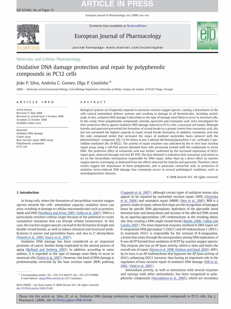
Oxidative DNA damage protection and repair by polyphenolic compounds in PC12 cells
11
Molecular and Cellular Pharmacology Oxidative DNA damage protection and repair by polyphenolic compounds in PC12 cells João P. Silva, Andreia C. Gomes, Olga P. Coutinho ⁎ CBMA — Molecular and Environmental Biology Centre/Biology Department, University of Minho, Campus de Gualtar, 4710-057 Braga, Portugal abstract article info Article history: Received 11 May 2008 Received in revised form 3 October 2008 Accepted 23 October 2008 Available online xxxx Keywords: Oxidative DNA damage Comet assay Base excision repair (BER) assay Polyphenolic compound PC12 cell Biological systems are frequently exposed to excessive reactive oxygen species, causing a disturbance in the cells natural antioxidant defence systems and resulting in damage to all biomolecules, including nucleic acids. In fact, oxidative DNA damage is described as the type of damage most likely to occur in neuronal cells. In this study, three polyphenolic compounds, luteolin, quercetin and rosmarinic acid, were investigated for their protective effects against oxidative DNA damage induced in PC12 cells, a neuronal cell model. Although luteolin and quercetin prevented the formation of strand breaks to a greater extent than rosmarinic acid, this last one presented the highest capacity to repair strand breaks formation. In addition, rosmarinic acid was the only compound tested that increased the repair of oxidized nucleotidic bases induced with the photosensitizer compound [R]-1-[(10-chloro-4-oxo-3-phenyl-4H-benzo[a]quinolizin-1-yl) carbonyl]-2-pyr- rolidine-methanol (Ro 19-8022). The activity of repair enzymes was indicated by the in vitro base excision repair assay, using a cell-free extract obtained from cells previously treated with the compounds to incise DNA. The protective effect of rosmarinic acid was further confirmed by the increased expression of OGG1 repair gene, observed through real time RT-PCR. The data obtained is indicative that rosmarinic acid seems to act on the intracellular mechanisms responsible for DNA repair, rather than by a direct effect on reactive oxygen species scavenging, as deducted from the effects observed for luteolin and quercetin. Therefore, these results suggest the importance of these polyphenols, and in particular rosmarinic acid, as protectors of oxidative stress-induced DNA damage that commonly occurs in several pathological conditions, such as neurodegenerative diseases. © 2008 Elsevier B.V. All rights reserved. 1. Introduction In living cells, when the formation of intracellular reactive oxygen species exceeds the cells' antioxidant capacity, oxidative stress can arise, resulting in damage to cellular macromolecules such as proteins, lipids and DNA (Nordberg and Arner, 2001; Valko et al., 2007). DNA is a particularly sensitive cellular target because of the potential to create cumulative mutations that can disrupt cellular homeostasis. In this case, the reactive oxygen species can lead to the formation of single and double-strand breaks, as well as induce chemical and structural modi- fications to purine and pyrimidine bases, and also to 2′-deoxyribose (Powell et al., 2005; Hazra et al., 2007). Oxidative DNA damage has been considered as an important promoter of cancer, besides being implicated in the normal process of aging (Bjelland and Seeberg, 2003). In addition, according to some authors, it is regarded as the type of damage most likely to occur in neuronal cells (Fishel et al., 2007). However, this kind of DNA damage is predominantly corrected by the base excision repair (BER) pathway (Coppede et al., 2007), although certain types of oxidative lesions also appear to be repaired by nucleotide excision repair (NER) (Dusinska et al., 2006) and mismatch repair (MMR) (Neri et al., 2005). BER is a generic mode of repair, whose first steps are the recognition of damaged bases by specific DNA glycosylases, hydrolysis of the glycosidic bond between base and deoxyribose and incision of the affected DNA strand by an apurinic/apyrimidinic (AP) endonuclease at the resulting abasic site, thus creating a DNA single-strand break (Burkle, 2006; Collins and Gaivão, 2007). The most important enzymes involved in DNA repair are 8-oxoguanine DNA glycosylase 1 (OGG1) and AP endonuclease 1 (APE1). In mammals, OGG1 is responsible for the removal of 8-oxoguanine, a lesion that arises through the incorporation, during DNA replication, of 8-oxo-dGTP formed from oxidation of dGTP by reactive oxygen species. This enzyme also has an AP lyase activity, which is slow and limits the overall rate of repair (Alamo et al., 1998; Dodson and Lloyd, 2002). APE1, by its turn, is an AP endonuclease that bypasses the AP lyase activity of OGG1, enhancing OGG1 turnover, thus having an important role in the regulation of base excision repair of oxidative DNA damage (Hill et al., 2001; Vidal et al., 2001). Antioxidant activity, as well as interaction with several enzymes and synergy with other antioxidants, has been recognized to poly- phenolic compounds (Horvathova et al., 2005), which are secondary European Journal of Pharmacology xxx (2008) xxx–xxx ⁎ Corresponding author. Tel.: +351 253 604317; fax: +351 253 678980. E-mail address: [email protected] (O.P. Coutinho). EJP-65546; No of Pages 11 0014-2999/$ – see front matter © 2008 Elsevier B.V. All rights reserved. doi:10.1016/j.ejphar.2008.10.046 Contents lists available at ScienceDirect European Journal of Pharmacology journal homepage: www.elsevier.com/locate/ejphar ARTICLE IN PRESS Please cite this article as: Silva, J.P., et al., Oxidative DNA damage protection and repair by polyphenolic compounds in PC12 cells, Eur. J. Pharmacol. (2008), doi:10.1016/j.ejphar.2008.10.046
-
Upload
independent -
Category
Documents
-
view
1 -
download
0
Transcript of Oxidative DNA damage protection and repair by polyphenolic compounds in PC12 cells
European Journal of Pharmacology xxx (2008) xxx–xxx
EJP-65546; No of Pages 11
Contents lists available at ScienceDirect
European Journal of Pharmacology
j ourna l homepage: www.e lsev ie r.com/ locate /e jphar
ARTICLE IN PRESS
Molecular and Cellular Pharmacology
Oxidative DNA damage protection and repair by polyphenoliccompounds in PC12 cells
João P. Silva, Andreia C. Gomes, Olga P. Coutinho ⁎CBMA — Molecular and Environmental Biology Centre/Biology Department, University of Minho, Campus de Gualtar, 4710-057 Braga, Portugal
⁎ Corresponding author. Tel.: +351 253 604317; fax: +E-mail address: [email protected] (O.P. Coutinh
0014-2999/$ – see front matter © 2008 Elsevier B.V. Aldoi:10.1016/j.ejphar.2008.10.046
Please cite this article as: Silva, J.P., et al.,Pharmacol. (2008), doi:10.1016/j.ejphar.200
a b s t r a c t
a r t i c l e i n f oArticle history:
Biological systems are frequ Received 11 May 2008Received in revised form 3 October 2008Accepted 23 October 2008Available online xxxxKeywords:Oxidative DNA damageComet assayBase excision repair (BER) assayPolyphenolic compoundPC12 cell
ently exposed to excessive reactive oxygen species, causing a disturbance in thecells natural antioxidant defence systems and resulting in damage to all biomolecules, including nucleicacids. In fact, oxidative DNA damage is described as the type of damage most likely to occur in neuronal cells.In this study, three polyphenolic compounds, luteolin, quercetin and rosmarinic acid, were investigated fortheir protective effects against oxidative DNA damage induced in PC12 cells, a neuronal cell model. Althoughluteolin and quercetin prevented the formation of strand breaks to a greater extent than rosmarinic acid, thislast one presented the highest capacity to repair strand breaks formation. In addition, rosmarinic acid wasthe only compound tested that increased the repair of oxidized nucleotidic bases induced with thephotosensitizer compound [R]-1-[(10-chloro-4-oxo-3-phenyl-4H-benzo[a]quinolizin-1-yl) carbonyl]-2-pyr-rolidine-methanol (Ro 19-8022). The activity of repair enzymes was indicated by the in vitro base excisionrepair assay, using a cell-free extract obtained from cells previously treated with the compounds to inciseDNA. The protective effect of rosmarinic acid was further confirmed by the increased expression of OGG1repair gene, observed through real time RT-PCR. The data obtained is indicative that rosmarinic acid seems toact on the intracellular mechanisms responsible for DNA repair, rather than by a direct effect on reactiveoxygen species scavenging, as deducted from the effects observed for luteolin and quercetin. Therefore, theseresults suggest the importance of these polyphenols, and in particular rosmarinic acid, as protectors ofoxidative stress-induced DNA damage that commonly occurs in several pathological conditions, such asneurodegenerative diseases.
© 2008 Elsevier B.V. All rights reserved.
1. Introduction
In living cells, when the formation of intracellular reactive oxygenspecies exceeds the cells' antioxidant capacity, oxidative stress canarise, resulting in damage to cellular macromolecules such as proteins,lipids and DNA (Nordberg and Arner, 2001; Valko et al., 2007). DNA is aparticularly sensitive cellular target because of the potential to createcumulative mutations that can disrupt cellular homeostasis. In thiscase, the reactive oxygen species can lead to the formationof single anddouble-strand breaks, as well as induce chemical and structural modi-fications to purine and pyrimidine bases, and also to 2′-deoxyribose(Powell et al., 2005; Hazra et al., 2007).
Oxidative DNA damage has been considered as an importantpromoter of cancer, besides being implicated in the normal process ofaging (Bjelland and Seeberg, 2003). In addition, according to someauthors, it is regarded as the type of damage most likely to occur inneuronal cells (Fishel et al., 2007). However, this kind of DNA damage ispredominantly corrected by the base excision repair (BER) pathway
351 253 678980.o).
l rights reserved.
Oxidative DNA damage prot8.10.046
(Coppede et al., 2007), although certain types of oxidative lesions alsoappear to be repaired by nucleotide excision repair (NER) (Dusinskaet al., 2006) and mismatch repair (MMR) (Neri et al., 2005). BER is agenericmode of repair, whose first steps are the recognition of damagedbases by specific DNA glycosylases, hydrolysis of the glycosidic bondbetween base and deoxyribose and incision of the affected DNA strandby an apurinic/apyrimidinic (AP) endonuclease at the resulting abasicsite, thus creating a DNA single-strand break (Burkle, 2006; Collins andGaivão, 2007). The most important enzymes involved in DNA repair are8-oxoguanineDNA glycosylase 1 (OGG1) and AP endonuclease 1 (APE1).In mammals, OGG1 is responsible for the removal of 8-oxoguanine,a lesion that arises through the incorporation, duringDNA replication, of8-oxo-dGTP formed from oxidation of dGTP by reactive oxygen species.This enzyme also has an AP lyase activity, which is slow and limits theoverall rate of repair (Alamo et al.,1998; Dodson and Lloyd, 2002). APE1,by its turn, is an AP endonuclease that bypasses the AP lyase activity ofOGG1, enhancing OGG1 turnover, thus having an important role in theregulation of base excision repair of oxidative DNA damage (Hill et al.,2001; Vidal et al., 2001).
Antioxidant activity, as well as interaction with several enzymesand synergy with other antioxidants, has been recognized to poly-phenolic compounds (Horvathova et al., 2005), which are secondary
ection and repair by polyphenolic compounds in PC12 cells, Eur. J.

2 J.P. Silva et al. / European Journal of Pharmacology xxx (2008) xxx–xxx
ARTICLE IN PRESS
plant metabolites with numerous other biological activities (Rice-Evans et al., 1996; Hollman and Katan, 1999; Skibola and Smith, 2000).In the present study, attention was given to the flavonoids quercetinand luteolin, and to the phenolic acid, rosmarinic acid (Fig. 1).Quercetin is one of the most abundant natural flavonoids and can befound in onion, tea and apple, for example (Scalbert et al., 2005).Luteolin, which differs from quercetin by having one less hydroxylgroup in the C-ring of its molecular structure (Fig. 1), is found in highcelery, green pepper and chamomile (Gutierrez-Venegas et al., 2006).Both quercetin and luteolin, which by their chemical nature areantioxidants, have been associated to the prevention of cancer,diabetes, osteoporosis, as well as cardiovascular and neurodegenera-tive diseases, among others (Hollman and Katan, 1999; Aherne andO'Brien, 2000; Scalbert et al., 2005). Rosmarinic acid is present inmany plants, such as rosemary (Petersen and Simmonds, 2003) and itstherapeutical value has been attributed to its antioxidant, anti-inflammatory, anti-bacterial and anti-viral properties (Petersen andSimmonds, 2003; Chlopcíková et al., 2004; Kim et al., 2005; Iuvoneet al., 2006). Neuroprotective effects have also been described for all ofthese compounds (Gelinas and Martinoli, 2002; Sasaki et al., 2003;Iuvone et al., 2006), suggesting their potential protective role inneurodegenerative diseases. Moreover, studies using some of thesecompounds have demonstrated their ability to protect different celltypes against oxidative DNA damage (Noroozi et al., 1998; Duthie andDobson, 1999; Horvathova et al., 2005; Lima et al., 2006). However,whether they act by enhancing DNA repair or simply by preventingoxidative DNA damage is still unknown.
In this work, we evaluated the potential of these polyphenoliccompounds to protect PC12 cells against oxidative DNA damage, byusing the Comet assay. In addition, by addressing specific repairenzymes, we aimed to further characterize that protection and explorethe involvement of those compounds in DNA repair mechanisms. ThePC12 cell model was used as a simple model in which a correlationbetween oxidative stress and neurodegeneration, characteristic ofParkinson's or Alzheimer diseases, has been established by severalauthors (Piga et al., 2005; Guan et al., 2006; Jung et al., 2007).
2. Materials and methods
2.1. Chemicals
Quercetin and rosmarinic acid were purchased from Sigma-Aldrich(St. Louis, MO, USA). Luteolin was purchased from Extrasynthese
Fig. 1. Chemical structures of the polyphenolic compounds used in this study: luteolin,quercetin and rosmarinic acid.
Please cite this article as: Silva, J.P., et al., Oxidative DNA damage protPharmacol. (2008), doi:10.1016/j.ejphar.2008.10.046
(Genay, France). The polyphenolic compounds were dissolved inDMSO, aliquoted and maintained frozen at −20 °C until usage. Eachaliquot was thawed only once.
RPMI-1640 cell culturemedium, dimethyl sulfoxide (DMSO), EDTA,trypsin, tert-butyl hydroperoxide were also purchased from Sigma-Aldrich (St. Louis, MO, USA).
Foetal bovine serum (FBS)was fromBioChromKG (Berlin, Germany);horse serum donor herd was purchased from Gibco (Paisley, UK).[R]-1-[(10-chloro-4-oxo-3-phenyl-4H-benzo[a]quinolizin-1-yl) carbo-nyl]-2-pyrrolidine-methanol (Ro 19-8022), used to induce specificDNA damage, was kindly provided by Hoffman La-Roche (Basel,Switzerland). Primers specific for 18S, OGG1 and APE1 genes weresynthesized by STAB-VIDA (Oeiras, Portugal). Qiagen RNeasy total RNAisolation kit was purchased to Qiagen (Hilden, Germany). SuperscriptReverse Transcriptase III kit was obtained from Invitrogen (USA). PowerSYBR Green master mix was acquired from Applied Biosciences(Cheshire, UK).
2.2. PC12 cell culture and treatment conditions
PC12, a neuronal cell line established from a rat adrenal pheochro-mocytoma (Greene and Tischler, 1976) was used in this study. Whengrown in serum-containing medium, these cells divide and resembleprecursors of adrenal chromaffin cells and sympathetic neurons, beingable to release dopamine (Sasaki et al., 2003). In addition, they are auser-friendly cell model with some advantages over primary culturedneuronal cells, including the homogeneity of the cell population(Colognato et al., 2006; Silva et al., 2008). Cells were cultured insuspension in 75 cm2
flasks, in RPMI-1640 medium supplementedwith 10% (v/v) heat-inactivated horse serum, 5% (v/v) heat-inactivatedfoetal bovine serum and 1% (v/v) of an antibiotic/antimycotic solution.Cultures were maintained in a humidified incubator containing95% air and 5% CO2, and passed twice a week. Before each assay, thecell aggregates were carefully disrupted by gently pipetting and theseparated cells plated in poly-D-lysine-coated multiwells, at a densityof 2.5×10⁵ cells/cm2, for the 3-(4,5-dimethylthiazol-2-yl)-2,5-diphe-nyltetrazolium bromide (MTT) assay, and at a density of 5×10⁵ cells/well for the other studies. After plating, cells were left for adhesionovernight. The polyphenols were either pre-incubated for theindicated periods of time or added simultaneously with the dele-terious stimuli.
L929 cells, used as substrates in the in vitro base excision repairassay, were routinely grown in 75 cm2 tissue culture flasks in DMEMsupplemented with 10% (v/v) heat-inactivated FBS and 1% (v/v) of anantibiotic/antimycotic solution containing 10,000 U of penicillin,10 mg streptomycin and 25 µg amphotericin B per ml.
2.3. Analysis of cell survival
Cell viability was evaluated by the 3-(4,5-dimethylthiazol-2-yl)-2,5-diphenyltetrazolium bromide (MTT) reduction test, as previouslydescribed (Silva et al., 2006). A volume of 0.5 ml MTT (finalconcentration 0.5 mg/ml, in Krebs medium, pH=7.4), prepared justbefore usage and maintained in the dark, was added to the PC12 cells,at a density of 2.5×10⁵ cells/cm2. Absorbance was read at 570 nm in amultiplate reader (Spectramax 340PC). The survival of PC12 cells wasexpressed as the percentage of OD towards control cells, containingthe same amount of the compounds' solvent, DMSO.
2.4. Single cell gel electrophoresis (Comet assay)
The protection against oxidative DNA damage conferred by thepolyphenols herein studied was assessed using the Comet assay, inwhich the strand breaks present in the DNA of nucleoids, obtainedafter lysis of gel-embedded cells, migrate towards the anode during anelectrophoresis in alkali conditions, yielding an image that, after
ection and repair by polyphenolic compounds in PC12 cells, Eur. J.

3J.P. Silva et al. / European Journal of Pharmacology xxx (2008) xxx–xxx
ARTICLE IN PRESS
staining with a fluorescent dye, looks like a comet, hence its name.This methodwas performed as previously described (Tice et al., 2000),with the following modifications. Cells were plated at a density of7.5×105 cells per well and left to adhere overnight. Oxidative DNAdamage was induced in the presence of either 200 µM t-BHP (forstrand breaks assessment) or 0.6 µM Ro 19-8022, in order to measureoxidized purines. Cells were then trypsinized, resuspended inphosphate buffered saline (PBS) and counted. About 50,000 cellswere centrifuged at ~1500 ×g, for 1 min, in an Eppendorf 5415Ccentrifuge. Supernatants were discarded and pelletsmixedwith 100 μlof low melting point agarose 0.5% (w/v) in PBS, at 37 °C, and spreadon slides previously coated with normal melting point agarose for10 min, at 4 °C. The slides were immersed in lysis buffer (2.5 M NaCl,100 mM EDTA, 10 mM Tris, pH 10, with 1% Triton X-100 v/v added atthe time of buffer preparation) at 4 °C for a minimum of 2 h. Slideswere then rinsed with distilled water and immersed in electrophor-esis buffer (300 mM NaOH, 1 mM EDTA, pHN13), in an horizontalelectrophoresis tank, at 4 °C for 40 min, to allow alkaline unwinding.Electrophoresis was carried out at 4 °C, under alkaline conditions for20 min at 1 V/cm. Finally, slides were neutralized by washing threetimes (5 min each) with 0.4 M Tris, pH 7.5, and fixed with absoluteethanol (two washes).
Comets were stained with ethidium bromide (10 µg/ml in PBS)and analyzed under a fluorescence microscope. Comet quantificationwas performed through either of two ways: visual scoring, a methodin which comets are classified into one of five classes of damage(from 0 to 4) in 100 nucleoids (range of score: 0–400); and/or througha computer-assisted image analysis (TriTek CometScore™ Freewarev1.5), by measuring the percentage of DNA in the tail. The methodidentifies DNA strand breaks.
Cells repair capacity (RC) was determined by using the followingformula:
RC = 100× D0X−DtXð Þ=D0X½ �;
where D0X represent DNA damage before the recovery period in thecondition X and DtX represent DNA damage after a recovery period,for the same condition. The increase in cells' repair capacity inducedby the polyphenolic compounds was obtained by subtracting thepercentage of repair observed in the presence of the deleteriousstimulus alone, to the percentage of repair in the presence of thecompounds and the deleterious stimulus.
2.5. Measurement of oxidized purines
Occurrence of oxidized bases were measured by using the bacterialDNA repair enzyme formamidopyrimidine DNA glycosylase (FPG),which recognizes oxidized purines, creating breaks at those sites(Duthie and Dobson, 1999).
Cells were treated with the photosensitizer compound Ro 19-8022(a kind gift from Hoffmann La-Roche, Basel, Switzerland). A stock of a1 mM concentration was prepared in 70% ethanol and stored in smallaliquots at −20 °C. The working solutions were prepared immediatelybefore use by diluting this stock solution with PBS (132 mM NaCl,4 mM KCl, 1.2 mM NaH2PO4, 1.4 mM MgCl2, 10 mM HEPES, 6 mMglucose, 1 mM CaCl2 pH 7.4). The experiment was carried out inthe dark and all solutions were kept on ice. Cells, plated and treatedas previously described, were rinsed with PBS and 3 ml of the dilutedRo 19-8022 solution was added to each well. The plates wereirradiated on ice at a distance of 33 cm from a 500 W halogen lamp,for 5 min. Control cells were incubated in the presence of PBS alone.After washing the cells with PBS to remove traces of Ro 19-8022,the Comet assay was performed as described above, with a smallchange: following lysis the slides were washed 3 times (5 min each) incold enzyme reaction buffer (40 mM HEPES-KOH, 0.1 M KCl, 0.5 mMEDTA, 0.2 mg/ml BSA, pH 8, 4 °C), blotted dry and incubated with the
Please cite this article as: Silva, J.P., et al., Oxidative DNA damage protPharmacol. (2008), doi:10.1016/j.ejphar.2008.10.046
repair enzyme (FPG), or buffer, for 30 min, at 37 °C. The amount ofoxidized purines (FPG-sensitive sites) was then determined bysubtracting the amount of strand breaks (samples incubated withbuffer alone) to the total amount of breaks obtained after incubationwith FPG.
2.6. In vitro base excision repair assay
The base excision repair (BER) assay measures the ability of acell-free extract to recognize the damage in the DNA of substratenucleoids and incise the DNA containing specific damage, in this case8-oxoguanine (8-oxoGua). The increase in the amount of strandbreaks produced reflects the DNA repair activity of the cell extract(Collins, 2004).
L929 fibroblasts were used as substrates. This allows an easiermeasurement of comets damage due to the greater size of these cellsand their lack of a tendency to form aggregates when compared toPC12 cells. Briefly, L929 cells in a 75 cm2
flask near confluence weretreated with the photosensitizer Ro 19-8022 plus visible light (5 minirradiation on ice at 33 cm from a 500 W halogen lamp). Cells werethen washed with PBS, trypsinized and collected in 5 ml of culturemedium. After a centrifugation at 400 g for 5 min, at 4 °C, in a Sigma2K15 microcentrifuge, the pellet was suspended in freezing medium(culture medium with 20% foetal bovine serum and 10% DMSO) at adensity of 3×10⁶ cells/ml. Cells were then aliquoted and stored at−80 °C.
For the extract preparation, PC12 cells, at a density of 5×10⁶ cells/ml,were incubated with the polyphenols for either 1 or 24 h. Cells werethen washed with PBS, trypsinized and collected in PBS to micro-centrifuge tubes. After centrifuging at 800 g for 5 min, at 4 °C, in aSigma 2K15 microcentrifuge, the pellets were resuspended in icecold PBS. Cells were divided into 1 ml aliquots and centrifuged at14,000 g for 5 min, at 4 °C. Supernatants were discarded and thedry pellets flash frozen in liquid nitrogen, to be stored at −80 °C. Onthe day of the experiment, one of these aliquots was thawed andthe pellet resuspended in 65 µl of lysis buffer (45 mM HEPES, 0.4 MKCl, 1 mM EDTA, 0.1 mM dithiothreitol, 10% glycerol, pH 7.8 withKOH, supplemented with 0.25% Triton X-100). The mixture wasvortexed for 5 s, followed by a 5 min incubation on ice, and thencentrifuged at 14,000 g for 5 min, at 4 °C. Finally, 55 µl of the super-natant were removed and combined with 220 µl of cold enzymereaction buffer.
An aliquot of Ro- and light-treated substrate L929 cells wasthawed, washed twice in cold PBS and spinned for 5 min at 800 g, 4 °C.The pellets were suspended in 100 µl of PBS and 35 µl of thissuspension was mixed with 1.5 ml of 1% low-melting point agarose at37 °C. These cells were then embedded in agarose and the procedurethen followed according to the standard Comet assay, with a slightmodification: after lysis, the slides werewashed three times (for 5mineach) in cold enzyme reaction buffer and incubated with 35 µl oftreated or non-treated extract for 20 min. Control slides incubatedwith either FPG (positive control) or buffer alone (negative control)were performed.
2.7. Quantification of rOGG1 and rAPE1 expression
Total RNA was isolated from PC12 cells treated in the sameconditions as for the base excision repair assay using a Qiagen RNeasytotal RNA isolation kit (Qiagen, USA), following standard protocol. RNApurity was confirmed by determining the OD260/OD280 nm absorp-tion ratio. 1 µg of total RNA was reversed transcribed with theSuperscript Reverse Transcriptase III kit (Invitrogen, USA), by using50 ng/µl of random hexamers and 10 mM of a dNTP mix according tothe manufacturer's instructions. cDNA integrity was verified by gelelectrophoresis after PCR amplification of Gapdh, using sequence-specific primers.
ection and repair by polyphenolic compounds in PC12 cells, Eur. J.

Fig. 2. Cell viability assessed by the MTT reduction test. PC12 cells were incubatedovernight with increasing concentrations of the polyphenolic compounds. Control cellswere incubated in the presence of 1% DMSO. Each bar represents the mean±S.E.M.,considering the results obtained in at least three independent experiments. Cell damageinduced by 1 mM t-BHP was used as a positive control. ⁎⁎P≤0.01, compared with thecontrol.
4 J.P. Silva et al. / European Journal of Pharmacology xxx (2008) xxx–xxx
ARTICLE IN PRESS
PCR was performed using 18S specific primers (forward: 5′-AAGTCC CTG CCC TTT GTA CAC A-3′; reverse: 5′-GCC TCA CTA AAC CAT CCAATC G-3′) as an internal reference. Specific primer pairs for rOGG1(forward: 5′-ACT TAT CAT GGC TTC CCA AAC C-3′; reverse: 5′-CAA CTTCCT CAG GTG GGT CTC T-3′) and rAPE1 (forward: 5′-GCG GCA GCGGAA GAC-3′; reverse: 5′-GCC TCC TTC TCA GTT TTC TTT GCT-3′) weredescribed in Englander andMa (2006). Primers weremanufactured bySTAB-VIDA, Portugal.
Real-time RT-PCR was performed in an ABI PRISM 7700 SequenceDetection System (Applied Biosystems, USA) using 1 µl of the cDNApreparation, which was added to a reactionmixture containing 12.5 µlPower SYBR Green master mix (Applied Biosciences, Cheshire, UK),1 µl of each primer (25 pmol/µl) and autoclaved water to a final
Fig. 3. t-BHP-induced damage in PC12 cells. Cells were incubated with different concentrquantified either by visual scoring (A) or by computer-assisted image analysis (B). C) Corrparameter. D) Cell viability assessed by MTT reduction test. Each bar represents the mean±compared with the control.
Please cite this article as: Silva, J.P., et al., Oxidative DNA damage protPharmacol. (2008), doi:10.1016/j.ejphar.2008.10.046
volume of 25 µl per well. The plates were covered, centrifuged andplaced in the thermal cycler. The PCR conditions used were: 50 °C for2 min, 95 °C for 10 min and 40 cycles (95 °C, 15 s; 60 °C, 60 s).
The expression of OGG1 and APE1 mRNA in samples wasdetermined from a standard curve constructed from serial dilutionsof cDNA obtained from unstimulated PC12 cells. Target genes'transcript levels were all normalized to 18S mRNA levels. The averageof at least two replicates for each of three independent experimentswas used.
2.8. Statistical analysis
Data are expressed as the mean±S.E.M., of the indicated number ofexperiments. The significance of the differences between the meansobservedwas evaluated using the unpaired two-tailed Student's t-test.A difference of P≤0.05 was considered significant.
3. Results
3.1. Polyphenols toxicity to PC12 cells
The beneficial effects of polyphenolic compounds like the onesherein tested in cells under oxidative stress are well recognized(Sasaki et al., 2002; Horvathova et al., 2005; Iuvone et al., 2006).Nevertheless, in specific conditions, some of them can also induceharmful effects. For example, quercetin, was described as becomingtoxic as a result of its own protective activity (Leung et al., 2005; Huret al., 2007; Boots et al., 2007).We therefore evaluated the cytotoxicityof quercetin, luteolin and rosmarinic acid in our biological model. Asdepicted in Fig. 2, for the concentrations tested, none of the com-pounds showed a statistical significant decrease in cell viability, evenafter an overnight incubation.
ations of t-BHP for 1 h and DNA damage assessed by the Comet assay. Comets wereelation coefficient between the semi-quantitative method and the computer assistedS.E.M., for at least three independent experiments. ⁎P≤0.05, ⁎⁎P≤0.01 and ⁎⁎⁎P≤0.001,
ection and repair by polyphenolic compounds in PC12 cells, Eur. J.

5J.P. Silva et al. / European Journal of Pharmacology xxx (2008) xxx–xxx
ARTICLE IN PRESS
3.2. Determination of t-BHP-induced damage to PC12 cells
The extent of oxidative DNA damage was evaluated by the Cometassay, by incubating PC12 cells for 1 h with increasing concentrationsof the thiol-oxidizing agent, t-BHP, a widely accepted inducer ofoxidative stress (Ahmed-Choudhury et al., 1998; Palomba et al., 2001;Pias and Aw, 2002). The dose–response curves to t-BHP on DNAdamage (Fig. 3A and B) obtained by comet quantification assessedboth by visual scoring (A) and computer-assisted image analysis (B),indicate a concentration-dependent effect in DNA damage. The resultsshowed a good correlation between the semi-quantitative method ofvisual scoring and the % of DNA in the tail assessed by computerizedimage analysis (Fig. 3C), which is in accordance with other authors(Collins et al., 1997). Despite a considerable amount of DNA damagebeing observed for concentrations above 200 µM, cell viability(assessed by the MTT assay) was significantly decreased at concentra-tions of 500 and 1000 µM (Fig. 3D). Therefore, for subsequentinduction of oxidative DNA damage, PC12 cells were incubated with200 µM t-BHP for 1 h.
3.3. Evaluation of the protective effect of the compounds against DNAstrand breaks formation
The interaction between reactive oxygen species and DNA can leadto the oxidation of this biomolecule, resulting in several types ofoxidative DNA damage, including strand breaks and oxidized bases(Saitoh et al., 2001). We firstly studied the protective effects of thecompounds against the formation of strand breaks, induced by t-BHP.
Fig. 4. Compounds protection against t-BHP-induced formation of DNA strand breaks, evalut-BHP, 50 µM luteolin, 50 µM quercetin and 50 µM rosmarinic acid (without pre-incubatiincubated for 1 h in the presence of 200 µM t-BHP. Polyphenolic compounds were added toEach bar represents the mean±S.E.M for at least three independent experiments. +++P≤0(for each respective incubation condition); ##P≤0.01, compared with the same compound
Please cite this article as: Silva, J.P., et al., Oxidative DNA damage protPharmacol. (2008), doi:10.1016/j.ejphar.2008.10.046
The polyphenolic compounds were either pre-incubated for a periodof 3 h, or incubated simultaneously with the oxidative stimulus. As itcan be observed in Fig. 4, all the compounds significantly decreasedthe t-BHP-induced formation of DNA strand breaks. Among them,luteolin showed the highest protective effect, with a reduction in DNAdamage of about 71% for either incubation conditions.. No differencescould be observed between pre-incubating or not the cells with theluteolin or rosmarinic acid. For quercetin, the protective effect wassignificantly increased when added simultaneously with t-BHP (28.8and 54.1% with or without pre-incubation, respectively). The resultsrelative to the simultaneous incubation of the cells with the com-pounds and t-BHP are clearly demonstrated by the comets represen-tative images for each condition (Fig. 4, A–E).
We then investigated the ability of these compounds in thestimulation of strand breaks repair. In this way, after cell incubation inthe presence of the polyphenolic compounds and 200 µM t-BHP for1 h, culture media was replaced and cells given a 1 h recovery period,to allow the repair of DNA damage. After this recovery period, and inthe presence of the deleterious stimulus alone, cells had repaired45.3% of strand breaks (Fig. 5). When cells were co-incubated with thepolyphenols, the strand breaks repair capacity was significantlyincreased. There seems to be an inverse correlation between thecompounds capacity to protect the cells against the formation ofstrand breaks and their ability to increase the cells' repair capacity ofthis kind of damage. In fact, despite having a weaker protective effectagainst the formation of strand breaks, rosmarinic acid induced thehighest increase in repair capacity (32.4% above cells own repair),followed by quercetin and luteolin, which increased repair capacity by
ated by the Comet assay. A–E) representative images of Comets from control, 200 µMon), respectively. F) Quantification of DNA damage by visual scoring. PC12 cells werethe cells either at the same time or 3 h prior to the addition of the oxidative stimulus..001, compared to respective control cells; ⁎⁎⁎P≤0.001, compared to 200 µM t-BHP, after a 3 h pre-incubation.
ection and repair by polyphenolic compounds in PC12 cells, Eur. J.

Fig. 5. Effects of the compounds on DNA strand breaks repair. A) After 1 h of cells'simultaneous incubation with the compounds and 200 µM t-BHP, cell culture mediawas replaced and DNA damage evaluated 1 h later. Comets were visually scored andresults expressed in percentage of damage relatively to the maximum obtained beforethe recovery period. B) Repair capacity was calculated using the formula presented inthe Materials and methods section. Values in brackets represent the increase in repaircapacity. For each bar is represented the mean±S.E.M for at least three independentexperiments. +++P≤0.001, compared to respective control cells; ⁎P≤0.05, ⁎⁎P≤0.01,⁎⁎⁎P≤0.001, compared to 200 µM t-BHP (for each respective incubation condition).
Fig. 6. Dose–response curve for cells treatment with Ro 19-8022. PC12 cells wereincubated for 5 min, on ice and under a 500 W halogen lamp, in the presence ofincreasing concentrations of Ro 19-8022. The level of oxidized purines was determinedthrough sample incubation with FPG, for 30 min. FPG-sensitive sites were obtained bysubtracting the amount of strand breaks alone to the amount of breaks obtained afterincubation with FPG. Percentage of damage in the comet tail was quantified bycomputer-assisted image analysis. For each condition is represented the mean±S.E.Mfor at least three independent experiments. +P≤0.05, compared to cells in the absence ofRo 19-8022; ⁎⁎P≤0.01, relatively to strand breaks for the same condition.
Fig. 7. Effect of the polyphenolic compounds on Ro 19-8022-induced purines oxidation.PC12 cells were either incubated for 1 h or 24 h, prior to the treatment with 0.6 µM Ro19-8022 plus light. The level of oxidized purines was determined through sampleincubation with FPG, for 30 min. Percentage of damage in the comet tail was quantifiedby computer-assisted image analysis. Each bar represents the mean±S.E.M for atleast three independent experiments. ++P≤0.01, compared to cells in the absence ofRo 19-8022.
6 J.P. Silva et al. / European Journal of Pharmacology xxx (2008) xxx–xxx
ARTICLE IN PRESS
24.7% and 13.8%, respectively. These results reflect the ability of thesecompounds to protect against DNA strand breaks formation and, moreimportantly, to enhance the repair of this type of damage, suggestingan involvement on specific mechanisms of DNA repair.
3.4. Effects of the polyphenolic compounds on base oxidation
In addition to formation of strand breaks, structure modificationsat the level of nucleotidic bases can occur as a consequence ofoxidative stress. Oxidation of nucleotidic bases is likely to be asimportant as DNA strand breaks to overall cellular function andsurvival (Bjelland and Seeberg, 2003). We therefore investigated theability of the polyphenols herein studied to help cells overcome theformation of oxidized bases. Since t-BHP induced a great amount ofstrand breaks along with bases oxidation, making it difficult todistinguish between these two types of damage, we used Ro 19-8022,a photosensitizer compound that induces only the accumulation ofoxidized bases, namely 8-oxoGua, without the induction of asignificant amount of strand breaks (Angelis et al., 1999; Gedik et al.,2002). Indeed, treatment of PC12 cells with increasing concentrationsof Ro 19-8022 (Fig. 6) resulted in a gradual increase in the percentageof DNA in the comet tails. From these results, we selected the 0.6 µMRo 19-8022 concentration as the most adequate for subsequentexperiments, since a significant increase in DNA damage could be
Please cite this article as: Silva, J.P., et al., Oxidative DNA damage protPharmacol. (2008), doi:10.1016/j.ejphar.2008.10.046
observed, with few class 4 comets generated. This indicates that theassay had not reached a saturation point, which must be avoided,since it could lead to underestimation of enzyme-sensitive sites(Gedik et al., 2002).
The polyphenolic compounds effects were then evaluated on theformation of oxidized bases induced by 0.6 µM Ro 19-8022. Two pre-incubation periods, 1 h and 24 h, were tested. Results in Fig. 7 showthat none of the compounds significantly reduced the amount ofoxidized purines, independently of the pre-incubation period used.
We also investigated the effects of the polyphenolic compoundsafter a recovery period after the incubation with Ro 19-8022, sincetime can be decisive for the observation of a possible protective effect.In this way, after Ro 19-8022 treatment, culture media was replacedand cells allowed to recover for a period of 6 h. The selection of thistime period was based on results from Fig. 8, which show that, after a6 h recovery, cells had already repaired about 51% of oxidized basesand the assay had not reached saturation. These results are inagreement with previous reports by other authors using different celllines, and can be attributed to the longer period of time requiredby oxidized bases to be repaired, when compared to strand breaks(Collins and Horvathova, 2001).
ection and repair by polyphenolic compounds in PC12 cells, Eur. J.

Fig. 8. DNA repair of Ro-inducedoxidizedpurines. PC12 cellswere treatedwith Ro19-8022plus light for 5 min, on ice. Culture media was then replaced to allow damage repair. Atdefined time-points, cells were collected and assayed for the level of oxidized purines, byincubating samples with FPG. Percentage of damage in the comet tails was quantified bycomputer-assisted image analysis. Foreach condition is represented themean±S.E.M for atleast three independent experiments. ⁎P≤0.05, ⁎⁎P≤0.01, ⁎⁎⁎P≤0.001 compared to cells inthe absence of Ro 19-8022.
Fig. 10. Dose–response curve of Ro 19-8022-induced DNA damage. L929 cells wereincubated in the same conditions indicated in Fig. 6. Levels of oxidized purines weredetermined and quantified as previously described. For each condition is represented themean±S.E.M for at least three independent experiments. +P≤0.05, ++P≤0.01, +++P≤0.001compared to cells in the absence of Ro 19-8022; ⁎P≤0.05, ⁎⁎⁎P≤0.001, relatively to strandbreaks for the same condition.
7J.P. Silva et al. / European Journal of Pharmacology xxx (2008) xxx–xxx
ARTICLE IN PRESS
As presented in Fig. 9A, after a 6 h recovery period following a24 h incubation of the cells in the presence of the polyphenoliccompounds, the levels of DNA in the comet tails was not significantlyaltered by the presence of the polyphenols. Nevertheless, after cal-
Fig. 9. Effects of the compounds on purine oxidation repair. A) After a 24 h incubation inthe presence of the compounds, cells were treated with Ro 19-8022 plus light for 5 min,on ice. Culture media was then replaced and DNA damage evaluated by the Comet assay6 h later. Percentage of damage in the comet tail was quantified by computer-assistedimage analysis. B) Repair capacity calculated according to the formula presented inMaterials and methods. Values in brackets represent the increase in repair capacity.For each bar is represented the mean±S.E.M. for at least three independentexperiments. +++P≤0.001, compared to respective control cells; ⁎P≤0.05, compared to0.6 µM Ro 19-8022+light.
Please cite this article as: Silva, J.P., et al., Oxidative DNA damage protPharmacol. (2008), doi:10.1016/j.ejphar.2008.10.046
culating the repair capacity induced by each compound, we observedthat rosmarinic acid significantly increased the cells capacity torepair oxidized purines in about 9.4% (Fig. 9B). On the other hand,neither luteolin nor quercetin were able to significantly increasethe cells' intrinsic repair capacity, although for luteolin there seemsto exist a trend to do so. The lack of effect obtained for quercetinhad been previously reported in lymphocytes, using hydrogenperoxide as an inducer of base oxidation (Duthie and Dobson, 1999).Our results clearly indicate that, among the polyphenolic compoundstested, only rosmarinic acid seems to affect the repair of oxidizedbases.
3.5. In vitro base excision repair assay
The capacity for incision activity by repair enzymes at oxidizedpurines in DNA was monitored in cell-free extracts, obtained fromPC12 cells previously incubated in the presence of the polyphenols for24 h. Damage-containing substrates were obtained by treating L929cells with 1 µM Ro 19-8022, for 5 min on ice, under a 500 W halogenlamp. This concentration was selected based on a dose–responsecurve of DNA damage against Ro 19-8022 (Fig. 10).
Results in Fig. 11 show that after a 20 min incubation of thesubstratewith a cell-free extract, L929 suffer an increase in the numberof DNA breaks relatively to the control (24.84±0.47% vs 6.12±0.97%,P≤0.001), as previously observed by Collins et al. (2003). Sincesubstrate cells were treated with Ro 19-8022 and light, these breaksreflect only the amount of oxidized nucleotidic bases. It is also evidentin the same figure that only rosmarinic acid led to an improvement ofPC12 cells' capacity to repair this kind of damage, as demonstrated byan increase of 6.96% of DNA in the tail for the rosmarinic acid-treatedextract. This is equivalent to a 37.1% increase in DNA repair capacityrelatively to the non-treated extract. In the presence of extracts treatedwith either luteolin or quercetin, DNA repair was unaffected. Theincrease in incision activity induced by the rosmarinic acid-treatedextract, as well as the lack of effect observed for luteolin and quercetin,are clearly demonstrated in the representative images of BER assay(Fig. 11A–E).
These data suggest a possible involvement of rosmarinic acid in theregulation of gene expression of DNA repair enzymes.
3.6. Expression of DNA repair genes
We tried to elucidate the molecular mechanisms underlying theresults observed with rosmarinic acid, namely by investigating adirect effect of this compound on the expression of the DNA repairgenes OGG1 and APE1, which play an important role in DNA repair,
ection and repair by polyphenolic compounds in PC12 cells, Eur. J.

Fig.11. Effect of the compounds on the in vitro BER assay. A–E) representative images of Comets (purines only) from control (buffer alone), extract from non-treated cells, extract from50 µM luteolin-treated cells, extract from 50 µM quercetin-treated cells and extract from 50 µM rosmarinic acid-treated cells, respectively. F) Quantification of the percentage of DNAin the comet tails was done after treatment of damage-containing substrate cells with a cell-free extract obtained from PC12 cells incubated with the polyphenols for 24 h. The repairenzyme FPGwas used as a positive control. Levels of oxidized purines were determined and quantified as previously described. For each condition is represented the mean±S.E.M forat least three independent experiments. +++P≤0.001compared to control cells; ⁎P≤0.05, relatively to non treated extract. SBs — Strand Breaks.
8 J.P. Silva et al. / European Journal of Pharmacology xxx (2008) xxx–xxx
ARTICLE IN PRESS
using real time RT-PCR. Treatment of PC12 cells with rosmarinic acidalone results in a significant decrease in the expression of APE1,(Fig. 12, white bars). However, in the presence of Ro 19-8022,rosmarinic acid seems to significantly enhance the expression of therepair gene OGG1. Such results confirm the action of rosmarinic acid atthe level of DNA repair, which is discussed further on.
Fig. 12. Effects of rosmarinic acid on the expression of repair genes OGG1 and APE1.PC12 cells were incubated for 24 h in the presence of rosmarinic acid, prior to theexposure to Ro 19-8022 plus visible light. Levels of genes expressionwas determined byreal time RT-PCR and results expressed as the increase in the expression of each generelatively to the control. Values in brackets represent the difference in the fold-increaseexpression between Ro-treated and non-treated conditions. Each bar represents themean±S.E.M for at least two independent experiments. ++P≤0.01, compared to controlcells in the absence of Ro 19-8022; ⁎P≤0.05, compared to cells treated with rosmarinicacid alone.
Please cite this article as: Silva, J.P., et al., Oxidative DNA damage protPharmacol. (2008), doi:10.1016/j.ejphar.2008.10.046
4. Discussion
Oxidative DNA damage is generally regarded as a dynamic steady-state: an equilibrium is maintained between input of damage (i.e.endogenous and/or exogenous free radicals attack), which can beattenuated by antioxidant defences, and output from this damage(i.e.DNA repair). A change in input or output will lead to an increase ordecrease in the level of damage, until a new equilibrium is attained.The implication is that DNA repair, in normal physiological conditions,maintains oxidative damage at a level that is tolerable in terms ofgenetic stability (Tomasetti et al., 2001; Collins and Gaivão, 2007).
In this work, three polyphenolic compounds (luteolin, quercetin androsmarinic acid) were investigated for their protective effects onoxidative DNA damage in a neuronal cell model, with a particularfocuson themechanismsbywhich theymaybeacting. Their antioxidantproperties have already been well characterized in the literature(Noroozi et al., 1998; Petersen and Simmonds, 2003) and, in addition,some authors have described their protective effects against oxidativeDNAdamage induced in cancer cell lines, in a clear associationwith theiranti-carcinogenic properties (Duthie and Dobson, 1999; Lima et al.,2006). In neuronal cells, it is described the cumulative effect of DNAdamage in human brain over time (especially in mitochondrial DNA),which is supposed to play a critical role in aging and in the pathogenesisof several neurodegenerative diseases (Bjelland and Seeberg, 2003;Coppede et al., 2007; Fishel et al., 2007).
To date, no studies have reported the effects of these polyphenolson oxidative DNA damage in neuronal models. Additionally, it remainsunknown whether these compounds exert their protective effectsagainst oxidative DNA damage by enhancing DNA repair or simply bypreventing the oxidation of DNA due to their antioxidant properties.
ection and repair by polyphenolic compounds in PC12 cells, Eur. J.
9J.P. Silva et al. / European Journal of Pharmacology xxx (2008) xxx–xxx
ARTICLE IN PRESS
Based on the fact that, in our cell model, a 50 µM concentration of thepolyphenolic compounds did not present any toxicity to cells, and afterselecting the best conditions to obtain a significant amount of DNAdamage without inducing a significant decrease in cell viability, we pro-ceeded to investigate the compounds protection against t-BHP-inducedoxidative DNA damage. It should be noted that the use of such concen-trations has been also reported in other workswith neuronal cell models(Sasaki et al., 2003; Park et al., 2007; Okawara et al., 2007) and for someauthorswere considered low (Horvathova et al., 2005). As expected fromprevious reports, regarding the protective effects of these polyphenoliccompounds on oxidative DNA damage (Noroozi et al., 1998; Duthie andDobson, 1999; Lima et al., 2006), all of themwere also able to decrease t-BHP-induced DNA strand breaks formation in our cell model. Amongthem, luteolin proved to be themost efficient one, which is in accordancewith previous studies reporting the high antioxidant activity for thiscompound, observed in different cells, such as skin fibroblasts or macro-phages (Filipe et al., 2005; Harris et al., 2006). Although no differences inprotection were observed when luteolin or rosmarinic acid were addedsimultaneously or 3 h prior to the addition of t-BHP, quercetin presentedan ameliorated effect when simply co-incubated with the deleteriousstimulus. A similar effect has been reported in human lymphocytes andwas attributed to the metabolization of this flavonoid into a less activecompound (Duthie and Dobson, 1999).
The ability of the compounds to increase strand breaks repairvaries in the inverse order of that observed for the prevention ofstrand breaks formation. In fact, although all the compounds testedwere able to enhance the repair of strand breaks in PC12 cells,rosmarinic acid induced the highest repair capacity for this kind ofdamage. Since both luteolin and quercetin are known to possess highantiradical scavenging activities (Noroozi et al., 1998; Choi et al., 2003;Horvathova et al., 2005), and based on the fact that most of theirprotective effects against DNA damage occurred during the 1 hincubation with t-BHP, we are led to believe that these effects aremainly due to their antioxidant properties. In addition, features suchas their high lipophilicities (Areias et al., 2001) and abilities to chelatemetal ions (Mira et al., 2002) may also be responsible for theirprotective effects. Rosmarinic acid, by its turn, has a weaker reactiveoxygen species scavenging activity and a lower lipophilicity whencompared to luteolin and quercetin, thus the results obtained suggestits involvement in the intracellular pathways related to DNA repair.
The hypothesis that rosmarinic acid could be acting on those intra-cellular pathways, in opposition to the more direct effect of luteolin andquercetin, was further studied by investigating the involvement ofthesepolyphenolic compoundson theprotectionagainst base oxidation.It should be taken into account that the effect of antioxidants onrecovery from oxidative DNA damage may be justified by at least twodifferent explanations: 1) by stimulating the activity of repair enzymesor 2) through a direct protection against oxidation (Tomasetti et al.,2001). In the conditions tested, none of the polyphenols was able todecrease the amount of oxidized purines induced by the photosensitizercompound, independently of the time used in the pre-treatment.Nevertheless, rosmarinic acid significantly increased the PC12 cellcapacity to repair oxidized purines. For luteolin, a trend to increase therepair of oxidized purines was observed, though this increase was notstatistically significant.
SinceRo19-8022can induce agreat amountofDNAdamage in a shortperiod of time, the antioxidant properties of the polyphenols testedmaynot be sufficient to exert a protective effect against DNAdamage inducedby this agent. Instead, a protective effect in this case, to be detected, maybe more easily attributed to an involvement of a compound in themechanisms responsible for Ro 19-8022 toxicity. Considering this, theresults obtained are indicative that only rosmarinic acidmaybe acting onthe intracellular pathways leading to repair of DNA damage.
In order to better re-create in vitro the conditions for DNA repairoccurring in vivo, an alternative approach for the assessment of repaircapacity of a sample extract, provided with a DNA substrate carrying
Please cite this article as: Silva, J.P., et al., Oxidative DNA damage protPharmacol. (2008), doi:10.1016/j.ejphar.2008.10.046
specific type of damage, was used (Tomasetti et al., 2001). We observedthat only rosmarinic acid was able to increase the PC12 cells' capacity torepair oxidized bases, as indicated by the greater amount of breaksformed in the presence of extract pre-treated with this compound,relatively to the non-treated extract. This effect on DNA repair appears tobe mediated through change in gene expression, namely an increase inOGG1 mRNA levels. However, in the presence of rosmarinic acid alonethe levels of repair genes expression decreased. This might be explainedconsidering the dynamic steady state of oxidative DNA damage,previously referred. Based on this theory (Collins and Gaivão, 2007) wemay assume that, in the presence of rosmarinic acid, basal damage inputto DNA induced by the deleterious stimulus will be lower than in theabsence of the compound. In this situation, a smaller amount of oxidizednucleotidic bases might be expected. So, repair enzymes might not berecruited to a great extent, whichwould explain the lower expression ofrepair genes. In addition, the lack of a significant effect of rosmarinic acidon APE1 expression may be attributed to a later recruitment of thisenzymeduring the repair process, as alsodescribed elsewhere (Hill et al.,2001). It shouldbe further noted thatDNA repair enzymes, such asOGG1,are considered housekeeping genes (Loft andMoller, 2006). In this way,an effect at the level of base oxidation repair, if present, should be subtle,since the intracellular pathways that are involved in the repair of oxida-tiveDNAdamageare vital to cells andare therefore tightly regulated. Thisfact also justifies the small variation obtained in expression of thesegenes with or without the deleterious stimulus.
Extracts pre-treated with either luteolin or quercetin were unableto increase the DNA repair capacity, confirming that these compoundscannot act against the oxidation of nucleotidic bases induced byRo 19-8022.
In this study, we have shown some of the underlying mechanismsassociated to the protective effects on oxidativeDNAdamage in PC12 cellsfor three polyphenolic compounds: luteolin, quercetin and rosmarinicacid. While the action of both luteolin and quercetin seems to beassociatedwith adirect effect on reactive oxygen species scavenging,withmajor implications on the prevention of t-BHP-induced strand breakage(in accordancewith results reported in other cellmodels), rosmarinic acidmay rather exert an indirect effect, by mediating intracellular mechan-isms responsible for DNA repair. However, it should be taken into accountthat this study intended to be the first step in the evaluation of theprotective effects of these polyphenols on oxidativeDNAdamage inducedto neuronal cells. Before any correlations can be made between theprotection observed in vitro, in this cell model, and what happens in vivo,bioavailability studies should be performed. Some polyphenols do notpass easily through the blood brain barrier and therefore are likelypresent, in the central nervous system, at low concentrations. This hasbeen reported forbothquercetin and rosmarinic acid (Youdimetal., 2004;Li et al., 2007) but not for luteolin which seems to have a good ability tocross the barrier (Hendriks et al., 2004). Nevertheless, a study withquercetin, showed that a combination of this flavonoid with phospho-lipids like lecitin is a way of increasing its ability to cross the blood brainbarrier and exert its protective effect in the brain (Dajas et al., 2003). So arole for these compounds in neuroprotection should not be ruled out.
In conclusion, our results strongly suggest that the polyphenolstested,which are present in the humandiet, and inparticular rosmarinicacid, should be considered as valuable protectors against oxidativestress-induced DNA damage that commonly occurs in several patholo-gical conditions, namely in neurodegenerative diseases.
Acknowledgements
We wish to thank Prof. Andrew Collins (Department of Nutrition,Faculty of Medicine, University of Oslo, Norway) for the helpful insightsand for kindly providing the repair enzymes. We thank Hoffman-LaRoche (Basel, Switzerland) for gently providing Ro 19-8022. We alsothank the Institute ofMolecularMedicine, Faculty ofMedicine of Lisbon,Portugal, for the facilities with the real time RT-PCR equipment.
ection and repair by polyphenolic compounds in PC12 cells, Eur. J.

10 J.P. Silva et al. / European Journal of Pharmacology xxx (2008) xxx–xxx
ARTICLE IN PRESS
PC12 cell line was obtained from CNC, University of Coimbra,Portugal (Prof. Catarina Oliveira).
JPS is supported by the Portuguese Foundation for Science andTechnology, Grant SFRH/BD/17174/2004.
References
Aherne, S.A., O'Brien, N.M., 2000. Lack of effect of the flavonoids, myricetin, quercetin,and rutin, on repair of H2O2-induced DNA single-strand breaks in Caco-2, Hep G2,and V79 cells. Nutr. Cancer 38, 106–115.
Ahmed-Choudhury, J., Orsler, D.J., Coleman, R., 1998. Hepatobiliary effects of tertiary-butylhydroperoxide (tBOOH) in isolated rat hepatocyte couplets. Toxicol. Appl.Pharmacol. 152, 270–275.
Alamo, M.J.P., Jurado, J., Francastel, E., Laval, F., 1998. Rat 7,8-dihydro-8-oxoguanine DNAglycosylase: substrate specificity, kinetics and cleavage mechanism at an apurinicsite. Nucleic Acids Res. 26, 5199–5202.
Angelis, K.J., Dusinská, M., Collins, A.R., 1999. Single cell gel electrophoresis: detection ofDNA damage at different levels of sensitivity. Electrophoresis 20, 2133–2138.
Areias, F.M., Rego, C., Oliveira, C., Seabra, R.M., 2001. Antioxidant effect of flavonoidsafter ascorbate/Fe2+-induced oxidative stress in cultured retinal cells. Biochem.Pharmacol. 62, 111–118.
Bjelland, S., Seeberg, E., 2003. Mutagenicity, toxicity and repair of DNA base damageinduced by oxidation. Mutat. Res. 531, 37–80.
Boots, A.W., Li, H., Schins, R.P.F., Duffin, R., Heemskerk, J.W.M., Bast, A., Haenen, G.R.M.M., 2007. The quercetin paradox. Toxicol. Appl. Pharmacol. 222, 89–96.
Burkle, A., 2006. DNA repair and PARP in aging. Free Radic. Res. 40, 1295–1302.Chlopcíková, S., Psotova, J., Miketová, P., Sousek, J., Lichnovsky, V., Simanek, V., 2004.
Chemoprotective effect of plant phenolics against anthracycline-induced toxicityon rat cardiomyocytes part II. Caffeic, chlorogenic and rosmarinic acids. Phytother.Res. 18, 408–413.
Choi, Y.J., Kang, J.S., Park, J.H.Y., Lee, Y.J., Choi, J.S., Kang, Y.H., 2003. Polyphenolicflavonoids differ in their antiapoptotic efficacy in hydrogen peroxide-treatedhuman vascular endothelial cells. J. Nutr. 133, 985–991.
Collins, A.R., 2004. The comet assay for DNA damage and repair— principles, applications,and limitations. Mol. Biotechnol. 26, 249–261.
Collins, A.R., Horvathova, E., 2001. Oxidative DNA damage, antioxidants and DNA repair:applications of the comet assay. Biochem. Soc. Trans. 29, 337–341.
Collins, A.R., Gaivão, I., 2007. DNA base excision repair as a biomarker in molecularepidemiology studies. Mol. Aspects Med. 28, 307–322.
Collins, A., Dusinska, M., Franklin, M., Somorovska, M., Petrovska, H., Duthie, S., Fillion, L.,Panayiotidis, M., Raslova, K., Vaughan, N., 1997. Comet assay in human biomonitoringstudies: reliability, validation, and applications. Environ. Mol. Mutagen. 30, 139–146.
Collins, A.R., Harrington, V., Drew, J., Melvin, R., 2003. Nutritional modulation of DNArepair in a human intervention study. Carcinogenesis 24, 511–515.
Colognato, R., Laurenza, I., Fontana, I., Coppede, F., Siciliano, G., Coecke, S., Aruoma, O.I.,Benzi, L., Migliore, L., 2006. Modulation of hydrogen peroxide-induced DNAdamage, MAPKs activation and cell death in PC12 by ergothioneine. Clin. Nutr. 25,135–145.
Coppede, F., Mancuso, M., Lo Gerfo, A., Manca, M.L., Petrozzi, L., Migliore, L., Siciliano, G.,Murri, L., 2007. A Ser326Cys polymorphism in the DNA repair gene hOGG1 is notassociated with sporadic Alzheimer's disease. Neurosci. Lett. 414, 282–285.
Dajas, F., Rivera-Megret, F., Blasina, F., Arredondo, F., bin-Carriquiry, J.A., Costa, G.,Echeverry, C., Lafon, L., Heizen, H., Ferreira, M., Morquio, A., 2003. Neuroprotectionby flavonoids. Braz. J. Med. Biol. Res. 36, 1613–1620.
Dodson, M.L., Lloyd, R.S., 2002. Mechanistic comparisons among base excision repairglycolsylases. Free Radic. Biol. Med. 32, 678–682.
Dusinska, M., Dzupinkova, Z., Wsolova, L., Harrington, V., Collins, A.R., 2006. Possibleinvolvement of XPA in repair of oxidative DNA damage deduced from analysisof damage, repair and genotype in a human population study. Mutagenesis 21,205–211.
Duthie, S., Dobson, V.L., 1999. Dietary flavonoids protect human colonocyte DNA fromoxidative attack. Eur. J. Nutr. 38, 28–34.
Englander, E.W., Ma, H.X., 2006. Differential modulation of base excision repairactivities during brain ontogeny: implications for repair of transcribed DNA. Mech.Ageing Dev. 127, 64–69.
Filipe, P., Silva, J.N., Haigle, J., Freitas, J.P., Fernandes, A., Santus, R., Morliere, P., 2005.Contrasting action of flavonoids on phototoxic effects induced in human skinfibroblasts by UVA alone or UVA plus cyamemazine, a phototoxic neuroleptic.Photochem. Photobiol. Sci. 4, 420–428.
Fishel, M.L., Vasko, M.R., Kelley, M.R., 2007. DNA repair in neurons: so if they don'tdivide what's to repair? Mutat. Res. 614, 24–36.
Gedik, C.M., Boyle, S.P., Wood, S.G., Vaughan, N.J., Collins, A.R., 2002. Oxidative stress inhumans: validation of biomarkers of DNA damage. Carcinogenesis 23, 1441–1446.
Gelinas, S., Martinoli, M.G., 2002. Neuroprotective effect of estradiol and phytoestrogenson MPP+-induced cytotoxicity in neuronal PC12 cells. J. Neurosci. Res. 70, 90–96.
Greene, L.A., Tischler, A.S., 1976. Establishment of a noradrenergic clonal line of ratadrenal pheochromocytoma cells which respond to nerve growth factor. Proc. Natl.Acad. Sci. U. S. A. 73, 2424–2428.
Guan, S., Bao, Y.-M., Jiang, B., An, L.-J., 2006. Protective effect of protocatechuic acid fromAlpinia oxyphylla on hydrogen peroxide-induced oxidative PC12 cell death. Eur. J.Pharmacol. 538, 73–79.
Gutierrez-Venegas, G., Kawasaki-Cardenas, P., Arroyo-Cruz, S.R., Maldonado-Frias, S.,2006. Luteolin inhibits lipopolysaccharide actions on human gingival fibroblasts.Eur. J. Pharmacol. 541, 95–105.
Please cite this article as: Silva, J.P., et al., Oxidative DNA damage protPharmacol. (2008), doi:10.1016/j.ejphar.2008.10.046
Harris, G.K., Qian, Y., Leonard, S.S., Sbarra, D.C., Shi, X.L., 2006. Luteolin and chrysindifferentially inhibit cyclooxygenase-2 expression and scavenge reactive oxygenspecies but similarly inhibit prostaglandin-E-2 formation in RAW 264.7 cells.J. Nutr. 136, 1517–1521.
Hazra, T.K., Das, A., Das, S., Choudhury, S., Kow, Y.W., Roy, R., 2007. Oxidative DNAdamage repair in mammalian cells: a new perspective. DNA Repair 6, 470–480.
Hendriks, J.J.A., Alblas, J., van der Pol, S.M., van Tol, E.A., Dijkstra, C.D., de Vries, H.E.,2004. Flavonoids influence monocytic GTPase activity and are protective inexperimental allergic encephalitis. J. Exp. Med. 200, 1667–1672.
Hill, J.W., Hazra, T.K., Izumi, T., Mitra, S., 2001. Stimulation of human 8-oxoguanine-DNAglycosylase by AP-endonuclease: potential coordination of the initial steps in baseexcision repair. Nucleic Acids Res. 29, 430–438.
Hollman,P.C.,Katan,M.B.,1999.Dietaryflavonoids: intake, health effects andbioavailability.Food Chem. Toxicol. 36, 937–942.
Horvathova, K., Chalupa, I., Sebova, L., Tothova, D., Vachalkova, A., 2005. Protective effectof quercetin and luteolin in human melanoma HMB-2 cells. Mutat. Res. 565,105–112.
Hur, Y.G., Suh, C.H., Kim, S., Won, J., 2007. Rosmarinic acid induces apoptosis of activatedt cells from rheumatoid arthritis patients viamitochondrial pathway. J. Clin. Immunol.27, 36–45.
Iuvone, T., De Filippis, D., Esposito, G., D'Amico, A., Izzo, A.A., 2006. The spice sage and itsactive ingredient rosmarinic acid protect PC12 cells from amyloid-beta peptide-induced neurotoxicity. J. Pharmacol. Exp. Ther. 317, 1143–1149.
Jung, J.Y., Han, C.R., Jeong, Y.J., Kim, H.J., Lim, H.S., Lee, K.H., Park, H.O., Oh, W.M., Kim, S.H., Kim, W.J., 2007. Epigallocatechin gallate inhibits nitric oxide-induced apoptosisin rat PC12 cells. Neurosci. Lett. 411, 222–227.
Kim, D.S., Kim, H.R., Woo, E.R., Hong, S.T., Chae, H.J., Chae, S.W., 2005. Inhibitory effectsof rosmarinic acid on adriamycin-induced apoptosis in H9c2 cardiac muscle cells byinhibiting reactive oxygen species and the activations of c-Jun N-terminal kinaseand extracellular signal-regulated kinase. Biochem. Pharmacol. 70, 1066–1078.
Leung, H.W.C., Wu, C.H., Lin, C.H., Lee, H.Z., 2005. Luteolin induced DNA damage leadingto human lung squamous carcinoma CH27 cell apoptosis. Eur. J. Pharmacol. 508,77–83.
Li, X., Yu, C., Lu, Y., Gu, Y., Lu, J., Xu,W., Xuan, L., Wang, Y., 2007. Pharmacokinetics, tissuedistribution, metabolism, and excretion of depside salts from Salvia miltiorrhiza inrats. Drug Metab. Dispos. 35, 234–239.
Lima, C.F., Fernandes-Ferreira, M., Pereira-Wilson, C., 2006. Phenolic compounds protectHepG2 cells from oxidative damage: Relevance of glutathione levels. Life Sci. 79,2056–2068.
Loft, S., Moller, P., 2006. Oxidative DNA damage and human cancer: Need for cohortstudies. Antioxid. Redox Signal. 8, 1021–1031.
Mira, L., Fernandez, M.T., Santos, M., Rocha, R., Florencio, M.H., Jennings, K.R., 2002.Interactions of flavonoids with iron and copper ions: A mechanism for theirantioxidant activity. Free Radic. Res. 36, 1199–1208.
Neri, S., Gardini, A., Facchini, A., Olivieri, F., Franceschi, C., Ravaglia, G., Mariani, E., 2005.Mismatch repair system and aging: Microsatellite instability in peripheral bloodcells from differently aged participants. J. Gerontol., A, Biol. Sci. Med. Sci. 60,285–292.
Nordberg, J., Arner, E.S., 2001. Reactive oxygen species, antioxidants, and themammalianthioredoxin system. Free Radic. Biol. Med. 31, 1287–1312.
Noroozi, M., Angerson, W.J., Lean, M.E.J., 1998. Effects of flavonoids and vitamin C onoxidative DNA damage to human lymphocytes. Am. J. Clin. Nutr. 67, 1210–1218.
Okawara,M., Katsuki, H., Kurimoto, E., Shibata, H., Kume, T., Akaike, A., 2007. Resveratrolprotects dopaminergic neurons in midbrain slice culture from multiple insults.Biochem. Pharmacol. 73, 550–560.
Palomba, L., Sestili, P., Cantoni, O., 2001. tert-butylhydroperoxide induces peroxynitrite-dependent mitochondrial permeability transition leading PC12 cells to necrosis.J. Neurosci. Res. 65, 387–395.
Park, B.C., Lee, Y.S., Park, H.J., Kwak, M.K., Yoo, B.K., Kim, J.Y., Kim, J.A., 2007. Protectiveeffects of fustin, a flavonoid from Rhus verniciflua Stokes, on 6-hydroxydopamine-induced neuronal cell death. Exp. Mol. Med. 39, 316–326.
Petersen, M., Simmonds, M.S., 2003. Rosmarinic acid. Phytochemistry 62, 121–125.Pias, E.K., Aw, T.Y., 2002. Early redox imbalance mediates hydroperoxide-induced
apoptosis in mitotic competent undifferentiated PC-12 cells. Cell Death Differ. 9,1007–1016.
Piga, R., Saito, Y., Chen, Z., Yoshida, Y., Niki, E., 2005. Characterization of monochlor-amine toxicity on PC12 cells and protective effect of tocopherol via antioxidativefunction. Arch. Biochem. Biophys. 436, 101–109.
Powell, C.L., Swenberg, J.A., Rusyn, I., 2005. Expression of base excision DNA repairgenes as a biomarker of oxidative DNA damage. Cancer Lett. 229, 1–11.
Rice-Evans, C., Miller, N.J., Paganga, G., 1996. Structure–antioxidant activity relation-ships of flavonoids and phenolic acids. Free Radic. Biol Med. 20, 933–956.
Saitoh, T., Shinmura, K., Yamaguchi, S., Tani, M., Seki, S., Murakami, H., Nojima, Y.,Yokota, J., 2001. Enhancement of OGG1 protein AP lyase activity by increase of APEXprotein. DNA Repair 486, 31–40.
Sasaki, N., Toda, T., Kaneko, T., Baba, N., Matsuo, M., 2002. Flavonoids suppress thecytotoxicity of linoleic acid hydroperoxide toward PC12 cells. Biol. Pharm. Bull. 25,1093–1096.
Sasaki, N., Toda, T., Kaneko, T., Baba, N., Matsuo, M., 2003. Protective effects of flavonoidson the cytotoxicity of linoleic acid hydroperoxide toward rat pheochromocytomaPC12 cells. Chem. Biol. Interact. 145, 101–116.
Scalbert, A., Manach, C., Morand, C., Remesy, C., Jimenez, L., 2005. Dietary polyphenolsand the prevention of diseases. Crit. Rev. Food Sci. Nutr. 45, 287–306.
Silva, J.P., Areias, F.M., Proença, M.F., Coutinho, O.P., 2006. Oxidative stress protection bynewly synthesized nitrogen compounds with pharmacological potential. Life Sci.78, 1256–1267.
ection and repair by polyphenolic compounds in PC12 cells, Eur. J.

11J.P. Silva et al. / European Journal of Pharmacology xxx (2008) xxx–xxx
ARTICLE IN PRESS
Silva, J.P., Proença, M.F., Coutinho, O.P., 2008. Protective role of new nitrogencompounds on ROS/RNS-mediated damage to PC12 cells. Free Radic. Res. 42, 57–69.
Skibola, C.F., Smith, M.T., 2000. Potential health impacts of excessive flavonoid intake.Free Radic. Biol. Med. 29, 375–383.
Tice, R.R., Agurell, E., Anderson, D., Burlinson, B., Hartmann, A., Kobayashi, H., Miyamae,Y., Rojas, E., Ryu, J.-C., Sasaki, Y.F., 2000. Single cell gel/Comet assay: guidelines for invitro and in vivo genetic toxicology testing. Environ. Mol. Mutagen. 35, 206–221.
Tomasetti, M., Alleva, R., Collins, A.R., 2001. In vivo supplementation with coenzymeQ(10) enhances the recovery of human lymphocytes from oxidative DNA damage.FASEB J. 15, 1425–1451.
Please cite this article as: Silva, J.P., et al., Oxidative DNA damage protPharmacol. (2008), doi:10.1016/j.ejphar.2008.10.046
Valko,M., Leibfritz, D.,Moncol, J., Cronin,M.T.D.,Mazur,M., Telser, J., 2007. Free radicals andantioxidants innormal physiological functions andhumandisease. Int. J. Biochem. CellBiol. 39, 44–84.
Vidal, A.E., Hickson, I.D., Boiteux, S., Radicella, J.P., 2001. Mechanism of stimulation ofthe DNA glycosylase activity of hOGG1 by the major human AP endonuclease:bypass of the AP lyase activity step. Nucleic Acids Res. 29, 1285–1292.
Youdim, K.A., Qaiser, M.Z., Begley, D.J., Rice-Evans, C.A., Abbott, N.J., 2004. Flavonoidpermeability across an in situ model of the blood-brain barrier. Free Radic. Biol.Med. 36, 592–604.
ection and repair by polyphenolic compounds in PC12 cells, Eur. J.